Embed Size (px)
Citation preview
Cancer Therapy: Preclinical
Adrenergic Stimulation of DUSP1 ImpairsChemotherapy Response in Ovarian CancerYu Kang1,2, Archana S. Nagaraja2, Guillermo N. Armaiz-Pena2, Piotr L. Dorniak2,Wei Hu2, Rajesha Rupaimoole2, Tao Liu2, Kshipra M. Gharpure2, Rebecca A. Previs2,Jean M. Hansen2, Cristian Rodriguez-Aguayo3,4, Cristina Ivan2,3, Prahlad Ram5,Vasudha Sehgal5, Gabriel Lopez-Berestein3,4, Susan K. Lutgendorf6,Steven W. Cole7, and Anil K. Sood2,3,8
Abstract
Purpose: Chronic adrenergic activation has been shown toassociate with adverse clinical outcomes in cancer patients, butthe underlyingmechanisms are not well understood. The focus ofthe current study was to determine the functional and biologiceffects of adrenergic pathways on response to chemotherapy inthe context of ovarian cancer.
Experimental Design: Increased DUSP1 production by sym-pathetic nervous system mediators (e.g., norepinephrine) wasanalyzed by real-time quantitative RT-PCR and by Western blot-ting. In vitro chemotherapy-induced cell apoptosis was examinedby flow cytometry. For in vivo therapy, a well-characterized modelof chronic stress was used.
Results: Catecholamines significantly inhibited paclitaxel- andcisplatin-induced apoptosis in ovarian cancer cells. Genomicanalyses of cells treated with norepinephrine identified DUSP1as a potential mediator. DUSP1 overexpression resulted in
reduced paclitaxel-induced apoptosis in ovarian cancer cells com-pared with control; conversely, DUSP1 gene silencing resulted inincreased apoptosis compared with control cells. DUSP1 genesilencing in vivo significantly enhanced response to paclitaxel andincreased apoptosis. In vitro analyses indicated that norepineph-rine-induced DUSP1 gene expression was mediated throughADRB2 activation of cAMP–PLC–PKC–CREB signaling, whichinhibits JNK-mediated phosphorylation of c-Jun and protectsovarian cancer cells from apoptosis. Moreover, analysis of TheCancer Genome Atlas data showed that increased DUSP1 expres-sion was associated with decreased overall (P ¼ 0.049) andprogression-free (P ¼ 0.0005) survival.
Conclusions: These findings provide a new understanding ofthe mechanisms by which adrenergic pathways can impairresponse to chemotherapy and have implications for cancermanagement. Clin Cancer Res; 22(7); 1713–24. �2015 AACR.
IntroductionGrowing evidence points to adverse effects of chronic adren-
ergic stimulation on clinical outcomes in cancer patients (1).Stress and/or neuroendocrine stress hormones have been shown
to reduce the efficacy of chemotherapy (2, 3). However, themolecular pathways involved in stress and impaired response tochemotherapy are not well known.
The effects of chronic stress on cancer growth andmetastasis arepotentially mediated by the sympathetic nervous system (SNS)and the hypothalamic–pituitary–adrenal (HPA) axis (4). Pretreat-ment with dexamethasone, an artificial glucocorticoid, has beenshown to reduce the cytotoxic efficacy of chemotherapy (pacli-taxel and doxorubicin) in breast cancer (5). In prostate cancermodels, chronic sympathetic activation was found to reduceapoptotic signaling (6, 7). However, the effects of SNS mediatorson chemotherapy response are not well understood. Here, wecarried out a series of in vitro and in vivo studies to examine thefunctional and biologic effects of adrenergic pathways onresponse to chemotherapy in the context of ovarian cancer.
Materials and MethodsDrugs and reagents
The primary antibodies against ADRB1 and ADRB2 were pur-chased from Abcam. Anti-ADRB3 antibody, norepinephrine, iso-proterenol, paclitaxel, a-adrenoceptor antagonist phentolamine,ADRA1 antagonist prazosin, ADRA2 antagonist yohimbine,ADRB1 antagonist atenolol, ADRB1 agonist dobutamine, ADRB2antagonist ICI118,551, ADRB2 agonist terbutaline, ADRB3 antag-onist SR59230A, MEK inhibitor U0126, PLC inhibitor U73122,
1ShanghaiKeyLaboratoryofFemaleReproductiveEndocrine-RelatedDiseases, Obstetrics and Gynecology Hospital of Fudan University,Shanghai, P.R. China. 2Department of Gynecologic Oncology andReproductive Medicine, The University of Texas MDAnderson CancerCenter, Houston, Texas. 3Center for RNAi and Non-Coding RNA, TheUniversity of Texas MD Anderson Cancer Center, Houston, Texas.4Department of Experimental Therapeutics, The University of TexasMD Anderson Cancer Center, Houston, Texas. 5Systems Biology, TheUniversity of Texas MD Anderson Cancer Center, Houston, Texas.6Departments of Psychology, Obstetrics and Gynecology, and Urol-ogy and Holden Comprehensive Cancer Center, University of Iowa,Iowa City, Iowa. 7Department of Medicine and Jonsson Comprehen-sive Cancer Center, University of California, Los Angeles School ofMedicine, UCLA Molecular Biology Institute, and Norman CousinsCenter, Los Angeles, California. 8Cancer Biology, The University ofTexas MD Anderson Cancer Center, Houston, Texas.
Note: Supplementary data for this article are available at Clinical CancerResearch Online (http://clincancerres.aacrjournals.org/).
Corresponding Author: Anil K. Sood, The University of Texas MD AndersonCancer Center, 1155 Herman Pressler, Unit 1362, Houston, TX 77030. Phone: 713-745-5266; Fax: 713-792-7586; E-mail: [email protected]
doi: 10.1158/1078-0432.CCR-15-1275
�2015 American Association for Cancer Research.
ClinicalCancerResearch
www.aacrjournals.org 1713
on March 7, 2020. © 2016 American Association for Cancer Research. clincancerres.aacrjournals.org Downloaded from
Published OnlineFirst November 18, 2015; DOI: 10.1158/1078-0432.CCR-15-1275
ADRB3-specific agonist BRL37344, cAMP agonist forskolin, PI3Kinhibitor LY294002, AKT inhibitor AKT1/2, PKA inhibitor H-89,PKA inhibitor KT5720, and anti-b-actin (A5316) were purchasedfrom Sigma-Aldrich. ADRB1 antagonist atenolol, nonspecificb-adrenergic antagonist propranolol and metoprolol, PKC inhib-itor staurosporine, p38 inhibitor SB203580, Epac inhibitor bre-feldin A, and Epac agonist 8CPT-2Me-cAMP were obtained fromTocris Bioscience. Anti-DUSP1 (MKP-1 and V-15) was acquiredfrom Santa Cruz Biotechnology. Anti-cleaved caspase-3, anti-JNK,anti-pJNK, anti-c-Jun, anti-CREB, and anti-p-c-Jun were obtainedfrom Cell Signaling Technology. Anti-Ki67 was acquired fromThermo Lab Vision. The following secondary antibodies wereused for colorimetric immunohistochemical analysis: horserad-ish peroxidase-conjugated goat anti-rabbit immunoglobulin G(Jackson ImmunoResearch Laboratories) and the Vectastain ABCdetection kit (Vector Laboratories). Docetaxel was purchasedfrom Sanofi-Aventis. Recombinant Human vascular endothelialgrowth factor (VEGF) 165 was purchased from R&D Systems.
Cell lines and culture conditionsOvarian cancer cell lines (SKOV3ip1, SKOV3-TR, HeyA8,
HeyA8-MDR, A2780, A2780-CP20, IGROV-1, ES2, OVCAR3, andOVCAR5), an ovarian epithelial cell line (HIO-180), and a breastcancer cell line (MDA-231) were cultured as previously described(4). In brief, all cell lines weremaintained in RPMI-1640mediumthat was supplemented with 10% fetal bovine serum and 0.1%gentamicin sulfate (Gemini BioProducts) in 5%CO2 and 95% airat 37�C. Cells were routinely screened for mycoplasma species(GenProbe detection kit; Fisher Scientific). All experiments wereperformed with 70% to 80% confluent cultures.
Real-time quantitative RT-PCR analysisQuantitative RT-PCR was performed to assess DUSP1 mRNA
expression in ovarian cancer cells (HeyA8 and SKOV3ip1) afterthe cells were treatedwith increasing doses of norepinephrine andisoproterenol using the RNAqueous kit (Ambion), following themanufacturer's protocols. For blocking experiments, cells werepretreated with propranolol (10 mm) or specific antagonist(10 mmol/L) for 3 hours prior to treatment with norepinephrineor isoproterenol. Cells were then washed twice with phosphate-buffered saline (PBS) and kept at �80�C for at least 20 minutes.ThemirVana kit (Ambion) was used for RNA extraction accordingto themanufacturer's guidelines. ThemRNAwas then transcribedinto cDNA using Verso cDNA synthesis kit (Thermo Scientific).Quantitative RT-PCR was performed in the Applied Biosystems
7500 series using conditions that have been previously described(8), using SYBR Green Master Mix (Applied Biosystems) intriplicate. b-Actin was used as an endogenous control. Mean foldchange was reported.
In vitro gene silencingHumanDUSP1 siRNA 1 (Cat. No. SASI_Hs01_00098747) and
human DUSP1 siRNA2 (Cat. No. SASI_Hs02_00337565) werepurchased from Sigma-Aldrich and used to silence DUSP1 expres-sion in ovarian cancer cell lines. A nonsilencing siRNA that didnot share sequence homology with any known human mRNA,according to a BLAST search, was used as a control for targetsiRNA. In brief, SKOV3ip1 and HeyA8 ovarian cancer cells werereverse transfected with siRNA (20 nmol/L) using LipofectamineRNAiMAX transfection reagent (Invitrogen Corp), according tothe manufacturer's instructions. After being transfected for 48hours, the cells were serum starved for 6 hours and untreated ortreatedwith paclitaxel for 72 hours. Cells were collected as lysates.DUSP1 expression was determined by Western blot analysis.
Apoptosis assayApoptosis was carried out using annexin V phycoerythrin/
7AAD staining (BD Biosciences) with flow cytometry, as previ-ously described (9). In brief, 72 hours after treatment withpaclitaxel, ovarian cancer cells were washed twice with cold PBSand resuspended in 1� binding buffer at a concentration of 1 �106 cells/mL. Cells (1 � 105) were then incubated with 5 mL ofannexin V phycoerythrin and 5 mL of 7AAD. Cells were gentlyvortexed and then incubated for 15 minutes at ambient temper-ature (25�C) in the dark. After the addition of 400 mL of 1�binding buffer, samples were analyzed using flow cytometry.
Western blot analysisLysates from cultured cells were prepared as previously
described (9). In brief, cells at 80% confluence were harvestedand lysed inmodified radioimmunoprecipitation assaybuffer (50mmol/L Tris, 150 mmol/L NaCl, 1% Triton X-100, 0.5% deox-ycholate, 25 mg/mL leupeptin, 10 mg/mL aprotinin, 2 mmol/LEDTA, and 1 mmol/L sodium orthovanadate). Cells wereremoved by scraping and centrifuged at 8,000� g rotations perminute for 10 minutes. The protein concentration of the super-natant was determined using a bicinchoninic acid protein assayreagent kit (Pierce Chemical). Typically, 30 mg of protein wasfractionated by 10% SDS-PAGE, transferred to a nitrocellulosemembrane (Bio-Rad Laboratories), blocked with 5% nonfat milkin TBS-T [10 mmol/L Tris (pH 8), 150 mmol/L NaCl, and 0.05%Tween-20] for 1 hour at ambient temperature, and incubatedwithprimary antibodies at 4�C overnight. Antibodies were detectedusing 0.167mg/mLhorseradish peroxidase–conjugated secondaryantibody (The Jackson Laboratory) and developed using anenhanced chemiluminescence detection kit (Pierce Chemical).A densitometric analysis was performed using ImageJ software(NIH) to interpret the differences in Western blot results, usingtotal DUSP1, JNK, c-Jun, or b-actin as a control for each sample.
Orthotopic mouse model of chronic stressFor the chronic stressmodel, we used a physical restraint system
that had been previously used by our research group (4). Femaleathymic nude mice (8–12 weeks old) were purchased from theNCI–Frederick Cancer Research and Development Center (Fre-derick,MD) andhoused in pathogen-free conditions in an animal
Translational Relevance
Mechanisms by which adrenergic pathways can reduce theefficacy of chemotherapy are not well understood. Here, wehave found that norepinephrine-mediated increase in DUSP1decreases the antitumor effects of commonly used chemother-apeutic agents. These findings provide a new understanding ofhow sustained adrenergic signaling leads to impaired chemo-therapy response.Our data suggest that interventions targetingthe sympathetic nervous system, such as b-blockers, couldenhance the efficacy of chemotherapy in patients with ovarianand other cancers.
Kang et al.
Clin Cancer Res; 22(7) April 1, 2016 Clinical Cancer Research1714
on March 7, 2020. © 2016 American Association for Cancer Research. clincancerres.aacrjournals.org Downloaded from
Published OnlineFirst November 18, 2015; DOI: 10.1158/1078-0432.CCR-15-1275
facility that is approved by the American Association for Accred-itation of Laboratory Animal Care, in agreement with the currentregulations and standards of the United States Department ofAgriculture, Department of Health and Human Services, and theNational Institutes of Health. The study protocols were approvedand supervised by the Institutional Animal Care and Use Com-mittee at MD Anderson. In brief, mice were subjected to dailyrestraint stress for 7 days prior to tumor cell inoculation; this stresscontinued until the end of the experiment. Mice were killed andnecropsied on day 35 (SKOV3ip1) or day 28 (HeyA8) after tumorcell injection. The tumors were harvested for immunohistochem-ical analysis and weighed, and the number of tumor nodules wasrecorded.
In vivo therapeutic experimentsHuman ovarian cancer cells (SKOV3ip1 and HeyA8) were
grown in culture, collected (SKOV3ip1 and trypsin in EDTA orHeyA8 in EDTA), and centrifuged at 1,000 rotations per minutefor 7 minutes at 4�C. Cells were then washed with Hanks'balanced salt solution (HBSS; Invitrogen). Only single-cell sus-pensions with >95% viability, as determined by trypan blueexclusion, were used for the in vivo injections. To produce tumors,we injected SKOV3ip1 cells (1� 106 cells per 0.2mL ofHBSS; LifeTechnologies and Invitrogen) or HeyA8 cells (2.5 � 105 cells per0.2mLofHBSS) into the peritoneal cavities of themice.Miceweremonitored daily for adverse effects of therapy and were killed onday 35 (SKOV3ip1), day 28 (HeyA8), or when any of the miceseemed moribund. The total body weight, tumor incidence andmass, and number of tumor nodules were recorded. Tumors werefixed in formalin and embedded in paraffin or snap frozen inoptimal cutting temperature compound (Sakura Finetek USA,Inc.) in liquid nitrogen.
To determine the effects of stress on chemotherapy, we injectedSKOV3ip1 or HeyA8 cells into the peritoneal cavity of mice. Oneweek after tumor cell injection, mice were randomly assigned to 1of 8 groups (10 mice each), 4 without stress and 4 with stresstreated with control, paclitaxel alone, propranolol alone, orpaclitaxel with propranolol. Treatment was initiated 3 to 4weeks after injection. Paclitaxel (2 mg/kg for SKOV3ip1 cells or2.5 mg/kg for HeyA8 cells) was administered intraperitoneallyweekly; propranolol (2 mg/kg) was administered intraperito-neally every day. Control mice received HBSS intraperitoneally.
To evaluate the role of DUSP1 in stress-induced chemoresis-tance, we subjected mice to daily restraint stress 1 week after cellinjection. Seven days later, mice were randomly assigned to 1 of 4groups (n ¼ 10 mice per group): (i) control siRNA, (ii) controlsiRNA and paclitaxel, (iii) DUSP1 siRNA, or (iv) DUSP1 siRNAand paclitaxel. Targeted siRNA (5 mg/mouse) was administeredtwice weekly until the end of the experiment.
Immunohistochemical analysisImmunohistochemical analysis was prepared as previously
described (9). Antigen retrieval was performed using a BorgDecloaker (BioCare Medical) that included a pressure cooker forcleaved caspase-3, citrate buffer (0.1 mol/L, pH 6.0) with asteamer for anti-DUSP1 and anti-pJNK, Diva (BioCare Medical)with a steamer for anti-Ki67, and pepsin in a 37�C humidifiedincubator for vascular endothelial growth factor. Endogenousperoxidase and nonspecific epitopes were blocked with 3%H2O2 (Fisher Scientific) in PBS for 12 minutes at ambient tem-perature; nonspecific protein binding was blocked with 5% nor-
mal horse serum and 1% normal goat serum for anti-Ki67antibody or with 4% fish skin gelatin (Electron MicroscopyScience) for 20 minutes at ambient temperature for anti-cleavedcaspase-3 and anti-DUSP1.
Sections were incubated with primary antibodies in blockingsolution overnight at 4�C at the following dilutions: anti-DUSP1, 1:100; anti-pJNK, 1:100; anti-Ki67, 1:500; and anti-cleaved caspase-3, 1:1,000. For the negative control, sectionswere incubated without primary antibody and with humanimmunoglobulin G antibody. After sections were washed withPBS, followed by Optimax buffer, the appropriate secondaryantibody was applied, and visualization was performed usingthe Vectastain ABC detection kit, according to the manufac-turer's instructions. Goat anti-rabbit horseradish peroxidase–conjugated antibodies for anti-DUSP1, anti-Ki67, and anti-cleaved caspase-3 (1 hour, ambient temperature) were usedfor secondary antibodies. The chromogenic reaction was per-formed with 3,30-diaminobenzidine (Phoenix Biotechnolo-gies), and counterstaining was performed using Gill's no. 3hematoxylin (Sigma-Aldrich). To quantify Ki67 and cleavedcaspase-3 expression, we counted the number of positive tumorcells in 10 random fields at �200 magnification. For DUSP1and p-JNK levels, the slides were stained with respective anti-bodies and staining intensity assessed semi-quantitatively. Inshort, 5 random fields were chosen per slide and scored from0 to 4 on intensity and 0% to 100% on distribution of positivestaining of tumor tissue. The final results per group are pre-sented in the accompanying graph with representative picturesfor each group.
DUSP1 promoter analysisHeyA8 (3 � 105) cells were transfected (Lipofectamine 2000;
Invitrogen) with a dual luciferase reporter construct (Switch-Gear Genomics) in which Firefly luciferase gene transcriptionwas driven by a human DUSP1 promoter sequence spanning914 base pairs (bp) upstream of the RefSeq transcription startsite. Cells were treated with vehicle control solutions, 1 or 10mmol/L norepinephrine, or indicated adrenergic agonistsand antagonists (all from Sigma) and harvested 3 to 4 hourslater for dual luciferase assay (Promega). To localize norepi-nephrine-responsive elements, we compared induction of thefull-length (�914) DUSP1 promoter sequence with that ofsuccessively truncated variants (100-bp decrements for grosslocalization, followed by 20-bp decrements for fine localiza-tion; GeneWiz). Within the identified norepinephrine-respon-sive region, potential transcription factor–binding sites wereidentified through standard position-specific weight matrixscans (TRANSFAC and Jaspar motif libraries). Functional activ-ity of putative binding sites was assessed using site-directedmutagenesis (GeneWiz) to abrogate core binding sequences.Transcription factor activation in response to norepinephrinewas assessed by enzyme-linked immunosorbent assay (Signo-sis) of 5 mg of nuclear protein (CelLytic NuCLEAR; Sigma)harvested after 5 minutes or 20 minutes of exposure to vehiclecontrol or 10 mmol/L norepinephrine. All data represent theaverage of 5 independent studies.
The Cancer Genome Atlas (TCGA)Affymetrix level 2 mRNA, Agilent level 2 microRNA and RNA-
Seq level 3 data for patients with HGS-OvCa were downloadedfrom the public TCGA data portal.
Chronic Stress and DUSP1-Mediated Chemoresistance
www.aacrjournals.org Clin Cancer Res; 22(7) April 1, 2016 1715
on March 7, 2020. © 2016 American Association for Cancer Research. clincancerres.aacrjournals.org Downloaded from
Published OnlineFirst November 18, 2015; DOI: 10.1158/1078-0432.CCR-15-1275
Statistical analysisAll results are expressed as the mean � SEM. Continuous
variables were compared using the Student t test or analysis ofvariance. Statistical analyses were performed using Statistical Pack-age for Social Science software (SPSS, version 18.0). Only two-tailed values are reported.We considered P < 0.05 to be statisticallysignificant for survival analysis the patients were grouped intopercentiles according to DUSP1 expression. The log-rank test wasemployed to determine the association betweenmRNA expressionand survival and the Kaplan–Meyer method was used to generatesurvival curves. Cutoff points to significantly split (log-rank test P <0.05) the samples into low/high DUSP1 groups were recorded.Survival analyses were performed using Cox regression analysis.
ResultsCatecholamines inhibit chemotherapy-induced apoptosis
Given the sustained increase in catecholamine levels underchronic stress settings, we first examined potential effects on apo-ptotic response to chemotherapy. We analyzed the effects of nor-
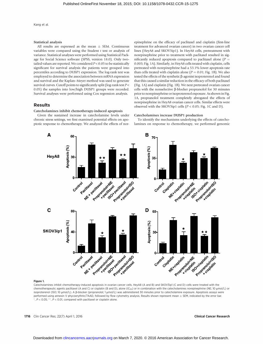
epinephrine on the efficacy of paclitaxel and cisplatin (first-linetreatment for advanced ovarian cancer) in two ovarian cancer celllines (HeyA8 and SKOV3ip1). In HeyA8 cells, pretreatment withnorepinephrine prior to treatment with paclitaxel resulted in sig-nificantly reduced apoptosis compared to paclitaxel alone (P ¼0.003; Fig. 1A). Similarly, inHeyA8 cells treatedwith cisplatin, cellspretreated with norepinephrine had a 53.1% lower apoptosis ratethan cells treated with cisplatin alone (P ¼ 0.01; Fig. 1B). We alsotested the effects of the synthetic b-agonist isoproterenol and foundthat this caused a similar reduction in the efficacy of both paclitaxel(Fig. 1A) and cisplatin (Fig. 1B). We next pretreated ovarian cancercells with the nonselective b-blocker propranolol for 30 minutesprior tonorepinephrineor isoproterenol exposure. As shown in Fig.1A, propranolol treatment completely abrogated the effects ofnorepinephrine in HeyA8 ovarian cancer cells. Similar effects wereobserved with the SKOV3ip1 cells (P < 0.05; Fig. 1C and D).
Catecholamines increase DUSP1 productionTo identify the mechanisms underlying the effects of catecho-
lamines on response to chemotherapy, we performed genomic
Figure 1.Catecholamines inhibit chemotherapy-induced apoptosis in ovarian cancer cells. HeyA8 (A and B) and SKOV3ip1 (C and D) cells were treated with thechemotherapeutic agents paclitaxel (A and C) or cisplatin (B and D), alone (IC50) or in combination with the catecholamines norepinephrine (NE; 10 mmol/L) orisoproterenol (ISO; 10 mmol/L). A b-blocker (propranolol; 1 mmol/L) was administered 30 minutes prior to catecholamine exposure. Apoptosis assays wereperformed using annexin V phycoerythrin/7AAD, followed by flow cytometry analysis. Results shown represent mean � SEM, indicated by the error bar.� , P < 0.05; �� , P < 0.01, compared with paclitaxel or cisplatin alone.
Kang et al.
Clin Cancer Res; 22(7) April 1, 2016 Clinical Cancer Research1716
on March 7, 2020. © 2016 American Association for Cancer Research. clincancerres.aacrjournals.org Downloaded from
Published OnlineFirst November 18, 2015; DOI: 10.1158/1078-0432.CCR-15-1275
analyses of HeyA8 and SKOV3ip1 ovarian cancer cells followingexposure to norepinephrine (10). These analyses showed that theMAPK phosphatase DUSP1 and its downstream network weresignificantly altered in both cell lines, compared with control cells(Supplementary Fig. S1A and S1B).We found that DUSP1 expres-sion was significantly higher in cells treated with norepinephrinethan in control cells (Fig. 2A). To validate these findings, westimulated ovarian cancer cells with increasing concentrations ofnorepinephrine or isoproterenol in independent experiments.DUSP1 levels were assessed by qRT-PCR after 1, 3, or 6 hours ofexposure to norepinephrine or isoproterenol (Fig. 2B), followedbyWestern blot analysis (Fig. 2C). Therewere significant increasesin DUSP1 mRNA levels following norepinephrine or isoproter-enol treatment. Propranolol completely abrogated the effects ofnorepinephrine on DUSP1 induction (Fig. 2C).
ADRB2 is a key mediator of increased DUSP1 levelsTo determine whether DUSP1 expression is regulated through
b-adrenergic receptors, we first screened ADRB1, ADRB2, ADRB3,and DUSP1 expression in 10 epithelial ovarian cancer cell linesandone breast cancer cell line (MDA-231), using nontransformedovarian surface epithelial HIO-180 cells as control. Supplemen-tary Fig. S2A shows the baseline expression of b-adrenergicreceptors and DUSP1 in these cells. A2780 cells were negativefor ADRB1, ADRB2, and with very weak expression of ADRB3.DUSP1 mRNA levels were assessed 3 hours after exposure to 10mmol/L norepinephrine. As shown in Supplementary Fig. S2B, inADRB2-positive cancer cells, treatment with norepinephrineresulted in significantly higher DUSP1 mRNA levels comparedwith control (P < 0.01), whereas in ADRB2-negative cancer cells,norepinephrine had no significant effect on DUSP1 levels.
To determine whether a-adrenergic receptors were involved inincreasing levels of DUSP1 or not, we treated HeyA8 cells witha-adrenergic receptor antagonists (ADRA1 antagonist prazosinand ADRA2 antagonist yohimbine; Fig. 2D) and found that therewas no effect on norepinephrine-inducedDUSP1 expression.Wethen found that inhibition with propranolol abrogated norepi-nephrine-induced DUSP1 expression in ovarian cancer cells(Fig. 2E). We also tested more specific ADRB blockers; atenolol(ADRB1 antagonist) had little effect on norepinephrine-inducedDUSP1 expression, but 1 mmol/L and 10 mmol/L ICI118,551(ADRB2 antagonist) and SR59230A (ADRB3 antagonist)markedly decreased norepinephrine-induced increases inDUSP1 expression (P < 0.01; Fig. 2F). Furthermore, using ADRB2and ADRB3 siRNAs, we found that norepinephrine-inducedDUSP1 expression occurs predominantly through ADRB2(P < 0.01; Fig. 2G).
Norepinephrine-mediated transcriptional regulationofDUSP1promoter
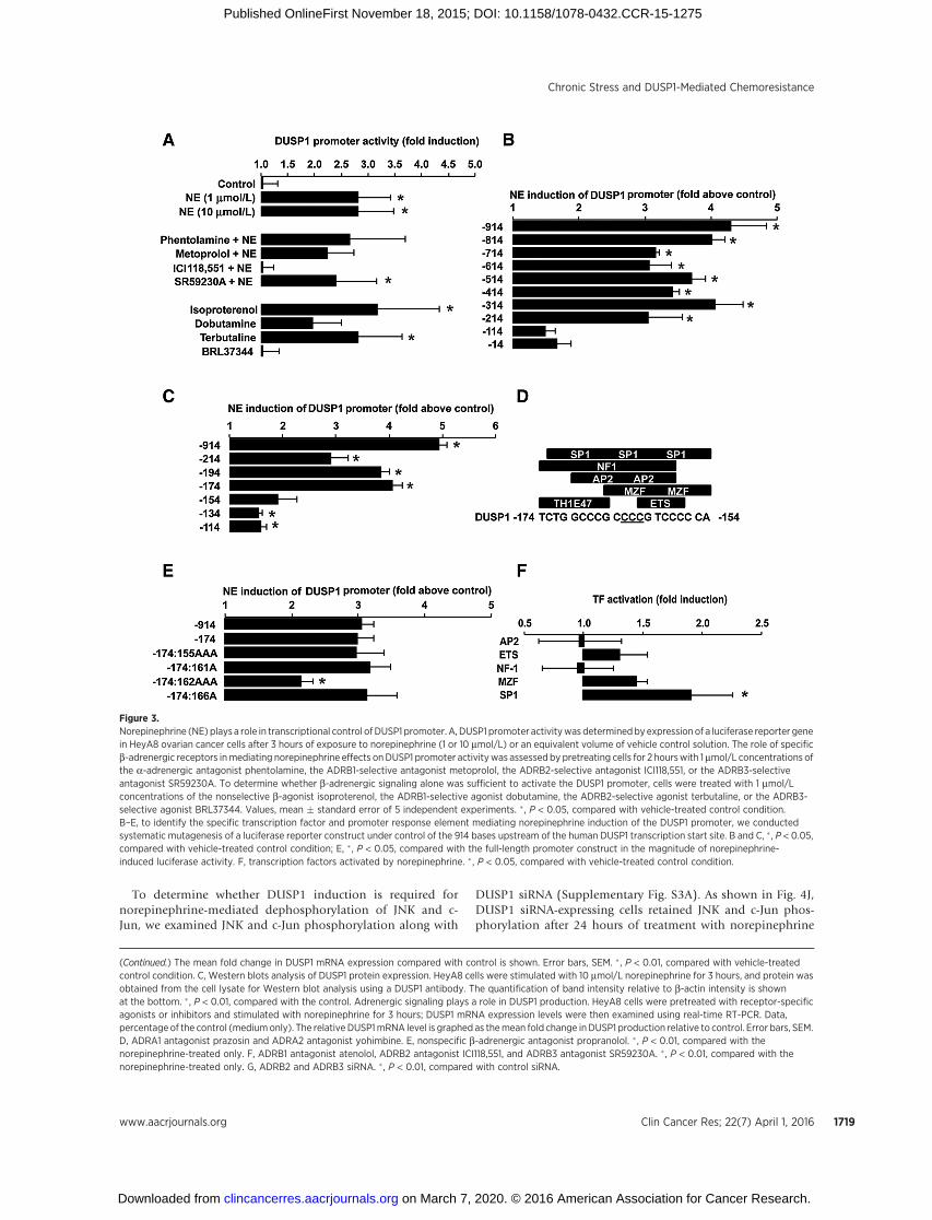
To further delineate the mechanism of norepinephrine-medi-ated DUSP1 expression, we examined the effects of norepineph-rine (3hours) on aDUSP1 luciferase promoter construct inHeyA8ovarian cancer cells (Fig. 3A). Treatment with norepinephrineresulted in �3-fold increase in DUSP1 promoter activity com-paredwith control (P < 0.05).However, pretreatment of cells withan ADRB2-selective antagonist (ICI118,551) efficiently blockedthe effects of norepinephrine. Antagonists of ADRB1 and ADRB3receptors (metoprolol and SR59230A) had no effect. We alsotested the effects of specific b-agonists (the ADRB1-selectiveagonist dobutamine, the ADRB2-selective agonist terbutaline, or
the ADRB3-selective agonist BRL37344); both nonselectiveb-adrenergic stimulation (isoproterenol) and selective activationof ADRB2 proved sufficient to stimulateDUSP1 promoter activityto levels commensurate with the effects of norepinephrine.
To identify the specific transcription factor and promoterresponse element mediating norepinephrine induction of theDUSP1 promoter, we conducted systematic mutagenesis of aluciferase reporter construct under control of the 914 bp DNAsequence upstream of the human DUSP1 transcription start site(Fig. 3B–E). A series of 100-bp and subsequent 20-bp deletionconstructs localized the norepinephrine responsive region of theDUSP1 promoter to a region ranging between�174 and�154 bpupstream of the transcription start site (P < 0.05; Fig. 3B and C).Bioinformatic analysis of this sequence identified multiple tran-scription factor–binding motifs (Fig. 3D), including potentialresponse elements for Sp1, NF1, AP-2, MZF (Myeloid Zinc Fingerproteins), and Ets family transcription factors. Systematic abro-gation of target binding sites for each factor further localized thenorepinephrine-responsive region of the DUSP1 promoter to aCCC repeat spanning�162 to�165bp (P<0.05; Fig. 3E). Amongthe transcription factors predicted to bind to this region, only Sp1showed significant activation by norepinephrine (1.9-fold � 0.4-fold, P ¼ 0.03; Fig. 3F).
To further elucidate the role of ADRB2 in norepinephrine-mediated DUSP1 induction, we inhibited several downstreamproteins in the ADRB2 pathway. Because cAMP is an importantcomponent of the ADRB2 signaling pathway, we tested the effectsof forskolin (cAMP activator) on DUSP1 gene expression. DUSP1levels were significantly increased in response to forskolin (Fig.4A). Downstream of cAMP are several proteins, including PLC,PKA, and Epac. The PLC inhibitor U73122 markedly decreasednorepinephrine-induced increases in DUSP1 expression in SKO-V3ip1 cells (Fig. 4B). Inhibition of factors downstream of PLCsuggested the involvement of PKC (inhibitor staurosporine; Fig.4B). We then examined DUSP1 expression following treatmentwith siRNA targeted against PKC, CREB1, or SP1, respectively. Asshown in Fig. 4C, after PKC, CREB1 and SP1 downregulation,norepinephrine-induced DUSP1 expression was significantlydecreased. Meantime, inhibition of additional downstream pro-teins, including PI3K (inhibitor LY294002; Fig. 4D), Akt (Akt1/2kinase inhibitor; Fig. 4D), Epac (inhibitor brefeldin A and acti-vator 8CPT-2Me-cAMP; Fig. 4E), PKA (inhibitor KT5720 andH-89; Fig. 4F), MEK (inhibitor U0126; Fig. 4G), and p38 (inhib-itor SB203580; Fig. 4H), had no significant effect on norepineph-rine-inducedDUSP1 expression. Similar results were observed forHeyA8 cells (data not shown).
Blocking DUSP1 inhibits norepinephrine-induceddephosphorylation of JNK and c-Jun
Some studies have suggested that DUSP1, as a MAPK phos-phatase, is an important regulator of JNK-dependent apoptosis.For example, DUSP1 overexpression can inhibit JNK-mediatedphosphorylation of c-Jun and protect sympathetic neurons fromapoptosis (11). To determine whether norepinephrine activatesJNK and c-Jun, we treated HeyA8 cells with or without norepi-nephrine (10 mmol/L) for various time periods prior to treatmentwith paclitaxel. The baseline expression of p-JNK and p-c-Jun (0-hour time point) was weak and expression of p-JNK and p-c-Junincreased at a peak of 6 hours after treatment with paclitaxel;however, JNK and c-Jun phosphorylation was inhibited as a resultof pretreatment with norepinephrine (Fig. 4I).
Chronic Stress and DUSP1-Mediated Chemoresistance
www.aacrjournals.org Clin Cancer Res; 22(7) April 1, 2016 1717
on March 7, 2020. © 2016 American Association for Cancer Research. clincancerres.aacrjournals.org Downloaded from
Published OnlineFirst November 18, 2015; DOI: 10.1158/1078-0432.CCR-15-1275
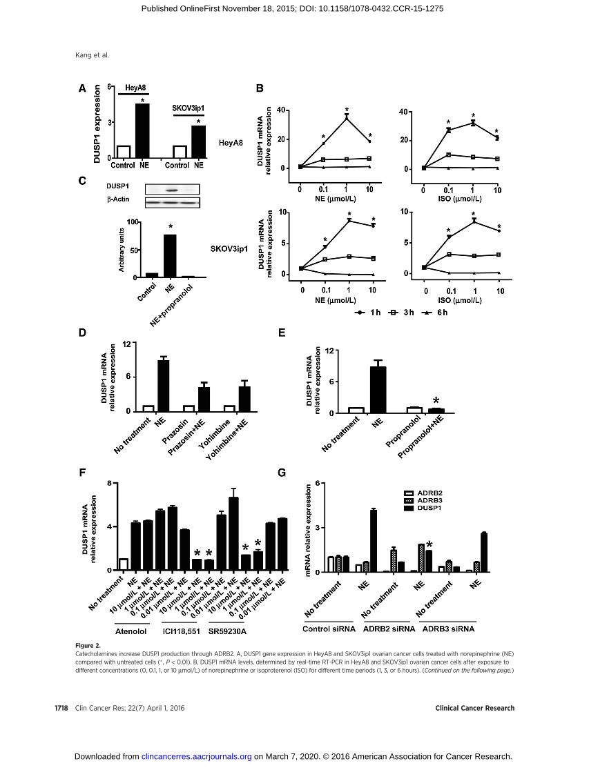
Figure 2.Catecholamines increase DUSP1 production through ADRB2. A, DUSP1 gene expression in HeyA8 and SKOV3ip1 ovarian cancer cells treated with norepinephrine (NE)compared with untreated cells (� , P < 0.01). B, DUSP1 mRNA levels, determined by real-time RT-PCR in HeyA8 and SKOV3ip1 ovarian cancer cells after exposure todifferent concentrations (0, 0.1, 1, or 10 mmol/L) of norepinephrine or isoproterenol (ISO) for different time periods (1, 3, or 6 hours). (Continued on the following page.)
Kang et al.
Clin Cancer Res; 22(7) April 1, 2016 Clinical Cancer Research1718
on March 7, 2020. © 2016 American Association for Cancer Research. clincancerres.aacrjournals.org Downloaded from
Published OnlineFirst November 18, 2015; DOI: 10.1158/1078-0432.CCR-15-1275
To determine whether DUSP1 induction is required fornorepinephrine-mediated dephosphorylation of JNK and c-Jun, we examined JNK and c-Jun phosphorylation along with
DUSP1 siRNA (Supplementary Fig. S3A). As shown in Fig. 4J,DUSP1 siRNA-expressing cells retained JNK and c-Jun phos-phorylation after 24 hours of treatment with norepinephrine
(Continued.) The mean fold change in DUSP1 mRNA expression compared with control is shown. Error bars, SEM. � , P < 0.01, compared with vehicle-treatedcontrol condition. C, Western blots analysis of DUSP1 protein expression. HeyA8 cells were stimulated with 10 mmol/L norepinephrine for 3 hours, and protein wasobtained from the cell lysate for Western blot analysis using a DUSP1 antibody. The quantification of band intensity relative to b-actin intensity is shownat the bottom. � , P < 0.01, compared with the control. Adrenergic signaling plays a role in DUSP1 production. HeyA8 cells were pretreated with receptor-specificagonists or inhibitors and stimulated with norepinephrine for 3 hours; DUSP1 mRNA expression levels were then examined using real-time RT-PCR. Data,percentage of the control (mediumonly). The relative DUSP1mRNA level is graphed as themean fold change inDUSP1 production relative to control. Error bars, SEM.D, ADRA1 antagonist prazosin and ADRA2 antagonist yohimbine. E, nonspecific b-adrenergic antagonist propranolol. � , P < 0.01, compared with thenorepinephrine-treated only. F, ADRB1 antagonist atenolol, ADRB2 antagonist ICI118,551, and ADRB3 antagonist SR59230A. � , P < 0.01, compared with thenorepinephrine-treated only. G, ADRB2 and ADRB3 siRNA. � , P < 0.01, compared with control siRNA.
Figure 3.Norepinephrine (NE) plays a role in transcriptional control of DUSP1 promoter. A, DUSP1 promoter activitywasdeterminedbyexpressionof a luciferase reporter genein HeyA8 ovarian cancer cells after 3 hours of exposure to norepinephrine (1 or 10 mmol/L) or an equivalent volume of vehicle control solution. The role of specificb-adrenergic receptors inmediating norepinephrine effects onDUSP1 promoter activitywas assessed by pretreating cells for 2 hourswith 1mmol/L concentrations ofthe a-adrenergic antagonist phentolamine, the ADRB1-selective antagonist metoprolol, the ADRB2-selective antagonist ICI118,551, or the ADRB3-selectiveantagonist SR59230A. To determine whether b-adrenergic signaling alone was sufficient to activate the DUSP1 promoter, cells were treated with 1 mmol/Lconcentrations of the nonselective b-agonist isoproterenol, the ADRB1-selective agonist dobutamine, the ADRB2-selective agonist terbutaline, or the ADRB3-selective agonist BRL37344. Values, mean � standard error of 5 independent experiments. � , P < 0.05, compared with vehicle-treated control condition.B–E, to identify the specific transcription factor and promoter response element mediating norepinephrine induction of the DUSP1 promoter, we conductedsystematic mutagenesis of a luciferase reporter construct under control of the 914 bases upstream of the human DUSP1 transcription start site. B and C, � , P < 0.05,compared with vehicle-treated control condition; E, � , P < 0.05, compared with the full-length promoter construct in the magnitude of norepinephrine-induced luciferase activity. F, transcription factors activated by norepinephrine. � , P < 0.05, compared with vehicle-treated control condition.
Chronic Stress and DUSP1-Mediated Chemoresistance
www.aacrjournals.org Clin Cancer Res; 22(7) April 1, 2016 1719
on March 7, 2020. © 2016 American Association for Cancer Research. clincancerres.aacrjournals.org Downloaded from
Published OnlineFirst November 18, 2015; DOI: 10.1158/1078-0432.CCR-15-1275
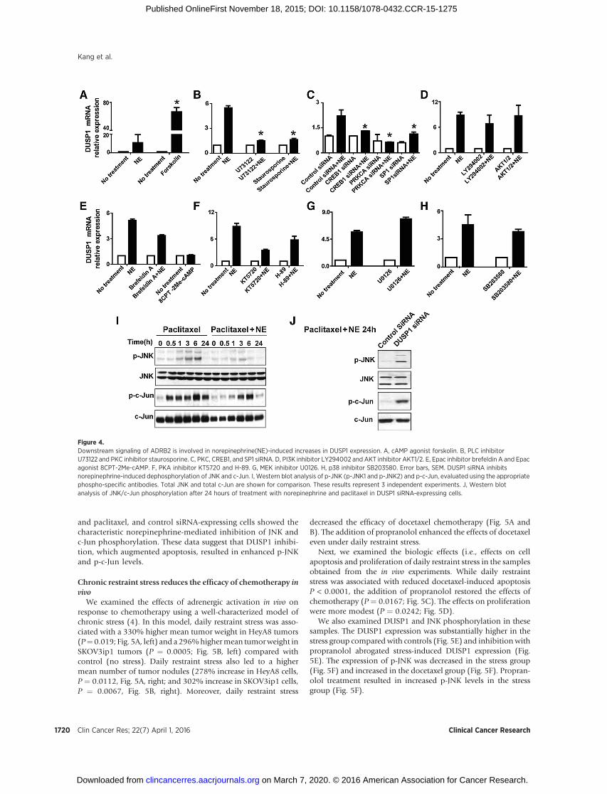
and paclitaxel, and control siRNA-expressing cells showed thecharacteristic norepinephrine-mediated inhibition of JNK andc-Jun phosphorylation. These data suggest that DUSP1 inhibi-tion, which augmented apoptosis, resulted in enhanced p-JNKand p-c-Jun levels.
Chronic restraint stress reduces the efficacy of chemotherapy invivo
We examined the effects of adrenergic activation in vivo onresponse to chemotherapy using a well-characterized model ofchronic stress (4). In this model, daily restraint stress was asso-ciated with a 330% higher mean tumor weight in HeyA8 tumors(P¼0.019; Fig. 5A, left) and a 296%highermean tumorweight inSKOV3ip1 tumors (P ¼ 0.0005; Fig. 5B, left) compared withcontrol (no stress). Daily restraint stress also led to a highermean number of tumor nodules (278% increase in HeyA8 cells,P ¼ 0.0112, Fig. 5A, right; and 302% increase in SKOV3ip1 cells,P ¼ 0.0067, Fig. 5B, right). Moreover, daily restraint stress
decreased the efficacy of docetaxel chemotherapy (Fig. 5A andB). The addition of propranolol enhanced the effects of docetaxeleven under daily restraint stress.
Next, we examined the biologic effects (i.e., effects on cellapoptosis and proliferation of daily restraint stress in the samplesobtained from the in vivo experiments. While daily restraintstress was associated with reduced docetaxel-induced apoptosisP < 0.0001, the addition of propranolol restored the effects ofchemotherapy (P ¼ 0.0167; Fig. 5C). The effects on proliferationwere more modest (P ¼ 0.0242; Fig. 5D).
We also examined DUSP1 and JNK phosphorylation in thesesamples. The DUSP1 expression was substantially higher in thestress group compared with controls (Fig. 5E) and inhibition withpropranolol abrogated stress-induced DUSP1 expression (Fig.5E). The expression of p-JNK was decreased in the stress group(Fig. 5F) and increased in the docetaxel group (Fig. 5F). Propran-olol treatment resulted in increased p-JNK levels in the stressgroup (Fig. 5F).
Figure 4.Downstream signaling of ADRB2 is involved in norepinephrine(NE)-induced increases in DUSP1 expression. A, cAMP agonist forskolin. B, PLC inhibitorU73122 and PKC inhibitor staurosporine. C, PKC, CREB1, and SP1 siRNA. D, PI3K inhibitor LY294002 and AKT inhibitor AKT1/2. E, Epac inhibitor brefeldin A and Epacagonist 8CPT-2Me-cAMP. F, PKA inhibitor KT5720 and H-89. G, MEK inhibitor U0126. H, p38 inhibitor SB203580. Error bars, SEM. DUSP1 siRNA inhibitsnorepinephrine-induced dephosphorylation of JNK and c-Jun. I, Western blot analysis of p-JNK (p-JNK1 and p-JNK2) and p-c-Jun, evaluated using the appropriatephospho-specific antibodies. Total JNK and total c-Jun are shown for comparison. These results represent 3 independent experiments. J, Western blotanalysis of JNK/c-Jun phosphorylation after 24 hours of treatment with norepinephrine and paclitaxel in DUSP1 siRNA-expressing cells.
Kang et al.
Clin Cancer Res; 22(7) April 1, 2016 Clinical Cancer Research1720
on March 7, 2020. © 2016 American Association for Cancer Research. clincancerres.aacrjournals.org Downloaded from
Published OnlineFirst November 18, 2015; DOI: 10.1158/1078-0432.CCR-15-1275
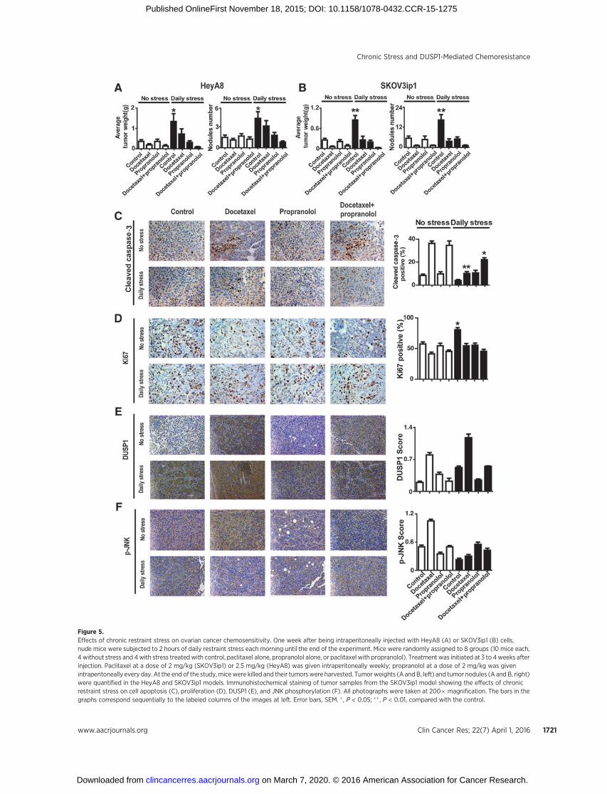
Figure 5.Effects of chronic restraint stress on ovarian cancer chemosensitivity. One week after being intraperitoneally injected with HeyA8 (A) or SKOV3ip1 (B) cells,nude mice were subjected to 2 hours of daily restraint stress each morning until the end of the experiment. Mice were randomly assigned to 8 groups (10 mice each,4 without stress and 4with stress treatedwith control, paclitaxel alone, propranolol alone, or paclitaxel with propranolol). Treatment was initiated at 3 to 4weeks afterinjection. Paclitaxel at a dose of 2 mg/kg (SKOV3ip1) or 2.5 mg/kg (HeyA8) was given intraperitoneally weekly; propranolol at a dose of 2 mg/kg was givenintraperitoneally every day. At the end of the study,micewere killed and their tumorswere harvested. Tumorweights (A andB, left) and tumor nodules (A and B, right)were quantified in the HeyA8 and SKOV3ip1 models. Immunohistochemical staining of tumor samples from the SKOV3ip1 model showing the effects of chronicrestraint stress on cell apoptosis (C), proliferation (D), DUSP1 (E), and JNK phosphorylation (F). All photographs were taken at 200� magnification. The bars in thegraphs correspond sequentially to the labeled columns of the images at left. Error bars, SEM. � , P < 0.05; �� , P < 0.01, compared with the control.
Chronic Stress and DUSP1-Mediated Chemoresistance
www.aacrjournals.org Clin Cancer Res; 22(7) April 1, 2016 1721
on March 7, 2020. © 2016 American Association for Cancer Research. clincancerres.aacrjournals.org Downloaded from
Published OnlineFirst November 18, 2015; DOI: 10.1158/1078-0432.CCR-15-1275
Functional roles of DUSP1 in response to chemotherapyGiven the role of DUSP1 in apoptosis (12, 13), we further
examined its role in protecting cancer cells from apoptosis duringexposure to norepinephrine. SKOV3ip1 cells were transientlytransfected with a DUSP1-myc/DDK-tagged expression vector;DUSP1 expression increased by about 3-fold in these cells com-
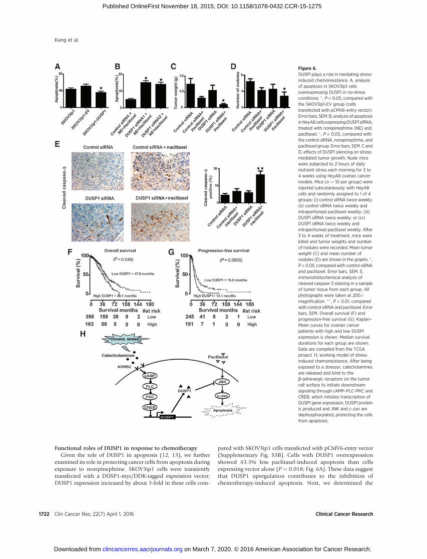
pared with SKOV3ip1 cells transfected with pCMV6-entry vector(Supplementary Fig. S3B). Cells with DUSP1 overexpressionshowed 43.3% less paclitaxel-induced apoptosis than cellsexpressing vector alone (P ¼ 0.018; Fig. 6A). These data suggestthat DUSP1 upregulation contributes to the inhibition ofchemotherapy-induced apoptosis. Next, we determined the
Figure 6.DUSP1 plays a role in mediating stress-induced chemoresistance. A, analysisof apoptosis in SKOV3ip1 cellsoverexpressing DUSP1 in no-stressconditions. � , P < 0.05, compared withthe SKOV3ip1-EV group (cellstransfected with pCMV6-entry vector).Error bars, SEM. B, analysis of apoptosisinHeyA8cells expressingDUSP1 siRNA,treated with norepinephrine (NE) andpaclitaxel. �, P < 0.05, compared withthe control siRNA, norepinephrine, andpaclitaxel group. Error bars, SEM. C andD, effects of DUSP1 silencing on stress-mediated tumor growth. Nude micewere subjected to 2 hours of dailyrestraint stress each morning for 3 to4 weeks using HeyA8 ovarian cancermodels. Mice (n ¼ 10 per group) wereinjected subcutaneously with HeyA8cells and randomly assigned to 1 of 4groups: (i) control siRNA twice weekly;(ii) control siRNA twice weekly andintraperitoneal paclitaxel weekly; (iii)DUSP1 siRNA twice weekly; or (iv)DUSP1 siRNA twice weekly andintraperitoneal paclitaxel weekly. After3 to 4 weeks of treatment, mice werekilled and tumor weights and numberof nodules were recorded. Mean tumorweight (C) and mean number ofnodules (D) are shown in the graphs. � ,P < 0.05, comparedwith control siRNAand paclitaxel. Error bars, SEM. E,immunohistochemical analysis ofcleaved caspase-3 staining in a sampleof tumor tissue from each group. Allphotographs were taken at 200�magnification. ��, P < 0.01, comparedwith control siRNA and paclitaxel. Errorbars, SEM. Overall survival (F) andprogression-free survival (G): Kaplan–Meier curves for ovarian cancerpatients with high and low DUSP1expression is shown. Median survivaldurations for each group are shown.Data are compiled from the TCGAproject. H, working model of stress-induced chemoresistance. After beingexposed to a stressor, catecholaminesare released and bind to theb-adrenergic receptors on the tumorcell surface to initiate downstreamsignaling through cAMP–PLC–PKC andCREB, which initiates transcription ofDUSP1 gene expression. DUSP1 proteinis produced and JNK and c-Jun aredephosphorylated, protecting the cellsfrom apoptosis.
Clin Cancer Res; 22(7) April 1, 2016 Clinical Cancer Research1722
Kang et al.
on March 7, 2020. © 2016 American Association for Cancer Research. clincancerres.aacrjournals.org Downloaded from
Published OnlineFirst November 18, 2015; DOI: 10.1158/1078-0432.CCR-15-1275
effect of DUSP1 siRNA on paclitaxel-induced apoptosis. Forty-eight hours after siRNA transfection, cells were treated withnorepinephrine and paclitaxel for 72 hours. As shown in Fig.6B, DUSP1 siRNA1 expression reversed the norepinephrine-mediated protection from apoptosis. DUSP1 siRNA2 producedsimilar results (Fig. 6B).
To test the role of DUSP1 in vivo, we used the DOPCnanoliposomal delivery method (14, 15). Daily restraint stressresulted in substantially increased DUSP1 levels, whereas treat-ment with DUSP1 siRNA effectively reduced DUSP1 expression(Supplementary Fig. S3C). To determine the role of DUSP1 inmediating stress-induced tumor growth, female nude mice wereinjected with HeyA8 cells into the peritoneal cavity and thenrandomized to one of the following treatment groups (n ¼ 10mice per group): (i) control siRNA twice weekly; (ii) controlsiRNA twice weekly and intraperitoneal paclitaxel weekly; (iii)DUSP1 siRNA twice weekly; or (iv) DUSP1 siRNA twice weeklyand intraperitoneal paclitaxel weekly. Treatment with DUSP1siRNA significantly improved the efficacy of paclitaxel chemo-therapy (Fig. 6C and D). Effects on apoptosis were the highestin the DUSP1 siRNA plus paclitaxel group, which increasedapoptosis by 144% compared with the control siRNA pluspaclitaxel group (P ¼ 0.008; Fig. 6E).
DUSP1 is associated with decreased overall and progression-free survival
Next, we examined for potential correlations between DUSP1expression and patient outcomes using TCGA. The Cox regressionanalysis of overall survival yielded a hazard ratio of 1.13 [95%confidence interval (CI), 1.01–1.27; P ¼ 0.049) for DUSP1(201044_x_at; Affymetrix microarray). The Kaplan–Meier plotswere generated for the cutoff of 0.71. The Cox regression analysisof disease-free survival yielded a hazard ratio of 1.22 (95% CI,1.09–1.37; P ¼ 0.0005) for DUSP1 (A_32_P171482; Agilentmicroarray). TheKaplan–Meier plotswere generated for the cutoffof 0.62. The data showed high DUSP1 expression was associatedwith decreased overall survival (Fig. 6F) and decreased progres-sion-free survival (Fig. 6G).
DiscussionThe key finding of this study is the discovery of a previously
unrecognized pathway by which sustained adrenergic signalingleads to impaired chemotherapy response in ovarian cancer (Fig.6H). In this model, chronic stress induces the expression ofDUSP1 through activation of the ADRB2/cAMP/PLC/PKC/CREBsignaling cascade, resulting in JNK-mediated phosphorylation ofc-Jun and protection from apoptosis. Our previous work showedthat the angiogenic effects of NE/ADRB2 aremediated, in part, viasecretion of VEGF (4). Here, we found that VEGF itself has noeffects on apoptosis inhibition (Supplementary Fig. S4). Inter-estingly, it has been reported that HPA mediators, such as glu-cocorticoids, promote cancer cell survival throughDUSP1-depen-dent pathways (5). However, the role of adrenergic signaling onDUSP1 expression and response to chemotherapy remainedunknown until now. These data have particular relevance due tothe fact that a substantial proportion of ovarian cancer patientshave biobehavioral alterations that would be reflective of chronicstress (16) and are associated with increased catecholamine con-centrations in primary tumor tissues (17, 18).
This work provides a rationale for the addition of b-blockers,such as propranolol, to adjuvant therapy to enhance the efficacy ofcurrent chemotherapy regimens. b-blockers are commonly usedto safely treat hypertension and other cardiovascular maladies.Recent studies have implicated b-blockers in reducing meta-static efficiency and are associated with lower recurrence ratesand longer disease-free intervals in several cancers, includingovarian cancer (19–22). Moreover, a phase 0 clinical trial(NCT01308944) is addressing the effects of adding propranololto first-line chemotherapy for ovarian cancer. Findings fromthis trial will shed further light on efficacy of b-blockers withtraditional chemotherapy for improving the outcome of cancerpatients.
In summary, norepinephrine-mediated increase in DUSP1decreases the antitumor effects of commonly used chemo-therapeutic agents. These findings provide a new understand-ing of how sustained adrenergic signaling leads to impairedchemotherapy response. Our data suggest that interventionstargeting the SNS, such as b-blockers, could enhance theefficacy of chemotherapy in patients with ovarian and othercancers.
Disclosure of Potential Conflicts of InterestNo potential conflicts of interest were disclosed.
Authors' ContributionsConceptionanddesign:Y.Kang, R. Rupaimoole,G. Lopez-Berestein, S.W.Cole,A.K. SoodDevelopment of methodology: Y. Kang, J.M. Hansen, A.K. SoodAcquisition of data (provided animals, acquired and managed patients,provided facilities, etc.): Y. Kang, G.N. Armaiz-Pena, R. Rupaimoole,R.A. Previs, J.M. Hansen, C. Rodriguez-Aguayo, G. Lopez-Berestein, S.W. Cole,A.K. SoodAnalysis and interpretation of data (e.g., statistical analysis, biostatistics,computational analysis): Y. Kang, W. Hu, R. Rupaimoole, C. Ivan, P. Ram,V. Sehgal, G. Lopez-Berestein, S.W. Cole, A.K. SoodWriting, review, and/or revision of the manuscript: Y. Kang, A.S. Nagaraja,G.N. Armaiz-Pena, P.L. Dorniak, W. Hu, R.A. Previs, J.M. Hansen, P. Ram,S.K. Lutgendorf, S.W. Cole, A.K. SoodAdministrative, technical, or material support (i.e., reporting or organizingdata, constructing databases): W. Hu, K.M. Gharpure, A.K. SoodStudy supervision: A.K. Sood
AcknowledgmentsThe authors thank Dr. Robert Langley and Donna Reynolds for their
assistance with the immunohistochemical analysis. They also thank EricaA. Goodoff in the Department of Scientific Publications for editing thismanuscript.
Grant SupportThis work was supported by the NIH (CA 109298, P50 CA083639, P50
CA098258, CA140933, and CA104825), the OvarianCancer Research Fund,Inc. (Program Project Development Grant), the Betty Anne Asche MurrayDistinguished Professorship and the National Natural Science Foundation ofChina (grant no. 81472423). J.M. Hansen and R.A. Previs are supported by anNCI–DHHS–NIH T32 Training Grant (T32 CA101642). A.S. Nagaraja is sup-ported by a Research Training Award from the Cancer Prevention and ResearchInstitute of Texas (CPRIT RP140106). K.M. Gharpure is supported by AltmanGoldstein Discovery fellowship.
The costs of publication of this articlewere defrayed inpart by the payment ofpage charges. This article must therefore be hereby marked advertisement inaccordance with 18 U.S.C. Section 1734 solely to indicate this fact.
Received May 31, 2015; revised October 6, 2015; accepted October 13, 2015;published OnlineFirst November 18, 2015.
www.aacrjournals.org Clin Cancer Res; 22(7) April 1, 2016 1723
Chronic Stress and DUSP1-Mediated Chemoresistance
on March 7, 2020. © 2016 American Association for Cancer Research. clincancerres.aacrjournals.org Downloaded from
Published OnlineFirst November 18, 2015; DOI: 10.1158/1078-0432.CCR-15-1275
References1. Lutgendorf SK, Sood AK, Antoni MH. Host factors and cancer progression:
biobehavioral signaling pathways and interventions. J Clin Oncol2010;28:4094–9.
2. Su F,OuyangN, Zhu P,OuyangN, JiaW, GongC, et al. Psychological stressinduces chemoresistance in breast cancer by upregulating mdr1. BiochemBiophys Res Commun 2005;329:888–97.
3. Flint MS, Kim G, Hood BL, Bateman NW, Stewart NA, Conrads TP. Stresshormones mediate drug resistance to paclitaxel in human breast cancercells through a CDK-1-dependent pathway. Psychoneuroendocrinology2009;34:1533–41.
4. Thaker PH,Han LY, Kamat AA, Arevalo JM, Takahashi R, LuC, et al. Chronicstress promotes tumor growth and angiogenesis in a mouse model ofovarian carcinoma. Nat Med 2006;12:939–44.
5. Wu W, Pew T, Zou M, Pang D, Conzen SD. Glucocorticoid receptor-induced MAPK phosphatase-1 (MPK-1) expression inhibits paclitaxel-associated MAPK activation and contributes to breast cancer cell survival.J Biol Chem 2005;280:4117–24.
6. Sastry KS, Karpova Y, Prokopovich S, Smith AJ, Essau B, Gersappe A, et al.Epinephrine protects cancer cells from apoptosis via activation of cAMP-dependent protein kinase and BAD phosphorylation. J Biol Chem 2007;282:14094–100.
7. Sun X, Bao J, Nelson KC, Li KC, Kulik G, Zhou X. Systemsmodeling of anti-apoptotic pathways in prostate cancer: psychological stress triggers asynergism pattern switch in drug combination therapy. PLoS Comput Biol2013;9:e1003358.
8. Pecot CV, Rupaimoole R, Yang D, Akbani R, Ivan C, Lu C, et al. Tumourangiogenesis regulation by the miR-200 family. Nat Commun 2013;4:2427.
9. Kang Y, Hu W, Ivan C, Dalton HJ, Miyake T, Pecot CV, et al. Role of focaladhesion kinase in regulating YB-1-mediated paclitaxel resistance in ovar-ian cancer. J Natl Cancer Inst 2013;105:1485–95.
10. ShahzadMM,Arevalo JM, Armaiz-PenaGN, LuC, StoneRL,Moreno-SmithM, et al. Stress effects on FosB- and interleukin-8 (IL8)-driven ovariancancer growth and metastasis. J Biol Chem 2010;285:35462–70.
11. KristiansenM, Hughes R, Patel P, Jacques TS, Clark AR, Ham J. Mkp1 is a c-Jun target gene that antagonizes JNK-dependent apoptosis in sympatheticneurons. J Neurosci 2010;30:10820–32.
12. Magi-Galluzzi C,Mishra R, FiorentinoM,Montironi R, YaoH,Capodieci P,et al. Mitogen-activated protein kinase phosphatase 1 is overexpressed inprostate cancers and is inversely related to apoptosis. Lab Invest1997;76:37–51.
13. Castillo SS, Teegarden D. Sphingosine-1-phosphate inhibition of apopto-sis requires mitogen-activated protein kinase phosphatase-1 in mousefibroblast C3H10T 1/2 cells. J Nutr 2003;133:3343–9.
14. Landen CN Jr, Chavez-Reyes A, Bucana C, Schmandt R, Deavers MT,Lopez-Berestein G, et al. Therapeutic EphA2 gene targeting in vivo usingneutral liposomal small interfering RNA delivery. Cancer Res 2005;65:6910–8.
15. Pradeep S, Kim SW,Wu SY, Nishimura M, Chaluvally-Raghavan P, MiyakeT, et al. Hematogenous metastasis of ovarian cancer: rethinking mode ofspread. Cancer Cell 2014;26:77–91.
16. Bodurka-Bevers D, Basen-Engquist K, Carmack CL, FitzgeraldMA,Wolf JK,de Moor C, et al. Depression, anxiety, and quality of life in patients withepithelial ovarian cancer. Gynecol Oncol 2000;78:302–8.
17. Lutgendorf SK, DeGeest K, Dahmoush L, Farley D, Penedo F, BenderD, et al. Social isolation is associated with elevated tumor norepi-nephrine in ovarian carcinoma patients. Brain Behav Immun 2011;25:250–5.
18. Lutgendorf SK, DeGeest K, SungCY, Arevalo JM, Penedo F, Lucci J 3rd, et al.Depression, social support, and beta-adrenergic transcription control inhuman ovarian cancer. Brain Behav Immun 2009;23:176–83.
19. Stone RL, Nick AM, McNeish IA, Balkwill F, Han HD, Bottsford-Miller J,et al. Paraneoplastic thrombocytosis in ovarian cancer. N Engl J Med2012;366:610–8.
20. Melhem-Bertrandt A, Chavez-Macgregor M, Lei X, Brown EN, Lee RT,Meric-Bernstam F, et al. Beta-blocker use is associated with improvedrelapse-free survival in patients with triple-negative breast cancer. J ClinOncol 2011;29:2645–52.
21. PoweDG, Voss MJ, Zanker KS, HabashyHO, Green AR, Ellis IO, et al. Beta-blocker drug therapy reduces secondary cancer formation in breast cancerand improves cancer specific survival. Oncotarget 2010;1:628–38.
22. Barron TI, Connolly RM, Sharp L, Bennett K, Visvanathan K. Beta blockersand breast cancer mortality: a population-based study. J Clin Oncol2011;29:2635–44.
Clin Cancer Res; 22(7) April 1, 2016 Clinical Cancer Research1724
Kang et al.
on March 7, 2020. © 2016 American Association for Cancer Research. clincancerres.aacrjournals.org Downloaded from
Published OnlineFirst November 18, 2015; DOI: 10.1158/1078-0432.CCR-15-1275
2016;22:1713-1724. Published OnlineFirst November 18, 2015.Clin Cancer Res Yu Kang, Archana S. Nagaraja, Guillermo N. Armaiz-Pena, et al. in Ovarian CancerAdrenergic Stimulation of DUSP1 Impairs Chemotherapy Response
Updated version
10.1158/1078-0432.CCR-15-1275doi:
Access the most recent version of this article at:
Material
Supplementary
http://clincancerres.aacrjournals.org/content/suppl/2015/11/18/1078-0432.CCR-15-1275.DC1
Access the most recent supplemental material at:
Cited articles
http://clincancerres.aacrjournals.org/content/22/7/1713.full#ref-list-1
This article cites 22 articles, 9 of which you can access for free at:
Citing articles
http://clincancerres.aacrjournals.org/content/22/7/1713.full#related-urls
This article has been cited by 6 HighWire-hosted articles. Access the articles at:
E-mail alerts related to this article or journal.Sign up to receive free email-alerts
Subscriptions
Reprints and
To order reprints of this article or to subscribe to the journal, contact the AACR Publications Department at
Permissions
Rightslink site. Click on "Request Permissions" which will take you to the Copyright Clearance Center's (CCC)
.http://clincancerres.aacrjournals.org/content/22/7/1713To request permission to re-use all or part of this article, use this link
on March 7, 2020. © 2016 American Association for Cancer Research. clincancerres.aacrjournals.org Downloaded from
Published OnlineFirst November 18, 2015; DOI: 10.1158/1078-0432.CCR-15-1275


![Knockout of Density-Enhanced Phosphatase-1 Impairs ......2 BioMedResearchInternational [4]. Mutations and loss of heterozygosity of DEP-1 have been observed in human cancers, substantiating](https://img.pdfslide.org/doc/110x75/5f61b58dfcc9115d17599f90/knockout-of-density-enhanced-phosphatase-1-impairs-2-biomedresearchinternational.jpg)