Embed Size (px)
Citation preview
115Oper Orthop Traumatol 2007 · No. 2 © Urban & Vogel
Arthroskopische Therapie der hinteren SchulterinstabilitätArthroscopic Treatment of Posterior Shoulder InstabilitySven Lichtenberg, Peter Habermeyer, Petra Magosch1
Operative Orthopädie und Traumatologie
ZusammenfassungOperationsziel
Wiederherstellung der stabilisierenden posterioren Struk-turen des Schultergelenk in arthroskopischer Technik.
IndikationenPosteriore Schulterinstabilitäten bzw. chronische Subluxa-tionen mit einer Läsion des posterioren Labrums und der posterioren Kapselbandstrukturen.
KontraindikationenWillkürliche oder muskulär bedingte posteriore Instabili-täten, posteriore Instabilitäten mit knöcherner Pfannen-fraktur, großem knöchernem Defekt des Humeruskopfes (20% der hinteren unteren Pfannenfläche) oder verhakte hintere Luxationen, Dysplasie der Pfanne mit patholo-gischer Retroversion > 25°.
OperationstechnikMobilisieren des pathologisch veränderten und fehlver-heilten Labrum-Ligament-Komplexes, Anfrischen des knö-chernen Pfannenrandes, Reposition und Refixation des La-brum-Ligament-Komplexes unter Verwendung von Naht-ankern.
WeiterbehandlungAbduktionskissen in 15° Abduktion für 3 Wochen. Dann funktionelle Weiterbehandlung mit passiver Mobilisation unter Vermeidung forcierter Innenrotation. Muskelkräfti-gung erst nach Wiedererlangung der freien Beweglichkeit. Rückkehr zu schulterbelastenden Sportarten nach 6 Mo-naten.
ErgebnisseElf Patienten mit einem Durchschnittsalter von 31 Jahren konnten prospektiv erfasst und verfolgt werden. Nach einem mittleren Beobachtungszeitraum von 33 Monaten kam es bei einem Patienten zu einer traumatischen Relu-xation und bei einem Patienten zu erneuten Subluxatio-nen. Der mittlere Rowe-Score betrug 95 Punkte.
AbstractObjective
Reconstruction of the posterior stabilizing structures of the glenohumeral joint in arthroscopic technique.
IndicationsPosterior shoulder instability and/or chronic subluxations with lesions of the posterior labrum and capsuloligamen-tous structures.
ContraindicationsVoluntary instability or posterior instability due to patho-logic muscle patterning, posterior instability with glenoid fracture, large bone defects of the humeral head (20% of the inferior posterior glenoid) or locked posterior disloca-tions, dysplasia of the glenoid with pathologic retrover-sion > 25°.
Surgical TechniqueMobilization of the pathologic and extraanatomically healed labroligamentous complex, decortication of the glenoid rim, repositioning and refixation of the labroliga-mentous complex at the glenoid rim using suture an-chors.
Postoperative ManagementAbduction splint at 15° for 3 weeks. Functional therapy with passive mobilization that avoids forced internal rota-tion. Muscle strengthening only after free range of motion has been achieved. Return to sports that put strain on the shoulder after 6 months.
ResultsEleven patients with a mean age of 31 years were prospec-tively documented and followed for a mean of 33 months postoperatively. One patient suffered from a traumatic recurrence and one patient experienced recurrent sub-luxations. The overall mean Rowe score was 95 out of 100 points.
Oper Orthop Traumatol 2007;19:115–32
DOI 10.1007/s00064-007-1198-2
1 ATOS-Praxisklinik Heidelberg.
Lichtenberg S, et al. Arthroscopic Treatment of Posterior Shoulder Instability
116 Oper Orthop Traumatol 2007 · No. 2 © Urban & Vogel
VorbemerkungenUnter den chronischen Instabilitäten des Schulterge-lenks ist die posteriore Instabilität mit einer Häufig-keit von ca. 2% aller Fälle sehr selten.
Folgende Strukturen schützen die Schulter vor ei-ner posterioren Instabilität:– das Glenoid mit dem die Konkavität vergrößernden
posterioren Labrum,– der glenohumerale Bandapparat, insbesondere das
posteriore Band des inferioren glenohumeralen Li-gaments (PB-IGHL),
– das superiore glenohumerale Ligament (SGHL) und das Rotatorenintervall (RI), die die posteriore Translation in Adduktion verhindern,
– die posteriore Rotatorenmanschette (Musculus in-fraspinatus und Musculus teres minor) durch An-spannen der glenohumeralen Bänder.
Die Entstehung einer posterioren Instabilität kann analog zur anterioren Instabilität traumatisch, atrau-matisch oder durch repetitive Mikrotraumata bedingt sein. Man muss unterscheiden zwischen der einma-ligen Dislokation, chronischen Dislokationen, chro-nischen Subluxationen und der schmerzhaften Schul-ter in einer Position der Innenrotation, Adduktion und Flexion.
Ferner können Konstellationen beobachtet werden, bei denen sich bei Vorliegen einer generalisierten Hy-perlaxität durch eine muskuläre Dysbalance schmerz-hafte, chronische posteriore Luxationen oder Sub-luxationen ereignen. Diese Patienten können am meisten von einer gezielten und kontrollierten Physio-therapie profitieren.
Die traumatische posteriore Luxation wird durch ein Anpralltrauma bei nach vorn gestrecktem, innen-rotiertem Arm ausgelöst, wie z.B. bei einem Auffahr-unfall mit Abstützen des Arms am Lenkrad oder durch einen entsprechenden Sturz oder Aufprall. Wenn bei dieser Dislokation eine anteriore Humeruskopfim-pression stattfindet, kann es zur chronisch verhakten posterioren Luxation kommen. Die Humerusimpres-sion, die analog zum Hill-Sachs-Defekt bei der ante-rioren Luxation durch den Kontakt mit dem Glenoid-rand entsteht, wird nach Malgaigne benannt. Sind
Introductory RemarksPosterior instability is a very rare form of chronic in-stability of the shoulder joint with an incidence of about 2% of all cases.
The following structures protect the shoulder from posterior instability:– the glenoid whose concavity is enlarged by the poste-
rior labrum,– the glenohumeral ligament complex, especially the
posterior band of the inferior glenohumeral ligament (PB-IGHL),
– the superior glenohumeral ligament (SGHL) and the rotator interval (RI) that prevent posterior transla-tion in adduction,
– the posterior rotator cuff (infraspinatus and teres mi-nor) that tension the glenohumeral ligaments.
The causes of posterior instability, as for anterior in-stability, may be traumatic, atraumatic, or due to re-petitive microtraumata. It is important to differentiate between a single incidence of dislocation, chronic dis-locations, chronic subluxations, and painful shoulder in positions of internal rotation, adduction and flex-ion.
In addition, constellations exist where there is a generalized hyperlaxity with concomitant muscular dysbalance precipitating painful chronic posterior dis-locations or subluxations. These patients benefit most from specific controlled physiotherapy.
Traumatic posterior dislocation is triggered by im-pact trauma with the arm stretched forward in internal rotation, e.g., during rear-end collision with the arm locked against the steering wheel or due to any fall or impact of that sort. If anterior humeral head impaction results as part of this dislocation, chronic locked poste-rior dislocation may occur. Humerus impaction, simi-lar to a Hill-Sachs defect in anterior dislocation due to contact with the glenoid rim, is named after Malgaigne. If > 50% of the humeral head is affected, the indication is given for humeral head replacement. If impaction affects 20–40% of the humeral head, the defect can be repaired by retrograde erecting or the lesser tubercle transferred into the defect as described by McLaugh-lin.
Key Words Posterior shoulder instability · Arthroscopic stabilization · Arthroscopy · Chronic posterior instability
SchlüsselwörterHintere Schulterinstabilität · Arthroskopische Stabilisierung · Arthroskopie · Chronische posteriore Instabilität
Lichtenberg S, et al. Arthroskopische Therapie der hinterer Schulterinstabilität
117Oper Orthop Traumatol 2007 · No. 2 © Urban & Vogel
> 50% des Humeruskopfes betroffen, besteht die Indi-kation zum endoprothetischen Ersatz des Humerus-kopfes. Bei Impressionen zwischen 20% und 40% des Humeruskopfes kann ein Aufstößeln des Defekts oder eine Versetzung des Tuberculum minus in den Defekt nach McLaughlin erfolgen.
Ebenfalls analog zur anterioren Luxation kommt es bei der traumatischen posterioren Dislokation zu ei-ner Ablösung des Labrums (posteriore Bankart-Lä-sion) und der glenohumeralen Ligamente. Bei ent-sprechend starker Gewalteinwirkung kann sich der Schädigungsmechanismus nach anterosuperior mit begleitender SLAP-Läsion oder nach anteroinferior fortsetzen. Je nach Einheilung des posterioren La-brum-Ligament-Komplexes entwickelt sich dann eine chronische posteriore Instabilität mit rezidivierenden Luxationen oder Subluxationen, die eine weitere Überkopfaktivität einschränken oder gar ganz verhin-dern.
Bei bestehender Laxität kann häufig ein geringes Trauma zur Luxation oder zu Subluxationen führen.
Knöcherne Veränderungen wie eine vermehrte gle-noidale Retroversion von > 25° bedürfen einer Kor-rekturosteotomie.
Bis heute stellt die offene Reparatur des Labrum-Li-gament-Komplexes den Goldstandard in der Therapie der posterioren Instabilität dar. Ähnlich wie in der Be-handlung der anterioren Instabilität ist es in den letz-ten Jahren durch ein besseres Verständnis von Anato-mie und Pathomechanik sowie durch innovative Instrumente und Verankerungssysteme zu einer Auf-wertung der arthroskopischen Technik gekommen.
Die Möglichkeiten und Grenzen der arthrosko-pischen Therapie der hinteren Schulterinstabilität werden aufgezeigt.
Analogous to anterior dislocation the labrum and the glenohumeral ligaments become detached (poste-rior Bankart lesion) in traumatic posterior dislocation. If the applied forces are very great, the injury mecha-nism may spread toward anterosuperior with concomi-tant SLAP lesion or toward anteroinferior. Depending on how the posterior labroligamentous complex heals, chronic posterior instability may develop with recur-rent dislocations or subluxations that restrict the full range of overhead activity or prevent it entirely.
If there is preexisting hyperlaxity, minor trauma is often sufficient to cause dislocation or subluxation.
Osseous alterations such as increased glenoid retro-version of > 25° require correction osteotomy.
To date, open repair of the labroligamentous com-plex represents the gold standard in the management of posterior instability. As for anterior instabilities, ar-throscopic treatment techniques have improved in re-cent years due to a better understanding of anatomy and pathomechanics as well as the introduction of in-novative instruments and anchor systems.
The possibilities and limitations of arthroscopic techniques as applied to posterior shoulder instability will be described.
Operationsprinzip und -zielRekonstruktion des Labrum-Ligament-Komplexes des posterioren Glenohumeralgelenks zur Wieder-herstellung der Konkavität des Glenoids und zur Zentrierung des Humeruskopfes im Glenoid. Hier-durch sollen eine weitere Dislokation, weitere Sublu-xationen oder Schmerzen bei forcierter kombi-nierter Innenrotation, Adduktion und Flexion verhindert sowie die Wiederaufnahme schulter-belastender Tätigkeiten und sportlicher Aktivitäten ermöglicht werden.
Surgical Principles and ObjectiveReconstruction of the labroligamentous complex of the posterior glenohumeral joint to restore the con-cavity of the glenoid and center the humeral head in the glenoid. This aims to prevent further disloca-tions, subluxations, or pain during forced internal ro-tation combined with adduction and flexion, and re-store the ability to perform daily and sports activi-ties that make demands on the shoulder.
Lichtenberg S, et al. Arthroscopic Treatment of Posterior Shoulder Instability
118 Oper Orthop Traumatol 2007 · No. 2 © Urban & Vogel
Vorteile• Schonung der posterioren Rotatorenmanschette,
die bei offenen Verfahren abgelöst und rekonstru-iert werden muss.
• Genaue und komplette Inspektion des Schulterge-lenks möglich.
• Therapie begleitender Verletzungen wie SLAP-Lä-sionen, Rotatorenmanschettenläsionen und ventral gelegener Labrum-Ligament-Läsionen.
• Wenig invasive Zugänge.
Nachteile• Technisch anspruchsvoll.• Setzt gute arthroskopische Fähigkeiten voraus.• Spezielle Instrumente notwendig, damit kostenin-
tensive Operation.
Indikationen• Posttraumatische hintere Schulterinstabilität mit
posteriorem Labrum-Ligament-Schaden.• Posttraumatische hintere Schulterinstabilität mit
kleiner knöcherner posteriorer Bankart-Läsion.• Posteriore Schulterinstabilität bei Vorliegen einer
Hyperlaxität nach erfolgloser gezielter Physiothe-rapie.
• Chronische posteriore Subluxationen.
Kontraindikationen• Willkürliche posteriore Instabilität.• Posteriore Instabilität mit Glenoidfrakturen.• Posteriore Instabilität aufgrund einer Pfannendys-
plasie.• Posteriore Instabilität bei pathologischer musku-
lärer Dysbalance.• Chronisch verhakte posteriore Luxation.• Größere knöcherne Defekte des Humeruskopfes
(Malgaigne-Impression mit > 25%iger Beteiligung der humeralen Gelenkfläche).
Patientenaufklärung• Allgemeine Operationsrisiken.• Schädigung des Nervus axillaris.• Mögliche Bewegungseinschränkung.• Lockerung der Fadenanker.• Rezidivinstabilität.• Knorpelschäden durch Fehlplatzierung der Naht-
anker.• Rehabilitationsdauer von ca. 6 Monaten.• Rückkehr zum Sport nach 6 Monaten.
Advantages• Preservation of the posterior rotator cuff, which has
to be detached and reconstructed in an open proce-dure.
• Thorough and complete inspection of the shoulder joint possible.
• Treatment of concomitant injuries such as SLAP le-sions, rotator cuff lesions, and anterior labroliga-mentous lesions.
• Minimally invasive approaches.
Disadvantages• Technically demanding.• Requires good arthroscopic skills.• Special instruments are required making it an ex-
pensive operation.
Indications• Posttraumatic posterior shoulder instability with in-
jury to the posterior labral ligament.• Posttraumatic posterior shoulder instability with
small posterior bony Bankart lesion.• Posterior shoulder instability with hyperlaxity after
specific but unsuccessful physiotherapy.• Chronic posterior subluxations.
Contraindications• Voluntary posterior instabilities.• Posterior instability with glenoid fractures.• Posterior instability based on glenoid dysplasia.• Posterior instability with pathologic muscular dys-
balance.• Chronic locked posterior dislocation.• Large bone defects of the humeral head (Malgaigne
impression fracture involving > 25% of the humeral joint surface).
Patient Information• General surgical risks.• Injury to the axillary nerve.• Possibility of restricted range of motion.• Loosening of the suture anchor.• Recurrent instability.• Cartilaginous damage due to incorrect positioning
of the suture anchor.• Rehabilitation time of about 6 months.• Return to sports after 6 months.
Lichtenberg S, et al. Arthroskopische Therapie der hinterer Schulterinstabilität
119Oper Orthop Traumatol 2007 · No. 2 © Urban & Vogel
Operationsvorbereitungen• Erfassung der präoperativen Bewegungsausmaße.• Ausschluss eines pathologischen Musters der Mus-
kelaktivität.• Röntgenaufnahmen der Schulter: anteroposterior
(true-a.p.), axial, Skapula tangential.• Kernspintomographie mit Gadoliniumkontrast zur
Beurteilung der Labrum- und Kapselverände-rungen.
• Computertomographie bei fraglicher knöcherner Glenoidschädigung.
Instrumentarium und Implantate• Arthroskop.• Rollenpumpe.• Armhalterung.• Arthroskopische Instrumente zum Halten, Ziehen
und Perforieren von Weichgewebe.• Fadenmanipulierinstrumente, Knotenschieber.• Transparente Arbeitskanülen.• Fadenanker mit nicht resorbierbaren Fäden.• Motorisierte Instrumente zum Abtragen von Ge-
webe und Anfrischen von Knochen.
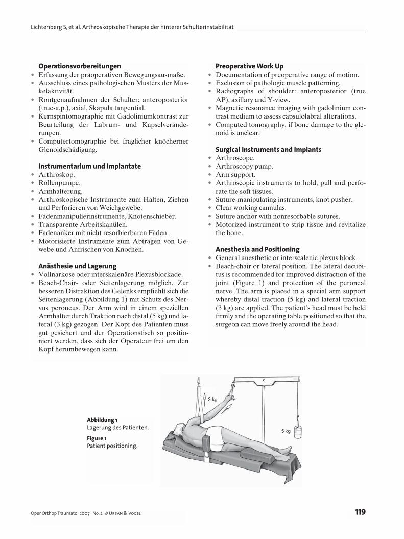
Anästhesie und Lagerung• Vollnarkose oder interskalenäre Plexusblockade.• Beach-Chair- oder Seitenlagerung möglich. Zur
besseren Distraktion des Gelenks empfiehlt sich die Seitenlagerung (Abbildung 1) mit Schutz des Ner-vus peroneus. Der Arm wird in einem speziellen Armhalter durch Traktion nach distal (5 kg) und la-teral (3 kg) gezogen. Der Kopf des Patienten muss gut gesichert und der Operationstisch so positio-niert werden, dass sich der Operateur frei um den Kopf herumbewegen kann.
Preoperative Work Up• Documentation of preoperative range of motion.• Exclusion of pathologic muscle patterning.• Radiographs of shoulder: anteroposterior (true
AP), axillary and Y-view.• Magnetic resonance imaging with gadolinium con-
trast medium to assess capsulolabral alterations.• Computed tomography, if bone damage to the gle-
noid is unclear.
Surgical Instruments and Implants• Arthroscope.• Arthroscopy pump.• Arm support.• Arthroscopic instruments to hold, pull and perfo-
rate the soft tissues.• Suture-manipulating instruments, knot pusher.• Clear working cannulas.• Suture anchor with nonresorbable sutures.• Motorized instrument to strip tissue and revitalize
the bone.
Anesthesia and Positioning• General anesthetic or interscalenic plexus block.• Beach-chair or lateral position. The lateral decubi-
tus is recommended for improved distraction of the joint (Figure 1) and protection of the peroneal nerve. The arm is placed in a special arm support whereby distal traction (5 kg) and lateral traction (3 kg) are applied. The patient’s head must be held firmly and the operating table positioned so that the surgeon can move freely around the head.
Abbildung 1Lagerung des Patienten.
Figure 1Patient positioning.
3 kg
5 kg
Lichtenberg S, et al. Arthroscopic Treatment of Posterior Shoulder Instability
120 Oper Orthop Traumatol 2007 · No. 2 © Urban & Vogel
Operationstechnik
Abbildungen 2 bis 17
Surgical Technique
Figures 2 to 17
Antirotationsverband aus Kunststoff
c Anterosuperiores Portal
c Anterosuperior portal
M. subscapularis
M. supraspinatus
c
d Posterolaterales Portal
d Posterolateral portal
b
Tendo m. bicip. brach.,Caput longum
a
a DorsalesStandardportal
a Standardposterior portal
b AnteroinferioresPortal
b Anteroinferior portal
d
3 kg
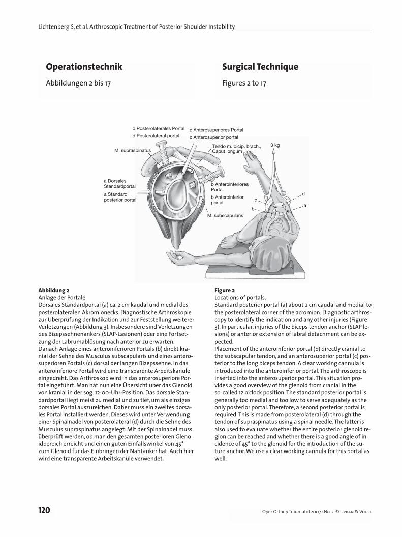
Abbildung 2Anlage der Portale.Dorsales Standardportal (a) ca. 2 cm kaudal und medial des posterolateralen Akromionecks. Diagnostische Arthroskopie zur Überprüfung der Indikation und zur Feststellung weiterer Verletzungen (Abbildung 3). Insbesondere sind Verletzungen des Bizepssehnenankers (SLAP-Läsionen) oder eine Fortset-zung der Labrumablösung nach anterior zu erwarten.Danach Anlage eines anteroinferioren Portals (b) direkt kra-nial der Sehne des Musculus subscapularis und eines antero-superioren Portals (c) dorsal der langen Bizepssehne. In das anteroinferiore Portal wird eine transparente Arbeitskanüle eingedreht. Das Arthroskop wird in das anterosuperiore Por-tal eingeführt. Man hat nun eine Übersicht über das Glenoid von kranial in der sog. 12:00-Uhr-Position. Das dorsale Stan-dardportal liegt meist zu medial und zu tief, um als einziges dorsales Portal auszureichen. Daher muss ein zweites dorsa-les Portal installiert werden. Dieses wird unter Verwendung einer Spinalnadel von posterolateral (d) durch die Sehne des Musculus supraspinatus angelegt. Mit der Spinalnadel muss überprüft werden, ob man den gesamten posterioren Gleno-idbereich erreicht und einen guten Einfallswinkel von 45° zum Glenoid für das Einbringen der Nahtanker hat. Auch hier wird eine transparente Arbeitskanüle verwendet.
Figure 2Locations of portals.Standard posterior portal (a) about 2 cm caudal and medial to the posterolateral corner of the acromion. Diagnostic arthros-copy to identify the indication and any other injuries (Figure 3). In particular, injuries of the biceps tendon anchor (SLAP le-sions) or anterior extension of labral detachment can be ex-pected.Placement of the anteroinferior portal (b) directly cranial to the subscapular tendon, and an anterosuperior portal (c) pos-terior to the long biceps tendon. A clear working cannula is introduced into the anteroinferior portal. The arthroscope is inserted into the anterosuperior portal. This situation pro-vides a good overview of the glenoid from cranial in the so-called 12 o’clock position. The standard posterior portal is generally too medial and too low to serve adequately as the only posterior portal. Therefore, a second posterior portal is required. This is made from posterolateral (d) through the tendon of supraspinatus using a spinal needle. The latter is also used to evaluate whether the entire posterior glenoid re-gion can be reached and whether there is a good angle of in-cidence of 45° to the glenoid for the introduction of the su-ture anchor. We use a clear working cannula for this portal as well.
Lichtenberg S, et al. Arthroskopische Therapie der hinterer Schulterinstabilität
121Oper Orthop Traumatol 2007 · No. 2 © Urban & Vogel
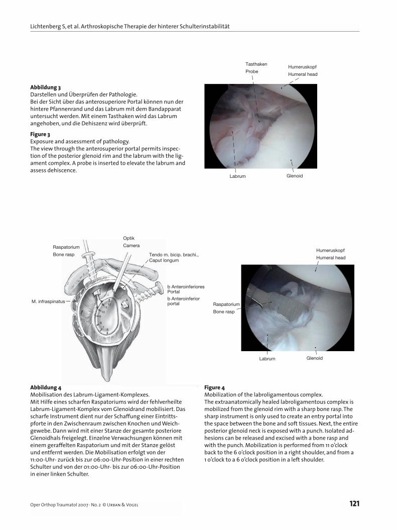
Abbildung 3Darstellen und Überprüfen der Pathologie.Bei der Sicht über das anterosuperiore Portal können nun der hintere Pfannenrand und das Labrum mit dem Bandapparat untersucht werden. Mit einem Tasthaken wird das Labrum angehoben, und die Dehiszenz wird überprüft.
Figure 3Exposure and assessment of pathology.The view through the anterosuperior portal permits inspec-tion of the posterior glenoid rim and the labrum with the lig-ament complex. A probe is inserted to elevate the labrum and assess dehiscence.
Abbildung 4Mobilisation des Labrum-Ligament-Komplexes.Mit Hilfe eines scharfen Raspatoriums wird der fehlverheilte Labrum-Ligament-Komplex vom Glenoidrand mobilisiert. Das scharfe Instrument dient nur der Schaffung einer Eintritts-pforte in den Zwischenraum zwischen Knochen und Weich-gewebe. Dann wird mit einer Stanze der gesamte posteriore Glenoidhals freigelegt. Einzelne Verwachsungen können mit einem geraffelten Raspatorium und mit der Stanze gelöst und entfernt werden. Die Mobilisation erfolgt von der 11:00-Uhr- zurück bis zur 06:00-Uhr-Position in einer rechten Schulter und von der 01:00-Uhr- bis zur 06:00-Uhr-Position in einer linken Schulter.
Figure 4Mobilization of the labroligamentous complex.The extraanatomically healed labroligamentous complex is mobilized from the glenoid rim with a sharp bone rasp. The sharp instrument is only used to create an entry portal into the space between the bone and soft tissues. Next, the entire posterior glenoid neck is exposed with a punch. Isolated ad-hesions can be released and excised with a bone rasp and with the punch. Mobilization is performed from 11 o’clock back to the 6 o’clock position in a right shoulder, and from a 1 o’clock to a 6 o’clock position in a left shoulder.
Humeruskopf
Humeral head
Tasthaken
Probe
GlenoidLabrum
Optik
Camera
M. infraspinatus
Raspatorium
Bone rasp Tendo m. bicip. brachi.,Caput longum
b AnteroinferioresPortal
b Anteroinferiorportal
Humeruskopf
Humeral head
Labrum Glenoid
Raspatorium
Bone rasp
Lichtenberg S, et al. Arthroscopic Treatment of Posterior Shoulder Instability
122 Oper Orthop Traumatol 2007 · No. 2 © Urban & Vogel
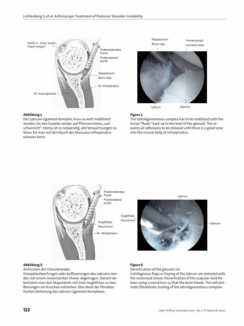
Abbildung 5Der Labrum-Ligament-Komplex muss so weit mobilisiert werden, bis das Gewebe wieder auf Pfannenniveau „auf-schwimmt“. Hierzu ist es notwendig, alle Verwachsungen zu lösen, bis man auf den Bauch des Musculus infraspinatus schauen kann.
Figure 5The labroligamentous complex has to be mobilized until the tissue “floats” back up to the level of the glenoid. This re-quires all adhesions to be released until there is a good view into the muscle belly of infraspinatus.
Abbildung 6Anfrischen des Glenoidrandes.Knorpelaufwerfungen oder Auffaserungen des Labrums wer-den mit einem motorisierten Shaver abgetragen. Danach de-kortiziert man den Skapulahals mit einer Kugelfräse, so dass Blutungen am Knochen entstehen. Dies dient der fibroblas-tischen Anheilung des Labrum-Ligament-Komplexes.
Figure 6Decortication of the glenoid rim.Cartilaginous flaps or fraying of the labrum are removed with the motorized shaver. Decortication of the scapular neck fol-lows using a round burr so that the bone bleeds. This will pro-mote fibroblastic healing of the labroligamentous complex.
M. subscapularis
Raspatorium
Bone rasp
Tendo m. bicip. brach.,Caput longum
M. infraspinatus
Posterolaterales
Posterolateralportal
Portal
Humeruskopf
Humeral head
Labrum Glenoid
Raspatorium
Bone rasp
Labrum
Glenoid
Kugelfräse
Round burr
M. infraspinatus
Kugelfräse
Round burr
PosterolateralesPortal
Posterolateralportal
Lichtenberg S, et al. Arthroskopische Therapie der hinterer Schulterinstabilität
123Oper Orthop Traumatol 2007 · No. 2 © Urban & Vogel
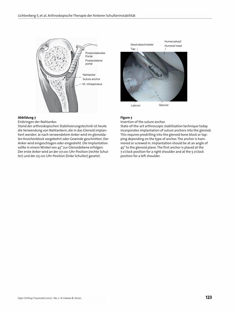
Abbildung 7Einbringen der Nahtanker.Stand der arthroskopischen Stabilisierungstechnik ist heute die Verwendung von Nahtankern, die in das Glenoid implan-tiert werden. Je nach verwendetem Anker wird im glenoida-len Knochenblock vorgebohrt oder Gewinde geschnitten. Der Anker wird eingeschlagen oder eingedreht. Die Implantation sollte in einem Winkel von 45° zur Glenoidebene erfolgen. Der erste Anker wird an der 07:00-Uhr-Position (rechte Schul-ter) und der 05:00-Uhr-Position (linke Schulter) gesetzt.
Figure 7Insertion of the suture anchor.State-of-the-art arthroscopic stabilization technique today incorporates implantation of suture anchors into the glenoid. This requires predrilling into the glenoid bone block or tap-ping depending on the type of anchor. The anchor is ham-mered or screwed in. Implantation should be at an angle of 45° to the glenoid plane. The first anchor is placed at the 7 o’clock position for a right shoulder and at the 5 o’clock position for a left shoulder.
Nahtanker
Suture anchor
M. infraspinatus
PosterolateralesPortal
Posterolateralportal
Humeruskopf
Humeral head
Labrum Glenoid
Gewindeschneider
Tap
Lichtenberg S, et al. Arthroscopic Treatment of Posterior Shoulder Instability
124 Oper Orthop Traumatol 2007 · No. 2 © Urban & Vogel
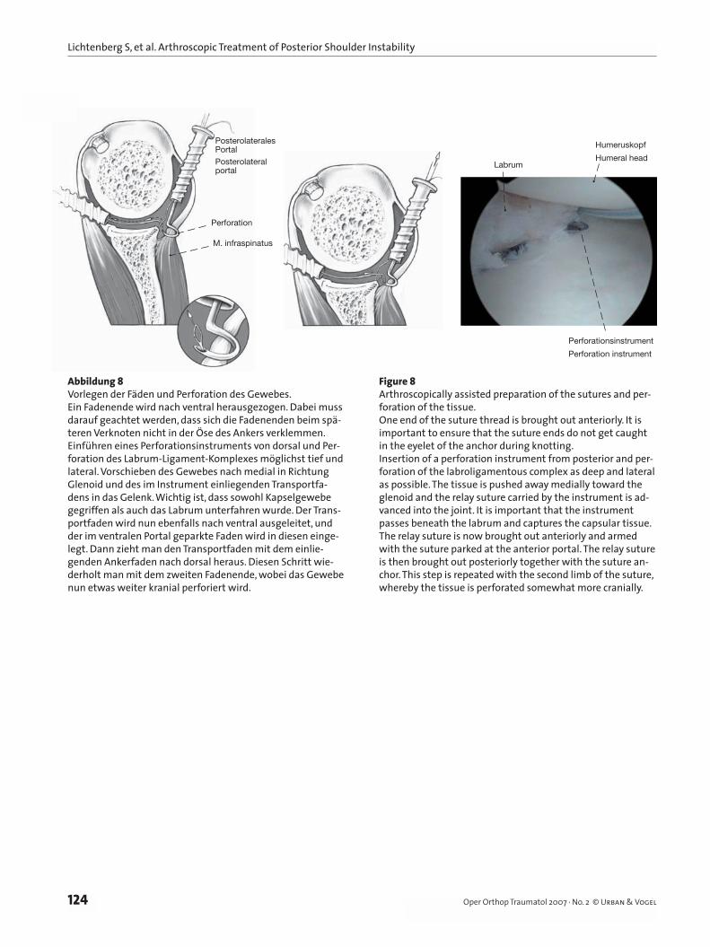
Abbildung 8Vorlegen der Fäden und Perforation des Gewebes.Ein Fadenende wird nach ventral herausgezogen. Dabei muss darauf geachtet werden, dass sich die Fadenenden beim spä-teren Verknoten nicht in der Öse des Ankers verklemmen.Einführen eines Perforationsinstruments von dorsal und Per-foration des Labrum-Ligament-Komplexes möglichst tief und lateral. Vorschieben des Gewebes nach medial in Richtung Glenoid und des im Instrument einliegenden Transportfa-dens in das Gelenk. Wichtig ist, dass sowohl Kapselgewebe gegriffen als auch das Labrum unterfahren wurde. Der Trans-portfaden wird nun ebenfalls nach ventral ausgeleitet, und der im ventralen Portal geparkte Faden wird in diesen einge-legt. Dann zieht man den Transportfaden mit dem einlie-genden Ankerfaden nach dorsal heraus. Diesen Schritt wie-derholt man mit dem zweiten Fadenende, wobei das Gewebe nun etwas weiter kranial perforiert wird.
Figure 8Arthroscopically assisted preparation of the sutures and per-foration of the tissue.One end of the suture thread is brought out anteriorly. It is important to ensure that the suture ends do not get caught in the eyelet of the anchor during knotting.Insertion of a perforation instrument from posterior and per-foration of the labroligamentous complex as deep and lateral as possible. The tissue is pushed away medially toward the glenoid and the relay suture carried by the instrument is ad-vanced into the joint. It is important that the instrument passes beneath the labrum and captures the capsular tissue. The relay suture is now brought out anteriorly and armed with the suture parked at the anterior portal. The relay suture is then brought out posteriorly together with the suture an-chor. This step is repeated with the second limb of the suture, whereby the tissue is perforated somewhat more cranially.
Abb.8
M. infraspinatus
Perforation
PosterolateralesPortalPosterolateralportal
Humeruskopf
Humeral headLabrum
Perforationsinstrument
Perforation instrument
Lichtenberg S, et al. Arthroskopische Therapie der hinterer Schulterinstabilität
125Oper Orthop Traumatol 2007 · No. 2 © Urban & Vogel
Labrumauf demPfannenrand
Labrum at the level of the glenoid working as abumper
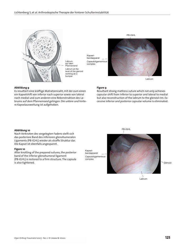
Abbildung 9Es resultiert eine kräftige Matratzennaht, mit der zum einen ein Kapselshift von inferior nach superior sowie von lateral nach medial und zum anderen eine Rekonstruktion des La-brums auf dem Pfannenrand gelingen. Die untere und hinte-re Kapselausweitung ist aufgehoben.
Figure 9Resultant strong mattress suture which not only achieves capsular shift from inferior to superior and lateral to medial but also reconstruction of the labrum to the glenoid rim. Ex-cessive inferior and posterior capsular volume is eliminated.
Abbildung 10Nach Verknoten des vorgelegten Fadens stellt sich das posteriore Band des inferioren glenohumeralen Ligaments (PB-IGHL) wieder als straffe Struktur dar. Die Kapsel ist ebenfalls angespannt.
Figure 10After knotting of the prepared sutures, the posterior band of the inferior glenohumeral ligament (PB-IGHL) is restored to a firm structure. The capsule is also tightened.
Kapsel-bandapparat
Capsuloligamentous complex
Labrum
PB-IGHL
Kapsel-bandapparat
Capsuloligamentous complex
Labrum
PB-IGHL
Glenoid
Lichtenberg S, et al. Arthroscopic Treatment of Posterior Shoulder Instability
126 Oper Orthop Traumatol 2007 · No. 2 © Urban & Vogel
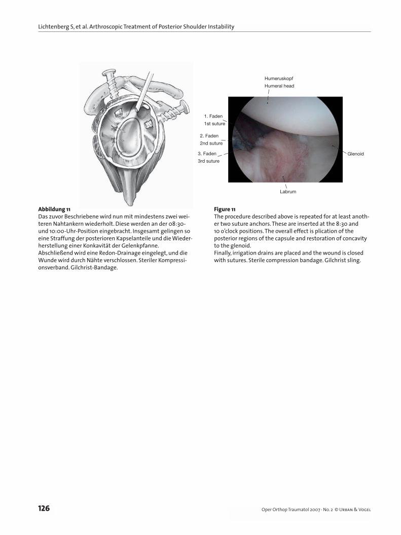
Abbildung 11Das zuvor Beschriebene wird nun mit mindestens zwei wei-teren Nahtankern wiederholt. Diese werden an der 08:30- und 10:00-Uhr-Position eingebracht. Insgesamt gelingen so eine Straffung der posterioren Kapselanteile und die Wieder-herstellung einer Konkavität der Gelenkpfanne.Abschließend wird eine Redon-Drainage eingelegt, und die Wunde wird durch Nähte verschlossen. Steriler Kompressi-onsverband. Gilchrist-Bandage.
Figure 11The procedure described above is repeated for at least anoth-er two suture anchors. These are inserted at the 8:30 and 10 o’clock positions. The overall effect is plication of the posterior regions of the capsule and restoration of concavity to the glenoid.Finally, irrigation drains are placed and the wound is closed with sutures. Sterile compression bandage. Gilchrist sling.
1. Faden
1st suture
Labrum
Humeruskopf
Humeral head
2. Faden
2nd suture
3. Faden
3rd suture
Glenoid
Lichtenberg S, et al. Arthroskopische Therapie der hinterer Schulterinstabilität
127Oper Orthop Traumatol 2007 · No. 2 © Urban & Vogel
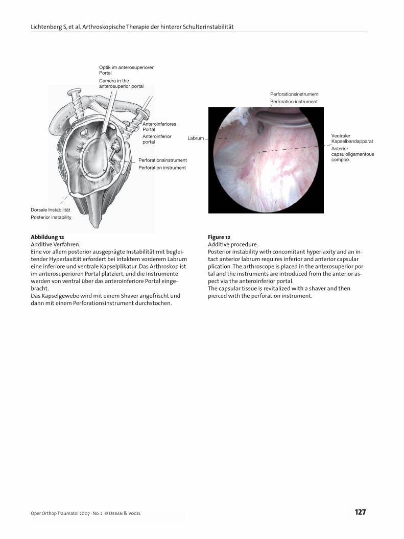
Abbildung 12Additive Verfahren.Eine vor allem posterior ausgeprägte Instabilität mit beglei-tender Hyperlaxität erfordert bei intaktem vorderem Labrum eine inferiore und ventrale Kapselplikatur. Das Arthroskop ist im anterosuperioren Portal platziert, und die Instrumente werden von ventral über das anteroinferiore Portal einge-bracht.Das Kapselgewebe wird mit einem Shaver angefrischt und dann mit einem Perforationsinstrument durchstochen.
Figure 12Additive procedure.Posterior instability with concomitant hyperlaxity and an in-tact anterior labrum requires inferior and anterior capsular plication. The arthroscope is placed in the anterosuperior por-tal and the instruments are introduced from the anterior as-pect via the anteroinferior portal.The capsular tissue is revitalized with a shaver and then pierced with the perforation instrument.
Dorsale Instabilität
Posterior instability
Perforationsinstrument
Perforation instrument
AnteroinferioresPortal
Anteroinferiorportal
Optik im anterosuperiorenPortal
Camera in the anterosuperior portal
Labrum
Perforationsinstrument
Perforation instrument
VentralerKapselbandapparat
Anterior capsuloligamentous complex
Lichtenberg S, et al. Arthroscopic Treatment of Posterior Shoulder Instability
128 Oper Orthop Traumatol 2007 · No. 2 © Urban & Vogel
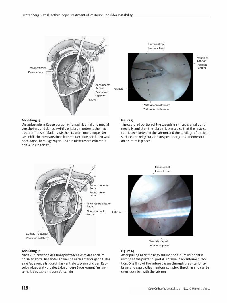
Abbildung 13Die aufgeladene Kapselportion wird nach kranial und medial verschoben, und danach wird das Labrum unterstochen, so dass der Transportfaden zwischen Labrum und Knorpel der Gelenkfläche zum Vorschein kommt. Der Transportfaden wird nach dorsal herausgezogen, und ein nicht resorbierbarer Fa-den wird eingelegt.
Figure 13The captured portion of the capsule is shifted cranially and medially and then the labrum is pierced so that the relay su-ture is seen between the labrum and the cartilage of the joint surface. The relay suture exits posteriorly and a nonresorb-able suture is placed.
Glenoid
Humeruskopf
Humeral head
VentralesLabrum
Anteriorlabrum
Perforationsinstrument
Perforation instrument
AngefrischteKapsel
Revitalized capsule
Labrum
Transportfaden
Relay suture
Dorsale Instabilität
Posterior instability
Nicht resorbierbarerFaden
Non resorbablesuture
Anteroinferiores
Anteroinferiorportal
Portal
Labrum
Humeruskopf
Humeral head
Ventrale Kapsel
Anterior capsule
Abbildung 14Nach Zurückziehen des Transportfadens wird das noch im dorsalen Portal liegende Fadenende nach anterior geholt. Das eine Fadenende ist durch das ventrale Labrum und den Kap-selbandapparat vorgelegt, das andere Ende kommt frei un-terhalb des Labrums zum Vorschein.
Figure 14After pulling back the relay suture, the suture limb that is resting at the posterior portal is drawn in an anterior direc-tion. One limb of the suture passes through the anterior la-brum and capsuloligamentous complex, the other end can be seen loose beneath the labrum.
Lichtenberg S, et al. Arthroskopische Therapie der hinterer Schulterinstabilität
129Oper Orthop Traumatol 2007 · No. 2 © Urban & Vogel
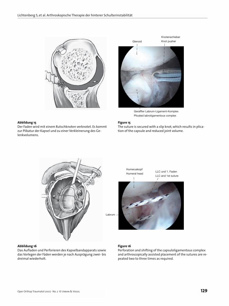
Abbildung 15Der Faden wird mit einem Rutschknoten verknotet. Es kommt zur Plikatur der Kapsel und zu einer Verkleinerung des Ge-lenkvolumens.
Figure 15The suture is secured with a slip knot, which results in plica-tion of the capsule and reduced joint volume.
Glenoid
Knotenschieber
Knot pusher
Geraffter Labrum-Ligament-Komplex
Plicated labroligamentous complex
LLC und 1. Faden
LLC and 1st suture
Labrum
Humeruskopf
Humeral head
Abbildung 16Das Aufladen und Perforieren des Kapselbandapparats sowie das Vorlegen der Fäden werden je nach Ausprägung zwei- bis dreimal wiederholt.
Figure 16Perforation and shifting of the capsuloligamentous complex and arthroscopically assisted placement of the sutures are re-peated two to three times as required.
Lichtenberg S, et al. Arthroscopic Treatment of Posterior Shoulder Instability
130 Oper Orthop Traumatol 2007 · No. 2 © Urban & Vogel
Postoperative Behandlung• Belassen des Gilchrist-Verbandes für 48 h, danach
Anlage eines 15°-Abduktionskissens in Neutralro-tation für 3 Wochen.
• Am 1. postoperativen Tag Entfernung der Draina-ge, Beginn mit Lymphdrainage und Kryotherapie. Haltungsschulung. Aktive Mobilisierung der Hand und des Ellbogens.
• In den ersten 3 Wochen Flexion/Abduktion bis 60° rein passiv unter Schmerzfreiheit. Aus der Außenrotation bis 0° Innenrotation mobilisieren. Aktive Mobilisierung des Ellbogens bei addu-ziertem Arm mit dynamischem Bizeps- und Tri-zepstraining. Schlingentisch. Manuelle Therapie mit leichter Gleitmobilisation Stufe I, schmerz-freie Traktion. Ab der 2. Woche Übergehen auf Warmpackungen.
• Ab der 4. Woche aktiv-assistierte Flexion bis 90°, Abduktion bis 60°, Außenrotation 80° aktiv vor der Körperlängsachse. Aus der Außenrotation bis 30° Innenrotation mobilisieren. Aktives Training der Skapulastabilisatoren.
• Ab der 7. Woche Erarbeiten der freien Flexion, Ab-duktion und Rotation. Nach Erreichen der freien Flexion Erarbeiten der freien Innenrotation. Trai-ning der Rotatorenmanschette, des Musculus delto-
Postoperative Management• The Gilchrist bandage is worn for 48 h, then applica-
tion of a 15° abduction splint in neutral rotation for 3 weeks.
• Removal of irrigation drains on the 1st postoperative day, start of lymph drainage and cryotherapy. Postural exercises. Active mobilization of the hand and elbow.
• Passive flexion/abduction up to 60° only or as far as the pain threshold in the first 3 weeks. Mobilization from external rotation to 0° internal rotation. Ac-tive mobilization of the adducted arm with dynamic biceps and triceps training. Horizontal sling table (schlingentisch method). Manual therapy with slight translation mobilization at level I, pain-free trac-tion. Application of hot packs from week 2.
• From week 4, active-assisted flexion to 90°, abduc-tion to 60°, active external rotation 80° in the longi-tudinal axis of the body. Mobilization from external rotation to 30° internal rotation. Active training of the scapular stabilizers.
• From week 7, work on free flexion, abduction and rotation. When free flexion has been restored, work on free internal rotation. Training of the rotator cuff, the deltoid muscle, and the scapular stabilizers. No resistance with long lever arm.
• Coordination training.
AIGHL
PIGHL
MGHL
SGHL
Gespanntes IGHL und MGHL
Tightened IGHL and MGHL
Labrum
Humeruskopf
Humeral head
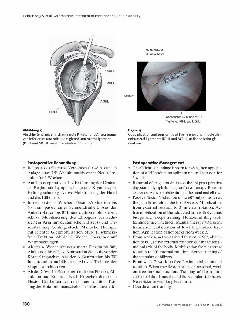
Abbildung 17Abschließend zeigen sich eine gute Plikatur und Anspannung von inferiorem und mittlerem glenohumeralem Ligament (IGHL und MGHL) an den ventralen Pfannenrand.
Figure 17Good plication and tensioning of the inferior and middle gle-nohumeral ligaments (IGHL and MGHL) at the anterior gle-noid rim.
Lichtenberg S, et al. Arthroskopische Therapie der hinterer Schulterinstabilität
131Oper Orthop Traumatol 2007 · No. 2 © Urban & Vogel
ideus und der Skapulastabilisatoren. Keine Wider-stände mit langem Hebelarm.
• Koordinationstraining.• Ab der 12. Woche sportartspezifisches Training.• Rückkehr zu Kontaktsport oder Wurf-/Schläger-
sportarten frühestens nach 6 Monaten.
Fehler, Gefahren, Komplika tionen• Rezidivinstabilität durch insuffizienten Kapselshift
oder nicht ausreichende Labrumrekonstruktion: Auf eine präzise Position des Labrum-Liga-ment-Komplexes muss geachtet werden; Rezidivin-stabilität durch Fehlplatzierung der Anker zu weit medial am Skapulahals, durch übersehenen knö-chernen Pfannenverlust.
• Knorpelschaden durch fehlplatzierte Fadenanker: Der richtige Einfallswinkel und eine genügende Verankerungstiefe sind zu beachten.
• Bewegungseinschränkung: Durch übermäßigen Kapselshift und/oder durch zu weit zentral in der Gelenkfläche implantierte Anker.
ErgebnisseDie orthopädische Literatur weist hinsichtlich pros-pektiver Studien, die das klinische Ergebnis einer ar-throskopischen hinteren Stabilisierung betrachten, nur eine kleine Zahl von Arbeiten auf.
• From week 12, training of specific sports activities.• Return to contact sports or throwing and racket
sports after 6 months at the earliest.
Errors, Hazards, Complications• Recurrent instability due to insufficient capsular
shift or inadequate reconstruction of the labrum: at-tention must be paid to precise positioning of the labroligamentous complex; other causes include in-correct positioning of the anchor too far medially at the scapular neck, and overlooking bone loss at the glenoid.
• Cartilaginous damage due to incorrectly placed su-ture anchors: the correct incident angle and suffi-cient anchorage depth are important.
• Limited range of motion: may be due to excessive capsular shift and/or implanting the anchors too centrally in the joint surface.
ResultsOnly a few prospective studies concerned with the clini-cal outcomes of arthroscopic posterior stabilization are to be found in the specialist orthopedic literature.
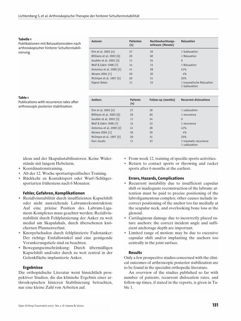
An overview of the studies published so far with number of patients, recurrent dislocation rates, and follow-up times, if stated in the reports, is given in Ta-ble 1.
Autoren Patienten Nachbeobachtungs- Reluxation (n) zeitraum (Monate)
Kim et al. 2003 [4] 27 39 1 SubluxationWilliams et al. 2003 [6] 26 60 1 ReluxationGoubier et al. 2003 [3] 11 34 0 Wolf & Eakin 1998 [7] 14 33 1 ReluxationAntoniou et al. 2000 [2] 41 28 41% Abrams 2004 [1] 50 30 4% McIntyre et al. 1997 [5] 20 31 25% Eigene Daten 11 33 1 traumatische Reluxation 1 Subluxation
Authors Patients Follow-up (months) Recurrent dislocations (n)
Kim et al. 2003 [4] 27 39 1 subluxation Williams et al. 2003 [6] 26 60 1 recurrence Goubier et al. 2003 [3] 11 34 0 Wolf & Eakin 1998 [7] 14 33 1 recurrence Antoniou et al. 2000 [2] 41 28 41% Abrams 2004 [1] 50 30 4% McIntyre et al. 1997 [5] 20 31 25% Own results 11 33 1 traumatic recurrence 1 subluxation
Tabelle 1Publikationen mit Reluxationsraten nach arthroskopischer hinterer Schulterstabili-sierung
Table 1Publications with recurrence rates after arthroscopic posterior stabilization.
Lichtenberg S, et al. Arthroscopic Treatment of Posterior Shoulder Instability
132 Oper Orthop Traumatol 2007 · No. 2 © Urban & Vogel
Eine Übersicht der bisher veröffentlichten Studien mit Patientenzahlen, Reluxationsraten und Nachbe-obachtungsdauer, soweit dies den Arbeiten zu entneh-men ist, bietet Tabelle 1.
Literatur – References1. Abrams JS. Arthroscopic findings and repair of recurrent posterior
subluxation. Presented at the 9th ICSS, Washington, DC, May 2–5, 2004.
2. Antoniou J, Duckworth DT, Harryman DT. Capsulolabral augmenta tion for the management of posteroinferior instability of the shoulder. J Bone Joint Surg Am 2000;82:1220–30.
3. Goubier JN, Iserin A, Duranthon LD, et al. A 4-portal arthroscopic stabilization in posterior shoulder instability. J Shoulder Elbow Surg 2003;12:337–41.
4. Kim SH, Ha KI, Park JH, et al. Arthroscopic posterior labral repair and capsular shift for traumatic unidirectional recurrent posterior sub-luxation of the shoulder. J Bone Joint Surg Am 2003;85:1479–87.
5. McIntyre LF, Caspari RB, Savoie FH 3rd. The arthroscopic treatment of posterior shoulder instability: two-year results of a multiple suture technique. Arthroscopy 1997;13:426–32.
6. Williams RJ, Strickland S, Cohen M, et al. Arthroscopic repair for trau-matic posterior shoulder instability. Am J Sports Med 2003;31:203–9.
7. Wolf EM, Eakin CL. Arthroscopic capsular plication for posterior shoul-der instability. Arthroscopy 1998;14:153–63.
Korrespondenzanschrift – Address for CorrespondenceDr. Sven LichtenbergATOS-Praxisklinik HeidelbergBismarckstraße 9–15D-69115 HeidelbergTelefon (+49/6221) 983-180, Fax -189E-Mail: [email protected]

![DIE ARTHROSKOPISCHE SYNOVIALEKTOMIE - dgou.de · toider Arthritis möglich [12, 20, 29]. Die Entfernung der Synovialmembran ohne Arthrotomie erfordert jedoch vom Endoskopiker eine](https://img.pdfslide.org/doc/110x75/5d62392288c993eb3e8b9a48/die-arthroskopische-synovialektomie-dgoude-toider-arthritis-moeglich-12.jpg)
![morphologischer Ausgangsbefunde 2006 bis 2012“Knoll... · 2 Gelenkchirurgie sind [2]. Bei symptomatischen Meniskusläsionen wird heutzutage hauptsächlich eine arthroskopische Therapie](https://img.pdfslide.org/doc/110x75/5d4c844f88c993ee788b66f1/morphologischer-ausgangsbefunde-2006-bis-2012-knoll-2-gelenkchirurgie.jpg)