Embed Size (px)
Citation preview

Aus dem Institute für Anatomieder Universität zu Lübeck
Direktor: Prof. Dr. med. J. Westermann
Anatomical Structures Forming the Lateral Partof the Rotator Interval in the Human Shoulder Joint
Die Anatomie des lateralen Bereiches desRotatoren-Intervalls im Schultergelenk des Menschen
Inauguraldissertation
zurErlangung der Doktorwürdeder Universität zu Lübeck
- Aus der Medizinischen Fakultät -
vorgelegt vonKristo Kask
aus Pärnu (Estland)
Lübeck 2008

II
1. Berichterstatter: Prof. Dr. rer. nat. Lüder C. Busch
2. Berichterstatter/-in: Priv.-Doz. Dr. med. Michael Wenzl
Tag der Mündlichen Prüfung: 08.05.2009
Zum Druck genehmigt: Lübeck, den 08.05.2009
gez.
- Dekan der Medizinischen Fakultät - Prof. Dr. med. Werner Solbach

III
In memory of my father

IV
Table of contents
Chapter Page
Anatomical and clinical abbreviations VII
Figures IX
1 Introduction 1
1.1 Anatomy of the superior part of the shoulder joint capsule 2
1.1.1 The rotator interval 2
1.1.2 The Ligamentum glenohumerale superius 2
1.1.3 The Ligamentum semicirculare humeri 4
1.2 Pathological seizures of the superior parts of the shoulder joint 5
1.2.1 Rotator cuff tear 5
1.2.2 Pulley lesion and anterior-superior impingement 6
1.2.3 Superior labrum antero-posterior (SLAP) lesion 6
1.2.4 Hill-Sachs lesion 7
2 Aims of the study 9
3 Materials and methods 11
3.1 Anatomic dissection 11
3.2 Light microscopical investigation 12
3.3 Magnetic resonance imaging of the shoulder 12
3.4 Arthroscopic examination 12
3.5 Comparison of the results of anatomical dissection, magnetic resonance
imaging and arthroscopy 13
V
4 Results 14
4.1 Normal anatomy and histology of the Ligamentum glenohumerale superius 14
4.1.1 Gross-anatomic description of the Ligamentum glenohumerale superius 14
4.1.2 Light microscopy of the Ligamentum glenohumerale superius 15
4.2 Normal, arthroscopic and MRI anatomy of the Ligamentum semicirculare
humeri 15
4.2.1 Gross-anatomic description of the Ligamentum semicirculare humeri 15
4.2.2 Magnetic resonance findings and correlation with gross anatomic dissection
results. 16
4.2.3 Arthroscopic anatomy 17
4.2.4 Light microscopy of the Ligamentum semicirculare humeri 18
4.3 Connection between Ligamentum glenohumerale superius and Ligamentum
semicirculare humeri, description of the lateral part of the rotator interval 18
5 Discussion 19
5.1 Materials and methods 19
5.2 Ligamentum glenohumerale superius 19
5.3 Ligamentum semicirculare humeri 22
5.4 Rotator cuff tears 25
5.5 Pulley lesion and anatomy of the lateral part of the rotator interval 25
5.6 Superior labrum antero-posterior lesion 26
5.7 Hill-Sachs lesion 27
6 Summary 28
6.1 Summary 28
6.2 Zusammenfassung 31

VI
7 Appendix of figures 36
8 References 46
9 Acknowledgements 53
10 Curriculum Vitae 54

VII
Anatomical and clinical abbreviations
BT Tendo capitis longi m. bicipitis brachii
CT computer tomography
CH Caput humeri
DESS 3D dual echo steady state three dimensional
GHJ glenohumeral joint, Articulatio humeri
ISP M. infraspinatus
LCG Ligamentum coracoglenoidale
LCH Ligamentum coracohumerale
LG Labrum glenoidale
LGHI Ligamentum glenohumerale inferius
LGHM Ligamentum glenohumerale medius
LGHS Ligamentum glenohumerale superius
LGHspir Ligamentum glenohumerale spirale
LSCH Ligamentum semicirculare humeri
LTH Ligamentum transversum humeri
NA number of acquisitions
MR magnetic resonance
MRI magnetic resonance imaging
PC Processus coracoideus
PD WI proton density weighted imaging
RC rotator cuff
SL slice thickness
SLAP lesion superior labrum antero-posterior lesion
SSC M. subscapularis
SSP M. supraspinatus
TE echo time
Te cranial prolongation of the Tendo m. pectoralis major
TM M. teres minor
TMA Tuberculum majus
TMI Tuberculum minus
TR repetition time
VIII
TSG Tuberculum supraglenoidale
WI weighted imaging

IX
Figures
Page
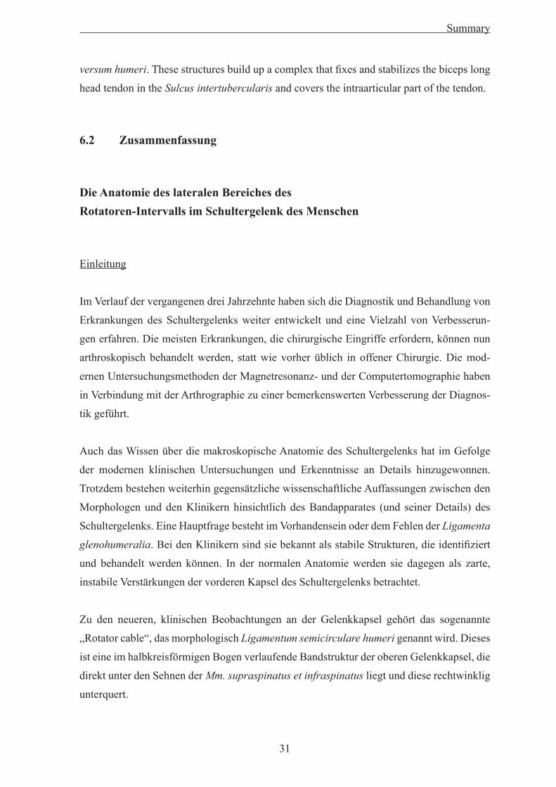
Figure 1: An anterior view of a fixed right shoulder joint specimen. 36
Figure 2: Anterior views of fixed and not fixed dissected right shoulder joint
specimens. 37
Figure 3: An anterior view of a fixed right shoulder joint specimen without the
Ligamentum glenohumerale medius. 38
Figure 4: Antero-superior and anterior views of the detailed anatomy of the
Ligamentum glenohumerale superius and the neighbouring structures
on fixed and not fixed shoulder joint specimens. 39
Figure 5: The detailed anatomy of the insertion region of the Ligamentum
glenohumerale superius. 40
Figure 6: A light, microscopic investigation of the different parts of the
Ligamentum glenohumerale superius. 41
Figure 7: Schematic drawing of the superior capsular ligaments and
an anterior view of a dissected left shoulder joint. 42
Figure 8: A gross anatomical specimen of a dissected left shoulder joint and the
corresponding MR arthrograms. 43
Figure 9: Right shoulder joint with the full-thickness tear of the M. supraspinatus
tendon and correlative MR arthrograms. 44
Figure 10: An arthroscopic view of the right shoulder joint through the posterior
portal with the 30 degree arthroscopic optics. 45
Figure 11: A light microscopic investigation of the Ligamentum
semicirculare humeri. 46

1
1. Introduction
The glenohumeral joint (GHJ) is the ball and socket joint and has the largest range of motion
among all the joints of the human body. The GHJ is guided by the rotator cuff (RC) muscles,
whose tendons extra-articularly reinforce the shoulder joint capsule. The static and dynamic
stabilizers – the ligamentous-capsular structures and the rotator cuff muscles - provide sta-
bility to the shoulder. (Netter 2000, Fanghänel et al. 2003, Schiebler 2005, Schünke et al.
2005, Tillmann 2005).
In recent decades, orthopedic surgery on joints has developed rapidly and is now minimally
invasive. Arthroscopic procedures have become the dominant method for resolving prob-
lems with the GHJ. At the same time, more detailed anatomic investigations allow for a bet-
ter orientation in the narrow field of view available with arthroscopy.
Magnetic resonance (MR) imaging adds a new dimension to clinical findings on noninvasive
visualization (Oxner 1997). MR imaging literature states that the usage of MR-arthrography
gives a better depiction of shoulder joint structures and pathologic disorders. In addition to
x-ray and computer tomography (CT), MR imaging makes it possible to get more detailed
images of the capsular and ligamentous structures. This has made it possible to get diagnos-
tic information about the anterior shoulder joint capsule, the rotator interval (space between
the M. subscapularis and M. supraspiantus tendons) and partial tears in the tendons of RC
muscles (Flannigan et al. 1990, Hodler et al. 1992, Merila et al. 2004, Krief 2005, Morag et
al., 2005).
Due to advanced diagnostic and surgical methods nowadays, orthopedic surgeons and radi-
ologists have been able to give more detailed descriptions of glenohumeral joint anatomy,
which can be found in clinical literature.
Introduction

2
1.1 Anatomy of the superior part of the shoulder joint capsule
1.1.1 Rotator interval
The antero-superior region of the GHJ capsule between the superior edge of the M. subscapu-
laris (SSC) and the anterior edge of the M. supraspinatus (SSP), is clinically termed as the
“rotator interval” and is not officially recognized in the anatomic literature as a topographic
region (Netter 2000, Fanghänel et al. 2003, Schiebler 2005, Schünke et al, 2005, Tillmann
2005). Descriptions of the anatomic composition of the rotator interval are contentious. It
has been described both as the weakest portion of shoulder joint capsule, claming that it has
no reinforcing structures, (Steiner and Hermann 1989) and also as the thickest portion (Jost
et al. 2000). The most detailed investigation of the rotator interval is given by Kolts and his
colleagues (2002). In their description, the rotator interval consists of a complex of capsulo-
ligamentous structures which are divided into the lateral, medio-superior and medio-inferior
parts (Kolts et al. 2002). According to their description, the lateral part of the rotator interval
is strengthened by the Ligamentum semicirculare humeri (LSCH) and anterior fibers from
the M. supraspinatus which, together, are attached to the Tuberculum minus (TMI). The
Ligamenta coracohumerale (LCH) et coracoglenoidale (LCG) form the medio-superior part
and the medio-inferior part is reinforced by the Ligamenta glenohumerale superius (LGHS)
et medius (LGHM) (Kolts et al. 2002).
Wermer et al. give another detailed, anatomic description of the lateral part of the rotator
interval (2000). In their histo-anatomic study, they focused on describing the stabilizing
sling for the long head of the biceps tendon that, according to their results, consists of trans-
verse fibers (Fasiculus obliquus) and the Ligamentum glenohumerale superius. These were
considered to be the main structures that form the stabilizing sling for the long head tendon
of the biceps (Werner et al. 2000).
1.1.2 Ligamentum glenohumerale superius
Although the glenohumeral ligaments were first described by Flood in 1829, standard human
anatomy has not recognized them as constant macroscopic structures until today. Contempo-
rary anatomy textbooks and atlases (Thiel 1999, Netter 2000, Fanghänel et al. 2003 Schiebler
2005, Schünke et al. 2005, Tillmann 2005) describe the three different glenohumeral liga-
Introduction

3
ments – Ligamenta glenohumerale superius, medium et inferius as unstable thickenings of
the anterior joint capsule. Some authors find that the Ligamenta glenohumeralia are visible
from the internal side of the shoulder joint capsule (Fanghänel et al. 2003) and others state
that the unstable thickenings can be visualized by transillumination of the shoulder joint
capsule (Thiel 1999). There are also authors (Rohen & Yokochi 1993) who do not mention
the existence of the Ligamenta glenohumeralia at all. Therefore, in the official Terminologia
Anatomica (1998) the superior, middle and inferior glenohumeral ligaments are not classi-
fied separately as single structures but all together as Ligamenta glenohumeralia.
Controversially, clinical orthopedic textbooks (DePalma 1983, Warren 1999,) and current
publications (Clark 1990, Ferrari 1990, Clark and Harryman II 1992, Palmer et al. 1994,
Steinbeck et al. 1998, Kolts et al. 2001, Pradhan et al. 2001, Burkhart 2002, Kolts et al. 2002,
Ide et al. 2004) pay a lot of attention to the clinical anatomy of the Ligamenta glenohumer-
alia and describe them as stable macroscopic structures that are visible from the intra- and
extra-articular sides of the shoulder joint capsule.
As there is no common agreement concerning the detailed anatomy of the Ligamenta gleno-
humeralia, a great variety of different opinions about the anatomy of each of ligament can
be found in literature. The focus of the present study was on the Ligamentum glenohumerale
superius (LGHS), which is described as a stable anatomical structure by the majority of
clinical investigators.
One of the dominating opinions concerning the origin of the LGHS is that it arises from the
upper pole of the Cavitas glenoidalis or from the Tuberculum supraglenoidale (TSG), which
is just anterior to the origin of the Tendo capitis longi m. bicipitis brachii (BT) (DePalma
1983, Ferrari 1990, Warren 1999, Kolts et al. 2001, Burkhart 2002, Kolts et al. 2002).
Some descriptions of the origins of the Ligamentum glenohumerale superius state that the
LGHS originates from the superior part of the Labrum glenoidale (LG) (Palmer et al. 1994,
Steinbeck et al. 1998, Pradhan et al. 2001, Ide et al. 2004). Other authors have also mentioned
that the LGHS has a connection with the Ligamentum glenohumerale medium (LGHM) at
the point of its origin in the region of the Tuberculum supraglenoidale (DePalma 1983,
Palmer et al. 1994, Steinbeck et al. 1998, Kolts et al. 2002, Ide et al. 2004).
Introduction

4
Anatomists do not recognize the existence of the Ligamentum glenohumerale superius as a
constant anatomic structure and therefore do not describe its origin or its insertion (Termi-
nologia Anatomica 1998, Thiel 1999, Netter 2000, Fanghänel et al. 2003 Schiebler 2005,
Schünke et al. 2005, Tillmann 2005). The majority of anatomic descriptions about the liga-
mentous structures within the rotator interval are limited to the Ligamentum coracohumerale
with its insertion regions on the Tuberculum majus et minus. Yet, there are currently contrary
anatomic and clinical publications that point out a tight connection between the LCH and the
LGHS and that also separately describe the course and origin of the LGHS and its insertion
region on the Tuberculum minus (DePalma 1983, Ferrari 1990, Steinbeck et al. 1998, Kolts
et al. 2001).
The direct and oblique parts of the ligament are also recognized and their two points of inser-
tion are described as the direct part at the Tuberculum minus and the oblique part at the Liga-
mentum semicirculare humeri (Kolts et al. 2002). Werner et al. (2000) describe the existence
of a “Fasciculus obliquus”, which is supposed to be one part of the stabilizing sling for the
Tendo capitis longi m. bicipitis brachii (BT) that also strengthens the rotator interval. Both
authors find that there are oblique fibers running over the intra-articular portion of the biceps
tendon, with the difference being, that one describes them as the oblique parts of the LGHS
(Kolts et al. 2002), the other finds that the obliquely running fibers belong to the “transverse
band of the rotator cuff (Fasciculus obliquus)” (Werner at al. 2000).
1.1.3 Ligamentum semicirculare humeri
Recent orthopedic and anatomic studies have depicted a capsular ligamentous structure in
the latero-superior shoulder joint capsule below the tendons of the M. supraspinatus (SSP)
and the M. infraspinatus (ISP).
The bundles of collagen fibers with parallel orientation in the superior shoulder joint capsule
run perpendicular to the longitudinal axis of the SSP tendon and were first described in the
findings of histological investigations (Clark et al. 1990, Clark and Harrymann 2nd 1992).
The ligamentous structure was identified as the deep extension of the LCH. These findings
were macroscopically and histologically confirmed by Fallon et al. (Fallon et al. 2002).
Introduction

5
Burkhart et al. (1992) gave an arthroscopic depiction of the morphologically described cap-
sular structure in the superior shoulder capsule and named it the “rotator cable”. In addition,
they proposed a biomechanical model of the tear in the rotator cuff, where the “rotator cable”
acts as the loaded cable of a suspension bridge (Burkhart, 1992; Burkhart et al. 1993). This
concept was also supported by a biomechanical study (Halder et al. 2002).
More detailed anatomical information about this structure was later attained from studies
of embalmed shoulder joints. The name “Ligamentum semicirculare humeri” (LSCH) was
proposed for the capsular ligament of the latero-superior shoulder joint capsule (Kolts et al.
2000, Kolts et al. 2002).
In spite of numerous morphological and clinical investigations, MR imaging literature on
the detailed visualization of the capsular structures in the superior shoulder joint capsule is
rarely found. Stoller et al. (2004) have so far been the only ones (!) who have published an
MR image of the so-called rotator cable or LSCH.
On the other hand, there is constant diagnostic interest in introducing the recently found
structures into clinical practice. In a recent study, sonography identifeid the so-called rota-
tor cable on cadavers, resulting in the ligament in the superior shoulder joint capsule being
confirmed by histological investigations (Morag et al. 2006).
1.2 Pathological seizures of the superior parts of the shoulder joint
1.2.1 Rotator cuff tear
Tears in rotator cuff (RC) muscles are common lesions in the superior part of the shoulder
joint. They are classified as partial or complete, or otherwise known as full-thickness tears.
The subtypes of partial tears are bursal, or articular side tears, and intratendineous tears
(Fukuda 2003). Complete tears are classified into groups by the size of the tear or depending
on how many of the RC muscles tendons are involved in the degenerative changes. A com-
mon view is that when the tear is localized only in the SSP tendon, and the diameter of the
lesion is lesser than 3 cm, it is classified as small; it is moderate when two tendons have a ten-
don defect of 3 – 5 cm and large when the tear spreads to 3 – 4 tendons with a lesion size over
5 cm (Baker et al., 2003). Burkhart et al. made an important statement concerning the pathol-
Introduction

6
ogy of RC tears, which differs from common understanding (1992, 1993). Their description
of the RC tear takes into consideration the existence of the “rotator cable”, otherwise known
as Ligamentum semicirculare humeri (LSCH). The formation of the RC tear is compared to
the suspension bridge structure, where the “rotator cable” acts like a loaded cable under ten-
sion from, and pulled medially, by the SSP (Burkhart 1992; Burkhart et al., 1993). Relying
on this model, Burkhart also recommended that L-shaped and larger U-shaped tears should
be repaired by first making side-by-side sutures to achieve a margin convergence and then
completing the repair with an anchor fixation to the bone (Burkhart 2004).
1.2.2 Pulley lesion and anterior-superior impingement
The “pulley system” is a functional term for the tendinoligamentous sling in the lateral part
of the rotator interval, which consists of four major structures. These four structures are the
LCH, the LGHS, the anterior fibers of the SSP tendon and the superior fibers of the SSC ten-
don. At the intraarticular entrance of the bicipital groove, the LGHS and LCH fibers together
form the reflection pulley for the long head of the biceps tendon (Werner et al., 2003; Hab-
ermeyer et al., 2004).
Habermeyer et al. (2004) divided the pulley lesions into four groups:
first group – only the LGHS lesion•
second group – the LGHS and the SSP lesions•
third group – the LGHS and the SSC lesions•
fourth group – the LGHS lesion with the SSP and the SSC lesions•
Statements in the research on the etiology and pathological mechanisms of pulley lesion are
not cohesive (Gerber and Sebesta 2000; Habermeyer et al., 2004).
1.2.3 Superior labrum antero-posterior (SLAP) lesion
Lesions of the superior labrum are difficult to diagnose because the superior labrum attaches
to the glenoid in a wide variety of ways (Gartsman and Hammerman 2000). Andrews et al.
(1985) gave the first report on the pathological changes in the anterior-superior labral tissue
in a group of high-level throwing athletes who were baseball players and javelin throwers.
Introduction

7
Snyder et al. (1990) named the described pathological changes SLAP (superior labrum ante-
ro-posterior) lesions. They identified different degrees of the SLAP lesions in a retrospective
study and worked out a system of classification:
type I SLAP lesion – degeneration or fraying of the superior labrum without •
instability
type II SLAP lesion – detachment of the superior labrum from the glenoid•
type III SLAP lesion – bucket-handle tear of the superior labrum•
type IV SLAP lesion – bucket-handle tear of the superior labrum that extends •
into the biceps tendon
The study by Maffet et al. (1999) added three types to the existing classification:
type V SLAP lesion – antero-inferior Bankart lesion (anterior labrum •
detachment) that continues superiorly and includes separation of the biceps
tendon from the glenoid
type VI SLAP lesion – biceps tendon separation from the glenoid with an •
unstable flap tear of the labrum
type VII SLAP lesion – the separation of the biceps with superior labrum from •
the glenoid that extends anteriorly beneath the middle glenohumeral ligament.
Recent analysis of SLAP lesions has lead to the conclusion that the superior labrum, the long
head of the biceps, and the superior and middle glenohumeral ligaments work together to
provide stability to the shoulder joint (Parentis et al., 2002).
1.2.4 Hill-Sachs lesion
Hill-Sachs lesions are postero-lateral bone defects of the humeral head. Classically,
such lesions have been described as compression fractures of the humeral head and ana-
tomic neck which occur when the glenohumeral joint (GHJ) becomes dislocated. Con-
temporary imaging techniques (arthroscopy, MR imaging and CT) have given new
information about this lesion. One three-dimensional CT investigation of the bony
structures of the GHJ with anterior instabilities showed that the Hill-Sachs lesion is
located in the area of insertion of the M. infraspinatus and M. teres minor tendons
(TM) (Stevens et al., 1999). The Hill-Sachs defect has also been found in 60% of patients
with atraumatic GHJ instability (Werner et al., 2004). Some studies have pointed out that
Introduction

8
the depth of the defect does not correlate to the number of dislocations and that the size of
the bone defect remains the same despite recurring dislocations (Wintzell et al., 1996; Ito
et al., 2000).
Introduction

9
2 Aims of the study
This study was inspired by recently published morphological findings on capsulo-ligamen-
tous structures in the superior and anterior parts of the glenohumeral joint.
The new anatomical findings give a better understanding of previously described pathologic
conditions like the biceps muscle tendon reflection pulley lesions, SLAP lesions, the forma-
tion of the Hill-Sachs defect and degenerative changes within rotator cuff tendons.
One of the structures involved in the formation of the biceps long head tendon pulley and the
SLAP lesions is LGHS, which has an insufficient and controversial anatomic description in
literature. In anatomy textbooks the Ligamenta glenohumeralia are commonly identified as
unstable thickenings of the anterior shoulder joint capsule. (Netter 2000; Fanghänel et al.,
2003; Schiebler 2005; Schünke et al., 2005; Tillmann 2005). Clinical literature either gives
controversial descriptions of the origin, composition, insertion and relation of the LGHS
with newly found capsular structures, or does not mention it at all (DePalma 1983, Palmer et
al. 1994, Steinbeck et al. 1998, Werner et al. 2000, Ide et al. 2004, Stoller et al. 2004, Morag
et al 2005). There is no morphological or clinical analysis of its relation to the previously
described capsular structure – rotator cable, otherwise known as LSCH (Clark et al. 1990,
Burkhart 1992, Clark and Harrymann 2nd 1992, Burkhart et al. 1993, Kolts et al. 2000,
Kolts et al. 2002), within the superior shoulder joint capsule and the lateral rotator interval.
Besides, this is the region that is frequently involved in different pathologies involving all
ages of the population.
In spite of a lack of morphologic description of the LGHS, it is commonly marked on diag-
nostic MR images. The rotator cable, or so-called LSCH, is still missing from radiological
diagnostics. Until now, there is only one (!) MR image of the rotator cable (Stoller et al.
2004) in connection with rotator cuff anatomy published.
This influenced interest in performing an additional morphologic investigation into MR
imaging studies that involve not only the LSCH connection with the rotator cuff anatomy
but also the composition of the lateral part of the rotator interval and the postero-superior
insertion region of the LSCH.
Aims of the study

10
In the lateral part of the rotator interval, both neighboring ligaments – LSCH and LGHS -
are tightly connected with each other and take part in the reinforcement of the shoulder joint
capsule. Therefore they are obviously both involved in the formation of the antero-superior
shoulder joint pathologies.
The particular interest in the clinical importance of the LSCH comes from its involvement
in rotator cuff pathologies in the superior part of the GHJ and from the fact that the posterior
insertion region of this ligament might be connected with the formation of the Hill-Sachs
lesion.
Aims of the study

11
3 Materials and methods
This anatomical study is based on 26 shoulder joint specimens from 21 cadavers. Eight right
and four left alcohol-formalin-glycerol fixed (absolute alcohol 38%, glycerine 24%, formal-
dehyde 4,75%, Fugaten® 9.5%, lysoformine 4,75%, demineralised water 19% ) shoulder
joints (age range 65 – 78 years) and eight right and six left non-fixed shoulder joint speci-
mens (age range 52 - 82 years) were investigated.
Only a detailed anatomic dissection was performed on all the twelve fixed and five non-
fixed (three right and two left) shoulder joint specimens. Nine fresh (five right and four left)
shoulder specimens were examined with MRI and arthroscopy before anatomic dissection.
Two shoulders from one cadaver were excluded from the study after the MRI and arthros-
copy, because both of them had large rotator cuff tears with muscle degeneration and the
glenohumeral joints were in subluxation. Two shoulders (one right, one left) were examined
before anatomic dissection only with arthroscopy.
The human cadavers were dissected under permission of the “Gesetz über das Leichen-,
Bestattungs- und Friedhofswesen (Bestattungsgesetz) des Landes Schleswig-Holstein vom
04.02.2005, Abschnitt II, § 9 (Leichenöffnung, anatomisch)”. In this case it is allowed
to dissect the bodies of the donators (Körperspender/in) for scientific and/or educational
purposes.
3.1 Anatomic dissection
The muscles of the shoulder girdle were removed. The rotator cuff muscles, the intraarticu-
lar part of the biceps tendon and the Mm. pectoralis major et minor insertion tendons were
dissected. The Acromion was cut from the Spina scapulae and removed together with the
Ligamentum coracoacromiale. The Bursa subacromialis and the loose connective tissue was
cleaned off of the muscles and tendons.
The rotator cuff muscles were separated from the shoulder joint capsule and the Ligamentum
coracohumerale, the Ligamentum coracoglenoidale, the Ligamentum glenohumerale super-
ius and the Ligamentum semicirculare humeri were dissected in fine detail.
Materials and methods

12
3.2 Light microscopical investigation
Approximately 1,5 cm × 1 cm pieces of LGHS and LSCH were taken from three different
parts of all of the fresh shoulder specimens: the origin, the insertion and the middle part of
the ligaments. The material was fixed in 10% neutral buffered formalin and embedded in
paraffin (Paraplast®). The histological slices with a thickness of 7 μm were stained after
Trichrome Masson-Goldner with resorcin-fuchsin.
3.3 Magnetic resonance imaging of the shoulder
The shoulder specimens were examined by MRI on a 1.5 Tesla device (Somatom sym-
phony®, Siemens, Erlangen, Germany). The shoulder coil was used on all specimens. MR
arthrography was performed on all the seven shoulder specimens. MR arthrography was
done under fluoroscopic guidance with the injection of 15-20 ml of a contrast solution (0.8
ml Magnevist® in 100 ml of saline). Standard axial, oblique coronal and oblique sagittal fat
saturated views PD WI (TR 3000ms, TE 36 ms, SL 3 mm, Matrix (M) 224*512, number of
acquisitions (NA) 2, TA (min) axial 3.18, oblique sagittal/coronal 3.54) , axial and oblique
coronal T1 WI (TR 632 ms, TE 14 ms, SL 3 mm, M 256*512, NA 3, TA (min) axial 4.32,
oblique coronal 3.46) and axial DESS 3D WI (TR 21.5 ms, TE 6.5 ms, SL 1.5 mm, M
217*256, NA 1, TA (min) 5.14) were used.
3.4 Arthroscopic examination
Shoulder arthroscopies were carried out through the posterior portal (between the suprasp-
inatus and the infraspinatus muscles) with oblique optics at 30 degrees. The glenohumeral
joints were distended with a 0,9% NaCl solution for the arthroscopies. The anterior and
superior shoulder joint capsule and the muscle tendons of the rotator cuff were inspected.
The ligamentous structures and rotator cuff tendons were photographed (Sony Colour Photo
Printer Mavigraph).
Materials and methods

13
3.5 Comparison of the results of anatomical dissection, magnetic resonance imaging and arthroscopy
The evaluation of the MR images was done together with two experienced radiologists.
Since the LSCH was not previously described in radiological literature, both radiologists
were introduced to pertinent LSCH anatomy by a demonstration of the LSCH in 5 gross-
anatomic non-fixed specimens. The first MR imaging specimen was evaluated after the radi-
ologists had seen the corresponding gross-anatomic specimens. The following 6 specimens
were evaluated before anatomic dissection.
Arthroscopies were carried out without knowing the results of the MR imaging investiga-
tion. An experienced orthopaedic surgeon assisted the arthroscopic procedures.
The gross-anatomical dissection was done knowing only the results of the previously per-
formed arthroscopy.
Finally, the results from the different methods were compared and analyzed.
Materials and methods

14
4 Results
4.1 Normal anatomy and histology of the Ligamentum glenohumerale superius
4.1.1 Gross-anatomic description of the Ligamentum glenohumerale superius
The Ligamentum glenohumerale superius (LGHS) was found in all of the twenty-six shoul-
der joint specimens that were investigated. It was always connected with the Ligamentum
coracohumerale (LCH) in its middle part (Figure 1).
The fibers of the Ligamentum glenohumerale superius were divided into two groups –
direct and oblique fibers. In 24 of 26 cases, the “oblique” fibers of the LGHS originated in
the Tuberculum supraglenoidale and arose together with the Ligamentum glenohumerale
medium (LGHM) (Figure 2).
In two cases, when the LGHM was absent, the oblique fibers arose together with the direct
fibers from the antero-superior labrum (Figure 3).
The “direct” fibers originated from the antero-superior and anterior Labrum glenoidale
between 11 and 3 o’clock (Figure 4, 5).
In 24 cases, the arising anterior fibers of the “direct” part of the LGHS were in partly covered
with the LGHM (Figure 2); the antero-superior fibers ran out from below the Tendo capitis
longi m. bicipitis brachii (BT) (Figure 4a)
The direct fibers ran parallel with the Tendo capitis longi m. bicipitis brachii lying between
the BT and the SSC (Figure 2, 4). They coursed with the overlying fibers of the LCH towards
the Tuberculum minus (TMI), inserted partly onto it, and, after reaching the Ligamentum
semicirculare humeri (LSCH), did not attach to it, but ran under it into the bottom of the
Sulcus intertubercularis and made a bridge over the Sulcus intertubercularis, thus forming
the superior part of the Ligamentum transversum humeri (LTH) (Figure 5).
The oblique fibers fused loosely with the overlying fibers of the LCH, coursed over the
intraarticular portion of the BT and inserted on the LSCH (Figure 2, 4).
Results

15
4.1.2 Light microscopy of the Ligamentum glenohumerale superius
The direct part of the Ligamentum glenohumerale superius showed the typical features of
the dense connective tissue with parallel oriented bundles of collagen fibers in the main,
middle part of the ligament (Figure 6a, b). The origin and insertion typically showed the
characters of the fibrocartilagineous tissue (Figure 6c, d, e, f).
The main portion of the oblique component of the LGHS that arose from the Tuberculum
supraglenoidale was composed of parallel oriented bundles of collagen fibers with the typical
fibroblasts present in all the investigated parts (Figure 6a, b). When the LGHM was absent,
the origin of the oblique component of the LGHS was in the superior Labrum glenoidale
(LG) and the tissue was fibrocartilagineous with chondrocyte-like cells lying separate from
each other, in pairs or rows (Figure 6c). At the place of insertion, the fibers of the oblique
component interwove with the perpendicularly running fibers of the LSCH and therefore the
fibers of the LGHS lost their course as an independent structure.
4.2 Normal, arthroscopic and MRI anatomy of the Ligamentum semicirculare humeri
4.2.1 Gross-anatomic description of the Ligamentum semicirculare humeri
The Ligamentum semicirculare humeri was present in all twenty-six shoulder joint speci-
mens. It was always well distinguishable as an arched structure in the superio-lateral shoul-
der joint capsule despite its intra-capsular position.
The LSCH arose from the Tuberculum majus et minus and formed a semicircular arch end-
ing on the posterior facet of the Tuberculum majus, between the insertion regions of the M.
infraspinatus (ISP) and M. teres minor (TM) muscle tendons.
The LSCH was divided into three segments - anterior, middle and posterior.
The portion of the LSCH going from its origin to the anterior edge of the SSP was identified
as the anterior part of the ligament. The anterior segment of the LSCH formed the lateral
part of the rotator interval. In this particular region, the LCH fused into the LSCH and they
Results
16
were indistinguishable from one other in all twenty-six specimens examined (Figure 7).
In the rotator interval, the fibres of the LSCH split into two layers – the superficial-medial
and deep-lateral fibre layers (Figure 7). The anterior fibres of the SSP tendon fused with the
superficial-medial layer of LSCH and coursed along with it until insertion. The insertion
region of the superficial-medial layer of the LSCH was on the superior facet of the Tuber-
culum minus. At the insertion point the LSCH interwove with the SSC tendon and the Liga-
mentum transversum humeri in all investigated specimens (Figure 7, 8a). The deep-lateral
layer of the LSCH coursed over the intertubercular groove and inserted into the anterior
facet of the Tuberculum majus (Figure 7, 8a).
The segment of the LSCH under the SSP tendon formed the middle portion of the LSCH
(Figure 8b). In this portion, the course of the ligament was perpendicular to the longitudinal
axes of the SSP and ISP tendons. Inferior fibres from the SSP tendon interwove tightly with
fibres of the LSCH, forming the capsular insertion of the SSP tendon. In this region, some
of the SSP tendon fibres could not be separated from the LSCH and the joint capsule.
The fibres of the LSCH, covered by the ISP tendon, formed the posterior part of the ligament.
The descending fibres curved latero-posteriorly and ended at the insertion region between
the ISP and TM tendons on the posterior side of the Tuberculum majus (Figure 8c).
4.2.2 Magnetic resonance imaging findings and correlation with gross anatomic
dissection results.
In six of seven specimens the LSCH or parts of the LSCH could be detected by MR imaging.
The ligament or parts of the ligament were best seen on axially (DESS, T1 WI, PDW WI)
oriented MR images.
In particular, the middle portion of the ligament was best identified on axial images as an
anatomic structure with an intermediate signal (Figure 8e). The posterior part of the LSCH
was recognized as a low signal intensity element of the capsule in two specimens (Figure
8e, 8f).
In one specimen with a partial tear of the SSP, the LSCH could not be identified on MR
images. This correlated well with the gross anatomical dissection. In this specimen, the
Results

17
fibres of LSCH were detectable, but the borders of the LSCH were blurred and the LSCH
was very thin compared to other specimens.
In two of seven specimens with full thickness tears of the SSP, the oblique coronal orienta-
tion showed a thickening of the middle LSCH region corresponding to a lateral tissue-defect
above the Caput humeri. The thickening was interpreted as part of the LSCH. This correlated
well with observations made during the anatomic dissection. In both instances, the LSCH
were dislocated medially and surrounded by disorganized connective tissue (Figure 9).
In three of seven specimens, the LSCH was evaluated as a weak ligament on MR images. In
these specimens, pathologies of the rotator cuff (2 partial tears of SSP) or the Caput humeri
(1 Hill Sachs lesion with anterior capsular rupture) could be detected. All pathologies were
confirmed by gross anatomical dissection. In each of these three specimens, the LSCH could
be identified by dissection, but the macroscopic characteristics of the LSCH were weak.
A relatively strong LSCH was identified on MR images in one of seven specimens (Figure
8). This specimen showed no pathology of the shoulder on MR images or gross anatomic
dissection.
4.2.3 Arthroscopic anatomy
The LSCH was clearly identified during arthroscopy of the GHJ specimens in all the investi-
gated cases. Even in the two cases with full-thickness tears of the SSP, the shape and location
of the arched edge of the rupture corresponded with the course of the fibers of the LSCH
(Figure 10).
During arthroscopy through the standard posterior portal, the anterior and the medial parts
of the LSCH became visible.
Two anterior insertion points of the LSCH could be identified; one on the Tuberculum majus
exactly in front of the BT and the other on the Tuberculum minus antero-medial to the intraar-
ticular portion of the BT (Figure 10a). The fibers of the medial part of LSCH were orientated
perpendicularly to the SSP tendon fibers. This was clearly seen in the three cases with partial
tears of the rotator cuff muscles on the articular side of the joint (Figure 10b). The joint cap-
Results

18
sule was destroyed, but the fibers of the tendon were still intact and their course was clearly
visible.
The posterior part of the LSCH was not very clearly visible from the posterior portal.
4.2.4 Light microscopy of the Ligamentum semicirculare humeri
The histological study showed that the main part of the LSCH is formed by well-organized,
parallelly oriented bundles of collagen fibers (Figure 11a, b). The ligamentous tissue is rich
in elastic fibers that are mainly distributed among the fusion regions between the tendons of
the SSP and the ISP muscles (Figure 11c).
Fibers from these tendons are entangled in the middle and posterior parts of the LSCH under
the SSP and ISP tendons. The ligament fibers and the tendon fibers are perpendicular to each
other (Figure 11a, b).
On the articular side of the LSCH, cartilage-like chondroid cells and fibrocartilage-like tis-
sue was found in all the three parts of the LSCH (Figure 11d). The fibrocartilage tissue of the
semicircular ligament built up the gliding surface on the intraarticular side.
4.3 Connection between Ligamentum glenohumerale superius and Ligamentum semicirculare humeri, description of the lateral part of the rotator interval
The lateral part of rotator interval is a triangular, capsular space above the intertubercular
sulcus between the insertion tendons of the SSP and SSC. It is reinforced by the LGHS and
the LSCH. Their fibers run perpendicular to each other in this region but do not intermin-
gle with each other. The fibers of the LSCH that course towards the insertion region on the
Tuberculum minus lie superficially above the fibers of the LGHS. The insertion fibers of the
LGHS insert on the Tuberculum minus, partly course into the Sulcus intertubercularis and
partly overbridge the sulcus, forming the upper part of the Ligamentum transversum humeri
and, in this way, closing the osteofibrous canal of the BT.
Results
19
5 Discussion
5.1 Materials and methods
The results of this investigation depend on a relatively limited number of investigated shoul-
der joint specimens and body donors with a small age range. The number of specimens was
satisfactory for making conclusions regarding normal anatomy, but without describing any
potential anatomic variations. The pathological changes, especially in rotator cuff (RC) were
described by dissection or MR imaging because previous data on the pathologic conditions
and complaints of shoulder problems from the body donors were not available. As MR imag-
ing is a relatively expensive procedure, the number of specimens investigated by this method
was limited.
5.2 Ligamentum glenohumerale superius
This current description of the Ligamentum glenohumerale superius (LGHS) differs from
a lot of previous ones due to the additional anatomic information published during the last
decades concerning the Ligamentum coracohumerale (LCH), the Ligamentum semicirculare
humeri (LSCH), the Ligamentum transversum humeri and the Ligamenta glenohumeralia.
In the present work, both fixed and fresh shoulder joint specimens were used to avoid an
interpretation of the ligaments or their parts as artifacts of fixation. Only structures found
onboth the fixed and the fresh specimens were taken into account as anatomic findings.
There is a common agreement in clinical literature that the LGHS is a constant anatomic
structure that has a rate of appearance of at least 94% (Palmer et al. 1994; Steinbeck et al.
1998; Burkart et al. 2002; Ide et al. 2004).
Although the LGHS varied in the present study in shape and size, it was present in all the
investigated specimens. It consisted of two main groups of “direct” and “oblique” fibres with
different places of origin, courses of the fibres, and attachment regions, except in the cases
without Ligamentum glenohumerale medius (LGHM). In these two cases, the place of origin
of the direct and oblique fibers was at the superior Labrum glenoidale. The Ligamentum gleno-
humerale superius was tightly connected to the overlaid Ligamentum coracohumerale.
Discussion

20
Clinical literature and some current publications state that the LGHS arises from the Tuber-
culum supraglenoidale and the upper pole of the glenoid cavity (DePalma 1983, Warren
1999, Ferrari 1990, Kolts et al. 2001, Burkart 2002, Kolts 2002). The other authors have
stated that the LGHS arises from the superior labrum at the 1 o’clock position (Palmer et al
1994, Steinbeck et al 1998, Ide et al 2004).
Controversial findings on the origin of the LGHS are obviously due to different methods of
investigation. Studies stating that the LGHS arises from the superior labrum, have mainly
focused on the visualization of the Ligamenta glenohumeralia on the intra-articular side of
the anterior shoulder joint capsule (Steinbeck et al. 1998, Ide et al. 2004) or have evaluated
data from the MR images with arthrography (Palmer et al. 1994).
Papers stating that the LGHS arises from the Tuberculum supraglenoidale have described
the extra-articular side of the shoulder joint capsule and placed their main focus on relations
with the other strengthening glenohumeral structures of this region (Ferrari 1990, Kolts et
al. 2001, Kolts et al. 2002).
The present study supports both previous descriptions. The extra- and intraarticular complex
investigation showed that there are two different regions of origin of the LGHS fibers that
may be divided into two groups: direct and oblique fibers. The “oblique fibers” of the LGHS
arise from the Tuberculum supraglenoidale and the “direct” fibers begin at the Labrum gle-
noidale between 11 and 3 o’clock.
At the point of origin, the LGHS is intimately connected with the LGHM and also partially
covered by it. This has given rise to the opinion that the LGHS and LGHM together originate
in the Tuberculum supraglenoidale.
The researchers who have stated that the LGHS originates in the Labrum glenoidale have
also mentioned the existence of anatomic association between the LGHS, the superior labrum
and the origin of the Tendo capitis longi m. bicipitis brachii (BT) (Palmer et al. 1994, Stein-
beck et al. 1998, Pradhan et al. 2001, Parentis et al. 2002, Ide et al. 2004). This statement
is fully supported by the results of the present study. The direct fibers of the LGHS always
originated from the Labrum glenoidale; in the two cases where the LGHM was absent, the
oblique fibers also arose from the Labrum glenoidale.
Discussion

21
The relationship between the LCH and the LGHS has been poorly studied. The lack of an
officially recognized anatomy of the LGHS and the different anatomic descriptions concern-
ing the anatomy of the LCH (Weinstabl et al. 1986, Ferrari 1990, Kolts et al. 2000, Kolts et
al. 2002) may be the causes of the different anatomic descriptions. In the middle part, the
two ligaments are closely connected to each other and the anatomic course of both liga-
ments could be recognized as one. This has obviously predetermined a variety of anatomical
considerations concerning their anatomy, since the ligaments were not separated from each
other before the anatomic description was made.
In recent decades, a lot of new additional information about the ligaments of the shoulder
joint capsule has been published. It has influenced a rather rapid development of the inves-
tigation of newly found ligamentous structures and their connections with officially recog-
nized ones.
Some articles, describe the existence of oblique fibers (Kolts et al. 2002) or the “Fasciculus
obliquus” (Werner et al. 2000) in the antero-superior shoulder joint capsule. According to
these works and our present results, the descriptions given by Werner et al. (2000) and Kolts
et al. (2002) support the existence of the “oblique” part of the LGHS. In addition, the newly
found “transverse fibers of the rotator cuff” (Clark 1990, Clark and Harryman 2nd 1992) - the
“rotator cable” (Burkhart et al. 1993) or the Ligamentum semicirculare humeri (Kolts et al.
2002) serves as an insertion location for the oblique fibers of the LGHS instead of the Tuber-
culum minus (where the direct fibers attach).
The results of the present study correspond with the results of the previous researchers
(Ferrari 1990, Steinbeck et al. 1998, Kolts et al. 2002), who mainly used a gross anatomic
approach, dissecting the ligamentous structures in fine detail. The gross anatomic approach
made it possible to separate the LCH from the underlying LGHS. This excluded the possibil-
ity that those two ligaments are interpreatated as one. Separation of the LCH from the LGHS
made it possible to divide the LGHS into direct and oblique parts with a different origin,
course and insertion regions.
The actual study supports the statements that the LCH, running into the “rotator cable” (Bur-
khart et al. 1993) or the LSCH (Kolts et al. 2002) or the “transverse band of the rotator cuff
(Fasciculus obliquus)” (Clark 1990, Clark and Harryman 2nd 1992, Werner et al. 2000) does
not attach directly to the Tubercula minus et majus of the Humerus (Thiel 1999, Netter 2000,
Discussion

22
Fanghänel et al. 2003 Schiebler 2005, Schünke et al. 2005, Tillmann 2005). The current
results show that the LCH attaches to the LSCH and reaches the Tuberculum minus together
with the semi-circularly running fibers. This means that the LGHS inserts into the Tubercu-
lum minus directly but the fibers of the LCH reach the Tuberculum minus indirectly and first
fuse with the anterior part of the LSCH, which overlays the insertion fibers of the LGHS.
The insertion region of the LGHS is commonly described as the Tuberculum minus (Ferrari
1990, Steinbeck et al. 1998, Kolts et al. 2001, Burkart et al. 2002). Werner et al. (2000) have
added that a U-shaped sling crossing under the biceps tendon and inserting into the proximal
aspect of the intertubercular groove formed at the insertion region the LGHS.. The actual
study shows that the “U- shaped sling” is formed by the direct fibers of the LGHS that partly
insert into the Tuberculum minus and into the bottom of the intertubercular groove and partly
overbridge the Sulcus intertubercularis, forming the upper part of the Ligamentum transver-
sum humeri.
According to current results, the connection of the LGHS with the “fasciculus obliquus”
pointed out by Werner et al. (2002) is located at the anterior part of the LSCH (Burkhart et al
1993; Kolts et al.2002) that overlays the direct fibers of the LGHS at the insertion region.
Considering the origin of the LGHS from the antero-superior Labrum glenoidale, the course
within the rotator interval and the tight connection with the LSCH, it can obviously be
assumed that it may influence the formation of the Labrum glenoidale and other gleno-
humeral joint pathologies, especially in the rotator interval.
5.3 Ligamentum semicirculare humeri
These current results based on twenty-six shoulder joint specimens confirm the presence of
the Ligamamentum semicirculare humeri (LSCH) in the latero-superior shoulder capsule as
an independent anatomic structure. The ligament was visible in all dissected specimens and
in six of seven cases on MR images.
The previous studies on fibers in the superior-lateral shoulder joint capsule locate the pos-
terior insertion point of the LSCH between the M. infraspinatus (ISP) and M. teres minor
(TM) tendons (Burkhart et al., 1993; Clark et al., 1990; Clark and Harryman, 1992; Kolts
Discussion

23
et al., 2000, Kolts et al., 2002). This was also confirmed by the present results. As to the
anterior insertion, most authors have stated that LSCH crosses the Tendo capitis longi m.
bicipitis brachii (BT) and inserts into the Tuberculum minus (Burkhart et al., 1993; Clark et
al., 1990; Clark and Harryman, 1992). The actual results confirm the previous ones, and also
describe the LSCH as having two insertion points anteriorly – the superficial-medial fibers
on the Tuberculum minus and the deep-lateral fibers on the Tuberculum majus. This state-
ment supports the results of observations made on embalmed specimens (Kolts et al., 2000,
Kolts et al., 2002).
The actual anatomical findings also support the previous statement that the Ligamentum
coracohumerale (LCH) does not insert into the Tuberculum majus et minus of the Humerus,
but rather fuses into the LSCH (Kolts et al., 2000, Kolts et al., 2002). The LSCH and LCH
are closely related structurally and functionally and they form the lateral and superior parts
of the rotator interval. In spite of several reports in current literature of new findings on the
ligaments of the shoulder joint, anatomical textbooks and atlases (Fanghänel et al., 2003;
Schiebler 2005; Schünke et al., 2005; Tillmann 2005) still state that the LCH inserts into the
Tuberculum minus et majus and not into the semicircular capsular ligament. As the existence
of the LSCH has not been officially recognized by the Terminologia Anatomica (1998), an
official anatomical description of the LSCH is also missing. The old statement on the inser-
tion of the LCH might have been predominant for so long due to the fact that the anterior
fibers of the LSCH were originally recognized as the insertion fibers of the LCH (Clark et
al., 1990; Clark and Harryman, 1992).
A cleavage in the anterior portion of the LSCH creates an additional cover for the superior
part of the intertubercular sulcus. The superficial-medial layer of the LSCH is associated with
the M. subscapularis (SSC) tendon and the Ligamentum transversum humeri at its insertion
point. The LSCH forms a gradual changeover between the Ligamentum transversum humeri
and the rotator interval. It is rather likely that the described complex (Ligamentum transver-
sum humeri and LSCH) plays an important role in the fixation and stabilization of the BT in
the Sulcus intertubercularis.
Current MR imaging literature states that the usage of MR-arthrography provides a better
depiction of the partial tears in the rotator cuff on its articular surface (Chaipat and Palmer
2006; Waldt et al., 2007). This method also makes it possible to get detailed images of the
anterior capsular structures (Merila et al., 2004).
Discussion

24
In the actual study, the findings of the MR imaging correlated well with the results of the
gross anatomical dissection, showing that there are different characteristics of the LSCH.
Despite the limitations of spatial resolution occurring with MR imaging, especially for very
small anatomic structures, it was possible to detect the LSCH or parts of the LSCH in six of
seven specimens using standard imaging sequences.
The MR study was limited by the small number of non-pathological specimens. Pathologic
findings were found in six of seven shoulder specimens, particularly rotator cuff tears. These
were found on the MR images and confirmed by dissection. This could be one reason why
the ligament had a slightly different appearance on the MR images than after gross dissec-
tion. It is still unknown how the LSCH is involved in, and affects degenerative or posttrau-
matic shoulder disorders.
The curved and semicircular course of the LSCH could be one explanation of why the
anterior and posterior parts of the LSCH are not very easily detected. Its close, anatomical
relationships to the BT, LCH, SSP and capsular structures makes it more difficult to depict
the anterior parts of the LSCH than the posterior and middle parts of the LSCH.
There is good contrast resolution between intra-articular fluid and soft tissue structures on
the superior capsule, especially in the supine position. In this area, the LSCH courses partly
across the plane of the MR images with a transverse orientation. As a result of the partial
volume effect high contrast areas on the MR arthrography, it was possible to recognize the
fibers of the middle portion of the LSCH. The LSCH was mainly detected as a structure of
hypointense to intermediate signal intensity.
As there is apparently no MR imaging literature describing the existence of the LSCH, it
is not possible to compare current results with any other investigation. According to the
present results, MR imaging can be successfully used for a more detailed interpretation of
the superior-lateral shoulder joint capsule. The attempt to visualize the normal anatomy of
the previously described ligament of the superior shoulder joint capsule on the MR images
occasionally provided additional information about the involvement of the ligament in the
pathologic formation of rotator cuff tears. The role and diagnostic value of the visualization
of the LSCH in other pathologic conditions remains unclear due to the lack of sufficient
diagnostic and clinical experience.
Discussion

25
5.4 Rotator cuff tears
Classically, the rotator cuff (RC) tears have been described as traumatic or degenerative
detachments of the rotator cuff muscles from the Humerus. In addition to this classical under-
standing, Burkhart’s hypothesis (Burkhart 1992; Burkhart et al., 1993) claims the existence
of a rotator cable (LSCH) that acts as a suspension bridge in RC tears. This hypothesis has
been confirmed by a biomechanical study by Halder et al. (2002). Later investigations have
found that the morphological base of the rotator cable is a semicircular capsular ligament
within the superior shoulder joint capsule named the Ligamentum semicirculare humeri
(Kolts et al., 2000, Kolts et al., 2002). In the present study, it was also possible to detect a
medially dislocated LSCH in the specimens with a full-thickness tear in the SSP tendon. This
gross anatomy finding provides a base of evidence for the suspension bridge model.
The results of the present study confirm Burkhart’s statement that the location of a rotator
cuff tear is more important than the size of the tear (Burkhart et al., 1993). The location of the
tear predetermines whether the LSCH is intact or not. If the LSCH is intact after the forma-
tion of the SSP and ISP tear, the LSCH is pulled medially by the force of the muscle tension
in the SSP and ISP. The form of the defect on RC tears is predicted by the semicircular form
of the LSCH and the remnants of the SSP and ISP tendons. Therefore the complete tear of
the SSP tendon has a semicircular, Y-shaped form (Kolts 1992).
5.5 Pulley lesion and anatomy of the lateral part of the rotator interval
Previous publications about the reflection pulley of the biceps tendon in the lateral part
of the rotator interval states that the structures forming it are the LCH, the LGHS and the
tendons of the SSP and the SSC (Werner et al. 2000; Werner et al., 2003; Habermeyer et al.
2004). Werner et al. have made a histoanatomical study of the morphology of this stabilizing
sling. They gave a detailed description of the formation of the pulley: it is mainly formed
by fibers from the LGHS along with fibers from the LCH in connection with the so called
Fasiculus obliquus. The origin of the Fasiculus obliquus fibers is not described in their
studies. According to their schematic drawings, it corresponds to the course and position of
the fibers of the LSCH in the upper part of the GHJ. The course of the continuation of the
Fasiculus obliquus fibers to the anterior shoulder joint capsule under the SSC tendon cor-
respond to the course of the recently described Ligamentum glenohumerale spirale (Kolts
Discussion

26
et al. 2001, Merila et al. 2002) which partly arises from the Tuberculum infraglenoidale and
has historically been named as the Fasiculus obliquus (DeLorme 1910). Therefore the term
Fasiculus obliquus to name the fibers in the superior part of the shoulder joint capsule is
confusing. According to the present results, the Fasiculus obliquus named by Werner et al.
(2000) is obviously the rotator cable (Burkhart et al. 1992, 1993) or the LSCH (Kolts et al.,
2000, Kolts et al., 2002).
Relying on the current results and on a previous anatomic study done by Kolts et al. (2000,
2002), it is possible to conclude that the LCH is not involved in the formation of the so-called
“pulley”, because it does not insert into the Tuberculum minus, but rather into the LSCH.
The classification of Habermeyer et al. (2004) for pulley lesions describes either isolated
LGHS tears, or tears associated with tears in the LCH and the SSP, and in the tendons of the
SSC muscles.
The essential problem which results from the formation of the pulley lesion is the instability
of the BT (Habermayer et al. 2004). Taking into consideration the present description of the
structures that form the pulley region, it is difficult to confirm the existence of an isolated
LGHS tear. According to the anatomic composition of the lateral part of the rotator interval,
tears of the LGHS must cause lesions of the SSC tendon, because the Ligamentum trans-
versum humeri is partly composed of the SSC tendon fibers and, superiorly, also the LGHS
fibers. A remarkable medial dislocation of the BT also injures the LSCH which has its SSP
fibers running to the Tuberculum minus. In the case of a lateral dislocation of the BT, the
Ligamentum transversum humeri, as well as fibers from the LGHS and the SSC tendon, must
be injured together with the deep-lateral fibers of the LSCH and the SSP tendon.
5.6 Superior labrum antero-posterior (SLAP) lesion
Superior labrum antero-posterior (SLAP) lesions have been described as BT and superior
labrum lesions. A detachment, or fraying, of the superior labrum always exists in the case of
SLAP lesions (Snyder et al. 1995). Relying on actual results, it is possible to conclude that
the superior labrum might be detached from the glenoid not only by the fibers of the BT but
also by the biomechanical overload of the LGHS. So it is possible to conclude that the SLAP
lesions can be characterized as complex injuries of the LGHS and the BT.
Discussion

27
5.7 Hill-Sachs lesion
The most common bony lesion as pertains to shoulder instability is the Hill-Sachs lesion. It is
a bony defect on the postero-lateral side of the humeral head and it is supposed to be caused
by an impression of the humerus onto the anterior glenoid rim during anterior dislocation.
The development of imaging diagnostics and arthroscopic surgery improved understanding
of the localization of the Hill-Sachs lesion.
A three-dimensional reconstruction of CT images of anteriorly unstable shoulders specified
the location of the Hill-Sachs lesion on the border of the articular surface and the posterior
edge of the Tuberculum majus (Stevens et al. 1999), located approximately at the insertion
points of the tendons of the ISP and the TM. Recent studies and present results have con-
firmed that this is the region of the posterior insertion of the LSCH.
Based on this CT-imaging study and the current results, it is possible to build up a hypoth-
esis, that the Hill-Sachs lesion is not only a bony lesion but it is also obviously combined
with the posterior insertion lesion of the LSCH. The cause of the bony lesion might be the
avulsion of the posterior part of the LSCH from the Humerus.
Studies on atraumatic instability and recurrent dislocations of the glenohumeral joint have
pointed out that there is no correlation between the number of the dislocations and the depth
of the lesion (Wintzell et al., 1996; Ito et al., 2000; Werner et al., 2004).
Discussion

28
6 Summary
6.1 Summary
During the last decades, diagnostics and medical treatment of shoulder joint disorders has
developed rapidly and passed through a lot of changes. Most of the disorders that need sur-
gery can be treated endoscopically instead of the previous, open surgery. Methods like MR
and CT with arthrography have also remarkably improved the possibility of a proper, medi-
cal diagnosis.
New clinical findings and needs have also contributed to the development of the normal,
macroscopic anatomy of the shoulder joint. Despite this, there are still controversies in the
morphologic and clinical sciences concerning the ligamentous structures of the glenohumeral
joint. The most well-known problem is the question of the existence or non-existence of
the glenohumeral ligaments. Clinically, they are known and recognized as stable macro-
scopic structures that are diagnosed and medically treated. Normal anatomy, on the contrary,
describes them as unstable thickenings of the anterior glenohumeral joint capsule.
Recent clinical finding on state that the superior shoulder joint capsule is the so-called „rota-
tor cable“, which has been morphologically described as the „Ligamentum semicirculare
humeri“. It is a semicircular, arched, capsular structure in the superior shoulder joint capsule
under the M. supraspinatus and M. infraspinatus tendons, coursing perpendicularly with the
muscle tendons of the rotator cuff.
These clinical and morphological developments have influenced the finding and description
of new pathological conditions, and therefore some formerly known disorders have been
described in more detail. The development of arthroscopic surgery has lead to a descrip-
tion of the superior labrum antero-posterior (SLAP) and the so-called biceps pulley lesions
within the lateral part of the rotator interval.
The most common problems concerning shoulder pathologies that influence the ability to
work and the quality of life, are the painful problems associated with ruptures in the tendons
of the rotator cuff muscles. Contemporary morphological investigations have influenced the
development of medical treatments for the ruptures and new surgical techniques and meth-
ods are constantly introduced into clinical praxis.
Summary

29
The aim of the present study was to get a detailed, anatomic depiction of one of the most
complex regions in the shoulder joint – the lateral part of rotator interval - and to investigate
the key structures of this region using MR imaging techniques and arthroscopy.
The study was carried out on cadavers, of which twelve were fixed and fourteen were fresh
shoulder joint specimens. The main methods of investigation were anatomic dissection and
MR imaging studies, combined with the arthroscopic examination of the specimens before
the anatomic dissection. Gross anatomic findings were supported by the histological inves-
tigation of the material. The results achieved by the different methods of investigation were
compared with each other and summarized.
The lateral part of the rotator interval is composed of two ligamentous structures - the Liga-
menta glenohumerale superius (LGHS) et semicirculare humeri(LSCH).
In anatomy textbooks, the LGHS is described as an unstable thickening of the anterior shoul-
der joint capsule. On the contrary, radiological and clinical literature describes the LGHS
as a certain and stable ligamentous structure. In the present study, we found that the LGHS
is a constantly present structure with direct and oblique fibers that have certain origin and
insertion regions. The direct fibers arise from the superior labrum (in connection with the
long head tendon of the bicep’s brachii muscle). The oblique fibers of the LGHS begin
together with the Ligamentum glenohumerale medius at the Tuberculum supraglenoidale. In
the cases where the Ligamentum glenohumerale medius is absent, the oblique fibers of the
LGHS begin together with the direct fibers from the Labrum glenoidale. The insertion area
of the direct fibers is at the Tuberculum minus and in the Sulcus intertubercularis. The direct
fibers of the LGHS also partly overbridge the intertubercular groove and form the superior
part of the Ligamentum transversum humeri. The oblique fibers of the LGHS run over the
intraarticular part of the Tendo capitis longi m. bicipitis brachii and fuse under the Ligamen-
tum coracohumerale with the Ligamentum semicirculare humeri.
Relying on the anatomic observations, and taking into consideration the previously published
clinical studies, it is possible to assume that the LGHS has an important role in forming the
pulley for the biceps long head tendon, and that it is directly involved in the formation of
SLAP.
Summary

30
Clinical articles also name the Ligamentum semicirculare humeri (LSCH) as the “rotator
cable”. It is a little-known ligamentous structure within the superior glenohumeral joint cap-
sule. The first descriptions of the “rotator cable” were done by orthopedic surgeons during
arthroscopic operations, who primarily described it as a ligament. Later anatomic studies
on embalmed shoulder joints described the “rotator cable” as a new capsular ligament of the
superior shoulder joint capsule, but did not give a detailed description of its origin and rela-
tion to the LGHS within the lateral rotator interval.
Until today, there is only one published MR image of the LSCH. The aim of the current study
was to get a detailed anatomical description of the LSCH on fresh cadavers and to identify it
with MR imaging and by arthroscopy.
On six out of the seven specimens that were studied with MR imaging, it was possible to
identify the entire LSCH, or some parts of it. An arthroscopic investigation was carried out
on nine specimens and it was possible to identify the LSCH from the intra-articular side in
all cases. Results of the anatomic dissection of embalmed shoulder joints correlated with the
results achieved on the fresh specimens and results of the arthroscopic and MR imaging.
The Ligamentum semicirculare humeri arises anteriorly from two areas: the superior facet of
the Tuberculum minus and the anterior facet of the Tuberculum majus just above the inter-
tubercular groove. It makes an arch under the M. supraspinatus and M. infraspinatus ten-
dons, coursing posteriorly and perpendicularly with the longitudinal axes of the tendons. The
insertion region of the ligament is on the posterior facet of the Tuberculum majus between
the insertion of the Mm. infraspinatus and teres minor tendons.
An evaluation of the glenohumeral joints with rotator cuff muscles tears was performed and
the role of the LSCH in the formation, the shape, and the size of the tear was analyzed. Rely-
ing on anatomic observation, it is possible to conclude that the presence of the LSCH causes
the full-thickness ruptures to have a Y-shape, and that the size of the rupture depends on the
load adjusted to the LSCH by the muscle forces of the Mm. supra- et infraspinatus.
The lateral part of rotator interval is formed by the LGHS and LSCH. In this region, they
run perpendicularly to each other, but the ligaments do not fuse. The LSCH and LGHS form
a “roof” over the intertubercular groove and are tightly connected with the superior edge of
the M. subscapularis, the anterior edge of the M. supraspinatus and the Ligamentum trans-
Summary
31
versum humeri. These structures build up a complex that fixes and stabilizes the biceps long
head tendon in the Sulcus intertubercularis and covers the intraarticular part of the tendon.
6.2 Zusammenfassung
Die Anatomie des lateralen Bereiches des Rotatoren-Intervalls im Schultergelenk des Menschen
Einleitung
Im Verlauf der vergangenen drei Jahrzehnte haben sich die Diagnostik und Behandlung von
Erkrankungen des Schultergelenks weiter entwickelt und eine Vielzahl von Verbesserun-
gen erfahren. Die meisten Erkrankungen, die chirurgische Eingriffe erfordern, können nun
arthroskopisch behandelt werden, statt wie vorher üblich in offener Chirurgie. Die mod-
ernen Untersuchungsmethoden der Magnetresonanz- und der Computertomographie haben
in Verbindung mit der Arthrographie zu einer bemerkenswerten Verbesserung der Diagnos-
tik geführt.
Auch das Wissen über die makroskopische Anatomie des Schultergelenks hat im Gefolge
der modernen klinischen Untersuchungen und Erkenntnisse an Details hinzugewonnen.
Trotzdem bestehen weiterhin gegensätzliche wissenschaftliche Auffassungen zwischen den
Morphologen und den Klinikern hinsichtlich des Bandapparates (und seiner Details) des
Schultergelenks. Eine Hauptfrage besteht im Vorhandensein oder dem Fehlen der Ligamenta
glenohumeralia. Bei den Klinikern sind sie bekannt als stabile Strukturen, die identifiziert
und behandelt werden können. In der normalen Anatomie werden sie dagegen als zarte,
instabile Verstärkungen der vorderen Kapsel des Schultergelenks betrachtet.
Zu den neueren, klinischen Beobachtungen an der Gelenkkapsel gehört das sogenannte
„Rotator cable“, das morphologisch Ligamentum semicirculare humeri genannt wird. Dieses
ist eine im halbkreisförmigen Bogen verlaufende Bandstruktur der oberen Gelenkkapsel, die
direkt unter den Sehnen der Mm. supraspinatus et infraspinatus liegt und diese rechtwinklig
unterquert.
Summary

32
Die neueren Beobachtungen und Erkenntnisse zur Klinik und Morphologie des Schulterge-
lenks führten zu detaillierteren Beschreibungen bekannter Störungen und zur neueren Bew-
ertung der pathologischen Abläufe.
Durch die Einführung der arthroskopischen Schulterchirurgie konnten die Verletzungen des
„Superior labrum antero-posterior = SLAP“ und des sogenannten „Biceps pulley“ im lat-
eralen Bereich des „Rotator Interval“ genau erkannt werden.
Das Hauptproblem aller Schultergelenksveränderungen, die die Arbeitsfähigkeit und die
Lebensqualität herabsetzen, sind die außergewöhnlichen Schmerzen bei Sehnenrupturen
der Muskeln der Rotatorenmanschetten. Zeitgleiche morphologische Untersuchungen der
Rotatorenmanschette haben maßgeblich die Behandlung der Sehnenrupturen und die Ent-
wicklung neuer chirurgischer Techniken beeinflusst und zu ihrer Einführung in die klinische
Praxis beigetragen.
Ziel der Untersuchung
Das Ziel dieser Studie bestand darin, eine detailreiche, anatomische Beschreibung einer der
komplexesten Regionen des Schultergelenks, des lateralen Anteils der Rotatorenmanschette,
zu erhalten und die klinisch bedeutsamen Strukturen dieser Region durch Magnetresonanz-
tomographie und Arthroskopie zu untersuchen.
Material und Methoden
Diese Studie wurde an anatomischen Präparaten von Körperspendern durchgeführt. Es wur-
den zwölf konservierte und vierzehn frische Schultern untersucht.
Im wesentlichen wurden sorgfältigste anatomische Präparationen und Magnet-resonanztom-
ographische Darstellungen verglichen; die frischen Schultern wurden vor der anatomischen
Präparation arthroskopisch untersucht. Die makroskopischen Ergebnisse wurden durch his-
tologische Untersuchungen ergänzt.
Summary

33
Ergebnisse und Diskussion
Der seitliche Anteil des „Rotator Interval“ besteht aus zwei Bandstrukturen, den Ligamenta
glenohumerale superius (LGHS) et semicirculare humeri (LSCH).
In den anatomischen Lehrbüchern wird das Ligamentum glenohumerale superius als schwache
Verstärkung der vorderen Gelenkkapsel des Schultergelenks beschrieben. Im Gegensatz
dazu wird dieses Band in der radiologischen und klinischen Literatur als umschriebene und
stabile Bandstruktur betrachtet.
In dieser Studie konnte festgestellt wirden, dass das LGHS eine sehr konstante Struktur ist,
die mit direkten und schrägen Fasern an exakt zu bestimmenden Punkten entspringt und
inseriert. Die gerade verlaufenden Fasern entspringen von der oberen Gelenklippe (in unmit-
telbarer Nähe zur Ursprungssehne des langen Bizepskopfes). Die schräg verlaufenden Fasern
entspringen zusammen mit dem Ligamentum glenohumerale medius am Tuberculum supra-
glenoidale. In jenen Fällen, in denen das Ligamentum glenohumerale medius fehlt, entsprin-
gen die schräg verlaufenden Fasern zusammen mit den gerade verlaufenden Fasern vom
Labrum glenoidale. Die geraden Fasern inserieren sowohl am Tuberculum minus humeri als
auch im Sulcus intertubercularis; ein Teil dieser Faserzüge überbrückt die Rinne zwischen
den Tubercula und bildet somit den oberen Teil des Ligamentum transversum humeri. Die
schrägen Fasern dieses Ligaments kreuzen den intraartikulären Teil der langen Bizepssehne
und vereinigen sich unter dem Ligamentum coracohumerale mit den Fasern des Ligamen-
tum semicirculare humeri.
Auf der Basis der anatomischen Ergebnisse und unter Berücksichtigung der vorliegenden
klinischen Literatur kann man schlussfolgern, dass das Ligamentum glenohumerale superius
entscheidend an der Ausformung der Umlenkung der langen Bizepssehne beteiligt ist und
damit auch am Zustandekommen der SLAP-Läsion.
Das Ligamentum semicirculare humeri (LSCH), wird in klinischen Artikeln englisch „Rota-
tor cable“ genannt. Diese bandartige Struktur innerhalb des oberen Anteils der Schulterge-
lenkskapsel ist relativ unbekannt. Erste Beobachtungen des „Rotator cable“ wurden von
orthopädischen Chirurgen bei Arthroskopien gemacht, anfangs jedoch nicht als ein Ligament
erkannt. Später wurde es auf der Basis anatomischer Präparationen konservierter Schulterge-
lenke als ein neues Verstärkungsband der oberen Gelenkkapsel identifiziert. Allerdings gab
Summary

34
es keine exakten Beschreibungen seines Verlaufes und seiner topographischen Beziehung
zum Ligamentum glenohumerale superius innerhalb des lateralen Rotator Intervalls.
Bis heute gibt es nur eine Magnetresonanzdarstellung des Ligamentum semicirculare humeri
(LSCH). Deshalb sollten detaillierte anatomische Studien und Beschreibungen des Ligamen-
tum semicirculare humeri von frischen, unfixierten Schultern bei zeitgleicher Darstellung mit
der Magnetresonanztomographie und mittels der Arthroskopie das Ziel dieser Arbeit sein.
An sechs von sieben Schultern war es möglich, mittels der Magnetresonanztomographie das
Ligamentum semicirculare humeri vollständig oder in Teilen zu erkennen. Arthroskopien
wurden an neun Schultergelenken durchgeführt und in allen Fällen konnte das Band von der
Innenseite des Gelenks identifiziert werden. Die anatomischen Präparationen der konservi-
erten Schultern stimmten mit den Ergebnissen der MRI– und arthroskopischen Untersuc-
hungen exakt überein.
Das Ligamentum semicirculare humeri entspringt in zwei Anteilen auf der oberen Facette
des Tuberculum minus und von der vorderen Facette des Tuberculum majus direkt über dem
Sulcus intertubercularis.
Das Band verläuft dann in einem Bogen unter den Sehnen der Mm. supraspinatus et infra-
spinatus, um dann nach hinten im rechten Winkel unter die Sehnen abzubiegen. Der Ansatz
des Bandes liegt auf der hinteren Facette des Tuberculum majus zwischen den Ansätzen der
Mm. infraspinatus et teres minor.
Es wurde eine Einschätzung des Muskelzugs der Rotatorenmuskelmanschette auf das Schul-
tergelenk durchgeführt und dabei die Rolle des LSCH auf die Art und den Grad des Zuges
abgeschätzt. Unter Bezug auf die anatomischen Beobachtungen kann gefolgert werden, dass
das LSCH die Y-Form des kompletten Risses beeinflusst und dass das Ausmass des Risses
von der Belastung des LSCH durch den Muskelzug der Mm. supraspinatus et infraspinatus
abhängt.
Der seitliche Teil des Rotatoren-Intervalls wird vom Ligamentum glenohumerale superius
und vom Ligamentum semicirculare humeri gebildet. In diesem Bereich laufen sie im rech-
ten Winkel zueinander, aber sie verwachsen nicht miteinander. Die beiden Bänder bilden
gleichsam das Dach über dem Sulcus intertubercularis und stehen in engem Kontakt zum
Summary
35
Oberrand des M. subscapularis, der Vorderkante des M. supraspinatus und dem Ligamentum
transversum humeri. Zusammen bilden sie einen Komplex, der die lange Bizepssehne im
Sulcus intertubercularis hält und im weiteren Verlauf den intraartikulären Teil dieser Sehne
bedeckt, um somit Luxationen dieses wichtigen aktiven Stabilisators des Schultergelenks zu
verhindern.
Summary
36
7 Appendix of Figures
Figure 1An anterior view of a fixed right shoulder joint specimen. The Processus coracoideus (PC) is cut at its base (cut) and posteriorly moved together with the Ligamentum coracohumerale (LCH). The M. subscapularis (SSC) is laterally lifted up to visualize the anterior capsule.
Before the detailed dissection, the Ligamentum glenohumerale superius (LGHS) and LCH are tightly connected with each other (oval). The connection between the LGHS and the Ligamentum gleno-humerale medium (LGHM) is also clearly visible. D – direct fibers of the LGHS; O – oblique fibers of the LGHS; LSCH – anterior part of the Ligamentum semicirculare humeri; LGHI – Ligamentum gleohumerale inferius; LGHspir – Ligamentum glenohumerale spirale; LG – Labrum glenoidale; SSP – M. supraspinatus; TMA – Tuberculum majus; TSG – Tuberculum supraglenoidale.
Appendix of figures

37
Figure 2 Anterior views of fixed (a) and not fixed (b) dissected right shoulder joint specimens. The Processus coracoideus (PC) is cut at its base (cut), the Ligamentum coracohumerale (LCH) is separated from the Ligamentum glenohumerale superius (LGHS) and posteriorly moved together with the Processus coracoideus. The ligamentous structures and “rotator cuff” muscles are finely dissected. The Mm. supraspinatus (SSP) et subscapularis (SSC) are separated from the shoulder joint capsule and placed laterally (on figure 2a only the M. supraspinatus is placed laterally).
The Ligamentum glenohumerale superius (LGHS) originates from anterio-superior labrum and has a loose connection to the Ligamentum glenohumerale medium (LGHM) at the origin region of the Tuberculum supraglenoidale (TSG). The oblique fibers of the LGHS (O) arise together with the LGHM from the Tuberculum supraglenoidale (TSG), run over the articluar part of the Tendo capitis longi m. bicipitis brachii (BT) and insert into the Ligamentum semicirculare humeri (LSCH). The direct fibers of the LGHS (D) arise beneath the oblique fibers from the Labrum glenoidale (LG) and run parallel to the Tendo capitis longi m. bicipitis brachii towards the Tuberculum minus. The Ligamentum glenohumerale medium arises at the Tuberculum supraglenoidale and inserts at the Ligamentum glenohumerale spirale (LGHspir).
a: An overview of the Ligamentum glenohumerale superius and its relation to the other ligaments and tendons of the shoulder joint in a fixed specimen. ISP – M. infraspinatus; LTH – Ligamentum transversum humeri.
b: An antero-superior view of the anatomy of the LGHS and its connections to neighboring struc-tures on a fresh specimen. To tension the glenohumeral ligaments, the Humerus is externally rotated. LGHI – Ligamentum glenohumerale inferius.
Appendix of figures

38
Figure 3An anterior view of a fixed right shoulder joint specimen without the Ligamentum glenohumerale medius. The Processus coracoideus is cut at its base (cut) and moved backwards together with the Ligamentae. coracohumerale (LCH) and coracoglenoidale (LCG), the M. subscapularis (SSC) is placed laterally. In the case of an absence of the Ligamentum glenohumerale medium, the oblique (O) and direct (D) fibers of the Ligamentum glenohumerale superius (LGHS) arise from the Labrum glenoidale (LG).
a: BT – Tendo capitis longi m. bicipitis brachii; LSCH – Ligamentum semicirculare humeri; TSG – Tuberculum supraglenoidale.
b: A magnified view of the origins of the Ligamentum glenohumerale superius and the arrange-ment of the fibers. The Ligamentae. corachumerale et coracoglenoidale are placed under tension to achieve a better visualization of their relation to the underlying structures.
BT – Tendo capitis longi m. bicipitis brachii; LSCH – Ligamentum semicirculare humeri; TSG – Tuberculum supraglenoidale.
Appendix of figures
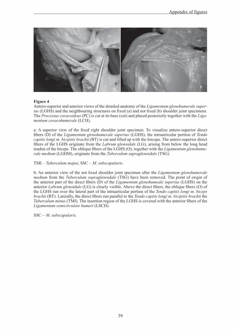
39
Figure 4 Antero-superior and anterior views of the detailed anatomy of the Ligamentum glenohumerale super-ius (LGHS) and the neighbouring structures on fixed (a) and not fixed (b) shoulder joint specimens. The Processus coracoideus (PC) is cut at its base (cut) and placed posteriorly together with the Liga-mentum coracohumerale (LCH).
a: A superior view of the fixed right shoulder joint specimen. To visualize antero-superior direct fibers (D) of the Ligamentum glenohumerale superius (LGHS), the intraarticular portion of Tendo capitis longi m. bicipitis brachii (BT) is cut and lifted up with the forceps. The antero-superior direct fibers of the LGHS originate from the Labrum glenoidale (LG), arising from below the long head tendon of the biceps. The oblique fibers of the LGHS (O), together with the Ligamentum glenohume-rale medium (LGHM), originate from the Tuberculum supraglenoidale (TSG).
TMI – Tuberculum majus; SSC – M. subscapularis.
b: An anterior view of the not fixed shoulder joint specimen after the Ligamentum glenohumerale medium from the Tuberculum supraglenoidale (TSG) have been removed. The point of origin of the anterior part of the direct fibers (D) of the Ligamentum glenohumeale superius (LGHS) on the anterior Labrum glenoidale (LG) is clearly visible. Above the direct fibers, the oblique fibers (O) of the LGHS run over the lateral part of the intraarticular portion of the Tendo capitis longi m. biceps brachii (BT). Laterally, the direct fibers run parallel to the Tendo capitis longi m. bicipitis brachii the Tuberculum minus (TMI). The insertion region of the LGHS is covered with the anterior fibers of the Ligamentum semicirculare humeri (LSCH).
SSC – M. subscapularis.
Appendix of figures

40
Figure 5The detailed anatomy of the insertion region of the Ligamentum glenohumerale superius (LGHS).
To visualize the insertion region of the LGHS, the anterior, superficial fibers of the Ligamentum semicirculare humeri (LSCH) are removed (oval) from the Tuberculum minus (TMI, doted oval) and turned posteriorly together with the Ligamentum coracohumerale (LCH). The intraarticular part of the Tendo capitis longi m. bicipitis brachii is removed. The direct fibers (D) of the LGHS insert into the bottom of the Sulcus intertubercularis and overbridge the Sulcus intertubercularis, forming the superior part of the Ligamentum transversum humeri (LTH). The oblique fibers (O) of the LGHS orginate from Tuberculum supraglenoidale (TSG) and insert into the Ligamentum semicirculare humeri (LSCH).
LGHM – Ligamentum glenohumerale medium; LCG - Ligamentum coracoglenoidale; PC, cut - Processus coracoideus cut at its base; TMA – Tuberculum majus; LG – Labrum glenoidale.
Appendix of figures

41
Figure 6A light, microscopic investigation of the different parts of the Ligamentum glenohumerale superius (LGHS). The LGHS is divided into the main middle part, the origin and the insertion regions.
a, b: Middle part of the LGHS.
a: An overview of the collagen fiber arrangement at low magnification. The collagen fibers are lying in parallel bundles of collagen fibers, which are separated from each other by relatively wide spaces of loose connective tissue at some regions. Magnification bar is 40 µm
b: An overview of the middle part of the LGHS at high magnification. The cells surrounded by the collagen fibers are typical fibrocytes, lying between the parallelly-oriented collagen fibers individu-ally, in pairs or in rows. Magnification bar is 10 µm
c, d: A light microscopic overview of the origin and insertion regions of the LGHS at low magnification.
c: At the origin of the labrum glenoidale, the arising collagen fibers are partly masked by the exrtra-cellular matrix, showing the tissue properties of fibrocartilage. Magnification bar is 40 µm
d: At the region where it inserts into the Sulcus intertubercularis the structure of the LGHS comes to have the features of fibrocartilagineous tissue, which is remarkably different from the less organized structure of the joint capsule. Magnification bar is 40 µm
e, f: The cells of the LGHS at the origin and insertion regions.
The fibrocartilagineous parts of the ligament are populated with oval, chondrocyte-like cells. Mag-nification bars are 40 and 5 µm
Appendix of figures

42
Figure 7a: Schematic drawing of the superior capsular ligaments.
LSCH – Ligamentum semicirculare humeri; LCH – Ligamentum coracohumerale; LCG – Ligamen-tum coracoglenoidale; capsule – Glenohumeral joint capsule; cut – acromial base; PC – Processus coracoideus; TMA – Tuberculum majus; TMI – Tuberculum minus; SSC – M. subscapularis; BT – Tendo capitis longi m. bicipitis brachii; Filled ring – Tuberculum supraglenoidale.
b: An anterior view of a dissected left shoulder joint.
The Acromion is cut from the Spina scapulae and removed together with the Ligamentum coracoac-romiale. The M. supraspinatus tendon (SSP) is separated from the shoulder joint capsule and placed laterally. The Processus coracoideus (PC) is cut at its base (cut) and moved posteriorly, together with the Ligamentum coracohumerale (LCH), to provide a better visualization of the superior capsular structures.
At its insertion into the joint capsule, the SSP tendon follows the course of the Ligamentum semicir-culare humeri (LSCH). The LSCH divides into superficial-medial (S, separated from the underly-ing joint capsule by a plastic probe) and deep-lateral (D) fiber layers. The superficial-medial layer inserts into the Tuberculum minus and the deep-lateral layer on the Tuberculum majus (TMA). At the insertion region on the Tuberculum minus, the superficial-medial layer interweaves with the Liga-mentum transversum humeri (LTH) and the M. subscapularis tendon (SSC). The LCH inserts into the LSCH.
BT - Tendo capitis longi m. bicipitis brachii; Te - cranial prolongation of the Tendo m. pectoralis major overlaying the Tendo capitis longi m. bicipitis brachii.
Appendix of figures

43
Figure 8A gross anatomical specimen of a dissected left shoulder joint and the corresponding MR arthrograms. The Mm. supra-, infraspinatus et M. teres minor have been removed from the joint capsule and placed later-ally. The underlying capsular structures are visualized.
a: An antero-lateral view of the dissected shoulder joint specimen. The Ligamentum coracohumerale (LCH) fuses with the Ligamentum semicir-culare humeri (LSCH). The superficial-medial layer (S) of the LSCH is closely related to the M. subscapularis tendon (SSC), Ligamentum transver-sum humeri (LTH) and the part of the M. supraspinatus ten-don (SSP) that inserts into the Tuberculum minus (TMI) of the Humerus. The deep-lateral layer (D) of the LSCH inserts together with the SSP tendon into the anterior facet of the Tuberculum majus (TMA).
b: A latero-superior view of the dissected shoulder joint specimen. The Ligamentum coracohumerale (LCH) fuses with the Ligamentum semicirculare humeri (LSCH). The course of the LSCH is perpen-dicular to the longitudinal axis of the M. supraspinatus (SSP) tendon.
c: A postero-lateral view of the dissected shoulder joint specimen. The Ligamentum semicirculare humeri (LSCH) inserts into the posterior facet of the Tuberculum majus between the Mm. infraspina-tus et teres minor tendons.
PC - Processus coracoideus; BT - Tendo capitis longi m. bicipitis brachii; CH - Caput humeri; TM - M. teres minor: ISP - M. infraspinatus.
d - f: MR arthrograms of the same left shoulder specimen (axial T1 WI).
d, f: Anterior and posterior parts of the Ligamentum semicirculare humeri (white arrows) are visible as hypotensive stripes under the M. infraspinatus tendon (ISP) and in front of the Tendo capitis longi m. bicipitis brachii (white unfilled arrow, BT).
e: The middle part of the Ligamentum semicirculare humeri (white arrow) is visible as an anatomic structure with an intermediate signal, coursing perpendicularly to the Mm. supraspinatus et infrasp-inatus longitudinal axis. ISP - M. infraspinatus.
Appendix of figures

44
Figure 9a: A posterio-superior view of a dissected right shoulder joint with the full-thickness tear of the M. supraspinatus tendon. The rotator cuff muscles are dissected from the joint capsule and placed later-ally; the M. supraspinatus tendon is lifted upwards.
The Ligamentum semicirculare humeri (LSCH) is located more medially than in the specimens with-out rotator cuff tears. The shape of the rupture in the M. supraspinatus (SSP) tendon fits the course of the LSCH. Lateral from the LSCH, there is a defect in the capsule (white unfilled arrow) that is surrounded by the LSCH and the Y-shaped remnant of the ruptured SSP.
TM - M. teres minor; CH - Caput humeri; PC - Processus coracoideus.
b, c: MR arthrograms of the same right shoulder joint specimen with the rotator cuff tear (axial DESS and oblique coronal PD-WI). The thickening, interpreted as the Ligamentum semicirculare humeri, is indicated with the white arrow. The capsular tissue defect (white unfilled arrow) is visible lateral to the LSCH.
Appendix of figures

45
Figure 10An arthroscopic view of the right shoulder joint through the posterior portal with the 30 degree arthroscopic optics. A supero-lateral view of the joint space.
a: A right joint without rotator cuff pathology. Biceps tendon, humeral head and a capsular structure with bowed course from posterior to anterior, the Ligamentum semicirculare humeri are visible. The cleavage of the LSCH is visible anteriorly: The LSCH fibers run around the biceps tendon, surround-ing the intraarticular part of the tendon anteriorly and posteriorly at its entrance into the joint.
b: A right glenohumeral joint with a M. supraspinatus tear. The defect is medially bordered by patho-logically changed, arched, inflammatory tissue. The course of the arched structure is similar to the course of the LSCH in the joint without pathological changes.
Appendix of figures

46
Figure 11A light microscopic investigation of the Ligamentum semicirculare humeri(LSCH).
a, b: An overview of the collagen fiber arrangement at low magnification.
The sections are in parallel (a) and perpendicularly (b) to the longitudinal axis of the LSCH.
a: The collagen fibers of the LSCH lie as parallel bundles, crossing the perpendicularly-oriented fib-ers of the M. supraspinatus tendon. On the inferior, intra-articular side of the ligament, cartilage-like chondroid cells and fibrocartilage-like tissue build up a gliding surface of the ligament. Magnifica-tion bar is 40 µm
b: The parallelly-oriented bundles of collagen fibers of the LSCH are entangled with the M. suprasp-inatus tendon fibers, which run perpendicular to the longitudinal axis of the LSCH. Magnification bar is 40 µm
c, d: Light microscopic views of the regions where the fibers from the muscle tendons of the rotator cuff are fused with the LSCH (c) and of the gliding surface of the intra-articular side of the ligament (d) at high magnification.
c: The arrangement of the collagen fibers at the region where the fibers of the rotator cuff muscles fuse into the LSCH. In this region, the dense connective tissue is rich in elastic fibers. Magnification bar is 10 µmd: The oval, chondrocyte-like cells of the LSCH lie within the fibrocartilage tissue that forms the gliding surface of the intra-articular side of the ligament. Magnification bar is 10 µm
Appendix of figures

47
8 References
1. Andrews, J.R., Carson, W.G. Jr, McLeod, W.D. (1985)
Glenoid labrum tears related to the long head of the biceps.
Am J Sports Med 13(5):337-41.
2. Baker, C.L., Whaley, A.L., Baker, M. (2003)
Arthroscopic rotator cuff tear repair.
J Surg Orthop Adv. 12(4):175-90. Review
3. Burkart, A.C., Debski, R.E. (2002)
Anatomy and function of the glenohumeral ligaments in anterior shoulder
instability.
Clin Orthop 400: 32-9.
4. Burkhart, S.S. (1992)
Fluoroscopic comparison of kinematic patterns in massive rotator cuff tears. A
suspension bridge model.
Clin Orthop Relat Res. 284:144-52.
5. Burkhart, S.S., Esch, J.C., Jolson, R.S. (1993)
The rotator crescent and rotator cable: an anatomic description of the shoulder’s
“suspension bridge”.
Arthroscopy 9: 611-6.
6. Burkhart, S.S. (2004)
The principle of margin convergence in rotator cuff repair as a means of strain
reduction at the tear margin.
Ann Biomed Eng. 32(1): 166-70. Review
7. Clark, J., Sidles, J.A., Matsen, F.A. (1990)
The relationship of the glenohumeral joint capsule to the rotator cuff.
Clin Orthop 254: 29-34.
8. Clark, J.M., Harryman, D.T. 2nd (1992)
Tendons, ligaments, and capsule of the rotator cuff. Gross and microscopic
anatomy.
J Bone Joint Surg Am 74: 713-25.
References

48
9. DeLorme (1910)
Die Hemmungsbänder des Schultergelenks und ihre Bedeutung für die
Schulterluxationen.
Arch klin Chir 92: 79 – 101
10. DePalma, A.F. (1983)
Surgery of the Shoulder, 3rd ed. JB Lippincott, Philadelphia – Toronto
11. Fanghänel, J., Pera, F., Anderhuber, F., Nitsch, R. (2003)
Waldeyer Anatomie des Menschen. 17. Aufl. Walterde Gruyter, Berlin – New
York.
12. Fallon, J., Blevins, F.T., Vogel, K., Trotter, J. (2002)
Functional morphology of the supraspinatus tendon.
J Orthop Res. 20: 920-6.
13. Ferrari, D.A. (1990)
Capsular ligaments of the shoulder. Anatomical and functional study of the anterior
superior capsule. Am J Sports Med 18(1):20-4.
14. Flannigan, B., Kursunoglu-Brahme, S., Snyder, S., Karzel, R., Del Pizzo, W.,
Resnick, D. (1990)
MR arthrography of the shoulder: comparison with conventional MR imaging.
AJR 155: 829-32
15. Flood, V. (1829)
Discovery of a new ligament of the shoulder joint. Lancet 1: 672 – 673.
Cited in: O‘Brien SJ, Neves MC, Arnoczky SP, Rozbruck SR, Dicarlo EF, Warren
RF, Schwartz R, Wickiewicz TL (1990) The anatomy and histology of the inferior
glenohumeral ligament complex of the shoulder. Am J Sports Med 18(5):449-56
16. Fukuda, H. (2003)
The management of partial-thickness tears of the rotator cuff.
J Bone Joint Surg Br. 85: 3-11. Review
17. Gartsman, G.M., Hammerman, SM. (2000)
Superior labrum, anterior and posterior lesions. When and how to treat them.
Clin Sports Med. 19(1): 115-24. Review
18. Gerber, C., Sebesta, A. (2000)
Impingement of the deep surface of the subscapularis tendon and the reflection
pulley on the anterosuperior glenoid rim: a preliminary report.
J Shoulder Elbow Surg. 9(6): 483-90
References

49
19. Habermeyer, P., Magosch, P., Pritsch, M., Scheibel, M.T., Lichtenberg, S.
(2004)
Anterosuperior impingement of the shoulder as a result of pulley lesions: a
prospective arthroscopic study.
J Shoulder Elbow Surg. 13(1): 5-12.
20. Halder, A.M., O’Driscoll, S.W., Heers, G., Mura, N., Zobitz, M.E., An, K.N.,
Kreusch-Brinker, R. (2002)
Biomechanical comparison of effects of supraspinatus tendon detachments, tendon
defects, and muscle retractions.
J Bone Joint Surg Am. 84: 780-5.
21. Hodler, J., Kursunoglu-Brahme, S., Snyder, S.J., Cervilla, V., Karzel, R.P.,
Schweitzer, M.E., Flannigan, B.D., Resnick, D. (1992)
Rotator cuff disease: assessment with MR arthrography versus standard MR imaging
in 36 patients with arthroscopic confirmation.
Radiology 182: 431-6
22. Ide, J., Maeda, S., Takagi, K. (2004)
Normal variations of the glenohumeral ligament complex: an anatomic study for
arthroscopic Bankart repair. Arthroscopy 20(2):164-8.
23. Ito, H., Takayama, A., Shirai, Y. (2000)
Radiographic evaluation of the Hill-Sachs lesion in patients with recurrent anterior
shoulder instability.
J Shoulder Elbow Surg. 9(6): 495-7.
24. Jost, B., Koch, P.P., Gerber, C. (2000)
Anatomy and functional aspects of the rotator interval.
J Shoulder Elbow Surg. 9(4): 336-41.
25. Kolts, I. (1992)
A note on the anatomy of the supraspinatus muscle.
Arch Orthop Trauma Surg.111(5): 247-9.
26. Kolts, I., Busch, L.C., Tomusk, H., Arend, A., Eller, A., Merila, M., Russlies, M.
(2000)
Anatomy of the coracohumeral and coracoglenoidal ligaments.
Ann Anat 182: 563-6.
References

50
27. Kolts, I., Busch, L.C., Tomusk, H., Rajavee, E., Eller, A., Russlies, M., Kuhnel,
W. (2001)
Anatomical composition of the anterior shoulder joint capsule. A cadaver study on
12 glenohumeral joints. Ann Anat 183(1):53-9.
28. Kolts, I., Busch, L.C., Tomusk, H., Raudheiding, A., Eller, A., Merila, M.,
Russlies, M., Paasuke, M., Leibecke, T., Kuhnel, W. (2002)
Macroscopical anatomy of the so-called “rotator interval”. A cadaver study on 19
shoulder joints. Ann Anat 184(1):9-14.
29. Krief, O.P. (2005)
MRI of the rotator interval capsule.
Am J Roentgenol.184(5):1490-4. Review.
30. Maffet, M.W., Gartsman, G.M., Moseley, B. (1999)
Superior labrum-biceps tendon complex lesions of the shoulder.
Am J Sports Med. 23(1): 93-8.
31. Merila, M., Leibecke, T., Gehl, H.B., Busch, L.C., Russlies, M., Eller, A.,
Haviko, T., Kolts, I. (2004)
The anterior glenohumeral joint capsule: macroscopic and MRI anatomy of the
fasciculus obliquus or so-called ligamentum glenohumerale spirale.
Eur Radiol 14: 1421-6.
32. Morag, Y., Jacobson, J.A., Shields, G., Rajani, R., Jamadar, D.A., Miller, B.,
Hayes, C.W. (2005)
MR arthrography of rotator interval, long head of the biceps brachii, and biceps
pulley of the shoulder.
Radiology 235(1): 21-30. Review
33. Morag, Y., Jacobson, J.A., Lucas, D., Miller, B., Brigido, M.K., Jamadar, D.A.
(2006)
US appearance of the rotator cable with histologic correlation: preliminary results
Radiology 241(2):485-91.
34. Netter, F.H. (2000)
Atlas der Anatomie des Menschen. 2. Aufl. Thieme, Stuttgart – New York
35. Oxner, K.G. (1997)
Magnetic resonance imaging of the musculoskeletal system. Part 6. The Shoulder.
Clin Orthop Relat Res. 334: 354-73. Review
References

51
36. Palmer, W.E., Brown, J.H., Rosenthal, D.I. (1994)
Labral-ligamentous complex of the shoulder: evaluation with MR arthrography.
Radiology 190(3):645-51.
37. Parentis, M.A., Mohr, K.J., El Attrache, N.S. (2002)
Disorders of the superior labrum: review and treatment guidelines.
Clin Orthop 400:77-87.
38. Pradhan, R.L., Itoi, E., Watanabe, W., Yamada, S., Nagasawa, H., Shimizu, T.,
Wakabayashi, I., Sato, K. (2001)
A rare anatomic variant of the superior glenohumeral ligament.
Arthroscopy 17(1):E3.
39. Rohen, J.W., Yokochi, C. (1993)
Anatomie des Menschen. Photographischer Atlas der systematischen und
topographischen Anatomie. 3. Aufl. Schattauer, Stuttgart – New York
40. Schünke, M., Schulte, E., Schumacher, U. (2005)
Prometheus. LernAtlas der Anatomie. Allgemeine Anatomie und Bewegungssystem.
Thieme, Stuttgart – New York
41. Snyder, S.J., Karzel, R.P., Del Pizzo, W., Ferkel, R.D., Friedman, M.J. (1990)
SLAP lesions of the shoulder.
Arthroscopy 6(4): 274-9.
42. Snyder, S.J., Banas, M.P., Karzel, R.P. (1995)
An analysis of 140 injuries to the superior glenoid labrum.
J Shoulder Elbow Surg. 4(4): 243-8.
43. Steinbeck, J., Liljenqvist, U., Jerosch, J. (1998)
Anatomy and function of the glenohumeral ligaments in anterior shoulder instability.
J Shoulder Elbow Surg 7(2):122-6
44. Steiner, D., Hermann, B. (1989)
Collagen fiber arrangement of the human shoulder joint capsule - an anatomical
study. Acta Anat (Basel). 136(4): 300-2.
45. Stoller, D.W., Tirman, P.F.J., Bredella, M.A., Beltran, S., Branster, R.M, 3rd,
Blease SCP (2004)
Diagnostic imaging, orthopedics. Amirsys Inc., Salt Lake City
46. Schiebler, T.H. (2005)
Anatomie. Histologie, Entwicklungsgeschichte, makroskopische und mikroskopische
Anatomie, Topographie. 9. Aufl. Springer, Berlin – Heidelberg – New York
References

52
47. Schünke, M., Schulte, E., Schumacher, U. (2005)
Prometheus. LernAtlas der Anatomie. Allgemeine Anatomie und Bewegungssystem.
Thieme, Stuttgart – New York
48. Stevens, K.J., Preston, B.J., Wallace, W.A., Kerslake, R.W. (1999)
CT imaging and three-dimensional reconstructions of shoulders with anterior
glenohumeral instability.
Clin Anat. 12(5): 326-36.
49. Terminologia Anatomica (1998)
Thieme, Stuttgart – New York
50. Thiel, W. (1999)
Photographic Atlas of Practical Anatomy II. Springer – Heidelberg
51. Tillmann, B. (2005)
Atlas der Anatomie des Menschen. Springer, Berlin – Heidelberg
52. Warren, R.F., Craig, E.V., Altchek, D.W. (1999)
The Unstable Shoulder. Lippincot-Raven Publishers, Philadelphia
53. Werner, A., Mueller, T., Boehm, D., Gohlke, F. (2000)
The stabilizing sling for the long head of the biceps tendon in the rotator cuff
interval. A histoanatomic study.
Am J Sports Med. 28(1): 28-31.
54. Werner, A., Ilg, A., Schmitz, H., Gohlke, F. (2003)
Tendinitis of the long head of biceps tendon associated with lesions of the “biceps
reflection pulley”
Sportverletz Sportschaden 17(2): 75-9
55. Werner, A.W., Lichtenberg, S., Schmitz, H., Nikolic, A., Habermeyer, P.
(2004)
Arthroscopic findings in atraumatic shoulder instability.
Arthroscopy. 20(3):268-72.
56. Wintzell, G., Haglund-Akerlind, Y., Tengvar, M., Johansson, L., Eriksson, E.
(1996)
MRI examination of the glenohumeral joint after traumatic primary anterior
dislocation. A descriptive evaluation of the acute lesion and at 6-month follow-up.
Knee Surg Sports Traumatol Arthrosc. 4(4):232-6.
References

53
9 Acknowledgements
The present study was carried out at University of Lübeck in the Institute of Anatomy, in col-
laboration with the Section for Orthopedics and Clinic of Radiology and Nuclear Medicine
First of all I would like to thank Prof. Dr. med. Jürgen Westermann and the entire personnel
of Institute of Anatomy for all the help and support to prepare the thesis.
Special thanks to my supervisors Professor Dr. rer. nat. Lüder C. Busch and Dr. med. Ivo
Kolts who gave me the initial idea of the chosen topic and guided me througout the entire
process. Their teaching and advising was essential to complete the present investigation.
I am very grateful to Prof. Dr. med. Martin Russlies, Dr. med. Thorsten Leibecke, Dr. med.
Andreas Lubienski, for their kind assistance and valuable contributions.
My thanks also goes to Prof. Dr. med. Dr. h. c. mult. Wolfgang Kühnel for the comments and
helpful advices.
Many thanks for all the technical support in the anatomy lab goes to Jana Maynicke and
Nadine Teletzky.
I thank Agu Raudheiding and Ingvar Meen for being helpful with computing skills.
I thank Lili Pilt for her kind assistance in revising the English text of the manuscript.
Also thanks to my colleagues at the North Estonian Medical Centre for having proper under-
standing of my aims and being always flexible in their cooperation in the clinic.
Last but not least thanks to my family and friends for incouraging me to reach my goal.
This study was financially supported by Foundation „Archimedes“ from Estonia.
Acknowledgements
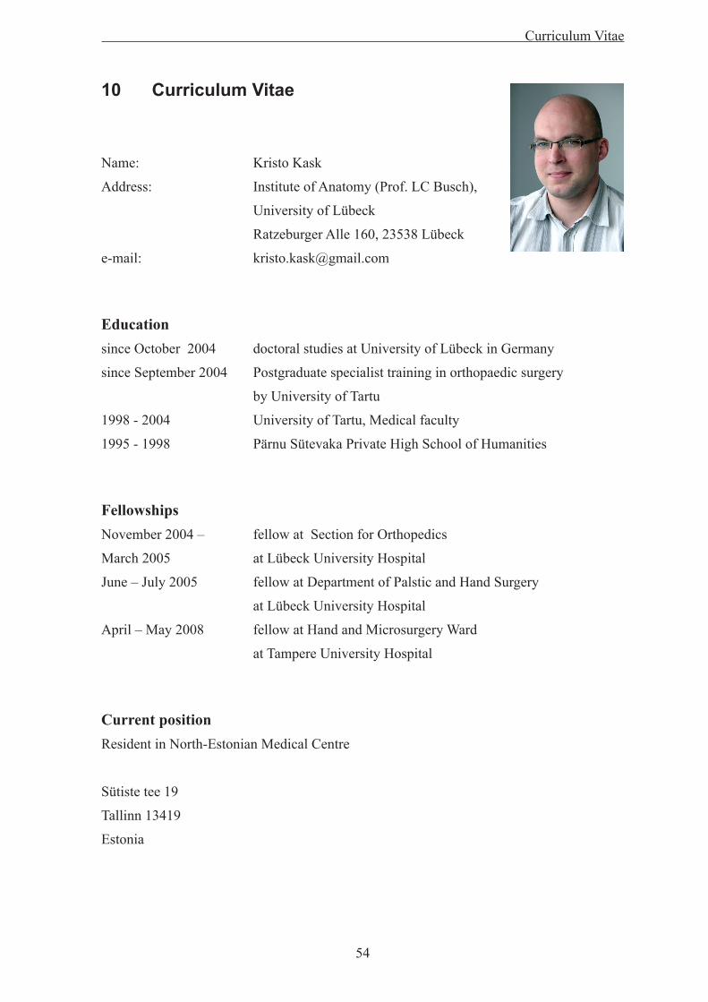
54
10 Curriculum Vitae
Name: Kristo Kask
Address: Institute of Anatomy (Prof. LC Busch),
University of Lübeck
Ratzeburger Alle 160, 23538 Lübeck
e-mail: [email protected]
Educationsince October 2004 doctoral studies at University of Lübeck in Germany
since September 2004 Postgraduate specialist training in orthopaedic surgery
by University of Tartu
1998 - 2004 University of Tartu, Medical faculty
1995 - 1998 Pärnu Sütevaka Private High School of Humanities
FellowshipsNovember 2004 – fellow at Section for Orthopedics
March 2005 at Lübeck University Hospital
June – July 2005 fellow at Department of Palstic and Hand Surgery
at Lübeck University Hospital
April – May 2008 fellow at Hand and Microsurgery Ward
at Tampere University Hospital
Current positionResident in North-Estonian Medical Centre
Sütiste tee 19
Tallinn 13419
Estonia
Curriculum Vitae
55
Medical interestsSurgery of upper extremity, hand trauma, arthroscopic surgery on upper extremity
LanguagesEstonian – native language
English – good in speeking and writing
Germany – moderate
Russian – moderate
Finnish – basic
HobbiesSailing and photography
Curriculum Vitae

56
List of Publications
Kask K, Kolts I, Lubienski A, Russlies M, Leibecke T, Busch LC.
Magnetic resonance imaging and correlative gross anatomy of the Ligamentum semicircu-
lare humeri (rotator cable).
Clinical Anatomy, 2008, Jul: 21(5): 420-6.
Merila M, Heliö H, Busch LC, Tomusk H, Poldoja E, Eller A, Kask K, Haviko T, Kolts I.
The Spiral Glenohumeral Ligament: An Open and Arthroscopic Anatomy Study
Arthroscopy: The Journal of Arthroscopic and Related Surgery
Available online 18 February 2008
Submitted, under review:
Kask K, Põldoja E, Lont T, Norit R, Merila M, Busch LC, Kolts I.
Anatomy of the Ligamentum glenohumerale superius
Annals of Anatomy
Abstracts of Poster Presentations on Congresses:
Kask K, Busch LC. Kolts I.
New insight into the Hill-Sachs lesion
13th ESSKA 2000 Congress, May 21 – 24, 2008 Porto, Portugal
Kask K, Kolts I, Lubienski A, Busch LC, Põldoja E, Tomusk H, Leibecke T.
Anatomy and MRI of the rotator cable or the so called Ligamentum semcirculare humeri
12th ESSKA 2000 Congress, May 24 – 27, 2006 Innsbruck, Austria
Kask K, Lont T, Norit R, Raudheiding A, Tomusk H, Kolts I.
Anatomy of the superior and middle glenohumeral ligaments in association with the
SLAP-lesion
6th EFORT Congress, June 7 – 10, 2003 Helsinki, Finland
Curriculum Vitae


![BB Anatomic Unico - 2020 - HERNIK (500000-XXX) Gr .1 [-205] (60 x 42 cm) Gr. 2 [-206] (60 x 48 cm) Sitzschalenadapter für Schlittenführung (100600) Sitzschalenadapter für Trapezführung](https://img.pdfslide.org/doc/110x75/605ef798828d01212434d7ef/bb-anatomic-unico-2020-hernik-500000-xxx-gr-1-205-60-x-42-cm-gr-2-206.jpg)