Embed Size (px)
Citation preview
_____________________________________________________
Aus der Poliklinik für Zahnerhaltung und Parodontologie Klinik der Ludwig-Maximilians-Universität München
Direktor: Prof. Dr. R. Hickel
In-vitro-Untersuchung neuartiger fließfähiger Komposite
(selbstadhäsiv, „bulk-fill“ und niedrigschrumpfend)
Dissertation zum Erwerb des Doktorgrades der Zahnheilkunde
an der Medizinischen Fakultät der Ludwig-Maximilians-Universität zu München
vorgelegt von
Pascal Roman Czasch aus Buchloe
2012
______________________________________________________________________
Mit Genehmigung der Medizinischen Fakultät der Universität München
Berichterstatter: PD Dr. Nicoleta Ilie Mitberichterstatter: Prof. Dr. Wolfgang Gernet PD Dr. Susanne Mayer Dekan: Prof. Dr. med. Dr. h. c. Maximilian
Reiser, FACR, FRCR
Tag der mündlichen Prüfung: 31.10.2012

Für meine Familie

Inhaltsverzeichnis 5
Inhaltsverzeichnis 1 Einleitung ..................................................................................................................6
2 Veröffentlichte Artikel ..............................................................................................9
2.1 „In-vitro comparison of mechanical properties and degree of cure of bulk-fill
composites“ ...................................................................................................................9
2.1.1 Deutsche Zusammenfassung .....................................................................38
2.1.2 English summary .......................................................................................40
2.2 „In-vitro comparison of mechanical properties and degree of cure of a self-
adhesive and four novel flowable composites“ ...........................................................42
2.2.1 Deutsche Zusammenfassung .....................................................................71
2.2.2 English summary .......................................................................................73
3 Literaturverzeichnis .................................................................................................75

6 Einleitung
1 Einleitung
Die Ansprüche, die an moderne fließfähige Komposite gestellt werden, steigen immer
weiter. Neben ausgezeichneten mechanischen Eigenschaften wird besonderer Wert auf
zusätzliche Eigenschaften wie Selbstadhäsion, geringe Polymerisationsschrumpfung
und immer einfachere Verarbeitbarkeit gelegt. Im Zuge dessen gab es zahlreiche
Innovationen: Die Veränderungen im chemischen Aufbau und der Füllkörper führte zu
vielen verschiedenen Komposittypen [1].
Vor kurzem wurde eine neue Entwicklung, die sogenannten „bulk-fill“ Komposite,
vorgestellt. Es soll die besondere Möglichkeit bestehen, das Material in 4 mm dicken
Inkrementen - statt der momentanen Inkrementtechnik mit maximal 2 mm dicken
Inkrementen - in den Zahn einzubringen ohne dabei die Polymerisationsschrumpfung,
die Konversionsrate oder die Passung zum Kavitätenrand negativ zu beeinflussen.
Darüber hinaus behaupten die Hersteller, dass diese Materialien sogar eine deutlich
niedrigere Schrumpfung als moderne fließfähige Komposite haben [2]. Die mit hoher
Kompositschrumpfung verbundenen Probleme [3] wie Randspaltbildung [4, 5] oder
post-operative Sensibilitätsbeschwerden [6] könnten somit vermieden werden. Laut
Hersteller sollen die Materialien sogar eine ausreichende Aushärtung bis zu einer
Inkrementdicke von 6 mm erfahren [2]. Leider fehlen hierzu bislang die grundlegenden
Studien. Dennoch würde ein an die Kavitätenwände anfließendes Komposit eine
enorme Zeit- und Kostenersparnis für die Praxis darstellen. Das „bulk-fill“ Komposit
Surefil® SDR™ (Smart Dentin Replacement, shrinkage decreased resin) beinhaltet
einen im Polymerisationsgerüst chemisch eingebundenen Polymerisationsmodulator,
der zu einer reduzierten Polymerisationsschrumpfung beitragen soll. Der Modulator hat
ein hohes molekulares Gewicht. Dank der konformativen Flexibilität um den
eingebundenen Modulator herum sollen Flexibilität und Gerüststruktur optimiert
werden. Untersuchungen von Kompositen die auf SDR™ Technologie basieren zeigten
signifikant niedrigere Schrumpfspannungswerte, nicht nur im Vergleich mit anderen
fließfähigen Kompositen [7] sondern auch mit Nano- und Hybridkompositen [8]. De
Biasi et al. untersuchten die mikromechanische Härte eines SDR™ Komposits und
äußerten Bedenken bezüglich der praktischen Anwendbarkeit aufgrund der geringen
Einleitung 7 Vickershärte [9]. Ilie et al. teilten diese Bedenken: sie maßen für das SDR™ Komposit
die geringsten Oberflächenwerte verglichen mit anderen oftmals verwendeten
Kompositen (EsthetXFlow, Filtek Supreme Plus Flow, EsthetX Plus, Filtek Silorane,
Filtek Supreme Plus) [10]. Bezüglich Verschleiß, Oberflächenrauhigkeit,
Politurfähigkeit und Verfärbungsresistenz wurden vergleichbare Werte mit anderen
klinisch erfolgreich verwendeten Kompositen erreicht [7].
Eine der neuesten Entwicklungen zur Vereinfachung des klinischen Behandlungsablaufs
sind die selbstadhäsiven fließfähigen Komposite, die weder eine Vorbehandlung der
Restzahnhartsubstanz mittels Phospohorsäure noch eines Dentin-Bonding-Agents
benötigen [11]. Seit der Einführung der Komposite stellte deren Befestigung an den
Kavitätenwänden der Zahnhartsubstanz Forscher und Entwickler vor eine schwere
Aufgabe. Nachdem Buonocore eine Methode zur Verbesserung der
Restaurationsbefestigung vorstellte [12], entwickelten zahlreiche Forscher neue,
einfachere und qualitativ hochwertigere Ätz- und Bondingsysteme [13]. Eines davon ist
die Herstellung selbstadhäsiver Komposite [14], Zemente [15] oder Adhäsive [16, 17]
mittels eines speziellen Dimethacrylatmonomers: Glycerol Phosphat Dimethacrylat
(GPDM). Dieses Monomer erlaubt die chemische Verbindung seiner Phosphatgruppe
mit Calciumionen der Zahnhartsubstanz. Zusätzlich dazu trägt die mikromechanische
Verankerung zwischen dem sich aufbauenden Polymer und den durch die
Phosphatgruppe des GPDM freigelegten Kollagenfasern wie auch die mechanische
Verbindung zwischen dem Polymer und dem „Smear layer“ zur Befestigung der
Restauration an der Zahnhartsubstanz bei [1]. Wenn man allerdings die Haftkraft
selbstadhäsiver Komposite zum Zahnschmelz mit der Haftkraft der state-of-the-art „etch
and rinse“ Adhäsive vergleicht, schneiden die Erstgenannten deutlich schlechter ab [18-
21]. Ein weiterer Nachteil von Ein-Flaschen-Adhäsiven ist in ihrer relativ hohen
Wasseraufnahme zu sehen [22, 23]. Die Haftscherfestigkeit zu oberflächlichem wie
auch zu tiefer gelegenem Dentin von GPDM-basierten Adhäsiven wurde in diversen
Studien als vergleichbar mit anderen selbstätzenden und „etch-and-rinse“
Adhäsivsystemen befunden [21]. Auch die Hydrolyse des Zahn-
Restaurationsüberganges mit den bekannten möglichen Folgen wie Microleakage,
Verfärbungen, Sekundärkaries bis hin zur Devitalisierung der Pulpa ist als mögliche
Komplikation zu nennen.
Ein weiterer Wunsch der modernen restaurativen Zahnheilkunde ist die Verringerung
der Polymerisationsschrumpfung. In einer Untersuchung in der die
Polymerisationsschrumpfung eines experimentellen Komposits mit verschiedenen
8 Einleitung Monomerzusammensetzungen gemessen wurde, fanden Ellakwa et al. beispielsweise
heraus, dass eine negative Korrelation zwischen der Schrumpfung und dem
Molekulargewicht des jeweiligen Monomers besteht [24].
Einige der modernen fließfähigen Komposite verzichten zugunsten hochmolekularer
Monomere soweit möglich auf niedermolekulare Monomere wie
Triethylenglycoldimethacrylat (TEGDMA) (286 g/mol) und 2-
Hydroxyethylmethacrylat (HEMA) (130 g/mol). Neben klassischen Monomeren wie
Bisphenylglycidyldimethacrylat (BisGMA) (512 g/mol) und ethoxyliertem Bisphenol-
A-Dimethacrylat (BisEMA) (540 g/mol) [25] werden heutzutage auch die neu
entwickelten Dimersäure- Dimethacrylate (574 g/mol) [26-28] verwendet. Diese
versprechen eine höhere Konversionsrate, höhere Biegemodulwerte und die
Möglichkeit einer dickeren Inkrementplatzierung [29].
Ziel der nachfolgenden zwei Artikel war es, die Auswirkungen der Veränderung von
chemischer Struktur und Zusammensetzung von anorganischer und organischer Phase
auf mikromechanische (Vickershärte, Eindringmodul) und makromechanische
Eigenschaften (Biegefestigkeit, Biegemodul) sowie die Konversionsrate bei
verschiedenen Belichtungszeiten und klinisch relevanten Inkrementdicken zu
untersuchen. Als Materialien dienten dabei zwei „bulk-fill“ (Inkrementdicke bis zu 4
mm), ein selbstadhäsives (keine Vorbehandlung der Kavität), zwei „low-shrinkage“
(niedrigschrumpfend) und als Kontrollgruppe zwei zur Gruppe der Mikrohybride
gehörige fließfähige Komposite.
Veröffentlichte Artikel 9
2 Veröffentlichte Artikel
2.1 „In-vitro comparison of mechanical properties and
degree of cure of bulk-fill composites“
Pascal Czasch and Nicoleta Ilie
Department of Restorative Dentistry, Dental School of Ludwig-Maximilians-University,
Munich, Germany
Clinical Oral Investigations 2012 Mar 14. [Epub ahead of print]
10 Veröffentlichte Artikel
Abstract:
OBJECTIVES: The aim of our study was to measure and compare degree of conversion
(DC) as well as micro- (Indentation modulus, E, Vickers hardness, HV) and
macromechanical properties (Flexural strength, σ, Flexural modulus, Eflexural) of two
recently launched bulk-fill RBCs (resin-based composites): Surefil® SDR™ flow (SF)
and Venus® bulk fill (VB).
MATERIALS AND METHODS: DC (n=6) was investigated by FTIR-Spectrometry in
clinical relevant filling depths (0.1 mm, 2 mm, 4 mm, 6 mm-bulk, 6 mm-incremental)
and irradiation times (10s, 20s, 40s). Micro- (n=6) and macromechanical (n=20)
properties were measured by an automatic microhardness indenter and a three-point
bending test device after storing the specimens in distilled water for 24 h at 37°C.
Furthermore, on the 6 mm-bulk samples the depth of cure was determined. A field
emission scanning electron microscope was used to assess filler size. Results were
evaluated using one-way ANOVA, Tukey HSD post-hoc test, a multi-variate analysis (α
=0.05) and an independent t-test. Weibull analysis was used to assess σ.
RESULTS: VB showed in all depth significant higher DC (VB: 62.4-67.4%; SF: 57.1-
61.9%), but significant lower macro- (VB: Eflexural=3.6GPa; σ=122.7MPa; SF:
Eflexural=5.0GPa; σ=131.8MPa) and micromechanical properties (VB: E=7.3-8.8GPa,
HV=40.7-46.5N/mm²; SF: E=10.6-12.2GPa, HV=55.1-61.1N/mm²). Both RBCs
showed high reliability (VB: m=21.6; SF: m=26.7) and a depth of cure of at least 6mm
at all polymerization times. The factor “RBC” showed the strongest influence on the
measured properties (η2=0.35-0.80) followed by “Measuring Depth” (η2=0.10-0.46) and
“Polymerization time” (η2=0.03-0.12).
CONCLUSIONS: Significant differences between both RBCs were found for DC, E, σ
and Eflexural at all irradiation times and measuring depths.
CLINICAL RELEVANCE: Curing the RBCs in 4 mm bulks for 20s can be
recommended.
Keywords:
Bulk-fill, composite, macro-mechanical properties, micro-mechanical properties, degree
of conversion;
Veröffentlichte Artikel 11
Introduction:
Since the development of resin-based composites (RBCs) several improvements in their
chemical composition as well as various filler reinforcements occurred, leading to a
large category of materials [1]. Recently a new category of flowable RBCs – so called
bulk-fill RBCs- was introduced (Surefil® SDR™ flow, Dentsply, Caulk, USA and
Venus® bulk fill, Heraeus Kulzer GmbH, Hanau, Germany) as bulk-fill material and as
liner in Class I and II restorations. The particularity of the new material category is
stated to be the option to place it in 4 mm thick bulks instead of the current incremental
placement technique, without negatively affecting polymerization shrinkage, cavity
adaptation or the degree of conversion (=DC). Moreover manufacturers stated that the
polymerization shrinkage of those materials is even lower when compared to commonly
used flowable and conventional RBCs [2]. Thus, problems related to polymerization
shrinkage [3] like gap formation causing secondary caries due to bacteria colonization
[4,5], pulp irritation, post-operative sensibility when chewing [6] or cusp deflection
when the “C” factor is high [7,8], could be minimized. Manufacturers claimed that bulk-
fill materials can achieve a depth of cure of 6 mm [2], though no published
investigations are available till now to confirm these statements. Nevertheless the idea
of placing a self-adapting material as bulk, saving time as well as improving material
handling is of great interest.
The bulk-fill material Surefil® SDR™ (Smart Dentin Replacement, Shrinkage
Decreased Resin) flow contains a polymerization modulator, chemically embedded in
the center of the polymerizable resin backbone of the SDR™ monomer, to lower
polymerization shrinkage. The modulator has a high molecular weight. Due to the
conformational flexibility around the centered modulator impart, the modulator is
supposed to optimize flexibility and network structure of the SDR™ resin [9].
Investigations on RBCs with SDR™ technology showed significant lower shrinkage
stress values [10] not only when compared to regular flowable RBCs, but also to nano-
and hybrid RBCs or even to silorane-based composites [11]. De Biasi et al. investigated
microhardness and raised concerns about its practical use due to its low Vickers
hardness (=HV) [12]. This was also confirmed by Ilie et al. [11] where Surefil® SDR™
flow showed the lowest surface hardness when compared to other commonly used
RBCs (EsthetX Flow, Filtek Supreme-Plus-Flow, EsthetX-Plus, Filtek Silorane and
Filtek Supreme-Plus). However, when compared to the investigated flowable RBC of
the same study, Surefil® SDR™ flow showed significant higher indentation modulus
12 Veröffentlichte Artikel (=E). In view of wear, surface roughness, gloss, color stability and stain resistance,
similar results to clinically successful RBCs were found [10]. Other experimental
flowable RBCs with SDR™ technology – P&P-Adaptable and P&P-Universal (both
Dentsply) – also showed low shrinkage stress values [13]. Moreover Surefil® SDR™
flow was used for luting fiber posts and resulted comparable regarding retentive
strength like a dual resin cement commonly used [14].
This study evaluated and compared two bulk-fill RBCs - Surefil® SDR™ flow and
Venus® bulk fill – regarding their micro- and macromechanical properties and DC at
different irradiation times and by simulating clinical relevant filling depth.
The tested null hypothesis were that: a) there would be no significant difference
between the two materials in view of macro- (flexural strength (=σ), modulus of
elasticity (=Eflexural)) and micromechanical properties (Vickers hardness HV, indentation
modulus E) and degree of cure (DC) at any measured depth and irradiation time; b)
within one material, irradiation time and depth would not influence the measured
properties.
Veröffentlichte Artikel 13
Materials and methods:
Two flowable bulk-fill RBCs - Surefil® SDR™ flow (Dentsply, Caulk, USA, Lot No.:
100407, 100507) and Venus® bulk fill (Heraeus Kulzer GmbH, Hanau, Germany, Lot
No.: 010026) were analyzed by assessing DC and micromechanical properties (HV, E)
as function of depth and polymerization time (10s, 20s or 40s) as well as the
macromechanical properties (σ, Eflexural). Due to manufacturers’ information Surefil®
SDR™ flow consists of Ba–Al–F–B–Si–glass and St–Al–F–Si–glass as fillers (68% per
weight, 44% per volume) and modified Urethane dimethacrylate (UDMA),
Triethyleneglycol dimethacrylate (TEGDMA) and ethoxylated Bisphenol-A-
dimethacrylate (EBPDMA) as resin matrix. For Venus® bulk fill, Ba–Al–F–Si–glass
and SiO2 were given as fillers (65% per weight, 38% per volume) and UDMA and
EBPDMA as resin matrix.
Degree of cure measurements:
To evaluate the DC, five different sample geometries were considered. Thin films (100
µm) as well as 2 mm, 4 mm and 6 mm high molds (3 mm diameter) were filled in bulk.
Additionally three consecutive increments - each 2 mm high – were prepared in the
mold of 6 mm height (6 mm-incremental). Samples were cured by applying the curing
unit (Elipar Freelight2, 3M ESPE, 1226 mW/cm²) directly on the top of the particular
mould, respectively on the film surface covered by a transparent matrix strip. For each
product, irradiation time (10s, 20s, 40s) and geometry (0.1 mm, 2 mm, 4 mm, 6 mm-
bulk, 6 mm-increment) six samples were measured (n=6). Real-time measurements
were made with a FTIR-Spectrometer with an attenuated total reflectance (ATR)
accessory (Nexus, Thermo Nicolet, Madison, USA). Therefore, the non-polymerized
RBC paste was put directly on the diamond ATR crystal in the mold as described
above. FTIR spectra were recorded in real time for 5 minutes at the bottom of the
samples irradiated according to the curing protocol presented above. Diameter of
measured surface was 800 µm, wave number of the spectrum ranged between 4000-650
cm-1 and the FTIR spectra were recorded with four scans at a resolution of 8 cm-1.
To determine the percentage of the remained unreacted double bonds, the DC was
measured by assessing the variation in peak height ratio of the absorbance intensities of
methacrylate carbon double bond peak at 1634 cm-1 and that of an internal standard
peak (=IS) at 1608 cm-1 (aromatic carbon double bond) during polymerization, in
14 Veröffentlichte Artikel relation to the uncured material. For the RBC Surefil® SDR™ flow, the reference peak
was set at 1600 cm-1 due to the absence of the aromatic carbon bond.
DCheight % = 100
curing beforePeak IS1-1634cm
curingafter Peak IS1-1634cm
1 x
úúúúú
û
ù
êêêêê
ë
é
÷÷ø
öççè
æ
÷÷ø
öççè
æ
-
Micro-mechanical properties:
The variation in micromechanical properties (HV, E) was assessed on the 6 mm bulk
samples prepared for the DC measurements. For this purpose, samples were stored in
distilled water after curing for 24 hours at 37°C, ground and polished under water in
longitudinal direction from 3 mm diameter to 1.5 mm diameter with diamond abrasive
paper (mean grain sizes: 20 µm, 13 µm, 6 µm) in a grinding system (EXAKT 400CS,
Exakt, Norderstedt, Germany). Measurements were made with an automatic
microhardness indenter (Fischerscope H100C, Fischer, Sindelfingen, Germany) starting
from 0.1 mm under the surface, with 100 µm intervals between the measuring points.
The test procedure was carried out force-controlled, where the test load increased and
decreased with constant speed between 0.4 mN and 500 mN. Load and penetration
depth of indenter (Vickers pyramid: diamond right pyramid with a square base and an
angle of α = 136° between the opposite faces at the vertex) were continuously measured
during the load-unload hysteresis. Universal hardness is defined as the test force divided
by the apparent area of indentation under the applied test force. From a multiplicity of
measurements stored in a database supplied by the manufacturer, a conversion factor
(0.0945) between Universal hardness and HV was calculated by the manufacturer and
entered into the software, so that the measurement results were indicated in the more
familiar HV units. E was calculated from the slope of the tangent adapted at the
beginning (at maximum force) of the non-linear indentation depth curve upon
unloading.
HV and E variations with depth and irradiation time were calculated for each product
(Tables 3 and 4) based on data from six samples (360 measuring points).
The depth of cure, usually acknowledged as the thickness of a RBC that is adequately
cured [15] or rather as the depth where HV equals the surface value multiplied by an
arbitrary ratio, usually 0.8 (=HV-80%) [16], was calculated. Therefore for each sample
Veröffentlichte Artikel 15 HV in the depth was compared to the related surface value and noted when it became
less than 80% (HV-80%).
Flexural strength and flexural modulus:
σ was determined in a three-point-bending test according to ISO/DIN 4049:1998. The
samples (n=20) were made by compressing the RBC material between two glass plates
with intermediate polyacetate sheets, separated by a steel mould having an internal
dimension of (2 x 2 x 16) mm. After curing (with three light exposures of 20 seconds
per side, Elipar Freelight2, 3M ESPE) the specimens were removed from the mould and
any flash material was trimmed away with sandpaper (grit size P4000 (FEPA)). All
specimens were then stored in distilled water at 37°C prior to testing for 24 h. Samples
were loaded until failure in the universal testing machine (MCE 2000ST, quick test
Prüfpartner GmbH, Langenfeld, Germany). The crosshead speed was 0.5 mm/min. The
specimens were placed on a three-point bending test device, which is constructed
according to the guidelines of NIST No. 4877 with 12 mm distance between the
supports. During testing the specimens were immersed in distilled water at room
temperature.
Flexural strength was calculated from formula (1).
(1) 22
3
bh
Fl=s
F is the maximum load [N], l is the distance between the supports [mm], b is the width
of the specimen [mm], h is the height of the specimen [mm].
The universal testing machine stored the force during bending and the deflection of the
beam in a file. The bending modulus was calculated from formula (2).
(2) ybh
FlflexuralE
34
3=
y is the deflection at load point [mm].
Field emission scanning electron microscope (FE-SEM):
For each product one specimen (1cm x 1cm x 0.5cm) was manufactured with an
irradiation time of 60s and treated for one hour in a chemical dry cleaning process with
oxygen plasma in vacuum (45 - 50 W). Afterwards surfaces were investigated
(Magnification: 10000x, Signal: Secondary electrons SE2, Working distance: 4 mm,
Electron high tension: 10 kV) with a field emission scanning microscope (FE-SEM)
16 Veröffentlichte Artikel (Zeiss Supra® 55 VP, Zeiss NTS GmbH, Oberkochen, Germany) and the most
representative picture was chosen for assessing fillers’ size.
Statistical analysis:
The results for DC, HV and E within each material, each measuring depth and each
curing time, respectively, were compared using one-way ANOVA and Tukey’s HSD
post hoc-test (α =0.05) (SPSS 18.0, Chicago, IL, USA). An ANOVA multivariate
analysis and partial eta-square statistic was used to investigate the influence of the
parameters “RBC”, “measuring depth” and “polymerization time” on E, HV, DC. For
the properties Eflexural and σ the influence of “RBC” was assessed. Additionally a
Weibull analysis was used to assess σ.
A common empirical expression for the cumulative probability of failure P at applied
stress is the Weibull model:
úúû
ù
êêë
é÷÷ø
öççè
æ--=
m
ccfP
0
exp1)(ss
s
where cs is the measured strength, m the Weibull modulus and 0s the characteristic
strength, defined as the uniform stress at which the probability of failure is 0.63. The
double logarithm of this expression gives:
By plotting lnln(1/(1-P)) versus ln(σ), a straight line results, with the upward gradient
m, whereas the intersection with the x-axes gives the logarithm of the characteristic
strength.
0lnln1
1lnln ss mm
P c -=-
Veröffentlichte Artikel 17
Results:
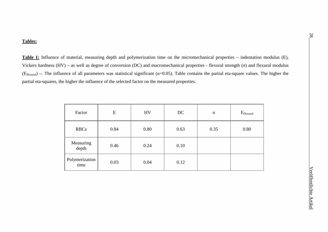
The influence of the parameters “RBCs”, “Measuring Depth” and “Polymerization
time” as well as their interaction products was analyzed in an ANOVA multivariate test
(Table 1). DC and the mechanical properties – HV, E, σ and Eflexural - were selected as
depended variables. The significance values of these three main effects were less than
0.05, indicating that they contribute all to the model. The “RBCs” was the parameter
exerting the strongest influence on all measured properties (higher eta square values).
The influence of the “Measuring Depth” was stronger on the micro-mechanical
properties (HV, E) than on DC, whereas the influence of polymerization time, though
significant, was very low.
A one-way ANOVA was used to identify detailed differences in the measured
properties within each material as function of polymerization times (horizontal lines in
tables 2 to 4) and geometries (vertical lines in tables 2 to 4).
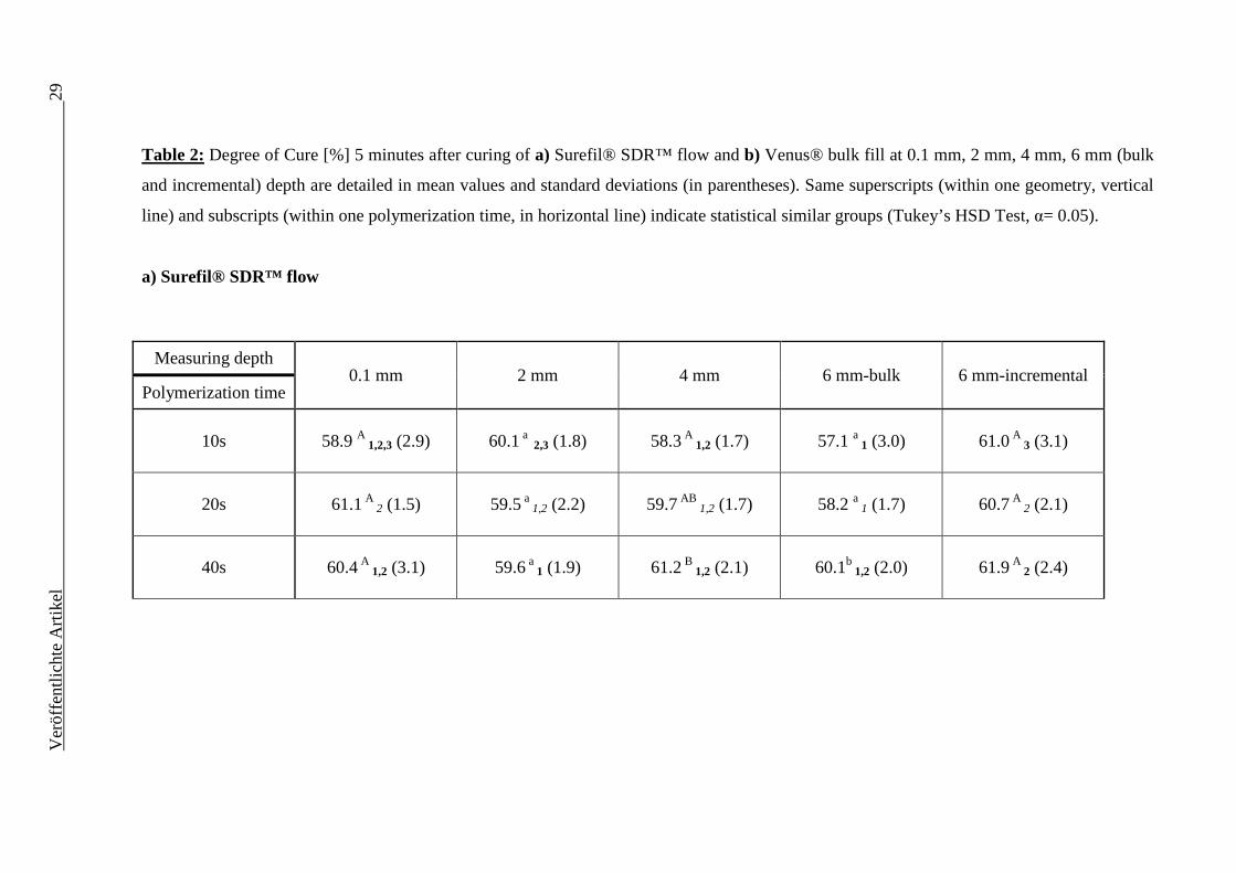
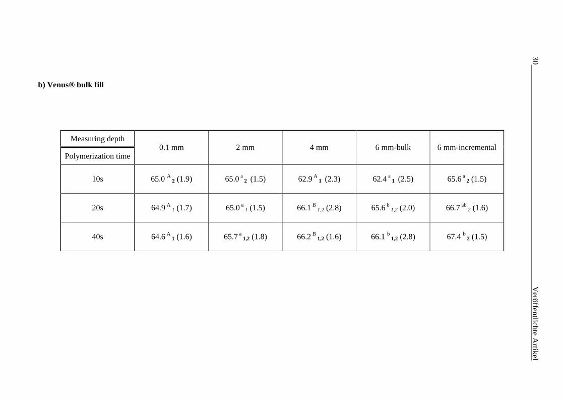
A significant increase (p<0.05) in DC (Table 2) with increasing polymerization time
was found for Surefil® SDR™ flow only at 4 mm (between 10s and 40s) and 6 mm
depth bulk placement (between 20s and 40s) whereas for Venus® bulk fill this
statement is only valid at 4 mm (between 10s and 20s, respectively 40s) and 6 mm
depth (bulk: between 10s and 20s; incremental: between 10s and 40s).
The DC at 6 mm depth bulk versus incremental placement was significantly lower only
at low polymerization times (10s and 20s for Surefil® SDR™ and 10s for Venus® bulk
fill).
Comparing both RBCs it can be seen that Venus® bulk fill had a statistically significant
higher DC (about 5%) for all irradiation times and measuring depths.
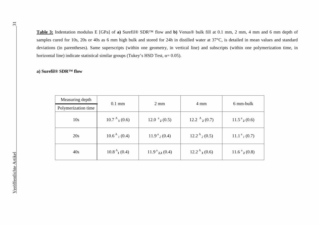
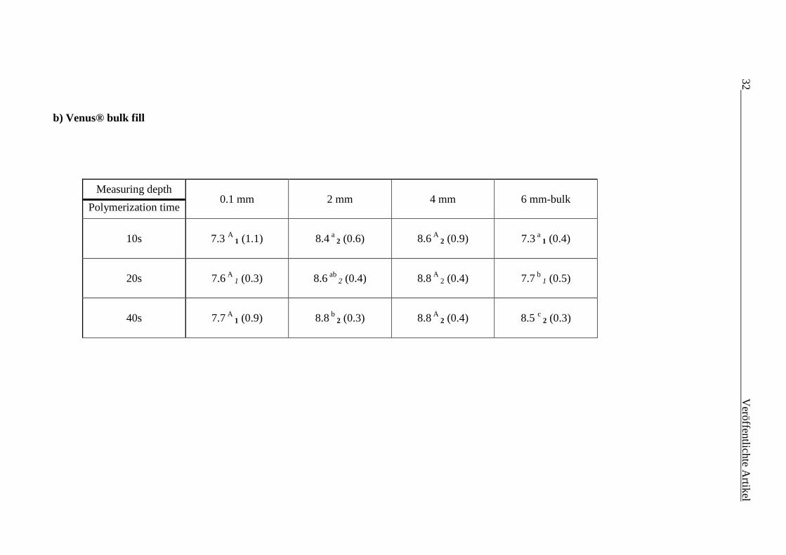
Concerning the variation of E (Table 3) results showed for both RBCs significant
(p<0.05) lower values for 0.1 mm when compared to 2 mm depth as well as statistically
equal values for 2 mm and 4 mm depth at all polymerization times. Similar trend is also
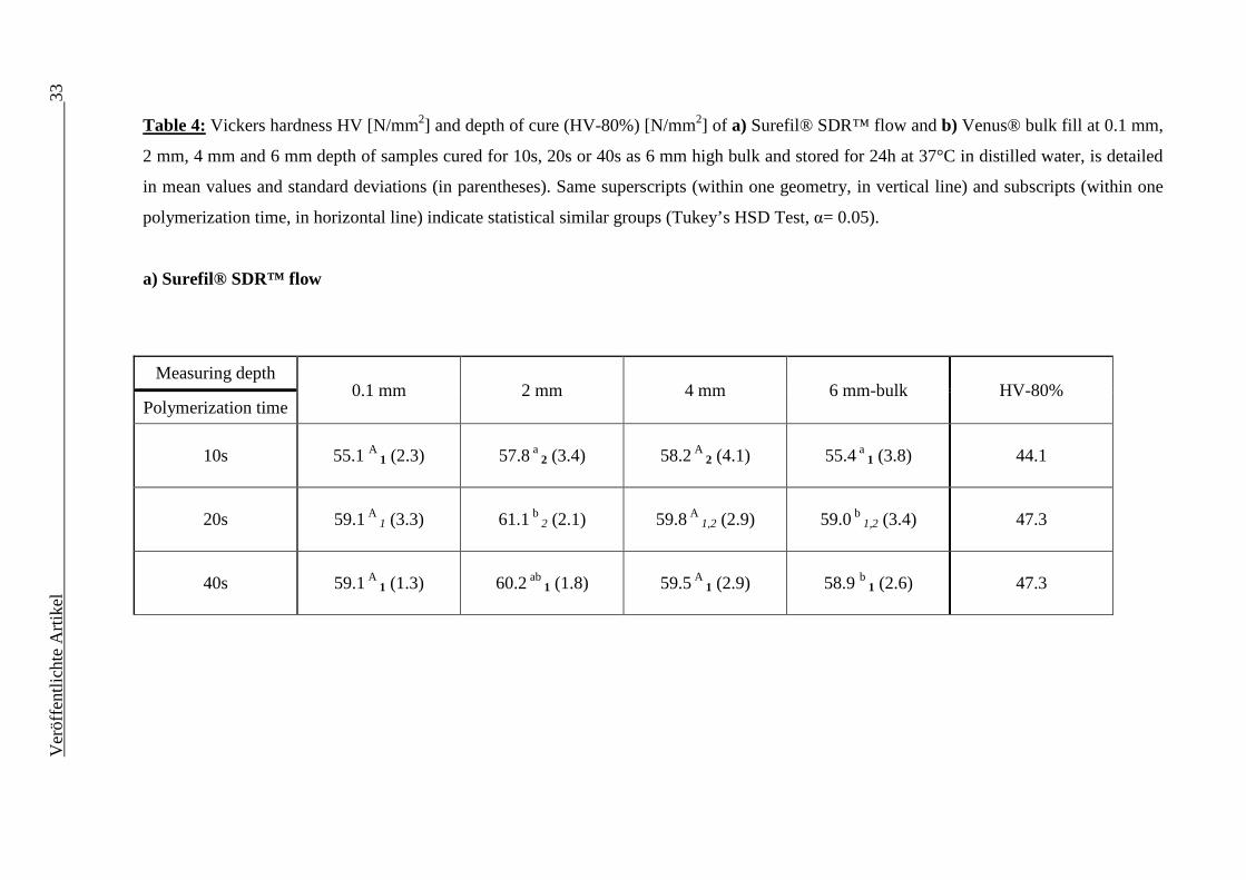
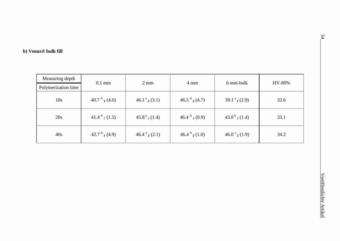
valid for HV (Table 4). As for the incremental thickness, the HV-80% was not reached
in the 6 mm samples at any polymerization time in both measured RBCs.
Comparing both RBCs, Surefil® SDR™ flow showed statistically significant higher
values for E (about 3 GPa) and HV (about 15 N/mm²) at all irradiation times and
measured depths.
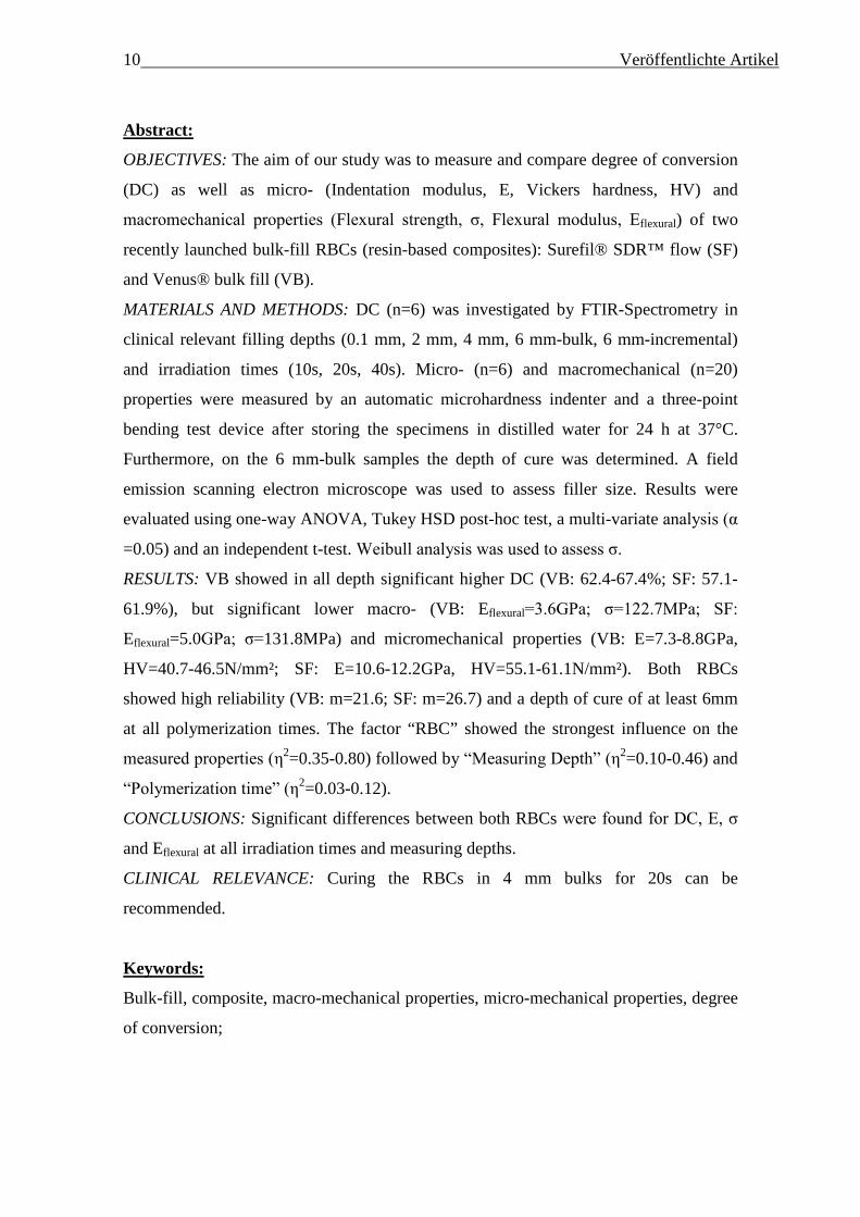
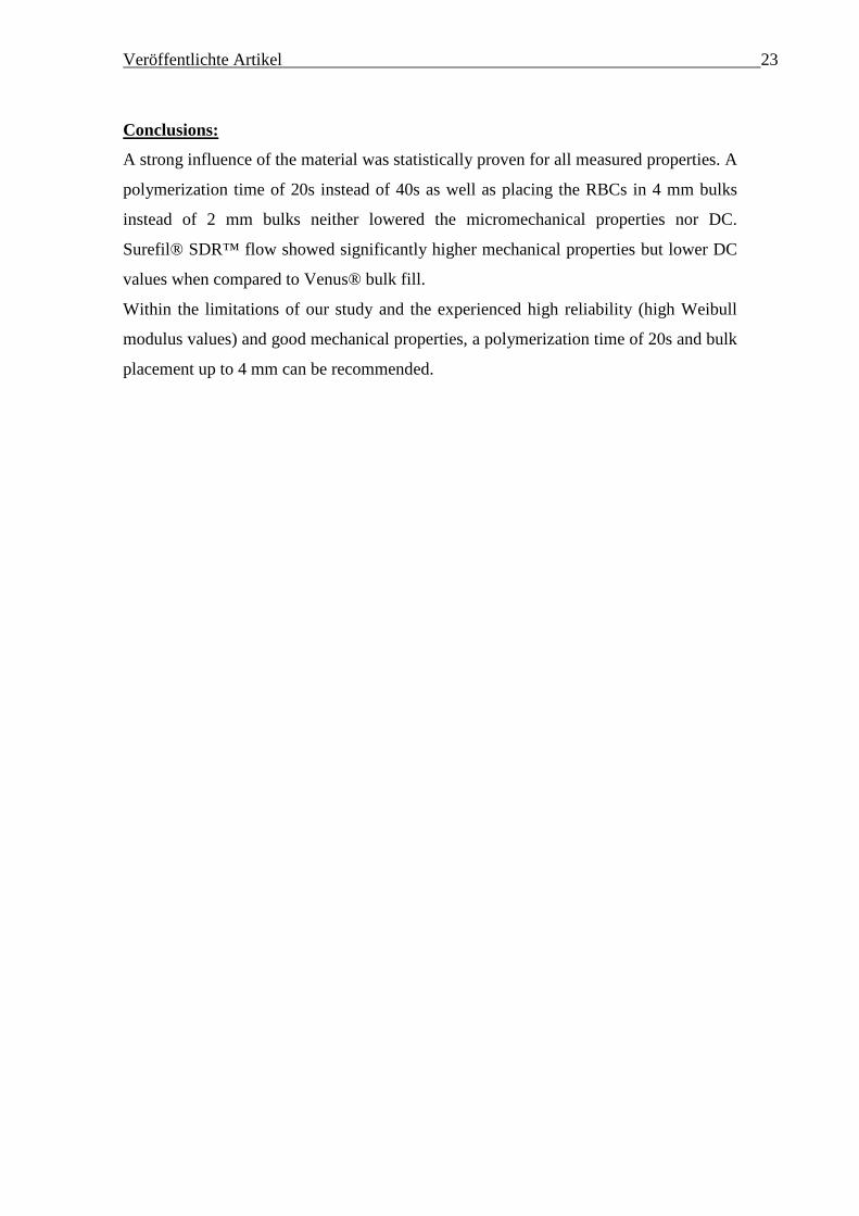
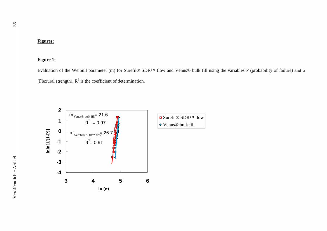
The investigated macromechanical properties σ and Eflexural revealed for Surefil®
SDR™ flow a significantly higher σ (131.8 ±5.8 MPa) and Eflexural (5.0 ±0.4 GPa) when
compared to Venus® bulk fill (σ= 122.7 ±6.9 MPa; Eflexural= 3.6 ±0.4 GPa). For both

18 Veröffentlichte Artikel materials a very high Weibull modulus was reached (21.6 and 26.7) attesting a high
reliability of both RBCs (Figure 1).
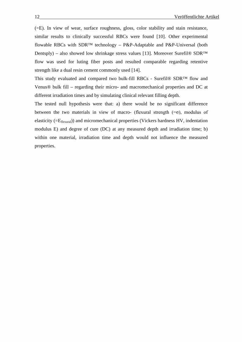
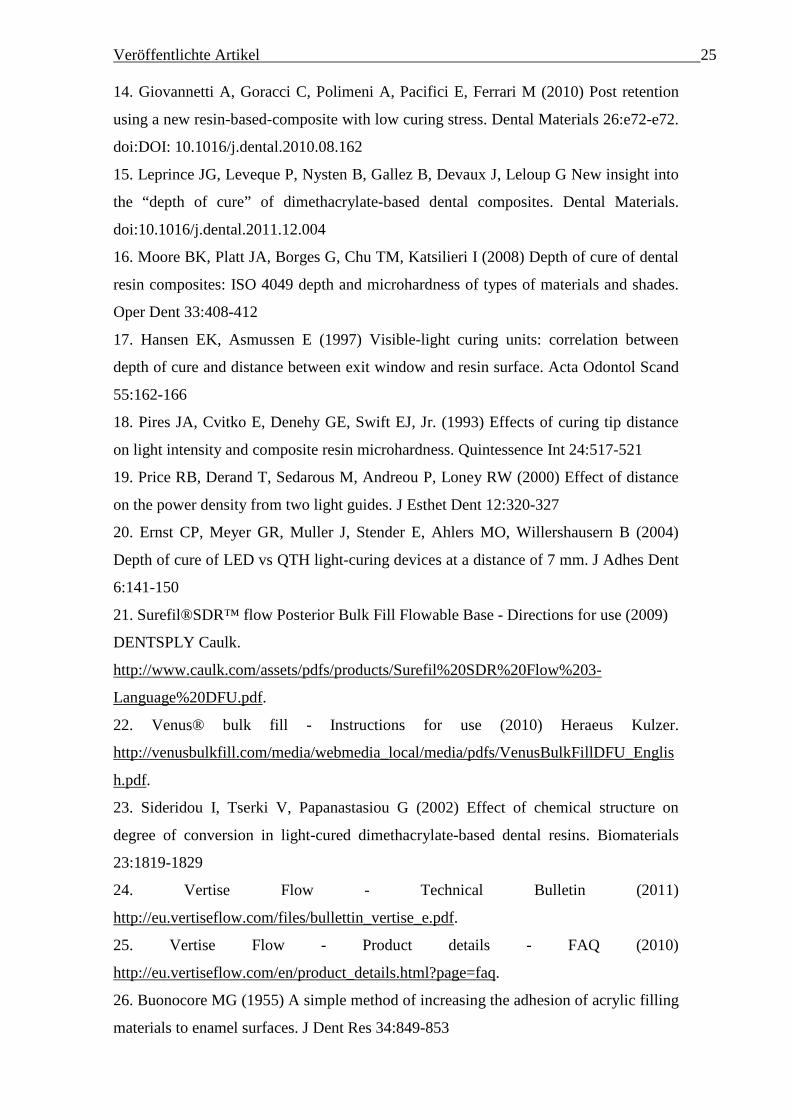
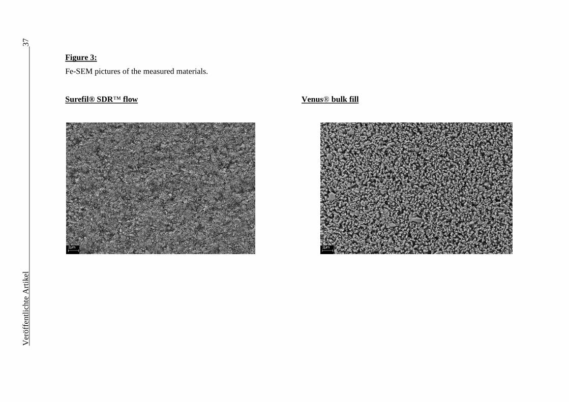
Comparing FE-SEM pictures (Figure 3), fillers in Surefil® SDR™ flow are consistently
smaller than fillers of Venus® bulk fill.
Veröffentlichte Artikel 19
Discussion:
Two recently launched bulk-fill flowable RBCs - Surefil® SDR™ flow and Venus®
bulk fill - considered to be used as cavity liners and bulk fill materials in class I and II
restorations were investigated. For this purpose specimens were measured by a FTIR-
Spectrometer, a microhardness indenter, a three-point-bending test device and a FE-
SEM. It must however be considered that the measurements were done with a modern
high intensity LED curing unit which was applied at mould upper surface. Placing
clinical restorations often means higher distances [17-19] between less effective curing
units [20] and RBC surface. Therefore the clinical values of the measured properties
could be lower.
The substantial reduction in polymerization shrinkage and particularly the ability to
place the RBCs as 4 mm bulks claimed by both manufacturers has led to further interest
about the composition of the measured products. For both RBCs the manufacturers
renounced to Bisphenol-A-dimethacrylate (Bis-GMA) and only formed the organic
matrix out of other dimethacrylates [21,22]. As a result, the RBCs are supposed to be
less viscous because Urethane dimethacrylate (UDMA), Triethyleneglycol
dimethacrylate (TEGDMA) and ethoxylated Bisphenol-A-dimethacrylate (EBPDMA)
form more flexible polymers than Bis-GMA [23-26]. Moreover, Bis-GMA is said to be
more hydrophilic [27] and consequently runs a higher risk of water uptake and
degradation than the more hydrophobic EBPDMA [28] - used in both RBCs – thus
reducing the risk of discoloration [29].
In our study DC was mainly influenced by the type of RBC (η2=0.63). Combined with
our results the claimed significant lower DC of Surefil® SDR™ flow in comparison to
Venus® bulk fill [2] as well as its stated high DC when compared to other common
RBCs (EsthetX Flow, Filtek Supreme Flow, Tetric Evo Flow, Filtek Silorane) [30] can
be confirmed within the limitations of our experimental set-up. Unless it has to be
pointed out that through different matrix compositions of the two RBCs, DC cannot be
rated because each monomer and additional group implicates different properties and
different molecular architecture, thus a higher DC does not necessarily mean higher
mechanical properties as also confirmed by the measured mechanical properties.
Furthermore by increasing the concentration of monomers [31] or diluents [32] the DC
can be artificially kept high without improving mechanical properties. This was
obviously not done in the analysed materials, since the measured mechanical properties
performed well as already investigated and confirmed for Surefil® SDR™ flow when
20 Veröffentlichte Artikel comparing it to different types of modern RBCs [11]. Scougall-Vilchis et al. claimed
that microhardness largely depends on the filler particles (size, weight, volume) as well
as on the chemical composition of the RBC when – like in our study - the test device
produces larger indents than the size of the fillers [33]. Therefore it can be stated that
the measured HV-values present the average microhardness of both, fillers and matrix.
Comparing micromechanical properties of Venus® bulk fill - concerning values on
surface and in 2 mm depth after curing for 20s - with literature data [11], HV and E
result like a commonly used microhybrid flowable RBC (EsthetX Flow) and a
nanohybrid flowable RBC (Filtek Supreme Flow) (for E). When comparing the neat
dimethacrylates Sideridou et al. showed that DC increases in the order Bis-GMA < Bis-
EMA (EBPDMA) < UDMA < TEGDMA [23]. However, there must be an upper limit
in increasing concentration of dimethacrylates with lower molecular weight because
polymerization shrinkage would either increase [34]. The low polymerization shrinkage
for Surefil® SDR™ flow shall result from the addition of the “polymerization
modulator”, a chemical moiety in the resin backbone increasing flexibility and thus
relaxing the polymerized network without harming DC (when compared to another
common flowable RBC (EsthetX Flow, Dentsply)) [30]. Moreover, the extreme lowered
polymerization shrinkage stress claimed by the manufacturer has been confirmed in
other studies, showing for Surefil® SDR™ flow significant lower polymerization stress
(1.1±0.1MPa) even when compared to the low- shrinkage silorane-based composite
Filtek Silorane [11]. Unfortunately there are no published studies concerning the
polymerization shrinkage of Venus® bulk fill. But with low contraction stress the cavity
adaptation increases and it allows the dentist to place the composite in a favourable
way. Nevertheless investigations on polymerization shrinkage in various bulks could be
useful as an increased “C”- Factor caused by lower unattached RBC surface raises cusp
deflection [8].
Statistics revealed for HV a strong influence (η2=0.80) and for σ a moderate influence
(η2=0.35) of the factor “RBC”; moreover E (η2=0.84) and Eflexural (η2=0.80) were
nearly equally strongly depended on the material. Therefore, the first tested hypothesis
must be rejected. In the macro- and micromechanical tests Surefil® SDR™ flow proved
to be significantly superior to Venus® bulk fill. Reasons for this behaviour might be
found in both, inorganic and organic compounds. Surefil® SDR™ flow differs from
Venus® bulk fill in the matrix composition as it contains additional TEGDMA and a
polymerization modulator [30]. With the addition of the more flexible side groups
containing TEGDMA, viscosity can be decreased [35] and with the formation of more
Veröffentlichte Artikel 21 homogenous copolymer networks, polymerization shrinkage decreases either [36].
When comparing experimental composites with different types and contents of fillers
Lee et al. found out that viscosity of RBC increases when filler volume increases [37].
Decreased viscosity is desirable for Surefil® SDR™ flow to reach similar levels of
flowability, as its filler content (68% per weight, 44% per volume) strongly differs from
the filler content of Venus® bulk fill (65% per weight, 38% per volume). With
increasing filler volume the flexural strength and modulus as well as hardness improve
[38,39]. Comparing the results for the micromechanical properties to a study
investigating five nanohybrid RBCs (Miris2, N’Durance, Premise, Simile, Venus
Diamond) with the same experimental set-up, Venus® bulk fill and Surefil® SDR™
flow show lower values than all of the measured materials [40]. The recommendation
for an irradiation time of 20s and a 4 mm bulk placement for Surefil® SDR™ flow as
well as an irradiation time of either 40s and 6 mm bulk placement or 20s and 4 mm bulk
placement for Venus® bulk fill, is supported by the measured micromechanical values.
Therefore the second hypothesis was rejected.
Assessing FE-SEM pictures (Figure 3), fillers in Surefil® SDR™ flow are consistently
smaller than fillers of Venus® bulk fill. Li et al. claimed that decreasing filler size also
means harming depth of cure and compressive strength [41] which however is not
evident for the measured bulk-fill materials. Further investigations are needed to define
the role of the polymerization modulator concerning both, mechanical properties and
DC.
The producers’ guarantee of placing the RBCs in 4 mm bulks and light curing for 20s
without a loss in DC and mechanical properties seems to be of great interest for
customers: it saves time and handling would be very easy. Our results confirm this
claim and show no improvement when placing thinner bulks than 4 mm or increasing
the irradiation time from 20s to 40s up to a measurement depth of 4 mm for both RBCs.
Moreover the 80%-HV value - presenting the percentage of the relation of bottom to top
surface hardness to be 80% for a properly cured composite [42] and due to Hansen et al.
rather important than top surface hardness [43] - was not reached in the 6 mm samples
at any of the measured irradiation times. This concludes that both RBCs may be placed
in 4 mm bulks without a loss in relevant properties, like mechanical properties or degree
of cure.
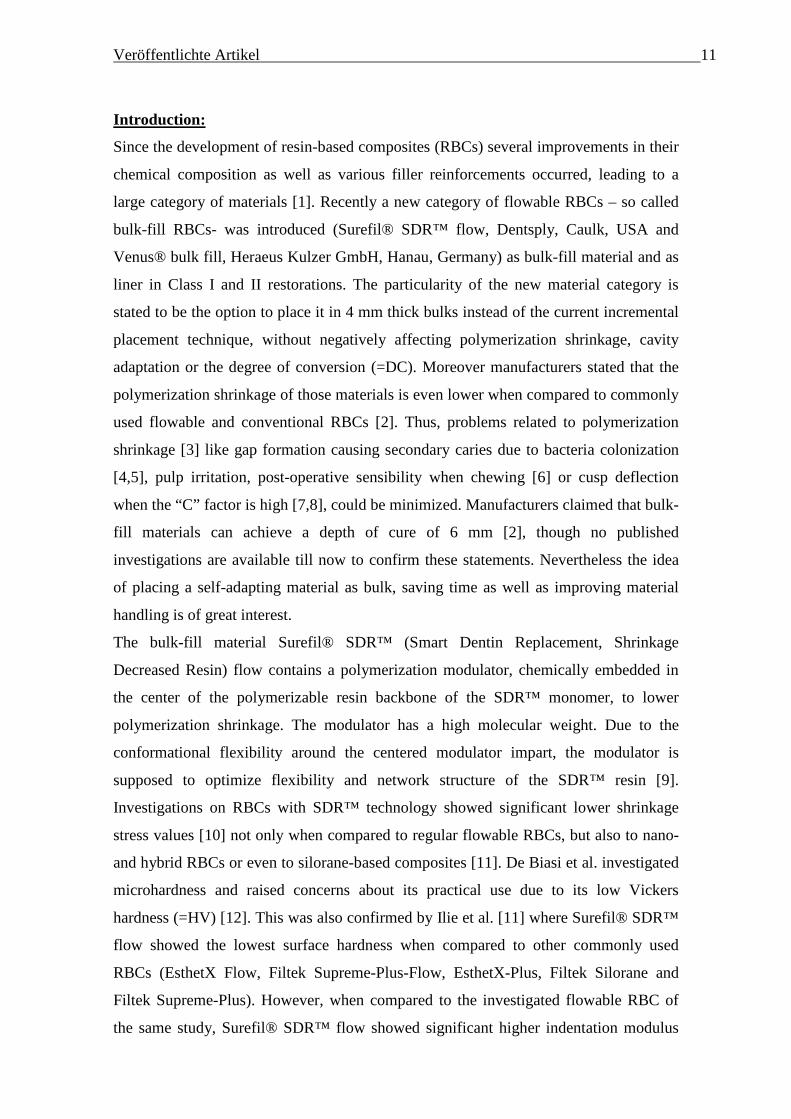
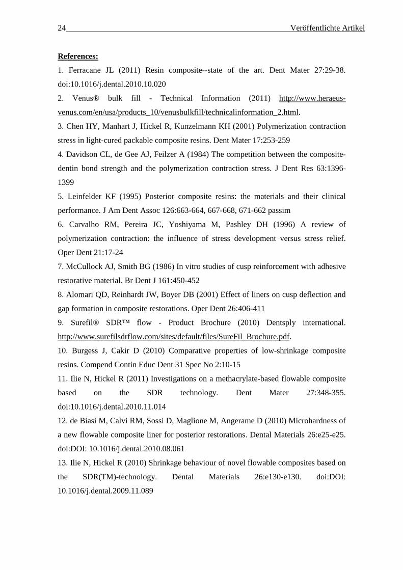
Besides the factor “RBC”, E (η2=0.46) as well as HV (η2=0.24) were moderately
influenced by “measuring depth”. Considering the variation of micromechanical
properties with depth (Fig. 2), it has to be noted that HV and E values rise with the
22 Veröffentlichte Artikel depth to a measuring depth of approximately 1.5 mm until then starting to decrease.
This behavior is not characteristic for high filled RBCs [44]. Since the oxygen
inhibition layer does not exceed 20 – 50 µm [45], the initial decrease in mechanical
properties can rather be explained by the fact that non-bonded light-cured RBCs may
shrink towards the center of the restoration [46]. Kakaboura et al. shared the same
thought when evaluating shrinkage strain of light-cured RBCs using a X-ray
microtomography and a bonded-disc method [47]. Therefore the polymerized bulks
could reach lower mechanical values at peripheral surfaces because the volumetric
shrinkage in the center of the bulk would be compensated by the flow from the
periphery. Moreover Baroudi et al. explained the increased edge fracture resistance with
the lower viscosity of monomers and the reduced particle size of fillers of flowable
RBCs [48].
Regarding the results of the Weibull analysis, both materials exerted a high reliability
(m-value). The high values of m (21.6 and 26.7) - indicating a narrow distribution of
values and therefore a small error range – were unexpected as consistently lower values
were measured for regular flowable RBCs on the market (6.37 to 15.23) [49].
Veröffentlichte Artikel 23
Conclusions:
A strong influence of the material was statistically proven for all measured properties. A
polymerization time of 20s instead of 40s as well as placing the RBCs in 4 mm bulks
instead of 2 mm bulks neither lowered the micromechanical properties nor DC.
Surefil® SDR™ flow showed significantly higher mechanical properties but lower DC
values when compared to Venus® bulk fill.
Within the limitations of our study and the experienced high reliability (high Weibull
modulus values) and good mechanical properties, a polymerization time of 20s and bulk
placement up to 4 mm can be recommended.
24 Veröffentlichte Artikel References:
1. Ferracane JL (2011) Resin composite--state of the art. Dent Mater 27:29-38.
doi:10.1016/j.dental.2010.10.020
2. Venus® bulk fill - Technical Information (2011) http://www.heraeus-
venus.com/en/usa/products_10/venusbulkfill/technicalinformation_2.html.
3. Chen HY, Manhart J, Hickel R, Kunzelmann KH (2001) Polymerization contraction
stress in light-cured packable composite resins. Dent Mater 17:253-259
4. Davidson CL, de Gee AJ, Feilzer A (1984) The competition between the composite-
dentin bond strength and the polymerization contraction stress. J Dent Res 63:1396-
1399
5. Leinfelder KF (1995) Posterior composite resins: the materials and their clinical
performance. J Am Dent Assoc 126:663-664, 667-668, 671-662 passim
6. Carvalho RM, Pereira JC, Yoshiyama M, Pashley DH (1996) A review of
polymerization contraction: the influence of stress development versus stress relief.
Oper Dent 21:17-24
7. McCullock AJ, Smith BG (1986) In vitro studies of cusp reinforcement with adhesive
restorative material. Br Dent J 161:450-452
8. Alomari QD, Reinhardt JW, Boyer DB (2001) Effect of liners on cusp deflection and
gap formation in composite restorations. Oper Dent 26:406-411
9. Surefil® SDR™ flow - Product Brochure (2010) Dentsply international.
http://www.surefilsdrflow.com/sites/default/files/SureFil_Brochure.pdf.
10. Burgess J, Cakir D (2010) Comparative properties of low-shrinkage composite
resins. Compend Contin Educ Dent 31 Spec No 2:10-15
11. Ilie N, Hickel R (2011) Investigations on a methacrylate-based flowable composite
based on the SDR technology. Dent Mater 27:348-355.
doi:10.1016/j.dental.2010.11.014
12. de Biasi M, Calvi RM, Sossi D, Maglione M, Angerame D (2010) Microhardness of
a new flowable composite liner for posterior restorations. Dental Materials 26:e25-e25.
doi:DOI: 10.1016/j.dental.2010.08.061
13. Ilie N, Hickel R (2010) Shrinkage behaviour of novel flowable composites based on
the SDR(TM)-technology. Dental Materials 26:e130-e130. doi:DOI:
10.1016/j.dental.2009.11.089
Veröffentlichte Artikel 25 14. Giovannetti A, Goracci C, Polimeni A, Pacifici E, Ferrari M (2010) Post retention
using a new resin-based-composite with low curing stress. Dental Materials 26:e72-e72.
doi:DOI: 10.1016/j.dental.2010.08.162
15. Leprince JG, Leveque P, Nysten B, Gallez B, Devaux J, Leloup G New insight into
the “depth of cure” of dimethacrylate-based dental composites. Dental Materials.
doi:10.1016/j.dental.2011.12.004
16. Moore BK, Platt JA, Borges G, Chu TM, Katsilieri I (2008) Depth of cure of dental
resin composites: ISO 4049 depth and microhardness of types of materials and shades.
Oper Dent 33:408-412
17. Hansen EK, Asmussen E (1997) Visible-light curing units: correlation between
depth of cure and distance between exit window and resin surface. Acta Odontol Scand
55:162-166
18. Pires JA, Cvitko E, Denehy GE, Swift EJ, Jr. (1993) Effects of curing tip distance
on light intensity and composite resin microhardness. Quintessence Int 24:517-521
19. Price RB, Derand T, Sedarous M, Andreou P, Loney RW (2000) Effect of distance
on the power density from two light guides. J Esthet Dent 12:320-327
20. Ernst CP, Meyer GR, Muller J, Stender E, Ahlers MO, Willershausern B (2004)
Depth of cure of LED vs QTH light-curing devices at a distance of 7 mm. J Adhes Dent
6:141-150
21. Surefil®SDR™ flow Posterior Bulk Fill Flowable Base - Directions for use (2009)
DENTSPLY Caulk.
http://www.caulk.com/assets/pdfs/products/Surefil%20SDR%20Flow%203-
Language%20DFU.pdf.
22. Venus® bulk fill - Instructions for use (2010) Heraeus Kulzer.
http://venusbulkfill.com/media/webmedia_local/media/pdfs/VenusBulkFillDFU_Englis
h.pdf.
23. Sideridou I, Tserki V, Papanastasiou G (2002) Effect of chemical structure on
degree of conversion in light-cured dimethacrylate-based dental resins. Biomaterials
23:1819-1829
24. Vertise Flow - Technical Bulletin (2011)
http://eu.vertiseflow.com/files/bullettin_vertise_e.pdf.
25. Vertise Flow - Product details - FAQ (2010)
http://eu.vertiseflow.com/en/product_details.html?page=faq.
26. Buonocore MG (1955) A simple method of increasing the adhesion of acrylic filling
materials to enamel surfaces. J Dent Res 34:849-853
26 Veröffentlichte Artikel 27. Glenn JF (1979) Comments on Dr. Bowen's Presentation. Journal of Dental
Research 58:1504-1506. doi:10.1177/00220345790580051401
28. Ling L, Xu X, Choi GY, Billodeaux D, Guo G, Diwan RM (2009) Novel F-
releasing composite with improved mechanical properties. J Dent Res 88:83-88.
doi:10.1177/0022034508328254
29. Asmussen E (1983) Factors affecting the color stability of restorative resins. Acta
Odontol Scand 41:11-18
30. SDR™ - Scientific Compendium (2011)
http://www.dentsply.eu/bausteine.net/file/showfile.aspx?downdaid=8854&sp=E&domi
d=1042&fd=2.
31. Amirouche-Korichi A, Mouzali M, Watts DC (2009) Effects of monomer ratios and
highly radiopaque fillers on degree of conversion and shrinkage-strain of dental resin
composites. Dent Mater 25:1411-1418. doi:10.1016/j.dental.2009.06.009
32. Ferracane JL, Greener EH (1986) The effect of resin formulation on the degree of
conversion and mechanical properties of dental restorative resins. J Biomed Mater Res
20:121-131. doi:10.1002/jbm.820200111
33. Scougall-Vilchis RJ, Hotta Y, Hotta M, Idono T, Yamamoto K (2009) Examination
of composite resins with electron microscopy, microhardness tester and energy
dispersive X-ray microanalyzer. Dent Mater J 28:102-112
34. Alvarez-Gayosso C, Barcelo-Santana F, Guerrero-Ibarra J, Saez-Espinola G,
Canseco-Martinez MA (2004) Calculation of contraction rates due to shrinkage in light-
cured composites. Dent Mater 20:228-235. doi:10.1016/s0109-5641(03)00097-6
35. Ellakwa A, Cho N, Lee IB (2007) The effect of resin matrix composition on the
polymerization shrinkage and rheological properties of experimental dental composites.
Dent Mater 23:1229-1235. doi:10.1016/j.dental.2006.11.004
36. Ge J, Trujillo M, Stansbury J (2005) Synthesis and photopolymerization of low
shrinkage methacrylate monomers containing bulky substituent groups. Dent Mater
21:1163-1169. doi:10.1016/j.dental.2005.02.002
37. Lee JH, Um CM, Lee IB (2006) Rheological properties of resin composites
according to variations in monomer and filler composition. Dent Mater 22:515-526.
doi:10.1016/j.dental.2005.05.008
38. Kim KH, Ong JL, Okuno O (2002) The effect of filler loading and morphology on
the mechanical properties of contemporary composites. J Prosthet Dent 87:642-649
39. Manhart J, Kunzelmann KH, Chen HY, Hickel R (2000) Mechanical properties and
wear behavior of light-cured packable composite resins. Dent Mater 16:33-40
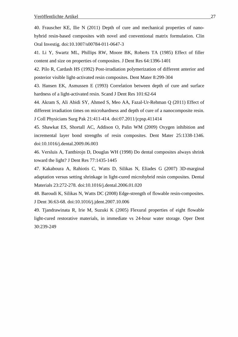
Veröffentlichte Artikel 27 40. Frauscher KE, Ilie N (2011) Depth of cure and mechanical properties of nano-
hybrid resin-based composites with novel and conventional matrix formulation. Clin
Oral Investig. doi:10.1007/s00784-011-0647-3
41. Li Y, Swartz ML, Phillips RW, Moore BK, Roberts TA (1985) Effect of filler
content and size on properties of composites. J Dent Res 64:1396-1401
42. Pilo R, Cardash HS (1992) Post-irradiation polymerization of different anterior and
posterior visible light-activated resin composites. Dent Mater 8:299-304
43. Hansen EK, Asmussen E (1993) Correlation between depth of cure and surface
hardness of a light-activated resin. Scand J Dent Res 101:62-64
44. Akram S, Ali Abidi SY, Ahmed S, Meo AA, Fazal-Ur-Rehman Q (2011) Effect of
different irradiation times on microhardness and depth of cure of a nanocomposite resin.
J Coll Physicians Surg Pak 21:411-414. doi:07.2011/jcpsp.411414
45. Shawkat ES, Shortall AC, Addison O, Palin WM (2009) Oxygen inhibition and
incremental layer bond strengths of resin composites. Dent Mater 25:1338-1346.
doi:10.1016/j.dental.2009.06.003
46. Versluis A, Tantbirojn D, Douglas WH (1998) Do dental composites always shrink
toward the light? J Dent Res 77:1435-1445
47. Kakaboura A, Rahiotis C, Watts D, Silikas N, Eliades G (2007) 3D-marginal
adaptation versus setting shrinkage in light-cured microhybrid resin composites. Dental
Materials 23:272-278. doi:10.1016/j.dental.2006.01.020
48. Baroudi K, Silikas N, Watts DC (2008) Edge-strength of flowable resin-composites.
J Dent 36:63-68. doi:10.1016/j.jdent.2007.10.006
49. Tjandrawinata R, Irie M, Suzuki K (2005) Flexural properties of eight flowable
light-cured restorative materials, in immediate vs 24-hour water storage. Oper Dent
30:239-249
28
V
eröffentlichte Artikel
Tables:
Table 1: Influence of material, measuring depth and polymerization time on the micromechanical properties – indentation modulus (E),
Vickers hardness (HV) – as well as degree of conversion (DC) and macromechanical properties - flexural strength (σ) and flexural modulus
(Eflexural) –. The influence of all parameters was statistical significant (α=0.05). Table contains the partial eta-square values. The higher the
partial eta-squares, the higher the influence of the selected factor on the measured properties.
Factor E HV DC σ Eflexural
RBCs 0.84 0.80 0.63 0.35 0.80
Measuring depth
0.46 0.24 0.10
Polymerization time 0.03 0.04 0.12
Ver
öffe
ntlic
hte
Art
ikel
29
Table 2: Degree of Cure [%] 5 minutes after curing of a) Surefil® SDR™ flow and b) Venus® bulk fill at 0.1 mm, 2 mm, 4 mm, 6 mm (bulk
and incremental) depth are detailed in mean values and standard deviations (in parentheses). Same superscripts (within one geometry, vertical
line) and subscripts (within one polymerization time, in horizontal line) indicate statistical similar groups (Tukey’s HSD Test, α= 0.05).
a) Surefil® SDR™ flow
Measuring depth 0.1 mm 2 mm 4 mm 6 mm-bulk 6 mm-incremental
Polymerization time
10s 58.9 A 1,2,3 (2.9) 60.1 a
2,3 (1.8) 58.3 A 1,2 (1.7) 57.1 a
1 (3.0) 61.0 A 3 (3.1)
20s 61.1 A 2 (1.5) 59.5 a
1,2 (2.2) 59.7 AB 1,2 (1.7) 58.2 a
1 (1.7) 60.7 A 2 (2.1)
40s 60.4 A 1,2 (3.1) 59.6 a
1 (1.9) 61.2 B 1,2 (2.1) 60.1b
1,2 (2.0) 61.9 A 2 (2.4)
30
V
eröffentlichte Artikel
b) Venus® bulk fill
Measuring depth 0.1 mm 2 mm 4 mm 6 mm-bulk 6 mm-incremental
Polymerization time
10s 65.0 A 2 (1.9) 65.0 a
2 (1.5) 62.9 A
1 (2.3) 62.4 a
1 (2.5) 65.6 a
2 (1.5)
20s 64.9 A 1 (1.7) 65.0 a
1 (1.5) 66.1 B 1,2 (2.8) 65.6 b
1,2 (2.0) 66.7 ab 2 (1.6)
40s 64.6 A 1 (1.6) 65.7 a
1,2 (1.8) 66.2 B 1,2 (1.6) 66.1 b
1,2 (2.8) 67.4 b 2 (1.5)
Ver
öffe
ntlic
hte
Art
ikel
31
Table 3: Indentation modulus E [GPa] of a) Surefil® SDR™ flow and b) Venus® bulk fill at 0.1 mm, 2 mm, 4 mm and 6 mm depth of
samples cured for 10s, 20s or 40s as 6 mm high bulk and stored for 24h in distilled water at 37°C, is detailed in mean values and standard
deviations (in parentheses). Same superscripts (within one geometry, in vertical line) and subscripts (within one polymerization time, in
horizontal line) indicate statistical similar groups (Tukey’s HSD Test, α= 0.05).
a) Surefil® SDR™ flow
Measuring depth 0.1 mm 2 mm 4 mm 6 mm-bulk
Polymerization time
10s 10.7 A 1 (0.6) 12.0 a
2 (0.5) 12.2 A 2 (0.7) 11.5 a
2 (0.6)
20s 10.6 A 1 (0.4) 11.9 a
2 (0.4) 12.2 A 2 (0.5) 11.1 a
1 (0.7)
40s 10.8 A1 (0.4) 11.9 a
2,3 (0.4) 12.2 A 3 (0.6) 11.6 a
2 (0.8)
32
V
eröffentlichte Artikel
b) Venus® bulk fill
Measuring depth 0.1 mm 2 mm 4 mm 6 mm-bulk
Polymerization time
10s 7.3 A 1 (1.1) 8.4 a
2 (0.6) 8.6 A 2 (0.9) 7.3 a
1 (0.4)
20s 7.6 A 1 (0.3) 8.6 ab
2 (0.4) 8.8 A 2 (0.4) 7.7 b
1 (0.5)
40s 7.7 A 1 (0.9) 8.8 b
2 (0.3) 8.8 A 2 (0.4) 8.5 c
2 (0.3)
Ver
öffe
ntlic
hte
Art
ikel
33
Table 4: Vickers hardness HV [N/mm2] and depth of cure (HV-80%) [N/mm2] of a) Surefil® SDR™ flow and b) Venus® bulk fill at 0.1 mm,
2 mm, 4 mm and 6 mm depth of samples cured for 10s, 20s or 40s as 6 mm high bulk and stored for 24h at 37°C in distilled water, is detailed
in mean values and standard deviations (in parentheses). Same superscripts (within one geometry, in vertical line) and subscripts (within one
polymerization time, in horizontal line) indicate statistical similar groups (Tukey’s HSD Test, α= 0.05).
a) Surefil® SDR™ flow
Measuring depth 0.1 mm 2 mm 4 mm 6 mm-bulk HV-80%
Polymerization time
10s 55.1 A 1 (2.3) 57.8 a
2 (3.4) 58.2 A 2 (4.1) 55.4 a
1 (3.8) 44.1
20s 59.1 A 1 (3.3) 61.1 b
2 (2.1) 59.8 A 1,2 (2.9) 59.0 b
1,2 (3.4) 47.3
40s 59.1 A 1 (1.3) 60.2 ab
1 (1.8) 59.5 A 1 (2.9) 58.9 b
1 (2.6) 47.3
34
V
eröffentlichte Artikel
b) Venus® bulk fill
Measuring depth 0.1 mm 2 mm 4 mm 6 mm-bulk HV-80%
Polymerization time
10s 40.7 A 1 (4.0) 46.1 a
2 (3.1) 46.5 A 2 (4.7) 39.1 a
1 (2.9) 32.6
20s 41.4 A 1 (1.5) 45.8 a
3 (1.4) 46.4 A 3 (0.9) 43.0 b
2 (1.4) 33.1
40s 42.7 A 1 (4.9) 46.4 a
2 (2.1) 46.4 A 2 (1.0) 46.0 c
2 (1.9) 34.2
Ver
öffe
ntlic
hte
Art
ikel
35
Figures:
Figure 1:
Evaluation of the Weibull parameter (m) for Surefil® SDR™ flow and Venus® bulk fill using the variables P (probability of failure) and σ
(Flexural strength). R2 is the coefficient of determination.
m Surefil® SDR™ flow = 26.7
R2 = 0.91
m Venus® bulk fill= 21.6
R2
= 0.97
-4
-3
-2
-1
0
1
2
3 4 5 6ln (σ)
lnln
[1/(
1-P
)]
Surefil® SDR™ flow
Venus® bulk fill
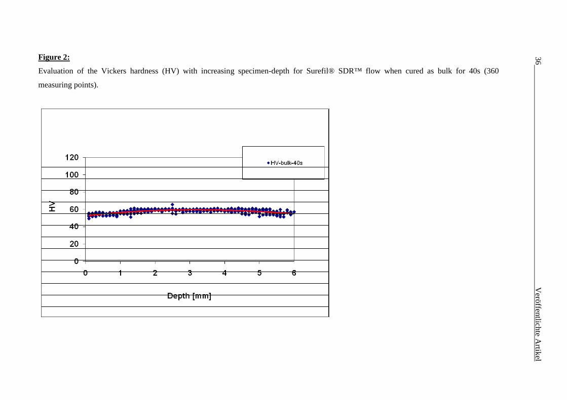
Figure 2:
Evaluation of the Vickers hardness (HV) with increasing specimen
measuring points).
s (HV) with increasing specimen-depth for Surefil® SDR™ flow when cured as bulk for 40s
36
V
eröffentlichte Artikel
bulk for 40s (360
Ver
öffe
ntlic
hte
Art
ikel
37
Figure 3:
Fe-SEM pictures of the measured materials.
Surefil® SDR™ flow Venus® bulk fill
38 Veröffentlichte Artikel
2.1.1 Deutsche Zusammenfassung
Ziel der Studie war es Konversionsrate (KR), mikromechanische (Eindringmodul (E),
Vickershärte (HV)) und makromechanische (Biegefestigkeit (σ), Biegemodul (EBiege)
Eigenschaften von zwei neu auf dem Markt erschienenen „bulk fill“ Kompositen zu
untersuchen und zu vergleichen: Surefil® SDR™flow (SF) und Venus® bulk fill (VB).
Die Arbeitshypothesen waren:
(a) Es gibt keinen Unterschied zwischen beiden Materialien bezüglich ihrer
makromechanischen (Biegefestigkeit, Biegemodul) und mikromechanischen
(Vickershärte, Eindringmodul) Eigenschaften sowie ihrer Konversionsrate in jeder der
gemessenen Inkrementdicken und zu jeder Belichtungszeit.
(b) Die Belichtungszeit und Inkrementdicke haben bei keinem von beiden Materialien
einen signifikanten Einfluss auf die gemessenen Materialeigenschaften.
Die KR (n=6) wurde durch eine Fourier-Transformations-Infrarotspektrometrie (FTIR)
in klinisch relevanten Inkrementdicken (0,1 mm, 2 mm, 4 mm, 6 mm-Bulk (ein
Inkrement), 6 mm-Inkrement (geschichtet aus dreimal 2 mm dicken Inkrementen)) und
Belichtungszeiten (10s, 20s, 40s) bestimmt. Mikro- (n=6) und makromechanische
(n=20) Eigenschaften wurden nach 24 Stunden Lagerung der Prüfkörper bei 37°C in
destilliertem Wasser von einem Universalhärtemessgerät und einer Drei-Punkt-
Biegeversuch Anordnung bestimmt. Darüber hinaus wurde mittels der 6 mm-Bulk
Prüfkörper die Tiefenhärte gemessen. Ein Rasterelektronenmikroskop wurde zur
Evaluation der Füllkörpergröße hinzugezogen. Die Ergebnisse wurden mittels
univariater Varianzanalyse, Tukey’s honest significance post-hoc-Test, einer
multivariaten Varianzanalyse (α=0.05) und einem unabhängigem t-Test ausgewertet.
Eine Verlässlichkeitsbestimmung wurde für σ mittels Weibullanalyse (m-Parameter)
durchgeführt.
Für alle Inkrementdicken zeigte VB signifikant höhere KR (VB: 62,4 – 67,4 %; SF:
57,1 – 61,9 %) und signifikant niedrigere makro- (VB: EBiege= 3,6 GPa; σ= 122,7 MPa;
SF: EBiege= 5,0 GPa; σ= 131,8MPa) und mikromechanische (VB: E= 7,3– 8,8 GPa, HV=
40,7–46,5 N/mm²; SF: E= 10,6–12,2 GPa, HV= 55,1–61,1 N/mm²) Eigenschaften.
Beide Materialien zeigten eine hohe Verlässlichkeit (VB: m= 21,6; SF: m= 26,7) und
eine Tiefenhärte von mindestens 6 mm zu allen Belichtungszeiten. Der Paramter
Veröffentlichte Artikel 39 „Kompositmaterial“ („RBC“) hatte den größten Einfluss auf die untersuchten
Eigenschaften (η2= 0,35–0,80) gefolgt von der Inkrementdicke („measuring depth“)
(η2= 0,10–0,46) und der Belichtungszeit („polymerization time“) (η2= 0,03–0,12).
Signifikante Unterschiede zwischen den beiden Kompositen konnten für KR, E, σ und
EBiege bei allen Inkrementdicken und zu allen Belichtungszeiten gemessen werden.
Surefil® SDR™ flow zeigte signifikant höhere Werte für die mechanischen
Eigenschaften und signifikant niedrigere KR-Werte als Venus® bulk fill. Somit wurde
die erste Arbeitshypothese widerlegt.
Eine Belichtungszeit von 20s und eine 4 mm Inkrementplatzierung von Surefil® SDR™
flow wie auch eine Belichtungszeit von entweder 40s mit einer 6 mm
Inkrementplatzierung oder 20s mit 4 mm Inkrementplatzierung von Venus® bulk fill
führten zu den besten mikromechanischen Werten. Somit wurde auch die zweite
Arbeitshypothese widerlegt.
Durch die gute Verlässlichkeit der Werte (hohe Weibullparameter) und der guten
mechanischen Eigenschaften beider Materialien kann mit Rücksicht auf die
Limitationen der Studie eine Belichtungszeit von 20s und eine Inkrementplatzierung bis
hin zu einer Dicke von 4 mm empfohlen werden.
40 Veröffentlichte Artikel
2.1.2 English summary
The aim of the study was to measure and compare degree of conversion (DC) as well as
micro- (indentation modulus (E); Vickers hardness, (HV)) and macromechanical
properties (flexural strength (σ); flexural modulus, Eflexural) of two recently launched
bulk fill resin-based composites (RBCs): Surefil® SDR™flow (SF) and Venus® bulk
fill (VB).
The tested null hypotheses were that:
(a) There would be no significant difference between the two materials in view of
macro- (flexural strength (σ), modulus of elasticity (Eflexural)) and micromechanical
properties (Vickers hardness (HV) and indentation modulus (E)) and degree of cure
(DC) at any measured depth and irradiation time.
(b) Within one material, irradiation time and depth would not influence the measured
properties.
DC (n=6) was investigated by Fourier transform infrared spectroscopy (FTIR) in
clinical relevant filling depths (0.1, 2, and 4 mm; 6 mm-bulk, 6 mm-incremental) and
irradiation times (10s, 20s, 40s). Micro- (n=6) and macromechanical (n=20) properties
were measured by an automatic microhardness indenter and a three-point bending test
device after storing the specimens in distilled water for 24 h at 37°C. Furthermore, on
the 6-mm bulk samples, the depth of cure was determined. A field emission scanning
electron microscope was used to assess filler size. Results were evaluated using one-
way analysis of variance, Tukey’s honest significance post-hoc-test, a multivariate
analysis (α=0.05) and an independent t-test. Weibull analysis was used to assess σ.
VB showed, in all depth, significant higher DC (VB: 62.4–67.4 %; SF: 57.1–61.9 %),
but significant lower macro- (VB: Eflexural= 3.6 GPa; σ= 122.7 MPa; SF: Eflexural= 5.0
GPa; σ= 131.8MPa) and micromechanical properties (VB: E= 7.3– 8.8 GPa, HV= 40.7–
46.5 N/mm²; SF: E= 10.6–12.2 GPa, HV= 55.1–61.1 N/mm²). Both RBCs showed high
reliability (VB: m= 21.6; SF: m= 26.7) and a depth of cure of at least 6 mm at all
polymerization times. The factor “RBC” showed the strongest influence on the
measured properties (η2= 0.35–0.80) followed by “measuring depth” (η2= 0.10–0.46)
and “polymerization time” (η2= 0.03–0.12). Significant differences between both RBCs
were found for DC, E, σ and Eflexural at all irradiation times and measuring depths.
Veröffentlichte Artikel 41 Surefil® SDR™ flow showed significantly higher mechanical properties but lower DC
values when compared to Venus® bulk fill. Therefore the first hypothesis had to be
rejected.
An irradiation time of 20s and a 4 mm bulk placement for Surefil® SDR™ flow as well
as an irradiation time of either 40s and 6 mm bulk placement or 20s and 4 mm bulk
placement for Venus® bulk fill led to the best micromechanical values. Therefore also
the second hypothesis was rejected.
Within the limitations of our study and the experienced high reliability (high Weibull
modulus values) and good mechanical properties, a polymerization time of 20s and bulk
placement up to 4 mm can be recommended.
42 Veröffentlichte Artikel
2.2 „In-vitro comparison of mechanical properties and
degree of cure of a self-adhesive and four novel
flowable composites“
Pascal Czasch and Nicoleta Ilie
Department of Restorative Dentistry, Dental School of Ludwig-Maximilians-University,
Munich, Germany
Journal of Adhesive Dentistry 2012 Jun 8. [Acceptance ahead of print]
Veröffentlichte Artikel 43
Abstract:
Purpose: The aim of our study was to compare a self-adhesive with two low shrinkage
and two regular flowable resin-based composites (RBCs) in terms of degree of
conversion (DC) and mechanical properties measured at microscopic (Indentation
modulus, E, Vickers hardness, HV) and macroscopic scale (Flexural strength, σ,
Flexural modulus, Eflexural).
Materials and Methods: DC was investigated by an ATR-FTIR-Spectrometer in
clinical relevant filling depth (0.1 mm, 2 mm, 4 mm, 6 mm-bulk, 6mm-incremental) and
irradiation times (10s, 20s, 40s). Micro- and macro-scale mechanical properties were
measured by an automatic microhardness indenter and a three-point bending test device
after curing the specimens for 20s and storing them in distilled water for 24h at 37°C.
Fillers were visualized by a field emission scanning electron microscope. Results were
evaluated using one-way ANOVA, Tukey’s HSD post-hoc test, Pearson correlation and
a multi-variate analysis (α =0.05). A Weibull analysis was used to assess σ.
Results: N’Durance® Dimer flow (65.66%) reached the highest DC (at 2 mm depth,
20s irradiation). At macro-scale EcuSphere®-Flow (129.82 MPa) for σ and Synergy®
D6 Flow (3.74 GPa) for Eflexural reached highest values. The highest micro-mechanical
properties were measured for the self-adhesive RBC (Vertise™ Flow; E= 10.81 GPa,
HV= 60.20 N/mm2). Reliability was highly influenced by filler weight (η2= 0.77) and
volume (η2= 0.99) proportion.
Conclusion: In the present study the self-adhesive RBC showed the highest reliability,
highest DC (together with one of the low shrinkage RBCs) and highest mechanical
properties measured at micro-scale as well as good mechanical properties measured at
macro-scale. Moreover a curing time of 40s and an incremental thickness not exceeding
2 mm appeared to be necessary for it.
44 Veröffentlichte Artikel
Introduction:
The demands on modern flowable resin based composites (=RBCs) in their function as
restorative materials or liners are permanently extended, asking nowadays for
supplementary properties like self-adhesion, low polymerization shrinkage or improved
mechanical properties. The latest developments in this subject area are the self-adhering
flowable RBCs, as a result of ongoing efforts to simplify clinical treatment. New self-
adhesive RBCs are promoted as materials needing neither etching nor a bonding agent
(1). Since the development of resin based composites, their adhesion to tooth structure
has been challenging clinicians and scientists. After Buonocore published a method of
increasing the restorations adhesion (2), various researchers developed new ways to
ease and enhance handling and quality of modern etching and bonding systems (3, 4).
One modern way to create self-adhesive RBCs (5), self-adhesive resin cements (6) or
self-etching adhesives (7, 8), is to use special phosphate dimethacrylate monomers like
glycerol phosphate dimethacrylate (=GPDM), allowing thus a chemical interaction of
the phosphonate group with the calcium ions of the tooth structure. But also a
micromechanical bonding between polymer and the collagen fibers that are exposed
through the etching effect of the phosphonate acidic group of GPDM, as well as
between the polymer and the integrated smear layer (interdiffusion zone) was
ascertained (3). However including GPDM in self-adhesive RBCs did not improve the
bond strength to enamel, when compared to etch- and rinse adhesives (9-12).
Disadvantages of one-component self-etching adhesives in general are seen in their
relatively high water uptake (13, 14) as well as a possible hydrolytic degradation of the
tooth-restoration interface (15). Nevertheless concerning shear bond strength to
superficial as well as to deep dentin, GPDM-containing adhesives resulted equally to
other self-etching and total-etching adhesive systems (12).
Another attempt intensively followed in modern flowable RBCs is to reduce
polymerization shrinkage. When measuring polymerization shrinkage of experimental
composites with the same filler and initiator concentrations but different fractions of
monomers, Ellakwa et al. found out that one way to decrease polymerization shrinkage
is to use monomers with higher molecular weights (16). Some modern flowable RBCs
therefore renounce to low molecular weight monomers like Triethyleneglycol
dimethacrylate (=TEGDMA) (286 g/mol) and are based on monomers with a high
molecular weight. Besides traditional monomers like bisphenylglycidyl dimethacrylate
Veröffentlichte Artikel 45 (=BisGMA) (512 g/mol) and ethoxylated bisphenol-A dimethacrylate (=BisEMA) (540
g/mol) (17), also the newly developed dimer-acid dimethacrylates (847 g/mol) (18-20)
are used, resulting in higher DC, higher flexural modulus and the possibility of placing
thicker increments than traditional RBCs (21). The mechanism of reducing shrinkage by
using dimer-acid dimethacrylates in combination with BisEMA and Urethane
dimethacrylate (=UDMA) (471 g/mol) is based, besides the higher molecular weight, on
a phase separation due to a partial compatibility of the monomers in their polymerized
state (20). A higher degree of conversion (=DC) and a lower shrinkage are stated to be
the result when the faster polymerizing phase remains in a less reticulated polymer (22).
The aim of our study was to compare a new self-adhesive RBC (Vertise™ flow) as well
as two low shrinkage flowable RBCs (N’Durance® Dimer flow and Extra low
shrinkage flow) with two traditional flowable microhybrid RBCs (EcuSphere®-Flow,
Synergy® D6 Flow) regarding DC and their mechanical properties at macroscopic and
microscopic scale at different irradiation times and by simulating clinical relevant filling
depth.
The tested null-hypotheses were that: a) there would be no significant difference
between the five materials in view of DC at any measured depth and irradiation time; b)
there would be no significant difference between the five materials when measuring
mechanical properties at macro- (flexural strength (=σ) and modulus of elasticity
(=Eflexural)) and micro-scale (Vickers hardness (=HV) and indentation modulus (=E))) at
a clinical relevant irradiation time of 20s.
46 Veröffentlichte Artikel
Materials and methods:
Five flowable RBCs – composition information as far as they could be collected (17,
18, 23-26) (Table 1) – all in shade A3, were analyzed by assessing DC as function of
depth, incremental technique and polymerization time (10s, 20s or 40s). Furthermore,
the mechanical properties measured at macroscopic (flexural strength (=σ) and modulus
of elasticity (=Eflexural)) and microscopic scale (Vickers hardness (=HV) and indentation
modulus (=E)) were assessed.
Degree of cure measurements:
To evaluate the DC, five different sample geometries were considered. Thin films (100
µm) as well as 2 mm, 4 mm and 6 mm high molds (3 mm diameter) were filled in bulk.
Additionally three consecutive increments - each 2 mm high – were prepared in the
mold of 6 mm height (6 mm-incremental). Samples were cured by applying the curing
unit (Elipar Freelight2, 3M ESPE, 1226 mW/cm²) directly on the top of the particular
mould, respectively on the film surface covered by a transparent matrix strip. For each
product, irradiation time (10s, 20s, 40s) and geometry (0.1 mm, 2 mm, 4 mm, 6 mm-
bulk, 6 mm-incremental) six samples were measured (n=6). Real-time measurements
were made with a FTIR-Spectrometer with an attenuated total reflectance (ATR)
accessory (Nexus, Thermo Nicolet, Madison, USA). Therefore, the non-polymerized
RBC paste was put directly on the diamond ATR crystal in the mold as described
above. FTIR spectra were recorded in real time for 5 minutes with two spectra per
second at the bottom of the samples irradiated according to the curing protocol
presented above. Diameter of measured surface was 800 µm, wave number of the
spectrum ranged between 4000-650 cm-1 and the FTIR spectra were recorded with four
scans at a resolution of 8 cm-1. During testing the specimens were constantly pressed to
the ATR refractive element by a fixed stamp to prevent the potential pull-out effect
caused by setting shrinkage.
To determine the percentage of the remained unreacted double bonds, DC was
measured by assessing the variation in peak height ratio of the absorbance intensities of
methacrylate carbon double bond peak at 1634 cm-1 and that of an internal standard
peak (=IS) at 1608 cm-1 (aromatic carbon double bond) during polymerization, in
relation to the uncured material.
Veröffentlichte Artikel 47
DCheight % = 100
curing beforePeak IS1-1634cm
curingafter Peak IS1-1634cm
1 x
úúúúú
û
ù
êêêêê
ë
é
÷÷ø
öççè
æ
÷÷ø
öççè
æ
-
Flexural strength and flexural modulus:
Flexural strength (σ) was determined in a three-point-bending test in analogy to DIN
EN ISO 4049:2010-03 (27). The samples (n=20) were made by compressing the RBC
material between two glass plates with intermediate polyacetate sheets, separated by a
steel mould having an internal dimension of (2 x 2 x 16) mm. After curing (with three
overlapping light exposures of 20 seconds per each side, Elipar Freelight2, 3M ESPE)
the specimens were removed from the mould and any flash material was trimmed away
with sandpaper (grit size P4000 (FEPA)). Afterwards all specimens were stored in
distilled water at 37°C prior to testing for 24 h. Samples were then loaded until failure
in the universal testing machine (MCE 2000ST, quick test Prüfpartner GmbH,
Langenfeld, Germany). The crosshead speed was 0.5 mm/min. The specimens were
placed on a three-point bending test device, which is constructed according to the
guidelines of NIST No. 4877 with 12 mm distance between the supports. During testing
the specimens were immersed in distilled water at room temperature.
Flexural strength was calculated from formula (1).
(1) 22
3
bh
Fl=s
F is the maximum load [N], l is the distance between the supports [mm], b is the width
of the specimens [mm], h is the height of the specimens [mm].
The universal testing machine stored the force during bending and the deflection of the
beam in a file. The bending modulus was calculated from formula (2).
(2) ybh
FlflexuralE
34
3=
y is the deflection at load point [mm].
Mechanical properties measured at microscopic scale:
The mechanical properties at micro-scale (HV, E) were assessed on six randomly
assigned fragments resulted after the bending test. For this purpose, the fragments were
48 Veröffentlichte Artikel ground and polished under water with diamond abrasive paper (mean grain sizes: 20
µm, 13 µm, 6 µm) in a grinding system (EXAKT 400CS, Exakt, Norderstedt,
Germany). Measurements were made with an automatic microhardness indenter
(Fischerscope H100C, Fischer, Sindelfingen, Germany) testing 10 randomly assigned
measurement points at one sample (total 60 measurement points per group). The test
procedure was carried out force-controlled, where the test load increased and decreased
with constant speed between 0.4 mN and 500 mN. Load and penetration depth of
indenter (Vickers pyramid: diamond right pyramid with a square base (2.5 µm x 2.5
µm) and an angle of α= 136° between the opposite faces at the vertex (calculated mean
radius of the tip: 0.5 µm)) were continuously measured during the load-unload
hysteresis. Universal hardness is defined as the test force divided by the apparent area of
indentation under the applied test force. From a multiplicity of measurements stored in a
database supplied by the manufacturer, a conversion factor (0.0945) between Universal
hardness and HV was calculated by the manufacturer and entered into the software, so
that the measurement results were indicated in the more familiar HV units. E was
calculated from the slope of the tangent adapted at the beginning (at maximum force) of
the non linear indentation depth curve upon maximum loading.
Field emission scanning electron microscope (FE-SEM):
For each product one specimen (1 cm x 1 cm x 0.5cm) was polymerized for 40s and
treated for one hour in a chemical dry cleaning process with oxygen plasma in vacuum
(45 - 50 W). Afterwards surfaces were investigated (Magnification: 20000x, Signal:
Secondary electrons SE2, Working distance: 4 mm, Electron high tension: 10 kV) with
a field emission scanning microscope (FE-SEM) (Zeiss Supra® 55 VP, Zeiss NTS
GmbH, Oberkochen, Germany) and the most representative picture was chosen to
visualise filler shape and dimension.
Statistical analysis:
The results for DC within each material, each measuring depth and each curing time,
respectively, were compared using one-way ANOVA and Tukey’s HSD post hoc-test
(α=0.05) (SPSS 18.0, Chicago, IL, USA). Additionally a Weibull analysis was used to
assess σ. An ANOVA multivariate analysis and partial eta-square statistic were used to
investigate the influence of the parameters “RBCs”, “measuring depth”,
“polymerization time”, “Wt%-Filler” (=weight proportion of fillers) and “Vol%-Filler”
Veröffentlichte Artikel 49 (=volume proportion of fillers) on DC, E, HV, Eflexural, σ and m. Furthermore a Pearson
correlation investigated the linear dependence between wt%-filler, vol%-filler, E, HV,
Eflexural, σ and m.
A common empirical expression for the cumulative probability of failure P at applied
stress is the Weibull model:
úúû
ù
êêë
é÷÷ø
öççè
æ--=
m
ccfP
0
exp1)(sss
where cs is the measured strength, m the Weibull modulus and 0s the characteristic
strength, defined as the uniform stress at which the probability of failure is 0.63.
The double logarithm of this expression gives:
By plotting ln ln(1/(1-P)) versus ln sigma, a straight line results, with the upward
gradient m, whereas the intersection with the x-axes gives the logarithm of the
characteristic strength.
0lnln1
1lnln ss mm
P c -=-
50 Veröffentlichte Artikel
Results:
The influence of the parameters “RBCs”, “Depth”, “Polymerization time”, “wt%-filler”
and “vol%-filler” were analyzed in an ANOVA multivariate test (Table 2). DC and the
mechanical properties – HV, E, σ, Eflexural, m - were selected as depended variables. The
significance values of these effects were less than 0.05, indicating that they contribute
all to the model. DC was strongly influenced by depth (η2 = 0.95, Table 2), RBC (0.94)
and polymerization time (0.81). The RBCs exerted the strongest influence (higher η2
values) on DC followed by the micro-mechanical properties (E (0.88), HV (0.68)) and
the mechanical properties measured at macroscopic scale (Eflexural (0.48) and σ (0.32)).
Vol%-filler strongly influenced m (0.99), E (0.80), HV (0.61) and moderately Eflexural
(0.47). This statement is equally valid for wt%-filler.
The DC five minutes after photo-initiation as function of samples thickness and
polymerization time is expressed in Table 3. Increasing measuring depth from 0.1 mm
to 2 mm significantly decreased DC values in all groups except for Vertise™ Flow,
N’Durance® Dimer flow and Synergy® D6 Flow at high polymerization times (20s,
40s). As for the differences in DC at 2 mm and 4 mm depth, a significant decrease was
measured at 4 mm depth for all materials and polymerization times. At 2 mm depth DC
increased with progressive irradiation time in all RBCs, except for N’Durance® Dimer
flow which showed similar DC values at 20s and 40s irradiation times. By using an
incremental technique, the DC values measured at 6-mm depth were similar to those
measured at 2 mm depth, except for Extra low shrinkage flow at low polymerization
times (10s, 20s) and N’Durance® Dimer flow at high polymerization time (40s).
Regarding the mechanical properties measured at microscopic scale (HV, E, Table 4),
the highest values were measured for Vertise™ Flow, whereas at macroscopic scale (σ,
Eflexural, Table 4), EcuSphere®-Flow reached the highest values for σ and Extra low
shrinkage flow the lowest values for Eflexural besides the other statistically similar RBCs.
As for the Weibull modulus (Figure 1, Table 4) Vertise™ Flow and Extra low shrinkage
flow showed the highest reliability of the investigated RBCs.
The modulus of elasticity measured in both methods – the flexural test and Universal
hardness test - correlated moderately (Pearson correlation coefficient = 0.43). An
excellent correlation was found between the microscopic mechanical properties (E-HV
= 0.91).

Veröffentlichte Artikel 51
Discussion:
A recently launched self-adhesive flowable RBC – Vertise™ Flow – and two low
shrinkage flowable RBCs – N’Durance® Dimer flow (a new flowable RBC with a tri-
monomer system including dimer-acid dimethacrylates) and Extra low shrinkage flow
(a new flowable RBC with a matrix free of low molecular weight monomers) – were
investigated and compared with two established flowable microhybrid composites on
the market (EcuSphere®-Flow, Synergy® D6 Flow).
When compared with other RBCs (GC Kalore, GC GradiaDirect Anterior, GC Gradia
Direct Posterior, Filtek® Silorane), Wei et al. found out that the self-adhesive flowable
RBC Vertise™ Flow suffered the greatest hygroscopic expansion (4.82%), the greatest
water sorption (71.96 g/mm3) and the greatest solubility (16.95 g/mm3) as well as a
significant higher dehydration shrinkage and a significant mass reduction after 42 days
in water immersion (28, 29). As shown in our study, good DC values were reached for
Vertise™ Flow when compared to the other investigated products (Table 3), thus the
above described material behaviour cannot be related to a disproportional high amount
of not-polymerized monomers that potentially could be washed out (30). A reason for
the high solubility associated with Vertise™ Flow could rather be the usage of GPDM
and other methacrylate co-monomers (5), since its hydrophilic acidic phosphate group
and the short spacer group are said to exhibit a great hydrophilicity (29). The role of the
methacrylate co-monomers cannot be assessed since any clear information is
unfortunately available about their chemical composition. Therefore it is questionable if
Vertise™ Flow may really be used in restoration parts that are exposed to saliva and
other fluids. Moreover it is stated that the self-adhesive flowable RBC Vertise™ Flow
requires a longer polymerization time in comparison with conventional adhesives or
other marketed flowable RBCs since the included monomers tend to have a slower
response to light curing than non-adhesive monomers (31).
For Vertise™ Flow the manufacturer advises the usage in small class I restorations, as
liner in class I and II restorations, as fissure sealant and as minor occlusal build-up in
non-stress bearing areas. Comparing our results to a recent study, Vertise™ Flow shows
lower mechanical properties at macro- and micro-scale than modern non-flowable
RBCs (32). Therefore a placement of non-flowable RBCs on top is strongly
recommended in occlusal loaded areas. Within the analysed flowable materials
however, Vertise™ Flow shows the best values in microscopic mechanical properties.
A reason for this can be found in the composition of the inorganic content. Vertise™
52 Veröffentlichte Artikel Flow contains 1-micron barium glass fillers, nano-sized colloidal silica and nano-sized
Ytterbium fluoride (5). Together they represent about 70 percent of total weight (28),
which is about 7 percent higher than the given weight percent values of EcuSphere®-
Flow and Synergy® D6 Flow and even 17 percent higher than the filler weight
proportion of Extra low shrinkage flow (Table 1). This is due to the increase of filler
volume being about 6 percent (Synergy® D6 Flow), 7 percent (EcuSphere®-Flow,
N’Durance® Dimer flow) or even 20 percent (Extra low shrinkage flow) higher for
Vertise™ Flow. When assessing those values it becomes clear that the low mechanical
properties of Extra low shrinkage flow are due to its low filler content since statistical
results show a high influence (high partial eta-square values) of the filler proportions
(weight and volume percent) on the investigated mechanical properties. A positive
correlation between filler loading and mechanical properties was also reported by
former studies (33, 34).
N’Durance® Dimer flow is said to use a tri-monomer system including dimer-acid
dimethacrylates which exhibit a higher molecular weight (847 g/mol) (19, 20) compared
to the two dimethacrylates – BisGMA (512 g/mol) and BisEMA (540 g/mol) (35) – the
matrix of Extra low shrinkage flow exclusively consists of. Trujillo-Lemon et al.
measured in several experimental hydrophobic monomers, that were based on a dimer-
acid structure, a higher DC, lower polymerization shrinkage and an increase of
flexibility (19). However two other studies stated that high molecular weight monomers
in general present lower mobility, thus reducing the final DC reached by the (36, 37).
Referring to our results N’Durance® Dimer flow reaches in fact- together with
Vertise™ Flow - the highest DC in contrast to Extra low shrinkage flow which presents
the lowest DC values. Unfortunately the co-monomers represented in the tri-monomer
system of N’Durance® Dimer flow have not been published, but it seems that they
contribute to a higher DC.
Referring to the mechanical properties at macro- and micro-scale, it becomes obvious
that through different organic and filler compositions of the RBCs, mechanical
superiority cannot be predicted by DC. Together with a different filler loading and filler
type each monomer and additional group implicates different properties and different
molecular architecture, thus a nearly equal DC in the case of Vertise™ Flow and
N’Durance® Dimer flow does not necessarily mean equal mechanical values as
reflected in the measured mechanical properties (Table 4).
For Weibull modulus (Figure 1, Table 4) - highly influenced by wt%-filler (η2=0.77)
and vol%-filler (η2=0.99) - Vertise™ Flow (11.35) and Extra low shrinkage flow
Veröffentlichte Artikel 53 (11.22) present the highest values thus exhibiting the highest reliability while the lowest
reliability was found for Synergy® D6 Flow (6.52).
Lee et al. found out - when comparing experimental composites with different types and
contents of fillers - that viscosity of RBCs increases when filler volume increases (38).
Following studies could therefore compare the rheological properties of Vertise™ Flow
with other flowable RBCs on the market.
Comparing our results to those of Tjandrawinata et al. (6.37 to 15.23) (39), who
measured eight flowable RBCs concerning their flexural strength, our investigated
RBCs show comparable values.
Assessing FE-SEM pictures (Figure 2), fillers of Vertise™ Flow are obviously bigger
than fillers of N’Durance® Dimer flow and Extra low shrinkage flow. The fillers of
Synergy® D6 Flow seem to be the smallest of all investigated products, while the
majority of fillers of EcuSphere®-Flow are small, with some sporadic fillers that are
bigger than 2 µm though. However Li et al. claimed that decreasing filler size in the
same monomer system with the same filler amount also means harming depth of cure,
compressive strength and resistance to tooth brushing (40). This could be of interest in
further studies about Vertise™ Flow.
The indications for use of the investigated products advise a placement up to 2 mm and
a curing time of 20s (1, 18, 24, 25), except for Extra low shrinkage flow where a curing
time of 30s was suggested (17). For N’Durance® Dimer flow this claim can be
confirmed by our data, since no improvement in DC is shown when increasing the
irradiation time from 20s to 40s in 2 mm depth. This does not apply for Vertise™ Flow
since an improvement of irradiation time from 20s to 40s also increased DC. Moreover
significant lower values appeared for both RBCs when raising placement thickness of
the specimens from 2 mm to 4 mm, limiting thus the incremental thickness to 2 mm.
It must however be considered that the measurements were done with a modern high
intensity LED curing unit which was applied at mould upper surface. Placing clinical
restorations often means higher distances between less effective curing units and RBC
surface. The DC measured 5 minutes after starting the polymerization was reported to
represent 85-90% of the final DC measured after 24 hours (41), legitimating thus the
short post-polymerisation period considered in our study. The inital DC after 5 minutes
is however of great importance since elution of unreacted monomers by saliva and other
fluids starts right after polymerization with all of the already investigated possible
toxicological long term consequences (42-44). Because Vertise™ Flow shows the best
mechanical properties at micro-scale and good macro-scale results as well as medium
54 Veröffentlichte Artikel mechanical values for N’Durance® Dimer flow and a further variation in mechanical
properties between the investigated flowable RBCs the second hypothesis must be
rejected.
Veröffentlichte Artikel 55
Conclusions:
A strong influence of the material on all measured properties, as well as a strong
influence of filler proportion (wt%-filler, vol%-filler) on the reliability was statistically
proven. All tested null-hypotheses had to be rejected.
Within the limitations of our study the self-adhesive RBC (Vertise™ Flow) showed best
performance concerning DC after five minutes of polymerization (together with the
dimer-acid based RBC) and mechanical properties measured at micro-scale. The
traditional microhybrid flowable RBCs performed best in the macroscopic mechanical
properties σ (EcuSphere®-Flow) and Eflexural (Synergy® D6 Flow). For the self-adhesive
RBC a curing time of 40s as well as an incremental placement not exceeding 2 mm
thickness appeared to be necessary.
56 Veröffentlichte Artikel
References:
1. Specialities SD. Vertise Flow - Technical Guide.
http://eu.vertiseflow.com/files/technical_guide_EN.pdf, 2010.
2. Buonocore MG. A simple method of increasing the adhesion of acrylic filling
materials to enamel surfaces. J Dent Res 1955;34:849-853.
3. Ferracane JL. Resin composite--state of the art. Dent Mater 2011;27:29-38.
4. Pashley DH, Tay FR, Breschi L, Tjäderhane L, Carvalho RM, Carrilho M, et al.
State of the art etch-and-rinse adhesives. Dental Materials 2011;27:1-16.
5. SA K. Vertise Flow - Technical Bulletin.
http://eu.vertiseflow.com/files/bullettin_vertise_e.pdf, 2011.
6. Vrochari AD, Eliades G, Hellwig E, Wrbas KT. Curing efficiency of four self-
etching, self-adhesive resin cements. Dent Mater 2009;25:1104-1108.
7. Heintze SD, Thunpithayakul C, Armstrong SR, Rousson V. Correlation between
microtensile bond strength data and clinical outcome of Class V restorations. Dent
Mater 2011;27:114-125.
8. Barkmeier WW, Erickson RL, Latta MA. Fatigue limits of enamel bonds with
moist and dry techniques. Dent Mater 2009;25:1527-1531.
9. Ernest CP, Holzmeier M, Willershausen B. In vitro shear bond strength of self-
etching adhesives in comparison to 4th and 5th generation adhesives. J Adhes Dent
2004;6:293-299.
10. Frankenberger R, Lohbauer U, Roggendorf MJ, Naumann M, Taschner M.
Selective enamel etching reconsidered: better than etch-and-rinse and self-etch? J Adhes
Dent 2008;10:339-344.
11. Inoue S, Vargas MA, Abe Y, Yoshida Y, Lambrechts P, Vanherle G, et al.
Microtensile bond strength of eleven contemporary adhesives to enamel. Am J Dent
2003;16:329-334.
12. Yazici AR, Celik C, Ozgunaltay G, Dayangac B. Bond strength of different
adhesive systems to dental hard tissues. Oper Dent 2007;32:166-172.
13. Tay FR, Pashley DH, Garcia-Godoy F, Yiu CK. Single-step, self-etch adhesives
behave as permeable membranes after polymerization. Part II. Silver tracer penetration
evidence. Am J Dent 2004;17:315-322.
14. Tay FR, Pashley DH, Suh BI, Carvalho RM, Itthagarun A. Single-step adhesives
are permeable membranes. J Dent 2002;30:371-382.
Veröffentlichte Artikel 57 15. Fukuoka A, Koshiro K, Inoue S, Yoshida Y, Tanaka T, Ikeda T, et al. Hydrolytic
stability of one-step self-etching adhesives bonded to dentin. J Adhes Dent
2011;13:243-248.
16. Ellakwa A, Cho N, Lee IB. The effect of resin matrix composition on the
polymerization shrinkage and rheological properties of experimental dental composites.
Dent Mater 2007;23:1229-1235.
17. Rebstein/Switzerland SDA. Instructions for use - SAREMCO Dental Restorative
System - Package Insert (updated version), 12/2011.
18. Septodont. N'Durance® Dimer Flow - Instructions for use.
http://www.septodontusa.com/sites/default/files/INST560-C-
N%27Durance%20Dimer%20Flow.pdf, 2011.
19. Trujillo-Lemon M, Ge J, Lu H, Tanaka J, Stansbury JW. Dimethacrylate
derivatives of dimer acid. Journal of Polymer Science Part A: Polymer Chemistry
2006;44:3921-3929.
20. Boaro LC, Goncalves F, Guimaraes TC, Ferracane JL, Versluis A, Braga RR.
Polymerization stress, shrinkage and elastic modulus of current low-shrinkage
restorative composites. Dent Mater 2010;26:1144-1150.
21. Frauscher KE, Ilie N. Depth of cure and mechanical properties of nano-hybrid
resin-based composites with novel and conventional matrix formulation. Clin Oral
Investig 2011.
22. Li W, Lee LJ. Low temperature cure of unsaturated polyester resins with
thermoplastic additives: I. Dilatometry and morphology study. Polymer 2000;41:685-
696.
23. dentistry RN-Tisfe. First look - Flowable composites - Self-adhesive.
http://www.realityesthetics.com/reprint/vertise_flow.pdf, 2010.
24. Whaledent C. Synergy D6 Flow - Instructions for use.
http://www.coltene.com/download.php?file_id=3982, 2010.
25. DMG. Ecusphere® - Instructions for use. www.dmg-
dental.com/products/permanent-treatment/restorative-materials/ecusphere/ecusphere-
flow/?download=56, 2009.
26. Septodont. Leistungsvergleich "Komposite" - Data available upon request, 2011.
27. Beuth. DIN-Taschenbuch 267: Zahnheilkunde - Werkstoffe, 2010.
28. Wei YJ, Silikas N, Zhang ZT, Watts DC. Hygroscopic dimensional changes of
self-adhering and new resin-matrix composites during water sorption/desorption cycles.
Dent Mater 2011;27:259-266.
58 Veröffentlichte Artikel 29. Wei YJ, Silikas N, Zhang ZT, Watts DC. Diffusion and concurrent solubility of
self-adhering and new resin-matrix composites during water sorption/desorption cycles.
Dent Mater 2011;27:197-205.
30. Polydorou O, Trittler R, Hellwig E, Kummerer K. Elution of monomers from
two conventional dental composite materials. Dent Mater 2007;23:1535-1541.
31. Vichi A, Goracci C, Ferrari M. Clinical study of the self-adhering flowable
composite resin Vertise Flow in Class I restorations: six-month follow-up. J Dent SA
2011;12:14-23.
32. Ilie N, Hickel R. Macro-, micro- and nano-mechanical investigations on silorane
and methacrylate-based composites. Dent Mater 2009;25:810-819.
33. Chung KH, Greener EH. Correlation between degree of conversion, filler
concentration and mechanical properties of posterior composite resins. J Oral Rehabil
1990;17:487-494.
34. Kim KH, Ong JL, Okuno O. The effect of filler loading and morphology on the
mechanical properties of contemporary composites. J Prosthet Dent 2002;87:642-649.
35. Goncalves F, Kawano Y, Pfeifer C, Stansbury JW, Braga RR. Influence of
BisGMA, TEGDMA, and BisEMA contents on viscosity, conversion, and flexural
strength of experimental resins and composites. Eur J Oral Sci 2009;117:442-446.
36. Sideridou I, Tserki V, Papanastasiou G. Effect of chemical structure on degree
of conversion in light-cured dimethacrylate-based dental resins. Biomaterials
2002;23:1819-1829.
37. Dewaele M, Truffier-Boutry D, Devaux J, Leloup G. Volume contraction in
photocured dental resins: the shrinkage-conversion relationship revisited. Dent Mater
2006;22:359-365.
38. Lee JH, Um CM, Lee IB. Rheological properties of resin composites according
to variations in monomer and filler composition. Dent Mater 2006;22:515-526.
39. Tjandrawinata R, Irie M, Suzuki K. Flexural properties of eight flowable light-
cured restorative materials, in immediate vs 24-hour water storage. Oper Dent
2005;30:239-249.
40. Li Y, Swartz ML, Phillips RW, Moore BK, Roberts TA. Effect of filler content
and size on properties of composites. J Dent Res 1985;64:1396-1401.
41. Tarumi H, Imazato S, Ehara A, Kato S, Ebi N, Ebisu S. Post-irradiation
polymerization of composites containing bis-GMA and TEGDMA. Dent Mater
1999;15:238-242.

Veröffentlichte Artikel 59 42. Aalto-Korte K, Jungewelter S, Henriks-Eckerman ML, Kuuliala O, Jolanki R.
Contact allergy to epoxy (meth)acrylates. Contact Dermatitis 2009;61:9-21.
43. Kanerva L, Alanko K. Stomatitis and perioral dermatitis caused by epoxy
diacrylates in dental composite resins. J Am Acad Dermatol 1998;38:116-120.
44. Seiss M, Nitz S, Kleinsasser N, Buters JT, Behrendt H, Hickel R, et al.
Identification of 2,3-epoxymethacrylic acid as an intermediate in the metabolism of
dental materials in human liver microsomes. Dent Mater 2007;23:9-16.
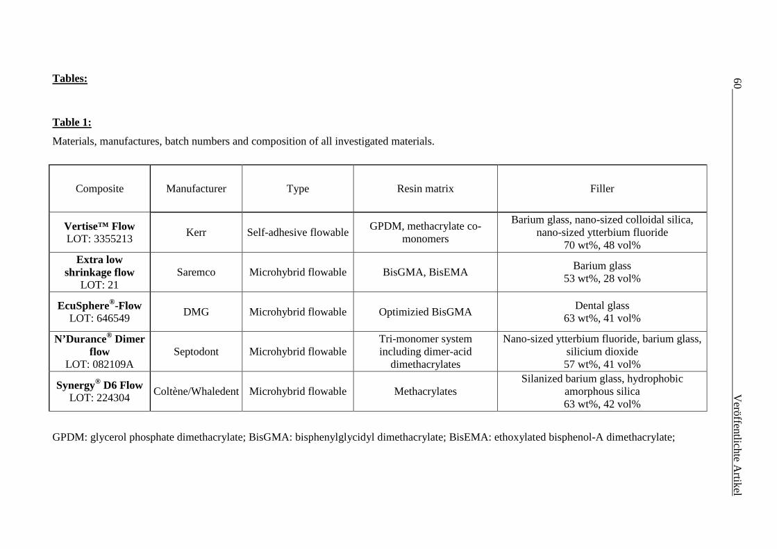
60
V
eröffentlichte Artikel
Tables:
Table 1:
Materials, manufactures, batch numbers and composition of all investigated materials.
GPDM: glycerol phosphate dimethacrylate; BisGMA: bisphenylglycidyl dimethacrylate; BisEMA: ethoxylated bisphenol-A dimethacrylate;
Composite Manufacturer Type Resin matrix Filler
Vertise™ Flow LOT: 3355213
Kerr Self-adhesive flowable GPDM, methacrylate co-monomers
Barium glass, nano-sized colloidal silica, nano-sized ytterbium fluoride
70 wt%, 48 vol% Extra low
shrinkage flow LOT: 21
Saremco Microhybrid flowable BisGMA, BisEMA Barium glass
53 wt%, 28 vol%
EcuSphere®-Flow LOT: 646549 DMG Microhybrid flowable Optimizied BisGMA
Dental glass 63 wt%, 41 vol%
N’Durance® Dimer flow
LOT: 082109A Septodont Microhybrid flowable
Tri-monomer system including dimer-acid
dimethacrylates
Nano-sized ytterbium fluoride, barium glass, silicium dioxide 57 wt%, 41 vol%
Synergy® D6 Flow LOT: 224304
Coltène/Whaledent Microhybrid flowable Methacrylates Silanized barium glass, hydrophobic
amorphous silica 63 wt%, 42 vol%
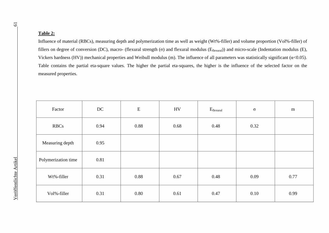
Ver
öffe
ntlic
hte
Art
ikel
61
Table 2:
Influence of material (RBCs), measuring depth and polymerization time as well as weight (Wt%-filler) and volume proportion (Vol%-filler) of
fillers on degree of conversion (DC), macro- (flexural strength (σ) and flexural modulus (Eflexural)) and micro-scale (Indentation modulus (E),
Vickers hardness (HV)) mechanical properties and Weibull modulus (m). The influence of all parameters was statistically significant (α<0.05).
Table contains the partial eta-square values. The higher the partial eta-squares, the higher is the influence of the selected factor on the
measured properties.
Factor DC E HV Eflexural σ m
RBCs 0.94 0.88 0.68 0.48 0.32
Measuring depth 0.95
Polymerization time 0.81
Wt%-filler 0.31 0.88 0.67 0.48 0.09 0.77
Vol%-filler 0.31 0.80 0.61 0.47 0.10 0.99
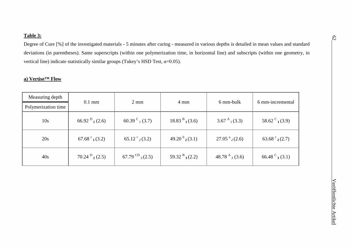
62
V
eröffentlichte Artikel
Table 3:
Degree of Cure [%] of the investigated materials - 5 minutes after curing - measured in various depths is detailed in mean values and standard
deviations (in parentheses). Same superscripts (within one polymerization time, in horizontal line) and subscripts (within one geometry, in
vertical line) indicate statistically similar groups (Tukey’s HSD Test, α=0.05).
a) Vertise™ Flow
Measuring depth 0.1 mm 2 mm 4 mm 6 mm-bulk 6 mm-incremental
Polymerization time
10s 66.92 D 1 (2.6) 60.39 C 1 (3.7) 18.83 B 1 (3.6) 3.67 A
1 (3.3) 58.62 C 1 (3.9)
20s 67.68 c 1 (3.2) 65.12 c 2 (3.2) 49.20 b
2 (3.1) 27.05 a 2 (2.6) 63.68 c 2 (2.7)
40s 70.24 D 2 (2.5) 67.79 CD 3 (2.5) 59.32 B
3 (2.2) 48.78 A 3 (3.6) 66.48 C 3 (3.1)
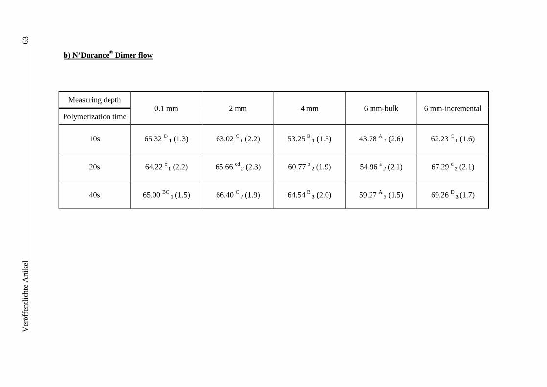
Ver
öffe
ntlic
hte
Art
ikel
63
b) N’Durance® Dimer flow
Measuring depth 0.1 mm 2 mm 4 mm 6 mm-bulk 6 mm-incremental
Polymerization time
10s 65.32 D 1 (1.3) 63.02 C 1 (2.2) 53.25 B
1 (1.5) 43.78 A 1 (2.6) 62.23 C 1 (1.6)
20s 64.22 c 1 (2.2) 65.66 cd 2 (2.3) 60.77 b
2 (1.9) 54.96 a 2 (2.1) 67.29 d 2 (2.1)
40s 65.00 BC 1 (1.5) 66.40 C
2 (1.9) 64.54 B 3 (2.0) 59.27 A
3 (1.5) 69.26 D 3 (1.7)

64
V
eröffentlichte Artikel
c) EcuSphere®-Flow
Measuring depth 0.1 mm 2 mm 4 mm 6 mm-bulk 6 mm-incremental
Polymerization time
10s 62.09 D 1 (1.1) 58.00 C 1 (1.7) 36.86 B 1 (3.0) 21.77 A 1 (2.5) 58.70 C 1 (2.5)
20s 64.72 d 2 (2.9) 62.43 c 2 (1.3) 54.65 b
2 (2.5) 42.70 a 2 (2.9) 62.76 cd
2 (2.2)
40s 65.69 D 2 (1.4) 65.21 C
3 (2.1) 59.06 B 3 (1.2) 52.73 A 3 (2.4) 63.68 CD
2 (1.3)
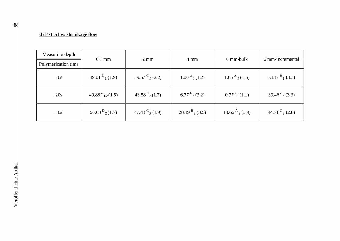
Ver
öffe
ntlic
hte
Art
ikel
65
d) Extra low shrinkage flow
Measuring depth
0.1 mm 2 mm 4 mm 6 mm-bulk 6 mm-incremental Polymerization time
10s 49.01 D 1 (1.9) 39.57 C 1 (2.2) 1.00 A
1 (1.2) 1.65 A 1 (1.6) 33.17 B 1 (3.3)
20s 49.88 e 1,2 (1.5) 43.58 d
2 (1.7) 6.77 b 2 (3.2) 0.77 a
1 (1.1) 39.46 c 2 (3.3)
40s 50.63 D 2 (1.7) 47.43 C
3 (1.9) 28.19 B 3 (3.5) 13.66 A
2 (3.9) 44.71 C 3 (2.8)
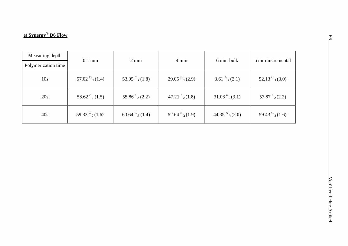
66
V
eröffentlichte Artikel
e) Synergy® D6 Flow
Measuring depth
0.1 mm 2 mm 4 mm 6 mm-bulk 6 mm-incremental Polymerization time
10s 57.02 D 1 (1.4) 53.05 C
1 (1.8) 29.05 B 1 (2.9) 3.61 A 1 (2.1) 52.13 C
1 (3.0)
20s 58.62 c 2 (1.5) 55.86 c
2 (2.2) 47.21 b 2 (1.8) 31.03 a
2 (3.1) 57.87 c 2 (2.2)
40s 59.33 C 2 (1.62 60.64 C
3 (1.4) 52.64 B 3 (1.9) 44.35 A
3 (2.0) 59.43 C 2 (1.6)
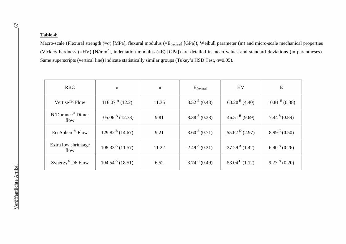
Ver
öffe
ntlic
hte
Art
ikel
67
Table 4:
Macro-scale (Flexural strength (=σ) [MPa], flexural modulus (=Eflexural) [GPa]), Weibull parameter (m) and micro-scale mechanical properties
(Vickers hardness (=HV) [N/mm2], indentation modulus (=E) [GPa]) are detailed in mean values and standard deviations (in parentheses).
Same superscripts (vertical line) indicate statistically similar groups (Tukey’s HSD Test, α=0.05).
RBC σ m Eflexural HV E
Vertise™ Flow 116.07 A (12.2) 11.35 3.52 B (0.43) 60.20 E (4.40) 10.81 E (0.38)
N’Durance® Dimer flow
105.06 A (12.33) 9.81 3.38 B (0.33) 46.51 B (9.69) 7.44 B (0.89)
EcuSphere®-Flow 129.82 B (14.67) 9.21 3.60 B (0.71) 55.62 D (2.97) 8.99 C (0.50)
Extra low shrinkage flow 108.33 A (11.57) 11.22 2.49 A (0.31) 37.29 A (1.42) 6.90 A (0.26)
Synergy® D6 Flow 104.54 A (18.51) 6.52 3.74 B (0.49) 53.04 C (1.12) 9.27 D (0.20)
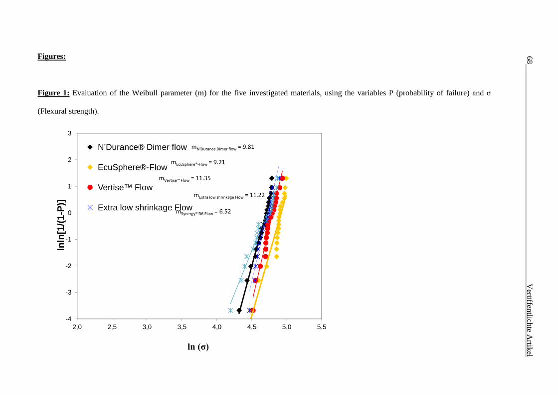
68
V
eröffentlichte Artikel
Figures:
Figure 1: Evaluation of the Weibull parameter (m) for the five investigated materials, using the variables P (probability of failure) and σ
(Flexural strength).
-4
-3
-2
-1
0
1
2
3
2,0 2,5 3,0 3,5 4,0 4,5 5,0 5,5
lnln
[1/(
1-P
)]
N’Durance® Dimer flow
EcuSphere®-Flow
Vertise™ Flow
Extra low shrinkage Flow
mN'Durance Dimer flow = 9.81
mEcuSphere®-Flow = 9.21
mVertise™ Flow = 11.35
mExtra low shrinkage Flow = 11.22
mSynergy® D6 Flow = 6.52
ln (σ)
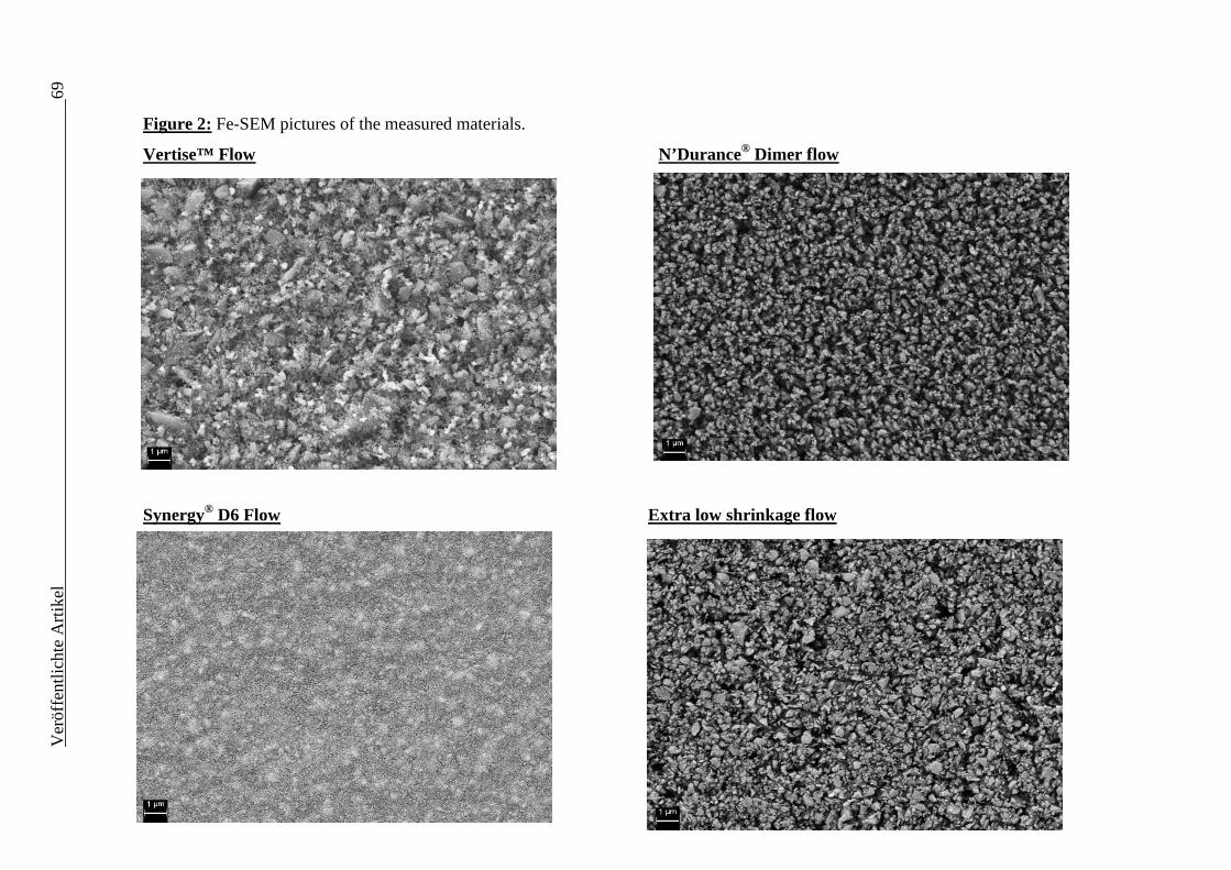
Ver
öffe
ntlic
hte
Art
ikel
69
Figure 2: Fe-SEM pictures of the measured materials.
Vertise™ Flow
N’Durance® Dimer flow
Synergy® D6 Flow Extra low shrinkage flow
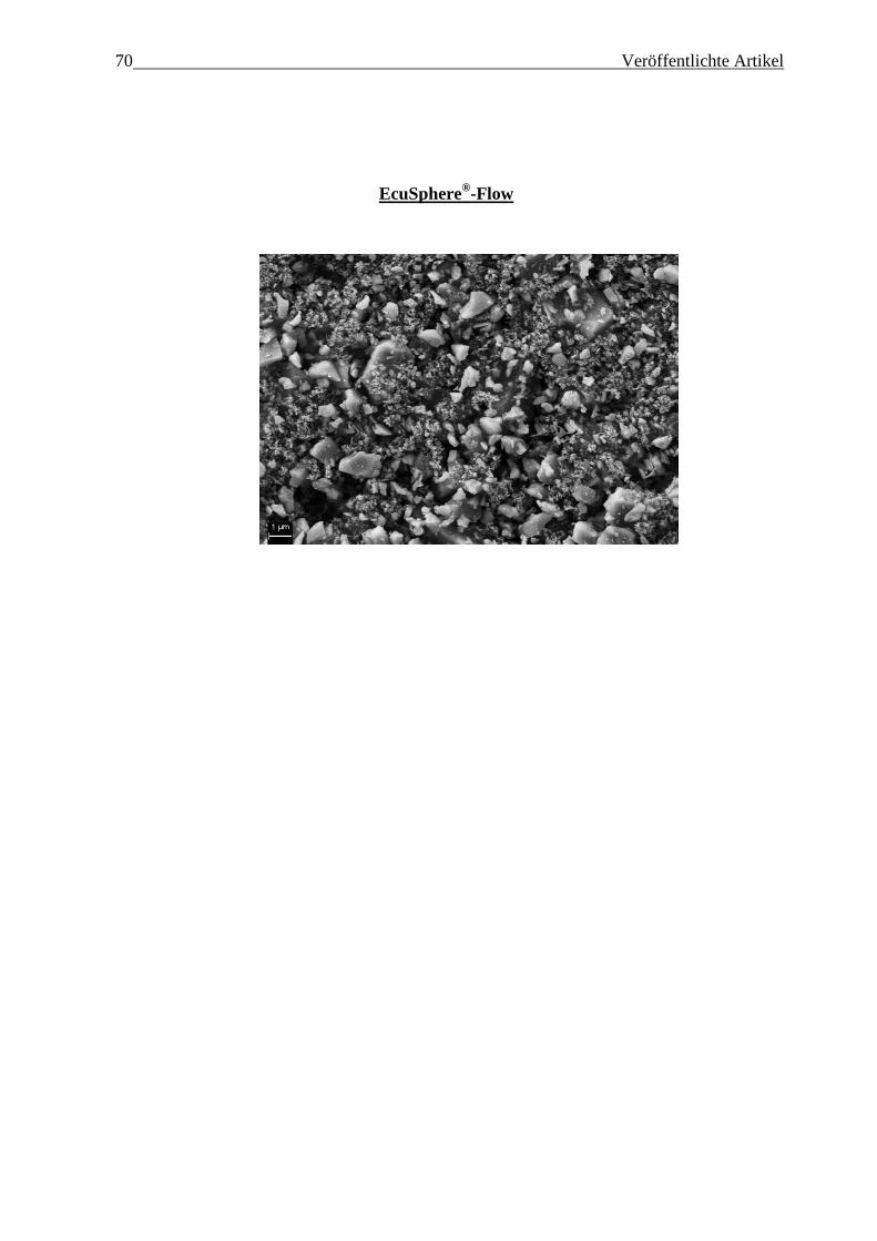
70 Veröffentlichte Artikel
EcuSphere®-Flow
Veröffentlichte Artikel 71
2.2.1 Deutsche Zusammenfassung
Ziel der Untersuchung war es ein selbstadhäsives fließfähiges Komposit mit zwei
niedrigschrumpfenden Kompositen und zwei Mikrohybridkompositen bezüglich ihrer
Konversionsrate (KR), ihrer mikromechanischen Eigenschaften (Eindringmodul (E),
Vickershärte (HV)) sowie ihrer makromechanischen Eigenschaften (Biegefestigkeit (σ),
Biegemodul (EBiege) zu vergleichen.
Die Arbeitshypothesen waren:
(a) Es gibt keinen signifikanten Unterschied bezüglich der Konversionsrate zwischen
den fünf untersuchten Materialien zu jeder Belichtungszeit und Inkrementdicke.
(b) Es gibt keinen signifikanten Unterschied zwischen den fünf untersuchten
Materialien bezüglich makromechanischer (Biegefestigkeit (σ), Biegemodul (EBiege))
und mikromechanischer (Vickershärte (HV), Eindringmodul (E))) Eigenschaften bei
einer klinisch praktikablen Belichtungszeit von 20s.
Die KR (n=6) wurde durch eine Fourier-Transformations-Infrarotspektrometrie (FTIR)
in klinisch relevanten Inkrementdicken (0,1 mm, 2 mm, 4 mm, 6 mm-Bulk (1
Inkrement), 6 mm-Inkrement (geschichtet in dreimal 2mm dicken Inkrementen) und
Belichtungszeiten (10s, 20s, 40s) bestimmt. Mikromechanische und makromechanische
Eigenschaften wurden von einem Universalhärtemessgerät und einer Drei-Punkt-
Biegeversuch Anordnung gemessen, nachdem die Prüfkörper 24 Stunden lang bei 37°C
in destilliertem Wasser gelagert wurden. Ein Rasterelektronenmikroskop diente zur
Visualisierung der Füllkörper. Die Ergebnisse wurden mittels univariater
Varianzanalyse, Tukey’s honest significance post-hoc-Test, einer multivariaten
Varianzanalyse (α=0,05) und einer Korrelation nach Pearson ausgewertet. Eine
Verlässlichkeitsbestimmung wurde für σ mittels Weibullanalyse (m-Parameter)
durchgeführt.
N’Durance® Dimer flow (65,66%) zeigte die höchsten KR-Werte (bei 2 mm
Inkrementdicke, 20s Belichtungszeit). Bezüglich der makromechanischen Eigenschaften
zeigten EcuSphere®-Flow (129,82 MPa) für die Biegefestigkeit und Synergy® D6 Flow
(3,74 GPa) für den Biegemodul die höchsten Werte. Die besten mikromechanischen
Eigenschaften wurden für das selbstadhäsive Komposit (Vertise™ Flow; E= 10,81 GPa;
HV= 60,20 N/mm2) gemessen. Die Verlässlichkeit wurde stark von Füllkörpergewicht
(η2= 0,77) und Füllkörpergröße (η2= 0,99) beeinflusst. Außerdem zeigte sich ein starker
72 Veröffentlichte Artikel Zusammenhang zwischen dem Kompositmaterial und den untersuchten
Materialeigenschaften.
Mit Rücksicht auf die Limitationen dieser Studie zeigte das selbstadhäsive Komposit
(Vertise™ Flow) die besten KR-Werte nach 5 Minuten Polymerisation (zusammen mit
dem Dimersäure- basierten Komposit) sowie die besten mikromechanischen
Eigenschaften. Die Mikrohybridkomposite zeigten die besten makromechanischen
Eigenschaften: EcuSphere®-Flow für die Biegefestigkeit und Synergy® D6 Flow für den
Biegemodul. Aus den genannten Gründen wurden demnach beide Arbeitshypothesen
widerlegt. Für das selbstadhäsive Komposit scheinen 40s Belichtungszeit und eine
Schichtung in maximal 2 mm dicken Inkrementen zur Erzielung guter mechanischer
Eigenschaften nötig zu sein.
Veröffentlichte Artikel 73
2.2.2 English summary
The aim of the study was to compare a self-adhesive with two low shrinkage and two
regular flowable resin-based composites (RBCs) in terms of degree of conversion (DC)
and mechanical properties measured at microscopic (Indentation modulus (E), Vickers
hardness (HV)) and macroscopic scale (Flexural strength (σ) Flexural modulus
(Eflexural)).
The tested null-hypotheses were that:
(a) There would be no significant difference between the five materials in view of DC at
any measured depth and irradiation time.
(b) There would be no significant difference between the five materials when measuring
mechanical properties at macro- (flexural strength (=σ) and modulus of elasticity
(=Eflexural)) and micro-scale (Vickers hardness (=HV) and indentation modulus (=E))) at
a clinical relevant irradiation time of 20s.
DC (n=6) was investigated by Fourier transform infrared spectroscopy (FTIR) in
clinical relevant filling depths (0.1, 2, and 4 mm; 6 mm-bulk, 6 mm-incremental) and
irradiation times (10s, 20s, 40s). Micro- and macro-scale mechanical properties were
measured by an automatic microhardness indenter and a three-point bending test device
after curing the specimens for 20s and storing them in distilled water for 24h at 37°C.
Fillers were visualized by a field emission scanning electron microscope. Results were
evaluated using one-way ANOVA, Tukey’s HSD post-hoc test, Pearson correlation and
a multi-variate analysis (α =0.05). A Weibull analysis was used to assess σ.
N’Durance® Dimer flow (65.66%) reached the highest DC (at 2 mm depth, 20s
irradiation). At macro-scale EcuSphere®-Flow (129.82 MPa) for σ and Synergy® D6
Flow (3.74 GPa) for Eflexural reached highest values. The highest micro-mechanical
properties were measured for the self-adhesive RBC (Vertise™ Flow; E= 10.81 GPa,
HV= 60.20 N/mm2). Reliability was highly influenced by filler weight (η2= 0.77) and
volume (η2= 0.99) proportion.
A strong influence of the material on all measured properties, as well as a strong
influence of filler proportion (wt%-filler, vol%-filler) on the reliability was statistically
proven.
Within the limitations of our study the self-adhesive RBC (Vertise™ Flow) showed best
performance concerning DC after five minutes of polymerization (together with the
dimer-acid based RBC) and mechanical properties measured at micro-scale. The

74 Veröffentlichte Artikel traditional microhybrid flowable RBCs performed best in the macroscopic mechanical
properties σ (EcuSphere®-Flow) and Eflexural (Synergy® D6 Flow). Therefore both null
hypotheses had to be rejected. For the self-adhesive RBC a curing time of 40s as well as
an incremental placement not exceeding 2 mm thickness appeared to be necessary.
Literaturverzeichnis 75
3 Literaturverzeichnis
1. Ferracane, J.L.: Resin composite--state of the art.
Dent Mater 27, 29-38 (2011)
2. Venus® bulk fill - Technical Information (2011)
http://www.heraeus-
venus.com/en/usa/products_10/venusbulkfill/technicalinformation_2.html
3. Chen, H.Y., Manhart, J., Hickel, R.,Kunzelmann, K.H.: Polymerization
contraction stress in light-cured packable composite resins.
Dent Mater 17, 253-259 (2001)
4. Davidson, C.L., de Gee, A.J.,Feilzer, A.: The competition between the
composite-dentin bond strength and the polymerization contraction stress.
J Dent Res 63, 1396-1399 (1984)
5. Leinfelder, K.F.: Posterior composite resins: the materials and their clinical
performance.
J Am Dent Assoc 126, 663-664, 667-668, 671-662 passim (1995)
6. Carvalho, R.M., Pereira, J.C., Yoshiyama, M.,Pashley, D.H.: A review of
polymerization contraction: the influence of stress development versus stress
relief.
Oper Dent 21, 17-24 (1996)
7. Burgess, J.,Cakir, D.: Comparative properties of low-shrinkage composite
resins.
Compend Contin Educ Dent 31 Spec No 2, 10-15 (2010)
76 Literaturverzeichnis
8. Ilie, N.,Hickel, R.: Investigations on a methacrylate-based flowable composite
based on the SDR technology.
Dent Mater 27, 348-355 (2011)
9. de Biasi, M., Calvi, R.M., Sossi, D., Maglione, M., et al.: Microhardness of a
new flowable composite liner for posterior restorations.
Dental Materials 26, e25-e25 (2010)
10. Ilie, N.,Hickel, R.: Shrinkage behaviour of novel flowable composites based on
the SDR(TM)-technology.
Dental Materials 26, e130-e130 (2010)
11. Vertise Flow - Technical Guide (2010)
http://eu.vertiseflow.com/files/technical_guide_EN.pdf
12. Buonocore, M.G.: A simple method of increasing the adhesion of acrylic filling
materials to enamel surfaces.
J Dent Res 34, 849-853 (1955)
13. Pashley, D.H., Tay, F.R., Breschi, L., Tjäderhane, L., et al.: State of the art etch-
and-rinse adhesives.
Dental Materials 27, 1-16 (2011)
14. Vertise Flow - Technical Bulletin (2011)
http://eu.vertiseflow.com/files/bullettin_vertise_e.pdf
15. Vrochari, A.D., Eliades, G., Hellwig, E.,Wrbas, K.T.: Curing efficiency of four
self-etching, self-adhesive resin cements.
Dent Mater 25, 1104-1108 (2009)
16. Barkmeier, W.W., Erickson, R.L.,Latta, M.A.: Fatigue limits of enamel bonds
with moist and dry techniques.
Dent Mater 25, 1527-1531 (2009)
Literaturverzeichnis 77
17. Hansen, E.K.,Asmussen, E.: Correlation between depth of cure and surface
hardness of a light-activated resin.
Scand J Dent Res 101, 62-64 (1993)
18. Ernest, C.P., Holzmeier, M.,Willershausen, B.: In vitro shear bond strength of
self-etching adhesives in comparison to 4th and 5th generation adhesives.
J Adhes Dent 6, 293-299 (2004)
19. Frankenberger, R., Lohbauer, U., Roggendorf, M.J., Naumann, M., et al.:
Selective enamel etching reconsidered: better than etch-and-rinse and self-etch?
J Adhes Dent 10, 339-344 (2008)
20. Inoue, S., Vargas, M.A., Abe, Y., Yoshida, Y., et al.: Microtensile bond strength
of eleven contemporary adhesives to enamel.
Am J Dent 16, 329-334 (2003)
21. Yazici, A.R., Celik, C., Ozgunaltay, G.,Dayangac, B.: Bond strength of different
adhesive systems to dental hard tissues.
Oper Dent 32, 166-172 (2007)
22. Tay, F.R., Pashley, D.H., Garcia-Godoy, F.,Yiu, C.K.: Single-step, self-etch
adhesives behave as permeable membranes after polymerization. Part II. Silver
tracer penetration evidence.
Am J Dent 17, 315-322 (2004)
23. Tay, F.R., Pashley, D.H., Suh, B.I., Carvalho, R.M., et al.: Single-step adhesives
are permeable membranes.
J Dent 30, 371-382 (2002)
24. Ellakwa, A., Cho, N.,Lee, I.B.: The effect of resin matrix composition on the
polymerization shrinkage and rheological properties of experimental dental
composites.
Dent Mater 23, 1229-1235 (2007)
78 Literaturverzeichnis
25. Rebstein/Switzerland, S.D.A.: Instructions for use - SAREMCO Dental
Restorative System - Package Insert (updated version). (12/2011)
26. Boaro, L.C., Goncalves, F., Guimaraes, T.C., Ferracane, J.L., et al.:
Polymerization stress, shrinkage and elastic modulus of current low-shrinkage
restorative composites.
Dent Mater 26, 1144-1150 (2010)
27. N'Durance® Dimer Flow - Instructions for use (2011)
http://www.septodontusa.com/sites/default/files/INST560-C-
N%27Durance%20Dimer%20Flow.pdf
28. Trujillo-Lemon, M., Ge, J., Lu, H., Tanaka, J., et al.: Dimethacrylate derivatives
of dimer acid.
Journal of Polymer Science Part A: Polymer Chemistry 44, 3921-3929 (2006)
29. Frauscher, K.E.,Ilie, N.: Depth of cure and mechanical properties of nano-hybrid
resin-based composites with novel and conventional matrix formulation.
Clin Oral Investig (2011)


![Aus der Poliklinik für Zahnerhaltung und Präventive ... · sie 1996 in aktualisierter Form veröffentlichte [6]. Danach wird die TMD als Überbegriff für Funktionsstörungen des](https://img.pdfslide.org/doc/110x75/60399bfacc8f9142ae3912a3/aus-der-poliklinik-fr-zahnerhaltung-und-prventive-sie-1996-in-aktualisierter.jpg)
![Aus der Poliklinik der Zahnerhaltung und Parodontologie ... · kanalaufbereitung wurden durch eine Umfrage in Nordjordan untersucht [Al Omari 2004]. Eine vergleichbare Untersuchung](https://img.pdfslide.org/doc/110x75/5d4a079088c993e6068bbe23/aus-der-poliklinik-der-zahnerhaltung-und-parodontologie-kanalaufbereitung.jpg)