Embed Size (px)
Citation preview

TTATATATTTAGGTACTTCAATGTTGAGTGGATATATGTTTACTATTGTTTTATTCTTTTGATGAATTGACCCTGTTATCATTATATAATGACATTATCTCTTATGACAGATTTGATTCAAAGTCTATTTTTTCTGACATAAATATTGCTTACCCATTCTCTCTTTGTTTTCATTTGCATGGAATATTTTTTATTTCTCTTCACTTGCAGTCAATATGTCTTCTTAAGGCTAAATTGAGTCCCCAGCAGGAATCATACTGTTTGATCTTGTTTTTTATTATCCATTCAGCCATGCTGTGTCCTTTGACTAGAGCATTTAATCCATTACATTTAAAGAAATTATTGATAGGTAAGAATTTACTATTGATATTATTTTCAATTGTTTTCTAATTGTTTTGTAGTTCCTTATTTCCTCTCTTTCTGATTTGCTTTGTGATTGGTTGATTTTCTGTAGTGGTATGCTTTTTTTTTTTTTCTTTAAGTTCTGGGATACATGTGCAGAACACGCAGGTTTGTTACATAGGTACACATGTGCCATGGTGGTTTGCTCCACCTATCAACCCGTCATCTAGGTTTTAAGCCCCGCATGCATTAGGTATTTGATTTCTTTTTCGTCATGTTTTGTGCACTGACTAGAGGTTTTTTGTGGTTACCATGAATCTTATATAAAAATCTTATCATTATAACATTCTATTTTAAAGTGATAACTTCAATCACATACAAAAATTATACTTTACTTTTCTCAAACAATAATTTTATGTGATTGACATCACACTTTATATCCTTTTATATTATGTCTACATTAACAAATTATTGTAGCTATAGTTATTTCTATTATATATGTCTTTTAAATTTAATAGTAGAGTTAAAAGAGATTAAATACAACCACTACTATATTAGTGCATTCTGAATTTGACTATATATCTATCTTTACCTGTGTATATCTGTGTATATATACTTTCATACATTTTCATGTTGCTGGTTTTTGTCTTTTTGTTTCAACTTGAAGAACCTCCTTTAGCATTTATTGTAAGGTAGGTCTTATGGTGATGAATTCCCACATTTTTTTTGTCTGGGAATGTCTTTATCCTTTCTTACTTCTGAAGGACAGTTTTTCGTAATATTGTTTCATAGTTATTTTTTCTTTCAGCATTTTGAATATATAATCTCAGTGCCTCCTGGTCTATAAAATTTCCTCTGAGAAGTCCATTGATAGACTCATTGGGGTGCCCTCATATGTGAAGAGACTCTTTTACTAGTTTCAGGATTTCTCTTTGTCTTTAACTTTTGAGAATTTCATTATAATATGTCTTGGGATAATTTTGATTTTAACTTATTGTCATCCTTTGAGCTTCATAAGTCTGGATGTGCATATCTTTCACCTAATTTTGGAAGTTTTCAGTCATTTCTCTAAATAAGCTGCCTTTTTTTTTTTCTTTTTTTTTTAAAGACATACGTAACTCACTCTGTCACCCAGGCTGGAGTGCAGTGGCACATTGATAGTTCACTGCAACCTTGATCTCCTGGAGTCAAGAGATCATCCTACCTCGCTTCCTGAATAGTGAGGACTACAGGTATGTATCATCACACCTGGCTGATTTTTTTTTTTTTTTTTTTTTTGGAAGATGGTGGTCTTAGTATGTTGCCCAGGCTGGTTTTAAATTCCTATCCTCAAGCAAACCATCTGCTTTGCCTTCCCAAAGCACTGGGATGACAGACATGAGCCACTGAGCCCAGCCCTCTAAATAAGCTTTCTGTCTCATTTTCCCTTTCTTCTCATTCTAGAAATCCTATAATTCATATATTTCTATGCTTGATGGTGTCCCATAGGTCCCTTATGCTTTCTTCAATCTTTTATTTTATTCCTCTAATTGGCTAATTTCAAATGACTAATCTTTGAGTTCCCTAATTCTGTTTTATAATTGAGTCTATCAAATTTTGCAGTTCTATCATTATGTTCTTCGGCTCCAGGATTTTTGTTTGCTTCCTTTTTATGGATTCCATTTCTTTGTTAAACTTCTCATTTTGTTCATGCATTGCTTTCCTAATTTTGTTTCGTTTTCAGTATGTGTTCTCTTGAATCTCATTGAGCTTCAAGATGATTTTTTTGTCAGGCAATTTGTAGATCTCTATTTCTATAGGATTGATTACTGGAGCCTTTCTAGTTTCATTTGGTTGTTTTATTGTCCATAAAGCCTCACATTATTGCCTGTGCAACTAAAGGAGCAAACACCTCTTTCAGTTTTTATAAGCTGGTTTTAGCATATAAAGACTTTCTCTTTTGGAGTCCTTGCAAAGACATGACTACCTTCAGGATCACAGATGAATGGGGTTGAAGCCAGGTCACCTAACTGCTGCCGGGTTTGCAGTGGAGCCTGTAGTTGGCAGATCTATTACCAAGTGCTTGAATGGGCATGGATTCTATCTAGTCTCTTGGTGCACTTAACTGCCTCCACAGCCTTGGTCAGTAGGGGTTGCACCTGGACCAGCGTCTTATTCAGTGTCTGCCAACAGAAAGACTGTTACCAAATATACAGATGCTCAGATGGGTATCACTTCCTCTGGGTTTCGGGACGGCTTCCTGCTGTGTCACTGGTTAAATACCTGGGCAGGCAATACTGGCCCATGACTGCAACTGAGCTAGAATGGAGTTCTAGCTGGGAAAGAACTGAGTTACAGGGTTCTGTCAATTTCCACAGCTGAGACTCATGTCTGTAGGGTTGCCCCTAGGGGCACAGATGGATGTCTCTGTTTTCAGGCTCAACTCTTAATTAAAAGTTACTTCCAAAATTCAAAAAATCCATTAAAAACCAGGCTTTCTGGGCTGCTTTCAGACTGCAGCTGACAAGGACTGGAGCCAGGTTCCATCTGAGGATCTGTTGTGAGACTGAGTTCAGCTGTTCTTCCAGTTGGGGGACAGATGTGTGTTTCTCTCCCTCAGTCCCTGGACAGGCACAACTGCTCTCAGACCAGAACTGAGTGAGGTTAGAACTGAGTTACAAGGCTGTTTCAGGGTGCATAGCAGGTACCGAGGCCAGCAGGCCTGCTATCCAAGGCATGAGTAGGCATGAATGCTGCTGGGTTCCTTGTCAGATGGTTCTGGCAATAGGACCCATGCCAAATGGAAATGAACTCAATCCACAAGGGAATGGAGATATTTCGGGGTTTAGAGGCGGGACCACGGTTGGCAAGTCTGCAACCTGGGAATACTTCTGCCCTCTCAAATCAACCTTCCTAGGTCTTGGGTAGCAACAAGTTTTAGTAACTTCCTGCCTGAATCCCAAAGCTCCCACAAAGGCACTTTTGTTCGTGAATGGCTGCAAAACCAGTGTTTCTATGGAAAGAATATGAGCCAGAAGAACTCCTATTCTACCATCTTGCTGATGTCACTCTAGAAATAATCATTACTGTGCTCACTAACACTGTTCCCTTTTCAGTTTCTTGACCTCCATAAATTTTCTGTCTCTTTTGAACTCTGTAAATAAAATTATTAATTCATTATATATATATCACTATTCTGTATTTAATAGCCTCCCATTTTCTGAGATTAATTTCTCACATTATTTTTATTGACCTGGAGACTATGAATTAACAAAATGTTCAAAATAAAAGTTTAAAGAAAATTAGCACGCATATTTTCTTTTTTTTCTTTATCTTTCTAGAGATGTGTAATTCCTACATTACACAAAGAGAATTTGTAGGAGAAACCAGGAAAGGAAAACAGGAAGGAAAAGACTTTTCTGATAAATACATGGTTTCATTTTCCTCTCCCTTCTTTGATGCAGAAGAGACCTTGATGTGTCCAAGAGTATATGAGGAGGTTAGATGTGCAGTCTCATTGACTGGAGAAGTGTCAGGAAGGAGGGGTTTATTTTTGCTTAGCTTTGCCCTGTATTCATTTTTCTTGTTGCATAAGCTTCTTATCGACATTAATTTTAGACTCCCAAGATGTTTTGCATAACATAGAATTATAATCTAGTGTCTAAAATAGTTGCAAACCATAGTTTCAAATACATTAGGAAGATGAATCATTTCCTTAACATGAACCACTGTGTTATTTGAAATGATTACTTACAAGGGAGAAGTGATACATAATTAAAGTATCATGTGACATACAAAAAAGAAATCAATGAAATTCAAACAATAAATGCTTCTTCTGTTTCTCGTGAAAGATAGATGAAATATGCAGCTCCTTCTCATATCCATTTTGAAATGAATGGGTCTTGAATACCATAACTATGTTATTTCAGTAGTAAGTAGAAATATTTCAGTATCAGAAGGGAAGAAATGAAATGAAATCAATCTACATCACTTTGGATTTTTAACTCCTCTAAAAACGTCTTACTGGGTATACATTATTGTTGTCAAATCCATTTTAATTTGAATTTTACTGTGTTTGTGTATGTGTATGCATGCACTTACTTTTGTTTTTAACTCTCTTAAATAGCTTCAAAATGAAAGTTTTGTAACCAAATTTGAGCAGCAAAGAAAAAGGAGAAAGGGATCAAATATCTCTAACATATTCTACTTCATACAGTTCTTGGGTTTCTTTTTGCCAAGCTTTCACTCATAGCAGCTACACCAGTACCATCATGAATACTAATGAAATGTAATAGAAGGCATCAGTCATGATCCATCCAGGCTAGGGACATAACCATATACAAAGTGATAGTTCTTCCAGCTTAATGAAGCCTTCTTAAAGAAAAACTGTTTACATTCAAATTTGGATAAGCTATGAGAGCTTGTAAGCTATGAGAGCTAGACTGTACAGTTTTTAGGGGCAGGCATTGGTACAGGGAAACTCTATTATCTTCTTTATTTTCCTTCCAAAATTGTGCCTCATCAAAGTCCTGGGCATAAAATGTTTACTGAACAAAGTTTCAAAGAAATGCCATAGGAAATAAAGCTTAAAACTGTAGAAATCGAAAGTAAAAGATTTTAAACAGATAGACAACAGTGTTTAGATAAGCAAATACTTTTTCTGCAATCCTTAAGGTTTGCTGCCAACCTATGGAGTTCAAATTAACATTTCTCTCAGAAGTAAGCCTCATCTTTCTACTATCTTTTTAGACTATGTTTCTACATTCTATATATTCCTCCTTTCCAATAACAAGTCTCAGGAGTGGTTTTGGAACTCACTGATTTTTGGATCAAGCTAATATAGGATGGCATTAATGTAAAGTAATGCTATTACTCAAATATCAGGGATACTATCGTGACAGCTATATCCCTGGAATCACTGAATAAGCTTACAAAACTTACTCTGCAAGAAGCTCCTGCTGAAACTTGAAAAGCATGTCAACAGAGGCTCCAAATGACAGAAAATTGCAATTTGTTATAACATTAAAAGAGAACTTATAACTTATTCTGACATATAATACTTCCCATAACCTGGTCAGGCCTCTTTTATTATTCAAGGTTTTCTAAAACCTCACTCTCATTATGAAGCTTTTCCAGACTCACTGCAAATAAAATTATCAGAGAAGAGACACATTCATATCTTACATGGCAATGTACTTGGCCACGAGTGCAAAGGTGCTTTGGCCTTGTATAAATTTAGTTACTAAATTTGTTCCACATGCATGTAAGTTTTTGTTTAATTTTATTTTGTTTTCCTTACCATATATGATTTAAATTATGAACTTCTACAGTCAAAATAATTTTAACTAAATTTTTATATCTATCTTTGGGGAGGGAGTACAAAGAAGTATACTAGTCAAATAATGTTGCAATATTGCTGCATGACAAATAGCCAGAAAATCTCAGTGGTACCCAACTATGAGGATCTTATCTCACTCAGTCCAAATGTCAGCTAGCATGGTGCCACCTCAGCATATGCATCTTCAGAGTTGCTGAATTTTGTTTCTCCTGGTTCATGCTGGACCTAAGGCTGAAGAAACAGTAGCTACCTGGGGTACCTTCTTCTTATAGAGGAGATATGAAGGTCCCAGAGAGTGCAAGCCAAACTGTGTGATGTCTCTTAAGATCTATGCTTAATATTTGATCCCTACTGCATTCCTTCTGCACGTCCTACTGTAAAATCATGTCCCTTGACCTAACACAATTTCTATGGATTAGAGACATGTACTATTGACATGGAATGGGAAGATCACAAGAGGTGAATATACCCTGATAAATATTCTAAATATACCATAGTGTACCCTCTTATTTAAAAATGTTCACATCTCTGGTCGGGTGAGGTGGCTCACATCTGTAATCCCAGCACTTTGGGAGGCCGACGCAGGCAGATCACAAGGTCAGGGGATCGAGACCACCTTGGCCAACATGGTGAAACCCCATCTTTACTAAAATACAAAAAATTAACCGGGTGTGGTGGTGGGTGCTTGTAATCCCAGCTACTTGGGAGGCTGAGGCAGGGGTATCACGTGAACCCAGGAGGGAGAGTTTGCAGTGAGCTGAGATCGCATCACTGCACTCCAGCCTAGCAACAGAGCAAGACTCCATTATTAATAATAATTAATTTATTAATTCATGTAAAACATAGAAAATGTGCAGCCATATAGGCTTATTTGCCTTCTTTTCCAGTCTTCTATGCTATAATTTTCCAGTCTTTTATGCTATAATTGTCATATGTATTACACATACATAAATTAAAATATATTATAATTTTTACATTAAAAAACTATATGTAAACACAATAAACAAAATAATCAAACAAAATAAAAAATTTGTCTTCTATGTTTACTCATATATCTTTCATTTCTAATCCTCCATATTTCTTCCTAAAATTCCATTTCTCCATCTGGTATCATTTTCCTTCAACCTGCAAGACTTTCTTGGTTTTGGCTTACCTGAAAATGGCTTTATTTTGCCTATGTTATTAAACAATGTTTCTGAATTTTGAAATTAACCCTTTATTTCTTGAATAATTAGAGATGGGGAAGTCTTCTGGTAGGGTAGTTTTGAGGGAATAAATCAGGAGATTGATGTCGGGCATACTGAATTCAAGATACTAAAACCTCCAAGAAGATACATAAACCTGGTGTTTGAGAAAACAGTCAGAATTGGACATAAAGAATTATGGGTTGTCAACATATATTACAGATAGTATTTAGAGCTATGAGATGATAGGACTCACATCTAGGACTATCATCAAGGGAGTGAGTGTAGTTAATGAAGTGAAGGAGGCTCTGAACTGTGTCTTAGAGCACTCCAACAATGTGAAGCTAGAGAAGAGGAGGAAACAGCAACAGAAAGTGAGGAGCAACTAATGAGTTAGGAGAAAACAAACCGTAGTGTATGGTTTTCTACAAGCTATATAAATAATGAAAATGAAGAAGGAAAAAACAATAATATCAAGGGCTACAGACTGGAAAGATTGGGACAGAAAATTAACCATTAGAATTAATTGAACGCAGGTCACCGGCAACCTTGAAGTTTTGGTGAACTGGTGGAAGTAAAAGTGTGATTGGAGTGGGTCATTAATTTTTAATAATGACAGTAGTGAATAGGTAAACATCCTATAGTGGTCACAAGAACATAATTGTGAATATAAATAACATTACATTCTTATTTATAACATTGTTTTATGATTTTCACATTATCCTGTTGGATTTATACCCAATAAGCAACCACTACTTTTTTGAGAACTGCCCTCTACCCTAGCCCCTGAAAATATATTATATGAAAATTCTCTCCCAGCTCTAATTGGTTTAACAAAATATATGACCCAACCCA
www. i -med . ac . a t / b i ocen t e rREP
OR
T 2
010


Adele Loidl | Alexander Hüttenhofer | Alexander Pauck | Alexandra Lusser | Alexandra Pipal | Anna Moussavi | Andrea Casari | Andrea Eigentler | Andreas Ploner | Andreas Villunger | Anette Zeilner | Angela Klein-Wondrak | Anita Kofler | Anna Chirkova | Anna Moussavi | Anne Krogsdam Anto Nogalo | Arno Helmberg | Astrid Devich | Ayten Yigit | Barbara Gschirr | Barbara Meissner | Barbara Stoiber | Barbara Wolf | Beate Abt | Beatrix Fürst | Beatriz Campo-Fernández | Benedicte Sohm | Benedikt Koller | Bernhard Halfinger | Bastian Bäumer | Bernhard Redl | Bernhard Rieder | Bettina Sarg | Bettina Thauerer | Bettina Unterberger | Birgit Faber | Brigitte Andrä | Cecilia Grundtmann | Christian Ploner | Christian Eller | Christina Mayerl | Christina Weinl | Christine Bandtlow | Christine Mantinger | Christoph Dohmesen | Cicek Aydemir | Claudia Manzl | Claudia Ram | Claudia Soratroi | Claudia Wöss | Constanze Nandy | Cornelia Thoeni | Cornelia Wandke | Daniel Bindreither | Denise Tischner | David Teis | Diana Hilber | Dietmar Fuchs | Dietmar Rieder | Divyavaradhi Varadarajan | Domagoj Cikes | Dorata Garczarczyk | Doris Bratschun | Doris Hinger | Edith Hofer | Elena Ledjeff | Ernst R. Werner | Evelyn Rabensteiner | Fabio Gsaller | Fatma Dikmen | Florentine Marx | Florian Baumgartner | Florian Bock | Florian Kern | Florian Schwarze | Florian Überall | Francesca Grespi | Franziska Gobber | Friederike Finsterbusch | Gabriele Baier-Bitterlich | Gabriele Scheran | Gabriele Stöckl | Gabriele Werner-Felmayer | Georg Golderer | Georg Nikolaidis | Georg Wick | Gerald Brosch | Gerhard Krumschnabel | Gernot Stocker | Gertrude Huber | Giridhar Shivalingaiah | Günther Böck | Giovanni Almanzar-Reiner | Glory Ranchez | Hans Grunicke | Heidelinde Jäkel | Hans Grunicke | Heribert Talasz | Helene Heiss | Hermann Krabichler | Hildegard Wörle | Hubert Hackl | Hubertus Haas | Igea Contarini | Ilenia Bertipaglia | Ilja Vietor | Ilona Lengenfelder | Ines Jaklitsch | Ines Peschel | Ingo Bauer | Irene Gaggl | Irina Berger | Ivan Prokudin | Jan Mrazek | Jan Wiegers | Jennifer Gebetsberger | Johann Hofmann | Johanna Gostner | Johanna Sebald | Johannes Rainer | Jonathan Vosper | Julia Scheffler | Julianna Leuenberger | Julien Lelong | Kalina Duszcka | Kamilla Bakowska-Zywicka | Karin Ecker | Karin Lentsch | Karin Schluifer | Karl Maly | Karoline Hörtnagel | Katharina Fegerl | Katherin Patsch | Kathrin Gotsch | Kathrin Rossi | Katja Jacob | Katrin Watschinger | Kire Trivodaliev | Konstantinia Skreka | Krista Trappl | Lára Hannesdottir | Leopold Kremser | Levent Kaya | Linda Teufel | Lisa Kindler-Maly | Ludger Hengst | Lukas A. Huber | Lukas Peintner | Lukas Sattler | Magan Ansbro | Manuel Kaufmann | Manuel Alonso Y Adell | Manuela Villunger-Gfreiner | Marek Zywicki | Maren Fischer | Maria Fischer | Maria Goralik-Schramel | Maria-Laura Fluckinger | Mariana Eca Guimaraes de Araujo | Maria Saurer | Marietta Brunner | Marin Barisic | Mario Gründlinger | Markus Keller | Markus Schrettl | Markus Unterwurzacher | Martin Eisendle | Martin Müller | Martin Offterdinger | Martin Tribus | Martina Barisic | Martina Brunner | Mathieu Rederstorff | Matthias Erlacher | Mattias Carlsson | Melanie Amort | Melanie Heymann | Melanie Hofer | Melanie Lukasser | Melanie Ramberger | Michael Blatzer | Michael Keith Kullmann | Michael Rittinger | Michaela Pfister | Michela Carlet | Mihaela Angelova | Miriam Alber | Monika Hertscher | Muhammad Saeed | Nadine Plank | Nadja Haas | Natalia Schiefermeier | Nikola Beckmann | Nicole Taub | Nikoletta Hegedüs | Nikos Yannoutsos | Nina Clementi | Nina Daschil | Nina Madl | Nirmala Parajuli | Norbert Polacek | Oliver Wrulich | Paolo Piatt | Patrick Clemens | Patrizia Nössing | Paul Vögele | Peter Gröbner | Peter Gruber | Peter Loidl | Petra Daum | Petra Loitzl | Petra Merschak | Petra Mikolcevic | Philipp Stefan Ascher | Pia Müller | Piotr Tymoszuk | Pornpimol Charoentong | Przemyslaw Filipek | Reinhard Kofler | Reinhard Sigl | Renate Gamper | Rita Holzknecht | Roland Hutzinger | Rosanna Nagele | Roswitha Sgonc | Rüdiger Schweigreiter | Rudolf Schicho | Ruth Pfeilschifter | Sabiha Yasmin | Sabine Chwatal | Sabine Weys | Sabine Weiss | Sandra Trojer | Sarah Borrie | Sebak Datta | Sebastian Schröcksnadl | Shadab Alipour | Siegfried Schwarz | Silvio Podmirseg | Simon Schnaiter | Simone Kreutmayer | Sonja Stenico | Stefan Grässle | Stefan Steixner | Stefano Morettini | Stephan Geley | Stephan Pabinger | Stephan Sickinger | Susanne Lobenwein | Sylvia Maurer | Tanja Hertscheg | Taras Stasyk | Ulrike Binder | Valerie Podhraski | Verena Labi | Vinca Ljesic | Wilhelm Sachsenmaier | Winfred Wunderlich | Wolfgang Doppler | Wolfgang Piendl | Zlatko Trajanoski |
The People
www.i-med.ac.at/biocenter
R E P O R T 2008 - 9

The Biocenter
Lukas A. HuberDirector [email protected]
Why do we need a Biocenter at lnnsbruck Medical University? Why do we want central resources and why is it better to share technology and supporting facilities? How does it relate to the ten individual institutes that founded the Biocenter? These are the kind of questions that we have been asked since we started the Biocenter in January 2005 and these are some answers:
The Biocenter is a Department where Innsbruck Medical University has put together scientists with different expertises, working with different genetic model organisms or biochemical systems but with the common goal to contribute as much as possible to a molecular understanding of diseases. Innsbruck has a unique campus structure where the research institutes of both Universities are centered around the University hospital. This fosters medically relevant research and provides the grounds for developing personalized medicine in Innsbruck. The Biocenter is also the nucleation point and main contributor of such recently established strategies such as the special research project SFB021 "Cell Proliferation and Cell Death in Tumors", the international graduate program in "Molecular Cell Biology and Oncology" (MCBO) or the "k1*" Competence Center for Personalized Medicine, ONCOlYROL. In the Iast seven years, the Biocenter has become a visible Iandmark within biomedical research in Austria and was also internationally recognized, as can be seen by the successful participation in and the coordination of several large EU projects (e.g. GROWTHSTOP) as well as nationally funded research consortia (e.g. Austrian Proteomics Platform, non-coding RNA Platform, GEN-AU). The Biocenter is proud of its talented young scientists. Besides many research prizes our scientists received, the Biocenter harbors four winners of the START prize, i.e. Alexardra Lusser, Norbert PoIacek, Andreas Villunger, and David Teis, which is the most prestigious distinction for young scientists in Austria.
An important step towards achieving these goals was the transition into a center structure with sharing of facilities and resources. Momentum was gained by common activities such as retreats, the Biocenter seminar series for postdocs and students and the yearly Biocenter party. We have embarked on a stony but also rewarding way, however, we have not reached our goal yet. The next major building block for a competitive research structure in Innsbruck will be the new life science building at the river site, which is very close to the University of Innsbruck and the University hospital. Together with chemists and pharmacists of the University, the Biocenter will contribute to this largest Iife science cluster in the western part of Austria. This is a historical chance for Innsbruck to further foster our coIlaborations and to take the utmost advantage of the campus structure in order to make another important step towards our goal, i.e. international visibility. The progress the Biocenter has made in the last 6 years is documented in this report and is testament to the commitment of many peopIe connected with us. lt's the peopIe who make the dilference in science and therefore I would like to express my sincere gratitude to all the enthusiastic and hard working scientists, technicians, administrative and supporting staff, without them the Biocenter and I would not be here.
We all hope that you enjoy the reading!
Finally, I would like to mention that we have succeeded to acquire a new Division in our Departement, i.e. the Division of Bioinformatics. Zlatko Trajanoski, former professor of Bioinformatics at the Technical University of Graz, was recently appointed to lead this Division. Welcome, Zlatko, in our team!
07
The way and the goal: The view of the director
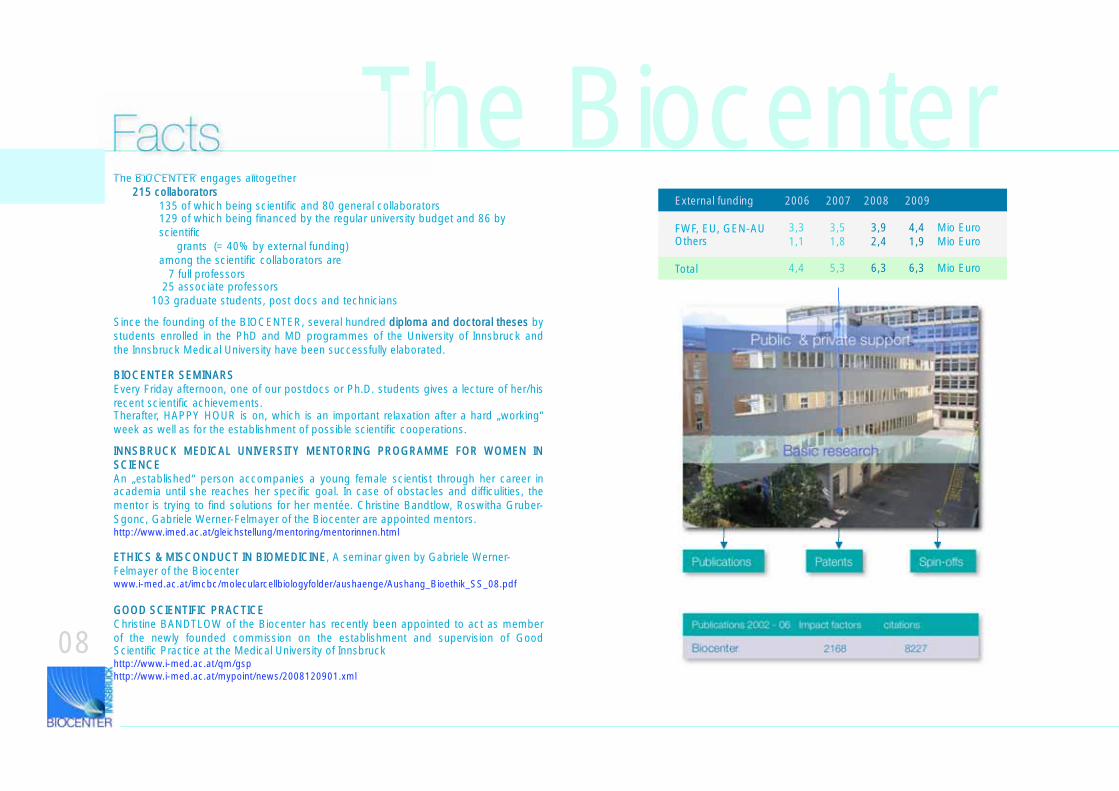
The BIOCENTER engages alltogether 215 collaborators
135 of which being scientific and 80 general collaborators129 of which being financed by the regular university budget and 86 by scientific
grants (= 40% by external funding)among the scientific collaborators are
7 full professors 25 associate professors
103 graduate students, post docs and technicians
Publications Spin-offsPatents
Publications 2002 - 06 Impact factors citations
Biocenter 2168 8227
The Biocenter
INNSBRUCK MEDICAL UNIVERSITY MENTORING PROGRAMME FOR WOMEN IN SCIENCEAn „established“ person accompanies a young female scientist through her career in academia until she reaches her specific goal. In case of obstacles and difficulities, the mentor is trying to find solutions for her mentée. Christine Bandtlow, Roswitha Gruber-Sgonc, Gabriele Werner-Felmayer of the Biocenter are appointed mentors.http://www.imed.ac.at/gleichstellung/mentoring/mentorinnen.html
Since the founding of the BIOCENTER, several hundred diploma and doctoral theses by students enrolled in the PhD and MD programmes of the University of Innsbruck and the Innsbruck Medical University have been successfully elaborated.
BIOCENTER SEMINARSEvery Friday afternoon, one of our postdocs or Ph.D. students gives a lecture of her/his recent scientific achievements.Therafter, HAPPY HOUR is on, which is an important relaxation after a hard „working“ week as well as for the establishment of possible scientific cooperations.
Basic research
Public & private support
FactsExternal funding 2006 2007 2008 2009
FWF, EU, GEN-AUOthers
Total
3,3 3,5 3,9 4,4 Mio Euro1,1 1,8 2,4 1,9 Mio Euro
4,4 5,3 6,3 6,3 Mio Euro
GOOD SCIENTIFIC PRACTICEChristine BANDTLOW of the Biocenter has recently been appointed to act as member of the newly founded commission on the establishment and supervision of Good Scientific Practice at the Medical University of Innsbruckhttp://www.i-med.ac.at/qm/gsphttp://www.i-med.ac.at/mypoint/news/2008120901.xml
ETHICS & MISCONDUCT IN BIOMEDICINE, A seminar given by Gabriele Werner-Felmayer of the Biocenterwww.i-med.ac.at/imcbc/molecularcellbiologyfolder/aushaenge/Aushang_Bioethik_SS_08.pdf
08
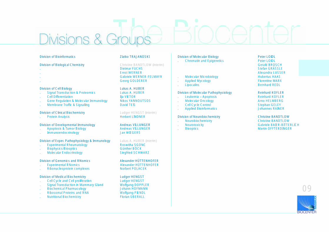
The BiocenterDivisions & GroupsDivision of Bioinformatics Zlatko TRAJANOSKI
Division of Biological Chemistry Christine BANDTLOW (interim). Dietmar FUCHS. Ernst WERNER. Gabriele WERNER-FELMAYR. Georg GOLDERER
Division of Cell Biology Lukas A. HUBER. Signal Transduction & Proteomics Lukas A. HUBER. Cell Differentiation Ilja VIETOR. Gene Regulation & Molecular Immunology Nikos YANNOUTSOS. Membrane Traffic & Signalling David TEIS
Division of Clinical Biochemistry Ludger HENGST (interim). Protein Analysis Herbert LINDNER
Division of Developmental Immunology Andreas VILLUNGER. Apoptosis & Tumor Biology Andreas VILLUNGER. Immunoendocrinology Jan WIEGERS
Division of Exper. Pathophysiology & Immunology Lukas A. HUBER (interim). Experimental Rheumatology Roswitha SGONC. Biophysics/Biooptics Günther BÖCK. Molecular Endocrinology Siegfried SCHWARZ
Division of Genomics and RNomics Alexander HÜTTENHOFER. Experimental RNomics Alexander HÜTTENHOFER. Ribonucleoprotein complexes Norbert POLACEK
Division of Medical Biochemistry Ludger HENGST. Cell Cycle and Cell proliferation Ludger HENGST. Signal Transduction in Mammary Gland Wolfgang DOPPLER. Biochemical Pharmacology Johann HOFMANN. Ribosomal Proteins and RNA Wolfgang PIENDL. Nutritional Biochemistry Florian ÜBERALL
Division of Molecular Biology Peter LOIDL. Chromatin and Epigenetics Peter LOIDL
Gerald BROSCHStefan GRÄSSLEAlexandra LUSSER
. Molecular Microbiology Hubertus HAAS
. Applied Mycology Florentine MARX
. Lipocalins Bernhard REDL
Division of Molecular Pathophysiology Reinhard KOFLER. Leukemia – Apoptosis Reinhard KOFLER. Molecular Oncology Arno HELMBERG. Cell Cycle Control Stephan GELEY. Applied Bioinformatics Johannes RAINER
Division of Neurobiochemistry Christine BANDTLOW. Neurobiochemistry Christine BANDTLOW. Neurotoxicity Gabriele BAIER-BITTERLICH. Biooptics Martin OFFTERDINGER
09
The BiocenterSupport & Collaborations
Institute for Biomedical Aging ResearchAustrian Academy of Sciences
10
MEMBERS Baccarini Manuela Baier Gottfried Bonn Günther Fässler Reinhard Geley Stephan Greil Richard Hengst Ludger Huber Lukas A. (Coordinator) Kofler Reinhard Villunger Andreas
Associated Members Gastl Guenther Schneider Rainer
Young Investigator Program Fiegl Heidelinde Rainer Johannes Teis David Wolf Dominik
Former Members Doppler Wolfgang Jansen-Dürr Pidder Klocker Helmut Tinhofer Inge Überall Florian
The Biocenterhttp://www.sfb021.at/
11
Spezialforschungsbereich SFB021
Members from the Biocenter
Geley Stephan, A.Univ.-Prof. Dr.: Cell Biology, Cancer Research (Division of Pathophysiology)Hengst Ludger, Univ.-Prof. Dr.: Cell Biology, Cancer Research, Medical Biochemistry (Division of Medical Biochemistry)
Huber Lukas A., Univ.-Prof. Dr.: Cell Biology (Division of Cell Biology)
Kofler Reinhard, Univ.-Prof. Dr.: Molecular Oncology (Division of Molecular Pathology)
Villunger Andreas, Prof. Dr.: Cellular Immunology, Tumor Biology, Apoptosis (Division of Developmental Immunology)
Associated Members Gastl Guenther, Univ.-Prof. Dr.: Tumor Profiling (Division of Haematology and Oncology, Department of Internal Medicine)
Schneider Rainer, A.Univ.-Prof. Dr.: Biochemistry (Department of Biochemistry, University of Innsbruck)
Young Investigator Program (supported by Tiroler Zukunftsstiftung and Medical University Innsbruck)
Rainer Johannes, Dr: Bioinformatics, Cancer Research (Division Molecular Pathophyisiology)
Teis David, Dr.: Membrane traffic and signaling (Division of Cell Biology)
For a detailed description of the various research topics, see the respective individual pages of the SFB members later in this brochure!
The major goals of the SFB021 are to understand molecular pathways that link signals leading either to cell death/survival or to cell proliferation/cell cycle arrest in tumors. The SFB021 also aims to better understand, why the immune system apparently fails to eliminate tumor cells focusing on the established pathways regulating T cell activation thresholds. In continuation of the work during the first funding period (2003-2007) SFB021 scientists propose the coordinated use of biochemical and genetic as well as proteomic/transcriptomic approaches to delineate changes in cellular pathways that occur in several types of tumors, namely epithelial tumors (breast cancer, liver, skin), chronic myeloic leukemia (CML) or tumors of lymphatic origin (acute lymphatic leukemia (ALL) and multiple myeloma). In addition, for the second funding period (2008-2010) they have now extended their experimental approaches towards antigen receptor signal processing machinery in T cells during immunesurveillance in tumors, adhesion signaling in tumor cells, as well as posttranslational modifications of the Cdk inhibitor p27Kip1.
12
Dietmar Fuchs and EErnst Werner of the division of Biological Chemistry are founding and steering members of the
International Neopterin NetworkThey organize since many years the annual Winter Workshop on Clinical, Chemical and Biochemical Aspects of Pteridines in St. Christoph/Arlberg/Austriawww.neopterin.net
Andreas Villunger of the Division of Developmental Immunology of the Biocenter is integrated into
ApopTraina Transeuropean Network of Apoptosis research laboratories
The Biocenter
Lukas A. Huber of the Division of Cell Biology of the Biocenter is coordinator of the EU-FP6 (Framework Program 6) research project entitled
GrowthStop Networkwhich started in October 2006. The topics and goals are: IDENTIFICATION, DEVELOPMENT AND VALIDATION OF NOVEL THERAPEUTICS TARGETING PROGRAMMED CELL DEATH IN TUMORS. A total of 12 international partners from different EU countries together with Israel are participating in this four year project. CCEMIT is supporting the project coordinator and project partners in administrative aspects.www.cemit.at
Georg Wick, professor emeritus of the Division of Experimental Pathophysiology and Immunology is – together with Mathias Mass of the Salzburg University Clinics - coordinator of
ECIBUG – EEuropean Inititative to fight CChlamydial IInfec-tions with UUnbbiased GGenomicsThis rersearch network is also part of the Austrian GEN-AU program (see next page!). http://www.i-med.ac.at/mypoint/news/2008050701.xml
www.cemit.at/
www.gen-au.at
13
Lukas A. Huber is also coordinator of the
Austrian Proteomics Platform. Proteomics is one of the greatest challenges in basic science of our age, where the human genome has already been sequenced.
www.gen-au.at
The Biocenter
Two members of the Biocenter are also members of the
Austrian RNomics Platform Andrea Barta, ViennaIvo Hofacker, ViennaAlexander Hüttenhofer, InnsbruckJavier Martinez, ViennaRonald Micura, InnsbruckNorbert Polacek, InnsbruckRenee Schroeder, Vienna
and are involved in a concerted research action on the study of noncoding (nc)RNAs in several organisms. ncRNAs are newly discovered RNA species which exert a profound regulatory role on gene expression and - in altered state - are thought to be responsible for various human diseases.http://www.cemit.at/folgeseite.cfm?id=187http://www.gen-au.at/projekt.jsp?projektId=60&lang=de
www.cemit.at/
Besides DNA as cellular carrier of information, proteins are importan cellular tools that are involved in the generation of diseases. It is therefore a great challenge in lifes sciences to decipher
besides the genome also the proteome, i.e. the entirety of proteins (gene products) of a particular cell (type) or tissue in a particular state (of development, activity) = function. While the genome is static, the proteome is dynamic, as it constantly changes in response to challenges impinging on the cell. The proteome includes expression profiles of gene products, but also the
posttranslational processing events of proteins. These, however, can only be investigated on the proteins themselves. The proteome reflects thus the state of a cell, tissue, organism, normal as well as pathological.
The Austrian Proteomics Platform was founded as a network of 5 Austrian basic scientists, working at the Universities and Medical Universities of Vienna, Graz, Innsbruck, the Veterinary
University of Vienna and the Institute of Molecular Pathology (IMP) Vienna. These Partners have submitted individual projects, yet exchange among themselves ressources, technologies and experience in order to establish a broad proteinomics platform. The individual researchers focus in particular on those biological as well as technological questions, where they have already
expertise and from which they can expect to achieve internationally a scientific advantage.In the first phase, the following activities are undertaken:
° Establishing a state-of-tha-art infrastructure for proteomics work
° Technological development with view to funtional proteomics, i.e. for the elucidation of cellular functions of proteins, such as protein-ligand interaction, postttranslational modifications etc. ° Development of new protocols and new stationary phases for prottein and peptide separation work
° Development of Bioinformatics for more thorough data evaluation° Education of highly qualified scientists
www.proteomics.or.at/
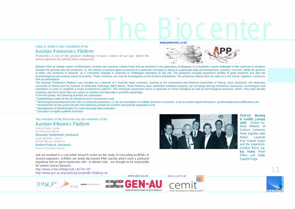
GEN-AU Meeting in Seefeld, January 2009 Former Fe-deral Minister of Science Johannes Hahn together with Nobel Laureate Prof. Robert Huber and the organizers, Günther Bonn, LLu-kas Huber, Peter Öfner, and Giulio Superti-Furga
14
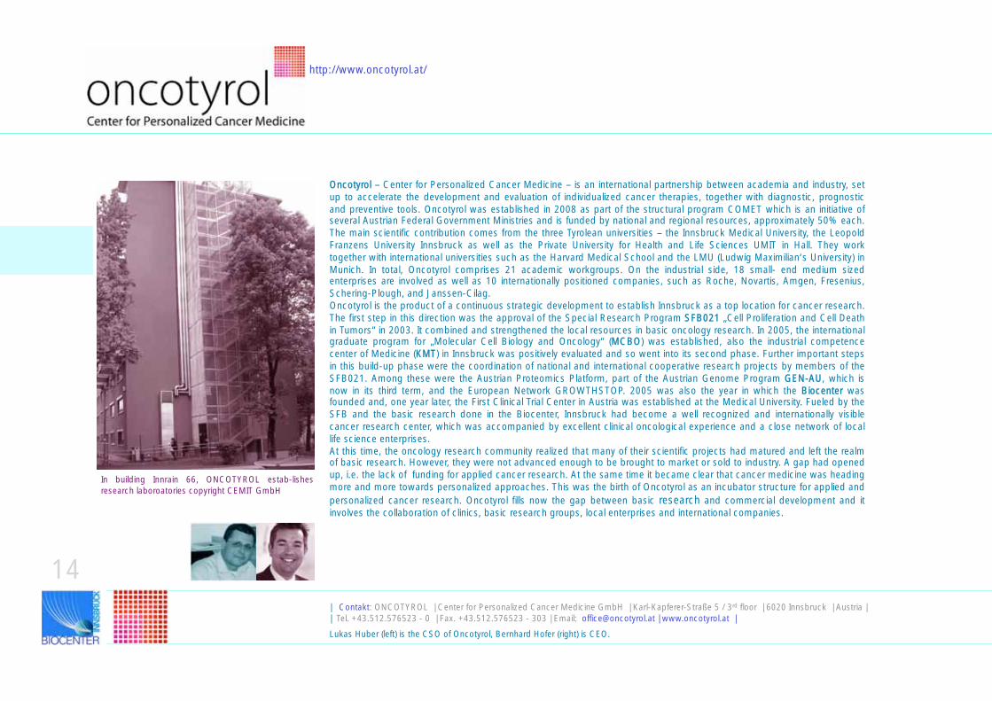
Oncotyrol – Center for Personalized Cancer Medicine – is an international partnership between academia and industry, set up to accelerate the development and evaluation of individualized cancer therapies, together with diagnostic, prognostic and preventive tools. Oncotyrol was established in 2008 as part of the structural program COMET which is an initiative of several Austrian Federal Government Ministries and is funded by national and regional resources, approximately 50% each. The main scientific contribution comes from the three Tyrolean universities – the Innsbruck Medical University, the Leopold Franzens University Innsbruck as well as the Private University for Health and Life Sciences UMIT in Hall. They work together with international universities such as the Harvard Medical School and the LMU (Ludwig Maximilian‘s University) in Munich. In total, Oncotyrol comprises 21 academic workgroups. On the industrial side, 18 small- end medium sized enterprises are involved as well as 10 internationally positioned companies, such as Roche, Novartis, Amgen, Fresenius, Schering-Plough, and Janssen-Cilag. Oncotyrol is the product of a continuous strategic development to establish Innsbruck as a top location for cancer research. The first step in this direction was the approval of the Special Research Program SSFB021 „Cell Proliferation and Cell Death in Tumors“ in 2003. It combined and strengthened the local resources in basic oncology research. In 2005, the international graduate program for „Molecular Cell Biology and Oncology“ (MMCBO) was established, also the industrial competence center of Medicine (KKMT) in Innsbruck was positively evaluated and so went into its second phase. Further important steps in this build-up phase were the coordination of national and international cooperative research projects by members of the SFB021. Among these were the Austrian Proteomics Platform, part of the Austrian Genome Program GGEN-AU, which is now in its third term, and the European Network GROWTHSTOP. 2005 was also the year in which the BBiocenter was founded and, one year later, the First Clinical Trial Center in Austria was established at the Medical University. Fueled by the SFB and the basic research done in the Biocenter, Innsbruck had become a well recognized and internationally visible cancer research center, which was accompanied by excellent clinical oncological experience and a close network of local life science enterprises.At this time, the oncology research community realized that many of their scientific projects had matured and left the realm of basic research. However, they were not advanced enough to be brought to market or sold to industry. A gap had opened up, i.e. the lack of funding for applied cancer research. At the same time it became clear that cancer medicine was heading more and more towards personalized approaches. This was the birth of Oncotyrol as an incubator structure for applied and personalized cancer research. Oncotyrol fills now the gap between basic research and commercial development and it involves the collaboration of clinics, basic research groups, local enterprises and international companies.
In building Innrain 66, ONCOTYROL estab-lishes research laboroatories copyright CEMIT GmbH
| Contakt: ONCOTYROL | Center for Personalized Cancer Medicine GmbH | Karl-Kapferer-Straße 5 / 3rd floor | 6020 Innsbruck | Austria | | Tel. +43.512.576523 - 0 | Fax. +43.512.576523 - 303 | Email: [email protected] | www.oncotyrol.at |
Lukas Huber (left) is the CSO of Oncotyrol, Bernhard Hofer (right) is CEO.
http://www.oncotyrol.at/
Area 1
Mechanisms controlling tumor growth and anti-tumor immunity
Gottfried Baier, i-med
Area 2
Bioanalytics and diagnostics
Helmut Klocker, i-med
Area 3
Biomarker-guided cancer diagnosis, therapy and prevention
Günther Gastl,i-med
Area 4
Decision-analytic outcome modeling, Health Technology Assessment (HTA)and health economicsUwe Siebert, UMIT
Area 5
Bioinformatics and systems biology
Zlatko Trajanoski, Biocenter
Core Facilities
Cbl-b targeting Concept G. Baier, i-med
Metabolite analysisD. Sonntag, Biocrates Life Sciences AG
New markers for prostate cancerH.Klocker, i-med
Methodological framework for early HTAU. Siebert, UMIT
Knowledge managementS. Losko, Biomax Informatics AG
Biobanking
G. Daxenbichler, i-med
MAP kinase scaffold inhibitorsL. Huber, Biocenter
Gas analysis of VOCsS. Praun, V&F medical development GmbH
Breast cancer – metastasis riskC. Marth, H. Fiegl, i-med
Austrian myeloma registerG. Gastl, i-med
Methods for analysis of metabolomic dataB. Haas, Biocrates Life Sciences AG
Sequencing & genotypingF. Kronenberg, i-med
Cell therapy unitN. Romani, i-medM. Thurnher, i-med
Glucocorticoid-responsive transcriptome IR. Kofler, Biocenter
Androgen receptor targeting for prostate cancer I. Eder, i-med
Outcome & policy model for prostate cancerU. Siebert, UMIT
Glucocorticoid-responsive transcriptome IIR. Kofler, Biocenter
General management
CEMIT GmbH
IAP antagonistsM. Außerlechner, i-med, H. Stuppner, LFU
Prostate cancer preventionH. Klocker, i-med
Evaluation of EpCAM expressionG. Spizzo, i-med
Outcome & policy model for breast cancerU. Siebert, UMIT
Targeting RAF/BAG-1 interactionJ. Troppmair, i-med
Breath gas analysis for breast cancer diagnosisI.Kohl, Ionimed Analytik GmbH
Angiogenesis biomarkers
E. Gunsilius, i-med
Outcome & Policy Model for Rheumatoid ArthritisU. Siebert, UMIT
Integration of health system data and exchangeT. Schabetsberger, UMIT
| Abbreviations: | Cbl: Cbl oncogene | MAP: mitogen activated protein kinase | IAP: inhibitor of apoptosis proteins | RAF: Raf oncogene | VOC: Volatile Organic Carbon | | EpCAM: eptithelial cell adhesion molecule | HTA Health assessement technology | i-med: Medizinische Universität Innsbruck | LFU: Leopold Franzens Universität Innsbruck | UMIT: Private Health and Life Sciences University, Hall |
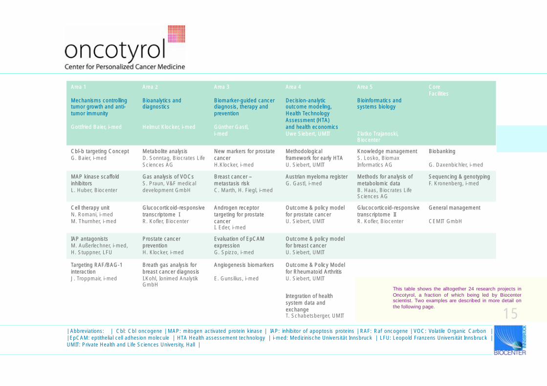
This table shows the alltogether 24 research projects in
Oncotyrol, a fraction of which being led by Biocenter scientist. Two examples are described in more detail on
the following page.
15
16
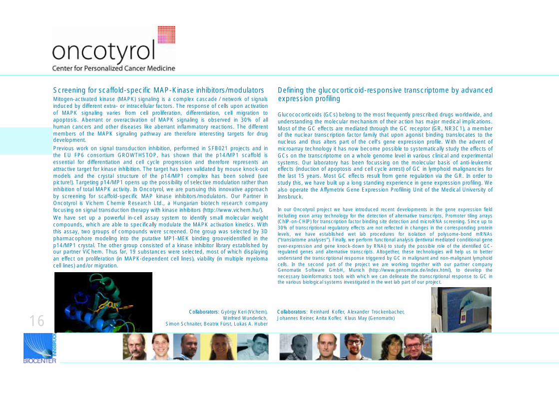
Screening for scaffold-specific MAP-Kinase inhibitors/modulatorsMitogen-activated kinase (MAPK) signaling is a complex cascade / network of signals induced by different extra- or intracellular factors. The response of cells upon activation of MAPK signaling varies from cell proliferation, differentiation, cell migration to apoptosis. Aberrant or overactivation of MAPK signaling is observed in 30% of all human cancers and other diseases like aberrant inflammatory reactions. The different members of the MAPK signaling pathway are therefore interesting targets for drug development. Previous work on signal transduction inhibition, performed in SFB021 projects and in the EU FP6 consortium GROWTHSTOP, has shown that the p14/MP1 scaffold is essential for differentiation and cell cycle progression and therefore represents an attractive target for kinase inhibition. The target has been validated by mouse knock-out models and the crystal structure of the p14/MP1 complex has been solved (see picture!). Targeting p14/MP1 opens up the possibility of selective modulation rather than inhibition of total MAPK activity. In Oncotyrol, we are pursuing this innovative approach by screening for scaffold-specific MAP kinase inhibitors/modulators. Our Partner in Oncotyrol is Vichem Chemie Research Ltd., a Hungarian biotech research company focusing on signal transduction therapy with kinase inhibitors (http://www.vichem.hu/). We have set up a powerful in-cell assay system to identify small molecular weight compounds, which are able to specifically modulate the MAPK activation kinetics. With this assay, two groups of compounds were screened. One group was selected by 3D pharmacophore modeling into the putative MP1-MEK binding grooveidentified in the p14/MP1 crystal. The other group consisted of a kinase inhibitor library established by our partner ViChem. Thus far, 19 substances were selected, most of which displaying an effect on proliferation (in MAPK-dependent cell lines), viability (in multiple myeloma cell lines) and/or migration.
Defining the glucocorticoid-responsive transcriptome by advanced
Glucococorticoids (GCs) belong to the most frequently prescribed drugs worldwide, and understanding the molecular mechanism of their action has major medical implications. Most of the GC effects are mediated through the GC receptor (GR, NR3C1), a member of the nuclear transcription factor family that upon agonist binding translocates to the nucleus and thus alters part of the cell‘s gene expression profile. With the advent of microarray technology it has now become possible to systematically study the effects of GCs on the transcriptome on a whole genome level in various clinical and experimental systems. Our laboratory has been focussing on the molecular basis of anti-leukemic effects (induction of apoptosis and cell cycle arrest) of GC in lymphoid malignancies for the last 15 years. Most GC effects result from gene regulation via the GR. In order to study this, we have built up a long standing experience in gene expression profiling. We also operate the Affymetrix Gene Expression Profilinig Unit of the Medical University of Innsbruck.
In our Oncotyrol project we have introduced recent developments in the gene expression field including exon array technology for the detection of alternative transcripts, Promoter tiling arrays (ChIP-on-CHIP) for transcription factor binding site detection and microRNA screening. Since up to 30% of transcriptional regulatory effects are not reflected in changes in the corresponding protein levels, we have established wet lab procedures for isolation of polysome-bond mRNAs (“translatome analyses”). Finally, we perform functional analysis (lentiviral mediated conditional gene over-expression and gene knock-down by RNAi) to study the possible role of the identified GC-regulated genes and alternative transcripts. Altogether, these technologies will help us to better understand the transcriptional response triggered by GC in malignant and non-malignant lymphoid cells. In the second part of the project we are working together with our partner company Genomatix Software GmbH, Munich (http://www.genomatix.de/index.html), to develop the necessary bioinformatics tools with which we can delineate the transcriptional response to GC in the various biological systems investigated in the wet lab part of our project.
Collaborators: Reinhard Kofler, Alexander Trockenbacher, Johannes Reiner, Anita Kofler, Klaus May (Genomatix)
Collaborators: György Keri (Vichem), Winfried Wunderlich,
Simon Schnaiter, Beatrix Fürst, Lukas A. Huber
expression profiling
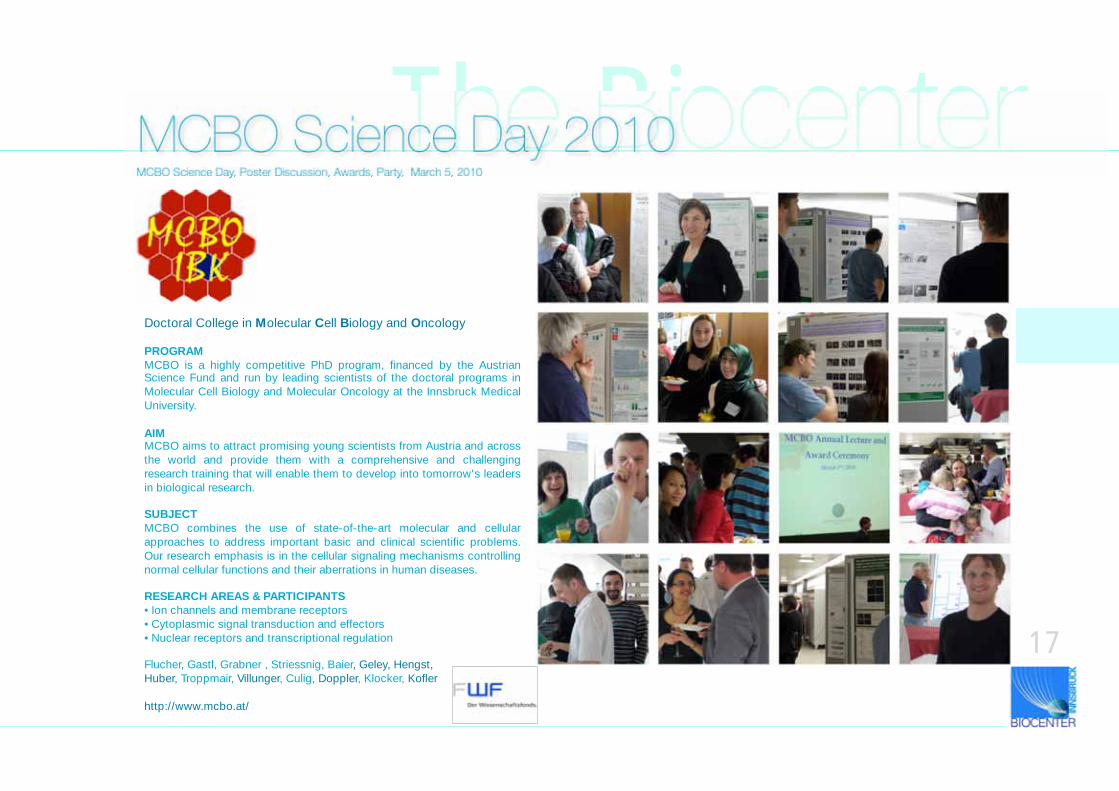
The BiocenterMCBO Science Day 2010MCBO Science Day, Poster Discussion, Awards, Party, March 5, 2010
17
Doctoral College in Molecular Cell Biology and Oncology
PROGRAMMCBO is a highly competitive PhD program, financed by the Austrian Science Fund and run by leading scientists of the doctoral programs in
Molecular Cell Biology and Molecular Oncology at the Innsbruck Medical
University.
AIMMCBO aims to attract promising young scientists from Austria and across
the world and provide them with a comprehensive and challenging
research training that will enable them to develop into tomorrow's leaders
in biological research.
SUBJECTMCBO combines the use of state-of-the-art molecular and cellular
approaches to address important basic and clinical scientific problems.
Our research emphasis is in the cellular signaling mechanisms controlling
normal cellular functions and their aberrations in human diseases.
RESEARCH AREAS & PARTICIPANTS• Ion channels and membrane receptors
• Cytoplasmic signal transduction and effectors
• Nuclear receptors and transcriptional regulation
Flucher, Gastl, Grabner , Striessnig, Baier, Geley, Hengst,
Huber, Troppmair, Villunger, Culig, Doppler, Klocker, Kofler
http://www.mcbo.at/
18
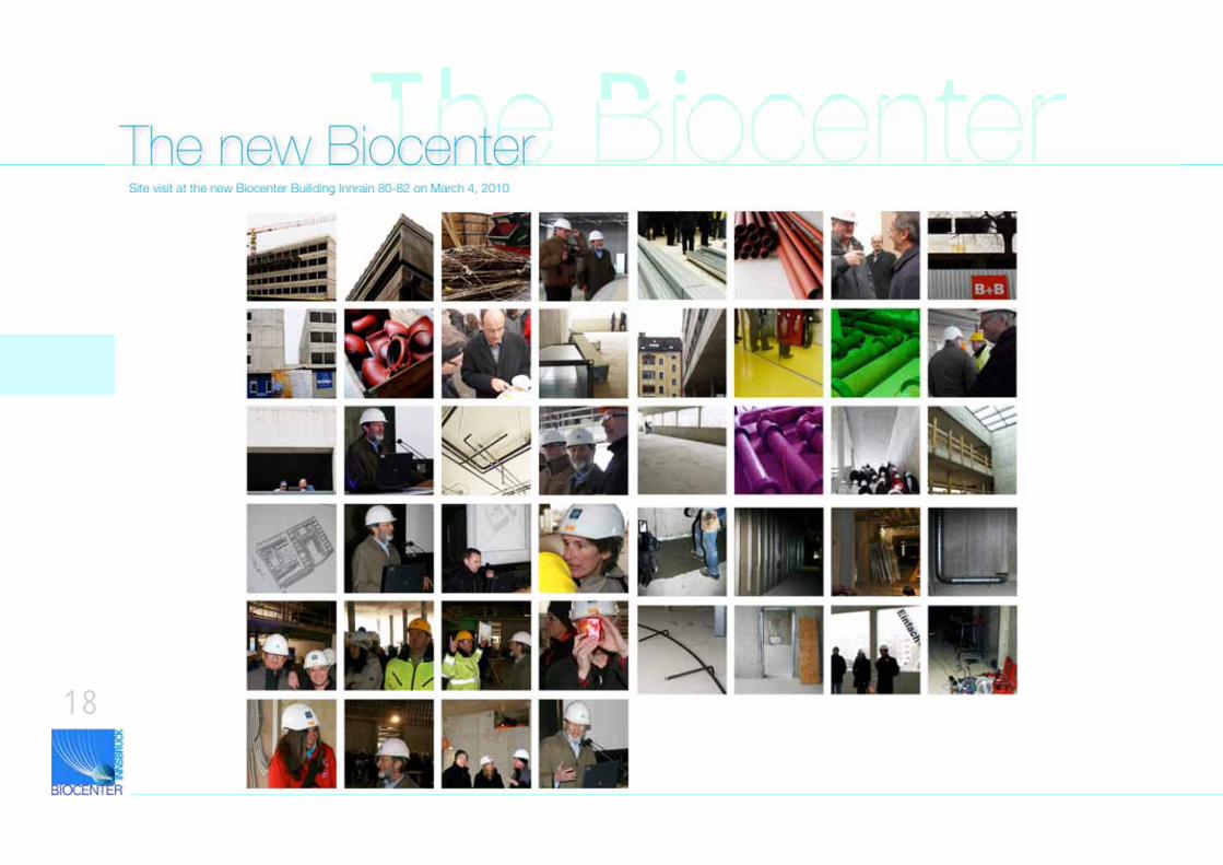
The BiocenterThe new BiocenterSite visit at the new Biocenter Builiding Innrain 80-82 on March 4, 2010
The Biocenter
19
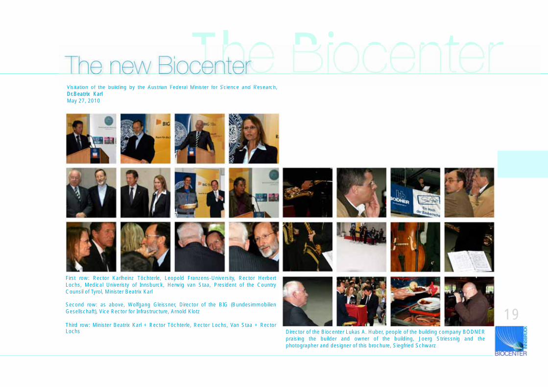
The new BiocenterVisitation of the building by the Austrian Federal Minister for Science and Research, Dr.Beatrix KarlMay 27, 2010
First row: Rector Karlheinz Töchterle, Leopold Franzens-University, Rector Herbert Lochs, Medical Univeristy of Innsburck, Herwig van Staa, President of the Country Counsil of Tyrol, Minister Beatrix Karl
Second row: as above, Wolfgang Gleissner, Director of the BIG (Bundesimmobilien Gesellschaft), Vice Rector for Infrastructure, Arnold Klotz
Third row: Minister Beatrix Karl + Rector Töchterle, Rector Lochs, Van Staa + Rector Lochs Director of the Biocenter Lukas A. Huber, people of the building company BODNER
praising the builder and owner of the building, Joerg Striessnig and the photographer and designer of this brochure, Siegfried Schwarz
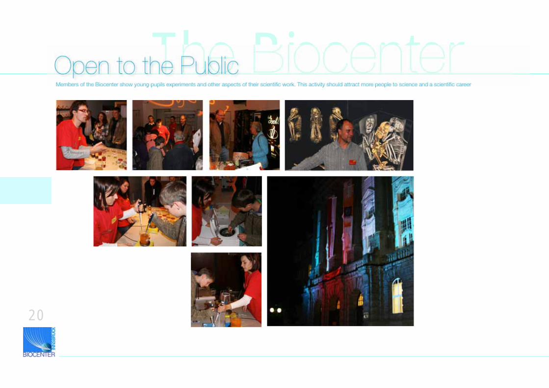
The BiocenterOpen to the PublicMembers of the Biocenter show young pupils experiments and other aspects of their scientific work. This activity should attract more people to science and a scientific career
20
The Biocenter
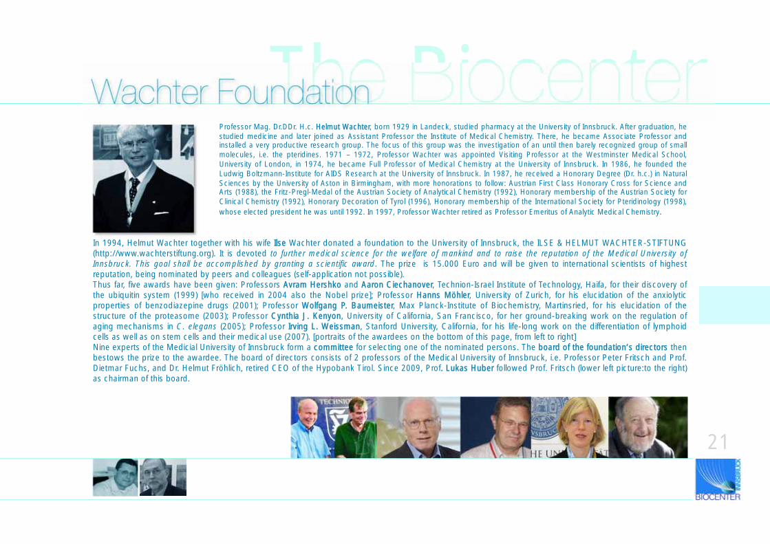
In 1994, Helmut Wachter together with his wife IIlse Wachter donated a foundation to the University of Innsbruck, the ILSE & HELMUT WACHTER-STIFTUNG (http://www.wachterstiftung.org). It is devoted to further medical science for the welfare of mankind and to raise the reputation of the Medical University of Innsbruck. This goal shall be accomplished by granting a scientific award. The prize is 15.000 Euro and will be given to international scientists of highest reputation, being nominated by peers and colleagues (self-application not possible). Thus far, five awards have been given: Professors AAvram Hershko and AAaron Ciechanover, Technion-Israel Institute of Technology, Haifa, for their discovery of the ubiquitin system (1999) [who received in 2004 also the Nobel prize]; Professor HHanns Möhler, University of Zurich, for his elucidation of the anxiolytic properties of benzodiazepine drugs (2001); Professor WWolfgang P. Baumeister, Max Planck-Institute of Biochemistry, Martinsried, for his elucidation of the structure of the proteasome (2003); Professor CCynthia J. Kenyon, University of California, San Francisco, for her ground-breaking work on the regulation of aging mechanisms in C. elegans (2005); Professor IIrving L. Weissman, Stanford University, California, for his life-long work on the differentiation of lymphoid cells as well as on stem cells and their medical use (2007). [portraits of the awardees on the bottom of this page, from left to right] Nine experts of the Medicial University of Innsbruck form a ccommittee for selecting one of the nominated persons. The bboard of the foundation‘s directors then bestows the prize to the awardee. The board of directors consists of 2 professors of the Medical University of Innsbruck, i.e. Professor Peter Fritsch and Prof. Dietmar Fuchs, and Dr. Helmut Fröhlich, retired CEO of the Hypobank Tirol. Since 2009, Prof.. Lukas Huber followed Prof. Fritsch (lower left picture:to the right) as chairman of this board.
Wachter FoundationProfessor Mag. Dr.DDr. H.c. HHelmut Wachter, born 1929 in Landeck, studied pharmacy at the University of Innsbruck. After graduation, he studied medicine and later joined as Assistant Professor the Institute of Medical Chemistry. There, he became Associate Professor and installed a very productive research group. The focus of this group was the investigation of an until then barely recognized group of small molecules, i.e. the pteridines. 1971 – 1972, Professor Wachter was appointed Visiting Professor at the Westminster Medical School, University of London, in 1974, he became Full Professor of Medical Chemistry at the University of Innsbruck. In 1986, he founded the Ludwig Boltzmann-Institute for AIDS Research at the University of Innsbruck. In 1987, he received a Honorary Degree (Dr. h.c.) in Natural Sciences by the University of Aston in Birmingham, with more honorations to follow: Austrian First Class Honorary Cross for Science and Arts (1988), the Fritz-Pregl-Medal of the Austrian Society of Analytical Chemistry (1992), Honorary membership of the Austrian Society for Clinical Chemistry (1992), Honorary Decoration of Tyrol (1996), Honorary membership of the International Society for Pteridinology (1998), whose elected president he was until 1992. In 1997, Professor Wachter retired as Professor Emeritus of Analytic Medical Chemistry.
21
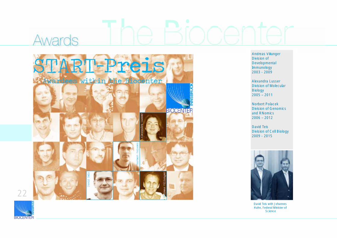
Andreas VillungerDivision of Developmental Immunology2003 - 2009
Alexandra LusserDivision of Molecular Biology2005 – 2011
Norbert PolacekDivision of Genomics and RNomics2006 – 2012
David TeisDivision of Cell Biology2009 - 2015
The BiocenterAwards
David Teis with Johannes Hahn, Federal Minister of
Science
22
23
Start PrizeThe START-Prize has been founded in 1996 by the then Federal Minister of Science Rudolf Scholten. It is announced every year. Applicants must be under 35 years and are selected by an international jury of renowned scientists. Successful applicants are supported for up to 6 years. Therefore, the START prize represents that award of the FWF that is of highest national reputation. Also, it is the highest amount of support (i.e. 1.200.000 Euro) in Austria that can be given to single awardee. The most important idea behind this prize is to give young and highly qualified scientists the financial basis to persue a long-term research project independent from their university supervisors. It is mostly young scientist who worked successfully as post-doctoral fellows abroad and who plan to return to their home universities. Often, the situation at home is difficult in terms of a secured career perspective which may hinder their return and which would mean a brain drain to Austrian universities. Until now, the START Prize has been awarded 76 times (see some of the portraits of awardees on the previous page). It is a great pleasure to mention that 11 of 76 awardees belong the Innsbruck Universities and 4 of these 11 to our institution. Only 7 of 76 awardees were women, however 1 of these 4 Biocenter awardees is a women. A list of awardees can be found under http://www.start-portal.at/die-starter/liste.html.
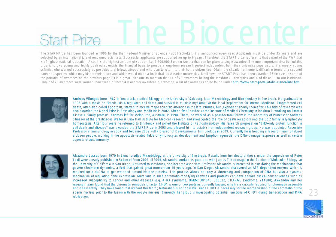
Alexandra Lusser, born 1970 in Lienz, studied Microbiology at the University of Innsbruck. Results from her doctoral thesis under the supervision of Peter Loidl were already published in Science! From 2001 till 2004, Alexandra worked as post-doc with James T. Kadonaga in the Section of Molecular Biology at the University of California in San Diego. Returned to Innsbruck, she became Associate Professor. Alexandra is interested in elucidating the mechanisms that govern chromatin dynamics, a field that gained great momentum 10 years ago. In San Diego, Alexandra discovered an ATP-dependent enzyme which is required for a dsDNA to get wrapped around histone proteins. This process allows not only a shortening and compaction of DNA but also a dynamic mechanism of regulating gene expression. Mutations in such chromatin-modifying enzymes and proteins can have serious clinical consequences such as increased susceptibility to cancer and other diseases (e.g. ATRX syndrome, OMIM: 301040, 300032, CHARGE syndrome, 214800). Alexandra and her research team found that the chromatin remodeling factor CHD1 is one of two proteins currently known, which are critically required for chromatin assembly and disassembly. They have found that without this factor, fertilization is not possible, since CHD1 is necessary for the reorganization of the chromatin of the sperm nucleus prior to the fusion with the oocyte nucleus. Currently, her group is investigating potential functions of CHD1 during transcription and DNA replication.
Andreas Villunger, born 1967 in Innsbruck, studied Biology at the University of Salzburg, later Microbiology and Biochemistry in Innsbruck. He graduated in 1996 with a thesis on "Interleukin-6 regulated cell death and survival in multiple myeloma" at the local Department for Internal Medicine. Programmed cell death, often also called apoptosis, started to receive major scientific attention in the late 1980ies, but „exploded“ shortly thereafter. This field of research was also awarded the Nobel Prize in Physiology and Medicine in 2002. After a first Postdoc at the Institute of Medical Chemistry in Innsbruck, working on Protein Kinase C family proteins, Andreas left for Melbourne, Australia, in 1998. There, he worked as a postdoctoral fellow in the laboratory of Professsor Andreas Strasser at the prestigeous Walter & Eliza Hall Institute for Medical Research and investigated the role of death receptors and the Bcl2 family in lymphocyte homeostasis. After four years he returned to Innsbruck and joined the Institute of Pathophysiology. His research proposal on "BH3-only protein function in cell death and disease" was awarded the START-Prize in 2003 and allowed him to establish an independent research group. He was appointed Associate Professor in Immunology in 2007 and became 2009 Full Professor of Developmental Immunology in 2009. Currently he is heading a research team of about a dozen people, working in the apoptosis-related fields of lymphocytes development and lymphomagenesis, the DNA-damage response as well as certain aspects of autoimmunity.
The Biocenter
24
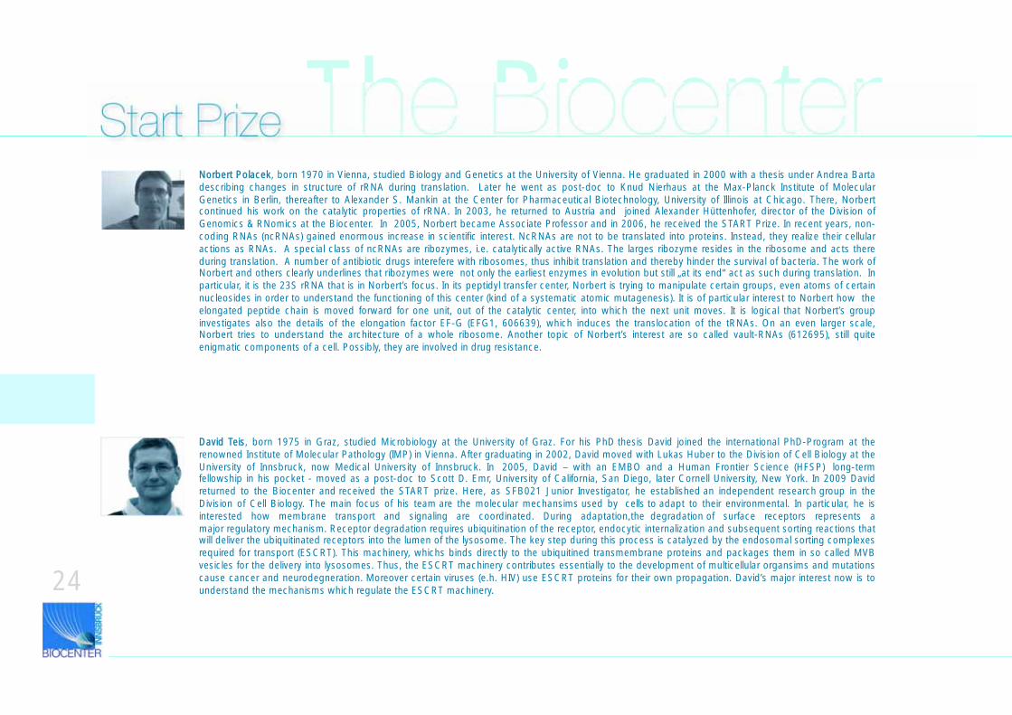
The BiocenterStart PrizeNorbert Polacek, born 1970 in Vienna, studied Biology and Genetics at the University of Vienna. He graduated in 2000 with a thesis under Andrea Barta describing changes in structure of rRNA during translation. Later he went as post-doc to Knud Nierhaus at the Max-Planck Institute of Molecular Genetics in Berlin, thereafter to Alexander S. Mankin at the Center for Pharmaceutical Biotechnology, University of Illinois at Chicago. There, Norbert continued his work on the catalytic properties of rRNA. In 2003, he returned to Austria and joined Alexander Hüttenhofer, director of the Division of Genomics & RNomics at the Biocenter. In 2005, Norbert became Associate Professor and in 2006, he received the START Prize. In recent years, non-coding RNAs (ncRNAs) gained enormous increase in scientific interest. NcRNAs are not to be translated into proteins. Instead, they realize their cellular actions as RNAs. A special class of ncRNAs are ribozymes, i.e. catalytically active RNAs. The larges ribozyme resides in the ribosome and acts there during translation. A number of antibiotic drugs interefere with ribosomes, thus inhibit translation and thereby hinder the survival of bacteria. The work of Norbert and others clearly underlines that ribozymes were not only the earliest enzymes in evolution but still „at its end“ act as such during translation. In particular, it is the 23S rRNA that is in Norbert’s focus. In its peptidyl transfer center, Norbert is trying to manipulate certain groups, even atoms of certain nucleosides in order to understand the functioning of this center (kind of a systematic atomic mutagenesis). It is of particular interest to Norbert how the elongated peptide chain is moved forward for one unit, out of the catalytic center, into which the next unit moves. It is logical that Norbert’s group investigates also the details of the elongation factor EF-G (EFG1, 606639), which induces the translocation of the tRNAs. On an even larger scale, Norbert tries to understand the architecture of a whole ribosome. Another topic of Norbert’s interest are so called vault-RNAs (612695), still quite enigmatic components of a cell. Possibly, they are involved in drug resistance.
David Teis, born 1975 in Graz, studied Microbiology at the University of Graz. For his PhD thesis David joined the international PhD-Program at the renowned Institute of Molecular Pathology (IMP) in Vienna. After graduating in 2002, David moved with Lukas Huber to the Division of Cell Biology at the University of Innsbruck, now Medical University of Innsbruck. In 2005, David – with an EMBO and a Human Frontier Science (HFSP) long-term fellowship in his pocket - moved as a post-doc to Scott D. Emr, University of California, San Diego, later Cornell University, New York. In 2009 David returned to the Biocenter and received the START prize. Here, as SFB021 Junior Investigator, he established an independent research group in the Division of Cell Biology. The main focus of his team are the molecular mechansims used by cells to adapt to their environmental. In particular, he is interested how membrane transport and signaling are coordinated. During adaptation,the degradation of surface receptors represents a major regulatory mechanism. Receptor degradation requires ubiquitination of the receptor, endocytic internalization and subsequent sorting reactions that will deliver the ubiquitinated receptors into the lumen of the lysosome. The key step during this process is catalyzed by the endosomal sorting complexes required for transport (ESCRT). This machinery, whichs binds directly to the ubiquitined transmembrane proteins and packages them in so called MVB vesicles for the delivery into lysosomes. Thus, the ESCRT machinery contributes essentially to the development of multicellular organsims and mutations cause cancer and neurodegneration. Moreover certain viruses (e.h. HIV) use ESCRT proteins for their own propagation. David’s major interest now is to understand the mechanisms which regulate the ESCRT machinery.
"Significance of STAT1 activation in colon cancer“
"The function of human mir-223 in leukemic cell death“
Division of Developmental Immunology
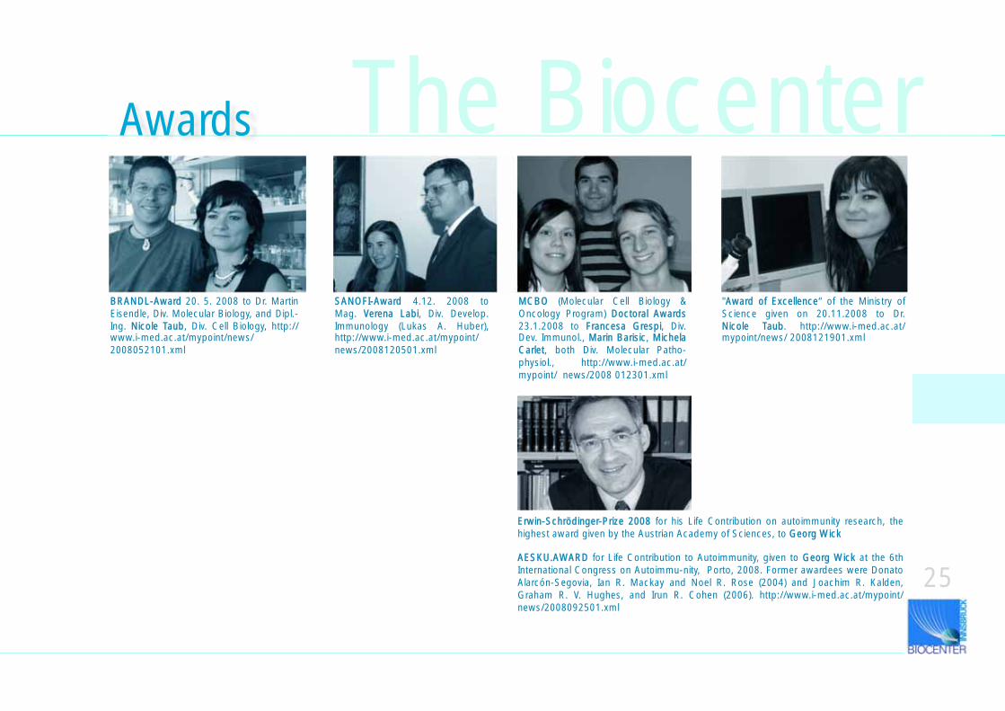
SANOFI-Award 4.12. 2008 to Mag. VVerena Labi, Div. Develop. Immunology (Lukas A. Huber), http://www.i-med.ac.at/mypoint/news/2008120501.xml
BRANDL-Award 20. 5. 2008 to Dr. Martin Eisendle, Div. Molecular Biology, and Dipl.-Ing. NNicole Taub, Div. Cell Biology, http://www.i-med.ac.at/mypoint/news/2008052101.xml
MCBO (Molecular Cell Biology & Oncology Program) DDoctoral Awards 23.1.2008 to FFrancesa Grespi, Div. Dev. Immunol., MMarin Barisic, MMichela Carlet, both Div. Molecular Patho-physiol., http://www.i-med.ac.at/mypoint/ news/2008 012301.xml
"AAward of Excellence“ of the Ministry of Science given on 20.11.2008 to Dr. Nicole Taub. http://www.i-med.ac.at/mypoint/news/ 2008121901.xml
25
Erwin-Schrödinger-Prize 2008 for his Life Contribution on autoimmunity research, the highest award given by the Austrian Academy of Sciences, to GGeorg Wick
AESKU.AWARD for Life Contribution to Autoimmunity, given to GGeorg Wick at the 6th International Congress on Autoimmu-nity, Porto, 2008. Former awardees were Donato Alarcón-Segovia, Ian R. Mackay and Noel R. Rose (2004) and Joachim R. Kalden, Graham R. V. Hughes, and Irun R. Cohen (2006). http://www.i-med.ac.at/mypoint/news/2008092501.xml
Awards The Biocenter
26
The BiocenterAwards, Meetings
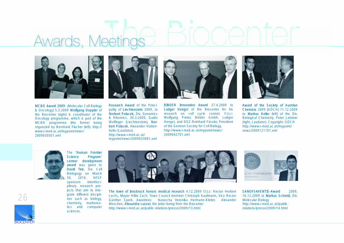
BINDER Innovation Award 27.4.2009 to Ludger Hengst of the Biocenter for his research on cell cycle control. F.l.t.r.: Wolfgang Preter, Binder Gmbh, Ludger Hengst, and DGZ-Reinhard Fässler, President of the German Society for Cell Biology,http://www.i-med.ac.at/mypoint/news/2009042701.xml
The town of Innsbruck honors medical research 4.12.2009 f.l.t.r.: Rector Herbert Lochs, Mayor Hilde Zach, Town Council member Christoph Kaufmann, Vice Rector Günther Sperk. Awardees: Natascha Veronika Hermann-Kleiter, Alexander Moschen, AAlexandra Lusser, the latter being from the Biocenterhttp://www.i-med.ac.at/public-relations/presse/2009/13.html
SANOFI-AVENTIS-Award 2009, 16.12.2009 to MMarkus Schrettl, Div. Molecular Biologyhttp://www.i-med.ac.at/public-relations/presse/2009/14.html
MCBO Award 2009. (Molecular Cell Biology & Oncology) 5.3.2009 WWolfgang Doppler of the Biocenter (right) is coordinator of the Oncology programme, which is part of the MCBO programme (the former being organzied by Bernhard Flucher (left). http://www.i-med.ac.at/mypoint/news/2009030501.xml
Award of the Society of Austrian Chemists 2009 (GÖCH) 11.12.2009 to MMarkus Keller (left) of the Div. Biological Chemistry. Peter Jaintner (right, Laudator). Copyright: GÖCH http://www.i-med.ac.at/mypoint/news/2009121101.xml
Research Award of the Princi-pality of LLiechtenstein 2009, to Norbert Polacek, Div. Genomics & RNomics, 30.3.2009, Guido Wolfinger (Liechtenstein), NNor-bert Polacek, Alexander Hütten-hofer (Laudator). http://www.i-med.ac.at/mypoint/news/2009033001.xml
The ''Human Frontier Science Program' carreer development award was given to David Teis, Div. Cell Biologogy on March 30, 2010. HFSP sponsors interdisci-plinary research pro-jects that aim to inte-grate different discipli-nes such as biology, chemistry, mathema-tics and computer sciences.
The BiocenterEmeriti Professors
27
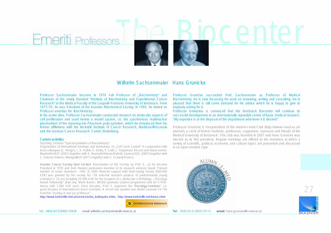
Professor Sachsenmaier became in 1970 Full Professor of „Biochemistry“ and Chairman of the newly founded “Institute of Biochemistry and Experimental Cancer Research” at the Medical Faculty of the Leopold-Franzens-University of Innsbruck. From 1977-79, he was President of the Austrian Biochemical Society. In 1995, he retired as Professor emeritus for Biochemistry. In his active time, Professor Sachsenmaier conducted research on molecular aspects of cell proliferation and used hereto a model system, i.e. the synchronous multinuclear plasmodium of the myxomycete Physarum polycephalum, which he introduced from his former affiliations with the McArdle Institute of Cancer Research, Madison/Wisconsin and the German Cancer Research Center, Heidelberg.
Current activitiesTeaching: Seminar “Special problems of Biochemistry”.Organization of international meetings and workshops on „Cell Cycle Control“ in cooperation with local colleagues (L. Hengst, L. A. Huber, S. Geley, P. Loidl, J. Troppmair). Recent and future events: Mayrhofen/AUT (2001) together with K. Nasmyth/Vienna/Oxford; Spetses/GR, (2007) together with C. Sekeris/ Athens; Montpellier/F (2011) together with E. Schwob/France.
Austrian Cancer Society–Tyrol Section: Reactivation of the Society by Prof. S., as he became President in 1970 and from thereon permanent member of its research advisory board. Present number of active members: ~400. In 2009 financial support with fund-raising money (300.000 EUR) was granted by the society for ~50 selected research projects of predominantly young scientists (< 35 yrs) including 30.000 EUR for the recipient of a „Molecular Cell Biology – Oncology Award Fellowship“ (Dipl.-Ing. Marin Barisic, MCBO-graduate student programme) and for 5 PhD- theses with 1.000 EUR each. Since decades, Prof. S. organizes the “OOncology-Seminars”, i.e. guest lectures of international cancer scientists. A recent top speaker was Nobel Laureate Sir Tim Hunt/UK “Getting in and out of Mitosis”.http://www.krebshilfe-tirol.at/service/onko_kolloquien.shtm, http://www.krebshilfe.net/home.shtm
Professor Grunicke succeeded Prof. Sachsenmaier as Professor of Medical Biochemistry. He is now focussing his work on reviewing, writing and consulting. He is pleased that there is still some demand for his advice which he is happy to give to anybody asking for it. Professor Grunickes is convinced that the Innsbruck Biocenter will continue its successful development to an internationally reputable centre of basic medical research. “My expertise is at the disposal of the department whenever it is desired”.
Professor Grunicke is Vicepresident of the Alumni-i-med-Club (http://www.i-med.ac.at/alumni/), a circle of former students, professors, supporters, sponsors and friends of the Medical University of Innsbruck. This club was founded in 2007 and Hans Grunicke was elected as its first president. Regular meetings are offered to the members at which a variety of scientific, political, economic and cultural topics are presented and discussed in an open-minded style.
Tel.: 0043 (512) 9003.70328 email: [email protected]
Wilhelm Sachsenmaier Hans Grunicke
Tel.: 0043 (512) 9003.70112 email: [email protected]
The Biocenterwww.autoimmunity.atTel.: 0043 (512) 9003.70960 email: [email protected] Professors
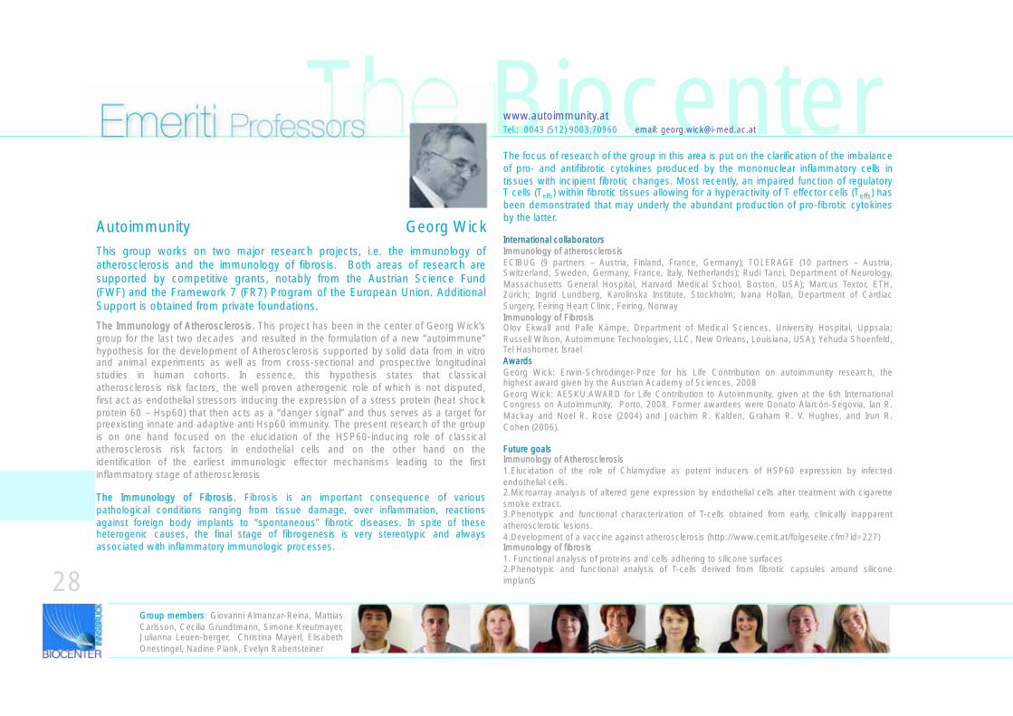
This group works on two major research projects, i.e. the immunology of atherosclerosis and the immunology of fibrosis. Both areas of research are supported by competitive grants, notably from the Austrian Science Fund (FWF) and the Framework 7 (FR7) Program of the European Union. Additional Support is obtained from private foundations.
The Immunology of Atherosclerosis. This project has been in the center of Georg Wick’s group for the last two decades and resulted in the formulation of a new “autoimmune” hypothesis for the development of Atherosclerosis supported by solid data from in vitro and animal experiments as well as from cross-sectional and prospective longitudinal studies in human cohorts. In essence, this hypothesis states that classical atherosclerosis risk factors, the well proven atherogenic role of which is not disputed, first act as endothelial stressors inducing the expression of a stress protein (heat shock protein 60 – Hsp60) that then acts as a “danger signal” and thus serves as a target for preexisting innate and adaptive anti Hsp60 immunity. The present research of the group is on one hand focused on the elucidation of the HSP60-inducing role of classical atherosclerosis risk factors in endothelial cells and on the other hand on the identification of the earliest immunologic effector mechanisms leading to the first inflammatory stage of atherosclerosis
The Immunology of Fibrosis. Fibrosis is an important consequence of various pathological conditions ranging from tissue damage, over inflammation, reactions against foreign body implants to “spontaneous” fibrotic diseases. In spite of these heterogenic causes, the final stage of fibrogenesis is very stereotypic and always associated with inflammatory immunologic processes.
CORRECTED BY AUTHOR VERSION
The focus of research of the group in this area is put on the clarification of the imbalance of pro- and antifibrotic cytokines produced by the mononuclear inflammatory cells in tissues with incipient fibrotic changes. Most recently, an impaired function of regulatory T cells (Teffs) within fibrotic tissues allowing for a hyperactivity of T effector cells (Teffs) has been demonstrated that may underly the abundant production of pro-fibrotic cytokines by the latter.
International collaborators Immunology of atherosclerosisECIBUG (9 partners – Austria, Finland, France, Germany); TOLERAGE (10 partners – Austria, Switzerland, Sweden, Germany, France, Italy, Netherlands); Rudi Tanzi, Department of Neurology, Massachusetts General Hospital, Harvard Medical School, Boston, USA); Marcus Textor, ETH, Zürich; Ingrid Lundberg, Karolinska Institute, Stockholm; Ivana Hollan, Department of Cardiac Surgery, Feiring Heart Clinic, Feiring, NorwayImmunology of Fibrosis Olov Ekwall and Palle Kämpe, Department of Medical Sciences, University Hospital, Uppsala; Russell Wilson, Autoimmune Technologies, LLC, New Orleans, Louisiana, USA); Yehuda Shoenfeld, Tel Hashomer, IsraelAwards Georg Wick: Erwin-Schrödinger-Prize for his Life Contribution on autoimmunity research, the highest award given by the Austrian Academy of Sciences, 2008Georg Wick: AESKU.AWARD for Life Contribution to Autoimmunity, given at the 6th International Congress on Autoimmunity, Porto, 2008. Former awardees were Donato Alarcón-Segovia, Ian R. Mackay and Noel R. Rose (2004) and Joachim R. Kalden, Graham R. V. Hughes, and Irun R. Cohen (2006).
Future goals Immunology of Atherosclerosis1.Elucidation of the role of Chlamydiae as potent inducers of HSP60 expression by infected endothelial cells. 2.Microarray analysis of altered gene expression by endothelial cells after treatment with cigarette smoke extract. 3.Phenotypic and functional characterization of T-cells obtained from early, clinically inapparent atherosclerotic lesions. 4.Development of a vaccine against atherosclerosis (http://www.cemit.at/folgeseite.cfm?id=227)Immunology of fibrosis1. Functional analysis of proteins and cells adhering to silicone surfaces2.Phenotypic and functional analysis of T-cells derived from fibrotic capsules around silicone implants
Group members: Giovanni Almanzar-Reina, Mattias Carlsson, Cecilia Grundtmann, Simone Kreutmayer, Julianna Leuen-berger, Christina Mayerl, Elisabeth Onestingel, Nadine Plank, Evelyn Rabensteiner
28
Autoimmunity Georg Wick

The Biocenter
29
Seminars & Happy Hour
The BiocenterCase ReportsWolfram, D.; Backovic, A.; Kaindl, R.; Hussl, H.; Wick, G.: Spontaneous unilateral autoinflation of a saline-filled mammary implant. JOURNAL OF PLASTIC RECONSTRUCTIVE AND AESTHETIC SURGERY. 2008; 61(3); 342-345. IF: 1.235
Original PublicationsAbdulle, Sahra; Hagberg, Lars; Svennerholm, Bo; Fuchs, Dietmar; Gisslén, Magnus: Cerebrospinal fluid viral load and intrathecal immune activation in individuals infected with different HIV-1 genetic subtypes. PLOS ONE. 2008; 3(4); e1971.
Baydar, T.; Girgin, G.; Fuchs, D.; Inanici, F.; Sipahi, H.; Erol, O.; Sahin, G.: Pteridine Pathway in Patients with Degenerative Diseases During Short Time Treatment with Low Dose of Meloxicam, as a Non-steroidal Anti-inflammatory Drug. PTERIDINES. 2008; 19(4); 107. IF: 0.706
Bellmann-Weiler, R.; Schroecksnadel, K.; Holzer, C.; Larcher, C.; Fuchs, D.; Weiss, G.: IFN-gamma mediated pathways in patients with fatigue and chronic active Epstein Barr virus-infection. JOURNAL OF AFFECTIVE DISORDERS. 2008; 108(1-2); 171-176. IF: 3.271
Blum, G.; Perkhofer, S.; Haas, H.; Schrettl, M.; Wurzner, R.; Dierich, MP.; Lass-Florl, C.: Potential basis for amphotericin B resistance in Aspergillus terreus. ANTIMICROBIAL AGENTS AND CHEMOTHERAPY. 2008; 52(4); 1553-1555. IF: 4.716
Brandacher, G.; Golderer, G.; Kienzl, K.; Werner, ER.; Margreiter, R.; Weiss, HG.: Potential applications of global protein expression analysis (proteomics) in morbid obesity and bariatric surgery. OBESITY SURGERY. 2008; 18(7); 905-910. IF: 2.913
Brunner, A.; Verdorfer, I.; Prelog, M.; Mayerl, C.; Mikuz, G.; Tzankov, A.: Large-scale analysis of cell cycle regulators in urothelial bladder cancer identifies p16 and p27 as potentially useful prognostic markers. PATHOBIOLOGY. 2008; 75(1); 25-33. IF: 1.818
Cano, OD.; Neurauter, G.; Fuchs, D.; Shearer, GM.; Boasso, A.: Differential effect of type I and type II interferons on neopterin production and amino acid metabolism in human astrocyte-derived cells. NEUROSCIENCE LETTERS. 2008; 438(1); 22-25. IF: 2.200
Carrico, AW.; Johnson, MO.; Morin, SF.; Remien, RH.; Riley, ED.; Hecht, FM.; Fuchs, D.: Stimulant use is associated with immune activation and depleted tryptophan among HIV-positive persons on anti-retroviral therapy. BRAIN BEHAVIOR AND IMMUNITY. 2008; 22(8); 1257-1262. IF: 4.909
Casella, I.; Lindner, H.; Zenzmaier, C.; Riitano, D.; Berger, P.; Costa, T.: Non-gonadotropin-releasing hormone-mediated transcription and secretion of large human glycoprotein hormone alpha-subunit in human embryonic kidney-293 cells. ENDOCRINOLOGY. 2008; 149(3); 1144-1154. IF: 4.945
Cecchinato, V.; Tryniszewska, E.; Ma, ZM.; Vaccari, M.; Boasso, A.; Tsai, WP.; Petrovas, C.; Fuchs, D.; Heraud, JM.; Venzon, D.; Shearer, GM.; Koup, RA.; Lowy, I.; Miller, CJ.; Franchini, G.: Immune activation driven by CTLA-4 blockade augments viral replication at mucosal sites in simian immunodeficiency virus infection. JOURNAL OF IMMUNOLOGY. 2008; 180(8); 5439-5447. IF: 6.000
Efimov, A.; Schiefermeier, N.; Grigoriev, I.; Brown, MC.; Turner, CE.; Small, JV.; Kaverina, I.: Paxillin-dependent stimulation of microtubule catastrophes at focal adhesion sites. JOURNAL OF CELL SCIENCE. 2008; 121(2); 196-204. IF: 6.247
Espelt, MV.; Alleva, K.; Amodeo, G.; Krumschnabel, G.; Rossi, RC.; Schwarzbaum, PJ.: Volumetric response of vertebrate hepatocytes challenged by osmotic gradients: A theoretical approach. COMPARATIVE BIOCHEMISTRY AND PHYSIOLOGY B-BIOCHEMISTRY & MOLECULAR BIOLOGY. 2008; 150(1); 103-111. IF: 1.468
Fluckinger, M.; Merschak, P.; Hermann, M.; Haertle, T.; Redl, B.: Lipocalin-interacting-membrane-receptor (LIMR) mediates cellular internalization of beta-lactoglobulin. BIOCHIMICA ET BIOPHYSICA ACTA-BIOMEMBRANES. 2008; 1778(1); 342-347
Fritsche, G.; Nairz, M.; Werner, ER.; Barton, HC.; Weiss, G.: Nramp1-functionality increases iNOS expression via repression of IL-10 formation. EUROPEAN JOURNAL OF IMMUNOLOGY. 2008; 38(11); 3060-3067. IF: 4.865
Glockner, G.; Golderer, G.; Werner-Felmayer, G.; Meyer, S.; Marwan, W.: A first glimpse at the transcriptome of Physarum polycephalum. BMC GENOMICS. 2008; 9(2). IF: 3.926
Green, A.; Sarg, B.; Koutzamani, E.; Genheden, U.; Lindner, HH.; Rundquist, I.: Histone H1 dephosphorylation is not a general feature in early apoptosis. BIOCHEMISTRY. 2008; 47(28); 7539-7547. IF: 3.379
Greilberger, J.; Koidl, C.; Greilberger, M.; Lamprecht, M.; Schroecksnadel, K.; Leblhuber, F.; Fuchs, D.; Oettl, K.: Malondialdehyde, carbonyl proteins and albumin-disulphide as useful oxidative markers in mild cognitive impairment and Alzheimer's disease. FREE RADICAL RESEARCH. 2008; 42(7); 633-638. IF: 2.826
30
Publications 2008
The Biocenter
31
Publications 2008Gruber, T.; Fresser, F.; Jenny, M.; Uberall, F.; Leitges, M.; Baier, GB.: PKC theta cooperates with atypical PKC zeta and PKC iota in NF-kappa B transactivation of T lymphocytes. MOLECULAR IMMUNOLOGY. 2008; 45(1); 117-126. IF: 3.555
Haffner, MC.; Jurgeit, A.; Berlato, C.; Geley, S.; Parajuli, N.; Yoshimura, A.; Doppler, W.: Interaction and functional interference of glucocorticoid receptor and SOCS1. JOURNAL OF BIOLOGICAL CHEMISTRY. 2008; 283(32); 22089-22096. IF: 5.520
Hammerer-Lercher, A.; Halfinger, B.; Sarg, B.; Mair, J.; Puschendorf, B.; Griesmacher, A.; Guzman, NA.; Lindner, HH.: Analysis of circulating forms of proBNP and NT-proBNP in patients with severe heart failure. CLINICAL CHEMISTRY. 2008; 54(5); 858-865. IF: 5.579
Henderson, B.; Csordas, A.; Backovic, A.; Kind, M.; Bernhard, D.; Wick, G.: Cigarette smoke is an endothelial stressor and leads to cell cycle arrest. ATHEROSCLEROSIS. 2008; 201(2); 298-305. IF: 4.601
Hermann-Kleiter, N.; Gruber, T.; Lutz-Nicoladoni, C.; Thuille, N.; Fresser, F.; Labi, V.; Schiefermeier, N.; Warnecke, M.; Huber, L.; Villunger, A.; Eichele, G.; Kaminski, S.; Baier, G.: The nuclear orphan receptor NR2176 suppresses lymphocyte activation and T helper 17-dependent autoimmunity. IMMUNITY. 2008; 29(2); 205-216. IF: 20.579
Imeri, F.; Herklotz, R.; Risch, L.; Arbetsleitner, C.; Zerlauth, M.; Risch, GM.; Huber, AR.: Stability of hematological analytes depends on the hematology analyser used: A stability study with Bayer Advia 120, Beckman Coulter LH 750 and Sysmex XE 2100. CLINICA CHIMICA ACTA. 2008; 397(1-2); 68-71. IF: 2.960
Jenny, M.; Winkler, C.; Spetea, M.; Schennach, H.; Schmidhammer, H.; Fuchs, D.: Non-peptidic delta-Opioid receptor antagonists suppress mitogen-induced tryptophan degradation in peripheral blood mononuclear cells in vitro. IMMUNOLOGY LETTERS. 2008; 118(1); 82-87. IF: 2.858
Jochl, C.; Rederstorff, M.; Hertel, J.; Stadler, PF.; Hofacker, IL.; Schrettl, M.; Haas, H.; Huttenhofer, A.: Small ncRNA transcriptome analysis from Aspergillus fumigatus suggests a novel mechanism for regulation of protein synthesis. NUCLEIC ACIDS RESEARCH. 2008; 36(8); 2677-2689. IF: 6.878
Kitchen, Maria; Quigley, Maria A.; Mwinga, Alwyn M.; Fuchs, Dietmar; Lisse, Ida M.; Porter, John D H.; McAdam, Keith P W J.; Godfrey-Faussett, Peter: HIV Progression and Predictors of Mortality in a Community-Based Cohort of Zambian Adults. JOURNAL OF THE INTERNATIONAL ASSOCIATION OF PHYSICIANS IN AIDS CARE. 2008; 7(1); 17-26.
Kositz, C.; Schroecksnadel, K.; Grander, G.; Schennach, H.; Kofler, H.; Fuchs, D.: High serum tryptophan concentration in pollinosis patients is associated with unresponsiveness to pollen extract therapy. INTERNATIONAL ARCHIVES OF ALLERGY AND IMMUNOLOGY. 2008; 147(1); 35-40. IF: 2.131 (2008) / ZIT: 0Labi, V.;
Erlacher, M.; Kiessling, S.; Manzl, C.; Frenzel, A.; O'Reilly, L.; Strasser, A.; Villunger, A.: Loss of the BH3-only protein Bmf impairs B cell homeostasis and accelerates gamma irradiation-induced thymic lymphoma development. JOURNAL OF EXPERIMENTAL MEDICINE. 2008; 205(3); 641-655. IF: 15.219 (2008) / ZIT: 7
Lang, K.; Erlacher, M.; Wilson, DN.; Micura, R.; Polacek, N.: The role of 23S ribosomal RNA residue A2451 in peptide bond synthesis revealed by atomic mutagenesis. CHEMISTRY & BIOLOGY. 2008; 15(5); 485-492. IF: 5.603 (2008) / ZIT: 5
Lirk, P.; Haller, I.; Colvin, HP.; Lang, L.; Tomaselli, B.; Klimaschewski, L.; Gerner, P.: In vitro, inhibition of mitogen-activated protein kinase pathways protects against bupivacaine- and ropivacaine-induced neurotoxicity. ANESTHESIA AND ANALGESIA. 2008; 106(5); 1456-1464. IF: 2.590 (2008) / ZIT: 0
Madej, MJ.; Niemann, M.; Huttenhofer, A.; Goringer, HU.: Identification of novel guide RNAs from the mitochondria of Trypanosome brucei. RNA BIOLOGY. 2008; 5(2); 84-91. IF: / ZIT: 1
Mai, A.; Cheng, D.; Bedford, MT.; Valente, S.; Nebbioso, A.; Perrone, A.; Brosch, G.; Sbardella, G.; De Bellis, F.; Miceli, M.; Altucci, L.: Epigenetic multiple ligands: Mixed Histone/Protein methyltransferase, acetyltransferase, and class III deacetylase (Sirtuin) inhibitors. JOURNAL OF MEDICINAL CHEMISTRY. 2008; 51(7); 2279-2290. IF: 4.898 (2008) / ZIT: 16
Mair, J.; Hammerer-Lercher, A.; Mittermayr, M.; Klingler, A.; Humpeler, E.; Pachinger, O.; Schobersberger, W.: 3-week hiking holidays at moderate altitude do not impair cardiac function in individuals with metabolic syndrome. INTERNATIONAL JOURNAL OF CARDIOLOGY. 2008; 123(2); 186-188. IF: 3.121 (2008) / ZIT: 0
Massoner, P.; Haag, P.; Seifarth, C.; Jurgeit, A.; Rogatsch, H.; Doppler, W.; Bartsch, G.; Klocker, H.: Insulin-like growth factor binding protein-3 (IGFBP-3) in the prostate and in prostate cancer: Local production, distribution and secretion pattern indicate a role in stromal-epithelial interaction. PROSTATE. 2008; 68(11); 1165-1178. IF: 3.069 (2008) / ZIT: 3
32
The BiocenterMcDonagh, A.; Fedorova, ND.; Crabtree, J.; Yu, Y.; Kim, S.; Chen, D.; Loss, O.; Cairns, T.; Goldman, G.; Armstrong-James, D.; Haynes, K.; Haas, H.; Schrettl, M.; May, G.; Nierman, WC.; Bignell, E.: Sub-telomere directed gene expression during initiation of invasive aspergillosis. PLOS PATHOGENS. 2008; 4(9); e1000154. IF: 9.125 (2008) / ZIT: 7
Meraner, J.; Lechner, M.; Schwarze, F.; Gander, R.; Jesacher, F.; Loidl, P.: Cell cycle dependent role of HDAC1 for proliferation control through modulating ribosomal DNA transcription. CELL BIOLOGY INTERNATIONAL. 2008; 32(9); 1073-1080. IF: 1.619 (2008) / ZIT: 0
Michalak, EM.; Villunger, A.; Adams, JM.; Strasser, A.: In several cell types tumour suppressor p53 induces apoptosis largely via Puma but Noxa can contribute. CELL DEATH AND DIFFERENTIATION. 2008; 15(6); 1019-1029. IF: 7.548 (2008) / ZIT: 16
Moheno, P.; Pfleiderer, W.; DiPasquale, AG.; Rheingold, AL.; Fuchs, D.: Cytokine and IDO metabolite changes effected by calcium pterin during inhibition of MDA-MB-231 xenograph tumors in nude mice. INTERNATIONAL JOURNAL OF PHARMACEUTICS. 2008; 355(1-2); 238-248. IF: 3.061 (2008) / ZIT: 1
Moser, B.; Schroecksnadel, K.; Hortnagl, H.; Rieder, J.; Fuchs, D.; Gottardis, M.: Influence of Extreme Long Endurance Sports Activity on Neopterin Excretion. PTERIDINES. 2008; 19(4); 114. IF: 0.706 (2008) / ZIT: 0
Muller, T.; Hess, MW.; Schiefermeier, N.; Pfaller, K.; Ebner, HL.; Heinz-Erian, P.; Ponstingl, H.; Partsch, J.; Rollinghoff, B.; Kohler, H.; Berger, T.; Lenhartz, H.; Schlenck, B.; Houwen, RJ.; Taylor, CJ.; Zoller, H.; Lechner, S.; Goulet, O.; Utermann, G.; Ruemmele, FM.; Huber, LA.; Janecke, AR.: MYO5B mutations cause microvillus inclusion disease and disrupt epithelial cell polarity. NATURE GENETICS. 2008; 40(10); 1163-1165. IF: 30.259 (2008) / ZIT: 5
Nairz, M.; Fritsche, G.; Brunner, P.; Talasz, H.; Hantke, K.; Weiss, G.: Interferon-gamma limits the availability of iron for intramacrophage Salmonella typhimurium. EUROPEAN JOURNAL OF IMMUNOLOGY. 2008; 38(7); 1923-1936. IF: 4.865 (2008) / ZIT: 10
Neher, A.; Schobersberger, W.; Augustijns, P.; Fuchs, D.; Wolf-Magele, A.; Hoffmann, G.: Influence of Neopterin on Ciliary Beat Frequency of Human Nasal Epithelial Cells in vitro. PTERIDINES. 2008; 19(3); 79. IF: 0.706 (2008) / ZIT: 0
Publications 2008Neurauter, G.; Grahmann, AV.; Klieber, M.; Zeimet, A.; Ledochowski, M.; Sperner-Unterweger, B.; Fuchs, D.: Serum phenylalanine concentrations in patients with ovarian carcinoma correlate with concentrations of immune activation markers and of isoprostane-8. CANCER LETTERS. 2008; 272(1); 141-147. IF: 3.504 (2008) / ZIT: 0
Offterdinger, M.; Bastiaens, PI.: Prolonged EGFR signaling by ERBB2-mediated sequestration at the plasma membrane. TRAFFIC. 2008; 9(1); 147-155. IF: 5.709 (2008) / ZIT: 4
Paddison, JS.; Booth, RJ.; Fuchs, D.; Hill, AG.: Peritoneal inflammation and fatigue experiences following colorectal surgery: A pilot study. PSYCHONEUROENDOCRINOLOGY. 2008; 33(4); 446-454. IF: 3.788 (2008) / ZIT: 2
Pafundo, DE.; Chara, O.; Faillace, MP.; Krumschnabel, G.; Schwarzbaum, PJ.: Kinetics of ATP release and cell volume regulation of hyposmotically challenged goldfish hepatocytes. AMERICAN JOURNAL OF PHYSIOLOGY-REGULATORY INTEGRATIVE AND COMPARATIVE PHYSIOLOGY. 2008; 294(1); R220-R233. IF: 3.272 (2008) / ZIT: 3
Perkhofer, S.; Kehrel, BE.; Dierich, MP.; Donnelly, JP.; Nussbaumer, W.; Hofmann, J.; von Eiff, C.; Lass-Florl, C.: Human platelets attenuate Aspergillus species via granule-dependent mechanisms. JOURNAL OF INFECTIOUS DISEASES. 2008; 198(8); 1243-1246. IF: 5.682 (2008) / ZIT: 3
Pfister, D.; De Mulder, K.; Hartenstein, V.; Kuales, G.; Borgonie, G.; Marx, F.; Morris, J.; Ladurner, P.: Flatworm stem cells and the germ line: Developmental and evolutionary implications of macvasa expression in Macrostomum lignano. DEVELOPMENTAL BIOLOGY. 2008; 319(1); 146-159. IF: 4.416 (2008) / ZIT: 6
Ploder, M.; Neurauter, G.; Spittler, A.; Schroecksnadel, K.; Roth, E.; Fuchs, D.: Serum phenylalanine in patients post trauma and with sepsis correlate to neopterin concentrations. AMINO ACIDS. 2008; 35(2); 303-307. IF: 4.132 (2008) / ZIT: 1
Ploner, C.; Rainer, J.; Niederegger, H.; Eduardoff, M.; Villunger, A.; Geley, S.; Kofler, R.: The BCL2 rheostat in glucocorticoid-induced apoptosis of acute lymphoblastic leukemia. LEUKEMIA. 2008; 22(2); 370-377. IF: 8.634 (2008) / ZIT: 10
Purschwitz, J.; Mueller, S.; Kastner, C.; Schoser, M.; Haas, H.; Espeso, EA.; Atoui, A.; Calvo, AM.; Fischer, R.: Functional and physical interaction of blue- and red-light sensors in Aspergillus nidulans. CURRENT BIOLOGY. 2008; 18(4); 255-259. IF: 10.777 (2008) / ZIT: 17
Ragno, R.; Simeoni, S.; Rotili, D.; Caroli, A.; Botta, G.; Brosch, G.; Massa, S.; Mai, A.: Class II-selective histone deacetylase inhibitors. Part 2: Alignment-independent GRIND 3-D QSAR, homology and docking studies. EUROPEAN JOURNAL OF MEDICINAL CHEMISTRY. 2008; 43(3); 621-632. IF: 2.882 (2008) / ZIT: 6
Rainer, M.; Sonderegger, H.; Bakry, R.; Huck, CW.; Morandell, S.; Huber, LA.; Gjerde, DT.; Bonn, GK.: Analysis of protein phosphorylation by monolithic extraction columns based on poly(divinylbenzene) containing embedded titanium dioxide and zirconium dioxide nano-powders. PROTEOMICS. 2008; 8(21); 4593-4602. IF: 4.586 (2008) / ZIT: 2
Reitinger, S.; Boroviak, T.; Laschober, GT.; Fehrer, C.; Muellegger, J.; Lindner, H.; Lepperdinger, G.: High-yield recombinant expression of the extremophile enzyme, bee hyaluronidase in Pichia pastoris. PROTEIN EXPRESSION AND PURIFICATION. 2008; 57(2); 226-233. IF: 1.621 (2008) / ZIT: 2
Rossmann, A.; Henderson, B.; Heidecker, B.; Seiler, R.; Fraedrich, G.; Singh, M.; Parson, W.; Keller, M.; Grubeck-Loebenstein, B.; Wick, G.: T-cells from advanced atherosclerotic lesions recognize hHSP60 and have a restricted T-cell receptor repertoire. EXPERIMENTAL GERONTOLOGY. 2008; 43(3); 229-237. IF: 3.283 (2008) / ZIT: 7
Schmidt-Glenewinkel, H.; Vacheva, I.; Hoeller, D.; Dikic, I.; Eils, R.: An ultrasensitive sorting mechanism for EGF Receptor Endocytosis. BMC SYSTEMS BIOLOGY. 2008; 2(4); IF: / ZIT: 2
Schneeberger, S.; Hautz, T.; Wahl, SM.; Brandacher, G.; Sucher, R.; Steinmassl, O.; Steinmassl, P.; Wright, CD.; Obrist, P.; Werner, ER.; Mark, W.; Troppmair, J.; Margreiter, R.; Amberger, A.: The effect of secretory leukocyte protease inhibitor (SLPI) on ischemia/reperfusion injury in cardiac transplantation. AMERICAN JOURNAL OF TRANSPLANTATION. 2008; 8(4); 773-782. IF: 6.559 (2008) / ZIT: 1
Schrettl, M.; Kim, HS.; Eisendle, M.; Kragl, C.; Nierman, WC.; Heinekamp, T.; Werner, ER.; Jacobsen, I.; Illmer, P.; Yi, H.; Brakhage, AA.; Haas, H.: SreA-mediated iron regulation in Aspergillus fumigatus. MOLECULAR MICROBIOLOGY. 2008; 70(1); 27-43. IF: 5.213 (2008) / ZIT: 5
Schroecksnadel, K.; Sarcletti, M.; Winkler, C.; Mumelter, B.; Weiss, G.; Fuchs, D.; Kemmler, G.; Zangerle, R.: Quality of life and immune activation in patients with HIV-infection. BRAIN BEHAVIOR AND IMMUNITY. 2008; 22(6); 881-889. IF: 4.909 (2008) / ZIT: 3
Schroecksnadel, K.; Walter, RB.; Weiss, G.; Mark, M.; Reinhart, WH.; Fuchs, D.: Association between plasma thiols and immune activation marker neopterin in stable coronary heart disease. CLINICAL CHEMISTRY AND LABORATORY MEDICINE. 2008; 46(5); 648-654. IF: 1.888 (2008) / ZIT: 0
Schweigreiter, R.: The natural history of the myelin-derived nerve growth inhibitor Nogo-A. NEURON GLIA BIOLOGY. 2008; 4(4); 83-89. IF: 0.937 (2008) / ZIT: 0
Scrandis, Debra A.; Langenberg, Patricia; Tonelli, Leonardo H.; Sheikh, Tehmina M.; Manogura, Anita C.; Alberico, Laura A.; Hermanstyne, Tracey; Fuchs, Dietmar; Mighty, Hugh; Hasday, Jeffrey D.; Boteva, Kalina; Postolache, Teodor T.: Prepartum Depressive Symptoms Correlate Positively with C-Reactive Protein Levels and Negatively with Tryptophan Levels: A Preliminary Report. INTERNATIONAL JOURNAL OF CHILD HEALTH AND HUMAN DEVELOPMENT. 2008; 1(2); 167-174.
Seifert, M.; Nairz, M.; Schroll, A.; Schrettl, M.; Haas, H.; Weiss, G.: Effects of the Aspergillus fumigatus siderophore systems on the regulation of macrophage immune effector pathways and iron homeostasis. IMMUNOBIOLOGY. 2008; 213(9-10); 767-778. IF: 3.461
Skvortsov, S.; Sarg, B.; Lindner, H.; Lukas, P.; Hilbe, W.; Zwierzina, H.; Skvortsova, I.: Cetuximab inhibits thymidylate synthase in colorectal cells expressing epidermal growth factor receptor. PROTEOMICS CLINICAL APPLICATIONS. 2008; 2(6); 908-914. IF: 1.514
Skvortsova, I.; Skvortsov, S.; Stasyk, T.; Raju, U.; Popper, BA.; Schiestl, B.; von Guggenberg, E.; Neher, A.; Bonn, GK.; Huber, LA.; Lukas, P.: Intracellular signaling pathways regulating radioresistance of human prostate carcinoma cells. PROTEOMICS. 2008; 8(21); 4521-4533. IF: 4.586
Sonnleitner, E.; Sorger-Domenigg, T.; Madej, MJ.; Findeiss, S.; Hackermuller, J.; Huettenhofer, A.; Stadler, PF.; Blasi, U.; Moll, I.: Detection of small RNAs in Pseudomonas aeruginosa by RNomics and structure-based bioinformatic tools. MICROBIOLOGY-SGM. 2008; 154(8); 3175-3187. IF: 2.841
Teufel, L.; Schuster, KC.; Merschak, P.; Bechtold, T.; Redl, B.: Development of a fast and reliable method for the assessment of microbial colonization and growth on textiles by DNA quantification. JOURNAL OF MOLECULAR MICROBIOLOGY AND BIOTECHNOLOGY. 2008; 14(4); 193-200. IF: 2.286
Theurl, I.; Theurl, M.; Seifert, M.; Mair, S.; Nairz, M.; Rumpold, H.; Zoller, H.; Bellmann-Weiler, R.; Niederegger, H.; Talasz, H.; Weiss, G.: Autocrine formation of hepcidin induces iron retention in human monocytes. BLOOD. 2008; 111(4); 2392-2399. IF: 10.432
Tishchenko, S.; Kljashtorny, V.; Kostareva, O.; Nevskaya, N.; Nikulin, A.; Gulak, P.; Piendl, W.; Garber, M.; Nikonov, S.: Domain II of Thermus thermophilus Ribosomal Protein L1 Hinders Recognition of Its mRNA. JOURNAL OF MOLECULAR BIOLOGY. 2008; 383(2); 301-305. IF: 4.146 33
The BiocenterPublications 2008
The BiocenterTomaselli, B.; zur Nedden, S.; Podhraski, V.; Baier-Bitterlich, G.: p42/44 MAPK is an essential effector for purine nucleoside-mediated neuroprotection of hypoxic PC12 cells and primary cerebellar granule neurons. MOLECULAR AND CELLULAR NEUROSCIENCE. 2008; 38(4); 559-568. IF: 3.934
Vaccari, M.; Boasso, A.; Ma, ZM.; Cecchinato, V.; Venzon, D.; Doster, MN.; Tsai, WP.; Shearer, GM.; Fuchs, D.; Felber, BK.; Pavlakis, GN.; Miller, CJ.; Franchini, G.: CD4(+) T-cell loss and delayed expression of modulators of immune responses at mucosal sites of vaccinated macaques following SIVmac251 infection. MUCOSAL IMMUNOLOGY. 2008; 1(6); 497-507.
Volgger, B.; Aspisirengil, C.; Genser-Krimbacher, E.; Ciresa-Koenig, A.; Daxenbichler, G.; Fuchs, D.; Windbichler, G.; Marth, C.: Prognostic significance of TPA versus SCC-Ag, CEA and neopterin in carcinoma of the uterine cervix. CANCER LETTERS. 2008; 262(2); 183-189. IF: 3.504 (
Watschinger, K.; Horak, SB.; Schulze, K.; Obermair, GJ.; Wild, C.; Koschak, A.; Sinnegger-Brauns, MJ.; Tampe, R.; Striessnig, J.: Functional properties and modulation of extracellular epitope-tagged Ca(V)2.1 voltage-gated calcium channels. CHANNELS (AUSTIN, TEX.). 2008; 2(6); 461-473. IF: 1.513
Weinmeister, P.; Lukowski, R.; Linder, S.; Traidl-Hoffmann, C.; Hengst, L.; Hofmann, F.; Feil, R.: Cyclic Guanosine Monophosphate-dependent Protein Kinase I Promotes Adhesion of Primary Vascular Smooth Muscle Cells. MOLECULAR BIOLOGY OF THE CELL. 2008; 19(10); 4434-4441. IF: 5.558
Wesierska-Gadek, J.; Hajek, SB.; Sarg, B.; Wandl, S.; Walzi, E.; Lindner, H.: Pleiotropic effects of selective CDK inhibitors on human normal and cancer cells. BIOCHEMICAL PHARMACOLOGY. 2008; 76(11); 1503-1514. IF: 4.838
Wick, MC.; Mayerl, C.; Backovic, A.; van der Zee, R.; Jaschke, W.; Dietrich, H.; Wick, G.: In vivo imaging of the effect of LPS on arterial endothelial cells: molecular imaging of heat shock protein 60 expression. CELL STRESS & CHAPERONES. 2008; 13(3); 275-285. IF: 2.238
Winkler, C.; Schroecksnadel, K.; Ledochowski, M.; Schennach, H.; Houcher, B.; Fuchs, D.: In vitro Effects of Nigella sativa Seeds Extracts on Stimulated Peripheral Blood Mononuclear Cells. PTERIDINES. 2008; 19(4); 101. IF: 0.706
Wolfram, D.; Oberreiter, B.; Mayerl, C.; Soelder, E.; Ulmer, H.; Piza-Katzer, H.; Wick, G.; Backovic, A.: Altered systemic serologic parameters in patients with silicone mammary implants. IMMUNOLOGY LETTERS. 2008; 118(1); 96-100. IF: 2.858
Wollensak, G.; Redl, B.: Gel electrophoretic analysis of corneal collagen after photodynamic cross-linking treatment. CORNEA. 2008; 27(3); 353-356. IF: 1.853
Yegnasubramanian, S.; Haffner, MC.; Zhang, YG.; Gurel, B.; Cornish, TC.; Wu, ZJ.; Irizarry, RA.; Morgan, J.; Hicks, J.; DeWeese, TL.; Isaacs, WB.; Bova, GS.; De Marzo, AM.; Nelson, WG.: DNA Hypomethylation Arises Later in Prostate Cancer Progression than CpG Island Hypermethylation and Contributes to Metastatic Tumor Heterogeneity. CANCER RESEARCH. 2008; 68(21); 8954-8967. IF: 7.514 (
Yilmaz, A.; Price, RW.; Spudich, S.; Fuchs, D.; Hagberg, L.; Gisslen, M.: Persistent intrathecal immune activation in HIV-1-infected individuals on antiretroviral therapy. JAIDS-JOURNAL OF ACQUIRED IMMUNE DEFICIENCY SYNDROMES. 2008; 47(2); 168-173. IF: 4.570
zur Nedden, S.; Tomaselli, B.; Baier-Bitterlich, G.: HIF-1 alpha is an essential effector for purine nucleoside-mediated neuroprotection against hypoxia in PC12 cells and primary cerebellar granule neurons. JOURNAL OF NEUROCHEMISTRY. 2008; 105(5); 1901-1914. IF: 4.500
Proceedings PapersNeurauter, G.; Sperner-Unterweger, B.; Ledochowski, M.; Fuchs, D.: Tryptophan and phenylalanine in systemic and CNS inflammatory disorders. ACTA NEUROPSYCHIATRICA. 2008; 20(7); 5-6. IF: 1.380
ReviewsBrandacher, G.; Margreiter, R.; Fuchs, D.: Clinical relevance of indoleamine 2,3-dioxygenase for alloimmunity and transplantation. CURRENT OPINION IN ORGAN TRANSPLANTATION. 2008; 13(1); 10-15. IF: 0.435
Brosch, G.; Loidl, P.; Graessle, S.: Histone modifications and chromatin dynamics: a focus on filamentous fungi. FEMS MICROBIOLOGY REVIEWS. 2008; 32(3); 409-439. IF: 7.963
Chu, IM.; Hengst, L.; Slingerland, JM.: The Cdk inhibitor p27 in human cancer: prognostic potential and relevance to anticancer therapy. NATURE REVIEWS CANCER. 2008; 8(4); 253-267. IF: 30.762
Gu, YS.; Kong, J.; Cheema, GS.; Keen, CL.; Wick, G.; Gershwin, ME.: The immunobiology of systemic sclerosis. SEMINARS IN ARTHRITIS AND RHEUMATISM. 2008; 38(2); 132-160. IF: 4.379
Haas, H.; Eisendle, M.; Turgeon, BG.: Siderophores in fungal physiology and virulence. ANNUAL REVIEW OF PHYTOPATHOLOGY. 2008; 46(1); 149-187.IF: 10.237
34
Publications 2008
The Biocenter
35
Original PublicationsBarrientos, G.; Fuchs, D.; Schrocksnadel, K.; Ruecke, M.; Garcia, MG.; Klapp, BF.; Raghupathy, R.; Miranda, S.; Arck, PC.; Blois, SM.: Low levels of serum asymmetric antibodies as a marker of threatened pregnancy. JOURNAL OF REPRODUCTIVE IMMUNOLOGY. 2009; 79(2); 201-210. IF: 2.778
Batta, G.; Barna, T.; Gaspari, Z.; Sandor, S.; Kover, KE.; Binder, U.; Sarg, B.; Kaiserer, L.; Chhillar, AK.; Eigentler, A.; Leiter, E.; Hegedus, N.; Pocsi, I.; Lindner, H.; Marx, F.: Functional aspects of the solution structure and dynamics of PAF - a highly-stable antifungal protein from Penicillium chrysogenum. FEBS JOURNAL. 2009; 276(10); 2875-2890. IF: 3.139
Berlato, C.; Doppler, W.: Selective Response to Insulin Versus Insulin-Like Growth Factor-I and -II and Up-Regulation of Insulin Receptor Splice Variant B in the Differentiated Mouse Mammary Epithelium. ENDOCRINOLOGY. 2009; 150(6); 2924-2933. IF: 4.945
Boasso, A.; Vaccari, M.; Fuchs, D.; Hardy, AW.; Tsai, WP.; Tryniszewska, E.; Shearer, GM.; Franchini, G.: Combined Effect of Antiretroviral Therapy and Blockade of IDO in SIV-Infected Rhesus Macaques. JOURNAL OF IMMUNOLOGY. 2009; 182(7); 4313-4320. IF: 6.000
Capuron, L.; Moranis, A.; Combe, N.; Cousson-Gelie, F.; Fuchs, D.; De Smedt-Peyrusse, V.; Barberger-Gateau, P.; Laye, S.: Vitamin E status and quality of life in the elderly: influence of inflammatory processes. BRITISH JOURNAL OF NUTRITION. 2009; 102(10); 1390-1394. IF: 2.764
Castets, P.; Maugenre, S.; Gartioux, C.; Rederstorff, M.; Krol, A.; Lescure, A.; Tajbakhsh, S.; Allamand, V.; Guicheney, P.: Selenoprotein N is dynamically expressed during mouse development and detected early in muscle precursors. BMC DEVELOPMENTAL BIOLOGY. 2009; 9(3); IF: 3.079
Dohnal, AM.; Luger, R.; Paul, P.; Fuchs, D.; Felzmann, T.: CD40 ligation restores type 1 polarizing capacity in TLR4-activated dendritic cells that have ceased interleukin-12 expression. JOURNAL OF CELLULAR AND MOLECULAR MEDICINE. 2009; 13(8B); 1741-1750. IF: 5.114
Ecker, K.; Lorenz, A.; Wolf, F.; Ploner, C.; Bock, G.; Duncan, T.; Geley, S.; Helmberg, A.: A RAS recruitment screen identifies ZKSCAN4 as a glucocorticoid receptor-interacting protein. JOURNAL OF MOLECULAR ENDOCRINOLOGY. 2009; 42(1-2); 105-117. IF: 3.225
Eisenmann, A.; Murr, C.; Fuchs, D.; Ledochowski, M.: Gliadin IgG antibodies and circulating immune complexes. SCANDINAVIAN JOURNAL OF GASTROENTEROLOGY. 2009; 44(2); 168-171. IF: 1.980
Publications 2008 2009Labi, V.; Grespi, F.; Baumgartner, F.; Villunger, A.: Targeting the Bcl-2-regulated apoptosis pathway by BH3 mimetics: a breakthrough in anticancer therapy? CELL DEATH AND DIFFERENTIATION. 2008; 15(6); 977-987. IF: 7.548
Lindner, HH.: Analysis of histones, histone variants, and their post-translationally modified forms. ELECTROPHORESIS. 2008; 29(12); 2516-2532. IF: 3.509
Marx, F.; Binder, U.; Leiter, E.; Pocsi, I.: The Penicillium chrysogenum antifungal protein PAF, a promising tool for the development of new antifungal therapies and fungal cell biology studies. CELLULAR AND MOLECULAR LIFE SCIENCES. 2008; 65(3); 445-454. IF: 5.511
Morandell, S.; Stasyk, T.; Skvortsov, S.; Ascher, S.; Huber, LA.: Quantitative proteomics and phosphoproteomics reveal novel insights into complexity and dynamics of the EGFR signaling network. PROTEOMICS. 2008; 8(21); 4383-4401. IF: 4.586
Morettini, S.; Podhraski, V.; Lusser, A.: ATP-dependent chromatin remodeling enzymes and their various roles in cell cycle control. FRONTIERS IN BIOSCIENCE. 2008; 13(3); 5522-5532. IF: 3.308
Neurauter, G.; Schrocksnadel, K.; Scholl-Burgi, S.; Sperner-Unterweger, B.; Schubert, C.; Ledochowski, M.; Fuchs, D.: Chronic immune stimulation correlates with reduced phenylalanine turnover. CURRENT DRUG METABOLISM. 2008; 9(7); 622-627. IF: 4.350
Pinon, JD.; Labi, V.; Egle, A.; Villunger, A.: Bmf and Bmf in tissue homeostasis and malignant disease. ONCOGENE. 2008; 27(9); S41-S52. IF: 7.216
Ploner, C.; Kofler, R.; Villunger, A.: Noxa: at the tip of the balance between life and death. ONCOGENE. 2008; 27(9); S84-S92. IF: 7.216
Rymarquis, LA.; Kastenmayer, JP.; Huttenhofer, AG.; Green, PJ.: Diamonds in the rough: mRNA-like non-coding RNAs. TRENDS IN PLANT SCIENCE. 2008; 13(7); 329-334. IF: 9.210
Sgonc, R.; Wick, G.: Pro- and anti-fibrotic effects of TGF-beta in scleroderma. RHEUMATOLOGY. 2008; 47(2); V5-V7. IF: 4.136
Wick, MC.; Kremser, C.; Frischauf, S.; Wick, G.: In vivo molecular imaging of vascular stress. CELL STRESS & CHAPERONES. 2008; 13(3); 263-273. IF: 2.238
100 Publications in 2008
36
Heinke, R.; Spannhoff, A.; Meier, R.; Trojer, P.; Bauer, I.; Jung, M.; Sippl, W.: Virtual Screening and Biological Characterization of Novel Histone Arginine Methyltransferase PRMT1 Inhibitors. CHEMMEDCHEM. 2009; 4(1); 69-77. IF: 3.150
Heiss, EH.; Schachner, D.; Werner, ER.; Dirsch, VM.: Active NF-E2-related Factor (Nrf2) Contributes to Keep Endothelial NO Synthase (eNOS) in the Coupled State ROLE OF REACTIVE OXYGEN SPECIES (ROS), eNOS, AND HEME OXYGENASE (HO-1) LEVELS. JOURNAL OF BIOLOGICAL CHEMISTRY. 2009; 284(46); 31579-31586. IF: 5.520
Hofmann, J.; Easmon, J.; Puerstinger, G.; Heinisch, G.; Jenny, M.; Shtil, AA.; Hermann, M.; Condorelli, DF.; Scire, S.; Musumarra, G.: N-benzoxazol-2-yl-N'-1-(isoquinolin-3-yl-ethylidene)-hydrazine, a novel compound with antitumor activity, induces radicals and dissipation of mitochondrial membrane potential. INVESTIGATIONAL NEW DRUGS. 2009; 27(3); 189-202. IF: 3.396
Huntington, ND.; Labi, V.; Cumano, A.; Vieira, P.; Strasser, A.; Villunger, A.; Di Santo, JP.; Alves, NL.: Loss of the pro-apoptotic BH3-only Bcl-2 family member Bim sustains B lymphopoiesis in the absence of IL-7. INTERNATIONAL IMMUNOLOGY. 2009; 21(6); 715-725. IF: 3.181
Hutzinger, R.; Feederle, R.; Mrazek, J.; Schiefermeier, N.; Balwierz, PJ.; Zavolan, M.; Polacek, N.; Delecluse, HJ.; Huttenhofer, A.: Expression and Processing of a Small Nucleolar RNA from the Epstein-Barr Virus Genome. PLOS PATHOGENS. 2009; 5(8); IF: 9.125
Jenny, M.; Santer, E.; Klein, A.; Ledochowski, M.; Schennach, H.; Ueberall, F.; Fuchs, D.: Cacao extracts suppress tryptophan degradation of mitogen-stimulated peripheral blood mononuclear cells. JOURNAL OF ETHNOPHARMACOLOGY. 2009; 122(2); 261-267. IF: 2.260
Jenny, M.; Santer, E.; Pirich, E.; Schennach, H.; Fuchs, D.: Delta 9-Tetrahydrocannabinol and cannabidiol modulate mitogen-induced tryptophan degradation and neopterin formation in peripheral blood mononuclear cells in vitro. JOURNAL OF NEUROIMMUNOLOGY. 2009; 207(1-2); 75-82. IF: 3.159
Jochl, C.; Loh, E.; Ploner, A.; Haas, H.; Huettenhofer, A.: Development-dependent scavenging of nucleic acids in the filamentous fungus Aspergillus fumigatus. RNA BIOLOGY. 2009; 6(2); 179-186.
Jurgens, B.; Hainz, U.; Fuchs, D.; Felzmann, T.; Heitger, A.: Interferon-gamma-triggered indoleamine 2,3-dioxygenase competence in human monocyte-derived dendritic cells induces regulatory activity in allogeneic T cells. BLOOD. 2009; 114(15); 3235-3243. IF: 10.432
The BiocenterPublications 2009Garczarczyk, D.; Toton, E.; Biedermann, V.; Rosivatz, E.; Rechfeld, F.; Rybczynska, M.; Hofmann, J.: Signal transduction of constitutively active protein kinase C epsilon. CELLULAR SIGNALLING. 2009; 21(5); 745-752. IF: 4.305
Girgin, G.; Baydar, T.; Ledochowski, M.; Schennach, H.; Bolukbasi, DN.; Sorkun, K.; Salih, B.; Sahin, G.; Fuchs, D.: Immunomodulatory effects of Turkish propolis: Changes in neopterin release and tryptophan degradation. IMMUNOBIOLOGY. 2009; 214(2); 129-134. IF: 3.461
Girgin, Gozde; Tolga Sahin, Tevfik; Fuchs, Dietmar; Kasuya, Hideki; Yuksel, Osman; Tekin, Ercument; Baydar, Terken: Immune system modulation in patients with malignant and benign breast disorders: tryptophan degradation and serum neopterin. INTERNATIONAL JOURNAL OF BIOLOGICAL MARKERS. 2009; 24(4); 265-270. IF: 1.265
Grammer, TB.; Fuchs, D.; Boehm, BO.; Winkelmann, BR.; Maerz, W.: Neopterin as a Predictor of Total and Cardiovascular Mortality in Individuals Undergoing Angiography in the Ludwigshafen Risk and Cardiovascular Health Study. CLINICAL CHEMISTRY. 2009; 55(6); 1135-1146. IF: 5.579
Greksova, K.; Parrak, V.; Chovancova, D.; Stencl, P.; Oravec, J.; Marsik, L.; Sysak, R.; Fuchs, D.; Peskova, Z.; Borovsky, M.: Procalcitonin, neopterin and C-reactive protein in diagnostics of intrauterine infection and preterm delivery. BRATISLAVSKE LEKARSKE LISTY. 2009; 110(10); 623-626.
Grundtman, C.; Lundberg, IE.: Vascular involvement in the pathogenesis of idiopathic inflammatory myopathies. AUTOIMMUNITY. 2009; 42(7); 615-626. IF: 2.525
Gu, YY.; Harley, IT. W.; Henderson, LB.; Aronow, BJ.; Vietor, I.; Huber, LA.; Harley, JB.; Kilpatrick, JR.; Langefeld, CD.; Williams, AH.; Jegga, AG.; Chen, J.; Wills-Karp, M.; Arshad, SH.; Ewart, SL.; Thio, CL.; Flick, LM.; Filippi, MD.; Grimes, HL.; Drumm, ML.; Cutting, GR.; Knowles, MR.; Karp, CL.: Identification of IFRD1 as a modifier gene for cystic fibrosis lung disease. NATURE. 2009; 458(7241); 1039-U119. IF: 31.434
Haffner, MC.; Kronberger, IE.; Ross, JS.; Sheehan, CE.; Zitt, M.; Muhlmann, G.; Ofner, D.; Zelger, B.; Ensinger, C.; Yang, XM. J.; Geley, S.; Margreiter, R.; Bander, NH.: Prostate-specific membrane antigen expression in the neovasculature of gastric and colorectal cancers. HUMAN PATHOLOGY. 2009; 40(12); 1754-1761. IF: 3.322
Hautz, T.; Brandacher, G.; Zelger, B.; Muller, HG.; Lee, AW. P.; Fuchs, D.; Margreiter, R.; Schneeberger, S.: Indoleamine 2,3-Dioxygenase and Foxp3 Expression in Skin Rejection of Human Hand Allografts. TRANSPLANTATION PROCEEDINGS. 2009; 41(2); 509-512. IF: 1.055
37
Messner, S.; Leitner, S.; Bommassar, C.; Golderer, G.; Grobner, P.; Werner, ER.; Werner-Felmayer, G.: Physarum nitric oxide synthases: genomic structures and enzymology of recombinant proteins. BIOCHEMICAL JOURNAL. 2009; 418(1); 691-700. IF: 4.371
Moheno, P.; Pfleiderer, W.; Fuchs, D.: Plasma cytokine concentration changes induced by the antitumor agents dipterinyl calcium pentahydrate (DCP) and related calcium pterins. IMMUNOBIOLOGY. 2009; 214(2); 135-141. IF: 3.461
Moroder, H.; Steger, J.; Graber, D.; Fauster, K.; Trappl, K.; Marquez, V.; Polacek, N.; Wilson, DN.; Micura, R.: Non-Hydrolyzable RNA-Peptide Conjugates: A Powerful Advance in the Synthesis of Mimics for 3 '-Peptidyl tRNA Termini. ANGEWANDTE CHEMIE-INTERNATIONAL EDITION. 2009; 48(22); 4056-4060. IF: 10.879
Murr, C.; Meinitzer, A.; Grammer, T.; Schroecksnadel, K.; Bohm, BO.; Marz, W.; Fuchs, D.: Association between Asymmetric Dimethylarginine and Neopterin in Patients with and without Angiographic Coronary Artery Disease. SCANDINAVIAN JOURNAL OF IMMUNOLOGY. 2009; 70(1); 63-67. IF: 2.186
Murr, C.; Winklhofer-Roob, BM.; Schroecksnadel, K.; Maritschnegg, M.; Mangge, H.; Bohm, BO.; Winkelmann, BR.; Marz, W.; Fuchs, D.: Inverse association between serum concentrations of neopterin and antioxidants in patients with and without angiographic coronary artery disease. ATHEROSCLEROSIS. 2009; 202(2); 543-549. IF: 4.601
Nandy, C.; Mrazek, J.; Stoiber, H.; Grasser, FA.; Huttenhofer, A.; Polacek, N.: Epstein-Barr Virus-Induced Expression of a Novel Human Vault RNA. JOURNAL OF MOLECULAR BIOLOGY. 2009; 388(4); 776-784. IF: 4.146
Neuwirt, H.; Puhr, M.; Santer, FR.; Susani, M.; Doppler, W.; Marcias, G.; Rauch, V.; Brugger, M.; Hobisch, A.; Kenner, L.; Culig, Z.: Suppressor of Cytokine Signaling (SOCS)-1 Is Expressed in Human Prostate Cancer and Exerts Growth-Inhibitory Function through Down-Regulation of Cyclins and Cyclin-Dependent Kinases. AMERICAN JOURNAL OF PATHOLOGY. 2009; 174(5); 1921-1930. IF: 5.697
Nordigarden, A.; Kraft, M.; Eliasson, P.; Labi, V.; Lam, EW. F.; Villunger, A.; Jonsson, JI.: BH3-only protein Bim more critical than Puma in tyrosine kinase inhibitor-induced apoptosis of human leukemic cells and transduced hematopoietic progenitors carrying oncogenic FLT3. BLOOD. 2009; 113(10); 2302-2311. IF: 10.432
The BiocenterPublications 2009Kauer, Maximilian; Ban, Jozef; Kofler, Reinhard; Walker, Bob; Davis, Sean; Meltzer, Paul; Kovar, Heinrich: A molecular function map of Ewing's sarcoma. PLOS ONE. 2009; 4(4); e5415.
Kern, J.; Untergasser, G.; Zenzmaier, C.; Sarg, B.; Gastl, G.; Gunsilius, E.; Steurer, M.: GRP-78 secreted by tumor cells blocks the antiangiogenic activity of bortezomib. BLOOD. 2009; 114(18); 3960-3967. IF: 10.432 4
Kienzl, K.; Sarg, B.; Golderer, G.; Obrist, P.; Werner, ER.; Werner-Felmayer, G.; Lindner, H.; Maglione, M.; Schneeberger, S.; Margreiter, R.; Brandacher, G.: Proteomic Profiling of Acute Cardiac Allograft Rejection. TRANSPLANTATION. 2009; 88(4); 553-560. IF: 1.417
Knoflach, M.; Kiechl, S.; Penz, D.; Zangerle, A.; Schmidauer, C.; Rossmann, A.; Shingh, M.; Spallek, R.; Griesmacher, A.; Bernhard, D.; Robatscher, P.; Buchberger, W.; Draxl, W.; Willeit, J.; Wick, G.: Cardiovascular Risk Factors and Atherosclerosis in Young Women Atherosclerosis Risk Factors in Female Youngsters (ARFY Study). STROKE. 2009; 40(4); 1063-1069. IF: 6.499
Kurz, K.; Teerlink, T.; Sarcletti, M.; Weis, G.; Zangerle, R.; Fuchs, D.: Plasma concentrations of the cardiovascular risk factor asymmetric dimethylarginine (ADMA) are increased in patients with HIV-1 infection and correlate with immune activation markers. PHARMACOLOGICAL RESEARCH. 2009; 60(6); 508-514. IF: 3.287
Loeffler-Ragg, J.; Mueller, D.; Gamerith, G.; Auer, T.; Skvortsov, S.; Sarg, B.; Skvortsova, I.; Schmitz, KJ.; Martin, HJ.; Krugmann, J.; Alakus, H.; Maser, E.; Menzel, J.; Hilbe, W.; Lindner, H.; Schmid, KW.; Zwierzina, H.: Proteomic identification of aldo-keto reductase AKR1B10 induction after treatment of colorectal cancer cells with the proteasome inhibitor bortezomib. MOLECULAR CANCER THERAPEUTICS. 2009; 8(7); 1995-2006. IF: 5.003
Malinowska, K.; Cavarretta, IT.; Susani, M.; Wrulich, OA.; Uberall, F.; Kenner, L.; Culig, Z.: Identification of mu-Crystallin as an Androgen-Regulated Gene in Human Prostate Cancer. PROSTATE. 2009; 69(10); 1109-1118. IF: 3.069
Malinowska, K.; Neuwirt, H.; Cavarretta, IT.; Bektic, J.; Steiner, H.; Dietrich, H.; Moser, PL.; Fuchs, D.; Hobisch, A.; Culig, Z.: Interleukin-6 stimulation of growth of prostate cancer in vitro and in vivo through activation of the androgen receptor. ENDOCRINE-RELATED CANCER. 2009; 16(1); 155-169. IF: 5.236
Manzl, C.; Krumschnabel, G.; Bock, F.; Sohm, B.; Labi, V.; Baumgartner, F.; Logette, E.; Tschopp, J.; Villunger, A.: Caspase-2 activation in the absence of PIDDosome formation. JOURNAL OF CELL BIOLOGY. 2009; 185(2); 291-303. IF: 9.120
Massoner, P.; Colleselli, D.; Matscheski, A.; Pircher, H.; Geley, S.; Durr, PJ.; Klocker, H.: Novel mechanism of IGF-binding protein-3 action on prostate cancer cells: inhibition of proliferation, adhesion, and motility. ENDOCRINE-RELATED CANCER. 2009; 16(3); 795-808. IF: 5.236
38
Rainer, TH.; Chan, CP. Y.; Leung, MF.; Leung, W.; Ip, M.; Lee, N.; Cautherley, GW. H.; Graham, CA.; Fuchs, D.; Renneberg, R.: Diagnostic utility of CRP to neopterin ratio in patients with acute respiratory tract infections. JOURNAL OF INFECTION. 2009; 58(2); 123-130. IF: 3.089
Rieder, U.; Lang, K.; Kreutz, C.; Polacek, N.; Micura, R.: Evidence for Pseudoknot Formation of Class I preQ(1) Riboswitch Aptamers. CHEMBIOCHEM. 2009; 10(7); 1141-1144. IF: 3.322
Rothmeier, AS.; Ischenko, I.; Joore, J.; Garczarczyk, D.; Furst, R.; Bruns, CJ.; Vollmar, AM.; Zahler, S.: Investigation of the marine compound spongistatin 1 links the inhibition of PKC alpha translocation to nonmitotic effects of tubulin antagonism in angiogenesis. FASEB JOURNAL. 2009; 23(4); 1127-1137. IF: HOLOGY. 2009; 42(1); 67-68. IF: 1.417
Sabiha, Y.; Abt, B.; Schrettl, M.; Moussa, TA. A.; Werner, ER.; Haas, H.: The interplay between iron and zinc metabolism in Aspergillus fumigatus. FUNGAL GENETICS AND BIOLOGY. 2009; 46(9); 707-713. IF: 3.005
Samaee, SM.; Lahnsteiner, F.; Gimenez, G.; Estevez, A.; Sarg, B.; Lindner, H.: Quantitative Composition of Vitellogenin-Derived Yolk Proteins and Their Effects on Viability of Embryos and Larvae of Common Dentex (Dentex dentex), a Marine Pelagophil Teleost. JOURNAL OF EXPERIMENTAL ZOOLOGY. PART A, ECOLOGICAL GENETICS AND PHYSIOLOGY. 2009; 311A(7); 504-520.
Sarg, B.; Chwatal, S.; Talasz, H.; Lindner, HH.: Testis-specific Linker Histone H1t Is Multiply Phosphorylated during Spermatogenesis IDENTIFICATION OF PHOSPHORYLATION SITES. JOURNAL OF BIOLOGICAL CHEMISTRY. 2009; 284(6); 3610-3618. IF: 5.52´0
Schroecksnadel, K.; Winkler, C.; Sarcletti, M.; Romani, N.; Banki, Z.; Ebner, S.; Fuchs, D.; Zangerle, R.: Parameters of Soluble Immune Activation In Vivo Correlate Negatively With the Proliferative Capacity of Peripheral Blood Mononuclear Cells In Vitro in HIV-Infected Patients. JAIDS-JOURNAL OF ACQUIRED IMMUNE DEFICIENCY SYNDROMES. 2009; 50(4); 354-359. IF: 4.570
Schroecksnadel, K.; Winkler, C.; Werner, ER.; Sarcletti, M.; Romani, N.; Ebner, S.; Schennach, H.; Fuchs, D.; Zangerle, R.: Interferon-gamma-mediated pathways and in vitro PBMC proliferation in HIV-infected patients. BIOLOGICAL CHEMISTRY. 2009; 390(2); 115-123. IF: 3.035
Schubert, C.; Lampe, A.; Geser, W.; Merk, M.; Jenny, M.; Fuchs, D.: Temporal Association Between Daily Alcohol Consumption, Emotional States and Urinary Neopterin Levels in a Patient with Systemic Lupus Erythematosus. PTERIDINES. 2009; 20(2); 62-72. IF: 0.706
Sehranz, M.; Talasz, H.; Graziadei, I.; Winder, T.; Sergi, C.; Bogner, K.; Vogel, W.; Zoller, H.: Diagnosis of Hepatic Iron Overload A Family Study Illustrating Pitfalls in Diagnosing Hemochromatosis. DIAGNOSTIC MOLECULAR PATHOLOGY. 2009; 18(1); 53-60. IF: 1.770
The BiocenterPublications 2009Obexer, P.; Hagenbuchner, J.; Rupp, M.; Salvador, C.; Holzner, M.; Deutsch, M.; Porto, V.; Kofler, R.; Unterkircher, T.; Ausserlechner, MJ.: p16(INK4A) Sensitizes Human Leukemia Cells to FAS- and Glucocorticoid-induced Apoptosis via Induction of BBC3/Puma and Repression of MCL1 and BCL2. JOURNAL OF BIOLOGICAL CHEMISTRY. 2009; 284(45); 30933-30940. IF: 5.520
Obexer, P.; Hagenbuchner, J.; Unterkircher, T.; Sachsenmaier, N.; Seifarth, C.; Bock, G.; Porto, V.; Geiger, K.; Ausserlechner, M.: Repression of BIRC5/Survivin by FOXO3/FKHRL1 Sensitizes Human Neuroblastoma Cells to DNA Damage-induced Apoptosis. MOLECULAR BIOLOGY OF THE CELL. 2009; 20(7); 2041-2048. IF: 5.558
Parajuli, N.; Doppler, W.: Precision-cut slice cultures of tumors from MMTV-neu mice for the study of the ex vivo response to cytokines and cytotoxic drugs. IN VITRO CELLULAR & DEVELOPMENTAL BIOLOGY-ANIMAL. 2009; 45(8); 442-450. IF: 0.576
Pedersen, AW.; Holmstrom, K.; Jensen, SS.; Fuchs, D.; Rasmussen, S.; Kvistborg, P.; Claesson, MH.; Zocca, MB.: Phenotypic and functional markers for 1 alpha,25-dihydroxyvitamin D-3-modified regulatory dendritic cells. CLINICAL AND EXPERIMENTAL IMMUNOLOGY. 2009; 157(1); 48-59. IF: 2.853
Ploder, M.; Spittler, A.; Schroecksnadel, K.; Neurauter, G.; Pelinka, LE.; Roth, E.; Fuchs, D.: Tryptophan degradation in multiple trauma patients: survivors compared with non-survivors. CLINICAL SCIENCE. 2009; 116(7-8); 593-598. IF: 4.187
Ploder, M.; Spittler, A.; Schroecksnadel, K.; Neurauter, G.; Pelinka, LE.; Roth, E.; Fuchs, D.: Accelerated Tryptophan Degradation in Trauma and Sepsis Patients is Related to Pro-inflammatory Response and to the Diminished in vitro Response of Monocytes. PTERIDINES. 2009; 20(2); 54-61. IF: 0.706
Ploner, A.; Ploner, C.; Lukasser, M.; Niederegger, H.; Huttenhofer, A.: Methodological obstacles in knocking down small noncoding RNAs. RNA-A PUBLICATION OF THE RNA SOCIETY. 2009; 15(10); 1797-1804. IF: 5.018
Ploner, C.; Rainer, J.; Lobenwein, S.; Geley, S.; Kofler, R.: Repression of the BH3-only molecule PMAIP1/Noxa impairs glucocorticoid sensitivity of acute lymphoblastic leukemia cells. APOPTOSIS. 2009; 14(6); 821-828. IF: 3.971
Rainer, J.; Ploner, C.; Jesacher, S.; Ploner, A.; Eduardoff, M.; Mansha, M.; Wasim, M.; Panzer-Grumayer, R.; Trajanoski, Z.; Niederegger, H.; Kofler, R.: Glucocorticoid-regulated microRNAs and mirtrons in acute lymphoblastic leukemia. LEUKEMIA. 2009; 23(4); 746-752. IF: 8.634
39
Proceedings PapersHautz, T.; Brandacher, G.; Zelger, B.; Muller, HG.; Lee, AW. P.; Fuchs, D.; Margreiter, R.; Schneeberger, S.: Indoleamine 2,3-Dioxygenase and Foxp3 Expression in Skin Rejection of Human Hand Allografts. TRANSPLANTATION PROCEEDINGS. 2009; 41(2); 509-512.
Pulkki, K.; Suvisaari, J.; Collinson, P.; Ravkilde, J.; Stavljenic-Rukavina, A.; Hammerer-Lercher, A.; Baum, H.; van Dieijen-Visser, MP.; Laitinen, P.: A pilot survey of the use and implementation of cardiac markers in acute coronary syndrome and heart failure across Europe The CARdiac MArker Guideline Uptake in Europe (CARMAGUE) study. CLINICAL CHEMISTRY AND LABORATORY MEDICINE. 2009; 47(2); 227-234. IF: 1.888
Unterluggauer, H.; Micutkova, L.; Lindner, H.; Sarg, B.; Hernebring, M.; Nystrom, T.; Jansen-Durr, P.: Identification of Hsc70 as target for AGE modification in senescent human fibroblasts. BIOGERONTOLOGY. 2009; 10(3); 299-309. IF: 3.000
ReviewsEcker, K.; Hengst, L.: Skp2: caught in the Akt. NATURE CELL BIOLOGY. 2009; 11(4); 377-379. IF: 17.774
Frenzel, A.; Grespi, F.; Chmelewskij, W.; Villunger, A.: Bcl2 family proteins in carcinogenesis and the treatment of cancer. APOPTOSIS. 2009; 14(4); 584-596. IF: 3.971
Fuchs, D.; Avanzas, P.; Arroyo-Espliguero, R.; Jenny, M.; Consuegra-Sanchez, L.; Kaski, JC.: The Role of Neopterin in Atherogenesis and Cardiovascular Risk Assessment. CURRENT MEDICINAL CHEMISTRY. 2009; 16(35); 4644-4653. IF: 4.823
Grunicke, HH.: Coordinated Regulation of Ras-, Rac-, and Ca2+-Dependent Signaling Pathways. CRITICAL REVIEWS IN EUKARYOTIC GENE EXPRESSION. 2009; 19(2); 139-169. IF: 1.947 (2008) / ZIT: 0
Hoeller, D.; Dikic, I.: Targeting the ubiquitin system in cancer therapy. NATURE. 2009; 458(7237); 438-444. IF: 31.434
Kremser, L.; Blaas, D.; Kenndler, E.: Virus analysis using electromigration techniques. ELECTROPHORESIS. 2009; 30(1); 133-140. IF: 3.509
Krumschnabel, G.; Manzl, C.; Villunger, A.: Caspase-2: killer, savior and safeguard-emerging versatile roles for an ill-defined caspase. ONCOGENE. 2009; 28(35); 3093-3096. IF: 7.216
Krumschnabel, G.; Podrabsky, JE.: Fish as model systems for the study of vertebrate apoptosis. APOPTOSIS. 2009; 14(1); 1-21. IF: 3.971
The BiocenterPublications 2009Shi, MG.; Vivian, CJ.; Lee, KJ.; Ge, CM.; Morotomi-Yano, K.; Manzl, C.; Bock, F.; Sato, S.; Tomomori-Sato, C.; Zhu, RH.; Haug, JS.; Swanson, SK.; Washburn, MP.; Chen, DJ.; Chen, BP. C.; Villunger, A.; Florens, L.; Du, CY.: DNA-PKcs-PIDDosome: A Nuclear Caspase-2-Activating Complex with Role in G2/M Checkpoint Maintenance. CELL. 2009; 136(3); 508-520. IF: 31.253417
Sigl, R.; Wandke, C.; Rauch, V.; Kirk, J.; Hunt, T.; Geley, S.: Loss of the mammalian APC/C activator FZR1 shortens G1 and lengthens S phase but has little effect on exit from mitosis. JOURNAL OF CELL SCIENCE. 2009; 122(22); 4208-4217. IF: 6.247
Subramanyam, P.; Obermair, GJ.; Baumgartner, S.; Gebhart, M.; Striessnig, J.; Kaufmann, WA.; Geley, S.; Flucher, BE.: Activity and calcium regulate nuclear targeting of the calcium channel beta(4b) subunit in nerve and muscle cells. CHANNELS (AUSTIN, TEX.). 2009; 3(5); 42-54. IF: 1.513
Talasz, H.; Sarg, B.; Lindner, HH.: Site-specifically phosphorylated forms of H1.5 and H1.2 localized at distinct regions of the nucleus are related to different processes during the cell cycle. CHROMOSOMA. 2009; 118(6); 693-709. IF: 5.111
Wallner, A.; Blatzer, M.; Schrettl, M.; Sarg, B.; Lindner, H.; Haas, H.: Ferricrocin, a Siderophore Involved in Intra- and Transcellular Iron Distribution in Aspergillus fumigatus. APPLIED AND ENVIRONMENTAL MICROBIOLOGY. 2009; 75(12); 4194-4196. IF: 3.801
Watschinger, K.; Keller, MA.; Hermetter, A.; Golderer, G.; Werner-Felmayer, G.; Werner, ER.: Glyceryl ether monooxygenase resembles aromatic amino acid hydroxylases in metal ion and tetrahydrobiopterin dependence. BIOLOGICAL CHEMISTRY. 2009; 390(1); 3-10. IF: 3.035
Weber, Arnim; Kirejczyk, Zofia; Potthoff, Stephanie; Ploner, Christian; Häcker, Georg: Endogenous Noxa Determines the Strong Proapoptotic Synergism of the BH3-Mimetic ABT-737 with Chemotherapeutic Agents in Human Melanoma Cells. TRANSLATIONAL ONCOLOGY. 2009; 2(2); 73-83.
Worter, V.; Schweigreiter, R.; Kinzel, B.; Mueller, M.; Barske, C.; Bock, G.; Frentzel, S.; Bandtlow, CE.: Inhibitory Activity of Myelin-Associated Glycoprotein on Sensory Neurons Is Largely Independent of NgR1 and NgR2 and Resides within Ig-Like Domains 4 and 5. PLOS ONE. 2009; 4(4);
Yamada, T.; Place, N.; Kosterina, N.; Ostberg, T.; Zhang, SJ.; Grundtman, C.; Erlandsson-Harris, H.; Lundberg, IE.; Glenmark, B.; Bruton, JD.; Westerblad, H.: Impaired Myofibrillar Function in the Soleus Muscle of Mice With Collagen-Induced Arthritis. ARTHRITIS AND RHEUMATISM. 2009; 60(11); 3280-3289. IF: 6.787
40
Hildgartner, A.; Wilflingseder, D.; Gassner, C.; Dierich, MP.; Stoiber, H.; Banki, Z.: Induction of complement-mediated lysis of HIV-1 by a combination of HIV-specific and HLA allotype-specific antibodies. IMMUNOLOGY LETTERS. 2009; 126(1-2); 85-90. IF: 2.858
Scholl-Buergi, S.; Neurauter, G.; Karall, D.; Fuchs, D.: Serum phenylalanine concentrations in patients post trauma and burn correlate to neopterin concentrations. JOURNAL OF INHERITED METABOLIC DISEASE. 2009; 32(4); 587-588. IF: 2.691
Schubert, C.; Neises, M.; Fritzsche, K.; Burbaum, C.; Geser, W.; Ocana-Peinado, FM.; Fuchs, D.; Hass, R.; Schmid-Ott, G.: Mood and Fatigue in Everyday Life Are Temporally Related to Cellular Immune Activity Preliminary Evidence from an Extensive Observation of a Patient with Prior Breast Cancer. PSYCHOPATHOLOGY. 2009; 42(1); 67-68. IF: 1.417
Selected publications of the 2010 formed Division of Bioinformatics
Mlecnik, B., Tosolini, M., Charoentong, P., Kirilovsky, A., Bindea, G., Berger, A., Camus, M., Gillard, M., Bruneval, P., Fridman, W.H., Pages, F., Trajanoski, Z., Galon, J.: Biomolecular network reconstruction identifies T cell homing factors associated with survival in colorectal cancer. GASTROENTEROLOGY. 2010; 138(4):1429-1440.
Galon, J., Costes, A., Sanchez-Cabo, F., Kirilovsky, A., Mlecnik, B., Lagorce, C., Tosolini, M., Camus, M., Berger, A., Wind, P., Zinzindohoué, F., Bruneval, P., Cugnenc, P.-H., Trajanoski, Z., Fridman, W.-H., Pagès, F.: Type, density, and location of immune cells within human colorectal tumors predicts clinical outcome. SCIENCE. 2006; 313: 1960-1964.
Hackl, H., Burkard, T., Sturn, A., Fiedler, R., Gaspard, R., Schleiffer, A., Tian, S., Quackenbush, J., Eisenhaber, F., Trajanoski, Z.: Molecular processes during fat cell development revealed by gene expression profiling and functional annotation. GENOME BIOLOGY. 2005; 6: R108
Pagès, F., Camus, M., Berger, A., Sanchez-Cabo, F., Costes, A., Molidor, R., Kirilovsky, A., Nilsson, M., Damotte, D., Bruneval, P., Cugnenc, P.-H., Trajanoski, Z., Fridman W.-F., Galon, J.: Control of early-metastatic invasion by effector-memory T-cells predicts increased survival of colorectal cancer. NEW ENGLAND JOURNAL OF MEDICINE. 2005; 353; 2654-2666
Mlecnik, B., Scheideler, M., Hackl, H., Hartler, J., Trajanoski, Z.: PathwayExplorer: web service for visualizing high-throughput expression data on biological pathways. NUCLEIC ACIDS RESEARCH. 2005; 33; W633-W637
102 Publications in 2009
91 Publications
The BiocenterPublications 2009Krumschnabel, G.; Sohm, B.; Bock, F.; Manzl, C.; Villunger, A.: The enigma of caspase-2: the laymen's view. CELL DEATH AND DIFFERENTIATION. 2009; 16(2); 195-207. IF: 7.548
Kurz, K.; Teerlink, T.; Sarcletti, M.; Weis, G.; Zangerle, R.; Fuchs, D.: Plasma concentrations of the cardiovascular risk factor asymmetric dimethylarginine (ADMA) are increased in patients with HIV-1 infection and correlate with immune activation markers. PHARMACOLOGICAL RESEARCH. 2009; 60(6); 508-514. IF: 3.287
Mai, A.; Valente, S.; Nebbioso, A.; Simeoni, S.; Ragno, R.; Massa, S.; Brosch, G.; De Bellis, F.; Manzo, F.; Altucci, L.: New pyrrole-based histone deacetylase inhibitors: Binding mode, enzyme- and cell-based investigations. INTERNATIONAL JOURNAL OF BIOCHEMISTRY & CELL BIOLOGY. 2009; 41(1); 235-247. IF: 4.178
Pictures in a journalTeis, D.; Saksena, S.; Emr, SD.: SnapShot: The ESCRT Machinery. CELL. 2009; 137(1); IF: 31.253
Biographical ItemsFuchs, D.; Hausen, A.; Murr, C.; Werner-Felmayer, G.; Werner, ER.; Reibnegger, G.: Helmut Wachter-80 Years - Happy Birthday! PTERIDINES. 2009; 20(3); 113-114. IF: 0.706
Wick, G.: Self and non-self Professor Emeritus. CELLULAR AND MOLECULAR LIFE SCIENCES. 2009; 66(6); 949-961. IF: 5.511
LettersEberhart, K.; Renner, K.; Ritter, I.; Kastenberger, M.; Singer, K.; Hellerbrand, C.; Kreutz, M.; Kofler, R.; Oefner, PJ.: Low doses of 2-deoxy-glucose sensitize acute lymphoblastic leukemia cells to glucocorticoid-induced apoptosis. LEUKEMIA. 2009; 23(11); 2167-2170. IF: 8.634
Ecker, K.; Hengst, L.: Skp2: caught in the Akt. NATURE CELL BIOLOGY. 2009; 11(4); 377-379. IF: 17.774 (2008)
Gruber, G.; Carlet, M.; Turtscher, E.; Meister, B.; Irving, JA. E.; Ploner, C.; Kofler, R.: Levels of glucocorticoid receptor and its ligand determine sensitivity and kinetics of glucocorticoid-induced leukemia apoptosis. LEUKEMIA. 2009; 23(4); 820-823. IF: 8.634
Hammerer-Lercher, A.; Griesmacher, A.; Polzl, G.; Brinskelle-Schmal, N.; Mair, J.; Frick, M.; Hawa, G.: Clinical value of a competitive NT-proBNP enzyme immunoassay compared to the Roche NT-proBNP platform. CLINICAL CHEMISTRY AND LABORATORY MEDICINE. 2009; 47(10); 1305-1308. IF: 1.888
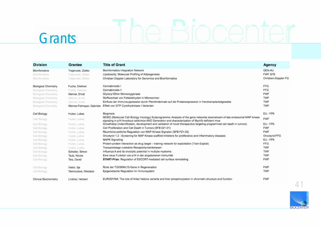
The Biocenter
41
Grants
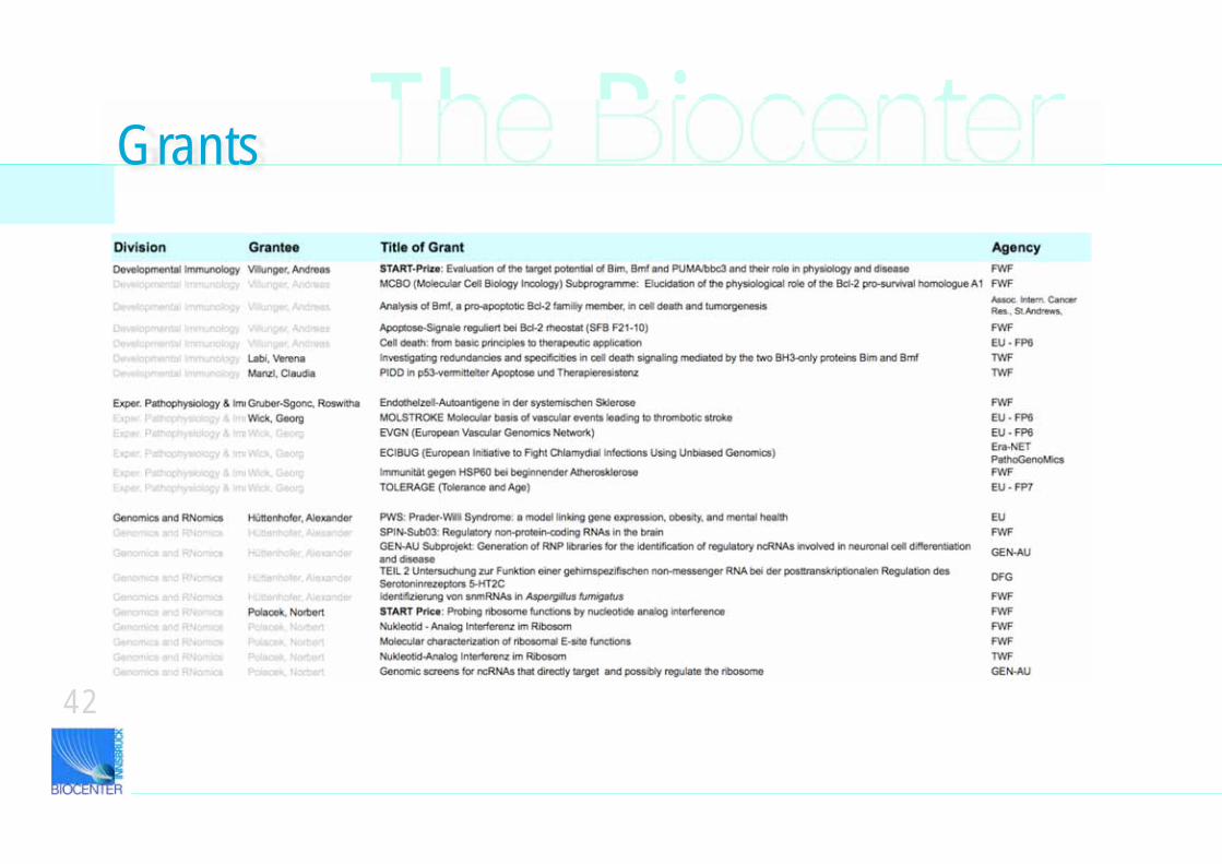
42
The BiocenterGrants
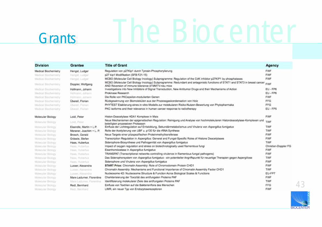
43
Grants The Biocenter
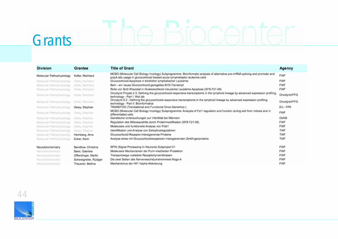
44
The BiocenterGrants
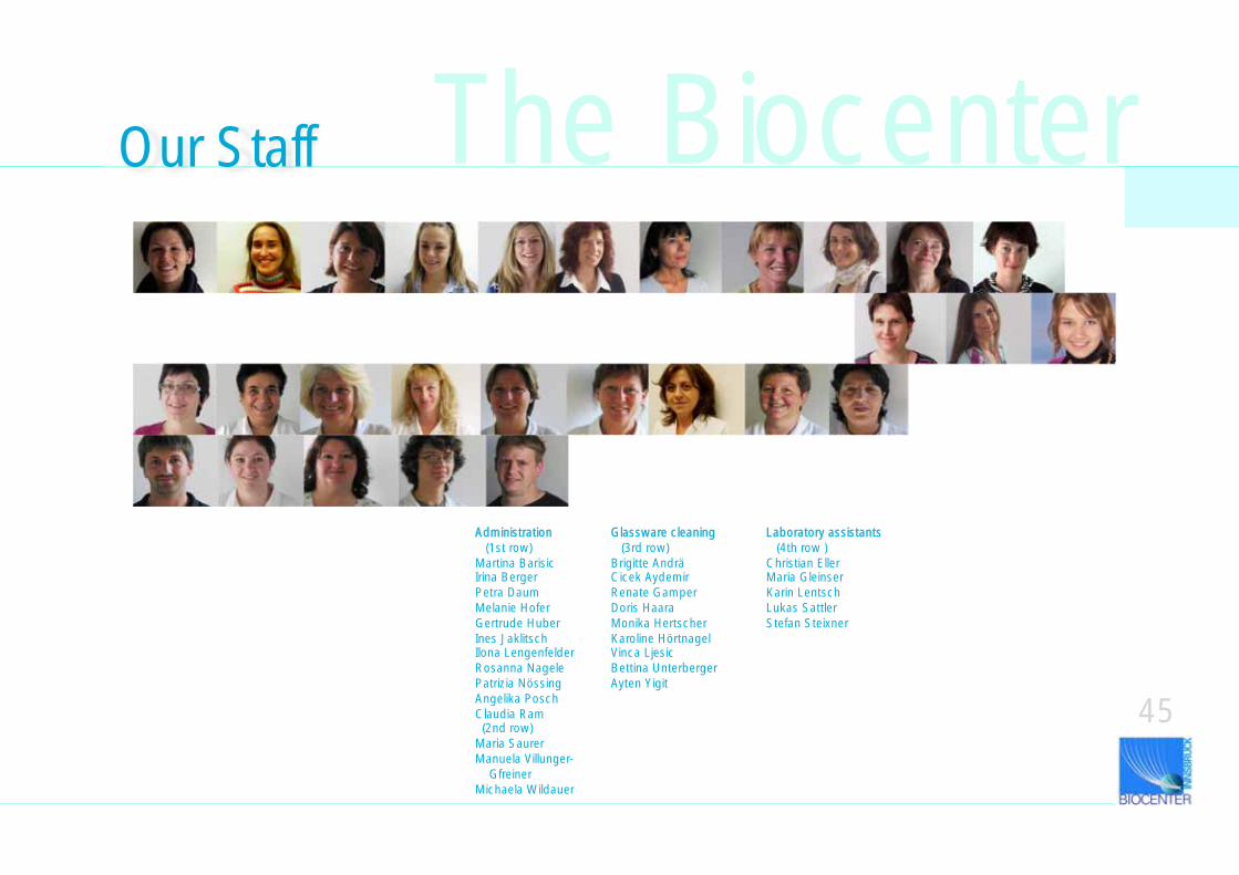
45
Administration (1st row)Martina BarisicIrina Berger Petra DaumMelanie HoferGertrude Huber Ines JaklitschIlona LengenfelderRosanna NagelePatrizia NössingAngelika Posch Claudia Ram (2nd row)Maria Saurer Manuela Villunger- GfreinerMichaela Wildauer
Glassware cleaning (3rd row) Brigitte AndräCicek Aydemir Renate GamperDoris HaaraMonika Hertscher Karoline HörtnagelVinca Ljesic Bettina UnterbergerAyten Yigit
Laboratory assistants (4th row )Christian Eller Maria GleinserKarin Lentsch Lukas SattlerStefan Steixner
Our Staff The Biocenter
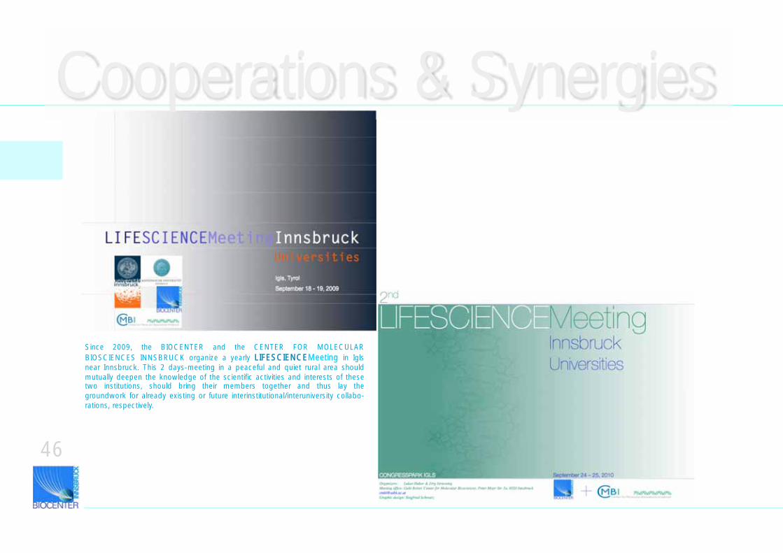
46
Since 2009, the BIOCENTER and the CENTER FOR MOLECULAR BIOSCIENCES INNSBRUCK organize a yearly LIFESCIENCEMeeting in Igls near Innsbruck. This 2 days-meeting in a peaceful and quiet rural area should mutually deepen the knowledge of the scientific activities and interests of these two institutions, should bring their members together and thus lay the groundwork for already existing or future interinstitutional/interuniversity collabo-rations, respectively.
Cooperations & Synergies
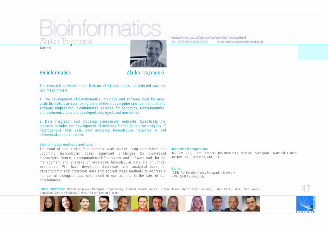
Bioinformatics
The research activities at the Division of Bioinformatics are directed towards two major thrusts:
1. The development of bioinformatics methods and software tools for large-scale biomolecular data. Using state-of-the-art computer science methods and software engineering, bioinformatics services for genomics, transcriptomics, and proteomics data are developed, deployed, and maintained.
2. Data integration and modeling biomolecular networks. Specifically, the research includes the development of methods for the integrative analyses of heterogenous data sets, and modeling biomolecular networks in cell differentiation and in cancer.
www.i-med.ac.at/biocenter/bioinformatics.htmlTel.: 0043 (512) 9003.71400 email: [email protected]
Bioinformatics methods and toolsThe flood of data arising from genomic-scale studies using established and upcoming technologies poses significant challenges for biomedical researchers. Hence, a computational infrastructure and software tools for the management and analyses of large-scale biomolecular data are of utmost importance. We have developed databases and analytical tools for transcriptomic and proteomic data and applied these methods to address a number of biological questions raised in our lab and in the labs of our collaborators.
Zlatko TrajanoskiDirector
Group members: Mihaela Angelova, Pornpimol Charoentong, Andreas Dander, Kalina Duszcka, Maria Fischer, Ralph Galasch, Hubert Hackl, Edith Hofer, Anne Krogsdam, Stephan Pabinger, Dietmar Rieder, Gernot Stocker
Bioinformatics Zlatko Trajanoski
International cooperationsINSERM 255, Paris, France; Bioinformatics Institute, Singapore; National Cancer Institute, NIH, Bethesda, MD/USA
Grants-GEN-AU Bioinformatics Integration Network -FWF SFB Lipotoxocity
47
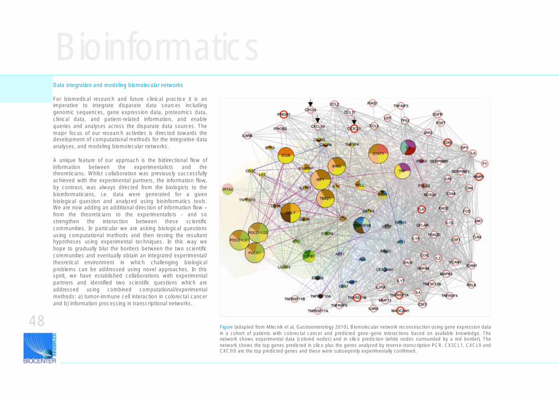
BioinformaticsData integration and modeling biomolecular networks
For biomedical research and future clinical practice it is an imperative to integrate disparate data sources including genomic sequences, gene expression data, proteomics data, clinical data, and patient-related information, and enable queries and analyses across the disparate data sources. The major focus of our research activities is directed towards the development of computational methods for the integrative data analyses, and modeling biomolecular networks.
A unique feature of our approach is the bidirectional flow of information between the experimentalists and the theoreticians. Whilst collaboration was previously successfully achieved with the experimental partners, the information flow, by contrast, was always directed from the biologists to the bioinformaticians, i.e. data were generated for a given biological question and analyzed using bioinformatics tools. We are now adding an additional direction of information flow – from the theoreticians to the experimentalists - and so strengthen the interaction between these scientific communities. In particular we are asking biological questions using computational methods and then testing the resultant hypotheses using experimental techniques. In this way we hope to gradually blur the borders between the two scientific communities and eventually obtain an integrated experimental/theoretical environment in which challenging biological problems can be addressed using novel approaches. In this spirit, we have established collaborations with experimental partners and identified two scientific questions which are addressed using combined computational/experimental methods: a) tumor-immune cell interaction in colorectal cancer and b) information processing in transcriptional networks.
Figure (adapted from Mlecnik et al, Gastroenterology 2010). Biomolecular network reconstruction using gene expression data in a cohort of patients with colorectal cancer and predicted gene–gene interactions based on available knowledge. The network shows experimental data (colored nodes) and in silico prediction (white nodes surrounded by a red border). The network shows the top genes predicted in silico plus the genes analyzed by reverse-transcription PCR. CX3CL1, CXCL9 and CXCl10 are the top predicted genes and these were subseqently experimentally confirmed.
48
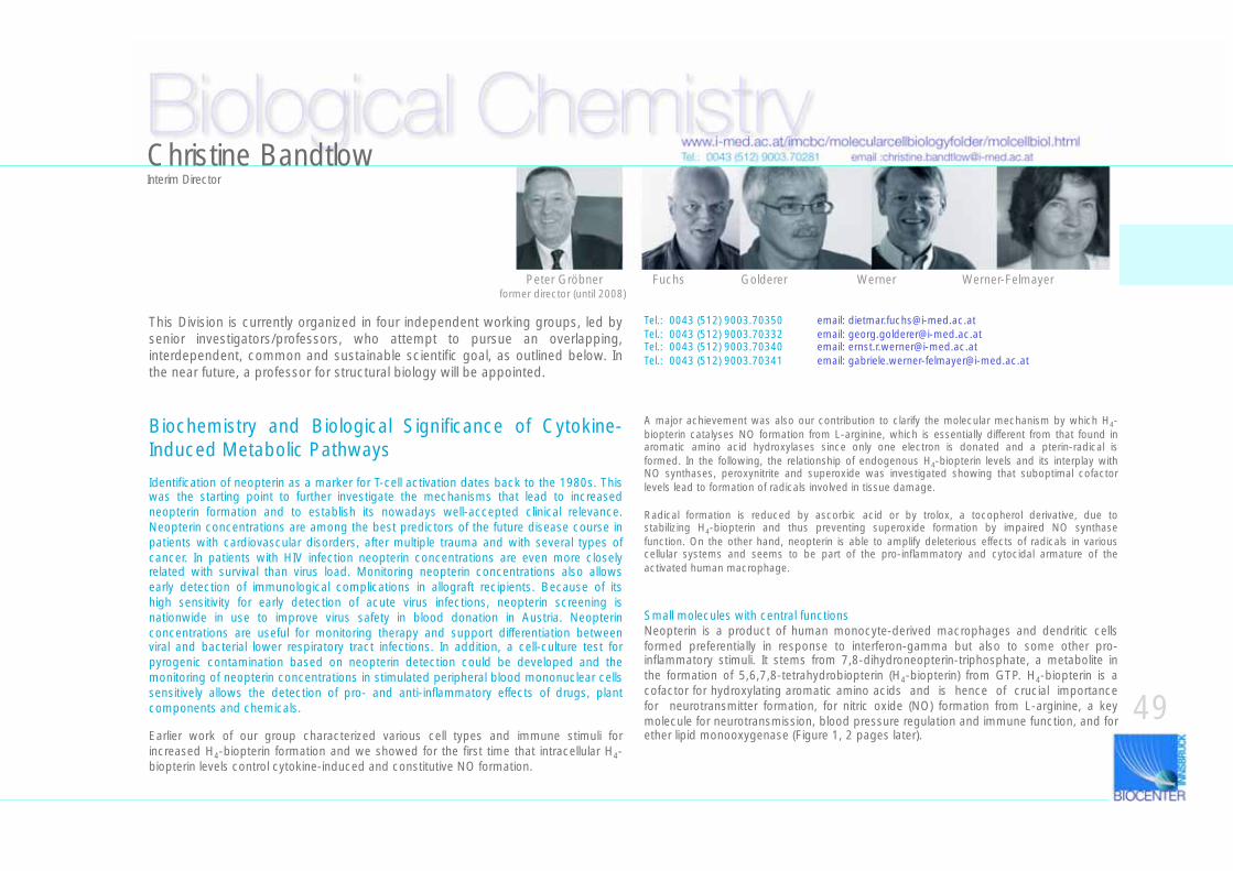
Biological Chemistry
This Division is currently organized in four independent working groups, led by senior investigators/professors, who attempt to pursue an overlapping, interdependent, common and sustainable scientific goal, as outlined below. In the near future, a professor for structural biology will be appointed.
Biochemistry and Biological Significance of Cytokine-Induced Metabolic Pathways
Identification of neopterin as a marker for T-cell activation dates back to the 1980s. This was the starting point to further investigate the mechanisms that lead to increased neopterin formation and to establish its nowadays well-accepted clinical relevance. Neopterin concentrations are among the best predictors of the future disease course in patients with cardiovascular disorders, after multiple trauma and with several types of cancer. In patients with HIV infection neopterin concentrations are even more closely related with survival than virus load. Monitoring neopterin concentrations also allows early detection of immunological complications in allograft recipients. Because of its high sensitivity for early detection of acute virus infections, neopterin screening is nationwide in use to improve virus safety in blood donation in Austria. Neopterin concentrations are useful for monitoring therapy and support differentiation between viral and bacterial lower respiratory tract infections. In addition, a cell-culture test for pyrogenic contamination based on neopterin detection could be developed and the monitoring of neopterin concentrations in stimulated peripheral blood mononuclear cells sensitively allows the detection of pro- and anti-inflammatory effects of drugs, plant components and chemicals.
Earlier work of our group characterized various cell types and immune stimuli for increased H4-biopterin formation and we showed for the first time that intracellular H4-biopterin levels control cytokine-induced and constitutive NO formation.
www.i-med.ac.at/imcbc/molecularcellbiologyfolder/molcellbiol.htmlTel.: 0043 (512) 9003.70281 email :[email protected]
A major achievement was also our contribution to clarify the molecular mechanism by which H4-biopterin catalyses NO formation from L-arginine, which is essentially different from that found in aromatic amino acid hydroxylases since only one electron is donated and a pterin-radical is formed. In the following, the relationship of endogenous H4-biopterin levels and its interplay with NO synthases, peroxynitrite and superoxide was investigated showing that suboptimal cofactor levels lead to formation of radicals involved in tissue damage.
Radical formation is reduced by ascorbic acid or by trolox, a tocopherol derivative, due to stabilizing H4-biopterin and thus preventing superoxide formation by impaired NO synthase function. On the other hand, neopterin is able to amplify deleterious effects of radicals in various cellular systems and seems to be part of the pro-inflammatory and cytocidal armature of the activated human macrophage.
Tel.: 0043 (512) 9003.70350 email: [email protected].: 0043 (512) 9003.70332 email: [email protected].: 0043 (512) 9003.70340 email: [email protected].: 0043 (512) 9003.70341 email: [email protected]
Fuchs Golderer Werner Werner-Felmayer
Small molecules with central functionsNeopterin is a product of human monocyte-derived macrophages and dendritic cells formed preferentially in response to interferon-gamma but also to some other pro-inflammatory stimuli. It stems from 7,8-dihydroneopterin-triphosphate, a metabolite in the formation of 5,6,7,8-tetrahydrobiopterin (H4-biopterin) from GTP. H4-biopterin is a cofactor for hydroxylating aromatic amino acids and is hence of crucial importance for neurotransmitter formation, for nitric oxide (NO) formation from L-arginine, a key molecule for neurotransmission, blood pressure regulation and immune function, and for ether lipid monooxygenase (Figure 1, 2 pages later).
49
Peter Gröbnerformer director (until 2008)
Christine BandtlowInterim Director
Pharmacological effects of H4-biopterin In analogy to folates and antifolates, we designed the H4-biopterin-analogue 4-amino-H4-biopterin and showed that it is an effective inhibitor of all three NO synthase isoenzymes with a preference for the cytokine-induced enzyme. In vivo, 4-amino-H4-biopterin prolonged allograft survival in a mouse heart transplantation model with an efficacy comparable to cyclosporine A and rescued rats from septic shock. However, the immunosuppressive effect of 4-amino-H4-biopterin can not be entirely explained by its capacity to inhibit NO synthase because in different expe-rimental settings, the cofactor of NO synthase, i.e. H4-biopterin, and the inhibitor of NO synthase, i.e. 4-amino-H4-biopterin, have similar effects. In mouse macrophages, we showed that H4-biopterin and its amino-analogue added to culture media suppressed NO synthase gene expression via hydrogen peroxide formation and induced apoptosis. In a mouse model, cardiac allograft survival was prolonged by both H4-pteridines independently from their effect on NO synthase expression or activity, whereas on dendritic cells only the amino analogon selectively suppressed MHC class II protein content and antigen response.
Pharmacological effects of H4-biopterin H4-biopterin is formed by three biosynthetic enzymes from guanosine triphosphate (GTP), i.e GTP cyclohydrolase I, 6-pyruvoyl tetrahydropterin synthase and sepiapterin reductase. We have shown that cytokines regulate the first step of this biosynthesis, by inducing GTP cyclohydrolase I and repressing its feedback regulatory protein. Neopterin derivatives accumulate in human cells and in particular in human macrophages due to a low activity of the second enzyme of the pathway, 6-pyruvyol tetrahydropterin synthase. The mechanistic basis of this is selective skipping of exon 3 of this enzyme in the splicing process which is particularly effective in human macrophages in which H4-biopterin is undetectable. GTP cyclohydrolase I is induced by cytokines in alternatively spliced RNAs, and coexpression of the spliced truncated forms of this enzyme cause decrease in activity and protein, presumably by accelerated decay.
Related pathways and clinical relevanceIn conditions leading to increased neopterin formation, i.e. activation of NK and T-cells and hence of macrophages as well, a number of further cytokines and metabolic pathways are induced (Figure 2, next page).
We were among the first to clone and characterize the novel small T-cell attracting CXC chemokine CXCL11 and to show its cross-reactivity with other CXC chemokines reacting with the CXCR3 receptor, and its involvement in allograft rejection. A metabolic pathway induced in response to T-cell activation is degradation of the essential amino acid tryptophan by indoleamine 2,3-dioxygenase (IDO). Patients with a negative prognosis have increased IDO activities in addition to increased neopterin levels in a number of conditions.
Like other pathways induced by interferon-gamma, this strategy of the immune system serves to restrict growth of pathogens or malignant cells but, when getting beyond control, can also lead to immune deficiency, impairment of erythropoiesis and increased probability to develop depression because of affecting the serotonin/tryptophan metabolism. Finally there exists a potential association of subnormal tryptophan degradation and the course of allergy and asthma, as has been investigated in patients suffering from pollinosis under specific immunotherapy.
Physarum nitric oxide synthetase
Physarum polycephalum is a single-cell multinuclear model organism used in cell biology because of its naturally synchronous cell cycle and its ability to undergo differentiation. Remarkably, this organism expresses the only thus far known fully functional NO synthase outside the animal kingdom, an enzyme required by this organism to gain sporulation competence thus establishing a novel role for the H4-biopterin/NO/cGMP axis in cell differentiation. Moreover, we were involved in initiating the Physarum genome project which was started in August 2004 by the National Human Genome Research Institute (NHGRI) following an initiative of an international Physarum Genome Consortium headed by J. Gott, Cleveland. Together with W. Marwan, Magdeburg, and G. Gloeckner, Jena, a transcriptome project of the plasmodial stage analysed about half of the protein coding genes of Physarum, yielding an important resource for the ongoing genome project. In the near future, these projects will allow to study signalling networks and protein expression related to cell differentiation in a systematic large-scale approach.
Current focus
Tryptophan degradation is still in focus of our experimental and clinical studies. In an intense collaboration with the NCI/Bethesda, the role of IDO and its relationship to regulatory T-cells and dendritic cells in the development of immunodeficiency is investigated in HIV infection and in experimental SIV infection of rhesus macaques. The influence of tryptophan degradation on the course of neuropsychiatric symptoms is investigated in patients with HIV infection (e.g. in collaboration with UCSF) and with cancer. To further characterize the role of immune activation and inflammation in cardiovascular diseases, the relationship between the metabolism of homocysteine
and asymmetric dimethylarginine (ADMA) is investigated in vitro and in vivo.
50Group members Tanja Hertscheg, Markus Keller, Elena Ledjeff (no picture), Petra Loitzl, Rita Holzknecht (statt Nina Madl, karnz.), Birgit Schraberger (on maternity leave, no picture), Sebastian Schröcksnadl, Katrin Watschinger
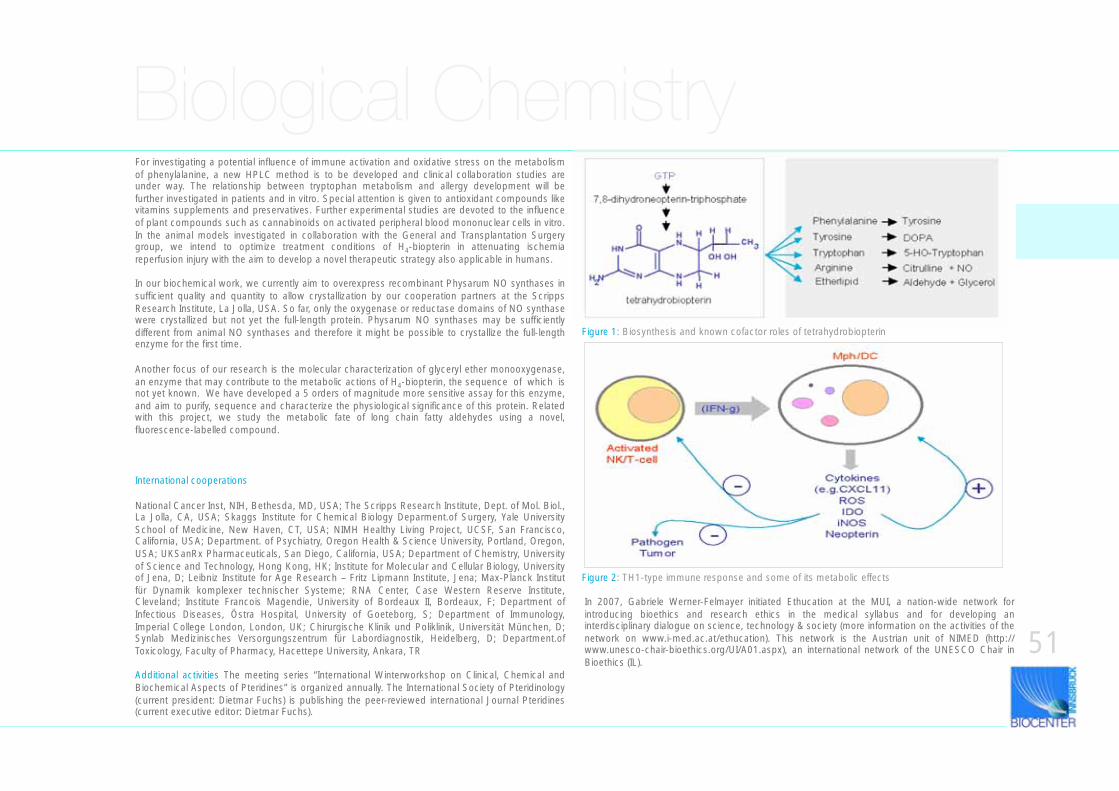
Figure 2: TH1-type immune response and some of its metabolic effects
Figure 1: Biosynthesis and known cofactor roles of tetrahydrobiopterin
For investigating a potential influence of immune activation and oxidative stress on the metabolism of phenylalanine, a new HPLC method is to be developed and clinical collaboration studies are under way. The relationship between tryptophan metabolism and allergy development will be further investigated in patients and in vitro. Special attention is given to antioxidant compounds like vitamins supplements and preservatives. Further experimental studies are devoted to the influence of plant compounds such as cannabinoids on activated peripheral blood mononuclear cells in vitro. In the animal models investigated in collaboration with the General and Transplantation Surgery group, we intend to optimize treatment conditions of H4-biopterin in attenuating ischemia reperfusion injury with the aim to develop a novel therapeutic strategy also applicable in humans.
In our biochemical work, we currently aim to overexpress recombinant Physarum NO synthases in sufficient quality and quantity to allow crystallization by our cooperation partners at the Scripps Research Institute, La Jolla, USA. So far, only the oxygenase or reductase domains of NO synthase were crystallized but not yet the full-length protein. Physarum NO synthases may be sufficiently different from animal NO synthases and therefore it might be possible to crystallize the full-length enzyme for the first time.
Another focus of our research is the molecular characterization of glyceryl ether monooxygenase, an enzyme that may contribute to the metabolic actions of H4-biopterin, the sequence of which is not yet known. We have developed a 5 orders of magnitude more sensitive assay for this enzyme, and aim to purify, sequence and characterize the physiological significance of this protein. Related with this project, we study the metabolic fate of long chain fatty aldehydes using a novel, fluorescence-labelled compound.
In 2007, Gabriele Werner-Felmayer initiated Ethucation at the MUI, a nation-wide network for introducing bioethics and research ethics in the medical syllabus and for developing an interdisciplinary dialogue on science, technology & society (more information on the activities of the network on www.i-med.ac.at/ethucation). This network is the Austrian unit of NIMED (http://www.unesco-chair-bioethics.org/UI/A01.aspx), an international network of the UNESCO Chair in Bioethics (IL).
International cooperations
National Cancer Inst, NIH, Bethesda, MD, USA; The Scripps Research Institute, Dept. of Mol. Biol., La Jolla, CA, USA; Skaggs Institute for Chemical Biology Deparment.of Surgery, Yale University School of Medicine, New Haven, CT, USA; NIMH Healthy Living Project, UCSF, San Francisco, California, USA; Department. of Psychiatry, Oregon Health & Science University, Portland, Oregon, USA; UKSanRx Pharmaceuticals, San Diego, California, USA; Department of Chemistry, University of Science and Technology, Hong Kong, HK; Institute for Molecular and Cellular Biology, University of Jena, D; Leibniz Institute for Age Research – Fritz Lipmann Institute, Jena; Max-Planck Institut für Dynamik komplexer technischer Systeme; RNA Center, Case Western Reserve Institute, Cleveland; Institute Francois Magendie, University of Bordeaux II, Bordeaux, F; Department of Infectious Diseases, Östra Hospital, University of Goeteborg, S; Department of Immunology, Imperial College London, London, UK; Chirurgische Klinik und Poliklinik, Universität München, D; Synlab Medizinisches Versorgungszentrum für Labordiagnostik, Heidelberg, D; Department.of Toxicology, Faculty of Pharmacy, Hacettepe University, Ankara, TR
Additional activities The meeting series “International Winterworkshop on Clinical, Chemical and Biochemical Aspects of Pteridines” is organized annually. The International Society of Pteridinology (current president: Dietmar Fuchs) is publishing the peer-reviewed international Journal Pteridines (current executive editor: Dietmar Fuchs).
51
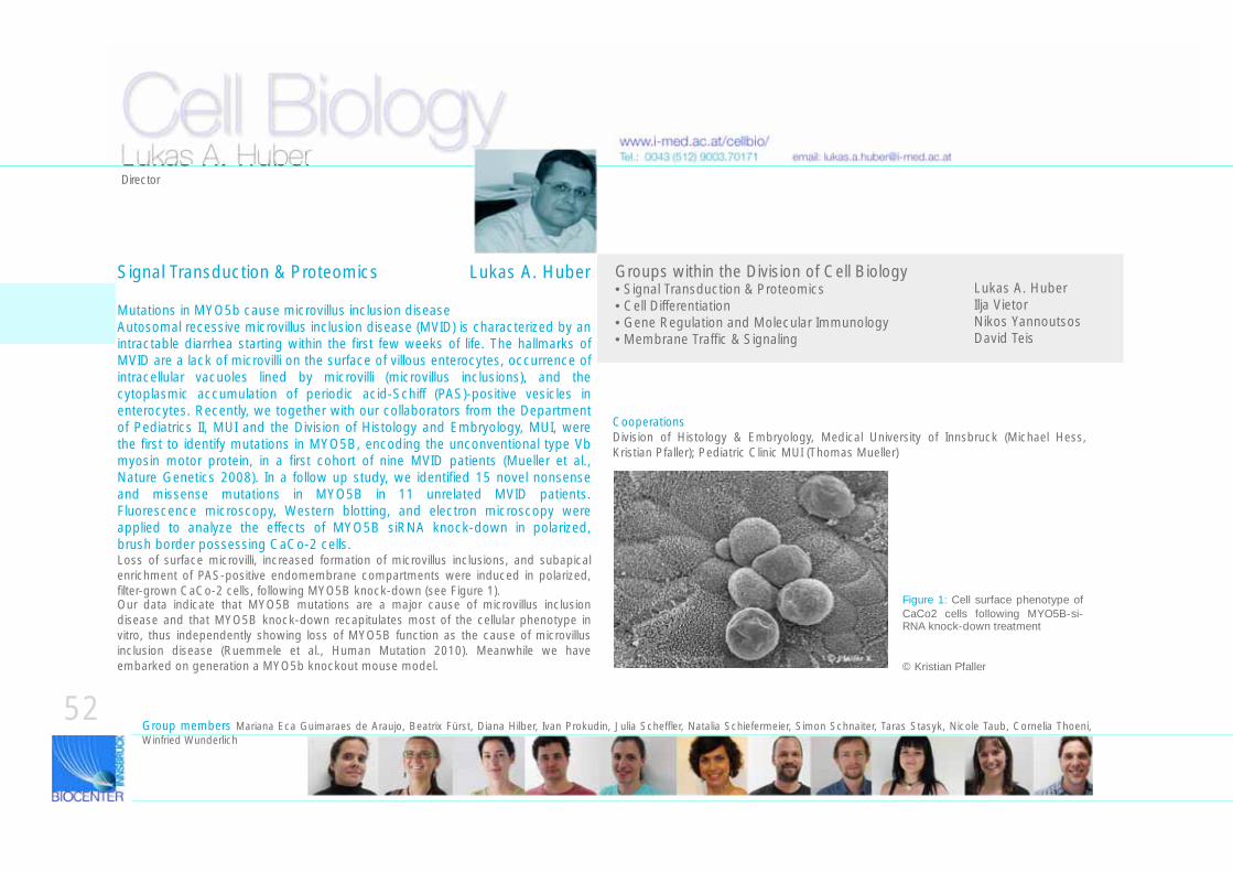
Cell Biology
Mutations in MYO5b cause microvillus inclusion diseaseAutosomal recessive microvillus inclusion disease (MVID) is characterized by an intractable diarrhea starting within the first few weeks of life. The hallmarks of MVID are a lack of microvilli on the surface of villous enterocytes, occurrence of intracellular vacuoles lined by microvilli (microvillus inclusions), and the cytoplasmic accumulation of periodic acid-Schiff (PAS)-positive vesicles in enterocytes. Recently, we together with our collaborators from the Department of Pediatrics II, MUI and the Division of Histology and Embryology, MUI, were the first to identify mutations in MYO5B, encoding the unconventional type Vb myosin motor protein, in a first cohort of nine MVID patients (Mueller et al., Nature Genetics 2008). In a follow up study, we identified 15 novel nonsense and missense mutations in MYO5B in 11 unrelated MVID patients. Fluorescence microscopy, Western blotting, and electron microscopy were applied to analyze the effects of MYO5B siRNA knock-down in polarized, brush border possessing CaCo-2 cells. Loss of surface microvilli, increased formation of microvillus inclusions, and subapical enrichment of PAS-positive endomembrane compartments were induced in polarized, filter-grown CaCo-2 cells, following MYO5B knock-down (see Figure 1). Our data indicate that MYO5B mutations are a major cause of microvillus inclusion disease and that MYO5B knock-down recapitulates most of the cellular phenotype in vitro, thus independently showing loss of MYO5B function as the cause of microvillus inclusion disease (Ruemmele et al., Human Mutation 2010). Meanwhile we have embarked on generation a MYO5b knockout mouse model.
www.i-med.ac.at/cellbio/
Signal Transduction & Proteomics Lukas A. Huber
Tel.: 0043 (512) 9003.70171 email: [email protected]
Groups within the Division of Cell Biology• Signal Transduction & Proteomics • Cell Differentiation • Gene Regulation and Molecular Immunology• Membrane Traffic & Signaling
Lukas A. HuberIlja VietorNikos YannoutsosDavid Teis
Group members Mariana Eca Guimaraes de Araujo, Beatrix Fürst, Diana Hilber, Ivan Prokudin, Julia Scheffler, Natalia Schiefermeier, Simon Schnaiter, Taras Stasyk, Nicole Taub, Cornelia Thoeni, Winfried Wunderlich
Figure 1: Cell surface phenotype of
CaCo2 cells following MYO5B-si-RNA knock-down treatment
© Kristian Pfaller
52
Lukas A. HuberDirector
CooperationsDivision of Histology & Embryology, Medical University of Innsbruck (Michael Hess, Kristian Pfaller); Pediatric Clinic MUI (Thomas Mueller)
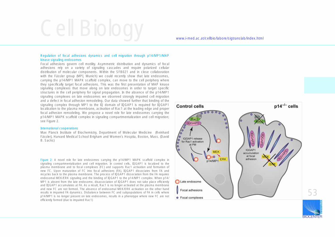
Cell BiologyRegulation of focal adhesions dynamics and cell migration through p14/MP1/MAP kinase signaling endosomes Focal adhesions govern cell motility. Asymmetric distribution and dynamics of focal adhesions rely on a variety of signaling cascades and require polarized cellular distribution of molecular components. Within the SFB021 and in close collaboration with the Fässler group (MPI, Munich) we could recently show that late endosomes, carrying the p14/MP1 MAPK scaffold complex, can move to the cell periphery where they specifically target focal adhesions. This was the first presentation of MAP kinase signaling complexes that move along on late endosomes in order to target specific structures in the cell periphery for signal propagation. In the absence of the p14/MP1 signaling complexes on late endosomes we observed strongly impaired cell migration and a defect in focal adhesion remodeling. Our data showed further that binding of the signaling complex through MP1 to the IQ domain of IQGAP1 is required for IQGAP1 localization to the plasma membrane, activation of Rac1 at the leading edge and proper focal adhesion remodeling. We propose a novel role for late endosomes carrying the p14/MP1 MAPK scaffold complex in signaling compartmentalization and cell migration; see Figure 2.
www.i-med.ac.at/cellbio/labore/sigtranslab/index.html
International cooperationsMax Planck Institute of Biochemistry, Department of Molecular Medicine (Reinhard Fässler), Harvard Medical School Brigham and Women`s Hospita, Boston, Mass. (David B. Sacks)
Figure 2: A novel role for late endosomes carrying the p14/MP1 MAPK scaffold complex in signaling compartmentalization and cell migration. In control cells, IQGAP1 is localized to the plasma membrane and to focal complexes (FC) and supports Rac1 activation and formation of new FC. Upon maturation of FC into focal adhesions (FA), IQGAP1 dissociates from FA and recycles back to the plasma membrane. The process of IQGAP1 dissociation from the FA requires endosomal MEK/ERK signaling and the binding of IQGAP1 to the p14/MP1 complex. When p14/MP1 is absent from the late endosome, disassociation of IQGAP1 does not take place efficiently and IQGAP1 accumulates at FA. As a result, Rac1 is no longer activated at the plasma membrane and new FC are not formed. The absence of endosomal MEK/ERK activation on the other hand results in impaired FA dynamics. Disbalance between FC and subpopulations of FA in cells where p14/MP1 is no longer present on late endosomes, results in a phenotype where new FC are not efficiently formed (due to impaired Rac1)
53
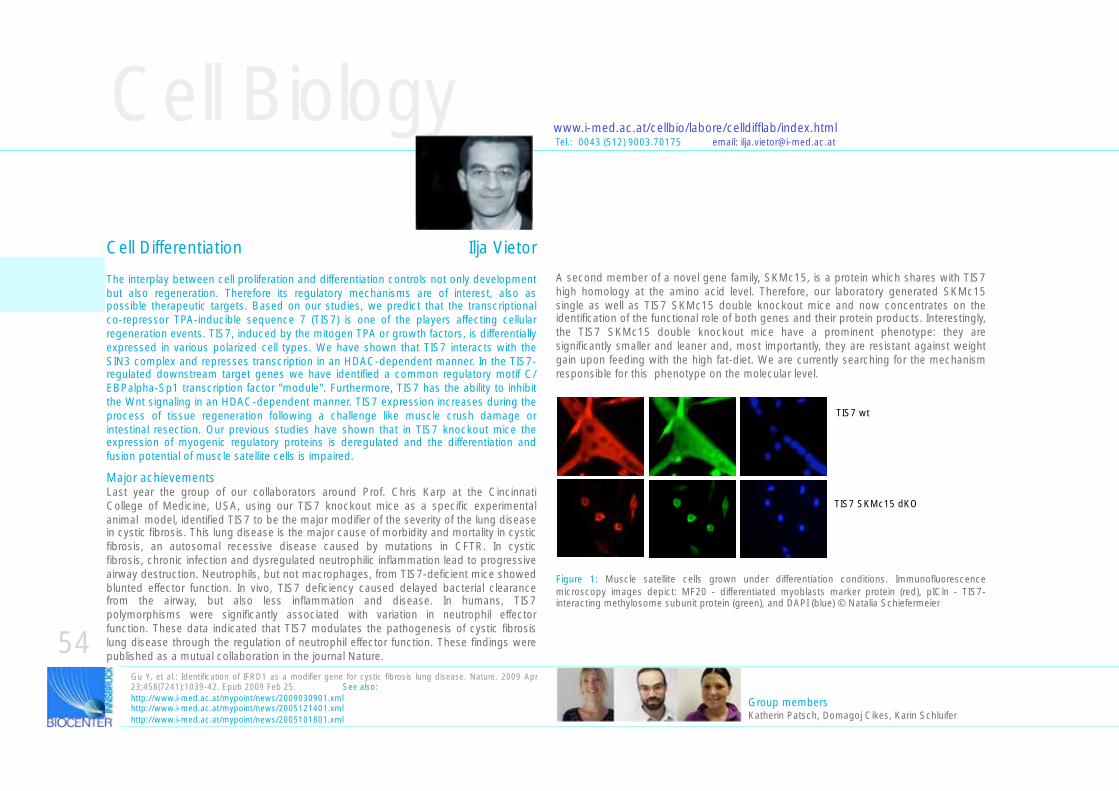
Cell Biologywww.i-med.ac.at/cellbio/labore/celldifflab/index.html
The interplay between cell proliferation and differentiation controls not only development but also regeneration. Therefore its regulatory mechanisms are of interest, also as possible therapeutic targets. Based on our studies, we predict that the transcriptional co-repressor TPA-inducible sequence 7 (TIS7) is one of the players affecting cellular regeneration events. TIS7, induced by the mitogen TPA or growth factors, is differentially expressed in various polarized cell types. We have shown that TIS7 interacts with the SIN3 complex and represses transcription in an HDAC-dependent manner. In the TIS7-regulated downstream target genes we have identified a common regulatory motif C/EBPalpha-Sp1 transcription factor "module". Furthermore, TIS7 has the ability to inhibit the Wnt signaling in an HDAC-dependent manner. TIS7 expression increases during the process of tissue regeneration following a challenge like muscle crush damage or intestinal resection. Our previous studies have shown that in TIS7 knockout mice the expression of myogenic regulatory proteins is deregulated and the differentiation and fusion potential of muscle satellite cells is impaired.
Gu Y, et al.: Identification of IFRD1 as a modifier gene for cystic fibrosis lung disease. Nature. 2009 Apr 23;458(7241):1039-42. Epub 2009 Feb 25. See also:http://www.i-med.ac.at/mypoint/news/2009030901.xmlhttp://www.i-med.ac.at/mypoint/news/2005121401.xmlhttp://www.i-med.ac.at/mypoint/news/2005101801.xml
Major achievementsLast year the group of our collaborators around Prof. Chris Karp at the Cincinnati College of Medicine, USA, using our TIS7 knockout mice as a specific experimental animal model, identified TIS7 to be the major modifier of the severity of the lung disease in cystic fibrosis. This lung disease is the major cause of morbidity and mortality in cystic fibrosis, an autosomal recessive disease caused by mutations in CFTR. In cystic fibrosis, chronic infection and dysregulated neutrophilic inflammation lead to progressive airway destruction. Neutrophils, but not macrophages, from TIS7-deficient mice showed blunted effector function. In vivo, TIS7 deficiency caused delayed bacterial clearance from the airway, but also less inflammation and disease. In humans, TIS7 polymorphisms were significantly associated with variation in neutrophil effector function. These data indicated that TIS7 modulates the pathogenesis of cystic fibrosis lung disease through the regulation of neutrophil effector function. These findings were published as a mutual collaboration in the journal Nature.
Figure 1: Muscle satellite cells grown under differentiation conditions. Immunofluorescence microscopy images depict: MF20 - differentiated myoblasts marker protein (red), pICln - TIS7-interacting methylosome subunit protein (green), and DAPI (blue) © Natalia Schiefermeier
Tel.: 0043 (512) 9003.70175 email: [email protected]
Group members Katherin Patsch, Domagoj Cikes, Karin Schluifer
A second member of a novel gene family, SKMc15, is a protein which shares with TIS7 high homology at the amino acid level. Therefore, our laboratory generated SKMc15 single as well as TIS7 SKMc15 double knockout mice and now concentrates on the identification of the functional role of both genes and their protein products. Interestingly, the TIS7 SKMc15 double knockout mice have a prominent phenotype: they are significantly smaller and leaner and, most importantly, they are resistant against weight gain upon feeding with the high fat-diet. We are currently searching for the mechanism responsible for this phenotype on the molecular level.
TIS7 wt
TIS7 SKMc15 dKO
54
Cell Differentiation Ilja Vietor
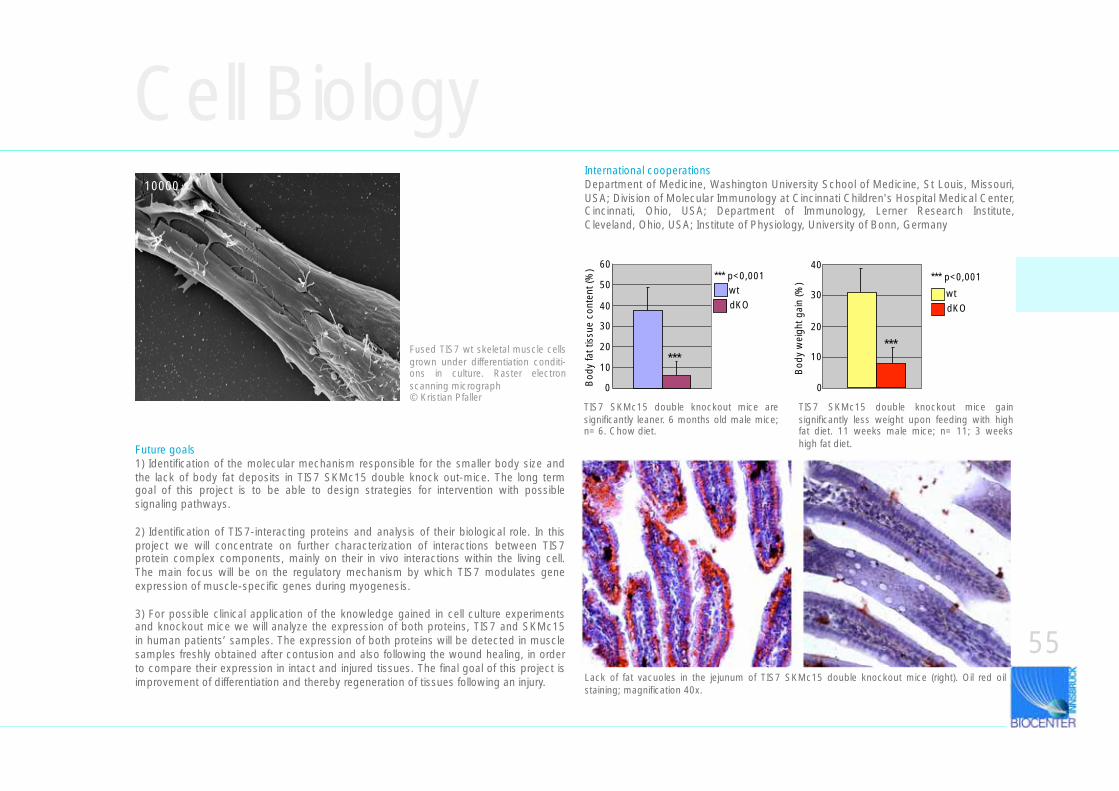
Cell Biology
Future goals1) Identification of the molecular mechanism responsible for the smaller body size and the lack of body fat deposits in TIS7 SKMc15 double knock out-mice. The long term goal of this project is to be able to design strategies for intervention with possible signaling pathways.
2) Identification of TIS7-interacting proteins and analysis of their biological role. In this project we will concentrate on further characterization of interactions between TIS7 protein complex components, mainly on their in vivo interactions within the living cell. The main focus will be on the regulatory mechanism by which TIS7 modulates gene expression of muscle-specific genes during myogenesis.
3) For possible clinical application of the knowledge gained in cell culture experiments and knockout mice we will analyze the expression of both proteins, TIS7 and SKMc15 in human patients’ samples. The expression of both proteins will be detected in muscle samples freshly obtained after contusion and also following the wound healing, in order to compare their expression in intact and injured tissues. The final goal of this project is improvement of differentiation and thereby regeneration of tissues following an injury.
TIS7 SKMc15 double knockout mice are significantly leaner. 6 months old male mice; n= 6. Chow diet.
Lack of fat vacuoles in the jejunum of TIS7 SKMc15 double knockout mice (right). Oil red oil staining; magnification 40x.
40
0
10
20
30
Bod
y w
eigh
t gai
n (%
) *** p<0,001
***
wtdKO
0
10
20
30
40
50
60
Bod
y fa
t tis
sue
cont
ent (
%)
wtdKO
*** p<0,001
***
10000
Fused TIS7 wt skeletal muscle cells grown under differentiation conditi-ons in culture. Raster electron scanning micrograph © Kristian Pfaller
TIS7 SKMc15 double knockout mice gain significantly less weight upon feeding with high fat diet. 11 weeks male mice; n= 11; 3 weeks high fat diet.
55
International cooperationsDepartment of Medicine, Washington University School of Medicine, St Louis, Missouri, USA; Division of Molecular Immunology at Cincinnati Children's Hospital Medical Center, Cincinnati, Ohio, USA; Department of Immunology, Lerner Research Institute, Cleveland, Ohio, USA; Institute of Physiology, University of Bonn, Germany
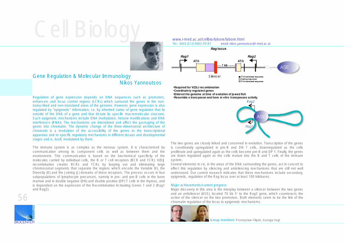
ASE
Silencer
ASE
Rag1
Rag2
ASE
Cell Biology
Regulation of gene expression depends on DNA sequences such as promoters, enhancers and locus control regions (LCRs) which surround the genes in the non-transcribed and non-translated areas of the genome. However, gene expression is also regulated by "epigenetic" information, i.e. by inherited states of gene regulation that lie outside of the DNA of a gene and that dictate its specific macromolecular structure. Such epigenetic mechanisms include DNA methylation, histone modifications and RNA interference (RNAi). The mechanisms are interrelated and affect the packaging of the genes into chromatin. The dynamic change of the three-dimensional architecture of chromatin is a modulator of the accessibility of the genes to the transcriptional apparatus and to specific regulatory mechanisms in different tissues and developmental stages and is, itself, modulated by them.
The immune system is as complex as the nervous system. It is characterized by communication among its component cells as well as between them and the environment. This communication is based on the biochemical specificity of the molecules carried by individual cells, the B or T cell receptors (BCR and TCR). V(D)J recombination creates BCRs and TCRs by looping out and eliminating large chromosomal segments that separate the regions which encode the Variable (V), the Diversity (D) and the Joining (J) domains of these receptors. The process occurs in four subpopulations of lymphocyte precursors, namely in pro- and pre-B cells in the bone marrow and in double negative (DN) and double positive (DP) T cells in the thymus, and is dependent on the expression of the Recombination Activating Genes 1 and 2 (Rag1 and Rag2).
The two genes are closely linked and conserved in evolution. Transcription of the genes is coordinately upregulated in pro-B and DN T cells, downregulated as the cells proliferate and upregulated again as the cells become pre-B and DP T. Finally, the genes are down regulated again as the cells mature into the B and T cells of the immune system. Several elements in cis, in the areas of the DNA surrounding the genes, act in concert to effect this regulation by silencing and antisilencing mechanisms that are still not well understood. Our current research indicates that these mechanisms include secondary, epigenetic, regulation of the Rag locus over at least 100 kilobases.
Major achievements/current progressMajor discovery in this area is the interplay between a silencer between the two genes and an antisilencer (ASE), located 70 kb 5‘ to the Rag2 gene, which counteracts the action of the silencer on the two promoters. Both elements seem to be the link of the chromatin regulation of the locus to epigenetic mechanisms.
Group members Przemyslaw Filipek, Georgia Vogl
www.i-med.ac.at/cellbio/labore/labore.htmlTel.: 0043 (512) 9003.70187 email: [email protected]
56
Gene Regulation & Molecular Immunology Nikos Yannoutsos
Aberrant translocations and lymphomasAnother focus of our laboratory is on follicular lymphoma generated by the aberrant Rag-dependent translocation that brings together the IgH enhancer with the anti-apoptotic BCL2 gene. This occurs during the DJ rearrangement in pro-B cells in the bone marrow. Disease, however, develops in the spleen in the adult cell. The project has two directions. On the one hand, in collaboration with Dr. Gunsilius of the Hematolog/Oncology Clinic of the MUI, we are analyzing patient material to see what BCRs the actual follicular lymphomas carry, in an effort to clarify what antigens might be involved in their activation. On the other hand, we aim to build a mouse model for the disease by making a transgenic mouse that carries both a selected BCR as a transgene and a transgene that mimics the translocation by driving expression of the BCL2 gene from the IgH enhancer. We are also pursuing alternative mouse models that may potentially provide a shortcut to these ideas. We have obtained a mouse from Dr. Villunger at the MUI, which drives expression of the bcl2 gene from a vav promoter and enhancer regulatory elements.In collaboration with Dr. Villunger, we have also generated a transgenic mouse that expresses a green fluorescent protein (Venus) under the same vav regulation. Combination of the vav-bcl2 mouse with the vav-venus mouse will facilitate these studies as the tumors that may eventually form from the transplanted pro-B cells into wild-type mice will be fluorescent.
Ongoing work in our laboratory includes the following findings•Northern blot analysis shows an 80 bp RNA species that lights up with probes from the Rag2 intron near the 5‘ untranslated exon. Database search finds three microRNA sequences located in tandem within this area. This array is also found at about 3 kb 5‘ to Rag2 with one of the microRNAs also within the ASE area. The possible silencing potential of these microRNA sequences will be further examined by monitoring Rag expression after transfecting them into DP T cells in culture.•FISH experiments with oligonucleotide sequences from the introns of Rag1 and Rag2 labeled with Alexa 560 (red) and Alexa 488 (green), respectively, which light up the nascent primary transcripts in the nucleus, have shown that both genes are expressed from the same chromosome at the same time•Chromosome Conformation Capture (3C) experiments provide evidence for a complex looping structure of the locus that explains its coordinated regulation by the ASE. One loop brings the promoter of the Rag1 gene 5‘ to the promoter of the Rag2 gene and a second loop brings the distant ASE to an area right behind the two touching promoters, presumably mediated by factors that bind in these areas (see picture above!). •DNA methylation studies around the promoters of the genes and histone methylation/acetylation studies across the locus are in progress.
Cell Biology
The transgenic/knock-out unitwww.i-med.ac.at/iftz/zentrale_gruppen/mouseunit/
Research in our laboratory depends on the generation of transgenic and knock-out mouse models. Since the establishement of this laboratory in November 2005, a major effort has been to create a laboratory for the generation of such mice by pronuclear and blastocyst microinjection. At the end of 2007 the transgenic mouse laboratory gained support by the former IFTZ (Integrated Research & Therapy Center) and is used for collaborations with all laboratories in MUI interested to generate such mouse models. It has also been used to establish rederivation of mouse lines to Specific-Pathogen-Free (SPF) status.
Another very useful mouse has been obtained from Dr. Hendriks (Erasmus University, Rotterdam). The mouse contains a transgene that overexpresses Bruton‘s tyrosine kinase (btk). Btk is at the center of an important intracellular signaling pathway downstream of BCR signaling after antigen stimulation and normal expression of Btk has been recently shown to be necessary for the development of follicular lymphoma. It is possible that when this mouse is combined with a mouse that overexpresses BCL2, it will give follicular lymphomas, independent of antigen stimulation by a particular antigen, providing a direct model for the dependence of follicular lymphoma on Btk.
Future goalsWe intend to develop projects related to stem cell technology in three major directions: -development of mouse models with reporters for genes that are upregulated in tumor cells and combine them with mouse models of selected cancers, in order to study tumor stem cells- using Oct4 to reprogram differentiated cells to pluripotent state and drive them forward with the aim to clarify development of cells, e.g. islets of Langerhans versus other pancreatic cells, via differential gene expression programs- extend such studies to material from patients by trying to reprogram tumor cells to pluripotent or progenitor state and thereafter to drive them forward in order to see where and how the disease-causing alteration occurs.
57
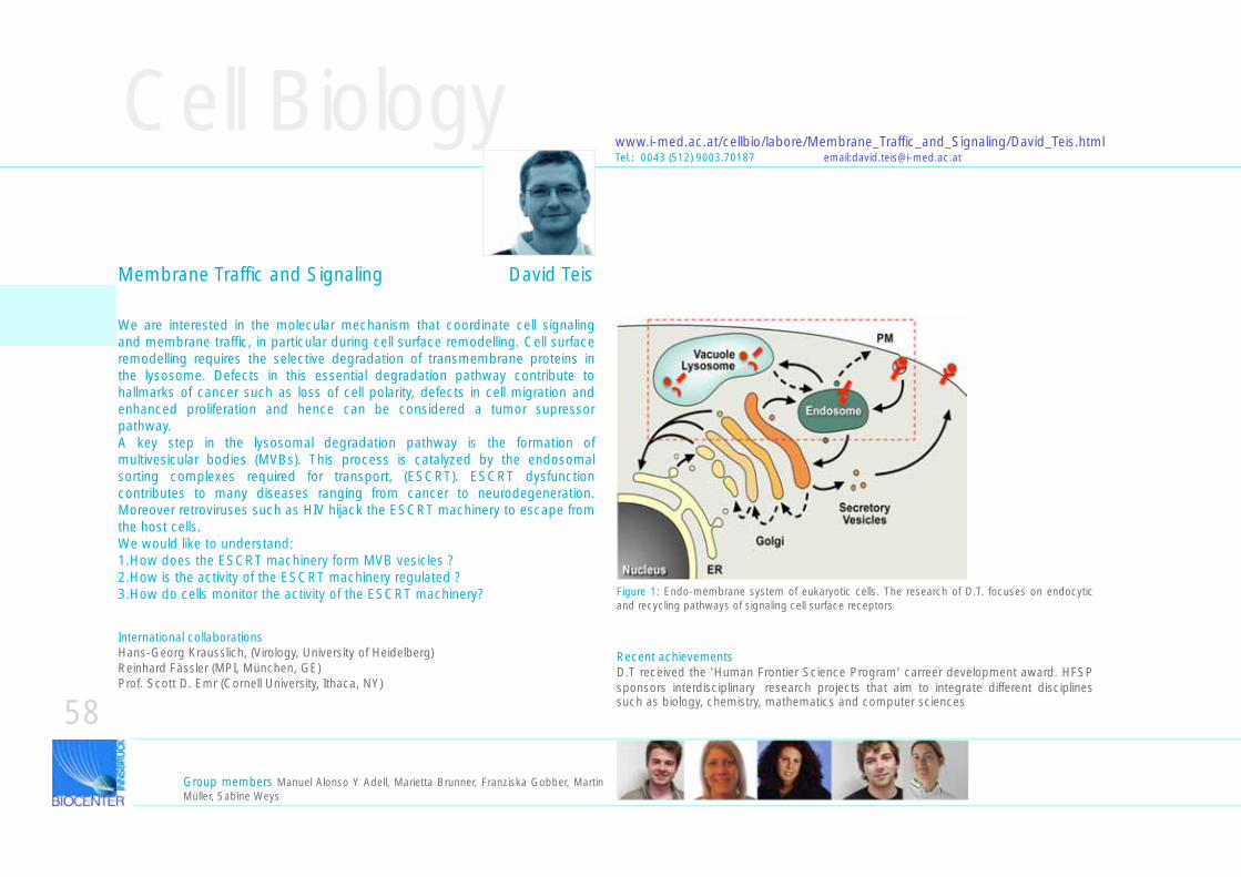
58
Cell Biologywww.i-med.ac.at/cellbio/labore/Membrane_Traffic_and_Signaling/David_Teis.htmlTel.: 0043 (512) 9003.70187 email:[email protected]
Group members Manuel Alonso Y Adell, Marietta Brunner, Franziska Gobber, Martin Müller, Sabine Weys
We are interested in the molecular mechanism that coordinate cell signaling and membrane traffic, in particular during cell surface remodelling. Cell surface remodelling requires the selective degradation of transmembrane proteins in the lysosome. Defects in this essential degradation pathway contribute to hallmarks of cancer such as loss of cell polarity, defects in cell migration and enhanced proliferation and hence can be considered a tumor supressor pathway.A key step in the lysosomal degradation pathway is the formation of multivesicular bodies (MVBs). This process is catalyzed by the endosomal sorting complexes required for transport, (ESCRT). ESCRT dysfunction contributes to many diseases ranging from cancer to neurodegeneration. Moreover retroviruses such as HIV hijack the ESCRT machinery to escape from the host cells. We would like to understand:1.How does the ESCRT machinery form MVB vesicles ?2.How is the activity of the ESCRT machinery regulated ?3.How do cells monitor the activity of the ESCRT machinery? Figure 1: Endo-membrane system of eukaryotic cells. The research of D.T. focuses on endocytic
and recycling pathways of signaling cell surface receptors
International collaborationsHans-Georg Krausslich, (Virology, University of Heidelberg) Reinhard Fässler (MPI, München, GE)Prof. Scott D. Emr (Cornell University, Ithaca, NY)
Recent achievementsD.T received the 'Human Frontier Science Program' carreer development award. HFSP sponsors interdisciplinary research projects that aim to integrate different disciplines such as biology, chemistry, mathematics and computer sciences
Membrane Traffic and Signaling David Teis
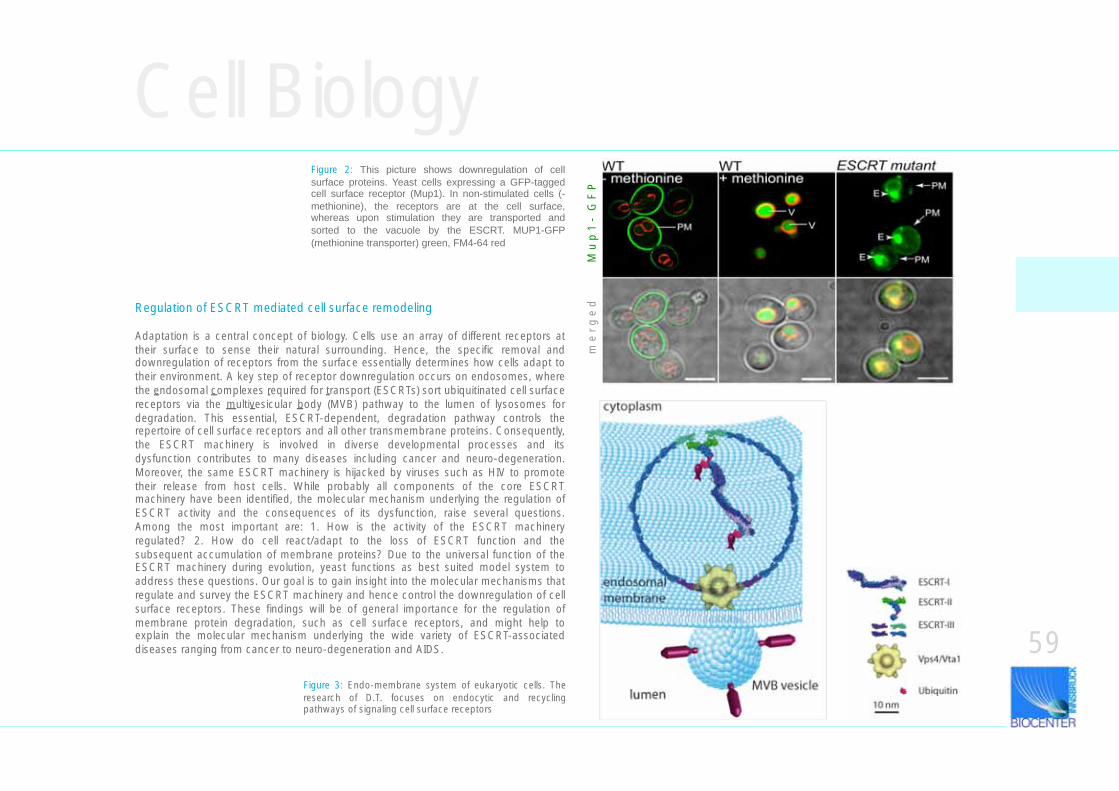
Cell Biology
Regulation of ESCRT mediated cell surface remodeling
Adaptation is a central concept of biology. Cells use an array of different receptors at their surface to sense their natural surrounding. Hence, the specific removal and downregulation of receptors from the surface essentially determines how cells adapt to their environment. A key step of receptor downregulation occurs on endosomes, where the endosomal complexes required for transport (ESCRTs) sort ubiquitinated cell surface receptors via the multivesicular body (MVB) pathway to the lumen of lysosomes for degradation. This essential, ESCRT-dependent, degradation pathway controls the repertoire of cell surface receptors and all other transmembrane proteins. Consequently, the ESCRT machinery is involved in diverse developmental processes and its dysfunction contributes to many diseases including cancer and neuro-degeneration. Moreover, the same ESCRT machinery is hijacked by viruses such as HIV to promote their release from host cells. While probably all components of the core ESCRT machinery have been identified, the molecular mechanism underlying the regulation of ESCRT activity and the consequences of its dysfunction, raise several questions. Among the most important are: 1. How is the activity of the ESCRT machinery regulated? 2. How do cell react/adapt to the loss of ESCRT function and the subsequent accumulation of membrane proteins? Due to the universal function of the ESCRT machinery during evolution, yeast functions as best suited model system to address these questions. Our goal is to gain insight into the molecular mechanisms that regulate and survey the ESCRT machinery and hence control the downregulation of cell surface receptors. These findings will be of general importance for the regulation of membrane protein degradation, such as cell surface receptors, and might help to explain the molecular mechanism underlying the wide variety of ESCRT-associated diseases ranging from cancer to neuro-degeneration and AIDS.
Figure 3: Endo-membrane system of eukaryotic cells. The research of D.T. focuses on endocytic and recycling pathways of signaling cell surface receptors
Figure 2: This picture shows downregulation of cell surface proteins. Yeast cells expressing a GFP-tagged cell surface receptor (Mup1). In non-stimulated cells (- methionine), the receptors are at the cell surface, whereas upon stimulation they are transported and sorted to the vacuole by the ESCRT. MUP1-GFP (methionine transporter) green, FM4-64 red
59
m e
r g
e d
M u
p 1
- G
F P
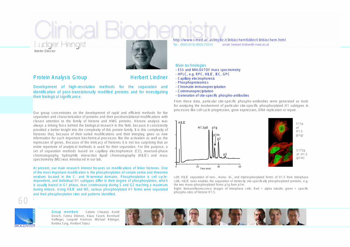
Clinical Biochemistry
Protein Analysis Group Herbert Lindner
Development of high-resolution methods for the separation and identification of post-translationally modified proteins and for investigating their biological significance.
Our group concentrates on the development of rapid and efficient methods for the separation and characterization of proteins and their posttranslational modifications with closest attention to the family of histone and HMG proteins. Histone analysis was always a driving force behind the biological research in this field, because it consistently provided a better insight into the complexity of this protein family. It is this complexity of histones that, because of their varied modifications and their interplay, gives us new information for such important biochemical processes like the activation as well as the repression of genes. Because of the intricacy of histones it is not too surprising that an entire repertoire of analytical methods is used for their separation. For this purpose, a set of separation methods based on capillary electrophoresis (CE), reversed-phase chromatography, hydrophilic interaction liquid chromatography (HILIC) and mass spectrometry (MS) was introduced in our lab.
At present, our main research interest focuses on modifications of linker histones. One of the most important modification is the phosphorylation of certain serine and threonine residues located in the C- and N-terminal domains. Phosphorylation is cell cycle- dependent, and individual H1 subtypes differ in their degree of phosphorylation, which is usually lowest in G1 phase, rises continuously during S and G2 reaching a maximum during mitosis. Using HILIC and MS, various phosphorylated H1 forms were separated and their phosphorylation sites and patterns identified..
Ludger HengstInterim Director
Tel.: 0043 (512) 9003.70310 email: [email protected]
Group members Sabine Chwatal, Astrid Devich, Fatma Dikmen, Klaus Faserl, Bernhard Halfinger, Leopold Kremser, Michael Rittinger, Bettina Sarg, Heribert Talasz
0 20 40 60Time (min)
HILIC
p1m
p1g
p2
p3
H1.5p0
From these data, particular site-specific phospho-antibodies were generated as tools for analyzing the involvement of particular site-specific phosphorylated H1 subtypes in processes like cell cycle progression, gene expression, DNA replication or repair.
S17p of H1.5(p1g)
S172p of H1.5(p1m)
Left: HILIC separation of non-, mono- di-, and triphosphorylated forms of H1.5 from interphase cells. HILIC even enables the separation of distinctly site-specifically phosphorylated proteins, e.g. the two mono-phosphorylated forms p1g from p1m. Right: Immunofluorescence images of interphase cells. Red = alpha tubulin; green = specific phospho-sites of histone H1.5.
60
Main technologies- ESI- and MALDI-TOF mass spectrometry- HPLC, e.g. RPC, HILIC, IEC, GPC- Capillary electrophoresis- Phosphoproteomics- Chromatin immunoprecipitation- Coimmunoprecipitation- Generation of site-specific phospho-antibodies
http://www.i-med.ac.at/imcbc/clinbiochemfolder/clinbiochem.html
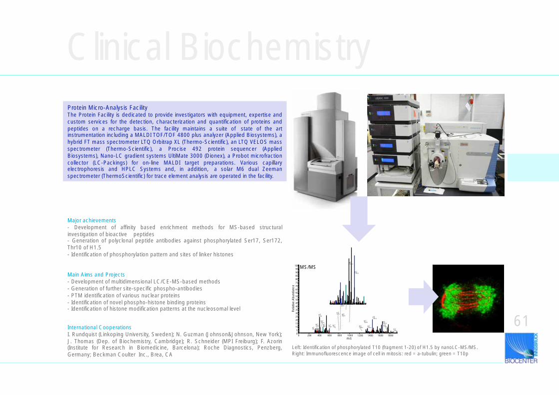
Clinical Biochemistry
International CooperationsI. Rundquist (Linkoping University, Sweden); N. Guzman (Johnson&Johnson, New York); J. Thomas (Dep. of Biochemistry, Cambridge); R. Schneider (MPI Freiburg); F. Azorin (Institute for Research in Biomedicine, Barcelona); Roche Diagnostics, Penzberg, Germany; Beckman Coulter Inc., Brea, CA
Major achievements- Development of affinity based enrichment methods for MS-based structural investigation of bioactive peptides- Generation of polyclonal peptide antibodies against phosphorylated Ser17, Ser172, Thr10 of H1.5 - Identification of phosphorylation pattern and sites of linker histones
Main Aims and Projects- Development of multidimensional LC/CE-MS-based methods- Generation of further site-specific phospho-antibodies- PTM identification of various nuclear proteins- Identification of novel phospho-histone binding proteins- Identification of histone modification patterns at the nucleosomal level
Protein Micro-Analysis FacilityThe Protein Facility is dedicated to provide investigators with equipment, expertise and custom services for the detection, characterization and quantification of proteins and peptides on a recharge basis. The facility maintains a suite of state of the art instrumentation including a MALDI TOF/TOF 4800 plus analyzer (Applied Biosystems), a hybrid FT mass spectrometer LTQ Orbitrap XL (Thermo-Scientific), an LTQ VELOS mass spectrometer (Thermo-Scientific), a Procise 492 protein sequencer (Applied Biosystems), Nano-LC gradient systems UltiMate 3000 (Dionex), a Probot microfraction collector (LC-Packings) for on-line MALDI target preparations. Various capillary electrophoresis and HPLC Systems and, in addition, a solar M6 dual Zeeman spectrometer (ThermoScientific) for trace element analysis are operated in the facility.
0 200 400 600 800 1000 1200 1400 1600 1800m/z
05
101520253035404550556065707580859095
100
Rel
ativ
e A
bund
ance
yy 10+1
1103.4
yy 15+2
828.6
bb 17+2
935.4
yy 16+2
877.8yy13+1
1456.4
yy 9+1
1006.5
yy 11+1
1284.4 yy15+1
1656.5yy 4
+1
482.4
bb 4+1
430.8
bb 7+1
728.1yy 5
+1610.3 yy12
+1
1355.5
bb 8+1
829.4
yy3+1
315.3
yy16+1
1753.5
bb 3+1
359.9
yy 14+2
793.1
bb 12+1
1249.9bb 17
+1
1870.1
yy 7+2
419.8
MS/MS
Left: Identification of phosphorylated T10 (fragment 1-20) of H1.5 by nanoLC-MS/MS. Right: Immunofluorescence image of cell in mitosis: red = a-tubulin; green = T10p
61
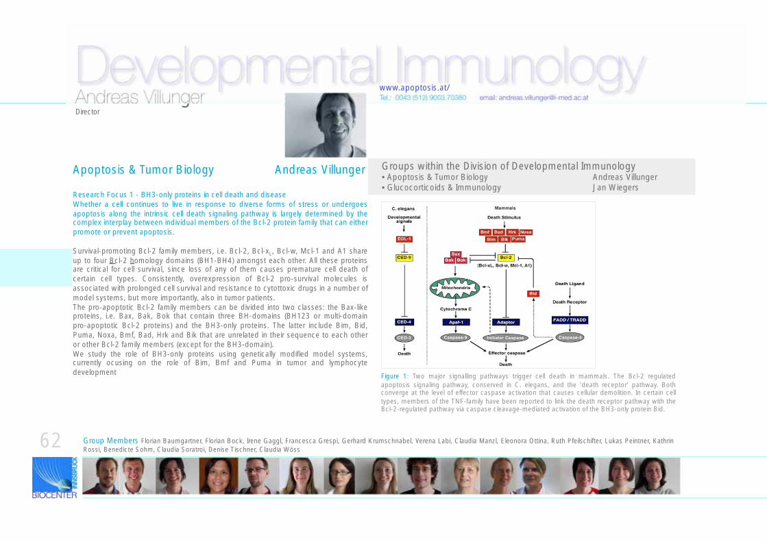
Developmental Immunology
Research Focus 1 - BH3-only proteins in cell death and diseaseWhether a cell continues to live in response to diverse forms of stress or undergoes apoptosis along the intrinsic cell death signaling pathway is largely determined by the complex interplay between individual members of the Bcl-2 protein family that can either promote or prevent apoptosis.
Survival-promoting Bcl-2 family members, i.e. Bcl-2, Bcl-xL, Bcl-w, Mcl-1 and A1 share up to four Bcl-2 homology domains (BH1-BH4) amongst each other. All these proteins are critical for cell survival, since loss of any of them causes premature cell death of certain cell types. Consistently, overexpression of Bcl-2 pro-survival molecules is associated with prolonged cell survival and resistance to cytottoxic drugs in a number of model systems, but more importantly, also in tumor patients.The pro-apoptotic Bcl-2 family members can be divided into two classes: the Bax-like proteins, i.e. Bax, Bak, Bok that contain three BH-domains (BH123 or multi-domain pro-apoptotic Bcl-2 proteins) and the BH3-only proteins. The latter include Bim, Bid, Puma, Noxa, Bmf, Bad, Hrk and Bik that are unrelated in their sequence to each other or other Bcl-2 family members (except for the BH3-domain).We study the role of BH3-only proteins using genetically modified model systems, currently ocusing on the role of Bim, Bmf and Puma in tumor and lymphocyte development Figure 1: Two major signalling pathways trigger cell death in mammals. The Bcl-2 regulated
apoptosis signaling pathway, conserved in C. elegans, and the 'death receptor' pathway. Both converge at the level of effector caspase activation that causes cellular demolition. In certain cell types, members of the TNF-family have been reported to link the death receptor pathway with the Bcl-2-regulated pathway via caspase cleavage-mediated activation of the BH3-only protein Bid.
Groups within the Division of Developmental Immunology• Apoptosis & Tumor Biology Andreas Villunger • Glucocorticoids & Immunology Jan Wiegers
Apoptosis & Tumor Biology Andreas Villunger
Tel.: 0043 (512) 9003.70380 email: [email protected]
Group Members Florian Baumgartner, Florian Bock, Irene Gaggl, Francesca Grespi, Gerhard Krumschnabel, Verena Labi, Claudia Manzl, Eleonora Ottina, Ruth Pfeilschifter, Lukas Peintner, Kathrin Rossi, Benedicte Sohm, Claudia Soratroi, Denise Tischner, Claudia Wöss
62
Andreas VillungerDirector
www.apoptosis.at/
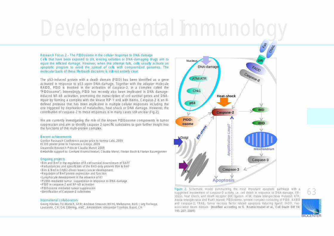
PIDD-osome
Developmental ImmunologyResearch Focus 2 - The PIDDosome in the cellular response to DNA damage Cells that have been exposed to UV, ionizing radiation or DNA-damaging drugs aim to repair the inflicted damage. However, when this attempt fails, cells usually activate an apoptotic program to avoid the spread of cells with compromized genomes. The molecular basis of these life/death decisions is still not entirely clear.
The p53-induced protein with a death domain (PIDD) has been identified as a gene activated in response to p53 upon DNA-damage. Together with the adapter molecule RAIDD, PIDD is involved in the activation of caspase-2, in a complex called the “PIDDosome”. Interestingly, PIDD has recently also been implicated in DNA damage-induced NF-kB activation, promoting the transcription of cell survival genes and DNA-repair by forming a complex with the kinase RIP-1 and with Nemo. Caspase-2 is an ill-defined protease that has been implicated in multiple cellular responses including the one triggered by deprivation of metabolites, heat shock or DNA damage. However, the contribution of caspase-2 to these responses is in many cases still unclear (Fig.2).
We are currently investigating the role of the known PIDDosome components in tumor suppression and aim to identify caspase 2-specific substrates to gain further insight into the functions of this multi-protein complex.
GM-130 overlay
Recent achievementsGordon Research Conference poster prize to Verena Labi, 2009ECDO poster prize to Francesca Grespi, 2009Swarovski Research Prize to Claudia Manzl; 2009Krebshilfe support to: Gerhard Krumschnabel, Claudia Manzl, Florian Bock & Florian Baumgartner
Ongoing projects•Bim and Bmf in the regulation of B cell survival downstream of BAFF•Redundancies and specificities of the BH3-only proteins Bim & Bmf•Bim & Bmf in ErbB2-dirven breast cancer development•Regulation of Bmf protein expression and function•Lymphocyte development in the absence of A1•PUMA-mediated tumor suppression in response to DNA-damage•PIDD in caspase-2 and NF-kB activation•PIDDosome mediated tumor suppression•Identification of Caspase-2 substrates
International collaboratorsGeorg Häcker, TU-Munich, GER; Andreas Strasser, WEHI, Melbourne, AUS; Jürg Tschopp, Lausanne, CH; Eric Eldering, AMC, Amsterdam; Alexandar Tzankov, Basel, CH
63
Caspase-2
Heat-shock
ER
Golgi
tBID
BID
Am
plifi
cati
on L
oop
?
Caspase-3
ATM/ATR
Chk1
p53
Caspase-9
Apoptosis
Figure 2. Schematic model summarizing the most important apoptotic pathways with a suggested involvement of caspase-2 activity, i.e. cell death in response to DNA-damage, ER-stress, heat shock, and death receptor (DR) ligation. ATM, Ataxia telangiectasia mutated; ATR, Ataxia telangiectasia and Rad3 related; PIDDosome, protein complex consisting of PIDD, RAIDD and caspase-2; TRAIL, tumor necrosis factor related apoptosis inducing ligand; FADD, Fas-associated death domain (modified according to G. Krumschnabel et al., Cell Death Diff 16: 195-207, 2009)
Mitochondrium
DR
DNA-damage
Nucleus
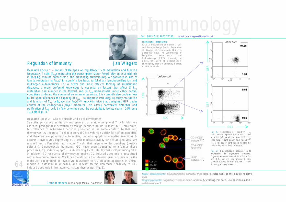
Research Focus 1 – Impact of life span on regulatory T cell maturation and function Regulatory T cells (Treg) expressing the transcription factor Foxp3 play an essential role in keeping immune homeostasis and preventing autoimmunity. A spontaneous loss of function-mutation in foxp3 in ‘scurfy’ mice leads to fulminant lymphoproliferation and multiorgan autoimmunity. For a better and more efficient therapy of autoimmune diseases, a more profound knowledge is essential on factors that affect (i) Treg maturation and number in the thymus and (ii) Treg homeostasis under either normal conditions or during the course of an immune response. It is currently also unclear how (iii) life span influences the capacity of Treg to suppress immunity. To study maturation and function of Treg cells, we use foxp3GFP knock-in mice that coexpress GFP under control of the endogenous foxp3 promoter. This allows convenient detection and purification of Treg cells by flow cytometry and the possibility to isolate nearly 100% pure Treg cells (Fig. 1).
Research Focus 2 – Glucocorticoids and T cell development Selection processes in the thymus ensure that mature peripheral T cells fulfill two essential prerequisites: activation by foreign peptides bound to (host) MHC molecules, but tolerance to self-derived peptides presented in the same context. To that end, thymocytes that express T cell receptors (TCRs) with high avidity for self antigen:MHC and therefore are potentially autoreactive, undergo apoptosis (negative selection). In contrast, thymocytes expressing TCR with moderate avidity for self antigen:MHC are rescued and differentiate into mature T cells that migrate to the periphery (positive selection). Glucocorticoid hormones (GC) have been suggested to influence these processes, e.g. induce apoptosis in developing T cells, the thymus itself producing GCs! In addition, GC resistance of thymocytes against GC-induced apoptosis is associated with autoimmune diseases. We focus therefore on the following questions: i) what is the molecular background of thymocyte resistance to GC-induced apoptosis in animal models of autoimmune diseases, and ii) what factors determine sensitivity to GC-induced apoptosis in immature vs. mature thymocytes (Fig. 2).
Developmental Immunology
Group members Irene Gaggl, Manuel Kaufmann
Tel.: 0043 (512) 9003.70390 email: [email protected]
GR-FITC
CD4-PE
CD8-CY
International collaboratorsFalus A Department of Genetics, Cell- and Immunobiology (earlier Department of Biology) at Semmelweis University, Budapest; Reul JM Laboratories of Integrative Neuroscience and Endocrinology (LINE), University of Bristol, UK; Boyd RL Department of Immunology, Monash University, Clayton, Victoria, Australia
FSC
64
CD4+ CD8+
Isotype FITC
CD4+ CD8+
GR-FITC
CD8+
Isotype FITC
Major achievements: Glucocorticoids enhance thymocyte development at the double-negative level. Ongoing projects: Regulatory T cells in bim-/- and vav-bcl2 transgenic mice, Glucocorticoids and T cell development
before sort
after sort
CD
4
CD4
GFP
GFP
Regulation of Immunity Jan Wiegers
Fig. 1.: Purification of Foxp3GFP+ Treg cells. Isolated splenocytes were stained for CD4 (left panel) and Foxp3GFP+ Treg cells (upper right panel) and Foxp3GFP– Tcon cells (lower right panel) isolated by cell sorting with a flow cytometer.
Fig. 2: Glucocorticoid receptor (GR) expression in thymocyte subsets. Thymocytes were stained for CD4, CD8 and GR, washed and mounted with Mowiol. Isotype control and GR stained thymocytes were mixed 1:1.
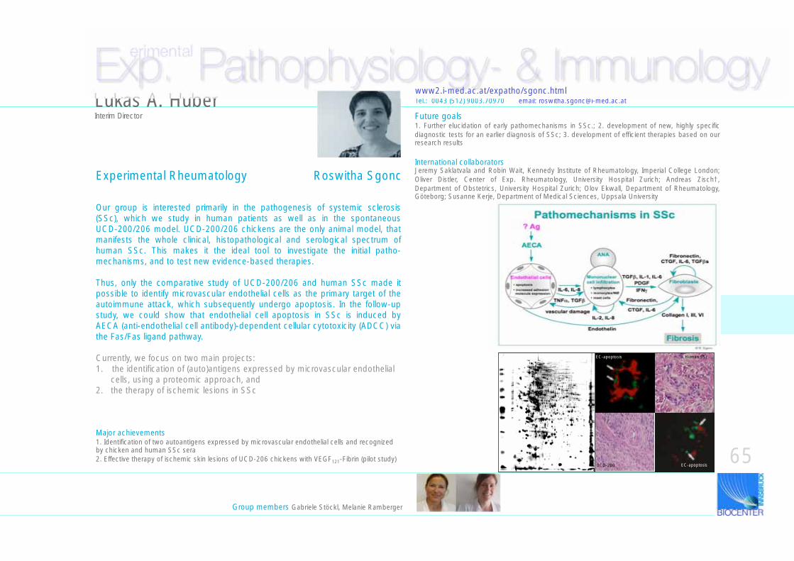
Exp. Pathophysiology- & Immunology erimental
Our group is interested primarily in the pathogenesis of systemic sclerosis (SSc), which we study in human patients as well as in the spontaneous UCD-200/206 model. UCD-200/206 chickens are the only animal model, that manifests the whole clinical, histopathological and serological spectrum of human SSc. This makes it the ideal tool to investigate the initial patho-mechanisms, and to test new evidence-based therapies.
Thus, only the comparative study of UCD-200/206 and human SSc made it possible to identify microvascular endothelial cells as the primary target of the autoimmune attack, which subsequently undergo apoptosis. In the follow-up study, we could show that endothelial cell apoptosis in SSc is induced by AECA (anti-endothelial cell antibody)-dependent cellular cytotoxicity (ADCC) via the Fas/Fas ligand pathway.
Currently, we focus on two main projects:1. the identification of (auto)antigens expressed by microvascular endothelial cells, using a proteomic approach, and2. the therapy of ischemic lesions in SSc
Group members Gabriele Stöckl, Melanie Ramberger
Experimental Rheumatology Roswitha Sgonc
Tel.: 0043 (512) 9003.70970 email: [email protected]
Major achievements1. Identification of two autoantigens expressed by microvascular endothelial cells and recognized by chicken and human SSc sera2. Effective therapy of ischemic skin lesions of UCD-206 chickens with VEGF121-Fibrin (pilot study)
EC-apoptosis
EC-apoptosis
Human SSc
UCD-200
Lukas A. HuberInterim Director
65
Future goals1. Further elucidation of early pathomechanisms in SSc.; 2. development of new, highly specific diagnostic tests for an earlier diagnosis of SSc; 3. development of efficient therapies based on our research results
International collaboratorsJeremy Saklatvala and Robin Wait, Kennedy Institute of Rheumatology, Imperial College London; Oliver Distler, Center of Exp. Rheumatology, University Hospital Zurich; Andreas Zisch†, Department of Obstetrics, University Hospital Zurich; Olov Ekwall, Department of Rheumatology, Göteborg; Susanne Kerje, Department of Medical Sciences, Uppsala University
www2.i-med.ac.at/expatho/sgonc.html
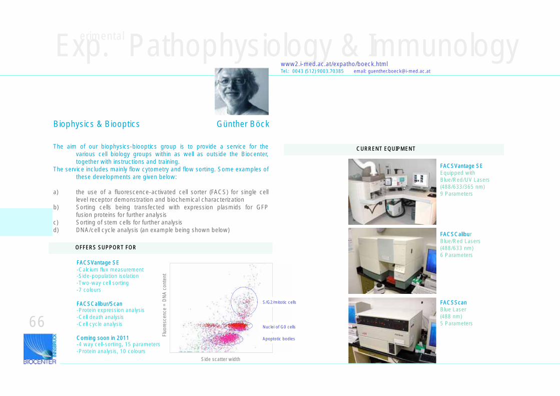
Tel.: 0043 (512) 9003.70385 email: [email protected]
Exp. Pathophysiology & Immunology erimental
Biophysics & Biooptics Günther Böck
The aim of our biophysics-biooptics group is to provide a service for the various cell biology groups within as well as outside the Biocenter, together with instructions and training.
The service includes mainly flow cytometry and flow sorting. Some examples of these developments are given below:
a) the use of a fluorescence-activated cell sorter (FACS) for single cell level receptor demonstration and biochemical characterization
b) Sorting cells being transfected with expression plasmids for GFP fusion proteins for further analysis
c) Sorting of stem cells for further analysisd) DNA/cell cycle analysis (an example being shown below)
66
www2.i-med.ac.at/expatho/boeck.html
FACSVantage SEEquipped withBlue/Red/UV Lasers(488/633/365 nm)9 Parameters
FACSCaliburBlue/Red Lasers(488/633 nm)6 Parameters
FACSScanBlue Laser(488 nm)5 Parameters
FACSVantage SE-Calcium flux measurement-Side-population isolation-Two-way cell sorting -7 colours
FACSCalibur/Scan-Protein expression analysis-Cell death analysis-Cell cycle analysis
Coming soon in 2011-4 way cell-sorting, 15 parameters-Protein analysis, 10 colours
CURRENT EQUIPMENT
OFFERS SUPPORT FOR
S/G2/mitotic cells
Nuclei of G0 cells
Apoptotic bodies
Side scatter width
Fluo
resc
ence
= D
NA
con
tent
Exp. Pathophysiology & Immunology erimental
www2.i-med.ac.at/expatho/schwarz.html
ResearchThis laboratory's work has focussed on the study of various hormone/neurotransmitter binding proteins and receptors as well as their ligands. Key papers describe: - Discovery of Sex Hormone Binding Globulin (SHBG) in cerebrospinal fluid (CSF) and interaction of SHBG with Danazol (non-genomic actions of steroids?)- 1st Demonstration of homocysteate as an NMDA-selective excitatory agonist- 1st Description of an epitope map of the glycoprotein hormone hCG- Construction of epitope-selective immunoassays for glycoprotein hormones-Demonstration of different orientations of receptor-bound agonistic vs. antagonistic hCG- Prediction of the 3D structure of the extracellular domain of the hCG receptor- Characterizartion of an apoptotic activity within urinary hCG preparations towards Kaposi‘s sarcoma cells-Demonstration of the importance of vasopressin in critical ill patients
Teaching PathophysiologyThe University law 2002 mandated the implementation of the NEW CURRICULUM HUMAN MEDICINE at the Innsbruck Medical University. The latter required a full-time teaching devotion for the subject PATHOPHYSIOLOGY. In accordance with these duties, Professor Schwarz published two textbooks, one in 2002, the other in 2007, on molecular aspects of pathophysiology.
Tel.: 0043 (512) 9003.70975 email: [email protected]
Therefore, teaching of Pathophysiology is stratified over five semesters instead of the previously two. Except for infectious diseases and cancer, the entire teaching of Patho-physiology in main lectures is covered by Prof. Schwarz. In addition, he is appointed coordinator of the module „Endocrinology“, coordinator of the 4th seme-ster, organizer of the SIP3 (summative integrative ex-amination at the end of the 6th semester). Also, he acts as a deputy chief examiner at the EMS (Eingangstest Medizin-Studium) mandatory for the enrollement in the study of medicine, and is member of the examination board of SIP3.
http://www2.i-med.ac.at/expatho/ molecules_of_life_anncmt.htmlBook Award 2003 by the British Medical Association
In contrast to previous and „classical“ curricula, Pathophysiology is not any more taught within 2 semesters as a separate and isolated subject, rather it is incorporated into an integrative teaching style of organ and disease modules. A module is covered by lecturers from diverse preclinical and clinical disciplines. The modules are the following: diagnostics and laboratory medicine, endocrinology, hematology, cardiovascular diseases, nephrology, pulmonology, neurology & psychiatry, infectious diseases + immunology, cancer, dermatology, gastroenterology, osteology, teratology, environmental medicine. Since 2008, Professor Schwarz is appointed Guest Professor for Pathophysiology at the Suranaree University of Technology (Korath/Nakhon Ratchasima, Thailand).In the newly formed CLINICAL SKILLS LAB-project implemented by the MUI, the Venipuncture practicum-proposal of S.S. was ranked 2nd out of 12.
http://www.maudrich.com/list?back=1f74a3719cde4316d61ae4a1952981f6&isbn=9783851758603
In 2009, Prof. Schwarz was invited to display 10 pictures of this book (format 50x50) in the newly built Pediatric University Hospital Innsbruck, 1st floor West. This exhibition serves an aesthetic as well as an educative purpose: ARS IN_STRUCTA (a term coined by Stephan Geley).
67
Molecular Endocrinology Siegfried Schwarz
Genomics & RNomicsgenomics.i-med.ac.at/
Groups within the Division of Genomics & Rnomics
• Experimental RNomics• Molecular Biology of Ribonucleoprotein Complexes
Hundreds of genes and their RNA products may thus remain undetected. Their functions, interactions in cellular circuits and roles in disease would remain unknown and our understanding of the functioning of a cell would be incomplete. Therefore, our goal is to directly identify ncRNAs and their genes in the human genome and those of various model organisms as well as to elucidate their functions in cellular processes and/or human diseases.
Tel.: 0043 (512) 9003.70250 email: [email protected]
Non-coding RNAs in model organisms: identification and functionIn cells from all organisms studied to date two different types of RNAs are found: messenger RNAs (mRNAs), which are translated into proteins, and so-called nonprotein-coding RNAs (ncRNAs), which are not translated into proteins but function at the level of the RNA itself. Many known ncRNAs, such as microRNAs, are involved in the regulation of gene expression and thus act as molecular switches. While the proteome of most model organisms is rather well defined, e.g. the total number of protein-coding genes, we are only at the very beginning of describing the ncRNA transcriptome. Thus, the predictions on the number of ncRNA genes in the human genome range from about 1.000 up to 400.000 (estimated from tiling-array experiments); in comparison, about 20.000 protein-coding genes are being estimated. NcRNAs are often found in complex with proteins that are bound to the RNA and thus form ribonucleoprotein complexes (RNPs).
Such RNPs, present in cellular compartments as diverse as the nucleolus or dendritic processes of nerve cells, exhibit a surprisingly diverse range of functions. However, the biological role of some of them remains elusive. Moreover, most systematic genomic searches are biased against their detection and comprehensive identification by computational analysis of the genomic sequence of any organism remains an unsolved problem.
Group members Roland Hutzinger, Hubert Muckenhuber, Matthias Heiss, Melanie Lukasser, Ronald Gstir, Andreas Ploner, Mathieu Rederstorff, Konstantinia Skreka, Katrin Perfler
Alexander HüttenhoferDirector
68
Experimental RNomics Alexander HüttenhoferAlexander HüttenhoferNorbert Polacek
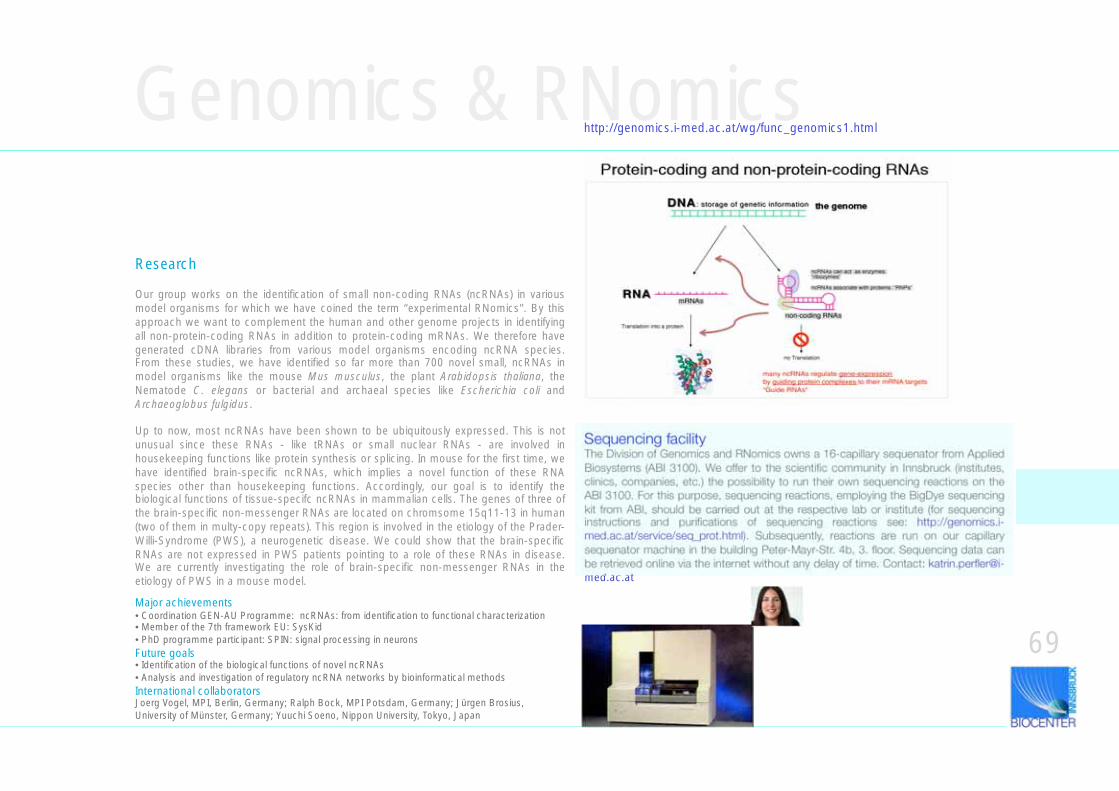
Research
Our group works on the identification of small non-coding RNAs (ncRNAs) in various model organisms for which we have coined the term “experimental RNomics". By this approach we want to complement the human and other genome projects in identifying all non-protein-coding RNAs in addition to protein-coding mRNAs. We therefore have generated cDNA libraries from various model organisms encoding ncRNA species. From these studies, we have identified so far more than 700 novel small, ncRNAs in model organisms like the mouse Mus musculus, the plant Arabidopsis thaliana, the Nematode C. elegans or bacterial and archaeal species like Escherichia coli and Archaeoglobus fulgidus.
Up to now, most ncRNAs have been shown to be ubiquitously expressed. This is not unusual since these RNAs - like tRNAs or small nuclear RNAs - are involved in housekeeping functions like protein synthesis or splicing. In mouse for the first time, we have identified brain-specific ncRNAs, which implies a novel function of these RNA species other than housekeeping functions. Accordingly, our goal is to identify the biological functions of tissue-specifc ncRNAs in mammalian cells. The genes of three of the brain-specific non-messenger RNAs are located on chromsome 15q11-13 in human (two of them in multy-copy repeats). This region is involved in the etiology of the Prader-Willi-Syndrome (PWS), a neurogenetic disease. We could show that the brain-specific RNAs are not expressed in PWS patients pointing to a role of these RNAs in disease. We are currently investigating the role of brain-specific non-messenger RNAs in the etiology of PWS in a mouse model.
Genomics & RNomicshttp://genomics.i-med.ac.at/wg/func_genomics1.html
Major achievements• Coordination GEN-AU Programme: ncRNAs: from identification to functional characterization• Member of the 7th framework EU: SysKid• PhD programme participant: SPIN: signal processing in neuronsFuture goals• Identification of the biological functions of novel ncRNAs• Analysis and investigation of regulatory ncRNA networks by bioinformatical methodsInternational collaboratorsJoerg Vogel, MPI, Berlin, Germany; Ralph Bock, MPI Potsdam, Germany; Jürgen Brosius, University of Münster, Germany; Yuuchi Soeno, Nippon University, Tokyo, Japan
69
Sequencing facilityThe Division of Genomics and RNomics owns a 16-capillary sequenator from Applied Biosystems (ABI 3100). We offer to the scientific community in Innsbruck (institutes, clinics, companies, etc.) the possibility to run their own sequencing reactions on the ABI 3100. For this purpose, sequencing reactions, employing the BigDye sequencing kit from ABI, should be carried out at the respective lab or institute (for sequencing instructions and purifications of sequencing reactions see: http://genomics.i-med.ac.at/service/seq_prot.html). Subsequently, reactions are run on our capillary sequenator machine in the building Peter-Mayr-Str. 4b, 3. floor. Sequencing data can be retrieved online via the internet without any delay of time. Contact: [email protected]
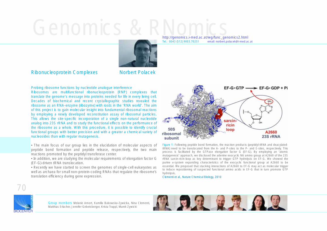
Genomics & RNomics
Ribonucleoprotein Complexes Norbert Polacek
http://genomics.i-med.ac.at/wg/func_genomics2.htmlTel.: 0043 (512) 9003.70251 email: [email protected]
Clementi et al., Nature Chemical Biology, 2010
Probing ribosome functions by nucleotide analogue interferenceRibosomes are multifunctional ribonucleoprotein (RNP) complexes that translate the genome's message into proteins needed for life in every living cell. Decades of biochemical and recent crystallographic studies revealed the ribosome as an RNA-enzyme (ribozyme) with roots in the 'RNA world'. The aim of this project is to gain molecular insight into fundamental ribosomal reactions by employing a newly developed reconstitution assay of ribosomal particles. This allows the site-specific incorporation of a single non-natural nucleotide analog into 23S rRNA and to study the functional effects on the performance of the ribosome as a whole. With this procedure, it is possible to identify crucial functional groups with better precision and with a greater a chemical variety of nucleosides than with regular mutagenesis.
• The main focus of our group lies in the elucidation of molecular aspects of peptide bond formation and peptide release, respectively, the two main reactions promoted by the peptidyl transferase center. • In addition, we are studying the molecular requirements of elongation factor G (EF-G)-driven tRNA translocation. • Recently we have started to screen the genomes of single-cell eukaryotes as well as archaea for small non-protein-coding RNAs that regulate the ribosome’s translation efficiency during gene expression.
Group members Melanie Amort, Kamilla Bakowska-Zywicka, Nina Clementi, Matthias Erlacher, Jennifer Gebetsberger, Krista Trappl, Marek Zywicki
70
Figure 1: Following peptide bond formation, the reaction products (peptidyl-tRNA and deacylated-tRNA) need to be translocated from the A- and P-sites to the P- and E-sites, respectively. This process is facilitated by the GTPase elongation factor G (EF-G). By employing an ‘atomic mutagenesis’ approach, we disclosed the adenine exocyclic N6 amino group at A2660 of the 23S rRNA sarcin-ricin-loop as key determinant to trigger GTP hydrolysis on EF-G. We showed the purine -system expanding characteristics of the exocyclic functional group at A2660 to be essential. We proposed that stacking interactions of A2660 to EF-G may act as molecular trigger to induce repositioning of suspected functional amino acids in EF-G that in turn promote GTP hydrolysis.
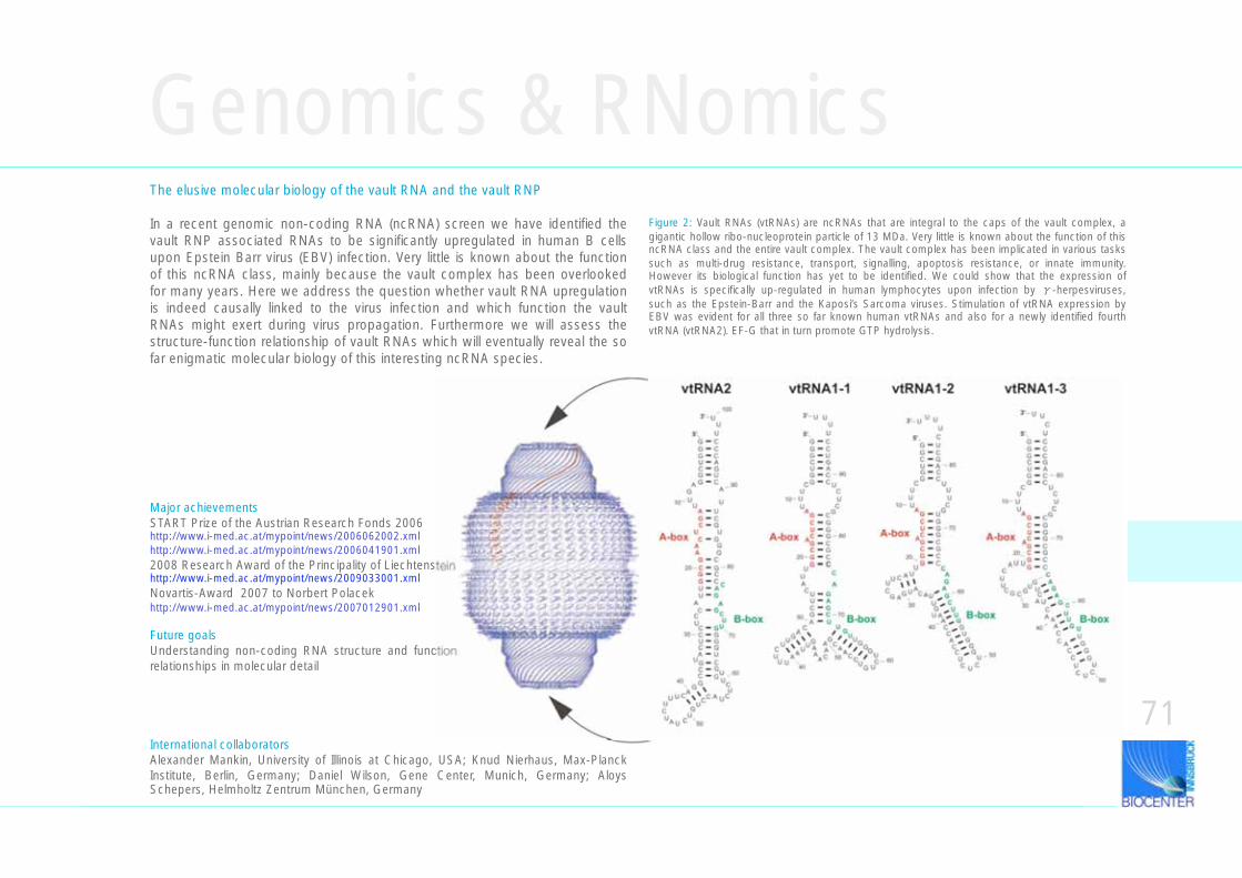
Genomics & RNomicsThe elusive molecular biology of the vault RNA and the vault RNP
In a recent genomic non-coding RNA (ncRNA) screen we have identified the vault RNP associated RNAs to be significantly upregulated in human B cells upon Epstein Barr virus (EBV) infection. Very little is known about the function of this ncRNA class, mainly because the vault complex has been overlooked for many years. Here we address the question whether vault RNA upregulation is indeed causally linked to the virus infection and which function the vault RNAs might exert during virus propagation. Furthermore we will assess the structure-function relationship of vault RNAs which will eventually reveal the so far enigmatic molecular biology of this interesting ncRNA species.
Major achievementsSTART Prize of the Austrian Research Fonds 2006 http://www.i-med.ac.at/mypoint/news/2006062002.xmlhttp://www.i-med.ac.at/mypoint/news/2006041901.xml2008 Research Award of the Principality of Liechtensteinhttp://www.i-med.ac.at/mypoint/news/2009033001.xmlNovartis-Award 2007 to Norbert Polacek http://www.i-med.ac.at/mypoint/news/2007012901.xml
Future goalsUnderstanding non-coding RNA structure and function relationships in molecular detail
71International collaboratorsAlexander Mankin, University of Illinois at Chicago, USA; Knud Nierhaus, Max-Planck Institute, Berlin, Germany; Daniel Wilson, Gene Center, Munich, Germany; Aloys Schepers, Helmholtz Zentrum München, Germany
Figure 2: Vault RNAs (vtRNAs) are ncRNAs that are integral to the caps of the vault complex, a gigantic hollow ribo-nucleoprotein particle of 13 MDa. Very little is known about the function of this ncRNA class and the entire vault complex. The vault complex has been implicated in various tasks such as multi-drug resistance, transport, signalling, apoptosis resistance, or innate immunity. However its biological function has yet to be identified. We could show that the expression of vtRNAs is specifically up-regulated in human lymphocytes upon infection by -herpesviruses, such as the Epstein-Barr and the Kaposi’s Sarcoma viruses. Stimulation of vtRNA expression by EBV was evident for all three so far known human vtRNAs and also for a newly identified fourth vtRNA (vtRNA2). EF-G that in turn promote GTP hydrolysis.
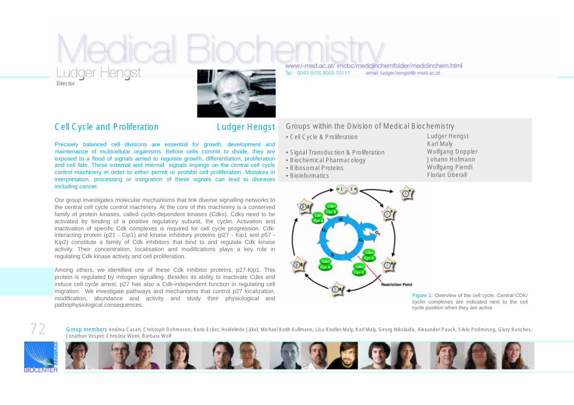
Medical Biochemistrywww.i-med.ac.at/ imcbc/medclinchemfolder/medclinchem.html
Groups within the Division of Medical BiochemistryCell Cycle and Proliferation Ludger HengstLudger HengstKarl MalyWolfgang DopplerJohann HofmannWolfgang PiendlFlorian Überall
Tel.: 0043 (512) 9003.70111 email: [email protected]
• Cell Cycle & Proliferation
• Signal Transduction & Proliferation • Biochemical Pharmacology • Ribosomal Proteins• Bioinformatics
Group members Andrea Casari, Christoph Dohmesen, Karin Ecker, Heidelinde Jäkel, Michael Keith Kullmann, Lisa Kindler-Maly, Karl Maly, Georg Nikolaidis, Alexander Pauck, Silvio Podmirseg, Glory Ranches, Jonathan Vosper, Christina Weinl, Barbara Wolf
Ludger HengstDirector
Precisely balanced cell divisions are essential for growth, development and maintenance of multicellular organisms. Before cells commit to divide, they are exposed to a flood of signals aimed to regulate growth, differentiation, proliferation and cell fate. These external and internal signals impinge on the central cell cycle control machinery in order to either permit or prohibit cell proliferation. Mistakes in interpretation, processing or integration of these signals can lead to diseases including cancer.
Our group investigates molecular mechanisms that link diverse signalling networks to the central cell cycle control machinery. At the core of this machinery is a conserved family of protein kinases, called cyclin-dependent kinases (Cdks). Cdks need to be activated by binding of a positive regulatory subunit, the cyclin. Activation and inactivation of specific Cdk complexes is required for cell cycle progression. Cdk-interacting protein (p21 - Cip1) and kinase inhibitory proteins (p27 - Kip1 and p57 - Kip2) constitute a family of Cdk inhibitors that bind to and regulate Cdk kinase activity. Their concentration, localisation and modifications plays a key role in regulating Cdk kinase activity and cell proliferation.
Among others, we identified one of these Cdk inhibitor proteins, p27-Kip1. This protein is regulated by mitogen signalling. Besides its ability to inactivate Cdks and induce cell cycle arrest, p27 has also a Cdk-independent function in regulating cell migration. We investigate pathways and mechanisms that control p27 localization, modification, abundance and activity and study their physiological and pathophysiological consequences.
Figure 1: Overview of the cell cycle. Central CDK/cyclin complexes are indicated next to the cell cycle position when they are active
72
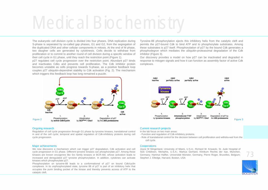
Medical BiochemistryThe eukaryotic cell division cycle is divided into four phases. DNA replication during S-phase is separated by so-called gap phases, G1 and G2, from the segregation of the duplicated DNA and other cellular components in mitosis. At the end of M phase, two daugher cells are generated by cytokinesis. Cells decide to withdraw from proliferation or to commit to another round of cell division during a specific window of their cell cycle in G1 phase, until they reach the restriction point (Figure 1). p27 regulates cell cycle progression over the restriction point. Abundant p27 binds and inactivates Cdks and prevents cell proliferation. The Cdk inhibitor protein becomes unstable as cells progress towards S-phase, as a positive feedback loop couples p27 ubiquitin-dependnet stability to Cdk activation (Fig. 2). The mechanism which triggers this feedback loop has long remained a puzzle.
Major achievements We now discovere a mechanism which can trigger p27 degradation, Cdk activation and cell cycle progression in G1 phase. Different tyrosine kinases can phosphorylate p27. Among these kinases are known oncogenes like Src family kinases or BCR-Abl, whose activation leads to increased and deregulated p27 tyrosine phosphorylation. In addition, cytokines can activate kinases which phosphorylate p27. Phosphorylation on tyrosine-88 leads to a conformational of p27 on bound Cdk/cyclin complexes. In its unphosphorylated state, tyrosine 88 of p27 is part of an inhibitory helix that occupies the purin binding pocket of the kinase and thereby prevents access of ATP to the catalytic cleft.
Cooperations Joyce M Slingerland, University of Miami, U.S.A.; Richard W. Kriwacki, St. Jude Hospital of Sick Childreen, Memphis, U.S.A.; Markus Gerhard, Klinikum Rechts der Isar, München, Germany; Hartmut Halfter, Universität Münster, Germany, Pierre Roger, Bruxelles, Belgium; Stephen J. Elledge, Harvard, Boston, USA
Current research projects in the lab focus on two main areas: - Function and regulation of Cdk-inhibitory proteins. - Role of translational control for the decision between cell proliferation and withdra-wal from the
cell cycle.
Tyrosine-88 phosphorylation ejects this inhibitory helix from the catalytic cleft and permits the p27-bound Cdk to bind ATP and to phosphorylate substrates. Among these substrates is p27 itself. Phosphorylation of p27 by the bound Cdk generates a phosphodegron which mediates the ubiquitin-proteasomal degradation of the Cdk inhibitor (Figure 3). Our discovery provides a model on how p27 can be inactivated and degraded in response to mitogen signals and how it can function as assembly factor of active Cdk complexes.
Figure 2 Figure 3
73
Ongoing research Regulation of cell cycle progression through G1 phase by tyrosine kinases, translational control in and of the cell cycle; temporal and spatial regulation of Cdk-inhibitory proteins during cell cycle progression.
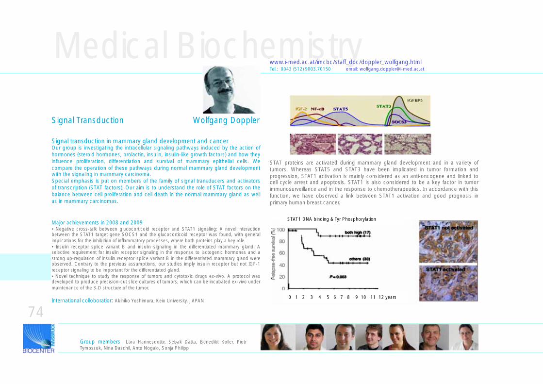
100
80
60
40
20
0
0
Rel
apse
-fre
e su
rviv
al (%
)
Medical Biochemistry
Signal transduction in mammary gland development and cancerOur group is investigating the intracellular signaling pathways induced by the action of hormones (steroid hormones, prolactin, insulin, insulin-like growth factors) and how they influence proliferation, differentiation and survival of mammary epithelial cells. We compare the operation of these pathways during normal mammary gland development with the signaling in mammary carcinoma. Special emphasis is put on members of the family of signal transducers and activators of transcription (STAT factors). Our aim is to understand the role of STAT factors on the balance between cell proliferation and cell death in the normal mammary gland as well as in mammary carcinomas.
www.i-med.ac.at/imcbc/staff_doc/doppler_wolfgang.html
Major achievements in 2008 and 2009• Negative cross-talk between glucocorticoid receptor and STAT1 signaling: A novel interaction between the STAT1 target gene SOCS1 and the glucocorticoid receptor was found, with general implications for the inhibition of inflammatory processes, where both proteins play a key role.• Insulin receptor splice variant B and insulin signaling in the differentiated mammary gland: A selective requirement for insulin receptor signaling in the response to lactogenic hormones and a strong up-regulation of insulin receptor splice variant B in the differentiated mammary gland were observed. Contrary to the previous assumptions, our studies imply insulin receptor but not IGF-1 receptor signaling to be important for the differentiated gland.• Novel technique to study the response of tumors and cytotoxic drugs ex-vivo. A protocol was developed to produce precision-cut slice cultures of tumors, which can be incubated ex-vivo under maintenance of the 3-D structure of the tumor.
Tel.: 0043 (512) 9003.70150 email: [email protected]
Signal Transduction Wolfgang Doppler
International colloboratior: Akihiko Yoshimura, Keio University, JAPAN
Pregnancy Lactation Involution
STAT proteins are activated during mammary gland development and in a variety of tumors. Whereas STAT5 and STAT3 have been implicated in tumor formation and progression, STAT1 activation is mainly considered as an anti-oncogene and linked to cell cycle arrest and apoptosis. STAT1 is also considered to be a key factor in tumor immunosurveillance and in the response to chemotherapeutics. In accordance with this function, we have observed a link between STAT1 activation and good prognosis in primary human breast cancer.
740 1 2 3 4 5 6 7 8 9 10 11 12 years
Group members Lára Hannesdottir, Sebak Datta, Benedikt Koller, Piotr Tymoszuk, Nina Daschil, Anto Nogalo, Sonja Philipp
STAT1 DNA binding & Tyr Phosphorylation
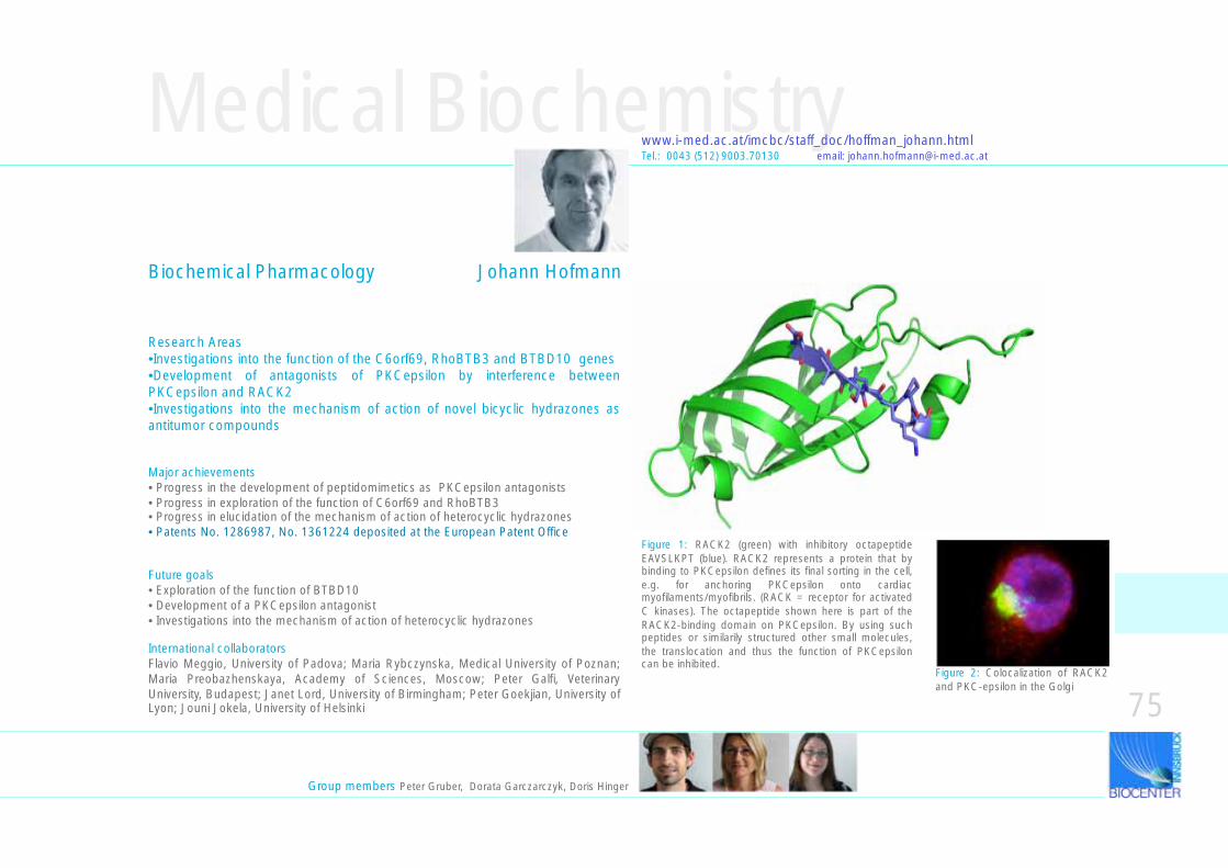
Medical Biochemistry
Research Areas•Investigations into the function of the C6orf69, RhoBTB3 and BTBD10 genes•Development of antagonists of PKCepsilon by interference between PKCepsilon and RACK2•Investigations into the mechanism of action of novel bicyclic hydrazones as antitumor compounds
www.i-med.ac.at/imcbc/staff_doc/hoffman_johann.htmlTel.: 0043 (512) 9003.70130 email: [email protected]
Figure 2: Colocalization of RACK2 and PKC-epsilon in the Golgi
Major achievements• Progress in the development of peptidomimetics as PKCepsilon antagonists• Progress in exploration of the function of C6orf69 and RhoBTB3• Progress in elucidation of the mechanism of action of heterocyclic hydrazones• Patents No. 1286987, No. 1361224 deposited at the European Patent Office
Future goals• Exploration of the function of BTBD10• Development of a PKCepsilon antagonist• Investigations into the mechanism of action of heterocyclic hydrazones
International collaboratorsFlavio Meggio, University of Padova; Maria Rybczynska, Medical University of Poznan; Maria Preobazhenskaya, Academy of Sciences, Moscow; Peter Galfi, Veterinary University, Budapest; Janet Lord, University of Birmingham; Peter Goekjian, University of Lyon; Jouni Jokela, University of Helsinki
Figure 1: RACK2 (green) with inhibitory octapeptide EAVSLKPT (blue). RACK2 represents a protein that by binding to PKCepsilon defines its final sorting in the cell, e.g. for anchoring PKCepsilon onto cardiac myofilaments/myofibrils. (RACK = receptor for activated C kinases). The octapeptide shown here is part of the RACK2-binding domain on PKCepsilon. By using such peptides or similarily structured other small molecules, the translocation and thus the function of PKCepsilon can be inhibited.
Biochemical Pharmacology Johann Hofmann
Group members Peter Gruber, Dorata Garczarczyk, Doris Hinger
75
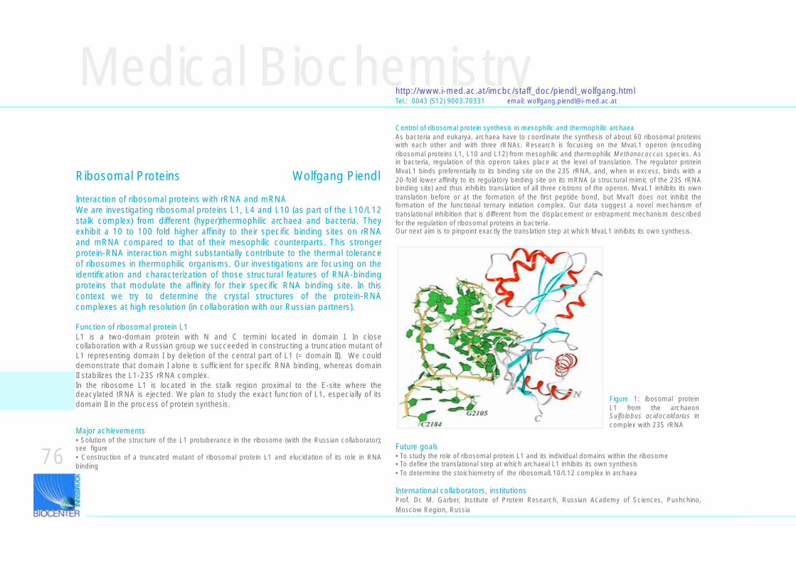
Medical Biochemistry
Interaction of ribosomal proteins with rRNA and mRNAWe are investigating ribosomal proteins L1, L4 and L10 (as part of the L10/L12 stalk complex) from different (hyper)thermophilic archaea and bacteria. They exhibit a 10 to 100 fold higher affinity to their specific binding sites on rRNA and mRNA compared to that of their mesophilic counterparts. This stronger protein-RNA interaction might substantially contribute to the thermal tolerance of ribosomes in thermophilic organisms. Our investigations are focusing on the identification and characterization of those structural features of RNA-binding proteins that modulate the affinity for their specific RNA binding site. In this context we try to determine the crystal structures of the protein-RNA complexes at high resolution (in collaboration with our Russian partners).
Function of ribosomal protein L1L1 is a two-domain protein with N and C termini located in domain I. In close collaboration with a Russian group we succeeded in constructing a truncation mutant of L1 representing domain I by deletion of the central part of L1 (= domain II). We could demonstrate that domain I alone is sufficient for specific RNA binding, whereas domain II stabilizes the L1-23S rRNA complex. In the ribosome L1 is located in the stalk region proximal to the E-site where the deacylated tRNA is ejected. We plan to study the exact function of L1, especially of its domain II in the process of protein synthesis.
Control of ribosomal protein synthesis in mesophilic and thermophilic archaeaAs bacteria and eukarya, archaea have to coordinate the synthesis of about 60 ribosomal proteins with each other and with three rRNAs. Research is focusing on the MvaL1 operon (encoding ribosomal proteins L1, L10 and L12) from mesophilic and thermophilic Methanococcus species. As in bacteria, regulation of this operon takes place at the level of translation. The regulator protein MvaL1 binds preferentially to its binding site on the 23S rRNA, and, when in excess, binds with a 20-fold lower affinity to its regulatory binding site on its mRNA (a structural mimic of the 23S rRNA binding site) and thus inhibits translation of all three cistrons of the operon. MvaL1 inhibits its own translation before or at the formation of the first peptide bond, but Mval1 does not inhibit the formation of the functional ternary initiation complex. Our data suggest a novel mechanism of translational inhibition that is different from the displacement or entrapment mechanism described for the regulation of ribosomal proteins in bacteria. Our next aim is to pinpoint exactly the translation step at which MvaL1 inhibits its own synthesis.
http://www.i-med.ac.at/imcbc/staff_doc/piendl_wolfgang.htmlTel.: 0043 (512) 9003.70331 email: [email protected]
Major achievements• Solution of the structure of the L1 protuberance in the ribosome (with the Russian collaborator); see figure• Construction of a truncated mutant of ribosomal protein L1 and elucidation of its role in RNA binding
Future goals• To study the role of ribosomal protein L1 and its individual domains within the ribosome• To define the translational step at which archaeal L1 inhibits its own synthesis• To determine the stoichiometry of the ribosomalL10/L12 complex in archaea
International collaborators, institutionsProf. Dr. M. Garber, Institute of Protein Research, Russian Academy of Sciences, Pushchino, Moscow Region, Russia
76
Figure 1: ibosomal protein L1 from the archaeon Sulfolobus acidocaldarius in complex with 23S rRNA
Ribosomal Proteins Wolfgang Piendl
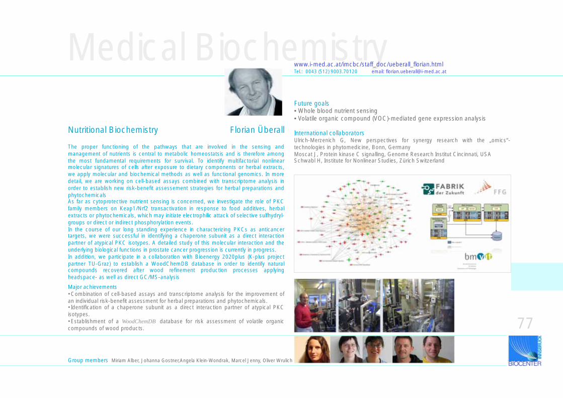
Medical Biochemistry
The proper functioning of the pathways that are involved in the sensing and management of nutrients is central to metabolic homeostatsis and is therefore among the most fundamental requirements for survival. To identify multifactorial nonlinear molecular signatures of cells after exposure to dietary components or herbal extracts, we apply molecular and biochemical methods as well as functional genomics. In more detail, we are working on cell-based assays combined with transcriptome analysis in order to establish new risk-benefit assessement strategies for herbal preparations and phytochemicalsAs far as cytoprotective nutrient sensing is concerned, we investigate the role of PKC family members on Keap1/Nrf2 transactivation in response to food additives, herbal extracts or phytochemicals, which may initiate electrophilic attack of selective sulfhydryl-groups or direct or indirect phosphorylation events.In the course of our long standing experience in characterizing PKCs as anticancer targets, we were successful in identifying a chaperone subunit as a direct interaction partner of atypical PKC isotypes. A detailed study of this molecular interaction and the underlying biological functions in prostate cancer progression is currently in progress.In addition, we participate in a collaboration with Bioenergy 2020plus (K-plus project partner TU-Graz) to establish a WoodChemDB database in order to identify natural compounds recovered after wood refinement production processes applying headspace- as well as direct GC/MS-analysis
www.i-med.ac.at/imcbc/staff_doc/ueberall_florian.html
Nutritional Biochemistry Florian Überall
Tel.: 0043 (512) 9003.70120 email: [email protected]
Major achievements•Combination of cell-based assays and transcriptome analysis for the improvement of an individual risk-benefit assessment for herbal preparations and phytochemicals. •Identification of a chaperone subunit as a direct interaction partner of atypical PKC isotypes.•Establishment of a WoodChemDB database for risk assessment of volatile organic compounds of wood products. 77
Future goals• Whole blood nutrient sensing• Volatile organic compound (VOC)-mediated gene expression analysis
International collaboratorsUlrich-Merzenich G, New perspectives for synergy research with the „omics“-technologies in phytomedicine, Bonn, GermanyMoscat J, Protein kinase C signalling, Genome Research Institut Cincinnati, USASchwabl H, Institute for Nonlinear Studies, Zürich Switzerland
Group members Miriam Alber, Johanna Gostner,Angela Klein-Wondrak, Marcel Jenny, Oliver Wrulich
Molecular Biology
Research of the division is devoted to various topics of molecular biology, including molecular microbiology, epigenetics, applied microbiology and lipocalin structure and function. Research is mainly focused on the regulation of gene expression in lower eukaryotes, plants and cultured mammalian cells. Several independent research teams try to hold an internationally recognized position within their research field. The division of Molecular Biology is responsible for teaching and training students of the Innsbruck Medical University and the Faculty of Biology of the University of Innsbruck in molecular biology. Moreover, the division has teaching responsibilities for medical students in microbiology and infectious disease control.
Groups within the Division of Molecular Biology
Chromatin and Epigenetics -Structure and Function of Chromatin: Maize and Mouse -Structure and Function of Chromatin: Filamentous Fungi -Chromatin Assembly and Remodeling Molecular Microbiology Applied Mycology Lipocalins
Peter LoidlGerald Brosch Stefan GrässleAlexandra LusserHubertus HaasFlorentine MarxBernhard Redl
Chromatin & Epigenetics Group
Nuclear DNA is compacted into chromatin which represents a structure built with repeating units, the nucleosomes. These consist of DNA wrapped around a protein octamer composed of 2 molecules each of the core histones H2A, H2B, H3, and H4.At least 2 domains can be distinguished in histones, a globular domain involved in histone-histone interactions (the histone fold motif) and the flexible N-terminal tails (H3, H4) or N-terminal and C-terminal tails (H2A, H2B). A series of consecutive nucleosomes forms a beads-on-a-string structure. A further level of compaction is the 30 nm fiber. In the past years our traditional picture of chromatin as a static and largely repressive functional state has changed to a more complex view of chromatin as a dynamic state that is essential for cellular regulation. The dynamic properties of chromatin are mediated by multiprotein complexes with different functions that set marks overlying the stable information of DNA. Factors that affect chromatin are enzymes that modify histones and chromatin remodeling machines. Histones are substrates of posttranslational modifications, like acetylation, methylation, phosphorylation and others which all can cause structural and functional rearrangements in chromatin and therefore represent essential elements of the complex epigenetic histone code.
Peter LoidlDirector
78
To decipher this code which is recognized and interpreted by transcriptional regulators and chromatin remodeling machines is one of the central challenges of chromatin research. Moreover, numerous regulatory, non-histone proteins are modified by histone acetyltransferases (HATs) and histone deacetylases (HDACs). This is an important point, in that the same enzyme activities that are responsible for the acetylation/deacetylation of nucleosomal histones also modify those regulatory proteins that bind to chromatin and probably recognize the complex modification pattern established on nucleosomes. One therefore faces a currently elusive correlation between pattern formation on nucleosomes and signal establishment on histone-binding proteins.
Tasks• identification and functional analysis of HDACs, histone methyltransferases and demethylases in filamentous fungi • role of HDACs in host-microbe interactions• mechanism of action of HDAC- and protein methyltransferase-inhibitors • histone acetylation/ methylation in murine cells with a focus on nucleolar chromatin• functional significance of histone and non-histone protein modifications for cell cycle regulation (p130)• chromatin assembly and remodeling
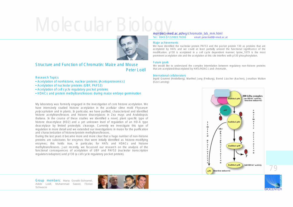
My laboratory was formerly engaged in the investigation of core histone acetylation. We have intensively studied histone acetylation in the acellular slime mold Physarum polycephalum and in plants. In particular, we have purified, characterized and identified histone acetyltransferases and histone deacetylases in Zea mays and Arabidopsis thaliana. In the course of these studies we identified a novel, plant specific type of histone deacetylase (HD2) and a yet unknown level of regulation of an HD-A type deacetylase by limited proteolytic cleavage. Currently we investigate this type of regulation in more detail and we extended our investigations in maize for the purification and characterization of histone/protein methyltransferases.During the last years it became more and more clear that a huge number of non-histone proteins are substrates for enzymes that were initially identified as histone-modifying enzymes; this holds true, in particular, for HATs and HDACs and histone methyltransferases. Just recently, we focussed our research on the analysis of the functional consequences of acetylation of UBF and PAF53 (nucleolar transcription regulators/adaptors) and p130 (a cell cycle regulatory pocket protein).
Molecular Biology
Structure and Function of Chromatin: Maize and Mouse Peter Loidl
Research Topics• Acetylation of nonhistone, nuclear proteins (Acetoproteomics)• Acetylation of nucleolar proteins (UBF, PAF53)• Acetylation of cell cycle regulatory pocket proteins• HDACs and protein methyltransferases during maize embryo germination
mol-biol.i-med.ac.at/wg/chromatin_lab_mm.html
79Group members: Maria Goralik-Schramel, Adele Loidl, Muhammad Saeed, Florian Schwarze
Future goalsWe would like to understand the complex interrelation between regulatory non-histone proteins that are acetylated/deacetylated by HATs/HDACs and chromatin.
International collaboratorsIngrid Grummt (Heidelberg), Manfred Jung (Freiburg), Bernd Lüscher (Aachen), Jonathan Walton (East Lansing)
Tel.: 0043 (512) 9003.70200 email: [email protected]/
Major achievementsWe have identified the nucleolar protein PAF53 and the pocket protein 130 as proteins that are acetylated by HATs and we could at least partially unravel the functional significance of the modification. p130 is acetylated in a cell cycle dependent manner; lysine_1079 is the most prominent acetylation site and the acetylation at this site interfers with p130 phosphorylation.
Molecular Biologymol-biol.i-med.ac.at/wg/chromatin_lab_ff.html
Chromatin & EpigeneticsTel.: 0043 (512) 9003.70211 email: [email protected] Tel.: 0043 (512) 9003.70218 email: [email protected]
Histone methylation in filamentous fungi Gerald Brosch
Protein methylation involves the transfer of a methyl group from S-adenosylmethionine to acceptor groups on substrate proteins. Arginine residues of proteins are modified by members of the protein arginine methyltransferase (PRMT) family. Arginine methylation has been observed on a variety of proteins associated with gene regulation, including DNA-binding transcriptional activators, transcriptional coactivators, and many RNA-binding proteins involved in RNA processing, transport, and stability. The long-term objective of our research is to investigate the functional role of protein methylation in filamentous fungi. We have identified three distinct PRMTs which all exhibit histone methyltransferase activity in vitro and in vivo. One of these proteins, termed RmtB, has an exceptional position because it displays both enzymatic and structural properties that are different from other known PRMTs. To study the functional role of PRMTs in A. nidulans we are currently deleting the corresponding PRMT genes by targeted gene replacement and are analysing putative growth defects of rmtA, rmtB, and rmtC deletion mutants under various growth conditions. These studies will be completed by the generation of double and triple mutant strains and the concomitant investigation of physiological and developmental effects. Finally, the analysis of global gene expression patterns, the study of effects of rmtA, rmtB, and rmtC deletion on the regulation of genes involved in secondary metabolism pathways, and the identification of novel substrate proteins of PRMTs will help to clarify the role of arginine methylation in Aspergillus.Besides the functional analysis of histone modifying enzymes, our group is also involved in the biological evaluation of small molecule inhibitors in cooperation with different pharmaceutical laboratories. Such inhibitors are potent inducers of differentiation and bear considerable potential as drugs for chemoprevention and treatment of cancer.
Functional roles of distinct histone deacetylases in the filamentous fungi Stefan Grässle
Histone acetylation plays a crucial role in the processes of gene regulation in eukaryotes. In particular, histones can be acetylated by histone acetyltransferases (HATs) and can be deacetylated by a second group of enzymes, the histone deacetylases (HDACs). In contrast to HATs, for which to date no potent inhibitors are known, there is a panel of structurally unrelated agents available that affect HDACs in a very selective way. Today these inhibitors are important tools for the study of histone acetylation processes. Our working group studies histone acetylation/deacetylation processes and attempts to purify HATs and HDACs of multiple organisms. Recently, we have identified and partially characterized HDACs of different classes in the filamentous fungus Aspergillus nidulans. The further characterization and clarification of the function of these enzymes within our model organism is the goal of this project. Since filamentous fungi are more complex than yeast in many important aspects, yet genetic manipulation is relatively simple and easy to perform, they have widely been regarded as model systems to study the basis of eukaryotic gene regulation. Moreover, many of these organisms contribute to the decay of organic material and thereby play an important role in the spoilage of food. But there are also filamentous fungi which represent dangerous pathogenic agents for people such as the closely related species to A. nidulans, A. fumigatus, which can cause life-threatening infections in patients that have a compromized immune system. For these reasons the study of these group of organisms is also of economic and medical interest.
80Group members Shadab Allipour, Ingo Bauer, Birgit Faber, Hermann Krabichler, Divyavaradhi Varadarajan
Structure and Function of Chromatin: Filamentous fungi
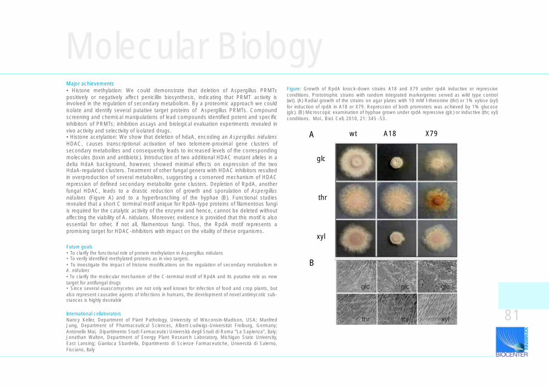
Molecular Biology
International collaboratorsNancy Keller, Department of Plant Pathology, University of Wisconsin-Madison, USA; Manfred Jung, Department of Pharmaceutical Sciences, Albert-Ludwigs-Universität Freiburg, Germany; Antonello Mai, Dipartimento Studi Farmaceutici Università degli Studi di Roma "La Sapienza“, Italy; Jonathan Walton, Department of Energy Plant Research Laboratory, Michigan State University, East Lansing; Gianluca Sbardella, Dipartimento di Scienze Farmaceutiche, Università di Salerno, Fisciano, Italy
Major achievements• Histone methylation: We could demonstrate that deletion of Aspergillus PRMTs positively or negatively affect penicillin biosynthesis, indicating that PRMT activity is involved in the regulation of secondary metabolism. By a proteomic approach we could isolate and identify several putative target proteins of Aspergillus PRMTs. Compound screening and chemical manipulations of lead compounds identified potent and specific inhibitors of PRMTs; inhibition assays and biological evaluation experiments revealed in vivo activity and selectivity of isolated drugs. • Histone acetylation: We show that deletion of hdaA, encoding an Aspergillus nidulans HDAC, causes transcriptional activation of two telomere-proximal gene clusters of secondary metabolites and consequently leads to increased levels of the corresponding molecules (toxin and antibiotic). Introduction of two additional HDAC mutant alleles in a delta HdaA background, however, showed minimal effects on expression of the two HdaA-regulated clusters. Treatment of other fungal genera with HDAC inhibitors resulted in overproduction of several metabolites, suggesting a conserved mechanism of HDAC repression of defined secondary metabolite gene clusters. Depletion of RpdA, another fungal HDAC, leads to a drastic reduction of growth and sporulation of Aspergillus nidulans (Figure A) and to a hyperbranching of the hyphae (B). Functional studies revealed that a short C terminal motif unique for RpdA-type proteins of filamentous fungi is required for the catalytic activity of the enzyme and hence, cannot be deleted without affecting the viability of A. nidulans. Moreover, evidence is provided that this motif is also essential for other, if not all, filamentous fungi. Thus, the RpdA motif represents a promising target for HDAC-inhibitors with impact on the vitality of these organisms.
Figure: Growth of RpdA knock-down strains A18 and X79 under rpdA inductive or repressive conditions. Prototrophic strains with random integrated markergenes served as wild type control (wt). (A) Radial growth of the strains on agar plates with 10 mM l-threonine (thr) or 1% xylose (xyl) for induction of rpdA in A18 or X79. Repression of both promoters was achieved by 1% glucose (glc). (B) Microscopic examination of hyphae grown under rpdA repressive (glc) or inductive (thr, xyl) conditions. Mol.. Biol. Cell, 2010, 21: 345 -53.
A wt A18 X79
glc
thr
xyl
glc glc glc
thr thr xyl
B
81
Future goals• To clarify the functional role of protein methylation in Aspergillus nidulans• To verify identified methylated proteins as in vivo targets.• To investigate the impact of histone modifications on the regulation of secondary metabolism in A. nidulans• To clarify the molecular mechanism of the C-terminal motif of RpdA and its putative role as new target for antifungal drugs• Since several euascomycetes are not only well known for infection of food and crop plants, but also represent causative agents of infections in humans, the development of novel antimycotic sub-stances is highly desirable
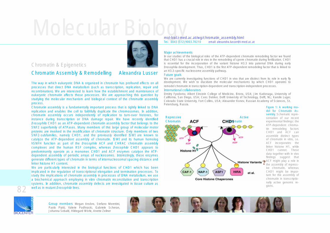
Molecular Biology
The way in which eukaryotic DNA is organized in chromatin has profound effects on all processes that direct DNA metabolism (such as transcription, replication, repair and recombination). We are interested to learn how the establishment and maintenance of eukaryotic chromatin affects those processes. We are approaching this question by studying the molecular mechanism and biological context of the chromatin assembly process. Chromatin assembly is a fundamentally important process that is tightly linked to DNA replication and enables the cell to faithfully duplicate the chromosomes. In addition, chromatin assembly occurs independently of replication to turn-over histones, for instance during transcription or DNA damage repair. We have recently identified Drosophila CHD1 as an ATP-dependent chromatin assembly factor that belongs to the SNF2 superfamily of ATPases. Many members of this large group of molecular motor proteins are involved in the modification of chromatin structure. Only members of two SNF2-subfamilies, namely CHD1, and the previously identified ISWI are known to catalyze the ATP-dependent assembly of chromatin. ISWI and its human homolog hSNFH function as part of the Drosophila ACF and CHRAC chromatin assembly complexes and the human RSF complex, whereas Drosophila CHD1 appears to predominantly operate as a monomer. CHD1 and ACF enzymes catalyze the ATP-dependent assembly of periodic arrays of nucleosomes. Interestingly, these enzymes generate different types of chromatin in terms of internucleosomal spacing distance and linker histone H1 content. We are particularly interested in the biological functions of CHD1 which has been implicated in the regulation of transcriptional elongation and termination processes. To study the implications of chromatin assembly in processes of DNA metabolism, we use a biochemical approach employing in vitro chromatin reconstitution and transcription systems. In addition, chromatin assembly defects are investigated in tissue culture as well as in mutant Drosophila lines.
mol-biol.i-med.ac.at/wg/chromatin_assembly.htmlTel.: 0043 (512) 9003.70210 email: [email protected]
Major achievementsIn our studies of the biological roles of the ATP-dependent chromatin remodeling factor we found that CHD1 has a crucial role in vivo in the remodeling of sperm chromatin during fertilization. CHD1 is essential for the incorporation of the variant histone H3.3 into paternal DNA during early Drosophila development. Thus, CHD1 is the first ATP-dependent remodeling factor that is linked to an H3.3-specific nucleosome assembly pathway.
Chromatin & Epigenetics
Future goalsWe are currently investigating functions of CHD1 in vivo that are distinct from its role in early fly development. We wish to elucidate the molecular mechanisms by which CHD1 operates to remodel chromatin in transcription-dependent and transcription-independent processes. International collaboratorsDmitry Fyodorov, Albert Einstein College of Medicine, Bronx, USA; Jim Kadonaga, University of California, San Diego, USA; Cees Dekker, Delft University of Technology, Delft, NL; Karolin Luger, Colorado State University, Fort Collins, USA; Alexander Konev, Russian Academy of Sciences, St. Petersburg, Russia.
Group members Megan Ansbro, Stefano Morettini, Paolo Piatti, Valerie Podhraski, Gabriele Scheran, Johanna Sebald, Hildegard Wörle, Anette Zeilner
82
Chromatin Assembly & Remodelling Alexandra Lusser
Repressive ActiveChromatin Chromatin
Figure 1: A working mo-del for Chromatin As-sembly. Schematic repre-sentation of our recent experimental findings: the ATP-dependent chroma-tin remodeling factors CHD1 and ACF can assemble distinct types of chromatin in vitro, i.e. ACF incorporates the linker histone H1, while CHD1 cannot. These data together with in vivo findings suggest that ACF might play a role in the assembly of repress-ive chromatin, whereas CHD1 might be impor-tant for the assembly of chromatin in transcriptio-nally active genomic re-gions.
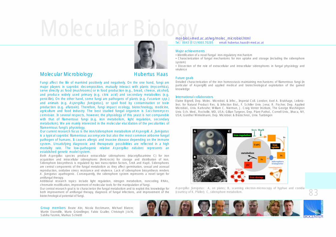
Molecular Biologymol-biol.i-med.ac.at/wg/molec_microbiol.html
Aspergillus fumigatus: A, on plates; B, scanning electron-microscopy of hyphae and conidia (courtesy of K. Pfaller); C, siderophore metabolism.
Tel.: 0043 (512) 9003.70205 email: [email protected]
Group members Beate Abt, Nicola Beckmann, Michael Blatzer, Martin Eisendlle, Mario Gründlinger, Fabio Gsaller, Christoph Jöchl, Sabiha Yasmin, Markus Schrettl
83
Major achievements• Identification of a novel fungal iron-regulatory mechanism • Characterization of fungal mechanisms for iron uptake and storage (including the siderophore system)• Dissection of the role of extracellular and intracellular siderophores in fungal physiology and virulence
Future goalsDetailed characterization of the iron homeostasis-maintaining mechanisms of filamentous fungi (in particular of Aspergilli) and applied medical and biotechnological exploitation of the gained knowledge
International collaboratorsElaine Bignell, Dep. Molec. Microbiol. & Infec., Imperial Coll. London; Axel A. Brakhage, Leibniz-Inst. for Natural Product Res. & Infection Biol., F. Schiller Univ. Jena; R. Fischer, Dep. Applied Microbiol., Univ. Karlsruhe; William C. Nierman, J. Craig Venter Institute, The George Washington Univ. Sch. Med., Rockville, MD, USA; Gillian Turgeon, Dep. Plant Pathol., Cornell Univ., Ithaca, NY, USA; Günther Winkelmann, Dep. Microbiol. & Biotechnol., Univ. Tuebingen
Fungi affect the life of mankind positively and negatively. On the one hand, fungi are major players in saprobic decomposition, mutually interact with plants (mycorrhiza), serve directly as food (mushrooms) or in food production (e.g., bread, cheese, alcohol), and produce widely used primary (e.g. citric acid) and secondary metabolites (e.g. penicillin). On the other hand, some fungi are pathogens of plants (e.g. Fusarium spp.) and animals (e.g. Aspergillus fumigatus), or spoil food by contamination or toxin production (e.g. aflatoxin). Therefore, fungi impact ecology, biotechnology, medicine, agriculture and food industry. The best studied fungal organism is Saccharomyces cerevisiae. In several respects, however, the physiology of this yeast is not comparable with that of filamentous fungi (e.g. iron metabolism, light regulation, secondary metabolism). We are mainly interested in the molecular elucidation of the peculiarities of filamemtous fungi´s physiology. Our current research focus is the iron/siderophore metabolism of Aspergilli. A. fumigatus is a typical saprobic filamentous ascomycete but also the most common airborne fungal pathogen of humans. It causes allergic and invasive disease depending on the immune system. Unsatisfying diagnostic and therapeutic possibilities are reflected in a high mortality rate. The low-pathogenic relative Aspergillus nidulans represents an established genetic model system.Both Aspergillus species produce extracellular siderophores (triacetylfusarinine C) for iron acquisition and intracellular siderophores (ferricrocin) for storage and distribution of iron. Siderophore biosynthesis is regulated by two transcription factors, SreA and HapX. Siderophores are central components of the fungal metabolism as they affect germination, sexual and asexual reproduction, oxidative stress resistance and virulence. Lack of siderophore biosynthesis renders A. fumigatus apathogenic. Consequently, the siderophore system represents a novel target for antifungal therapy. Additional research topics include light regulation, nitrogen metabolism, noncoding RNAs, chromatin modification, improvement of molecular tools for the manipulation of fungi. Our central research goal is to characterize the fungal metabolism and to exploit this knowledge for both improvement of antifungal therapy, diagnosis of fungal infections, and improvement of the biotechnological potential of fungi.
Molecular Microbiology Hubertus Haas
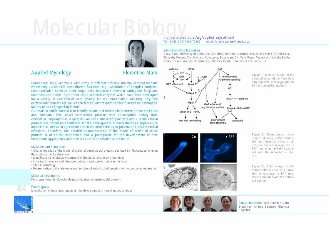
Molecular Biology
Filamentous fungi secrete a wide array of different proteins into the external medium where they accomplish most diverse functions, e.g. assimilation of complex nutrients, communication between other fungal cells, interaction between pathogenic fungi and their host and others. Apart from some secreted enzymes which have been developed for a variety of commercial uses (mainly for the fermentation industry), only few extracellular proteins are well characterized with respect to their function as pathogenic factors or as cell signalling factors. Our main scientific interest is to identify, isolate and further characterize on the molecular and functional level novel extracellular proteins with antimicrobial activity from Penicillium chrysogenum, Aspergillus nidulans and Aspergillus fumigatus. Antimicrobial proteins are promising candidates for the development of novel therapies applicable in medicine as well as in agriculture and in the food industry to prevent and treat microbial infections. Therefore, the detailed characterization of the mode of action of these proteins is of crucial importance and a prerequisite for the development of new therapeutic approaches and their successful application in the future.
Major research interests• Characterization of the mode of action of antimicrobial proteins secreted by filamentous fungi on the molecular and cellular level• Identification and characterization of molecular targets in sensitive fungi• Localization studies and characterization of endocytotic pathways in fungi• Structural biology• Determination of the relevance and function of antimicrobial proteins for the producing organisms
Major achievementsFirst steps towards biotechnological utilization of antimicrobial proteins
Future goalsIdentification of molecular targets for the development of new therapeutic drugs
mol-biol.i-med.ac.at/wg/applied_mycol.htmlTel.: 0043 (512) 9003.70207 email: [email protected]
International collaboratorsGyula Batta, University of Debrecen, HU; Mojca Bencina, National Institute of Chemistry, Ljublijana, Slovenia; Mogens Trier Hansen, Novozyme, Bagsvaert, DK; Vera Meyer, Technical University Berlin; István Pócsi, University of Debrecen, HU; Nick Read, University of Edinburgh, UK
Group members Ulrike Binder, Doris Bratschun, Andrea Eigentler, Nikoletta Hegedüs
84
Applied Mycology Florentine Marx Figure 1: Tentative model of the mode of action of the Penicillium chrysogenum antifungal protein PAF in Aspergillus nidulans
Figure 2: Fluorescence micro-graphs showing chitin distribu-tion and hyperbranching in A. nidulans hyphae in response to PAF treatement (+PAF) compa-red with the untreated control (Co)
Figure 3:. TEM images of the cellular ultrastructure of A. nidu-lans in response to PAF trea-tment compared with the untrea-ted control
1
2
3
Co + PAF
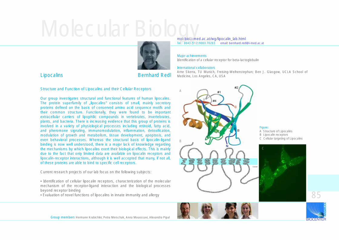
Molecular Biology
Structure and Function of Lipocalins and their Cellular Receptors
Our group investigates structural and functional features of human lipocalins. The protein superfamily of „lipocalins“ consists of small, mainly secretory proteins defined on the basis of conserved amino acid sequence motifs and their common structure. Functionally, they were found to be important extracellular carriers of lipophilic compounds in vertebrates, invertebrates, plants, and bacteria. There is increasing evidence that this group of proteins is involved in a variety of physiological processes including retinoid, fatty acid, and pheromone signaling, immunomodulation, inflammation, detoxification, modulation of growth and metabolism, tissue development, apoptosis, and even behavioral processes. Whereas the structural basis of lipocalin-ligand binding is now well understood, there is a major lack of knowledge regarding the mechanisms by which lipocalins exert their biological effects. This is mainly due to the fact that only limited data are available on lipocalin receptors and lipocalin-receptor interactions, although it is well accepted that many, if not all, of these proteins are able to bind to specific cell receptors.
Current research projects of our lab focus on the following subjects:
• Identification of cellular lipocalin receptors, characterization of the molecular mechanism of the receptor-ligand interaction and the biological processes beyond receptor binding• Evaluation of novel functions of lipocalins in innate immunity and allergy
mol-biol.i-med.ac.at/wg/lipocalin_lab.html
Figure:A Structure of Lipocalins B Lipocalin receptorsC Cellular targeting of Lipocalins
APDYEVLS
R
I I S T
T
LLFALYILC
H I F L
T T
IALL C T F T
A LIALG A V L L L
PFS
ALE
SN
EVLLSL
PR
N Y Y I Q WL
NG
SL
IHGLWN
LVFLF
S N L SLIF
LM P F A
YFFT
I I
ESEG
FA
G S R K G VLGR
VY
ET V V M
LMLLT
L L V LG
MVWVA S A I V
DKNKANR
ES L Y
DFW
EYYLPYLYSC
I S F LG
VLLLL V C T
PLGLA AL AL
RMFSVT
GK
LL
VKPRLLE
DL
EE
QL
YCSAFEE
AA
L T R R I C N P T S C W L P L D M E L L H R Q VL
ALQ
TQRVL
LE
KR
RK
AS
AW
QR
NL
GYPL A M L
CLLVLT G L S
VLIVA
I H I LELLI
DE
AAM
PG M Q G
TSL
GQ
VS
FSK
R
L G S F GAIQVV
L I F Y LMVSSV
V G F Y
V
SSPLFRS
L R P R WHDT
AMT
QIIGN
C V C LLVL
S S A L
LL
PVFSRTLG
LT
RF D L L
GDFGRFNW L
L C L V
G N
YIVLY A A F
AGLTT
N
FF
KTFTAA
VR
AE
LI
RA
FG
LD
RLP
L P V S GF
PQ
A S R K T Q H Q Q
Cytosol
EQ
LF
H
RI
E
R
M
V
E
CE
TR
FKK
PA
E F DE
DATVN
K
A
B
Major achievementsIdentification of a cellular receptor for beta-lactoglobulin
International collaboratorsArne Skerra, TU Munich, Freising-Weihenstephan; Ben J. Glasgow, UCLA School of Medicine, Los Angeles, CA, USA
Tel.: 0043 (512) 9003.70203 email: [email protected]
Group members Hermann Krabichler, Petra Merschak, Anna Mouossavi, Alexandra Pipal
C
85
Lipocalins Bernhard Redl
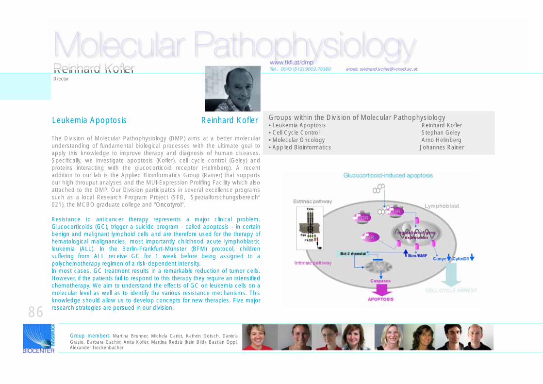
Groups within the Division of Molecular Pathophysiology• Leukemia Apoptosis Reinhard Kofler• Cell Cycle Control Stephan Geley• Molecular Oncology Arno Helmberg• Applied Bioinformatics Johannes Rainer
Tel.: 0043 (512) 9003.70360 email: [email protected]
The Division of Molecular Pathophysiology (DMP) aims at a better molecular understanding of fundamental biological processes with the ultimate goal to apply this knowledge to improve therapy and diagnosis of human diseases. Specifically, we investigate apoptosis (Kofler), cell cycle control (Geley) and proteins interacting with the glucocorticoid receptor (Helmberg). A recent addition to our lab is the Applied Bioinformatics Group (Rainer) that supports our high throuput analyses and the MUI-Expression Prolifing Facility which also attached to the DMP. Our Division participates in several excellence programs such as a local Research Program Project (SFB, "Spezialforschungsbereich“ 021), the MCBO graduate college and “OOncotyrol“.
Resistance to anticancer therapy represents a major clinical problem. Glucocorticoids (GC), trigger a suicide program - called apoptosis - in certain benign and malignant lymphoid cells and are therefore used for the therapy of hematological malignancies, most importantly childhood acute lymphoblastic leukemia (ALL). In the Berlin-Frankfurt-Münster (BFM) protocol, children suffering from ALL receive GC for 1 week before being assigned to a polychemotherapy regimen of a risk-dependent intensity.In most cases, GC treatment results in a remarkable reduction of tumor cells. However, if the patients fail to respond to this therapy they require an intensified chemotherapy. We aim to understand the effects of GC on leukemia cells on a molecular level as well as to identify the various resistance mechanisms. This knowledge should allow us to develop concepts for new therapies. Five major research strategies are persued in our division.
Molecular Pathophysiologywww.tkfi.at/dmp
Glucocorticoid-induced apoptosis
Reinhard KoflerDirector
86Group members Martina Brunner, Michela Carlet, Kathrin Götsch, Daniela Grazio, Barbara Gschirr, Anita Kofler, Martina Redziz (kein Bild), Bastian Oppl, Alexander Trockenbacher
Leukemia Apoptosis Reinhard Kofler
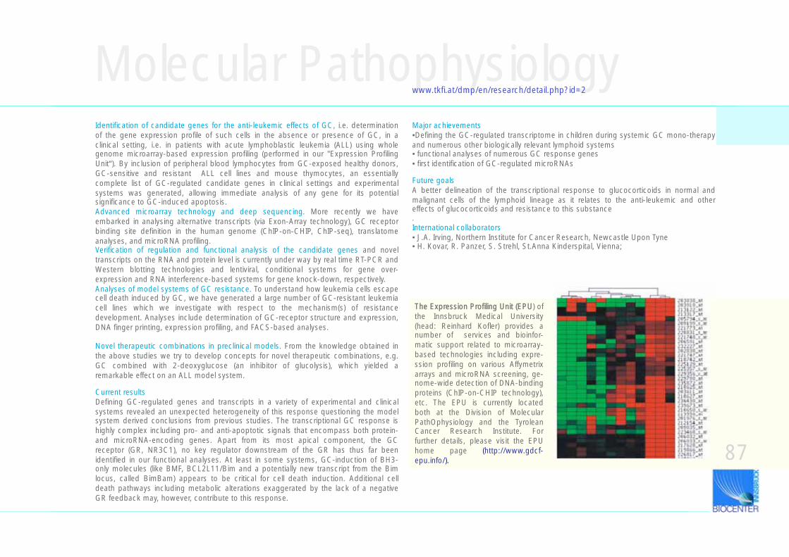
Identification of candidate genes for the anti-leukemic effects of GC, i.e. determination of the gene expression profile of such cells in the absence or presence of GC, in a clinical setting, i.e. in patients with acute lymphoblastic leukemia (ALL) using whole genome microarray-based expression profiling (performed in our "Expression Profiling Unit“). By inclusion of peripheral blood lymphocytes from GC-exposed healthy donors, GC-sensitive and resistant ALL cell lines and mouse thymocytes, an essentially complete list of GC-regulated candidate genes in clinical settings and experimental systems was generated, allowing immediate analysis of any gene for its potential significance to GC-induced apoptosis.Advanced microarray technology and deep sequencing. More recently we have embarked in analysing alternative transcripts (via Exon-Array technology), GC receptor binding site definition in the human genome (ChIP-on-CHIP, ChIP-seq), translatome analyses, and microRNA profiling. Verification of regulation and functional analysis of the candidate genes and novel transcripts on the RNA and protein level is currently under way by real time RT-PCR and Western blotting technologies and lentiviral, conditional systems for gene over-expression and RNA interference-based systems for gene knock-down, respectively.Analyses of model systems of GC resistance. To understand how leukemia cells escape cell death induced by GC, we have generated a large number of GC-resistant leukemia cell lines which we investigate with respect to the mechanism(s) of resistance development. Analyses include determination of GC-receptor structure and expression, DNA finger printing, expression profiling, and FACS-based analyses.
Novel therapeutic combinations in preclinical models. From the knowledge obtained in the above studies we try to develop concepts for novel therapeutic combinations, e.g. GC combined with 2-deoxyglucose (an inhibitor of glucolysis), which yielded a remarkable effect on an ALL model system.
Current resultsDefining GC-regulated genes and transcripts in a variety of experimental and clinical systems revealed an unexpected heterogeneity of this response questioning the model system derived conclusions from previous studies. The transcriptional GC response is highly complex including pro- and anti-apoptotic signals that encompass both protein- and microRNA-encoding genes. Apart from its most apical component, the GC receptor (GR, NR3C1), no key regulator downstream of the GR has thus far been identified in our functional analyses. At least in some systems, GC-induction of BH3-only molecules (like BMF, BCL2L11/Bim and a potentially new transcript from the Bim locus, called BimBam) appears to be critical for cell death induction. Additional cell death pathways including metabolic alterations exaggerated by the lack of a negative GR feedback may, however, contribute to this response.
Molecular Pathophysiologywww.tkfi.at/dmp/en/research/detail.php?id=2
The Expression Profiling Unit (EPU) of the Innsbruck Medical University (head: Reinhard Kofler) provides a number of services and bioinfor-matic support related to microarray-based technologies including expre-ssion profiling on various Affymetrix arrays and microRNA screening, ge-nome-wide detection of DNA-binding proteins (ChIP-on-CHIP technology), etc. The EPU is currently located both at the Division of Molecular PathOphysiology and the Tyrolean Cancer Research Institute. For further details, please visit the EPU home page (http://www.gdcf-epu.info/).
Major achievements•Defining the GC-regulated transcriptome in children during systemic GC mono-therapy and numerous other biologically relevant lymphoid systems• functional analyses of numerous GC response genes• first identification of GC-regulated microRNAs
Future goalsA better delineation of the transcriptional response to glucocorticoids in normal and malignant cells of the lymphoid lineage as it relates to the anti-leukemic and other effects of glucocorticoids and resistance to this substance.International collaborators• J.A. Irving, Northern Institute for Cancer Research, Newcastle Upon Tyne• H. Kovar, R. Panzer, S. Strehl, St.Anna Kinderspital, Vienna;
87
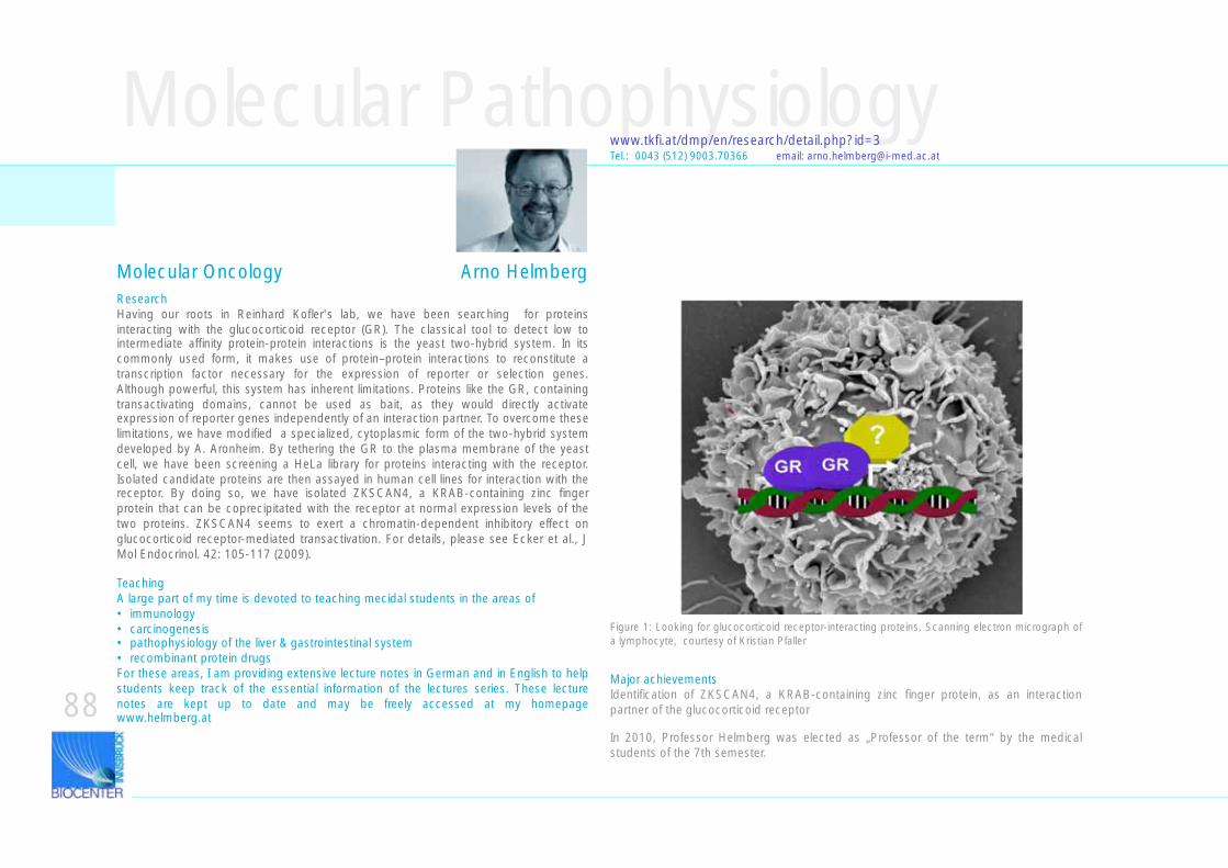
ResearchHaving our roots in Reinhard Kofler's lab, we have been searching for proteins interacting with the glucocorticoid receptor (GR). The classical tool to detect low to intermediate affinity protein-protein interactions is the yeast two-hybrid system. In its commonly used form, it makes use of protein–protein interactions to reconstitute a transcription factor necessary for the expression of reporter or selection genes. Although powerful, this system has inherent limitations. Proteins like the GR, containing transactivating domains, cannot be used as bait, as they would directly activate expression of reporter genes independently of an interaction partner. To overcome these limitations, we have modified a specialized, cytoplasmic form of the two-hybrid system developed by A. Aronheim. By tethering the GR to the plasma membrane of the yeast cell, we have been screening a HeLa library for proteins interacting with the receptor. Isolated candidate proteins are then assayed in human cell lines for interaction with the receptor. By doing so, we have isolated ZKSCAN4, a KRAB-containing zinc finger protein that can be coprecipitated with the receptor at normal expression levels of the two proteins. ZKSCAN4 seems to exert a chromatin-dependent inhibitory effect on glucocorticoid receptor-mediated transactivation. For details, please see Ecker et al., J Mol Endocrinol. 42: 105-117 (2009).
TeachingA large part of my time is devoted to teaching mecidal students in the areas of• immunology• carcinogenesis• pathophysiology of the liver & gastrointestinal system• recombinant protein drugsFor these areas, I am providing extensive lecture notes in German and in English to help students keep track of the essential information of the lectures series. These lecture notes are kept up to date and may be freely accessed at my homepage www.helmberg.at
Figure 1: Looking for glucocorticoid receptor-interacting proteins. Scanning electron micrograph of a lymphocyte, courtesy of Kristian Pfaller
Molecular Pathophysiologywww.tkfi.at/dmp/en/research/detail.php?id=3Tel.: 0043 (512) 9003.70366 email: [email protected]
Major achievementsIdentification of ZKSCAN4, a KRAB-containing zinc finger protein, as an interaction partner of the glucocorticoid receptor
In 2010, Professor Helmberg was elected as „Professor of the term“ by the medical students of the 7th semester.
88
Molecular Oncology Arno Helmberg
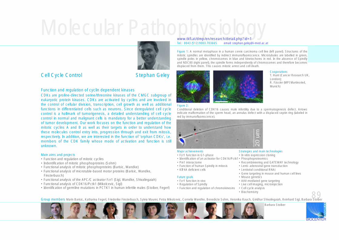
CDKs are proline-directed serine/threonine kinases of the CMGC subgroup of eukaryotic protein kinases. CDKs are activated by cyclins and are involved in the control of cellular division, transcription, cell growth as well as additional functions in differentiated cells such as neurons. Since deregulated cell cycle control is a hallmark of tumorigenesis, a detailed understanding of cell cycle control in normal and malignant cells is mandatory for a better understanding of tumor development. Our work focuses on the function and regulation of the mitotic cyclins A and B as well as their targets in order to understand how these molecules control entry into, progression through and exit from mitosis, respectively. In addition, we are interested in the function of ‘orphan CDKs‘, i.e. members of the CDK family whose mode of activation and function is still unknown.
Main aims and projects• Function and regulation of mitotic cyclins• Indentification of mitotic phosphoproteins (Sohm)• Functional analysis of mitotic phosphoproteins (Barisic, Wandke)• Functional analysis of microtuble-based motor proteins (Barisic, Wandke, Finsterbusch)• Functional analysis of the APC/C activator Fzr1 (Sigl, Wandke, Shivalingaiah)• Functional analysis of CDK16/Pctk1 (Mikolcevic, Sigl)• Identification of germline mutations in PCTK1 in human infertile males (Stoiber, Fegerl)
Function and regulation of cyclin dependent kinases
Cell Cycle Control Stephan Geley
Molecular Pathophysiologywww.tkfi.at/dmp/en/research/detail.php?id=1Tel.: 0043 (512) 9003.703665 email: [email protected]
Major achievements• Fzr1 function in G1-phase• Identification of an activator for CDk16/Pctk1• Pin1 interactome• Function of human Spindly in mitosis• KIF4A deficient cells
Future goals• Fzr1 function in vivo• Regulation of Spindly• Function and regulation of chromokinesins
Strategies and main technologies• In vitro expression cloning• Phosphoproteomics• Recombineering and GATEWAY technology• Lenti- adenoviral gene transduction• Lentiviral conditional RNAi• Gene targeting in mouse and human cell lines• Mouse genetics• AAV-mediated gene targeting• Live cell imaging, microinjection• Cell cycle analysis• Biochemistry
Figure 1: A normal metaphase in a human cervix carcinoma cell line (left panel). Structures of the mitotic spindles are identified by indirect immunofluorescence. Microtubules are labelled in green, spindle poles in yellow, chromosomes in blue and kinetochores in red. In the absence of Spindly and NDC80 (right panel), the spindle forms independently of chromosomes and therefore becomes displaced from them. This causes mitotic arrest and cell death.
Figure 2:Conditional deletion of CDK16 causes male infertility due to a spermatogenesis defect. Arrows indicate malformation of the sperm head, an annulus defect with a displaced septin ring (labeled in red by immunofluorescence).
89
CooperationsT. Hunt (Cancer Research UK, London); R. Fässler (MPI Martinsried, Munich)
Group members Marin Barisic, Katharina Fegerl, Friederike Finsterbusch, Sylvia Maurer, Petra Mikolcevic, Cornelia Wandke, Benedicte Sohm, Veronika Rauch, Giridhar Shivalingaiah, Reinhard Sigl, Barbara Stoiber
Barbara Stoiber
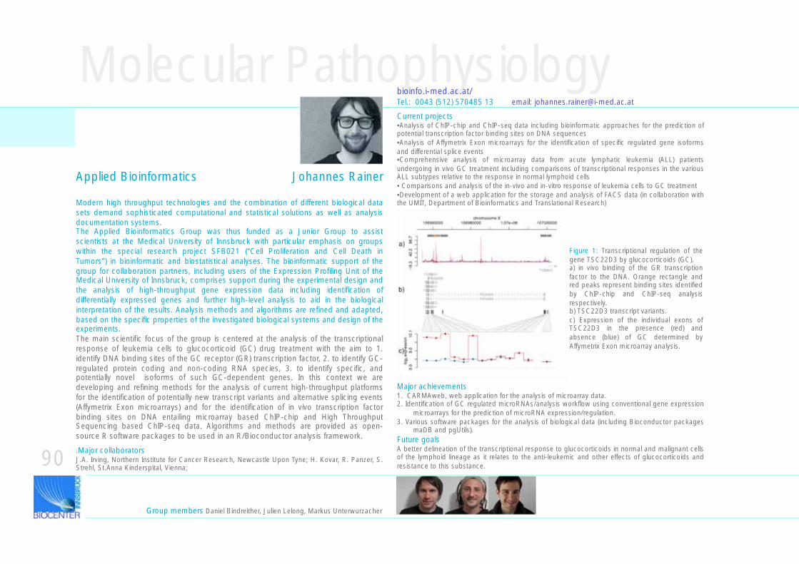
Molecular Pathophysiology
Figure 1: Transcriptional regulation of the gene TSC22D3 by glucocorticoids (GC). a) in vivo binding of the GR transcription factor to the DNA. Orange rectangle and red peaks represent binding sites identified by ChIP-chip and ChIP-seq analysis respectively. b) TSC22D3 transcript variants. c) Expression of the individual exons of TSC22D3 in the presence (red) and absence (blue) of GC determined by Affymetrix Exon microarray analysis.
Major achievements1. CARMAweb, web application for the analysis of microarray data. 2. Identification of GC regulated microRNAs/analysis workflow using conventional gene expression
microarrays for the prediction of microRNA expression/regulation. 3. Various software packages for the analysis of biological data (including Bioconductor packages
maDB and pgUtils).Future goalsA better delineation of the transcriptional response to glucocorticoids in normal and malignant cells of the lymphoid lineage as it relates to the anti-leukemic and other effects of glucocorticoids and resistance to this substance.
Modern high throughput technologies and the combination of different biological data sets demand sophisticated computational and statistical solutions as well as analysis documentation systems.The Applied Bioinformatics Group was thus funded as a Junior Group to assist scientists at the Medical University of Innsbruck with particular emphasis on groups within the special research project SFB021 (“Cell Proliferation and Cell Death in Tumors”) in bioinformatic and biostatistical analyses. The bioinformatic support of the group for collaboration partners, including users of the Expression Profiling Unit of the Medical University of Innsbruck, comprises support during the experimental design and the analysis of high-throughput gene expression data including identification of differentially expressed genes and further high-level analysis to aid in the biological interpretation of the results. Analysis methods and algorithms are refined and adapted, based on the specific properties of the investigated biological systems and design of the experiments.The main scientific focus of the group is centered at the analysis of the transcriptional response of leukemia cells to glucocorticoid (GC) drug treatment with the aim to 1. identify DNA binding sites of the GC receptor (GR) transcription factor, 2. to identify GC-regulated protein coding and non-coding RNA species, 3. to identify specific, and potentially novel isoforms of such GC-dependent genes. In this context we are developing and refining methods for the analysis of current high-throughput platforms for the identification of potentially new transcript variants and alternative splicing events (Affymetrix Exon microarrays) and for the identification of in vivo transcription factor binding sites on DNA entailing microarray based ChIP-chip and High Throughput Sequencing based ChIP-seq data. Algorithms and methods are provided as open-source R software packages to be used in an R/Bioconductor analysis framework.
Applied Bioinformatics Johannes Rainer
bioinfo.i-med.ac.at/Tel.: 0043 (512) 570485 13 email: [email protected]
Group members Daniel Bindreither, Julien Lelong, Markus Unterwurzacher
Current projects•Analysis of ChIP-chip and ChIP-seq data including bioinformatic approaches for the prediction of potential transcription factor binding sites on DNA sequences•Analysis of Affymetrix Exon microarrays for the identification of specific regulated gene isoforms and differential splice events•Comprehensive analysis of microarray data from acute lymphatic leukemia (ALL) patients undergoing in vivo GC treatment including comparisons of transcriptional responses in the various ALL subtypes relative to the response in normal lymphoid cells• Comparisons and analysis of the in-vivo and in-vitro response of leukemia cells to GC treatment•Development of a web application for the storage and analysis of FACS data (in collaboration with the UMIT, Department of Bioinformatics and Translational Research)
90.Major collaboratorsJ.A. Irving, Northern Institute for Cancer Research, Newcastle Upon Tyne; H. Kovar, R. Panzer, S. Strehl, St.Anna Kinderspital, Vienna;
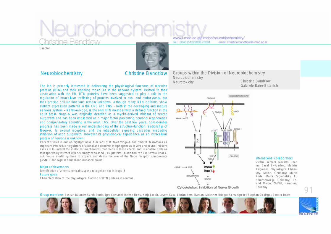
The lab is primarily interested in delineating the physiological functions of reticulon proteins (RTN) and their signaling molecules in the nervous system. Related to their association with the ER, RTN proteins have been suggested to play a role in the regulation of intracellular trafficking of proteins involved in exo- and endocytosis, but their precise cellular functions remain unknown. Although many RTN isoforms show distinct expression patterns in the CNS and PNS - both in the developing and mature nervous system – RTN4-A/Nogo, is the only RTN member with a defined function in the adult brain. Nogo-A was originally identified as a myelin-derived inhibitor of neurite outgrowth and has been implicated as a major factor preventing neuronal regeneration and compensatory sprouting in the adult CNS. Over the past few years, considerable progress has been made in our understanding of the structure-function relationship of Nogo-A, its axonal receptors, and the intracellular signaling cascades mediating inhibition of axon outgrowth. However its physiological significance as an intracellular protein of neurons is unknown. Recent studies in our lab highlight novel functions of RTN-4A/Nogo-A and other RTN isoforms as important intracellular regulators of axonal and dendritic morphogenesis in vitro and in vivo. Present aims are to unravel the molecular mechanisms that mediate these effects and to analyse proteins that specifically interact with neuronally expressed RTN proteins. In addition, we use several knock-out mouse model systems to explore and define the role of the Nogo receptor components p75NTR and NgR in normal and diseased brains.
Neurobiochemistrywww.i-med.ac.at/ imcbc/neurobiochemistry/Tel.: 0043 (512) 9003.70281 email: [email protected]
Major achievementsIdentification of a noncanonical caspase recognition site in Nogo-BFuture goalsCharacterization of the physiological function of RTN proteins in neurons
Groups within the Division of NeurobiochemistryNeurobiochemistryNeurotoxicity Christine Bandtlow
Gabriele Baier-Bitterlich
Group members Bastian Bäumler, Sarah Borrie, Igea Contarini, Helene Heiss, Katja Jacob, Levent Kaya, Florian Kern, Barbara Meissner, Rüdiger Schweigreiter, Stephan Sickinger, Sandra Trojer
Christine BandtlowDirector
91
Neurobiochemistry Christine Bandtlow
International collaboratorsStefan Frentzel, Novartis Phar-ma, Basel, Switzerland; Mathias Klugmann, Physiological Chemi-stry, Mainz, Germany; Martin Korte, Marta Zagrebelsky, TU Braunschweig, Germany; Ro-land Martin, ZMNH, Hamburg, Germany
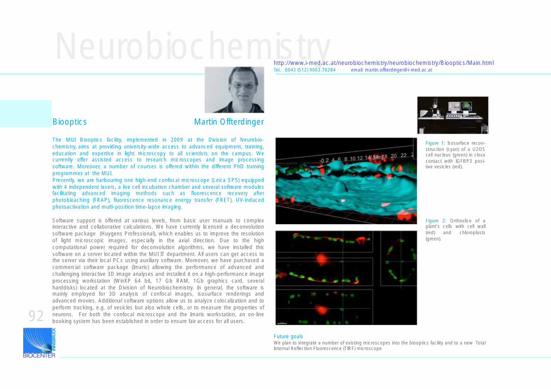
Neurobiochemistryhttp://www.i-med.ac.at/neurobiochemistry/neurobiochemistry/Biooptics/Main.htmlTel.: 0043 (512) 9003.70284 email: [email protected]
Figure 1: Isosurface recon-struction (cyan) of a U2OS cell nucleus (green) in close contact with IGFBP3 posi-tive vesicles (red).
Figure 2: Orthoslice of a plant‘s cells with cell wall (red) and chloroplasts (green).
92
The MUI Biooptics facility, implemented in 2009 at the Division of Neurobio-chemistry, aims at providing university-wide access to advanced equipment, training, education and expertise in light microscopy to all scientists on the campus. We currently offer assisted access to research microscopes and image processing software. Moreover, a number of courses is offered within the different PhD training programmes at the MUI.Presently, we are harbouring one high-end confocal microscope (Leica SP5) equipped with 4 independent lasers, a live cell incubation chamber and several software modules facilitating advanced imaging methods such as fluorescence recovery after photobleaching (FRAP), fluorescence resonance energy transfer (FRET), UV-induced photoactivation and multi-position time-lapse imaging.
Software support is offered at various levels, from basic user manuals to complex interactive and collaborative calculations. We have currently licensed a deconvolution software package (Huygens Professional), which enables us to improve the resolution of light microscopic images, especially in the axial direction. Due to the high computational power required for deconvolution algorithms, we have installed this software on a server located within the MUI IT department. All users can get access to the server via their local PCs using auxiliary software. Moreover, we have purchased a commercial software package (Imaris) allowing the performance of advanced and challenging interactive 3D image analyses and installed it on a high-performance image processing workstation (WinXP 64 bit, 17 Gb RAM, 1Gb graphics card, several harddisks) located at the Division of Neurobiochemistry. In general, the software is mainly employed for 3D analysis of confocal images, isosurface renderings and advanced movies. Additional software options allow us to analyze colocalization and to perform tracking, e.g. of vesicles but also whole cells, or to measure the properties of neurons. For both the confocal microscope and the Imaris workstation, an on-line booking system has been established in order to ensure fair access for all users.
Biooptics Martin Offterdinger
Future goalsWe plan to integrate a number of existing microscopes into the biooptics facility and to a new Total Internal Reflection Fluorescence (TIRF) microscope
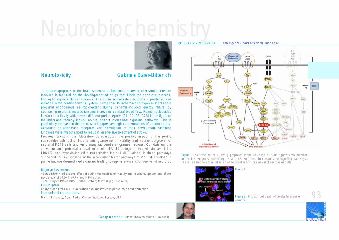
Neurobiochemistry
Hoechst/PI
Group member: Bettina Thauerer (former Tomaselli)
Figure 2.: Hypoxic cell death of cerebellar granule neurons
Tel.: 0043 (512) 9003.70289 email: [email protected]
To reduce apoptosis in the brain is central to functional recovery after stroke. Present research is focused on the development of drugs that block the apoptotic process, hoping to improve clinical outcome. The purine nucleoside adenosine is produced and released in the central nervous system in response to ischemia and hypoxia. It acts as a powerful endogenous neuroprotectant during ischemia-induced energy failure, by decreasing neuronal metabolism and increasing cerebral blood flow. Purine nucleosides interact specifically with several different purinoceptors (A1, A2, A3, A2B) in the figure to the right) and thereby induce several distinct intercellular signaling pathways. This is particularly the case in the brain, which expresses high concentrations of purinoceptors. Activation of adenosine receptors and stimulation of their downstream signaling functions were hypothesized to result in an effective treatment of stroke. Previous results in this laboratory demonstrated the positive impact of the purine nucleosides adenosine, inosine and guanosine on viability and neurite outgrowth of neuronal PC12 cells and on primary rat cerebellar granule neurons. Our data on the activation and potential causal roles of p42/p44 mitogen-activated kinases (alias ERK1/2) and hypoxia-inducible transcription factor-1 (HIF1-alpha) in these pathways supported the investigation of the molecular effector pathways of MAPK/HIF1-alpha in purine nucleoside-mediated signaling leading to regeneration and/or survival of neurons.
Major achievements• Establishment of positive effect of purine nucleosides on viability and neurite outgrowth and of the special role of p42/44 MAPK and HIF-1alpha.• FWF project 19578-B05, Hertha-Firnberg fellowship (B.Thauerer)Future goalsAnalysis of p42/44 MAPK activation and substrates in purine-mediated protectionInternational collaboratorsMichail Sitkovsky, Dana-Farber Cancer Institute, Boston, USA
Neurotoxicity Gabriele Baier-Bitterlich
93
Figure 1: Scheme of the currently proposed mode of action of purin agonists via different adenosine receptors (purinoceptors A1, A2, etc.) and their associated signaling pathways. These can lead to either inhibition of neuronal activity or survival of neurons or both.
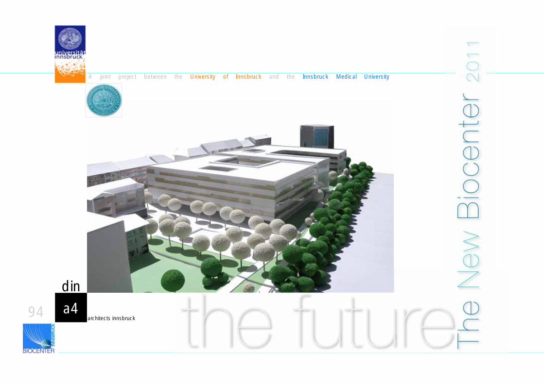
A joint project between the University of Innsbruck and the Innsbruck Medical University
architects innsbrucka494
din
the future The
New
Bio
cent
er 2
01
1
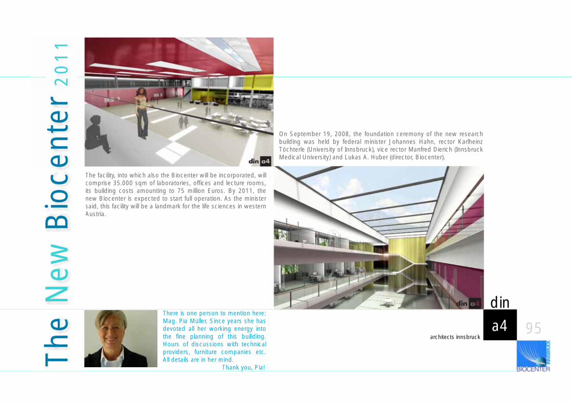
The facility, into which also the Biocenter will be incorporated, will comprise 35.000 sqm of laboratories, offices and lecture rooms, its building costs amounting to 75 million Euros. By 2011, the new Biocenter is expected to start full operation. As the minister said, this facility will be a landmark for the life sciences in western Austria.
architects innsbrucka4
din
95
The
New
Bio
cent
er 2
01
1
On September 19, 2008, the foundation ceremony of the new research building was held by federal minister Johannes Hahn, rector Karlheinz Töchterle (University of Innsbruck), vice rector Manfred Dierich (Innsbruck Medical University) and Lukas A. Huber (director, Biocenter).
There is one person to mention here: Mag. Pia Müller. Since years she has devoted all her working energy into the fine planning of this builiding. Hours of discussions with technical providers, furniture companies etc. All details are in her mind. Thank you, Pia!

This rreport has been designed and edited by Siegfried Schwarz, by order of the department conference of THE BIOCENTER and with the help of Peter Loidl and Lukas A. Huber. The ccontents are based on informations provided by the directors and group leaders of the BIOCENTER. Gregor Retti is thanked for elaborating data on publications and funds.The llogo of the BIOCENTER (to the left) was created by Siegfried Schwarz in 2007 and is by acceptance by the Department Conference officially used by the BIOCENTER.Printed by Egger-Druck Imst/Tyrol, 2010