Embed Size (px)
Citation preview
Dissertation
zur Erlangung des Grades
Doktor der Naturwissenschaften
am Fachbereich Biologie
der Johannes Gutenberg-Universität Mainz
Rebecca Bohn
geb. am 31.12.1988 in Karlsruhe
Mainz, 2017
Canonical and non-canonical autophagy
modulation in human primary macrophages
and its effect on the adaptive immune system
Diese Publikation ist urheberrechtlich geschützt.
Vervielfältigungen jeder Art, zur Schau stellen oder andere
Verwendungen sind nur nach Absprache mit der Abteilung
Immunologie des Paul-Ehrlich-Instituts zulässig.

Dekan Prof. Dr. Hans Zischler
Institut für Anthropologie
Johannes Gutenberg-Universität Mainz
1. Berichterstatter
2. Berichterstatter
Tag der mündlichen Prüfung:

The obtained research data during this PhD project and collaborative studies led to the
following publications:
Rebecca Bohn, Anne-Kathrin Knuth, Peter Crauwels, Stefan Schille, Ger van
Zandbergen: LC3-associated phagocytosis as mechanism to secure Leishmania
survival in human host macrophages. 2017, in preparation, abstract accepted for
manuscript submission in Frontiers in Immunology
Rebecca Bohn, Peter Crauwels, Pascal Devant, Tim Haselwander, Jan W. Drijfhout,
Stefan Tenzer, Ger van Zandbergen: Autophagy in human primary macrophages is
independent of ULK-1 and Beclin-1. 2017, submitted
Peter Crauwels, Rebecca Bohn and Ger van Zandbergen: Autophagy during infection
– friend or foe? 2017, submitted
Krämer S, Crauwels P, Bohn R, Radzimski C, Szaszák M, Klinger M, Rupp J, van
Zandbergen G.: AP-1 transcription factor serves as a molecular switch between
Chlamydia pneumoniae replication and persistence. Infect Immun 2015 83:2651–2660.
PMID: 25895972
Peter Crauwels, Rebecca Bohn, Meike Thomas, Stefan Gottwalt, Florian Jäckel, Susi
Krämer, Elena Bank, Stefan Tenzer, Paul Walther, Max Bastian & Ger van
Zandbergen: Apoptotic-like Leishmania exploit the host´s autophagy machinery to
reduce T-cell mediated parasite elimination. Autophagy 2015, 11:2, 285-297 PMID:
25801301

I
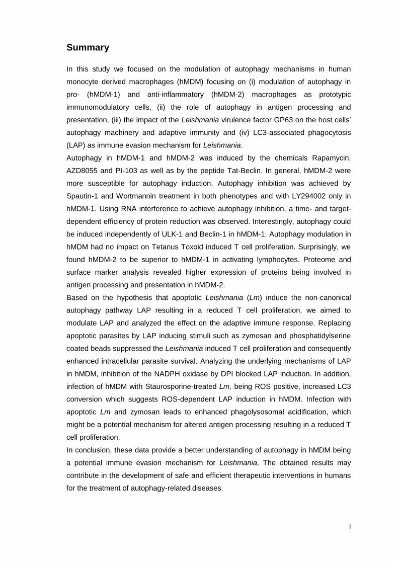
Summary
In this study we focused on the modulation of autophagy mechanisms in human
monocyte derived macrophages (hMDM) focusing on (i) modulation of autophagy in
pro- (hMDM-1) and anti-inflammatory (hMDM-2) macrophages as prototypic
immunomodulatory cells, (ii) the role of autophagy in antigen processing and
presentation, (iii) the impact of the Leishmania virulence factor GP63 on the host cells’
autophagy machinery and adaptive immunity and (iv) LC3-associated phagocytosis
(LAP) as immune evasion mechanism for Leishmania.
Autophagy in hMDM-1 and hMDM-2 was induced by the chemicals Rapamycin,
AZD8055 and PI-103 as well as by the peptide Tat-Beclin. In general, hMDM-2 were
more susceptible for autophagy induction. Autophagy inhibition was achieved by
Spautin-1 and Wortmannin treatment in both phenotypes and with LY294002 only in
hMDM-1. Using RNA interference to achieve autophagy inhibition, a time- and target-
dependent efficiency of protein reduction was observed. Interestingly, autophagy could
be induced independently of ULK-1 and Beclin-1 in hMDM-1. Autophagy modulation in
hMDM had no impact on Tetanus Toxoid induced T cell proliferation. Surprisingly, we
found hMDM-2 to be superior to hMDM-1 in activating lymphocytes. Proteome and
surface marker analysis revealed higher expression of proteins being involved in
antigen processing and presentation in hMDM-2.
Based on the hypothesis that apoptotic Leishmania (Lm) induce the non-canonical
autophagy pathway LAP resulting in a reduced T cell proliferation, we aimed to
modulate LAP and analyzed the effect on the adaptive immune response. Replacing
apoptotic parasites by LAP inducing stimuli such as zymosan and phosphatidylserine
coated beads suppressed the Leishmania induced T cell proliferation and consequently
enhanced intracellular parasite survival. Analyzing the underlying mechanisms of LAP
in hMDM, inhibition of the NADPH oxidase by DPI blocked LAP induction. In addition,
infection of hMDM with Staurosporine-treated Lm, being ROS positive, increased LC3
conversion which suggests ROS-dependent LAP induction in hMDM. Infection with
apoptotic Lm and zymosan leads to enhanced phagolysosomal acidification, which
might be a potential mechanism for altered antigen processing resulting in a reduced T
cell proliferation.
In conclusion, these data provide a better understanding of autophagy in hMDM being
a potential immune evasion mechanism for Leishmania. The obtained results may
contribute in the development of safe and efficient therapeutic interventions in humans
for the treatment of autophagy-related diseases.

II
III
Zusammenfassung
In der vorliegenden Arbeit fokussierten wir uns auf die Modulation von Autophagie
Mechanismen in humanen primären Makrophagen (hMDM) und untersuchten dabei
folgende Punkte: (i) Modulation von Autophagie in pro- (hMDM-1) und anti-
inflammatorischen (hMDM-2) Makrophagen als prototypische immunmodulierende
Zellen, (ii) die Rolle von Autophagie für Antigen-Prozessierung und -Präsentation, (iii)
den Einfluss des Leishmanien Virulenzfaktors GP63 auf den Autophagieprozess der
Wirtszelle und die adaptive Immunantwort und (iv) LC3-assoziierte Phagozytose (LAP)
als Ausweichmechanismus für Leishmanien vor dem Immunsystem.
Autophagie konnte mit den Chemikalien Rapamycin, AZD8055 und PI-103 als auch mit
dem Peptid Tat-Beclin in hMDM-1 und hMDM-2 induziert werden. Generell zeigten
hMDM-2 eine stärkere Autophagie Induktion. Inhibition wurde durch Behandlung mit
Spautin-1 und Wortmannin in beiden Phenotypen und mit LY294002 nur in hMDM-1
erreicht. Mit Hilfe von RNA Interferenz zur Autophagie Inhibition wurde eine Zeit- und
Target-abhängige Proteinreduktion beobachtet. Interessanterweise konnte Autophagie
unabhängig von ULK-1 und Beclin-1 in hMDM-1 induziert werden. Die Modulation von
Autophagie hatte keinen Einfluss auf die Tetanus Toxoid induzierte T Zell Proliferation.
Überraschenderweise zeigten hMDM-2 eine bessere Aktivierung von Lymphozyten als
hMDM-1. Proteom- und Oberflächenmarker-Analyse wiesen auf eine höhere
Expression von Proteinen, die an der Antigen-Prozessierung und -Präsentation
beteiligt sind, in hMDM-2 hin.
Basierend auf der Hypothese, dass apoptotische Leishmanien den nicht-kanonischen
Autophagie-Mechanismus LAP induzieren was zu reduzierter T Zell Proliferation führt,
modulierten wir LAP und analysierten den Effekt auf die adaptive Immunantwort. Das
Ersetzen von apoptotischen Parasiten durch LAP-induzierende Stimuli wie Zymosan
und phosphatidylserin-beschichtete Partikel unterdrückte die Leishmanien-induzierte T
Zell Proliferation und erhöhte das intrazelluläre Überleben der Parasiten. Die Analyse
der zugrundeliegenden Mechanismen zeigte, dass die Inhibition der NADPH Oxidase
durch DPI die Induktion von LAP in hMDM blockierte. Weiterhin resultierte die Infektion
von hMDM mit Staurosporin-behandelten Leishmanien, die ROS positiv sind, in einer
erhöhten LC3 Konversion, die eine ROS-abhängige Induktion von LAP in hMDM
suggeriert. Die Infektion mit apoptotischen Leishmanien und Zymosan führte zu einer
verstärkten phagolysosomalen Ansäuerung, die ein potentieller Mechanismus für eine
veränderte Antigen-Prozessierung, resultierend in verminderter T Zell Proliferation,
sein könnte.

IV
Abschließend liefern diese Daten ein besseres Verständnis von Autophagie in hMDM
welche als möglicher Ausweichmechanismus für Leishmanien vor dem humanen
Immunsystem dienen könnte. Die erhaltenen Ergebnisse liefern einen wertvollen
Beitrag zur Entwicklung von sicheren und effizienten therapeutischen Ansätzen zur
Behandlung von Autophagie-bedingten Krankheiten beim Menschen.
V
Table of content
1 Introduction ...................................................................................................................... 1
1.1 Autophagy ................................................................................................................ 1
1.1.1 Historical background .................................................................................... 1
1.1.2 Autophagy in health and disease ................................................................... 1
1.1.3 Molecular pathway ........................................................................................ 2
1.1.4 Modulation of autophagy ............................................................................... 4
1.1.5 LC3-associated phagocytosis ........................................................................ 5
1.2 Macrophages ............................................................................................................ 7
1.2.1 Macrophage subtypes and their activation .................................................... 7
1.2.2 MFs are professional phagocytes and mediators of adaptive
immunity ........................................................................................................ 9
1.3 Leishmaniasis ......................................................................................................... 11
1.3.1 Epidemiology ............................................................................................... 11
1.3.2 Clinical manifestations ................................................................................. 11
1.3.3 Therapy ....................................................................................................... 12
1.4 Leishmania ............................................................................................................. 13
1.4.1 Taxonomy ................................................................................................... 13
1.4.2 Life cycle ..................................................................................................... 13
1.4.3 Macrophage receptors mediate Leishmania uptake .................................... 16
1.4.4 The Leishmania virulence factor GP63 ........................................................ 16
1.4.5 Adaptive immunity in response to Leishmania infection ............................... 17
1.4.6 Immune evasion strategies by Leishmania .................................................. 18
1.5 Hypothesis and Aims .............................................................................................. 21
1.5.1 The role of autophagy in human primary macrophages ............................... 21
1.5.2 The role of LAP during Leishmania infection ............................................... 23
2 Material and methods .................................................................................................... 25
2.1 Material................................................................................................................... 25
2.1.1 Chemicals ................................................................................................... 25
2.1.2 Culture Medium ........................................................................................... 27
2.1.3 Buffer and solutions ..................................................................................... 27
2.1.4 Human primary cells .................................................................................... 29
2.1.5 Leishmania strains ...................................................................................... 30
2.1.6 Oligonucleotides .......................................................................................... 30
2.1.7 siRNA .......................................................................................................... 30
2.1.8 Peptides ...................................................................................................... 31
VI
2.1.9 Enzymes ..................................................................................................... 31
2.1.10 Antibodies ................................................................................................... 31
2.1.11 Dyes and Marker ......................................................................................... 32
2.1.12 Ready to use Kits ........................................................................................ 32
2.1.13 Laboratory supplies ..................................................................................... 33
2.1.14 Instruments ................................................................................................. 34
2.1.15 Software ...................................................................................................... 36
2.2 Methods .................................................................................................................. 37
2.2.1 Cell culture of Leishmania major (Lm) promastigotes .................................. 37
2.2.2 MACS separation of viable and apoptotic L. major
promastigotes .............................................................................................. 38
2.2.3 Chemical treatment of Leishmania promastigotes ....................................... 39
2.2.4 Cell culture of human primary cells .............................................................. 39
2.2.5 Autophagy / LAP modulation in hMDM ........................................................ 41
2.2.6 CFSE based proliferation assay .................................................................. 41
2.2.7 Flow Cytometry ........................................................................................... 42
2.2.8 Molecular biology methods .......................................................................... 44
2.2.9 Western Blot analysis .................................................................................. 48
2.2.10 Enzyme-linked immunosorbent assay (ELISA) ............................................ 49
2.2.11 Microscopy .................................................................................................. 49
2.2.12 Transduction of hMDM with eGFP-LC3 lentiviral particles ........................... 50
2.2.13 Statistical analysis ....................................................................................... 51
3 Results............................................................................................................................ 53
3.1 Autophagy modulation in human primary macrophages ......................................... 53
3.1.1 PI-103, AZD8055 and Rapamycin induce autophagy in hMDM ................... 53
3.1.2 Autophagy induction with the peptide Tat-Beclin ......................................... 56
3.1.3 Inhibition of class III PI3 kinase pathway by Wortmannin and
Spautin-1 blocks autophagy induction in hMDM .......................................... 58
3.1.4 Autophagy inhibition by siRNA knockdown .................................................. 59
3.2 Effect of autophagy modulation on antigen processing using the recall
antigen Tetanus Toxoid .......................................................................................... 67
3.2.1 Characterization of the Tetanus Toxoid specific T cell
proliferation ................................................................................................. 67
3.2.2 Impact of autophagy modulation on the TT specific T cell
proliferation ................................................................................................. 69
3.2.3 Macrophage phenotypes and their ability to activate T cells ........................ 71
3.3 Investigation of the Leishmania virulence factor GP63 ............................................ 78
VII
3.3.1 Growth characteristics of Leishmania GP63 knockout
parasites ..................................................................................................... 78
3.3.2 GP63 has no impact on the infection rate of hMDM ..................................... 79
3.3.3 GP63 has no impact on T cell proliferation .................................................. 80
3.3.4 Infection of hMDM with Leishmania induces autophagy
independently of GP63 ................................................................................ 81
3.4 Modulation of LC3-associated phagocytosis ........................................................... 82
3.4.1 LAP induction by phosphatidylserine beads ................................................ 82
3.4.2 LAP induction by zymosan particles ............................................................ 86
3.4.3 The presence of zymosan or apoptotic Lm reduces the Lm
specific T cell proliferation and enhances parasite survival .......................... 88
3.4.4 Impact of ROS for LAP induction ................................................................. 91
3.4.5 Inhibition of LAP by NOX2 knockdown and NOX2 inhibition by
DPI .............................................................................................................. 92
3.4.6 Proteome analysis to identify additional factors leading to
immune suppression upon Leishmania infection ......................................... 97
4 Discussion ................................................................................................................... 101
4.1 Summary of the data ............................................................................................ 101
4.2 Autophagy modulation in human primary macrophages ....................................... 102
4.3 Impact of autophagy on antigen presentation using the model antigen
Tetanus Toxoid ..................................................................................................... 105
4.4 Priming of an adaptive immune response by hMDM-1 vs. hMDM-2 ...................... 106
4.5 Influence of GP63 on infectivity, adaptive immunity and host cell
autophagy ............................................................................................................. 108
4.6 LAP as immune evasion mechanism during Leishmania infection ........................ 110
4.7 Concluding remarks .............................................................................................. 115
5 References ................................................................................................................... 116
6 Acronyms and Abbreviations ..................................................................................... 140
7 Figure list ..................................................................................................................... 145
8 Table list ....................................................................................................................... 148
9 Declaration of authorship ........................................................................................... 149
10 Acknowledgements ..................................................................................................... 150
11 Curriculum Vitae .......................................................................................................... 152

VIII
Introduction
1
1 Introduction
1.1 Autophagy
1.1.1 Historical background
Research on autophagy started more than 60 years ago by the discovery of the
lysosome, a late organelle in the autophagic process (Duve et al., 1955). In the
following years, Novikoff and Clark described “dense bodies” harboring organelles,
which were later identified as autolysosomal compartments (Novikoff et al., 1956;
Clark, 1957; Novikoff, 1959). In 1963, de Duve postulated the term “autophagy” as a
sequestration process present in all cell types leading to the degradation of cytoplasmic
content in autophagosomes (Duve and Wattiaux, 1966). Extensive research revealed
that autophagy is induced in nutrient poor conditions and is inhibited by the addition of
amino acids to the culture medium (Mortimore et al., 1983). A huge breakthrough in
understanding the molecular pathway was achieved in the 1990s in Ohsumi’s lab by
screening yeast deletion mutants, which led to the identification of autophagy-related
(ATG) genes and proteins (Takeshige et al., 1992; Tsukada and Ohsumi, 1993; Baba
et al., 1995). Almost twenty years ago, Mizushima postulated that autophagy is
conserved from yeast to mammals (Mizushima et al., 1998a). Until then, autophagy
was monitored by electron microscopy when Kabeya and colleagues discovered the
microtubule-associated protein 1 light chain 3, short LC3, as a marker on
autophagosomes enabling detection by immunofluorescence and Western Blot
(Kabeya et al., 2000). Since then, the number of papers per year increased
exponentially, highlighting important functions of autophagy for health and disease
(Ohsumi, 2014). Ohsumi’s work on the autophagic process was awarded in 2016 with
the Nobel Prize for medicine.
1.1.2 Autophagy in health and disease
Macroautophagy (greek: auto – self, phagein – eating; hereafter referred to as
autophagy) is a self-eating process and a key mechanism to ensure survival during
harsh conditions. The digestion of particles ranging in size from small molecules to
whole organelles is unique and provides the cell with nutrients in times of starvation
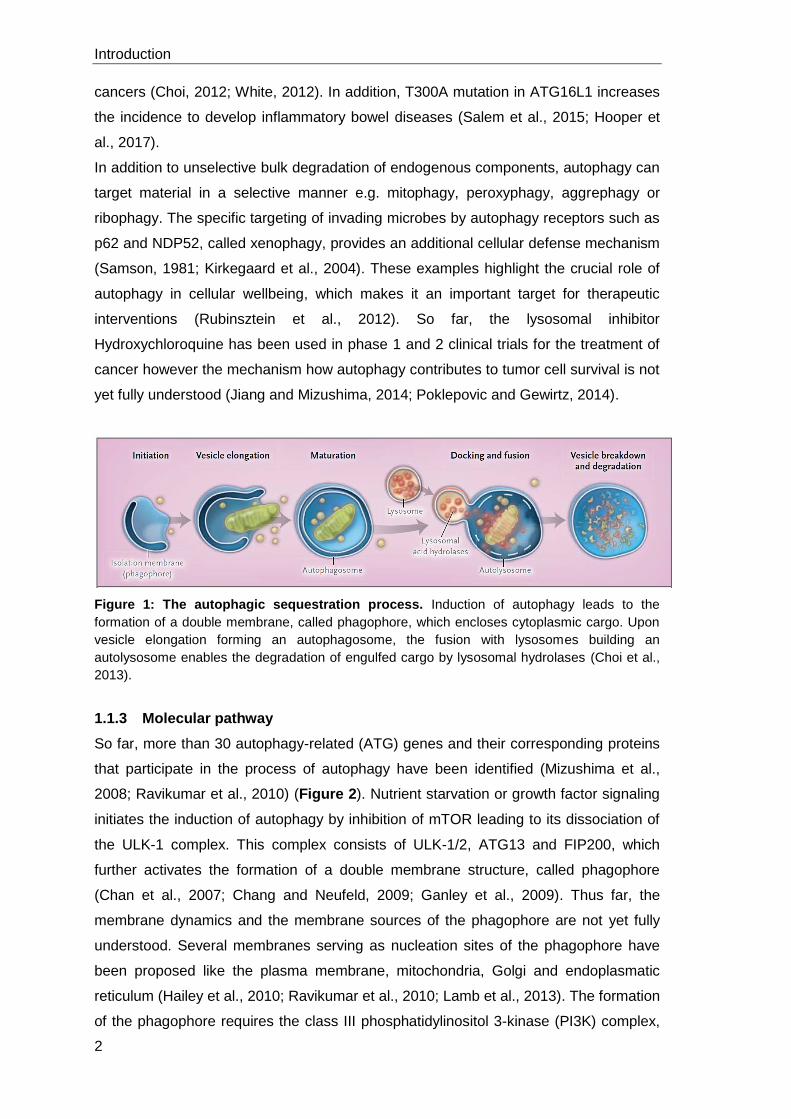
ensuring energy homeostasis (Figure 1) (Mizushima et al., 2008; Levine et al., 2011).
Furthermore, the degradation of defect organelles or misfolded proteins prevents the
outcome of neurodegenerative diseases like Parkinson or Alzheimer (Pan et al., 2008;
Lynch-Day et al., 2012; Nixon, 2013). Consequently, mutations in autophagy related
genes are associated with increased risk of developing diseases. Mono-allelic deletion
of Beclin-1, a critical regulator protein of autophagy, has been observed in some
Introduction
2
cancers (Choi, 2012; White, 2012). In addition, T300A mutation in ATG16L1 increases
the incidence to develop inflammatory bowel diseases (Salem et al., 2015; Hooper et
al., 2017).
In addition to unselective bulk degradation of endogenous components, autophagy can
target material in a selective manner e.g. mitophagy, peroxyphagy, aggrephagy or
ribophagy. The specific targeting of invading microbes by autophagy receptors such as
p62 and NDP52, called xenophagy, provides an additional cellular defense mechanism
(Samson, 1981; Kirkegaard et al., 2004). These examples highlight the crucial role of
autophagy in cellular wellbeing, which makes it an important target for therapeutic
interventions (Rubinsztein et al., 2012). So far, the lysosomal inhibitor
Hydroxychloroquine has been used in phase 1 and 2 clinical trials for the treatment of
cancer however the mechanism how autophagy contributes to tumor cell survival is not
yet fully understood (Jiang and Mizushima, 2014; Poklepovic and Gewirtz, 2014).
Figure 1: The autophagic sequestration process. Induction of autophagy leads to the
formation of a double membrane, called phagophore, which encloses cytoplasmic cargo. Upon
vesicle elongation forming an autophagosome, the fusion with lysosomes building an
autolysosome enables the degradation of engulfed cargo by lysosomal hydrolases (Choi et al.,
2013).
1.1.3 Molecular pathway
So far, more than 30 autophagy-related (ATG) genes and their corresponding proteins
that participate in the process of autophagy have been identified (Mizushima et al.,
2008; Ravikumar et al., 2010) (Figure 2). Nutrient starvation or growth factor signaling
initiates the induction of autophagy by inhibition of mTOR leading to its dissociation of
the ULK-1 complex. This complex consists of ULK-1/2, ATG13 and FIP200, which
further activates the formation of a double membrane structure, called phagophore
(Chan et al., 2007; Chang and Neufeld, 2009; Ganley et al., 2009). Thus far, the
membrane dynamics and the membrane sources of the phagophore are not yet fully
understood. Several membranes serving as nucleation sites of the phagophore have
been proposed like the plasma membrane, mitochondria, Golgi and endoplasmatic
reticulum (Hailey et al., 2010; Ravikumar et al., 2010; Lamb et al., 2013). The formation
of the phagophore requires the class III phosphatidylinositol 3-kinase (PI3K) complex,
Introduction
3
consisting of Vps34, Vps15 ATG14 and Beclin-1, which produces phosphatidylinositol-
3-phosphate (PI3P) (Liang et al., 1999; Kihara et al., 2001; Axe et al., 2008; Itakura
and Mizushima, 2014). Membrane-bound PI3P leads to the recruitment of two
ubiquitin-like conjugating systems, which mediate the elongation of the membrane
(Geng and Klionsky, 2008). First, ATG12 is covalently conjugated to ATG5 mediated
by ATG7, an E1 ubiquitin-activating enzyme, and ATG10, an E2 ubiquitin-conjugating
enzyme (Mizushima et al., 1998b). The complex of ATG12-ATG5 is further stabilized
by ATG16L1 and then associates with phagophores but dissociates from completed
autophagosomes (Mizushima, 2003). The formation of the second complex is initiated
by cleavage of pro-LC3 in LC3 (or LC3-I) by ATG4 (Hemelaar et al., 2003; Tanida et
al., 2004a). Subsequently, ATG7 and ATG3 (E2-like) catalyze the ligation of cytosolic
LC3-I to phosphatidylethanolamine (PE), forming autophagosome-associated LC3-PE
(or LC3-II) (Tanida et al., 2004b). Cross-talk between the two complexes has been
suggested and the ATG16L1 complex is believed to facilitate the subcellular location of
LC3-I to the site of its lipidation on the autophagosomal membrane (Fujita et al., 2008).
At the autophagosome, LC3-II has been shown to possess a dual role, in selecting
cargo for degradation through interaction with adaptor proteins such as p62 and by
promoting membrane tethering and fusion (Nakatogawa et al., 2007; Pankiv et al.,
2007). During maturation, the phagophore closes to an autophagosome, which fuses
with lysosomes forming an autolysosome. The influx of hydrolases and the acidification
of the autolysosomal lumen results in the complete degradation of the enclosed luminal
content together with the inner autophagosomal membrane (Mizushima et al., 2002;
Yang and Klionsky, 2010). So far, only LC3-II is known to be predominantly associated
with autophagosomes until the formation of an autolysosome and therefore serves as a
widely used marker to monitor autophagy (Kabeya et al., 2000; Mizushima et al., 2010;
Klionsky et al., 2014; Klionsky et al., 2016).
Introduction
4
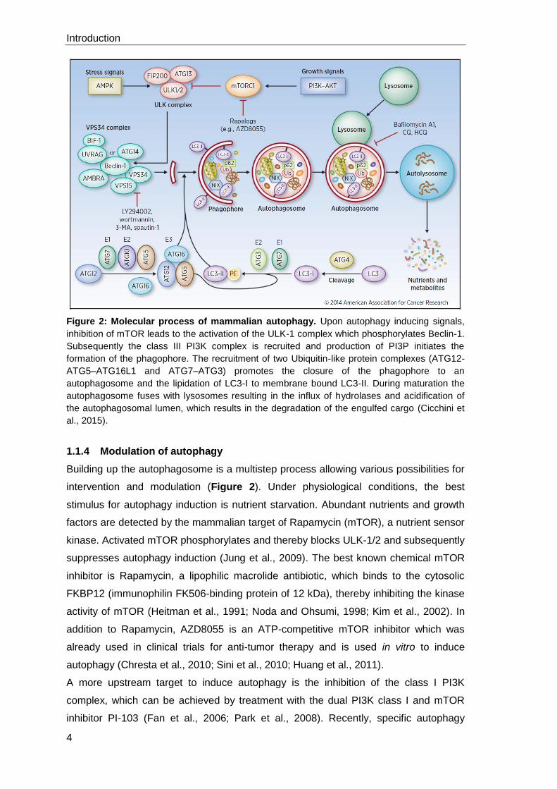
Figure 2: Molecular process of mammalian autophagy. Upon autophagy inducing signals,
inhibition of mTOR leads to the activation of the ULK-1 complex which phosphorylates Beclin-1.
Subsequently the class III PI3K complex is recruited and production of PI3P initiates the
formation of the phagophore. The recruitment of two Ubiquitin-like protein complexes (ATG12-
ATG5–ATG16L1 and ATG7–ATG3) promotes the closure of the phagophore to an
autophagosome and the lipidation of LC3-I to membrane bound LC3-II. During maturation the
autophagosome fuses with lysosomes resulting in the influx of hydrolases and acidification of
the autophagosomal lumen, which results in the degradation of the engulfed cargo (Cicchini et
al., 2015).
1.1.4 Modulation of autophagy
Building up the autophagosome is a multistep process allowing various possibilities for
intervention and modulation (Figure 2). Under physiological conditions, the best
stimulus for autophagy induction is nutrient starvation. Abundant nutrients and growth
factors are detected by the mammalian target of Rapamycin (mTOR), a nutrient sensor
kinase. Activated mTOR phosphorylates and thereby blocks ULK-1/2 and subsequently
suppresses autophagy induction (Jung et al., 2009). The best known chemical mTOR
inhibitor is Rapamycin, a lipophilic macrolide antibiotic, which binds to the cytosolic
FKBP12 (immunophilin FK506-binding protein of 12 kDa), thereby inhibiting the kinase
activity of mTOR (Heitman et al., 1991; Noda and Ohsumi, 1998; Kim et al., 2002). In
addition to Rapamycin, AZD8055 is an ATP-competitive mTOR inhibitor which was
already used in clinical trials for anti-tumor therapy and is used in vitro to induce
autophagy (Chresta et al., 2010; Sini et al., 2010; Huang et al., 2011).
A more upstream target to induce autophagy is the inhibition of the class I PI3K
complex, which can be achieved by treatment with the dual PI3K class I and mTOR
inhibitor PI-103 (Fan et al., 2006; Park et al., 2008). Recently, specific autophagy
Introduction
5
induction by an autophagy inducing peptide, Tat-Beclin, was shown. This peptide is
based on the Beclin-interacting sequence with the HIV-1 virulence factor Nef (Shoji-
Kawata et al., 2013).
On the other hand, current strategies to block autophagy predominantly target (i) the
inhibition of the class III PI3K complex, (ii) the disruption of lysosomal functions or (iii)
the blocking of ATG protein expression (Vinod et al., 2014). The most commonly used
autophagy inhibitor is 3-Methyladenine (3-MA) targeting the class III PI3K complex
(Seglen and Gordon, 1982; Miller et al., 2010; Workman and van Montfort, 2010). In
addition to 3-MA, LY294002 and Wortmannin both inhibit the Vps34 kinase in the class
III PI3K complex and subsequently prevent autophagic sequestration (Arcaro and
Wymann, 1993; Blommaart et al., 1997). Furthermore, a novel chemical autophagy
inhibitor, Spautin-1, was described. Spautin-1 was shown to increase the ubiquitination
of Beclin-1 leading to its degradation through the proteasomal pathway. Treatment with
Spautin-1 was shown to reduce the amount of GFP-LC3 puncta per cell (Liu et al.,
2011; Mateo et al., 2013). Furthermore the degradation of autophagosomes
(autophagic flux) can be blocked by Bafilomycin A1. Bafilomycin A1 blocks the
lysosomal V-ATPase, thereby preventing the acidification of lysosomes (Yoshimori et
al., 1991; Yamamoto et al., 1998).
1.1.5 LC3-associated phagocytosis
In addition to the canonical autophagy process described above, LC3 can be recruited
to single membrane compartments in a process called LC3-associated phagocytosis
(LAP) (Figure 3). In general, canonical autophagy and LAP require the same molecular
machinery. In contrast to autophagy, LAP is triggered from outside the cell via receptor
engagement leading to particle uptake in single membrane compartments. Autophagy
is initiated via intracellular signals or the regulation by mTOR resulting in the formation
of a double membrane structure (Florey et al., 2011).
LAP is triggered after receptor engagement and similar to canonical autophagy, the
recruitment of LC3-II to the phagosome is dependent on Beclin-1 and class III PI3K
activity whereas mTOR signaling and the ULK-complex are dispensable. Furthermore,
both of the ubiquitin-like conjugating systems are required for LAP, as cells lacking
either ATG5 or ATG7 fail to recruit LC3 to phagosomes (Sanjuan et al., 2007; Martinez
et al., 2011; Henault et al., 2012). Thus far, there are several receptors known which
are involved in the initiation of LAP. Activation of TLR1, TLR2 and TLR4 by particles
like zymosan or LPS- and PAM3CSK4-coated beads was shown to result in LC3
recruitment to single membrane phagosomes (Sanjuan et al., 2007). The recognition of
DNA-containing immune complexes (DNA-IC) by Fcγ receptor and subsequent TLR9
activation required LAP for the secretion of type I interferons such as IFN-α (Henault et
Introduction
6
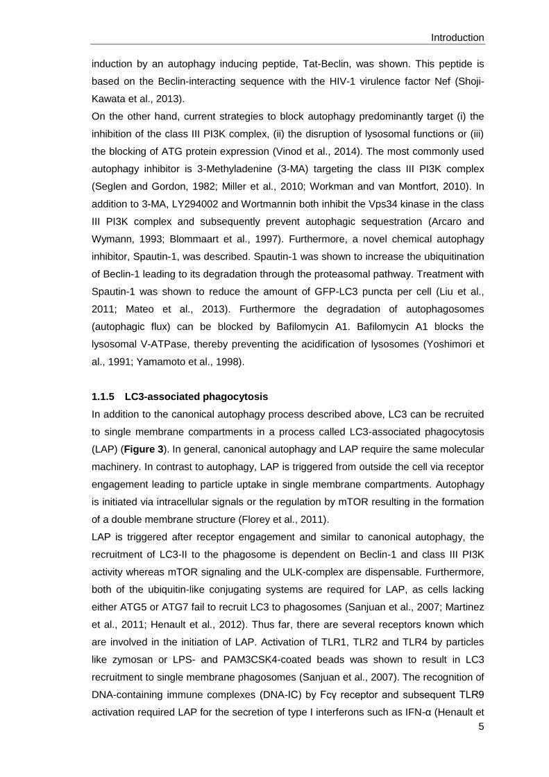
al., 2012). Besides TLR stimulation, also engagement of Dectin-1 by β-glucan from
fungal cell walls was shown to trigger LAP resulting in rapid lysosomal maturation (Ma
et al., 2012; Mansour et al., 2013). Furthermore, LAP in macrophages is induced upon
uptake and clearance of apoptotic cells mediated by the phosphatidylserine (PS)
receptor T cell immunoglobulin domain and mucin domain protein-4 (TIM4) resulting in
the production of anti-inflammatory cytokines (Martinez et al., 2011).
Figure 3: LC3-associated phagocytosis (LAP). LC3-associated phagocytosis is induced
through receptor engagement by FcγR, Dectin-1, TLRs or TIM4. The internalization of receptor
and bound particles results in the recruitment of the class III PI3K complex and NOX2,
producing ROS, to the phagosomal membrane. Subsequently, the ATG12-ATG5–ATG16L1 and
the ATG7–ATG3 complex mediate the formation of membrane bound LC3-II. Enhanced
phagosomal maturation and fusion with lysosomes efficiently degrades the engulfed cargo and
provides presentation of antigens on major histocompatibility complex II (MHC-II) molecules
(adapted from Mehta et al., 2014).
Recently, Martinez and colleagues identified Rubicon as a molecular switch between
the repression of macroautophagy and the activation of LAP. In the absence of
Rubicon, the activation of a specific class III PI3K subcomplex containing UVRAG (UV
radiation resistance-associated gene), and subsequently the recruitment of LC3, was
impaired (Martinez et al., 2015). Rubicon was suggested to interact with proteins of the
NADPH oxidase complex that were already demonstrated to be important for LAP
induction (Yang et al., 2012). The activation of TLRs initiates the production of reactive
Introduction
7
oxygen species (ROS) by NOX2, an isoform of the NADPH oxidase in macrophages
(Huang et al., 2009). NOX2 is composed of the integral membrane-bound
NOX2/gp91phox and p22phox and a complex of cytosolic components p67phox, p47phox,
p40phox and Rac2 (Nauseef, 2008). PI3P production of activated class III PI3K recruits
the soluble oxidase component p40phox. Furthermore, Rubicon binds p22phox and
thereby stabilizes the oxidase complex directly. The activated complex transfers
electrons from the substrate (NADPH) through a flavin and heme group to oxygen
inside the phagosome, building superoxides (Panday et al., 2015). The production of
ROS is necessary for the recruitment of ubiquitin-like conjugating systems mediating
the membrane attachment of LC3. LC3 on the LAPosome enhances maturation
resulting in phagosomal fusion with lysosomes and a rapid and efficient degradation of
the engulfed cargo.
The activity of NOX2 can be inhibited by Diphenyleneiodonium (DPI), an uncompetitive
inhibitor of flavoproteins (Riganti et al., 2004). On the other hand, LAP induction via
TLR2 engagement induced by zymosan, a glucan found in the cell wall of fungi like
yeast, is commonly used. Stimulation of macrophages with zymosan results in a pro-
inflammatory response and cytokine secretion (Sato et al., 2003; Du et al., 2006).
1.2 Macrophages
1.2.1 Macrophage subtypes and their activation
Macrophages (MFs), discovered in 1882 by the Russian zoologist Élie Metchnikoff, are
found in all tissues and serve as a first line of defense against invading microbes
(Nathan, 2008). They originate from hematopoietic stem cells in the bone marrow and
are released into the circulation as macrophage precursor cells called monocytes
(Murray and Wynn, 2011). In steady state, they enter the tissue to differentiate in either
(anti-inflammatory) MFs or dendritic cells and replenish the long-lived, tissue resident
populations (Gordon and Taylor, 2005). Furthermore, they can be specifically recruited
to the side of infection by chemotaxis (Jones, 2000). Once in the tissue, MFs are
exposed to a whole bunch of stimulatory and suppressive signals. Depending on the
local stimuli, MFs adopt context-dependent phenotypes that either promote or inhibit
immunity and inflammatory responses (Adams and Hamilton, 1984). The term
“activated macrophage” was introduced by Mackaness in the 1960s, which can be
further subdivided in classically activated MFs (M1) or alternatively activated MFs
(M2a, M2b, M2c) promoting either Th1 or Th2 responses and resulting in different
disease outcome (Mills et al., 2000; Mantovani et al., 2004).
Stimulation with pro-inflammatory molecules such as IFN-γ, secreted by activated Th1
cells or NK cells, in combination with TLR activation by e.g. LPS and production of
Introduction
8
TNF-α by the macrophage itself, leads to M1 polarization (Mosser, 2003; Mantovani et
al., 2004). Those classically activated MFs produce pro-inflammatory cytokines like IL-
1β, IL-6, IL-12, TNF and IL-23 (Verreck et al., 2004; Mosser and Edwards, 2008).
Furthermore, co-stimulatory molecules and proteins involved in antigen processing and
presentation become upregulated favoring an inflammatory Th1 mediated immune
response (Martinez and Gordon, 2014). M1 possess an antimicrobial arsenal like
production of reactive oxygen species (ROS) that mediate host defence from a variety
of bacteria, protozoa and viruses and have roles in anti-tumor immunity (Mackaness,
1962; Murray and Wynn, 2011). In vitro stimulation of monocytes with granulocyte
macrophage colony-stimulating factor (GM-CSF) leads to the differentiation into MFs
with a pro-inflammatory cytokine profile (Burgess and Metcalf, 1980; Verreck et al.,
2004).
Besides classical activation, all other stimuli leading to macrophage polarization were
termed as alternative activation. Stimulation with IL-4 and IL-13, secreted by Th2 cells,
eosinophils or basophils, leads to M2a polarization which possesses tissue repair
effector functions (Stein et al., 1992). These MFs are more susceptible to some
intracellular infections such as Mycobacterium tuberculosis or Leishmania (Kropf et al.,
2005; Harris et al., 2007). Furthermore, stimulation of monocytes with IgG
immuncomplexes in combination with TLR ligands such as LPS, retains high
expression of inflammatory cytokines (IL-1, IL-6, TNF) in combination with an IL-10high
and IL-12low profile (Gerber and Mosser, 2001; Mantovani et al., 2004). Those MFs,
also termed M2b or regulatory macrophages, are potent inhibitors of inflammation
(Mosser and Edwards, 2008). Stimulation of monocytes with Glucocorticoids and IL-10
is referred to as M2c macrophage polarization. Glucocorticoids are released by adrenal
cells in response to stress and inhibit inflammatory functions of MFs. Those MFs
secrete IL-10 and TGF-β leading to immune suppression and induce the development
of regulatory T cells (Mantovani et al., 2004; Mosser and Edwards, 2008). IL-10
producing macrophages possess a high capability for the clearance of apoptotic cells
(Xu et al., 2006). In general, alternatively activated MFs counteract the pro-
inflammatory functions of M1 MFs providing immune regulation as well as immune
inhibition by promoting Th2 responses. Additionally, they are essential in helminth
killing and tissue remodeling although interaction with mast cells, basophils,
eosinophils, NKT cells and IgE promotes allergy and hypersensitivity (Gordon and
Martinez, 2010; Locksley, 2010). In vitro stimulation of monocytes with macrophage
colony-stimulating factor (M-CSF) leads to the differentiation into MFs with an anti-
inflammatory cytokine profile and features of M2 macrophages (Verreck et al., 2004;
Lacey et al., 2012).
Introduction
9
1.2.2 MFs are professional phagocytes and mediators of adaptive immunity
A major factor that differentiates professional from non-professional phagocytes is the
multitude of surface receptors pattern-recognition receptors such as the Toll-like
receptors (TLRs), C-type lectin receptors (CLRs), NOD-like receptors (NLRs), DNA
sensing receptors or retinoic acid inducible gene I (RIG-I) like receptors that detect
signals that are not normally found in healthy tissue (Murray and Wynn, 2011).
Receptor triggering by foreign material like pathogens results in their engulfment in
phagosomes. During phagosome maturation, phagosomes fuse with lysosomes
resulting in pH acidification (4 – 4.5) and the influx of proteases generically called
cathepsins (Blum et al., 2013). These conditions mediate the denaturation of engulfed
material by proteolytical cleavage producing peptides of >11 amino acids for
presentation on MHC-II molecules (van Kasteren and Overkleeft, 2014; Rossjohn et al.,
2015). MHC-II molecules are restricted to antigen presenting cells (APC), whereas
MHC-I molecules are abundantly expressed on all cell types as they are loaded with
cytosolic, proteasomal processed peptides of 8-10 amino acids containing a broad
spectrum of self-peptides which are presented to CD8+ T cells (van Kasteren and
Overkleeft, 2014; Rossjohn et al., 2015). Prior of antigen loading to MHC-II molecules,
the invariant chain (Ii) which occupies the binding groove needs to be removed. MHC-
II, being assembled in the endoplasmatic reticulum of α- and β-chains associated with
the Ii, is transported to MHC class II compartments (MIIC) (Neefjes et al., 1990). The Ii
is processed by Cathepsin S, resulting in a 25 aa class-II associated invariant chain
peptide (CLIP) that is exchanged by Cathepsin V and the chaperone HLA-DM for a
high affinity peptide (Tolosa et al., 2003; van Kasteren and Overkleeft, 2014).
Subsequently, the peptide-MHC complex is transported to the cell surface for immune
surveillance by CD4+ T cells (Neefjes et al., 2011). T cell activation requires at least
two signals namely the complex of a peptide and a MHC molecule binding the T cell
receptor (TCR) and a second co-stimulatory signal e.g. the binding of CD80 (B7-
1)/CD86 (B7-2) or CD40 on APCs to CD28 or CD40L on T cells (Medzhitov and
Janeway, JR, 2000) (Figure 4). The surface expression of co-stimulatory molecules on
APCs is induced by TLRs upon recognition of their cognate pathogen-associated
molecular pattern (PAMP) in the presence of infection leading to the activation of
pathogen-specific T cells (Medzhitov and Janeway, JR, 2000).
In addition to the classical phagolysosomal pathway, also conventional autophagy and
LC3-associated phagocytosis can be involved in antigen processing. In the case of
autophagy processing of endogenous antigens for MHC-II presentation takes place,
which is also known as cross-presentation (Munz, 2016a). Thereby, these pathways
are directly associated with pathogen recognition, autolysosomal degradation and the
induction of an adaptive immune response (Dengjel et al., 2005; Munz, 2016b) (Figure
Introduction
10
4). Especially for professional phagocytes like macrophages, the autophagic
sequestration process is of great importance. Autophagy ensures the macrophages’
homeostasis as they are faced with changes in nutrient and oxygen availability when
they enter inflamed or tumor tissues and mediates pathogen degradation (Martinez et
al., 2013). Furthermore the pre- or absence of autophagy influences macrophage
polarization as impaired autophagy promotes pro-inflammatory macrophage generation
(Liu et al., 2015). Facing Mycobacterium tuberculosis infection, IFN-γ induced
autophagy eliminates the bacterial infection whereas exposure of macrophages to Th2
cytokines like IL-4 or IL-13 abrogates autophagy leading to failure of pathogen control
(Harris et al., 2007; Matsuzawa et al., 2012).
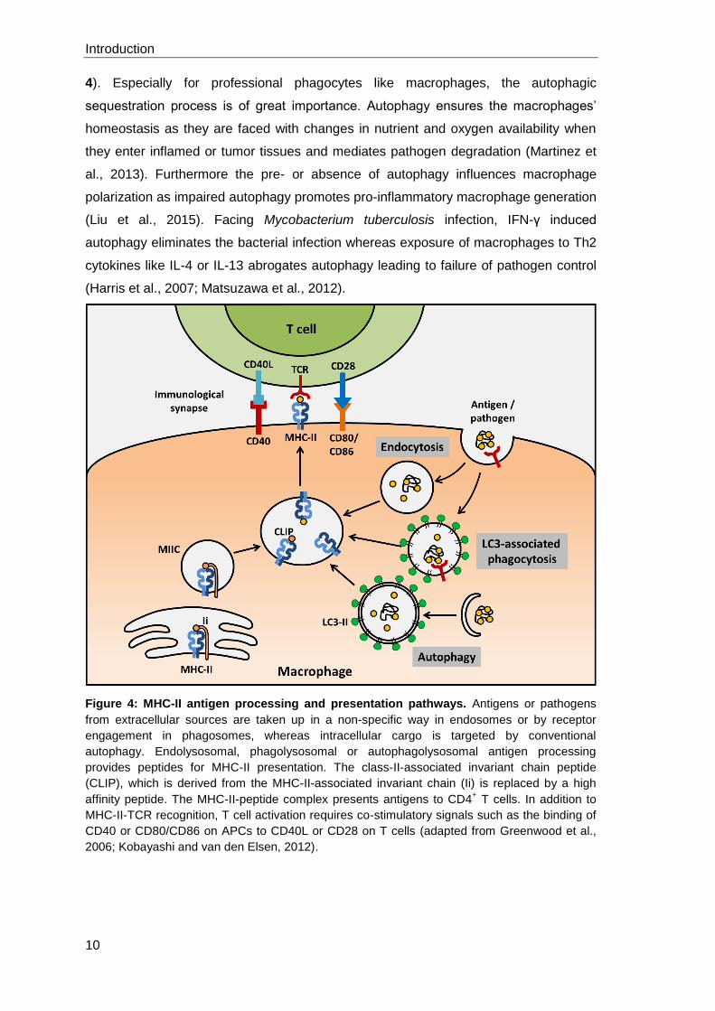
Figure 4: MHC-II antigen processing and presentation pathways. Antigens or pathogens
from extracellular sources are taken up in a non-specific way in endosomes or by receptor
engagement in phagosomes, whereas intracellular cargo is targeted by conventional
autophagy. Endolysosomal, phagolysosomal or autophagolysosomal antigen processing
provides peptides for MHC-II presentation. The class-II-associated invariant chain peptide
(CLIP), which is derived from the MHC-II-associated invariant chain (Ii) is replaced by a high
affinity peptide. The MHC-II-peptide complex presents antigens to CD4+ T cells. In addition to
MHC-II-TCR recognition, T cell activation requires co-stimulatory signals such as the binding of
CD40 or CD80/CD86 on APCs to CD40L or CD28 on T cells (adapted from Greenwood et al.,
2006; Kobayashi and van den Elsen, 2012).
Introduction
11
1.3 Leishmaniasis
1.3.1 Epidemiology
Leishmaniasis is caused by the protozoan vector-born parasites of the genus
Leishmania, which are transmitted by the bite of infected female sandflies (mainly of
the subfamily Phlebotominae or Lutzomyia) (Goto and Lauletta Lindoso, 2012).
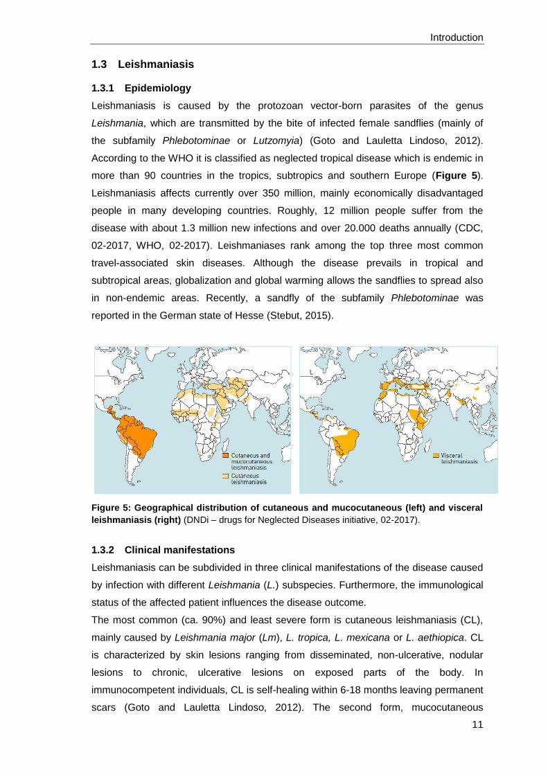
According to the WHO it is classified as neglected tropical disease which is endemic in
more than 90 countries in the tropics, subtropics and southern Europe (Figure 5).
Leishmaniasis affects currently over 350 million, mainly economically disadvantaged
people in many developing countries. Roughly, 12 million people suffer from the
disease with about 1.3 million new infections and over 20.000 deaths annually (CDC,
02-2017, WHO, 02-2017). Leishmaniases rank among the top three most common
travel-associated skin diseases. Although the disease prevails in tropical and
subtropical areas, globalization and global warming allows the sandflies to spread also
in non-endemic areas. Recently, a sandfly of the subfamily Phlebotominae was
reported in the German state of Hesse (Stebut, 2015).
Figure 5: Geographical distribution of cutaneous and mucocutaneous (left) and visceral
leishmaniasis (right) (DNDi – drugs for Neglected Diseases initiative, 02-2017).
1.3.2 Clinical manifestations
Leishmaniasis can be subdivided in three clinical manifestations of the disease caused
by infection with different Leishmania (L.) subspecies. Furthermore, the immunological
status of the affected patient influences the disease outcome.
The most common (ca. 90%) and least severe form is cutaneous leishmaniasis (CL),
mainly caused by Leishmania major (Lm), L. tropica, L. mexicana or L. aethiopica. CL
is characterized by skin lesions ranging from disseminated, non-ulcerative, nodular
lesions to chronic, ulcerative lesions on exposed parts of the body. In
immunocompetent individuals, CL is self-healing within 6-18 months leaving permanent
scars (Goto and Lauletta Lindoso, 2012). The second form, mucocutaneous
Introduction
12
leishmaniasis (MCL), is caused by L. baziliensis and occurs mainly in South America.
MCL is leading to partial or total destructive lesions of the mucous membranes of the
nose, mouth and throat cavities and surrounding tissue. It can occur months to years
after the resolution of CL and is associated with inadequate treatment of the primary
infection (Stebut, 2015); WHO, 2017). The third and most severe form is visceral
leishmaniasis (VL), also known as kala-azar or black fever and is fatal if left untreated.
VL is typically caused by L. donovani (India and Africa), L. infantum (Mediterranean)
and L. chagasi (Latin America). Disease outcome is characterized by irregular bouts of
fever, substantial weight loss, anemia and swelling of the spleen and liver due to
parasitic infection. Several years after healing of VL, patients can develop post kala-
azar dermal leishmaniasis (PKDL), which is characterized by a rush of papular, nodular
or macular skin lesions. PKDL patients are considered to be a potential pathogen
reservoir (WHO, 2017). Especially co-infection of Leishmania and HIV leads to
increased incidence of developing a severe form of leishmaniasis (Lindoso et al.,
2016).
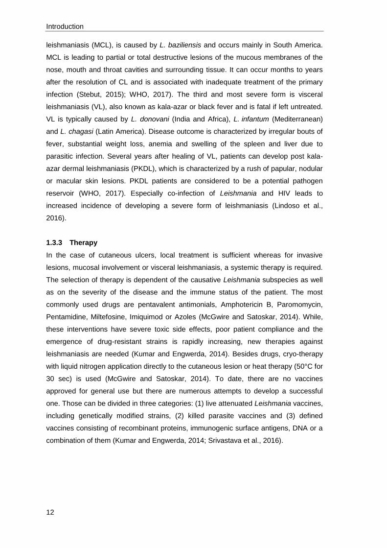
1.3.3 Therapy
In the case of cutaneous ulcers, local treatment is sufficient whereas for invasive
lesions, mucosal involvement or visceral leishmaniasis, a systemic therapy is required.
The selection of therapy is dependent of the causative Leishmania subspecies as well
as on the severity of the disease and the immune status of the patient. The most
commonly used drugs are pentavalent antimonials, Amphotericin B, Paromomycin,
Pentamidine, Miltefosine, Imiquimod or Azoles (McGwire and Satoskar, 2014). While,
these interventions have severe toxic side effects, poor patient compliance and the
emergence of drug-resistant strains is rapidly increasing, new therapies against
leishmaniasis are needed (Kumar and Engwerda, 2014). Besides drugs, cryo-therapy
with liquid nitrogen application directly to the cutaneous lesion or heat therapy (50°C for
30 sec) is used (McGwire and Satoskar, 2014). To date, there are no vaccines
approved for general use but there are numerous attempts to develop a successful
one. Those can be divided in three categories: (1) live attenuated Leishmania vaccines,
including genetically modified strains, (2) killed parasite vaccines and (3) defined
vaccines consisting of recombinant proteins, immunogenic surface antigens, DNA or a
combination of them (Kumar and Engwerda, 2014; Srivastava et al., 2016).
Introduction
13
1.4 Leishmania
1.4.1 Taxonomy
The protozoan parasites of the genus Leishmania belong to the class of Kinetoplastea
and the family of Trypanosomatidae. In 1903, Ronald Ross classified the genus
Leishmania, named after William Boog Leishman due to his method of staining blood
for malaria and other parasites (Rioux et al., 1990; Akhoundi et al., 2016). Leishmania
species are unicellular, obligate intracellular parasites with a well-defined nucleus, a
large DNA-containing mitochondrium (kinetoplast) and a flagellum (promastigote form).
1.4.2 Life cycle
Leishmania are protozoan parasites which sustain their life cycle through transmission
between the intestinal tract of a sandfly and phagocytes in a mammalian host (Figure
6). Among this life cycle, Leishmania pass in two different life stages, namely the
promastigote and the amastigote form. In the midgut of the sandfly, Leishmania
procyclic promastigotes proliferate and differentiate into infective, non-dividing
metacyclic promastigotes in a process called metacyclogenesis (Giannini, 1974).
Therefore typical environmental parameters of the sand fly midgut as alkaline pH and
temperatures between 22°C and 28°C are necessary (Zilberstein and Shapira, 1994).
During blood meal, the sandfly regurgitates about 100-3000 metacyclic promastigotes
together with immunomodulatory parasite-derived proteophosphoglycans and various
salivary components (Warburg and Schlein, 1986; Kaye and Scott, 2011).
Once in the skin, the majority of parasites is killed in the extracellular tissue by
complement mediated lysis (Mosser and Edelson, 1984). Remaining parasites are
phagocytosed by resident skin macrophages, dendritic cells or rapidly infiltrating
polymorphonuclear neutrophil granulocytes (PMN) as a first line of immune defense
towards invading pathogens (Laskay et al., 2003). Neutrophils are attracted to the side
of infection by the release of a soluble Leishmania chemotactic factor (LCF) (van
Zandbergen et al., 2002). The neutrophils engulf the virulent inoculum of Leishmania
comprising a mixture of viable and apoptotic parasites, of which the latter ones are
crucial for disease development (van Zandbergen et al., 2006). The uptake of apoptotic
cells, characterized by phosphatidylserine (PS) on their surface, represents a “no
danger” signal to immune cells. Hence, a silent phagocytosis of the parasite takes
place thereby suppressing innate intracellular antimicrobial mechanisms. Furthermore,
the release of cytokines such as TGF-β and IL-10 creates an anti-inflammatory
environment (Voll et al., 1997; Huynh et al., 2002; van Zandbergen et al., 2007).
Additionally, the rapid and spontaneous apoptosis of neutrophils is delayed up to two
days, providing a transient shelter for Leishmania (Aga et al., 2002).
Introduction
14
Subsequently, macrophages are recruited to the side of infection by the release of
macrophage inflammatory protein-1 (MIP-1β) from infected neutrophils. Macrophages
favor neutrophil apoptosis by membrane TNF and clear the tissue from apoptotic cell
corpses (Menten et al., 2002; Allenbach et al., 2006). The uptake of infected and
apoptotic neutrophils provides a silent entry mechanism (“Trojan horse strategy”) for
the parasites into their final host cells, the macrophages (Laskay et al., 2003). Another
study revealed that Leishmania-containing neutrophils were not directly phagocytosed
by macrophages. However, in neutrophil depleted mice, macrophages are able to
directly take up parasites (Peters et al., 2008; Charmoy et al., 2010). Inside
macrophages, promastigotes delay the phagolysosomal biogenesis by
lipophosphoglycan (LPG), present on the membrane of the parasites, to allow the
adaption to the acidic conditions (Desjardins and Descoteaux, 1997; Rodriguez et al.,
2006). The parasitophorous vacuole is characterized by a pH of 4.7-5.2 (Antoine et al.,
1990), lysosomal hydrolases (Prina et al., 1990), lysosomal-membrane markers LAMP-
1 and LAMP-2 (Russell et al., 1992), a proton ATPase (Sturgill-Koszycki et al., 1994)
and MHC class II molecules (Lang et al., 1994). This acidic and warmer (32-37°C
subcutaneous-visceral) environment of phagolysosomal compartments initiates the
transformation of metacyclic promastigotes into the aflagellate amastigote form
(Zilberstein and Shapira, 1994). This differentiation process starts within the first 5-12
hours after phagocytosis and is completed on day 2-5, depending on the infecting
strain (Courret et al., 2002). Amastigotes are adapted to survive the harsh lysosomal
conditions and can even start replication. After cell burst, they can infect further
macrophages and are responsible for disease development. During another blood
meal, the sandfly takes up amastigote-infected phagocytes which can redifferentiate
into infectious promastigotes in the sandfly’s midgut completing the Leishmania life
cycle (Kaye and Scott, 2011).
Introduction
15
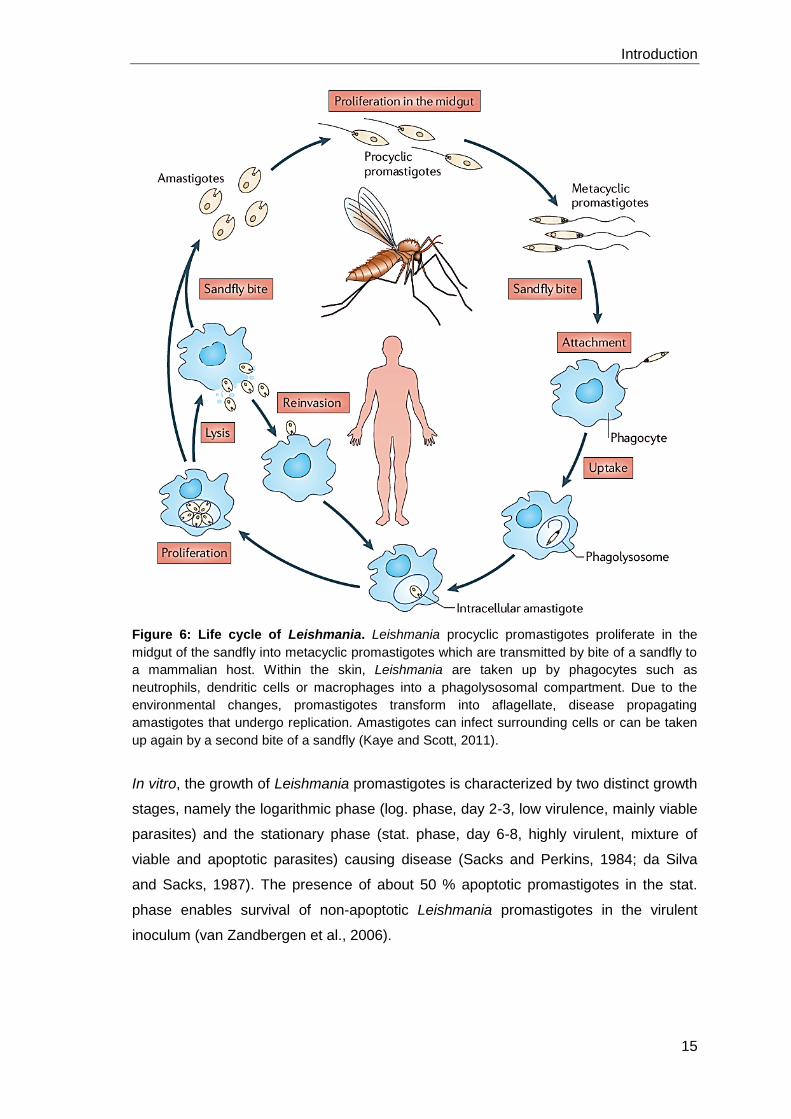
Figure 6: Life cycle of Leishmania. Leishmania procyclic promastigotes proliferate in the
midgut of the sandfly into metacyclic promastigotes which are transmitted by bite of a sandfly to
a mammalian host. Within the skin, Leishmania are taken up by phagocytes such as
neutrophils, dendritic cells or macrophages into a phagolysosomal compartment. Due to the
environmental changes, promastigotes transform into aflagellate, disease propagating
amastigotes that undergo replication. Amastigotes can infect surrounding cells or can be taken
up again by a second bite of a sandfly (Kaye and Scott, 2011).
In vitro, the growth of Leishmania promastigotes is characterized by two distinct growth
stages, namely the logarithmic phase (log. phase, day 2-3, low virulence, mainly viable
parasites) and the stationary phase (stat. phase, day 6-8, highly virulent, mixture of
viable and apoptotic parasites) causing disease (Sacks and Perkins, 1984; da Silva
and Sacks, 1987). The presence of about 50 % apoptotic promastigotes in the stat.
phase enables survival of non-apoptotic Leishmania promastigotes in the virulent
inoculum (van Zandbergen et al., 2006).
Introduction
16
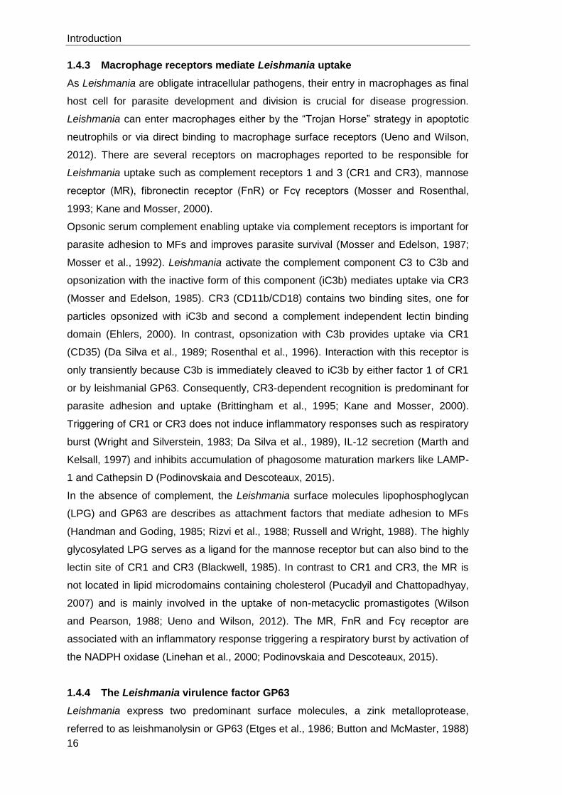
1.4.3 Macrophage receptors mediate Leishmania uptake
As Leishmania are obligate intracellular pathogens, their entry in macrophages as final
host cell for parasite development and division is crucial for disease progression.
Leishmania can enter macrophages either by the “Trojan Horse” strategy in apoptotic
neutrophils or via direct binding to macrophage surface receptors (Ueno and Wilson,
2012). There are several receptors on macrophages reported to be responsible for
Leishmania uptake such as complement receptors 1 and 3 (CR1 and CR3), mannose
receptor (MR), fibronectin receptor (FnR) or Fcγ receptors (Mosser and Rosenthal,
1993; Kane and Mosser, 2000).
Opsonic serum complement enabling uptake via complement receptors is important for
parasite adhesion to MFs and improves parasite survival (Mosser and Edelson, 1987;
Mosser et al., 1992). Leishmania activate the complement component C3 to C3b and
opsonization with the inactive form of this component (iC3b) mediates uptake via CR3
(Mosser and Edelson, 1985). CR3 (CD11b/CD18) contains two binding sites, one for
particles opsonized with iC3b and second a complement independent lectin binding
domain (Ehlers, 2000). In contrast, opsonization with C3b provides uptake via CR1
(CD35) (Da Silva et al., 1989; Rosenthal et al., 1996). Interaction with this receptor is
only transiently because C3b is immediately cleaved to iC3b by either factor 1 of CR1
or by leishmanial GP63. Consequently, CR3-dependent recognition is predominant for
parasite adhesion and uptake (Brittingham et al., 1995; Kane and Mosser, 2000).
Triggering of CR1 or CR3 does not induce inflammatory responses such as respiratory
burst (Wright and Silverstein, 1983; Da Silva et al., 1989), IL-12 secretion (Marth and
Kelsall, 1997) and inhibits accumulation of phagosome maturation markers like LAMP-
1 and Cathepsin D (Podinovskaia and Descoteaux, 2015).
In the absence of complement, the Leishmania surface molecules lipophosphoglycan
(LPG) and GP63 are describes as attachment factors that mediate adhesion to MFs
(Handman and Goding, 1985; Rizvi et al., 1988; Russell and Wright, 1988). The highly
glycosylated LPG serves as a ligand for the mannose receptor but can also bind to the
lectin site of CR1 and CR3 (Blackwell, 1985). In contrast to CR1 and CR3, the MR is
not located in lipid microdomains containing cholesterol (Pucadyil and Chattopadhyay,
2007) and is mainly involved in the uptake of non-metacyclic promastigotes (Wilson
and Pearson, 1988; Ueno and Wilson, 2012). The MR, FnR and Fcγ receptor are
associated with an inflammatory response triggering a respiratory burst by activation of
the NADPH oxidase (Linehan et al., 2000; Podinovskaia and Descoteaux, 2015).
1.4.4 The Leishmania virulence factor GP63
Leishmania express two predominant surface molecules, a zink metalloprotease,
referred to as leishmanolysin or GP63 (Etges et al., 1986; Button and McMaster, 1988)
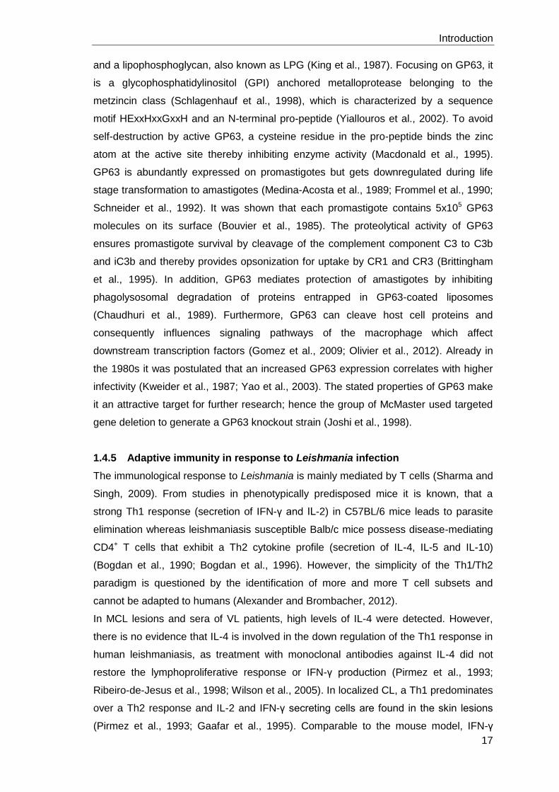
Introduction
17
and a lipophosphoglycan, also known as LPG (King et al., 1987). Focusing on GP63, it
is a glycophosphatidylinositol (GPI) anchored metalloprotease belonging to the
metzincin class (Schlagenhauf et al., 1998), which is characterized by a sequence
motif HExxHxxGxxH and an N-terminal pro-peptide (Yiallouros et al., 2002). To avoid
self-destruction by active GP63, a cysteine residue in the pro-peptide binds the zinc
atom at the active site thereby inhibiting enzyme activity (Macdonald et al., 1995).
GP63 is abundantly expressed on promastigotes but gets downregulated during life
stage transformation to amastigotes (Medina-Acosta et al., 1989; Frommel et al., 1990;
Schneider et al., 1992). It was shown that each promastigote contains 5x10⁵ GP63
molecules on its surface (Bouvier et al., 1985). The proteolytical activity of GP63
ensures promastigote survival by cleavage of the complement component C3 to C3b
and iC3b and thereby provides opsonization for uptake by CR1 and CR3 (Brittingham
et al., 1995). In addition, GP63 mediates protection of amastigotes by inhibiting
phagolysosomal degradation of proteins entrapped in GP63-coated liposomes
(Chaudhuri et al., 1989). Furthermore, GP63 can cleave host cell proteins and
consequently influences signaling pathways of the macrophage which affect
downstream transcription factors (Gomez et al., 2009; Olivier et al., 2012). Already in
the 1980s it was postulated that an increased GP63 expression correlates with higher
infectivity (Kweider et al., 1987; Yao et al., 2003). The stated properties of GP63 make
it an attractive target for further research; hence the group of McMaster used targeted
gene deletion to generate a GP63 knockout strain (Joshi et al., 1998).
1.4.5 Adaptive immunity in response to Leishmania infection
The immunological response to Leishmania is mainly mediated by T cells (Sharma and
Singh, 2009). From studies in phenotypically predisposed mice it is known, that a
strong Th1 response (secretion of IFN-γ and IL-2) in C57BL/6 mice leads to parasite
elimination whereas leishmaniasis susceptible Balb/c mice possess disease-mediating
CD4+ T cells that exhibit a Th2 cytokine profile (secretion of IL-4, IL-5 and IL-10)
(Bogdan et al., 1990; Bogdan et al., 1996). However, the simplicity of the Th1/Th2
paradigm is questioned by the identification of more and more T cell subsets and
cannot be adapted to humans (Alexander and Brombacher, 2012).
In MCL lesions and sera of VL patients, high levels of IL-4 were detected. However,
there is no evidence that IL-4 is involved in the down regulation of the Th1 response in
human leishmaniasis, as treatment with monoclonal antibodies against IL-4 did not
restore the lymphoproliferative response or IFN-γ production (Pirmez et al., 1993;
Ribeiro-de-Jesus et al., 1998; Wilson et al., 2005). In localized CL, a Th1 predominates
over a Th2 response and IL-2 and IFN-γ secreting cells are found in the skin lesions
(Pirmez et al., 1993; Gaafar et al., 1995). Comparable to the mouse model, IFN-γ
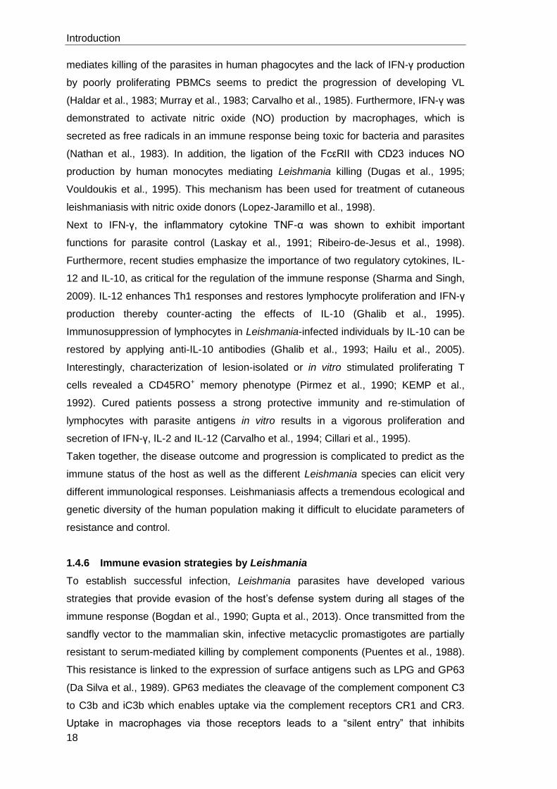
Introduction
18
mediates killing of the parasites in human phagocytes and the lack of IFN-γ production
by poorly proliferating PBMCs seems to predict the progression of developing VL
(Haldar et al., 1983; Murray et al., 1983; Carvalho et al., 1985). Furthermore, IFN-γ was
demonstrated to activate nitric oxide (NO) production by macrophages, which is
secreted as free radicals in an immune response being toxic for bacteria and parasites
(Nathan et al., 1983). In addition, the ligation of the FcεRII with CD23 induces NO
production by human monocytes mediating Leishmania killing (Dugas et al., 1995;
Vouldoukis et al., 1995). This mechanism has been used for treatment of cutaneous
leishmaniasis with nitric oxide donors (Lopez-Jaramillo et al., 1998).
Next to IFN-γ, the inflammatory cytokine TNF-α was shown to exhibit important
functions for parasite control (Laskay et al., 1991; Ribeiro-de-Jesus et al., 1998).
Furthermore, recent studies emphasize the importance of two regulatory cytokines, IL-
12 and IL-10, as critical for the regulation of the immune response (Sharma and Singh,
2009). IL-12 enhances Th1 responses and restores lymphocyte proliferation and IFN-γ
production thereby counter-acting the effects of IL-10 (Ghalib et al., 1995).
Immunosuppression of lymphocytes in Leishmania-infected individuals by IL-10 can be
restored by applying anti-IL-10 antibodies (Ghalib et al., 1993; Hailu et al., 2005).
Interestingly, characterization of lesion-isolated or in vitro stimulated proliferating T
cells revealed a CD45RO+ memory phenotype (Pirmez et al., 1990; KEMP et al.,
1992). Cured patients possess a strong protective immunity and re-stimulation of
lymphocytes with parasite antigens in vitro results in a vigorous proliferation and
secretion of IFN-γ, IL-2 and IL-12 (Carvalho et al., 1994; Cillari et al., 1995).
Taken together, the disease outcome and progression is complicated to predict as the
immune status of the host as well as the different Leishmania species can elicit very
different immunological responses. Leishmaniasis affects a tremendous ecological and
genetic diversity of the human population making it difficult to elucidate parameters of
resistance and control.
1.4.6 Immune evasion strategies by Leishmania
To establish successful infection, Leishmania parasites have developed various
strategies that provide evasion of the host’s defense system during all stages of the
immune response (Bogdan et al., 1990; Gupta et al., 2013). Once transmitted from the
sandfly vector to the mammalian skin, infective metacyclic promastigotes are partially
resistant to serum-mediated killing by complement components (Puentes et al., 1988).
This resistance is linked to the expression of surface antigens such as LPG and GP63
(Da Silva et al., 1989). GP63 mediates the cleavage of the complement component C3
to C3b and iC3b which enables uptake via the complement receptors CR1 and CR3.
Uptake in macrophages via those receptors leads to a “silent entry” that inhibits
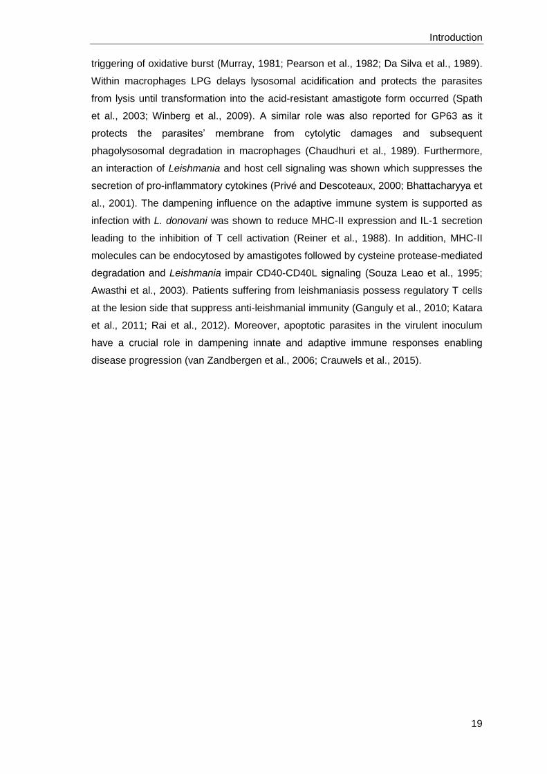
Introduction
19
triggering of oxidative burst (Murray, 1981; Pearson et al., 1982; Da Silva et al., 1989).
Within macrophages LPG delays lysosomal acidification and protects the parasites
from lysis until transformation into the acid-resistant amastigote form occurred (Spath
et al., 2003; Winberg et al., 2009). A similar role was also reported for GP63 as it
protects the parasites’ membrane from cytolytic damages and subsequent
phagolysosomal degradation in macrophages (Chaudhuri et al., 1989). Furthermore,
an interaction of Leishmania and host cell signaling was shown which suppresses the
secretion of pro-inflammatory cytokines (Privé and Descoteaux, 2000; Bhattacharyya et
al., 2001). The dampening influence on the adaptive immune system is supported as
infection with L. donovani was shown to reduce MHC-II expression and IL-1 secretion
leading to the inhibition of T cell activation (Reiner et al., 1988). In addition, MHC-II
molecules can be endocytosed by amastigotes followed by cysteine protease-mediated
degradation and Leishmania impair CD40-CD40L signaling (Souza Leao et al., 1995;
Awasthi et al., 2003). Patients suffering from leishmaniasis possess regulatory T cells
at the lesion side that suppress anti-leishmanial immunity (Ganguly et al., 2010; Katara
et al., 2011; Rai et al., 2012). Moreover, apoptotic parasites in the virulent inoculum
have a crucial role in dampening innate and adaptive immune responses enabling
disease progression (van Zandbergen et al., 2006; Crauwels et al., 2015).

Introduction
20
Hypothesis and Aims
21
1.5 Hypothesis and Aims
1.5.1 The role of autophagy in human primary macrophages
Macrophages are crucial for mediating innate and adaptive immune responses in
health and disease. Autophagic degradation mechanisms in macrophages are versatile
and can have cytoprotective as well as immunomodulatory functions. Therefore,
autophagy proteins in macrophages are an interesting evasion target for intracellular
pathogens and an interesting new class for therapeutic approaches in cancer and
immune diseases. Most of our knowledge on autophagy in macrophages is based on
cell line and mouse model research. Consequently, in this thesis we hypothesize that:
“The autophagic machinery in macrophages depends on the cells’ origin,
phenotype and applied stimulus. In addition, autophagy modulation in
macrophages influences the adaptive immune response”
To investigate this hypothesis we have the following aims:
Aim 1: a) Assessing autophagy modulation and autophagic flux in hMDM induced
by chemicals and the peptide Tat-Beclin using Western Blot analysis for
LC3-I to LC3-II conversion and LC3 immunofluorescence stainings.
b) Investigate chemical autophagy inhibition in hMDM.
c) Establish siRNA knockdowns for various autophagy related proteins and
assess the knockdown efficiency on mRNA and on protein level. In
addition, ULK-1 and Beclin-1 knockdown macrophages will be
functionally characterized for their ability to induce autophagy.
Aim 2: Analyzing the impact of autophagy on antigen processing and
presentation in hMDM using the recall antigen Tetanus Toxoid (TT). The
adaptive immune response to TT will be characterized in hMDM-1 and
hMDM-2. Furthermore, the impact of autophagy modulation, by induction
with AZD8055 and inhibition using an ULK-1 siRNA knockdown, on T cell
proliferation will be investigated.
Aim 3: Investigation of the Leishmania virulence factor GP63 for its potential to
influence infection of hMDM, the Leishmania specific T cell proliferation
and the host cells’ autophagic process.
Hypothesis and Aims
22
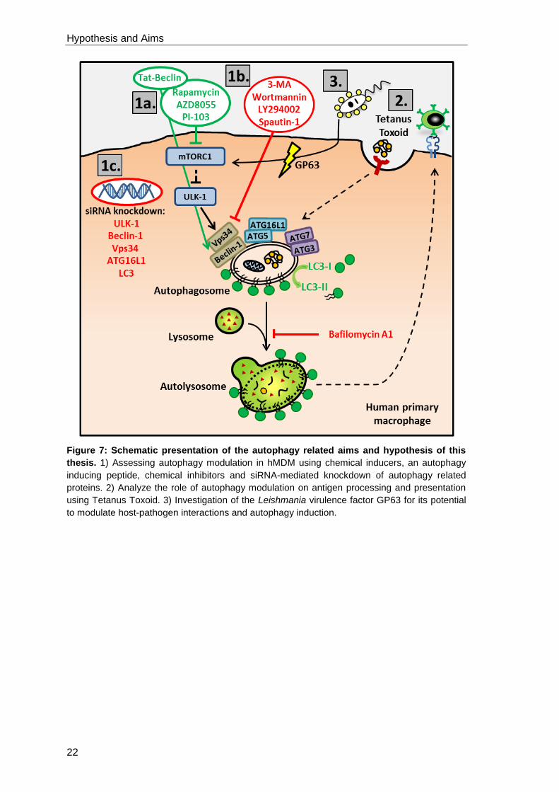
Figure 7: Schematic presentation of the autophagy related aims and hypothesis of this
thesis. 1) Assessing autophagy modulation in hMDM using chemical inducers, an autophagy
inducing peptide, chemical inhibitors and siRNA-mediated knockdown of autophagy related
proteins. 2) Analyze the role of autophagy modulation on antigen processing and presentation
using Tetanus Toxoid. 3) Investigation of the Leishmania virulence factor GP63 for its potential
to modulate host-pathogen interactions and autophagy induction.
Hypothesis and Aims
23
1.5.2 The role of LAP during Leishmania infection
In addition to conventional autophagy, extracellular particles can be taken up receptor-
mediated by LAP. LC3 recruitment to phagosomes leads to enhanced maturation,
lysosomal degradation of engulfed particles and peptide presentation on MHC-II
molecules to CD4+ T cells. We could already show that apoptotic Leishmania reside
within a single-membrane, LC3+ compartment indicating LAP. Furthermore, the
presence of apoptotic parasites in the virulent inoculum leads to disease progression in
vivo and a reduced T cell response in vitro. Consequently, we hypothesize:
“The induction of LAP by apoptotic Leishmania is an immune evasion
mechanism enabling disease progression”
To investigate this hypothesis, we have the following aims:
Aim 1: Establish a suitable LAP-inducing particle to replace apoptotic parasites.
Aim 2: Investigate the ability of LAP-inducing particles to dampen the Leishmania
specific T cell proliferation and analyze the effect on parasite survival.
Aim 3: Characterize the underlying mechanisms which lead to the induction of LAP
upon infection of hMDM with Leishmania or stimulation with LAP-inducing
particles and analyze the effect of LAP modulation on the adaptive immune
response.
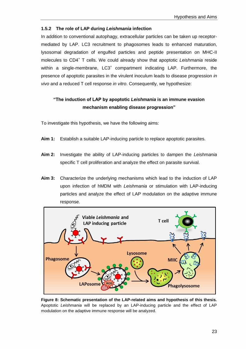
Figure 8: Schematic presentation of the LAP-related aims and hypothesis of this thesis.
Apoptotic Leishmania will be replaced by an LAP-inducing particle and the effect of LAP
modulation on the adaptive immune response will be analyzed.

Hypothesis and Aims
24
Material and Methods
25
2 Material and methods
2.1 Material
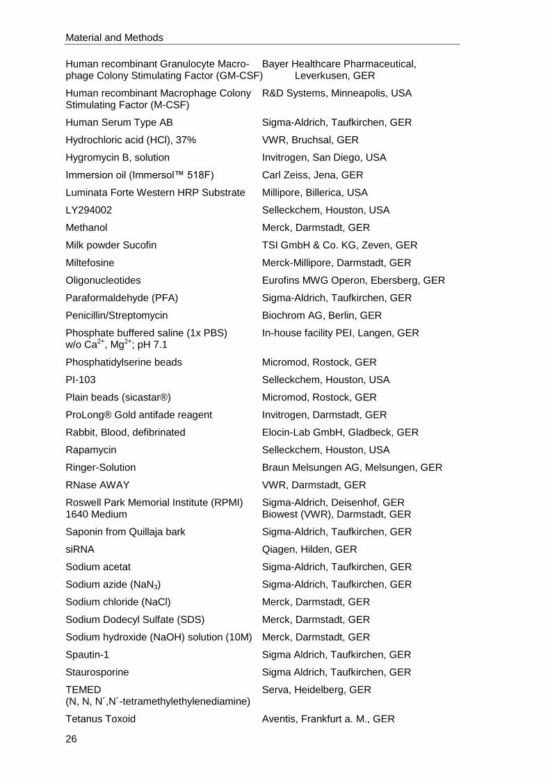
2.1.1 Chemicals
2- Propanol VWR, Bruchsal, GER
3-Methyladenine Selleckchem, Houston, USA
β-Mercaptoethanol Sigma-Aldrich, Taufkirchen, GER
Acetic acid Merck, Darmstadt, GER
Acrylamide-Bis solution 30% SERVA, Heidelberg, GER
Adenine Sigma-Aldrich, Taufkirchen, GER
Agarose LE Biozym Scientific GmbH, Oldendorf, GER
Ammonium chloride In-house facility PEI, Langen, GER
Ammoniumpersulfat (APS) Serva, Heidelberg, GER
Aqua bidest. In-house facility PEI, Langen, GER
AZD8055 Selleckchem, Houston, USA
Biotin Sigma-Aldrich, Taufkirchen, GER
Bafilomycin-A1 Sigma-Aldrich, Taufkirchen, GER
Bovine Serum Albumin (BSA) Applichem, Darmstadt, GER
CASYton OLS-OMNI, Bremen, GER
Cytochalasin D Calbiochem, Merck, Darmstadt, GER
Developer (G153 A) and Fixer (G354) AGFA, Mortsel, BE
Difco™ Brain Heart Infusion Agar Becton Dickenson, Sparks, USA
Dimethylsulfoxid (DMSO) Sigma Aldrich, Taufkirchen, GER
Diphenyleneiodonium chloride (DPI) Sigma-Aldrich, Taufkirchen, GER
Dithiothreitol (DTT) Sigma-Aldrich, Taufkirchen, GER
dNTP-Mix (10 mM each) NEB, Frankfurt am Main, GER
Ethanol (EtOH), absolut Applichem, Darmstadt, GER
FACS Clean In-house facility PEI, Langen, GER
FACS Flow (Sheath Solution) In-house facility PEI, Langen, GER
FACS Rinse In-house facility PEI, Langen, GER
Fetal Calf Serum (FCS) Sigma-Aldrich, Taufkirchen, GER
Glutamine (L-Glutamine) Biochrom AG, Berlin, GER
Glycerol (99 %) Citifluor, London, UK
Glycine In-house facility PEI, Langen, GER
HEPES (4-(2-hydroxyethyl)-1- Biochrom AG, Berlin, GER piperazineethanesulfonic acid)
High purity water In-house facility PEI, Langen, GER
Histopaque 1077 Sigma-Aldrich, Taufkirchen, GER
Material and Methods
26
Human recombinant Granulocyte Macro- Bayer Healthcare Pharmaceutical, phage Colony Stimulating Factor (GM-CSF) Leverkusen, GER
Human recombinant Macrophage Colony R&D Systems, Minneapolis, USA Stimulating Factor (M-CSF)
Human Serum Type AB Sigma-Aldrich, Taufkirchen, GER
Hydrochloric acid (HCl), 37% VWR, Bruchsal, GER
Hygromycin B, solution Invitrogen, San Diego, USA
Immersion oil (Immersol™ 518F) Carl Zeiss, Jena, GER
Luminata Forte Western HRP Substrate Millipore, Billerica, USA
LY294002 Selleckchem, Houston, USA
Methanol Merck, Darmstadt, GER
Milk powder Sucofin TSI GmbH & Co. KG, Zeven, GER
Miltefosine Merck-Millipore, Darmstadt, GER
Oligonucleotides Eurofins MWG Operon, Ebersberg, GER
Paraformaldehyde (PFA) Sigma-Aldrich, Taufkirchen, GER
Penicillin/Streptomycin Biochrom AG, Berlin, GER
Phosphate buffered saline (1x PBS) In-house facility PEI, Langen, GER w/o Ca2+, Mg2+; pH 7.1
Phosphatidylserine beads Micromod, Rostock, GER
PI-103 Selleckchem, Houston, USA
Plain beads (sicastar®) Micromod, Rostock, GER
ProLong® Gold antifade reagent Invitrogen, Darmstadt, GER
Rabbit, Blood, defibrinated Elocin-Lab GmbH, Gladbeck, GER
Rapamycin Selleckchem, Houston, USA
Ringer-Solution Braun Melsungen AG, Melsungen, GER
RNase AWAY VWR, Darmstadt, GER
Roswell Park Memorial Institute (RPMI) Sigma-Aldrich, Deisenhof, GER 1640 Medium Biowest (VWR), Darmstadt, GER
Saponin from Quillaja bark Sigma-Aldrich, Taufkirchen, GER
siRNA Qiagen, Hilden, GER
Sodium acetat Sigma-Aldrich, Taufkirchen, GER
Sodium azide (NaN3) Sigma-Aldrich, Taufkirchen, GER
Sodium chloride (NaCl) Merck, Darmstadt, GER
Sodium Dodecyl Sulfate (SDS) Merck, Darmstadt, GER
Sodium hydroxide (NaOH) solution (10M) Merck, Darmstadt, GER
Spautin-1 Sigma Aldrich, Taufkirchen, GER
Staurosporine Sigma Aldrich, Taufkirchen, GER
TEMED Serva, Heidelberg, GER (N, N, N´,N´-tetramethylethylenediamine)
Tetanus Toxoid Aventis, Frankfurt a. M., GER
Material and Methods
27
Tris(hydroxylmethyl)-aminomethan (Tris) In-house facility PEI, Langen, GER
Tween 20 Sigma-Aldrich, Steinheim, GER
Western Blot Detection Substrate GE Healthcare, Buckinghamshire, UK
Wortmannin Sigma-Aldrich, Steinheim GER
Zymosan A S. cerevisiae BioParticlesTM Invitrogen, Thermo Fisher, Dreieich, GER
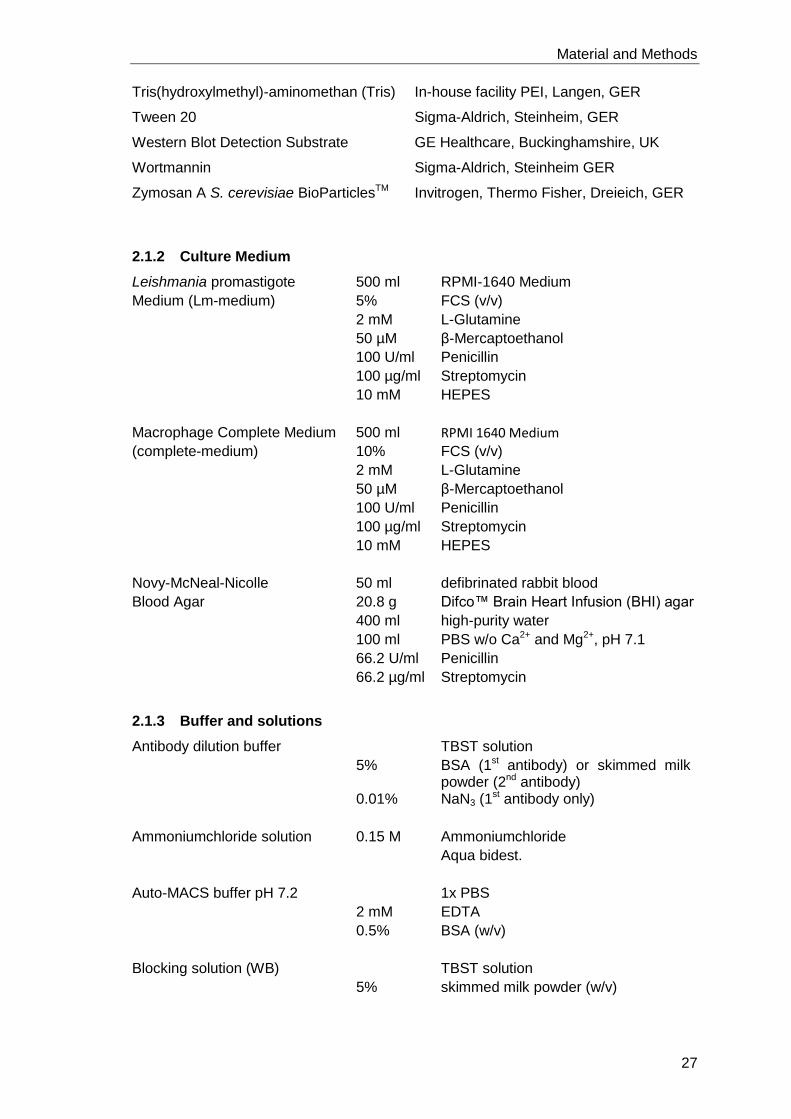
2.1.2 Culture Medium
Leishmania promastigote 500 ml RPMI-1640 Medium
Medium (Lm-medium) 5% FCS (v/v)
2 mM L-Glutamine
50 µM β-Mercaptoethanol
100 U/ml Penicillin
100 µg/ml Streptomycin
10 mM HEPES
Macrophage Complete Medium 500 ml RPMI 1640 Medium
(complete-medium) 10% FCS (v/v)
2 mM L-Glutamine
50 µM β-Mercaptoethanol
100 U/ml Penicillin
100 µg/ml Streptomycin
10 mM HEPES
Novy-McNeal-Nicolle 50 ml defibrinated rabbit blood
Blood Agar 20.8 g Difco™ Brain Heart Infusion (BHI) agar
400 ml high-purity water
100 ml PBS w/o Ca2+ and Mg2+, pH 7.1
66.2 U/ml Penicillin
66.2 µg/ml Streptomycin
2.1.3 Buffer and solutions
Antibody dilution buffer TBST solution
5% BSA (1st antibody) or skimmed milk powder (2nd antibody)
0.01% NaN3 (1st antibody only)
Ammoniumchloride solution 0.15 M Ammoniumchloride
Aqua bidest.
Auto-MACS buffer pH 7.2 1x PBS
2 mM EDTA
0.5% BSA (w/v)
Blocking solution (WB) TBST solution
5% skimmed milk powder (w/v)
Material and Methods
28
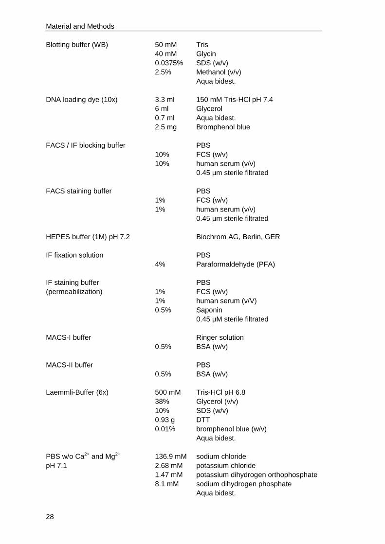
Blotting buffer (WB) 50 mM Tris
40 mM Glycin
0.0375% SDS (w/v)
2.5% Methanol (v/v)
Aqua bidest.
DNA loading dye (10x) 3.3 ml 150 mM Tris-HCl pH 7.4
6 ml Glycerol
0.7 ml Aqua bidest.
2.5 mg Bromphenol blue
FACS / IF blocking buffer PBS
10% FCS (w/v)
10% human serum (v/v)
0.45 µm sterile filtrated
FACS staining buffer PBS
1% FCS (w/v)
1% human serum (v/v)
0.45 µm sterile filtrated
HEPES buffer (1M) pH 7.2 Biochrom AG, Berlin, GER
IF fixation solution PBS
4% Paraformaldehyde (PFA)
IF staining buffer PBS
(permeabilization) 1% FCS (w/v)
1% human serum (v/V)
0.5% Saponin
0.45 µM sterile filtrated
MACS-I buffer Ringer solution
0.5% BSA (w/v)
MACS-II buffer PBS
0.5% BSA (w/v)
Laemmli-Buffer (6x) 500 mM Tris-HCl pH 6.8
38% Glycerol (v/v)
10% SDS (w/v)
0.93 g DTT
0.01% bromphenol blue (w/v)
Aqua bidest.
PBS w/o Ca2+ and Mg2+ 136.9 mM sodium chloride
pH 7.1 2.68 mM potassium chloride
1.47 mM potassium dihydrogen orthophosphate
8.1 mM sodium dihydrogen phosphate
Aqua bidest.
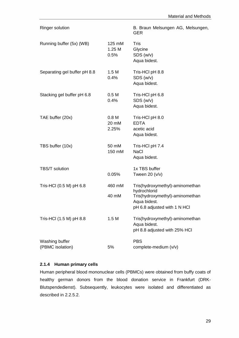
Material and Methods
29
Ringer solution B. Braun Melsungen AG, Melsungen, GER
Running buffer (5x) (WB) 125 mM Tris
1.25 M Glycine
0.5% SDS (w/v)
Aqua bidest.
Separating gel buffer pH 8.8 1.5 M Tris-HCl pH 8.8
0.4% SDS (w/v)
Aqua bidest.
Stacking gel buffer pH 6.8 0.5 M Tris-HCl pH 6.8
0.4% SDS (w/v)
Aqua bidest.
TAE buffer (20x) 0.8 M Tris-HCl pH 8.0
20 mM EDTA
2.25% acetic acid
Aqua bidest.
TBS buffer (10x) 50 mM Tris-HCl pH 7.4
150 mM NaCl
Aqua bidest.
TBS/T solution 1x TBS buffer
0.05% Tween 20 (v/v)
Tris-HCl (0.5 M) pH 6.8 460 mM Tris(hydroxymethyl)-aminomethan hydrochlorid
40 mM Tris(hydroxymethyl)-aminomethan
Aqua bidest.
pH 6.8 adjusted with 1 N HCl
Tris-HCl (1.5 M) pH 8.8 1.5 M Tris(hydroxymethyl)-aminomethan
Aqua bidest.
pH 8.8 adjusted with 25% HCl
Washing buffer PBS
(PBMC isolation) 5% complete-medium (v/v)
2.1.4 Human primary cells
Human peripheral blood mononuclear cells (PBMCs) were obtained from buffy coats of
healthy german donors from the blood donation service in Frankfurt (DRK-
Blutspendedienst). Subsequently, leukocytes were isolated and differentiated as
described in 2.2.5.2.
Material and Methods
30
2.1.5 Leishmania strains
Leishmania major isolate MHOM/IL/81/FEBNI:
Originally isolated from a skin biopsy of an Israeli patient and kindly provided by Dr.
Frank Ebert (Bernhard Nocht Institute for Tropical Medicine, Hamburg, Germany)
Leishmania major dsRed:
MHOM/IL/81/FEBNI isolates genetically transfected with the red fluorescent dsRed
gene (from Discosoma) together with a hygromycin phosphotransferase as selection
marker (Mißlitz et al., 2000).
Leishmania major Seidmann (SD) isolate MHOM/SN/74/SD:
Leishmania major SD Wildtyp (WT; ATCC# PRA-348), ∆gp63 1-7 (KO; ATCC# PRA-
385) and ∆gp63 1-7 + gp63-1 (KO+gp63; ATCC# PRA-386) were purchased from
ATCC, Manassas, USA. The wildtype was initially isolated from a cutaneous
Leishmaniasis patient in Senegal in 1973 and was genetically modified by Robert
McMaster. Geneticin serves as a selection marker to maintain the genomic integration
of gp63-1.
2.1.6 Oligonucleotides
Name Sequence
ATG16L1 Fwd TCTGGGACATTCGATCAGAGAG
ATG16L1 Rev CCTTTCTGGGTTTAAGTCCAGG
Beclin-1 Fwd GGTGTCTCTCGCAGATTCATC
Beclin-1 Rev TCAGTCTTCGGCTGAGGTTCT
CYBB Fwd AACGAATTGTACGTGGGCAGA
CYBB Rev GAGGGTTTCCAGCAAACTGAG
GAPDH Fwd GAGTCAACGGATTTGGTCGT
GAPDH Rev TTGATTTTGGAGGGATCTCG
LC3 Fwd AAGGCGCTTACAGCTCAATG
LC3 Rev CTGGGAGGCATAGACCATGT
ULK-1 Fwd AGCACGATTTGGAGGTCGC
ULK-1 Rev GCCACGATGTTTTCATGTTTCA
Vps34 Fwd TAGGAGGAACAACGGTTTCGC
Vps34 Rev GCTTCTACATTAGGCCAGACTTT
2.1.7 siRNA
Allstars Negative Control siRNA SI03650318 Qiagen, Hilden, GER
ATG16L1_1 SI04139121 Qiagen, Hilden, GER
Material and Methods
31
Beclin-1_4 SI00055594 Qiagen, Hilden, GER
CYBB SI03075709 Qiagen, Hilden, GER SI03028424 SI00008729 SI00008722
LC3_9 SI04219614 Qiagen, Hilden, GER
ULK-1_5 SI02223270 Qiagen, Hilden, GER
Vps34 SI00605829 Qiagen, Hilden, GER SI00605822 SI03649527
SI03649520
2.1.8 Peptides
Name Sequence Manufacturer
Tat-Beclin YGRKKRRQRRR-GG- J. W. Drijfhout, Leiden, NL TNVFNATFEIWHDGEFGT B. Levine, Dallas, USA
Tat-Scramble YGRKKRRQRRR-GG- J. W. Drijfhout, Leiden, NL VGNDFFINHETTGFATEW Beth Levine, Dallas, USA
2.1.9 Enzymes
Rec. RNasin® Ribonuclease Inhibitor (20-40 U/μl) Promega, Mannheim, GER
ImPromIITM Reverse Transcriptase Promega, Mannheim, GER
Taq-polymerase (5 U/μl) NEB, Frankfurt a.M., GER
2.1.10 Antibodies
Mouse, α-actin Cell Signaling, Danvers, USA
Mouse, α-Beclin-1 BD Pharmingen, Heidelberg, GER
Mouse, α-GAPDH GeneTex, Irvine, USA
Mouse, α-gp91 phox (NOX2) Santa Cruz Biotechnology, Dallas, USA
Mouse, α-p62 Santa Cruz Biotechnology, Dallas, USA
Mouse, α-gp63 (α-Lm) (1-1.5 mg/ml) AbD Serotec, Puchheim, GER
Rabbit, α-ATG16L1 Cell Signaling, Danvers, USA
Rabbit, α-LC3 (73.5 µg/ml) Cell Signaling, Danvers, USA
Rabbit, α-ULK-1 Santa Cruz Biotechnology, Dallas, USA
Rabbit, α-Vps34 Cell Signaling, Danvers, USA
Isotype controls
Mouse, IgG2a kappa BD Pharmingen, Heidelberg, GER
Mouse, IgG2a kappa, V450 BD Pharmingen, Heidelberg, GER
Mouse, IgG1 kappa, APC-Cy7 BioLegend, Aachen, GER
Material and Methods
32
Mouse, IgG1 kappa, APC BD Pharmingen, Heidelberg, GER
Mouse, IgG1 kappa, PE BD Pharmingen, Heidelberg, GER
Mouse, IgG1 kappa, V450 BD Pharmingen, Heidelberg, GER
Mouse, IgG1 kappa, FITC BD Pharmingen, Heidelberg, GER
Mouse, IgG2 kappa, Pacific blue BD Pharmingen, Heidelberg, GER
Rabbit polyclonal serum (75.3 µg/ml) Uwe Ritter, Regensburg, GER
HRP coupled antibodies
Goat, α-rabbit-HRP Santa Cruz Biotechnology, Dallas, USA
Goat, α-mouse-HRP Santa Cruz Biotechnology, Dallas, USA
Fluorescent labeled antibodies
Chicken, α-mouse-Alexa Fluor 488 Invitrogen, Thermo Fisher Scientific, GER
Goat, α-rabbit-Alexa Fluor 568 Invitrogen, Thermo Fisher Scientific, GER
Mouse, α-CD3, APC BD Pharmingen, Heidelberg, GER
Mouse, α-CD4, APC-Cy7 BD Pharmingen, Heidelberg, GER
Mouse, α-CD8, Pacific blue BD Pharmingen, Heidelberg, GER
Mouse, α-CD40, PE BD Pharmingen, Heidelberg, GER
Mouse, α-CD80, V450 BD Pharmingen, Heidelberg, GER
Mouse, α-CD86, FITC BD Pharmingen, Heidelberg, GER
Mouse, α-MHC-II, Pacific blue BioLegend, Aachen, GER
2.1.11 Dyes and Marker
Bromphenol blue dye Serva, Heidelberg, GER
CFSE (5(6)-Carboxyfluorescein Sigma Aldrich, Steinheim, GER diacetate N-succinimidyl ester)
DAPI (5 μg/mL) Molecular Probes, California, USA
Diff-QUIK® Medion Diagnostics, Düdingen, CH
DNA ladder 1kb NEB, Frankfurt a. M., GER
DNA ladder 100 bp NEB, Frankfurt a. M., GER
Ethidium bromide solution (1% in water) Merck, Darmstadt, GER
GelRed (10000x in H2O) Biotium Inc., Hayward, USA
LysoTracker® red Molecular Probes Invitrogen, GER
PageRuler Prests. Protein Ladder Thermo Fisher Scientific, Dreieich, GER
2.1.12 Ready to use Kits
Annexin V MicroBeads Miltenyi Biotec, Bergisch Gladbach, GER
Amersham ™ ECL ™ GE Healthcare, Buckinghamshire, UK Western Blotting Analysis System
Material and Methods
33
CD14 MicroBeads, human Miltenyi Biotec, Bergisch Gladbach, GER
Human IL-10 DuoSet ELISA R&D systems, Minneapolis, USA
Human TNFα DuoSet ELISA R&D systems, Minneapolis, USA
Im Prom-II Reverse Transkription System Promega, Mannheim, GER
MESA Blue qPCR MasterMix Plus Eurogentec, Köln, GER for SYBR Assay No Rox
RNeasy Plus Mini Kit Qiagen, Hilden, GER
Stemfect™ RNA Transfection Kit Stemgent, San Diego, USA
2.1.13 Laboratory supplies
Cell culture flasks with filter Greiner Bio-One, Frickenhausen, GER (25 cm2, 75 cm2 and 175 cm2)
Cell culture petri dish (10 cm diameter) Sarstedt, Nümbrecht, GER
Cell culture plates (96-well f-/v-/u-bottom) Sarstedt, Nümbrecht, GER
Cell culture plates (6-well flat) Sarstedt, Nümbrecht, GER
Cellfunnel (single, double) Tharmac GmbH, Waldsolms, GER
Cellspin filter cards (one/two hole/s) Tharmac GmbH, Waldsolms, GER
Cell Scraper, 16 cm Sarstedt, Nümbrecht, GER
Centrifuge tubes (0.2 mL) Sarstedt, Nümbrecht, GER
Centrifuge tubes (1.5 mL, 2.0 mL) Eppendorf, Hamburg, GER
Centrifuge tubes PCR Tube Multiply® Pro Sarstedt, Nümbrecht, GER (0.5 mL)
Chamberslide, 12-well, ibidi-treat ibidi GmbH, Planegg / Martinsried, GER
Cover Slide (24x50 mm) VWR, Darmstadt, GER
Cryogenic vial, internal thread (2 mL) Greiner Bio-One, Frickenhausen, GER
Cytocentrifuge Slides one circle, uncoated Tharmac GmbH, Waldsolms, GER
FACS microtubes (2 mL) Micronic, Lelystad, NL
FACS tubes (5 mL) with snap-cap Greiner Bio-One, Frickenhausen, GER
Falcons (15 mL, 50 mL) Greiner Bio-One, Frickenhausen, GER
Hyperfilm™ ECL GE Healthcare, Buckinghamshire, UK
Light Cycler 96-well plates with foil, white Roche Applied Science, Darmstadt, GER
MACS LD column or LS column Miltenyi Biotec, Bergisch Gladbach, GER
Manufix sensitive gloves Braun Melsungen AG, Melsungen, GER
Microplate (96-well flat, transparent) Greiner Bio-One, Frickenhausen, GER
MidiMACS separator Miltenyi Biotec, Bergisch Gladbach, GER
Millipore Express® PLUS Membrane Merck Millipore, Billerica, USA Filters, polyethersulfone, 0.22 μm; 0.45 μm
Multichannel pipette (Research® Plus) Eppendorf, Hamburg, GER
Nalgene™ Mr. Frosty Freezing Container Thermo Scientific, Dreieich, GER
Material and Methods
34
Nitril gloves Ansell Healthcare, Brussels, BE
Nitrocellulose membrane GE Healthcare, Buchinghamshire, UK (Hybond ECL blot membrane)
Neubauer improved cell counting chamber VWR, Darmstadt, GER (depth 0.1 mm, 0.02 mm)
Petri dish (3 cm diameter) Greiner Bio-One, Kremsmünster, AT
Pipette controller (accu-jet® pro) BRAND, Wertheim, GER
Pipette filter tips Nerbe plus, Winsen/Luhe, GER (1-10 μL; 10-200 μL, 100-1000 μL)
Pipettes (Research® plus: 0.5-10 μL; Eppendorf, Hamburg, GER 10-100 μL, 20-200 μL, 100-1000 μL)
Pipette tips Eppendorf, Hamburg, GER (0.5-10 μL; 2-200 μL; 50-1000 μL)
Polyallomer Centrifuge Tubes Beckmann Coulter GmbH, Krefeld, GER
Polystyrene Lids, PS, High profile Greiner Bio-One, Frickenhausen, GER
Serological pipettes, sterile Greiner Bio-One, Kremsmünster, AT (2.5 mL; 5 mL; 10 mL; 25 mL)
Sterile filter (0.22 μm, 0.45 μm) Sarstedt, Nümbrecht, GER
Transfer pipette (3.5 mL) Sarstedt, Nümbrecht, GER
Whatman paper gel blotting VWR, Darmstadt, GER
2.1.14 Instruments
Centrifuges
BIOLiner Buckets (75003670; 7500368) Thermo Scientific, Dreieich, GER
Bioshield 1000 A swing-out rotor Thermo Scientific, Dreieich, GER
Bench top centrifuges 5430 and 5430R Eppendorf, Hamburg, GER
Cytocentrifuge Cellspin II Universal 320R
Tharmac GmbH, Waldsolms, GER
Heraeus Megafuge 40R Thermo Scientific, Dreieich, GER
Sprout Mini-Centrifuge Biozym, Hamburg, GER
Electrophoresis and Blotting
Curix 60 tabletop processor AGFA, Mortsel, BE
Horizontal electrophoresis equipment Biotec-Fischer, Reiskirchen, GER
Mini-PROTEAN® Tetra Cell Bio-Rad, München, GER
Power Supply “PowerPac™ 200/2.0” Bio-Rad, München, GER
Semi-Dry Transfer Unit TE 77 PWR Amersham Biosciences, Freiburg, GER
UV-Transilluminator GenoView VWR International, Darmstadt, GER
Material and Methods
35
Flow Cytometer
Flow Cytometer LSR II SORP Becton Dickinson, Heidelberg, GER
Imaging
Microscope AxioPhot Carl Zeiss, Jena, GER
Microscope Axio Vert.A1 Carl Zeiss, Jena, GER
Microscope LSM 7 Live Carl Zeiss, Jena, GER
Microscope Primo Star Carl Zeiss, Jena, GER
Incubators
CO2-Incubator Forma Series II Water
Jacket Thermo Scientific, Marietta, USA
CO2 incubator, Heraeus Auto Zero Thermo Fisher Scientific, Dreieich, GER
Recirculating cooler Julabo, Seebach, GER
Lamina air flow
Workbench MSC-Advantage Thermo Fisher Scientific, Dreieich, GER
Steril Gard III Advance The Baker Company, Sanford, USA
Steril Gard Hood The Baker Company, Sanford, USA
PCR Thermo Cycler
LightCycler® 480 System Roche Applied Science, Mannheim, GER
Personal Cycler Biometra, Göttingen, GER
Others
Analytical balance KB BA 100 Sartorius, Göttingen, GER
AutoMACS Pro separator Miltenyi Biotec, Bergisch Gladbach, GER
Autoclave Systec vx-150 Systec, Wettenberg, GER
CASY Modell TT Roche Innovatis AG, Reutlingen, GER
ELISA Reader Infinite® F50 Tecan Group Ltd, Männedorf, CH
Ice machine AF 1000 Scotsman, Pogliano Milanese, IT
Freezer (-20°C) Bosch, Stuttgart, GER
Freezer U725-G (-80°C) New Brunswick, Eppendorf, Hamburg, GER
Magnetic stirrer IKA® C-MaG HS7 IKA®-Werke, Staufen, GER
Microwave Bosch, Gerlingen, GER
NanoDrop 2000c PeqLab, Erlangen, GER
Nitrogen container “Chronos” Messer, Bad Soden, GER
Material and Methods
36
pH Meter PB-11 Sartorius, Göttingen, GER
Thermomixer 5437 (1.5 mL) Eppendorf, Hamburg, GER
Thermomixer comfort (1.5 mL) Eppendorf, Hamburg, GER
Ultrasonic bath Sonorex Super RK103H Bandelin, Berlin, GER
Vortex mixer VV3 VWR International, Darmstadt, GER
Water bath Köttermann VWR International, Darmstadt, GER
2.1.15 Software
Axio Vision Rel. 4.8 Carl Zeiss, Jena, GER
BD Diva Software (V6.1.3) Becton Dickinson, Heidelberg, GER
Citavi 5 Swiss Academic Software GmbH
FlowJo Vx Miltenyi Biotec, Bergisch Gladbach, GER
GraphPad Prism 6 GraphPad Software, Inc., La Jolla, USA
ImageJ and FiJi Open Source
Light Cycler software LC480 (v1.5.0 SP4) Roche Applied Science, Mannheim, GER
Mendeley Desktop Mendeley Ltd., London, UK
Microsoft® Office 2010 Microsoft, Redmont, US
NCBI Nucleotide Blast® (blastn) National Library of Medicine, Bethesda, USA
Zen 2012 (blue edition, black edition) Carl Zeiss, Jena, GER
Material and Methods
37
2.2 Methods
All cell culture work was carried out using a laminar air flow workbench providing sterile
and endotoxin free conditions. Cells were cultivated in humidified incubators with 5%
CO2 at 37°C (human cells) or 27°C (Leishmania promastigotes).
2.2.1 Cell culture of Leishmania major (Lm) promastigotes
2.2.1.1. Preparation of blood agar plates
For cultivation of Lm promastigotes on NNN biphasic blood agar medium, the blood
agar was prepared in a 96-well plate. First, 20.8 g Brain Heart Infusion (BHI) agar was
dissolved in 400 ml water and autoclaved for 15 min at 121°C. 100 ml PBS was
prewarmed to 42°C in a petri dish with a diameter of 25 cm and the cooled BHI agar
(~55°C) was added. Subsequently, 4 ml penicillin/streptomycin and finally 50 ml
aseptically collected defibrinated rabbit blood were added. Flat-bottom 96-well plates
were put in a 45° angle and 60 µl blood agar was distributed in each well with a multi-
channel pipette to achieve sloped blood agar. As soon as the blood agar became solid,
plates were sealed and stored at 4°C for up to 4 months.
2.2.1.2. Counting L. major promastigotes
Leishmania parasites were counted using a hemocytometer (Neubauer chamber with a
depth of 0.02 mm). 5 µl of the cell suspension was added to the chamber and at least 8
small squares were counted at a 40x magnification. When counting promastigotes,
both viable and apoptotic parasites were counted. For calculation of cell concentrations
per ml, the amount of counted parasites was divided through the amount of counted
squares, multiplied by 16 and multiplied with the chamber factor (5x10⁴).
2.2.1.3. Cultivation of L. major promastigotes
Leishmania promastigotes were cultured in biphasic NNN blood agar medium. For
passaging, 3 wells out of a 6-8 day old Leishmania culture were collected in 3 ml
Lm-Medium for counting. The parasites were adjusted to a final concentration of
1x10⁶/ml, then 1x10⁵ cells in 100 µl were distributed in each well of a new blood agar
plate using a multi-channel pipette. The plate was incubated at 27°C and 5% CO2.The
first days after passage, the parasites are in a logarithmic growth phase and from day 5
on they reach the stationary growth phase with equal amounts of viable and apoptotic
parasites. The parasites were cultured up to 8 serial passages until new Leishmania
were thawed from liquid nitrogen.
Material and Methods
38
For the passage of L. major (FEBNI) dsRed, 20 ng/ml hygromycin and for L. major SD
KO+GP63 50 µg/ml geneticin was added to maintain selection pressure for keeping the
genomic integration.
2.2.1.4. Long-time storage of L. major promastigotes
For long-time storage of L. major promastigotes, the cells were pelleted at 2400 g for 8
min and resuspended in cold Lm-medium supplemented with 20% FCS. 100-200x106
parasites in 1 ml were transferred into cryo tubes and 10% DMSO was added. The
cells were cooled down overnight in a Mr. Frosty freezing container at -80°C prior to
storage in liquid nitrogen.
For thawing, the parasites were warmed at 37°C and added drop wise to 7 ml
Lm-medium. After centrifugation (2400 g, 8 min, RT), promastigotes were resuspended
in 12 ml Lm-medium and 100 µl of the suspension were transferred per well to a new
blood agar plate. The culture was not used until parasites were passaged the first time.
2.2.2 MACS separation of viable and apoptotic L. major promastigotes
During early events of apoptosis phosphatidylserine (PS) is translocated from the inner
side to the outer side of the plasma membrane and can be bound by Annexin V under
physiological concentrations of calcium (Ca2+). Separation of viable from apoptotic
L. major promastigotes was achieved by magnetic activated cell sorting (MACS) using
Annexin V MicroBeads. MicroBeads are 50 nm large super paramagnetic particles that
do not activate cells due to their small size. First of all, stationary phase promastigotes
were counted (2.2.1.2). 500x106 parasites were pelleted (2400 g, 8 min, 4°C) and
washed twice with 20 ml and 10 ml MACS-I buffer. Next, the pellets were resuspended
in 800 µl MACS-I buffer. 200 µl Annexin V MicroBeads were added gently mixed and
incubated for 15 min at 4°C. In the meantime a MACS LS column, designed for positive
selection and containing ferromagnetic spheres, was calibrated three times with 3 ml
MACS-I buffer. After incubation 10 ml MACS-I buffer was added to the parasites, cells
were centrifuged, the pellet was resuspended in 1 ml MACS-I buffer and the cell
suspension was applied onto the column. The flow-through containing unlabeled
PS-negative parasites was collected in a 15 ml tube. The column was washed three
times with 3 ml MACS-I buffer to collect PS- promastigotes which are mainly viable. For
the elution of PS-positive (PS+) promastigotes, mainly apoptotic parasites, the column
was washed three times with 3 ml MACS-II buffer. Purity of both fractions was checked
in a counting chamber (2.2.1.2). To improve the purity of the PS- fraction, viable
promastigotes were separated via a second, LD column. Therefore 100x106 pre-
separated viable promastigotes were resuspended in 160 µl MACS-I buffer and 20 µl
Material and Methods
39
Annexin V MicroBeads. Basically, the separation procedure was the same as
described above. The purity of the obtained fractions was checked microscopically.
2.2.3 Chemical treatment of Leishmania promastigotes
To induce apoptosis in Leishmania, the parasites were treated with 25 µM
staurosporine or 25 µM miltefosine for 48 h at 27°C and 5% CO2. DNA fragmentation
was analyzed by a TUNEL assay (data not shown). Subsequently, the parasites were
used for an infection experiment of hMDM followed by the generation of Western Blot
samples.
2.2.4 Cell culture of human primary cells
2.2.4.1. Counting PBMCs, monocytes, hMDMs and PBLs
PBMCs after gradient isolation, monocytes after CD14 separation and differentiated
hMDMs were counted using an automatic CASY Cell Counter and a 150 µm capillary.
Therefore, 10 µl of the cell suspension were added to 10 ml CASY ton in a CASY tube.
The cell counter determines the cell concentration, cell viability, cell size, cell debris as
well as cell aggregation. The aggregation factor (AF) indicates clumping of the cells, so
the cell concentration was divided by the AF, if it was > 1.00. For counting peripheral
blood lymphocytes (PBLs) after thawing, a hemocytometer (Neubauer chamber with a
0.1 mm depth) was used. For calculation of cell concentrations per ml, the amount of
counted cells was divided through the amount of counted squares, multiplied by 16 and
by the chamber factor (1x10⁴).
2.2.4.2. Isolation of Peripheral Blood Mononuclear Cells (PBMCs)
PBMCs were isolated from buffy coats of healthy donors (obtained from the DRK
Blutspendedienst, Frankfurt) containing 30-50 ml blood. First the buffy coat was diluted
with prewarmed PBS to a final volume of 100 ml and 25 ml of this solution was layered
carefully on top of 15 ml prewarmed leukocyte separation medium (Histopaque 1077)
in a 50 ml tube. The tubes were centrifuged at 573 g for 30 min and 20°C with
acceleration and deceleration at minimum level to separate the different blood cells and
to split off the plasma. The PBMCs were collected from the interphase and distributed
into 6 new tubes for the following washing steps. Wash-Buffer (PBS + 5% Complete-
Medium) was added till a final volume of 50 ml. To remove cell debris and
thrombocytes several washing steps at different rotation speeds (1084 g, 573 g, 143 g,
8 min, 20°C) were performed until the supernatant was clear. If the pellet was still red,
erythrocytes were lysed by incubation with 10 ml cold ammonium chloride (0.15 M in
H2O) for 10-15 min (20°C). Subsequently, the cells were washed with Wash-Buffer
(143 g, 8 min), pooled in one tube and counted using a CASY Cell Counter as
Material and Methods
40
described in 2.2.4.1.. Isolation of monocytes (CD14+ cells) was achieved by plastic
adherence or CD14+ MACS selection.
2.2.4.3. Generation of hMDM by plastic adherence
For the generation of hMDMs by plastic adherence, 40x106 freshly isolated PBMCs
were seeded in 5 ml Complete-Medium supplemented with 1% human serum in a
25 cm2 culture flask. The cells were incubated for 1 h at 37°C and 5% CO2 to let the
monocytes adhere to the plastic. Subsequently, the supernatant with the non-adherent
cells, mainly lymphocytes, was removed by gently washing the flasks with prewarmed
washing buffer. The lymphocytes (PBLs) were collected in a 50 ml tube for freezing
and a later use in proliferation assays. To differentiate the monocytes in macrophages,
Complete-Medium supplemented with 10 ng/ml GM-CSF (generation of hMDM-1) or 30
ng/ml M-CSF (generation of hMDM-2) was added. The cells were incubated 5-7 days
at 37°C and 5% CO2.
2.2.4.4. Generation of hMDM by AutoMACS separation
For CD14+ separation of monocytes, 100x106 freshly isolated PBMCs per preparation
were put in a 15 ml tube and were washed with 10 ml Auto-MACS buffer (322 g, 8 min,
20°C). The pellet was resuspended in 960 µl Auto-MACS buffer and 40 µl CD14
MicroBeads were added. PBMCs and beads were incubated 15 min at 4°C.
Subsequently the cells were washed with Auto-MACS buffer (322 g, 8 min, 20°C) and
the pellet was resuspended in 500 µl Auto-MACS buffer. The labeled cells were put in
an AutoMACS separator and separation was achieved with the program “posseld”
(positive selection double column). After separation, the isolated monocytes were
pooled and counted by a CASY Cell Counter. 4x10⁶ cells (in 2.5 ml) were seeded per
well in a 6 well plate. To differentiate the monocytes into hMDM-1 or hMDM-2, the
Complete-Medium was supplemented with GM-CSF (10 ng/ml) or M-CSF (30 ng/ml).
The CD14- cells were pooled for freezing and a later use in proliferation assays. The
cells were incubated for 3 days at 37°C and 5% CO2. To provide better growth and
differentiation, growth factors were refreshed. Therefore, 1.5 ml medium per well were
removed, same conditions were pooled, centrifuged (322 g, 8 min, 20°C) and
resuspended in Complete-Medium containing growth factors.
2.2.4.5. Thawing of peripheral blood lymphocytes (PBLs)
The cryo-tube was thawed in the water bath at 37°C. The thawed PBLs were carefully
transferred to 7 ml prewarmed Complete-Medium. Residual DMSO was removed by
centrifugation (143 g, 8 min, 20°C) and the pellet was resuspended in Complete-
Material and Methods
41
Medium. The cells were counted using a hemocytometer (Neubauer chamber 0.1 mm
depth) as described in 2.2.4.1.
2.2.4.6. Harvesting of differentiated hMDMs
Prior to harvesting the differentiated 5-7 day old hMDM, morphological features like
shape and adherence as well as cell count were checked microscopically.
Subsequently, the flasks or plates were put on ice for 30 min to enable detachment.
Macrophages were harvested using a cell scraper and pooled in a 50 ml tube. To
increase the yield, the flaks were washed with cold PBS. The tubes were centrifuged
(143 g, 8 min, 20°C) and the pellet was resuspended in an appropriate amount of
Complete-Medium and counted using a CASY cell counter as described in 2.2.4.1.
2.2.5 Autophagy / LAP modulation in hMDM
Prior to assessing autophagy and LAP modulation by Western Blot or
Immunofluorescence, hMDM were cultured in 1.5 ml cytocentrifuge tubes or
chamberslides, respectively.
For chemical autophagy induction, hMDM were stimulated with PI-103, AZD8055 or
Rapamycin (all 10 µM) for 2 h at 37°C and 5% CO2. For stimulation with Tat-Beclin or
Tat-Scramble (5-30 µM), hMDM were incubated 4.5 h at 37°C and 5% CO2. Chemical
autophagy inhibition was performed by pretreatment of hMDM with Spautin-1 (10 µM,
overnight), 3-Methyladenine (1 mM, overnight), LY294002 (20 µM, 2 h) or Wortmannin
(100 nM, 1 h) at 37°C and 5% CO2 and subsequently, autophagy inducers were added
to check the hMDMs ability to induce autophagy. For the induction of LAP, hMDM were
treated with plain beads, PS-beads, zymosan or Leishmania parasites (Multiplicity of
Infection (MOI) 10 or 20) for different periods of time. For the inhibition of LAP, the
macrophages’ NADPH oxidase, NOX2, was inhibited with 10 µM Diphenyliodonium
(DPI) for 30 min at 37°C and 5% CO2. Subsequently, the cells were washed three
times with complete medium (208 g, 8 min, RT) and used for infection experiments.
2.2.6 CFSE based proliferation assay
2.2.6.1. Labeling of PBLs with Carboxyfluorescein succinimidyl ester (CFSE)
For labeling PBLs with CFSE, 10x106 PBLs were put in 2 ml prewarmed
Complete-Medium and 500 µl Complete-Medium with 4 µM CFSE was added. The
cells were incubated for 10 min at 37°C and 5% CO2. To remove excessive CFSE, 5 ml
Complete-Medium was added and the tube was centrifuged (143 g, 8 min, 20°C). The
pellet was resuspended in 10 ml Complete-Medium to obtain a concentration of 1x10⁶
PBLs/ml. In the same way, the harvested macrophages were labeled.
Material and Methods
42
2.2.6.2. Co-culture of infected hMDM and PBLs
The desired amount of macrophages was adjusted to a cell concentration of 0.4x10⁶
/ml. In 1.5 ml centrifuge tubes, 2x10⁵ cells per sample (in 500 μl) were infected with
different Multiplicities of Infection (MOI) of Leishmania parasites, zymosan, plain beads,
phosphatidylserine beads in 50 µl or were stimulated with Tetanus Toxoid (TT) in
different concentrations. The cells were incubated for 18 hours at 37°C and 5% CO2.
The next day, the samples were centrifuged (68 g, 8 min) to wash away remaining
Leishmania parasites or beads. Subsequently, the cells were resuspended in 500 µl
Complete-Medium and transferred in a round-bottom 96 well plate (4x50 μl per sample,
2x10⁴ hMDM). Autologous, CFSE-labeled PBLs were added to the wells (1x10⁵
cells/well in 100 μl) and the plates were incubated for 6 days at 37°C and 5% CO2. To
assess lymphocyte proliferation as CFSElow cells, the samples were analyzed by flow
cytometry.
2.2.6.3. Assessing hMDM infection rate
Macrophages were harvested and counted as described in 2.2.4.6. and 2.2.4.1.
0.4x10⁶ macrophages per sample were put in 1 ml Complete-Medium and infected with
Leishmania dsRed and/or beads for 18 h at 37°C and 5% CO2. The next day,
remaining Leishmania parasites or beads were washed away two times (68 g, 8 min)
and the pellet was resuspended in Complete-Medium. Each sample was split into four
tubes (0.1x10⁶ cells in 250 μl). Autologous PBLs were thawed, counted in a Neubauer
Chamber (0.1 mm depth) and 0.5x10⁶ PBLs in 500 μl were added to the hMDM. After
incubation at 37°C and 5% CO2 for 6 days, same samples were pooled and centrifuged
(322 g, 8 min, RT). The pellets were resuspended in 100 µl FACS buffer, transferred in
FACS tubes and infection rate as dsRed positive cells was analyzed by flow cytometry.
2.2.7 Flow Cytometry
Flow cytometry is used to assess cell characteristics such as size, granularity and
fluorescence. All samples were suspended to a single cell solution and were analyzed
by a BDTM LSR II SORP flow cytometer and FACSDivaTM software. Further data
analysis and representative dot plots were obtained with FlowJoTM software.
2.2.7.1. Surface expression of GP63 on Lm SD promastigotes
Leishmania major SD promastigotes were analyzed for their surface expression of
GP63. Therefore, 10x106 logarithmic Lm promastigotes were incubated in a V-shaped
96 well plate for 15 min on ice in 100 µl FACS blocking buffer. Subsequently, the plate
was centrifuged (439 g, 8 min, 4°C), the supernatant removed and the pellets were
washed once in FACS buffer. Staining of GP63 was done by incubation of Lm
Material and Methods
43
promastigotes with primary antibody (mouse α-GP63, approximately 1 µg/ml) or
isotype control (mouse IgG2a kappa, 10 µg/ml) in FACS buffer for 30 min on ice.
Excessive primary antibody was washed away by centrifugation (439 g, 8 min, 4°C)
followed by one washing step with FACS buffer. Subsequently, the cells were
incubated in secondary antibody (chicken α-mouse, Alexa Fluor 488) for 30 min on ice
in the dark. Incubation was followed by two washing steps with FACS buffer. The
expression of GP63 was analyzed with a threshold of 200 and logarithmic scaled axes
for FSC, SSC and FITC by flow cytometry.
2.2.7.2. Proliferation of PBLs
After 6 days of co-culture of hMDM and PBLs, the plate was taken out of the incubator
and 4 wells containing the same sample were pooled in a V-shaped 96 well plate. The
plate was centrifuged (439 g, 8 min, 4°C) and the pellets were resuspended in 100 µl
FACS buffer.
2.2.7.3. Phenotyping of proliferating PBLs
After 6 days of co-culture of Tetanus stimulated hMDMs and PBLs, the proliferating
subset was characterized for surface markers. Therefore, the cells, obtained from the
proliferation assay, were washed in FACS buffer (439 g, 4 min, 4°C). Next, the cells
were resuspended in FACS blocking buffer and incubated for 15 min on ice.
Subsequently, the cells were washed again and incubated with anti-CD3, anti-CD4,
anti-CD8 or the respective isotype controls for 30 min at 4°C in the dark. After
incubation, the cells were washed with FACS buffer and the pellet was resuspended in
100 µl FACS buffer. Analysis of surface expression was performed by flow cytometry.
2.2.7.4. Characterization of surface marker on hMDM
Macrophages, harvested and counted as described in 2.2.4.6. and 2.2.4.1., were put in
centrifuge tubes and were stimulated with 10 µg/ml Tetanus Toxoid for 24 h at 37°C
and 5% CO2 or left untreated. After incubation, the cells were centrifuged (439 g, 8 min,
RT) and transferred to a V-shaped 96 well plate (0.3x10⁶/well). Subsequently, the cells
were incubated in 100 µl FACS blocking buffer for 15 min at 4°C to prevent unspecific
binding of the antibodies. After a washing step with FACS buffer, the cells were
incubated with the desired antibody/isotype for 30 min at 4°C in the dark.
Subsequently, the cells were washed twice with FACS buffer and after resuspension in
100 μl FACS buffer, the cells were transferred in FACS tubes to assess surface marker
expression by flow cytometry.
Material and Methods
44
2.2.7.6. Infection rate of hMDM with CFSE-labeled Lm SD promastigotes
For assessing the infection rate of hMDM with Lm promastigotes, Leishmania were
labeled with CFSE. Therefore, 5x10⁶ parasites were incubated in 2.5 ml complete-
medium containing 1 µM CFSE for 10 min at 27°C and 5% CO2. Excessive CFSE was
removed by two washing steps (2400 g, 8 min, 20°C). Subsequently, 0.5x10⁶ hMDM
were incubated with a MOI 10 of Leishmania in cytocentrifuge tubes for 2 h at 37°C
and 5% CO2 followed by one washing step (68 g, 8 min, 20°C) to remove extracellular
Leishmania. The pellet was resuspended in 100 µl PBS and analyzed by flow
cytometry as CFSE positive cells being infected.
2.2.7.6. Assessing lysosomal acidification
To assess lysosomal acidification, hMDM were treated in centrifuge tubes with an MOI
10 of MACS-separated Leishmania or zymosan particles for 1, 3 and 5 h at 37°C and
5% CO2. After incubation, extracellular Leishmania or zymosan were washed away (68
g, 8 min, RT) and subsequently, Lysotracker® Red DND-99 (final conc. 1 µM) was
added, staining acidic organelles like lysosomes. The cells were incubated for 10 at
37°C and 5% CO2 followed by two washing steps with cold wash buffer. Acidification of
hMDM as mean fluorescence intensity (MFI) in the PE channel was immediately
assessed by flow cytometry. MFI values were normalized to the respective medium
control.
2.2.8 Molecular biology methods
2.2.8.1. Assessing nucleic acid concentrations
Nucleic acid concentrations were determined using a NanoDrop 2000c UV-Vis
spectrometer. Therefore, 2 µl of the sample were pipetted on the quartz cell and
concentration was measured at a wavelength of 260 nm in duplicates. For blanking,
corresponding buffers in which samples were dissolved, were used.
2.2.8.2. Transfection of hMDM with siRNA
For siRNA transfection, hMDM (isolated as described in 2.2.4.4.) were washed with
1 ml RPMI-Medium without supplements and 1 ml RPMI-Medium without supplements
was added per well. For the transfection, two solutions (A and B) were prepared at RT
and mixed within 5 min. For each well, 4.6 µl Stemfect RNA transfection reagent and
20 µl transfection buffer (solution A) were mixed with 2 µl specific siRNA or Non-Target
siRNA (10 µM) and 20 µl transfection buffer (solution B). The transfection mixture was
incubated for 20 min at RT and then added dropwise to the wells. The final
concentration of siRNA was approximately 20 nM. The cells were incubated at 37°C
and 5% CO2 for 7 h and subsequently a medium change was performed. Therefore,
Material and Methods
45
the cells were washed once with complete-medium and 2.5 ml complete-medium was
added per well. 2 days post transfection, hMDM were harvested, counted by a CASY
cell counter and used for RNA isolation (2.2.8.3.), Western Blot samples (2.2.9.1.) or
proliferation assays (2.2.6.2.).
2.2.8.3. RNA isolation
RNA was isolated using the RNeasy Plus Mini Kit according to the manufacturer’s
instructions. Therefore, at least 0.3x10⁶ hMDM were washed with cold PBS (1024 g, 8
min, 20°C). Subsequently, the pellet was lysed in 350 µl RLT-Plus-Buffer by
resuspending and the homogenized lysate was transferred to a gDNA Eliminator spin
column and centrifuged (15300 g, 30 sec, 20°C) to get rid of genomic DNA. 350 µl 70%
ethanol was added to the flow-through and this mixture was transferred to a RNeasy
spin column. After the following centrifugation step (15300 g, 30 sec, 20°C) the flow-
through was discarded and the column was washed with 700 µl RW1 Buffer (15300 g,
30 sec, 20°C). The washing was repeated twice with 500 µl RPE Buffer (15300 g, 30
sec and 2 min, 20°C) and the column was dried by centrifugation in a fresh collection
tube (18000 g, 1 min, 20°C). RNA was eluted from the column with 30 µl RNase-free
water in a fresh 1.5 mL centrifuge tube by centrifugation (15300 xg, 30 s, RT).The
isolated RNA was either placed on ice for a further use or was frozen at -80°C.
2.2.8.4. Test PCR of isolated RNA
To confirm that the isolated RNA is free of DNA contaminations, a Test-PCR was
performed using human GAPDH primer. In addition, a negative control without
template (H2O) and a positive control with cDNA as template were prepared. A Master-
Mix without template was prepared and distributed to the 0.2 µl PCR tubes.
Volume [µl] Reagent Final conc.
1-2 (50-100 ng) RNA (or 2 µl cDNA/H2O)
1 Forward Primer (10 µM) 0.2 µM
1 Reverse Primer (10 µM) 0.2 µM
5 dNTP-Mix ( 2mM each) 0.2 mM
5 NEB Taq-Buffer (10x) 1x
0.25 NEB-Taq-Polymerase (5 U/µl) 1.25 U
ad 50 ddH2O
Subsequently, the template was added and the PCR with the following program was
run.
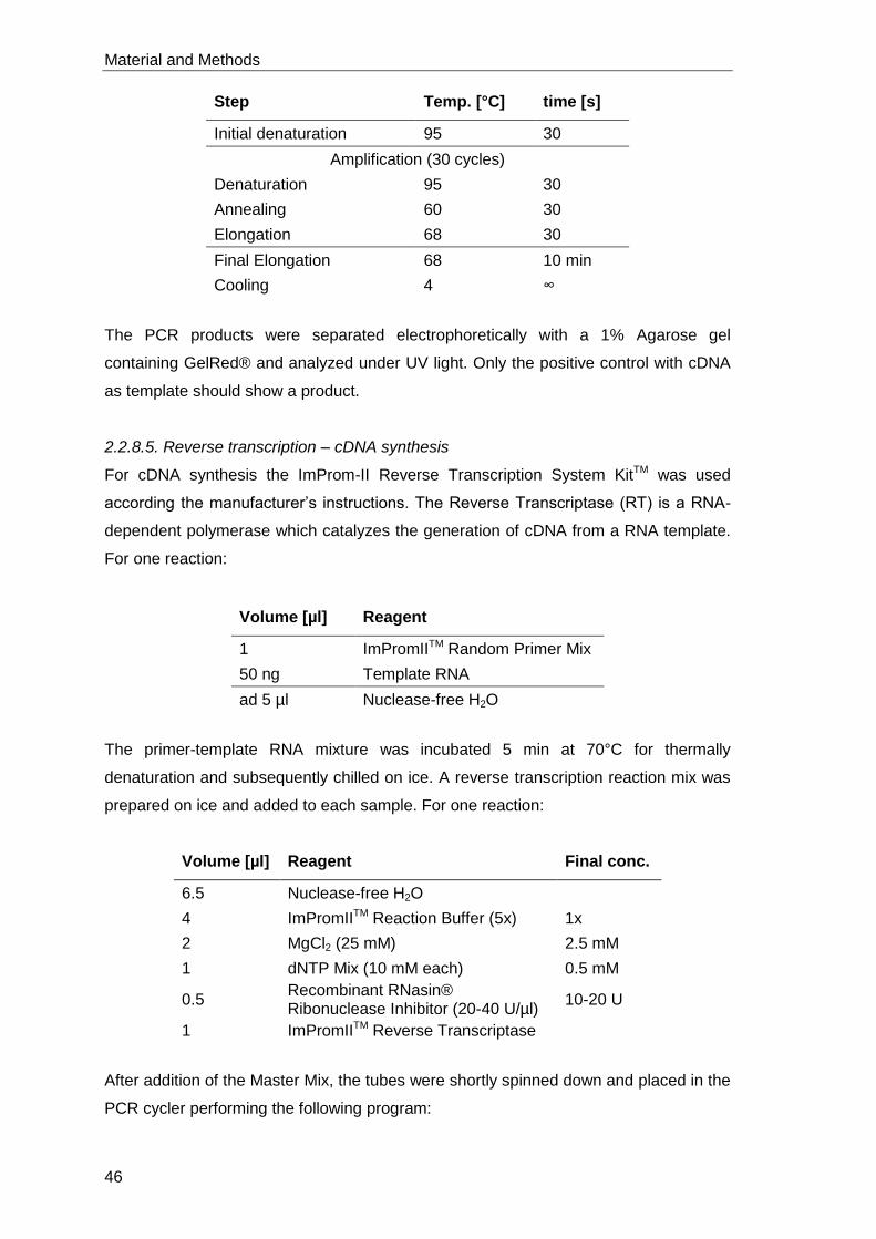
Material and Methods
46
Step Temp. [°C] time [s]
Initial denaturation 95 30
Amplification (30 cycles)
Denaturation 95 30
Annealing 60 30
Elongation 68 30
Final Elongation 68 10 min
Cooling 4 ∞
The PCR products were separated electrophoretically with a 1% Agarose gel
containing GelRed® and analyzed under UV light. Only the positive control with cDNA
as template should show a product.
2.2.8.5. Reverse transcription – cDNA synthesis
For cDNA synthesis the ImProm-II Reverse Transcription System KitTM was used
according the manufacturer’s instructions. The Reverse Transcriptase (RT) is a RNA-
dependent polymerase which catalyzes the generation of cDNA from a RNA template.
For one reaction:
Volume [µl] Reagent
1 ImPromIITM Random Primer Mix
50 ng Template RNA
ad 5 µl Nuclease-free H2O
The primer-template RNA mixture was incubated 5 min at 70°C for thermally
denaturation and subsequently chilled on ice. A reverse transcription reaction mix was
prepared on ice and added to each sample. For one reaction:
Volume [µl] Reagent Final conc.
6.5 Nuclease-free H2O
4 ImPromIITM Reaction Buffer (5x) 1x
2 MgCl2 (25 mM) 2.5 mM
1 dNTP Mix (10 mM each) 0.5 mM
0.5 Recombinant RNasin® Ribonuclease Inhibitor (20-40 U/µl)
10-20 U
1 ImPromIITM Reverse Transcriptase
After addition of the Master Mix, the tubes were shortly spinned down and placed in the
PCR cycler performing the following program:
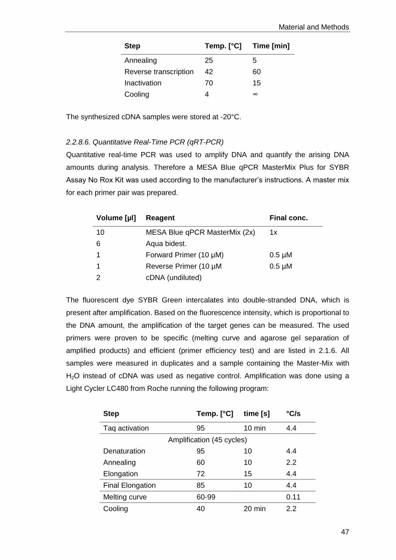
Material and Methods
47
Step Temp. [°C] Time [min]
Annealing 25 5
Reverse transcription 42 60
Inactivation 70 15
Cooling 4 ∞
The synthesized cDNA samples were stored at -20°C.
2.2.8.6. Quantitative Real-Time PCR (qRT-PCR)
Quantitative real-time PCR was used to amplify DNA and quantify the arising DNA
amounts during analysis. Therefore a MESA Blue qPCR MasterMix Plus for SYBR
Assay No Rox Kit was used according to the manufacturer’s instructions. A master mix
for each primer pair was prepared.
Volume [µl] Reagent Final conc.
10 MESA Blue qPCR MasterMix (2x) 1x
6 Aqua bidest.
1 Forward Primer (10 µM) 0.5 µM
1 Reverse Primer (10 µM 0.5 µM
2 cDNA (undiluted)
The fluorescent dye SYBR Green intercalates into double-stranded DNA, which is
present after amplification. Based on the fluorescence intensity, which is proportional to
the DNA amount, the amplification of the target genes can be measured. The used
primers were proven to be specific (melting curve and agarose gel separation of
amplified products) and efficient (primer efficiency test) and are listed in 2.1.6. All
samples were measured in duplicates and a sample containing the Master-Mix with
H2O instead of cDNA was used as negative control. Amplification was done using a
Light Cycler LC480 from Roche running the following program:
Step Temp. [°C] time [s] °C/s
Taq activation 95 10 min 4.4
Amplification (45 cycles)
Denaturation 95 10 4.4
Annealing 60 10 2.2
Elongation 72 15 4.4
Final Elongation 85 10 4.4
Melting curve 60-99 0.11
Cooling 40 20 min 2.2
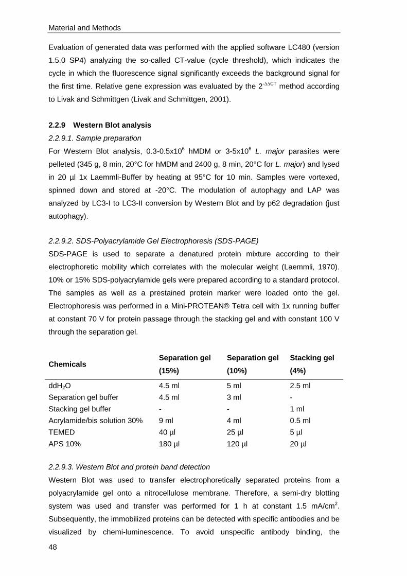
Material and Methods
48
Evaluation of generated data was performed with the applied software LC480 (version
1.5.0 SP4) analyzing the so-called CT-value (cycle threshold), which indicates the
cycle in which the fluorescence signal significantly exceeds the background signal for
the first time. Relative gene expression was evaluated by the 2-∆∆CT method according
to Livak and Schmittgen (Livak and Schmittgen, 2001).
2.2.9 Western Blot analysis
2.2.9.1. Sample preparation
For Western Blot analysis, 0.3-0.5x10⁶ hMDM or 3-5x10⁶ L. major parasites were
pelleted (345 g, 8 min, 20°C for hMDM and 2400 g, 8 min, 20°C for L. major) and lysed
in 20 µl 1x Laemmli-Buffer by heating at 95°C for 10 min. Samples were vortexed,
spinned down and stored at -20°C. The modulation of autophagy and LAP was
analyzed by LC3-I to LC3-II conversion by Western Blot and by p62 degradation (just
autophagy).
2.2.9.2. SDS-Polyacrylamide Gel Electrophoresis (SDS-PAGE)
SDS-PAGE is used to separate a denatured protein mixture according to their
electrophoretic mobility which correlates with the molecular weight (Laemmli, 1970).
10% or 15% SDS-polyacrylamide gels were prepared according to a standard protocol.
The samples as well as a prestained protein marker were loaded onto the gel.
Electrophoresis was performed in a Mini-PROTEAN® Tetra cell with 1x running buffer
at constant 70 V for protein passage through the stacking gel and with constant 100 V
through the separation gel.
Chemicals Separation gel
(15%)
Separation gel
(10%)
Stacking gel
(4%)
ddH2O 4.5 ml 5 ml 2.5 ml
Separation gel buffer 4.5 ml 3 ml -
Stacking gel buffer - - 1 ml
Acrylamide/bis solution 30% 9 ml 4 ml 0.5 ml
TEMED 40 µl 25 µl 5 µl
APS 10% 180 µl 120 µl 20 µl
2.2.9.3. Western Blot and protein band detection
Western Blot was used to transfer electrophoretically separated proteins from a
polyacrylamide gel onto a nitrocellulose membrane. Therefore, a semi-dry blotting
system was used and transfer was performed for 1 h at constant 1.5 mA/cm2.
Subsequently, the immobilized proteins can be detected with specific antibodies and be
visualized by chemi-luminescence. To avoid unspecific antibody binding, the
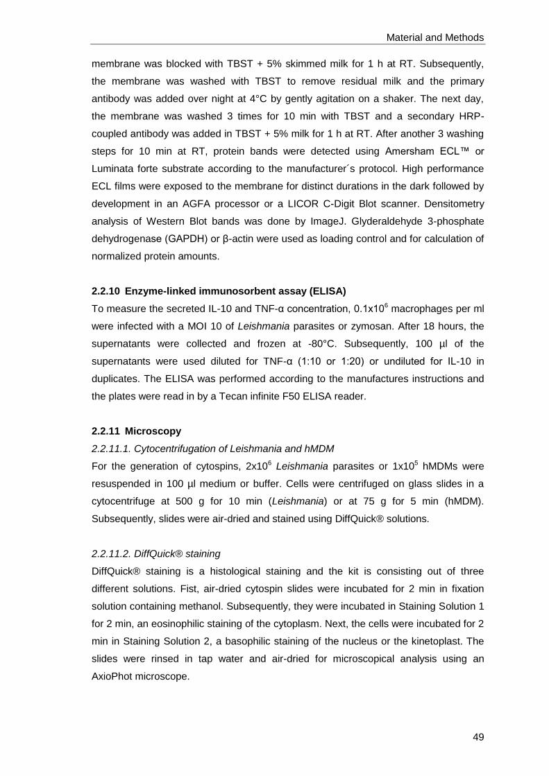
Material and Methods
49
membrane was blocked with TBST + 5% skimmed milk for 1 h at RT. Subsequently,
the membrane was washed with TBST to remove residual milk and the primary
antibody was added over night at 4°C by gently agitation on a shaker. The next day,
the membrane was washed 3 times for 10 min with TBST and a secondary HRP-
coupled antibody was added in TBST + 5% milk for 1 h at RT. After another 3 washing
steps for 10 min at RT, protein bands were detected using Amersham ECL™ or
Luminata forte substrate according to the manufacturer´s protocol. High performance
ECL films were exposed to the membrane for distinct durations in the dark followed by
development in an AGFA processor or a LICOR C-Digit Blot scanner. Densitometry
analysis of Western Blot bands was done by ImageJ. Glyderaldehyde 3-phosphate
dehydrogenase (GAPDH) or β-actin were used as loading control and for calculation of
normalized protein amounts.
2.2.10 Enzyme-linked immunosorbent assay (ELISA)
To measure the secreted IL-10 and TNF-α concentration, 0.1x10⁶ macrophages per ml
were infected with a MOI 10 of Leishmania parasites or zymosan. After 18 hours, the
supernatants were collected and frozen at -80°C. Subsequently, 100 µl of the
supernatants were used diluted for TNF-α (1:10 or 1:20) or undiluted for IL-10 in
duplicates. The ELISA was performed according to the manufactures instructions and
the plates were read in by a Tecan infinite F50 ELISA reader.
2.2.11 Microscopy
2.2.11.1. Cytocentrifugation of Leishmania and hMDM
For the generation of cytospins, 2x10⁶ Leishmania parasites or 1x10⁵ hMDMs were
resuspended in 100 µl medium or buffer. Cells were centrifuged on glass slides in a
cytocentrifuge at 500 g for 10 min (Leishmania) or at 75 g for 5 min (hMDM).
Subsequently, slides were air-dried and stained using DiffQuick® solutions.
2.2.11.2. DiffQuick® staining
DiffQuick® staining is a histological staining and the kit is consisting out of three
different solutions. Fist, air-dried cytospin slides were incubated for 2 min in fixation
solution containing methanol. Subsequently, they were incubated in Staining Solution 1
for 2 min, an eosinophilic staining of the cytoplasm. Next, the cells were incubated for 2
min in Staining Solution 2, a basophilic staining of the nucleus or the kinetoplast. The
slides were rinsed in tap water and air-dried for microscopical analysis using an
AxioPhot microscope.
Material and Methods
50
2.2.11.3. LC3 staining in chamberslides
To assess LC3 recruitment to autophagosomes or phagosomes by
immunofluorescence, 0.1x10⁶ hMDM in 100 µl complete-medium were added per well
in a 12-well chamberslide. The adherence of hMDM was checked microscopically after
30 min at 37°C and 5% CO2. Autophagy was induced by incubation with PI-103,
AZD8055 or Rapamycin (all 10 µM) for 30-40 min at 37°C and 5% CO2.
LC3-associated phagocytosis was induced by incubation with zymosan, plain beads or
PS-Beads for 2 h. After incubation all wells were washed twice with 100 µl prewarmed
wash buffer. Subsequently, the cells were fixated through incubation with 100 µl PBS
containing 4% paraformaldehyde (PFA) for 2 min at 4°C in the dark. After removing the
PFA all wells were washed with 100 µl of Buffer-1 followed by 100 µl of Buffer-2
containing saponin. To each well 100 µl complete-medium containing 183.75 ng/ml
primary antibody (polyclonal rabbit α-LC3) and 2 µl DAPI or 100 µl complete-medium
containing 183.75 ng/ml isotype control (polyclonal rabbit serum) and 2 µl DAPI was
added and incubated for 30 min at 4°C in the dark. Subsequently, the supernatant was
removed and the wells were washed with 100 µl Buffer-2. To each well 100 µl
complete-medium containing 0.5 µl secondary antibody (goat α-rabbit Alexa-Fluor-568)
was added and incubated for 30 min at 4°C in the dark. Subsequently, the wells were
washed with 100 µl Buffer-2 followed by 100 µl of Buffer-1. All liquid was removed
completely. To preserve the sample for microscopically analysis, a cover slip was
applied with 3 drops of ProLong® Gold Antifade Reagent. Surplus ProLong® Gold
Antifade Reagent was removed with cellulose papers. The Chamber Slide was stored
in the dark at 4°C. Analysis of Chamber Slides was performed by a LSM7 Live
microscope with a 630x magnification and by ZEN blue software.
2.2.12 Transduction of hMDM with eGFP-LC3 lentiviral particles
For transduction with lentiviral particles, 2x10⁵ CD14+ separated monocytes (2.2.4.4)
were seeded in chamberslides for microscopic analysis. Adherent macrophages were
transduced with an MOI 0.1 of eGFP-LC3 lentiviral vector particles and incubated for
4 h at 37°C and 5% CO2. Subsequently, a medium change was performed and the
cells were further incubated for 4-5 days at 37°C and 5% CO2 before being used for
stimulation with zymosan particles. The formation of LC3 positive compartments was
analyzed by live cell imaging using a LSM7 Live microscope with a 630x magnification
and by ZEN blue software.
Material and Methods
51
2.2.13 Statistical analysis
All data were shown as mean ± SD (standard deviation). To determine whether
differences were statistically significant, the results were analyzed with a Wilcoxon
matched-pairs signed-rank test and GraphPad Prism software. Values of p < 0.05 (*), p
< 0.01 (**) and p < 0.001 (***) were considered as significant.

Material and Methods
52
Results
53
3 Results
3.1 Autophagy modulation in human primary macrophages
In the first part of this thesis, we focused on the modulation of autophagy in human
primary macrophages using GM-CSF and M-CSF differentiated monocytes as model
phenotypes representing a pro- and an anti-inflammatory macrophage, respectively.
3.1.1 PI-103, AZD8055 and Rapamycin induce autophagy in hMDM
The best known stimulus to induce autophagy is the inhibition of the nutrient sensor
kinase mTOR. Therefore, pro-inflammatory macrophages were stimulated with the
mTOR inhibitors Rapamycin and AZD8055 and with the dual class I PI3K/mTOR
inhibitor PI-103. Subsequently, we assessed the protein expression levels of the
autophagy marker LC3 by immunofluorescence. All chemical autophagy inducers were
shown to induce LC3 protein expression compared to the respective control (Figure
9A). Analyzing the corrected total cell fluorescence (CTCF), significantly augmented
LC3 levels upon PI-103 (1.35 ± 0.8 fold) and AZD8055 (1.39 ± 0.47 fold) treatment
were confirmed indicating no superior effect of dual class I PI3K/mTOR compared to
single mTOR inhibition. Concerning stimulation with Rapamycin, elevated LC3
expression levels were observed (1.13 ± 0.37 fold) (Figure 9B).
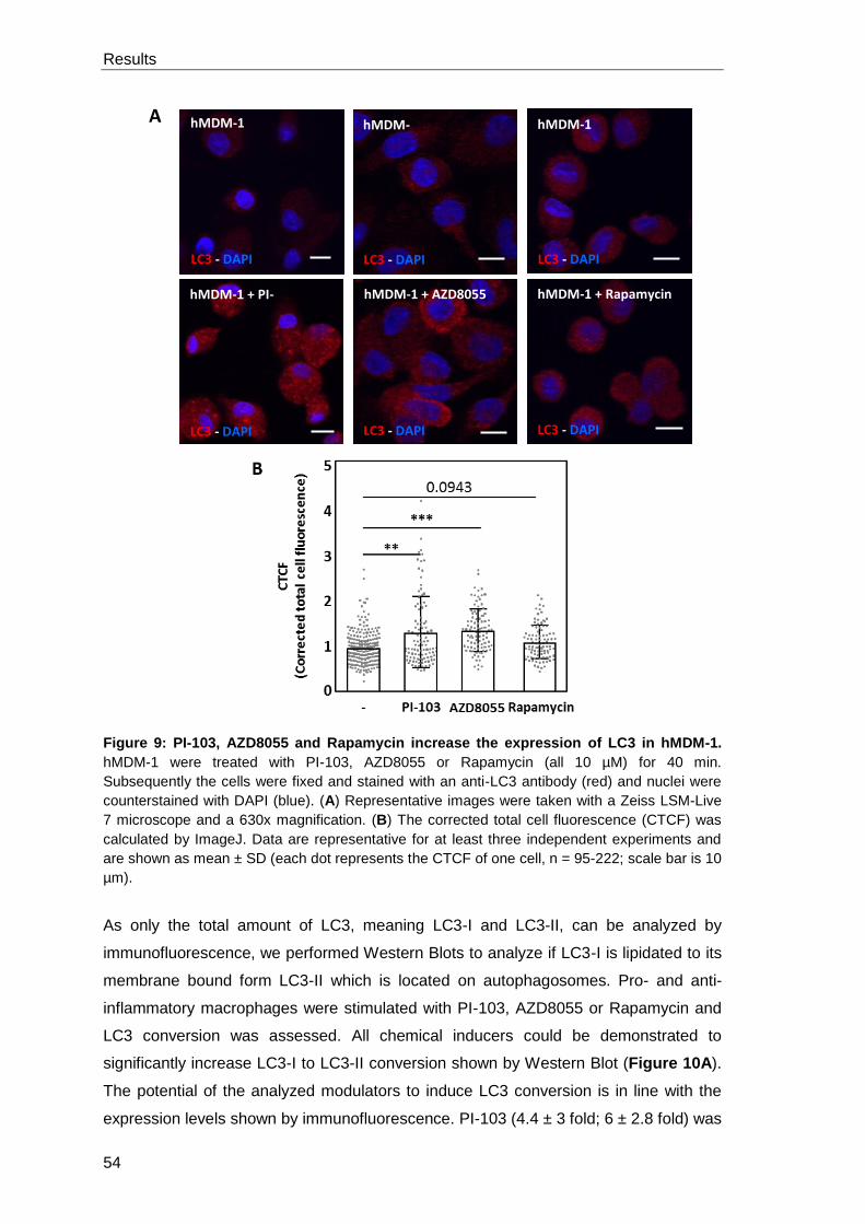
Results
54
Figure 9: PI-103, AZD8055 and Rapamycin increase the expression of LC3 in hMDM-1.
hMDM-1 were treated with PI-103, AZD8055 or Rapamycin (all 10 µM) for 40 min.
Subsequently the cells were fixed and stained with an anti-LC3 antibody (red) and nuclei were
counterstained with DAPI (blue). (A) Representative images were taken with a Zeiss LSM-Live
7 microscope and a 630x magnification. (B) The corrected total cell fluorescence (CTCF) was
calculated by ImageJ. Data are representative for at least three independent experiments and
are shown as mean ± SD (each dot represents the CTCF of one cell, n = 95-222; scale bar is 10
µm).
As only the total amount of LC3, meaning LC3-I and LC3-II, can be analyzed by
immunofluorescence, we performed Western Blots to analyze if LC3-I is lipidated to its
membrane bound form LC3-II which is located on autophagosomes. Pro- and anti-
inflammatory macrophages were stimulated with PI-103, AZD8055 or Rapamycin and
LC3 conversion was assessed. All chemical inducers could be demonstrated to
significantly increase LC3-I to LC3-II conversion shown by Western Blot (Figure 10A).
The potential of the analyzed modulators to induce LC3 conversion is in line with the
expression levels shown by immunofluorescence. PI-103 (4.4 ± 3 fold; 6 ± 2.8 fold) was
hMDM-1
LC3 - DAPI
hMDM-1 + Rapamycin
LC3 - DAPI
hMDM-1
LC3 - DAPI
hMDM1 + PI-103
LC3 - DAPI
hMDM-1
LC3 - DAPI
hMDM-1 + AZD8055
LC3 - DAPI
hMDM-1 + PI-103
LC3 - DAPI
A
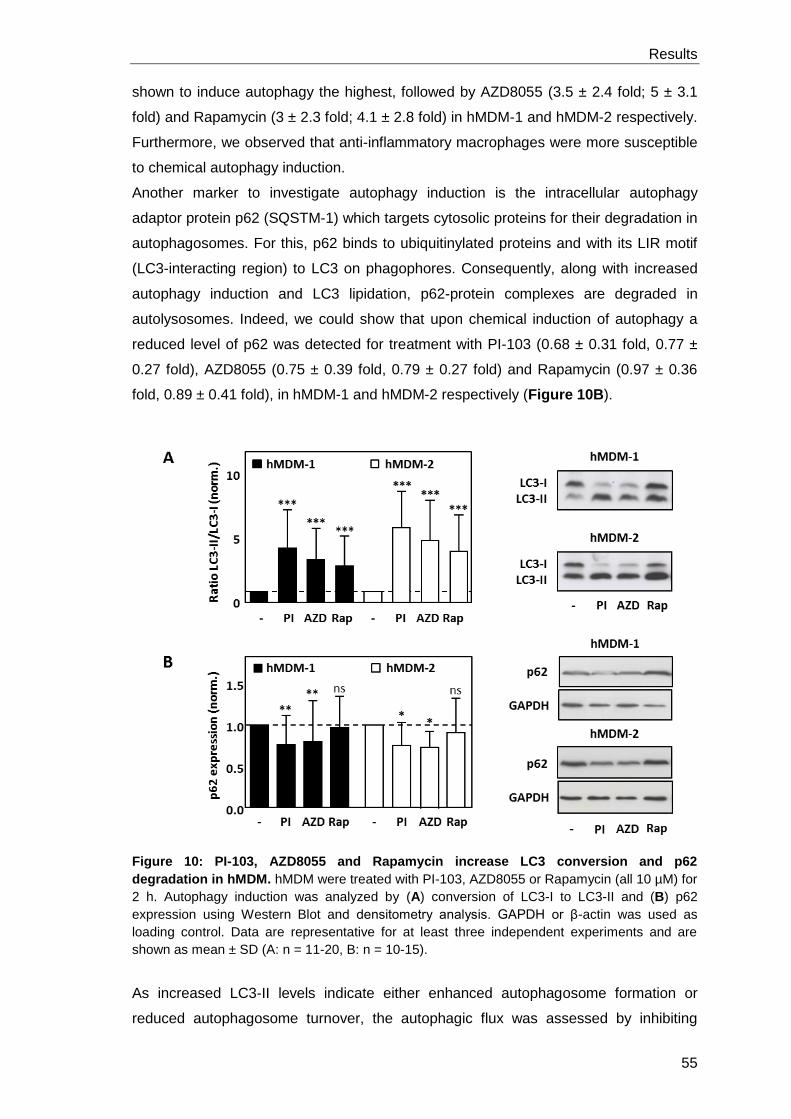
Results
55
shown to induce autophagy the highest, followed by AZD8055 (3.5 ± 2.4 fold; 5 ± 3.1
fold) and Rapamycin (3 ± 2.3 fold; 4.1 ± 2.8 fold) in hMDM-1 and hMDM-2 respectively.
Furthermore, we observed that anti-inflammatory macrophages were more susceptible
to chemical autophagy induction.
Another marker to investigate autophagy induction is the intracellular autophagy
adaptor protein p62 (SQSTM-1) which targets cytosolic proteins for their degradation in
autophagosomes. For this, p62 binds to ubiquitinylated proteins and with its LIR motif
(LC3-interacting region) to LC3 on phagophores. Consequently, along with increased
autophagy induction and LC3 lipidation, p62-protein complexes are degraded in
autolysosomes. Indeed, we could show that upon chemical induction of autophagy a
reduced level of p62 was detected for treatment with PI-103 (0.68 ± 0.31 fold, 0.77 ±
0.27 fold), AZD8055 (0.75 ± 0.39 fold, 0.79 ± 0.27 fold) and Rapamycin (0.97 ± 0.36
fold, 0.89 ± 0.41 fold), in hMDM-1 and hMDM-2 respectively (Figure 10B).
Figure 10: PI-103, AZD8055 and Rapamycin increase LC3 conversion and p62
degradation in hMDM. hMDM were treated with PI-103, AZD8055 or Rapamycin (all 10 µM) for
2 h. Autophagy induction was analyzed by (A) conversion of LC3-I to LC3-II and (B) p62
expression using Western Blot and densitometry analysis. GAPDH or β-actin was used as
loading control. Data are representative for at least three independent experiments and are
shown as mean ± SD (A: n = 11-20, B: n = 10-15).
As increased LC3-II levels indicate either enhanced autophagosome formation or
reduced autophagosome turnover, the autophagic flux was assessed by inhibiting
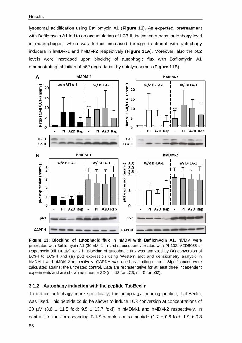
Results
56
lysosomal acidification using Bafilomycin A1 (Figure 11). As expected, pretreatment
with Bafilomycin A1 led to an accumulation of LC3-II, indicating a basal autophagy level
in macrophages, which was further increased through treatment with autophagy
inducers in hMDM-1 and hMDM-2 respectively (Figure 11A). Moreover, also the p62
levels were increased upon blocking of autophagic flux with Bafilomycin A1
demonstrating inhibition of p62 degradation by autolysosomes (Figure 11B).
Figure 11: Blocking of autophagic flux in hMDM with Bafilomycin A1. hMDM were
pretreated with Bafilomycin A1 (30 nM, 1 h) and subsequently treated with PI-103, AZD8055 or
Rapamycin (all 10 µM) for 2 h. Blocking of autophagic flux was analyzed by (A) conversion of
LC3-I to LC3-II and (B) p62 expression using Western Blot and densitometry analysis in
hMDM-1 and hMDM-2 respectively. GAPDH was used as loading control. Significances were
calculated against the untreated control. Data are representative for at least three independent
experiments and are shown as mean ± SD (n = 12 for LC3, n = 5 for p62).
3.1.2 Autophagy induction with the peptide Tat-Beclin
To induce autophagy more specifically, the autophagy inducing peptide, Tat-Beclin,
was used. This peptide could be shown to induce LC3 conversion at concentrations of
30 µM (8.6 ± 11.5 fold; 9.5 ± 13.7 fold) in hMDM-1 and hMDM-2 respectively, in
contrast to the corresponding Tat-Scramble control peptide (1.7 ± 0.6 fold; 1.9 ± 0.8
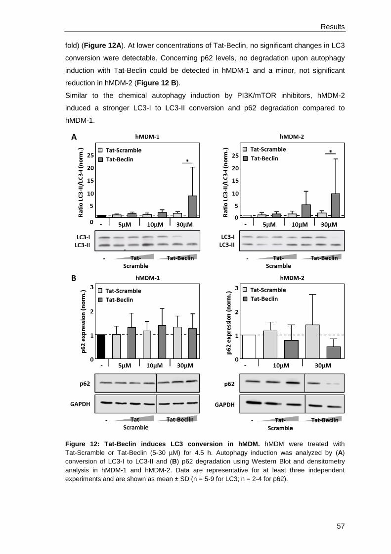
Results
57
fold) (Figure 12A). At lower concentrations of Tat-Beclin, no significant changes in LC3
conversion were detectable. Concerning p62 levels, no degradation upon autophagy
induction with Tat-Beclin could be detected in hMDM-1 and a minor, not significant
reduction in hMDM-2 (Figure 12 B).
Similar to the chemical autophagy induction by PI3K/mTOR inhibitors, hMDM-2
induced a stronger LC3-I to LC3-II conversion and p62 degradation compared to
hMDM-1.
Figure 12: Tat-Beclin induces LC3 conversion in hMDM. hMDM were treated with
Tat-Scramble or Tat-Beclin (5-30 µM) for 4.5 h. Autophagy induction was analyzed by (A)
conversion of LC3-I to LC3-II and (B) p62 degradation using Western Blot and densitometry
analysis in hMDM-1 and hMDM-2. Data are representative for at least three independent
experiments and are shown as mean ± SD (n = 5-9 for LC3; n = 2-4 for p62).
Results
58
3.1.3 Inhibition of class III PI3 kinase pathway by Wortmannin and Spautin-1
blocks autophagy induction in hMDM
Next, we aimed to modulate autophagy negatively using the class III PI3-kinase
inhibitors 3-Methyladenine, Wortmannin and LY294002. Furthermore, Spautin-1, which
targets Beclin-1 for proteasomal degradation, is used.
To identify the potential of 3-Methyladenine, Wortmannin, LY294002 and Spautin-1 to
inhibit autophagy, macrophages were pretreated with the inhibitors and subsequently
autophagy was induced using PI-103, AZD8055 or Rapamycin in hMDM-1 (Figure
13A) and hMDM-2 (Figure 13B). By assessing LC3 conversion in PI-103 treated
hMDM (5.5 ± 4 fold; 7 ± 5.1 fold), pretreatment with Spautin-1 (1.6 ± 0.9 fold; 2.0 ± 1.4
fold) and Wortmannin (1.8 ± 1.3 fold and 3 ± 2.8 fold) could be demonstrated to
decrease the cellular LC3 conversion levels significantly in both hMDM-1 and hMDM-2,
respectively. Remarkably, pretreatment with 3-Methyladenine even led to an increased
autophagy induction in hMDM-1 (11.3 ± 8.6 fold) and just a minor decrease in hMDM-2
(6.2 ± 2.6 fold). Interestingly, the preincubation of macrophages with LY294002
followed by autophagy induction was able to block LC3 conversion in hMDM-1 (2.8 ±
1.8 fold) but induced the conversion in hMDM-2 (8.7 ± 5.9 fold).
Comparable results were obtained by the application of Rapamycin or AZD8055 as
inducing stimuli. Taken together, autophagy induction was successfully inhibited by the
pretreatment with Spautin-1, Wortmannin and LY294002 in hMDM-1 and with
Spautin-1 and Wortmannin in hMDM-2.
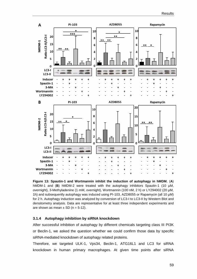
Results
59
Figure 13: Spautin-1 and Wortmannin inhibit the induction of autophagy in hMDM. (A)
hMDM-1 and (B) hMDM-2 were treated with the autophagy inhibitors Spautin-1 (10 µM,
overnight), 3-Methyladenine (1 mM, overnight), Wortmannin (100 nM, 2 h) or LY294002 (20 µM,
1h) and subsequently autophagy was induced using PI-103, AZD8055 or Rapamycin (all 10 µM)
for 2 h. Autophagy induction was analyzed by conversion of LC3-I to LC3-II by Western Blot and
densitometry analysis. Data are representative for at least three independent experiments and
are shown as mean ± SD (n = 5-12).
3.1.4 Autophagy inhibition by siRNA knockdown
After successful inhibition of autophagy by different chemicals targeting class III PI3K
or Beclin-1, we asked the question whether we could confirm those data by specific
siRNA-mediated knockdown of autophagy related proteins.
Therefore, we targeted ULK-1, Vps34, Beclin-1, ATG16L1 and LC3 for siRNA
knockdown in human primary macrophages. At given time points after siRNA
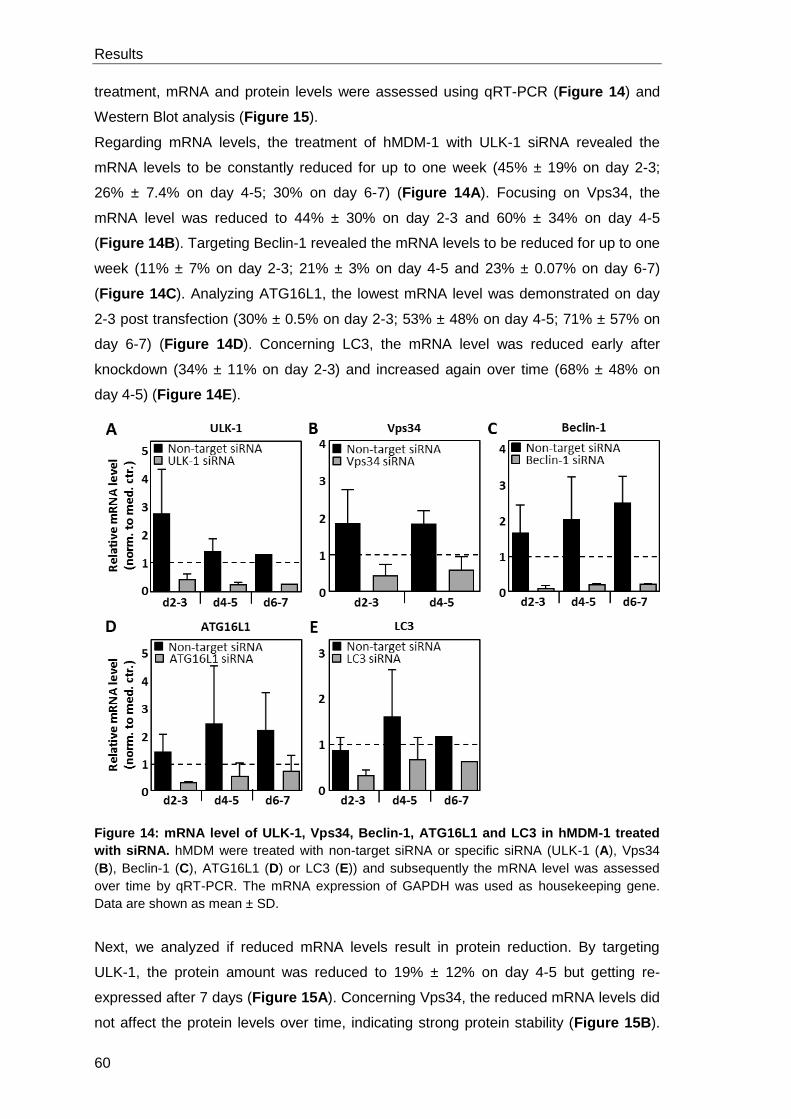
Results
60
treatment, mRNA and protein levels were assessed using qRT-PCR (Figure 14) and
Western Blot analysis (Figure 15).
Regarding mRNA levels, the treatment of hMDM-1 with ULK-1 siRNA revealed the
mRNA levels to be constantly reduced for up to one week (45% ± 19% on day 2-3;
26% ± 7.4% on day 4-5; 30% on day 6-7) (Figure 14A). Focusing on Vps34, the
mRNA level was reduced to 44% ± 30% on day 2-3 and 60% ± 34% on day 4-5
(Figure 14B). Targeting Beclin-1 revealed the mRNA levels to be reduced for up to one
week (11% ± 7% on day 2-3; 21% ± 3% on day 4-5 and 23% ± 0.07% on day 6-7)
(Figure 14C). Analyzing ATG16L1, the lowest mRNA level was demonstrated on day
2-3 post transfection (30% ± 0.5% on day 2-3; 53% ± 48% on day 4-5; 71% ± 57% on
day 6-7) (Figure 14D). Concerning LC3, the mRNA level was reduced early after
knockdown (34% ± 11% on day 2-3) and increased again over time (68% ± 48% on
day 4-5) (Figure 14E).
Figure 14: mRNA level of ULK-1, Vps34, Beclin-1, ATG16L1 and LC3 in hMDM-1 treated
with siRNA. hMDM were treated with non-target siRNA or specific siRNA (ULK-1 (A), Vps34
(B), Beclin-1 (C), ATG16L1 (D) or LC3 (E)) and subsequently the mRNA level was assessed
over time by qRT-PCR. The mRNA expression of GAPDH was used as housekeeping gene.
Data are shown as mean ± SD.
Next, we analyzed if reduced mRNA levels result in protein reduction. By targeting
ULK-1, the protein amount was reduced to 19% ± 12% on day 4-5 but getting re-
expressed after 7 days (Figure 15A). Concerning Vps34, the reduced mRNA levels did
not affect the protein levels over time, indicating strong protein stability (Figure 15B).
Results
61
By targeting Beclin-1 it took up to 7 days until the protein amount is reduced to 39% ±
32% compared to the medium control (Figure 15C). The same was observed for
ATG16L1, the protein levels did not decrease at early time points after knockdown
(90% ± 35% on day 2-3) but dropped strongly after 7 days to 35% ± 12% (Figure 15D).
Focusing on LC3, the protein level was reduced early after knockdown (25% ± 10% on
day 2-3 and increased again over time (68% ± 56 % on day 4-5) (Figure 15E).
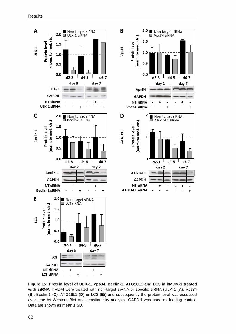
Results
62
Figure 15: Protein level of ULK-1, Vps34, Beclin-1, ATG16L1 and LC3 in hMDM-1 treated
with siRNA. hMDM were treated with non-target siRNA or specific siRNA (ULK-1 (A), Vps34
(B), Beclin-1 (C), ATG16L1 (D) or LC3 (E)) and subsequently the protein level was assessed
over time by Western Blot and densitometry analysis. GAPDH was used as loading control.
Data are shown as mean ± SD.
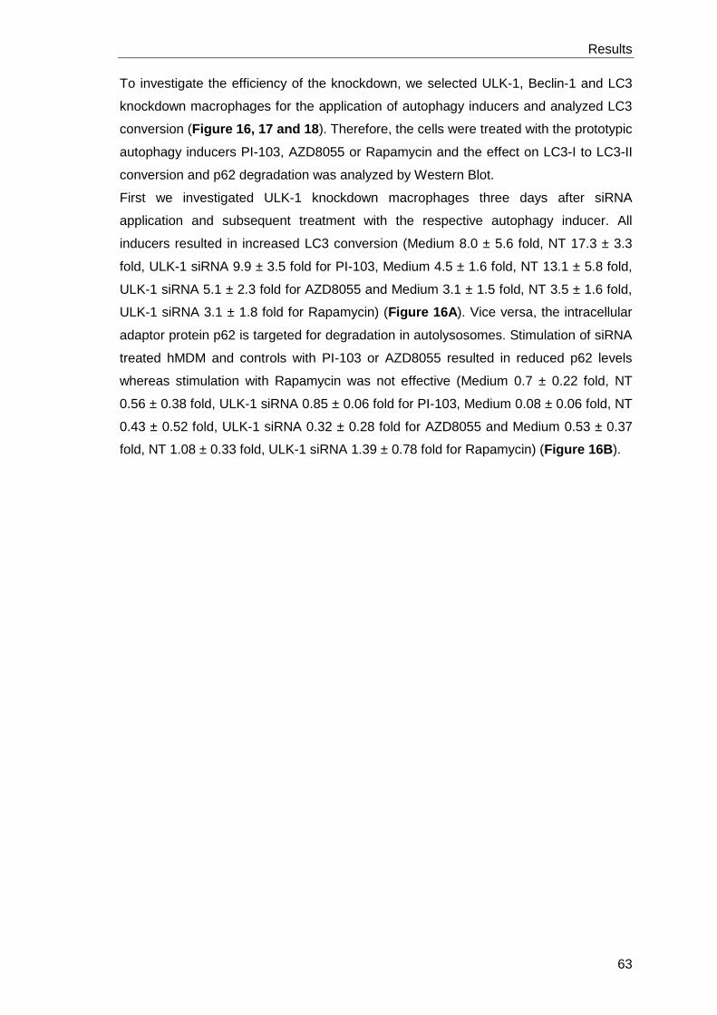
Results
63
To investigate the efficiency of the knockdown, we selected ULK-1, Beclin-1 and LC3
knockdown macrophages for the application of autophagy inducers and analyzed LC3
conversion (Figure 16, 17 and 18). Therefore, the cells were treated with the prototypic
autophagy inducers PI-103, AZD8055 or Rapamycin and the effect on LC3-I to LC3-II
conversion and p62 degradation was analyzed by Western Blot.
First we investigated ULK-1 knockdown macrophages three days after siRNA
application and subsequent treatment with the respective autophagy inducer. All
inducers resulted in increased LC3 conversion (Medium 8.0 ± 5.6 fold, NT 17.3 ± 3.3
fold, ULK-1 siRNA 9.9 ± 3.5 fold for PI-103, Medium 4.5 ± 1.6 fold, NT 13.1 ± 5.8 fold,
ULK-1 siRNA 5.1 ± 2.3 fold for AZD8055 and Medium 3.1 ± 1.5 fold, NT 3.5 ± 1.6 fold,
ULK-1 siRNA 3.1 ± 1.8 fold for Rapamycin) (Figure 16A). Vice versa, the intracellular
adaptor protein p62 is targeted for degradation in autolysosomes. Stimulation of siRNA
treated hMDM and controls with PI-103 or AZD8055 resulted in reduced p62 levels
whereas stimulation with Rapamycin was not effective (Medium 0.7 ± 0.22 fold, NT
0.56 ± 0.38 fold, ULK-1 siRNA 0.85 ± 0.06 fold for PI-103, Medium 0.08 ± 0.06 fold, NT
0.43 ± 0.52 fold, ULK-1 siRNA 0.32 ± 0.28 fold for AZD8055 and Medium 0.53 ± 0.37
fold, NT 1.08 ± 0.33 fold, ULK-1 siRNA 1.39 ± 0.78 fold for Rapamycin) (Figure 16B).
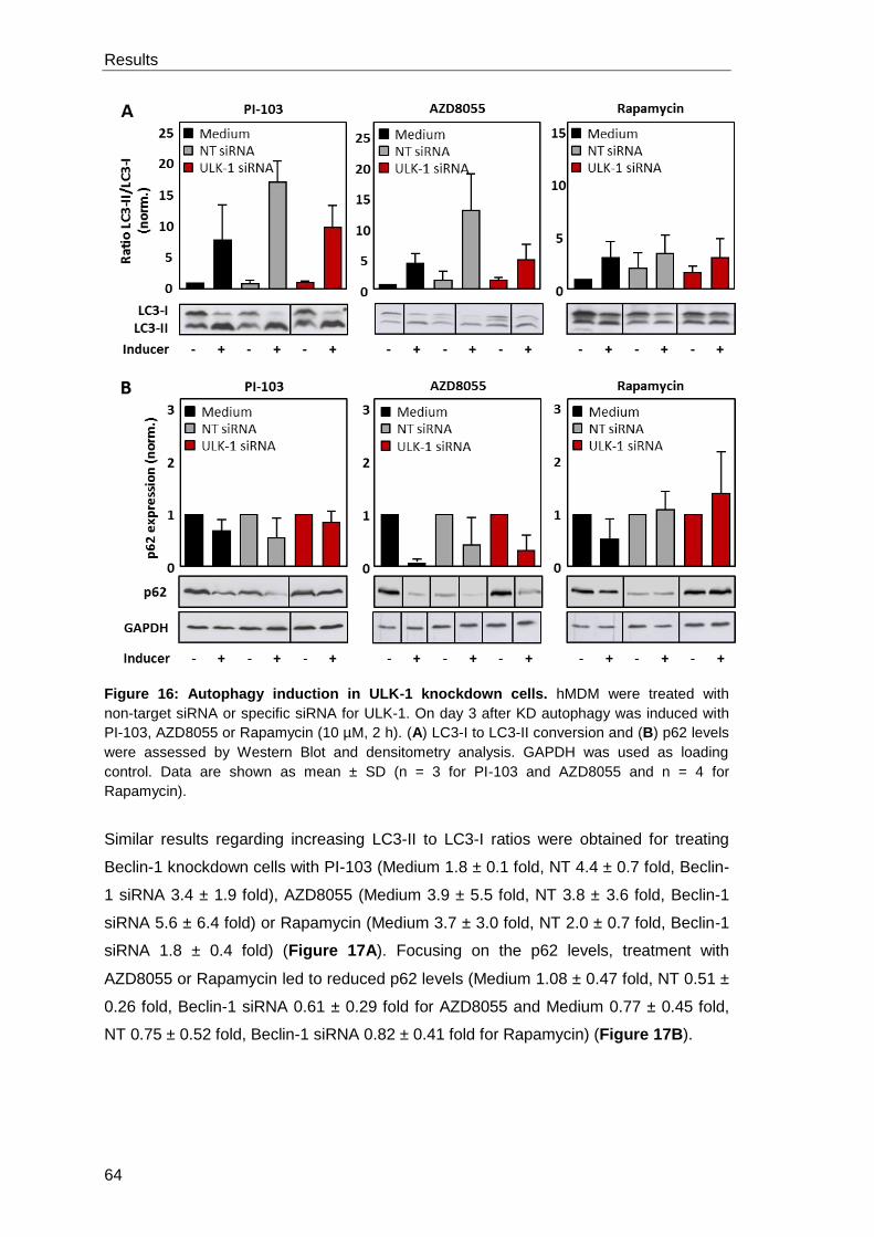
Results
64
Figure 16: Autophagy induction in ULK-1 knockdown cells. hMDM were treated with
non-target siRNA or specific siRNA for ULK-1. On day 3 after KD autophagy was induced with
PI-103, AZD8055 or Rapamycin (10 µM, 2 h). (A) LC3-I to LC3-II conversion and (B) p62 levels
were assessed by Western Blot and densitometry analysis. GAPDH was used as loading
control. Data are shown as mean ± SD (n = 3 for PI-103 and AZD8055 and n = 4 for
Rapamycin).
Similar results regarding increasing LC3-II to LC3-I ratios were obtained for treating
Beclin-1 knockdown cells with PI-103 (Medium 1.8 ± 0.1 fold, NT 4.4 ± 0.7 fold, Beclin-
1 siRNA 3.4 ± 1.9 fold), AZD8055 (Medium 3.9 ± 5.5 fold, NT 3.8 ± 3.6 fold, Beclin-1
siRNA 5.6 ± 6.4 fold) or Rapamycin (Medium 3.7 ± 3.0 fold, NT 2.0 ± 0.7 fold, Beclin-1
siRNA 1.8 ± 0.4 fold) (Figure 17A). Focusing on the p62 levels, treatment with
AZD8055 or Rapamycin led to reduced p62 levels (Medium 1.08 ± 0.47 fold, NT 0.51 ±
0.26 fold, Beclin-1 siRNA 0.61 ± 0.29 fold for AZD8055 and Medium 0.77 ± 0.45 fold,
NT 0.75 ± 0.52 fold, Beclin-1 siRNA 0.82 ± 0.41 fold for Rapamycin) (Figure 17B).

Results
65
Figure 17: Autophagy induction in Beclin-1 knockdown cells. hMDM were treated with
non-target siRNA or specific siRNA for Beclin-1. On day 7 after KD autophagy was induced with
PI-103, AZD8055 or Rapamycin (10 µM, 2 h). (A) LC3-I to LC3-II conversion and (B) p62 levels
were assessed by Western Blot and densitometry analysis. GAPDH was used as loading
control. Data are shown as mean ± SD (n = 2 for PI-103; n = 5 (LC3) and n = 3 (p62) for
AZD8055 and Rapamycin).
Furthermore, the autophagy marker LC3 was targeted for siRNA knockdown as well.
Stimulation of siRNA treated hMDM and controls with PI-103 resulted in reduced p62
levels in Medium and NT-treated cells but not in LC3 knockdown cells (Medium 0.7 ±
0.22 fold, NT 0.55 ± 0.38 fold, LC3 siRNA 1.11 ± 0.41 fold) (Figure 18).
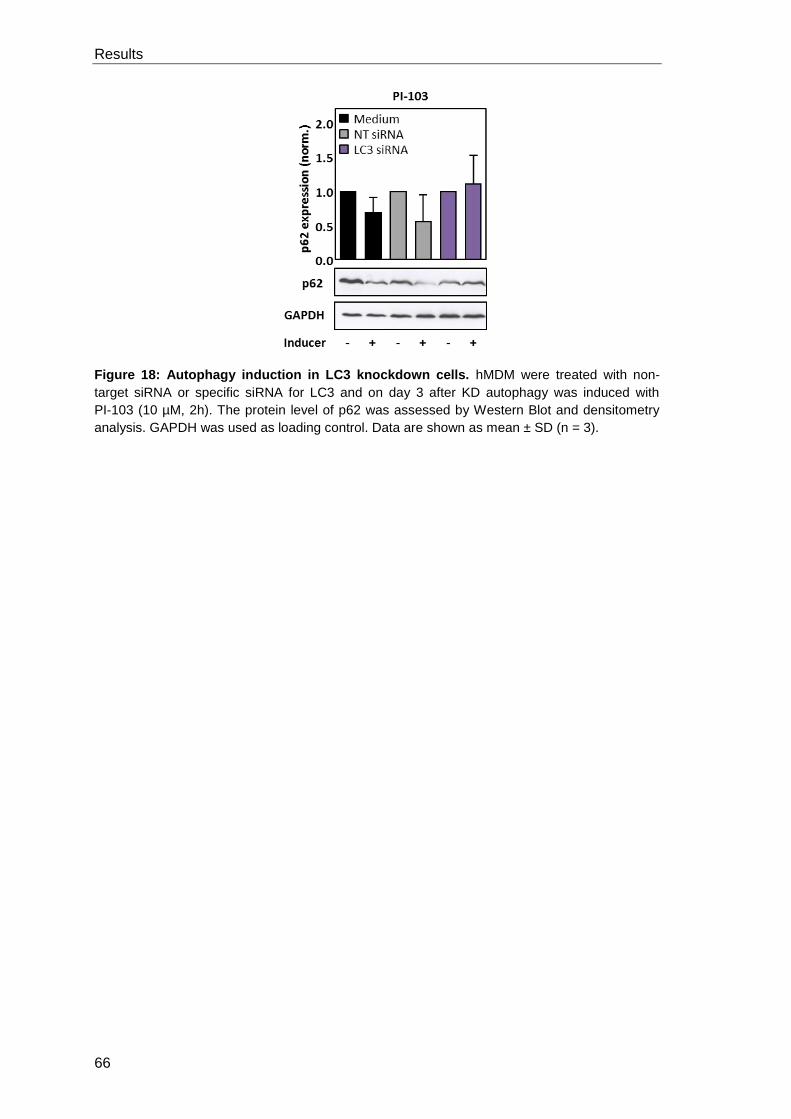
Results
66
Figure 18: Autophagy induction in LC3 knockdown cells. hMDM were treated with non-
target siRNA or specific siRNA for LC3 and on day 3 after KD autophagy was induced with
PI-103 (10 µM, 2h). The protein level of p62 was assessed by Western Blot and densitometry
analysis. GAPDH was used as loading control. Data are shown as mean ± SD (n = 3).
Results
67
3.2 Effect of autophagy modulation on antigen processing using the
recall antigen Tetanus Toxoid
Macrophages are immunomodulatory cells which are involved in antigen processing
and presentation to the adaptive immune system. In addition, various studies
demonstrated the involvement of autophagic degradation in antigen processing for
MHC class II presentation (Schmid and Munz, 2007; Munz, 2016a). Therefore, we
aimed to analyze the effect of autophagy modulation on antigen processing using the
recall antigen Tetanus Toxoid (TT) as a model antigen. Hence, autophagy in human
primary macrophages was induced by AZD8055 or inhibited by siRNA knockdown of
ULK-1 and the effect on lymphocyte proliferation in response to Tetanus Toxoid
stimulation was analyzed.
3.2.1 Characterization of the Tetanus Toxoid specific T cell proliferation
First of all, the immune response against stimulation of hMDM with Tetanus Toxoid
was characterized to ensure that antigens undergo processing and are presented via
MHC-II to CD4+ T cells. Hence, a proliferation kinetic in response to different TT
concentrations was assessed by a CFSE-based proliferation assay (Figure 19).
Therefore, macrophages were stimulated with TT overnight and subsequently co-
cultured with autologous, CFSE-labeled peripheral blood lymphocytes (PBLs) for 6
days. Upon T cell activation, half of the initial CFSE molecules are transferred to the
daughter cell with each cell division. Accordingly, lymphocyte proliferation can be
analyzed by measuring the percentage of CFSElow proliferating cells.
After three days of co-cultivation, lymphocyte proliferation arises and is steadily
increasing until day 6, reaching 15-20%. Analyzing longer periods of co-cultivation
revealed unspecific proliferation in the unstimulated controls (data not shown). There
were no significant changes observed regarding the applied concentrations. For the
following experiments a concentration of 10 µg/ml was used and proliferation was
assessed on day 6 post co-cultivation.
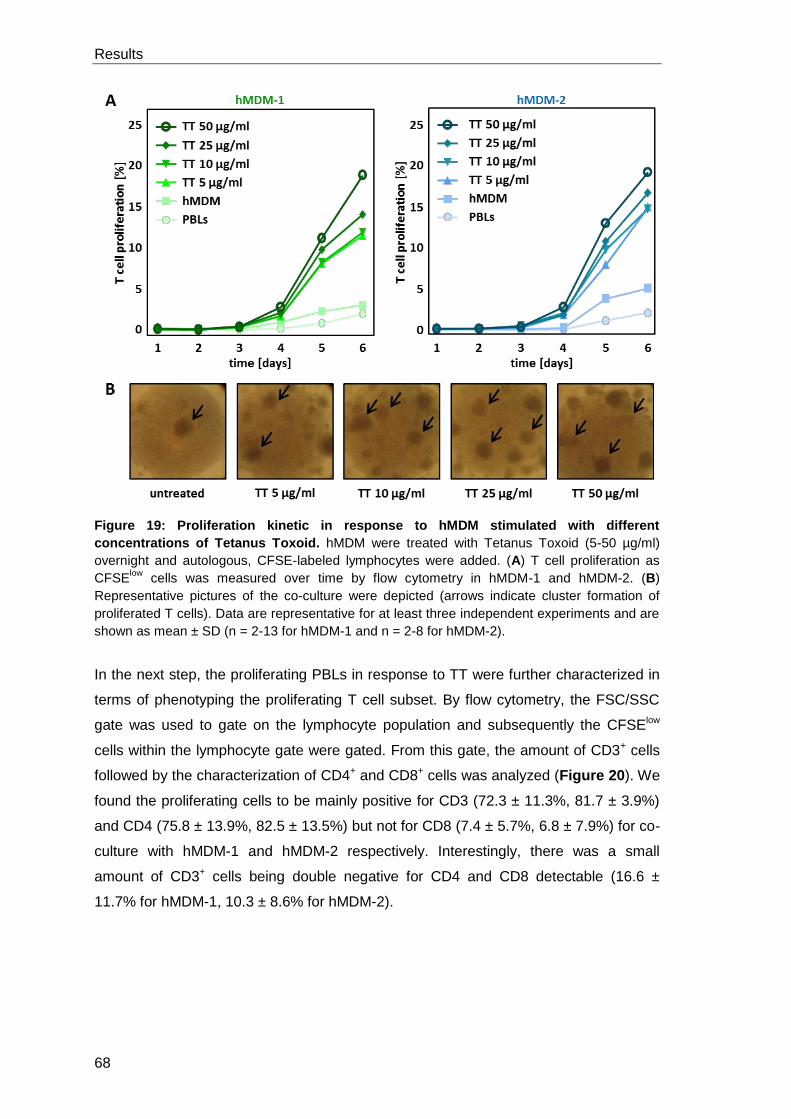
Results
68
Figure 19: Proliferation kinetic in response to hMDM stimulated with different
concentrations of Tetanus Toxoid. hMDM were treated with Tetanus Toxoid (5-50 µg/ml)
overnight and autologous, CFSE-labeled lymphocytes were added. (A) T cell proliferation as
CFSElow
cells was measured over time by flow cytometry in hMDM-1 and hMDM-2. (B)
Representative pictures of the co-culture were depicted (arrows indicate cluster formation of
proliferated T cells). Data are representative for at least three independent experiments and are
shown as mean ± SD (n = 2-13 for hMDM-1 and n = 2-8 for hMDM-2).
In the next step, the proliferating PBLs in response to TT were further characterized in
terms of phenotyping the proliferating T cell subset. By flow cytometry, the FSC/SSC
gate was used to gate on the lymphocyte population and subsequently the CFSElow
cells within the lymphocyte gate were gated. From this gate, the amount of CD3+ cells
followed by the characterization of CD4+ and CD8+ cells was analyzed (Figure 20). We
found the proliferating cells to be mainly positive for CD3 (72.3 ± 11.3%, 81.7 ± 3.9%)
and CD4 (75.8 ± 13.9%, 82.5 ± 13.5%) but not for CD8 (7.4 ± 5.7%, 6.8 ± 7.9%) for co-
culture with hMDM-1 and hMDM-2 respectively. Interestingly, there was a small
amount of CD3+ cells being double negative for CD4 and CD8 detectable (16.6 ±
11.7% for hMDM-1, 10.3 ± 8.6% for hMDM-2).
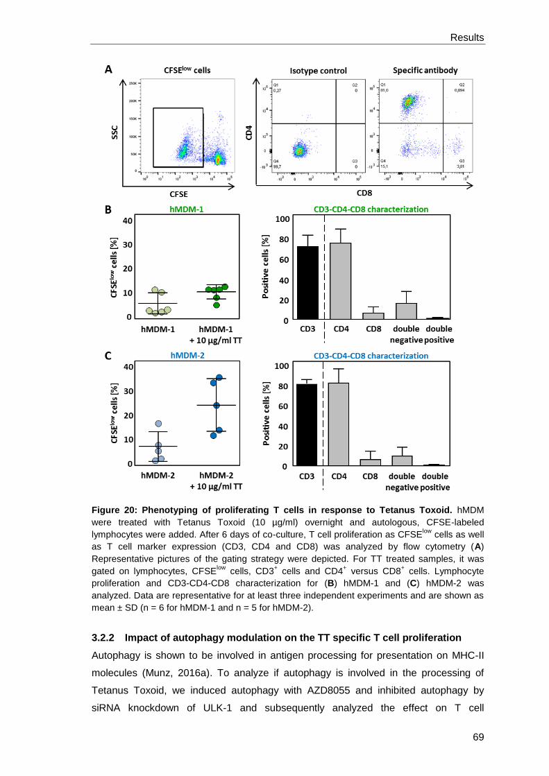
Results
69
Figure 20: Phenotyping of proliferating T cells in response to Tetanus Toxoid. hMDM
were treated with Tetanus Toxoid (10 µg/ml) overnight and autologous, CFSE-labeled
lymphocytes were added. After 6 days of co-culture, T cell proliferation as CFSElow
cells as well
as T cell marker expression (CD3, CD4 and CD8) was analyzed by flow cytometry (A)
Representative pictures of the gating strategy were depicted. For TT treated samples, it was
gated on lymphocytes, CFSElow
cells, CD3+ cells and CD4
+ versus CD8
+ cells. Lymphocyte
proliferation and CD3-CD4-CD8 characterization for (B) hMDM-1 and (C) hMDM-2 was
analyzed. Data are representative for at least three independent experiments and are shown as
mean ± SD (n = 6 for hMDM-1 and n = 5 for hMDM-2).
3.2.2 Impact of autophagy modulation on the TT specific T cell proliferation
Autophagy is shown to be involved in antigen processing for presentation on MHC-II
molecules (Munz, 2016a). To analyze if autophagy is involved in the processing of
Tetanus Toxoid, we induced autophagy with AZD8055 and inhibited autophagy by
siRNA knockdown of ULK-1 and subsequently analyzed the effect on T cell
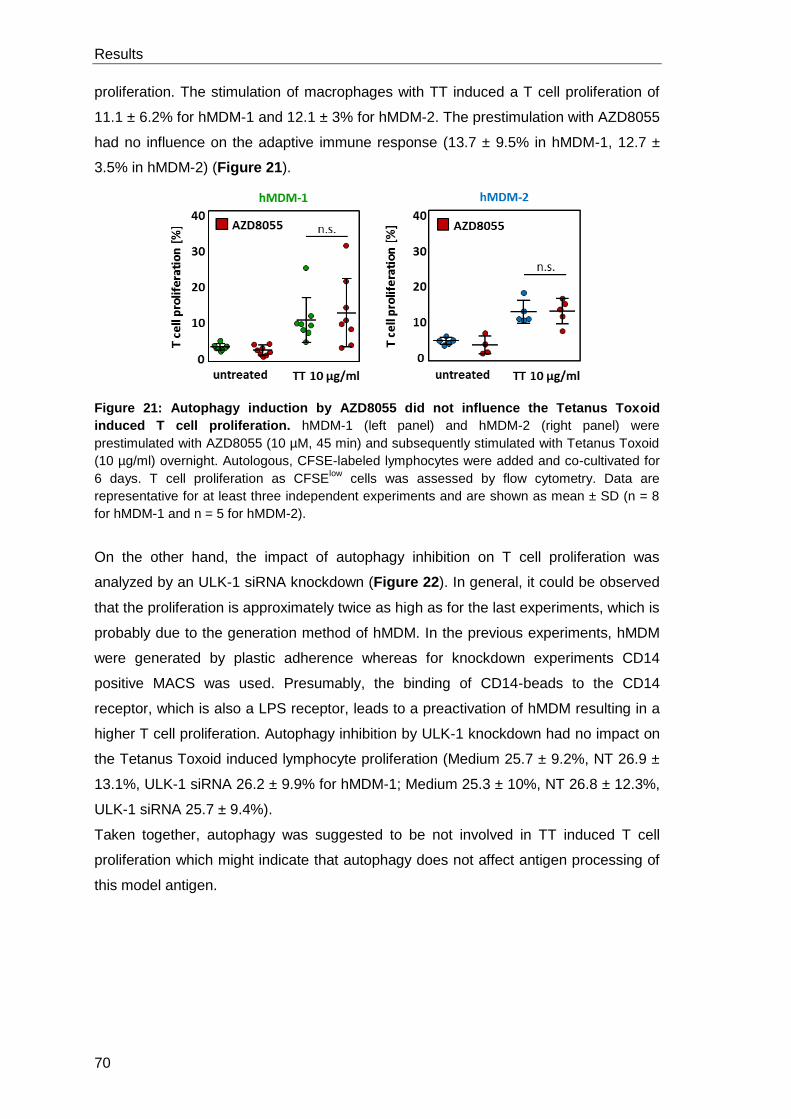
Results
70
proliferation. The stimulation of macrophages with TT induced a T cell proliferation of
11.1 ± 6.2% for hMDM-1 and 12.1 ± 3% for hMDM-2. The prestimulation with AZD8055
had no influence on the adaptive immune response (13.7 ± 9.5% in hMDM-1, 12.7 ±
3.5% in hMDM-2) (Figure 21).
Figure 21: Autophagy induction by AZD8055 did not influence the Tetanus Toxoid
induced T cell proliferation. hMDM-1 (left panel) and hMDM-2 (right panel) were
prestimulated with AZD8055 (10 µM, 45 min) and subsequently stimulated with Tetanus Toxoid
(10 µg/ml) overnight. Autologous, CFSE-labeled lymphocytes were added and co-cultivated for
6 days. T cell proliferation as CFSElow
cells was assessed by flow cytometry. Data are
representative for at least three independent experiments and are shown as mean ± SD (n = 8
for hMDM-1 and n = 5 for hMDM-2).
On the other hand, the impact of autophagy inhibition on T cell proliferation was
analyzed by an ULK-1 siRNA knockdown (Figure 22). In general, it could be observed
that the proliferation is approximately twice as high as for the last experiments, which is
probably due to the generation method of hMDM. In the previous experiments, hMDM
were generated by plastic adherence whereas for knockdown experiments CD14
positive MACS was used. Presumably, the binding of CD14-beads to the CD14
receptor, which is also a LPS receptor, leads to a preactivation of hMDM resulting in a
higher T cell proliferation. Autophagy inhibition by ULK-1 knockdown had no impact on
the Tetanus Toxoid induced lymphocyte proliferation (Medium 25.7 ± 9.2%, NT 26.9 ±
13.1%, ULK-1 siRNA 26.2 ± 9.9% for hMDM-1; Medium 25.3 ± 10%, NT 26.8 ± 12.3%,
ULK-1 siRNA 25.7 ± 9.4%).
Taken together, autophagy was suggested to be not involved in TT induced T cell
proliferation which might indicate that autophagy does not affect antigen processing of
this model antigen.
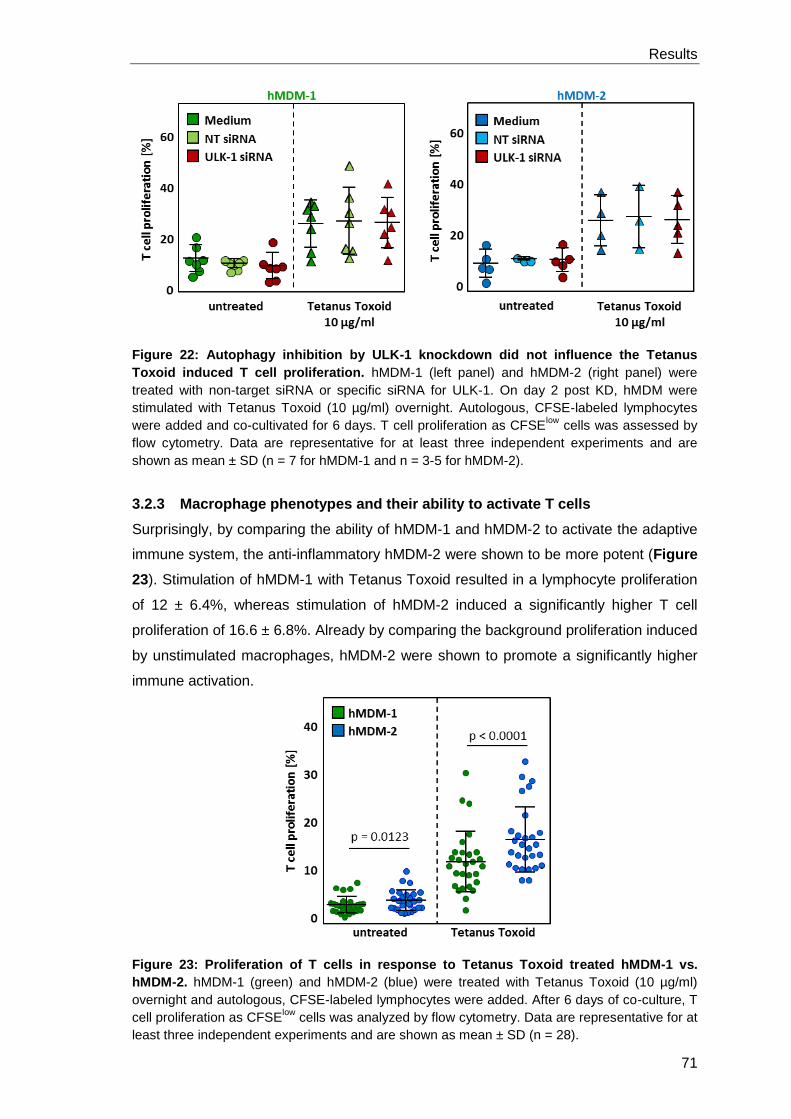
Results
71
Figure 22: Autophagy inhibition by ULK-1 knockdown did not influence the Tetanus
Toxoid induced T cell proliferation. hMDM-1 (left panel) and hMDM-2 (right panel) were
treated with non-target siRNA or specific siRNA for ULK-1. On day 2 post KD, hMDM were
stimulated with Tetanus Toxoid (10 µg/ml) overnight. Autologous, CFSE-labeled lymphocytes
were added and co-cultivated for 6 days. T cell proliferation as CFSElow
cells was assessed by
flow cytometry. Data are representative for at least three independent experiments and are
shown as mean ± SD (n = 7 for hMDM-1 and n = 3-5 for hMDM-2).
3.2.3 Macrophage phenotypes and their ability to activate T cells
Surprisingly, by comparing the ability of hMDM-1 and hMDM-2 to activate the adaptive
immune system, the anti-inflammatory hMDM-2 were shown to be more potent (Figure
23). Stimulation of hMDM-1 with Tetanus Toxoid resulted in a lymphocyte proliferation
of 12 ± 6.4%, whereas stimulation of hMDM-2 induced a significantly higher T cell
proliferation of 16.6 ± 6.8%. Already by comparing the background proliferation induced
by unstimulated macrophages, hMDM-2 were shown to promote a significantly higher
immune activation.
Figure 23: Proliferation of T cells in response to Tetanus Toxoid treated hMDM-1 vs.
hMDM-2. hMDM-1 (green) and hMDM-2 (blue) were treated with Tetanus Toxoid (10 µg/ml)
overnight and autologous, CFSE-labeled lymphocytes were added. After 6 days of co-culture, T
cell proliferation as CFSElow
cells was analyzed by flow cytometry. Data are representative for at
least three independent experiments and are shown as mean ± SD (n = 28).
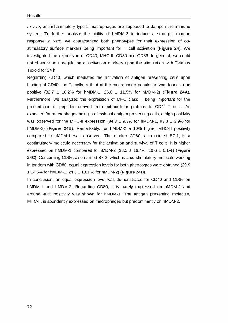
Results
72
In vivo, anti-inflammatory type 2 macrophages are supposed to dampen the immune
system. To further analyze the ability of hMDM-2 to induce a stronger immune
response in vitro, we characterized both phenotypes for their expression of co-
stimulatory surface markers being important for T cell activation (Figure 24). We
investigated the expression of CD40, MHC-II, CD80 and CD86. In general, we could
not observe an upregulation of activation markers upon the stimulation with Tetanus
Toxoid for 24 h.
Regarding CD40, which mediates the activation of antigen presenting cells upon
binding of CD40L on TH cells, a third of the macrophage population was found to be
positive (32.7 ± 18.2% for hMDM-1, 26.0 ± 11.5% for hMDM-2) (Figure 24A).
Furthermore, we analyzed the expression of MHC class II being important for the
presentation of peptides derived from extracellular proteins to CD4+ T cells. As
expected for macrophages being professional antigen presenting cells, a high positivity
was observed for the MHC-II expression (84.8 ± 9.3% for hMDM-1, 93.3 ± 3.9% for
hMDM-2) (Figure 24B). Remarkably, for hMDM-2 a 10% higher MHC-II positivity
compared to hMDM-1 was observed. The marker CD80, also named B7-1, is a
costimulatory molecule necessary for the activation and survival of T cells. It is higher
expressed on hMDM-1 compared to hMDM-2 (38.5 ± 16.4%, 10.6 ± 6.1%) (Figure
24C). Concerning CD86, also named B7-2, which is a co-stimulatory molecule working
in tandem with CD80, equal expression levels for both phenotypes were obtained (29.9
± 14.5% for hMDM-1, 24.3 ± 13.1 % for hMDM-2) (Figure 24D).
In conclusion, an equal expression level was demonstrated for CD40 and CD86 on
hMDM-1 and hMDM-2. Regarding CD80, it is barely expressed on hMDM-2 and
around 40% positivity was shown for hMDM-1. The antigen presenting molecule,
MHC-II, is abundantly expressed on macrophages but predominantly on hMDM-2.
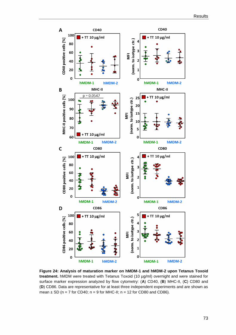
Results
73
Figure 24: Analysis of maturation marker on hMDM-1 and hMDM-2 upon Tetanus Toxoid
treatment. hMDM were treated with Tetanus Toxoid (10 µg/ml) overnight and were stained for
surface marker expression analyzed by flow cytometry: (A) CD40, (B) MHC-II, (C) CD80 and
(D) CD86. Data are representative for at least three independent experiments and are shown as
mean ± SD (n = 7 for CD40; n = 9 for MHC-II; n = 12 for CD80 and CD86).
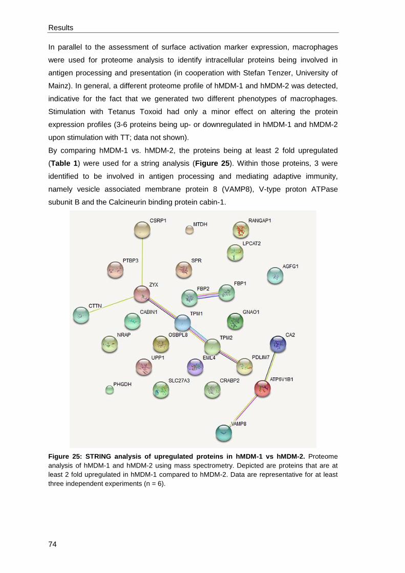
Results
74
In parallel to the assessment of surface activation marker expression, macrophages
were used for proteome analysis to identify intracellular proteins being involved in
antigen processing and presentation (in cooperation with Stefan Tenzer, University of
Mainz). In general, a different proteome profile of hMDM-1 and hMDM-2 was detected,
indicative for the fact that we generated two different phenotypes of macrophages.
Stimulation with Tetanus Toxoid had only a minor effect on altering the protein
expression profiles (3-6 proteins being up- or downregulated in hMDM-1 and hMDM-2
upon stimulation with TT; data not shown).
By comparing hMDM-1 vs. hMDM-2, the proteins being at least 2 fold upregulated
(Table 1) were used for a string analysis (Figure 25). Within those proteins, 3 were
identified to be involved in antigen processing and mediating adaptive immunity,
namely vesicle associated membrane protein 8 (VAMP8), V-type proton ATPase
subunit B and the Calcineurin binding protein cabin-1.
Figure 25: STRING analysis of upregulated proteins in hMDM-1 vs hMDM-2. Proteome
analysis of hMDM-1 and hMDM-2 using mass spectrometry. Depicted are proteins that are at
least 2 fold upregulated in hMDM-1 compared to hMDM-2. Data are representative for at least
three independent experiments (n = 6).
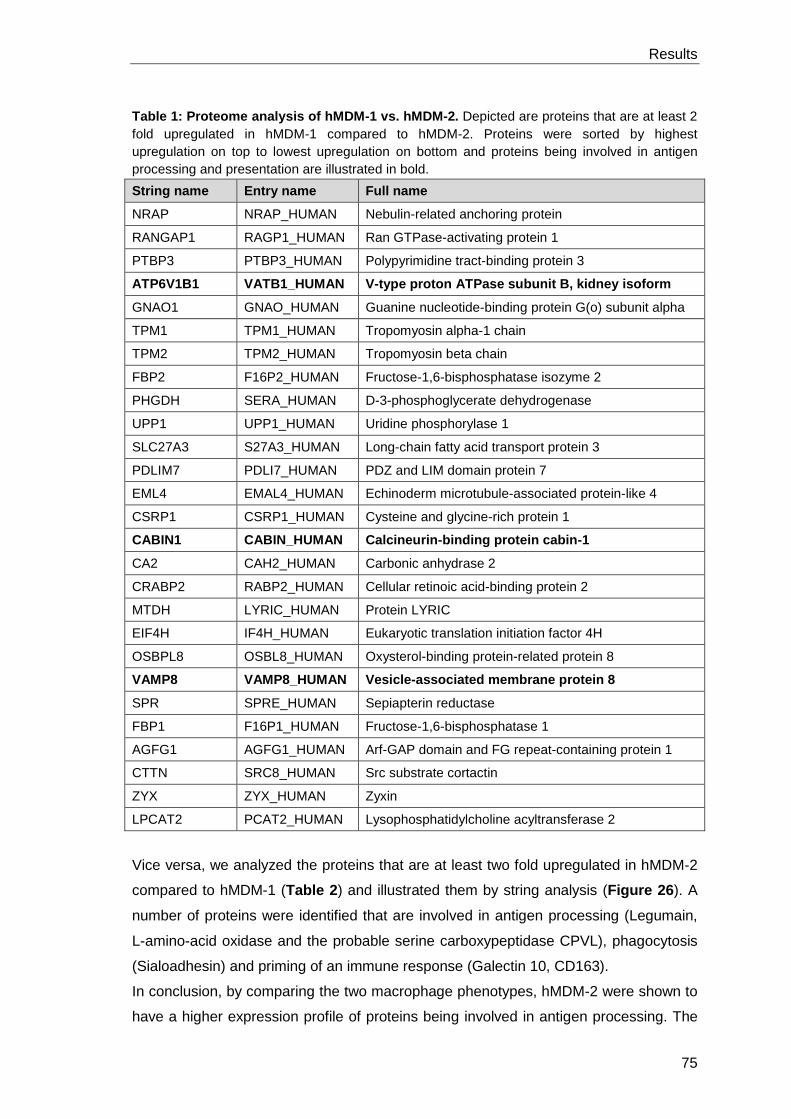
Results
75
Table 1: Proteome analysis of hMDM-1 vs. hMDM-2. Depicted are proteins that are at least 2
fold upregulated in hMDM-1 compared to hMDM-2. Proteins were sorted by highest
upregulation on top to lowest upregulation on bottom and proteins being involved in antigen
processing and presentation are illustrated in bold.
String name Entry name Full name
NRAP NRAP_HUMAN Nebulin-related anchoring protein
RANGAP1 RAGP1_HUMAN Ran GTPase-activating protein 1
PTBP3 PTBP3_HUMAN Polypyrimidine tract-binding protein 3
ATP6V1B1 VATB1_HUMAN V-type proton ATPase subunit B, kidney isoform
GNAO1 GNAO_HUMAN Guanine nucleotide-binding protein G(o) subunit alpha
TPM1 TPM1_HUMAN Tropomyosin alpha-1 chain
TPM2 TPM2_HUMAN Tropomyosin beta chain
FBP2 F16P2_HUMAN Fructose-1,6-bisphosphatase isozyme 2
PHGDH SERA_HUMAN D-3-phosphoglycerate dehydrogenase
UPP1 UPP1_HUMAN Uridine phosphorylase 1
SLC27A3 S27A3_HUMAN Long-chain fatty acid transport protein 3
PDLIM7 PDLI7_HUMAN PDZ and LIM domain protein 7
EML4 EMAL4_HUMAN Echinoderm microtubule-associated protein-like 4
CSRP1 CSRP1_HUMAN Cysteine and glycine-rich protein 1
CABIN1 CABIN_HUMAN Calcineurin-binding protein cabin-1
CA2 CAH2_HUMAN Carbonic anhydrase 2
CRABP2 RABP2_HUMAN Cellular retinoic acid-binding protein 2
MTDH LYRIC_HUMAN Protein LYRIC
EIF4H IF4H_HUMAN Eukaryotic translation initiation factor 4H
OSBPL8 OSBL8_HUMAN Oxysterol-binding protein-related protein 8
VAMP8 VAMP8_HUMAN Vesicle-associated membrane protein 8
SPR SPRE_HUMAN Sepiapterin reductase
FBP1 F16P1_HUMAN Fructose-1,6-bisphosphatase 1
AGFG1 AGFG1_HUMAN Arf-GAP domain and FG repeat-containing protein 1
CTTN SRC8_HUMAN Src substrate cortactin
ZYX ZYX_HUMAN Zyxin
LPCAT2 PCAT2_HUMAN Lysophosphatidylcholine acyltransferase 2
Vice versa, we analyzed the proteins that are at least two fold upregulated in hMDM-2
compared to hMDM-1 (Table 2) and illustrated them by string analysis (Figure 26). A
number of proteins were identified that are involved in antigen processing (Legumain,
L-amino-acid oxidase and the probable serine carboxypeptidase CPVL), phagocytosis
(Sialoadhesin) and priming of an immune response (Galectin 10, CD163).
In conclusion, by comparing the two macrophage phenotypes, hMDM-2 were shown to
have a higher expression profile of proteins being involved in antigen processing. The
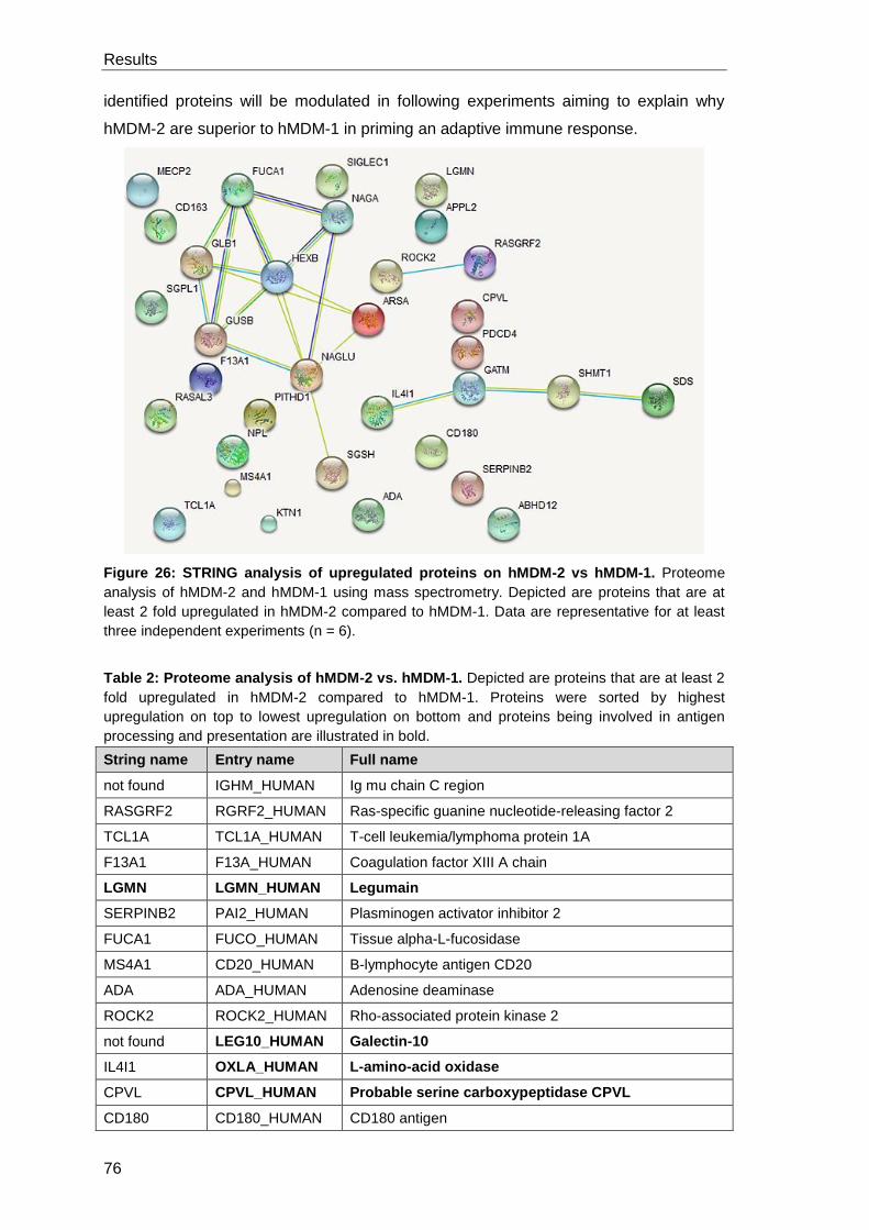
Results
76
identified proteins will be modulated in following experiments aiming to explain why
hMDM-2 are superior to hMDM-1 in priming an adaptive immune response.
Figure 26: STRING analysis of upregulated proteins on hMDM-2 vs hMDM-1. Proteome
analysis of hMDM-2 and hMDM-1 using mass spectrometry. Depicted are proteins that are at
least 2 fold upregulated in hMDM-2 compared to hMDM-1. Data are representative for at least
three independent experiments (n = 6).
Table 2: Proteome analysis of hMDM-2 vs. hMDM-1. Depicted are proteins that are at least 2
fold upregulated in hMDM-2 compared to hMDM-1. Proteins were sorted by highest
upregulation on top to lowest upregulation on bottom and proteins being involved in antigen
processing and presentation are illustrated in bold.
String name Entry name Full name
not found IGHM_HUMAN Ig mu chain C region
RASGRF2 RGRF2_HUMAN Ras-specific guanine nucleotide-releasing factor 2
TCL1A TCL1A_HUMAN T-cell leukemia/lymphoma protein 1A
F13A1 F13A_HUMAN Coagulation factor XIII A chain
LGMN LGMN_HUMAN Legumain
SERPINB2 PAI2_HUMAN Plasminogen activator inhibitor 2
FUCA1 FUCO_HUMAN Tissue alpha-L-fucosidase
MS4A1 CD20_HUMAN B-lymphocyte antigen CD20
ADA ADA_HUMAN Adenosine deaminase
ROCK2 ROCK2_HUMAN Rho-associated protein kinase 2
not found LEG10_HUMAN Galectin-10
IL4I1 OXLA_HUMAN L-amino-acid oxidase
CPVL CPVL_HUMAN Probable serine carboxypeptidase CPVL
CD180 CD180_HUMAN CD180 antigen
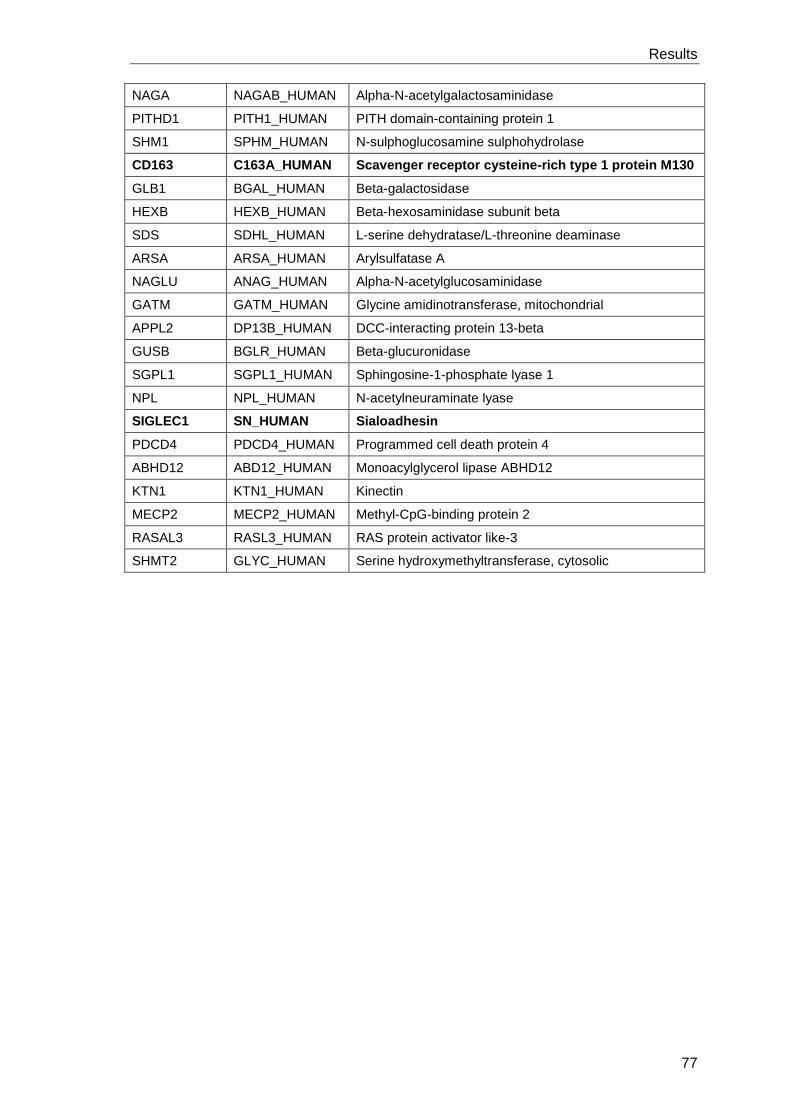
Results
77
NAGA NAGAB_HUMAN Alpha-N-acetylgalactosaminidase
PITHD1 PITH1_HUMAN PITH domain-containing protein 1
SHM1 SPHM_HUMAN N-sulphoglucosamine sulphohydrolase
CD163 C163A_HUMAN Scavenger receptor cysteine-rich type 1 protein M130
GLB1 BGAL_HUMAN Beta-galactosidase
HEXB HEXB_HUMAN Beta-hexosaminidase subunit beta
SDS SDHL_HUMAN L-serine dehydratase/L-threonine deaminase
ARSA ARSA_HUMAN Arylsulfatase A
NAGLU ANAG_HUMAN Alpha-N-acetylglucosaminidase
GATM GATM_HUMAN Glycine amidinotransferase, mitochondrial
APPL2 DP13B_HUMAN DCC-interacting protein 13-beta
GUSB BGLR_HUMAN Beta-glucuronidase
SGPL1 SGPL1_HUMAN Sphingosine-1-phosphate lyase 1
NPL NPL_HUMAN N-acetylneuraminate lyase
SIGLEC1 SN_HUMAN Sialoadhesin
PDCD4 PDCD4_HUMAN Programmed cell death protein 4
ABHD12 ABD12_HUMAN Monoacylglycerol lipase ABHD12
KTN1 KTN1_HUMAN Kinectin
MECP2 MECP2_HUMAN Methyl-CpG-binding protein 2
RASAL3 RASL3_HUMAN RAS protein activator like-3
SHMT2 GLYC_HUMAN Serine hydroxymethyltransferase, cytosolic
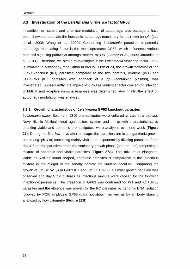
Results
78
3.3 Investigation of the Leishmania virulence factor GP63
In addition to nutrient and chemical modulation of autophagy, also pathogens have
been shown to modulate the host cells’ autophagy machinery for their own benefit (Lee
et al., 2008; Wang et al., 2009). Concerning Leishmania parasites a potential
autophagy modulating factor is the metalloprotease GP63, which influences various
host cell signaling pathways amongst others, mTOR (Gomez et al., 2009; Jaramillo et
al., 2011). Therefore, we aimed to investigate if the Leishmania virulence factor GP63
is involved in autophagy modulation in hMDM. First of all, the growth behavior of the
GP63 knockout (KO) parasites compared to the two controls, wildtype (WT) and
KO+GP63 (KO parasites with addback of a gp63-containing plasmid), was
investigated. Subsequently, the impact of GP63 as virulence factor concerning infection
of hMDM and adaptive immune response was determined. And finally, the effect on
autophagy modulation was analyzed.
3.3.1 Growth characteristics of Leishmania GP63 knockout parasites
Leishmania major Seidmann (SD) promastigotes were cultured in vitro in a biphasic
Novy Nicolle McNeal blood agar culture system and the growth characteristics, by
counting viable and apoptotic promastigotes, were analyzed over one week (Figure
27). During the first few days after passage, the parasites are in a logarithmic growth
phase (log. ph. Lm) containing mainly viable and exponentially dividing parasites. From
day 4-5 on, the parasites reach the stationary growth phase (stat. ph. Lm) comprising a
mixture of apoptotic and viable parasites (Figure 27A). This mixture of elongated,
viable as well as round shaped, apoptotic parasites is comparable to the infectious
mixture in the midgut of the sandfly namely the virulent inoculum. Comparing the
growth of Lm SD WT, Lm GP63 KO and Lm KO+GP63, a similar growth behavior was
observed and day 5 old cultures as infectious mixture were chosen for the following
infection experiments. The presence of GP63 was confirmed for WT and KO+GP63
parasites and the absence was proven for the KO parasites by genomic DNA isolation
followed by PCR amplifying GP63 (data not shown) as well as by antibody staining
analyzed by flow cytometry (Figure 27B).
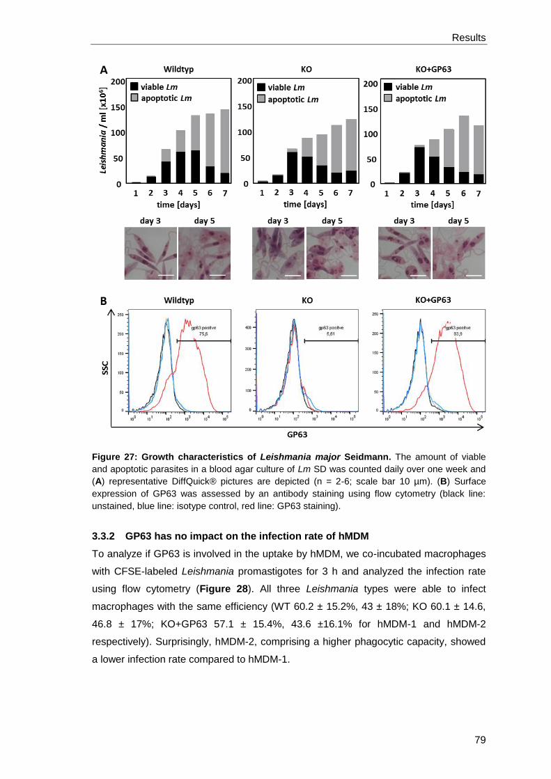
Results
79
Figure 27: Growth characteristics of Leishmania major Seidmann. The amount of viable
and apoptotic parasites in a blood agar culture of Lm SD was counted daily over one week and
(A) representative DiffQuick® pictures are depicted (n = 2-6; scale bar 10 µm). (B) Surface
expression of GP63 was assessed by an antibody staining using flow cytometry (black line:
unstained, blue line: isotype control, red line: GP63 staining).
3.3.2 GP63 has no impact on the infection rate of hMDM
To analyze if GP63 is involved in the uptake by hMDM, we co-incubated macrophages
with CFSE-labeled Leishmania promastigotes for 3 h and analyzed the infection rate
using flow cytometry (Figure 28). All three Leishmania types were able to infect
macrophages with the same efficiency (WT 60.2 ± 15.2%, 43 ± 18%; KO 60.1 ± 14.6,
46.8 ± 17%; KO+GP63 57.1 ± 15.4%, 43.6 ±16.1% for hMDM-1 and hMDM-2
respectively). Surprisingly, hMDM-2, comprising a higher phagocytic capacity, showed
a lower infection rate compared to hMDM-1.
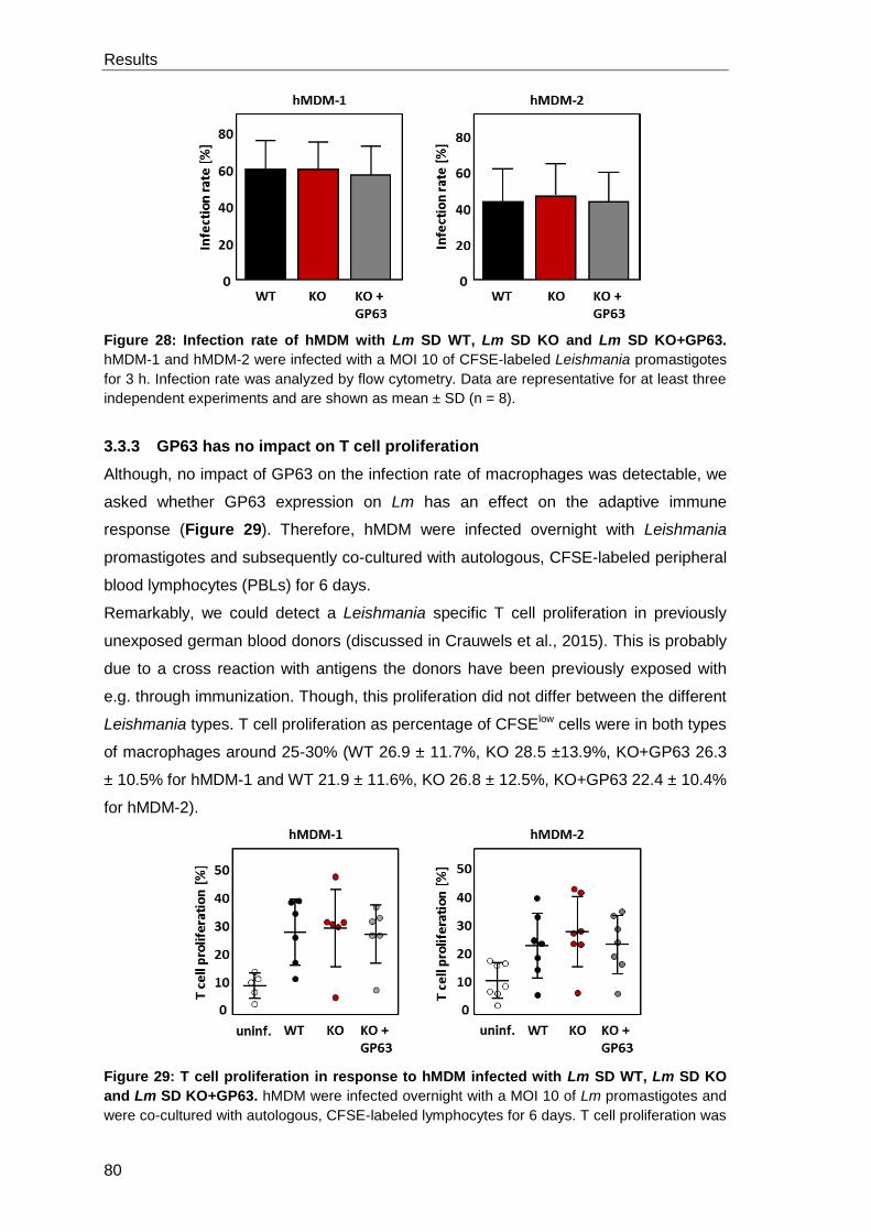
Results
80
Figure 28: Infection rate of hMDM with Lm SD WT, Lm SD KO and Lm SD KO+GP63.
hMDM-1 and hMDM-2 were infected with a MOI 10 of CFSE-labeled Leishmania promastigotes
for 3 h. Infection rate was analyzed by flow cytometry. Data are representative for at least three
independent experiments and are shown as mean ± SD (n = 8).
3.3.3 GP63 has no impact on T cell proliferation
Although, no impact of GP63 on the infection rate of macrophages was detectable, we
asked whether GP63 expression on Lm has an effect on the adaptive immune
response (Figure 29). Therefore, hMDM were infected overnight with Leishmania
promastigotes and subsequently co-cultured with autologous, CFSE-labeled peripheral
blood lymphocytes (PBLs) for 6 days.
Remarkably, we could detect a Leishmania specific T cell proliferation in previously
unexposed german blood donors (discussed in Crauwels et al., 2015). This is probably
due to a cross reaction with antigens the donors have been previously exposed with
e.g. through immunization. Though, this proliferation did not differ between the different
Leishmania types. T cell proliferation as percentage of CFSElow cells were in both types
of macrophages around 25-30% (WT 26.9 ± 11.7%, KO 28.5 ±13.9%, KO+GP63 26.3
± 10.5% for hMDM-1 and WT 21.9 ± 11.6%, KO 26.8 ± 12.5%, KO+GP63 22.4 ± 10.4%
for hMDM-2).
Figure 29: T cell proliferation in response to hMDM infected with Lm SD WT, Lm SD KO
and Lm SD KO+GP63. hMDM were infected overnight with a MOI 10 of Lm promastigotes and
were co-cultured with autologous, CFSE-labeled lymphocytes for 6 days. T cell proliferation was
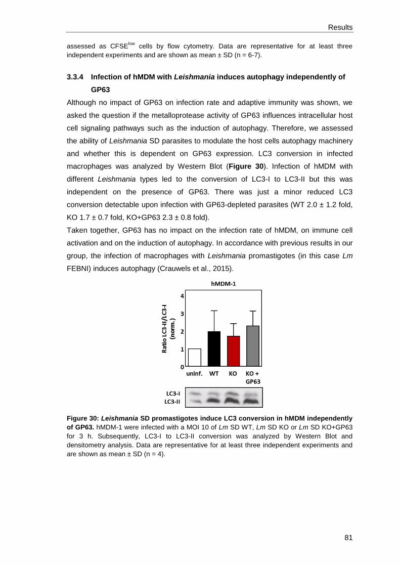
Results
81
assessed as CFSElow
cells by flow cytometry. Data are representative for at least three
independent experiments and are shown as mean ± SD (n = 6-7).
3.3.4 Infection of hMDM with Leishmania induces autophagy independently of
GP63
Although no impact of GP63 on infection rate and adaptive immunity was shown, we
asked the question if the metalloprotease activity of GP63 influences intracellular host
cell signaling pathways such as the induction of autophagy. Therefore, we assessed
the ability of Leishmania SD parasites to modulate the host cells autophagy machinery
and whether this is dependent on GP63 expression. LC3 conversion in infected
macrophages was analyzed by Western Blot (Figure 30). Infection of hMDM with
different Leishmania types led to the conversion of LC3-I to LC3-II but this was
independent on the presence of GP63. There was just a minor reduced LC3
conversion detectable upon infection with GP63-depleted parasites (WT 2.0 ± 1.2 fold,
KO 1.7 ± 0.7 fold, KO+GP63 2.3 ± 0.8 fold).
Taken together, GP63 has no impact on the infection rate of hMDM, on immune cell
activation and on the induction of autophagy. In accordance with previous results in our
group, the infection of macrophages with Leishmania promastigotes (in this case Lm
FEBNI) induces autophagy (Crauwels et al., 2015).
Figure 30: Leishmania SD promastigotes induce LC3 conversion in hMDM independently
of GP63. hMDM-1 were infected with a MOI 10 of Lm SD WT, Lm SD KO or Lm SD KO+GP63
for 3 h. Subsequently, LC3-I to LC3-II conversion was analyzed by Western Blot and
densitometry analysis. Data are representative for at least three independent experiments and
are shown as mean ± SD (n = 4).
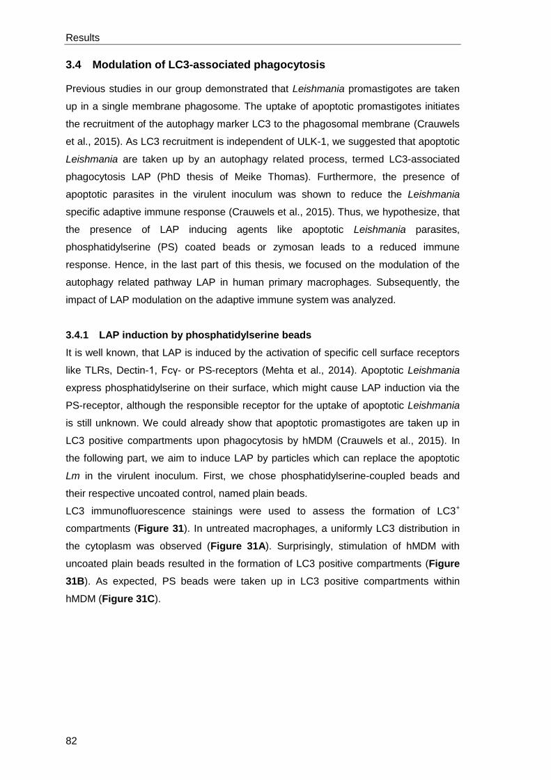
Results
82
3.4 Modulation of LC3-associated phagocytosis
Previous studies in our group demonstrated that Leishmania promastigotes are taken
up in a single membrane phagosome. The uptake of apoptotic promastigotes initiates
the recruitment of the autophagy marker LC3 to the phagosomal membrane (Crauwels
et al., 2015). As LC3 recruitment is independent of ULK-1, we suggested that apoptotic
Leishmania are taken up by an autophagy related process, termed LC3-associated
phagocytosis LAP (PhD thesis of Meike Thomas). Furthermore, the presence of
apoptotic parasites in the virulent inoculum was shown to reduce the Leishmania
specific adaptive immune response (Crauwels et al., 2015). Thus, we hypothesize, that
the presence of LAP inducing agents like apoptotic Leishmania parasites,
phosphatidylserine (PS) coated beads or zymosan leads to a reduced immune
response. Hence, in the last part of this thesis, we focused on the modulation of the
autophagy related pathway LAP in human primary macrophages. Subsequently, the
impact of LAP modulation on the adaptive immune system was analyzed.
3.4.1 LAP induction by phosphatidylserine beads
It is well known, that LAP is induced by the activation of specific cell surface receptors
like TLRs, Dectin-1, Fcγ- or PS-receptors (Mehta et al., 2014). Apoptotic Leishmania
express phosphatidylserine on their surface, which might cause LAP induction via the
PS-receptor, although the responsible receptor for the uptake of apoptotic Leishmania
is still unknown. We could already show that apoptotic promastigotes are taken up in
LC3 positive compartments upon phagocytosis by hMDM (Crauwels et al., 2015). In
the following part, we aim to induce LAP by particles which can replace the apoptotic
Lm in the virulent inoculum. First, we chose phosphatidylserine-coupled beads and
their respective uncoated control, named plain beads.
LC3 immunofluorescence stainings were used to assess the formation of LC3+
compartments (Figure 31). In untreated macrophages, a uniformly LC3 distribution in
the cytoplasm was observed (Figure 31A). Surprisingly, stimulation of hMDM with
uncoated plain beads resulted in the formation of LC3 positive compartments (Figure
31B). As expected, PS beads were taken up in LC3 positive compartments within
hMDM (Figure 31C).
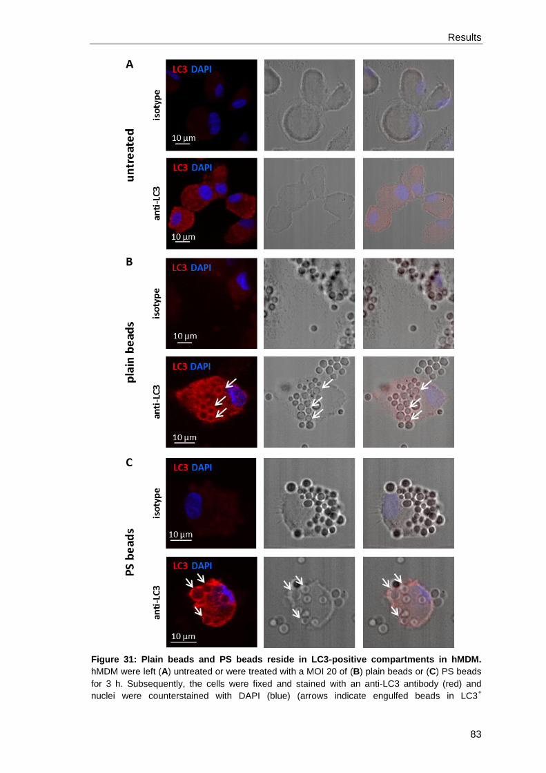
Results
83
Figure 31: Plain beads and PS beads reside in LC3-positive compartments in hMDM.
hMDM were left (A) untreated or were treated with a MOI 20 of (B) plain beads or (C) PS beads
for 3 h. Subsequently, the cells were fixed and stained with an anti-LC3 antibody (red) and
nuclei were counterstained with DAPI (blue) (arrows indicate engulfed beads in LC3+
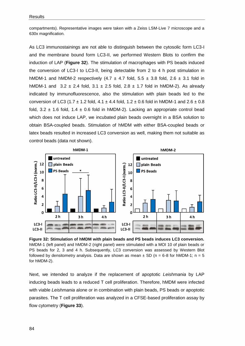
Results
84
compartments). Representative images were taken with a Zeiss LSM-Live 7 microscope and a
630x magnification.
As LC3 immunostainings are not able to distinguish between the cytosolic form LC3-I
and the membrane bound form LC3-II, we performed Western Blots to confirm the
induction of LAP (Figure 32). The stimulation of macrophages with PS beads induced
the conversion of LC3-I to LC3-II, being detectable from 2 to 4 h post stimulation in
hMDM-1 and hMDM-2 respectively (4.7 ± 4.7 fold, 5.5 ± 3.8 fold, 2.6 ± 3.1 fold in
hMDM-1 and 3.2 ± 2.4 fold, 3.1 ± 2.5 fold, 2.8 ± 1.7 fold in hMDM-2). As already
indicated by immunofluorescence, also the stimulation with plain beads led to the
conversion of LC3 (1.7 ± 1.2 fold, 4.1 ± 4.4 fold, 1.2 ± 0.6 fold in hMDM-1 and 2.6 ± 0.8
fold, 3.2 ± 1.6 fold, 1.4 ± 0.6 fold in hMDM-2). Lacking an appropriate control bead
which does not induce LAP, we incubated plain beads overnight in a BSA solution to
obtain BSA-coupled beads. Stimulation of hMDM with either BSA-coupled beads or
latex beads resulted in increased LC3 conversion as well, making them not suitable as
control beads (data not shown).
Figure 32: Stimulation of hMDM with plain beads and PS beads induces LC3 conversion.
hMDM-1 (left panel) and hMDM-2 (right panel) were stimulated with a MOI 10 of plain beads or
PS beads for 2, 3 and 4 h. Subsequently, LC3 conversion was assessed by Western Blot
followed by densitometry analysis. Data are shown as mean ± SD (n = 6-8 for hMDM-1; n = 5
for hMDM-2).
Next, we intended to analyze if the replacement of apoptotic Leishmania by LAP
inducing beads leads to a reduced T cell proliferation. Therefore, hMDM were infected
with viable Leishmania alone or in combination with plain beads, PS beads or apoptotic
parasites. The T cell proliferation was analyzed in a CFSE-based proliferation assay by
flow cytometry (Figure 33).
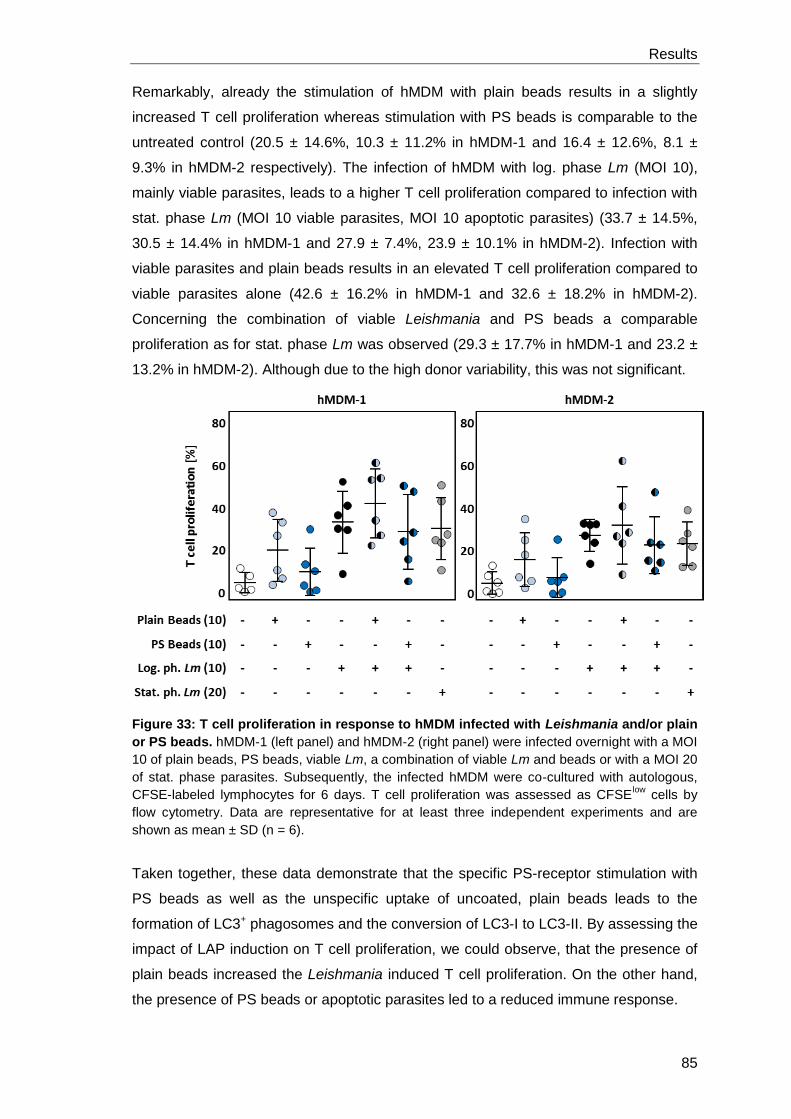
Results
85
Remarkably, already the stimulation of hMDM with plain beads results in a slightly
increased T cell proliferation whereas stimulation with PS beads is comparable to the
untreated control (20.5 ± 14.6%, 10.3 ± 11.2% in hMDM-1 and 16.4 ± 12.6%, 8.1 ±
9.3% in hMDM-2 respectively). The infection of hMDM with log. phase Lm (MOI 10),
mainly viable parasites, leads to a higher T cell proliferation compared to infection with
stat. phase Lm (MOI 10 viable parasites, MOI 10 apoptotic parasites) (33.7 ± 14.5%,
30.5 ± 14.4% in hMDM-1 and 27.9 ± 7.4%, 23.9 ± 10.1% in hMDM-2). Infection with
viable parasites and plain beads results in an elevated T cell proliferation compared to
viable parasites alone (42.6 ± 16.2% in hMDM-1 and 32.6 ± 18.2% in hMDM-2).
Concerning the combination of viable Leishmania and PS beads a comparable
proliferation as for stat. phase Lm was observed (29.3 ± 17.7% in hMDM-1 and 23.2 ±
13.2% in hMDM-2). Although due to the high donor variability, this was not significant.
Figure 33: T cell proliferation in response to hMDM infected with Leishmania and/or plain
or PS beads. hMDM-1 (left panel) and hMDM-2 (right panel) were infected overnight with a MOI
10 of plain beads, PS beads, viable Lm, a combination of viable Lm and beads or with a MOI 20
of stat. phase parasites. Subsequently, the infected hMDM were co-cultured with autologous,
CFSE-labeled lymphocytes for 6 days. T cell proliferation was assessed as CFSElow
cells by
flow cytometry. Data are representative for at least three independent experiments and are
shown as mean ± SD (n = 6).
Taken together, these data demonstrate that the specific PS-receptor stimulation with
PS beads as well as the unspecific uptake of uncoated, plain beads leads to the
formation of LC3+ phagosomes and the conversion of LC3-I to LC3-II. By assessing the
impact of LAP induction on T cell proliferation, we could observe, that the presence of
plain beads increased the Leishmania induced T cell proliferation. On the other hand,
the presence of PS beads or apoptotic parasites led to a reduced immune response.
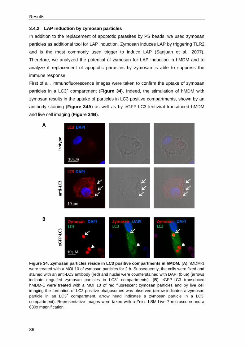
Results
86
3.4.2 LAP induction by zymosan particles
In addition to the replacement of apoptotic parasites by PS beads, we used zymosan
particles as additional tool for LAP induction. Zymosan induces LAP by triggering TLR2
and is the most commonly used trigger to induce LAP (Sanjuan et al., 2007).
Therefore, we analyzed the potential of zymosan for LAP induction in hMDM and to
analyze if replacement of apoptotic parasites by zymosan is able to suppress the
immune response.
First of all, immunofluorescence images were taken to confirm the uptake of zymosan
particles in a LC3+ compartment (Figure 34). Indeed, the stimulation of hMDM with
zymosan results in the uptake of particles in LC3 positive compartments, shown by an
antibody staining (Figure 34A) as well as by eGFP-LC3 lentiviral transduced hMDM
and live cell imaging (Figure 34B).
Figure 34: Zymosan particles reside in LC3 positive compartments in hMDM. (A) hMDM-1
were treated with a MOI 10 of zymosan particles for 2 h. Subsequently, the cells were fixed and
stained with an anti-LC3 antibody (red) and nuclei were counterstained with DAPI (blue) (arrows
indicate engulfed zymosan particles in LC3+ compartments). (B) eGFP-LC3 transduced
hMDM-1 were treated with a MOI 10 of red fluorescent zymosan particles and by live cell
imaging the formation of LC3 positive phagosomes was observed (arrow indicates a zymosan
particle in an LC3+ compartment, arrow head indicates a zymosan particle in a LC3
-
compartment). Representative images were taken with a Zeiss LSM-Live 7 microscope and a
630x magnification.
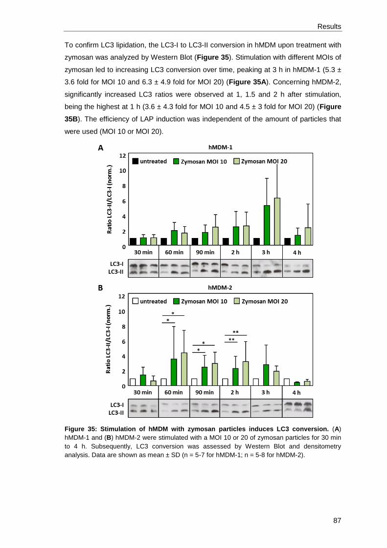
Results
87
To confirm LC3 lipidation, the LC3-I to LC3-II conversion in hMDM upon treatment with
zymosan was analyzed by Western Blot (Figure 35). Stimulation with different MOIs of
zymosan led to increasing LC3 conversion over time, peaking at 3 h in hMDM-1 (5.3 ±
3.6 fold for MOI 10 and 6.3 ± 4.9 fold for MOI 20) (Figure 35A). Concerning hMDM-2,
significantly increased LC3 ratios were observed at 1, 1.5 and 2 h after stimulation,
being the highest at 1 h (3.6 ± 4.3 fold for MOI 10 and 4.5 ± 3 fold for MOI 20) (Figure
35B). The efficiency of LAP induction was independent of the amount of particles that
were used (MOI 10 or MOI 20).
Figure 35: Stimulation of hMDM with zymosan particles induces LC3 conversion. (A)
hMDM-1 and (B) hMDM-2 were stimulated with a MOI 10 or 20 of zymosan particles for 30 min
to 4 h. Subsequently, LC3 conversion was assessed by Western Blot and densitometry
analysis. Data are shown as mean ± SD (n = 5-7 for hMDM-1; n = 5-8 for hMDM-2).
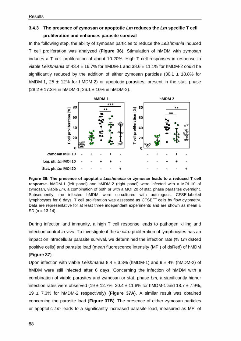
Results
88
3.4.3 The presence of zymosan or apoptotic Lm reduces the Lm specific T cell
proliferation and enhances parasite survival
In the following step, the ability of zymosan particles to reduce the Leishmania induced
T cell proliferation was analyzed (Figure 36). Stimulation of hMDM with zymosan
induces a T cell proliferation of about 10-20%. High T cell responses in response to
viable Leishmania of 43.4 ± 16.7% for hMDM-1 and 38.6 ± 11.1% for hMDM-2 could be
significantly reduced by the addition of either zymosan particles (30.1 ± 18.8% for
hMDM-1, 25 ± 12% for hMDM-2) or apoptotic parasites, present in the stat. phase
(28.2 ± 17.3% in hMDM-1, 26.1 ± 10% in hMDM-2).
Figure 36: The presence of apoptotic Leishmania or zymosan leads to a reduced T cell
response. hMDM-1 (left panel) and hMDM-2 (right panel) were infected with a MOI 10 of
zymosan, viable Lm, a combination of both or with a MOI 20 of stat. phase parasites overnight.
Subsequently, the infected hMDM were co-cultured with autologous, CFSE-labeled
lymphocytes for 6 days. T cell proliferation was assessed as CFSElow
cells by flow cytometry.
Data are representative for at least three independent experiments and are shown as mean ±
SD (n = 13-14).
During infection and immunity, a high T cell response leads to pathogen killing and
infection control in vivo. To investigate if the in vitro proliferation of lymphocytes has an
impact on intracellular parasite survival, we determined the infection rate (% Lm dsRed
positive cells) and parasite load (mean fluorescence intensity (MFI) of dsRed) of hMDM
(Figure 37).
Upon infection with viable Leishmania 8.4 ± 3.3% (hMDM-1) and 9 ± 4% (hMDM-2) of
hMDM were still infected after 6 days. Concerning the infection of hMDM with a
combination of viable parasites and zymosan or stat. phase Lm, a significantly higher
infection rates were observed (19 ± 12.7%, 20.4 ± 11.8% for hMDM-1 and 18.7 ± 7.9%,
19 ± 7.3% for hMDM-2 respectively) (Figure 37A). A similar result was obtained
concerning the parasite load (Figure 37B). The presence of either zymosan particles
or apoptotic Lm leads to a significantly increased parasite load, measured as MFI of
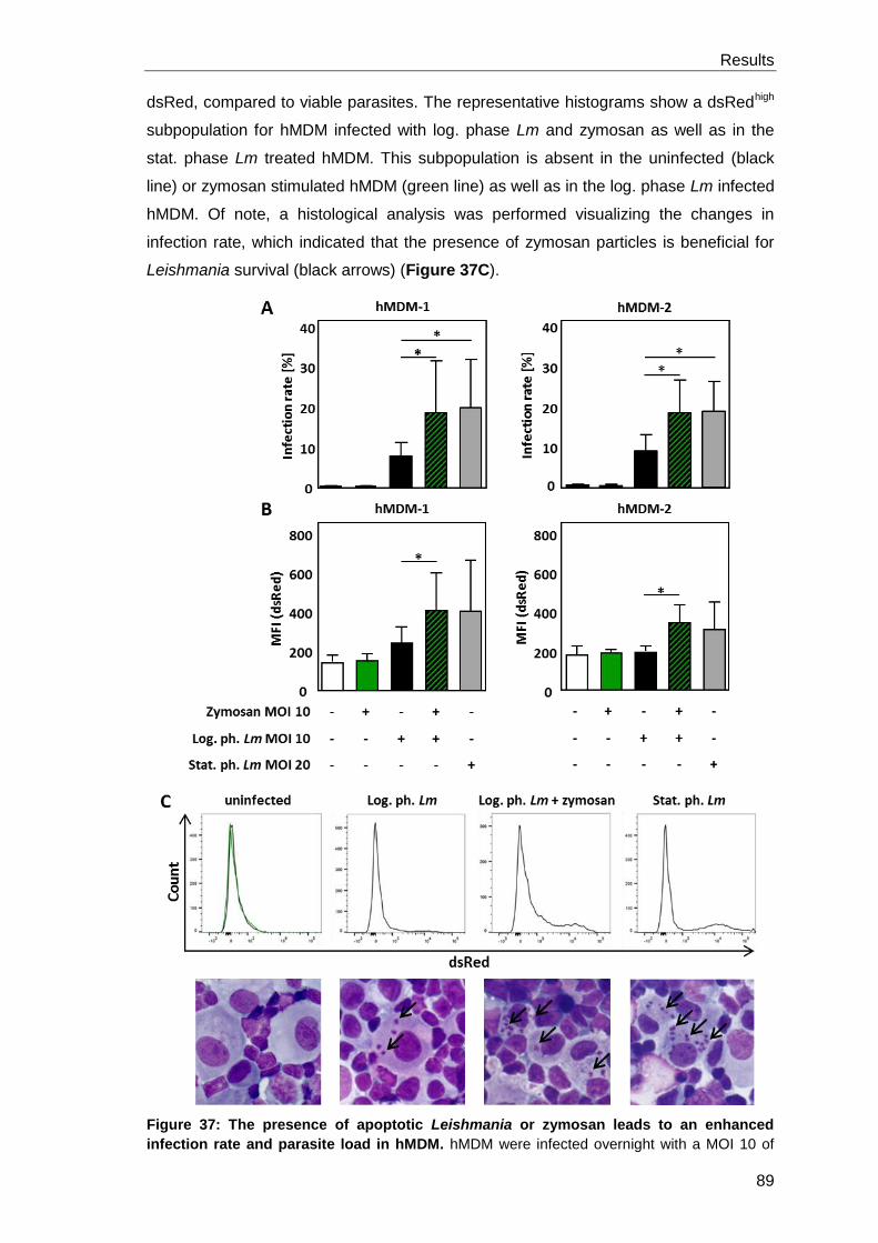
Results
89
dsRed, compared to viable parasites. The representative histograms show a dsRedhigh
subpopulation for hMDM infected with log. phase Lm and zymosan as well as in the
stat. phase Lm treated hMDM. This subpopulation is absent in the uninfected (black
line) or zymosan stimulated hMDM (green line) as well as in the log. phase Lm infected
hMDM. Of note, a histological analysis was performed visualizing the changes in
infection rate, which indicated that the presence of zymosan particles is beneficial for
Leishmania survival (black arrows) (Figure 37C).
Figure 37: The presence of apoptotic Leishmania or zymosan leads to an enhanced
infection rate and parasite load in hMDM. hMDM were infected overnight with a MOI 10 of
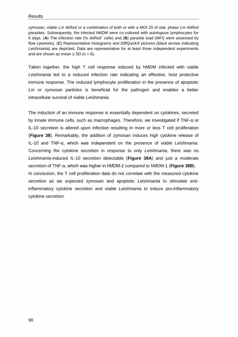
Results
90
zymosan, viable Lm dsRed or a combination of both or with a MOI 20 of stat. phase Lm dsRed
parasites. Subsequently, the infected hMDM were co-cultured with autologous lymphocytes for
6 days. (A) The infection rate (% dsRed+ cells) and (B) parasite load (MFI) were assessed by
flow cytometry. (C) Representative histograms and DiffQuick® pictures (black arrows indicating
Leishmania) are depicted. Data are representative for at least three independent experiments
and are shown as mean ± SD (n = 6).
Taken together, the high T cell response induced by hMDM infected with viable
Leishmania led to a reduced infection rate indicating an effective, host protective
immune response. The reduced lymphocyte proliferation in the presence of apoptotic
Lm or zymosan particles is beneficial for the pathogen and enables a better
intracellular survival of viable Leishmania.
The induction of an immune response is essentially dependent on cytokines, secreted
by innate immune cells, such as macrophages. Therefore, we investigated if TNF-α or
IL-10 secretion is altered upon infection resulting in more or less T cell proliferation
(Figure 38). Remarkably, the addition of zymosan induces high cytokine release of
IL-10 and TNF-α, which was independent on the presence of viable Leishmania.
Concerning the cytokine secretion in response to only Leishmania, there was no
Leishmania-induced IL-10 secretion detectable (Figure 38A) and just a moderate
secretion of TNF-α, which was higher in hMDM-2 compared to hMDM-1 (Figure 38B).
In conclusion, the T cell proliferation data do not correlate with the measured cytokine
secretion as we expected zymosan and apoptotic Leishmania to stimulate anti-
inflammatory cytokine secretion and viable Leishmania to induce pro-inflammatory
cytokine secretion.
Results
91
Figure 38: Stimulation of hMDM with zymosan particles increases the secretion of IL-10
and TNF-α. hMDM-1 (left panel) and hMDM-2 (right panel) were treated with a MOI 10 of log.
phase Leishmania, zymosan, a combination of both or stat. phase Lm for 18 h. Subsequently,
the supernatants of hMDM-1 and hMDM-2 were analyzed by ELISA to assess the secretion of
(A) IL-10 and (B) TNF-α. Data are representative for at least three independent experiments
and are shown as mean ± SD (n = 6 for IL-10 and n = 4 for TNF-α).
3.4.4 Impact of ROS for LAP induction
In the next part, we aimed to analyze the underlying mechanisms leading to LAP
induction in hMDM. The production of reactive oxygen species (ROS) was shown to
lead to the recruitment of LC3 to phagosomes (Huang et al., 2009). There exist two
possibilities for ROS to occur within the phagosome. The molecules can be brought
inside the cell by the parasite or can be produced by the macrophages’ NADPH
oxidase (NOX2) which is located on the phagosomal membrane.
First, we analyzed the importance of parasitic ROS for LAP induction. Therefore, we
treated viable Leishmania parasites with two apoptosis inducing drugs, Staurosporine
and Miltefosine. By analyzing the ROS positivity with 2′,7′-Dichlorodihydrofluorescein
diacetate (H2DCFDA), dihydroethidium (DHE) and Dihydrorhodamine 123 (DHR123),
Staurosporine treated Leishmania were shown to be ROS-positive whereas Miltefosine
treated Leishmania were ROS-negative (MD thesis Jochen Steinacker). To assess if
parasitic ROS is important for LAP induction, we used Staurosporine and Miltefosine
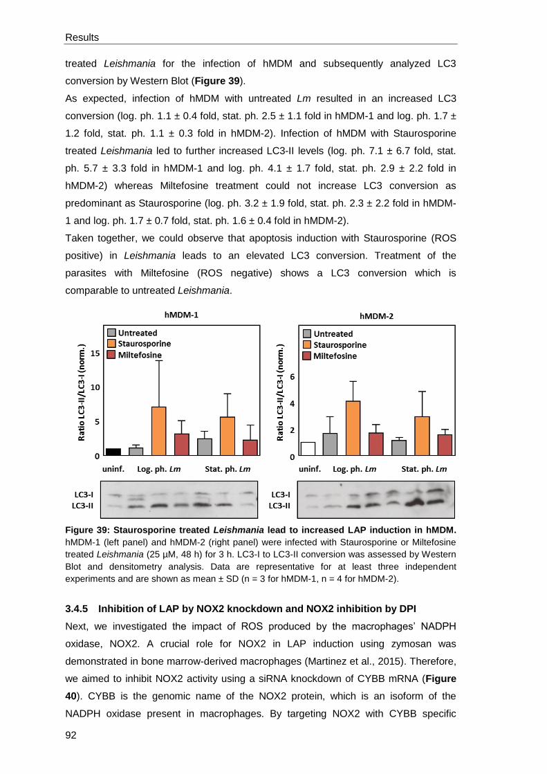
Results
92
treated Leishmania for the infection of hMDM and subsequently analyzed LC3
conversion by Western Blot (Figure 39).
As expected, infection of hMDM with untreated Lm resulted in an increased LC3
conversion (log. ph. 1.1 ± 0.4 fold, stat. ph. 2.5 ± 1.1 fold in hMDM-1 and log. ph. 1.7 ±
1.2 fold, stat. ph. 1.1 ± 0.3 fold in hMDM-2). Infection of hMDM with Staurosporine
treated Leishmania led to further increased LC3-II levels (log. ph. 7.1 ± 6.7 fold, stat.
ph. 5.7 ± 3.3 fold in hMDM-1 and log. ph. 4.1 ± 1.7 fold, stat. ph. 2.9 ± 2.2 fold in
hMDM-2) whereas Miltefosine treatment could not increase LC3 conversion as
predominant as Staurosporine (log. ph. 3.2 ± 1.9 fold, stat. ph. 2.3 ± 2.2 fold in hMDM-
1 and log. ph. 1.7 ± 0.7 fold, stat. ph. 1.6 ± 0.4 fold in hMDM-2).
Taken together, we could observe that apoptosis induction with Staurosporine (ROS
positive) in Leishmania leads to an elevated LC3 conversion. Treatment of the
parasites with Miltefosine (ROS negative) shows a LC3 conversion which is
comparable to untreated Leishmania.
Figure 39: Staurosporine treated Leishmania lead to increased LAP induction in hMDM.
hMDM-1 (left panel) and hMDM-2 (right panel) were infected with Staurosporine or Miltefosine
treated Leishmania (25 µM, 48 h) for 3 h. LC3-I to LC3-II conversion was assessed by Western
Blot and densitometry analysis. Data are representative for at least three independent
experiments and are shown as mean ± SD (n = 3 for hMDM-1, n = 4 for hMDM-2).
3.4.5 Inhibition of LAP by NOX2 knockdown and NOX2 inhibition by DPI
Next, we investigated the impact of ROS produced by the macrophages’ NADPH
oxidase, NOX2. A crucial role for NOX2 in LAP induction using zymosan was
demonstrated in bone marrow-derived macrophages (Martinez et al., 2015). Therefore,
we aimed to inhibit NOX2 activity using a siRNA knockdown of CYBB mRNA (Figure
40). CYBB is the genomic name of the NOX2 protein, which is an isoform of the
NADPH oxidase present in macrophages. By targeting NOX2 with CYBB specific
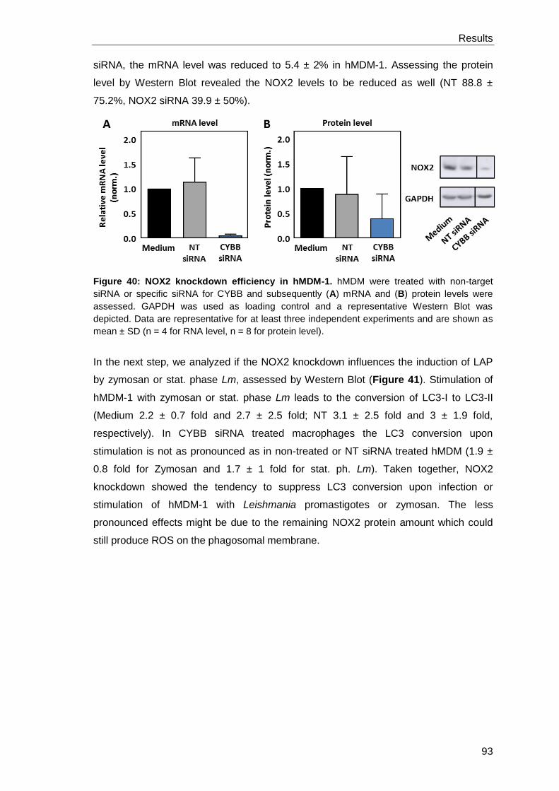
Results
93
siRNA, the mRNA level was reduced to 5.4 ± 2% in hMDM-1. Assessing the protein
level by Western Blot revealed the NOX2 levels to be reduced as well (NT 88.8 ±
75.2%, NOX2 siRNA 39.9 ± 50%).
Figure 40: NOX2 knockdown efficiency in hMDM-1. hMDM were treated with non-target
siRNA or specific siRNA for CYBB and subsequently (A) mRNA and (B) protein levels were
assessed. GAPDH was used as loading control and a representative Western Blot was
depicted. Data are representative for at least three independent experiments and are shown as
mean ± SD (n = 4 for RNA level, n = 8 for protein level).
In the next step, we analyzed if the NOX2 knockdown influences the induction of LAP
by zymosan or stat. phase Lm, assessed by Western Blot (Figure 41). Stimulation of
hMDM-1 with zymosan or stat. phase Lm leads to the conversion of LC3-I to LC3-II
(Medium 2.2 ± 0.7 fold and 2.7 ± 2.5 fold; NT 3.1 ± 2.5 fold and 3 ± 1.9 fold,
respectively). In CYBB siRNA treated macrophages the LC3 conversion upon
stimulation is not as pronounced as in non-treated or NT siRNA treated hMDM (1.9 ±
0.8 fold for Zymosan and 1.7 ± 1 fold for stat. ph. Lm). Taken together, NOX2
knockdown showed the tendency to suppress LC3 conversion upon infection or
stimulation of hMDM-1 with Leishmania promastigotes or zymosan. The less
pronounced effects might be due to the remaining NOX2 protein amount which could
still produce ROS on the phagosomal membrane.
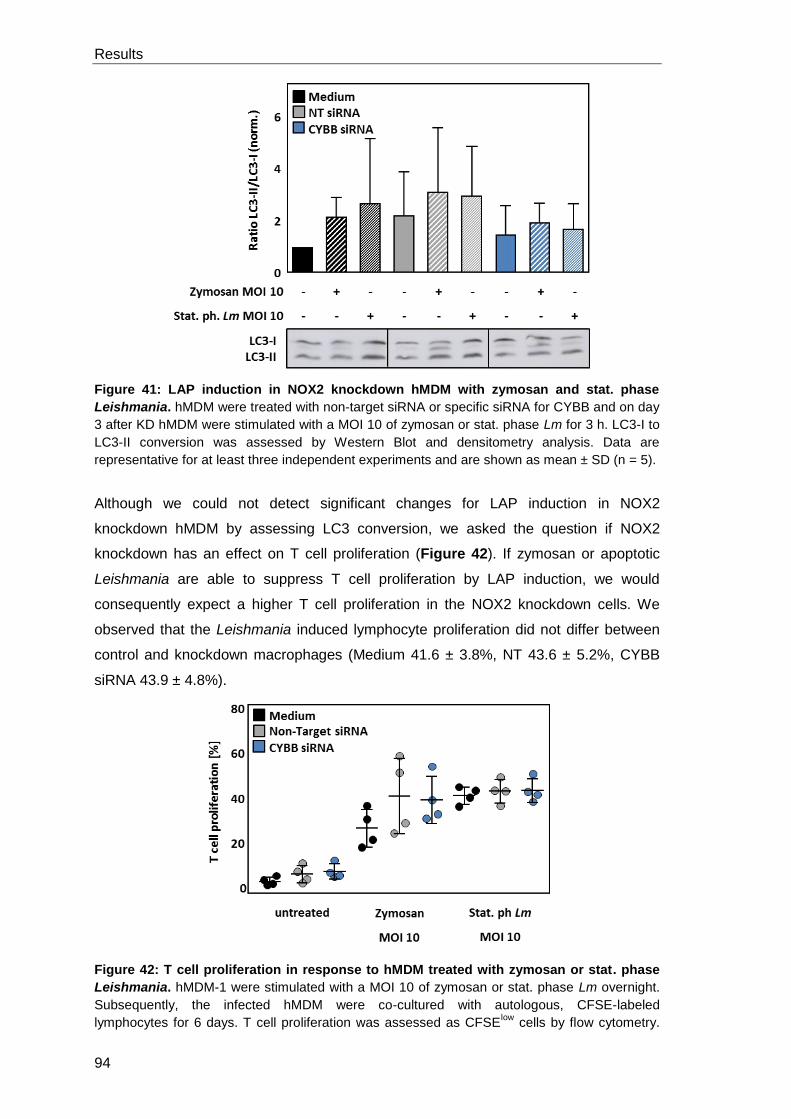
Results
94
Figure 41: LAP induction in NOX2 knockdown hMDM with zymosan and stat. phase
Leishmania. hMDM were treated with non-target siRNA or specific siRNA for CYBB and on day
3 after KD hMDM were stimulated with a MOI 10 of zymosan or stat. phase Lm for 3 h. LC3-I to
LC3-II conversion was assessed by Western Blot and densitometry analysis. Data are
representative for at least three independent experiments and are shown as mean ± SD (n = 5).
Although we could not detect significant changes for LAP induction in NOX2
knockdown hMDM by assessing LC3 conversion, we asked the question if NOX2
knockdown has an effect on T cell proliferation (Figure 42). If zymosan or apoptotic
Leishmania are able to suppress T cell proliferation by LAP induction, we would
consequently expect a higher T cell proliferation in the NOX2 knockdown cells. We
observed that the Leishmania induced lymphocyte proliferation did not differ between
control and knockdown macrophages (Medium 41.6 ± 3.8%, NT 43.6 ± 5.2%, CYBB
siRNA 43.9 ± 4.8%).
Figure 42: T cell proliferation in response to hMDM treated with zymosan or stat. phase
Leishmania. hMDM-1 were stimulated with a MOI 10 of zymosan or stat. phase Lm overnight.
Subsequently, the infected hMDM were co-cultured with autologous, CFSE-labeled
lymphocytes for 6 days. T cell proliferation was assessed as CFSElow
cells by flow cytometry.
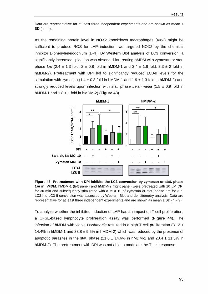
Results
95
Data are representative for at least three independent experiments and are shown as mean ±
SD (n = 4).
As the remaining protein level in NOX2 knockdown macrophages (40%) might be
sufficient to produce ROS for LAP induction, we targeted NOX2 by the chemical
inhibitor Diphenyleneiodonium (DPI). By Western Blot analysis of LC3 conversion, a
significantly increased lipidation was observed for treating hMDM with zymosan or stat.
phase Lm (2.4 ± 1.3 fold, 2 ± 0.8 fold in hMDM-1 and 3.4 ± 1.6 fold, 3.3 ± 2 fold in
hMDM-2). Pretreatment with DPI led to significantly reduced LC3-II levels for the
stimulation with zymosan (1.4 ± 0.8 fold in hMDM-1 and 1.9 ± 1.3 fold in hMDM-2) and
strongly reduced levels upon infection with stat. phase Leishmania (1.5 ± 0.9 fold in
hMDM-1 and 1.8 ± 1 fold in hMDM-2) (Figure 43).
Figure 43: Pretreatment with DPI inhibits the LC3 conversion by zymosan or stat. phase
Lm in hMDM. hMDM-1 (left panel) and hMDM-2 (right panel) were pretreated with 10 µM DPI
for 30 min and subsequently stimulated with a MOI 10 of zymosan or stat. phase Lm for 3 h.
LC3-I to LC3-II conversion was assessed by Western Blot and densitometry analysis. Data are
representative for at least three independent experiments and are shown as mean ± SD (n = 9).
To analyze whether the inhibited induction of LAP has an impact on T cell proliferation,
a CFSE-based lymphocyte proliferation assay was performed (Figure 44). The
infection of hMDM with viable Leishmania resulted in a high T cell proliferation (31.2 ±
14.4% in hMDM-1 and 33.8 ± 9.5% in hMDM-2) which was reduced by the presence of
apoptotic parasites in the stat. phase (21.6 ± 14.6% in hMDM-1 and 20.4 ± 11.5% in
hMDM-2). The pretreatment with DPI was not able to modulate the T cell response.
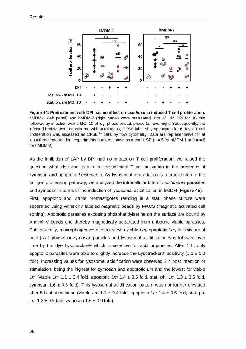
Results
96
Figure 44: Pretreatment with DPI has no effect on Leishmania induced T cell proliferation.
hMDM-1 (left panel) and hMDM-2 (right panel) were pretreated with 10 µM DPI for 30 min
followed by infection with a MOI 10 of log. phase or stat. phase Lm overnight. Subsequently, the
infected hMDM were co-cultured with autologous, CFSE-labeled lymphocytes for 6 days. T cell
proliferation was assessed as CFSElow
cells by flow cytometry. Data are representative for at
least three independent experiments and are shown as mean ± SD (n = 5 for hMDM-1 and n = 6
for hMDM-2).
As the inhibition of LAP by DPI had no impact on T cell proliferation, we raised the
question what else can lead to a less efficient T cell activation in the presence of
zymosan and apoptotic Leishmania. As lysosomal degradation is a crucial step in the
antigen processing pathway, we analyzed the intracellular fate of Leishmania parasites
and zymosan in terms of the induction of lysosomal acidification in hMDM (Figure 45).
First, apoptotic and viable promastigotes residing in a stat. phase culture were
separated using AnnexinV labeled magnetic beads by MACS (magnetic activated cell
sorting). Apoptotic parasites exposing phosphatidylserine on the surface are bound by
AnnexinV beads and thereby magnetically separated from unbound viable parasites.
Subsequently, macrophages were infected with viable Lm, apoptotic Lm, the mixture of
both (stat. phase) or zymosan particles and lysosomal acidification was followed over
time by the dye Lysotracker® which is selective for acid organelles. After 1 h, only
apoptotic parasites were able to slightly increase the Lysotracker® positivity (1.1 ± 0.2
fold). Increasing values for lysosomal acidification were observed 3 h post infection or
stimulation, being the highest for zymosan and apoptotic Lm and the lowest for viable
Lm (viable Lm 1.1 ± 0.4 fold, apoptotic Lm 1.4 ± 0.5 fold, stat. ph. Lm 1.3 ± 0.5 fold,
zymosan 1.6 ± 0.8 fold). This lysosomal acidification pattern was not further elevated
after 5 h of stimulation (viable Lm 1.1 ± 0.4 fold, apoptotic Lm 1.4 ± 0.6 fold, stat. ph.
Lm 1.2 ± 0.5 fold, zymosan 1.6 ± 0.9 fold).
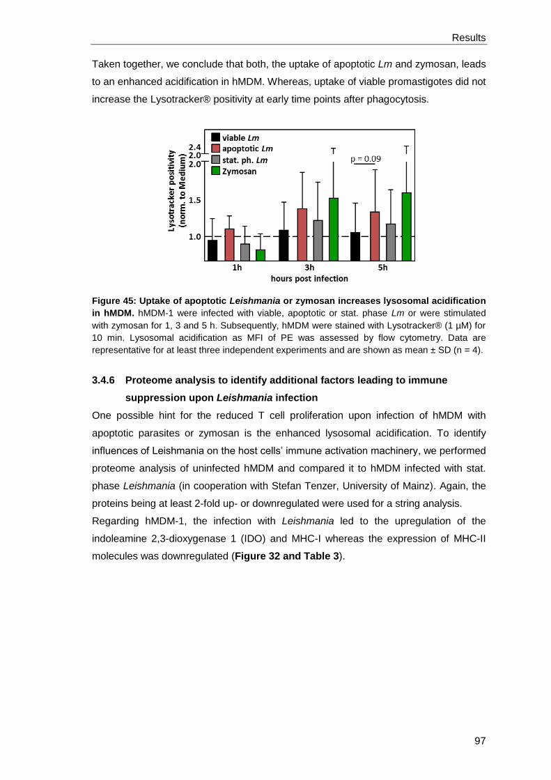
Results
97
Taken together, we conclude that both, the uptake of apoptotic Lm and zymosan, leads
to an enhanced acidification in hMDM. Whereas, uptake of viable promastigotes did not
increase the Lysotracker® positivity at early time points after phagocytosis.
Figure 45: Uptake of apoptotic Leishmania or zymosan increases lysosomal acidification
in hMDM. hMDM-1 were infected with viable, apoptotic or stat. phase Lm or were stimulated
with zymosan for 1, 3 and 5 h. Subsequently, hMDM were stained with Lysotracker® (1 µM) for
10 min. Lysosomal acidification as MFI of PE was assessed by flow cytometry. Data are
representative for at least three independent experiments and are shown as mean ± SD (n = 4).
3.4.6 Proteome analysis to identify additional factors leading to immune
suppression upon Leishmania infection
One possible hint for the reduced T cell proliferation upon infection of hMDM with
apoptotic parasites or zymosan is the enhanced lysosomal acidification. To identify
influences of Leishmania on the host cells’ immune activation machinery, we performed
proteome analysis of uninfected hMDM and compared it to hMDM infected with stat.
phase Leishmania (in cooperation with Stefan Tenzer, University of Mainz). Again, the
proteins being at least 2-fold up- or downregulated were used for a string analysis.
Regarding hMDM-1, the infection with Leishmania led to the upregulation of the
indoleamine 2,3-dioxygenase 1 (IDO) and MHC-I whereas the expression of MHC-II
molecules was downregulated (Figure 32 and Table 3).
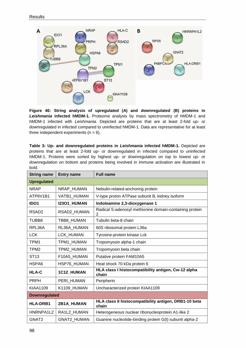
Results
98
Figure 46: String analysis of upregulated (A) and downregulated (B) proteins in
Leishmania infected hMDM-1. Proteome analysis by mass spectrometry of hMDM-1 and
hMDM-1 infected with Leishmania. Depicted are proteins that are at least 2-fold up- or
downregulated in infected compared to uninfected hMDM-1. Data are representative for at least
three independent experiments (n = 6).
Table 3: Up- and downregulated proteins in Leishmania infected hMDM-1. Depicted are
proteins that are at least 2-fold up- or downregulated in infected compared to uninfected
hMDM-1. Proteins were sorted by highest up- or downregulation on top to lowest up- or
downregulation on bottom and proteins being involved in immune activation are illustrated in
bold.
String name Entry name Full name
Upregulated
NRAP NRAP_HUMAN Nebulin-related-anchoring protein
ATP6V1B1 VATB1_HUMAN V-type proton ATPase subunit B, kidney isoform
IDO1 I23O1_HUMAN Indoleamine 2,3-dioxygenase 1
RSAD2 RSAD2_HUMAN Radical S-adenosyl methionine domain-containing protein 2
TUBB8 TBB8_HUMAN Tubulin beta-8 chain
RPL36A RL36A_HUMAN 60S ribosomal protein L36a
LCK LCK_HUMAN Tyrosine-protein kinase Lck
TPM1 TPM1_HUMAN Tropomyosin alpha-1 chain
TPM2 TPM2_HUMAN Tropomyosin beta chain
ST13 F10A5_HUMAN Putative protein FAM10A5
HSPA6 HSP76_HUMAN Heat shock 70 kDa protein 6
HLA-C 1C12_HUMAN HLA class I histocompatibility antigen, Cw-12 alpha chain
PRPH PERI_HUMAN Peripherin
KIAA1109 K1109_HUMAN Uncharacterized protein KIAA1109
Downregulated
HLA-DRB1 2B1A_HUMAN HLA class II histocompatibility antigen, DRB1-10 beta chain
HNRNPA1L2 RA1L2_HUMAN Heterogeneous nuclear ribonucleoprotein A1-like 2
GNAT2 GNAT2_HUMAN Guanine nucleotide-binding protein G(t) subunit alpha-2
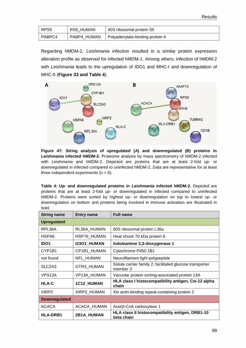
Results
99
RPS5 RS5_HUMAN 40S ribosomal protein S5
PABPC4 PABP4_HUMAN Polyadenylate-binding protein 4
Regarding hMDM-2, Leishmania infection resulted in a similar protein expression
alteration profile as observed for infected hMDM-1. Among others, infection of hMDM-2
with Leishmania leads to the upregulation of IDO1 and MHC-I and downregulation of
MHC-II (Figure 33 and Table 4).
Figure 47: String analysis of upregulated (A) and downregulated (B) proteins in
Leishmania infected hMDM-2. Proteome analysis by mass spectrometry of hMDM-2 infected
with Leishmania and hMDM-2. Depicted are proteins that are at least 2-fold up- or
downregulated in infected compared to uninfected hMDM-2. Data are representative for at least
three independent experiments (n = 6).
Table 4: Up- and downregulated proteins in Leishmania infected hMDM-2. Depicted are
proteins that are at least 2-fold up- or downregulated in infected compared to uninfected
hMDM-2. Proteins were sorted by highest up- or downregulation on top to lowest up- or
downregulation on bottom and proteins being involved in immune activation are illustrated in
bold.
String name Entry name Full name
Upregulated
RPL36A RL36A_HUMAN 60S ribosomal protein L36a
HSPA6 HSP76_HUMAN Heat shock 70 kDa protein 6
IDO1 I23O1_HUMAN Indoleamine 2,3-dioxygenase 1
CYP1B1 CP1B1_HUMAN Cytochrome P450 1B1
not found NFL_HUMAN Neurofilament light polypeptide
SLC2A3 GTR3_HUMAN Solute carrier family 2, facilitated glucose transporter member 3
VPS13A VP13A_HUMAN Vacuolar protein sorting-associated protein 13A
HLA-C 1C12_HUMAN HLA class I histocompatibility antigen, Cw-12 alpha chain
XIRP2 XIRP2_HUMAN Xin actin-binding repeat-containing protein 2
Downregulated
ACACA ACACA_HUMAN Acetyl-CoA carboxylase 1
HLA-DRB1 2B1A_HUMAN HLA class II histocompatibility antigen, DRB1-10 beta chain
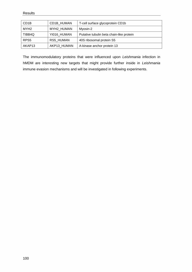
Results
100
CD1B CD1B_HUMAN T-cell surface glycoprotein CD1b
MYH2 MYH2_HUMAN Myosin-2
TIBB4Q YI016_HUMAN Putative tubulin beta chain-like protein
RPS5 RS5_HUMAN 40S ribosomal protein S5
AKAP13 AKP13_HUMAN A-kinase anchor protein 13
The immunomodulatory proteins that were influenced upon Leishmania infection in
hMDM are interesting new targets that might provide further inside in Leishmania
immune evasion mechanisms and will be investigated in following experiments.
Discussion
101
4 Discussion
4.1 Summary of the data
In the first part of this thesis we focused on the modulation of autophagy, a highly
conserved cellular response mechanism to organismic stress. We aimed to investigate
autophagy in two different phenotypes of immunomodulatory cells, namely pro- and
anti-inflammatory human macrophages. By applying chemical class I PI3K/mTOR
inhibitors or the peptide Tat-Beclin autophagy in hMDM was induced. In contrast,
targeting the class III PI3K complex by Spautin-1 and Wortmannin blocked autophagy
induction. Interestingly, hMDM-2 were in general more susceptible for autophagy
induction. To inhibit autophagy more specifically, a siRNA approach was established
and the knockdown efficiency could be shown to be time- and target-dependent. By
functional characterization of ULK-1 and Beclin-1 knockdown cells, chemical
autophagy induction could still take place, indicating an ULK-1- and Beclin-1-
independent autophagy mechanism in hMDM. Regarding the role of autophagy
modulation on antigen processing using the model antigen Tetanus Toxoid, no impact
could be observed. Surprisingly, hMDM-2 were shown to be superior to hMDM-1 for
priming an adaptive immune response which might be due to higher expression of
MHC-II and proteins being involved in antigen processing.
Also pathogens such as Leishmania were shown to interact with the host cells’
autophagy machinery. We demonstrated Leishmania to induce autophagy in hMDM
however this occurred independent of the Leishmania virulence factor GP63.
Furthermore, GP63 deficiency did not influence the infection of hMDM and the adaptive
immune response.
In the second part we focused on the modulation of LC3-associated phagocytosis. In a
previous study we could show that apoptotic Lm in the virulent inoculum induce LAP
and suppress the adaptive immune response. Replacing apoptotic Lm by LAP-inducing
PS beads or zymosan, we demonstrated the formation of LC3+ phagosomes around
those particles. Stimulation of hMDM with PS coated beads or zymosan and co-
infection with viable Lm resulted in a reduction of T cell proliferation. Suppression of the
adaptive immune response enabled an enhanced parasite survival. Analyzing the
underlying mechanisms of LAP in hMDM, inhibition of the NADPH oxidase by DPI
blocked LAP induction. In addition, infection of hMDM with Staurosporine-treated Lm,
being ROS positive, increased LC3 conversion which suggests ROS-dependent LAP
induction in hMDM. Furthermore, uptake of apoptotic Lm or zymosan led to enhanced
acidification of hMDM indicating a role for improved antigen processing and
presentation.
Discussion
102
4.2 Autophagy modulation in human primary macrophages
Extensive research using cell lines or mouse model systems contributed strongly to the
understanding of the autophagy process. Though still little is known of the autophagy
pathway in human primary macrophages, playing a central role as immune modulating
cells. In the first part of this study we focused on autophagy modulation in human
primary macrophages, in which autophagy has the dual role of ensuring homeostasis
and degrading invading pathogens for antigen presentation. Therefore, we took a
closer look on autophagy modulation in two different phenotypes of macrophages, the
pro-inflammatory type-I and the anti-inflammatory type-II macrophages.
Inhibition of the class I PI3K/mTOR pathway resulted in an induction of autophagy.
Specifically, PI-103 and AZD8055 were shown to induce autophagy more potent than
Rapamycin. This finding is in accordance with previous studies which revealed
Rapamycin to be less potent for autophagy induction indicated by just modest LC3
conversion (Andersson et al., 2016). Furthermore, we could successfully prove the
autophagy inducing peptide Tat-Beclin to induce autophagy in hMDM-1 and hMDM-2.
This specific activation of Beclin-1 is in accordance with previous data in HeLa and
cancer stem cells (Shoji-Kawata et al., 2013; Sharif et al., 2017).
Using pre-treatment of macrophages with Bafilomycin A1 which blocks lysosomal
acidification and hence degradation of autophagosomal cargo, we demonstrated that
the whole sequestration process was induced and not just the initial steps of
autophagy. Our data are in line with previous studies showing LC3-II and p62
accumulation due to blocked degradation with Bafilomycin A1 (Bjorkoy et al., 2005;
Watanabe and Tanaka, 2011; Klionsky et al., 2016).
Furthermore, we aimed to regulate autophagy negatively by chemical inhibition of the
class III PI3K complex downstream of mTOR. In line with previous studies, Spautin-1
and Wortmannin were demonstrated to inhibit autophagy induction in human primary
macrophages (Mateo et al., 2013). Our data indicate that LY294002 is capable of
inhibiting autophagy in hMDM-1 but interestingly not in hMDM-2. Controversy results
for LY294002 as autophagy inhibitor or inducer were already demonstrated. Blommaart
et al. could demonstrate LY294002 to inhibit autophagy in rat hepatocytes, a finding in
agreement with Petiot’s data showing LY294002 to dampen autophagy activity in HT-
29 cells (Blommaart et al., 1997; Petiot et al., 2000). Whereas Xing and colleagues
could show LY294002 to induce autophagy in gastric cancer cells by activating p53
and caspase-3 which led to increased expression of LC3 (Xing et al., 2008). Our
results and the given examples highlight the cell type specific function of LY294002
which seems to differ even between different macrophage phenotypes. Concerning the
most commonly described autophagy inhibitor, 3-MA, pre-treatment of macrophages
Discussion
103
with 1 mM even led to an increased LC3-I to LC3-II conversion. Previously, 3-MA was
described to inhibit autophagy at high concentrations (e.g. 10 mM) in various cell types
and even in low concentrations in the presence of active vitamin D3 in hMDM (Seglen
and Gordon, 1982; Blommaart et al., 1997; Petiot et al., 2000; Yuk et al., 2009). In
human prostate cancer cells, 5 mM 3-MA treatment could inhibit the autophagy
inducing effects of PI-103 (Degtyarev et al., 2008). However there is rising evidence
that 3-MA can also induce autophagy, shown by Lin and colleagues in RAW 264.7
macrophages or under nutrient rich conditions in mouse embryonic fibroblasts and
L929 cells (Wu et al., 2010; Lin et al., 2012). Considering these results and based on
our findings, we suggest that 3-MA induces autophagy in hMDM at low concentrations
and possesses inhibitory capacities at higher concentrations.
Most strategies to gain information about the autophagic process focus on the inhibition
of autophagy by genome editing, RNA interference or chemical modulation of class III
PI3-kinase using pharmacological inhibitors. Chemical targeting of class III PI3K is
associated with various side effects as Vps34 regulates cellular membrane trafficking
processes and transport across the nuclear membrane (Roggo et al., 2002; Johnson et
al., 2006). Indeed, we could observe reduced phagocytic capacity of hMDM upon
stimulation with Wortmannin (data not shown). As chemical modulators are often not
entirely specific, genetic intervention is preferred (Vinod et al., 2014). Therefore, we
targeted several autophagy related proteins by siRNA knockdown. In general, we
observed protein knockdowns in hMDM to be time and target dependent. We could
show a long protein half-life for Vps34 suggested by protein stability for up to one
week. In contrast, an effective protein knockdown by siRNA was reported in HeLa cells,
demonstrating significantly reduced protein levels 2-5 days post knockdown (Nobukuni
et al., 2005; Sagona et al., 2010). Moreover, RNAi knockdown of Beclin-1 was efficient
1-2 days post transfection in RAW 264.7 macrophages whereas we observed a longer
protein half-life in hMDM indicated by reduced Beclin-1 levels on day 6-7 post siRNA
application (Li et al., 2013; Martyniszyn et al., 2013). Our data demonstrate comparable
protein stability of ATG16L1 to Beclin-1 in hMDM although in contrast, other studies
could show efficient protein reduction 48 h post RNAi treatment in HeLa and HCT116
cell lines (Rioux et al., 2007; Homer et al., 2010) Hence, our data elucidate that
findings on protein stability in cell lines can not be extrapolated to human primary cells.
Interestingly, although we demonstrated ULK-1 protein levels to be strongly reduced,
the autophagy machinery in human macrophages was still functional. In contrast to our
results, an essential role for ULK-1 as modulator of autophagy was shown by a kinase-
specific siRNA screening in HEK293 cells (Young et al., 2006; Chan et al., 2007). In
addition, the group of D. Green could show reduced LC3 levels and LC3 punctae in
ULK-1 knockdown murine bone marrow-derived macrophages upon autophagy
Discussion
104
induction with Rapamycin (Martinez et al., 2011). On the other hand and in line with our
data, Alers et al. reported ULK-1/ULK-2-independent starvation-induced autophagy in
DT40 cells assuming mTOR independent regulation of Atg13 and FIP200 by other
kinases (Alers et al., 2014). In line with their results, an mTOR independent, but not yet
identified pathway was proposed in COS7 cells by autophagy induction with
Glucosamine (Shintani et al., 2010). Furthermore, controversy results for ULK-1/2
requirement for autophagy induction were obtained depending on the applied stimuli.
ULK-1/2 are required for amino acid induced autophagy but dispensable for glucose
deprivation induced autophagy in mouse embryonic fibroblasts (Cheong et al., 2011). It
might be possible that also in human primary macrophages autophagy induction can
precede via an mTOR- and accordingly ULK-1-independent mechanism. Moreover,
ULK-1-independent autophagy in hMDM might be explained by the functional
compensation by the structural related protein ULK-2. ULK-1 and ULK-2 redundancy is
cell type specific, as ULK-2 can not compensate for ULK-1 in HEK293 cells and
cerebellar granule neurons but is sufficient for autophagy induction in mouse
embryonic fibroblasts (Chan et al., 2007; Kundu et al., 2008; Lee and Tournier, 2014).
Comprehensively, further experiments need to be performed to rule out if the kinase
activity of ULK-1 is compensated by ULK-2 in hMDM or if an ULK-independent
mechanism for autophagy induction exists. A simultaneous knockdown of ULK-1 and
ULK-2 will clarify if protein compensation occurs. Furthermore, if autophagy can
proceed in ULK-1/2 knockdown cells, analyzing Beclin-1 phosphorylation will provide a
hint for other kinases than ULK being involved in autophagy induction.
Assessing the role of Beclin-1 knockdown for autophagy induction, we could show that,
similar to ULK-1 knockdown, LC3 conversion could still take place. In contrast to these
data, Beclin-1-dependent autophagy induction with Rapamycin was shown in primary
cortical neurons and murine bone marrow-derived macrophages (Grishchuk et al.,
2011; Martinez et al., 2011). On the other hand and in line with our data are the results
by Li et al., demonstrating that Beclin-1 is critical for starvation induced but not for
Rapamycin induced autophagy in murine macrophages (RAW 264.7) (Li et al., 2013).
Furthermore, other autophagy inducers could also be shown to induce LC3 conversion
in a Beclin-1 independent manner in various cell types (Gao et al., 2010; Smith et al.,
2010; Tian et al., 2010). Interestingly, Gossypol induces autophagy in a Beclin-1
dependent way in HeLa cells and in a Beclin-1 independent way in MCF-7 cells (Gao et
al., 2010). Hence we suggest that autophagy induction in human primary macrophages
is independent of Beclin-1.
By comparing two different macrophage phenotypes we observed that hMDM-2 were
more susceptible for autophagy induction compared to hMDM-1. The physiological
function of alternatively activated macrophages is the uptake of apoptotic or necrotic
Discussion
105
cells and tissue repair functions and thus, possessing a higher phagocytic capacity,
hMDM-2 might be of greater importance for maintaining homeostasis (Mosser and
Edwards, 2008). In line with our data, a recently published study also indicates the
autophagic sequestration pathway to be highly dependent on the investigated cell type
and can even vary by applying different stimuli (Gomez-Sanchez et al., 2015).
Taken together, our data as well as the mentioned observations by other groups
highlight the distinct regulation of autophagy in dependence of the used model systems
and applied stimuli. Specifically, several autophagy-related proteins were demonstrated
to be more stable in human primary cells compared to cell lines. Additionally,
autophagic sequestration is ULK-1-dependent in HEK293 cells but ULK-1 is
dispensable for autophagy induction in hMDM. Our results suggest that even in
different phenotypes of macrophages, autophagy is differentially regulated as hMDM-2
possess higher autophagy inducibility and are not sensitive to LY294002 treatment.
4.3 Impact of autophagy on antigen presentation using the model
antigen Tetanus Toxoid
The adaptive immune response is orchestrated by the processing and presentation of
peptides on MHC molecules for the recognition by specific T cells (Munz, 2016a). For a
long time, the endocytotic uptake and lysosomal degradation pathway was suspected
to be responsible for the processing and presentation of exogenous antigens on
MHC-II molecules (Roche and Furuta, 2015). The presentation of cytosolic antigens on
MHC-II revealed the participation of the autophagic sequestration process to be
involved in the priming of a CD4+ T cell response (Nimmerjahn et al., 2003; Dengjel et
al., 2005). Jagannath and colleagues could nicely demonstrate that starvation or
Rapamycin induced autophagy enhances the efficacy of BCG vaccine by increasing
peptide processing and presentation of cytosolic secreted Ag85B (Jagannath et al.,
2009).
To analyze if autophagy in hMDM is involved in antigen processing, we used the recall
antigen Tetanus Toxoid to initiate an adaptive immune response as German blood
donors are commonly vaccinated against Tetanus. As expected from literature, we
demonstrated the onset of a detectable T cell proliferation upon antigen processing and
presentation by macrophages to T cells (Nielsen et al., 2010). In line with other studies,
the proliferating T cells were shown to exhibit a CD4+ phenotype, indicating peptide
presentation via MHC-II molecules (Barbey et al., 2007; Cellerai et al., 2007). To
investigate the role of autophagy in this MHC-II mediated antigen presentation, we
induced autophagy by AZD8055 or inhibited it by siRNA knockdown of ULK-1. Neither
the induction of autophagy nor the inhibition could be shown to influence the Tetanus
Discussion
106
Toxoid specific T cell proliferation. The inactivation process of Tetanus Toxin by
formaldehyde to generate the toxoid vaccine prevents the binding to gangliosides on
motoneurons leading to the loss of its toxic effects. Hence, TT might be taken up by
antigen presenting cells by endocytosis or pinocytosis without specific receptor
engagement or by receptor-mediated phagocytosis (Kamphorst et al., 2010; Roche and
Furuta, 2015). Consequently, the expendability of autophagy is probably due to the
uptake process and intracellular localization of Tetanus Toxoid in an endosome making
it inaccessible for autophagic targeting. Focusing on the latter uptake mechanism,
specific receptor triggering initiating LC3-associated phagocytosis can modify
endocytosis and degradation of extracellular material for MHC-II presentation (Munz,
2016a). The requirement of the LAP degradation process was shown in murine
dendritic cells lacking ATG5 as CD4+ T cell priming was strongly impaired, especially
the processing and presentation of phagocytosed antigens containing Toll-like receptor
stimuli (Blander and Medzhitov, 2006; Lee et al., 2010). According to these studies, the
modulation of the conventional autophagy process by AZD8055 and ULK-1 might be
“too upstream” as only the autophagy proteins downstream of mTOR and ULK-1 are
required for LAP. As we did not further rule out the intracellular localization of Tetanus
Toxoid, we can only assume that it is taken up in a compartment resulting in antigen
processing independent on conventional autophagy. To clarify the participation of
receptor-mediated LAP, a modulation downstream of mTOR such as an ATG5
knockdown, needs to be done.
Interestingly, T cell proliferation in response to co-cultivation of CD14+ isolated
monocytes (used for knockdown experiments) with PBLs was in general elevated
compared to macrophages generated by plastic adherence. As CD14 is a surface
pattern recognition receptor for LPS and a co-receptor for TLR2, the applied isolation
method leads to macrophage activation prior to co-cultivation with autologous PBLs
(Dobrovolskaia and Vogel, 2002). In line with this, CD14-dependent isolation of human
monocytes results in enhanced phagocytic activity of Listeria monocytogenes (Neu et
al., 2013).
4.4 Priming of an adaptive immune response by hMDM-1 vs. hMDM-2
Interestingly, direct comparison of the T cell response induced by hMDM-1 and
hMDM-2 revealed anti-inflammatory macrophages to be superior in priming lymphocyte
proliferation. Controversially, it is known from literature that monocytes differentiated by
GM-CSF, inducing a pro-inflammatory phenotype, have a higher T cell stimulatory
capacity (Xu et al., 2013). Looking for a possible explanation, we examined the
expression of T cell activation marker on the surface of both phenotypes.
Discussion
107
Oversimplified, neglecting environmental signs such as cytokines, activation of naïve
CD4 T cells requires two distinct signals. On the one hand the ligation of a specific T
cell receptor with a peptide bound to MHC class II and on the other hand a co-
stimulatory signal which is not antigen specific (Janeway and Bottomly, 1994). In
addition to antigen presentation, co-stimulation by CD80/CD86 or CD40 on antigen
presenting cells binding to CD28 or CD40L respectively on T cells is essential for the
induction of an adaptive immune response (Fujii et al., 2004; Murphy et al., 2012).
Concerning the co-stimulatory molecules CD40 and CD86, we were not able to detect
different expression patterns between hMDM-1 and hMDM-2. In accordance with
previous studies, CD80 was proven to be a specific marker for GM-CSF generated
macrophages (Ambarus et al., 2012). Although being indispensable for T cell
activation, co-stimulatory molecules themselves could not induce any responses in T
cells (Janeway and Bottomly, 1994). Consequently, the antigen-specific MHC signal
accompanied by any co-stimulatory molecule might be limiting to induce more or less T
cell activation (Germain, 1994). Indeed, we found MHC-II to be higher expressed on
M-CSF generated macrophages (hMDM-2), which is in line with phenotyping by
Verreck and colleagues (Verreck et al., 2006).
In addition to surface staining of MHC and co-stimulatory molecules using flow
cytometry, we analyzed hMDM-1 and hMDM-2 by proteome analysis using Mass
Spectrometry to identify additional immunomodulatory molecules. Focusing on
upregulated proteins in hMDM-1 compared to hMDM-2, we found the Calcineurin-
binding protein cabin-1 to be higher expressed. The Calcineurin-binding protein cabin-1
was shown to serve as a negative regulator of T cell receptor signaling via the inhibition
of calcineurin (Sun et al., 1998). Furthermore, being in line with the increased
microbicidal capacity of pro-inflammatory macrophages, the V-type proton ATPase
subunit B and the SNARE protein VAMP8 are upregulated.
Concerning hMDM-2, more immunomodulatory involved proteins were shown to be
upregulated compared to hMDM-1, namely CD163, Legumain, L-amino-acid oxidase,
CPVL, Sialoadhesin and Galectin-10. As expected, the scavenger receptor cysteine-
rich type 1 protein M130, also named CD163, was found to be higher expressed in
hMDM-2. After shedding, the soluble form (sCD163) may play an important role in anti-
inflammatory responses (Verreck et al., 2006; Moestrup and Møller, 2009). Focusing
on proteins supporting antigen processing, Legumain was shown to be higher
expressed in hMDM-2. It is found in lysosomes where its cysteine endopeptidase
activity contributes to antigen processing for MHC-II presentation (Dall and
Brandstetter, 2016). In addition, the L-amino-acid oxidase was shown by similarity to
have a potential role in lysosomal antigen processing and presentation (Mason et al.,
2004). CPVL, a probable serine carboxypeptidase, might be involved in the digestion of
Discussion
108
phagocytosed particles in the lysosome, trimming of peptides for antigen presentation
and the participation in an inflammatory protease cascade (Mahoney et al., 2001).
Being in line with the higher phagocytic capacity of hMDM-2, Sialoadhesin was
upregulated acting as endocytic receptor mediating clathrin-dependent endocytosis
and providing sialic-acid dependent binding to lymphocytes (van den Berg et al., 1992;
Vanderheijden et al., 2003; O'Neill et al., 2013). On the other hand, Galectin-10
regulates immune responses through the recognition of cell-surface glycans and is
essential for the anergy and suppressive function of CD25 positive regulatory T cells
(Kubach et al., 2007).
In conclusion, by comparing the two macrophage phenotypes, hMDM-2 were shown to
have a higher expression profile of proteins being involved in antigen processing and
presentation such as peptidases and MHC-II. Most likely, this makes them superior to
hMDM-1 in priming an adaptive immune response. This assumption will be verified in
following experiments by specific modulation of the identified targets such as MHC-II
blocking experiments or inhibition (e.g. by specific protein down regulation) of the
identified peptidases.
4.5 Influence of GP63 on infectivity, adaptive immunity and host cell
autophagy
GP63 is a main surface protein of Leishmania parasites and described as virulence
factor mediating attraction of and adhesion to macrophages, avoiding innate immune
killing mechanisms and providing an enhanced intracellular parasite survival.
Previously published data indicate a clear link between GP63 expression and infectivity
or binding to macrophages (Kweider et al., 1987; Liu and Chang, 1992; Ahmed et al.,
1998; Brittingham et al., 1999; Thiakaki et al., 2006). Kweider et al. used monoclonal
IgM antibodies against L. braziliensis to block surface proteins leading to reduced
percentage of infected MFs of about 25% and reduced intracellular survival (Kweider et
al., 1987). In the studies of Liu et al., L. amazonensis was passaged for more than 3
years resulting in the loss of GP63 and hence reduced infectivity whereas
overexpression of GP63 in those parasites could restore the binding to a murine
macrophage cell line (Liu and Chang, 1992). In contrast to these data, we could not
observe an altered infection rate of GP63-deficient parasites by infection of hMDM with
Lm SD WT, Lm SD KO and Lm SD KO+GP63. In line with our data are results
obtained by Joshi et al. using for the first time GP63 knockout parasites by targeted
gene deletion of GP63 in Lm Seidmann. GP63 knockout and wildtype parasites
showed similar early (1 h) and late (96 h) infection rates in mouse peritoneal MFs and
comparable differentiation in amastigotes. Furthermore, the KO parasites were able to
Discussion
109
induce lesion formation with an initial delay but unaltered disease progression in Balb/c
mice. Similar to our results, Joshi et al. concluded that the GP63 gene products 1-6 do
not play a role for the survival of Leishmania within macrophages (Joshi et al., 1998).
Since more than 25 years it is known, that the stimulation of PBMCs with crude
Leishmania sonicates induces a memory T cell response in previously unexposed
individuals (Kemp et al., 1991; Kemp et al., 1992). In accordance with these data, we
also observed a lymphocyte proliferative response using a slightly different setting by
prior incubation of hMDM with Leishmania and subsequent co-cultivation with PBLs
allowing antigen processing, presentation and recognition. By comparing Lm SD WT
and Lm SD GP63 KO parasites, no significant changes in the lymphocyte proliferative
response were observed. Opposing results were obtained by Matheoud et al.,
demonstrating immune evasion of Leishmania by the inhibition of cross-presentation
using direct cleavage of VAMP8 in a GP63-dependent manner (Matheoud et al., 2013).
Analogical immune suppressing effects were demonstrated by the direct binding of
GP63 to human primary NK cells leading to the inhibition of proliferation (Lieke et al.,
2008). In addition it was shown that GP63 of Lm and L. donovani was able to cleave
CD4 molecules on human T cells. However, in this study an artificial in vitro system
was used and the biological significance was critically discussed (Hey et al., 1994). In
line with our results, it was shown by various studies that GP63 seems to be no
immunodominant antigen as stimulation of PBMCs with purified GP63 was not able to
induce T cell proliferation in neither unexposed individuals nor any CL patient or cured
patient (Mendonca et al., 1991; KEMP et al., 1994; Gaafar et al., 1995). On the other
hand, immunization of mice with GP63 induces a significant level of protection
highlighting again the T cell dichotomy between mice and human (Handman and
Mitchell, 1985; Russell and Alexander, 1988). The unaltered infection rate and the
inability of the GP63 protein itself to induce T cell proliferation gives an explanation of
our results that GP63 seems to be neglectable for the induction of an adaptive immune
response in hMDM. To further analyze GP63 and its role as virulence factor, the
occurrence of an oxidative burst and hence impaired survival of GP63 KO parasites
need to be investigated.
GP63 is known to mediate a silent uptake in macrophages via CR3 and additionally
ensures survival by cleavage of macrophage signaling proteins (Gomez et al., 2009;
Isnard et al., 2012). Host signaling is altered as GP63 gets access to the macrophages’
cytoplasm through a process mediated in part by host lipid rafts enabling cleavage of
the autophagy modulator mTOR (Gomez et al., 2009; Jaramillo et al., 2011). To
investigate if mTOR cleavage has an impact on the macrophages’ autophagy
machinery, we analyzed autophagy modulation by LC3 conversion during infection with
GP63-deficient Lm and the respective controls. Infection of hMDM with Leishmania
Discussion
110
resulted in the induction of autophagy. The induction of autophagy in host
macrophages by Lm is in agreement with results of Cyrino et al. demonstrating L.
amazonensis to induce autophagy in Balb/c and C57BL/6 BMDM as well as in RAW
macrophages (Cyrino et al., 2012). Also the visceral leishmaniasis causing strain L.
donovani was recently shown to induce LC3-I to LC3-II conversion in THP-1 cells and
to modulate host cell autophagy in a Beclin-1-dependent manner (Singh et al., 2016).
We could not detect a GP63-mediated modulation of autophagy by analyzing LC3
lipidation. These results are in line with a recently published study in mice also
indicating LC3 levels by Western Blot to be unaffected upon infection with Lm SD WT
and Lm SD GP63 KO although by immunofluorescence they could show that the
presence of GP63 on promastigotes impaired the recruitment of LC3 to phagosomes
(Matte et al., 2016).
Taken together, these studies and our data indicate that the relevance of GP63 as
Leishmania virulence factor is strongly dependent on the experimental setup and
differs between mice and human. A role of GP63 on the hosts’ autophagy machinery
can not be completely ruled out. Overall levels of LC3 did not change, however the
intracellular distribution might be altered. Further research has to be performed
focusing on LC3 recruitment to parasite containing compartments assessed by
immunofluorescence, which might be improved using GP63 KO parasites.
4.6 LAP as immune evasion mechanism during Leishmania infection
Host-pathogen-interactions are a well-studied key dogma in infectious diseases but far
away from being fully understood. According to the “catch me if you can” principle, the
pathogen tries to inhibit host defense mechanisms or even subverts them for its own
benefit (Lennemann and Coyne, 2015). An evolutionary highly conserved cellular
response mechanism is the non-canonical autophagic process “LC3-associated
phagocytosis” (LAP), mediating rapid phagosomal acidification and enhanced killing of
engulfed microbes (Sanjuan et al., 2007). For several pathogens like Mycobacterium
tuberculosis, the autophagic sequestration process was shown to control infection
(Gutierrez et al., 2004). Whereas other microbes such as the bacteria Listeria
monocytogenes and Burkholderia pseudomallei evade LAP compartments and mutants
lacking proteins for phagosomal escape were trapped in phagosomes for enhanced
killing (Gong et al., 2011; Birmingham et al., 2014). For Lm we could already
demonstrate that the apoptotic parasites reside in single membrane, LC3+
compartments in host macrophages and the presence of apoptotic promastigotes in the
virulent inoculum causes the suppression of an adaptive immune response allowing
disease progression (van Zandbergen et al., 2006; Crauwels et al., 2015).
Discussion
111
Replacing apoptotic parasites by phosphatidylserine (PS) beads and zymosan led to
the formation of LC3+ phagosomes and LC3 lipidation which is in line with previous
studies in RAW 264.7 and murine bone marrow-derived macrophages (Sanjuan et al.,
2007; Martinez et al., 2011). Co-incubation of hMDM with PS-beads and viable
Leishmania dampened the T cell response to the same degree as apoptotic
Leishmania did. In accordance with these results, Martinez et al. demonstrated that in
the absence of LAP the uptake of dead cells results in the secretion of pro-
inflammatory cytokines suggesting immune dampening effects for LAP-mediated
phagocytosis (Martinez et al., 2011). We demonstrated zymosan to significantly
suppress Leishmania induced T cell proliferation and hence resulting in enhanced
parasite survival as observed for co-infection of hMDM with viable and apoptotic
Leishmania. Treatment of hMDM with zymosan triggered a strong release of the pro-
inflammatory cytokine TNF-α and the anti-inflammatory cytokine IL-10. In agreement
with our results, Du and colleagues reported high secretion levels of TNF-α after 3 h
and high levels of IL-10 6 h post stimulation of macrophages with zymosan (Du et al.,
2006). The production of TNF-α leads to the recruitment of adaptive immune cells and
subsequent secretion of IL-10 suppresses macrophage activation and dampens the
immune response preventing autoimmunity (Arango Duque and Descoteaux, 2014).
Additionally, the immune dampening effects of zymosan stimulation have been shown
in autoimmune disease models for diabetes and multiple sclerosis. Injection of
zymosan was able to delay diabetes onset by the expansion of PD-L1+, TGF-β+
macrophages and Foxp3+ regulatory T cells in NOD mice (Burton et al., 2010).
Furthermore, zymosan regulates the cytokine secretion in dendritic cells and
macrophages resulting in immunological tolerance as the co-injection of zymosan and
OVA plus LPS suppresses the OVA-specific T cell response by APCs secreting IL-10
and TGF-β but lacking IL-6 (Dillon et al., 2006). Another mechanism indicating the
immune dampening effects of zymosan was reported by Yem demonstrating zymosan
treatment of macrophages to result in surface Ia antigen loss from many macrophages
leading to reduced T cell proliferation (Yem and Parmely, 1981).
Taken together, our data indicate, that the immune dampening effects of apoptotic
Leishmania in the virulent inoculum or their replacement by PS beads and zymosan
are probably due to anti-inflammatory cytokine secretion. Although zymosan induced
strong TNF-α secretion, it is likely that anti-inflammatory cytokines halt adaptive
immune activation as only a moderate T cell response was observed. To verify this
mechanism, further experiments such as analyzing TGF-β secretion, need to be
performed. The reduced T cell proliferation can be considered as immune evasion
mechanism used by Leishmania as it enables improved intracellular parasite survival
and differentiation in amastigotes.
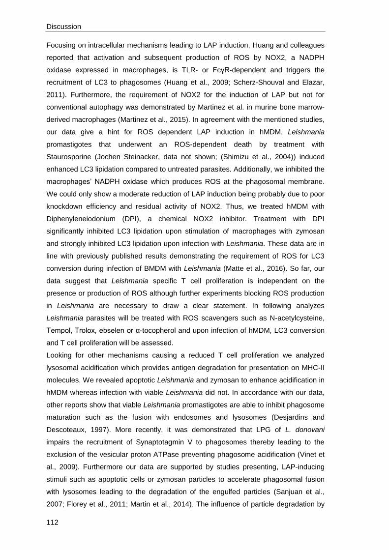
Discussion
112
Focusing on intracellular mechanisms leading to LAP induction, Huang and colleagues
reported that activation and subsequent production of ROS by NOX2, a NADPH
oxidase expressed in macrophages, is TLR- or FcγR-dependent and triggers the
recruitment of LC3 to phagosomes (Huang et al., 2009; Scherz-Shouval and Elazar,
2011). Furthermore, the requirement of NOX2 for the induction of LAP but not for
conventional autophagy was demonstrated by Martinez et al. in murine bone marrow-
derived macrophages (Martinez et al., 2015). In agreement with the mentioned studies,
our data give a hint for ROS dependent LAP induction in hMDM. Leishmania
promastigotes that underwent an ROS-dependent death by treatment with
Staurosporine (Jochen Steinacker, data not shown; (Shimizu et al., 2004)) induced
enhanced LC3 lipidation compared to untreated parasites. Additionally, we inhibited the
macrophages’ NADPH oxidase which produces ROS at the phagosomal membrane.
We could only show a moderate reduction of LAP induction being probably due to poor
knockdown efficiency and residual activity of NOX2. Thus, we treated hMDM with
Diphenyleneiodonium (DPI), a chemical NOX2 inhibitor. Treatment with DPI
significantly inhibited LC3 lipidation upon stimulation of macrophages with zymosan
and strongly inhibited LC3 lipidation upon infection with Leishmania. These data are in
line with previously published results demonstrating the requirement of ROS for LC3
conversion during infection of BMDM with Leishmania (Matte et al., 2016). So far, our
data suggest that Leishmania specific T cell proliferation is independent on the
presence or production of ROS although further experiments blocking ROS production
in Leishmania are necessary to draw a clear statement. In following analyzes
Leishmania parasites will be treated with ROS scavengers such as N-acetylcysteine,
Tempol, Trolox, ebselen or α-tocopherol and upon infection of hMDM, LC3 conversion
and T cell proliferation will be assessed.
Looking for other mechanisms causing a reduced T cell proliferation we analyzed
lysosomal acidification which provides antigen degradation for presentation on MHC-II
molecules. We revealed apoptotic Leishmania and zymosan to enhance acidification in
hMDM whereas infection with viable Leishmania did not. In accordance with our data,
other reports show that viable Leishmania promastigotes are able to inhibit phagosome
maturation such as the fusion with endosomes and lysosomes (Desjardins and
Descoteaux, 1997). More recently, it was demonstrated that LPG of L. donovani
impairs the recruitment of Synaptotagmin V to phagosomes thereby leading to the
exclusion of the vesicular proton ATPase preventing phagosome acidification (Vinet et
al., 2009). Furthermore our data are supported by studies presenting, LAP-inducing
stimuli such as apoptotic cells or zymosan particles to accelerate phagosomal fusion
with lysosomes leading to the degradation of the engulfed particles (Sanjuan et al.,
2007; Florey et al., 2011; Martin et al., 2014). The influence of particle degradation by
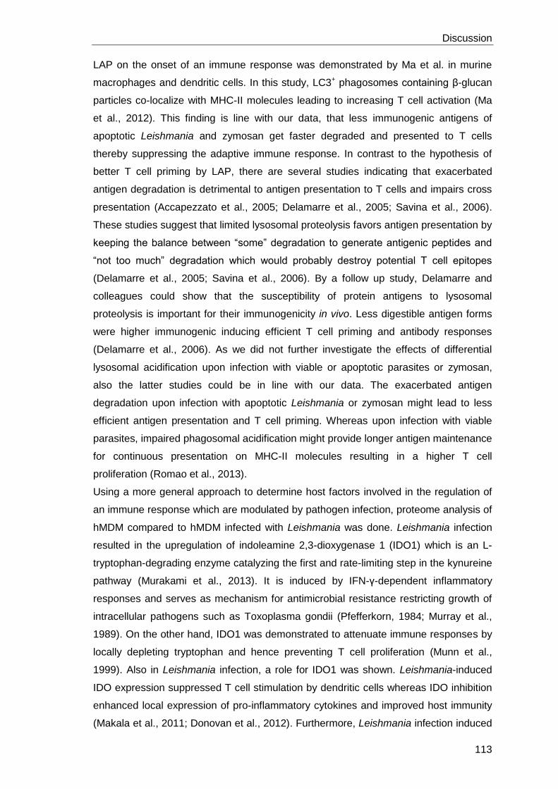
Discussion
113
LAP on the onset of an immune response was demonstrated by Ma et al. in murine
macrophages and dendritic cells. In this study, LC3+ phagosomes containing β-glucan
particles co-localize with MHC-II molecules leading to increasing T cell activation (Ma
et al., 2012). This finding is line with our data, that less immunogenic antigens of
apoptotic Leishmania and zymosan get faster degraded and presented to T cells
thereby suppressing the adaptive immune response. In contrast to the hypothesis of
better T cell priming by LAP, there are several studies indicating that exacerbated
antigen degradation is detrimental to antigen presentation to T cells and impairs cross
presentation (Accapezzato et al., 2005; Delamarre et al., 2005; Savina et al., 2006).
These studies suggest that limited lysosomal proteolysis favors antigen presentation by
keeping the balance between “some” degradation to generate antigenic peptides and
“not too much” degradation which would probably destroy potential T cell epitopes
(Delamarre et al., 2005; Savina et al., 2006). By a follow up study, Delamarre and
colleagues could show that the susceptibility of protein antigens to lysosomal
proteolysis is important for their immunogenicity in vivo. Less digestible antigen forms
were higher immunogenic inducing efficient T cell priming and antibody responses
(Delamarre et al., 2006). As we did not further investigate the effects of differential
lysosomal acidification upon infection with viable or apoptotic parasites or zymosan,
also the latter studies could be in line with our data. The exacerbated antigen
degradation upon infection with apoptotic Leishmania or zymosan might lead to less
efficient antigen presentation and T cell priming. Whereas upon infection with viable
parasites, impaired phagosomal acidification might provide longer antigen maintenance
for continuous presentation on MHC-II molecules resulting in a higher T cell
proliferation (Romao et al., 2013).
Using a more general approach to determine host factors involved in the regulation of
an immune response which are modulated by pathogen infection, proteome analysis of
hMDM compared to hMDM infected with Leishmania was done. Leishmania infection
resulted in the upregulation of indoleamine 2,3-dioxygenase 1 (IDO1) which is an L-
tryptophan-degrading enzyme catalyzing the first and rate-limiting step in the kynureine
pathway (Murakami et al., 2013). It is induced by IFN-γ-dependent inflammatory
responses and serves as mechanism for antimicrobial resistance restricting growth of
intracellular pathogens such as Toxoplasma gondii (Pfefferkorn, 1984; Murray et al.,
1989). On the other hand, IDO1 was demonstrated to attenuate immune responses by
locally depleting tryptophan and hence preventing T cell proliferation (Munn et al.,
1999). Also in Leishmania infection, a role for IDO1 was shown. Leishmania-induced
IDO expression suppressed T cell stimulation by dendritic cells whereas IDO inhibition
enhanced local expression of pro-inflammatory cytokines and improved host immunity
(Makala et al., 2011; Donovan et al., 2012). Furthermore, Leishmania infection induced
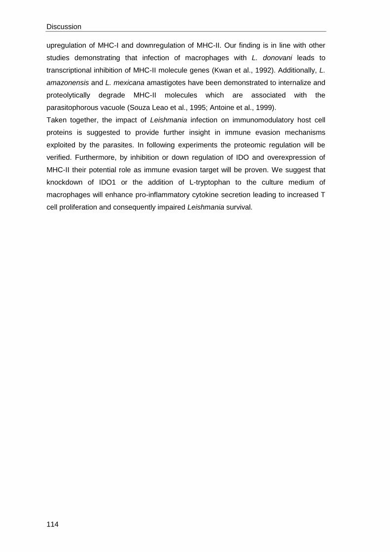
Discussion
114
upregulation of MHC-I and downregulation of MHC-II. Our finding is in line with other
studies demonstrating that infection of macrophages with L. donovani leads to
transcriptional inhibition of MHC-II molecule genes (Kwan et al., 1992). Additionally, L.
amazonensis and L. mexicana amastigotes have been demonstrated to internalize and
proteolytically degrade MHC-II molecules which are associated with the
parasitophorous vacuole (Souza Leao et al., 1995; Antoine et al., 1999).
Taken together, the impact of Leishmania infection on immunomodulatory host cell
proteins is suggested to provide further insight in immune evasion mechanisms
exploited by the parasites. In following experiments the proteomic regulation will be
verified. Furthermore, by inhibition or down regulation of IDO and overexpression of
MHC-II their potential role as immune evasion target will be proven. We suggest that
knockdown of IDO1 or the addition of L-tryptophan to the culture medium of
macrophages will enhance pro-inflammatory cytokine secretion leading to increased T
cell proliferation and consequently impaired Leishmania survival.
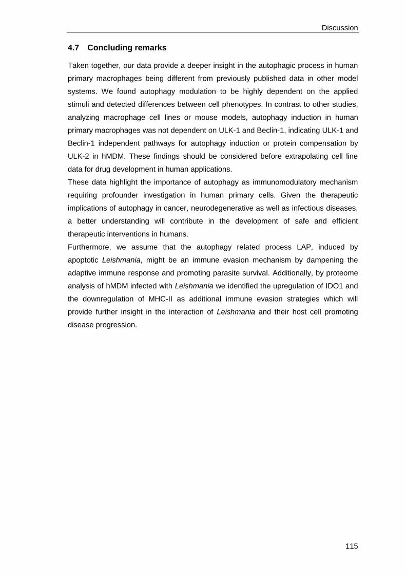
Discussion
115
4.7 Concluding remarks
Taken together, our data provide a deeper insight in the autophagic process in human
primary macrophages being different from previously published data in other model
systems. We found autophagy modulation to be highly dependent on the applied
stimuli and detected differences between cell phenotypes. In contrast to other studies,
analyzing macrophage cell lines or mouse models, autophagy induction in human
primary macrophages was not dependent on ULK-1 and Beclin-1, indicating ULK-1 and
Beclin-1 independent pathways for autophagy induction or protein compensation by
ULK-2 in hMDM. These findings should be considered before extrapolating cell line
data for drug development in human applications.
These data highlight the importance of autophagy as immunomodulatory mechanism
requiring profounder investigation in human primary cells. Given the therapeutic
implications of autophagy in cancer, neurodegenerative as well as infectious diseases,
a better understanding will contribute in the development of safe and efficient
therapeutic interventions in humans.
Furthermore, we assume that the autophagy related process LAP, induced by
apoptotic Leishmania, might be an immune evasion mechanism by dampening the
adaptive immune response and promoting parasite survival. Additionally, by proteome
analysis of hMDM infected with Leishmania we identified the upregulation of IDO1 and
the downregulation of MHC-II as additional immune evasion strategies which will
provide further insight in the interaction of Leishmania and their host cell promoting
disease progression.
References
116
5 References
Accapezzato, D., Visco, V., Francavilla, V., Molette, C., Donato, T., Paroli, M., Mondelli,
M.U., Doria, M., Torrisi, M.R., and Barnaba, V. (2005). Chloroquine enhances human
CD8+ T cell responses against soluble antigens in vivo. The Journal of experimental
medicine 202, 817-828.
Adams, D.O., and Hamilton, T.A. (1984). The cell biology of macrophage activation.
Annual review of immunology 2, 283-318.
Aga, E., Katschinski, D.M., van Zandbergen, G., Laufs, H., Hansen, B., Muller, K.,
Solbach, W., and Laskay, T. (2002). Inhibition of the Spontaneous Apoptosis of
Neutrophil Granulocytes by the Intracellular Parasite Leishmania major. The Journal of
Immunology 169, 898-905.
Ahmed, Wahbi, Nordlind, Kharazmi, Sundqvist, Mutt, and Liden (1998). In Vitro
Leishmania major Promastigote-Induced Macrophage Migration is Modulated by
Sensory and Autonomic Neuropeptides. Scand J Immunol 48, 79-85.
Akhoundi, M., Kuhls, K., Cannet, A., Votypka, J., Marty, P., Delaunay, P., and Sereno,
D. (2016). A Historical Overview of the Classification, Evolution, and Dispersion of
Leishmania Parasites and Sandflies. PLoS neglected tropical diseases 10, e0004349.
Alers, S., Löffler, A.S., Paasch, F., Dieterle, A.M., Keppeler, H., Lauber, K., Campbell,
D.G., Fehrenbacher, B., Schaller, M., and Wesselborg, S., et al. (2014). Atg13 and
FIP200 act independently of Ulk1 and Ulk2 in autophagy induction. Autophagy 7, 1424-
1433.
Alexander, J., and Brombacher, F. (2012). T helper1/t helper2 cells and
resistance/susceptibility to leishmania infection. Is this paradigm still relevant? Frontiers
in immunology 3, 80.
Allenbach, C., Zufferey, C., Perez, C., Launois, P., Mueller, C., and Tacchini-Cottier, F.
(2006). Macrophages Induce Neutrophil Apoptosis through Membrane TNF, a Process
Amplified by Leishmania major. The Journal of Immunology 176, 6656-6664.
Ambarus, C.A., Krausz, S., van Eijk, M., Hamann, J., Radstake, T.R.D.J., Reedquist,
K.A., Tak, P.P., and Baeten, D.L.P. (2012). Systematic validation of specific phenotypic
markers for in vitro polarized human macrophages. Journal of immunological methods
375, 196-206.
Andersson, A.-M., Andersson, B., Lorell, C., Raffetseder, J., Larsson, M., and
Blomgran, R. (2016). Autophagy induction targeting mTORC1 enhances
Mycobacterium tuberculosis replication in HIV co-infected human macrophages.
Scientific reports 6, 28171.
Antoine, J.C., Lang, T., Prina, E., Courret, N., and Hellio, R. (1999). H-2M molecules,
like MHC class II molecules, are targeted to parasitophorous vacuoles of Leishmania-
infected macrophages and internalized by amastigotes of L. amazonensis and L.
mexicana. Journal of cell science 112 ( Pt 15), 2559-2570.
Antoine, J.C., Prina, E., Jouanne, C., and Bongrand, P. (1990). Parasitophorous
vacuoles of Leishmania amazonensis-infected macrophages maintain an acidic pH.
Infection and immunity 58, 779-787.
References
117
Arango Duque, G., and Descoteaux, A. (2014). Macrophage cytokines. Involvement in
immunity and infectious diseases. Frontiers in immunology 5, 491.
Arcaro, A., and Wymann, M.P. (1993). Wortmannin is a potent phosphatidylinositol 3-
kinase inhibitor. The role of phosphatidylinositol 3,4,5-trisphosphate in neutrophil
responses. The Biochemical journal 296 ( Pt 2), 297-301.
Awasthi, A., Mathur, R., Khan, A., Joshi, B.N., Jain, N., Sawant, S., Boppana, R., Mitra,
D., and Saha, B. (2003). CD40 signaling is impaired in L. major-infected macrophages
and is rescued by a p38MAPK activator establishing a host-protective memory T cell
response. The Journal of experimental medicine 197, 1037-1043.
Axe, E.L., Walker, S.A., Manifava, M., Chandra, P., Roderick, H.L., Habermann, A.,
Griffiths, G., and Ktistakis, N.T. (2008). Autophagosome formation from membrane
compartments enriched in phosphatidylinositol 3-phosphate and dynamically
connected to the endoplasmic reticulum. J Cell Biol 182, 685-701.
Baba, M., Osumi, M., and Ohsumi, Y. (1995). Analysis of the Membrane Structures
Involved in Autophagy in Yeast by Freeze-Replica Method. Cell Struct. Funct. 20, 465-
471.
Barbey, C., Pradervand, E., Barbier, N., and Spertini, F. (2007). Ex vivo monitoring of
antigen-specific CD4+ T cells after recall immunization with tetanus toxoid. Clinical and
vaccine immunology : CVI 14, 1108-1116.
Bhattacharyya, S., Ghosh, S., Jhonson, P.L., Bhattacharya, S.K., and Majumdar, S.
(2001). Immunomodulatory role of interleukin-10 in visceral leishmaniasis. Defective
activation of protein kinase C-mediated signal transduction events. Infection and
immunity 69, 1499-1507.
Birmingham, C.L., Canadien, V., Gouin, E., Troy, E.B., Yoshimori, T., Cossart, P.,
Higgins, D.E., and Brumell, J.H. (2014). Listeria monocytogenes Evades Killing by
Autophagy During Colonization of Host Cells. Autophagy 3, 442-451.
Bjorkoy, G., Lamark, T., Brech, A., Outzen, H., Perander, M., Overvatn, A., Stenmark,
H., and Johansen, T. (2005). p62/SQSTM1 forms protein aggregates degraded by
autophagy and has a protective effect on huntingtin-induced cell death. The Journal of
cell biology 171, 603-614.
Blackwell, J.M. (1985). Role of macrophage complement and lectin-like receptors in
binding Leishmania parasites to host macrophages. Immunology letters 11, 227-232.
Blander, J.M., and Medzhitov, R. (2006). Toll-dependent selection of microbial antigens
for presentation by dendritic cells. Nature 440, 808-812.
Blommaart, E.F.C., Krause, U., Schellens, J.P.M., Vreeling-Sindelarova, H., and
Meijer, A.J. (1997). The Phosphatidylinositol 3-Kinase Inhibitors Wortmannin and
LY294002 Inhibit Autophagy in Isolated Rat Hepatocytes. Eur J Biochem 243, 240-246.
Blum, J.S., Wearsch, P.A., and Cresswell, P. (2013). Pathways of antigen processing.
Annual review of immunology 31, 443-473.
Bogdan, C., Gessner, A., Werner, S., and Martin, R. (1996). Invasion, control and
persistence of Leishmania parasites. Current opinion in immunology 8, 517-525.
Bogdan, C., Röllinghoff, M., and Solbach, W. (1990). Evasion strategies of Leishmania
parasites. Parasitology Today 6, 183-187.
References
118
Bouvier, J., Etges, R.J., and Bordier, C. (1985). Identification and purification of
membrane and soluble forms of the major surface protein of Leishmania
promastigotes. The Journal of biological chemistry 260, 15504-15509.
Brittingham, A., Chen, G., McGwire, B.S., Chang, K.P., and Mosser, D.M. (1999).
Interaction of Leishmania gp63 with cellular receptors for fibronectin. Infection and
immunity 67, 4477-4484.
Brittingham, A., Morrison, C.J., McMaster, W.R., McGwire, B.S., Chang, K.P., and
Mosser, D.M. (1995). Role of the Leishmania surface protease gp63 in complement
fixation, cell adhesion, and resistance to complement-mediated lysis. Journal of
immunology (Baltimore, Md. : 1950) 155, 3102-3111.
Burgess, A.W., and Metcalf, D. (1980). The nature and action of granulocyte-
macrophage colony stimulating factors. Blood 56, 947-958.
Burton, O.T., Zaccone, P., Phillips, J.M., La Pena, H. de, Fehervari, Z., Azuma, M.,
Gibbs, S., Stockinger, B., and Cooke, A. (2010). Roles for TGF-beta and programmed
cell death 1 ligand 1 in regulatory T cell expansion and diabetes suppression by
zymosan in nonobese diabetic mice. Journal of immunology (Baltimore, Md. : 1950)
185, 2754-2762.
Button, L.L., and McMaster, W.R. (1988). Molecular cloning of the major surface
antigen of leishmania. The Journal of experimental medicine 167, 724-729.
Carvalho, E.M., Bacellar, O., Brownell, C., Regis, T., Coffman, R.L., and Reed, S.G.
(1994). Restoration of IFN-gamma production and lymphocyte proliferation in visceral
leishmaniasis. Journal of immunology (Baltimore, Md. : 1950) 152, 5949-5956.
Carvalho, E.M., Badaro, R., Reed, S.G., Jones, T.C., and Johnson, W.D., JR (1985).
Absence of gamma interferon and interleukin 2 production during active visceral
leishmaniasis. The Journal of clinical investigation 76, 2066-2069.
Cellerai, C., Harari, A., Vallelian, F., Boyman, O., and Pantaleo, G. (2007). Functional
and phenotypic characterization of tetanus toxoid-specific human CD4+ T cells
following re-immunization. European journal of immunology 37, 1129-1138.
Chan, E.Y.W., Kir, S., and Tooze, S.A. (2007). siRNA screening of the kinome
identifies ULK1 as a multidomain modulator of autophagy. The Journal of biological
chemistry 282, 25464-25474.
Chang, Y.-Y., and Neufeld, T.P. (2009). An Atg1/Atg13 complex with multiple roles in
TOR-mediated autophagy regulation. Molecular biology of the cell 20, 2004-2014.
Charmoy, M., Auderset, F., Allenbach, C., and Tacchini-Cottier, F. (2010). The
prominent role of neutrophils during the initial phase of infection by Leishmania
parasites. Journal of biomedicine & biotechnology 2010, 719361.
Chaudhuri, G., Chaudhuri, M., Pan, A., and Chang, K.P. (1989). Surface acid
proteinase (gp63) of Leishmania mexicana. A metalloenzyme capable of protecting
liposome-encapsulated proteins from phagolysosomal degradation by macrophages.
The Journal of biological chemistry 264, 7483-7489.
Cheong, H., Lindsten, T., Wu, J., Lu, C., and Thompson, C.B. (2011). Ammonia-
induced autophagy is independent of ULK1/ULK2 kinases. Proceedings of the National
Academy of Sciences of the United States of America 108, 11121-11126.
References
119
Choi, A.M.K., Ryter, S.W., and Levine, B. (2013). Autophagy in human health and
disease. The New England journal of medicine 368, 651-662.
Choi, K.S. (2012). Autophagy and cancer. Experimental & molecular medicine 44, 109-
120.
Chresta, C.M., Davies, B.R., Hickson, I., Harding, T., Cosulich, S., Critchlow, S.E.,
Vincent, J.P., Ellston, R., Jones, D., and Sini, P., et al. (2010). AZD8055 is a potent,
selective, and orally bioavailable ATP-competitive mammalian target of rapamycin
kinase inhibitor with in vitro and in vivo antitumor activity. Cancer research 70, 288-298.
Cicchini, M., Karantza, V., and Xia, B. (2015). Molecular pathways. Autophagy in
cancer--a matter of timing and context. Clinical cancer research : an official journal of
the American Association for Cancer Research 21, 498-504.
Cillari, E., Vitale, G., Arcoleo, F., D 'Agostino, P., Mocciaro, C., Gambino, G., Malta, R.,
Stassi, G., Giordano, C., and Milano, S., et al. (1995). In vivo and in vitro cytokine
profiles and mononuclear cell subsets in sicilian patients with active visceral
leishmaniasis. Cytokine 7, 740-745.
Clark, S.L. (1957). Cellular differentiation in the kidneys of newborn mice studies with
the electron microscope. The Journal of biophysical and biochemical cytology 3, 349-
362.
Courret, N., Frehel, C., Gouhier, N., Pouchelet, M., Prina, E., Roux, P., and Antoine, J.-
C. (2002). Biogenesis of Leishmania-harbouring parasitophorous vacuoles following
phagocytosis of the metacyclic promastigote or amastigote stages of the parasites.
Journal of cell science 115, 2303-2316.
Crauwels, P., Bohn, R., Thomas, M., Gottwalt, S., Jackel, F., Kramer, S., Bank, E.,
Tenzer, S., Walther, P., and Bastian, M., et al. (2015). Apoptotic-like Leishmania exploit
the host's autophagy machinery to reduce T-cell-mediated parasite elimination.
Autophagy 11, 285-297.
Cyrino, L.T., Araujo, A.P., Joazeiro, P.P., Vicente, C.P., and Giorgio, S. (2012). In vivo
and in vitro Leishmania amazonensis infection induces autophagy in macrophages.
Tissue & cell 44, 401-408.
da Silva, R., and Sacks, D.L. (1987). Metacyclogenesis is a major determinant of
Leishmania promastigote virulence and attenuation. Infection and immunity 55, 2802-
2806.
Da Silva, R.P., Hall, B.F., Joiner, K.A., and Sacks, D.L. (1989). CR1, the C3b receptor,
mediates binding of infective Leishmania major metacyclic promastigotes to human
macrophages. Journal of immunology (Baltimore, Md. : 1950) 143, 617-622.
Dall, E., and Brandstetter, H. (2016). Structure and function of legumain in health and
disease. Biochimie 122, 126-150.
Degtyarev, M., Maziere, A. de, Orr, C., Lin, J., Lee, B.B., Tien, J.Y., Prior, W.W., van
Dijk, S., Wu, H., and Gray, D.C., et al. (2008). Akt inhibition promotes autophagy and
sensitizes PTEN-null tumors to lysosomotropic agents. The Journal of cell biology 183,
101-116.
Delamarre, L., Couture, R., Mellman, I., and Trombetta, E.S. (2006). Enhancing
immunogenicity by limiting susceptibility to lysosomal proteolysis. The Journal of
experimental medicine 203, 2049-2055.
References
120
Delamarre, L., Pack, M., Chang, H., Mellman, I., and Trombetta, E.S. (2005).
Differential lysosomal proteolysis in antigen-presenting cells determines antigen fate.
Science (New York, N.Y.) 307, 1630-1634.
Dengjel, J., Schoor, O., Fischer, R., Reich, M., Kraus, M., Muller, M., Kreymborg, K.,
Altenberend, F., Brandenburg, J., and Kalbacher, H., et al. (2005). Autophagy
promotes MHC class II presentation of peptides from intracellular source proteins.
Proceedings of the National Academy of Sciences of the United States of America 102,
7922-7927.
Desjardins, M., and Descoteaux, A. (1997). Inhibition of phagolysosomal biogenesis by
the Leishmania lipophosphoglycan. The Journal of experimental medicine 185, 2061-
2068.
Dillon, S., Agrawal, S., Banerjee, K., Letterio, J., Denning, T.L., Oswald-Richter, K.,
Kasprowicz, D.J., Kellar, K., Pare, J., and van Dyke, T., et al. (2006). Yeast zymosan, a
stimulus for TLR2 and dectin-1, induces regulatory antigen-presenting cells and
immunological tolerance. The Journal of clinical investigation 116, 916-928.
Dobrovolskaia, M.A., and Vogel, S.N. (2002). Toll receptors, CD14, and macrophage
activation and deactivation by LPS. Microbes and Infection 4, 903-914.
Donovan, M.J., Tripathi, V., Favila, M.A., Geraci, N.S., Lange, M.C., Ballhorn, W., and
McDowell, M.A. (2012). Indoleamine 2,3-dioxygenase (IDO) induced by Leishmania
infection of human dendritic cells. Parasite immunology 34, 464-472.
Du, Z., Kelly, E., Mecklenbrauker, I., Agle, L., Herrero, C., Paik, P., and Ivashkiv, L.B.
(2006). Selective Regulation of IL-10 Signaling and Function by Zymosan. The Journal
of Immunology 176, 4785-4792.
Dugas, B., Mossalayi, M.D., Damais, C., and Kolb, J.P. (1995). Nitric oxide production
by human monocytes. Evidence for a role of CD23. Immunology today 16, 574-580.
DUVE, C. de, PRESSMAN, B.C., GIANETTO, R., WATTIAUX, R., and APPELMANS,
F. (1955). Tissue fractionation studies. 6. Intracellular distribution patterns of enzymes
in rat-liver tissue. The Biochemical journal 60, 604-617.
DUVE, C. de, and WATTIAUX, R. (1966). Functions of lysosomes. Annual review of
physiology 28, 435-492.
Ehlers, M.R. (2000). CR3. A general purpose adhesion-recognition receptor essential
for innate immunity. In The Zinc Industry (Elsevier), pp. 289–294.
Etges, R., Bouvier, J., and Bordier, C. (1986). The major surface protein of Leishmania
promastigotes is a protease. The Journal of biological chemistry 261, 9098-9101.
Fan, Q.-W., Knight, Z.A., Goldenberg, D.D., Yu, W., Mostov, K.E., Stokoe, D., Shokat,
K.M., and Weiss, W.A. (2006). A dual PI3 kinase/mTOR inhibitor reveals emergent
efficacy in glioma. Cancer cell 9, 341-349.
Florey, O., Kim, S.E., Sandoval, C.P., Haynes, C.M., and Overholtzer, M. (2011).
Autophagy machinery mediates macroendocytic processing and entotic cell death by
targeting single membranes. Nature cell biology 13, 1335-1343.
Frommel, T.O., Button, L.L., Fujikura, Y., and McMaster, W. (1990). The major surface
glycoprotein (GP63) is present in both life stages of Leishmania. Molecular and
Biochemical Parasitology 38, 25-32.
References
121
Fujii, S.-I., Liu, K., Smith, C., Bonito, A.J., and Steinman, R.M. (2004). The linkage of
innate to adaptive immunity via maturing dendritic cells in vivo requires CD40 ligation in
addition to antigen presentation and CD80/86 costimulation. The Journal of
experimental medicine 199, 1607-1618.
Fujita, N., Itoh, T., Omori, H., Fukuda, M., Noda, T., and Yoshimori, T. (2008). The
Atg16L complex specifies the site of LC3 lipidation for membrane biogenesis in
autophagy. Molecular biology of the cell 19, 2092-2100.
Gaafar, A., KHARAZMI, A., Ismail, A., KEMP, M., Hey, A., Christensen, C.B., Dafalla,
M., el Kadaro, A.Y., el Hassan, A.M., and THEANDER, T.G. (1995). Dichotomy of the T
cell response to Leishmania antigens in patients suffering from cutaneous
leishmaniasis; absence or scarcity of Th1 activity is associated with severe infections.
Clinical and experimental immunology 100, 239-245.
Ganguly, S., Mukhopadhyay, D., Das, N.K., Chaduvula, M., Sadhu, S., Chatterjee, U.,
Rahman, M., Goswami, R.P., Guha, S.K., and Modak, D., et al. (2010). Enhanced
lesional Foxp3 expression and peripheral anergic lymphocytes indicate a role for
regulatory T cells in Indian post-kala-azar dermal leishmaniasis. The Journal of
investigative dermatology 130, 1013-1022.
Ganley, I.G., Du Lam, H., Wang, J., Ding, X., Chen, S., and Jiang, X. (2009).
ULK1.ATG13.FIP200 complex mediates mTOR signaling and is essential for
autophagy. The Journal of biological chemistry 284, 12297-12305.
Gao, P., Bauvy, C., Souquere, S., Tonelli, G., Liu, L., Zhu, Y., Qiao, Z., Bakula, D.,
Proikas-Cezanne, T., and Pierron, G., et al. (2010). The Bcl-2 homology domain 3
mimetic gossypol induces both Beclin 1-dependent and Beclin 1-independent
cytoprotective autophagy in cancer cells. The Journal of biological chemistry 285,
25570-25581.
Geng, J., and Klionsky, D.J. (2008). The Atg8 and Atg12 ubiquitin-like conjugation
systems in macroautophagy. 'Protein modifications. Beyond the usual suspects' review
series. EMBO reports 9, 859-864.
Gerber, J.S., and Mosser, D.M. (2001). Reversing Lipopolysaccharide Toxicity by
Ligating the Macrophage Fc Receptors. The Journal of Immunology 166, 6861-6868.
Germain, R.N. (1994). MHC-dependent antigen processing and peptide presentation.
Providing ligands for T lymphocyte activation. Cell 76, 287-299.
Ghalib, H.W., Piuvezam, M.R., Skeiky, Y.A., Siddig, M., Hashim, F.A., el-Hassan, A.M.,
Russo, D.M., and Reed, S.G. (1993). Interleukin 10 production correlates with
pathology in human Leishmania donovani infections. The Journal of clinical
investigation 92, 324-329.
Ghalib, H.W., Whittle, J.A., Kubin, M., Hashim, F.A., el-Hassan, A.M., Grabstein, K.H.,
Trinchieri, G., and Reed, S.G. (1995). IL-12 enhances Th1-type responses in human
Leishmania donovani infections. Journal of immunology (Baltimore, Md. : 1950) 154,
4623-4629.
Giannini, M.S. (1974). Effects of promastigote growth phase, frequency of subculture,
and host age on promastigote-initiated infections with Leishmania donovani in the
golden hamster. Involvement in immunity and infectious diseases. The Journal of
protozoology 21, 521-527.
References
122
Gomez, M.A., Contreras, I., Halle, M., Tremblay, M.L., McMaster, R.W., and Olivier, M.
(2009). Leishmania GP63 alters host signaling through cleavage-activated protein
tyrosine phosphatases. Science signaling 2, ra58.
Gomez-Sanchez, R., Pizarro-Estrella, E., Yakhine-Diop, S.M.S., Rodriguez-Arribas, M.,
Bravo-San Pedro, J.M., Fuentes, J.M., and Gonzalez-Polo, R.A. (2015). Routine
Western blot to check autophagic flux. Cautions and recommendations. Analytical
biochemistry 477, 13-20.
Gong, L., Cullinane, M., Treerat, P., Ramm, G., Prescott, M., Adler, B., Boyce, J.D.,
and Devenish, R.J. (2011). The Burkholderia pseudomallei type III secretion system
and BopA are required for evasion of LC3-associated phagocytosis. PloS one 6,
e17852.
Gordon, S., and Martinez, F.O. (2010). Alternative activation of macrophages.
Mechanism and functions. Immunity 32, 593-604.
Gordon, S., and Taylor, P.R. (2005). Monocyte and macrophage heterogeneity. Nature
reviews. Immunology 5, 953-964.
Goto, H., and Lauletta Lindoso, J.A. (2012). Cutaneous and mucocutaneous
leishmaniasis. Infectious disease clinics of North America 26, 293-307.
Greenwood, J., Steinman, L., and Zamvil, S.S. (2006). Statin therapy and autoimmune
disease. From protein prenylation to immunomodulation. Nature reviews. Immunology
6, 358-370.
Grishchuk, Y., Ginet, V., Truttmann, A.C., Clarke, P.G.H., and Puyal, J. (2011). Beclin
1-independent autophagy contributes to apoptosis in cortical neurons. Autophagy 7,
1115-1131.
Gupta, G., Oghumu, S., and Satoskar, A.R. (2013). Mechanisms of immune evasion in
leishmaniasis. Advances in applied microbiology 82, 155-184.
Gutierrez, M.G., Master, S.S., Singh, S.B., Taylor, G.A., Colombo, M.I., and Deretic, V.
(2004). Autophagy is a defense mechanism inhibiting BCG and Mycobacterium
tuberculosis survival in infected macrophages. Cell 119, 753-766.
Hailey, D.W., Rambold, A.S., Satpute-Krishnan, P., Mitra, K., Sougrat, R., Kim, P.K.,
and Lippincott-Schwartz, J. (2010). Mitochondria supply membranes for
autophagosome biogenesis during starvation. Cell 141, 656-667.
Hailu, A., van Baarle, D., Knol, G.J., Berhe, N., Miedema, F., and Kager, P.A. (2005). T
cell subset and cytokine profiles in human visceral leishmaniasis during active and
asymptomatic or sub-clinical infection with Leishmania donovani. Clinical immunology
(Orlando, Fla.) 117, 182-191.
Haldar, J.P., Ghose, S., Saha, K.C., and Ghose, A.C. (1983). Cell-mediated immune
response in Indian kala-azar and post-kala-azar dermal leishmaniasis. Infection and
immunity 42, 702-707.
Handman, E., and Goding, J.W. (1985). The Leishmania receptor for macrophages is a
lipid-containing glycoconjugate. The EMBO journal 4, 329-336.
Handman, E., and Mitchell, G.F. (1985). Immunization with Leishmania receptor for
macrophages protects mice against cutaneous leishmaniasis. Proceedings of the
National Academy of Sciences 82, 5910-5914.
References
123
Harris, J., Haro, S.A. de, Master, S.S., Keane, J., Roberts, E.A., Delgado, M., and
Deretic, V. (2007). T helper 2 cytokines inhibit autophagic control of intracellular
Mycobacterium tuberculosis. Immunity 27, 505-517.
Heitman, J., Movva, N.R., and Hall, M.N. (1991). Targets for cell cycle arrest by the
immunosuppressant rapamycin in yeast. Science (New York, N.Y.) 253, 905-909.
Hemelaar, J., Lelyveld, V.S., Kessler, B.M., and Ploegh, H.L. (2003). A single protease,
Apg4B, is specific for the autophagy-related ubiquitin-like proteins GATE-16, MAP1-
LC3, GABARAP, and Apg8L. The Journal of biological chemistry 278, 51841-51850.
Henault, J., Martinez, J., Riggs, J.M., Tian, J., Mehta, P., Clarke, L., Sasai, M., Latz, E.,
Brinkmann, M.M., and Iwasaki, A., et al. (2012). Noncanonical autophagy is required
for type I interferon secretion in response to DNA-immune complexes. Immunity 37,
986-997.
HEY, A.S., THEANDER, T.G., HVIID, L., Hazrati, S.M., KEMP, M., and KHARAZMI, A.
(1994). The major surface glycoprotein (gp63) from Leishmania major and Leishmania
donovani cleaves CD4 molecules on human T cells. Journal of immunology (Baltimore,
Md. : 1950) 152, 4542-4548.
Homer, C.R., Richmond, A.L., Rebert, N.A., Achkar, J.-P., and McDonald, C. (2010).
ATG16L1 and NOD2 interact in an autophagy-dependent antibacterial pathway
implicated in Crohn's disease pathogenesis. Gastroenterology 139, 1630-41, 1641.e1-
2.
Hooper, K.M., Barlow, P.G., Stevens, C., and Henderson, P. (2017). Inflammatory
Bowel Disease Drugs. A Focus on Autophagy. Journal of Crohn's & colitis 11, 118-127.
Huang, J., Canadien, V., Lam, G.Y., Steinberg, B.E., Dinauer, M.C., Magalhaes,
M.A.O., Glogauer, M., Grinstein, S., and Brumell, J.H. (2009). Activation of antibacterial
autophagy by NADPH oxidases. Proceedings of the National Academy of Sciences of
the United States of America 106, 6226-6231.
Huang, S., Yang, Z.J., Yu, C., and Sinicrope, F.A. (2011). Inhibition of mTOR kinase by
AZD8055 can antagonize chemotherapy-induced cell death through autophagy
induction and down-regulation of p62/sequestosome 1. The Journal of biological
chemistry 286, 40002-40012.
Huynh, M.-L.N., Fadok, V.A., and Henson, P.M. (2002). Phosphatidylserine-dependent
ingestion of apoptotic cells promotes TGF-beta1 secretion and the resolution of
inflammation. The Journal of clinical investigation 109, 41-50.
Isnard, A., Shio, M.T., and Olivier, M. (2012). Impact of Leishmania metalloprotease
GP63 on macrophage signaling. Frontiers in cellular and infection microbiology 2, 72.
Itakura, E., and Mizushima, N. (2014). Characterization of autophagosome formation
site by a hierarchical analysis of mammalian Atg proteins. Autophagy 6, 764-776.
Jagannath, C., Lindsey, D.R., Dhandayuthapani, S., Xu, Y., Hunter, R.L., JR, and
Eissa, N.T. (2009). Autophagy enhances the efficacy of BCG vaccine by increasing
peptide presentation in mouse dendritic cells. Nature medicine 15, 267-276.
Janeway, C.A., and Bottomly, K. (1994). Signals and signs for lymphocyte responses.
Cell 76, 275-285.
References
124
Jaramillo, M., Gomez, M.A., Larsson, O., Shio, M.T., Topisirovic, I., Contreras, I.,
Luxenburg, R., Rosenfeld, A., Colina, R., and McMaster, R.W., et al. (2011).
Leishmania repression of host translation through mTOR cleavage is required for
parasite survival and infection. Cell host & microbe 9, 331-341.
Jiang, P., and Mizushima, N. (2014). Autophagy and human diseases. Cell research
24, 69-79.
Johnson, E.E., Overmeyer, J.H., Gunning, W.T., and Maltese, W.A. (2006). Gene
silencing reveals a specific function of hVps34 phosphatidylinositol 3-kinase in late
versus early endosomes. Journal of cell science 119, 1219-1232.
Jones, G.E. (2000). Cellular signaling in macrophage migration and chemotaxis.
Journal of leukocyte biology 68, 593-602.
Joshi, P.B., Sacks, D.L., Modi, G., and McMaster, W.R. (1998). Targeted gene deletion
of Leishmania major genes encoding developmental stage-specific leishmanolysin
(GP63). Mol Microbiol 27, 519-530.
Jung, C.H., Jun, C.B., Ro, S.-H., Kim, Y.-M., Otto, N.M., Cao, J., Kundu, M., and Kim,
D.-H. (2009). ULK-Atg13-FIP200 complexes mediate mTOR signaling to the autophagy
machinery. Molecular biology of the cell 20, 1992-2003.
Kabeya, Y., Mizushima, N., Ueno, T., Yamamoto, A., Kirisako, T., Noda, T., Kominami,
E., Ohsumi, Y., and Yoshimori, T. (2000). LC3, a mammalian homologue of yeast
Apg8p, is localized in autophagosome membranes after processing. The EMBO journal
19, 5720-5728.
Kamphorst, A.O., Guermonprez, P., Dudziak, D., and Nussenzweig, M.C. (2010).
Route of antigen uptake differentially impacts presentation by dendritic cells and
activated monocytes. Journal of immunology (Baltimore, Md. : 1950) 185, 3426-3435.
Kane, M.M., and Mosser, D.M. (2000). Leishmania parasites and their ploys to disrupt
macrophage activation. Current opinion in hematology 7, 26-31.
Katara, G.K., Ansari, N.A., Verma, S., Ramesh, V., Salotra, P., and Engwerda, C.R.
(2011). Foxp3 and IL-10 Expression Correlates with Parasite Burden in Lesional
Tissues of Post Kala Azar Dermal Leishmaniasis (PKDL) Patients. PLoS Negl Trop Dis
5, e1171.
Kaye, P., and Scott, P. (2011). Leishmaniasis. Complexity at the host-pathogen
interface. Nature reviews. Microbiology 9, 604-615.
KEMP, M., Hansen, M.B., and THEANDER, T.G. (1992). Recognition of Leishmania
antigens by T lymphocytes from nonexposed individuals. Infection and immunity 60,
2246-2251.
KEMP, M., HEY, A.S., Kurtzhals, J.A., Christensen, C.B., Gaafar, A., Mustafa, M.D.,
Kordofani, A.A., Ismail, A., KHARAZMI, A., and THEANDER, T.G. (1994). Dichotomy
of the human T cell response to Leishmania antigens. I. Th1-like response to
Leishmania major promastigote antigens in individuals recovered from cutaneous
leishmaniasis. Clinical and experimental immunology 96, 410-415.
KEMP, M., THEANDER, T.G., Handman, E., HEY, A.S., KURTZHALS, J.A.L., HVIID,
L., SØRENSEN, A.L., WERE, J.O.B., KOECH, D.K., and KHARAZMI, A. (1991).
Activation of Human T Lymphocytes by Leishmania Lipophosphoglycan. Scand J
Immunol 33, 219-224.
References
125
Kihara, A., Kabeya, Y., Ohsumi, Y., and Yoshimori, T. (2001). Beclin-
phosphatidylinositol 3-kinase complex functions at the trans-Golgi network. EMBO
reports 2, 330-335.
Kim, D.-H., Sarbassov, D.D., Ali, S.M., King, J.E., Latek, R.R., Erdjument-Bromage, H.,
Tempst, P., and Sabatini, D.M. (2002). mTOR Interacts with Raptor to Form a Nutrient-
Sensitive Complex that Signals to the Cell Growth Machinery. Cell 110, 163-175.
King, D.L., Chang, Y.D., and Turco, S.J. (1987). Cell surface lipophosphoglycan of
Leishmania donovani. Molecular and Biochemical Parasitology 24, 47-53.
Kirkegaard, K., Taylor, M.P., and Jackson, W.T. (2004). Cellular autophagy. Surrender,
avoidance and subversion by microorganisms. Nature reviews. Microbiology 2, 301-
314.
Klionsky, D.J., Abdelmohsen, K., Abe, A., Abedin, M.J., Abeliovich, H., Acevedo
Arozena, A., Adachi, H., Adams, C.M., Adams, P.D., and Adeli, K., et al. (2016).
Guidelines for the use and interpretation of assays for monitoring autophagy (3rd
edition). Autophagy 12, 1-222.
Klionsky, D.J., Cuervo, A.M., and Seglen, P.O. (2014). Methods for Monitoring
Autophagy from Yeast to Human. Autophagy 3, 181-206.
Kobayashi, K.S., and van den Elsen, P.J. (2012). NLRC5. A key regulator of MHC
class I-dependent immune responses. Nature reviews. Immunology 12, 813-820.
Kropf, P., Fuentes, J.M., Fahnrich, E., Arpa, L., Herath, S., Weber, V., Soler, G.,
Celada, A., Modolell, M., and Muller, I. (2005). Arginase and polyamine synthesis are
key factors in the regulation of experimental leishmaniasis in vivo. FASEB journal :
official publication of the Federation of American Societies for Experimental Biology 19,
1000-1002.
Kubach, J., Lutter, P., Bopp, T., Stoll, S., Becker, C., Huter, E., Richter, C., Weingarten,
P., Warger, T., and Knop, J., et al. (2007). Human CD4+CD25+ regulatory T cells.
Proteome analysis identifies galectin-10 as a novel marker essential for their anergy
and suppressive function. Blood 110, 1550-1558.
Kumar, R., and Engwerda, C. (2014). Vaccines to prevent leishmaniasis. Clinical &
translational immunology 3, e13.
Kundu, M., Lindsten, T., Yang, C.-Y., Wu, J., Zhao, F., Zhang, J., Selak, M.A., Ney,
P.A., and Thompson, C.B. (2008). Ulk1 plays a critical role in the autophagic clearance
of mitochondria and ribosomes during reticulocyte maturation. Blood 112, 1493-1502.
Kwan, W.C., McMaster, W.R., Wong, N., and Reiner, N.E. (1992). Inhibition of
expression of major histocompatibility complex class II molecules in macrophages
infected with Leishmania donovani occurs at the level of gene transcription via a cyclic
AMP-independent mechanism. Infection and immunity 60, 2115-2120.
Kweider, M., Lemesre, J.L., Darcy, F., Kusnierz, J.P., Capron, A., and Santoro, F.
(1987). Infectivity of Leishmania braziliensis promastigotes is dependent on the
increasing expression of a 65,000-dalton surface antigen. Journal of immunology
(Baltimore, Md. : 1950) 138, 299-305.
Lacey, D.C., Achuthan, A., Fleetwood, A.J., Dinh, H., Roiniotis, J., Scholz, G.M.,
Chang, M.W., Beckman, S.K., Cook, A.D., and Hamilton, J.A. (2012). Defining GM-
References
126
CSF- and macrophage-CSF-dependent macrophage responses by in vitro models.
Journal of immunology (Baltimore, Md. : 1950) 188, 5752-5765.
Lamb, C.A., Yoshimori, T., and Tooze, S.A. (2013). The autophagosome. Origins
unknown, biogenesis complex. Nature reviews. Molecular cell biology 14, 759-774.
Lang, T., Hellio, R., Kaye, P.M., and Antoine, J.C. (1994). Leishmania donovani-
infected macrophages. Characterization of the parasitophorous vacuole and potential
role of this organelle in antigen presentation. Journal of cell science 107 ( Pt 8), 2137-
2150.
Laskay, T., Mariam, H.G., Berhane, T.Y., Fehniger, T.E., and Kiessling, R. (1991).
Immune reactivity to fractionated Leishmania aethiopica antigens during active human
infection. Journal of clinical microbiology 29, 757-763.
Laskay, T., van Zandbergen, G., and Solbach, W. (2003). Neutrophil granulocytes –
Trojan horses for Leishmania major and other intracellular microbes? Trends in
Microbiology 11, 210-214.
Lee, E.-J., and Tournier, C. (2014). The requirement of uncoordinated 51-like kinase 1
(ULK1) and ULK2 in the regulation of autophagy. Autophagy 7, 689-695.
Lee, H.K., Mattei, L.M., Steinberg, B.E., Alberts, P., Lee, Y.H., Chervonsky, A.,
Mizushima, N., Grinstein, S., and Iwasaki, A. (2010). In vivo requirement for Atg5 in
antigen presentation by dendritic cells. Immunity 32, 227-239.
Lee, Y.-R., Lei, H.-Y., Liu, M.-T., Wang, J.-R., Chen, S.-H., Jiang-Shieh, Y.-F., Lin, Y.-
S., Yeh, T.-M., Liu, C.-C., and Liu, H.-S. (2008). Autophagic machinery activated by
dengue virus enhances virus replication. Virology 374, 240-248.
Lennemann, N.J., and Coyne, C.B. (2015). Catch me if you can. The link between
autophagy and viruses. PLoS pathogens 11, e1004685.
Levine, B., Mizushima, N., and Virgin, H.W. (2011). Autophagy in immunity and
inflammation. Nature 469, 323-335.
Li, X., Prescott, M., Adler, B., Boyce, J.D., and Devenish, R.J. (2013). Beclin 1 is
required for starvation-enhanced, but not rapamycin-enhanced, LC3-associated
phagocytosis of Burkholderia pseudomallei in RAW 264.7 cells. Infection and immunity
81, 271-277.
Liang, X.H., Jackson, S., Seaman, M., Brown, K., Kempkes, B., Hibshoosh, H., and
Levine, B. (1999). Induction of autophagy and inhibition of tumorigenesis by beclin 1.
Nature 402, 672-676.
Lieke, T., Nylen, S., Eidsmo, L., McMaster, W.R., Mohammadi, A.M., Khamesipour, A.,
Berg, L., and Akuffo, H. (2008). Leishmania surface protein gp63 binds directly to
human natural killer cells and inhibits proliferation. Clinical and experimental
immunology 153, 221-230.
Lin, Y.-C., Kuo, H.-C., Wang, J.-S., and Lin, W.-W. (2012). Regulation of inflammatory
response by 3-methyladenine involves the coordinative actions on Akt and glycogen
synthase kinase 3beta rather than autophagy. Journal of immunology (Baltimore, Md. :
1950) 189, 4154-4164.
Lindoso, J.A.L., Cunha, M.A., Queiroz, I.T., and Moreira, C.H.V. (2016). Leishmaniasis-
HIV coinfection. Current challenges. HIV/AIDS (Auckland, N.Z.) 8, 147-156.
References
127
Linehan, S.A., Martínez-Pomares, L., and Gordon, S. (2000). Macrophage lectins in
host defence. Microbes and Infection 2, 279-288.
Liu, J., Xia, H., Kim, M., Xu, L., Li, Y., Zhang, L., Cai, Y., Norberg, H.V., Zhang, T., and
Furuya, T., et al. (2011). Beclin1 controls the levels of p53 by regulating the
deubiquitination activity of USP10 and USP13. Cell 147, 223-234.
Liu, K., Zhao, E., Ilyas, G., Lalazar, G., Lin, Y., Haseeb, M., Tanaka, K.E., and Czaja,
M.J. (2015). Impaired macrophage autophagy increases the immune response in
obese mice by promoting proinflammatory macrophage polarization. Autophagy 11,
271-284.
Liu, X., and Chang, K.P. (1992). Extrachromosomal genetic complementation of
surface metalloproteinase (gp63)-deficient Leishmania increases their binding to
macrophages. Proceedings of the National Academy of Sciences of the United States
of America 89, 4991-4995.
Livak, K.J., and Schmittgen, T.D. (2001). Analysis of relative gene expression data
using real-time quantitative PCR and the 2(-Delta Delta C(T)) Method. Methods (San
Diego, Calif.) 25, 402-408.
Locksley, R.M. (2010). Asthma and allergic inflammation. Cell 140, 777-783.
Lopez-Jaramillo, P., Ruano, C., Rivera, J., Teran, E., Salazar-Irigoyen, R., Esplugues,
J.V., and Moncada, S. (1998). Treatment of cutaneous leishmaniasis with nitric-oxide
donor. The Lancet 351, 1176-1177.
Lynch-Day, M.A., Mao, K., Wang, K., Zhao, M., and Klionsky, D.J. (2012). The role of
autophagy in Parkinson's disease. Cold Spring Harbor perspectives in medicine 2,
a009357.
Ma, J., Becker, C., Lowell, C.A., and Underhill, D.M. (2012). Dectin-1-triggered
recruitment of light chain 3 protein to phagosomes facilitates major histocompatibility
complex class II presentation of fungal-derived antigens. The Journal of biological
chemistry 287, 34149-34156.
Macdonald, M.H., Morrison, C.J., and McMaster, W.R. (1995). Analysis of the active
site and activation mechanism of the Leishmania surface metalloproteinase GP63.
Biochimica et biophysica acta 1253, 199-207.
MACKANESS, G.B. (1962). Cellular resistance to infection. The Journal of
experimental medicine 116, 381-406.
Mahoney, J.A., Ntolosi, B., DaSilva, R.P., Gordon, S., and McKnight, A.J. (2001).
Cloning and characterization of CPVL, a novel serine carboxypeptidase, from human
macrophages. Genomics 72, 243-251.
Makala, L.H.C., Baban, B., Lemos, H., El-Awady, A.R., Chandler, P.R., Hou, D.-Y.,
Munn, D.H., and Mellor, A.L. (2011). Leishmania major attenuates host immunity by
stimulating local indoleamine 2,3-dioxygenase expression. The Journal of infectious
diseases 203, 715-725.
Mansour, M.K., Tam, J.M., Khan, N.S., Seward, M., Davids, P.J., Puranam, S.,
Sokolovska, A., Sykes, D.B., Dagher, Z., and Becker, C., et al. (2013). Dectin-1
activation controls maturation of beta-1,3-glucan-containing phagosomes. The Journal
of biological chemistry 288, 16043-16054.
References
128
Mantovani, A., Sica, A., Sozzani, S., Allavena, P., Vecchi, A., and Locati, M. (2004).
The chemokine system in diverse forms of macrophage activation and polarization.
Trends in immunology 25, 677-686.
Marth, T., and Kelsall, B.L. (1997). Regulation of interleukin-12 by complement
receptor 3 signaling. The Journal of experimental medicine 185, 1987-1995.
Martin, C.J., Peters, K.N., and Behar, S.M. (2014). Macrophages clean up.
Efferocytosis and microbial control. Current opinion in microbiology 17, 17-23.
Martinez, F.O., and Gordon, S. (2014). The M1 and M2 paradigm of macrophage
activation. Time for reassessment. F1000prime reports 6, 13.
Martinez, J., Almendinger, J., Oberst, A., Ness, R., Dillon, C.P., Fitzgerald, P.,
Hengartner, M.O., and Green, D.R. (2011). Microtubule-associated protein 1 light chain
3 alpha (LC3)-associated phagocytosis is required for the efficient clearance of dead
cells. Proceedings of the National Academy of Sciences of the United States of
America 108, 17396-17401.
Martinez, J., Malireddi, R.K.S., Lu, Q., Cunha, L.D., Pelletier, S., Gingras, S., Orchard,
R., Guan, J.-L., Tan, H., and Peng, J., et al. (2015). Molecular characterization of LC3-
associated phagocytosis reveals distinct roles for Rubicon, NOX2 and autophagy
proteins. Nature cell biology 17, 893-906.
Martinez, J., Verbist, K., Wang, R., and Green, D.R. (2013). The relationship between
metabolism and the autophagy machinery during the innate immune response. Cell
metabolism 17, 895-900.
Martyniszyn, L., Szulc-Dabrowska, L., Boratynska-Jasinska, A., Struzik, J., Winnicka,
A., and Niemialtowski, M. (2013). Crosstalk between autophagy and apoptosis in RAW
264.7 macrophages infected with ectromelia orthopoxvirus. Viral immunology 26, 322-
335.
Mason, J.M., Naidu, M.D., Barcia, M., Porti, D., Chavan, S.S., and Chu, C.C. (2004).
IL-4-Induced Gene-1 Is a Leukocyte L-Amino Acid Oxidase with an Unusual Acidic pH
Preference and Lysosomal Localization. The Journal of Immunology 173, 4561-4567.
Mateo, R., Nagamine, C.M., Spagnolo, J., Mendez, E., Rahe, M., Gale, M., JR, Yuan,
J., and Kirkegaard, K. (2013). Inhibition of cellular autophagy deranges dengue virion
maturation. Journal of virology 87, 1312-1321.
Matheoud, D., Moradin, N., Bellemare-Pelletier, A., Shio, M.T., Hong, W.J., Olivier, M.,
Gagnon, E., Desjardins, M., and Descoteaux, A. (2013). Leishmania evades host
immunity by inhibiting antigen cross-presentation through direct cleavage of the
SNARE VAMP8. Cell host & microbe 14, 15-25.
Matsuzawa, T., Kim, B.-H., Shenoy, A.R., Kamitani, S., Miyake, M., and MacMicking,
J.D. (2012). IFN-gamma elicits macrophage autophagy via the p38 MAPK signaling
pathway. Journal of immunology (Baltimore, Md. : 1950) 189, 813-818.
Matte, C., Casgrain, P.-A., Seguin, O., Moradin, N., Hong, W.J., and Descoteaux, A.
(2016). Leishmania major Promastigotes Evade LC3-Associated Phagocytosis through
the Action of GP63. PLoS pathogens 12, e1005690.
McGwire, B.S., and Satoskar, A.R. (2014). Leishmaniasis. Clinical syndromes and
treatment. QJM : monthly journal of the Association of Physicians 107, 7-14.
References
129
Medina-Acosta, E., Karess, R.E., Schwartz, H., and Russell, D.G. (1989). The
promastigote surface protease (gp63) of Leishmania is expressed but differentially
processed and localized in the amastigote stage. Molecular and Biochemical
Parasitology 37, 263-273.
Medzhitov, R., and Janeway, C., JR (2000). Innate immunity. The New England journal
of medicine 343, 338-344.
Mehta, P., Henault, J., Kolbeck, R., and Sanjuan, M.A. (2014). Noncanonical
autophagy. One small step for LC3, one giant leap for immunity. Current opinion in
immunology 26, 69-75.
Mendonca, S.C., Russell, D.G., and Coutinho, S.G. (1991). Analysis of the human T
cell responsiveness to purified antigens of Leishmania. Lipophosphoglycan (LPG) and
glycoprotein 63 (gp 63). Clinical and experimental immunology 83, 472-478.
Menten, P., Wuyts, A., and van Damme, J. (2002). Macrophage inflammatory protein-
1. Cytokine & growth factor reviews 13, 455-481.
Miller, S., Tavshanjian, B., Oleksy, A., Perisic, O., Houseman, B.T., Shokat, K.M., and
Williams, R.L. (2010). Shaping development of autophagy inhibitors with the structure
of the lipid kinase Vps34. Science (New York, N.Y.) 327, 1638-1642.
Mills, C.D., Kincaid, K., Alt, J.M., Heilman, M.J., and Hill, A.M. (2000). M-1/M-2
Macrophages and the Th1/Th2 Paradigm. The Journal of Immunology 164, 6166-6173.
Mizushima, N. (2003). Mouse Apg16L, a novel WD-repeat protein, targets to the
autophagic isolation membrane with the Apg12-Apg5 conjugate. Journal of cell science
116, 1679-1688.
Mizushima, N., Levine, B., Cuervo, A.M., and Klionsky, D.J. (2008). Autophagy fights
disease through cellular self-digestion. Nature 451, 1069-1075.
Mizushima, N., Noda, T., Yoshimori, T., Tanaka, Y., Ishii, T., George, M.D., Klionsky,
D.J., Ohsumi, M., and Ohsumi, Y. (1998a). A protein conjugation system essential for
autophagy. Nature 395, 395-398.
Mizushima, N., Ohsumi, Y., and Yoshimori, T. (2002). Autophagosome Formation in
Mammalian Cells. Cell Struct. Funct. 27, 421-429.
Mizushima, N., Sugita, H., Yoshimori, T., and Ohsumi, Y. (1998b). A new protein
conjugation system in human. The counterpart of the yeast Apg12p conjugation system
essential for autophagy. The Journal of biological chemistry 273, 33889-33892.
Mizushima, N., Yoshimori, T., and Levine, B. (2010). Methods in mammalian
autophagy research. Cell 140, 313-326.
Moestrup, S., and Møller, H. (2009). CD163. A regulated hemoglobin scavenger
receptor with a role in the anti‐inflammatory response. Annals of Medicine 36, 347-354.
Mortimore, G.E., Hutson, N.J., and Surmacz, C.A. (1983). Quantitative correlation
between proteolysis and macro- and microautophagy in mouse hepatocytes during
starvation and refeeding. Proceedings of the National Academy of Sciences of the
United States of America 80, 2179-2183.
Mosser, D.M. (2003). The many faces of macrophage activation. Journal of leukocyte
biology 73, 209-212.
References
130
Mosser, D.M., and Edelson, P.J. (1984). Activation of the alternative complement
pathway by Leishmania promastigotes. Parasite lysis and attachment to macrophages.
Journal of immunology (Baltimore, Md. : 1950) 132, 1501-1505.
Mosser, D.M., and Edelson, P.J. (1985). The mouse macrophage receptor for C3bi
(CR3) is a major mechanism in the phagocytosis of Leishmania promastigotes. Journal
of immunology (Baltimore, Md. : 1950) 135, 2785-2789.
Mosser, D.M., and Edelson, P.J. (1987). The third component of complement (C3) is
responsible for the intracellular survival of Leishmania major. Nature 327, 329-331.
Mosser, D.M., and Edwards, J.P. (2008). Exploring the full spectrum of macrophage
activation. Nature reviews. Immunology 8, 958-969.
Mosser, D.M., and Rosenthal, L.A. (1993). Leishmania-macrophage interactions.
Multiple receptors, multiple ligands and diverse cellular responses. Seminars in cell
biology 4, 315-322.
Mosser, D.M., Springer, T.A., and Diamond, M.S. (1992). Leishmania promastigotes
require opsonic complement to bind to the human leukocyte integrin Mac-1
(CD11b/CD18). The Journal of cell biology 116, 511-520.
Munn, D.H., Shafizadeh, E., Attwood, J.T., Bondarev, I., Pashine, A., and Mellor, A.L.
(1999). Inhibition of T cell proliferation by macrophage tryptophan catabolism. The
Journal of experimental medicine 189, 1363-1372.
Munz, C. (2016a). Autophagy Beyond Intracellular MHC Class II Antigen Presentation.
Trends in immunology 37, 755-763.
Munz, C. (2016b). Autophagy proteins in antigen processing for presentation on MHC
molecules. Immunological reviews 272, 17-27.
Murakami, Y., Hoshi, M., Imamura, Y., Arioka, Y., Yamamoto, Y., and Saito, K. (2013).
Remarkable role of indoleamine 2,3-dioxygenase and tryptophan metabolites in
infectious diseases. Potential role in macrophage-mediated inflammatory diseases.
Mediators of inflammation 2013, 391984.
Murphy, K., Janeway, C.A., and Mowat, A. (2012). Janeway's immunobiology (London:
Garland Science).
Murray, H.W. (1981). Susceptibility of Leishmania to oxygen intermediates and killing
by normal macrophages. The Journal of experimental medicine 153, 1302-1315.
Murray, H.W., Rubin, B.Y., and Rothermel, C.D. (1983). Killing of intracellular
Leishmania donovani by lymphokine-stimulated human mononuclear phagocytes.
Evidence that interferon-gamma is the activating lymphokine. The Journal of clinical
investigation 72, 1506-1510.
Murray, H.W., Szuro-Sudol, A., Wellner, D., Oca, M.J., Granger, A.M., Libby, D.M.,
Rothermel, C.D., and Rubin, B.Y. (1989). Role of tryptophan degradation in respiratory
burst-independent antimicrobial activity of gamma interferon-stimulated human
macrophages. Infection and immunity 57, 845-849.
Murray, P.J., and Wynn, T.A. (2011). Protective and pathogenic functions of
macrophage subsets. Nature reviews. Immunology 11, 723-737.
References
131
Nakatogawa, H., Ichimura, Y., and Ohsumi, Y. (2007). Atg8, a ubiquitin-like protein
required for autophagosome formation, mediates membrane tethering and hemifusion.
Cell 130, 165-178.
Nathan, C. (2008). Metchnikoff's Legacy in 2008. Nature immunology 9, 695-698.
Nathan, C.F., Murray, H.W., Wiebe, M.E., and Rubin, B.Y. (1983). Identification of
interferon-gamma as the lymphokine that activates human macrophage oxidative
metabolism and antimicrobial activity. The Journal of experimental medicine 158, 670-
689.
Nauseef, W.M. (2008). Biological roles for the NOX family NADPH oxidases. The
Journal of biological chemistry 283, 16961-16965.
Neefjes, J., Jongsma, M.L.M., Paul, P., and Bakke, O. (2011). Towards a systems
understanding of MHC class I and MHC class II antigen presentation. Nature reviews.
Immunology 11, 823-836.
Neefjes, J.J., Stollorz, V., Peters, P.J., Geuze, H.J., and Ploegh, H.L. (1990). The
biosynthetic pathway of MHC class II but not class I molecules intersects the endocytic
route. Cell 61, 171-183.
Neu, C., Sedlag, A., Bayer, C., Forster, S., Crauwels, P., Niess, J.-H., van Zandbergen,
G., Frascaroli, G., and Riedel, C.U. (2013). CD14-dependent monocyte isolation
enhances phagocytosis of listeria monocytogenes by proinflammatory, GM-CSF-
derived macrophages. PloS one 8, e66898.
Nielsen, C.H., Galdiers, M.P., Hedegaard, C.J., and Leslie, R.G.Q. (2010). The self-
antigen, thyroglobulin, induces antigen-experienced CD4+ T cells from healthy donors
to proliferate and promote production of the regulatory cytokine, interleukin-10, by
monocytes. Immunology 129, 291-299.
Nimmerjahn, F., Milosevic, S., Behrends, U., Jaffee, E.M., Pardoll, D.M., Bornkamm,
G.W., and Mautner, J. (2003). Major histocompatibility complex class II-restricted
presentation of a cytosolic antigen by autophagy. European journal of immunology 33,
1250-1259.
Nixon, R.A. (2013). The role of autophagy in neurodegenerative disease. Nature
medicine 19, 983-997.
Nobukuni, T., Joaquin, M., Roccio, M., Dann, S.G., Kim, S.Y., Gulati, P., Byfield, M.P.,
Backer, J.M., Natt, F., and Bos, J.L., et al. (2005). Amino acids mediate mTOR/raptor
signaling through activation of class 3 phosphatidylinositol 3OH-kinase. Proceedings of
the National Academy of Sciences of the United States of America 102, 14238-14243.
Noda, T., and Ohsumi, Y. (1998). Tor, a phosphatidylinositol kinase homologue,
controls autophagy in yeast. The Journal of biological chemistry 273, 3963-3966.
Novikoff, A.B. (1959). The proximal tubule cell in experimental hydronephrosis. The
Journal of biophysical and biochemical cytology 6, 136-138.
Novikoff, A.B., BEAUFAY, H., and DUVE, C. de (1956). Electron microscopy of
lysosomerich fractions from rat liver. The Journal of biophysical and biochemical
cytology 2, 179-184.
Ohsumi, Y. (2014). Historical landmarks of autophagy research. Cell research 24, 9-23.
References
132
Olivier, M., Atayde, V.D., Isnard, A., Hassani, K., and Shio, M.T. (2012). Leishmania
virulence factors. Focus on the metalloprotease GP63. Microbes and Infection 14,
1377-1389.
O'Neill, A.S.G., van den Berg, T.K., and Mullen, G.E.D. (2013). Sialoadhesin - a
macrophage-restricted marker of immunoregulation and inflammation. Immunology
138, 198-207.
Pan, T., Kondo, S., Le, W., and Jankovic, J. (2008). The role of autophagy-lysosome
pathway in neurodegeneration associated with Parkinson's disease. Brain : a journal of
neurology 131, 1969-1978.
Panday, A., Sahoo, M.K., Osorio, D., and Batra, S. (2015). NADPH oxidases. An
overview from structure to innate immunity-associated pathologies. Cellular &
molecular immunology 12, 5-23.
Pankiv, S., Clausen, T.H., Lamark, T., Brech, A., Bruun, J.-A., Outzen, H., Overvatn,
A., Bjorkoy, G., and Johansen, T. (2007). p62/SQSTM1 binds directly to Atg8/LC3 to
facilitate degradation of ubiquitinated protein aggregates by autophagy. The Journal of
biological chemistry 282, 24131-24145.
Park, S., Chapuis, N., Bardet, V., Tamburini, J., Gallay, N., Willems, L., Knight, Z.A.,
Shokat, K.M., Azar, N., and Viguie, F., et al. (2008). PI-103, a dual inhibitor of Class IA
phosphatidylinositide 3-kinase and mTOR, has antileukemic activity in AML. Leukemia
22, 1698-1706.
Pearson, R.D., Harcus, J.L., Symes, P.H., Romito, R., and Donowitz, G.R. (1982).
Failure of the phagocytic oxidative response to protect human monocyte-derived
macrophages from infection by Leishmania donovani. Journal of immunology
(Baltimore, Md. : 1950) 129, 1282-1286.
Peters, N.C., Egen, J.G., Secundino, N., Debrabant, A., Kimblin, N., Kamhawi, S.,
Lawyer, P., Fay, M.P., Germain, R.N., and Sacks, D. (2008). In vivo imaging reveals an
essential role for neutrophils in leishmaniasis transmitted by sand flies. Science (New
York, N.Y.) 321, 970-974.
Petiot, A., Ogier-Denis, E., Blommaart, E.F.C., Meijer, A.J., and Codogno, P. (2000).
Distinct Classes of Phosphatidylinositol 3'-Kinases Are Involved in Signaling Pathways
That Control Macroautophagy in HT-29 Cells. Journal of Biological Chemistry 275, 992-
998.
Pfefferkorn, E.R. (1984). Interferon gamma blocks the growth of Toxoplasma gondii in
human fibroblasts by inducing the host cells to degrade tryptophan. Proceedings of the
National Academy of Sciences of the United States of America 81, 908-912.
Pirmez, C., Cooper, C., Paes-Oliveira, M., Schubach, A., Torigian, V.K., and Modlin,
R.L. (1990). Immunologic responsiveness in American cutaneous leishmaniasis
lesions. Journal of immunology (Baltimore, Md. : 1950) 145, 3100-3104.
Pirmez, C., Yamamura, M., Uyemura, K., Paes-Oliveira, M., Conceicao-Silva, F., and
Modlin, R.L. (1993). Cytokine patterns in the pathogenesis of human leishmaniasis.
The Journal of clinical investigation 91, 1390-1395.
Podinovskaia, M., and Descoteaux, A. (2015). Leishmania and the macrophage. A
multifaceted interaction. Future microbiology 10, 111-129.
References
133
Poklepovic, A., and Gewirtz, D.A. (2014). Outcome of early clinical trials of the
combination of hydroxychloroquine with chemotherapy in cancer. Autophagy 10, 1478-
1480.
Prina, E., Antoine, J.C., Wiederanders, B., and Kirschke, H. (1990). Localization and
activity of various lysosomal proteases in Leishmania amazonensis-infected
macrophages. Infection and immunity 58, 1730-1737.
Privé, C., and Descoteaux, A. (2000). Leishmania donovani promastigotes evade the
activation of mitogen-activated protein kinases p38, c-Jun N-terminal kinase, and
extracellular signal-regulated kinase-1/2 during infection of naive macrophages. Eur. J.
Immunol. 30, 2235-2244.
Pucadyil, T.J., and Chattopadhyay, A. (2007). Cholesterol. A potential therapeutic
target in Leishmania infection? Trends in Parasitology 23, 49-53.
Puentes, S.M., Sacks, D.L., Da Silva, R.P., and Joiner, K.A. (1988). Complement
binding by two developmental stages of Leishmania major promastigotes varying in
expression of a surface lipophosphoglycan. The Journal of experimental medicine 167,
887-902.
Rai, A.K., Thakur, C.P., Singh, A., Seth, T., Srivastava, S.K., Singh, P., and Mitra, D.K.
(2012). Regulatory T cells suppress T cell activation at the pathologic site of human
visceral leishmaniasis. PloS one 7, e31551.
Ravikumar, B., Sarkar, S., Davies, J.E., Futter, M., Garcia-Arencibia, M., Green-
Thompson, Z.W., Jimenez-Sanchez, M., Korolchuk, V.I., Lichtenberg, M., and Luo, S.,
et al. (2010). Regulation of mammalian autophagy in physiology and pathophysiology.
Physiological reviews 90, 1383-1435.
Reiner, N.E., Ng, W., Ma, T., and McMaster, W.R. (1988). Kinetics of gamma interferon
binding and induction of major histocompatibility complex class II mRNA in Leishmania-
infected macrophages. Proceedings of the National Academy of Sciences of the United
States of America 85, 4330-4334.
Ribeiro-de-Jesus, A., Almeida, R.P., Lessa, H., Bacellar, O., and Carvalho, E.M.
(1998). Cytokine profile and pathology in human leishmaniasis. Braz J Med Biol Res
31, 143-148.
Riganti, C., Gazzano, E., Polimeni, M., Costamagna, C., Bosia, A., and Ghigo, D.
(2004). Diphenyleneiodonium inhibits the cell redox metabolism and induces oxidative
stress. The Journal of biological chemistry 279, 47726-47731.
Rioux, J.A., Lanotte, G., Serres, E., Pratlong, F., Bastien, P., and Perieres, J. (1990).
Taxonomy of Leishmania. Use of isoenzymes. Suggestions for a new classification.
Annales de parasitologie humaine et comparee 65, 111-125.
Rioux, J.D., Xavier, R.J., Taylor, K.D., Silverberg, M.S., Goyette, P., Huett, A., Green,
T., Kuballa, P., Barmada, M.M., and Datta, L.W., et al. (2007). Genome-wide
association study identifies new susceptibility loci for Crohn disease and implicates
autophagy in disease pathogenesis. Nature genetics 39, 596-604.
Rizvi, F.S., Ouaissi, M.A., Marty, B., Santoro, F., and Capron, A. (1988). The major
surface protein of Leishmania promastigotes is a fibronectin-like molecule. European
journal of immunology 18, 473-476.
References
134
Roche, P.A., and Furuta, K. (2015). The ins and outs of MHC class II-mediated antigen
processing and presentation. Nature reviews. Immunology 15, 203-216.
Rodriguez, N.E., Gaur, U., and Wilson, M.E. (2006). Role of caveolae in Leishmania
chagasi phagocytosis and intracellular survival in macrophages. Cellular microbiology
8, 1106-1120.
Roggo, L., Bernard, V., Kovacs, A.L., Rose, A.M., Savoy, F., Zetka, M., Wymann, M.P.,
and Muller, F. (2002). Membrane transport in Caenorhabditis elegans. An essential role
for VPS34 at the nuclear membrane. The EMBO journal 21, 1673-1683.
Romao, S., Gasser, N., Becker, A.C., Guhl, B., Bajagic, M., Vanoaica, D., Ziegler, U.,
Roesler, J., Dengjel, J., and Reichenbach, J., et al. (2013). Autophagy proteins
stabilize pathogen-containing phagosomes for prolonged MHC II antigen processing.
The Journal of cell biology 203, 757-766.
Rosenthal, L.A., Sutterwala, F.S., Kehrli, M.E., and Mosser, D.M. (1996). Leishmania
major-human macrophage interactions. Cooperation between Mac-1 (CD11b/CD18)
and complement receptor type 1 (CD35) in promastigote adhesion. Infection and
immunity 64, 2206-2215.
Rossjohn, J., Gras, S., Miles, J.J., Turner, S.J., Godfrey, D.I., and McCluskey, J.
(2015). T cell antigen receptor recognition of antigen-presenting molecules. Annual
review of immunology 33, 169-200.
Rubinsztein, D.C., Codogno, P., and Levine, B. (2012). Autophagy modulation as a
potential therapeutic target for diverse diseases. Nature reviews. Drug discovery 11,
709-730.
Russell, D.G., and Alexander, J. (1988). Effective immunization against cutaneous
leishmaniasis with defined membrane antigens reconstituted into liposomes. Journal of
immunology (Baltimore, Md. : 1950) 140, 1274-1279.
Russell, D.G., and Wright, S.D. (1988). Complement receptor type 3 (CR3) binds to an
Arg-Gly-Asp-containing region of the major surface glycoprotein, gp63, of Leishmania
promastigotes. The Journal of experimental medicine 168, 279-292.
Russell, D.G., Xu, S., and Chakraborty, P. (1992). Intracellular trafficking and the
parasitophorous vacuole of Leishmania mexicana-infected macrophages. Journal of
cell science 103 ( Pt 4), 1193-1210.
Sacks, D.L., and Perkins, P.V. (1984). Identification of an infective stage of Leishmania
promastigotes. Science (New York, N.Y.) 223, 1417-1419.
Sagona, A.P., Nezis, I.P., Pedersen, N.M., Liestol, K., Poulton, J., Rusten, T.E.,
Skotheim, R.I., Raiborg, C., and Stenmark, H. (2010). PtdIns(3)P controls cytokinesis
through KIF13A-mediated recruitment of FYVE-CENT to the midbody. Nature cell
biology 12, 362-371.
Salem, M., Nielsen, O.H., Nys, K., Yazdanyar, S., and Seidelin, J.B. (2015). Impact of
T300A Variant of ATG16L1 on Antibacterial Response, Risk of Culture Positive
Infections, and Clinical Course of Crohn's Disease. Clinical and translational
gastroenterology 6, e122.
Samson, E. (1981). Xenophagy. British dental journal 150, 136.
References
135
Sanjuan, M.A., Dillon, C.P., Tait, S.W.G., Moshiach, S., Dorsey, F., Connell, S.,
Komatsu, M., Tanaka, K., Cleveland, J.L., and Withoff, S., et al. (2007). Toll-like
receptor signalling in macrophages links the autophagy pathway to phagocytosis.
Nature 450, 1253-1257.
Sato, M., Sano, H., Iwaki, D., Kudo, K., Konishi, M., Takahashi, H., Takahashi, T.,
Imaizumi, H., Asai, Y., and Kuroki, Y. (2003). Direct Binding of Toll-Like Receptor 2 to
Zymosan, and Zymosan-Induced NF- B Activation and TNF- Secretion Are Down-
Regulated by Lung Collectin Surfactant Protein A. The Journal of Immunology 171,
417-425.
Savina, A., Jancic, C., Hugues, S., Guermonprez, P., Vargas, P., Moura, I.C., Lennon-
Dumenil, A.-M., Seabra, M.C., Raposo, G., and Amigorena, S. (2006). NOX2 controls
phagosomal pH to regulate antigen processing during crosspresentation by dendritic
cells. Cell 126, 205-218.
Scherz-Shouval, R., and Elazar, Z. (2011). Regulation of autophagy by ROS.
Physiology and pathology. Trends in biochemical sciences 36, 30-38.
Schlagenhauf, E., Etges, R., and Metcalf, P. (1998). The crystal structure of the
Leishmania major surface proteinase leishmanolysin (gp63). Structure 6, 1035-1046.
Schmid, D., and Munz, C. (2007). Innate and adaptive immunity through autophagy.
Immunity 27, 11-21.
Schneider, P., Rosat, J.P., Bouvier, J., Louis, J., and Bordier, C. (1992). Leishmania
major. Differential regulation of the surface metalloprotease in amastigote and
promastigote stages. Experimental parasitology 75, 196-206.
Seglen, P.O., and Gordon, P.B. (1982). 3-Methyladenine. Specific inhibitor of
autophagic/lysosomal protein degradation in isolated rat hepatocytes. Proceedings of
the National Academy of Sciences of the United States of America 79, 1889-1892.
Sharif, T., Martell, E., Dai, C., Kennedy, B.E., Murphy, P., Clements, D.R., Kim, Y.,
Lee, P.W.K., and Gujar, S.A. (2017). Autophagic homeostasis is required for the
pluripotency of cancer stem cells. Autophagy 13, 264-284.
Sharma, U., and Singh, S. (2009). Immunobiology of leishmaniasis. When is a library
not a library? Indian journal of experimental biology 47, 412-423.
Shimizu, T., Numata, T., and Okada, Y. (2004). A role of reactive oxygen species in
apoptotic activation of volume-sensitive Cl(-) channel. Proceedings of the National
Academy of Sciences of the United States of America 101, 6770-6773.
Shintani, T., Yamazaki, F., Katoh, T., Umekawa, M., Matahira, Y., Hori, S., Kakizuka,
A., Totani, K., Yamamoto, K., and Ashida, H. (2010). Glucosamine induces autophagy
via an mTOR-independent pathway. Biochemical and biophysical research
communications 391, 1775-1779.
Shoji-Kawata, S., Sumpter, R., Leveno, M., Campbell, G.R., Zou, Z., Kinch, L., Wilkins,
A.D., Sun, Q., Pallauf, K., and MacDuff, D., et al. (2013). Identification of a candidate
therapeutic autophagy-inducing peptide. Nature 494, 201-206.
Singh, A.K., Pandey, R.K., Shaha, C., and Madhubala, R. (2016). MicroRNA
expression profiling of Leishmania donovani-infected host cells uncovers the regulatory
role of MIR30A-3p in host autophagy. Autophagy 12, 1817-1831.
References
136
Sini, P., James, D., Chresta, C., and Guichard, S. (2010). Simultaneous inhibition of
mTORC1 and mTORC2 by mTOR kinase inhibitor AZD8055 induces autophagy and
cell death in cancer cells. Autophagy 6, 553-554.
Smith, D.M., Patel, S., Raffoul, F., Haller, E., Mills, G.B., and Nanjundan, M. (2010).
Arsenic trioxide induces a beclin-1-independent autophagic pathway via modulation of
SnoN/SkiL expression in ovarian carcinoma cells. Cell death and differentiation 17,
1867-1881.
Souza Leao, S. de, Lang, T., Prina, E., Hellio, R., and Antoine, J.C. (1995). Intracellular
Leishmania amazonensis amastigotes internalize and degrade MHC class II molecules
of their host cells. Journal of cell science 108 ( Pt 10), 3219-3231.
Spath, G.F., Garraway, L.A., Turco, S.J., and Beverley, S.M. (2003). The role(s) of
lipophosphoglycan (LPG) in the establishment of Leishmania major infections in
mammalian hosts. Proceedings of the National Academy of Sciences of the United
States of America 100, 9536-9541.
Srivastava, S., Shankar, P., Mishra, J., and Singh, S. (2016). Possibilities and
challenges for developing a successful vaccine for leishmaniasis. Parasites & vectors
9, 277.
Stebut, E. von (2015). Leishmaniasis. Journal der Deutschen Dermatologischen
Gesellschaft = Journal of the German Society of Dermatology : JDDG 13, 191-200;
quiz 201.
Stein, M., Keshav, S., Harris, N., and Gordon, S. (1992). Interleukin 4 potently
enhances murine macrophage mannose receptor activity. A marker of alternative
immunologic macrophage activation. The Journal of experimental medicine 176, 287-
292.
Sturgill-Koszycki, S., Schlesinger, P.H., Chakraborty, P., Haddix, P.L., Collins, H.L.,
Fok, A.K., Allen, R.D., Gluck, S.L., Heuser, J., and Russell, D.G. (1994). Lack of
acidification in Mycobacterium phagosomes produced by exclusion of the vesicular
proton-ATPase. Science (New York, N.Y.) 263, 678-681.
Sun, L., Youn, H.-D., Loh, C., Stolow, M., He, W., and Liu, J.O. (1998). Cabin 1, A
Negative Regulator for Calcineurin Signaling in T Lymphocytes. Immunity 8, 703-711.
Takeshige, K., Baba, M., Tsuboi, S., Noda, T., and Ohsumi, Y. (1992). Autophagy in
yeast demonstrated with proteinase-deficient mutants and conditions for its induction.
The Journal of cell biology 119, 301-311.
Tanida, I., Sou, Y.-s., Ezaki, J., Minematsu-Ikeguchi, N., Ueno, T., and Kominami, E.
(2004a). HsAtg4B/HsApg4B/autophagin-1 cleaves the carboxyl termini of three human
Atg8 homologues and delipidates microtubule-associated protein light chain 3- and
GABAA receptor-associated protein-phospholipid conjugates. The Journal of biological
chemistry 279, 36268-36276.
Tanida, I., Ueno, T., and Kominami, E. (2004b). LC3 conjugation system in mammalian
autophagy. The international journal of biochemistry & cell biology 36, 2503-2518.
Thiakaki, M., Kolli, B., Chang, K.-P., and Soteriadou, K. (2006). Down-regulation of
gp63 level in Leishmania amazonensis promastigotes reduces their infectivity in
BALB/c mice. Microbes and Infection 8, 1455-1463.
References
137
Tian, S., Lin, J., Jun Zhou, J., Wang, X., Li, Y., Ren, X., Yu, W., Zhong, W., Xiao, J.,
and Sheng, F., et al. (2010). Beclin 1-independent autophagy induced by a Bcl-XL/Bcl-
2 targeting compound, Z18. Autophagy 6, 1032-1041.
Tolosa, E., Li, W., Yasuda, Y., Wienhold, W., Denzin, L.K., Lautwein, A., Driessen, C.,
Schnorrer, P., Weber, E., and Stevanovic, S., et al. (2003). Cathepsin V is involved in
the degradation of invariant chain in human thymus and is overexpressed in
myasthenia gravis. The Journal of clinical investigation 112, 517-526.
Tsukada, M., and Ohsumi, Y. (1993). Isolation and characterization of autophagy-
defective mutants of Saccharomyces cerevisiae. FEBS letters 333, 169-174.
Ueno, N., and Wilson, M.E. (2012). Receptor-mediated phagocytosis of Leishmania.
Implications for intracellular survival. Trends in Parasitology 28, 335-344.
van den Berg, T.K., Breve, J.J., Damoiseaux, J.G., Dopp, E.A., Kelm, S., Crocker,
P.R., Dijkstra, C.D., and Kraal, G. (1992). Sialoadhesin on macrophages. Its
identification as a lymphocyte adhesion molecule. The Journal of experimental
medicine 176, 647-655.
van Kasteren, S.I., and Overkleeft, H.S. (2014). Endo-lysosomal proteases in antigen
presentation. Current opinion in chemical biology 23, 8-15.
van Zandbergen, G., Bollinger, A., Wenzel, A., Kamhawi, S., Voll, R., Klinger, M.,
Muller, A., Holscher, C., Herrmann, M., and Sacks, D., et al. (2006). Leishmania
disease development depends on the presence of apoptotic promastigotes in the
virulent inoculum. Proceedings of the National Academy of Sciences of the United
States of America 103, 13837-13842.
van Zandbergen, G., Hermann, N., Laufs, H., Solbach, W., and Laskay, T. (2002).
Leishmania Promastigotes Release a Granulocyte Chemotactic Factor and Induce
Interleukin-8 Release but Inhibit Gamma Interferon-Inducible Protein 10 Production by
Neutrophil Granulocytes. Infection and immunity 70, 4177-4184.
van Zandbergen, G., Solbach, W., and Laskay, T. (2007). Apoptosis driven infection.
Autoimmunity 40, 349-352.
Vanderheijden, N., Delputte, P.L., Favoreel, H.W., Vandekerckhove, J., van Damme,
J., van Woensel, P.A., and Nauwynck, H.J. (2003). Involvement of Sialoadhesin in
Entry of Porcine Reproductive and Respiratory Syndrome Virus into Porcine Alveolar
Macrophages. Journal of virology 77, 8207-8215.
Verreck, F.A.W., Boer, T. de, Langenberg, D.M.L., Hoeve, M.A., Kramer, M., Vaisberg,
E., Kastelein, R., Kolk, A., Waal-Malefyt, R. de, and Ottenhoff, T.H.M. (2004). Human
IL-23-producing type 1 macrophages promote but IL-10-producing type 2 macrophages
subvert immunity to (myco)bacteria. Proceedings of the National Academy of Sciences
of the United States of America 101, 4560-4565.
Verreck, F.A.W., Boer, T. de, Langenberg, D.M.L., van der Zanden, L., and Ottenhoff,
T.H.M. (2006). Phenotypic and functional profiling of human proinflammatory type-1
and anti-inflammatory type-2 macrophages in response to microbial antigens and IFN-
gamma- and CD40L-mediated costimulation. Journal of leukocyte biology 79, 285-293.
Vinet, A.F., Fukuda, M., Turco, S.J., and Descoteaux, A. (2009). The Leishmania
donovani lipophosphoglycan excludes the vesicular proton-ATPase from phagosomes
by impairing the recruitment of synaptotagmin V. PLoS pathogens 5, e1000628.
References
138
Vinod, V., Padmakrishnan, C.J., Vijayan, B., and Gopala, S. (2014). 'How can I halt
thee?' The puzzles involved in autophagic inhibition. Pharmacological research 82, 1-8.
Voll, R.E., Herrmann, M., Roth, E.A., Stach, C., Kalden, J.R., and Girkontaite, I. (1997).
Immunosuppressive effects of apoptotic cells. Nature 390, 350-351.
Vouldoukis, I., Riveros-Moreno, V., Dugas, B., Ouaaz, F., Becherel, P., Debre, P.,
Moncada, S., and Mossalayi, M.D. (1995). The killing of Leishmania major by human
macrophages is mediated by nitric oxide induced after ligation of the Fc epsilon
RII/CD23 surface antigen. Proceedings of the National Academy of Sciences of the
United States of America 92, 7804-7808.
Wang, Y., Weiss, L.M., and Orlofsky, A. (2009). Host cell autophagy is induced by
Toxoplasma gondii and contributes to parasite growth. The Journal of biological
chemistry 284, 1694-1701.
Warburg, A., and Schlein, Y. (1986). The effect of post-bloodmeal nutrition of
Phlebotomus papatasi on the transmission of Leishmania major. The American journal
of tropical medicine and hygiene 35, 926-930.
Watanabe, Y., and Tanaka, M. (2011). p62/SQSTM1 in autophagic clearance of a non-
ubiquitylated substrate. Journal of cell science 124, 2692-2701.
White, E. (2012). Deconvoluting the context-dependent role for autophagy in cancer.
Nature reviews. Cancer 12, 401-410.
Wilson, M.E., Jeronimo, S.M.B., and Pearson, R.D. (2005). Immunopathogenesis of
infection with the visceralizing Leishmania species. Microbial pathogenesis 38, 147-
160.
Wilson, M.E., and Pearson, R.D. (1988). Roles of CR3 and mannose receptors in the
attachment and ingestion of Leishmania donovani by human mononuclear phagocytes.
Infection and immunity 56, 363-369.
Winberg, M.E., Holm, A., Sarndahl, E., Vinet, A.F., Descoteaux, A., Magnusson, K.-E.,
Rasmusson, B., and Lerm, M. (2009). Leishmania donovani lipophosphoglycan inhibits
phagosomal maturation via action on membrane rafts. Microbes and Infection 11, 215-
222.
Workman, P., and van Montfort, R.L.M. (2010). Unveiling the secrets of the ancestral
PI3 kinase Vps34. Cancer cell 17, 421-423.
Wright, S.D., and Silverstein, S.C. (1983). Receptors for C3b and C3bi promote
phagocytosis but not the release of toxic oxygen from human phagocytes. The Journal
of experimental medicine 158, 2016-2023.
Wu, Y.-T., Tan, H.-L., Shui, G., Bauvy, C., Huang, Q., Wenk, M.R., Ong, C.-N.,
Codogno, P., and Shen, H.-M. (2010). Dual role of 3-methyladenine in modulation of
autophagy via different temporal patterns of inhibition on class I and III
phosphoinositide 3-kinase. The Journal of biological chemistry 285, 10850-10861.
Xing, C., Zhu, B., Liu, H., Yao, H., and Zhang, L. (2008). Class I phosphatidylinositol 3-
kinase inhibitor LY294002 activates autophagy and induces apoptosis through p53
pathway in gastric cancer cell line SGC7901. Acta Biochim Biophys Sinica 40, 194-
201.
References
139
Xu, W., Roos, A., Schlagwein, N., Woltman, A.M., Daha, M.R., and van Kooten, C.
(2006). IL-10-producing macrophages preferentially clear early apoptotic cells. Blood
107, 4930-4937.
Xu, W., Zhao, X., Daha, M.R., and van Kooten, C. (2013). Reversible differentiation of
pro- and anti-inflammatory macrophages. Molecular immunology 53, 179-186.
Yamamoto, A., Tagawa, Y., Yoshimori, T., Moriyama, Y., Masaki, R., and Tashiro, Y.
(1998). Bafilomycin A1 prevents maturation of autophagic vacuoles by inhibiting fusion
between autophagosomes and lysosomes in rat hepatoma cell line, H-4-II-E cells. Cell
Struct. Funct. 23, 33-42.
Yang, C.-S., Lee, J.-S., Rodgers, M., Min, C.-K., Lee, J.-Y., Kim, H.J., Lee, K.-H., Kim,
C.-J., Oh, B., and Zandi, E., et al. (2012). Autophagy protein Rubicon mediates
phagocytic NADPH oxidase activation in response to microbial infection or TLR
stimulation. Cell host & microbe 11, 264-276.
Yang, Z., and Klionsky, D.J. (2010). Eaten alive. A history of macroautophagy. Nature
cell biology 12, 814-822.
Yao, C., Donelson, J.E., and Wilson, M.E. (2003). The major surface protease (MSP or
GP63) of Leishmania sp. Biosynthesis, regulation of expression, and function.
Molecular and Biochemical Parasitology 132, 1-16.
Yem, A.W., and Parmely, M.J. (1981). Modulation of Ia-like antigen expression and
antigen-presenting activity of human monocytes by endotoxin and zymosan A. Journal
of immunology (Baltimore, Md. : 1950) 127, 2245-2251.
Yiallouros, I., Kappelhoff, R., Schilling, O., Wegmann, F., Helms, M.W., Auge, A.,
Brachtendorf, G., Berkhoff, E.G., Beermann, B., and Hinz, H.-J., et al. (2002).
Activation Mechanism of Pro-astacin. Role of the Pro-peptide, Tryptic and
Autoproteolytic Cleavage and Importance of Precise Amino-terminal Processing.
Journal of Molecular Biology 324, 237-246.
Yoshimori, T., Yamamoto, A., Moriyama, Y., Futai, M., and Tashiro, Y. (1991).
Bafilomycin A1, a specific inhibitor of vacuolar-type H(+)-ATPase, inhibits acidification
and protein degradation in lysosomes of cultured cells. The Journal of biological
chemistry 266, 17707-17712.
Young, A.R.J., Chan, E.Y.W., Hu, X.W., Kochl, R., Crawshaw, S.G., High, S., Hailey,
D.W., Lippincott-Schwartz, J., and Tooze, S.A. (2006). Starvation and ULK1-dependent
cycling of mammalian Atg9 between the TGN and endosomes. Journal of cell science
119, 3888-3900.
Yuk, J.-M., Shin, D.-M., Lee, H.-M., Yang, C.-S., Jin, H.S., Kim, K.-K., Lee, Z.-W., Lee,
S.-H., Kim, J.-M., and Jo, E.-K. (2009). Vitamin D3 induces autophagy in human
monocytes/macrophages via cathelicidin. Cell host & microbe 6, 231-243.
Zilberstein, D., and Shapira, M. (1994). The role of pH and temperature in the
development of Leishmania parasites. Annual Review of Microbiology 48, 449-470.
Acronyms and Abbreviations
140
6 Acronyms and Abbreviations
°C Degree Celsius
µl Microliter
µM Micromolar
µm Micrometer
3-MA 3-Methyladenine
AF Aggregation factor
APC Antigen presenting cell
APS Ammoniumpersulfat
ATG Autophagy related
BCG Bacille Calmette-Guérin
BMDM Bone marrow derived macrophages
BSA Bovine serum albumin
C3 Complement component 3
CD Cluster of differentiation
cDNA Complementary DNA
CFSE 5(6)-Carboxyfluorescein diacetate N-succinimidyl ester
CL Cutaneous leishmaniasis
CLIP Class-II associated invariant chain peptide
CLR C-type lectin receptors
cm Centimeter
CR Complement receptor
CT Cycle threshold
DIC Differential interference contrast
DMSO Dimethylsulfoxid
DNA Deoxyribonucleic acid
dNTP Deoxy-nucleotide tri-phosphate
DPI Diphenyleneiodonium
DTT Dithiothreitol
ECL Enhanced chemiluminescence
EDTA Ethylenediaminetetraacetic acid
ELISA Enzyme-linked Immunosorbent Assay
EtOH Ethanol
FACS Fluorescence activated cell sorting
FCS Fetal Calf Serum
FIP200 Focal adhesion kinase-interacting protein of 200 kDa
Acronyms and Abbreviations
141
FnR Fibronectin receptor
FSC Forward scatter
fwd Forward
g Gramm
g Gravitational force
GAPDH Glycerinaldehyd-3-phosphat-Dehydrogenase
GFP Green fluorescent protein
GM-CSF Granulocyte macrophage colony-stimulating factor
GP Glycoprotein
GP63 Glycoprotein of 63 kDa
h Hour/s
HCl Hydrochloric acid
HEPES 4-(2-hydroxyethyl)-1-piperazineethanesulfonic acid
HIV Human immunodeficiency virus
HLA-DM Human leukocyte antigen DM
hMDM Human monocyte derived macrophage
hMDM-1 (M1) Pro-inflammatory human monocyte derived macrophage
hMDM-2 (M2) Anti-inflammatory human monocyte derived macrophage
HRP Horseradish peroxidase
IC Immune complex
iC3b Inactive complement component 3b
IDO1 Indoleamine 2,3-dioxygenase 1
IF Immunofluorescence
IFN-α Interferon α
IFN-γ Interferon γ
Ig Immunoglobulin
Ii Invariant chain
IL Interleukin
KD Knockdown
kDa Kilo Dalton
CPVL Carboxypeptidase, vitellogenic like
KO Knockout
L. Leishmania
LAMP Lysosomal-associated membrane protein
LAP LC3-associated phagocytosis
LC3 Microtubule-associated protein 1A/1B-light chain 3
LC3-I Cytosolic form of LC3
LC3-II Lipidated, membrane bound form of LC3
Acronyms and Abbreviations
142
LCF Leishmania chemotactic factor
Lm Leishmania major
log. phase Logarithmic phase
LPG Lipophosphoglycan
LPS Lipopolysaccharide
mA Milliampere
MACS Magnetic activated cell sorting
MCL Mucocutaneous leishmaniasis
M-CSF Macrophage colony-stimulating factor
MEF Mouse embryonic fibroblasts
MF Macrophage
MFI Mean fluorescence intensity
MHC Major histocompatibility complex
MIIC MHC class II compartments
min Minute/s
MIP-1β Macrophage inflammatory protein-1β
ml Milliliter
mM Millimolar
mm Millimeter
MOI Multiplicity of infection
MR Mannose receptor
mRNA Messenger Ribonucleic Acid
mTOR mammalian target of Rapamycin
NaCl Sodium chloride
NADPH Nicotinamide adenine dinucleotide phosphate
NaN3 Sodium azide
NaOH Sodium hydroxide
ng Nanogramm
NK Natural killer
NLR Nucleotide-binding oligomerization domain-like receptors
NO Nitric oxide
NOD Non-obese diabetic
NOX2 NADPH oxidase isoform 2
NT Non target
OVA Ovalbumin
p.i. Post infection
PAGE Polyacrylamide Gel Electrophoresis
PAMP Pathogen-associated molecular pattern
Acronyms and Abbreviations
143
PBLs Peripheral Blood Lymphocytes
PBMCs Peripheral Blood Mononuclear Cells
PBS Phosphate buffered saline
PE Phosphatidylethanolamine
PFA Paraformaldehyde
pg Picogramm
PI3K Phosphatidylinositol 3 kinase
PI3P Phosphatidylinositol-3-phosphate
PKC Protein kinase C
PKDL Post kala-azar dermal leishmaniasis
PMN Polymorphonuclear neutrophil granulocytes
PS Phosphatidylserine
qRT-PCR Quantitative Real-Time Polymerase Chain Reaction
rev Reverse
RIG-I Retinoic acid inducible gene I
RNA Ribonucleic Acid
ROS Reactive oxygen species
RT Room temperature (22°C)
Rubicon Run domain Beclin-1-interacting and cysteine-rich domain-
containing protein
SD Seidmann
SD Standard deviation
SDS Sodium Dodecyl Sulfate
sec Second/s
siRNA Small interfering RNA
SNARE Soluble N-ethylmaleimide-sensitive-factor attachment receptor
Spautin-1 Specific autophagy inhibitor 1
SSC Side scatter
stat. phase Stationary phase
Tat Twin-Arginine Translocation
TCR T cell receptor
TEMED N, N, N´,N´-tetramethylethylenediamine
TGF Transforming growth factor
Th T-helper
TIM4 T cell immunoglobulin domain and mucin domain protein-4
TLM Translocation motif
TLR Toll-like receptor
TNF-α Tumor necrosis factor α
Acronyms and Abbreviations
144
Tris Tris(hydroxylmethyl)-aminomethan
TT Tetanus Toxoid
TUNEL Terminal deoxynucleotidyl transferase (TdT)-mediated dUTP
nick end labeling
ULK-1 Unc-51 like autophagy activating kinase 1
UVRAG Ultra violet radiation resistance-associated gene
V Volt
v/v Volume per volume
VAMP8 Vesicle-associated membrane protein 8
V-ATPase Vacuolar-type H+- Adenosine triphosphatase
VL Visceral leishmaniasis
Vps34 Phosphatidylinositol 3-kinase catalytic subunit type 3
w/v Weight per volume
WB Western Blot
WHO World health organization
WT Wildtype
x Times
Figure list
145
7 Figure list
Figure 1: The autophagic sequestration process. ........................................................ 2
Figure 2: Molecular process of mammalian autophagy. ............................................... 4
Figure 3: LC3-associated phagocytosis (LAP). ............................................................ 6
Figure 4: MHC-II antigen processing and presentation pathways. ..............................10
Figure 5: Geographical distribution of cutaneous and mucocutaneous (left) and
visceral leishmaniasis (right) ............................................................................11
Figure 6: Life cycle of Leishmania. .............................................................................15
Figure 7: Schematic presentation of the autophagy related aims and hypothesis of this
thesis.. .............................................................................................................22
Figure 8: Schematic presentation of the LAP-related aims and hypothesis of this
thesis. ..............................................................................................................23
Figure 9: PI-103, AZD8055 and Rapamycin increase the expression of LC3 in
hMDM-1. ..........................................................................................................54
Figure 10: PI-103, AZD8055 and Rapamycin increase LC3 conversion and p62
degradation in hMDM. ......................................................................................55
Figure 11: Blocking of autophagic flux in hMDM with Bafilomycin A1.. .......................56
Figure 12: Tat-Beclin induces LC3 conversion in hMDM.. ..........................................57
Figure 13: Spautin-1 and Wortmannin inhibit the induction of autophagy in hMDM.. ..59
Figure 14: mRNA level of ULK-1, Vps34, Beclin-1, ATG16L1 and LC3 in hMDM-1
treated with siRNA. ..........................................................................................60
Figure 15: Protein level of ULK-1, Vps34, Beclin-1, ATG16L1 and LC3 in hMDM-1
treated with siRNA. ..........................................................................................62
Figure 16: Autophagy induction in ULK-1 knockdown cells. ........................................64
Figure 17: Autophagy induction in Beclin-1 knockdown cells. .....................................65
Figure 18: Autophagy induction in LC3 knockdown cells.. ..........................................66
Figure 19: Proliferation kinetic in response to hMDM stimulated with different
concentrations of Tetanus Toxoid. ...................................................................68
Figure 20: Phenotyping of proliferating T cells in response to Tetanus Toxoid.. .........69
Figure 21: Autophagy induction by AZD8055 did not influence the Tetanus Toxoid
induced T cell proliferation. ..............................................................................70
Figure 22: Autophagy inhibition by ULK-1 knockdown did not influence the Tetanus
Toxoid induced T cell proliferation.. ..................................................................71
Figure 23: Proliferation of T cells in response to Tetanus Toxoid treated hMDM-1 vs.
hMDM-2. ..........................................................................................................71
Figure 24: Analysis of maturation marker on hMDM-1 and hMDM-2 upon Tetanus
Toxoid treatment. .............................................................................................73
Figure list
146
Figure 25: STRING analysis of upregulated proteins in hMDM-1 vs hMDM-2. ........... 74
Figure 26: STRING analysis of upregulated proteins on hMDM-2 vs hMDM-1.. ........ 76
Figure 27: Growth characteristics of Leishmania major Seidmann............................. 79
Figure 28: Infection rate of hMDM with Lm SD WT, Lm SD KO and Lm SD KO+GP63.
....................................................................................................................... 80
Figure 29: T cell proliferation in response to hMDM infected with Lm SD WT, Lm SD
KO and Lm SD KO+GP63. .............................................................................. 80
Figure 30: Leishmania SD promastigotes induce LC3 conversion in hMDM
independently of GP63.. .................................................................................. 81
Figure 31: Plain beads and PS beads reside in LC3-positive compartments in hMDM.
....................................................................................................................... 83
Figure 32: Stimulation of hMDM with plain beads and PS beads induces LC3
conversion. ..................................................................................................... 84
Figure 33: T cell proliferation in response to hMDM infected with Leishmania and/or
plain or PS beads. ........................................................................................... 85
Figure 34: Zymosan particles reside in LC3 positive compartments in hMDM. .......... 86
Figure 35: Stimulation of hMDM with zymosan particles induces LC3 conversion. .... 87
Figure 36: The presence of apoptotic Leishmania or zymosan leads to a reduced T cell
response. ........................................................................................................ 88
Figure 37: The presence of apoptotic Leishmania or zymosan leads to an enhanced
infection rate and parasite load in hMDM. ....................................................... 89
Figure 38: Stimulation of hMDM with zymosan particles increases the secretion of IL-
10 and TNF-α.. ................................................................................................ 91
Figure 39: Staurosporine treated Leishmania lead to increased LAP induction in
hMDM. ............................................................................................................ 92
Figure 40: NOX2 knockdown efficiency in hMDM-1. .................................................. 93
Figure 41: LAP induction in NOX2 knockdown hMDM with zymosan and stat. phase
Leishmania. .................................................................................................... 94
Figure 42: T cell proliferation in response to hMDM treated with zymosan or stat.
phase Leishmania.. ......................................................................................... 94
Figure 43: Pretreatment with DPI inhibits the LC3 conversion by zymosan or stat.
phase Lm in hMDM. ........................................................................................ 95
Figure 44: Pretreatment with DPI has no effect on Leishmania induced T cell
proliferation.. ................................................................................................... 96
Figure 45: Uptake of apoptotic Leishmania or zymosan increases lysosomal
acidification in hMDM.. .................................................................................... 97
Figure 32: String analysis of upregulated (A) and downregulated (B) proteins in
Leishmania infected hMDM-1.. ........................................................................ 98
Figure list
147
Figure 33: String analysis of upregulated (A) and downregulated (B) proteins in
Leishmania infected hMDM-2.. .........................................................................99
Table list
148
8 Table list
Table 1: Proteome analysis of hMDM-1 vs. hMDM-2. ................................................ 75
Table 2: Proteome analysis of hMDM-2 vs. hMDM-1. ................................................ 76
Table 3: Up- and downregulated proteins in Leishmania infected hMDM-1. ............... 98
Table 4: Up- and downregulated proteins in Leishmania infected hMDM-2. ............... 99
Declaration of Authorship
149
9 Declaration of authorship
I hereby certify that I have written the present dissertation with the topic:
Canonical and non-canonical autophagy modulation in human primary
macrophages and its effect on the adaptive immune system
independently, using no other aids then those I have cited. I have clearly mentioned the
source of the passages that are taken word for word or paraphrased from other works.
The presented thesis has not been submitted in this or any other form to another
faculty or examination institution.
Eidesstattliche Versicherung
Hiermit versichere ich, dass ich die vorgelegte Dissertation mit dem Titel
Canonical and non-canonical autophagy modulation in human primary
macrophages and its effect on the adaptive immune system
selbstständig verfasst habe und keine anderen als die angegebenen Quellen und
Hilfsmittel verwendet habe. Die Stellen der Dissertation, die anderen Werken und
Veröffentlichungen dem Wortlaut oder dem Sinn nach entnommen wurden, sind durch
Quellenangaben gekennzeichnet.
Diese Dissertation wurde in der jetzigen oder in ähnlicher Form noch an keiner
anderen Hochschule eingereicht und hat noch keinen sonstigen Prüfungszwecken
gedient.
Langen, 07.04.2017
Rebecca Bohn
Acknowledgements
150
10 Acknowledgements
Die vorliegende Dissertation wäre in dieser Form ohne die Hilfe und Unterstützung
zahlreicher Personen nicht möglich gewesen. Im Folgenden möchte ich mich bei
denjenigen bedanken, die maßgeblich am Gelingen meiner Arbeit beteiligt waren.
DANKE an…
… Prof. Dr. Ger van Zandbergen für das Engagement mich nach meiner Diplomarbeit
am PEI auch als Doktorandin weiter zu beschäftigen. Ich danke dir für eine
hervorragende Zeit, während der deine konstante Betreuung, konstruktiven
Vorschläge und uneingeschränkte Unterstützung zum erfolgreichen Abschluss
meiner Promotion beigetragen haben. Außerdem möchte ich dir für deine
freundliche und empathische Art danken und hoffe, dass es demnächst noch zu
einer Bohn et al.-Party kommt.
… Prof. Dr. Walter Stöcker für seine Einwilligung meine Doktorarbeit von Seiten der
Universität in Mainz zu betreuen und zu begutachten und für interessante
Diskussionen über Metalloproteasen.
… mein Thesis Komitee bestehend aus Prof. Dr. Eberhard Hildt, Dr. Max Bastian, Dr.
Peter Crauwels und Prof. Dr. Stefan Tenzer für innovative Vorschläge und
konkrete Unterstützung während meines Projektes. Ein besonderer Dank geht an
Stefan Tenzer für die Proteomanalyse der humanen Makrophagen und die
konstruktiven Vorschläge zum Manuskript.
… Peter Crauwels für deine herausragende Unterstützung während der Diplomarbeit
als dein „proofreading“ und während der Promotion. Ich danke dir für deine stete
Hilfsbereitschaft, deine dauerhaft gute Laune, deine Art die Dinge positiv zu sehen
und deine großartige Motivation an jedem Punkt des Projektes.
… Susi Krämer und Bianca Walber für eure positive und unterhaltsame Art sowie
eure Unterstützung meiner Doktorarbeit sowohl auf experimenteller als auch auf
menschlicher Ebene. Ihr seid wunderbare Kollegeninnen und Freunde und auf
euren Rat und eure Tat war immer Verlass.
… alle wundervollen aktuellen und ehemaligen Teammitglieder der Gruppe 3/0: Anja,
Anna, Anne-Kathrin, Carolin, Christian, Christos, Dido, Elena, Henner, Julia,

Acknowledgements
151
Kathy, Katharina, Katja, Laura, Marcel, Marie-Sophie, Meike, Melanie, Miguel,
Nadia, Pascal, Sabine, Simone, Stefan, Stephan, Tim und Vanessa. Vielen Dank
für eure freundliche Hilfe, technische Assistenz, die großartige Atmosphäre,
köstliche Kuchen und die lustige und unvergessene Zeit im und außerhalb des
Labors. Ich weiß es sehr zu schätzen in einer so kollegialen Gruppe zu arbeiten
und bin jedem einzelnen sehr dankbar für seinen Beitrag.
… Regina, Fabian, Patricia, Lilija, Simone, Steffi und Zoe für eine tolle
gruppenübergreifende Zusammenarbeit, wertvolle Tipps und eine Menge Spaß.
… meine Mädels Sabrina, Laura, Lisa, Chrissie, Carina, Lisa, Wiebke, Sarah, Kyra,
Alexandra und Mona für jede Menge Ablenkung, Spaß und Erholung in der
Freizeit.
… meine Eltern Beate und Hans, meine Brüder Joachim und Matthias sowie Simon
für eure ständige Unterstützung, eure immense Geduld und eure Ermutigung. Ihr
habt immer ein offenes Ohr für mich und habt einen außerordentlichen Beitrag
zum Gelingen dieser Doktorarbeit beigetragen.

Curriculum Vitae
152
12 Curriculum Vitae
Personal Information
Surname, first name Bohn, Rebecca
Date, place of birth 31 December 1988, Karlsruhe
Address Nordendstraße 6, 63329 Egelsbach
Phone +49 172 717957
E-mail [email protected]
Experience
03/2014 – 04/2017 Paul-Ehrlich-Institute, Division of Immunology, Langen PhD student “The role of autophagy in human myeloid host cells and its effect on the adaptive immune system”
03/2013 – 09/2013 Paul-Ehrlich-Institute, Division of Immunology, Langen Diploma student „Autophagy as immune evasion mechanism during Leishmania infection”
06/2012 – 08/2012 Hospital for Sick Children, Toronto, CA
Internship “Immunomodulation of GFAP auto-reactivity improves obesity and insulin resistance” Poster award Sickkids summer students
04/2012 – 05/2012 Paul-Ehrlich-Institute, Division of Immunology, Langen Internship “Antigen presentation in human primary macrophages during Leishmania infection”
08/2011 – 09/2011 J. W. Goethe University, Pharmaceutical chemistry, Frankfurt Internship “Expression and analysis of 5-lipoxygenase isoforms”
Education
03/2014 – 05/2017 Paul-Ehrlich-Institute, Division of Immunology, Langen PhD in Biology (University of Mainz)
10/2008 – 10/2013 J. W. Goethe University, Frankfurt Diploma in Biochemistry (final grade: 1.2) Study focus: Biochemistry, Biophysical chemistry, Pharmacology
07/1999 – 06/2008 Dreieichschule, Langen High school degree (final grade: 1.4) Study focus: Mathematics and French

Curriculum Vitae
153
Publications
in preparation Rebecca Bohn, Anne-Kathrin Knuth, Peter Crauwels, Stefan Schille, Ger van Zandbergen: LC3-associated phagocytosis as mechanism to secure Leishmania survival in human host macrophages.
submitted Rebecca Bohn, Peter Crauwels, Pascal Devant, Tim Haselwander, Jan W. Drijfhout, Stefan Tenzer, Ger van Zandbergen: Autophagy in human primary macrophages is independent of ULK-1 and Beclin-1.
submitted Peter Crauwels, Rebecca Bohn and Ger van Zandbergen: Autophagy during infection – friend or foe?
2015 Krämer S, Crauwels P, Bohn R, Radzimski C, Szaszák M, Klinger M, Rupp J, van Zandbergen G.: AP-1 transcription factor serves as a molecular switch between Chlamydia pneumoniae replication and persistence. Infect Immun 2015 83:2651–2660. PMID: 25895972
2015 Peter Crauwels, Rebecca Bohn, Meike Thomas, Stefan Gottwalt, Florian Jäckel, Susi Krämer, Elena Bank, Stefan Tenzer, Paul Walther, Max Bastian & Ger van Zandbergen: Apoptotic-like Leishmania exploit the host´s autophagy machinery to reduce T-cell mediated parasite elimination. Autophagy 2015, 11:2, 285-297 PMID: 25801301
Oral presentations
Jan 2017 PEI Retreat 2017, Ronneburg Non-canonical autophagy as a mechanism to secure Leishmania survival in human host macrophages?
Sep 2016 46th annual conference of the German Society for Immunology (DGfI), Hamburg LC3-associated phagocytosis as a mechanism to secure Leishmania survival in human host macrophages?
Mar 2016 20th Symposium “Infektion und Immunabwehr”, Rothenfels The induction of LC3-associated phagocytosis in primary human macrophages suppresses the adaptive immune response
Jan 2016 PEI Retreat 2016, Ronneburg Is autophagy involved in the processing and presentation of Tetanus Toxoid?
Sep 2015 4th European Congress on Immunology (ECI), Vienna Autophagy as a key regulator of parasite survival in macrophages and dendritic cells
Mar 2016 19th Symposium “Infektion und Immunabwehr”, Rothenfels Autophagy modulation in hMDM influences Leishmania induced T cell proliferation

Curriculum Vitae
154
Mar 2015 18th Symposium “Infektion und Immunabwehr”, Rothenfels Autophagy as immune evasion mechanism during Leishmania infection
Poster presentations
Jul 2016 Frankfurt Conference on Ubiquitin and Autophagy, Frankfurt a. M. Autophagy modulation in human primary macrophages and its effect on the adaptive immune system
Sep 2015 4th European Congress on Immunology (ECI), Vienna Apoptotic-like Leishmania exploit the host’s autophagy machinery to reduce T-cell-mediated parasite elimination Poster award candidate
Jan 2015 PEI Retreat 2015, Heidelberg Autophagy modulation in hMDM influences the Leishmania induced T cell proliferation
Sep 2014 44th annual conference of the German Society for Immunology (DGfI), Bonn Autophagy modulation in human macrophages regulates T cell mediated Leishmania elimination