Embed Size (px)
Citation preview
From the Fetal Treatment Center
of the University of California, San Francisco Medical Center, USA
Director: Prof. Dr. med. Michael R. Harrison
&
Aus der Klinik und Poliklinik für Frauenheilkunde und Geburtshilfe - Großhadern
der Ludwig-Maximilians-Universität München, Deutschland
Direktor: Prof. Dr. med. Klaus Friese
Congenital cystic adenomatoid malformation of the lung (CCAM) and
Bronchopulmonary sequestration (BPS):
Prenatal diagnosis, pre- and postnatal interventions, and early- and long-term outcome
(14 years clinical experience with 60 patients)
Dissertation
zum Erwerb des Doktorgrades in der Medizin
an der Medizinischen Fakultät der
Ludwig-Maximilians-Universität zu München
vorgelegt von
Eva Maria Pott Bärtsch
aus
Köln
2009
Mit Genehmigung der Medizinischen Fakultät
der Universität München
Berichterstatter: Prof. Dr. Alexander Strauss
Mitberichterstatter: Priv. Doz. Dr. Joseph Rosenecker
Prof. Dr. Orsolya Genzel-Boroviczény
Dekan: Prof. Dr. med. Dr. h.c. M. Reiser, FACR, FRCR
Tag der mündlichen Prüfung:
30.07.2009

„Über sieben Brücken musst du geh'n …“, Song

Kongenitale zystisch-adenomatoide Malformation der Lunge (CCAM) und
Bronchopulmonale Sequestration (BPS):
Pränatale Diagnose, prä- und postnatale Interventionen, sowie Früh- und
Langzeitverlauf
(14 Jahre klinische Erfahrung mit 60 Patienten)
Congenital cystic adenomatoid malformation of the lung (CCAM) and
Bronchopulmonary sequestration (BPS):
Prenatal diagnosis, pre- and postnatal interventions, and early- and long-term outcome
(14 years clinical experience with 60 patients)

Klinik und Poliklinik für Frauenheilkunde und Geburtshilfe - Großhadern
Klinikum der Ludwig-Maximilians-Universität München, Deutschland
Prof. Klaus Friese, Prof. Alexander Strauss
&
Fetal Treatment Center,
Division of Pediatric Surgery, Department of Surgery, and
Division of Ultrasound, Department of Radiology,
University of California, San Francisco Medical Center, USA
Prof. Michael R. Harrison, Prof. Diana L. Farmer, Prof. Ruth B. Goldstein

Table of contents 1
Table of contents
PART I .............................................................................................................. 5
1. Zusammenfassung Teil I ............................................................................. 7
2. Summary Part I.......................................................................................... 18
3. Introduction................................................................................................ 27
4. Patients and Methods ................................................................................ 29 4.1 The patients .............................................................................................................. 29
4.1.1 Fetal patient population at the UCSF Medical Center, USA, and at the University Hospital Großhadern of the LMU Munich, Germany ..............................................................29 4.1.2 Recruitment of the fetal study patients ............................................................................30
4.2 The materials and methods ..................................................................................... 30 4.2.1 Long-term follow-up questionnaire .................................................................................30 4.2.2 Medical records review....................................................................................................31 4.2.3 Prenatal ultrasonographic data.........................................................................................31 4.2.4 Perinatal and pediatric outcome data ...............................................................................35 4.2.5 Statistical analysis............................................................................................................37
5. Results ......................................................................................................... 38 5.1 The patients’ response and data source ................................................................. 38 5.2 The children’s outcomes.......................................................................................... 40
5.2.1 The survival rates of the children in the fetal non-intervention groups and in the fetal treatment group were excellent.................................................................................................40 5.2.2 The early respiratory outcome correlated with the children’s age at surgical intervention ...............................................................................................................................43 5.2.3 Prematurity was a significant factor of the early outcome, and it predominated the (potential) fetal intervention group ...........................................................................................44 5.2.4 The final respiratory outcome was excellent, and it was not determined by the early respiratory outcome ..................................................................................................................46 5.2.5 Interim respiratory outcomes correlated with early respiratory outcomes during the period before significant improvement.....................................................................................47 5.2.6 Re-hospitalizations in childhood were required for 55% of the children after invasive fetal treatment and for 54% of the non-operated children for delayed surgery ........................48 5.2.7 Other non-respiratory complications were postoperative complications, prematurity-associated feeding problems, transient cardiac symptoms, and rare congenital anomalies......49
5.3 The prognostic value of the prenatal ultrasound parameters ............................. 54 5.3.1 Not the distinct prenatal diagnosis, but the type of the cystic lung lesion was a potential predictive factor of the early outcome .......................................................................54 5.3.2 The final size of the mass was the most important predictor of the early outcome.........57 5.3.3 A hydrops fetalis and some of the hydrops-associated symptoms were associated with the size of the mass and they were very strong predictors of the early outcome......................63 5.3.4 The degree of the mediastinal shift correlated with the size of the mass and it was also a strong predictor of the early outcome ....................................................................................67
Table of contents
2
6. Discussion.................................................................................................... 69 6.1 Prediction of the outcomes ...................................................................................... 69
6.1.1 The set of prenatal early outcome predictors that this 60-patients-study disclosed, strikingly coincides with the predictor set generated by an extended statistical analysis of published data ...........................................................................................................................69 6.1.2 The size of the mass is the crucial pivotal point of the early outcome and it requires serial observation due to its non-static character ......................................................................77 6.1.3 First signs of a hydrops in fetuses with large lesions mark the beginning of serious complications starting before birth, but they can be averted with fetal treatment ....................85 6.1.4 Marked ascites and the type of the lesion are, in contrast to polyhydramnios, independent outcome predictors ...............................................................................................93 6.1.5 A major reason for the overestimation of the mortality rate is that the “hidden survival rate” is unaccounted for ............................................................................................................95 6.1.6 The long term outcome is excellent .................................................................................97
6.2 Management of the patients.................................................................................. 101 6.2.1 Prenatal management recommendations........................................................................101 6.2.2 Postnatal management recommendations ......................................................................104
PART II......................................................................................................... 109
1. Zusammenfassung Teil II........................................................................ 111
2. Summary Part II ...................................................................................... 115
3. Introduction.............................................................................................. 119
4. Case ........................................................................................................... 120
5. Discussion.................................................................................................. 126
REFERENCES............................................................................................. 129
APPENDIX................................................................................................... 153 1.1 Parents recruitment letter ..................................................................................................154 1.2 Parents questionnaire ........................................................................................................156 1.3 Parents telephone script ....................................................................................................160 2.1 First Committee on Human Research (CHR) approval letter ...........................................162 2.2 Second CHR approval letter .............................................................................................163 2.3 Cover letter of the annual renewal of the subcommittee reviewed research study...........164 2.4 Research protocol of the annual renewal of the subcommittee reviewed research study.167
Curriculum Vitae ......................................................................................................... 170 Danksagung .................................................................................................................. 172
Abbreviations 3
List of abbreviations
& And @ At A Ascites AFP Alpha-fetoprotein ANOVA Analysis of variance BA Bronchial atresia BC Bronchogenic cyst BE Bronchiectasis Bilat. Bilateral BPS Bronchopulmonary sequestration BS Bronchial stenosis CCAM Congenital cystic adenomatoid malformation of the lung CDH Congenital diaphragmatic hernia CHOP Children's Hospital of Philadelphia ci Contraindication CLE Congenital lobar emphysema cm Centimeter CT Computed tomography CVR CCAM volume ratio d. h. Das heißt (deutsche Abkürzung) DOL Day of life (old) E Subcutaneous edema ECMO Extracorporeal membrane oxygenation excl. Excluding EXIT Ex utero intrapartum treatment F/u Follow-up g Gram GA Gestational age GERD Gastro-esophageal reflux disease HFOV High-frequency oscillatory ventilation Hydrothx Hydrothorax ICU Intensive care unit ID Infant death i. e. That is incl. Including Info Information IUD Intrauterine death L Left side L/T Lung to thorax ratio LLL Left lower lobectomy LMP Last menstrual period LMU Ludwig-Maximilians-Universität LUL Left upper lobectomy Med. Shift Mediastinal shift
Abbreviations
4
mo Month(s) old mod. Moderate MRI Magnetic resonance imaging MTR Mass to thorax ratio n/a Not available, not applicable, not accessible ND Neonatal death No. Number (case number or study number) Oligo Oligohydramnios P Pleural effusion Periop. Perioperative Poly Polyhydramnios PPHN Persistent pulmonary hypertension of the newborn PPV Positive predictive value PROM Premature rupture of membranes PTL Preterm labor R Right side Ref. Reference RLL Right lower lobectomy RML Right middle lobectomy RP Right pneumonectomy S/P Status post SSW Schwangerschaftswoche(n) (deutsche Abkürzung);
Beispiel: 25+6 SSW = 25 Wochen + 6 Tage andauernde Schwangerschaft = 25.9 gestational weeks
TAE Transcatheter arterial embolization TGA Transposition of the great arteries TOP Termination of pregnancy UCSF University of California, San Francisco unkn. Unknown US Ultrasound USA United States of America wks Gestational weeks yo Year(s) old
5
TEIL I
Die sonographische Überwachung der pränatalen Tumorgröße und eine fetale Therapie
wirken einer möglichen Hydropsentwicklung entgegen
und verbessern den Frühverlauf von CCAM und BPS mit gutem Langzeitergebnis
PART I
Prenatal ultrasonographic tumor size monitoring and fetal therapy
prevent potential hydrops development
and enhance the early outcome of CCAM and BPS with a favorable long-term outcome

6
Zusammenfassung Teil I 7
1. Zusammenfassung Teil I
Fetale zystische Lungenläsionen, wie die kongenitale zystisch-adenomatoide
Malformation der Lunge („congenital cystic adenomatoid malformation of the lung“,
CCAM) und die bronchopulmonale Sequestration („bronchopulmonary sequestration“,
BPS), galten früher als seltene und prognostisch ungünstig verlaufende Erkrankungen.
Als in den letzten 20 Jahren immer mehr zystische Lungenläsionen vor der Geburt mittels
Ultraschall entdeckt wurden, konnte der Beweis angetreten werden, dass der perinatale
Verlauf viel vorteilhafter war als zuvor angenommen. Die Schwere der Erkrankung war
in früher publizierten Studien, aufgrund der weit verbreiteten Praxis der
Schwangerschaftsunterbrechungen und der selektiven Auswahl der am schwersten
erkrankten Feten, überbewertet worden. Neuere klinische Untersuchungen weisen einen
günstigen perinatalen Verlauf nach (für entsprechende Literaturangaben siehe dritter
Absatz der INTRODUCTION). Ungeachtet dieser Tatsache gibt es kaum
Langzeitstudien, und die pränatalen prognostischen Einflussgrößen werden immer noch
kontrovers und häufig wenig detailliert diskutiert. Eine angemessene Beratung werdender
Eltern und die Entscheidungsfindung für das therapeutische Vorgehen sind hierdurch
erschwert. Deshalb führte ich eine Langzeitverlaufsstudie an einer großen Zahl pränatal
diagnostizierter Kinder mit fetalen zystischen Lungenläsionen, an zwei großen
medizinischen Versorgungszentren, durch. Ich verglich den perinatalen Verlauf der
Kinder mit dem langzeitlichen Verlauf. Um die pränatalen prognostischen Einflussgrößen
für den perinatalen und den langzeitlichen Verlauf identifizieren zu können, untersuchte
ich die Daten der pränatalen Ultraschallbilder der Kinder. Der prognostische Wert jedes
einzelnen pränatalen Parameters, der in dieser Studie untersucht wurde, wurde mit den
Zusammenfassung Teil I 8
bisher publizierten prognostischen Aussagen verglichen, die ich durch eine zusätzliche
statistische Analyse aller Patienten aus den in der Literatur veröffentlichten großen
Studien gewann. Aufgrund der Ergebnisse dieser Studie und aufgrund der Erfahrungen
der Spezialisten am Medizinischen Versorgungszentrum der Universität von Kalifornien
in San Francisco („University of California, San Francisco“, UCSF), USA, und am
Klinikum Großhadern der Ludwig-Maximilians-Universität (LMU) München,
Deutschland, konnten eindeutige Strategien für die Diagnostik und Therapie der fetalen
zystischen Lungenläsionen formuliert werden.
Um den Krankheitsverlauf von Kindern mit fetalen zystischen Lungenläsionen
und die Prädiktoren für den Verlauf zu untersuchen, wendete ich folgende Methoden an:
Zuerst wurde ein eigens zu diesem Zweck entwickelter Fragebogen mit 39 Fragen an die
Eltern verschickt, deren Kinder zwischen 1988 und 2002 am Medizinischen
Versorgungszentrum der UCSF und am Klinikum Großhadern der LMU München,
wegen einer pränatal diagnostizierten zystischen Lungenläsion, sonographisch evaluiert
worden waren. Das Patientenkollektiv bestand sowohl aus den an diesen zwei tertiären
Versorgungszentren behandelten Patienten als auch aus Patienten, die an den beiden
Zentren beraten wurden, aber in sekundären oder primären Versorgungszentren behandelt
wurden. Anschließend wertete ich die pränatalen Ultraschallbilder und/oder
Ultraschallbefundberichte derjenigen Patienten aus, deren Eltern auf die Anfrage hin
antworteten. Daten über die Größe und den Typ sowie über die Lage der zystischen
Lungenläsion und das Auftreten oder Ausbleiben eines fetalen Hydrops wurden
extrahiert. Auch die Entwicklung und Ausdehnung von mediastinaler Verschiebung,
Aszites, Pleuraergüssen, Perikardergüssen, Hautödemen, Plazentahypertrophie und
Zusammenfassung Teil I 9
Polyhydramnion sowie das fetale Wachstum untersuchte ich anhand der Daten. Die
Größe der Tumormasse wurde als (i) klein, (ii) mittel, (iii) groß oder (iv) sehr groß
eingestuft. Die anderen quantifizierbaren Ultraschallparameter wurden als (i) nicht
vorhanden, (ii) mild, (iii) moderat oder (iv) ausgeprägt klassifiziert. Schließlich wertete
ich die beantworteten Fragebögen und sämtliche Krankengeschichten der Patienten
hinsichtlich des Krankheitsverlaufes in der Schwangerschaft, in der Neonatalperiode und
in der Kindheit aus. Klinische Daten über gegenwärtige und frühere respiratorische und
nicht-respiratorische Symptome, therapeutische Interventionen und Operationen,
bildgebende Untersuchungen und perinatale Diagnostik sowie demographische Daten
wurden entnommen. Alle postnatalen Verlaufsdaten wurden dann in (i) frühe
Verlaufsdaten der Neonatalperiode, (ii) zwischenzeitliche Daten über den Verlauf in der
Kindheit und (iii) abschließende Daten über den endgültigen Ausgang gruppiert. Ich
bestimmte das Alter, in dem Symptomverbesserungen von respiratorischen und nicht-
respiratorischen Symptomen auftraten. Atemwegsbeschwerden, wie zum Beispiel
asthmatische Symptome, rezidivierende Atemwegsinfektionen und
Beatmungspflichtigkeit, wurden entsprechend ihres Schweregrades in vier Gruppen
eingeteilt: (i) keine, (ii) milde, (iii) mäßige oder (iv) schwerwiegende respiratorische
Symptome. Diese Studie stellte die klinisch relevanten respiratorischen Symptome in den
Mittelpunkt der Untersuchung. Am Medizinischen Versorgungszentrum der UCSF und
am Klinikum Großhadern der LMU München wurde keine Lungenventilations- oder -
perfusionsszintigraphie zur Erfassung von subtileren Lungenfunktionsstörungen
durchgeführt. Die Patienten der Studie wurden in fünf chirurgische Interventionsgruppen
eingeordnet: (1) Schwangerschaftsunterbrechungen, (2) invasive Fetaltherapie, (3)
Zusammenfassung Teil I 10
Neugeborenenchirurgie, (4) Kinderchirurgie und (5) keine chirurgischen Interventionen.
Postoperative frühe Komplikationen (sofern sie einen verlängerten
Krankenhausaufenthalt nötig machten) und späte Beeinträchtigungen (z.B. starke
Narbenbildung oder Pectus excavatum) sowie andere nicht-respiratorische Anomalien
wurden recherchiert – insofern sie auftraten. Alle Daten wurden mit den folgenden
statistischen Methoden ausgewertet: Chi-Quadrat-Test nach Pearson, t-Tests der
Mittelwerte, Varianzanalyse („analysis of variance“, ANOVA) und bivariate Korrelation.
Die Resultate der Studie lauten wie folgt: Es wurden 182 Elternfragebögen
versandt mit einer Rücklaufquote von 34,6%. Sechzig Kinder im Durchschnittsalter von
5,9 Jahren (Maximalalter war 13,1 Jahre) wurden in diese Studie über den
Langzeitverlauf von Feten mit zystischen Lungenläsionen aufgenommen. Von diesen 60
Kindern mussten 13 (22%) nicht operiert werden, 15 (25%) wurden im Kindesalter
operiert, 12 (20%) wurden in der Neonatalperiode operiert, 14 (23%) hatten invasive
Fetaltherapie (dazu gehörten 3 verstorbene Fälle), 4 (7%) der Schwangerschaften wurden
unterbrochen und 2 (3%) Feten waren Frühgeburten und überlebten, aufgrund
mütterlicher Kontraindikationen gegen eine fetale invasive Therapie, nicht. Die
Überlebensrate war 94% (51 von 54 Fällen), und zwar unter Ausschluss der vier
Schwangerschaftsunterbrechungen und der zwei verstorbenen unbehandelten Fälle, bei
denen eine Fetaltherapie indiziert gewesen wäre. Nach der Geburt zeigten 31 (61%) von
den 51 lebenden Kindern keine Symptome oder nur eine milde respiratorische
Anfangssymptomatik. Mäßige Atembeschwerden hatten 4 (8%) Neugeborene, 16 (31%)
hatten schwerwiegende anfängliche Atembeschwerden. Gewöhnlich aber lösten sich die
frühen respiratorischen Symptome in der Neonatalperiode (41%) oder in den ersten
Zusammenfassung Teil I 11
beiden Lebensjahren (34%) auf, selten auch erst im vierten Lebensjahr (16%). Nur 3
(9%) Kinder behielten mäßige Symptome bei. Bei der letzten Verlaufskontrolle waren 48
(94%) von den 51 lebenden Kindern asymptomatisch (38 Fälle) oder hatten nur ganz
milde respiratorische Symptome (10 Fälle). Der endgültige respiratorische Verlauf war
somit unabhängig vom anfänglichen Krankheitsverlauf. Bei näherer Untersuchung des
anfänglichen Krankheitsverlaufes fiel auf, dass die Kinder umso eher operiert wurden, je
schwerer ihre anfänglichen Atembeschwerden waren (Signifikanzniveau von p<0,001).
Das spiegelte sich auch in der Zugehörigkeit zu einer der fünf Interventionsgruppen
wider. Je schwerer die anfänglichen Atembeschwerden waren, desto niedriger war auch
das Gestationsalter der Kinder bei der Geburt (p<0,001). Es wurden 19 (37%) der 51
lebenden Kinder zu früh geboren (zwischen 25+6 und 37+3 Schwangerschaftswochen
(SSW)). Von diesen 19 frühgeborenen Kindern waren 9 Kinder in der Fetalperiode
operiert worden, 8 von diesen 9 hatten schwerwiegende anfängliche Atembeschwerden.
Die Frühgeburtlichkeit war zusätzlich mit vorübergehenden, perinatalen nicht-
respiratorischen Komplikationen und auch mit anhaltenden Schwierigkeiten bei der
Nahrungsaufnahme und der Gewichtszunahme verbunden. Diese Schwierigkeiten ließen
alle im Alter von etwa zwei Jahren nach. Postoperative Spätkomplikationen waren starke
Narbenbildung oder die Ausbildung eines Pectus excavatum oder beides zusammen.
Diese Komplikationen traten bei 8 (21%) von 38 überlebenden operierten Kindern auf.
Weitere kongenitale Anomalien traten in 5 der 60 untersuchten Patienten auf:
Albinismus, Asperger-Syndrom, Kraniosynostose, Mayer-Rokitansky-Küster-Hauser-
Syndrom und Dysplasie der Olivenkerne. Nur in zwei dieser fünf Fälle war eine pränatale
Diagnose der zusätzlichen Anomalie sonographisch möglich. Die zystischen
Zusammenfassung Teil I 12
Lungenläsionen der Kinder wurden pränatal im Durchschnitt mit 20+5 ± 3+6 SSW
diagnostiziert und sonographisch als CCAM (in 51 Fällen), BPS (in 5 Fällen) und
CCAM-BPS-Hybrid (in 4 Fällen) erkannt. In 50 Fällen wurden diese Läsionen als
Stocker-Typ I (12 Fälle), Typ II (17 Fälle) und Typ III (21 Fälle) klassifiziert. Die fetalen
zystischen Lungentumoren waren schlussendlich sehr groß (18 Fälle), groß (8 Fälle),
mittelgroß (14 Fälle) und klein (13 Fälle) geworden; 7 Tumormassen verschwanden
sonographisch ganz. Während der Schwangerschaft waren 25% der Tumoren größer
geworden, 35% behielten dieselbe relative Größe bei und 40% wurden kleiner. Die
meisten anfänglich sehr großen Tumoren blieben sehr groß (10 Fälle), und auch die
meisten der anfangs kleinen Tumoren blieben klein oder verschwanden sonographisch (8
Fälle). Tumoren, die während der Fetalzeit zu einem beliebigen Zeitpunkt groß oder sehr
groß wurden, hatten sich im Durchschnitt während 22+2 ± 1+4 SSW vergrößert. Nahm
die Größe der (sehr) großen Tumoren jedoch wieder ab, so geschah dies durchschnittlich
mit 27+4 ± 3+5 SSW. Im Vergleich dazu vergrößerten und/oder verkleinerten sich die in
der Maximalgröße nur klein oder mittelgroß gebliebenen Tumoren im Durchschnitt
ungefähr 1+4 SSW und/oder 3+0 SSW später, d.h. mit 23+6 ± 2+1 SSW und/oder 30+4 ±
3+6 SSW. Das Ausmaß der Mediastinalverschiebung folgte der Größe und dem
Wachstumsverlauf der Tumormasse. Ein fetaler Hydrops bildete sich in einer kritischen
Phase zwischen 18+3 SSW und 28+3 SSW (23+0 ± 3+1 SSW im Durchschnitt) bei 13
Feten (22% der 60 Patienten) mit sehr großen Tumoren aus. Eine Rückbildung des
Hydrops erfolgte nach fetaler Tumorresektion in allen fünf fetal-operierten Überlebenden
nach 1 bis 3 Woche(n). Eine große Tumor-Endgröße, ein hoher Grad an mediastinaler
Verschiebung und das Auftreten eines Hydrops waren die wichtigsten
Zusammenfassung Teil I 13
Prognoseprädiktoren (p<0,001 bis p<0,01) für einen anfänglich komplizierten Verlauf
mit Frühgeburtlichkeit und respiratorischen Frühsymptomen. Diese Frühsymptome
erforderten pränatale und/oder postnatale therapeutische Interventionen wie
Tumorentfernung, Beatmung und Intensivtherapie. Drei der klassischen Symptome eines
fetalen Hydrops (gemäß Definition nach Evans, 1996) waren in dieser Studie, wie folgt,
tatsächlich signifikant mit einem Hydrops verknüpft: Hautödeme, Aszites und
Pleuraergüsse. Aber auch die Plazentahypertrophie und das Polyhydramnion waren zwei
weitere Symptome, die mit einem fetalen Hydrops signifikant assoziiert waren. Drei
dieser Hydrops-assoziierten Symptome, nämlich Hautödeme, ausgeprägter Aszites und
Plazentahypertrophie, waren außerdem in der statistischen Berechnung – unabhängig
voneinander – signifikant mit der Größe der Tumormasse und dem frühen
respiratorischen Verlauf verknüpft. Pleuraergüsse und Polyhydramnion waren zwar
hinsichtlich der Tumorgröße prognostisch aussagekräftig, konnten jedoch den frühen
respiratorischen Verlauf nur unbeständig und nicht unabhängig von den anderen
Hydrops-assoziierten Symptomen vorhersagen. Perikardergüsse waren weder mit der
Entwicklung eines Hydrops noch mit der Tumorgröße und auch nicht mit dem frühen
respiratorischen Verlauf assoziiert. Eine potenziell prognostische Einflussgröße
hinsichtlich der Tumorgröße und des respiratorischen Frühverlaufes aber war der
Stocker-Typ der zystischen Lungenläsion, da solide erscheinende Tumoren vom Stocker-
Typ III häufiger zur Regression tendierten und einen besseren Verlauf zeigten, als
Stocker-Typ I Tumoren mit sich rasch füllenden, dominanten Zysten. Der frühe
respiratorische Krankheitsverlauf, der mit unterschiedlich schweren Symptomen
einherging, war mit den zuvor diskutierten, pränatalen Parametern vorhersagbar. Im
Zusammenfassung Teil I 14
Unterschied zum frühen respiratorischen Krankheitsverlauf zeichnete sich der
respiratorische Langzeitverlauf durch einen (bis auf wenige Ausnahmen) einheitlich
vorteilhaften Verlauf aus. Damit war der Langzeitverlauf unabhängig von den pränatalen
Prädiktoren (p>0,3). Bei der letzten Nachuntersuchung waren 94% der Kinder (48 der 51
lebenden Kinder) im Durchschnittsalter von 5,9 Jahren bei guter Gesundheit, und das ist
ein exzellentes Langzeitergebnis.
Fazit der Studie ist, dass die Größe des fetalen zystischen Lungentumors der
Dreh- und Angelpunkt im Hinblick auf den antenatalen, perinatalen, frühen und
zwischenzeitlichen respiratorischen Verlauf der Kinder ist. Die Tumorgröße beeinflusst
alle anderen fetalen Symptome. Feten mit sehr großen Tumoren haben ein hohes Risiko,
einen Hydrops zu entwickeln. Der Hydrops ist ein alarmierender und lebensbedrohlicher
Zustand. Maßangaben der Tumorgröße wie die CCAM-Volumen-Ratio (CVR), die
Masse-zu-Thorax-Ratio (MTR) oder auch die Lungen-zu-Thorax-Ratio (L/T) tragen zur
Identifizierung der durch Hydropsbildung gefährdeten Feten bei. Die statistische Analyse
der Patientendaten dieser Studie zeigt, dass die Tumorendgröße und die Entwicklung
eines Hydrops die stärksten Prädiktoren für den anfänglichen Krankheitsverlauf sind.
Dies wird auch durch die Resultate der Auswertungen der Daten aller veröffentlichten
größeren Patientenserien untermauert. Zweitrangige prognostische Einflussgrößen sind
einzelne Tumor-assoziierte und Hydrops-assoziierte Symptome wie eine höhergradige
Mediastinalverschiebung, ein ausgeprägter Aszites und eine Plazentahypertrophie.
Demgegenüber zeigen sich milde seröse Ergüsse und das isolierte Polyhydramnion
häufig nur vorübergehend und haben, wenn sie unabhängig von den anderen fetalen
Symptomen auftreten, keinen prognostischen Aussagewert für die Krankheit.
Zusammenfassung Teil I 15
Eine engmaschige sonographische Überwachung von Feten mit zystischen
Lungentumoren sollte stattfinden, zumindest während der kritischen Phase des
potentiellen Tumorwachstums und der möglichen Hydropsentwicklung zwischen der 19.
und 29. SSW. Die Früherkennung von ersten Hydropsanzeichen ist entscheidend für das
rechtzeitige fetaltherapeutische Eingreifen. Unverzügliche fetaltherapeutische
Interventionen erhöhen die Überlebenschancen für hydropische Feten. Todesfälle
kommen fast ausschließlich bei verspäteter Fetaltherapie und weit fortgeschrittenem
Hydrops vor. Einige Chirurgen befürworten sogar an nicht-hydropischen Feten zu
operieren, wenn diese sehr große Tumoren haben, um der Entwicklung einer pulmonalen
Hypoplasie vorzubeugen. Diese Vorgehensweise bedarf weiterer Untersuchungen.
Etablierte fetalchirurgische Verfahren bei zystischen Lungentumoren sind die (serielle)
Aspiration oder Shunt-Einlage, die der Drainage von sich rasch vergrößernden Zysten
oder Spannungshydrothoraxen dient, und die offene Fetalchirurgie zur Resektion von
sehr großen Tumoren. Die fetalchirurgischen Eingriffe führen in 55 bis 78% der Fälle
zum Erfolg. Das bedeutet, dass sich der Hydrops in der ersten bis dritten Woche nach der
Operation wieder zurückbildet und das Kind überlebt. Zur Linderung der Symptome bei
einem massiven Polyhydramnion oder bei einem massiven Aszites kann die (serielle)
Amniozentese oder Parazentese zur Anwendung kommen. Der Nutzen anderer minimal-
invasiver, alternativer Verfahren, wie die vaskuläre Embolisation und die Gabe von
Steroiden, werden zurzeit untersucht. Ist der Fetus, der durch eine Hydropsentwicklung
bedroht wird, älter als 32 SSW, so ist die sofortige Entbindung per Kaiserschnitt mit
anschließender Operation des Neugeborenen der invasiven Fetaltherapie vorzuziehen.
Unmittelbare postnatale Behandlungsmöglichkeiten, wie die ex utero intrapartum
Zusammenfassung Teil I 16
Therapie (EXIT), die extrakorporale Membranoxygenation („extracorporeal membrane
oxygenation“, ECMO) und die hochfrequente oszillatorische Ventilation (HFOV),
verbessern die Prognose der ernsthaft betroffenen Feten nach der 32. SSW. Im weiteren
Verlauf nach der Geburt sollte man zu einer möglichst frühzeitigen Entfernung des
radiologisch nachgewiesenen Tumors raten. Diese Strategie hat sich auch bei
asymptomatischen Kindern bewährt. Die Tumorentfernung beugt einer möglichen
Entstehung von Atemwegsinfektionen, Pneumothoraxen, Blutungen und einer malignen
Transformation des Tumors vor und macht eine sonst lebenslang notwendige
Überwachung überflüssig. Neue, minimal-invasive Operationsverfahren sollten
berücksichtigt und in der Beratung mit den Eltern der Patienten erwogen werden.
Der Verlauf der Kinder, die pränatal mit fetalen Lungenläsionen diagnostiziert
werden, lässt sich wie folgt zusammenfassen: Die reale Mortalitätsrate läge bei nur 7%,
wenn moderne pränatale Diagnostik und prä- und postnatale Behandlungsmethoden zur
Verfügung stünden und den „hidden survivors“ eine Überlebenschance gegeben würde
durch das Unterlassen von Schwangerschaftsabbrüchen. Nur ein Drittel der überlebenden
Kinder haben nach der Geburt schwerwiegende respiratorische Symptome. Die
Frühgeburtlichkeit hat einen erheblichen Einfluss auf den respiratorischen Frühverlauf,
wie auch auf nicht-respiratorische Probleme bei der Ernährung und der
Gewichtszunahme. Feten mit sehr großen Tumoren, mit einem Hydrops oder nach
fetaltherapeutischen Interventionen haben ein hohes Risiko zu früh geboren zu werden.
Sonographische vorgeburtliche Prognoseprädiktoren sind hilfreich bei der Vorhersage
des Frühverlaufes. Den Langzeitverlauf können die Prädiktoren jedoch nicht vorhersagen,
da die Mehrheit der symptomatischen Kinder aus ihren anfänglichen respiratorischen
Zusammenfassung Teil I
17
Schwierigkeiten, bereits in der Neonatalperiode oder in der frühen Kindheit,
herauswächst. Auch die mit der Frühgeburtlichkeit zusammenhängenden nicht-
respiratorischen Symptome nehmen mit zunehmendem Alter ab. Seltene, längerfristige
respiratorische Symptome sind eine eingeschränkte körperliche Ausdauer, rezidivierende
Atemwegsinfektionen und Asthma. Drei der Kinder aus dieser Studie entwickelten solche
Symptome. Die anderen 48 überlebenden Kinder waren bei ihrer letzten
Nachuntersuchung (im Durchschnittsalter von 5,9 Jahren) frei von klinisch relevanten
respiratorischen Symptomen und in ihren alltäglichen Aktivitäten nicht eingeschränkt.
Dieses Resultat meiner Studie weist überzeugend darauf hin, dass der Langzeitverlauf
von Feten mit zystischen Lungenläsionen, nach adäquater Überwachung und Therapie,
hervorragend ist.
Summary Part I 18
2. Summary Part I
Fetal cystic lung lesions, such as congenital cystic adenomatoid malformation of
the lung (CCAM) and bronchopulmonary sequestration (BPS), were once considered rare
and of poor prognosis. Over the past 20 years as more cystic lung lesions were
recognized in utero using ultrasound, it became evident that the perinatal outcome was
much more favorable than was previously estimated. The seriousness of the disease has
been overestimated based on the widespread numbers of terminated pregnancies and the
selection of the sickest fetuses in earlier published series. More recent clinical studies
provide new evidence of a favorable perinatal outcome (please refer to the references in
the third paragraph of the INTRODUCTION). However, hardly any long-term
investigation exists and prenatal predictive factors are still controversially debated with a
lack of detail. This complicates adequate counseling of expectant parents and
management decisions. Therefore, I conducted a long-term follow-up study at two large
medical centers on a large number of prenatally diagnosed children with fetal cystic lung
lesions. I compared the perinatal outcome with the long-term outcome of the children. To
identify prenatal predictive factors for the perinatal and long-term outcome, I reviewed
the children’s prenatal ultrasound data. The predictive value of each prenatal parameter
examined in this study was compared with the published predictions that I obtained from
an extended statistical analysis of all major series of patients published in the literature.
Based on the findings of this study and the expert knowledge of the fetal treatment
specialists at the University of California, San Francisco (UCSF) Medical Center, USA,
and at the University Hospital Großhadern of the Ludwig-Maximilians-University (LMU)
Summary Part I 19
Munich, Germany, clear diagnostic and therapeutic strategies for fetuses with cystic lung
lesions have been developed.
The following methods to explore the outcome and predictors of children with
fetal cystic lung lesions were used: First, I sent a questionnaire with 39 questions
(specifically designed for this study) to parents whose children had been prenatally
diagnosed with cystic lung lesions and ultrasonographically evaluated from 1988 through
2002 at the UCSF Medical Center and at the University Hospital Großhadern of the LMU
Munich. The patient population included patients treated at the two tertiary care centers
as well as patients counseled at the two centers, but managed at secondary or primary
centers. Second, I reviewed the prenatal ultrasound scans and/or ultrasound reports of the
patients whose parents responded to the inquiry. Data about the size, type, and location of
the cystic lung lesion and about the presence or absence of a hydrops fetalis were
extracted. I also collected information about the development and extension of
mediastinal shift, ascites, pleural effusion, pericardial effusion, skin edema, placental
thickening, polyhydramnios, and fetal growth. The size of the mass was categorized into
(i) small, (ii) moderate, (iii) large, or (iv) very large; the other quantifiable
ultrasonographic parameters were classified into (i) not present, (ii) mild, (iii) moderate,
or (iv) marked. Finally, based on the returned questionnaires and all medical records, I
reviewed the outcome of the pregnancy, the neonatal period, and the childhood of the
patients. Clinical data about present and past respiratory and non-respiratory symptoms,
therapeutic interventions, surgeries, imaging studies, perinatal diagnostics, and
demographic data were extracted. All postnatal outcome data were then grouped into (i)
early outcome data of the neonatal period, (ii) interim childhood outcome data, and (iii)
Summary Part I 20
final outcome data. I evaluated the age at improvement of the respiratory and non-
respiratory symptoms. Respiratory difficulties such as asthmatic symptoms, recurrent
respiratory infections, and the requirement of ventilatory support were categorized into
four severity groups with (i) no, (ii) mild, (iii) moderate, or (iv) severe respiratory
symptoms. This study focused on the clinically relevant respiratory symptoms. The
UCSF Medical Center and the University Hospital Großhadern of the LMU Munich did
not perform lung ventilation or perfusion scintigraphy to evaluate more subtle lung
function deficits. The study patients were classified into five surgical intervention groups:
(1) termination of pregnancy, (2) invasive fetal treatment, (3) neonatal surgery, (4)
childhood surgery, and (5) no surgical intervention. If present, postoperative early
complications (requiring prolonged hospital stays) and late impairments (e.g. a prominent
scar or pectus excavatum) were investigated, as well as other non-respiratory anomalies.
All data were analyzed using the following statistical tests: Pearson's chi-square test, t-
tests of the means, analysis of variance (ANOVA), and bivariate correlation.
The results of the study are presented as follows: 182 questionnaires were sent to
the parents, and the response rate was 34.6%. Sixty children at a mean follow-up age of
5.9 years old (maximum 13.1 years) were included in this long-term outcome study of
fetuses with cystic lung lesions. Of these 60 children, 13 (22%) did not require surgery,
15 (25%) were operated on in childhood, 12 (20%) were operated on as neonates, 14
(23%) had fetal treatment (including 3 deceased cases), 4 (7%) pregnancies were
terminated, and 2 (3%) prematurely delivered fetuses did not survive due to maternal
contraindications for invasive fetal treatment. The survival rate was 94% (51 of 54 cases)
to the exclusion of the 4 terminated pregnancies and the 2 deceased, untreated fetal
Summary Part I 21
treatment candidates. After birth, 31 (61%) of the 51 surviving children were
asymptomatic or had only mild early respiratory symptoms. Moderate respiratory
symptoms were present in 4 (8%) neonates, and 16 (31%) had severe early respiratory
difficulties. However, the early respiratory symptoms usually resolved in the neonatal
period (41%), in the first two years of life (34%), or less frequently, lately until four years
of age (16%). Only 3 (9%) children remained with moderate symptoms. At the last
follow-up, 48 (94%) of the 51 surviving children were asymptomatic (38 cases) or had
only minor respiratory symptoms (10 cases). The final respiratory outcome was
independent of the early course of the disease. On closer examination of the early
outcome, it became clear that the more severe the children’s early respiratory symptoms,
the earlier the children received operations (level of significance of p<0.001). This is
mirrored in the affiliation of the cases to one of the five intervention groups. Moreover,
the more severe the early respiratory symptoms were, the lower the gestational age at
delivery had been (p<0.001). Nineteen (37%) of the 51 surviving children were
premature (between 25.9 and 37.4 weeks’ gestation). Of these 19 premature children, 9
were fetal treatment patients and 8 of these 9 patients exhibited severe early respiratory
symptoms. Prematurity was also associated with transient perinatal non-respiratory
complications and prolonged feeding and weight gain difficulties. All of these difficulties
improved or disappeared at about two years of age. Late postoperative complications
were the development of a prominent scar, a pectus excavatum, or both. These
complications occurred in 8 (21%) of the 38 surviving children which had received
operations. Additional congenital anomalies were present in 5 of the 60 study patients:
albinism, Asperger syndrome, craniosynostosis, Mayer-Rokitansky-Küster-Hauser
Summary Part I 22
syndrome, and olivary nuclei dysplasia. Only in 2 of the 5 cases was a sonographic
prenatal diagnosis of the additional anomaly possible. The cystic lung lesions of the
children were prenatally diagnosed at a mean gestational age of 20.7 ± 3.8 weeks and
sonographically characterized as CCAM (51 cases), BPS (5 cases), and CCAM-BPS-
hybrid (4 cases). The lesions were additionally classified as Stocker type I (12 cases),
type II (17 cases), and type III (21 cases) in 50 cases. The fetal cystic lung masses finally
became very large (18 cases), large (8 cases), moderate (14 cases), and small (13 cases);
and 7 masses vanished on sonography. During the pregnancy 25% of the masses had
increased, 35% kept the same relative size, and 40% regressed. The majority of the
initially very large masses remained very large (10 cases), and most of the initially small
masses stayed small or vanished on sonography (8 cases). Masses that had become large
or very large at any time point during fetal life had increased during 22.3 ± 1.5 mean
gestational weeks. If a (very) large mass subsequently decreased, it occurred at 27.6 ± 3.7
mean gestational weeks. In comparison, masses that had become small or moderate in
their maximal size increased and/or decreased about 1.5 and/or 3.0 mean gestational
weeks later. The degree of a mediastinal shift followed the size and growth pattern of the
mass. A hydrops fetalis developed during a critical period of 18.4 to 28.4 weeks’
gestation (mean 23.0 ± 3.2 weeks’ gestation) in 13 fetuses with very large masses (22%
of the 60 patients). The regression of a hydrops occurred in all five fetal surgery survivors
at 1 to 3 weeks after fetal tumor resection. A large final mass size, a high degree of a
mediastinal shift, and the development of a hydrops were the most important predictors
of the early outcome (p<0.001 to p<0.01). These predictors were prognostic regarding the
development of early respiratory symptoms and complications from prematurity. The
Summary Part I 23
early respiratory symptoms required pre- or postnatal therapeutic interventions or both.
Such interventions were a tumor operation, ventilatory support, or intensive care
treatment. Three of the following classic hydrops symptoms, as defined by Evans in
1996, were significantly associated with a fetal hydrops in this study: skin edema, ascites,
and pleural effusion. Placentomegaly and polyhydramnios were two additional significant
fetal hydrops-associated symptoms. Three of these hydrops-associated symptoms, namely
skin edema, marked ascites, and placentomegaly, when statistically analyzed, were also,
as independent variables, significantly associated with the size of the mass and the early
respiratory outcome. Pleural effusion and polyhydramnios were prognostic when the size
of the mass was of concern, but these symptoms did not predict the early respiratory
outcome consistently and independently of the other hydrops-associated symptoms. A
pericardial effusion was neither associated with a hydrops development, nor with the size
of the mass or the early respiratory outcome. A potential predictive factor of the mass
size and early respiratory outcome was the Stocker type of the cystic lung lesion, as solid
appearing Stocker type III lesions tended to regress more often with a better outcome
than Stocker type I lesions with dominant rapidly filling cysts. The early respiratory
outcome, characterized by different degrees of severity of symptoms, was predictable by
the above-discussed prenatal parameters. Unlike the early respiratory outcome, the
respiratory long-term outcome was, with a few exceptions, consistently favorable and
independent of the prenatal predictors (p>0.3). At the last follow-up (at a mean of 5.9
years old), there were 94% of the children (48 of 51 survivors) at good health, and this is
an excellent long-term outcome.
Summary Part I 24
In conclusion, the size of the fetal cystic lung tumor is the pivotal point for the
antenatal, perinatal, early, and interim respiratory outcomes of the children. The tumor
size conditions all other fetal symptoms. Fetuses with very large masses are at high risk
of developing a hydrops. A hydrops is an alarming and life-threatening condition. Size
measurements, such as the CCAM volume ratio (CVR), the mass to thorax ratio (MTR),
or the lung to thorax ratio (L/T), are useful for identifying fetuses at high risk of hydrops
development. The statistical analysis of the study data of this thesis proved that the final
mass size and development of a hydrops were the strongest predictors of the early
outcome. Over and above, the analysis of the data obtained from all major published
patient series corroborates these findings. Particular mass size-associated and hydrops-
associated symptoms, such as a high degree of a mediastinal shift, marked ascites, and
placentomegaly, are secondary prognostic factors. In contrast, solitary polyhydramnios
and mild serous effusions are frequently transient. They do not predict the outcome
unless combined with other fetal symptoms.
Close ultrasonographic surveillance of fetuses with cystic lung tumors is
recommended at least during the critical period of potential mass size increase and
hydrops development between 18 and 29 weeks’ gestation. The early detection of signs
of a hydrops is crucial for initiating timely fetal treatment. Immediate fetal therapeutic
interventions increase the chance of survival for hydropic fetuses. Fatalities almost
exclusively occur if fetal treatment is delayed and a hydrops becomes advanced. Some
surgeons recommend operating even on non-hydropic fetuses with very large masses to
prevent pulmonary hypoplasia. This approach needs to be investigated in future studies.
Established fetal therapeutic interventions for cystic lung lesions are (serial) aspirations
Summary Part I 25
or shunts to drain rapidly enlarging cysts or tension hydrothoraxes and open fetal surgery
to resect very large tumors. Between 55% and 78% of the fetal operations are successful.
Success means that the hydrops resolves within 1 to 3 weeks after the operation and the
child survives. To relieve the symptoms of an extensive polyhydramnios or an extensive
ascites, (serial) amniocenteses or paracenteses can be employed. The benefits of
alternative, minimally invasive techniques such as vascular embolization and steroid
administration are under investigation. After 32 weeks’ gestation, prompt delivery via
caesarean section and subsequent neonatal surgery instead of invasive fetal treatment is
preferred in hydrops-threatened fetuses. Early postnatal treatment options, such as ex
utero intrapartum treatment (EXIT), extracorporeal membrane oxygenation (ECMO), and
high-frequency oscillatory ventilation (HFOV), improve the outcome of severely affected
fetuses post-32 weeks’ gestation. Postnatal management recommendations should include
the early resection of the radiologically confirmed mass. This strategy is justified even if
the child is asymptomatic. Resecting the tumor prevents potential pulmonary infections,
pneumothoraxes, bleeding, and malignant transformation of the mass. Thus, it eliminates
the need for life-long surveillance. Novel minimally invasive surgery options should be
considered and discussed when counseling the patients’ parents.
The outcome of children who are prenatally diagnosed with cystic lung lesions
can be summarized as follows: The true overall mortality rate can be as low as 7% if
modern prenatal diagnostics and pre- and postnatal treatment options are available, and if
the “hidden survivors” are given a chance to survive by abandon the practice of
terminating pregnancies. Only one-third of the surviving children have severe respiratory
symptoms after birth. Prematurity has a considerable influence on the early respiratory
Summary Part I
26
difficulties, as well as on non-respiratory feeding and weight gain problems. Fetuses with
very large lesions, with a hydrops, or after fetal therapeutic intervention are at high risk to
deliver prematurely. Prenatal ultrasonographic outcome predictors are useful for
predicting the early outcome. However, they do not predict the long-term outcome, since
the majority of symptomatic children grow out of their initial respiratory symptoms in the
neonatal period or during early childhood. Prematurity-associated, non-respiratory
symptoms also decrease with increasing age. Rare prolonged respiratory symptoms are
limited physical endurance, recurrent pulmonary infections, and asthma. Three of the
study patients developed these symptoms. The other 48 surviving children were at their
last follow-up (at a mean of 5.9 years old) free of clinically relevant respiratory
symptoms and without restrictions in everyday activities. This result of my study
convincingly shows that the long-term outcome of adequately monitored and treated
fetuses with cystic lung lesions is excellent.
Introduction Part I 27
3. Introduction
Congenital cystic adenomatoid malformation of the lung (CCAM) and
bronchopulmonary sequestration (BPS) are relatively rare developmental abnormalities
of the lung. They are benign lung tumors. The underlying feature of a CCAM is an
excessive overgrowth of terminal respiratory bronchioles, forming various sizes of cysts.
This abnormal lung tissue is of defective epithelial-mesenchymal architecture. A BPS
arises from a supranummary lung bud. It forms non-functioning accessory lung tissue
which appears to be of normal epithelial-mesenchymal architecture, but has no
connection to the bronchiolar tree. It is supplied by an anomalous systemic artery [3, 104,
123, 125]. These congenital lung lesions are detectable on prenatal ultrasound as solid or
cystic masses. Hyperechogenic, solid appearing lesions are indicative for microcystic
CCAMs or BPSs. Hypoechoic areas within the mass hint at large cysts, as present in
macrocystic CCAMs [24].
Since the first ultrasonographic detection of a CCAM in a fetus in 1975 [55] and a
fetal BPS in 1982 [124], fetal chest masses, which were previously considered rare [7, 60,
127], have been reported more frequently after 1990 (Table 6). Recent studies estimate
the incidence of CCAM in the range of 1:35000 and 1:10000 births [29, 48, 59, 91, 133].
CCAM is the most common chest mass detected in the fetus and accounts for more than
25% of congenital lung lesions [24, 125]. Less frequent are BPS with 0.15% to 6.6% [32,
63, 130]. Other lung masses, such as lobar emphysema and bronchogenic cyst, have been
rarely detected before birth.
In the past, the prognosis of the outcome of fetuses with chest masses was
considered to be poor in general [7, 107, 137, 138]. Since the late 1990s, an increasing
Introduction Part I
28
number of fetal cystic lung lesions have been detected on sonography, providing new
evidence for a favorable outcome [21, 39, 101, 104, 119, 140, 144]. In 1998, Adzick et al.
[5] researched the outcomes of a large number of fetuses with antenatally detected chest
masses and reported 100% survival of non-hydropic fetuses.
Recent studies show that the perinatal outcome of children with CCAM and BPS
detected before birth is generally favorable, especially if no signs of non-immune hydrops
fetalis (hydrops) develop antenatally. However, most authors address survival before or
in the neonatal period as outcome endpoints [5, 34, 59, 91, 143], whereas hardly any
study has investigated the long-term outcome of prenatally affected children [81]. The
majority of the studies on fetal cystic lung lesions evaluate the prognostic value of
prenatal findings. However, attributable to the limited numbers of patients or the lack of
detail on the ultrasonographic findings in the published studies, a consensus on the
prognostic value of prenatal findings has not been achieved. This research differs from
the other studies in its more detailed analysis of the prenatal findings and analysis of a
larger patient population, which allowed this study to come to a firmer conclusion.
The goal of this extensive research was four-fold: (1) to study the long-term
outcome of children with prenatally detected congenital cystic lung lesions (patients with
congenital diaphragmatic hernia were excluded), (2) to compare the long-term outcome
with the early outcome of the disease, (3) to determine prenatal ultrasonographic features
which are prognostic for short-term and long-term outcomes, and (4) to compare the data
of this study with the data obtained by an extended statistical analysis of all major series
of patients published in the literature. The new insights in the disease of fetal lung lesions
gained through this study add significantly to the improvement of patient counseling.
Patients and Methods Part I 29
4. Patients and Methods
4.1 The patients
4.1.1 Fetal patient population at the UCSF Medical Center, USA, and at the
University Hospital Großhadern of the LMU Munich, Germany
Between 1988 and 2002 there were 269 cases of fetal cystic lung lesions evaluated and
filed at the UCSF Fetal Treatment Center. Of these 269 patients, 130 (48%) had been
permanently or temporarily monitored at the UCSF Medical Center using fetal
ultrasound. Some of these patients were in critical condition. The remaining 139 (52%) of
the 269 patients were not directly monitored at the UCSF Medical Center. Most of these
patients were at low risk of clinical deterioration. Their ultrasonographic videotapes and
medical records provided by outside institutions were reviewed at the weekly UCSF Fetal
Treatment Center meetings and recommendations were given to the referring physicians.
Thus, the patient population consisted of patients treated at the UCSF tertiary care center
as well as patients treated at outside secondary or primary care centers in the United
States of America. During the same time period, 7 patients with fetal cystic lung lesions
were evaluated and monitored at the tertiary care center Großhadern of the LMU Munich,
Germany. In collaboration with the UCSF Medical Center, I conducted a long-term
follow-up study to determine the long-term outcome of patients with fetal cystic lung
lesions. The study was approved by the UCSF Committee of Human Research
Institutional Review Board (see Appendix).
Patients and Methods Part I 30
4.1.2 Recruitment of the fetal study patients
Within the records of the 276 fetal patients evaluated between 1988 and 2002 at
the UCSF Medical Center and the University Hospital Großhadern of the LMU Munich, I
gathered the contact information of 180 patients from the UCSF Medical Center (180 of
269 patients) and 2 patients from the University Hospital Großhadern of the LMU
Munich (2 of 7 patients). Letters were sent to those 182 patients with a known address,
soliciting participation in this study. To limit a bias toward the recruitment of favorable
outcome patients and patients closely allied with the two participating institutions, I
further tried to make contact with all non-responding parents by phone, three weeks after
the mailing and with at least three attempts.
4.2 The materials and methods
4.2.1 Long-term follow-up questionnaire
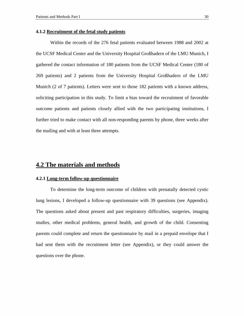
To determine the long-term outcome of children with prenatally detected cystic
lung lesions, I developed a follow-up questionnaire with 39 questions (see Appendix).
The questions asked about present and past respiratory difficulties, surgeries, imaging
studies, other medical problems, general health, and growth of the child. Consenting
parents could complete and return the questionnaire by mail in a prepaid envelope that I
had sent them with the recruitment letter (see Appendix), or they could answer the
questions over the phone.
Patients and Methods Part I 31
4.2.2 Medical records review
In addition to the completed questionnaires, all of the patients’ medical records
were reviewed. The records were filed either at the UCSF Medical Center or at the
University Hospital Großhadern of the LMU Munich, or they were sent from outside
institutions upon parents’ consent, or provided by the patients’ parents. The participants’
prenatal ultrasound reports or ultrasound scans, as well as their medical records,
documenting the patients’ perinatal and pediatric outcomes were reviewed.
4.2.3 Prenatal ultrasonographic data
From the participating patients’ ultrasound reports I extracted the prenatal
ultrasonographic data. When the patients’ prenatal ultrasound reports were inconclusive
regarding relevant study parameters, Professor Ruth B. Goldstein and I followed up with
a review of the ultrasound scans or videotapes.
Ultrasonographic data about the type and diagnosis of the lesion, the size of the
mass, the involved thoracic site (right, left, or bilateral), mediastinal shift, hydrops
development (with detailed information about ascites, pleural effusion, pericardial
effusion, and skin edema), polyhydramnios, placentomegaly, and the size of the fetus
(cited as below or above the 90th percentile) were tabulated and labeled with the
gestational age at observation (dating based on last menstrual period [LMP]). The
quantifiable ultrasonographic parameters were sub-classified into four severity groups
(including one group without symptoms, if suitable).
The appearance and type of the mass was categorized into predominantly cystic,
solid, or mixed; as well as into type I (cysts >2 cm), type II (cysts <2 cm), or type III (no
Patients and Methods Part I 32
cysts identified) – as described by Stocker [125]. If the mass was cystic, the size of the
largest cyst was recorded (Figure 1).
The mass was categorized into (i) a small sized lesion, if up to one-third of the
hemithorax was affected; (ii) a medium sized lesion, if one-third to two-thirds of the
hemithorax was occupied; and if the complete hemithorax, or even more than that was
affected, it was considered as (iii) large or (iv) very large, respectively (Figures 1 and 2).
The mass size was assessed at the initial diagnosis, at its largest stage (maximal mass
size), and finally at the end of the pregnancy or before fetal intervention (final/ultimate
mass size).
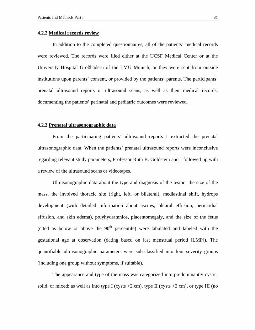
Figure 1. Axial image of the fetal chest at the level of the 4-chamber heart view (case no. 26): At 22 4/7 weeks’ gestation, a left-sided CCAM of Stocker type II impresses as a large, solid appearing lung mass with several microcysts (largest microcyst 1.2 cm). Crosses, labeled 1 and 2, show the extent of the tumor.
Patients and Methods Part I
33
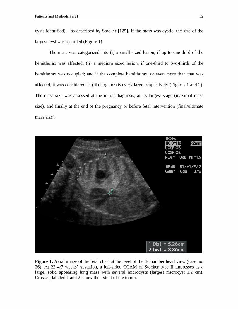
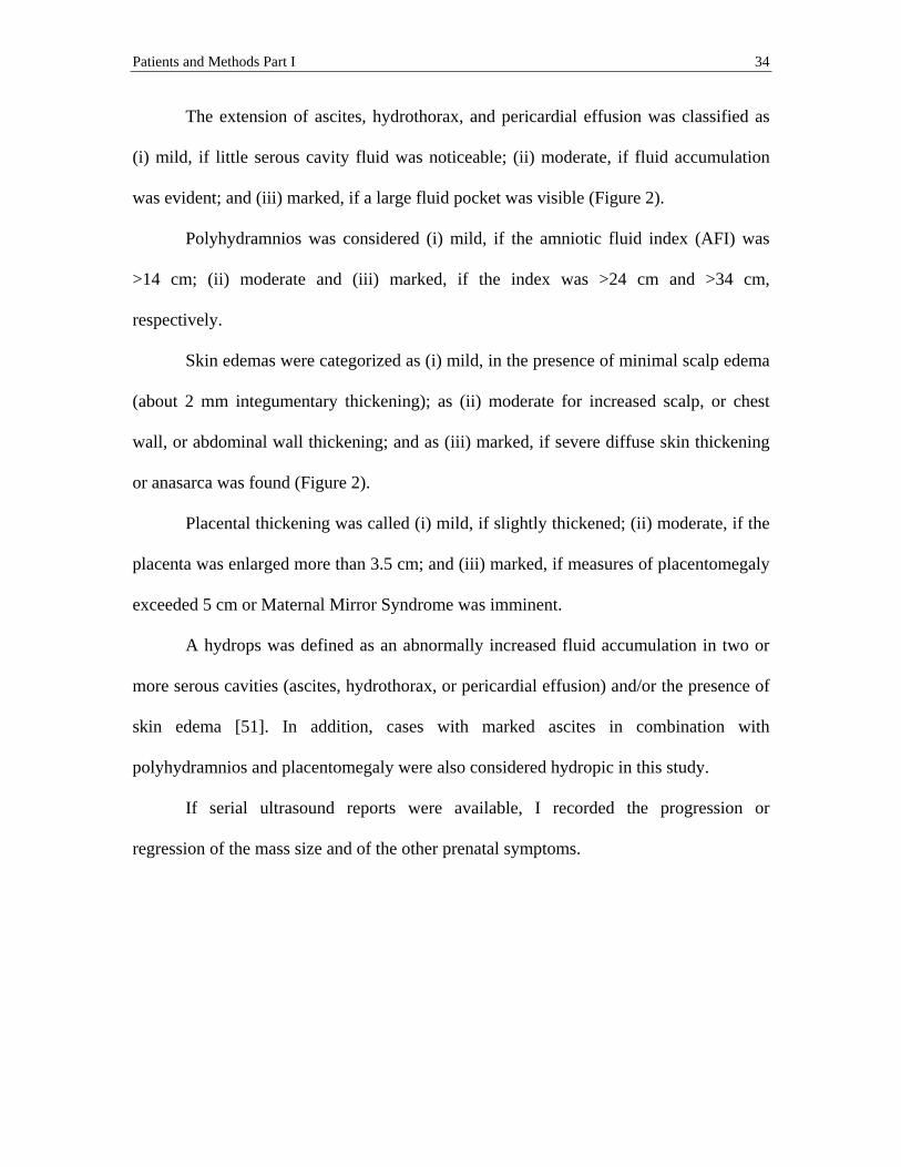
Figure 2. Longitudinal view of a 22 weeks and 3 days old fetus (case no. 56) with a very large CCAM of Stocker type III: The extremely echogenic lung mass (small arrows) involved almost the whole chest cavity by extending from the right to the left hemithorax and compressing and markedly shifting the fetal heart and mediastinum. The fetus was hydropic with marked scalp edema and ascites (block arrows).
Criteria for the classification of the mediastinal shift were the position of the heart
and the ability to visualize the normal contralateral lung tissue. The shift was considered
(i) mild, if the fetal cardiac axis was minimally deviated; (ii) moderate, if the heart was
considerably shifted to the contralateral side of the thorax, but contralateral lung was still
visible; and (iii) marked, if the heart was pressed against the contralateral rib cage, and
the contralateral lung was not visible any more (Figure 2).
Patients and Methods Part I 34
The extension of ascites, hydrothorax, and pericardial effusion was classified as
(i) mild, if little serous cavity fluid was noticeable; (ii) moderate, if fluid accumulation
was evident; and (iii) marked, if a large fluid pocket was visible (Figure 2).
Polyhydramnios was considered (i) mild, if the amniotic fluid index (AFI) was
>14 cm; (ii) moderate and (iii) marked, if the index was >24 cm and >34 cm,
respectively.
Skin edemas were categorized as (i) mild, in the presence of minimal scalp edema
(about 2 mm integumentary thickening); as (ii) moderate for increased scalp, or chest
wall, or abdominal wall thickening; and as (iii) marked, if severe diffuse skin thickening
or anasarca was found (Figure 2).
Placental thickening was called (i) mild, if slightly thickened; (ii) moderate, if the
placenta was enlarged more than 3.5 cm; and (iii) marked, if measures of placentomegaly
exceeded 5 cm or Maternal Mirror Syndrome was imminent.
A hydrops was defined as an abnormally increased fluid accumulation in two or
more serous cavities (ascites, hydrothorax, or pericardial effusion) and/or the presence of
skin edema [51]. In addition, cases with marked ascites in combination with
polyhydramnios and placentomegaly were also considered hydropic in this study.
If serial ultrasound reports were available, I recorded the progression or
regression of the mass size and of the other prenatal symptoms.
Patients and Methods Part I 35
4.2.4 Perinatal and pediatric outcome data
The medical records of the study patients were reviewed with a focus on clinical
data collected during the prenatal period, the neonatal period, and during the entire baby-
and childhood.
I extracted obstetrical history data about parity, karyotype, alpha-fetoprotein
(AFP) serum concentration, fertility treatment, mother’s age at delivery, and the parents’
ethnicity. The following postnatal data about the tumor were collected: the lung lesion’s
size and its appearance upon radiographic imaging, the size and location (lobe) of the
lesion at operation, the type of operation, and the histopathologic diagnosis, outlining the
type of the lesion and the cyst sizes.
After reviewing the surgical intervention strategies, I classified the study patients
into 5 intervention groups with respect to the invasiveness and the age at the time of the
procedure, as follows: (1) the termination of pregnancy (TOP) group; (2) the invasive
fetal treatment group, including patients having experienced (a) fetal tumor resection or
(b) thoraco-amniotic shunting, cyst aspiration(s), or both; (3) the neonatal surgery group,
consisting of patients operated on until 1 month of age; (4) the childhood surgery group,
divided into two subgroups, (a) the early childhood operation group with children
operated on between 1 to 12 months of age and (b) the late childhood operation group
with children operated on after 1 year of age; and finally (5) the no-surgery group.
For all patients I collected the outcome data about respiratory symptoms, presence
and duration of ventilatory support, the need for intensive care treatment, and the duration
of hospitalization(s). Further, complications after surgery, such as early postoperative
problems, final appearance of the thoracic scar, manifestation of a pectus excavatum, and
Patients and Methods Part I 36
presence of a residual tumor, were recorded. The 1- and 5-minute Apgar scores, the
gestational age at birth, mode of delivery, gender, birth weight, and weight and height at
last follow-up were noted.
I grouped the outcome data into (A) the early postnatal outcome (0 to 2 months
old), (B) the interim childhood outcome (overall childhood outcome prior to the last
follow-up), and (C) the final outcome at last follow-up. As follows, cases were
categorized regarding the respiratory outcome as either completely asymptomatic or
having mild, moderate, or severe respiratory symptoms (but common transient
postoperative symptoms were excluded). In the neonatal period (A), (i) mild symptoms
were defined as caused by tachypnea requiring minor blow-by oxygen; (ii) symptomatic
moderate cases asked for prolonged oxygen need, limited mechanical respiration for less
than 2 days, or both; and (iii) in the severe symptoms category, those patients were placed
who had respiratory distress requiring mechanical ventilation for more than 2 days after
birth. Respiratory symptoms in childhood (B) and at last follow-up (C) were categorized
as (i) mild, if children had minor labored breathing on exertion or mild asthmatic
symptoms; (ii) moderate, if they had manifest asthma, frequent pulmonary infections,
and/or limited physical endurance; and (iii) severe, if extended re-hospitalizations,
prolonged oxygen treatment, or both were required. For each case I included the age at
improvement of the symptoms.
Permanent and transient non-respiratory health concerns were also recorded and I
classified patients into experiencing (i) no complications; problems being (ii) mild, such
as weight gain failure and transient cardiac arrhythmia; problems being (iii) moderate, as
Patients and Methods Part I
37
there were prematurity concerns and therapy resistant gastro-esophageal reflux disease
(GERD); and (iv) severe ailments, such as congenital syndromes.
4.2.5 Statistical analysis
Using SPSS 11.5 software (SPSS Inc, Chicago, IL, 2002), I performed the
following statistical tests: Pearson's chi-square test for categorical or ordinal data, t-tests
of the means and analysis of variance (ANOVA) for continuous data, and bivariate
correlation for both ordinal and continuous data. The results were considered significant
at an observed significance level (2-sided P value) of less than 0.05 (p<0.05) [72].
Results Part I 38
5. Results
5.1 The patients’ response and data source
As a result of two mass mailings and up to three phone call attempts per patient, I
reached 63 of 182 families. Herewith the response rate is 34.6%. Sixty (60 of 63) families
did consent to participate in the study. They either sent the completed follow-up
questionnaire by mail (48 families), or they answered the questions subsequently over the
phone (12 families; including the only 2 responding families from the University Hospital
Großhadern of the LMU Munich, contacted by Professor Alexander Strauss). Two
families did not consent, although both patients were expected to be of good outcome.
One family informed me that their fetus had been misdiagnosed as having a fetal lung
lesion, and instead it turned out he had a heart disease. The other 119 non-responding
families had moved to other locations without leaving further contact information or their
addresses, phone numbers, or both were not correct.
From the 60 participating patients, I reviewed 434 prenatal ultrasound reports. In
about 10% of the cases, Professor Ruth B. Goldstein and I additionally reviewed the
ultrasound scans, because of inconclusive reports regarding relevant study parameters.
The scans of the 434 prenatal ultrasound reports had been performed at the UCSF
Medical Center (217 ultrasound scans), at the University Hospital Großhadern of the
LMU Munich (22 ultrasound scans), and 195 scans had been done at outside institutions
in the United States. Six fetuses were diagnosed and monitored at the UCSF Medical
Center. Another 39 fetuses had been initially diagnosed at outside institutions, but were
finally evaluated or monitored at the UCSF Medical Center (37 patients) or at the
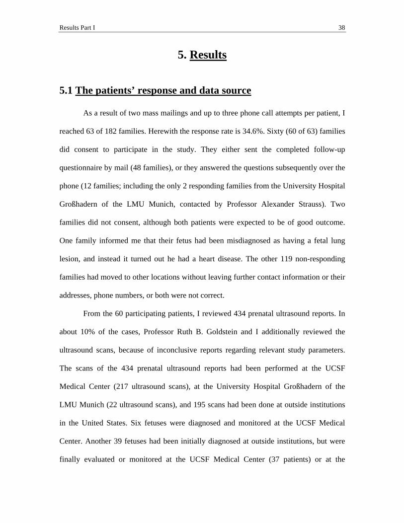
Results Part I 39
University Hospital Großhadern of the LMU Munich (2 patients). The mean delay until
referral after diagnosis was 3.8 ± 3.7 gestational weeks. Of the remaining 15 fetuses, the
outside institutions had sent ultrasound scans (as videotapes) and records, and these were
discussed at the UCSF Fetal Treatment Center meetings (Figure 3). I also reviewed the
records of these meetings.
Figure 3. Study patients (N=60)
6
37
2
15
Patients from UCSF
Patients evaluated at UCSF
Patients evaluated at LMU Großhadern
Patients from outside institutions
I reviewed more than 3,500 pages of the 60 patients’ medical records,
documenting the patients’ perinatal and pediatric outcomes. There were 27 patients
delivered and/or operated on at the UCSF Medical Center and 1 patient at the University
Hospital Großhadern of the LMU Munich. The remaining 32 patients were born and, if
necessary, operated at outside institutions.
Results Part I 40
5.2 The children’s outcomes
5.2.1 The survival rates of the children in the fetal non-intervention groups and in
the fetal treatment group were excellent
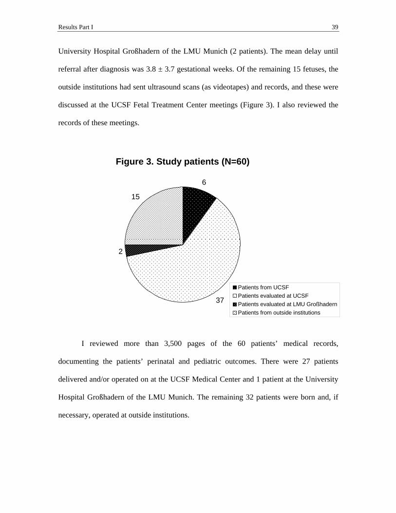
Of the 60 study fetuses with a completed follow-up, 51 (85%) children were
survivors at a mean follow-up of 5.9 ± 3.3 years. They were between 1.3 and 13.1 years
old. Of these 51 survivors, 13 children had not received surgery, 12 children were
operated on as neonates (1 to 30 days of age), 15 children subsequently underwent
surgery in childhood, and fetal interventions were performed in 11 cases. Of the 15
subsequent childhood surgery cases, 11 cases received operations during infancy (at an
age of less than 1 year, between 2.4 to 11.7 months of age) and 4 cases received
operations later in childhood (between 1.75 to 4.5 years of age). Of the 11 cases treated
with fetal intervention, a fetal tumor resection (at 20.4 to 27.7 weeks’ gestation) was
performed in 7 cases and fetal shunt/cyst aspirations were performed (at 23.1 to 34
weeks’ gestation) in 4 cases (Figure 4.1; Tables 2.1, 2.2, and 3).
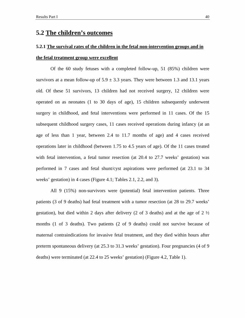
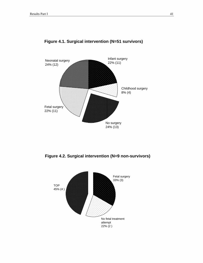
All 9 (15%) non-survivors were (potential) fetal intervention patients. Three
patients (3 of 9 deaths) had fetal treatment with a tumor resection (at 28 to 29.7 weeks’
gestation), but died within 2 days after delivery (2 of 3 deaths) and at the age of 2 ½
months (1 of 3 deaths). Two patients (2 of 9 deaths) could not survive because of
maternal contraindications for invasive fetal treatment, and they died within hours after
preterm spontaneous delivery (at 25.3 to 31.3 weeks’ gestation). Four pregnancies (4 of 9
deaths) were terminated (at 22.4 to 25 weeks’ gestation) (Figure 4.2, Table 1).
Results Part I 41
Figure 4.1. Surgical intervention (N=51 survivors)
Fetal surgery 22% (11)
No surgery 24% (13)
Childhood surgery 8% (4)
Infant surgery 22% (11)
Neonatal surgery 24% (12)
Figure 4.2. Surgical intervention (N=9 non-survivors)
No fetal treatment attempt 22% (2 )
Fetal surgery 33% (3)
TOP 45% (4 )
Results Part I 42
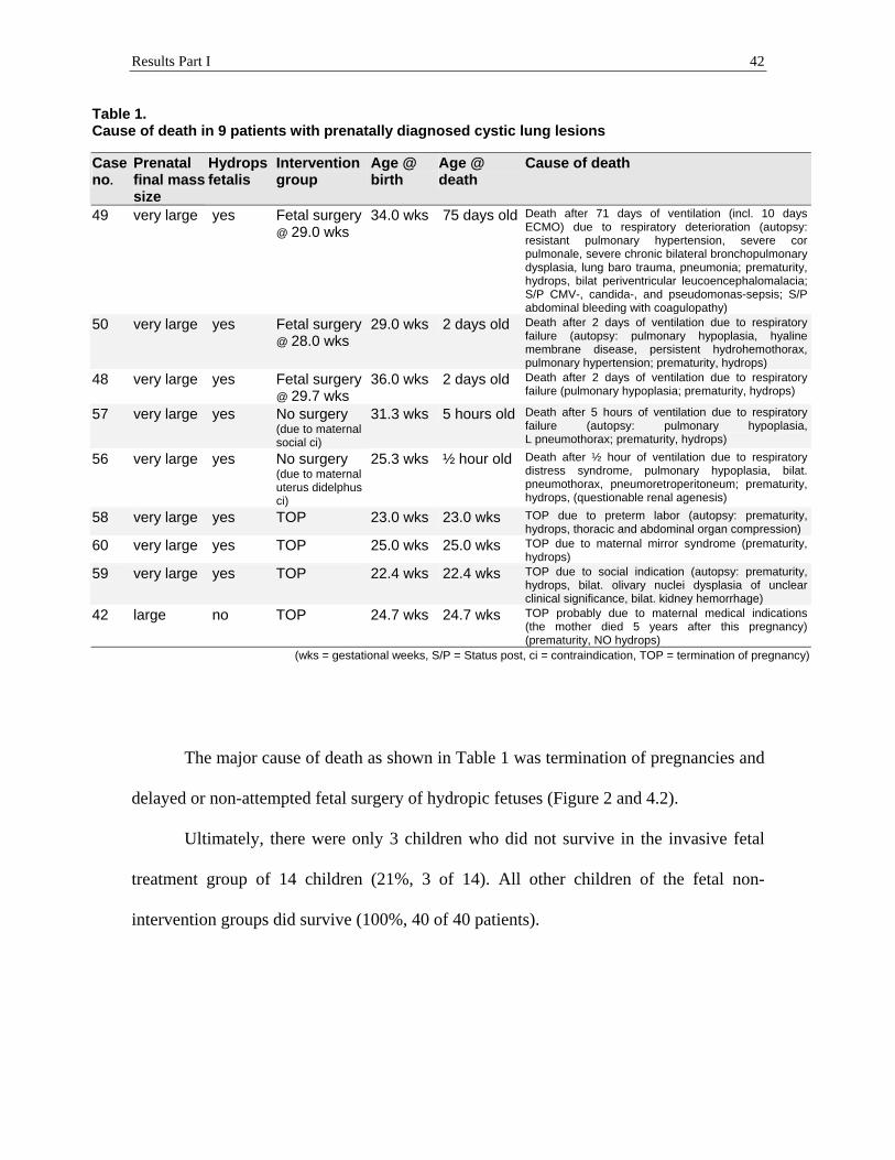
Table 1. Cause of death in 9 patients with prenatally diagnosed cystic lung lesions
Case no.
Prenatal final mass size
Hydrops fetalis
Intervention group
Age @ birth
Age @ death
Cause of death
49 very large yes Fetal surgery @ 29.0 wks
34.0 wks 75 days old Death after 71 days of ventilation (incl. 10 days ECMO) due to respiratory deterioration (autopsy: resistant pulmonary hypertension, severe cor pulmonale, severe chronic bilateral bronchopulmonary dysplasia, lung baro trauma, pneumonia; prematurity, hydrops, bilat periventricular leucoencephalomalacia; S/P CMV-, candida-, and pseudomonas-sepsis; S/P abdominal bleeding with coagulopathy)
50 very large yes Fetal surgery @ 28.0 wks
29.0 wks 2 days old Death after 2 days of ventilation due to respiratory failure (autopsy: pulmonary hypoplasia, hyaline membrane disease, persistent hydrohemothorax, pulmonary hypertension; prematurity, hydrops)
48 very large yes Fetal surgery @ 29.7 wks
36.0 wks 2 days old Death after 2 days of ventilation due to respiratory failure (pulmonary hypoplasia; prematurity, hydrops)
57 very large yes No surgery (due to maternal social ci)
31.3 wks 5 hours old Death after 5 hours of ventilation due to respiratory failure (autopsy: pulmonary hypoplasia, L pneumothorax; prematurity, hydrops)
56 very large yes No surgery (due to maternal uterus didelphus ci)
25.3 wks ½ hour old Death after ½ hour of ventilation due to respiratory distress syndrome, pulmonary hypoplasia, bilat. pneumothorax, pneumoretroperitoneum; prematurity, hydrops, (questionable renal agenesis)
58 very large yes TOP 23.0 wks 23.0 wks TOP due to preterm labor (autopsy: prematurity, hydrops, thoracic and abdominal organ compression)
60 very large yes TOP 25.0 wks 25.0 wks TOP due to maternal mirror syndrome (prematurity, hydrops)
59 very large yes TOP 22.4 wks 22.4 wks TOP due to social indication (autopsy: prematurity, hydrops, bilat. olivary nuclei dysplasia of unclear clinical significance, bilat. kidney hemorrhage)
42 large no TOP 24.7 wks 24.7 wks TOP probably due to maternal medical indications (the mother died 5 years after this pregnancy) (prematurity, NO hydrops)
(wks = gestational weeks, S/P = Status post, ci = contraindication, TOP = termination of pregnancy)
The major cause of death as shown in Table 1 was termination of pregnancies and
delayed or non-attempted fetal surgery of hydropic fetuses (Figure 2 and 4.2).
Ultimately, there were only 3 children who did not survive in the invasive fetal
treatment group of 14 children (21%, 3 of 14). All other children of the fetal non-
intervention groups did survive (100%, 40 of 40 patients).
Results Part I 43
5.2.2 The early respiratory outcome correlated with the children’s age at surgical
intervention
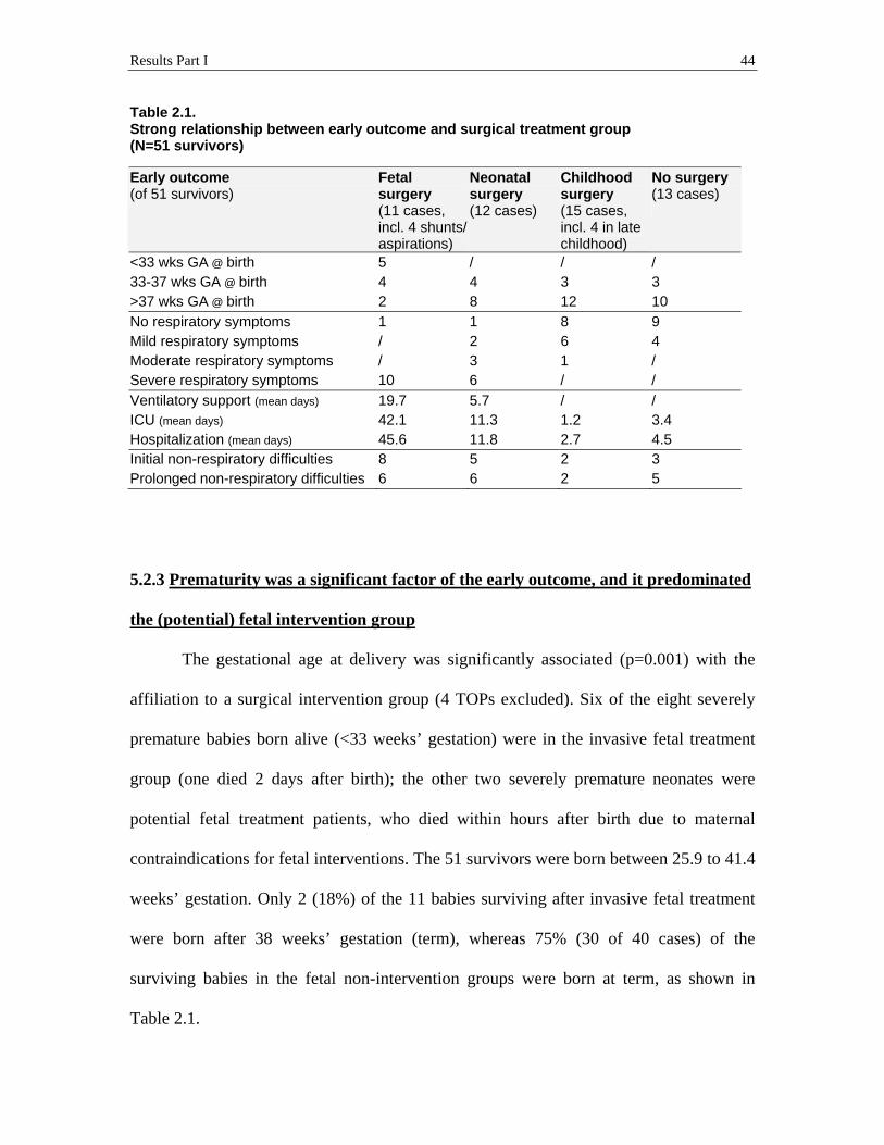
The early respiratory outcome in the first 2 months of the children’s lives was
categorized according to the respiratory symptoms and the need of respiratory support as
described in the PATIENTS AND METHODS section. The differences in the early
respiratory outcomes of the survivors depended significantly (p<0.001) on the age at
surgical intervention (5 intervention groups, as classified in the PATIENTS AND
METHODS section). Of the 11 neonates surviving invasive fetal treatment, 10 (91%) had
severe early respiratory symptoms. Of the 12 children requiring neonatal surgery, 6 had
severe and 3 had moderate early respiratory symptoms, and only 3 neonates were
electively operated. Of the surviving children in the childhood surgery group (15 cases)
and in the no-surgery group (13 cases), all but one were respiratory-wise asymptomatic
(17 cases) or only mildly symptomatic (10 cases) early after birth (Table 3). As expected,
more aggressively treated children had more severe early respiratory symptoms and they
required longer days on mechanical ventilation, in the intensive care unit (ICU), or both
(p<0.001) (Table 2.1).
Results Part I 44
Table 2.1. Strong relationship between early outcome and surgical treatment group (N=51 survivors)
Early outcome (of 51 survivors)
Fetal surgery (11 cases, incl. 4 shunts/aspirations)
Neonatal surgery (12 cases)
Childhood surgery (15 cases, incl. 4 in late childhood)
No surgery (13 cases)
<33 wks GA @ birth 5 / / / 33-37 wks GA @ birth 4 4 3 3 >37 wks GA @ birth 2 8 12 10 No respiratory symptoms 1 1 8 9 Mild respiratory symptoms / 2 6 4 Moderate respiratory symptoms / 3 1 / Severe respiratory symptoms 10 6 / / Ventilatory support (mean days) 19.7 5.7 / / ICU (mean days) 42.1 11.3 1.2 3.4 Hospitalization (mean days) 45.6 11.8 2.7 4.5 Initial non-respiratory difficulties 8 5 2 3 Prolonged non-respiratory difficulties 6 6 2 5
5.2.3 Prematurity was a significant factor of the early outcome, and it predominated
the (potential) fetal intervention group
The gestational age at delivery was significantly associated (p=0.001) with the
affiliation to a surgical intervention group (4 TOPs excluded). Six of the eight severely
premature babies born alive (<33 weeks’ gestation) were in the invasive fetal treatment
group (one died 2 days after birth); the other two severely premature neonates were
potential fetal treatment patients, who died within hours after birth due to maternal
contraindications for fetal interventions. The 51 survivors were born between 25.9 to 41.4
weeks’ gestation. Only 2 (18%) of the 11 babies surviving after invasive fetal treatment
were born after 38 weeks’ gestation (term), whereas 75% (30 of 40 cases) of the
surviving babies in the fetal non-intervention groups were born at term, as shown in
Table 2.1.
Results Part I 45
Preterm labor (PTL) and premature rupture of membranes (PROM) were the two
reasons for premature delivery in the fetal intervention group. Following fetal
intervention, 85% (11/13) of the patients were born prior to 36.3 weeks’ gestation due to
PTL (9%, 1/11), PROM (9%, 1/11), or mostly due to both (55%, 6/11); (data were not
available for 2 cases, case no. 41 and case no. 48).
The early respiratory outcomes as well as the interim respiratory childhood
outcomes significantly correlated with the gestational age at delivery (p<0.001). Thus, the
need for mechanical ventilation and ICU treatment was significantly higher for children
who were prematurely born (p=0.006). However, the respiratory outcomes improved
remarkably with long-term observation, as described in the following CHAPTER 5.2.4.
Initial transient non-respiratory difficulties of the survivors, such as
hyperbilirubinemia, anemia, arterial hypo- or hypertonia, electrolyte irregularities, or
sepsis, were also significantly associated with prematurity (p=0.005). Of the 19 survivors
born at less than 38 weeks’ gestation, 12 had perinatal non-respiratory symptoms (Table
2.1). However, these problems resolved in the neonatal period. More resistant to
treatment were prolonged difficulties with feeding, weight gain, and GERD, which also
tended to be associated with prematurity (p=0.08). Of the 19 survivors born less than 38
weeks’ gestation, 10 had such difficulties (Table 2.1). The prolonged non-respiratory
difficulties were still present at the last follow-up in half of the symptomatic cases (5 of
10 cases), yet, they all had improved to a milder stage, mostly by the age of 2 years old
(see CHAPTER 5.2.7). Prematurity was a significant determining factor for the early
respiratory and non-respiratory outcomes, but the early symptoms mostly resolved in the
course of the follow-ups.
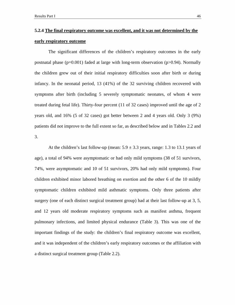
Results Part I 46
5.2.4 The final respiratory outcome was excellent, and it was not determined by the
early respiratory outcome
The significant differences of the children’s respiratory outcomes in the early
postnatal phase (p=0.001) faded at large with long-term observation (p>0.94). Normally
the children grew out of their initial respiratory difficulties soon after birth or during
infancy. In the neonatal period, 13 (41%) of the 32 surviving children recovered with
symptoms after birth (including 5 severely symptomatic neonates, of whom 4 were
treated during fetal life). Thirty-four percent (11 of 32 cases) improved until the age of 2
years old, and 16% (5 of 32 cases) got better between 2 and 4 years old. Only 3 (9%)
patients did not improve to the full extent so far, as described below and in Tables 2.2 and
3.
At the children’s last follow-up (mean: 5.9 ± 3.3 years, range: 1.3 to 13.1 years of
age), a total of 94% were asymptomatic or had only mild symptoms (38 of 51 survivors,
74%, were asymptomatic and 10 of 51 survivors, 20% had only mild symptoms). Four
children exhibited minor labored breathing on exertion and the other 6 of the 10 mildly
symptomatic children exhibited mild asthmatic symptoms. Only three patients after
surgery (one of each distinct surgical treatment group) had at their last follow-up at 3, 5,
and 12 years old moderate respiratory symptoms such as manifest asthma, frequent
pulmonary infections, and limited physical endurance (Table 3). This was one of the
important findings of the study: the children’s final respiratory outcome was excellent,
and it was independent of the children’s early respiratory outcomes or the affiliation with
a distinct surgical treatment group (Table 2.2).
Results Part I 47
Table 2.2. No relationship between long-term respiratory outcomes and surgical treatment group
Long-term respiratory outcome
Total patients (60 cases)
No surgery (15 cases)
Childhood surgery (15 cases, incl. 4 in late childhood)
Neonatal surgery (12 cases)
Fetal surgery (14 cases, incl. 4 shunts/ aspirations)
TOP (4 cases)
No symptoms 38 11 11 8 8 / Mild symptoms 10 2 3 3 2 / Moderate symptoms 3 / 1 1 1 / Severe symptoms / / / / / /
Alive 51 13 15 12 11 /
Dead 9 2 / / 3 4
5.2.5 Interim respiratory outcomes correlated with early respiratory outcomes
during the period before significant improvement
Interim respiratory symptom severity, observed within the period before
significant improvements, correlated with the level of early respiratory symptoms
(p<0.001). However, the age when significant improvement was achieved did not
correlate with the severity of the early symptoms (p>0.6).
Only 2 (6%) of the 31 surviving children who were asymptomatic or mildly
symptomatic early after birth developed a moderately worsened respiratory condition
during childhood. In the group of the 20 surviving children with severe or moderate early
respiratory symptoms, 13 (65%) had prolonged severe (7 of 13 cases) or moderate (6 of
13 cases) interim respiratory symptoms prior to the age of 2 years (8 of 13 cases), 4 years
(3 of 13 cases), or until the last follow-up (2 of 13 cases). The remaining 7 children (7 of
20 cases) in this group had already improved in the neonatal period (6 of 7 cases) or in
the first year (1 of 7 case), and all 7 children were asymptomatic or mildly symptomatic
thereafter (Table 3).
Results Part I 48
The children with severe interim respiratory symptoms required re-
hospitalizations in childhood (4 cases after fetal surgery, 1 case after neonatal surgery, 1
case for delayed symptomatic surgery) or extended oxygen treatment during infancy (1
case after neonatal surgery), as described in the following CHAPTER 5.2.6. Despite the
above-described interim respiratory symptoms, final respiratory outcomes were excellent
and not determined by the interim respiratory outcomes, as it was not determined by the
early respiratory outcomes.
5.2.6 Re-hospitalizations in childhood were required for 55% of the children after
invasive fetal treatment and for 54% of the non-operated children for delayed
surgery
Of the 28 children who were not operated on until the age of one month, 15 (54%)
were re-hospitalized after the perinatal period, either for symptomatic delayed surgery (6
cases, 2.4 months to 4.3 years old) or for primary elective surgery (9 cases, 5 months to
4.5 years old). Four of the six children who were operated on for delayed symptoms had
prolonged hospital stays (9 days to 34 days) due to postoperative complications. One
non-operated child (case no. 12) was re-hospitalized not for surgery, but due to
pneumonia that he developed at the age of 2 years. Among the 12 children operated on
neonatally, only 2 were re-hospitalized due to recurrent respiratory infections (one at 6
months old and the other at 3 ½ years old).
Re- hospitalization(s) were required for 6 (55%) of the 11 invasive fetal treatment
survivors, because of severe (4 cases) or mild (2 cases) interim respiratory symptoms.
Three of these patients were re-hospitalized during infancy for 3 to 15 days due to
Results Part I 49
pneumonia, respiratory syncytial virus bronchiolitis, or both. One child on whom
multiple cyst aspirations had been performed before birth required postnatal surgery on a
bronchogenic cyst at 6 months due to mild respiratory symptoms (case no. 41). Another
fetal surgery patient (case no. 55) required 16 days of re-hospitalizations during infancy.
She was finally re-operated on at the age of 3 ½ years for symptomatic CCAM residual
and thoracic scar deformation. This case is discussed in detail in PART II of this doctoral
thesis. The patient with the longest re-hospitalization interval of 153 days (case no. 54)
required an implantation of a tissue expander at 2 months old and aortopexy at 3 ½
months old due to severe mediastinal shift after fetal pneumonectomy.
All children recovered well from the symptoms and re-hospitalizations (Tables
2.2 and 3). Only three children had moderate respiratory symptoms at their last follow-
ups. An important conclusion could be drawn: The final outcomes of the children were
excellent, despite re-hospitalizations due to interim respiratory problems.
5.2.7 Other non-respiratory complications were postoperative complications,
prematurity-associated feeding problems, transient cardiac symptoms, and rare
congenital anomalies
Postoperative late complications, such as a pectus excavatum and a prominent
thoracic scar, developed six times and five times, respectively, in 8 (21%) of the 38
survivors who had received operations. Remarkably, all 8 children were operated on prior
to the age of 1 month (including 3 fetal surgery cases). None of these children required
surgical revision by the age of 3 to 12 years old, except for one re-operated child (case
Results Part I 50
no. 55), as described above in CHAPTER 5.2.6 and discussed in detail in PART II of this
doctoral thesis.
Prolonged problems with feeding, weight gain, and GERD occurred close to twice
as much in prematurely born survivors (10 of 19 cases born <37 weeks’ gestation, 53%)
than in term born survivors (9 of 32 cases born >37 weeks’ gestation, 28%), with a trend
to statistical significance (p=0.08). However, these difficulties had mostly diminished to a
milder stage by the age of 2 years. The fact that 8 (16%) of 51 surviving children had low
body weight (<3rd percentile) at their last follow-ups was not linked to any initial feeding
problems (p>0.13) or prematurity (p>0.3). It was also independent of the prior surgical
treatment strategy (p>0.3) and of the early and final respiratory outcomes (p>0.5).
There were 10 children of the 51 survivors who presented with transient
functional cardiac symptoms such as arrhythmia (3 cases), cardiac enlargement (4 cases),
and heart murmurs (7 cases, combinations included). These symptoms disappeared in the
neonatal period or at the latest at 6 months of age. None of these children had a
congenital, anatomic heart disease. No association between the cardiac symptoms and
prematurity (p>0.1), early respiratory outcomes (p>0.1), or surgery group affiliation
(p>0.3) could be assessed. Also, the extension of a prenatal mediastinal shift had no
influence on the cardiac symptoms (p>0.1).
Other major congenital anomalies were found in 5 (8.3%) of the 60 study cases. A
prenatal ultrasonographic diagnosis of these anomalies was possible for only 2 (3.3%, 2
of 60) of these cases. One fetal surgery case developed Asperger syndrome, a milder
variant of autism. One child in the neonatal surgery group was found to have Mayer-
Rokitansky-Küster-Hauser syndrome (with right renal agenesis and vaginal atresia), and
Results Part I 51
another child in this group had albinism. One child required craniofacial surgery for
congenital craniosynostosis shortly after the CCAM operation in late infancy. At their last
follow-ups, all of these 4 children had been enrolled in the regular (pre-) school system
and were doing fine. The fifth case, a terminated fetus (case no. 59), was found through
autopsy to have bilateral dysplasia of the olivary nuclei of unclear clinical significance.
There was no obvious link between the congenital anomalies and a distinct intervention
group or prematurity. However, the number of cases was too small to carry out
statistically significant calculations regarding the incidence of these anomalies.
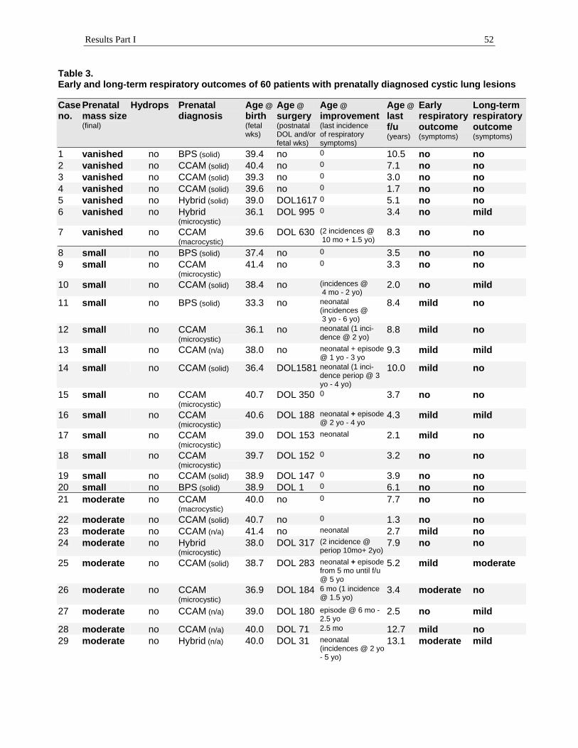
Results Part I 52
Table 3. Early and long-term respiratory outcomes of 60 patients with prenatally diagnosed cystic lung lesions
Case no.
Prenatal mass size (final)
Hydrops Prenatal diagnosis
Age @ birth (fetal wks)
Age @ surgery (postnatal DOL and/or fetal wks)
Age @ improvement (last incidence of respiratory symptoms)
Age @ last f/u (years)
Early respiratory outcome (symptoms)
Long-term respiratory outcome (symptoms)
1 vanished no BPS (solid) 39.4 no 0 10.5 no no 2 vanished no CCAM (solid) 40.4 no 0 7.1 no no 3 vanished no CCAM (solid) 39.3 no 0 3.0 no no 4 vanished no CCAM (solid) 39.6 no 0 1.7 no no 5 vanished no Hybrid (solid) 39.0 DOL1617 0 5.1 no no 6 vanished no Hybrid
(microcystic) 36.1 DOL 995 0 3.4 no mild
7 vanished no CCAM (macrocystic)
39.6 DOL 630 (2 incidences @ 10 mo + 1.5 yo)
8.3 no no
8 small no BPS (solid) 37.4 no 0 3.5 no no 9 small no CCAM
(microcystic) 41.4 no 0 3.3 no no
10 small no CCAM (solid) 38.4 no (incidences @ 4 mo - 2 yo)
2.0 no mild
11 small no BPS (solid) 33.3 no neonatal (incidences @ 3 yo - 6 yo)
8.4 mild no
12 small no CCAM (microcystic)
36.1 no neonatal (1 inci-dence @ 2 yo)
8.8 mild no
13 small no CCAM (n/a) 38.0 no neonatal + episode @ 1 yo - 3 yo
9.3 mild mild
14 small no CCAM (solid) 36.4 DOL1581 neonatal (1 inci-dence periop @ 3 yo - 4 yo)
10.0 mild no
15 small no CCAM (microcystic)
40.7 DOL 350 0 3.7 no no
16 small no CCAM (microcystic)
40.6 DOL 188 neonatal + episode @ 2 yo - 4 yo
4.3 mild mild
17 small no CCAM (microcystic)
39.0 DOL 153 neonatal 2.1 mild no
18 small no CCAM (microcystic)
39.7 DOL 152 0 3.2 no no
19 small no CCAM (solid) 38.9 DOL 147 0 3.9 no no 20 small no BPS (solid) 38.9 DOL 1 0 6.1 no no 21 moderate no CCAM
(macrocystic) 40.0 no 0 7.7 no no
22 moderate no CCAM (solid) 40.7 no 0 1.3 no no 23 moderate no CCAM (n/a) 41.4 no neonatal 2.7 mild no 24 moderate no Hybrid
(microcystic) 38.0 DOL 317 (2 incidence @
periop 10mo+ 2yo) 7.9 no no
25 moderate no CCAM (solid) 38.7 DOL 283 neonatal + episode from 5 mo until f/u @ 5 yo
5.2 mild moderate
26 moderate no CCAM (microcystic)
36.9 DOL 184 6 mo (1 incidence @ 1.5 yo)
3.4 moderate no
27 moderate no CCAM (n/a) 39.0 DOL 180 episode @ 6 mo - 2.5 yo
2.5 no mild
28 moderate no CCAM (n/a) 40.0 DOL 71 2.5 mo 12.7 mild no 29 moderate no Hybrid (n/a) 40.0 DOL 31 neonatal
(incidences @ 2 yo - 5 yo)
13.1 moderate mild
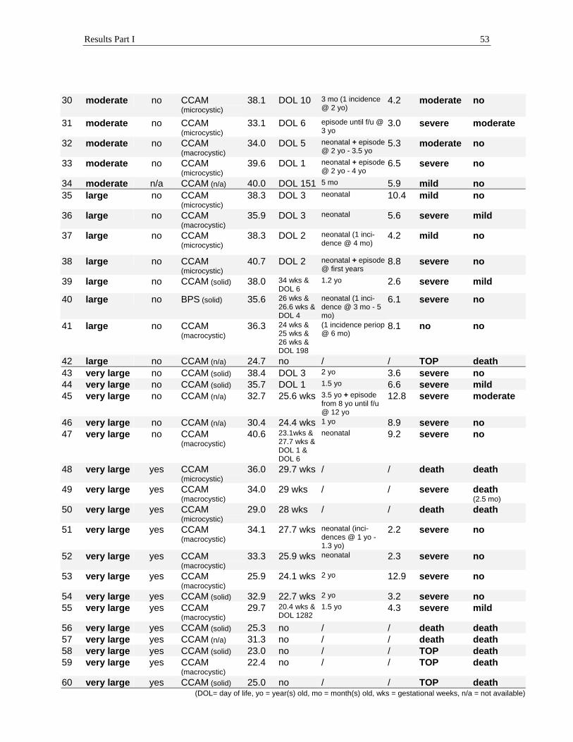
Results Part I 53
30 moderate no CCAM (microcystic)
38.1 DOL 10 3 mo (1 incidence @ 2 yo)
4.2 moderate no
31 moderate no CCAM (microcystic)
33.1 DOL 6 episode until f/u @ 3 yo
3.0 severe moderate
32 moderate no CCAM (macrocystic)
34.0 DOL 5 neonatal + episode @ 2 yo - 3.5 yo
5.3 moderate no
33 moderate no CCAM (microcystic)
39.6 DOL 1 neonatal + episode @ 2 yo - 4 yo
6.5 severe no
34 moderate n/a CCAM (n/a) 40.0 DOL 151 5 mo 5.9 mild no 35 large no CCAM
(microcystic) 38.3 DOL 3 neonatal 10.4 mild no
36 large no CCAM (macrocystic)
35.9 DOL 3 neonatal 5.6 severe mild
37 large no CCAM (microcystic)
38.3 DOL 2 neonatal (1 inci-dence @ 4 mo)
4.2 mild no
38 large no CCAM (microcystic)
40.7 DOL 2 neonatal + episode @ first years
8.8 severe no
39 large no CCAM (solid) 38.0 34 wks & DOL 6
1.2 yo 2.6 severe mild
40 large no BPS (solid) 35.6 26 wks & 26.6 wks & DOL 4
neonatal (1 inci-dence @ 3 mo - 5 mo)
6.1 severe no
41 large no CCAM (macrocystic)
36.3 24 wks & 25 wks & 26 wks & DOL 198
(1 incidence periop @ 6 mo)
8.1 no no
42 large no CCAM (n/a) 24.7 no / / TOP death 43 very large no CCAM (solid) 38.4 DOL 3 2 yo 3.6 severe no 44 very large no CCAM (solid) 35.7 DOL 1 1.5 yo 6.6 severe mild 45 very large no CCAM (n/a) 32.7 25.6 wks 3.5 yo + episode
from 8 yo until f/u @ 12 yo
12.8 severe moderate
46 very large no CCAM (n/a) 30.4 24.4 wks 1 yo 8.9 severe no 47 very large no CCAM
(macrocystic) 40.6 23.1wks &
27.7 wks & DOL 1 & DOL 6
neonatal 9.2 severe no
48 very large yes CCAM (microcystic)
36.0 29.7 wks / / death death
49 very large yes CCAM (macrocystic)
34.0 29 wks / / severe death (2.5 mo)
50 very large yes CCAM (microcystic)
29.0 28 wks / / death death
51 very large yes CCAM (macrocystic)
34.1 27.7 wks neonatal (inci-dences @ 1 yo - 1.3 yo)
2.2 severe no
52 very large yes CCAM (macrocystic)
33.3 25.9 wks neonatal 2.3 severe no
53 very large yes CCAM (macrocystic)
25.9 24.1 wks 2 yo 12.9 severe no
54 very large yes CCAM (solid) 32.9 22.7 wks 2 yo 3.2 severe no 55 very large yes CCAM
(macrocystic) 29.7 20.4 wks &
DOL 1282 1.5 yo 4.3 severe mild
56 very large yes CCAM (solid) 25.3 no / / death death 57 very large yes CCAM (n/a) 31.3 no / / death death 58 very large yes CCAM (solid) 23.0 no / / TOP death 59 very large yes CCAM
(macrocystic) 22.4 no / / TOP death
60 very large yes CCAM (solid) 25.0 no / / TOP death (DOL= day of life, yo = year(s) old, mo = month(s) old, wks = gestational weeks, n/a = not available)
Results Part I 54
5.3 The prognostic value of the prenatal ultrasound parameters
5.3.1 Not the distinct prenatal diagnosis, but the type of the cystic lung lesion was a
potential predictive factor of the early outcome
The prenatal ultrasonographic diagnoses of the 60 study cases were CCAM (51
cases), BPS (5 cases), and a CCAM-BPS-hybrid (4 cases). A histopathologic diagnosis
was established in 40 of the 60 study cases. For the other 20 cases, a histopathologic
diagnosis was not available, either because no surgery was done (13 cases) or the reports
of the surgery (3 cases) or of the autopsy (4 cases) were not accessible to me. The
prenatal diagnosis was confirmed in 29 of the 40 cases, which means an agreement
between the prenatal ultrasonographic diagnosis and the histopathologic diagnosis was
achieved in 72.5% of the cases. Nine of the prenatally diagnosed CCAM cases were
misdiagnosed and turned out to be BPSs (4 cases), CCAM-BPS-hybrids (3 cases), a
bronchogenic cyst (1 case), and a bronchiectasis (1 case) at pathology. Two prenatally
misdiagnosed CCAM-BPS-hybrid cases were identified as BPSs at pathology.
Prenatal color Doppler sonography was applied in 40 of the 60 study cases and it
provided information about a potential systemic feeding vessel from the aorta to the
mass. Color Doppler sonography, applied in addition to the conventional prenatal
ultrasonography, has an influence on the prenatal diagnosis. A histopathologic diagnosis
was established in 25 of these 40 cases. The ultrasonographic diagnosis supported by
Color Doppler imaging agreed with the pathological findings in 80% of the cases (20 of
25 cases). In contrast, a prenatal ultrasonographic diagnosis without color Doppler
imaging was correct in only 60% (only 9 of 15 study cases with a conventional prenatal
ultrasonographic diagnosis and a histopathologic diagnosis had a prenatal correct
Results Part I 55
diagnosis). Color Doppler imaging increased the positive predictive value for correct
prenatal distinct diagnosis.
The type of the cystic lung lesion was characterized using the Stocker type
classification in 50 of the 60 study cases, as described in the PATIENTS AND
METHODS section. There were 12 macrocystic type I cases, 17 microcystic type II
cases, and 21 solid appearing type III cases (Table 3). The Stocker type classification
matched roughly with the plain categories of sonographic appearance introduced in this
study, since the sonographic appearance of 56 of the 60 study cases was characterized as
predominantly cystic in 9 cases, mixed in 25 cases, and predominantly solid in 22 cases.
Of the 56 cases characterized into solid, cystic, or mixed, 37 had a histopathologic
diagnosis. Comparing the sonographic appearances of the lung lesions with the
histopathologic diagnoses, it again becomes clear that the distinct prenatal diagnosis,
solely based on the sonographic appearance of the lesion, is not very specific. Of the 10
fetal lung lesions with the sonographic appearance of a solid tumor, 6 (60%) were
CCAMs, 3 (30%) were BPSs, and 1 (10%) was a bronchiectasis at pathology. The 27
fetal lung lesions with the sonographic appearance of a cystic or mixed tumor showed a
comparable pattern of histopathologic diagnoses with 21 (78%) CCAMs, 3 (11%) BPSs,
2 (7%) CCAM-BPS-hybrids, and 1 (4%) bronchogenic cyst. This breakdown, from the
lesions’ sonographic appearances into their histopathologic distinct diagnoses,
demonstrates that using conventional prenatal ultrasound without color Doppler imaging
does not establish a distinct correct prenatal diagnosis. Consequently, I did not include
the distinct prenatal ultrasonographic diagnosis due to its inaccuracy into my statistical
analyses of prenatal outcome predictors. Instead, I tested the postoperative or post-
Results Part I 56
autopsy, histopathologic diagnosis for its prognostic value and still there was no
significant difference in the data set between CCAM and BPS concerning the early
outcome (p>0.2).
However, the plain description of the lesions’ sonographic appearance and the
Stocker-types were of some prognostic value concerning the early outcome. The
predominantly solid appearing lung lesions (Stocker type III) in my study showed a
tendency (p=0.01 to p=0.07) to be of smaller final mass sizes and better early respiratory
outcomes than mixed lung lesions or large-cystic, Stocker type I lesions. Masses with
large Stocker type I cysts increased significantly more often during pregnancy and about
2 gestational weeks earlier (p<0.023) than solid, Stocker type III lesions or small-cystic,
Stocker type II lesions which in contrast regressed more often (Table 4). In one case (no.
59), the macrocystic Stocker type I lesion grew rapidly from a small sized lesion to a very
large mass before the pregnancy was terminated (Table 1). The significant factor
concerning the early outcome was the size of the cysts, not the predominance of the
cystic component in the mass. The larger the cysts grew, the larger the final size of the
mass became (p<0.023) and the more severe the early respiratory symptoms became
(p<0.028). Moderate or severe early respiratory symptoms were present in 70% (7 of 10
cases; 2 deaths excluded) of the surviving newborns with large Stocker type I cysts,
whereas 70% (23 of 33 cases; 5 deaths excluded) of the survivors with solid Stocker type
III lesions or small-cystic Stocker type II lesions were asymptomatic or only mildly
affected after birth. However, all of these children had a favorable final respiratory
outcome, independent of the type of the cystic lung lesion.
Results Part I 57
5.3.2 The final size of the mass was the most important predictor of the early
outcome
Using prenatal ultrasound, the cystic lung lesions of the 60 fetal patients were
initially diagnosed between 9.9 and 34.9 weeks’ gestation (mean 20.7 ± 3.8 weeks’
gestation) after the LMP. Serial ultrasound reports or scans were available for 52 (87%)
of the 60 fetuses; and in 49 of these 52 cases, more than three ultrasound reports or scans
existed. Using the serial ultrasound reports or scans of these 52 cases, it was possible to
determine the progression or regression of the size of the masses and of other prenatal
symptoms.
The cystic lung lesions of the 60 fetuses were initially very large in 11 fetuses,
large in 18 fetuses, moderate in 18 fetuses, and 11 fetuses had small lesions at initial
diagnosis. For one moderate sized lesion (no. 34) and one prenatal shunt case (no. 39) the
initial data were missing. At the end of the prenatal observational period or until fetal
intervention, most mass sizes had changed, and the final sizes of the 60 lesions became
very large (18 cases), large (8 cases), moderate (14 cases), and small (13 cases). In 7
cases the masses disappeared (Figure 5; Tables 3 and 4).
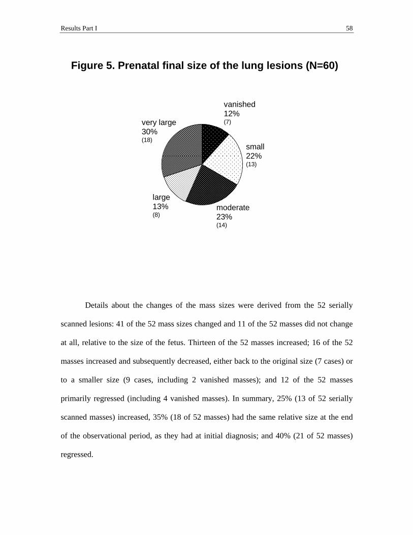
Results Part I 58
Figure 5. Prenatal final size of the lung lesions (N=60)
very large30%(18)
large13%(8)
vanished12%(7)
small22%(13)
moderate23%(14)
Details about the changes of the mass sizes were derived from the 52 serially
scanned lesions: 41 of the 52 mass sizes changed and 11 of the 52 masses did not change
at all, relative to the size of the fetus. Thirteen of the 52 masses increased; 16 of the 52
masses increased and subsequently decreased, either back to the original size (7 cases) or
to a smaller size (9 cases, including 2 vanished masses); and 12 of the 52 masses
primarily regressed (including 4 vanished masses). In summary, 25% (13 of 52 serially
scanned masses) increased, 35% (18 of 52 masses) had the same relative size at the end
of the observational period, as they had at initial diagnosis; and 40% (21 of 52 masses)
regressed.
Results Part I 59
Tracking the growth of the masses from their initial size to the final size, I noticed
that each size category had a different growth pattern. Most of the initially very large
masses stayed very large (10 of 11 cases, 91%); the as large categorized masses regressed
in 56% (10 of 18 cases) and increased in 33% (6 of 18 cases); the moderate masses
almost equally grew larger (7 of 18 cases, 39%), or smaller (6 of 18 cases, 33%), or
stayed the same size (5 of 18 cases, 28%); and the initially small masses were mostly
small or vanished (8 of 11 cases, 73%) at the end of the prenatal observational period.
Another interesting finding concerned the timing of the mass size changes.
Masses that had become large or very large at any point in time during fetal life
(independent of the final size of the mass) were more likely (p=0.06) to be diagnosed at
an earlier gestational age and – as the case may be – to increase and (subsequently)
regress earlier than small or moderate sized masses. The large and very large masses at
maximum size were diagnosed at a mean of 20.1 ± 3.3 weeks’ gestation (9.9 to 28.0
weeks’ gestation) and did – as the case may be – increase at a mean of 22.3 ± 1.5 weeks’
gestation (19.9 to 26.3 weeks’ gestation) and regress at a mean of 27.6 ± 3.7 weeks’
gestation (23.4 to 34.0 weeks’ gestation). In comparison, the masses with a small or
moderate maximum size were later diagnosed at a mean of 22.3 ± 4.7 weeks’ gestation
(13.0 to 34.9 weeks’ gestation) and – as the case may be – also increased later at a mean
of 23.8 ± 2.1 weeks’ gestation (21.7 to 26.7 weeks’ gestation) and regressed later at a
mean of 30.5 ± 3.8 weeks’ gestation (22.9 to 37.0 weeks’ gestation).
The size of the lesion was the most crucial factor in the early outcomes of the
fetuses. The final and the maximal prenatal sizes of the masses were significantly
associated (p<0.001 to p<0.003) with mortality, the need for pre- or postnatal surgical
Results Part I 60
treatment, and along with the early respiratory difficulties, with the requirement of
ventilatory support and intensive care treatment. Mortality almost exclusively occurred in
fetuses with ultimately very large masses (8 of 9 deaths, including 3 TOPs) (Table 1 and
Figure 2). The survivors of ultimately very large lesions (10 of 18 cases) all had severe
early respiratory symptoms and required mechanical ventilation for more than 3 days
(including 4 children with more than 15 days of ventilation). All surviving 17 children
with ultimately large or very large lesions (26 cases minus 9 non-survivors) were
operated on in the fetal (11 of 17 cases, 65%) or neonatal (6 of 17 cases, 35%) period.
None of the children with ultimately small or vanished lesions (20 cases) died or had
more than mild respiratory symptoms early after birth. Most of them were asymptomatic
(14 of 20 cases, 70%). None of them needed ventilatory support except for one child who
was ventilated only as a precaution, following neonatal surgery. Most of the children with
small or moderate sized lesions at the end of the pregnancy (34 cases) did not require
surgical treatment or were operated on later in childhood (28 of 34 cases, 82%) (Tables 3
and 4). Prematurity and prematurity-associated transient non-respiratory difficulties were
mostly a problem of children with expansive masses. The gestational age at birth and the
absolute birth weight were significantly lower (p<0.001 to p<0.002) in the very large
mass size group (mean: 32.6 ± 4.2 weeks’ gestation, 2172 ± 765 gram; TOPs excluded),
compared to the small mass size group (mean: 38.6 ± 1.9 weeks’ gestation, 3346 ± 662
gram).
In contrast to the high prognostic value of the final or maximal size of the mass,
the initial mass size, if it was moderate or large, did not predict the early outcome. If the
initial mass size was at one of the extreme ends (very small or very large), it would more
Results Part I 61
likely keep its size during pregnancy (for information about the specific prenatal growth
patterns of the different mass sizes, please refer to the fourth paragraph of this CHAPTER
5.3.2), and herewith it predicted (p<0.002 to p<0.018) in these cases the early outcome as
much as the final size did.
The differentiation into early respiratory outcome and long-term respiratory
outcome turned out to be essential, since the long-term outcome was not predictable
(p>0.6) by the prenatal size of the mass, in contrast to the early outcome. The final
outcomes of the children were excellent. More than 70% of the surviving children of any
prenatal mass size category were asymptomatic at final follow-up and no child had severe
symptoms. Only three children with prenatally different ultimate mass sizes (one very
large mass and two moderate masses) and different surgical treatment strategies (one fetal
surgery, one neonatal surgery, and one childhood surgery) had moderate symptoms at the
end of the study, as described in CHAPTER 5.2.4 and in Tables 3 and 4.
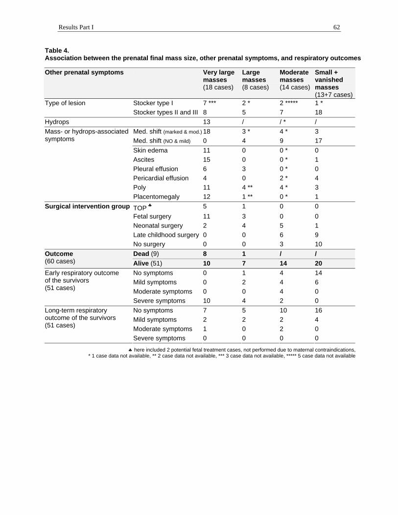
Results Part I 62
Table 4. Association between the prenatal final mass size, other prenatal symptoms, and respiratory outcomes
Other prenatal symptoms Very large masses (18 cases)
Large masses (8 cases)
Moderate masses (14 cases)
Small + vanished masses (13+7 cases)
Stocker type I 7 *** 2 * 2 ***** 1 * Type of lesion
Stocker types II and III 8 5 7 18
Hydrops 13 / / * /
Med. shift (marked & mod.) 18 3 * 4 * 3
Med. shift (NO & mild) 0 4 9 17
Skin edema 11 0 0 * 0
Ascites 15 0 0 * 1
Pleural effusion 6 3 0 * 0
Pericardial effusion 4 0 2 * 4
Poly 11 4 ** 4 * 3
Mass- or hydrops-associated symptoms
Placentomegaly 12 1 ** 0 * 1
TOP 5 1 0 0
Fetal surgery 11 3 0 0
Neonatal surgery 2 4 5 1
Late childhood surgery 0 0 6 9
Surgical intervention group
No surgery 0 0 3 10
Dead (9) 8 1 / / Outcome (60 cases) Alive (51) 10 7 14 20
No symptoms 0 1 4 14
Mild symptoms 0 2 4 6
Moderate symptoms 0 0 4 0
Early respiratory outcome of the survivors (51 cases)
Severe symptoms 10 4 2 0
No symptoms 7 5 10 16
Mild symptoms 2 2 2 4
Moderate symptoms 1 0 2 0
Long-term respiratory outcome of the survivors (51 cases)
Severe symptoms 0 0 0 0
here included 2 potential fetal treatment cases, not performed due to maternal contraindications, * 1 case data not available, ** 2 case data not available, *** 3 case data not available, ***** 5 case data not available
Results Part I 63
5.3.3 A hydrops fetalis and some of the hydrops-associated symptoms were associated
with the size of the mass and they were very strong predictors of the early outcome
A hydrops developed in 13 (22%) of the 59 fetuses (there was a lack of data from
case no. 34 with a moderate sized lesion). According to Evans [51], a hydrops fetalis is
classically defined as an abnormally increased fluid accumulation in two or more serous
cavities and/or skin edema. In addition, a modified definition of a hydrops was applied to
two of the study cases (no. 54 and 58) which had marked ascites combined with
placentomegaly and polyhydramnios. A hydrops developed at a mean gestational age of
23.0 ± 3.2 weeks’ gestation (18.4 to 28.4 weeks’ gestation), which was about 1 week
after the mean gestational age of mass size increase of very large masses (as described in
CHAPTER 5.3.2). In most cases the hydrops rapidly worsened until fetal surgery or
termination (8 of 13 hydrops cases), or it was already advanced at the time of detection (3
of 13 hydrops cases). A regression of a hydrops was observed between 1 to 3 weeks after
fetal tumor resection in all 5 hydropic fetal surgery survivors.
Skin edemas were present in 11 (85%) of the 13 hydropic fetuses and in 9 of them
it was marked (Figure 2). All 13 (100%) hydropic fetuses developed ascites and it was
marked in all but one of them. Only 3 (7%) of 46 non-hydropic fetuses (lack of data from
case no. 34) had mild ascites. As a sign of substantial fluid accumulation the “growth” of
hydropic fetuses was significantly larger than their dates (p<0.01). Too-large-for-dates
(fetal size > 90th percentile) were 6 (46%) of 13 hydropic fetuses with skin edema,
ascites, or both. Pleural effusions were observed in 6 (46%) of 13 hydropic cases, all of
them were combined with skin edema (6 of 6 cases), and 2 of them were marked (2 of 6
cases, including 1 BPS). Only 7% (3 of 46 cases; there was a lack of data from case no.
Results Part I 64
34) of the fetuses without frank hydrops developed pleural effusion. Skin edema, ascites,
and pleural effusion were significantly related to a hydrops (p<0.001, p<0.001, and
p=0.004, respectively). In contrast, pericardial effusion was not significantly associated
with a hydrops (p>0.2) and it was recorded in nearly the same percentage of hydropic
cases (15%, 2 of 13 hydrops cases) as non-hydropic cases (17%, 8 of 46 non-hydropic
cases, there was a lack of data from case no. 34) (Table 4).
Placentomegaly and polyhydramnios were the two prenatal symptoms that were
not included in the classic definition of a hydrops fetalis [51] but they were also
significantly related to a fetal hydrops (p<0.001 and p=0.005, respectively). Of 13 fetuses
with a hydrops, 12 (92%) had placental enlargement (5 marked, 6 moderate, and 1 mild)
and 9 (69%) had polyhydramnios (3 marked, 2 moderate, and 4 mild). Placental
enlargement and polyhydramnios occurred in only 2 (4%) and 13 (24%) of 54 non-
hydropic cases, respectively (Table 4; data not available in three cases: TOP case no. 42,
fetal shunt case no. 39, and moderate sized lesion case no. 34).
The development of a hydrops was significantly associated with the final size of
the mass (p<0.001). All 13 hydropic fetuses had very large masses or in other words,
72% (13 of 18 cases) of the fetuses with ultimately very large lesions developed a
hydrops (Figure 2, Tables 3 and 4). I could also demonstrate that the classic hydrops-
associated symptoms of ascites, skin edema, and pleural effusion as well as the non-
classic hydrops-associated signs of placentomegaly and polyhydramnios were, if
separately calculated, also significantly associated with the final mass size (p<0.001 to
p<0.008). The larger the mass finally grew the more severe the hydrops-associated
symptoms became. Some of the separately tested, hydrops-associated symptoms were
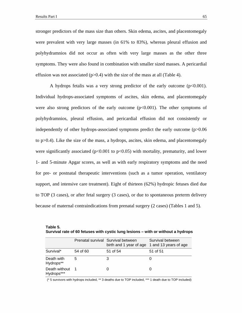
Results Part I 65
stronger predictors of the mass size than others. Skin edema, ascites, and placentomegaly
were prevalent with very large masses (in 61% to 83%), whereas pleural effusion and
polyhydramnios did not occur as often with very large masses as the other three
symptoms. They were also found in combination with smaller sized masses. A pericardial
effusion was not associated (p>0.4) with the size of the mass at all (Table 4).
A hydrops fetalis was a very strong predictor of the early outcome (p<0.001).
Individual hydrops-associated symptoms of ascites, skin edema, and placentomegaly
were also strong predictors of the early outcome (p<0.001). The other symptoms of
polyhydramnios, pleural effusion, and pericardial effusion did not consistently or
independently of other hydrops-associated symptoms predict the early outcome (p>0.06
to p>0.4). Like the size of the mass, a hydrops, ascites, skin edema, and placentomegaly
were significantly associated (p<0.001 to p<0.05) with mortality, prematurity, and lower
1- and 5-minute Apgar scores, as well as with early respiratory symptoms and the need
for pre- or postnatal therapeutic interventions (such as a tumor operation, ventilatory
support, and intensive care treatment). Eight of thirteen (62%) hydropic fetuses died due
to TOP (3 cases), or after fetal surgery (3 cases), or due to spontaneous preterm delivery
because of maternal contraindications from prenatal surgery (2 cases) (Tables 1 and 5).
Table 5. Survival rate of 60 fetuses with cystic lung lesions – with or without a hydrops
Prenatal survival Survival between birth and 1 year of age
Survival between 1 and 13 years of age
Survival* 54 of 60 51 of 54 51 of 51
Death with Hydrops**
5 3 0
Death without Hydrops***
1 0 0
(* 5 survivors with hydrops included, ** 3 deaths due to TOP included, *** 1 death due to TOP included)
Results Part I 66
All five hydrops survivors (5 of 13 hydrops cases, 38%) required prenatal tumor
resection and they exhibited severe early respiratory symptoms, necessitating more than
20 days of ICU treatment (5 of 5 cases) and prolonged ventilatory support for more than
20 days in three of the cases (3 of 5 cases) (Tables 3 and 4). The more advanced the
hydrops, ascites, skin edema, or placentomegaly had been before birth the lower the 1-
and 5-minute Apgar scores were at birth (p<0.001 to p<0.01), the more severe the early
respiratory symptoms were after birth (p<0.001), and the longer the ventilatory support
was used (p=0.001).
The 13 hydropic fetuses were compared with the 14 fetuses that had no obvious
hydrops, but solitary ascites (3 mild), solitary pleural effusion (2 mild and 1 marked), or
solitary pericardial effusion (7 mild and 1 marked). The association between a solitary
effusion and a mass expansion or an unfavorable early respiratory outcome was weaker
(p<0.03) than the association between a hydrops and the mass size or between a hydrops
and the early respiratory outcome (p<0.001), although the association was stronger than
for non-hydropic cases without solitary effusions.
In contrast to the unfavorable early respiratory outcomes of the children with
prenatal hydrops, the final respiratory outcomes of the hydrops survivors were excellent.
Similar to the prenatal size of the mass, a prenatal hydrops and hydrops-associated
symptoms did not influence long-term outcomes (p>0.3 to p>0.9). All 5 survivors of
advanced prenatal hydrops (coupled with very large masses) grew out of their initial
severe early respiratory difficulties, and they were asymptomatic (4 of 5 cases) or had
only minor respiratory symptoms (1 of 5 cases), 2 to 12 years later, at their final follow-
ups (Tables 3 and 4).
Results Part I 67
5.3.4 The degree of the mediastinal shift correlated with the size of the mass and it
was also a strong predictor of the early outcome
A mediastinal shift was observed in 51 (88%) of the 58 cases (data were not
available in two cases: TOP case no. 42 and moderate sized lesion case no. 34). The
mediastinal shift was categorized as overall marked in 18 (31%), overall moderate in 10
(17%), and overall mild in 23 (40%) of the 58 cases. In only 7 (12%) of the 58 cases, the
mediastinum was not shifted. The mediastinum shifted to the side that was contralateral
to the location of the lung lesion. There was, regarding the size of the lung lesion or the
degree of the mediastinal shift, no side preference of the lesion and the contralateral shift
(p>0.5). Of 59 lesions, 30 (51%) lesions were located on the left side of the thorax and
shifted the mediastinum to the right, 26 (44%) lesions were on the right side, and 3 (5%)
lesions were bilateral with no shift (no records of the location of the lung lesion of TOP
case no. 42 were available).
The extension of the mediastinal shift significantly correlated with the size of the
mass (p<0.001). A mediastinal shift appeared at a mean of 21.5 ± 3.0 weeks’ gestation
(17.6 to 33.6 weeks’ gestation), which was about 1 week after the mean gestational age of
the diagnosis of (very) large masses and about 1 ½ weeks before the mean gestational age
of a potential hydrops development (see previous CHAPTERS 5.3.2 and 5.3.3). At the
first diagnosis, the mediastinal shift was mild or moderate in 36 of 52 serially scanned
fetuses, and it regressed in 53% (19 of 36) of these cases until the end of the prenatal
observation. Ten of the 52 serially scanned cases had initially marked mediastinal shifts,
but those shifts persisted as marked in 70% (7 of 10 cases). In total, the mediastinal shifts
regressed in 38.5% (20 of 52 serially scanned cases), stayed the same in 38.5% (20 of 52
Results Part I
68
cases), and increased in 23% (12 of 52 cases). The change of the shift followed the
growth pattern of the mass (p=0.001), which was described in detail in the earlier
CHAPTER 5.3.2.
A higher degree of the mediastinal shift was associated (p<0.001) with a larger
size of the mass, a higher chance of hydrops development, premature delivery, and a
poorer early outcome with more severe early respiratory symptoms and a longer duration
of ventilatory support, ICU treatment, or both. Of the 18 fetuses with a marked
mediastinal shift, 89% (16 of 18 cases) ultimately had very large masses and 67% (12 of
18 cases) developed a hydrops. Or in other words, 89% (16 of 18 cases) of the ultimately
very large masses and 92% (12 of 13 cases) of the hydropic fetuses were accompanied by
marked mediastinal shifts. The survival rate of the fetuses with marked mediastinal shifts
was 61% (11 of 18 cases), and all the survivors had severe early respiratory symptoms
(11 of 11 cases) (Table 4). In contrast, the 30 fetuses with a mild or no shift had masses
that were ultimately small or vanished in 57% (17 of 30 cases). None of them had a very
large mass or developed a hydrops. Most of the children in that group were asymptomatic
or only mildly affected early after birth (87%, 26 of 30 cases) (Table 4).
Again, as with the size of the mass or the development of a hydrops, the
mediastinal shift was a highly significant prognostic factor of the early respiratory
outcome (p<0.001), but not of the final respiratory outcome (p>0.4), which was excellent.
Discussion Part I 69
6. Discussion
6.1 Prediction of the outcomes
6.1.1 The set of prenatal early outcome predictors that this 60-patients-study
disclosed, strikingly coincides with the predictor set generated by an extended
statistical analysis of published data
The goal of my 60-patients-study was to research the natural history of fetal cystic
lung lesions and how this history determines the postnatal outcome, in order to establish
prenatal criteria to help counsel expecting parents of affected fetuses appropriately.
Relying on the two participating centers’ 14 years experience with fetal cystic lung
lesions, such as CCAM and BPS, I performed a survey of 60 fetuses diagnosed with
cystic lung lesions. To the best of my knowledge, this study presented here is unique
regarding the large numbers of prenatal cases, the long follow-up interval of up to 13.1
years (mean follow-up age: 5.9 ± 3.3 years), and the comprehensive study of the prenatal
ultrasonographic data to determine statistically significant predictors of the early and
long-term outcomes. The equal gender distribution in this study of 31 female versus 28
male patients (1 TOP case was of unknown gender) argues against a bias in the studied
fetal patient population. I consider this a result of my patient recruitment strategy,
outlined in detail in CHAPTER 4.1.2 of the PATIENTS AND METHODS section. The
results of this study show that the early respiratory outcomes of fetuses with cystic lung
lesions are significantly associated (p<0.001) with the final size of the lesion, the
development of a non-immune hydrops fetalis, and the mass- and hydrops-associated
symptoms of mediastinal shift, skin edema, ascites, and placentomegaly. An ultimately
Discussion Part I
70
very large mass, a high degree of a mediastinal shift, and the development of a hydrops or
symptoms of skin edema, marked ascites, and placentomegaly are significant predictors
of a complicated early outcome, impaired by respiratory difficulties and prematurity. In
rare cases only, a child may not survive the neonatal period. While the early respiratory
outcome can be well predicted by the above-mentioned prenatal parameters, the long-
term outcome cannot be predicted by any of the prenatal parameters at all, since in
general, symptoms resolve in the neonatal period or in the first 2 years of life. The long-
term outcome of children with fetal cystic lung lesions is excellent, as this study shows.
I compared the data of this 60-patients-study with the data of 49 clinical studies
presented in the literature. The authors of the 49 studies examined the prenatal symptoms
and outcomes in series with 10 or more fetuses with congenital cystic lung lesions. No
other selection criteria were applied. Analyzing these studies published since 1985 by
authors from 35 institutions (among them 5 multi-center-studies), I identified 1,442 fetal
cystic lung lesion cases (Table 6 and Figure 6). I statistically analyzed the published data
and searched for the most reliable prenatal predictors of the early outcome. As a
conclusion, the development of a hydrops and the presence of a non-regressive, large
lung lesion are the most frequently cited and reliable predictors of an unfavorable
perinatal outcome (Table 7). The findings of my 60-patients-study coincide with the other
authors’ results, as discussed in detail below and compiled in Tables 6 and 7. Finally, the
clinical implications of the entire study are the significantly improved pre- and postnatal
management recommendations.
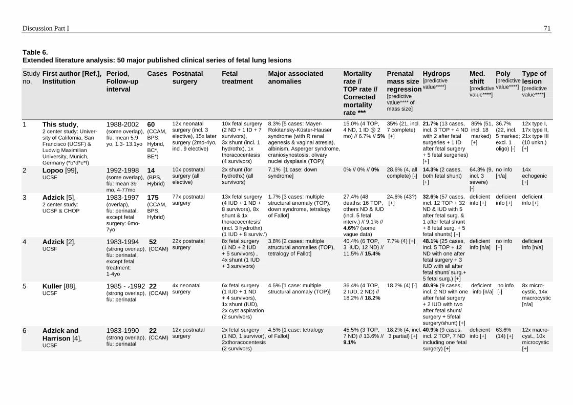
Discussion Part I 71
Table 6. Extended literature analysis: 50 major published clinical series of fetal lung lesions
Study no.
First author [Ref.], Institution
Period, Follow-up interval
Cases Postnatal surgery
Fetal treatment
Major associated anomalies
Mortality rate // TOP rate // Corrected mortality rate ***
Prenatal mass size regression[predictive value**** of mass size]
Hydrops [predictive value****]
Med. shift [predictive value****]
Poly [predictive value****]
Type of lesion [predictive value****]
1 This study, 2 center study: Univer-sity of California, San Francisco (UCSF) & Ludwig Maximilian University, Munich, Germany (*b*d*e*f)
1988-2002 (some overlap), f/u: mean 5.9 yo, 1.3- 13.1yo
60 (CCAM, BPS, Hybrid, BC*,
12x neonatal surgery (incl. 3 elective), 15x later surgery (2mo-4yo, incl. 9 elective)
BE*)
10x fetal surgery (2 ND + 1 ID + 7 survivors), 3x shunt (incl. 1 hydrothx), 1x thoracocentesis (4 survivors)
8.3% [5 cases: Mayer-Rokitansky-Küster-Hauser syndrome (with R renal agenesis & vaginal atresia), albinism, Asperger syndrome, craniosynostosis, olivary nuclei dysplasia (TOP)]
15.0% (4 TOP, 4 ND, 1 ID @ 2 mo) // 6.7% // 5%
35% (21, incl. 7 complete) [+]
21.7% (13 cases, incl. 3 TOP + 4 ND with 2 after fetal surgeries + 1 ID after fetal surgery + 5 fetal surgeries) [+]
85% (51, incl. 18 marked) [+]
36.7% (22, incl. 5 marked; excl. 1 oligo) [-]
12x type I, 17x type II, 21x type III (10 unkn.) [+]
2 Lopoo [99], UCSF
1992-1998 (some overlap), f/u: mean 39 mo, 4-77mo
14 (BPS, Hybrid)
10x postnatal surgery (all elective)
2x shunt (for hydrothx) (all survivors)
7.1% [1 case: down syndrome]
0% // 0% // 0% 28.6% (4, all complete) [-]
14.3% (2 cases, both fetal shunt) [+]
64.3% (9, incl. 3 severe) [-]
no info [n/a]
14x echogenic [+]
3 Adzick [5], 2 center study: UCSF & CHOP
1983-1997 (overlap), f/u: perinatal, except fetal surgery: 6mo-7yo
175 (CCAM, BPS, Hybrid)
77x postnatal surgery
13x fetal surgery (4 IUD + 1 ND + 8 survivors), 8x shunt & 1x thoracocentesis’ (incl. 3 hydrothx) (1 IUD + 8 surviv.')
1.7% [3 cases: multiple structural anomaly (TOP), down syndrome, tetralogy of Fallot]
27.4% (48 deaths: 16 TOP, others ND & IUD (incl. 5 fetal interv.) // 9.1% // 4.6%? (some vague data)
24.6% (43?) [+]
32.6% (57 cases, incl. 12 TOP + 32 ND & IUD with 5 after fetal surg. & 1 after fetal shunt + 8 fetal surg. + 5 fetal shunts) [+]
deficient info [+]
deficient info [+]
deficient info [+]
4 Adzick [2], UCSF
1983-1994 (strong overlap), f/u: perinatal, except fetal treatment: 1-4yo
52 (CCAM)
22x postnatal surgery
8x fetal surgery (1 ND + 2 IUD + 5 survivors) , 4x shunt (1 IUD + 3 survivors)
3.8% [2 cases: multiple structural anomalies (TOP), tetralogy of Fallot]
40.4% (6 TOP, 3 IUD, 12 ND) // 11.5% // 15.4%
7.7% (4) [+] 48.1% (25 cases, incl. 5 TOP + 12 ND with one after fetal surgery + 3 IUD with all after fetal shunt/ surg.+ 5 fetal surg.) [+]
deficient info [n/a]
no info [+]
deficient info [n/a]
5 Kuller [88], UCSF
1985 - <1992 (strong overlap), f/u: perinatal
22 (CCAM)
4x neonatal surgery
6x fetal surgery (1 IUD + 1 ND + 4 survivors), 1x shunt (IUD), 2x cyst aspiration (2 survivors)
4.5% [1 case: multiple structural anomaly (TOP)]
36.4% (4 TOP, 2 IUD, 2 ND) // 18.2% // 18.2%
18.2% (4) [-] 40.9% (9 cases, incl. 2 ND with one after fetal surgery + 2 IUD with two after fetal shunt/ surgery + 5fetal surgery/shunt) [+]
deficient info [n/a]
no info [-]
8x micro-cystic, 14x macrocystic [n/a]
6 Adzick and Harrison [4], UCSF
1983-1990 (strong overlap), f/u: perinatal
22 (CCAM)
12x postnatal surgery
2x fetal surgery (1 ND, 1 survivor),2xthoracocentesis(2 survivors)
4.5% [1 case: tetralogy of Fallot]
45.5% (3 TOP, 7 ND) // 13.6% // 9.1%
18.2% (4, incl. 3 partial) [+]
40.9% (9 cases, incl. 2 TOP, 7 ND including one fetal surgery) [+]
deficient info [+]
63.6% (14) [+]
12x macro-cyst., 10x microcystic [+]
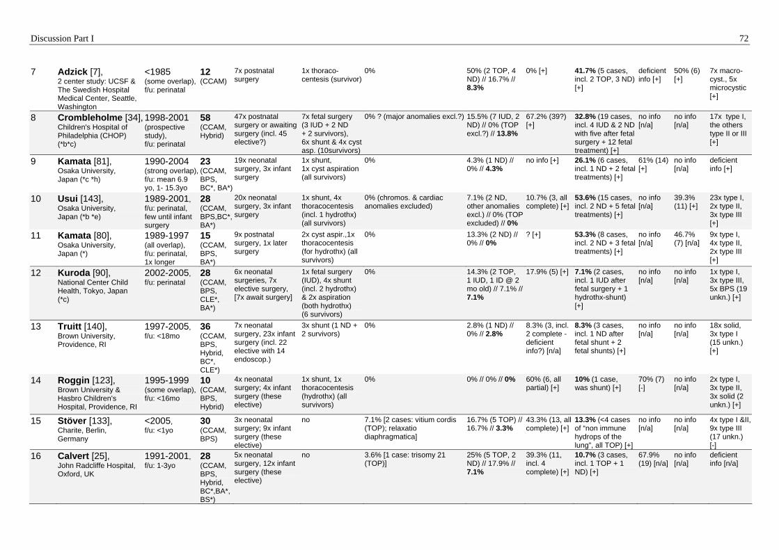
Discussion Part I 72
7 Adzick [7], 2 center study: UCSF & The Swedish Hospital Medical Center, Seattle, Washington
<1985 (some overlap), f/u: perinatal
12 (CCAM)
7x postnatal surgery
1x thoraco-centesis (survivor)
0% 50% (2 TOP, 4 ND) // 16.7% // 8.3%
0% [+] 41.7% (5 cases, incl. 2 TOP, 3 ND) [+]
deficient info [+]
50% (6) [+]
7x macro-cyst., 5x microcystic [+]
8 Crombleholme [34],Children's Hospital of Philadelphia (CHOP) (*b*c)
1998-2001 (prospective study), f/u: perinatal
58 (CCAM, Hybrid)
47x postnatal surgery or awaiting surgery (incl. 45 elective?)
7x fetal surgery (3 IUD + 2 ND + 2 survivors), 6x shunt & 4x cyst asp. (10survivors)
0% ? (major anomalies excl.?) 15.5% (7 IUD, 2 ND) // 0% (TOP excl.?) // 13.8%
67.2% (39?) [+]
32.8% (19 cases, incl. 4 IUD & 2 ND with five after fetal surgery + 12 fetal treatment) [+]
no info [n/a]
no info [n/a]
17x type I, the others type II or III [+]
9 Kamata [81], Osaka University, Japan (*c *h)
1990-2004 (strong overlap), f/u: mean 6.9 yo, 1- 15.3yo
23 (CCAM, BPS, BC*, BA*)
19x neonatal surgery, 3x infant surgery
1x shunt, 1x cyst aspiration (all survivors)
0% 4.3% (1 ND) // 0% // 4.3%
no info [+] 26.1% (6 cases, incl. 1 ND + 2 fetal treatments) [+]
61% (14) [+]
no info [n/a]
deficient info [+]
10 Usui [143], Osaka University, Japan (*b *e)
1989-2001, f/u: perinatal, few until infant surgery
28 (CCAM, BPS,BC*, BA*)
20x neonatal surgery, 3x infant surgery
1x shunt, 4x thoracocentesis (incl. 1 hydrothx) (all survivors)
0% (chromos. & cardiac anomalies excluded)
7.1% (2 ND, other anomalies excl.) // 0% (TOP excluded) // 0%
10.7% (3, all complete) [+]
53.6% (15 cases, incl. 2 ND + 5 fetal treatments) [+]
no info [n/a]
39.3% (11) [+]
23x type I, 2x type II, 3x type III [+]
11 Kamata [80], Osaka University, Japan (*)
1989-1997 (all overlap), f/u: perinatal, 1x longer
15 (CCAM, BPS, BA*)
9x postnatal surgery, 1x later surgery
2x cyst aspir.,1x thoracocentesis (for hydrothx) (all survivors)
0% 13.3% (2 ND) // 0% // 0%
? [+] 53.3% (8 cases, incl. 2 ND + 3 fetal treatments) [+]
no info [n/a]
46.7% (7) [n/a]
9x type I, 4x type II, 2x type III [+]
12 Kuroda [90], National Center Child Health, Tokyo, Japan (*c)
2002-2005, f/u: perinatal
28 (CCAM, BPS, CLE*, BA*)
6x neonatal surgeries, 7x elective surgery, [7x await surgery]
1x fetal surgery (IUD), 4x shunt (incl. 2 hydrothx) & 2x aspiration (both hydrothx) (6 survivors)
0% 14.3% (2 TOP, 1 IUD, 1 ID @ 2 mo old) // 7.1% // 7.1%
17.9% (5) [+] 7.1% (2 cases, incl. 1 IUD after fetal surgery + 1 hydrothx-shunt) [+]
no info [n/a]
no info [n/a]
1x type I, 3x type III, 5x BPS (19 unkn.) [+]
13 Truitt [140], Brown University, Providence, RI
1997-2005, f/u: <18mo
36 (CCAM, BPS, Hybrid, BC*, CLE*)
7x neonatal surgery, 23x infant surgery (incl. 22 elective with 14 endoscop.)
3x shunt (1 ND + 2 survivors)
0% 2.8% (1 ND) // 0% // 2.8%
8.3% (3, incl. 2 complete - deficient info?) [n/a]
8.3% (3 cases, incl. 1 ND after fetal shunt + 2 fetal shunts) [+]
no info [n/a]
no info [n/a]
18x solid, 3x type I (15 unkn.) [+]
14 Roggin [123], Brown University & Hasbro Children's Hospital, Providence, RI
1995-1999 (some overlap), f/u: <16mo
10 (CCAM, BPS, Hybrid)
4x neonatal surgery; 4x infant surgery (these elective)
1x shunt, 1x thoracocentesis (hydrothx) (all survivors)
0% 0% // 0% // 0% 60% (6, all partial) [+]
10% (1 case, was shunt) [+]
70% (7) [-]
no info [n/a]
2x type I, 3x type II, 3x solid (2 unkn.) [+]
15 Stöver [133], Charite, Berlin, Germany
<2005, f/u: <1yo
30 (CCAM, BPS)
3x neonatal surgery; 9x infant surgery (these elective)
no 7.1% [2 cases: vitium cordis (TOP); relaxatio diaphragmatica]
16.7% (5 TOP) // 16.7% // 3.3%
43.3% (13, all complete) [+]
13.3% (<4 cases of “non immune hydrops of the lung”, all TOP) [+]
no info [n/a]
no info [n/a]
4x type I &II, 9x type III (17 unkn.) [-]
16 Calvert [25], John Radcliffe Hospital, Oxford, UK
1991-2001, f/u: 1-3yo
28 (CCAM, BPS, Hybrid, BC*,BA*, BS*)
5x neonatal surgery, 12x infant surgery (these elective)
no 3.6% [1 case: trisomy 21 (TOP)]
25% (5 TOP, 2 ND) // 17.9% // 7.1%
39.3% (11, incl. 4 complete) [+]
10.7% (3 cases, incl. 1 TOP + 1 ND) [+]
67.9% (19) [n/a]
no info [n/a]
deficient info [n/a]
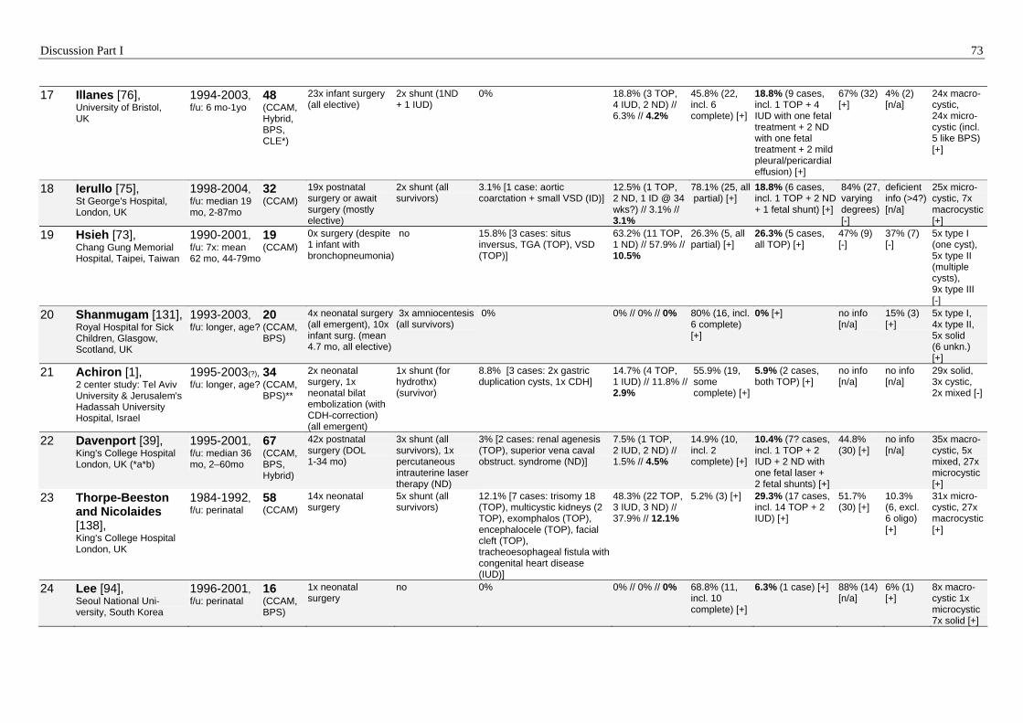
Discussion Part I 73
17 Illanes [76], University of Bristol, UK
1994-2003, f/u: 6 mo-1yo
48 (CCAM, Hybrid, BPS, CLE*)
23x infant surgery (all elective)
2x shunt (1ND + 1 IUD)
0% 18.8% (3 TOP, 4 IUD, 2 ND) // 6.3% // 4.2%
45.8% (22, incl. 6 complete) [+]
18.8% (9 cases, incl. 1 TOP + 4 IUD with one fetal treatment + 2 ND with one fetal treatment + 2 mild pleural/pericardial effusion) [+]
67% (32) [+]
4% (2) [n/a]
24x macro-cystic, 24x micro-cystic (incl. 5 like BPS) [+]
18 Ierullo [75], St George's Hospital, London, UK
1998-2004, f/u: median 19 mo, 2-87mo
32 (CCAM)
19x postnatal surgery or await surgery (mostly elective)
2x shunt (all survivors)
3.1% [1 case: aortic coarctation + small VSD (ID)]
12.5% (1 TOP, 2 ND, 1 ID @ 34 wks?) // 3.1% // 3.1%
78.1% (25, all partial) [+]
18.8% (6 cases, incl. 1 TOP + 2 ND + 1 fetal shunt) [+]
84% (27, varying degrees) [-]
deficient info (>4?) [n/a]
25x micro-cystic, 7x macrocystic [+]
19 Hsieh [73], Chang Gung Memorial Hospital, Taipei, Taiwan
1990-2001, f/u: 7x: mean 62 mo, 44-79mo
19 (CCAM)
0x surgery (despite 1 infant with bronchopneumonia)
no 15.8% [3 cases: situs inversus, TGA (TOP), VSD (TOP)]
63.2% (11 TOP, 1 ND) // 57.9% // 10.5%
26.3% (5, all partial) [+]
26.3% (5 cases, all TOP) [+]
47% (9) [-]
37% (7) [-]
5x type I (one cyst), 5x type II (multiple cysts), 9x type III [-]
20 Shanmugam [131],Royal Hospital for Sick Children, Glasgow, Scotland, UK
1993-2003, f/u: longer, age?
20 (CCAM, BPS)
4x neonatal surgery (all emergent), 10x infant surg. (mean 4.7 mo, all elective)
3x amniocentesis (all survivors)
0% 0% // 0% // 0% 80% (16, incl. 6 complete) [+]
0% [+] no info [n/a]
15% (3) [+]
5x type I, 4x type II, 5x solid (6 unkn.) [+]
21 Achiron [1], 2 center study: Tel Aviv University & Jerusalem's Hadassah University Hospital, Israel
1995-2003(?), f/u: longer, age?
34 (CCAM, BPS)**
2x neonatal surgery, 1x neonatal bilat embolization (with CDH-correction) (all emergent)
1x shunt (for hydrothx) (survivor)
8.8% [3 cases: 2x gastric duplication cysts, 1x CDH]
14.7% (4 TOP, 1 IUD) // 11.8% // 2.9%
55.9% (19, some complete) [+]
5.9% (2 cases, both TOP) [+]
no info [n/a]
no info [n/a]
29x solid, 3x cystic, 2x mixed [-]
22 Davenport [39], King's College Hospital London, UK (*a*b)
1995-2001, f/u: median 36 mo, 2–60mo
67 (CCAM, BPS, Hybrid)
42x postnatal surgery (DOL 1-34 mo)
3x shunt (all survivors), 1x percutaneous intrauterine laser therapy (ND)
3% [2 cases: renal agenesis (TOP), superior vena caval obstruct. syndrome (ND)]
7.5% (1 TOP, 2 IUD, 2 ND) // 1.5% // 4.5%
14.9% (10, incl. 2 complete) [+]
10.4% (7? cases, incl. 1 TOP + 2 IUD + 2 ND with one fetal laser + 2 fetal shunts) [+]
44.8% (30) [+]
no info [n/a]
35x macro-cystic, 5x mixed, 27x microcystic [+]
23 Thorpe-Beeston and Nicolaides [138], King's College Hospital London, UK
1984-1992, f/u: perinatal
58 (CCAM)
14x neonatal surgery
5x shunt (all survivors)
12.1% [7 cases: trisomy 18 (TOP), multicystic kidneys (2 TOP), exomphalos (TOP), encephalocele (TOP), facial cleft (TOP), tracheoesophageal fistula with congenital heart disease (IUD)]
48.3% (22 TOP, 3 IUD, 3 ND) // 37.9% // 12.1%
5.2% (3) [+] 29.3% (17 cases, incl. 14 TOP + 2 IUD) [+]
51.7% (30) [+]
10.3% (6, excl. 6 oligo) [+]
31x micro-cystic, 27x macrocystic [+]
24 Lee [94], Seoul National Uni-versity, South Korea
1996-2001, f/u: perinatal
16 (CCAM, BPS)
1x neonatal surgery
no 0% 0% // 0% // 0% 68.8% (11, incl. 10 complete) [+]
6.3% (1 case) [+] 88% (14) [n/a]
6% (1) [+]
8x macro-cystic 1x microcystic 7x solid [+]
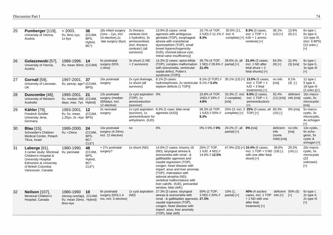
Discussion Part I 74
25 Pumberger [119], University of Vienna, Austria
< 2003, f/u: 6mo-1yo, 1x 6yo
36 (CCAM, BPS, Hybrid, BC*)
18x infant surgery (1mo – 1yo, incl. 14 elective),1x late surgery (6yo)
2x thoraco- centesis (incl. 1 hydrothx), 3x amniocentesis (incl. thoraco-centesis') (all survivors)
13.9% [5 cases: renal agenesis with ambiguous genitalia (TOP), esophageal atresia with craniofacial dysmorphism (TOP), small bowel hyperechogenicity (IUD), chorioid plexus cyst, mitral valve insufficiency]
16.7% (4 TOP, 2 IUD) // 11.1% // 8.3%
30.6% (11, incl. 6 complete) [+]
8.3% (3 cases, incl. 1 TOP + 1 IUD + 1 amnio-centesis) [+]
36.1% (13) [+]
13.9% (5) [+]
6x type I, 6x type II, 12x type III, (incl. 6 BPS) (12 unkn.) [+]
26 Golaszewski [57], University of Vienna, Austria
1989-1996, f/u: mean 30mo
14 (CCAM)
5x postnatal surgery (DOL 1-4mo)
3x shunt (1 ND + 2 survivors)
14.3% [2 cases: spina bifida (TOP), complex malformation with sirenomelia, ventricular septal defect, Potter's syndrome (TOP)]
35.7% (4 TOP, 1 ND) // 28.6% // 21.4%
28.6% (4, all partial) [+]
21.4% (3 cases, incl. 1 ND after fetal shunt + 2 fetal shunts) [+]
64.3% (9) [+]
21.4% (3) [n/a]
6x type I, 5x type II, 3x type III [+]
27 Gornall [59], University of Leicester, UK
1997-2001, f/u: peirop, age?
37 (CCAM, BPS)
16x postnatal surgery
2x cyst drainage, 1x shunt (all survivors)
8.1% [3 cases: hydronephrosis, 2x ventricular septum defects (1 TOP)]
8.1% (3 TOP) // 8.1% // 5.4%
35.1% (13) [+] 13.5% (5 cases, incl. 1 TOP + 1 IUD + 3 fetal treatments) [+]
no info [n/a]
8.1% (3) [-]
11 type I, 5 type II, 18 type III (3 unkn.)[+]
28 Duncombe [48], University of Western Australia
1995-2001, f/u: median 265 days, max. 7yo
21 (CCAM, Hybrid)
13x postnatal surgery (median 265days, incl. 12 elective)
1x cyst aspiration (TOP), 1x amnioreduction (survivor)
0% 23.8% (4 TOP, 1ND) // 19% // 4.8%
33.3% (7, incl. 2 complete) [n/a]
9.5% (2 cases, incl. 1 TOP + 1 amnioreduction) [+]
52.4% (11) [n/a]
deficient info [n/a]
16x macro-cystic, 5x microcystic [n/a]
29 Kähler [79], Friedrich Schiller University Jena, Germany
1993-2001, f/u: 5x: mean 1.25yo, 2x >4yo
12 (CCAM, BPS)
2x neonatal surgery
1x cyst aspiration (survivor), 1x amnioninfusion for anhydramn. (IUD)
8.3% [1 case: bilat renal agenesis (IUD)]
58.3% (6 TOP, 1 IUD) // 50% // 8.3%
25% (3, incl. 2 complete) [+]
25% (3 cases, all TOP) [+]
83.3% (10) [-]
0% [n/a] 5x macro-cystic, 3x microcystic, 4x echogen [+]
30 Blau [15], Schneider's Children Medical Center, Petach Tikva, Israel
1995-2000, f/u: <24mo
24 (CCAM, BPS, BC*, CLE*)
14x postnatal surgery (4-24mo, incl. 12 elective)
no 0% 0% // 0% // 0% 29.2% (7, all partial) [+]
0% [n/a] deficient info (some mild) [n/a]
no info [n/a]
13x cystic, 6x echo-genic, 5x cystic & echogen [+]
31 Laberge [91], 3 center study: Montreal Children's Hospital & University Hospital Edmonton & University of British Columbia Vancouver, Canada
1980-1990, f/u: perinatal
48 (CCAM, BPS, Hybrid, BC*, CLE*)
> 27x postnatal surgery?
1x shunt (ND) 14.6% [7 cases: trisomy 18 (ND), laryngeal atresia & sirenomelia with renal - & gallbladder agenesis and caudal regression (TOP), congen. heart disease with imperf. anus and liver anomaly (TOP), malrotation with adrenal atrophia (ND), vertebral malformations with liver calcific. (IUD), pericardial window, bilat cleft,]
25% (7 TOP, 1 IUD, 4 ND) // 14.6% // 12.5%
47.9% (23) [+] 10.4% (5 cases, incl. 1 TOP + 3 ND with one after fetal shunt) [+]
39.6% (19) [-]
29.2% (14) [-]
20x macro-cystic, 5x microcystic (23 unknown) [+]
32 Neilson [107], Montreal Children's Hospital, Canada
1980-1990 (strong overlap), f/u: mean 20mo, 8mo-4yo
10 (CCAM, Hybrid)
8x postnatal surgery (DOL1-4 mo, incl. 3 elective)
1x cyst aspiration (ND)
27.3% [3 cases: laryngeal atresia & sirenomelia with renal - & gallbladder agenesis, caudal regression (TOP), congen. heart disease with imperf. anus, liver anomaly (TOP), bilat cleft]
50% (2 TOP, 3 ND) // 20% // 27.3%
10% (1, partial) [+]
40% (4 ascites cases, incl. 1 TOP + 2 ND with one after fetal treatment) [+]
deficient info [+]
50% (5) [+]
6x type I, 2x type II, 2x type III [+]
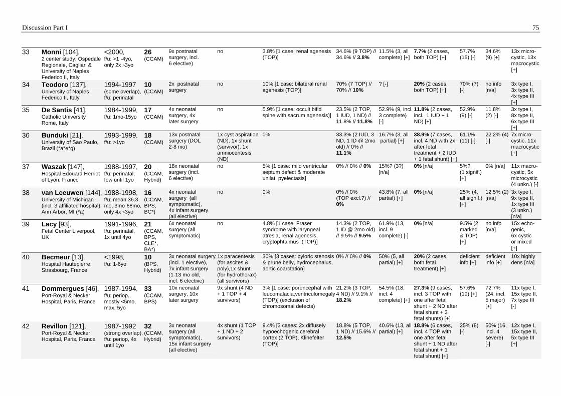
Discussion Part I 75
33 Monni [104], 2 center study: Ospedale Regionale, Cagliari & University of Naples Federico II, Italy
<2000, f/u: >1 -4yo, only 2x >3yo
26 (CCAM)
9x postnatal surgery, incl. 6 elective)
no 3.8% [1 case: renal agenesis (TOP)]
34.6% (9 TOP) // 34.6% // 3.8%
11.5% (3, all complete) [+]
7.7% (2 cases, both TOP) [+]
57.7% (15) [-]
34.6% (9) [+]
13x micro-cystic, 13x macrocystic [+]
34 Teodoro [137], University of Naples Federico II, Italy
1994-1997 (some overlap), f/u: perinatal
10 (CCAM)
2x postnatal surgery
no 10% [1 case: bilateral renal agenesis (TOP)]
70% (7 TOP) // 70% // 10%
? [-] 20% (2 cases, both TOP) [+]
70% (7) [-]
no info [n/a]
3x type I, 3x type II, 4x type III [+]
35 De Santis [41], Catholic University Rome, Italy
1984-1999, f/u: 1mo-15yo
17 (CCAM)
4x neonatal surgery, 4x later surgery
no 5.9% [1 case: occult bifid spine with sacrum agenesis)]
23.5% (2 TOP, 1 IUD, 1 ND) // 11.8% // 11.8%
52.9% (9, incl. 3 complete) [-]
11.8% (2 cases, incl. 1 IUD + 1 ND) [+]
52.9% (9) [-]
11.8% (2) [-]
3x type I, 8x type II, 6x type III [+]
36 Bunduki [21], University of Sao Paulo, Brazil (*a*e*g)
1993-1999, f/u: >1yo
18 (CCAM)
13x postnatal surgery (DOL 2-8 mo)
1x cyst aspiration (ND), 1x shunt (survivor), 1x amniocentesis (ND)
0% 33.3% (2 IUD, 3 ND, 1 ID @ 2mo old) // 0% // 11.1%
16.7% (3, all partial) [+]
38.9% (7 cases, incl. 4 ND with 2x after fetal treatment + 2 IUD + 1 fetal shunt) [+]
61.1% (11) [-]
22.2% (4) [-]
7x micro-cystic, 11x macrocystic [+]
37 Waszak [147], Hospital Edouard Herriot of Lyon, France
1988-1997, f/u: perinatal, few until 1yo
20 (CCAM, Hybrid)
18x neonatal surgery (incl. 6 elective)
no 5% [1 case: mild ventricular septum defect & moderate unilat. pyelectasis]
0% // 0% // 0% 15%? (3?) [n/a]
0% [n/a] 5%? (1 signif.) [+]
0% [n/a] 11x macro-cystic, 5x microcystic (4 unkn.) [-]
38 van Leeuwen [144],University of Michigan (incl. 3 affiliated hospital), Ann Arbor, MI (*a)
1988-1998, f/u: mean 36.3 mo, 3mo-68mo, only 4x >3yo
16 (CCAM, BPS, BC*)
4x neonatal surgery (all symptomatic), 4x infant surgery (all elective)
no 0% 0% // 0% (TOP excl.?) // 0%
43.8% (7, all partial) [+]
0% [n/a] 25% (4, all signif.) [+]
12.5% (2) [n/a]
3x type I, 9x type II, 1x type III (3 unkn.) [n/a]
39 Lacy [93], Fetal Center Liverpool, UK
1991-1996, f/u: perinatal, 1x until 4yo
21 (CCAM, BPS, CLE*, BA*)
6x neonatal surgery (all symptomatic)
no 4.8% [1 case: Fraser syndrome with laryngeal atresia, renal agenesis, cryptophtalmus (TOP)]
14.3% (2 TOP, 1 ID @ 2mo old) // 9.5% // 9.5%
61.9% (13, incl. 9 complete) [-]
0% [n/a] 9.5% (2 marked & TOP) [+]
no info [n/a]
15x echo-genic, 6x cystic or mixed [+]
40 Becmeur [13], Hospital Hautepierre, Strasbourg, France
<1998, f/u: 1-6yo
10 (BPS, Hybrid)
3x neonatal surgery (incl. 1 elective), 7x infant surgery (1-13 mo old, incl. 6 elective)
1x paracentesis (for ascites & poly),1x shunt (for hydrothorax) (all survivors)
30% [3 cases: pyloric stenosis & prune belly, hydrocephalus, aortic coarctation]
0% // 0% // 0% 50% (5, all partial) [+]
20% (2 cases, both fetal treatment) [+]
deficient info [+]
deficient info [+]
10x highly dens [n/a]
41 Dommergues [46], Port-Royal & Necker Hospital, Paris, France
1987-1994, f/u: periop., mostly <5mo, max. 5yo
33 (CCAM, BPS)
10x neonatal surgery, 10x later surgery
9x shunt (4 ND + 1 TOP + 4 survivors)
3% [1 case: porencephal with leucomalacia,ventriculomegaly (TOP)] (exclusion of chromosomal defects)
21.2% (3 TOP, 4 ND) // 9.1% // 18.2%
54.5% (18, incl. 4 complete) [+]
27.3% (9 cases, incl. 3 TOP with one after fetal shunt + 2 ND after fetal shunt + 3 fetal shunts) [+]
57.6% (19) [+]
72.7% (24, incl. 5 major) [+]
11x type I, 15x type II, 7x type III [-]
42 Revillon [121], Port-Royal & Necker Hospital, Paris, France
1987-1992 (strong overlap), f/u: periop, 4x until 1yo
32 (CCAM, Hybrid)
3x neonatal surgery (all symptomatic), 15x infant surgery (all elective)
4x shunt (1 TOP + 1 ND + 2 survivors)
9.4% [3 cases: 2x diffusely hypoechogenic cerebral cortex (2 TOP), Klinefelter (TOP)]
18.8% (5 TOP, 1 ND) // 15.6% // 12.5%
40.6% (13, all partial) [+]
18.8% (6 cases, incl. 4 TOP with one after fetal shunt + 1 ND after fetal shunt + 1 fetal shunt) [+]
25% (8) [-]
50% (16, incl. 4 severe) [-]
12x type I, 15x type II, 5x type III [+]
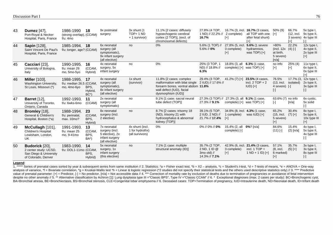
Discussion Part I
76
43 Dumez [47], Port-Royal & Necker Hospital, Paris, France
1986-1990 (strong overlap), f/u: 4mo
18 (CCAM)
9x postnatal surgery
3x shunt (1 TOP+ 1 ND + 1 survivor)
11.1% [2 cases: diffusely hypoechogenic cerebral cortex (2 TOP)], (excl. of chromosomal defects)
27.8% (4 TOP, 1 ND) // 22.2% // 16.7%
16.7% (3, incl. 2 complete) [+]
16.7% (3 cases, all TOP with one after fetal shunt) [+]
50% (9) [+]
66.7% (12, incl. 3 severe) [+]
5x type I, 9x type II, 4x type III [-]
44 Sapin [128], Saint Vincent De Paul's Hospital, Paris, France
1985-1994, f/u: longer, age?
18 (CCAM)
6x neonatal surgery (all symptomatic), 9x infant surgery (all elective)
no 0% 5.6% (1 TOP) // 5.6% // 0%
27.8% (5, incl. 3 complete) [+]
5.6% (1 severe hydramnios, was TOP) [+]
>80% (incl. 12x at birth, 5 severe) [n/a]
22.2% (4) [-]
12x type I, 4x type II, 2x type III [+]
45 Cacciari [23], University of Bologna, Italy
1990-1995, f/u: mean 29 mo, 5mo-5yo
16 (CCAM, Hybrid)
4x neonatal surgery, 5x infant surgery
no 0% 25% (3 TOP, 1 ND) // 18.8% // 6.3%
18.8% (3, all complete) [+]
6.3% (1 case, was TOP) [+]
no info [+]
25% (4) [+]
11x type I, 6x type II, 0x type III [+]
46 Miller [103], Washington University, St Louis, Missouri (*)
1988-1995, f/u: median 26.5 mo, 4mo-6yo
17 (CCAM, BPS, Hybrid, BC*)
4x neonatal surgery (all symptomatic), 8x infant surgery (all elective)
1x shunt (survivor)
11.8% [2 cases: complex malformation with bilat single forearm bones, ventral abdom wall defect (IUD), facial dysmorphism (IUD)]
29.4% (3 TOP, 2 IUD) // 17.6% // 11.8%
41.2% (7) [+] 23.5% (4 cases, incl. 2 TOP + 2 IUD) [+]
76.5% (13, incl. 4 severe) [-]
17.7% (3, isolated) [-]
12x type I, 2x type II, 3x type III [-]
47 Barret [12], University of Toronto, Ontario, Canada
1992-1993, f/u: 6wks-6mo
11 (CCAM)
2x neonatal surgery (all symptomatic)
no 9.1% [1 case: sacral neural tube defect (TOP)]
27.3% (3 TOP) // 27.3% // 9.1%
27.3% (3, all complete) [+]
9.1% (1 case, was TOP) [+]
63.6% (7) [-]
no info [n/a]
6x cystic, 5x solid [-]
48 Bromley [18], General & Children's Hospital, Boston (*a)
1988-1994, f/u: perinatal, max. 10mo?
23 (CCAM, BPS, Hybrid)
8x neonatal surgery (incl. 2 elective)
no 8.7% [2 cases: trisomy 18 (ND), trisomy 21 with hydrocephalus & abnormal kidney (TOP)]
39.1% (5 TOP, 2 IUD, 2 ND) // 21.7% // 17.4%
34.8% (8, incl. 1 complete) [+]
4.3% (1 case, was IUD) [+]
65.2% (15, incl. 5 severe) [+]
30.4% (7) [+]
8x type I, 5x type II, 10x type III [+]
49 McCullagh [101], Children's Hospital Lewisham, London, UK
1991-1993, f/u: mean 25 mo, 9-41mo
13 (CCAM, BPS, BA*)
7x neonatal surgery (incl. 5 elective), 2x infant surgery (all elective)
4x shunt (incl. 1 for hydrothx) (all survivors)
0% 0% // 0% // 0% 15.4% (2, all complete) [-]
0%? [n/a] 84.6% (11) [-]
15.4% (2) [n/a]
4x type I, 5x type II, 4x type III [-]
50 Budorick [20], 2 center study: UCSD, San Diego & University of Colorado, Denver
1983-1990, f/u: DOL1-11mo
14 (CCAM, BPS, Hybrid)
5x neonatal surgery, 1x infant surgery (this elective)
no 7.1% [1 case: multiple structural anomaly (ID)]
35.7% (2 TOP, 2 ND, 1 ID @ 3mo old) // 14.3% // 7.1%
42.9% (6, incl. 3 complete) [+]
21.4% (3 cases, incl. 1 TOP + 1 ND + 1 ID) [+]
57.1% (8, incl. 6 marked) [+]
35.7% (5) [+]
3x type I, 3x type II, 8x type III [-]
Legend: 1. ***** Series of prenatal cases sorted by year & subsequent series from same institution // 2. Statistics: *a = Fisher exact test, *b = X2 – analysis, *c = Student's t-test, *d = T-tests of means, *e = ANOVA = One-way analysis of variance, *f = Bivariate correlation, *g = Kruskal-Wallis test *h = Linear & logistic regression (*2 studies did not specify their statistical tests and the others used descriptive statistics only) // 3. **** Predictive value of prenatal parameter: [+] = Predictor, [-] = No predictor, [n/a] = Not accessible data // 4. *** Correction of mortality rate by exclusion of deaths due to termination of pregnancies or avoidance of fetal intervention despite no other anomaly // 5. ** Alternative classification by Achiron [1]: Lung dysplasia type III ="Classic BPS", Type IV ="Classic CCAM" // 6. * Exceptional diagnoses (max. 2 cases per study): BC=Bronchogenic cyst, BA=Bronchial atresia, BE=Bronchiectasis, BS=Bronchial stenosis, CLE=Congenital lobar emphysema // 6. Deceased cases: TOP=Termination of pregnancy, IUD=Intrauterine death, ND=Neonatal death, ID=Infant death
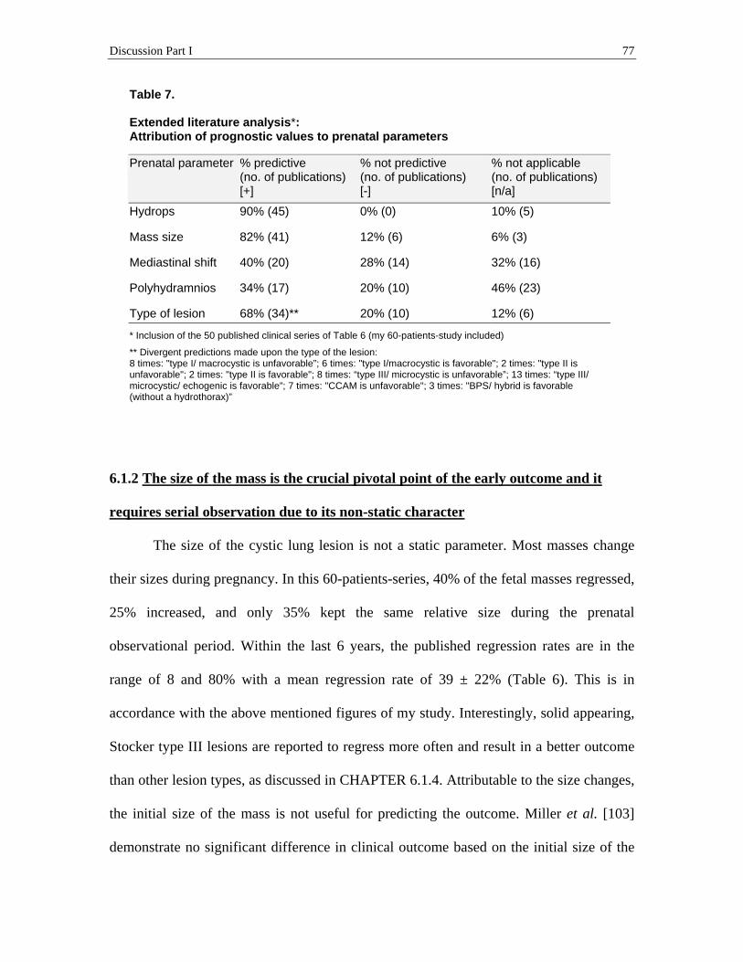
Discussion Part I 77
Table 7.
Extended literature analysis*: Attribution of prognostic values to prenatal parameters
Prenatal parameter % predictive (no. of publications) [+]
% not predictive (no. of publications) [-]
% not applicable (no. of publications) [n/a]
Hydrops 90% (45) 0% (0) 10% (5)
Mass size 82% (41) 12% (6) 6% (3)
Mediastinal shift 40% (20) 28% (14) 32% (16)
Polyhydramnios 34% (17) 20% (10) 46% (23)
Type of lesion 68% (34)** 20% (10) 12% (6)
* Inclusion of the 50 published clinical series of Table 6 (my 60-patients-study included)
** Divergent predictions made upon the type of the lesion: 8 times: "type I/ macrocystic is unfavorable”; 6 times: "type I/macrocystic is favorable"; 2 times: "type II is unfavorable"; 2 times: "type II is favorable"; 8 times: “type III/ microcystic is unfavorable”; 13 times: “type III/ microcystic/ echogenic is favorable”; 7 times: "CCAM is unfavorable"; 3 times: "BPS/ hybrid is favorable (without a hydrothorax)”
6.1.2 The size of the mass is the crucial pivotal point of the early outcome and it
requires serial observation due to its non-static character
The size of the cystic lung lesion is not a static parameter. Most masses change
their sizes during pregnancy. In this 60-patients-series, 40% of the fetal masses regressed,
25% increased, and only 35% kept the same relative size during the prenatal
observational period. Within the last 6 years, the published regression rates are in the
range of 8 and 80% with a mean regression rate of 39 ± 22% (Table 6). This is in
accordance with the above mentioned figures of my study. Interestingly, solid appearing,
Stocker type III lesions are reported to regress more often and result in a better outcome
than other lesion types, as discussed in CHAPTER 6.1.4. Attributable to the size changes,
the initial size of the mass is not useful for predicting the outcome. Miller et al. [103]
demonstrate no significant difference in clinical outcome based on the initial size of the
Discussion Part I 78
lesion. However, I discovered different growth patterns in different mass size categories.
Initially large or moderate sized masses frequently changed their sizes and were of no
value in predicting the outcome. Masses with sizes initially described to be at the extreme
low (very small) or high (very large) end of the size scale were more likely to maintain
the same size until the end of the prenatal observational period. Mass sizes with these
initial measurements predicted the early outcomes as well as the final mass sizes. A
diagram in Usui et al. [143], outlining the initial mass size versus the final mass size,
reflects the above described findings of my study. Interestingly, I observed one exception
to that rule. One of the cases of my study, an initially small macrocystic Stocker type I
lesion, grew rapidly to a very large mass until the pregnancy was terminated (case no. 59
of Tables 1 and 3). Crombleholme et al. [34] also observed that small Stocker type I
lesions may grow abruptly to hazardous large masses, attributable to rapid fluid
accumulation within a dominant cyst. Having this exception in mind and taking into
account all considerations mentioned above, it becomes clear that in general, the initial
size of the cystic lung lesion is not a reliable outcome predictor.
A significant change in mass size typically occurs during a narrow time frame. It
can be divided into two phases, a period of critical mass size increase, followed by a
period of mass size regression. The critical period of mass size increase for maximal
large or very large lesions in my series was between 19 and 27 weeks’ gestation, with a
mean increase age of 22.3 ± 1.5 weeks’ gestation. The Adzick group of the Children's
Hospital of Philadelphia (CHOP), USA, also reports significant progressive growth
between 20 and 26 weeks’ gestation, with a peak growth at 25 weeks’ gestation and a
plateau of the growth at 26 to 28 weeks’ gestation [5, 34]. Following the period of mass
Discussion Part I 79
size increase, fetal cystic lung lesions may regress, even if they reached a large size [5,
91, 103]. Regression of maximal large and very large masses was observed in my series
at a mean of 27.6 ± 3.7 weeks’ gestation. In agreement with the findings of my study,
Budorick et al. [20] in 1992 describe in one of the earliest studies on fetal cystic lung
lesions, the regression of six, probably large lesions at a mean gestational age of 27.8 ±
5.7 weeks. The most recent study on this issue, from the Jennings group [89] of the
Children’s Hospital Boston, USA, demonstrates in a series of 12 large fetal CCAMs that
again, the peak dimension is reached at 25.3 ± 3.6 weeks’ gestation, and a decrease of the
CCAM mass volume (relative to the thoracic cavity volume) occurs by 25 to 30 weeks’
gestation. Different from the larger masses, I observed that the non-critical, small or
moderate sized masses regressed about 3 mean gestational weeks later, at 30.5 ± 3.8
weeks’ gestation. Accordingly, mass size regressions are reported in the literature after 28
weeks’ gestation [34] and at a mean menstrual age of 30.4 ± 1.7 weeks’ gestation [94] in
series including small as well as large masses. Attributable to their non-static character,
fetal cystic lung lesions that grow to large sizes require close serial ultrasonographic
surveillance, at least prior to potential regression and during the critical period of mass
size increase and potential hydrops development between 18 and 29 weeks’ gestation.
The size of the lesion in proportion to the size of the fetus (relative mass size) and
not the absolute size of the mass is the measure according to which intra- or inter-
individual case comparisons or outcome prognoses have to be made. The estimation of
the fetal mass size (whether small, large, increased, or decreased) should be based on
relative measures. In order to account for fetal growth, Lee et al. [94] consider the mass
volume (calculated with the formula: anteroposterior height x width x craniocaudal length
Discussion Part I 80
x /6) not to have regressed until the volume becomes less than 50% and not to have
increased until the volume exceeds 150% of the initial volume. Crombleholme et al. [34]
created a more precise, semi-quantitative size-associated dimension: the CCAM volume
ratio (CVR). This measurement is helpful in predicting prenatal hydrops development. To
normalize for gestational age, the CVR is the tumor elliptic volume (calculated with the
formula: maximal length x width x height x 0.52) divided by the head circumference of
the fetus. A CVR >1.6 at presentation predicts an increased risk of hydrops development,
with a positive predictive value (PPV) of 75%, whereas fetuses with a CVR <1.6 are at
low risk of developing a hydrops (PPV=83%). Another size-associated, straightforward
measurement for predicting hydrops development is the mass to thorax ratio (MTR), as
published by Vu et al. [145]. To adjust for fetal size, the MTR compares the transverse
diameter of the mass to the transverse diameter of the fetal thorax, at the level of the 4-
chamber view of the heart. An MTR value >0.6 is significantly associated with a hydrops
development (PPV=74%). Miller et al. [103] calculated a similar dimension by dividing
the cross-sectional area of the lesion by the cross-sectional area of the fetal thorax, from
which the heart volume is excluded. As a converse dimension of the mass size and an
indicator of pulmonary hypoplasia and fetal compromise, Kamata and Usui et al. [80, 81,
143] formulated the lung to thorax ratio (L/T), which is the measurement of the
remaining normal bilateral lung area divided by the thorax transverse area, at the level of
the 4-chamber view of the heart. Fetuses with no increase in L/T during pregnancy and a
final L/T of <0.25 are at the highest risk of hydrops development (PPV=100% in their
first series). They also showed in a second study that the L/T correlates with postnatal
decrease in lung ventilation and perfusion [81]. In my study, the relative size of the mass
Discussion Part I 81
was addressed as follows: The size was determined by the examining ultrasonographer,
and it was categorized into four size groups, regarding the proportion of the mass to less
or more than one- or two-thirds of the fetal hemithorax. This approach is described in
detail in CHAPTER 4.2.3 of the PATIENTS AND METHODS section and in Lopoo et
al. [99]. The assignment to a particular size category in my study allowed the prediction
of the early outcome, including the prediction of hydrops development, severity of early
respiratory symptoms, and whether or not fetal or neonatal surgical treatment is required
(with a PPV in the very large mass size group of 72%, 100%, and 100%, respectively).
The early outcome findings are further discussed in detail in the last paragraph of this
CHAPTER 6.1.2. The Dumez group [46, 47, 121] and van Leeuwen et al. [144]
categorized the lesions into similar size groups, as they defined the extension of the
lesion in the hemithorax as <50%, equal to 50%, >50%, and 100% (equals bilateral). The
authors conclude that cases with a mass size <50% are at low risk of developing fetal
complications and postnatal pulmonary sequelae, whereas cases with masses >50% have
a higher risk of being symptomatic and requiring postnatal surgery. In a more simplified
approach than in my study, Bromley et al. [18] classified the lesions into only two size
groups: (a) lesions affecting parts of the lung, or (b) lesions affecting the entire lung,
based on the presence of sonographically visible, adjacent ipsilateral lung tissue. Even
with this more simple distinction into only two size groups, they could demonstrate that
the mortality rate in the group with the entire lung involved is significantly higher
compared to the other size group. Only in the course of my study, more standardized,
objective size measures were introduced, such as the above-mentioned CVR, MTR, or
L/T. Based on precise mass size measurements and normalized for fetal body size, these
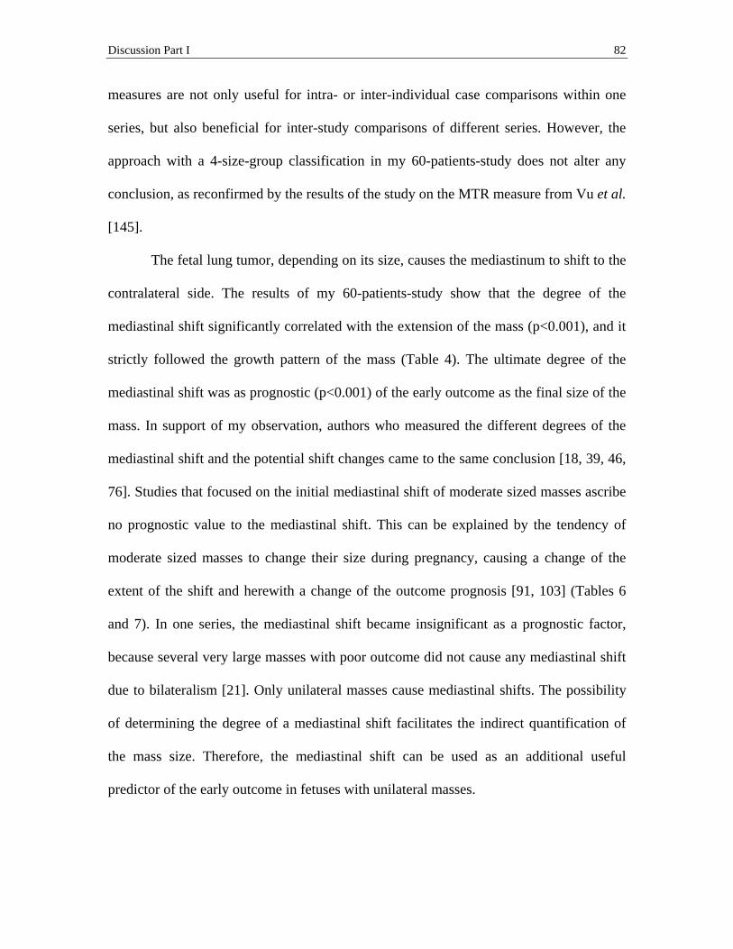
Discussion Part I 82
measures are not only useful for intra- or inter-individual case comparisons within one
series, but also beneficial for inter-study comparisons of different series. However, the
approach with a 4-size-group classification in my 60-patients-study does not alter any
conclusion, as reconfirmed by the results of the study on the MTR measure from Vu et al.
[145].
The fetal lung tumor, depending on its size, causes the mediastinum to shift to the
contralateral side. The results of my 60-patients-study show that the degree of the
mediastinal shift significantly correlated with the extension of the mass (p<0.001), and it
strictly followed the growth pattern of the mass (Table 4). The ultimate degree of the
mediastinal shift was as prognostic (p<0.001) of the early outcome as the final size of the
mass. In support of my observation, authors who measured the different degrees of the
mediastinal shift and the potential shift changes came to the same conclusion [18, 39, 46,
76]. Studies that focused on the initial mediastinal shift of moderate sized masses ascribe
no prognostic value to the mediastinal shift. This can be explained by the tendency of
moderate sized masses to change their size during pregnancy, causing a change of the
extent of the shift and herewith a change of the outcome prognosis [91, 103] (Tables 6
and 7). In one series, the mediastinal shift became insignificant as a prognostic factor,
because several very large masses with poor outcome did not cause any mediastinal shift
due to bilateralism [21]. Only unilateral masses cause mediastinal shifts. The possibility
of determining the degree of a mediastinal shift facilitates the indirect quantification of
the mass size. Therefore, the mediastinal shift can be used as an additional useful
predictor of the early outcome in fetuses with unilateral masses.
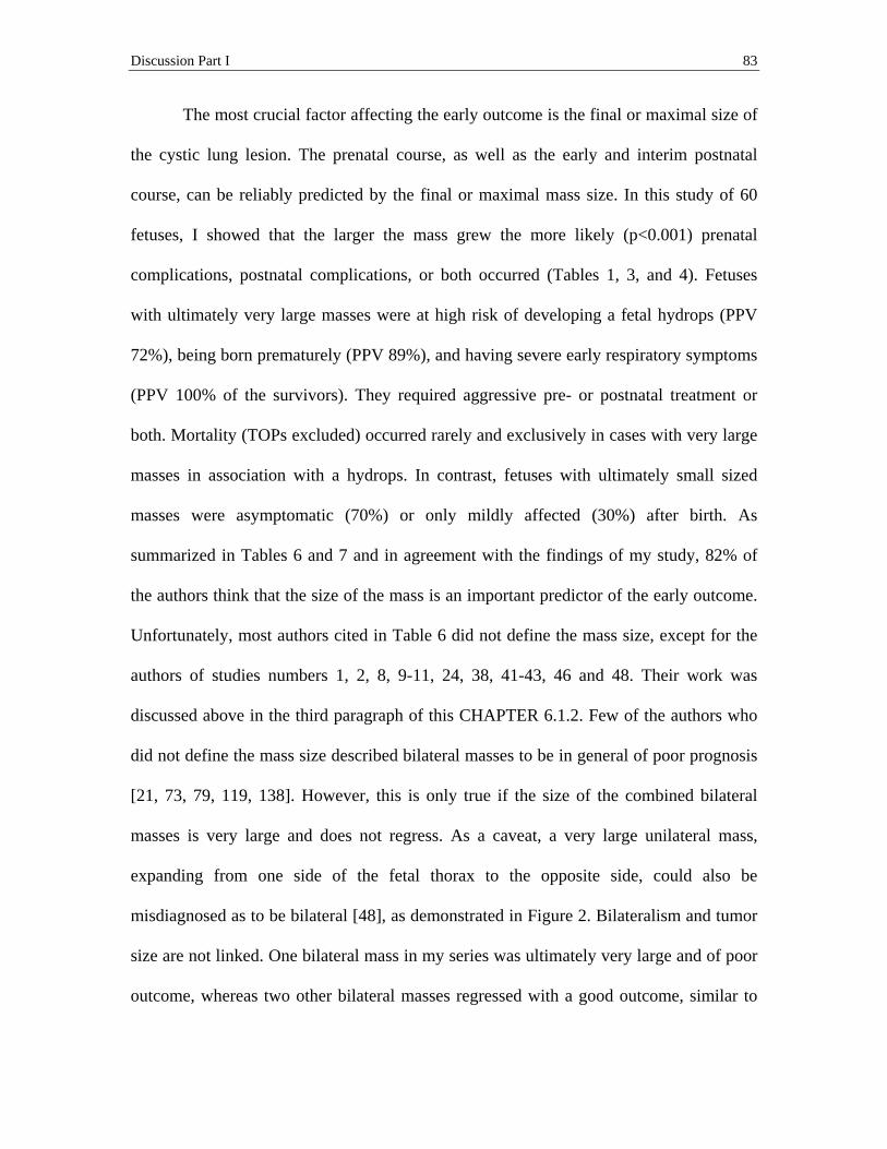
Discussion Part I 83
The most crucial factor affecting the early outcome is the final or maximal size of
the cystic lung lesion. The prenatal course, as well as the early and interim postnatal
course, can be reliably predicted by the final or maximal mass size. In this study of 60
fetuses, I showed that the larger the mass grew the more likely (p<0.001) prenatal
complications, postnatal complications, or both occurred (Tables 1, 3, and 4). Fetuses
with ultimately very large masses were at high risk of developing a fetal hydrops (PPV
72%), being born prematurely (PPV 89%), and having severe early respiratory symptoms
(PPV 100% of the survivors). They required aggressive pre- or postnatal treatment or
both. Mortality (TOPs excluded) occurred rarely and exclusively in cases with very large
masses in association with a hydrops. In contrast, fetuses with ultimately small sized
masses were asymptomatic (70%) or only mildly affected (30%) after birth. As
summarized in Tables 6 and 7 and in agreement with the findings of my study, 82% of
the authors think that the size of the mass is an important predictor of the early outcome.
Unfortunately, most authors cited in Table 6 did not define the mass size, except for the
authors of studies numbers 1, 2, 8, 9-11, 24, 38, 41-43, 46 and 48. Their work was
discussed above in the third paragraph of this CHAPTER 6.1.2. Few of the authors who
did not define the mass size described bilateral masses to be in general of poor prognosis
[21, 73, 79, 119, 138]. However, this is only true if the size of the combined bilateral
masses is very large and does not regress. As a caveat, a very large unilateral mass,
expanding from one side of the fetal thorax to the opposite side, could also be
misdiagnosed as to be bilateral [48], as demonstrated in Figure 2. Bilateralism and tumor
size are not linked. One bilateral mass in my series was ultimately very large and of poor
outcome, whereas two other bilateral masses regressed with a good outcome, similar to
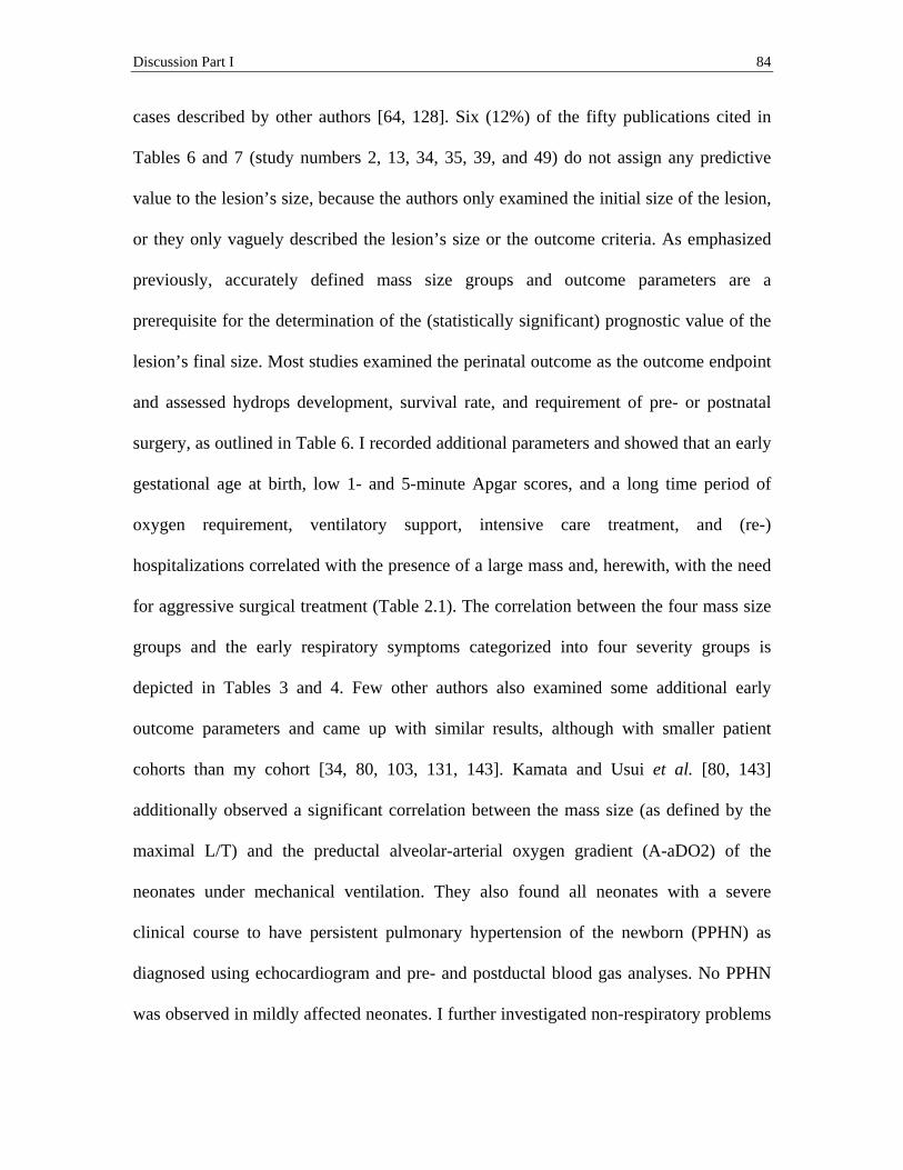
Discussion Part I 84
cases described by other authors [64, 128]. Six (12%) of the fifty publications cited in
Tables 6 and 7 (study numbers 2, 13, 34, 35, 39, and 49) do not assign any predictive
value to the lesion’s size, because the authors only examined the initial size of the lesion,
or they only vaguely described the lesion’s size or the outcome criteria. As emphasized
previously, accurately defined mass size groups and outcome parameters are a
prerequisite for the determination of the (statistically significant) prognostic value of the
lesion’s final size. Most studies examined the perinatal outcome as the outcome endpoint
and assessed hydrops development, survival rate, and requirement of pre- or postnatal
surgery, as outlined in Table 6. I recorded additional parameters and showed that an early
gestational age at birth, low 1- and 5-minute Apgar scores, and a long time period of
oxygen requirement, ventilatory support, intensive care treatment, and (re-)
hospitalizations correlated with the presence of a large mass and, herewith, with the need
for aggressive surgical treatment (Table 2.1). The correlation between the four mass size
groups and the early respiratory symptoms categorized into four severity groups is
depicted in Tables 3 and 4. Few other authors also examined some additional early
outcome parameters and came up with similar results, although with smaller patient
cohorts than my cohort [34, 80, 103, 131, 143]. Kamata and Usui et al. [80, 143]
additionally observed a significant correlation between the mass size (as defined by the
maximal L/T) and the preductal alveolar-arterial oxygen gradient (A-aDO2) of the
neonates under mechanical ventilation. They also found all neonates with a severe
clinical course to have persistent pulmonary hypertension of the newborn (PPHN) as
diagnosed using echocardiogram and pre- and postductal blood gas analyses. No PPHN
was observed in mildly affected neonates. I further investigated non-respiratory problems

Discussion Part I 85
and found that early and prolonged difficulties with feeding, weight gain, and GERD
were prematurity-associated problems in children with large fetal masses and after fetal
hydrops. Prematurity is reported to significantly occur in fetuses with large masses [34,
91] and in hydropic fetuses with or without fetal treatment [5, 21, 25, 76, 143]. In this 60-
patients-study, fetuses with very large masses but no hydrops were born at a mean
gestational age of 35.6 ± 4.1 weeks. Those with signs of a hydrops were born already at a
mean of 31.2 ± 3.6 weeks’ gestation and had severe early respiratory symptoms when
they survived. Prematurity is influential in early respiratory and non-respiratory
morbidity, as well as in early mortality. The long-term outcome analysis is as important
as the early outcome analysis. I succeeded in following-up with the children over a very
long time period, which to the best of my knowledge has not been done before in a large
patient cohort. I compared the early outcomes with the long-term outcomes and
surprisingly, a disadvantageous early outcome did not lead to an unfavorable long-term
outcome, as discussed in detail in CHAPTER 5.2.4 and 6.1.6. As this study has shown so
far, the pivotal point for the antenatal, perinatal, and interim early outcomes is the fetal
mass size. The mass size, which preconditions all other fetal symptoms, requires close
ultrasonographic surveillance, at least during the critical period when the size increases.
6.1.3 First signs of a hydrops in fetuses with large lesions mark the beginning of
serious complications starting before birth, but they can be averted with fetal
treatment
A hydrops fetalis develops with high probability if the size of a large lesion
exceeds a size the fetus cannot manage without cardiovascular decompensation. In this
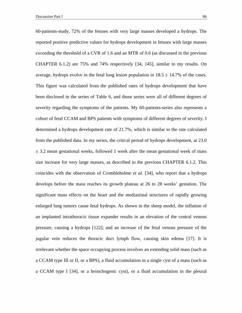
Discussion Part I 86
60-patients-study, 72% of the fetuses with very large masses developed a hydrops. The
reported positive predictive values for hydrops development in fetuses with large masses
exceeding the threshold of a CVR of 1.6 and an MTR of 0.6 (as discussed in the previous
CHAPTER 6.1.2) are 75% and 74% respectively [34, 145], similar to my results. On
average, hydrops evolve in the fetal lung lesion population in 18.5 ± 14.7% of the cases.
This figure was calculated from the published rates of hydrops development that have
been disclosed in the series of Table 6, and those series were all of different degrees of
severity regarding the symptoms of the patients. My 60-patients-series also represents a
cohort of fetal CCAM and BPS patients with symptoms of different degrees of severity. I
determined a hydrops development rate of 21.7%, which is similar to the rate calculated
from the published data. In my series, the critical period of hydrops development, at 23.0
± 3.2 mean gestational weeks, followed 1 week after the mean gestational week of mass
size increase for very large masses, as described in the previous CHAPTER 6.1.2. This
coincides with the observation of Crombleholme et al. [34], who report that a hydrops
develops before the mass reaches its growth plateau at 26 to 28 weeks’ gestation. The
significant mass effects on the heart and the mediastinal structures of rapidly growing
enlarged lung tumors cause fetal hydrops. As shown in the sheep model, the inflation of
an implanted intrathoracic tissue expander results in an elevation of the central venous
pressure, causing a hydrops [122]; and an increase of the fetal venous pressure of the
jugular vein reduces the thoracic duct lymph flow, causing skin edema [17]. It is
irrelevant whether the space occupying process involves an extending solid mass (such as
a CCAM type III or II, or a BPS), a fluid accumulation in a single cyst of a mass (such as
a CCAM type I [34], or a bronchogenic cyst), or a fluid accumulation in the pleural
Discussion Part I 87
cavity (causing tension hydrothorax in BPS cases [99]). What is relevant is the large size
and rapid progression of the space occupying process in the fetal thorax. Further, a
hydrops can also result from an anomalous venous drainage of large lesions via
significant intrapulmonary arterio-venous shuns, causing cardiac decompensation and
central venous congestion [1]. Why every very large mass does not cause a hydrops is not
known, but what I found is that the larger and faster the mass grows or the fluid
accumulates in the thorax, the higher the risk of developing a hydrops.
The signs of a hydrops are indicators of a condition threatening the fetus’ life,
already before birth. The natural history of untreated hydropic fetuses is grave. My
extended statistical analysis of the series that are summarized in Table 6 revealed that
73.4% (83 of 113 cases) of the untreated hydropic fetuses died (Figure 6). Due to this
alarming and unambiguous number of fatalities, a hydrops has become known as the
most useful predictor for an unfavorable early outcome of fetuses with lung lesions [5,
34, 39, 46, 76, 91]. In 90% of the publications, summarized in Tables 6 and 7, authors
convincingly show that a hydrops is an important predictor of the early outcome. In the
remaining 10% no hydrops were observed. However, the mean overall mortality rate of
67% (199 of 297 hydrops cases) is an overestimation of the true mortality rate in
hydropic fetuses. This was shown with the extended statistical analysis of all published
series summarized in Table 6 and Figure 6. Of the 199 deceased hydropic fetuses, 81
fetuses (40.7%) died because the pregnancy was terminated, and an additional 83 fetuses
(41.7%) did not survive because prenatal treatment was not attempted (Figure 6).
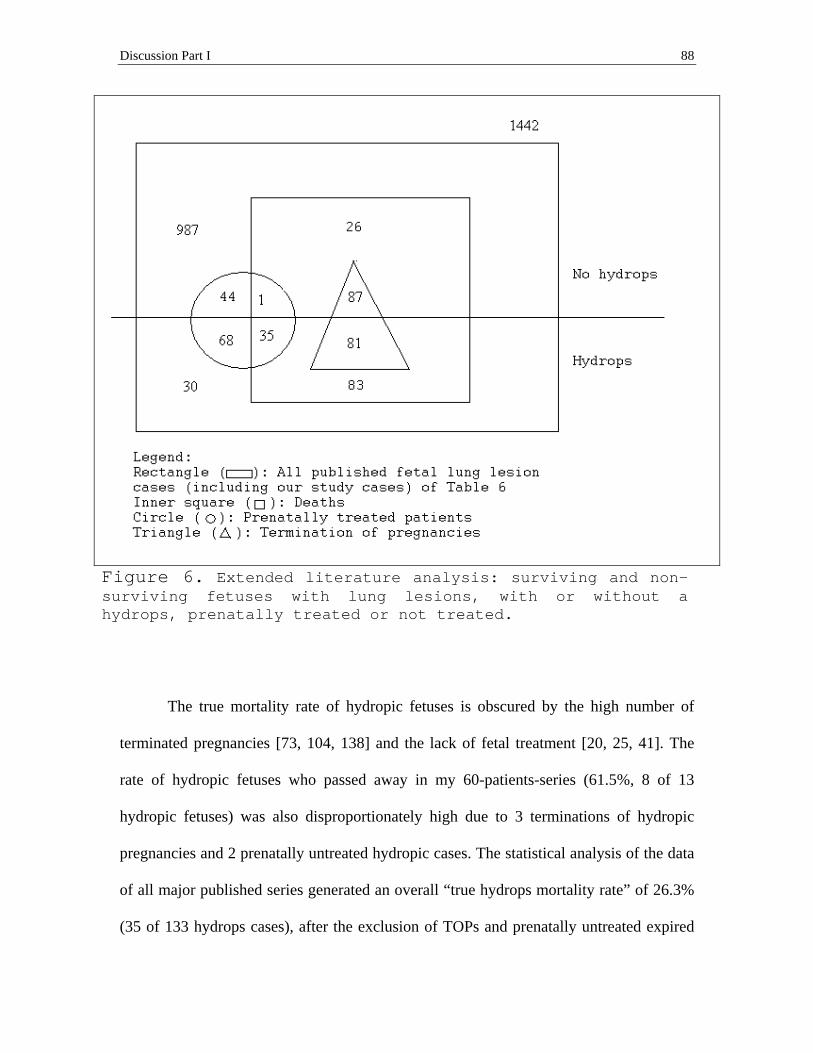
Discussion Part I 88
Figure 6. Extended literature analysis: surviving and non-surviving fetuses with lung lesions, with or without a hydrops, prenatally treated or not treated.
The true mortality rate of hydropic fetuses is obscured by the high number of
terminated pregnancies [73, 104, 138] and the lack of fetal treatment [20, 25, 41]. The
rate of hydropic fetuses who passed away in my 60-patients-series (61.5%, 8 of 13
hydropic fetuses) was also disproportionately high due to 3 terminations of hydropic
pregnancies and 2 prenatally untreated hydropic cases. The statistical analysis of the data
of all major published series generated an overall “true hydrops mortality rate” of 26.3%
(35 of 133 hydrops cases), after the exclusion of TOPs and prenatally untreated expired
Discussion Part I 89
cases (Table 6 and Figure 6). In a recently published survey from the UCSF Medical
Center, Grethel et al. [62] report the neonatal survival rates for (i) non-treated non-
hydropic fetuses, (ii) treated non-hydropic fetuses, (iii) treated hydropic fetuses, and (iv)
non-treated hydropic fetuses with CCAM or BPS (after exclusion of TOPs) to be (i)
98.5%, (ii) 85.7%, (iii) 56.7%, and (iv) 0%, respectively. These results are largely
congruent with the results obtained from the extended statistical analysis of the published
data (from 1985 to 2007) of (i) 97.4%, (ii) 97.7%, (iii) 66.0%, and (iv) 26.5%,
respectively (Figure 6). The only exception is a higher percentage of surviving untreated
hydropic fetuses in the analyzed published series, compared to the studies of Grethel’s et
al. and myself. In my series none of the hydropic fetuses survived without invasive fetal
treatment from 1988 to 2002, as well as in Grethel’s et al series from 1991 to 2006 (the
series partly overlapped), whereas 26.5% (30 survivors among 113 untreated hydrops
cases) survived in the reported literature (Table 6 and Figure 6). In the survivors,
spontaneous improvement of the hydropic symptoms mostly occurred early in the third
trimester [46, 75, 80]. Other authors not included in Table 6 also report remarkable
spontaneous regression of very large masses followed by the improvement of a hydrops
[36, 56, 60, 69, 89]. The mechanism of the spontaneous regression is not fully
understood. True mass regression has to be distinguished from the proportional mass size
reduction that occurs due to fetal growth and gradual augmentation of the surrounding
normal lung tissue, while the lesion itself remains stable. Plausible explanations of true
tumor shrinkage include: involution of the lesion due to the outgrowth of the blood
supply or the obstruction of the vascular pedicle, decompression of fetal lung fluid
through abnormal channels, and spontaneous correction of an underlying bronchial
Discussion Part I 90
obstruction within the lesion [1, 94, 100, 103]. Mass regression and improvement of
hydropic symptoms point to a favorable outcome. However, it is premature to assume
that the fetus is safe when hydropic symptoms disappear after fetal treatment or even if
they disappear spontaneously. Although rare, serious perinatal complications are reported
despite prenatal hydrops improvement [91, 97, 102, 149]. The early onset of a regression
of the mass size and the hydrops seems to be the key to a favorable outcome. In the
reported cases with good outcome, fetuses showed signs of a regression prior to 30
weeks’ gestation, whereas after 31 weeks’ gestation, most fetuses with late observed
regressions experienced severe complications due to pulmonary hypoplasia, prolonged
cardiovascular compromise, or both. A lingering untreated hydrops is a dismal situation
for a fetus with a lung mass, and it justifies fetal therapeutic intervention.
Early recognition of signs of a hydrops and prompt therapeutic fetal intervention
are crucial for successfully treating severely affected fetuses with cystic lung lesions. At
the UCSF Children’s Hospital as well as at the CHOP, fetal treatment specialists, like
Michael R. Harrison and Scott N. Adzick [34], advise against delaying fetal therapy.
Hoping for spontaneous hydrops resolution, even though it could eventually occur,
endangers the fetus’s life. The three unsuccessfully operated fetuses in my study who
passed away had a fetal tumor resection done after 28 weeks’ gestation and the hydrops
was far advanced in all of them. All other ten successfully operated fetuses who achieved
a good outcome, except for one case (a fetal shunt case), were operated on much earlier at
a mean gestational age of 24.4 ± 2.1 weeks. Timely fetal therapeutic intervention
increases the chance of survival for hydropic fetuses with cystic lung lesions.
Discussion Part I 91
Established fetal therapeutic interventions for cystic lung lesions are: (serial)
aspirations or shunts, to drain rapidly enlarging cysts or tension hydrothoraxes, and open
fetal surgery to resect very large tumors. Many of these interventions have been
successfully performed since the pioneering first fetal thoracotomy and pulmonary tumor
resection in 1990 by Harrison et al. [66] and the first fetal thoracocentesis and pulmonary
cyst shunting in 1987 by Nicolaides et al. [109]. From 20 different fetal treatment centers
(Table 6), I analyzed the surgical fetal intervention performances and the success rates
are as follows: Of 148 prenatally operated fetuses, 112 survived upon cyst aspirations,
shunting, and tumor resection. That results in an overall fetal intervention success rate of
76%. When limiting the focus to 103 hydropic fetuses after therapeutic fetal interventions
(148 prenatally operated cases minus 45 non-hydropic cases), and when discriminating
between cyst aspirations or shunting versus tumor resections, I calculated a success rate
for hydropic fetuses of 78% (46 survivors of 59 cases) for aspirations or shunting versus
55% (24 survivors of 44 cases) for tumor resections. These results correspond with the
most recent published outcome data on in utero therapy from the CHOP [152] and show
that fetal therapeutic interventions are successful in saving hydropic fetuses. Early and
effective mass volume reduction and timely resolution of a hydrops are crucial for a
favorable outcome after fetal treatment. The group from Philadelphia observed hydrops
resolution in the survivors 1 to 2 weeks postoperatively, which is in agreement with the
experiences at the UCSF Medical Center and the findings of my study. Noteworthy is the
fact that the premise for a successful fetal shunt therapy with an improved survival rate is
an effectively functioning and long lasting shunt [108, 152]. However, even if the fetal
therapeutic procedure is successfully accomplished, there may be postoperative

Discussion Part I 92
complications to be considered. A sudden thoracic decompression after the resection of a
large lung mass may lead to intra-operative hemodynamic collapse, bradycardia, and fetal
loss. This might require specific treatment adjustments to save the fetus [152]. For this
case, the underlying pathophysiological mechanism is most likely the reverse effect of
the above-mentioned mass effect of hydrops causing, rapid progressing masses. The most
common and difficult to handle complications after fetal surgery are preterm labor (PTL)
and premature rupture of membranes (PROM), which often result in premature delivery,
as described in CHAPTER 5.2.3 of the RESULT section and in our published case report
of PART II of this doctoral thesis. For this reason, fetal surgery was poorly received in
Europe, in contrast to Northern American practices. Since the late 1990s, new minimally
invasive, fetoscopic treatment options have been developed and, not until then were fetal
surgical programs introduced in Europe to promote these techniques [44]. Thereafter,
fetal (serial) aspiration and shunt interventions were increasingly performed in Europe.
However, open fetal surgery remains to the best of my knowledge almost nonexistent in
Europe, in spite of advanced, postoperative tocolytic management and despite the success
rates that are reported by respected American fetal treatment centers.
In conclusion, the survival rate of hydropic fetuses with large cystic lung lesions
can be significantly improved with timely fetal therapeutic intervention, preventing
prolonged, irreversible damage to the fetus. Moreover, it seems advisable to also operate
on non-hydropic fetuses with significant space-occupying lesions to prevent pulmonary
hypoplasia, although further investigations need to be performed.
Discussion Part I 93
6.1.4 Marked ascites and the type of the lesion are, in contrast to polyhydramnios,
independent outcome predictors
The results of my 60-patients-study show that marked ascites were predictive of
the early outcome (p<0.001), even in cases in which they were not associated with a
hydrops as classically defined by Evans [51]. Goldstein [58] suggests that the presence of
solitary ascites is sufficient to diagnose the presence of a hydrops. Other authors regard
solitary ascites in combination with polyhydramnios as hydrops [23, 107, 143]. A
modified hydrops definition was also applied to two of my study cases which had marked
ascites combined with placentomegaly and polyhydramnios. It should be emphasized that
it isn’t ascites alone, but the degree of the ascites, that justifies treating these cases as
classically defined hydrops cases. All marked ascites in my series worsened and
complicated the outcome, while 2 out of 3 mild ascites resolved. The resolution of ascites
was observed by others, in cases of mild degrees of ascites or in the absence of further
hydrops-associated symptoms [4, 21, 91, 119]. A mild solitary ascites is, as much as mild
solitary pleural effusion or pericardial effusion, not an indicator of an unfavorable
outcome. However, marked progressing ascites, especially in combination with
polyhydramnios, is a predictor of an unfavorable outcome, as much as a classic hydrops.
Polyhydramnios is a controversially discussed outcome parameter. Of the studies
included in Tables 6 and 7, 34% assign a prognostic value to polyhydramnios, 20%
refrain to do so, and 46% do not address this issue. However, the authors consent to one
issue: Polyhydramnios is a prognostic factor of the outcome if combined with hydrops,
marked ascites, or both [4, 20, 94, 104, 143]. Solitary polyhydramnios can be transient
[91, 128]. As the fetal lung lesion regresses, solitary polyhydramnios, as much as other
Discussion Part I 94
solitary fetal effusions, may resolve. This 60-patients-study showed that the degree of a
polyhydramnios was significantly associated with the fetal lung lesion size and with fetal
hydrops (p<0.005). Marked and moderate polyhydramnios were seen in very large
masses (with only one exception), whereas mild poly was also found in mildly affected
fetuses, and it regressed in three of them (Table 4). In summary, polyhydramnios is not a
reliable outcome predictor independent of the lesion’s size or hydrops.
The prognostic value derived from the type of the cystic lung lesion, as described
by Stocker et al. (type I with cysts >2 cm, type II with cysts <2 cm, and type III without
identifiable cysts) [7, 132] or by Adzick et al. (microcystic with cysts <5 mm,
macrocystic with cysts >5 mm) [7], is the most controversially discussed prenatal
parameter (Table 7). Several authors state that solid or microcystic lesions are favorable
because of their nature to regress more frequently than lesions with discernible cysts [15,
18, 39, 41, 57, 75, 76, 91, 93, 94, 104, 128]. Conversely, other authors state that a solid or
microcystic lesion is a predictor of an unfavorable outcome [4, 7, 21, 79, 107, 137, 138].
The results of my 60-patients-study support the notion that solid appearing lung lesions
of Stocker type III and small-cystic Stocker type II lesions are more favorable,
attributable to a higher regression rate, than masses with large Stocker type I cysts, which
more often increase to critical large mass sizes. A large dominant cyst within the type I
lesion is highly critical as it may abruptly increase due to rapid fluid accumulation, as
described by Crombleholme et al. [34] and also observed in my series. To emphasize
again, close ultrasonographic surveillance is required, at least during the critical period of
mass size increase, to be prepared for prompt therapeutic interventions in rapidly
increasing Type I lesion cases, as mentioned before in CHAPTERS 6.1.2 and 6.1.3.
Discussion Part I 95
6.1.5 A major reason for the overestimation of the mortality rate is that the “hidden
survival rate” is unaccounted for
The overall mean mortality rate of 22 ± 18.2%, calculated from data published
from 1985 to 2007 (Table 6 and Figure 6), is an overestimation of the true mortality rate
of fetuses with cystic lung lesions. The earlier performed studies in particular, tended to
overestimate the mortality rate, due to a biased selection for the sickest fetuses and
attributable to numerous elective termination of pregnancies [7, 107, 138]. As ultrasound
techniques have improved, many previously unnoticed lesions with a good prognosis
were detected and, as superior pre- and postnatal treatment options have become
available, many institutions abandoned the practice of interrupting pregnancies for fetal
cystic lung lesions. As a result, from 2003 to 2007, the recent mean mortality rate
dropped to 10.9 ± 7.3%. For this calculation I included the data from the fifteen most
recent studies of Table 6 and excluded one study with an exceptionally high TOP rate
[73]. The mortality rate of 15% in my series lies in between the above-mentioned,
overestimated mortality rate of 22% and the more realistic, recent mortality rate of
10.9%. This can be explained by the very long 14-year-period covered in my study, and it
reflects the changes toward improved diagnostic and therapeutic techniques at the UCSF
Medical Center. In order to account for modern treatment standards, I corrected the
individually published mortality rates by calculating a “true mortality rate.” To achieve
this, I excluded the interrupted pregnancies and the prenatally untreated hydropic fatal
cases which otherwise had no other congenital anomalies (Table 6 and Figure 6) from the
calculations. The true mean mortality rate dropped to as low as 7.4 ± 6.5%, which is
about three times lower than the overestimation.
Discussion Part I 96
Another important piece of information that helped with this study – to reflect on
the outcome of fetal cystic lung lesions, such as CCAM or BPS – came in fact from the
parents of the affected children. There were 22 parents of the 60 study patients who
declined to follow the recommendations of their referring doctors to terminate the
pregnancy. Their decisions were justified, since their children were later doing fine. In
retrospect, these children were considered “miracles of life” by their parents (in
communication to me, the conductor of this study). Within this group, only 1 fetus
required prenatal surgery and 4 required postnatal surgery due to symptoms. The other 17
children were asymptomatic or had only minor symptoms early after birth. Professor
Diana L. Farmer, Professor Ruth B. Goldstein, and I coined the term “hidden survival
rate” for this phenomenon. Several other authors also critically reflect on how to counsel
parents [64, 91, 101, 104]. The growing number of healthy fetal lung lesion survivors –
which were initially given a poor prognosis – made the authors reconsider their parental
counseling. McCullagh et al. [101] suggest that no specific diagnosis should be given to
the parents, instead they should be told that some cystic changes have been observed and
further monitoring will be necessary. No action should be taken which might jeopardize
the outcome of a benign condition. The fetal treatment specialists of the UCSF Medical
Center and the University Hospital Großhadern of the LMU Munich also recommend that
physicians adapt to the knowledge of the “hidden survival rate” for fetuses with cystic
lung lesions, and that they make proper adjustments in their parental counseling.
Discussion Part I 97
6.1.6 The long term outcome is excellent
The long-term outcome of children with prenatally detected cystic lung lesions is
excellent, independent (p>0.3) of the prenatal outcome predictors, and not influenced by
the early (respiratory) outcome as observed in my patients survey. This is backed by the
following findings. No fatality occurred past infancy. A single child of this study died in
the extended neonatal period, at 2 months of age. In the surveyed literature, only four
infant deaths were recorded and all of them occurred within 3 months of birth (Table 6;
one case excluded because of uncertain detail).
The initial respiratory difficulties improved in the patients of my study, either in the
neonatal period (41%), during the first 2 years (34%), or rarely between 2 and 4 years of
age (16%). The majority (75%) was completely asymptomatic at last follow-up, 20% had
minor residual labored breathing on exertion or infrequent mild asthmatic symptoms, and
only three children who had received operations continued to be moderately symptomatic
with limited physical endurance (3 years old), frequent pulmonary infections (5 years
old), or asthma (12 years old) (Tables 2.2 and 3). To the best of my knowledge, this
report of a comprehensive long-term surveillance of a large cohort of fetuses with cystic
lung lesions is unique in the literature. Most of the other published reports focused on
perinatal outcome. The few examinations of long-term outcomes were performed on
small patient numbers or selected cases (Table 6). So far, only one recent study by
Kamata et al. [81] presents a comparable long-term study, with a similar follow-up
interval (6.9 mean years of Kamata’s study versus 5.9 mean years of my study), but with
less than one-half as many patients, compared to my study. As I did, Kamata et al. also
observed that respiratory symptoms, such as frequent respiratory tract infections and
Discussion Part I 98
asthmatic attacks, improve or disappear with increasing age. In their series only one 7-
year-old child who had massive hydrops during fetal life had persistent severe respiratory
restrictions. Two more children are reported by other authors to have pulmonary sequelae
and asthma with recurrent pneumonia later in life [103, 128]. All other children with fetal
cystic lung lesions who were followed long-term are reported to have complete functional
recovery after overcoming the critical neonatal period [23] and are doing well at last
follow-up [39, 41, 73, 104]. However, using ventilation and perfusion lung scintigraphy
to evaluate the children’s lung functions, Kamata et al. discovered a significant decrease
of lung ventilation and lung perfusion in patients who have had a large fetal mass (low
L/T), prenatal hydrops, or required a lobectomy [81]. This suggests that lung scintigraphy
appears to be a sensitive method for evaluating even subtle lung function deficits, and it
should be applied in future care and research studies of fetuses with lung lesions. In my
research, I focused on clinically relevant respiratory symptoms and on the potential
interference with the children’s or families’ daily activities. In this regard, all of the 51
surviving study patients were doing fine except for the three above-mentioned,
moderately affected children. Furthermore, prematurity-associated non-respiratory
problems, such as prolonged difficulties with feeding, weight gain, and GERD, improved
to a milder stage mostly by the age of 2 years. Kamata et al. (2006) also observed that
failure to thrive is a common complication, but improves with increasing age. They argue
that prolonged artificial ventilation, labored respiration, and frequent respiratory tract
infections lead to malnutrition, and it may last in patients who have had fetal hydrops, as
they experienced with one case [81]. Other major congenital anomalies were seen in 8%
(5 of 60) of the patients in my study. This rate is slightly higher than the average anomaly
Discussion Part I 99
rate of 5% of the cases included in Table 6. The explanation for this data is that a few
studies in Table 6 did not include cases with major anomalies due to the restrictive
referral policy of their tertiary care centers or the exclusion of these cases from their
research population [34, 46, 47, 59, 143]. Only 3% of the anomalies were detectable
before birth in my 60-patients-series. This percentage is comparable with the average
frequency of prenatally detected congenital anomalies of about 2% in ordinary
pregnancies, observed in the EUROFETUS and EUROCAT study [98]. The incidence of
congenital heart diseases is 11.8:1000 (Table 6) and not significantly elevated in fetuses
with cystic lung lesions, compared to a general prenatal incidence of 6:1000 to 12:1000
[71]. The computed incidence from the reports summarized in Table 6 is 6.9:1000 for
renal agenesis and 2.1:1000 for trisomy 18 in the fetal lung lesion population and it is
surprisingly high. This is in contrast to a reported incidence of about 0.3:1000 of each,
renal agenesis and trisomy 18, in regular pregnancies [27, 86, 115, 151]. In my 60-
patients-series, one unilateral renal agenesis (in association with a Mayer-Rokitansky-
Küster-Hauser syndrome) and one ultrasonographically questionable bilateral renal
agenesis (not confirmed with an autopsy report) were found, but no cardiac anomalies
(only 10 newborns with non-structural, transient cardiac symptoms) or trisomy 18 cases.
In summary, the long-term outcome of fetuses with cystic lung lesions, such as CCAM or
BPS, is excellent. Most children are postnatally asymptomatic and the other survivors
grow out of their initial respiratory symptoms by early childhood at the latest. Only a few
exceptional cases remain with prolonged respiratory or non-respiratory anomalies.
There is a significant difference between the long-term outcome of fetuses with
CCAM or BPS and the long-term outcome of fetuses with congenital diaphragmatic
Discussion Part I 100
hernia (CDH). Neurodevelopmental delay, hearing loss, and growth failure is frequent in
children with severe fetal CDH, as recent follow-up studies on CDH show [33, 111].
Mortality and pulmonary hypoplasia rates are much higher in association with prenatally
diagnosed CDH than with CCAM and BPS, despite similar ultrasound impressions of the
volume of the mass and the compression of the surrounding normal lung during
pregnancy [5, 47, 58, 129]. About 30% of CDH patients require ECMO (extracorporeal
membrane oxygenation) after birth, and about 25% of the survivors suffer from long-term
chronic obstructive airway disease [77, 105]. Only a single child of the 51 survivors in
this study of fetuses with cystic lung lesions had ECMO and did not survive. None of the
children of this series and of the reported series had long-term neurodevelopmental
delays or hearing loss. Only very few children with very large fetal lung masses or
hydrops had prolonged respiratory sequelae, as described in CHAPTER 5.2.4 and
discussed above in this CHAPTER 6.1.6. The outcome of fetuses with cystic lung lesions
is much better than the outcome of fetuses with CDH.
Discussion Part I 101
6.2 Management of the patients
6.2.1 Prenatal management recommendations
The ultrasonography provides an important resource for prenatal management.
Ultrasonographic screening techniques have become very sensitive in detecting fetal
cystic lung lesions. Under rare circumstances where ultrasonographic findings are
indistinguishable, fetal MRI can confirm the diagnosis of a lung tumor, determine its
origin, and be instrumental in the differentiation from a CDH [22, 74, 120]. Screening for
other anomalies, with a particular attention to renal agenesis and trisomy 18, is indicated
following the diagnosis of a fetal cystic lung lesion. Serial ultrasound imaging is the
method of choice for following the fetuses with lung lesions through pregnancy. Owing
to the growth pattern of moderate and large sized lesions, these lesions require closer
observation than small sized lesions. During the period of critical mass size increase,
between 20 and 28 weeks’ gestation, weekly ultrasounds or at least every two weeks are
indicated [58]. Small lesions can be followed monthly, as well as stable lesions after 30
weeks’ gestation. If the mass enlarges markedly or a dominant cyst within the mass
grows rapidly, up to daily ultrasounds are required to rule out fetal ill being, specifically
in the event of the development of hydropic symptoms. Alerting mass sizes, indicative
for a high risk of hydrops progression, are a CVR of >1.6, an MTR of >0.6, and an L/T of
<0.25 [34, 80, 145], as discussed in CHAPTERS 6.1.2 and 6.1.3. Close serial ultrasound
surveillance will not miss early signs of a hydrops development, the “point of (almost) no
return.”
Once hydropic signs emerge, an expeditious decision has to be made in favor of
fetal treatment versus delivery. If the fetus is more than 32 weeks of age prompt delivery
Discussion Part I 102
is preferred, whereas at less than 32 weeks of age an immediate fetal therapeutic
intervention is indicated to have a chance of a successful outcome [34, 58, 152]. Maternal
contraindications have to be ruled out prior to the procedure. This strategy is also
recommended for other life-threatening fetal tumors, such as giant fetal hepatic
hemangiomas, as I pointed out in a previous publication [118]. Even if the fetus is not
hydropic, but the lung mass is huge, invasive fetal treatment has to be considered to
prevent significant pulmonary hypoplasia. However, no consensus has been reached
regarding when and at what size a therapeutic intervention would be advisable for non-
hydropic fetuses with huge masses. Fetal treatment options are (serial) aspirations or
catheter-induced, thoraco-amniotic shunts for large dominant CCAM type I cysts or for
pleural effusion associated with BPS [46, 65, 108]. A more invasive procedure with
maternal hysterotomy, fetal thoracotomy, and pulmonary tumor resection is required for
fetuses with massive solid appearing cystic lung lesions [65]. The success rate of this
procedure is evidently increasing, as discussed in CHAPTER 6.1.3. However, open fetal
surgery remains a risky procedure for both the fetus and the mother, and it should be
considered only if no other options are available [58]. Further studies on alternative fetal
treatment techniques, such as percutaneous radiofrequency ablation for large solid masses
[19, 39, 53] and embolization of the aberrant artery for BPS reduction [1, 110, 121], have
to prove that these procedures are reliable and safe for the fetus. To treat severe
polyhydramnios or solitary massive ascites, minimally invasive procedures such as
(serial) amniocentesis [91, 119] or fetal paracentesis [13] can ease these symptoms and
may prevent fetal lung growth restriction or prune belly formation. Non-surgical
management with medication, such as steroids (via the mother) for tumor reduction [141]
Discussion Part I 103
or furosemide and Digoxin (via the umbilical cord) for fetal cardiac failure [9], may also
find their way into conventional prenatal treatment. If early delivery is indicated, the fetal
treatment specialists of the UCSF Children’s Hospital and CHOP [68] recommend the ex
utero intrapartum treatment (EXIT) for very large lung lesions. With this procedure, the
mass is excised at the time of delivery, while the fetus is maintained on placental support.
If early postnatal pulmonary hypertension or hypoplasia complicates the recovery of the
newborn, transient high-frequency oscillatory ventilation (HFOV) [82, 147],
extracorporeal membrane oxygenation (ECMO) [3], or both can be used, in addition to
other intensive care treatment options. Institutions where these modern pre-, peri-, and
postnatal treatment techniques are established are able to save the lives of severely
affected fetuses, and they significantly improve the children’s outcomes.
When counseling parents, it is justifiable for physicians to point out the excellent
prognosis of fetuses with cystic lung lesions, as coached at the UCSF Medical Center and
various other institutions [64, 101, 104]. Although rarely necessary, the need for intense
and demanding care during the first few months or at latest up to 3 to 4 years of age has
to be addressed. This might be the case when the cystic lung lesion is very large, a fetal
hydrops develops, or a fetal therapeutic intervention was performed. Reasons for the
intense care are the associated respiratory restrictions and prematurity. Parents of very
seriously affected fetuses have to be prepared that although occurring seldom, despite
supreme pre- and postnatal treatment, an early pre- or postnatal death cannot be
prevented [34, 46, 58, 91, 152]. However, the majority of fetuses with cystic lung lesions
is doing fine, and parents of these fetuses do not need to be counseled about the worst-
case scenario.
Discussion Part I 104
6.2.2 Postnatal management recommendations
As most institutions with ample experience in postnatal management of children
with congenital cystic lung lesions, the specialists at the UCSF Children’s Hospital
recommend postnatal radiological examinations. An initial x-ray is indicated immediately
after birth, even if the newborn is asymptomatic. If the initial x-ray is negative, the child
needs a subsequent computed tomography (CT) at the age of 1 month. Many lesions are
identified on postnatal CT, even if they disappear on prenatal ultrasound or are not
demonstrable on postnatal x-ray. Fetal cystic lung lesions often decrease before birth, but
they rarely dissolve completely [15, 39, 43, 94, 100, 129, 144]. Of the 7 lesions that
disappeared on prenatal ultrasound, in my 60-patients-series, 3 were demonstrated on
postnatal CT. The others had no CT or MRI done, including two with negative x-rays. A
postnatal magnetic resonance imaging (MRI) is a radiation-free alternative to a CT scan
in respiratory stable patients, but it reveals less information about the pulmonary
parenchyma than a CT [23, 87, 133, 147]. However, the pre-operative demonstration of
the supplying and draining vessels to a bronchopulmonary sequestration with an MRI
scan, using a three-dimensional, contrast-enhanced MR angiography, may be superior to
an enhanced CT and more practical than an invasive digital subtraction angiography
(DSA) [42, 85, 96, 153]. The postnatal imaging studies are obligatory for further
therapeutic proceedings.
No controversy exists about the surgical proceeding in symptomatic patients.
Immediate, urgent resection of the cystic lung lesion is mandatory if the neonate becomes
symptomatic. The prenatal diagnosis is helpful in recognizing neonates at high risk of
postnatal respiratory distress, and it enables programmed and immediate surgical
Discussion Part I 105
treatment [21, 107, 136], if necessary. Children who are becoming symptomatic later in
life are also advised to have the cystic lung lesion excised. However, the prenatal
detection and the postnatal radiological confirmation of asymptomatic cystic lung lesions
have ignited a discussion about the removal of asymptomatic lesions, in particular about
the timing of the removal. Most pediatric surgeons advocate elective tumor resection. At
the UCSF Medical Center and at many other institutions, pediatric surgeons recommend
early elective resection of radiologically confirmed lesions, usually after 1 month of age
[34, 58, 131, 147] (Table 6). At this age the child has adapted to the postnatal circulation
and anesthetic risks are reduced. Other surgeons suggest an elective resection of
prenatally detected lung tumors at 2 to 3 months [21, 83], between 3 and 6 months [26],
or between 6 and 18 months of age [23, 48, 133, 140]. In the most recent clinical study on
the management and outcome of fetal CCAMs, published while proofreading this
paragraph, the authors suggest an early postnatal thorax CT and the operation of all CT-
confirmed cases – already before the newborn’s discharge from the hospital. This
recommendation comes in view of the significant risk of serious early infective
complications in non-operated babies, compared to the relatively low postoperative
morbidity in operated babies [29]. Postoperative lung growth after decompression,
predominantly during the first 2 years of life, considerably compensates the loss of
resected lung tissue [47, 54, 129, 139]. The minimal risks associated with the surgery and
the benefit of eliminating the need for life-long surveillance by eliminating the ongoing
risk of potential pulmonary infections, pneumothoraxes, and bleeding from an anomalous
artery (supplying a BPS or CCAM-BPS-hybrid) justify the elective resection of
asymptomatic congenital cystic lung lesions [13, 15, 34, 39, 75, 92, 103, 131, 140, 147].
Discussion Part I 106
Further, a malignant transformation of a remaining CCAM or CCAM-BPS-hybrid into
bronchoalveolar carcinoma, pleuropulmonary blastoma, or pulmonary
rhabdomyosarcoma, even though rare, is an ongoing threat to the patient and justifies
prophylactic operation [3, 14, 37, 45, 52, 61, 67, 106, 113, 114, 123, 134, 142]. An
experienced pediatric surgeon can safely – and with postoperative minimal morbidity –
perform a thoracotomy with a segmentectomy or a lobectomy in infants. However, a few
complications after thoracotomy are reported, such as prominent scarring and
musculoskeletal deformities. Such reports lead to recommendations against elective
surgery [10, 144, 146]. Bunduki et al. even report a case of death in an electively
operated 2-months-old infant with fetal CCAM [21]. In contrast to this case report,
Ierullo et al. report another case of death in a neonate with asymptomatic CCAM, which
was not operated [75]. Both died from the same complication, which was sepsis. The
experiences with post-thoracotomy complications in my follow-up study were that 8
children had developed a prominent thoracic scar, a pectus excavatum, or both. All of
these children were operated prior to the age of 1 month, including three fetal surgery
cases. One of these fetal surgery cases, in addition to the thoracic scar deformations,
developed a significant residual CCAM (the only late residual in this series: case no. 55)
and had a revision of the residual, the scar, and the pectus at the age of 3 ½ years. The
management of this case and the implications of residual CCAMs and thoracic scar
deformations after fetal surgery are discussed in detail in our 2006 published case report
of PART II of this doctoral thesis. Some pediatricians propose that delaying surgery into
childhood may reduce chest deformities and somatic growth impairment [11, 73, 117].
This therapeutic approach is not supported by the UCSF pediatric surgeons. Failure to
Discussion Part I 107
thrive was significantly associated with prematurity, not with the age at surgery, as the
results of my long-term follow-up study show. Prematurity though, was found more often
in severely affected children, who were candidates for an early operation. Other than that,
children with delayed operations, after becoming symptomatic later in life, had prolonged
hospital stays compared to the early-operated patients, as described in CHAPTER 5.2.6.
Moreover, new minimally invasive surgical techniques are increasingly available, which
further minimize postoperative complications. Thoracoscopic lobectomy (with or without
video assistance) has become a feasible and safe procedure in very small patients, with an
excellent cosmetic result [8, 16, 31, 38, 40, 78, 84, 126, 135, 148]. Five thoracoscopic
postnatal operations (four elective and one for symptoms) were performed on the patients
of my study series, carried out by Professor Craig T. Albanese. All of them were
uncomplicated and showed an excellent cosmetic outcome. Another minimally invasive,
new technique is the transcatheter arterial embolization (TAE) of bronchopulmonary
sequestrations. Recent studies demonstrate that it is safe and effective [1, 13, 35, 95],
though follow-up studies have to be developed in order to show that the regressions after
embolizations last. The early postnatal surgery, from the perspective of a risk/benefit
analysis, is justified, even in asymptomatic patients. Resecting the lung tumor eliminates
the need for a lifelong surveillance with radiation exposure and the ongoing risk of
tumor-related complications. This outweighs the small risk of postoperative morbidity. A
definite removal of the lung mass also reduces parental anxiety. In addition, minimally
invasive surgery further minimizes postoperative morbidity. This should be considered
when counseling future patients.
Discussion Part I
108
In conclusion, early respiratory symptoms can be predicted using prenatal mass
size measurements. The fetus requires close ultrasonographic surveillance, at least during
the period of critical mass size increase. Early signs of a hydrops justify prenatal therapy.
Timely pre- or postnatal therapeutic interventions reduce mortality and morbidity of
children with symptomatic lung masses. Most of the early respiratory and non-respiratory
symptoms resolve in the neonatal period or until the age of 2 years, and the long-term
outcomes are excellent.
109
TEIL II
Residuum einer kongenitalen zystisch-adenomatoiden Malformation der Lunge
mit starker Narbenbildung und Deformation der Brustwand nach fetaler Chirurgie:
Ein Fallbericht [116]
PART II
Residual congenital cystic adenomatoid malformation
and thoracic scar deformation after fetal surgery: a case report [116]

110
Zusammenfassung Teil II 111
1. Zusammenfassung Teil II
Pränatale Behandlungsmethoden für Feten mit kongenitalen zystisch-
adenomatoiden Malformationen der Lunge („congenital cystic adenomatoid
malformation of the lung“, CCAM) richten sich nach dem Schweregrad der Krankheit
und dem Gestationsalter. Feten, die vor Vollendung der 32. Schwangerschaftswoche
(SSW) einen Hydrops entwickeln, sind Kandidaten für eine in utero Therapie. Die fetale
CCAM Resektion ist eine der Therapiemöglichkeiten und sollte durchgeführt werden
bevor der fetale Hydrops ein fortgeschrittenes Stadium erreicht. Postoperative
Spätkomplikationen sind selten und, ebenso, wenig untersucht. Wir berichten über ein 4-
jähriges Mädchen, das im Rahmen der Forschungsarbeiten von TEIL I (Fallnummer 55)
nachuntersucht worden war. Nach einer fetalen CCAM Resektion in fortgeschrittenem
Hydropsstadium traten bei diesem Kind ein CCAM-Residuum und eine deformierende
Thoraxnarbenbildung auf. Ziel dieser Anschlussstudie TEIL II war es, diejenigen
Einflussgrößen zu bestimmen, die auf post-fetalchirurgische Spätkomplikationen
einwirken. Deshalb verglichen wir diesen Fall mit acht weiteren Fällen unserer Institution
(darunter vier Fälle aus der Studie TEIL I mit den Fallnummern 51-54), in denen eine
fetalchirurgische CCAM Resektion durchgeführt worden war und die Kinder überlebt
hatten.
Im folgenden Fallbericht befassen wir uns ausführlich mit dem Fetus
(Fallnummer 55 aus der Studie TEIL I), bei dem mit 18+2 SSW eine rechtsseitige
CCAM-Läsion im pränatalen Ultraschallbild festgestellt wurde. Der Tumor vergrößerte
sich rasch, und innerhalb einer Woche entwickelte sich ein schwerer fetaler Hydrops. Die
Abklärungen, die an unserer Institution in der 21. SSW durchgeführt wurden, ergaben
Zusammenfassung Teil II 112
einen überdimensional großen und überwiegend soliden CCAM-Tumor. Dieser
verursachte eine ausgeprägte, linksgerichtete Mediastinalverschiebung und einen
enormen fetalen Hydrops, der aus erheblichem Aszites, Pleuraergüssen und einem
Skalpödem bestand. Es wurden keine weiteren morphologischen Anomalien beobachtet.
Der Fetus wurde mit 20+3 SSW operiert. Zwischen der rechten 6. und 7. Rippe wurde
thorakotomiert. Der Lungentumor, der den gesamten rechten Unter- und Mittellappen
vereinnahmte, wurde vom rechten oberen Lungenlappen freipräpariert und mit zwei
elektrischen Schlingen („Endoloops“) vom Lungenhilus abgesetzt. Die fetale Brustwand
wurde mit einer fortlaufenden Allschichtennaht wieder verschlossen. Histologische
Untersuchungen der Tumormasse bestätigten die Diagnose einer mikrozystischen
CCAM. Der fetale Hydrops bildete sich drei Wochen nach der Operation wieder zurück.
Vorzeitiger Blasensprung und Chorioamnionitis erzwangen die Geburt eines 29+5 SSW
alten und 1500 Gramm schweren, weiblichen Kindes durch einen Kaiserschnitt. Nach der
Geburt war ein verlängerter Krankenhausaufenthalt von 99 Tagen aufgrund von
Frühgeburtlichkeit, erhöhtem Sauerstoffbedarf und chronischer Lungenkrankheit
erforderlich. Die Symptome hielten auch nach Krankenhausentlassung weiter an. Ein
CCAM-Residuum war weder auf den fetalen Ultraschallbildern, am ersten postoperativen
Tag nach der Fetalchirurgie, noch im Thorax-Computertomogramm (CT), einen Monat
nach der Geburt, zu sehen; während eine postoperative Vernarbung der Lunge anhand der
CT-Bilder wahrscheinlich zu sein schien. Ein späteres Thorax-CT, im Alter von 3 ¼
Jahren, zeigte unveränderte Abnormalitäten im rechten unteren Hemithorax. Diese
Abnormalitäten wurden nun als post-fetalchirurgische Veränderungen oder als CCAM-
Residuum der rechten Lunge interpretiert. Die bereits bei der Geburt des Kindes
Zusammenfassung Teil II 113
festgestellte, starke Narbenbildung und Deformation der rechten Brustwand wurde im
Laufe der Zeit immer auffälliger – wegen der wulstigen und furchigen
Hautveränderungen, der Fusionierung mehrerer Rippen und aufgrund des Muskeldefektes
der lateralen Brustwand. Hinzu kam, dass der Muskeldefekt zu einer
Bewegungseinschränkung des rechten Armes und der Schulter führte. Im Alter von 3 ½
Jahren wurde eine zweite, revidierende Operation durchgeführt. Das CCAM-Residuum
wurde reseziert, wonach es zu einer raschen Wiederauffüllung des Brustkorbes durch
gesundes Lungengewebe aus dem zurückgebliebenen rechten Oberlappen kam. In
derselben Operation wurde auch eine Rekonstruktion der Brustwand durchgeführt.
Hierbei wurde das dichte Narbengewebe entfernt, die fusionierten Rippen voneinander
getrennt und der Muskel, der sich unterhalb der Narbe zurückgezogen hatte, zurück
verlagert. Die Atembeschwerden besserten sich nach der Operation dramatisch, und das
kosmetische Resultat war gut. Wir verglichen diesen Fall mit acht weiteren Überlebenden
nach fetaler CCAM Resektion von unserer Institution, welche jedoch keine späten post-
fetalchirurgischen Komplikationen entwickelt hatten. Im Unterschied zu den anderen
Überlebenden war in diesem Fallbeispiel der Tumor überdimensional groß und wuchs
außergewöhnlich schnell. Ein sich rasch verschlechternder Hydrops trat mit 19+3 SSW
auf, also früher als in allen anderen Fällen.
Fazit der Studie ist, dass eine pränatale Behandlung von Feten mit CCAM und
Hydrops lebensrettend ist und eine Herausforderung für den Chirurgen darstellt. Eine
fetaltherapeutische Operation sollte nicht zu spät erfolgen. In diesem Fall wurde ein Fetus
mit einem äußerst schnell wachsenden und überdimensional großen CCAM-Tumor, in
weit fortgeschrittenem Hydropsstadium, erfolgreich vor der Geburt operiert. Dennoch
Zusammenfassung Teil II
114
traten im postoperativen Verlauf Komplikationen auf, nämlich eine chronische
Lungenkrankheit und eine kosmetisch unbefriedigende Brustwanddeformation mit
starker Narbenbildung. Die Faktoren, die zur Beeinträchtigung der Wundheilung nach
fetalchirurgischen Eingriffen führen, sind sowohl (i) ein ungünstiger Allgemeinzustand
des Feten (wie das Stadium der kardiovaskulären Dekompensation bei einem Hydrops),
als auch (ii) ein ungünstiger Zustand der Operationswunde aufgrund von Hautödemen,
die die Gewebeperfusion behindern und die Entzündungsreaktion verlängern, und ebenso
(iii) eine unvorteilhafte chirurgische Nahttechnik. Auch die Dekompression der
Brusthöhle, die der operativen Entfernung eines sehr großen Lungentumors folgt, mag
zur Entwicklung einer postoperativen Brustwanddeformierung beitragen. Ein CCAM-
Residuum ist eine seltene Komplikation nach pränataler oder postnataler Tumorresektion.
Unser Fall zeigt auf, dass ein CCAM-Residuum höchstwahrscheinlich mit einer
beschleunigten Resektionstechnik bei Notfalleingriffen zusammenhängt, die bei schwer
kranken, hydropischen Feten und Neugeborenen angewendet wird, um die Operationszeit
zu verkürzen. Wird ein Kind nach einer CCAM Resektion mit chronischen
Atembeschwerden auffällig, so sollte an ein postoperatives Residuum gedacht werden.
Die Unterscheidung zwischen einem CCAM-Residuum und einer postoperativen
Narbenbildung in der Lunge kann schwierig sein. Deshalb empfehlen wir bei
persistierenden postoperativen respiratorischen Symptomen eine Thorax-CT-Aufnahme,
mit oder ohne (serielle) Thorax-Röntgenaufnahmen, während der ersten 6 bis 12
Lebensmonate, unter Umständen auch noch später. Eine chirurgische Revision der späten
post-fetalchirurgischen Anomalitäten ist bei stark symptomatischen Kindern indiziert und
hat gute Erfolgsaussichten.
Summary Part II 115
2. Summary Part II
Prenatal management of fetuses with congenital cystic adenomatoid
malformations of the lung (CCAM) depends on the severity of the disease and the
gestational age. Fetuses developing a hydrops prior to the age of 32 gestational weeks are
candidates for in utero treatment. Fetal CCAM resection is one of the treatment options
and should be performed before the hydrops becomes advanced. Late postoperative
complications are rare and likewise not well studied. We report a case of a 4-year-old
girl, who had a follow-up in the course of the study PART I (case number 55). The child
developed a residual CCAM and a deforming thoracic scar after prenatal CCAM
resection for advanced hydrops. The purpose of this successional study PART II was to
identify the factors related to late post- fetal surgery complications. Thus, we compared
this case with eight other fetal CCAM resection survivors of our institution (among these
were four cases of the study PART I with the case numbers 51-54).
The following detailed case report is about the fetus (case number 55 of the study
PART I) that had a right-sided CCAM detected on prenatal ultrasound at 18.3 weeks’
gestation. The lesion was rapidly growing, and severe fetal hydrops developed over 1
week. The investigations at our institution at 20 weeks gestation revealed a huge,
predominantly solid CCAM, which caused marked leftward mediastinal shift and
tremendous hydrops as evidenced by a large ascites, pleural effusion, and scalp edema.
No other morphological abnormalities were identified. The fetus underwent an operation
at 20.4 weeks of gestation. A right thoracotomy between the 6th and 7th rib was
performed. The lung mass, which occupied the entire right lower and middle lobe, was
dissected off the right upper lobe and amputated from the pulmonary hilum with two
Summary Part II 116
Endoloops. The fetal chest wall was closed with running all-layer sutures. The histologic
examination of the mass confirmed the diagnosis of a microcystic CCAM. The hydrops
resolved three postoperative weeks later. Premature rupture of the membranes and
chorioamnionitis mandated the delivery of a 1500-g female via caesarean section at 29.7
weeks gestation. A prolonged hospital stay of 99 days after birth was required due to
prematurity, oxygen requirement, and chronic lung disease. The symptoms were ongoing
after discharge from the hospital. A residual CCAM lesion was not seen on fetal
ultrasound on postoperative day 1 after fetal surgery, nor on chest computed tomography
(CT) scan at 1 month of age; though, postoperative scarring of the lung was suggested
after the CT. A subsequent CT scan of the chest, at 3 ¼ years of age, still showed an
abnormality in the right lower hemithorax. This abnormality was then interpreted as post-
fetal surgery changes or residual CCAM in the right lung. The child’s chest wall
deformity and the prominent right thoracic scar, already diagnosed following birth,
became more and more prominent – with bulging and indented skin, with several ribs
fused together, and with a muscle defect at the lateral chest wall causing some limitation
of the right arm and shoulder movements. A second, revising operation was done at 3 ½
years of age. The residual CCAM was resected allowing the remaining intact right upper
lobe to quickly refill the chest. In the same operation, a chest wall reconstruction was
performed by resecting the dense scar, separating the fused ribs, and repositioning the
retracted muscle underneath the scar. Postoperatively, the respiratory symptoms
improved dramatically together with a good cosmetic result. We compared this case with
eight other fetal CCAM resection survivors of our institution, which had not developed
late post-surgery morbidity. Different from the other survivors, this case had an
Summary Part II 117
extraordinary huge and rapidly growing mass. An expeditiously progressing hydrops
developed at 19.4 weeks’ gestation, and this was earlier than in all other cases.
In conclusion, the prenatal management of fetal CCAM with hydrops is crucial
and challenging for surgeons. Fetal surgery should not be delayed. Our case of a fetus
with a rapidly growing, huge CCAM and advanced hydrops was successfully operated
before birth. However, chronic lung disease and a cosmetically unsatisfying chest wall
deformity with a prominent thoracic scar complicated the postoperative course. The
factors contributing to impaired wound healing after fetal surgery are (i) an unfavorable
underlying condition of the fetus (such as a state of cardiovascular decompensation under
hydrops condition), as well as (ii) an unfavorable condition of the surgical wound due to
skin edema compromising tissue perfusion and prolonging the inflammatory response,
and also (iii) a disadvantageous suturing technique. Surgical decompression of the
thoracic space after removal of a huge lung tumor may also contribute to the development
of a postoperative chest wall deformity. A residual CCAM is a rare complication after
prenatal or postnatal tumor resection. As our case demonstrates, a residual CCAM is
most likely associated with an accelerated resection technique – performed to shorten the
operation time and necessary for emergency procedures in very sick hydropic fetuses or
neonates. A postoperative residual CCAM should be suspected in a child presenting with
chronic respiratory symptoms. The differentiation between a residual CCAM and
postoperative intrapulmonary scarring might be difficult. In the case of enduring
postoperative respiratory symptoms we recommend a chest CT scan, with or without
(serial) chest x-rays, during the first 6 to 12 months of life or even longer. A surgical

Summary Part II
118
revision of a late post- fetal surgery aberration is indicated in significantly symptomatic
children and has a good chance of success.
Introduction Part II 119
3. Introduction
Congenital cystic adenomatoid malformation (CCAM) is an abnormality in lung
development that is characterized by an overgrowth of terminal bronchioles and a lack of
normal alveoli [28, 132]. The clinical course of CCAM is variable [5, 39, 119, 144];
therefore, prenatal management depends upon the severity of disease and gestational age.
Expectant management is recommended if the size of the lesion is small and there are no
signs of hydrops [5]. Once hydrops occurs, spontaneous resolution is extremely rare [50,
60, 69]. At that point fetuses are at high risk for demise [5, 6, 46]; thus, careful
management is important. Early delivery to resect the lesion either postnatally or
intrapartum using the ex utero intrapartum treatment (EXIT) strategy [70] are options for
hydropic fetuses greater than 32 weeks of gestation, whereas previable cases are
candidates for in utero treatment [5]. Fetal CCAM resection is considered if thoracentesis
or thoraco-amniotic shunt for large cysts is not possible or has been unsuccessful [6].
More recently than this case, maternal steroids treatment has been associated with
resolution of hydrops in some cases [141].
Timing of prenatal CCAM resection is crucial for a favorable outcome. Fetal
surgery is only indicated after hydrops develops. On the other hand, performing the
operation once hydrops is far advanced may be too late to rescue the fetuses [6]. Herein,
we report a case of prenatal CCAM resection in a fetus with advanced hydrops, who
survived and is doing well at long-term follow-up 4* years later (*corrected). However,
the child had a difficult early postnatal course with postoperative complications owing to
residual CCAM and deforming thoracic scar.
Case Part II 120
4. Case
A 44-year-old white woman (gravida 6, para 1) was referred from Oregon to the
University of California, San Francisco Fetal Treatment Center at 20 weeks of gestation
for evaluation of a right-sided CCAM in her fetus. The lesion was initially detected on
routine ultrasound (US) at 18 weeks of gestation and was growing rapidly. Severe
hydrops had developed over 1 week.
Transabdominal US performed at our institution at 20 weeks of gestation revealed
a huge right-sided CCAM that was predominantly solid with several small cysts. The
mass extended across the midline and caused marked leftward mediastinal shift as well as
mass effect on the right diaphragm. The fetus had tremendous hydrops as evidenced by
large ascites, pleural effusion, and scalp edema. There was mild polyhydramnios. Fetal
echocardiogram demonstrated a small heart with good function, and no other
morphological abnormalities were detected; fetal karyotype was normal.
After extensive discussion with the Fetal Treatment Center team regarding fetal
prognosis and the risks and benefits of fetal treatment, the parents decided to undergo
fetal surgery.
The fetus underwent operation at 20 3/7 weeks of gestation. After maternal
laparotomy, the placental and fetal positions were localized, and a uterine incision was
made. A right thoracotomy incision was performed between the sixth and seventh rib,
and the right lung mass immediately "popped" from the fetal chest. The right upper lobe
was dissected off, and the inferior pulmonary ligament was taken down with
electrocautery. Two 0-chromic Endoloops were placed around the hilum of CCAM, and
the mass was then amputated. The fetal chest wall was closed with 4-0 Maxon running
Case Part II 121
all-layer suture. Antibiotic and warm saline were instilled into uterine cavity once the
hysterotomy was closed. Both mother and fetus tolerated the procedure well. Histologic
examination of the mass confirmed the diagnosis of microcystic CCAM.
On postoperative day 1, no remaining CCAM lesion was seen on US. Hydrops
was still present at that time but resolved 3 weeks later, with resolution confirmed by
repeated US. Mother returned to Portland, Ore, and the pregnancy continued uneventfully
until 28 weeks of gestation when premature rupture of the membranes occurred.
Chorioamnionitis mandated the delivery at 30 weeks, and a 1500-g female neonate was
delivered via caesarean section with Apgar scores of 6, 6, and 7, at 1, 5, and 10 minutes,
respectively.
A prominent right-sided scar with deformation of the underlying ribs was found
on the infant's right chest wall. The newborn developed mild respiratory distress
syndrome, requiring ventilation for 2 days. Prematurity, oxygen requirement, and chronic
lung disease required the infant to stay in the hospital for 99 days. Chest computed
tomographic (CT) scan at 1 month suggested postoperative scarring of the lung in the
right lower hemithorax, but a residual CCAM could not be excluded.
The infant had ongoing symptoms of chronic lung disease after discharge from
the hospital. Her right thoracic scar became more prominent with bulging and indented
skin. Several ribs fused together, and there was a muscle defect in the lateral chest wall
(Figure A). Until 3 years of age, the child had some limitation of right arm and shoulder
movement. A CT scan of the chest at 3 ¼ years of age still showed an abnormality in the
right lower hemithorax, which was interpreted as postfetal surgery changes or residual
CCAM of right lung. At that point, the parents were counseled about possible

Case Part II 122
reexploration for residual CCAM of the remaining right lung and simultaneous chest wall
reconstruction to correct the bony and muscular chest wall deformity.
The second operation was done at 3 ½ years of age. At exploration, there was
dense scar over the fused ribs, and the muscle underneath the scar was found to have
retracted completely away from the fused ribs contributing significantly to the chest wall
deformity. The scar was resected and the ribs separated. There was residual CCAM in the
same residual tissue from the right lower lobe or middle lobe. The right upper lobe was
intact and free of CCAM, and quickly filled the chest after resection of the residual
CCAM.
The chest wall deformity and the respiratory symptoms improved dramatically.
The child's chest is symmetrical and the scar is very thin (Figure B).
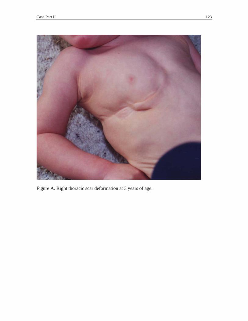
Case Part II 123
Figure A. Right thoracic scar deformation at 3 years of age.
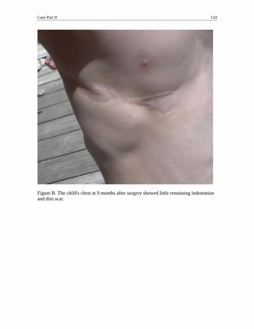
Case Part II 124
Figure B. The child's chest at 9 months after surgery showed little remaining indentation and thin scar.
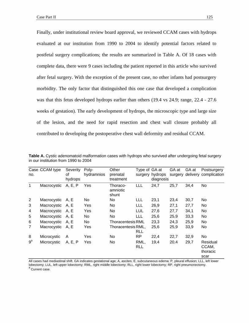
Case Part II
125
Finally, under institutional review board approval, we reviewed CCAM cases with hydrops
evaluated at our institution from 1990 to 2004 to identify potential factors related to
postfetal surgery complications; the results are summarized in Table A. Of 18 cases with
complete data, there were 9 cases including the patient reported in this article who survived
after fetal surgery. With the exception of the present case, no other infants had postsurgery
morbidity. The only factor that distinguished this one case that developed a complication
was that this fetus developed hydrops earlier than others (19.4 vs 24.9; range, 22.4 - 27.6
weeks of gestation). The early development of hydrops, the microscopic type and large size
of the lesion, and the need for rapid resection and chest wall closure probably all
contributed to developing the postoperative chest wall deformity and residual CCAM.
Table A. Cystic adenomatoid malformation cases with hydrops who survived after undergoing fetal surgery in our institution from 1990 to 2004
Case no.
CCAM type Severity of hydrops
Poly-hydramnios
Other prenatal treatment
Type of surgery
GA at hydrops diagnosis
GA at surgery
GA at delivery
Postsurgery complication
1 Macrocystic A, E, P Yes Thoraco-amniotic shunt
LLL 24,7 25,7 34,4 No
2 Macrocystic A, E No No LLL 23,1 23,4 30,7 No 3 Macrocystic A, E Yes No LLL 26,9 27,1 27,7 No 4 Macrocystic A, E Yes No LUL 27,6 27,7 34,1 No 5 Macrocystic A, E No No LLL 25,6 25,9 33,3 No 6 Macrocystic A, E No Thoracentesis RML 23,3 24,3 25,9 No 7 Macrocystic A, E Yes Thoracentesis RML,
RLL 25,6 25,9 33,9 No
8 Microcystic A Yes No RP 22,4 22,7 32,9 No 9a Microcystic A, E, P Yes No RML,
RLL 19,4 20,4 29,7 Residual
CCAM, thoracic scar
All cases had mediastinal shift. GA indicates gestational age; A, ascites; E, subcutaneous edema; P, pleural effusion; LLL, left lower lobectomy; LUL, left upper lobectomy; RML, right middle lobectomy; RLL, right lower lobectomy; RP, right pneumonectomy. a
Current case.
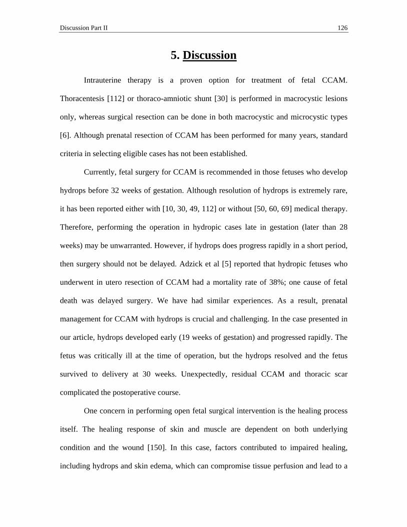
Discussion Part II 126
5. Discussion
Intrauterine therapy is a proven option for treatment of fetal CCAM.
Thoracentesis [112] or thoraco-amniotic shunt [30] is performed in macrocystic lesions
only, whereas surgical resection can be done in both macrocystic and microcystic types
[6]. Although prenatal resection of CCAM has been performed for many years, standard
criteria in selecting eligible cases has not been established.
Currently, fetal surgery for CCAM is recommended in those fetuses who develop
hydrops before 32 weeks of gestation. Although resolution of hydrops is extremely rare,
it has been reported either with [10, 30, 49, 112] or without [50, 60, 69] medical therapy.
Therefore, performing the operation in hydropic cases late in gestation (later than 28
weeks) may be unwarranted. However, if hydrops does progress rapidly in a short period,
then surgery should not be delayed. Adzick et al [5] reported that hydropic fetuses who
underwent in utero resection of CCAM had a mortality rate of 38%; one cause of fetal
death was delayed surgery. We have had similar experiences. As a result, prenatal
management for CCAM with hydrops is crucial and challenging. In the case presented in
our article, hydrops developed early (19 weeks of gestation) and progressed rapidly. The
fetus was critically ill at the time of operation, but the hydrops resolved and the fetus
survived to delivery at 30 weeks. Unexpectedly, residual CCAM and thoracic scar
complicated the postoperative course.
One concern in performing open fetal surgical intervention is the healing process
itself. The healing response of skin and muscle are dependent on both underlying
condition and the wound [150]. In this case, factors contributed to impaired healing,
including hydrops and skin edema, which can compromise tissue perfusion and lead to a
Discussion Part II 127
prolonged inflammatory response to wounding [150]. In addition, in this case, the fetus
was in heart failure and unable to tolerate a prolonged operation. Thus, operative time
was shortened by closing the fetal chest very rapidly with all-layer suture. We postulate
that both tissue edema and suturing technique contributed to abnormal healing that
resulted in chest wall deformity. Another factor that may have contributed to the chest
wall deformity is the surgical decompression of the thoracic space from removal of such
a large tumor.
Residual CCAM is a rare complication after resection of the mass. In a review of
the literature from 1980 to 2004, only 1 case of residual CCAM after segmentectomy in
early neonatal period was reported [128]. In elective operations after birth, it is not
difficult to define whether the residual parenchyma of the lobe surrounding the affected
segment is normal or abnormal [150]. However, in an emergency procedure performed
very rapidly (20 minutes) in such a sick 500-g, 20-week fetus, some residual CCAM from
the huge microcystic mass is left surrounding the pulmonary vessels at the root of the
resected lobe.
Because residual CCAM can occur after fetal lobectomy, we recommend that,
even if a lesion cannot be seen on postoperative US, the neonate should be followed up
closely with serial chest radiographs and/or CT scans during the first 6 to 12 months of
life. The length of follow-up will depend on the presence or absence of signs and
symptoms of respiratory disease. In this case, serial chest x-rays and CT scans showed an
abnormality in the right lower hemithorax. However, it was difficult to differentiate
between postoperative scarring and residual CCAM of right lower lung until the child
was 3 years old, when we thought the risk of reoperation was outweighed by the
Discussion Part II
128
significant respiration problems including recurrent pneumonia. We suggest that residual
CCAM should be suspected when an infant has chronic respiratory symptoms, along with
abnormal serial chest radiographs or CT scans.
References 129
REFERENCES
[1] Achiron R, Zalel Y, Lipitz S, Hegesh J, Mazkereth R, Kuint J, Jacobson J, Yagel
S. Fetal lung dysplasia: clinical outcome based on a new classification system.
Ultrasound Obstet Gynecol 2004; 24:127-133.
[2] Adzick NS. Fetal cystic adenomatoid malformation of the lung: diagnosis,
perinatal management, and outcome. Semin Thorac Cardiovasc Surg 1994; 6:247-
252.
[3] Adzick NS. Management of fetal lung lesions. Clin Perinatol 2003; 30:481-492.
[4] Adzick NS, Harrison MR. Management of the fetus with a cystic adenomatoid
malformation. World J Surg 1993; 17:342-349.
[5] Adzick NS, Harrison MR, Crombleholme TM, Flake AW, Howell LJ. Fetal lung
lesions: management and outcome. Am J Obstet Gynecol 1998; 179:884-889.
[6] Adzick NS, Harrison MR, Flake AW, Howell LJ, Golbus MS, Filly RA. Fetal
surgery for cystic adenomatoid malformation of the lung. J Pediatr Surg 1993;
28:806-812.
[7] Adzick NS, Harrison MR, Glick PL, Golbus MS, Anderson RL, Mahony BS,
Callen PW, Hirsch JH, Luthy DA, Filly RA, et al. Fetal cystic adenomatoid
malformation: prenatal diagnosis and natural history. J Pediatr Surg 1985; 20:483-
488.
References 130
[8] Albanese CT, Sydorak RM, Tsao K, Lee H. Thoracoscopic lobectomy for
prenatally diagnosed lung lesions. J Pediatr Surg 2003; 38:553-555.
[9] Anandakumar C, Biswas A, Chua TM, Choolani M, Chia D, Wong YC, Gole L.
Direct intrauterine fetal therapy in a case of bronchopulmonary sequestration
associated with non-immune hydrops fetalis. Ultrasound Obstet Gynecol 1999;
13:263-265.
[10] Aziz D, Langer JC, Tuuha SE, Ryan G, Ein SH, Kim PC. Perinatally diagnosed
asymptomatic congenital cystic adenomatoid malformation: to resect or not? J
Pediatr Surg 2004; 39:329-334.
[11] Bagolan P, Nahom A, Giorlandino C, Trucchi A, Bilancioni E, Inserra A,
Gambuzza G, Spina V. Cystic adenomatoid malformation of the lung: clinical
evolution and management. Eur J Pediatr 1999; 158:879-882.
[12] Barret J, Chitayat D, Sermer M, Amankwah K, Morrow R, Toi A, Ryan G. The
prognostic factors in the prenatal diagnosis of the echogenic fetal lung. Prenat
Diagn 1995; 15:849-853.
[13] Becmeur F, Horta-Geraud P, Donato L, Sauvage P. Pulmonary sequestrations:
prenatal ultrasound diagnosis, treatment, and outcome. J Pediatr Surg 1998;
33:492-496.
References 131
[14] Benjamin DR, Cahill JL. Bronchioloalveolar carcinoma of the lung and
congenital cystic adenomatoid malformation. Am J Clin Pathol 1991; 95:889-892.
[15] Blau H, Barak A, Karmazyn B, Mussaffi H, Ben Ari J, Schoenfeld T, Aviram M,
Vinograd Y, Lotem Y, Meizner I. Postnatal management of resolving fetal lung
lesions. Pediatrics 2002; 109:105-108.
[16] Bonnard A, Malbezin S, Ferkdadji L, Luton D, Aigrain Y, de Lagauise P.
Pulmonary sequestration children: is the thoracoscopic approach a good option?
Surg Endosc 2004; 18:1364-1367.
[17] Brace RA. Effects of outflow pressure on fetal lymph flow. Am J Obstet Gynecol
1989; 160:494-497.
[18] Bromley B, Parad R, Estroff JA, Benacerraf BR. Fetal lung masses: prenatal
course and outcome. J Ultrasound Med 1995; 14:927-936; quiz 1378.
[19] Bruner JP, Jarnagin BK, Reinisch L. Percutaneous laser ablation of fetal
congenital cystic adenomatoid malformation: too little, too late? Fetal Diagn Ther
2000; 15:359-363.
[20] Budorick NE, Pretorius DH, Leopold GR, Stamm ER. Spontaneous improvement
of intrathoracic masses diagnosed in utero. J Ultrasound Med 1992; 11:653-662.
References 132
[21] Bunduki V, Ruano R, da Silva MM, Miguelez J, Miyadahira S, Maksoud JG,
Zugaib M. Prognostic factors associated with congenital cystic adenomatoid
malformation of the lung. Prenat Diagn 2000; 20:459-464.
[22] Busing KA, Kilian AK, Schaible T, Neff KW. [Fetal magnetic resonance
imaging. Diagnostics in cases of congenital cystadenomatoid malformation of the
lung (CCAM)]. Radiologe 2006; 46:133-138.
[23] Cacciari A, Ceccarelli PL, Pilu GL, Bianchini MA, Mordenti M, Gabrielli S,
Milano V, Zanetti G, Pigna A, Gentili A. A series of 17 cases of congenital cystic
adenomatoid malformation of the lung: management and outcome. Eur J Pediatr
Surg 1997; 7:84-89.
[24] Callen PW. Ultrasonography in obstetrics and gynecology. Philadelphia:
Saunders, 2000.
[25] Calvert JK, Boyd PA, Chamberlain PC, Syed S, Lakhoo K. Outcome of
antenatally suspected congenital cystic adenomatoid malformation of the lung: 10
years' experience 1991-2001. Arch Dis Child Fetal Neonatal Ed 2006; 91:F26-28.
[26] Calvert JK, Lakhoo K. Antenatally suspected congenital cystic adenomatoid
malformation of the lung: postnatal investigation and timing of surgery. J Pediatr
Surg 2007; 42:411-414.
[27] Cardwell MS. Bilateral renal agenesis: clinical implications. South Med J 1988;
81:327-328.
References 133
[28] Cha I, Adzick NS, Harrison MR, Finkbeiner WE. Fetal congenital cystic
adenomatoid malformations of the lung: a clinicopathologic study of eleven cases.
Am J Surg Pathol 1997; 21:537-544.
[29] Chow PC, Lee SL, Tang MH, Chan KL, Lee CP, Lam BC, Tsoi NS. Management
and outcome of antenatally diagnosed congenital cystic adenomatoid
malformation of the lung. Hong Kong Med J 2007; 13:31-9.
[30] Clark SL, Vitale DJ, Minton SD, Stoddard RA, Sabey PL. Successful fetal
therapy for cystic adenomatoid malformation associated with second-trimester
hydrops. Am J Obstet Gynecol 1987; 157:294-295.
[31] Congregado M, Loscertales J, Giron-Arjona JC, Jimenez-Merchan R, Arroyo-
Tristan A, Gonzalez Campora R. [Video-assisted thoracoscopic surgery in 3 cases
of adult cystic adenomatoid malformation]. Arch Bronconeumol 2004; 40:236-
239.
[32] Corbett HJ, Humphrey GM. Pulmonary sequestration. Paediatr Respir Rev 2004;
5:59-68.
[33] Cortes RA, Keller RL, Townsend T, Harrison MR, Farmer DL, Lee H, Piecuch
RE, Leonard CH, Hetherton M, Bisgaard R, Nobuhara KK. Survival of severe
congenital diaphragmatic hernia has morbid consequences. J Pediatr Surg 2005;
40:36-45; discussion 45-46.
References 134
[34] Crombleholme TM, Coleman B, Hedrick H, Liechty K, Howell L, Flake AW,
Johnson M, Adzick NS. Cystic adenomatoid malformation volume ratio predicts
outcome in prenatally diagnosed cystic adenomatoid malformation of the lung. J
Pediatr Surg 2002; 37:331-338.
[35] Curros F, Chigot V, Emond S, Sayegh N, Revillon Y, Scheinman P, Lebourgeois
M, Brunelle F. Role of embolisation in the treatment of bronchopulmonary
sequestration. Pediatr Radiol 2000; 30:769-773.
[36] da Silva OP, Ramanan R, Romano W, Bocking A, Evans M. Nonimmune hydrops
fetalis, pulmonary sequestration, and favorable outcome. Obstet Gynecol 1996;
88:681-683.
[37] d'Agostino S, Bonoldi E, Dante S, Meli S, Cappellari F, Musi L. Embryonal
rhabdomyosarcoma of the lung arising in cystic adenomatoid malformation: case
report and review of the literature. J Pediatr Surg 1997; 32:1381-1383.
[38] Danielson PD, Sherman NJ. Laparoscopic removal of an abdominal extralobar
pulmonary sequestration. J Pediatr Surg 2001; 36:1653-1655.
[39] Davenport M, Warne SA, Cacciaguerra S, Patel S, Greenough A, Nicolaides K.
Current outcome of antenally diagnosed cystic lung disease. J Pediatr Surg 2004;
39:549-556.
References 135
[40] de Lagausie P, Bonnard A, Berrebi D, Petit P, Dorgeret S, Guys JM. Video-
assisted thoracoscopic surgery for pulmonary sequestration in children. Ann
Thorac Surg 2005; 80:1266-1269.
[41] De Santis M, Masini L, Noia G, Cavaliere AF, Oliva N, Caruso A. Congenital
cystic adenomatoid malformation of the lung: antenatal ultrasound findings and
fetal-neonatal outcome. Fifteen years of experience. Fetal Diagn Ther 2000;
15:246-250.
[42] Deguchi E, Furukawa T, Ono S, Aoi S, Kimura O, Iwai N. Intralobar pulmonary
sequestration diagnosed by MR angiography. Pediatr Surg Int 2005; 21:576-577.
[43] dell'Agnola C, Tadini B, Mosca F, Colnaghi M, Wesley J. Advantages of prenatal
diagnosis and early surgery for congenital cystic disease of the lung. J Perinat
Med 1996; 24:621-631.
[44] Deprest J, Jani J, Lewi L, Ochsenbein-Kolble N, Cannie M, Done E, Roubliova
X, Van Mieghem T, Debeer A, Debuck F, Sbragia L, Toelen J, Devlieger R, Lewi
P, Van de Velde M. Fetoscopic surgery: encouraged by clinical experience and
boosted by instrument innovation. Semin Fetal Neonatal Med 2006; 11:398-412.
[45] Domizio P, Liesner RJ, Dicks-Mireaux C, Risdon RA. Malignant mesenchymoma
associated with a congenital lung cyst in a child: case report and review of the
literature. Pediatr Pathol 1990; 10:785-797.
References 136
[46] Dommergues M, Louis-Sylvestre C, Mandelbrot L, Aubry MC, Revillon Y,
Jarreau PH, Dumez Y. Congenital adenomatoid malformation of the lung: when is
active fetal therapy indicated? Am J Obstet Gynecol 1997; 177:953-958.
[47] Dumez Y, Mandelbrot L, Radunovic N, Revillon Y, Dommergues M, Aubry MC,
Aubry JP, Narcy F, Sonigo P. Prenatal management of congenital cystic
adenomatoid malformation of the lung. J Pediatr Surg 1993; 28:36-41.
[48] Duncombe GJ, Dickinson JE, Kikiros CS. Prenatal diagnosis and management of
congenital cystic adenomatoid malformation of the lung. Am J Obstet Gynecol
2002; 187:950-954.
[49] Entezami M, Halis G, Waldschmidt J, Opri F, Runkel S. Congenital cystic
adenomatoid malformation of the lung and fetal hydrops--a case with favourable
outcome. Eur J Obstet Gynecol Reprod Biol 1998; 79:99-101.
[50] Etches PC, Tierney AJ, Demianczuk NN. Successful outcome in a case of cystic
adenomatoid malformation of the lung complicated by fetal hydrops, using
extracorporeal membrane oxygenation. Fetal Diagn Ther 1994; 9:88-91.
[51] Evans MG. Hydrops fetalis and pulmonary sequestration. J Pediatr Surg 1996;
31:761-764.
[52] Federici S, Domenichelli V, Tani G, Sciutti R, Burnelli R, Zanetti G, Domini R.
Pleuropulmonary blastoma in congenital cystic adenomatoid malformation: report
of a case. Eur J Pediatr Surg 2001; 11:196-199.

References 137
[53] Fortunato S, S. L, Dantrell J, Ismael S. Intrauterine laser ablation of a fetal cystic
adenomatoid malformation with hydrops: the application of minimally invasive
surgical techniques to fetal surgery. Am J Obstet Gynecol 1997; 177:S84.
[54] Frenckner B, Freyschuss U. Pulmonary function after lobectomy for congenital
lobar emphysema and congenital cystic adenomatoid malformation. A follow-up
study. Scand J Thorac Cardiovasc Surg 1982; 16:293-298.
[55] Garrett WJ, Kossoff G, Lawrence R. Gray scale echography in the diagnosis of
hydrops due to fetal lung tumor. J Clin Ultrasound 1975; 3:45-50.
[56] Glaves J, Baker JL. Spontaneous resolution of maternal hydramnios in congenital
cystic adenomatoid malformation of the lung. Antenatal ultrasound report. Br J
Obstet Gynaecol 1983; 90:1065-1068.
[57] Golaszewski T, Bettelheim D, Eppel W, Deutinger J, Bernaschek G. Cystic
adenomatoid malformation of the lung: prenatal diagnosis, prognostic factors and
fetal outcome. Gynecol Obstet Invest 1998; 46:241-246.
[58] Goldstein RB. A practical approach to fetal chest masses. Ultrasound Q 2006;
22:177-194.
[59] Gornall AS, Budd JL, Draper ES, Konje JC, Kurinczuk JJ. Congenital cystic
adenomatoid malformation: accuracy of prenatal diagnosis, prevalence and
outcome in a general population. Prenat Diagn 2003; 23:997-1002.
References 138
[60] Graham D, Winn K, Dex W, Sanders RC. Prenatal diagnosis of cystic
adenomatoid malformation of the lung. J Ultrasound Med 1982; 1:9-12.
[61] Granata C, Gambini C, Balducci T, Toma P, Michelazzi A, Conte M, Jasonni V.
Bronchioloalveolar carcinoma arising in congenital cystic adenomatoid
malformation in a child: a case report and review on malignancies originating in
congenital cystic adenomatoid malformation. Pediatr Pulmonol 1998; 25:62-66.
[62] Grethel EJ, Wagner AJ, Clifton MS, Cortes RA, Farmer DL, Harrison MR,
Nobuhara KK, Lee H. Fetal intervention for mass lesions and hydrops improves
outcome: a 15-year experience. J Pediatr Surg 2007; 42:117-123.
[63] Halkic N, Cuenoud PF, Corthesy ME, Ksontini R, Boumghar M. Pulmonary
sequestration: a review of 26 cases. Eur J Cardiothorac Surg 1998; 14:127-133.
[64] Haller A. In discussion of Neilson IR, Russo P, Laberge J-M, et al: Congenital
adenomatoid malformation of the lung. Current management and prognosis. J
Pediatr Surg 1991; 26:975-981.
[65] Harrison MR, Adzick NS. Open fetal surgery techniques. In: Harrison MR, Evans
MI, Adzick NS, Holzgreve W, editors. The unborn patient. The art and science of
fetal therapy. Philadelphia: W. B. Saunders 2001; 3rd edition:247-258.
[66] Harrison MR, Adzick NS, Jennings RW, Duncan BW, Rosen MA, Filly RA,
Goldberg JD, deLorimier AA, Golbus MS. Antenatal intervention for congenital
cystic adenomatoid malformation. Lancet 1990; 336:965-967.
References 139
[67] Hasiotou M, Polyviou P, Strantzia CM, Pourtsidis A, Stinios I. Pleuropulmonary
blastoma in the area of a previously diagnosed congenital lung cyst: report of two
cases. Acta Radiol 2004; 45:289-292.
[68] Hedrick HL, Flake AW, Crombleholme TM, Howell LJ, Johnson MP, Wilson
RD, Adzick NS. The ex utero intrapartum therapy procedure for high-risk fetal
lung lesions. J Pediatr Surg 2005; 40:1038-1043; discussion 1044.
[69] Higby K, Melendez BA, Heiman HS. Spontaneous resolution of nonimmune
hydrops in a fetus with a cystic adenomatoid malformation. J Perinatol 1998;
18:308-310.
[70] Hirose S, Farmer DL, Lee H, Nobuhara KK, Harrison MR. The ex utero
intrapartum treatment procedure: looking back at the EXIT. J Pediatr Surg 2004;
39:375-80; discussion 375-80.
[71] Hoffman JI, Kaplan S. The incidence of congenital heart disease. J Am Coll
Cardiol 2002; 39:1890-1900.
[72] Howell DC. Statistical methods for psychology. Belmont, CA: Duxbury Press,
2002.
[73] Hsieh CC, Chao AS, Chang YL, Kuo DM, Hsieh TT, Hung HT. Outcome of
congenital cystic adenomatoid malformation of the lung after antenatal diagnosis.
Int J Gynaecol Obstet 2005; 89:99-102.
References 140
[74] Hubbard AM, Adzick NS, Crombleholme TM, Coleman BG, Howell LJ,
Haselgrove JC, Mahboubi S. Congenital chest lesions: diagnosis and
characterization with prenatal MR imaging. Radiology 1999; 212:43-48.
[75] Ierullo AM, Ganapathy R, Crowley S, Craxford L, Bhide A, Thilaganathan B.
Neonatal outcome of antenatally diagnosed congenital cystic adenomatoid
malformations. Ultrasound Obstet Gynecol 2005; 26:150-153.
[76] Illanes S, Hunter A, Evans M, Cusick E, Soothill P. Prenatal diagnosis of
echogenic lung: evolution and outcome. Ultrasound Obstet Gynecol 2005;
26:145-149.
[77] Jaillard SM, Pierrat V, Dubois A, Truffert P, Lequien P, Wurtz AJ, Storme L.
Outcome at 2 years of infants with congenital diaphragmatic hernia: a population-
based study. Ann Thorac Surg 2003; 75:250-256.
[78] Jesch NK, Leonhardt J, Sumpelmann R, Gluer S, Nustede R, Ure BM.
Thoracoscopic resection of intra- and extralobar pulmonary sequestration in the
first 3 months of life. J Pediatr Surg 2005; 40:1404-1406.
[79] Kähler C, Schulze E, Eichhorn KH, Seewald HJ. [Fetal hyperechogenic and cystic
pulmonary masses: sonographic findings, antenatal management and outcome of
12 cases]. Z Geburtshilfe Neonatol 2002; 206:205-210.
References 141
[80] Kamata S, Ishikawa S, Usui N, Sawai T, Kitayama Y, Nose K, Okuyama H,
Imura K, Okada A. Clinical significance of the lung/thorax transverse-area ratio
in fetuses with cystic lung disease. Pediatr Surg Int 1999; 15:470-474.
[81] Kamata S, Usui N, Kamiyama M, Nose K, Sawai T, Fukuzawa M. Long-term
outcome in patients with prenatally diagnosed cystic lung disease: special
reference to ventilation and perfusion scan in the affected lung. J Pediatr Surg
2006; 41:2023-2027.
[82] Kamata S, Usui N, Kamiyama M, Tazuke Y, Nose K, Sawai T, Fukuzawa M.
Long-term follow-up of patients with high-risk congenital diaphragmatic hernia. J
Pediatr Surg 2005; 40:1833-1838.
[83] Kim YT, Kim JS, Park JD, Kang CH, Sung SW, Kim JH. Treatment of congenital
cystic adenomatoid malformation-does resection in the early postnatal period
increase surgical risk? Eur J Cardiothorac Surg 2005; 27:658-661.
[84] Koontz CS, Oliva V, Gow KW, Wulkan ML. Video-assisted thoracoscopic
surgical excision of cystic lung disease in children. J Pediatr Surg 2005; 40:835-
837.
[85] Kouchi K, Yoshida H, Matsunaga T, Ohtsuka Y, Kuroda H, Hishiki T, Satou Y,
Terui K, Mitsunaga T, Ohnuma N. Intralobar bronchopulmonary sequestration
evaluated by contrast-enhanced three-dimensional MR angiography. Pediatr
Radiol 2000; 30:774-775.
References 142
[86] Krous HF, Harper PE, Perlman M. Congenital cystic adenomatoid malformation
in bilateral renal agenesis. Its mitigation of Potter's syndrome. Arch Pathol Lab
Med 1980; 104:368-370.
[87] Kubin K, Hormann M, Riccabona M, Wiesbauer P, Puig S. [Benign and
malignant pulmonary tumors in childhood]. Radiologe 2003; 43:1095-1102.
[88] Kuller JA, Yankowitz J, Goldberg JD, Harrison MR, Adzick NS, Filly RA, Callen
PW, Golbus MS. Outcome of antenatally diagnosed cystic adenomatoid
malformations. Am J Obstet Gynecol 1992; 167:1038-1041.
[89] Kunisaki SM, Barnewolt CE, Estroff JA, Ward VL, Nemes LP, Fauza DO,
Jennings RW. Large fetal congenital cystic adenomatoid malformations: growth
trends and patient survival. J Pediatr Surg 2007; 42:404-410.
[90] Kuroda T, Morikawa N, Kitano Y, Sago H, Hayashi S, Honna T, Saeki M.
Clinicopathologic assessment of prenatally diagnosed lung diseases. J Pediatr
Surg 2006; 41:2028-2031.
[91] Laberge JM, Flageole H, Pugash D, Khalife S, Blair G, Filiatrault D, Russo P,
Lees G, Wilson RD. Outcome of the prenatally diagnosed congenital cystic
adenomatoid lung malformation: a Canadian experience. Fetal Diagn Ther 2001;
16:178-186.
[92] Laberge JM, Puligandla P, Flageole H. Asymptomatic congenital lung
malformations. Semin Pediatr Surg 2005; 14:16-33.
References 143
[93] Lacy DE, Shaw NJ, Pilling DW, Walkinshaw S. Outcome of congenital lung
abnormalities detected antenatally. Acta Paediatr 1999; 88:454-458.
[94] Lee HJ, Song MJ, Cho JY, Lee YH. Echogenic fetal lung masses: comparison of
prenatal sonographic and postnatal CT findings. J Clin Ultrasound 2003; 31:419-
424.
[95] Lee KH, Sung KB, Yoon HK, Ko GY, Yoon CH, Goo HW, Kim EA, Kim KS, Pi
SY. Transcatheter arterial embolization of pulmonary sequestration in neonates:
long-term follow-up results. J Vasc Interv Radiol 2003; 14:363-367.
[96] Lehnhardt S, Winterer JT, Uhrmeister P, Herget G, Laubenberger J. Pulmonary
sequestration: demonstration of blood supply with 2D and 3D MR angiography.
Eur J Radiol 2002; 44:28-32.
[97] Leung WC, Ngai C, Lam TPW, Chan KL, Lao TT, Tang MHY. Unexpected
intrauterine death following resolution of hydrops fetalis after betamethasone
treatment in a fetus with a large cystic adenomatoid malformation of the lung.
Ultrasound in Obstetrics and Gynecology 2005; 25:610-612.
[98] Levi S. Mass screening for fetal malformations: the Eurofetus study. Ultrasound
Obstet Gynecol 2003; 22:555-558.
[99] Lopoo JB, Goldstein RB, Lipshutz GS, Goldberg JD, Harrison MR, Albanese CT.
Fetal pulmonary sequestration: a favorable congenital lung lesion. Obstet Gynecol
1999; 94:567-571.
References 144
[100] MacGillivray TE, Harrison MR, Goldstein RB, Adzick NS. Disappearing fetal
lung lesions. J Pediatr Surg 1993; 28:1321-1324; discussion 1324-1325.
[101] McCullagh M, MacConnachie I, Garvie D, Dykes E. Accuracy of prenatal
diagnosis of congenital cystic adenomatoid malformation. Arch Dis Child 1994;
71:F111-F113.
[102] Meagher SE, Fisk NM, Harvey JG, Watson GF, Boogert A. Disappearing lung
echogenicity in fetal bronchopulmonary malformations: a reassuring sign? Prenat
Diagn 1993; 13:495-501.
[103] Miller JA, Corteville JE, Langer JC. Congenital cystic adenomatoid malformation
in the fetus: natural history and predictors of outcome. J Pediatr Surg 1996;
31:805-808.
[104] Monni G, Paladini D, Ibba RM, Teodoro A, Zoppi MA, Lamberti A, Floris M,
Putzolu M, Martinelli P. Prenatal ultrasound diagnosis of congenital cystic
adenomatoid malformation of the lung: a report of 26 cases and review of the
literature. Ultrasound Obstet Gynecol 2000; 16:159-162.
[105] Muratore CS, Kharasch V, Lund DP, Sheils C, Friedman S, Brown C, Utter S,
Jaksic T, Wilson JM. Pulmonary morbidity in 100 survivors of congenital
diaphragmatic hernia monitored in a multidisciplinary clinic. J Pediatr Surg 2001;
36:133-140.
References 145
[106] Murphy JJ, Blair GK, Fraser GC, Ashmore PG, LeBlanc JG, Sett SS, Rogers P,
Magee JF, Taylor GP, Dimmick J. Rhabdomyosarcoma arising within congenital
pulmonary cysts: report of three cases. J Pediatr Surg 1992; 27:1364-1367.
[107] Neilson IR, Russo P, Laberge JM, Filiatrault D, Nguyen LT, Collin PP, Guttman
FM. Congenital adenomatoid malformation of the lung: current management and
prognosis. J Pediatr Surg 1991; 26:975-980; discussion 980-981.
[108] Nicolaides KH, Azar GB. Thoraco-amniotic shunting. Fetal Diagn Ther 1990;
5:153-164.
[109] Nicolaides KH, Blott M, Greenough A. Chronic drainage of fetal pulmonary cyst.
Lancet 1987; 1:618.
[110] Nicolini U, Cerri V, Groli C, Poblete A, Mauro F. A new approach to prenatal
treatment of extralobar pulmonary sequestration. Prenat Diagn 2000; 20:758-760.
[111] Nobuhara KK, Lund DP, Mitchell J, Kharasch V, Wilson JM. Long-term outlook
for survivors of congenital diaphragmatic hernia. Clin Perinatol 1996; 23:873-
887.
[112] Nugent CE, Hayashi RH, Rubin J. Prenatal treatment of type I congenital cystic
adenomatoid malformation by intrauterine fetal thoracentesis. J Clin Ultrasound
1989; 17:675-677.
References 146
[113] Ozcan C, Celik A, Ural Z, Veral A, Kandiloglu G, Balik E. Primary pulmonary
rhabdomyosarcoma arising within cystic adenomatoid malformation: a case report
and review of the literature. J Pediatr Surg 2001; 36:1062-1065.
[114] Papagiannopoulos K, Hughes S, Nicholson AG, Goldstraw P. Cystic lung lesions
in the pediatric and adult population: surgical experience at the Brompton
Hospital. Ann Thorac Surg 2002; 73:1594-1598.
[115] Parker MJ, Budd JL, Draper ES, Young ID. Trisomy 13 and trisomy 18 in a
defined population: epidemiological, genetic and prenatal observations. Prenat
Diagn 2003; 23:856-860.
[116] Phaloprakarn* C, Pott Bärtsch* EM, Harrison MR. Residual congenital cystic
adenomatoid malformation and thoracic scar deformation after fetal surgery: a
case report. J Pediatr Surg 2006; 41:e11-e14 (*equal first authors).
[117] Pinter A, Kalman A, Karsza L, Verebely T, Szemledy F. Long-term outcome of
congenital cystic adenomatoid malformation. Pediatr Surg Int 1999; 15:332-335.
[118] Pott Bartsch EM, Paek BW, Yoshizawa J, Goldstein RB, Ferrell LD, Coakley FV,
Harrison MR, Albanese CT. Giant fetal hepatic hemangioma. Case report and
literature review. Fetal Diagn Ther 2003; 18:59-64.
[119] Pumberger W, Hormann M, Deutinger J, Bernaschek G, Bistricky E, Horcher E.
Longitudinal observation of antenatally detected congenital lung malformations
References 147
(CLM): natural history, clinical outcome and long-term follow-up. Eur J
Cardiothorac Surg 2003; 24:703-711.
[120] Quinn TM, Hubbard AM, Adzick NS. Prenatal magnetic resonance imaging
enhances fetal diagnosis. J Pediatr Surg 1998; 33:553-558.
[121] Revillon Y, Jan D, Plattner V, Sonigo P, Dommergues M, Mandelbrot L, Dumez
Y, Nihoul-Fekete C. Congenital cystic adenomatoid malformation of the lung:
prenatal management and prognosis. J Pediatr Surg 1993; 28:1009-1011.
[122] Rice HE, Estes JM, Hedrick MH, Bealer JF, Harrison MR, Adzick NS.
Congenital cystic adenomatoid malformation: a sheep model of fetal hydrops. J
Pediatr Surg 1994; 29:692-696.
[123] Roggin KK, Breuer CK, Carr SR, Hansen K, Kurkchubasche AG, Wesselhoeft
CW, Jr., Tracy TF, Jr., Luks FI. The unpredictable character of congenital cystic
lung lesions. J Pediatr Surg 2000; 35:801-805.
[124] Romero R, Chervenak FA, Kotzen J, Berkowitz RL, Hobbins JC. Antenatal
sonographic findings of extralobar pulmonary sequestration. J Ultrasound Med
1982; 1:131-132.
[125] Rosado-de-Christenson ML, Stocker JT. Congenital cystic adenomatoid
malformation. Radiographics 1991; 11:865-886.
References 148
[126] Rothenberg SS. Thoracoscopic lung resection in children. J Pediatr Surg 2000;
35:271-274; discussion 274-275.
[127] Sakala EP, Perrott WS, Grube GL. Sonographic characteristics of antenatally
diagnosed extralobar pulmonary sequestration and congenital cystic adenomatoid
malformation. Obstet Gynecol Surv 1994; 49:647-655.
[128] Sapin E, Lejeune VV, Barbet JP, Carricaburu E, Lewin F, Baron JM, Barbotin-
Larrieu F, Helardot PG. Congenital adenomatoid disease of the lung: prenatal
diagnosis and perinatal management. Pediatr Surg Int 1997; 12:126-129.
[129] Sauvat F, Michel JL, Benachi A, Emond S, Revillon Y. Management of
asymptomatic neonatal cystic adenomatoid malformations. J Pediatr Surg 2003;
38:548-552.
[130] Savic B, Birtel FJ, Tholen W, Funke HD, Knoche R. Lung sequestration: report of
seven cases and review of 540 published cases. Thorax 1979; 34:96-101.
[131] Shanmugam G, MacArthur K, Pollock JC. Congenital lung malformations--
antenatal and postnatal evaluation and management. Eur J Cardiothorac Surg
2005; 27:45-52.
[132] Stocker JT, Madewell JE, Drake RM. Congenital cystic adenomatoid
malformation of the lung. Classification and morphologic spectrum. Hum Pathol
1977; 8:155-171.
References 149
[133] Stöver B, Mau H, Scheer I, Bassir C, Chaoui R, Henrich W, Schwabe M, Wauer
R. [Congenital cystic lung malformations]. Rofo 2006; 178:432-437.
[134] Tagge EP, Mulvihill D, Chandler JC, Richardson M, Uflacker R, Othersen HD.
Childhood pleuropulmonary blastoma: caution against nonoperative management
of congenital lung cysts. J Pediatr Surg 1996; 31:187-189; discussion 190.
[135] Tanaka T, Ueda K, Sakano H, Hayashi M, Li TS, Zempo N. Video-assisted
thoracoscopic surgery for intralobar pulmonary sequestration. Surgery 2003;
133:216-218.
[136] Tawil MI, Pilling DW. Congenital cystic adenomatoid malformation: is there a
difference between the antenatally and postnatally diagnosed cases? Pediatr
Radiol 2005; 35:79-84.
[137] Teodoro A, Lamberti A, Liguori M, Tartaglione A, Tremolaterra F, Paladini D.
[Cystic adenomatoid malformation of the lung at the fetal stage. Ultrasonographic
diagnosis and counseling]. Minerva Ginecol 1999; 51:213-217.
[138] Thorpe-Beeston JG, Nicolaides KH. Cystic adenomatoid malformation of the
lung: prenatal diagnosis and outcome. Prenat Diagn 1994; 14:677-688.
[139] Thurlbeck WM. Postnatal human lung growth. Thorax 1982; 37:564-571.
References 150
[140] Truitt AK, Carr SR, Cassese J, Kurkchubasche AG, Tracy TF, Jr., Luks FI.
Perinatal management of congenital cystic lung lesions in the age of minimally
invasive surgery. J Pediatr Surg 2006; 41:893-896.
[141] Tsao K, Hawgood S, Vu L, Hirose S, Sydorak R, Albanese CT, Farmer DL,
Harrison MR, Lee H. Resolution of hydrops fetalis in congenital cystic
adenomatoid malformation after prenatal steroid therapy. J Pediatr Surg 2003;
38:508-510.
[142] Ueda K, Gruppo R, Unger F, Martin L, Bove K. Rhabdomyosarcoma of lung
arising in congenital cystic adenomatoid malformation. Cancer 1977; 40:383-388.
[143] Usui N, Kamata S, Sawai T, Kamiyama M, Okuyama H, Kubota A, Okada A.
Outcome predictors for infants with cystic lung disease. J Pediatr Surg 2004;
39:603-606.
[144] van Leeuwen K, Teitelbaum DH, Hirschl RB, Austin E, Adelman SH, Polley TZ,
Marshall KW, Coran AG, Nugent C. Prenatal diagnosis of congenital cystic
adenomatoid malformation and its postnatal presentation, surgical indications, and
natural history. J Pediatr Surg 1999; 34:794-799; incl. discussion 798-799.
[145] Vu L, Tsao K, Lee H, Nobuhara K, Farmer D, Harrison M, Goldstein RB.
Characteristics of congenital cystic adenomatoid malformations associated with
nonimmune hydrops and outcome. J Pediatr Surg 2007; 42:1351-6.
References 151
[146] Ward SJ. Surgery for congenital cystic adenomatoid malformation of the lung. N
Engl J Med 1996; 335:1689-1690.
[147] Waszak P, Claris O, Lapillonne A, Picaud JC, Basson E, Chappuis JP, Salle BL.
Cystic adenomatoid malformation of the lung: neonatal management of 21 cases.
Pediatr Surg Int 1999; 15:326-331.
[148] Watine O, Mensier E, Delecluse P, Ribet M. Pulmonary sequestration treated by
video-assisted thoracoscopic. Eur J Cardiothorac Surg 1994; 8:155-156.
[149] Weiner C, Varner M, Pringle K, Hein H, Williamson R, Smith WL. Antenatal
diagnosis and palliative treatment of nonimmune hydrops fetalis secondary to
pulmonary extralobar sequestration. Obstet Gynecol 1986; 68:275-280.
[150] Williams DT, Harding K. Healing responses of skin and muscle in critical illness.
Crit Care Med 2003; 31:S547-S557.
[151] Wilson RD, Baird PA. Renal agenesis in British Columbia. Am J Med Genet
1985; 21:153-169.
[152] Wilson RD, Hedrick HL, Liechty KW, Flake AW, Johnson MP, Bebbington M,
Adzick NS. Cystic adenomatoid malformation of the lung: review of genetics,
prenatal diagnosis, and in utero treatment. Am J Med Genet A 2006; 140:151-
155.

References
152
[153] Xu H, Jiang D, Kong X, Xiong Y, Liu D, Liu X, Deng X. Pulmonary
sequestration: three dimensional dynamic contrast-enhanced MR angiography and
MRI. J Tongji Med Univ 2001; 21:345-348.
Appendix 153
APPENDIX
(16 pages)
1.1 Parents recruitment letter
(2 pages)
1.2 Parents questionnaire
(4 pages)
1.3 Parents telephone script
(2 pages)
2.1 First Committee on Human Research (CHR) approval letter
(1 page)
2.2 Second CHR approval letter
(1 page)
2.3 Cover letter of the annual renewal of the subcommittee reviewed research study
(3 pages)
2.4 Research protocol of the annual renewal of the subcommittee reviewed research
study
(3 pages)
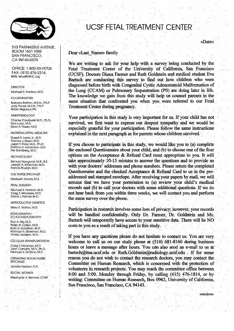
513 PARNASSUS AVENUE, ROOM 1601 HSW SAN FRANCISCO, CA 94143-0570
OFFICE: 1-800-RX-FETUS FAX: (415) 476-2314
WEB: [email protected]
DIRECTOR
Michael R. Harrison. MD.
COORDIi\JATORS
Barbara Bratton. MS.N" P.N.P. Jody Farrell, MS.N" P.N.P Robln Bisgaard, RN
ANESTHESIOLOGY
Charles Cauldwell, MD., Pt'lD. Errol Lobo, MD. Mark A. Rosen, MD.
MATERNALIFETAL MEDICINE
Russell K. Laros, Jr" MD. Thomas J. Musci. MD. Julian T. Parer, MD" PhD. Patrlcia A. Robertson, MD. Per Sandberg, MD.
NEONATOLOGY
Samuel Hawgood,M.B" B.S. Joseph A. Kitterman, MD. Francis Poulain, MD.
O.B. NURSE.SPECIALIST
Maribeth Inturrisi, MS.
FETAL SURGERY·
Michael R. Harrison, MD. Cral.g T. Albanese, M.D. Diana L. Farmer, MD.
REPRODUCTIVE GENETICS
Mary E. Norton, MD.
SONOGRAPHY I ECHOCARDIOGRAPHY
Roy A. Filly, MD. Peter W. Callen, MD. Ruth B. Goldsfein, MD. Nor'man H. Silverman, MD. Vickie Feldstein, MD.
CELLULAR TRANSPLANTATION
Craig T. Albanese. MD. John Curnutte, MD" PhD. Michael R. Harrison, MD.
OPERATING ROOM NURSE SPECIALIST
Jackita Harrison, R.N.
SOCIAL WORKER
Stept'lanie A. Berrnan, LCSW
UCSF FETAL TREATMENT CENTER
«Date»
Dear «LasCName» family
Wearewriting to ask for your helpwith a surveybeing eondueted by the Fetal Treatment Center of the University of California, San Francisco (UCSF). Doctors Diana Farmer and Ruth Goldstein and medical student Eva Baitsch are conducting this survey. to find outhow ehildren who were' diagnosed beforebirth with Congenital Cystic Adenomatoid Malformation of theLung (CCAM) or Puhnonary Sequestration (PS) are doing later in life. Theknowledge wegain from this study, will help us counsel parents in the same situation that confronted you when you, were referred to our Fetal Treatment Center during pregnancy.
Y our participation in this study is very important for uso If your child has not survived, we firSt want to express our, deepest sympathy and we would be especially grateful for your participation .. Please follow the same instructions explained in the next paragraph as for parents whose ehildren survived.
If you ehooseto participate in this study, we would likeyou to (a) complete the enclosed Questionnaire about your ehild, and (b) to choose one of the four options on the Acceptance & Refusal eard most appropriate to you. It ,win take approximately 10-15 miriutes to answer the questions and to provide us with your doctors' addresses andphone numberS. Please return the completed Questionnaire and the checked' Acceptance, & Refusal'Card to us in the preaddressed and stamped envelope. After receivingyout papers by mail, we will assume that we have rour permission to (a) review your child's medical records and (b) to call your doctors with some additioiiaI questions. If we do not hear back from you within three weeks, we willcontact you and perform the same survey over the phone. .
Participationin research involves some 10ss ofprivacy;however, yout records wiUbe handled confidentially. Only Dr. Farmer, Dr. Goldstein and Ms. Bartsch will temporarily haveaccess to your sensitive data. There will be NO costs to you a:s a result oftakingpartin this study.
If you have any questions please do not hesitate to cOlltact uso Y ou are very welc9me to call us on our study phone at (510) 681-8160 durlng business hours or leave a message after hours.· You can also send an e-mail to us at [email protected] or Ruth.Gö[email protected] . If for some reason you do' not wish to contact the research doctors, you may. contact the Committee on Human Research, wruch is concemed with the proteCtion of volunteers in research projects. ,You may reach the committee office between 8:00 and 5:00. Monday through, Friday, by calling (415) 476-1814, or by writing: Committee on Human Research, Box 0962, University of California, San Francisco, San Francisco,CA 94143.
«randolID)
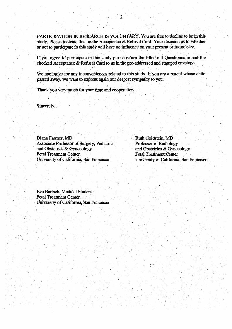
2
PARTICIPATION IN RESEARCH IS VOLUNTARY. You are free to decline to beinthis study.Please indicate this onthe Acceptan~ &Refusal Cani. YOll( decisionas to whether or not to participatein this study ~ll have no influence on your present or future care.
If you agree to participate in thisstudy please retum the filled-out. Questionnaire and the checked Acceptance & Refusal Card to us in the pre-addressed ~d stamped envelope ..
We apologize for any inconveni~ncesrelaWd to this study. Ifyou are·a parent whose child passed away, we wa:I;lt to express again our deepest sympathy.to you.
Thankyou very milch for your time and cooperation.
Sincerely,
. Diana Fanner, MD Associate Professor of Surgery, Pediatrics and Obstetrics & Gynecology Fetal Treatment Center University of Califomia, Sah Ftancisco
Eva Bartscb, Medical. Student Fetal Treatment Center University of Califomia, San Francisco
Ruth Goldstein, .MD Professor ofRadiology and Obstetrics & Gynecology Fetal Treatment Center University of Califoinia, San Francisco
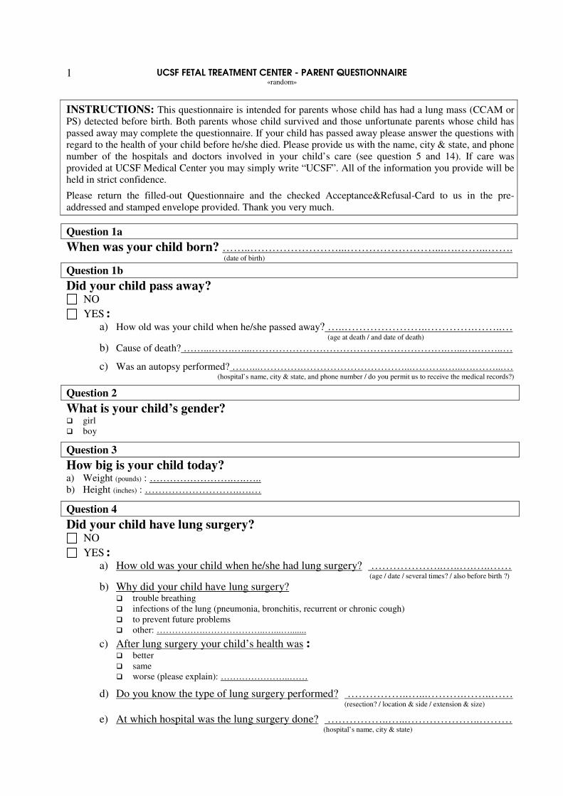
UCSF FETAL TREATMENT CENTER - PARENT QUESTIONNAIRE «random»
1
INSTRUCTIONS: This questionnaire is intended for parents whose child has had a lung mass (CCAM or PS) detected before birth. Both parents whose child survived and those unfortunate parents whose child has passed away may complete the questionnaire. If your child has passed away please answer the questions with regard to the health of your child before he/she died. Please provide us with the name, city & state, and phone number of the hospitals and doctors involved in your child’s care (see question 5 and 14). If care was provided at UCSF Medical Center you may simply write “UCSF”. All of the information you provide will be held in strict confidence.
Please return the filled-out Questionnaire and the checked Acceptance&Refusal-Card to us in the pre-addressed and stamped envelope provided. Thank you very much.
Question 1a
When was your child born? ……..……………………...……………………...….……...……. (date of birth)
Question 1b
Did your child pass away? NO
YES : a) How old was your child when he/she passed away? …..…………………..………….……..…
(age at death / and date of death)
b) Cause of death? ……...…….…...………………………………………………….…...….……..…
c) Was an autopsy performed? ……...………….…………………………...……….…...….……...… (hospital’s name, city & state, and phone number / do you permit us to receive the medical records?)
Question 2
What is your child’s gender? � girl � boy
Question 3
How big is your child today? a) Weight (pounds) : …………………….….….. b) Height (inches) : ……………………….….…
Question 4
Did your child have lung surgery? NO
YES : a) How old was your child when he/she had lung surgery? ………………..…..….…..……
(age / date / several times? / also before birth ?)
b) Why did your child have lung surgery? � trouble breathing � infections of the lung (pneumonia, bronchitis, recurrent or chronic cough) � to prevent future problems � other: …………….………………..…...….......
c) After lung surgery your child’s health was : � better � same � worse (please explain): …………………..……
d) Do you know the type of lung surgery performed? ……………..…...……….……..…… (resection? / location & side / extension & size)
e) At which hospital was the lung surgery done? ……………..…...………………..……… (hospital’s name, city & state)
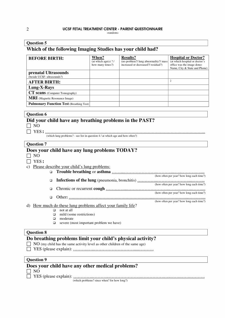
UCSF FETAL TREATMENT CENTER - PARENT QUESTIONNAIRE «random»
2
Question 5
Which of the following Imaging Studies has your child had?
BEFORE BIRTH: When? (at which age(s) ? / how many times?)
Results? (no problem?/ lung abnormality?/ mass increased or decreased?/ residual?)
Hospital or Doctor? (at which hospital or doctor’s office was the image done: Name, City & State and Phone)
prenatal Ultrasounds (beside UCSF- ultrasounds?)
AFTER BIRTH: )
Lung-X-Rays CT scans (Computer Tomography) MRI (Magnetic Resonance Image) Pulmonary Function Test (Breathing Test)
Question 6
Did your child have any breathing problems in the PAST? NO
YES : ……………………………………………………………………………………………………….. (which lung problems? - see list in question 4 / at which age and how often?)
Question 7
Does your child have any lung problems TODAY? NO
YES : c) Please describe your child’s lung problems:
� Trouble breathing or asthma ……...…………………..………....…..……….….... (how often per year? how long each time?)
� Infections of the lung (pneumonia, bronchitis) …………..…………………....……... (how often per year? how long each time?)
� Chronic or recurrent cough …………...………………………..…….…..…..…….... (how often per year? how long each time?)
� Other: ……………………………………………………...……………………...…….….... (how often per year? how long each time?)
d) How much do these lung problems affect your family life? � not at all � mild (some restrictions) � moderate � severe (most important problem we have)
Question 8
Do breathing problems limit your child’s physical activity? NO (my child has the same activity level as other children of the same age) YES (please explain): ……….…..……………………….....……….
Question 9
Does your child have any other medical problems? NO YES (please explain): …………..………..………………….…………….…..…….…….……….
(which problems? since when? for how long?)
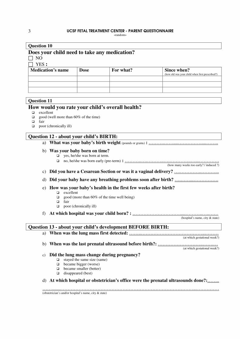
UCSF FETAL TREATMENT CENTER - PARENT QUESTIONNAIRE «random»
3
Question 10
Does your child need to take any medication? NO
YES : Medication’s name Dose For what? Since when?
(how old was your child when first prescribed?)
Question 11
How would you rate your child’s overall health? � excellent � good (well more than 60% of the time) � fair � poor (chronically ill)
Question 12 - about your child’s BIRTH: a) What was your baby’s birth weight (pounds or grams) : ……….……....……………....……..
b) Was your baby born on time? � yes, he/she was born at term.
� no, he/she was born early (pre-term) : ………..….………..….…………..…...…………. (how many weeks too early? / induced ?)
c) Did you have a Cesarean Section or was it a vaginal delivery? ….…………..………...
d) Did your baby have any breathing problems soon after birth? ….……...…………….
e) How was your baby’s health in the first few weeks after birth? � excellent � good (more than 60% of the time well being) � fair � poor (chronically ill)
f) At which hospital was your child born? : ………………………….....………..…..…… (hospital’s name, city & state)
Question 13 - about your child’s development BEFORE BIRTH: a) When was the lung mass first detected: ……..…………………………………..………
(at which gestational week?)
b) When was the last prenatal ultrasound before birth?: ………………………..…….… (at which gestational week?)
c) Did the lung mass change during pregnancy? � stayed the same size (same) � became bigger (worse) � became smaller (better) � disappeared (best)
d) At which hospital or obstetrician’s office were the prenatal ultrasounds done?:……..
……........…………………………………………………………………………..…………… (obstetrician’s and/or hospital’s name, city & state)
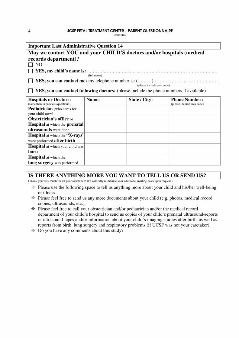
UCSF FETAL TREATMENT CENTER - PARENT QUESTIONNAIRE «random»
4
Important Last Administrative Question 14 May we contact YOU and your CHILD’S doctors and/or hospitals (medical records department)?
NO
YES, my child’s name is: ..……….……….………....……….………………………………..… (full name)
YES, you can contact me: my telephone number is: (………)……….………....……….…….… (please include area code)
YES, you can contact following doctors: (please include the phone numbers if available)
Hospitals or Doctors: (same than in previous questions ?)
Name: State / City: Phone Number: (please include area code)
Pediatrician (who cares for your child now)
Obstetrician’s office or Hospital at which the prenatal ultrasounds were done
Hospital at which the “X-rays”were preformed after birth
Hospital at which your child was
born
Hospital at which the lung surgery was performed
IS THERE ANYTHING MORE YOU WANT TO TELL US OR SEND US? (Thank you very much for all your assistance! We will fully reimburse your additional mailing costs upon request.)
� Please use the following space to tell us anything more about your child and his/her well-being or illness.
� Please feel free to send us any more documents about your child (e.g. photos, medical record copies, ultrasounds, etc.).
� Please feel free to call your obstetrician and/or pediatrician and/or the medical record department of your child’s hospital to send us copies of your child’s prenatal ultrasound-reports or ultrasound-tapes and/or information about your child’s imaging studies after birth, as well as reports from birth, lung surgery and respiratory problems (if UCSF was not your caretaker).
� Do you have any comments about this study?
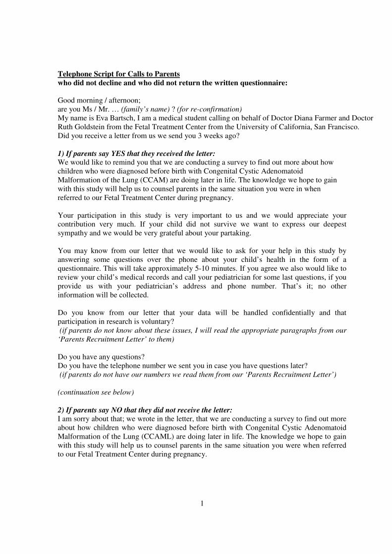
1
Telephone Script for Calls to Parents who did not decline and who did not return the written questionnaire: Good morning / afternoon; are you Ms / Mr. … (family’s name) ? (for re-confirmation) My name is Eva Bartsch, I am a medical student calling on behalf of Doctor Diana Farmer and Doctor Ruth Goldstein from the Fetal Treatment Center from the University of California, San Francisco. Did you receive a letter from us we send you 3 weeks ago? 1) If parents say YES that they received the letter:
We would like to remind you that we are conducting a survey to find out more about how children who were diagnosed before birth with Congenital Cystic Adenomatoid Malformation of the Lung (CCAM) are doing later in life. The knowledge we hope to gain with this study will help us to counsel parents in the same situation you were in when referred to our Fetal Treatment Center during pregnancy. Your participation in this study is very important to us and we would appreciate your contribution very much. If your child did not survive we want to express our deepest sympathy and we would be very grateful about your partaking. You may know from our letter that we would like to ask for your help in this study by answering some questions over the phone about your child’s health in the form of a questionnaire. This will take approximately 5-10 minutes. If you agree we also would like to review your child’s medical records and call your pediatrician for some last questions, if you provide us with your pediatrician’s address and phone number. That’s it; no other information will be collected. Do you know from our letter that your data will be handled confidentially and that participation in research is voluntary? (if parents do not know about these issues, I will read the appropriate paragraphs from our ‘Parents Recruitment Letter’ to them) Do you have any questions? Do you have the telephone number we sent you in case you have questions later? (if parents do not have our numbers we read them from our ‘Parents Recruitment Letter’) (continuation see below) 2) If parents say NO that they did not receive the letter:
I am sorry about that; we wrote in the letter, that we are conducting a survey to find out more about how children who were diagnosed before birth with Congenital Cystic Adenomatoid Malformation of the Lung (CCAML) are doing later in life. The knowledge we hope to gain with this study will help us to counsel parents in the same situation you were when referred to our Fetal Treatment Center during pregnancy.
2
Your participation in this study is very important to us and we would appreciate your contribution very much. If your child did not survive we want to express our deepest sympathy and we would be very grateful about your partaking. We would like to ask for your help in this study by answering some questions over the phone about your child’s health in form of a questionnaire. This will take approximately 5-10 minutes. If you agree we also would like to review your child’s medical records and call your pediatrician for some last questions, if you provide us with your pediatrician’s address and phone number. That’s it; no other information will be collected. (then I read paragraph five, six and the first and last sentence of paragraph seven of the ‘Parents Recruitment Letter’ to the parents on the phone) Continuation of 1) and 2) for both, parents who say YES that they received the letter and
parents who say NO that they did not receive the letter:
We would appreciate your consent very much. Do you want to take part in the study and give your verbal consent to proceed? (If parents hesitate to answer I can ask them if they need more time for decision making and that I could call back later or make an appointment to call back later) If parents say NO to consent:
We apologize very much for taking your time and we will not contact you again. Do you want to take our telephone number to call us back in case you have some questions or you decide to take part in the study later? ( � our study phone number (510) 681-8160 ) We wish you a good day. Good-bye. If parents say YES to consent:
Do you want to answer the questions about your child’s health now or do you want me to call back later or make another appointment on the phone? ( � date/time?) (If parents say again YES to answer questions now:) The Questionnaire about your child’s health contains 13 questions, some of them with sub topics. The first question is: … (see attached ‘Parents Questionnaire’)… (if parents answer in question 1b that their child passed away we again express our deepest sympathy to them before proceeding with the questionnaire) (at the end of the questionnaire) Thank you very much for your time and cooperation. Do you have any further questions or do you want any of our numbers again to contact us later? (� numbers as above) Have a good day. Good-bye. (If I did an appointment with parents and I call them a second time, then I introduce myself again and I remind them about having made an appointment with us at this time/date before I start with the ‘Parents Questionnaire’)
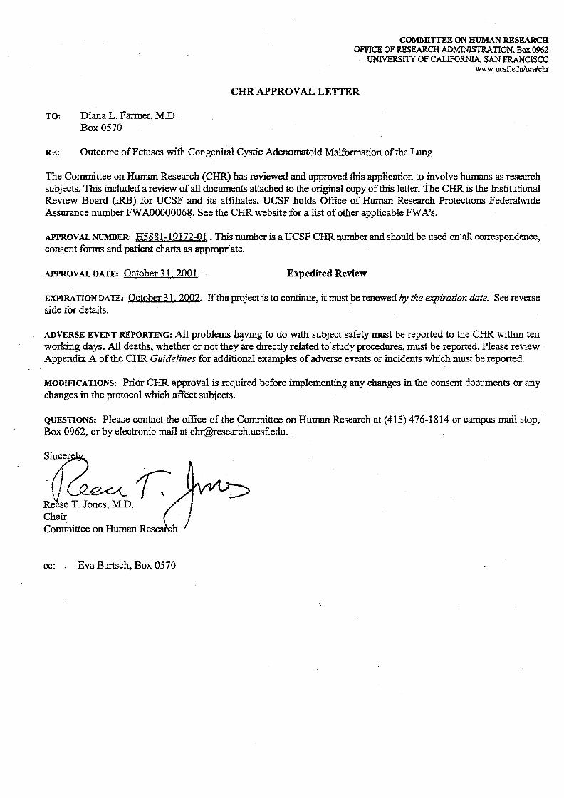
TO: Diana L. Fanner, M.D. Box 0570
COMMITTEE ON HUMAN RESEARCH OFFICE OF RESEARCH ADMINISTRATION, Box 0962
UNIVERSITY OF CALIFORNIA. SN'I" FRANCISCO www.ucsf.eduloralchr
CHR APPROV AL LETTER
RE: Outcome ofFetuses with Congenital Cystic Adenomatoid Malfonnation ofthe Lung
The Comrnittee on Human Research (CHR) has reviewed and approved this application to :involve humans as research .subjects. This incIuded a review of a11 documents attached to the original copy of this letter. The CHR is the Institutional Review Board (IRB) for UCSF and its affiliates. UCSF holds Office of Human Research Protections Federalwide Assurance riumber FW A00000068. See the CHR website for a list of other applicable FW A's.
APPROV AL NUMBER: H5881-19172-O 1 . This number is a UCSF CHR number and should be used on alI correspondence, consent forms and patient charts as appropriate.
APPROV AL DATE: October 31. 2001. Expedited Review
EXPIRATION DATE: October 31. 2002. Ifthe project is to continue, it must be renewed by the expiration date. See reverse side for details.
ADVERSE EVENT REPORTING: All problems l],aving to do with subject safety must be reported to the CHR within ten working days. All deaths, whether or not they ~e direct1y related tostudy procedures, must be reported. Please review Appendix A of the CHR Guidelines for additional examples of adverse events or incidents which must be reported.
MODIFICATIONS:. Prior CHR approval is required before implementing any changes in.the consent documents or any changes:in the protocol which affect subjects.
QUESTIONS: Pleasecontact the office ofthe Committee on Human Research at (415) 476-1814 or campus mail stop, Box 0962, or by electronic mai! at [email protected].
s~:r (J~r: '·1 Reese T. Jones, M.D.
Chair ( . J Committee on Human Resea:h:h .
ce: . Eva Bartsch, Box 0570
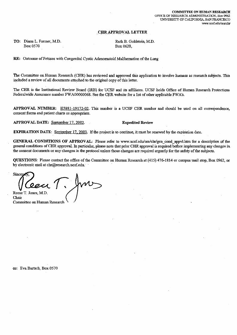
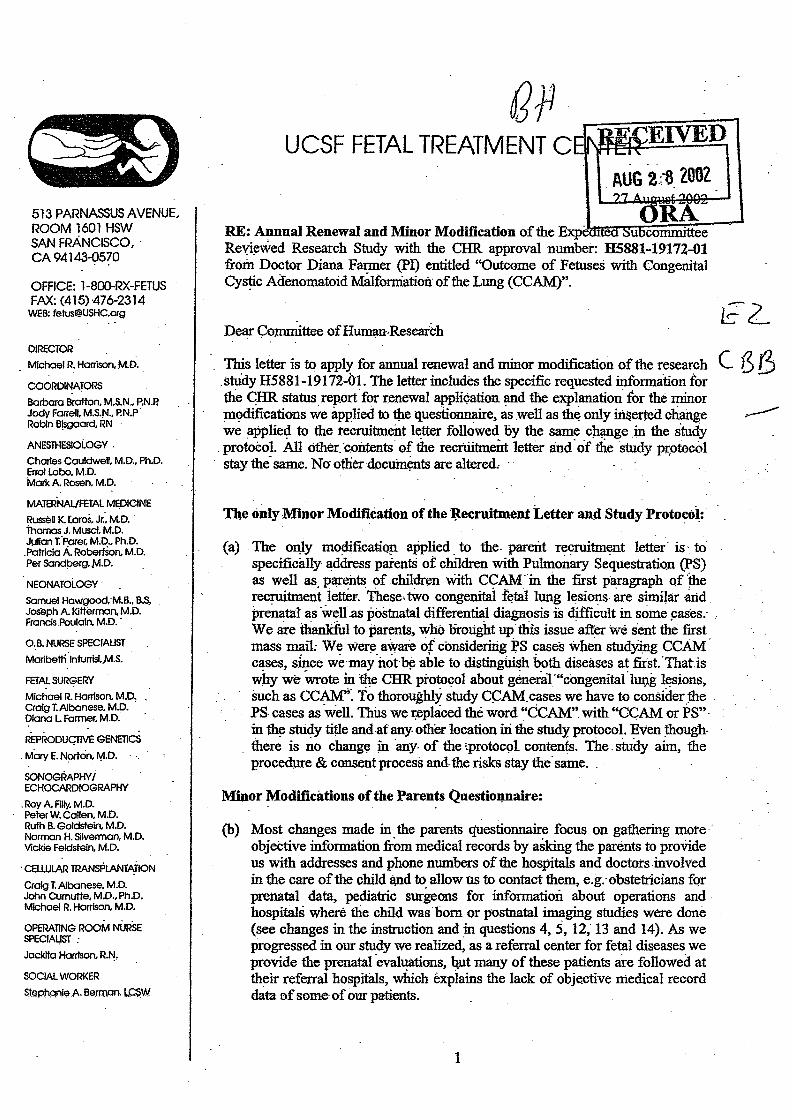
513 PARNASSUS AVENUE. ROOM 1601 HSW SAN FRÄNCISCO •. CA 94143-0570
OFFICE: 1-800-RX-FETUS FAX: (415)'476-2314
WEB: [email protected]
DIRECTOR
Michael R. Harrison. M.D.
COORDINATORS
Barbara Brotton. M.S.N .• RN.P. Jody Farren. M.S.N~ P.N.P· Robin B!sgaard. RN
ANESTHESIOLOGY .
Chartes Cauldwell. M.D .• Ph.D. Errollobo. M.D. Mark A. Rosen. M.D.
MATERNAvFETAl M~DlCINE RusseU K. [ares. Jr.. M.D. ihomas J. Musci. M.D. J!Jlian t"r;~arer. M.~ •• Ph.D. .patricia A Robertson. M.D. Per Sandperg. MD.
NEONAToLOGY .
Somuel Hawgood;M.B .• B.S, Joseph A Kittermal\ M.D. Francis .Poulain. M.D. '
O.B, NURSE SPECIAUST
Maribefll'lnfurrisf..M,S.
FETAL SUR<?ERY
Michael R. Harrison. M.D .. Cralg T. Albanese. M.D. . Diana L Farmer. M.D.
REPRODUGTIVE GENETICS
. Mory E. Norfon. M.D.
SONOGRAPHY i ECHOCARDIOGRAPHY
,Roy A Fdly. M.D. Peter W. Callen. M.D. Rufh B. GoldsteIn. M.D. Norman H. Silverman. M.D. Vickie Feldstein. M.D.
. CELlULAR TRANSPLANT~TION
Craig T. Albanese. M.D. John Curnutte, M.D., Ph.D. Michael R. Harrison. M.D.
OPERATING ROOM NURSE SPECIA1,IST :
Jacklta Harrlson. R.I'!.
SOCIAL WORKER
Sf.!ilPhqn/17A, Bermon.lCSW
(]}4 UCSF FETAL TREATMENT C
RE: Annual Renewal and Minor Modification ofthe Exp' e u committee Re':'i~wed Research Study with the CHR approval number: H5881-19172-01 froril Doctor Diana Fm:mer (PI) entitled "Outcome of Fetuses with Congenital Cys~c Adenomatoid Malforniatioir of the Lung (CCAM)".
Dear c,:::ommittee ofHum~n.Resea:ith
This letter is to apply for annual renewal and minor modification of the research ,stUdy H5881-19172-tll. The letter includes the specific requested mformation for the qm. status rep.ort· for r:enewal appliqation and the explanation for the minor ~9difications we applied to the questionnaire, as well as th~ only m~erted change we .aPPlie~ to the recruitment l~tter followed by the same cp.~ge.in the study
. protocol. All öther, 'c.ontents· of the recrt.iitment letter and' of thestudy pr,otocoI stay the- same. NO' other 'docuin~nts are altered,
The oBly ·Minor Modification of thel,tecruitment Letter and Study Protoco~: .
(a) . The on)y modificatiQp- applied . to the. parent recruitm~nt letter' iso to specificälly ~dress patentS oi children with Pulmonary Sequestration (PS) as weIl as. p~~ts ,?f .chil~~n With C9AM,,'in the first pi:l:ragraph Qf 'the recruitment letter. These. two congenital f~tal lung lesions are similar and prenatiil as 'weIl.as pos1natal differential diagnosis 18 difficult in some pases.; We are iliankful to-j>arents. who brought up tbis issue after we sent the first mass mail: We wer~ aware of consideririg :t>S cases when study.i:ng CCAM cases, slnce wemay 'not'b!! able to distingUish both diseases at first.'Thatjs why we MOte in t4e CHR pro~ol about gtneial'''congenital'l~g l~ions, such as CCAM'\ To thoroughly study C~AM.cases we have tO' consider:the . PS cases as ·well. Thils we r:ephiced the word "CCAM". with "CCAM or PS" . in th~ sttidy title anda! any. other location iri the study protocol. Even :though·
. there is no chang<,:: in ·any. of the ~protoc91 contents. The, study aim, the procedure & consent process and. the risks stay the' same.
Minor Modifications of the Parents Questio.I)D.aire:
(b)
. ,
Most changes made in the parents questionnaire focus on gathering more·' objeetive information Horn medical records byasking the parents to proVide us with'addresses and phone numbers ofthe hospitals and doctots.involved in the care of the child iUld to allow OS to contact them, e.g.' obstetricians for prenatal data, pediatric surgeons for information about operations and hospitals where tbe child was born or postnatal imaging studies were done (see changes in the instruction and .in questions 4, 5, 12; 13 and 14). As we progressed in our study we realized, as a referral center for fetal diseases we prO'vide the prenataI 'evaluations, qut many of these patients are followed at their referral hospitals, which explains the lack of obje.ctive niedical record data of some of Ol:lr pa:tients.
1
Cß!3
;:'0~Y) ,)J1.e .i1dd,ed some questions about the prenatal course to evaluate how the parents assess the ,."~, < :'-sitUation before birth (see question 13 and 5). .
." i'-' ."
;(d)';;We leamed from the patients feed back about valuable material some parents provided us with . an<! asked for these documents in the last optional question "Is there anything more you want' to tell us or send us?"
(e) We changed the sequence of the questions as we thought it might be more convenient for the parents (see highlighted question numbers).
(f) All changes are. highlighted on the printout together with the replacement of the word "CCAM" with "CCAM and PS" in the first sentence and some trivial adjustments not mentioned here.
The proposed modifications do not change the risks of 10ss of privacy and confidentiality and the potential incOllvenience for the parents in taking part in the questionnaire and phone calls. The meastires, undertaken to minimize these risks stay the same. Benefits for fetal diagnosis and treatment of future patients will most like1y increase following the modifications, since we will be able to gather more and even more objective data. .
For the .Annual Renewal Status Report:
(a) Protocol Summary: The long-term objective of our study is.to better counsel parents with fetuses with congertital lung lesions, such as CCAM or PS. The specific aims include elucidation of the natural history of these lesions during the prenatal period' and. the development of the mass after birth before 1ung surgery. We want to find out if there are speeifie prenatal sonographie parameters' predictive to the outeome of the fetuses. We also want to know how the children are doing iater life with and without surgery. Therefore we conduct a long term: outcome study (retrospective cohort study) by sending questionnaires to parents of affeeted ehildren and reviewing the mothers' and children' s medieal records.
(b) Summary of Results: We analyzed the data of 28 patients who sent back the filled out questionnaire and reviewed their medical reeords regarding prenatal ultrasounds and pre-and postnatal outcome, if available. The results show a tendency of large lung lesions· doing worse, that means children needed surgery or even died. However we wonder about some large lesions in the study showing favorable outcome even without surgery and some lesions which decreased before or after birth. We observed a·sirnilar phenomenon for prenatally diagnosed mediastinal shift. Up to this point our results are not statistically significant, because we need more patients involved in the study.
(c) Summary ofRecent Literature: In the last lOyears 23 clinicalstudies were published.about the outcome of children prenatally diagnosed with CCAM or PS. However, the published studies laek adequate number of cases and 10ng term follow up. Only four of them evaluated more than 25 patients looking mainly at perinatal outcome. The 2 new elinical studies brought out in the last year (2001) dealt again with less than 15 patients. We intend to evaluate more than.90 patients and our follow up interval reaches 13 years so far. There is no alteration of the risks involved in our retrospective cohort study (see also next paragraph). Results of the above mentioned literature is not changing the risk either.
2
(d) Reanalysis of RiskIBenefit Ration: As we pointed out in the last paragraph about OUT
questionnaire modifications, the risks of 10ss of privacy and confidentiality and the potential inconvenience for parents of taking part in the questionnaire do not change. The measures undertaken to minimize tbis risks stay the same. Yet the benefits for fetal diagllosis and treatment of future patients will most likely increase by continuing the studyand collecting more patients' data with the modified questionnaire. We expect to find statistically significant results and formulate a message to our obstetri~ colleagues, who still consider large lung lesions as lethal.
(e) Brief Summary of Plans for the Coming Year: We plan to involve at least 62 more patients in the study by s~nding out a secf>nd mass mailing to more patients and follow up on patients by phone who do not respond. As soon as we receive the mothers' consent we go ahead and examine the medical records, By analyzing the patients ' data. with the assistance of OUT
statistician we look forward to find significant results and knowledge about prognosis and treatment of CCAM and PS.
If there are any further questions, please do not hesitate to contact uso
Sffi-M .. <:.
!YG Diana Farmer, , PI Department of Surgery and Fetal Treatment Center, UCSF Box 0570, HSW 1601, 513 Pamassus Ave
:z:w/ Eval~I Medical Student Fetal Treatment Center, UCSF Box 0570, HSW 1601, 513 Parnassus Ave Study phone: (510) 6818160
,MD, Co-PI epartment ofRadiology
and Fetal Treatment Center, UCSF Box 0628, Moffit 396, 505 Parnassus Ave
Office phone: 476-1397
3
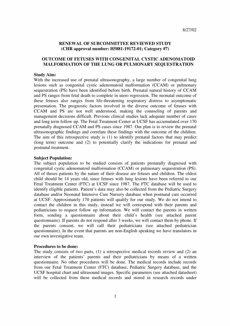
8/27/02
1
RENEWAL OF SUBCOMMITTEE REVIEWED STUDY (CHR approval number: H5881-19172-01; Category #7)
OUTCOME OF FETUSES WITH CONGENITAL CYSTIC ADENOMATOID MALFORMATION OF THE LUNG OR PULMONARY SEQUESTRATION
Study Aim: With the increased use of prenatal ultrasonography, a large number of congenital lung lesions such as congenital cystic adenomatoid malformation (CCAM) or pulmonary sequestration (PS) have been identified before birth. Prenatal natural history of CCAM and PS ranges from fetal death to complete in utero regression. The neonatal outcome of these fetuses also ranges from life-threatening respiratory distress to asymptomatic presentation. The prognostic factors involved in the diverse outcome of fetuses with CCAM and PS are not well understood, making the counseling of parents and management decisions difficult. Previous clinical studies lack adequate number of cases and long term follow up. The Fetal Treatment Center at UCSF has accumulated over 170 prenatally diagnosed CCAM and PS cases since 1987. Our plan is to review the prenatal ultrasonographic findings and correlate these findings with the outcome of the children. The aim of this retrospective study is (1) to identify prenatal factors that may predict (long term) outcome and (2) to potentially clarify the indications for prenatal and postnatal treatment. Subject Population: The subject population to be studied consists of patients prenatally diagnosed with congenital cystic adenomatoid malformation (CCAM) or pulmonary sequestration (PS). All of theses patients by the nature of their disease are fetuses and children. The oldest child should be 14 years old, since fetuses with lung lesions have been referred to our Fetal Treatment Center (FTC) at UCSF since 1987. The FTC database will be used to identify eligible patients. Patient’s data may also be collected from the Pediatric Surgery database and/or Neonatal Intensive Care Nursery database when postnatal care occurred at UCSF. Approximately 170 patients will qualify for our study. We do not intend to contact the children in this study, instead we will correspond with their parents and pediatricians to request follow up information. We will contact the parents in written form, sending a questionnaire about their child’s health (see attached parent questionnaire). If parents do not respond after 3 weeks, we will contact them by phone. If the parents consent, we will call their pediatricians (see attached pediatrician questionnaire). In the event that parents are non-English speaking we have translators in our own investigative team. Procedures to be done: The study consists of two parts, (1) a retrospective medical records review and (2) an interview of the patients’ parents and their pediatricians by means of a written questionnaire. No other procedures will be done. The medical records include records from our Fetal Treatment Center (FTC) database, Pediatric Surgery database, and the UCSF hospital chart and ultrasound images. Specific parameters (see attached datasheet) will be collected from these medical records and stored in research records under
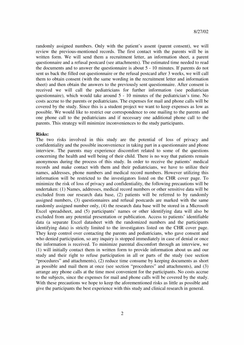
8/27/02
2
randomly assigned numbers. Only with the patient’s assent (parent consent), we will review the previous-mentioned records. The first contact with the parents will be in written form. We will send them a recruitment letter, an information sheet, a parent questionnaire and a refusal postcard (see attachments). The estimated time needed to read the documents and to answer the questionnaire is about 5 - 10 minutes. If parents do not sent us back the filled out questionnaire or the refusal postcard after 3 weeks, we will call them to obtain consent (with the same wording in the recruitment letter and information sheet) and then obtain the answers to the previously sent questionnaire. After consent is received we will call the pediatricians for further information (see pediatrician questionnaire), which would take around 5 - 10 minutes of the pediatrician’s time. No costs accrue to the parents or pediatricians. The expenses for mail and phone calls will be covered by the study. Since this is a student project we want to keep expenses as low as possible. We would like to restrict our correspondence to one mailing to the parents and one phone call to the pediatricians and if necessary one additional phone call to the parents. This strategy will minimize inconveniences to the study participants. Risks: The two risks involved in this study are the potential of loss of privacy and confidentiality and the possible inconvenience in taking part in a questionnaire and phone interview. The parents may experience discomfort related to some of the questions concerning the health and well being of their child. There is no way that patients remain anonymous during the process of this study. In order to receive the patients’ medical records and make contact with them and their pediatricians, we have to utilize their names, addresses, phone numbers and medical record numbers. However utilizing this information will be restricted to the investigators listed on the CHR cover page. To minimize the risk of loss of privacy and confidentiality, the following precautions will be undertaken: (1) Names, addresses, medical record numbers or other sensitive data will be excluded from our research data base, (2) patients will be referred to by randomly assigned numbers, (3) questionnaires and refusal postcards are marked with the same randomly assigned number only, (4) the research data base will be stored in a Microsoft Excel spreadsheet, and (5) participants’ names or other identifying data will also be excluded from any potential presentation or publication. Access to patients’ identifiable data (a separate Excel datasheet with the randomized numbers and the participants identifying data) is strictly limited to the investigators listed on the CHR cover page. They keep control over contacting the parents and pediatricians, who gave consent and who denied participation, so any inquiry is stopped immediately in case of denial or once the information is received. To minimize parental discomfort through an interview, we (1) will initially contact them in written form to provide information about us and our study and their right to refuse participation in all or parts of the study (see section “procedures” and attachments), (2) reduce time consume by keeping documents as short as possible and mail them at once (see section “procedures” and attachments), and (3) arrange any phone calls at the time most convenient for the participants. No costs accrue to the subjects, since the expenses for mail and phone calls will be covered by the study. With these precautions we hope to keep the aforementioned risks as little as possible and give the participants the best experience with this study and clinical research in general.
8/27/02
3
Benefits: Results from this study will benefit medical science, in particular the growing field of fetal diagnosis and treatment. Significant knowledge, based on data collected from a tertiary referral center for fetal diseases with 14-years of experience, will be gained. The beneficial knowledge includes: (1) outcome of fetal lung lesions, in particular of congenital cystic adenomatoid malformation of the lung (CCAM) and pulmonary sequestration (PS), (2) determination of crucial prognostic factors for fetuses with CCAM and PS by means of prenatal ultrasonography, and (3) elucidation of indications for prenatal and postnatal treatment of CCAM and PS. We anticipate better and more informed counseling of parents with fetuses suffering from CCAM or PS lung lesions. There is no direct benefit for the individual participant. However, the parents may feel satisfied after taking part in the study. Parents who are referred to the Fetal Treatment Center are typically proactive and very engaged and they may gain satisfaction from helping to find more precise indicators for outcome and treatment of unborn children with the same disease as their child. Consent Process and Documentation: Herewith we apply for a CHR approval of waiver of signed consent to conduct the informed consent process through use of an information sheet. There are four reasons to do so: (1) our study involves no more than minimal risk (category #1), (2) the identities of the subjects will be completely anonymous after the information for the research records is obtained (category #1), (3) the risks of loss of privacy and confidentiality and inconveniences for the participants will be lessen in this way, and (4) the rights and welfare of the subjects are not adversely affected without a signed consent. The patients’ freedom to participate in the study or to denial participation will be guaranteed (1) in form of a written refusal postcard attached to the information sheet, since the initial contact is made in written and (2) again in verbal form during the following phone call, if patients did not answer in written form. To offer more choices and flexibility to the participants, they can refuse to take part in all or only some parts of the study. Furthermore, they can decide to participate in written form only by sending back the filled out parent questionnaire if they do not want to be contacted in person, or they can simply answer in verbal form on the phone, in case they do not want to write. Please refer to the attached recruitment letter and information sheet, which includes all elements of the consent document. Qualifications of Investigators: The principle investigator is Doctor Diana L. Farmer, UCSF Associate Professor of Surgery and Pediatric Surgeon. Co- principle investigator is Doctor Ruth B. Goldstein, UCSF Professor of Radiology and Obstetrics & Gynecology. They will mentor the investigative team, consisting of Eva Bartsch, visiting scholar and medical student in the final year from the University of Cologne, Germany and collaborator at the UCSF Fetal Treatment Center and Doctor Alexander Strauss, Assistant Professor of Obstetrics & Gynecology from the University of Munich, Germany. All of these investigators have a particular interest in prenatal diagnosis and treatment and Pediatric Surgery.
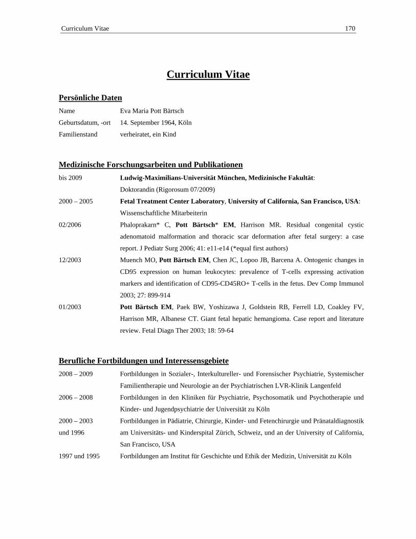
Curriculum Vitae 170
Curriculum Vitae
Persönliche Daten
Name Eva Maria Pott Bärtsch
Geburtsdatum, -ort 14. September 1964, Köln
Familienstand verheiratet, ein Kind
Medizinische Forschungsarbeiten und Publikationen
bis 2009 Ludwig-Maximilians-Universität München, Medizinische Fakultät:
Doktorandin (Rigorosum 07/2009)
2000 – 2005 Fetal Treatment Center Laboratory, University of California, San Francisco, USA:
Wissenschaftliche Mitarbeiterin
02/2006 Phaloprakarn* C, Pott Bärtsch* EM, Harrison MR. Residual congenital cystic
adenomatoid malformation and thoracic scar deformation after fetal surgery: a case
report. J Pediatr Surg 2006; 41: e11-e14 (*equal first authors)
12/2003 Muench MO, Pott Bärtsch EM, Chen JC, Lopoo JB, Barcena A. Ontogenic changes in
CD95 expression on human leukocytes: prevalence of T-cells expressing activation
markers and identification of CD95-CD45RO+ T-cells in the fetus. Dev Comp Immunol
2003; 27: 899-914
01/2003 Pott Bärtsch EM, Paek BW, Yoshizawa J, Goldstein RB, Ferrell LD, Coakley FV,
Harrison MR, Albanese CT. Giant fetal hepatic hemangioma. Case report and literature
review. Fetal Diagn Ther 2003; 18: 59-64
Berufliche Fortbildungen und Interessensgebiete
2008 – 2009 Fortbildungen in Sozialer-, Interkultureller- und Forensischer Psychiatrie, Systemischer
Familientherapie und Neurologie an der Psychiatrischen LVR-Klinik Langenfeld
2006 – 2008 Fortbildungen in den Kliniken für Psychiatrie, Psychosomatik und Psychotherapie und
Kinder- und Jugendpsychiatrie der Universität zu Köln
2000 – 2003
und 1996
Fortbildungen in Pädiatrie, Chirurgie, Kinder- und Fetenchirurgie und Pränataldiagnostik
am Universitäts- und Kinderspital Zürich, Schweiz, und an der University of California,
San Francisco, USA
1997 und 1995 Fortbildungen am Institut für Geschichte und Ethik der Medizin, Universität zu Köln
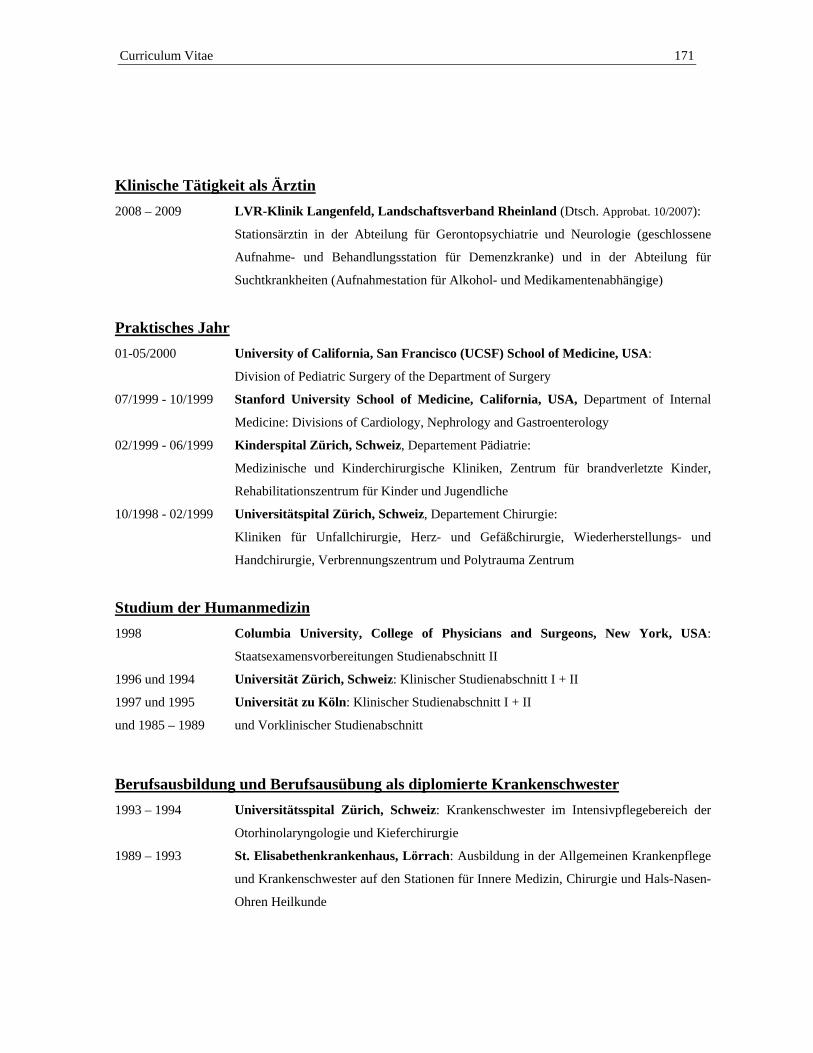
Curriculum Vitae
171
Klinische Tätigkeit als Ärztin
2008 – 2009 LVR-Klinik Langenfeld, Landschaftsverband Rheinland (Dtsch. Approbat. 10/2007):
Stationsärztin in der Abteilung für Gerontopsychiatrie und Neurologie (geschlossene
Aufnahme- und Behandlungsstation für Demenzkranke) und in der Abteilung für
Suchtkrankheiten (Aufnahmestation für Alkohol- und Medikamentenabhängige)
Praktisches Jahr
01-05/2000 University of California, San Francisco (UCSF) School of Medicine, USA:
Division of Pediatric Surgery of the Department of Surgery
07/1999 - 10/1999 Stanford University School of Medicine, California, USA, Department of Internal
Medicine: Divisions of Cardiology, Nephrology and Gastroenterology
02/1999 - 06/1999 Kinderspital Zürich, Schweiz, Departement Pädiatrie:
Medizinische und Kinderchirurgische Kliniken, Zentrum für brandverletzte Kinder,
Rehabilitationszentrum für Kinder und Jugendliche
10/1998 - 02/1999 Universitätspital Zürich, Schweiz, Departement Chirurgie:
Kliniken für Unfallchirurgie, Herz- und Gefäßchirurgie, Wiederherstellungs- und
Handchirurgie, Verbrennungszentrum und Polytrauma Zentrum
Studium der Humanmedizin
1998 Columbia University, College of Physicians and Surgeons, New York, USA:
Staatsexamensvorbereitungen Studienabschnitt II
1996 und 1994 Universität Zürich, Schweiz: Klinischer Studienabschnitt I + II
1997 und 1995
und 1985 – 1989
Universität zu Köln: Klinischer Studienabschnitt I + II
und Vorklinischer Studienabschnitt
Berufsausbildung und Berufsausübung als diplomierte Krankenschwester
1993 – 1994 Universitätsspital Zürich, Schweiz: Krankenschwester im Intensivpflegebereich der
Otorhinolaryngologie und Kieferchirurgie
1989 – 1993 St. Elisabethenkrankenhaus, Lörrach: Ausbildung in der Allgemeinen Krankenpflege
und Krankenschwester auf den Stationen für Innere Medizin, Chirurgie und Hals-Nasen-
Ohren Heilkunde
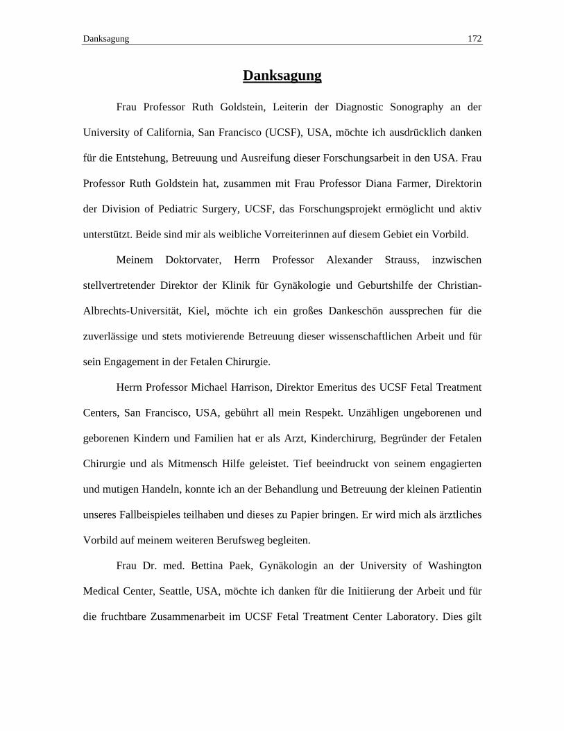
Danksagung 172
Danksagung
Frau Professor Ruth Goldstein, Leiterin der Diagnostic Sonography an der
University of California, San Francisco (UCSF), USA, möchte ich ausdrücklich danken
für die Entstehung, Betreuung und Ausreifung dieser Forschungsarbeit in den USA. Frau
Professor Ruth Goldstein hat, zusammen mit Frau Professor Diana Farmer, Direktorin
der Division of Pediatric Surgery, UCSF, das Forschungsprojekt ermöglicht und aktiv
unterstützt. Beide sind mir als weibliche Vorreiterinnen auf diesem Gebiet ein Vorbild.
Meinem Doktorvater, Herrn Professor Alexander Strauss, inzwischen
stellvertretender Direktor der Klinik für Gynäkologie und Geburtshilfe der Christian-
Albrechts-Universität, Kiel, möchte ich ein großes Dankeschön aussprechen für die
zuverlässige und stets motivierende Betreuung dieser wissenschaftlichen Arbeit und für
sein Engagement in der Fetalen Chirurgie.
Herrn Professor Michael Harrison, Direktor Emeritus des UCSF Fetal Treatment
Centers, San Francisco, USA, gebührt all mein Respekt. Unzähligen ungeborenen und
geborenen Kindern und Familien hat er als Arzt, Kinderchirurg, Begründer der Fetalen
Chirurgie und als Mitmensch Hilfe geleistet. Tief beeindruckt von seinem engagierten
und mutigen Handeln, konnte ich an der Behandlung und Betreuung der kleinen Patientin
unseres Fallbeispieles teilhaben und dieses zu Papier bringen. Er wird mich als ärztliches
Vorbild auf meinem weiteren Berufsweg begleiten.
Frau Dr. med. Bettina Paek, Gynäkologin an der University of Washington
Medical Center, Seattle, USA, möchte ich danken für die Initiierung der Arbeit und für
die fruchtbare Zusammenarbeit im UCSF Fetal Treatment Center Laboratory. Dies gilt
Danksagung
173
auch für den engagierten brasilianischen Kinderchirurgen, Herrn Dr. med. Lourenco
Sbragia, von der State University of Campinas, UNICAMP, Sao Paulo, Brazil.
Bei der statistischen Auswertung der Daten waren mir Herr Jimmy Hwang, Ph.D.,
Senior Biostatistician vom UCSF Comprehensive Cancer Center, und Herr Professor
Christopher Tori, von der California School of Professional Psychology, Alliant
International University, San Francisco, sehr behilflich – vielen Dank.
Ganz besonders danke ich den Eltern und Ärzten der Kinder dieser Studie für ihre
engagierte Mithilfe auf der Suche nach einem verbesserten Verständnis und Management
dieser Erkrankung. Auch den Menschen gehört ein Dankeschön, die – trotz, dass sie
ungenannt bleiben – sich tagtäglich einsetzen für Patienten und Mitarbeiter und die
Arbeit im Krankenhaus und in der Forschung erleichtern.
Mein größter Dank gebührt meinem Ehemann und Vater unseres Sohnes Philipp,
Dr. sc. nat. Stephan Bärtsch, der mir mit seinem wissenschaftlichen Know-how bei
meiner Forschungsarbeit und mit seinem Einsatz bei der Behütung unseres Sohnes zur
Seite stand. Sein Weltbild, das gleiches Recht auf Karriere für Mann und Frau vorsieht,
hat mir die Freiheit zum Studieren und Forschen gegeben. Ein besonderer Dank gilt auch
meinen Eltern, Elisabeth Pott und Dr. rer. nat. Franz Philipp Pott, die mich im beruflichen
Weiterkommen immer unterstützt haben und zusammen mit meiner Schwester, Elisabeth
Pott-Grinstein, Gynäkologin, und meinem Bruder, Dr. rer. pol. Philipp Pott und Cousinen
sowie Freunden den Rückhalt in einer „Großfamilie“ gaben – das war der Hintergrund
auf dem unsere Forschungsarbeiten gediehen.