Embed Size (px)
Citation preview
Cutaneous myoepithelial neoplasms:clinicopathologic andimmunohistochemical study of 20 casessuggesting a continuous spectrum rangingfrom benign mixed tumor of the skin tocutaneous myoepithelioma andmyoepithelial carcinoma
Thomas Mentzel1, LuisRequena2, Steven Kaddu3, LuisM. Soares de Aleida4, OmarP. Sangueza5 and Heinz Kutzner1
1Dermatopathologisches Gemeinschaftslabor,Friedrichshafen, Germany,2Departments of Dermatology, FundacionJimenez Diaz, Universidad Autonoma, Madrid,Spain,3University of Graz, Graz, Austria,4Hospital de Santa Maria, Lisboa, Portugal,and5Wake Forest University, Winston–Salem, NC,USA
Background: Myoepithelial neoplasms, both benign and malignant,are rare but well-established clinicopathologic entities in the salivaryglands, the breast, and the lung. Despite similarities betweencutaneous sweat glands and glandular structures in the above-mentioned organs as well as the presence of regular myoepithelialcells around cutaneous eccrine/apocrine glands, the concept ofcutaneous myoepithelial neoplasms is still debatable and notcommonly accepted.Methods: Twenty cutaneous myoepithelial neoplasms have beenstudied histologically and immunohistochemically.Results: Nine neoplasms showed features of benign mixed tumor ofthe skin (chondroid syringoma) (five females and four males, age range19–65 years, all cases arose in the head and neck region). Two casesrepresented the eccrine and seven the apocrine subtype. Interestingly, inthree cases of the apocrine subtype, solid areas composedpredominantly of myoepithelial cells were detected; these neoplasmswere designated as benign mixed tumors with prominent myoepithelialcells. Nine cutaneous neoplasms were composed of spindled,epithelioid, and plasmocytoid cells without ductal differentiation andimmunohistochemically stained variably positive for vimentin, epithelialand myogenic markers, S-100 protein, calponin, and glial fibrillaryacidic protein (four females and five males, age range 3–71 years, fourcases arose in the head and neck region and one case each on the finger,the thigh, the lower leg, the foot, and the breast, respectively); theseneoplasms were designated as cutaneous myoepitheliomas. Twomorphologically malignant neoplasms with cytologic andimmunohistochemical features of myoepithelial cells arose on the faceof a 70-year-old female and a 79-year-old male patient; these neoplasmswere designated as malignant cutaneous myoepitheliomas (cutaneousmyoepithelial carcinomas).Conclusions: The study suggests a continuous spectrum of cutaneousmyoepithelial neoplasms ranging from benign mixed tumor of the
J Cutan Pathol 2003: 30: 294–302 Copyright # Blackwell Munksgaard 2003Blackwell Munksgaard. Printed in Denmark
Journal of
Cutaneous PathologyISSN 0303-6987
294
skin to cutaneous myoepithelioma and cutaneous myoepithelialcarcinoma. Further studies with extended follow-up information arenecessary to establish prognostic factors.
Mentzel T, Requena L, Kaddu S, Soares de Aleida LM, Sangueza OP,Kutzner H. Cutaneous myoepithelial neoplasms: clinicopathologic andimmunohistochemical study of 20 cases suggesting a continuousspectrum ranging from benign mixed tumor of the skin to cutaneousmyoepithelioma and myoepithelial carcinomaJ Cutan Pathol 2003; 30: 294–302. # Blackwell Munksgaard 2003.
Myoepithelial cells are characterized by their capacityfor a bidirectional differentiation and show epithelialand myoid features on histologic, immunohisto-chemical, and ultrastructural levels.1,2 In the skin,non-neoplastic myoepithelial cells are seen mainlyaround apocrine and eccrine glands. Morphologic-ally, myoepithelial cells may show spindled, epi-thelioid, plasmocytoid and/or clear cell features, andalso immunohistochemically a variable expression ofvimentin, cytokeratins, epithelial membrane antigen(EMA), S-100 protein, muscle actins, glial fibrillaryacidic protein (GFAP), and calponin is seen, whereasdesmin is absent.3 Benign and malignant myoepithe-lial neoplasms are rare but well-recognized in thesalivary glands,4,5 the breast,6 and the lung.7 Mostrecently, neoplasms of predominantly myoepithelialdifferentiation (myoepitheliomas) have been describedin skin and soft tissues as well.8–10 We report cases ofbenign cutaneous mixed tumors (chondroid syringo-mas) (apocrine and eccrine subtypes) including caseswith prominent myoepithelial differentiation, cuta-neous myoepitheliomas, and cutaneous myoepithelialcarcinomas in order to demonstrate a continuousmorphologic spectrum of myoepithelial neoplasmsoccurring in the skin.
Materials and methods
Paraffin-embedded blocks and slides of nine benignmixed tumors of the skin, nine cutaneous myoepithe-liomas, and two cutaneous myoepithelial carcinomaswere retrieved from the routine files of the Dermato-pathologische Gemeinschaftspraxis, Friedrichshafen,as well as from the personal files of the authors. Fiveout of nine cutaneous myoepitheliomas have beendescribed in detail elsewhere.10 Clinical informationwas obtained from the laboratory request forms andcontributing clinicians and pathologists. In each case,tissue was fixed in 4% buffered formalin, routinelyprocessed, and embedded in paraffin. Four-micronthick sections were stained with hematoxylin andeosin. In addition, sections in all cases were alsostained immunohistochemically by the labeled strep-tavidin biotin technique, using commercially available
antibodies; antigen retrieval was used for all anti-bodies. Used antibodies, their sources, and dilutions aregiven in Table 1. Appropriate negative and positivecontrols were used in each case.
Results
Benign mixed tumor of the skin
Clinical features of nine cases of benign mixed tumorof the skin are summarized in Table 2. Briefly, allcases arose in the head and neck region of adultpatients (five females and four males, age range19–65 years), who presented with a soft, nodular lesionin most cases. The neoplasms were completely ormarginally excised, and no sign of recurrence has beenreported so far. Histologically, all neoplasms wererestricted to the dermis, nodular, and well definedbut not completely encapsulated. Seven cases (cases1, 2, 3, 4, 6, 8, and 9) showed morphological featuresof the so-called apocrine subtype of benign mixedtumor of the skin with ducts and branching tubularstructures lined by two layers of epithelial and myo-epithelial cells (Fig. 1). These cells were cytologicallyuniform, without striking atypia and increased prolif-erative activity. The intercellular matrix showed prom-inent myxoid changes in cases 1, 3, 4, and 9 andrather myxohyaline changes in cases 2, 6, and 8.Chondroid areas were present in cases 1, 4, and 8,and focal adipocytic metaplasia was noted in cases 3,4, 6, and 8. In cases 5 and 7, monomorphous smallductal structures, lined by a single layer of cuboid
Table 1. Immunohistochemical reagents used
Antibody Clone Dilution Source
Desmin D33 1 : 200 Dako, Glostrup, DenmarkS-100 protein Polyclonal 1 : 4000 Dako, Glostrup, DenmarkASMA 1A4 1 : 300 Dako, Glostrup, DenmarkCalponin CALP 1 : 300 BioGenex, San Ramon, CA, USAVimentin Vim3B4 1 : 150 Dako, Glostrup, DenmarkGFAP GA-5 1 : 200 BioGenex, San Ramon, CA, USAPancytokeratin MNF116 1 : 100 Dako, Glostrup, DenmarkEMA E29 1 : 100 BioGenex, San Ramon, CA, USA
ASMA, alpha-smooth muscle actin; EMA, epithelial membrane antigen;GFAP, glial fibrillary acidic protein.
Thomas Mentzel, MD, DermatopathologischesGemeinschaftslabor, Siemensstrasse 6/1, D-88048Friedrichshafen, GermanyTel: þ49 7541 56071Fax: 49 7541 604410e-mail: [email protected]
Accepted October 17, 2002
Cutaneous myoepithelial neoplasms
295
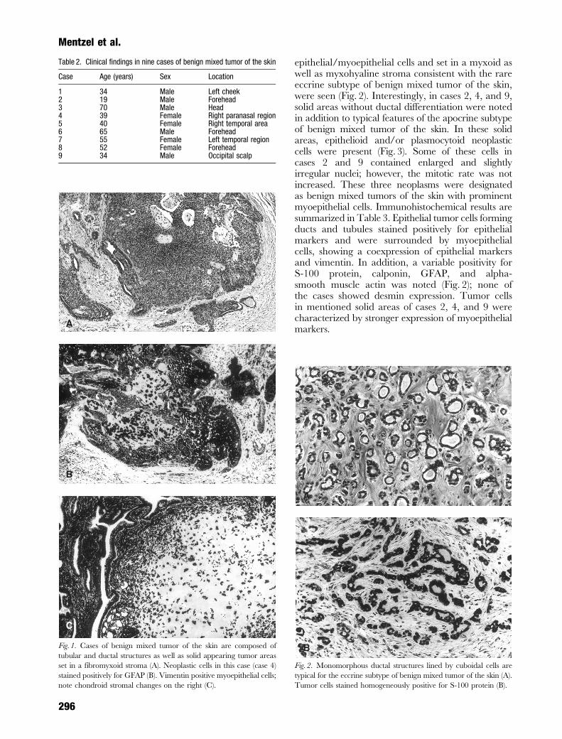
epithelial/myoepithelial cells and set in a myxoid aswell as myxohyaline stroma consistent with the rareeccrine subtype of benign mixed tumor of the skin,were seen (Fig. 2). Interestingly, in cases 2, 4, and 9,solid areas without ductal differentiation were notedin addition to typical features of the apocrine subtypeof benign mixed tumor of the skin. In these solidareas, epithelioid and/or plasmocytoid neoplasticcells were present (Fig. 3). Some of these cells incases 2 and 9 contained enlarged and slightlyirregular nuclei; however, the mitotic rate was notincreased. These three neoplasms were designatedas benign mixed tumors of the skin with prominentmyoepithelial cells. Immunohistochemical results aresummarized in Table 3. Epithelial tumor cells formingducts and tubules stained positively for epithelialmarkers and were surrounded by myoepithelialcells, showing a coexpression of epithelial markersand vimentin. In addition, a variable positivity forS-100 protein, calponin, GFAP, and alpha-smooth muscle actin was noted (Fig. 2); none ofthe cases showed desmin expression. Tumor cellsin mentioned solid areas of cases 2, 4, and 9 werecharacterized by stronger expression of myoepithelialmarkers.
Table 2. Clinical findings in nine cases of benign mixed tumor of the skin
Case Age (years) Sex Location
1 34 Male Left cheek2 19 Male Forehead3 70 Male Head4 39 Female Right paranasal region5 40 Female Right temporal area6 65 Male Forehead7 55 Female Left temporal region8 52 Female Forehead9 34 Male Occipital scalp
A
B
C
Fig. 1. Cases of benign mixed tumor of the skin are composed of
tubular and ductal structures as well as solid appearing tumor areas
set in a fibromyxoid stroma (A). Neoplastic cells in this case (case 4)
stained positively for GFAP (B). Vimentin positive myoepithelial cells;
note chondroid stromal changes on the right (C).
A
B
Fig. 2. Monomorphous ductal structures lined by cuboidal cells are
typical for the eccrine subtype of benign mixed tumor of the skin (A).
Tumor cells stained homogeneously positive for S-100 protein (B).
Mentzel et al.
296
Cutaneous myoepithelioma
Nine cases (four females and five males, age range3–71 years) were designated as cutaneous myoepithe-lioma and arose on the head (four cases), and one caseeach on the finger, the thigh, the lower leg, the foot,and the breast (Table 4). None of the cases recurredafter complete excision. All cases were confined to thedermis, and only in case 7, expansion into superficialparts of the subcutis was present. Except cases 2 and 8that showed focal extension into surroundingstructures, neoplasms were nodular, solid, and wellcircumscribed. Histologically, a broad variation wasseen. Whereas cases 3, 4, 5, and 6 were rather solidand composed of epithelioid cells admixed with
plump and spindled neoplastic cells (Fig. 4), fusiformtumor cells arranged in bundles and fascicles pre-dominated in cases 2, 8, and 9 (Fig. 5). Mainly plasmo-cytoid tumor cells were noted in case 1, and in case 7,epithelioid cells were admixed with plasmocytoidtumor cells (Fig. 6). By definition, no ductal and/ortubular differentiation was evident in these nine cases.The tumor stroma contained clusters of mature adi-pocytes in cases 2 and 8 (Fig. 5), and in cases 1, 2, 3, 4,5, and 8, focal myxoid stromal changes were noted.Results of immunohistochemical stainings are sum-marized in Table 5. Briefly, all neoplasms stainedpositively for vimentin and at least one epithelialmarker (cytokeratin and/or EMA) and showed avariable expression of S-100 protein, calponin,GFAP, and alpha-smooth muscle actin (Figs 5 and 6),whereas desmin was negative in all cases tested.Interestingly, spindled neoplastic cells in cases 2, 8,and 9 stained homogeneously positive for EMA,whereas pancytokeratin was absent in these threeneoplasms.
Malignant cutaneous myoepithelioma (cutaneousmyoepithelial carcinoma)
The first case arose in a 70-year-old female patient,who developed an infiltrating dermal and sub-cutaneous neoplasm on her left cheek. The lesionmeasured 1.0 cm in greatest diameter and was notconnected with the overlying epidermis and deeperlocated structures. Histologically, the cellular neo-plasm was composed of atypical spindle-shapedtumor cells with enlarged and pleomorphic nuclei
A
B
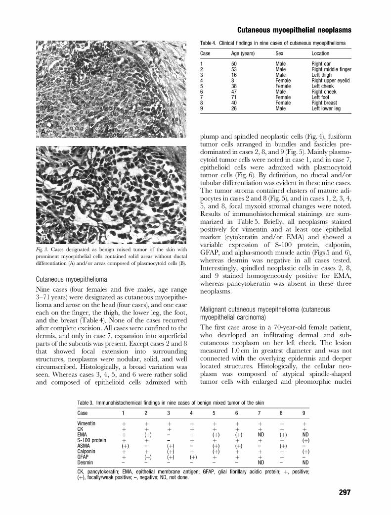
Fig. 3. Cases designated as benign mixed tumor of the skin with
prominent myoepithelial cells contained solid areas without ductal
differentiation (A) and/or areas composed of plasmocytoid cells (B).
Table 3. Immunohistochemical findings in nine cases of benign mixed tumor of the skin
Case 1 2 3 4 5 6 7 8 9
Vimentin þ þ þ þ þ þ þ þ þCK þ þ þ þ þ þ þ þ þEMA þ (þ) – þ (þ) (þ) ND (þ) NDS-100 protein þ þ – þ þ þ þ þ (þ)ASMA (þ) – (þ) – (þ) (þ) – (þ) –Calponin þ þ (þ) þ (þ) þ þ þ (þ)GFAP þ (þ) (þ) (þ) þ þ þ þ –Desmin – – – – – – ND – ND
CK, pancytokeratin; EMA, epithelial membrane antigen; GFAP, glial fibrillary acidic protein; þ, positive;(þ), focally/weak positive; –, negative; ND, not done.
Table 4. Clinical findings in nine cases of cutaneous myoepithelioma
Case Age (years) Sex Location
1 50 Male Right ear2 53 Male Right middle finger3 16 Male Left thigh4 3 Female Right upper eyelid5 38 Female Left cheek6 47 Male Right cheek7 71 Female Left foot8 40 Female Right breast9 26 Male Left lower leg
Cutaneous myoepithelial neoplasms
297
containing occasionally prominent nucleoli (Fig. 7).Numerous mitoses (mean, eight mitoses/10 highpower fields) and central tumor necrosis were found.Ductal structures and squamous or basaloid differen-tiation were not present. Immunohistochemically,neoplastic cells stained positively for vimentin, pan-cytokeratin, EMA, and alpha-smooth muscle actin(Fig. 7) as well as focally positive for calponin; S-100protein, GFAP, and desmin were all negative.
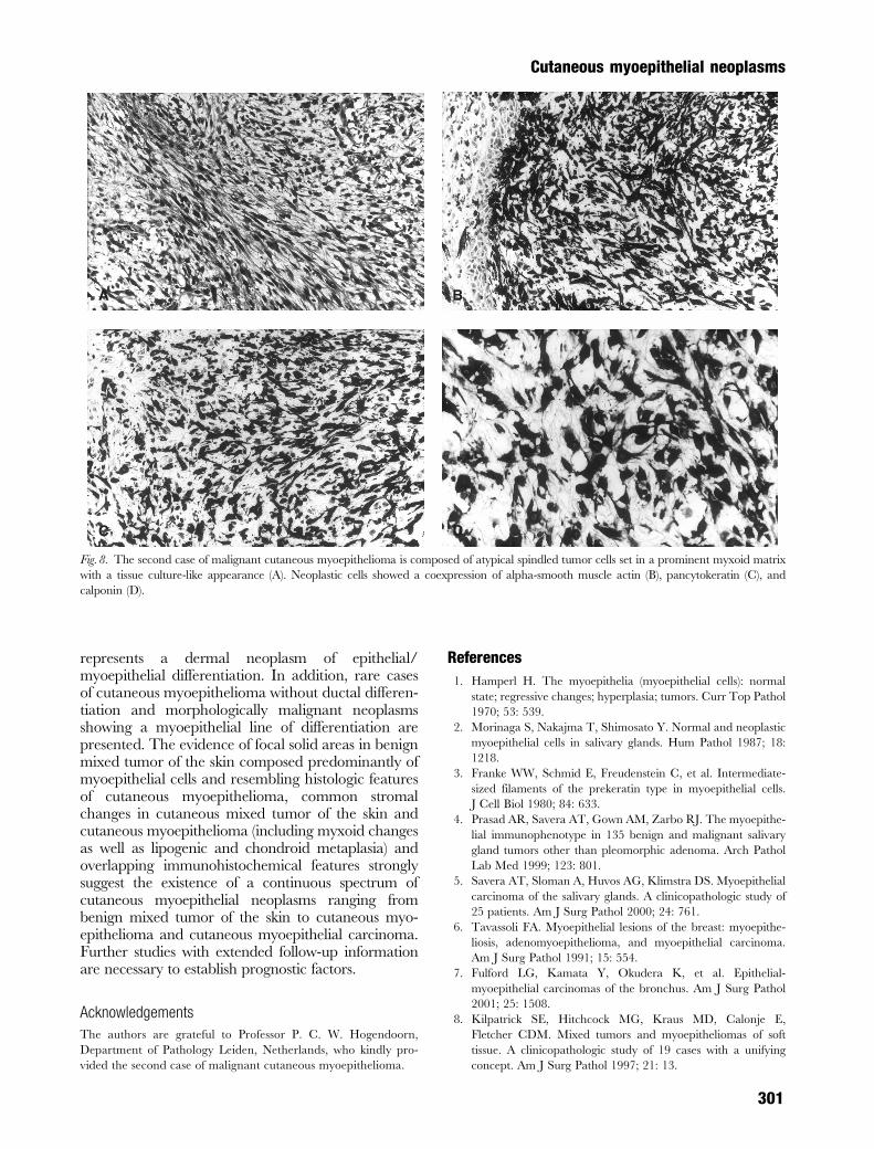
The second case was that of a 79-year-old malepatient, who developed a lesion, measuring 0.7 cm inlargest diameter, in his left retroauricular region.Under an intact epidermis, a nodular dermal neo-plasm with extension into subcutaneous tissue wasseen (deeper located structures were not involved).The neoplasm was composed of plump, spindled,round, and stellated tumor cells set in a prominentmyxoid matrix, with a tissue culture-like appearancein some areas (Fig. 8). Tumor cells contained an oftenwell-delineated, pale eosinophilic cytoplasm, andenlarged pleomorphic nuclei with prominent nucleoli;scattered mono- and multinucleated tumor giant cellswere noted. The mitotic rate was increased (mean,16 mitoses/10 high power fields), and also atypicalmitoses were found. Immunohistochemically, neoplasticcells stained homogeneously positive for vimentin, pan-cytokeratin, and alpha-smooth muscle actin, and a focalexpression of S-100 protein and calponin was notedas well (Fig. 8); desmin and GFAP were negative.
In both cases, the diagnosis of malignant cutaneousmyoepithelioma (cutaneous myoepithelial carcinoma)was established. There was no sign of local recurrenceor metastasis 6 months as well as 2 years after com-plete excision.
Discussion
Although relatively infrequent, benign and malignantmyoepithelial neoplasms are well-established entities
in the salivary glands as well as in several organs suchas the breast and the lung. In addition, it is nowcommonly accepted that neoplasms showing amyoepithelial line of differentiation may arise alsoin subcutaneous and deep soft tissues.8,9 Despiteembryologic and morphologic similarities betweensweat glands, salivary glands, and mammary glandsincluding their neoplasms11 as well as the well-knownpresence of regular myoepithelial cells in the skinsurrounding the secretory portion of apocrine andeccrine sweat glands, the concept of cutaneous
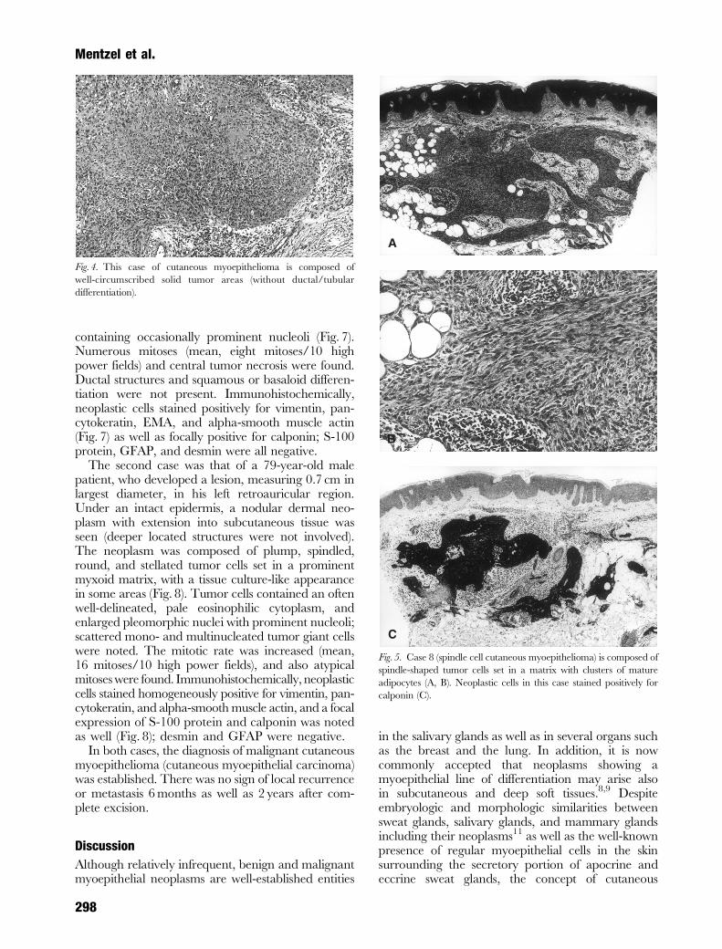
Fig. 4. This case of cutaneous myoepithelioma is composed of
well-circumscribed solid tumor areas (without ductal/tubular
differentiation).
A
B
C
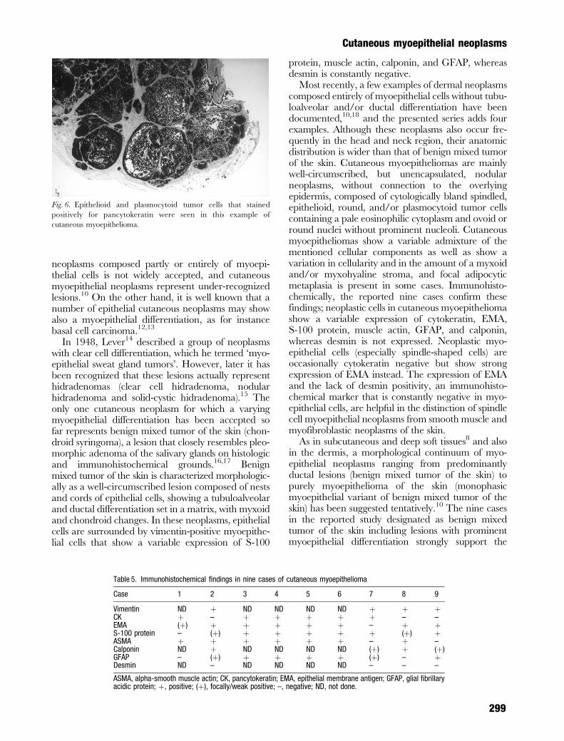
Fig. 5. Case 8 (spindle cell cutaneous myoepithelioma) is composed of
spindle-shaped tumor cells set in a matrix with clusters of mature
adipocytes (A, B). Neoplastic cells in this case stained positively for
calponin (C).
Mentzel et al.
298
neoplasms composed partly or entirely of myoepi-thelial cells is not widely accepted, and cutaneousmyoepithelial neoplasms represent under-recognizedlesions.10 On the other hand, it is well known that anumber of epithelial cutaneous neoplasms may showalso a myoepithelial differentiation, as for instancebasal cell carcinoma.12,13
In 1948, Lever14 described a group of neoplasmswith clear cell differentiation, which he termed ‘myo-epithelial sweat gland tumors’. However, later it hasbeen recognized that these lesions actually representhidradenomas (clear cell hidradenoma, nodularhidradenoma and solid-cystic hidradenoma).15 Theonly one cutaneous neoplasm for which a varyingmyoepithelial differentiation has been accepted sofar represents benign mixed tumor of the skin (chon-droid syringoma), a lesion that closely resembles pleo-morphic adenoma of the salivary glands on histologicand immunohistochemical grounds.16,17 Benignmixed tumor of the skin is characterized morphologic-ally as a well-circumscribed lesion composed of nestsand cords of epithelial cells, showing a tubuloalveolarand ductal differentiation set in a matrix, with myxoidand chondroid changes. In these neoplasms, epithelialcells are surrounded by vimentin-positive myoepithe-lial cells that show a variable expression of S-100
protein, muscle actin, calponin, and GFAP, whereasdesmin is constantly negative.
Most recently, a few examples of dermal neoplasmscomposed entirely of myoepithelial cells without tubu-loalveolar and/or ductal differentiation have beendocumented,10,18 and the presented series adds fourexamples. Although these neoplasms also occur fre-quently in the head and neck region, their anatomicdistribution is wider than that of benign mixed tumorof the skin. Cutaneous myoepitheliomas are mainlywell-circumscribed, but unencapsulated, nodularneoplasms, without connection to the overlyingepidermis, composed of cytologically bland spindled,epithelioid, round, and/or plasmocytoid tumor cellscontaining a pale eosinophilic cytoplasm and ovoid orround nuclei without prominent nucleoli. Cutaneousmyoepitheliomas show a variable admixture of thementioned cellular components as well as show avariation in cellularity and in the amount of a myxoidand/or myxohyaline stroma, and focal adipocyticmetaplasia is present in some cases. Immunohisto-chemically, the reported nine cases confirm thesefindings; neoplastic cells in cutaneous myoepitheliomashow a variable expression of cytokeratin, EMA,S-100 protein, muscle actin, GFAP, and calponin,whereas desmin is not expressed. Neoplastic myo-epithelial cells (especially spindle-shaped cells) areoccasionally cytokeratin negative but show strongexpression of EMA instead. The expression of EMAand the lack of desmin positivity, an immunohisto-chemical marker that is constantly negative in myo-epithelial cells, are helpful in the distinction of spindlecell myoepithelial neoplasms from smooth muscle andmyofibroblastic neoplasms of the skin.
As in subcutaneous and deep soft tissues8 and alsoin the dermis, a morphological continuum of myo-epithelial neoplasms ranging from predominantlyductal lesions (benign mixed tumor of the skin) topurely myoepithelioma of the skin (monophasicmyoepithelial variant of benign mixed tumor of theskin) has been suggested tentatively.10 The nine casesin the reported study designated as benign mixedtumor of the skin including lesions with prominentmyoepithelial differentiation strongly support the
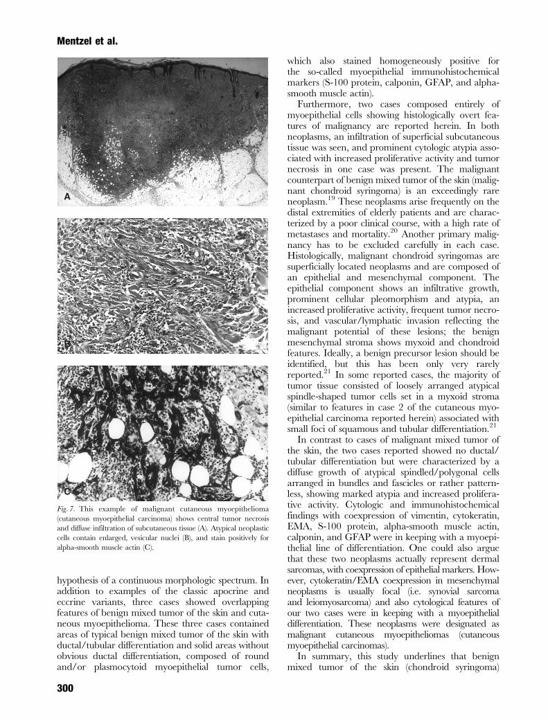
Fig. 6. Epithelioid and plasmocytoid tumor cells that stained
positively for pancytokeratin were seen in this example of
cutaneous myoepithelioma.
Table 5. Immunohistochemical findings in nine cases of cutaneous myoepithelioma
Case 1 2 3 4 5 6 7 8 9
Vimentin ND þ ND ND ND ND þ þ þCK þ – þ þ þ þ þ – –EMA (þ) þ þ þ þ þ – þ þS-100 protein – (þ) þ þ þ þ þ (þ) þASMA þ þ þ þ þ þ – þ –Calponin ND þ ND ND ND ND (þ) þ (þ)GFAP – (þ) þ þ þ þ (þ) – þDesmin ND – ND ND ND ND – – –
ASMA, alpha-smooth muscle actin; CK, pancytokeratin; EMA, epithelial membrane antigen; GFAP, glial fibrillaryacidic protein; þ, positive; (þ), focally/weak positive; –, negative; ND, not done.
Cutaneous myoepithelial neoplasms
299
hypothesis of a continuous morphologic spectrum. Inaddition to examples of the classic apocrine andeccrine variants, three cases showed overlappingfeatures of benign mixed tumor of the skin and cuta-neous myoepithelioma. These three cases containedareas of typical benign mixed tumor of the skin withductal/tubular differentiation and solid areas withoutobvious ductal differentiation, composed of roundand/or plasmocytoid myoepithelial tumor cells,
which also stained homogeneously positive forthe so-called myoepithelial immunohistochemicalmarkers (S-100 protein, calponin, GFAP, and alpha-smooth muscle actin).
Furthermore, two cases composed entirely ofmyoepithelial cells showing histologically overt fea-tures of malignancy are reported herein. In bothneoplasms, an infiltration of superficial subcutaneoustissue was seen, and prominent cytologic atypia asso-ciated with increased proliferative activity and tumornecrosis in one case was present. The malignantcounterpart of benign mixed tumor of the skin (malig-nant chondroid syringoma) is an exceedingly rareneoplasm.19 These neoplasms arise frequently on thedistal extremities of elderly patients and are charac-terized by a poor clinical course, with a high rate ofmetastases and mortality.20 Another primary malig-nancy has to be excluded carefully in each case.Histologically, malignant chondroid syringomas aresuperficially located neoplasms and are composed ofan epithelial and mesenchymal component. Theepithelial component shows an infiltrative growth,prominent cellular pleomorphism and atypia, anincreased proliferative activity, frequent tumor necro-sis, and vascular/lymphatic invasion reflecting themalignant potential of these lesions; the benignmesenchymal stroma shows myxoid and chondroidfeatures. Ideally, a benign precursor lesion should beidentified, but this has been only very rarelyreported.21 In some reported cases, the majority oftumor tissue consisted of loosely arranged atypicalspindle-shaped tumor cells set in a myxoid stroma(similar to features in case 2 of the cutaneous myo-epithelial carcinoma reported herein) associated withsmall foci of squamous and tubular differentiation.21
In contrast to cases of malignant mixed tumor ofthe skin, the two cases reported showed no ductal/tubular differentiation but were characterized by adiffuse growth of atypical spindled/polygonal cellsarranged in bundles and fascicles or rather pattern-less, showing marked atypia and increased prolifera-tive activity. Cytologic and immunohistochemicalfindings with coexpression of vimentin, cytokeratin,EMA, S-100 protein, alpha-smooth muscle actin,calponin, and GFAP were in keeping with a myoepi-thelial line of differentiation. One could also arguethat these two neoplasms actually represent dermalsarcomas, with coexpression of epithelial markers. How-ever, cytokeratin/EMA coexpression in mesenchymalneoplasms is usually focal (i.e. synovial sarcomaand leiomyosarcoma) and also cytological features ofour two cases were in keeping with a myoepithelialdifferentiation. These neoplasms were designated asmalignant cutaneous myoepitheliomas (cutaneousmyoepithelial carcinomas).
In summary, this study underlines that benignmixed tumor of the skin (chondroid syringoma)
A
B
C
Fig. 7. This example of malignant cutaneous myoepithelioma
(cutaneous myoepithelial carcinoma) shows central tumor necrosis
and diffuse infiltration of subcutaneous tissue (A). Atypical neoplastic
cells contain enlarged, vesicular nuclei (B), and stain positively for
alpha-smooth muscle actin (C).
Mentzel et al.
300
represents a dermal neoplasm of epithelial/myoepithelial differentiation. In addition, rare casesof cutaneous myoepithelioma without ductal differen-tiation and morphologically malignant neoplasmsshowing a myoepithelial line of differentiation arepresented. The evidence of focal solid areas in benignmixed tumor of the skin composed predominantly ofmyoepithelial cells and resembling histologic featuresof cutaneous myoepithelioma, common stromalchanges in cutaneous mixed tumor of the skin andcutaneous myoepithelioma (including myxoid changesas well as lipogenic and chondroid metaplasia) andoverlapping immunohistochemical features stronglysuggest the existence of a continuous spectrum ofcutaneous myoepithelial neoplasms ranging frombenign mixed tumor of the skin to cutaneous myo-epithelioma and cutaneous myoepithelial carcinoma.Further studies with extended follow-up informationare necessary to establish prognostic factors.
Acknowledgements
The authors are grateful to Professor P. C. W. Hogendoorn,
Department of Pathology Leiden, Netherlands, who kindly pro-
vided the second case of malignant cutaneous myoepithelioma.
References
1. Hamperl H. The myoepithelia (myoepithelial cells): normal
state; regressive changes; hyperplasia; tumors. Curr Top Pathol
1970; 53: 539.
2. Morinaga S, Nakajma T, Shimosato Y. Normal and neoplastic
myoepithelial cells in salivary glands. Hum Pathol 1987; 18:
1218.
3. Franke WW, Schmid E, Freudenstein C, et al. Intermediate-
sized filaments of the prekeratin type in myoepithelial cells.
J Cell Biol 1980; 84: 633.
4. Prasad AR, Savera AT, Gown AM, Zarbo RJ. The myoepithe-
lial immunophenotype in 135 benign and malignant salivary
gland tumors other than pleomorphic adenoma. Arch Pathol
Lab Med 1999; 123: 801.
5. Savera AT, Sloman A, Huvos AG, Klimstra DS. Myoepithelial
carcinoma of the salivary glands. A clinicopathologic study of
25 patients. Am J Surg Pathol 2000; 24: 761.
6. Tavassoli FA. Myoepithelial lesions of the breast: myoepithe-
liosis, adenomyoepithelioma, and myoepithelial carcinoma.
Am J Surg Pathol 1991; 15: 554.
7. Fulford LG, Kamata Y, Okudera K, et al. Epithelial-
myoepithelial carcinomas of the bronchus. Am J Surg Pathol
2001; 25: 1508.
8. Kilpatrick SE, Hitchcock MG, Kraus MD, Calonje E,
Fletcher CDM. Mixed tumors and myoepitheliomas of soft
tissue. A clinicopathologic study of 19 cases with a unifying
concept. Am J Surg Pathol 1997; 21: 13.
A B
C D
Fig. 8. The second case of malignant cutaneous myoepithelioma is composed of atypical spindled tumor cells set in a prominent myxoid matrix
with a tissue culture-like appearance (A). Neoplastic cells showed a coexpression of alpha-smooth muscle actin (B), pancytokeratin (C), and
calponin (D).
Cutaneous myoepithelial neoplasms
301
9. Michal M, Miettinen M. Myoepitheliomas of the skin and soft
tissues. Report of 12 cases. Virchows Arch 1999; 434: 393.
10. Kutzner H, Mentzel T, Kaddu S, et al. Cutaneous myoepithe-
lioma. An under-recognized cutaneous neoplasm composed of
myoepithelial cells. Am J Surg Pathol 2001; 25: 348.
11. Wick MR, Ockner DM, Mills SE, Ritter JH, Swanson PE.
Homologous carcinomas of the breasts, skins, and salivary
glands. A histologic and immunohistochemical comparison of
ductal mammary carcinomas, ductal sweat gland carcinoma,
and salivary duct carcinoma. Am J Clin Pathol 1998; 109: 75.
12. Suster S, Ramon y Cajal S. Myoepithelial differentiation in
basal cell carcinoma. Am J Dermatopathol 1991; 13: 350.
13. Kim YC, Vandersteen DP, Chung YJ, Myong NH. Signet
ring cell basal cell carcinoma. A basal cell carcinoma with
myoepithelial differentiation. Am J Dermatopathol 2001;
23: 525.
14. Lever WF. Myoepithelial sweat gland tumor: myoepithelioma –
report of three cases with a review of the literature. Arch
Dermatol 1948; 57: 332.
15. Requena L, Kiryu H, Ackerman AB. Apocrine hidradenoma.
In Neoplasms with apocrine differentiation. Philadelphia, PA:
Lippincott-Raven, 1998; 243.
16. Wilk M, Klesper D, Uerlich M, Biwer E, Wimheuer R. Chon-
droides Syringom. Immunhistologische Hinweise fur eine
myoepitheliale Differenzierung. Hautarzt 1994; 45: 324.
17. Hara K. Mixed tumours of the skin: a histopathological,
enzyme-histochemical and immunohistochemical study. Histo-
pathology 1995; 26: 145.
18. Fernandez-Figueras MT, Puig L, Trias I, Lorenzo JC,
Navas-Palacios JJ. Benign myoepithelioma of the skin. Am J
Dermatopathol 1998; 20: 208.
19. Harrist TJ, Aretz TH, Mihm MC, Evans GW. Cutaneous
malignant mixed tumor. Arch Dermatol 1981; 117: 719.
20. McKee PH, ed. Malignant chondroid syringoma. In Pathology
of the skin, 2nd edn. London: Mosby-Wolfe, 1996; 15.66.
21. Scott A, Metcalf JS. Cutaneous malignant mixed tumor.
Report of a case and review of the literature. Am J Dermato-
pathol 1988; 10: 335.
Mentzel et al.
302