Embed Size (px)
Citation preview
Diplomarbeit
Diagnostics and possibilities of clinical
management of residual placenta in cases with
suspected arteriovenous malformation of the
uterus or severe haemorrhage in the puerperium
eingereicht von
Sabine Enengl
geb. 06.10.1987
zur Erlangung des akademischen Grades
Doktorin der gesamten Heilkunde
(Dr. med. univ.)
an der
Medizinischen Universität Graz
ausgeführt an der
Universitätsklinik für Frauenheilkunde und Geburtshilfe
unter der Anleitung von
OA Dr. med. univ. Bence Csapo
Ao. Univ.- Prof. Dr. med. univ. Martin Häusler
Ort, Datum (Unterschrift)

i
Eidesstattliche Erklärung
Ich erkläre ehrenwörtlich, dass ich die vorliegende Arbeit selbstständig und ohne
fremde Hilfe verfasst habe, andere als die angegebenen Quellen nicht verwendet
habe und die den benutzten Quellen wörtlich oder inhaltlich entnommenen Stellen
als solche kenntlich gemacht habe.
Graz, am …………………. Unterschrift ……………………
ii
Acknowledgement
First of all I would like to thank my supervisors OA Dr. med. univ. Bence Csapo
and Ao. Univ.- Prof. Dr. med. univ. Martin Häusler from the Division of Obstetrics
and Maternal Fetal Medicine at the Medical University of Graz for giving me the
opportunity to write this thesis.
I would like to show my special gratitude to Dr. Csapo, who always had some
useful advice and took his precious time to answer all of my questions. It really
was a pleasure working at this department.
Thank you to all my friends and colleagues, who made the time of my studies a
special chapter of my life that I will always like to think back to.
Thank you to my friend and colleague Dr. Herbert Stockinger, who awoke my
interest for the subject of gynecology and obstetrics and always supported me to
expand my knowledge.
Above all I would like to express my gratitude to my beloved parents Inge
and Klaus, thank you for giving me the chance to study and find my own
way! It makes me proud to have parents like you, who always stand right
behind me and support me!
iii
Table of contents
Abbreviations and their explanation
List of figures
List of tables
Summary
Abstract
1 Introduction ..................................................................................................... 1
2 Theoretical examination .................................................................................. 2
2.1 General information .................................................................................. 2
2.1.1 Anatomy of the uterus ......................................................................... 2
2.1.2 Vascular supply .................................................................................. 4
2.1.3 The pregnant uterus ........................................................................... 5
2.1.4 The placenta ....................................................................................... 6
2.1.4.1 Placentation ................................................................................. 6
2.1.4.2 Fetal circulation ............................................................................ 7
2.1.4.3 The placental stage ...................................................................... 8
2.2 Special approach ..................................................................................... 9
2.2.1 Anomaly of placental separation ......................................................... 9
2.2.1.1 Management of residual placenta .............................................. 11
2.2.1.1.1 Ultrasonography.................................................................... 11
2.2.1.1.2 Magnetic resonance imaging ................................................ 13
2.2.1.1.3 Therapy of residual placenta ................................................. 14
2.2.2 Arteriovenous malformation of the uterus ......................................... 17
2.2.2.1 Definition .................................................................................... 17
2.2.2.2 Subtypes .................................................................................... 17
2.2.2.2.1 Congenital uterine AVMs ...................................................... 17
iv
2.2.2.2.2 Acquired uterine AVMs ......................................................... 18
2.2.2.3 Epidemiology .............................................................................. 18
2.2.2.4 Previous events .......................................................................... 19
2.2.2.5 Symptoms .................................................................................. 19
2.2.2.6 Diagnosis ................................................................................... 20
2.2.2.6.1 Clinical examination .............................................................. 20
2.2.2.6.2 Ultrasonography.................................................................... 20
2.2.2.6.3 Hysteroscopy ........................................................................ 23
2.2.2.6.4 Computed tomography angiogram ........................................ 23
2.2.2.6.5 Magnetic resonance imaging ................................................ 23
2.2.2.6.6 Pelvic angiography ................................................................ 24
2.2.2.7 Therapy ...................................................................................... 25
2.2.2.7.1 Acute measures .................................................................... 25
2.2.2.7.2 Hysterectomy ........................................................................ 25
2.2.2.7.3 Transcatheter arterial embolisation ....................................... 26
2.2.2.7.3.1 Complications .................................................................. 27
2.2.2.7.3.2 Post-procedure pregnancy .............................................. 28
2.2.2.7.4 Ligation of feeding arteries .................................................... 28
2.2.2.7.5 Medical treatment ................................................................. 29
2.2.3 Postpartum haemorrhage ................................................................. 30
3 Methods and Material .................................................................................... 31
3.1 Patient population .................................................................................. 31
3.2 Data acquisition ..................................................................................... 32
3.3 Analysis and statistical evaluation .......................................................... 34
4 Results .......................................................................................................... 35
4.1 Maternal facts ........................................................................................ 35
4.2 Pregnancy and delivery .......................................................................... 37

v
4.3 Placenta ................................................................................................. 40
4.4 Treatment ............................................................................................... 42
4.5 Haemorrhage ......................................................................................... 44
4.6 Postpartum anaemia .............................................................................. 47
5 Discussion ..................................................................................................... 52
6 References of the text ................................................................................... 56
vi
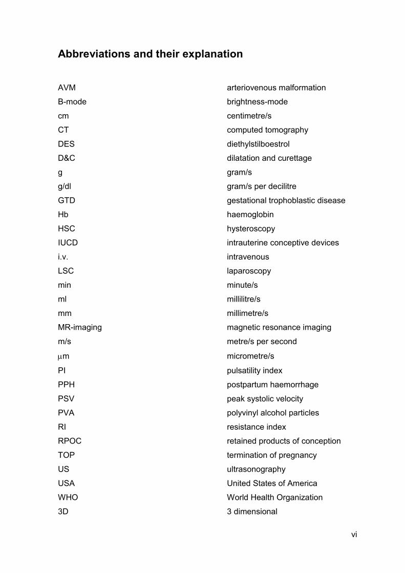
Abbreviations and their explanation
AVM arteriovenous malformation
B-mode brightness-mode
cm centimetre/s
CT computed tomography
DES diethylstilboestrol
D&C dilatation and curettage
g gram/s
g/dl gram/s per decilitre
GTD gestational trophoblastic disease
Hb haemoglobin
HSC hysteroscopy
IUCD intrauterine conceptive devices
i.v. intravenous
LSC laparoscopy
min minute/s
ml millilitre/s
mm millimetre/s
MR-imaging magnetic resonance imaging
m/s metre/s per second
m micrometre/s
PI pulsatility index
PPH postpartum haemorrhage
PSV peak systolic velocity
PVA polyvinyl alcohol particles
RI resistance index
RPOC retained products of conception
TOP termination of pregnancy
US ultrasonography
USA United States of America
WHO World Health Organization
3D 3 dimensional
vii
List of figures
Figure 1: General anatomy ..................................................................................... 3
Figure 2: Arteries of the female pelvis .................................................................... 4
Figure 3: Fetal Circulation ...................................................................................... 7
Figure 4: Echogenic mass in the uterine cavity .................................................... 12
Figure 5: Heterogenous mass (arrow) in uterine cavity ........................................ 13
Figure 6: Areas of enhancement (arrow) within mass, fluid (asterisk) matching
PPH in upper vagina ............................................................................................ 14
Figure 7: Approach of residual placenta ............................................................... 15
Figure 8: Manual removal of residual placenta ..................................................... 16
Figure 9: Gray-scale image of the arteriovenous malformation with prominent
cystic spaces within the myometrium ................................................................... 21
Figure 10: Longitudinal transvaginal ultrasound scan with colour Doppler
demonstrating mixed arterial and venous flow in the body of the uterus posteriorly
............................................................................................................................. 22
Figure 11: Early phase angiogram showing filling of the uterine artery and the
arteriovenous malformation .................................................................................. 24
Figure 12: Gray-scale image of the arteriovenous malformation following selective
embolisation ......................................................................................................... 26
Figure 13: Detailed distribution of maternal age ................................................... 35
Figure 14: Distribution of maternal age ................................................................ 36
Figure 15: Previous events ................................................................................... 36
Figure 16: Distribution of gestational age ............................................................. 37
Figure 17: Distribution of deliveries before and after the completed 24th week .... 38
Figure 18: Distribution of mode of delivery in cases after the 24th week ............... 38
Figure 19: Distribution of number of pregnancy .................................................... 39
Figure 20: Distribution of parity ............................................................................. 40
Figure 21: Type of placental anomaly .................................................................. 41
Figure 22: Further diagnosis documented in MEDOCS ....................................... 41
Figure 23: Treatment of retained placenta ........................................................... 42
Figure 24: Distribution of dates of treatment......................................................... 43
Figure 25: Weeks between delivery and treatment .............................................. 44
viii
Figure 26: Grade of postpartum haemorrhage ..................................................... 45
Figure 27: Management of severe haemorrhage ................................................. 45
Figure 28: Treatment of severe haemorrhage ...................................................... 46
Figure 29: Distribution of postpartum haemoglobin .............................................. 47
Figure 30: Postpartum Hb .................................................................................... 48
Figure 31: Management of postpartum anaemia with Hb <10 g/dl ....................... 48
Figure 32: Management of postpartum anaemia with Hb <8 g/dl ......................... 49
Figure 33: Correlation of severe haemorrhage and anaemia <10 g/dl ................. 50
Figure 34: Correlation of severe haemorrhage and anaemia <8 g/dl ................... 50
Figure 35: Course of haemoglobin ....................................................................... 51

ix
List of tables
Table 1: Considered diagnosis and procedures ................................................... 32
Table 2: Parameters of detailed analysis ............................................................. 33
x
Zusammenfassung
Hintergrund: In der Postpartal-Periode sind Blutungen ein großer Risikofaktor für
Mutter und Kind. Die Kombination von Plazentaresten, operativen Eingriffen und
arteriovenösen Fehlbildungen des Uterus (AVMs) könnte eine der Ursachen für
starke postpartale Blutungen sein. Es gibt in der aktuellen Literatur wenige
Berichte über uterine AVMs, die möglicherweise häufiger sind als angenommen.
Zielsetzung: Ziel dieser Studie war, mehr über die Prävalenz von AVMs in Fällen
mit Plazentaresten und schweren postpartalen Blutungen oder Blutungen im
Wochenbett herauszufinden. Den Patientinnen muss eine adäquate Behandlung
geboten werden, um das Risiko für Komplikationen zu minimieren.
Methode: Anhand einer retrospektiven Studie wurden alle Fälle mit
Plazentaresten an der Universitätsklinik für Frauenheilkunde und Geburtshilfe der
Medizinischen Universität Graz von 2003-2011 analysiert. Besondere
Aufmerksamkeit schenkte man Fällen mit schweren Blutungskomplikationen nach
operativen Eingriffen wie Cürretage.
Ergebnisse: Die Ergebnisse zeigen, dass AVMs als Ursache für postpartale
Blutungen eine spekulative Diagnose darstellen, da keine abnormalen
Gefäßverbindungen in den analysierten Fällen nachgewiesen wurden. Jedoch
präsentieren sich einige der Fälle mit einer Klinik, die zweifelsohne kompatibel mit
AVMs ist, weshalb das Krankheitsbild ein möglicher Risikofaktor für postpartale
Blutungskomplikationen bleibt. Vorhergehende Ereignisse, mütterliche
Risikofaktoren und Möglichkeiten des Managements von Blutverlust konnten
aufgezeigt werden und decken sich mit Ergebnissen der aktuellen Literatur.
Schlussfolgerung: Obwohl uterine AVMs eine Seltenheit zu sein scheinen und
oft nicht primär diagnostiziert werden, muss man an die Komplikationen denken,
die nach Plazentaresten und operativen Eingriffen auftreten können.
Schlagworte: Plazentareste, AVM, postpartale Blutung
xi
Abstract
Background: In the postpartum period haemorrhage is one of the greatest risk
factors for mother and child. The combination of residual placenta, which might
require surgical intervention, and arteriovenous malformations (AVMs) of the
uterus could be a main cause for severe haemorrhage postpartum. There are not
many cases of uterine AVMs reported in literature, but it is possible, that this entity
is more frequent than thought.
Objective: The aim of this study was to find out more about the prevalence of
uterine AVMs in cases with residual placenta and severe haemorrhage postpartum
or in the puerperium. It is important to offer an adequate treatment to the patients
to minimise the risk of maternal or fetal complications.
Methods: A retrospective study has been carried out, investigating all cases of
residual placenta at the Division of Obstetrics and Maternal Fetal Medicine at the
Medical University of Graz from 2003-2011, with special attention for cases with
severe bleeding complications after surgical interventions like dilatation &
curettage (D&C).
Results: The results show that uterine AVMs as cause for postpartum
haemorrhage seem to be a speculative diagnosis, as no abnormal vascular
connections were proved in the analysed cases. Some of the cases however did
show symptoms which are without doubt compatible with AVM, so it stays a
possible risk factor for bleeding complications in the postpartum period. Previous
events, maternal risk factors and options for the management of blood loss could
be explored and seem to correspond with current literature.
Conclusion: Even though AVMs seem to be a rarity and often stay unobserved, it
is important to be aware of the problem and the complications that might occur
after residual placenta and surgical intervention.
Key words: residual placenta, AVM, postpartum haemorrhage
1
1 Introduction
A common complication during the postpartum period is residual placenta resulting
in postpartum haemorrhage (PPH). Even in developed countries, PPH is one of
the greatest risk factors for maternal morbidity and mortality during birth. A close
examination during pregnancy and early diagnosis are essential for the individual
management of our patients. The combination of residual placenta and an
arteriovenous malformation (AVM) of the uterus could be a main cause for PPH.
AVM of the uterus seems to be a rarity. However it is necessary to take a closer
look at this problem as there are not many cases reported and the consequences
of AVMs can be very severe. Diagnostic methods have to be explored to learn
more about its prevalence, development and different occurrences. It is really
important to understand the risk of this entity in pregnant women as it could lead to
severe bleeding or even to maternal mortality if undiagnosed. As AVM of the
uterus could be a more frequent problem than thought, adequate clinical
management is imperative for a possible prevention or optimal management of
postpartum complications found in these cases.
This retrospective study aims to analyse cases of residual placenta with
postpartum haemorrhage and their clinical management at the Division of
Obstetrics and Maternal Fetal Medicine at the Medical University of Graz. The
clinical question was, how often AVMs could have been the cause of PPH. It was
also assessed what the management was and discussed what measures would be
necessary to reduce severe complications and maternal death.
2
2 Theoretical examination
2.1 General information
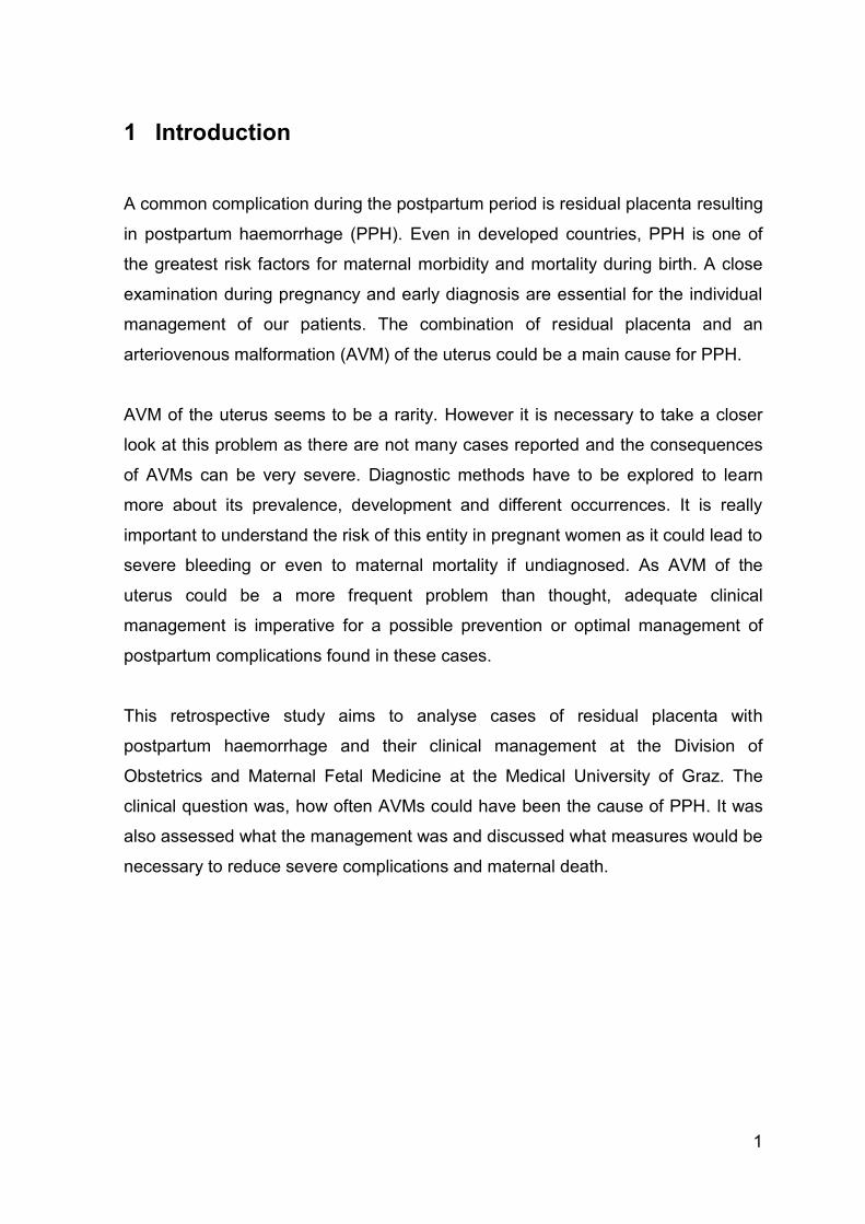
2.1.1 Anatomy of the uterus
The uterus is situated between the bladder and the rectum mostly in the antero-
posterior midline of the female pelvis, held in this position by the round and broad
ligaments on each side (ligamentum teres uteri and ligamentum latum). The broad
ligaments connect the uterus with the tubes and ovaries (1).
The shape of the uterus represents a pear turned upside down within which four
different parts are distinguished. The upper extremity called fundus is located
above the area where the two fallopian tubes commence into the uterine cavity
linking it to the ovary. The corpus, somewhat flattened in the sagittal plane, forms
the main part of the organ connecting the lower part (isthmus) and the fundus. It is
leaning forwards and upwards, narrowing from top to bottom. The anterior surface
of the uterus faces the bladder (facies vesicalis) and its posterior surface faces the
intestines (facies intestinalis). The isthmus of the uterus forms the connection
between corpus and cervix. It is not longer than 1 cm but during pregnancy it
unfolds and is called the lower uterine segment. The cervix’s shape reminds of a
cylinder measuring about 3 cm. Its lower extremity is attached to the vagina,
consisting of an anterior and a posterior lip shaping an aperture called uterine
orifice (ostium uteri). The uterine cavity is triangular flattened in an anterior-
posterior direction with the orifice of the tubes on top and it narrowing into the
isthmus below called canal of the isthmus followed by the canal of the cervix (1; 2;
3).
The longitudinal axis of the uterus is normally bending forwards in the area of the
isthmus, which leads to a flexure towards the bladder (anteflexio uteri) forming an
angle of 100-170 degrees. Similar to the axis of the uterus, the axis of the cervix
3
also shows a bend towards the anterior abdominal wall, which results in the effect
that the uterus seems to be laid over the bladder (anteversio uteri). Of course the
position of the uterus varies with the filling of the bladder and of the rectum (1; 2).
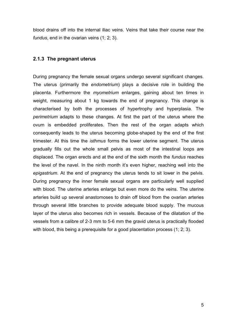
Figure 1: General anatomy
Data from Gilroy AM, MacPherson BR, Ross LM. Atlas of Anatomy. New York: Thieme Medical
Publishers, Inc; 2008. p. 189.
Concerning the structure of the uterine wall we differentiate three layers. It
normally shows an average thickness of about 1-2 cm and consists of a mucous
layer towards the cavum called endometrium, a muscular layer in the middle called
myometrium and the external serous layer called perimetrium. The endometrium
plays a dominant role in the process of placentation. The myometrium also
undergoes several changes during pregnancy. The perimetrium is derived from
the peritoneum forming the external coat of the organ (1; 2; 3).
4
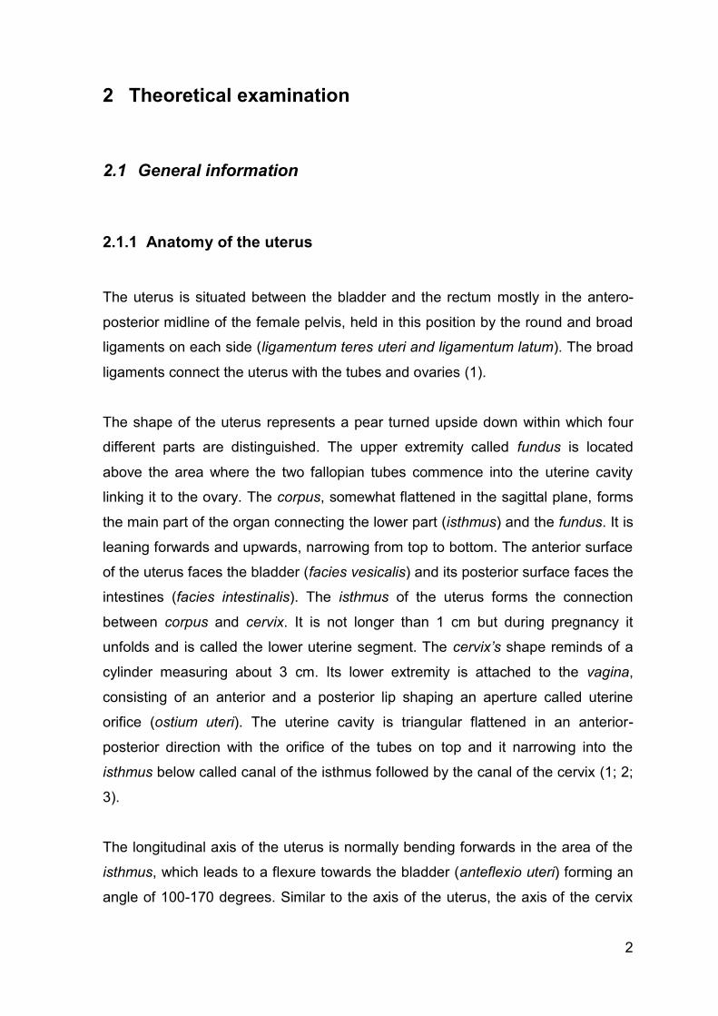
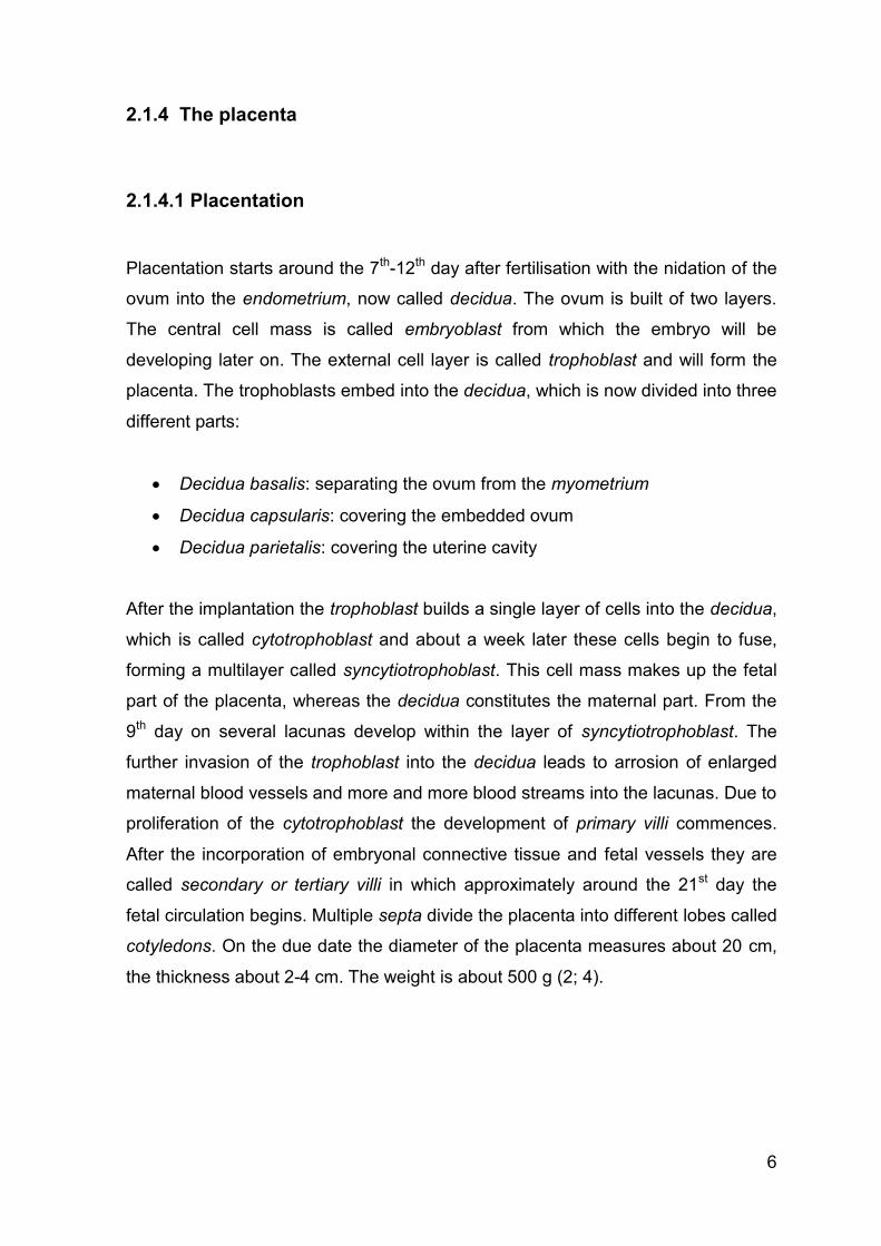
2.1.2 Vascular supply
The main artery of the uterus is the uterine artery running on each side from the
cervix-corpus margin both towards the fundus and the vagina. The uterine artery
runs downwards from the internal iliac artery towards the cervix in the broad
ligaments going along with the ureter at first. At this point the vaginal artery is
formed. It then goes upwards along the lateral border of the uterus in the direction
of the tubes. Along its route it gives off the main vessels for the uterine supply.
Furthermore it also supplies the tubes and ovaries through little branches (1; 2; 3).
The uterine arteries follow a tortuous course in the substance of the uterus and are
also very likely to build anastomoses (3).
Figure 2: Arteries of the female pelvis
Data from Gilroy AM, MacPherson BR, Ross LM. Atlas of Anatomy. New York: Thieme Medical
Publishers, Inc; 2008. p. 222.
Along the lateral borders of the uterus we find a large venous plexus. There are
two veins on each side, the uterine veins, which are remarkable for their large
size. The plexus is connected with the venous plexus of the vagina. Most of the
5
blood drains off into the internal iliac veins. Veins that take their course near the
fundus, end in the ovarian veins (1; 2; 3).
2.1.3 The pregnant uterus
During pregnancy the female sexual organs undergo several significant changes.
The uterus (primarily the endometrium) plays a decisive role in building the
placenta. Furthermore the myometrium enlarges, gaining about ten times in
weight, measuring about 1 kg towards the end of pregnancy. This change is
characterised by both the processes of hypertrophy and hyperplasia. The
perimetrium adapts to these changes. At first the part of the uterus where the
ovum is embedded proliferates. Then the rest of the organ adapts which
consequently leads to the uterus becoming globe-shaped by the end of the first
trimester. At this time the isthmus forms the lower uterine segment. The uterus
gradually fills out the whole small pelvis as most of the intestinal loops are
displaced. The organ erects and at the end of the sixth month the fundus reaches
the level of the navel. In the ninth month it’s even higher, reaching well into the
epigastrium. At the end of pregnancy the uterus tends to sit lower in the pelvis.
During pregnancy the inner female sexual organs are particularly well supplied
with blood. The uterine arteries enlarge but even more do the veins. The uterine
arteries build up several anastomoses to drain off blood from the ovarian arteries
through several little branches to provide adequate blood supply. The mucous
layer of the uterus also becomes rich in vessels. Because of the dilatation of the
vessels from a calibre of 2-3 mm to 5-6 mm the gravid uterus is practically flooded
with blood, this being a prerequisite for a good placentation process (1; 2; 3).
6
2.1.4 The placenta
2.1.4.1 Placentation
Placentation starts around the 7th-12th day after fertilisation with the nidation of the
ovum into the endometrium, now called decidua. The ovum is built of two layers.
The central cell mass is called embryoblast from which the embryo will be
developing later on. The external cell layer is called trophoblast and will form the
placenta. The trophoblasts embed into the decidua, which is now divided into three
different parts:
Decidua basalis: separating the ovum from the myometrium
Decidua capsularis: covering the embedded ovum
Decidua parietalis: covering the uterine cavity
After the implantation the trophoblast builds a single layer of cells into the decidua,
which is called cytotrophoblast and about a week later these cells begin to fuse,
forming a multilayer called syncytiotrophoblast. This cell mass makes up the fetal
part of the placenta, whereas the decidua constitutes the maternal part. From the
9th day on several lacunas develop within the layer of syncytiotrophoblast. The
further invasion of the trophoblast into the decidua leads to arrosion of enlarged
maternal blood vessels and more and more blood streams into the lacunas. Due to
proliferation of the cytotrophoblast the development of primary villi commences.
After the incorporation of embryonal connective tissue and fetal vessels they are
called secondary or tertiary villi in which approximately around the 21st day the
fetal circulation begins. Multiple septa divide the placenta into different lobes called
cotyledons. On the due date the diameter of the placenta measures about 20 cm,
the thickness about 2-4 cm. The weight is about 500 g (2; 4).
7
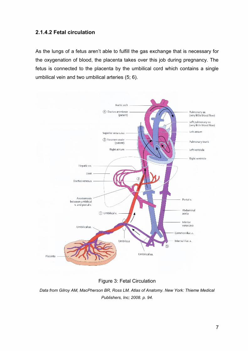
2.1.4.2 Fetal circulation
As the lungs of a fetus aren’t able to fulfill the gas exchange that is necessary for
the oxygenation of blood, the placenta takes over this job during pregnancy. The
fetus is connected to the placenta by the umbilical cord which contains a single
umbilical vein and two umbilical arteries (5; 6).
Figure 3: Fetal Circulation
Data from Gilroy AM, MacPherson BR, Ross LM. Atlas of Anatomy. New York: Thieme Medical
Publishers, Inc; 2008. p. 94.
8
The oxygenated blood from the placenta passes through the umbilical vein of the
fetus. Via the Ductus venosus a significant part of the blood enters the inferior
vena cava bypassing the liver. The rest of the blood leads into the portal vein and
passes the liver. In the inferior vena cava the oxygen-rich blood of the placenta
mixes with oxygen-poor blood from the fetus. Both the superior and inferior vena
cava enter the right atrium (5; 6).
Most of the blood from the inferior vena cava moves to the left atrium via the
Foramen ovale, thereby building a right-to-left-shunt. The mixed blood passes the
left ventricle and leaves through the aorta giving nutrients to the Truncus
brachiocephalicus, the left carotid and subclavian arteries. Deoxygenated blood
from the superior vena cava goes from the right atrium to the right ventricle into
the Truncus pulmonalis, which is connected to the aorta by another right-to-left-
shunt, the Ductus arteriosus Botalli (5; 6).
The partially oxygenated blood in the aorta supplies the fetal vessels and finally
enters the two umbilical arteries, which are branches of the internal iliac arteries,
and goes back to the placenta to be oxygenated again (5; 6).
2.1.4.3 The placental stage
The placental stage is the period after the birth of the child until the delivery of the
placenta and fetal membranes. This normally takes about ten to twenty minutes.
Due to a release of prostaglandins by the placenta itself, the uterus tends to
contract and shortens to a length of about 15 cm. This leads to a size reduction of
the surface, where the placenta is still attached to the uterus. In 75% the process
of detaching starts in the centre of the placenta. This mechanism is named after
Schulze. In the remaining 25% the separation of the placenta starts along its
border, named the method of Duncan. The advantage of Schulze’s method is the
loss of a smaller amount of blood (2; 4).
There are clinical signs of placental detachment (4; 7):
Sign of Schröder: the fundus retracts and rises above the detached
placenta
9
Sign of Küstner: pushing on the abdominal wall between symphysis and
navel leads to a retraction of the umbilical cord into the vagina if the
placenta hasn’t detached yet
Sign of Ahlfeld: during the course of ablation a clamp attached to the
umbilical cord moves caudally
The maternal and fetal parts of the placenta are delivered together. The dividing
line takes its course in the layer of the decidua basalis. The fetal membranes
consisting of decidua capsularis and parietalis are connected to the placenta. After
the detachment of the placenta it is very important to examine its surface to make
sure that there are no residuals left. Residual placenta means danger to life as it
might lead to severe haemorrhage, infection or trophoblastic tumour (8). The blood
loss during placental stage normally measures about 300 ml. Due to strong uterine
contractions and afterpains followed by lochia during childbed (puerperium) the
wound of the uterine mucous membrane is epithelialised approximately within ten
days (2; 4).
2.2 Special approach
2.2.1 Anomaly of placental separation
We talk about an anomaly of placental separation in the following three conditions:
if the placenta hasn’t detached within thirty minutes after delivery and/or if the
blood loss measures more than an estimated 300 ml and/or if the placenta hasn’t
been delivered completely and a residual placenta is assumed (4). This happens
in about 1% of pregnancies, more likely after termination of pregnancy (TOP) than
after vaginal or caesarean delivery (9). Residual placenta even of very small size
always means danger to life, so a thorough inspection for defects of the placenta
after its delivery is imperative. Residual placenta might lead to immediate
bleeding, haemorrhage, infection or even sepsis during childbed, and could be the
cause for trophoblastic tumour (8).
10
We distinguish between functional and anatomical reasons for the retention of
parts of the placenta (4; 7):
functional
- Adherent placenta (Placenta adhaerens): an anomaly of placental
separation caused by bradytocia
- Incarcerate placenta (Placenta incarcerate): the placenta is detached
but retained by a spasm of the cervix
anatomical
- Accrete placenta (Placenta accreta): the decidua is missing so the
villi reach up to the myometrium
- Increte placenta (Placenta increta): villi reach into the myometrium
- Percrete placenta (Placenta percreta): villi reach through the
myometrium into the perimetrium or even invade surrounding organs
like the urinary bladder
In cases of placenta adhaerens the placenta is not separating totally, in cases of
anatomical reason it’s not separating at all, however the anatomical cause behind
the failure of separation may not affect the whole of the placenta equally. In
pregnancy the uterine blood flow is highly increased. During the course of
placental ablation the maternal vessels are opened up but tend to get closed soon
after delivery of the placenta due to strong uterine contractions. A failure or arrest
of complete placental detachment leads to a delay in the haemostiptic effect of the
uterine contractions, which in turn leads to considerable amounts of blood loss. In
cases of accrete, increte and percrete placentas the affected part of the placenta
can’t be detached at all, so initially there may be no haemorrhage at all. The
diagnostic is based on missing clinical signs of placental detachment within thirty
minutes postpartum (4; 7).
11
2.2.1.1 Management of residual placenta
As residual placenta might lead to severe immediate or delayed postpartum
haemorrhage an early diagnosis is essential. It has been shown to be linked with
subsequent bleeding complications, hysterectomy and longer maternal hospital
stays (10). At first it is necessary to keep patients’ histories in mind. Previous
uterine surgeries like caesarean section or fibrome enucleations, previous D&C
(dilatation and evacuation or curettage), trauma, infection, placenta praevia,
Asherman’s Syndrome, presence of submucous leiomyomata and advanced
maternal age are the main risk factors for retained products of conception (RPOC)
(11; 12). The next step would be to check for possible anatomical reasons
predisposing for a retained placenta already during pregnancy. So, in case of
placenta accreta, increta or percreta the delivery can be planned in a clinic with
special service for perinatology, anaesthesia, diagnostic and invasive radiology,
haematology and blood transfusions (12). In case of PPH (both primary and
secondary) it is important to find out if there is truly residual placental tissue, as
this would be the only indication for a curettage, which can be traumatising during
the puerperium (13).
2.2.1.1.1 Ultrasonography
During pregnancy several ultrasonographic tests are carried out. It is necessary to
turn one’s attention to the placenta during this examination. There are several
signs in normal gray-scale ultrasonography (US), which might be a sign for
placenta accreta, percreta or increta (12):
Placental lacunae
Obliteration of clear space
Interruption of bladder border
Myometrium of less than 1 mm
12
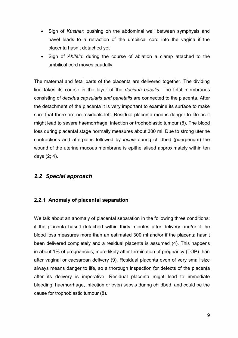
Figure 4: Echogenic mass in the uterine cavity
Data from Mulic-Lutvica A, Axelsson O. Ultrasound finding of an echogenic mass in women with
secondary postpartum hemorrhage is associated with retained placental tissue. Ultrasound Obstet
Gynecol. 2006 Sept;28(3):312-9.
In Colour-Doppler US the examiner should look for turbulent blood flow from the
placenta to surrounding tissues (12).
US can also be a useful tool to find out if RPOC is the cause for PPH after delivery
of the child. However ultrasound is not always accurate. Mulic-Lutvica et al. carried
out a prospective observational study to gather morphological findings
corresponding to residual placenta (13). 17 of 18 patients with secondary PPH
showed an echogenic mass in their uterine cavity and in 14 of these 17 cases the
presence of placental tissue was histologically confirmed. Patients with an
uneventful puerperium demonstrated either a mixed-echo pattern or a minor
amount of fluid in their uterine cavity, so it was concluded, that this would be the
common sign of the involuting uterus.
Even though the precision of US seems to vary, it is by all means a valid tool to
confirm an empty uterine cavity, if RPOC should not be the cause of PPH (13).
Furthermore it is also recommended to perform an US after uterine evacuation for
TOP, which leads to residual placenta in 1-3% (14). An endometrial thickness of ≥
8 mm measured by US after TOP should lead to further investigation and
eventually re-evacuation (14).
13
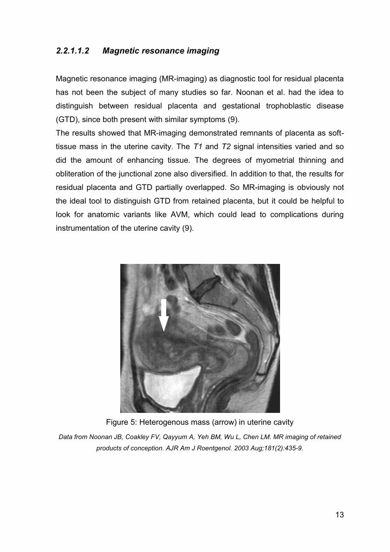
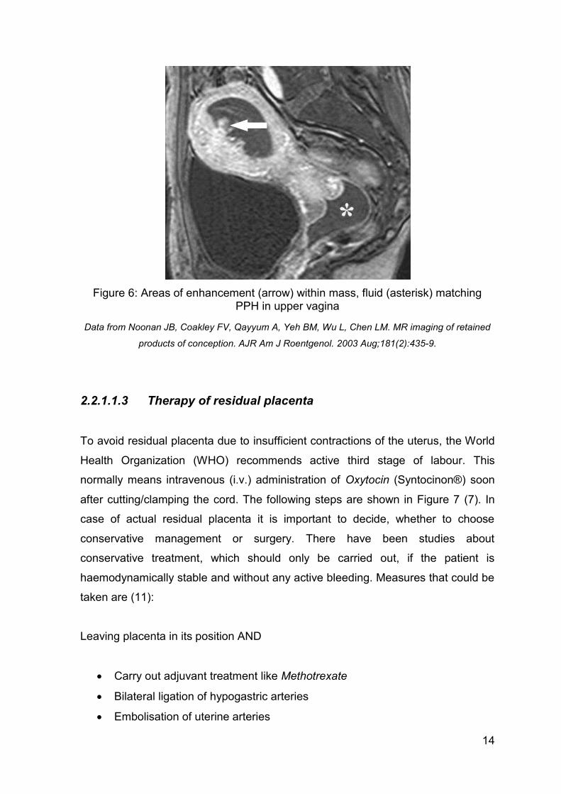
2.2.1.1.2 Magnetic resonance imaging
Magnetic resonance imaging (MR-imaging) as diagnostic tool for residual placenta
has not been the subject of many studies so far. Noonan et al. had the idea to
distinguish between residual placenta and gestational trophoblastic disease
(GTD), since both present with similar symptoms (9).
The results showed that MR-imaging demonstrated remnants of placenta as soft-
tissue mass in the uterine cavity. The T1 and T2 signal intensities varied and so
did the amount of enhancing tissue. The degrees of myometrial thinning and
obliteration of the junctional zone also diversified. In addition to that, the results for
residual placenta and GTD partially overlapped. So MR-imaging is obviously not
the ideal tool to distinguish GTD from retained placenta, but it could be helpful to
look for anatomic variants like AVM, which could lead to complications during
instrumentation of the uterine cavity (9).
Figure 5: Heterogenous mass (arrow) in uterine cavity
Data from Noonan JB, Coakley FV, Qayyum A, Yeh BM, Wu L, Chen LM. MR imaging of retained
products of conception. AJR Am J Roentgenol. 2003 Aug;181(2):435-9.
14
Figure 6: Areas of enhancement (arrow) within mass, fluid (asterisk) matching PPH in upper vagina
Data from Noonan JB, Coakley FV, Qayyum A, Yeh BM, Wu L, Chen LM. MR imaging of retained
products of conception. AJR Am J Roentgenol. 2003 Aug;181(2):435-9.
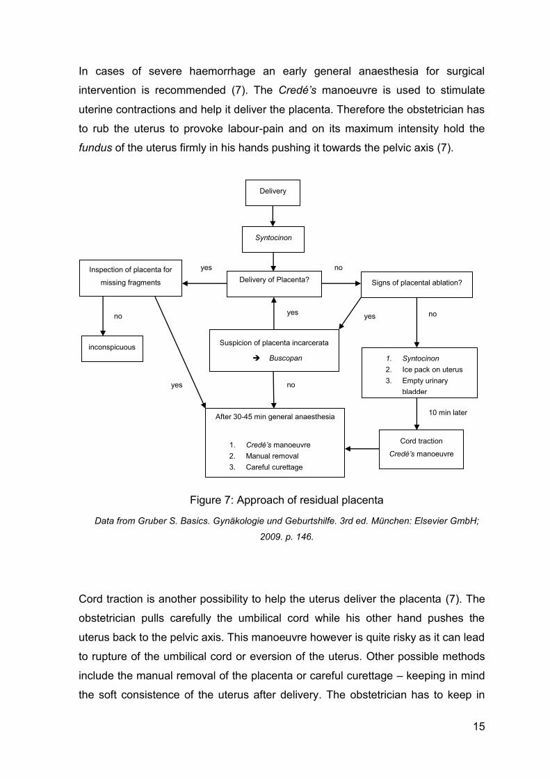
2.2.1.1.3 Therapy of residual placenta
To avoid residual placenta due to insufficient contractions of the uterus, the World
Health Organization (WHO) recommends active third stage of labour. This
normally means intravenous (i.v.) administration of Oxytocin (Syntocinon®) soon
after cutting/clamping the cord. The following steps are shown in Figure 7 (7). In
case of actual residual placenta it is important to decide, whether to choose
conservative management or surgery. There have been studies about
conservative treatment, which should only be carried out, if the patient is
haemodynamically stable and without any active bleeding. Measures that could be
taken are (11):
Leaving placenta in its position AND
Carry out adjuvant treatment like Methotrexate
Bilateral ligation of hypogastric arteries
Embolisation of uterine arteries
15
In cases of severe haemorrhage an early general anaesthesia for surgical
intervention is recommended (7). The Credé’s manoeuvre is used to stimulate
uterine contractions and help it deliver the placenta. Therefore the obstetrician has
to rub the uterus to provoke labour-pain and on its maximum intensity hold the
fundus of the uterus firmly in his hands pushing it towards the pelvic axis (7).
Cord traction is another possibility to help the uterus deliver the placenta (7). The
obstetrician pulls carefully the umbilical cord while his other hand pushes the
uterus back to the pelvic axis. This manoeuvre however is quite risky as it can lead
to rupture of the umbilical cord or eversion of the uterus. Other possible methods
include the manual removal of the placenta or careful curettage – keeping in mind
the soft consistence of the uterus after delivery. The obstetrician has to keep in
Inspection of placenta for
missing fragments
inconspicuous Suspicion of placenta incarcerata
Buscopan
After 30-45 min general anaesthesia
1. Credé’s manoeuvre
2. Manual removal
3. Careful curettage
Signs of placental ablation?
1. Syntocinon
2. Ice pack on uterus
3. Empty urinary
bladder
Cord traction
Credé’s manoeuvre
Delivery
Syntocinon
Delivery of Placenta?
no
no yes
yes
10 min later
no yes
no yes
Figure 7: Approach of residual placenta
Data from Gruber S. Basics. Gynäkologie und Geburtshilfe. 3rd ed. München: Elsevier GmbH;
2009. p. 146.
16
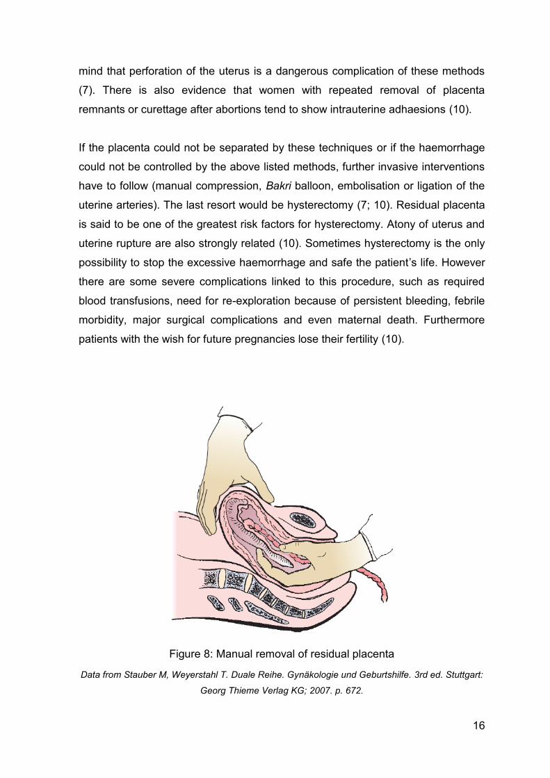
mind that perforation of the uterus is a dangerous complication of these methods
(7). There is also evidence that women with repeated removal of placenta
remnants or curettage after abortions tend to show intrauterine adhaesions (10).
If the placenta could not be separated by these techniques or if the haemorrhage
could not be controlled by the above listed methods, further invasive interventions
have to follow (manual compression, Bakri balloon, embolisation or ligation of the
uterine arteries). The last resort would be hysterectomy (7; 10). Residual placenta
is said to be one of the greatest risk factors for hysterectomy. Atony of uterus and
uterine rupture are also strongly related (10). Sometimes hysterectomy is the only
possibility to stop the excessive haemorrhage and safe the patient’s life. However
there are some severe complications linked to this procedure, such as required
blood transfusions, need for re-exploration because of persistent bleeding, febrile
morbidity, major surgical complications and even maternal death. Furthermore
patients with the wish for future pregnancies lose their fertility (10).
Figure 8: Manual removal of residual placenta
Data from Stauber M, Weyerstahl T. Duale Reihe. Gynäkologie und Geburtshilfe. 3rd ed. Stuttgart:
Georg Thieme Verlag KG; 2007. p. 672.
17
2.2.2 Arteriovenous malformation of the uterus
2.2.2.1 Definition
Arteriovenous malformations (AVMs) develop out of abnormal communications
between arteries and veins (15). The terms that have been used to describe this
entity include cirsoid aneurysm, pulsating angioma, arteriovenous aneurysm,
racemose aneurysm, arteriovenous fistula and cavernous haemangioma (16).
They are said to exist more likely in the pelvis, but only rarely do they affect the
uterus itself (17). The AVM is usually fed by one or more arteries, which then drain
into a venous plexus, whereby the intervening capillary network is missing (18).
Both the calibre of the different vessels as well as the extent of the uterine
involvement might vary enormously. In most cases reported, the vessels tend to
be prominent and dilated. The tortuous proliferation of channels leads to a diffuse
vascular malformation. This might either be located within or on the surface of the
uterine tissue, and the vessels might also open directly to the surface of the
endometrium (19). An exact histological investigation explores that the abnormal
proliferation shows both arterial and venous constituents with interconnecting
fistulas at which the proportions of different vessel types may vary (20).
2.2.2.2 Subtypes
Two different subtypes might be distinguished, but it is not yet clear which of them
is more common, a question where the current literature seems to be not quite
sure, according to the different cases reported (21; 22).
2.2.2.2.1 Congenital uterine AVMs
The congenital type is due to a dysfunction of normal vascular embryologic
development, so a vessel fails to differentiate either into an artery or a vein (23). It
tends to have several feeding arteries and draining veins, connected by a nidus
(24). Congenital AVMs are likely to invade the surrounding tissues and structures
18
(25). They are normally isolated, but they have also been reported in association
with AVMs at other sites (15).
2.2.2.2.2 Acquired uterine AVMs
They develop after or are related to GTD, especially following treatment with
chemotherapeutic agents, endometrial carcinoma or other uterine malignancies,
diethylstilboestrol (DES) exposure, uterine trauma including prior surgery or D&C,
use of intrauterine conceptive devices (IUCD), infection, subinvolution of placental
site (non-obliteration of vessels after miscarriage or delivery), retained products of
conception, traumatic fistula following caesarean section, necrosis of chorionic villi
(venous sinuses become incorporated in scars), endometriosis or previous
pregnancy (20; 26; 27; 28). This subtype is constituted by a single or bilateral
feeding artery joining a single vein without a connecting nidus but multiple
arteriovenous fistulas between the branches. Normally the uterine arteries build up
the malformation without supply from extrauterine arteries (24; 25).
2.2.2.3 Epidemiology
Uterine AVM seems to be a rarity, because there are not many cases reported.
We find only 73 in literature before 1997, but the true incidence is unknown. Most
of the published examples are case reports or just small case series, which makes
the exploration of the real frequency difficult (17). The first case was reported by
Dubreuil and Loubat in 1926 (19). The youngest patient with possible diagnosis of
uterine AVM was a stillborn at 34 weeks of gestation, and the oldest patient
documented was a 72 year-old woman. However, most of the patients suffering
from AVM were between the age of 20 and 40, which means that hormonal
changes might play a role concerning the origin of this disease (17). Though there
have been reports in adolescence as well as following the menopause, this entity
is more likely to appear among women of reproductive age and are more prevalent
in patients with bleeding complications in pregnancy, after delivery or uterine
instrumentation (20; 23). As uterine AVM could lead to potentially life threatening
19
complications, we have to be aware, that they are probably more frequent than
thought.
2.2.2.4 Previous events
As already discussed in chapter 2.2.2.2.2 many patients have undergone different
clinical procedures and maybe even complications or events at different times
before they were diagnosed with uterine AVM. These include pregnancies with or
without complications for retained products of gestation, caesarean section, IUCD,
spontaneous abortion, cervical conisation, termination for unwanted pregnancy by
D&C, D&C for abortion or miscarriage or any other previous surgery (19; 21; 22;
26). Most likely these interventions were the exact moment when the symptoms of
AVM showed for the first time, as the layer of endometrium covering the lesion is
very thin (20).
2.2.2.5 Symptoms
The main acute symptom every reported case showed was vaginal or uterine
bleeding either associated with medical intervention or (more rarely)
spontaneously but with no obvious cause. This varied from mild intermittent to
profuse or even torrential bleeding, so that in certain cases massive blood
transfusion was necessary(26). There have been intermenstrual, postmenopausal
and early as well as heavy delayed postpartum bleedings described (22; 26). The
postpartum haemorrhage often resulted partly from curettage performed for
residual placenta (20; 23). The loss of blood often led to a low haematocrit and
haemoglobin (anaemia) or even to tachycardia and shock in severe cases (17; 19;
21). Symptoms like lower abdominal or back pain and dyspareunia have also been
reported (27; 28). The chronic symptoms spotted were heart failure, fatigue and
dyspnoea due to a serious circulatory disturbance called vascular steal syndrome
(15; 25; 28). AVM could also be the reason for recurrent pregnancy wastage (29).
Although AVM always carries the risk of a great magnitude of blood loss, there
have been only two deaths reported in literature (15).
20
2.2.2.6 Diagnosis
Diagnosis of uterine AVM has proved to be difficult. Nevertheless it is important to
be aware of this lesion especially preoperatively in any patient suffering from any
form of recurrent abnormal vaginal or uterine bleeding. This is the only way the
bleeding is treated adequately and not by curettage which might be lethal (22).
Historically the diagnosis of uterine AVM was retrospective, as it was only proven
by laparotomy or hysterectomy performed for heavy bleeding (15; 19). Nowadays
these interventions as diagnostic and therapeutical methods might be avoided by
early diagnosis with the help of different examinations and techniques:
2.2.2.6.1 Clinical examination
The clinical examination including palpation of the lower abdomen and the
investigation of the vagina per speculum is the first step of diagnosis. Both a soft
and nontender abdomen can be found as well as a slightly painful one (15; 27).
Speculum examination revealed mild to heavy vaginal bleeding or at least signs of
blood clots in the vagina in most of the reported cases (15; 17; 19; 21; 27). Rarely
a pulsatile mass in the pelvis was detected (20). The uterus is usually anteverted
and is either of normal size or slightly enlarged, nontender or bulky (17; 18; 27).
No thrill or bruit was mentioned (27). Further steps of investigation include the
evaluation of coagulation studies and a pregnancy test, so it is possible to exclude
differential diagnoses like GTD or coagulopathy (15; 27).
2.2.2.6.2 Ultrasonography
Transvaginal US is the most common imaging technique carried out for the
diagnosis of uterine or vaginal bleeding. However the use of gray-scale US alone
is probably not enough to distinguish between AVM and other pathologies, but
might of course play a role (20; 21).
21
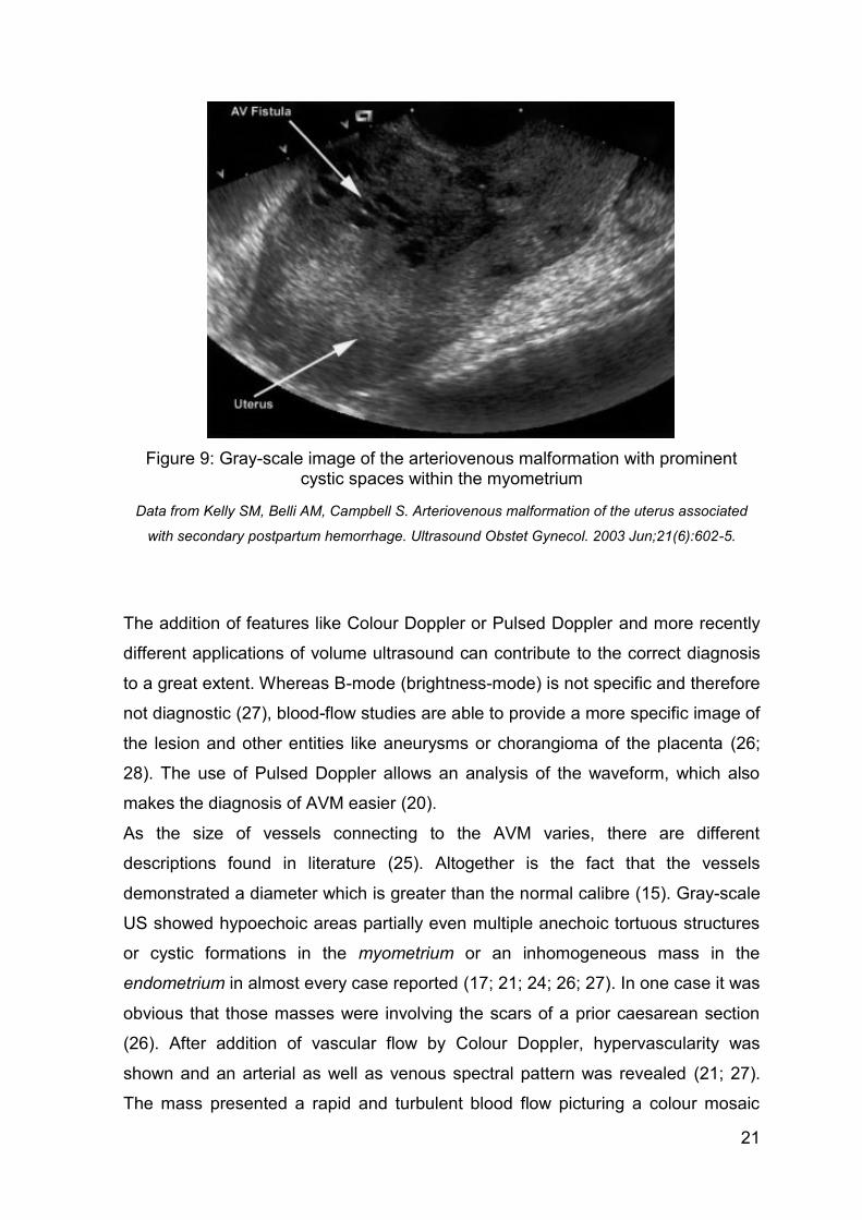
Figure 9: Gray-scale image of the arteriovenous malformation with prominent cystic spaces within the myometrium
Data from Kelly SM, Belli AM, Campbell S. Arteriovenous malformation of the uterus associated
with secondary postpartum hemorrhage. Ultrasound Obstet Gynecol. 2003 Jun;21(6):602-5.
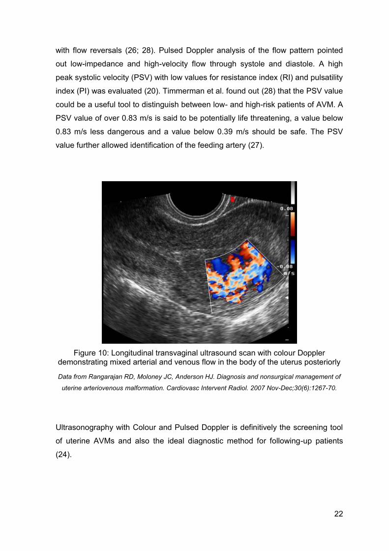
The addition of features like Colour Doppler or Pulsed Doppler and more recently
different applications of volume ultrasound can contribute to the correct diagnosis
to a great extent. Whereas B-mode (brightness-mode) is not specific and therefore
not diagnostic (27), blood-flow studies are able to provide a more specific image of
the lesion and other entities like aneurysms or chorangioma of the placenta (26;
28). The use of Pulsed Doppler allows an analysis of the waveform, which also
makes the diagnosis of AVM easier (20).
As the size of vessels connecting to the AVM varies, there are different
descriptions found in literature (25). Altogether is the fact that the vessels
demonstrated a diameter which is greater than the normal calibre (15). Gray-scale
US showed hypoechoic areas partially even multiple anechoic tortuous structures
or cystic formations in the myometrium or an inhomogeneous mass in the
endometrium in almost every case reported (17; 21; 24; 26; 27). In one case it was
obvious that those masses were involving the scars of a prior caesarean section
(26). After addition of vascular flow by Colour Doppler, hypervascularity was
shown and an arterial as well as venous spectral pattern was revealed (21; 27).
The mass presented a rapid and turbulent blood flow picturing a colour mosaic
22
with flow reversals (26; 28). Pulsed Doppler analysis of the flow pattern pointed
out low-impedance and high-velocity flow through systole and diastole. A high
peak systolic velocity (PSV) with low values for resistance index (RI) and pulsatility
index (PI) was evaluated (20). Timmerman et al. found out (28) that the PSV value
could be a useful tool to distinguish between low- and high-risk patients of AVM. A
PSV value of over 0.83 m/s is said to be potentially life threatening, a value below
0.83 m/s less dangerous and a value below 0.39 m/s should be safe. The PSV
value further allowed identification of the feeding artery (27).
Figure 10: Longitudinal transvaginal ultrasound scan with colour Doppler demonstrating mixed arterial and venous flow in the body of the uterus posteriorly
Data from Rangarajan RD, Moloney JC, Anderson HJ. Diagnosis and nonsurgical management of
uterine arteriovenous malformation. Cardiovasc Intervent Radiol. 2007 Nov-Dec;30(6):1267-70.
Ultrasonography with Colour and Pulsed Doppler is definitively the screening tool
of uterine AVMs and also the ideal diagnostic method for following-up patients
(24).
23
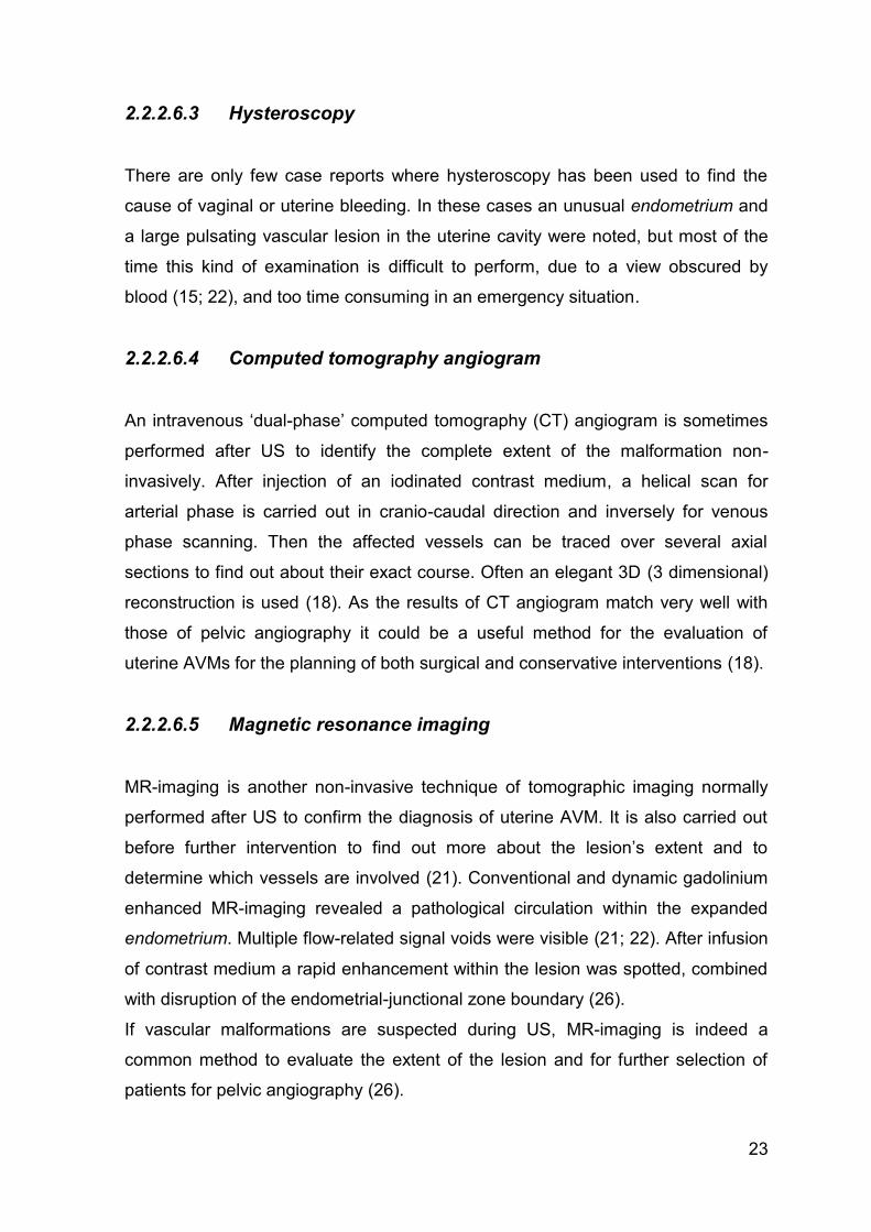
2.2.2.6.3 Hysteroscopy
There are only few case reports where hysteroscopy has been used to find the
cause of vaginal or uterine bleeding. In these cases an unusual endometrium and
a large pulsating vascular lesion in the uterine cavity were noted, but most of the
time this kind of examination is difficult to perform, due to a view obscured by
blood (15; 22), and too time consuming in an emergency situation.
2.2.2.6.4 Computed tomography angiogram
An intravenous ‘dual-phase’ computed tomography (CT) angiogram is sometimes
performed after US to identify the complete extent of the malformation non-
invasively. After injection of an iodinated contrast medium, a helical scan for
arterial phase is carried out in cranio-caudal direction and inversely for venous
phase scanning. Then the affected vessels can be traced over several axial
sections to find out about their exact course. Often an elegant 3D (3 dimensional)
reconstruction is used (18). As the results of CT angiogram match very well with
those of pelvic angiography it could be a useful method for the evaluation of
uterine AVMs for the planning of both surgical and conservative interventions (18).
2.2.2.6.5 Magnetic resonance imaging
MR-imaging is another non-invasive technique of tomographic imaging normally
performed after US to confirm the diagnosis of uterine AVM. It is also carried out
before further intervention to find out more about the lesion’s extent and to
determine which vessels are involved (21). Conventional and dynamic gadolinium
enhanced MR-imaging revealed a pathological circulation within the expanded
endometrium. Multiple flow-related signal voids were visible (21; 22). After infusion
of contrast medium a rapid enhancement within the lesion was spotted, combined
with disruption of the endometrial-junctional zone boundary (26).
If vascular malformations are suspected during US, MR-imaging is indeed a
common method to evaluate the extent of the lesion and for further selection of
patients for pelvic angiography (26).
24
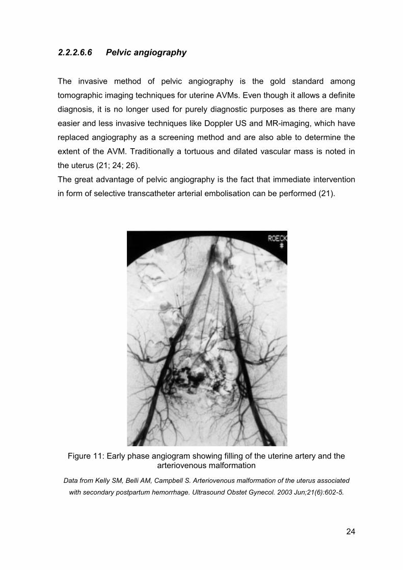
2.2.2.6.6 Pelvic angiography
The invasive method of pelvic angiography is the gold standard among
tomographic imaging techniques for uterine AVMs. Even though it allows a definite
diagnosis, it is no longer used for purely diagnostic purposes as there are many
easier and less invasive techniques like Doppler US and MR-imaging, which have
replaced angiography as a screening method and are also able to determine the
extent of the AVM. Traditionally a tortuous and dilated vascular mass is noted in
the uterus (21; 24; 26).
The great advantage of pelvic angiography is the fact that immediate intervention
in form of selective transcatheter arterial embolisation can be performed (21).
Figure 11: Early phase angiogram showing filling of the uterine artery and the arteriovenous malformation
Data from Kelly SM, Belli AM, Campbell S. Arteriovenous malformation of the uterus associated
with secondary postpartum hemorrhage. Ultrasound Obstet Gynecol. 2003 Jun;21(6):602-5.
25
2.2.2.7 Therapy
2.2.2.7.1 Acute measures
As the main acute symptom of uterine AVM is haemorrhage, the first measure that
should be taken is the controlling of active bleeding for haemodynamic
stabilisation (26; 27). In accordance with literature this has either been achieved
by tamponade, occlusion with Foley catheter bulb or intramuscular injection of
methylergonovine or prostaglandin F2a (18; 26). In 30% of the reported cases, the
bleeding was so severe that massive transfusions of blood were necessary
because of anaemia or shock (21).
It is important always to be aware of possible AVMs so that contraindicated
procedures like curettage, which might aggravate the bleeding, can be avoided in
any case (19; 26).
2.2.2.7.2 Hysterectomy
Hysterectomy has historically been the therapy of choice for vaginal bleeding,
especially as emergency hysterectomy for intractable uterine haemorrhage (15).
Nowadays it is still carried out after recurrence of uterine AVM, non-successful
attempts of other treatments like transcatheter arterial embolisation or after
complications like heavy bleeding due to curettage, as it is by all means the
definitive treatment to eliminate bleeding and its consequences (15; 17; 26; 27).
The obvious disadvantage of hysterectomy for treatment of uterine AVM is the loss
of fertility, so among patients in reproductive years this therapy might lead to
considerable psychological distress (21; 22). Furthermore there are reports of
uterine AVMs which could not be cured by hysterectomy as the lesion was
spreading to the upper vagina and the symptoms stayed (30).
26
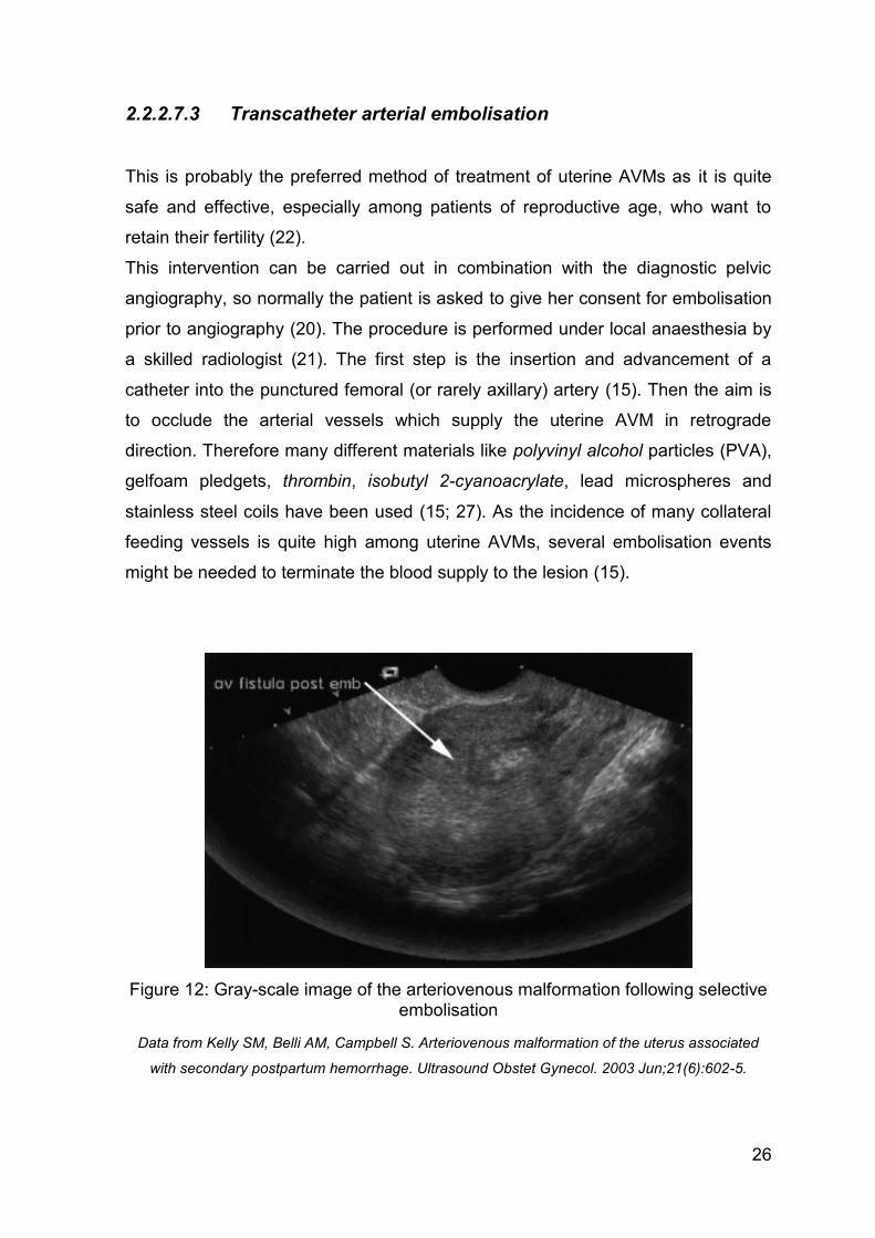
2.2.2.7.3 Transcatheter arterial embolisation
This is probably the preferred method of treatment of uterine AVMs as it is quite
safe and effective, especially among patients of reproductive age, who want to
retain their fertility (22).
This intervention can be carried out in combination with the diagnostic pelvic
angiography, so normally the patient is asked to give her consent for embolisation
prior to angiography (20). The procedure is performed under local anaesthesia by
a skilled radiologist (21). The first step is the insertion and advancement of a
catheter into the punctured femoral (or rarely axillary) artery (15). Then the aim is
to occlude the arterial vessels which supply the uterine AVM in retrograde
direction. Therefore many different materials like polyvinyl alcohol particles (PVA),
gelfoam pledgets, thrombin, isobutyl 2-cyanoacrylate, lead microspheres and
stainless steel coils have been used (15; 27). As the incidence of many collateral
feeding vessels is quite high among uterine AVMs, several embolisation events
might be needed to terminate the blood supply to the lesion (15).
Figure 12: Gray-scale image of the arteriovenous malformation following selective embolisation
Data from Kelly SM, Belli AM, Campbell S. Arteriovenous malformation of the uterus associated
with secondary postpartum hemorrhage. Ultrasound Obstet Gynecol. 2003 Jun;21(6):602-5.
27
Because of the different characteristics of congenital and acquired AVMs, it seems
to be much easier to treat the acquired subtype, as there is no intervening network
and mostly only two vessels, a feeding and a draining one (24).
One disadvantage of transcatheter arterial embolisation is that uterine AVMs are
likely to build several collaterals, so recurrence happens quite often and thus more
than one procedure is frequently required (22; 26). For this reason several reports
mention, that it is probably necessary to perform the procedure from both sides, so
that the AVM can’t be supplied by the corresponding vessel of the contralateral
side (22; 31).
However, other reports are of the opinion that an AVM fed by vessels of only one
side should be treated by selective unilateral embolisation, as this minimises the
complications for further pregnancies. The treated area is inevitably hypovascular
which might affect the placenta and fetal growth (21).
Recently US-guided transvaginal embolisation of AVMs with fibrin glue was safely
and successfully achieved (32).
2.2.2.7.3.1 Complications
Besides from recurrence of the lesion which can lead to a repetition of the
procedure, there are not many complications reported and most of the patients
showed an uneventful recovery. In some cases post-procedure fever occurred,
which could be controlled conservatively (22; 26). Mild suprapubic pain was also
described (22). One case report mentioned a vascular rupture after follow-up
angiography, due to the high injection pressure in the coil occluded artery (22).
There are some concerns about future pregnancies expressed in literature, which
are discussed in the following chapter.
By all means frequent follow-up examinations by US, MR-imaging and
angiography are advisable (21; 22).
28
2.2.2.7.3.2 Post-procedure pregnancy
Although transcatheter arterial embolisation of uterine AVMs seems to be a safe
and effective method of treatment, its long term effect on fertility has not been fully
evaluated (33). In the majority of patients the therapy did not influence the uterine
or ovarian function which led to the return of a normal menstrual cycling allowing
successful pregnancies (22; 33). Nevertheless transient and also persistent
amenorrhea has been reported, which is probably caused by a loss of ovarian
arterial perfusion, which can fortunately be re-established in the majority of women
(33). However, there are not many reports of successful pregnancies in current
literature. The first case was described by Chapman and Lutz in 1985 (31). All in
all between 1985 and 2005 thirteen cases of successful delivery have been
mentioned, with delays between embolisation and pregnancy varying from six
weeks to five years (31). Another report mentions a patient with ectopic tubal
pregnancy after embolisation. As she was thought to have had normal
reproductive function before the procedure, the hypothesis occurred, that
transcatheter arterial embolisation might disturb the function of the fallopian tube
(33). There is one case of embolisation after which a patient underwent in vitro
fertilisation with uterine embryo transfer, which led to a dichorionic diamniotic twin
intrauterine gestation (29).
Summarily it can be said, that pregnancy after transcatheter arterial embolisation
carries a higher risk of prematurity, placental abnormality, ovarian dysfunction or
abortion, whereas growth retardation and uterine rupture have not been reported
(27). As the complications mentioned above are most likely due to a disturbance of
perfusion, selective unilateral embolisation with preservation of corresponding
vessels seems to improve the obstetric outlook. Furthermore delivery per
caesarean section might be advisable (31).
2.2.2.7.4 Ligation of feeding arteries
As mentioned above transcatheter arterial embolisation does not always cure
patients from uterine AVMs. In some cases, after an unsuccessful transcatheter
intervention another treatment might be necessary. Normally, hysterectomy is the
29
definitive treatment. However in some cases this procedure might be too
dangerous as it carries a higher risk of massive blood loss. That’s the case in
patients with AVM of extraordinarily dilated and tortuous vessels within and around
the uterus’ tissue. If so, the surgical ligation of the feeding arteries should be the
therapy of choice (30).
The repetition of transcatheter arterial embolisation in those cases is also not
indicated, as a controlled catheterisation in such dilated vessels is quite difficult
and the higher blood flow carries the risk of pulmonary embolism caused by
embolic materials (30).
2.2.2.7.5 Medical treatment
In some selected cases mainly in acquired uterine AVMs with moderate symptoms
a conservative treatment in the form of medical therapy is also possible (31).
There are only few cases reported with treatment of either methylergonovine or
hormonal. The advantage is the non-invasive procedure which furthermore
preserves fertility among patients in reproductive years (27). The hormonal
treatment used in the published case was a combined oral contraceptive pill with
50 µg ethinyl oestradiol called Microgynon50®. As the reported AVM was of the
acquired subtype, it might also have regressed spontaneously or as result of
therapy with the contraceptive pill. Oestrogen therapy has also been used to cure
gastrointestinal AVMs causing bleeding, but the exact mechanism is still unclear. It
could be the hormonal influence on the endothel’s integrity or an effect on the
bleeding time (16). Methylergonovine and hormonal therapy has been prescribed
to control active bleeding as well as for long-term use (16). Treatment with
goserelin acetate (Zoladex®, Zeneca®) as initial therapy and after unsuccessful
transcatheter intervention is also mentioned in literature, but doesn’t seem to be
quite effective (22).
In moderate cases of uterine AVM medical treatment could indeed be a successful
way of therapy, however, regular follow-up is indicated in all cases (31).
30
2.2.3 Postpartum haemorrhage
The placental stage is quite risky for the parturient as severe postpartum
haemorrhage (PPH) might occur. PPH is defined by an amount of blood loss of
over 500 ml within 2-4 hours postpartum or over 1000 ml after caesarean section.
There are several reasons for PPH like lesions of the vagina or coagulation
disorders, the main reason however is a strong bleeding of the wound where the
placenta has been attached to the endometrium of the uterus. This might happen
after complete separation of the placenta or even with residual placenta (8).
There are many different definitions going round concerning the amount of blood
loss of PPH and so are different classifications. In Australia, Belgium, Canada,
France, the United Kingdom and the USA PPH is stratified by cause (13; 34):
Third stage haemorrhage: PPH due to retained placenta
Other immediate PPH within the first 24 hours following delivery of placenta
(uterine atony)
Delayed and secondary PPH: after the first 24 hours following delivery to 12
weeks postpartum
Postpartum coagulation defects
Other causes
The most common causes for secondary PPH are an abnormal involution of the
placental site, endometritis or retained placental tissue. AVM of the uterus is
another rare cause (13). PPH before or after delivery of placenta is the greatest
risk factor of maternal mortality (8).
31
3 Methods and Material
At the Division of Obstetrics and Maternal Fetal Medicine at the Medical University
of Graz the course of pregnancies, births and postpartum periods is documented
via electronic health records called MEDOCS, PIA and ViewPoint since
approximately 2003. AVMs of the uterus are known to be a possible cause of
severe haemorrhage postpartum or in the puerperium. As this entity is rarely
diagnosed before the occurrence of postpartum complications, for this study cases
with residual placenta and postpartum surgical events like D&C have been
investigated in order to find events showing the possible diagnosis of uterine
AVMs. A retrospective data analysis has been carried out by investigating cases of
patients with postpartum operations like D&C or hysteroscopy due to residual
placenta or ongoing postpartum haemorrhage. Special attention was given to
patients, who suffered from complications (severe haemorrhage, repeat
interventions) after the procedure mentioned above, as this could be a sign of
undetected AVM of the uterus.
For the query only electronic health records were analysed, handwritten patient
charts or nursing documents have not been used.
3.1 Patient population
In an attempt to find all cases of possible postpartum haemorrhage due to AVMs
of the uterus we have conducted a query of the two electronic patients records
systems: MEDOCS – the general electronic documentation software of the
department including both obstetrical and gynaecological cases and especially
records of surgical interventions and secondly ViewPoint Fetal Database – a
primarily obstetrical electronic patient records system.
The study includes all patients of the Division of Obstetrics and Maternal Fetal
Medicine at the Medical University of Graz, who have been diagnosed with
residual placenta after birth from 2003 to 2011. Not only cases with residual
placenta after live birth were analysed, but also residual placenta after
spontaneous, missed or medically induced abortion. The maternal age included
32
was age 17 to 45. The study contains primiparae as well as multiparae. Both
singleton and multiple pregnancies that fulfilled the search criteria were included in
the data analysis.
3.2 Data acquisition
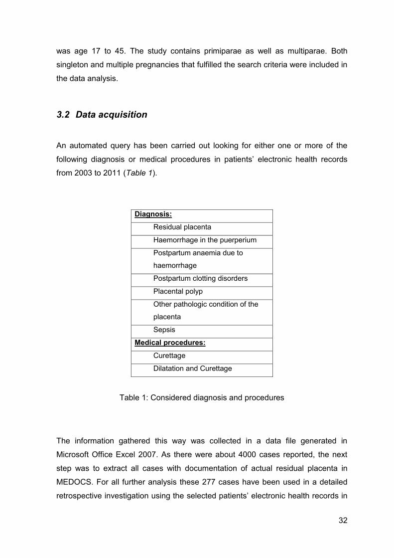
An automated query has been carried out looking for either one or more of the
following diagnosis or medical procedures in patients’ electronic health records
from 2003 to 2011 (Table 1).
Diagnosis:
Residual placenta
Haemorrhage in the puerperium
Postpartum anaemia due to
haemorrhage
Postpartum clotting disorders
Placental polyp
Other pathologic condition of the
placenta
Sepsis
Medical procedures:
Curettage
Dilatation and Curettage
Table 1: Considered diagnosis and procedures
The information gathered this way was collected in a data file generated in
Microsoft Office Excel 2007. As there were about 4000 cases reported, the next
step was to extract all cases with documentation of actual residual placenta in
MEDOCS. For all further analysis these 277 cases have been used in a detailed
retrospective investigation using the selected patients’ electronic health records in
33
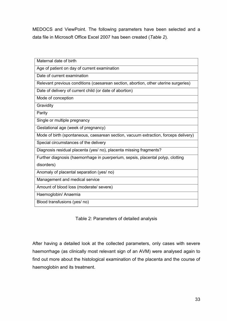
MEDOCS and ViewPoint. The following parameters have been selected and a
data file in Microsoft Office Excel 2007 has been created (Table 2).
Maternal date of birth
Age of patient on day of current examination
Date of current examination
Relevant previous conditions (caesarean section, abortion, other uterine surgeries)
Date of delivery of current child (or date of abortion)
Mode of conception
Gravidity
Parity
Single or multiple pregnancy
Gestational age (week of pregnancy)
Mode of birth (spontaneous, caesarean section, vacuum extraction, forceps delivery)
Special circumstances of the delivery
Diagnosis residual placenta (yes/ no), placenta missing fragments?
Further diagnosis (haemorrhage in puerperium, sepsis, placental polyp, clotting
disorders)
Anomaly of placental separation (yes/ no)
Management and medical service
Amount of blood loss (moderate/ severe)
Haemoglobin/ Anaemia
Blood transfusions (yes/ no)
Table 2: Parameters of detailed analysis
After having a detailed look at the collected parameters, only cases with severe
haemorrhage (as clinically most relevant sign of an AVM) were analysed again to
find out more about the histological examination of the placenta and the course of
haemoglobin and its treatment.
34
3.3 Analysis and statistical evaluation
After the acquisition of the relevant data had been completed, the analysis was
carried out using Microsoft Office Excel 2007. Using the filtering function, tables
and figures were generated by bringing the collected parameters into relation.
Firstly the whole patient population was analysed and secondly cases before and
after the completed 24th week of pregnancy were examined separately.
35
4 Results
All together there were 277 patients between 2003 and 2011 showing the
diagnosis ‘residual placenta’ (code o73.0 or o73.1) in their electronic MEDOCS
health records. More precisely spoken there were actually 273 patients but 277
cases, as 4 patients showed the diagnosis twice in the period mentioned above. In
the following chapters those four cases will be treated as individual patients.
4.1 Maternal facts
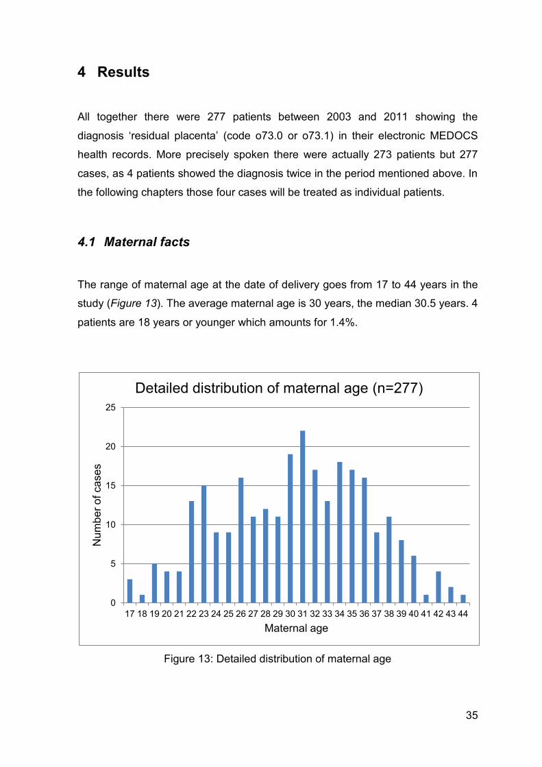
The range of maternal age at the date of delivery goes from 17 to 44 years in the
study (Figure 13). The average maternal age is 30 years, the median 30.5 years. 4
patients are 18 years or younger which amounts for 1.4%.
Figure 13: Detailed distribution of maternal age
0
5
10
15
20
25
17 18 19 20 21 22 23 24 25 26 27 28 29 30 31 32 33 34 35 36 37 38 39 40 41 42 43 44
Num
be
r o
f ca
se
s
Maternal age
Detailed distribution of maternal age (n=277)
36
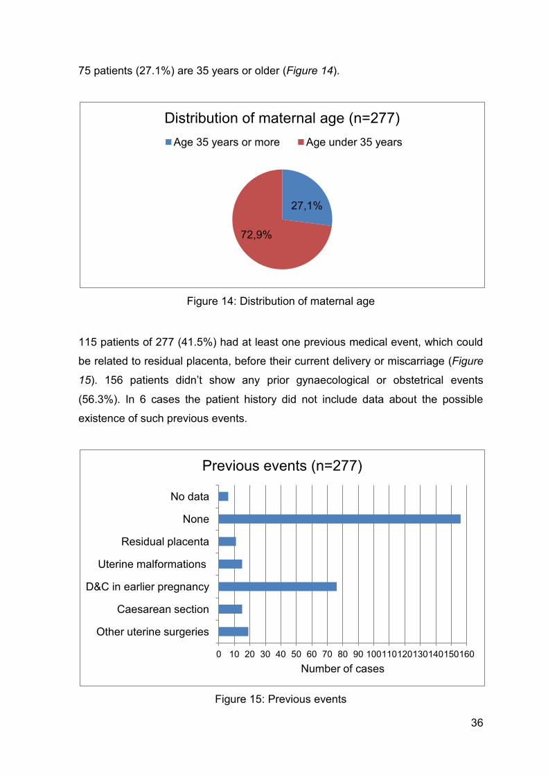
75 patients (27.1%) are 35 years or older (Figure 14).
Figure 14: Distribution of maternal age
115 patients of 277 (41.5%) had at least one previous medical event, which could
be related to residual placenta, before their current delivery or miscarriage (Figure
15). 156 patients didn’t show any prior gynaecological or obstetrical events
(56.3%). In 6 cases the patient history did not include data about the possible
existence of such previous events.
Figure 15: Previous events
27,1%
72,9%
Distribution of maternal age (n=277)
Age 35 years or more Age under 35 years
0 10 20 30 40 50 60 70 80 90 100 110 120 130 140 150 160
Other uterine surgeries
Caesarean section
D&C in earlier pregnancy
Uterine malformations
Residual placenta
None
No data
Number of cases
Previous events (n=277)
37
11 patients already experienced residual placenta after a previous delivery (4.0%).
15 women (5.4%) suffered from uterine malformations (like uterus myomatosus,
bicornis, arcuatus, subseptus), endometriosis or polyps. A D&C was carried out in
earlier pregnancies in 76 patients (27.4%), mostly because of TOP. In 15 patients
(5.4%) a caesarean section was performed in an earlier pregnancy. 19 women
(6.9%) underwent other uterine surgeries like conisation or cerclage.
4.2 Pregnancy and delivery
The average gestational age of patients at the time of the procedure was the 36th
week of pregnancy within a range from the 6th week to the 43rd week, so the
median was the 30th week (Figure 16). In 28 cases it was not possible to find a
detailed gestational age so they were just divided into cases before or after the
24th week of pregnancy by considering the individual context.
Figure 16: Distribution of gestational age
0
10
20
30
40
50
60
6
9
10
11
17
18
19
21
24
25
26
29
30
32
33
34
35
36
37
38
39
40
41
42
43
< 2
4th
WO
P
> 2
4th
WO
P
Nu
mb
er
of
ca
se
s
Weeks of pregnancy
Distribution of gestational age (n=277)

38
Of 277 cases there were 31 cases (11.2%) before the completed 24th week and
246 cases (88.8%) older than the completed 24th week (Figure 17).
Figure 17: Distribution of deliveries before and after the completed 24th week
As the cases before the completed 24th week were abortions, the birth mode was
only analysed in the group with gestational age over 24 weeks (Figure 18).
Figure 18: Distribution of mode of delivery in cases after the 24th week
11,2%
88,8%
Distribution of deliveries before and after the completed 24th week of pregnancy (n=277)
cases up to WOP 23+6 cases from WOP 24+0
87,0%
2,0%
1,6% 7,3% 2,0%
Distribution of mode of delivery in cases after the 24th week of pregnancy (n=246)
spontaneous Caesarean section
forceps delivery vacuum extraction delivery
intrauterine foetal death
39
214 patients of 246 had a spontaneous delivery (87.0%). This patient population
contains 213 singleton pregnancies and 1 twin pregnancy. A caesarean section
was performed in 5 patients (2.0%). 4 women (1.6%) underwent forceps delivery
and 18 women (7.3%) had a vacuum extraction assisted delivery. In 5 cases (2%)
the pregnancy was terminated due to intrauterine fetal death.
For the majority of patients (34.7%) the current pregnancy was their first, for 1
woman it was the 10th pregnancy (Figure 19). In 15 patients’ health records it was
not possible to find out about the number of pregnancy.
Figure 19: Distribution of number of pregnancy
For 119 patients (43.0%) the current delivery was their first (Figure 20). 34 of
those primiparae had at least 1 abortion before. In 19 cases (6.9%) the current
pregnancy was terminated by abortion, which is parity 0 in the figure. It was the
second delivery for 79 women (28.5%). 25 of the cases (9.0%) had their third
delivery. 10 women (3.6%) underwent their fourth delivery. 6 patients (2.2%) gave
0
20
40
60
80
100
120
1 2 3 4 5 6 7 8 10 no data
Num
be
r o
f ca
se
s
Number of pregnancy
Distribution of number of pregnancy (n=277)
40
birth for the fifth time. For 2 women (0.7%) the current delivery was their sixth. For
2 patients it was the seventh and eighth delivery respectively. In 15 cases (5.4%)
the number of their current delivery was not documented in MEDOCS or
ViewPoint health records.
Figure 20: Distribution of parity
4.3 Placenta
277 patients showed the diagnosis ‘residual placenta’ in their health record in
MEDOCS. 157 women showed the detailed description of placenta adhaerens in
ViewPoint Fetal Database, which is 56.7% (Figure 21). In 4 cases a placenta
accreta was suspected (1.4%). 109 patients were diagnosed with retained
placenta (39.4%). In 7 cases (2.5%) prolonged placental detachment occurred.
0
20
40
60
80
100
120
140
0 1 2 3 4 5 6 7 8 no data
Num
be
r o
f ca
se
s
Number of current delivery
Distribution of parity (n=277)
41
Figure 21: Type of placental anomaly
Besides from the diagnosis residual placenta, patients also showed other
diagnoses in MEDOCS (Figure 22). In 32 women (11.6%) haemorrhage in the
puerperium was documented. 2 patients suffered from sepsis (0.7%). 7 health
records revealed postpartum anaemia due to haemorrhage (2.5%). 1 woman had
a placental polyp (0.4%). Other pathologic condition of the placenta was detected
in 2 cases (0.7%).
Figure 22: Further diagnosis documented in MEDOCS
0 20 40 60 80 100 120 140 160 180
Placenta adhaerens
Placenta accreta
Retained placenta
Prolonged detachment
Number of cases
Type of placental anomaly (n=277)
0 5 10 15 20 25 30 35
Other pathologic condition
Sepsis
Postpartum anaemia
Placental polyp
Haemorrhage in the puerperium
Number of cases
Further diagnosis documented in Medocs (n=277)
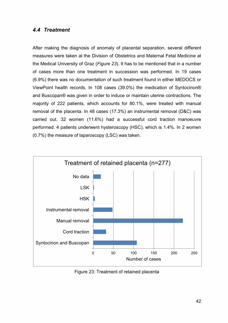
42
4.4 Treatment
After making the diagnosis of anomaly of placental separation, several different
measures were taken at the Division of Obstetrics and Maternal Fetal Medicine at
the Medical University of Graz (Figure 23). It has to be mentioned that in a number
of cases more than one treatment in succession was performed. In 19 cases
(6.9%) there was no documentation of such treatment found in either MEDOCS or
ViewPoint health records. In 108 cases (39.0%) the medication of Syntocinon®
and Buscopan® was given in order to induce or maintain uterine contractions. The
majority of 222 patients, which accounts for 80.1%, were treated with manual
removal of the placenta. In 48 cases (17.3%) an instrumental removal (D&C) was
carried out. 32 women (11.6%) had a successful cord traction manoeuvre
performed. 4 patients underwent hysteroscopy (HSC), which is 1.4%. In 2 women
(0.7%) the measure of laparoscopy (LSC) was taken.
Figure 23: Treatment of retained placenta
0 50 100 150 200 250
Syntocinon and Buscopan
Cord traction
Manual removal
Instrumental removal
HSK
LSK
No data
Number of cases
Treatment of retained placenta (n=277)
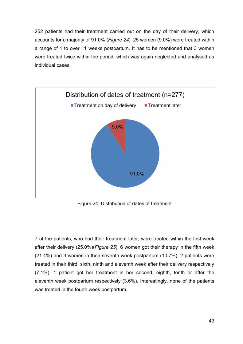
43
252 patients had their treatment carried out on the day of their delivery, which
accounts for a majority of 91.0% (Figure 24). 25 women (9.0%) were treated within
a range of 1 to over 11 weeks postpartum. It has to be mentioned that 3 women
were treated twice within the period, which was again neglected and analysed as
individual cases.
Figure 24: Distribution of dates of treatment
7 of the patients, who had their treatment later, were treated within the first week
after their delivery (25.0%)(Figure 25). 6 women got their therapy in the fifth week
(21.4%) and 3 women in their seventh week postpartum (10.7%). 2 patients were
treated in their third, sixth, ninth and eleventh week after their delivery respectively
(7.1%). 1 patient got her treatment in her second, eighth, tenth or after the
eleventh week postpartum respectively (3.6%). Interestingly, none of the patients
was treated in the fourth week postpartum.
91,0%
9,0%
Distribution of dates of treatment (n=277)
Treatment on day of delivery Treatment later
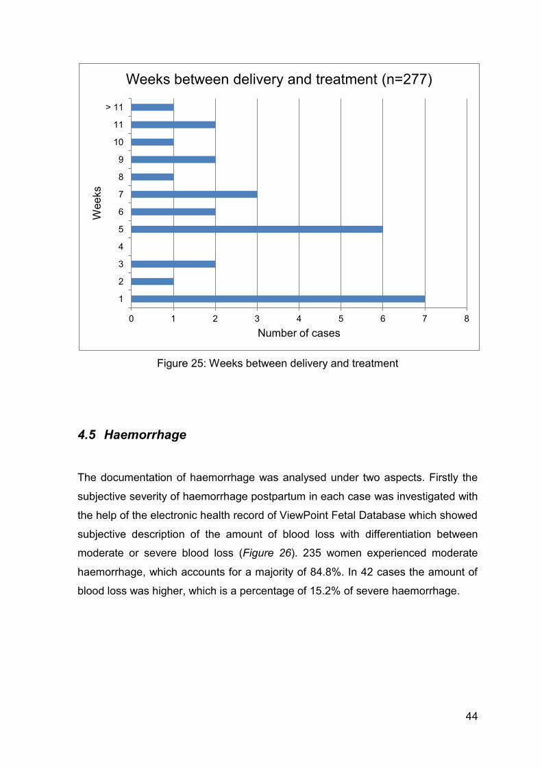
44
Figure 25: Weeks between delivery and treatment
4.5 Haemorrhage
The documentation of haemorrhage was analysed under two aspects. Firstly the
subjective severity of haemorrhage postpartum in each case was investigated with
the help of the electronic health record of ViewPoint Fetal Database which showed
subjective description of the amount of blood loss with differentiation between
moderate or severe blood loss (Figure 26). 235 women experienced moderate
haemorrhage, which accounts for a majority of 84.8%. In 42 cases the amount of
blood loss was higher, which is a percentage of 15.2% of severe haemorrhage.
0 1 2 3 4 5 6 7 8
1
2
3
4
5
6
7
8
9
10
11
> 11
Number of cases
Wee
ks
Weeks between delivery and treatment (n=277)
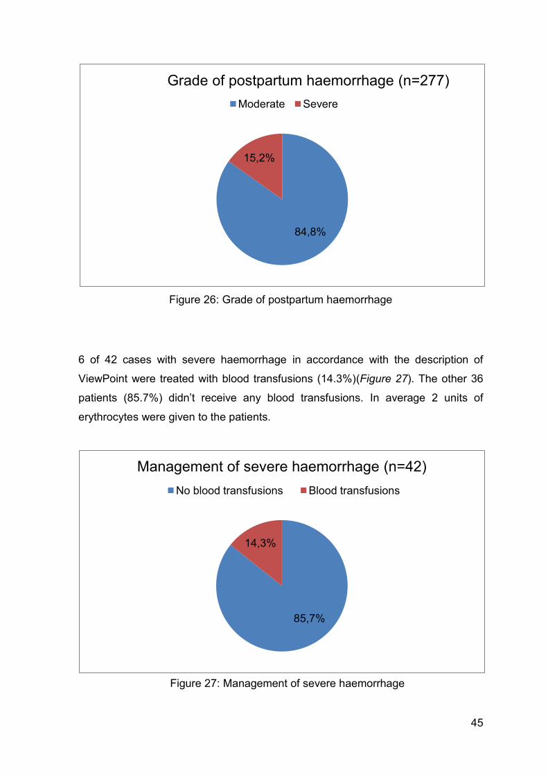
45
Figure 26: Grade of postpartum haemorrhage
6 of 42 cases with severe haemorrhage in accordance with the description of
ViewPoint were treated with blood transfusions (14.3%)(Figure 27). The other 36
patients (85.7%) didn’t receive any blood transfusions. In average 2 units of
erythrocytes were given to the patients.
84,8%
15,2%
Grade of postpartum haemorrhage (n=277)
Moderate Severe
85,7%
14,3%
Management of severe haemorrhage (n=42)
No blood transfusions Blood transfusions
Figure 27: Management of severe haemorrhage
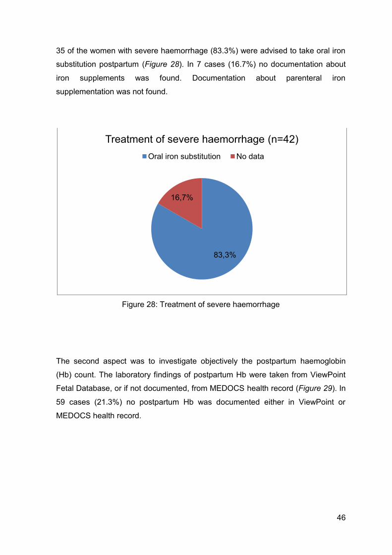
46
35 of the women with severe haemorrhage (83.3%) were advised to take oral iron
substitution postpartum (Figure 28). In 7 cases (16.7%) no documentation about
iron supplements was found. Documentation about parenteral iron
supplementation was not found.
Figure 28: Treatment of severe haemorrhage
The second aspect was to investigate objectively the postpartum haemoglobin
(Hb) count. The laboratory findings of postpartum Hb were taken from ViewPoint
Fetal Database, or if not documented, from MEDOCS health record (Figure 29). In
59 cases (21.3%) no postpartum Hb was documented either in ViewPoint or
MEDOCS health record.
83,3%
16,7%
Treatment of severe haemorrhage (n=42)
Oral iron substitution No data
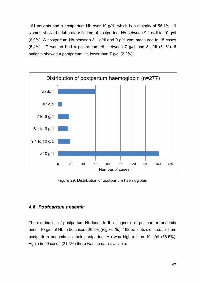
47
161 patients had a postpartum Hb over 10 g/dl, which is a majority of 58.1%. 19
women showed a laboratory finding of postpartum Hb between 9.1 g/dl to 10 g/dl
(6.9%). A postpartum Hb between 8.1 g/dl and 9 g/dl was measured in 15 cases
(5.4%). 17 women had a postpartum Hb between 7 g/dl and 8 g/dl (6.1%). 6
patients showed a postpartum Hb lower than 7 g/dl (2.2%).
Figure 29: Distribution of postpartum haemoglobin
4.6 Postpartum anaemia
The distribution of postpartum Hb leads to the diagnosis of postpartum anaemia
under 10 g/dl of Hb in 56 cases (20.2%)(Figure 30). 162 patients didn’t suffer from
postpartum anaemia as their postpartum Hb was higher than 10 g/dl (58.5%).
Again in 59 cases (21.3%) there was no data available.
0 20 40 60 80 100 120 140 160 180
>10 g/dl
9.1 to 10 g/dl
8.1 to 9 g/dl
7 to 8 g/dl
<7 g/dl
No data
Number of cases
Distribution of postpartum haemoglobin (n=277)
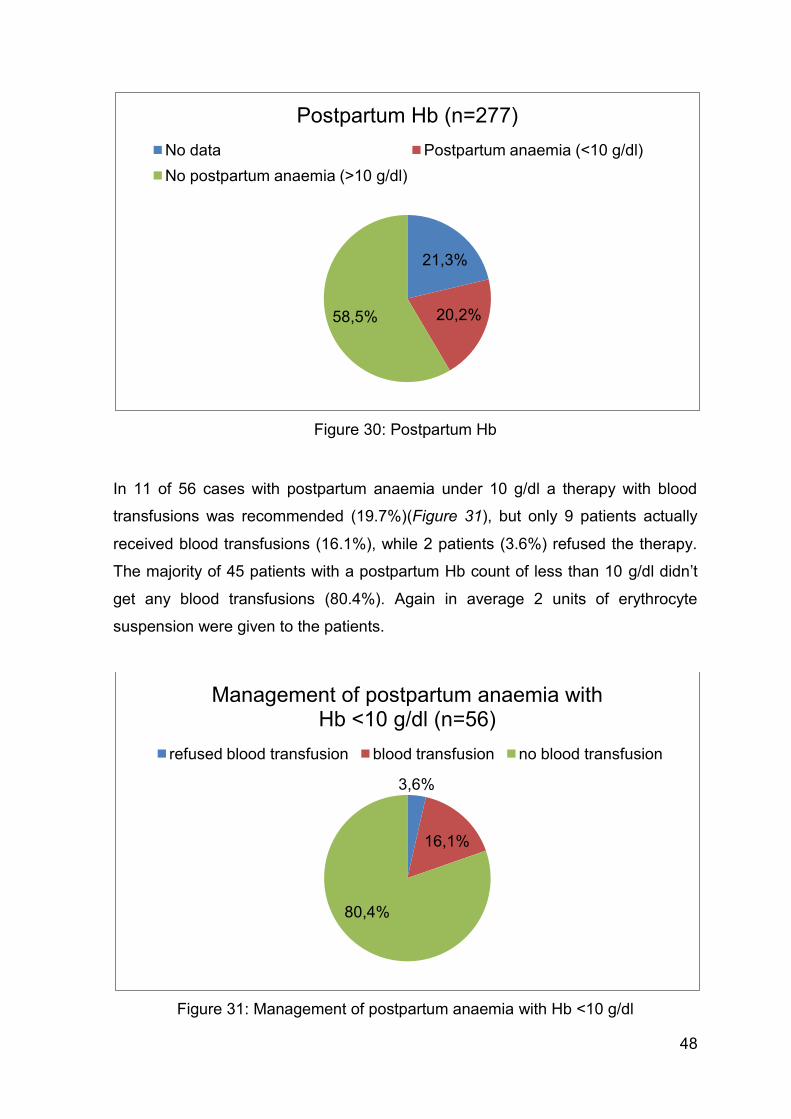
48
Figure 30: Postpartum Hb
In 11 of 56 cases with postpartum anaemia under 10 g/dl a therapy with blood
transfusions was recommended (19.7%)(Figure 31), but only 9 patients actually
received blood transfusions (16.1%), while 2 patients (3.6%) refused the therapy.
The majority of 45 patients with a postpartum Hb count of less than 10 g/dl didn’t
get any blood transfusions (80.4%). Again in average 2 units of erythrocyte
suspension were given to the patients.
Figure 31: Management of postpartum anaemia with Hb <10 g/dl
21,3%
20,2% 58,5%
Postpartum Hb (n=277)
No data Postpartum anaemia (<10 g/dl)
No postpartum anaemia (>10 g/dl)
3,6%
16,1%
80,4%
Management of postpartum anaemia with Hb <10 g/dl (n=56)
refused blood transfusion blood transfusion no blood transfusion
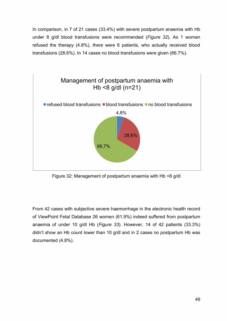
49
In comparison, in 7 of 21 cases (33.4%) with severe postpartum anaemia with Hb
under 8 g/dl blood transfusions were recommended (Figure 32). As 1 woman
refused the therapy (4.8%), there were 6 patients, who actually received blood
transfusions (28.6%). In 14 cases no blood transfusions were given (66.7%).
Figure 32: Management of postpartum anaemia with Hb <8 g/dl
From 42 cases with subjective severe haemorrhage in the electronic health record
of ViewPoint Fetal Database 26 women (61.9%) indeed suffered from postpartum
anaemia of under 10 g/dl Hb (Figure 33). However, 14 of 42 patients (33.3%)
didn’t show an Hb count lower than 10 g/dl and in 2 cases no postpartum Hb was
documented (4.8%).
4,8%
28,6%
66,7%
Management of postpartum anaemia with Hb <8 g/dl (n=21)
refused blood transfusions blood transfusions no blood transfusions
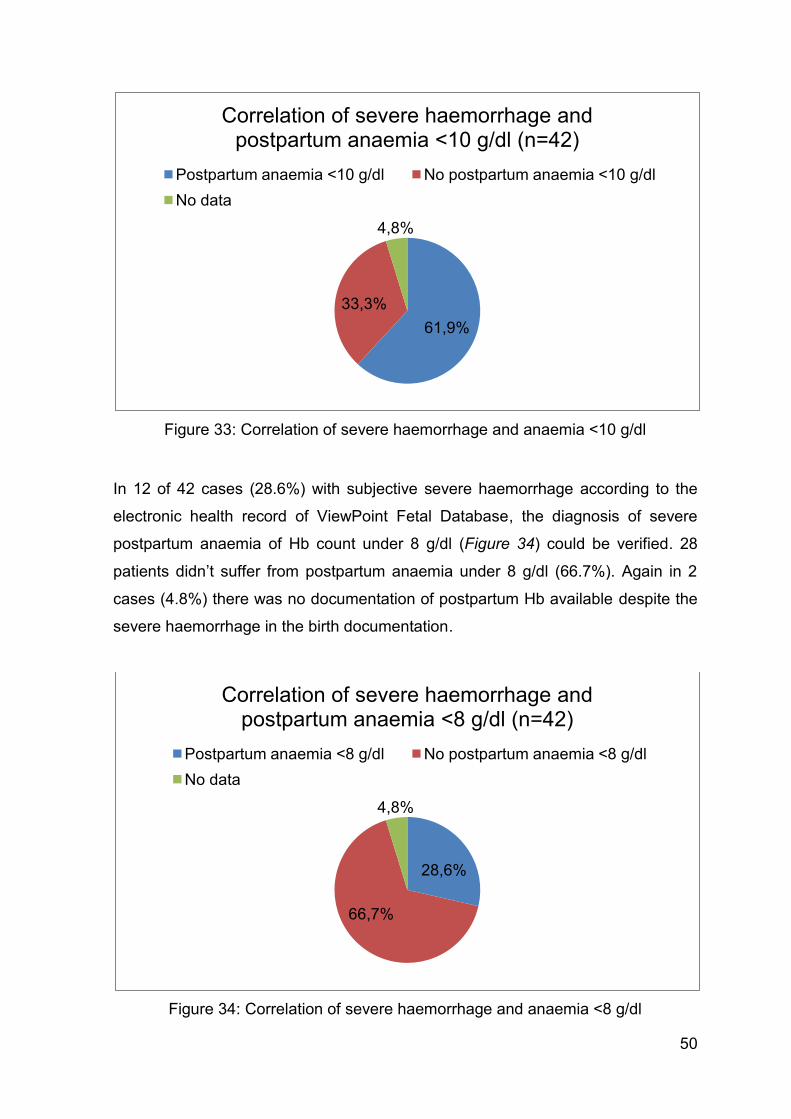
50
Figure 33: Correlation of severe haemorrhage and anaemia <10 g/dl
In 12 of 42 cases (28.6%) with subjective severe haemorrhage according to the
electronic health record of ViewPoint Fetal Database, the diagnosis of severe
postpartum anaemia of Hb count under 8 g/dl (Figure 34) could be verified. 28
patients didn’t suffer from postpartum anaemia under 8 g/dl (66.7%). Again in 2
cases (4.8%) there was no documentation of postpartum Hb available despite the
severe haemorrhage in the birth documentation.
Figure 34: Correlation of severe haemorrhage and anaemia <8 g/dl
61,9%
33,3%
4,8%
Correlation of severe haemorrhage and postpartum anaemia <10 g/dl (n=42)
Postpartum anaemia <10 g/dl No postpartum anaemia <10 g/dl
No data
28,6%
66,7%
4,8%
Correlation of severe haemorrhage and postpartum anaemia <8 g/dl (n=42)
Postpartum anaemia <8 g/dl No postpartum anaemia <8 g/dl
No data
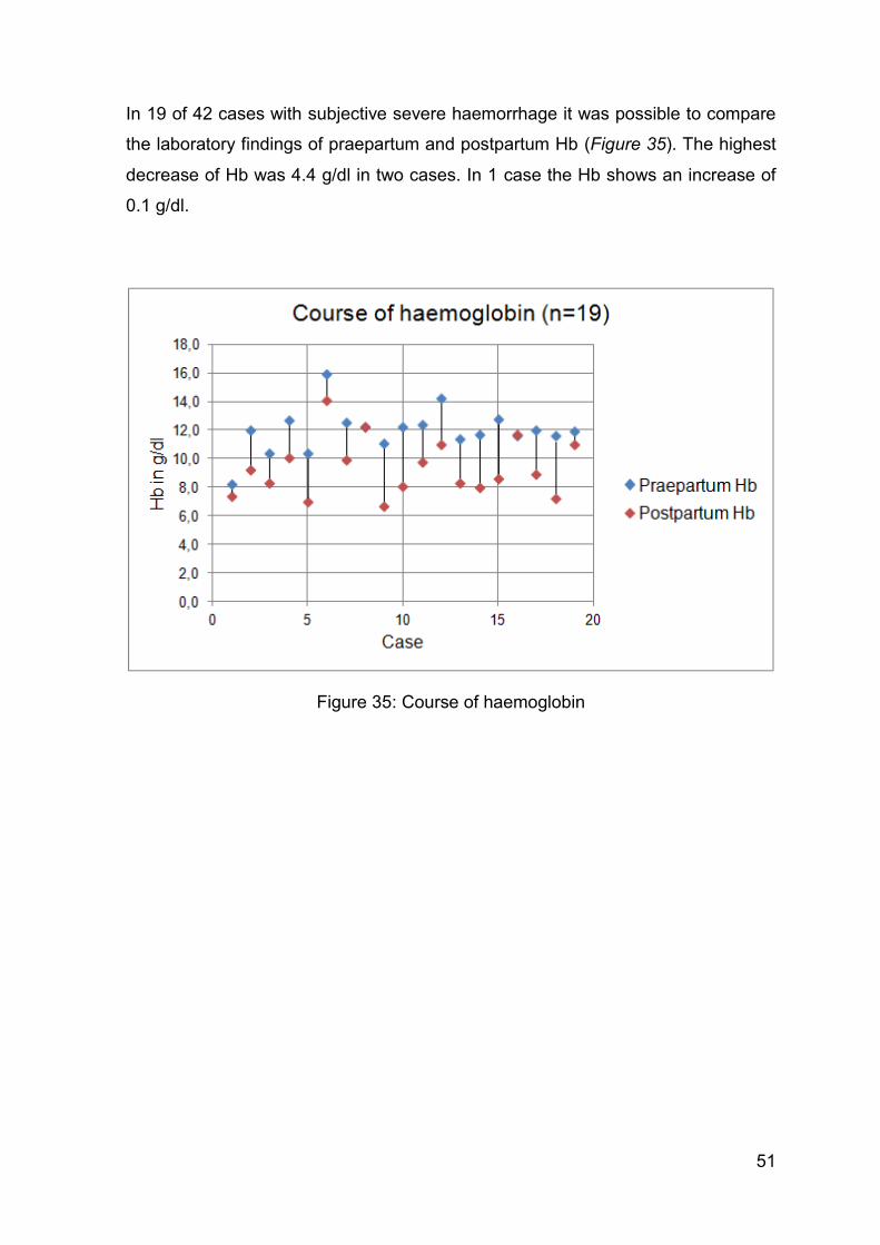
51
In 19 of 42 cases with subjective severe haemorrhage it was possible to compare
the laboratory findings of praepartum and postpartum Hb (Figure 35). The highest
decrease of Hb was 4.4 g/dl in two cases. In 1 case the Hb shows an increase of
0.1 g/dl.
Figure 35: Course of haemoglobin
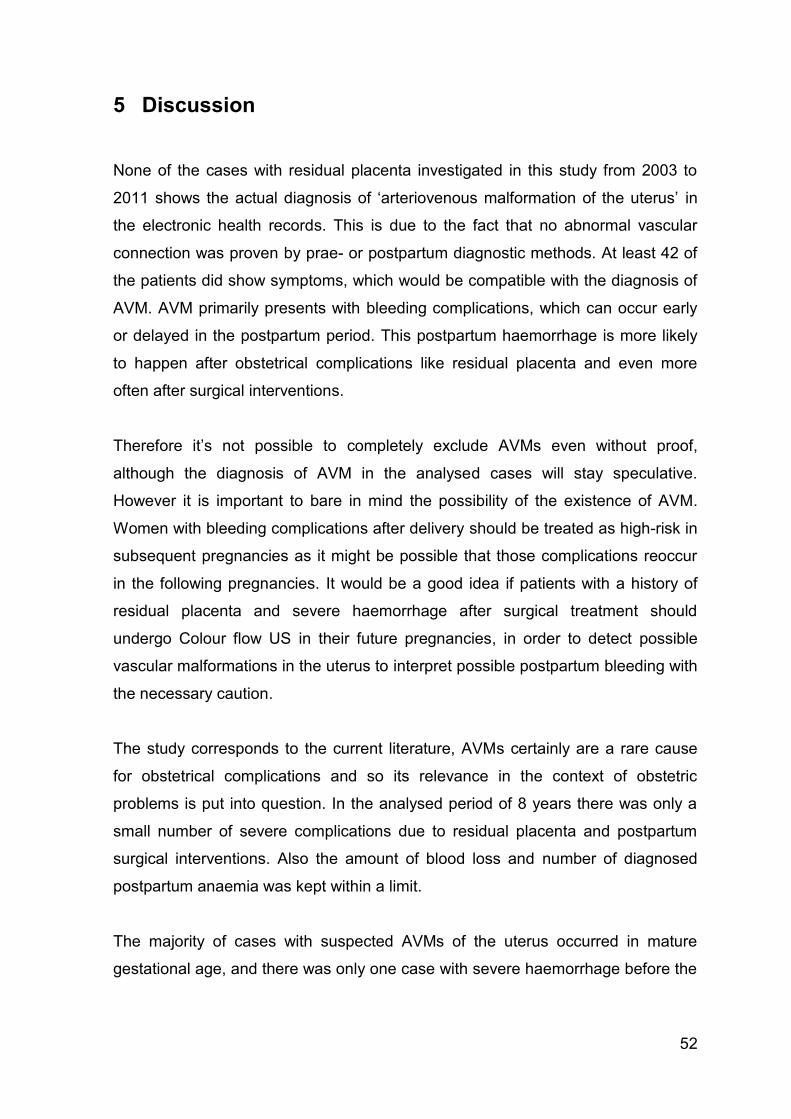
52
5 Discussion
None of the cases with residual placenta investigated in this study from 2003 to
2011 shows the actual diagnosis of ‘arteriovenous malformation of the uterus’ in
the electronic health records. This is due to the fact that no abnormal vascular
connection was proven by prae- or postpartum diagnostic methods. At least 42 of
the patients did show symptoms, which would be compatible with the diagnosis of
AVM. AVM primarily presents with bleeding complications, which can occur early
or delayed in the postpartum period. This postpartum haemorrhage is more likely
to happen after obstetrical complications like residual placenta and even more
often after surgical interventions.
Therefore it’s not possible to completely exclude AVMs even without proof,
although the diagnosis of AVM in the analysed cases will stay speculative.
However it is important to bare in mind the possibility of the existence of AVM.
Women with bleeding complications after delivery should be treated as high-risk in
subsequent pregnancies as it might be possible that those complications reoccur
in the following pregnancies. It would be a good idea if patients with a history of
residual placenta and severe haemorrhage after surgical treatment should
undergo Colour flow US in their future pregnancies, in order to detect possible
vascular malformations in the uterus to interpret possible postpartum bleeding with
the necessary caution.
The study corresponds to the current literature, AVMs certainly are a rare cause
for obstetrical complications and so its relevance in the context of obstetric
problems is put into question. In the analysed period of 8 years there was only a
small number of severe complications due to residual placenta and postpartum
surgical interventions. Also the amount of blood loss and number of diagnosed
postpartum anaemia was kept within a limit.
The majority of cases with suspected AVMs of the uterus occurred in mature
gestational age, and there was only one case with severe haemorrhage before the
53
24th week of pregnancy. More than one quarter of the studied cases were women
over the age of 35 years, which might be another important epidemiologic factor.
However, a woman of over 35 years shows a greater chance to have experienced
previous obstetrical or surgical conditions and measurements, which also
increases the risk to develop AVMs or a residual placenta. All the previous events
identified in cases of the study have been reported in literature as possible risk
factors for AVM.
Most of the cases of the study experienced residual placenta with bleeding
complications during their first pregnancy and delivery. Summing up all cases
showing the diagnosis after the first pregnancy and delivery, the outcome shows
that residual placenta is more likely to occur after previous pregnancies and
deliveries. The statistics show a lower number of cases in higher number of
pregnancy or delivery, which is due to the fact, that we live in a society where it is
more popular to have fewer children.
The outcome of the study with no death or necessity for hysterectomy shows the
adequacy of the therapy given to the patients. In an absolute majority of cases the
treatment was carried out on the same day of the delivery, so the diagnosis of
residual placenta was soon revealed. Depending on the severity of complications,
conservative measures like administration of medication for uterine contractions or
surgery under general anaesthesia were taken. It was not necessary to take any
further steps to control severe bleeding complications.
Cases with residual placenta didn’t occur very often in the investigated period and
in those detected cases the problem could nearly always be controlled with one
single procedure and moderate blood loss. Only in 3 cases it was necessary to
carry out a second measure.
About 20% of all patients with residual placenta in the period suffered from a
postpartum anaemia with an Hb count of lower than 10 g/dl and about 8% showed
an Hb count of lower than 8 g/dl. The standard therapy of anaemia is to give blood
transfusions, which was recommended to about 20% of the patients with anaemia
with an Hb count of lower than 10 g/dl and to about 33% of the patients with
54
anaemia with an Hb count of lower than 8 g/dl. At the Division of Obstetrics and
Maternal Fetal Medicine at the Medical University of Graz the standard procedure
in cases with an Hb count of lower than 8 g/dl is to offer the administration of
erythrocyte suspension to the patient. The woman of course does not have to
accept this and can refuse the therapy. This refusal is mostly documented in the
handwritten patient charts/records and may not be copied into the electronic health
records of the patient. For the study only electronic health records have been
analysed. This is why it is possible, that the statistics show such a low percentage
of women with an Hb count of lower than 8 g/dl, who actually received erythrocyte
suspensions. In cases of emergency (unconsciousness, non-responding patient,
massive haemorrhage) primary a selective blood substitution therapy is used
(erythrocyte suspension, plasma concentrate, volume supplementation).
Another problem due to bleeding complications might be an iron deficiency. The
majority of cases with severe haemorrhage in the study received oral iron
supplements. Again in some cases no documentation was found about iron
substitution at all, which might again be the problem of handwritten patient refusal,
which was not included in the analysis. One reason for the decline of oral iron
supplementation could be the high rate of gastroenterological side effects like
diarrhoea, nausea or obstipation. In December 2010 an oral presentation about i.v.
iron substitution was held by Prof. Christoph Gasche of the Medical University of
Vienna at the Division of Obstetrics and Maternal Fetal Medicine at the Medical
University of Graz (35). In his study he presents a new product for i.v. iron
administration called Ferinject®. Women with an Hb count of lower than 10 g/dl
within ten days postpartum received either i.v. iron supplementation of Ferinject®
or oral iron supplementation of iron sulphate. Not only did the i.v. administration
show less side effects than the oral one, it also showed a significant increase of
their Hb count of more than 2 g/dl after two and four weeks, and a significant
increase of 3 g/dl of Hb after two, four and seven weeks. It also shows that the iron
stores have been filling up. In about 80% of the patients only one infusion of
Ferinject® was given (35). At the Division of Obstetrics and Maternal Fetal
Medicine at the Medical University of Graz since 2010 the i.v. iron
supplementation of Ferinject® has gotten more and more popular due to the easy
single administration and less side effects for the patients.
55
One problem of this retrospective study was the inconsistent and sometimes
incomplete documentation in the patients’ electronic health records. Handwritten
patient charts have not been analysed at all. It was difficult to compare the results
with a high number of undocumented details. The programs used for electronic
documentation offer to much free space for the single examiner, so the same
details revealed in different patients are documented in different spaces of the
electronic health records. So it can easily be overlooked by a second examiner.
For the future a more consistent documentation should be established, which
requires training for all staff members working with those programs. This will not
only guarantee a complete, homogenous patient record that can easily be
continued to be used by every staff member, it will also be easier to compare
different managements and outcomes.
To totally understand the problem of uterine AVM in the context of obstetrics it
might be necessary to carry out prospective studies. Special attention should be
given to possible vascular malformations in the praepartum period. The problem is
of course that such detailed screening examinations of patients will take a lot of
time and cost a lot of money, so it should be considered in cases with possible
consequences of certain results.
Even though the complications of residual placenta in cases with suspected AVM
of the uterus could be well controlled in the cases of the presented study, it is
important to guarantee a detailed medical education of the patients. Women,
especially those, who already suffered from bleeding complications after residual
placenta, have to know about the entity and the chance that more than one clinical
procedure might be necessary after their delivery. More clinical interventions might
lead to even more haemorrhage, therefore a longer hospital stay, a longer
recovery period and psychological stress.
AVMs of the uterus stay a rarity. However, they remain to be a risk factor for
severe bleeding complications after delivery and especially after residual placenta
followed by surgical intervention. The standard therapy seems to be adequate, but
it is important to know, that arteriovenous malformations could as well lead to
uncontrollable haemorrhage and therefore should be kept in mind.
56
6 References of the text
(1) Hafferl A. Lehrbuch der topographischen Anatomie. 2nd ed. Berlin, Göttingen,
Heidelberg: Springer Verlag oHG; 1957. p. 614-45.
(2) Rauber A, Kopsch F. Anatomie des Menschen. Lehrbuch und Atlas. Band II:
Innere Organe. Stuttgart: Georg Thieme Verlag; 1987. p. 487-540.
(3) Gray H. Gray’s anatomy. London: The Promotional Reprint Company Limited;
1997. p. 378-688.
(4) Stauber M, Weyerstahl T. Duale Reihe. Gynäkologie und Geburtshilfe. 3rd ed.
Stuttgart: Georg Thieme Verlag KG; 2007. p. 463-743.
(5) Schünke M, Schulte E, Schumacher U. Prometheus. LernAtlas der Anatomie.
Hals und Innere Organe. Stuttgart: Georg Thieme Verlag; 2005. p. 244-93.
(6) Gilroy AM, MacPherson BR, Ross LM. Atlas of Anatomy. New York: Thieme
Medical Publishers, Inc; 2008. p. 94-5.
(7) Gruber S. Basics. Gynäkologie und Geburtshilfe. 3rd ed. München: Elsevier
GmbH; 2009. p. 146-7.
(8) Dudenhausen JW. Praktische Geburtshilfe mit geburtshilflichen Operationen.
20th ed. Berlin: Walter de Gruyter GmbH & Co KG; 2008. p. 187-382.
(9) Noonan JB, Coakley FV, Qayyum A, Yeh BM, Wu L, Chen LM. MR imaging of
retained products of conception. AJR Am J Roentgenol. 2003 Aug;181(2):435-9.
(10) Bodelon C, Bernabe-Ortiz A, Schiff MA, Reed SD. Factors associated with
peripartum hysterectomy. Obstet Gynecol. 2009 Jul;114(1):115-23.
57
(11) Mahendru R, Taneja BK, Malik S. Preservation of fertility following abnormally
adherent placenta treated conservatively: a case report. Cases J. 2009 Dec
18;2:9349.
(12) Thia EWH, Lee SL, Tan HK, Tan LK. Ultrasonographical features of morbidly-
adherent placentas. Singapore Med J. 2007 Sept;48(9):799-802.
(13) Mulic-Lutvica A, Axelsson O. Ultrasound finding of an echogenic mass in
women with secondary postpartum hemorrhage is associated with retained
placental tissue. Ultrasound Obstet Gynecol. 2006 Sept;28(3):312-9.
(14) Debby A, Malinger G, Harow E, Golan A, Glezerman M. Transvaginal
ultrasound after first-trimester uterine evacuation reduces the incidence of retained
products of conception. Ultrasound Obstet Gynecol. 2006 Jan;27(1):61-4.
(15) Manolitsas T, Hurley V, Gilford E. Uterine arteriovenous malformation- a rare
cause of uterine haemorrhage. Aust NZ J Obstet Gynaecol. 1994 May;34(2):197-
9.
(16) Khatree MH, Titiz H. Medical treatment of a uterine arteriovenous
malformation. Aust NZ J Obstet Gynaecol. 1999 Aug;39(3):378-80.
(17) Hoffman MK, Meilstrup JW, Shackelford DP, Kaminski PF. Arteriovenous
malformations of the uterus: an uncommon cause of vaginal bleeding. Obstet
Gynecol Surv. 1997 Dec;52(12):736-40.
(18) Gulati MS, Paul SB, Batra A, Sarma D, Dadhwal V, Nath J. Uterine
arteriovenous malformations: the role of intravenous ‘dual-phase’ CT angiography.
Clin Imaging. 2000 Jan-Feb;24(1):10-4.
(19) Fleming H, Östör AG, Pickel H, Fortune DW. Arteriovenous malformations of
the uterus. Obstet Gynecol. 1989 Feb;73(2):209-14.
58
(20) Kelly SM, Belli AM, Campbell S. Arteriovenous malformation of the uterus
associated with secondary postpartum hemorrhage. Ultrasound Obstet Gynecol.
2003 Jun;21(6):602-5.
(21) Rangarajan RD, Moloney JC, Anderson HJ. Diagnosis and nonsurgical
management of uterine arteriovenous malformation. Cardiovasc Intervent Radiol.
2007 Nov-Dec;30(6):1267-70.
(22) Nicholson AA, Turnbull LW, Coady AM, Guthrie K. Diagnosis and
management of uterine arterio-venous malformations. Clin Radiol. 1999
Apr;54(4):265-9.
(23) Aziz N, Lenzi TA, Jeffrey RB Jr, Lyell DJ. Postpartum uterine arteriovenous
fistula. Obstet Gynecol. 2004 May;103(5 Pt 2):1076-8.
(24) Kwon JH, Kim GS. Obstetric iatrogenic arterial injuries of the uterus:
diagnosis with US and treatment with transcatheter arterial embolization.
Radiographics. 2002 Jan-Feb;22(1):35-46.
(25) Polat P, Suma S, Kantarcý M, Alper F, Levent A. Color Doppler US in the
evaluation of uterine vascular abnormalities. Radiographics. 2002 Jan-
Feb;22(1):47-53.
(26) Ahn HY, Park IY, Lee G, Kim SJ, Shin JC. Uterine arteriovenous
malformation. Arch Gynecol Obstet. 2005 Feb;271(2):172-5.
(27) Clarke MJ, Mitchell PJ. Uterine arteriovenous malformation: a rare cause of
uterine bleeding. Diagnosis and treatment. Australas Radiol. 2003 Sept;47(3):302-
5.
(28) Timmerman D, Wauters J, Van Calenbergh S, Van Schoubroeck D, Maleux
G, Van Den Bosch T, Spitz B. Color Doppler imaging is a valuable tool for the
diagnosis and management of uterine vascular malformations. Ultrasound Obstet
Gynecol. 2003 Jun;21(6):570-7.
59
(29) Gopal M, Goldberg J, Klein TA, Fossum GT. Embolization of a uterine
arteriovenous malformation followed by a twin pregnancy. Obstet Gynecol. 2003
Oct;102(4):696-8.
(30) Yokomine D, Yoshinaga M, Baba Y, Matsuo T, Iguro Y, Nakajo M, Douchi T.
Successful management of uterine arteriovenous malformation by ligation of
feeding artery after unsuccessful uterine artery embolization. J Obstet Gynaecol
Res. 2009 Feb;35(1):183-8.
(31) Delotte J, Chevallier P, Benoit B, Castillon JM, Bongain A. Pregnancy after
embolization therapy for uterine arteriovenous malformation. Fertil Steril. 2006
Jan;85(1):228.
(32) Schwärzler P. Department of Obstetrics and Gynecology Feldkirch, Austria.
Personal communication. 2011.
(33) Nasu K, Nishida M, Yoshimatsu J, Narahara H. Ectopic pregnancy after
successful treatment with percutaneous transcatheter uterine arterial embolization
for congenital uterine arteriovenous malformation: a case report. Arch Gynecol
Obstet. 2008 Aug;278(2):171-2.
(34) Knight M, Callaghan WM, Berg C, Alexander S, Bouvier-Colle MH, Ford JB et
al. Trends in postpartum hemorrhage in high resource countries: a review and
recommendations from the International Postpartum Hemorrhage Collaborative
Group. BMC Pregnancy Childbirth. 2009 Nov 27;9:55.
(35) Gasche C. Iron deficiency in gynecology. Department of Gastroenterology of
the Medical University of Vienna. Oral presentation at the Medical University of
Graz. Dec 2010.
![Service-Handbuch (mit separater Videokarte)€¦ · l Added diagnostics options +LQ]XJHI JWH'LDJQRVHRSWLRQHQ ± 'DV6WDUWPHQ HQWKlOW]ZHL2SWLRQHQI UGLH'LDJQRVH IDE Drive Diagnostics](https://img.pdfslide.org/doc/110x75/5f2b8e5ffc3d9727ba628bd7/service-handbuch-mit-separater-videokarte-l-added-diagnostics-options-lqxjhi.jpg)