Embed Size (px)
Citation preview
Die Haut als Barriere für
NanopartikelDas NANODERM Projekt
Tilman ButzUniversität Leipzig
Nanotechnologie,ihre Produkte und Risiken für den Verbraucher
Expertengespräch im Bundesinstitut für RisikobewertungBerlin, 28. März 2006
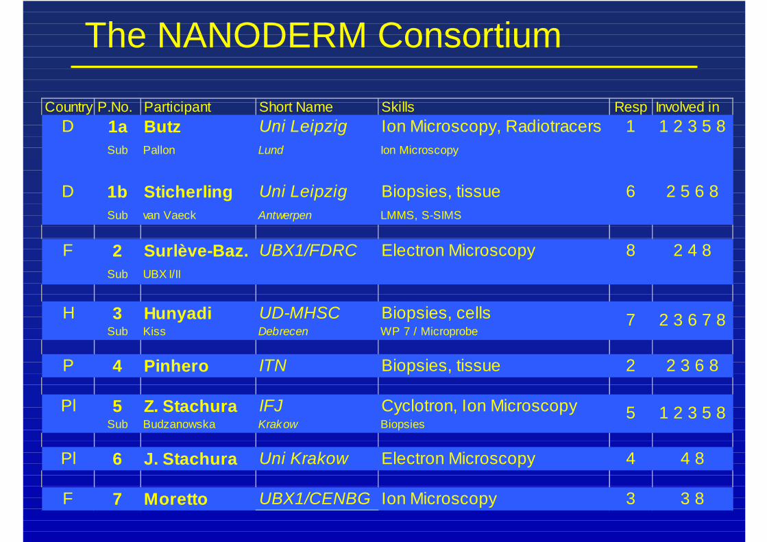
The NANODERM Consortium
Country P.No. Participant Short Name Skills Resp Involved inD 1a Butz Uni Leipzig Ion Microscopy, Radiotracers 1 1 2 3 5 8
Sub Pallon Lund Ion Microscopy
D 1b Sticherling Uni Leipzig Biopsies, tissue 6 2 5 6 8Sub van Vaeck Antwerpen LMMS, S-SIMS
F 2 Surlève-Baz. UBX1/FDRC Electron Microscopy 8 2 4 8Sub UBX I/II
H 3 Hunyadi UD-MHSC Biopsies, cellsSub Kiss Debrecen WP 7 / Microprobe
P 4 Pinhero ITN Biopsies, tissue 2 2 3 6 8
Pl 5 Z. Stachura IFJ Cyclotron, Ion MicroscopySub Budzanowska Krakow Biopsies
Pl 6 J. Stachura Uni Krakow Electron Microscopy 4 4 8
F 7 Moretto UBX1/CENBG Ion Microscopy 3 3 8
7 2 3 6 7 8
5 1 2 3 5 8
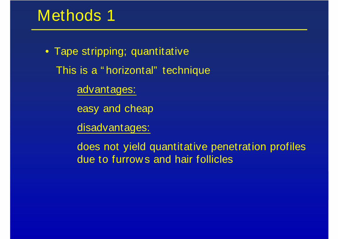
Methods 1
• Tape stripping; quantitat ive
This is a “ horizontal” technique
advantages:
easy and cheap
disadvantages:
does not yield quantitat ive penetrat ion prof iles due to furrow s and hair follicles
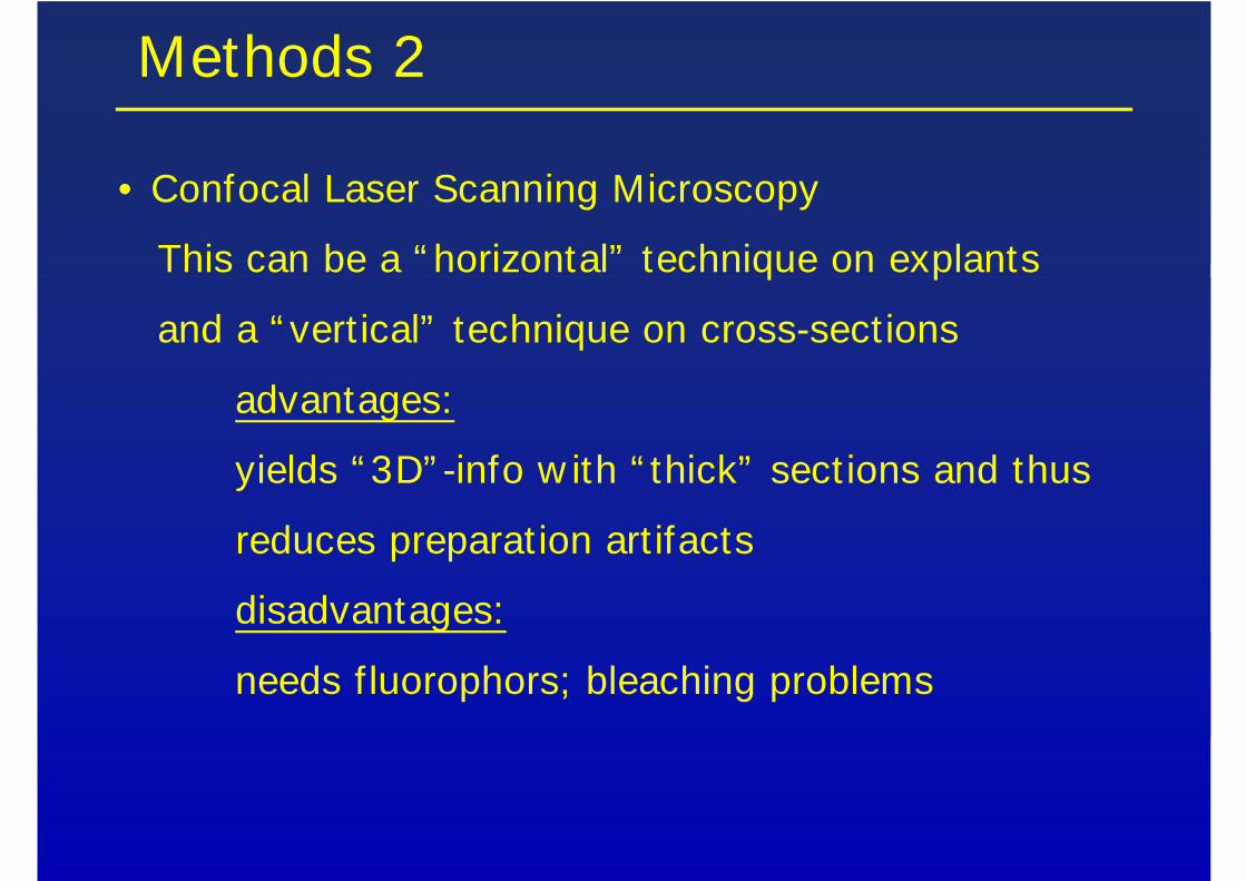
Methods 2
• Confocal Laser Scanning Microscopy
This can be a “ horizontal” technique on explants
and a “ vert ical” technique on cross-sections
advantages:
yields “ 3D” -info w ith “ thick” sect ions and thus
reduces preparation art ifacts
disadvantages:
needs f luorophors; bleaching problems
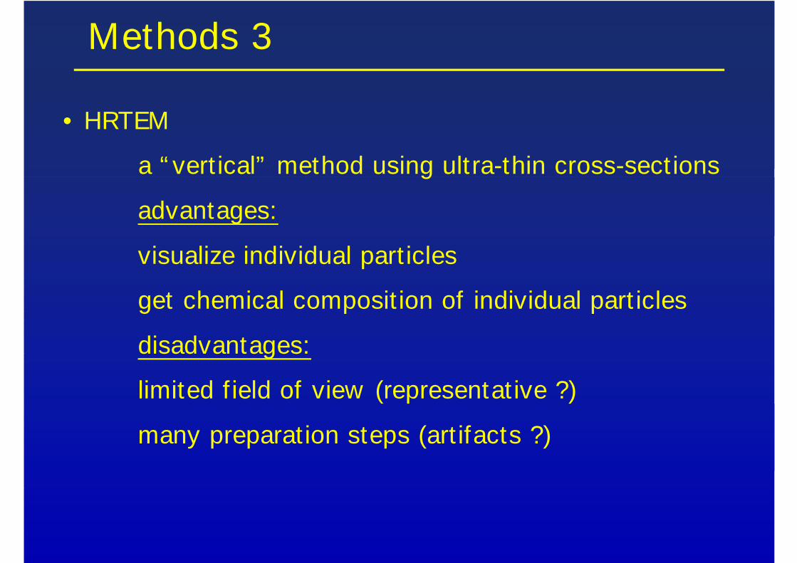
Methods 3
• HRTEM
a “ vert ical” method using ultra-thin cross-sections
advantages:
visualize individual part icles
get chemical composit ion of individual part icles
disadvantages:
limited f ield of view (representative ?)
many preparation steps (art ifacts ?)
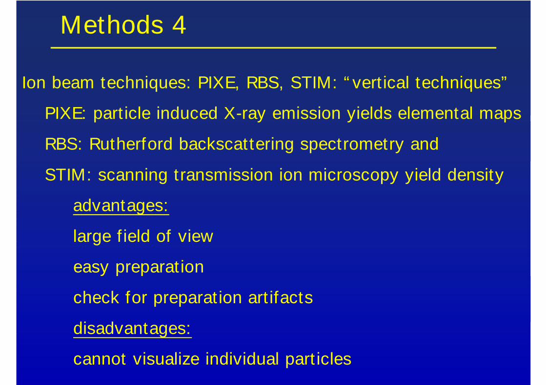
Methods 4
Ion beam techniques: PIXE, RBS, STIM: “ vert ical techniques”
PIXE: part icle induced X-ray emission yields elemental maps
RBS: Rutherford backscattering spectrometry and
STIM: scanning transmission ion microscopy yield density
advantages:
large f ield of view
easy preparation
check for preparation art ifacts
disadvantages:
cannot visualize individual part icles
Methods 5
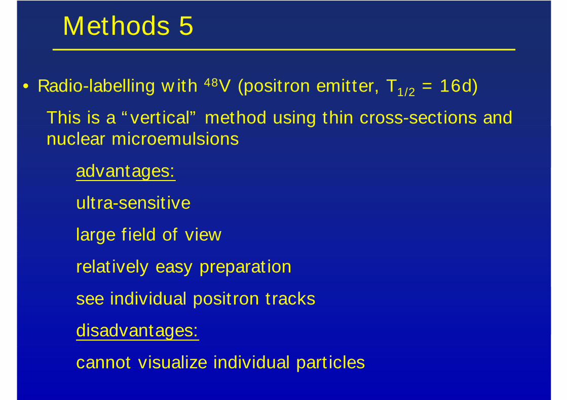
• Radio-labelling w ith 48V (positron emitter, T1/2 = 16d)
This is a “ vert ical” method using thin cross-sections andnuclear microemulsions
advantages:
ultra-sensit ive
large f ield of view
relat ively easy preparat ion
see individual positron tracks
disadvantages:
cannot visualize individual part icles
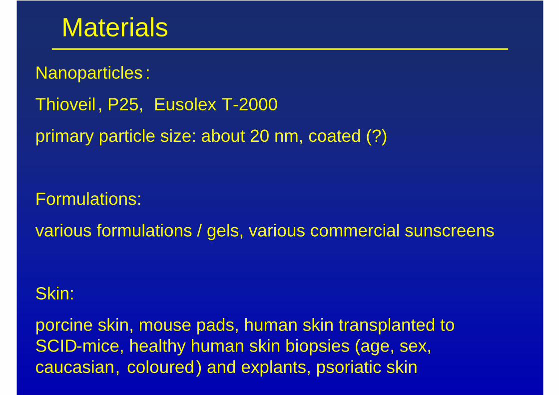
Materials Nanoparticles :
Thioveil , P25, Eusolex T-2000
primary particle size: about 20 nm, coated (?)
Formulations:
various formulations / gels, various commercial sunscreens
Skin:
porcine skin, mouse pads, human skin transplanted to SCID-mice, healthy human skin biopsies (age, sex, caucasian, coloured) and explants, psoriatic skin
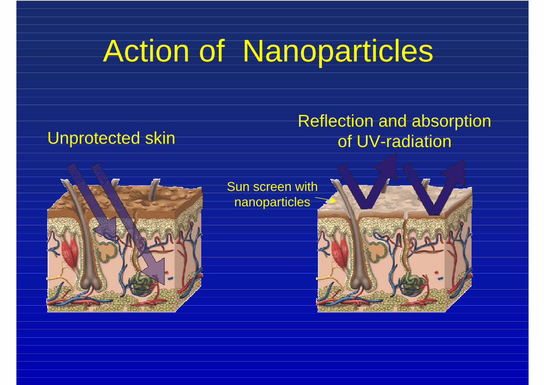
Unprotected skin
Action of Nanoparticles
Reflection and absorptionof UV-radiation
Sun screen withnanoparticles
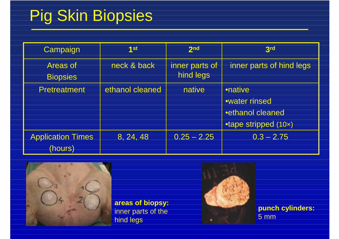
Pig Skin Biopsies
areas of biopsy:inner parts of thehind legs
0.3 – 2.750.25 – 2.258, 24, 48Application Times(hours)
•native•water rinsed•ethanol cleaned•tape stripped (10×)
nativeethanol cleanedPretreatment
inner parts of hind legsinner parts ofhind legs
neck & backAreas ofBiopsies
3rd2nd1stCampaign
punch cylinders:5 mm
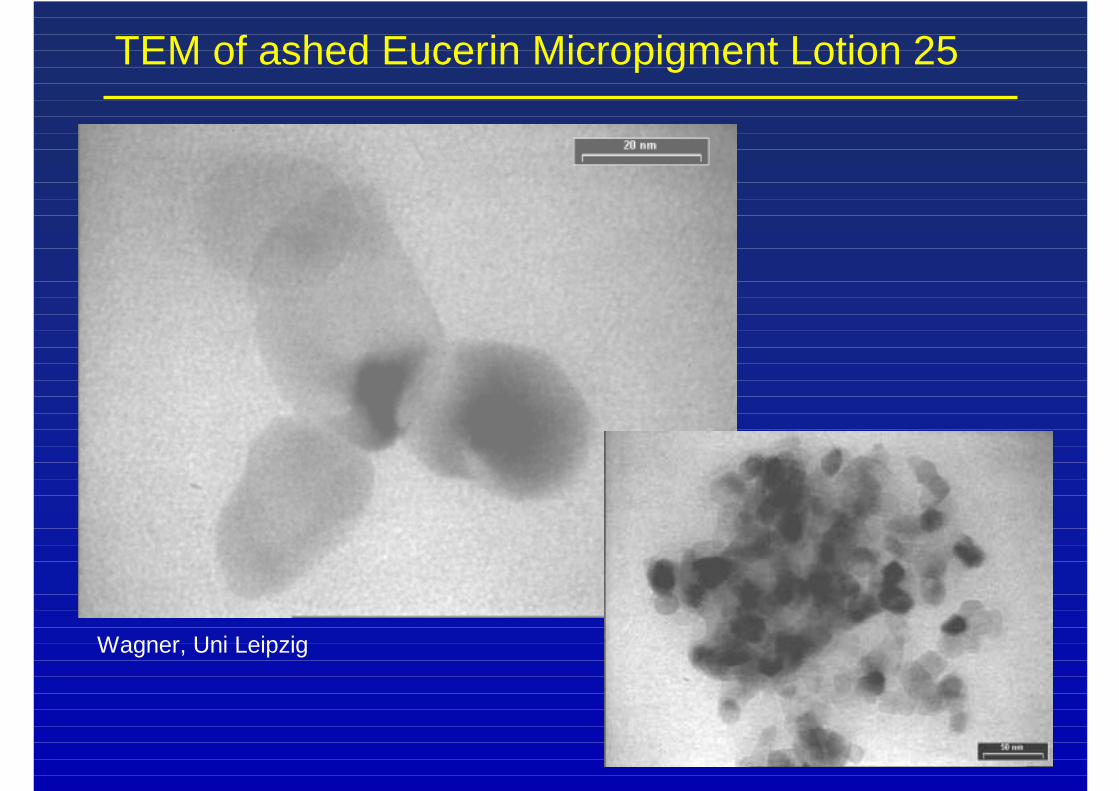
TEM of ashed Eucerin Micropigment Lotion 25
Wagner, Uni Leipzig
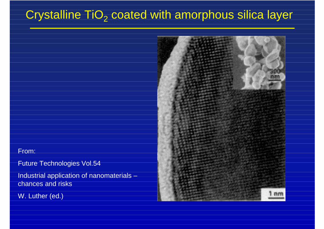
Crystalline TiO2 coated with amorphous silica layer
From:
Future Technologies Vol.54
Industrial application of nanomaterials –chances and risks
W. Luther (ed.)
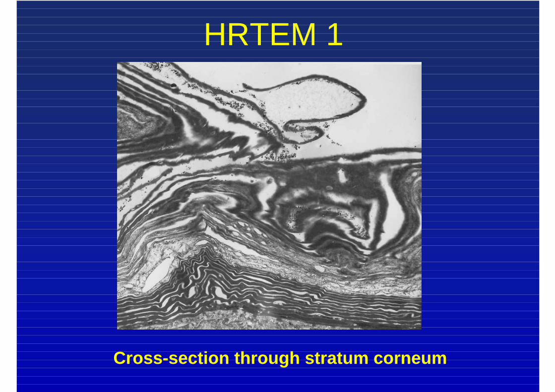
HRTEM 1
Cross-section through stratum corneum
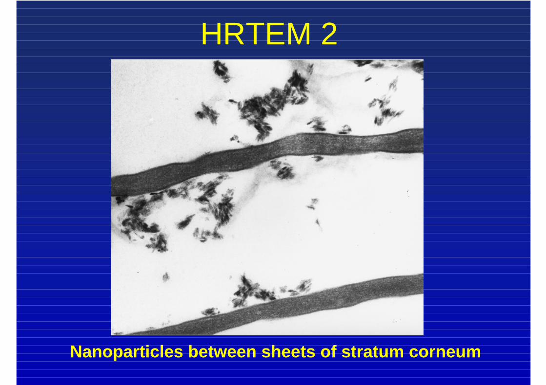
HRTEM 2
Nanoparticles between sheets of stratum corneum
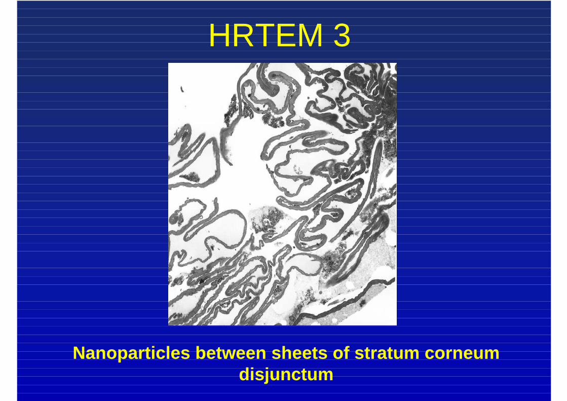
HRTEM 3
Nanoparticles between sheets of stratum corneumdisjunctum
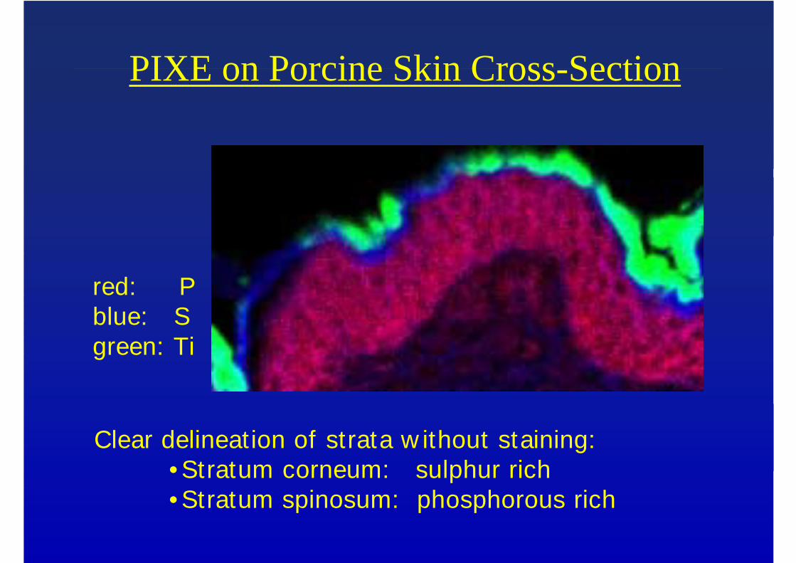
PIXE on Porcine Skin Cross-Section
red: Pblue: Sgreen: Ti
Clear delineation of strata w ithout staining:• Stratum corneum: sulphur rich• Stratum spinosum: phosphorous rich
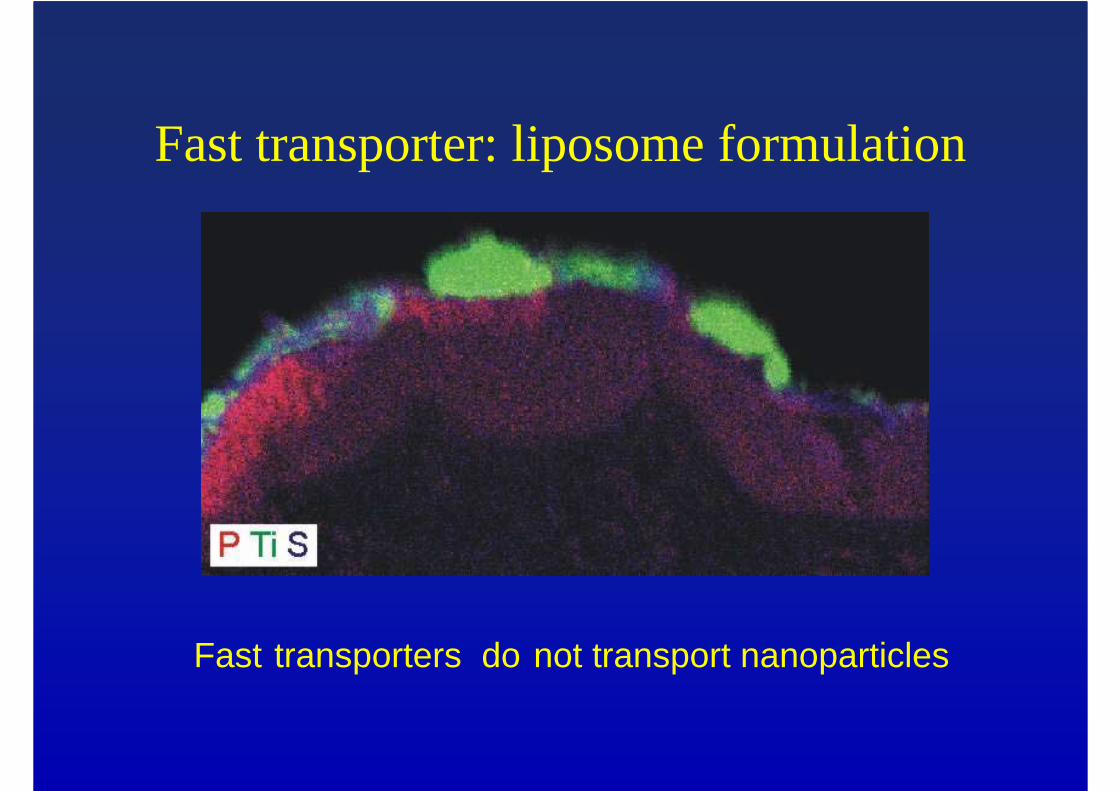
Fast transporter: liposome formulation
Fast transporters do not transport nanoparticles
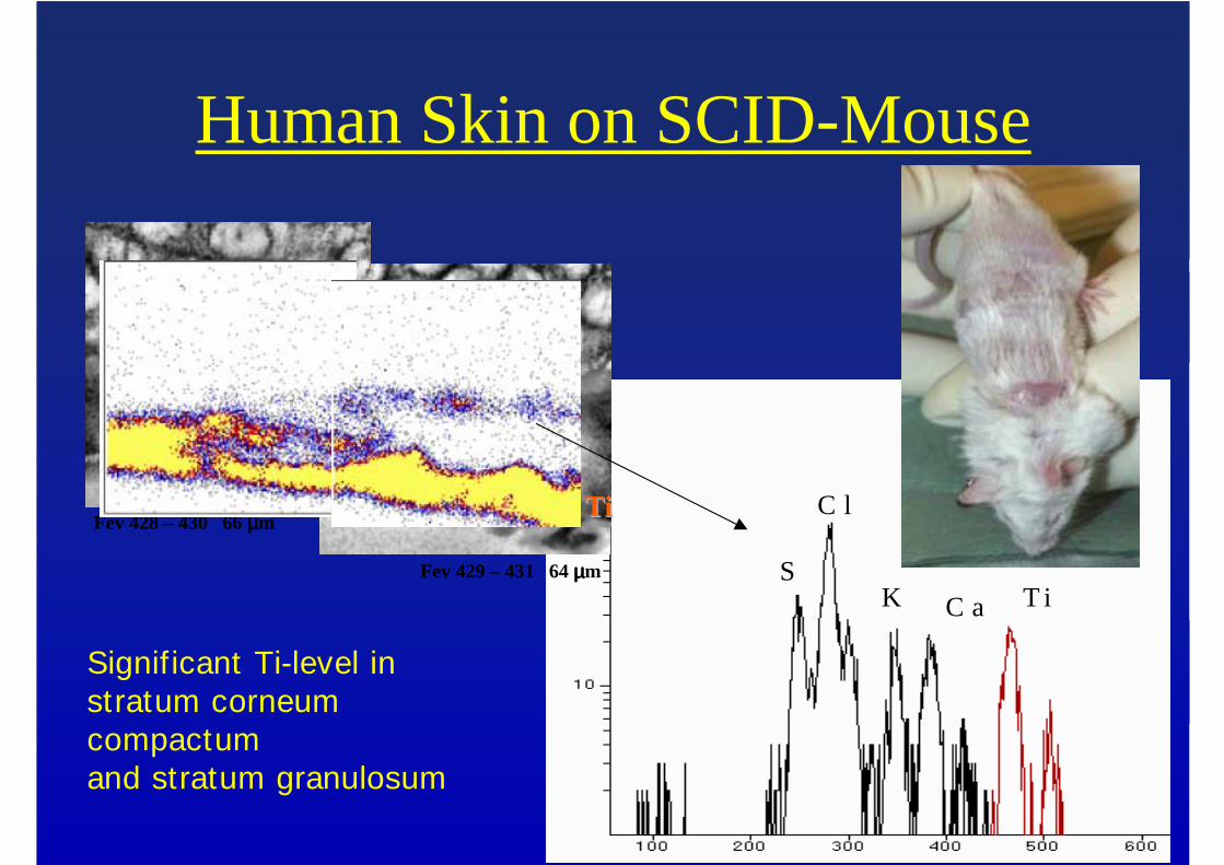
Human Skin on SCID-Mouse
C aK
C l
ST iC aK
C l
ST i
Fev 429 – 431 64 µµµµm
Fev 428 – 430 66 µµµµm TiTi
Significant Ti-level instratum corneum compactumand stratum granulosum
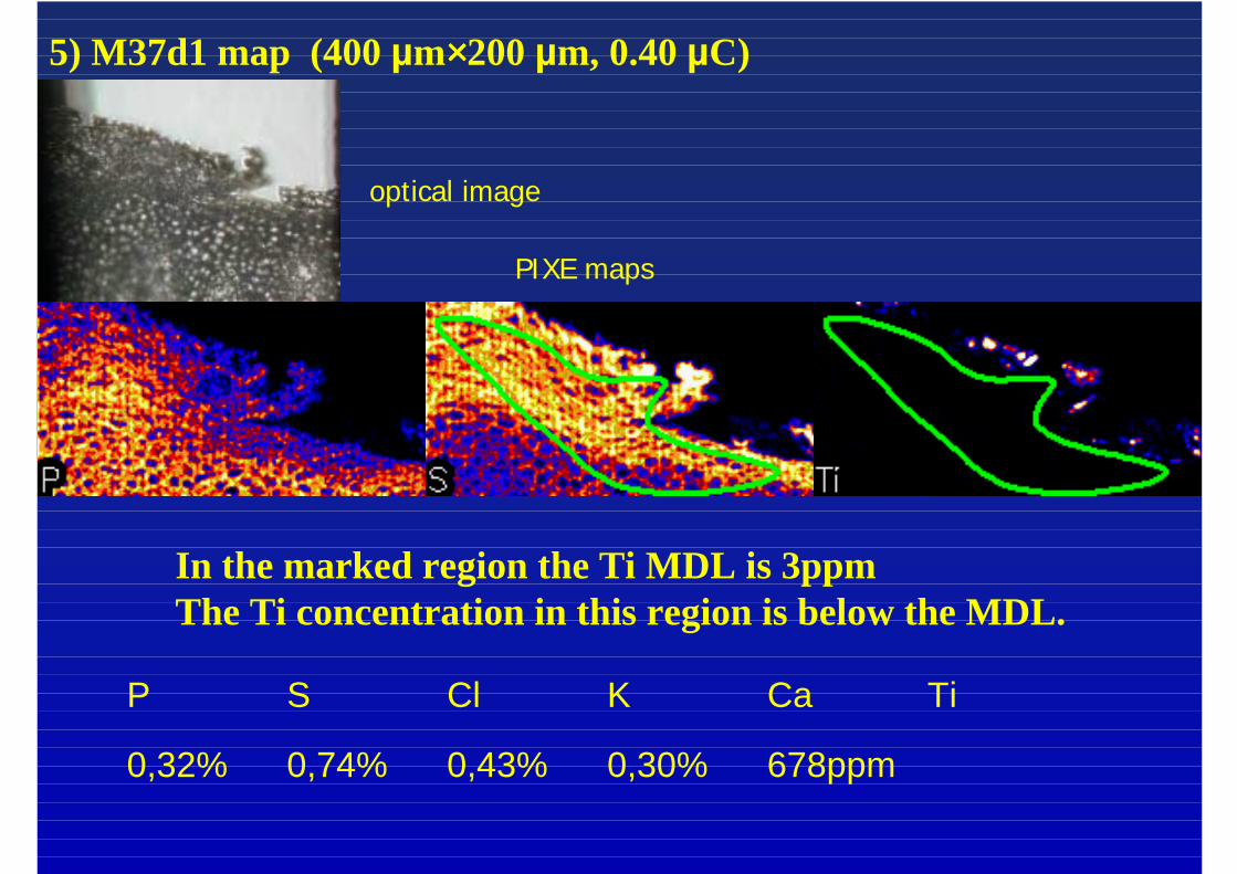
5) M37d1 map (400 µm××××200 µm, 0.40 µC)
In the marked region the Ti MDL is 3ppmThe Ti concentration in this region is below the MDL.
P S Cl K Ca Ti
0,32% 0,74% 0,43% 0,30% 678ppm
optical image
PIXE maps
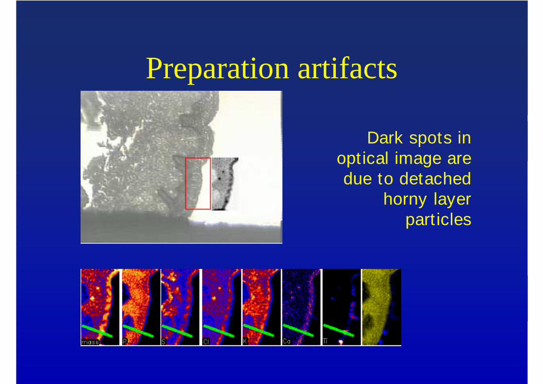
Preparation artifacts
Dark spots in optical image are due to detached
horny layer part icles
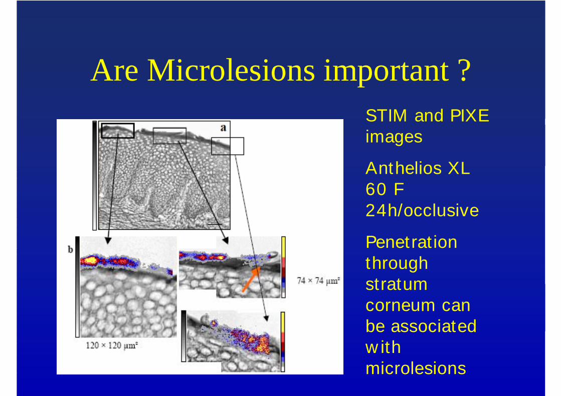
Are Microlesions important ?STIM and PIXE images
Anthelios XL 60 F 24h/occlusive
Penetrat ion through stratum corneum can be associated w ith microlesions
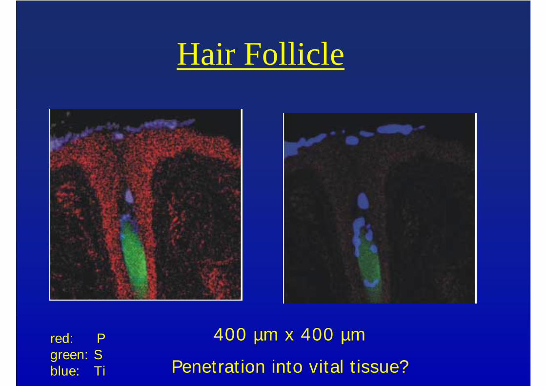
Hair Follicle
400 µm x 400 µm
Penetration into vital t issue?red: P green: Sblue: Ti
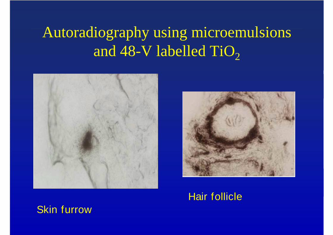
Autoradiography using microemulsions and 48-V labelled TiO2
Hair follicleSkin furrow
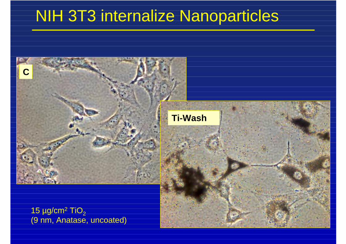
NIH 3T3 internalize Nanoparticles
C
Ti-Wash
15 µg/cm2 TiO2(9 nm, Anatase, uncoated)
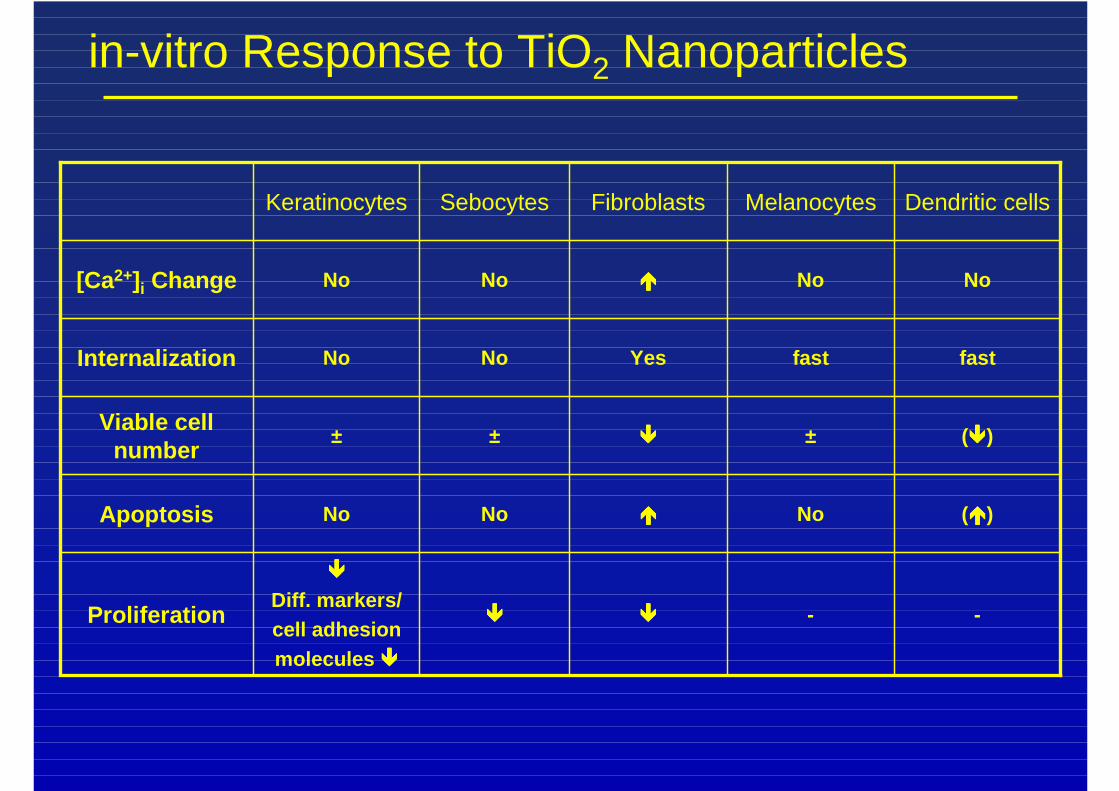
in-vitro Response to TiO2 Nanoparticles
--��������
����
Diff. markers/cell adhesionmolecules ����
Proliferation
(����)No����NoNoApoptosis
(����)±����±±Viable cellnumber
fastfastYesNoNoInternalization
NoNo����NoNo[Ca2+]i Change
Dendritic cellsMelanocytesFibroblastsSebocytesKeratinocytes
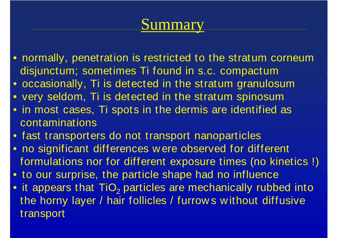
Summary
• normally, penetrat ion is restricted to the stratum corneumdisjunctum; sometimes Ti found in s.c. compactum
• occasionally, Ti is detected in the stratum granulosum• very seldom, Ti is detected in the stratum spinosum• in most cases, Ti spots in the dermis are ident if ied as
contaminat ions• fast t ransporters do not transport nanopart icles• no signif icant dif ferences w ere observed for dif ferent
formulat ions nor for dif ferent exposure t imes (no kinet ics !)• to our surprise, the part icle shape had no inf luence• it appears that TiO2 part icles are mechanically rubbed into
the horny layer / hair follicles / furrow s w ithout dif fusivetransport
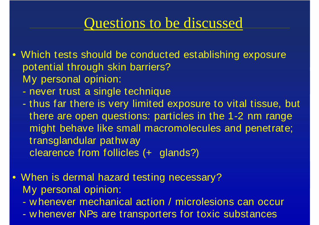
Questions to be discussed
• Which tests should be conducted establishing exposurepotent ial through skin barriers?My personal opinion: - never trust a single technique- thus far there is very limited exposure to vital t issue, but
there are open quest ions: part icles in the 1-2 nm rangemight behave like small macromolecules and penetrate;transglandular pathw ayclearence from follicles (+ glands?)
• When is dermal hazard test ing necessary?My personal opinion:- w henever mechanical act ion / microlesions can occur- w henever NPs are t ransporters for toxic substances


![Herstellung und Immobilisierung acher Gold-Nanopartikel · Ein Substrat, was zumindest das Potential besitzt, alle diese Anforderungen zu erfüllen, sind ache Gold-Nanopartikel [12]](https://img.pdfslide.org/doc/110x75/5d5eece788c993230f8b5164/herstellung-und-immobilisierung-acher-gold-nanopartikel-ein-substrat-was-zumindest.jpg)