Embed Size (px)
Citation preview
Original Article
Diplomonad flagellates of some ornamental fish cultured in Thailand
Suchanya Mankhakhet1,4, Naraid Suanyuk1, Chutima Tantikitti1*, Wutiporn Phromkunthong1,Suphada Kiriratnikom2, Theerawoot Lerssutthichawal3 and Boonkob Viriyapongsutee1
1 Kidchakan Supamattaya Aquatic Animal Health Research Center, Department of Aquatic Science, Faculty of Natural Resources,Prince of Songkla University, Hat Yai, Songkhla, 90112 Thailand.
2 Department of Biology, Faculty of Science,Thaksin University, Pa Phayom, Phatthalung, 93110 Thailand.
3 Department of Fisheries, Faculty of Agriculture,Rajamangala University of Technology Srivijaya, Thung Yai, Nakhon Si Thammarat, 80110 Thailand.
4 Center of Excellence on Agricultural Biotechnology: (AG-BIO/PERDO-CHE),Bangkok, 10900 Thailand.
Received 15 November 2011; Accepted 10 July 2012
Abstract
The study on diplomonad flagellates infection in some ornamental fishes in the family cichlidae i.e., angelfish(Pterophyllum scalare), oscar (Astronotus ocellatus), blue mbuna (Labeotropheus fuelleborni) and the family osphrone-midae i.e., Siamese fighting fish (Betta splendens) revealed that this parasite infected three out of four ornamental fish species,angelfish, oscar and blue mbuna. The highest infection was recorded in angelfish (90%) followed by oscar (75.4%) and bluembuna (61%), respectively. Identification of diplomonad flagellates from angelfish by means of morphological studies underlight and electron microscopes indicated that the parasite was Spironucleus vortens. The 14–days LD50 of S. vortens inangelfish was 2.99x10³ cells. Histopathological changes of infected angelfish revealed granulomatous liver, numerousnumbers of melanomacrophage in the spleen and inflammation of the intestine. Susceptibility study of S. vortens to goldfish(Carassius auratus), guppy (Poecilia reticulata) and platy (Xiphophorus maculatus) indicated that they were resistant toartificial infection. In vitro examination of the growth inhibition assay of S. vortens indicated that dimetridazole and metro-nidazole were effective in inhibiting parasite growth after 48 hrs exposure at concentrations of >4.0 µg/ml and >6.0 µg/ml,respectively. Magnesium sulfate at a concentration of >60 mg/ml inhibited the parasite growth after 72 hrs exposure. In vivoexamination of the dimetridazole efficiency on S. vortens infection indicated that dimetridazole at 4.0 µg/ml provided thehighest efficiency which could be used for treatment of spironucleosis in angelfish.
Keywords: Diplomonad flagellates, Spironucleus vortens, ornamental fish, treatment
Songklanakarin J. Sci. Technol.34 (5), 487-494, Sep. - Oct. 2012
1. Introduction
Infectious diseases pose a constant and serious threatto ornamental fish that are farmed intensively under condi-
tions of high stocking density, poor water quality, and poormanagement. At present, various diseases have been reportedin ornamental fish including bacterial diseases (Fergusonet al., 1994; Pate et al., 2005), viral diseases (Mellergaardand Bloch, 1988; Schuh and Shirley, 1990; Hedrick andMcDowell, 1995), and parasitic diseases (Kim et al., 2002a,b;Thilakaratne et al., 2003; Leibowitz and Zilberg, 2009). Para-sitic diseases of ornamental fish such as ichthyophthiriasis,
* Corresponding author.Email address: [email protected]
http://www.sjst.psu.ac.th
S. Mankhakhet et al. / Songklanakarin J. Sci. Technol. 34 (5), 487-494, 2012488
trichodiniasis, and tetrahymeniasis are common (Kim et al.,2002b; Tavares-Dias et al., 2010). However, pathogenic intes-tinal protozoans including diplomonad flagellates are res-ponsible for clinically important infections reported in manytropical fresh water fish species including anabantidae,belontidae, and cichlidae, and marine species, such asacanthuridae and pomacentridae (Becker, 1977; Fergusonand Moccia, 1980; Bassleer, 1983; Post, 1987; Andrews et al.,1988; Gratzek, 1988; Paull and Matthews, 2001). In ornamentalfish, these flagellates have been associated with systemicinfections, with the “hole in the head” disease (Paull andMatthews, 2001), and commonly found in a wide variety ofhosts (Poynton and Sterud, 2002). This study reports adiplomonad flagellates infection in some ornamental fish inthe family cichlidae and belontidae in Thailand. The parasiteprevalence, ultrastructure, pathogenicity, susceptibility ofdifferent fish species as well as the treatment of spiro-nucleosis are reported.
2. Materials and Methods
2.1 Fish
Between November 2008 and May 2009, 121 angelfish(P. scalare), 100 blue mbuna (L. fuelleborni), 110 oscar (A.ocellatus), and 98 Siamese fighting fish (B. splendens) withan average weight of 2.46±0.30, 3.17±1.11, 5.56±1.16 and3.19±0.31g, respectively, were collected from commercialornamental fish farms, Bangkok and Ratchaburi Provinceand transported alive to the Kidchakan Supamattaya AquaticAnimal Health Research Center, Prince of Songkla University,Hat Yai, Songkhla, Thailand. Each fish species was keptseparately in glass aquaria to prevent cross infections. Watertemperature during maintenance was 26-28°C and 20% waterexchange was carried out for each system every two days.Test fish were fed daily with commercial feed at 3-5% bodyweight.
2.2 Detection of diplomonad flagellates
Infected fish were anesthetized with 50 ppm quinal-dine (Coyle et al., 2004) and recorded for clinical signs.Internal organs including intestine, spleen, heart, andstomach were dissected and smeared onto a glass slide. Un-stained flagellates were inactivated with 5% formalin priorto examination and measurement for body size under lightmicroscope. Internal organs were also imprinted on glassslides and stained with diff quick (Baxter Diagnostics) toallow characterization of the parasite morphology and nucleishape. Prevalence of diplomonad flagellate in each ornamen-tal fish is calculated as percentage from the number ofinfected fish to the total number of fish examined.
2.3 In vitro culture of diplomonad flagellates
Intestine of infected angelfish, blue mbuna and oscar
were dissected aseptically, minced and inoculated in 10 ml ofLeibovitz’s L-15 medium supplemented with 3% fetal bovineserum, 1% heparin, 10 µg/ml of penicillin-streptomycin, 150µg/ml of penicillin G, 150 µg/ml of streptomycin, 150 µg/ml ofgentamicin, 40 µg/ml of amphotericin B, 10 µg/ml of bovinebile (Sigma) and 0.01 g fresh tilapia liver. Cultures were incu-bated at 25°C and viable cells were observed daily with aninverted microscope.
2.4 Electron microscopy
Diplomonad flagellates, grown in culture medium asdescribed above, were harvested by centrifugation at 1,500rpm for 5 minutes and re–suspended in fresh Leibovitz’sL-15 medium. A suspension of parasite was smeared on agelatin coated slide and fixed with 2.5% glutaraldehyde incacodylate buffer (pH 7.4) at 4 °C for 1 hr, then post-fixedwith 1% OsO4 in 0.1 M cacodylate buffer (pH 7.4). The samplewas then dehydrated through a graded ethanol series anddried under a critical point dryer (CPD-Hitachi), then coatedwith 15 nm gold particles in a Polaron sputter coater. Para-sites were viewed with a Jeol scanning electron microscope(SEM) at 15 kV and images recorded digitally for later print-ing.
For transmission electron microscope (TEM), sampleswere fixed as described for SEM, and then post-fixed with1% OsO4 in Veronal acetate buffer (pH 7.3). Samples wereembedded in Epon-812 and thin sections stained with uranylacetate and lead citrate. They were viewed with a Zeiss EM109 at 80 kV.
2.5 Identification to genus and species
Parasites were identified to the genus and speciesbased on parasite morphology under light and electronmicroscopy using the descriptions of diplomonad flagellatesisolated from fish as described by Poynton and Sterud (2002)and Poynton et al. (2004).
2.6 Pathogenicity study
Angelfish with an average weight of 1.81±0.44 g weremaintained in aerated glass aquaria and acclimatized for twoweeks before infectivity testing. During the acclimatizationperiod, these fish were fed daily with commercial feed at3-5% body weight. The fish were examined to ensure thatthey were disease-free prior to use in the trial. They wereanaesthetized with 50 ppm quinaldine and injected intra-peritoneally with 0.1 ml of a parasitic suspension. Briefly, theparasites grown in culture medium were harvested and re–suspended in phosphate buffer saline (pH 7.4) to achievedfinal concentration of 1.1x10³, 4.2x10³, 1.2x10⁴, 4.6x104,1.12x105, 4.5x10⁵ and 1.25x10⁶ cells/ml. A control group wasinjected with sterile PBS (pH 7.4). The experiment wasconducted in triplicate using 10 fish per replicate. Watertemperature during the assay was 26-28°C. Clinical signs and
489S. Mankhakhet et al. / Songklanakarin J. Sci. Technol. 34 (5), 487-494, 2012
mortality were recorded daily for 14 days post challenge.Tissue samples of intestine, stomach, liver, spleen and heartwere aseptically collected and examined for diplomonadflagellates. The mean 14–day-LD50 was determined using thesimplified method of Reed and Muench (1938).
2.7 Histopathology
Tissue samples of intestine, liver, stomach, gall blad-der, heart and spleen from diseased fish obtained from bothcommercial fish farms and experimentally infected conditionswere fixed in 10% buffered formalin solution and processedusing standard histological techniques (Humason, 1979).Briefly, after 72 hrs fixation, each sample was cut into smallpieces for better penetration of the reagents in the process ofparaffin embedding. Respective tissue samples were placedin tissue cassettes labeled with corresponding source ofsamples. Histological sections were processed routinely,sectioned at 3-5 µm, and stained with haematoxylin and eosin(H&E). The stained sections were examined under a lightmicroscope.
2.8 Susceptibility of other fish species
Thirty goldfish (Carassius auratus) with an averageweight of 5.94±1.88 g, thirty guppy (Poecilia reticulata)with an average weight of 0.81±0.24 g and thirty platy(Xiphophorus maculatus) with an average weight of 0.44±0.21 g were inoculated intraperitoneally with 0.1 ml of 3.0x10³
cells of diplomonad flagellates prepared as described above.Ten control fish from each species were injected with 0.1 mlPBS (pH 7.4). Observation was made daily over a period of10 days for signs of disease. Tissue samples from all deador survival fish were aseptically collected and examined toconfirm diplomonad flagellates as the cause of death.
2.9 Growth inhibition assay
Dimetridazole, metronidazole and magnesium sulfate(MgSO4) were used to inhibit the growth of diplomonadflagellates in vitro. Dimetridazole and metronidazole wereexamined at concentrations of 0.25, 0.50, 1.0, 2.0, 4.0, 6.0, 8.0,and 10 µg/ml by dissolving in dimethyl sulfoxide. MgSO4was examined at concentration of 2.5, 5.0,10, 20, 40, 60, 80,and 100 mg/ml by dissolving in distilled water. Stationaryphase of diplomonad flagellates grown in culture mediumat concentration of 2.5x10³ cells/ml were used in the study.Control groups were established for each chemotherapeutictesting group. All treatments were examined in triplicate insterile screw-capped test tubes containing 5 ml diplomonadflagellates, which were incubated at 25ºC in a thermo-regu-lated incubator. The average number of parasites was deter-mined every 24 hrs until no parasites was observed alive(Sangmaneedet and Smith, 1999).
2.10 In vivo treatment of spironucleosis in angelfish
Dimetridazole was chosen for treatment of spiro-nucleosis in angelfish because of its effectiveness in inhibit-ing parasite growth in vitro study. Infected angelfish collectedfrom ornamental fish farm were used in this study. Infectedfish were sampled and determined for the present of thediplomonad flagellates using the same method as describedpreviously. This experiment was conducted in duplicate using45 fish per replication. Dimetridazole was examined at con-centrations of 0, 0.25, 0.50, 1.0, 2.0, 4.0, and 6.0 µg/ml byprolonged immersion method. After immersion, five infectedfish were collected daily from each replication to examine thepresent of the parasite. Infection rate was calculated aspercentage from number of infected fish to the total numberof fish examined.
3. Results
3.1 Clinical signs
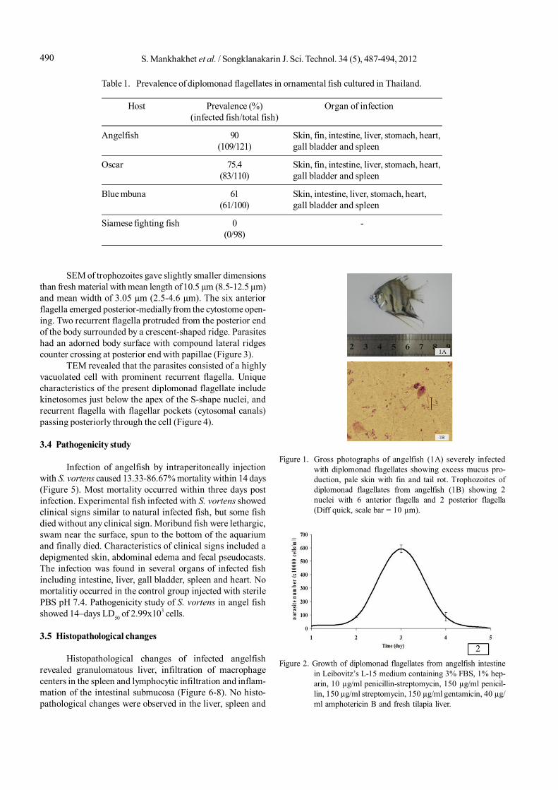
The study of diplomonad flagellates infection revealedthe infection in 3 out of 4 ornamental fish species i.e., angel-fish, oscar and blue mbuna. The highest percentage of infec-tion was recorded in angelfish followed by oscar and bluembuna (Table 1). Infected fish showed various disease signsincluding lethargy, emaciation, anorexia, abdominal edemaand fecal pseudocasts, pale skin with fin and tail rot (Figure1). The infection was found in several organs of infected fishincluding intestine, liver, gall bladder, spleen, heart, skin andfins. No diplomonad flagellates was observed in Siamesefighting fish.
3.2 In vitro culture of diplomonad flagellates
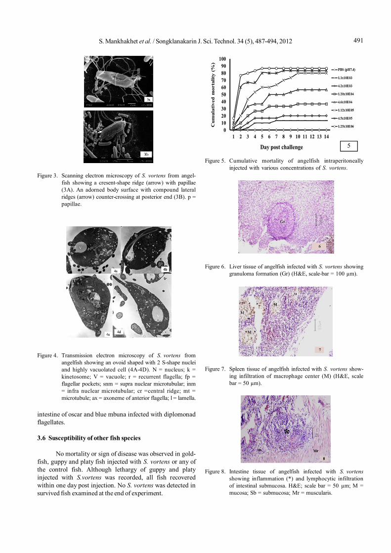
Diplomonad flagellates from the angelfish intestinewere successfully established and maintained in culturemedia. The cultured cells reached maximum numbers withinthree days post inoculation. The parasite died within fivedays if sub-culture was not made (Figure 2). No multiplica-tion of the parasites isolated from blue mbuna and oscarwas observed.
3.3 Identification of diplomonad flagellates
Morphological characteristics of diplomonad flagel-lates from angelfish under light and electron microscopesstudies indicated that the parasite was Spironucleus vortens.In squash preparations, parasites were highly motile androtated around their longitudinal axis. Trophozoites werepyriform to ovoid in shape, 9.0-16.0 µm (mean 12.86 µm) inlength and 3.0-10.0 µm (mean 6.52 µm) in width. The parasitehad distinct S-shaped nuclei and eight flagella (six anteriorand two posterior).
S. Mankhakhet et al. / Songklanakarin J. Sci. Technol. 34 (5), 487-494, 2012490
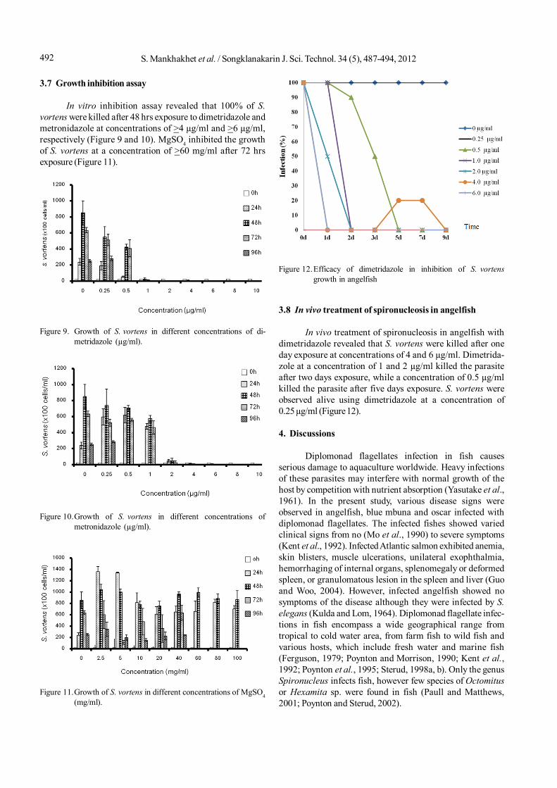
SEM of trophozoites gave slightly smaller dimensionsthan fresh material with mean length of 10.5 µm (8.5-12.5 µm)and mean width of 3.05 µm (2.5-4.6 µm). The six anteriorflagella emerged posterior-medially from the cytostome open-ing. Two recurrent flagella protruded from the posterior endof the body surrounded by a crescent-shaped ridge. Parasiteshad an adorned body surface with compound lateral ridgescounter crossing at posterior end with papillae (Figure 3).
TEM revealed that the parasites consisted of a highlyvacuolated cell with prominent recurrent flagella. Uniquecharacteristics of the present diplomonad flagellate includekinetosomes just below the apex of the S-shape nuclei, andrecurrent flagella with flagellar pockets (cytosomal canals)passing posteriorly through the cell (Figure 4).
3.4 Pathogenicity study
Infection of angelfish by intraperitoneally injectionwith S. vortens caused 13.33-86.67% mortality within 14 days(Figure 5). Most mortality occurred within three days postinfection. Experimental fish infected with S. vortens showedclinical signs similar to natural infected fish, but some fishdied without any clinical sign. Moribund fish were lethargic,swam near the surface, spun to the bottom of the aquariumand finally died. Characteristics of clinical signs included adepigmented skin, abdominal edema and fecal pseudocasts.The infection was found in several organs of infected fishincluding intestine, liver, gall bladder, spleen and heart. Nomortalitiy occurred in the control group injected with sterilePBS pH 7.4. Pathogenicity study of S. vortens in angel fishshowed 14–days LD50 of 2.99x10³ cells.
3.5 Histopathological changes
Histopathological changes of infected angelfishrevealed granulomatous liver, infiltration of macrophagecenters in the spleen and lymphocytic infiltration and inflam-mation of the intestinal submucosa (Figure 6-8). No histo-pathological changes were observed in the liver, spleen and
Table 1. Prevalence of diplomonad flagellates in ornamental fish cultured in Thailand.
Host Prevalence (%) Organ of infection(infected fish/total fish)
Angelfish 90 Skin, fin, intestine, liver, stomach, heart,(109/121) gall bladder and spleen
Oscar 75.4 Skin, fin, intestine, liver, stomach, heart,(83/110) gall bladder and spleen
Blue mbuna 61 Skin, intestine, liver, stomach, heart,(61/100) gall bladder and spleen
Siamese fighting fish 0 -(0/98)
Figure 2. Growth of diplomonad flagellates from angelfish intestinein Leibovitz’s L-15 medium containing 3% FBS, 1% hep-arin, 10 µg/ml penicillin-streptomycin, 150 µg/ml penicil-lin, 150 µg/ml streptomycin, 150 µg/ml gentamicin, 40 µg/ml amphotericin B and fresh tilapia liver.
Figure 1. Gross photographs of angelfish (1A) severely infectedwith diplomonad flagellates showing excess mucus pro-duction, pale skin with fin and tail rot. Trophozoites ofdiplomonad flagellates from angelfish (1B) showing 2nuclei with 6 anterior flagella and 2 posterior flagella(Diff quick, scale bar = 10 µm).
491S. Mankhakhet et al. / Songklanakarin J. Sci. Technol. 34 (5), 487-494, 2012
intestine of oscar and blue mbuna infected with diplomonadflagellates.
3.6 Susceptibility of other fish species
No mortality or sign of disease was observed in gold-fish, guppy and platy fish injected with S. vortens or any ofthe control fish. Although lethargy of guppy and platyinjected with S.vortens was recorded, all fish recoveredwithin one day post injection. No S. vortens was detected insurvived fish examined at the end of experiment.
Figure 3. Scanning electron microscopy of S. vortens from angel-fish showing a cresent-shape ridge (arrow) with papillae(3A). An adorned body surface with compound lateralridges (arrow) counter-crossing at posterior end (3B). p =papillae.
Figure 4. Transmission electron microscopy of S. vortens fromangelfish showing an ovoid shaped with 2 S-shape nucleiand highly vacuolated cell (4A-4D). N = nucleus; k =kinetosome; V = vacuole; r = recurrent flagella; fp =flagellar pockets; snm = supra nuclear microtubular; inm= infra nuclear microtubular; cr =central ridge; mt =microtubule; ax = axoneme of anterior flagella; l = lamella.
Figure 5. Cumulative mortality of angelfish intraperitoneallyinjected with various concentrations of S. vortens.
Figure 6. Liver tissue of angelfish infected with S. vortens showinggranuloma formation (Gr) (H&E, scale-bar = 100 µm).
Figure 7. Spleen tissue of angelfish infected with S. vortens show-ing infiltration of macrophage center (M) (H&E, scalebar = 50 µm).
Figure 8. Intestine tissue of angelfish infected with S. vortensshowing inflammation (*) and lymphocytic infiltrationof intestinal submucosa. H&E; scale bar = 50 µm; M =mucosa; Sb = submucosa; Mr = muscularis.
S. Mankhakhet et al. / Songklanakarin J. Sci. Technol. 34 (5), 487-494, 2012492
3.7 Growth inhibition assay
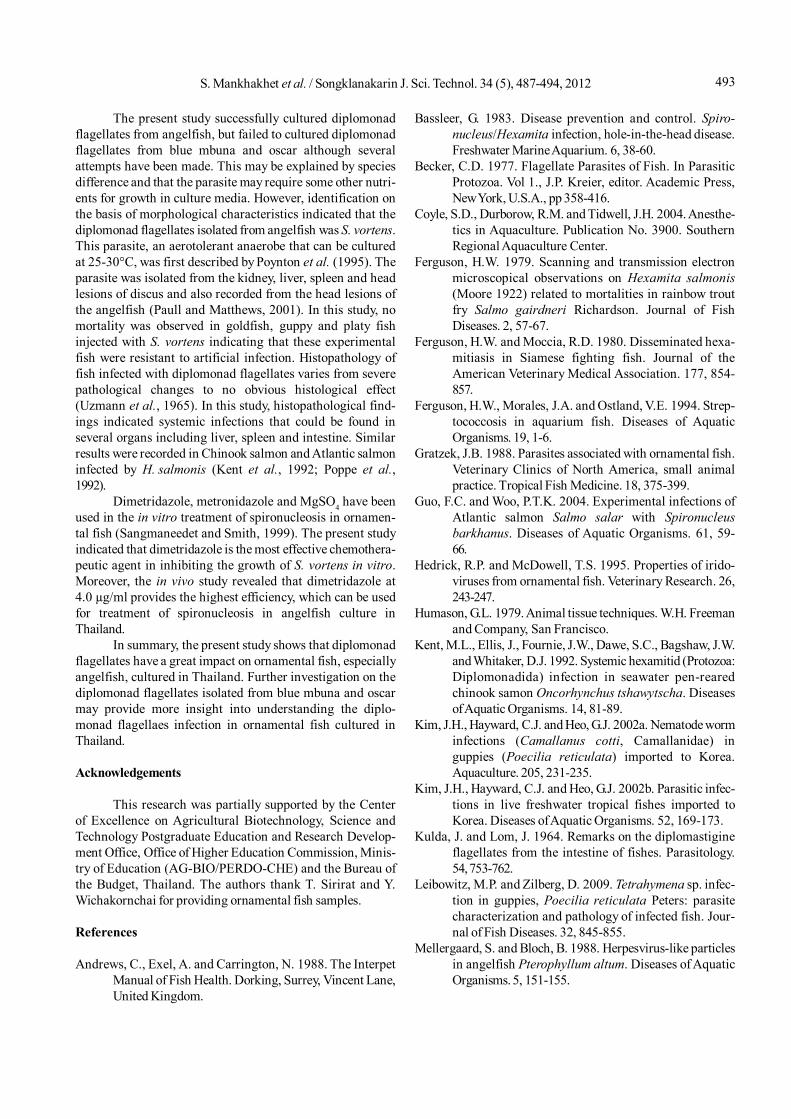
In vitro inhibition assay revealed that 100% of S.vortens were killed after 48 hrs exposure to dimetridazole andmetronidazole at concentrations of >4 µg/ml and >6 µg/ml,respectively (Figure 9 and 10). MgSO4 inhibited the growthof S. vortens at a concentration of >60 mg/ml after 72 hrsexposure (Figure 11).
3.8 In vivo treatment of spironucleosis in angelfish
In vivo treatment of spironucleosis in angelfish withdimetridazole revealed that S. vortens were killed after oneday exposure at concentrations of 4 and 6 µg/ml. Dimetrida-zole at a concentration of 1 and 2 µg/ml killed the parasiteafter two days exposure, while a concentration of 0.5 µg/mlkilled the parasite after five days exposure. S. vortens wereobserved alive using dimetridazole at a concentration of0.25 µg/ml (Figure 12).
4. Discussions
Diplomonad flagellates infection in fish causesserious damage to aquaculture worldwide. Heavy infectionsof these parasites may interfere with normal growth of thehost by competition with nutrient absorption (Yasutake et al.,1961). In the present study, various disease signs wereobserved in angelfish, blue mbuna and oscar infected withdiplomonad flagellates. The infected fishes showed variedclinical signs from no (Mo et al., 1990) to severe symptoms(Kent et al., 1992). Infected Atlantic salmon exhibited anemia,skin blisters, muscle ulcerations, unilateral exophthalmia,hemorrhaging of internal organs, splenomegaly or deformedspleen, or granulomatous lesion in the spleen and liver (Guoand Woo, 2004). However, infected angelfish showed nosymptoms of the disease although they were infected by S.elegans (Kulda and Lom, 1964). Diplomonad flagellate infec-tions in fish encompass a wide geographical range fromtropical to cold water area, from farm fish to wild fish andvarious hosts, which include fresh water and marine fish(Ferguson, 1979; Poynton and Morrison, 1990; Kent et al.,1992; Poynton et al., 1995; Sterud, 1998a, b). Only the genusSpironucleus infects fish, however few species of Octomitusor Hexamita sp. were found in fish (Paull and Matthews,2001; Poynton and Sterud, 2002).
Figure 9. Growth of S. vortens in different concentrations of di-metridazole (µg/ml).
Figure 10.Growth of S. vortens in different concentrations ofmetronidazole (µg/ml).
Figure 11.Growth of S. vortens in different concentrations of MgSO4(mg/ml).
Figure 12.Efficacy of dimetridazole in inhibition of S. vortensgrowth in angelfish
493S. Mankhakhet et al. / Songklanakarin J. Sci. Technol. 34 (5), 487-494, 2012
The present study successfully cultured diplomonadflagellates from angelfish, but failed to cultured diplomonadflagellates from blue mbuna and oscar although severalattempts have been made. This may be explained by speciesdifference and that the parasite may require some other nutri-ents for growth in culture media. However, identification onthe basis of morphological characteristics indicated that thediplomonad flagellates isolated from angelfish was S. vortens.This parasite, an aerotolerant anaerobe that can be culturedat 25-30°C, was first described by Poynton et al. (1995). Theparasite was isolated from the kidney, liver, spleen and headlesions of discus and also recorded from the head lesions ofthe angelfish (Paull and Matthews, 2001). In this study, nomortality was observed in goldfish, guppy and platy fishinjected with S. vortens indicating that these experimentalfish were resistant to artificial infection. Histopathology offish infected with diplomonad flagellates varies from severepathological changes to no obvious histological effect(Uzmann et al., 1965). In this study, histopathological find-ings indicated systemic infections that could be found inseveral organs including liver, spleen and intestine. Similarresults were recorded in Chinook salmon and Atlantic salmoninfected by H. salmonis (Kent et al., 1992; Poppe et al.,1992).
Dimetridazole, metronidazole and MgSO4 have beenused in the in vitro treatment of spironucleosis in ornamen-tal fish (Sangmaneedet and Smith, 1999). The present studyindicated that dimetridazole is the most effective chemothera-peutic agent in inhibiting the growth of S. vortens in vitro.Moreover, the in vivo study revealed that dimetridazole at4.0 µg/ml provides the highest efficiency, which can be usedfor treatment of spironucleosis in angelfish culture inThailand.
In summary, the present study shows that diplomonadflagellates have a great impact on ornamental fish, especiallyangelfish, cultured in Thailand. Further investigation on thediplomonad flagellates isolated from blue mbuna and oscarmay provide more insight into understanding the diplo-monad flagellaes infection in ornamental fish cultured inThailand.
Acknowledgements
This research was partially supported by the Centerof Excellence on Agricultural Biotechnology, Science andTechnology Postgraduate Education and Research Develop-ment Office, Office of Higher Education Commission, Minis-try of Education (AG-BIO/PERDO-CHE) and the Bureau ofthe Budget, Thailand. The authors thank T. Sirirat and Y.Wichakornchai for providing ornamental fish samples.
References
Andrews, C., Exel, A. and Carrington, N. 1988. The InterpetManual of Fish Health. Dorking, Surrey, Vincent Lane,United Kingdom.
Bassleer, G. 1983. Disease prevention and control. Spiro-nucleus/Hexamita infection, hole-in-the-head disease.Freshwater Marine Aquarium. 6, 38-60.
Becker, C.D. 1977. Flagellate Parasites of Fish. In ParasiticProtozoa. Vol 1., J.P. Kreier, editor. Academic Press,New York, U.S.A., pp 358-416.
Coyle, S.D., Durborow, R.M. and Tidwell, J.H. 2004. Anesthe-tics in Aquaculture. Publication No. 3900. SouthernRegional Aquaculture Center.
Ferguson, H.W. 1979. Scanning and transmission electronmicroscopical observations on Hexamita salmonis(Moore 1922) related to mortalities in rainbow troutfry Salmo gairdneri Richardson. Journal of FishDiseases. 2, 57-67.
Ferguson, H.W. and Moccia, R.D. 1980. Disseminated hexa-mitiasis in Siamese fighting fish. Journal of theAmerican Veterinary Medical Association. 177, 854-857.
Ferguson, H.W., Morales, J.A. and Ostland, V.E. 1994. Strep-tococcosis in aquarium fish. Diseases of AquaticOrganisms. 19, 1-6.
Gratzek, J.B. 1988. Parasites associated with ornamental fish.Veterinary Clinics of North America, small animalpractice. Tropical Fish Medicine. 18, 375-399.
Guo, F.C. and Woo, P.T.K. 2004. Experimental infections ofAtlantic salmon Salmo salar with Spironucleusbarkhanus. Diseases of Aquatic Organisms. 61, 59-66.
Hedrick, R.P. and McDowell, T.S. 1995. Properties of irido-viruses from ornamental fish. Veterinary Research. 26,243-247.
Humason, G.L. 1979. Animal tissue techniques. W.H. Freemanand Company, San Francisco.
Kent, M.L., Ellis, J., Fournie, J.W., Dawe, S.C., Bagshaw, J.W.and Whitaker, D.J. 1992. Systemic hexamitid (Protozoa:Diplomonadida) infection in seawater pen-rearedchinook samon Oncorhynchus tshawytscha. Diseasesof Aquatic Organisms. 14, 81-89.
Kim, J.H., Hayward, C.J. and Heo, G.J. 2002a. Nematode worminfections (Camallanus cotti, Camallanidae) inguppies (Poecilia reticulata) imported to Korea.Aquaculture. 205, 231-235.
Kim, J.H., Hayward, C.J. and Heo, G.J. 2002b. Parasitic infec-tions in live freshwater tropical fishes imported toKorea. Diseases of Aquatic Organisms. 52, 169-173.
Kulda, J. and Lom, J. 1964. Remarks on the diplomastigineflagellates from the intestine of fishes. Parasitology.54, 753-762.
Leibowitz, M.P. and Zilberg, D. 2009. Tetrahymena sp. infec-tion in guppies, Poecilia reticulata Peters: parasitecharacterization and pathology of infected fish. Jour-nal of Fish Diseases. 32, 845-855.
Mellergaard, S. and Bloch, B. 1988. Herpesvirus-like particlesin angelfish Pterophyllum altum. Diseases of AquaticOrganisms. 5, 151-155.
S. Mankhakhet et al. / Songklanakarin J. Sci. Technol. 34 (5), 487-494, 2012494
Mo, T.A., Poppe, T.T. and Iversen, L. 1990. Systemic hexa-mitosis in salt-water reared Atlantic salmon (Salmosalar L.). Bulletin of the European Association of FishPathology. 10, 69-70.
Pate, M., Jenèiè, V., Dovè, M.Ž. and Ocepek, M. 2005. Detec-tion of mycobacteria in aquarium fish in Slovenia byculture and molecular methods. Diseases of AquaticOrganisms. 64, 29-35.
Paull, G.C. and Matthews, R.A. 2001. Spironucleus vortens,a possible cause of hole–in–the–head disease incichlids. Diseases of Aquatic Organisms. 45, 197-202.
Poppe, T.T., Mo, T.A. and Iversen, L. 1992. Disseminatedhexamitosis in sea-caged Atlantic salmon Salmosalar. Diseases of Aquatic Organisms. 14, 91-97.
Post, G. 1987. Textbook of Fish Health, 2nd edn. TFH Publica-tions Inc, New Jersey, U.S.A., p 288.
Poynton, S.L., Fard, M.R.S., Jenkins, J. and Ferguson, H.W.2004. Ultrastructure of Spironucleus salmonis n.comb. (formerly Octomitus salmonis sensu Moore 1922,Davis 1926, and Hexamita salmonis sensu Ferguson1979), with a guide to Spironucleus species. Diseasesof Aquatic Organisms. 60, 49-64.
Poynton, S.L., Fraser, W., Francis-Floyd, R., Rutledge, P.,Reed, P. and Nerad, T.A. 1995. Spironucleus vortensn. sp. from the freshwater angelfish Pterophyllumscalare: morphology and culture. Journal of Eukar-yotic Microbiology. 42, 731-742.
Poynton, S.L. and Morrison, C.M. 1990. Morphology ofdiplomonad flagellates: Spironucleus torosa fromAtlantic Cod Gadus morhua L., and HaddockMelanogrammus aeglefinus (L.) and Hexamitasalmonis from brook trout Salvelinus fontinalis(Mitchill). Journal of Protozoology. 37, 369-383.
Poynton, S.L. and Sterud, E. 2002. Guidelines for speciesdescriptions of diplomonad flagellates from fish.Journal of Fish Diseases. 25, 15-31.
Reed, L.J. and Muench, H. 1938. A simple method of esti-mating fifty percent endpoints. American Journal ofHygiene. 27, 493-497.
Sangmaneedet, S. and Smith, S.A. 1999. Efficacy of variouschemotherapeutic agents on the growth of Spiro-nucleus vortens, an intestinal parasite of freshwaterangelfish. Diseases of Aquatic Organisms. 38: 47-52
Schuh, J.C.L. and Shirley, I.G. 1990. Viral hematopoietic necro-sis in an angelfish (Pterophyllum scalare). Journal ofZoo and Wildlife Medicine. 21, 95-98.
Sterud, E. 1998a. In vitro cultivation and temperature-depen-dent growth of two strains of Spironucleus barkhanus(Diplomonadida: Hexamitidae) from Atlantic salmonSalmo salar and grayling Thymallus thymallus.Diseases of Aquatic Organisms. 33, 57-61.
Sterud, E. 1998b. Ultrastructure of Spironucleus torosaPoynton and Morrison, 1990 (Diplomonadida:Hexamitidae) in cod Gadus morhua (L.) and saithePollachius virens (L.) from southeastern Norway.European Journal of Protistology. 34, 69-77.
Tavares-Dias, M., Lemos, J.R.G. and Martins, M.L. 2010. Para-sitic fauna of eight species of ornamental freshwaterfish species from the middle Negro River in the Brazil-ian Amazon Region. Revista Brasileira de ParasitologiaVeterinária. 19, 103-107.
Thilakaratne, I.D.S.I.P., Rajapaksha, G., Hewakopara, A.,Rajapakse, R.P.V.J. and Faizal, A.C.M. 2003. Parasiticinfections in freshwater ornamental fish in Sri Lanka.Diseases of Aquatic Organisms. 54, 157-162.
Uzmann, J.R., Paulik, G.J. and Hayduk, S.H. 1965. Experimentalhexamitiasis in juvenile coho salmon (Oncorhynchuskisutch) and steelhead trout (Salmo gairdneri).Transactions of the American Fisheries Society. 94,53-61.
Yasutake, W.T., Buhler, D.R. and Shanks, W.E. 1961. Chemo-therapy of hexamitiasis in fish. Journal of Parasito-logy. 47, 81-86.