Embed Size (px)
Citation preview
DISSERTATION
Titel der Dissertation
„Preparation, implementation and evaluation of affinity
chromatographic tools for targeted proteome identification
by liquid chromatography –
tandem mass spectrometry“
Verfasser
Mag. rer. nat. Martin Georg Sturm
angestrebter akademischer Grad
Doktor der Naturwissenschaften (Dr. rer. nat.)
Wien, 2010
Studienkennzahl lt. Studienblatt: A 9604921
Dissertationsgebiet lt. Studienblatt: Dr.-Studium der Naturwissenschaften Chemie
Betreuerin / Betreuer: O. Univ. Prof. Dr. Wolfgang Lindner
Die Arbeiten für die vorliegende Dissertation wurden in der Zeit von Oktober 2006 bis März
2009 am Institut für Analytische Chemie und Lebensmittelchemie der Universität Wien unter
der Leitung von Prof. Dr. Wolfgang Lindner und Dr. Alexander Leitner durchgeführt.
Ich möchte dafür
Prof. Dr. Wolfgang Lindner für die Möglichkeit der Durchführung der Arbeiten, für die
Nutzung der instrumentellen Ressourcen, der fachlichen Kompetenz und das in mich
gesetzte Vertrauen;
Dr. Alexander Leitner für die außerordentlich gute Betreuung und gute Zusammenarbeit, für
Geduld und Inspiration;
den Mitarbeitern der Arbeitsgruppe für fachliche Diskussionen und auch viele unterhaltsame
Momente ganz herzlich danken.
Vielen Dank auch meinen Freunden für die angenehme Zeit außerhalb der Wissenschaft.
Mein besonderer Dank gilt natürlich meinen Eltern, im Besonderen meiner Mutter für das
Vertrauen in mich und vieles mehr.
Die Arbeit wurde finanziert von der Austrian Proteomics Platform, Teil des GEN-AU Projekts
des österreichischen Ministeriums für Forschung und Entwicklung.
1
Table of content
Summary ………………...……………………………………………………………………………....3
Zusammenfassung …………………………………………………………………………….……………….5
1 Aim ………………………………………………………………………......…………………….7
2 Tools for analysing the phosphoproteome and other phosphorylated biocompounds ……………………………………………………………………………………………………...........……8
1 Introduction .................................................................................................................. 9
2 Starting an experiment .............................................................................................. 12
3 Immunoaffinity chromatography ................................................................................ 13
4 Immobilized Metal Affinity Chromatography .............................................................. 15
5 Metal Oxide Affinity Chromatography ....................................................................... 18
6 Ion exchange chromatography .................................................................................. 23
7 Chemical modification based methods ..................................................................... 27
8 Ca2+-Precipitation ...................................................................................................... 29
9 Combination of methods .......................................................................................... 30
10 Application to other biological phosphocompounds .................................................. 33
11 Mass spectrometry based protein analysis .............................................................. 35
12 Technologies for peptide sequencing by mass spectrometry ................................... 36
Collision induced fragmentation .................................................................................................... 37
Electron capture dissociation ........................................................................................................ 38
Electron transfer dissociation ........................................................................................................ 38
13 Quantitation Methods ................................................................................................................... 39
Conclusion ……………………………………………………………………………......…………….42
3 A chemically cleavable linker concept .....................................................................................59
4 Appendix ...........................................................................................................................60

2
Publication 1: Tin dioxide microspheres as a promising material for phosphpeptide enrichment prior
to liquid chromatography - (tandem) mass spectrometry analysis
Publication 2: Optimizing the performance of tin dioxide microspheres for phosphopeptide
enrichment
Publication 3: Probing the phosphoproteome of HeLa cells using nanocast metal oxide
microspheres for phosphopeptide enrichment
Manuscript 1: A chemically cleavable linker for immobilizing bait compounds in a target protein
pull-down concept. Submitted to Bioconjugate Chemistry.
Lebenslauf
3
Summary In the course of this work two main projects comprising the development and implementation
of analytical tools for proteomic applications were realized. Both projects included
methodology development and methodology optimization of specific enrichment methods to
facilitate target proteome identification by liquid chromatography (LC) – tandem mass
spectrometry (MS/MS). Optimized protocols were then used for biological applications.
In the first part tin dioxide (SnO2) was examined for the use as enrichment material for
selective phosphopeptide enrichment. Tin dioxide was provided in the form of porous
microspheres, which were manufactured by the nanocast process [Smått, 2007]. Initially, a
prescreening of different surface treated tin dioxide materials was performed. The most
suitable material was chosen for method optimization, which comprised optimization of the
loading and elution buffer composition, including variations of the pH, the ion strength and
the percentage of organic solvent, as well as the evaluation of possible buffer additives to
increase selectivity and phosphopeptide recovery rates. The applicability of the optimized
protocol was demonstrated by the LC-MS/MS analysis of a tryptic digest of a mix of
phosphorylated and nonphosphorylated model proteins [see Appendix Publication no. 1].
Beneficial features of the new method were a simple loading buffer composition and good
practicability. Furthermore, different nanocast TiO2 materials, with variations in diameter,
pore size, and surface treatment were examined for material dependent parameter
optimization. Whereas variations in pore size yielded comparable results, the largest
differences in terms of selectivity and recovery rates were achieved by surface treatment with
acid (HF or HCl) or base (NaOH) [see Appendix Publication no. 2]. Finally the
phosphoproteome of HeLa cells was analyzed using nanocast tin dioxide and titanium
dioxide (TiO2) and a commercial TiO2 material (Sachtopore) For the enrichment with the
titania materials an enrichment protocol of Sugiyama et al. [Sugiyama, 2007] was used. In
sum 1595 unique phosphopeptide were identified, combining the results from all 3 methods.
Using the new stannia material 619 unique phosphopeptides were found and 140 peptides
were exclusively identified using this material. By combining both nanocast materials in one
experiment 1137 identified phosphopeptides could be identified, providing a complementary
coverage of the given proteome [see Appendix Publication no. 3]. The highest rate of
unspecific binding was observed using the stannia material, but the simple loading buffer
composition (no additives in high concentrations) makes this material ideal for a 2-
dimensional setup (e.g. strong cation exchange fractionation followed by SnO2 enrichment).
In this case the lower selectivity would be less of a concern. The second project included the
conception, synthesis and biological application of a chemically cleavable linker for targeted
4
protein pull down experiments. Parts of this project were carried out in cooperation with the
Center of Molecular Medicine (CeMM) of the medical university of Vienna. Based on
previous work of the group [Foettinger, 2007], an indolylacetic acid – malondialdehyde
derivative was synthesized, which could be selectively cleaved at the indolyl nitrogen by
treatment with pyrrolidine or hydrazine. The linker construct was further attached to
hydrazide functionalized agarose- or acrylamide beads. For a biological application the
kinase inhibitor bosutinib, a drug used for the therapy of chronic myeloid leukemia (CML),
was coupled onto beads via this linker system for enrichment of known and potential
interaction partners. The protein pull down experiment was realized at the CeMM using a
K562 cell lysate for affinity purification. The successful application of the concept could be
demonstrated by immunoblot and LC-MS/MS protein analysis [see Appendix Manuscript no.
1].
5
Zusammenfassung
Die Doktorarbeit umfasste die wissenschaftliche Bearbeitung von 2 Hauptprojekten auf dem
Gebiet der Proteomanalytik. Beide Projekte beinhalteten im wesentlichen
Methodenentwicklung und Methodenoptimierung von spezifischen Anreicherungsverfahren
zur Erleichterung der jeweiligen Ziel-Proteomidentifizierung (Proteomics) mittels
Flüssigkeitschromatographie (LC) – Tandem-Massenspektrometrie (MS/MS). Im weiteren
Verlauf der Arbeit wurde, basierend auf den jeweils optimierten Protokollen, die biologische
Anwendbarkeit untersucht.
Im Zuge des ersten Projekts wurde Zinnoxid (SnO2) im Berich der Metalloxid –
Affinitätschromatographie als Material zur Phosphopeptidanreicherung untersucht. Zinnoxid
wurde dabei in Form von porösen Mikropartikeln, welche nach dem Nanocasting-Verfahren
hergestellt wurden [Smått, 2007], verwendet. Anhand einer Mischung aus phosphorylierten
und nicht phosphorylierten Peptiden wurde ein Vorabscreen von unterschiedlich
oberflächenbehandelten Zinnoxidmaterialien durchgeführt. Mit dem bestgeeigneten Material
wurde eine Methodenoptimierung vorgenommen. Dies beinhaltete die Optimierung der Lade-
und Elutionspufferzusammensetzung durch Variation des pH-Werts, der Ionenstärke und des
Anteils an organischem Lösungsmittel, sowie die Evaluierung von etwaigen Pufferzusätzen
zur Verbesserung der Selektivität und der Phosphopeptid-Wiederfindungsrate.
Anhand eines tryptischen Verdaus von Modellproteinen wurde die Anwendbarkeit des
optimierten Protokolls gezeigt [siehe Appendix Publikation Nr. 1]. Das optimierte Protokoll
zeichnete sich insbesondere durch eine einfache Ladepufferzusammensetzung und einfache
Durchführbarkeit aus. Weiterführend wurden Zinnoxidmaterialien unterschiedlichen
Durchmessers, unterschiedlicher Porengrösse und unterschiedlicher Oberflächen-
behandlung untersucht, um eine materialseitige Parameteroptimierung zu erhalten. Während
unterschiedliche Porengrössen vergleichbare Ergebnisse lieferten, hatte die
Oberflächenbehandlung mit Säure (HF, HCl) oder Base (NaOH) den größten Einfluß auf
Selektivität und Wiederfindungsrate von Phosphopeptiden [siehe Appendix Publikation Nr.
2]. Zum Abschluss des Projekts wurde ein Phosphoproteomikexperiment eines HeLa-
Zelllysatverdaus mit Nanocast-Zinnoxid (optimiertes Protokoll), Nanocast-Titanoxid (TiO2)
und einem kommerziellen TiO2-Material (Sachtopore) durchgeführt. Die
Phosphopeptidanreicherung mit Titanoxid wurde nach einem Protokoll von Sugiyama et al.
durchgeführt [Sugiyama, 2007]. Insgesamt konnten 1595 unterschiedliche Phosphopeptide
mit Hilfe aller drei Anreicherungsmaterialien identifiziert werden. Allein 619 Phosphopeptide
wurden durch SnO2 identifiziert und davon 140 exklusiv durch SnO2 nachgewiesen [siehe
6
Appendix Publikation Nr. 3]. Durch Kombination beider Nanocast-Materialien in einem
Experiment wurden 1137 Phosphopeptide identifiziert, was eine komplementäre Abdeckung
des HeLa-Zellproteoms ermöglichte. Zwar war der Anteil an unspezifischer Bindung bei
Zinnoxid am höchsten, die einfache Ladepufferzusammensetzung (keine hochkonzentrierten
Additive) erlaubt jedoch eine einfache Durchführung eines 2-dimensionalen
Anreicherungsverfahrens (Fraktionierung mittels starkem Kationenaustauscher gefolgt von
einer Anreicherung mit SnO2), wobei die geringere Spezifität eine untergeordnete Rolle
spielen würde.
Das zweite Projekt umfasste die Konzeption, Synthese und praktische Anwendung eines
chemisch spaltbaren Linkers für Protein-„pull-down“-Experimente. Bereiche dieses Projekts
wurden in Kooperation mit dem Center of Molecular Medicine (CeMM) der Medizinischen
Universität Wien durchgeführt. Basierend auf Arbeiten aus unserer Arbeitsgruppe [Foettinger
2007] wurde ein Indolessigsäure-Malondialdehydderivat synthetisiert, welches selektiv durch
Pyrrolidin oder Hydrazin am Indolstickstoff gespalten werden kann. Dieses Derivat wurde an
Hydrazid-funktionalisierte Agarose- oder Acrylamidbeads gekoppelt. Zur biologischen
Evaluierung dieses Systems wurde der Kinaseinhibitor Bosutinib, ein Wirkstoff zur Therapie
der chronischen myeloiden Leukämie, an den Linker gekoppelt, um potentielle
Interaktionspartner des Wirkstoffs selektiv für die Identifizierung mittels -Gelelektrophorese
und LC-MS/MS anzureichern. Der Pulldown wurde an einem K562-Zelllysat am CeMM
durchgeführt. Die erfolgreiche Durchführung konnte mit Immunoblotanalyse und LC-MS/MS-
Proteomanalyse gezeigt werden [siehe Appendix Manuscript Nr. 1].
7
1 Aim Analysing the proteome is a challenging task due to the plethora of possible analytes. Mass
spectrometry is the technique of choice when analyzing the (human) proteome. Although
capable of determining thousands of analytes in a single run, certain analytes are present in
substoichiometric amounts and may not be detected without specific purification steps. Both
topics of my thesis were based on the development and implementation of specific affinity
purification tools to enrich for a certain class of proteins to facilitate their mass spectrometric
identification.
Accordingly a review about purification tools for phosphoproteomics was chosen as the
introduction part of the thesis. Therein the theoretical background of one dissertation project
is fully described and separation tools and mass spectrometric technologies for proteomic
purposes are discussed in detail.
8
2 Tools for analysing the phosphoproteome and other
phosphorylated biocompounds
The enrichment, separation and analysis of phosphate group containing biomolecules plays
an ever increasing role in recent separation science. Starting from the preparative
enrichment of phospholipids for biotechnological purposes, the separation and purification of
pDNA or mRNA, to the specific preconcentration of phosphoproteins and peptides and
carbohydrates to ease their later identification by mass spectrometry, many new methods
and materials were therefore developed. Most improvements in this field were triggered by
the need of phosphopeptide enrichment technology for the analysis of cellular protein
phosphorylation in the proteomics field with the help of liquid chromatography - mass
spectrometry instrumentation.
The high sensitivity and the possibility to combine mass spectrometry with different liquid
chromatography separation techniques with the accessibility for high throughput online
analysis make mass spectrometry to the instrument of choice for proteome analysis.
Suppression effects and low quality fragment spectra of phosphopeptides in mass
spectrometry interfere with the identification of phosphoproteins. Recent developments in
phosphopeptide enrichment and fragmentation technologies successfully help to overcome
these limitations.
9
1 Introduction
Phosphate groups are a very common functional group in a variety of different biological
compounds. They form the hydrophilic backbone of DNA or RNA poly- and oligomers and
represent the hydrophilic group in many amphiphilic membrane lipids. Reversible protein
phosphorylation works as a molecular switch which allows the regulation of metabolism and
signal transduction in cells [1]. Phosphorylation marks the activation of sugars for
metabolism, and the triphosphate nucleotide ATP serves as the main energy carrier and
phosphate group donor in all organisms. The phosphate group is capable to carry two
negative charges depending on the pH of the solvent. Typical pKa values are 2.2 for the
singly charged and 7.2 for the doubly charged free phosphate [2]. These values can vary
depending on the specific chemical environment. Phosphate groups are basically tetrahedral,
with a symmetry depending on the number of substituents on the O-atoms [3].
Most enrichment methods take advantage of the ionic and Lewis base character of the
phosphate group for interaction. Therefore, methods based on ion exchange mechanisms
are generally suitable for the analysis of phosphates. Additionally the Lewis base properties
allow coordinative binding to positively charged iron or gallium central atoms in a chelating
matrix, a concept called immobilized metal affinity chromatography (IMAC), and recently new
metal oxide materials like TiO2, ZrO2 or SnO2 which are said to possess Lewis acid and ion
exchange properties are frequently used for selective phosphopeptide enrichment [4]. In
addition to the methods mentioned above, there are different chemical phosphosite tagging
techniques, and of course phosphosite- and/or phosphoamino acid-specific antibodies are
available and are widely used.
The majority of enrichment and separation strategies were developed as a result of the need
for the selective enrichment of phosphopeptides to facilitate their identification by mass
spectrometry.
Reversible protein phosphorylation is the most widespread post translational protein
modification (PTM) in (eukaryotic) cells, and it is the main chemical protein modification in
cellular signalling, metabolism, protein transport or cell division and apoptosis, when proteins
interact with each other [5]. Recent proteomic research revealed that the onset of many
severe diseases, especially many cancer types, is influenced by the activity of tyrosine
kinases in certain regulation/signal transduction pathways [6]. Recently developed anticancer
drugs like imatinib, desatinib or bosutinib act as direct inhibitors of the respective protein
tyrosine kinases in cancer specific signalling networks [7-9]. New proteins in protein
interaction pathways could successfully be inferred by LC-MS/MS identification coupled with
phosphopeptide enrichment methods [10-13]. These achievements show that
10
phosphoprotein identification and phosphorylation site determination is of high clinical and
research interest. O-phosphorylation is the most common type of protein phosphorylation. It
occurs mainly on the hydroxyl-containing amino acids serine, threonine and tyrosine in a
distribution of approximately 1800:200:1 [14], and far less frequently abundance as N-, S-
and acyl-phosphorylation on histidine, lysine, cysteine and aspartic or glutamic acid residues
[15]. The covalent attachment of the phosphate group to the respective amino acid residues
is catalyzed by a class of enzymes called kinases which use the energy of adenosine
triphosphate (ATP) or guanine triphosphate (GTP) hydrolysis for the transfer of the
phosphate group onto the substrate protein [16, 17]. This leads to a conformational change
and thus alteration of the activity of the respective substrate proteins or attracts
phosphospecific binding domains from other proteins constituting a protein interaction [18].
Although some proteins remain phosphorylated, the larger amount, especially those involved
in signalling pathways show a highly dynamic and regulated interaction between kinases and
phosphatases, which rapidly dephosphorylate the proteins after a phosphorylation event,
resembling an on/off mechanism. Even though phosphoproteins account for approximately
30% of the eukaryotic proteome, the ratio of the phosphorylated to unphosphorylated form of
the protein is rather small, so that the phosphoproteins will be present in substoichiometric
concentrations [19]. A couple of years ago researchers believed that the sensitivity and
dynamic range of mass spectrometry analysers would increase in a few years so much that it
would be possible to identify even low abundance proteins of complex samples like body
fluids of cell lysates in a single run (“shotgun” proteomics) without the need of any further
enrichment or separation technique. But like many dreams, this dream did not come true.
Even with the availability of more powerful mass analysers (with improved selectivity,
sensitivity, resolution power paired with a high dynamic range) like the linear ion trap (LIT),
the Orbitrap or Fourier transform ion cyclotron resonance mass spectrometer (FT-ICR-MS)
that were designed for proteomics applications, there is still the need for an efficient and
selective separation of peptides before mass spectrometry analysis, able to identify even low
abundant protein species from a complex biological sample. A typical (phospho) proteomic
experiment workflow is shown in Figure 1. Depending on the requirements one might start
with a more or less specific prefractionation for the proteins of interest. This comprises
methods like targeted protein pull downs (affinity purification of protein complexes), 1D or 2D
gel electrophoresis or other common protein fractionation techniques.
After separation or fractionation on the protein level, proteins are typically digested with site
specific enzymes to obtain smaller peptide fragments, which are accessible to a wider range
of separation techniques and mass analysers, show higher ionisation efficiency and give a
more predictable fragmentation pattern compared to whole proteins when using MS/MS
fragmentation techniques. After protein digestion, peptides are injected onto a micro or nano
11
high performance liquid chromatography (nanoHPLC) system which is in most cases coupled
online to the mass analyser. A digest of a phosphorylated protein will result in an excess of
nonphosphorylated peptides. In a biological sample like cell extracts or body fluids, which
consists of several thousand different proteins and phosphorylated proteins present in low
abundances, the ratio between nonphosphorylated to phosphorylated peptides will be much
higher. So even when using high performance liquid chromatography separation techniques,
coelution of peptides will be unavoidable. It has been shown that phosphopeptides are more
difficult to detect in complex mixtures than their unphosphorylated analogs. This is either
attributed to lower ionization efficiency of phosphopeptides compared to unphosphorylated
peptides [20], or to their sub stoichiometric occurrence [21]; the reports on this are
contradictory. In any case, an enrichment step for phosphopeptides prior to reversed phase
separation will help to overcome this limitation. Many different phosphopeptide enrichment
strategies were therefore developed and are in permanent improvement. Nowadays a
combination of phosphoselective prefractionation and enrichment is most widely used to
obtain an increase in selectivity and to achieve better orthogonality for the following reversed
phase separation. Figure 2 gives an overview about present enrichment methods. In
following chapters methods are discussed in detail.
Here it is important to mention that some of these techniques, primarily developed for
phosphoproteomic purposes, may also be applied to other classes of phosphorylated
biomolecules. Due to the fact that there is an increased interest of academic researchers, but
also from the pharmaceutical industry for the selective enrichment and separation of
phospholipids, DNA (plasmids), or phosphorylated metabolites, a short comment about
recent applications and developments will be included in this review.
12
2 Starting an experiment
When starting a proteomic experiment, a careful and elaborate sample preparation is
necessary. Therefore, all sample preparation steps should be carried out at low temperature,
and when working with cells or body fluids, protease inhibitors have to be added in an early
stage to avoid protein artefacts which may lead to false positive identification. Phosphatase
inhibitors have also to be added to avoid cleavage of the phosphate group upon cell lysis.
The molecules of interest should exist in their native state, which means that all possible
degradation during the preparation steps should be minimized. When analysing biological
samples like tissue, cells or body fluids, one needs to remove interfering sample constituents
such as DNA or RNA and cell debris. Therefore crude pretreatment steps such as
centrifugation, precipitation, protein extraction, or protein pull downs, or some
chromatographic steps are necessary. The resulting protein lysates are in most cases
treated with site specific proteolytic enzymes like trypsin or Lys-c to obtain smaller fragments,
for which LC-MS analysis can be realized more easily than when working with whole
proteins. Peptides are then separated by liquid chromatography -mass spectrometry. The
obtained peptide- and fragment mass data allows the identification of proteins from protein or
genome databases.
13
3 Immunoaffinity chromatography
Affinity chromatography or immunoprecipitation are widely used enrichment strategies in
biochemistry. Antibodies are known to have a high affinity and selectivity for their target
epitopes. Commonly used for trapping whole proteins, they are even used for enrichment of
smaller molecules like peptides or amino acids, although the generation of good working
antibodies for smaller molecules is more elaborate.
In a traditional experiment to analyse protein phosphorylation the setup was focused on one
phosphoprotein of interest. For detection and purification specific antibodies directed against
the phospho-epitopes had to be generated. The production and validation of proper
antibodies is a time consuming and expensive process taking up to one year to generate and
validate an effective antibody [22].
Antibodies used for phosphopeptide or phosphoprotein enrichment on a proteomic scale
should therefore be directed against the phosphosite solely, so they should have the same or
similar affinity and selectivity for all the phosphopeptides or proteins in a biological sample to
obtain a balanced enrichment of all phosphorylated members. However the proximity of
phosphate groups in peptides is affected by surrounding amino acids so it is very difficult to
generate adequate antibodies. Although there are antibodies against most common
phosphosites like phosphoserine (pS), phosphothreonine (pT) and phosphotyrosine (pY)
available, their use is limited by the specificity of the antibodies [14, 23]. Highly specific pY
antibodies exist and are predominantely used. Because phosphorylation on Serine, threonine
and tyrosine occurs in a ratio of 1800:200:1, so the occurrence of phosphotyrosine compared
to the other common phosphorylation sites is rather low. Tyrosine kinases play an important
role in human cancer, taking part in oncogenic signalling for cellular proliferation and survival.
Therefore an unregulated tyrosine kinase activity can lead to malignancy and tumor
formation [24]. In a global phosphoproteomic approach the number of identifications of
phosphotyrosine containing peptides would be rather small because of its low abundance.
Repetition! Pandey et al.[23] used immunoprecipitation of tyrosine phosphorylated molecules
of EGF stimulated HeLa cells in an LC-MS based proteomic approach. They used a mix of
different anti phosphotyrosine antibodies for enrichment followed by a 1D electrophoresis
and in gel digestion before LC-MS analysis. This approach led to the identification of nine
signalling molecules, seven of it had previously been implicated in EGFR signalling. For
example Vav-2, STAM and Odin could be identified from epidermal growth factor stimulated
HeLa cells.
A more comprehensive proteomic study of tyrosine phosphorylation from Jurkat cells was
done by Rush et al.[24]. Phosphotyrosine containing peptides from a cell digest of
14
pervanadate treated Jurkat cells were immunoprecipitated with P-Tyr-100, a phosphotyrosine
specific antibody noncovalently coupled to protein G agarose. Pervanadate was used as a
specific inhibitor of tyrosine phosphatases. The enriched peptides were later on analysed by
conventional reversed phase chromatography – tandem massspectrometry. Using this
strategy 688 phosphotyrosine containing peptides and 628 phosphotyrosine sites could be
identified. The same strategy was used by Rukova et al. for a large scale analysis of
tyrosine kinase activity in non-small cell lung cancer (NSCLC) cells lines [25]. Known tyrosine
kinases such as EGFR and c-Met as well as new oncogenic tyrosine kinases not known to
play a role in lung cancer were identified. In summary over 50 different tyrosine kinases and
over 2500 downstream substrates were identified by this approach After elution an
additional enrichment of phosphorylated species on the peptide level with less phosphosite
specific enrichment or separation methods like immobilized metal affinity chromatography
may be performed [26, 27].
15
4 Immobilized Metal Affinity Chromatography
Cysteines and Histidines are known to form stable complexes with zinc and copper ions in an
aqueous solution [28]. This effect was used for selective enrichment of proteins via certain
transition metals trapped in a chelating matrix. The concept, termed immobilized metal
affinity chromatography, is a sort of pseudo affinity chromatography. The materials show
affinity to a motif like a certain amino acid or functional group and not to a specific epitope
like in biospecific affinity chromatography. The IMAC technique is widely used for the
enrichment of recombinant histidine-tagged proteins for biotechnological purposes. A string
of six His residues is genetically attached to the C- or N-terminus of proteins of interest and
the high affinity of histidine to covalently fixed Ni2+ is used for enrichment of the tagged
protein. The concept was expanded for enrichment of phosphorylated proteins by Andersson
and Porath in the year 1986 [29]. In their experiments they used Fe3+ bound via iminodiacetic
acid (IDA) to a sepharose matrix. Phosphorylated amino acids like phosphoserine,
phosphothreonine or phosphotyrosine were retained by the chromatographic material
whereas nonphosphorylated amino acids were not, or in some cases, like aspartic acid or
glutamic acid, were only weakly bound. A separation of ovalbumin phosphoisoforms, carrying
different numbers of phosphates, succeeded using this technology [49]. An advantage of the
method was that all steps could be carried out in water or buffer and no protein denaturing
components were needed.
The affinities of phosphorylated amino acids, peptides, and proteins to Fe3+ ions was
characterised by Muzynska et al.[30]. The binding and release of analysed compounds were
observed to be strongly pH dependent. The release of phosphorylated compounds occurred
at higher pH than their nonphosphorylated counterparts. An improved application of the
technique was reported by Scanff et al. [31] who did a phosphopeptide enrichment of a
tryptic digest of the phosphoprotein casein with Fe3+-IDA-Superose-beads.
Neville et al. [22, 32] observed an increase in selectivity towards phosphorylated peptides
when using the tetradentate ligand nitrilotriacetic acid (NTA) instead of the tridentate ligand
IDA for fixing the iron ion in the chromatographic matrix when analysing the phosphorylation
sites of the cystic fibrose transmembrane conductance regulator (CFTCR). The
phosphopeptides were identified using MALDI-MS2. Meanwhile IMAC is the most frequently
used method for enrichment of phosphopeptides also due to the fact that commercial kits are
available from different suppliers. However, the high level of unspecific binding of acidic
peptides via their carboxylic residues remains a challenging problem. A method to
circumvent unspecific binding of acidic peptides was introduced by Ficarro et al. [33]. In his
approach a tryptic digest of a yeast whole cell lysate was analysed and carboxylic residues
16
on peptides were chemically derivatised by O-methyl esterification with methanolic HCl
before Fe3+-IMAC enrichment. The esterification led to an elimination of unspecifically bound
peptides without loss in sensitivity. Using this method the authors could identify 200
phosphopeptides from S. cerevisiae.
This technology increased the selectivity of IMAC phosphopeptide enrichment considerably
and was used in numerous publications [26, 27, 33-38].
However, authors reported that the chemical derivatisation reaction does not have a 100%
turnover, so some carboxylic residues remain unmethylated. This led to an increase in
sample complexity originating from peptides with different degrees of O-methylation [39].
Other authors reported sample loss when performing methyl esterification before IMAC
enrichment [36, 37]. Additionally this chemical modification is time consuming especially
when one has to deal with many samples.
Another way to increase selectivity of the IMAC method towards phosphorylated peptides is
to adjust the pH of the loading buffer. Acidifying the sample with strong acids like TFA, formic
acid or acetic acid before enrichment protonates the carboxylic residues on peptides and will
therefore reduce non specific binding. The pKa value of phosphate residues from peptides is
significantly lower than that of carboxylic ones, roughly 1-2 units [40]. So the pH of the
loading buffer has to be adjusted to a value where the carboxylic residues are protonated
and the phosphate residues remain ionized to keep their affinity to the IMAC resin. Kokubu
used 50% acetonitrile 0.1% TFA as loading buffer for Fe3+- IMAC enrichment when analysing
a tryptic digest of a mouse brain protein extract [41]. With TFA, a selective protonation of
carboxylic residues on peptides was reported. Additionally the high acetonitrile content
alleviated hydrophobic interaction between peptides and the IMAC resin. Using this loading
buffer a reduction of nonspecific binding of acidic peptides was obtained. Just 96
nonphosphopeptides and 1654 phosphopeptides were identified using Mascot interpretation.
After manual validation 166 phosphosites on 135 different proteins were identified using this
approach. Although iron is used as central ion in most IMAC methods, other metal ions have
been evaluated for selective phosphate affinity. Posewitz tested different metal ions like Ga,
Sn, Ge, Fe, and others for their applicability in IMAC phosphopeptide enrichment [42]. With
Ga3+ a better selectivity compared to conventional Fe3+–IMAC was reported when analysing
a tryptic digest of phosphoproteins. An interesting approach was reported from the group of
Zou [43, 44]. They used a phosphate polymer to coordinatively bind Ti4+ or Zr4+ ions. The
resulting IMAC resin was used for phosphopeptide enrichment and compared to Fe3+-IMAC,
TiO2 and ZrO2 enrichment methods. For the preparation of the IMAC resin they used a
special chemistry to attach the phosphate groups to the matrix via a linker. The binding of the
metal ions via phosphonate groups provides a beneficial structural orientation for the
selective binding of phosphorylated biomolecules. Additionally phosphates form a stable
17
MO6 octahedron structure with Ti and Zr ions. So the metal ions stably bound to the
phosphonate groups form an interface on the surface of the resin which is very attractive for
phosphate ions to form a stable double layer. A mix of a standard phosphoprotein digest and
BSA digest as nonphosphoprotein in a ratio of 1 to 500 was anaylsed. Further the method
was applied to the phosphoproteome analysis of mouse liver. The method was reported to
outperform conventional metal oxide enrichment (see following chapter) and Fe3+-IMAC in
terms of efficacy and selectivity, even when using optimized conditions for the respective
methods and a bias of Ti4+-IMAC towards monophosphorylated peptides and of Zr4+-IMAC
towards multiply phosphorylated peptides was reported. The results of the new methods
were ascribed on one hand to the highly specific interaction between the Ti and Zr ions and
the phosphate groups on peptides and on the other hand to the novel resin design which is
composed of phosphonate- immobilized titanium or zirconium ions with a flexible spacer arm
linked to polymer beads. Another way to increase the specifity and efficiacy of the IMAC
technology is to combine it with other enrichment or fractionation methods. In most cases
ione exchange chromatography (IEX) or hydrophilic interaction liquid chromatography
(HILIC) chromatography is used for prefractionation before a specific enrichment for
phosphopeptides with IMAC [39, 45-47] or metal oxides is performed. A detailed discussion
is given in the chapter combination of methods.
18
5 Metal Oxide Affinity Chromatography
From the beginning of the 1990s TiO2 and ZrO2 was increasingly used as a new
chromatographic material for high performance liquid chromatography in normal phase
mode. The advantages of this material included large adsorption capacities, chemical
stability when used under extreme pH ranges, mechanical stability and unique amphoteric
ion exchange properties [48-53]. Phosphates are known to bind to metal oxide materials [3]
and in 1990 Matsuda et al. [51] reported the selective adsorption of organic phosphates to
ceramic TiO2 material. Henceforward several studies concerning enrichment of phosphate
containing biomolecules with metal oxide materials emerged [49, 51], but not a lot was
known about the surface chemistry and binding properties of these materials. Connor et al.
[3] investigated the phosphate adsorption onto TiO2 from aqueous solutions with infrared
spectroscopy. They proposed a pH dependent bidentate binding of monosubstituted
phosphate groups to TiO2 sol gel films. In another work, the surface chemistry of metal oxide
materials was characterized by their acidity and basicity which are from Lewis and Bronsted
base type [4]. Alumina, TiO2 and ZrO2 possess strong Lewis acid properties with increasing
Lewis acidity from aluminia to ZrO2. In addition, the high coordination numbers of Ti-O and
Zr-O are responsible for strong complexation properties of these oxides. Both the Lewis acid
and the strong complexation properties may be a possible explanation for the affinity of some
metal oxides for phosphate molecules.
Tani et al.[54] characterized the chromatographic properties in terms of ion exchange and
ligand exchange behaviour of different calcinated TiO2. TiO2 was calcinated in an oven from
200 to 800° C and a strong temperature dependence of the ion - and ligand exchange
behaviour could be observed. At 700° C TiO2 is completely converted from anatase to rultile
form and no ion-exchange or ligand-exchange behaviour could be detected any more. The
authors ascribed the absence of these properties to the loss of the surface hydroxyl groups
and coodinatively bonded water by calcination at high temperatures. In another experiment
Tani et al.[55] compared the chromatographic behaviour of TiO2 and ZrO2 with hydroxyl and
other substituent aliphatic carboxylic acids. The highest retention times were achieved with
alpha hydroxyl carboxylic acid because they can form a stable five-membered ring between
metal ion and acid. Alpha hydroxy acids were later used as additives to avoid unspecific
binding of carboxylic acid containing peptides to TiO2 when used for selective
phosphopeptide enrichment [56]. The stable ring formation can be a possible explanation
why alpha hydroxyl acids are able to displace acidic peptides from TiO2 or ZrO2. A work with
a focus to screen differently treated metal oxide materials for their phosphate affinity was
done by Leitner et al. [57]. They compared different surface treated tin dioxide microspheres,

19
an alternative metal oxide used for phosphopeptide enrichment, for phosphopeptide affinity
and selectivity purposes. The results gave evidence that parameters like calcination
temperature, acid or base surface treatment can differently affect the surface chemistry and
morphology of metal oxide materials, which will affect the affinity and selectivity of the
materials to phosphates, respectively.
The first proteomic application of TiO2 for phosphopeptide enrichment was done by Pinkse et
al.[58] In his work a novel automated method for the enrichment of phosphopeptides from
complex mixtures with TiO2 was developed. A two dimensional chromatographic setup with
titanium dioxide-based solid-phase material (Titansphere) as the first dimension and
reversed-phase material as the second dimension was employed. Phosphorylated peptides
were separated from nonphosphorylated peptides in the first dimension by trapping them
under acidic conditions on the TiO2 precolumn (0,1M acetic acid). Nonphosphopeptides were
not retained in the first dimension but trapped in the second dimension precolumn before
they were analysed by nanoflow LC-ESI-MS/MS. The phosphopeptides were eluted from the
column under alkaline condition (ammonium bicarbonate pH 9.0), concentrated on the
second dimension and analysed on nanoflow LC-ESI-MS/MS. 125 fmol of a phosphopeptide
in a 1:1 mixture of the phosphorylated to unphosphorylated form could be successfully
identified with a recovery rate of above 90 %. Additionally from a digest of the cGMP
dependent protein kinase a novel autophosphorylation target could be identified. A drawback
of this strategy was the high level of unspecific binding of acidic non-phosphorylated peptides
to TiO2 under these conditions which were reported to be in a comparable level to the IMAC
method. This may be attributed to the relatively high pH in the binding step. The authors
recommended O-methyl esterification [33] to reduce unspecific binding. Larsen[59] used 2,5-
dihydroxy benzoic acid (DHB), a matrix component used in MALDI mass spectrometry, to
increase selectivity without a reduction in phosphopeptide recovery. In this procedure,
peptides from a tryptic digest of the phosphoproteins ovalbumin and α-and β- casein were
used for phosphopeptide enrichment with TiO2 columns. Peptides were loaded onto TiO2
columns with different concentrations of DHB (0-200 mg/ml in 80% acetonitrile, 0.1% TFA) in
the loading buffer. The column was washed with 20 mg/ml DHB in 50% acetonitrile and
subsequently with an 80% acetonitrile 0.1% TFA solution. Bound peptides were eluted with
NH4OH pH 10.5. With increasing concentrations of DHB in the loading buffer the number of
non-phosphorylated peptides bound to the column decreased dramatically with no
unspecifically bound peptides observed at 200 mg/ml DHB. When using very complex
samples the author recommended the use of high concentrations of DHB, up to 400 mg/ml,
to effectively exclude unspecific binding of non-phosphorylated peptides. A possible reason
why DHB is able to avoid unspecific binding of acidic non-phosphorylated peptides without
reducing phosphopeptide binding was explained by different binding properties of these
20
molecules to TiO2. DHB binds to TiO2 via a chelating bidentate geometry whereas a
phosphate group binds in a bridging bidentate way, so the two molecules target different
binding sites on the TiO2 surface and DHB is directly competing with carboxylic residues from
acidic peptides. One big disadvantage of this protocol is that one has to get rid of the DHB
before LC-MS analysis, because the high amount of DHB would interfere with the reversed
phase separation and the ionisation. Meanwhile several attempts to increase selectivity
without decrease in sensitivity were done by screening different loading buffer additives to
suppress unspecific binding [56, 60]. Mazanek [60] and co workers claimed that the high
concentration of DHB used in the enrichment protocol from Larsen et al. [59] could be
problematic with the following LC-MS analysis. Although washing with high organic content is
performed, the remaining DHB causes a high background in MS. They suggested using a
mix of DHB and octanesulfonic acid (OSA), an ion pairing agent used for improved peptide
separation in reversed phase chromatography, to reduce unspecific binding. The additives
were used in lower concentration and should therefore be less problematic with the following
analysis. Additionally OSA as ion pairing reagent should improve the quality of the reversed
phase separation. The quality of the protocol was shown with a mix of phosphorylated and
non-phosphorylated peptides and the applicability of the protocol to more complex samples
like a tryptic HeLa digest was demonstrated. More recently, this method was further
optimized in terms of selectivity using slightly increased concentrations of DHB and OSA,
and with the addition of heptafluorobutyric acid (HFBA) [61]. The optimized protocol allowed
improved recovery of phosphorylated peptides and a reduction of unspecific binding from a
trypsinized bovine serum albumin matrix, when compared to a previous protocol [60]. The
new method was applicable to both titania and zirconia enrichment. The applicability to
complex samples was demonstrated by the identification of in vivo phosphorylation sites of
the affinity enriched anaphase promoting complex (APC/C) and the mitotic complex,
condensin-I. Sugiyama et al screened different hydroxyl carboxylic acids as alternative
displacement additives in TiO2 and ZrO2 enrichment. The best results in terms of selectivity
were achieved when using lactic acid in a concentration of 300 mg/ml in 80% acetonitrile
0.1% TFA for TiO2 and 100 mg/ml β-hydroxpropanoic acid in 80% ACN 0.1% TFA for ZrO2
enrichment . Again the applicability to complex samples was successfully demonstrated by
analysing a tryptic HeLa sample [56]. Lactic acid modified TiO2 gave the highest selectivity
as well the largest number of identified phosphopeptides. In total, 1100 phosphopeptides
could be identified by four replicated experiments using TiO2 and ZrO2 enrichment. An
alternative to optimizing the loading buffer composition for TiO2 enrichment would be to test
out other metal oxide materials for phosphopeptied enrichment purposes. At the same time
when TiO2 was tested as affinity chromatography material ZrO2 was also evaluated due to
21
structural, chemical and physical similarities but phosphopeptide enrichment with ZrO2 did
not offer noteworthy alternatives or improvements to TiO2 enrichment. Al(OH)3 was reported
as a cheap and robust alternative to TiO2 or IMAC enrichment. In a work of Wolschin et
al.[62], phosphoprotein enrichment was performed with Al(OH)3 and two commercial
available Phosphoprotein enrichment kits (BD, QIAGEN) were used as benchmark.
Additionally phosphopeptide enrichment was done with a tryptic digest of the phosphoprotein
α-casein. To increase selectivity 0.2 M sodium glutamate and 0.2 M potassium aspartate
was used in the loading buffer, which again will not allow coupling online to LC-MS. Although
the authors succeeded to outperform the commercial kits, not a lot of further applications
were published using this protocol since then. Porous tin dioxide microspheres, a new
material for phosphopeptide enrichment, were tested in our group as novel phosphoaffinity
chromatographic material [19]. The spheres were manufactured by the nanocasting process
[63] which allows the fine tuning of material properties like particle size, morphology and
pore size of different metal oxide materials. Using a tryptic digest of 10 proteins with
ovalbumin as the only phosphorylated protein, phosphopeptide enrichment with SnO2 and a
simple loading buffer composition without additives performed on the same level to TiO2 with
the use of 300 mg/ml lactic acid as additive [56] which was used as benchmark protocol for
all experiments in this approach. Later, a tryptic digest of a HeLa cell lysate was analysed on
a FT-ICR mass spectrometer. With both protocols similar numbers of peptides were
identified, although the SnO2 protocol provided lower phospho-specifity. The overall specifity
of SnO2 with around 70% phosphopeptides in the eluent is however comparable to IMAC
enrichment.
Recently, several authors reported the preparation of magnetic iron oxide (Fe3O4) / metal
oxide - core/ shell particles for the enrichment of phosphopeptides [64-69]. The focus of this
work was to facilitate the enrichment procedure by the fact that coated beads could be easily
captured when applying a magnet to the sample vials during enrichment process, so that
incubation and washing steps can be performed with a minimum of sample loss. Members
from the Zhang group used known and novel metal oxide shell material for phosphopeptide
enrichment [65, 66, 68, 69]. For example Dawei and colleagues coated the magnetic core
particles with a tin oxide layer [69] and got comparable results to TiO2 coated beads from a
previous work without the use of high concentrated additives in the loading buffer. Also novel
oxide materials like Nb2O5 [70] or Ta2O5 [68] were analysed for phosphopepetide enrichment
application. Both materials in the near vicinity to TiO2 and ZrO2 in the periodic table of
elements gave comparable performance to the benchmark material TiO2 using common
protocols, but were also able to reveal a useful degree of divergence in peptides selectivity
between the compared materials.

22
Summing up, no other enrichment material can provide such a plethora of benefits for
phosphopeptide enrichment than metal oxides. Different types metal oxide materials have
proven their quality for selective phosphopeptide enrichment, with every material displaying
its own (characteristic) bias, giving the possibility of enriching a wide range of differently
phosphorylated peptides or other phosphorylated species. The materials have a high
phosphate affinity, exhibit chemical and physical stability and can be manufactured in
different morphologies and sizes, like porous microspheres [19, 63] or magnetic nanobeads
[64, 67]. Additionally, MeOAC has shown to be more tolerant against buffer additives like
salts, detergents and denaturing components than IMAC enrichment. These unique
properties make metal oxides the ideal materials when an automated online enrichment/ LC-
MS analysis is performed which would allow faster and more reproducible proteomic data
acquisition [1, 71].
23
6 Ion exchange chromatography
In early proteomic approaches proteins were separated by their size and pI by 2D-PAGE
before they were in gel digested and analysed by reversed phase liquid chromatography and
mass spectrometry. The gel-electrophoresis step was reported to be tedious and time
consuming, and portions of proteomes such as proteins with high molecular weight, extreme
pI and low abundance proteins as well as hydrophobic proteins were rarely found in a 2D-
PAGE study [72, 73]. Non gel based chromatographic systems for proteomic investigation
were developed [72, 74] and it was found that ion exchange chromatography as an
orthogonal resolving system to reversed phase separation performed sufficiently well
especially when working with complex protein/ peptide samples. Multidimensional protein
identification technology (MudPIT) used strong cation exchange chromatography in the first
chromatographic dimension to separate peptides depending to their solution charge state,
and reversed phase chromatography in the second dimension where analytes are separated
by their hydrophobicity, which provided an unbiased chromatographic tool for proteome
analysis [72]. The MudPIT technology was introduced to obtain a higher resolving power of
the chromatographic system which was necessary for the analysis of complex protein
samples. Beausoleil et al. used MudPIT in a phosphoproteomic approach [10] when
analysing a tryptic digest of a HeLa cell lysate. For loading of peptides to the SCX, peptides
were acidified to a pH of 2.7 and bound peptides were eluted with increasing salt and pH. At
the pH 2.7 Lys, Arg, His and the amino-terminus of peptides are charged. Tryptic proteolysis
produces peptides with an Arg or Lys on the C-terminus. Consequently, tryptic peptides
should carry a solution charge of 2+ and because phosphate groups retain their negative
charge at this pH, the net charge state of phosphopeptides should be 1+. Thus the
phosphopeptides tend to elute earlier from the SCX column. The phosphopeptide containing
fractions were analysed by LC-MS2 and additional data dependent MS3 for improved
phosphopeptide identification was performed. This chromatographic setup combined with
elaborate mass spectrometric identification allowed the determination of 2002
phosphorylation sites on 967 phosphoproteins. But the combination of SCX with RP
chromatography also revealed some difficulties, especially when both columns were coupled
online. For example, the interaction of peptides with the SCX resin is proposed to be (mainly)
of electrostatic and (partially) of hydrophobic character, arising from the hydrophobic
character of the sufonyl polymer backbone, so that structurally similar peptides with the same
net charge may be separated. For the increase of peptide recovery from the SCX column,
the use of organic modifier is recommended [75], which can on other hand interfere with the
following RP separation. As a consequence both dimensions cannot be optimized
24
independently [76]. On the other hand, when using strong anion exchange chromatography
(SAX) instead of SCX, phosphopeptides are typically more retained than non phosphorylated
ones. Uridine monophosphate, uridine diphosphate, uridine triphosphate, and their
unphosphorylated uridine were baseline separated on an anion-exchanging solid-phase
extraction (SPE) column by a four step elution with a gradient of salt concentration and pH
values in an approach from Zhang et al. [77]. Zhang et al. also demonstrated the applicability
of anion exchange chromatography (AEC) for selective phosphopeptide enrichment when
analysing a tryptic digest of the standard phosphoprotein β-casein. SAX was used for the
enrichment and separation of phosphopetides from a digest of human liver cancer tissue
from Han and co-workers [78]. In contrast to SCX chromatography, the authors claimed that
not only phosphopeptide enrichment, but also fractionation according to the number of
phosphate groups was possible. This was demonstrated when analysing tryptic a digest of
the standard phosphoproteins α- and β-casein. For separation of bound phosphopeptides a
salt gradient of NH4Cl was used and the pH of the solvent was set to a value of 4. At this pH
phosphopeptides which are more acidic than nonphosphorylated ones and should therefore
remain ionized and hence are able to interact with the SAX resin via their phosphate groups.
Consequently, multiply phosphorylated peptides should show a higher affinity to the SAX
resin than singly phosphorylated ones. The performance of the technology was compared to
a Fe3+-IMAC enrichment with POROS 20 MC beads with a commonly used protocol. For
comparison of the two methods only one fraction (5-28 minutes retention time) of retained
peptides was collected from the SCX column due to the assumption that
nonphosphopeptides would not be retained by the SCX column and will therefore elute with
the flow through. A higher number of unique phosphopeptides, 47 to 24 with IMAC, could be
identified using the SAX method and an overlap of 12 unique phosphopeptides was identified
with both methods. Although both methods suffer from unspecific binding, the
nonphosphopeptides identified with SAX were mainly acidic peptides whereas IMAC
enriched peptides were reported to be more heterogeneous (histidine binding to IMAC).
Additionally the ability for fractionating phosphopeptides was investigated. Two minutes
fractions from the liver digest were taken and analysed by MALDI-MS. This led to the
identification of 274 phosphorylation sites from 305 unique phosphopeptides corresponding
to 168 proteins at FDR of 0.96%. A disadvantage arising from this method is that the
solvents used in SAX chromatography are not optimal for online LC-MSMS coupling because
the weak acidic to neutral pH and the aqueous buffer lowers the ionisation efficiency when
LC-ESI-MS is used.
An anion-cation (ACE) -mixed bed system for shotgun proteome analysis was introduced by
the Yates group [76]. They used self assembled columns filled with C-18 (upstream) and
25
SCX material or a mixture of SCX and WAX material. Increased recovery of peptides from
the SCX resin and a better orthogonality of the 2D system were reported when using the
anion-exchange-SCX mixed bed material. The “Donnan” effect [reference] was used to
explain the beneficial behaviour of this novel chromatographic setup for proteome analysis.
The opposite charges of the mixed bed material would lead to a separation of the salt ions
used for elution. Salt cations were repelled from the anion exchange resin whereas the salt
anions were attracted, resulting in a increased reflux of salt cations in the SCX proximity [76]
improving elution efficacy. Beyond partitioning by charge state, HILIC provides an additional
orthogonal separation tool to reversed phase chromatography for proteomic applications. In
contrast to SCX, which is the standard first dimension in proteomic applications, in HILIC
analytes are separated according to their polarity. HILIC is a special form of normal phase
chromatography run using mobile phases that are 10-40% aqueous solutions. In HILIC, the
interaction occurs via hydrogen-bonding of the analytes to a neutral hydrophilic resin within a
small aqueous film at the interface between the stationary phase and the highly organic
eluent. So the more polar the eluent becomes the more elution strength it has. Typical
starting conditions for HILIC separations are up to 90% organic solvent. HILIC as part of a
multidimensional peptide separation strategy was used for the phosphoproteomic analysis of
a tryptic HeLa cell digest [79]. Phosphopeptides with the highly polar phosphate group
should therefore be strongly retained on the HILIC stationary phase. IMAC phosphopeptide
enrichment was added before or after HILIC chromatography to increase selectivity of the
setup. Nearly 100% selectivity for phosphopeptides was achieved when IMAC enrichment
was performed after HILIC chromatography, and over 1000 phosphorylation sites on 914
peptides were identified, demonstrating HILIC chromatography as a powerful prefractionation
tool before selective phosphopeptide enrichment is done. Additionally phosphopeptides elute
at a buffer composition of 70-50% ACN containing 0,1% TFA which is asserted to be the
optimal loading buffer composition for IMAC phosphopeptide loading [41].
Somewhat similar to HILIC, electrostatic repulsion - hydrophilic interaction liquid
chromatography (ERLIC) uses electrostatic repulsion as an additional chromatographic
property to adjust selectivity in HILIC chromatography, as introduced by Alpert et al. [80].
Superimposing the properties of ion exchange and HILIC chromatography, selectivity can be
adjusted by changing the organic content or the pH of the solvent and additionally by using a
salt gradient. Using a weak anion exchange column and a solvent with a pH of 2(because
carboxylic residues of acids peptides are not ionized at this pH) tryptic peptides are repelled
from the stationary phase into the mobile phase via their N-termini and basic amino acids at
their C-terminus. On the other hand the phosphate groups on phosphopeptides remain
ionized and are retained by the WAX resin. A high organic content in the mobile phase will
therefore trap these peptides on the stationary phase increasing the retention. With
26
decreasing organic content in the mobile phase nonphosphorylated peptides will elute
whereas phosphopeptides remain trapped. Applying a salt gradient will elute remaining
phosphopeptides. With this setup phosphopeptide separation and fractionation in a single
step is possible [46]. In an elaborate comparative study, ERLIC chromatography was
compared to SCX and SCX-IMAC for phosphopeptide enrichment purposes by Gan et al. In
their approach human epithelial carcinoma cell line A431 cells were analysed leading to a
total identification of 2058 unique phosphopeptides of which 1801 were identified by only one
of the methods. Of these phosphopetides, ERLIC accounted for 38%, SCX-IMAC for 57%
and SCX alone for 5%. The overlap between ERLIC and SCX-IMAC identified
phosphopeptides was 12% showing that for a comprehensive phosphoproteome analysis a
combination of different enrichment strategies is sometimes necessary. Currently most LC-
based methods described in this chapter are used for crude phosphopeptide prefractionation
before more selective enrichment with IMAC or MOAC is done (see also chapter 2.10). In
this way superior selectivity up to 99% is archieved.
27
7 Chemical modification based methods
In addition to chromatography based methods, tagging phosphate species with certain
compounds using a specific chemical derivatisation reaction is another strategy for
phosphopeptide enrichment. Site specific modification of phosphoseryl and phosphothreonyl
residues using a combination of β-elimination and Michael addition is a way to introduce
phosphosite specific tagging. Oda and co-workers used this technology to introduce a biotin
tag for biotin/ avidin enrichment [81]. Under strongly alkaline conditions the phosphate moiety
from phosphoserine or phosphothreonine undergoes β-elimination to form a dehydroalanyl or
β-methyldehydroalanyl residue, respectively. These α,β-unsaturated residues are Michael
acceptors, which can react with nucleophiles like ethandithiole, which is further coupled to
biotin. The benefits for this method are that one can selectively enrich via different types of
tags available, and the tags can be isotope labelled for quantification purposes [81] or carry
functional groups to increase ionization efficiacy or to facilitate phosphorylation site
determination [82]. However, there were some drawbacks reported from the authors. The
high concentration of hydroxyl ions, necessary for efficient β-elimination, can cause some
proteine / peptide degradation, also the reactivity of free cysteine residues can lead to
unwanted derivatisation. In addition, O-glycosylated serines could be converted to
dehydroalanine as well. These drawbacks can be avoided by additional protecting reactions
[81] or method modifications [83] but this may head to a more complicated realisation and
potential sample losses, respectively. Besides only phosphoserine and phosphothreonine
containing peptides can be enriched using β-elimination/ Michael addition because tyrosine
is not able to form an α,β-unsaturated bond and additionally β-elimination on free serines
were observed as well [84]. Aebersold and co-workers used a carbodiimide catalyzed
reaction for a reversible capturing of phosphate groups on phosphopeptides [85]. A six step
protocol was developed. First, the free amino groups had to be protected by t-Boc chemistry
to avoid intra – and intermolecular condensation in the following steps. The main reaction
used carbodiimide to catalyze the condensation reaction between the peptides and excess
amine to derivatize phosphate and carboxylate groups. The phosphoamidate bonds had to
be cleaved off again by a brief acid hydrolysis and the carbodiimide catalyzed condensation
reaction had to be repeated with cysteamine as condensating agent. The newly introduced
thiol groups were covalently bound to immobilized iodoacetyl groups. Phosphopeptides were
released from the resin by acidic hydrolysis revealing the native phosphate form. The
applicability of this method for complex samples was proven by analysing the
phosphoproteome of a yeast cell lysate. Although no low abundance phosphoproteins were
28
identified using the method, this was referred to an inadequate post enrichment separation
[85]. Because the six step strategy seems to be time consuming and prone to sample losses,
this method was further refined to a more simplified realization and the implementation of
quantitation by introduction of isotope labelling [86].
The resulting three step strategy includes the protecting of free carboxylate groups by
methylation with methanolic HCl which also offers the possibility for the introduction of
deuterium labelled methyl groups, the coupling of the methylated peptides to a dendrimer via
their phosphate groups, catalyzed by imidazole and carbodiimide in a one pot reaction, and
the release of the peptides by acidic hydrolysis with 10% TFA. Using a tandem purification
protocol with anti phosphotyrosine immunoprecipitation followed by phosphoserine and
threonine enrichment by implementing the above stated protocol, 80 serine and threonine
phosphorylation sites on 97 tyrosine phosphoproteins from Jurkat T cells could be identified.
A recent application of a similar protocol was reported by Bodenmiller et al. when performing
an elaborate phosphosite assignment of D. melanogaster KC167 cells [84].
29
8 Ca2+-Precipitation
In the year 1994 Reynolds et al. used an excess of Ca2+ (20 mol/ mol protein) in 50% ethanol
for precipitating phosphopeptides from a tryptic casein hydrolysate. At lower pH, only
peptides containing multiple phosphoserines were enriched. At a pH of 8 all
phosphopeptides except two monophosphorylated could be found in the precipitate [87].
Calcium phosphate – calcium-phosphopetide–coprecipitation in combination with two
subsequent IMAC enrichment steps were used when analysing the phosphoproteome of rice
embryonic cells. Peptides were mixed with disodium phosphate (Na2HPO4) and ammonia
solution (NH3 x H2O) followed by the addition of calcium chloride (CaCl2) at a pH of 10. In
total 242 phosphopeptides, representing 125 phosphoproteins could be identified [88].
Although no tyrosine phosphorylated peptide was detected in this study, this was attributed
to the low abundance of these in rice, rather than lacking selectivity for it. A similar method
was recently used for the phosphoproteomic analysis of human brain by Xia et al. allowing
the identification of 551 phosphopeptides with 466 unique phosphorylation sites [89]. Again,
no phosphotyrosine containing peptides could be found.
30
9 Combination of methods
On the one hand, a combination of methods can be employed to combine different
enrichment methods in parallel to obtain a more comprehensive view of the
phosphoproteome, on the other hand it is possible to combine them in a serial workflow to
maximise selectivity. For example, several groups reported that using TiO2 enrichment
preferably singly phosphorylated peptides are isolated, whereas IMAC enrichment shows a
bias towards multiply, predominantly doubly phosphorylated peptides [1, 88, 90]. This may
be explained by the different binding properties and /or some microstructural preferences,
but also by the different peptide to resin ratios of both materials [91]. In most recent
phosphoproteome studies TiO2 and IMAC enrichment are carried out in parallel [11, 91, 92]
utilizing the different preferences of the methods. A comprehensive comparison of TiO2,
PAC and IMAC enrichment was carried out by Bodenmiller et al. In their experiments they
obtained good reproducibility within the same method when analysing repeated isolates but
the overlaps of peptide identification, generated between different methods were rather low
(around 30% between all methods) [91]. In a recent work from our group the
phosphoproteome of HeLa cells was screened by combining different metal oxide protocols
(nanocast SnO2 and TiO2, commercial TiO2) in a parallel setup [93]. In sum 1595 unique
phosphopeptides were identified. The overlap of identifications between the methods was
less than one third, almost 140 phosphopeptides were identified by SnO2 solely, but more
surprisingly both titania materials isolated exclusively for more than 20% of the total
identifications. These results are indicating that for a complementary coverage of the
phosphoproteome, a combined approach may be necessary.
LC based methods are used preliminary in the first dimension and fractions are collected
according to resolving power of the respective method used. One has to pay attention that
the elution conditions used for the first dimension do not interfere with the following
enrichment step. Primarily high salt concentration or low pH elution conditions, which may be
used to elute peptides from prefractionation columns, can cause some problems with
following enrichment methods. Therefore a desalting step or other sample buffer adjustment
may be required to allow further phosphopeptide enrichment. Trinidad et al. analysed the
postsynaptic density (PSD), a part of the mammalian central nervous system using SCX or
SCX-IMAC phosphopeptide enrichment. 88 SCX fractions were collected and subjected
either direct to LC-MS analysis or to further IMAC enrichment (for fractions 24 to 88). In the
early fractions the highest amount of phosphopeptides were found, but phosphopeptides
were identified throughout the whole fractions. Thus 311 phosphopetides were identified with
SCX alone and with the addition of IMAC chromatography a 3 fold increase of identification
31
could be achieved summing up to a total of 998 unique phosphopeptide identifications on
287 phosphoproteins out of 1263 proteins which were identified in total with this approach.
Using SILAC peptide labeling in combination with SCX-IMAC phosphopeptide enrichment
and high accuracy mass spectrometry with MS3 peptide sequencing, double auxotroph yeast
strains were analyzed for pheromone response by the Jensen group. More than 700
phosphopeptides could be identified of whom 18% were regulated by pheromone response.
Another comprehensive phosphoproteomic approach using SILAC-quantification, but using
SCX-TiO2 enrichment and high resolution mass spectrometry with multistage activation
peptide sequencing [94] for accurate phosphopeptide identification was done by Olsen et al.
[13]. Analysing EGF treated HeLa cells, more than 99% confidence of phosphopeptide
identification could be achieved by combining high precursor mass accuracy information and
two stage fragmentation of peptides losing a phosphate group and 6600 phosphorylation
sites on a total of 2244 proteins could be identified, 14% of identified phosphorylation sites
were modulated more than two fold upon EGF stimulation.
SCX fractionation and a combination of IMAC and TiO2 enrichment with high mass accuracy
peptide identification and SILAC quantification was used for the analysis of the
phosphorylation dependent cell cycle regulation of HeLa cells [11]. Cells were arrested in the
G1 or M-phase of the cell cycle and –grown in media supplemented with differently isotope
labeled amino acids to be able to distinguish between the different cell cycle phases. After
SCX chromatography, fractions were split into halves and equally enriched with either TiO2
or IMAC. Differently enriched fractions were pooled again and analysed by mass
spectrometry. The combined setup allowed the identification of over 14.000 sites of
phosphorylation on 3682 proteins with over 1000 proteins being upregulated phosphorylation
during mitosis. The authors reported nearly one order of magnitude increased number of
sites identified from nocodazole arrested HeLa cells compared to previous work [10].
Studies have shown that IMAC shows a bias towards multiply phosphorylated peptides
whereas preferentially singly phosphorylated peptides elute from TiO2 [1, 33]. Utilizing this
behavior, Thingholm developed a strategy for sequential separation of monophosphorylated
peptides and multiplyphosphorylated peptides. The strategy named sequential elution from
IMAC (SIMAC) uses IMAC in the first dimension and TiO2 for secondary enrichment of IMAC
flow through and mild condition elutes. Peptides were therefore eluted from IMAC under
acidic conditions (1% TFA, pH 1.0) when preferably monophosphorylated peptides should
elute followed by TiO2 enrichment to remove remaining nonphosphorylated peptides which
may coelute from IMAC under these conditions, respectively. Subsequently basic elution with
ammonium hydroxide solution (pH 11.3) is applied to IMAC to recover remaining multiply
phsophorylated peptides. Combining the advantages of IMAC and TiO2 it was possible to
identify 306 monophosphorylated peptides, 186 multiply phosphorylated peptides and 716

32
unique phosphosites of a tryptic digest of human mesenchymal stem cells. These numbers
were in contrast to 232 monophosphorylated and just 54 multiphosphorylated peptides and
350 unique sites when using an optimized TiO2 protocol alone.
33
10 Application to other biological phosphocompounds
As already mentioned in the Introduction, other phosphorylated biocompounds such as
phosopholipids (PL), DNA or RNA, and phosphorylated metabolites play an important role in
biological and pharmaceutical research as well.
Phospholipids are the main constituent of the cellular membrane bilayer but also play an
important role in cellular signalling [95]. Lipidomics provides analytical strategies for the
analysis of lipid metabolism and lipid mediated signalling processes [96].
Mass spectrometry is currently the method of choice for straightforward lipidomic analysis
[references], however, elaborate separation or specific enrichment is also in this case
essential for efficient MS identification.
Analytical tools such as gas chromatography-mass spectrometry (GC-MS) and thin layer
chromatography (TLC) with MALDI-TOF-MS can be used [97] but HPLC-ESI-MS is currently
the most preferred combination for lipidomic analysis [95-97].
Therefore, in general, total lipids are extracted from biological samples like cells or tissue
according to the Bligh-Dyer or the Folch method [98, 99] and extracts are separated by
normal phase or reversed phase chromatography using diol, C-18 or amino- phase columns
before mass spectrometric identification [97]. A more phosphospecific method was first
introduced by Ikeguchi and co workers. In their approach they used TiO2 for selective
enrichment of PLs from egg yolk prior to LC analysis. Recovery rates up to 70 % for diverse
PLs could be achieved and the method was also effective for removement of fatty acids and
neutral lipids [49]. A further refinement of the method was done by Calvano et al. [100]. TiO2
was packet into frits allowing a fast and uncomplicated SPE enrichment of PLs from dairy
products. Similar to the protocol of Larsen for phosphopeptide enrichment they used DHB to
reduce unspecific binding from nonphosphorylated lipids. Only PLs could be identified in the
elution fraction, proving the high selectivity for PLs of the method. In a recent work from our
group, ZrO2 packed into SPE cartridges were used to selectively isolate phosphatidylcholines
(PCs) from natural samples like milk, or human or mouse plasma [101]. Recovery rates up to
100% were achieved using an optimized extraction, incubation and elution protocol. Using
the protocol optimized for ZrO2, TiO2 and SnO2 were also tested for PC enrichment, whereas
ZrO2 performed superior compared to other materials tested
The negatively charged phosphate diester backbone of DNA can also serve as ligand for
purification with phosphoaffine enrichment materials.
Due to the advancements of gene therapy and genetic vaccines during the last decade, the
demand for highly purified plasmid DNA is still increasing [102]. pDNAs are mainly produced
by recombinant E.coli fermentation. So, for research and clinical applications pDNA has to
34
be purified and separated from genomic DNA, RNA, remaining proteins and endotoxins.
Secondly, only the super coiled conformation of pDNA (sc pDNA) is biologically active and
hence has to be separated from other isoforms [103]. Therefore chromatographic methods
such as size exclusion (SEC), hydrophobic interaction (HIC), hydroxyapatite (HA), reversed
phase (RP), or thiophilic adsorption and affinity chromatography (AC) mostly in combination
with ion exchange chromatography (IEX) as the second dimension is used [104]. To date, no
phosphatespecific method for pDNA purification was reported, although Sousa et al. reported
the complete separation of sc pDNA and open circular pDNA by arginine affinity
chromatography. The specific recognition was described to be the result of multiple
interactions between arginine and pDNA, including electrostatic interaction with the pDNA
phosphate backbone and also some degree of biorecognition of nucleotide bases by the
arginine ligand [105].
The highly active antiretroviral therapy (HAART) is currently the common treatment for
human immunodeficiency virus (HIV)-infected patients. HAART is a combination of different
antiretroviral drugs and one of its main constituents are nucleoside reverse transcriptase
inhibitors (NRTIs). The active form of NRTIs is not the drug itself but its triphosphorylated
metabolites in the cytosol. Analysis of NRTIs and their phosphorylated metabolites is needed
to prove individual effectiveness of therapy. In a recent review by Jiaping Lai [106] analysis
methods of HAART drugs constituents and their phosphorylated metabolites are discussed.
The chromatographic methods range from SPE, GC to RP-LC with either spectroscopic or
mass spectrometric detection. For triphosphorylated NRTIs (tp NRTIs), nano or micro RP-LC
combined with triple quad MS in multiple reaction monitoring mode (MRM) was
recommended due to the high selectivity and sensitivity of the method, but is still suffering
from some serious problems. Ion pairing reagents (IPRs) are necessary to reach sufficient
separation of the highly polar tp NRTIs, but IPRs lead to high signal background and reduced
ionization efficiency during the ESI-MS analysis. Even though no work on phosphate specific
enrichment of phosphorylated metabolites was published to date, according to the authors,
more sensitive and selective methods for the trace analysis of tp NRTIs in biological samples
need to be developed [106].
Summing up, the most important property of phosphorylated biomolecules is their low
concentration in a complex biological matrix. Therefore, specific enrichment methods for
phosphorylated biomolecules, that were initially developed for phosphoproteome analysis
may help to overcome limitations which currently hamper effective LC-MS analysis.
35
11 Mass spectrometry based protein analysis
Before the introduction of mass spectrometry for (phospho-) proteome analysis, phosphosite
specific antibodies or the incorporation of (32P) orthophosphate into proteins with later
detection on electrophoresis gels were the traditional methods for the detection of
phosphoproteins. Phosphorylation site determination was accomplished using Edman
degradation of 32P- labelled peptides. These methods were robust and well established, but
showed limitations when analysing complex samples and were labour extensive and prone to
sample losses, so a lot of sample material was needed [107-110]. With the introduction of
mass spectrometry for proteome analysis, the possibilities increased dramatically. Mass
spectrometers provided accurate, fast, sensitive and selective instruments for peptide or
protein mass determination. Depending on the type of mass spectrometer, data-dependent
fragmentation [111] of measured peptides(tandem mass spectrometry) delivered additional
peptide sequence information and/or allowed the identification of post-translational
modification sites. During data dependent acquisition, the most abundant peptides from a
survey scan are selected for further fragmentation. Depending on the analytical
requirements, different types of mass analysators are available ranging from cheap and
robust low resolution analysers like quadrupole, (linear) ion trap, or time of flight (TOF) mass
spectrometers with higher resolving power, to highly accurate and sensitive, high resolution
mass spectrometers like Orbitrap or Fourier transform ion cyclotron resonance (FT-ICR)
mass analysers. The application of the different analysers also depends on the budget of the
laboratories. Nowadays most instruments are combinations of different analysers (“hybrid”
instruments). Here, the first part is used for precursor selection or accumulation and highly
sensitive, high resolution detection is accomplished in the second part of the instrument.
Recent mass spectrometers used in proteomic studies are listed in Table 1 with their pros
and cons and types of application.
36
12 Technologies for peptide sequencing by mass spectrometry
With the introduction of soft ionization techniques like electrospray ionization (ESI) or matrix
assisted laser desorption / ionization (MALDI) analytes can be ionized in the gas phase,
mostly in the positive mode by protonation, without inducing fragmentation. This allowed the
introduction of the intact peptide or protein into the analysator.
After an enzymatic protein digest by site specific proteases, peptides are either analysed
direcly without additional separation, e.g. by MALDI-TOF-MS, or using liquid
chromatographic separation coupled to ESI-MS, and determined peptide masses are then
compared to predicted peptide masses from genomic or proteomic databases. This
technology called peptide mass fingerprinting is a useful tool when proteomic samples are
not too complex or only mass spectrometers without fragmentation capabilities are available.
Nevertheless this identification tool has its limitations when more complex protein samples
like body fluids or cell lysates have to be analysed. There is a much higher risk of false
positive identifications in comparison with methods that rely on MS/MS information as well
[112]. Protein identification and peptide sequencing using tandem mass spectrometry
revealed additional sequence specific data which allowed a higher identification confidence.
Using this technique, precursors (peptides) are fragmented by colliding peptides with an inert
gas (collision induced fragmentation, CID), or by capture of low energy electrons (electron
capture dissociation, ECD) or by reduction with anions in the gas phase (electron transfer
dissociation, ETD). These techniques predominantly induce fragmentation of the peptide
backbone and sequence informative fragments designated as a, b, c and their
complementary counterparts, x, y, z ions are generated [figure 3].
Each fragmentation technique produces a distinct series of ions (y and b for CID, c and z for
ECD and ETD respectively). By determining the mass differences of ions of one ion series, a
particular amino acid can be assigned to the next larger fragment and sequence
determination can be realized.
For protein identification by database search, search programs need a predictable
fragmentation pattern for generating possible ions out of DNA or protein databases [113-
116]. Therefore an understanding of the mechanisms underlying this fragmentation pattern is
necessary. The mobile proton model gives a good explanation of peptide fragmentation
behaviour in CID experiments [117]. Briefly, the protons for ionisation need to move along
the peptide backbone and the cleavage is initiated at the site where the proton is located at
the time point of collision. The energy necessary for fragmentation depends on the sequence
of the peptide and on the charge state. In the case of CID fragmentation, basic residues like
lysine or arginine can attract or sequester protons required for fragmentation, and additional
37
energy is needed to move (“mobilize”) the protons from the residue to the backbone. If a
second proton is available less activation energy is necessary because the most basic site is
already occupied. Side-chains from acidic residues like aspartic acid, glumatic acid or
protonated histidine can favour a cleavage in their proximity by providing additional protons.
Collision induced fragmentation
With Collision induced dissociation (CID), also called collision activated dissociation (CAD),
peptide ions in the mass spectrometer are accelerated by applying additional kinetic energy
and fragmented by collision with an inert gas such as nitrogen or helium.
Thereby imparted translational energy is converted into vibrational energy which is rapidly
dispersed over the whole molecule where the weakest bonds are cleaved.
With CID, mainly sequence informative b and/or y fragment ions are generated, which
contain the N- and C-terminus, respectively [figure 4]. In addition, neutral loss of water,
ammonia, carbon dioxide and - in the case of phosphopeptides - phosphoric acid can occur
[94, 118]. The phosphate group on serine or threonine is the energetically most labile bond
on phosphorylated peptides. Frequently, a neutral loss of phosphoric acid (H3PO4 (98 Da)) is
induced upon CID fragmentation and dehydroalanine or dehydroaminobutyric acid is
generated due to an internal β-elimination reaction in the gas phase. A neutral loss of a
phosphate group on phosphotyrosine can also occur (HPO3 (80 Da)) but is less common
[119]. Using the neutral loss as indicator to selectively target for phosphopeptides, a
precursor ion scan or a neutral loss scan (on triple quadrupole mass spectrometers) can be
used to specifically detect phosphorylated peptides [94, 120-123].
When a phosphopeptide is selected for fragmentation and a neutral loss is detected, the ion
originating from the neutral loss can be selected for further fragmentation by MS3 [10]. MS3
provides a more sequence specific fragmentation spectra of the product ion of the neutral
loss. In addition, phosphorylation site identification is enabled, for example by the detection
of dehydroalanine instead of pSer. As a disadvantage, the authors [10] stated that this
technology only worked well when relatively abundant phosphopeptides were selected,
whereas low abundant phosphopeptides did not provide evaluable MS3 spectra. This was
attributed to insufficient trapping of ions over several MS stages. An alternative to MS3, called
pseudo MS3 or multistage activation (MSA), to circumvent several trapping stages was
introduced by Schroeder et al. Here, peptides and possible (multiple) neutral loss products
are activated for fragmentation during one MS stage. Hence a composite spectrum
containing fragments from precursor (MS2) and the possible neutral loss products (MS3) are
generated and the analysis time per peptide is reduced. A comprehensive comparison of
MS2-only, MS2/MS3 and MSA workflows was carried out by Ulintz et al. When analysing the
phosphoproteome of yeast and fruit fly, the MSA method showed the highest number of
38
identifiable peaks and the obtained spectra had the highest information content compared to
the other methods used in the study [124].
Electron capture dissociation
In 1998 Zubarev introduced electron capture dissociation as a more gentle fragmentation
method compared to CID. ECD rarely causes loss of labile groups such as post-translational
modifications. In the static magnetic field of an FT-ICR mass spectrometer peptides are
trapped and irradiated with a beam of thermal electrons. The absorption of the electron is a
non-ergodic process and the intra-molecular energy dispersion by atomic vibration and
rearrangements is slower than the bond cleavage mechanism [125]. Via ECD mainly c and z
fragments are generated [figure 4], which results from the cleavage of the N-Cα amine
backbone bond [126]. As a consequence of the electron capture and the ensuing charge
state reduction, peptide ions have to be multiply protonated for fragmentation. In general,
ECD generates a more randomized fragmentation pattern than CID and is less affected by
acidic or basic side-chains. Only N-Cα cleavage N-terminal to proline residues is not
observed due to the tertiary nature of the amide nitrogen. Disulfide bonds are reduced due to
their high affinity towards radicals [125].
Electron transfer dissociation
Due to the fact that ECD is limited to FT-ICR mass spectrometers a further development of
ECD, which can be applied to more accessible instruments like linear ion traps or ion trap
hybrid mass spectrometers was developed by Syka and co- workers [126]. In a static radio-
frequency field, like it is present in quadrupoles or ion traps, thermal electrons cannot be
trapped [126]. Using anions with low electron affinities like anthracene as one electron donor,
electron transfer to protonated peptides occurs and initiates the same non-ergodic
fragmentation process like ECD. The anion beam is generated by a chemical ionization
source and guided via octopole lenses to the ion trap where fragmentation occurs. With ETD,
thermal electron triggered fragmentation is accessible to cheaper mass spectrometers like
3D-ion traps (IT) or linear ion traps (LTQ). The disadvantage of these analysers is the lower
mass accuracy compared to FT-ICR mass spectrometers. With the
introduction/commercialization of the LTQ-Orbitrap mass analyser, a cheaper and more
robust mass spectrometer with similar mass accuracy and superior sensitivity compared to
FT-ICR is available, which can use either CID or ETD fragmentation.
39
13 Quantitation Methods
Measuring the proteomic response of cells to certain stimuli like drug treatment, changes in
the environment, or disease, changes can be determined by differences in the amount of
affected proteins. Mass spectrometry offers a wide range of applications for differential
quantitative proteomics. In contrast to non mass spectrometry based quantification tools like
staining of gels or western blots, mass spectrometry analysis delivers both, identification and
quantitative information. For the determination of cellular phosphorylation dynamics
phosphospecific enrichment is in most cases required before quantitation can be realized
[127]. So the phosphorylation dynamics can be inferred from the respective number of
phosphorylated peptides. Taking into account that the mass spectrometric response of
peptides can depend on their size, charge or the hydrophobicity, it is necessary to compare
each individual peptide during analysis. In general this can be accomplished by introducing a
stable isotope label for the respective peptides. Isotopes used for this task are 2H, 13C, 18O,
or 15N and can be introduced as internal standard during cell culture (SILAC, labelled amino
acids), using enzymes (incorporation of 18O during digestion),chemical tagging (iTRAQ,
ICAT) or by spiking samples with known amounts of synthetic peptides as external standard
(absolute quantification, AQUA). Isotopically labelled peptides are chemically identical to
their native counterparts and exhibit the same chromatographic (with the exception of
deuterium, which might lead to small shifts in retention time) and mass spectrometric
properties. Thereby “light” and “heavy” phosphopeptide species can be distinguished in the
spectra by a distinct mass difference and quantitation can be accomplished by comparing
signal intensities [128].
In general it is recommended to pool sample and reference in the earliest stages of sample
preparation. Thus, from the time point of combination, both samples are treated identically
and errors in quantitation resulting from variations during sample preparation can be
compensated for. Metabolic labeling allows the combination of samples in early stages right
after cell harvesting. For SILAC, cells are grown in amino acid depleted media in which
labelled essential amino acids are substituted. In most cases 13C and 15N labelled arginine
and lysine are used, generating singly labelled peptides after trypic protein digestion
ensuring a defined mass difference between peptides. SILAC quantification provides
probably the most accurate quantitation method and applications are numerous [13, 129-
134]. The disadvantages of SILAC are that the method is expensive and limited to cell
culture.
Enzymatic and chemical tagging allows the integration of stable isotopes on the protein or
peptide level. 18O can be incorporated into peptides during protein digestion with proteolytic
40
enzymes [135]. For example trypsin and Glu-C introduce two oxygens per peptide which
provides a mass shift of 4 Da, which is also sufficient for discrimination of isotopomers with
higher charge states.
Reactive side chains offer a possibility to attach isotopic mass tags to peptides or proteins
via a chemical reaction. Isotope coded affinity tags (ICAT) use iodoacetamide functional
groups to selectively bind to the sulfhydryl group of cysteines [136]. The tag consists of the
reactive group, a linker which can be isotopically labelled, and a biotin group for affinity
purification of labelled peptides. Taking into consideration that cysteine is a rare amino acid,
this method is not suitable when analysing proteins with low numbers of cysteines or for
analysing post-translational modifications like phosphorylation [128]. The N-terminus of
peptides and the epsilon amino group of lysines can be used to introduce an isobaric tag by
specific N-hydroxysuccinimide (NHS) chemistry, a method called isotope tags for relative and
absolute quantification (iTRAQ). During analysis, iTRAQ tagged peptides behave identical
until CID fragmentation is initiated. During fragmentation, the tag loses its balancer group
and peptide abundances can be determined by the ratio of the generated reporter ions [137].
Methyl esterification of carboxyl residues with deuterated alcohols can also be used to
introduce isotopic tags. This modification is additionally attractive for phosphopetide
enrichment, because it reduces unspecific binding to IMAC resins. As well as direct tagging
of phosphorylated amino acids by using β-elimination/Michael addition reaction with
deuterated nucleophiles is possible [138, 139]. However, a variety of additional chemical
reaction for selectively tagging certain amino acids exist which can be used to introduce
stable isotope labels [140-147].
Absolute protein quantification is generally used when only one or a few proteins are in
focus, e.g. for analysis and validation of biomarkers in a large number of clinical samples
[148]. In its simplest case, synthetic peptides in known amounts are added to a protein digest
and quantities are determined by comparing signal intensities (absolute quantification by
adding isotope-labelled peptides, AQUA) [149, 150].
More recently label free quantitation proved a straightforward technique for screening
experiments, because no extra sample preparation or specific growth media are required.
Label free quantitation uses global standards instead of peptide specific standards and
quantitative information can be acquired by either counting the frequency of acquired MS2
spectra [151] or by comparing the signal intensities of each peptide [150, 152], respectively .
More recently, liquid chromatography coupled with inductive coupled plasma mass
spectrometry (LC-ICP-MS) was introduced for specific phosphopeptide quantification [153-
156]. With ICP-MS, heteroatoms like metals and semimetals but also biochemically important
non-metals like sulphur, phosphorous, and iodine on biomolecules can be selectively
detected. ICP-MS delivers no sequence information and thus no discrimination between

41
different phosphopeptides is possible. Therefore one has to ensure that phosphopeptides are
sufficiently separated into discrete peaks by chromatography. Secondly, decreasing
ionization efficacy due to increasing organic solvent has to be considered. With the help of
ICP-MS, relative and absolute specific phosphopeptide quantification is possible. The
phosphorylation degree of proteins can be determined using the sulphur signal from cysteine
or methionine as a probe to quantify both, the phosphorylated and the nonphosphorylated
form of the protein [155].
This chapter describes only a brief and generalized comment on recent quantification
methods. For further information the reader is referred to recent reviews [127, 128].
42
Conclusion
Phosphospecific enrichment combined with liquid chromatography – mass spectrometry
analysis has enabled a more comprehensive proteome-wide view of cellular phosphorylation
networks. But the analysis of the phosphoproteome is still a challenging task due to high
phosphorylation dynamics and abundance differences between analytes and matrix
components, respectively. Therefore, the utilization of new materials as well as
improvements of protocols and strategies are offering solutions which may help to overcome
these limitations. The combination of ion exchange chromatography with specific
phosphopeptide enrichment enabled a large increase in phosphopeptide selectivity. On the
other hand, the parallel implementation of different enrichment methods allowed a more
complementary coverage of the phosphoproteome. Improvements in MS-instrumentation
offer increased resolution and dynamic range, high mass accuracy and sensitivity with fast
scan speeds, enabling the identification of thousands of analytes in a single run. The
additional implementation of new fragmentation tools such as neutral loss-triggeredMS3,
MSA, ECD and ETD allow a more specific phosphopeptide identification and a more detailed
sequencing of phosphopeptides for accurate phosphorylation site identification.
A major protocol improvement for metal oxide-based phosphopeptide enrichment was
achieved by the addition of specific buffer additives which allowed higher selectivity and
better recovery rates of phosphopeptides. For further optimization, a fine tuning of material
properties which may influence phosphopeptide binding and selectivity may be necessary.
First attempts were already made in our group by screening different enrichment materials
for material properties like surface area, surface treatment, or manufacturing process in
terms of phosphopeptide affinity and selectivity, but there is still more information needed to
allow a dedicated rational material design.
As a future perspective, a robust and automated phosphoproteome analysis for clinical
application is intended. Therefore both physically and chemically robust enrichment
materials are necessary which may be packed into chromatographic columns for online
analysis. By the nanocast process metal oxides can be manufactured in different diameters
and pore sizes. Especially porous tin dioxide, which can be used without the use of highly
concentrated additives, may be a suitable material for this purpose.
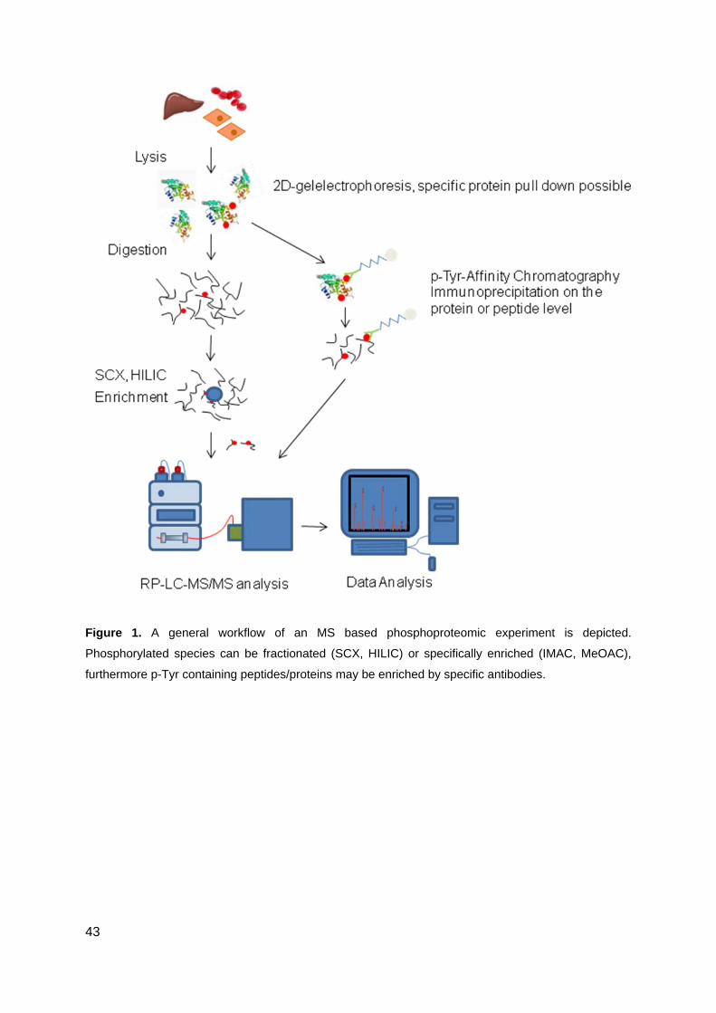
43
Figure 1. A general workflow of an MS based phosphoproteomic experiment is depicted.
Phosphorylated species can be fractionated (SCX, HILIC) or specifically enriched (IMAC, MeOAC),
furthermore p-Tyr containing peptides/proteins may be enriched by specific antibodies.
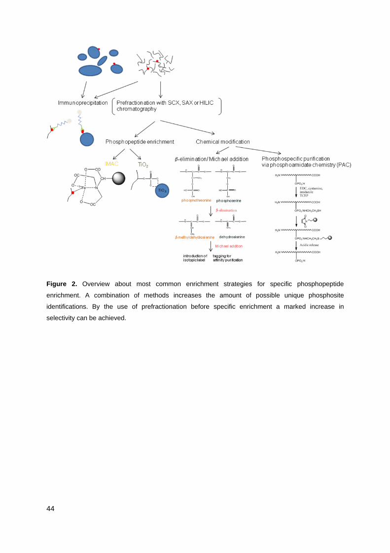
44
Figure 2. Overview about most common enrichment strategies for specific phosphopeptide
enrichment. A combination of methods increases the amount of possible unique phosphosite
identifications. By the use of prefractionation before specific enrichment a marked increase in
selectivity can be achieved.
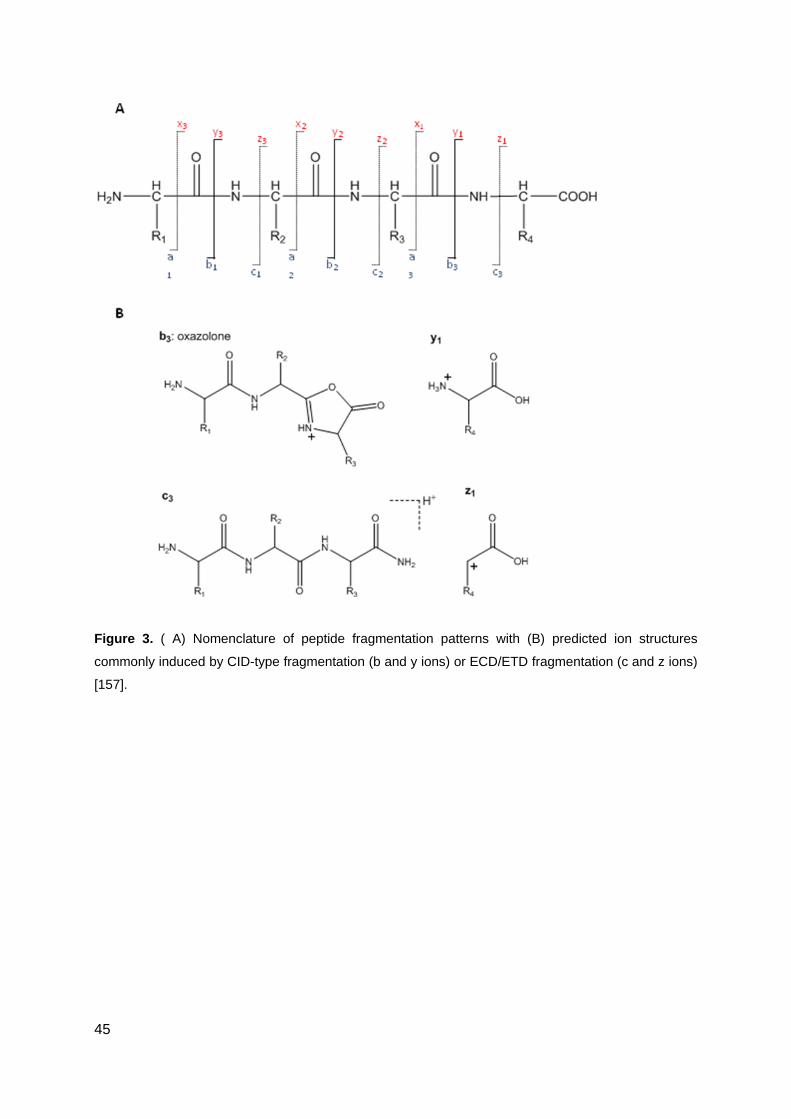
45
Figure 3. ( A) Nomenclature of peptide fragmentation patterns with (B) predicted ion structures
commonly induced by CID-type fragmentation (b and y ions) or ECD/ETD fragmentation (c and z ions)
[157].
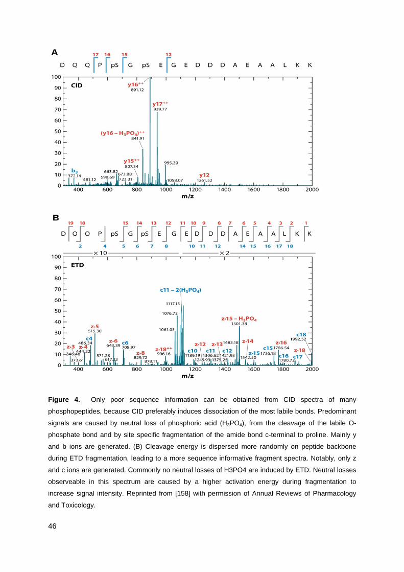
46
Figure 4. Only poor sequence information can be obtained from CID spectra of many
phosphopeptides, because CID preferably induces dissociation of the most labile bonds. Predominant
signals are caused by neutral loss of phosphoric acid (H3PO4), from the cleavage of the labile O-
phosphate bond and by site specific fragmentation of the amide bond c-terminal to proline. Mainly y
and b ions are generated. (B) Cleavage energy is dispersed more randomly on peptide backbone
during ETD fragmentation, leading to a more sequence informative fragment spectra. Notably, only z
and c ions are generated. Commonly no neutral losses of H3PO4 are induced by ETD. Neutral losses
observeable in this spectrum are caused by a higher activation energy during fragmentation to
increase signal intensity. Reprinted from [158] with permission of Annual Reviews of Pharmacology
and Toxicology.
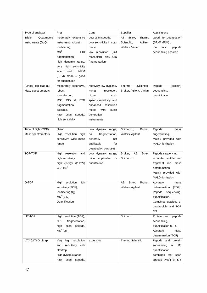
47
Type of analyzer Pros Cons Supplier Applications
Triple Quadrupole
instruments (QqQ)
moderately expensive
instrument, robust;
Ion filtering,
MS2, CID
fragmentation
high dynamic range,
very high sensitivity
when used in MRM
(SRM) mode – good
for quantitation
Low scan speeds,
Low sensitivity in scan
mode,
low resolution (unit
resolution), only CID
fragmentation
AB Sciex, Thermo
Scientific, Agilent,
Waters, Varian
Good for quantitation
(SRM/ MRM) ,
but also peptide
sequencing possible
(Linear) Ion Trap (L)IT
Mass spectrometers
moderately expensive,
robust;
Ion selection,
MSn, CID & ETD
fragmentation
possible,
Fast scan speeds,
high sensitivity
relatively low (typically
~unit) resolution,
higher scan
speeds,sensitivity and
enhanced resolution
mode with latest
generation
instruments
Thermo Scientific,
Bruker, Agilent, Varian
Peptide (protein)
sequencing,
quantification
Time of flight (TOF)
Mass spectrometers
cheap
High resolution, high
sensitivity, wide mass
range
Low dynamic range,
no fragmentation,
generally not
applicable for
quantitation purposes
Shimadzu, Bruker,
Waters, Agilent
Peptide mass
fingerprinting.
Mainly provided with
MALDI-ionization
TOF-TOF High resolution and
high sensitivity,
high energy (20keV)
CID, MS2
Low dynamic range,
minor application for
quantitation
Bruker, AB Sciex,
Shimadzu
Peptide sequencing,
accurate peptide and
fragment ion mass
determination.
Mainly provided with
MALDI-ionization
Q-TOF High resolution, high
sensitivity (TOF),
Ion filtering (Q)
MS2 (CID)
Quantification
AB Sciex, Bruker,
Waters, Agilent
Accurate mass
determination (TOF).
Peptide sequencing,
quantification.
Combines qualities of
quadruplole and TOF
MS
LIT-TOF High resolution (TOF),
CID fragmentation,
high scan speeds,
MSn (LIT)
Shimadzu Protein and peptide
sequencing,
quantification (LIT),
Accurate mass
determination (TOF)
LTQ (LIT)-Orbitrap Very high resolution
and sensitivity with
Orbitrap
High dynamic range
Fast scan speeds,
expensive Thermo Scientific Peptide and protein
sequencing in LIT,
quantification
combines fast scan
speeds (MSn) of LIT
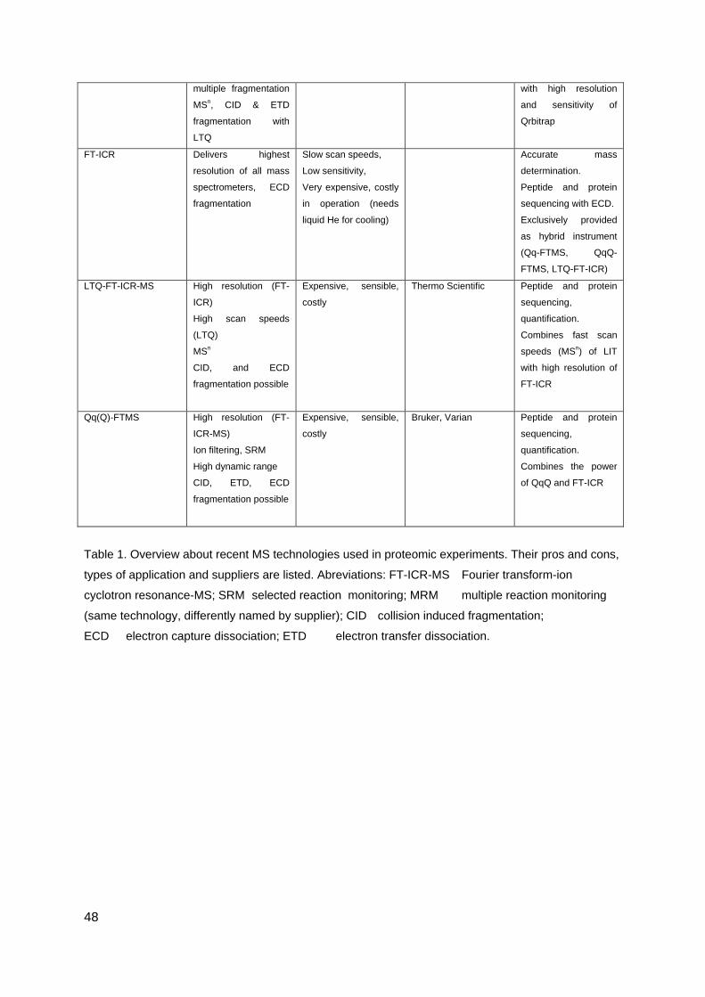
48
multiple fragmentation
MSn, CID & ETD
fragmentation with
LTQ
with high resolution
and sensitivity of
Qrbitrap
FT-ICR Delivers highest
resolution of all mass
spectrometers, ECD
fragmentation
Slow scan speeds,
Low sensitivity,
Very expensive, costly
in operation (needs
liquid He for cooling)
Accurate mass
determination.
Peptide and protein
sequencing with ECD.
Exclusively provided
as hybrid instrument
(Qq-FTMS, QqQ-
FTMS, LTQ-FT-ICR)
LTQ-FT-ICR-MS High resolution (FT-
ICR)
High scan speeds
(LTQ)
MSn
CID, and ECD
fragmentation possible
Expensive, sensible,
costly
Thermo Scientific Peptide and protein
sequencing,
quantification.
Combines fast scan
speeds (MSn) of LIT
with high resolution of
FT-ICR
Qq(Q)-FTMS High resolution (FT-
ICR-MS)
Ion filtering, SRM
High dynamic range
CID, ETD, ECD
fragmentation possible
Expensive, sensible,
costly
Bruker, Varian Peptide and protein
sequencing,
quantification.
Combines the power
of QqQ and FT-ICR
Table 1. Overview about recent MS technologies used in proteomic experiments. Their pros and cons,
types of application and suppliers are listed. Abreviations: FT-ICR-MS Fourier transform-ion
cyclotron resonance-MS; SRM selected reaction monitoring; MRM multiple reaction monitoring
(same technology, differently named by supplier); CID collision induced fragmentation;
ECD electron capture dissociation; ETD electron transfer dissociation.

49
Acknowledgement
This work was funded by the Austrian Proteomics Platform, part of the GEN-AU program of
the Austrian Federal Ministry of Science and Research.
50
References
1. Jensen, S.S. and M.R. Larsen, Evaluation of the impact of some experimental procedures on different phosphopeptide enrichment techniques. Rapid Commun Mass Spectrom, 2007. 21(22): p. 3635-45.
2. Goldberg, R.N., N. Kishore, and R.M. Lennen, Thermodynamic quantities for the ionization reactions of buffers. Journal of Physical and Chemical Reference Data, 2002. 31(2): p. 231-370.
3. Connor, P.A. and A.J. McQuillan, Phosphate adsorption onto TiO2 from aqueous solutions: An in situ internal reflection infrared spectroscopic study. Langmuir, 1999. 15(8): p. 2916-2921.
4. Grun, M., et al., Comparison of an ordered mesoporous aluminosilicate, silica, alumina, titania and zirconia in normal-phase high-performance liquid chromatography. Journal of Chromatography A, 1996. 740(1): p. 1-9.
5. Hunter, T., Signaling - 2000 and beyond. Cell, 2000. 100(1): p. 113-127. 6. Lim, Y.P., Mining the tumor phosphoproteome for cancer markers. Clin Cancer Res,
2005. 11(9): p. 3163-9. 7. Levitzki, A., Protein kinase inhibitors as a therapeutic modality. Acc Chem Res, 2003.
36(6): p. 462-9. 8. Force, T., et al., Inhibitors of protein kinase signaling pathways: emerging therapies
for cardiovascular disease. Circulation, 2004. 109(10): p. 1196-205. 9. Croom, K.F. and C.M. Perry, Imatinib mesylate: in the treatment of gastrointestinal
stromal tumours. Drugs, 2003. 63(5): p. 513-22; discussion 523-4. 10. Beausoleil, S.A., et al., Large-scale characterization of HeLa cell nuclear
phosphoproteins. Proc Natl Acad Sci U S A, 2004. 101(33): p. 12130-5. 11. Dephoure, N., et al., A quantitative atlas of mitotic phosphorylation. Proc Natl Acad
Sci U S A, 2008. 105(31): p. 10762-7. 12. Lemeer, S., et al., Endogenous phosphotyrosine signaling in zebrafish embryos. Mol
Cell Proteomics, 2007. 6(12): p. 2088-99. 13. Olsen, J.V., et al., Global, in vivo, and site-specific phosphorylation dynamics in
signaling networks. Cell, 2006. 127(3): p. 635-48. 14. Gronborg, M., et al., A mass spectrometry-based proteomic approach for
identification of serine/threonine-phosphorylated proteins by enrichment with phospho-specific antibodies: identification of a novel protein, Frigg, as a protein kinase A substrate. Mol Cell Proteomics, 2002. 1(7): p. 517-27.
15. Reinders, J. and A. Sickmann, State-of-the-art in phosphoproteomics. Proteomics, 2005. 5(16): p. 4052-61.
16. Bourne, H.R., GTPases: a family of molecular switches and clocks. Philos Trans R Soc Lond B Biol Sci, 1995. 349(1329): p. 283-9.
17. Bourne, H.R., Signal transduction. Team blue sees red. Nature, 1995. 376(6543): p. 727-9.
18. Lichtarge, O., H.R. Bourne, and F.E. Cohen, An evolutionary trace method defines binding surfaces common to protein families. J Mol Biol, 1996. 257(2): p. 342-58.
19. Sturm, M., et al., Tin dioxide microspheres as a promising material for phosphopeptide enrichment prior to liquid chromatography-(tandem) mass spectrometry analysis. Advanced Functional Materials, 2008. 18(16): p. 2381-2389.
51
20. Marcantonio, M., et al., Combined enzymatic and data mining approaches for comprehensive phosphoproteome analyses: application to cell signaling events of interferon-gamma-stimulated macrophages. Mol Cell Proteomics, 2008. 7(4): p. 645-60.
21. Rogers, L.D. and L.J. Foster, Phosphoproteomics--finally fulfilling the promise? Mol Biosyst, 2009. 5(10): p. 1122-9.
22. Collins, M.O., L. Yu, and J.S. Choudhary, Analysis of protein phosphorylation on a proteome-scale. Proteomics, 2007. 7(16): p. 2751-2768.
23. Pandey, A., et al., Analysis of receptor signaling pathways by mass spectrometry: Identification of Vav-2 as a substrate of the epidermal and platelet-derived growth factor receptors. Proceedings of the National Academy of Sciences of the United States of America, 2000. 97(1): p. 179-184.
24. Rush, J., et al., Immunoaffinity profiling of tyrosine phosphorylation in cancer cells. Nature Biotechnology, 2005. 23(1): p. 94-101.
25. Rikova, K., et al., Global survey of phosphotyrosine signaling identifies oncogenic kinases in lung cancer. Cell, 2007. 131(6): p. 1190-1203.
26. Salomon, A.R., et al., Profiling of tyrosine phosphorylation pathways in human cells using mass spectrometry. Proc Natl Acad Sci U S A, 2003. 100(2): p. 443-8.
27. Brill, L.M., et al., Robust phosphoproteomic profiling of tyrosine phosphorylation sites from human T cells using immobilized metal affinity chromatography and tandem mass spectrometry. Anal Chem, 2004. 76(10): p. 2763-72.
28. Porath, J., et al., Metal Chelate Affinity Chromatography, a New Approach to Protein Fractionation. Nature, 1975. 258(5536): p. 598-599.
29. Andersson, L. and J. Porath, Isolation of Phosphoproteins by Immobilized Metal (Fe-3+) Affinity-Chromatography. Analytical Biochemistry, 1986. 154(1): p. 250-254.
30. Muszynska, G., L. Andersson, and J. Porath, Selective Adsorption of Phosphoproteins on Gel-Immobilized Ferric Chelate. Biochemistry, 1986. 25(22): p. 6850-6853.
31. Scanff, P., M. Yvon, and J.P. Pelissier, Immobilized Fe3+ affinity chromatographic isolation of phosphopeptides. J Chromatogr, 1991. 539(2): p. 425-32.
32. Neville, D.C.A., et al., Evidence for phosphorylation of serine 753 in CFTR using a novel metal-ion affinity resin and matrix-assisted laser desorption mass spectrometry. Protein Science, 1997. 6(11): p. 2436-2445.
33. Ficarro, S.B., et al., Phosphoproteome analysis by mass spectrometry and its application to Saccharomyces cerevisiae. Nat Biotechnol, 2002. 20(3): p. 301-5.
34. Aprilita, N.H., et al., Poly(glycidyl methacrylate/divinylbenzene)-IDA-FeIII in phosphoproteomics. J Proteome Res, 2005. 4(6): p. 2312-9.
35. Feuerstein, I., et al., Phosphoproteomic analysis using immobilized metal ion affinity chromatography on the basis of cellulose powder. Proteomics, 2005. 5(1): p. 46-54.
36. Haydon, C.E., et al., Identification of novel phosphorylation sites on Xenopus laevis Aurora A and analysis of phosphopeptide enrichment by immobilized metal-affinity chromatography. Mol Cell Proteomics, 2003. 2(10): p. 1055-67.
37. Tsai, C.F., et al., Immobilized metal affinity chromatography revisited: pH/acid control toward high selectivity in phosphoproteomics. J Proteome Res, 2008. 7(9): p. 4058-69.
38. Zheng, H., et al., Phosphotyrosine proteomic study of interferon alpha signaling pathway using a combination of immunoprecipitation and immobilized metal affinity chromatography. Mol Cell Proteomics, 2005. 4(6): p. 721-30.
52
39. Trinidad, J.C., et al., Comprehensive identification of phosphorylation sites in postsynaptic density preparations. Mol Cell Proteomics, 2006. 5(5): p. 914-22.
40. Saha, A., et al., Stability of metal ion complexes formed with methyl phosphate and hydrogen phosphate. Journal of Biological Inorganic Chemistry, 1996. 1(3): p. 231-238.
41. Kokubu, M., et al., Specificity of immobilized metal affinity-based IMAC/C18 tip enrichment of phosphopeptides for protein phosphorylation analysis. Anal Chem, 2005. 77(16): p. 5144-54.
42. Posewitz, M.C. and P. Tempst, Immobilized gallium(III) affinity chromatography of phosphopeptides. Anal Chem, 1999. 71(14): p. 2883-92.
43. Zhou, H., et al., Specific phosphopeptide enrichment with immobilized titanium ion affinity chromatography adsorbent for phosphoproteome analysis. J Proteome Res, 2008. 7(9): p. 3957-67.
44. Feng, S., et al., Immobilized zirconium ion affinity chromatography for specific enrichment of phosphopeptides in phosphoproteome analysis. Mol Cell Proteomics, 2007. 6(9): p. 1656-65.
45. Endler, A., et al., In vivo phosphorylation sites of barley tonoplast proteins identified by a phosphoproteomic approach. Proteomics, 2009. 9(2): p. 310-21.
46. Gan, C.S., et al., A comparative study of electrostatic repulsion-hydrophilic interaction chromatography (ERLIC) versus SCX-IMAC-based methods for phosphopeptide isolation/enrichment. J Proteome Res, 2008. 7(11): p. 4869-77.
47. Villen, J. and S.P. Gygi, The SCX/IMAC enrichment approach for global phosphorylation analysis by mass spectrometry. Nat Protoc, 2008. 3(10): p. 1630-8.
48. Ikeguchi, Y. and H. Nakamura, Determination of organic phosphates by column-switching high performance anion-exchange chromatography using on-line preconcentration on titania. Analytical Sciences, 1997. 13(3): p. 479-483.
49. Ikeguchi, Y. and H. Nakamura, Selective enrichment of phospholipids by titania. Analytical Sciences, 2000. 16(5): p. 541-543.
50. Kawahara, M., H. Nakamura, and T. Nakajima, Titania and Zirconia as New Ceramic Column Packing Materials for High-Performance Liquid-Chromatography. Analytical Sciences, 1989. 5(4): p. 485-486.
51. Matsuda, H., H. Nakamura, and T. Nakajima, New Ceramic Titania - Selective Adsorbent for Organic-Phosphates. Analytical Sciences, 1990. 6(6): p. 911-912.
52. Matsuda, H., et al., Sensitivity Enhancement of Alkaline-Phosphatase Assay by Titania Treatment. Analytical Sciences, 1991. 7(5): p. 813-814.
53. Tani, K. and Y. Suzuki, Investigation of the ion-exchange behaviour of titania: Application as a packing material for ion chromatography. Chromatographia, 1997. 46(11-12): p. 623-627.
54. Tani, K. and E. Miyamoto, Investigation of chromatographic properties of titania. II. Influence of calcination temperature. Journal of Liquid Chromatography & Related Technologies, 1999. 22(6): p. 857-871.
55. Tani, K. and M. Ozawa, Investigation of chromatographic properties of titania. I. On retention behavior of hydroxyl and other substituent aliphatic carboxylic acids: Comparison with zirconia. Journal of Liquid Chromatography & Related Technologies, 1999. 22(6): p. 843-856.
56. Sugiyama, N., et al., Phosphopeptide enrichment by aliphatic hydroxy acid-modified metal oxide chromatography for nano-LC-MS/MS in proteomics applications. Molecular & Cellular Proteomics, 2007. 6(6): p. 1103-1109.
53
57. Leitner, A., et al., Optimizing the performance of tin dioxide microspheres for phosphopeptide enrichment. Analytica Chimica Acta, 2009. 638(1): p. 51-57.
58. Pinkse, M.W.H., et al., Selective isolation at the femtomole level of phosphopeptides from proteolytic digests using 2D-nanoLC-ESI-MS/MS and titanium oxide precolumns. Analytical Chemistry, 2004. 76(14): p. 3935-3943.
59. Larsen, M.R., et al., Highly selective enrichment of phosphorylated peptides from peptide mixtures using titanium dioxide microcolumns. Mol Cell Proteomics, 2005. 4(7): p. 873-86.
60. Mazanek, M., et al., Titanium dioxide as a chemo-affinity solid phase in offline phosphopeptide chromatography prior to HPLC-MS/MS analysis. Nature Protocols, 2007. 2(5): p. 1059-U1.
61. Mazanek, M., et al., A new acid mix enhances phosphopeptide enrichment on titanium- and zirconium dioxide for mapping of phosphorylation sites on protein complexes. J Chromatogr B Analyt Technol Biomed Life Sci. 878(5-6): p. 515-24.
62. Wolschin, F., S. Wienkoop, and W. Weckwerth, Enrichment of phosphorylated proteins and peptides from complex mixtures using metal oxide/hydroxide affinity chromatography (MOAC). Proteomics, 2005. 5(17): p. 4389-4397.
63. Smatt, J.H., et al., Synthesis of micrometer sized mesoporous metal oxide spheres by nanocasting. Microporous and Mesoporous Materials, 2008. 112(1-3): p. 308-318.
64. Chen, C.T. and Y.C. Chen, Fe3O4/TiO2 core/shell nanoparticles as affinity probes for the analysis of phosphopeptides using TiO2 surface-assisted laser desorption/ionization mass spectrometry. Anal Chem, 2005. 77(18): p. 5912-9.
65. Li, Y., et al., Highly selective and rapid enrichment of phosphorylated peptides using gallium oxide-coated magnetic microspheres for MALDI-TOF-MS and nano-LC-ESI-MS/MS/MS analysis. Proteomics, 2008. 8(2): p. 238-49.
66. Li, Y., et al., Fe3O4@Al2O3 magnetic core-shell microspheres for rapid and highly specific capture of phosphopeptides with mass spectrometry analysis. J Chromatogr A, 2007. 1172(1): p. 57-71.
67. Lo, C.Y., et al., Rapid enrichment of phosphopeptides from tryptic digests of proteins using iron oxide nanocomposites of magnetic particles coated with zirconia as the concentrating probes. J Proteome Res, 2007. 6(2): p. 887-93.
68. Qi, D., et al., Development of core-shell structure Fe3O4@Ta2O5 microspheres for selective enrichment of phosphopeptides for mass spectrometry analysis. J Chromatogr A, 2009. 1216(29): p. 5533-9.
69. Qi, D.W., et al., Magnetically Responsive Fe3O4@C@SnO2 Core-Shell Microspheres: Synthesis, Characterization and Application in Phosphoproteomics. Journal of Physical Chemistry C, 2009. 113(36): p. 15854-15861.
70. Ficarro, S.B., et al., Niobium(V) oxide (Nb2O5): application to phosphoproteomics. Anal Chem, 2008. 80(12): p. 4606-13.
71. Lemeer, S., et al., Online automated in vivo zebrafish phosphoproteomics: from large-scale analysis down to a single embryo. J Proteome Res, 2008. 7(4): p. 1555-64.
72. Washburn, M.P., D. Wolters, and J.R. Yates, 3rd, Large-scale analysis of the yeast proteome by multidimensional protein identification technology. Nat Biotechnol, 2001. 19(3): p. 242-7.
73. Washburn, M.P. and J.R. Yates, 3rd, Analysis of the microbial proteome. Curr Opin Microbiol, 2000. 3(3): p. 292-7.
74. Link, A.J., et al., Direct analysis of protein complexes using mass spectrometry. Nat Biotechnol, 1999. 17(7): p. 676-82.
54
75. Wagner, Y., et al., Multidimensional nano-HPLC for analysis of protein complexes. J Am Soc Mass Spectrom, 2003. 14(9): p. 1003-11.
76. Motoyama, A., et al., Anion and cation mixed-bed ion exchange for enhanced multidimensional separations of peptides and phosphopeptides. Anal Chem, 2007. 79(10): p. 3623-34.
77. Zhang, K., From purification of large amounts of phospho-compounds (nucleotides) to enrichment of phospho-peptides using anion-exchanging resin. Anal Biochem, 2006. 357(2): p. 225-31.
78. Han, G., et al., Large-scale phosphoproteome analysis of human liver tissue by enrichment and fractionation of phosphopeptides with strong anion exchange chromatography. Proteomics, 2008. 8(7): p. 1346-61.
79. McNulty, D.E. and R.S. Annan, Hydrophilic interaction chromatography reduces the complexity of the phosphoproteome and improves global phosphopeptide isolation and detection. Mol Cell Proteomics, 2008. 7(5): p. 971-80.
80. Alpert, A.J., Electrostatic repulsion hydrophilic interaction chromatography for isocratic separation of charged solutes and selective isolation of phosphopeptides. Anal Chem, 2008. 80(1): p. 62-76.
81. Oda, Y., T. Nagasu, and B.T. Chait, Enrichment analysis of phosphorylated proteins as a tool for probing the phosphoproteome. Nat Biotechnol, 2001. 19(4): p. 379-82.
82. Knight, Z.A., et al., Phosphospecific proteolysis for mapping sites of protein phosphorylation. Nat Biotechnol, 2003. 21(9): p. 1047-54.
83. McLachlin, D.T. and B.T. Chait, Improved beta-elimination-based affinity purification strategy for enrichment of phosphopeptides. Anal Chem, 2003. 75(24): p. 6826-36.
84. Bodenmiller, B., et al., An integrated chemical, mass spectrometric and computational strategy for (quantitative) phosphoproteomics: application to Drosophila melanogaster Kc167 cells. Mol Biosyst, 2007. 3(4): p. 275-86.
85. Zhou, H., J.D. Watts, and R. Aebersold, A systematic approach to the analysis of protein phosphorylation. Nat Biotechnol, 2001. 19(4): p. 375-8.
86. Tao, W.A., et al., Quantitative phosphoproteome analysis using a dendrimer conjugation chemistry and tandem mass spectrometry. Nat Methods, 2005. 2(8): p. 591-8.
87. Reynolds, E.C., P.F. Riley, and N.J. Adamson, A selective precipitation purification procedure for multiple phosphoseryl-containing peptides and methods for their identification. Anal Biochem, 1994. 217(2): p. 277-84.
88. Zhang, X., et al., Highly Efficient Phosphopeptide Enrichment by Calcium Phosphate Precipitation Combined with Subsequent IMAC Enrichment. Mol Cell Proteomics, 2007. 6(11): p. 2032-42.
89. Xia, Q., et al., Phosphoproteomic analysis of human brain by calcium phosphate precipitation and mass spectrometry. J Proteome Res, 2008. 7(7): p. 2845-51.
90. Nabetani, T., et al., Improved method of phosphopeptides enrichment using biphasic Phos-tag/C18 tip for versatile analysis of phosphorylation dynamics. Proteomics, 2009.
91. Bodenmiller, B., et al., Reproducible isolation of distinct, overlapping segments of the phosphoproteome. Nat Methods, 2007. 4(3): p. 231-7.
92. Hojlund, K., et al., In vivo Phosphoproteome of Human Skeletal Muscle Revealed by Phosphopeptide Enrichment and HPLC-ESI-MS/MS. J Proteome Res, 2009.
93. Leitner, A., et al., Probing the phosphoproteome of HeLa cells using nanocast metal oxide microspheres for phosphopeptide enrichment. Anal Chem. 82(7): p. 2726-33.
55
94. Schroeder, M.J., et al., A neutral loss activation method for improved phosphopeptide sequence analysis by quadrupole ion trap mass spectrometry. Anal Chem, 2004. 76(13): p. 3590-8.
95. Wang, C., et al., Plasma phospholipid metabolic profiling and biomarkers of type 2 diabetes mellitus based on high-performance liquid chromatography/electrospray mass spectrometry and multivariate statistical analysis. Anal Chem, 2005. 77(13): p. 4108-16.
96. Wang, C., J. Yang, and J. Nie, Plasma phospholipid metabolic profiling and biomarkers of rats following radiation exposure based on liquid chromatography-mass spectrometry technique. Biomed Chromatogr, 2009. 23(10): p. 1079-85.
97. Min, H.K., G. Kong, and M.H. Moon, Quantitative analysis of urinary phospholipids found in patients with breast cancer by nanoflow liquid chromatography-tandem mass spectrometry: II. Negative ion mode analysis of four phospholipid classes. Anal Bioanal Chem. 396(3): p. 1273-80.
98. Bligh, E.G. and W.J. Dyer, A rapid method of total lipid extraction and purification. Can J Biochem Physiol, 1959. 37(8): p. 911-7.
99. Folch, J., M. Lees, and G.H. Sloane Stanley, A simple method for the isolation and purification of total lipides from animal tissues. J Biol Chem, 1957. 226(1): p. 497-509.
100. Calvano, C.D., O.N. Jensen, and C.G. Zambonin, Selective extraction of phospholipids from dairy products by micro-solid phase extraction based on titanium dioxide microcolumns followed by MALDI-TOF-MS analysis. Anal Bioanal Chem, 2009. 394(5): p. 1453-61.
101. Gonzalvez, A., B. Preinerstorfer, and W. Lindner, Selective enrichment of phosphatidylcholines from food and biological matrices using metal oxides as solid-phase extraction materials prior to analysis by HPLC-ESI-MS/MS. Anal Bioanal Chem. 396(8): p. 2965-75.
102. Urthaler, J., et al., Application of monoliths for plasmid DNA purification development and transfer to production. J Chromatogr A, 2005. 1065(1): p. 93-106.
103. Sousa, F., D.M. Prazeres, and J.A. Queiroz, Binding and elution strategy for improved performance of arginine affinity chromatography in supercoiled plasmid DNA purification. Biomed Chromatogr, 2009. 23(2): p. 160-5.
104. Stadler, J., R. Lemmens, and T. Nyhammar, Plasmid DNA purification. J Gene Med, 2004. 6 Suppl 1: p. S54-66.
105. Sousa, F., et al., Specific recognition of supercoiled plasmid DNA in arginine affinity chromatography. Anal Biochem, 2008. 374(2): p. 432-4.
106. Lai, J., J. Wang, and Z. Cai, Nucleoside reverse transcriptase inhibitors and their phosphorylated metabolites in human immunodeficiency virus-infected human matrices. J Chromatogr B Analyt Technol Biomed Life Sci, 2008. 868(1-2): p. 1-12.
107. Hubbard, M.J. and P. Cohen, On target with a new mechanism for the regulation of protein phosphorylation. Trends Biochem Sci, 1993. 18(5): p. 172-7.
108. Ishihama, Y., et al., Enhancement of the efficiency of phosphoproteomic identification by removing phosphates after phosphopeptide enrichment. J Proteome Res, 2007. 6(3): p. 1139-44.
109. McLachlin, D.T. and B.T. Chait, Analysis of phosphorylated proteins and peptides by mass spectrometry. Curr Opin Chem Biol, 2001. 5(5): p. 591-602.
110. Roepstorff, P. and K. Kristiansen, The use of Edman degradation in peptide mixture analysis by mass spectrometry. Biomed Mass Spectrom, 1974. 1(4): p. 231-6.
56
111. Hunt, D.F., et al., Protein sequencing by tandem mass spectrometry. Proc Natl Acad Sci U S A, 1986. 83(17): p. 6233-7.
112. Standing, K.G., Peptide and protein de novo sequencing by mass spectrometry. Curr Opin Struct Biol, 2003. 13(5): p. 595-601.
113. Johnson, R.S. and K. Biemann, Computer program (SEQPEP) to aid in the interpretation of high-energy collision tandem mass spectra of peptides. Biomed Environ Mass Spectrom, 1989. 18(11): p. 945-57.
114. Mann, M. and M. Wilm, Error-tolerant identification of peptides in sequence databases by peptide sequence tags. Anal Chem, 1994. 66(24): p. 4390-9.
115. Yates, J.R., 3rd, J.K. Eng, and A.L. McCormack, Mining genomes: correlating tandem mass spectra of modified and unmodified peptides to sequences in nucleotide databases. Anal Chem, 1995. 67(18): p. 3202-10.
116. Yates, J.R., 3rd, et al., Method to correlate tandem mass spectra of modified peptides to amino acid sequences in the protein database. Anal Chem, 1995. 67(8): p. 1426-36.
117. Wysocki, V.H., et al., Mobile and localized protons: a framework for understanding peptide dissociation. J Mass Spectrom, 2000. 35(12): p. 1399-406.
118. Paizs, B. and S. Suhai, Fragmentation pathways of protonated peptides. Mass Spectrom Rev, 2005. 24(4): p. 508-48.
119. Thingholm, T.E., O.N. Jensen, and M.R. Larsen, Analytical strategies for phosphoproteomics. Proteomics, 2009. 9(6): p. 1451-68.
120. Carr, S.A., M.J. Huddleston, and R.S. Annan, Selective detection and sequencing of phosphopeptides at the femtomole level by mass spectrometry. Anal Biochem, 1996. 239(2): p. 180-92.
121. Collins, M.O., et al., Proteomic analysis of in vivo phosphorylated synaptic proteins. J Biol Chem, 2005. 280(7): p. 5972-82.
122. Steen, H., et al., Detection of tyrosine phosphorylated peptides by precursor ion scanning quadrupole TOF mass spectrometry in positive ion mode. Anal Chem, 2001. 73(7): p. 1440-8.
123. Steen, H., B. Kuster, and M. Mann, Quadrupole time-of-flight versus triple-quadrupole mass spectrometry for the determination of phosphopeptides by precursor ion scanning. J Mass Spectrom, 2001. 36(7): p. 782-90.
124. Ulintz, P.J., et al., Comparison of MS(2)-only, MSA, and MS(2)/MS(3) methodologies for phosphopeptide identification. J Proteome Res, 2009. 8(2): p. 887-99.
125. Zubarev, R.A., Reactions of polypeptide ions with electrons in the gas phase. Mass Spectrom Rev, 2003. 22(1): p. 57-77.
126. Syka, J.E., et al., Peptide and protein sequence analysis by electron transfer dissociation mass spectrometry. Proc Natl Acad Sci U S A, 2004. 101(26): p. 9528-33.
127. Wilm, M., Quantitative proteomics in biological research. Proteomics, 2009. 9(20): p. 4590-605.
128. Bantscheff, M., et al., Quantitative mass spectrometry in proteomics: a critical review. Anal Bioanal Chem, 2007. 389(4): p. 1017-31.
129. Blagoev, B., et al., Temporal analysis of phosphotyrosine-dependent signaling networks by quantitative proteomics. Nat Biotechnol, 2004. 22(9): p. 1139-45.
130. de Godoy, L.M., et al., Status of complete proteome analysis by mass spectrometry: SILAC labeled yeast as a model system. Genome Biol, 2006. 7(6): p. R50.
57
131. Mann, K., B. Macek, and J.V. Olsen, Proteomic analysis of the acid-soluble organic matrix of the chicken calcified eggshell layer. Proteomics, 2006. 6(13): p. 3801-10.
132. Park, K.S., et al., Graded regulation of the Kv2.1 potassium channel by variable phosphorylation. Science, 2006. 313(5789): p. 976-9.
133. Xiong, L. and Y. Wang, Quantitative Proteomic Analysis Reveals the Perturbation of Multiple Cellular Pathways in HL-60 Cells Induced by Arsenite Treatment. J Proteome Res.
134. Zhang, Y., et al., MAPU: Max-Planck Unified database of organellar, cellular, tissue and body fluid proteomes. Nucleic Acids Res, 2007. 35(Database issue): p. D771-9.
135. Mirgorodskaya, O.A., et al., Quantitation of peptides and proteins by matrix-assisted laser desorption/ionization mass spectrometry using (18)O-labeled internal standards. Rapid Commun Mass Spectrom, 2000. 14(14): p. 1226-32.
136. Gygi, S.P., et al., Quantitative analysis of complex protein mixtures using isotope-coded affinity tags. Nat Biotechnol, 1999. 17(10): p. 994-9.
137. Ross, P.L., et al., Multiplexed protein quantitation in Saccharomyces cerevisiae using amine-reactive isobaric tagging reagents. Mol Cell Proteomics, 2004. 3(12): p. 1154-69.
138. Goshe, M.B., et al., Phosphoprotein isotope-coded affinity tag approach for isolating and quantitating phosphopeptides in proteome-wide analyses. Anal Chem, 2001. 73(11): p. 2578-86.
139. Goshe, M.B., et al., Phosphoprotein isotope-coded affinity tags: application to the enrichment and identification of low-abundance phosphoproteins. Anal Chem, 2002. 74(3): p. 607-16.
140. Pasquarello, C., et al., N-t-butyliodoacetamide and iodoacetanilide: two new cysteine alkylating reagents for relative quantitation of proteins. Rapid Commun Mass Spectrom, 2004. 18(1): p. 117-27.
141. Qiu, Y., et al., Acid-labile isotope-coded extractants: a class of reagents for quantitative mass spectrometric analysis of complex protein mixtures. Anal Chem, 2002. 74(19): p. 4969-79.
142. Sebastiano, R., et al., A new deuterated alkylating agent for quantitative proteomics. Rapid Commun Mass Spectrom, 2003. 17(21): p. 2380-6.
143. Shen, M., et al., Isolation and isotope labeling of cysteine- and methionine-containing tryptic peptides: application to the study of cell surface proteolysis. Mol Cell Proteomics, 2003. 2(5): p. 315-24.
144. Shi, Y., et al., A simple solid phase mass tagging approach for quantitative proteomics. J Proteome Res, 2004. 3(1): p. 104-11.
145. Shi, Y., et al., Design and synthesis of a solid-phase fluorescent mass tag. J Sep Sci, 2005. 28(14): p. 1812-7.
146. Che, F.Y. and L.D. Fricker, Quantitation of neuropeptides in Cpe(fat)/Cpe(fat) mice using differential isotopic tags and mass spectrometry. Anal Chem, 2002. 74(13): p. 3190-8.
147. Hsu, J.L., et al., Stable-isotope dimethyl labeling for quantitative proteomics. Anal Chem, 2003. 75(24): p. 6843-52.
148. Bantscheff, M., et al., Quantitative chemical proteomics reveals mechanisms of action of clinical ABL kinase inhibitors. Nat Biotechnol, 2007. 25(9): p. 1035-44.
149. Gerber, S.A., et al., Absolute quantification of proteins and phosphoproteins from cell lysates by tandem MS. Proc Natl Acad Sci U S A, 2003. 100(12): p. 6940-5.
58
150. Silva, J.C., et al., Absolute quantification of proteins by LCMSE: a virtue of parallel MS acquisition. Mol Cell Proteomics, 2006. 5(1): p. 144-56.
151. Liu, H., et al., Formation and composition of the Bacillus anthracis endospore. J Bacteriol, 2004. 186(1): p. 164-78.
152. Wang, W., et al., Quantification of proteins and metabolites by mass spectrometry without isotopic labeling or spiked standards. Anal Chem, 2003. 75(18): p. 4818-26.
153. Sanz-Medel, A., Heteroatom(isotope)-tagged proteomics via ICP-MS: screening and quantification of proteins and their post-translational modifications. Anal Bioanal Chem, 2008. 391(3): p. 885-94.
154. Wind, M., et al., Analysis of protein phosphorylation by capillary liquid chromatography coupled to element mass spectrometry with 31P detection and to electrospray mass spectrometry. Anal Chem, 2001. 73(1): p. 29-35.
155. Wind, M., et al., Identification of phosphorylation sites in the polo-like kinases Plx1 and Plk1 by a novel strategy based on element and electrospray high resolution mass spectrometry. Proteomics, 2002. 2(11): p. 1516-23.
156. Wind, M., H. Wesch, and W.D. Lehmann, Protein phosphorylation degree: determination by capillary liquid chromatography and inductively coupled plasma mass spectrometry. Anal Chem, 2001. 73(13): p. 3006-10.
157. Wysocki, V.H., et al., Mass spectrometry of peptides and proteins. Methods, 2005. 35(3): p. 211-22.
158. Macek, B., M. Mann, and J.V. Olsen, Global and site-specific quantitative phosphoproteomics: principles and applications. Annu Rev Pharmacol Toxicol, 2009. 49: p. 199-221.
59
3 A Chemically Cleavable Linker Concept
Aim
During the second project the development and evaluation of a new method for affinity
purification of target proteins was realized. Therefore a new linker chemistry was designed,
which allows the release of bait – target complexes via specific chemical linker cleavage.
In the field of chemical proteomics, targeted protein pull-downs are generally used to
selectively enrich for specific bait interactors, enabling their identification by liquid
chromatography – (tandem) mass spectrometry. Commonly, baits are coupled to beads via
different linker chemistries, allowing the isolation of target proteins from matrix components.
Elution is generally performed by the use of denaturing buffers. However, this may also elute
proteins which are unspecifically bound to the bead matrix, increasing the risk of false
positive identifications. By elution-independent affinity chromatography, intact target–bait
complexes can be eluted, commonly by specific linker cleavage. In our group a cleavable
core structure, based on an indoleacetic acid-malondialdehyde derivative, was designed,
which can be selectively cleaved at the indole nitrogen by incubation with pyrrolidine. This
derivative was coupled to hydrazide functionalized agarose or acrylamide beads via the
reactive allylaldehyde group of the derivative. As bait we used bosutinib, a drug recently
used in the therapy of chronic myeloid leukemia, which was available in its amino
functionalized form. Bosutinib was coupled to the linker via an amidation reaction with the
help of 4-(4,6-dimethoxy-1,3,5-triazin-2-yl)-4-methylmorpholinium chloride (DMT-MM) as
coupling reagent. A protein pull down was performed on a K562 cell lysate and isolated
interaction partners were identified by immunoblot and LC-MS/MS analysis.
Synthesis and results of this work can be found in the manuscript “A New Chemically
Cleavable Linker of an Immobilized Bait for Target Protein Pull-down Concept” to be
submitted (see Appendix Manuscript no. 1).

60
4 Appendix

Publication 1
FULLP
DOI: 10.1002/adfm.200800215APER
Tin Dioxide Microspheres as a Promising Materialfor Phosphopeptide Enrichment Prior to LiquidChromatography-(Tandem) Mass Spectrometry Analysis**
By Martin Sturm, Alexander Leitner,* Jan-Henrik Smatt, Mika Linden, and Wolfgang Lindner*
Functional materials are frequently used to identify and characterize proteins and their modifications in complex biological
samples throughout the analytical workflow. Protein phosphorylation is a highly important post-translational modification that is
being examined with the help of diverse materials that exhibit affinity towards the phosphate group of phosphoamino acids.
Titanium dioxide and zirconium dioxide particles are increasingly used to enrich phosphopeptides from proteolytic digests of
protein mixtures, although specificity and recovery still leave room for improvement. Here, we present tin dioxide (SnO2,
stannia)microspheres as a new type ofmetal oxidematerial for phosphopeptide enrichment. Themicrospheres are produced by a
nanocasting process, starting from silica particles as a template, which allows the tuning of material properties such as particle
diameter and porosity of themicrospheres in a straightforwardmanner. For the first time, we are able to show that tin dioxide can
be used to enrich phosphopeptides from mixtures such as enzymatic digests of proteins, followed by analysis by liquid
chromatography-mass spectrometry (LC-MS). For optimization of the enrichment protocol, we use synthetic phosphorylated
and nonphosphorylated peptides, and test different solvent compositions for loading and washing steps to enhance the selectivity
of the material without compromising phosphopeptide recovery. Furthermore, the selectivity and phosphopeptide binding
properties of tin dioxide are compared to the established metal oxide materials, titanium dioxide and zirconium dioxide, using
mixtures of model proteins. Even without the use of additional additives such as a-hydroxy acids, which have been used to
enhance the specificity of TiO2-based enrichment, we show that a comparably good performance can be achieved for the SnO2
spheres.
1. Introduction
Proteomics, as a scientific discipline, aims to characterize the
protein complement – the ‘‘proteome’’ – of a cell, a body fluid
or another complex biological sample. It is estimated that the
human proteome consists of approximately 30 000 genes, each
gene coding for a specific protein. But the number of different
proteins present in an organism at any given time could rise
over one million, and this is mainly the result of splice variants
and posttranslational modifications (PTMs). These PTMs
comprise chemical modifications ranging from the formation
of disulfide bonds via oxidation of the thiol groups from
[*] Dr. A. Leitner, Prof. Dr. W. Lindner, M. SturmDepartment of Analytical Chemistry and Food Chemistry, Universityof ViennaWaehringer Strasse 38, 1090 Vienna (Austria)E-mail: [email protected]; [email protected]
Dr. J.-H. Smatt, Dr. M. LindenCenter for Functional Materials, Department of Physical Chemistry,Abo Akademi UniversityPorthansgatan 3-5, 20500 Turku (Finland)
[**] The authors would like to acknowledge Karl Mechtler (Institute forMolecular Pathology, Vienna, Austria) for providing the phosphopep-tides for this study. This work was financially supported by the GEN-AUframework program ‘‘Austrian Proteomics Platform II’’ funded by theAustrian Federal Ministry of Science and Research.
Adv. Funct. Mater. 2008, 18, 2381–2389 � 2008 WILEY-VCH Verlag
cysteines during protein folding, to phosphorylation, glycosy-
lation and other covalent modifications when the protein is
already folded.[1] Protein phosphorylation is known to be a key
event in intracellular signaling and other important cellular
processes such as metabolism, transcription and apoptosis.
Thus, the identification and localization of phosphorylation
sites in proteins is very important to get insights in the
regulation of cellular signaling cascades and other biological
networks. It is estimated that phosphoproteins account for
more than 30% of the proteome,[2] but on the level of a
particular protein, phosphorylation is typically transient and
only present in substoichiometric amounts. Mass spectrometry
(MS) is the technique of choice for proteome analysis due to its
ability to not only identify a large number of proteins, mostly
from their corresponding peptides, but also to identify,
characterize and locate post-translational modifications.[1]
In a typical ‘‘bottom-up’’ analysis, single proteins or more
complex samples like whole cell lysates or plasma are
enzymatically digested into peptides, because they are more
amenable to mass spectrometric analysis. Peptide mixtures
are thenmost commonly separated by one- or two-dimensional
high performance liquid chromatography (HPLC) prior to MS
analysis. However, after the digestion step, phosphopeptides
are usually present in an overwhelming excess of unpho-
sphorylated peptides, which usually have higher ionization
GmbH & Co. KGaA, Weinheim 2381
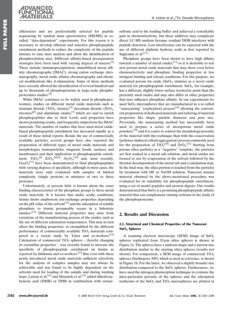
FULLPAPER
A. Leitner et al. / Tin Dioxide Microspheres
2382
efficiencies and are preferentially selected for peptide
sequencing by tandem mass spectrometry (MS/MS) in so-
called ‘‘data-dependent’’ experiments. For this reason it is
necessary to develop efficient and selective phosphopeptide
enrichment methods to reduce the complexity of the peptide
mixture to ease later analysis and allow the identification of
phosphorylation sites. Different affinity-based preseparation
strategies have been used with varying degrees of success.[3]
These include immunoprecipitation, immobilized metal affi-
nity chromatography (IMAC), strong cation exchange chro-
matography, metal oxide affinity chromatography and chemi-
cal modifications like b-elimination. Some of these methods
have recently allowed the identification of several hundred and
up to thousands of phosphoproteins in large-scale phospho-
proteomics studies.[4]
While IMAC continues to be widely used in phosphopro-
teomics, studies on different metal oxide materials such as
titanium dioxide (TiO2, titania),[5] zirconium dioxide (ZrO2,
zirconia)[6] and aluminum oxides,[7] which are said to enrich
phosphopeptides due to their Lewis acid properties have
shown promising results, and frequently outperform the IMAC
materials. The number of studies that have used metal oxide-
based phosphopeptide enrichment has increased rapidly as a
result of these initial reports. Beside the use of commercially
available particles, several groups have also reported the
preparation of different types of metal oxide materials and
morphologies (nanoparticles, magnetic beads, surfaces and
membranes) and their application to phosphopeptide enrich-
ment. TiO2,[8] ZrO2,
[8d,9] Al2O3,[10] and, most recently,
Ga2O3[11] have been demonstrated to bind phosphopeptides
with varying degrees of specificity, although in most cases, the
materials were only evaluated with samples of limited
complexity (single proteins or mixtures of two to three
proteins).
Unfortunately, at present little is known about the exact
binding characteristics of the phosphate groups to these metal
oxide materials. It is known that under acidic conditions,
titania shows amphoteric ion-exchange properties depending
on the pH-value of the solvent[12] and the adsorption of soluble
phosphate to titania presumably occurs in a bidentate
manner.[13] Different material properties may arise from
variations of the manufacturing process of the oxides, such as
the use of different calcination temperatures. This may in turn
affect the binding properties, as exemplified by the different
performance of commercially available TiO2 materials com-
pared in a recent study by Yates and co-workers.[14]
Calcination of commercial TiO2 spheres – thereby changing
its crystalline properties – was recently found to increase the
specificity of phosphopeptide enrichment on titania as
reported by Ishihama and co-workers.[15] But even with these
newly introduced metal oxide materials sufficient selectivity
for the analysis of complex samples may not always be
achievable and was found to be highly dependent on the
solvents used for loading of the sample and during washing
steps. Larsen et al.[5c] and Mazanek et al.[16] added dihydroxy-
benzoic acid (DHB) or DHB in combination with octane-
www.afm-journal.de � 2008 WILEY-VCH Verlag GmbH
sulfonic acid to the loading buffer and achieved a remarkable
gain in chemoselectivity, but these additives may complicate
direct LC-MS analysis because residual DHB interferes with
peptide detection. Less interference can be expected with the
use of different aliphatic hydroxy acids as first reported by
Sugiyama et al.[17]
Phosphate groups have been shown to have high affinity
towards a number of metal oxides,[13] so it is desirable to test
new porous metal oxide materials that may show even better
chemoselectivity and phosphate binding properties at less
stringent binding and release conditions. For this purpose, we
evaluated porous tin oxide (SnO2, stannia) as a novel oxide
material for phosphopeptide enrichment. SnO2, for example,
has a different, slightly lower surface isoelectric point than the
presently used oxides and may also differ in other properties
that may influence phosphate affinity. In our experiments we
used SnO2 microspheres that are manufactured in a so called
‘‘nanocasting’’ (replication) process[18] allowing the conveni-
ent preparation of dedicatedmaterials and tailoring of material
properties like shape, particle diameter and pore size.
Previously, the nanocasting method has successfully been
used to prepare a series of mesoporous metal oxide
powders,[19] and it is easier to control the morphology/porosity
of the material with this technique than with the coacervation
(polymer-induced colloid aggregation) process commonly used
for the preparation of TiO2[20] and ZrO2.
[21] Starting from
porous silica particles as a ‘‘negative’’ template, the particles
are first soaked in a metal salt solution, and metal oxides are
formed in situ by evaporation of the solvent followed by the
thermal decomposition of the metal salt and a calcination step.
In the final step, the silica portion is leached from the particles
by treatment with HF or NaOH solution. Nanocast stannia
material obtained by the above-mentioned procedure was
evaluated for its suitability for phosphopeptide enrichment,
using a set of model peptides and protein digests. Our results
demonstrated that SnO2 is a promising phosphopeptide affinity
material that can complement existing sorbents in the study of
the phosphoproteome.
2. Results and Discussion
2.1. Structural and Chemical Properties of the Nanocast
SnO2 Spheres
A scanning electron microscopy (SEM) image of SnO2
spheres replicated from 10mm silica spheres is shown in
Figure 1a. The spheres have a uniform shape and a narrow size
distribution similar to the starting silica spheres (results not
shown). For comparison, a SEM image of commercial TiO2
spheres (Sachtopore NP), which is used as reference, is shown
in Figure 1b. For the latter, we observed a slightly broader size
distribution compared to the SnO2 spheres. Furthermore, we
have used the nitrogen physisorption technique to evaluate the
intra-particular porosity of the spheres and the adsorption
isotherms of the SnO2 and TiO2 microspheres are plotted in
& Co. KGaA, Weinheim Adv. Funct. Mater. 2008, 18, 2381–2389
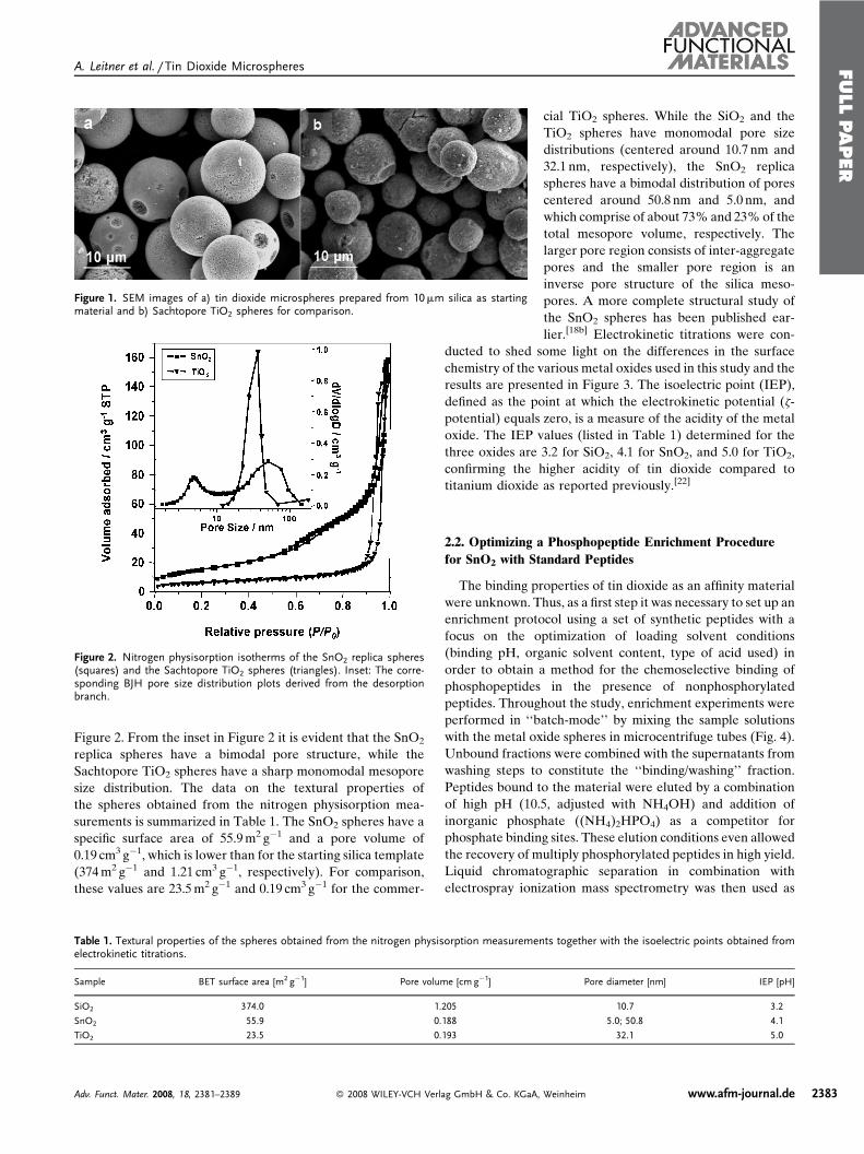
FULLPAPER
A. Leitner et al. / Tin Dioxide Microspheres
Figure 1. SEM images of a) tin dioxide microspheres prepared from 10mm silica as startingmaterial and b) Sachtopore TiO2 spheres for comparison.
Figure 2. Nitrogen physisorption isotherms of the SnO2 replica spheres(squares) and the Sachtopore TiO2 spheres (triangles). Inset: The corre-sponding BJH pore size distribution plots derived from the desorptionbranch.
Figure 2. From the inset in Figure 2 it is evident that the SnO2
replica spheres have a bimodal pore structure, while the
Sachtopore TiO2 spheres have a sharp monomodal mesopore
size distribution. The data on the textural properties of
the spheres obtained from the nitrogen physisorption mea-
surements is summarized in Table 1. The SnO2 spheres have a
specific surface area of 55.9m2 g�1 and a pore volume of
0.19 cm3 g�1, which is lower than for the starting silica template
(374m2 g�1 and 1.21 cm3 g�1, respectively). For comparison,
these values are 23.5m2 g�1 and 0.19 cm3 g�1 for the commer-
Table 1. Textural properties of the spheres obtained from the nitrogen physisorption measuremenelectrokinetic titrations.
Sample BET surface area [m2 g�1] Pore volume [cm g�1]
SiO2 374.0 1.205
SnO2 55.9 0.188
TiO2 23.5 0.193
Adv. Funct. Mater. 2008, 18, 2381–2389 � 2008 WILEY-VCH Verlag GmbH & Co. KGaA,
cial TiO2 spheres. While the SiO2 and the
TiO2 spheres have monomodal pore size
distributions (centered around 10.7 nm and
32.1 nm, respectively), the SnO2 replica
spheres have a bimodal distribution of pores
centered around 50.8 nm and 5.0 nm, and
which comprise of about 73% and 23%of the
total mesopore volume, respectively. The
larger pore region consists of inter-aggregate
pores and the smaller pore region is an
inverse pore structure of the silica meso-
pores. A more complete structural study of
the SnO2 spheres has been published ear-
lier.[18b] Electrokinetic titrations were con-
ducted to shed some light on the differences in the surface
chemistry of the various metal oxides used in this study and the
results are presented in Figure 3. The isoelectric point (IEP),
defined as the point at which the electrokinetic potential (z-
potential) equals zero, is a measure of the acidity of the metal
oxide. The IEP values (listed in Table 1) determined for the
three oxides are 3.2 for SiO2, 4.1 for SnO2, and 5.0 for TiO2,
confirming the higher acidity of tin dioxide compared to
titanium dioxide as reported previously.[22]
2.2. Optimizing a Phosphopeptide Enrichment Procedure
for SnO2 with Standard Peptides
The binding properties of tin dioxide as an affinity material
were unknown. Thus, as a first step it was necessary to set up an
enrichment protocol using a set of synthetic peptides with a
focus on the optimization of loading solvent conditions
(binding pH, organic solvent content, type of acid used) in
order to obtain a method for the chemoselective binding of
phosphopeptides in the presence of nonphosphorylated
peptides. Throughout the study, enrichment experiments were
performed in ‘‘batch-mode’’ by mixing the sample solutions
with the metal oxide spheres in microcentrifuge tubes (Fig. 4).
Unbound fractions were combined with the supernatants from
washing steps to constitute the ‘‘binding/washing’’ fraction.
Peptides bound to the material were eluted by a combination
of high pH (10.5, adjusted with NH4OH) and addition of
inorganic phosphate ((NH4)2HPO4) as a competitor for
phosphate binding sites. These elution conditions even allowed
the recovery of multiply phosphorylated peptides in high yield.
Liquid chromatographic separation in combination with
electrospray ionization mass spectrometry was then used as
ts together with the isoelectric points obtained from
Pore diameter [nm] IEP [pH]
10.7 3.2
5.0; 50.8 4.1
32.1 5.0
Weinheim www.afm-journal.de 2383
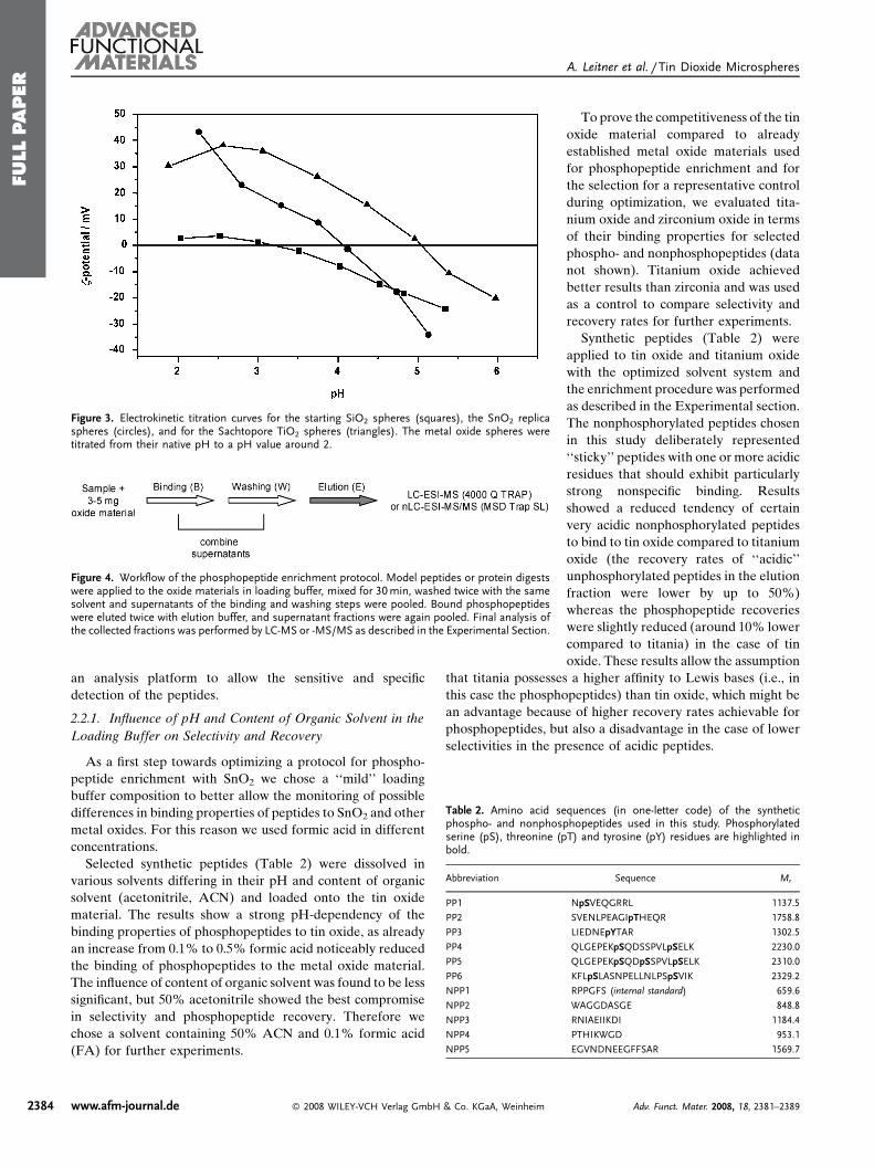
FULLPAPER
A. Leitner et al. / Tin Dioxide Microspheres
Figure 3. Electrokinetic titration curves for the starting SiO2 spheres (squares), the SnO2 replicaspheres (circles), and for the Sachtopore TiO2 spheres (triangles). The metal oxide spheres weretitrated from their native pH to a pH value around 2.
Figure 4. Workflow of the phosphopeptide enrichment protocol. Model peptides or protein digestswere applied to the oxide materials in loading buffer, mixed for 30min, washed twice with the samesolvent and supernatants of the binding and washing steps were pooled. Bound phosphopeptideswere eluted twice with elution buffer, and supernatant fractions were again pooled. Final analysis ofthe collected fractions was performed by LC-MS or -MS/MS as described in the Experimental Section.
Table 2. Amino acid sequences (in one-letter code) of the syntheticphospho- and nonphosphopeptides used in this study. Phosphorylatedserine (pS), threonine (pT) and tyrosine (pY) residues are highlighted inbold.
Abbreviation Sequence Mr
PP1 NpSVEQGRRL 1137.5
PP2 SVENLPEAGIpTHEQR 1758.8
PP3 LIEDNEpYTAR 1302.5
PP4 QLGEPEKpSQDSSPVLpSELK 2230.0
PP5 QLGEPEKpSQDpSSPVLpSELK 2310.0
PP6 KFLpSLASNPELLNLPSpSVIK 2329.2
NPP1 RPPGFS (internal standard) 659.6
NPP2 WAGGDASGE 848.8
NPP3 RNIAEIIKDI 1184.4
NPP4 PTHIKWGD 953.1
NPP5 EGVNDNEEGFFSAR 1569.7
2384
an analysis platform to allow the sensitive and specific
detection of the peptides.
2.2.1. Influence of pH and Content of Organic Solvent in the
Loading Buffer on Selectivity and Recovery
As a first step towards optimizing a protocol for phospho-
peptide enrichment with SnO2 we chose a ‘‘mild’’ loading
buffer composition to better allow the monitoring of possible
differences in binding properties of peptides to SnO2 and other
metal oxides. For this reason we used formic acid in different
concentrations.
Selected synthetic peptides (Table 2) were dissolved in
various solvents differing in their pH and content of organic
solvent (acetonitrile, ACN) and loaded onto the tin oxide
material. The results show a strong pH-dependency of the
binding properties of phosphopeptides to tin oxide, as already
an increase from 0.1% to 0.5% formic acid noticeably reduced
the binding of phosphopeptides to the metal oxide material.
The influence of content of organic solvent was found to be less
significant, but 50% acetonitrile showed the best compromise
in selectivity and phosphopeptide recovery. Therefore we
chose a solvent containing 50% ACN and 0.1% formic acid
(FA) for further experiments.
www.afm-journal.de � 2008 WILEY-VCH Verlag GmbH & Co. KGaA, Weinheim
To prove the competitiveness of the tin
oxide material compared to already
established metal oxide materials used
for phosphopeptide enrichment and for
the selection for a representative control
during optimization, we evaluated tita-
nium oxide and zirconium oxide in terms
of their binding properties for selected
phospho- and nonphosphopeptides (data
not shown). Titanium oxide achieved
better results than zirconia and was used
as a control to compare selectivity and
recovery rates for further experiments.
Synthetic peptides (Table 2) were
applied to tin oxide and titanium oxide
with the optimized solvent system and
the enrichment procedure was performed
as described in the Experimental section.
The nonphosphorylated peptides chosen
in this study deliberately represented
‘‘sticky’’ peptides with one or more acidic
residues that should exhibit particularly
strong nonspecific binding. Results
showed a reduced tendency of certain
very acidic nonphosphorylated peptides
to bind to tin oxide compared to titanium
oxide (the recovery rates of ‘‘acidic’’
unphosphorylated peptides in the elution
fraction were lower by up to 50%)
whereas the phosphopeptide recoveries
were slightly reduced (around 10% lower
compared to titania) in the case of tin
oxide. These results allow the assumption
that titania possesses a higher affinity to Lewis bases (i.e., in
this case the phosphopeptides) than tin oxide, which might be
an advantage because of higher recovery rates achievable for
phosphopeptides, but also a disadvantage in the case of lower
selectivities in the presence of acidic peptides.
Adv. Funct. Mater. 2008, 18, 2381–2389
FULLPAPER
A. Leitner et al. / Tin Dioxide Microspheres
Figure 5. Recovery rates (relative to an unprocessed sample) of nonpho-sphorylated (NPP) and phosphorylated (PP) peptides after the phospho-peptide enrichment procedure with SnO2 and different loading buffercompositions. ACN¼ acetonitrile, FA¼ formic acid, TFA¼ trifluoroaceticacid.
Figure 6. Recovery rates (relative to an unprocessed sample) of selectedpeptides after phosphopeptide enrichment using optimized procedures forSnO2 and TiO2. ACN¼ acetonitrile, TFA¼ trifluoroacetic acid, LA¼ lacticacid.
Nonetheless, we observed that a complete removal of
nonphosphorylated peptides in the elution fraction was not
attainable with this simple experimental setup for both stannia
and titania. Thus, the selectivity achieved in this first
optimization step may not be adequate for more complex
samples such as protein digests or cell extracts. The problem
could be that the high abundance of nonphosphorylated
peptides in such samples will prevent binding of phosphory-
lated peptides if the selectivity of the enrichment material is
not good enough. Several groups have reported the effective-
ness of hydroxy acids to reduce unspecific binding of acidic
peptides to titanium oxide. In our study, we tested some a-
hydroxy acids (lactic acid, tartaric acid, malic acid in a
concentration of 300mgml�1; according to Ref. [17a]) and 2,5-
dihydroxybenzoic acid (300mgml�1; Ref. [5c]) for both tin
oxide and titanium oxide. Both protocols were previously
optimized for a different TiO2 material and reevaluation for
the present titania spheres was necessary. The best selectivity
and phosphopeptide recovery were obtained by titania with
0.1% trifluoroacetic acid (TFA) and 300mgml�1 of lactic acid
(LA), in line with data reported by Sugiyama et al.,[17a] while
for the SnO2material the use of hydroxy acids was not found to
further improve specificity.
2.2.2. Influence of pH and Type of Acid in the Solvent for
Binding Properties of Enrichment Materials
Despite favorable binding of phosphopeptides to SnO2 even
at relatively high pH, the lack of specificity led us to investigate
the influence of the acidity of the loading solvent in more
detail. It was expected that working at a lower pH would
improve the discrimination between phosphopeptides and
unphosphorylated, acidic peptides when the pH is lower than
the pKa of the carboxyl groups (�4). Thus, in accordance with
results previously observed for other oxides, carboxylic acids
should be present in protonated form to a large degree and not
compete with phosphopeptides for binding.
In the next experiment we therefore compared the
selectivity achieved by the use of different acids in varying
concentrations for the tin oxide material. Figure 5 shows the
recovery rates of unphosphorylated (NPP2-NPP5) and phos-
phorylated peptides (PP1-PP4, see Table 2) in the elution
fraction after enrichment using tin oxide and different
compositions of the loading solution. Solvents used were
50% ACN with 0.05% TFA, 0.1% TFA or 0.1% FA. It is
evident that there is no sufficient selectivity achievable for a
satisfactory phosphopeptide enrichment with formic acid,
particularly for complex samples where the excess of acidic
peptides is much higher than in this case: up to 80% of the
unphosphorylated controls were retained under these condi-
tions. The lowest recovery rates for nonphosphopeptides in the
elution fraction (i.e., less than 3% of nonspecific binding) were
obtained with 0.1% TFA. Considering the fact that in complex
samples the excess of acidic peptides will be much higher than
in this experiment, we decided to use 0.1% TFA in the loading
solution instead of 0.05% TFA for further experiments,
although higher phosphopeptide recovery rates would be
Adv. Funct. Mater. 2008, 18, 2381–2389 � 2008 WILEY-VCH Verl
feasible with 0.05% TFA. In the following, we compared our
protocol for SnO2 to an optimized protocol for titania, as
reported previously by Sugiyama et al.[17a] The percentage of
peptides recovered in the elution fraction of peptides (relative
to an unprocessed sample) was determined for three different
experimental set-ups: SnO2 and 0.1% TFA as additive in the
loading solvent; TiO2 with 0.1% TFA, or 0.1% TFA and
300mgml�1 LA as solvent. The results (shown in Fig. 6) reveal
that the recovery rates of nonphosphopeptides are comparably
low for SnO2 and TiO2 with lactic acid – indicating a sufficient
reduction in nonspecific binding – while in the case of TiO2
with TFA alone the selectivity was not satisfactory (e.g., 65%
nonspecific binding for NPP5). The comparable performance
of SnO2 using TFA alone with an optimized TiO2 method
demonstrates that the optimized protocol for SnO2 is able to
ag GmbH & Co. KGaA, Weinheim www.afm-journal.de 2385
FULLPAPER
A. Leitner et al. / Tin Dioxide Microspheres
Table 3. Peptides and proteins identified from a tryptic digest of a-casein and fetuin with phosphopeptide enrichment using either SnO2 or TiO2, analysis bynanoLC-MS/MS and subsequent database search using Mascot. Shown are the numbers of identifications from six repeat analyses.
Protein Peptide Identified by SnO2 enrichment[a] Identified by TiO2 enrichment[b]
aS1-casein YKVPQLEIVPNpSAEER 6 of 6 runs 6 of 6 runs
VPQLEIVPNpSAEER 5/6 5/6
HIQKEDVPSER 2/6 2/6
aS2-casein TVDMEpSTEVFTKK 4/6 4/6
NMAINPpSKENLCSTFCK 1/6 1/6
Fetuin HTFSGVASVEpSSSGEAFLIVGK 2/6 2/6
[a] Using 50% ACN/0.1% TFA as loading buffer. [b] Using 50% ACN/0.1% TFAþ 300mgml�1 lactic acid as loading buffer.
2386
compete with other established enrichment materials and
protocols, at least for model peptides. As a next step it was
necessary to prove whether our material and protocol was
applicable to more complex samples such as protein digests.
2.3. Tryptic Protein Digests
a-casein and fetuin represent highly phosphorylated pro-
teins and are therefore often used as model proteins for
phosphopeptide enrichment studies. We analyzed a tryptic
digest of a mixture of a-casein (containing both aS1- and aS2-
forms) and fetuin after a phosphopeptide enrichment step
using the two optimized protocols for SnO2 (50% ACN and
0.1% TFA as loading buffer) and TiO2 (50% ACN/0.1%
TFAþ 300mgml�1 LA), respectively. The elution fractions
recovered from the materials were analyzed by nanoflow LC-
MS/MS as described in detail in the Experimental section.
For evaluation of the results, we decided to rely on the
qualitative interpretation of database search results using the
Mascot search engine, as this reflects a typical ‘‘discovery-
mode’’ type of phosphopeptide profiling. Quantitative strate-
gies in complex samples need to rely on stable isotope labeling
or related approaches,[23] as (relative) quantitation based on
extracted ion chromatograms exclusively cannot be expected
to yield reliable results when comparing samples differing
enormously in their complexity such as a whole protein digest
and a phosphopeptide-enriched sample in the present case.
Table 4. Peptides and proteins identified from a tryptic digest of the ten proteinnanoLC-MS/MS and subsequent database search using Mascot. Shown are
Protein[a] Peptide
Ovalbumin, chicken EVVGpSAEAGVDAASVSEER
ISQAVHAAHAEINEAGR
a1-acid glycoprotein 1, human EQLGEFYEALDCLR
Carbonic anhydrase, bovine AVVQDPALKPLALVYGEATSR
b-lactoglobulin, bovine TPEVDDEALEK
TPEVDDEALEKFDK
VYVEELKPTGDLEILLQK
Lactotransferrin, bovine LCALCAGDDQGLDK
Lysozyme C, chicken FESNFNTQATNR
NTDGSTDYGILQINSR
[a] No peptides were identified from the following proteins: bovine k-casein, a-
0.1% TFA as loading buffer. [c] Using 50% ACN/0.1% TFAþ 300mgml�1 la
www.afm-journal.de � 2008 WILEY-VCH Verlag GmbH
As Table 3 shows, the data obtained from the two materials
were highly comparable. One phosphorylation site from aS1-
casein was identified in all analyses (three parallel enrichment
procedures, two nanoLC-MS/MS runs per sample), in most
cases with two peptides, one of which contained a missed
cleavage site. A phosphopeptide from aS2-casein was identified
in four out of six runs, while a second phosphopeptide from this
casein variant and a phosphopeptide from fetuin were only
infrequently detected. This may be due to several reasons such
as low phosphorylation stoichiometry, poor spectral quality or
lower recovery from the enrichment procedure. For both the
stannia and the titania material, only one peptide (from aS1-
casein) was found to bind nonspecifically under the conditions
employed, and this peptide was identified in less than half of
the analyses. While a tryptic digest of these phosphoproteins is
expected to produce more than the phosphopeptides identified
here, the reason for the inability of identifying further
phosphopeptides may be the generally unfavorable fragmen-
tation properties, the reliance on identification by MS/MS, not
MS alone (thus requiring good spectral quality), and the low
ionization efficiency of these peptides.
After these encouraging results, a tryptic digest of a ten
protein mix was analyzed by nano-LC-MS/MS after phospho-
peptide enrichment by both SnO2 and TiO2. Ovalbumin
represented the only phosphorylated protein in the sample
(containing two annotated phosphorylation sites) and ideally
should be the only protein identifiable after the enrichment
step. The results for this sample are shown in Table 4. With the
mix with phosphopeptide enrichment using either SnO2 or TiO2, analysis bythe numbers of identifications from four repeat analyses.
Identified by SnO2 enrichment[b] Identified by TiO2 enrichment[c]
4 of 4 runs 2 of 4 runs
0/4 2/4
1/4 0/4
0/4 3/4
4/4 0/4
4/4 0/4
2/4 0/4
1/4 0/4
4/4 0/4
4/4 0/4
lactalbumin, equine myoglobin and human transferrin. [b] Using 50% ACN/
ctic acid as loading buffer.
& Co. KGaA, Weinheim Adv. Funct. Mater. 2008, 18, 2381–2389
FULLPAPER
A. Leitner et al. / Tin Dioxide Microspheres
Figure 7. a) and b) Total ion chromatogram of a nanoLC-MS/MS run of the tryptic digest of the tenprotein mix (a) before and (b) after an enrichment step using SnO2 microspheres and 50% acetonitrile/0.1% trifluoroacetic acid as loading buffer. c) shows the annotated MS/MS spectrum of the phospho-peptide identified from ovalbumin (EVVGpSAEAGVDAASVSEEFR). Peptide identification was carriedout by Mascot as described in the Experimental section.
optimized SnO2 protocol it was possible to identify ovalbumin
via one of its phosphopeptides in every single LC-MS/MS
analysis of duplicate runs from two separately processed
samples. As an example, Figure 7a and b depicts the MS2 total
ion chromatograms of a nanoLC-MS/MS analysis of the ten
protein mix before and after phosphopeptide enrichment with
stannia. It is apparent that a significant reduction of matrix
complexity was achieved, as the intensity of the most abundant
peak is reduced by a factor of �25 (note that an extended
gradient for the unprocessed sample was used to allow a more
accurate comparison of the results). The annotated MS/MS
spectrum of the phosphopeptide EVVGpSAEAGVDAASV-
SEEFR after the respective enrichment step is shown in
Figure 7c.
In the case of stannia, four other proteins (b-lactoglobulin
and lysozyme C in all four analyses, lactotransferrin and a1-
acid glycoprotein 1 in one analysis) were also identified with up
to two nonphosphopeptides. For enrichment with titania the
selectivity appeared to be better, because only in three cases
one unphosphorylated protein (carbonic anhydrase 2) was
Adv. Funct. Mater. 2008, 18, 2381–2389 � 2008 WILEY-VCH Verlag GmbH & Co. KGaA, Weinhe
identified. However, the identification
of ovalbumin via its phosphopeptides
succeeded in only two of four attempts,
indicating that the use of lactic acid as a
displacer in the loading solvent for the
titania material also causes significant
losses of phosphopeptides during load-
ing and washing. To demonstrate the
degree of enrichment, we analyzed the
ten protein digest also without an
enrichment step. In this experiment
all ten proteins could be identified, and
in the case of ovalbumin, no phospho-
peptides but eight unphosphorylated
peptides containing up to five acidic
amino acid residues per peptide were
identified, whereas in the enriched
sample only the phosphopeptide could
be identified. This is a further indicator
for the specificity of the phosphopep-
tide enrichment step using SnO2micro-
spheres.
By using the simple argumentation
that the total surface area of the
employed spheres in the enrichment
experiments is enormous compared to
what is needed to completely adsorb
the phosphopeptides (about 1 million
times larger), we conclude that the
surface area of the spheres is not the
decisive factor for the good perfor-
mance observed for the SnO2 material.
Furthermore, since we also have smal-
ler pores in the SnO2 replica spheres
compared to the commercial TiO2, it is
more likely that this is limiting rather
than promoting the accessibility of the SnO2 surface to some
degree, because larger peptides entering the 5 nm pores is not
favored. Therefore, we can presume that the variations in the
phosphopeptide enrichment behavior observed between the
studied materials are mainly consequences of the surface
chemistry rather than the morphology.
3. Conclusions
We have shown that porous tin oxide microspheres
represent a valuable new material for phosphopeptide
enrichment purposes. Using an optimized protocol, compar-
able selectivity to titania was achievable, with the advantage
that no solvent additives in high concentrations are necessary
to gain sufficient selectivity. This minimizes the risk of
problems with nanoscale HPLC columns and/or nanoelec-
trospray in the downstream analysis and should lead to a more
robust analytical platform for phosphoproteomic studies. The
im www.afm-journal.de 2387
FULLPAPER
A. Leitner et al. / Tin Dioxide Microspheres
2388
microspheres can be manufactured using readily available
starting materials, and their properties (thermal, physical, and
chemical stability) make them an interesting alternative to
presently used material. Further studies are currently ongoing
to test the performance of the SnO2 replicas on more complex
biological samples. Moreover, the flexibility of their prepara-
tion will allow the in-depth study of factors influencing the
phosphate group affinity that will help in the design of
improved metal oxide affinity materials.
4. Experimental
Metal Oxide Materials: Porous tin oxide microspheres wereprepared according to the protocol of Smatt et al. [18b], starting fromDaisogel silica particles (Daiso Co. Ltd., Osaka, Japan) with a particlesize of 10mm and 120 A pore size. Briefly, the silica spheres wereimpregnated with an excess amount of an aqueous SnCl2 solution(1.8 gmL�1, Riedel-de Haen, Seelze, Germany). After an impregna-tion time of 1 h, centrifugation was used to remove the excess precursorsolution. The samples were then directly heated at 150 8C (3 h) toremove the solvent and further heated up to 250 8C (3h) and 550 8C(5h) in order to decompose the precursor salt to SnO2. In a final step,the silica portion was removed by leaching 2 times in aqueous NaOH(2M) at 90 8C, and the obtained spheres were dried in vacuo. Prior touse, the SnO2 material was treated with aqueous HCl (0.1 M) for 24 hunder reflux for activation, followed by drying in vacuo. Titania(Sachtopore NP titanium dioxide, particle size 10mm/pore size 300 A)and zirconia (ZirChrom-PHASE zirconium dioxide, particle size10mm/pore size 300 A) were obtained from ZirChrom Separations,Inc. (Anoka, MN, USA) and used as received.
Characterization of the SnO2 and Reference Spheres: The spheremorphology was studied using SEM (Jeol JSM-6335F, Jeol Ltd.,Japan). The surface area, mesopore dimensions and the mesoporevolumes were determined by nitrogen physisorption measurements at77K (ASAP 2010, Micromeritics Co., Norcross, GA). The totalmesopore volume was taken at 0.98P/P0 and the BJH model(desorption branch) was used for pore size calculations. Electrokinetictitrations to obtain the IEP were performed on a Malvern ZetaSizerNano-ZS (Malvern Instruments Ltd.,Malvern, UK) coupled to aMPT-2 titrator unit. The zeta potential was measured as a function of pH bytitrating with aqueous HCl (0.1M) at 25 8C. The data was monitoredand analyzed using the Malvern Dispersion Technology software v.4.20.
Peptides and Proteins: Synthetic unphosphorylated peptides wereobtained from Bachem (Weil am Rhein, Germany), while phospho-peptides were prepared by standard Fmoc solid phase chemistry andkindly provided by K. Mechtler from the Institute of MolecularPathology (IMP, Vienna, Austria). All peptides used in the study arelisted in Table 1. Model proteins used in our experiments wereobtained from Sigma–Aldrich (Deisenhofen, Germany).
Tryptic digest of standard phosphoproteins: A total of onemilligramof the respective proteins was dissolved in aqueous urea solution (8M).Disulfide bonds were reduced using dithiothreitol (10mM, 55 8C,60min) followed by alkylation of free thiol groups with iodoacetamide(50mM, room temperature, 90min) according to standard procedures.Samples were diluted 1 to 10 with aqueous NH4HCO3 (50mm), andtrypsin (proteomics grade, Sigma) was added at an enzyme-to-substrate ratio of approximately 1:25. Samples were digested overnightat 37 8C and purified by solid-phase extraction (C18 material) prior toevaporation under a stream of nitrogen.
Enrichment of Phosphopeptides: In brief, the workflow is shown inFigure 4. Different phosphorylated and nonphosphorylated peptides(see Table 1) were used in a quantity of 300 ng (amounting to approx.
www.afm-journal.de � 2008 WILEY-VCH Verlag GmbH
200 fmol) per peptide per sample for an enrichment experiment. In thecase of tryptic digests, 20mg of starting material for the digest of caseinand fetuin and 50mg for the ten protein digest were applied.
Approximately 4mg of oxide material (SnO2, TiO2, or ZrO2) wasweighed in 1.5 ml microcentrifuge tubes and equilibrated for 10minwith 100ml of the respective solvent (as noted in the Results andDiscussion section) prior to loading of the samples. After equilibrationthe suspension was centrifuged and the supernatant was discarded.Peptide samples diluted in loading solvent (100mL) were applied to theoxide material and incubated for 30min, and after centrifugation andremoval of the supernatant the materials were washed twice for10 minutes with loading buffer (100mL). When hydroxy acids wereused as additives the hydroxy acid was omitted in the second washingstep to avoid carry-over into the elution fraction. The supernatants ofthe binding and two washing steps were pooled and stored for later MSanalysis. For elution of bound phosphopeptides, the material wasincubated twice with elution buffer (100mL of 50mM of (NH4)2HPO4
adjusted to pH 10.5 with NH4OH) for 10min and supernatants wereagain pooled for analysis. All steps (equilibration, binding, washing,eluting) were carried out on an IKA MS3 digital minishaker (IKA,Staufen, Germany) to keep the metal oxides in suspension during theprocedure.
Pooled samples were dried in a nitrogen stream and redissolved inwater/acetonitrile/formic acid (95:5:0.5, v/v/v) prior to LC-MS analysis asdescribed below. All enrichment experiments were performed at least induplicate, and all collected fractions were analyzed twice by LC-MS.
LC-MS System: For the analysis and quantification of the standardpeptides after the enrichment procedure we used an Agilent 1200HPLC system coupled with an Applied Biosystems 4000 Q TRAP(triple quadrupole linear ion trap hybrid) mass spectrometer equippedwith a Turbo V electrospray ion source (Applied Biosystems, FosterCity, CA, USA). For gradient LC separation a Thermo BetaBasic-18reversed phase column (150� 2.1mm; particle size 3mm; ThermoScientific, Runcorn, UK) was used with a flow rate of 200mLmin�1.The mobile phases consisted of (A) 0.1% formic acid in water and (B)0.1% formic acid in acetonitrile. A gradient elution, starting with 5% to50% B in 30min, followed by 50% to 80% B in 5min, 80% B for 5minand reequilibration of the column with 5% of solvent B for 10 minuteswas employed for reversed phase separation of the peptides. The massspectrometer was operated in positive enhancedMS scan (EMS) modewhere the third quadrupole is operated as a linear ion trap. The m/z-rangewas set from 300 to 2000 with a scan rate of 4000 per second.Dataacquisition and analysis was performed with Analyst software, version1.4.2 (Applied Biosystems).
For peptide quantification, an internal standard (300 ng ofbradykinin fragment 1–6, Table 1) was added to the sample afterthe phosphopeptide enrichment procedure to correct for signalvariabilities. The recovery rates were calculated as a quotient of thecorrected peptide signal of the peptide in the elution fraction and thecorrected peptide signal of the corresponding peptide in an untreatedsample (100%, no enrichment).
Nanoflow LC-MS/MS System: Protein digests were analyzed usingan Agilent Nanoflow Proteomics Solution in data-dependent MS/MSmode essentially as described previously [24]. An Agilent Zorbax 300SB-C18 column (50mm� 75mm i.d.) was used for all samples with theexception of the unprocessed 10 protein mix, which was analyzed on a150mm column to avoid undersampling due to the higher samplecomplexity. Peak lists generated by the instrument software (LC/MSDTrap software 4.2, BrukerDaltonik, Bremen, Germany) were exportedin Mascot-MGF format and searched against the UniProtKB/SwissProt database (release 47.5, dated 19/07/05, 186882 entries) usingan in-house Mascot server running version 2.0.5 (Matrix Science Ltd.,London, UK). Search parameters were set as follows: Taxono-my¼ chordata (vertebrates and relatives), enzyme¼ trypsin, fixedmodifications¼ carbamidomethylation on Cys, variable modifica-tions¼ phosphorylation on serine or threonine, peptide toleran-ce¼�1.5Da, MS/MS tolerance¼�0.8Da, peptide charge¼ 2þ/3þ,instrument¼ESI trap. Peptides were considered identified if they were
& Co. KGaA, Weinheim Adv. Funct. Mater. 2008, 18, 2381–2389
FULLPAPER
A. Leitner et al. / Tin Dioxide Microspheres
assigned to the main proteins in the sample (excluding possible low-level contaminants) and the ions score reported by Mascot was at least20.
Received: February 11, 2008Revised: April 11, 2008
[1] a) M. R. Larsen, M. B. Trelle, T. E. Thingholm, O. N. Jensen,
BioTechniques 2006, 40, 790. b) O. N. Jensen, Nat. Rev. Mol. Cell
Biol. 2006, 7, 391. c) E. S. Witze, W. M. Old, K. A. Resing, N. G. Ahn,
Nat. Methods 2007, 4, 798.
[2] S. B. Ficarro, M. L. McCleland, P. T. Stukenberg, D. J. Burke, M. M.
Ross, J. Shabanowitz, D. F. Hunt, F. M. White, Nat. Biotechnol. 2002,
20, 301.
[3] a) J. Reinders, A. Sickmann, Proteomics 2005, 5, 4052. b) F. Delom, E.
Chevet,Proteome Sci. 2006, 4, 15 (article number). c)M.W. H. Pinkse,
A. J. R. Heck,Drug Discovery Today: Technologies 2006, 3, 331. d) C.
D’Ambrosio, A. M. Salzano, S. Arena, G. Renzone, A. Scaloni, J.
Chromatogr. B 2007, 849, 163. e)M. O. Collins, L. Yu, J. S. Choudhary,
Proteomics 2007, 7, 2751.
[4] a) J. V. Olsen, B. Blagoev, F. Gnad, B. Macek, C. Kumar,
P. Mortensen, M. Mann, Cell 2006, 127, 635. b) J. Villen, S. A.
Beausoleil, S. A. Gerber, S. P. Gygi, Proc. Natl. Acad. Sci. USA
2007, 104, 1488. c) A. Chi, C. Huttenhower, L. Y. Geer, J. J. Coon,
J. E. P. Syka, D. L. Bai, J. Shabanowitz, D. J. Burke, O. G. Troyans-
kaya, D. F. Hunt, Proc. Natl. Acad. Sci. USA 2007, 104, 2193. d) H.
Molina, D. M. Horn, N. Tang, S. Mathivanan, A. Pandey, Proc. Natl.
Acad. Sci. USA 2007, 104, 2199. e) X. Li, S. A. Gerber, A. D. Rudner,
S. A. Beausoleil, W. Haas, J. Villen, J. E. Elias, S. P. Gygi, J. Proteome
Res. 2007, 6, 1190.
[5] a) M. W. H. Pinkse, P. M. Uitto, M. J. Hilhorst, B. Ooms, A. J. R.
Heck, Anal. Chem. 2004, 76, 3935. b) I. Kuroda, Y. Shintani, M.
Motokawa, S. Abe, M. Furuno, Anal. Sci. 2004, 20, 1313. c) M. R.
Larsen, T. E. Thingholm, O. N. Jensen, P. Roepstorff, T. D. Jørgensen,
Mol. Cell. Proteomics 2005, 4, 873.
[6] H. K. Kweon, K. Hakansson, Anal. Chem. 2006, 78, 1743.
[7] a) F. Wolschin, S. Wienkoop, W. Weckwerth, Proteomics 2005, 5,
4389. b) F. Wolschin, W. Weckwerth, Plant Methods 2005, 1, 9 (article
number).
[8] a) C.-T. Chen, Y.-C. Chen, Anal. Chem. 2005, 77, 5912. b) F. Tan,
Y. Zhang, J. Wang, J. Wei, P. Qin, Y. Cai, X. Qian, Rapid Commun.
Mass Spectrom. 2007, 21, 2407. c) H.-C. Hsieh, C. Sheu, F.-K. Shi,
D.-T. Li, J. Chromatogr. A 2007, 1165, 128. d) X. Liang,
G. Fonnum, M. Hajivandi, T. Stene, N. H. Kjus, E. Ragnhildstveit,
J.W.Amshey, P. Predki, R.M. Pope, J. Am. Soc.Mass Spectrom. 2007,
Adv. Funct. Mater. 2008, 18, 2381–2389 � 2008 WILEY-VCH Verl
18, 1932. e) Y. Li, J. Wu, D. Qi, X. Xu, C. Deng, P. Yang, X. Zhang,
Chem. Commun. 2008, 564.
[9] a) C.-Y. Lo, W.-Y. Chen, C.-T. Chen, Y.-C. Chen, J. Proteome Res.
2007, 6, 887. b) G. R. Blacken, M. Volny, T. Vaisar, M. Sadılek,
F. Turecek, Anal. Chem. 2007, 79, 5449. c) H. Zhou, R. Tian, M. Ye,
S. Xu, S. Feng, C. Pan, X. Jiang, X. Li, H. Zou, Electrophoresis 2007,
28, 2201. d) Y. Li, T. Leng, H. Lin, C. Deng, X. Xu, N. Yao, P. Yang,
X. Zhang, J. Proteome Res. 2007, 6, 4498.
[10] a) C.-T. Chen, W.-Y. Chen, P.-J. Tsai, K.-Y. Chien, J.-S. Yu, Y.-C.
Chen, J. Proteome Res. 2007, 6, 316. b) Y. Wang, W. Chen, J. Wu,
Y. Guo, X. Xia, J. Am. Soc. Mass Spectrom. 2007, 18, 1387. c) Y. Li,
Y. Liu, J. Tang, H. Lin, N. Yao, X. Shen, C. Deng, P. Yang, X. Zhang,
J. Chromatogr. A 2007, 1172, 57.
[11] Y. Li, H. Lin, C. Deng, P. Yang, X. Zhang, Proteomics 2008, 8,
238.
[12] a) K. Tani, M. Ozawa, J. Liq. Chromatogr. Relat. Technol. 1999, 22,
843. b) K. Tani, E. Miyamoto, J. Liq. Chromatogr. Relat. Technol.
1999, 22, 857.
[13] P. A. Connor, A. J. McQuillan, Langmuir 1999, 15, 2916.
[14] G. T. Cantin, T. R. Shock, S. K. Park, H. D. Madhani, J. R. Yates, III,
Anal. Chem. 2007, 79, 4666.
[15] K. Imami, N. Sugiyama, Y. Kyono, M. Tomita, Y. Ishihama,Anal. Sci.
2008, 24, 161.
[16] M. Mazanek, G. Mitulovic, F. Herzog, C. Stingl, J. R. A. Hutchins,
J.-M. Peters, K. Mechtler, Nat. Protocols 2007, 2, 1059.
[17] a) N. Sugiyama, T. Masuda, K. Shinoda, A. Nakamura, M. Tomita,
Y. Ishihama, Mol. Cell. Proteomics 2007, 6, 1103. b) S. S. Jensen,
M. R. Larsen,Rapid Commun. Mass Spectrom. 2007, 21, 3635. c) L.-R.
Yu, Z. Zhu, K. C. Chan, H. J. Issaq, D. S. Dimitrov, T. D. Veenstra,
J. Proteome Res. 2007, 6, 4150.
[18] a) J.-H. Smatt, C. Weidenthaler, J. B. Rosenholm, M. Linden, Chem.
Mater. 2006, 18, 1443. b) J.-H. Smatt, N. Schuwer, M. Jarn,W. Lindner,
M. Linden, Microporous Mesoporous Mater. 2008, 112, 308.
[19] a) K. Zhu, B. Yue, W. Zhou, H. He, Chem. Commun. 2003, 98.
b) H. Yang, Q. Shi, B. Tian, Q. Lu, F. Gao, S. Xie, J. Fan, C. Yu,
B. Tu, D. Zhao, J. Am. Chem. Soc. 2003, 125, 4724. c) S. C. Laha,
R. Ryoo, Chem. Commun. 2003, 2138. d) B. Tian, X. Liu, H. Yang, S.
Xie, C. Yu, B. Tu, D. Zhao, Adv. Mater. 2003, 15, 1370.
[20] Z.-T. Jiang, Y.-M. Zuo, Anal. Chem. 2001, 73, 686.
[21] L. Sun, M. J. Annen, F. Lorenzano-Porras, P. W. Carr, A. W.
McCormick, J. Colloid Interface Sci. 1994, 163, 464.
[22] a) M. Kosmulski, J. Colloid Interface Sci. 2004, 275, 214.
b) M. Kosmulski, J. Colloid Interface Sci. 2006, 298, 730.
[23] a) V. Mayya, D. K. Han, Exp. Rev. Proteomics 2006, 3, 597.
b) M. Bantscheff, M. Schirle, G. Sweetman, J. Rick, B. Kuster, Anal.
Bioanal. Chem. 2007, 389, 1017.
[24] A. Leitner, F. Castro-Rubio, M. L. Marina, W. Lindner, J. Proteome
Res. 2006, 5, 2424.
ag GmbH & Co. KGaA, Weinheim www.afm-journal.de 2389

Publication 2
Oe
AKa
b
c
a
ARRAA
KPPMTMN
1
natsamalbic
rait
EF
0d
Analytica Chimica Acta 638 (2009) 51–57
Contents lists available at ScienceDirect
Analytica Chimica Acta
journa l homepage: www.e lsev ier .com/ locate /aca
ptimizing the performance of tin dioxide microspheres for phosphopeptidenrichment
lexander Leitnera,∗, Martin Sturma, Jan-Henrik Småttb, Mikael Järnb, Mika Lindénb,arl Mechtlerc, Wolfgang Lindnera
Department of Analytical Chemistry and Food Chemistry, University of Vienna, Waehringer Strasse 38, 1090 Vienna, AustriaCenter for Functional Materials, Department of Physical Chemistry, Åbo Akademi University, Porthansgatan 3-5, 20500 Turku, FinlandResearch Institute of Molecular Pathology, Dr. Bohrgasse 7, 1030 Vienna, Austria
r t i c l e i n f o
rticle history:eceived 26 November 2008eceived in revised form 29 January 2009ccepted 30 January 2009vailable online 8 February 2009
a b s t r a c t
Phosphopeptide enrichment based on metal oxide affinity chromatography is one of the most pow-erful tools for studying protein phosphorylation on a large scale. To complement existing metal oxidesorbents, we have recently introduced tin dioxide as a promising alternative. The preparation of SnO2
microspheres by the nanocasting technique, using silica of different morphology as a template, offers astrategy to prepare materials that vary in their particle size and their porosity. Here, we demonstrate how
eywords:hosphorylationhosphopeptide enrichmentetal oxide affinity chromatography
in dioxideass spectrometry
such stannia materials can be successfully generated and their properties fine-tuned in order to obtain anoptimized phosphopeptide enrichment material. We combined data from liquid chromatography–massspectrometry experiments and physicochemical characterization, including nitrogen physisorption andenergy-dispersive X-ray spectroscopy (EDX), to explain the influence of the various experimental param-eters.
© 2009 Elsevier B.V. All rights reserved.
anocasting. Introduction
The central role of protein phosphorylation in biological sig-aling processes has been the driving force for the developmentnd advancement of methods for the characterization of this post-ranslational modification in biological samples. Recent years havehown a notable increase in the number of analytical tools avail-ble to study phosphorylation events on a large scale [1–5]. Theseethods for “phosphoproteomics” have complemented previously
vailable tools such as phosphospecific antibodies and immobi-ized metal affinity chromatography (IMAC) and include methodsased on metal oxide affinity chromatography (MOAC, see below),
on exchange chromatography [6–9] as well as methods involvinghemical modification reactions [10,11].
MOAC has arguably been the most widely adopted strategy in
ecent years and has yielded very promising results. While theffinity of inorganic phosphates to oxides such as titanium diox-des had been demonstrated earlier [12], TiO2 was introduced forhe purpose of phosphopeptide enrichment only in the begin-∗ Corresponding author. Current address: Institute of Molecular Systems Biology,TH Zurich, Wolfgang-Pauli-Strasse 16, 8093 Zurich, Switzerland.ax: +41 44 633 26 98.
E-mail address: [email protected] (A. Leitner).
003-2670/$ – see front matter © 2009 Elsevier B.V. All rights reserved.oi:10.1016/j.aca.2009.01.063
ning of this decade [13–15]. This was followed by reports thatother oxides are suitable for the same purpose as well, includ-ing zirconium [16], aluminum [17], gallium [18], cerium [19], tin[20] and niobium [21] oxides. Titania is currently the most widelyused oxide material and has been applied in a number of largescale studies, allowing the identification of up to thousands ofphosphorylation sites in complex biological samples. For exam-ple, Mann and co-workers used two-dimensional phosphopeptideenrichment based on strong cation exchange and titanium dioxidefractionation to study signaling dynamics in HeLa cells upon epi-dermal growth factor stimulation, identifying approximately 6600phosphorylation sites in the study [22]. The use of two comple-mentary fragmentation techniques, collision-induced dissociationand electron transfer dissociation, in combination with TiO2-basedMOAC allowed the identification of nearly 1500 phosphorylationsites in human embryonic kidney 293T cells [23], as shown byMolina et al. and Heck and co-workers developed a titania-basedon-line enrichment strategy and profiled 2152 phosphopeptidesfrom Drosophila melanogaster cell lysate [24]. Using both TiO2 andZrO2 material, and IMAC as complementary methods for enrich-
ment, Sugiyama et al. characterized 2172 phosphorylation sites inArabidopsis [25].Despite the successful implementation of MOAC, it is evidentthat there is little detailed knowledge available on the intricaciesof the binding mechanism of phosphopeptides to metal oxides.
5 Chimi
FnbratbatetMrctcai[
a[odcigTotso
2
2n
mT2Tptoos
TO
S
S
SSSSSSSSS
S
2 A. Leitner et al. / Analytica
undamental studies carried out on small model compounds mayot adequately reflect the binding behavior of polyfunctionaliomolecules. It is well known that unphosphorylated peptides car-ying acidic residues also bind to metal oxides to some degree,nd this binding may be reduced by the addition of different addi-ives/displacers in the solution used during the binding step (seeelow). While nonspecific binding of acidic peptides has beenttributed to a bidendate binding of their carboxylic groups onhe surface of the metal oxide, this model does not offer a fullxplanation for the observed interactions. Presently, a more sys-ematic study of the influence of different material properties of
OAC sorbents is limited by the availability of the respective mate-ials, although there are different parameters that are expected toontribute to the enrichment process. For example, pore size dis-ribution and total surface area of the particles will influence theapacity of the material. Also, different MOAC materials may notlways be chemically homogeneous as is evident from differencesn performance of titania materials from different manufacturers26] or with different crystallinity [27].
Recently, we have evaluated porous tin dioxide (SnO2, stannia)s an alternative material for phosphopeptide enrichment by MOAC20]. We could not only demonstrate that stannia gives comparabler even superior results than titania when used under the same con-itions, but the material is also produced by a strategy that allowsonsiderable flexibility in material design. The so-called “nanocast-ng” process [28,29] uses porous silica spheres as a template for theeneration of metal oxide replicas in the pores of the silica material.hus, it offers a straightforward preparation of metal oxide spheresf different particle size and type of porosity. Here, we take advan-age of the flexibility of the nanocasting process to prepare differenttannia materials and study the factors affecting the performancef this novel phosphopeptide affinity material.
. Experimental
.1. Preparation of porous tin dioxide microspheres byanocasting
SnO2 materials were prepared according to the nanocastingethod described previously [30] from different silica templates.
hese included SiO2 spheres with different particle sizes (5, 10,0 �m) and porosities (120, 200, 300, 1000 Å nominal pore size).ypically, the nanocasting process involves impregnation of the
orous silica spheres with SnCl2 solution, followed by decomposi-ion of the salt to SnO2 by heat treatments in air, and finally removalf the silica portion by leaching in either HF or NaOH solutions. Anverview of the different materials prepared and evaluated in thistudy is given in Table 1.able 1verview of the materials used in this study.
ample code Starting material Numberimpregn
nO2 120-5-1-NaOH Daisogel 5 �m, 120 Å average pore size(Daiso Co. Ltd., Osaka, Japan)
1
nO2 120-10-1-HF Daiso 10 �m, 120 Å average pore size 1nO2 120-10-1-HF HCl-treated Daiso 10 �m, 120 Å average pore size 1nO2 120-10-1-NaOH Daiso 10 �m, 120 Å average pore size 1nO2 120-10-1-NaOH HCl-treated Daiso 10 �m, 120 Å average pore size 1nO2 120-10-2-NaOH Daiso 10 �m, 120 Å average pore size 2nO2 120-10-3-NaOH Daiso 10 �m, 120 Å average pore size 3nO2 120-20-1-NaOH Daiso 20 �m, 120 Å average pore size 1nO2 200-5-2-NaOH Daiso 5 �m, 200 Å average pore size 2nO2 300-10-2-NaOH Kromasil 10 �m, 300 Å average pore
size (Eka Chemicals, Bohus, Sweden)2
nO2 1000-10-2-NaOH Daiso 10 �m, 1000 Å average pore size 2
ca Acta 638 (2009) 51–57
For hydrochloric acid activation, larger amounts (>500 mg) oftin dioxide materials were suspended in 0.1 M aqueous HCl andrefluxed overnight (approximately 16 h) with overhead stirring. Thespheres were collected by filtration, thoroughly washed with waterand dried in vacuo for up to two days at 60 ◦C. Alternatively, smallerbatches of material (∼100 mg) were weighed in 1.5 mL microcen-trifuge tubes (Eppendorf Safe-Lock), 1.5 mL 0.1 M aqueous HCl wasadded and the tightly sealed tubes were placed in an EppendorfThermomixer set to 95 ◦C. After overnight shaking at 1400 rpm,samples were thoroughly washed and dried as above.
2.2. Commercially available porous metal oxides
For comparison purposes, the following materials were used: GLSciences Titansphere TiO2 (10 �m particle size, 80 Å nominal poresize; MZ Analysentechnik, Mainz, Germany), Sachtopore NP TiO2(10 �m/300 Å; ZirChrom, Anoka, MN, USA) and ZirChrom-PHASEZrO2 (10 �m/300 Å; ZirChrom).
2.3. Physicochemical characterization
The size and the morphology of the tin dioxide spheres werestudied using scanning electron microscopy (SEM) with a JeolJSM-6335F (Jeol Ltd., Tokyo, Japan) and prior to the analysis the sam-ples were sputtered with platinum. The surface areas, mesoporedimensions and mesopore volumes were determined by nitrogenphysisorption measurements at 77 K (ASAP 2010, Micromeritics Co.,Norcross, GA, USA). The total mesopore volume was acquired at0.98 P/P0, while the BJH model based on the desorption branchwas used in the pore size calculations. For the elemental analysis,both a Link Inca 300 (Oxford Instruments, Abingdon, UK) energy-dispersive X-ray spectroscopy (EDX) instrument and a Quantum2000 (Physical Electronics, Chanhassen, MN, USA) X-ray photoelec-tron spectroscopy (XPS) instrument were used. The XPS instrumentwas equipped with a monochromatic AlK� X-ray source and oper-ated at 25 W with a spot diameter of 100 �m.
2.4. Phosphopeptide enrichment by metal oxide affinitychromatography
The enrichment procedure was performed essentially asdescribed previously [20]. Peptide mixtures (1 �g per peptide, seeTable 2) were dissolved in 100 �L of 50% CH3CN, 0.1% TFA in water
and added to microcentrifuge tubes containing 4.5 ± 0.2 mg metaloxide beads that had been prewashed with 200 �L of the same solu-tion. After incubation for 30 min on the thermomixer (1400 rpm,room temperature), the beads were settled by centrifugation andthe supernatant was recovered. The beads were then washed twiceofation steps
Leaching agent Average pore sizeof SnO2 replica (Å)
Surface area of SnO2
replica (m2 g−1)
NaOH 50; 520 55.9
HF 54; 540 53.4HF, HCl-treated 52; 620 46.4NaOH 49; 530 55.9NaOH, HCl-treated 48; 580 48.7NaOH 51; 490 52.8NaOH 52; 540 50.4NaOH 52; 400 62.1NaOH 100; 500 44.7NaOH 500 (monomodal) 40.3
NaOH 530 (monomodal) 18.3
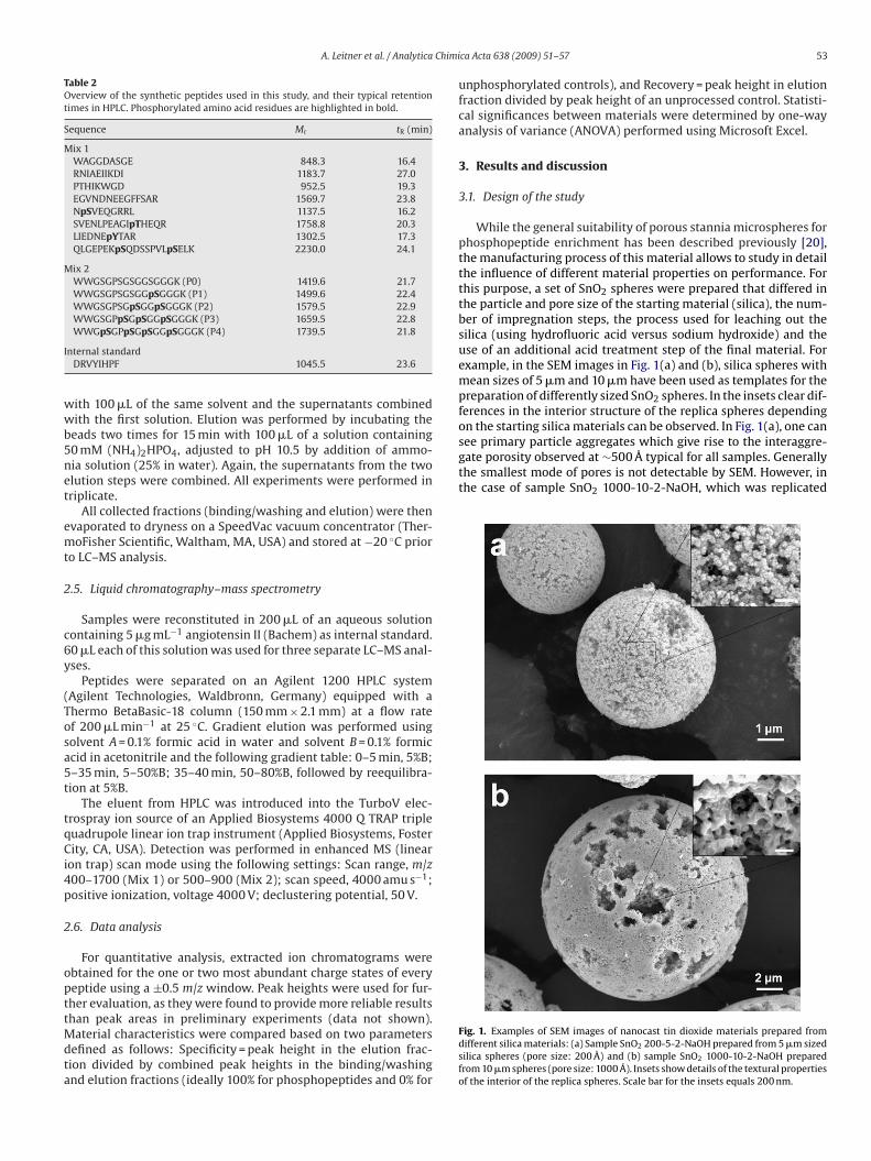
A. Leitner et al. / Analytica Chimi
Table 2Overview of the synthetic peptides used in this study, and their typical retentiontimes in HPLC. Phosphorylated amino acid residues are highlighted in bold.
Sequence Mr tR (min)
Mix 1WAGGDASGE 848.3 16.4RNIAEIIKDI 1183.7 27.0PTHIKWGD 952.5 19.3EGVNDNEEGFFSAR 1569.7 23.8NpSVEQGRRL 1137.5 16.2SVENLPEAGIpTHEQR 1758.8 20.3LIEDNEpYTAR 1302.5 17.3QLGEPEKpSQDSSPVLpSELK 2230.0 24.1
Mix 2WWGSGPSGSGGSGGGK (P0) 1419.6 21.7WWGSGPSGSGGpSGGGK (P1) 1499.6 22.4WWGSGPSGpSGGpSGGGK (P2) 1579.5 22.9WWGSGPpSGpSGGpSGGGK (P3) 1659.5 22.8
I
wwb5net
emt
2
c6y
(Tosa5t
tqCi4p
2
opttMdta
see primary particle aggregates which give rise to the interaggre-gate porosity observed at ∼500 Å typical for all samples. Generallythe smallest mode of pores is not detectable by SEM. However, inthe case of sample SnO2 1000-10-2-NaOH, which was replicated
WWGpSGPpSGpSGGpSGGGK (P4) 1739.5 21.8
nternal standardDRVYIHPF 1045.5 23.6
ith 100 �L of the same solvent and the supernatants combinedith the first solution. Elution was performed by incubating the
eads two times for 15 min with 100 �L of a solution containing0 mM (NH4)2HPO4, adjusted to pH 10.5 by addition of ammo-ia solution (25% in water). Again, the supernatants from the twolution steps were combined. All experiments were performed inriplicate.
All collected fractions (binding/washing and elution) were thenvaporated to dryness on a SpeedVac vacuum concentrator (Ther-oFisher Scientific, Waltham, MA, USA) and stored at −20 ◦C prior
o LC–MS analysis.
.5. Liquid chromatography–mass spectrometry
Samples were reconstituted in 200 �L of an aqueous solutionontaining 5 �g mL−1 angiotensin II (Bachem) as internal standard.0 �L each of this solution was used for three separate LC–MS anal-ses.
Peptides were separated on an Agilent 1200 HPLC systemAgilent Technologies, Waldbronn, Germany) equipped with ahermo BetaBasic-18 column (150 mm × 2.1 mm) at a flow ratef 200 �L min−1 at 25 ◦C. Gradient elution was performed usingolvent A = 0.1% formic acid in water and solvent B = 0.1% formiccid in acetonitrile and the following gradient table: 0–5 min, 5%B;–35 min, 5–50%B; 35–40 min, 50–80%B, followed by reequilibra-ion at 5%B.
The eluent from HPLC was introduced into the TurboV elec-rospray ion source of an Applied Biosystems 4000 Q TRAP tripleuadrupole linear ion trap instrument (Applied Biosystems, Fosterity, CA, USA). Detection was performed in enhanced MS (linear
on trap) scan mode using the following settings: Scan range, m/z00–1700 (Mix 1) or 500–900 (Mix 2); scan speed, 4000 amu s−1;ositive ionization, voltage 4000 V; declustering potential, 50 V.
.6. Data analysis
For quantitative analysis, extracted ion chromatograms werebtained for the one or two most abundant charge states of everyeptide using a ±0.5 m/z window. Peak heights were used for fur-her evaluation, as they were found to provide more reliable results
han peak areas in preliminary experiments (data not shown).aterial characteristics were compared based on two parametersefined as follows: Specificity = peak height in the elution frac-ion divided by combined peak heights in the binding/washingnd elution fractions (ideally 100% for phosphopeptides and 0% for
ca Acta 638 (2009) 51–57 53
unphosphorylated controls), and Recovery = peak height in elutionfraction divided by peak height of an unprocessed control. Statisti-cal significances between materials were determined by one-wayanalysis of variance (ANOVA) performed using Microsoft Excel.
3. Results and discussion
3.1. Design of the study
While the general suitability of porous stannia microspheres forphosphopeptide enrichment has been described previously [20],the manufacturing process of this material allows to study in detailthe influence of different material properties on performance. Forthis purpose, a set of SnO2 spheres were prepared that differed inthe particle and pore size of the starting material (silica), the num-ber of impregnation steps, the process used for leaching out thesilica (using hydrofluoric acid versus sodium hydroxide) and theuse of an additional acid treatment step of the final material. Forexample, in the SEM images in Fig. 1(a) and (b), silica spheres withmean sizes of 5 �m and 10 �m have been used as templates for thepreparation of differently sized SnO2 spheres. In the insets clear dif-ferences in the interior structure of the replica spheres dependingon the starting silica materials can be observed. In Fig. 1(a), one can
Fig. 1. Examples of SEM images of nanocast tin dioxide materials prepared fromdifferent silica materials: (a) Sample SnO2 200-5-2-NaOH prepared from 5 �m sizedsilica spheres (pore size: 200 Å) and (b) sample SnO2 1000-10-2-NaOH preparedfrom 10 �m spheres (pore size: 1000 Å). Insets show details of the textural propertiesof the interior of the replica spheres. Scale bar for the insets equals 200 nm.
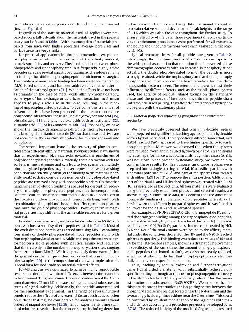
5 Chimi
f(
psps
tnppaTIcasaisnpgscnc
ttpspcepheDtamrm
btffftftpi
rtutfpood
4 A. Leitner et al. / Analytica
rom silica spheres with a pore size of 1000 Å, it can be observedinset of Fig. 1(b)).
Regardless of the starting material used, all replicas were pre-ared successfully; details about the materials used in the presenttudy can be found in Table 1. With the exception of materials pre-ared from silica with higher porosities, average pore sizes andurface areas are very similar.
For practical applications in phosphoproteomics, two proper-ies play a major role for the end user of the affinity material,amely specificity and recovery. The discrimination between phos-hopeptides and unphosphorylated, but frequently highly acidiceptides carrying several aspartic or glutamic acid residues remainschallenge for different phosphopeptide enrichment strategies.
he problem of nonspecific binding has been well documented forMAC-based protocols and has been addressed by methyl esterifi-ation of the carboxyl groups [31]. While the effects have not beens dramatic in the case of metal oxide affinity chromatography,ome type of ion exchange or acid-base interaction mechanismppears to play a role also in this case, resulting in the bind-ng of unphosphorylated peptides. To overcome this, a number ofolvent additives have been proposed in the literature to reduceonspecific interactions, these include dihydroxybenzoic acid [15],hthalic acid [11], aliphatic hydroxy acids such as lactic acid [32],lutamic acid [33] or its ammonium salt [34]. Previously, we havehown that tin dioxide appears to exhibit intrinsically less nonspe-ific binding than titanium dioxide [20] so that these additives areot required in the enrichment protocol for mixtures of moderateomplexity.
The second important issue is the recovery of phosphopep-ides from different affinity materials. Previous studies have shownhat an experimental bias may exist towards the enrichment ofolyphosphorylated peptides. Obviously, their interaction with theorbent is much stronger and can lead to two scenarios: multiplyhosphorylated peptides may be over represented when washingonditions are relatively harsh (or the binding to the material inher-ntly weak) so that a considerable number of singly phosphorylatedeptides are removed along with nonspecific binders. On the otherand, when mild elution conditions are used for desorption, recov-ry of multiply phosphorylated peptides may be compromised.ifferent elution conditions from metal oxides have been used in
he literature, and we have obtained the most satisfying results withcombination of high pH and the addition of inorganic phosphate toaximize the yield of phosphopeptides. However, intrinsic mate-
ial properties may still limit the achievable recoveries for a givenaterial.In order to systematically evaluate tin dioxide as an MOAC sor-
ent, we chose a set of synthetic peptides listed in Table 2. Most ofhe work described herein was carried out using Mix 1 containingour singly or doubly phosphorylated model peptides along withour unphosphorylated controls. Additional experiments were per-ormed on a set of peptides with identical amino acid sequencehat differed only in the number of phosphorylation sites, rangingrom zero to four (Mix 2). We have previously demonstrated thathe general enrichment procedure works well also in more com-lex samples [20], so the composition of the two sample mixtures
s ideal for a focused study on material-related effects.LC–MS analysis was optimized to achieve highly reproducible
esults in order to allow minor differences between the materialso be observed. Thus, we favored the use of conventional HPLC col-mn diameters (2 mm I.D.) because of the increased robustness inerms of signal stability. Additionally, the peptide amounts used
or the enrichment experiments, in the range of several hundredmols, reduce the effects of any external factors such as adsorptionn surfaces that may be considerable for analyte amounts severalrders of magnitude lower [35,36]. Initial experiments using stan-ard mixtures revealed that the chosen set-up including detectionca Acta 638 (2009) 51–57
in the linear ion trap mode of the Q TRAP instrument allowed usto obtain relative standard deviations of peak heights in the rangeof ∼1% which was also the case throughout the further study. Toensure reliability of the data, three experimental replicates (indi-vidual enrichment procedures) were performed for all materials,and bound and unbound fractions were each analyzed in triplicateby LC–MS.
Typical retention times for all peptides are given in Table 2.Interestingly, the retention times of Mix 2 do not correspond tothe widespread assumption that retention time in reversed-phasechromatography decreases with an increase in phosphorylation:actually, the doubly phosphorylated form of the peptide is moststrongly retained, while the unphosphorylated and the quadruplyphosphorylated form showed the least retention for the chro-matographic system chosen. The retention behavior is most likelyinfluenced by different factors such as the mobile phase systemused, the activity of residual silanol groups on the stationaryphase and the presence of interactions within the peptide chain(intramolecular ion pairing) that affect the interaction of hydropho-bic regions with the stationary phase.
3.2. Material properties influencing phosphopeptide enrichmentspecificity
We have previously observed that when tin dioxide replicaswere prepared using different leaching agents (sodium hydroxideversus hydrofluoric acid), their specificity differed somewhat, andNaOH-leached SnO2 appeared to have higher specificity towardsphosphopeptides. Moreover, we observed that when the sphereswere refluxed overnight in diluted hydrochloric acid, an additionalincrease in performance was obtained, although the reason for thiswas not clear. In the present, systematic study, we were able toconfirm these results. For this purpose, tin dioxide replicas wereprepared from a single starting material, 10 �m silica particles witha nominal pore size of 120 Å, and part of the spheres was treatedwith either NaOH or HF to remove the silica portion. Additionally,part of the NaOH- and HF-leached spheres was treated with 0.1 MHCl, as described in the Section 2. All four materials were evaluatedusing the previously established protocol, and selected results areshown in Fig. 2. From these data, it is evident that the amount ofnonspecific binding of unphosphorylated peptides noticeably dif-fers between the differently prepared spheres, and it was found tobe lowest for the NaOH- and HCl-treated spheres.
For example, EGVNDNEEGFFSAR (Glu1-fibrinopeptide B), exhib-ited the strongest binding among the unphosphorylated peptides,which is due to the highly acidic character of the peptide (four acidicresidues, pI = 4.00). For SnO2 particles that were not treated by HCl,37% and 14% of the total amount were bound to the affinity mate-rial under the conditions chosen for the HF- and the NaOH-leachedspheres, respectively. This binding was reduced to values of 15% and9% for the HCl-treated samples, showing a dramatic improvementin specificity. At the same time, the amount of singly phosphory-lated peptides that bound to SnO2 was also noticeably reduced,which we attribute to the fact that phosphopeptides are also par-tially bound via nonspecific interactions.
Thus, leaching by sodium hydroxide and further “activation”using HCl afforded a material with substantially reduced non-specific binding, although at the cost of phosphopeptide recoveryin the elution fraction. This is particularly relevant for the weak-est binding phosphopeptide, NpSVEQGRRL. We propose that forthis peptide, strong intermolecular ion pairing occurs between the
phosphorylated serine residue located near the N-terminus and thetwo strongly basic arginine residues near the C-terminus. This couldbe confirmed by covalent modification of the arginines with mal-ondialdehyde according to a procedure previously developed by us[37,38]. The reduced basicity of the modified Arg residues resulted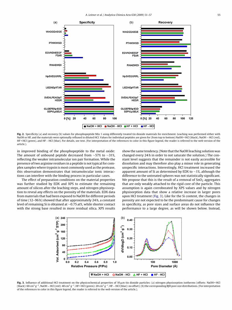
A. Leitner et al. / Analytica Chimica Acta 638 (2009) 51–57 55
F rentlyN ividuH refera
iTrpptt
watfolw
F(o
ig. 2. Specificity (a) and recovery (b) values for phosphopeptide Mix 1 using diffeaOH or HF, and the materials were optionally refluxed in diluted HCl. Values for indF + HCl (green), and HF − HCl (blue). For details, see text. (For interpretation of therticle.).
n improved binding of the phosphopeptide to the metal oxide:he amount of unbound peptide decreased from ∼57% to ∼31%,eflecting the weaker intramolecular ion pair formation. While theresence of two arginine residues in a peptide is not typical for com-lex samples where trypsin is most commonly used as the protease,his observation demonstrates that intramolecular ionic interac-ions can interfere with the binding process in particular cases.
The effect of preparation conditions on the material propertiesas further studied by EDX and XPS to estimate the remaining
mount of silicon after the leaching steps, and nitrogen physisorp-
ion to reveal any effects on the porosity of the materials. EDX datarom materials that had been exposed to NaOH for different periodsf time (12–96 h) showed that after approximately 24 h, a constantevel of remaining Si is obtained at ∼0.75 at%, while shorter contactith the strong base resulted in more residual silica. XPS results
ig. 3. Influence of additional HCl treatment on the physicochemical properties of 10 �black): 60 cm3 g−1, NaOH − HCl (red): 40 cm3 g−1, HF + HCl (green): 20 cm3 g−1, HF − HCl (bf the references to color in this figure legend, the reader is referred to the web version of
treated tin dioxide materials for enrichment. Leaching was performed either withal peptides are given for (from top to bottom) NaOH + HCl (black), NaOH − HCl (red),ences to color in this figure legend, the reader is referred to the web version of the
show the same tendency. (Note that the NaOH leaching solution waschanged every 24 h in order to not saturate the solution.) The con-stant level suggests that the remainder is not easily accessible fordissolution and may therefore also play a minor role in generatingunspecific interactions. Interestingly, HCl treatment increased theapparent amount of Si as determined by EDX to ∼1%, although thedifference to the untreated spheres was not statistically significant.We propose that this is the result of a removal of SnO2 aggregatesthat are only weakly attached to the rigid core of the particle. Thisassumption is again corroborated by XPS values and by nitrogen
physisorption data that show a relative increase in larger poresupon HCl treatment (Fig. 3). Like for the Si content, the changes inporosity are not expected to be the predominant cause for changesin specificity, as pore sizes and surface areas do not influence theperformance to a large degree, as will be shown below. Instead,m tin dioxide particles: (a) nitrogen physisorption isotherms (offsets: NaOH + HCllue): no offset); (b) the corresponding BJH pore size distributions. (For interpretationthe article.).
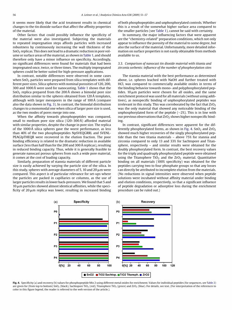
5 Chimi
ico
ttrSutnis
wf3Sdaaca
swotPbsigi
stctl1fi
Fac
6 A. Leitner et al. / Analytica
t seems more likely that the acid treatment results in chemicalhanges to the tin dioxide surface that affect the affinity propertiesf the material.
Other factors that could possibly influence the specificity ofhe material were also investigated. Subjecting the materialso repeated impregnation steps affords particles with increasedobustness by continuously increasing the wall thickness of thenO2 replicas. This does not lead to a dramatic reduction in pore vol-me or surface areas of the material, as shown in Table 1, and shouldherefore only have a minor influence on specificity. Accordingly,o significant differences were found for materials that had been
mpregnated once, twice, or three times. The multiply impregnatedpheres could be better suited for high-pressure applications.
In contrast, notable differences were observed in some caseshen SnO2 particles were prepared from silica templates with dif-
erent pore sizes. Silica spheres with nominal porosities of 120, 200,00 and 1000 Å were used for nanocasting. Table 1 shows that thenO2 replica prepared from the 200 Å shows a bimodal pore sizeistribution similar to the spheres obtained from 120 Å templates,lthough with larger mesopores in the range of 100 Å (comparelso the data shown in Fig. 3). In contrast, the bimodal distributionhanges to a monomodal one when silica of larger pore size is used,s the two modes of pores merge into one.
When the affinity towards phosphopeptides was compared,mall to medium pore size silica (120–300 Å) afforded materialith similar properties, despite the change in pore size. The replica
f the 1000 Å silica spheres gave the worst performance, as lesshan 40% of the two phosphopeptides NpSVEQGRRL and SVENL-EAGIpTHEQR were recovered in the elution fraction. The poorinding efficiency is related to the dramatic reduction in availableurface (less than half than for the 200 and 300 Å replicas), resultingn reduced binding capacity. Thus, while it is generally feasible toenerate nanocast porous spheres from such a wide pore material,t comes at the cost of loading capacity.
Similarly, preparation of stannia materials of different particleize is easily achieved by varying the particle size of the silica. Inhis study, spheres with average diameters of 5, 10 and 20 �m were
ompared. This aspect is of particular relevance for set-ups wherehe particles are packed in capillaries or columns, as the use ofarger particles results in lower back-pressures. We found that 5 and0 �m particles showed almost identical affinities, while the speci-city of 20 �m replica was lower, resulting in increased bindingig. 4. Specificity (a) and recovery (b) values for phosphopeptide Mix 2 using different mere given for (from top to bottom) SnO2 (black), Sachtopore TiO2 (red), Titansphere TiO2 (olor in this figure legend, the reader is referred to the web version of the article.).
ca Acta 638 (2009) 51–57
of both phosphopeptides and unphosphorylated controls. Whetherthis is a result of the somewhat higher surface area compared tothe smaller particles (see Table 1), cannot be said with certainty.
In summary, the major influencing factors that were apparentare the “chemistry-related” preparation conditions, which not onlyappear to influence the porosity of the material to some degree, butalso the surface of the material. Unfortunately, more detailed infor-mation on surface properties is not easily obtainable from methodsavailable to us.
3.3. Comparison of nanocast tin dioxide material with titania andzirconia sorbents: influence of the number of phosphorylation sites
The stannia material with the best performance as determinedabove, i.e. spheres leached with NaOH and further treated withHCl, was compared to commercially available oxides in terms ofthe binding behavior towards mono- and polyphosphorylated pep-tides. 10 �m particles were chosen for all oxides, and the sameenrichment protocol was used for all materials (without using addi-tives), as nonspecific binding of unphosphorylated peptides wasirrelevant in this study. This was corroborated by the fact that ZrO2was the only material that showed any noticeable binding of theunphosphorylated form of the peptide (∼2%). This is in line withour previous observations that ZrO2 shows higher nonspecific bind-ing.
In contrast, significant differences were apparent for the dif-ferently phosphorylated forms, as shown in Fig. 4. SnO2 and ZrO2showed much higher recoveries of the singly phosphorylated pep-tide than the two titania materials – above 75% for stannia andzirconia compared to only 13 and 33% for Sachtopore and Titan-sphere, respectively – and similar results were obtained for thedoubly phosphorylated form. In contrast, the best recovery valuesfor the triply and quadruply phosphorylated peptide were obtainedusing the Titansphere TiO2 and the ZrO2 material. Quantitativebinding on all materials (100% specificity) was obtained for thepeptides carrying two to four phosphate groups so that any lossescan directly be attributed to incomplete elution from the materials.
(No reductions in signal intensities were observed when peptidesolutions were incubated without affinity material under bindingand elution conditions, respectively, so that a significant influenceof peptide degradation or adsorptive loss during the enrichmentprocedure can be ruled out.)tal oxides for enrichment. Values for individual peptides (for sequences, see Table 2)green) and ZrO2 (blue). For details, see text. (For interpretation of the references to
Chimi
nmuoagRbr
4
pftoimapevodsedrFptsf
cmhl
A
“iSPoS
[
[[
[
[
[
[[[
[
[
[
[
[
[
[
[[[
[
[
[
[
[
A. Leitner et al. / Analytica
Clearly, additional experiments on rather complex samples areecessary to compare in more detail the affinities of the stanniaaterial to other metal oxides, and are presently being carried out
sing samples of biological interest. However, judging from this setf data, it appears that SnO2 allows for a high recovery of singlynd doubly phosphorylated forms of the same peptide, while stilliving acceptable values for higher phosphorylated forms (>30%).ecoveries of multiply phosphorylated peptides may be improvedy a further optimization of elution conditions, for example usingeagents proposed recently by Kyono et al. [39].
. Conclusion
While different porous metal oxides are increasingly used forhosphopeptide enrichment purposes, little is known about the
actors influencing their performance. Commonly, structural varia-ions of the affinity material are limited by commercial availabilityr the flexibility of the manufacturing process. Using the nanocast-ng technique, we have shown that a wide range of tin dioxide
aterials may be prepared, and the process should be equallydaptable to other oxides such as TiO2 or ZrO2. Differently porousarticles were found to give comparable performance (with thexception of replicas from 1000 Å material), which may be rele-ant to particular experimental set-ups such as packed beds forn-line enrichment columns. Interestingly, acid or base treatmenturing the preparation of the spheres was found to have the mostignificant influence. While this was connected at least to somextent to a change in the pore size distribution of the material, theecisive changes seem to take place on the surface of the mate-ial. Further investigations in this direction are therefore necessary.inally, experiments with peptides differing in the number of phos-horylation sites revealed that phosphopeptide enrichment usinghe stannia material allows for the adequate representation of bothingly and multiply phosphorylated peptides, which is importantor the analysis of biological samples.
Preliminary data from an ongoing study on the analysis of wholeell lysates have already demonstrated that tin dioxide affinity chro-atography is also applicable to this type of sample, as several
undred phosphopeptides were tentatively identified from HeLaysate. Results from this study will be reported in due course.
cknowledgements
This work was financially supported by the network projectAustrian Proteomics Platform II” of the GEN-AU (Genome Research
n Austria) program funded by the Austrian Federal Ministry ofcience and Research, and the European Union Sixth Frameworkrogram “Advanced Interactive Materials by Design – AIMS”. Partf this work was presented at the 56th ASMS Conference on Masspectrometry and Allied Topics, Denver, CO, USA.[[[[
ca Acta 638 (2009) 51–57 57
References
[1] C. DıAmbrosio, A.M. Salzano, S. Arena, G. Renzone, A. Scaloni, J. Chromatogr. B849 (2007) 163.
[2] M.O. Collins, L. Yu, J.S. Choudhary, Proteomics 7 (2007) 2751.[3] J.D. Hoffert, M.A. Knepper, Anal. Biochem. 375 (2008) 1.[4] A. Paradela, J.P. Albar, J. Proteome Res. 7 (2008) 1809.[5] G. Han, M. Ye, H. Zou, Analyst 133 (2008) 1128.[6] S.A. Beausoleil, M. Jedrychowski, D. Schwartz, J.E. Elias, J. Villén, J. Li, M.A. Cohn,
L.C. Cantley, S.P. Gygi, Proc. Natl. Acad. Sci. U.S.A. 101 (2004) 12130.[7] J. Dai, W.-H. Jin, Q.-H. Sheng, C.-H. Shieh, J.-R. Wu, R. Zeng, J. Proteome Res. 6
(2007) 250.[8] A. Motoyama, T. Xu, C.I. Ruse, J.A. Wohlschlegel, J.R. Yates III, Anal. Chem. 79
(2007) 3623.[9] G. Han, M. Ye, H. Zhou, X. Jiang, S. Feng, X. Jiang, R. Tian, D. Wan, H. Zou, J. Gu,
Proteomics 8 (2008) 1346.10] H. Zhou, J.D. Watts, R. Aebersold, Nat. Biotechnol. 19 (2001) 375.
[11] B. Bodenmiller, L.N. Mueller, M. Mueller, B. Domon, R. Aebersold, Nat. Methods4 (2007) 231.
12] P.A. Connor, A.J. McQuillan, Langmuir 15 (1999) 2916.13] M.W.H. Pinkse, P.M. Uitto, M.J. Hilhorst, B. Ooms, A.J.R. Heck, Anal. Chem. 76
(2004) 3935.14] I. Kuroda, Y. Shintani, M. Motokawa, S. Abe, M. Furuno, Anal. Sci. 20 (2004)
1313.15] M.R. Larsen, T.E. Thingholm, O.N. Jensen, P. Roepstorff, T.D. Jørgensen, Mol. Cell.
Proteomics 4 (2005) 873.16] H.K. Kweon, K. Håkansson, Anal. Chem. 78 (2006) 1743.
[17] F. Wolschin, S. Wienkoop, W. Weckwerth, Proteomics 5 (2005) 4389.18] Y. Li, H. Lin, C. Deng, P. Yang, X. Zhang, Proteomics 8 (2008) 238.19] Y. Li, D. Qi, C. Deng, P. Yang, X. Zhang, J. Proteome Res. 7 (2008) 1767.20] M. Sturm, A. Leitner, J.-H. Smått, M. Lindén, W. Lindner, Adv. Funct. Mater. 18
(2008) 2381.21] S.B. Ficarro, J.R. Parikh, N.C. Blank, J.A. Marto, Anal. Chem. 80 (2008)
4606.22] J.V. Olsen, B. Blagoev, F. Gnad, B. Macek, C. Kumar, P. Mortensen, M. Mann, Cell
127 (2006) 635.23] H. Molina, D.M. Horn, N. Tang, S. Mathivanan, A. Pandey, Proc. Natl. Acad. Sci.
U.S.A. 104 (2007) 2199.24] M.W.H. Pinkse, S. Mohammed, J.W. Gouw, B.v. Breukelen, H.R. Vos, A.J.R. Heck,
J. Proteome Res. 7 (2008) 687.25] N. Sugiyama, H. Nakagami, K. Mochida, A. Daudi, M. Tomita, K. Shirasu, Y. Ishi-
hama, Mol. Syst. Biol. 4 (2008) 193 (article number).26] G.T. Cantin, T.R. Shock, S.K. Park, H.D. Madhani, J.R. Yates III, Anal. Chem. 79
(2007) 4666.27] K. Imami, N. Sugiyama, Y. Kyono, M. Tomita, Y. Ishihama, Anal. Sci. 24 (2008)
161.28] A.-H. Lu, F. Schüth, Adv. Mater. 18 (2006) 1793.29] T. Valdés-Solís, A.B. Fuertes, Mater. Res. Bull. 41 (2006) 2187.30] J.-H. Smått, N. Schüwer, M. Järn, W. Lindner, M. Lindén, Microporous Meso-
porous Mater. 112 (2008) 308.31] S.B. Ficarro, M.L. McCleland, P.T. Stukenberg, D.J. Burke, M.M. Ross, J. Sha-
banowitz, D.F. Hunt, F.M. White, Nat. Biotechnol. 20 (2002) 301.32] N. Sugiyama, T. Masuda, K. Shinoda, A. Nakamura, M. Tomita, Y. Ishihama, Mol.
Cell. Proteomics 6 (2007) 1103.33] J. Wu, Q. Shakey, W. Liu, A. Schuller, M.T. Follettie, J. Proteome Res. 6 (2007)
4684.34] L.-R. Yu, Z. Zhu, K.C. Chan, H.J. Issaq, D.S. Dimitrov, T.D. Veenstra, J. Proteome
Res. 6 (2007) 4150.35] P.M.v. Midwoud, L. Rieux, R. Bischoff, E. Verpoorte, H.A.G. Niederländer, J. Pro-
teome Res. 6 (2007) 781.36] S.J. Bark, V. Hook, J. Proteome Res. 6 (2007) 4511.37] A. Foettinger, A. Leitner, W. Lindner, J. Mass Spectrom. 41 (2006) 623.38] A. Leitner, A. Foettinger, W. Lindner, J. Mass Spectrom. 42 (2007) 950.39] Y. Kyono, N. Sugiyama, K. Imami, M. Tomita, Y. Ishihama, J. Proteome Res. 7
(2008) 4585.

Publication 3
Probing the Phosphoproteome of HeLa Cells UsingNanocast Metal Oxide Microspheres forPhosphopeptide Enrichment
Alexander Leitner,*,†,⊥ Martin Sturm,† Otto Hudecz,‡,§ Michael Mazanek,‡,§ Jan-Henrik Smatt,|
Mika Linden,| Wolfgang Lindner,† and Karl Mechtler‡,§
Department of Analytical Chemistry and Food Chemistry, University of Vienna, Waehringer Strasse 38,1090 Vienna, Austria, Protein Chemistry Facility, IMPsResearch Institute of Molecular Pathology, Dr. Bohr-Gasse 7,1030 Vienna, Austria, Protein Chemistry Facility, IMBAsInstitute for Molecular Biotechnology, Dr. Bohr-Gasse 3,1030 Vienna, Austria and Department of Physical Chemistry, Center for Functional Materials, Åbo Akademi University,Porthansgatan 3-5, 20500 Turku, Finland
Metal oxide affinity chromatography (MOAC) has becomea prominent method to enrich phosphopeptides prior totheir analysis by liquid chromatography-mass spectrom-etry. To overcome limitations in material design, we havepreviously reported the use of nanocasting as a means togenerate metal oxide spheres with tailored properties.Here, we report on the application of two oxides, tindioxide (stannia) and titanium dioxide (titania), for theanalysis of the HeLa phosphoproteome. In combinationwith nanoflow LC-MS/MS analysis on a linear ion trap-Fourier transform ion cyclotron resonance instrument, weidentified 619 phosphopeptides using the new stanniamaterial, and 896 phosphopeptides using titania pre-pared in house. We also compared the newly developedmaterials to commercial titania material using an estab-lished enrichment protocol. Both titania materials yieldeda comparable total number of phosphopeptides, but theoverlap of the two data sets was less than one-third.Although fewer peptides were identified using stannia, thecomplementarity of SnO2-based MOAC could be shownas more than 140 phosphopeptides were exclusivelyidentified by this material.
The biological significance of protein phosphorylation hasstimulated the development and refinement of countless analyticaltools for profiling the “phosphoproteome”.1-5 Difficulties instudying protein phosphorylation on a large scale are well
documented and have been addressed by improvements on alllevels of the proteomic workflow. For mass spectrometry-basedproteomics, this begins with the optimization of extractionprocedures for biological material (e.g., the addition of phos-phatase inhibitors6) to ensure that the phosphorylation state isaccurately represented in the sample that is analyzed and endswith the use of highly sensitive, fast scanning mass spectrometersthat employ improved fragmentation techniques to maximize thesequence information from phosphopeptides.7,8 In between, newenrichment and fractionation strategies have been developed inrecent years to complement the established techniques of im-mobilized metal affinity chromatography (IMAC) and immunoaf-finity precipitation. Among those, metal oxide affinity chromatog-raphy (MOAC) takes advantage of the particular affinity of metaloxides such as titanium dioxide to phosphate groups.9 Variousmaterials have been proposed although the majority of studieswere carried out using TiO2 as affinity material.10-12 In addition,zirconium dioxide12-14 and, more recently, niobium pentoxide15
have been used in large-scale studies. Frequently, it has beenreported that metal oxides vary in their affinity to different groupsof phosphopeptides, e.g., mono- and multiply phosphorylatedforms. Therefore, several groups have reported the use of twometal oxides and/or different enrichment techniques (MOAC andIMAC in parallel) to allow for a more comprehensive representa-tion of the phosphoproteome.16,17
* To whom correspondence should be addressed. E-mail:[email protected].
† University of Vienna.‡ IMPsResearch Institute of Molecular Pathology.§ IMBAsInstitute for Molecular Biotechnology.| Åbo Akademi University.⊥ Current address: Institute of Molecular Systems Biology, ETH Zurich,
Wolfgang-Pauli-Strasse 16, 8093 Zurich, Switzerland.(1) Thingholm, T. E.; Jensen, O. N.; Larsen, M. R. Proteomics 2009, 20, 1451–
1468.(2) Macek, B.; Mann, M.; Olsen, J. V. Annu. Rev. Pharmacol. Toxicol. 2009,
49, 199–221.(3) Tedford, N. C.; White, F. M.; Redding, J. A. Briefings Funct. Genomics
Proteomics 2008, 7, 383–394.(4) Nita-Lazar, A.; Saito-Benz, H.; White, F. M. Proteomics 2008, 8, 4433–4443.(5) Paradela, A.; Albar, J. P. J. Proteome Res. 2008, 7, 1809–1818.
(6) Thingholm, T. E.; Larsen, M. R.; Ingrell, C. R.; Kassem, M.; Jensen, O. N.J. Proteome Res. 2008, 7, 3304–3313.
(7) Han, X.; Aslanian, A.; Yates, J. R., III. Curr. Opin. Chem. Biol. 2008, 12,483–490.
(8) Abu-Farha, M.; Elisma, F.; Zhou, H.; Tian, R.; Zhou, H.; Asmer, M. S.; Figeys,D. Anal. Chem. 2009, 81, 4585–4599.
(9) Leitner, A. Trends Anal. Chem. 2010, 29, 177–185.(10) Pinkse, M. W. H.; Uitto, P. M.; Hilhorst, M. J.; Ooms, B.; Heck, A. J. R.
Anal. Chem. 2004, 76, 3935–3943.(11) Larsen, M. R.; Thingholm, T. E.; Jensen, O. N.; Roepstorff, P.; Jorgensen,
T. J. D. Mol. Cell. Proteomics 2005, 4, 873–886.(12) Sugiyama, N.; Masuda, T.; Shinoda, K.; Nakamura, A.; Tomita, M.; Ishihama,
Y. Mol. Cell. Proteomics 2007, 6, 1103–1109.(13) Kweon, H. K.; Hakansson, K. Anal. Chem. 2006, 78, 1743–1749.(14) Sugiyama, N.; Nakagami, H.; Mochida, K.; Daudi, A.; Tomita, M.; Shirasu,
K.; Ishihama, Y. Mol. Syst. Biol. 2008, 4, 193 (article number).(15) Ficarro, S. B.; Parikh, J. R.; Blank, N. C.; Marto, J. A. Anal. Chem. 2008,
80, 4606–4613.
Anal. Chem. 2010, 82, 2726–2733
10.1021/ac902560z 2010 American Chemical Society2726 Analytical Chemistry, Vol. 82, No. 7, April 1, 2010Published on Web 03/04/2010
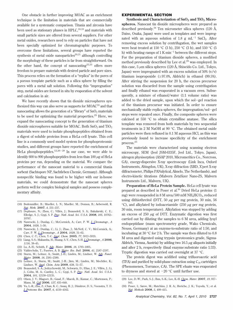
One obstacle in further improving MOAC as an enrichmenttechnique is the limitation in materials that are commerciallyavailable for a systematic comparison. Titania and zirconia havebeen used as stationary phases in HPLC,18,19 and materials withsmall particle sizes are offered from several suppliers. For othermetal oxides, researchers need to rely on particles that have notbeen specially optimized for chromatographic purposes. Toovercome these limitations, several groups have reported thesynthesis of metal oxide nanoparticles20,21 although controllingthe morphology of these particles is far from straightforward. Onthe other hand, the concept of nanocasting22,23 offers morefreedom to prepare materials with desired particle and pore sizes.This process relies on the formation of a “replica” in the pores ofa porous template particle such as a silica sphere by filling thepores with a metal salt solution. Following this “impregnation”step, metal oxides are formed in situ by evaporation of the solventand calcination in air.
We have recently shown that tin dioxide microspheres syn-thesized this way can also serve as supports for MOAC24 and thatnanocasting allows the generation of a “library” of SnO2 materialsto be used for optimizing the material properties.25 Here, weexpand the nanocasting concept to the generation of titaniumdioxide microspheres suitable for MOAC. Both SnO2 and TiO2
materials were used to isolate phosphopeptides obtained froma digest of soluble proteins from a HeLa cell lysate. This cellline is a commonly used model system for phosphoproteomicstudies, and different groups have reported the enrichment ofHeLa phosphopeptides.12,26-28 In our case, we were able toidentify 600 to 900 phosphopeptides from less than 100 µg of HeLaproteins per run, depending on the material. We compare theperformance of the nanocast material to a commercial titaniasorbent (Sachtopore NP, Sachtleben Chemie, Germany). Althoughnonspecific binding was found to be higher with our in-housematerials, we could demonstrate that the nanocast spheresperform well for complex biological samples and possess comple-mentary affinity.
EXPERIMENTAL SECTIONSynthesis and Characterization of SnO2 and TiO2 Micro-
spheres. Nanocast tin dioxide microspheres were prepared asdescribed previously.24 Ten micrometer silica spheres (120 Å,Daiso, Osaka, Japan) were used as templates and were impreg-nated with an aqueous solution of 1.8 g mL-1 SnCl2. Afterremoving excess solution by centrifugation, the wet sampleswere heat treated at 150 °C (3 h), 250 °C (3 h), and 550 °C (5h) with heating ramps of 1 K min-1 between the different steps.For the preparation of titanium dioxide spheres, a modifiedmethod previously described by Lee et al.29 was employed. Inthis case, 5 µm silica spheres (120 Å, Shiseido Co. Ltd., Tokyo,Japan) were impregnated with an excess solution of 50% (v/v)titanium isopropoxide (g97.0%, Aldrich) in ethanol (99.5%).After stirring the suspension for 20 h, the excess precursorsolution was discarded from the sample using centrifugationand finally ethanol was evaporated in a vacuum oven. Subse-quently, a mixture of ethanol/water (1:1 volume ratio) wasadded to the dried sample, upon which the sol-gel reactionof the titanium precursor was initiated. In order to ensuremechanically stable replica spheres, the impregnation/sol-gelsteps were repeated once. Finally, the composite spheres werecalcined at 550 °C to obtain crystalline anatase. The silicatemplate was removed from both materials by two sequentialtreatments in 2 M NaOH at 90 °C. The obtained metal oxideparticles were then refluxed in 0.1 M aqueous HCl, as this waspreviously found to increase specificity of the enrichmentprocess.25
The materials were characterized using scanning electronmicroscopy, SEM (Jeol JSM-6335F, Jeol Ltd., Tokyo, Japan),nitrogen physisorption (ASAP 2010, Micromeritics Co., Norcross,GA), energy-dispersive X-ray spectroscopy (Link Inca, OxfordInstruments, Abingdon, UK), X-ray diffraction (XRD, X’pert X-raydiffractometer, Philips PANalytical, Almelo, The Netherlands), andelectro-kinetic titrations (Malvern ZetaSizer Nano-ZS, MalvernInstruments Ltd., Malvern, UK).
Preparation of HeLa Protein Sample. HeLa cell lysate wasprepared as described in Poser et al.30 Dried HeLa proteins (1mg) were resuspended in 8 M urea/500 mM NH4HCO3, reducedusing dithiothreitol (DTT, 50 µg per mg protein, 30 min, 56°C), and alkylated by iodoacetamide (250 µg per mg protein,30 min, room temperature). Alkylation was stopped by addingan excess of 250 µg of DTT. Enzymatic digestion was firstcarried out by diluting the samples to 6 M urea, adding lysylendopeptidase (mass spectrometry grade, Wako Chemicals,Neuss, Germany) at an enzyme-to-substrate ratio of 1:50, andincubating at 30 °C for 2 h. The sample was then diluted to 0.8M urea and digested using trypsin (proteomics grade, Sigma-Aldrich, Vienna, Austria) by adding two 16.5 µg aliquots initiallyand after 2 h, respectively (final enzyme:substrate ratio 1:33).Tryptic digestion was carried out overnight at 37 °C.
The protein digest was acidified using trifluoroacetic acid(TFA) and purified by solid-phase extraction using C18 cartridges(Phenomenex, Torrance, CA). The SPE eluate was evaporatedto dryness and stored at -20 °C until further use.
(16) Bodenmiller, B.; Mueller, L. N.; Mueller, M.; Domon, B.; Aebersold, R.Nat. Meth. 2007, 4, 231–237.
(17) Dephoure, N.; Zhou, C.; Villen, J.; Beausoleil, S. A.; Bakalarski, C. E.;Elledge, S. J.; Gygi, S. P. Proc. Natl. Acad. Sci. U.S.A. 2008, 105, 10762–10767.
(18) Nawrocki, J.; Dunlap, C.; McCormick, A.; Carr, P. W. J. Chromatogr., A2004, 1028, 1–30.
(19) Nawrocki, J.; Dunlap, C.; Li, J.; Zhao, J.; McNeff, C. V.; McCormick, A.;Carr, P. W. J. Chromatogr., A 2004, 1028, 31–62.
(20) Chen, C.-T.; Chen, Y.-C. Anal. Chem. 2005, 77, 5912–5919.(21) Liang, S.-S.; Makamba, H.; Huang, S.-Y.; Chen, S.-H. J. Chromatogr., A 2006,
1116, 38–45.(22) Lu, A.-H.; Schuth, F. Adv. Mater. 2006, 18, 1793–1805.(23) Valdes-Solıs, T.; Fuertes, A. B. Mater. Res. Bull. 2006, 41, 2187–2197.(24) Sturm, M.; Leitner, A.; Smatt, J.-H.; Linden, M.; Lindner, W. Adv. Funct.
Mater. 2008, 18, 2381–2389.(25) Leitner, A.; Sturm, M.; Smatt, J.-H.; Jarn, M.; Linden, M.; Mechtler, K.;
Lindner, W. Anal. Chim. Acta 2009, 638, 51–57.(26) Beausoleil, S. A.; Jedrychowski, M.; Schwartz, D.; Elias, J. E.; Villen, J.; Li,
J.; Cohn, M. A.; Cantley, L. C.; Gygi, S. P. Proc. Natl. Acad. Sci. U.S.A.2004, 101, 12130–12135.
(27) Olsen, J. V.; Blagoev, B.; Gnad, F.; Macek, B.; Kumar, C.; Mortensen, P.;Mann, M. Cell 2006, 127, 635–648.
(28) Yu, L.-R.; Zhu, Z.; Chan, K. C.; Issaq, H. J.; Dimitrov, D. S.; Veenstra, T. D.J. Proteome Res. 2007, 6, 4150–4162.
(29) Lee, D.-W.; Park, S.-J.; Ihm, S.-K.; Lee, K.-H. Chem. Mater. 2007, 19, 937–941.
(30) Poser, I.; Sarov, M.; Hutchins, J. R. A.; Heriche, J. K.; Toyoda, Y.; et al.Nat. Methods 2008, 5, 409–415.
2727Analytical Chemistry, Vol. 82, No. 7, April 1, 2010
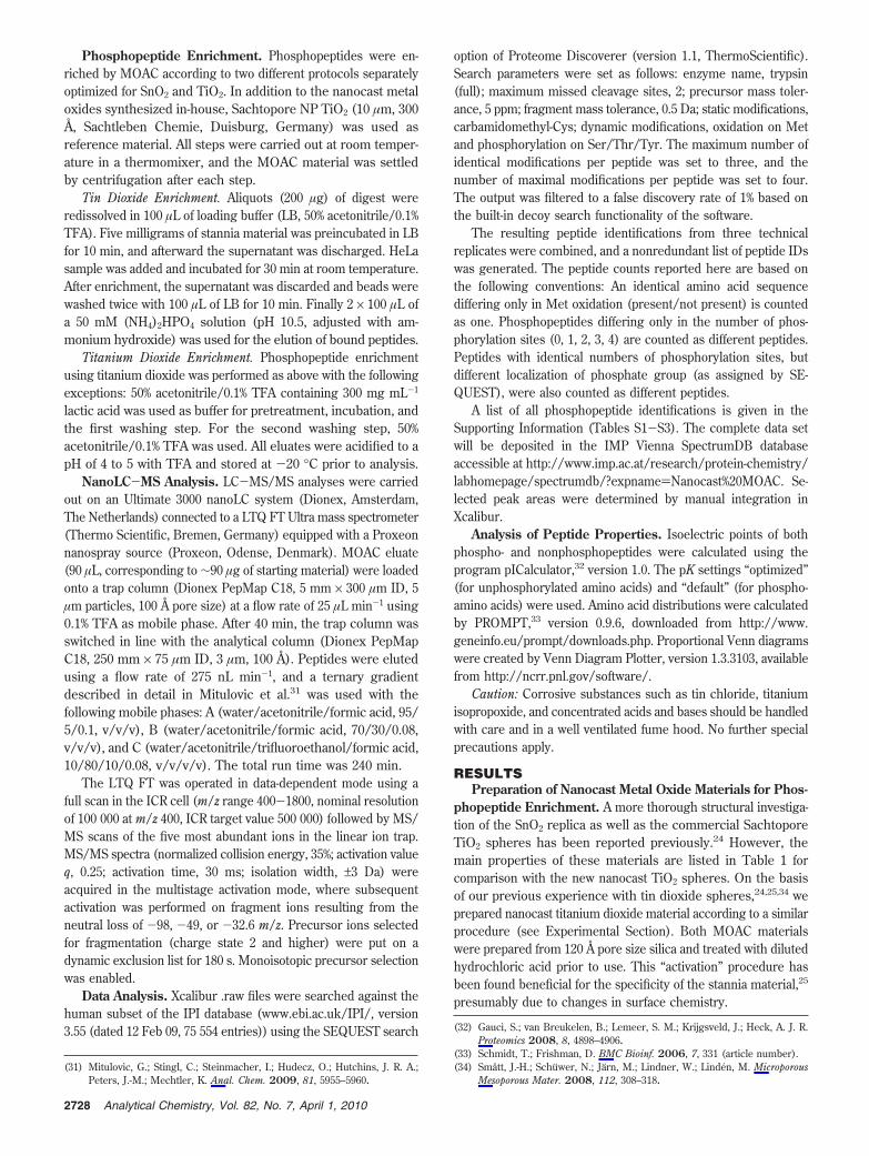
Phosphopeptide Enrichment. Phosphopeptides were en-riched by MOAC according to two different protocols separatelyoptimized for SnO2 and TiO2. In addition to the nanocast metaloxides synthesized in-house, Sachtopore NP TiO2 (10 µm, 300Å, Sachtleben Chemie, Duisburg, Germany) was used asreference material. All steps were carried out at room temper-ature in a thermomixer, and the MOAC material was settledby centrifugation after each step.
Tin Dioxide Enrichment. Aliquots (200 µg) of digest wereredissolved in 100 µL of loading buffer (LB, 50% acetonitrile/0.1%TFA). Five milligrams of stannia material was preincubated in LBfor 10 min, and afterward the supernatant was discharged. HeLasample was added and incubated for 30 min at room temperature.After enrichment, the supernatant was discarded and beads werewashed twice with 100 µL of LB for 10 min. Finally 2 × 100 µL ofa 50 mM (NH4)2HPO4 solution (pH 10.5, adjusted with am-monium hydroxide) was used for the elution of bound peptides.
Titanium Dioxide Enrichment. Phosphopeptide enrichmentusing titanium dioxide was performed as above with the followingexceptions: 50% acetonitrile/0.1% TFA containing 300 mg mL-1
lactic acid was used as buffer for pretreatment, incubation, andthe first washing step. For the second washing step, 50%acetonitrile/0.1% TFA was used. All eluates were acidified to apH of 4 to 5 with TFA and stored at -20 °C prior to analysis.
NanoLC-MS Analysis. LC-MS/MS analyses were carriedout on an Ultimate 3000 nanoLC system (Dionex, Amsterdam,The Netherlands) connected to a LTQ FT Ultra mass spectrometer(Thermo Scientific, Bremen, Germany) equipped with a Proxeonnanospray source (Proxeon, Odense, Denmark). MOAC eluate(90 µL, corresponding to ∼90 µg of starting material) were loadedonto a trap column (Dionex PepMap C18, 5 mm × 300 µm ID, 5µm particles, 100 Å pore size) at a flow rate of 25 µL min-1 using0.1% TFA as mobile phase. After 40 min, the trap column wasswitched in line with the analytical column (Dionex PepMapC18, 250 mm × 75 µm ID, 3 µm, 100 Å). Peptides were elutedusing a flow rate of 275 nL min-1, and a ternary gradientdescribed in detail in Mitulovic et al.31 was used with thefollowing mobile phases: A (water/acetonitrile/formic acid, 95/5/0.1, v/v/v), B (water/acetonitrile/formic acid, 70/30/0.08,v/v/v), and C (water/acetonitrile/trifluoroethanol/formic acid,10/80/10/0.08, v/v/v/v). The total run time was 240 min.
The LTQ FT was operated in data-dependent mode using afull scan in the ICR cell (m/z range 400-1800, nominal resolutionof 100 000 at m/z 400, ICR target value 500 000) followed by MS/MS scans of the five most abundant ions in the linear ion trap.MS/MS spectra (normalized collision energy, 35%; activation valueq, 0.25; activation time, 30 ms; isolation width, ±3 Da) wereacquired in the multistage activation mode, where subsequentactivation was performed on fragment ions resulting from theneutral loss of -98, -49, or -32.6 m/z. Precursor ions selectedfor fragmentation (charge state 2 and higher) were put on adynamic exclusion list for 180 s. Monoisotopic precursor selectionwas enabled.
Data Analysis. Xcalibur .raw files were searched against thehuman subset of the IPI database (www.ebi.ac.uk/IPI/, version3.55 (dated 12 Feb 09, 75 554 entries)) using the SEQUEST search
option of Proteome Discoverer (version 1.1, ThermoScientific).Search parameters were set as follows: enzyme name, trypsin(full); maximum missed cleavage sites, 2; precursor mass toler-ance, 5 ppm; fragment mass tolerance, 0.5 Da; static modifications,carbamidomethyl-Cys; dynamic modifications, oxidation on Metand phosphorylation on Ser/Thr/Tyr. The maximum number ofidentical modifications per peptide was set to three, and thenumber of maximal modifications per peptide was set to four.The output was filtered to a false discovery rate of 1% based onthe built-in decoy search functionality of the software.
The resulting peptide identifications from three technicalreplicates were combined, and a nonredundant list of peptide IDswas generated. The peptide counts reported here are based onthe following conventions: An identical amino acid sequencediffering only in Met oxidation (present/not present) is countedas one. Phosphopeptides differing only in the number of phos-phorylation sites (0, 1, 2, 3, 4) are counted as different peptides.Peptides with identical numbers of phosphorylation sites, butdifferent localization of phosphate group (as assigned by SE-QUEST), were also counted as different peptides.
A list of all phosphopeptide identifications is given in theSupporting Information (Tables S1-S3). The complete data setwill be deposited in the IMP Vienna SpectrumDB databaseaccessible at http://www.imp.ac.at/research/protein-chemistry/labhomepage/spectrumdb/?expname)Nanocast%20MOAC. Se-lected peak areas were determined by manual integration inXcalibur.
Analysis of Peptide Properties. Isoelectric points of bothphospho- and nonphosphopeptides were calculated using theprogram pICalculator,32 version 1.0. The pK settings “optimized”(for unphosphorylated amino acids) and “default” (for phospho-amino acids) were used. Amino acid distributions were calculatedby PROMPT,33 version 0.9.6, downloaded from http://www.geneinfo.eu/prompt/downloads.php. Proportional Venn diagramswere created by Venn Diagram Plotter, version 1.3.3103, availablefrom http://ncrr.pnl.gov/software/.
Caution: Corrosive substances such as tin chloride, titaniumisopropoxide, and concentrated acids and bases should be handledwith care and in a well ventilated fume hood. No further specialprecautions apply.
RESULTSPreparation of Nanocast Metal Oxide Materials for Phos-
phopeptide Enrichment. A more thorough structural investiga-tion of the SnO2 replica as well as the commercial SachtoporeTiO2 spheres has been reported previously.24 However, themain properties of these materials are listed in Table 1 forcomparison with the new nanocast TiO2 spheres. On the basisof our previous experience with tin dioxide spheres,24,25,34 weprepared nanocast titanium dioxide material according to a similarprocedure (see Experimental Section). Both MOAC materialswere prepared from 120 Å pore size silica and treated with dilutedhydrochloric acid prior to use. This “activation” procedure hasbeen found beneficial for the specificity of the stannia material,25
presumably due to changes in surface chemistry.
(31) Mitulovic, G.; Stingl, C.; Steinmacher, I.; Hudecz, O.; Hutchins, J. R. A.;Peters, J.-M.; Mechtler, K. Anal. Chem. 2009, 81, 5955–5960.
(32) Gauci, S.; van Breukelen, B.; Lemeer, S. M.; Krijgsveld, J.; Heck, A. J. R.Proteomics 2008, 8, 4898–4906.
(33) Schmidt, T.; Frishman, D. BMC Bioinf. 2006, 7, 331 (article number).(34) Smatt, J.-H.; Schuwer, N.; Jarn, M.; Lindner, W.; Linden, M. Microporous
Mesoporous Mater. 2008, 112, 308–318.
2728 Analytical Chemistry, Vol. 82, No. 7, April 1, 2010
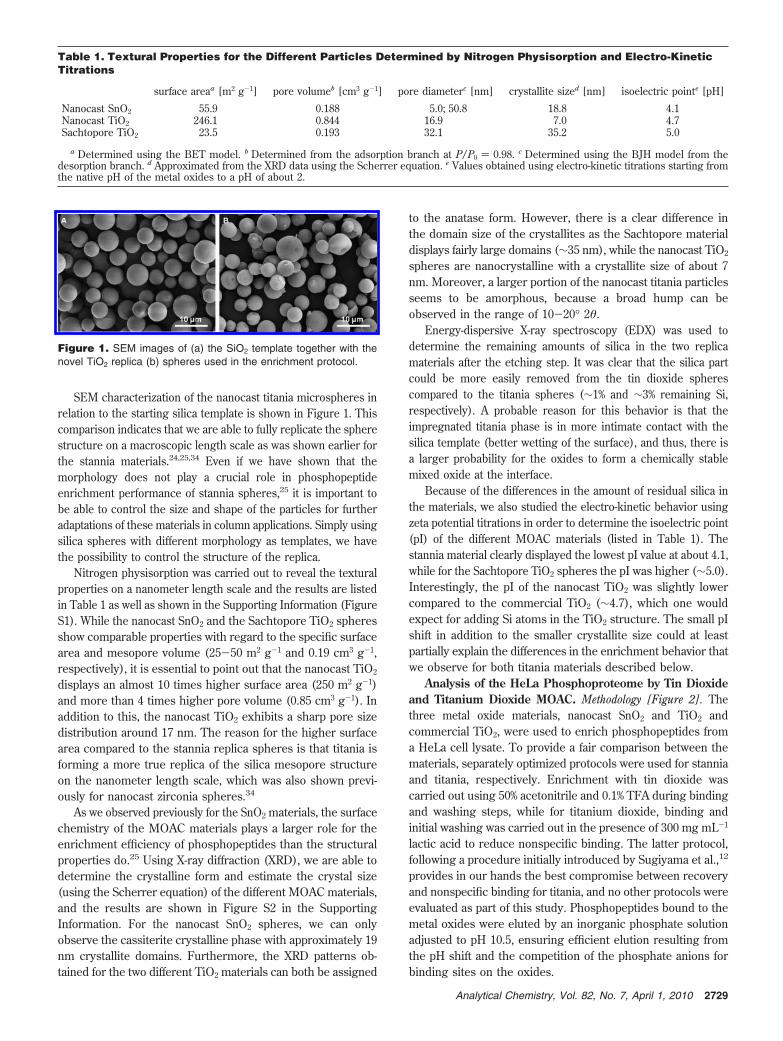
SEM characterization of the nanocast titania microspheres inrelation to the starting silica template is shown in Figure 1. Thiscomparison indicates that we are able to fully replicate the spherestructure on a macroscopic length scale as was shown earlier forthe stannia materials.24,25,34 Even if we have shown that themorphology does not play a crucial role in phosphopeptideenrichment performance of stannia spheres,25 it is important tobe able to control the size and shape of the particles for furtheradaptations of these materials in column applications. Simply usingsilica spheres with different morphology as templates, we havethe possibility to control the structure of the replica.
Nitrogen physisorption was carried out to reveal the texturalproperties on a nanometer length scale and the results are listedin Table 1 as well as shown in the Supporting Information (FigureS1). While the nanocast SnO2 and the Sachtopore TiO2 spheresshow comparable properties with regard to the specific surfacearea and mesopore volume (25-50 m2 g-1 and 0.19 cm3 g-1,respectively), it is essential to point out that the nanocast TiO2
displays an almost 10 times higher surface area (250 m2 g-1)and more than 4 times higher pore volume (0.85 cm3 g-1). Inaddition to this, the nanocast TiO2 exhibits a sharp pore sizedistribution around 17 nm. The reason for the higher surfacearea compared to the stannia replica spheres is that titania isforming a more true replica of the silica mesopore structureon the nanometer length scale, which was also shown previ-ously for nanocast zirconia spheres.34
As we observed previously for the SnO2 materials, the surfacechemistry of the MOAC materials plays a larger role for theenrichment efficiency of phosphopeptides than the structuralproperties do.25 Using X-ray diffraction (XRD), we are able todetermine the crystalline form and estimate the crystal size(using the Scherrer equation) of the different MOAC materials,and the results are shown in Figure S2 in the SupportingInformation. For the nanocast SnO2 spheres, we can onlyobserve the cassiterite crystalline phase with approximately 19nm crystallite domains. Furthermore, the XRD patterns ob-tained for the two different TiO2 materials can both be assigned
to the anatase form. However, there is a clear difference inthe domain size of the crystallites as the Sachtopore materialdisplays fairly large domains (∼35 nm), while the nanocast TiO2
spheres are nanocrystalline with a crystallite size of about 7nm. Moreover, a larger portion of the nanocast titania particlesseems to be amorphous, because a broad hump can beobserved in the range of 10-20° 2θ.
Energy-dispersive X-ray spectroscopy (EDX) was used todetermine the remaining amounts of silica in the two replicamaterials after the etching step. It was clear that the silica partcould be more easily removed from the tin dioxide spherescompared to the titania spheres (∼1% and ∼3% remaining Si,respectively). A probable reason for this behavior is that theimpregnated titania phase is in more intimate contact with thesilica template (better wetting of the surface), and thus, there isa larger probability for the oxides to form a chemically stablemixed oxide at the interface.
Because of the differences in the amount of residual silica inthe materials, we also studied the electro-kinetic behavior usingzeta potential titrations in order to determine the isoelectric point(pI) of the different MOAC materials (listed in Table 1). Thestannia material clearly displayed the lowest pI value at about 4.1,while for the Sachtopore TiO2 spheres the pI was higher (∼5.0).Interestingly, the pI of the nanocast TiO2 was slightly lowercompared to the commercial TiO2 (∼4.7), which one wouldexpect for adding Si atoms in the TiO2 structure. The small pIshift in addition to the smaller crystallite size could at leastpartially explain the differences in the enrichment behavior thatwe observe for both titania materials described below.
Analysis of the HeLa Phosphoproteome by Tin Dioxideand Titanium Dioxide MOAC. Methodology [Figure 2]. Thethree metal oxide materials, nanocast SnO2 and TiO2 andcommercial TiO2, were used to enrich phosphopeptides froma HeLa cell lysate. To provide a fair comparison between thematerials, separately optimized protocols were used for stanniaand titania, respectively. Enrichment with tin dioxide wascarried out using 50% acetonitrile and 0.1% TFA during bindingand washing steps, while for titanium dioxide, binding andinitial washing was carried out in the presence of 300 mg mL-1
lactic acid to reduce nonspecific binding. The latter protocol,following a procedure initially introduced by Sugiyama et al.,12
provides in our hands the best compromise between recoveryand nonspecific binding for titania, and no other protocols wereevaluated as part of this study. Phosphopeptides bound to themetal oxides were eluted by an inorganic phosphate solutionadjusted to pH 10.5, ensuring efficient elution resulting fromthe pH shift and the competition of the phosphate anions forbinding sites on the oxides.
Table 1. Textural Properties for the Different Particles Determined by Nitrogen Physisorption and Electro-KineticTitrations
surface areaa [m2 g-1] pore volumeb [cm3 g-1] pore diameterc [nm] crystallite sized [nm] isoelectric pointe [pH]
Nanocast SnO2 55.9 0.188 5.0; 50.8 18.8 4.1Nanocast TiO2 246.1 0.844 16.9 7.0 4.7Sachtopore TiO2 23.5 0.193 32.1 35.2 5.0
a Determined using the BET model. b Determined from the adsorption branch at P/P0 ) 0.98. c Determined using the BJH model from thedesorption branch. d Approximated from the XRD data using the Scherrer equation. e Values obtained using electro-kinetic titrations starting fromthe native pH of the metal oxides to a pH of about 2.
Figure 1. SEM images of (a) the SiO2 template together with thenovel TiO2 replica (b) spheres used in the enrichment protocol.
2729Analytical Chemistry, Vol. 82, No. 7, April 1, 2010
MOAC eluates were subsequently analyzed by reversed-phaseHPLC-MS/MS on a linear ion trap-FTICR hybrid mass spec-trometer. Because of the facile neutral loss of the phosphate group(in the form of H3PO4) from many phosphoserine and phos-phothreonine peptides that can compromise spectral quality,35
we employed multistage activation36 for sequencing. Thistechnique, which activates both the precursor ion and productions resulting from neutral loss of H3PO4 without an intermit-tent isolation step, shows better performance compared to aMS2/MS3 strategy and facilitates data analysis, as also recentlyconfirmed by Ulintz et al.37 Following a database search usingSEQUEST against the human subset of the IPI database anda decoy database approach, peptide identifications were filteredto a false discovery rate of less than 1% for every individualLC-MS/MS run.
Number of Phosphopeptides. Figure 3 gives an overview of theperformance of the three MOAC materials. Combining the resultsof three technical replicates (separate MOAC enrichment followed
by a single LC-MS/MS run), more than 600 phosphopeptideswere confidently identified from each material in at least oneLC-MS/MS analysis (Figure 3a). Average numbers per individualrun ranged from ∼330 to 570 for the different materials. Usingthe two titania materials, a larger number of phosphopeptidescould be identified compared to stannia, and enrichment by theSachtopore material yielded roughly 10% more identifications thanthe in-house made material (1010 vs 896). When grouping thephosphopeptides according to the number of phosphorylation sites(Figure 3b), we found that 71% of the peptides identified fromthe Sachtopore samples were singly phosphorylated, while thenumber was higher for the two nanocast materials (82% and 84%,respectively). The majority of the remaining peptides werediphosphorylated (>85% for all materials), although triply and (few)quadruply phosphorylated peptides were also identified by allmaterials.
Ishihama and co-workers previously reported specificities of>90% in the elution fractions of Titansphere TiO2-MOAC usinglactic acid as modifier.14 We could corroborate these resultsfor the Sachtopore material, where we achieved 94% specificity.In contrast, the amount of nonspecific binding was higher forthe nanocast materials: Specificities of 67% and 60% wereobtained for titania and stannia, respectively. These results are,however, comparable to reported values obtained for zirconia-based MOAC, and it is important to consider that no additionaladditive was used for the stannia material. This confirms ourprevious observation made using model peptides that stanniapossesses a significantly higher intrinsic specificity for phos-phopeptides than titania,24 which has also been recentlyconfirmed by Zhang and co-workers using magnetic tin dioxide-coated particles.38
Overlaps in the Data Sets. Every sample obtained from MOACwas analyzed using a single dimension chromatographic separa-tion followed by tandem MS. In order to examine the reproduc-ibility of the enrichment strategy, MOAC was carried out intriplicate. Figure 4a shows the overlap in phosphopeptide identi-fications of the three materials used for this study. Less than one-third of the phosphopeptides were identified in all three replicateanalyses, with the overlap being the highest for the nanocasttitania material. In at least two runs, 40-50% of the peptides weredetected. In both cases, the overlap was lowest for the tin dioxidematerial. The (small) difference to the titania materials may becaused not only by the material properties but also by slightvariations in instrument performance over time. In general, thevalues obtained reflect the enormous complexity of the phospho-peptide sample which would require a more comprehensive(multidimensional) separation strategy for in-depth characteriza-tion.
However, it is still of interest to confirm the overlap inphosphopeptide IDs between the different metal oxide materials.Figure 4b shows the overlap for all peptides identified in at leastone run per material. Only 297 phosphopeptides (18.6% of the totalpool) were identified with all three materials. Compared to theSachtopore data set, the nanocast materials contributed 586additional identifications, of which 363 were from nanocast titaniaand 144 from nanocast stannia (79 were found in both). Despite
(35) Boersema, P. J.; Mohammed, S.; Heck, A. J. R. J. Mass Spectrom. 2009,44, 861–878.
(36) Schroeder, M. J.; Shabanowitz, J.; Schwartz, J. C.; Hunt, D. F.; Coon, J. J.Anal. Chem. 2004, 76, 3590–3598.
(37) Ulintz, P. J.; Yocum, A. K.; Bodenmiller, B.; Aebersold, R.; Andrews, P. C.;Nesvizhskii, A. I. J. Proteome Res. 2009, 8, 887–899.
(38) Qi, D.; Lu, J.; Deng, C.; Zhang, X. J. Phys. Chem. C 2009, 113, 15854–15861.
Figure 2. Analytical strategy for the enrichment of HeLa phospho-peptides by metal oxide affinity chromatography.
2730 Analytical Chemistry, Vol. 82, No. 7, April 1, 2010
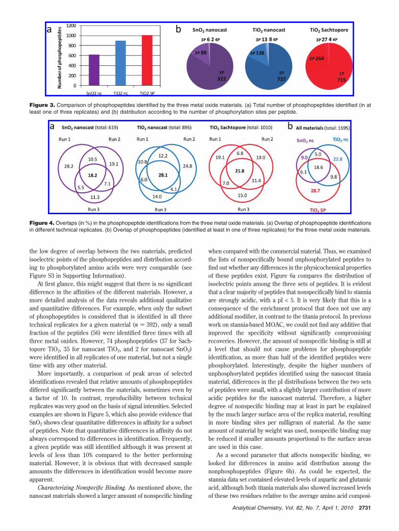
the low degree of overlap between the two materials, predictedisoelectric points of the phosphopeptides and distribution accord-ing to phosphorylated amino acids were very comparable (seeFigure S3 in Supporting Information).
At first glance, this might suggest that there is no significantdifference in the affinities of the different materials. However, amore detailed analysis of the data reveals additional qualitativeand quantitative differences. For example, when only the subsetof phosphopeptides is considered that is identified in all threetechnical replicates for a given material (n ) 392), only a smallfraction of the peptides (56) were identified three times with allthree metal oxides. However, 74 phosphopeptides (37 for Sach-topore TiO2, 35 for nanocast TiO2, and 2 for nanocast SnO2)were identified in all replicates of one material, but not a singletime with any other material.
More importantly, a comparison of peak areas of selectedidentifications revealed that relative amounts of phosphopeptidesdiffered significantly between the materials, sometimes even bya factor of 10. In contrast, reproducibility between technicalreplicates was very good on the basis of signal intensities. Selectedexamples are shown in Figure 5, which also provide evidence thatSnO2 shows clear quantitative differences in affinity for a subsetof peptides. Note that quantitative differences in affinity do notalways correspond to differences in identification. Frequently,a given peptide was still identified although it was present atlevels of less than 10% compared to the better performingmaterial. However, it is obvious that with decreased sampleamounts the differences in identification would become moreapparent.
Characterizing Nonspecific Binding. As mentioned above, thenanocast materials showed a larger amount of nonspecific binding
when compared with the commercial material. Thus, we examinedthe lists of nonspecifically bound unphosphorylated peptides tofind out whether any differences in the physicochemical propertiesof these peptides exist. Figure 6a compares the distribution ofisoelectric points among the three sets of peptides. It is evidentthat a clear majority of peptides that nonspecifically bind to stanniaare strongly acidic, with a pI < 5. It is very likely that this is aconsequence of the enrichment protocol that does not use anyadditional modifier, in contrast to the titania protocol. In previouswork on stannia-based MOAC, we could not find any additive thatimproved the specificity without significantly compromisingrecoveries. However, the amount of nonspecific binding is still ata level that should not cause problems for phosphopeptideidentification, as more than half of the identified peptides werephosphorylated. Interestingly, despite the higher numbers ofunphosphorylated peptides identified using the nanocast titaniamaterial, differences in the pI distributions between the two setsof peptides were small, with a slightly larger contribution of moreacidic peptides for the nanocast material. Therefore, a higherdegree of nonspecific binding may at least in part be explainedby the much larger surface area of the replica material, resultingin more binding sites per milligram of material. As the sameamount of material by weight was used, nonspecific binding maybe reduced if smaller amounts proportional to the surface areasare used in this case.
As a second parameter that affects nonspecific binding, welooked for differences in amino acid distribution among thenonphosphopeptides (Figure 6b). As could be expected, thestannia data set contained elevated levels of aspartic and glutamicacid, although both titania materials also showed increased levelsof these two residues relative to the average amino acid composi-
Figure 3. Comparison of phosphopeptides identified by the three metal oxide materials. (a) Total number of phosphopeptides identified (in atleast one of three replicates) and (b) distribution according to the number of phosphorylation sites per peptide.
Figure 4. Overlaps (in %) in the phosphopeptide identifications from the three metal oxide materials. (a) Overlap of phosphopeptide identificationsin different technical replicates. (b) Overlap of phosphopeptides (identified at least in one of three replicates) for the three metal oxide materials.
2731Analytical Chemistry, Vol. 82, No. 7, April 1, 2010
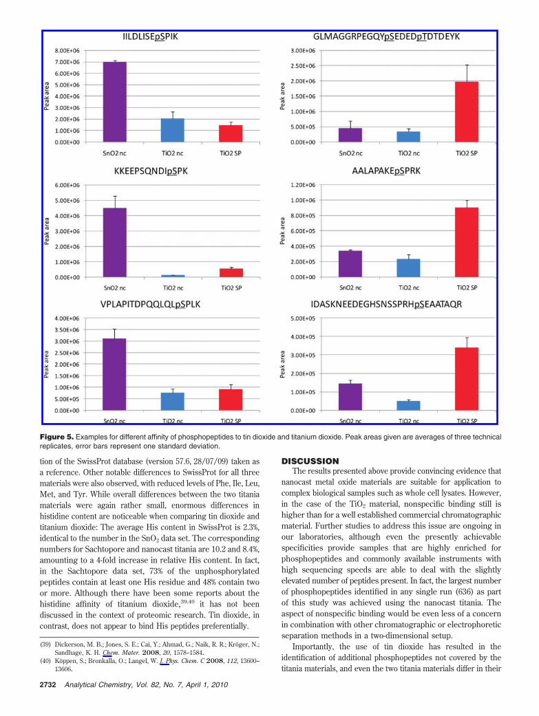
tion of the SwissProt database (version 57.6, 28/07/09) taken asa reference. Other notable differences to SwissProt for all threematerials were also observed, with reduced levels of Phe, Ile, Leu,Met, and Tyr. While overall differences between the two titaniamaterials were again rather small, enormous differences inhistidine content are noticeable when comparing tin dioxide andtitanium dioxide: The average His content in SwissProt is 2.3%,identical to the number in the SnO2 data set. The correspondingnumbers for Sachtopore and nanocast titania are 10.2 and 8.4%,amounting to a 4-fold increase in relative His content. In fact,in the Sachtopore data set, 73% of the unphosphorylatedpeptides contain at least one His residue and 48% contain twoor more. Although there have been some reports about thehistidine affinity of titanium dioxide,39,40 it has not beendiscussed in the context of proteomic research. Tin dioxide, incontrast, does not appear to bind His peptides preferentially.
DISCUSSIONThe results presented above provide convincing evidence that
nanocast metal oxide materials are suitable for application tocomplex biological samples such as whole cell lysates. However,in the case of the TiO2 material, nonspecific binding still ishigher than for a well established commercial chromatographicmaterial. Further studies to address this issue are ongoing inour laboratories, although even the presently achievablespecificities provide samples that are highly enriched forphosphopeptides and commonly available instruments withhigh sequencing speeds are able to deal with the slightlyelevated number of peptides present. In fact, the largest numberof phosphopeptides identified in any single run (636) as partof this study was achieved using the nanocast titania. Theaspect of nonspecific binding would be even less of a concernin combination with other chromatographic or electrophoreticseparation methods in a two-dimensional setup.
Importantly, the use of tin dioxide has resulted in theidentification of additional phosphopeptides not covered by thetitania materials, and even the two titania materials differ in their
(39) Dickerson, M. B.; Jones, S. E.; Cai, Y.; Ahmad, G.; Naik, R. R.; Kroger, N.;Sandhage, K. H. Chem. Mater. 2008, 20, 1578–1584.
(40) Koppen, S.; Bronkalla, O.; Langel, W. J. Phys. Chem. C 2008, 112, 13600–13606.
Figure 5. Examples for different affinity of phosphopeptides to tin dioxide and titanium dioxide. Peak areas given are averages of three technicalreplicates, error bars represent one standard deviation.
2732 Analytical Chemistry, Vol. 82, No. 7, April 1, 2010
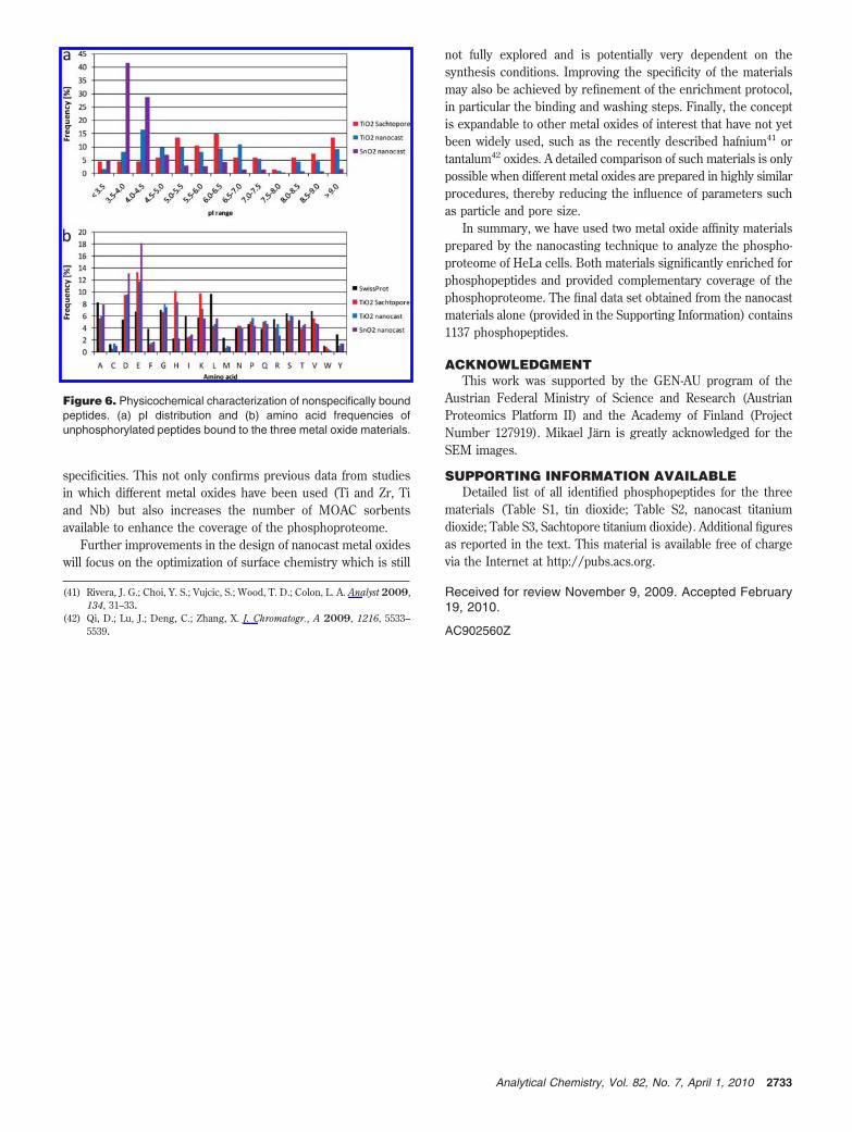
specificities. This not only confirms previous data from studiesin which different metal oxides have been used (Ti and Zr, Tiand Nb) but also increases the number of MOAC sorbentsavailable to enhance the coverage of the phosphoproteome.
Further improvements in the design of nanocast metal oxideswill focus on the optimization of surface chemistry which is still
not fully explored and is potentially very dependent on thesynthesis conditions. Improving the specificity of the materialsmay also be achieved by refinement of the enrichment protocol,in particular the binding and washing steps. Finally, the conceptis expandable to other metal oxides of interest that have not yetbeen widely used, such as the recently described hafnium41 ortantalum42 oxides. A detailed comparison of such materials is onlypossible when different metal oxides are prepared in highly similarprocedures, thereby reducing the influence of parameters suchas particle and pore size.
In summary, we have used two metal oxide affinity materialsprepared by the nanocasting technique to analyze the phospho-proteome of HeLa cells. Both materials significantly enriched forphosphopeptides and provided complementary coverage of thephosphoproteome. The final data set obtained from the nanocastmaterials alone (provided in the Supporting Information) contains1137 phosphopeptides.
ACKNOWLEDGMENTThis work was supported by the GEN-AU program of the
Austrian Federal Ministry of Science and Research (AustrianProteomics Platform II) and the Academy of Finland (ProjectNumber 127919). Mikael Jarn is greatly acknowledged for theSEM images.
SUPPORTING INFORMATION AVAILABLEDetailed list of all identified phosphopeptides for the three
materials (Table S1, tin dioxide; Table S2, nanocast titaniumdioxide; Table S3, Sachtopore titanium dioxide). Additional figuresas reported in the text. This material is available free of chargevia the Internet at http://pubs.acs.org.
Received for review November 9, 2009. Accepted February19, 2010.
AC902560Z
(41) Rivera, J. G.; Choi, Y. S.; Vujcic, S.; Wood, T. D.; Colon, L. A. Analyst 2009,134, 31–33.
(42) Qi, D.; Lu, J.; Deng, C.; Zhang, X. J. Chromatogr., A 2009, 1216, 5533–5539.
Figure 6. Physicochemical characterization of nonspecifically boundpeptides. (a) pI distribution and (b) amino acid frequencies ofunphosphorylated peptides bound to the three metal oxide materials.
2733Analytical Chemistry, Vol. 82, No. 7, April 1, 2010

Manuscript 1
A Chemically Cleavable Linker for Immobilizing Bait
Compounds in a Target Protein Pull-down Concept
Abstract
A novel linker chemistry based on a malondialdehyde - indole condensation reaction has
been developed for the affinity-independent elution of targeted protein pull downs. Recently
developed in our lab for tagging of tryptophan residues on proteins or peptides, the concept
was extended for the design of a chemically cleavable linker system. In our setup a specific
kinase inhibitor (bosutinib) was coupled to agarose and acrylamide beads respectively via
the new linker system and a protein pull down experiment of possible interaction partners of
a K562 whole cell lysate was performed. After purification, the linker can be cleaved from the
beads by incubation with 50mM pyrrolidine. The system is especially compatible with
targeted protein pull downs because during cleavage step no protein hydrolys is or any
degradation of amino acid side-chains was induced. From the pull-down experiment, key
targets of bosutinib such as the tyrosine kinase, Btk, were identified.
Introduction
For many drugs which are in pharmaceutical development or already in clinical use, their
main targets and other possible interaction partners are still unknown. To facilitate further
advances in drug effectiveness but also to get a better overview of possible side effects, all
interacting proteins should ideally be known. In the field of chemical proteomics, proteins are
selectively enriched by affinity chromatography for further analysis by 2D-gelelectrophoresis
and/or liquid chromatography followed by tandem mass spectrometry (LC-MS/MS). With the
help of organic chemistry probes can be coupled to beads via certain linker chemistry to
further purify targeted proteins. Thereby it is possible to purify targets from matrix
components which may otherwise interfere with downstream analysis. However, in case of
targeted protein pull downs, elution of enriched proteins in their native form can be difficult,
since most conventional elution protocols are based on the use of denaturing components
like detergents, low pH, or high salt concentrations for elution. Recently new affinity
independent elution methods which do not affect the bioaffinity interaction were introduced
[1-5]. Linkers can be cleaved enzymatically by cleaving a DNA-linker with nuclease [3]
introduced by Santala et al., photochemically [5], or chemically by using an azobenzene
linker which can be cleeaved by reduction with sodium dithionite [1, 4].
Here, we use a malondialdehyde-indole derivative as linker which can be chemically cleaved
by incubation with pyrrolidine or hydrazine. This chemistry, using malondialdehyde bound to
the indole nitrogen was previously used in our lab for the reversible and selective tagging of
tryphophan residues on peptides or proteins [6, 7].
Experimental
Synthesis
The indole linker 3 ((E)-2-(1-(3-oxoprop-1-enyl)-1H-indol-3-yl)acetic acid, IAA-MDA) was
designed to display two functional groups on opposite ends for coupling to the support and
the drug (“bait”), respectively, and a chemically cleavable group in the middle of the linker
moiety (scheme 1, a).
A condensation reaction of malondialdehyde (MDA) with 3-indolyl-acetic acid (IAA) 1 under
strongly acidic condition [7], primarily described by Teuber et al.in 1964 [8],was used
(scheme 1, a). As the first step, malondialdehyde (MDA) was applied in its commercial
available form as tetramethoxypropane (TMP) 2, which is converted to MDA in situ during
reaction. The newly formed derivative contains a chemically cleavable group at the indole
nitrogen which is linked to an activated - unsaturated aldehyde group for coupling onto
beads. Structure of the synthesis product was confirmed by mass spectrometry and NMR
spectroscopy (see supplemental data). The reaction conditions had to be controlled
accurately (not more than 2h reaction time, controlled temperature) to avoid polymerisation
of the derivative, due to the high activity of the resulting acroleine type group. The reactive
carbonyl group typically reacts with amine or hydrazide nitrogens to form Schiff base type
products.
For affinity chromatography purpose, we used the Pierce Carbolink Coupling Gel, an
agarose gel which contains hydrazide functionalities (coverage approximately 10 µmol/ml
gel). Additional coupling experiments were carried out on hydrazide activated acrylamide
beads for comparison of coupling and purification performance. Both materials performed in
comparable manner. For simplicity only data gained by use of agarose beads is quoted. With
the use of 10 mM aniline as catalyst [9] the reaction time could be reduced to two hours and
the reaction was performed in slightly acidic environment (solvent buffered to pH 4.5)
(scheme 1, b). During the reaction, the beads turned yellowish due to binding of the colored
IAA-MDA derivate.
The coupling efficacy was around 50% as determined by liquid chromatography – mass
spectrometry (LC-MS) in multiple reaction monitoring mode detection of the cleavage product
3-indolyl-acetic acid (IAA) after cleavage with 355mM pyrrolidine (complete cleavage). After
coupling, remaining hydrazide groups had to be saturated to avoid cross reactions in
following steps. Therefore we used a 30x molar excess of N-acetoxysuccinimide in order to
achieve complete saturation of remaining hydrazide groups (scheme 1, c). Completeness of
saturation was demonstrated by trying to re-bind the derivative to already saturated beads.
No coloring of beads, like it is normally the case upon binding of the derivative, could be
detected upon twentyfold excess of N-acetylsuccinimide, and after treatment with pyrrolidine,
no IAA could be detected by LC-MS analysis. A result of the saturation process was the
competitive displacement of readily bound IAA from beads (up to 50%).
From this point, the beads were ready for the attachment of the desired affinity target. The
free carboxylic residue of the linker was coupled to amino groups on affinity targets, using 4-
(4,6-dimethoxy-1,3,5-triazin-2-yl)-4-methylmorpholinium chloride (DMT-MM) as coupling
reagent [10]. The coupling reaction with DMT-MM is a one step reaction, so carboxylic
residues on the bait have to be protected first. Additional, only free amino groups in their
non-ionized form are accessible for the reaction.
To prove the effectiveness of the reaction, we chose methyl 2-amino-3-(4-
hydroxyphenyl)propanoate (tyrosine methyl ester) for coupling (scheme 1, d). As solvent,
methanol was found to be the best candidate because it ensured good coupling efficiency
without a collapse of the agarose beads. The binding efficiency was determined to be 50%,
again evaluated by quantitative LC-MS (see supplemental data). As a reference for
quantitation a 3-indolylacetic acid- tyrosine methyl ester conjugate 4 was used, which was
synthesized in house. The coverage of baits could be varied by changing in the amount of
IDA-MDA used for the initiation reaction. Consequently the protocol allows an easy control of
the total loading of baits on the beads.
For specific linker cleavage 50mM pyrrolidine was used and a 0.1M Tris buffer system did
not seem to interfere with the cleavage chemistry. The use of pyrrolidine was further tested
to clarify whether or not the reagent causes protein precipitation on the beads or causes any
protein degradation. Therefore beads were incubated overnight with a mixture of four
proteins that were dissolved in 0,1M Tris buffer or 50mM pyrrolidine solution either (see
supplemental data). The supernatant and possible remaining proteins from beads, once
directly after incubation and once after three washing steps with 0.1M Tris buffer (beads
were boiled in Lämmli buffer for elution) of both samples were analysed by 1D SDS-PAGE
with silver staining. The two samples behaved similarly. In case of pyrrolidine, a small
amount of protein could be detected after 3 washing steps, indicating a slight precipitating
effect of pyrrolidine (see supplemental data). To prove the long term stability of the system,
beads linked with the derivative alone, or linker coupled to bait were stored for 3 weeks in
water containing 0.02% sodium azide at 4°C, respectively. After 3 weeks, no changes in the
coupling rate could be detected for both samples.
Biological application
As a biological application we used the tyrosine kinase inhibitor bosutinib (SKI-606) [11] for
enrichment of possible direct and indirect interactors from a K562 cell lysate. Bosutinib is an
ATP-competitive third generation kinase inhibitor for the treatment of chronic myeloid
leukemia (CML) [12], which is currently in phase III of clinical trials. Former investigations
revealed SRC and ABL kinases as the main interactors of bosutinib, but also a lot of other
affinity targets could be found [12-14]. The drug in its amino functionalized form (scheme 2)
was kindly provided by the Center of Molecular Medicine, Austria (CeMM). The applicability
of this functionalized form of bosutinib for targeted protein pulldowns was shown in a recent
publication from Superti-Furga’s group [13]. A coupling rate of 50% could be achieved with
DMT-MM as coupling agent. Ready-made beads had an overall coverage of 0.7 µmol of
bosutinib per ml beads suspension. The cleavage product (IAA-MDA-c-bosutinb) resulting
from elution by pyrrolidine was found and confirmed by MS (see supplemental data). The
efficacy of the c-bosutinb coupling reaction was calculated by the amount of remaining IAA
after cleavage with pyrrolidine compared to an uncoupled control.
Affinity Purification
Protein pull down experiments were performed at the Center of Molecular Medicine on a
K562 cell lysate according to a protocol from Rix et al. [15] with the exception that instead of
eluting interactors by boiling with Lämmli buffer, elution was performed with 100mM
pyrrolidine and incubation for 3h at 4°C. The higher amount of pyrrolidine (100mM instead of
50mM in previous experiments) was chosen to compensate for higher sample complexity
and the lower incubation temperature (4 °C instead of RT in previous experiments). This step
was at this point not optimized with regards to concentration needed to carry out a complete
cleavage.
Immunoblot analysis
Immunoblot analysis was performed by 1D SDS-PAGE separation of the eluates (an elution
by boiling beads with Lämmli buffer was used for comparison). A monoclonal anti BTK
antibody was used for detection. The tyrosine kinase BTK is one of the main interactors of
Bosutinib. The immunoblot is shown in figure 1, demonstrating the enrichment of BTK by
affinity purification. Hence, the applicability of the purification protocol using indole linker
beads could be successfully demonstrated (line C). In terms of efficacy a stronger signal
could be achieved using the indole-beads compared to the reference (line D). This strong
enrichment of BTK suggests that even lower concentrations of pyrrolidine might be used for
cleavage if desired.
Liquid chromatography mass spectrometry
Along with the immunoblot analysis a LC-MS/MS protein identification of the pull-down was
realized on a LTQ-Orbitrap XL mass spectrometer to identify specific interaction partners and
to determine the degree of unspecific binding. Prior to LC-MS, 2D gel electrophoresis with
tryptic in gel digestion of separated proteins was performed. For procedure information the
reader is referred to the work of K. Bennett [15] and collegues.
Target purification using the indole linker system allowed the identification of 32 targets of
bosutinib (Table 1). Main interactors like SRC, ABL, BTK or TEC were successfully identified
but also newly found targets like CAMK2G could be identified [16] (Table 1). However, also a
high degree of unspecific bound proteins were found (see supplemental data). Agarose and
acrylamid gels behaved similarly, whereby the latter showed a higher degree of unspecific
binding.
Conclusion
A new linker chemistry, based on a reversible reaction of malondialdehyde with the indolyl
group of 3-indolyl-acetic acid was demonstrated for an affinity chromatography concept in
form of a pull-down protocol. The linker was coupled to hydrazide functionalized agarose
beads via an -unsaturated aldehyde group, and baits can be coupled to the linker via an
amide groups with DMT-MM as coupling agent. A protein pull down was successfully
performed using bosutinib as bait for purifying interaction partners of a K562 cell lysate.
Efficient purification of the main target BTK was shown by immunoblot analysis. All main
interactors could be identified by LC-MS/MS analysis. To further reduce unspecific binding of
unwanted proteins the binding, washing and elution protocol may be further improved. The
reduction of the pyrrolidine concentration used during cleavage seems particularly
reasonable due to the high enrichment efficiacy achieved for the bosutinib target Btk when
using 100mM pyrrolidine, compared to the purification using a noncleavable linker.
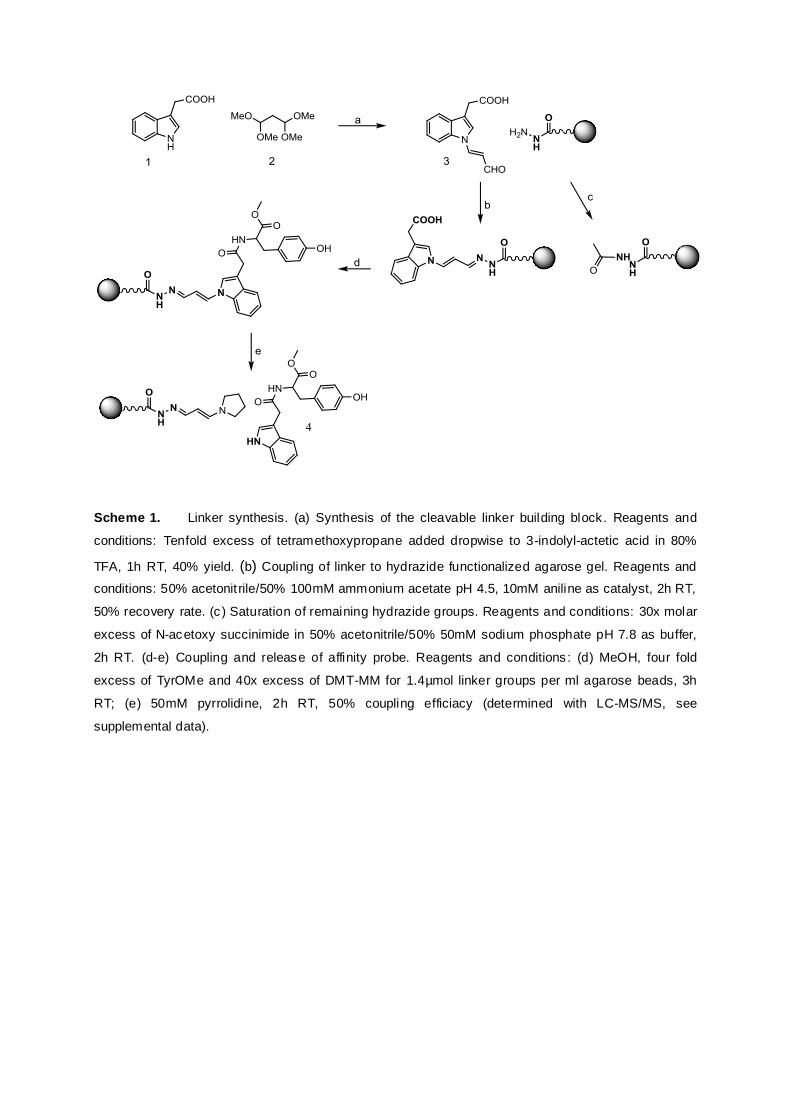
Scheme 1. Linker synthesis. (a) Synthesis of the cleavable linker building block. Reagents and
conditions: Tenfold excess of tetramethoxypropane added dropwise to 3-indolyl-actetic acid in 80%
TFA, 1h RT, 40% yield. (b) Coupling of linker to hydrazide functionalized agarose gel. Reagents and
conditions: 50% acetonit rile/50% 100mM ammonium acetate pH 4.5, 10mM aniline as catalyst, 2h RT,
50% recovery rate. (c ) Saturation of remaining hydrazide groups. Reagents and conditions: 30x molar
excess of N-acetoxy succinimide in 50% acetonitrile/50% 50mM sodium phosphate pH 7.8 as buffer,
2h RT. (d-e) Coupling and release of affinity probe. Reagents and conditions : (d) MeOH, four fold
excess of TyrOMe and 40x excess of DMT-MM for 1.4µmol linker groups per ml agarose beads, 3h
RT; (e) 50mM pyrrolidine, 2h RT, 50% coupling efficiacy (determined with LC-MS/MS, see
supplemental data).
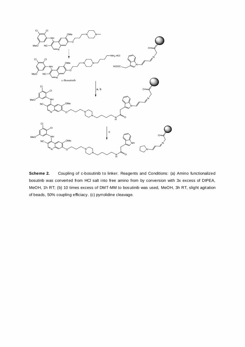
Scheme 2. Coupling of c-bosutinib to linker. Reagents and Conditions: (a) Amino functionalized
bosutinb was converted from HCl salt into free amino from by conversion with 3x excess of DIPEA,
MeOH, 1h RT; (b) 10 times excess of DMT-MM to bosutinib was used, MeOH, 3h RT, slight agitation
of beads, 50% coupling efficiacy. (c) pyrrolidine cleavage.
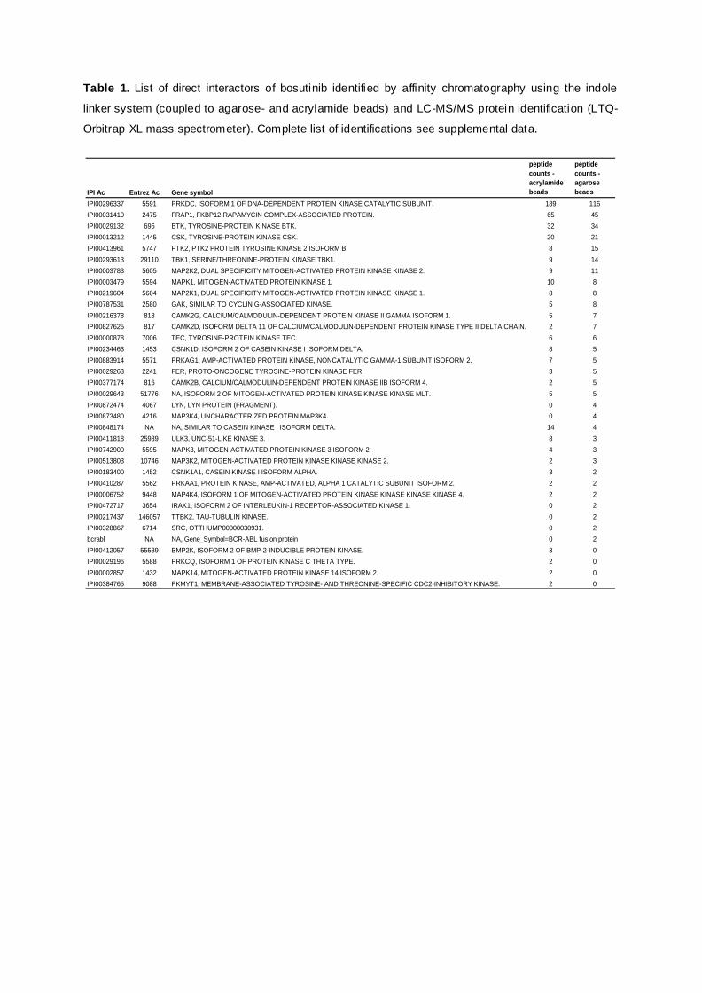
Table 1. List of direct interactors of bosutinib identified by affinity chromatography using the indole
linker system (coupled to agarose- and acrylamide beads) and LC-MS/MS protein identification (LTQ-
Orbitrap XL mass spectrometer). Complete list of identifications see supplemental data.
IPI Ac Entrez Ac Gene symbol
peptide counts - acrylamide beads
peptide counts - agarose beads
IPI00296337 5591 PRKDC, ISOFORM 1 OF DNA-DEPENDENT PROTEIN KINASE CATALYTIC SUBUNIT. 189 116
IPI00031410 2475 FRAP1, FKBP12-RAPAMYCIN COMPLEX-ASSOCIATED PROTEIN. 65 45
IPI00029132 695 BTK, TYROSINE-PROTEIN KINASE BTK. 32 34
IPI00013212 1445 CSK, TYROSINE-PROTEIN KINASE CSK. 20 21
IPI00413961 5747 PTK2, PTK2 PROTEIN TYROSINE KINASE 2 ISOFORM B. 8 15
IPI00293613 29110 TBK1, SERINE/THREONINE-PROTEIN KINASE TBK1. 9 14
IPI00003783 5605 MAP2K2, DUAL SPECIFICITY MITOGEN-ACTIVATED PROTEIN KINASE KINASE 2. 9 11
IPI00003479 5594 MAPK1, MITOGEN-ACTIVATED PROTEIN KINASE 1. 10 8
IPI00219604 5604 MAP2K1, DUAL SPECIFICITY MITOGEN-ACTIVATED PROTEIN KINASE KINASE 1. 8 8
IPI00787531 2580 GAK, SIMILAR TO CYCLIN G-ASSOCIATED KINASE. 5 8
IPI00216378 818 CAMK2G, CALCIUM/CALMODULIN-DEPENDENT PROTEIN KINASE II GAMMA ISOFORM 1. 5 7
IPI00827625 817 CAMK2D, ISOFORM DELTA 11 OF CALCIUM/CALMODULIN-DEPENDENT PROTEIN KINASE TYPE II DELTA CHAIN. 2 7
IPI00000878 7006 TEC, TYROSINE-PROTEIN KINASE TEC. 6 6
IPI00234463 1453 CSNK1D, ISOFORM 2 OF CASEIN KINASE I ISOFORM DELTA. 8 5
IPI00883914 5571 PRKAG1, AMP-ACTIVATED PROTEIN KINASE, NONCATALYTIC GAMMA-1 SUBUNIT ISOFORM 2. 7 5
IPI00029263 2241 FER, PROTO-ONCOGENE TYROSINE-PROTEIN KINASE FER. 3 5
IPI00377174 816 CAMK2B, CALCIUM/CALMODULIN-DEPENDENT PROTEIN KINASE IIB ISOFORM 4. 2 5
IPI00029643 51776 NA, ISOFORM 2 OF MITOGEN-ACTIVATED PROTEIN KINASE KINASE KINASE MLT. 5 5
IPI00872474 4067 LYN, LYN PROTEIN (FRAGMENT). 0 4
IPI00873480 4216 MAP3K4, UNCHARACTERIZED PROTEIN MAP3K4. 0 4
IPI00848174 NA NA, SIMILAR TO CASEIN KINASE I ISOFORM DELTA. 14 4
IPI00411818 25989 ULK3, UNC-51-LIKE KINASE 3. 8 3
IPI00742900 5595 MAPK3, MITOGEN-ACTIVATED PROTEIN KINASE 3 ISOFORM 2. 4 3
IPI00513803 10746 MAP3K2, MITOGEN-ACTIVATED PROTEIN KINASE KINASE KINASE 2. 2 3
IPI00183400 1452 CSNK1A1, CASEIN KINASE I ISOFORM ALPHA. 3 2
IPI00410287 5562 PRKAA1, PROTEIN KINASE, AMP-ACTIVATED, ALPHA 1 CATALYTIC SUBUNIT ISOFORM 2. 2 2
IPI00006752 9448 MAP4K4, ISOFORM 1 OF MITOGEN-ACTIVATED PROTEIN KINASE KINASE KINASE KINASE 4. 2 2
IPI00472717 3654 IRAK1, ISOFORM 2 OF INTERLEUKIN-1 RECEPTOR-ASSOCIATED KINASE 1. 0 2
IPI00217437 146057 TTBK2, TAU-TUBULIN KINASE. 0 2
IPI00328867 6714 SRC, OTTHUMP00000030931. 0 2
bcrabl NA NA, Gene_Symbol=BCR-ABL fusion protein 0 2
IPI00412057 55589 BMP2K, ISOFORM 2 OF BMP-2-INDUCIBLE PROTEIN KINASE. 3 0
IPI00029196 5588 PRKCQ, ISOFORM 1 OF PROTEIN KINASE C THETA TYPE. 2 0
IPI00002857 1432 MAPK14, MITOGEN-ACTIVATED PROTEIN KINASE 14 ISOFORM 2. 2 0
IPI00384765 9088 PKMYT1, MEMBRANE-ASSOCIATED TYROSINE- AND THREONINE-SPECIFIC CDC2-INHIBITORY KINASE. 2 0
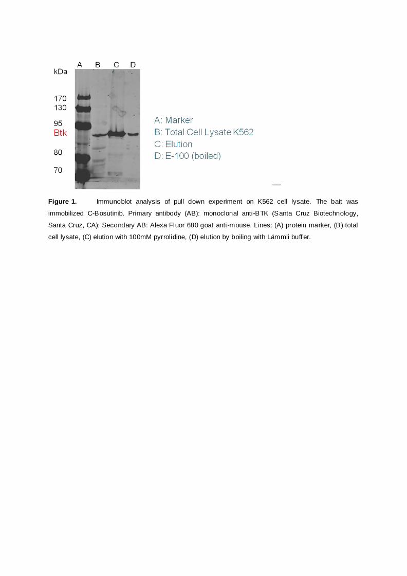
__
Figure 1. Immunoblot analysis of pull down experiment on K562 cell lysate. The bait was
immobilized C-Bosutinib. Primary antibody (AB): monoclonal anti-BTK (Santa Cruz Biotechnology,
Santa Cruz, CA); Secondary AB: Alexa Fluor 680 goat anti-mouse. Lines: (A) protein marker, (B) total
cell lysate, (C) elution with 100mM pyrrolidine, (D) elution by boiling with Lämmli buffer.

Acknowledgments
The authors would like to thank U. Rix and N. Fernbach (CeMM, Center for Molecular
Medicine, Vienna) for experimental support and K. Bennett (CeMM) for the Orbitrap mass
spectrometry analysis and G. Superti-Furga for the stimulating discussion in the course of
this project. This work was funded by the Austrian Proteomics Platform, part of the GEN-AU
program of the Austrian Federal Ministry of Science and Research.
References
1. Landi, F., et al., Synthesis and application of a new cleavable linker for "click"-based affinity chromatography. Organic & Biomolecular Chemistry, 2010. 8(1): p. 56-59.
2. Ovaa, H. and F. van Leeuwen, Chemical Biology Approaches to Probe the Proteome. Chembiochem, 2008. 9(18): p. 2913-2919.
3. Santala, V. and P. Savirantal, Affinity-independent elution of antibody-displaying phages using cleavable DNA linker containing streptavidin beads. Journal of Immunological Methods, 2004. 284(1-2): p. 159-163.
4. Verhelst, S.H.L., M. Fonovic, and M. Bogyo, A mild chemically cleavable linker system for functional proteomic applications. Angewandte Chemie-International Edition, 2007. 46(8): p. 1284-1286.
5. Cho, S., et al., Microbead-based affinity chromatography chip using RNA aptamer modified with photocleavable linker. Electrophoresis, 2004. 25(21-22): p. 3730-3739.
6. Foettinger, A., A. Leitner, and W. Lindner, Selective enrichment of tryptophan-containing peptides from protein digests employing a reversible derivatization with malondialdehyde and solid-phase capture on hydrazide beads. Journal of Proteome Research, 2007. 6(9): p. 3827-3834.
7. Foettinger, A., et al., Reaction of the indole group with malondialdehyde: Application for the derivatization of tryptophan residues in peptides. Bioconjugate Chemistry, 2007. 18(5): p. 1678-1683.
8. Teuber, H.J., O. Glosauer, and U. Hochmuth, Ein Neues Kondensationsprodukt Aus Tryptamin Und Acetessigaldehyd. Chemische Berichte-Recueil, 1964. 97(2): p. 557-&.
9. Dirksen, A., et al., Nucleophilic catalysis of hydrazone formation and transimination: Implications for dynamic covalent chemistry. Journal of the American Chemical Society, 2006. 128(49): p. 15602-15603.
10. Kunishima, M., et al., 4-(4,6-dimethoxy-1,3,5-triazin-2-yl)-4-methyl-morpholinium chloride: An efficient condensing agent leading to the formation of amides and esters. Tetrahedron, 1999. 55(46): p. 13159-13170.
11. Vultur, A., et al., SKI-606 (bosutinib), a novel Src kinase inhibitor, suppresses migration and invasion of human breast cancer cells. Mol Cancer Ther, 2008. 7(5): p. 1185-94.
12. Fernbach, N.V., et al., Acid elution and one-dimensional shotgun analysis on an Orbitrap mass spectrometer: an application to drug affinity chromatography. J Proteome Res, 2009. 8(10): p. 4753-65.
13. Remsing Rix, L.L., et al., Global target profile of the kinase inhibitor bosutinib in primary chronic myeloid leukemia cells. Leukemia, 2009. 23(3): p. 477-85.
14. Bantscheff, M., et al., Quantitative chemical proteomics reveals mechanisms of action of clinical ABL kinase inhibitors. Nat Biotechnol, 2007. 25(9): p. 1035-44.

15. Rix, U., et al., Chemical proteomic profiles of the BCR-ABL inhibitors imatinib, nilotinib, and dasatinib reveal novel kinase and nonkinase targets. Blood, 2007. 110(12): p. 4055-63.
16. Rix, U., et al., A comprehensive target selectivity survey of the BCR-ABL kinase inhibitor INNO-406 by kinase profiling and chemical proteomics in chronic myeloid leukemia cells. Leukemia. 24(1): p. 44-50.


_____________________________ Lebenslauf Martin Sturm 1
Curriculum Vitae
Persönliche Daten
Name Mag. Martin Georg Sturm
Adresse Schwendergasse 1b/2
1150 Wien, Österreich
Mobil 43 664 3828996
Arbeit 01 4277 52322
E-mail [email protected]
Nationalität Österreich
Geburtsdaten Klagenfurt, 31.10.1975
Familienstand ledig
Ausbildung 10/06-06/10 Dissertation am Institut für Analytische Chemie und
Lebensmittelchemie in der Arbeitgruppe von Prof. W. Lindner
Thema der Dissertation:
„ Preparation, implementation and evaluation of affinity
chromatographic tools for targeted proteome identification by liquid
chromatography – tandem mass spectrometry“
Die Dissertation wurde im Rahmen des APP II (Austrian Proteomics
Platform II) durchgeführt.
Das Dissertationsthema umfaßte die Entwicklung und Evaluierung von
porösen Metalloxidmaterialien zur selektiven
Phosphopeptidanreicherung, sowie Phosphopeptidanreicherung unter
Verwendung von Ionentauscher und Mixed Mode Phases im
Ionenaustauscher- und HILIC-Modus. Evaluierung (Quantifizierung
und Identifikation) mittels Flüssigkeitschromatographie und ESI-
_____________________________ Lebenslauf Martin Sturm 2
Massenspektrometrie (Agilent 1200 HPLC System gekoppelt mit
Applied Biosystems 4000 QTRAP, Agilent 1100 Series nano LC/MSD
Trap);
Weiters die Entwicklung eines chemisch spaltbaren Linkers gekoppelt
mit Wirkstoff an Agarose- oder Acrylamidbeads für Protein
Pulldownexperimente.
Evaluierung der Kopplungs- und Abspaltraten mittels LC-MS² im MRM-
Modus (Agilent 1100 HPLC system, Applied Biosystems 365).
Test des Konzepts mit Bosutinb-Wirkstoff an K562-Zellen am Center
für Molekulare Medizin (CeMM).
11/05-08/06 Mitarbeit in der Arbeitsgruppe von Prof. Dr. G. Ammerer am Max F.
Perutz Laboratories zur Weiterentwicklung eines genetischen
Proteininteraktionassays für Modellorganismus S.Cerevisiae mit dem
Ziel der Möglichkeit einer massenspektrometrischen Detektion.
Die praktische Arbeit beinhaltete die Konstruktion von Enzym/Substrat-
getaggten Zielproteingenkonstrukten in E. coli, Durchführung des
Assays und Proteinexpression in S. cerevisiae. Auswertung mittels
Western Blot und Massenspektrometrie.
09/05 Abschluss des Chemiestudiums mit dem Titel Magister der
Naturwissenschaften, mit Auszeichnung bestanden.
03/04-07/05 Diplomarbeit am Institut für spezifische Prophylaxe & Tropenmedizin
am Zentrum für Physiologie und Pathophysiologie der Medizinischen
Universität Wien unter der Betreuung von Ao. Univ. Prof. Dr. Ursula
Wiedermann-Schmidt zum Thema: „Mucosal vaccination against type I
allergy: a new treatment approach using lactic acid bacteria as delivery
systems for allergens“.
Praktische Tätigkeit beinhaltete Herstellung von Zellkulturen von
Mausmilzzellen, Evaluierung der Zellulären Cytokinantwort mittels
ELISA, Immunoglobulin-Subklassen-ELISA, Western Blot,
Proliferationskontrolle durch Thymidineinbau.
10/96-09/05 Studium der Chemie an der Universität Wien
Schwerpunkt: Biochemie, Analytik
_____________________________ Lebenslauf Martin Sturm 3
09/90-10/96 Höhere Technische Bundeslehranstalt, Abteilung für Elektrotechnik
09/82-06/87 Realgymnasium Lerchenfeldstrasse 22, 9020 Klagenfurt
Berufspraxis
Seit Sept. 2009 Mitarbeiter der Firma Österreichisches Forschunginstitut - OFI am
Projekt „COIN-QUANTUM“ zur massenspektrometrischen Detektion
und Quantifizierung von Kunststoffadditiven.
11/05-03/09 Wissenschaftlicher Mitarbeiter an der Universität Wien und
Lehrbeauftragter (Betreuung von Grund- und Fortgeschrittenenpraktika
im Bereich Biochemie und Analytik).
03/04-07/05 Bezahlte Diplomarbeit an der Medizinischen Universität Wien.
05/03-09/03 Fa. Fluka Production GmbH, CH-9471 Buchs/Schweiz;
Abteilung Qualitätskontrolle: Massenanalytische Gehaltsbestimmung
mittels Titration (NaOH, HCl, Komplexometrie);
UV-Spektroskopie (Gehaltsbestimmung, Qualitätsbeurteilung von
Farbstoffen und org. Lösungsmittels);
CHN-Analyse (Elementaranalyse) von organischen, anorganischen
und biochemischen Substanzen.
Während des Studiums Anstellungen im Gastgewerbe, Fixanstellung
bei Firma T-Mobile im Bereich Verkauf, Promotiontätigkeit für Firma
One, Siemens.
_____________________________ Lebenslauf Martin Sturm 4
Vorträge, Posterpräsentationen, Workshops
01/09 Vortrag am Doktorandenseminar des AK Separation Science in
Hohenroda zum Thema: „Porous SnO2 Microspheres a new Material
for Phosphopetide Enrichment“.
05/08 Vortrag am 4. ASAC JunganalytikerInnen Forum in Wien zum Thema:
„Porous SnO2 Microspheres a new Material for Phosphopetide
Enrichment“.
02/08 Teilnahme am Glycoproteomics Workshop am Institut für
Pharmazeutische Wissenschaften in Graz.
01/08 Posterpräsentation am 5. International Symposium of the Austrian
Proteomics Platform in Seefeld, Tirol.
06/07 Vortrag am 3. ASAC Junganalytikerforum in Linz
„SnO2 Microspheres als Material zur Phosphopeptidanreicherung“
05/07 Teilnahme am Workshop “Practical course about quantitative
Proteomics” am Institut für Molekulare Biotechnologie (IMBA) in Wien.
_____________________________ Lebenslauf Martin Sturm 5
Publikationen Sturm, M., A. Leitner, et al. (2008). "Tin dioxide microspheres as a promising material for
phosphopeptide enrichment prior to liquid chromatography-(tandem) mass spectrometry analysis." Advanced Functional Materials 18(16): 2381-2389.
Leitner, A., M. Sturm, et al. (2009). "Optimizing the performance of tin dioxide microspheres
for phosphopeptide enrichment." Analytica Chimica Acta 638(1): 51-57. Leitner, A., M. Sturm, et al. "Probing the phosphoproteome of HeLa cells using nanocast
metal oxide microspheres for phosphopeptide enrichment." Anal Chem 82(7): 2726-33.
Sturm, M., A. Leitner, W. Lindner (2010). „A chemically cleavable linker for immobilizing bait compounds in a target protein pull down concept”. Submitted to Bioconjugate Chemistry.
Stojanovic, Anja; Laemmerhofer, Michael; Kogelnig, Daniel; Schiesel, Simone; Sturm, Martin; Galanski, Markus; Krachler, Regina; Keppler, Bernhard K.; Lindner, Wolfgang. Analysis of quaternary ammonium and phosphonium ionic liquids by reversed-phase high-performance liquid chromatography with charged aerosol detection and unified calibration. Journal of Chromatography, A (2008), 1209(1-2), 179-187. Zusätzliche Qualifikationen Sprachen Englisch: fließend EDV-Kenntnisse Word, Exel, Powerpoint, Windows, Analyst, Chemstation. Führerschein Klasse B Hobbies Sport (Rennrad, Mountainbike, Badminton, Snowboard)
Literatur, Theater, Film, Reisen, Kochen.