Embed Size (px)
Citation preview
R A P P O R T
ENVIRHOM
Bioaccumulation of radionuclides in situations of
chronic exposure of ecosystems and members of
the public
Progress Report 2 covering the period June 2003 – September 2005
Report DRPH 2005-07 & DEI 2005-05
DIRECTION DE LA RADIOPROTECTION DE L’HOMME DIRECTION DE L’ENVIRONNEMENT ET DE L’INTERVENTION
List of contributors
ADAM Christelle ALONZO Frédéric BARESCUT Jean-Claude BARILLET Sabrina BONNEHORGNE M BONZOM Jean-Marc BUSSY C BOUST Dominique CLARAZ M CAMILLERI Virginie CAVALIE Isabelle DELISSEN O CHABROULLET Christophe DHIEUX B COLLE Claude DUBLINEAU I COPPIN Frédéric FRELON S DARCHEVILLE Olivia GRANDCOLAS L DELLA VEDOVA Claire Grison S DENISON Frank GUEGUEN Y DIAS Victor HOUPERT P FARCY Emilie FÉVRIER Laurelyne FIEVET Bruno FLORIANI Magali FOURNIER Elodie GARNIER-LAPLACE Jacqueline GERMAIN Pierre GEOFFROY Laure

GILBIN Rodolphe GOUZY Aurélien GRASSET Gaëla HENNER Pascale HURTEVENT Pierre LAGAUZERE Sandra LARNO Valérie LAROCHE Laetitia LESTAEVEL Ph MADOZ – ESCANDE Chantal MAUBERT C MARTIN-GARIN Arnaud PAQUET F MORELLO Marcel SER-Leroux K MORLON Hélène SOUIDI M ORJOLLET Daniel TAULAN M PERRIER Thomas PONCET-BONNARD Danielle PRADINE Catherine SIMON Olivier TISSANDIE E VOISEUX Claire
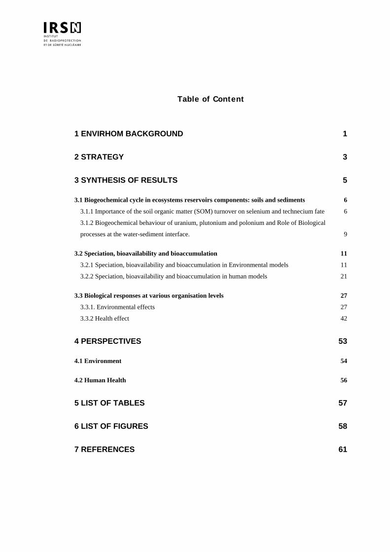
Table of Content
1 ENVIRHOM BACKGROUND 1
2 STRATEGY 3
3 SYNTHESIS OF RESULTS 5
3.1 Biogeochemical cycle in ecosystems reservoirs components: soils and sediments 6 3.1.1 Importance of the soil organic matter (SOM) turnover on selenium and technecium fate 6 3.1.2 Biogeochemical behaviour of uranium, plutonium and polonium and Role of Biological
processes at the water-sediment interface. 9
3.2 Speciation, bioavailability and bioaccumulation 11 3.2.1 Speciation, bioavailability and bioaccumulation in Environmental models 11 3.2.2 Speciation, bioavailability and bioaccumulation in human models 21
3.3 Biological responses at various organisation levels 27 3.3.1. Environmental effects 27 3.3.2 Health effect 42
4 PERSPECTIVES 53
4.1 Environment 54
4.2 Human Health 56
5 LIST OF TABLES 57
6 LIST OF FIGURES 58
7 REFERENCES 61
1 ENVIRHOM BACKGROUND
The main focuses of radioprotection were until now cases where radioactivity may be a distinguished source of stress. It is typically the case of workers and of critical groups around release sources. Improvement in release control has nearly suppressed new accumulation zones around nuclear plants since dispersal is much more optimized than before and since an important part of released fluxes is directed towards wastes repositories. This is of course favourable to close neighbours of plants but does not necessarily imply that the contamination of the rest of the world is also improving. For example, an increase of the radioactive background is already noticeable for iodine 129 even thousands of km away of main sources. Mining activity, which is a powerful way of releasing elements disperse in the environment many radionuclides that were before strongly trapped in rocks. The Chernobyl accident, dispersed 20 years ago in the most part of Europe about 1018 Bq of many radionuclides, including isotopes of iodine and cesium. All these releases contaminated both the environment and the humans. In addition, thousands of people are living in areas where there are high concentrations of natural alpha emitters such as uranium and radon. This situation is completely new. Instead of small populations, we have to protect populations of a regional or even of a world size. It is obviously completely unlikely to be part of thousands of very small critical groups but it is quite possible to be submitted to thousands of small risks simultaneously when each one concerns very large populations. This is why risk levels that can be tolerated from largely spread contaminants (such as what can be found in drinking water) are set to far lower values than what is considered as low in "usual" radioprotection. A whole life probability of premature death between 10-6 and 10-4 is considered as reasonably low in the case of largely spread toxicants. The equivalent of 10-5 in Sievert is 4µSv/y if we use the ICRP dose-effect relationship. Such levels are so far from those of situations used to validate the relationship that it is very doubtful that an extrapolation will be valid. Another evolution is the request to be protected against risks that are not only the so called "stochastic" risks such as cancer. Deterministic risks linked to small doses and small dose-rates have not yet been demonstrated but cannot be completely excluded. This point is particularly acute in areas contaminated by Chernobyl fall-out, where many cardiovascular, digestive, and respiratory diseases are linked, according to some authors, to the direct effects of contamination with cesium and iodine. For radionuclides, such as uranium, that are naturally present in the environment or released following a civil and/or a military use, similar questions about its health effects have few answers. Unfortunately, endpoints other than cancer have been poorly studied before. One of the objectives of the ENVIRHOM program are thus to bring new information on these radionuclide effects in terms of public health, focusing on the effects on the central nervous system, immune system and metabolisms. An emerging concern, in the case of environment is that it is not only considered today as a path to man in very straightforward scenario (such as plume to grass, grass to milk, milk to man), but it is also considered as something that need to be protected as well. Even if man is not put at risk immediately, a degradation of biota health and habitats may be a threat for the future. We do not need only knowing direct transfers of radioactive contaminants but we have also to know their real effects. We have also to be able to deal with complex processes that may happen with ageing of radioactive "tanks" and with recycling. We clearly lack such data since analytical experiments were, until now, mainly directed towards direct transfer of usual radioactive contaminants (Cs…) and more focused on transfer and contaminant repartition than on health effect. Field data are not a greater help since they are mainly related to usual contaminants (gamma emitters, rarely some alpha) and since they do not include observation of effects and observation of cofactors (other stressors, lack of nutrients, chemical conditions, overexploitation…). Hence ENVIRHOM has to fill these gaps in order to better assess the ecological risk. The last point, to be addressed as well for man as for environment, is the handling of mixed stresses among which radioactivity is not the dominant one. The consequences of the sum of stresses are certainly not the sum of consequences of each stress. A supplement of radioactivity will not have the same effect if it is added to one set of cofactors or to another one. It is a critical point in order to perform a global risk management. For all these reasons, ENVIRHOM will focus on consequences on man and environment of low and continuous exposures to radioactive stressors.
ENVIRHOM, Second Progress Report 1/64
There is a large amount of data about acute exposures such as those resulting from nuclear tests, from some medical exposures and from specific worker's exposures. But there is clearly a data gap when the exposures are low and chronic, especially when they are due to internal contamination. There is now a rather broad consensus on the fact that both the biological responses and the fighting mechanisms of living organisms against radioactive stressors are not the same in the case of important and in the case of weak exposure. It implies obviously that extrapolations of strong towards low doses have to be justified. Hence it requires specific experiments adapted to the case of chronic exposures to small quantities of radionuclides. There are some signs that the management of radioactive risk is at question today. ICRP is about to issue new recommendations and wishes to include biota protection. Dissidents scientists like ECRR are critical about risk evaluation and lastly, the non nuclear world is rapidly evolving toward a more cautious control (REACH European directive) of chemicals suspected to impact man or environmental health. It seems quite likely that regulations will evolve. To avoid using too large margins, it is important that ENVIRHOM can provide knowledge "on time".
ENVIRHOM, Second Progress Report 2/64
2 STRATEGY
The main objective of ENVIRHOM is to better assess real effects caused by chronic exposure to low levels of radioactive contaminants. This includes for example consequences on nervous system, immunity or metabolisms, consequences on reproduction, consequences on feeding processes and consequences on ecosystem productivity. Phenomena such as incorporation and elimination of the various radionuclides and of their various chemical forms have of course to be studied even if they are "intermediate" processes that do not necessarily involve a real damage to organisms. They have indeed a very direct influence on amounts of toxicants that can reach and stay in a target. But it is important to be aware that a change as well in Bq concentration or in cellular activity (gene activation, biomarkers…) may be only a physiological answer of an organism that stays fully functional. To concentrate on cases most likely to induce an effect, ENVIRHOM has set a priority on radionuclides that are suspected of accumulation in organisms. This phenomenon may indeed be responsible of local concentrations exceeding the background even when the source term is very small. For the same reason, ENVIRHOM has also set a priority on radionuclides that act not only with gamma rays. Due to the long action range of gamma rays, their energy deposit in local targets is less dependant of the source position than it is in the case of alpha and beta emitters. An uneven repartition of pure gamma emitters in organisms is smoothed as regards energy deposit and a small amount of these emitters is unable to build a local energy release above the background. It is hence logical to concentrate on other cases where there are more possibilities of local effects. The program was started in 2001 and uranium was chosen to test the methodology. Uranium possesses the suitable features for that: long life element, alpha emitters and uneven distribution in organisms. Beside, uranium is ubiquitous and may be present at very high concentrations in underground water of some areas (Finland, Canada, USA). Since the program was looking for "real" effects, various animal and vegetal models were necessary: rodents (rat, mouse) as a human model, and various organisms (algae, mollusks, fishes, plants …) as representative of environmental components. In addition to the biokinetic, transfer and speciation processes that are a common basis for all studies, a choice had to be made as regards endpoints. In the case of human models (rat, mouse), the chosen endpoints were the immune status of intestine, the genomic effects on kidneys, the metabolisms of drugs and vitamin D and the central nervous system performance. In the case of other models, behavioral effects were looked for: feeding strategies, ventilation of mollusks, reproduction, growth rate... Global effects such as evolution of algae populations were also studied. A full review of this period's work is included in the 2003 report to the scientific committee. It is very important to underline an important feature of ENVIRHOM: whenever possible, studies on whole organisms or functional tissues are privileged. The first two years of ENVIRHOM demonstrated clearly that a signal can be seen even as a consequence of moderate exposures. We did not see only signals related to cellular phenomena but we saw also modifications related to the chosen endpoints. It was also found that some simplifying hypothesis may be false. For example the simple biokinetic model assuming that the result of a continuous feeding is equivalent to the convolution of successive punctual inputs is not always true. When the input is constant, it should involve a steady state following an increase. Instead of that, a decrease has been observed in some cases (rats, crayfish). The second 2 years period (2004-2005) used the same strategy in a larger scale. As regards biota, the list of test organisms was extended (daphnia, insects…) and also the tested radionuclides (Se, Tc, Am). As regards human health, the preference was to study several functions involving different organs in the same integrated system (rodents). The list of studied functions was thus extended (behavior and sleep, neurotransmission, genomic effects, intestinal immune capacity, drug metabolism, Vitamin D metabolism,…). Currently, the ENVIRHOM program is the main experimental part of a container program devoted to chronic risks. This program will amount to 47 workers in 2006. In addition to ENVIRHOM, it covers also modeling activities, involvement in new trends of environmental protection and participating to closely linked international projects
ENVIRHOM, Second Progress Report 3/64
(ERICA). It employs 20 full time workers for the environmental part and 13 for the man health part. 9 PhD thesis are in progress and 5 post-docs. The general trend is to increase these numbers by redirecting toward ENVIRHOM searchers positions that where previously assigned to other radioprotection areas. The publication rate has been increased after a launching phase devoted to methodology settling and waiting of long experiments results. Since the beginning of 2004, 46 publications were issued.
ENVIRHOM, Second Progress Report 4/64
3 SYNTHESIS OF RESULTS
This section is devoted to present the key messages to come out of the research that has been performed in the ENVIRHOM program since its launching in late 2000. Our focus is on results obtained during the last 2-year period avoiding redundancy with the first synthesis that was presented to the last scientific committee (ENVIRHOM 2003). Chronic low-level exposure to radionuclides and induced biological responses at various organizational levels, from subcellular level to individual level (and to ecosystem level for the environmental aspect), has become a major issue in environmental and health science research and policy. From a scientific perspective, a series of lessons has been learnt during this period, some of them being specific to the element studied (e.g. uranium, selenium), others being relevant to more general issues concerned with the impact of radioactive substances on environmental and human health. The results we have selected within this report are presented in a consistent and integrated way whatever the radionuclide studied, the biological model and the organizational level. This presentation highlights the common points between the environmental and the human aspects, both on the methodological side and on the acquired knowledge side. Obviously, the two fields are also interesting per se and the outcome present specific operational issues in each domain i.e. environmental and human radioprotection, as explained below. For the environmental aspect, number of projects are devoted to the development of tools and associated knowledge to predict the fate and transport of radioactive substances in the “subsurface”. Actually, the lessons learnt on (bio)reactive transport processes within soils and sediments contribute to the global understanding of biogeochemical cycles in those ecosystems reservoirs. Once mobile, bioavailability will govern the link between speciation in the various exposure sources, bioaccumulation in living organisms, and induced biological responses. Further, as the majority of our research on bioavailability of radionuclides and induced biological responses has concerned aquatic organisms (e.g., phytoplankton, mollusks, crustaceans, fish), our lessons are derived primarily for aquatic ecosystems per se, even though they all almost apply to terrestrial ecosystems. At present, all our results mainly come out of laboratory studies under controlled conditions. The main goal was to help the understanding of involved mechanisms and as such, to contribute (i) answering a number of field issues around sources and long-term fates and (ii) improving ecological risk assessment methodology. Health aspects were evaluated by the mean of experimental contamination and follow-up of rodents (human model). The experiments carried out in this field aimed to verify if the biokinetic and toxicity data already established for acute exposures to radionuclides are transposable to situations of protracted exposure. The first radioelement studied was uranium, since it can be present at very high concentrations in underground waters of certain areas such as Finland, New Mexico in USA and Canada. The corresponding studies were therefore undertaken on rats and mice contaminated experimentally with uranium added to the drinking water. They were carried out in two parts, centered respectively on the comparison of biokinetics and on the biological effects of uranium after acute or chronic exposure. The first aspect comprised the description of the general kinetics of accumulation and excretion of radionuclides, of the influence of their speciation on their absorption, of the different ways of absorption in the gastro-intestinal tract and of their microdistribution after translocation. The second part was centered on the toxicology of uranium. It attempted to describe the effects of uranium on various organs (the kidneys, the liver, the intestine, the central nervous system, the lungs) and on some metabolisms such as that of the drugs, the vitamin D or the cholesterol. From an environmental policy perspective, results on chronic effects of low-level radionuclides exposure of living organisms may contribute to the derivation of more robust safe levels for ecosystems and their sub organizational levels. They also contribute to propose extrapolation rules to deal with the quantification of the main sources of uncertainties associated with these safe/acceptable criteria. Factors/key extrapolations issues that are known to influence the proposed values are numerous, the most important being extrapolations over time (acute vs. chronic), irradiation pathways (external vs. internal), taxa, level of biological organization, stressors (Garnier-Laplace, Gilek et al. 2004). From a human radioprotection perspective, results presented here demonstrated for the first time that biokinetics and toxicity of radionuclides after chronic exposure may not be simply extrapolated from data acquired after acute exposure (Paquet, Houpert et al. in press). Moreover, they showed that many deterministic effects may be induced after ingestion of small amounts of radionuclide (Houpert, Lestaevel et al. 2004; Houpert, Lestaevel et al. 2005; Lestaevel, Bussy et al. 2005; Souidi, Gueguen et al. 2005), although main concern in this range of dose was on cancer induction. These data are too sparse to be already incorporated in the current system of radioprotection but emphasize the interest to get more specific data for these particular -although widely represented- situations of exposure.
ENVIRHOM, Second Progress Report 5/64
3.1 BIOGEOCHEMICAL CYCLE IN ECOSYSTEMS RESERVOIRS COMPONENTS: SOILS AND SEDIMENTS
Improving radionuclides transport modeling from soils and sediments needs to determine to which extent natural geochemical cycle of major elements (e.g., C, H, O, N, S, P) combined with biological processes (microbial reactions, higher plant root influence, bioturbation by macrofauna) affect the mobility (and the speciation) of the radionuclides, as abiotic processes do (thermodynamic and sorption/desorption).
Natural soils and sediments constitute the most important storage reservoirs of the ecosystems for (ultra-)trace elements, such as radionuclides. In these complex systems, the behaviour of radionuclides greatly depends on the bio-physico-chemical properties of the media. Both mineral and abiotic organic matters interact with most trace elements by sorption and/or complexation processes (solid-phase interactions and aqueous chemistry). Moreover, some of these pollutants strongly interact directly or indirectly with the biological components (micro- and macro-organisms, plants and animals) leading to a significant evolution in time and space of their speciation, and therefore of their mobility in terms of transport (spatial displacement) and transfers among the various ecosystem components. Natural geochemical cycles of major elements (e.g., C, H, O, N, S, P, …) combined with biological processes (microbial reactions, higher plant root influence, bioturbation by macrofauna) constitute the most important factors to explain speciation, transport and transfers of radionuclides within ecosystems. The studies presented here aimed at gaining a true understanding of the relevancy and the relative significance of such processes in comparison with purely thermodynamic abiotic reactions and hydrodynamic transport processes. The operational objectives are (i) to determine the ‘best-representative’ model and associated parameters to predict radionuclides mobility in soils and sediments, (ii) to define their environmental domain of validity (e.g. physico-chemical properties of soils/sediments, time-scale of interest). The results selected especially focused on (1) the importance of the soil organic matter (SOM) turnover (e.g., humification and mineralisation processes) on the fate of long-lived radionuclides such as 79Se and 99Tc ; (2) the role of microbial processes on solid/solution interfaces in soils and sediments. For the latter, the emphasis is placed on our current researches on uranium and plutonium behaviours at the water-sediment interface in freshwaters and marine ecosystem respectively.
3.1.1 IMPORTANCE OF THE SOIL ORGANIC MATTER (SOM) TURNOVER ON SELENIUM AND TECHNECIUM FATE
Even though many selenium or technetium transformations in the soil environment are known to be microbially mediated, little is understood on the importance of soil microbial activity on Se and Tc mobility and bioavailability relative to pure abiotic thermodynamic reactions. 79Se and 99Tc are long-lived β-emitting fission products recovered in the nuclear wastes. Understanding their behaviours in soils is of major concern because of their biotransformation and potential toxicity to living organisms arising from long-term exposure. In the soil environment, it is generally assumed that the retention as well as the mobility of selenium is predominately governed by positively charged soil minerals, such as metal oxides (Balistrieri and Chao 1987; Su and Suarez 2000). However, there is a relative paucity of reliable data on Se-soil interactions and the reality and significance of Se-SOM interactions are still ambiguous. This is also the case for Tc which could exist as mobile and bioavailable Tc(VII) as well as Tc(IV) species. The latter leads to a large decrease of Tc solubility and transport (insoluble oxide and sulphur, complexation with SOM; (Nillson, Jensen et al. 1985)) although soluble Tc(IV)-complexes may appear (Wildung, Garland et al. 1986). A number of soil scientists have reported that various microbiological processes (direct or indirect bioreduction, microbial oxidation, co-precipitation, biomethylation, biosorption, etc.) are demonstrated or assumed to affect Tc and Se speciation, mobility and bioavailability, as pure abiotic sorption or complexation processes do (Lortie, Gould et al. 1992; Garbisu, Ishii et al. 1996). Even so, the resulting effect of the microbial activity on mobility and bioavailability of these oxyanions is still poorly understood.
ENVIRHOM, Second Progress Report 6/64
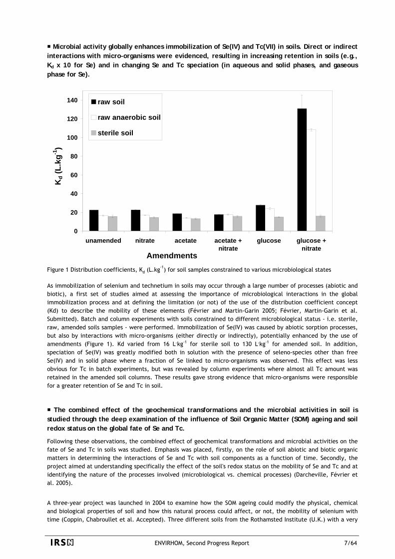
Microbial activity globally enhances immobilization of Se(IV) and Tc(VII) in soils. Direct or indirect interactions with micro-organisms were evidenced, resulting in increasing retention in soils (e.g., Kd x 10 for Se) and in changing Se and Tc speciation (in aqueous and solid phases, and gaseous phase for Se).
0
20
40
60
80
100
120
140
unamended nitrate acetate acetate +nitrate
glucose glucose +nitrate
Amendments
Kd (
L.kg
-1)
raw soil
raw anaerobic soil
sterile soil
Figure 1 Distribution coefficients, Kd (L.kg-1) for soil samples constrained to various microbiological states As immobilization of selenium and technetium in soils may occur through a large number of processes (abiotic and biotic), a first set of studies aimed at assessing the importance of microbiological interactions in the global immobilization process and at defining the limitation (or not) of the use of the distribution coefficient concept (Kd) to describe the mobility of these elements (Février and Martin-Garin 2005; Février, Martin-Garin et al. Submitted). Batch and column experiments with soils constrained to different microbiological status - i.e. sterile, raw, amended soils samples - were performed. Immobilization of Se(IV) was caused by abiotic sorption processes, but also by interactions with micro-organisms (either directly or indirectly), potentially enhanced by the use of amendments (Figure 1). Kd varied from 16 L.kg-1 for sterile soil to 130 L.kg-1 for amended soil. In addition, speciation of Se(IV) was greatly modified both in solution with the presence of seleno-species other than free Se(IV) and in solid phase where a fraction of Se linked to micro-organisms was observed. This effect was less obvious for Tc in batch experiments, but was revealed by column experiments where almost all Tc amount was retained in the amended soil columns. These results gave strong evidence that micro-organisms were responsible for a greater retention of Se and Tc in soil.
The combined effect of the geochemical transformations and the microbial activities in soil is studied through the deep examination of the influence of Soil Organic Matter (SOM) ageing and soil redox status on the global fate of Se and Tc.
Following these observations, the combined effect of geochemical transformations and microbial activities on the fate of Se and Tc in soils was studied. Emphasis was placed, firstly, on the role of soil abiotic and biotic organic matters in determining the interactions of Se and Tc with soil components as a function of time. Secondly, the project aimed at understanding specifically the effect of the soil's redox status on the mobility of Se and Tc and at identifying the nature of the processes involved (microbiological vs. chemical processes) (Darcheville, Février et al. 2005).
A three-year project was launched in 2004 to examine how the SOM ageing could modify the physical, chemical and biological properties of soil and how this natural process could affect, or not, the mobility of selenium with time (Coppin, Chabroullet et al. Accepted). Three different soils from the Rothamsted Institute (U.K.) with a very
ENVIRHOM, Second Progress Report 7/64
similar mineralogical composition, but with contrasted organic matter qualities and contents (1.0, 3.8 and 4.5 % of organic carbon for Roth1, Roth2 and Roth3, respectively) were chosen. Half of each soil was initially contaminated with radio-labelled selenium (75Se) while the other half was not (blank). The incubation conditions (constant temperature and moisture) were adjusted to increase the carbon turnover without disturbing the soil micro-organisms. The design of incubation chambers allowed us to maintain oxic condition by renewing the internal atmosphere with air. At the outlet of the chambers, different traps were used to monitor CO2 (SOM mineralisation) and Se-volatile species production (biomethylation). At different times of incubation, corresponding to different degradation-states of the SOM, the soils (contaminated or not) were sampled and characterized. The effect of time was studied according to a combination of methodological approaches to assess selenium mobility (sorption and desorption tests in batch and column experiments ; chemical and physical fractionation of Se within the soil components) with an unusual wealth of complementary analysis of the physical (granular size, aggregates stability, CEC, etc.), chemical (DOC, major organic and inorganic ions, etc.) and microbiological (biomass, community structure) properties of the soil, the SOM and the soil solution.
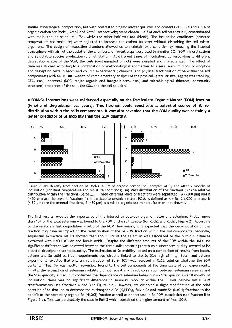
SOM-Se interactions were evidenced especially on the Particulate Organic Matter (POM) fraction (kinetic of degradation ca. years). This fraction could constitute a potential source of Se re-distribution within the soils components. It was also revealed that the SOM quality was certainly a better predictor of Se mobility than the SOM quantity.
0% 5% 10% 15%
A
B
C
D
T0
T=7m onths
a) 0% 5% 10% 15%
A
B
C
D
T0
T=7m onths
a) 0 5 10 15
A
B
C
DT0
T=7m onths
b) % % % %0 5 10 15
A
B
C
DT0
T=7m onths
b) % % % %
Figure 2 Size-density fractionation of Roth3 (4.9 % of organic carbon) soil samples at T0 and after 7 months of incubation (constant temperature and moisture conditions). (a) Mass distribution of the fractions ; (b) Se relative distribution within the fractions (Se/SeTotal). Three different kinds of fractions were separated : A (>200 µm) and B (> 50 µm) are the organic fractions ( the particulate organic matter, POM, is defined as A + B), C (>200 µm) and D (> 50 µm) are the mineral fractions, E (<50 µm) is a mixed organic and mineral fraction (not shown). The first results revealed the importance of the interaction between organic matter and selenium. Firstly, more than 10% of the total selenium was bound to the POM of the soil sample (for Roth2 and Roth3, Figure 2). According to the relatively fast degradation kinetic of the POM (few years), it is expected that the decomposition of this fraction may have an impact on the redistribution of the Se-POM fraction within the soil components. Secondly, sequential extraction results showed that about 60% of the selenium was associated to the humic substances extracted with NaOH (fulvic and humic acids). Despite the different amounts of the SOM within the soils, no significant difference was observed between the three soils indicating that humic substances quality seemed to be a better descriptor than its quantity. The estimation of Se mobility, based on a comparison of results from batch, column and Se solid partition experiments was directly linked to the Se-SOM high affinity. Batch and column experiments revealed that only a small fraction of Se (< 10%) was released in CaCl2 solution whatever the SOM contents. Thus, Se was mostly irreversibly bound to the soil components at the time scale of our experiments. Finally, the estimation of selenium mobility did not reveal any direct correlation between selenium releases and the SOM quantity either, but confirmed the dependence of selenium behaviour on SOM quality. Over 8 months of incubation, there was no significant difference in selenium mobility within the 3 soils despite initial SOM transformations (see fractions A and B in Figure 2-a). However, we observed a slight modification of the solid partition of Se that led to decrease the exchangeable-Se (K2HPO4), fulvic-Se and humic-Se (NaOH) fractions to the benefit of the refractory organic-Se (NaOCl) fraction as well as an increase in Se-POM association (see fraction B in Figure 2-b). This was particularly the case in Roth3 which contained the higher amount of fresh SOM.
ENVIRHOM, Second Progress Report 8/64
3.1.2 BIOGEOCHEMICAL BEHAVIOUR OF URANIUM, PLUTONIUM AND POLONIUM AND ROLE OF BIOLOGICAL PROCESSES AT THE WATER-SEDIMENT INTERFACE.
The current knowledge on biogeochemical cycle of uranium encourages further investigations on the role and the importance of bioturbation and microbial activity on mobility, in terms of exchange fluxes at the water-sediment interface.
Uranium is a potential toxic non-essential metallic radioelement that can be found at high concentrations in sediments, as high as several hundreds of milligrams per kilogram of dry sediments in particular areas (e.g., U-bearing mines). The geochemical behavior of uranium into the sediment, and then the presence of more or less toxic U-species, is determined by the local physico-chemical conditions (pH, redox status, pCO2, ionic strength, etc. – (Langmuir 1978; Davis, Payne et al. 2002; Fournier, Tran et al. 2004)and the microbial activity (Lovley, Phillips et al. 1991; Fredrickson, Zachara et al. 2000). A number of previous experimental work clearly demonstrated the reduction of U(VI) by the products of iron and sulphate respiration, and the important role of the chemical speciation of Fe(III) and U(VI) in microbial reduction processes (Behrends and Van Cappellen 2005). These bio-physico-chemical conditions vary as a function of sediment depth and contribute to the formation of U(+VI), U(+V) and U(+IV) species following a vertical sequence from oxic to anoxic zones (Froelich 1979). However, some disruptions may occur in this sequence due to oxygen incursions into the anoxic layers (e.g., re-suspension, bioturbation) leading to re-oxidation of reduced U-species. Hence, the activity of benthic organisms changes the local physico-chemical and microbial conditions in sediments via bioturbation processes. Bioturbation can be defined as the result of burrowing, feeding, irrigating, respiring and defecating activities from animal species living at the surface and/or within the sediment superficial layers. This activity causes sediment mixing and solute transport across the sediment-water interface. Sediment provides a habitat for various benthic macro-invertebrates which play an important role on the structure and the functioning of aquatic ecosystems. In addition to bioturbation, it is generally admitted that microbes, sheltered in biofilm matrix, contribute to a large extent to energy flow, nutrient and trace elements cycling.
An on-going project investigates the importance of biologically-driven processes in the global biogeochemical cycle of uranium at the water-sediment interface in freshwaters.
The link between bioturbation, biofilm, physico-chemical conditions of sediment, uranium transfers between sediment and the overlying water, and finally uranium bioavailability and toxicity, has been poorly studied. The goal of this project that was launched last year is to evaluate the relative importance of biologically driven processes to explain and properly assess the exchange fluxes of uranium at the interface in freshwaters. Experiments performed in indoors microcosms using simplified mixed natural biotope (water column and sediment), are implemented to address the following issues: (i) the influence of U on the microbial biofilm formation at the interface (structure, diversity, development duration); (ii) the impact of the benthic macro-invertebrates on sediment bio-geochemistry and thus on uranium distribution and fate in the sediment and the overlying water; this will be investigated while using species with contrasted modes of bioturbation: one gallery-diffusor species (Chironomus riparius) and one upward conveyor (tubificids); (iii) the bioavailability and the toxicity of uranium (from both sediment and overlying water source) for benthic and pelagic organisms; Bioindicators among bivalves and amphibian larvae will be used (Dreissena polymorpha, Corbicula fluminea, Xenopus laevis).
In situ investigations on the post depositional reactivity of Pu and Po evidence the prominent
role of the sulphides as temporary sink phases for these elements.
In the eastern Irish Sea, muddy sediments are confined to the Cumbrian Mud Patch, a large offshore mudflat lying parallel to the Cumbrian coast, off Sellafield, which acts as a very efficient sink for particle-reactive radionuclides, such as transuranics. These sediments are known to be subject to extensive physical (tide currents and waves) and biological (benthic organisms) reworking, as well as trawling activities. Together with post depositional evolution, these processes are liable to enhance the remobilization and relocation of the plutonium back to the water-column, its advection to distant sites, or its transfer through the food chain. Special attention was paid to both the determination of acid-volatile sulphides (AVS) and chromium reducible sulphides (CRS), and to their selective extraction by chemical leaching. Dissolved plutonium profiles in pore waters and plutonium solid
ENVIRHOM, Second Progress Report 9/64
partitioning suggest an active Pu uptake process by the most reactive sulphides (AVS). Very high and localised concentrations can be reached in these phases (up to 20000-500000 Bq of 239Pu per kg of AVS assumed to have a mean composition of FeS) potentially focusing its impact on biota, especially microorganisms. These reactive sulphides are liable to act as source phases if they are brought close to the interface by bioturbation or in contact with oxygenated seawater by burrowing and bioirrigation activity. This observation was not expected from previous investigations, which took less care of preservation of the anoxic character of the sediment and of resorption of plutonium during the extraction (Gouzy 2004). Preliminary studies of the post depositional reactivity of Po in the anoxic sediments of the Roads of Cherbourg yielded similar results with a subsurface peak activity of Po (ten fold seaweater activity) in pore water. Contrastingly, Po was found to be mainly associated with CRS sulphides (e.g., pyrite), a much less reactive phase, but no correlation was observed between CRS bound Po and CRS concentrations.
ENVIRHOM, Second Progress Report 10/64
3.2 SPECIATION, BIOAVAILABILITY AND BIOACCUMULATION
There is extensive evidence that neither total nor dissolved aqueous metal concentrations are good predictors of bioavailability and/or toxicity. Knowledge on radionuclide-organism interactions that result from a complex combination of biological and chemical processes, both governed by kinetics and thermodynamics, is essential to evidence the best exposure predictor of biological responses at various organizational levels.
The bioavailability of trace elements to the biota (uptake processes, bioaccumulation kinetics and the resulting extra- and intra-cellular distributions) results from a complex combination of biological and chemical processes, both governed by kinetics and thermodynamics. There is extensive evidence that neither total nor dissolved aqueous metal concentrations are good predictors of bioavailability and/or toxicity (Van Leeuwen and Köster 2004). A number of different modeling approaches to relate chemical speciation to toxicity or bioavailability of trace metals have been proposed and several comprehensive reviews of the development of these different modeling approaches have been published (e.g.,(Paquin, Gorsuch et al. 2002)). The first part of this chapter presents the information obtained on uranium(VI) and selenium(IV) with organisms representative of freshwater ecosystems (the unicellular green algae Chlamydomonas reinhardtii and the freshwater bivalve Corbicula fluminea) and of terrestrial ecosystem (the higher plant Phaseolus vulgaris), in order to enhance our knowledge previously obtained on the processes governing the uranium(VI)-algal cell interactions (ENVIRHOM 2003; Fortin, Dutel et al. 2004). Then, the second part of this chapter describes the uranium bioaccumulation in crustaceans and mammals tissues after chronic exposure and compares its biokinetics after acute and chronic exposure by ingestion. Finally, the third part points out the knowledge acquired on biotransformation, subcellular fractionation and microlocalisation of uranium and selenium in invertebrates and mammals.
3.2.1 SPECIATION, BIOAVAILABILITY AND BIOACCUMULATION IN ENVIRONMENTAL MODELS
3.2.1.1 Use of thermodynamic approach to support bioavailability models conceptions and implementations
Even though thermodynamic data used for predictive speciation modelling are critical as they are vital to ensure reliable speciation modelling and hence, robust bioavailability models development, poor attention has been drawn on their quality, their internal consistency and their associated uncertainties. However, a study carried out for U(VI) in solution demonstrated that speciation calculations based on mean value estimates of the thermodynamic values may result in predictions of a relatively low probability compared to an approach that considers the effects of uncertainty.
According to the free ion activity model and its derivates (Campbell 1995), the complexation of an element by a ligand in solution would be expected to decrease its bioavailability. On the other hand, competing ions can decrease the surface complexation reactions at the organism’s membrane surface. Until now, bioavailability models such as BLM, have been mainly tested for cationic metals with simple aqueous chemistry. In oxidized freshwaters and soils, uranium and selenium constitute good candidates to extend or not the BLM concepts to a cationic metal (U(VI) as uranyl ion UO2
2+) with a highly complex chemistry and to an element tending to forms oxyanions (Se(IV) as selenite ion SeO3
2-, Se(VI) as selenate ion SeO42-), respectively. These ions form different
complexes in solution according to the pH and to the presence of ligands, mainly for the uranyl ion (inorganic anions -carbonates, phosphates, nitrates- and organic ligands –citrate, EDTA…). To reliably perform experiments which aim at improving the understanding and modeling of radionuclide-organism interactions, two steps are needed. Firstly, there is a need for a high quality database to predict the aqueous speciation of each radionuclide to be able to relate the toxicity and bioavailability of the element to the physico-chemical parameters of the exposure medium (Fournier, Tran et al. 2004). Second, to investigate the effects of varying solution composition whilst keeping model values representative of the wide range of environmentally relevant physico-chemical parameters, a large testable composition domain is needed. Its complexity is specific to the chemical speciation
ENVIRHOM, Second Progress Report 11/64
complexity of the element being studied. Concerning U(VI), we checked first the quality of the used thermodynamic data (solubility, complex formation, redox and acidity constants), and the influence of thermodynamic values uncertainties on the species distribution outputs (by the speciation model JCHESS 2.0). A comprehensive review of the thermodynamic data relevant to environmentally relevant solutions composition domains allowed to compile for uranium(VI) in solution the best available estimates of thermodynamic parameters and to assign them uncertainty values (Denison and Garnier-Laplace 2005). The propagation of database parameter uncertainty has been assessed for aqueous and mineral equilibrium calculations of U(VI) by Monte Carlo and quasi-Monte Carlo simulations in simple inorganic solution compositions. The simulation output distributions of individual species’ concentrations vary greatly depending on the solution composition modeled, clearly demonstrating that conservative estimates of input uncertainty can result in considerable output uncertainty due to both the complexity of uranium solution chemistry and the system interdependencies. The lesson learnt from this study was that “classical” speciation calculations based on mean value estimates of the thermodynamic values may result in predictions of a relatively low probability compared to an approach that considers the effects of uncertainty.
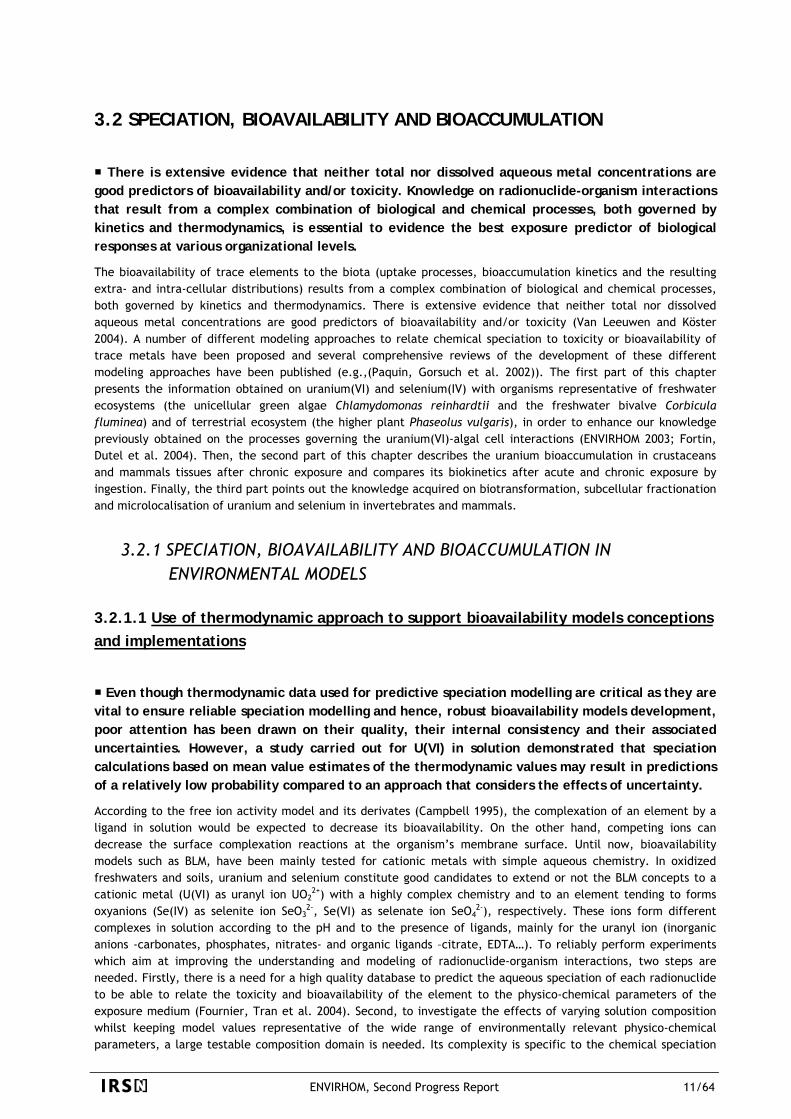
Figure 3 Uranium uptake kinetics for 10-7 mol dm-3 [UO2]T. a) internalised uranium concentrations as a function of exposure time, b) uranium uptake flux rates, error bars show ± 1 S.D.
ENVIRHOM, Second Progress Report 12/64
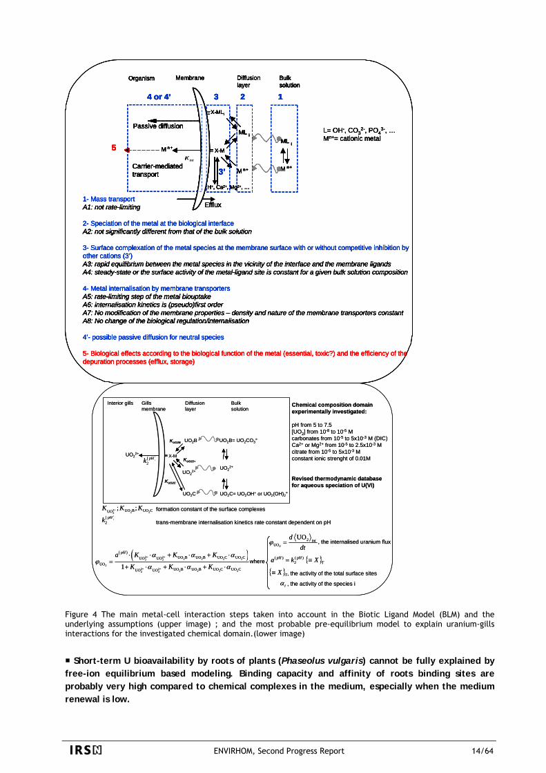
Short-term U uptake by excised gills of clam (Corbicula fluminea) can be explained by several equilibrium-based modeling, as well as by models considering changes in membrane properties. The only model flexible enough to represent the U(VI) – gills interaction for the complete solution composition domain considers the accumulation of 3 different uranium species and non-competitive pH dependent modulation of the transport system.
The effects of varying solution composition on the interactions between uranium(VI) and excised gills of the freshwater bivalve Corbicula fluminea showed a significant reduction in the uptake of uranium on increasing the concentrations of the uranium complexing ligands citrate and carbonate. Saturation kinetics as a function of uranium concentration at pH = 5.0 were observed, indicating that the uptake of uranium is a facilitated process, probably involving one or several trans-membrane transport systems (Figure 3). A relatively small change in the uptake of uranium was found as a function of pH (factor of ca. 2), despite the extremely large changes to the solution speciation of uranium within the range of pH investigated (5.0 – 7.5) (Fournier, Tran et al. 2004; Gilbin, Denison et al. 2005). A number of different equilibrium based bioavailability models were applied to the experimental results. This series of models was developed starting with the most restrictive hypotheses resulting in the simplest “pure” BLM models, progressively relaxing the physical and physiological hypotheses leading to increasingly complex and flexible models. The only model flexible enough to represent the U(VI) – gills interaction for the complete solution composition domain considers the accumulation of 3 different uranium species and non-competitive pH dependent modulation of the transport system (Figure 4). Ternary metal-ligand-transporter complexes involving hydroxide and carbonate ligands need to be considered to successfully encompass the large chemical composition domain space that has been specified. Moreover, a non-competitive modulation of the transporter system by proton concentration can successfully explain the observed pH dependence (i.e. the increase in uptake with increasing pH) (Fournier, Tran et al. 2004; Garnier-Laplace, Denison et al. 2005). Finally, this approach demonstrated that only some chemical species of aqueous U(+VI) need to be taken into account to assess properly the internal exposure of living organisms. These available species only represent a fraction of the total uranium in the contaminated medium that strongly varies with medium criteria such as pH and carbonates concentrations. As a result, bioconcentration factors often used to assess exposure of an organism to a given radionuclide are strongly dependent on a number of medium quality criteria which are specific to each element
ENVIRHOM, Second Progress Report 13/64
L= OH-, CO32-, PO4
3-, …Mn+= cationic metal
1- Mass transportA1: not rate-limiting
2- Speciation of the metal at the biological interfaceA2: not significantly different from that of the bulk solution
3- Surface complexation of the metal species at the membrane surface with or without competitive inhibition by other cations (3’)A3: rapid equilibrium between the metal species in the vicinity of the interface and the membrane ligandsA4: steady-state or the surface activity of the metal-ligand site is constant for a given bulk solution composition
4- Metal internalisation by membrane transportersA5: rate-limiting step of the metal biouptakeA6: internalisation kinetics is (pseudo)first orderA7: No modification of the membrane properties – density and nature of the membrane transporters constantA8: No change of the biological regulation/internalisation
4’- possible passive diffusion for neutral species
5- Biological effects according to the biological function of the metal (essential, toxic?) and the efficiency of thedepuration processes (efflux, storage)
M n+
ML i
X-M
ML i
M n+
Kint
M n +
4 or 4’
MembraneOrganism
Passive diffusion
Carrier-mediatedtransport
Efflux
X-MLi
1
Bulksolution
2
Diffusion layer
3
5
H+, Ca2+, Mg2+, …
3’
L= OH-, CO32-, PO4
3-, …Mn+= cationic metal
1- Mass transportA1: not rate-limiting
2- Speciation of the metal at the biological interfaceA2: not significantly different from that of the bulk solution
3- Surface complexation of the metal species at the membrane surface with or without competitive inhibition by other cations (3’)A3: rapid equilibrium between the metal species in the vicinity of the interface and the membrane ligandsA4: steady-state or the surface activity of the metal-ligand site is constant for a given bulk solution composition
4- Metal internalisation by membrane transportersA5: rate-limiting step of the metal biouptakeA6: internalisation kinetics is (pseudo)first orderA7: No modification of the membrane properties – density and nature of the membrane transporters constantA8: No change of the biological regulation/internalisation
4’- possible passive diffusion for neutral species
5- Biological effects according to the biological function of the metal (essential, toxic?) and the efficiency of thedepuration processes (efflux, storage)
M n+
ML i
X-M
ML i
M n+
Kint
M n +
4 or 4’
MembraneOrganism
Passive diffusion
Carrier-mediatedtransport
Efflux
X-MLi
1
Bulksolution
2
Diffusion layer
3
5
H+, Ca2+, Mg2+, …
3’ M n+
ML i
X-M
ML i
M n+
Kint
M n +
4 or 4’
MembraneOrganism
Passive diffusion
Carrier-mediatedtransport
Efflux
X-MLi
1
Bulksolution
2
Diffusion layer
3
5
H+, Ca2+, Mg2+, …
3’
UO2B= UO2CO3°
X-MUO22+
Bulksolution
Diffusion layer
Gillsmembrane
Interior gills
KUO22+
KUO2B
UO22+
UO22+
UO2B
formation constant of the surface complexes
trans-membrane internalisation kinetics rate constant dependent on pH
2
2 intUO
UOddt
ϕ =
where
2+ 2 22UO B UO CUO
; ;K K K
, the activity of the species iiα
( )2
pHk
( ) ( )2+ 2+ 2 2 2 22 2
22+ 2+ 2 2 2 22 2
UO B UO B UO C UO CUO UOUO
UO B UO B UO C UO CUO UO1
pHa K K K
K K K
α α αϕ
α α α
⋅ ⋅ + ⋅ + ⋅=
+ ⋅ + ⋅ + ⋅
, the internalised uranium flux
{ }TpHpH Xka ≡= )(2
)(
, the activity of the total surface sites{ }TX≡
UO2C= UO2OH+ or UO2(OH)2°UO2C
KUO2C
( )2
pHk
Chemical composition domainexperimentally investigated:
pH from 5 to 7.5[UO2] from 10-8 to 10-5 Mcarbonates from 10-5 to 5x10⁻³ M (DIC)Ca2+ or Mg2+ from 10-5 to 2.5x10⁻³ Mcitrate from 10-5 to 5x10⁻³ Mconstant ionic strenght of 0.01M
Revised thermodynamic databasefor aqueous speciation of U(VI)
L= OH-, CO32-, PO4
3-, …Mn+= cationic metal
1- Mass transportA1: not rate-limiting
2- Speciation of the metal at the biological interfaceA2: not significantly different from that of the bulk solution
3- Surface complexation of the metal species at the membrane surface with or without competitive inhibition by other cations (3’)A3: rapid equilibrium between the metal species in the vicinity of the interface and the membrane ligandsA4: steady-state or the surface activity of the metal-ligand site is constant for a given bulk solution composition
4- Metal internalisation by membrane transportersA5: rate-limiting step of the metal biouptakeA6: internalisation kinetics is (pseudo)first orderA7: No modification of the membrane properties – density and nature of the membrane transporters constantA8: No change of the biological regulation/internalisation
4’- possible passive diffusion for neutral species
5- Biological effects according to the biological function of the metal (essential, toxic?) and the efficiency of thedepuration processes (efflux, storage)
M n+
ML i
X-M
ML i
M n+
Kint
M n +
4 or 4’
MembraneOrganism
Passive diffusion
Carrier-mediatedtransport
Efflux
X-MLi
1
Bulksolution
2
Diffusion layer
3
5
H+, Ca2+, Mg2+, …
3’
L= OH-, CO32-, PO4
3-, …Mn+= cationic metal
1- Mass transportA1: not rate-limiting
2- Speciation of the metal at the biological interfaceA2: not significantly different from that of the bulk solution
3- Surface complexation of the metal species at the membrane surface with or without competitive inhibition by other cations (3’)A3: rapid equilibrium between the metal species in the vicinity of the interface and the membrane ligandsA4: steady-state or the surface activity of the metal-ligand site is constant for a given bulk solution composition
4- Metal internalisation by membrane transportersA5: rate-limiting step of the metal biouptakeA6: internalisation kinetics is (pseudo)first orderA7: No modification of the membrane properties – density and nature of the membrane transporters constantA8: No change of the biological regulation/internalisation
4’- possible passive diffusion for neutral species
5- Biological effects according to the biological function of the metal (essential, toxic?) and the efficiency of thedepuration processes (efflux, storage)
M n+
ML i
X-M
ML i
M n+
Kint
M n +
4 or 4’
MembraneOrganism
Passive diffusion
Carrier-mediatedtransport
Efflux
X-MLi
1
Bulksolution
2
Diffusion layer
3
5
H+, Ca2+, Mg2+, …
3’ M n+
ML i
X-M
ML i
M n+
Kint
M n +
4 or 4’
MembraneOrganism
2
Diffusion layer
1
Bulksolution
3
Passive diffusion
Carrier-mediatedtransport
Efflux
X-MLi
5
H+, Ca2+, Mg2+, …
3’
UO2B= UO2CO3°
X-MUO22+
Bulksolution
Diffusion layer
Gillsmembrane
Interior gills
KUO22+
KUO2B
UO22+
UO22+
UO2B
formation constant of the surface complexes
trans-membrane internalisation kinetics rate constant dependent on pH
2
2 intUO
UOddt
ϕ =
where
2+ 2 22UO B UO CUO
; ;K K K
, the activity of the species iiα
( )2
pHk
( ) ( )2+ 2+ 2 2 2 22 2
22+ 2+ 2 2 2 22 2
UO B UO B UO C UO CUO UOUO
UO B UO B UO C UO CUO UO1
pHa K K K
K K K
α α αϕ
α α α
⋅ ⋅ + ⋅ + ⋅=
+ ⋅ + ⋅ + ⋅
, the internalised uranium flux
{ }TpHpH Xka ≡= )(2
)(
, the activity of the total surface sites{ }TX≡
UO2C= UO2OH+ or UO2(OH)2°UO2C
KUO2C
( )2
pHk
Chemical composition domainexperimentally investigated:
pH from 5 to 7.5[UO2] from 10-8 to 10-5 Mcarbonates from 10-5 to 5x10⁻³ M (DIC)Ca2+ or Mg2+ from 10-5 to 2.5x10⁻³ Mcitrate from 10-5 to 5x10⁻³ Mconstant ionic strenght of 0.01M
Revised thermodynamic databasefor aqueous speciation of U(VI)
Figure 4 The main metal-cell interaction steps taken into account in the Biotic Ligand Model (BLM) and the underlying assumptions (upper image) ; and the most probable pre-equilibrium model to explain uranium-gills interactions for the investigated chemical domain.(lower image)
Short-term U bioavailability by roots of plants (Phaseolus vulgaris) cannot be fully explained by free-ion equilibrium based modeling. Binding capacity and affinity of roots binding sites are probably very high compared to chemical complexes in the medium, especially when the medium renewal is low.
ENVIRHOM, Second Progress Report 14/64
Short term (5 hrs) kinetic bioaccumulation studies in non renewed solution had shown linear and non-saturating relationships between U content of bean roots and U total in solution along the whole U solubility range (up to µM (Laroche 2005)). Whatever the conditions, U is rapidly bound to the root and the maximum binding capacity, as measured through uptake studies with continuously renewed solution, is close to 80 µmol U.g-1
dw root, the young part of roots being the most active structures (maximum binding capacity at 180 µmol U.g-1
dw root). The effect of pH on root transfer factors could not be modeled on the unique basis of the free ion concentrations in the medium, nor the effect of competing cations (Ca2+ content from 0.1 to 5mM) showed no competition with uranyl ions for binding sites. In the same way, by increasing the concentration of known U ligand, phosphates (0-15 µM) and citrate (0-10 µM), U root uptake was not affected. These results suggest that either other species of U than uranyl ions are bioavailable, or more probably uranium(VI) complexes are labile towards the high affinity of bean root tissues that depletes the medium from its uranyl ions. These results are therefore in accordance with other results on the uptake of trace metals in soil-plant systems where models based on equilibrium with free ion seems not adequate for plants (Smolders and McLaughlin 1996; Smolders and McLaughlin 1996).
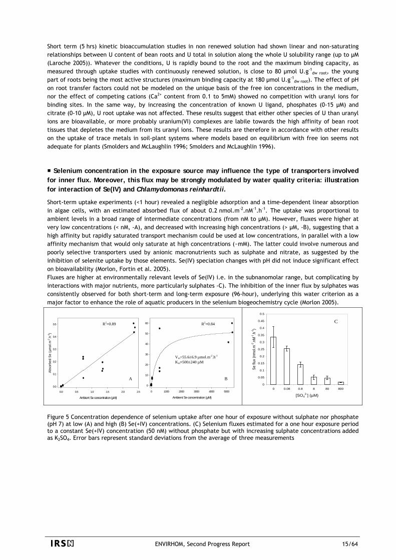
Selenium concentration in the exposure source may influence the type of transporters involved for inner flux. Moreover, this flux may be strongly modulated by water quality criteria: illustration for interaction of Se(IV) and Chlamydomonas reinhardtii.
Short-term uptake experiments (<1 hour) revealed a negligible adsorption and a time-dependent linear absorption in algae cells, with an estimated absorbed flux of about 0.2 nmol.m-2.nM-1.h-1. The uptake was proportional to ambient levels in a broad range of intermediate concentrations (from nM to µM). However, fluxes were higher at very low concentrations (< nM, -A), and decreased with increasing high concentrations (> µM, -B), suggesting that a high affinity but rapidly saturated transport mechanism could be used at low concentrations, in parallel with a low affinity mechanism that would only saturate at high concentrations (~mM). The latter could involve numerous and poorly selective transporters used by anionic macronutrients such as sulphate and nitrate, as suggested by the inhibition of selenite uptake by those elements. Se(IV) speciation changes with pH did not induce significant effect on bioavailability (Morlon, Fortin et al. 2005). Fluxes are higher at environmentally relevant levels of Se(IV) i.e. in the subnanomolar range, but complicating by interactions with major nutrients, more particularly sulphates -C). The inhibition of the inner flux by sulphates was consistently observed for both short-term and long-term exposure (96-hour), underlying this water criterion as a major factor to enhance the role of aquatic producers in the selenium biogeochemistry cycle (Morlon 2005).
Ambient Se concentration (µM)
0.0 0.5 1.0 1.5 2.0 2.5
Abso
rbed
Se
(µm
ol.m
-2.h
-1)
0.0
0.1
0.2
0.3
0.4
0.5
Ambient Se concentration (µM)
0 1000 2000 3000 4000 5000
0
10
20
30
40
50
60
Vm=55.6±6.9 µmol.m-2.h-1
Km=500±240 µM
R2=0.84 R2=0.89
A B
0
0.05
0.1
0.15
0.2
0.25
0.3
0.35
0.4
0.45
0.5
0 0.08 0.8 8 80 800
[SO42-] (µM)
Se fl
ux (n
mol
.m-2
.nM
-1.h
-1)
C
Figure 5 Concentration dependence of selenium uptake after one hour of exposure without sulphate nor phosphate (pH 7) at low (A) and high (B) Se(+IV) concentrations. (C) Selenium fluxes estimated for a one hour exposure period to a constant Se(+IV) concentration (50 nM) without phosphate but with increasing sulphate concentrations added as K2SO4. Error bars represent standard deviations from the average of three measurements
ENVIRHOM, Second Progress Report 15/64
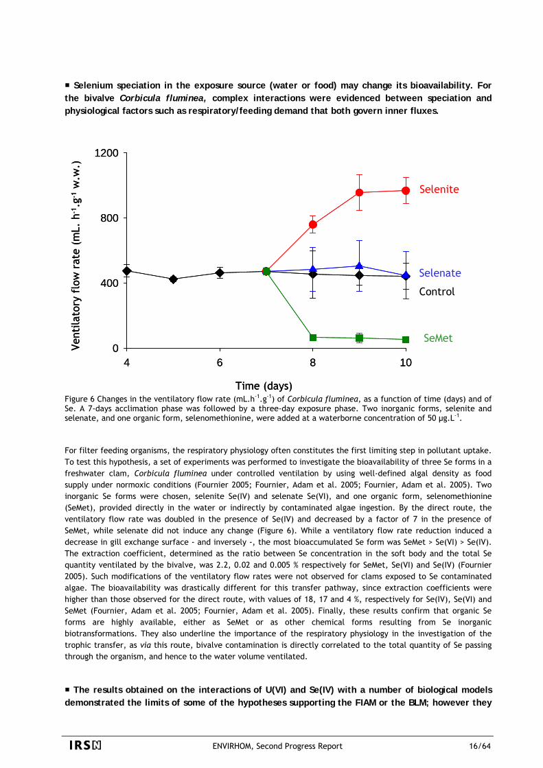
Selenium speciation in the exposure source (water or food) may change its bioavailability. For the bivalve Corbicula fluminea, complex interactions were evidenced between speciation and physiological factors such as respiratory/feeding demand that both govern inner fluxes.
4 6 8 100
400
800
1200
sélénite
témoinséléniate
SeMet
Vent
ilato
ry f
low
rat
e (m
L. h
-1.g
-1w
.w.)
Time (days)
Selenite
0
400
800
1200
Selenate
Control
SeMet
sélénite
4 6 8 10
témoinséléniate
SeMet
Vent
ilato
ry f
low
rat
e (m
L. h
-1.g
-1w
.w.)
Time (days)
Selenite
Selenate
Control
SeMet
Figure 6 Changes in the ventilatory flow rate (mL.h-1.g-1) of Corbicula fluminea, as a function of time (days) and of Se. A 7-days acclimation phase was followed by a three-day exposure phase. Two inorganic forms, selenite and selenate, and one organic form, selenomethionine, were added at a waterborne concentration of 50 µg.L-1. For filter feeding organisms, the respiratory physiology often constitutes the first limiting step in pollutant uptake. To test this hypothesis, a set of experiments was performed to investigate the bioavailability of three Se forms in a freshwater clam, Corbicula fluminea under controlled ventilation by using well-defined algal density as food supply under normoxic conditions (Fournier 2005; Fournier, Adam et al. 2005; Fournier, Adam et al. 2005). Two inorganic Se forms were chosen, selenite Se(IV) and selenate Se(VI), and one organic form, selenomethionine (SeMet), provided directly in the water or indirectly by contaminated algae ingestion. By the direct route, the ventilatory flow rate was doubled in the presence of Se(IV) and decreased by a factor of 7 in the presence of SeMet, while selenate did not induce any change (Figure 6). While a ventilatory flow rate reduction induced a decrease in gill exchange surface - and inversely -, the most bioaccumulated Se form was SeMet > Se(VI) > Se(IV). The extraction coefficient, determined as the ratio between Se concentration in the soft body and the total Se quantity ventilated by the bivalve, was 2.2, 0.02 and 0.005 % respectively for SeMet, Se(VI) and Se(IV) (Fournier 2005). Such modifications of the ventilatory flow rates were not observed for clams exposed to Se contaminated algae. The bioavailability was drastically different for this transfer pathway, since extraction coefficients were higher than those observed for the direct route, with values of 18, 17 and 4 %, respectively for Se(IV), Se(VI) and SeMet (Fournier, Adam et al. 2005; Fournier, Adam et al. 2005). Finally, these results confirm that organic Se forms are highly available, either as SeMet or as other chemical forms resulting from Se inorganic biotransformations. They also underline the importance of the respiratory physiology in the investigation of the trophic transfer, as via this route, bivalve contamination is directly correlated to the total quantity of Se passing through the organism, and hence to the water volume ventilated.
The results obtained on the interactions of U(VI) and Se(IV) with a number of biological models demonstrated the limits of some of the hypotheses supporting the FIAM or the BLM; however they
ENVIRHOM, Second Progress Report 16/64
gave principles for properly implementing bioavailability models for radionuclides on the basis of well-defined uptake experiments under controlled conditions.
Results showed that uranium(VI) and selenium(IV) uptake could not be entirely explained by classical approaches proposed for trace metals bioavailability modeling (Figure 4). Even if database uncertainty limits the predictive ability of thermodynamic equilibrium modeling, especially for elements exhibiting an extensive solution chemistry (e.g., uranium), understanding its effects can help to define the model’s validity domain and assist attempts to improve the model, for example by identifying sensitive parameters (Gilbin, Denison et al. 2005). For selenite (water concentration ca. µM), the high uptake in algae (probably linked with numerous and non specific transporters) showed little effect of speciation, and probably a competition with sulphates. The redox state of selenium had a stronger influence on the bioaccumulation, both in algae and bivalves. For uranium(VI), pH was found to strongly influence uranium uptake by algae and bivalve’s gills, but the change in free-ion concentration alone was not sufficient to explain the results. BLM-type models (including competitive UO2
2+/H+ sorption, or uptake of another species, e.g. uranyl hydroxide) could fit the data, but other models not only based on chemical equilibrium (e.g. variable transporter kinetics or binding affinities) could also explain the results. On another hand, in situations where the binding capacity and affinity of organism’s binding sites is much higher compared to the affinity of ions to the ligands in the medium (eg. in root/soil solution system), equilibriums are probably not reached. Results obtained showed that behavioral and/or physiological factors need also to be considered in addition to the physico-chemical conditions of the exposure medium. (e.g. for bivalves, valve closure and decrease of ventilatory flow rate in bivalves). Finally, on a general point of view, the obtained results allow us to assess the relative importance of a number of environmental and physiological factors to explain the link between speciation, bioavailability and bioaccumulation.
3.2.1.2. Biotransformation, subcellular fractionation and microlocalisation
Once internalized into the organism, the pollutant may be distributed according a variety of metabolism processes that can be element- and/or species- specific. Similarly to the bioavailable fraction of the total external concentration for the pollutant, only a fraction of the internal quantity is biologically active in terms of toxicity. The knowledge of internal compartmentalization is of major importance on a pragmatic point of view to assess the critical body residue, marking the transition between no effect and adverse effect. A project is on-going to identify the subcellular distribution and microlocation of uranium and selenium in various models (algae, plant and invertebrates).
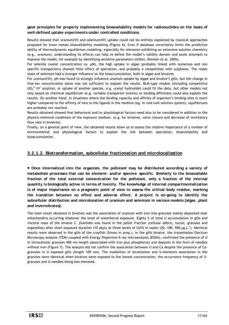
The main result obtained in bivalves was the association of uranium with iron into granules mainly deposited near mitochondria occurring whatever the level of waterborne exposure. Eighty % of total U accumulation in gills and visceral mass of the bivalve C. fluminea was found in the pellet fraction (cellular debris, nuclei, granules and organelles) after short exposure duration (10 days) at three levels of U(VI) in water (20, 100, 500 µg.L-1). Identical results were observed in the gills of the crayfish (Simon in prep.). In the gills bivalve, the transmission Electron Microscopy analysis (TEM) coupled with Energy Dispersive-X-ray microanalysis (EDAX), confirmed the presence of U in intracellular granules 400 nm length (associated with iron plus phosphorus) and deposits in the form of needles without iron (Figure 7). The analysis did not confirm the association between U and Ca despite the presence of Ca-granules in U exposed gills (length 100 nm). The modalities of localization and U-elements association in the granules were identical when bivalves were exposed to the lowest concentration, the occurrence frequency of U-granules and U-needles being less elevated.
ENVIRHOM, Second Progress Report 17/64
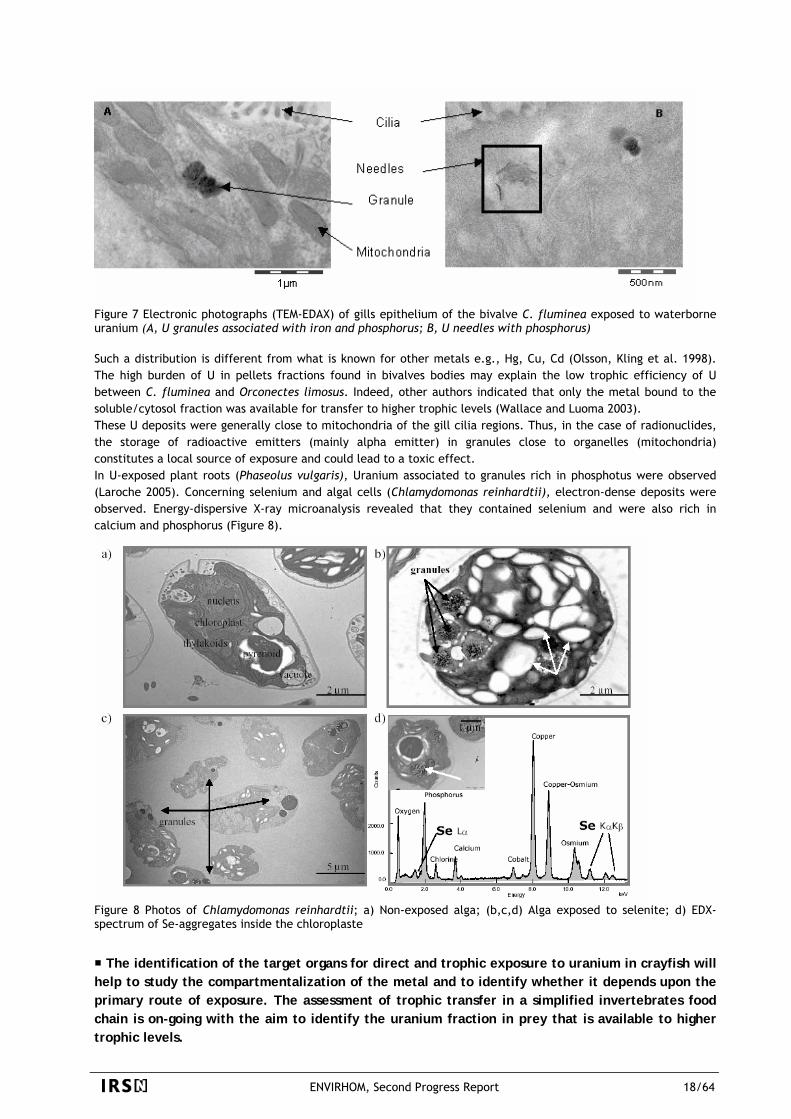
Figure 7 Electronic photographs (TEM-EDAX) of gills epithelium of the bivalve C. fluminea exposed to waterborne uranium (A, U granules associated with iron and phosphorus; B, U needles with phosphorus) Such a distribution is different from what is known for other metals e.g., Hg, Cu, Cd (Olsson, Kling et al. 1998). The high burden of U in pellets fractions found in bivalves bodies may explain the low trophic efficiency of U between C. fluminea and Orconectes limosus. Indeed, other authors indicated that only the metal bound to the soluble/cytosol fraction was available for transfer to higher trophic levels (Wallace and Luoma 2003). These U deposits were generally close to mitochondria of the gill cilia regions. Thus, in the case of radionuclides, the storage of radioactive emitters (mainly alpha emitter) in granules close to organelles (mitochondria) constitutes a local source of exposure and could lead to a toxic effect. In U-exposed plant roots (Phaseolus vulgaris), Uranium associated to granules rich in phosphotus were observed (Laroche 2005). Concerning selenium and algal cells (Chlamydomonas reinhardtii), electron-dense deposits were observed. Energy-dispersive X-ray microanalysis revealed that they contained selenium and were also rich in calcium and phosphorus (Figure 8).
Figure 8 Photos of Chlamydomonas reinhardtii; a) Non-exposed alga; (b,c,d) Alga exposed to selenite; d) EDX-spectrum of Se-aggregates inside the chloroplaste
The identification of the target organs for direct and trophic exposure to uranium in crayfish will help to study the compartmentalization of the metal and to identify whether it depends upon the primary route of exposure. The assessment of trophic transfer in a simplified invertebrates food chain is on-going with the aim to identify the uranium fraction in prey that is available to higher trophic levels.
ENVIRHOM, Second Progress Report 18/64
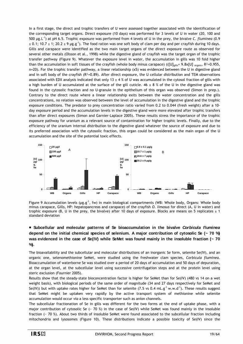
In a first stage, the direct and trophic transfers of U were assessed together associated with the identification of the corresponding target organs. Direct exposure (10 days) was performed for 3 levels of U in water (20, 100 and 500 µg.L-1) at pH 6.5. Trophic exposure was performed from 4 levels of U in the prey, the bivalve C. fluminea (0.9 ± 0.1; 10.7 ± 1; 20.2 ± 9 µg.g-1). The food ration was one soft body of clam per day and per crayfish during 10 days. Gills and carapace were identified as the two main target organs of the direct exposure route as observed for several other metals (Olsson et al., 1998) while the digestive gland of crayfish was the target organ of the trophic transfer pathway (Figure 9). Whatever the exposure level in water, the accumulation in gills was 10 fold higher than the accumulation in soft tissues of the crayfish (whole body minus carapace) (([U]gills= 9.8x[U] organs, R²=0.955, n=20). For the trophic transfer pathway, a linear relationship (x5) was evidenced between the U in digestive gland and in soft body of the crayfish (R²=0.89). After direct exposure, the U cellular distribution and TEM observations associated with EDX analysis indicated that only 13 ± 4 % of U was accumulated in the cytosol fraction of gills with a high burden of U accumulated at the surface of the gill cuticle. 46 ± 8 % of the U in the digestive gland was found in the cytosolic fraction and no U-granule in the epithelium of this organ was observed (Simon in prep.). Contrary to the direct route where a linear relationship exits between the water concentration and the gills concentrations, no relation was observed between the level of accumulation in the digestive gland and the trophic exposure conditions. The predator to prey concentration ratio varied from 0.2 to 0.044 (fresh weight) after a 10-day exposure period and the accumulation levels in the digestive gland were more elevated after trophic transfers than after direct exposures (Simon and Garnier-Laplace 2005). These results stress the importance of the trophic exposure pathway for uranium as a relevant source of contamination for higher trophic levels. Finally, due to the efficiency of the uranium internal distribution to the digestive gland whatever the source of exposure and due to its preferred association with the cytosolic fraction, this organ could be considered as the main organ of the U accumulation and the site of the potential toxic effects.
0
50
100
150
WB Organs Gills HP Carapace
[U] µg/g, fw
0.9 ± 0.1 µg/g
10.7 ± 1 µg/g
0
50
100
150
WB Organs Gills HP Carapace
[U] µg/g, fw
20 µg/l
19.6 ± 7 µg/g
20.2 ± 9 µg/g
100 µg/l A B
500 µg/l
Figure 9 Accumulation levels (µg.g-1, fw) in main biological compartments (WB: Whole body, Organs: Whole body minus carapace, Gills, HP: hepatopancreas and carapace) of the crayfish O. limosus for direct (A, U in water) and trophic exposure (B, U in the prey, the bivalve) after 10 days of exposure. Blocks are means on 5 replicates ± 1 standard deviation
Subcellular and molecular patterns of Se bioaccumulation in the bivalve Corbicula fluminea depend on the initial chemical species of selenium. A major contribution of cytosolic Se (~ 70 %) was evidenced in the case of Se(IV) while SeMet was found mainly in the insoluble fraction (~ 70 %).
The bioavailability and the subcellular and molecular distributions of an inorganic Se form, selenite Se(IV), and an organic one, selenomethionine SeMet, were studied using the freshwater clam species, Corbicula fluminea. Bioaccumulation of waterborne Se was studied over a period of 20 days of accumulation and 50 days of depuration, at the organ level, at the subcellular level using successive centrifugation steps and at the protein level using steric exclusion (Fournier 2005). Results show that the steady-state bioconcentration factor is higher for SeMet than for Se(IV) (480 vs 14 on a wet weight basis), with biological periods of the same order of magnitude (34 and 27 days respectively for SeMet and Se(IV)) but with uptake rates higher for SeMet than for selenite (7.5 vs 0.4 mL.g-1 w.w.d-1). These results suggest that SeMet might be uptaken very rapidly by the active transport system of methionine while selenite accumulation would occur via a less specific transporter such as anion channels. The subcellular fractionation of Se in gills was different for the two forms at the end of uptake phase, with a major contribution of cytosolic Se (~ 70 %) in the case of Se(IV) while SeMet was found mainly in the insoluble fraction (~ 70 %). About two thirds of insoluble SeMet were found associated to the subcellular fraction including mitochondria and lysosomes (Figure 10). These distributions indicate a possible toxicity of Se(IV) since the
ENVIRHOM, Second Progress Report 19/64
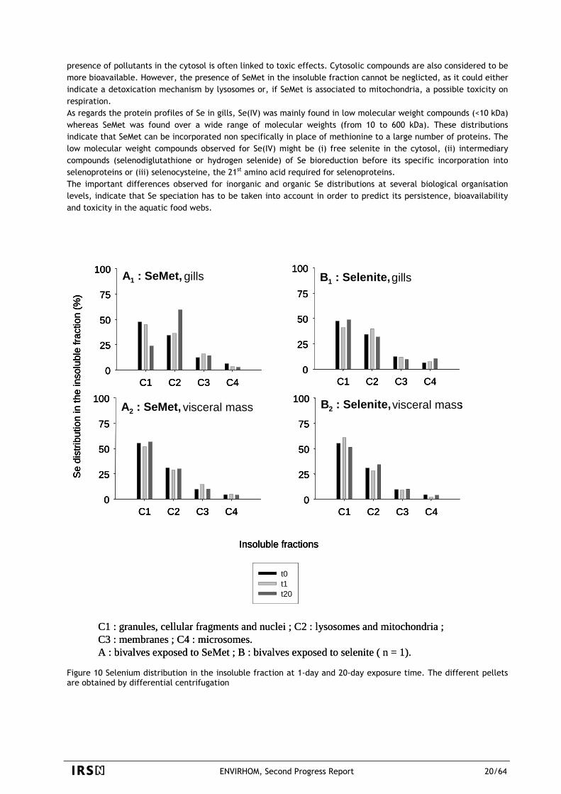
presence of pollutants in the cytosol is often linked to toxic effects. Cytosolic compounds are also considered to be more bioavailable. However, the presence of SeMet in the insoluble fraction cannot be neglicted, as it could either indicate a detoxication mechanism by lysosomes or, if SeMet is associated to mitochondria, a possible toxicity on respiration. As regards the protein profiles of Se in gills, Se(IV) was mainly found in low molecular weight compounds (<10 kDa) whereas SeMet was found over a wide range of molecular weights (from 10 to 600 kDa). These distributions indicate that SeMet can be incorporated non specifically in place of methionine to a large number of proteins. The low molecular weight compounds observed for Se(IV) might be (i) free selenite in the cytosol, (ii) intermediary compounds (selenodiglutathione or hydrogen selenide) of Se bioreduction before its specific incorporation into selenoproteins or (iii) selenocysteine, the 21st amino acid required for selenoproteins. The important differences observed for inorganic and organic Se distributions at several biological organisation levels, indicate that Se speciation has to be taken into account in order to predict its persistence, bioavailability and toxicity in the aquatic food webs.
C1 C2 C3 C40
25
50
75
100
t0t1t20
A2 : SeMet, visceral mass
Insoluble fractions
C1 C2 C3 C4
Se
dist
ribut
ion
in th
ein
solu
ble
fract
ion
(%)
0
25
50
75
100
A1 : SeMet, gills
C1 C2 C3 C40
25
50
75
100
B2 : Selenite, visceral mass
C1 C2 C3 C40
25
50
75
100
B1 : Selenite, gills
C1 : granules, cellular fragments and nuclei ; C2 : lysosomes and mitochondria ; C3 : membranes ; C4 : microsomes. A : bivalves exposed to SeMet ; B : bivalves exposed to selenite ( n = 1).
C1 C2 C3 C40
25
50
75
100
t0t1t20
A2 : SeMet, visceral mass
Insoluble fractions
C1 C2 C3 C4
Se
dist
ribut
ion
in th
ein
solu
ble
fract
ion
(%)
0
25
50
75
100
A1 : SeMet, gills
C1 C2 C3 C40
25
50
75
100
B2 : Selenite, visceral mass
C1 C2 C3 C40
25
50
75
100
B1 : Selenite, gills
C1 C2 C3 C40
25
50
75
100
t0t1t20
A2 : SeMet, visceral mass
Insoluble fractions
C1 C2 C3 C4
Se
dist
ribut
ion
in th
ein
solu
ble
fract
ion
(%)
0
25
50
75
100
A1 : SeMet, gills
C1 C2 C3 C40
25
50
75
100
B2 : Selenite, visceral mass
C1 C2 C3 C40
25
50
75
100
B1 : Selenite, gills
C1 : granules, cellular fragments and nuclei ; C2 : lysosomes and mitochondria ; C3 : membranes ; C4 : microsomes. A : bivalves exposed to SeMet ; B : bivalves exposed to selenite ( n = 1).
Figure 10 Selenium distribution in the insoluble fraction at 1-day and 20-day exposure time. The different pellets are obtained by differential centrifugation
ENVIRHOM, Second Progress Report 20/64
3.2.2 Speciation, bioavailability and bioaccumulation in human models
For mammals, data describing the accumulation and distribution of radionuclides after contamination come
mainly from experimental acute exposures of laboratory animals and follow-up of incidental exposures of humans. These data were compiled to form different specific models that could be used for dose calculations in humans. In case of protracted exposure, the models recommend an iterative use of the retention and excretion functions, i.e. to consider that a chronic exposure should be treated as a sum of independent acute intakes. Experiments performed and discussed in this section aimed at testing this hypothesis. This section presents new experimental data on retention of uranium after chronic intake that are compared to calculated values coming from an iterative use of an “acute” biokinetic model for rats. Additionally, data on transfer, bioaccumulation and distribution of uranium in different tissues are described, presenting unexpected results.
3.2.2.1 Uranium speciation
Uranium absorption in rats is not influenced by the initial chemical form in the water (exposure source). This is somewhat different from the current assumption that speciation influences the fractional absorption of radionuclides.
Studies of both the chemical speciation of uranium in water and its absorption through different segments of the gastrointestinal tract can enhance the knowledge of the mechanisms of its absorption in the body. Such studies are also helpful for improving the human alimentary tract model recommended by the International Commission on Radiological Protection (ICRP). The aim of this work was firstly to assess the influence of uranium speciation on its absorption from the gastrointestinal tract by using both computer speciation modeling and direct measurement of the fractional absorption in rats (Frelon, Houpert et al. 2005). At the same time, the main location of absorption throughout the entire gastrointestinal tract was determined by further in vivo and ex vivo studies in rats (Dublineau, Grison et al. 2005) (see next paragraph). Finally, the role of Peyer’s patches, specialized in antigen uptake, was assessed with ex vivo studies. Uranium speciation in water Five different samples of water, differing by their range of ionic composition were selected with or without carbonates, to obtain a very distinct initial speciation. Calculations of uranium speciation with modeling software were performed with uranium concentration of 168 µM (40 mg/L) in each sample of water. The results of this modelling show a different aqueous species distribution pattern for each mixture studied, which could then influence the absorption of this element. Uranium speciation and distribution through the gastro-intestinal (GI) tract Calculations of uranium speciation through the GI tract went on with modeling in mixtures of contaminated water plus saliva, and then with this last mixture plus gastric juice. The conclusion of this modeling study focuses on the minor influence that water composition seems to have on uranium final speciation in the stomach in these conditions. At the same time, ex vivo study on the distribution of uranium within saliva and a mixture of saliva and gastric juice was performed and gave similar conclusions, that is most of uranium is recovered with molecules of molecular weight >30kDa originating from the biological medium itself. This suggests that the ionic complexes entering the mouth are not stable enough and changed in reaction to the nature of the biological species of each compartment. In addition, the large molecular weight of the uranium recovering fraction suggests that the absorption would not occur at these levels. This last hypothesis was then confirmed by further in vivo absorption study. Uranium speciation and its fractional absorption from the gastrointestinal tract In vivo study of rat contamination via drinking contaminated water was run with 5 different water samples in order to assess the fractional absorption (f1) and confirm the first results found by modeling or ex vivo studies.
ENVIRHOM, Second Progress Report 21/64
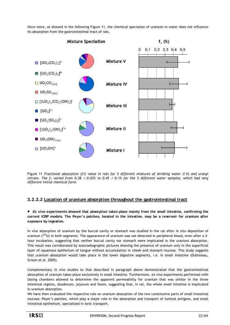
Once more, as showed in the following Figure 11, the chemical speciation of uranium in water does not influence its absorption from the gastrointestinal tract of rats.
Figure 11 Fractional absorption (f1) value in rats for 5 different mixtures of drinking water (I-V) and uranyl nitrate. The f1 varied from 0.38 ± 0.03% to 0.45 ± 0.1% for the 5 different water samples, which had very different initial chemical form.
3.2.2.2 Location of uranium absorption throughout the gastrointestinal tract Ex vivo experiments showed that absorption takes place mainly from the small intestine, confirming the
current ICRP models. The Peyer’s patches, located in the intestine, may be a reservoir for uranium after exposure by ingestion. In vivo absorption of uranium by the buccal cavity or stomach was studied in the rat after in situ deposition of uranium (233U) in both segments. The appearance of uranium was not detected in peripheral blood, even after a 2-hour incubation, suggesting that neither buccal cavity nor stomach were implicated in the uranium absorption. This result was corroborated by autoradiographic pictures showing the presence of uranium only in the superficial layer of squamous epithelium of tongue without accumulation in cheek and stomach mucosa. This study suggests that uranium absorption would take place in the lower digestive segments, i.e. in small intestine (Dublineau, Grison et al. 2005). Complementary in vivo studies to that described in paragraph above demonstrated that the gastrointestinal absorption of uranium takes place exclusively in small intestine. Furthermore, ex vivo experiments performed with Ussing chambers allowed to determine the apparent permeability for uranium that was similar in the three intestinal regions, duodenum, jejunum and ileum, suggesting that, in rat, the whole small intestine is implicated in uranium absorption. We have then evaluated the respective role on uranium absorption of the two constitutive parts of small intestinal mucosa: Peyer’s patches, which play a major role in the absorption and transport of luminal antigens, and small intestinal epithelium, specialized in ionic transport.
ENVIRHOM, Second Progress Report 22/64
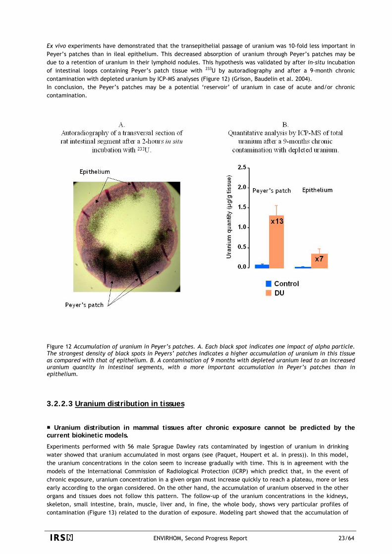
Ex vivo experiments have demonstrated that the transepithelial passage of uranium was 10-fold less important in Peyer’s patches than in ileal epithelium. This decreased absorption of uranium through Peyer’s patches may be due to a retention of uranium in their lymphoid nodules. This hypothesis was validated by after in-situ incubation of intestinal loops containing Peyer’s patch tissue with 233U by autoradiography and after a 9-month chronic contamination with depleted uranium by ICP-MS analyses (Figure 12) (Grison, Baudelin et al. 2004). In conclusion, the Peyer’s patches may be a potential ‘reservoir’ of uranium in case of acute and/or chronic contamination.
Figure 12 Accumulation of uranium in Peyer’s patches. A. Each black spot indicates one impact of alpha particle. The strongest density of black spots in Peyers’ patches indicates a higher accumulation of uranium in this tissue as compared with that of epithelium. B. A contamination of 9 months with depleted uranium lead to an increased uranium quantity in intestinal segments, with a more important accumulation in Peyer’s patches than in epithelium.
3.2.2.3 Uranium distribution in tissues
Uranium distribution in mammal tissues after chronic exposure cannot be predicted by the current biokinetic models.
Experiments performed with 56 male Sprague Dawley rats contaminated by ingestion of uranium in drinking water showed that uranium accumulated in most organs (see (Paquet, Houpert et al. in press)). In this model, the uranium concentrations in the colon seem to increase gradually with time. This is in agreement with the models of the International Commission of Radiological Protection (ICRP) which predict that, in the event of chronic exposure, uranium concentration in a given organ must increase quickly to reach a plateau, more or less early according to the organ considered. On the other hand, the accumulation of uranium observed in the other organs and tissues does not follow this pattern. The follow-up of the uranium concentrations in the kidneys, skeleton, small intestine, brain, muscle, liver and, in fine, the whole body, shows very particular profiles of contamination ( ) related to the duration of exposure. Modeling part showed that the accumulation of Figure 13
ENVIRHOM, Second Progress Report 23/64
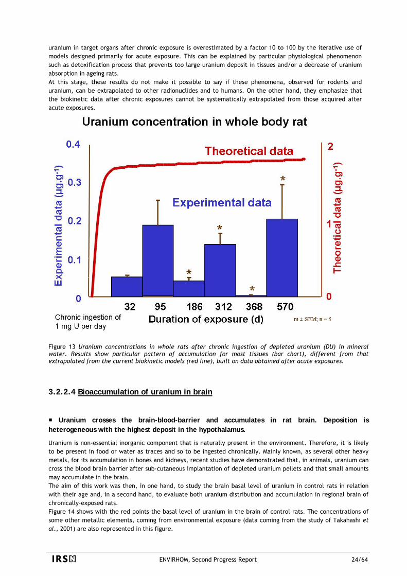
uranium in target organs after chronic exposure is overestimated by a factor 10 to 100 by the iterative use of models designed primarily for acute exposure. This can be explained by particular physiological phenomenon such as detoxification process that prevents too large uranium deposit in tissues and/or a decrease of uranium absorption in ageing rats. At this stage, these results do not make it possible to say if these phenomena, observed for rodents and uranium, can be extrapolated to other radionuclides and to humans. On the other hand, they emphasize that the biokinetic data after chronic exposures cannot be systematically extrapolated from those acquired after acute exposures.
Figure 13 Uranium concentrations in whole rats after chronic ingestion of depleted uranium (DU) in mineral water. Results show particular pattern of accumulation for most tissues (bar chart), different from that extrapolated from the current biokinetic models (red line), built on data obtained after acute exposures.
3.2.2.4 Bioaccumulation of uranium in brain
Uranium crosses the brain-blood-barrier and accumulates in rat brain. Deposition is heterogeneous with the highest deposit in the hypothalamus.
Uranium is non-essential inorganic component that is naturally present in the environment. Therefore, it is likely to be present in food or water as traces and so to be ingested chronically. Mainly known, as several other heavy metals, for its accumulation in bones and kidneys, recent studies have demonstrated that, in animals, uranium can cross the blood brain barrier after sub-cutaneous implantation of depleted uranium pellets and that small amounts may accumulate in the brain. The aim of this work was then, in one hand, to study the brain basal level of uranium in control rats in relation with their age and, in a second hand, to evaluate both uranium distribution and accumulation in regional brain of chronically-exposed rats. Figure 14 shows with the red points the basal level of uranium in the brain of control rats. The concentrations of some other metallic elements, coming from environmental exposure (data coming from the study of Takahashi et al., 2001) are also represented in this figure.
ENVIRHOM, Second Progress Report 24/64
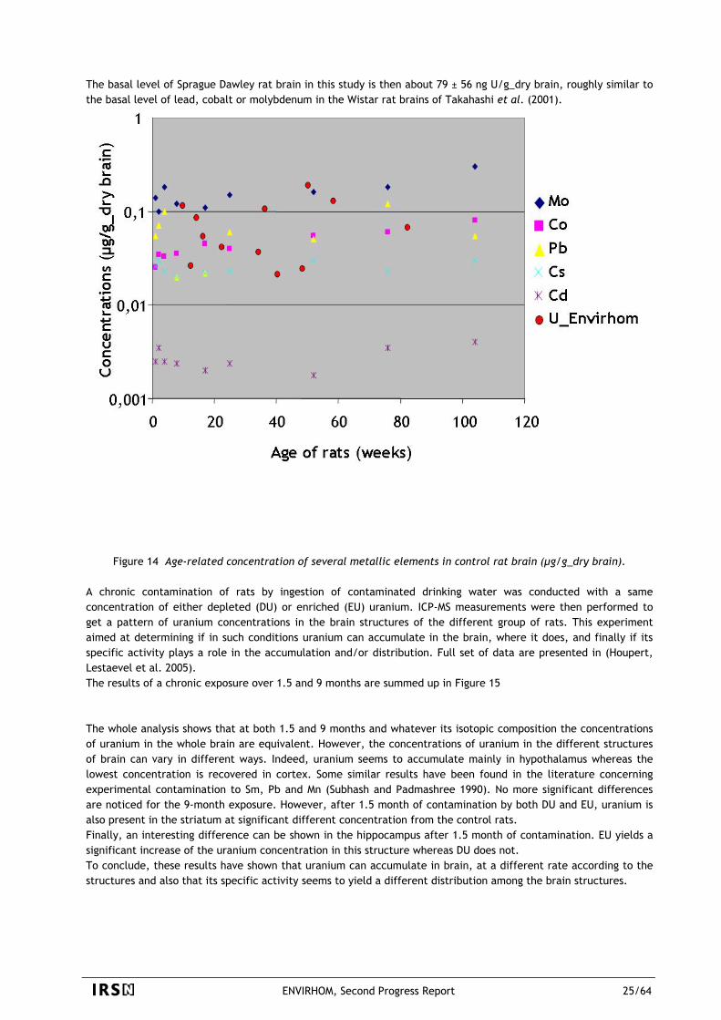
The basal level of Sprague Dawley rat brain in this study is then about 79 ± 56 ng U/g_dry brain, roughly similar to the basal level of lead, cobalt or molybdenum in the Wistar rat brains of Takahashi et al. (2001).
Figure 14 Age-related concentration of several metallic elements in control rat brain (µg/g_dry brain). A chronic contamination of rats by ingestion of contaminated drinking water was conducted with a same concentration of either depleted (DU) or enriched (EU) uranium. ICP-MS measurements were then performed to get a pattern of uranium concentrations in the brain structures of the different group of rats. This experiment aimed at determining if in such conditions uranium can accumulate in the brain, where it does, and finally if its specific activity plays a role in the accumulation and/or distribution. Full set of data are presented in (Houpert, Lestaevel et al. 2005). The results of a chronic exposure over 1.5 and 9 months are summed up in Figure 15 The whole analysis shows that at both 1.5 and 9 months and whatever its isotopic composition the concentrations of uranium in the whole brain are equivalent. However, the concentrations of uranium in the different structures of brain can vary in different ways. Indeed, uranium seems to accumulate mainly in hypothalamus whereas the lowest concentration is recovered in cortex. Some similar results have been found in the literature concerning experimental contamination to Sm, Pb and Mn (Subhash and Padmashree 1990). No more significant differences are noticed for the 9-month exposure. However, after 1.5 month of contamination by both DU and EU, uranium is also present in the striatum at significant different concentration from the control rats. Finally, an interesting difference can be shown in the hippocampus after 1.5 month of contamination. EU yields a significant increase of the uranium concentration in this structure whereas DU does not. To conclude, these results have shown that uranium can accumulate in brain, at a different rate according to the structures and also that its specific activity seems to yield a different distribution among the brain structures.
ENVIRHOM, Second Progress Report 25/64
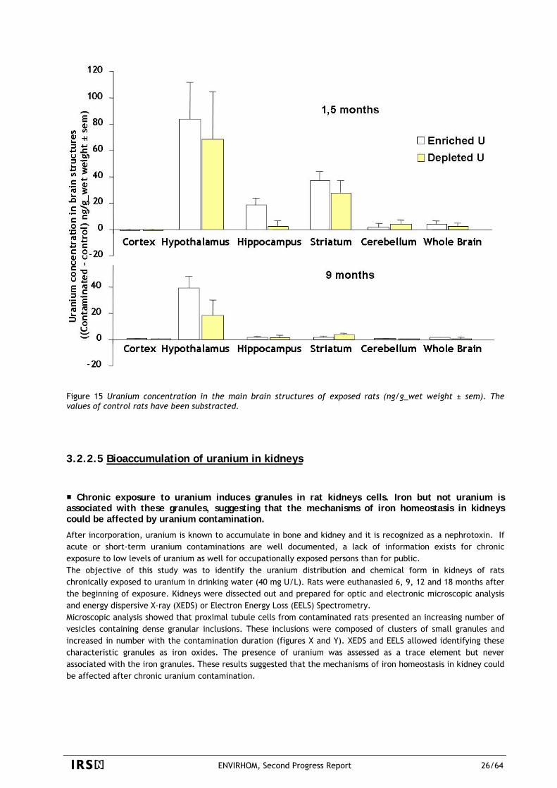
Figure 15 Uranium concentration in the main brain structures of exposed rats (ng/g_wet weight ± sem). The values of control rats have been substracted.
3.2.2.5 Bioaccumulation of uranium in kidneys
Chronic exposure to uranium induces granules in rat kidneys cells. Iron but not uranium is associated with these granules, suggesting that the mechanisms of iron homeostasis in kidneys could be affected by uranium contamination.
After incorporation, uranium is known to accumulate in bone and kidney and it is recognized as a nephrotoxin. If acute or short-term uranium contaminations are well documented, a lack of information exists for chronic exposure to low levels of uranium as well for occupationally exposed persons than for public. The objective of this study was to identify the uranium distribution and chemical form in kidneys of rats chronically exposed to uranium in drinking water (40 mg U/L). Rats were euthanasied 6, 9, 12 and 18 months after the beginning of exposure. Kidneys were dissected out and prepared for optic and electronic microscopic analysis and energy dispersive X-ray (XEDS) or Electron Energy Loss (EELS) Spectrometry. Microscopic analysis showed that proximal tubule cells from contaminated rats presented an increasing number of vesicles containing dense granular inclusions. These inclusions were composed of clusters of small granules and increased in number with the contamination duration (figures X and Y). XEDS and EELS allowed identifying these characteristic granules as iron oxides. The presence of uranium was assessed as a trace element but never associated with the iron granules. These results suggested that the mechanisms of iron homeostasis in kidney could be affected after chronic uranium contamination.
ENVIRHOM, Second Progress Report 26/64
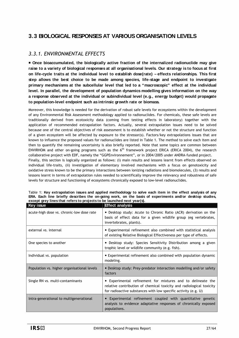
3.3 BIOLOGICAL RESPONSES AT VARIOUS ORGANISATION LEVELS
3.3.1. ENVIRONMENTAL EFFECTS
Once bioaccumulated, the biologically active fraction of the internalized radionuclide may give raise to a variety of biological responses at all organisational levels. Our strategy is to focus at first on life-cycle traits at the individual level to establish dose(rate) – effects relationships. This first step allows the best choice to be made among species, life-stage and endpoint to investigate primary mechanisms at the subcellular level that led to a “macroscopic” effect at the individual level. In parallel, the development of population dynamics modelling gives information on the way a response observed at the individual or subindividual level (e.g., energy budget) would propagate to population-level endpoint such as intrinsic growth rate or biomass.
Moreover, this knowledge is needed for the derivation of robust safe levels for ecosystems within the development of any Environmental Risk Assessment methodology applied to radionuclides. For chemicals, these safe levels are traditionally derived from ecotoxicity data (coming from testing effects in laboratory) together with the application of recommended extrapolation factors. Actually, several extrapolation issues need to be solved because one of the central objectives of risk assessment is to establish whether or not the structure and function of a given ecosystem will be affected by exposure to the stressor(s). Factors/key extrapolations issues that are known to influence the proposed values for radionuclides are listed in Table 1. The method to solve each item and then to quantify the remaining uncertainty is also briefly reported. Note that some topics are common between ENVIRHOM and other on-going programs such as the 6th framework project ERICA (ERICA 2004), the research collaborative project with EDF, namely the “GGPEnvironnement”, or in 2004/2005 under ANDRA-funded project. Finally, this section is logically organized as follows: (i) main results and lessons learnt from effects observed on individual life-traits, (ii) investigation of elementary involved mechanisms with a focus on genotoxicity and oxidative stress known to be the primary interactions between ionizing radiations and biomolecules, (3) results and lessons learnt in terms of extrapolation rules needed to scientifically improve the relevancy and robustness of safe levels for structure and functioning of ecosystems chronically exposed to low-level radionuclides.
Table 1: Key extrapolation issues and applied methodology to solve each item in the effect analysis of any ERA. Each line briefly describes the on-going work, on the basis of experiments and/or desktop studies, except grey lines that refers to projects to be launched next year(s). Key issue Effect analysis
acute-high dose vs. chronic-low dose rate Desktop study: Acute to Chronic Ratio (ACR) derivation on the basis of effect data for a given wildlife group (eg vertebrates, invertebrates, plants).
external vs. internal Experimental refinement also combined with statistical analysis of existing Relative Biological Effectiveness per type of effects.
One species to another Desktop study: Species Sensitivity Distribution among a given trophic level or wildlife community (e.g. fish).
Individual vs. population Experimental refinement also combined with population dynamic modeling.
Population vs. higher organisational levels Desktop study: Prey-predator interaction modelling and/or safety factors
Single RN vs. multi-contaminants Experimental refinement for mixtures and to delineate the relative contribution of chemical toxicity and radiological toxicity for radioactive substances with low specific activity (e.g. U)
Intra-generational to multigenerational Experimental refinement coupled with quantitative genetic analysis to evidence adaptative responses of chronically exposed populations.
ENVIRHOM, Second Progress Report 27/64
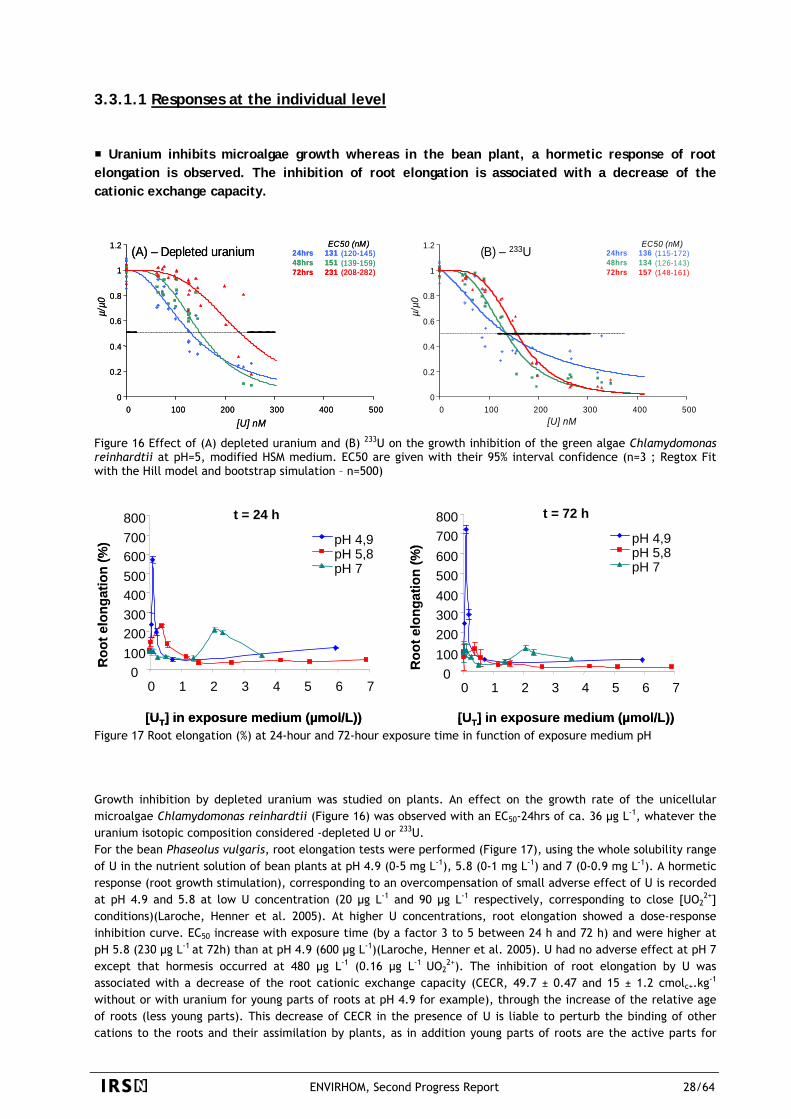
3.3.1.1 Responses at the individual level
Uranium inhibits microalgae growth whereas in the bean plant, a hormetic response of root elongation is observed. The inhibition of root elongation is associated with a decrease of the cationic exchange capacity.
0
0.2
0.4
0.6
0.8
1
1.2
0 100 200 300 400 500
µ/µ0
[U] nM
EC50 (nM)24hrs 131 (120-145)48hrs 151 (139-159)72hrs 231 (208-282)
0
0.2
0.4
0.6
0.8
1
1.2
0 100 200 300 400 500µ/
µ0[U] nM
EC50 (nM)24hrs 136 (115-172)48hrs 134 (126-143)
(A) – Depleted uranium (B) – 233U72hrs 157 (148-161)
0
0.2
0.4
0.6
0.8
1
1.2
0 100 200 300 400 500
µ/µ0
[U] nM
EC50 (nM)24hrs 131 (120-145)48hrs 151 (139-159)
0
0.2
0.4
0.6
0.8
1
1.2
0 100 200 300 400 500µ/
µ0[U] nM
EC50 (nM)24hrs 136 (115-172)48hrs 134 (126-143)
(A) – Depleted uranium (B) – 233U72hrs 231 (208-282) 72hrs 157 (148-161)
Figure 16 Effect of (A) depleted uranium and (B) 233U on the growth inhibition of the green algae Chlamydomonas reinhardtii at pH=5, modified HSM medium. EC50 are given with their 95% interval confidence (n=3 ; Regtox Fit with the Hill model and bootstrap simulation – n=500)
t = 24 h
0100200300400500600700800
0 1 2 3 4 5 6 7
[UT] in exposure medium (µmol/L))
Roo
t elo
ngat
ion
(%) pH 4,9
pH 5,8pH 7
t = 72 h
0100200300400500600700800
0 1 2 3 4 5 6 7
pH 4,9pH 5,8pH 7
Roo
t elo
ngat
ion
(%)
[UT] in exposure medium (µmol/L))
t = 24 h
0100200300400500600700800
0 1 2 3 4 5 6 7
[UT] in exposure medium (µmol/L))
Roo
t elo
ngat
ion
(%) pH 4,9
pH 5,8pH 7
t = 72 h
0100200300400500600700800
0 1 2 3 4 5 6 7
pH 4,9pH 5,8pH 7
Roo
t elo
ngat
ion
(%)
[UT] in exposure medium (µmol/L)) Figure 17 Root elongation (%) at 24-hour and 72-hour exposure time in function of exposure medium pH Growth inhibition by depleted uranium was studied on plants. An effect on the growth rate of the unicellular microalgae Chlamydomonas reinhardtii (Figure 16) was observed with an EC50-24hrs of ca. 36 µg L-1, whatever the uranium isotopic composition considered -depleted U or 233U. For the bean Phaseolus vulgaris, root elongation tests were performed (Figure 17), using the whole solubility range of U in the nutrient solution of bean plants at pH 4.9 (0-5 mg L-1), 5.8 (0-1 mg L-1) and 7 (0-0.9 mg L-1). A hormetic response (root growth stimulation), corresponding to an overcompensation of small adverse effect of U is recorded at pH 4.9 and 5.8 at low U concentration (20 µg L-1 and 90 µg L-1 respectively, corresponding to close [UO2
2+] conditions)(Laroche, Henner et al. 2005). At higher U concentrations, root elongation showed a dose-response inhibition curve. EC50 increase with exposure time (by a factor 3 to 5 between 24 h and 72 h) and were higher at pH 5.8 (230 µg L-1 at 72h) than at pH 4.9 (600 µg L-1)(Laroche, Henner et al. 2005). U had no adverse effect at pH 7 except that hormesis occurred at 480 µg L-1 (0.16 µg L-1 UO2
2+). The inhibition of root elongation by U was associated with a decrease of the root cationic exchange capacity (CECR, 49.7 ± 0.47 and 15 ± 1.2 cmolc+.kg-1 without or with uranium for young parts of roots at pH 4.9 for example), through the increase of the relative age of roots (less young parts). This decrease of CECR in the presence of U is liable to perturb the binding of other cations to the roots and their assimilation by plants, as in addition young parts of roots are the active parts for
ENVIRHOM, Second Progress Report 28/64
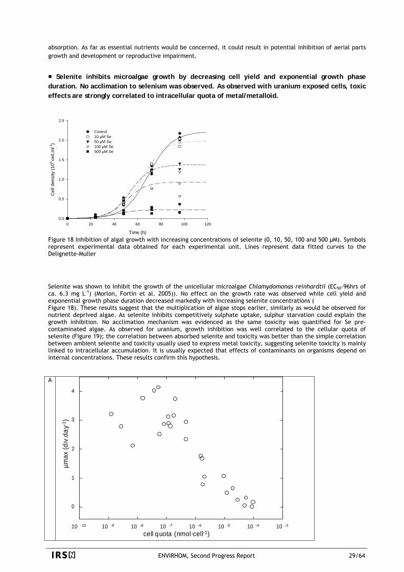
absorption. As far as essential nutrients would be concerned, it could result in potential inhibition of aerial parts growth and development or reproductive impairment.
Selenite inhibits microalgae growth by decreasing cell yield and exponential growth phase duration. No acclimation to selenium was observed. As observed with uranium exposed cells, toxic effects are strongly correlated to intracellular quota of metal/metalloid.
Time (h)
0 20 40 60 80 100 120
Cel
l den
sity
(106
cell.
ml-1
)
0.0
0.5
1.0
1.5
2.0
2.5
Control10 µM Se50 µM Se100 µM Se500 µM Se
Figure 18 Inhibition of algal growth with increasing concentrations of selenite (0, 10, 50, 100 and 500 µM). Symbols represent experimental data obtained for each experimental unit. Lines represent data fitted curves to the Delignette-Muller Selenite was shown to inhibit the growth of the unicellular microalgae Chlamydomonas reinhardtii (EC50-96hrs of ca. 6.3 mg L-1) (Morlon, Fortin et al. 2005)). No effect on the growth rate was observed while cell yield and exponential growth phase duration decreased markedly with increasing selenite concentrations ( Figure 18). These results suggest that the multiplication of algae stops earlier, similarly as would be observed for nutrient deprived algae. As selenite inhibits competitively sulphate uptake, sulphur starvation could explain the growth inhibition. No acclimation mechanism was evidenced as the same toxicity was quantified for Se pre-contaminated algae. As observed for uranium, growth inhibition was well correlated to the cellular quota of selenite (Figure 19); the correlation between absorbed selenite and toxicity was better than the simple correlation between ambient selenite and toxicity usually used to express metal toxicity, suggesting selenite toxicity is mainly linked to intracellular accumulation. It is usually expected that effects of contaminants on organisms depend on internal concentrations. These results confirm this hypothesis. A
cell quota (nmol·cell-1)10 -10 10 -9 10 -8 10 -7 10 -6 10 -5 10 -4 10 -3
0
1
2
3
4
µmax
(div
.day
-1)
ENVIRHOM, Second Progress Report 29/64

cell quota (amol.cell-1)
0 200 400 600 800 1000 1200 1400
estim
ated
max
imal
den
sity
(106 ce
ll.m
l-1)
0.0
0.2
0.4
0.6
0.8
1.0
1.2
1.4
1.6
1.8
2.0
0 µM10 µM20 µM30 µM40 µM50 µM
B
Figure 19 Algal growth inhibition of uranium (A- effect on maximal growth rate) and selenite (B- effect on estimated maximal densities) plotted against measured intracellular contents
Uranium associated to sediment induces adverse effects on survival, development and growth of the midge larvae.
A 10-day laboratory bioassay was performed exposing first instar larvae of Chironomus riparius to a gradient of U-sediment associated concentrations (0, 3, 6, 11, 24 µg g-1 d.w. – Figure 20). Effects of uranium on survival, development extended to mouthpart deformities, and growth on C. riparius larvae were assessed at the end of the exposure period (Dias, Vasseur et al. Submitted). The percentage of survival decreased when U concentration in treatment increased. Survival results showed significant negative effects from the 6 µg g-1 d.w. treatment (28.0 % of survival). An LC20 of 2.49 (95 % CI = 1.48 – 4.27) µg g-1 d.w., and an LC50 of 5.30 (95 % CI = 3.94 – 7.25) were estimated. Concerning the development, the mean head capsules width decreased with increasing U concentrations. This result is obviously related to an increase in the relative abundances of third instar larvae collected after 10 days. This is the first time that U may be found to induce a development delay in C. riparius. This is of particular interest since this development delay could have broader consequences on C. riparius reproduction (Postma and Davids 1995). The total deformity rate in surviving larvae after a 10-day exposure decreased with increasing U concentration in the sediment and associated water. Significant differences were detected for U concentrations ≥ 6 µg g-1 d.w. The reduction of deformity rate in fourth instar larvae in U treatments can be explained by the fact that for the higher concentration, mortality eliminated larvae that may have had deformities otherwise. A selection of the most tolerant larvae (not deformed) seemed to occur in treatments where U had a significative effect on survival. Finally, a significant inhibition growth was detected for U treatments ≥ 3 µg g-1 d.w. after a 10-day exposure. Results of larvae length showed that effect of U were only detectable for the last instar larvae. It could be explained by the fact that during the first three instars of Chironomus sp, development of somatic tissue dominates, gametic tissue development is being initiated only during the fourth instar (Postma and Davids 1995). Larvae must reach a critical somatic tissue mass before moulting and then after, energy is allocated to the development of reproductive tissue (Sibley, Benoit et al. 1997). Exposed to U-associated sediment, C. riparius larvae may need more time to reach the critical somatic tissue mass. This may explain both the inhibition growth and the delay in development observed after 10 days of exposure.
ENVIRHOM, Second Progress Report 30/64
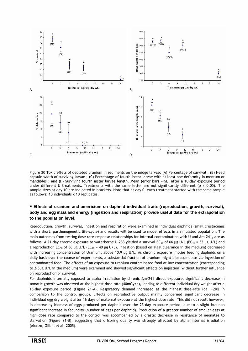
B A
C D Figure 20 Toxic effets of depleted uranium in sediments on the midge larvae: (A) Percentage of survival ; (B) Head capsule width of surviving larvae ; (C) Percentage of fourth instar larvae with at least one deformity in mentum or mandibles ; and (D) Surviving fourth instar larvae length. Mean (error bars = SE) after a 10-day exposure period under different U treatments. Treatments with the same letter are not significantly different (p ≤ 0.05). The sample sizes at day 10 are indicated in brackets. Note that at day 0, each treatment started with the same sample as follows: 10 individuals x 10 replicates.
Effects of uranium and americium on daphnid individual traits (reproduction, growth, survival), body and egg mass and energy (ingestion and respiration) provide useful data for the extrapolation to the population level.
Reproduction, growth, survival, ingestion and respiration were examined in individual daphnids (small crustaceans with a short, parthenogenetic life-cycle) and results will be used to model effects in a simulated population. The main outcomes from testing dose rate-response relationships for internal contamination with U and Am-241, are as follows. A 21-day chronic exposure to waterborne U-233 yielded a survival EC50 of 66 µg U/L (EC10 = 32 µg U/L) and a reproduction EC50 of 56 µg U/L (EC10 = 40 µg U/L). Ingestion (based on algal clearance in the medium) decreased with increasing concentration of Uranium, above 10.9 µg U/L. As chronic exposure implies feeding daphnids on a daily basis over the course of experiments, a substantial fraction of uranium might bioaccumulate via ingestion of contaminated food. The effects of an exposure to uranium contaminated food at low concentration (corresponding to 2-5µg U/L in the medium) were examined and showed significant effects on ingestion, without further influence on reproduction or survival. For daphnids internally exposed to alpha irradiation by chronic Am-241 direct exposure, significant decrease in somatic growth was observed at the highest dose rate (40mGy/h), leading to different individual dry weight after a 16-day exposure period (Figure 21-A). Respiratory demand increased at the highest dose-rate (ca. +20% in comparison to the control group). Effects on reproductive output mainly concerned significant decrease in individual egg dry weight after 16 days of maternal exposure at the highest dose rate. This did not result however, in decreasing biomass of eggs produced per daphnid over the 23-day exposure period, due to a slight but non significant increase in fecundity (number of eggs per daphnid). Production of a greater number of smaller eggs at high dose rate compared to the control was accompanied by a drastic decrease in resistance of neonates to starvation (Figure 21-B), suggesting that offspring quality was strongly affected by alpha internal irradiation (Alonzo, Gilbin et al. 2005).
ENVIRHOM, Second Progress Report 31/64
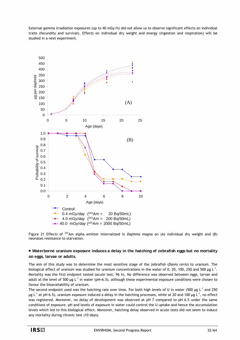
External gamma irradiation exposures (up to 40 mGy/h) did not allow us to observe significant effects on individual traits (fecundity and survival). Effects on individual dry weight and energy (ingestion and respiration) will be studied in a next experiment.
5th brood
0.00.10.20.30.40.50.60.70.80.91.0
0 2 4 6 8 10Age (days)
Pro
babi
lity
of s
urvi
val
Tm420 Bq200 Bq2000 Bq
Individual dry weight of daphnid
050
100150200250300350400450500
0 5 10 15 20 25Age (days)
µg p
er d
aphn
id Control0.4 mGy/h4.0 mGy/h40 mGy/h
(A)
(B)
Control0.4 mGy/day (241Am = 20 Bq/50mL)4.0 mGy/day (241Am = 200 Bq/50mL)
40.0 mGy/day (241Am = 2000 Bq/50mL)
Control0.4 mGy/day (241Am = 20 Bq/50mL)4.0 mGy/day (241Am = 200 Bq/50mL)
40.0 mGy/day (241Am = 2000 Bq/50mL)
Figure 21 Effects of 241Am alpha emitter internalized in Daphnia magna on (A) individual dry weight and (B) neonates resistance to starvation.
Waterborne uranium exposure induces a delay in the hatching of zebrafish eggs but no mortality on eggs, larvae or adults.
The aim of this study was to determine the most sensitive stage of the zebrafish (Danio rerio) to uranium. The biological effect of uranium was studied for uranium concentrations in the water of 0, 20, 100, 250 and 500 µg L-1. Mortality was the first endpoint tested (acute test, 96 h). No difference was observed between eggs, larvae and adult at the level of 500 µg L-1 in water (pH=6.5), although these experimental exposure conditions were chosen to favour the bioavailability of uranium. The second endpoint used was the hatching rate over time. For both high levels of U in water (500 µg L-1 and 250 µg L-1 at pH 6.5), uranium exposure induced a delay in the hatching processes, while at 20 and 100 µg L-1, no effect was registered. Moreover, no delay of development was observed at pH 7 compared to pH 6.5 under the same conditions of exposure. pH and levels of exposure in water could control the U uptake and hence the accumulation levels which led to this biological effect. Moreover, hatching delay observed in acute tests did not seem to induce any mortality during chronic test (10 days).
ENVIRHOM, Second Progress Report 32/64
It is essential that the method used to assess the risk to the environment from radionuclides should be consistent with that used for chemical substances. This consistency is all the more necessary as it is difficult to separate radiotoxicity and chemotoxicity in some particular cases of internal exposure (IRSN 2005).
Uranium is known to be both radiotoxic and chemotoxic, as a function of the considered isotopes. Such experiments, with a parallel study of depleted uranium and U-233, are in progress on a number of aquatic organisms. Some results were already obtained on freswater algae (see below). For the freshwater crustacean Daphnia magna, results obtained with U-233 (see in this section, 4th bullet.) will be compared to further experiments with depleted uranium; similar experiments on the fish Danio rerio showed some differences in oxidative stress status (depletion of glutathione and inhibition of glutathione peroxidase) between depleted uranium and depleted+U-233 exposed organisms, but only for early responses (SOD and catalase activity). In the future, effects (altered individual traits linked with subcellular effects on oxidative stress and DNA damages) specifically induced by ionizing radiations will be studied by external gamma rays exposure and internal alpha (Am-241) contamination. On the basis of simplified dose-rate calculations, it will be possible to better differentiate the effects linked to the radiation dose from the chemotoxicity of uranium. This will also need, for depleted uranium exposure, to measure bioaccumulation kinetics of the different isotopes of uranium (U-238, U-234, U-235) as well as its major decay elements (Th-232).
Preliminary results on freshwater algae showed that no additional toxicity due to the radiation of U-233 was observed, compared to the growth inhibition observed with depleted uranium.
Phytoplanktonic cells were exposed to different radiological doses while keeping constant the uranium molar concentration: e.g. for a given element concentration (chemical dose), replacing depleted U by U-233 to enhance the internally delivered radiological dose to organisms. We established relationships between uranium doses (depleted uranium and 233-U) and effect on the growth rate of the green algae Chlamydomonas reinhardtii. Uranium bioaccumulation was also monitored. Growth rate was measured both in classical batch (0-72hrs) and in continuous (turbidostat) cultures, the latter protocol allowing medium renewal to diminish exudates accumulation and speciation changes in the medium. No additional toxicity due to the radiation of U-233 was observed, compared to the growth inhibition observed with depleted uranium (Gilbin, Pradines et al. 2004). U in water did not play a significant role in the absorbed dose rate for each cell, while adsorbed fraction and absorbed fraction contributed equally to the dose rate. Delivered radiological doses were enhanced by a factor of 104 with U-233 compared to the dose rate delivered with depleted uranium. However, we did not observe a difference in toxicities, on the basis of growth inhibition, but the maximum dose rate absorbed by the cells was less than 0.4mGy/h. Studies performed at the subcellular level did not allow to see any difference between the effect of depleted uranium and U-233 on algae cells (Pradines, Wiktor et al. 2005).
3.3.1.2 Responses at the subcellular level: oxidative stress and genotoxicity
Global effects of uranium on the growth rate of green algae Chlamydomonas reinhardtii could not yet be related to oxidative stress (glutathione) or metal detoxifying (phytochelatins) production, but were consistent with photosynthetic activity inhibition (chlorophyll fluorescence).
There are actually few data dealing with ecotoxicological data of uranium on microalgae. Recent studies have shown that uranium toxicity on Chlorella sp. is highly dependent on pH and hardness (Franklin, Stauber et al. 2000; Charles, Markich et al. 2002), and that its bioavailability is also influenced by inorganic and organic ligands (Fortin, Dutel et al. 2004). To our knowledge, there is no data concerning uranium subcellular toxicity on microalgae. However, the induction of subcellular damage such as oxidative stress (Colle, Garnier-Laplace et al. 2001) could be the first earlier measurable signal for potential toxicity of uranium, related to chemical and/or radiological properties of the isotope (Gilbin and Garnier-Laplace 2004). Furthermore, it was proved that algae respond to heavy metal by induction of antioxidant enzymes, synthesis of low molecular weight compounds such as glutathione (Pinto, Sigaud-Kutner et al. 2003) and synthesis of small metal-binding proteins (phytochelatins) in
ENVIRHOM, Second Progress Report 33/64
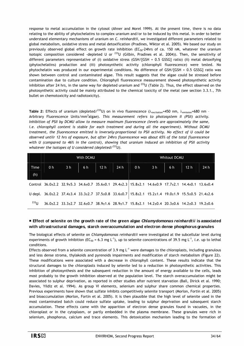
response to metal accumulation in the cytosol (Ahner and Morel 1999). At the present time, there is no data relating to the ability of phytochelatins to complex uranium and/or to be induced by this metal. In order to better understand elementary mechanisms of uranium on C. reinhardtii, we investigated different parameters related to global metabolism, oxidative stress and metal detoxification (Pradines, Wiktor et al. 2005). We based our study on previously observed global effect on growth rate inhibition (EC50-24hrs of ca. 150 nM, whatever the uranium isotopic composition considered -depleted U or 233U (Gilbin, Pradines et al. 2004)). Then, the sensitivity of different parameters representative of (i) oxidative stress (GSH/[GSH + 0.5 GSSG] ratio) (ii) metal detoxifying (phytochelatins) production and (iii) photosynthetic activity (chlorophyll fluorescence) were tested. No phytochelatin was produced in our experimental conditions. No difference of GSH/[GSH + 0.5 GSSG] ratio was shown between control and contaminated algae. This result suggests that the algae could be stressed before contamination due to culture condition. Chlorophyll fluorescence measurement showed photosynthetic activity inhibition after 24 hrs, in the same way for depleted uranium and 233U (Table 2). Thus, the effect observed on the photosynthetic activity could be mainly attributed to the chemical toxicity of the metal (see section 3.3.1., 7th bullet on chemotoxicity and radiotoxicity). Table 2: Effects of uranium (depleted/233U) on in vivo fluorescence (λexcitation=450 nm, λemission=680 nm – Arbitrary Fluorescence Units/mm3algae). This measurement refers to photosystem II (PSII) activity. Inhibition of PSII by DCMU allow to measure maximum fluorescence (levels are approximately the same, i.e. chlorophyll content is stable for each treatment and during all the experiment). Without DCMU treatment, the fluorescence emitted is inversely-proportional to PSII activity. No effect of U could be observed untilr 12 hrs of exposure, but after 24hrs fluorescence was about 65% of the total fluorescence with U (compared to 46% in the control), showing that uranium induced an inhibition of PSII activity whatever the isotopes of U considered (depleted/233U).
With DCMU Whitout DCMU
Time
(h)
0 h 3 h 6 h 12 h 24 h 0 h 3 h 6 h 12 h 24 h
Control 36.0±2.2 32.9±5.3 34.6±0.7 35.6±0.1 29.4±2.3 15.8±2.1 14.6±0.9 17.7±2.1 14.4±0.1 13.6±0.4
U depl. 36.0±2.2 37.4±3.4 33.3±2.7 37.5±0.8 33.6±0.7 15.8±2.1 15.2±1.4 19.0±1.9 15.5±0.5 21.4±2.6
233U 36.0±2.2 33.3±2.7 32.6±0.7 38.9±1.6 28.9±1.7 15.8±2.1 14.2±0.4 20.3±0.6 14.2±0.3 19.2±0.6
Effect of selenite on the growth rate of the green algae Chlamydomonas reinhardtii is associated with ultrastructural damages, starch overaccumulation and electron dense phosphorus granules
The biological effects of selenite on Chlamydomonas reinhardtii were investigated at the subcellular level during experiments of growth inhibition (EC50 = 6.3 mg L-1), up to selenite concentrations of 39.5 mg L-1, i.e. up to lethal conditions. Effects observed from a selenite concentration of 3.9 mg L-1 were damages to the chloroplasts, including granulous and less dense stroma, thylakoids and pyrenoids impairments and modification of starch metabolism (Figure 22). These modifications were associated with a decrease in chlorophyll content. These results indicate that the structural damages to the chloroplasts induced by selenite led to a reduction in photosynthetic activities. This inhibition of photosynthesis and the subsequent reduction in the amount of energy available to the cells, leads most probably to the growth inhibition observed at the population level. The starch overaccumulation might be associated to sulphur deprivation, as reported in other studies after nutrient starvation (Ball, Dirick et al. 1990; Davies, Yildiz et al. 1994). As group VI elements, selenium and sulphur share common chemical properties. Previous experiments have shown that sulfate inhibits competitively selenite transport (Morlon, Fortin et al. 2005) and bioaccumulation (Morlon, Fortin et al. 2005). It is then plausible that the high level of selenite used in the most contaminated batch could reduce sulfate uptake, leading to sulphur deprivation and subsequent starch accumulation. These effects came with the apparition of electron dense granules found in vacuoles, in the chloroplast or in the cytoplasm, or partly embedded in the plasma membrane. These granules were rich in selenium, phosphorus, calcium and trace elements. This detoxication mechanism leading to the formation of
ENVIRHOM, Second Progress Report 34/64
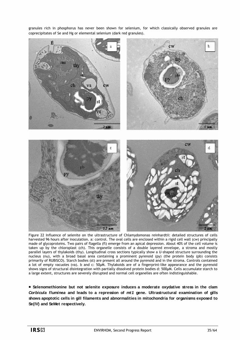
granules rich in phosphorus has never been shown for selenium, for which classically observed granules are coprecipitates of Se and Hg or elemental selenium (dark red granules).
Figure 22 Influence of selenite on the ultrastructure of Chlamydomonas reinhardtii: detailed structures of cells harvested 96 hours after inoculation. a: control. The oval cells are enclosed within a rigid cell wall (cw) principally made of glycoproteins. Two pairs of flagella (fl) emerge from an apical depression. About 40% of the cell volume is taken up by the chloroplast (ch). This organelle consists of a double layered envelope, a stroma and mostly parallel layers of thylakoids (thy). Longitudinal cross sections typically show a U-shaped structure surrounding the nucleus (nu), with a broad basal area containing a prominent pyrenoid (py) (the protein body (pb) consists primarily of RUBISCO). Starch bodies (st) are present all around the pyrenoid and in the stroma. Controls contained a lot of empty vacuoles (va). b and c: 50µM. Thylakoids are of a fingerprint-like appearance and the pyrenoid shows signs of structural disintegration with partially dissolved protein bodies d: 500µM. Cells accumulate starch to a large extent, structures are severely disrupted and normal cell organelles are often indistinguishable.
Selenomethionine but not selenite exposure induces a moderate oxydative stress in the clam Corbicula fluminea and leads to a repression of mt1 gene. Ultrastructural examination of gills shows apoptotic cells in gill filaments and abnormalities in mitochondria for organisms exposed to Se(IV) and SeMet respectively.
ENVIRHOM, Second Progress Report 35/64
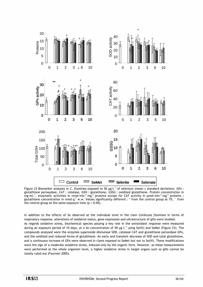
Control SeMet Selenite Selenate
0
5
10
15
20
0 1 2 3 6 10
Pro
tein
s
0
50
100
150
200
0 1 2 3 6 10
Tota
l GS
H
0
5
10
15
20
0 1 2 3 6 10
GS
SG
0
10
20
30
40
0 1 2 3 6 10
SO
D a
ctiv
ity0
10
20
30
0 1 2 3 6 10
GP
x ac
tivity
0
20
40
60
80
0 1 2 3 6 10
CA
T ac
tivity
* ****°
*°
*
°°
°*°* *
°
* *
*
*°
*
Control SeMet Selenite Selenate
0
5
10
15
20
0 1 2 3 6 10
Pro
tein
s
Control SeMet Selenite Selenate
0
5
10
15
20
0 1 2 3 6 10
Pro
tein
s
0
50
100
150
200
0 1 2 3 6 10
Tota
l GS
H
0
5
10
15
20
0 1 2 3 6 10
GS
SG
0
10
20
30
40
0 1 2 3 6 10
SO
D a
ctiv
ity
0
5
10
15
20
0 1 2 3 6 10
GS
SG
0
10
20
30
40
0 1 2 3 6 10
SO
D a
ctiv
ity0
10
20
30
0 1 2 3 6 10
GP
x ac
tivity
0
10
20
30
0 1 2 3 6 10
GP
x ac
tivity
0
20
40
60
80
0 1 2 3 6 10
CA
T ac
tivity
* ****°
*°
*
°°
°*°* *
°
* *
*
*°
*
Figure 23 Biomarker analyses in C. fluminea exposed to 50 µg·L–1 of selenium (mean ± standard deviation). GPx : glutathione peroxydase, CAT : catalase, GSH : glutathione, GSSG : oxidized glutathione. Protein concentration in mg·mL–1, enzymatic activities in nmol·min–1·mg–1 proteins except for CAT activity in µmol·min-1·mg–1 proteins ; glutathione concentration in nmol·g–1 w.w. Values significantly different : * from the control group at T0, ° from the control group at the same exposure time (p < 0.05). In addition to the effects of Se observed at the individual level in the clam Corbicula fluminea in terms of respiratory response, alterations of oxidative status, gene expression and ultrastructure of gills were studied. As regards oxidative stress, biochemical species playing a key role in the antioxidant response were measured during an exposure period of 10 days, at a Se concentration of 50 µg L-1 using Se(IV) and SeMet (Figure 23). The compounds analysed were the enzymes superoxide dismutase SOD, catalase CAT and glutathione peroxidase GPx, and the oxidized and reduced forms of glutathione. An early and transient decrease of SOD and total glutathione, and a continuous increase of GPx were observed in clams exposed to SeMet but not to Se(IV). These modifications were the sign of a moderate oxidative stress, induced only by the organic form. However, as these measurements were performed at the whole organism level, a higher oxidative stress in target organs such as gills cannot be totally ruled out (Fournier 2005).
ENVIRHOM, Second Progress Report 36/64
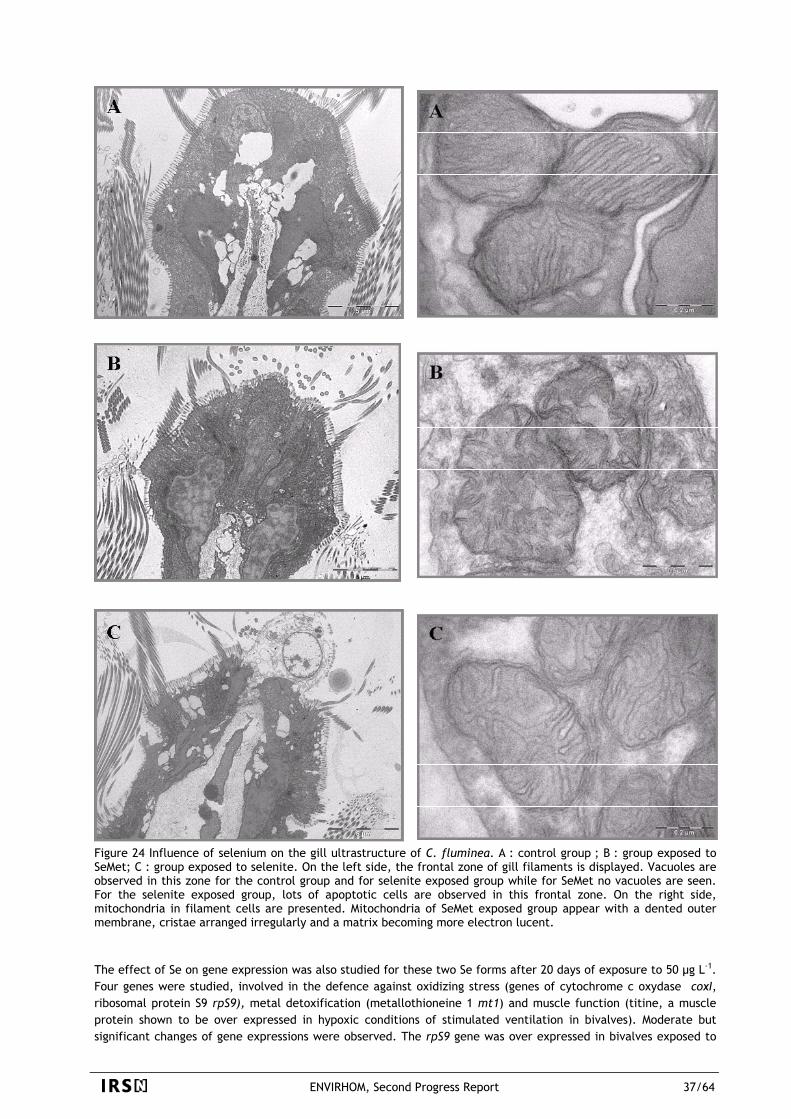
Figure 24 Influence of selenium on the gill ultrastructure of C. fluminea. A : control group ; B : group exposed to SeMet; C : group exposed to selenite. On the left side, the frontal zone of gill filaments is displayed. Vacuoles are observed in this zone for the control group and for selenite exposed group while for SeMet no vacuoles are seen. For the selenite exposed group, lots of apoptotic cells are observed in this frontal zone. On the right side, mitochondria in filament cells are presented. Mitochondria of SeMet exposed group appear with a dented outer membrane, cristae arranged irregularly and a matrix becoming more electron lucent. The effect of Se on gene expression was also studied for these two Se forms after 20 days of exposure to 50 µg L-1. Four genes were studied, involved in the defence against oxidizing stress (genes of cytochrome c oxydase coxI, ribosomal protein S9 rpS9), metal detoxification (metallothioneine 1 mt1) and muscle function (titine, a muscle protein shown to be over expressed in hypoxic conditions of stimulated ventilation in bivalves). Moderate but significant changes of gene expressions were observed. The rpS9 gene was over expressed in bivalves exposed to
ENVIRHOM, Second Progress Report 37/64
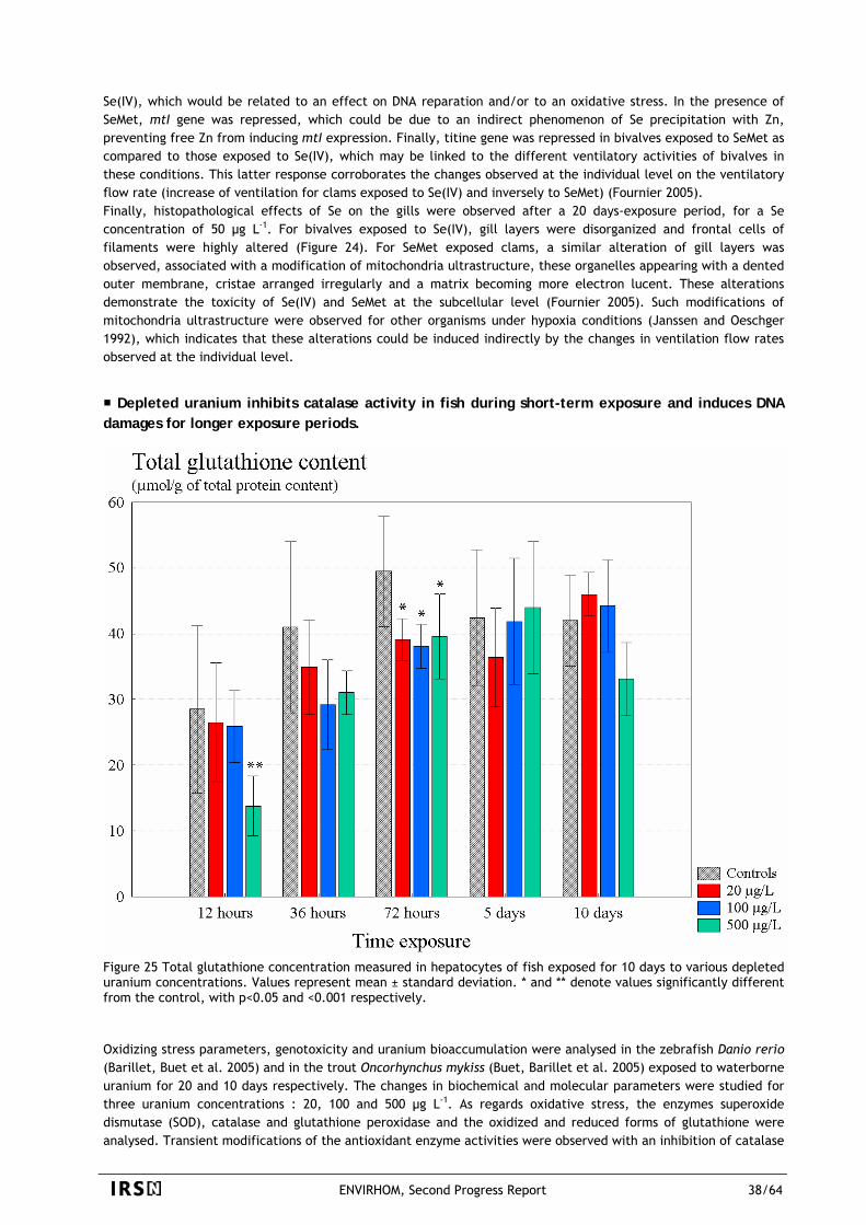
Se(IV), which would be related to an effect on DNA reparation and/or to an oxidative stress. In the presence of SeMet, mtI gene was repressed, which could be due to an indirect phenomenon of Se precipitation with Zn, preventing free Zn from inducing mtI expression. Finally, titine gene was repressed in bivalves exposed to SeMet as compared to those exposed to Se(IV), which may be linked to the different ventilatory activities of bivalves in these conditions. This latter response corroborates the changes observed at the individual level on the ventilatory flow rate (increase of ventilation for clams exposed to Se(IV) and inversely to SeMet) (Fournier 2005). Finally, histopathological effects of Se on the gills were observed after a 20 days-exposure period, for a Se concentration of 50 µg L-1. For bivalves exposed to Se(IV), gill layers were disorganized and frontal cells of filaments were highly altered (Figure 24). For SeMet exposed clams, a similar alteration of gill layers was observed, associated with a modification of mitochondria ultrastructure, these organelles appearing with a dented outer membrane, cristae arranged irregularly and a matrix becoming more electron lucent. These alterations demonstrate the toxicity of Se(IV) and SeMet at the subcellular level (Fournier 2005). Such modifications of mitochondria ultrastructure were observed for other organisms under hypoxia conditions (Janssen and Oeschger 1992), which indicates that these alterations could be induced indirectly by the changes in ventilation flow rates observed at the individual level.
Depleted uranium inhibits catalase activity in fish during short-term exposure and induces DNA damages for longer exposure periods.
Figure 25 Total glutathione concentration measured in hepatocytes of fish exposed for 10 days to various depleted uranium concentrations. Values represent mean ± standard deviation. * and ** denote values significantly different from the control, with p<0.05 and <0.001 respectively. Oxidizing stress parameters, genotoxicity and uranium bioaccumulation were analysed in the zebrafish Danio rerio (Barillet, Buet et al. 2005) and in the trout Oncorhynchus mykiss (Buet, Barillet et al. 2005) exposed to waterborne uranium for 20 and 10 days respectively. The changes in biochemical and molecular parameters were studied for three uranium concentrations : 20, 100 and 500 µg L-1. As regards oxidative stress, the enzymes superoxide dismutase (SOD), catalase and glutathione peroxidase and the oxidized and reduced forms of glutathione were analysed. Transient modifications of the antioxidant enzyme activities were observed with an inhibition of catalase
ENVIRHOM, Second Progress Report 38/64
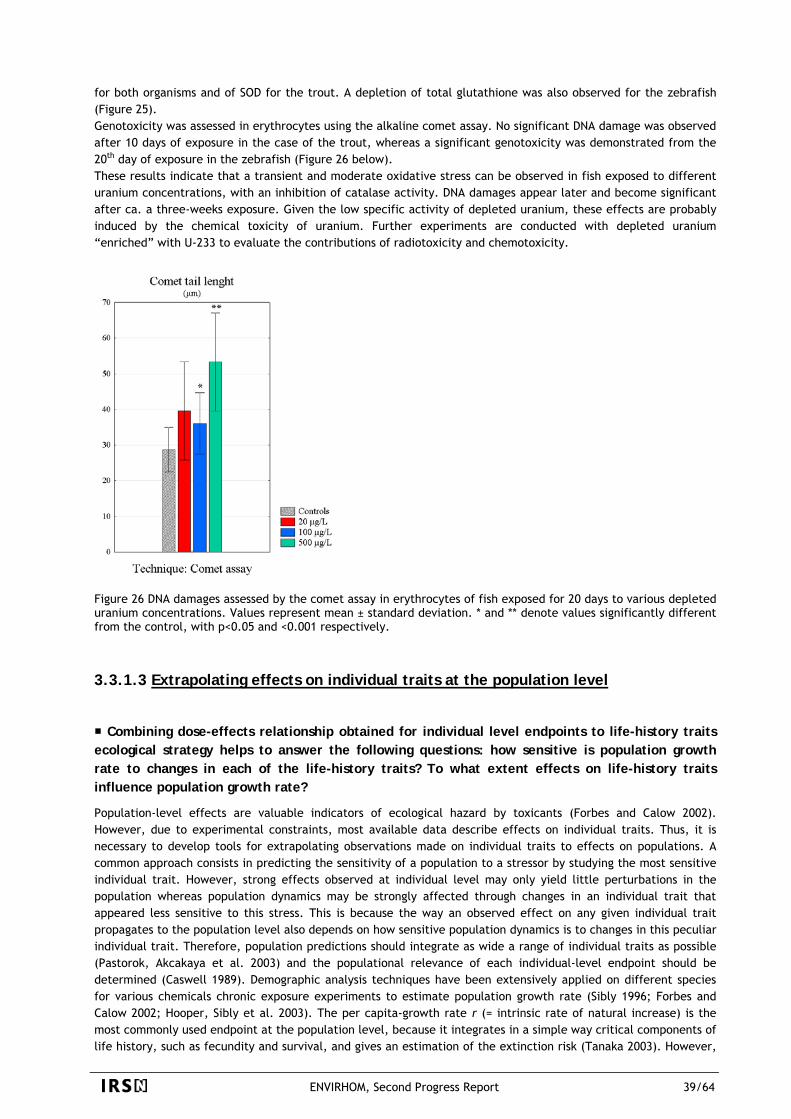
for both organisms and of SOD for the trout. A depletion of total glutathione was also observed for the zebrafish (Figure 25). Genotoxicity was assessed in erythrocytes using the alkaline comet assay. No significant DNA damage was observed after 10 days of exposure in the case of the trout, whereas a significant genotoxicity was demonstrated from the 20th day of exposure in the zebrafish (Figure 26 below). These results indicate that a transient and moderate oxidative stress can be observed in fish exposed to different uranium concentrations, with an inhibition of catalase activity. DNA damages appear later and become significant after ca. a three-weeks exposure. Given the low specific activity of depleted uranium, these effects are probably induced by the chemical toxicity of uranium. Further experiments are conducted with depleted uranium “enriched” with U-233 to evaluate the contributions of radiotoxicity and chemotoxicity.
Figure 26 DNA damages assessed by the comet assay in erythrocytes of fish exposed for 20 days to various depleted uranium concentrations. Values represent mean ± standard deviation. * and ** denote values significantly different from the control, with p<0.05 and <0.001 respectively.
3.3.1.3 Extrapolating effects on individual traits at the population level
Combining dose-effects relationship obtained for individual level endpoints to life-history traits ecological strategy helps to answer the following questions: how sensitive is population growth rate to changes in each of the life-history traits? To what extent effects on life-history traits influence population growth rate?
Population-level effects are valuable indicators of ecological hazard by toxicants (Forbes and Calow 2002). However, due to experimental constraints, most available data describe effects on individual traits. Thus, it is necessary to develop tools for extrapolating observations made on individual traits to effects on populations. A common approach consists in predicting the sensitivity of a population to a stressor by studying the most sensitive individual trait. However, strong effects observed at individual level may only yield little perturbations in the population whereas population dynamics may be strongly affected through changes in an individual trait that appeared less sensitive to this stress. This is because the way an observed effect on any given individual trait propagates to the population level also depends on how sensitive population dynamics is to changes in this peculiar individual trait. Therefore, population predictions should integrate as wide a range of individual traits as possible (Pastorok, Akcakaya et al. 2003) and the populational relevance of each individual-level endpoint should be determined (Caswell 1989). Demographic analysis techniques have been extensively applied on different species for various chemicals chronic exposure experiments to estimate population growth rate (Sibly 1996; Forbes and Calow 2002; Hooper, Sibly et al. 2003). The per capita-growth rate r (= intrinsic rate of natural increase) is the most commonly used endpoint at the population level, because it integrates in a simple way critical components of life history, such as fecundity and survival, and gives an estimation of the extinction risk (Tanaka 2003). However,
ENVIRHOM, Second Progress Report 39/64
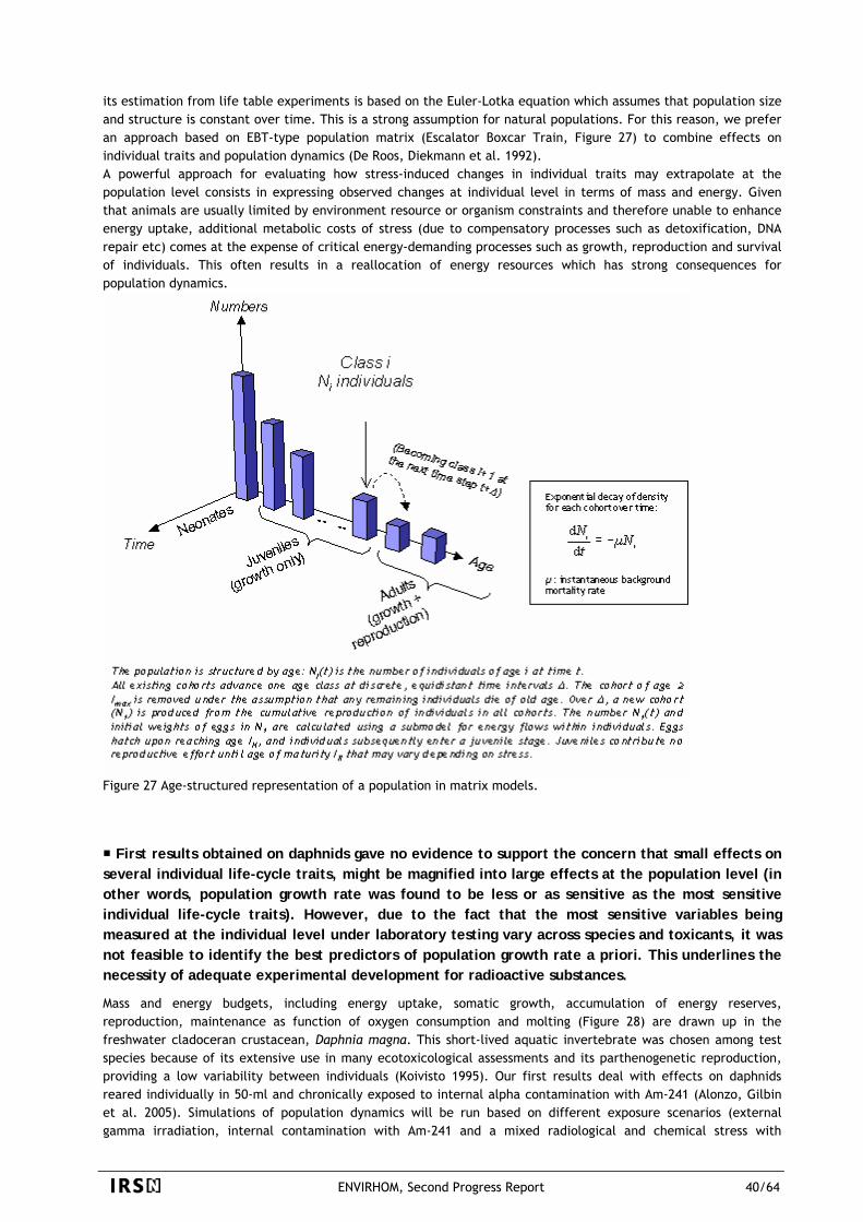
its estimation from life table experiments is based on the Euler-Lotka equation which assumes that population size and structure is constant over time. This is a strong assumption for natural populations. For this reason, we prefer an approach based on EBT-type population matrix (Escalator Boxcar Train, Figure 27) to combine effects on individual traits and population dynamics (De Roos, Diekmann et al. 1992). A powerful approach for evaluating how stress-induced changes in individual traits may extrapolate at the population level consists in expressing observed changes at individual level in terms of mass and energy. Given that animals are usually limited by environment resource or organism constraints and therefore unable to enhance energy uptake, additional metabolic costs of stress (due to compensatory processes such as detoxification, DNA repair etc) comes at the expense of critical energy-demanding processes such as growth, reproduction and survival of individuals. This often results in a reallocation of energy resources which has strong consequences for population dynamics.
Figure 27 Age-structured representation of a population in matrix models.
First results obtained on daphnids gave no evidence to support the concern that small effects on several individual life-cycle traits, might be magnified into large effects at the population level (in other words, population growth rate was found to be less or as sensitive as the most sensitive individual life-cycle traits). However, due to the fact that the most sensitive variables being measured at the individual level under laboratory testing vary across species and toxicants, it was not feasible to identify the best predictors of population growth rate a priori. This underlines the necessity of adequate experimental development for radioactive substances.
Mass and energy budgets, including energy uptake, somatic growth, accumulation of energy reserves, reproduction, maintenance as function of oxygen consumption and molting (Figure 28) are drawn up in the freshwater cladoceran crustacean, Daphnia magna. This short-lived aquatic invertebrate was chosen among test species because of its extensive use in many ecotoxicological assessments and its parthenogenetic reproduction, providing a low variability between individuals (Koivisto 1995). Our first results deal with effects on daphnids reared individually in 50-ml and chronically exposed to internal alpha contamination with Am-241 (Alonzo, Gilbin et al. 2005). Simulations of population dynamics will be run based on different exposure scenarios (external gamma irradiation, internal contamination with Am-241 and a mixed radiological and chemical stress with
ENVIRHOM, Second Progress Report 40/64
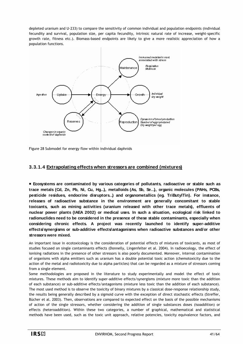
depleted uranium and U-233) to compare the sensitivity of common individual and population endpoints (individual fecundity and survival, population size, per capita fecundity, intrinsic natural rate of increase, weight-specific growth rate, fitness etc.). Biomass-based endpoints are likely to give a more realistic appreciation of how a population functions.
Figure 28 Submodel for energy flow within individual daphnids
3.3.1.4 Extrapolating effects when stressors are combined (mixtures)
Ecosystems are contaminated by various categories of pollutants, radioactive or stable such as trace metals (Cd, Zn, Pb, Ni, Cu, Hg…), metalloids (As, Sb, Se…), organic molecules (PAHs, PCBs, pesticide residues, endocrine disruptors…) and organometallics (eg. TriButylTin). For instance, releases of radioactive substance in the environment are generally concomitant to stable toxicants, such as mining activities (uranium released with other trace metals), effluents of nuclear power plants (IAEA 2002) or medical uses. In such a situation, ecological risk linked to radionuclides need to be considered in the presence of these stable contaminants, especially when considering chronic effects. A project was recently launched to identify super-additive effects/synergisms or sub-additive effects/antagonisms when radioactive substances and/or other stressors were mixed.
An important issue in ecotoxicology is the consideration of potential effects of mixtures of toxicants, as most of studies focused on single contaminants effects (Donnelly, Lingenfelter et al. 2004). In radioecology, the effect of ionising radiations in the presence of other stressors is also poorly documented. Moreover, internal contamination of organisms with alpha emitters such as uranium has a double potential toxic action (chemotoxicity due to the action of the metal and radiotoxicity due to alpha particles) that can be regarded as a mixture of stressors coming from a single element. Some methodologies are proposed in the literature to study experimentally and model the effect of toxic mixtures. These methods aim to identify super-additive effects/synergisms (mixture more toxic than the addition of each substance) or sub-additive effects/antagonisms (mixture less toxic than the addition of each substance). The most used method is to observe the toxicity of binary mixtures by a classical dose-response relationship study, the results being generally described by a sigmoid curve with the exception of direct stochastic effects (Streffer, Bücher et al. 2003). Then, observations are compared to expected effect on the basis of the possible mechanisms of action of the single stressors, whether considering the addition of single substances doses (isoaddition) or effects (heteroaddition). Within these two categories, a number of graphical, mathematical and statistical methods have been used, such as the toxic unit approach, relative potencies, toxicity equivalence factors, and
ENVIRHOM, Second Progress Report 41/64
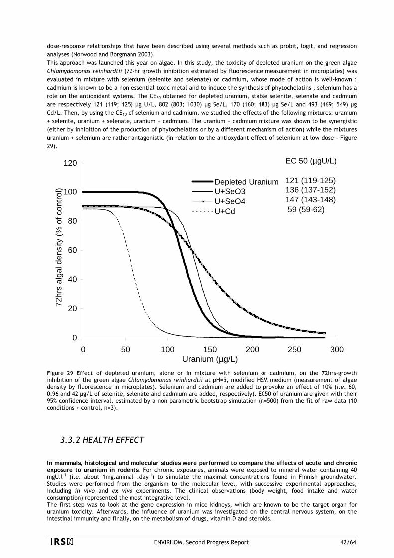
dose-response relationships that have been described using several methods such as probit, logit, and regression analyses (Norwood and Borgmann 2003). This approach was launched this year on algae. In this study, the toxicity of depleted uranium on the green algae Chlamydomonas reinhardtii (72-hr growth inhibition estimated by fluorescence measurement in microplates) was evaluated in mixture with selenium (selenite and selenate) or cadmium, whose mode of action is well-known : cadmium is known to be a non-essential toxic metal and to induce the synthesis of phytochelatins ; selenium has a role on the antioxidant systems. The CE50 obtained for depleted uranium, stable selenite, selenate and cadmium are respectively 121 (119; 125) µg U/L, 802 (803; 1030) µg Se/L, 170 (160; 183) µg Se/L and 493 (469; 549) µg Cd/L. Then, by using the CE10 of selenium and cadmium, we studied the effects of the following mixtures: uranium + selenite, uranium + selenate, uranium + cadmium. The uranium + cadmium mixture was shown to be synergistic (either by inhibition of the production of phytochelatins or by a different mechanism of action) while the mixtures uranium + selenium are rather antagonistic (in relation to the antioxydant effect of selenium at low dose - Figure 29).
0
20
40
60
80
100
120
0 50 100 150 200 250 300Uranium (µg/L)
72hr
s al
gal d
ensi
ty (%
of c
ontro
l)
Depleted UraniumU+SeO3U+SeO4U+Cd
EC 50 (µgU/L)
121 (119-125) 136 (137-152) 147 (143-148) 59 (59-62)
Figure 29 Effect of depleted uranium, alone or in mixture with selenium or cadmium, on the 72hrs-growth inhibition of the green algae Chlamydomonas reinhardtii at pH=5, modified HSM medium (measurement of algae density by fluorescence in microplates). Selenium and cadmium are added to provoke an effect of 10% (i.e. 60, 0.96 and 42 µg/L of selenite, selenate and cadmium are added, respectively). EC50 of uranium are given with their 95% confidence interval, estimated by a non parametric bootstrap simulation (n=500) from the fit of raw data (10 conditions + control, n=3).
3.3.2 HEALTH EFFECT
In mammals, histological and molecular studies were performed to compare the effects of acute and chronic exposure to uranium in rodents. For chronic exposures, animals were exposed to mineral water containing 40 mgU.l-1 (i.e. about 1mg.animal-1.day-1) to simulate the maximal concentrations found in Finnish groundwater. Studies were performed from the organism to the molecular level, with successive experimental approaches, including in vivo and ex vivo experiments. The clinical observations (body weight, food intake and water consumption) represented the most integrative level. The first step was to look at the gene expression in mice kidneys, which are known to be the target organ for uranium toxicity. Afterwards, the influence of uranium was investigated on the central nervous system, on the intestinal immunity and finally, on the metabolism of drugs, vitamin D and steroids.
ENVIRHOM, Second Progress Report 42/64
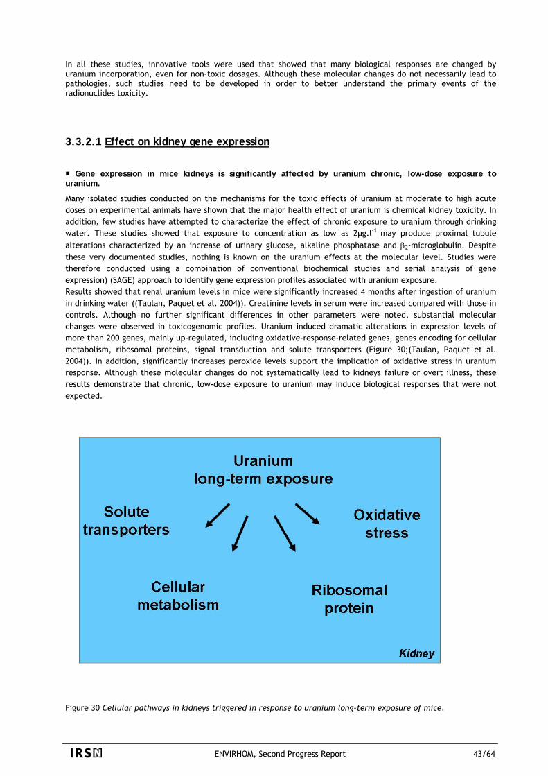
In all these studies, innovative tools were used that showed that many biological responses are changed by uranium incorporation, even for non-toxic dosages. Although these molecular changes do not necessarily lead to pathologies, such studies need to be developed in order to better understand the primary events of the radionuclides toxicity.
3.3.2.1 Effect on kidney gene expression
Gene expression in mice kidneys is significantly affected by uranium chronic, low-dose exposure to uranium.
Many isolated studies conducted on the mechanisms for the toxic effects of uranium at moderate to high acute doses on experimental animals have shown that the major health effect of uranium is chemical kidney toxicity. In addition, few studies have attempted to characterize the effect of chronic exposure to uranium through drinking water. These studies showed that exposure to concentration as low as 2µg.l-1 may produce proximal tubule alterations characterized by an increase of urinary glucose, alkaline phosphatase and β2-microglobulin. Despite these very documented studies, nothing is known on the uranium effects at the molecular level. Studies were therefore conducted using a combination of conventional biochemical studies and serial analysis of gene expression) (SAGE) approach to identify gene expression profiles associated with uranium exposure. Results showed that renal uranium levels in mice were significantly increased 4 months after ingestion of uranium in drinking water ((Taulan, Paquet et al. 2004)). Creatinine levels in serum were increased compared with those in controls. Although no further significant differences in other parameters were noted, substantial molecular changes were observed in toxicogenomic profiles. Uranium induced dramatic alterations in expression levels of more than 200 genes, mainly up-regulated, including oxidative-response-related genes, genes encoding for cellular metabolism, ribosomal proteins, signal transduction and solute transporters (Figure 30;(Taulan, Paquet et al. 2004)). In addition, significantly increases peroxide levels support the implication of oxidative stress in uranium response. Although these molecular changes do not systematically lead to kidneys failure or overt illness, these results demonstrate that chronic, low-dose exposure to uranium may induce biological responses that were not expected.
Figure 30 Cellular pathways in kidneys triggered in response to uranium long-term exposure of mice.
ENVIRHOM, Second Progress Report 43/64
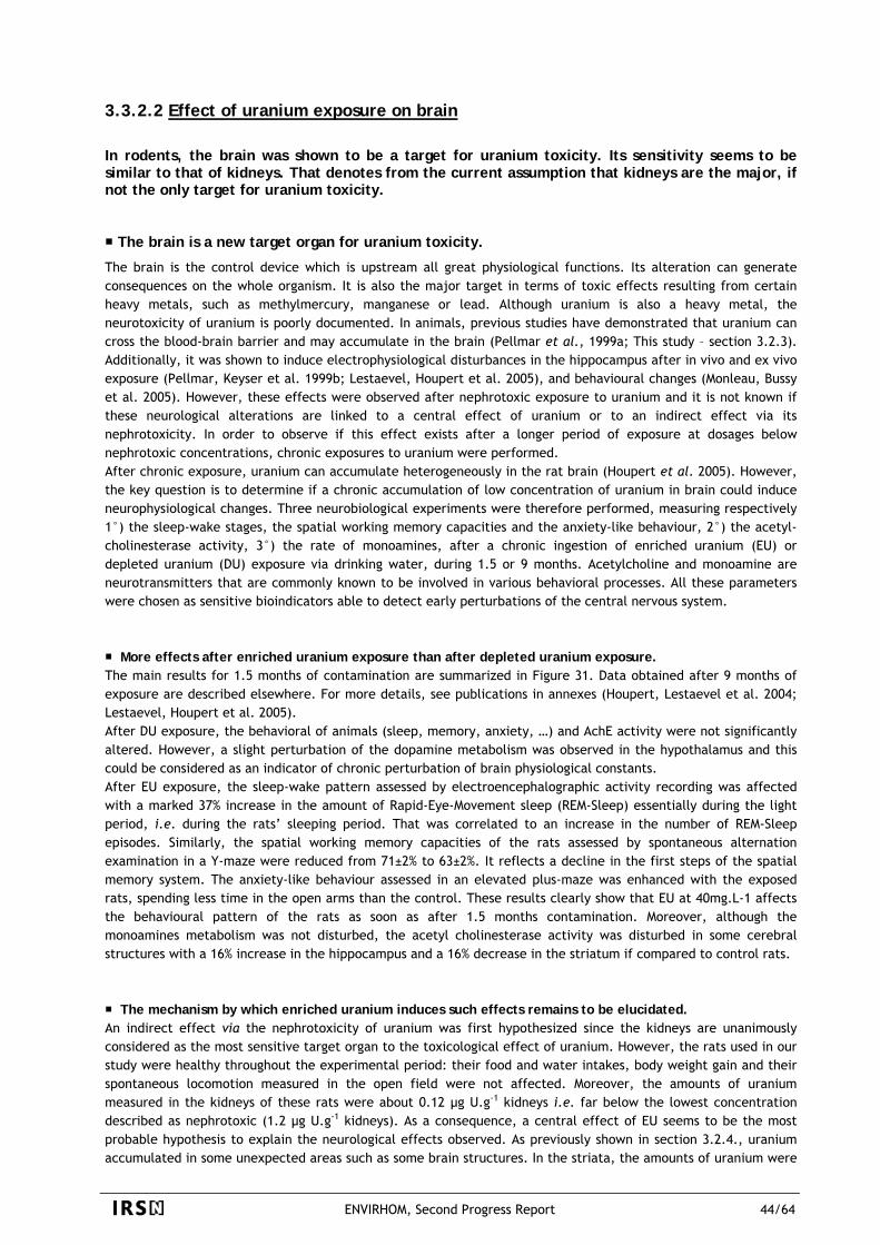
3.3.2.2 Effect of uranium exposure on brain In rodents, the brain was shown to be a target for uranium toxicity. Its sensitivity seems to be similar to that of kidneys. That denotes from the current assumption that kidneys are the major, if not the only target for uranium toxicity.
The brain is a new target organ for uranium toxicity.
The brain is the control device which is upstream all great physiological functions. Its alteration can generate consequences on the whole organism. It is also the major target in terms of toxic effects resulting from certain heavy metals, such as methylmercury, manganese or lead. Although uranium is also a heavy metal, the neurotoxicity of uranium is poorly documented. In animals, previous studies have demonstrated that uranium can cross the blood-brain barrier and may accumulate in the brain (Pellmar et al., 1999a; This study – section 3.2.3). Additionally, it was shown to induce electrophysiological disturbances in the hippocampus after in vivo and ex vivo exposure (Pellmar, Keyser et al. 1999b; Lestaevel, Houpert et al. 2005), and behavioural changes (Monleau, Bussy et al. 2005). However, these effects were observed after nephrotoxic exposure to uranium and it is not known if these neurological alterations are linked to a central effect of uranium or to an indirect effect via its nephrotoxicity. In order to observe if this effect exists after a longer period of exposure at dosages below nephrotoxic concentrations, chronic exposures to uranium were performed. After chronic exposure, uranium can accumulate heterogeneously in the rat brain (Houpert et al. 2005). However, the key question is to determine if a chronic accumulation of low concentration of uranium in brain could induce neurophysiological changes. Three neurobiological experiments were therefore performed, measuring respectively 1°) the sleep-wake stages, the spatial working memory capacities and the anxiety-like behaviour, 2°) the acetyl-cholinesterase activity, 3°) the rate of monoamines, after a chronic ingestion of enriched uranium (EU) or depleted uranium (DU) exposure via drinking water, during 1.5 or 9 months. Acetylcholine and monoamine are neurotransmitters that are commonly known to be involved in various behavioral processes. All these parameters were chosen as sensitive bioindicators able to detect early perturbations of the central nervous system. More effects after enriched uranium exposure than after depleted uranium exposure.
The main results for 1.5 months of contamination are summarized in Figure 31. Data obtained after 9 months of exposure are described elsewhere. For more details, see publications in annexes (Houpert, Lestaevel et al. 2004; Lestaevel, Houpert et al. 2005). After DU exposure, the behavioral of animals (sleep, memory, anxiety, …) and AchE activity were not significantly altered. However, a slight perturbation of the dopamine metabolism was observed in the hypothalamus and this could be considered as an indicator of chronic perturbation of brain physiological constants. After EU exposure, the sleep-wake pattern assessed by electroencephalographic activity recording was affected with a marked 37% increase in the amount of Rapid-Eye-Movement sleep (REM-Sleep) essentially during the light period, i.e. during the rats’ sleeping period. That was correlated to an increase in the number of REM-Sleep episodes. Similarly, the spatial working memory capacities of the rats assessed by spontaneous alternation examination in a Y-maze were reduced from 71±2% to 63±2%. It reflects a decline in the first steps of the spatial memory system. The anxiety-like behaviour assessed in an elevated plus-maze was enhanced with the exposed rats, spending less time in the open arms than the control. These results clearly show that EU at 40mg.L-1 affects the behavioural pattern of the rats as soon as after 1.5 months contamination. Moreover, although the monoamines metabolism was not disturbed, the acetyl cholinesterase activity was disturbed in some cerebral structures with a 16% increase in the hippocampus and a 16% decrease in the striatum if compared to control rats.
The mechanism by which enriched uranium induces such effects remains to be elucidated.
An indirect effect via the nephrotoxicity of uranium was first hypothesized since the kidneys are unanimously considered as the most sensitive target organ to the toxicological effect of uranium. However, the rats used in our study were healthy throughout the experimental period: their food and water intakes, body weight gain and their spontaneous locomotion measured in the open field were not affected. Moreover, the amounts of uranium measured in the kidneys of these rats were about 0.12 µg U.g-1 kidneys i.e. far below the lowest concentration described as nephrotoxic (1.2 µg U.g-1 kidneys). As a consequence, a central effect of EU seems to be the most probable hypothesis to explain the neurological effects observed. As previously shown in section 3.2.4., uranium accumulated in some unexpected areas such as some brain structures. In the striata, the amounts of uranium were
ENVIRHOM, Second Progress Report 44/64
increased by a factor of 2.2 and 2 for the rats exposed to EU and DU, respectively, when compared to controls. More surprisingly, EU accumulated in some structures while depleted uranium did not. Uranium amounts were 1.5 to 2 times higher in the hippocampus and hypothalamus of rats exposed to enriched uranium when compared to DU rats or controls. This differential accumulation was also found in other organs such as adrenals, for which rats exposed to EU accumulated 1.5 times more uranium than did the DU rats. The above low chronic accumulation of uranium can be related to the neurophysiological and behavioural changes that we observed. The new target organs concerned by EU accumulation, hippocampus, hypothalamus and adrenals, are known to be involved in the behavioural effects observed in our study. The hippocampus is known to be involved in the spatial working memory and the hypothalamic-pituitary-adrenal axis in the sleep-wake cycle and anxiety (Figure 32). What remains to be determined is why EU accumulated differently from DU. Enriched and depleted uranium were introduced in the same chemical form in similar drinking water, at the same pH. The only difference between EU and DU was their specific activities, and thus the differential accumulation of EU can only be explained by the fact that its radiological activity is 4 times as high. We can therefore conclude that the radiological activity of EU induces the primary events of the neurobiological effects of uranium. All these results strongly suggest that the brain is a new target organ of uranium toxicity. But these results
raise more questions than they resolve. The mechanisms of uranium accumulation and neurological effect are totally unknown. To understand why EU and DU accumulated differently, rats should be contaminated with uranium of higher specific activity (such as 233U for example or higher EU). It could also help to discriminate the radiological versus chemical toxicity of uranium. Studies should be carried out with longer periods of contamination than 1.5 months in order to assess if these effects persist with time. The neurological effects of a chronic radio-contamination may be more dramatic if this contamination occurred during the critical periods of the central nervous system development (before and after the birth) or during ageing; in this latter case, a chronic inflammation could worsen a neurodegenerative disease. It is why the experiments should be carried out with different animal models: On models of neuropathological disease (as Alzheimer) and on young rats whose parents were contaminated before/during the gestation and during the lactation. It will be crucial for public health to determine if these phenomena occurred at even lower exposure levels with natural uranium.
ENVIRHOM, Second Progress Report 45/64
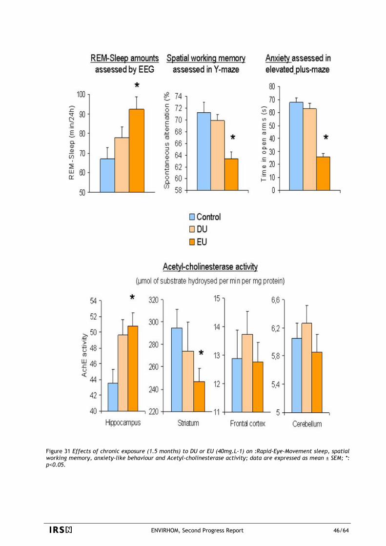
Figure 31 Effects of chronic exposure (1.5 months) to DU or EU (40mg.L-1) on :Rapid-Eye-Movement sleep, spatial working memory, anxiety-like behaviour and Acetyl-cholinesterase activity; data are expressed as mean ± SEM; *: p<0.05.
ENVIRHOM, Second Progress Report 46/64
Figure 32 The effects of UA and UE, respectively, on the central nervous system after a 1.5-month chronic exposure period. EU, but DU accumulated in hippocampus and hypothalamus. The hippocampus is involved in the spatial working memory and showed increase of AchE activity (green arrows). Hypothalamus is involved in anxiety and sleep wake cycle (orange arrows). Interactions among the different observed effects are presented as blue arrows. As for example, sleep wake cycle of animals is dependant on their level of anxiety.
3.3.2.3 Effect of uranium exposure on gastrointestinal tract
Acute exposure to depleted uranium induces changes in the production or the expression of cytokines and chemokines in the intestinal epithelium of rats.
In the event of ingestion of uranium, the digestive tract is the first biological system exposed to radionuclide intake in the intestinal lumen. The presence of depleted uranium in the intestinal lumen following ingestion may lead to functional modifications of intestinal properties, especially intestinal permeability and mucosal immunity. The purpose of the study performed was to determine whether some properties of intestinal immunity were modified following ingestion of uranium. This study was performed with a sublethal dose of uranium, consisting of an intra-gastric administration of uranium, to simulate contamination by ingestion. Because of the absence of data in literature on eventual effects of depleted uranium on immune cells and molecules, a high dose of DU was administrated to rats in this study to determine the immune cellular and molecular targets of this radionuclide in the intestine. The effects of uranium contamination were measured at short-term after the uranium exposure, at one and three days, before the elimination of uranium from the gastrointestinal tract. The first part of this work has shown an absence of changes in proliferation, differentiation and apoptosis processes following uranium administration at high dose. These results suggest that depleted uranium was not toxic for the intestine following an acute exposure. In the second part of this study, the evaluation of the immune status of the intestine indicates that depleted uranium induced some changes in the production or the expression of cytokines (IFNγ, interferon gamma, a cytotoxic molecule) and chemokines (MCP-1, monocyte chemoattractant protein-1, promoting monocyte recruitment into the inflammatory site) in the intestinal epithelium. In addition, this study showed no modifications in the localization and density of immune cells involved in the production of
ENVIRHOM, Second Progress Report 47/64
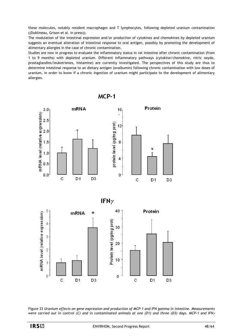
these molecules, notably resident macrophages and T lymphocytes, following depleted uranium contamination ((Dublineau, Grison et al. in press)). The modulation of the intestinal expression and/or production of cytokines and chemokines by depleted uranium suggests an eventual alteration of intestinal response to oral antigen, possibly by promoting the development of alimentary allergies in the case of chronic contamination. Studies are now in progress to evaluate the inflammatory status in rat intestine after chronic contamination (from 1 to 9 months) with depleted uranium. Different inflammatory pathways (cytokine/chemokine, nitric oxyde, prostaglandins/leukotrienes, histamine) are currently investigated. The perspectives of this study are thus to determine intestinal response to an dietary antigen (ovalbumin) following chronic contamination with low doses of uranium, in order to know if a chronic ingestion of uranium might participate to the development of alimentary allergies.
Figure 33 Uranium effects on gene expression and production of MCP-1 and IFN gamma in intestine. Measurements were carried out in control (C) and in contaminated animals at one (D1) and three (D3) days. MCP-1 and IFNγ
ENVIRHOM, Second Progress Report 48/64
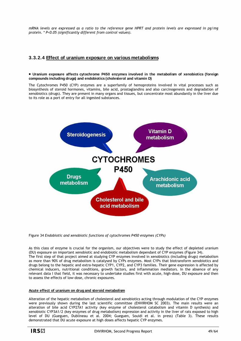
mRNA levels are expressed as a ratio to the reference gene HPRT and protein levels are expressed in pg/mg protein. * P<0.05 (significantly different from control values).
3.3.2.4 Effect of uranium exposure on various metabolisms
Uranium exposure affects cytochrome P450 enzymes involved in the metabolism of xenobiotics (foreign compounds including drugs) and endobiotics (cholesterol and vitamin D)
The Cytochromes P450 (CYP) enzymes are a superfamily of hemoproteins involved in vital processes such as biosynthesis of steroid hormones, vitamins, bile acid, prostaglandins and also carcinogenesis and degradation of xenobiotics (drugs). They are present in many organs and tissues, but concentrate most abundantly in the liver due to its role as a port of entry for all ingested substances.
Figure 34 Endobiotic and xenobiotic functions of cytochromes P450 enzymes (CYPs) As this class of enzyme is crucial for the organism, our objectives were to study the effect of depleted uranium (DU) exposure on important xenobiotic and endobiotic metabolism dependant of CYP enzymes (Figure 34). The first step of that project aimed at studying CYP enzymes involved in xenobiotics (including drugs) metabolism as more than 90% of drug metabolism is catalysed by CYPs enzymes. Most CYPs that biotransform xenobiotics and drugs belong to the hepatic and extra-hepatic CYP1, CYP2, and CYP3 families. Their gene expression is affected by chemical inducers, nutritional conditions, growth factors, and inflammation mediators. In the absence of any relevant data I that field, it was necessary to undertake studies first with acute, high-dose, DU exposure and then to assess the effects of low-dose, chronic exposures. Acute effect of uranium on drug and steroid metabolism Alteration of the hepatic metabolism of cholesterol and xenobiotics acting through modulation of the CYP enzymes were previously shown during the last scientific committee (ENVIRHOM SC 2003). The main results were an alteration of bile acid CYP27A1 activity (key enzyme of cholesterol catabolism and vitamin D synthesis) and xenobiotic CYP3A1/2 (key enzymes of drug metabolism) expression and activity in the liver of rats exposed to high level of DU (Gueguen, Dublineau et al. 2004; Gueguen, Souidi et al. in press) (Table 3). These results demonstrated that DU acute exposure at high doses affects hepatic CYP enzymes.
ENVIRHOM, Second Progress Report 49/64
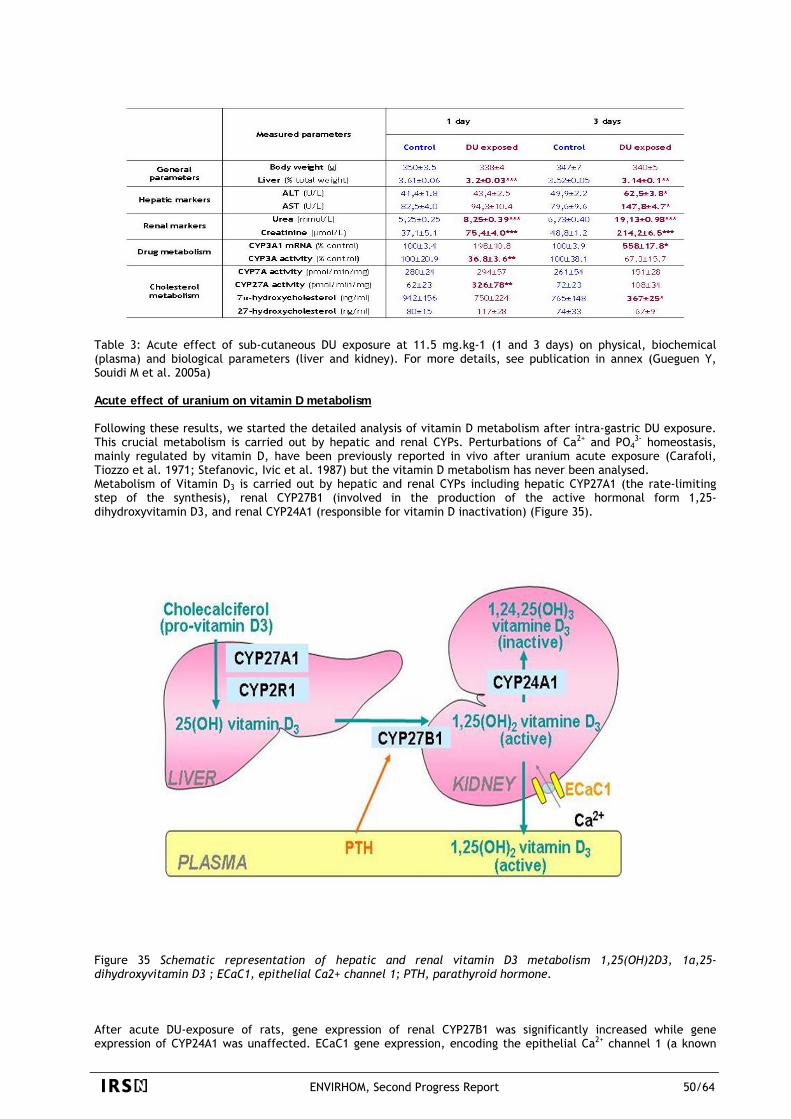
Table 3: Acute effect of sub-cutaneous DU exposure at 11.5 mg.kg-1 (1 and 3 days) on physical, biochemical (plasma) and biological parameters (liver and kidney). For more details, see publication in annex (Gueguen Y, Souidi M et al. 2005a) Acute effect of uranium on vitamin D metabolism Following these results, we started the detailed analysis of vitamin D metabolism after intra-gastric DU exposure. This crucial metabolism is carried out by hepatic and renal CYPs. Perturbations of Ca2+ and PO4
3- homeostasis, mainly regulated by vitamin D, have been previously reported in vivo after uranium acute exposure (Carafoli, Tiozzo et al. 1971; Stefanovic, Ivic et al. 1987) but the vitamin D metabolism has never been analysed. Metabolism of Vitamin D3 is carried out by hepatic and renal CYPs including hepatic CYP27A1 (the rate-limiting step of the synthesis), renal CYP27B1 (involved in the production of the active hormonal form 1,25-dihydroxyvitamin D3, and renal CYP24A1 (responsible for vitamin D inactivation) (Figure 35).
Figure 35 Schematic representation of hepatic and renal vitamin D3 metabolism 1,25(OH)2D3, 1α,25-dihydroxyvitamin D3 ; ECaC1, epithelial Ca2+ channel 1; PTH, parathyroid hormone. After acute DU-exposure of rats, gene expression of renal CYP27B1 was significantly increased while gene expression of CYP24A1 was unaffected. ECaC1 gene expression, encoding the epithelial Ca2+ channel 1 (a known
ENVIRHOM, Second Progress Report 50/64
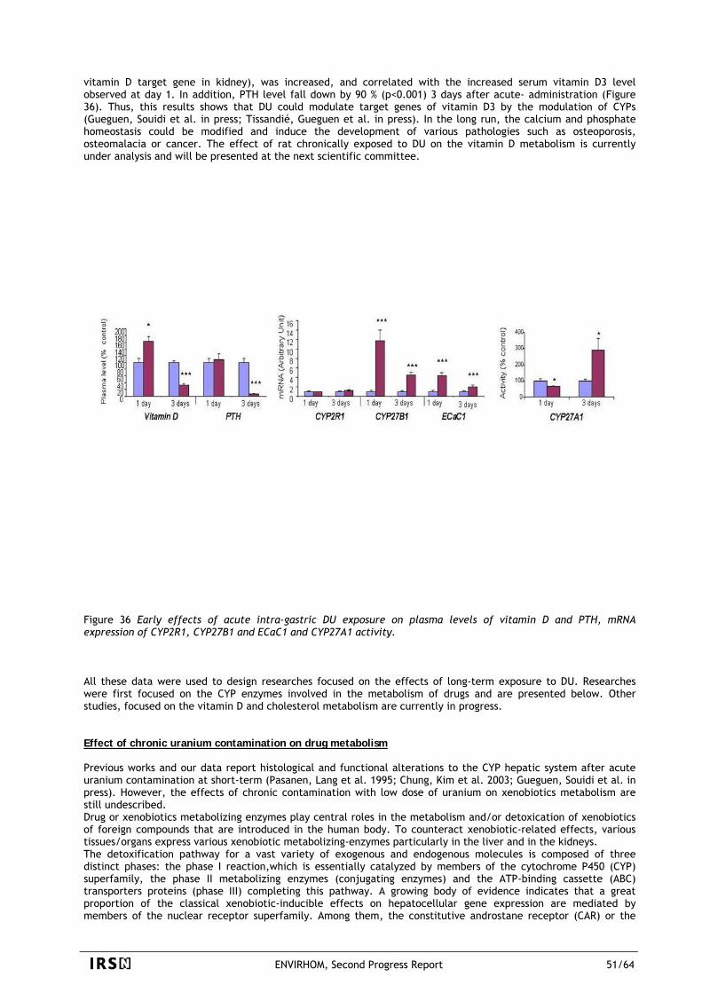
vitamin D target gene in kidney), was increased, and correlated with the increased serum vitamin D3 level observed at day 1. In addition, PTH level fall down by 90 % (p<0.001) 3 days after acute- administration (Figure 36). Thus, this results shows that DU could modulate target genes of vitamin D3 by the modulation of CYPs (Gueguen, Souidi et al. in press; Tissandié, Gueguen et al. in press). In the long run, the calcium and phosphate homeostasis could be modified and induce the development of various pathologies such as osteoporosis, osteomalacia or cancer. The effect of rat chronically exposed to DU on the vitamin D metabolism is currently under analysis and will be presented at the next scientific committee.
Figure 36 Early effects of acute intra-gastric DU exposure on plasma levels of vitamin D and PTH, mRNA expression of CYP2R1, CYP27B1 and ECaC1 and CYP27A1 activity. All these data were used to design researches focused on the effects of long-term exposure to DU. Researches were first focused on the CYP enzymes involved in the metabolism of drugs and are presented below. Other studies, focused on the vitamin D and cholesterol metabolism are currently in progress. Effect of chronic uranium contamination on drug metabolism Previous works and our data report histological and functional alterations to the CYP hepatic system after acute uranium contamination at short-term (Pasanen, Lang et al. 1995; Chung, Kim et al. 2003; Gueguen, Souidi et al. in press). However, the effects of chronic contamination with low dose of uranium on xenobiotics metabolism are still undescribed. Drug or xenobiotics metabolizing enzymes play central roles in the metabolism and/or detoxication of xenobiotics of foreign compounds that are introduced in the human body. To counteract xenobiotic-related effects, various tissues/organs express various xenobiotic metabolizing-enzymes particularly in the liver and in the kidneys. The detoxification pathway for a vast variety of exogenous and endogenous molecules is composed of three distinct phases: the phase I reaction,which is essentially catalyzed by members of the cytochrome P450 (CYP) superfamily, the phase II metabolizing enzymes (conjugating enzymes) and the ATP-binding cassette (ABC) transporters proteins (phase III) completing this pathway. A growing body of evidence indicates that a great proportion of the classical xenobiotic-inducible effects on hepatocellular gene expression are mediated by members of the nuclear receptor superfamily. Among them, the constitutive androstane receptor (CAR) or the
ENVIRHOM, Second Progress Report 51/64
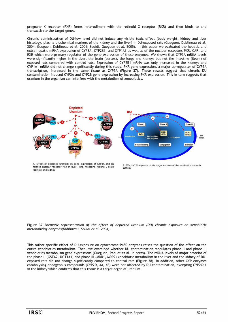
pregnane X receptor (PXR) forms heterodimers with the retinoid X receptor (RXR) and then binds to and transactivate the target genes. Chronic administration of DU-low level did not induce any visible toxic effect (body weight, kidney and liver histology, plasma biochemical markers of the kidney and the liver) in DU-exposed rats (Gueguen, Dublineau et al. 2004; Gueguen, Dublineau et al. 2004; Souidi, Gueguen et al. 2005). In this paper we evaluated the hepatic and extra hepatic mRNA expression of CYP3A, CYP2B1, and CYP1A1 as well as of the nuclear receptors PXR, CAR, and RXR which were primary regulator of the gene expression of these enzymes. We shown that CYP3A mRNA levels were significantly higher in the liver, the brain (cortex), the lungs and kidneys but not the intestine (ileum) of exposed rats compared with control rats. Expression of CYP2B1 mRNA was only increased in the kidneys and CYP1A1 mRNA did not change significantly during this study. PXR gene expression, a major up-regulator of CYP3A transcription, increased in the same tissue as CYP3A (Figure 37). These results suggest that chronic DU contamination induced CYP3A and CYP2B gene expression by increasing PXR expression. This in turn suggests that uranium in the organism can interfere with the metabolism of xenobiotics.
Figure 37 Shematic representation of the effect of depleted uranium (DU) chronic exposure on xenobiotic metabolizing enzymes(Dublineau, Souidi et al. 2004). This rather specific effect of DU-exposure on cytochrome P450 enzymes raises the question of the effect on the entire xenobiotics metabolism. Then, we examined whether DU contamination modulates phase II and phase III xenobiotics metabolism gene expressions (Gueguen, Paquet et al. in press). The mRNA levels of major proteins of the phase II (GSTA2, UGT1A1) and phase III (MDR1, MRP2) xenobiotic metabolism in the liver and the kidney of DU-exposed rats did not change significantly compared to control rats (Figure 38). In addition, other CYP enzymes catabolysing endogenous compounds (CYP2D, 4A, 4F) were not affected by DU contamination, excepting CYP2C11 in the kidney which confirms that this tissue is a target organ of uranium.
ENVIRHOM, Second Progress Report 52/64
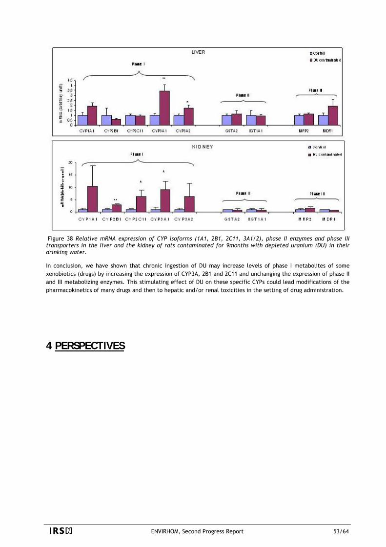
Figure 38 Relative mRNA expression of CYP isoforms (1A1, 2B1, 2C11, 3A1/2), phase II enzymes and phase III transporters in the liver and the kidney of rats contaminated for 9months with depleted uranium (DU) in their drinking water. In conclusion, we have shown that chronic ingestion of DU may increase levels of phase I metabolites of some xenobiotics (drugs) by increasing the expression of CYP3A, 2B1 and 2C11 and unchanging the expression of phase II and III metabolizing enzymes. This stimulating effect of DU on these specific CYPs could lead modifications of the pharmacokinetics of many drugs and then to hepatic and/or renal toxicities in the setting of drug administration.
4 PERSPECTIVES
ENVIRHOM, Second Progress Report 53/64
4.1 ENVIRONMENT
With regard to our main obtained results, a number of major perspectives can be easily outlined as follows:
Concerning the understanding and modeling of the biogeochemical cycle of radionuclides in soils and sediments, perspectives should be focused:
-on studying biotransformation processes in soils such as methylation for element sensitive to this process (Se) and facilitated transport that both extend the spatial scale of initial contamination. According to the high retention of Se(IV) in soil, we will complete our data set on Se migration by looking at other processes that could affect Se distribution within the components of the soils. Hence, future works will consist in (i) determining the magnitude and the conditions of volatile-Se species formation (biomethylation) and (ii) determining the magnitude and the conditions of Se facilitated transport due to dissolved Se-organic complexes and colloids. For each point, the influence of conditions specific to higher plants’ rhizosphere will be investigated. The impact of facilitated transport process will be also addressed for U and Pu because it is potentially one of the major dispersion processes of such elements through the natural ecosystems (Runde, Conradson et al. 2002).
-on investigating the importance of biologically-driven processes in the global biogeochemical cycle of radioelements (U, Pu, Po) at the water-sediment interface. A focus will be put on the understanding of the relationship between microbial activities, biofilm, bioturbation, physico-chemical conditions of sediment, and fluxes of U, Pu or Po between sediment and the overlying water, and finally the bioavailability and toxicity of the element. Several techniques will help to assess: (i) changes involved on sediment properties and on the radioelement distribution within the sediment, (ii) bioturbation, (iii) abundance of both bacterial and algal cells on the microfilm and (iv) proportion of active/non-active bacterial cells, temporal variation of the 3D structure of the overall microbial community and algal photosynthetic activity.
Concerning speciation, bioavailability, bioaccumulation and induced biological responses in various biological models, a focus will be put:
-on following our strategy to establish dose(rate) – effects relationships mathematically structured at first on life-cycle traits at the individual level as a first step (1) to look for the existence of triggered “dose” (expressed as a concentration of radionuclide in the exposure medium, in the organism and/or the radiation dose(rate)), (2) to narrow the investigation domain to understand involved elementary mechanisms and finally (3) to obtain relevant data useful for extrapolation of effects at the population level. This first step allows the best choice to be made among species, life-stage and endpoint to investigate primary mechanisms at the subcellular level that led to a “macroscopic” effect at the individual level. In parallel, the development of population dynamics modelling gives information on the way a response observed at the individual or subindividual level (e.g., energy budget) would propagate to population-level endpoint such as intrinsic growth rate or biomass.
Both population dynamics models and Dynamic Energy Budget models are under development for daphnids and similar studies will also be carried out on other biological models: benthic invertebrates (Chironomus riparius and examination of effects of sediment-associated uranium on mortality, development time, and emergence of adults, fecundity of females and hatching success of eggs) and a fish (Danio rerio with examination of mortality, growth, morphological abnormalities, hatching rate and half time, number of eggs per female, fertilization success). - on following our approach devoted to model the interactions between various categories of stressors and to identify super-additive effects or sub-additive effects when radioactive substances are mixed with other stressors. This approach will be tested in the future years on other aquatic organisms (daphnids), considering the ambivalent effects of selenium (role on oxidative stress regulation at low dose ; toxicity at higher dose ; effects of radioactive isotopes of Se). The interactions between various types of stressors will also be apprehended by the study of their modes of action and their cellular targets at the molecular level (e.g., oxidative stress, genotoxicity). This project will also contribute to the understanding of radiotoxicity and chemotoxicity, and their delineation when it is relevant. - on increasing molecular studies to early detect cell stress molecular markers transcriptional level changes in response to exposure to various categories of stressors, including ionizing radiation. The field of application of such research mainly takes place in (1) contributing to the understanding of the elementary mechanisms involved
ENVIRHOM, Second Progress Report 54/64
in the biological responses observed at higher organizational level, (2) in offering a screening tool for real liquid effluents ecotoxicity and in comparing the ecotoxicity of different types of industrial liquid effluents and/or stressors. The extension of the domain of applicability of such tool to field exposure conditions needs to previously characterize the response sensitivity of the selected biomarkers and their natural variability in the studied populations. A pilot study has already been performed with mice contaminated with uranium. Others are in progress with rats and, recently, a new a project, using real-time PCR, has been launched in the oyster Crassostrea gigas.
- for environment, on launching projects devoted to the study of adaptative responses and multi-generation approach since we have acquired results on immediate effects (intra-generational) of uranium on the organisms (e.g. physiology, behaviours, life-history traits). Understanding long-term effects of pollutants on the phenotypic and the genetics characteristics of the population is crucial to assess the risks associated with a pollutant of population extinction and their consequence on the maintenance of biodiversity. This project will be launched next year for U contaminated sediment and benthic macroinvertebrates. Our goal is to study the evolutionary response of invertebrate populations submitted to varying levels of U concentrations in their environment, and to understand the mechanisms responsible for this response across generations. This work will be conducted on C. riparius using indoors microcosms with simplified water-sediment biotope. The novelty is to associate an experimental evolution approach with quantitative genetic methods to study the evolutionary response of a natural population to a rapid change in its environment. In comparison with a reference population, the genetic variance of life history traits and some biochemical traits involved in detoxification will be estimated by using classical breeding design experiments (“common-garden” experiments or reciprocal-transfer experiments).
ENVIRHOM, Second Progress Report 55/64
4.2 HUMAN HEALTH With regard to our main obtained results, a number of major perspectives can be easily outlined as follows: On going studies on uranium in order to better understand the effects observed and to extend the list of
functions examined. Concerning the continuation of the current studies, it is necessary to keep on the effects of uranium (U) contamination on drugs pharmacokinetic, the response of mucosal immunity of intestine to a dietary antigen, the mechanisms of U passage in the brain, the role of oxidative stress in the cerebral physio-pathology. Furthermore, several points must now be investigated, especially uranium effects on metabolisms, such as the metabolisms of iron, of cholesterol and of steroid hormones, and uranium effects on hematopoietic , immune and reproductive systems.
On going studies on uranium in order to assess, for a small number of endpoints, both a dose effects relation ship and a Non Observable Effects Level (NOEL). A focus will be put on biological responses that are essential for the general health and that are the most sensitive to uranium. For these parameters, the determination of a NOEL will allow us to determine the highest safety level of exposure in case of chronic ingestion and will therefore serve to improve the current radioprotection system.
- On launching studies on cesium (Cs) in mammals in order to assess the effects of protracted, low-dose exposure on the health status. Established knowledge is about deterministic effects of cesium after high dose since most of the toxicological studies were performed after acute, lethal or sublethal dosage. Information on the transfer and toxic effects of Cs in man after chronic, low-doses exposure are very sparse. Some of them describe numerous pathologies for the Belarus inhabitants, concerning the cardiovascular, central nervous, digestive, respiratory, immune, breeding systems, as well as thyroid and kidneys (Bandazhevsky 2001). The author connects these pathologies to a permanent exposure to 137Cs present on the territory. All these publications maintain a fear in the populations living in the Eastern Europe contaminated territories linked to the absence of reliable scientific data to determine at present if a direct causation exists between the internal contamination level of these populations and these pathologies. According to what was demonstrated for uranium, we concluded that it is urgent to initiate specific researches on thyroid hormones, cardiovascular system (metabolic and functional levels), central nervous system, intestinal functions (immunity and muscular function), drug and vitamin D metabolisms in order to provide initial response elements.
- On launching studies on U and Cs toxicity on new targets such as the reproductive and the immune system of mammals. These two systems figure among the biological systems that are the most sensitive to radiation. The damage to one of these may have significant consequences for the populations such as the appearance of behavioral disorders, impaired immunity or a decline in the fertility of individuals. Moreover, according to our previous results, chronic exposure to uranium may affect at least the reproductive faculty of mice and comprehensive studies on these two fields should be launched. All these studies will allow a better understanding of the internal contamination phenomena and their consequences on health. They will provide the basic data that are failing in this area and will thus contribute to improve our whole radioprotection system. However, these researches require an effort supported for many years and a very developed and diversified technical platforms, implying a pooling of considerable human means. These researches would thus benefit to be undertaken within an international framework in the form of joint actions at the European or World level.
ENVIRHOM, Second Progress Report 56/64
5 LIST OF TABLES
Table 1: Key extrapolation issues and applied methodology to solve each item in the effect analysis of any
ERA. Each line briefly describes the on-going work, on the basis of experiments and/or desktop studies,
except grey lines that refers to projects to be launched next year(s). ........................................27 Table 2: Effects of uranium (depleted/233U) on in vivo fluorescence (λexcitation=450 nm, λemission=680 nm – Arbitrary
Fluorescence Units/mm3algae). This measurement refers to photosystem II (PSII) activity. Inhibition of PSII by
DCMU allow to measure maximum fluorescence (levels are approximately the same, i.e. chlorophyll content
is stable for each treatment and during all the experiment). Whitout DCMU treatment, the fluorescence
emitted is inversely-proportional to PSII activity. No effect of U could be observed untilr 12 hrs of exposure,
but after 24hrs fluorescence was about 65% of the total fluorescence with U (compared to 46% in the
control),. showing that uranium induced an inhibition of PSII activity whatever the isotopes of U considered
(depleted/233U)..............................................................................................................34
Table 3: Acute effect of sub-cutaneous DU exposure at 11.5 mg.kg-1 (1 and 3 days) on physical, biochemical
(plasma) and biological parameters (liver and kidney). For more details, see publication in annex (Gueguen Y,
Souidi M et al. 2005a)
ENVIRHOM, Second Progress Report 57/64
6 LIST OF FIGURES
Figure 1 Distribution coefficients, Kd (L.kg-1) for soil samples constrained to various microbiological states ..... 7 Figure 2 Size-density fractionation of Roth3 (4.9 % of organic carbon) soil samples at T0 and after 7 months of
incubation (constant temperature and moisture conditions). (a) Mass distribution of the fractions ; (b) Se
relative distribution within the fractions (Se/SeTotal). Three different kinds of fractions were separated : A
(>200 µm) and B (> 50 µm) are the organic fractions ( the particulate organic matter, POM, is defined as A +
B), C (>200 µm) and D (> 50 µm) are the mineral fractions, E (<50 µm) is a mixed organic and mineral fraction
(not shown). .................................................................................................................. 8 Figure 3 Uranium uptake kinetics for 10-7 mol dm-3 [UO2]T. a) internalised uranium concentrations as a function of
exposure time, b) uranium uptake flux rates, error bars show ± 1 S.D. ...........................................12 Figure 4 The main metal-cell interaction steps taken into account in the Biotic Ligand Model (BLM) and the
underlying assumptions (upper image) ; and the most probable pre-equilibrium model to explain uranium-gills
interactions for the investigated chemical domain.(lower image) .................................................14 Figure 5 Concentration dependence of selenium uptake after one hour of exposure without sulphate nor phosphate
(pH 7) at low (A) and high (B) Se(+IV) concentrations. (C) Selenium fluxes estimated for a one hour exposure
period to a constant Se(+IV) concentration (50 nM) without phosphate but with increasing sulphate
concentrations added as K2SO4. Error bars represent standard deviations from the average of three
measurements...............................................................................................................15 Figure 6 Changes in the ventilatory flow rate (mL.h-1.g-1) of Corbicula fluminea, as a function of time (days) and of
Se. A 7-days acclimation phase was followed by a three-day exposure phase. Two inorganic forms, selenite
and selenate, and one organic form, selenomethionine, were added at a waterborne concentration of 50 µg.L-
1................................................................................................................................16 Figure 7 Electronic photographs (TEM-EDAX) of gills epithelium of the bivalve C. fluminea exposed to waterborne
uranium (A, U granules associated with iron and phosphorus; B, U needles with phosphorus) ...............18 Figure 8 Photos of Chlamydomonas reinhardtii; a) Non-exposed alga; (b,c,d) Alga exposed to selenite; d) EDX-
spectrum of Se-aggregates inside the chloroplaste ...................................................................18 Figure 9 Accumulation levels (µg.g-1, fw) in main biological compartments (WB: Whole body, Organs: Whole body
minus carapace, Gills, HP: hepatopancreas and carapace) of the crayfish O. limosus for direct (A, U in water)
and trophic exposure (B, U in the prey, the bivalve) after 10 days of exposure. Blocks are means on 5
replicates ± 1 standard deviation ........................................................................................19 Figure 10 Selenium distribution in the insoluble fraction at 1-day and 20-day exposure time. The different pellets
are obtained by differential centrifugation ............................................................................20 Figure 11 Fractional absorption (f1) value in rats for 5 different mixtures of drinking water (I-V) and uranyl
nitrate. The f1 varied from 0.38 ± 0.03% to 0.45 ± 0.1% for the 5 different water samples, which had very
different initial chemical form...........................................................................................22 Figure 12 Accumulation of uranium in Peyer’s patches. A. Each black spot indicates one impact of alpha particle.
The strongest density of black spots in Peyers’ patches indicates a higher accumulation of uranium in this
tissue as compared with that of epithelium. B. A contamination of 9 months with depleted uranium lead to
an increased uranium quantity in intestinal segments, with a more important accumulation in Peyer’s patches
than in epithelium..........................................................................................................23
ENVIRHOM, Second Progress Report 58/64
Figure 13 Uranium concentrations in whole rats after chronic ingestion of depleted uranium (DU) in mineral
water. Results show particular pattern of accumulation for most tissues (bar chart), different from that
extrapolated from the current biokinetic models (red line), built on data obtained after acute exposures.24 Figure 14 Age-related concentration of several metallic elements in control rat brain (µg/g_dry brain). ......25 Figure 15 Uranium concentration in the main brain structures of exposed rats (ng/g_wet weight ± sem). The
values of control rats have been substracted..........................................................................26 Figure 16 Effect of (A) depleted uranium and (B) 233U on the growth inhibition of the green algae Chlamydomonas
reinhardtii at pH=5, modified HSM medium. EC50 are given with their 95% interval confidence (n=3 ; Regtox
Fit with the Hill model and bootstrap simulation – n=500)...........................................................28 Figure 17 Root elongation (%) at 24-hour and 72-hour exposure time in function of exposure medium pH .......28 Figure 18 Inhibition of algal growth with increasing concentrations of selenite (0, 10, 50, 100 and 500 µM). Symbols
represent experimental data obtained for each experimental unit. Lines represent data fitted curves to the
Delignette-Muller ...........................................................................................................29 Figure 19 Algal growth inhibition of uranium (A- effect on maximal growth rate) and selenite (B- effect on
estimated maximal densities) plotted against measured intracellular contents .................................30 Figure 20 Toxic effets of depleted uranium in sediments on the midge larvae: (A) Percentage of survival ; (B) Head
capsule width of surviving larvae ; (C) Percentage of fourth instar larvae with at least one deformity in
mentum or mandibles ; and (D) Surviving fourth instar larvae length. Mean (error bars = SE) after a 10-day
exposure period under different U treatments. Treatments with the same letter are not significantly different
(p ≤ 0.05). The sample sizes at day 10 are indicated in brackets. Note that at day 0, each treatment started
with the same sample as follows: 10 individuals x 10 replicates....................................................31 Figure 21 Effects of 241Am alpha emitter internalized in Daphnia magna on (A) individual dry weight and (B)
neonates resistance to starvation. .......................................................................................32 Figure 22 Influence of selenite on the ultrastructure of Chlamydomonas reinhardtii: detailed structures of cells
harvested 96 hours after inoculation. a: control. The oval cells are enclosed within a rigid cell wall (cw)
principally made of glycoproteins. Two pairs of flagella (fl) emerge from an apical depression. About 40% of
the cell volume is taken up by the chloroplast (ch). This organelle consists of a double layered envelope, a
stroma and mostly parallel layers of thylakoids (thy). Longitudinal cross sections typically show a U-shaped
structure surrounding the nucleus (nu), with a broad basal area containing a prominent pyrenoid (py) (the
protein body (pb) consists primarily of RUBISCO). Starch bodies (st) are present all around the pyrenoid and in
the stroma. Controls contained a lot of empty vacuoles (va). b and c: 50µM. Thylakoids are of a fingerprint-
like appearance and the pyrenoid shows signs of structural disintegration with partially dissolved protein
bodies d: 500µM. Cells accumulate starch to a large extent, structures are severely disrupted and normal cell
organelles are often indistinguishable...................................................................................35 Figure 23 Biomarker analyses in C. fluminea exposed to 50 µg·L–1 of selenium (mean ± standard deviation). GPx :
glutathione peroxydase, CAT : catalase, GSH : glutathione, GSSG : oxidized glutathione. Protein concentration
in mg·mL–1, enzymatic activities in nmol·min–1·mg–1 proteins except for CAT activity in µmol·min-1·mg–1
proteins ; glutathione concentration in nmol·g–1 w.w. Values significantly different : * from the control group
at T0, ° from the control group at the same exposure time (p < 0.05). ...........................................36 Figure 24 Influence of selenium on the gill ultrastructure of C. fluminea. A : control group ; B : group exposed to
SeMet; C : group exposed to selenite. On the left side, the frontal zone of gill filaments is displayed. Vacuoles
are observed in this zone for the control group and for selenite exposed group while for SeMet no vacuoles are
ENVIRHOM, Second Progress Report 59/64
seen. For the selenite exposed group, lots of apoptotic cells are observed in this frontal zone. On the right
side, mitochondria in filament cells are presented. Mitochondria of SeMet exposed group appear with a dented
outer membrane, cristae arranged irregularly and a matrix becoming more electron lucent. ................37 Figure 25 Total glutathione concentration measured in hepatocytes of fish exposed for 10 days to various depleted
uranium concentrations. Values represent mean ± standard deviation. * and ** denote values significantly
different from the control, with p<0.05 and <0.001 respectively. .................................................38 Figure 26 DNA damages assessed by the comet assay in erythrocytes of fish exposed for 20 days to various depleted
uranium concentrations. Values represent mean ± standard deviation. * and ** denote values significantly
different from the control, with p<0.05 and <0.001 respectively. .................................................39 Figure 27 Age-structured representation of a population in matrix models. ...........................................40 Figure 28 Submodel for energy flow within individual daphnids..........................................................41 Figure 29 Effect of depleted uranium, alone or in mixture with selenium or cadmium, on the 72hrs-growth
inhibition of the green algae Chlamydomonas reinhardtii at pH=5, modified HSM medium (measurement of
algae density by fluorescence in microplates). Selenium and cadmium are added to provoke an effect of 10%
(i.e. 60, 0.96 and 42 µg/L of selenite, selenate and cadmium are added, respectively). EC50 of uranium are
given with their 95% confidence interval, estimated by a non parametric bootstrap simulation (n=500) from
the fit of raw data (10 conditions + control, n=3). ....................................................................42 Figure 30 Cellular pathways in kidneys triggered in response to uranium long-term exposure of mice. .........43 Figure 31 Effects of chronic exposure (1.5 months) to DU or EU (40mg.L-1) on :Rapid-Eye-Movement sleep, spatial
working memory, anxiety-like behaviour and Acetyl-cholinesterase activity; data are expressed as mean ±
SEM; *: p<0.05. ..............................................................................................................46 Figure 32 The effects of UA and UE, respectively, on the central nervous system after a 1.5-month chronic
exposure period. EU, but DU accumulated in hippocampus and hypothalamus. The hippocampus is involved in
the spatial working memory and showed increase of AchE activity (green arrows). Hypothalamus is involved in
anxiety and sleep wake cycle (orange arrows). Interactions among the different observed effects are
presented as blue arrows. As for example, sleep wake cycle of animals is dependant on their level of anxiety.
.................................................................................................................................47 Figure 33 Uranium effects on gene expression and production of MCP-1 and IFN gamma in intestine. Measurements
were carried out in control (C) and in contaminated animals at one (D1) and three (D3) days. MCP-1 and IFNγ
mRNA levels are expressed as a ratio to the reference gene HPRT and protein levels are expressed in pg/mg
protein. * P<0.05 (significantly different from control values).....................................................48 Figure 34 Endobiotic and xenobiotic functions of cytochromes P450 enzymes (CYPs)................................49 Figure 35 Schematic representation of hepatic and renal vitamin D3 metabolism 1,25(OH)2D3, 1α,25-
dihydroxyvitamin D3 ; ECaC1, epithelial Ca2+ channel 1; PTH, parathyroid hormone. ........................50 Figure 36 Early effects of acute intra-gastric DU exposure on plasma levels of vitamin D and PTH, mRNA
expression of CYP2R1, CYP27B1 and ECaC1 and CYP27A1 activity. ................................................51 Figure 37 Shematic representation of the effect of depleted uranium (DU) chronic exposure on xenobiotic
metabolizing enzymes(Dublineau, Souidi et al. 2004)................................................................52 Figure 38 Relative mRNA expression of CYP isoforms (1A1, 2B1, 2C11, 3A1/2), phase II enzymes and phase III
transporters in the liver and the kidney of rats contaminated for 9months with depleted uranium (DU) in
their drinking water........................................................................................................53
ENVIRHOM, Second Progress Report 60/64
7 REFERENCES
Ahner, B. and F. Morel (1999). "Phytochelatins in microalgae." Progress in Phycological Research 13: 1-31. Alonzo, F., R. Gilbin, et al. (2005). Extrapolating effects of stress from individuals to populations based on
bioenergetics: Experimental and modelling approach. SETAC Europe 15th Annual Meeting, Lille (France). Balistrieri, L. S. and T. T. Chao (1987). "Selenium adsorption by goethite." Soil Science Society of America Journal
51: 1145-1151. Ball, S. G., L. Dirick, et al. (1990). "Physiology of starch storage in the monocellular alga Chlamydomonas
reinhartii." Plant Sciences 66: 1-9. Bandazhevsky, Y. I. (2001). Pathology of incorporated radioactive emission. Minsk, Gomel State Medical Institute. Barillet, S., A. Buet, et al. (2005). "Does uranium exposure induce genotoxicity in the teleostean Danio rerio? First
experimental results." Radioprotection, Suppl. 1 40: S175-S182. Behrends, T. and P. Van Cappellen (2005). "Competition between enzymatic and abiotic reduction of uranium(VI)
under iron reducing conditions." Chemical Geology 220(3-4): 315-327. Buet, A., S. Barillet, et al. (2005). "Changes in oxidative stress parameters in fish as response to direct uranium
exposure." Radioprotection, Suppl. 1 40: S151-S156. Campbell, P. (1995). Interactions between trace metals and aquatic organisms: A critique of the free-ion activity
model. Metal Speciation and Bioavailability in Aquatic Systems. A. Tessier and D. R. Turner. Chichester, John Wiley and Sons: 45-102.
Carafoli, E., R. Tiozzo, et al. (1971). "A study of Ca 2+ metabolism in kidney mitochondria during acute uranium intoxication." Lab Invest 25(6): 516-27.
Caswell, H. (1989). Matrix Population Models: construction, analysis, and interpretation, Sinauer Associates. Charles, A. L., S. J. Markich, et al. (2002). "The effect of water hardness on the toxicity of uranium to a tropical
freshwater alga (Chlorella sp.)." Aquatic Toxicology 60(1-2): 61-73. Chung, W., E. J. Kim, et al. (2003). "Effects of recombinant human growth hormone on the pharmacokinetics of
intravenous chlorzoxazone in rats with acute renal failure induced by uranyl nitrate." Life Sci 73(3): 253-63.
Colle, C., J. Garnier-Laplace, et al. (2001). Comportement de l'uranium dans l'environnement. in L'Uranium, de l'environnement à l'homme. Les Ulis (France), EDP Sciences, collection IPSN. chapitre 6: 188-211.
Coppin, F., C. Chabroullet, et al. (Accepted). "Methodological approach to assess the effect of soil ageing on selenium behaviour: first results concerning mobility and solid fractionation of selenium." Biology and Fertility of Soils.
Darcheville, O., L. Février, et al. (2005). Effect of soil's redox conditions on the mobility of Se. Migration 2005, Avignon, France.
Davies, J. P., F. Yildiz, et al. (1994). "Mutants of Chlamydomonas with aberrant responses to sulphur deprivation." Plant Cell 6: 53-63.
Davis, J. A., T. E. Payne, et al. (2002). Simulating the pH and pCO2 dependence of uranium(VI) adsorption by a weathered schist with surface complexation models. Geochemistry of Soil Radionuclides, Special Pub. 59, pp. 61-86. Madison, WI., Soil Science Society America.
De Roos, A. M., O. Diekmann, et al. (1992). "Studying the dynamics of structured populations models: a versatile technique and its application to Daphnia." Am Nat 139: 123-147.
Denison, F. and J. Garnier-Laplace (2005). "The effects of database parameter uncertainty on uranium(VI) equilibrium calculations." Geochemica et Cosmochemica Acta 69(9): 2183-2191.
Dias, V., C. Vasseur, et al. (Submitted). "Sublethal effects of sediment-associated uranium in Chironomus riparius (Diptera: Chironomidae) larvae." Environmental Toxicology & Chemistry.
Donnelly, K. C., R. Lingenfelter, et al. (2004). "Toxicity assessment of complex mixtures remains a goal." Environmental Toxicology and Pharmacology 18(2): 135-141.
Dublineau, I., S. Grison, et al. (in press). "Short-term effects of depleted uranium on immune status in rat intestine." Journal of Toxicology and Environmental Health Part A.
Dublineau, I., S. Grison, et al. (2005). "Absorption of uranium through the entire gastrointestinal tract of the rat." International Journal of Radiation Biology 81(6): 473-482.
Dublineau, I., M. Souidi, et al. (2004). "Hepatic effects of depleted uranium on cytochromes P450 implicated in metabolism of bile acids and xenobiotics in rat." XVIII International Bile Acid Meeting, Falk Symposium N°141, june 18-19, 2004, Stockholm, Sweden.
ENVIRHOM (2003). Bioaccumulation of radionuclides in situations of chronic exposure of ecosystems and members of the public. Progress report 1 covering the period January 2001 - June 2003, IRSN, Summary report and Technical notes, DEI/03-01 & DRPH/03-03.
ENVIRHOM, Second Progress Report 61/64
ERICA (2004). Environmental Risk from Ionising Contaminants: Assessment and Management. Technical annex I, European Commission, 6th Framework Programme.
Février, L. and A. Martin-Garin (2005). "Biogeochemical behaviour of anionic radionuclides in soil: Evidence for biotic interactions." Radioprotection 40(Suppl. 1): S79-S86.
Février, L., A. Martin-Garin, et al. (Submitted). "Effect of soil microbial activity stimulation on selenite transformations and distribution coefficient (Kd)." European Journal of Soil Science.
Forbes, V. E. and P. Calow (2002). "Population growth rate as a basis for ecological risk assessment of toxic chemicals." Philosophical Transactions of the Royal Society of London Series B Biological Sciences 357(1425): 1299-1306.
Fortin, C., L. Dutel, et al. (2004). "Uranium complexation and uptake by a green alga in relation to chemical speciation: the importance of the free uranyl ion." Environ. Toxicol. Chem. 23(4): 974-981.
Fournier, E. (2005). Bioaccumulation du sélénium et effets biologiques induits chez le bivalve filtreur Corbicula fluminea. Etude de l'influence de l'activité ventilatoire, de la spéciation du Se et de la voie de contamination. Bordeaux (France), Thèse, Université de Bordeaux 1, Sciences du vivant, Géosciences et Sciences de l’environnement.
Fournier, E., C. Adam, et al. (2005). "Bioaccumulation of waterborne selenium in the Asiatic clam Corbicula fluminea: Influence of feeding-induced ventilatory activity and selenium species." Aquatic Toxicology 72(3): 251-260.
Fournier, E., C. Adam, et al. (2005). "Effect of selenium exposure on the freshwater bivalve Corbicula fluminea." Radioprotection, Suppl. 1 40: S3-S9.
Fournier, E., C. Adam, et al. (2005). "Selenium bioaccumulation in Chlamydomonas reinhardtii and subsequent transfer to Corbicula fluminea: role of Se speciation and ventilation of bivalve." Environ. Toxicol. Chem. submitted.
Fournier, E., D. Tran, et al. (2004). "Valve closure response to uranium exposure for a freshwater bivalve (Corbicula fluminea): quantification of the influence of pH." Environmental Toxicology and Chemistry 23(5): 1108-1114.
Franklin, N., J. Stauber, et al. (2000). "pH-dependent toxicity of copper and uranium to a tropical freshwater alga (Chlorella sp.)." Aquat Toxicol 48(2-3): 275-289.
Fredrickson, J. K., J. M. Zachara, et al. (2000). "Reduction of U(VI) in goethite (-FeOOH) suspensions by a dissimilatory metal-reducing bacterium." Geochimica et Cosmochimica Acta 64(18): 3085-3098.
Frelon, S., P. Houpert, et al. (2005). "The chemical speciation of uranium in water does not influence its absorption from the gastrointestinal tract of rats." Chem. Res. Toxicol. 18(7): 1150-1154.
Froelich, P. N. (1979). "Early oxidation of organic matter in pelagic sediments of the eastern equatorial Atlantic: suboxic diagenesis." Geochimica et Cosmochimica Acta 43(7): 1075-1090.
Garbisu, C., T. Ishii, et al. (1996). "Bacterial reduction of selenite to elemental selenium." Chemical Geology 132: 199-204.
Garnier-Laplace, J., F. H. Denison, et al. (2005). Bioavailability in Ecological Risk Assessment for Radionuclides. Scientific trends in radiological protection of the environment - ECORAD 2004. Aix en Provence, Tec&Doc.
Garnier-Laplace, J., M. Gilek, et al. (2004). "Assessing ecological effects of radionuclides: data gaps and extrapolation issues." Journal of Radiological Protection 24(4A): A139-A155.
Gilbin, R., F. Denison, et al. (2005). Is thermodynamic equilibrium modelling useful to predict the link between trace metal exposure and bioavailiability? The case of uranium. SETAC Europe 15th Annual Meeting, Lille (France).
Gilbin, R. and J. Garnier-Laplace (2004). Dose-effect relationship on the green algae Chlamydomonas reinhardtii: a first attempt to discriminate chemo and radiotoxicity on growth parameters using different uranium isotopes. SETAC Europe 14th meeting, Prague (Tcheck Republic).
Gilbin, R., C. Pradines, et al. (2004). Chronic uranium exposure and growth toxicity for phytoplankton. Dose-effect relationship: a first comparison of chemical and radiological toxicity. ECORAD, Aix-en-Provence (France).
Gouzy, A. (2004). Etude du comportement du plutonium au cours de la diagénèse précoce des sédiments marins: application à deux environnements marins marqués par les rejets issus d'usines de retraitement de combustibles usés. Octeville, PhD Terre Solide et Enveloppe Superficielle, Université de Caen Basse Normandie.
Gueguen Y, Souidi M, et al. (2005a). "Short-term hepatic effects of depleted uranium on xenobiotic and bile acid metabolizing cytochrome P450 enzymes in the rat." Archives of Toxicology in press.
Gueguen, Y., I. Dublineau, et al. (2004). Effets de l'uranium sur le CYP3A et les récepteurs nucléaires associés (CAR et PXR) dans le foie et le rein chez le rat. Congrès de la Société de Pharmaco-Toxicologie Cellulaire, Paris, France. (communication orale).
Gueguen, Y., I. Dublineau, et al. (2004). Hepatic and renal effects of uranium on xenobiotic biotransformation enzymes in the rat. 9th International conference on Health Effects of Incorporated Radionuclides - Emphasis on radium, thorium, uranium and their daughter products (HEIR 2004), Neuherberg, Allemagne.
ENVIRHOM, Second Progress Report 62/64
Gueguen, Y., F. Paquet, et al. (in press). "Effects of chronic contamination with depleted uranium on xenobiotic biotransformation enzymes in the rat." Proceedings of the 14th Intl. Conf. on Cytochromes P450.
Gueguen, Y., M. Souidi, et al. (in press). "Short-term hepatic effects of depleted uranium on xenobiotic and bile acid metabolizing cytochrome P450 enzymes in the rat." Archives of Toxicology.
Hooper, H. L., R. M. Sibly, et al. (2003). "The influence of larval density, food availability and habitat longevity on the life history and population growth rate of the midge Chironomus riparius." Oikos 102(3): 515-524.
Houpert, P., P. Lestaevel, et al. (2004). "Effect of U and 137Cs chronic contamination on dopamine and serotonin metabolism in the central nervous system of the rat." Canadian Journal Of Physiology And Pharmacology 82(2): 161-166.
Houpert, P., P. Lestaevel, et al. (2005). "Enriched But Not Depleted Uranium Affects Central Nervous System In Long-Term Exposed Rat." NeuroToxicology In Press, Corrected Proof.
IAEA (2002). Management of low and intermediate level radioactive wastes with regard to their chemical toxicity. Vienne, Autriche, IAEA: 77.
IRSN (2005). Protection of the environment - IRSN orientation. Clamart (France), IRSN: 15. Janssen, H. H. and R. Oeschger (1992). "The body wall of Halicryptus spinulosus (Priapulida) - ultrastructure and
changes induced by hydrogen sulfide." Hydrobiologia 230: 219-230. Koivisto, S. (1995). "Is Daphnia magna an ecologically representative zooplankton species in toxicity tests?"
Environmental Pollution 90(2): 263-7. Langmuir, D. (1978). "Uranium solution-mineral equilibria at low temperatures with applications to sedimentary
ore deposits." Geochim. Cosmochim. Acta 42: 547-596. Laroche, L. (2005). Transfert racinaire de l'uranium (VI) en solution chez une plante supérieure. Spéciation en
solution hydroponique, prise en charge par la plante, microlocalisation et effets biologiques induits. Biosciences de l'Environnement, Chimie et Santé. Marseille (France), Université Provence - Aix-Marseille I: 241.
Laroche, L., P. Henner, et al. (2005). "Root uptake of uranium by a higher plant model (Phaseolus vulgaris) - bioavailability from soil solution." Radioprotection 40(Suppl. 1): S33-S39.
Lestaevel, P., C. Bussy, et al. (2005). "Changes in sleep-wake cycle after chronic exposure to uranium in rats." Neurotoxicology and Teratology In Press, Corrected Proof.
Lestaevel, P., P. Houpert, et al. (2005). "The brain is a target organ after acute exposure to depleted uranium." Toxicology 212(2-3): 219-226.
Lortie, L., W. D. Gould, et al. (1992). "Reduction of selenate and selenite to elemental selenium by a Pseudomonas stutzeri isolate." Applied and Environmental Microbiology 58(12): 4042-4044.
Lovley, D. R., E. J. P. Phillips, et al. (1991). "Microbial reduction of uranium." Nature 350(6317): 413-416. Monleau, M., C. Bussy, et al. (2005). "Bioaccumulation and behavioural effects of depleted uranium in rats exposed
to repeated inhalations." Neuroscience Letters 390(1): 31. Morlon, H. (2005). Mécanismes de prise en charge du sélénite - Se(IV)- chez l’algue verte unicellulaire
Chlamydomonas reinhardtii. Bioaccumulation et effets induits sur la croissance et l’ultrastructure. Bordeaux (France), Thèse, Université de Bordeaux 1, Sciences du vivant, Géosciences et Sciences de l’environnement.
Morlon, H., C. Fortin, et al. (2005). "Cellular burdens and induced toxicity of selenite in the unicellular green alga Chlamydomonas reinhardtii." Radioprotection, Suppl. 1 40: S101-S106.
Morlon, H., C. Fortin, et al. (2005). "Short-term uptake of selenite by Chlamydomonas reinhardtii: dependence on time, Se concentration and chemical variables." Environ. Toxicol. Chem. in press.
Morlon, H., C. Fortin, et al. (2005). "Toxicity of selenite in the unicellular green alga Chlamydomonas reinhardtii: Comparison between effects at the population and sub-cellular level." Aquatic Toxicology 73(1): 65-78.
Nillson, K., B. S. Jensen, et al. (1985). "The migration chemistry of technetium." European Applied Research Report - Nuclear Science and Technology 7(1): 1-22.
Norwood, W. P. and U. Borgmann (2003). "Effects of Metal Mixtures on Aquatic Biota: A Review of Observations and Methods." Human and Ecological Risk Assessment 9(4): 795-811.
Olsson, P. E., P. Kling, et al. (1998). Mechanisms of heavy metal accumulation and toxicity in fish. Metal metabolism in aquatic environments. W. J. Langston and M. J. Bebianno. London, Chapman & Hall: 321-350.
Paquet, F., P. Houpert, et al. (in press). "Accumulation and distribution of uranium in rats after chronic exposure by ingestion." Health Physics.
Paquin, P., J. Gorsuch, et al. (2002). "The biotic ligand model: a historical overview." Comparative Biochemistry and Physiology Part C: Toxicology & Pharmacology 133(1-2): 3-35.
Pasanen, M., S. Lang, et al. (1995). "Effects of simulated nuclear fuel particles on the histopathology and CYP enzymes in the rat lung and liver." Environ Res 70(2): 126-33.
Pastorok, R. A., H. R. Akcakaya, et al. (2003). "Role of Ecological Modeling in Risk Assessment." Human and Ecological Risk Assessment 9(4): 939-972.
ENVIRHOM, Second Progress Report 63/64
Pellmar, T. C., D. O. Keyser, et al. (1999b). "Electrophysiological changes in hippocampal slices isolated from rats embedded with depleted uranium fragments." Neurotoxicology 20(5): 785-92.
Pinto, E., T. C. S. Sigaud-Kutner, et al. (2003). "Heavy metal-induced oxidative stress in algae." J. Phycol. 39: 1008-1018.
Postma, J. F. and C. Davids (1995). "Tolerance induction and life cycle changes in cadmium-exposed Chironomus riparius (Diptera) during consecutive generations." Ecotox Environ Safe 30: 195-202.
Pradines, C., V. Wiktor, et al. (2005). "Development of biochemical methods to estimate the subcellular impact of uranium exposure on Chlamydomonas reinhardtii." Radioprotection 4(Suppl. 1): S163-S168.
Runde, W., S. D. Conradson, et al. (2002). "Solubility and sorption of redox-sensitive radionuclides (Np, Pu) in J-13 water from the Yucca Mountain site: comparison between experiment and theory." Applied Geochemistry 17(6): 837-853.
Sibley, P. K., D. A. Benoit, et al. (1997). "The significance of growth in Chironomus tentans sediment toxicity tests: relationship to reproduction and demographic endpoints." Environ Toxicol Chem 16: 336-345.
Sibly, R. M. (1996). Effects of pollutant on individual life histories and population growth rates. Quantitative ecotoxicology. M. C. Newman and C. H. Jagoe. Chelsea, Michigan, Lewis publishers: 197-223.
Simon, O. (in prep.). "Subcellular distribution of Uranium in the bivalve Corbicula fluminea." Simon, O. and J. Garnier-Laplace (2005). "Laboratory and field assessment of uranium trophic efficiency in the
crayfish Orconectes limosus fed the bivalve Corbicula fluminea." Environ. Toxicol. Chem. in press. Smolders, E. and M. J. McLaughlin (1996). "Chloride increases cadmium uptake in Swiss chard in a resin-buffered
nutrient solution." Soil Sci. Soc. Am. J. 60(5): 1443-1447. Smolders, E. and M. J. McLaughlin (1996). "Effect of Cl on Cd uptake by Swiss chard in nutrient solutions." Plant
and soil 179: 57-64. Souidi, M., Y. Gueguen, et al. (2005). "In vivo effects of chronic contamination with depleted uranium on CYP3A
and associated nuclear receptors PXR and CAR in the rat." Toxicology 214(1-2): 113. Stefanovic, V., M. A. Ivic, et al. (1987). "Calcium and phosphate metabolism in uranyl nitrate-induced acute renal
failure." Arch Int Physiol Biochim 95(3): 223-8. Streffer, C., J. Bücher, et al. (2003). Environmental standards - Combined exposures and their effects on human
beings and their environment. Berlin, Allemagne, Springer. Su, C. and D. L. Suarez (2000). "Selenate and selenite sorption on iron oxides: an infrared and electrophoretic
study." Soil Science Society of America Journal 64: 101-111. Subhash, M. and T. Padmashree (1990). "Regional distribution of dopamine beta-hydroxylase and monoamine
oxidase in the brains of rats exposed to manganese." Food Chem Toxicol. 28 (8): 567-570. Tanaka, Y. (2003). "Ecological risk assessment of pollutant chemicals: extinction risk based on population-level
effects." Chemosphere 53(4): 421-425. Taulan, M., F. Paquet, et al. (2004). "Renal toxicogenomic response to chronic uranyl nitrate insult in mice."
Environmental Health Perspectives 112(16): 1628. Tissandié, E., Y. Gueguen, et al. (in press). "Effects of short term depleted uranium exposure on vitamin D3
cytochromes P450 metabolizing enzymes in rat." Proceedings of the 14th Intl. Conf. on Cytochromes P450. Van Leeuwen, H. and W. Köster (2004). Physicochemical kinetics and transport at biointerfaces, John Wiley &
Sons, LTD, IUPAC (Buffle and van Leeuven, Series Eds), West Sussex, England. Wallace, W. G. and S. N. Luoma (2003). "Subcellular compartmentalization of Cd and Zn in two bivalves. II.
Significance of trophically available metal (TAM)." Mar. Ecol. Progress Ser. 257: 125-137. Wildung, R. E., T. R. Garland, et al. (1986). Technetium sorption in surface soils. Technetium in the environment.
G. Desmet, and C. Myttenaere. London, Elsevier Applied Science Publishers: 115-129.
ENVIRHOM, Second Progress Report 64/64