Embed Size (px)
Citation preview
Female genital system
Miloš Grim
Institute of Anatomy, First Faculty of Medicine,
Summer semester 2017 / 2018
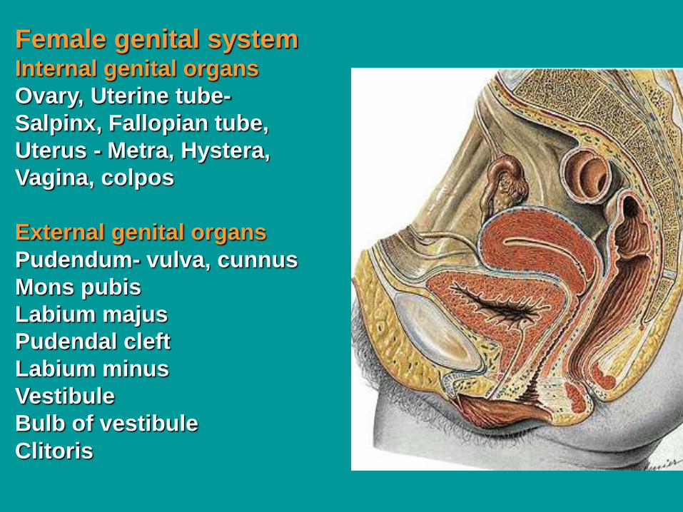
Female genital systemInternal genital organs
Ovary, Uterine tube-
Salpinx, Fallopian tube,
Uterus - Metra, Hystera,
Vagina, colpos
External genital organs
Pudendum- vulva, cunnus
Mons pubis
Labium majus
Pudendal cleft
Labium minus
Vestibule
Bulb of vestibule
Clitoris
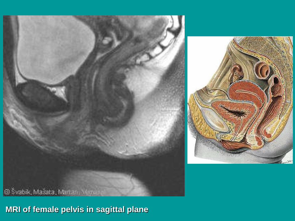
MRI of female pelvis in sagittal plane
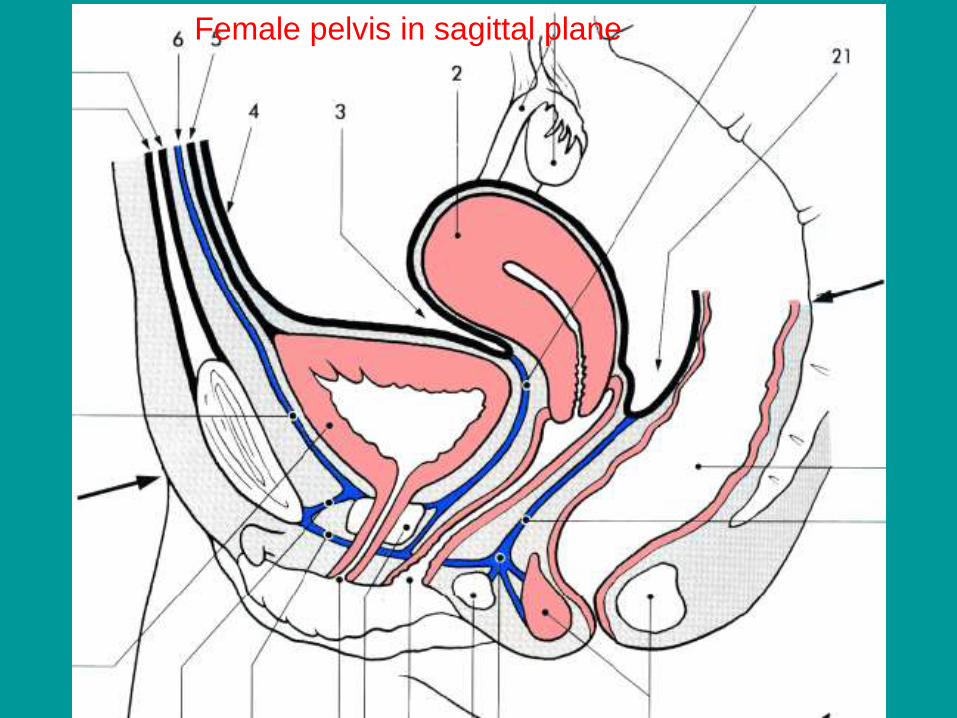
Female pelvis in sagittal plane
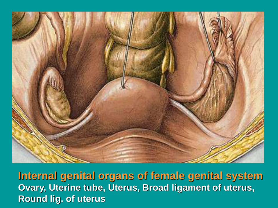
Internal genital organs of female genital systemOvary, Uterine tube, Uterus, Broad ligament of uterus,
Round lig. of uterus
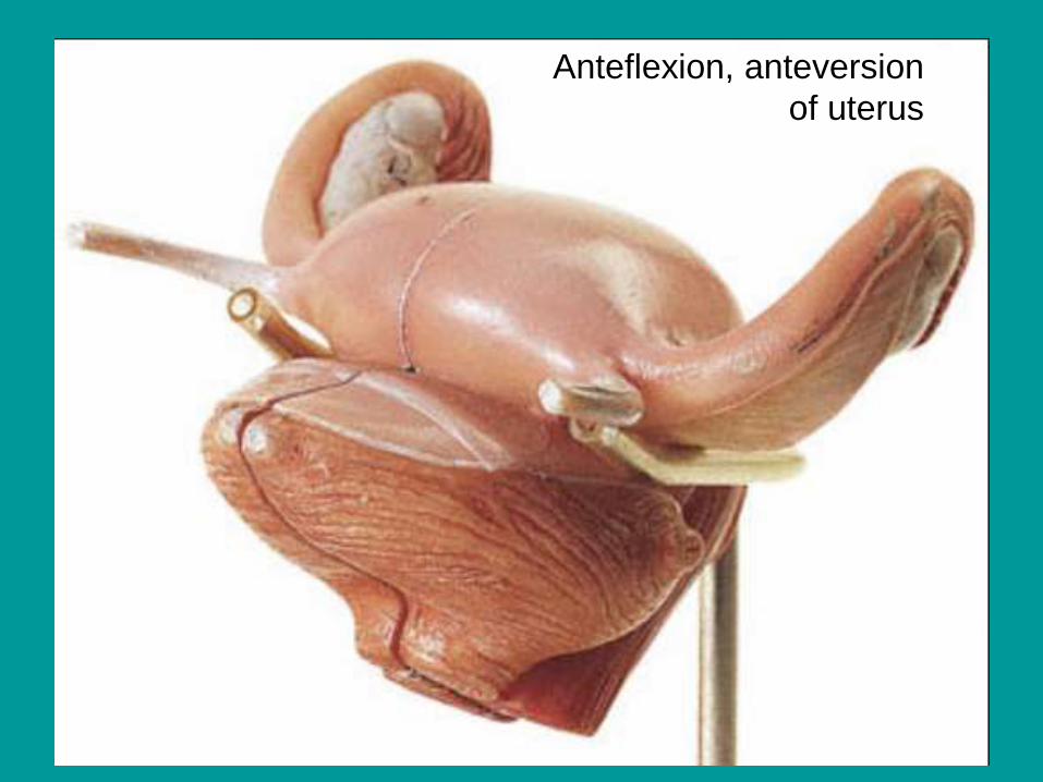
Anteflexion, anteversion
of uterus
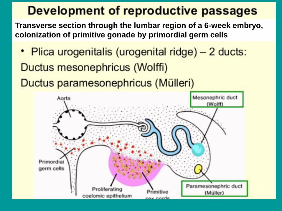
Transverse section through the lumbar region of a 6-week embryo,
colonization of primitive gonade by primordial germ cells
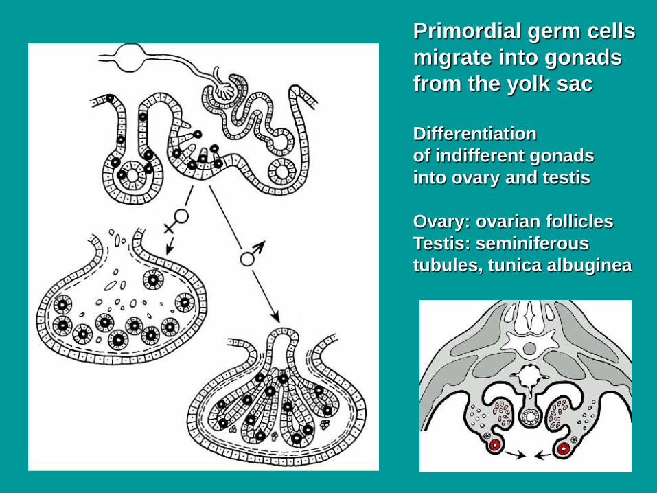
Primordial germ cells
migrate into gonads
from the yolk sac
Differentiation
of indifferent gonads
into ovary and testis
Ovary: ovarian follicles
Testis: seminiferous
tubules, tunica albuginea
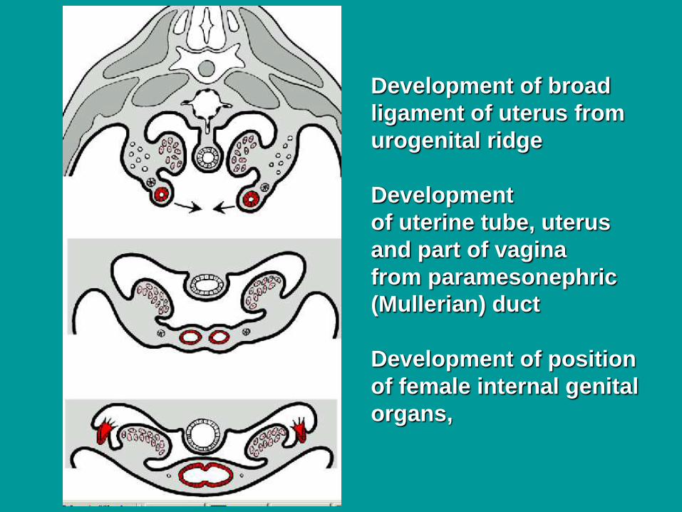
Development of broad
ligament of uterus from
urogenital ridge
Development
of uterine tube, uterus
and part of vagina
from paramesonephric
(Mullerian) duct
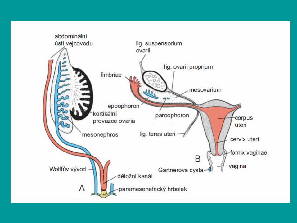
Development of position
of female internal genital
organs,

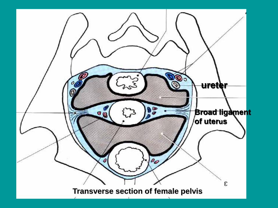
Transverse section of female pelvis
Broad ligament
of uterus
ureter
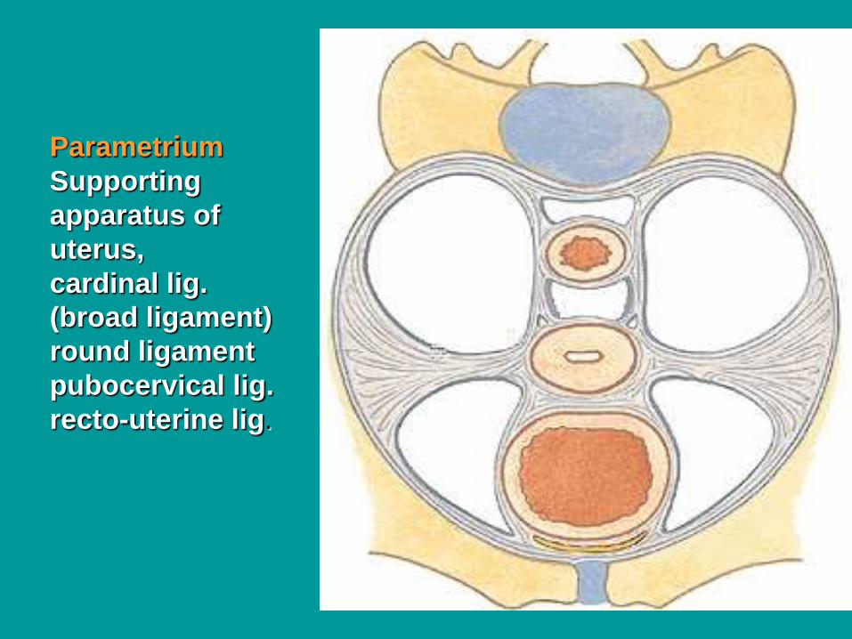
Parametrium
Supporting
apparatus of
uterus,
cardinal lig.
(broad ligament)
round ligament
pubocervical lig.
recto-uterine lig.
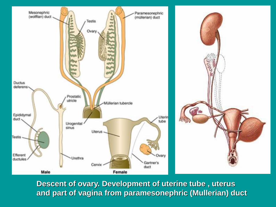
Descent of ovary. Development of uterine tube , uterus
and part of vagina from paramesonephric (Mullerian) duct
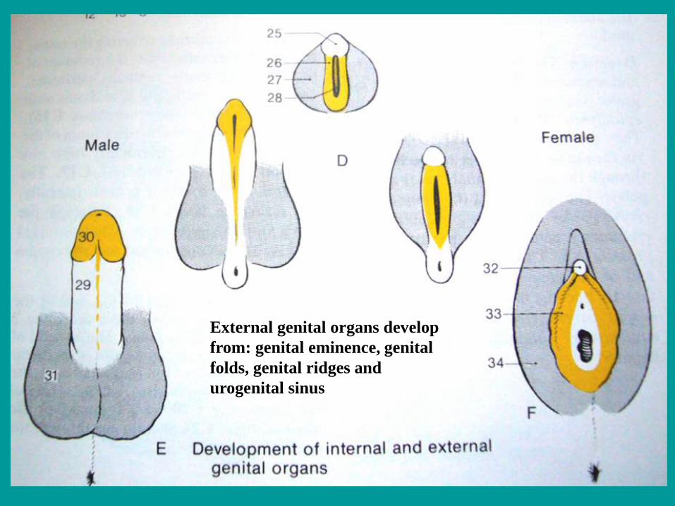
External genital organs develop
from: genital eminence, genital
folds, genital ridges and
urogenital sinus
Transverse section of female pelvis
Broad ligament
of uterus
ureter

Ovary (posterior view)
Tubal + uterine extremity, Medial + lateral surface
Free + mesovarian border,
Mesovarium, Uteroovaric lig., Suspensory lig. of ovary,
Mesosalpinx, Mesometrium
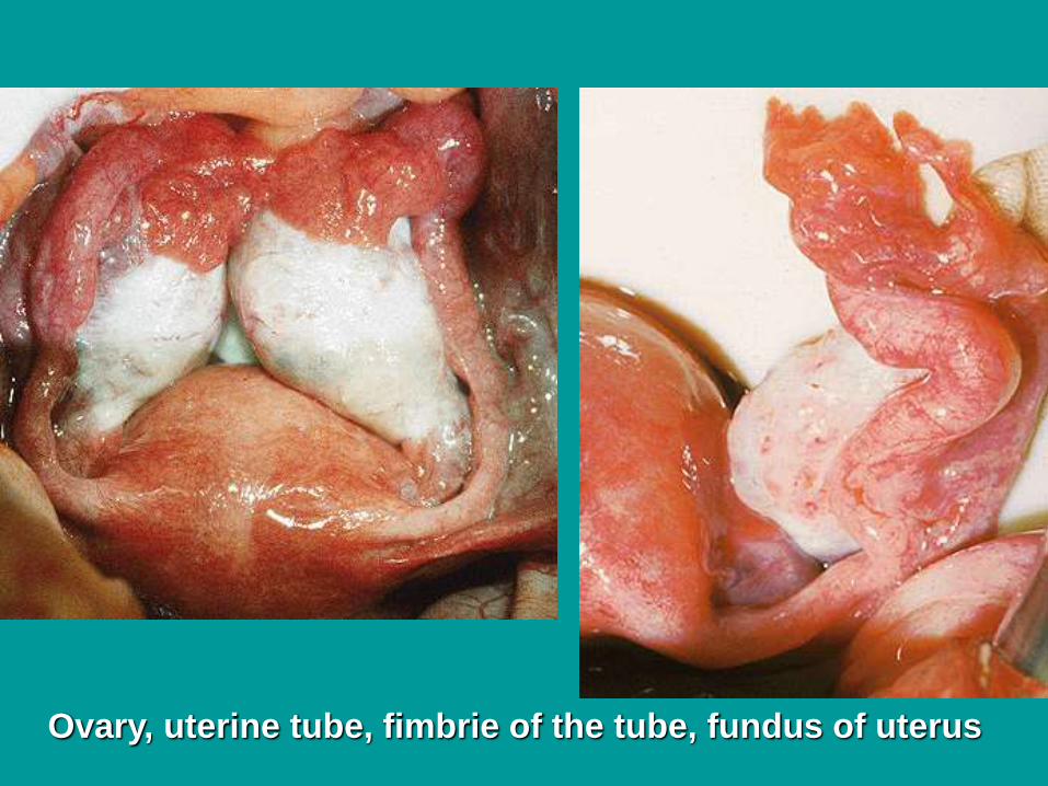
Ovary, uterine tube, fimbrie of the tube, fundus of uterus
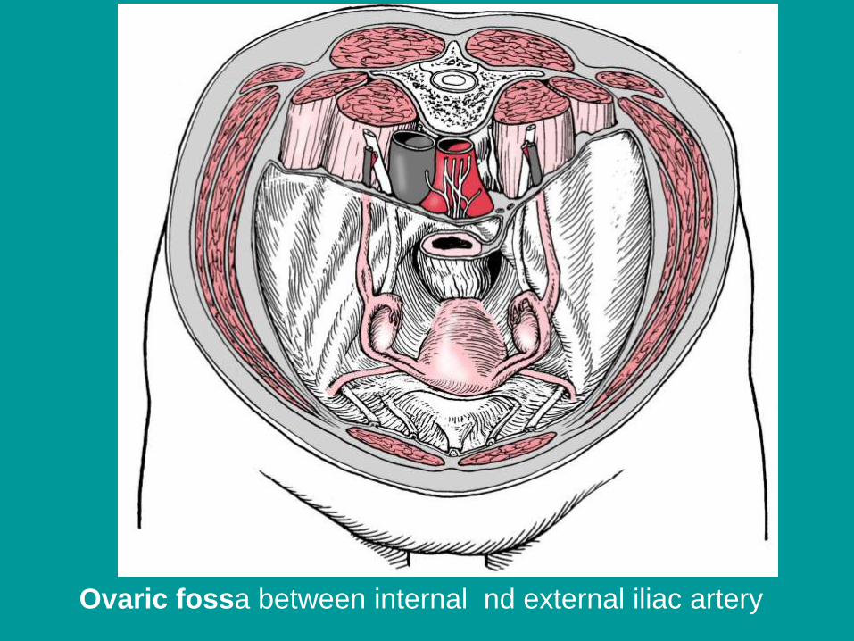
Ovaric fossa between internal nd external iliac artery
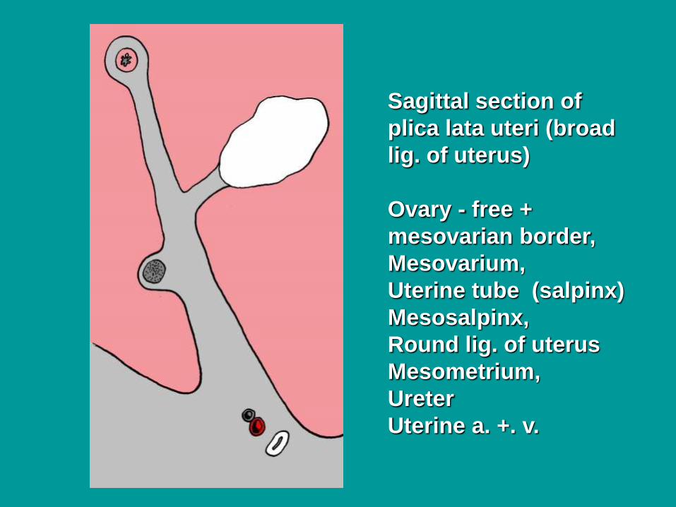
Sagittal section of
plica lata uteri (broad
lig. of uterus)
Ovary - free +
mesovarian border,
Mesovarium,
Uterine tube (salpinx)
Mesosalpinx,
Round lig. of uterus
Mesometrium,
Ureter
Uterine a. +. v.
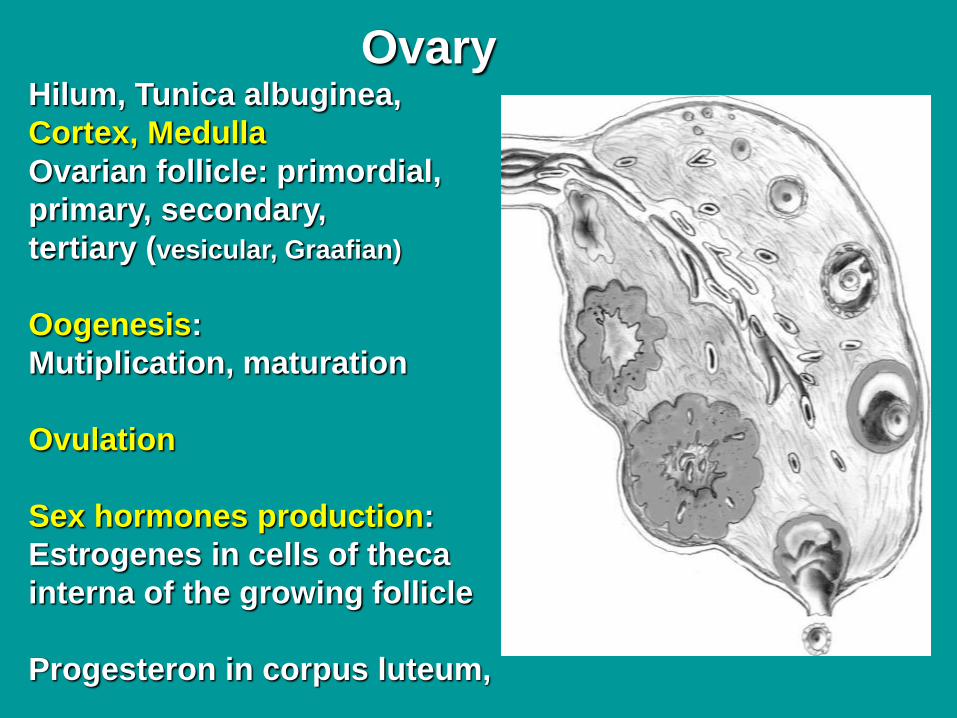
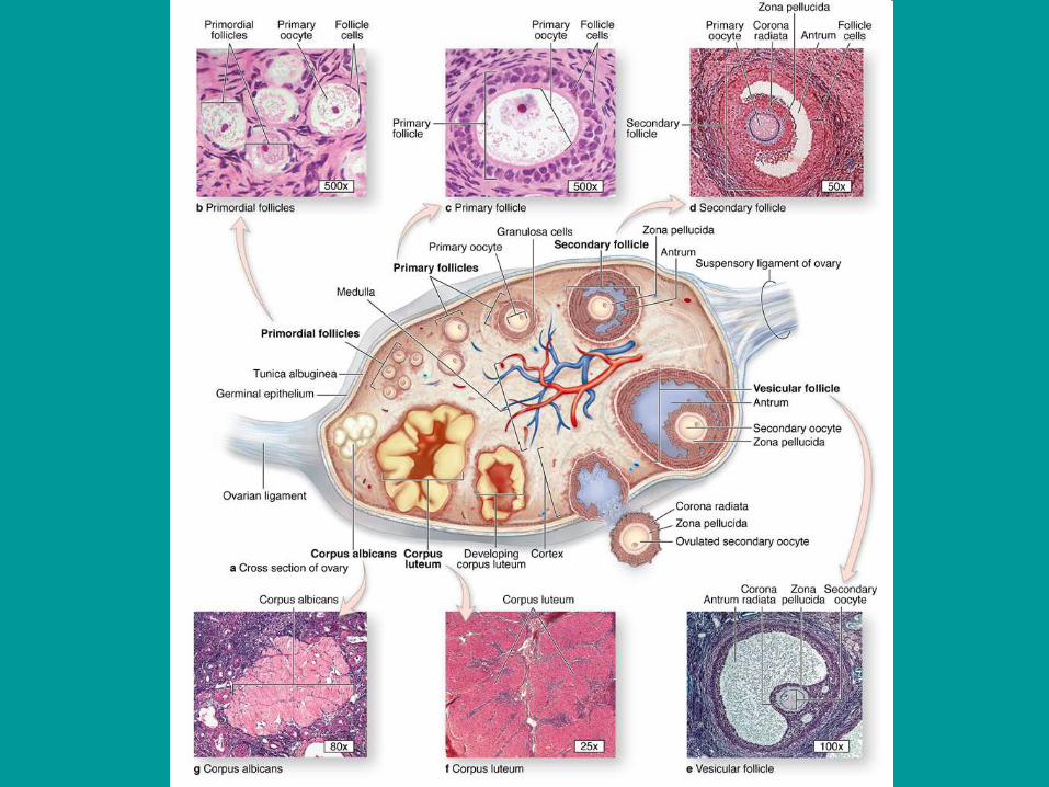
OvaryHilum, Tunica albuginea,
Cortex, Medulla
Ovarian follicle: primordial,
primary, secondary,
tertiary (vesicular, Graafian)
Oogenesis:
Mutiplication, maturation
Ovulation
Sex hormones production:
Estrogenes in cells of theca
interna of the growing follicle
Progesteron in corpus luteum,
Corpus albigans
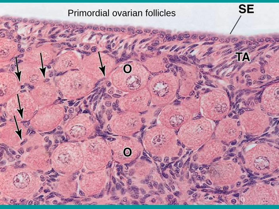
Primordial ovarian follicles
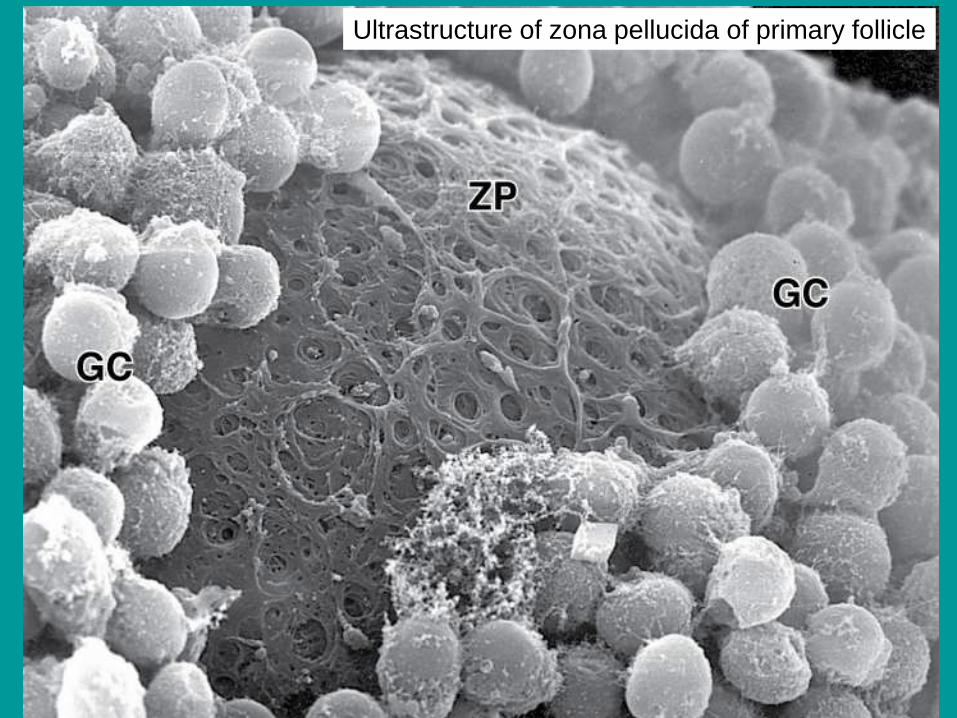
Ultrastructure of zona pellucida of primary follicle
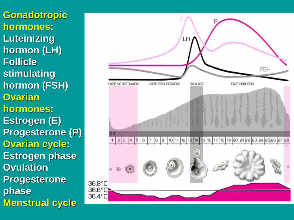
Gonadotropic
hormones:
Luteinizing
hormon (LH)
Follicle
stimulating
hormon (FSH)
Ovarian
hormones:
Estrogen (E)
Progesterone (P)
Ovarian cycle:
Estrogen phase
Ovulation
Progesterone
phase
Menstrual cycle
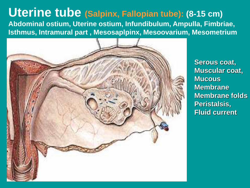
Uterine tube (Salpinx, Fallopian tube): (8-15 cm) Abdominal ostium, Uterine ostium, Infundibulum, Ampulla, Fimbriae,
Isthmus, Intramural part , Mesosaplpinx, Mesoovarium, Mesometrium
Serous coat,
Muscular coat,
Mucous
Membrane
Membrane folds
Peristalsis,
Fluid current
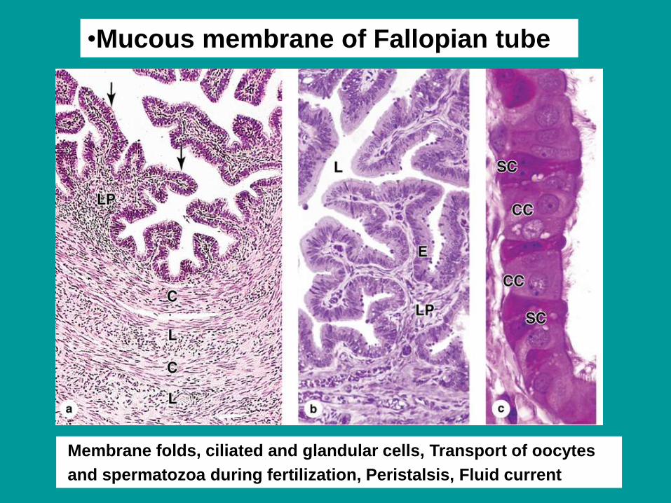
Membrane folds, ciliated and glandular cells, Transport of oocytes
and spermatozoa during fertilization, Peristalsis, Fluid current
•Mucous membrane of Fallopian tube
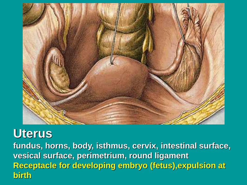
Uterusfundus, horns, body, isthmus, cervix, intestinal surface,
vesical surface, perimetrium, round ligament
Receptacle for developing embryo (fetus),expulsion at
birth
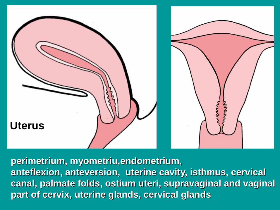
perimetrium, myometriu,endometrium,
anteflexion, anteversion, uterine cavity, isthmus, cervical
canal, palmate folds, ostium uteri, supravaginal and vaginal
part of cervix, uterine glands, cervical glands
Uterus
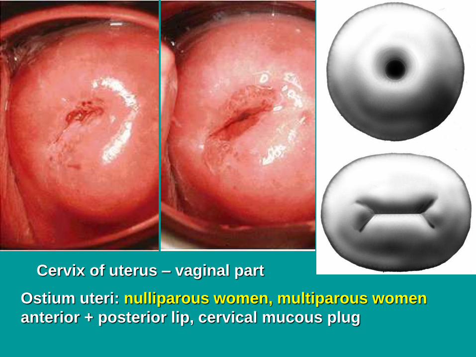
Cervix of uterus – vaginal part
Ostium uteri: nulliparous women, multiparous women
anterior + posterior lip, cervical mucous plug
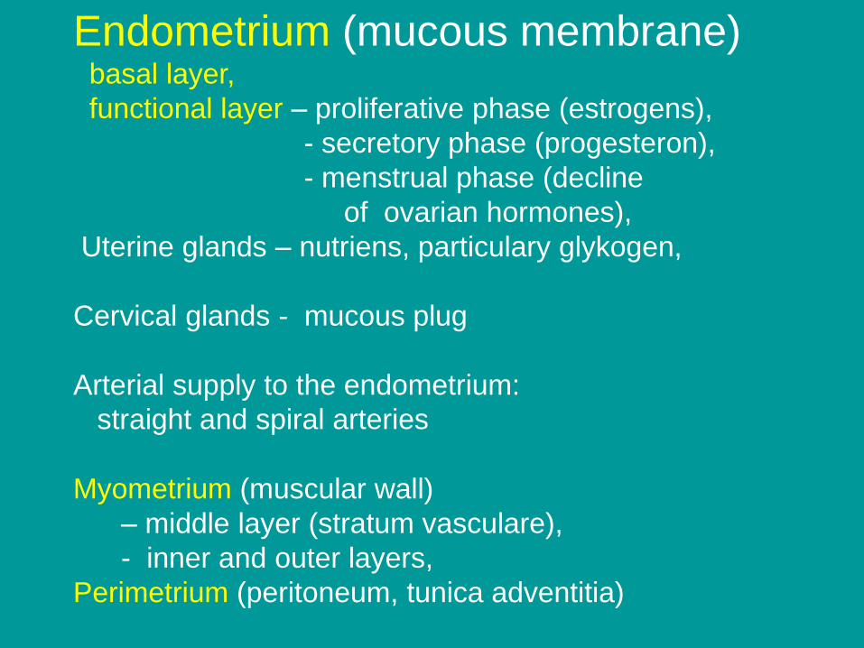
Endometrium (mucous membrane) basal layer,
functional layer – proliferative phase (estrogens),
- secretory phase (progesteron),
- menstrual phase (decline
of ovarian hormones),
Uterine glands – nutriens, particulary glykogen,
Cervical glands - mucous plug
Arterial supply to the endometrium:
straight and spiral arteries
Myometrium (muscular wall)
– middle layer (stratum vasculare),
- inner and outer layers,
Perimetrium (peritoneum, tunica adventitia)
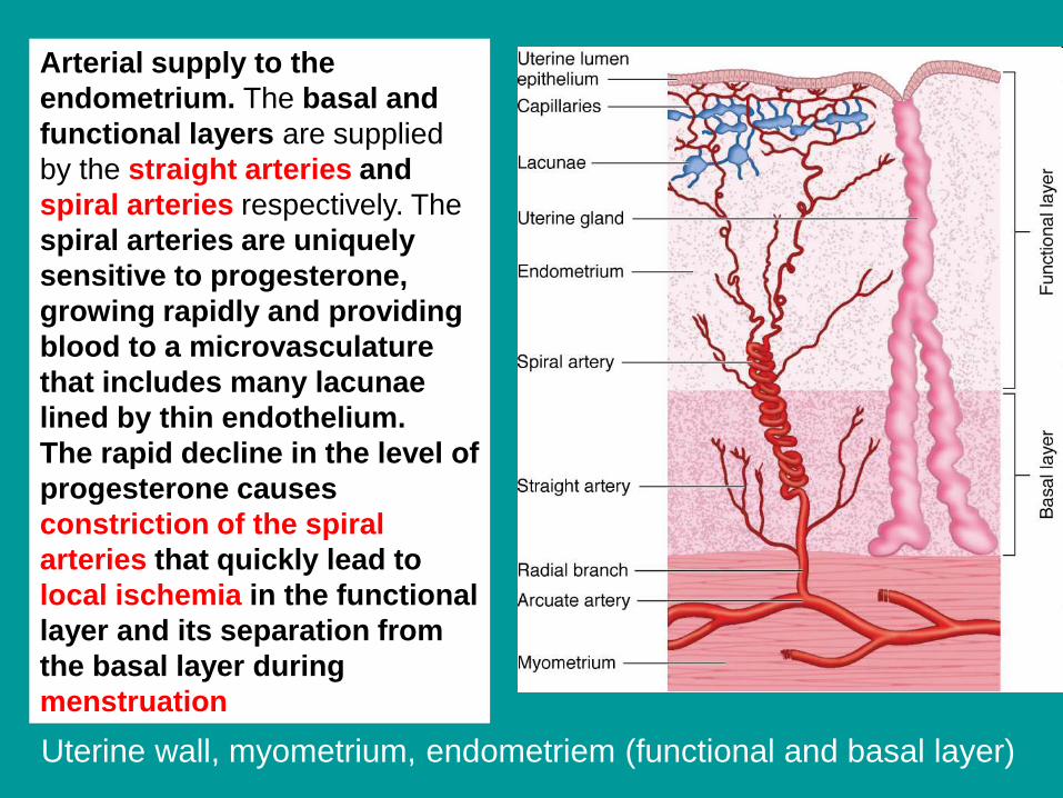
Arterial supply to the
endometrium. The basal and
functional layers are supplied
by the straight arteries and
spiral arteries respectively. The
spiral arteries are uniquely
sensitive to progesterone,
growing rapidly and providing
blood to a microvasculature
that includes many lacunae
lined by thin endothelium.
The rapid decline in the level of
progesterone causes
constriction of the spiral
arteries that quickly lead to
local ischemia in the functional
layer and its separation from
the basal layer during
menstruation
Uterine wall, myometrium, endometriem (functional and basal layer)
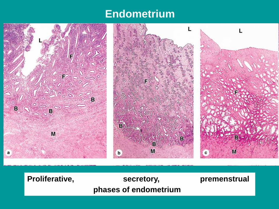
Proliferative, secretory, premenstrual
phases of endometrium
Endometrium
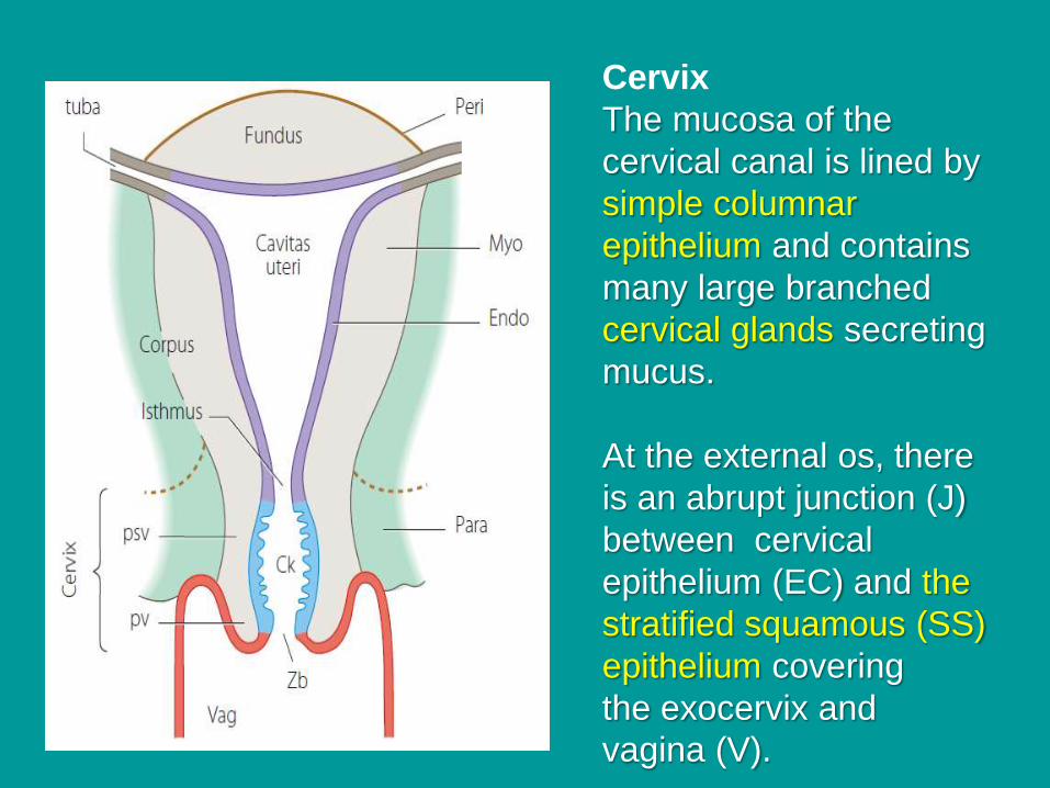
Cervix
The mucosa of the
cervical canal is lined by
simple columnar
epithelium and contains
many large branched
cervical glands secreting
mucus.
At the external os, there
is an abrupt junction (J)
between cervical
epithelium (EC) and the
stratified squamous (SS)
epithelium covering
the exocervix and
vagina (V).
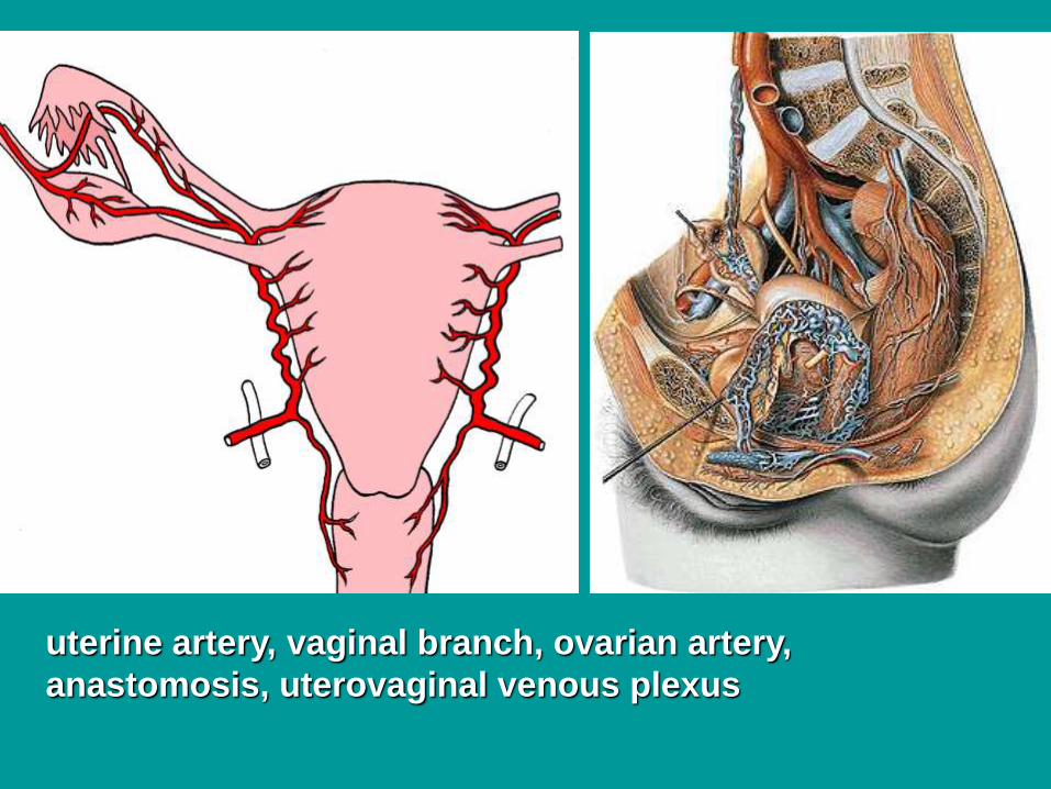
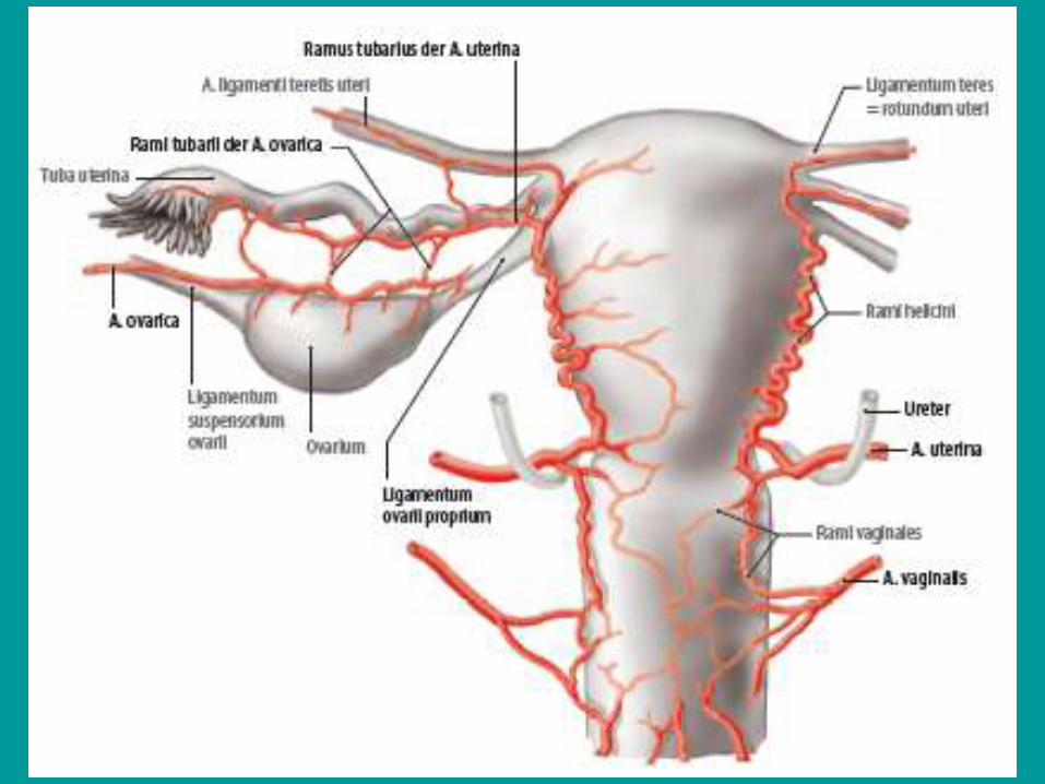
uterine artery, vaginal branch, ovarian artery,
anastomosis, uterovaginal venous plexus
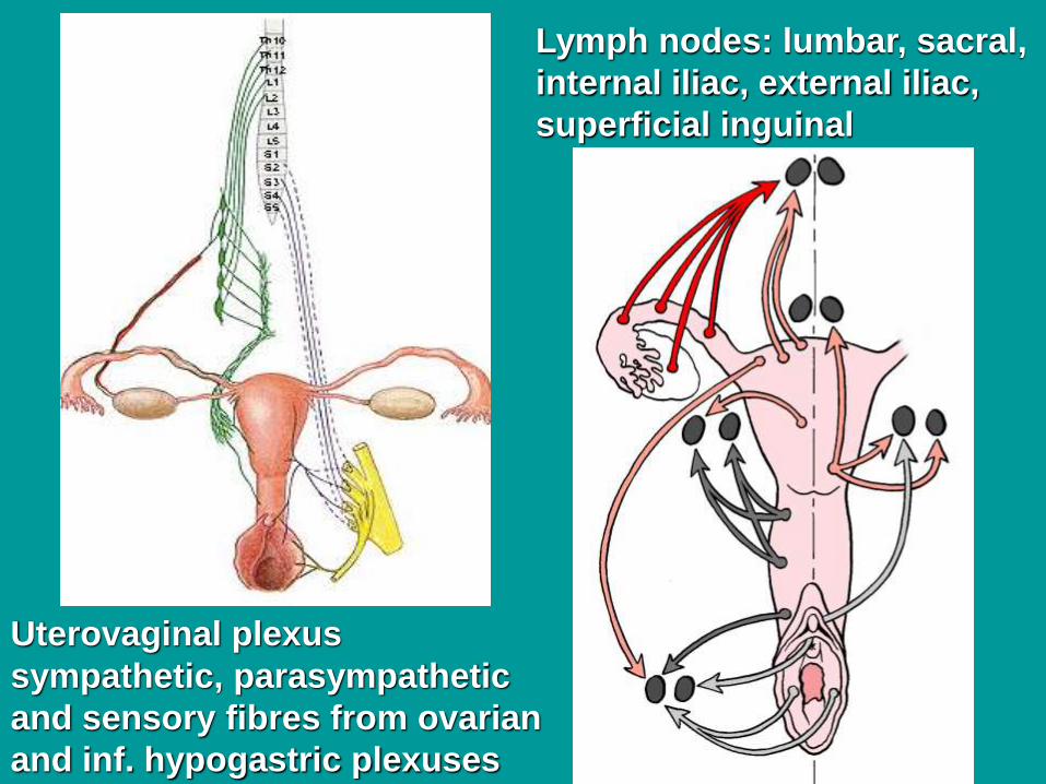
Lymph nodes: lumbar, sacral,
internal iliac, external iliac,
superficial inguinal
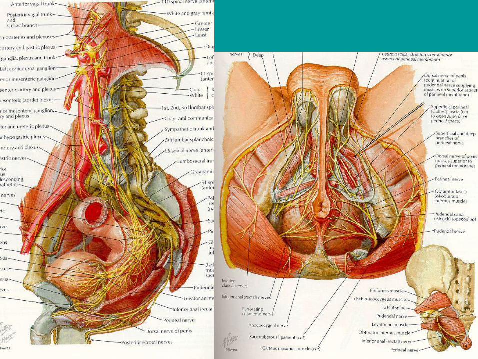
Uterovaginal plexus
sympathetic, parasympathetic
and sensory fibres from ovarian
and inf. hypogastric plexuses
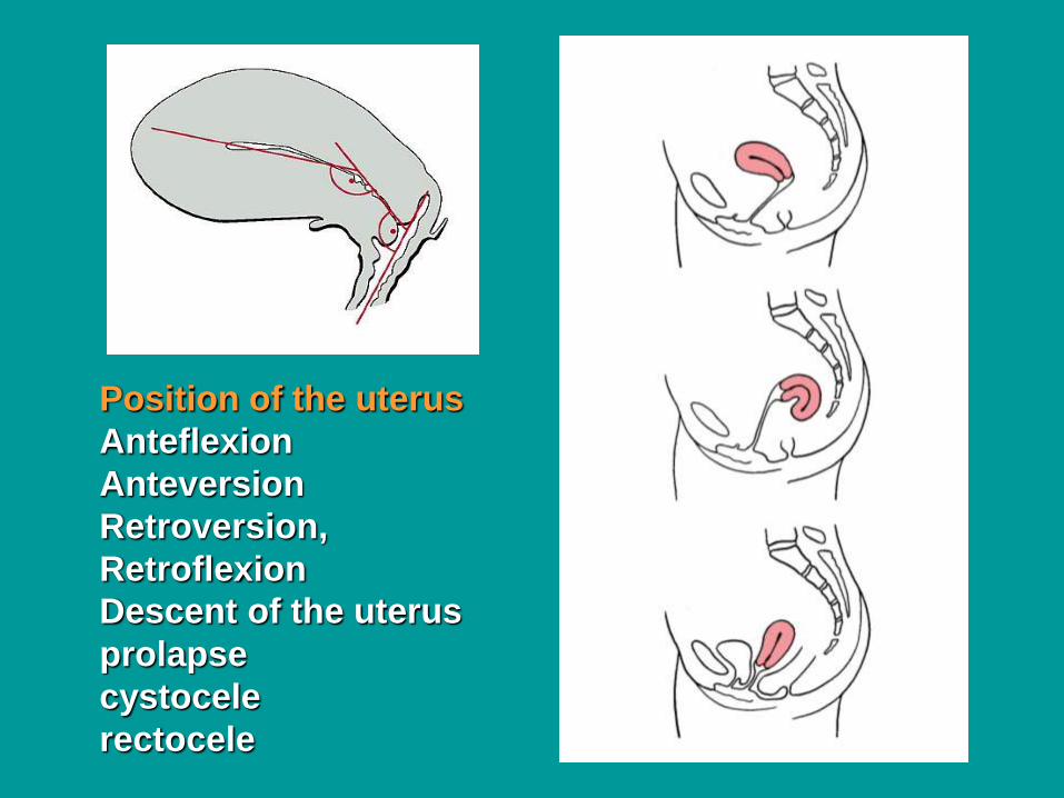
Position of the uterus
Anteflexion
Anteversion
Retroversion,
Retroflexion
Descent of the uterus
prolapse
cystocele
rectocele
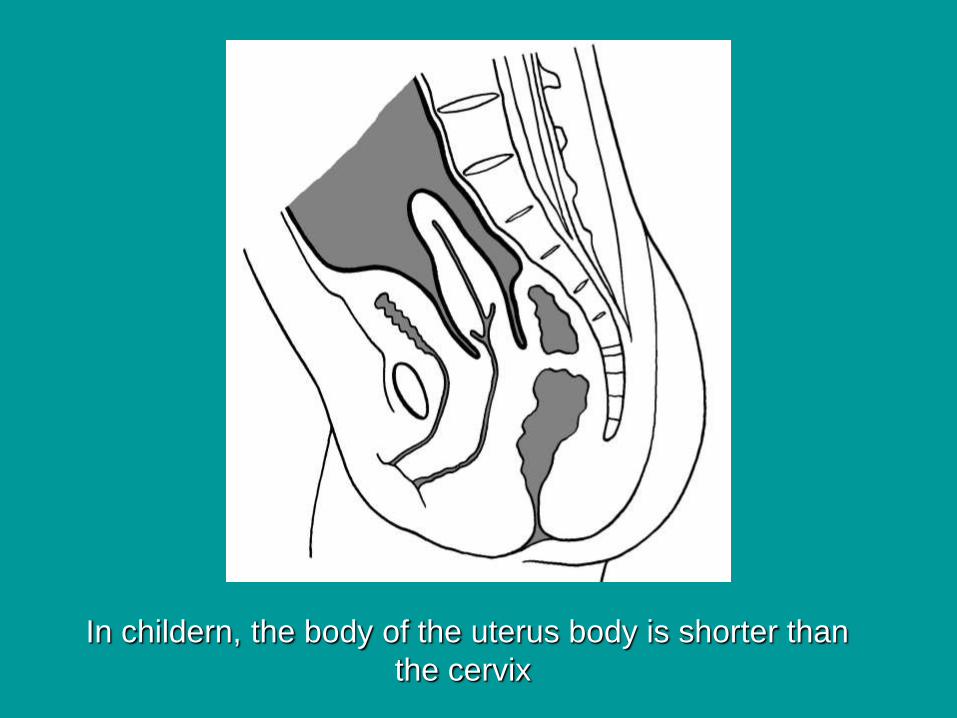
In childern, the body of the uterus body is shorter than
the cervix
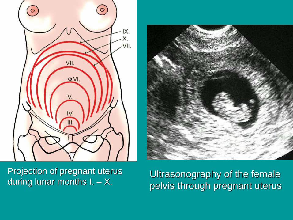
Projection of pregnant uterus
during lunar months I. – X. Ultrasonography of the female
pelvis through pregnant uterus
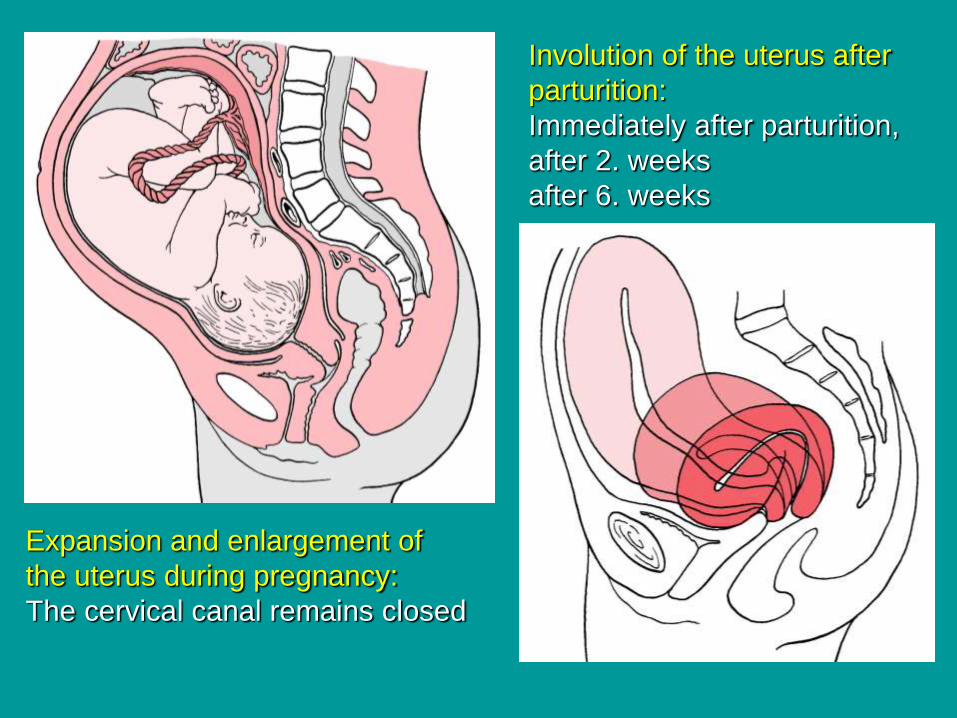
Expansion and enlargement of
the uterus during pregnancy:
The cervical canal remains closed
Involution of the uterus after
parturition:
Immediately after parturition,
after 2. weeks
after 6. weeks
VaginaThe vagina has mucosal, muscular, and adventitial layers
There are no secretory glands, cells of the thick,
nonkeratinized stratified squamous epithelium become
filled with glycogen before desquamation.
The lamina propria mucosae contains thin—walled veins
and muscular layers exude fluid on the surface
of the epithelium.
The papillae and entire lamina propria are very rich
in protective lymphocytes and neutrophils.
The muscular layer has bundles of smooth muscle
arranged in a circular manner near the mucosa and
longitudinally near the adventitia.
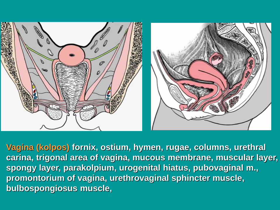
Vagina (kolpos) fornix, ostium, hymen, rugae, columns, urethral
carina, trigonal area of vagina, mucous membrane, muscular layer,
spongy layer, parakolpium, urogenital hiatus, pubovaginal m.,
promontorium of vagina, urethrovaginal sphincter muscle,
bulbospongiosus muscle,
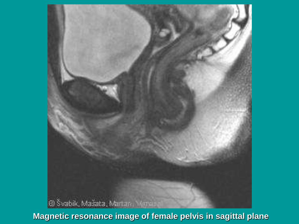
Magnetic resonance image of female pelvis in sagittal plane
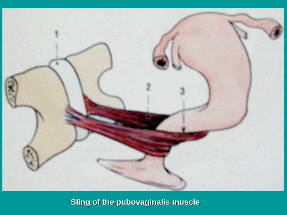
Sling of the pubovaginalis muscle
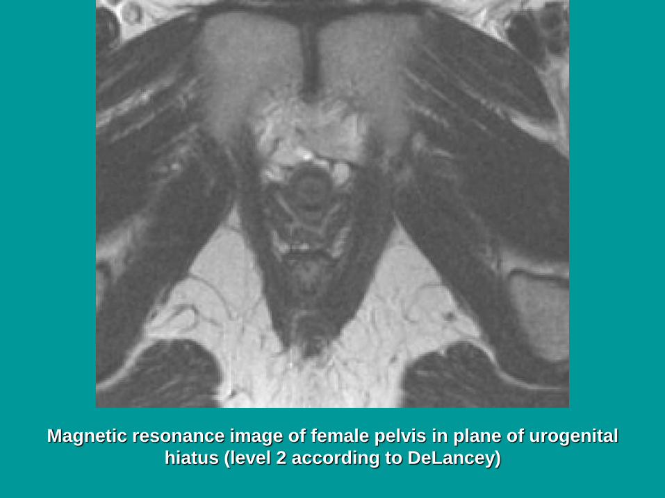
Magnetic resonance image of female pelvis in plane of urogenital
hiatus (level 2 according to DeLancey)
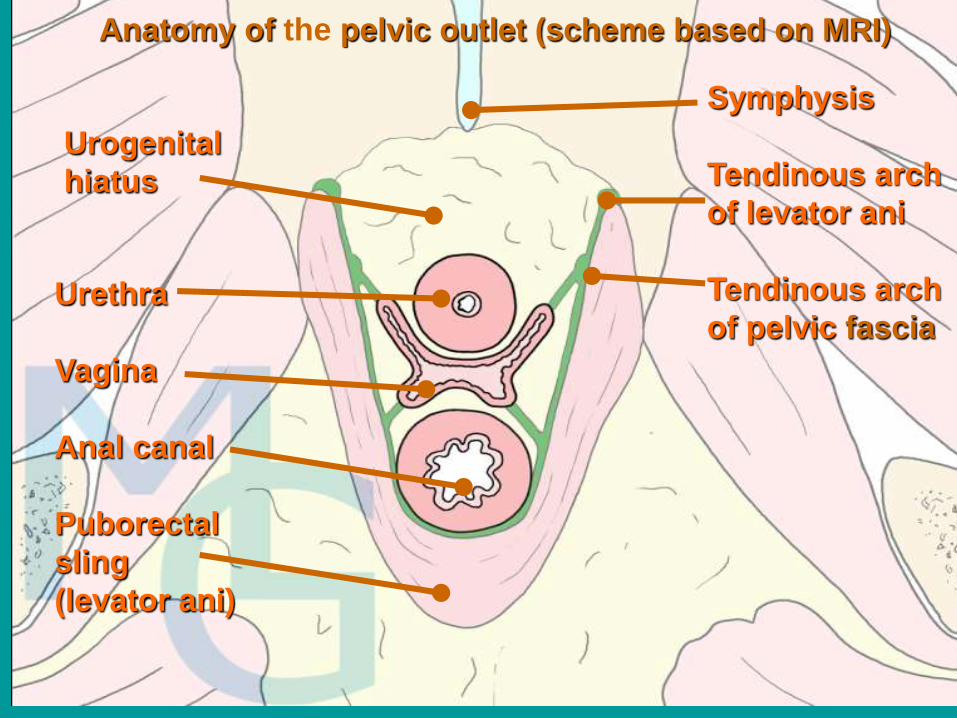
Symphysis
Tendinous arch
of levator ani
Tendinous arch
of pelvic fasciaUrethra
Vagina
Anal canal
Puborectal
sling
(levator ani)
Anatomy of the pelvic outlet (scheme based on MRI)
Urogenital
hiatus
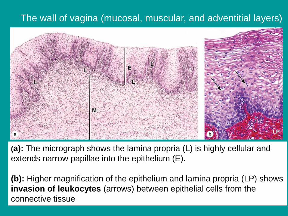
(a): The micrograph shows the lamina propria (L) is highly cellular and
extends narrow papillae into the epithelium (E).
(b): Higher magnification of the epithelium and lamina propria (LP) shows
invasion of leukocytes (arrows) between epithelial cells from the
connective tissue
The wall of vagina (mucosal, muscular, and adventitial layers)
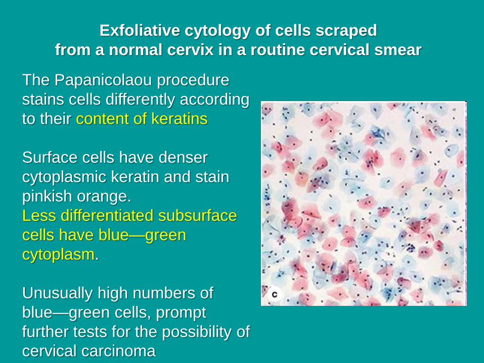
The Papanicolaou procedure
stains cells differently according
to their content of keratins
Surface cells have denser
cytoplasmic keratin and stain
pinkish orange.
Less differentiated subsurface
cells have blue—green
cytoplasm.
Unusually high numbers of
blue—green cells, prompt
further tests for the possibility of
cervical carcinoma
Exfoliative cytology of cells scraped
from a normal cervix in a routine cervical smear
External
genital organsPudendum (vulva)
Mons pubis
Labium majus
Labium minus
Pudendal cleft
Vestibule
Bulb of vestibule
Clitoris
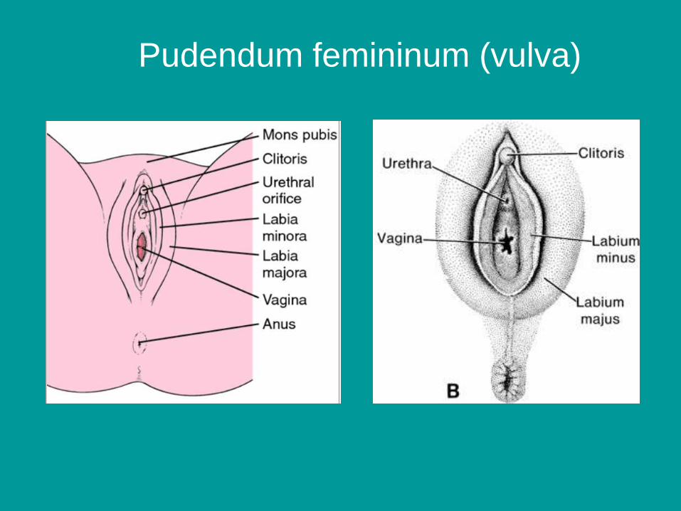
Pudendum femininum (vulva)

External genital organs develop
from: genital eminence, genital
folds, genital ridges and
urogenital sinus
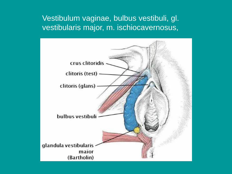
Vestibulum vaginae, bulbus vestibuli, gl.
vestibularis major, m. ischiocavernosus,
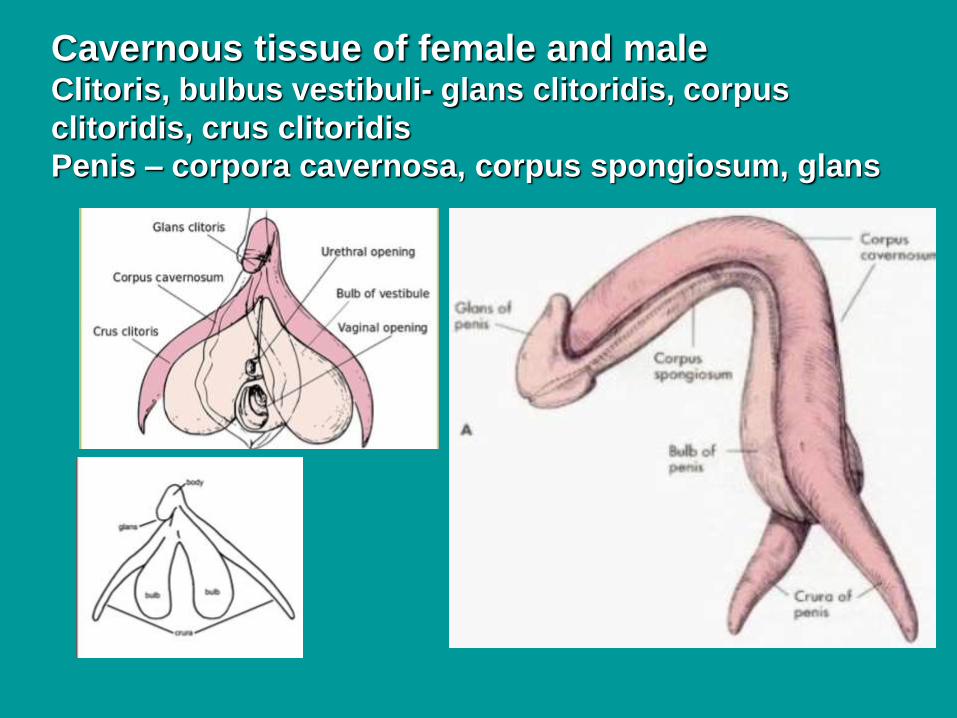
Cavernous tissue of female and maleClitoris, bulbus vestibuli- glans clitoridis, corpus
clitoridis, crus clitoridis
Penis – corpora cavernosa, corpus spongiosum, glans
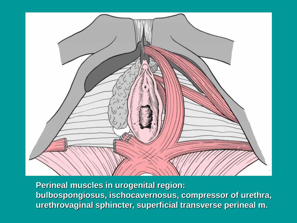
Perineal muscles in urogenital region:
bulbospongiosus, ischocavernosus, compressor of urethra,
urethrovaginal sphincter, superficial transverse perineal m.

Sources of illustrations used :Gray´s Anatomy,
Sobotta: Atlas der Anatomie des Menschen
Grim, Druga: Regional Anatomy, Galen, Prague 2012
Benninghoff, Drenckhahn: Anatomie I., II.
Carlson,B.M.: Human Embryology and Developmental Anatomy
Recommended Textbooks:R. S. Snell: Clinical Anatomy. 7th Edition, Lippincott Williams &
Wilkins, 2004, pp. 478 – 562
or
K. L. Moore: Clinically oriented Anatomy, 3rd Edition, Williams &
Wilkins 1992, pp. 501 – 635
and
W. Kahle: Color Atlas/Text of Human Anatomy, Vol. 2 Internal organs.
Thieme, 4th English Edition, 1993
Langman´s Medical Embryology,11th Edition, 2010
Junqueira´s Basic Histology 12th Edition, 2010
Ross MH, Pawlina W: Histology, 5th edition, Lippincott
Wiliams, Wilkins, 2005
Gilroy, MacPherson, Schuenke, Schulte, Schumacher: Atlas of
Anatomy, 3rd edition, Thieme 2016