Embed Size (px)
Citation preview
TECHNISCHE UNIVERSITÄT MÜNCHEN
Fakultät für Medizin
Institut für Virologie
Hepatitis C virus control:
viral resistance, new therapeutic targets
and host defense.
Hanaa M. Gaber Moawad
Vollständiger Abdruck der von der Fakultät für Medizin der Technischen Universität München
zur Erlangung des akademischen Grades eines
Doctor of Philosophy (Ph.D.)
genehmigten Dissertation.
Vorsitzende: Univ.-Prof. Dr. Agnes Görlach
Betreuerin: Univ.-Prof. Dr. Ulrike Protzer
Prüfer der Dissertation:
1. Priv.-Doz. Dr. Andreas Umgelter
2. Priv.-Doz. Dr. Fabian Geisler
Die Dissertation wurde am 03.08.2016 bei der Fakultät für Medizin der Technischen Universität
München eingereicht und durch die Fakultät für Medizin am 12.09.2016 angenommen.
I
A dissertation entitled
Hepatitis C virus control:
viral resistance, new therapeutic targets
and host defense.
By
Hanaa Mohamed Gaber Moawad
Submitted to the Faculty of Medicine, Technical University of Munich as partial
fulfillment of the requirements for the
Doctor of Philosophy Degree
Supervisor Prof. Dr. Ulrike Protzer
2016
II
Dedication
I would like to dedicate my thesis to my beloved parents Mrs. Nawal Fayek and Mr. Mohamed
Gaber and my family their support and keeping me up whenever I am down.
To the memory of my Grandfather Salah Fayek (1934-2014). For his support and inspiration and
to believe that everything is possible.
To the memory of my Uncle Ashraf Fayek (1960-2013). For his encouragement and support for
my career.
I dedicate my Ph.D. to you.
Hanaa Gaber 2016

III
Acknowledgements
I would like to thank a lot of people for their support along one of my important goals in my life,
and it is hard to find words to express my gratitude and my deep thanks to them.
First of all, I would like to thank my supervisor Prof. Dr. Ulrike Protzer for the opportunity to
join the lab and for her mentorship and guidance. I thank her for giving me the opportunity and
freedom to think and grow to become an independent scientist.
Then I would like to thank my thesis supervisory committee Dr. Med. Fabian Geisler and Dr.
Andreas Umgelter for the advice and suggestions that I gained in our meetings which helped me
guiding my project.
Next, I would like to thank the former lab members, Dr. Christian Bach and Dr. Ke Zhang, for
all the technical support and scientific discussions, which helped me a lot in my first project in
the lab.
I would like to thank all people in the diagnostic lab especially Dr. Dieter Hoffman, Dr. Barbara
Bauer and all assistance, for all viral RNA extractions and the clinical information.
And I would like to thank a wonderful group of people including Stefanie Graf, Xiaoming
Cheng, Lili Cho, Cam Tu Ho, Theresa Asen for being always around, and the great person Julia
Graf for being kind, supportive, open minded and being open for all discussions and questions.
You are a wonderful group of people to work with and I consider you my friends as well as
labmates.
Special thanks also goes to Katrin Singethan, I don’t know how to start, thanks a lot you made
me feel welcome from the first day we started working together, for being the first to support and
introduce me to the S3 lab and to the HCV techniques, more than this to make my life in Munich
much easier and for your support in moments when everything seemed to go wrong. Even in
your busy time, you could always make a time for problem solving and discussions.
I would like to thank all participants in my Ph.D. projects, especially Akram Amin for working
together on what sounds first crazy and impossible idea, for all the discussions, the cooperation
which made the outer space environment a possible lab for our project. Then I would like to
thank Prof. Dr. Dierk Niessing and Dr. Robert Janowski, for joining the Crystallization project
IV
and offering the platform, the expertise and their effort which succeeded in achieving the project
goal.
I would like to thank all people work in the Medical Life Science and Technology Ph.D.
program; special thanks go to Desislava Zlatanova, for her positive attitude, for being supportive,
patient and helpful in solving all problems and for her guidance along the program requirements
and the graduation steps.
Thanks for all the collaborative labs that opened the door to get more insights during my Ph.D.,
in Germany, Prof. Dr. Ralf Bartenschlager and in the USA Dr. Jake Liang, for giving me the
chance for training in their labs.
I would like to thank DAAD (GERLS) program for funding my Ph.D. studies in Germany, for all
the before and during and after study guidance and support, I am proud of being an Alumni of
this institution.
My deep thanks to NASA and Space X for funding the ``Space part`` of the second project in my
thesis and to introduce us to a different research discipline.
V
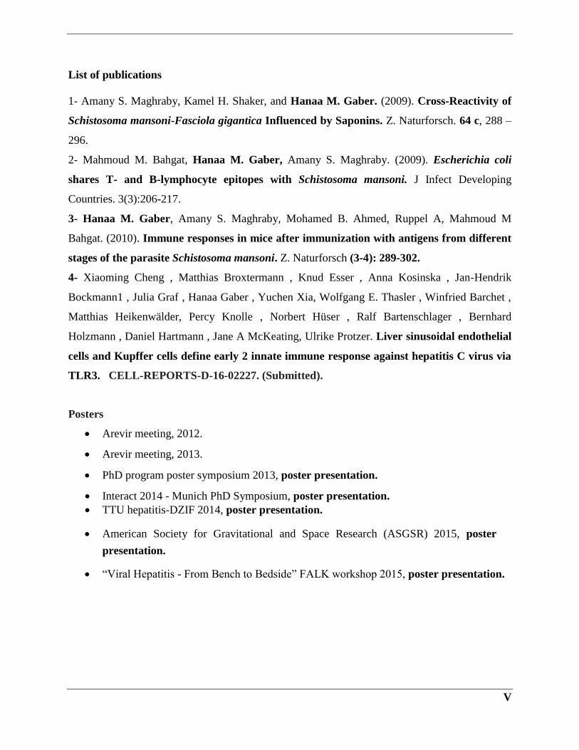
List of publications
1- Amany S. Maghraby, Kamel H. Shaker, and Hanaa M. Gaber. (2009). Cross-Reactivity of
Schistosoma mansoni-Fasciola gigantica Influenced by Saponins. Z. Naturforsch. 64 c, 288 –
296.
2- Mahmoud M. Bahgat, Hanaa M. Gaber, Amany S. Maghraby. (2009). Escherichia coli
shares T- and B-lymphocyte epitopes with Schistosoma mansoni. J Infect Developing
Countries. 3(3):206-217.
3- Hanaa M. Gaber, Amany S. Maghraby, Mohamed B. Ahmed, Ruppel A, Mahmoud M
Bahgat. (2010). Immune responses in mice after immunization with antigens from different
stages of the parasite Schistosoma mansoni. Z. Naturforsch (3-4): 289-302.
4- Xiaoming Cheng , Matthias Broxtermann , Knud Esser , Anna Kosinska , Jan-Hendrik
Bockmann1 , Julia Graf , Hanaa Gaber , Yuchen Xia, Wolfgang E. Thasler , Winfried Barchet ,
Matthias Heikenwälder, Percy Knolle , Norbert Hüser , Ralf Bartenschlager , Bernhard
Holzmann , Daniel Hartmann , Jane A McKeating, Ulrike Protzer. Liver sinusoidal endothelial
cells and Kupffer cells define early 2 innate immune response against hepatitis C virus via
TLR3. CELL-REPORTS-D-16-02227. (Submitted).
Posters
Arevir meeting, 2012.
Arevir meeting, 2013.
PhD program poster symposium 2013, poster presentation.
Interact 2014 - Munich PhD Symposium, poster presentation.
TTU hepatitis-DZIF 2014, poster presentation.
American Society for Gravitational and Space Research (ASGSR) 2015, poster
presentation.
“Viral Hepatitis - From Bench to Bedside” FALK workshop 2015, poster presentation.
VI
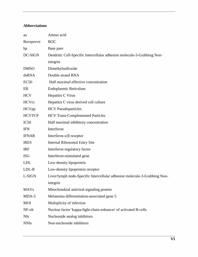
Abberviations
aa Amino acid
Boceprevir BOC
bp Base pare
DC-SIGN Dendritic Cell-Specific Intercellular adhesion molecule-3-Grabbing Non-
integrin
DMSO Dimethylsulfoxide
dsRNA Double strand RNA
EC50 Half maximal effective concentration
ER Endoplasmic Reticulum
HCV Hepatitis C Virus
HCVcc Hepatitis C virus derived cell culture
HCVpp HCV Pseudoparticles
HCVTCP HCV Trans-Complemented Particles
IC50 Half maximal inhibitory concentration
IFN Interferon
IFNAR Interferon α/β receptor
IRES Internal Ribosomal Entry Site
IRF Interferon regulatory factor
ISG Interferon-stimulated gene
LDL Low-density lipoprotein
LDL-R Low-density lipoprotein receptor
L-SIGN Liver/lymph node-Specific Intercellular adhesion molecule-3-Grabbing Non-
integrin
MAVs Mitochondrial antiviral-signaling protein
MDA-5 Melanoma differentiation-associated gene 5
MOI Multiplicity of infection
NF-κb Nuclear factor 'kappa-light-chain-enhancer' of activated B-cells
NIs Nucleoside analog inhibitors
NNIs Non-nucleoside inhibitors
VII
NS Non-structural protein
NTRs Non-translated regions
PHH Primary Human Hepatocyte
PIs Protease inhibitors
PRR Pattern Recognition Receptor
qRT-PCR Quantitative real-time PCR
RdRp RNA-dependent RNA polymerase
RIG-1 Retinoic Acid-Inducible Gene 1
RNA Ribonucleic acid
RT-PCR reverse transcription-PCR
SNP Single Nucleotide Polymorphism
SR-BI Scavenger receptor B type I
ssRNA Single strand RNA
SVR Sustained virological response
TCID50 50% Tissue Culture Infective Dose
Telaprevir TLV
TLR Toll-like receptor
TRIF TIR-domain-containing adapter-inducing interferon-β
TRIM Tripartite motif
VIII
Abstract
The studies presented in my Ph.D. outline multiple aspects of Hepatitis C research. The first
study focused on the establishment of convenient genotypic and phenotypic assays to determine
the resistance profile of the protease inhibitors Boceprevir and Telaprevir. The results reveal the
association of several mutations within NS3 with the different levels of resistance to protease
inhibitors. Moreover, genotyping of IL28b polymorphisms showed that SNP rs12979860 has
more predictive values than SNP rs8099917 and that all relapsed patients had a non-CC
genotype.
The second study was a part of NASA and the ISS contributions in the space-based research in
the biological sciences. The project aimed for determination of the 3D structure of HCV-NS5b of
the predominant genotype in Egypt (genotype 4a). During the study, we were able to employ the
protein crystallization technology using microgravity environment in the ISS along with the earth
crystallography techniques to obtain crystallize of good quality for X-ray diffraction. In the pre-
flight procedure, we used the available standardized procedure including protein production,
purification and quality check for sample preparation and shipment. During the flight, protein
sample was exposed to microgravity for 25 days and the equivalent earth control was exposed to
1 gravity condition. Post flight phase, few crystals grew on the space crystal card and not in the
earth one. For technical difficulties, we could not harvest the crystals for further x-ray diffraction
experiments. The space experiment followed by further optimization of the crystallization
conditions, when we acquired crystals of good quality, crystals were illuminated by the X-ray
beam generated by synchrotron sources. The dataset of diffraction images showed that most
crystals diffracted to an average of 3.1 Å resolution. The NS5b structural model determined by
the Molecular Replacement approach showed that the structure is identical to other HCV-NS5b
polymerases, in which the HCV-NS5b of genotype 4a contains six conserved motifs. However,
the NS5b protein has different confirmations, the other conformations were not determined
because we lack the protein-inhibitor co-crystallization experiment.
In the last study, we aimed to identify the role of TRIM protein family in HCV infection. Here,
we could identify several TRIM proteins regulated by interferon and HCV infection, thus we
hypnotized a potential association of TRIM proteins with antiviral activities against HCV. In our
replicon and infection experiments, we could show that in response to TRIM5, TRIM9, TRIM14
IX
or TRIM25 overexpression, the HCV replication and infection were restricted. Further analysis
suggests that TRIM5 positively regulates IFN signaling pathway by upregulating RIG-I, MAVs,
TRIF IFN-β expression levels, while TRIM9 could enhance both IFN-b and ISRE promotors’
activation.
We could also demonstrate that TRIM14 overexpression decreases HCV replication and
infection by triggering the IFN antiviral response, notably, it could augment both IFN-β and NF-
κb promoters’ activities and increased expression levels of RIG-I, MAVs, TRIF, and IFN-β.
Finally, we could attribute the elimination of HCV infection during the TRIM25 overexpression
to the increase of the expression levels of RIG-I, TRIF, MAVs and IFN-β in addition to the
enhancement of ISRE, IFN-β, and NF-kb promoters’ activities.
X
List of contents
Chapter I Introduction……………………………………………………… 1
1. Hepatitis C virus overview…………………………………………………..1
1.1. HCV virology and genome organization……………………………...3
1.2. HCV genome organization…………………………………………….3
1.2.1. Structural proteins...………………………………………………..4
Core protein
E1 and E2 protein
1.2.2. Non-structural proteins……………………………………………..4
Non-structural protein2 (NS2)
Non-structural protein3/4A (NS3/4A)
Non-structural protein4b (NS4b)
Non-structural protein5a (NS5a)
Non-structural protein5b (NS5b)
1.3. HCV receptors……………………………………………….………8
Cluster of differentiation 81 (CD81)
Scavenger receptor B type I (SR-BI)
Dendritic Cell-Specific Intercellular adhesion molecule-3-Grabbing Non-integrin)
DC-SIGN
Low-density lipoprotein receptor (LDL-R)
Occludin
1.4. HCV lifecycle……………………………………………………….10
1.5. HCV cell culture systems…………………………………………...12
1.5.1. HCV Replicon System
Improvements of replicons
1.5.2. HCV Pseudoparticles (HCVpp)…………………………………13
1.5.3. HCV infectious virus (HCVcc) …………………………………14
1.5.4. HCV Trans-Complemented Particles (HCVTCP)…………………..14
1.6. Immune responses to HCV infection…………………………………..15
1.6.1. Innate immune responses to HCV ……………………………….....16
1.6.2. Adaptive immune responses during HCV infection……………...…16
XI
1.6.3. HCV immune escape mechanisms…………………………………..18
1.7. Genotyping and phenotyping of HCV protease inhibitors resistant mutations
………………………………………………………………18
1.7.1. HCV therapy……………………………………………………….18
1.7.1.1. A decade of Interferon- Ribavirin standard of care
therapy……………………………………………………….19
Interferon-alpha in HCV treatment
1.7.2. Direct-acting antivirals (DAAs)………………………………...…21
Protease inhibitors
The first generation of protease inhibitors (TLV and BOC)
Second wave of PIs, Simeprevir
1.7.2.1. HCV PIs resistance tools…………………………………..23
Genotypic assay for HCV PIs resistant analysis
Phenotypic assay for HCV PIs resistant analysis
Interpretation of phenotypic results
1.7.3. NS5a inhibitors…………………………………………………….26
Ledipasvir and Daclatasvir
Resistance to NS5A inhibitors
1.7.4. NS5b polymerase inhibitors…………………………………….....28
Sofosbuvir
Resistance to polymerase inhibitors
1.7.5. Host genetic predictor of treatment response: IL28b
polymorphisms…………………………………………….…........30
1.8. Crystallization of HCV NS5b polymerase and structure
determination………………………………………………………...31
1.8.1. Proteins function and structural analysis………………………......31
1.8.2. Protein crystals characteristics…………………………………….32
1.8.3. Principles of protein crystallization ……………………………...33
1.8.4. X-ray diffraction and data collection………………………………33
1.8.5. Protein crystallization in microgravity…………………………….33
XII
Microgravity
1.8.6. Gravity vs. Microgravity for protein crystallization………………..34
Proof of concept and success stories of crystallization in microgravity
1.8.7. Crystal structure of viral proteins and drug research……………....36
1.8.8. HCV protein crystallization …………………………………..……37
1.9. The role of tripartite motif family members (TRIM) in mediating susceptibility
to HCV infection………………………………………38
1.9.1. Cellular Response to Viral Infection ………………………...…..38
1.9.1.1. Interferons and their role in HCV infection………………41
Interferons activation during HCV infection
1.9.2. The role of ISGs in controlling HCV infection…………..…42
1.9.3. The TRIM protein family…………………………………………45
TRIMs modify proteins by ubiquitination
1.9.3.1. TRIM proteins immunological functions………………....47
TRIM proteins involvement in cancers and other diseases
1.9.3.2. TRIM5………………………………………………………49
1.9.3.3. TRIM9………………………………………………………50
1.9.3.4. TRIM14……………………………………………………..53
1.9.3.5. TRIM25………………………………………….………….54
Aims of the Thesis……………………………………….…………………….57
2. Chapter II: Materials and Methods…………………….………………..59
2.1. Materials…………………………………….…………………………59
2.1.1. Patient samples……………………………………………………...59
2.1.2. Chemicals and reagents……………………………………………..59
2.1.3. Kits…………………………………………………………………..60
2.1.4. Enzymes……………………………………………………………..61
2.1.5. Plasmids and bacterial strains………………………………….…....62
2.1.6. Primers…………………………………………………………..…..66
2.1.7. Cell lines………………………………………………………….....66
2.1.8. Cell culture media…………………………………………………...67
2.1.9. Devices and equipment……………………………………………...67
2.1.10. Software……………………………………………………………68
2.2. Experimental methods: Part I………………………………………….69
2.2.1. Viral RNA Extraction……………………………………………….69
XIII
2.2.2. HCV-NS3 RT- PCR amplification and sequencing………………...69
DNA concentration determination
2.2.3. DNA sequencing and Genotypic Analysis………………………….70
Direct and clone sequencing
Genotypic Analysis
2.2.4. In vitro phenotypic analysis of HCV drug resistance…….…….72
Cloning and plasmid purification
Large scale preparation of plasmid DNA (MaxPreps)
RNA in vitro transcription
Cells electroporation and drug titration
2.2.5. IL28b polymorphism genotyping…………………………………73
DNA extraction
TaqMan SNP genotyping assay
2.3. Experimental methods: Part II………………………………………74
2.3.1. Expression and purification of HCV-NS5b……………………74
SDS-polyacrylamide gel electrophoresis (SDS-PAGE)
Western blot
2.3.2. Protein Large-scale production and purification……………..76
His-tag purification of HCV-NS5b protein
Gel filtration chromatography of HCV NS5b protein
2.3.3. Microgravity crystallization……………………………………78
Filling and Freezing of CrystalCards™
Protein crystallization in the ISS
2.3.4. Optimization of crystallization…………………………..…….78
2.3.5. X-ray diffraction and data collection………………………….79
2.3.6. Structure determination and refinement……………………...79
2.4. Experimental methods: Part III
2.4.1. Cell culture……………………………………………………...80
Growth and maintenance of Huh7.5, Huh7
Growth and maintenance of HepG2-HFlL
XIV
Growth and maintenance of ED43
Primary human hepatocytes (PHHs)
2.4.2. HCV virus production………………………………………....82
TCID50 for HCV virus titer determination
2.4.3. HCV Infection experiments…………………………………....82
2.4.4. Interferon treatment experiments…………………………….82
2.4.5. Transient transfections………………………………………...82
2.4.6. Cell viability assay……………………………………………...82
2.4.7. Reporter gene assay……………………………………….…...83
2.4.8. RNA extraction, reverse transcription and qRT-PCR………83
3. Chapter III Results and Discussions
3.1. Part I: Genotyping and phenotyping of HCV protease inhibitors resistant mutations
……………………………………………….85
3.1.1. HCV PIs resistant testing
3.1.1.1. RT-PCR amplification of HCV protease-helicase domain (NS3)
3.1.1.2. Detection of resistant mutations by direct and clone sequencing analysis
3.1.1.3. Pre-existence and emergence of BOC/TLV-Resistant Variant in HCV Patients
3.1.1.4. Construction of shuttle vector containing patient isolates
3.1.1.5. Replication fitness of control and shuttle vector
3.1.1.6. Replication fitness of replicons containing different variants
3.1.1.7. Titration example of chimeric replicon containing NS3 resistant variants
3.1.1.8. Susceptibility of resistant isolates to BOC and TLV and their replication fitness
3.2. IL28 polymorphisms in HCV treatment response
3.2.1. IL28b rs8099917T/G, rs12979860C/T polymorphism genotyping
3.2.1.1. Direct DNA sequencing method
3.2.1.2. Establishment of TaqMan assay for IL28b allele discrimination
3.2.1.3. Relationship between poor response to triple therapy and IL28B polymorphisms
(SNP rs12979860 and rs8099917)
3.3. Discussion……………………………………………………….102
3.4. Summary………………………………………………………..106
XV
4. Part II: Crystallization of HCV NS5b polymerase and structure determination
……………………………………………………..108
4.1. HCV NS5b protein crystallization
4.1.1. Cloning and expression of HCV NS5b
4.1.2. HCV His-Tagged NS5b protein purification
4.1.3. Protein expression and solubility
4.1.4. Large scale production and Ni-NTA column purification
4.1.5. Size exclusion chromatography of NS5b protein
4.2. Comparison of the NS5b protein crystallization on earth and under microgravity
conditions
4.2.1. NS5b protein crystallization in the control card
4.3. Further crystallization tests and optimization
4.4. Structure determination and refinement
4.5. Structure comparison to the closest genotype
4.6. Discussion………………………………………………………….116
4.7. Summary…………………………………………………………..119
5. Part III: The role of tripartite motif family members (TRIM) in mediating
susceptibility to HCV infection……………………….122
5.1. Characterization of TRIMs in hepatocytes
5.1.1. TRIM genes regulation upon HCV infection
5.1.2. Expression kinetics of TRIM genes in Huh7 hepatoma cells
5.1.3. TRIM gene expression in primary hepatocytes
5.2. The antiviral effect of TRIMs on HCV infection and replication
5.2.1. TRIM5 restricts HCV viral replication and infection
5.2.2. TRIM9 role in HCV infection
5.2.3. TRIM14 overexpression influence on HCV infection
5.2.4. TRIM25 effectively inhibits HCV replication and infection
5.3. Effect of TRIM proteins overexpression on type I IFN response
5.3.1. HCV regulatory effect on IFN response (in vitro)
5.3.1.1. Effects of HCV infection on IFN-β signaling pathway
5.3.1.2. Kinetic stimulation of IFN-β response upon Interferon-α treatment
XVI
5.4. TRIM proteins and innate immune regulation
5.4.1. TRIM5 and innate immune response regulation
5.4.2. TRIM9 regulation of the innate immune response
5.4.3. TRIM14 activates the immune response at different levels upstream the IFN-β
5.4.4. TRIM25 activates the immune response at different levels
5.5. Discussion…………………………………………………………150
5.6. Summary………………………………………………………….156
6. Chapter IV References……………………………………………..160
Chapter I Introduction
17
Chapter I Introduction
1. Hepatitis C virus overview
Before 1989 viral hepatitis C was referred to as NonA NonB hepatitis, which is known now as a
leading cause of chronic liver disease, cirrhosis and hepatocellular carcinoma (HCC) (Barth
2015).
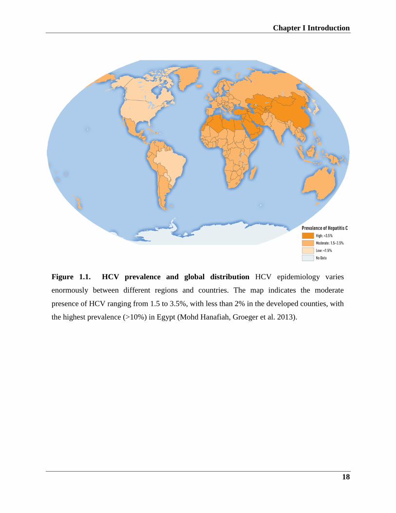
The global distribution of HCV varies greatly, while the estimated rate of HCV infection in
Europe is around 0.5% of the population, 1.3-1.5 % in the USA and Australia, the highest
prevalence has been reported in Egypt with a 15– 20% of the population (Reker and Islam 2014)
The reason for the wide spread of the virus in Egypt is due to the use of contaminated syringes
for schistosomiasis treatment during the campaigns to eradicate Schistosoma (Frank C,
Mohamed MK et al 2000). Other reasons are the intra-familial transmission, direct blood contact,
mother to child, organ transplant, needle-stick injuries, medical dental treatment, drug injection,
and sharing contaminated sharp materials like razors and shaving kits (Mohd Hanafiah, Groeger
et al. 2013).
Chapter I Introduction
18
Figure 1.1. HCV prevalence and global distribution HCV epidemiology varies
enormously between different regions and countries. The map indicates the moderate
presence of HCV ranging from 1.5 to 3.5%, with less than 2% in the developed counties, with
the highest prevalence (>10%) in Egypt (Mohd Hanafiah, Groeger et al. 2013).
Chapter I Introduction
19
1.1. HCV virology and genome organization
HCV is a single positive-stranded RNA virus belongs to the family of Flaviridae (Fournier,
Duverlie et al. 2013). The HCV genome contains approximately 9600 base pair (bp) and encodes
a long polypeptide protein of 3000 amino acids (aa), which is flanked by non-translated regions
(NTRs) at both C- and N- terminals (Bowen and Walker 2005, Lauer 2013)
HCV has high mutation rate owing to the error-prone nature of RNA-dependent RNA
polymerase (RdRp). Therefore, HCV divides into 6 genotypes, and each genotype subdivides
into subtypes, (e.g.:1a, 1b etc...). The mutations within each subtype are called quasispecies
(Halfon and Locarnini 2011) .
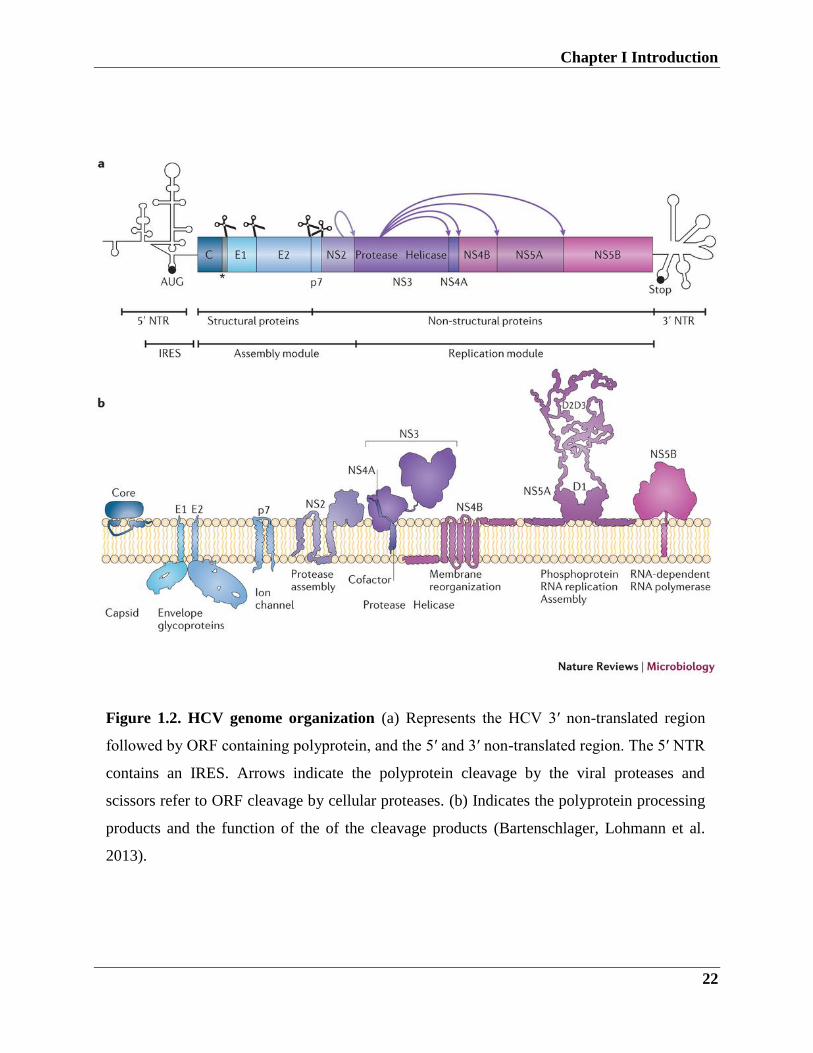
1.2. HCV genome organization
The genetic representation is illustrated in figure 1.2.
Flaviviridae family has 3 different genera: flavivirus, pestivirus, and hepacivirus. HCV belongs
to hepacivirus genera, all viruses belong to Flaviviridae is similar in genome organization and
viral replication (Katze, He et al. 2002).
HCV genome is flanked by small 5’ and 3’ NTRs and has one open reading frame that translated
into a polyprotein of roughly 3000 aa, followed by polyprotein cleavage by viral and cellular
protease into ten gene products. HCV structural proteins are located in the N-terminal region of
the polyprotein: the capsid ‘core’ protein and the glycoproteins, E1 and E2 (Huang, Sineva et al.
2004, Steinmann, Brohm et al. 2008).
While the non-structural (NS) proteins: p7, NS2, NS3, NS4A, NS5A, and NS5B are encoded in
the C-terminal of the polyprotein (Halfon and Locarnini 2011).
1.3. Structural proteins
The structural proteins which form the virus particle include Core and the E1-E2 (Sulbaran, Di
Lello et al. 2010).
The highly conserved viral core protein is relatively small ~21kDa and consists of 191 aa and
located at the N-terminus of the HCV polyprotein. It plays multiple functions such as viral
assembly, gene transcriptional regulation, (Zuniga, Macal et al. 2015) apoptosis, alteration of
IFN signaling, cell transformation and interference with lipid metabolism (Singaravelu, O'Hara
Chapter I Introduction
20
et al. 2015). Many of the biological functions of the core protein are thought to provide a
survival advantage to the virus.
The HCV core protein localizes in the cell cytoplasm and can also localize in the nucleus
(Steinmann, Brohm et al. 2008). It circulates in the blood stream and can stimulate cell surface
receptors.
The E1, E2 structural proteins are hypervariable glycoproteins and associated with the
endoplasmic reticulum (ER). The envelope proteins are of great interest because of their
potential use in the development of HCV vaccine (Khan, Whidby et al. 2014). The variability of
the E1-E2 region is challenging in the generation of effective neutralizing antibodies (Khan,
Whidby et al. 2014) .
Previous studies have demonstrated that E1-E2 proteins can form pseudo-viral particles and
these particles capable of eliciting antibody responses (Suzuki, Saito et al. 2012).
1.2.2. Non-structural proteins
The non-structural proteins (NS): p7, NS2, NS3, NS4A, NS5A, and NS5B are associated with
viral replication and immune evasion (Lassmann, Arumugaswami et al. 2013).
The non-structural 2 (NS2) is a serine protease responsible for the cleavage of NS2/3 when NS2
expresses alone it localizes to the ER membrane. NS2 dimerizes and yields a cysteine protease
with two active sites which cleave between NS2 and NS3 (Shiryaev, Cheltsov et al. 2012). Due
to the dimerization, it is believed that NS2 might be involved in viral assembly and release. It
can also bind host pro-apoptotic protein, CIDE-B, which subsequently inhibits apoptosis
(Guglietta, Garbuglia et al. 2009).
HCV Non-structural protein3/4A (NS3/4A) protein complex localizes to the ER membrane. The
complex formation protects the NS3 and it is responsible for multiple biological functions
including the viral replication and polyprotein processing. Both N3 protease and helicase activity
are enhanced after localization to the ER (Heim and Thimme 2014).
HCV NS3 mediates apoptosis via caspase-8 indicating that it is implicated in the modulation of
host cellular functions. It was reported that NS3/4 plays also an important role in HCV immune
evasion (Gokhale, Vazquez et al. 2014).
Chapter I Introduction
21
The Non-structural protein 4B (NS4B) is a transmembrane protein that contributes to the
formation of HCV replication complexes. The exact function is not known, but mutations in this
protein affect the replication efficiency (Gokhale, Vazquez et al. 2014, Heim and Thimme 2014,
Tamori, Enomoto et al. 2016).
The Non-structural protein5a (NS5a), is phosphorylated by cellular serine kinases, including
MEK1, MKK6, AKT, p70S6K, and cAMP-dependent protein kinase A-a.42-46 and is associated
with the ER membrane (Hamamoto, Nishimura et al. 2005). NS5a has two forms, hypo- and
hyperphosphorylated form (Huang, Sineva et al. 2004). The hypophosphorylated form is
required for efficient HCV replication in cell culture. Therefore, modifications such as point
mutation (S2240I) in the NS5a are necessary for permissibility (Hardy, Marcotrigiano et al.
2003) Blight KJ, 2002). Moreover, NS5a can interact with a number of viral and host proteins to
promote RNA replication and suppress the IFN-induced antiviral efficacy (Macdonald and
Harris 2004, Huang, Hwang et al. 2005).
Non-structural protein 5b (NS5b) is RNA-dependent RNA polymerase required for HCV RNA
replication (Wang, Ng et al. 2003, Wang, Johnson et al. 2004). It interacts with viral and host
proteins, the interaction with NS3 and NS5a plays an important role in the HCV replication
complex formation. Interestingly, NS5b interacts with cyclophilin B, a cellular peptidyl-prolyl
cis-trans isomerase that regulates HCV replication through modulation of the RNA binding
capacity of NS5b (Huang, Hwang et al. 2005).
The initial 21 residues of C-terminal region form a α-helical transmembrane domain responsible
for post-translational targeting to the cytosolic side of the ER (Moradpour and Blum 2004). The
RdRp is an important target for development of anti-HCV drugs (Afdhal, Reddy et al. 2014)
Chapter I Introduction
22
Figure 1.2. HCV genome organization (a) Represents the HCV 3′ non-translated region
followed by ORF containing polyprotein, and the 5′ and 3′ non-translated region. The 5′ NTR
contains an IRES. Arrows indicate the polyprotein cleavage by the viral proteases and
scissors refer to ORF cleavage by cellular proteases. (b) Indicates the polyprotein processing
products and the function of the of the cleavage products (Bartenschlager, Lohmann et al.
2013).
Chapter I Introduction
23
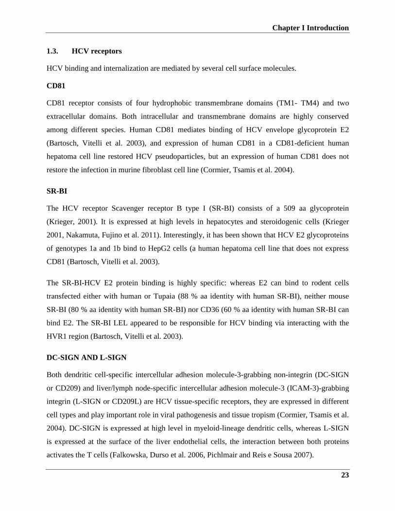
1.3. HCV receptors
HCV binding and internalization are mediated by several cell surface molecules.
CD81
CD81 receptor consists of four hydrophobic transmembrane domains (TM1- TM4) and two
extracellular domains. Both intracellular and transmembrane domains are highly conserved
among different species. Human CD81 mediates binding of HCV envelope glycoprotein E2
(Bartosch, Vitelli et al. 2003), and expression of human CD81 in a CD81-deficient human
hepatoma cell line restored HCV pseudoparticles, but an expression of human CD81 does not
restore the infection in murine fibroblast cell line (Cormier, Tsamis et al. 2004).
SR-BI
The HCV receptor Scavenger receptor B type I (SR-BI) consists of a 509 aa glycoprotein
(Krieger, 2001). It is expressed at high levels in hepatocytes and steroidogenic cells (Krieger
2001, Nakamuta, Fujino et al. 2011). Interestingly, it has been shown that HCV E2 glycoproteins
of genotypes 1a and 1b bind to HepG2 cells (a human hepatoma cell line that does not express
CD81 (Bartosch, Vitelli et al. 2003).
The SR-BI-HCV E2 protein binding is highly specific: whereas E2 can bind to rodent cells
transfected either with human or Tupaia (88 % aa identity with human SR-BI), neither mouse
SR-BI (80 % aa identity with human SR-BI) nor CD36 (60 % aa identity with human SR-BI can
bind E2. The SR-BI LEL appeared to be responsible for HCV binding via interacting with the
HVR1 region (Bartosch, Vitelli et al. 2003).
DC-SIGN AND L-SIGN
Both dendritic cell-specific intercellular adhesion molecule-3-grabbing non-integrin (DC-SIGN
or CD209) and liver/lymph node-specific intercellular adhesion molecule-3 (ICAM-3)-grabbing
integrin (L-SIGN or CD209L) are HCV tissue-specific receptors, they are expressed in different
cell types and play important role in viral pathogenesis and tissue tropism (Cormier, Tsamis et al.
2004). DC-SIGN is expressed at high level in myeloid-lineage dendritic cells, whereas L-SIGN
is expressed at the surface of the liver endothelial cells, the interaction between both proteins
activates the T cells (Falkowska, Durso et al. 2006, Pichlmair and Reis e Sousa 2007).
Chapter I Introduction
24
LDL-R
The low-density lipoprotein (LDL) receptor (LDL-R) is an endocytic receptor consists of 839
amino acids, mainly the cholesterol-rich LDLs, into cells through receptor-mediated endocytosis
(Chung, Gale et al. 2008). The majority of human plasma cholesterol is in LDL form. It was
reported that LDLR is involved in the HCV infection, the HCV infection was inhibited when
cells were treated LDL peptide inhibitor (Bartenschlager, Penin et al. 2011).
Occludin
Occludin (OCLN) has been identified as an important receptor for HCV (Nakamuta, Fujino et al.
2011, Zeisel, Fofana et al. 2011, Dorner, Horwitz et al. 2013). It is 65 kDa and consists of a four-
transmembrane domain protein located in the tight junctions of polarized cells. Human hepatoma
cell lines such as Huh7 or cell lines that lack other entry factors (i.e. HepG2 and 293T cells)
express detectable levels of OCLN. Although, OCLN overexpression did not enhance infection
susceptibility of HCVpp. OCLN knockout led to inhibition of HCVpp infection in Hep3B cells
and both HCVpp and HCVcc in Huh-7.5 cells. Taken together, OCLN is an essential receptor for
HCV infection in permissive cell lines. Whereas in HCV non-permissive cells, which express
other cell entry receptors, OCLN overexpression enhances HCV entry (Nakamuta, Fujino et al.
2011, Dorner, Horwitz et al. 2013)To date, human OCLN has been reported to be a species
specific entry factor responsible for HCV infection. Expression of OCLN along with human
CD81 may confer HCV permissibility to mouse cell lines (Zeisel, Fofana et al. 2011).
1.4. HCV replication cycle
HCV replication cycle consists of 5 stages: viral entry, protein translation, RNA replication,
virion assembly, and release. The entry starts by binding to E2 protein to CD81 (Rajesh, Sridhar
et al. 2012), SRB1 (Nakamuta, Fujino et al. 2011), glycosaminoglycans, LDLR (Nakamuta,
Fujino et al. 2011), and claudin-1 (Nakamuta, Fujino et al. 2011, Zeisel, Fofana et al. 2011).
The endosome is characterized by the acidic environment which introduces the HCV genome
entry into the cytoplasm, where HCV negative sense strand RNA production occurs (Friebe and
Bartenschlager 2009, Pham, Coffin et al. 2010). HCV polyprotein translation is driven by the
internal ribosome entry site (IRES), initiation of translation occurs via HCV IRES and the 40S
ribosomal subunit complex formation. The translation process is followed by viral and cellular
proteases processing to form the structural and non-structural proteins. HCV protein expression
results in the replication complexes formation in the cytosol. The viral proteins are then arranged
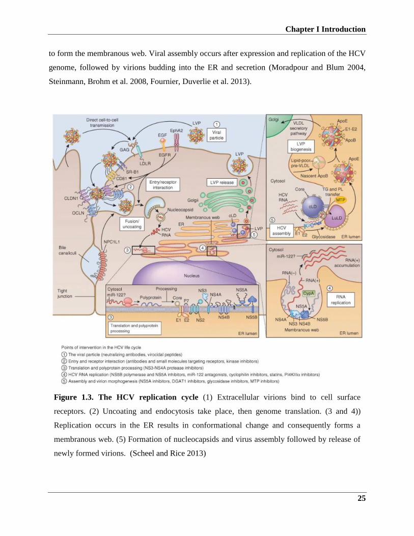
Chapter I Introduction
25
to form the membranous web. Viral assembly occurs after expression and replication of the HCV
genome, followed by virions budding into the ER and secretion (Moradpour and Blum 2004,
Steinmann, Brohm et al. 2008, Fournier, Duverlie et al. 2013).
Figure 1.3. The HCV replication cycle (1) Extracellular virions bind to cell surface
receptors. (2) Uncoating and endocytosis take place, then genome translation. (3 and 4))
Replication occurs in the ER results in conformational change and consequently forms a
membranous web. (5) Formation of nucleocapsids and virus assembly followed by release of
newly formed virions. (Scheel and Rice 2013)
Chapter I Introduction
26
1.5. HCV cell culture systems
Human hepatocytes are the primary host for HCV (Rajesh, Sridhar et al. 2012). There are some
evidence that HCV found in peripheral blood mononuclear cells and the brain, but replication in
these tissues has not been confirmed (Zhu, Liu et al. 2016). Some of the early challenges to
studying HCV were the inefficient replication of different HCV isolates and the limitations of
permissive cell culture systems that support HCV replication.
1.5.1. HCV replicon system
One of the major breakthroughs to study HCV was the development of subgenomic replicons. A
replicon is a nucleic acid which is capable of self-replication. Con1 is the first HCV replicon
system used to study HCV. It is a full HCV genome which was cloned from genotype 1b isolate
(Moriishi and Matsuura 2007, Wakita 2007). In this model the structural proteins are replaced
by, a selection neomycin phosphotransferase (neo) gene which allows selecting for G418
resistance, and an IRES from a picornavirus (Jiang, Guo et al. 2008, Pichlmair, Schulz et al.
2009).This results in a bicistronic construct, the first cistron drives the neo gene and the second
drives the HCV replicas genes (NS3–NS5B). Then, HCV RNA was reversely transcribed in vitro
and transfected in human hepatoma cell line Huh-7, afterward, HCV replicon colonies that
support replication efficiently replicate in cells (1000–5000 positive-strand RNA molecules per
cell) (Friebe and Bartenschlager 2009). Replicon cells can be maintained for years by
continuously passaging under selective pressure (Gaudieri, Rauch et al. 2009, Lassmann,
Arumugaswami et al. 2013).
After the introduction of the first replicon system in 1999 by Ralf Bartenschlager lab
(Bartenschlager, Kaul et al. 2003, Woerz, Lohmann et al. 2009), a number of replicons covering
genotypes (1a-4a), and with various reporter genes such as luciferases and fluorescent proteins
were constructed to cover the researcher needs to study the HCV RNA replication mechanisms.
Improvements of the replicon system
There are two factors which determine the efficiency of HCV replicons replication: the cell
culture adaptive mutations and the permissiveness of the cell line for HCV RNA replication
(Hwang, Huang et al. 2010). Mutations enhancing replications were found at the N-terminus of
the NS3 helicase, NS4B and in NS5A. Insertion of these mutations into replicons resulted in an
Chapter I Introduction
27
increase in the replication capacity, and the enhancement is depending on the mutations
combination (Le Pogam, Yan et al. 2012, Mayhoub 2012, Nakamoto, Kanda et al. 2014). The
second step to improve the efficiency of the RNA replication is to select for highly permissive
cells. Huh7.5 and Huh7-Lunet are Huh7 cell clones that highly support HCV RNA replication
(Robinson, Yang et al. 2010).
Establishment of cell lines stably expresses reporter replicons was an additional step forward for
high-throughput screening purposes. Interestingly, con1 containing adaptive mutations replicon
cannot produce viral particles in the supernatant of cultured cells (Pietschmann, Kaul et al.
2006). Theses adaptive mutations do not exist in natural viral isolates, and some studies show
that they might interfere with viral production and spread.
1.5.2. HCV pseudoparticles (HCVpp)
One of the important models to study HCV early steps of infection is HCVpp. This model is
established by transfection of 293T cells with three vectors, first, is HCV E1 and E2, second is
the retroviral gag-pol and third is a reporter gene vector (Bartosch, Vitelli et al. 2003).
Afterward, HCVpps are secreted by the cultured transfected cells, which can be used to infect
Huh 7 cells. Therefore, this system proved important in discovering viral host receptors such as
glycosaminoglycans, LDL-R, DC-SIGN, Occludin, and recently Niemann-Pick C1-like
cholesterol absorption receptor (NPC1L1) (Ploss and Dubuisson 2012).
1.5.3. HCV cell culture infectious virus (HCVcc)
Another important breakthrough is the HCVcc system when an HCV genotype 2a full-length
replicon (JFH-1) isolate was isolated from a Japanese fulminant hepatitis patient (Sung, Shin et
al. 2015). HCVcc enables the study of the full viral life cycle in vitro. Infection of hepatoma cells
with JFH-1 resulted in the release of infectious viruses which are capable of infecting naive cells
(Wakita 2007). Interferon-alpha and DAAs inhibit HCVcc replication, so this system is a useful
tool to study antiviral drugs and vaccines. Although HCVcc is an important tool to study HCV, it
is limited by the fact that only replicating isolate is restricted to genotype 2 (Gaudieri, Rauch et
al. 2009, Caillet-Saguy, Simister et al. 2011).
To improve the system, inter and intragenic chimeras were created, the chimeras are generated
from (NS3–NS5b) of JFH-1isolate fused to the core to NS2 region of other HCV isolates. This
Chapter I Introduction
28
created a broad range of virus chimeras to the isolates Con1 (gt 1b), H77 (gt1a), 452 (gt 3a) and
J6 (gt 2a), these chimeras are designated Jc1 (Tu, Ziemann et al. 2010, Verbinnen, Jacobs et al.
2012).
1.5.4. HCV trans-complemented particles (HCVTCP)
Recently, HCVTCP system was established, this is a useful system to study HCV early and late
stages of infection. The HCVTCP are able of one round of infection but are unable to spread.
Different strategies are used to produce HCVTCP, for example, co-transfection with constructs
carrying viral glycoprotein genes (Luc-_E1/E2 or (Luc-NS3-5b) (Pai, Prabhu et al. 2005) along
with the JFH1 genome as a helper virus into Huh7-Lunet cells (Steinmann, Brohm et al. 2008).
It was reported that HCV RNA subgenomes with different deleterious mutations naturally exist
in patients and could be trans-complemented in vitro (Steinmann, Brohm et al. 2008, Suzuki,
Saito et al. 2012) and these subgenomes can circulate in vivo and modulate the disease
progression.
1.6. Immune responses to HCV infection
1.6.1. Innate immune responses to HCV
Various studies have shown that HCV triggers innate and adaptive immune response and the
virus has developed mechanisms to escape the immune response.
Although, HCV persists in high viral loads in patients, but the viral proteins expressed at low
amounts to be detected by immunohistochemistry. In HCV chronically infected patients, it is
estimated that 30%–50% of hepatocytes are infected (Park and Rehermann 2014).
Despite the fact that HCV establish persistent chronic infection in 80% of the patients, 20% of
patients in the acute phase can eliminate the virus, and the elimination is associated with innate
immune response mainly IFN-α and β response.
HCV is first recognized by pattern recognition receptors (PRRs) members such as retinoic acid-
inducible gene I (RIG-I) and melanoma differentiation-associated gene 5 (MDA-5), RIG-I plays
a pivotal role in recognition of positive and negative ssRNA viral genomes (Pichlmair, Schulz et
al. 2006), both RIG-I and MDA-5 are interferon stimulated genes that share conserved features
and functions. Early during infection, RIG-I recognizes short stretches of the RNA which are
rich in poly-U/UC ribonucleotides (Rajsbaum, Stoye et al. 2008), this activates the downstream
Chapter I Introduction
29
signaling pathway. Later, longer RNA intermediates sense TLR3 and PKRs. Therefore, PKRs
activation signaling cascades lead to IFN-β and κkβ activation (Liu, Su et al. 2007, Jiang, Guo et
al. 2008). TLR3 works as another innate immune molecule helping in surveillance of infected
dead cells and in the establishment of antiviral state during the infection. Activation of non-
canonical NF-kβ can result in and CXCL10 pro
-inflammatory cytokines production.
Kupffer cells (KC) are macrophages that abundantly localize in the liver and they could produce
IFN-β in vitro (Boltjes, Movita et al. 2014). Although HCV is unlikely to replicate in KCs, some
studies have shown that HCV RNAs and protein can activate KCs. This may be attributed to the
phagocytosis of infected cells and exposure to viral RNA, but this hypothesis has not yet been
proven.
Interestingly, KCs increased in the liver of chronically infected patients with high expression
level of the activation markers such as CD163 and CD33. Although the contribution of KC on
HCV infection is unclear, it was reported that HCV can induce cytokines such as IL-6, IL-1β,
and IFN-β (Li, Liu et al. 2009, Pichlmair, Schulz et al. 2009, Pagliaccetti and Robek 2010) and
consequently inhibit HCV replication in HCV replicon models. Furthermore, KC cytokines
recruit infiltrating leukocytes in the liver and promote virus-specific T cell responses (Meylan,
Curran et al. 2005, Simister, Schmitt et al. 2009, Pichlmair, Kandasamy et al. 2012).
1.6.2. Adaptive immune responses during HCV infection
In order to evade host immune system, HCV has developed different escape mechanisms.
Moreover, it interferes with the IFN system and the potential for DC function. Although the
adaptive immune response is detectable after 6–8 weeks of HCV infection,(Chen and Yu 2010,
Mishima, Sakamoto et al. 2010), it plays an important role in viral clearance during the acute
phase of infection. During acute HCV infection, patients produce antibodies against structural as
well as non-structural protein epitopes. In particular, antibodies fraction called ‘neutralizing
antibodies’ can inhibit virus binding, entry, or uncoating. This was demonstrated in a group of
women infected by an HCV-contaminated anti-D immunoglobulin. In this homogenous group, in
contrast to patients who developed chronic infection, patients who resolved HCV infection early
during acute infection are characterized by the early production of neutralizing antibodies (Khan,
Whidby et al. 2014). Although this study associates virus clearance with an early neutralizing
antibody, virus control has also been noticed in hyperglobulinaemia patients. Another
Chapter I Introduction
30
mechanism of HCV clearance in acute infection is the strong and sustained CD4+ and CD8+T
cell responses that target various HCV polyprotein epitopes (Schmidt, Blum et al. 2013, Heim
and Thimme 2014).
Several pieces of evidence suggest an important role of both CD4+ and CD8+T cells in HCV
spontaneous virus elimination, mainly the association between class I (e.g., HLA-B27) and II
(e.g., DRB1⁄1101) alleles (Miki, Ochi et al. 2013).
The importance of T cell responses in virus clearance has been suggested by a study where
protected chimpanzees CD8+T cells were depleted. This led to HCV persistence until the
recovery of CD8+T cell response. Moreover, CD4+T cells depletion in protected chimpanzees
resulted in HCV persistence and emergence of CD8+ escape mutations (Heim and Thimme
2014). Another recent study has shown that the IL-17 and 21 cytokines production of
CD161+CCR6+CD26+CD4+ T expanded cells led to virus clearance. Using MHC class I, allele
HLA-A2 gene transuded replicon cells showed that HCV-specific CD8+T cells possessed a
dominant IFN-gamma production (Pichlmair, Kandasamy et al. 2012, Miki, Ochi et al. 2013) .
Taken together, CD4+T cells are central regulators, while virus-specific CD8+T cells primarily
function as the key effectors.
1.6.3. Immune escape
In order to evade host immune system, HCV has developed different escape mechanisms.
Moreover, it interferes with the IFN system and the DC function. One of the escape mechanism
is the cleavage of MAVS (Gale and Foy 2005)) and TRIF (Heim and Thimme 2014) by NS3-4A
serine protease. This resulted in inhibition of IFN-β and Interferon-stimulated gene (ISG)
induction in infected hepatoma cells (Liu, Su et al. 2007, Chen, Wang et al. 2014). This is
supported by some studies which detected cleaved MAVS in HCV-infected livers (Gokhale,
Vazquez et al. 2014, Heim and Thimme 2014) and the presence of an inactive form of IRF-3
(Lau, Tam et al. 2002, Meylan, Curran et al. 2005).
HCV core protein plays a role in immune escape mechanisms by inactivation and degradation of
the STAT-1 and -2 which is required for the expression of ISGs such as PKR, 2’-5’ OAS, ISG-
p56, IRF-7 (Kanda, Steele et al. 2007, Gaudieri, Rauch et al. 2009, Heim 2012). Moreover, IFN
activity can be blocked by NS5A protein blocking IRF-1 production and induction of IL-8
chemokine (Meylan, Curran et al. 2005, Seth, Sun et al. 2005) and subsequently inhibits IFN
activity (Helbig, Lau et al. 2005).
Chapter I Introduction
31
1.7. Genotyping and phenotyping of HCV protease inhibitors resistant mutations
1.7.1. HCV therapy
Approximately 75% HCV patients develop chronic infection and this might lead to severe
illness, including cirrhosis and hepatocellular carcinoma (HCC) (Wakita 2007, Pham, Coffin et
al. 2010).
In 1991 interferon was approved by FDA as the first treatment for hepatitis C (Donaldson,
Harrington et al. 2015) . A few years later, a combination of pegylated interferon-α (PEG–IFNα)
and ribavirin was approved, this leads to cure of 20-80% of infection depending on the genotype,
age and genetic host factors such as interleukin-28B polymorphisms (Ansaldi, Orsi et al. 2014,
Rupp and Bartenschlager 2014, Qian, Zhu et al. 2016). Overall sustained virological response
(SVR) in patients infected with genotype 2 and 3 is up to ~85% of treated patients, while it is
successful only in ~45% of genotype 1 and 4 treated patients (Ferenci 2004). Later in 2011 the
first direct-acting antiviral agents (DAAs) protease inhibitors BOC (Victrelis) and TLV (Incivek)
were approved (Schiering, D'Arcy et al. 2011, Manns and von Hahn 2013). The new standard of
care therapy consists of BOC or TLV in combination with PEG–IFNα and ribavirin raised the
cure rates of HCV genotype 1 patients up to 70%, with treatment duration of 24 to 48 weeks
(Hofmann and Zeuzem 2011, Rupp and Bartenschlager 2014). However, the SVR has improved
with the new combination therapy. The emergence of resistant mutations is a main considerable
drawback for the first generation of HCV protease inhibitors. The second wave of HCV protease
inhibitors includes Simeprevir and Faldaprevir (Izquierdo, Helle et al. 2014). They improved the
pharmacokinetics and have a higher genetic barrier. Daclatasvir is the first NS5A inhibitor with
potent antiviral activity and broad genotype coverage (Pawlotsky 2013). Sofosbuvir is a
nucleotide NS5B inhibitor. Combined with Daclatasvir, it was the first approved IFN free
therapy (Degasperi and Aghemo 2014).
1.7.1.1. A decade of interferon- ribavirin standard of care therapy
Ribavirin is a (1--D-ribofuranosyl-1, 2, 4-triazole-3-carboxamide), guanosine analog a, that has
been approved for other viruses treatment before HCV discovery. When ribavirin administrated
alone for HCV treatment, it resulted in a decrease of HCV viral load and improved the liver
enzymes in 40% of patients (Crotty, Cameron et al. 2001, Lau, Tam et al. 2002).
The ribavirin proposed antiviral mechanisms are: activation of virus mutagenesis resulting in
accumulation of lethal virus forms, immunomodulatory effect, and induction of TH1 cytokines.
Chapter I Introduction
32
Another antiviral mechanism of ribavirin is its function as inosine-5-monophosphate
dehydrogenase (IMPDH) inhibitor. Thus, it inhibits guanosine triphosphate, resulting in
abnormal viral RNA form (Zhou, Liu et al. 2003).
Interferon-alpha (IFN-α) in HCV treatment
The interferons are naturally secreted cytokines produced by the cell. They have diverse
functions including, antiviral activities, cell growth and regulation of immune response.
Interferons antiviral activity was first discovered over half a century. It was reported that
influenza infected cells secreted a protein which was able to control infection in vitro. Due to the
interesting antiviral activity reported for interferons, researchers paid attention in using them for
virus treatment (Katze, He et al. 2002) .
IFN-α, proved useful in the treatment of chronic hepatitis c infection as it decreases serum levels
of HCV RNA and normalizes ALT levels. In 1998 the interferon- α and ribavirin combination
therapy were approved for HCV treatment. Three years later, the PEGylated interferon- α along
with ribavirin were approved for HCV treatment and improved the overall SVR of 75-80% in
genotype 2 and 3 and limited to 40% in genotype 1 and 4 treated patients (Ferenci 2004, Manns,
Wedemeyer et al. 2006). The issue of the differences SVR rates is attributed to the association of
different factors with the response, among those (HCV genotype, co-infections and viral load)
and host factors such as (age, sex, race and the recently identified genetic factor IL28b
polymorphisms) (Balagopal, Thomas et al. 2010). One of the IFN-α antiviral mechanisms is the
activation of PKR, the essential innate immunity regulator. The PKR mediates the
phosphorylation of eukaryotic initiation factor-2 (eIF2a) and consequently blocks the HCV viral
protein synthesis (Jiang, Guo et al. 2008).
1.7.2. Direct-acting antivirals (DAAs)
In spite of the increase of the SVR rates using the PEGylated interferon-α -ribavirin therapy, it
has side-effects such as anemia, depression and limited efficacy for genotype 1 and 4 treatment.
Thus, the need for more efficient anti-HCV therapies arouses. The better understanding of HCV
life cycle and proteins function led to the development of multiple DAAs. These drugs target
different steps in the virus replication and infection steps. There are three classes of DAAs:
NS3/4A protease inhibitors (PIs), NS5A inhibitors and NS5B nucleoside and non-nucleoside
polymerase inhibitors (NPIs&NNPIs) (Wang, Ng et al. 2003, Biswal, Cherney et al. 2005,
Marascio, Torti et al. 2014, Bunchorntavakul and Reddy 2015).
Chapter I Introduction
33
1.7.2.1. Protease inhibitors
In 2003 (Lamarre, Anderson et al. 2003) have reported the first proof of concept that NS3/4A
protease inhibitor BILN 2061 was able to decrease the viral load within 48 hours. However, the
inhibitor has been withdrawn from further development due to the potential for cardiac toxicity.
The first generation of protease inhibitors (Telaprevir and Boceprevir)
In 2011, the FDA approved the linear compounds TLV (VX-950) and BOC (SCH 503034) for
HCV genotype 1 therapy, both are administrated in combination with PEG–IFNα and ribavirin
(Hofmann and Zeuzem 2011, Shiryaev, Cheltsov et al. 2012, Elbaz, El-Kassas et al. 2015). The
triple therapy increased the SVR rates in genotype 1 treated patients up to 75%. The in vitro and
in vivo studies showed that linear ketoamide has a low genetic barrier, meaning that a single
substitution in one aa can confer resistance (Halfon and Locarnini 2011). The single aa
substitutions confer low to high resistance for both TLV and BOC and the existence of two or
more substitutions synergize the resistance effect (Hofmann and Zeuzem 2011, Rupp and
Bartenschlager 2014, Welzel, Dultz et al. 2014, Elbaz, El-Kassas et al. 2015). Therefore, further
studies including ours have evaluated the clinical resistance associated with treatment failure.
The R155K and A156T substitutions are class-specific mutations (Nakamoto, Kanda et al. 2014,
Welzel, Dultz et al. 2014, Elbaz, El-Kassas et al. 2015). Moreover, they are predominant in the
patient quasispecies as both cause strong steric hindrance and modify the inhibitory interactions
in a way which cannot be compensated. Other mutations such asV36, T54, V55 and V/I170 are
also associated with PIs resistance (Lassmann, Arumugaswami et al. 2013). Typically, the drug
resistance is determined in vitro and the degree of resistance measured as the fold increase in the
50% and 90% inhibitory concentrations (IC50 and IC90, respectively). Additionally, the variant
must be able to propagate in vitro, notably, in the presence of drug highly resistant variants are
less fit and vice versa (Chayama and Hayes 2015).
Although the analysis of PIs resistance is important for the further protease inhibitors
development, the drug resistance testing was not recommended for patients before DAA therapy
(Halfon and Locarnini 2011).
Chapter I Introduction
34
Second wave of PIs, Simeprevir
Simeprevir belongs to the second-wave of protease inhibitors and it has some advantages over
the first wave inhibitors. These include higher genetic barriers to resistance and better
pharmacokinetic profiles (Nakamoto, Kanda et al. 2014, Qian, Zhu et al. 2016). This drug was
approved for treatment of genotype 1 patients in combination with peg-IFNα/RBV and
administrated once daily, the triple therapy increased the SVR rates up to 80-85% in naïve
treated patients (Bunchorntavakul and Reddy 2015). In genotype 1a patients, Q80K
polymorphism is prevalent in about 30% of American patients and in 19% of European patients
(Izquierdo, Helle et al. 2014, Sarrazin, Lathouwers et al. 2015). This led to the failure of the
therapy with simeprevir+PEG/RBV. Therefore, screening for the presence of a Q80K
polymorphism at baseline is recommended, and alternative treatment should be considered
(Izquierdo, Helle et al. 2014). In November 2014 the FDA has approved Simeprevir+Sofosbuvir
therapy for HCV genotype 1 patients, this combination was the first oral IFN-free therapy
(Asselah and Marcellin 2015).
HCV PIs resistance tools
Similar to other RNA viruses, HCV replication is characterized by a high replication rate
(1×10¹²) particles per day) (Crotty, Cameron et al. 2001, Katze, He et al. 2002). In addition,
HCV replication is characterized by the genetically error prone because of the low fidelity of
RdRp. The error rate reaches 10 -4 for one single mutation (Crotty, Cameron et al. 2001) which
results in one mutation every genome. This inaccurate replication machinery besides the high
replication rate leads to HCV with high sequence diversity. As a result, in chronic HCV
infection, the virus exists as a multitude of genetically distinct but closely related viral variants
called quasispecies. Mutations can be introduced in HCV during both negative-strand RNA
intermediate, and the synthesis of the new positive strand RNA genome from the negative one
(Habjan, Andersson et al. 2008). With the availability of HCV DAAs, the SVR increased up to
75% (Bunchorntavakul and Reddy 2015)for patients infected with HCV genotype 1, but DAAs
capability is hampered by the presence of resistance variants that generate amino-acid
substitutions within the targeted protein, which in turn influence the viral sensitivity to these
DAAs. Variants with reduced susceptibility to DAAs can exist naturally before treatment starts,
but usually at low levels and under DAAs treatment the drug may select for pre-existing
Chapter I Introduction
35
resistance variants and decrease the virus susceptibility to the drug (Halfon and Locarnini 2011,
Le Pogam, Yan et al. 2012, Issur and Gotte 2014). Thus, patients should be monitored for
resistance selection over the course of treatment. Two integral techniques are used to
characterize viral resistance: genotypic and phenotypic assays
Genotypic assay for HCV PIs resistant analysis
A genotypic assay is the detection of the sequence mutations in the viral genome that cause drug
resistance. HCV resistance examination during drug treatment is important for understanding the
impact of resistant mutations on the clinical outcome and improving the strategies for new drug
design (Ferenci 2004, Thibeault, Bousquet et al. 2004, Moriishi and Matsuura 2007, Halfon and
Locarnini 2011).
The utilization of genotypic-resistance tool is important for therapeutic decision- making in
patients. Patients monitoring should be done in three phases: baseline (pre-treatment) samples,
during and post-treatment samples (Le Pogam, Yan et al. 2012).
For baseline samples, viral sequences are analyzed to detect any known resistant variants or
possible novel ones. On treatment samples, patient samples are monitored for sequence changes
which likely decrease the sensitivity to the drug.
Post-treatment samples, existence or absence of resistant variants give a hint on the virus fitness
and the resistance profile compared to the wild type.
PCR (population sequencing) is the most common method for genotypic analysis, but as
mutation detection is assay sensitivity dependent (Verbinnen, Jacobs et al. 2012), so the
population sequencing is suitable for single mutation pattern and for mutations that exist in less
than 10%. While clonal sequencing, ultra-deep sequencing or the TaqMan mismatch
amplification mutation assay, are sensitive enough to detect minor resistant variants at 0.01%
level (Curry, Qiu et al. 2008).
The phenotypic analysis should be performed on concurrent samples to assess the level of
decreased susceptibility related to specific mutations and to the baseline sample. Interpretation of
data from samples at viral suppression needs to take into account the fact that different
genotypes, subtypes, or specific regions differ in the minimum viral load required for
Chapter I Introduction
36
amplification (Qi, Bae et al. 2009, Imhof and Simmonds 2010). Low template numbers
negatively impact assay performance in two ways: (1) the relative proportion of each variant
detected increases as a result of reduced amplification of co-existing minority variants,
potentially yielding larger discrete changes in frequency estimates; and (2) the chances of re-
sampling increases.
Phenotypic assay for HCV PIs resistant analysis
Phenotypic assays are methods used to characterize the susceptibility of the resistant variants to
the drugs, the phenotyping is conducted either by cell based: including replicon-based and viral
enzymatic assay, or by the biochemical-based enzymatic assay. In the cell-based phenotypic
assay, the subgenomic viral RNA model is generally used for anti-HCV research. For insertion
of patients fragment, shuttle vector constructs are used. Therefore, clinical isolates are collected
and either amplified and cloned into the shuttle vector with a combination of quasispecies, or by
insertion of the individual clone in the shuttle vector (Qi, Bae et al. 2009, Imhof and Simmonds
2010, Halfon and Locarnini 2011, Verbinnen, Jacobs et al. 2012). Several shuttle vectors were
developed covering the 1a, 1b and 2a subtypes.
Numerous studies examined the utility of the shuttling vector for insertion of different lengths of
the HCV genome, they showed that (NS5a-NS5b), (NS3-4A) up to (NS3-NS5b) could be
successfully inserted. The shuttle vector used to test for PIs, enables the insertion of NS3
protease or the NS3 protease-helicase domain (Qi, Bae et al. 2009). Investigation of the insertion
of the genome from clinical isolates showed that the replication decreased significantly. This
could be attributed to the incompatibility between replicon lab strain and the viral genome
derived from the patient in addition to insertion of clinical isolates from different subtype than
the shuttle vector (Qi, Bae et al. 2009, Binder, Tetangco et al. 2011).
Analysis of the phenotypic results explained by measuring EC50 (the effective concentration of
drug needed to inhibit virus replication by 50% in vitro) of clinically derived isolates and
compare it to the EC50 of baseline samples, in case baseline samples are unavailable, the EC50
can be compared to the EC50 of the reference replicon (Huang, Hwang et al. 2005, Hwang,
Huang et al. 2010).

Chapter I Introduction
37
Interpretation of phenotypic results
Analysis results should be related to a reference (replicon/virus/reporter) control that is run in
each assay and can be reported as the fold change EC50. Understanding the significance of a
given fold change value is a major challenge. Different cut-off values include technical
(accounting for assay variability), biological (accounting for natural variation in drug
susceptibility), and clinical (relating drug susceptibility to clinical responses) (Binder, Tetangco
et al. 2011, Shiryaev, Cheltsov et al. 2012, Lassmann, Arumugaswami et al. 2013).
1.7.3. NS5a inhibitors
The non-structural NS5A is a zinc-binding phosphoprotein and consists of approximately 447
amino acids and localizes to the ER-derived membrane (Macdonald and Harris 2004, Huang,
Hwang et al. 2005, Pai, Prabhu et al. 2005). Along with NS5b protein, it plays an important role
in HCV replication and assembly. In addition, it is implicated in the interactions with host
cellular function. Therefore, NS5A has been identified as an interesting target for HCV inhibitors
development. One possible mechanism of NS5A inhibitors is the inhibition of
hyperphosphorylation. Thus, the regulation of phosphorylation is required for efficient viral
replication. However, other mechanisms might be involved, for example, NS5a inhibitors
mislocalize the NS5a in the ER, which causes errors in the viral assembly (Ranjith-Kumar, Wen
et al. 2011, Wang, Wu et al. 2012, Issur and Gotte 2014).
Ledipasvir and Daclatasvir
Ledipasvir is an oral NS5a inhibitor with a broad activity against genotypes 1a, 1b, 4a, and 5a in
vitro, but has lower activity against genotypes 2a and 3a. The combination therapy
Sofosbuvir+Ledipasvir (Harvoni) at first has been approved for treatment of genotype1, and later
it has been extended for genotype4, 5 and 6 treatment (Nakamoto, Kanda et al. 2014, Asselah
and Marcellin 2015).
Daclatasvir is another oral NS5a inhibitor. It has a broad coverage of HCV genotypes. Clinical
trials have shown that Daclatasvir is more active than Ledipasvir in genotype 3 patients
(Bunchorntavakul and Reddy 2015). At first, Daclatasvir in combination with Sofosbuvir has
been approved to treat genotype 3. Very recently, FDA has extended the approval of
Daclatasvir+Sofosbuvir with or without ribavirin for genotype 1 and 3 (Asselah and Marcellin
2015, Bunchorntavakul and Reddy 2015).
Chapter I Introduction
38
Resistance to NS5a inhibitors
NS5a resistant variants pre-exist within HCV quasispecies without any previous exposure to
these drugs. A study has reported that half of the relapsed patients after ledipasvir and sofosbuvir
combination therapy were associated with NS5a-resistant variants that pre-exist before therapy
(Le Pogam, Yan et al. 2012, Nakamoto, Kanda et al. 2014, Chayama and Hayes 2015). The
baseline Ledipasvir resistant variants are, M28T, Q30R/H, L31M, Y93C/H for genotype 1a, and
Y93H in genotype 1b which exists in 100 % of patients. These mutations led to lower SVR in
patients harboring mutation (Bunchorntavakul and Reddy 2015, Chayama and Hayes 2015).
Thus, DAAs from other classes should be used to avoid the cross-resistance.
1.7.4. NS5b polymerase inhibitors
The HCV NS5b polymerase is an important viral protein mediating RNA viral replication, it is
composed 591 aa. The enzyme catalytic domain can be structured in hand like structure and it
consists of 'fingers’, ‘palm’ and ‘thumb’ subdomains and the active site is conserved among
genotypes, and located in the palm, and ‘fingers and thumb’ domains are in close interactions.
The structural studies have shown that the NS5b has enclosed conformation, with fingertips at
one side and b-hairpin linker on the other side. As a result, the closed conformation forms a
single-stranded RNA space, which initiates the replication process (Wang, Ng et al. 2003,
Powdrill, Bernatchez et al. 2010).
In addition to its role in the virus replication, NS5b plays several roles in pathology by
interaction with cellular proteins such as lipid kinase phosphatidylinositol 4-kinase III alpha
(PI4KIIIa), and Caprin 1, a cell-cycle associated phosphoprotein, forms a complex with Ras-
GTPase-activating protein-binding protein 1 (G3BP1) which has been shown to interact with
both HCV NS5b and the 5’ end of the HCV minus-strand RNA indicating that it is part of HCV
replication complex (Elhefnawi, ElGamacy et al. 2012, Mosley, Edwards et al. 2012, Karam,
Powdrill et al. 2014, Asselah and Marcellin 2015). Therefore, the NS5b attracted the attention as
a therapeutic target for HCV.
Sofosbuvir
NS5b RNA polymerase inhibitors are classified into two different classes, nucleoside analog
inhibitors (NIs) and non-nucleoside inhibitors (NNIs). NIs are ribonucleosides or ribonucleotides
analogs that compete with natural ribonucleotide for binding polymerase active site. The first NI
investigated in patients is Valopicitabine. Studies reported only low to moderate antiviral activity
Chapter I Introduction
39
(Afdhal, Reddy et al. 2014). Further clinical developments of valopicitabine have been stopped
due to toxicity issues.
NIs like sofosbuvir (GS-7977) has broad activity against all HCV genotypes. It terminates the
viral synthesis as a false substrate by interacting with the growing RNA chain which results in
termination of chain formation. In addition to the pan-genotypic activity, one main advantage
over other inhibitors is the high genetic barrier (Janardhan and Reau 2015). The approval of
Sofosbuvir along with other DAAs, as the first IFN free all oral combination therapies is an
important breakthrough in HCV treatment. Ledipasvir and sofosbuvir (Harvoni) were first
approved for HCV genotype 1 chronic patients. The clinical trials have shown impressive results,
with SVR rates reached 100% in naïve patients. For other genotypes, sofosbuvir is approved in
combination with IFN for therapy (Welzel, Dultz et al. 2014, Nakamura, Kanda et al. 2016).
Recently, FDA has extended the approval for patients with genotype 4, 5, and 6 chronic hepatitis
C virus (HCV) infection and for patients co-infected with HIV (Ploss and Dubuisson 2012).
Unlike NIs class of inhibitors, the NNIs inhibitors achieve its activity by binding to one of
allosteric enzyme sites and this causes a conformational change in the active site and stops the
replication complex.
Resistance to polymerase inhibitors
Sofosbuvir is favored for its high genetic barrier, in vitro studies have identified S282T mutation
to confer resistance to Sofosbuvir (Mosley, Edwards et al. 2012). Nevertheless, in phase III trials
this mutation was not detected, whereas in phase II trials (LONESTAR), only one patient was
relapsed under Sofosbuvir monotherapy and this was associated with S282T mutation (Le
Pogam, Yan et al. 2012). Despite what was observed in other trials of SOF co-administration
with other DAAs like DCV, SMV, or LDV, S282T was not detected in any relapsed patients.
Even though this mutation occurred much more commonly in approximately 20% of patients and
almost all of patients accomplished SVR. So contrary to PIs, the Sofosbuvir S282T mutation did
not affect the treatment outcome (Nakamura, Kanda et al. 2016).
NNIs are associated with three main resistant variants (414T, 419S, and 422K), these mutations
occur among the six HCV genotypes. However, NNIs in development have shown that they are
possibly effective in combination with other DAAs classes with no cross-resistance (Kayali and
Schmidt 2014, Bunchorntavakul and Reddy 2015, Donaldson, Harrington et al. 2015).
Chapter I Introduction
40
1.7.5. Host genetic predictor of treatment response
IL28b polymorphisms
Interferon lamba3 belongs to type III interferons, it is encoded by IL28B, and it is induced by
antigens binding to the toll-like receptors (Balagopal, Thomas et al. 2010, Barreiro, Pineda et al.
2011, Asselah, De Muynck et al. 2012). Subsequent to the production of type III interferons,
several cellular pathways are triggered, converging with the type I IFN signal transduction and
follow by transcription of ISGs (Lange and Zeuzem 2011).
Single nucleotide polymorphism (SNP) is one form of genetic variation, it occurs at a specific
location in the genome and they have a frequency of 1% of the population. Overall, SNPs occur
more frequently in non-coding regions of the genome than in coding regions.
Genome-wide association study (GWAS) conducted the association of the IL28 rs12979860 and
rs8099917 single nucleotide polymorphisms (SNPs) with the SVR in HCV infection. The
rs12979860 SNP is positioned upstream the IL28B gene and exists either as C or T allele.
Numerous studies have shown that rs12979860 SNP is the strongest predictor of HCV treatment
response. Thus, the IL28 rs12979860 C/C genotype is strongly associated with HCV
spontaneous clearance and with obtaining the SVR in patients treated with interferon-based
therapy. Whereas, IL28 rs12979860 C/T genotype, and IL28 rs12979860 T/T genotype
associated with virus persistence and poor response to IFN based therapy. C allele has been
linked to lower baseline levels of ISGs in liver tissue as well as a sharper drop in HCV RNA on
interferon therapy (Thomas, Thio et al. 2009, Lindh, Lagging et al. 2011, Tillmann, Patel et al.
2011, Asselah, De Muynck et al. 2012)This In the line with the association of low baseline levels
of ISGs are associated with a better response to treatment.
rs8099917 is another IL28B polymorphism which located between the IL28A and IL28B genes.
It is a strong genetic predictor of the HCV treatment response, it has either homozygous G/G,
homozygous T/T and heterozygous G/T allele. Individuals with rs8099917 G/G minor allele
were found to have low IFN-λ3 expression levels in patients PBMCs (Ito, Higami et al. 2011)
Moreover, the rs8099917 G/G minor allele is more frequent in African-American populations
compared to European populations, that consistent with the poor response rates in African-
Americans to IFN treatment (Thomas, Thio et al. 2009, Barreiro, Pineda et al. 2011, Lindh,
Lagging et al. 2011, Tillmann, Patel et al. 2011).
Chapter I Introduction
41
1.8. Crystallization of HCV NS5b polymerase and structure determination
1.8.1. Protein function and structural analysis
Proteins are biological macromolecules, which are related to almost all functions in living cells
(Giege 2013). The human body contains more than 100,000 proteins and there are 10 billion
ones in the whole universe. Proteins consist of amino acids which are linked together by covalent
bonds to make peptides (Raymond, Lovell et al. 2009) . Peptides consist of approximately 100
aa, while polypeptides consist of long chains of hundreds up to thousands of aa. Proteins are
formed of one or more polypeptides. Protein function is coupled to its conformation. Considering
that protein are large macromolecules, the structure is divided into primary, secondary, tertiary
and quaternary (Neamati 2001, Williams, Kuyper et al. 2005).
Protein interaction and function occur via tertiary structure. Consequently, protein structure
determines its function. Therefore, researchers seek to determine the protein structure and its
relation to the function. The determination of the structure has proven important in many aspects
including the structure-guided drug design (M S Smyth 2000).
1.8.2. Protein crystals characteristics
Crystal is a highly symmetric structure which occurs naturally for organic and non-organic
substances. In 1985, Kendrew et al have identified the myoglobin three-dimensional structure by
x-ray analysis. Protein crystals have different forms and sizes. Thus, the growth conditions
affect the diffraction quality which determines the quality of crystal structure modeling. Protein
crystals are characterized by size ranging from 0.1 to 0.3 mm, with the largest does not exceed
one millimeter dimension. Protein crystals are fragile because of their weak non-covalent bonds
and the high water content in them. Therefore, protein crystals should be handled carefully (M S
Smyth 2000).
1.8.3. Principles of protein crystallization
In the process of protein structure determination, obtaining a high purified protein in large
quantities (few milligrams) is a crucial step for successful crystallization. Once the protein has
been obtained in a pure form finding the best protein crystallization condition is a very important
step, this includes the right combination of pH, ionic strength, temperature, protein
concentration, the concentration of various salts and additives.
Chapter I Introduction
42
There are different approaches for protein crystallization. The most popular ones are the vapor
diffusion and the hanging drop method. In solution, crystal growth consists of crystal nucleation
followed by growth. The initial screens usually led to small crystals formation then followed by
further improvements to obtain larger crystals. Once crystals obtained, they are further exposed
to diffraction.
1.8.4. X-ray diffraction and data collection
The X-ray diffraction is delivered by synchrotrons, and then crystals resulting in the diffraction
process are a combination of two steps: scattering and interference. The scattering occurs by the
interaction between the X-rays and the protein crystal. Subsequently, the scattered waves interact
with one another and form the diffraction pattern. Therefore, crystal quality is an important
determinant for the resolution (the higher, the better), and the mosaicity (the lower, the better)
which determine the degree of lattice perfection. The diffracted X-ray data are collected by X-
ray detector and used to solve the protein structure (Chayen 2004, Wakayama, Yin et al. 2006,
Newby, O'Connell et al. 2009) .
1.8.5. Protein crystallization in microgravity
Microgravity
Gravity is a natural phenomenon that brings objects with matters together by gravitational force.
Therefore, earth acts as a magnet which attracts objects with a force that is commonly described
as one g or one earth gravity (Snell and Helliwell 2005, Wakayama, Yin et al. 2006, McPherson
and DeLucas 2015). Microgravity or reduced gravity can be created by obtaining a free fall state.
This can be exemplified in the drop fall provided by NASA and other facilities, which results in
reduced gravity for 4-5 seconds, in aircraft results in 15 seconds of reduced gravity, whereas
rockets create several minutes of reduced gravity (Wakayama, Yin et al. 2006).
However, microgravity environment can be conducted by previously mentioned facilities.
Therefore, orbiting space crafts provide a solution to establish microgravity conditions for a
longer time.
Microgravity provides an opportunity for different areas of research such as biotechnology, fluid
physics, fundamental physics and materials science(Snell and Helliwell 2005, DiFrancesco and
Olson 2015). Of interest is biotechnology where researchers can benefit from microgravity to
study protein crystal growth, mammalian cell tissue cultures, and fundamental biotechnology.
Chapter I Introduction
43
1.8.6. Gravity vs. microgravity for protein crystallization
Researchers have shown that almost 30% of protein crystals grown in space return better X-ray
diffraction data than the equivalent ones grown on Earth(Wakayama, Yin et al. 2006, Tanaka,
Umehara et al. 2007, Takahashi, Ohta et al. 2013). The reasons behind this can be generally
stated in "Factors that claimed to prevent or disturb protein crystal growth are eliminated under
microgravity environment." These factors include sedimentation, mass transfer, concentration
gradients and convective currents (Carruthers, Gerdts et al. 2013).
Sedimentation in crystallography is the tendency of crystals to precipitate in fluids, considering
that gravity acts as a magnetic force which influences sedimentation. Microgravity environment
has very less sedimentation rate; therefore formed crystals have little effect on each other in the
fluid. On gravity, nucleation of protein crystals occurs in supersaturated fluids and the
crystallization process starts when a small aggregates form the nucleus. In contrary to gravity,
microgravity crystals nucleate and grow in a depletion zone, where there is less convection, thus
crystals grow by stable diffusion of molecules to the depletion zone and this leads to large
crystals formation (Leng and Salmon 2009, Giege 2013, Kim, Siesser et al. 2015).
Proof of concept and success stories of crystallization in microgravity
As mentioned earlier, crystals grown in microgravity may exhibit better properties than the ones
grown on earth, therefore microgravity aroused as an attractive option for crystallization of
proteins with enhanced qualities (Snell and Helliwell 2005, Takahashi, Ohta et al. 2013).
Crystallization in microgravity started three decades ago, but early protein crystallization trials
either were poorly documented or described and noted only in space agencies reports mainly
because of the failure or partial success. The results of protein crystals of lysozyme, canavalin,
and serum albumin grew in microgravity showed that crystals grown in space were more
homogeneous, of a higher quality and generally of larger size. In consequence, these proteins
showed better X-ray diffraction compared to the earth formed crystals.
A series of crystallized proteins in space showed superior resolution of values less than 1.5 Ǻ
including, human insulin, fungus Proteinase K and hen Lysozyme. The results accumulated from
microgravity crystallization trials revealed that about 20% of crystallized proteins have better
characteristics compared to earth-grown ones. In terms of benefits, high-quality crystals would
Chapter I Introduction
44
fulfill pharmaceutical industry needs and help in the drug design research. In addition, it will
provide better understanding and improvements in the microgravity environment research.
However, first space crystallization trials lack the proper control because of the uncontrolled
temperature. The collected results formed a base for improvement of the Vapor Diffusion
Apparatus (VDA) which was used for previous crystallizations in space and a better
understanding of the effect of gravity on crystallization.
Focus on microgravity crystallization results of viruses and viral protein
In the US Shuttle mission IML-1, impressive crystallization results were obtained for Satellite
tobacco mosaic virus (STMV). The STMV crystals were of remarkable size ranging from 1 to
1.5 mm, while some other crystals were small in size and were of incomplete and irregular
shape, this could be explained by the interruption of the crystallization process (Lorber 2002).
In the United States Microgravity Laboratory- 1 mission (USML-1), HIV-reverse transcriptase
protein has been flown to space; the crystallization results showed improvements in both
resolution values and crystals order of the space-grown crystals compared to the earth-grown
control (Stanley Koszelak 1995).
1.8.7. Crystal structure of viral proteins and drug research
There are several methods to study viruses including, serology, spectrophotometry, electron
microscopy and X-ray crystallography.
The structure-based drug design and structural determination using several techniques including
X-ray crystallography are interactive processes. In 1935, Wendell Meredith Stanley determined
the first virus crystal structure of Tobacco mosaic virus crystal and in 1946 he was awarded
Nobel Prize in Chemistry for his work (Wlodawer, Minor et al. 2008).
Over the last half century, virus structures accumulated providing a basis to understand virus
capsid assembly, which makes a portal for antiviral drug design. One of the best examples of the
structure-based drug design is the HIV protease inhibitors. The presence of several crystal
structures of HIV-1 PR –inhibitor complex prompted the drug design against the protease
domain. In contrast, the absence of structural information of HIV-1 integrase limited the
integrase inhibitors design (Neamati 2001).
Chapter I Introduction
45
Another successful story for structural based antiviral design is Influenza virus neuraminidase
inhibitor (Zanamivir).The crystal structure of neuraminidase led to the recognition of the specific
enzyme inhibitor (Anderson 2003, Malito, Carfi et al. 2015).
1.8.8. HCV protein crystallization
It took a century from the recognition of viruses as microorganisms till the determination of virus
structure on the atomic level. The structural determination of viruses on the atomic level
illustrates the virus shape and arrangement of the active site. This consequently led to the
interpretation of the virus life cycle. Several methods are utilized for structure determination
including X-ray crystallography, electron microscopy and NMR (DiMattia, Govindasamy et al.
2005).
HCV genome varies between different genotypes and subtypes. Thus, sequencing studies
collected more than 20000 sequences for HCV. The nucleotide and amino acid sequence analysis
of HCV could predict the protein structure, with several 3D structures of HCV proteins are
available and this covers structural and structural proteins of different genotypes. The 3D
structure of HCV NS5b reveals that it has six conserved motifs, furthermore, it folds in fingers,
palm and thumb subdomains. It has also shown that finger and thumb subdomains interact with
each other. A unique characteristic of HCV NS5b is the presence of a β-hairpin loop pouch into
the active site of the palm subdomain, this suggested to be an important feature for initiation of
the virus replication (Wang, Ng et al. 2003, Biswal, Cherney et al. 2005, Hamamoto, Nishimura
et al. 2005, Karam, Powdrill et al. 2014). Overall, the NS5b structure determination could
explain the virus replication, initiation, and elongation mechanisms and this is targeted for anti-
HCV inhibitors design (Ma, Leveque et al. 2005, Powdrill, Bernatchez et al. 2010, Talele, Arora
et al. 2010).
1.9. The role of tripartite motif family members (TRIM) in mediating susceptibility to HCV
infection
1.9.1. Cellular response to viral infection
Intracellular pathogens such as viruses can lead to chronic infection. Viruses including hepatitis
C and B, HIV or Epstein–Barr virus establishes chronic infection by evading the host immune
Chapter I Introduction
46
system and by functional T-cell exhaustion (Vigano, Perreau et al. 2012, Ye, Liu et al. 2015,
Beltra and Decaluwe 2016).
However, the elimination of HCV virus is associated with expression of high levels of IFN-γ and
low levels of CD38 activation marker at the CD8+CTLs. Contrary to the elimination of HCV
infection, the liver injury is associated with CTLs secreting high levels of CD38 and low levels
of IFN-γ. In HCV resolved patients, epitopes presented by MHCII CD4+T-cell responses are
associated with high levels of IL-2 and IFN-γ (Guglietta, Garbuglia et al. 2009, Sun, Rajsbaum et
al. 2015), while chronicity is associated with upregulation of CD4+regulatory T cells that
suppress HCV-specific CTL in patients. Moreover, CD4+and CD8+T cells express high amounts
of the PD-1 receptor which associated with T cell exhaustion (Ye, Liu et al. 2015).
Toll-like receptors (TLRs) and retinoic acid inducible gene-I like RNA helicases are two
important families of PRRs, receptors which can recognize pathogen-associated molecular
patterns (PAMPs) at different stages of the virus lifecycle. Consequently, binding results in IFN
synthesis and IFN pathway activation (Takeda and Akira 2004, Thompson and Locarnini 2007,
Firdaus, Biswas et al. 2014).
TLRs play an important role virus infection, they are classified into two groups depends on the
subcellular localization of intra-and extracellular receptors. Therefore, extracellular TLRs can
interact with virions on the cell surface, while and intracellular TLRs can recognize viral nucleic
acids. Each TLR recognizes a specific PAMP (Heim and Thimme 2014).
Anti-viral TLRs includes TLR3, TLR7 and TLR9 are localized in the ER, following viral
infection they translocate to the endosome (Takeda and Akira 2004, Thompson and Locarnini
2007) .
TLR3 sense the viral double-stranded (dsRNA) during the replication of single-stranded RNA
viruses (ssRNA), while TLR7 detects the ssRNA and TLR9 recognize DNA PAMP with CpG
dinucleotides. Most of the TLRs such as TLR7 and TLR9 are associated with Myeloid
differentiation factor 88 (MyD88) adaptor molecule, while TLR3 is dependent on TRIF adaptor
molecule (Katze, He et al. 2002, Heim and Thimme 2014).
Activation of MyD88 dependent pathway by TLRs, recruits protein kinases such as IL-1
receptor-associated kinase (IRAK), resulting in activation of Transforming growth factor B-
Chapter I Introduction
47
activated kinase (TAK1), TAK1-binding protein1 and 2. This activation, as a result, activates
IkB complex which leads to activation of NF-κB (Meylan, Curran et al. 2005).
Unlike TLR7 and 9, TLR3 associated with TIR domain-containing adaptor (TRIF/TICAM-1),
this is mediated by the interaction of TRIF with non-canonical IKk kinases such as TANK-
Binding kinase (TBK1) (Firdaus, Biswas et al. 2014). Activation of TBK1 induces IRF3 and
IRF7 resulting in the production of IFN-β and ISGs (Suzuki, Saito et al. 2012) .
HCV can invade the immune system at different levels, some groups have shown that NS34A
cleaves TRIF molecule, this leads to interruption of IRF3 and NF-κB activation pathway (Heim
and Thimme 2014). In addition, HCV NS5a binds to MyD88 molecule and interrupts the further
interactions, this leads to inhibition of cytokines production (Shiryaev, Cheltsov et al. 2012,
Heim and Thimme 2014). Furthermore, IRF7 plays an important role in controlling HCV
infection and it has been observed that HCV decreases IRF7 production; this effect can be
rescued by stimulation of TLR7, this leads to IRF7 stimulation and reduction of HCV replication
(Meylan, Curran et al. 2005, Kanda, Steele et al. 2007, Majumdar, Chattopadhyay et al. 2015).
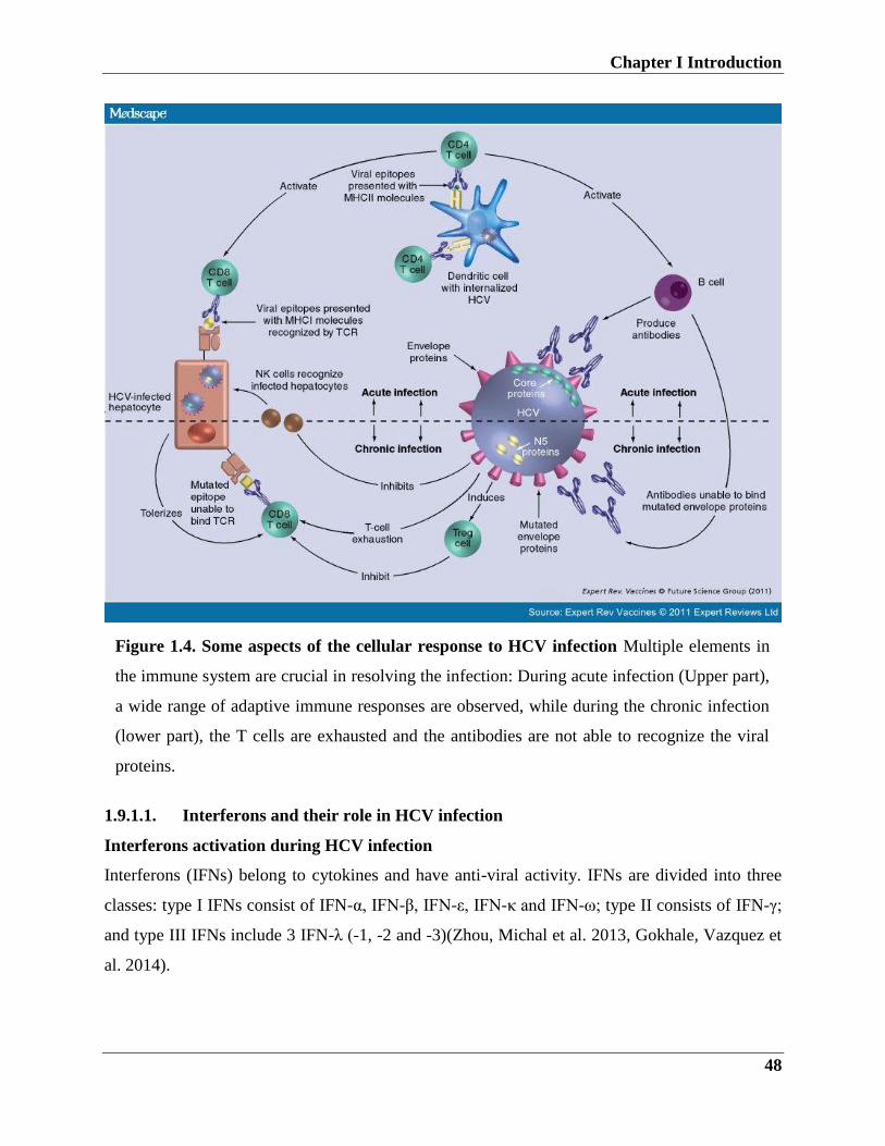
Chapter I Introduction
48
1.9.1.1. Interferons and their role in HCV infection
Interferons activation during HCV infection
Interferons (IFNs) belong to cytokines and have anti-viral activity. IFNs are divided into three
classes: type I IFNs consist of IFN-α, IFN-β, IFN-ε, IFN-κ and IFN-ω; type II consists of IFN-γ;
and type III IFNs include 3 IFN-λ (-1, -2 and -3)(Zhou, Michal et al. 2013, Gokhale, Vazquez et
al. 2014).
Figure 1.4. Some aspects of the cellular response to HCV infection Multiple elements in
the immune system are crucial in resolving the infection: During acute infection (Upper part),
a wide range of adaptive immune responses are observed, while during the chronic infection
(lower part), the T cells are exhausted and the antibodies are not able to recognize the viral
proteins.
Chapter I Introduction
49
Type I IFN receptors and signaling
All IFN-α subtypes bind to the same receptor IFN-α receptor (IFNAR), which consists of two
subunits (IFNAR1 and IFNAR2). Upon binding, receptors are activated by dimerization and
phosphorylation. Following receptors activation, IFNAR1 subunit binds to Tyrosine Kinase
(TYK2) and IFNAR2 binds to Janus kinase (JAK). Upon binding, JAK and TYK2 trans-activate
each other, results in activation of signal transducer and activator of transcription (STAT) 1,
STAT2 and STAT3 (Rajsbaum, Stoye et al. 2008, Metz, Reuter et al. 2013, Majumdar,
Chattopadhyay et al. 2015).
Thus, activated STATs interacts with IRF9 and translocates into the nucleus to form interferon-
stimulated gene factor 3 (ISGF3), this complex subsequently binds to ISRE of the IFN
stimulated genes promoters. Therefore, this binding results in the induction of antiviral state
(Spiegel, Pichlmair et al. 2005, Kanda, Steele et al. 2007, Schoggins, Wilson et al. 2011).
Type II IFN: Consists of IFN-γ, it is produced by certain cells such as (NK) cells, CD4+ T helper
cells and CD8+ cytotoxic T cells (Ye, Liu et al. 2015)65. IFN-γ binds to IFNGR which consists
of two subunits IFNGR1 and IFNGR2 (Zhou, Michal et al. 2013, Jilg, Lin et al. 2014).
Type III IFN: Is recently identified, it consists of IFN-λ family (IFN-λ1, -2 and 3) which also
known as (IL29, IL28A, and IL28B) respectively (Eksteen 2011). Upon viral infection, they
sense by binding to IFN-λ receptor chain which consists of IL28RA and IL28RB (Li, Liu et al.
2009).
Type I and III IFN are produced in response to viral infections. In viral infection, IFN signaling
occurs via PRRs, in the case of HCV, RNA is detected by TLRs and by retinoic acid-inducible
gene (RIG)-I-like receptors (RLRs).
RIG-I (also known as s DDX58) and MDA-5 (also known as IFIH1) (Meylan, Curran et al.
2005, Pichlmair, Schulz et al. 2006, Takeuchi and Akira 2008) belong to RLRs, they contain 2
N-terminal caspase recruitment domain (CARDs), DExD/H-box helicase domain . Upon
recognition of HCV dsRNA, RIG-I and MDA-5 undergo conformational changes; this allows
their binding to MAVS (also known as VISA, CARDIF or IFN-β promoter stimulator 1(IPS-I)
(Meylan, Curran et al. 2005). The formed complex then activates downstream proteins TBK1
and IKKε kinases, which in turn activates IRF3 and NF-κB (Majumdar, Chattopadhyay et al.
2015).
Chapter I Introduction
50
In HCV-infected cells, IFN type I and III are induced via two independent pathways, TLR3 and
RIG-I and subsequently stimulation of hundreds of ISGs. In acute HCV-infected patients, the
expression of ISGs in hepatocytes was shown to be upregulated (Jilg, Lin et al. 2014, Ignatius
Irudayam, Contreras et al. 2015).
1.9.2. The role of ISGs in controlling HCV infection
IFNs can upregulate hundreds of ISGs, and several of ISGs have broad antiviral activity, some of
those ISGs are protein kinase R (PKR), 2’-5’ oligoadenylate synthetase (OAS), MxA and ISG15
(Sadler and Williams 2008). Many ISGs antiviral mechanisms are unknown, it has been noted
that HCV-infected cells produce ISGs pattern which overlaps with the ISGs induced by IFN.
Surprisingly, high levels of ISGs at the infection baseline are associated with the poor response
to IFN based treatment. In chimpanzees and CHC patients, the ISGs levels increase upon HCV
infection (Simister, Schmitt et al. 2009, Wilson and Stamataki 2012).
In order to determine ISGs with anti-viral activity, several studies have been done in Huh7 and
Huh7.5 hepatoma cell lines. Basically, two approaches were used the overexpression by the
introduction of the individual ISGs in the cells using lentivirus followed by infection with HCV.
In these experiments, a number of introduced ISGs were associated with the suppression of HCV
replication, with the robust antiviral effect observed for RIG-I, MDA-5, IRF1, IRF 2, IRF 7 and
Viperin (Helbig, Lau et al. 2005, Seo, Yaneva et al. 2011, Wang, Wu et al. 2012).
Viperin stands for (virus-inhibitory protein, ER-associated, interferon-inducible), it is type I IFN
inducible gene, and it is evolutionary conserved. It belongs to radical S-adenosyl methionine
domain containing enzymes and consists of 361 aa with 42kDa. Viperin can be induced by
different stimulus such as type I, II and III IFNs, the dsRNA analog poly I:C, lipopolysaccharide
(LPS), and by several viruses (Seo, Yaneva et al. 2011, Wang, Wu et al. 2012). It has been
reported that Viperin inhibits wide range of viruses, including an alphavirus (Sindbis virus), an
orthomyxovirus (Influenza A virus), a paramyxovirus (Sendai virus), a rhabdovirus (VSV), and a
retrovirus (HIV-1) (Jiang, Guo et al. 2008), and flaviviruses (HCV, West Nile virus, and Dengue
virus).
The overexpression of Viperin resulted in suppression of HCV replication, it´s localization in
lipid droplets and ER proposes that the anti-HCV mechanism depends on lipid droplets
cooperation. Whereas HCV replication complex take place in ER-lipid droplets, and the N-
terminal alpha-helix Viperin is associated with ER and lipid droplets. Of note, HCV NS5a and
Chapter I Introduction
51
Viperin have similar N-terminal alpha-helix, therefore Viperin could interact with NS5a on the
ER-lipid droplets (Wang, Wu et al. 2012). Another proposed viperin’s anti-HCV activity is its
binding to vesicle-associated membrane protein-associated protein subtype A (VAP-A), which is
known as a host factor essential for HCV replication by interaction with both NS5a and NS5b
(Helbig, Lau et al. 2005, Wang, Wu et al. 2012).
Another class of ISGs named Interferon-inducible transmembrane proteins (IFITMs), with the
exception of IFITM5, IFITM1,-2 and -3 are induced by IFNs. It has been reported that IFTIMs
proteins play an active role against a wide variety of viruses. IFITM1 was found to interfere with
HCV on the entry level; this occurs through binding of IFITM1 to the HCV co-receptors CD81
and occludin, consequently this inhibits the virus entry (Schoggins, Wilson et al. 2011).
IFITM3 has been reported to interfere with HCV replication; this is supposed to function via
interruption of HCV IRES-mediated translation. Another proposed mechanism overlaps with
Viperin mechanism, is the negative regulation of VAP-A, which in turn suppress the virus
entry(Jiang, Guo et al. 2008, Seo, Yaneva et al. 2011, Wang, Wu et al. 2012) .
ISG15, identified as an ubiquitin-like molecule (UBL), it conjugates with hundreds of proteins
which are involved in several pathways. The protein modification (ISGylation) occurs through
activation of E1, E2, and E3 enzymatic subsequent events (Randall, Chen et al. 2006). ISG15 has
antiviral activity against different viruses, including HIV and SINV. ISG15 has antiviral activity
against different viruses including, HIV and Sendai Virus (Sadler and Williams 2008).
Overexpression of ISG15 in IFN-alpha/beta knockout mice resulted in a reduction in the Sendai
Virus infection.
The effect of ISG15 on HCV is controversial; in one report they showed that ISG15
overexpression resulted in an increase in HCV replication, the ISG15 pro-viral role is supported
by another report whereas silencing of ISG15 inhibits the HCV replication. Moreover,
knockdown of the ISGylation negative regulator USP18 results in suppression of HCV via the
IFN-alpha-mediated mechanism. In line with this, knockdown of USP18 enhances the antiviral
activity against viruses including, such as SINV, hepatitis B virus and VSV (Randall, Chen et al.
2006). The knockdown of USP18 is associated with general enhancement of the ISGs
expression, an increase in the cellular protein ISGylation and sustained activation of JAK-STAT
pathway. It was observed that liver biopsies from chronically HCV-infected patients, there is a
Chapter I Introduction
52
high increase in UPS18 and ISG15 expression levels, and this could partially explain the poor
response to IFN based treatment (Sadler and Williams 2008, Nenasheva, Kovaleva et al. 2015).
1.9.3. The TRIM protein family
TRIM (tripartite motif) proteins (also known as RBCC) are a Ring domain–containing proteins,
in human, there are ~70 members of TRIM proteins (Meroni and Diez-Roux 2005, Boudinot,
van der Aa et al. 2011, Marin 2012). They are involved in many biological functions including
cell proliferation, apoptosis, cell signaling pathways and antiviral defense mechanisms. Many
TRIM proteins are ISGs and induced by IFNs (Napolitano and Meroni 2012).
The evolution analysis of TRIM genes revealed that, they are divided into two groups: one group
is conserved and present in vertebrates, a second group, evolves more rapidly and present only in
vertebrates (Sardiello, Cairo et al. 2008).
TRIM members consist of an N-terminal RING, one or two B-boxes, and coiled-coil (CC)
domains (RBCC). The RING domain confers Ubiquitin E3 ligase activity by binding to (UBE2
or E2), which conducts protein modification (Ye and Rape 2009, Napolitano, Jaffray et al. 2011).
The B-box domains (B1 and B2) are zinc-binding motifs, they have a different spacing of the
conserved Cys and His residues, B-boxes exist only in TRIM family proteins, and the exact
function is still unclear(Marin 2012), some studies showed that intact B-boxes are necessary for
TRIMs biological functions. Following the B-box domains is the CC domains, is a helical
structure which mediates protein-protein interactions in particular homo and hetero-
oligomerization, this supports the formation of high molecular weight complexes, which can be
useful for the TRIMs function. Based on the C-Terminal domain of TRIM proteins, they are
divided into eleven subgroups. The C-terminal domain contains (PRY or SPRY alone or merge
of both domains (known asB30.2 domain), recently in TRIM evolution this domain showed a
broad expansion which is important for selection, this could explain the importance of this
domain in the innate immune recognition of retroviruses (Meroni and Diez-Roux 2005, Rhodes,
de Bono et al. 2005, Ozato, Shin et al. 2008, Du Pasquier 2009). TRIMs have different
expression patterns depending on the tissue, and their localizations are in both cytoplasm and in
the nucleus (Rajsbaum, Stoye et al. 2008, Versteeg, Rajsbaum et al. 2013).
Chapter I Introduction
53
TRIMs modify proteins by ubiquitination
Ubiquitination is a protein post-translational modification that conjugates the Ubiquitin (Ub), a
highly conserved cytoplasmic protein (76 aa) to the target protein. This process involves Ub-
activating (E1) enzyme, Ub-conjugating (E2) enzyme and Ub ligase (E3) (Choo and Zhang
2009). At first, Ub is activated by E1 enzyme, forming thioester bond, followed by transmission
of activated Ub to the cysteine residues of the E2 enzyme, and finally, the E3 mediates the
ligation of the Ub to the target protein (Choo and Zhang 2009, Calistri, Munegato et al. 2014).
There is one E1 enzyme, around 30 E2 enzymes, and hundreds of E3 enzymes, the high diversity
of E3 group define the substrate specificity during ubiquitination. Based on the catalytic core
domain there are three groups of the E3 ligases, HECT, RING and U-box protein groups
(Sardiello, Cairo et al. 2008). The ubiquitination process can be mono or poly-ubiquitination, the
protein monoubiquitination results in a regulatory role in protein trafficking and transportation.
Poly-ubiquitination process occurs via several rounds of ubiquitination resulting in a long chain
of Ub (Ye and Rape 2009).
Ub contains lysine residues at different positions (Lys6, Lys11, Lys27, Lys29, Lys33, Lys48 and
Lys63). Two residues are proven to be associated with Ubiquitination, the (Lys48 and Lys63).
While, Lys48-poly-Ub linkage results in 26S proteasome degradation, Lys63 Ub linkage recruits
functions associated with non-proteolytic functions such as NF-kβ signaling, DNA repair and
protein endocytosis (Napolitano, Jaffray et al. 2011, Versteeg, Rajsbaum et al. 2013). The
Ubiquitination can be reversed by a group of enzymes called Deubiquitinating enzymes (DUBs),
these enzymes can cleave the Ub from the target protein (Choo and Zhang 2009).
1.9.3.1. TRIM proteins immunological functions
TRIM family members have been implicated in several functions such as cell differentiation,
apoptosis, neurogenesis, muscular physiology and innate immune responses. A recent study has
shown that TRIM69 plays an important role in zebrafish brain development, the study shows that
TRIM69 is expressed in early stages of the embryos brain development, this occurs through
regulation of AP-1 pathway (AP-1 functions in a variety of cellular processes, including
proliferation, differentiation, and apoptosis), and knockdown of TRIM69 resulted in deformed
brain formation, this effect could be reversed when human TRIM69 mRNA was injected into
deformed phenotype mice (Han, Wang et al. 2016, Han, Zhao et al. 2016).
Chapter I Introduction
54
The integrity of the cellular membrane is important for the cellular function, several genes have
been implicated in the cell membrane repair such as dysferlin4. Other genes such as TRIM72
(mitsugumin 53 (MG53), a muscle-enriched member of TRIMs, has been shown to be an
important factor in the cellular membrane repair at the site of injury (Cai, Masumiya et al. 2009,
Alloush and Weisleder 2013). In addition, it was observed that muscular membrane damage in
TRIM72−/− mice cannot be properly repaired and myopathy phenotype was observed in mice. In
vivo experiments showed that injection of recombinant human MG53 (rhMG53) protein can
prevent myocyte death. As TRIM72 exist in the blood of mice and other animals, it provides one
more advantage for their potential future therapeutic applications. If future researchers prove
benefits rendering neuronal cell death, this would be beneficial for potential therapy in
neurodegenerative diseases (Alloush and Weisleder 2013).
TRIM proteins involvement in cancers and other diseases
TRIM proteins such as TRIM19, TRIM24, and TRIM25 have been associated with the breast
and prostate tumorigenesis development and leukemia progression, this due to their ability to
regulate the activation of nuclear and hormone receptors (Nisole, Stoye et al. 2005, Rhodes, de
Bono et al. 2005, Rajsbaum, Garcia-Sastre et al. 2014, Masroori, Merindol et al. 2016).
TRIM19 (PML) molecule manages different cell functions such as apoptosis and DNA damage.
It has been reported that PML function as a tumor suppressor, its fusion with retinoic acid
receptor alpha (RARα) prevents the proper functions of PML and RAR, this complex formation
is involved in the acute promyelocytic leukemia. In addition, Pml–/– cells are associated with
nuclear bodies mislocalization (Mandell, Jain et al. 2014).
TRIM24 is regulated the activation of several nuclear receptors, including estrogen and retinoic
acid, its role in tumorigenesis is variable, however it acts as a tumor suppressor in liver cancers
through interfering with RARα-mediated transcription in mice, its overexpression in human
breast cancers associated with poor prognosis (Hatakeyama 2011, Elabd, Meroni et al. 2016).
1.9.3.2. TRIM5
TRIM5 belongs to class-IV TRIM family; it contains three zinc-binding domains, a RING, a B-
box type 1 and a B-box type 2, and a coiled-coil region, and composed of 493 amino acids, it is
expressed in different tissues either intra or extracellularly, in case of liver it is expressed
Chapter I Introduction
55
intracellularly, TRIM5 mainly localizes in the cytoplasmic bodies (Nisole, Stoye et al. 2005,
Rhodes, de Bono et al. 2005, Wu, Kirmaier et al. 2013).
TRIM5 alpha is best known as a retroviruses restriction factor, (de Silva and Wu 2011, Pertel,
Hausmann et al. 2011, Wu, Kirmaier et al. 2013). Rhesus Monkey TRIM5 alpha has been
identified as a restriction factor for HIV-1 and N-tropic murine leukemia virus, in contrast,
human TRIM5 does not restrict HIV-1 and shows mild restriction of HIV-2, this indicates the
species-specific nature of TRIM5. It inhibits HIV early during infection entry and this can be
contributed to the absence of reverse transcripts. TRIM5 different domains contribute to the
restriction of viral infection, the Ring domain is responsible for its E3 ligase activity, the B-box
domain is associated with the high-order assembly, the coiled-coil domain functions in the
TRM5 dimerization which is required for capsid binding, the variation in the B30.2/SPRY
domain is responsible for the recognition specificity. It was hypnotized that TRIM5 alpha
interacts directly with the core protein. Therefore, upon virus entry the B30.2 domain recognizes
the core, presence of viral core enhances the formation of TRIM5 hexamers, and formation of
hexamer lattice allows efficient binding and coverage of the core protein, this results in rapid
capsid uncoating and blocking of viral infection. (Pertel, Hausmann et al. 2011), studied the
TRIM5 role in the innate immune signaling to retroviruses. Consequently, they have found that
TRIM5 mediates the innate immune response, which in turn establishes an antiviral state, thus
can reduce the spread of viruses (de Silva and Wu 2011).
Moreover, several TRIM family proteins including TRIM5 are playing a role in autophagy via
participating in the formation of the ULK1-BECN1 active complex. In addition, overexpression
of TRIM5 led to autophagy activation, while TRIM5 knockdown resulted in the inhibition of
autophagy. Another role of TRIM5 in the autophagy mechanism is its role as an autophagy
receptor via its interaction with LC3 (Mandell, Jain et al. 2014). Moreover, it was observed that
TRIM5 co-localizes with autophagosome, this observation was tested to check if TRIM5
mediates HIV degradation via autophagosome degradation, the study showed that rhTRIN5
recognizes and interacts with HIV-1capsid consists of the p24 viral protein, which act as a
lysosomal substrate (Mandell, Kimura et al. 2014, Lascano, Uchil et al. 2016). The study also
found that HIV-1 but not SIV was reduced, thus proposed that TRIM5 specific interaction with
HIV-1 capsid protein mediates the HIV degradation.
Chapter I Introduction
56
In a study of the etiology of Multiple Sclerosis (MS) and retroviruses, a link between MS and
TRIM5 has been observed, the investigation of 12 SNPs located in the TRIM5 intron 1, whereas
TRIM5 RING is encoded by exon 2 showed that SNP rs3802981 is associated with MS
development (Nexo, Christensen et al. 2011).
1.9.3.3. TRIM9
TRIM9 protein is also known as (RNF91and SPRING), it is consists of three zinc-binding
domains, a RING, a B-box type 1 and a B-box type 2, and a coiled-coil domain, it localizes to
the cytoplasmic bodies (Tanji, Kamitani et al. 2010).
The mRNA expression analysis in mice, revealed that TRIM9 mRNA is mainly expressed in the
central nervous system during the embryo development, the study also investigated the
arrangement of the protein in the nervous system, they supposed that TRIM9 is brain specific
and is highly expressed in the cerebral cortex and hippocampus (Caterina Berti 2002, Yang, Li et
al. 2014). Moreover, alterations in the Parkinson's disease (PD) and dementia with Lewy bodies
(DLB) were detected, whereas brain affected areas have less TRIM9 expression, this finding
represent the first prove of TRIM9 involvement in the neurodegenerative diseases (Winkle,
Olsen et al. 2016) .
As several members in the TRIM family proteins act as E3 ligase and have, TRIM9 was tested
for its E3 activity, in vitro assay showed that TRIM9 act as a self-ubiquitination molecule,
furthermore proteasomal inhibitor treatment led to increase in TRIM9 protein, that suppose that
TRIM9 undergoes the proteasomal degradation. Moreover, the reduction of TRIM9 protein was
observed in mice infected with rabies virus which resulted in down-regulation of several proteins
including TRIM9 and as a consequence, accumulation of synaptic vesicles (Yang, Li et al. 2014).
Another role TRIM9 in the nervous system is, its role in the regulation of neurotransmission, the
tightly regulated process involves the formation of SNARE complex formation which is
important in the neurotransmission, it was reported that TRIM9 specifically interacts with
SNAP-25 and adjusts the SNARE complex formation (Tanji, Kamitani et al. 2010).
Since several TRMs involved in the innate immune signaling, TRIM9 was investigated for its
role in the immune system. The study showed that, C-I TRIM subfamily including, TRIM9 is
highly expressed in CD4+ T cells, while very low or hardly detectable in the macrophages and
dendritic cells (Rajsbaum, Stoye et al. 2008, Chan, Towers et al. 2014).
Chapter I Introduction
57
Notably, TRIM C-1 members are highly expressed in T cells and it is the only subgroup contains
the COS-FN3 motif, this suggests it role in the T cell regulation (Rajsbaum, Stoye et al. 2008).
An interesting study has revealed that TRIM9 act as a negative regulator of KF-kb in the brain
cells. NF-κB has two signaling pathways, canonical and non-canonical pathways, and
misregulation of these pathways are associated with cancers, inflammation, and autoimmune
disseises. Activation of NF-κB canonical response is dependent on IkB kinase (IKK) complex
(IKKa, IKKb, and IKK-g), the inactive form of NF-κB is controlled by IkB inhibitory protein,
activation of IKK complex led to phosphorylation of IkBa and b-TrCP, this followed by the
release of NF-κB active form. The negative regulation of TRIM9 occurs through its interaction
and stabilization of b-TrCP, and this consequently inhibits activation of NF-κB and cytokines
(Shi, Cho et al. 2014).
TRIM9 has two isoforms, long (TRIM91) and short (TRIM9s), the long form acts as a negative
regulator of the NF-κB , and a recent study has investigated the role of TRIM9 in viral
infections, a recent study has found that the short form of TRIM9 acts as a positive regulator of
IFN signaling against RNA and DNA viruses. Following viral infection, TRIM9s undergoes self-
ubiquitination and enhances the recruitment of GSK3β to TBK1 which results in stimulation of
IRF3 signaling cascade. The study analyzed the role of TRIM9 isoforms in VSV infection, when
293T cells transfected with TRIM9s showed a reduction in the VSV infection, while TRIM91
only has modest effect when transfected at high concentration (Qin, Liu et al. 2016).
Interestingly, TRIM9s has antiviral activity against both RNA and DNA viruses, when TRIM9s-
overexpressing cell line infected with VSV and Herpes simplex virus HSV-1, low levels of viral
RNA was detected compared to wild-type cells (Qin, Liu et al. 2016).
1.9.3.4. TRIM14
The trim14 protein consists of a B-box, a coiled-coil, and a C-terminal PRY-SPRY domain but
misses the RING domain; it is localized in the mitochondria and expressed in different tissues
(Rajsbaum, Stoye et al. 2008, Uchil, Hinz et al. 2013, Nenasheva, Kovaleva et al. 2015).
Several studies have investigated TRIM14 function in viral infections and innate immunity, for
instance in HIV-1-infected patients TRIM14 is upregulated compared to uninfected donors,
moreover, TRIM14 was highly expressed PBMCs of influenza A-infected patients and in
hepatocytes of HCV-infected patients. Along these lines, the effect of TRIM14 on the SINV
was investigated in HEK–trim14 cell lines, the study showed that overexpression of TRIM14 led
Chapter I Introduction
58
to reduction of SINV replication and this reduction is associated with increase in the expression
of several genes such as IFN-α, IL6 (IFNb2), ISG15, RAF-1, NF-κB (Nenasheva, Kovaleva et
al. 2014).
TRIM14 is an IFN stimulated gene and the investigation of its role in the innate immune
signaling revealed that TRIM14 advocate the immune response against viral infections. Upon
viral infections, TRIM14 undergoes polyubiquitination and recruits the NF-κB to the MAVS,
resulting in stimulation of both IRF3 and NF-κB pathways. Furthermore, TRIM14 knockdown
resulted in interruption of NF-κB association with the MAVS which consequently attenuates the
antiviral response. This reveals that TRIM14 is a part of the mitochondrial antiviral immunity.
TRIM14 facilitates the type I IFN response via activation of ISRE, NF-κB, and IFN-β promoter
activity. Thus, when TRIM14 overexpressed cells infected with VSV, virus replication was
inhibited correlating with the transfection dose (Nenasheva, Kovaleva et al. 2014, Zhou, Jia et al.
2014, Nenasheva, Kovaleva et al. 2015).
Some data have suggested the association of TRIM14 expression with tumorigenesis; in tongue
squamous cells carcinoma (TSCC) one of the lethal cancers, the aggressiveness of the disease is
correlated with the NF-κB pathway activation, in a microarray study TSCC cell survival and
resistance to therapy was correlated with the activation of NF-κB pathway. TRIM14 acts as a
regulator of several genes which are involved in the NF-κB pathway activation, and when
TRIM14 investigated in TSCC patient tissues, data showed that TRIM14 upregulation is
associated with the disease stage and with the survival duration. The in vitro analysis of TRIM14
function in TSCC, when Cal127 and SCC9 cells stably transfected with TRIM14, cells showed
remarkable growth and resistance to apoptosis induced by chemotherapy (Xuan Su1 2016).
1.9.3.5. TRIM25
TRIM25 also known as Estrogen-responsive finger protein and RING finger protein 147, it
consists of N-terminal RING domain, B-box type 1 domain, B-box type 2 domain, coiled-coil
domain and C-terminal SPRY domain and localizes to the cytoplasm (Gack, Shin et al. 2007).
TRIM25 is involved in several functions. Its role in the regulation of the RNA expression has
been determined, whereas the E3 ligase acts as a specific Lin28a/TuT4 cofactor for uridylation
pathway, Lin28a is an RNA binding protein which controls miRNA) maturation (Choudhury,
Nowak et al. 2014) mRNA translation and splicing.
Chapter I Introduction
59
Furthermore, TRIM25 expression is involved in several cancers such as ovarian, breast and
gastric cancers. It was reported that TRIM25 overexpression is associated with ovarian and
breast cancers, while its downregulation was observed in endometrial carcinoma. The analysis of
gene expression in gastric cancers revealed that TRIM25 is upregulated in gastric cancer tissues
compared to normal ones. Moreover, TRIM25 high expression was associated with low survival
of GC patients. Further analysis showed that knockdown of the TRIM25 in GC cell lines (MGC-
803 and AGS), hampered the cells invasion compared to the control ones. Interestingly, the study
showed that TRIM25 enhances cells migration and invasion by activating TGF-β pathway.
Which makes it an interesting target for future therapy perspectives (Zhu, Wang et al. 2016).
Due to the role of many TRIMs in the innate immunity, TRIM25 was investigated for its role in
the regulation of innate immunity. RIG-I is an important receptor recognizes viral RNA and
undergoes activation process results in the induction of IFN.
TRIM25 acts as an activator for RIG-I, it mediates the RIG-I ubiquitination via interaction of
TRIM25 SPRY domain with RIG-I CARD domain which undergoes Lys 63 ubiquitination
results in activation of RIG-I downstream signaling (Gack, Shin et al. 2007, Castanier, Zemirli et
al. 2012, Choudhury, Nowak et al. 2014).
Given its role in RIG-I ubiquitination, TRIM25 has been proposed to be implicated in the IFN-β
production, this was proved when TIRM25WT, TRIM25+/-, and TRIM25-/- MEF cells were
transiently transfected with IFN-β promoter plasmid, IFN-β promoter was activated in TRIM25
WT MEF cells with early undetectable activation in TRIM25-/- MEF cells. Moreover,
TRIM25 expression in MEF cells decreases the VSV infection, in contrast, high levels of the
VSV were detected in TRIM25-/- cells (Choudhury, Nowak et al. 2014, Zhang, Elabd et al.
2015).
Chapter I Introduction
60
Aims of the Thesis
This dissertation is divided into three sections, each one describes a study on HCV.
The aim of study 1: Genotypic and phenotypic analysis of HCV-NS3 variants under
protease inhibitors treatment.
The overall aim of the first part of the thesis was to characterize the resistance to HCV protease
inhibitors drugs by genotyping and phenotyping. Furthermore, as IL28b is an important host
factor associated with the clinical outcome, we wanted to determine the association of the IL28b
polymorphisms with SVR in our cohort.
This was accomplished by the following objectives:
1- Establishment and optimization of the genotypic assay of HCV resistance variants (Pre-
exist and/or acquired under protease inhibitors treatment).
2- Establishment of phenotyping resistance assay for the analysis of viral fitness and
characterization of the resistance profile of the protease inhibitors drugs.
3- Establishment of TaqMan assay for genotyping of IL28b polymorphisms and
determination of the relationship between different SNPs and the clinical outcome.
Aim of study 2: Crystallization of HCV-NS5b protein of genotype4a under Microgravity
conditions and structure determination:
One aim of NASA missions is to use the advances of the microgravity conditions in the
International Space Station (ISS) for scientific research. Thus, throughout the second part of the
thesis, we used the microgravity environment of the ISS to crystallize the HCV-NS5b protein of
genotype 4a and compare it to the crystallization in 1 g control conditions. Followed by
determination of the 3D structure of the HCV-NS5b protein.
These aims were determined as follows:
1- HCV-NS5b protein expression and purification.
2- Protein crystallization under microgravity conditions.
3- Protein crystallization optimization.
4- X-ray diffraction and structure determination.
Chapter I Introduction
61
Aim of study 3: Identification and characterization of TRIM proteins role in HCV
infection
Since several TRIMs are interferon-inducible genes and have antiviral activities, we
hypothesized that they could have antiviral activities against HCV. We, therefore, sought to
investigate whether TRIM proteins could modulate HCV infection and the involvement in
type I IFN signaling.
These aims were achieved by:
1- Investigation of the TRIM genes differential regulations during HCV infection and
upon IFN treatment.
2- Assessment of whether TRIM5, 9, 14 and 25 have an antiviral activities against HCV
infection.
3- Investigation of whether TRIM5, 9, 14 and 25-mediate the regulation of IFN I
signaling pathway, in particular, HCV cell receptors and adaptor molecules.
Chapter II Materials and Methods
62
2. Chapter II Materials and Methods
2.1. Materials
2.1.1. Patient samples
For genotypic and phenotypic analysis, blood samples were obtained from patients enrolled in
the PEPSI project. All patients with established chronic hepatitis C infection of genotype1, who
were clinically resistant or partial responders to protease inhibitors.
2.1.2. Chemicals and reagents
Agarose Invitrogen
Ampicillin Roth
BOC SeqChem Ltd., Pangbourne, UK
Bovine Serum Albumin, BSA BSA- Sigma-Aldrich
Collagen R Serva
Dithiothreitol Sigma-Aldrich
DMSO Sigma-Aldrich
EDTA Roch
EGTA Roth
Ethanol Roth
Fatty Acid-Free Sigma-Aldrich
FCS Gibco
Geneticin Gibco
Glycine Roth
HCl 37% (w/w) Roth
Isopropanol Roth
PBS Invitrogen
Penicillin/Streptomycin Gibco
Polyethylene Glycol (PEG) 8000 Promega
Potassium chloride Sigma-Aldrich
RNA loading buffer Sigma-Aldrich
Sodium pyruvate Gibco
Chapter II Materials and Methods
63
TLV SeqChem Ltd., Pangbourne, UK
Tris Roth
Triton-X- 100 Sigma-Aldrich
Tritonx100 Roth
Trypsin- EDTA Gibco
Tween 20 Roth
Yeast Extract Gibco
6 x DNA loading dye Thermo Fisher
Roti®-GelStain Roth
Pierce ECL Western Blotting Substrate
OptiMEM Life Technologies
DMEM Invitrogen
2.1.3. Kits
Cell Proliferation Kit II (XTT) Promega
CellTiter-Blue® Cell Viability Assay Promega
High Pure PCR Product Purification Kit Roche
LightCycler 480 SYBR Green I Master Roche
Lipofecatmine2000,Transfection Reagent Life Technologies
Ni-NTA Spin Columns Qiagen
NucleoBond Xtra Maxi Kit Macherey & Nagel
NucleoSpin®RNAII kit Macherey & Nagel
PJET Plasmid Miniprep Kit Promega
SuperScript III First-Strand Synthesis
SuperMix for qRT-PCR
Life Technologies
SuperScript® III One-Step RT-PCR System
with Platinum ® Taq DNA Polymerase
Life Technologies
Luciferase Assay System Qiagen
T7 Megascript RNA synthesis kit Life Technologies
Wizard® Genomic DNA Purification Kit Promega
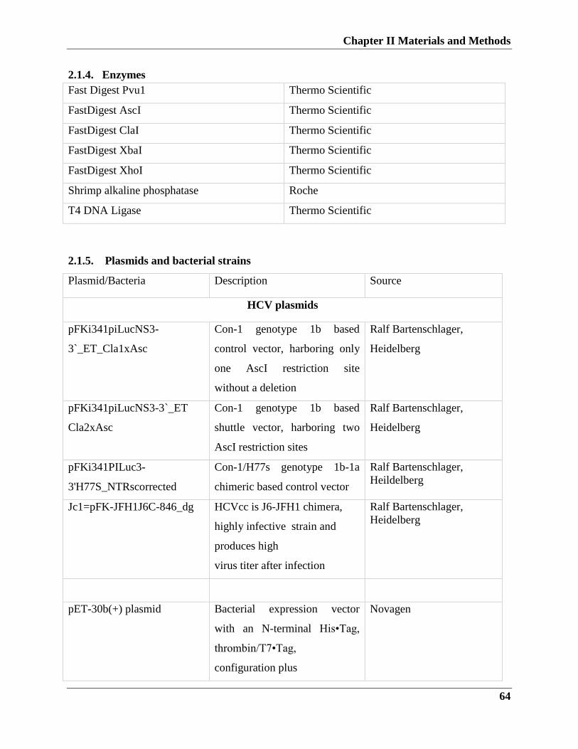
Chapter II Materials and Methods
64
2.1.4. Enzymes
Fast Digest Pvu1 Thermo Scientific
FastDigest AscI Thermo Scientific
FastDigest ClaI Thermo Scientific
FastDigest XbaI Thermo Scientific
FastDigest XhoI Thermo Scientific
Shrimp alkaline phosphatase Roche
T4 DNA Ligase Thermo Scientific
2.1.5. Plasmids and bacterial strains
Plasmid/Bacteria Description Source
HCV plasmids
pFKi341piLucNS3-
3`_ET_Cla1xAsc
Con-1 genotype 1b based
control vector, harboring only
one AscI restriction site
without a deletion
Ralf Bartenschlager,
Heidelberg
pFKi341piLucNS3-3`_ET
Cla2xAsc
Con-1 genotype 1b based
shuttle vector, harboring two
AscI restriction sites
Ralf Bartenschlager,
Heidelberg
pFKi341PILuc3-
3'H77S_NTRscorrected
Con-1/H77s genotype 1b-1a
chimeric based control vector
Ralf Bartenschlager,
Heildelberg
Jc1=pFK-JFH1J6C-846_dg HCVcc is J6-JFH1 chimera,
highly infective strain and
produces high
virus titer after infection
Ralf Bartenschlager,
Heidelberg
pET-30b(+) plasmid Bacterial expression vector
with an N-terminal His•Tag,
thrombin/T7•Tag,
configuration plus
Novagen
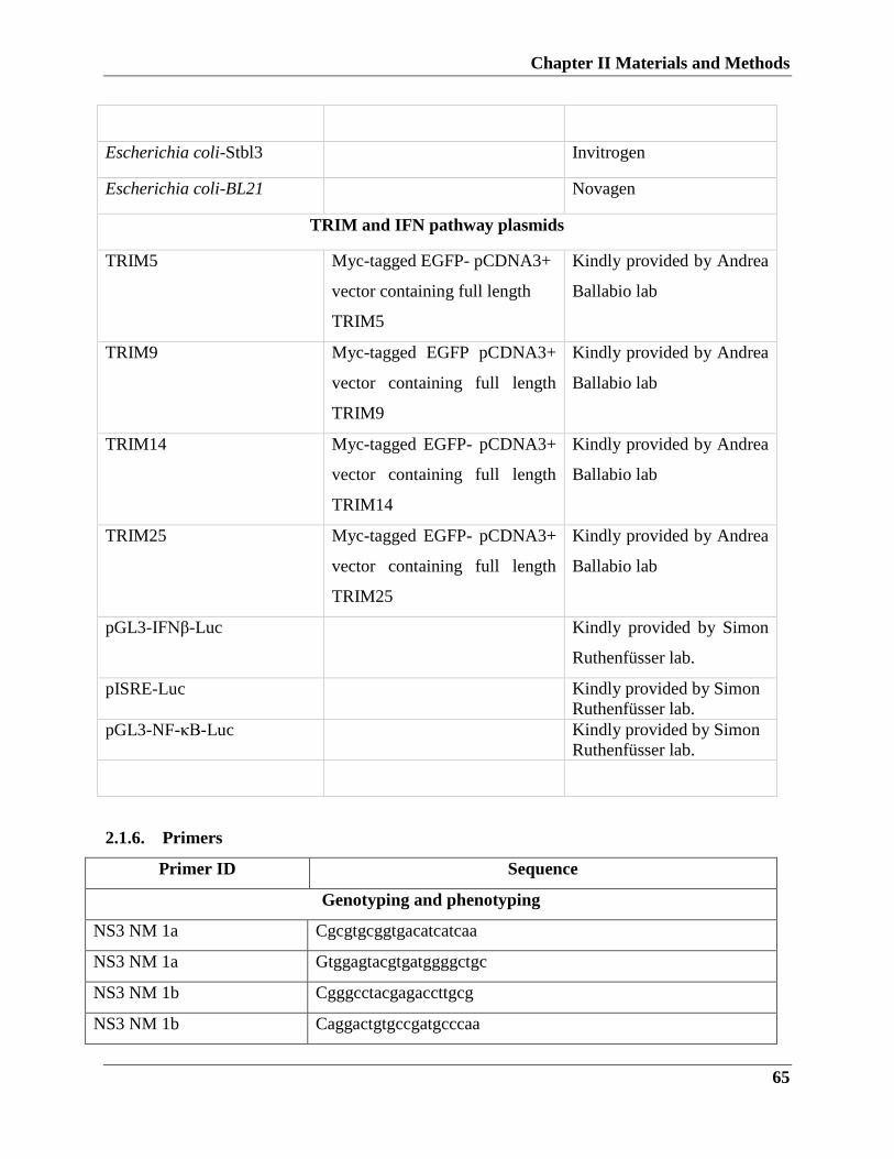
Chapter II Materials and Methods
65
Escherichia coli-Stbl3 Invitrogen
Escherichia coli-BL21 Novagen
TRIM and IFN pathway plasmids
TRIM5 Myc-tagged EGFP- pCDNA3+
vector containing full length
TRIM5
Kindly provided by Andrea
Ballabio lab
TRIM9 Myc-tagged EGFP pCDNA3+
vector containing full length
TRIM9
Kindly provided by Andrea
Ballabio lab
TRIM14 Myc-tagged EGFP- pCDNA3+
vector containing full length
TRIM14
Kindly provided by Andrea
Ballabio lab
TRIM25 Myc-tagged EGFP- pCDNA3+
vector containing full length
TRIM25
Kindly provided by Andrea
Ballabio lab
pGL3-IFNβ-Luc Kindly provided by Simon
Ruthenfüsser lab.
pISRE-Luc Kindly provided by Simon
Ruthenfüsser lab.
pGL3-NF-κB-Luc Kindly provided by Simon
Ruthenfüsser lab.
2.1.6. Primers
Primer ID Sequence
Genotyping and phenotyping
NS3 NM 1a Cgcgtgcggtgacatcatcaa
NS3 NM 1a Gtggagtacgtgatggggctgc
NS3 NM 1b Cgggcctacgagaccttgcg
NS3 NM 1b Caggactgtgccgatgcccaa
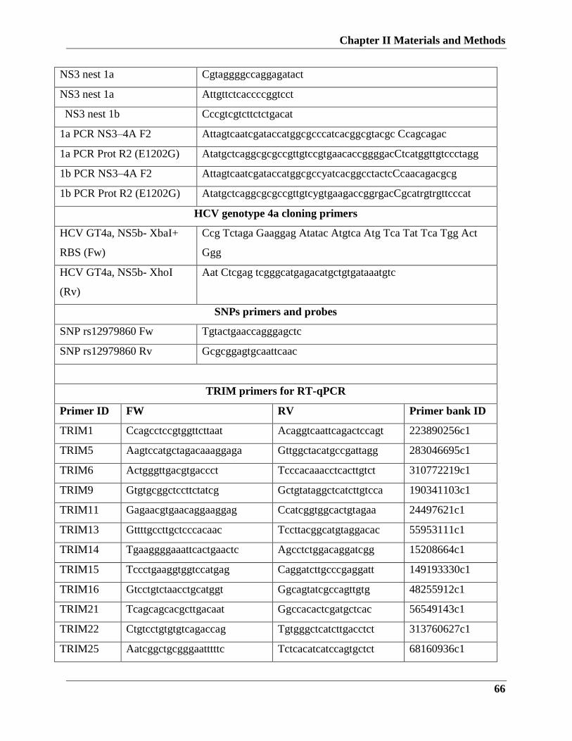
Chapter II Materials and Methods
66
NS3 nest 1a Cgtaggggccaggagatact
NS3 nest 1a Attgttctcaccccggtcct
NS3 nest 1b
Cccgtcgtcttctctgacat
1a PCR NS3–4A F2 Attagtcaatcgataccatggcgcccatcacggcgtacgc Ccagcagac
1a PCR Prot R2 (E1202G) AtatgctcaggcgcgccgttgtccgtgaacaccggggacCtcatggttgtccctagg
1b PCR NS3–4A F2 AttagtcaatcgataccatggcgccyatcacggcctactcCcaacagacgcg
1b PCR Prot R2 (E1202G) AtatgctcaggcgcgccgttgtcygtgaagaccggrgacCgcatrgtrgttcccat
HCV genotype 4a cloning primers
HCV GT4a, NS5b- XbaI+
RBS (Fw)
Ccg Tctaga Gaaggag Atatac Atgtca Atg Tca Tat Tca Tgg Act
Ggg
HCV GT4a, NS5b- XhoI
(Rv)
Aat Ctcgag tcgggcatgagacatgctgtgataaatgtc
SNPs primers and probes
SNP rs12979860 Fw Tgtactgaaccagggagctc
SNP rs12979860 Rv Gcgcggagtgcaattcaac
TRIM primers for RT-qPCR
Primer ID FW RV Primer bank ID
TRIM1 Ccagcctccgtggttcttaat Acaggtcaattcagactccagt 223890256c1
TRIM5 Aagtccatgctagacaaaggaga Gttggctacatgccgattagg 283046695c1
TRIM6 Actgggttgacgtgaccct Tcccacaaacctcacttgtct 310772219c1
TRIM9 Gtgtgcggctccttctatcg Gctgtataggctcatcttgtcca 190341103c1
TRIM11 Gagaacgtgaacaggaaggag Ccatcggtggcactgtagaa 24497621c1
TRIM13 Gttttgccttgctcccacaac Tccttacggcatgtaggacac 55953111c1
TRIM14 Tgaaggggaaattcactgaactc Agcctctggacaggatcgg 15208664c1
TRIM15 Tccctgaaggtggtccatgag Caggatcttgcccgaggatt 149193330c1
TRIM16 Gtcctgtctaacctgcatggt Ggcagtatcgccagttgtg 48255912c1
TRIM21 Tcagcagcacgcttgacaat Ggccacactcgatgctcac 56549143c1
TRIM22 Ctgtcctgtgtgtcagaccag Tgtgggctcatcttgacctct 313760627c1
TRIM25 Aatcggctgcgggaatttttc Tctcacatcatccagtgctct 68160936c1
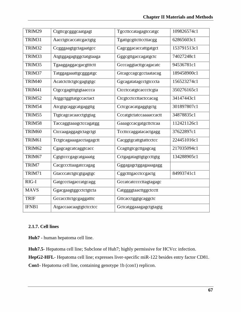
Chapter II Materials and Methods
67
TRIM29 Ctgttcgcgggcaatgagt Tgccttccatagagtccatgc 109826574c1
TRIM31 Aacctgtcaccatcgactgtg Tgattgcgttcttccttacgg 62865603c1
TRIM32 Ccgggaagtgctagaatgcc Cagcggacaccattgatgct 153791513c1
TRIM33 Atgtggagagtggctatgtaaga Gggcgttgaccagatgctc 74027248c1
TRIM35 Tgaaggaggacgacgtttctt Gcccaggtacttgcagacatc 94536781c1
TRIM37 Tatggagaaattgcgggatgc Gtcagccagcgcctaatacag 189458900c1
TRIM40 Acatctcttctgtcgagtgtgc Ggcagatatagcctgtcccta 156523274c1
TRIM41 Ctgccgagtttgtgtaaccca Ctcctccatgtcaccctcgta 350276165c1
TRIM52 Atggctggttatgccactact Ctcgtcctccttactccacag 34147443c1
TRIM54 Atcgtgcaggcatgaggttg Cctcgcacatgaggtgctg 301897807c1
TRIM55 Ttgtcagcacaacctgtgtag Cccatgtctatccaaaaccactt 34878835c1
TRIM58 Taccaggtaaagctccagatgg Gaaagccacgatgcttctcaa 112421126c1
TRIM60 Ctccaagaggagtctagctgt Tccttccaggatacactgagg 37622897c1
TRIM61 Tctgtcagaaagacctagagctt Cacggtgcattgtattcctcc 224451016c1
TRIM62 Cgagcagcatcaggtcacc Ccagttgtcgcttgagcag 217035094c1
TRIM67 Cgtgtcccgagcatgaaatg Cctgagatagttgtgccttgtg 134288905c1
TRIM7 Cacgcccttaagatccagag Gggagagctggagaaagagg
TRIM71 Gtacccatctgtcgtgagtgc Cggctttgacctccgactg 84993741c1
RIG-I Gatgccctagaccatgcagg Gccatcatccccttagtagagc
MAVS Ggacgaagtggcctctgtcta Catggggtaacttggctcctt
TRIF Gccaccttctgcgaggatttc Gttcacctggtgcaggctc
IFNB1 Atgaccaacaagtgtctcctcc Gctcatggaaagagctgtagtg
2.1.7. Cell lines
Huh7 - human hepatoma cell line.
Huh7.5- Hepatoma cell line; Subclone of Huh7; highly permissive for HCVcc infection.
HepG2-HFL- Hepatoma cell line; expresses liver-specific miR-122 besides entry factor CD81.
Con1- Hepatoma cell line, containing genotype 1b (con1) replicon.
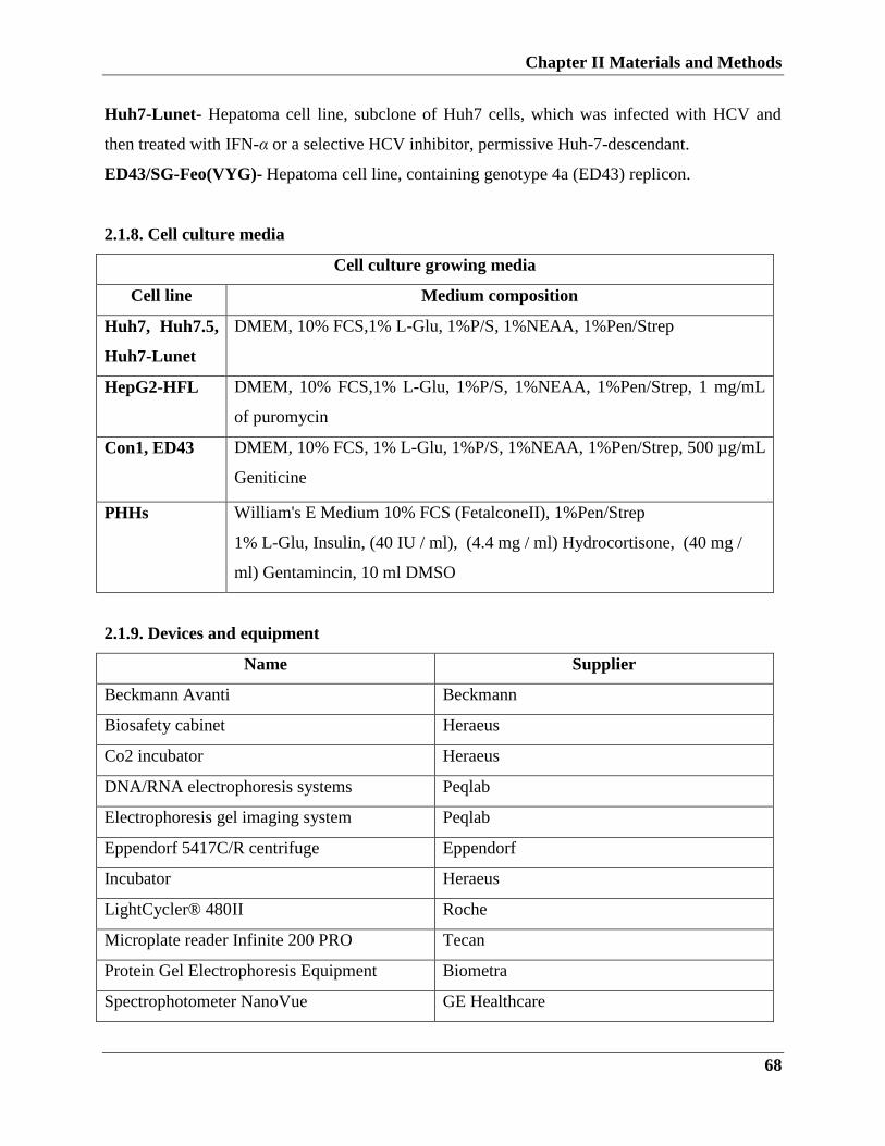
Chapter II Materials and Methods
68
Huh7-Lunet- Hepatoma cell line, subclone of Huh7 cells, which was infected with HCV and
then treated with IFN-α or a selective HCV inhibitor, permissive Huh-7-descendant.
ED43/SG-Feo(VYG)- Hepatoma cell line, containing genotype 4a (ED43) replicon.
2.1.8. Cell culture media
Cell culture growing media
Cell line Medium composition
Huh7, Huh7.5,
Huh7-Lunet
DMEM, 10% FCS,1% L-Glu, 1%P/S, 1%NEAA, 1%Pen/Strep
HepG2-HFL DMEM, 10% FCS,1% L-Glu, 1%P/S, 1%NEAA, 1%Pen/Strep, 1 mg/mL
of puromycin
Con1, ED43 DMEM, 10% FCS, 1% L-Glu, 1%P/S, 1%NEAA, 1%Pen/Strep, 500 µg/mL
Geniticine
PHHs William's E Medium 10% FCS (FetalconeII), 1%Pen/Strep
1% L-Glu, Insulin, (40 IU / ml), (4.4 mg / ml) Hydrocortisone, (40 mg /
ml) Gentamincin, 10 ml DMSO
2.1.9. Devices and equipment
Name Supplier
Beckmann Avanti Beckmann
Biosafety cabinet Heraeus
Co2 incubator Heraeus
DNA/RNA electrophoresis systems Peqlab
Electrophoresis gel imaging system Peqlab
Eppendorf 5417C/R centrifuge Eppendorf
Incubator Heraeus
LightCycler® 480II Roche
Microplate reader Infinite 200 PRO Tecan
Protein Gel Electrophoresis Equipment Biometra
Spectrophotometer NanoVue GE Healthcare
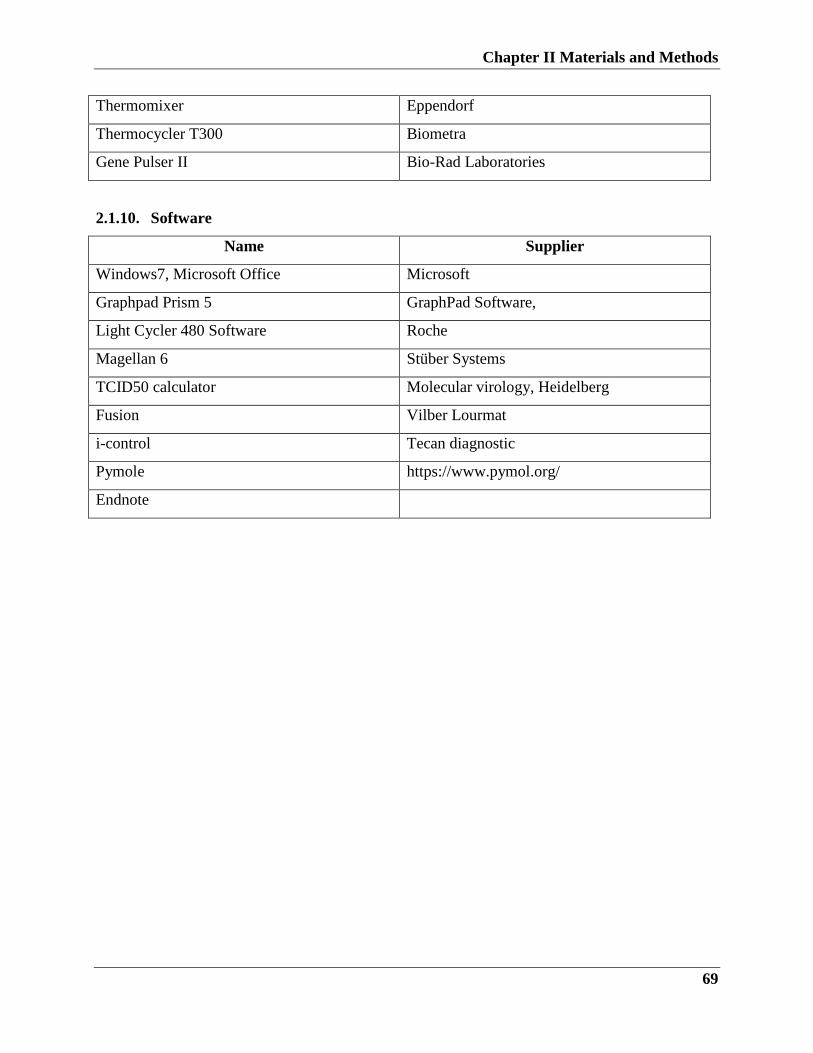
Chapter II Materials and Methods
69
Thermomixer Eppendorf
Thermocycler T300 Biometra
Gene Pulser II Bio-Rad Laboratories
2.1.10. Software
Name Supplier
Windows7, Microsoft Office Microsoft
Graphpad Prism 5 GraphPad Software,
Light Cycler 480 Software Roche
Magellan 6 Stüber Systems
TCID50 calculator Molecular virology, Heidelberg
Fusion Vilber Lourmat
i-control Tecan diagnostic
Pymole https://www.pymol.org/
Endnote
Chapter II Materials and Methods
70
2.2. Experimental Methods: Part I
2.2.1. Viral RNA extraction
Viral RNA was isolated from all patient serum samples using the QIAamp Virus BioRobot MDx
kit (Qiagen, Belgium). RNA extraction was performed using the high throughput M1000 robot
extractor (Abbott Diagnostics).
2.2.2. HCV-NS3 RT- PCR amplification and sequencing
Samples were transcribed and amplified in one-step, in which both cDNA synthesis and PCR
amplification in a single tube using gene-specific primers was used to amplify a 1 kb fragment.
The RT-PCR thermal cycler protocol was as following; one cycle at 55°C for 30 min followed
by PCR cycles, 94 ◦C for 2min, 35 cycles of (94 ◦C for 30 s, 55 ◦C for 30 s, 72 ◦C for 3min) and
one cycle at 72 ◦C for 7 min.
Amplification products were analyzed by 1% agarose gel electrophoresis, in 1 x TAE buffer.
Samples were mixed with 6 x DNA loading dye (Thermo Fisher) and circa 6 μl were loaded per
lane on the agarose gel. Electrophoresis reactions were run at 80 - 100 V. The DNA was stained
with Roti®-GelStain (Roth) (0.5 μg/ml) and viewed under a UV light with a wavelength between
280 and 320 nm, with the Electrophoresis gel imaging system (Peqlab).
DNA concentration determination
The Spectrophotometer NanoVue (GE Healthcare) was used to determine the DNA
concentration using a 3 μl sample. DNA measurements are read at a wavelength of 260 nm,
while purity is determined by dividing the absorbance at 260 nm with the absorbance at 280 nm.
Pure DNA has a value between 1.7 and 1.9. Lower values are revealing protein contamination,
while higher values designate the presence of RNA in the sample.
2.1.3. DNA sequencing and genotypic analysis
Direct and clone sequencing
DNA sequencing used to determine the exact base pair sequence of a DNA fragment. The PCR-
based sequencing reaction is based on the enzymatic method. The reaction includes both dNTPs
as well as dideoxyribonucleoside triphosphates (ddNTPs), where the addition of ddNTPs to the
DNA strand primers chains the reaction termination. As the dNTPs are fluorescently labeled with
different dyes they can be recited on an automated DNA sequencer. For direct sequencing,
Chapter II Materials and Methods
71
amplified PCR products were purified by agarose gel extraction using High Pure PCR Product
Purification Kit (Roche) following the manufacturer’s instructions. For clonal sequencing,
amplified PCR products were ligated into a cloning vector using CloneJET PCR Cloning Kit
(Thermo Fischer), followed by plasmid purification using GeneJET Plasmid Miniprep Kit
(Thermo Fischer). Purified PCR products and selected clones were submitted for sequencing.
Genotypic analysis
Sequence data produced from PCR sequence analysis was submitted to
(http://hcv.geno2pheno.org/) HCV drug resistance database to generate an HCV drug resistance
profile. Based on the HCV drug resistance profiles, resistant samples were selected for the
subsequent phenotypic drug resistance analysis.
2.1.4. In vitro phenotypic analysis of HCV drug resistance
Cloning and plasmid purification
Using cloning primers, RT- PCR amplification was done as previously described. PCR products
and the shuttle vector DNA were digested with Fast digest AscI and ClaI (Thermo Scientific).
Vector DNA fragment and PCR products were purified. The shuttle vector was later treated with
shrimp alkaline phosphatase (Roche). In order to ligate DNA insert with DNA vector, the
following protocol was used: insert DNA was ligated to vector DNA in a ratio of 1:3, with
indicated reaction buffer and 2 U of T4 DNA ligase enzyme in a total reaction volume of 20 μl.
The reaction was incubated for approximately 16 hours at 16°C. The ligated DNA vectors were
transformed into E.coli STBL3 competent cells as follows. Competent cells, formerly stored at -
80°C were permitted to thaw for 10 minutes on ice. About 5 to 10 μl of ligated DNA were mixed
with 100 μl of competent cells and incubated on ice for 30 minutes. The reaction mixture was
heat shocked at 42°C for 1 min and incubated on ice for a further 5 min. A volume of 400 μl of 1
x LB medium was added to the cells and the reaction was allowed to shake for 1 hour at 37°C.
For direct large scale preparation of bacterial clones, the transformed bacteria were grown in a
250 ml LB-medium containing ampicillin antibiotic with shaking at 200 rpm overnight.
Large scale preparation of plasmid DNA (MaxPreps)
Maxipreps cultures from transformed DNA were grown in 250 ml 1 x LB medium overnight at
37°C and pelleted by centrifugation at 6000 rpm for 15 minutes. The preparations were done
Chapter II Materials and Methods
72
with the NucleoBond Xtra Maxi Kit (Macherey-Nagel) according to the manufacturer’s
instructions.
RNA in vitro transcription
A panel of samples was used in the antiviral experiment to establish the resistance profile of the
PIs. Each chimeric replicon was initially used to generate RNA for phenotyping experiments.
The plasmid DNA was linearized by digestion with Fast Digest Pvu1 (Thermo Scientific) at 37
◦C for 1 hour. RNA was synthesized using a T7 Megascript RNA synthesis kit (Invitrogen)
following manufacturer’s instruction.
In order to study the resistance profile of HCV NS3 variants, in vitro transcribed RNA samples
were purified using MEGAclear™ Kit (Ambion) following manufacturer’s instruction.
Electroporation and drug titration
In order to determine the resistance profile of chimeric replicons, purified RNA was prepared for
electroporation into Huh-7-Lunet cells. Before electroporation Huh-7-Lunet cells were
maintained in growth medium indicated previously, cells were harvested by trypsinization. Cells
were washed with PBS prior to electroporation. Cells were adjusted to 1×10⁷ cell/ml, 0.4 ml of
cells were transferred with 10µg of RNA using 0.2cm width cuvette (Bio-Rad, Laboratories).
Cuvettes were pulsed at 960µF and 270V with a Gene Pulser II (Bio-Rad Laboratories). Cells
were then transferred to 20ml of complete medium. Cells were seeded in a white 96-well plate
with a clear bottom (Costar) at 100µl/well and incubated overnight. For determination of 50%
inhibitory concentration (IC50), incubated plates were treated with serially diluted PIs in DMSO
(0.5% final DMSO concentration), 72 hours later, cells were harvested for luciferase
measurement.
To determine the luciferase activity, plates were washed with once PBS followed Lysis by
adding 50µl of lysis buffer (Promega) and rocking for 20 min with, then 50 µl of Luciferase
substrate (Promega) was injected into each well using Microplate reader Infinite 200 PRO
Injector (Tecan) to the well. Luciferase was measured with a Microplate reader Infinite 200 PRO
(Tecan). IC50 values of PIs were calculated and compared to control samples using the
GraphPad Prism program (GraphPad Software Inc., La Jolla, CA).
Chapter II Materials and Methods
73
2.2.5. IL28b polymorphism genotyping
DNA extraction
Genomic DNA was extracted from the whole blood using Wizard® Genomic DNA Purification
Kit (Promega).
PCR and direct sequencing
PCR was carried out with Platinum® Taq DNA Polymerase (Invitrogen), 10 pmol of each
primer, and 10 ng of genomic DNA. The thermal cycler settings: one cycle of, 94°C for 5 min;
35 cycles of , 94°C for 30 s, 60°C for 30 s, 72°C for 30 s, and one cycle of, 72°C for 7 min. For
sequencing, PCR products were purified and subjected to sequencing.
TaqMan SNP genotyping assay
To determine the IL28b polymorphisms, TaqMan predesigned SNP genotyping assay was used
to genotype rs8099917 polymorphisms, and to genotype rs12979860 polymorphisms, the PCR
reaction and conditions as the following: TaqMan genotyping assay specific mixture and
TaqMan universal PCR master mix reagent (Applied Biosystems) were used for genotyping. The
PCR genotyping conditions were as following: 10 min at 95°C and 40 cycles of 10 s at 95°C, 1
min at 60°C and 10 s at 72°C. The assay was done by Light Cycler 480 II system (Roche). Data
were analyzed by endpoint genotyping.
Chapter II Materials and Methods
74
2.3. Experimental Methods: Part II
2.3.1. Expression and purification of HCV-NS5b
To generate pET30b-GT4a HCV-NS5b construct, HCV RNA was isolated from the plasma of a
patient infected by HCV-4a, the NS5b sequence was RT-PCR amplified using HCV GT4a
NS5b- XbaI- RBS (Fw) and HCV GT4a NS5b- XhoI (Rv) primers. The forward primer added
the Ribosomal binding site (RBS) and XbaI sequences to the 5’ end of the gene and reverse
primer added XhoI sequence to the 3’ end of the gene. PCR product and pET30b+ (Novagen)
were digested with XbaI and XhoI before ligation. The pure cut DNA were then ligated as
follows, DNA was ligated to vector DNA in a ratio of 1:3, with indicated reaction buffer and 2 U
of T4 DNA ligase enzyme in a total reaction volume of 20 μl, resulting in pET30b-GT4a HCV-
NS5b construct. The ligated DNA vectors were transformed into E.coli BL21 (DE3) competent
cells as follows. BL21 (DE3) cells were transformed with the pET30b-GT4a HCV-NS5b
expression plasmid and plated on an LB agar plate containing 100 μg/mL kanamycin. To check
for insertion of HCV-NS5b, a single colony was picked and cultured for mini preparation. The
purified plasmid was then subjected to sequencing. After sequence confirmation, the same
colony was cultured at 37°C with shaking at 250 rpm till OD600 reached ~0.6. To induce protein
expression, isopropyl β–D–1–thiogalactopyranoside (IPTG, 0.5 mM) was added to the culture
and the culture cells were cooled to 32°C for 4 hours. Cells were harvested by centrifugation at
10000 x g and 4°C for 30 minutes and the pellet was stored at –80 °C until use. For recombinant
protein purification, cell pellets were resuspended in lysis buffer NPI-10 (50 mM NaH2PO4, 300
mM NaCl, 10 mM imidazole) pH 8 and disrupted by sonication on ice (QSonica Misonix 200
(Qsonica), 1-minute pulse 1-minute pause for several rounds). Whole cell lysate was clarified by
centrifugation at 14,000 x rpm for 30 minutes at 4°C. Soluble cell lysates were loaded onto 1 mL
Ni–NTA agarose beads (Qiagen). The loaded resin was washed with NPI-20 (50 mM NaH2PO4,
300 mM NaCl, 200 mM imidazole), and the product was eluted in NPI-50 (50 mM NaH2PO4,
300 mM NaCl, 500 mM imidazole).
SDS-polyacrylamide gel electrophoresis (SDS-PAGE)
To check for protein expression and purity, an aliquot of each step was run under denaturing
conditions in a sodium dodecyl sulfate-polyacrylamide gel. Therefore, gels were made using an
acrylamide concentration of 12% for the resolving gel. The acrylamide/bisacrylamide solution
Chapter II Materials and Methods
75
was a 30% stock solution. The polymerization reaction was started when 1/1,000 volume
TEMED and 1/1,000 volume saturated ammonium persulphate added to the solution of the
separating gel and then was poured into gel casts. Then, 1 ml of isopropanol was applied to
remove air bubbles, and the gel was allowed to polymerize for ~20 minutes. The stacking gel
containing 5% acrylamide stock solution was applied. After insertion of the comb, the gel left to
polymerize. After polymerization, gel cast was assembled into electrophoresis module and the
tank filled with 1X running buffer. Protein samples were mixed with a relative volume of 6X
Laemmli buffer containing DTT as a reducing agent. Further denaturation was done by
incubation for 5 min at 95°C before loading onto the gel. To estimate the molecular weight of the
protein sample, a protein standard “Prestained Protein Marker“(Thermo Scientific) was used and
gels were run at 35mA per gel.
After electrophoresis, proteins were stained with Coomassie blue solution for overnight at 22-
25°C. Thereby proteins were fixed within the gel. For destaining, gels were incubated for 20 min
at 22-25°C in a 5% methanol/5% acetic acid solution, the solution was changed when until clear
staining obtained.
Western blot
To confirm the expression of His-Tagged HCV NS5b, Proteins were resolved by SDS-PAGE
and after electrophoresis transferred to a polyvinylidene difluoride membrane (PVDF) (GE,
health) using a Trans-Blot SD Semi-Dry Transfer Cell (Bio-Rad, Munich, Germany). PVDF
membrane was activated by soaking into Methanol for I min. Then all components were soaked
in 1X transfer buffer () and the blotting sandwich was layered as follows: From bottom to top:
sponge layered with filter paper followed by the gel, the PVDF membrane, a piece of filter paper
and the second piece of sponge. The assembly was then run at a constant run at 18V for an hour.
The membrane was then blocked for non-specific binding by incubation in blocking buffer
(TBS-T containing 5% milk powder). The membrane was then incubated at 4°C in m anti-His
primary antibody diluted in (TBS-T containing 5% milk powder) at a dilution of (1:2000). The
membrane was then washed 3X 10 min with (TBST), then the membrane was incubated with
HRP- secondary antibody at a concentration of 1:10000 for an hour at room temperature. The
membrane was then washed three times as described above. Bound antibodies were detected by
ECL+- system (Amersham) and protein bands were visualized by Fusion.
Chapter II Materials and Methods
76
2.3.2. Protein Large-scale production and purification
To obtain enough protein for crystallization, the cell culture volume was scaled up and protein
was expressed and induced as previously described. The cell cultures were then transferred to
300ml centrifuge tubes and spun at 14,000 g for about 30 minutes to pellet the cells. The cell
pellets were frozen in a -80 ˚C freezer for overnight storage. The next day, the cells were thawed
and were resuspended in lysis buffer and lysed by sonication. The cell lysate was clarified by
centrifugation at 14,000 g for 60min at 4°C. The supernatant was then processed for purification.
His-tag purification of HCV-NS5b protein
To purify the His6-HCV-NS5b protein, two purification steps were performed, First, the Nickel-
NTA column method. Briefly, Ni-NTA column was equilibrated with lysis buffer. The cleared
supernatant was loaded onto the Ni-NTA column with a suction pump. Impurities that were
weakly bound to the column were detached by washing the column with a wash buffer with a
low imidazole concentration (50 mM NaH2PO4, 300 mM NaCl, 20 mM imidazole, pH 8.0). To
elute the NS5b protein elution buffer with a high concentration of imidazole (50 mM NaH2PO4,
300 mM NaCl, 550 mM imidazole, pH 8.0) was used. Flow-through was collected and the
protein concentration and purity were examined by a UV absorption and SDS-PAGE.
Gel filtration chromatography of HCV NS5b protein
The purified protein sample obtained from previous purification step was injected to the ÄKTA
system, and AIX was performed with a flow rate of 1 ml/min. For the first half an hour the
sample was washed over the column with the low-salt buffer (20 mM Ethanolamine – 50 mM
NaCl buffer at pH 9) and the flow-through was collected in 1 ml fractions. After several washes,
the buffer passing over the column was changed to 20 mM ethanolamine – 1 M NaCl buffer at
pH 9. The protein peak sample was eluted with this high salt buffer and fractions were collected
in 2 ml. Wash and peak fractions were analyzed in 12% SDS-PAGE. The purest fractions were
then pooled and concentrated with Amicon ultrafiltration system to the final concentration of
volume of 16mg/mL. The protein sample was then aliquoted and flash frozen and stored at -80°C
for crystallization experiments.
Chapter II Materials and Methods
77
2.3.3. Microgravity crystallization
Filling and Freezing of CrystalCards™
Protein, buffer and precipitant samples were shipped on dry ice to (Protein BioSolutions, Inc.,
Gaithersburg, Maryland, United States of America,). Samples were kept frozen prior filling onto
Crystal cards. The CrystalCards™ were filled using Protein BioSolutions, Inc.’s Plug Maker™
with the parameters. Two cards were filled, one for microgravity and one for 1g control. Next,
cards were stored at -80 °C prior to the launch.
Protein crystallization in the ISS
Microgravity card was placed in card frame, implanted into individual zip closure plastic bags
and placed into NanoRacks NanoLab module and transported to the SpaceX launch facility at
Kennedy Space Center, Florida. NR PCG1 was kept at -80 °C until moved to the -95 °C General
Laboratory Active Cryogenic International Space Station (ISS) Experiment Refrigerator
(GLACIER) on the SpaceX Dragon capsule ~12 hours before launch. Launch occurred on NR
PCG1 was removed from the GLACIER and stored in Expedite the Processing of Experiments
for Space Station (EXPRESS) rack 4 on the Japanese Experiment Module of the International
Space Station. The experiment was permitted to thaw at 23-24 °C. The control 1g NanoLab
module was subjected to the equal alterations in temperature. The ISS card then returned from
the ISS after 21 days of exposure to microgravity. Both cards were then surveyed with the
microscope for crystals formation.
2.3.4. Optimization of crystallization
Crystallization experiments for NS5b were performed at the X-ray Crystallography Platform at
Helmholtz Zentrum München.
The initial crystallization screening of NS5b was set up at 292 K using 4 mg/ml of protein with a
nanodrop dispenser in sitting-drop 96-well plates and commercial screens. The conditions were
then optimized to obtain monocrystals of sufficient dimensions and quality for the X-ray
diffraction experiments. Optimization was performed using the sitting-drop vapor diffusion
method at 292 K in 24-well plates. When crystals of potential good quality were obtained,
crystals were looped with nylon loop and flash frozen in liquid nitrogen.
Chapter II Materials and Methods
78
2.3.5. X-ray diffraction and data collection
For the X-ray diffraction experiments, the NS5b crystals were mounted in a nylon loop and flash
cooled to 100 K in liquid nitrogen. The cryoprotection was performed. Diffraction data for HCV-
NS5b crystals was collected Diffraction data was collected on the ID30 beamline (ESRF,
Grenoble, France). Data collection was performed at 100 K. The dataset was indexed and
integrated using XDS and scaled using SCALA. Intensities were converted to structure-factor
amplitudes using the program TRUNCATE.
2.3.6. Structure determination and refinement
The structure of NS5b was solved by molecular replacement using the crystal structure of the
RNA-dependent RNA polymerase from hepatitis C virus (PDB code: 1C2P, Lesburg et al., 1999)
as a search model. Both proteins share 75% sequence identity. Model building was done in
COOT. The refinement was done in REFMAC5 using the maximum likelihood target function
including TLS parameters. The current model is characterized by R and Rfree factors of 22.4%
and 32.2% (Table 2). The stereochemical analysis of the model was done in PROCHECK and
MolProbity.
Chapter II Materials and Methods
79
2.4. Experimental Methods: Part III
2.4.1. Cell Culture
Growth and maintenance of Huh7.5, Huh7
Huh7 and Huh7.5 cells were cultured in complete DMEM (See Materials).
Growth and maintenance of HepG2-HFlL
Huh7 and Huh7.5 cells were cultured in complete DMEM supplemented with 1 mg/mL of
puromycin (See Materials).
Growth and maintenance of ED43
Huh7 and Huh7.5 cells were cultured in complete DMEM supplemented with 500 µg/mL
Geneticin (See Materials).
Primary human hepatocytes (PHHs)
PHHs were obtained from (Grosshadern Hospital Munich), adhered to 12 well plates coated with
collagen, PHHs were 95% pure.
All cells were maintained at 37°C in a Co2 incubator.
2.4.2. HCV virus production
For the production of HCV-JC1 strain, the Jc1=pFK-JFH1J6C-846 plasmid was used. The
plasmid was into E.coli STBL3 competent cells. The reaction mixture was heat shocked at 42°C
for 1 min and incubated on ice for a further 5 min. A volume of 400 μl of 1 x LB medium was
added to the cells and the reaction was allowed to shake for 1 hour at 37°C. The transformed
bacteria were grown in a 250 ml LB-medium containing ampicillin antibiotic with shaking at 200
rpm overnight. Plasmid DNA was then purified using the NucleoBond Xtra Maxi Kit
(Macherey-Nagel) according to the manufacturer’s instructions. RNA was synthesized from
plasmid DNA using a T7 Megascript RNA synthesis kit (Invitrogen), and then RNA was purified
using MEGAclear™ Kit (Ambion) following manufacturer’s instruction.
For electroporation with HCV RNA, Huh7.5 cells at 70-90% confluence were trypsinized and
resuspended in PBS. Cells were then resuspended at 1.5 x 10⁷/mL Cytomix. 10μg RNA was used
to electroporate 400 μl cell suspension, cells were pulsed at 975 μF, 270V with a Gene Pulser II
(Bio-Rad Laboratories). The cells were immediately transferred into DMEM (1.3
Chapter II Materials and Methods
80
electroporation/150 cm2 flask). Flasks were transferred to the BL3 lab in maximum 4 hours
post electroporation). Cell supernatant was collected every day for circa 2 weeks. Cell
supernatants were centrifuged at 1000 g, clear supernatants were mixed at (1:5) with 40% PEG
8.000 for at least 24 h at 4°C. Finally, supernatants were centrifuged for 2.5 h at 5.000 g, 4°C,
the virus pellet was re-suspended, aliquoted and stored at -80°C.
TCID50 for HCV virus titer determination
Huh7.5 cells were seeded at a density of 1x10⁴/ well in a (96 well plate). Next day, virus stock or
supernatants with unknown virus titer were serially diluted and applied to the cells. 72h later,
cells were fixed with 4% PFA for 20 min at room temperature. Cells were washed 3X with PBS
and were permeabilized with 0.5% TritonX100 in PBS for 10 min. Cells were blocked with 5%
BSA in PBS for 30-60 min at room temperature. Cells were incubated with anti HCV-NS3
(1:3000 in 1%BSA) overnight at 4°C, followed by incubation with anti-mouse HRP antibody for 1
hour at room temperature. The substrate was added and positive cells were counted for 50%
Tissue Culture Infective Dose (TCID50) calculation.
2.4.3. HCV infection experiments
Huh7, Huh7.5, HepG2-HFL cells were infected with HCV (JC1) at 0.1 Multiplicity of infection
(MOI) and the PHHs cells were infected at (5 MOI). Cells were then incubated at 37°C for
indicated durations for each experimental setup.
2.4.4. Interferon treatment experiments
For interferon-alpha treatment experiments, cells were treated with IFN (1000IU/ml) and
incubated at 37°C for indicated time intervals given for each experiment.
2.4.5. Transient transfections
One day prior transfections, cells were seeded in an appropriate plate format. On the day of
transfection, plasmid DNA was diluted serum-free OptiMEM medium and incubated for 5 min at
RT. Lipofectamine (Lipofectamine 2000, Invitrogen) was diluted OptiMEM for 5 min at RT.
Diluted DNA was added to diluted lipofectamine, and the complex allowed to form by
incubation for 20 min at RT. The transfection mix was then added dropwise to the wells. Cell
numbers and DNA amounts are indicated in the table below.
Chapter II Materials and Methods
81
Plate format Cell number DNA amount Lipofectamine
amount
Transfection
volume
6-well plate 3-4×10⁵ 0.4-2µg 5µL 400µL
12-well plate 1-2×10⁵ 0.2-1µg 2.5µL 200µL
2.4.6. Cell viability assay
The viability of TRIMs transfected cells was determined using CellTiter-Blue® Cell Viability
Assay (Promega). Briefly, 12 well plates were transfected with different TRIM5, 9, 14, 25. 72h
post-transfection, cells were treated with CellTiter-Blue reagent 100µl diluted in 400 µl media.
Cells were incubated at 37°C for 2-4 hours. Fluorescence intensities were measured, the viability
was calculated in relation to control cultures.
2.4.7. Reporter gene assay
For pGL3-IFNβ-Luc, pISRE-Luc, pGL3-NF-κB-Luc promoter plasmids encode F- luciferase
gene, the promoters’ activities were determined using firefly luciferase assay. Cells were seeded
and co-transfected with DNA from one of the promoter plasmids along with DNA from TRIM or
pCDNA3.1 control plasmid. For some experiments, further HCV infection or IFN treatment
were performed. Cells were then incubated at 37°C for the desired durations. Cells were washed
with PBS followed by lysis with 50µl of lysis buffer (Promega) and agitation for 20 min. Firefly
luciferase was assayed by adding 50 µl oF-Luc substrate (Promega) in each well, and
luminescence readings determined using Microplate reader Infinite 200 PRO (Tecan) and
normalized to protein contents.
RNA extraction, reverse transcription and q-PCR
RNA was extracted from cells using the NucleoSpin®RNAII kit“(Macherey Nagel) following
manufacturer instructions. RNA purities and concentrations were measured using NanoVue.
RNA was subsequently reverse transcribed to cDNA using the SuperScript® III First-Strand
Synthesis SuperMix kit (Invitrogen) as follows, 5 μl of 2x RT reaction mix were combined with
1 μl of RT enzyme mix and 4 μl of extracted RNA. cDNA was transcribed in a thermocycler
with following temperature profile: 25 °C for 5 min, 50 °C for 30 min, 85 °C for 5 min, 4 °C.
Then 0.5 μl of RNaseH (5000U/ml) were added to each preparation.
To determine the expression of genes of interest, the qPCR reaction was carried out using the
cDNA sample missed with 0.5 μl of reverse primer (20 μM), 0.5 μl of forward specific primers
Chapter II Materials and Methods
82
(20 μM) and 5 μl SYBR® Green Mix (Invitrogen, Karlsruhe, Germany). Relative expressions to
GAPDH (housekeeping gene) were determined using Light Cycler 480 Software using.
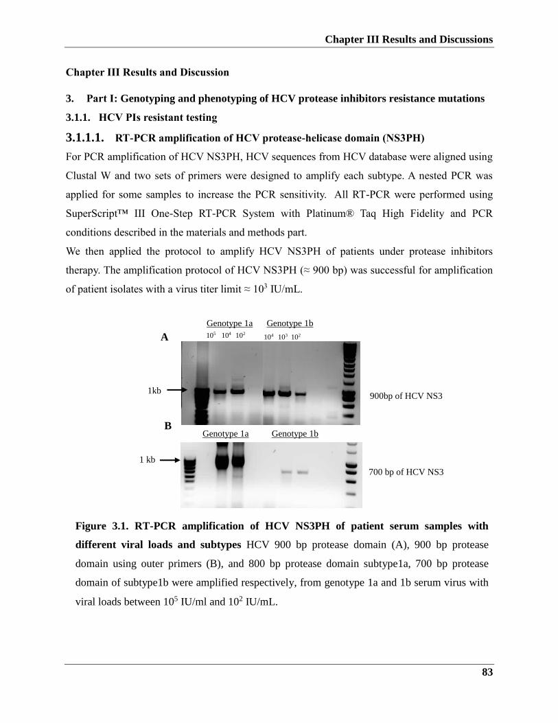
Chapter III Results and Discussions
83
Chapter III Results and Discussion
3. Part I: Genotyping and phenotyping of HCV protease inhibitors resistance mutations
3.1.1. HCV PIs resistant testing
3.1.1.1. RT-PCR amplification of HCV protease-helicase domain (NS3PH)
For PCR amplification of HCV NS3PH, HCV sequences from HCV database were aligned using
Clustal W and two sets of primers were designed to amplify each subtype. A nested PCR was
applied for some samples to increase the PCR sensitivity. All RT-PCR were performed using
SuperScript™ III One-Step RT-PCR System with Platinum® Taq High Fidelity and PCR
conditions described in the materials and methods part.
We then applied the protocol to amplify HCV NS3PH of patients under protease inhibitors
therapy. The amplification protocol of HCV NS3PH (≈ 900 bp) was successful for amplification
of patient isolates with a virus titer limit ≈ 103 IU/mL.
Figure 3.1. RT-PCR amplification of HCV NS3PH of patient serum samples with
different viral loads and subtypes HCV 900 bp protease domain (A), 900 bp protease
domain using outer primers (B), and 800 bp protease domain subtype1a, 700 bp protease
domain of subtype1b were amplified respectively, from genotype 1a and 1b serum virus with
viral loads between 105 IU/ml and 102 IU/mL.
B Genotype 1a Genotype 1b
104 103 102 A
105 104 102
Genotype 1a Genotype 1bccc
1kb 900bp of HCV NS3
700 bp of HCV NS3
1 kb
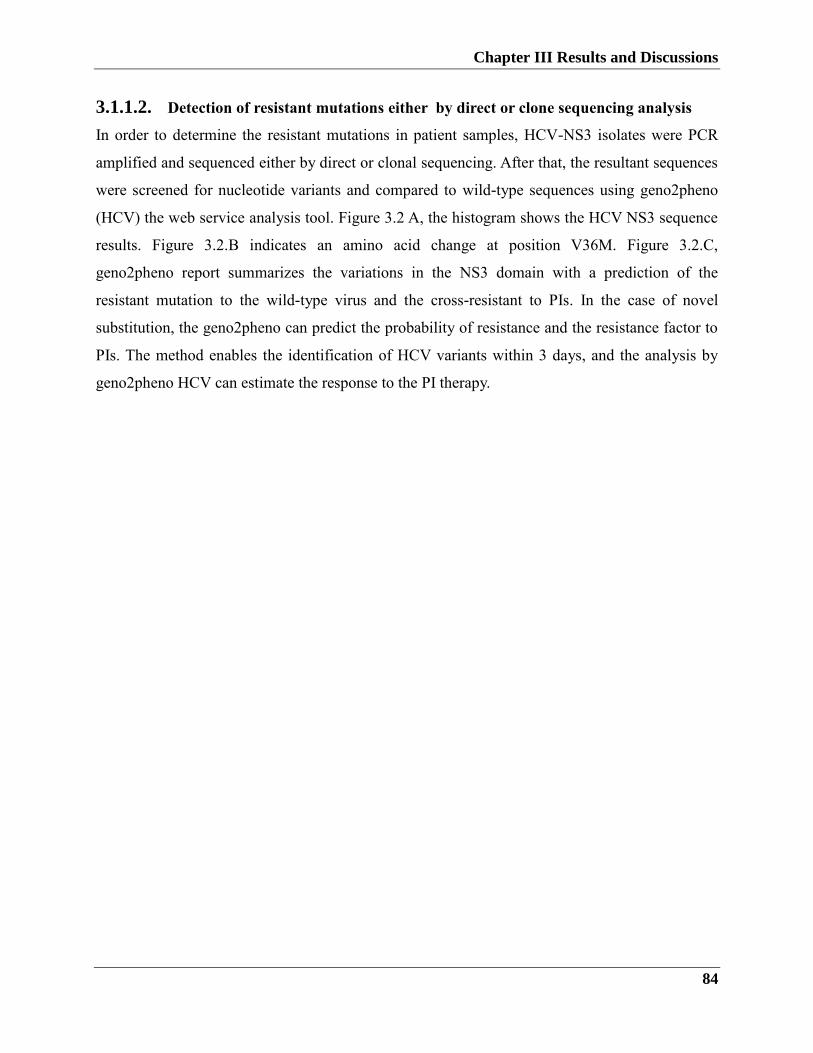
Chapter III Results and Discussions
84
3.1.1.2. Detection of resistant mutations either by direct or clone sequencing analysis
In order to determine the resistant mutations in patient samples, HCV-NS3 isolates were PCR
amplified and sequenced either by direct or clonal sequencing. After that, the resultant sequences
were screened for nucleotide variants and compared to wild-type sequences using geno2pheno
(HCV) the web service analysis tool. Figure 3.2 A, the histogram shows the HCV NS3 sequence
results. Figure 3.2.B indicates an amino acid change at position V36M. Figure 3.2.C,
geno2pheno report summarizes the variations in the NS3 domain with a prediction of the
resistant mutation to the wild-type virus and the cross-resistant to PIs. In the case of novel
substitution, the geno2pheno can predict the probability of resistance and the resistance factor to
PIs. The method enables the identification of HCV variants within 3 days, and the analysis by
geno2pheno HCV can estimate the response to the PI therapy.
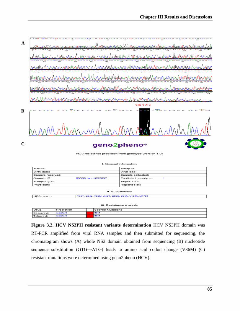
Chapter III Results and Discussions
85
A
BIOTRACEBioEdit v ersion 7.0.9.0 (6/27/07)
Model 3730
KB.bcp
6258050-03 6258002-04 6258003-03 6258005-00
File: 61112NS31aDH89658-61112NS31anestedRV (1).ab1
61112NS31a DH 89658-61112 NS31a nested RV
10528372
Lane 84
Signal G:683 A:821 T:980 C:1497
KB_3730_POP7_BDTv3.mob
?? no 'MTXF' field
Points 2668 to 16417
Page 1 of 1
11/7/2012
Spacing: 17.9818382263184
G C GATTTA A
10
TCCCGTGG G C
20
TTG G AC TG T A
30
G C T C AA AG CC
40
C C A TG T TG C A
50
G C A A C A G AG G
60
G G T TG AG C A C
70
C A G C A C C T T A
80
TA G C C C TG A G
90
C TG C G T A TG C
100
A G C CG G G A C C
110
T TG G T A C T C T
120
T A C CG C TG C C
130
TG TG G G AG C A
140
TG C A G G TG G G
150
C C A C C TG G A A
160
G C T C TG G G G C
170
AC C A C TG G TG
180
G AG AG G AG T T
190
G TC C G TG A A C
200
A C CG G G G AC C
210
T CG TG G T TG T
220
T TC T AG G T TC
230
TC CA C AG G G A
240
T A A AG TC C A C
250
CG C T T TAG C C
260
A C
C C C A CG G G
270
TG C A C A C CG C
280
G G C TC TG A A T
290
A TG C C C G CG G
300
CG TG C C C CG C
310
G G G G C A C A A C
320
AG CG G AC C C C
330
C CG AG G AG C C
340
T T TC A AG T AG
350
G A A A TG G G C C
360
G G GG C G A A AG
370
C A GG C TG C C C
380
C TG C T A TC A C
390
C T CG C CG G CG
400
C AC AG
G A A TG
410
AC A T CG G CG T
420
G C C TCG TG AC
430
C AG A T A A AG G
440
T C CG AGG AG C
450
CG C AGG T A C A
460
G GG TG T C A A T
470
G AG CG GG C AC
480
C T TG AG G AG C
490
G GG C CAG C C C
500
A C A AG A T C T T
510
TG T C C A C A T T
520
G G T A T A C A T C
530
TG G A T A AC AG
540
G AC C C T TGG G
550
TG A T
G CG A TG
560
G T T C TCG T T C
570
C AG C C C CG TG
580
G T AG AC AG T C
590
C AG C A T AC C C
600
C A T TG ATG C A
610
C G T TG C T AG G
620
A A AG T T TG G G
630
TA G C AG T TG A
640
C A T A A T C AG A
650
G T C T C A C C C T
660
C C AC T TG G T T
670
T T TG T C C CG G
680
C C AG T T A GG C
690
TG G TG A T T A T
700
A C A
T C C T AG G
710
AGG C C C C T TG
720
T C TG C TGG G C
730
G T A TG C TG TG
740
A TG G G CG C C A
750
G C A A C C T C C A
760
C C C C T TG G AG
770
TC C A T T C C A T
780
C A G C T GG T C C
790
G A G C A G T A T C
800
T C C TG G C C C C
810
C A A CAG AG C G
820
GA G A
GTG → ATG
Figure 3.2. HCV NS3PH resistant variants determination HCV NS3PH domain was
RT-PCR amplified from viral RNA samples and then submitted for sequencing, the
chromatogram shows (A) whole NS3 domain obtained from sequencing (B) nucleotide
sequence substitution (GTG→ATG) leads to amino acid codon change (V36M) (C)
resistant mutations were determined using geno2pheno (HCV).
B
C
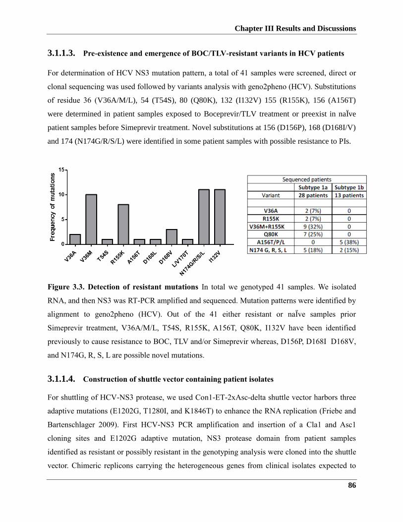
Chapter III Results and Discussions
86
3.1.1.3. Pre-existence and emergence of BOC/TLV-resistant variants in HCV patients
For determination of HCV NS3 mutation pattern, a total of 41 samples were screened, direct or
clonal sequencing was used followed by variants analysis with geno2pheno (HCV). Substitutions
of residue 36 (V36A/M/L), 54 (T54S), 80 (Q80K), 132 (I132V) 155 (R155K), 156 (A156T)
were determined in patient samples exposed to Boceprevir/TLV treatment or preexist in naÏve
patient samples before Simeprevir treatment. Novel substitutions at 156 (D156P), 168 (D168I/V)
and 174 (N174G/R/S/L) were identified in some patient samples with possible resistance to PIs.
Figure 3.3. Detection of resistant mutations In total we genotyped 41 samples. We isolated
RNA, and then NS3 was RT-PCR amplified and sequenced. Mutation patterns were identified by
alignment to geno2pheno (HCV). Out of the 41 either resistant or naÏve samples prior
Simeprevir treatment, V36A/M/L, T54S, R155K, A156T, Q80K, I132V have been identified
previously to cause resistance to BOC, TLV and/or Simeprevir whereas, D156P, D168I D168V,
and N174G, R, S, L are possible novel mutations.
3.1.1.4. Construction of shuttle vector containing patient isolates
For shuttling of HCV-NS3 protease, we used Con1-ET-2xAsc-delta shuttle vector harbors three
adaptive mutations (E1202G, T1280I, and K1846T) to enhance the RNA replication (Friebe and
Bartenschlager 2009). First HCV-NS3 PCR amplification and insertion of a Cla1 and Asc1
cloning sites and E1202G adaptive mutation, NS3 protease domain from patient samples
identified as resistant or possibly resistant in the genotyping analysis were cloned into the shuttle
vector. Chimeric replicons carrying the heterogeneous genes from clinical isolates expected to
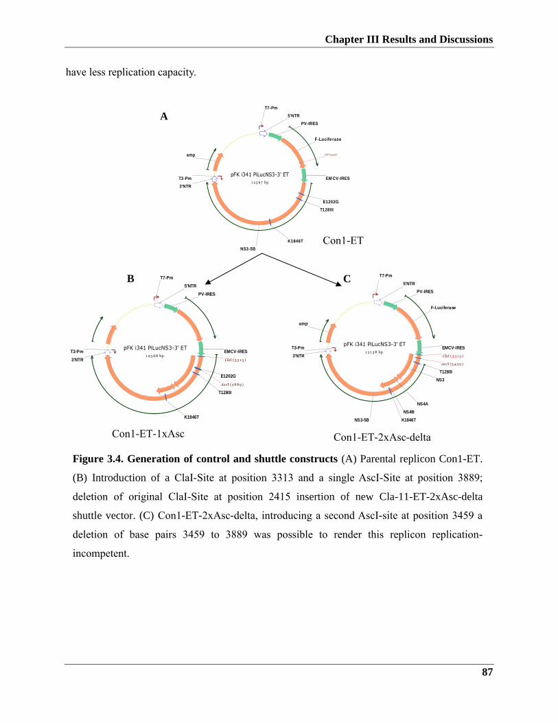
Chapter III Results and Discussions
87
Figure 3.4. Generation of control and shuttle constructs (A) Parental replicon Con1-ET.
(B) Introduction of a ClaI-Site at position 3313 and a single AscI-Site at position 3889;
deletion of original ClaI-Site at position 2415 insertion of new Cla-11-ET-2xAsc-delta
shuttle vector. (C) Con1-ET-2xAsc-delta, introducing a second AscI-site at position 3459 a
deletion of base pairs 3459 to 3889 was possible to render this replicon replication-
incompetent.
have less replication capacity.
B
pFK i341 PiLucNS3-3' ET
1 2 5 68 bpEMCV-IRES
PV-IRES
T7-Pm
T3-Pm
3'NTR
5'NTR
E1202G
T1280I
K1846T
ClaI (3 3 1 3 )
AscI (3 889 )
C
A
pFK i341 PiLucNS3-3' ET
1 2 1 3 8 bp
F-Luciferase
amp
NS3-5B
NS3
NS4A
NS4B
EMCV-IRES
PV-IRES
T7-Pm
T3-Pm
3'NTR
5'NTR
T1280I
K1846T
ClaI (3 3 1 3 )
AscI (3 4 5 9 )
pFK i341 PiLucNS3-3' ET
1 2 5 6 7 bp
F-Luciferase
amp
NS3-5B
EM CV-IRES
PV-IRES
T7-Pm
T3-Pm
3'NTR
5'NTR
E1202G
T1280I
K1846T
ClaI (2415)
Con1-ET
Con1-ET-1xAsc Con1-ET-2xAsc-delta
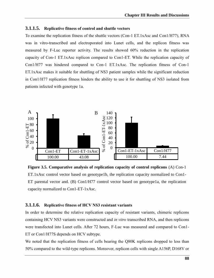
Chapter III Results and Discussions
88
Con1-ET-1xAsc Con1/H77
100.00 7.44
0
20
40
60
80
100
120
140
% o
f C
on1
-ET
-1x
Asc
3.1.1.5. Replicative fitness of control and shuttle vectors
To examine the replication fitness of the shuttle vectors (Con-1 ET.1xAsc and Con1/H77), RNA
was in vitro-transcribed and electroporated into Lunet cells, and the replicon fitness was
measured by F-Luc reporter activity. The results showed 60% reduction in the replication
capacity of Con-1 ET.1xAsc replicon compared to Con1-ET. While the replication capacity of
Con1/H77 was hindered compared to Con-1 ET.1xAsc. The replication fitness of Con-1
ET.1xAsc makes it suitable for shuttling of NS3 patient samples while the significant reduction
in Con1/H77 replication fitness hinders the ability to use it for shuttling of NS3 isolated from
patients infected with genotype 1a.
3.1.1.6. Replicative fitness of HCV NS3 resistant variants
In order to determine the relative replication capacity of resistant variants, chimeric replicons
containing HCV NS3 variants were constructed and in vitro transcribed RNA, and then replicons
were transfected into Lunet cells. After 72 hours, F-Luc was measured and compared to Con1-
ET or Con1/H77S depends on HCV subtype.
We noted that the replication fitness of cells bearing the Q80K replicons dropped to less than
50% compared to the wild-type replicons. Moreover, replicon cells with single A156P, D168V or
Con1-ET Con1-ET-1xAsc
100.00 43.08
0
20
40
60
80
100
120
% o
f C
on1
-ET
A
Figure 3.5. Comparative analysis of replication capacity of control replicons (A) Con-1
ET.1xAsc control vector based on genotype1b, the replication capacity normalized to Con1-
ET parental vector and. (B) Con1/H77 control vector based on genotype1a, the replication
capacity normalized to Con1-ET-1xAsc.
B
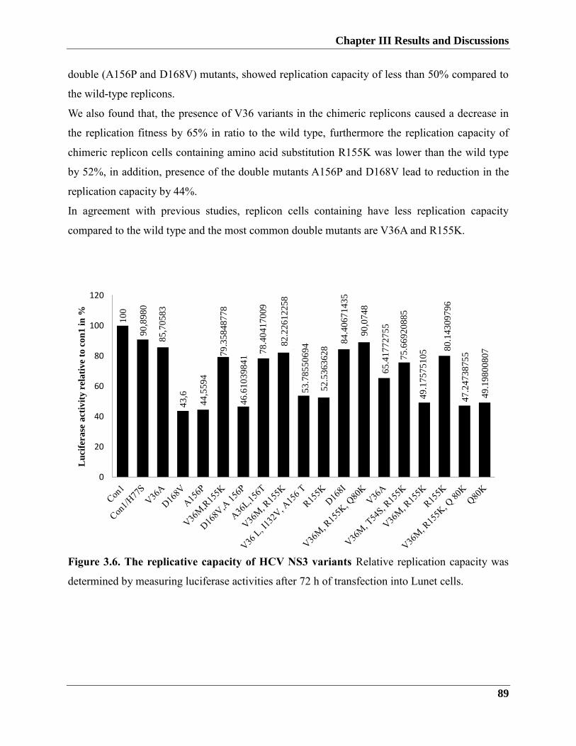
Chapter III Results and Discussions
89
double (A156P and D168V) mutants, showed replication capacity of less than 50% compared to
the wild-type replicons.
We also found that, the presence of V36 variants in the chimeric replicons caused a decrease in
the replication fitness by 65% in ratio to the wild type, furthermore the replication capacity of
chimeric replicon cells containing amino acid substitution R155K was lower than the wild type
by 52%, in addition, presence of the double mutants A156P and D168V lead to reduction in the
replication capacity by 44%.
In agreement with previous studies, replicon cells containing have less replication capacity
compared to the wild type and the most common double mutants are V36A and R155K.
Figure 3.6. The replicative capacity of HCV NS3 variants Relative replication capacity was
determined by measuring luciferase activities after 72 h of transfection into Lunet cells.
10
0
90
,89
80
85
,70
58
3
43
,6
44
,55
94
79
.35
84
87
78
46
.61
03
98
41
78
.40
41
70
09
82
.22
61
22
58
53
.78
55
06
94
52
.53
63
62
8
84
.40
67
14
35
90
,07
48
65
.41
77
27
55
75
.66
92
08
85
49
.17
57
51
05 8
0.1
43
09
79
6
47
.24
73
87
55
49
.19
80
08
07
0
20
40
60
80
100
120
Lu
cife
ra
se a
ctiv
ity
rela
tiv
e to
co
n1
in
%
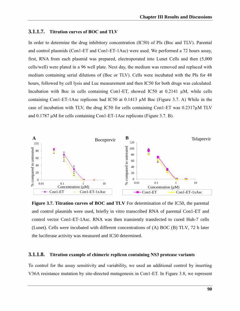
Chapter III Results and Discussions
90
3.1.1.7. Titration curves of BOC and TLV
In order to determine the drug inhibitory concentration (IC50) of PIs (Boc and TLV). Parental
and control plasmids (Con1-ET and Con1-ET-1Asc) were used. We performed a 72 hours assay,
first, RNA from each plasmid was prepared, electroporated into Lunet Cells and then (5,000
cells/well) were plated in a 96 well plate. Next day, the medium was removed and replaced with
medium containing serial dilutions of (Boc or TLV). Cells were incubated with the PIs for 48
hours, followed by cell lysis and Luc measurement and then IC50 for both drugs was calculated.
Incubation with Boc in cells containing Con1-ET, showed IC50 at 0.2141 µM, while cells
containing Con1-ET-1Asc replicons had IC50 at 0.1413 µM Boc (Figure 3.7. A) While in the
case of incubation with TLV, the drug IC50 for cells containing Con1-ET was 0.2317µM TLV
and 0.1787 µM for cells containing Con1-ET-1Asc replicons (Figure 3.7. B).
3.1.1.8. Titration example of chimeric replicon containing NS3 protease variants
To control for the assay sensitivity and variability, we used an additional control by inserting
V36A resistance mutation by site-directed mutagenesis in Con1-ET. In Figure 3.8, we represent
0
20
40
60
80
100
0.01 0.1 1 10
% c
om
par
ed t
o u
ntr
eate
d
Concentration (µM)
Boceprevir
Con1-ET Con1-ET-1xAsc
0
20
40
60
80
100
120
0.01 0.1 1 10% c
om
par
ed t
o u
ntr
eate
d
Concentration (µM)
Telaprevir
Con1-ET Con1-ET-1xAsc
Figure 3.7. Titration curves of BOC and TLV For determination of the IC50, the parental
and control plasmids were used, briefly in vitro transcribed RNA of parental Con1-ET and
control vector Con1-ET-1Asc. RNA was then transiently transfected to cured Huh-7 cells
(Lunet). Cells were incubated with different concentrations of (A) BOC (B) TLV, 72 h later
the luciferase activity was measured and IC50 determined.
A B
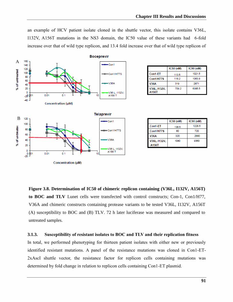
Chapter III Results and Discussions
91
Figure 3.8. Determination of IC50 of chimeric replicon containing (V36L, I132V,
A156T) to BOC and TLV Lunet cells were transfected with control constructs; con-1,
Con1/H77, V36A and chimeric constructs containing protease variants to be tested V36L,
I132V, A156T (A) susceptibility to BOC. (B) TLV. 72 h later luciferase was measured and
compared to untreated samples.
an example of HCV patient isolate cloned in the shuttle vector, this isolate contains V36L,
I132V, A156T mutations in the NS3 domain, the IC50 value of these variants had 6-fold
increase over that of wild type replicon, and 13.4 fold increase over that of wild type replicon of
TLV.
3.1.2. Susceptibility of resistant isolates to BOC and TLV and their replication fitness
In total, we performed phenotyping for thirteen patient isolates with either new or previously
identified resistant mutations. A panel of the resistance mutations was cloned in Con1-ET-
2xAscI shuttle vector, the resistance factor for replicon cells containing mutations was
determined by fold change in relation to replicon cells containing Con1-ET plasmid.
3.1.3. Susceptibility of resistant isolates to BOC and TLV and their replication fitness
In total, we performed phenotyping for thirteen patient isolates with either new or previously
identified resistant mutations. A panel of the resistance mutations was cloned in Con1-ET-
2xAscI shuttle vector, the resistance factor for replicon cells containing mutations was
determined by fold change in relation to replicon cells containing Con1-ET plasmid.
Figure 3.8. Determination of IC50 of chimeric replicon containing (V36L, I132V, A156T)
to BOC and TLV Lunet cells were transfected with control constructs; Con-1, Con1/H77,
V36A and chimeric constructs containing protease variants to be tested V36L, I132V, A156T
(A) susceptibility to BOC and (B) TLV. 72 h later luciferase was measured and compared to
untreated samples.
A
B
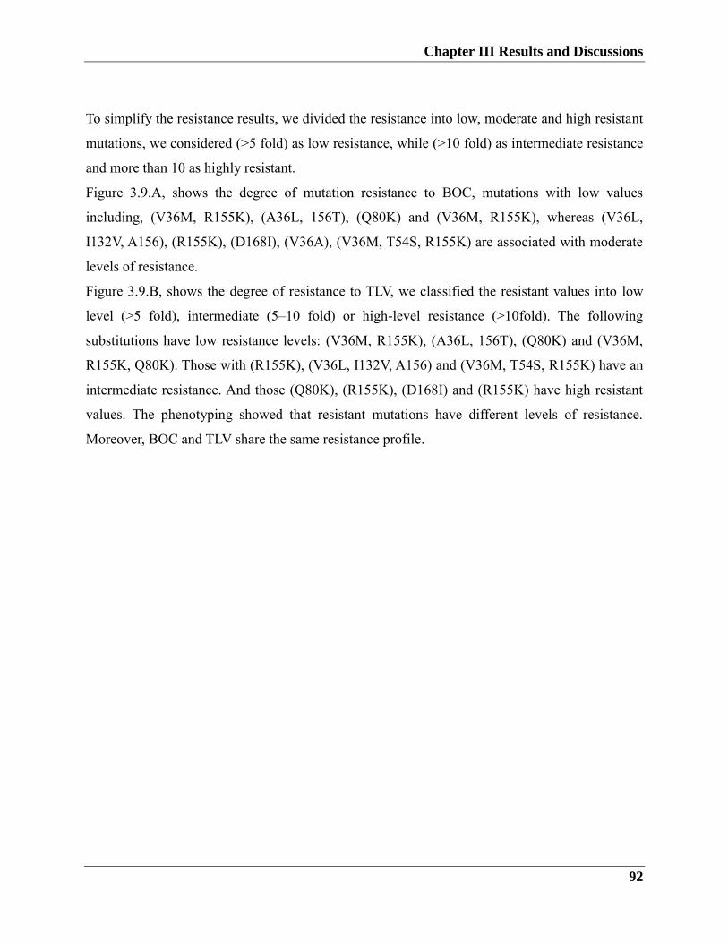
Chapter III Results and Discussions
92
To simplify the resistance results, we divided the resistance into low, moderate and high resistant
mutations, we considered (>5 fold) as low resistance, while (>10 fold) as intermediate resistance
and more than 10 as highly resistant.
Figure 3.9.A, shows the degree of mutation resistance to BOC, mutations with low values
including, (V36M, R155K), (A36L, 156T), (Q80K) and (V36M, R155K), whereas (V36L,
I132V, A156), (R155K), (D168I), (V36A), (V36M, T54S, R155K) are associated with moderate
levels of resistance.
Figure 3.9.B, shows the degree of resistance to TLV, we classified the resistant values into low
level (>5 fold), intermediate (5–10 fold) or high-level resistance (>10fold). The following
substitutions have low resistance levels: (V36M, R155K), (A36L, 156T), (Q80K) and (V36M,
R155K, Q80K). Those with (R155K), (V36L, I132V, A156) and (V36M, T54S, R155K) have an
intermediate resistance. And those (Q80K), (R155K), (D168I) and (R155K) have high resistant
values. The phenotyping showed that resistant mutations have different levels of resistance.
Moreover, BOC and TLV share the same resistance profile.
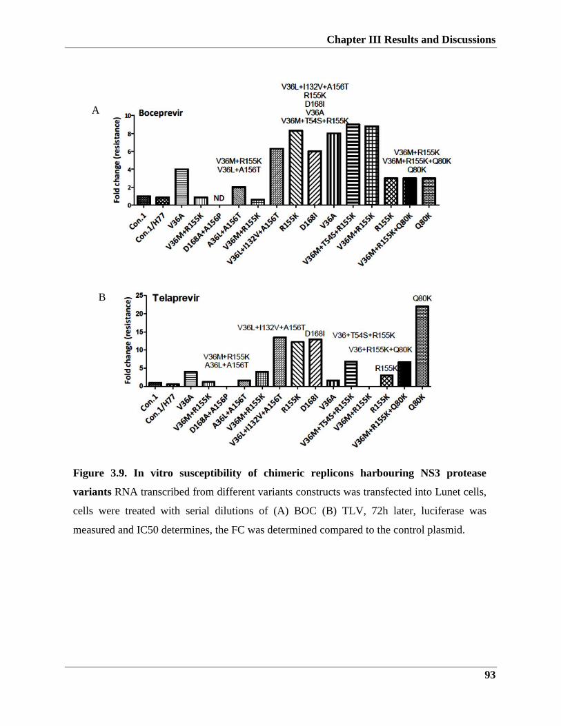
Chapter III Results and Discussions
93
A
B
Figure 3.9. In vitro susceptibility of chimeric replicons harbouring NS3 protease
variants RNA transcribed from different variants constructs was transfected into Lunet cells,
cells were treated with serial dilutions of (A) BOC (B) TLV, 72h later, luciferase was
measured and IC50 determines, the FC was determined compared to the control plasmid.
Chapter III Results and Discussions
94
Figure 3.10. Determination of IL28B variants rs8099917T/G, rs12979860C/T by
sequencing Genomic DNA was extracted from donor blood samples, (A) amplicons were PCR
amplified for SNP rs12979860 (1-5), SNP rs8099917 (6-9). (B) SNP rs12979860 variants. (C)
SNP rs8099917 variants.
3.2. IL28 polymorphisms in HCV treatment response
3.2.1. IL28b rs8099917T/G, rs12979860C/T polymorphism genotyping
3.2.1.1. Direct DNA sequencing method
The association between IL28b polymorphisms and the HCV SVR has been intensively studied.
Therefore, we have established a fast convenient method for rs8099917T/G, rs12979860C/T
genotyping and compared it to the gold standard sequencing method. DNA was extracted and
PCR amplified (Figure 4.1.A), and then subjected to Pyrosequencing. As shown in (Figure
4.1.B), the homologs rs12979860 SNP with genotype CC or TT has one peak in the chronogram,
while heterologous SNP with CT genotype has two peaks. Same is applied for rs8099917 (Figure
4.1.C).
~ 600bp
~ 200bp
rs12979860 rs8099917
1 2 3 4 5 6 7 8 9
rs12979860 CC rs12979860 TT rs12979860 CT
rs8099917 TT rs8099917 GG rs8099917 TG
A
B
C
Chapter III Results and Discussions
95
3.2.1.2. Establishment of TaqMan assay for IL28b allele discrimination
TaqMan SNP Genotyping is a highly flexible technology for polymorphisms determination.
Therefore, we have established the TaqMan assay for IL28b rs8099917T/G, rs12979860C/T
polymorphisms. TaqMan is a PCR-based assay, in which specific primers and probes are used,
one probe is coupled to (FAM) with Ex/Em=495-516, and the other probe is coupled to (VIC)
with Ex/Em= 538-554 and both probes contain minor groove binder (MGB). As each probe is
allele specific, it will break once it binds its allele releasing the fluorescence signal.
To determine an unknown SNP, purified DNA with unknown SNP allele and reference DNA
samples with known SNP allele covering homozygous and heterozygous alleles are included.
Figure 3.11.A illustrates that in the case of rs12979860C/C homozygous allele, high fluorescence
intensity is detected in one channel (533-580), the left-handed side of the figure, and only
background signal on the other channel. Whereas, in the case of rs12979860T/T homozygosity, a
high fluorescence intensity is detected in the one channel (465-510), and background signal in
the second channel (Figure 3.11.b). In the case of rs12979860C/T heterozygous allele, equal
fluorescence intensity is detected in both channels (Figure. 3.11.C).
A scatter plot Figure 3.11.D, the green color indicates the rs12979860C/C homolog allele, blue
color indicates rs12979860T/T, and the red is rs12979860C/T heterologous allele. In scattering
plot Figure 3.11.E, the green triangles represent the homologous rs8099917T/T, the blue triangle
represents the homologous rs8099917G/G, and the red ones represent the heterologous
rs8099917T/G.
When we compared the genotyping results obtained by TaqMan assay with the results obtained
by direct sequencing, we confirmed the reliability of TaqMan to distinguish between two
different SNPs, which make it the assay of choice to genotype HCV patients SNPs in our
experiments.
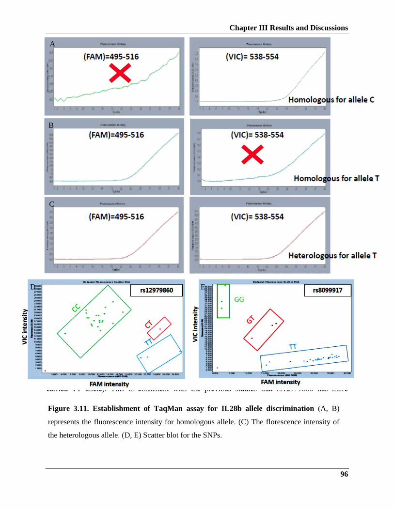
Chapter III Results and Discussions
96
3.2.1.3. Relationship between poor response to triple therapy and IL28b polymorphisms
(SNP rs12979860 and rs8099917)
In order to determine the association between IL28b SNPs and the poor response in patients
treated with triple therapy. In total 7 non-responder patients were genotyped for rs8099917 and
rs12979860. Five patients carried GG allele, while 2 patients had TT allele for rs8099917. In
addition, the genotype distribution for SNP rs12979860 alleles in patients were all non-CC allele
(3 out of the 5 patients carrying rs8099917 GG allele carried rs12979860 CT while 2 patients
carried TT allele). This is consistent with the previous studies that rs12979860 has more
Figure 3.11. Establishment of TaqMan assay for IL28b allele discrimination (A, B)
represents the fluorescence intensity for homologous allele. (C) The florescence intensity of
the heterologous allele. (D, E) Scatter blot for the SNPs.
A
B
C
D E
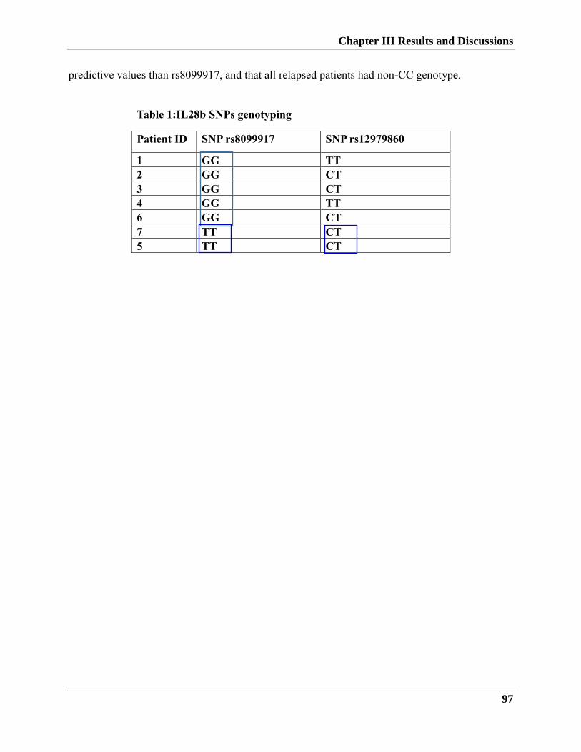
Chapter III Results and Discussions
97
predictive values than rs8099917, and that all relapsed patients had non-CC genotype.
Table 1:IL28b SNPs genotyping
Patient ID SNP rs8099917 SNP rs12979860
1 GG TT
2 GG CT
3 GG CT
4 GG TT
6 GG CT
7 TT CT
5 TT CT
Chapter III Results and Discussions
98
3.3. Discussion
Chronic infection with hepatitis C is still a major worldwide health problem. For almost two
decades, interferon-ribavirin dual therapy was the only drug available for HCV treatment (Heim
2012). Since the application of DAAs in combination with interferon-Ribavirin, a progress has
been made in the cure rates of genotype 1 infected patients (Wakita 2007, Jang and Chung 2011).
Drug resistance appeared as a major challenge hinders successful treatment with protease
inhibitors, therefore the establishment of reliable assay arose as a need to evaluate resistant
variants. The first PIs approved were BOC and TLV, both are linear ketoamide and
administrated in combination with IFN-RBV. The first generation of PIs including BOC and
TLV are characterized by a low genetic barrier, meaning that one single amino acid change
might confer drug resistance (Halfon and Locarnini 2011, Schiering, D'Arcy et al. 2011,
Wartelle-Bladou, Le Folgoc et al. 2012) .
In the beginning, we wanted to genotype NS3 variants at baseline, during and post-treatment. In
relapsed patients, we used population and clonal sequencing to identify NS3 variants in pre-
treatment samples. In most cases, we did not have a pre-treatment sample, therefore we could not
compare the sequence changes in relation to the baseline sequence. Nevertheless, the sequencing
method and geno2phenoHCV analysis tool enabled us to identify, the NS3 resistant variants, the
resistant value and the cross-resistant to other inhibitors. In our cohort, we have identified novel
substitutions at 156 (D156P), 168 (D168I/V) and 174 (N174G/R/S/L). Moreover, we identified
known mutations that confer resistance to PIs, those include substitutions of residue 36
(V36A/M/L), 54 (T54S), 80 (Q80K), 132 (I132V) 155 (R155K) and 156 (A156T). The known
mutations that were observed most frequently in NS3 in patient isolates were V36M and R155K.
While the most frequent novel substitutions were at amino acid N174 with the replacement to G,
R, S, L.
In spite of that, the method was useful in the determination of resistant variants in both naive and
during treatment, lacking the pre-treatment samples hindered our ability to know whether those
mutations pre-exist prior PI therapy, or they were mutated under PI treatment.
Chapter III Results and Discussions
99
Phenotyping of HCV resistant samples
HCV replicon system has been proven a useful tool to study the efficacy of anti -HCV
compounds. Therefore, a modified replicon system named (shuttle vector) was used for the
phenotyping assay. The shuttle vector developed by insertion of three adaptive mutations
(E1202G + T1280I+K1846T). In addition, two restriction sites (Cla1 and Asc1) were created for
NS3 shuttling, and for the control, defective parental vector Asc1 was inserted to the replicon
(Qi, Bae et al. 2009, Imhof and Simmonds 2010). The replicative capacity of the control
defective replicon decreased to 43% related to the parental replicon Con-1 ET. For the shuttling
procedure, we amplified NS3 protease domain using a primer that contains the E1202G adaptive
mutation. Then the PCR product of NS3P was ligated into the shuttle vector. Given that all
resistant mutations reside within the NS3P domain, besides the technical challenges of cloning of
the whole NS3PH domain, we decided to amplify the NS3P domain instead of the whole length
NS3/4A domain.
In this study, we used Huh7-Lunet cells to determine the replication fitness and the sensitivity of
mutants to PIs. Huh7-Lunet cell line is a highly permissive hepatoma cell line to HCV, this cell
line was developed by infecting Huh7 cells and treat the cells with IFN (Robinson, Yang et al.
2010). Replication fitness of resistant variants defined as the ability of the mutations to replicate
in relation to wild-type replication fitness (Halfon and Locarnini 2011, Lassmann,
Arumugaswami et al. 2013). Given that the fitness is an important determinant of the amount of
new viral transcripts accumulates in the hepatocytes, we determined the replication fitness of
resistant variants. Using a transient transfection approach, we clearly saw that constructs
containing resistant variants lost the replication fitness compared to the wild type. The results
generally confirm previous results showed that in the presence of resistant mutations the virus
will have less replication fitness. Of note, the replication fitness results that based on transient
replicon system might be underestimated, that is we miss the role of those resistant variants on
the late steps of the virus replication cycle. To estimate the sensitivity of resistant mutants, we
determined the IC50 of BOC and TLV for both the wild type Con-1 ET and Con-1 ET 1xAsc
and the resistant variants. Based on sequencing results we identified seven resistant variants in
the NS3 domain, those mutations have different levels of resistance to both BOC and TLV.
However, (Q80K), (R155K), (D168I) and (R155K) variants have high resistant values,
Chapter III Results and Discussions
100
suggesting that those substitutions have big effect on substrate or enzyme binding, consistent
with the notion these mutations have cross-resistance to different PIs(Shiryaev, Cheltsov et al.
2012, Lassmann, Arumugaswami et al. 2013, Solund, Krarup et al. 2014) . A previous study
showed that R155K mutation counteracts the binding to BOC and TLV. The residues A156
located in the P-side of the active site, and when a mutation occurs in this position, this cause
weakness of the binding between NS3 and the inhibitor (Shiryaev, Cheltsov et al. 2012, Xue,
Wang et al. 2012).
Taken together, our results confirmed the sensitivity of the phenotyping assay in the
determination of the resistant values of resistant variants. Moreover, it agrees with previous
clinical studies for both BOC and TLV. Overall, we could attribute the failure of triple therapy to
the presence of the resistant mutations which were either pre-exist or selected under PI pressure.
GWAS analysis is a successful approach to identify important polymorphism genotypes and their
relation to disease pathogenesis (Clark, 2011; Lange, 2011). In 2009, several GWAS studies
showed the association of IL28b polymorphisms HCV clearance or persistence. Notably, the
strongest associations were for SNPs located genetic polymorphism near the IL28B locus on a
chromosome, namely rs12979860, rs8099917. As mentioned earlier in the introduction part,
patients carry the unfavorable allele rs12979860 (T/T), are fewer responders to IFN based
therapy. The role of the different polymorphisms in disease progression is still conflicting, in one
study (Asselah, 2012; Barreiro, 2011) they showed that patients with unfavourable allele have
higher risk of developing liver fibrosis and cirrhosis, while (Tillmann, 2011; Thomas, 2009)
showed that patients with favourable allele have higher progression to fibrosis. Given the
importance of several IL28b polymorphisms, we decided to include IL28b polymorphisms in our
study as a host genetic predictor in HCV clinical outcome. Due to the high homology between
the IL2b and IL28A, first, we have established a TaqMan-based assay for SNPs genotyping and
confirmed the genotypes by sequencing. In total, we genotyped 33 human DNA samples for
both rs12979860, rs8099917 polymorphisms, then we confirmed the genotype with sequencing.
Our results showed 100% matching with the sequencing results. Thus, we used TaqMan-based
assay to determine the association between IL28b SNPs and the poor response in patients treated
with triple therapy. When we genotyped 7 non-responder patients for rs8099917 and rs12979860,
we found that five patients carried GG allele, while two patients had TT allele for rs8099917. In
Chapter III Results and Discussions
101
addition, the genotype distribution for SNP rs12979860 alleles in patients were all non-CC allele
(3 out of the 5 patients carrying rs8099917 GG allele carried rs12979860 CT while 2 patients
carried TT allele). This is consistent with the previous studies that rs12979860 has more
predictive values than rs8099917, and that all relapsed patients had non-CC genotype.
In order to determine the association between IL28b SNPs and the poor response in patients
treated with triple therapy. In total 7 non-responder patients were genotyped for rs8099917 and
rs12979860. Five patients carried GG allele, while 2 patients had TT allele for rs8099917. In
addition, the genotype distribution for SNP rs12979860 alleles in patients were all non-CC allele
(3 out of the 5 patients carrying rs8099917 GG allele carried rs12979860 CT while 2 patients
carried TT allele). Furthermore, we confirmed that rs12979860 has more predictive values than
rs8099917 and that all relapsed patients had non-CC genotype. Taken together, we have
established a fast and sensitive assay for IL28b polymorphisms genotyping, and that our results
confirm the previous observations of the high predictive value of the rs12979860 with the
clinical outcome.
3.4. Summary
Approximately 75% HCV patients develop chronic infection and this might lead to severe
illness, including cirrhosis and hepatocellular carcinoma. In 2011 the first direct-acting antiviral
agents (DAAs) protease inhibitors BOC (Victrelis) and TLV (Incivek) were approved. The
novel standard of care therapy, which consists of BOC or TLV in combination with PEG–IFNα -
ribavirin raised the cure rates of HCV genotype 1 infected patients up to 70%. However, drug-
resistance emergence arises as a major challenge to effective antiviral therapy. Taking advantage
of the availability of in vitro systems that support HCV replication, we used this system to
establish a phenotyping resistance testing for PIs. Thus, we could establish genotyping and
phenotyping assays to characterize the resistant variants. We determined substitutions of residue
36 (V36A/M/L), 54 (T54S), 80 (Q80K), 132 (I132V) 155 (R155K), 156 (A156T) in both patient
samples exposed to Boceprevir/TLV, and naÏve patient samples. In addition, we identified novel
substitutions with potential resistance, they located at 156 (D156P), 168 (D168I/V) and 174
(N174G/R/S/L).
We used Huh7-Lunet cells to determine the replication fitness and the sensitivity of mutants to
PIs. Thus, the phenotyping assay revealed that, mutations at (V36M, R155K), (A36L, 156T),
Chapter III Results and Discussions
102
(Q80K) and (V36M, R155K), are associated with low resistant value, whereas (V36L, I132V,
A156), (R155K), (D168I), (V36A), (V36M, T54S, R155K) are associated with moderate levels
of resistance. For TLV, the following substitutions have low resistance levels, (V36M, R155K),
(A36L, 156T), (Q80K) and (V36M, R155K, Q80K). Those with (R155K), (V36L, I132V, A156)
and (V36M, T54S, R155K) have an intermediate resistance. And those (Q80K), (R155K),
(D168I) and (R155K) have high resistant values. Moreover, BOC and TLV share similar
resistance profile. We also noted that the replication fitness of resistant variants dropped
compared to the wild type. Taken together, we confirmed the sensitivity of the phenotyping
assay in the determination of different resistance values. Moreover, the resistance associated with
different variants are correlated with the therapy failure in those patients.
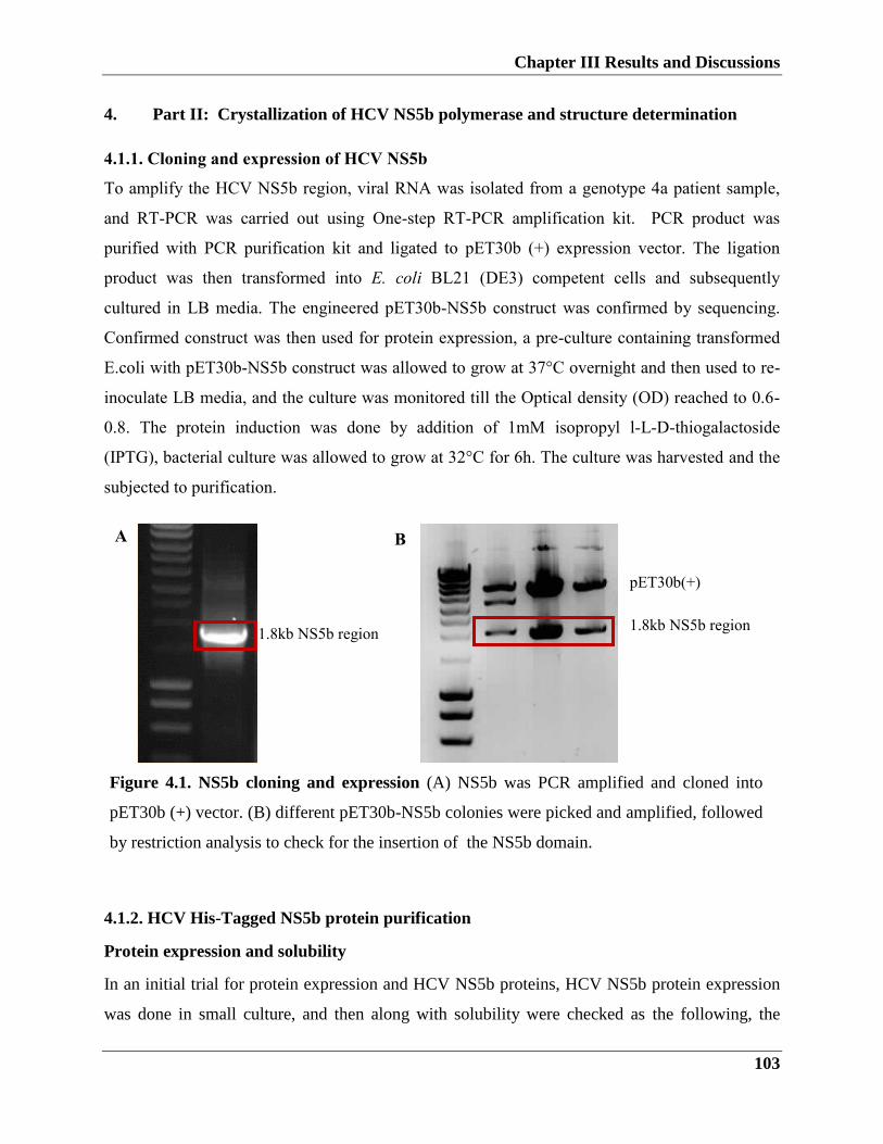
Chapter III Results and Discussions
103
4. Part II: Crystallization of HCV NS5b polymerase and structure determination
4.1.1. Cloning and expression of HCV NS5b
To amplify the HCV NS5b region, viral RNA was isolated from a genotype 4a patient sample,
and RT-PCR was carried out using One-step RT-PCR amplification kit. PCR product was
purified with PCR purification kit and ligated to pET30b (+) expression vector. The ligation
product was then transformed into E. coli BL21 (DE3) competent cells and subsequently
cultured in LB media. The engineered pET30b-NS5b construct was confirmed by sequencing.
Confirmed construct was then used for protein expression, a pre-culture containing transformed
E.coli with pET30b-NS5b construct was allowed to grow at 37°C overnight and then used to re-
inoculate LB media, and the culture was monitored till the Optical density (OD) reached to 0.6-
0.8. The protein induction was done by addition of 1mM isopropyl l-L-D-thiogalactoside
(IPTG), bacterial culture was allowed to grow at 32°C for 6h. The culture was harvested and the
subjected to purification.
4.1.2. HCV His-Tagged NS5b protein purification
Protein expression and solubility
In an initial trial for protein expression and HCV NS5b proteins, HCV NS5b protein expression
was done in small culture, and then along with solubility were checked as the following, the
1.8kb NS5b region 1.8kb NS5b region
pET30b(+)
A B
Figure 4.1. NS5b cloning and expression (A) NS5b was PCR amplified and cloned into
pET30b (+) vector. (B) different pET30b-NS5b colonies were picked and amplified, followed
by restriction analysis to check for the insertion of the NS5b domain.
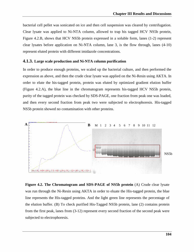
Chapter III Results and Discussions
104
bacterial cell pellet was sonicated on ice and then cell suspension was cleared by centrifugation.
Clear lysate was applied to Ni-NTA column, allowed to trap his tagged HCV NS5b protein,
Figure 4.2.B, shows that HCV NS5b protein expressed in a soluble form, lanes (1-2) represent
clear lysates before application on Ni-NTA column, lane 3, is the flow through, lanes (4-10)
represent eluted protein with different imidazole concentrations.
4.1.3. Large scale production and Ni-NTA column purification
In order to produce enough proteins, we scaled up the bacterial culture, and then performed the
expression as above, and then the crude clear lysate was applied on the Ni-Resin using AKTA. In
order to elute the his-tagged protein, protein was eluted by optimized gradient elution buffer
(Figure 4.2.A), the blue line in the chromatogram represents his-tagged HCV NS5b protein,
purity of the tagged protein was checked by SDS-PAGE, one fraction from peak one was loaded,
and then every second fraction from peak two were subjected to electrophoresis. His-tagged
NS5b protein showed no contamination with other proteins.
Figure 4.2. The Chromatogram and SDS-PAGE of NS5b protein (A) Crude clear lysate
was run through the Ni-Resin using AKTA in order to eluate the His-tagged protein, the blue
line represents the His-tagged proteins. And the light green line represents the percentage of
the elution buffer. (B) To check purified His-Tagged NS5b protein, lane (2) contains protein
from the first peak, lanes from (3-12) represent every second fraction of the second peak were
subjected to electrophoresis.
M 1 2 3 4 5 6 7 8 9 10 11 12 A B
NS5b
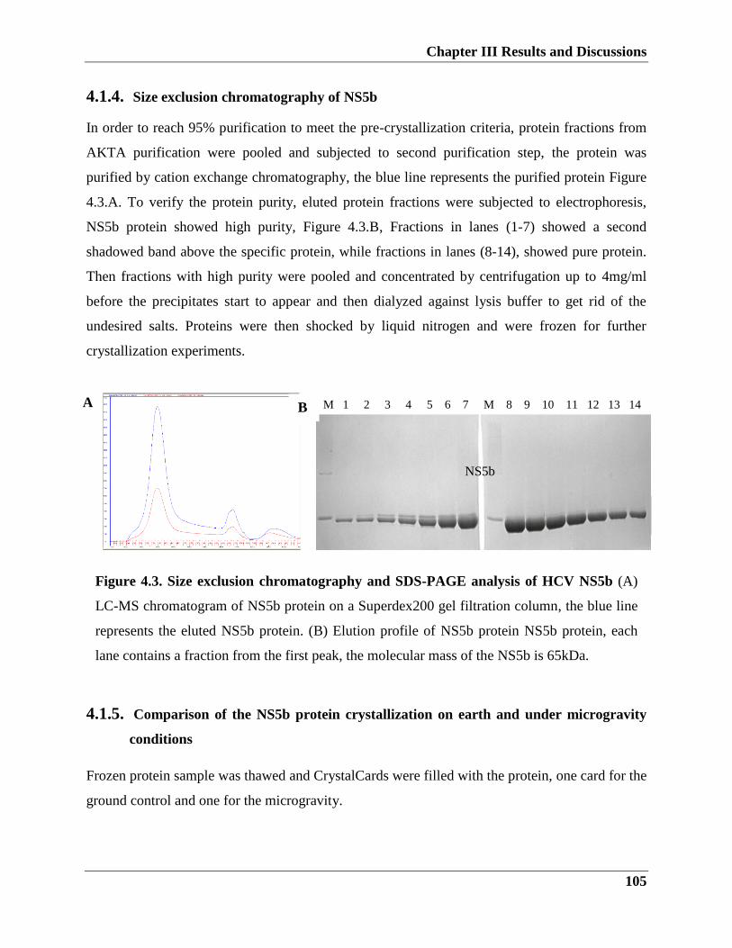
Chapter III Results and Discussions
105
4.1.4. Size exclusion chromatography of NS5b
In order to reach 95% purification to meet the pre-crystallization criteria, protein fractions from
AKTA purification were pooled and subjected to second purification step, the protein was
purified by cation exchange chromatography, the blue line represents the purified protein Figure
4.3.A. To verify the protein purity, eluted protein fractions were subjected to electrophoresis,
NS5b protein showed high purity, Figure 4.3.B, Fractions in lanes (1-7) showed a second
shadowed band above the specific protein, while fractions in lanes (8-14), showed pure protein.
Then fractions with high purity were pooled and concentrated by centrifugation up to 4mg/ml
before the precipitates start to appear and then dialyzed against lysis buffer to get rid of the
undesired salts. Proteins were then shocked by liquid nitrogen and were frozen for further
crystallization experiments.
4.1.5. Comparison of the NS5b protein crystallization on earth and under microgravity
conditions
Frozen protein sample was thawed and CrystalCards were filled with the protein, one card for the
ground control and one for the microgravity.
Figure 4.3. Size exclusion chromatography and SDS-PAGE analysis of HCV NS5b (A)
LC-MS chromatogram of NS5b protein on a Superdex200 gel filtration column, the blue line
represents the eluted NS5b protein. (B) Elution profile of NS5b protein NS5b protein, each
lane contains a fraction from the first peak, the molecular mass of the NS5b is 65kDa.
M 1 2 3 4 5 6 7 M 8 9 10 11 12 13 14 A B
NS5b
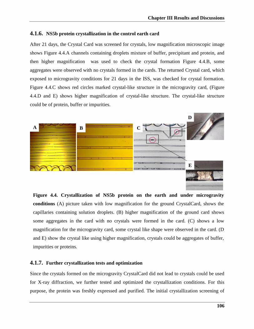
Chapter III Results and Discussions
106
4.1.6. NS5b protein crystallization in the control earth card
After 21 days, the Crystal Card was screened for crystals, low magnification microscopic image
shows Figure 4.4.A channels containing droplets mixture of buffer, precipitant and protein, and
then higher magnification was used to check the crystal formation Figure 4.4.B, some
aggregates were observed with no crystals formed in the cards. The returned Crystal card, which
exposed to microgravity conditions for 21 days in the ISS, was checked for crystal formation.
Figure 4.4.C shows red circles marked crystal-like structure in the microgravity card, (Figure
4.4.D and E) shows higher magnification of crystal-like structure. The crystal-like structure
could be of protein, buffer or impurities.
4.1.7. Further crystallization tests and optimization
Since the crystals formed on the microgravity CrystalCard did not lead to crystals could be used
for X-ray diffraction, we further tested and optimized the crystallization conditions. For this
purpose, the protein was freshly expressed and purified. The initial crystallization screening of
Figure 4.4. Crystallization of NS5b protein on the earth and under microgravity
conditions (A) picture taken with low magnification for the ground CrystalCard, shows the
capillaries containing solution droplets. (B) higher magnification of the ground card shows
some aggregates in the card with no crystals were formed in the card. (C) shows a low
magnification for the microgravity card, some crystal like shape were observed in the card. (D
and E) show the crystal like using higher magnification, crystals could be aggregates of buffer,
impurities or proteins.
A B C
D
E
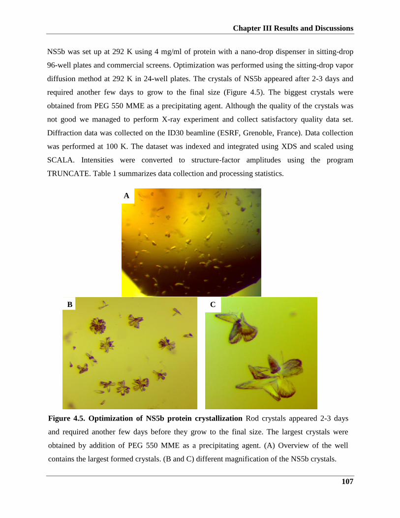
Chapter III Results and Discussions
107
NS5b was set up at 292 K using 4 mg/ml of protein with a nano-drop dispenser in sitting-drop
96-well plates and commercial screens. Optimization was performed using the sitting-drop vapor
diffusion method at 292 K in 24-well plates. The crystals of NS5b appeared after 2-3 days and
required another few days to grow to the final size (Figure 4.5). The biggest crystals were
obtained from PEG 550 MME as a precipitating agent. Although the quality of the crystals was
not good we managed to perform X-ray experiment and collect satisfactory quality data set.
Diffraction data was collected on the ID30 beamline (ESRF, Grenoble, France). Data collection
was performed at 100 K. The dataset was indexed and integrated using XDS and scaled using
SCALA. Intensities were converted to structure-factor amplitudes using the program
TRUNCATE. Table 1 summarizes data collection and processing statistics.
A
Figure 4.5. Optimization of NS5b protein crystallization Rod crystals appeared 2-3 days
and required another few days before they grow to the final size. The largest crystals were
obtained by addition of PEG 550 MME as a precipitating agent. (A) Overview of the well
contains the largest formed crystals. (B and C) different magnification of the NS5b crystals.
C B
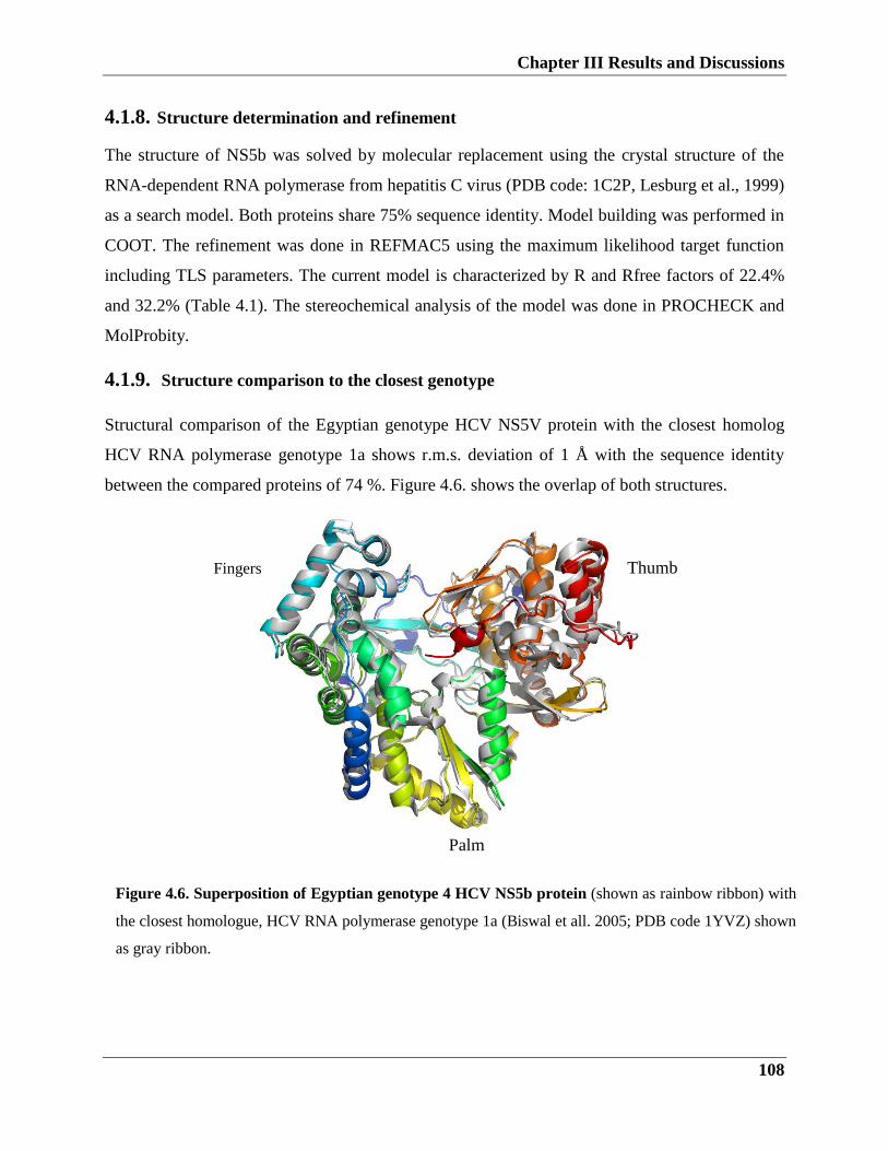
Chapter III Results and Discussions
108
4.1.8. Structure determination and refinement
The structure of NS5b was solved by molecular replacement using the crystal structure of the
RNA-dependent RNA polymerase from hepatitis C virus (PDB code: 1C2P, Lesburg et al., 1999)
as a search model. Both proteins share 75% sequence identity. Model building was performed in
COOT. The refinement was done in REFMAC5 using the maximum likelihood target function
including TLS parameters. The current model is characterized by R and Rfree factors of 22.4%
and 32.2% (Table 4.1). The stereochemical analysis of the model was done in PROCHECK and
MolProbity.
4.1.9. Structure comparison to the closest genotype
Structural comparison of the Egyptian genotype HCV NS5V protein with the closest homolog
HCV RNA polymerase genotype 1a shows r.m.s. deviation of 1 Å with the sequence identity
between the compared proteins of 74 %. Figure 4.6. shows the overlap of both structures.
Figure 4.6. Superposition of Egyptian genotype 4 HCV NS5b protein (shown as rainbow ribbon) with
the closest homologue, HCV RNA polymerase genotype 1a (Biswal et all. 2005; PDB code 1YVZ) shown
as gray ribbon.
Thumb Fingers
Palm
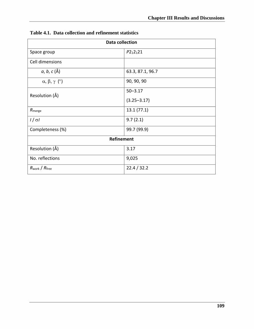
Chapter III Results and Discussions
109
Table 4.1. Data collection and refinement statistics
Data collection
Space group P212121
Cell dimensions
a, b, c (Å) 63.3, 87.1, 96.7
() 90, 90, 90
Resolution (Å) 50–3.17
(3.25–3.17)
Rmerge 13.1 (77.1)
I / I 9.7 (2.1)
Completeness (%) 99.7 (99.9)
Refinement
Resolution (Å) 3.17
No. reflections 9,025
Rwork / Rfree 22.4 / 32.2
Chapter III Results and Discussions
110
4.2. Discussion
Biological macromolecular crystallography is a fundamental method used to study biological
molecules such as proteins, viruses, and nucleic acids. Scanning for the right crystallization
conditions is essential for obtaining crystals of good quality for x-ray diffraction (Ke and Doudna
2004, McPherson 2004). Several steps involved in the determination of 3D structure by X-ray
diffraction, target macromolecule cloning, expression and purification, screening of
crystallization conditions; diffraction data collection; structure determination; analysis of the
structural model. In the beginning of our study, the aim was to crystalize the HCV-NS5b protein
by using the ISS protein crystallization facility and compare the crystals formed in microgravity
to the earth formed ones.
Despite the presence of several crystal structures covering HCV-NS5b of different genotypes,
none of them is for genotype4. HCV NS5b is an important target for the development of new
therapies for the improved treatment of HCV infection. Recently, many pan-genotypic drugs
target NS5b approved with efficiency up to 95% in naïve patients (Degasperi and Aghemo 2014,
Asselah and Marcellin 2015, Bunchorntavakul and Reddy 2015, Loustaud-Ratti, Debette-Gratien
et al. 2016, Nakamura, Kanda et al. 2016).
In order to proceed with the first step of protein crystallization, we cloned either full length or
truncated NS5b macromolecule into the bacterial expression vector (pET30b+). The pet30b+
vector contains a T7 promoter for overexpression in E. coli strain BL21(DE3). Although we
were able to successfully express truncated NS5b in a soluble form using IPTG induction, we
were not able to express a full-length NS5b in bacterial cells. That is consistent with previous
studies which showed the difficulty in the expression of the NS5b full length in bacteria, one
possible reason is the presence of membrane-anchor domain (Huang, Sineva et al. 2004, Wang,
Johnson et al. 2004). On the basis of the expression results, the over-expressed NS5B-truncstd
protein was used for further purification and crystallization. Initial experiments showed that
NS5b-T protein could be purified and concentrated up to 16mg/ml.
Given that gravity negatively influences the growth protein crystals, initial attempts to grow
protein crystals in microgravity were conducted. Results obtained from early space shuttle
missions showed improvement in the quality of the microgravity-grown crystals compared to the
earth ones. This encouraged scientists to develop and optimize conditions and devices for
Chapter III Results and Discussions
111
crystallization in space (Giege 2013, Takahashi, Ohta et al. 2013, McPherson and DeLucas
2015).
For microgravity conditions, we choose the Microcapillary Protein Crystallization System
(MPCS) (also called Plug Make). The characteristic of this system is that it allows getting
crystals from as tiny as µL of protein per experiment. MPCS permits mixing of different
concentrations of protein, buffer, precipitate and carrier fluid, this would increase the chance of
protein crystallization.
The concentrated frozen protein was thawed and loaded into the crystal cards, one was used for
1g control and the other prepared for travel to the microgravity conditions in the ISS (Gerdts,
Elliott et al. 2008, Gerdts, Stahl et al. 2010) .
Unfortunately, no pictures were obtained for the crystal card on the ISS, the card was examined
after the return journey to the earth.
After 25 days of the launch, the crystal card returned back to the earth and both crystal cards, the
ISS, and the control 1g were checked for crystal growth.
Comparison of both cards revealed the formation of crystals in the ISS card, while no crystals
were formed on the earth card.
Because of technical difficulties we could not perform X-ray diffraction on the crystals, thus we
could not confirm whether formed crystals were protein, buffer aggregates or dirt. At present,
there is still no compelling hypotheses that can anticipate protein crystallization conditions.
Consequently, further screening and optimization for crystallization parameters are required. For
screening purposes, we used crystallization plates with 96 microwells, in which each well
contains one precipitant, followed by an automated stage that allows for dispensing the same
amount of protein in each well. This strategy is advantageous in obtaining the best conditions for
growing crystals. Since temperature is one parameter which can influence crystals formation, it
was necessary to control for crystallization temperature. As the most common temperatures for
crystallization are 4°C and 22°C (Bartling et al. 2005), we placed the plates either at room
temperature 22°C.
In the initial crystallization screening experiment, crystals were obtained in the presence of PEG
550 MME. The determination of the right conditions followed by crystallization in a 24 well
crystallization plate. Protein crystals formed either as a single needle-like structure or as
microcrystal aggregates which likely originate from one nucleation site. The microscope
Chapter III Results and Discussions
112
observation of crystals showed that the quality of crystals external appearance is promising for x-
ray diffraction. To harvest crystals for crystallography, we carefully opened the plate, and gently
harvest them using Nylon loops, crystals were immediately cryo frozen into liquid nitrogen.
Crystals stored in liquid nitrogen until X-ray diffraction.
For diffraction data collection, crystals were mounted and illuminated by the X-ray beam
generated by synchrotron sources. The dataset of diffraction images taken for crystals showed
that most crystals diffracted to an average of 3.1 Å resolution. The calculated factors including
space group, cell dimension reflect the quality of resolution data. This poor quality of the
diffraction data could be attributed to the lack of protein crystals homogeneity, which believed to
be the case for protein crystals. Generally, protein crystals diffract at 1.5 to 4 Å resolution, and
thus the diffraction dataset quality reduced with misalignment of the crystal lattice.
For the establishment of the NS5b structural model, we processed the X-ray diffraction data by
Molecular Replacement approach (Abergel 2013). This method can be used to solve the
unknown crystal structure if there is any known similar solved protein structure. In the case of
genotype4a HCV-NS5b protein modeling, several models covering other genotypes are
available. We built the model based on a homologous genotype 1a HCV-NS5b model. Identical
to other HCV-NS5b polymerases, the HCV-NS5b of genotype4a comprises of six conserved
subdomains, the analogy structure indicates that fingers, palm, and thumb are in close
interaction, which contracts the flexibility between the subdomains (Biswal, Cherney et al. 2005,
Powdrill, Bernatchez et al. 2010, Karam, Powdrill et al. 2014). However, previous models of
HCV-NS5b revealed the presence of several conformations when in complex with the template.
Unfortunately, as we did not perform NS5b template co-crystallization, we still miss the other
confirmations.
4.3. Summary
The structural determination of viruses on the atomic level illustrates the virus nature
arrangement, this consequently led to the interpretation of the virus life cycle, pathogenesis and
potential targets for drug development. Numerous approaches are utilized for structure
determination including, X-ray crystallography, electron microscopy, and NMR. Several 3D
structures of HCV structural and structural proteins are available. The 3D structure of HCV
NS5b reveals that it has six conserved motifs, furthermore, it folds in fingers, palm and thumb
subdomains. Microgravity provides an opportunity for different areas of research such as
Chapter III Results and Discussions
113
biotechnology. Thus, we used microgravity to study protein crystal growth of HCV-NS5b
protein of genotype 4a.
At first, for microgravity conditions, we selected the Microcapillary Protein Crystallization
System (MPCS), which allows obtaining crystals from as little amount of protein per experiment.
Comparison of crystallization in microgravity and gravity revealed the formation of crystals in
the ISS card, while no crystals were formed on the earth card. Because of technical difficulties
we could not perform X-ray diffraction on the crystals. At present, no compelling hypotheses can
anticipate protein crystallization conditions. Therefore, we went for further optimization for
crystallization parameters to obtain crystals suitable for X-ray diffraction.
Once we obtained protein crystals of good quality, crystals were illuminated by the X-ray beam
generated by synchrotron sources. The dataset of diffraction images taken for crystals showed
that most crystals diffracted to an average of 3.1 Å resolution. The calculated factors including
space group, cell dimension reflect the quality of resolution data. This poor quality of the
diffraction data could be attributed to the lack of protein crystals homogeneity, which believed to
be the case for protein crystals. Generally, protein crystals diffract at 1.5 to 4 Å resolution, and
thus the diffraction dataset quality reduced with misalignment of the crystal lattice.
For NS5b structural modeling, we processed the X-ray diffraction data by Molecular
Replacement approach. Identical to other HCV-NS5b polymerases, the HCV-NS5b of
genotype4a comprises of six conserved subdomains, the analogy structure indicates that fingers,
palm, and thumb are in close interaction, which contracts the flexibility between the subdomains.
However, previous models of HCV-NS5b revealed the presence of several conformations when
in complex with the template. Unfortunately, as we did not perform NS5b template co-
crystallization, we still miss the other protein confirmations.
Chapter III Results and Discussions
114
5. Part III: The role of tripartite motif family members (TRIM) in mediating
susceptibility to HCV infection
5.1. Characterization of TRIMs in hepatocytes
Multiple TRIM proteins play a role in restricting viral infection such as retroviruses infection, by
inducing IFN response, regulating RPRs and/or interacting directly with viral proteins.
5.1.1. TRIM genes regulation upon HCV infection
A number of host factors are regulated by HCV infection either directly or indirectly, previous
studies have shown some innate immune factors manipulated by HCV infection. To investigate
TRIMs roles in HCV infection, we used different hepatoma cell lines that support either HCV
infection or replication. Huh7.5 cells are highly permissive for HCV infection, the high
permissiveness results from a mutation (Thr-55-Iso) in the RIG-I gene (retinoic acid-inducible
gene I, which impairs interferon signaling).
We searched the literature to check for the TRIMs which expressed by hepatocytes. Human gene
array for the transcriptional levels including TRIMs was investigated in Huh7.5 HCV-infected
cells. A number of TRIM genes were upregulated upon HCV infection including some TRIMs
such as TRIM5, 14 and 25 with a known anti-viral effect on viruses such as HIV and SeV.
TRIM31 showed the highest expression level in HCV-infected cells (Figure 5.2.A). A panel of
TRIMs showed down-regulation upon HCV infection, including TRIM43, 44 which have been
identified by another group (Li, Zhang et al. 2014). The gene array was done at the one-time
point, 72 hours post HCV (JC-1) infection. Therefore, further characterization of TRIMs
regulation upon HCV infection or interferon stimulation were performed.
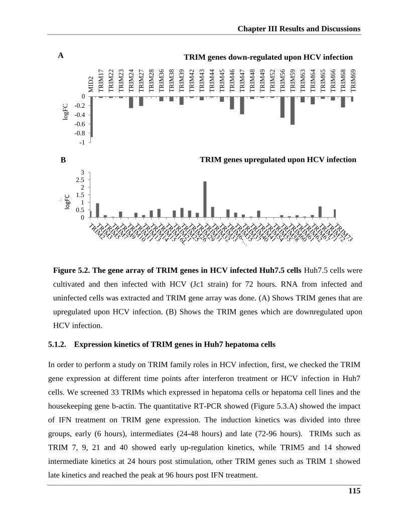
Chapter III Results and Discussions
115
-1
-0.8
-0.6
-0.4
-0.2
0M
ID2
TR
IM1
7
TR
IM2
2
TR
IM2
3
TR
IM2
4
TR
IM2
7
TR
IM2
8
TR
IM3
6
TR
IM3
8
TR
IM3
9
TR
IM4
2
TR
IM4
3
TR
IM4
4
TR
IM4
5
TR
IM4
6
TR
IM4
7
TR
IM4
8
TR
IM4
9
TR
IM5
2
TR
IM5
6
TR
IM5
9
TR
IM6
3
TR
IM6
4
TR
IM6
5
TR
IM6
6
TR
IM6
8
TR
IM6
9
TRIM genes down-regulated upon HCV infectionlo
gF
C
A
5.1.2. Expression kinetics of TRIM genes in Huh7 hepatoma cells
In order to perform a study on TRIM family roles in HCV infection, first, we checked the TRIM
gene expression at different time points after interferon treatment or HCV infection in Huh7
cells. We screened 33 TRIMs which expressed in hepatoma cells or hepatoma cell lines and the
housekeeping gene b-actin. The quantitative RT-PCR showed (Figure 5.3.A) showed the impact
of IFN treatment on TRIM gene expression. The induction kinetics was divided into three
groups, early (6 hours), intermediates (24-48 hours) and late (72-96 hours). TRIMs such as
TRIM 7, 9, 21 and 40 showed early up-regulation kinetics, while TRIM5 and 14 showed
intermediate kinetics at 24 hours post stimulation, other TRIM genes such as TRIM 1 showed
late kinetics and reached the peak at 96 hours post IFN treatment.
00.5
11.5
22.5
3
TRIM genes upregulated upon HCV infection
logF
C
B
Figure 5.2. The gene array of TRIM genes in HCV infected Huh7.5 cells Huh7.5 cells were
cultivated and then infected with HCV (Jc1 strain) for 72 hours. RNA from infected and
uninfected cells was extracted and TRIM gene array was done. (A) Shows TRIM genes that are
upregulated upon HCV infection. (B) Shows the TRIM genes which are downregulated upon
HCV infection.
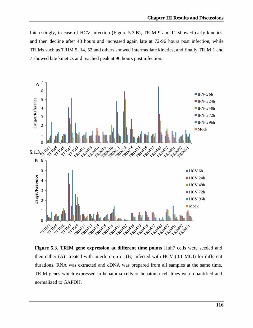
Chapter III Results and Discussions
116
0
1
2
3
4
5
6
7
Ta
rget
/Ref
eren
ce
IFN-α 6h
IFN-α 24h
IFN-α 48h
IFN-α 72h
IFN-α 96h
Mock
A
Interestingly, in case of HCV infection (Figure 5.3.B), TRIM 9 and 11 showed early kinetics,
and then decline after 48 hours and increased again late at 72-96 hours post infection, while
TRIMs such as TRIM 5, 14, 52 and others showed intermediate kinetics, and finally TRIM 1 and
7 showed late kinetics and reached peak at 96 hours post infection.
5.1.3.
Figure 5.3. TRIM gene expression at different time points Huh7 cells were seeded and
then either (A) treated with interferon-α or (B) infected with HCV (0.1 MOI) for different
durations. RNA was extracted and cDNA was prepared from all samples at the same time.
TRIM genes which expressed in hepatoma cells or hepatoma cell lines were quantified and
normalized to GAPDH.
0
1
2
3
4
5
6
Ta
rget
/Ree
ren
ce
HCV 6h
HCV 24h
HCV 48h
HCV 72h
HCV 96h
Mock
B
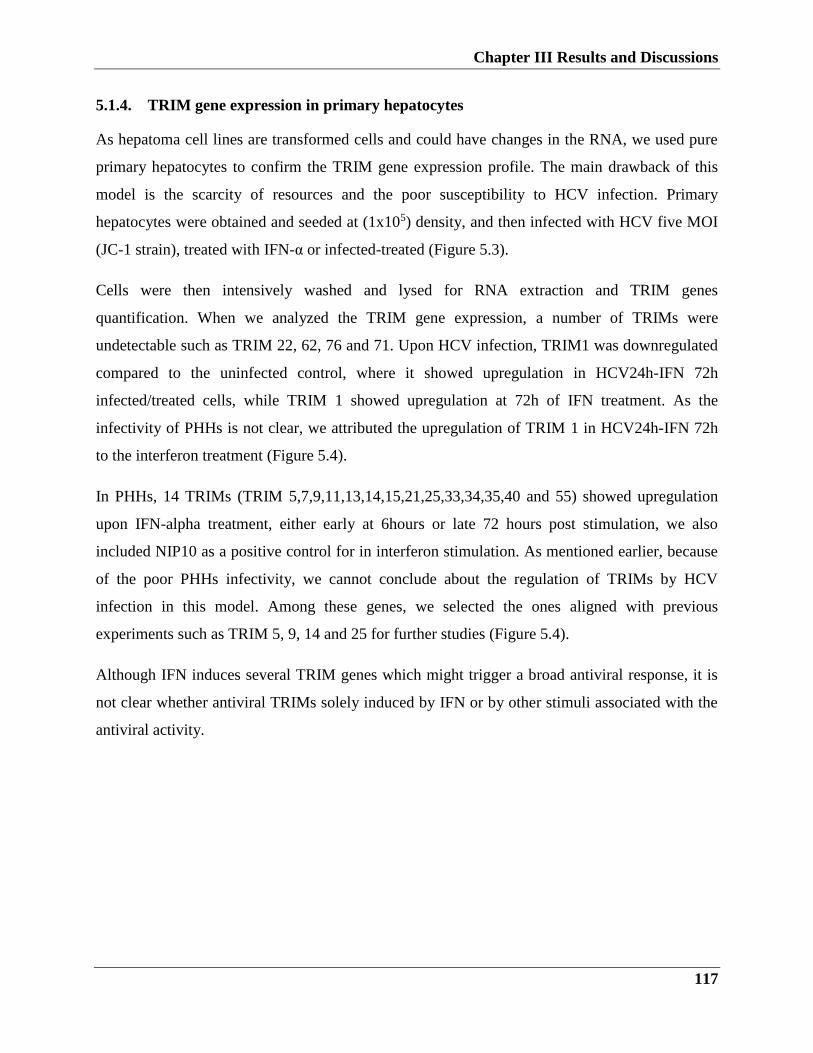
Chapter III Results and Discussions
117
5.1.4. TRIM gene expression in primary hepatocytes
As hepatoma cell lines are transformed cells and could have changes in the RNA, we used pure
primary hepatocytes to confirm the TRIM gene expression profile. The main drawback of this
model is the scarcity of resources and the poor susceptibility to HCV infection. Primary
hepatocytes were obtained and seeded at (1x10⁵) density, and then infected with HCV five MOI
(JC-1 strain), treated with IFN-α or infected-treated (Figure 5.3).
Cells were then intensively washed and lysed for RNA extraction and TRIM genes
quantification. When we analyzed the TRIM gene expression, a number of TRIMs were
undetectable such as TRIM 22, 62, 76 and 71. Upon HCV infection, TRIM1 was downregulated
compared to the uninfected control, where it showed upregulation in HCV24h-IFN 72h
infected/treated cells, while TRIM 1 showed upregulation at 72h of IFN treatment. As the
infectivity of PHHs is not clear, we attributed the upregulation of TRIM 1 in HCV24h-IFN 72h
to the interferon treatment (Figure 5.4).
In PHHs, 14 TRIMs (TRIM 5,7,9,11,13,14,15,21,25,33,34,35,40 and 55) showed upregulation
upon IFN-alpha treatment, either early at 6hours or late 72 hours post stimulation, we also
included NIP10 as a positive control for in interferon stimulation. As mentioned earlier, because
of the poor PHHs infectivity, we cannot conclude about the regulation of TRIMs by HCV
infection in this model. Among these genes, we selected the ones aligned with previous
experiments such as TRIM 5, 9, 14 and 25 for further studies (Figure 5.4).
Although IFN induces several TRIM genes which might trigger a broad antiviral response, it is
not clear whether antiviral TRIMs solely induced by IFN or by other stimuli associated with the
antiviral activity.
Chapter III Results and Discussions
118
5.2. The antiviral effect of TRIMs on HCV infection and replication
Previous work identified two possible antiviral mechanisms of TRIMs, either by interacting
directly with viral proteins or inducing IFNs and cytokines by modulating the signaling pathway.
The antiviral mechanisms of most TRIMs are unknown, while few are well studied such as
TRIM5, it functions in mediating the restriction of HIV infection, this occurs via its interaction
with core protein and destabilization of the virus.
So, to test the role of selected TRIMs in HCV replication and infection we used HCV reporter
replicon cell line (ED43) harboring ED43-genotype4a isolate. The ED43 replicon is a bicistronic
vector with a F-Luc reporter gene.
5.2.1.1. TRIM5 restricts HCV viral replication and infection
As shown in the schematic representation (figure 5.4.A), ED43 replicon cells were seeded and
then transiently transfected with increasing amount of TRIM5 or pcDNA3.1 control plasmids
(Figure 5.5.B). 72 hours later, cells were lysed and F-Luc was measured and normalized to
0
1
2
3
4
5
6
7
8Ta
rge
t/R
efe
ren
ce
HCV 6h HCV 24h HCV 48h HCV 72 hHCV24h-IFN 24h HCV 24h-IFN 48h HCV 24h-IFN 72 h IFN 6hIFN-72h Mock
Figure 5.4. TRIM gene expression in PHHs cells were seeded at (1x10⁵) density, cells were
then either infected with HCV five MOI (JC-1 strain) or treated with IFN-alpha or infected-
treated for different durations. RNAs were extracted, RT-qPCR used to quantify TRIM gene
expression and normalized to GAPDH.

Chapter III Results and Discussions
119
untransfected control. The overexpression of TRIM5 results in a decrease in HCV replication in
a dose-dependent manner (figure 5.5.C).
To further test the effect of TRIM5 on HCV infection, we performed infection experiment using
HEPepG2-HFL. The advantages of using this cell line are that it expresses the liver-specific
microRNA, miR-122 along with the entry factor, CD81, so they support the entire HCV life
cycle. Moreover, unlike Huh7.5, HepG2-HFL has intact RIG-I system which leads to a robust
IFN response.
Briefly, HepG2-HFL cells were seeded, 24 hours later cells were infected with HCVcc (JC1)
with MOI of 0.1, 48 hours later cells were transiently transfected with increasing amounts of
TRIM5 plasmid, control plasmid or left untreated. Then, 48 hours later supernatants were
collected for TCID50 (to test the extracellular virus production), and cells were lysed for RNA
extraction, HCV viral genome, and TRIM5 expression were quantified by qRT-PCR (Figure
5.6.A). As expected from our replicon cell model, we observed a reduction in viral genome
production with increasing amount of TRIM5 transfection dose (Figure 5.6.C). In addition,
supernatants were collected and titrated for TCID50, showed very little infectivity in transfected
cells compared to the untransfected or plasmid control ones (Figure 5.6.D).
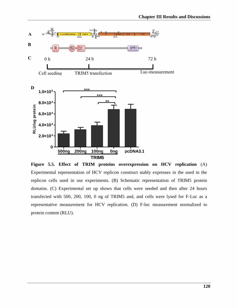
Chapter III Results and Discussions
120
D
A
B
Figure 5.5. Effect of TRIM proteins overexpression on HCV replication (A)
Experimental representation of HCV replicon construct stably expresses in the used in the
replicon cells used in our experiments. (B) Schematic representation of TRIM5 protein
domains. (C) Experimental set up shows that cells were seeded and then after 24 hours
transfected with 500, 200, 100, 0 ng of TRIM5 and, and cells were lysed for F-Luc as a
representative measurement for HCV replication. (D) F-luc measurement normalized to
protein content (RLU).
C
Cell seeding TRIM5 transfection
Luc-measurement
0 h 24 h 72 h
500ng 200ng 100ng 0ng pcDNA3.10
2.0×10⁴
4.0×10⁴
6.0×10⁴
8.0×10⁴
1.0×10⁵ ******
**
TRIM5
RL
U/u
g p
rote
in

Chapter III Results and Discussions
121
1
10
100
1000
10000
100000
1000000
500ng 200ng 100ng 0ng
TRIM5
Figure 5.6. TRIM5 overexpression restricts HCV infection (A) HepG2-HFL cells were
seeded (1×10⁵)/well in a 12 well plate. Cells were then transfected with TRIM5, 48 hours
later cells were infected with HCV at a MOI of 0.1. After 48hours, supernatants were
collected for TCID50 and cells were lysed and RNA was analyzed for the expression of
TRIM5 and HCV genome. (B) HCV genome quantification by qRT-PCR. (C) TRIM5
expression quantified by qRT-PCR. (D) supernatants were titrated and virus titers were
determined using TCID50.
500ng 200ng 100ng 0ng pcDNA3.10
5
10
15 ***
******
TRIM5
HC
V/G
AP
DH
500ng 200ng 100ng 0ng pcDNA3.10
1
2
3
4
5
TRIM5
TR
IM5/G
AP
DH
B C
D
TC
ID5
0/m
l
RNA Extraction & qPCR Cell seeding TRIM5 transfection HCV infection (0.1
MOI)
0 h 24 h 72 h 120 h
A
Chapter III Results and Discussions
122
5.2.1.2. TRIM9 role in HCV infection
A recent study revealed that TRIM9 act as a negative regulator of NF-κB in brain cells (Shi, Cho
et al. 2014), this negative regulation of NF-κB might be beneficial for HCV to escape the
immune system. In order to check the role of TRIM9 in HCV infection, we performed
replication and infection based experiments.
Figure 5.7.A shows a schematic representation of TRIM9, it consists of RING, B-box, coiled-
coil and SPRY domains. To examine the effect of TRIM9 expression on HCV replication, ED43
cells were seeded, 24 hours later cells were transfected with the TRIM9 plasmid. After 48 hours,
cells were lysed and F-Luc and protein content (Figure 5.6.B). TRIM9 expression inhibits HCV
replication in a dose-dependent manner (Figure 5.7.C).
To further examine the effect of TRIM9 expression on HCV infection, HepG2-HFL cells were
seeded and then transfected with TRIM9 plasmid, 48 hours later, cells were infected with HCV
at (0.1 MOI), and then cells were left for another 48 hours before they analyzed for Intra and
extracellular HCV infection, supernatants were collected for TCID50 and lysed cells were RNA
extractions and RT-qPCR analysis. As shown in (Figure 5.7.B-C), HCV viral genome was
strongly reduced with the increasing amount of TRIM9 expression. TCID50 results showed that
TRIM9 overexpression does decrease the HCV infectivity, which confirms the TRIM9 anti-HCV
effect on HCV (Figure 5.7.D).
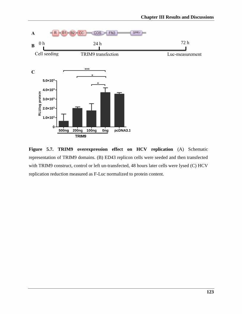
Chapter III Results and Discussions
123
C
A
B
Figure 5.7. TRIM9 overexpression effect on HCV replication (A) Schematic
representation of TRIM9 domains. (B) ED43 replicon cells were seeded and then transfected
with TRIM9 construct, control or left un-transfected, 48 hours later cells were lysed (C) HCV
replication reduction measured as F-Luc normalized to protein content.
Cell seeding TRIM9 transfection Luc-measurement
0 h 24 h 72 h
500ng 200ng 100ng 0ng pcDNA3.10
1.0×10⁵
2.0×10⁵
3.0×10⁵
4.0×10⁵
5.0×10⁵
***
*
*
TRIM9
RL
U/u
g p
rote
in
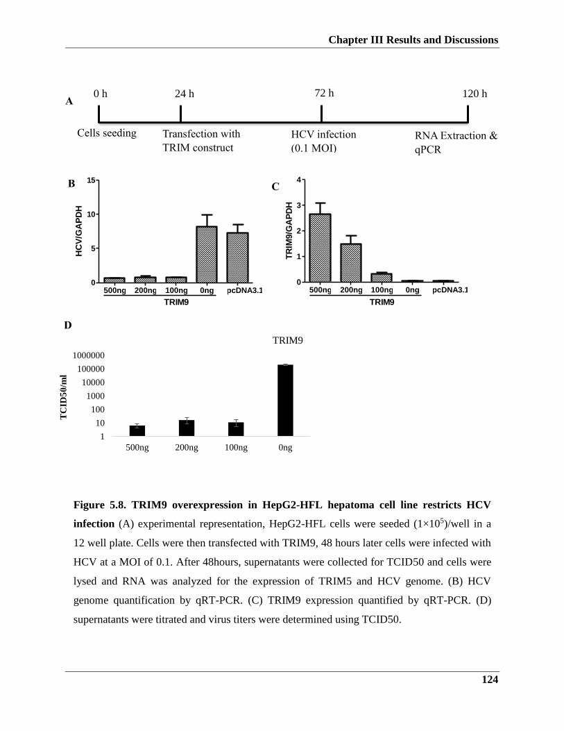
Chapter III Results and Discussions
124
500ng 200ng 100ng 0ng pcDNA3.10
5
10
15
TRIM9
HC
V/G
AP
DH
500ng 200ng 100ng 0ng pcDNA3.10
1
2
3
4
TRIM9
TR
IM9/G
AP
DH
1
10
100
1000
10000
100000
1000000
500ng 200ng 100ng 0ng
TRIM9
Figure 5.8. TRIM9 overexpression in HepG2-HFL hepatoma cell line restricts HCV
infection (A) experimental representation, HepG2-HFL cells were seeded (1×10⁵)/well in a
12 well plate. Cells were then transfected with TRIM9, 48 hours later cells were infected with
HCV at a MOI of 0.1. After 48hours, supernatants were collected for TCID50 and cells were
lysed and RNA was analyzed for the expression of TRIM5 and HCV genome. (B) HCV
genome quantification by qRT-PCR. (C) TRIM9 expression quantified by qRT-PCR. (D)
supernatants were titrated and virus titers were determined using TCID50.
TC
ID5
0/m
l
B C
D
RNA Extraction &
qPCR
Cells seeding Transfection with
TRIM construct
HCV infection
(0.1 MOI)
0 h 24 h 72 h 120 h A
Chapter III Results and Discussions
125
5.2.1.3. TRIM14 overexpression influence on HCV infection
TRIM14 is a protein belongs to the family of the TRIM proteins with a conserved domain
architecture (known as RBCC) that is characterized by RING (R) finger domain, one or two B-
box domains (B1 and B2), a Coiled-coil domain and a variable C-terminus (Meroni and Diez-
Roux 2005). Similar to TRIM family protein, TRIM14 is involved in the innate immune
response, upon viral infection, TRIM14 triggers the IFN and NF-κB pathways activation.
Briefly, TRIM14 activation occurs via facilitation the link between NEMO and MAVS, this
resulted in amplification of the innate immune response (Zhou, Jia et al. 2014)In order to
determine whether TRIM14 has an anti-HCV activity. We transfected HCV replicon cells with
increasing amounts of TRIM14 constructs after the cells were lysed and F-Luc measured (Figure
5.9.A). As shown in Figure 5.9.B, over-expression of the TRIM14 in HCV replicon cells resulted
in a decrease of HCV virus replication in a dose-dependent manner.
To further examine the effect of TRIM14 on the HCV infection, cells were seeded followed by
transfection with increasing amounts of TRIM14 or control plasmid, and then cells were infected
with HCV cc (MOI= 0.1) Figure 5.10.A. After that intracellular HCV genome was quantified by
qPCR Figure 5.10.B and extracellular virus secretion was evaluated by TCID50 (Figure 5.10.D).
Figure 5.10.B-C shows that expression of TRIM14 resulted in a decrease of the HCV infection
as quantified by qPCR. The effect of TRIM14 overexpression is confirmed by the decrease in
extracellular virus quantification (Figure 5.10.D). Taken together, TRIM14 overexpression
restricts HCV replication and infection.
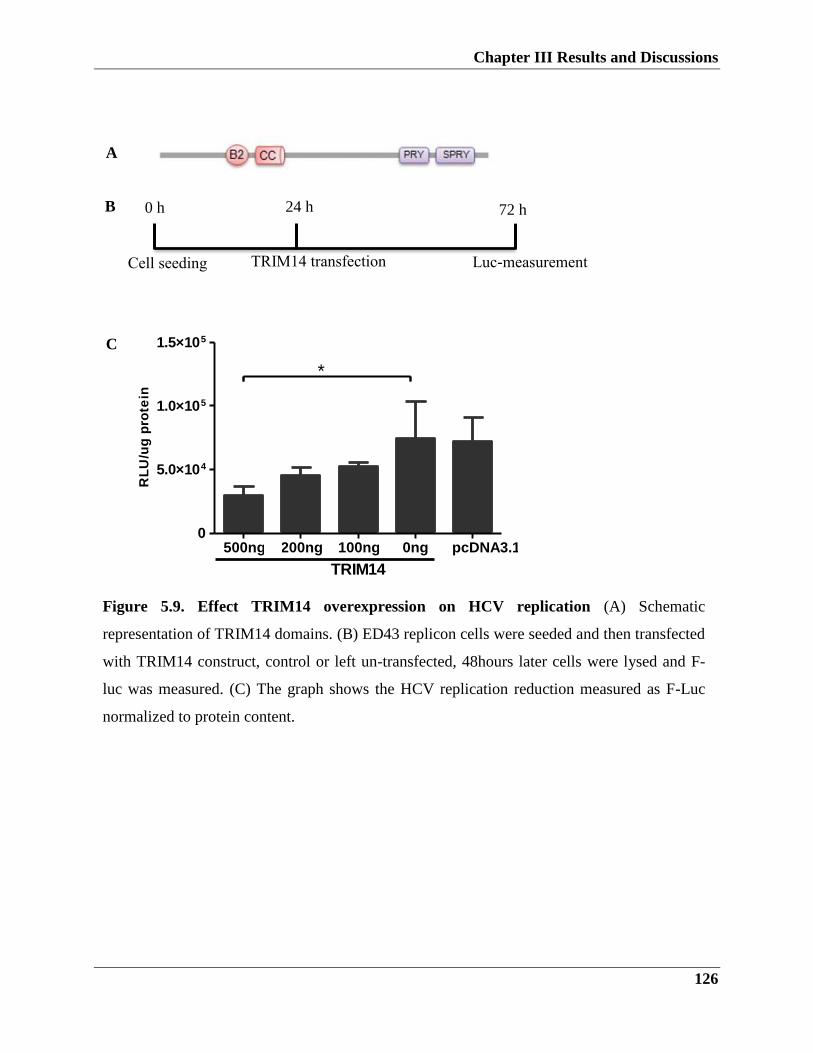
Chapter III Results and Discussions
126
B
B
B
A
C
Figure 5.9. Effect TRIM14 overexpression on HCV replication (A) Schematic
representation of TRIM14 domains. (B) ED43 replicon cells were seeded and then transfected
with TRIM14 construct, control or left un-transfected, 48hours later cells were lysed and F-
luc was measured. (C) The graph shows the HCV replication reduction measured as F-Luc
normalized to protein content.
Cell seeding TRIM14 transfection
Luc-measurement
0 h 24 h 72 h
500ng 200ng 100ng 0ng pcDNA3.10
5.0×10⁴
1.0×10⁵
1.5×10⁵
*
TRIM14
RL
U/u
g p
rote
in
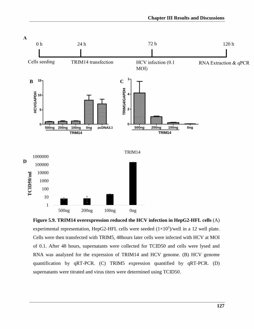
Chapter III Results and Discussions
127
500ng 200ng 100ng 0ng pcDNA3.10
5
10
15
TRIM14
HC
V/G
AP
DH
500ng 200ng 100ng 0ng0
2
4
6
TRIM14
TR
IM14/G
AP
DH
1
10
100
1000
10000
100000
1000000
500ng 200ng 100ng 0ng
TRIM14
Figure 5.9. TRIM14 overexpression reduced the HCV infection in HepG2-HFL cells (A)
experimental representation, HepG2-HFL cells were seeded (1×10⁵)/well in a 12 well plate.
Cells were then transfected with TRIM5, 48hours later cells were infected with HCV at MOI
of 0.1. After 48 hours, supernatants were collected for TCID50 and cells were lysed and
RNA was analyzed for the expression of TRIM14 and HCV genome. (B) HCV genome
quantification by qRT-PCR. (C) TRIM5 expression quantified by qRT-PCR. (D)
supernatants were titrated and virus titers were determined using TCID50.
B C
A
RNA Extraction & qPCR Cells seeding TRIM14 transfection HCV infection (0.1
MOI)
0 h 24 h 72 h 120 h
TC
ID5
0/m
l
D
Chapter III Results and Discussions
128
5.2.1.4. TRIM25 effectively inhibits HCV replication and infection
TRIM25 belongs to the C-IV TRIM proteins family, TRIM25 is abundantly expressed in
placenta and uterus, highly expressed in the thyroid gland, aorta and spleen and low in most
other tissues. In addition, TRIM25 is an IFN-inducible gene. Recently, it was shown that
TRIM25 plays an important role in innate immunity regulation, notably in RIG-I pathway
regulation. It interacts with (CARD) domain of RIG-I through its PRYSPRY region, this results
in ubiquitination of RIG-I and, consequently, induction of type I IFN, production and activation
of NF-κB. Interestingly, TRIM25 inhibits HIV and MLV late stage of viral replication and may
play a role in HBV viral restriction.
As described in (Figure 5.10.A), TRIM25 consists of N-terminal RING domain, one or two B-
boxes, and a coiled-coil region. To characterize the role of TRIM25 in HCV replication and
infection, we performed an experiment where we seeded replicon cells ED43 and then
transfected with increasing concentrations of TRIM25 plasmid or control plasmid for 48 hours
and then F-Luc was measured and normalized to protein content (Figure 5.10.B). Our results
showed that HCV replication was reduced in the TRIM25 transfected cells compared to the
untransfected ones (Figure 5.10.C), we did not notice differences between the 100 and 200 ng
transfections.
Next, we performed an infection experiment to test the effect of TRIM25 on HCV infection. As
described in Figure 5.11.A, we transfected HepG2-HFL-HCV infected cells with TRIM25
plasmid, then supernatants and cell lysates were used for analysis. First, by RT-PCR we did
observe a tremendous decrease in HCV genome in TRIM25 transfected cells compared to
control plasmid or untransfected cells (Figure 5.11.B-C). TCID50 results confirmed the
reduction in the HCV infection as seen in (Figure 5.11.D). These results strongly suggest that
overexpression of TRIM25 protein acts as anti-HCV protein.
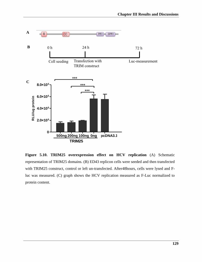
Chapter III Results and Discussions
129
B
A
C
Cell seeding Transfection with
TRIM construct
Luc-measurement
0 h 24 h 72 h
Figure 5.10. TRIM25 overexpression effect on HCV replication (A) Schematic
representation of TRIM25 domains. (B) ED43 replicon cells were seeded and then transfected
with TRIM25 construct, control or left un-transfected. After48hours, cells were lysed and F-
luc was measured. (C) graph shows the HCV replication measured as F-Luc normalized to
protein content.
500ng 200ng 100ng 0ng pcDNA3.10
2.0×10⁵
4.0×10⁵
6.0×10⁵
8.0×10⁵
***
***
***
RL
U/u
g p
rote
in
TRIM25
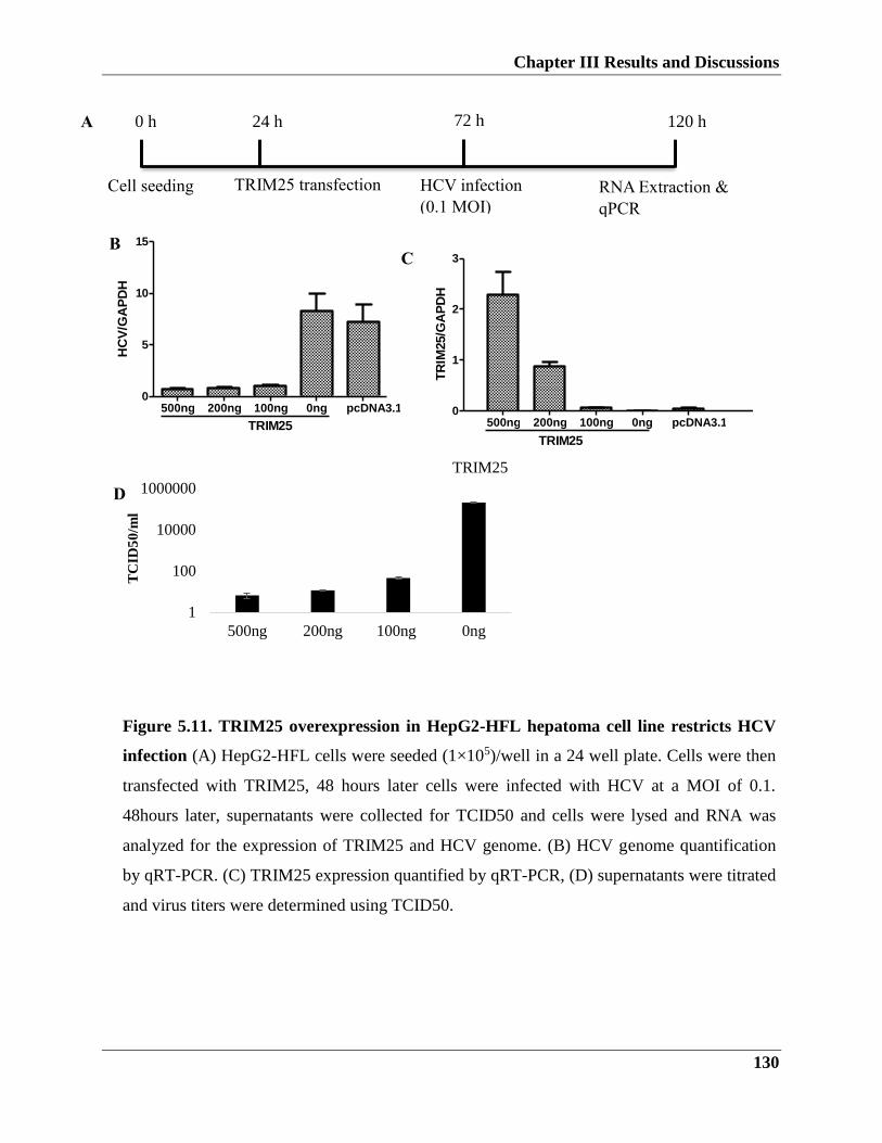
Chapter III Results and Discussions
130
500ng 200ng 100ng 0ng pcDNA3.10
5
10
15
TRIM25
HC
V/G
AP
DH
500ng 200ng 100ng 0ng pcDNA3.10
1
2
3
TRIM25T
RIM
25/G
AP
DH
TC
ID5
0/m
l
D
Figure 5.11. TRIM25 overexpression in HepG2-HFL hepatoma cell line restricts HCV
infection (A) HepG2-HFL cells were seeded (1×10⁵)/well in a 24 well plate. Cells were then
transfected with TRIM25, 48 hours later cells were infected with HCV at a MOI of 0.1.
48hours later, supernatants were collected for TCID50 and cells were lysed and RNA was
analyzed for the expression of TRIM25 and HCV genome. (B) HCV genome quantification
by qRT-PCR. (C) TRIM25 expression quantified by qRT-PCR, (D) supernatants were titrated
and virus titers were determined using TCID50.
1
100
10000
1000000
500ng 200ng 100ng 0ng
TRIM25
A
B C
RNA Extraction &
qPCR
Cell seeding TRIM25 transfection HCV infection
(0.1 MOI)
0 h 24 h 72 h 120 h
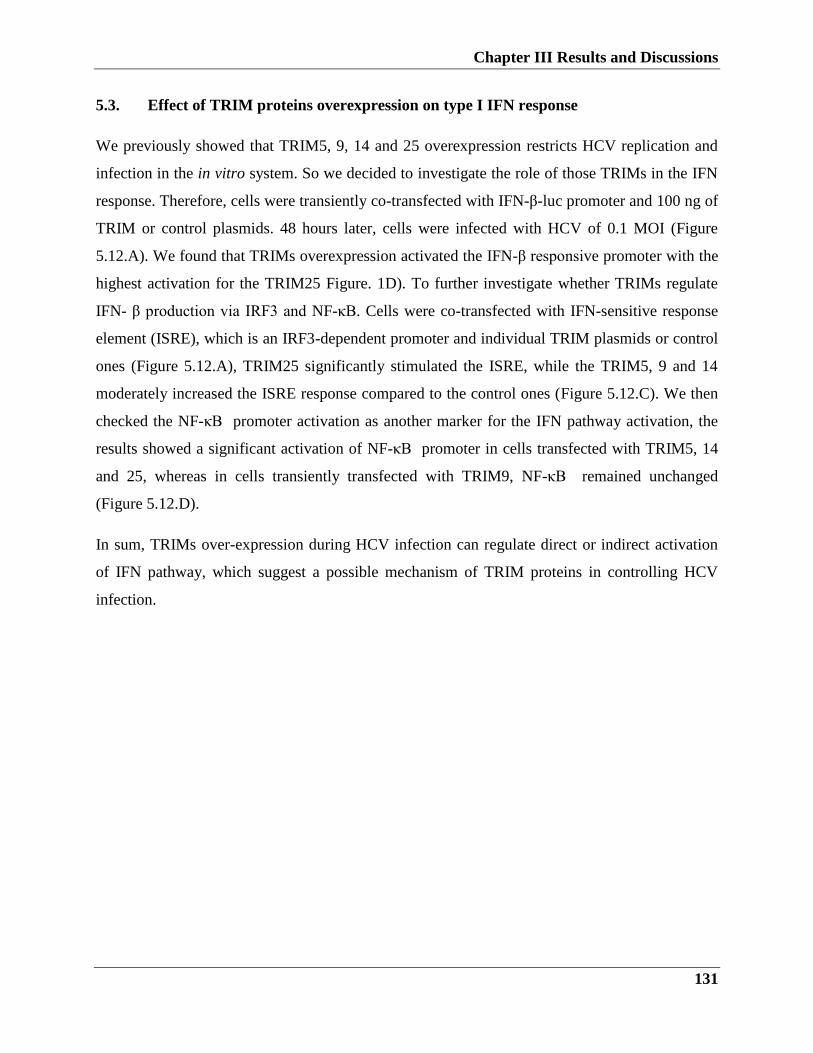
Chapter III Results and Discussions
131
5.3. Effect of TRIM proteins overexpression on type I IFN response
We previously showed that TRIM5, 9, 14 and 25 overexpression restricts HCV replication and
infection in the in vitro system. So we decided to investigate the role of those TRIMs in the IFN
response. Therefore, cells were transiently co-transfected with IFN-β-luc promoter and 100 ng of
TRIM or control plasmids. 48 hours later, cells were infected with HCV of 0.1 MOI (Figure
5.12.A). We found that TRIMs overexpression activated the IFN-β responsive promoter with the
highest activation for the TRIM25 Figure. 1D). To further investigate whether TRIMs regulate
IFN- β production via IRF3 and NF-κB. Cells were co-transfected with IFN-sensitive response
element (ISRE), which is an IRF3-dependent promoter and individual TRIM plasmids or control
ones (Figure 5.12.A), TRIM25 significantly stimulated the ISRE, while the TRIM5, 9 and 14
moderately increased the ISRE response compared to the control ones (Figure 5.12.C). We then
checked the NF-κB promoter activation as another marker for the IFN pathway activation, the
results showed a significant activation of NF-κB promoter in cells transfected with TRIM5, 14
and 25, whereas in cells transiently transfected with TRIM9, NF-κB remained unchanged
(Figure 5.12.D).
In sum, TRIMs over-expression during HCV infection can regulate direct or indirect activation
of IFN pathway, which suggest a possible mechanism of TRIM proteins in controlling HCV
infection.
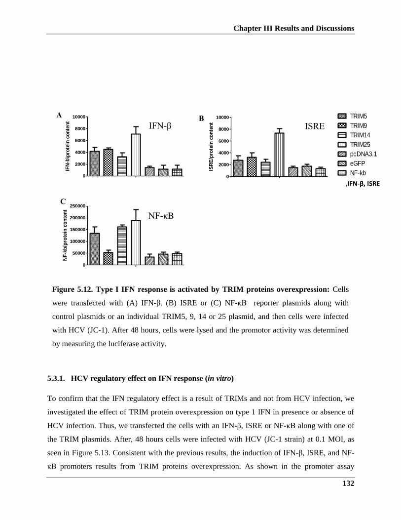
Chapter III Results and Discussions
132
5.3.1. HCV regulatory effect on IFN response (in vitro)
To confirm that the IFN regulatory effect is a result of TRIMs and not from HCV infection, we
investigated the effect of TRIM protein overexpression on type 1 IFN in presence or absence of
HCV infection. Thus, we transfected the cells with an IFN-β, ISRE or NF-κB along with one of
the TRIM plasmids. After, 48 hours cells were infected with HCV (JC-1 strain) at 0.1 MOI, as
seen in Figure 5.13. Consistent with the previous results, the induction of IFN-β, ISRE, and NF-
κB promoters results from TRIM proteins overexpression. As shown in the promoter assay
0
2000
4000
6000
8000
10000TRIM5
TRIM9
TRIM14
TRIM25
pcDNA3.1
eGFP
IFN-B
HepG2-HFL co-transfected with IFN-b reporter plasmid with TRIM or controlplasmids, followed by HCV infection
IFN
-b\p
rote
in c
on
ten
t
0
2000
4000
6000
8000
10000TRIM5
TRIM9
TRIM14
TRIM25
pcDNA3.1
eGFP
ISRE
HepG2-HFL co-transfected with ISRE reporter plasmid with TRIM or controlplasmids, followed by HCV infection
ISR
E/p
rote
in c
on
ten
t
A B
C
D
0
50000
100000
150000
200000
250000TRIM5
TRIM9
TRIM14
TRIM25
pcDNA3.1
eGFP
NF-kb
HepG2-HFL co-transfected with Nf-kb reporter plasmid with TRIM or controlplasmids, followed by HCV infection
NF
-kb
/pro
tein
co
nte
nt
0
50000
100000
150000
200000
250000TRIM5
TRIM9
TRIM14
TRIM25
pcDNA3.1
eGFP
NF-kb
HepG2-HFL co-transfected with Nf-kb reporter plasmid with TRIM or controlplasmids, followed by HCV infection
NF
-kb
/pro
tein
co
nte
nt
IFN-β
NF-κB
ISRE
Figure 5.12. Type I IFN response is activated by TRIM proteins overexpression: Cells
were transfected with (A) IFN-β. (B) ISRE or (C) NF-κB reporter plasmids along with
control plasmids or an individual TRIM5, 9, 14 or 25 plasmid, and then cells were infected
with HCV (JC-1). After 48 hours, cells were lysed and the promotor activity was determined
by measuring the luciferase activity.
,IFN-β, ISRE
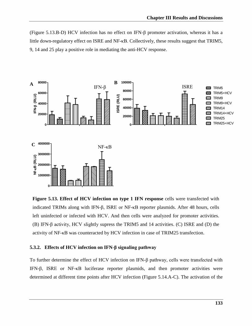
Chapter III Results and Discussions
133
Figure 5.13. Effect of HCV infection on type 1 IFN response cells were transfected with
indicated TRIMs along with IFN-β, ISRE or NF-κB reporter plasmids. After 48 hours, cells
left uninfected or infected with HCV. And then cells were analyzed for promoter activities.
(B) IFN-β activity, HCV slightly supress the TRIM5 and 14 activities. (C) ISRE and (D) the
activity of NF-κB was counteracted by HCV infection in case of TRIM25 transfection.
(Figure 5.13.B-D) HCV infection has no effect on IFN-β promoter activation, whereas it has a
little down-regulatory effect on ISRE and NF-κB. Collectively, these results suggest that TRIM5,
9, 14 and 25 play a positive role in mediating the anti-HCV response.
0
20000
40000
60000
80000
IFN
- (
RL
U)
0
20000
40000
60000
80000
100000
ISR
E (
RL
U)
0
1000000
2000000
3000000
4000000
Nf-B
(R
LU
)
5.3.2. Effects of HCV infection on IFN-β signaling pathway
To further determine the effect of HCV infection on IFN-β pathway, cells were transfected with
IFN-β, ISRE or NF-κB luciferase reporter plasmids, and then promoter activities were
determined at different time points after HCV infection (Figure 5.14.A-C). The activation of the
ISRE
0
1000000
2000000
3000000
4000000TRIM5
TRIM5+HCV
TRIM9
TRIM9+HCV
TRIM14
TRIM14+HCV
TRIM25
TRIM25+HCVNF
-kb
/pro
tein
co
nte
nt
C
A B
C IFN-β
NF-κB
ISRE
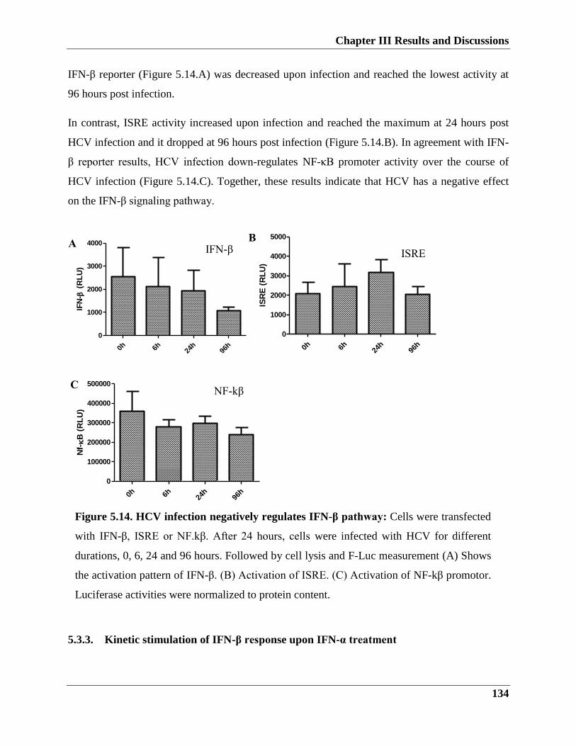
Chapter III Results and Discussions
134
Figure 5.14. HCV infection negatively regulates IFN-β pathway: Cells were transfected
with IFN-β, ISRE or NF.kβ. After 24 hours, cells were infected with HCV for different
durations, 0, 6, 24 and 96 hours. Followed by cell lysis and F-Luc measurement (A) Shows
the activation pattern of IFN-β. (B) Activation of ISRE. (C) Activation of NF-kβ promotor.
Luciferase activities were normalized to protein content.
IFN-β reporter (Figure 5.14.A) was decreased upon infection and reached the lowest activity at
96 hours post infection.
In contrast, ISRE activity increased upon infection and reached the maximum at 24 hours post
HCV infection and it dropped at 96 hours post infection (Figure 5.14.B). In agreement with IFN-
β reporter results, HCV infection down-regulates NF-κB promoter activity over the course of
HCV infection (Figure 5.14.C). Together, these results indicate that HCV has a negative effect
on the IFN-β signaling pathway.
0h 6h 24h
96h
0
1000
2000
3000
4000
IFN
- (
RL
U)
0h 6h 24h
96h
0
1000
2000
3000
4000
5000
ISR
E (
RL
U)
0h 6h 24h
96h
0
100000
200000
300000
400000
500000
Nf-B
(R
LU
)
5.3.3. Kinetic stimulation of IFN-β response upon IFN-α treatment
A
C
B IFN-β
ISRE
NF-kβ
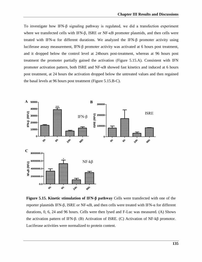
Chapter III Results and Discussions
135
Figure 5.15. Kinetic stimulation of IFN-β pathway Cells were transfected with one of the
reporter plasmids IFN-β, ISRE or NF-κB, and then cells were treated with IFN-α for different
durations, 0, 6, 24 and 96 hours. Cells were then lysed and F-Luc was measured. (A) Shows
the activation pattern of IFN-β. (B) Activation of ISRE. (C) Activation of NF-kβ promotor.
Luciferase activities were normalized to protein content.
To investigate how IFN-β signaling pathway is regulated, we did a transfection experiment
where we transfected cells with IFN-β, ISRE or NF-κB promoter plasmids, and then cells were
treated with IFN-α for different durations. We analyzed the IFN-β promoter activity using
luciferase assay measurement, IFN-β promoter activity was activated at 6 hours post treatment,
and it dropped below the control level at 24hours post-treatment, whereas at 96 hours post
treatment the promoter partially gained the activation (Figure 5.15.A). Consistent with IFN
promoter activation pattern, both ISRE and NF-κB showed fast kinetics and induced at 6 hours
post treatment, at 24 hours the activation dropped below the untreated values and then regained
the basal levels at 96 hours post treatment (Figure 5.15.B-C).
0h 6h 24h
96h
0
10000
20000
30000
40000
50000
***
IFN
- (
RF
U)
0h 6h 24h
96h
0
100000
200000
300000
ISR
E (
RF
U)
0h 6h24
h96
h
0.0
2000000.0
4000000.0
6000000.0
8000000.0
*
Nf-B
(R
FU
)
IFN-β ISRE
NF-kβ
A B
C
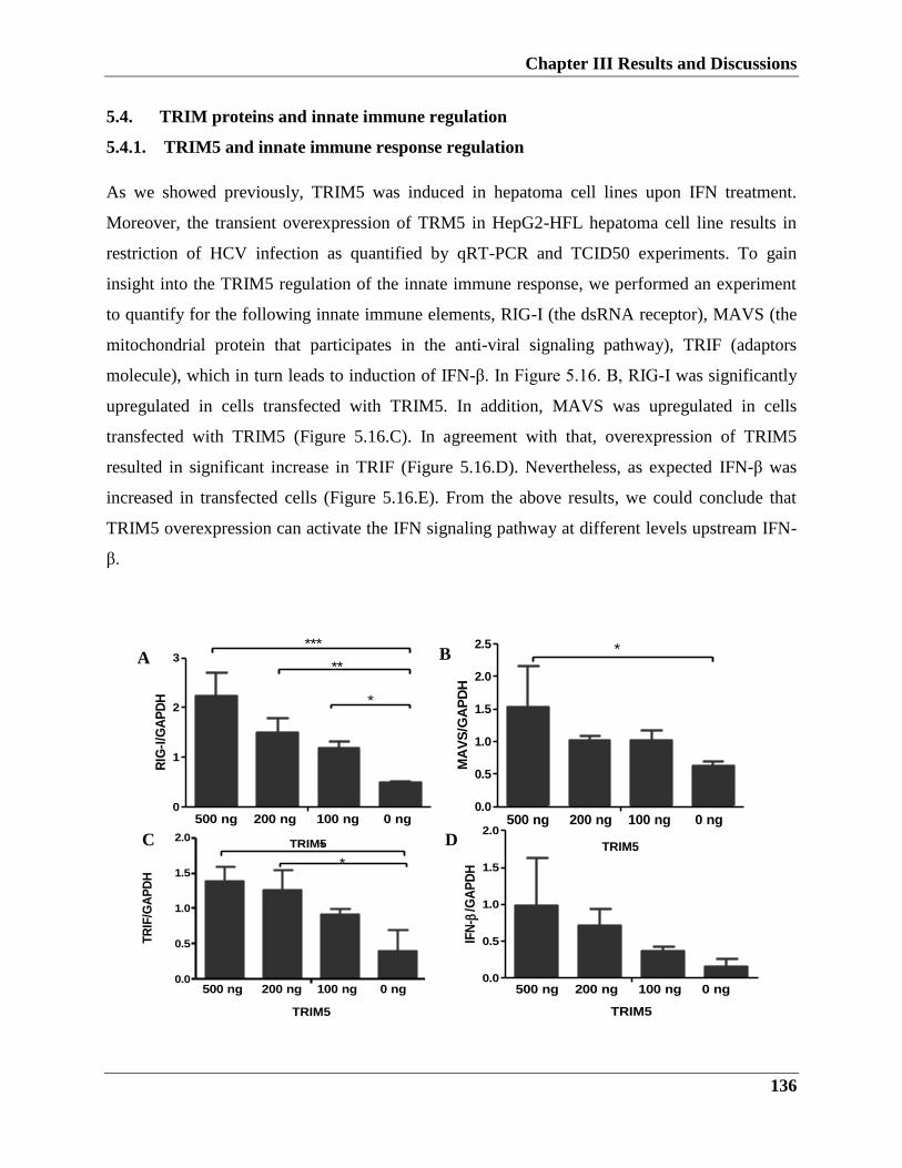
Chapter III Results and Discussions
136
B
D
5.4. TRIM proteins and innate immune regulation
5.4.1. TRIM5 and innate immune response regulation
As we showed previously, TRIM5 was induced in hepatoma cell lines upon IFN treatment.
Moreover, the transient overexpression of TRM5 in HepG2-HFL hepatoma cell line results in
restriction of HCV infection as quantified by qRT-PCR and TCID50 experiments. To gain
insight into the TRIM5 regulation of the innate immune response, we performed an experiment
to quantify for the following innate immune elements, RIG-I (the dsRNA receptor), MAVS (the
mitochondrial protein that participates in the anti-viral signaling pathway), TRIF (adaptors
molecule), which in turn leads to induction of IFN-β. In Figure 5.16. B, RIG-I was significantly
upregulated in cells transfected with TRIM5. In addition, MAVS was upregulated in cells
transfected with TRIM5 (Figure 5.16.C). In agreement with that, overexpression of TRIM5
resulted in significant increase in TRIF (Figure 5.16.D). Nevertheless, as expected IFN-β was
increased in transfected cells (Figure 5.16.E). From the above results, we could conclude that
TRIM5 overexpression can activate the IFN signaling pathway at different levels upstream IFN-
β.
A
C TRIM5
0
1
2
3500ng
200ng
100ng
0ng
***
**
*
RIG
-I/G
AP
DH
500 ng 200 ng 100 ng 0 ng
TRIM5
0.0
0.5
1.0
1.5
2.0
2.5500ng
200ng
100ng
0ng
*
500 ng 200 ng 100 ng 0 ng
MA
VS
/GA
PD
H
TRIM5
0.0
0.5
1.0
1.5
2.0500ng
200ng
100ng
0ng
*
*
500 ng 200 ng 100 ng 0 ng
TR
IF/G
AP
DH
TRIM5
0.0
0.5
1.0
1.5
2.0500ng
200ng
100ng
0ng
500 ng 200 ng 100 ng 0 ng
IFN
- /G
AP
DH
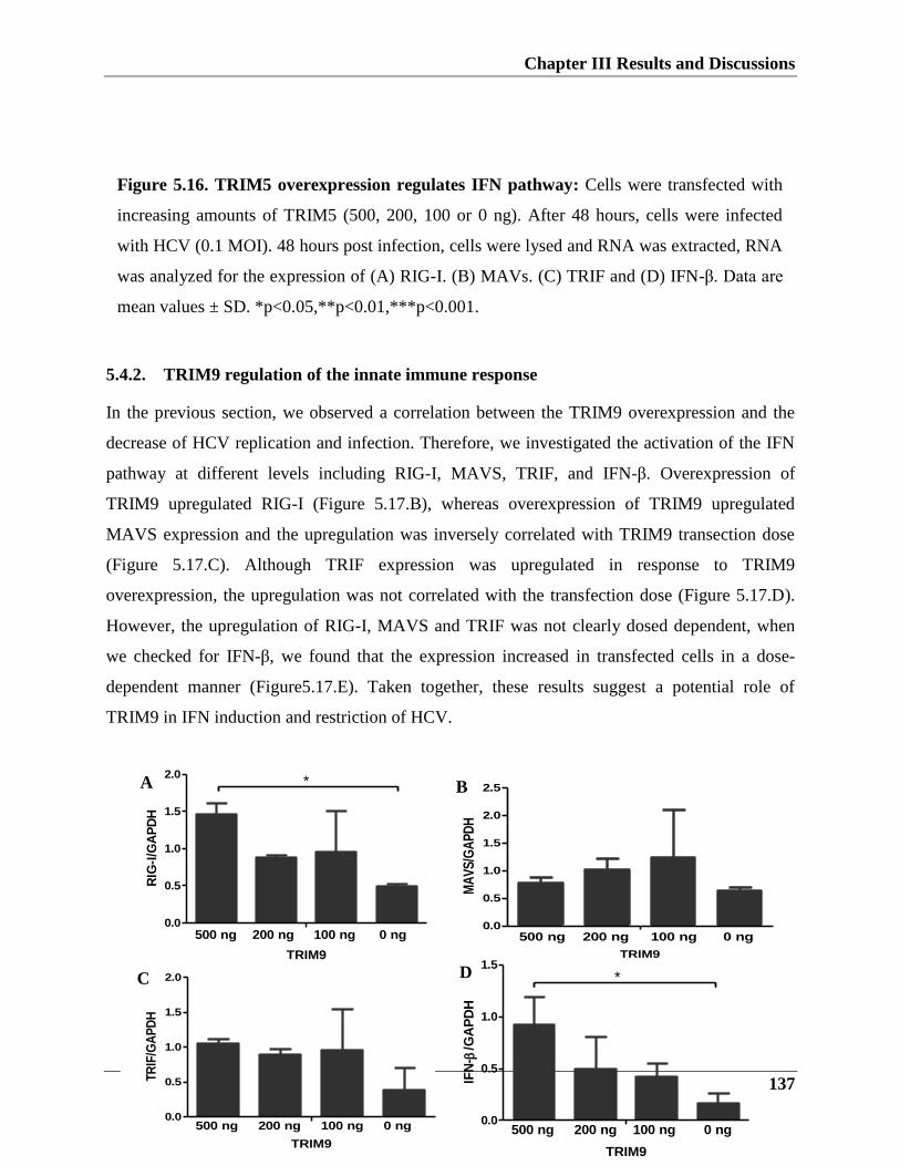
Chapter III Results and Discussions
137
5.4.2. TRIM9 regulation of the innate immune response
In the previous section, we observed a correlation between the TRIM9 overexpression and the
decrease of HCV replication and infection. Therefore, we investigated the activation of the IFN
pathway at different levels including RIG-I, MAVS, TRIF, and IFN-β. Overexpression of
TRIM9 upregulated RIG-I (Figure 5.17.B), whereas overexpression of TRIM9 upregulated
MAVS expression and the upregulation was inversely correlated with TRIM9 transection dose
(Figure 5.17.C). Although TRIF expression was upregulated in response to TRIM9
overexpression, the upregulation was not correlated with the transfection dose (Figure 5.17.D).
However, the upregulation of RIG-I, MAVS and TRIF was not clearly dosed dependent, when
we checked for IFN-β, we found that the expression increased in transfected cells in a dose-
dependent manner (Figure5.17.E). Taken together, these results suggest a potential role of
TRIM9 in IFN induction and restriction of HCV.
B
TRIM9
0.0
0.5
1.0
1.5
2.0
500 ng 200 ng 100 ng 0 ng
TR
IF/G
AP
DH
A
C
TRIM9
0.0
0.5
1.0
1.5
2.0
2.5500ng
200ng
100ng
0ng
500 ng 200 ng 100 ng 0 ng
MA
VS
/GA
PD
H
D
Figure 5.16. TRIM5 overexpression regulates IFN pathway: Cells were transfected with
increasing amounts of TRIM5 (500, 200, 100 or 0 ng). After 48 hours, cells were infected
with HCV (0.1 MOI). 48 hours post infection, cells were lysed and RNA was extracted, RNA
was analyzed for the expression of (A) RIG-I. (B) MAVs. (C) TRIF and (D) IFN-β. Data are
mean values ± SD. *p<0.05,**p<0.01,***p<0.001.
TRIM9
0.0
0.5
1.0
1.5
2.0500ng
200ng
100ng
0ng
*
500 ng 200 ng 100 ng 0 ng
RIG
-I/G
AP
DH
TRIM9
0.0
0.5
1.0
1.5500ng
200ng
100ng
0ng
*
500 ng 200 ng 100 ng 0 ng
IFN
- /G
AP
DH
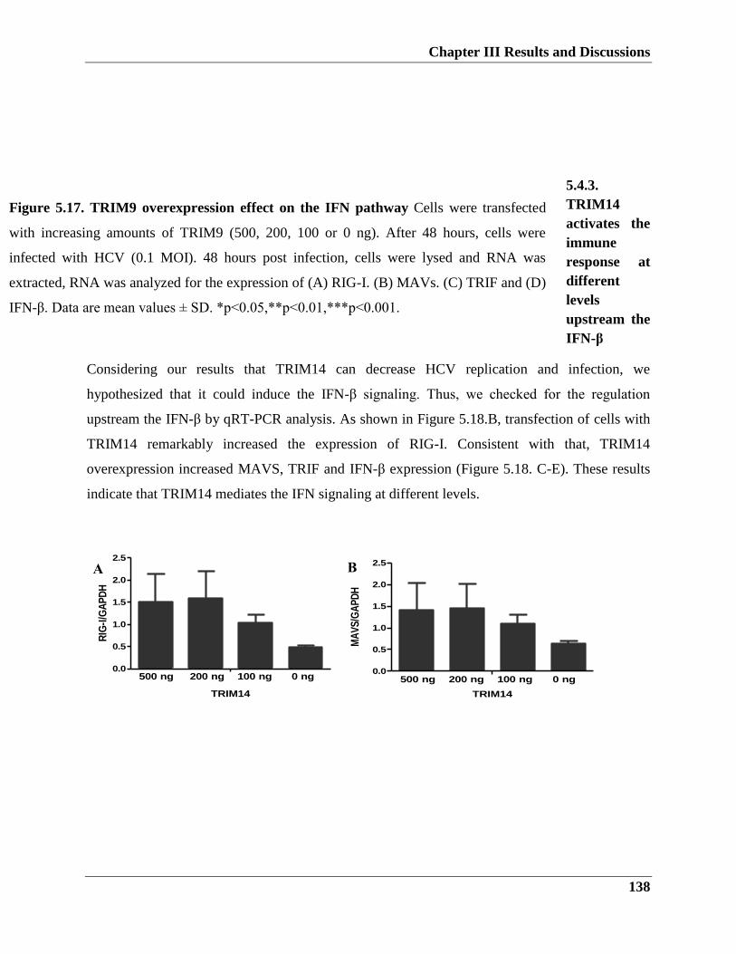
Chapter III Results and Discussions
138
5.4.3.
TRIM14
activates the
immune
response at
different
levels
upstream the
IFN-β
Considering our results that TRIM14 can decrease HCV replication and infection, we
hypothesized that it could induce the IFN-β signaling. Thus, we checked for the regulation
upstream the IFN-β by qRT-PCR analysis. As shown in Figure 5.18.B, transfection of cells with
TRIM14 remarkably increased the expression of RIG-I. Consistent with that, TRIM14
overexpression increased MAVS, TRIF and IFN-β expression (Figure 5.18. C-E). These results
indicate that TRIM14 mediates the IFN signaling at different levels.
TRIM14
0.0
0.5
1.0
1.5
2.0
2.5500ng
200ng
100ng
0ng
500 ng 200 ng 100 ng 0 ng
RIG
-I/G
AP
DH
TRIM14
0.0
0.5
1.0
1.5
2.0
2.5500ng
200ng
100ng
0ng
500 ng 200 ng 100 ng 0 ng
MA
VS
/GA
PD
H
Figure 5.17. TRIM9 overexpression effect on the IFN pathway Cells were transfected
with increasing amounts of TRIM9 (500, 200, 100 or 0 ng). After 48 hours, cells were
infected with HCV (0.1 MOI). 48 hours post infection, cells were lysed and RNA was
extracted, RNA was analyzed for the expression of (A) RIG-I. (B) MAVs. (C) TRIF and (D)
IFN-β. Data are mean values ± SD. *p<0.05,**p<0.01,***p<0.001.
B A
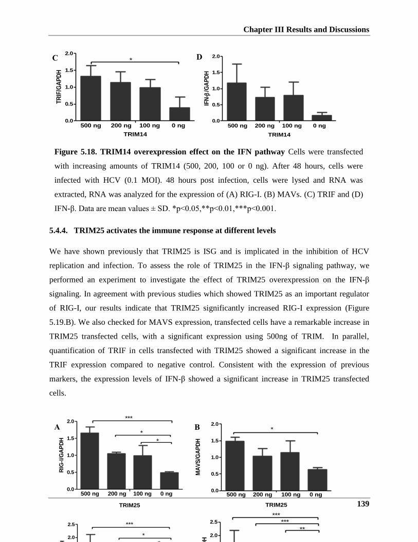
Chapter III Results and Discussions
139
TRIM14
0.0
0.5
1.0
1.5
2.0500ng
200ng
100ng
0ng
*
500 ng 200 ng 100 ng 0 ng
TR
IF/G
AP
DH
TRIM14
0.0
0.5
1.0
1.5
2.0500ng
200ng
100ng
0ng
500 ng 200 ng 100 ng 0 ng
IFN
- /G
AP
DH
5.4.4. TRIM25 activates the immune response at different levels
We have shown previously that TRIM25 is ISG and is implicated in the inhibition of HCV
replication and infection. To assess the role of TRIM25 in the IFN-β signaling pathway, we
performed an experiment to investigate the effect of TRIM25 overexpression on the IFN-β
signaling. In agreement with previous studies which showed TRIM25 as an important regulator
of RIG-I, our results indicate that TRIM25 significantly increased RIG-I expression (Figure
5.19.B). We also checked for MAVS expression, transfected cells have a remarkable increase in
TRIM25 transfected cells, with a significant expression using 500ng of TRIM. In parallel,
quantification of TRIF in cells transfected with TRIM25 showed a significant increase in the
TRIF expression compared to negative control. Consistent with the expression of previous
markers, the expression levels of IFN-β showed a significant increase in TRIM25 transfected
cells.
Figure 5.18. TRIM14 overexpression effect on the IFN pathway Cells were transfected
with increasing amounts of TRIM14 (500, 200, 100 or 0 ng). After 48 hours, cells were
infected with HCV (0.1 MOI). 48 hours post infection, cells were lysed and RNA was
extracted, RNA was analyzed for the expression of (A) RIG-I. (B) MAVs. (C) TRIF and (D)
IFN-β. Data are mean values ± SD. *p<0.05,**p<0.01,***p<0.001.
A B
C D
TRIM25
0.0
0.5
1.0
1.5
2.0
2.5500ng
200ng
100ng
0ng
***
*
*
500 ng 200 ng 100 ng 0 ng
TR
IF/G
AP
DH
TRIM25
0.0
0.5
1.0
1.5
2.0
2.5500ng
200ng
100ng
0ng
******
**
500 ng 200 ng 100 ng 0 ng
IFN
- /G
AP
DH
TRIM25
0.0
0.5
1.0
1.5
2.0500ng
200ng
100ng
0ng
***
*
*
500 ng 200 ng 100 ng 0 ng
RIG
-I/G
AP
DH
TRIM25
0.0
0.5
1.0
1.5
2.0500ng
200ng
100ng
0ng
*
500 ng 200 ng 100 ng 0 ng
MA
VS
/GA
PD
H
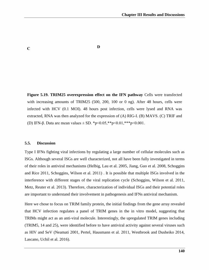
Chapter III Results and Discussions
140
5.5. Discussion
Type I IFNs fighting viral infections by regulating a large number of cellular molecules such as
ISGs. Although several ISGs are well characterized, not all have been fully investigated in terms
of their roles in antiviral mechanisms (Helbig, Lau et al. 2005, Jiang, Guo et al. 2008, Schoggins
and Rice 2011, Schoggins, Wilson et al. 2011) . It is possible that multiple ISGs involved in the
interference with different stages of the viral replication cycle (Schoggins, Wilson et al. 2011,
Metz, Reuter et al. 2013). Therefore, characterization of individual ISGs and their potential roles
are important to understand their involvement in pathogenesis and IFNs antiviral mechanism.
Here we chose to focus on TRIM family protein, the initial findings from the gene array revealed
that HCV infection regulates a panel of TRIM genes in the in vitro model, suggesting that
TRIMs might act as an anti-viral molecule. Interestingly, the upregulated TRIM genes including
(TRIM5, 14 and 25), were identified before to have antiviral activity against several viruses such
as HIV and SeV (Neamati 2001, Pertel, Hausmann et al. 2011, Westbrook and Dusheiko 2014,
Lascano, Uchil et al. 2016).
Figure 5.19. TRIM25 overexpression effect on the IFN pathway Cells were transfected
with increasing amounts of TRIM25 (500, 200, 100 or 0 ng). After 48 hours, cells were
infected with HCV (0.1 MOI). 48 hours post infection, cells were lysed and RNA was
extracted, RNA was then analyzed for the expression of (A) RIG-I. (B) MAVS. (C) TRIF and
(D) IFN-β. Data are mean values ± SD. *p<0.05,**p<0.01,***p<0.001.
C D
Chapter III Results and Discussions
141
While we believe that TRIMs are likely played different roles in controlling viral infections, their
role in HCV infection is not yet clear. Using our array data along with literature search, we could
identify the genes of interest for further investigations.
We considered two approaches to identify TRIMs with anti-HCV activity, the first determination
of the induction profile of TRIMs using interferon, a second determination of TRIM regulation
upon HCV infection in cell culture.
We analyzed the induction kinetics of selected TRIMs, TRIM 7, 9, 21 and 40 have rapid
induction kinetics, while TRIM5 and 14 have intermediate kinetics peaking at 24 hours, and
TRIM 1 have slow kinetics and reached a peak at 96 hours post IFN treatment. In the course of
HCV infection, the kinetic peaks of TRIM5, 7, 14 and 40 overlap with the expression pattern
post IFN treatment. Notably, TRIM21, 22 and 40 have a high expression level, one possibility is
that HCV tends to establish a mechanism to downregulate them. Because hepatoma cell lines are
transformed cells, we checked for TRIMs expression in PHHs, our analysis noted upregulation
of 14 TRIMs (TRIM5,7,9,11,13,14,15,21,25,33,34,35,40 and 55) at different time points post
IFN treatment. Considering that PHHs are poorly infectible, and the quantification of the viral
genome was not clearly determined, it is not obvious how HCV regulates TRIMs expression in
this model.
Among overlapped TRIMs from different experiments, we selected TRIM5, 9, 14 and 25 to
further study their role in HCV infection.
Our data showed that TRIM5 is one of the most highly induced Type 1 IFN TRIMs in PHHs and
hepatoma cells. Several studies revealed that TRIM5 restricts various retroviral infections, such
as HIV and SeV (James, Keeble et al. 2007, Pertel, Hausmann et al. 2011, Lascano, Uchil et al.
2016). However, the antiviral activity of TRIM5 is well studied, no information are available on
its role in HCV infection. Therefore, we explored the TRIM5 role in HCV infection in two parts:
first, we used an overexpression approach to determine the effect on HCV replication cycle in
both replicon and HCVcc system, next we continued to determine the efficiency of TRIM5
activation on multiple molecules in the IFN signaling pathway.
We showed that TRIM5 overexpression significantly decreased HCV replication in both a
replicon and infection (HCVcc) based tests. Thus, we suggest that TRIM5 restricts multiple steps
Chapter III Results and Discussions
142
in the HCV replication cycle, we confirmed that TRIM5 inhibits replication when TRIM5
transfected into replicon cells in a dose-dependent manner. Further infection experiment revealed
a significant decrease in intracellular HCV genome RNA level in TRIM5 transfected cells, in
addition, our TCID50 assay showed that secreted viral particles were significantly diminished
when TRIM5 was expressed in the cells. Comment from other literature… These observations
reflect, the anti-viral activity of TRIM5 in HCV infection. Yet, we could not rule out the effect
on the early (e.g: entry) and the late (e.g: assembly) steps of HCV infection cycle.
Before we analyze the activation of ISRE, IFN-β, and NF-κB reporter promotes by several
TRIMs, we studied the regulation kinetic pattern in the presence of either IFN-alpha or HCV.
We demonstrated that the ISRE, IFN-β, and NF-κB promoters were activated upon IFN
treatment, and peaked the maximum at 6 hours post-treatment, and then dropped at 24 hours
followed by gaining increase at 96 hours post treatment. In contrast, during HCV infection, IFN-
β promoter activation dropped, these results also suggest that the presence of HCV inhibited the
activation of the IFN-β promoter in our experiments. Same is true for NF-κB promoter
activation, we have observed a gradual decline in the activation after HCV infection. While,
ISRE promoter activation increased and reached the maximum at 24 hours post infection, then
dropped in the later time points indicating that HCV might establish a defense mechanism to
overcome the immune mechanisms.
Subsequent to the identification of TRIM5 as anti-HCV molecule, we wanted to check its ability
to activate IFN-β promoter, NF-κB responsive promoter, and ISRE in reporter assays. In the
presence of HCV infection, overexpression of TRIM5 enhanced the induction of ISRE and NF-
κB by approximately 1 fold, while IFN-β was induced by 2 folds. When we used same
conditions to check the induction in the absence of HCV infection, we found that the induction
of NF-κB and ISRE is the same in both presence and absence of HCV, whereas IFN-β was
higher in the absence of HCV infection.
To understand whether TRIM5 regulates the innate immune responses, we investigated several
marker molecules in the antiviral innate immunity. TRIM5 upregulates the RIG-I viral
recognition receptor, and the downstream molecule MAVS, moreover TRIM5 transfection
upregulates TRIF adaptor molecule and the IFN-β. Taken together, these observations suggest
Chapter III Results and Discussions
143
that TRIM5 positively regulates IFN signaling pathway (Rajsbaum, Stoye et al. 2008, Rajsbaum,
Albrecht et al. 2012, Ovsyannikova, Haralambieva et al. 2013, Zhu, Liu et al. 2016).
As mentioned earlier TRIM9 has two isoforms TRIM9s and TRIM9I, the short isoform poses
anti-viral activity and enhances the expression of ISGs (Qin, Liu et al. 2016). Therefore, further
series of experiments aimed to identify the role of TRIM9 in HCV infection were performed. In
HCV replicon cell line, overexpression of TRIM9 led to a significant reduction in the virus
replication as measured by the reporter assay. Then we further sought to determine the effect of
TRIM9 on HCV infection using the HCVcc based system. As we expected TRIM9
overexpression resulted in a significant decrease in HCV infection, this decrease was in both, the
intracellular HCV genomic RNA level and the extracellular virus secretion.
First reports about TRIM9 identified TRIM9 as a brain-specific molecule (Tanji, Kamitani et al.
2010), then further characterization showed that it is widely expressed in several tissues in the
human body. A previous study indicated that TRIM9 expression is reduced in damaged brains of
Parkinson’s disease and Lewy body dementia patients (Tanji, Kamitani et al. 2010, Yang, Li et
al. 2014), and this decrease is associated with a dramatic increase of NF-κB . The study also
revealed that, TRIM9 act as a negative regulator of NF-κB in brain cells via targeting b-TrCP
SCF complex which regulates NF-κB (Shi, Cho et al. 2014). Another study showed that
overexpression of TRIM9 hindered virus infection in particular VSV, this could be attributed to
the enhancements of IRF3 and IFN-β signaling (Qin, Liu et al. 2016). Thus, we determined the
regulatory mechanism of TRIM9 on the innate immune signaling during HCV infection in vitro.
In our HCVcc infection system, when TRIM9 was overexpressed in the cells, we were able to
see enhancements in both IFN-β and ISRE promotors’ activation, whereas NF-κB promotor
stayed unchanged, this partially agrees with the previous study which indicated the negative
regulatory role of TRIM9 on NF-κB. Nevertheless, the analysis of the TRIM9 effect on ISRE,
IFN-β and NF-κB promoters in the absence of HCV infection, showed an overlapped pattern
with the previously determined in the presence of HCV infection. The second set of innate
molecular markers including RIG-I, MAVS, TRIF and finally IFN-β were analyzed. The RIG-I
dsRNA molecular receptor and IFN-β were up-regulated in TRIM9 transfected cells in a dose-
dependent manner. Independent of TRIM9 transfection does, TRIF adaptor molecule expression
was upregulated in transfected cells. Unlike, RIG-I and IFN-β, MAVS expression was reversely
Chapter III Results and Discussions
144
correlated with TRIM9 transfection dose. We could argue that the overall upregulation of
different molecules attributed in the IFN-β signaling pathway, is one possible explanation for
inhibiting HCV infection in our study.
Similar to other selected TRIMs, TRIM14 was highly expressed upon IFN-alpha treatment in
HCV replicon cells, Huh7 cells, and PHHs. Therefore, we first analyzed the effect of TRM14
overexpression on HCV infection cycle. In line with another study, TRIM14 overexpression
impaired HCV virus replication and infection. Our results based on both HCV replicon and
HCVcc systems. This is similar to the observations made before for several ISGs including
TRIM14, in which an overexpression approach was performed for individual ISGs, in that
TRIM14 showed an inhibition of HCV replication (Jiang, Guo et al. 2008, Metz, Reuter et al.
2013). Nevertheless, the mechanism of TRIM14 associated with HCV replication reduction is
still not determined. To determine whether TRIM14 mediates IFN-β pathway activation, we
analyzed the ISRE, IFN-β, and NF-κB promoter activation during TRIM14 overexpression.
Thus, while TRIM14 led to an enhancement of both IFN-β and NF-κB promoters’ activities, the
ISRE promoter activity remained unchanged. Consequently, we wanted to test what influence
TRIM14 overexpression have on the RIG-I, MAVS, TRIF and IFN-β expression level in relation
to HCV infection. Overexpression of TRIM14, increased mRNA expression levels of RIG-I,
MAVS, TRIF and IFN-β demonstrating that TRIM14 triggers the IFN antiviral response.
Together, this proposes an explanation of the decline of HCV replication in our study.
As stated earlier, TRIM25 is able to cooperate with the CARD domains of RIG-I which facilitate
RIG-I activation of MAVS. This interaction then facilitates downstream signaling and the
production of IFNβ (Gack, Shin et al. 2007, Castanier, Zemirli et al. 2012, Rajsbaum, Albrecht et
al. 2012). Our results indicated that TRIM25 is induced upon IFN treatment or HCV viral
infection. Thus, in order to determine the role of TRIM25 in HCV infection, we performed an
overexpression experiment where we overexpressed TRIM25 in both HCV replicons cells and in
HCV infection settings. Thus, TRIM25 led to a reduction of HCV replication and infection. This
was accompanied by the activation of several promoter activations such as ISRE, IFN-β, and NF-
κB, which suggests its anti-viral role against HCV infection. Additionally, we tested the effect
of TRIM25 overexpression on several molecules in the IFN signaling pathway. All markers
including HCV recognition receptor RIG-I, adaptor molecule TRIF, MAVS, and IFN-β showed a
Chapter III Results and Discussions
145
significant increase in the expression level upon TRIM25 transfection. Collectively, this
activation is required for elimination of HCV infection, agrees with previous findings which
revealed that TRIM25 possess anti-viral activity which attributed to the activation of the IFN-β
signaling pathway.
5.6. Summary
TRIM family members have been implicated in several functions such as cell differentiation,
apoptosis, neurogenesis, muscular physiology and innate immune responses. Multiple TRIM
proteins play a role in restricting viral infection such as retroviruses infection, by inducing IFN
response, regulate RPRs and/or interacting directly with viral proteins. Thus, we sought to study
the role of TRIMs in HCV infection. Screening of TRIMs revealed that HCV infection regulates
a panel of TRIM genes in the in vitro model, suggesting that TRIMs might act as a anti-viral
molecule. Interestingly, the upregulated TRIM genes including (TRIM5, 14 and 25), were
identified before to have antiviral activity against several viruses such as HIV and SeV. While
we believe that TRIMs are likely played different roles in controlling viral infections, their role
in HCV infection is not yet clear. We could identify the genes of interest for further
investigations. Therefore, we hypothesized that those TRIMs play anti-HCV activities. In our
study, we choose to focus on (TRIM5, TRIM9, TRIM14 and TRIM25).
We indicated that TRIM5 overexpression significantly decreased HCV replication in both a
replicon and infection (HCVcc) based system. Thus, we suggest that TRIM5 restricts multiple
steps in the HCV replication cycle, we confirmed that TRIM5 inhibits replication when TRIM5
transfected into replicon cells in a dose-dependent manner. Further infection experiment revealed
a significant decrease in intracellular HCV genome RNA level in TRIM5 transfected cells, in
addition, our TCID50 assay showed that secreted viral particles were significantly diminished
when TRIM5 was expressed in the cells. These observations reflect the anti-viral activity of
TRIM5 in HCV infection. Yet, we could not rule out the effect on the early (e.g: entry) and the
late (e.g: assembly) steps of HCV infection cycle. In this model, TRIM5 upregulates the RIG-I
viral recognition receptor and the downstream molecule MAVS, moreover TRIM5 transfection
upregulates TRIF adaptor molecule and the IFN-β. Taken together, these observations suggest
that TRIM5 positively regulates IFN signaling pathway.

Chapter III Results and Discussions
146
For TRIM9, overexpression led to a significant reduction in the virus replication as measured by
the reporter assay. Further analysis showed that the effect of TRIM9 extends to the whole virus
replication system as illustrated by HCVcc based system. A former study showed that
overexpression of TRIM9 hindered virus infection in particular VSV, this could be attributed to
the enhancements of IRF3 and IFN-β signaling. Our analysis of the innate immune regulatory
role of TRIM9 on the revealed enhancements in both IFN-β and ISRE promotors’ activation,
whereas NF-κB promotor stayed unchanged, this partially agrees with the previous study which
indicated the negative regulatory role of TRIM9 on NF-κB. Nevertheless, the analysis of the
TRIM9 effect on ISRE, IFN-β and NF-κB promoters in the absence of HCV infection, showed
an overlapped pattern with the previously determined in the presence of HCV infection.
TRIM14 was highly expressed upon IFN-α treatment in HCV replicon cells, Huh7 cells, and
PHHs. In our study, TRIM14 overexpression impaired HCV virus replication and infection. This
is similar to the observations made before for several ISGs including TRIM14, in that TRIM14
showed an inhibition of HCV replication Nevertheless, the mechanism of TRIM14 associated
with HCV replication reduction is still not determined. To determine whether TRIM14 mediates
IFN-β pathway activation, we analyzed the ISRE, IFN-β, and NF-κB promoter activation during
TRIM14 overexpression. Thus, while TRIM14 led to an enhancement of both IFN-β and NF-κB
promoters’ activities. Overexpression of TRIM14 increased expression levels of RIG-I, MAVS,
TRIF and IFN-β. Together, this demonstrates that TRIM14 triggers the IFN antiviral response
leading to the decline of HCV replication.
TRIM25 is able to cooperate with the CARD domains of RIG-I which facilitate RIG-I activation
of MAVS. Thus, overexpression of TRIM25 in both HCV replicons cells and in HCV infection
settings. This was accompanied by the activation of several promoter activations such as ISRE,
IFN-β, and NF-κB, which suggests its anti-viral role against HCV infection. Additionally,
TRIM25 transfection enhances the expression levels of the RIG-I receptor, adaptor molecule
TRIF, MAVS, and IFN-β. Collectively, this activation is required for elimination of HCV
infection, agrees with previous findings which revealed that TRIM25 possess anti-viral activity
which attributed to the activation of the IFN-β signaling pathway.
Chapter III Results and Discussions
147
Chapter VI References
148
6. Chapter VI References
Abergel, C. (2013). "Molecular replacement: tricks and treats." Acta Crystallogr D Biol
Crystallogr 69(Pt 11): 2167-2173.
Afdhal, N., K. R. Reddy, D. R. Nelson, E. Lawitz, S. C. Gordon, E. Schiff, R. Nahass, R. Ghalib,
N. Gitlin, R. Herring, J. Lalezari, Z. H. Younes, P. J. Pockros, A. M. Di Bisceglie, S. Arora, G.
M. Subramanian, Y. Zhu, H. Dvory-Sobol, J. C. Yang, P. S. Pang, W. T. Symonds, J. G.
McHutchison, A. J. Muir, M. Sulkowski, P. Kwo and I. O. N. Investigators (2014). "Ledipasvir
and sofosbuvir for previously treated HCV genotype 1 infection." N Engl J Med 370(16): 1483-
1493.
Alloush, J. and N. Weisleder (2013). "TRIM proteins in therapeutic membrane repair of
muscular dystrophy." JAMA Neurol 70(7): 928-931.
Anderson, A. C. (2003). "The Process of Structure-Based Drug Design." Chemistry & Biology
10(9): 787-797.
Ansaldi, F., A. Orsi, L. Sticchi, B. Bruzzone and G. Icardi (2014). "Hepatitis C virus in the new
era: perspectives in epidemiology, prevention, diagnostics and predictors of response to therapy."
World J Gastroenterol 20(29): 9633-9652.
Asselah, T., S. De Muynck, P. Broet, J. Masliah-Planchon, M. Blanluet, I. Bieche, M. Lapalus,
M. Martinot-Peignoux, O. Lada, E. Estrabaud, Q. Zhang, A. El Ray, D. Vidaud, M. P. Ripault,
N. Boyer, P. Bedossa, D. Valla, M. Vidaud and P. Marcellin (2012). "IL28B polymorphism is
associated with treatment response in patients with genotype 4 chronic hepatitis C." J Hepatol
56(3): 527-532.
Asselah, T. and P. Marcellin (2015). "Optimal IFN-free therapy in treatment-naive patients with
HCV genotype 1 infection." Liver Int 35 Suppl 1: 56-64.
Balagopal, A., D. L. Thomas and C. L. Thio (2010). "IL28B and the control of hepatitis C virus
infection." Gastroenterology 139(6): 1865-1876.
Barreiro, P., J. A. Pineda, N. Rallon, S. Naggie, L. Martin-Carbonero, K. Neukam, A. Rivero, J.
M. Benito, A. Caruz, E. Vispo, A. Camacho, J. Medrano, J. McHutchison and V. Soriano (2011).
"Influence of interleukin-28B single-nucleotide polymorphisms on progression to liver cirrhosis
in human immunodeficiency virus-hepatitis C virus-coinfected patients receiving antiretroviral
therapy." J Infect Dis 203(11): 1629-1636.
Bartenschlager, R., A. Kaul and S. Sparacio (2003). "Replication of the hepatitis C virus in cell
culture." Antiviral Research 60(2): 91-102.
Bartenschlager, R., V. Lohmann and F. Penin (2013). "The molecular and structural basis of
advanced antiviral therapy for hepatitis C virus infection." Nat Rev Microbiol 11(7): 482-496.
Bartenschlager, R., F. Penin, V. Lohmann and P. Andre (2011). "Assembly of infectious
hepatitis C virus particles." Trends Microbiol 19(2): 95-103.
Barth, H. (2015). "Hepatitis C virus: Is it time to say goodbye yet? Perspectives and challenges
for the next decade." World J Hepatol 7(5): 725-737.
Bartosch, B., A. Vitelli, C. Granier, C. Goujon, J. Dubuisson, S. Pascale, E. Scarselli, R. Cortese,
A. Nicosia and F. L. Cosset (2003). "Cell entry of hepatitis C virus requires a set of co-receptors
Chapter VI References
149
that include the CD81 tetraspanin and the SR-B1 scavenger receptor." J Biol Chem 278(43):
41624-41630.
Beltra, J. C. and H. Decaluwe (2016). "Cytokines and persistent viral infections." Cytokine.
Binder, J., S. Tetangco, M. Weinshank, K. Maegley, L. Lingardo, W. Diehl, R. Love, A. K.
Patick and G. J. Smith, 3rd (2011). "Development of hepatitis C virus chimeric replicons for
identifying broad spectrum NS3 protease inhibitors." Antiviral Res 91(2): 102-111.
Biswal, B. K., M. M. Cherney, M. Wang, L. Chan, C. G. Yannopoulos, D. Bilimoria, O. Nicolas,
J. Bedard and M. N. James (2005). "Crystal structures of the RNA-dependent RNA polymerase
genotype 2a of hepatitis C virus reveal two conformations and suggest mechanisms of inhibition
by non-nucleoside inhibitors." J Biol Chem 280(18): 18202-18210.
Boltjes, A., D. Movita, A. Boonstra and A. M. Woltman (2014). "The role of Kupffer cells in
hepatitis B and hepatitis C virus infections." J Hepatol 61(3): 660-671.
Boudinot, P., L. M. van der Aa, L. Jouneau, L. Du Pasquier, P. Pontarotti, V. Briolat, A.
Benmansour and J. P. Levraud (2011). "Origin and evolution of TRIM proteins: new insights
from the complete TRIM repertoire of zebrafish and pufferfish." PLoS One 6(7): e22022.
Bowen, D. G. and C. M. Walker (2005). "Adaptive immune responses in acute and chronic
hepatitis C virus infection." Nature 436(7053): 946-952.
Bunchorntavakul, C. and K. R. Reddy (2015). "Review article: the efficacy and safety of
daclatasvir in the treatment of chronic hepatitis C virus infection." Aliment Pharmacol Ther
42(3): 258-272.
Cai, C., H. Masumiya, N. Weisleder, N. Matsuda, M. Nishi, M. Hwang, J. K. Ko, P. Lin, A.
Thornton, X. Zhao, Z. Pan, S. Komazaki, M. Brotto, H. Takeshima and J. Ma (2009). "MG53
nucleates assembly of cell membrane repair machinery." Nat Cell Biol 11(1): 56-64.
Caillet-Saguy, C., P. C. Simister and S. Bressanelli (2011). "An objective assessment of
conformational variability in complexes of hepatitis C virus polymerase with non-nucleoside
inhibitors." J Mol Biol 414(3): 370-384.
Calistri, A., D. Munegato, I. Carli, C. Parolin and G. Palu (2014). "The ubiquitin-conjugating
system: multiple roles in viral replication and infection." Cells 3(2): 386-417.
Carruthers, C. W., Jr., C. Gerdts, M. D. Johnson and P. Webb (2013). "A microfluidic, high
throughput protein crystal growth method for microgravity." PLoS One 8(11): e82298.
Castanier, C., N. Zemirli, A. Portier, D. Garcin, N. Bidere, A. Vazquez and D. Arnoult (2012).
"MAVS ubiquitination by the E3 ligase TRIM25 and degradation by the proteasome is involved
in type I interferon production after activation of the antiviral RIG-I-like receptors." BMC Biol
10: 44.
Caterina Berti, S. M., Andrea Ballabio, Alexandre Reymond1, Germana Meroni (2002). "TRIM9
is specifically expressed in the embryonic and adult nervous system." Mechanisms of
Development 113 4.
Chan, E., G. J. Towers and W. Qasim (2014). "Gene therapy strategies to exploit TRIM derived
restriction factors against HIV-1." Viruses 6(1): 243-263.
Chapter VI References
150
Chayama, K. and C. N. Hayes (2015). "HCV Drug Resistance Challenges in Japan: The Role of
Pre-Existing Variants and Emerging Resistant Strains in Direct Acting Antiviral Therapy."
Viruses 7(10): 5328-5342.
Chayen, N. E. (2004). "Turning protein crystallisation from an art into a science." Curr Opin
Struct Biol 14(5): 577-583.
Chen, C. H. and M. L. Yu (2010). "Evolution of interferon-based therapy for chronic hepatitis
C." Hepat Res Treat 2010: 140953.
Chen, Y., S. Wang, Z. Yi, H. Tian, R. Aliyari, Y. Li, G. Chen, P. Liu, J. Zhong, X. Chen, P. Du,
L. Su, F. X. Qin, H. Deng and G. Cheng (2014). "Interferon-inducible cholesterol-25-
hydroxylase inhibits hepatitis C virus replication via distinct mechanisms." Sci Rep 4: 7242.
Choo, Y. S. and Z. Zhang (2009). "Detection of protein ubiquitination." J Vis Exp(30).
Choudhury, N. R., J. S. Nowak, J. Zuo, J. Rappsilber, S. H. Spoel and G. Michlewski (2014).
"Trim25 Is an RNA-Specific Activator of Lin28a/TuT4-Mediated Uridylation." Cell Rep 9(4):
1265-1272.
Chung, R. T., M. Gale, Jr., S. J. Polyak, S. M. Lemon, T. J. Liang and J. H. Hoofnagle (2008).
"Mechanisms of action of interferon and ribavirin in chronic hepatitis C: Summary of a
workshop." Hepatology 47(1): 306-320.
Cormier, E. G., F. Tsamis, F. Kajumo, R. J. Durso, J. P. Gardner and T. Dragic (2004). "CD81 is
an entry coreceptor for hepatitis C virus." Proceedings of the National Academy of Sciences
101(19): 7270-7274.
Crotty, S., C. E. Cameron and R. Andino (2001). "RNA virus error catastrophe: direct molecular
test by using ribavirin." Proc Natl Acad Sci U S A 98(12): 6895-6900.
Curry, S., P. Qiu and X. Tong (2008). "Analysis of HCV resistance mutations during
combination therapy with protease inhibitor boceprevir and PEG-IFN alpha-2b using TaqMan
mismatch amplification mutation assay." J Virol Methods 153(2): 156-162.
de Silva, S. and L. Wu (2011). "TRIM5 acts as more than a retroviral restriction factor." Viruses
3(7): 1204-1209.
Degasperi, E. and A. Aghemo (2014). "Sofosbuvir for the treatment of chronic hepatitis C:
between current evidence and future perspectives." Hepat Med 6: 25-33.
DiFrancesco, J. M. and J. M. Olson (2015). "The economics of microgravity research." npj
Microgravity 1: 15001.
DiMattia, M., L. Govindasamy, H. C. Levy, B. Gurda-Whitaker, A. Kalina, E. Kohlbrenner, J. A.
Chiorini, R. McKenna, N. Muzyczka, S. Zolotukhin and M. Agbandje-McKenna (2005).
"Production, purification, crystallization and preliminary X-ray structural studies of adeno-
associated virus serotype 5." Acta Crystallogr Sect F Struct Biol Cryst Commun 61(Pt 10): 917-
921.
Donaldson, E. F., P. R. Harrington, J. J. O'Rear and L. K. Naeger (2015). "Clinical evidence and
bioinformatics characterization of potential hepatitis C virus resistance pathways for sofosbuvir."
Hepatology 61(1): 56-65.
Chapter VI References
151
Dorner, M., J. A. Horwitz, B. M. Donovan, R. N. Labitt, W. C. Budell, T. Friling, A. Vogt, M. T.
Catanese, T. Satoh, T. Kawai, S. Akira, M. Law, C. M. Rice and A. Ploss (2013). "Completion
of the entire hepatitis C virus life cycle in genetically humanized mice." Nature 501(7466): 237-
241.
Du Pasquier, L. (2009). "Fish 'n' TRIMs." J Biol 8(5): 50.
Eksteen, B. (2011). "Infections and the liver." Dig Dis 29(2): 184-190.
Elabd, S., G. Meroni and C. Blattner (2016). "TRIMming p53’s anticancer activity." Oncogene.
Elbaz, T., M. El-Kassas and G. Esmat (2015). "New era for management of chronic hepatitis C
virus using direct antiviral agents: A review." J Adv Res 6(3): 301-310.
Elhefnawi, M., M. ElGamacy and M. Fares (2012). "Multiple virtual screening approaches for
finding new hepatitis C virus RNA-dependent RNA polymerase inhibitors: structure-based
screens and molecular dynamics for the pursue of new poly pharmacological inhibitors." BMC
Bioinformatics 13 Suppl 17: S5.
Falkowska, E., R. J. Durso, J. P. Gardner, E. G. Cormier, R. A. Arrigale, R. N. Ogawa, G. P.
Donovan, P. J. Maddon, W. C. Olson and T. Dragic (2006). "L-SIGN (CD209L) isoforms
differently mediate trans-infection of hepatoma cells by hepatitis C virus pseudoparticles." J Gen
Virol 87(Pt 9): 2571-2576.
Ferenci, P. (2004). "Predicting the therapeutic response in patients with chronic hepatitis C: the
role of viral kinetic studies." J Antimicrob Chemother 53(1): 15-18.
Firdaus, R., A. Biswas, K. Saha, A. Mukherjee, F. Pal, S. Chaudhuri, A. Chandra, A. Konar and
P. C. Sadhukhan (2014). "Modulation of TLR 3, 7 and 8 expressions in HCV genotype 3
infected individuals: potential correlations of pathogenesis and spontaneous clearance." Biomed
Res Int 2014: 491064.
Fournier, C., G. Duverlie and S. Castelain (2013). "Are trans-complementation systems suitable
for hepatitis C virus life cycle studies?" J Viral Hepat 20(4): 225-233.
Friebe, P. and R. Bartenschlager (2009). "Role of RNA structures in genome terminal sequences
of the hepatitis C virus for replication and assembly." J Virol 83(22): 11989-11995.
Gack, M. U., Y. C. Shin, C. H. Joo, T. Urano, C. Liang, L. Sun, O. Takeuchi, S. Akira, Z. Chen,
S. Inoue and J. U. Jung (2007). "TRIM25 RING-finger E3 ubiquitin ligase is essential for RIG-I-
mediated antiviral activity." Nature 446(7138): 916-920.
Gale, M., Jr. and E. M. Foy (2005). "Evasion of intracellular host defence by hepatitis C virus."
Nature 436(7053): 939-945.
Gaudieri, S., A. Rauch, K. Pfafferott, E. Barnes, W. Cheng, G. McCaughan, N. Shackel, G. P.
Jeffrey, L. Mollison, R. Baker, H. Furrer, H. F. Gunthard, E. Freitas, I. Humphreys, P.
Klenerman, S. Mallal, I. James, S. Roberts, D. Nolan and M. Lucas (2009). "Hepatitis C virus
drug resistance and immune-driven adaptations: relevance to new antiviral therapy." Hepatology
49(4): 1069-1082.
Gerdts, C. J., M. Elliott, S. Lovell, M. B. Mixon, A. J. Napuli, B. L. Staker, P. Nollert and L.
Stewart (2008). "The plug-based nanovolume Microcapillary Protein Crystallization System
(MPCS)." Acta Crystallogr D Biol Crystallogr 64(Pt 11): 1116-1122.
Chapter VI References
152
Gerdts, C. J., G. L. Stahl, A. Napuli, B. Staker, J. Abendroth, T. E. Edwards, P. Myler, W. Van
Voorhis, P. Nollert and L. J. Stewart (2010). "Nanovolume optimization of protein crystal
growth using the microcapillary protein crystallization system." J Appl Crystallogr 43(Pt 5):
1078-1083.
Giege, R. (2013). "A historical perspective on protein crystallization from 1840 to the present
day." FEBS J 280(24): 6456-6497.
Gokhale, N. S., C. Vazquez and S. M. Horner (2014). "Hepatitis C Virus. Strategies to Evade
Antiviral Responses." Future Virol 9(12): 1061-1075.
Guglietta, S., A. R. Garbuglia, L. Salichos, L. Ruggeri, A. Folgori, M. P. Perrone, C. Camperio,
V. Mellace, G. Maio, P. Maio, M. R. Capobianchi, E. Spada, N. Gargano, C. Scotta, E. Piccolella
and P. Del Porto (2009). "Impact of viral selected mutations on T cell mediated immunity in
chronically evolving and self limiting acute HCV infection." Virology 386(2): 398-406.
Habjan, M., I. Andersson, J. Klingstrom, M. Schumann, A. Martin, P. Zimmermann, V. Wagner,
A. Pichlmair, U. Schneider, E. Muhlberger, A. Mirazimi and F. Weber (2008). "Processing of
genome 5' termini as a strategy of negative-strand RNA viruses to avoid RIG-I-dependent
interferon induction." PLoS One 3(4): e2032.
Halfon, P. and S. Locarnini (2011). "Hepatitis C virus resistance to protease inhibitors." J
Hepatol 55(1): 192-206.
Hamamoto, I., Y. Nishimura, T. Okamoto, H. Aizaki, M. Liu, Y. Mori, T. Abe, T. Suzuki, M. M.
Lai, T. Miyamura, K. Moriishi and Y. Matsuura (2005). "Human VAP-B is involved in hepatitis
C virus replication through interaction with NS5A and NS5B." J Virol 79(21): 13473-13482.
Han, R., R. Wang, Q. Zhao, Y. Han, S. Zong, S. Miao, W. Song and L. Wang (2016). "Trim69
regulates zebrafish brain development by ap-1 pathway." Sci Rep 6: 24034.
Han, R., Q. Zhao, S. Zong, S. Miao, W. Song and L. Wang (2016). "A novel TRIM family
member, trim69, regulates zebrafish development through p53-mediated apoptosis." Mol Reprod
Dev.
Hardy, R. W., J. Marcotrigiano, K. J. Blight, J. E. Majors and C. M. Rice (2003). "Hepatitis C
Virus RNA Synthesis in a Cell-Free System Isolated from Replicon-Containing Hepatoma
Cells." Journal of Virology 77(3): 2029-2037.
Hatakeyama, S. (2011). "TRIM proteins and cancer." Nat Rev Cancer 11(11): 792-804.
Heim, M. H. (2012). "Interferons and hepatitis C virus." Swiss Med Wkly 142: w13586.
Heim, M. H. and R. Thimme (2014). "Innate and adaptive immune responses in HCV
infections." J Hepatol 61(1 Suppl): S14-25.
Helbig, K. J., D. T. Lau, L. Semendric, H. A. Harley and M. R. Beard (2005). "Analysis of ISG
expression in chronic hepatitis C identifies viperin as a potential antiviral effector." Hepatology
42(3): 702-710.
Hofmann, W. P. and S. Zeuzem (2011). "A new standard of care for the treatment of chronic
HCV infection." Nat Rev Gastroenterol Hepatol 8(5): 257-264.
Chapter VI References
153
Huang, L., J. Hwang, S. D. Sharma, M. R. Hargittai, Y. Chen, J. J. Arnold, K. D. Raney and C.
E. Cameron (2005). "Hepatitis C virus nonstructural protein 5A (NS5A) is an RNA-binding
protein." J Biol Chem 280(43): 36417-36428.
Huang, L., E. V. Sineva, M. R. Hargittai, S. D. Sharma, M. Suthar, K. D. Raney and C. E.
Cameron (2004). "Purification and characterization of hepatitis C virus non-structural protein 5A
expressed in Escherichia coli." Protein Expr Purif 37(1): 144-153.
Hwang, J., L. Huang, D. G. Cordek, R. Vaughan, S. L. Reynolds, G. Kihara, K. D. Raney, C. C.
Kao and C. E. Cameron (2010). "Hepatitis C virus nonstructural protein 5A: biochemical
characterization of a novel structural class of RNA-binding proteins." J Virol 84(24): 12480-
12491.
Ignatius Irudayam, J., D. Contreras, L. Spurka, A. Subramanian, J. Allen, S. Ren, V. Kanagavel,
Q. Nguyen, A. Ramaiah, K. Ramamoorthy, S. W. French, A. S. Klein, V. Funari and V.
Arumugaswami (2015). "Characterization of type I interferon pathway during hepatic
differentiation of human pluripotent stem cells and hepatitis C virus infection." Stem Cell Res
15(2): 354-364.
Imhof, I. and P. Simmonds (2010). "Development of an intergenotypic hepatitis C virus (HCV)
cell culture method to assess antiviral susceptibilities and resistance development of HCV NS3
protease genes from HCV genotypes 1 to 6." J Virol 84(9): 4597-4610.
Issur, M. and M. Gotte (2014). "Resistance patterns associated with HCV NS5A inhibitors
provide limited insight into drug binding." Viruses 6(11): 4227-4241.
Ito, K., K. Higami, N. Masaki, M. Sugiyama, M. Mukaide, H. Saito, Y. Aoki, Y. Sato, M.
Imamura, K. Murata, H. Nomura, S. Hige, H. Adachi, K. Hino, H. Yatsuhashi, E. Orito, S. Kani,
Y. Tanaka and M. Mizokami (2011). "The rs8099917 polymorphism, when determined by a
suitable genotyping method, is a better predictor for response to pegylated alpha
interferon/ribavirin therapy in Japanese patients than other single nucleotide polymorphisms
associated with interleukin-28B." J Clin Microbiol 49(5): 1853-1860.
Izquierdo, L., F. Helle, C. Francois, S. Castelain, G. Duverlie and E. Brochot (2014).
"Simeprevir for the treatment of hepatitis C virus infection." Pharmgenomics Pers Med 7: 241-
249.
James, L. C., A. H. Keeble, Z. Khan, D. A. Rhodes and J. Trowsdale (2007). "Structural basis for
PRYSPRY-mediated tripartite motif (TRIM) protein function." Proc Natl Acad Sci U S A
104(15): 6200-6205.
Janardhan, S. V. and N. S. Reau (2015). "Should NS5A inhibitors serve as the scaffold for all-
oral anti-HCV combination therapies?" Hepat Med 7: 11-20.
Jang, J. Y. and R. T. Chung (2011). "Chronic hepatitis C." Gut Liver 5(2): 117-132.
Jiang, D., H. Guo, C. Xu, J. Chang, B. Gu, L. Wang, T. M. Block and J. T. Guo (2008).
"Identification of three interferon-inducible cellular enzymes that inhibit the replication of
hepatitis C virus." J Virol 82(4): 1665-1678.
Jilg, N., W. Lin, J. Hong, E. A. Schaefer, D. Wolski, J. Meixong, K. Goto, C. Brisac, P. Chusri,
D. N. Fusco, S. Chevaliez, J. Luther, K. Kumthip, T. J. Urban, L. F. Peng, G. M. Lauer and R. T.
Chapter VI References
154
Chung (2014). "Kinetic differences in the induction of interferon stimulated genes by interferon-
alpha and interleukin 28B are altered by infection with hepatitis C virus." Hepatology 59(4):
1250-1261.
Kanda, T., R. Steele, R. Ray and R. B. Ray (2007). "Hepatitis C virus infection induces the beta
interferon signaling pathway in immortalized human hepatocytes." J Virol 81(22): 12375-12381.
Karam, P., M. H. Powdrill, H. W. Liu, C. Vasquez, W. Mah, J. Bernatchez, M. Gotte and G.
Cosa (2014). "Dynamics of hepatitis C virus (HCV) RNA-dependent RNA polymerase NS5B in
complex with RNA." J Biol Chem 289(20): 14399-14411.
Katze, M. G., Y. He and M. Gale, Jr. (2002). "Viruses and interferon: a fight for supremacy." Nat
Rev Immunol 2(9): 675-687.
Kayali, Z. and W. N. Schmidt (2014). "Finally sofosbuvir: an oral anti-HCV drug with wide
performance capability." Pharmgenomics Pers Med 7: 387-398.
Ke, A. and J. A. Doudna (2004). "Crystallization of RNA and RNA-protein complexes."
Methods 34(3): 408-414.
Khan, A. G., J. Whidby, M. T. Miller, H. Scarborough, A. V. Zatorski, A. Cygan, A. A. Price, S.
A. Yost, C. D. Bohannon, J. Jacob, A. Grakoui and J. Marcotrigiano (2014). "Structure of the
core ectodomain of the hepatitis C virus envelope glycoprotein 2." Nature 509(7500): 381-384.
Kim, T. Y., P. F. Siesser, K. L. Rossman, D. Goldfarb, K. Mackinnon, F. Yan, X. Yi, M. J.
MacCoss, R. T. Moon, C. J. Der and M. B. Major (2015). "Substrate trapping proteomics reveals
targets of the betaTrCP2/FBXW11 ubiquitin ligase." Mol Cell Biol 35(1): 167-181.
Krieger, M. (2001). "Scavenger receptor class B type I is a multiligand HDL receptor that
influences diverse physiologic systems." Journal of Clinical Investigation 108(6): 793-797.
Lamarre, D., P. C. Anderson, M. Bailey, P. Beaulieu, G. Bolger, P. Bonneau, M. Bos, D. R.
Cameron, M. Cartier, M. G. Cordingley, A. M. Faucher, N. Goudreau, S. H. Kawai, G. Kukolj,
L. Lagace, S. R. LaPlante, H. Narjes, M. A. Poupart, J. Rancourt, R. E. Sentjens, R. St George,
B. Simoneau, G. Steinmann, D. Thibeault, Y. S. Tsantrizos, S. M. Weldon, C. L. Yong and M.
Llinas-Brunet (2003). "An NS3 protease inhibitor with antiviral effects in humans infected with
hepatitis C virus." Nature 426(6963): 186-189.
Lange, C. M. and S. Zeuzem (2011). "IL28B single nucleotide polymorphisms in the treatment
of hepatitis C." J Hepatol 55(3): 692-701.
Lascano, J., P. D. Uchil, W. Mothes and J. Luban (2016). "TRIM5 Retroviral Restriction
Activity Correlates with the Ability To Induce Innate Immune Signaling." J Virol 90(1): 308-
316.
Lassmann, B., V. Arumugaswami, K. W. Chew and M. J. Lewis (2013). "A new system to
measure and compare hepatitis C virus replication capacity using full-length, replication
competent viruses." J Virol Methods 194(1-2): 82-88.
Lau, J. Y., R. C. Tam, T. J. Liang and Z. Hong (2002). "Mechanism of action of ribavirin in the
combination treatment of chronic HCV infection." Hepatology 35(5): 1002-1009.
Lauer, G. M. (2013). "Immune responses to hepatitis C virus (HCV) infection and the prospects
for an effective HCV vaccine or immunotherapies." J Infect Dis 207 Suppl 1: S7-S12.
Chapter VI References
155
Le Pogam, S., J. M. Yan, M. Chhabra, M. Ilnicka, H. Kang, A. Kosaka, S. Ali, D. J. Chin, N. S.
Shulman, P. Smith, K. Klumpp and I. Najera (2012). "Characterization of hepatitis C virus
(HCV) quasispecies dynamics upon short-term dual therapy with the HCV NS5B nucleoside
polymerase inhibitor mericitabine and the NS3/4 protease inhibitor danoprevir." Antimicrob
Agents Chemother 56(11): 5494-5502.
Leng, J. and J. B. Salmon (2009). "Microfluidic crystallization." Lab Chip 9(1): 24-34.
Li, M., X. Liu, Y. Zhou and S. B. Su (2009). "Interferon-lambdas: the modulators of antivirus,
antitumor, and immune responses." J Leukoc Biol 86(1): 23-32.
Li, Q., Y. Y. Zhang, S. Chiu, Z. Hu, K. H. Lan, H. Cha, C. Sodroski, F. Zhang, C. S. Hsu, E.
Thomas and T. J. Liang (2014). "Integrative functional genomics of hepatitis C virus infection
identifies host dependencies in complete viral replication cycle." PLoS Pathog 10(5): e1004163.
Lindh, M., M. Lagging, M. Farkkila, N. Langeland, K. Morch, S. Nilsson, G. Norkrans, C.
Pedersen, M. R. Buhl, J. Westin and K. Hellstrand (2011). "Interleukin 28B gene variation at
rs12979860 determines early viral kinetics during treatment in patients carrying genotypes 2 or 3
of hepatitis C virus." J Infect Dis 203(12): 1748-1752.
Liu, W. L., W. C. Su, C. W. Cheng, L. H. Hwang, C. C. Wang, H. L. Chen, D. S. Chen and M.
Y. Lai (2007). "Ribavirin up-regulates the activity of double-stranded RNA-activated protein
kinase and enhances the action of interferon-alpha against hepatitis C virus." J Infect Dis 196(3):
425-434.
Lorber, B. (2002). "<The crystallization of biological macromolecules under microgravity.pdf>."
Biochimica et Biophysica Acta 1599 8.
Loustaud-Ratti, V., M. Debette-Gratien, J. Jacques, S. Alain, P. Marquet, D. Sautereau, A.
Rousseau and P. Carrier (2016). "Ribavirin: Past, present and future." World J Hepatol 8(2): 123-
130.
M S Smyth, J. H. J. M. ( 2000). "x Ray crystallography." J Clin Pathol:Mol Pathol 53(1): 7.
Ma, H., V. Leveque, A. De Witte, W. Li, T. Hendricks, S. M. Clausen, N. Cammack and K.
Klumpp (2005). "Inhibition of native hepatitis C virus replicase by nucleotide and non-
nucleoside inhibitors." Virology 332(1): 8-15.
Macdonald, A. and M. Harris (2004). "Hepatitis C virus NS5A: tales of a promiscuous protein."
J Gen Virol 85(Pt 9): 2485-2502.
Majumdar, T., S. Chattopadhyay, E. Ozhegov, J. Dhar, R. Goswami, G. C. Sen and S. Barik
(2015). "Induction of interferon-stimulated genes by IRF3 promotes replication of Toxoplasma
gondii." PLoS Pathog 11(3): e1004779.
Malito, E., A. Carfi and M. J. Bottomley (2015). "Protein Crystallography in Vaccine Research
and Development." Int J Mol Sci 16(6): 13106-13140.
Mandell, M. A., A. Jain, J. Arko-Mensah, S. Chauhan, T. Kimura, C. Dinkins, G. Silvestri, J.
Munch, F. Kirchhoff, A. Simonsen, Y. Wei, B. Levine, T. Johansen and V. Deretic (2014).
"TRIM proteins regulate autophagy and can target autophagic substrates by direct recognition."
Dev Cell 30(4): 394-409.
Chapter VI References
156
Mandell, M. A., T. Kimura, A. Jain, T. Johansen and V. Deretic (2014). "TRIM proteins regulate
autophagy: TRIM5 is a selective autophagy receptor mediating HIV-1 restriction." Autophagy
10(12): 2387-2388.
Manns, M. P. and T. von Hahn (2013). "Novel therapies for hepatitis C - one pill fits all?" Nat
Rev Drug Discov 12(8): 595-610.
Manns, M. P., H. Wedemeyer and M. Cornberg (2006). "Treating viral hepatitis C: efficacy, side
effects, and complications." Gut 55(9): 1350-1359.
Marascio, N., C. Torti, M. Liberto and A. Foca (2014). "Update on different aspects of HCV
variability: focus on NS5B polymerase." BMC Infect Dis 14 Suppl 5: S1.
Marin, I. (2012). "Origin and diversification of TRIM ubiquitin ligases." PLoS One 7(11):
e50030.
Masroori, N., N. Merindol and L. Berthoux (2016). "The interferon-induced antiviral protein
PML (TRIM19) promotes the restriction and transcriptional silencing of lentiviruses in a context-
specific, isoform-specific fashion." Retrovirology 13: 19.
Mayhoub, A. S. (2012). "Hepatitis C RNA-dependent RNA polymerase inhibitors: a review of
structure-activity and resistance relationships; different scaffolds and mutations." Bioorg Med
Chem 20(10): 3150-3161.
McPherson, A. (2004). "Introduction to protein crystallization." Methods 34(3): 254-265.
McPherson, A. and L. J. DeLucas (2015). "Microgravity protein crystallization." npj
Microgravity 1: 15010.
Meroni, G. and G. Diez-Roux (2005). "TRIM/RBCC, a novel class of 'single protein RING
finger' E3 ubiquitin ligases." Bioessays 27(11): 1147-1157.
Metz, P., A. Reuter, S. Bender and R. Bartenschlager (2013). "Interferon-stimulated genes and
their role in controlling hepatitis C virus." J Hepatol 59(6): 1331-1341.
Meylan, E., J. Curran, K. Hofmann, D. Moradpour, M. Binder, R. Bartenschlager and J. Tschopp
(2005). "Cardif is an adaptor protein in the RIG-I antiviral pathway and is targeted by hepatitis C
virus." Nature 437(7062): 1167-1172.
Miki, D., H. Ochi, A. Takahashi, C. N. Hayes, Y. Urabe, H. Abe, T. Kawaoka, M. Tsuge, N.
Hiraga, M. Imamura, Y. Kawakami, H. Aikata, S. Takahashi, N. Akuta, F. Suzuki, K. Ikeda, H.
Kumada, Y. Karino, J. Toyota, T. Tsunoda, M. Kubo, N. Kamatani, Y. Nakamura and K.
Chayama (2013). "HLA-DQB1*03 confers susceptibility to chronic hepatitis C in Japanese: a
genome-wide association study." PLoS One 8(12): e84226.
Mishima, K., N. Sakamoto, Y. Sekine-Osajima, M. Nakagawa, Y. Itsui, S. Azuma, S. Kakinuma,
K. Kiyohashi, A. Kitazume, K. Tsuchiya, M. Imamura, N. Hiraga, K. Chayama, T. Wakita and
M. Watanabe (2010). "Cell culture and in vivo analyses of cytopathic hepatitis C virus mutants."
Virology 405(2): 361-369.
Mohd Hanafiah, K., J. Groeger, A. D. Flaxman and S. T. Wiersma (2013). "Global epidemiology
of hepatitis C virus infection: new estimates of age-specific antibody to HCV seroprevalence."
Hepatology 57(4): 1333-1342.
Chapter VI References
157
Moradpour, D. and H. E. Blum (2004). "A primer on the molecular virology of hepatitis C."
Liver Int 24(6): 519-525.
Moriishi, K. and Y. Matsuura (2007). "Evaluation systems for anti-HCV drugs." Adv Drug Deliv
Rev 59(12): 1213-1221.
Mosley, R. T., T. E. Edwards, E. Murakami, A. M. Lam, R. L. Grice, J. Du, M. J. Sofia, P. A.
Furman and M. J. Otto (2012). "Structure of hepatitis C virus polymerase in complex with
primer-template RNA." J Virol 86(12): 6503-6511.
Nakamoto, S., T. Kanda, S. Wu, H. Shirasawa and O. Yokosuka (2014). "Hepatitis C virus
NS5A inhibitors and drug resistance mutations." World J Gastroenterol 20(11): 2902-2912.
Nakamura, M., T. Kanda, Y. Haga, R. Sasaki, S. Wu, S. Nakamoto, S. Yasui, M. Arai, F.
Imazeki and O. Yokosuka (2016). "Sofosbuvir treatment and hepatitis C virus infection." World
J Hepatol 8(3): 183-190.
Nakamuta, M., T. Fujino, R. Yada, Y. Aoyagi, K. Yasutake, M. Kohjima, K. Fukuizumi, T.
Yoshimoto, N. Harada, M. Yada, M. Kato, K. Kotoh, A. Taketomi, Y. Maehara, M. Nakashima
and M. Enjoji (2011). "Expression profiles of genes associated with viral entry in HCV-infected
human liver." J Med Virol 83(5): 921-927.
Napolitano, L. M., E. G. Jaffray, R. T. Hay and G. Meroni (2011). "Functional interactions
between ubiquitin E2 enzymes and TRIM proteins." Biochem J 434(2): 309-319.
Napolitano, L. M. and G. Meroni (2012). "TRIM family: Pleiotropy and diversification through
homomultimer and heteromultimer formation." IUBMB Life 64(1): 64-71.
Neamati, N. (2001). "Structure-based HIV-1 integrase inhibitor design: a future perspective."
Expert Opin Investig Drugs 10(2): 281-296.
Nenasheva, V. V., G. V. Kovaleva, N. V. Khaidarova, E. V. Novosadova, E. S. Manuilova, S. A.
Antonov and V. Z. Tarantul (2014). "Trim14 overexpression causes the same transcriptional
changes in mouse embryonic stem cells and human HEK293 cells." In Vitro Cell Dev Biol Anim
50(2): 121-128.
Nenasheva, V. V., G. V. Kovaleva, L. V. Uryvaev, K. S. Ionova, A. V. Dedova, G. K.
Vorkunova, S. V. Chernyshenko, N. V. Khaidarova and V. Z. Tarantul (2015). "Enhanced
expression of trim14 gene suppressed Sindbis virus reproduction and modulated the transcription
of a large number of genes of innate immunity." Immunol Res 62(3): 255-262.
Newby, Z. E., J. D. O'Connell, 3rd, F. Gruswitz, F. A. Hays, W. E. Harries, I. M. Harwood, J. D.
Ho, J. K. Lee, D. F. Savage, L. J. Miercke and R. M. Stroud (2009). "A general protocol for the
crystallization of membrane proteins for X-ray structural investigation." Nat Protoc 4(5): 619-
637.
Nexo, B. A., T. Christensen, J. Frederiksen, A. Moller-Larsen, A. B. Oturai, P. Villesen, B.
Hansen, K. K. Nissen, M. J. Laska, T. S. Petersen, S. Bonnesen, A. Hedemand, T. Wu, X. Wang,
X. Zhang, T. Brudek, R. Maric, H. B. Sondergaard, F. Sellebjerg, K. Brusgaard, A. L.
Kjeldbjerg, H. B. Rasmussen, A. L. Nielsen, M. Nyegaard, T. Petersen, A. D. Borglum and F. S.
Pedersen (2011). "The etiology of multiple sclerosis: genetic evidence for the involvement of the
human endogenous retrovirus HERV-Fc1." PLoS One 6(2): e16652.
Chapter VI References
158
Nisole, S., J. P. Stoye and A. Saib (2005). "TRIM family proteins: retroviral restriction and
antiviral defence." Nat Rev Microbiol 3(10): 799-808.
Ovsyannikova, I. G., I. H. Haralambieva, R. A. Vierkant, M. M. O'Byrne and G. A. Poland
(2013). "Associations between polymorphisms in the antiviral TRIM genes and measles vaccine
immunity." Hum Immunol 74(6): 768-774.
Ozato, K., D. M. Shin, T. H. Chang and H. C. Morse, 3rd (2008). "TRIM family proteins and
their emerging roles in innate immunity." Nat Rev Immunol 8(11): 849-860.
Pagliaccetti, N. E. and M. D. Robek (2010). "Interferon-lambda in the immune response to
hepatitis B virus and hepatitis C virus." J Interferon Cytokine Res 30(8): 585-590.
Pai, M., R. Prabhu, A. Panebra, S. Nangle, S. Haque, F. Bastian, R. Garry, K. Agrawal, S.
Goodbourn and S. Dash (2005). "Activation of interferon-stimulated response element in huh-7
cells replicating hepatitis C virus subgenomic RNA." Intervirology 48(5): 301-311.
Park, S. H. and B. Rehermann (2014). "Immune responses to HCV and other hepatitis viruses."
Immunity 40(1): 13-24.
Pawlotsky, J. M. (2013). "NS5A inhibitors in the treatment of hepatitis C." J Hepatol 59(2): 375-
382.
Pertel, T., S. Hausmann, D. Morger, S. Zuger, J. Guerra, J. Lascano, C. Reinhard, F. A. Santoni,
P. D. Uchil, L. Chatel, A. Bisiaux, M. L. Albert, C. Strambio-De-Castillia, W. Mothes, M.
Pizzato, M. G. Grutter and J. Luban (2011). "TRIM5 is an innate immune sensor for the
retrovirus capsid lattice." Nature 472(7343): 361-365.
Pham, T. N., C. S. Coffin and T. I. Michalak (2010). "Occult hepatitis C virus infection: what
does it mean?" Liver Int 30(4): 502-511.
Pichlmair, A., K. Kandasamy, G. Alvisi, O. Mulhern, R. Sacco, M. Habjan, M. Binder, A.
Stefanovic, C. A. Eberle, A. Goncalves, T. Burckstummer, A. C. Muller, A. Fauster, C. Holze,
K. Lindsten, S. Goodbourn, G. Kochs, F. Weber, R. Bartenschlager, A. G. Bowie, K. L. Bennett,
J. Colinge and G. Superti-Furga (2012). "Viral immune modulators perturb the human molecular
network by common and unique strategies." Nature 487(7408): 486-490.
Pichlmair, A. and C. Reis e Sousa (2007). "Innate recognition of viruses." Immunity 27(3): 370-
383.
Pichlmair, A., O. Schulz, C. P. Tan, T. I. Naslund, P. Liljestrom, F. Weber and C. Reis e Sousa
(2006). "RIG-I-mediated antiviral responses to single-stranded RNA bearing 5'-phosphates."
Science 314(5801): 997-1001.
Pichlmair, A., O. Schulz, C. P. Tan, J. Rehwinkel, H. Kato, O. Takeuchi, S. Akira, M. Way, G.
Schiavo and C. Reis e Sousa (2009). "Activation of MDA5 requires higher-order RNA structures
generated during virus infection." J Virol 83(20): 10761-10769.
Pietschmann, T., A. Kaul, G. Koutsoudakis, A. Shavinskaya, S. Kallis, E. Steinmann, K. Abid, F.
Negro, M. Dreux, F. L. Cosset and R. Bartenschlager (2006). "Construction and characterization
of infectious intragenotypic and intergenotypic hepatitis C virus chimeras." Proc Natl Acad Sci
U S A 103(19): 7408-7413.
Chapter VI References
159
Ploss, A. and J. Dubuisson (2012). "New advances in the molecular biology of hepatitis C virus
infection: towards the identification of new treatment targets." Gut 61 Suppl 1: i25-35.
Powdrill, M. H., J. A. Bernatchez and M. Gotte (2010). "Inhibitors of the Hepatitis C Virus
RNA-Dependent RNA Polymerase NS5B." Viruses 2(10): 2169-2195.
Qi, X., A. Bae, S. Liu, H. Yang, S. C. Sun, J. Harris, W. Delaney, M. Miller and H. Mo (2009).
"Development of a replicon-based phenotypic assay for assessing the drug susceptibilities of
HCV NS3 protease genes from clinical isolates." Antiviral Res 81(2): 166-173.
Qian, X. J., Y. Z. Zhu, P. Zhao and Z. T. Qi (2016). "Entry inhibitors: New advances in HCV
treatment." Emerg Microbes Infect 5: e3.
Qin, Y., Q. Liu, S. Tian, W. Xie, J. Cui and R. F. Wang (2016). "TRIM9 short isoform
preferentially promotes DNA and RNA virus-induced production of type I interferon by
recruiting GSK3beta to TBK1." Cell Res 26(5): 613-628.
Rajesh, S., P. Sridhar, B. A. Tews, L. Feneant, L. Cocquerel, D. G. Ward, F. Berditchevski and
M. Overduin (2012). "Structural basis of ligand interactions of the large extracellular domain of
tetraspanin CD81." J Virol 86(18): 9606-9616.
Rajsbaum, R., R. A. Albrecht, M. K. Wang, N. P. Maharaj, G. A. Versteeg, E. Nistal-Villan, A.
Garcia-Sastre and M. U. Gack (2012). "Species-specific inhibition of RIG-I ubiquitination and
IFN induction by the influenza A virus NS1 protein." PLoS Pathog 8(11): e1003059.
Rajsbaum, R., A. Garcia-Sastre and G. A. Versteeg (2014). "TRIMmunity: the roles of the TRIM
E3-ubiquitin ligase family in innate antiviral immunity." J Mol Biol 426(6): 1265-1284.
Rajsbaum, R., J. P. Stoye and A. O'Garra (2008). "Type I interferon-dependent and -independent
expression of tripartite motif proteins in immune cells." Eur J Immunol 38(3): 619-630.
Randall, G., L. Chen, M. Panis, A. K. Fischer, B. D. Lindenbach, J. Sun, J. Heathcote, C. M.
Rice, A. M. Edwards and I. D. McGilvray (2006). "Silencing of USP18 potentiates the antiviral
activity of interferon against hepatitis C virus infection." Gastroenterology 131(5): 1584-1591.
Ranjith-Kumar, C. T., Y. Wen, N. Baxter, K. Bhardwaj and C. Cheng Kao (2011). "A cell-based
assay for RNA synthesis by the HCV polymerase reveals new insights on mechanism of
polymerase inhibitors and modulation by NS5A." PLoS One 6(7): e22575.
Raymond, A., S. Lovell, D. Lorimer, J. Walchli, M. Mixon, E. Wallace, K. Thompkins, K.
Archer, A. Burgin and L. Stewart (2009). "Combined protein construct and synthetic gene
engineering for heterologous protein expression and crystallization using Gene Composer."
BMC Biotechnol 9: 37.
Reker, C. and K. M. Islam (2014). "Risk factors associated with high prevalence rates of
hepatitis C infection in Egypt." Int J Infect Dis 25: 104-106.
Rhodes, D. A., B. de Bono and J. Trowsdale (2005). "Relationship between SPRY and B30.2
protein domains. Evolution of a component of immune defence?" Immunology 116(4): 411-417.
Robinson, M., H. Yang, S. C. Sun, B. Peng, Y. Tian, N. Pagratis, A. E. Greenstein and W. E. t.
Delaney (2010). "Novel hepatitis C virus reporter replicon cell lines enable efficient antiviral
screening against genotype 1a." Antimicrob Agents Chemother 54(8): 3099-3106.
Chapter VI References
160
Rupp, D. and R. Bartenschlager (2014). "Targets for antiviral therapy of hepatitis C." Semin
Liver Dis 34(1): 9-21.
Sadler, A. J. and B. R. Williams (2008). "Interferon-inducible antiviral effectors." Nat Rev
Immunol 8(7): 559-568.
Sardiello, M., S. Cairo, B. Fontanella, A. Ballabio and G. Meroni (2008). "Genomic analysis of
the TRIM family reveals two groups of genes with distinct evolutionary properties." BMC Evol
Biol 8: 225.
Sarrazin, C., E. Lathouwers, M. Peeters, B. Daems, A. Buelens, J. Witek, Y. Wyckmans, B.
Fevery, T. Verbinnen, A. Ghys, M. Schlag, A. Baldini, S. De Meyer and O. Lenz (2015).
"Prevalence of the hepatitis C virus NS3 polymorphism Q80K in genotype 1 patients in the
European region." Antiviral Res 116: 10-16.
Scheel, T. K. and C. M. Rice (2013). "Understanding the hepatitis C virus life cycle paves the
way for highly effective therapies." Nat Med 19(7): 837-849.
Schiering, N., A. D'Arcy, F. Villard, O. Simic, M. Kamke, G. Monnet, U. Hassiepen, D. I.
Svergun, R. Pulfer, J. Eder, P. Raman and U. Bodendorf (2011). "A macrocyclic HCV NS3/4A
protease inhibitor interacts with protease and helicase residues in the complex with its full-length
target." Proc Natl Acad Sci U S A 108(52): 21052-21056.
Schmidt, J., H. E. Blum and R. Thimme (2013). "T-cell responses in hepatitis B and C virus
infection: similarities and differences." Emerg Microbes Infect 2(3): e15.
Schoggins, J. W. and C. M. Rice (2011). "Interferon-stimulated genes and their antiviral effector
functions." Curr Opin Virol 1(6): 519-525.
Schoggins, J. W., S. J. Wilson, M. Panis, M. Y. Murphy, C. T. Jones, P. Bieniasz and C. M. Rice
(2011). "A diverse range of gene products are effectors of the type I interferon antiviral
response." Nature 472(7344): 481-485.
Seo, J. Y., R. Yaneva and P. Cresswell (2011). "Viperin: a multifunctional, interferon-inducible
protein that regulates virus replication." Cell Host Microbe 10(6): 534-539.
Seth, R. B., L. Sun, C. K. Ea and Z. J. Chen (2005). "Identification and characterization of
MAVS, a mitochondrial antiviral signaling protein that activates NF-kappaB and IRF 3." Cell
122(5): 669-682.
Shi, M., H. Cho, K. S. Inn, A. Yang, Z. Zhao, Q. Liang, G. A. Versteeg, S. Amini-Bavil-Olyaee,
L. Y. Wong, B. V. Zlokovic, H. S. Park, A. Garcia-Sastre and J. U. Jung (2014). "Negative
regulation of NF-kappaB activity by brain-specific TRIpartite Motif protein 9." Nat Commun 5:
4820.
Shiryaev, S. A., A. V. Cheltsov and A. Y. Strongin (2012). "Probing of exosites leads to novel
inhibitor scaffolds of HCV NS3/4A proteinase." PLoS One 7(7): e40029.
Simister, P., M. Schmitt, M. Geitmann, O. Wicht, U. H. Danielson, R. Klein, S. Bressanelli and
V. Lohmann (2009). "Structural and functional analysis of hepatitis C virus strain JFH1
polymerase." J Virol 83(22): 11926-11939.
Singaravelu, R., S. O'Hara, D. M. Jones, R. Chen, N. G. Taylor, P. Srinivasan, C. Quan, D. G.
Roy, R. H. Steenbergen, A. Kumar, R. K. Lyn, D. Ozcelik, Y. Rouleau, M. A. Nguyen, K. J.
Chapter VI References
161
Rayner, T. C. Hobman, D. L. Tyrrell, R. S. Russell and J. P. Pezacki (2015). "MicroRNAs
regulate the immunometabolic response to viral infection in the liver." Nat Chem Biol 11(12):
988-993.
Snell, E. H. and J. R. Helliwell (2005). "Macromolecular crystallization in microgravity."
Reports on Progress in Physics 68(4): 799-853.
Solund, C., H. Krarup, S. Ramirez, P. Thielsen, B. T. Roge, S. Lunding, T. S. Barfod, L. G.
Madsen, B. Tarp, P. B. Christensen, J. Gerstoft, A. L. Laursen, J. Bukh, N. Weis and D. group
(2014). "Nationwide experience of treatment with protease inhibitors in chronic hepatitis C
patients in Denmark: identification of viral resistance mutations." PLoS One 9(12): e113034.
Spiegel, M., A. Pichlmair, L. Martinez-Sobrido, J. Cros, A. Garcia-Sastre, O. Haller and F.
Weber (2005). "Inhibition of Beta interferon induction by severe acute respiratory syndrome
coronavirus suggests a two-step model for activation of interferon regulatory factor 3." J Virol
79(4): 2079-2086.
Stanley Koszelak, J. D., Cathy Leja, Robert Cudney, and Alexander McPherson (1995).
"<Protein and Virus Crystal Growth on International Microgravity.pdf>." Biophysical Journal 69
7.
Steinmann, E., C. Brohm, S. Kallis, R. Bartenschlager and T. Pietschmann (2008). "Efficient
trans-encapsidation of hepatitis C virus RNAs into infectious virus-like particles." J Virol 82(14):
7034-7046.
Sulbaran, M. Z., F. A. Di Lello, Y. Sulbaran, C. Cosson, C. L. Loureiro, H. R. Rangel, J. F.
Cantaloube, R. H. Campos, G. Moratorio, J. Cristina and F. H. Pujol (2010). "Genetic history of
hepatitis C virus in Venezuela: high diversity and long time of evolution of HCV genotype 2."
PLoS One 5(12): e14315.
Sun, J., R. Rajsbaum and M. Yi (2015). "Immune and non-immune responses to hepatitis C virus
infection." World J Gastroenterol 21(38): 10739-10748.
Sung, P. S., E. C. Shin and S. K. Yoon (2015). "Interferon Response in Hepatitis C Virus (HCV)
Infection: Lessons from Cell Culture Systems of HCV Infection." Int J Mol Sci 16(10): 23683-
23694.
Suzuki, R., K. Saito, T. Kato, M. Shirakura, D. Akazawa, K. Ishii, H. Aizaki, Y. Kanegae, Y.
Matsuura, I. Saito, T. Wakita and T. Suzuki (2012). "Trans-complemented hepatitis C virus
particles as a versatile tool for study of virus assembly and infection." Virology 432(1): 29-38.
Takahashi, S., K. Ohta, N. Furubayashi, B. Yan, M. Koga, Y. Wada, M. Yamada, K. Inaka, H.
Tanaka, H. Miyoshi, T. Kobayashi and S. Kamigaichi (2013). "JAXA protein crystallization in
space: ongoing improvements for growing high-quality crystals." J Synchrotron Radiat 20(Pt 6):
968-973.
Takeda, K. and S. Akira (2004). "TLR signaling pathways." Seminars in Immunology 16(1): 3-9.
Takeuchi, O. and S. Akira (2008). "MDA5/RIG-I and virus recognition." Curr Opin Immunol
20(1): 17-22.
Chapter VI References
162
Talele, T. T., P. Arora, S. S. Kulkarni, M. R. Patel, S. Singh, M. Chudayeu and N. Kaushik-Basu
(2010). "Structure-based virtual screening, synthesis and SAR of novel inhibitors of hepatitis C
virus NS5B polymerase." Bioorg Med Chem 18(13): 4630-4638.
Tamori, A., M. Enomoto and N. Kawada (2016). "Recent Advances in Antiviral Therapy for
Chronic Hepatitis C." Mediators Inflamm 2016: 6841628.
Tanaka, H., T. Umehara, K. Inaka, S. Takahashi, R. Shibata, Y. Bessho, M. Sato, S. Sugiyama,
E. Fusatomi, T. Terada, M. Shirouzu, S. Sano, M. Motohara, T. Kobayashi, T. Tanaka, A.
Tanaka and S. Yokoyama (2007). "Crystallization of the archaeal transcription termination factor
NusA: a significant decrease in twinning under microgravity conditions." Acta Crystallogr Sect F
Struct Biol Cryst Commun 63(Pt 2): 69-73.
Tanji, K., T. Kamitani, F. Mori, A. Kakita, H. Takahashi and K. Wakabayashi (2010). "TRIM9,
a novel brain-specific E3 ubiquitin ligase, is repressed in the brain of Parkinson's disease and
dementia with Lewy bodies." Neurobiol Dis 38(2): 210-218.
Thibeault, D., C. Bousquet, R. Gingras, L. Lagace, R. Maurice, P. W. White and D. Lamarre
(2004). "Sensitivity of NS3 serine proteases from hepatitis C virus genotypes 2 and 3 to the
inhibitor BILN 2061." J Virol 78(14): 7352-7359.
Thomas, D. L., C. L. Thio, M. P. Martin, Y. Qi, D. Ge, C. O'Huigin, J. Kidd, K. Kidd, S. I.
Khakoo, G. Alexander, J. J. Goedert, G. D. Kirk, S. M. Donfield, H. R. Rosen, L. H. Tobler, M.
P. Busch, J. G. McHutchison, D. B. Goldstein and M. Carrington (2009). "Genetic variation in
IL28B and spontaneous clearance of hepatitis C virus." Nature 461(7265): 798-801.
Thompson, A. J. and S. A. Locarnini (2007). "Toll-like receptors, RIG-I-like RNA helicases and
the antiviral innate immune response." Immunol Cell Biol 85(6): 435-445.
Tillmann, H. L., K. Patel, A. J. Muir, C. D. Guy, J. H. Li, X. Q. Lao, A. Thompson, P. J. Clark,
S. D. Gardner, J. G. McHutchison and J. J. McCarthy (2011). "Beneficial IL28B genotype
associated with lower frequency of hepatic steatosis in patients with chronic hepatitis C." J
Hepatol 55(6): 1195-1200.
Tu, B., R. N. Ziemann, B. C. Tieman, D. J. Hawksworth, J. Tyner, J. Scheffel, M. S. Pinkus, S.
E. Brophy, J. M. Werneke, R. Gutierrez and M. White (2010). "Generation and characterization
of chimeric antibodies against NS3, NS4, NS5, and core antigens of hepatitis C virus." Clin
Vaccine Immunol 17(6): 1040-1047.
Uchil, P. D., A. Hinz, S. Siegel, A. Coenen-Stass, T. Pertel, J. Luban and W. Mothes (2013).
"TRIM protein-mediated regulation of inflammatory and innate immune signaling and its
association with antiretroviral activity." J Virol 87(1): 257-272.
Verbinnen, T., T. Jacobs, L. Vijgen, H. Ceulemans, J. Neyts, G. Fanning and O. Lenz (2012).
"Replication capacity of minority variants in viral populations can affect the assessment of
resistance in HCV chimeric replicon phenotyping assays." J Antimicrob Chemother 67(10):
2327-2337.
Versteeg, G. A., R. Rajsbaum, M. T. Sanchez-Aparicio, A. M. Maestre, J. Valdiviezo, M. Shi, K.
S. Inn, A. Fernandez-Sesma, J. Jung and A. Garcia-Sastre (2013). "The E3-ligase TRIM family
Chapter VI References
163
of proteins regulates signaling pathways triggered by innate immune pattern-recognition
receptors." Immunity 38(2): 384-398.
Vigano, S., M. Perreau, G. Pantaleo and A. Harari (2012). "Positive and negative regulation of
cellular immune responses in physiologic conditions and diseases." Clin Dev Immunol 2012:
485781.
Wakayama, N. I., D. C. Yin, K. Harata, T. Kiyoshi, M. Fujiwara and Y. Tanimoto (2006).
"Macromolecular crystallization in microgravity generated by a superconducting magnet." Ann
N Y Acad Sci 1077: 184-193.
Wakita, T. (2007). "HCV research and anti-HCV drug discovery: toward the next generation."
Adv Drug Deliv Rev 59(12): 1196-1199.
Wang, M., K. K. Ng, M. M. Cherney, L. Chan, C. G. Yannopoulos, J. Bedard, N. Morin, N.
Nguyen-Ba, M. H. Alaoui-Ismaili, R. C. Bethell and M. N. James (2003). "Non-nucleoside
analogue inhibitors bind to an allosteric site on HCV NS5B polymerase. Crystal structures and
mechanism of inhibition." J Biol Chem 278(11): 9489-9495.
Wang, Q. M., R. B. Johnson, D. Chen, V. J. Leveque, J. Ren, M. A. Hockman, K. Abe, T.
Hachisu, Y. Kondo, Y. Isaka, A. Sato and T. Fujiwara (2004). "Expression and purification of
untagged full-length HCV NS5B RNA-dependent RNA polymerase." Protein Expr Purif 35(2):
304-312.
Wang, S., X. Wu, T. Pan, W. Song, Y. Wang, F. Zhang and Z. Yuan (2012). "Viperin inhibits
hepatitis C virus replication by interfering with binding of NS5A to host protein hVAP-33." J
Gen Virol 93(Pt 1): 83-92.
Wartelle-Bladou, C., G. Le Folgoc, M. Bourliere and L. Lecomte (2012). "Hepatitis C therapy in
non-genotype 1 patients: the near future." J Viral Hepat 19(8): 525-536.
Welzel, T. M., G. Dultz and S. Zeuzem (2014). "Interferon-free antiviral combination therapies
without nucleosidic polymerase inhibitors." J Hepatol 61(1 Suppl): S98-S107.
Westbrook, R. H. and G. Dusheiko (2014). "Natural history of hepatitis C." J Hepatol 61(1
Suppl): S58-68.
Williams, S. P., L. F. Kuyper and K. H. Pearce (2005). "Recent applications of protein
crystallography and structure-guided drug design." Curr Opin Chem Biol 9(4): 371-380.
Wilson, G. K. and Z. Stamataki (2012). "In vitro systems for the study of hepatitis C virus
infection." Int J Hepatol 2012: 292591.
Winkle, C. C., R. H. Olsen, H. Kim, S. S. Moy, J. Song and S. L. Gupton (2016). "Trim9
Deletion Alters the Morphogenesis of Developing and Adult-Born Hippocampal Neurons and
Impairs Spatial Learning and Memory." J Neurosci 36(18): 4940-4958.
Wlodawer, A., W. Minor, Z. Dauter and M. Jaskolski (2008). "Protein crystallography for non-
crystallographers, or how to get the best (but not more) from published macromolecular
structures." FEBS J 275(1): 1-21.
Woerz, I., V. Lohmann and R. Bartenschlager (2009). "Hepatitis C virus replicons: dinosaurs
still in business?" Journal of Viral Hepatitis 16(1): 1-9.
Chapter VI References
164
Wu, F., A. Kirmaier, R. Goeken, I. Ourmanov, L. Hall, J. S. Morgan, K. Matsuda, A. Buckler-
White, K. Tomioka, R. Plishka, S. Whitted, W. Johnson and V. M. Hirsch (2013). "TRIM5 alpha
drives SIVsmm evolution in rhesus macaques." PLoS Pathog 9(8): e1003577.
Xuan Su1, Jianning Wang2,*, Weichao Chen1, Zhaoqu Li1, Xiaoyan Fu1, Ankui Yang1 (2016).
"Overexpression of TRIM14 promotes tongue squamous cell carcinoma aggressiveness by
activating the NF-jB signaling pathway." Oncotarget 7(9).
Xue, W., M. Wang, X. Jin, H. Liu and X. Yao (2012). "Understanding the structural and
energetic basis of inhibitor and substrate bound to the full-length NS3/4A: insights from
molecular dynamics simulation, binding free energy calculation and network analysis." Mol
Biosyst 8(10): 2753-2765.
Yang, L., R. Li, T. Kaneko, K. Takle, R. K. Morikawa, L. Essex, X. Wang, J. Zhou, K. Emoto,
Y. Xiang and B. Ye (2014). "Trim9 regulates activity-dependent fine-scale topography in
Drosophila." Curr Biol 24(9): 1024-1030.
Ye, B., X. Liu, X. Li, H. Kong, L. Tian and Y. Chen (2015). "T-cell exhaustion in chronic
hepatitis B infection: current knowledge and clinical significance." Cell Death Dis 6: e1694.
Ye, Y. and M. Rape (2009). "Building ubiquitin chains: E2 enzymes at work." Nat Rev Mol Cell
Biol 10(11): 755-764.
Zeisel, M. B., I. Fofana, S. Fafi-Kremer and T. F. Baumert (2011). "Hepatitis C virus entry into
hepatocytes: molecular mechanisms and targets for antiviral therapies." J Hepatol 54(3): 566-
576.
Zhang, P., S. Elabd, S. Hammer, V. Solozobova, H. Yan, F. Bartel, S. Inoue, T. Henrich, J.
Wittbrodt, F. Loosli, G. Davidson and C. Blattner (2015). "TRIM25 has a dual function in the
p53/Mdm2 circuit." Oncogene 34(46): 5729-5738.
Zhou, S., R. Liu, B. M. Baroudy, B. A. Malcolm and G. R. Reyes (2003). "The effect of ribavirin
and IMPDH inhibitors on hepatitis C virus subgenomic replicon RNA." Virology 310(2): 333-
342.
Zhou, X., J. J. Michal, L. Zhang, B. Ding, J. K. Lunney, B. Liu and Z. Jiang (2013). "Interferon
induced IFIT family genes in host antiviral defense." Int J Biol Sci 9(2): 200-208.
Zhou, Z., X. Jia, Q. Xue, Z. Dou, Y. Ma, Z. Zhao, Z. Jiang, B. He, Q. Jin and J. Wang (2014).
"TRIM14 is a mitochondrial adaptor that facilitates retinoic acid-inducible gene-I-like receptor-
mediated innate immune response." Proc Natl Acad Sci U S A 111(2): E245-254.
Zhu, J. W., F. L. Liu, D. Mu, D. Y. Deng and Y. T. Zheng (2016). "Increased expression and
dysregulated association of restriction factors and type I interferon in HIV, HCV mono- and co-
infected patients." J Med Virol 88(6): 987-995.
Zhu, Z., Y. Wang, C. Zhang, S. Yu, Q. Zhu, K. Hou and B. Yan (2016). "TRIM25 blockade by
RNA interference inhibited migration and invasion of gastric cancer cells through TGF-beta
signaling." Sci Rep 6: 19070.
Zuniga, E. I., M. Macal, G. M. Lewis and J. A. Harker (2015). "Innate and Adaptive Immune
Regulation During Chronic Viral Infections." Annu Rev Virol 2(1): 573-597.


![AIH Stättermayer.pptx [Schreibgeschützt] · Hepatitis Leberzirrhose akute Hepatitis fulminante Hepatitis Variable Symptomatik (abhängig vom Zirrhosestadium) Klinik Labor Histologie](https://img.pdfslide.org/doc/110x75/5e1f7a2a896ac53341729d45/aih-st-schreibgeschtzt-hepatitis-leberzirrhose-akute-hepatitis-fulminante.jpg)