Embed Size (px)
Citation preview
Influence of Osteopathic Treatment
on Congestive Menstrual Disorders and
Premenstrual Syndrome
Master Thesis zur Erlangung des Grades
Master of Science in Osteopathie
an der Donau Universität Krems
niedergelegt
an der Wiener Schule für Osteopathie
von Ingrid Riepler-Reisecker
Krems, November 2006
Bereut von Sarah Wallace Übersetzt von Gudrun Meddeb Statistik von Christoph Schöggl
1
EIDESSTATTLICHE ERKLÄRUNG
Hiermit versichere ich, die vorgelegte Masterthese selbständig verfasst zu haben. Alle Stellen, die wörtlich oder sinngemäß aus veröffentlichten oder nicht veröffentlichen Arbeiten anderer übernommen wurden, wurden als solche gekennzeichnet. Sämtliche Quellen und Hilfsmittel, die ich für die Arbeit genützt habe, sind angegeben. Die Arbeit hat mit gleichem Inhalt noch keiner anderen Prüfungsbehörde vorgelegen.
--------------------------- --------------------------- Datum Unterschrift
2
Influence of Osteopathic Treatment
on Congestive Menstrual Disorders and
Premenstrual Syndrome
3
1.INTRODUCTION:1.1.Presentation of the problem: 1.2.Hypothesis: 1.3.Aims:
2. FUNDAMENTAL ANATOMICAL AND PHYSIOLOGICAL PRINCIPLES2.1. Causes: 2.1.1. PMS-A 2.1.2. PMS-C 2.1.3. PMS-H 2.1.4. PMS-D 2.1.5. PMS-P
2.2. Epidemiology
2.3. Anatomy of female genitalia 2.3.1. Uterus: 2.3.2. Ovarian tubes - ovaries: 2.3.3. Vagina: 2.3.4. Arterial supply: 2.3.5. Veins: 2.3.6. Lymph vessels: 2.3.7. Nerve supply: 2.3.8. Fascial links:
2.4. Physiology of the menstrual cycle 2.4.1. Course of the cycle – Phases 2.4.1.1. Follicular phase 2.4.1.2. Proliferative phase 2.4.1.3. Luteal phase 2.4.1.4. Ischemic phase 2.4.1.5. Desquamative phase 2.4.2. Role of prostaglandin in causing menstrual pain
2.5. Hormones and liver 2.6. Hypothalamus-Hypophysis-Third ventricle
3. CONSIDERATIONS AND APPROACH TO OSTEOPATHIC TREATMENT3.1. Posture:
4
3.1.1 Posture pattern 3.1.1.1. Pattern of equilibrium 3.1.1.2. Anterior pattern 3.1.1.3. Posterior pattern 3.2. Possible causes in the parietal system (musculoskeletal)
3.2.1. Pelvis: 3.2.2. Spinal areas which should be given special attention 3.2.3. Lower extremities
3.3. Possible causes in the visceral system: 3.3.1. Uterus-Ovarian tubes-Ovaries
3.3.2. Bladder: 3.3.3. Colon: 3.3.4. Small intestine: 3.3.5. Liver: 3.3.6. Kidneys:
3.4. Possible causes in the craniosacral system: 3.4.1. Primary Respiratory Mechanism 3.4.2. Sphenobasilar synchondrosis: 3.4.3. Diaphragm of sella turcica 3.4.4. Circulation of cerebrospinal fluid 3.4.5. Connection of dura mater of spinal cord to sacrum
4. METHOD OF MONITORING SUCCESS OF TREATMENT4.1. Presentation of the problem 4.2. Questionnaire 4.3. Selecting subjects: 4.4. Procedure:
4.4.1. Procedure – Test group 4.4.2. Procedure − Control group
5. RESULTS AND STATISTICAL EVALUATION5.1. Results of the questionnaire 5.1.1. Part A of the questionnaire - Menstruation history 5.1.2. Part B of the questionnaire - Symptom groups 5.1.3. Graphic representation of the results in an overview
5

5.1.4. Graphic representation of the symptom groups - comparision of the test group with the control group
6. DISCUSSION
7. SUMMARY
8. BIBLIOGRAPHY
9. LIST OF ILLUSTRATIONS
10. APPENDIX10.1. Questionnaire 10.2. Statistical evaluation of the individual questions 10.2.1. Test group 10.2.2. Control group
11. ABSTRACT
6
1. Introduction
1.1 . Presentation of the problem
Throughout my practising years as an osteopath I often noticed that young
women, having come for osteopathic treatment of some different kind, reported
that their menstrual complaints or PMS diminished or in some cases completely
disappeared. With the emphasis of the manipulation being in a different area, I
saw this as a positive side effect.
Bearing this in mind I became more and more interested in the matter and
decided to present my practical experiences systematically in a thesis.
Sarah Wallace liked the idea and in a first supervision we talked things over and
she helped me to arrange my ideas.
I decided to limit the scope of the thesis to congestive menstrual disorders and to
put it in relation to symptoms of PMS since both sets of problems
characteristically disappear when a period starts.
My first look into the relevant literature confirmed my decision. In fact, recent
literature always seems to consider congestive menstrual disorders as part of the
premenstrual syndrome.
Generally speaking, the medical literature deals with this problem in a very
superficial way and treats it just the same. The only book giving a
comprehensive overview, a good theoretical basis and a holistic therapeutic
approach is a book written by Trickey1 (1998). Even in osteopathic literature,
the subject is only touched upon occasionally. Most of the information I used in
this thesis is based on Barral2 (1993).
1 Trickey,R. (1998): Women, Hormones & The Menstrual Cycle .St. Leonhards/ Australia: Allen & Unwin 2 Barral, J.P. (1993): Urogenital Manipulation. Seattle: Eastland Press
7
It seemed important to treat patients always in the same phase of the cycle, e.g.
between day 7 and 10 before the onset of menstruation since the symptoms are
most evident at that time and more easy to include in a questionnaire.
In preparing the questionnaires, I came to the conclusion that the symptoms
could be categorised in two groups. This way it is more ease to determine which
symptoms or groups of symptoms can be influenced and to which extend.
A short case history about the menstruation is part of my normal case history
form, the data of which I include in my study.
Since even my gynaecologist could not find any questionnaire suitable for my
thesis, I started looking on the internet and in the literature. I could not find a
questionnaire but what I did find was a list of groups of symptoms to which I
just had to add a few extra details.
The discussion with my patients revealed that most of them don’t get enough
help in dealing with their specific problem. Most gynaecologists just prescribe
pain killers or oral contraceptives. Household remedies often don’t help.
If we can prove that osteopathic treatment helps women suffering from these
ailments, we could considerably improve the quality of life of these women.
1.2. Hypothesis:
With osteopathic treatment being able to have an impact on different levels of
the body we may safely assume that
• dysfunctions causing intensified congestion in the lesser pelvis can be
reduced thus causing less pain. This fact should be proven by subjective /
individual interviews based on a questionnaire.
• This may also positively influence pain and the symptoms of PMS.
8
Dysfunction in an osteopathic sense is a malfunction emerging in osseous,
fascial and muscular structures as a reaction of the body to internal or external
influences.
This definition also determined my treatment approach: Meert3 (2003)
The osteopath has to find the dysfunction and treat it, instead of using just
individual techniques that don’t necessarily correspond to the diverse origins
and the mechanism of the dysfunction
1.3. Aims:
The aim of this thesis is to prove that a holistic osteopathic treatment and the
resulting positive influence on the mechanical, visceral and cranio-sacral
functions of the body reduce the premenstrual symptoms and the pain related to
these symptoms.
With this thesis I would like to find answers to the following questions:
• Is it possible to improve PMS pain through osteopathic treatment or is there
no difference compared to the control group?
• Are all groups of symptoms concerned by the improvement? (see
questionnaire)
• Is it possible to define certain specific lesion patterns?
3 Meert, G.F. (2003): Das Becken aus osteopathischer Sicht München: Urban & Fischer
9
2. Fundamental anatomical and physiological principles
This chapter contains a classification of dysmenorrhea and PMS, it presents an
overview of its main symptoms, causes and epidemiology.
It mainly concentrates on the anatomical and physiological principles
underlining the relationship developed by the subject of the present thesis.
What are congestive dysmenorrhea and PMS?
Painful menstrual complaints are called dysmenorrhea. There are two distinct
categories:
• Functional complaints in which the uterus is perfectly healthy but behaves
abnormally. This type of problem is called primary or essential
dysmenorrhea.
• In those cases where pain is caused by abnormal medical conditions we
speak of a secondary or acquired dysmenorrhea. The most common
causes are endometriosis or chronic inflammations in the pelvic area with
different causes4.
Premenstrual Syndrome (PMS) covers a whole range of symptoms emerging
during the days before a menstruation period. It is defined as a set of disruptive
physical, psychological and behavioural symptoms which are not caused by any
organic disease and which come up regularly during the same phase of
menstruation and diminish significantly or disappear completely during the
remaining cycle5
4 Trickey, R. (1998): Women, Hormones & The Menstrual Cycle. St. Leonhards /Australia: Allen & Unwin 5 www.toppharm.ch/ratgeber/krankheitsbilder/746.html 26.01.01
10
Classification of dysmenorrhea and PMS is not consistent.
In some publications dysmenorrhea and PMS are treated as two different types
of disorders with two different sets of symptoms 6 7, whereas in others primary
dysmenorrhea is defined as being one symptom of PMS, for example Berninger-
Schäfer and Larbig8 (1996).
These authors describe primary dysmenorrhea as follows:
• Spastic dysmenorrhea : pain starts on the first day of a menstruation
period with severe cramps accompanied sometimes by nausea, vomiting
and fainting. Pain is restricted to the area of back, thighs and lower
abdomen.
• Congestive dysmenorrhea : is part of PMS and begins approximately 4
days before a period. Congestive dysmenorrhea manifests itself with a
feeling of heaviness and dull pain in the lower abdomen or other parts of
the body such as chest, back, thighs or joints. Very often bodily symptoms
are accompanied by emotional symptoms such as lethargy, depression
and general irritability.
These two types of primary dysmenorrhea may appear together which means
that the dull, congestive pain may at times be accompanied or even masked by
the sharp pain of spasms.
Trickey9 also assigns primary dysmenorrhoea to PMS.
She uses the term PMS in order to describe emotional and physical symptoms
and divides them up into five groups, in the same way Abraham10 (1980) had
originally done.
6 Greer, I. Cameron, I. Kitchener, H. Prentice, A. (2001): Obstetrics & Gynecology. London: Mosby 7 Stamm, H.E. Stamm, H.(1987): Leitfaden der praktischen Gynäkologie. Landsberg: Ecomed 8 Berninger-Schäfer, E. Larbig, W. (1996): Menstruationsschmerz. Stuttgard: Schattauer S.10 9 Trickey, R. (1998): Women, Hormones & The Menstrual Cycle. St. Leonhards /Australia: Allen & Unwin
11
His grouping, dating back to 1983, is based on hormonal, biochemical and/or
food related causes for PMS which he subdivided into the following five groups.
1. PMS – A (A = anxiety): feelings of irritability and nervousness prevail
2. PMS – C (C = cravings): accompanied by hypoglycemic symptoms and
premenstrual food cravings for sugar
3. PMS – H ( = hyper-hydration): fluid retention is very pronounced
4. PMS – D (D = depression): going along with depression and the need to
withdraw
5. PMS – P (P = pain): increased sensitivity to pain, pain being the
foremost problem
Women suffering from PMS may be afflicted by several of these groups of
symptoms.
Trickey11 describes congestive dysmenorrhea in the following way:
Congestive dysmenorrhea is extremely common and women feel it as a “dull
pain in the background” linked with a feeling of heaviness. The pain may be
masked by episodic cramps (spastic dysmenorrhea).
Pain is mainly felt in the lower abdomen, sometimes it can spread to the
inguinal region, the back or the legs. Some women describe it as a dull boring
pain in the vagina or a bloated feeling in the bowels. Very often it is described
as a feeling of “everything will fall out”.
Congestive menstrual pain is also characterised by the following: Pain starts
before bleeding begins and is lifted once bleeding has set in properly.
10 Abraham, G.E. (1980): Premenstrual Tension. Curr Probs Obstet Gynecol 3 (12) S.1-39 11 Trickey, R. (1998): Women, Hormones & The Menstrual Cycle. St. Leonhards/Australia: Allen & Unwin S.225
12
When bleeding sets in, builds up until the flow of blood intensifies or blood
clots are released, and then it tapers off.
2.1 . Causes12 13 14
The exact causes of PMS are still elusive. There are, however a number of
possibilities being discussed
• Excess or lack of progesterone
• Disturbance of estrogen release
• Excess or lack of cortisone, androgen or prolactin
• Excess or lack of an anti-diuretic hormone
• Lack of minerals (magnesium)
• Lack of vitamins (A, B1, B6)
• Hypoglycemia
• Excess or lack of prostaglandin
• Excess of serotonin
• Emotional, social or genetic factors
A lot of theories are put forward which is usually done when causes are not
clear. These theories are widely discussed with controversial results. A few
facts, however, have been established beyond any doubt:
Most women suffer from some kind of symptoms before menstruation and
PMS is linked to ovulation and becomes apparent in ovulatory cycles only.
The bleeding itself does not seem to have a part in all this since symptoms
persist after a hysterectomy unless the ovaries have been removed as well.
12 www.toppharm.ch/ratgeber/krankheitsbilder13 www.healthanswers.co14 www.webmed.com/content/dmk_article_40070
13
In spite of all these uncertainties I would like to have a closer look at some of
the possible causes thereby giving a more detailed account of the five previously
mentioned groupings of symptoms15
2.1.1. PMS - A (anxiety)
This type of PMS is linked to a relative imbalance of estrogen and progesterone
with a relatively increased level of estrogen and a relative lack of progesterone.
There are two theories for this imbalance:
• increased production of estrogen leading to an increased availability of
noradrenaline in the brain which in turn leads to symptoms such as
irritability, aggressiveness and fear
• lack of progesterone inhibiting aldosterone and facilitating fluid retention
in the tissue. Another reason may be a disturbance in the area of
progesterone receptors which reduces the flow of progesterone to the
cells. Higher levels of adrenal hormones caused by stress may block these
receptors and diminish their capability of taking up progesterone.
Progesterone receptors are found in all regions of the body linked to PMS (brain,
nose, respiratory tract, uterus, skin, breasts …).
One reason for high estrogen levels is the inefficient processing of estrogen in
the liver, a fact which may be crucial for osteopathic manipulations.
Symptoms for this group are nervous tension, mood swings, irritability and
anxiety.
15 Trickey, R.(1998): Women, Hormones & The Menstrual Cycle. St.Leonhards/Australia: Allen & Unwin
14
2.1.2. PMS - C (cravings)
Symptoms of this subgroup are often combined with PMS - A. They are related
to hypoglycemia caused by a lack of magnesium, a heightened sensitivity to
insulin or an imbalance of prostaglandin.
Women were found to test abnormally high in glucose tolerance in their
premenstrual phase whereas after the period it was back to normal. The
conclusion was that glucose tolerance must be linked to the increase of estrogen
during the luteal phase.
The most common symptoms emerging in this group are headache, cravings for
sugary foods, increased appetite, heart palpitations, fatigue, dizziness or fainting.
2.1.3. PMS - H (hyper-hydration)
Fluid retention is the most common aspect of this group of symptoms. It is very
probably caused by an increased level of aldosterone which is responsible for
the sodium level and the water balance in the body. It plays a major role in
regulating blood pressure and fluid balance. An increase in aldosterone may be
triggered by low progesterone levels, high estrogen levels, lack of magnesium,
irregularities in serotonin and dopamine levels or stress. Serotonin and
dopamine are substances of the brain affecting irritability, nervous tension,
concentration but they influence the ability to relax as well.
If the main problem is discomfort in the breasts, the cause may be an elevated
prolactin level which in turn is linked to estrogen and dopamine.
The most frequent symptoms are tensions in the breasts, bloating, weight gain
and swellings of the lower extremities and the eye lids.
15
2.1.4. PMS - D (depression)
This type of PMS is accompanied by depression and withdrawal and is linked to
a lack of estrogen.
Depression, forgetfulness, crying spells, disorientation and sleeplessness are
symptoms characteristic of this group.
2.1.5. PMS - P (pain)
The main problem of this category of PMS is an increased sensitivity to pain.
An imbalance in the prostaglandin level is thought to be the cause. Dietary
habits may play a role since prostaglandin levels are known to rise with a
heightened intake of animal fat.
The symptoms are pain, a lowered pain threshold and dysmenorrhea.
2.2 . Epidemiology16
The range of frequency distribution for PMS is rather wide. About 50% – 75%
of all women are believed to suffer from PMS with frequency and intensity
being very different. More than 30% of these women suffer from pronounced
symptoms.
In this context physicians point to the fact that women with marked symptoms –
pain being the most distinct one – are considerably restricted in their daily
activities eg. decreased work or social performance.
A German study found that 30% of professionally active women per year stayed
away from work because of difficult menstruation.
16 www.intmedcom Dr. Elnekheli
16
The present chapter describes the different categories of dysmenorrhoea, its
causes and main symptoms. We also consider some epidemiological aspects to
see how many women suffer from menstrual pain and PMS.
The next chapter will be dedicated to the anatomical and physiological findings
again in relation to the subject of this thesis.
2.3 . Anatomy of female genitalia17 18 19 20
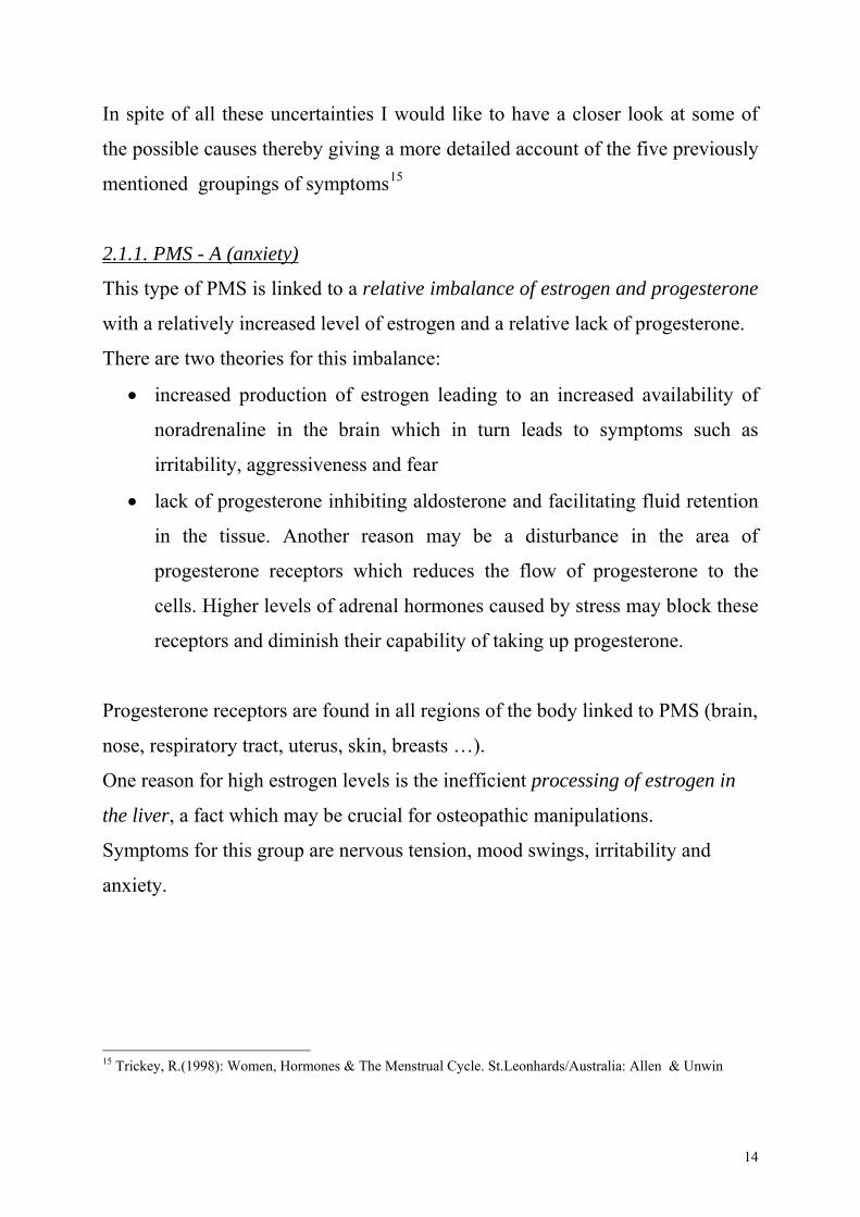
2.3.1. Uterus
The uterus is a hollow, muscular organ situated in the middle of the lesser
pelvis.
Abb. 1: Female pelvic organs: Drenckhahn, D. Zenker, W. (1994): page 124
17 Drenckhahn, D. Zenker, W.(1994): Benninghoff Anatomie Band 2. München: Urban & Schwarzenberg 18 Trickey, R. (1998): Women, Hormones & The Menstrual Cycle. St. Leonhards /Australia: Allen & Unwin 19 Barral, J.P. (1993): Urogenital Manipulation. Seattle: Eastland Press 20 Paoletti, S. (2001): Faszien. München: Urban & Fischer
17
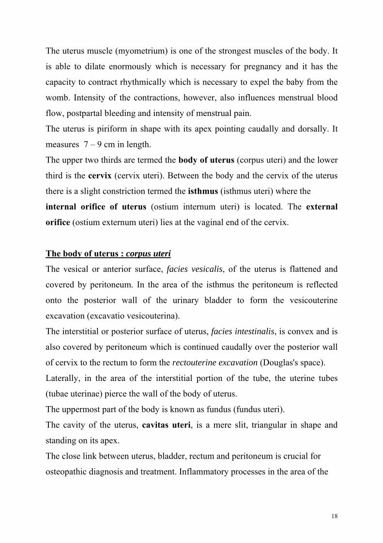
The uterus muscle (myometrium) is one of the strongest muscles of the body. It
is able to dilate enormously which is necessary for pregnancy and it has the
capacity to contract rhythmically which is necessary to expel the baby from the
womb. Intensity of the contractions, however, also influences menstrual blood
flow, postpartal bleeding and intensity of menstrual pain.
The uterus is piriform in shape with its apex pointing caudally and dorsally. It
measures 7 – 9 cm in length.
The upper two thirds are termed the body of uterus (corpus uteri) and the lower
third is the cervix (cervix uteri). Between the body and the cervix of the uterus
there is a slight constriction termed the isthmus (isthmus uteri) where the
internal orifice of uterus (ostium internum uteri) is located. The external
orifice (ostium externum uteri) lies at the vaginal end of the cervix.
The body of uterus : corpus uteri
The vesical or anterior surface, facies vesicalis, of the uterus is flattened and
covered by peritoneum. In the area of the isthmus the peritoneum is reflected
onto the posterior wall of the urinary bladder to form the vesicouterine
excavation (excavatio vesicouterina).
The interstitial or posterior surface of uterus, facies intestinalis, is convex and is
also covered by peritoneum which is continued caudally over the posterior wall
of cervix to the rectum to form the rectouterine excavation (Douglas's space).
Laterally, in the area of the interstitial portion of the tube, the uterine tubes
(tubae uterinae) pierce the wall of the body of uterus.
The uppermost part of the body is known as fundus (fundus uteri).
The cavity of the uterus, cavitas uteri, is a mere slit, triangular in shape and
standing on its apex.
The close link between uterus, bladder, rectum and peritoneum is crucial for
osteopathic diagnosis and treatment. Inflammatory processes in the area of the
18
peritoneum, for example, may result in adhesions restricting uterine mobility.
These in turn may trigger functional disorders including menstrual complaints.
The uterine wall measures 1 – 2 cm in thickness and consists of three layers:
• Endometrium
• Myometrium
• Perimetrium
Abb. 2: Female internal genital organs: Putz, R, Pabst, R. (1993) page 193
Endometrium:
The uterine mucous lining is influenced by sexual hormones and subject to a
cyclical change of its structure.
Myometrium:
Myometrium is approximately 1 cm thick. It consists of a plexus of smooth
muscles and vessels.
Myometrium contracts regularly. Intensity and frequency of contractions depend
on hormones. They are increased by estrogens and inhibited by gestagens.
19
Basic tone of uterus is crucial in causing dysmenorrhea. If tone is normal,
contractions happen at a regular rate; they are neither too strong nor too weak
and there is a recovery phase between contractions. This muscular activity does
not cease, even when the uterus is inactive.
During menstruation or labour uterine activity is heightened, but if the basic
tone is normal, contractions and inactive phases do not exceed a normal amount
of pain.
The inactive phase is very important. In normal cases blood flow through the
myometrium transports oxygen and nutrients, but if the myometrium is not able
to relax sufficiently in the inactive phases, there will not be enough oxygen
which causes pain. Severity of this pain corresponds to pain described by
women suffering from dysmenorrhea.
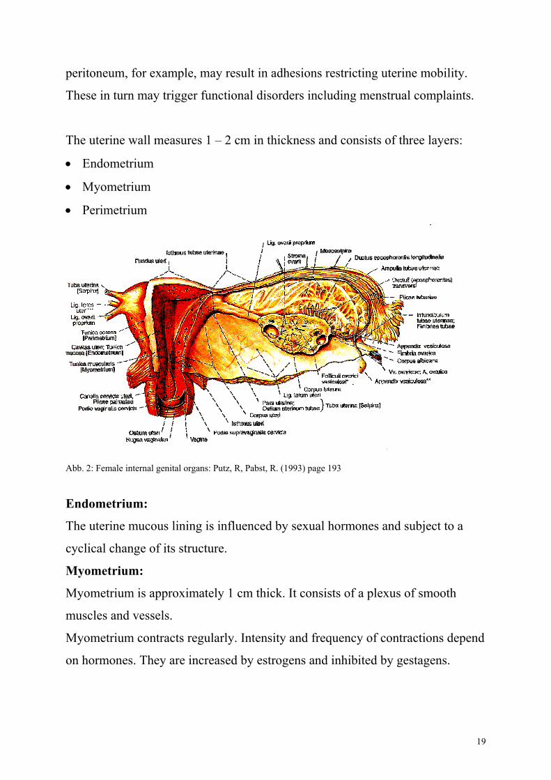
Perimetrium:
Perimetrium consists of a peritoneal coating and a subserous layer.
The smooth surface enables the uterus to shift freely along adjacent structures.
Perimetrium invests the uterus at its anterior, posterior and cranial surface.
Laterally it is replaced by parametrium .
Abb. 3: Uterus and vagina: Putz, R. Pabst, R. (1993) page 194
20
Suspending and supporting structures of uterus :
There are a number of ligaments and other types of connective tissue which
fasten or support the uterus.
Peritoneum:
The peritoneum plays a minor role in suspension but peritoneal restrictions may
disrupt uterine mobility.
Lig. teres uteri:
The round ligament of uterus (lig.teres uteri / rotundum) passes on either side of
the lateral angle of the uterine tubes (cornua uteri) through the abdominal
inguinal ring and along the inguinal canal to the greater pudendal labia. It
anteverts and stabilises the uterus to a small extent.
Lig. latum:
The broad ligament (Lig. Latum) connects the uterus to the lateral walls of the
pelvis. The posterior part is in relation with the small intestine. The two broad
ligaments form a suspension sling for the uterus. Around the cervix the broad
ligament becomes larger and stronger, it is now called parametrium.
The broad ligament contains all the vessels leading to the uterus (arteries, veins,
venous plexus, lymphatics) nerves as well as the ureter.
The broad ligament forms the transverse support for the uterus and a cross-
shaped structure with the lamina sacro-recto-genito-pubicalis.
21
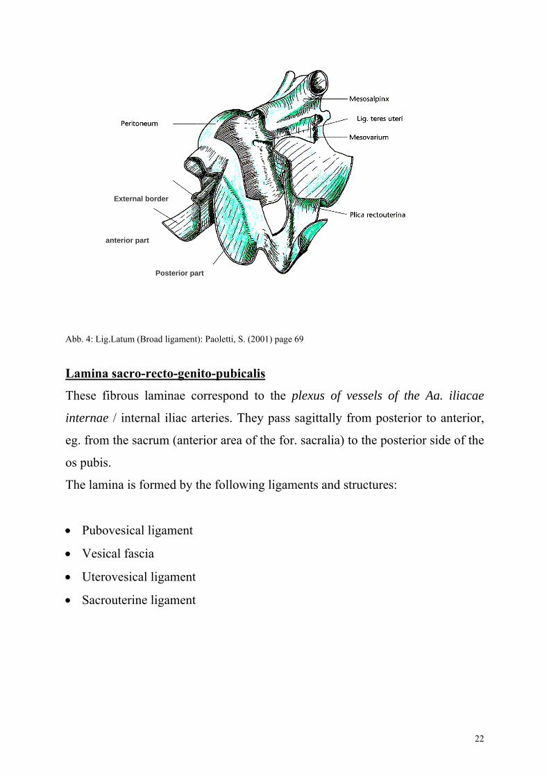
External border
anterior part
Posterior part
Abb. 4: Lig.Latum (Broad ligament): Paoletti, S. (2001) page 69 Lamina sacro-recto-genito-pubicalis
These fibrous laminae correspond to the plexus of vessels of the Aa. iliacae
internae / internal iliac arteries. They pass sagittally from posterior to anterior,
eg. from the sacrum (anterior area of the for. sacralia) to the posterior side of the
os pubis.
The lamina is formed by the following ligaments and structures:
• Pubovesical ligament
• Vesical fascia
• Uterovesical ligament
• Sacrouterine ligament
22
Sacrum
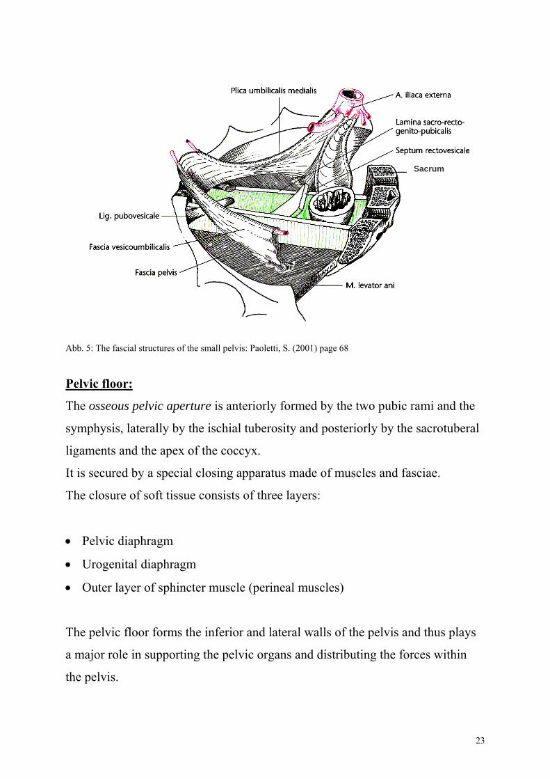
Abb. 5: The fascial structures of the small pelvis: Paoletti, S. (2001) page 68
Pelvic floor:
The osseous pelvic aperture is anteriorly formed by the two pubic rami and the
symphysis, laterally by the ischial tuberosity and posteriorly by the sacrotuberal
ligaments and the apex of the coccyx.
It is secured by a special closing apparatus made of muscles and fasciae.
The closure of soft tissue consists of three layers:
• Pelvic diaphragm
• Urogenital diaphragm
• Outer layer of sphincter muscle (perineal muscles)
The pelvic floor forms the inferior and lateral walls of the pelvis and thus plays
a major role in supporting the pelvic organs and distributing the forces within
the pelvis.
23
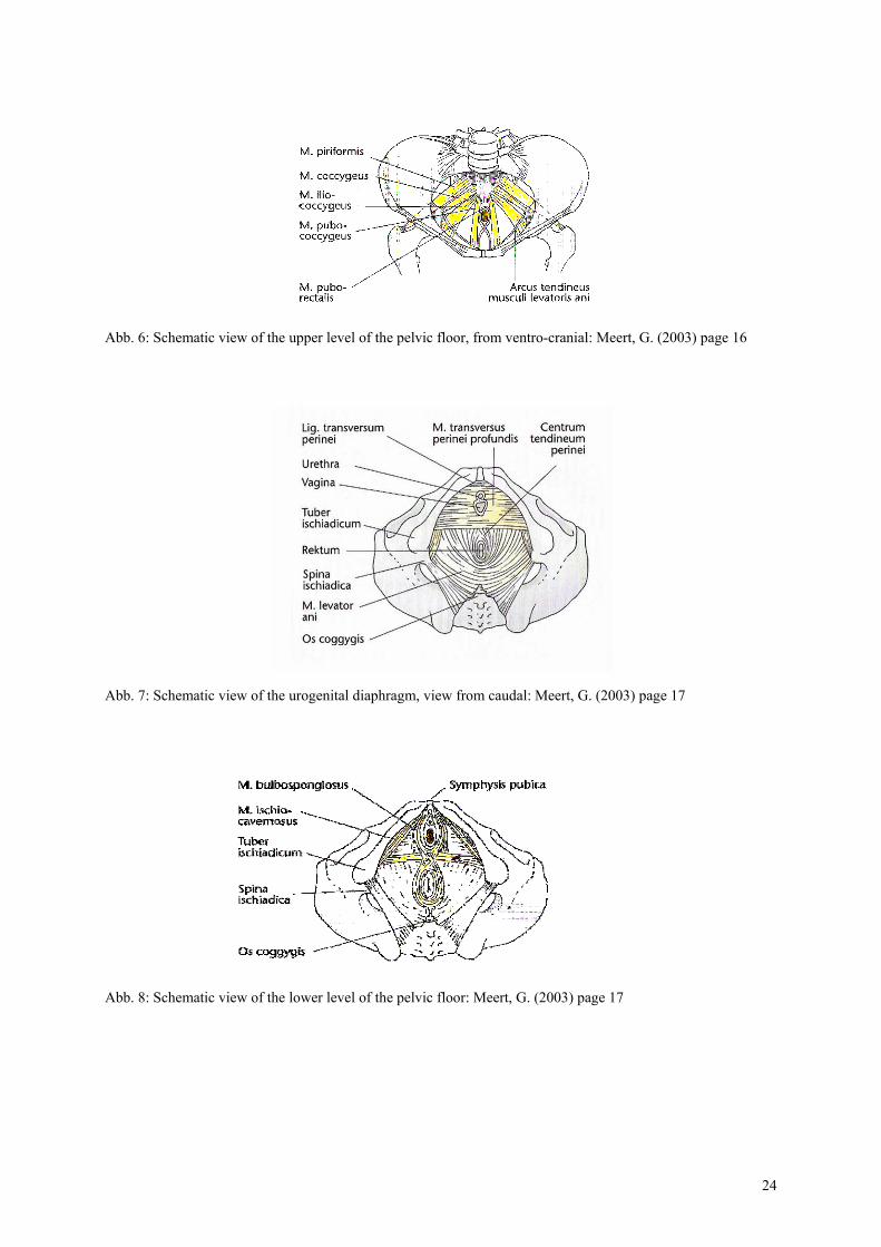
Abb. 6: Schematic view of the upper level of the pelvic floor, from ventro-cranial: Meert, G. (2003) page 16
Abb. 7: Schematic view of the urogenital diaphragm, view from caudal: Meert, G. (2003) page 17
Abb. 8: Schematic view of the lower level of the pelvic floor: Meert, G. (2003) page 17
24
Cervix: cervix uteri
The cervix is the lower, constricted segment of the uterus. The apex projects
freely downward into the vagina.
The cervix serves as a closing mechanism which, on the one hand, prevents
ascending bacilli from reaching the uterus and on the other hand, prevents
spontaneous abortion and miscarriage.
Moreover, the cervix is the central point of attachment for the supportive
structure of the inner genitalia.
2.3.2. Uterine tubes and ovaries:
Uterine Tube: tuba uterina
At the ovary the egg cell (ovum) is drawn in the infundibulum (infundibulm
tubae uterinae) of the uterine tubes from where it is conveyed to the uterus.
The uterine tube is suspended between the superior angle of the uterus and the
ovary or rather the suspensory ligament of ovary. Additionally, it is attached to
the broad ligament which, in that part, is termed the mesosalpinx.
The ovaries are muscular and contract rhythmically in order to transport the
ovum to the uterus.
Ovary : Ovar
The ovary is the “female gonad”. It contains egg cells which are released at
certain times as fertile ova.
The ovary is also an endocrine gland producing sexual steroids such as estrogen
and progesterone in the first place but other hormones as well.
The ovary controls the cycle and influences the whole body of a woman,
particularly through estrogen. Its superimposed system is the hypophysis but the
autonomic nervous system exerts some influence on the ovary as well.
25
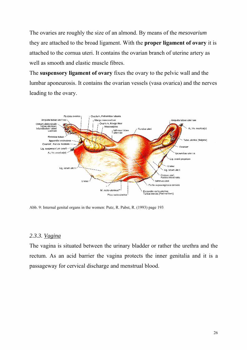
The ovaries are roughly the size of an almond. By means of the mesovarium
they are attached to the broad ligament. With the proper ligament of ovary it is
attached to the cornua uteri. It contains the ovarian branch of uterine artery as
well as smooth and elastic muscle fibres.
The suspensory ligament of ovary fixes the ovary to the pelvic wall and the
lumbar aponeurosis. It contains the ovarian vessels (vasa ovarica) and the nerves
leading to the ovary.
Abb. 9: Internal genital organs in the women: Putz, R. Pabst, R. (1993) page 193
2.3.3. Vagina
The vagina is situated between the urinary bladder or rather the urethra and the
rectum. As an acid barrier the vagina protects the inner genitalia and it is a
passageway for cervical discharge and menstrual blood.
26
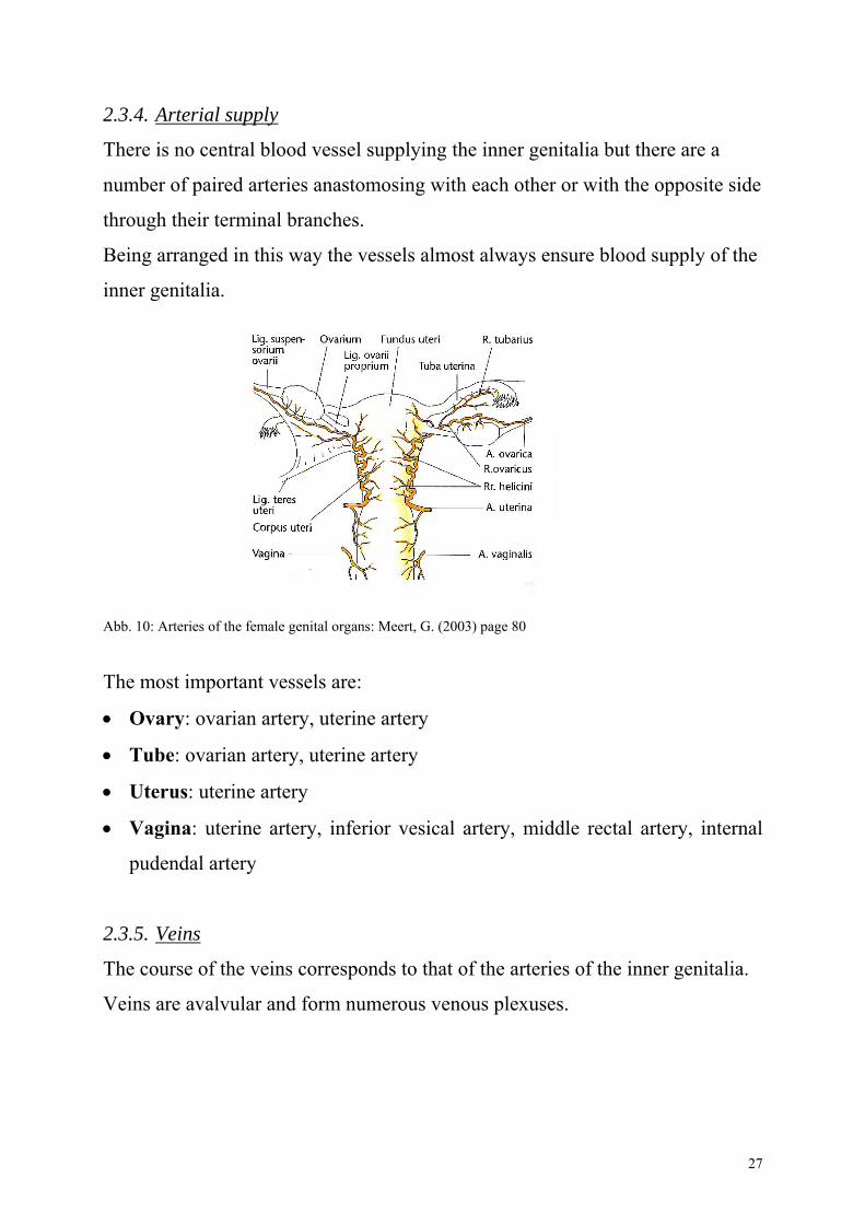
2.3.4. Arterial supply
There is no central blood vessel supplying the inner genitalia but there are a
number of paired arteries anastomosing with each other or with the opposite side
through their terminal branches.
Being arranged in this way the vessels almost always ensure blood supply of the
inner genitalia.
Abb. 10: Arteries of the female genital organs: Meert, G. (2003) page 80
The most important vessels are:
• Ovary: ovarian artery, uterine artery
• Tube: ovarian artery, uterine artery
• Uterus: uterine artery
• Vagina: uterine artery, inferior vesical artery, middle rectal artery, internal
pudendal artery
2.3.5. Veins
The course of the veins corresponds to that of the arteries of the inner genitalia.
Veins are avalvular and form numerous venous plexuses.
27
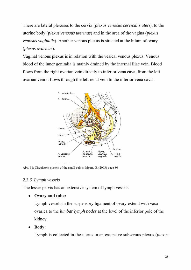
There are lateral plexuses to the cervix (plexus venosus cervicalis uteri), to the
uterine body (plexus venosus uterinus) and in the area of the vagina (plexus
venosus vaginalis). Another venous plexus is situated at the hilum of ovary
(plexus ovaricus).
Vaginal venous plexus is in relation with the vesical venous plexus. Venous
blood of the inner genitalia is mainly drained by the internal iliac vein. Blood
flows from the right ovarian vein directly to inferior vena cava, from the left
ovarian vein it flows through the left renal vein to the inferior vena cava.
Abb. 11: Circulatory system of the small pelvis: Meert, G. (2003) page 80
2.3.6. Lymph vessels
The lesser pelvis has an extensive system of lymph vessels.
• Ovary and tube:
Lymph vessels in the suspensory ligament of ovary extend with vasa
ovarica to the lumbar lymph nodes at the level of the inferior pole of the
kidney.
• Body:
Lymph is collected in the uterus in an extensive subserous plexus (plexus
28
lymphaticus uteri, plexus lymphaticus cervicis uteri). The main drainage
of the corpus to the vasa ovarica and the lumbar lymph nodes is done via
the superior lymphatic vessels. Lymphatic vessels of the anterior side of
the uterus pass from the interstitial portion of the tube via round uterine
ligament to the superior inguinal lymph nodes. The lateral lymph vessels
drain into external iliac lymph nodes.
• Cervix:
Lymph flows to the external iliac lymph nodes or the lymph nodes of the
fossa obturatoria via the broad ligament.
• Vagina:
Lymph flows to external iliac lymph nodes and the superficial inguinal
lymph nodes.
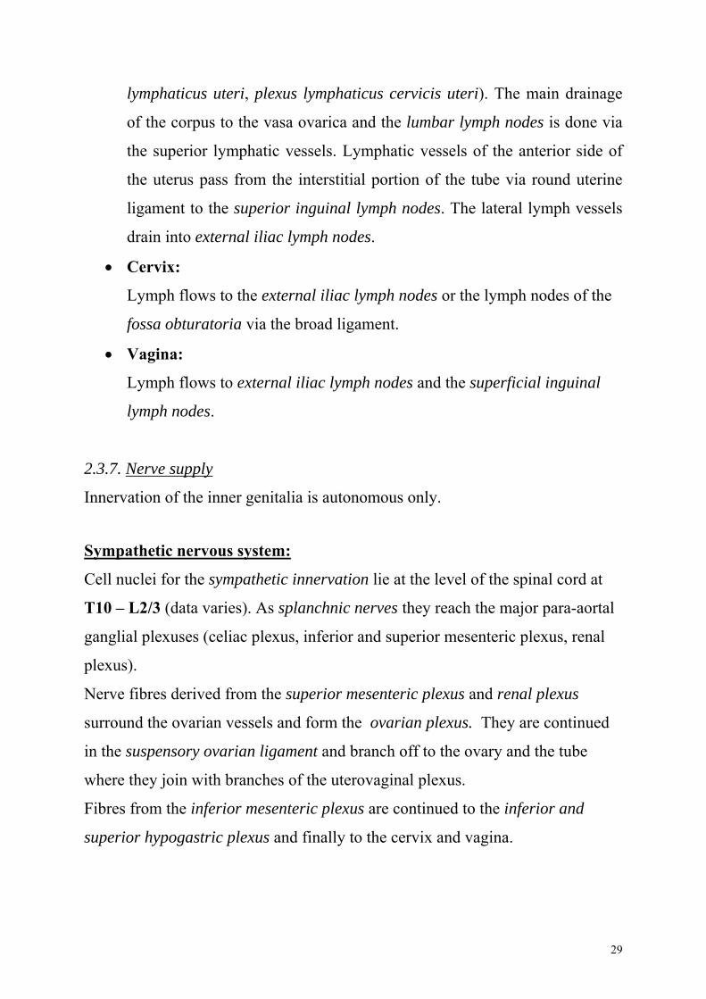
2.3.7. Nerve supply
Innervation of the inner genitalia is autonomous only.
Sympathetic nervous system:
Cell nuclei for the sympathetic innervation lie at the level of the spinal cord at
T10 – L2/3 (data varies). As splanchnic nerves they reach the major para-aortal
ganglial plexuses (celiac plexus, inferior and superior mesenteric plexus, renal
plexus).
Nerve fibres derived from the superior mesenteric plexus and renal plexus
surround the ovarian vessels and form the ovarian plexus. They are continued
in the suspensory ovarian ligament and branch off to the ovary and the tube
where they join with branches of the uterovaginal plexus.
Fibres from the inferior mesenteric plexus are continued to the inferior and
superior hypogastric plexus and finally to the cervix and vagina.
29
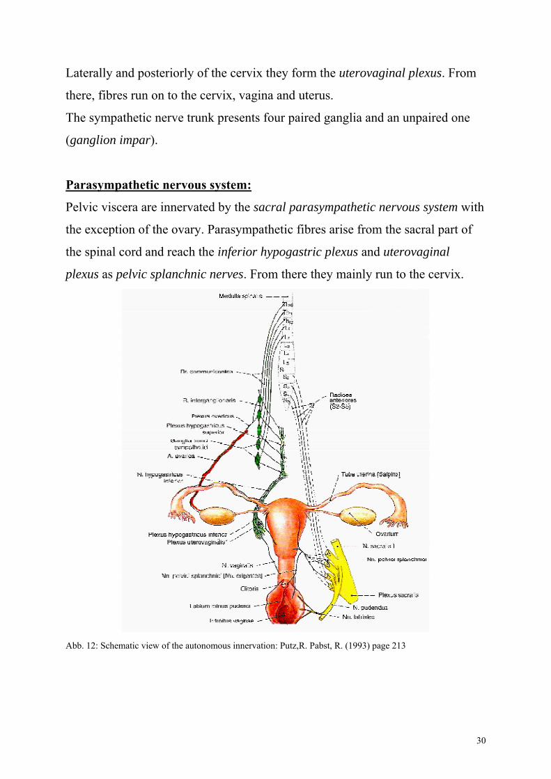
Laterally and posteriorly of the cervix they form the uterovaginal plexus. From
there, fibres run on to the cervix, vagina and uterus.
The sympathetic nerve trunk presents four paired ganglia and an unpaired one
(ganglion impar).
Parasympathetic nervous system:
Pelvic viscera are innervated by the sacral parasympathetic nervous system with
the exception of the ovary. Parasympathetic fibres arise from the sacral part of
the spinal cord and reach the inferior hypogastric plexus and uterovaginal
plexus as pelvic splanchnic nerves. From there they mainly run to the cervix.
Abb. 12: Schematic view of the autonomous innervation: Putz,R. Pabst, R. (1993) page 213
30
Afferent sensory nerve supplySensory nervous fibres join the autonomic nervous fibres. Sensory fibres from
ovary, tube, fundus and corpus reach the spinal cord at level T10 – L1 together
with fibres of the ovarian plexus.
Afferent fibres from the cervix and particularly from the orifice of uterus run
along the hypogastric and uterovaginal plexuses to T11 – T12.
There are neural relations between the pelvic organs leading to a mutual
influence of intestines, uterus, kidney and urinary bladder.
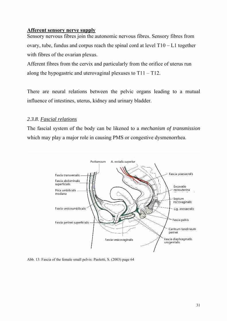
2.3.8. Fascial relations
The fascial system of the body can be likened to a mechanism of transmission
which may play a major role in causing PMS or congestive dysmenorrhea.
Abb. 13: Fascia of the female small pelvis: Paoletti, S. (2003) page 64
31
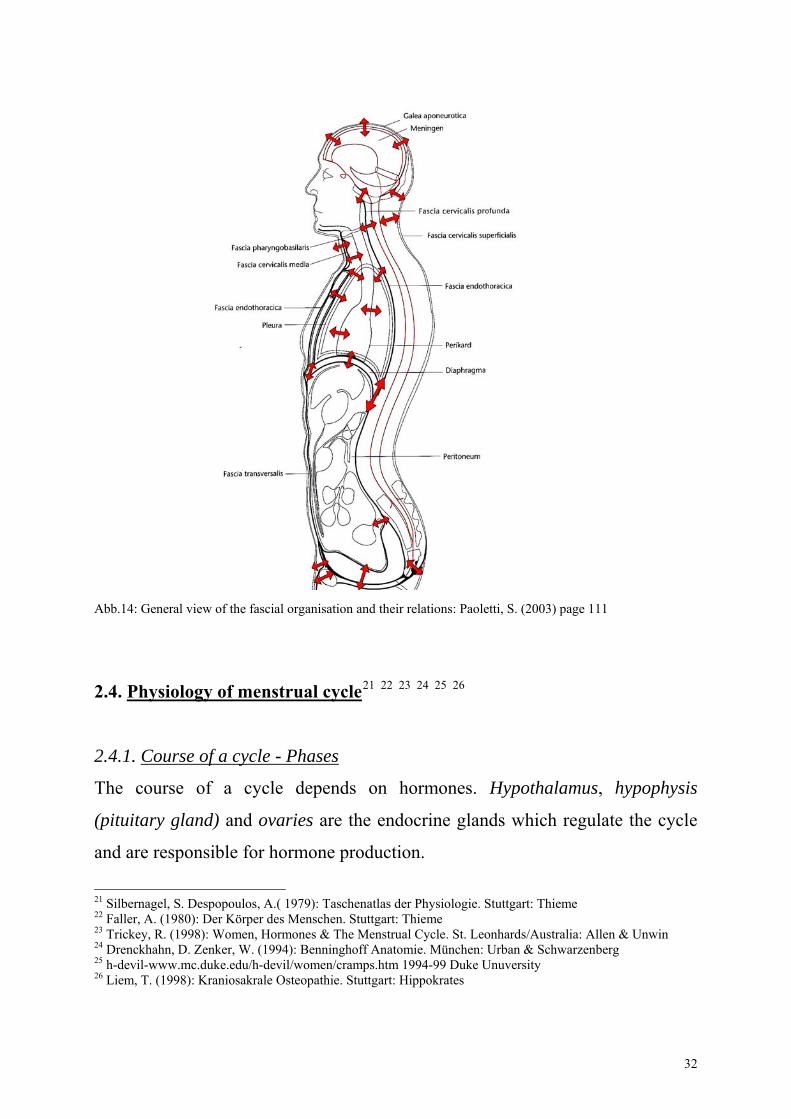
Abb.14: General view of the fascial organisation and their relations: Paoletti, S. (2003) page 111
2.4. Physiology of menstrual cycle21 22 23 24 25 26
2.4.1. Course of a cycle - Phases
The course of a cycle depends on hormones. Hypothalamus, hypophysis
(pituitary gland) and ovaries are the endocrine glands which regulate the cycle
and are responsible for hormone production.
21 Silbernagel, S. Despopoulos, A.( 1979): Taschenatlas der Physiologie. Stuttgart: Thieme 22 Faller, A. (1980): Der Körper des Menschen. Stuttgart: Thieme 23 Trickey, R. (1998): Women, Hormones & The Menstrual Cycle. St. Leonhards/Australia: Allen & Unwin 24 Drenckhahn, D. Zenker, W. (1994): Benninghoff Anatomie. München: Urban & Schwarzenberg 25 h-devil-www.mc.duke.edu/h-devil/women/cramps.htm 1994-99 Duke Unuversity 26 Liem, T. (1998): Kraniosakrale Osteopathie. Stuttgart: Hippokrates
32
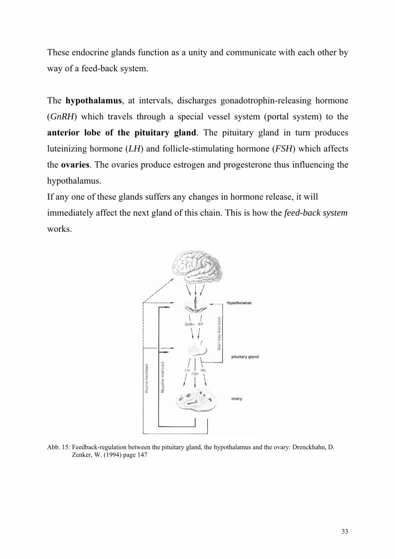
These endocrine glands function as a unity and communicate with each other by
way of a feed-back system.
The hypothalamus, at intervals, discharges gonadotrophin-releasing hormone
(GnRH) which travels through a special vessel system (portal system) to the
anterior lobe of the pituitary gland. The pituitary gland in turn produces
luteinizing hormone (LH) and follicle-stimulating hormone (FSH) which affects
the ovaries. The ovaries produce estrogen and progesterone thus influencing the
hypothalamus.
If any one of these glands suffers any changes in hormone release, it will
immediately affect the next gland of this chain. This is how the feed-back system
works.
pituitary gland
ovary
Abb. 15: Feedback-regulation between the pituitary gland, the hypothalamus and the ovary: Drenckhahn, D. Zenker, W. (1994) page 147
33
First let me describe the five phases of the menstrual cycle: follicular phase,
proliferative phase, luteal phase, ischemic phase and desquamative phase.
2.4.1.1. Follicular phase:
In the course of the cycle estrogen levels decline and the hypothalamus releases
GnRH. This signals to the pituitary gland to release FSH in order to stimulate
follicle growth.
10 – 20 tertiary follicles grow but one of them only matures enough to become
an ovum. The remaining follicles degenerate and at the time of ovulation one
ovum is ready to be released.
2.4.1.2. Proliferative phase: (part of the follicular phase)
During maturation of the follicle more estrogen is produced. The endometrium
is caused to grow and thicken. LH levels increase considerably for 16 – 24 hours
before ovulation which in the end triggers ovulation.
FSH levels drop dramatically whereas LH levels decline slowly.
2.4.1.3. Luteal phase: Secretory phase
This phase starts after ovulation has occurred and the follicular epithelium left in
the ovary develops into the corpus luteum.
Characteristic changes take place in the glands of the uterine mucous membrane
(endometrium). These glands and their blood vessels now present a contorted or
waved appearance and begin secretion. Increasing progesterone secretion of the
corpus luteum causes the endometrium to stop proliferation. Progesterone
stimulates secretion of the endometrial glands which proliferated under the
influence of estrogen. After an initial drop corpus luteum produces steady
amounts of estrogen.
34
LH keeps up normal functioning of corpus luteum but LH production is
constantly declining while progesterone levels are rising. If fertilisation does not
occur, corpus luteum degenerates after 8 – 10 days after ovulation which is most
probably due to the influence of prostaglandin.
Corpus luteum starts to degenerate after two weeks of producing estrogen and
progesterone and in this way hormone production declines.
2.4.1.4. Ischemic phase: part of luteal phase
Shedding of endometrium is believed to have several reasons. Firstly, declining
estrogen and progesterone levels are held responsible, and secondly,
prostaglandin seems to play a major role.
Prostaglandin level rises during the secretory phase which causes uterine
contractions to intensify. Spiral arteries which ensure blood supply to the
endometrium react to prostaglandin. They begin to contract rhythmically which
results in a heavily reduced blood supply (ischemia).
The endometrium degenerates.
2.4.1.5. Desquamative phase: early follicular phase
Relaxation of spiral arteries allows blood to flow back into the ischemic area.
Capillary walls burst and endometrial cells fall apart. These, together with parts
of spiral arteries, are shed as menstrual blood.
Uterine contractions facilitate shedding of endometrium through the cervix.
At the same time the estrogen level is so low that hypothalamus secretes GnRH
and the cycle resumes its course.
2.4.2. The role of prostaglandin in causing menstrual pain
Here, I would like to go into more detail about the mechanism causing menstrual
pain.
35
How does prostaglandin cause menstrual pain?
Prostaglandin is a hormone-like substance present in almost all the body cells.
It regulates muscle tone of smooth muscles for example in blood vessels, uterus
and intestines. If prostaglandin increases excessively, smooth muscles react by a
heightening muscle tone and contractions.
Prostaglandin level is very high before menstruation and is at its peak when
bleeding sets in.
Thus uterine contractions increase and women feel pain and cramps.
The uterus contracts so heavily that its blood vessels are compressed and blood
supply to myometrium is strongly reduced.
Vigorous blood flow makes for a sufficient supply of oxygen and nutrients – bad
circulation results in lack of oxygen and thus leads to pain.
It is also conceivable that the increased prostaglandin concentration goes into
blood circulation and influences smooth muscles of other organs or
prostaglandin receptors which are present in the whole body. In this way an
increased prostaglandin level may also be related to symptoms accompanying
menstrual pain such as headache, dizziness, hot flashes, diarrhea and nausea.
2.5. Hormones and the liver
In the liver estrogen is turned into different, less active forms and released into
the small intestine via bile.
One part is excreted with the stool, another part is bound to special enzymes
(beta-glucuronidase). These enzymes are able to turn estrogen back into a more
active form. In this process one part is excreted with the stool and one part is
reabsorbed into the blood stream. This process is termed entero-hepatic
circulation.
36
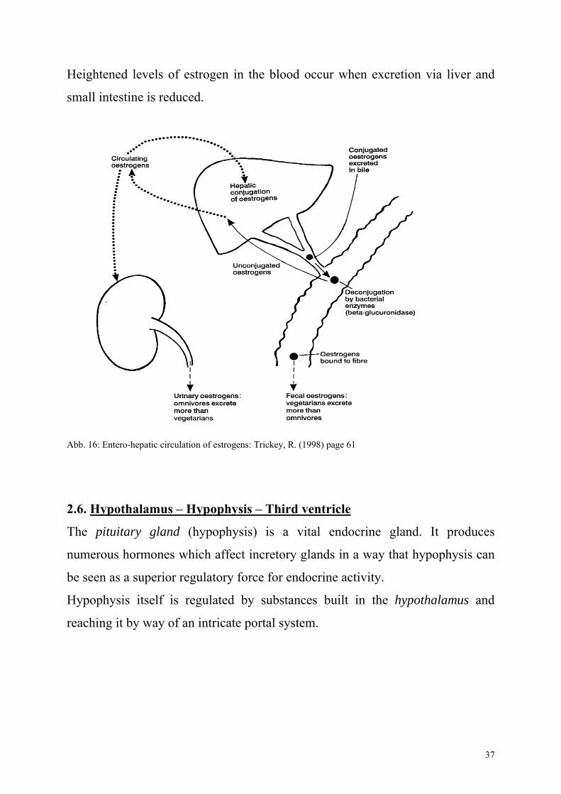
Heightened levels of estrogen in the blood occur when excretion via liver and
small intestine is reduced.
Abb. 16: Entero-hepatic circulation of estrogens: Trickey, R. (1998) page 61
2.6. Hypothalamus – Hypophysis – Third ventricle
The pituitary gland (hypophysis) is a vital endocrine gland. It produces
numerous hormones which affect incretory glands in a way that hypophysis can
be seen as a superior regulatory force for endocrine activity.
Hypophysis itself is regulated by substances built in the hypothalamus and
reaching it by way of an intricate portal system.
37
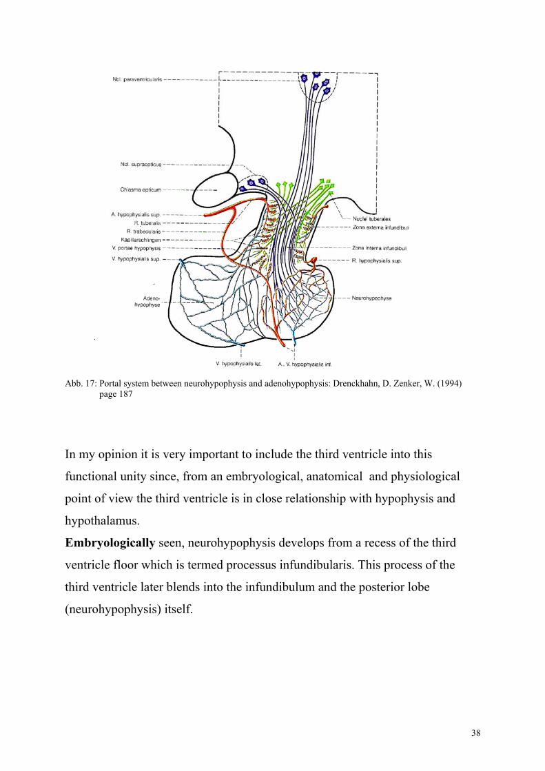
Abb. 17: Portal system between neurohypophysis and adenohypophysis: Drenckhahn, D. Zenker, W. (1994) page 187
In my opinion it is very important to include the third ventricle into this
functional unity since, from an embryological, anatomical and physiological
point of view the third ventricle is in close relationship with hypophysis and
hypothalamus.
Embryologically seen, neurohypophysis develops from a recess of the third
ventricle floor which is termed processus infundibularis. This process of the
third ventricle later blends into the infundibulum and the posterior lobe
(neurohypophysis) itself.
38
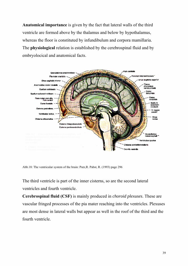
Anatomical importance is given by the fact that lateral walls of the third
ventricle are formed above by the thalamus and below by hypothalamus,
whereas the floor is constituted by infundibulum and corpora mamillaria.
The physiological relation is established by the cerebrospinal fluid and by
embryolocical and anatomical facts.
Abb.18: The ventricular system of the brain: Putz,R. Pabst, R. (1993) page 296
The third ventricle is part of the inner cisterns, so are the second lateral
ventricles and fourth ventricle.
Cerebrospinal fluid (CSF) is mainly produced in choroid plexuses. These are
vascular fringed processes of the pia mater reaching into the ventricles. Plexuses
are most dense in lateral walls but appear as well in the roof of the third and the
fourth ventricle.
39
CSF is also built in capillaries of the subarachnoidal space in the area of the
cranium or the spinal cord but to a much lesser extent.
CSF circulates from lateral ventricles through the interventricular foramen
(Monro's foramen) to the third ventricle and from there to the fourth ventricle by
way of the cerebral aqueduct (aquaeductus cerebri).
At the fourth ventricle CSF flows through lateral and medial apertures into the
outer cistern and subarachnoidal space of spine and skull.
Arachnoidal villi reabsorb CSF which then flows into the intracranial sinus.
Good circulation of CSF is imperative for the many functions it has to fulfil.
Transporting hypothalamic and neurohypophysial substances is but one of
them.
40
3. Considerations and approach to osteopathic treatment
PMS and dysmenorrhea have been of central interest lately. There are numerous
approaches, theories and pearls of wisdom, nevertheless, a lot of insecurity
remains.
Each person should be seen as an individual. Thus, very likely, every woman
has a history of her own as to how her menstrual problems emerged.
Based on this individuality manipulations should be "customised" to each
woman.
There are, however, certain regions of the body which deserve being looked at
more closely.
At first, it is necessary to get a general overlook of the posture followed by a
more detailed examination of certain regions in the parietal system, visceral
system and craniosacral system.
In doing so, I would like to take into account the various physiological and
anatomical aspects which bear considerable weight in context with PMS and
dysmenorrhea.
3.1. Posture:27 28
A good posture is essential for the proper functioning of the body.
A body functions well, if mechanic, visceral and craniosacral mobility is not
restricted.
We can define different posture patterns which are used in order to compensate
gravity. It is important to analyse them, since they may explain certain
compensatory factors and the presence of tension centres which affect the body.
27 Mitchell, F.L.(1995): The Muscle Energy Manual Volume one. Michigan: Met press 28 Richard, J.P. (1994): Die Wirbelsäule aus der Sicht der Osteopathie. Kötzing: Verlag für Osteopathie
41
On the frontal plane (seen from the front) relative symmetry of the body is a
factor of equilibrium. On the sagittal plane (seen from the side) an asymmetric
order of structures tends to result in imbalance.
This imbalance invariably establishes either a balance pattern or one of the
functional patterns (anterior or posterior).
3.1.1. Basic postural pattern:
The basic postural patterns are the pattern of equilibrium, the anterior pattern
and the posterior pattern.
3.1.1.1 Pattern of equilibrium:
There are two factors which cause an imbalance of the body:
• The first factor is the Transmission of body weight via sacrum onto the
pelvis is done behind the point the hip joint rests upon.
Thus the pelvis gets into retroversion, the hip into extension and the knee
into flexion.
Body weight on the pelvis can only be kept in balance by flexing hip
flexor muscles and knee extensor muscles. Considering the fact that the
whole lower extremity forms one unity this means that the foot has to be
an integral part of the balance pattern.
• The second factor is the Position of the gravitational centre of the head
which causes its flexion. Balance of the head is maintained by constant
activity of dorsal neck muscles. Cervical lordosis is then physiologically
more pronounced.
This tonic postural activity is intermittent and does neither induce muscles to tire
nor nerves to overstrain.
42
The body as a whole gives an impression of stability and unconstraint;
abdominal and thoracic tension is in balance.
The gravitational line which is an indicator for the postural pattern runs as
follows, with the patient seen from the side:
It starts trough the external auditory canal and continues to the acromial bone,
the body of the third lumbar vertebra, the greater trochanter and passes slightly
in front of the lateral ankle.
3.1.1.2. Anterior pattern:
This is the most common postural pattern. The gravitational line runs down in
front of the third lumbar vertebra, the hip joints, transverse axis of the knee joint
and towards the forefoot.
The body is ventrally off balance and reacts with hyperextension of the knee
joint. Body weight is on the area of the forefoot, hip is bent, pelvis inclined
forwards. Curvature of cervical spine and lumbar spine is increased.
Heavy tension rests on the lumbosacral junction, the iliosacral joints and
T11/12.
Between thorax and abdominal cavity there is an imbalance of pressure.
Diaphragm is low and abdominal organs show a disposition to ptosis.
Posterior myofascial muscle chains keep this postural pattern up.
3.1.1.3. Posterior pattern:
The gravitational line runs behind its physiological points of orientation which
means behind the hip and the knees and reaches is lowest point at the heel.
The body is dorsally off balance.
Flexion of the knee, extension of the hip and the pelvis inclining backwards are
the results of this posture.
43
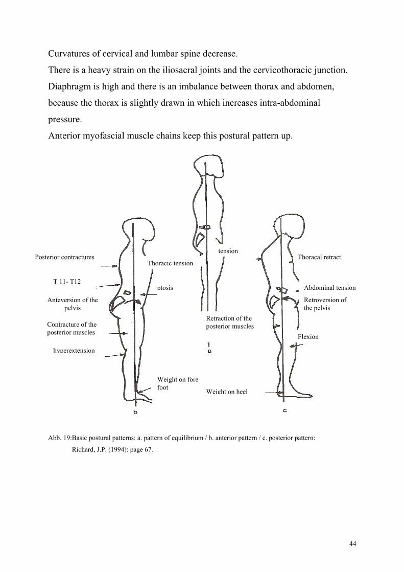
Curvatures of cervical and lumbar spine decrease.
There is a heavy strain on the iliosacral joints and the cervicothoracic junction.
Diaphragm is high and there is an imbalance between thorax and abdomen,
because the thorax is slightly drawn in which increases intra-abdominal
pressure.
Anterior myofascial muscle chains keep this postural pattern up.
tensionPosterior contractures Thoracal retract
Thoracic tension
T 11- T12 ptosis
Retroversion of the pelvis
Abdominal tension
Anteversion of the pelvis
Contracture of the posterior muscles
hyperextension
Retraction of the posterior muscles
Flexion
Weight on fore foot Weight on heel
Abb. 19:Basic postural patterns: a. pattern of equilibrium / b. anterior pattern / c. posterior pattern:
Richard, J.P. (1994): page 67.
44

How can these physiological facts be linked to PMS and dysmenorrhea?
Osteopathic manipulations can only be applied, if the mechanisms causing a
certain postural pattern have been identified.
Causes may be found both in the parietal and the craniosacral system as well as
in the organs.
In the following chapters I will go into more detail about the precise links.
3.2. Possible causes in the parietal system29 30
It is very important to look at the parietal system as a lot of mobility problems
start in this area.
The possible causes in the parietal system (musculoskeletal) are the pelvis,
special spinal areas and the lower extremities.
3.2.1. Pelvis:
Due to its close contact to the inner genitalia the pelvis has an important
function.
Mobility restrictions affect the organs via pelvic floor, on the one hand, and via
ligaments and fasciae on the other hand.
Moreover, the close relationship to vessels and nerves play a major part.
Pelvic floor muscles and the connective tissue between the osseous structures
build a strong link between the pubic symphysis, pelvic, sacral and coccygeal
bones.
This means that each change in position causes a change in tension of the pelvic
floor which may prove to be hypotone or hypertone.
29. Lason, G. Peeters, L.(1993): Handbuch für Osteopathie: Das Becken OSTEO 2000 b.v.b.a. 30 Barral, J.P.(1993) : Urogenital Manipulation. Seattle: Eastland Press
45
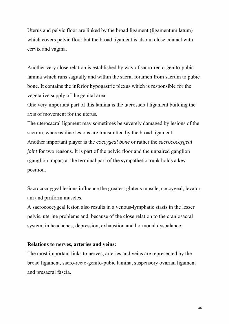
Uterus and pelvic floor are linked by the broad ligament (ligamentum latum)
which covers pelvic floor but the broad ligament is also in close contact with
cervix and vagina.
Another very close relation is established by way of sacro-recto-genito-pubic
lamina which runs sagitally and within the sacral foramen from sacrum to pubic
bone. It contains the inferior hypogastric plexus which is responsible for the
vegetative supply of the genital area.
One very important part of this lamina is the uterosacral ligament building the
axis of movement for the uterus.
The uterosacral ligament may sometimes be severely damaged by lesions of the
sacrum, whereas iliac lesions are transmitted by the broad ligament.
Another important player is the coccygeal bone or rather the sacrococcygeal
joint for two reasons. It is part of the pelvic floor and the unpaired ganglion
(ganglion impar) at the terminal part of the sympathetic trunk holds a key
position.
Sacrococcygeal lesions influence the greatest gluteus muscle, coccygeal, levator
ani and piriform muscles.
A sacrococcygeal lesion also results in a venous-lymphatic stasis in the lesser
pelvis, uterine problems and, because of the close relation to the craniosacral
system, in headaches, depression, exhaustion and hormonal dysbalance.
Relations to nerves, arteries and veins:
The most important links to nerves, arteries and veins are represented by the
broad ligament, sacro-recto-genito-pubic lamina, suspensory ovarian ligament
and presacral fascia.
46
The broad ligament contains all the vessels and nerves leading to or from the
uterus, thus we can safely conclude that changes in tension affect these
structures.
Structural changes influencing sacro-recto-genito-pubic lamina may also impair
the inferior hypogastric plexus and the ovarian artery.
Lesions of the sacrum may influence vegetative supply due to the close relation
of presacral fascia to the sacral plexus.
The ovarian suspensory ligament contains vessels and nerves which supply the
ovary.
There is a close link to the broad ligament through the mesovarium which
influences the ovary and also a close link to the lumbar aponeurosis.
This is how we can establish a link to the hormonal system which plays a major
role in causing PMS. Barral31 (1993) writes: "It seems that all local tubo-
ovarian manipulations have a general effect on the body". He believes that
local manipulations affect hypothalamus and hypophysis.
So there is according to Barral (1993) a close connection to hormonal regulation
which is crucial in the mechanisms causing PMS.
And we can safely assume that changes of tension in the pelvic area provoke
changes of uterus tone and thus promote congestive dysmenorrhea.
3.2.2. Spinal regions deserving special attention32
Cellular nuclei for the sympathetic nerve supply of inner genitalia lie at level
T10 – L3. For the sacral parasympathetic nervous system they lie in the second
to fourth sacral spinal segment and reach down to S5 and in the first coccygeal
segment.
31 Barral J.P.(1993): Urogenital Manipulation. Seattle: Eastland Press 1993 page 219 32 Barral J.P.(1993): Urogenital Manipulation. Seattle: Eastland Press 1993
47
From experience we know that lesions of the uterus are often linked to
restrictions in the area of T12 and L1, L5/sacrum and sacrococcygeal
restrictions, whereas spinal restrictions at T11, L1, L3 and sacrum affect ovary
and tube.
Lesions in the lower thoracic spine and lumbar spine usually lead to
vasodilation in the lesser pelvis.
Restrictions in the thoracolumbar junction may influence the genital region
not only via vegetative nervous system but also because of its close link of the
diaphragm in this area.
Thus, a change of tension in the diaphragm may also result in a change of
pressure balance between thorax and abdomen.
If diaphragm is low, abdominal organs show a disposition to ptosis which in turn
exerts more pressure on the organs of the lesser pelvis.
If diaphragm is high, thorax is drawn in, intra-abdominal pressure increases and
pressure balance between abdomen and lesser pelvis is upset.
The diaphragm additionally plays a key role in proper arterial and venous
function and may, due to its close relation to organs, damage their function.
Liver, kidney, small intestine and colon are of major importance for PMS and
dysmenorrhea.
The diaphragm is also responsible for the mobility of pelvic organs since they
move synchronically along with inhalation and exhalation.
Restrictions of the middle and upper thoracic spine may be significant in the
clinical picture of PMS and dysmenorrhea because of the relation to the
autonomic nervous system and because they may induce changes of the
intrathoracic pressure and postural changes.
Pericardium presents a close link between thorax and the fasciae of the middle
body axis.
48
This chain of fasciae continues upwards and connects to the cranial base which,
in turn, is closely linked to the hormonal system.
Fascia of the middle body axis is also connected to the meninx (via cerebral
nerves), to deep and middle cervical fasciae and consequently to thyroid, thymic
space, pleura and endothoracic fascia as well as to the diaphragm.
Firstly, the upper cervical spine is connected to the sacrum and coccyx by the
dura mater and lesions in this area may, via dura mater, affect high cervical
lesions.
Secondly, the upper cervical spine is connected to the cranial base and the
hypothalamus - hypophysis system. Nevertheless, cranial restrictions are
invariably reflected at this level.
The upper cervical spine has a very important function as a compensatory
mechanism for ascending lesion chains because this area ensures that
horizontality of the eyes is maintained.
Restrictions at level C3/4/5 may be linked to the diaphragm and the peritoneum
via phrenic nerve. The close relationship of diaphragm and peritoneum to the
uterus has already been mentioned.
All these examples contribute to emphasise the significance of the upper
extremities.
3.2.3. Lower extremities
The lower extremities, the foot in particular, are a major point of departure for
ascending lesion chains.
In general, any disturbance in mobility of the lower limbs may be transmitted to
the pelvis or the whole body via fasciae. According to Jean Pierre Barral the
most commonly affected are navicular bone, proximal and distal tibiofibular
joints.
49
Fasciae of the lower limbs are associated with the thoracic and abdominal
fasciae by gluteal fascia which goes over into thoracolumbar fascia.
And by piriform and obturator internus muscles there is a close relation to the
superficial perineal and pelvic fasciae.
It is continued over psoas muscle to iliac fascia and in the area of the inguinal
ligament to abdominal and transverse fascia.
3.3. Possible causes in the visceral system:33 34 35 36 37
The viscera are very closely connected e.g. if one organ doesn’t work properly,
its dysfunction will affect other organs.
Possible causes in the visceral system are: the uterus, ovarian tubes, ovaries,
bladder, colon, small intestine, liver and kidneys.
3.3.1. Uterus-ovarian tubes-ovaries
Uterus has the following visceral articulations:
• Superiorly with peritoneum, small intestine and colon
• Anteriorly with peritoneum and via vesicouterine excavation with the
bladder. Enteric interloops may slide between bladder and uterus, particularly
if the bladder is empty.
• Posteriorly with peritoneum and via rectouterine excavation with rectum.
• Laterally with the broad ligament and subperitoneal pelvic tissue
• Inferiorly cervicoisthmic region articulates with bladder neck, trigone, base
of bladder, vagina and perineal elements.
33 Barral, J.P. Mercier, P. (1997) : Visceral Manipulation I,II. Seattle : Eastland Press 34 Barral, J.P. (1993): Urogenital Manipulation. Seattle : Eastland Press 35 Stone, C. (1996): Die inneren Organe aus der Sicht der Osteopathie. Kötzing :Verlag für Osteopathie 36 Drenckhahn, D. Zenker, W. (1994): Benninghoff Anatomie I,II. München: Urban & Schwarzenberg 37 Lason, G. Peeters, L.(1993): Handbuch für Osteopathie: Das Becken OSTEO 2000 b.v.b.a
50
Ovary is associated with the following structures:
• Pelvic cavity
• Infundibulum and fimbriae
• Mesosalpinx and peritoneum
• Vascular system of the pelvis
Position of the ovary depends on the woman's age and activities and may often
be linked to the symptomatology.
Very often ovaries lie in the rectouterine excavation behind the broad ligament
to which it is attached by a peritoneal fold termed mesovarium.
The ovary is therefore situated posteroinferiorly to the tube and anteriorly of the
rectum.
Ovarian tube is connected to the following structures:
• Broad ligament via mesosalpinx
• It is positioned between posterior ovary and anterior round ligament
• Medially it is related to the small intestine, bladder and rectum
• Laterally to iliac vessels, urethra, enteric sigmoid
Due to the above mentioned close links mobility and motility of uterus, ovary
and tube may be negatively influenced by restrictions of other pelvic structures.
Particularly because pelvic organs share a system of tension based on
reciprocity e.g. any one structure suffering too much or too little tension may
affect all the other structures as well.
51
3.3.2. Bladder
Proper functioning of uterus and vagina depend to a large extent on the bladder.
Normally, the uterus is anteverted and lies on the bladder. Each change of
position of the bladder and each change of mobility is automatically transmitted
to the uterus.
Restricted mobility between uterus and bladder may be brought about by
adhesions which means that lubrication between these two organs is lowered.
Another reason may be restrictions because the bladder has, partly or
completely, lost the ability to move which influences the uterus.
Important structures in this context are:
The median and medial umbilical ligaments because their lateral parts end in the
sacro-recto-genito-pubic lamina. Typically, a restriction in this area leads to an
anterior fixation of the uterus.
The pubovesical ligaments, since they are connected to the vesicouterine
ligaments (Ligamentum uterovesicale) which help to connect the bladder to the
cervix.
The anterior part of sacro-recto-genito-pubic lamina contains the vesicouterine
ligaments which blend into the vesicovaginal fascia and the pubovesical
ligament.
Obturator foramen and obturator internus muscle are closely linked to the
bladder. The lateral fascia of the bladder continues as the pubovesical and the
uterovesical ligaments. Laterally they melt into the pelvic aponeurosis which
also connects obturator internus muscle, levator ani and their aponeuroses. Thus,
restrictions of the obturator internus muscle not only influence the bladder – as a
result of their specific links – they also affect the suspensory apparatus of the
uterus. Fibrous tissue is always found in and around the obturator foramen when
patients suffer from bladder problems, particularly bacterial infections.
52
Restrictions in the area of the obturator muscle always result in anterior
restrictions of the uterus and the perineum by way of the bladder.
3.3.3. Colon:
The colon is closely linked to the genital tract. Coecum or appendix is located
very close to the right ovary. It could be interpreted as leaning on each other, or
else as having a ligamentous link between coecum and ovary.
Adhesions or scarring which result from appendectomies play a major part
because they may lead to mechanic restrictions in the right ovary or uterus.
Rectum or sigmoid colon are also in close contact with the uterus.
• The posterior side of the uterus is linked to the rectouterine excavation. The
uterosacral ligament which is the posterior part of sacro-recto-genito-pubic
lamina connects the uterus with rectum and sacrum. It is a relative point of
fixation for anteversial and rotational motion of the uterus.
• Uterosacral ligament is fixed to the lateral wall of the rectouterine
excavation.
• One very important link to the diaphragm is the rectovaginal septum.
This septum goes off at the posterior part of the urogenital diaphragm and is
fixed superiorly to the rectouterine excavation (Douglas's space).
• Sacro-recto-genito-pubic lamina and retrorectal fascia can both be traced
backward to the presacral fascia. These links may both influence the sacral
plexus.
Bad mobility of colic flexures are very common and may affect other regions of
the colon through Told's fascia and in this way it may also influence the genital
tract.
53
Moreover, the ribs connect colic flexures to thorax and diaphragm, but to the
kidneys and the liver as well. The latter two being very important for proper
functioning of uterus and ovary.
Reduced mobility of the colon may be caused by spasm, heightened muscle tone
of smooth muscles or by torsion of the peritoneal ligaments.
The smooth muscles may be disturbed by an irritation of the intestines
themselves eg. infection, disorder or malnutrition or else by an irritation of the
autonomic nerve supply of the colon.
If restriction is caused by the peritoneal ligaments, we have to find out why. The
cause may be found in the musculoskeletal system or in links to other organs.
It is an established fact that intestinal problems exacerbate dysmenorrhea,
because the intestines are influenced by hormones as well as by the muscular
activity of the uterus and vice versa.
Many women are premenstrually congested and this heightens the feeling of
being bloated and heavy which is one of the symptoms of congestive
dysmenorrhea.
Irritable bowel syndrome is classified as a functional disorder. It is characterised
by pain in the colon, constipation or diarrhea or nausea and a sensation of
fullness all of which may heighten menstrual pain. This, in turn, can increase
symptoms of the colon because both have a similar nerve supply. If one organ
has spasms the other reacts with spasms as well. With this knowledge uterine
contractions can be stimulated via intestines.
3.3.4. Small intestine:
The small intestine is a very flexible structure with the exception of its
peritoneal fixation, the root of mesentery.
54
Due to this flexibility intestinal interloops may slide between bladder and uterus
and may cause irritations.
Ptosis is a very common problem. It is caused by postural problems (anterior
pattern) and often goes along with an increased pressure of the small intestine on
the lesser pelvis. The uterus may react to the increased pressure but peritoneal
restrictions may also impair uterine mobility.
The duodenojejunal flexure is fixed to the crura of the diaphragm by the Treitz'
arch. Restrictions in this ligament may influence the tension of the diaphragm
which, due to its point of attachment in the thoracolumbar region down to L3
may in turn irritate the vegetative spinal segments responsible for the genital
tract.
Another large area of influence goes back to the root of mesentery. It crosses
from the duodenojejunal angle to the centre where it assumes a vertical position.
Passing in front of L3 it continues diagonally on to the ileocecal angle.
The root contains the supplying vessels, essential for the performance of the
small intestine. Tensions in the ileocecal valve, a common enough feature,
impair the right ileosacral joint altering mobility of sacrum and ilium. Disorders
of the small intestine mainly affect the region of T10 to L2.
Problems of the first part of the small intestine, the duodenum, always restrict
mobility of the upper lumbar spine, mostly between T12 and L1.
The close functional and local link of duodenum and liver and the release of
estrogen may also indicate that there is a connection to the hormonal system.
It is important to examine and treat the small intestine before examining uterus,
ovary and tube, because restrictions in the lower area of the small intestine have
an immediate effect on the uterus. Transmission of force on the uterus changes,
pressure is transmitted via an abnormal axis.
55
Parasympathetic supply of colon and small intestine via vagus nerve presents a
very important link to the upper cervical spine. Vagus nerve is closely linked to
the upper cervical spine and the cranial base whose functioning is vital for the
genital tract.
3.3.5. Liver
The liver has a key function in processing and discharging of estrogen as has
been stated before.
In order to function properly the liver must have good mobility and motility.
Quite a number of structures and organs are connected to the liver and they all
may have a negative influence on its mobility.
The liver is in contact with the hepatic colic flexure, the right part of the
transverse colon, the right kidney, the superior part of the duodenum, the
gastroesophageal junction and the stomach.
There is also a close connection to the diaphragm, peritoneum, pleura,
mediastinum, pericardium, bladder and all the vessels passing through
diaphragm.
Liver functions are manifold and are essential for the proper functioning of the
whole organism.
Thus, metabolic activities are optimised, blood flow, lymphatic drainage and
bile production and discharge are improved.
The whole digestive tract depends on liver function.
For women in particular, it is necessary to work on the liver because it
metabolises hormones which are released to the small intestine via bile.
Work load of the liver is especially high after ovulation because it has to process
a high estrogen level.
56
It is absolutely imperative that liver, gall bladder and duodenum interact well.
The liver has its part in the circulation as well and that depends on how well its
connection to the diaphragm works.
Pumping action by the diaphragm may be disturbed, if the ligaments linking it to
the liver are restricted, therefore changing their position or else by congestion in
the area of the liver.
If the liver is heavier, due to congestion, or if fixation to neighbouring structures
lower the position of the liver, diaphragmal attraction is not working properly.
The weight of the liver disturbs the balance and liver and diaphragm separate,
thus disrupting pumping action.
A disruption of the venous flow through the liver invariably affects general
venous circulation since the backflow of venous blood to vena cava is reduced.
At the same time venous flow via vena porta is impaired which results in a
venous congestion in the afferent venous systems.
Portal hypertension can lead to sciatica due to congestion of the venous
circulation in the area of rectum / sigmoid which leads to dilatation of
hemorrhoidal veins. They react with inflammation that leads to congestion of the
sacral region.
At the same time Batson's plexus may be affected by congestion as well. In both
cases sciatica develops on the left side.
Apart from this direct link between liver, estrogen level and good venous
circulation there are other regions which are affected by a dysfunction and
which may have an influence on PMS although indirectly.
Liver problems almost always cause restrictions of the cervical spine at C4-5 on
the right side or on both sides. Problems of the gall bladder are invariably found
at C4 on the left side.
57
These dysfunctions may be transmitted to the cervical spine by way of pleura
and cervical fascia or through an irritation of the vagus nerve and phrenic nerve.
Through this chain of lesions we may find shoulder problems which have their
origin in liver dysfunction.
The more the liver is restricted in its mobility and motility the more the lower
cervical vertebrae, the first thoracic vertebra and the first rib are restricted.
That means there is a link to the cervicothoracic diaphragm, the cervical fascia
and the cranial base. In this way, in context with liver problems, we always find
restrictions on the right cranial base. They may influence the function of SBS
and the hormonal situation of the body.
In case of liver problems we find restrictions in the thoracic spine at T7-10 and
the respective ribs.
In this way not only an increased weight of the liver brought about by changes
in the relation between liver and diaphragm but also the affected areas of the
spine may change the complete postural pattern.
3.3.6. Kidneys
The kidneys are, in the same way as the liver, key organs in visceral-osteopathic
manipulations because their functioning is vital the whole organism. Liver and
kidneys should therefore always be treated together.
From an osteopathic point of view the right kidney corresponds to the digestive
tract thanks to its close connection to liver, hepatic colic flexure, the ascending
colon and the second part of duodenum.
This is the reason why changes of mobility, motility and position of the kidneys
(such as ptosis) may be transmitted to the digestive tract.
Depending on how the digestive tract is influenced the genital tract may be
affected as well.
58
The left kidney corresponds to the genital tract. It is significant that restrictions
of the left kidney are often linked to restrictions of the left ovary. It is not clear,
however, how exactly this mechanism works, but we do know that in some way
the blood vessel systems has its part in this.
Venous situation is very important because it is responsible for the venous
backflow from the genital tract.
Blood from the left ovarian vein flows into the left renal vein, whereas the right
ovarian vein drains directly into the inferior vena cava.
This, and ptosis of the left kidney may change the angle of the afferent ovarian
vein rendering venous drainage more difficult and encouraging functional
problems. Cervix and ovary are more often restricted on the left side and, very
probably, the cause may be found in this venous asymmetry.
Since backflow does not work properly women feel congested in the area of the
pelvis and may even feel pain which is more pronounced during the
premenstrual phase of the cycle.
A phenomenon accompanying kidney problems are restrictions at the coccygeal
bone. Here again, we do not know exactly how this mechanism works, but it is
an established fact that treatment of the kidney releases the coccygeal bone. This
being the point of attachment for the muscles of the pelvic floor, there is a clear
link to the pelvic floor.
In my opinion, the close contact of diaphragm and psoas muscle with the
kidneys is significant.
The two structures are closely related to the thoracolumbar junction.
Tension of the diaphragm may be strongly changed by the kidneys which
prevents a balancing of pressure between abdomen and lesser pelvis. The result
is an increased pressure on the organs of the lesser pelvis.
59
Psoas muscle serves as a gliding surface for the movement of the kidneys on the
one hand and as a link between pelvis and hip joint on the other hand. Both
these functions may have an irritating effect on the organs of the lesser pelvis.
The diaphragm is part of transverse fasciae which are linked by the longitudinal
fascial systems. In this way, changes of diaphragmatic tension may be
transmitted to other transverse fasciae.
3.4. Possible causes in the craniosacral system:38 39 40 41
The close connection between the hormonal regulation and the craniosacral
system means that, in order for the hormonal system to fulfil its role properly,a
good function of the primary respiratory mechanism is necessary.
The possible causes for dysfunctions are: sphenobasilar synchondrosis,
diaphragm of sella turcica, circulation of cerebrospinal fluid and the connection
of the cranium with the sacrum via the dura mater spinalis.
3.4.1. Primary Respiratory Mechanism
The craniosacral mechanism is a functional, anatomical system existing in all
living beings which have a central nervous system. The craniosacral system
emphasises the functional unity of cranium and sacrum in the primary
respiratory mechanism.
PRM (primary respiratory mechanism) consists of the following factors:
1. Motility (inherent movement) of the brain and the spinal cord
2. Fluctuation of the cerebral spinal fluid (CSF)
38 Stone, C. (1996): Die inneren Organe aus der Sicht der Osteopathie. Kötzing :Verlag für Osteopathie 39 Liem,T. (1998): Kraniosakrale Osteopathie. Stuttgart: Hippokrates 40 Drenckhahn, D. Zenker, W. (1994): Benninghoff Anatomie II. München: Urban & Schwarzenberg 41 Paoletti, S. (2001): Faszien. München: Urban & Fischer
60
3. Mobility of the intracranial and intraspinal membranes
4. Mobility of the cranial bones
5. Involuntary movement of the sacrum between the ilia
The autonomous motion of cerebral tissue and production and absorption of
CSF generate a rhythm, feed it and draw from it at the same time. Membranes
receive these rhythmic impulses and transmit them to the cranial bones and via
dura mater of spinal cord to the sacral bone as well.
Proper functioning of the whole organism is brought about by:
• Continuous motion of cranium and sacrum
• Constant and continuous changes of tension from intracranial and
intraspinal membranes to the fasciae and connective tissue of the
remaining body
• Transmission of rhythmic pressure changes of the intracranial and
intraspinal CFS on extracellular CFS pressure and from this to each
single body cell
PRM influences the following:
• Metabolism of central nervous system
• Nuclei of cerebral nerves and vital centres of third and fourth ventricle
• Transport of neurohypophysial substances
• Venous drainage of the brain
• Biochemical and bioelectric balance of the body
• Exchange of body fluids
• Structural and dynamic balance in the body
61
The craniosacral system is, due to the above mentioned factors, very likely to
contribute to lesion mechanisms. Firstly, lesions may take their origin directly in
the craniosacral system, secondly, it presents an adaptational mechanism for the
whole body which, in turn, may affect the whole organism.
Proper functioning of hormonal regulation depends largely on structures like the
sphenobasilar synchondrosis (SBS), diaphragm of sella turcica, circulation of
CSF and the connection between the cranium and the sacrum via dura mater of
spinal cord as described below.
3.4.2. Sphenobasilar synchondrosis:
SBS is the articulation between occiput and sphenoid. Disturbances in this area
affect all the other cranial bones and the mobility of the cranium. They lower
craniosacral mobility and block the whole craniosacral system to a higher or
lesser extent. And they reflect peripheral, extra- or intracranial disturbances in
the form of adaptational mechanisms of the sphenobasilar synchondrosis.
Possible causes of disturbances of the sphenobasilar synchondrosis may be
found in:
• Head traumata including birth traumata
• Strong tension of neck muscles beginning at occiput and fascial tension
• Intracranial tension of the dura at its point of attachment at the sphenoid
and occipital bone
• Sutural restriction
• Falls on the sacrococcyx
• Visceral dysfunction via fascial chains
• Musculoskeletal dysfunction via myofascial links to the cranial base
62
All these factors may affect sphenobasilar synchondrosis which always leads to
changes in CSF circulation and to changes of tension of intracranial
membranes. Thus, hormonal balance may be disturbed on several levels.
3.4.3. Diaphragm of sella turcica – venous sinus
The hypophysis is imbedded in the sella turcica of the sphenoid. It is covered by
the diaphragm of sella turcica presenting an aperture for the hypophysial stalk
which is the link to the third ventricle. Together with the other ventricles it plays
a major role in initiating the craniosacral rhythm (CSR) and fluctuation of CSF.
Another significant factor is tension of the diaphragm of sella turcica.
The aperture in the diaphragm of sella turcica for the hypophysial stalk dilates in
inhalation and shrinks in exhalation. Abnormal tension of the diaphragm of sella
turcica is present in all lesions of sphenobasilar synchondrosis and may thus
influence hypophysis.
In my opinion this influence is based on the fact that arterial blood supply of
hypophysis, a portal system, partly runs along the hypophysial stalk.
This system of portal vessels is the basis for the functional linkage of
hypothalamus and hypophysis. Arteries are accompanied by small veins joining
larger veins and finally emptying into cavernous sinus.
I am convinced that, if tension of diaphragm of sella turcica changes, it can
directly influence the vessel system or else it can indirectly influence the vessel
system through the close link of diaphragm of sella turcica and cavernous sinus.
The cavernous sinus serves as a connection to all venous sinuses whose drainage
is essential for a functioning balance of fluid circulation.
Tension of dural membranes has a great impact on venous sinuses which is not
surprising since they are built by dural layers.
63
3.4.4. Circulation of CSF
Quality of PRM depends largely on production, circulation and absorption of
CSF.
The influence of PRM on different systems and links has already been described
at length.
3.4.5. Connection via dura mater of spinal cord to sacrum
Cranium and sacrum build a unit which is linked by the dura mater of spinal
cord.
Therefore all lesions in the area of the cranium are reflected in the sacrum which
influences mobility and motility of the genital tract thus contributing to PMS
and dysmenorrhea. The ovaries contribute to the impact on the hormonal
system.
A mechanism of lesions may also be brought about in reverse order.
This chapter shows that lesions in all regions of the body may be the origin of a
lesion chain ultimately leading to PMS or dysmenorrhea.
The hormonal system and its regulatory mechanism play a superior role in the
development of dysmenorrhea and PMS.
64
4. Methods
4.1 . Presentation of the Problem
The patients included in my study were women suffering from PMS and
congestive dysmenorrhea.
The problem seems to be affecting younger women who have not given birth
yet, more often than others. Therefore it made sense to cover a certain age
group.
The question is whether osteopathic treatment, within a time period of 4 cycles
(4 therapy sessions), can improve symptoms and whether there are symptoms
which react more favourably to osteopathic manipulations.
Verification of treatment success is done by means of a questionnaire.
4.2 . Questionnaire
The questionnaire I set up is based on the grouping of PMS according to
Abraham42 which I copied from Trickey.43
Guy Abraham divides PMS into 5 subgroups to which he assigned main
symptoms. I only changed the subgroup “Pain” in order to obtain a more precise
characterization of pain and thus improving the statistical assessment (see
questionnaire in appendix).
It seemed logical to follow this classification that serves as a basis for the
theoretical part of my theses and helps to reduce the numerous symptoms related
to PMS to a manageable number.
The questionnaire starts with a short anamnesis of menstruation since this
seemed to be sufficient for my purposes.
42 Abraham, G.E.(1980): Premenstrual Tension. Curr Probs Obstet Gynecol 3 (12) S.1-39 43 Trickey, R.(1998): Women, Hormones &The Menstrual Cycle. St.Leonhards/Australia: Allen&Unwin
65
The general anamnesis concerning traumata, birth and so on is done with the
anamnesis sheet I usually use in my office ( see appendix)
4.3 . Choosing the subjects
In my study I included 20 women suffering from PMS and congestive
dysmenorrhea.
They were assigned haphazardly to a trial group and a control group. Each group
consists of 10 women.
The subjects had to fulfil the following criteria:
• PMS and congestive dysmenorrhoea and their different degrees of
severity
• Age group of 18 - 30
• No previous childbirth
• No known current pregnancy
• No contraceptives or intrauterine contraceptive device
• No known organic diseases of the urogenital tract, hormonal disturbances
or endometriosis
The women were referred to me by my gynaecologist, the general practitioner I
share my surgery with or by word of mouth. I made sure I was not interfering in
any other kind of treatment.
All the subjects/patients who were not referred to me by my gynaecologist or
who had not had any medical check-up had to undergo a gynaecological
examination and, if necessary, had to have a hormonal status done in order to
make sure their complaints were of the functional type.
Most patients suffering from pronounced pain symptomatology take painkillers
as a preventive measure in order to “function” properly in their work, studies or
66
family life. They were asked to take painkillers only if absolutely necessary so
that changes could be noticed.
4.4 . Procedure
All the subjects were asked to come to my office according to the schedule of
their cycle. My advisor, Sarah Wallace and I decided that it would be most
useful to treat patients in their premenstrual phase which is about 5 days before
the onset of bleeding, because this is definitely the time when symptoms are
most severe. It does contradict the opinion of experienced osteopaths who prefer
to treat women in the first phase of their cycle, right after bleeding has stopped.
However, we thought it more advantageous in order to establish the immediate
effect on symptoms and to evaluate the long term effect by treatment during 4
cycles.
The trial group consisting of 10 women were treated by general osteopathic
methods whereas the control group received no treatment at all.
All the women filled in the questionnaire.
The trial group was treated four times in their premenstrual phase, according to
each patients individual cycle and asked to answer the questionnaire once more.
The control group was asked to answer a questionnaire a second time after four
menstrual cycles.
4.4.1 Procedure – trial group
The patients are asked to come for an interview, they are given all the necessary
information and answer the questionnaire before the first treatment.
67
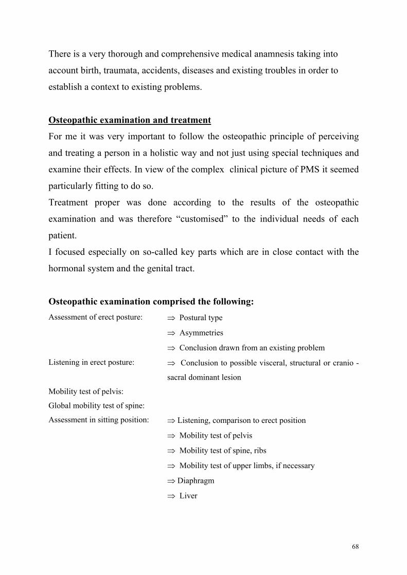
There is a very thorough and comprehensive medical anamnesis taking into
account birth, traumata, accidents, diseases and existing troubles in order to
establish a context to existing problems.
Osteopathic examination and treatment
For me it was very important to follow the osteopathic principle of perceiving
and treating a person in a holistic way and not just using special techniques and
examine their effects. In view of the complex clinical picture of PMS it seemed
particularly fitting to do so.
Treatment proper was done according to the results of the osteopathic
examination and was therefore “customised” to the individual needs of each
patient.
I focused especially on so-called key parts which are in close contact with the
hormonal system and the genital tract.
Osteopathic examination comprised the following: Assessment of erect posture: ⇒ Postural type
⇒ Asymmetries
⇒ Conclusion drawn from an existing problem
Listening in erect posture: ⇒ Conclusion to possible visceral, structural or cranio -
sacral dominant lesion
Mobility test of pelvis:
Global mobility test of spine:
Assessment in sitting position: ⇒ Listening, comparison to erect position
⇒ Mobility test of pelvis
⇒ Mobility test of spine, ribs
⇒ Mobility test of upper limbs, if necessary
⇒ Diaphragm
⇒ Liver
68
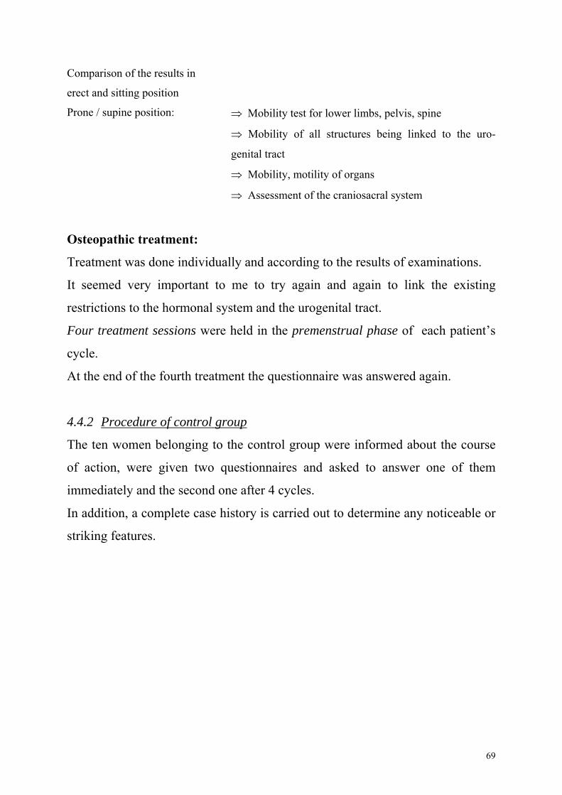
Comparison of the results in
erect and sitting position
Prone / supine position: ⇒ Mobility test for lower limbs, pelvis, spine
⇒ Mobility of all structures being linked to the uro-
genital tract
⇒ Mobility, motility of organs
⇒ Assessment of the craniosacral system
Osteopathic treatment:
Treatment was done individually and according to the results of examinations.
It seemed very important to me to try again and again to link the existing
restrictions to the hormonal system and the urogenital tract.
Four treatment sessions were held in the premenstrual phase of each patient’s
cycle.
At the end of the fourth treatment the questionnaire was answered again.
4.4.2 Procedure of control group
The ten women belonging to the control group were informed about the course
of action, were given two questionnaires and asked to answer one of them
immediately and the second one after 4 cycles.
In addition, a complete case history is carried out to determine any noticeable or
striking features.
69
5. Results and statistical evaluation
My study was based on the hypothesis that four sessions of osteopathic
treatment would improve pain symptomatology and premenstrual symptoms.
Additionally, a general survey of each patient’s menstruation history was
conducted, but served as background information only.
5.1. Results of the questionnaire
5.1.1. Part A of the questionnaire – Menstruation history
This part of the questionnaire captures information on the development of
menstruation from the first period to the beginning of this study.
The results include all the women participating in the study.
The interviews yielded the following results:
- The time of the first menstruation period for almost all the women was
between ages 10 and 14 with a cycle length of 28 to 30 days, very often
involving moderate or heavy bleeding.
- Concerning the first menstrual period and cycle length, most women are
within a normal range, women with a tendency to light bleeding do not
suffer from dysmenorhhea or PMS as often as others.
- Surprisingly, their mothers’ attitude was not generally perceived by the
patients as an example, which is the opposite of what I expected.
- Half of the women surveyed take pain killers, but there is a distinct
tendency towards taking them only as a last resort. If there is a long day at
work to withstand, women take pain killers more readily than at week-
ends, when they can rest and keep warm.
70
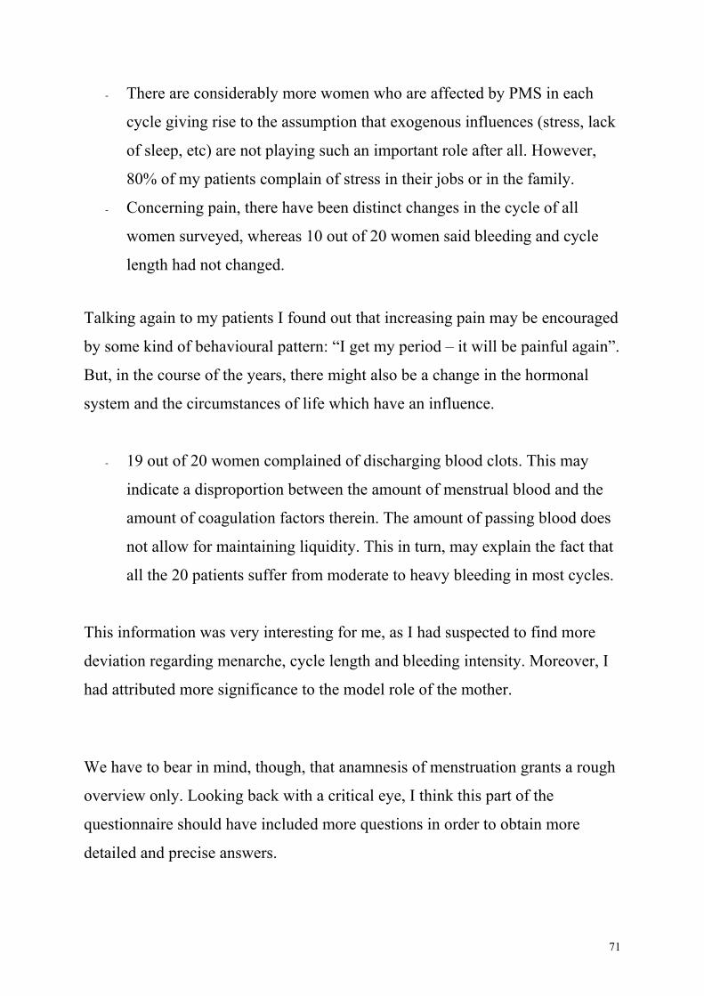
- There are considerably more women who are affected by PMS in each
cycle giving rise to the assumption that exogenous influences (stress, lack
of sleep, etc) are not playing such an important role after all. However,
80% of my patients complain of stress in their jobs or in the family.
- Concerning pain, there have been distinct changes in the cycle of all
women surveyed, whereas 10 out of 20 women said bleeding and cycle
length had not changed.
Talking again to my patients I found out that increasing pain may be encouraged
by some kind of behavioural pattern: “I get my period – it will be painful again”.
But, in the course of the years, there might also be a change in the hormonal
system and the circumstances of life which have an influence.
- 19 out of 20 women complained of discharging blood clots. This may
indicate a disproportion between the amount of menstrual blood and the
amount of coagulation factors therein. The amount of passing blood does
not allow for maintaining liquidity. This in turn, may explain the fact that
all the 20 patients suffer from moderate to heavy bleeding in most cycles.
This information was very interesting for me, as I had suspected to find more
deviation regarding menarche, cycle length and bleeding intensity. Moreover, I
had attributed more significance to the model role of the mother.
We have to bear in mind, though, that anamnesis of menstruation grants a rough
overview only. Looking back with a critical eye, I think this part of the
questionnaire should have included more questions in order to obtain more
detailed and precise answers.
71
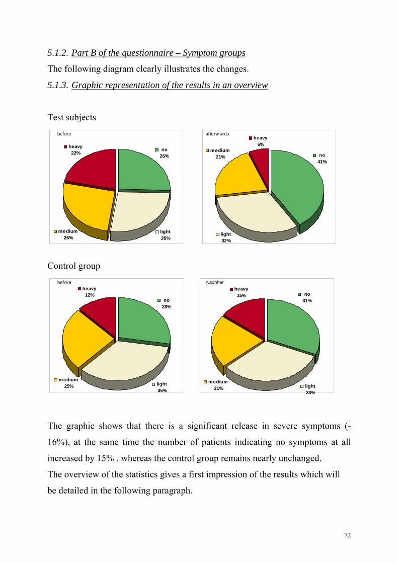
5.1.2. Part B of the questionnaire – Symptom groups
The following diagram clearly illustrates the changes.
5.1.3. Graphic representation of the results in an overview
Test subjects
Control group
The graphic shows that there is a significant release in severe symptoms (-
16%), at the same time the number of patients indicating no symptoms at all
increased by 15% , whereas the control group remains nearly unchanged.
The overview of the statistics gives a first impression of the results which will
be detailed in the following paragraph.
before
medium25%
no28%
heavy12%
light35%
Nachher
medium21%
no31%
heavy15%
light33%
afterw ards
light32%
heavy6%
no41%
medium21%
before
heavy22%
no26%
medium26%
light26%
72
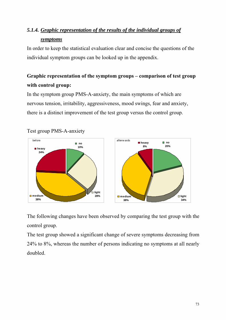
5.1.4. Graphic representation of the results of the individual groups of
symptoms
In order to keep the statistical evaluation clear and concise the questions o
dual sympto
f the
indivi m groups can be looked up in the appendix.
n of test group
ith control group:
bility, aggressiveness, mood swings, fear and anxiety,
The following changes have been observed by comparing the test group with the
control group.
ereas the number of persons indicating no symptoms at all nearly
Graphic representation of the symptom groups – compariso
w
In the symptom group PMS-A-anxiety, the main symptoms of which are
nervous tension, irrita
there is a distinct improvement of the test group versus the control group.
Test group PMS-A-anxiety
The test group showed a significant change of severe symptoms decreasing from
24% to 8%, wh
doubled.
afterw ards
light34%
heavy8%
no20%
medium38%
beforeno
10%heavy24%
light28%medium
38%
73

Control group PMS-A-anxiety
In the control group a change in intensity of the symptoms from middle to slight
symptoms was described in 14% of the cases.
e main symptoms of which are
eadache, craving for sweets, increased appetite, heart pounding, tiredness,
The test group showed a significant relief of the server symptoms from 27% to
9%, the number of persons indicating only light symptoms doubled.
In the symptom group PMS-C-cravings, th
h
dizziness, and fainting, there is also a distinct improvement in the test group.
Test group PMS-C-cravings
afterw ards
light50%
heavy16%
no18%
medium16%
before
no22%
heavy12%
light36%
medium30%
before
medium24%
no39%heavy
27%
light10%
afterw ardsheavy
9% no48%medium
23%
light20%
74
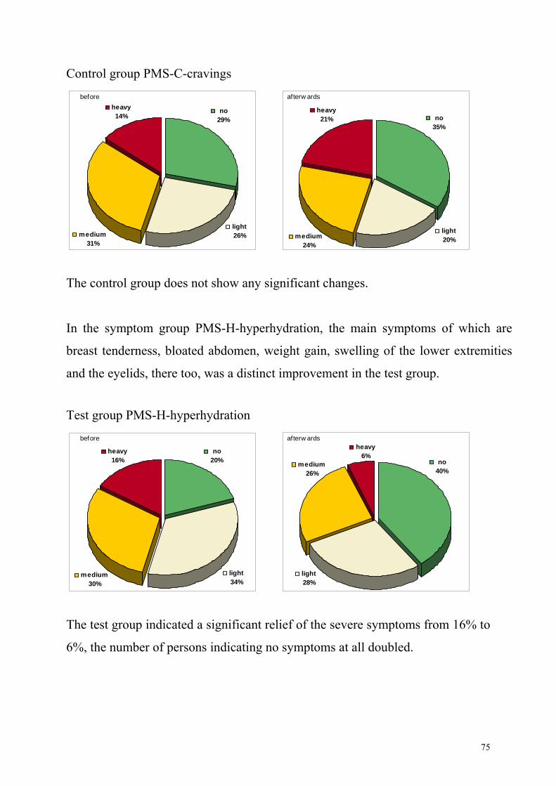
Control group PMS-C-cravings
The control group does not show any significant changes.
symptoms of which are
reast tenderness, bloated abdomen, weight gain, swelling of the lower extremities
The test group indicated a significant relief of the severe symptoms from 16% to
6%, the number of persons indicating no symptoms at all doubled.
In the symptom group PMS-H-hyperhydration, the main
b
and the eyelids, there too, was a distinct improvement in the test group.
Test group PMS-H-hyperhydration
before
medium31%
no29%
heavy14%
light26%
afterw ards
heavy21% no
35%
light20%medium
24%
before
light34%
heavy16%
no20%
medium30%
afterw ardsheavy
6%no
40%medium
26%
light28%
75
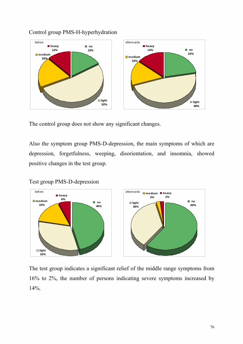
Control group PMS-H-hyperhydration
The control group does not show any significant changes.
Also the symptom group PMS-D-depression, the main symptoms of which are
insomnia, showed
ositive changes in the test group.
The test group indicates a significant relief of the middle range symptoms from
16% to 2%, the number of persons indicating severe symptoms increased by
14%.
depression, forgetfulness, weeping, disorientation, and
p
Test group PMS-D-depression
before
light52%
heavy12%
no16%
medium20%
afterw ardsheavy
no22%
14%
medium16%
light48%
before
medium16%
no46%
heavy6%
light32%
afterw ards heavy2%
medium2%
no60%
light36%
76
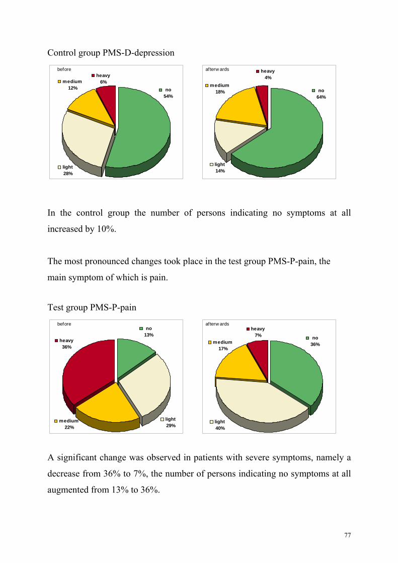
Control group PMS-D-depression
In the control group the number of persons indicating no symptoms at all
increased by 10%.
he most pronounced changes took place in the test group PMS-P-pain, the
est group PMS-P-pain
A significant change was observed in patients with severe symptoms, namely a
decrease from 36% to 7%, the number of persons indicating no symptoms at all
augmented from 13% to 36%.
T
main symptom of which is pain.
T
before
me1dium2% no
54%
heavy6%
light28%
afterw ards heavy4%
medium18% no
64%
light14%
before
light29%
heavy36%
no13%
medium22%
afterw ardsheavy
7% no36%medium
17%
light40%
77
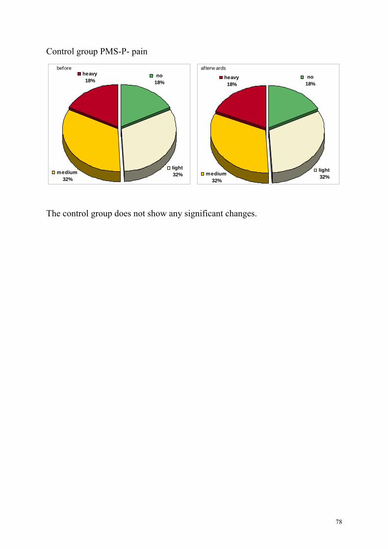
Control group PMS-P- pain
any significant changes.
The control group does not show
before
light32%
heavy18%
no18%
medium32%
afterw ards
no18%
heavy18%
lightmedium 32%
32%
78
6. Discussion he starting point for my study was the hypothesis that holistic, osteopathic
provement of congestive menstrual disorders
nd premenstrual syndrome.
t took very intensive and critical analysis of the problem to make me aware of
s complexity. Ruth Trickey’s book on Women, Hormones & Menstrual Cycle
as of invaluable help to me.
edical and scientific quarters put forth numerous theories which all boil down
ones.
ormones, of course, but why do these unmeasurable changes of hormones
ause some women to suffer symptoms, yet others are not affected?
is is where osteopathy comes into play. Osteopathic treatment
nal situation
ird ventricle), and those that improve loss of
ing more flexibility.
ering,
am able to deal with psychological
aspects inasmuch as I am convinced that by touching a persons body I
also touch his/her soul.
T
treatment could bring about an im
a
I
it
w
M
to one thing: horm
H
c
In my opinion, th
can help the body to deal more easily with the tricky hormonal situation.
Dysfunctions on all levels of the body can be reduced, allowing the body to
better adapt itself to the hormo
Within the framework of an osteopathic treatment we have to distinguish
between two types of techniques. Those that influence directly the hormonal
balance (e.g. work on SBS, th
mobility and motility in other body parts and so indirectly influence the
hormonal situation by giv
Item 3 “Considerations and approach to osteopathic treatment” deals with these
questions in detail.
Besides the hormonal factors a number of other aspects are worth consid
too.
- Psychological factors: in my work I
79
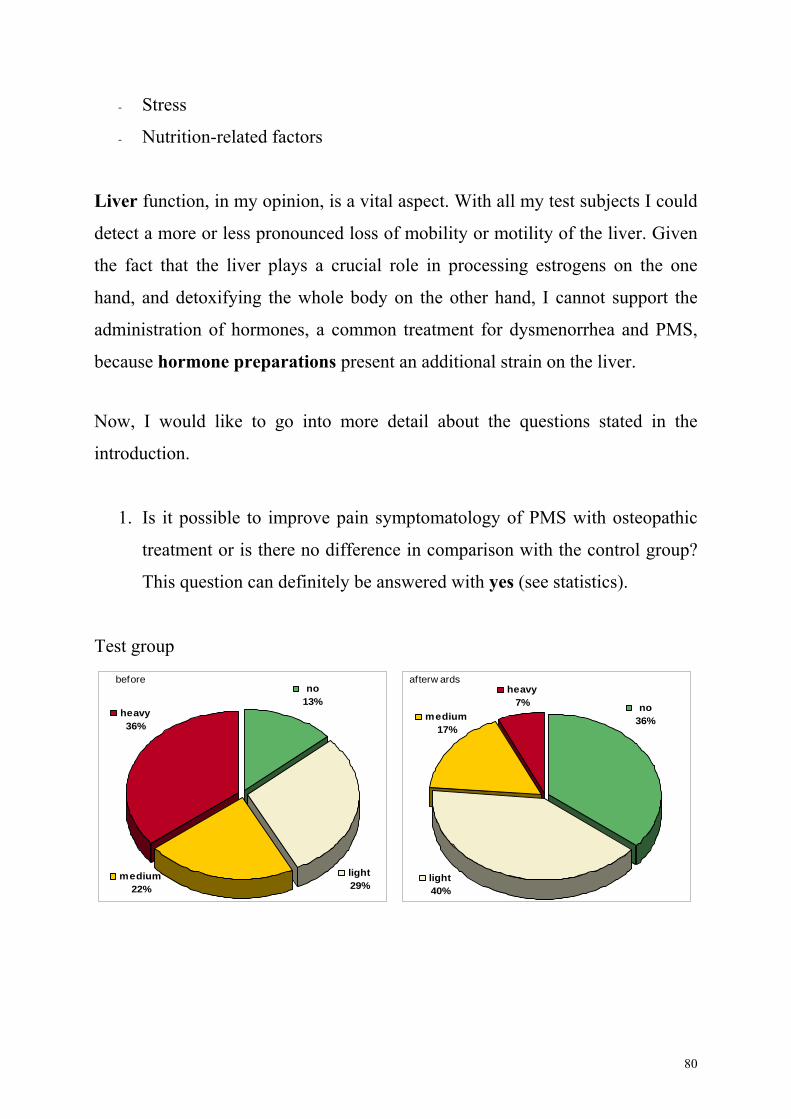
- Stress
- Nutrition-related factors
Liver function, in my opinion, is a vital aspect. With all my test subjects I could
detect a more or less pronounced loss of mobility or motility of the liver. Given
ng the whole body on the other hand, I cannot support the
dministration of hormones, a common treatment for dysmenorrhea and PMS,
No ,
introd
. ssible to improve pain symptomatology of PMS with osteopathic
ifference in comparison with the control group?
This question can definitely be answered with yes (see statistics).
the fact that the liver plays a crucial role in processing estrogens on the one
hand, and detoxifyi
a
because hormone preparations present an additional strain on the liver.
w I would like to go into more detail about the questions stated in the
uction.
1 Is it po
treatment or is there no d
Test group
before
light29%
heavy36%
no13%
medium22%
afterw ardsheavy
7% no36%medium
light40%
17%
80
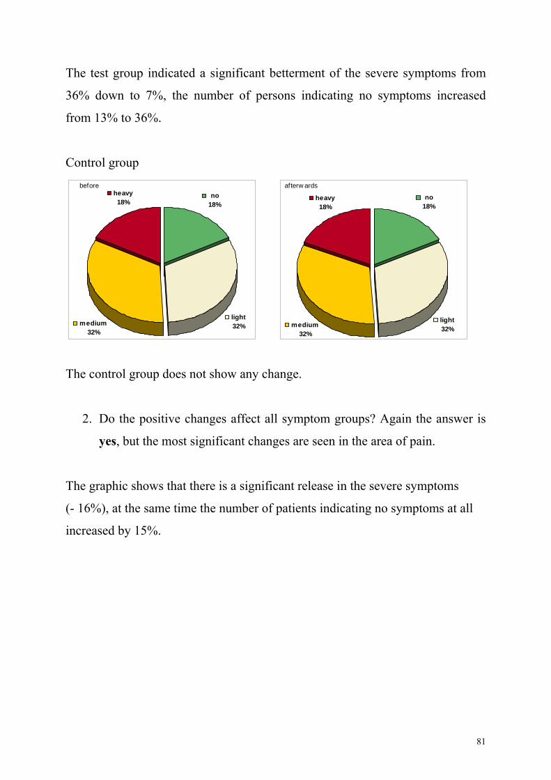
Th
36% d
from 1
p
e changes affect all symptom groups? Again the answer is
yes, but the most significant changes are seen in the area of pain.
The graphic shows that there is a significant release in the severe symptoms
(- 16%), at the same time the number of patients indicating no symptoms at all
increased by 15%.
e test group indicated a significant betterment of the severe symptoms from
own to 7%, the number of persons indicating no symptoms increased
3% to 36%.
Control grou
The control group does not show any change.
2. Do the positiv
before
light32%
heavy18%
no18%
medium32%
afterw ards
no18%
heavy18%
light32%medium
32%
81
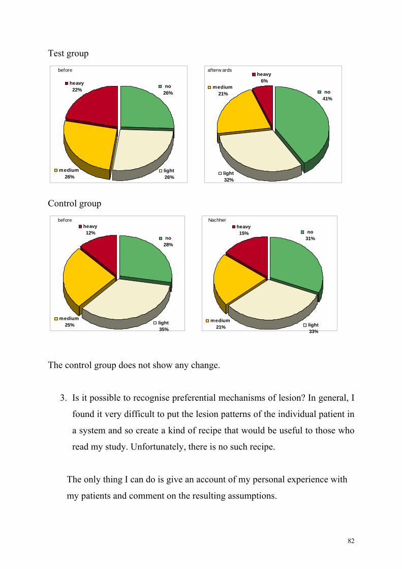
Test group
ontrol group
The control group does not show any change.
3. Is it possible to recognise preferential mechanisms of lesion? In general, I
ery difficult to put the lesion patterns of the individual patient in
a system and so create a kind of recipe that would be useful to those who
read my study. Unfortunately, there is no such recipe.
The only thing I can do is give an account of my personal experience with
my patients and comment on the resulting assumptions.
C
found it v
before
medium25%
no28%
heavy12%
light35%
Nachher
medium21%
no31%
heavy15%
light33%
afterw ards
light32%
heavy6%
no41%
medium21%
before
no26%
heavy22%
medium26%
light26%
82
Due to the anamnestic details my patients disclosed to me, I neglected traumatic
or operative influences, because very few patients mentioned them.
While doing the necessary tests for osteopathic data collection I came across
numerous lesions.
The relevant lesions
- concerning posture were more often found in patients of the anterior type
- in the pelvic area were mainly restrictions of primary sacroiliac mobility
in context with changes of tension and function in the lesser pelvis
ident as difference of tension in
the area of the diaphragm
-
-
- always included reduced fluctuation of CSF between cranium and sacrum.
clearly revealed
e given a lot of attention to their craniosacral system and the
tructures which are causal for the proper functioning of the hormonal system
lesser pelvis …). Besides the hormonal factors, however,
the m
vis r
functi
- in the thoracolumbar junction became ev
- were found in T7/8/9, cervicothoracal junction, C0/C1/C2
were changes of tension in the area of the fascia of the middle axis
always included a restriction of the SBS which is a direct link to the
hormonal system (pituitary gland, third ventricle)
On the basis of the data I collected and the findings which
these central points of problems I set up the following hypothesis:
From the osteopath’s view, patients suffering from congestive dysmenorrhea
and PMS need to b
s
(liver, small intestine,
obility of the pelvic area plays a major role, from a structural as well as a
ce al point of view, as it ensures the best possible conditions for the proper
oning of the organs.
83
Wh e
functioning of the entire fascia of the middle axis is and how stabilising the
eff t
Th a ctions,
par cu
Seen o s well.
- Stress reducing factors (80% of the subjects claimed to be exposed to
on of this
assumption is the fact that at week-ends, with a usually lower stress level,
ularly rewarding to be able to contribute to such a
s not allow a
he design of the questionnaire was a difficult task. In hind view, I would
inc d
Pain G struation would also be more
relevant if more details were contained.
il working with the patients I became aware of how significant proper
ec on the whole body is, if this vital area functions properly.
e natomic background confirms the importance of these conne
ti larly considering the close links to the cranial basis.
n a wider scale other factors have to be included a
heavy stress in their job or their private life)
- Nutritional factors, often induced by stress factors. A confirmati
patients use fewer pain killers
- Psychogenic factors
The success I achieved with osteopathic treatment leads me to the conclusion
that with my point of view I have good possibility of giving therapeutic support
to at least some of the women suffering from dysmenorrhea and PMS. Being a
woman myself, it is partic
considerable extent to the improvement of life quality.
The small number of test persons included in this study doe
statistically relevant result; nevertheless, the results obtained are a clear
indication of the validity of osteopathic treatment.
T
lu e more details for the different groups of symptoms, especially the PMS
roup. The case history sheet concerning men
84
As c
Schind
mine.
ery encouragingly, her study Osteopathic treatment of primary dysmenorrhea –
a losing remark I would like to direct attention to the study of Heike Karl-
ler, who concluded her study during the time it took me to conclude
V
possible effects revealed similar results, thus supporting the findings of my
study.
In the course of my work I became more and more interested in the specific
nction of the liver as regards the processing of hormones. It would be very fu
motivating for me, if somebody decided to write his/her thesis on that topic.
85
7. Summary
At the beginning of my study I put forward the hypothesis that it should be
ossible to improve complaints of congestive dysmenorrhea and the symptoms
f PMS by giving the patients osteopathic treatment. The hypothesis was
onfirmed by the findings of the questionnaire.
has been proven beyond any doubt that hormones, or rather the limited
exibility of the body to deal properly with the current hormonal situation, play
significant role. Therefore, any osteopathic treatment has to aim at supporting
e body individually so that this situation can be dealt with in a more
vourable way.
my experience, it is important to combine structural, cranial and visceral
eatment in order to do justice to the individual clinical picture.
order to support osteopathic work and increase concerned patients’
sponsibility for themselves it is necessary to dig a little deeper and find out
bout the circumstances of these women’s lives. In this way, the significance of
tress and other psychological or nutritional causes becomes apparent.
word I would like to emphasise that in the course of this study I
ttached more and more importance to the knowledge that with my osteopathic
p
o
c
It
fl
a
th
fa
In
tr
In
re
a
s
As a concluding
a
abilities I literally hold in my hands the possibility to contribute to the quality of
life of so many women.
86
8. Bibliography Abraham, G.E. (1980): Premenstrual Tension.Curr Probs Obstet Gynecol 3
arral, J.P. (1993): Urogenital Manipulation. Seattle: Eastland Press. and Larbig, W. (1996): Menstruationsschmerz.
München: Urban & Schwarzenberg.
tetrics &
ason, G. and Peeters, L. (1993): Handbuch für Osteopathie: Das Becken.
The Muscle Energy Manual Volume one.
aoletti, S. (2001): Faszien: München: Urban & Fischer. Die Wirbelsäule aus der Sicht der Osteopathie. Kötzing:
erlag für Osteopathie. ilbernagel, S. and Despopoulos, A. (1987): Taschenatlas der Physiologie.
tamm, H.E. and Stamm, H. (1987): Leitfaden der praktischen Gynäkologie. andsberg/Lech: ecomed.
,R. and Pabst, R. (1993): Sobotta I,II. München: Urban & Schwarzenberg. ie inneren Organe aus der Sicht der Osteopathie. Kötzing:
(12) page 1-39. Barral, J.P. and Mercier, P. (1997): Visceral Manipulation I,II. Seattle: Eastland Press. BBerninger-Schäfer, E. Stuttgart: Schattauer. Drenckhahn, D. and Zenker, W. (1994): Benninghoff Anatomie I,II.
Faller, A. (1980): Der Körper des Menschen. Stuttgart: Thieme Greer, I. Cameron, I. Kitchener, H. and Prentice, A. (2001): ObsGynecology. London: Mosby. LGent: OSTEO 2000. Liem, T. (1998): Kraniosakrale Osteopathie. Stuttgart: Hippokrates. Meert, G.F. (2003): Das Becken aus osteopathischer Sicht. München: Urban & Fischer. Mitchell jr, F.L. (1995):Michigan: Met press. PRichard, J.P. (1994):VSStuttgart: Thieme. SLPutzStone, C. (1996): DVerlag für Osteopathie. Trickey, R. (1998): Women, Hormones & The The Menstrual Cycle: St. Leonhards/Australia: Allen & Unwin. www.healthanswers.cowww.intmedcom. Dr. Elnekheli www.mc.duke.edu/h-devil/women/cramps.htm 1994-99
atgeber/krankheitsbilder/746html. 16.01.01www.toppharm.ch/rwww.webmed.com/content/dmk/dmk-article 40070
87
9. List of illustrations
Abb.1 Internal female genitalia ie II.
female genitalia &
erg. Page 193.
. Page 194.
S. (2001): Faszien: München: Urban & Fischer. Page 69.
): Faszien: München: Urban & Fischer. Page 68.
: er. Page 16.
aus osteopathischer Sicht. München: 7.
. München:
Putz, R. and Pabst, R. (1993): Sobotta II. München: Urban & Schwarzenberg. Page 193.
bb.10 Arteries and veins of the female genitalia ): Das Becken aus osteopathischer Sicht. München:
Page 80. bb.11 The vascular system of the lesser pelvis
Meert, G.F. (2003): Das Becken aus osteopathischer Sicht. München:
Abb.12 München: Urban &
Abb.13 München: Urban & Fischer. Page 64.
y of the fasciae and their connections
Drenkhahn, D. and Zenker, W. (1994): Benninghoff AnatomMünchen: Urban & Schwarzenberg. Page 124.
Abb.2 Internal Putz, R. and Pabst, R. (1993): Sobotta II. München: UrbanSchwarzenb
Abb.3 Uterus, vagina Putz, R. and Pabst, R. (1993): Sobotta II. München: Urban & Schwarzenberg
Abb.4 Broad ligament of the uterus Paoletti,
Abb.5 Fascial anatomy of the lesser pelvis Paoletti, S. (2001
Abb.6 Pelvic floor Meert, G.F. (2003): Das Becken aus osteopathischer Sicht. MünchenUrban & Fisch
Abb.7 Pelvic floor 003): Das BeckenMeert, G.F. (2
Urban & Fischer. Page 1Abb.8 Pelvic floor
Meert, G.F. (2003): Das Becken aus osteopathischer SichtUrban & Fischer. Page 17.
bb.9 Internal female genitalia A
AMeert, G.F. (2003Urban & Fischer.
A
Urban & Fischer. Page 80. Schematic presentation of the vegetative innervation Putz, R. and Pabst, R. (1993): Sobotta II.Schwarzenberg. Page 213. Fascia of the female lesser pelvis Paoletti, S. (2001): Faszien:
Abb.14 General anatomPaoletti, S. (2001): Faszien: München: Urban & Fischer. Page 111.
88
Abb.15 n ovaries, hypophysis and hypothalamus W. (1994): Benninghoff Anatomie II.
s e:
s/Australia: Allen & Unwin. Page 61. Abb.17
r, W. (1994): Benninghoff Anatomie II. ban & Schwarzenberg. Page 187.
Abb.19 irbelsäule aus der Sicht der Osteopathie.
pathie. Page 67.
Feedback regulation betweeDrenkhahn, D. and Zenker, München: Urban & Schwarzenberg. Page 147.
Abb.16 Enterohepatic circulation of estrogenTrickey, R. (1998): Women, Hormones & The The Menstrual CyclSt. LeonhardPortal system between neurohypophysis and adenohypophysis Drenkhahn, D. and ZenkeMünchen: Ur
Abb.18 Ventricle of the brain ): Sobotta I. München: Urban & Putz, R. and Pabst, R. (1993
rg. Page 296. SchwarzenbePostural profiles Richard, J.P. (1994): Die WKötzing: Verlag für Osteo
89
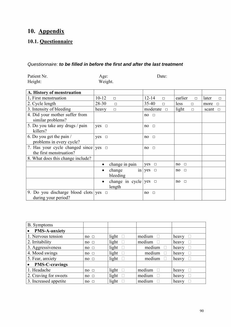
10. Appendix 10.1. Questionnaire
uestionnaire: to be filled in before the first and after the last treatment
atient Nr. Age: Date: eight: Weight.
. History of menstruation
Q PH A1. First menstruation 10-12 □ 12-14 □ earlier □ later □ 2. Cycle length 28-30 □ 35-40 □ less □ more □ 3. Intensity of bleeding heavy □ moderate □ light □ scant □ 4. Did your mother suffer from
similar problems? no □
5. Do you take any drugs / pain killers?
yes □ no □
6. Do you get the pain / problems in every cycle?
yes □ no □
7. Has your cycle changed since the first menstruation?
yes □ no □
8. What does this change include? • change in pain yes □ no □ • change in
bleeding yes □ no □
• change in cycle length
yes □ no □
9. Do you discharge blood clots during your period?
yes □ no □
B. Symptoms • PMS-A-anxiety 1. Nervous tension no □ light ڤ medium ڤ heavy ڤ 2. Irritability no □ light ڤ medium ڤ heavy ڤ 3. Aggressiveness no □ light ڤ medium ڤ heavy ڤ 4. Mood swings no □ light ڤ medium ڤ heavy ڤ 5. Fear, anxiety no □ light ڤ medium ڤ heavy ڤ PMS-C-cravings •
1. Headache no □ light ڤ medium ڤ heavy ڤ 2. Cravin s ligh m g forڤ weets no □ t ڤ edium heavy ڤ 3. Increased appetite no □ light ڤ medium ڤ heavy ڤ
90
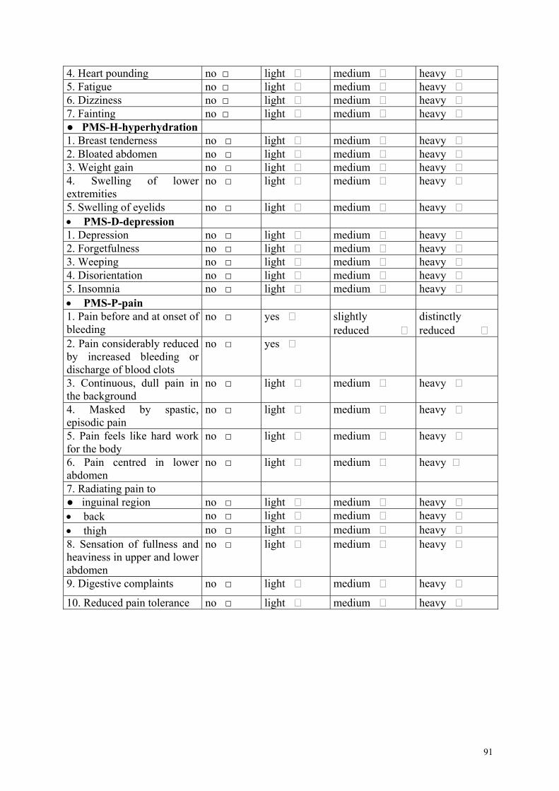
4. Heart pounding no □ light ڤ me um ڤ heavy ڤ di5. Fatigue no □ me light ڤ dium ڤ heavy ڤ 6. Dizziness no □ me h light ڤ dium ڤ eavy ڤ 7. Fainting no □ me ڤ h light dium ڤ eavy ڤ ● PMS-H-hyperhydration 1. no □ light ڤ medium ڤ heavy ڤ Breast tenderness 2. Bloated abdomen no □ me ڤ heavy ڤ light dium ڤ3. in no □ light ڤ medium ڤ heavy ڤ Weight ga4. Swelling of lower ex
no □ me ڤ heavy ڤ tremities
light dium ڤ
5. Swelling of eyelids no □ light ڤ me heavy ڤ dium ڤ• PMS-D-depression 1. Depression no □ light ڤ me um ڤ heavy ڤ di2. Forgetfulness no □ e h light ڤ m dium ڤ eavy ڤ3. Weeping no □ h light ڤ medium ڤ eavy ڤ4. Disorientation no □ medium ڤ heavy ڤ light ڤ 5. Insomnia no □ h light ڤ medium ڤ eavy ڤ• PMS-P-pain 1. Pain before and at onset of
eding no □
ble slightly ڤ
reduced distinctly reduced
yes
2. Pain considerably reduced y increased bleeding or ischarge of blood clots
no □ yes ڤ bd3. Continuous, dull pain in
no □ light ڤ medium ڤ heavy ڤ
the background4. Masked by spastic, o □ ght ڤ edium ڤ eavy ڤ episodic pain
n li m h
5. Pain feels like hard work for the body
no □ light ڤ medium ڤ heavy ڤ
6. Pain centred in lower meabdomen
no □ light ڤ dium ڤ heavy ڤ
7. Radiating pain to ● inguinal region no □ light ڤ medium ڤ heavy ڤ • back no □ light ڤ medium ڤ heavy ڤ • thigh no □ light ڤ medium ڤ heavy ڤ 8. Sensation of fullness and
n upper and lower heaviness iabdomen
no □ light ڤ medium ڤ heavy ڤ
9. Digestive complaints no □ light ڤ medium ڤ heavy ڤ
10. Reduced pain tolerance no □ light ڤ medium ڤ heavy ڤ
91
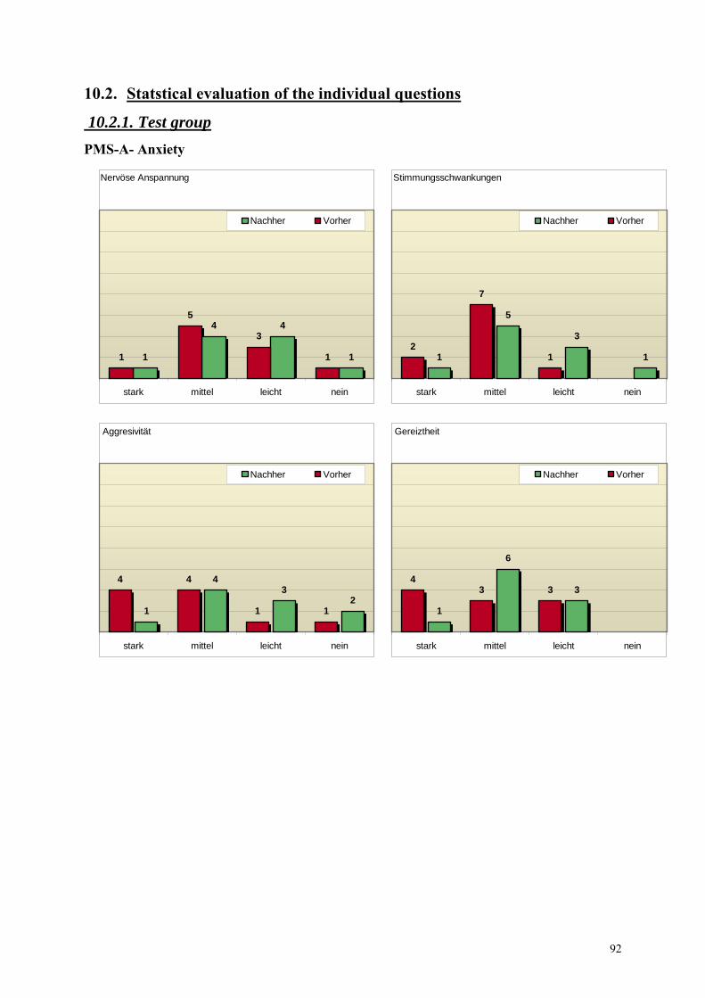
10.2. Statstical evaluatio of the in ividual que tionsn d s
oup 10.2.1. Test gr PMS-A- Anxiety
Nervöse Anspannung
5
1
443
11 1
neinleichtmittelstark
Nachher Vorher
Stimm n
1
5
2
neintel
ungsschwankunge
3
1 1
7
leichtmitstark
Nachher Vorher
Aggresivität
23
1 11
neinleichtmittelstark
444
Nachher Vorher
Gereiztheit
3
6
1
334
neinleichtmittelstark
Nachher Vorher
92
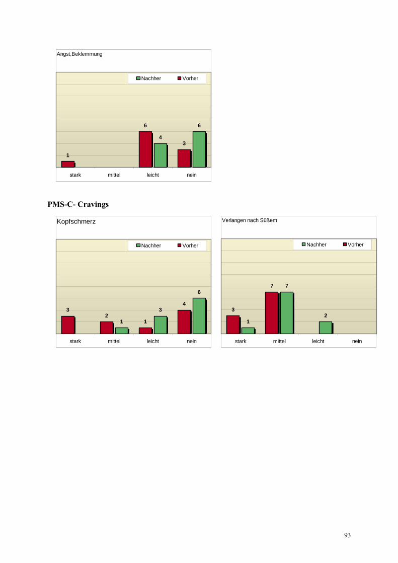
Angst,Beklemmung
6
43
6
1
neinleichtmittelstark
Nachher Vorher
PMS-C- Cravings
Kopfschmerz
6
3
1
4
12
3
neinleichtmittelstark
Nachher Vorher
Verlangen nach Süßem
2
7
1
7
3
neinleichtmittelstark
Nachher Vorher
93
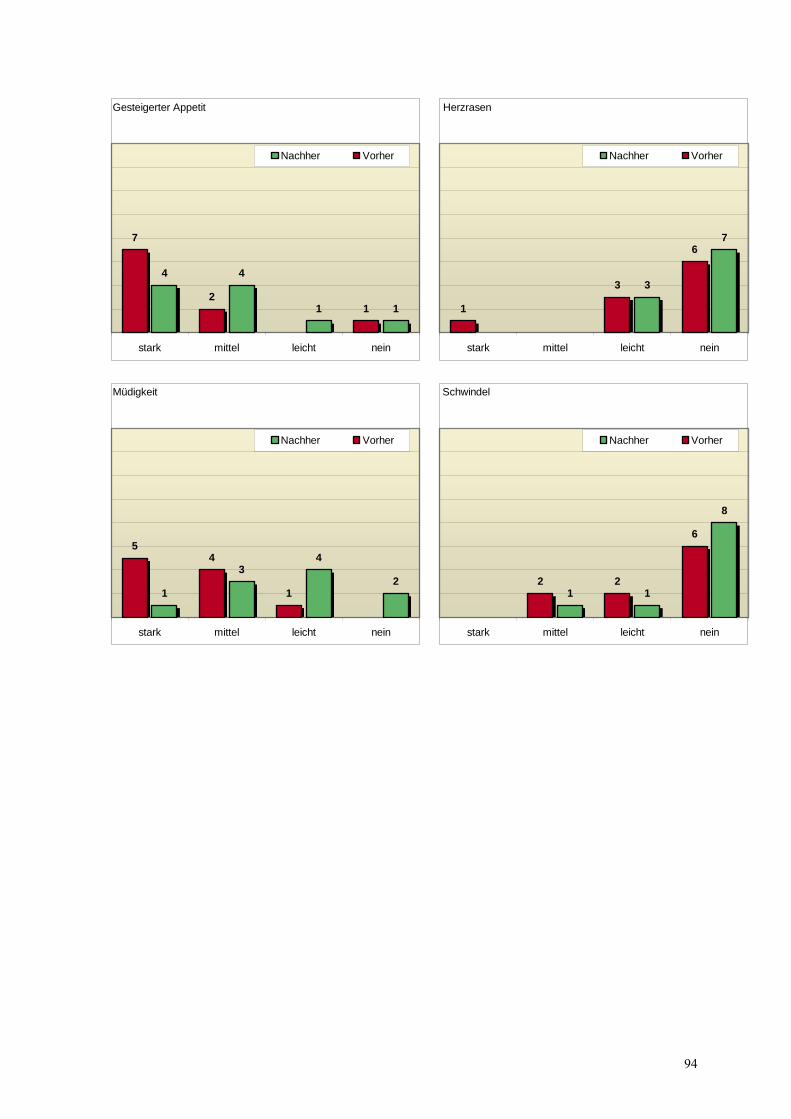
Gesteigerter Appetit
11
44
12
7
neinleichtmittelstark
Nachher Vorher
Herzrasen
7
3
6
3
1
neinleichtmittelstark
Nachher Vorher
Müdigkeit
2
43
1 1
45
neinleichtmittelstark
Nachher Vorher
Schwindel
8
11
6
22
neinleichtmittelstark
Nachher Vorher
94
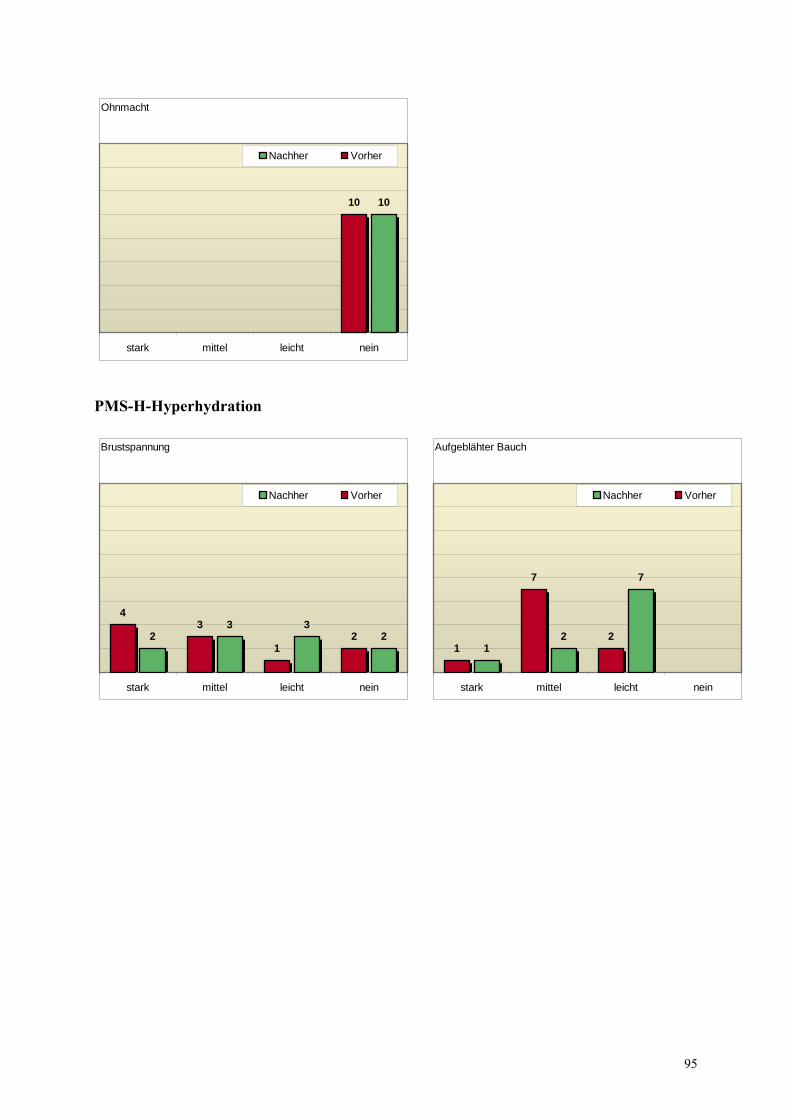
Ohnmacht
1010
neinleichtmittelstark
Nachher Vorher
PMS-H-Hyperhydration
Brustspannung
233
2 21
34
neinleichtmittelstark
Nachher Vorher
Aufgeblähter Bauch
7
21
2
7
1
neinleichtmittelstark
Nachher Vorher
95
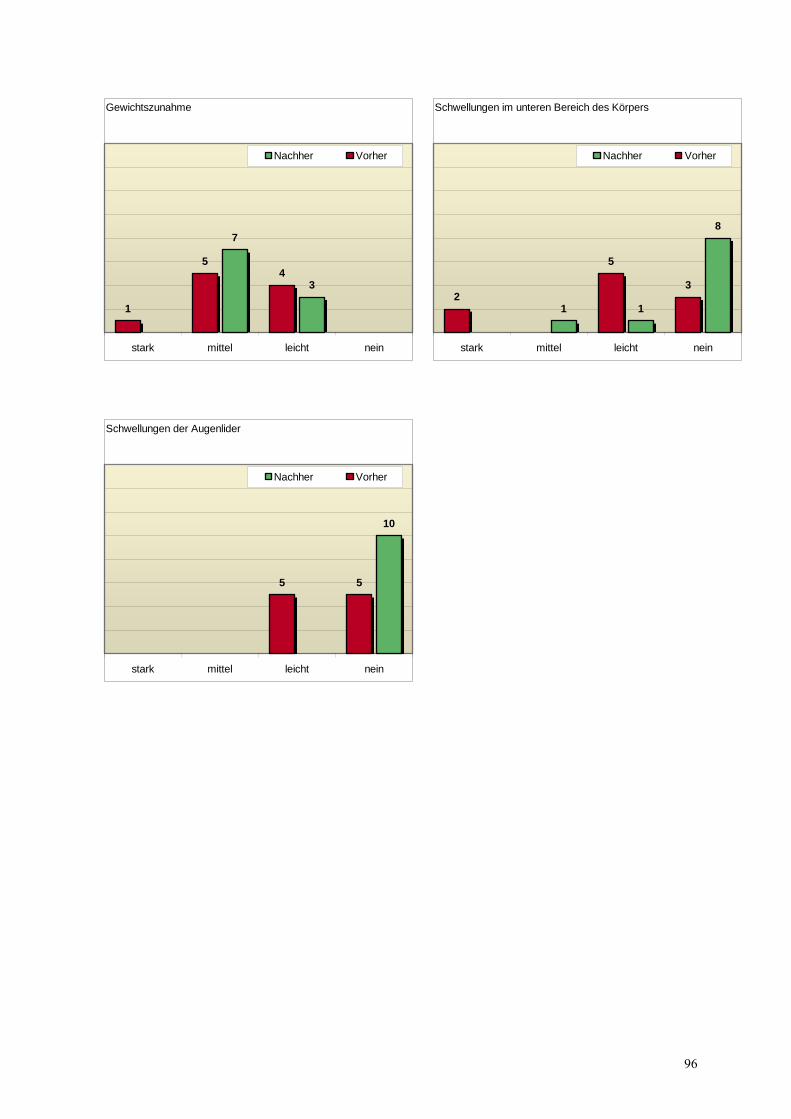
Gewichtszunahme
3
7
45
1
neinleichtmittelstark
Nachher Vorher
Schwellungen im unteren Bereich des Körpers
8
11
3
5
2
neinleichtmittelstark
Nachher Vorher
Schwellungen der Augenlider
10
55
neinleichtmittelstark
Nachher Vorher
96
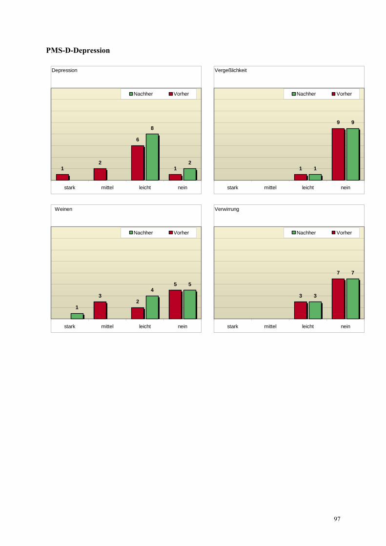
PMS-D-Depression
Depression
2
8
1
6
21
neinleichtmittelstark
Nachher Vorher
Vergeßlichkeit
9
1
9
1
neinleichtmittelstark
Nachher Vorher
Weinen
54
1
5
23
neinleichtmittelstark
Nachher Vorher
Verwirrung
7
3
7
3
neinleichtmittelstark
Nachher Vorher
97
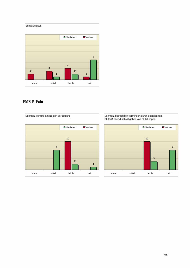
Schlaflosigkeit
7
21 1
43
2
neinleichtmittelstark
Nachher Vorher
PMS-P-Pain
Schmerz vor und am Beginn der Blutung
12
7
10
neinleichtmittelstark
Nachher Vorher
Schmerz beträchtlich vermindert durch gesteigerten Blutfluß oder durch Abgehen von Blutklumpen
7
3
10
neinleichtmittelstark
Nachher Vorher
98
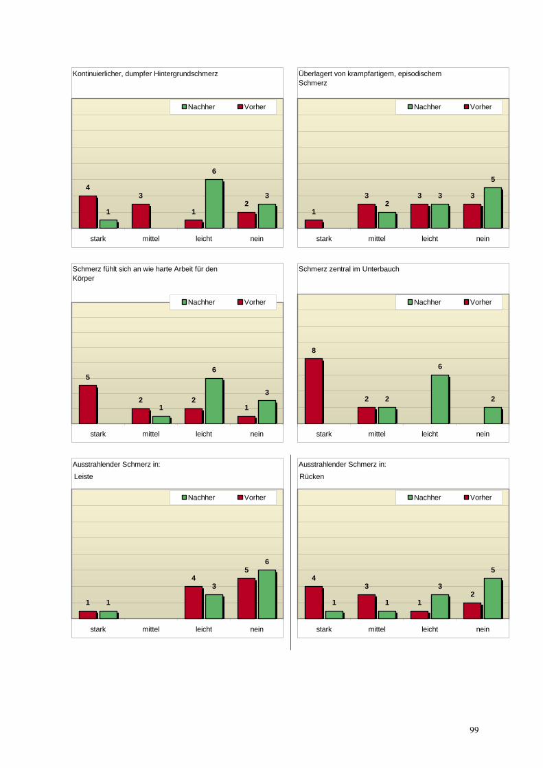
Kontinuierlicher, dumpfer Hintergrundschmerz
3
6
12
1
34
neinleichtmittelstark
Nachher Vorher
Überlagert von krampfartigem, episodischem Schmerz
5
32
333
1
neinleichtmittelstark
Nachher Vorher
Schmerz fühlt sich an wie harte Arbeit für den Körper
3
6
1 122
5
neinleichtmittelstark
Nachher Vorher
Schmerz zentral im Unterbauch
2
6
22
8
neinleichtmittelstark
Nachher Vorher
Ausstrahlender Schmerz in:
6
3
1
54
1
neinleichtmittelstark
Nachher Vorher
Leiste
Ausstrahlender Schmerz in:
5
3
112
1
34
neinleichtmittelstark
Nachher Vorher
Rücken
99
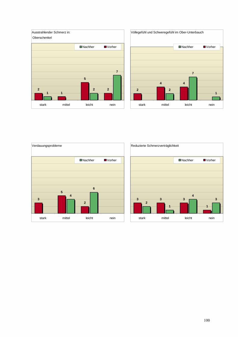
Ausstrahlender Schmerz in:
7
21
2
5
12
neinleichtmittelstark
Nachher Vorher
Oberschenkel
Völlegefühl und Schweregefühl im Ober-Unterbauch
1
7
2
44
2
neinleichtmittelstark
Nachher Vorher
Verdauungsprobleme
6
4
2
5
3
neinleichtmittelstark
Nachher Vorher
Reduzierte Schmerzverträglichkeit
34
12
1
333
neinleichtmittelstark
Nachher Vorher
100
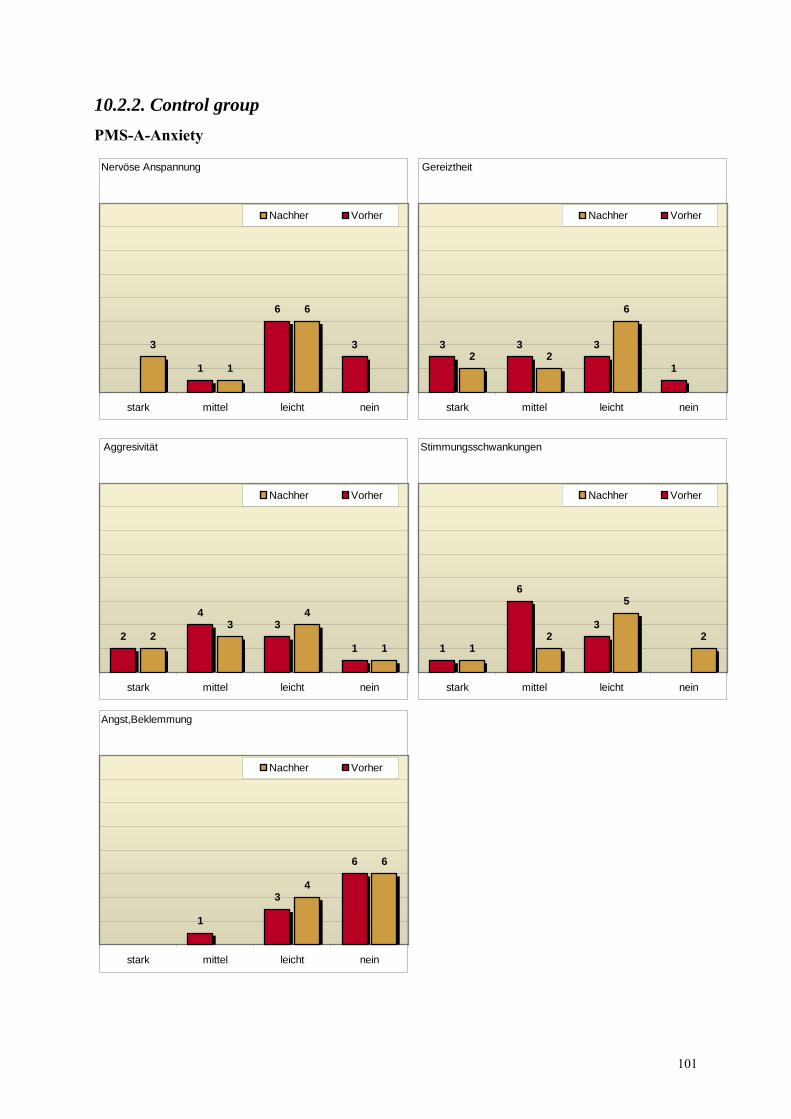
10.2.2. Control group PMS-A-Anxiety
Nervöse Anspannung
6
1
3 3
6
1
neinleichtmittelstark
Nachher Vorher
Gereiztheit
6
221
333
neinleichtmittelstark
Nachher Vorher
Aggresivität
1
43
21
34
2
neinleichtmittelstark
Nachher Vorher
Stimmungsschwankungen
2
5
21
3
6
1
neinleichtmittelstark
Nachher Vorher
Angst,Beklemmung
6
4
6
3
1
neinleichtmittelstark
Nachher Vorher
101
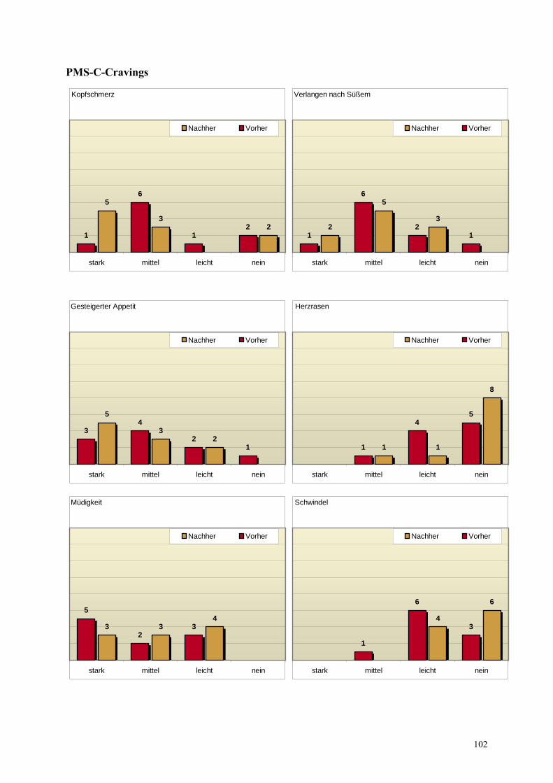
PMS-C-Cravings
Kopfschmerz
23
5
21
6
1
neinleichtmittelstark
Nachher Vorher
Verlangen nach Süßem
3
5
21
2
6
1
neinleichtmittelstark
Nachher Vorher
Gesteigerter Appetit
23
5
12
43
neinleichtmittelstark
Nachher Vorher
Herzrasen
8
11
54
1
neinleichtmittelstark
Nachher Vorher
Müdigkeit
433 3
2
5
neinleichtmittelstark
Nachher Vorher
Schwindel
6
43
6
1
neinleichtmittelstark
Nachher Vorher
102
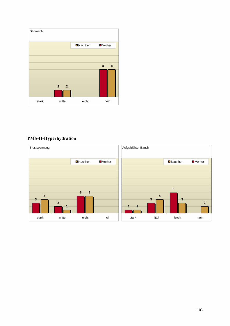
Ohnmacht
8
2
8
2
neinleichtmittelstark
Nachher Vorher
PMS-H-Hyperhydration
Brustspannung
5
1
45
23
neinleichtmittelstark
Nachher Vorher
Aufgeblähter Bauch
23
4
1
6
3
1
neinleichtmittelstark
Nachher Vorher
103
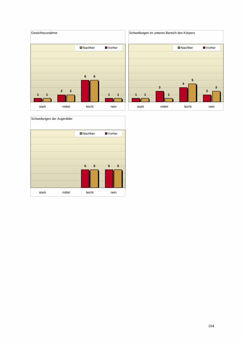
Gewichtszunahme
1
6
21 1
6
21
neinleichtmittelstark
Nachher Vorher
Schwellungen im unteren Bereich des Körpers
3
5
112
43
1
neinleichtmittelstark
Nachher Vorher
Schwellungen der Augenlider
55 55
neinleichtmittelstark
Nachher Vorher
104
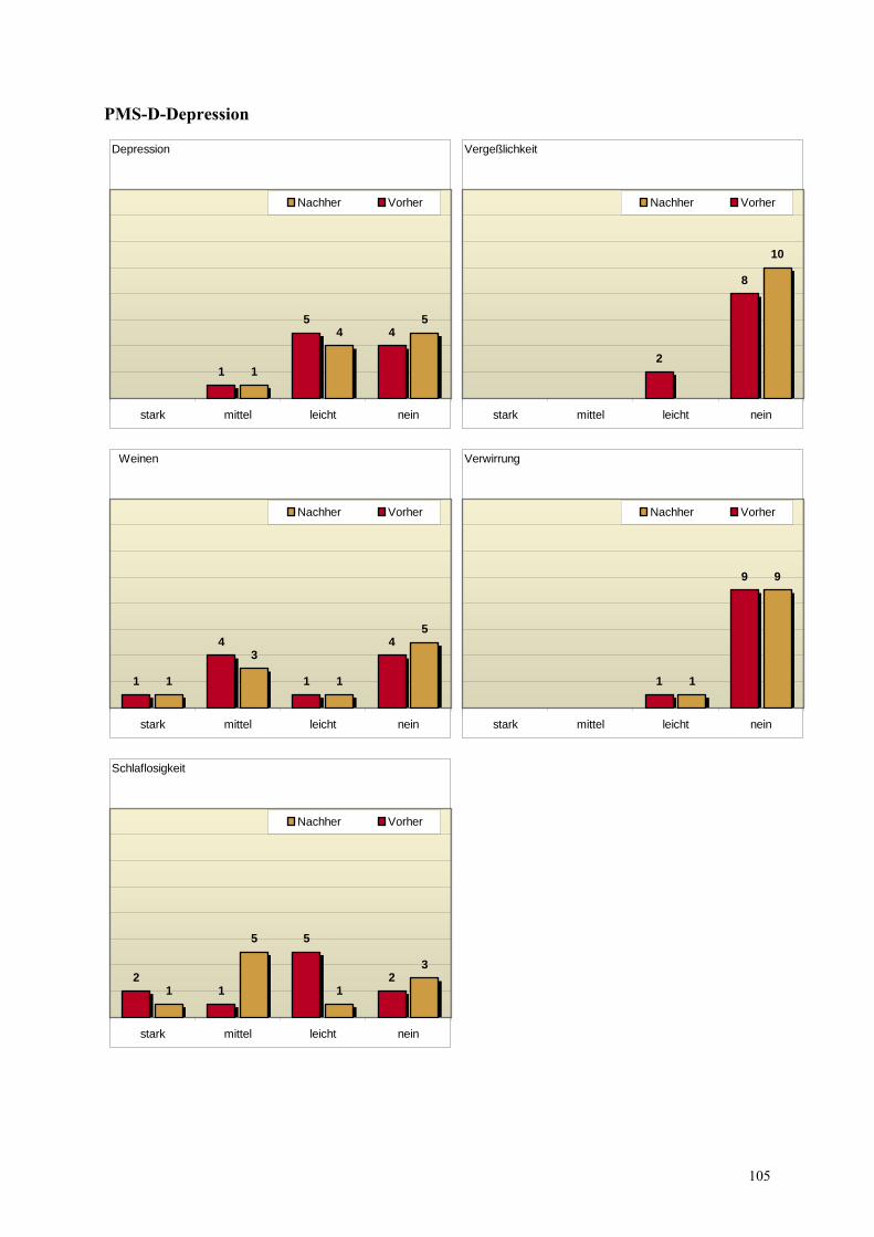
PMS-D-Depression
Depression
54
1
45
1
neinleichtmittelstark
Nachher Vorher
Vergeßlichkeit
10
8
2
neinleichtmittelstark
Nachher Vorher
Weinen
5
1
3
1
4
1
4
1
neinleichtmittelstark
Nachher Vorher
Verwirrung
9
1
9
1
neinleichtmittelstark
Nachher Vorher
Schlaflosigkeit
3
1
5
12
5
12
neinleichtmittelstark
Nachher Vorher
105
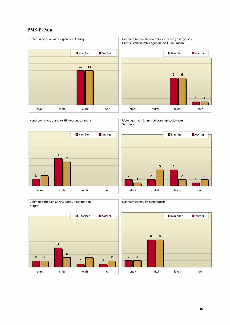
PMS-P-Pain
Schmerz vor und am Beginn der Blutung
1010
neinleichtmittelstark
Nachher Vorher
Schmerz beträchtlich vermindert durch gesteigerten Blutfluß oder durch Abgehen von Blutklumpen
1
9
1
9
neinleichtmittelstark
Nachher Vorher
Kontinuierlicher, dumpfer Hintergrundschmerz
7
3
8
2
neinleichtmittelstark
Nachher Vorher
Überlagert von krampfartigem, episodischem Schmerz
22
5
1 1
5
22
neinleichtmittelstark
Nachher Vorher
Schmerz fühlt sich an wie harte Arbeit für den Körper
233
211
6
2
neinleichtmittelstark
Nachher Vorher
Schmerz zentral im Unterbauch
8
2
8
2
neinleichtmittelstark
Nachher Vorher
106
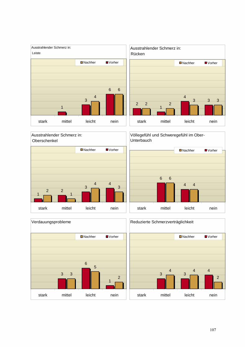
Ausstrahlender Schmerz in:
6
4
6
3
1
neinleichtmittelstark
Nachher Vorher
LeisteAusstrahlender Schmerz in:
3322
34
12
neinleichtmittelstark
Nachher Vorher
Rücken
Ausstrahlender Schmerz in:
34
12
43
21
neinleichtmittelstark
Nachher Vorher
OberschenkelVöllegefühl und Schweregefühl im Ober-Unterbauch
46
46
neinleichtmittelstark
Nachher Vorher
Verdauungsprobleme
2
5
3
1
6
3
neinleichtmittelstark
Nachher Vorher
Reduzierte Schmerzverträglichkeit
2
44 433
neinleichtmittelstark
Nachher Vorher
107
11. Abstract
The Influence of Osteopathic Treatment on Congestive Menstrual Disorders and Premenstrual Syndrome
by Ingrid Riepler-Reisecker
Subject: this study aims at finding out, if it is possible to exert a positive
influence on congestive menstrual disorders and premenstrual syndrome by
using osteopathic treatment and thus improve quality of life for the women
concerned.
Study design: comparative, clinical study plus statistical evaluation.
Method: 10 patients of the test group are given osteopathic treatment in their
premenstrual phase for a period of 4 menstrual cycles. Before the first and after
the last treatment patients fill in a questionnaire.
The results of the test group are compared with a control group who also fills in
two questionnaires at an interval of 4 cycles.
Results: the results of the study are statistically evaluated and represented in
diagrams. On the basis of this evaluation we can clearly see a statistical
tendency towards iomprovement for both congestive menstrual pain and
premenstrual syndrome. However, due to the low number of participants the
results cannot be called statistically significant.
Conclusion: on the basis of the statistical results it has been proven beyond any
doubt that it is possible to influence hormonal dysbalances of women by giving
them osteopathic treatment. Function of organs in the lesser pelvis is improved
by a harmonisation on the structural, cranial and visceral levels and congestive
pain symptomatology is considerably reduced.
108

Key words: congestive menstrual pain, PMS, quality of life, hormonal
dysbalance from an osteopath’s view.
109

110