Embed Size (px)
Citation preview

This work has been digitalized and published in 2013 by Verlag Zeitschrift für Naturforschung in cooperation with the Max Planck Society for the Advancement of Science under a Creative Commons Attribution4.0 International License.
Dieses Werk wurde im Jahr 2013 vom Verlag Zeitschrift für Naturforschungin Zusammenarbeit mit der Max-Planck-Gesellschaft zur Förderung derWissenschaften e.V. digitalisiert und unter folgender Lizenz veröffentlicht:Creative Commons Namensnennung 4.0 Lizenz.
Interaction between Acetyltetralone and Cu(II) Ions: X-Ray Diffraction of the Ligand and Single Crystal ESR Study of the Complex
Michel Geoffroy*, Akilesh Jain, and Alice Celalyan Department of Physical Chemistry, 30 quai E-Ansermet University of Geneva, 1211 Geneva, Switzerland
Gerald Bernardinelli Laboratoire de Christallographie aux Rayoux, 24 quai E-Ansermet University of Geneva, 1211 Geneva, Switzerland Z. Natorforsch. 38b, 830-834 (1983); received April 5, 1983 X-Ray, ESR, Acetyltetralone, Cu(II) Organic Complexes.
Single crystals of acetyltetralone grown from a solution containing a small amount of Cu(II) ions are studied by ESR. The corresponding g, 63Cu magnetic and quadrupolar hyperfine tensors are obtained and are consistent with the trapping of a square planar copper(II) complex. X-ray diffraction shows that pure acetyltetralone crystallizes in the orthorhombic space group Pbca (a — 8.893 A, b = 20.301 X, c = 10.715 A). Comparison of the ESR eigenvectors with the bond directions obtained from this crystal structure study shows that the Cu(II) complex experiences some constraint from the crystal matrix. The present complex is a model for one of the complexation sites of tetracyclines.
1. Introduction
Interactions between divalent metallic ions and tetracycline (TC) are frequently used in order to explain the antibiotic properties of these latter com-pounds [1, 2]. However, the mechanism of forma-tion and the structure of these complexes are not yet well understood. As discussed in a recent study [3], several sites of chelation are possible in a tetra-cycline molecule (1): oxygen atoms at positions C ( l l ) and C(12), oxygen atoms at positions C(10) and C( l l ) , and substituents at positions C(l) , C(2), and C(3).
2
* Reprint requests to Prof. M. Geoffroy. 0340-5087/83/0700-0830/$ 01.00/0
Thirty years ago it was postulated [4] that tetra-cycline can form 2:1 (TC:Cu(II)) complexes. A recent ESR study [3] of dimethyl sulfoxide solu-tions containing tetracycline and Cu(II) was con-sistent with this hypothesis and suggested an im-portant complexing role for the 0(11) and 0(12) atoms.
In the present work, we report the ESR study of acetyltetralone (ATL) the tautomeric form 2 of which can appear as a model for the chelation of the site C( l l ) , C(12) in 1. Acetyltetralone and tetra-cycline exhibit some similar biological properties and Nickell and Gordon [5] have postulated that chelation plays a dominant role in the effect of these compounds on plant growth.
As the maximum information about the struc-ture of a paramagnetic complex is obtained by ESR w7hen this complex is diluted in a diamagnetic single crystal matrix, we have performed the following experiments: i) determination, by X-rays diffrac-tion, of the crystal structure of pure acetyltetralone ii) doping of single crystals of acetyltetralone with Cu(II) complexed acetyltetralone molecules iii) de-termination of the g-tensor and of the 63Cu magnetic and quadrupolar hyperfine tensors iv) comparison of the orientation of the corresponding eigenvectors with the crystallographic bond directions.
2. Experimental 2-Acetyl-a-tetralone was a commercial compound
(ALDRICH) which was purified by crystallization.
M. Geoffroy et al. • Interaction between Acetyltetralone and Cu(II) Ions 831
Single crystals were obtained by slow evaporation of a solution in methanol. Lattice parameters and diffracted intensities were measured at 210 K on an automatic four-circle Philips P W 1100 diffractome-ter with monochromatic MoKa radiation. A data set of 1890 independant reflections having IFo| ^ 2 a (Fo) and |Fo| ^ 5 were considered as being observed. The structure was solved by direct methods (MUL-TAN 74 [6]) and all hydrogen could be located in a A F synthesis calculated at an intermediate stage of resolution. The convergence of least-square refinement (full matrix, X - R A Y System [7]) was obtained for the final R value of 0.070.The calcula-tions were carried out with anisotropic temperature factors for non-hydrogen atoms and isotropic terms f o r H .
Cu(II) doped single crystals of acetyltetralone were obtained by slow evaporation of a solution of acetyltetralone in methanol containing a small amount of copper nitrate (CUNO3: ATL = 1/1000 molar). These crystals were studied on a V A R I A N E-9 ESR spectrometer (X-band, 100 K H z field modulation) by using a previously described goniometer [8] which allows rotation of the crystal in the different principal planes. For each spectrum, the magnetic field was calibrated with an NMR marker and the klystron frequency was measured via a transfer oscillator (Hewlett Packard 200 B) and a Racal frequency meter.
3. Results
3.1. Crystal structure
The cell parameters are: a — 8.893(5), b = 20.301(6), c = 10.715(2) Ä, the space group is Pbca, Z = 8, /u = 0.913 cm - 1 . The atomic coordonates are shown in Table I and the bond lengths and bond angles are given in Table II . The C(l)-C(2) length
Table I. Final positional and thermal parameters ( X 104).
Atom x/a y/b z/c Ueq(Ä2)
0(1) 1929(3) 4446(1) 159(3) 370 0(2) 2602(4) 5588(1) — 416(3) 413 C(l) 2938(4) 4243(2) — 659(3) 246 0(2) 3799(4) 4682(2) — 1358(3) 269 0(3) 4956(5) 4416(2) — 2269(4) 362 0(4) 4462(5) 3761(2) — 2815(3) 358 0(5) 4123(5) 2606(2) — 1947(4) 392 0(6) 3502(5) 2174(2) — 1079(4) 440 0(7) 2686(5) 2419(2) — 73(4) 429 0(8) 2511(5) 3092(2) 71(3) 348 0(9) 3127(4) 3529(2) — 789(3) 268 0(10) 3925(4) 3286(2) — 1824(3) 291 0(11) 3592(5) 5375(2) — 1168(4) 343 0(12) 4526(6) 5868(2) — 1864(4) 417
Table II. Interatomic distances (A) and bond angles (degrees) for acetyltetralone. (The average standard deviation for bond angles is 0.3°).
Bonds Angles
O(l)-O(l) 1.321(4) 0(1 -C(l)-C(2) 122.1 C(l)-C(2) 1.392(5) 0(1 -C(l)-C(9) 116.7 C(l)-C(9) 1.466(5) C(2 -C(l)-C(9) 121.2 C(2)-C(3) 1.518(6) 0(1 -C(2)-C(3) 119.4 C(2)-C(ll) 1.435(6) 0(1 -C(2)-C(l 1) 118.7 C(3)-C(4) 1.517(6) 0(3 -C(2) -C( l l ) 121.8 C(4)-C(10) 1.511(6) C(5)-C(6) 1.392(6) 0(2 -C(3)-C(4) 111.3 C(5)-C(10) 1.398(6) C(3 -C(4)-C(10) 112.3 C(6)-C(7) 1.391(7) 0(6 -C(5)-C(10) 120.6 C(7)-C(8) 1.383(6) C(5 -C(6)-C(7) 120.0 C(8)-C(9) 1.392(5) 0(6 -C(7)-C(8) 119.9 C(9)-C(10) 1.406(5) 0(7 -C(8)-C(9) 120.8 C(l l ) -0(2) 1.269(5) 0(1 -C(9)-C(8) 121.5 C(ll)-C(12) 1.499(6) C(1 -C(9)-C(10) 118.7
0(8 -C(9)-C(10) 119.8 0(4 -C(10)-C(5) 121.6 0(4 -C(10)-C(9) 119.3 C(5 -C(10)-C(9) 119.0 0(2 -C( l l ) -C(2) 120.9 0(2 -C(l l ) -C(12) 118.2 0(2 -C(l l ) -C(12) 120.3
clearly shows that these two carbon atoms are doubly bonded and that the cetoenolic tautomer which has crystallized is 3:
3
The atoms C(l) , C(2), C(3) and C ( l l ) are coplanar (maximum deviation from the least squares plane: 0.009 Ä). In fact the atomic coordonates indicate that two main planes are present in the molecule: i) the aromatic plane corresponding to the phenyl ring ii) the plane containing all the other atoms except C(4) and two methyl protons. Geometric data for 0 ( 1 ) - H and 0(2) are in accordance with an intra-molecular hydrogen bond between the hydroxyl and the ketone group: 0 — 0 = 2.472(4) Ä, O - H - O = 147.5°, O - H = 1.201(7) Ä.
As two acetyltetralone molecules are involved in the formation of a square planar Cu(II)-complex, it is appropriate to consider the molecular arrange-ment between two neighbouring molecules related
832 M. Geoffroy et al. • Interaction between Acetyltetralone and Cu(II) Ions 832
by the (0, 2, 0) inversion center. The four coplanar oxygen atoms form a parallelogram, the diagonals of which respectively measure 5.283 Ä and 4.116 A and form a 62° angle.
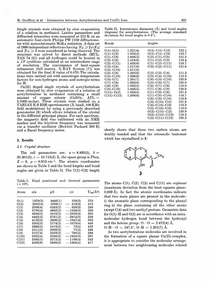
3.2. ESB An idealized single crystal of acetyltetralone is
shown in Fig. 1 together with the ESR reference system and the crystallographic axes. An example of an ESR spectrum obtained with a Cu(II) con-taining single crystal is shown in Fig. 2. For an
reference axes.
Fig. 2. ERS spectrum obtained with an acetyltetra-lone single crystal grown in presence of Cu(II). (The magnetic field is oriented along b).
arbitrary orientation of the magnetic field four non-equivalent paramagnetic sites are present, each site exhibits interaction with a 63Cu and a 65Cu nucleus. In accordance with the crystallographic structure of the non-doped crystal, the angular dependence of
the ESR spectra show that these sites are related by reflection through the reference planes. The angular dependences of the signals are analyzed by using the usual Hamiltonian:
§ = £HgS + g N £ N I H + S T I + I P I
The four terms correspond, respectively, to the electronic Zeeman effect, the nuclear Zeeman effect, the 63/65Cu-magnetic hyperfine interaction and the 63/65Qu quadrupolar interaction. For some orienta-tions forbidden transitions (Mi = ± 1 ) are clearly observed and their detection is imperative for the determination of the quadrupolar interaction. The g tensor and the magnetic and quadrupolar hyperfine tensors are obtained by using a previously described optimization method which takes the difference (direct diagonalization - 2nd order perturbation) into account [9]. These ESR tensors are given in Table III.
Table III. ESR tensors for the (ATL)2Cu complex.
Tensor Eigenvalues Eigenvectors* X n V
g 2.257(6) 0.944 0.262 0.199 2.051(4) — 0.295 0.405 0.860 2.046(6) 0.137 — 0.875 0.460
63Cu-T — 543 0.935 0.294 0.196 (MHz) — 85 — 0.336 0.577 0.742
— 65 0.105 — 0.760 0.639 63Cu-P + 4.7 0.938 0.299 0.17 (MHz) — 0.9 — 0.282 0.952 — 0.114
— 3.8 0.196 — 0.059 — 0.978
* (other sites: A, fx, v; A, ju, v; A, p, v).
4. Discussion
The fact that the angular variations of the ESR lines are in accordance with the symmetry pro-perties of the host crystal shows that the para-magnetic species really occupies a specific place in the crystal lattice and is not adsorbed at the surface or randomly trapped in interstitial defects. This species is obviously the result of the chelation of the Cu(II) ions by the acetyltetralone molecules in the methanol solution at high (ATL/Cu(II)) ratio. The corresponding complex is then incorporated in the acetyltetralone lattice during the crystallization of this compound.
The g and 63Cu-magnetic tensors given in Table III are characterized by an axial symmetry.
M. Geoffroy et al. • Interaction between Acetyltetralone and Cu(II) Ions 833
Table IV. ESR tensors for some square planar copper complexes.
g T (MHZ) Pm a x (MHz) rj Ref
Cu(acac)2 2.266 — 479 6.79 0 [10, 11] 2.053 — 58 2.053 — 58
Cu(bzac)2 5.1 0 [11] CU(C204)22- 2.281 — 542 8.99 [12]
2.056 — 52.4 2.060 — 67.4
Cu(dtO)22- 2.080 — 491 1.3 0 [13] 2.019 — 127 2.019 — 124
Cu(SdbmO)2 2.145 — 481 2.6 0.7 [13] 2.029 — 105 2.031 — 112
The two directions gmax and 6 3Cu-T / / are aligned and the eigenvalues are quite similar with those previously reported for some square planar Cu(II)-complexes (Table IV) . Such a square coplanar stereochemistry is perfectly consistent with the fact that acetyltetralone is capable of 7r-bonding to the Cu(II) ion [14]. The complex is therefore of the (ATL)2Cu type, the unpaired electron belongs to a 3d2:2_2/2 copper orbital and the gmax and T,, are expected to be oriented perpendicularly to the plane of the ligands.
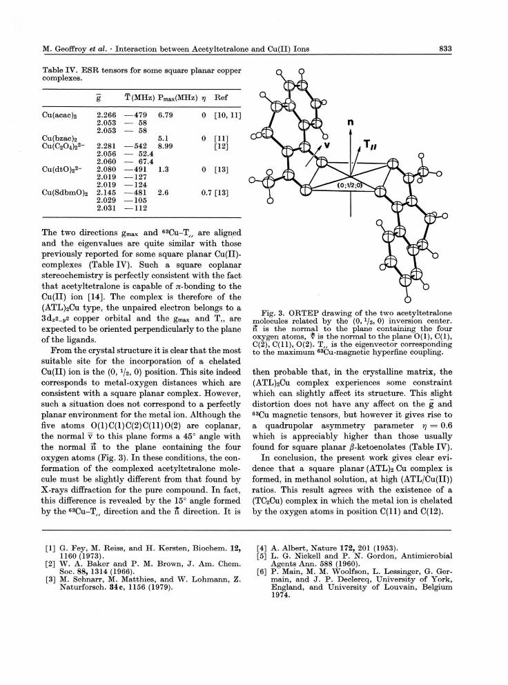
From the crystal structure it is clear that the most suitable site for the incorporation of a chelated Cu(II) ion is the (0, 1/2, 0) position. This site indeed corresponds to metal-oxygen distances which are consistent with a square planar complex. However, such a situation does not correspond to a perfectly planar environment for the metal ion. Although the five atoms 0 (1 )C(1 )C(2 )C(11 )0 (2 ) are coplanar, the normal v to this plane forms a 45° angle with the normal ii to the plane containing the four oxygen atoms (Fig. 3). In these conditions, the con-formation of the complexed acetyltetralone mole-cule must be slightly different from that found by X-rays diffraction for the pure compound. In fact, this difference is revealed b y the 15° angle formed b y the 6 3 Cu-T / / direction and the n direction. It is
Fig. 3. ORTEP drawing of the two acetyltetralone molecules related by the (0, l /2 , 0) inversion center, it is the normal to the plane containing the four oxygen atoms, v is the normal to the plane O(l ) , C(l) , C(2), C( l l ) , 0(2). T „ is the eigenvector corresponding to the maximum 63Cu-magnetic hyperfine coupling.
then probable that, in the crystalline matrix, the (ATL)2CU complex experiences some constraint which can slightly affect its structure. This slight distortion does not have any affect on the g and 63Cu magnetic tensors, but however it gives rise to a quadrupolar asymmetry parameter rj — 0.6 which is appreciably higher than those usually found for square planar /5-ketoenolates (Table IV) .
In conclusion, the present work gives clear evi-dence that a square planar (ATL) 2 Cu complex is formed, in methanol solution, at high (ATL/Cu(II ) ) ratios. This result agrees with the existence of a (TC2CU) complex in which the metal ion is chelated by the oxygen atoms in position C ( l l ) and C(12).
[1] G. Fey, M. Reiss, and H. Kersten, Biochem. 12, 1160 (1973).
[2] W. A. Baker and P. M. Brown, J. Am. Chem. Soc. 88, 1314 (1966).
[3] M. Schnarr, M. Matthies, and W. Lohmann, Z. Naturforsch. 34c, 1156 (1979).
[4] A. Albert, Nature 172, 201 (1953). [5] L. G. Nickell and P. N. Gordon, Antimicrobial
Agents Ann. 588 (1960). [6] P. Main, M. M. Woolfson, L. Lessinger, G. Ger-
main, and J. P. Declercq, University of York, England, and University of Louvain, Belgium 1974.
834 M. Geoffroy et al. • Interaction between Acetyltetralone and Cu(II) Ions 834
[7] J. M. Stewart, X - R A Y System, Technical Report TR-446 of the Computer Science Center, Uni-versity of Maryland, USA 1976.
[8] T. Berclaz, J. Diolot, M. Geoffroy, and L. Ginet, J. Physics E 10, 871 (1977).
[9] T. Berclaz, M. Geoffroy, L. Ginet, and E. A. C. Lucken, Chem. Phys. Lett. 62, 515 (1979).
[10] A. H. Maki and B. R. McGarvey, J. Chem. Phys. 29, 31 (1958).
[11] H. So and R. L. Beiford, J. Am. Chem. Soc. 91, 2392 (1969).
[12] L. K. White and R. L. Beiford, Chem. Phys Lett. 37, 553 (1976).
[13] L. K. White and R. L. Belford, J. Am. Chem. Soc. 98, 4428 (1976).
[14] B. J. Hathaway, Struct. Bonding. 14, 49 (1973).
Full details of X - R A Y structural analyses including tables of hydrogen atom positions and lists of ob-served and calculated structure factors are available from the authors.


![Interaction of Metal Ions with 7-Deaza-8-aza- and 8-Aza ...zfn.mpdl.mpg.de/data/Reihe_B/42/ZNB-1987-42b-0195.pdf · adeninium complex [ZnCl3(AAdH2)], in which the base is protonated](https://img.pdfslide.org/doc/110x75/606ba15911de70632d7bd401/interaction-of-metal-ions-with-7-deaza-8-aza-and-8-aza-zfnmpdlmpgdedatareiheb42znb-1987-42b-0195pdf.jpg)