Embed Size (px)
Citation preview
1
Interaction between
Suboccipital Muscles and TMJ Muscles
Master Thesis zur Erlangung des Grades Master of Science in Osteopathie
an der Donau Universität Krems
niedergelegt an der Wiener Schule für Osteopathie
von Raphael Van Assche
Wien, Dezember 2006
Betreut von Paul Klein, D.O.
2
Eidesstattliche Erklärung Hiermit versichere ich, die vorgelegte Masterthese selbständig verfasst zu haben. Alle Stellen, die wörtlich oder sinngemäß aus veröffentlichten oder nicht veröffentlichten Arbeiten anderer übernommen wurden, wurden als solche gekennzeichnet. Sämtliche Quellen und Hilfsmittel, die ich für die Arbeit genützt habe, sind angegeben. Die Arbeit hat mit gleichem Inhalt noch keiner anderen Prüfungsbehörde vorgelegen. -------------------------- ------------------------------ Datum Unterschrift
3
Inhaltsverzeichnis
CHAPTER 1: Introduction 5 1.1. Objectives of thesis 5
1.2. Hypothesis 12
1.3. Study design 12
1.4. Overview of relevant literature 13
1.4.1. Receptors of the suboccipital region 13 1.4.2. Relationship between occipital nerve and trigeminal nerve 14 1.4.3. Relationship between cervical muscles and eyes 15 1.4.4. Relation between cervical and temporomandibular joint muscles 15 1.4.5. Relation between rectus capitis posterior minor and dura mater 16 1.4.6. Short cervical muscles and posture 17 1.4.7. Relation between head posture and temporomandibular Joint 19 1.4.8. Relation between stress and masseter 21
CHAPTER 2: Background 22 2.1. Anatomy 22
2.2. Neurology 24
2.3. Functional anatomy 26
CHAPTER 3: Methodology 31 3.1. Study setup 31
3.2. Biofeedback 32
3.3. Strain-Counterstrain (SCS) 37
3.4. Procedure and treatment protocol Positioning of electrodes 40
3.5. Calculation and evaluation 41
CHAPTER 4: Results 46
CHAPTER 5: Discussion 51

4
5.1. Regarding clinical relevance: 51
5.2. Methodological aspects 52
CHAPTER 6: Summary 55
ABSTRACT 56
APPENDIX 57
BIBLIOGRAPHY 85
5
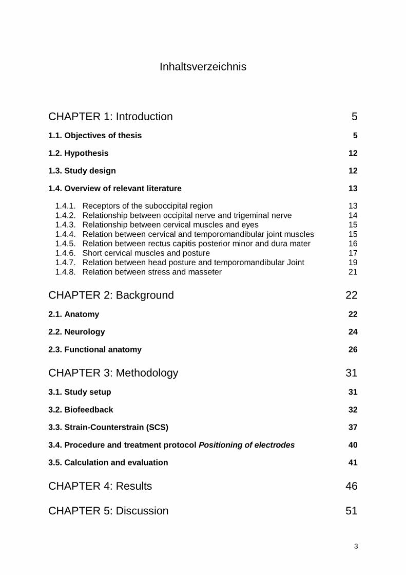
CHAPTER 1: Introduction 1.1. Objectives of thesis Out of the wide variety of existing functional relations within the human body, this study is concerned with a specific set of interrelations, i.e. the interactions between: • Short cervical muscles (m. rectus capitis posterior major, m. rectus capitis posterior minor, m. obliquus capitis superior) • Temporomandibular joint (m. masseter) • Posture • Stress (sympathetic nervous system) This network is sketched in the diagram below:
Fig. 1 Network Within this network, two concrete questions are analyzed: 1. Is it possible to prove the effects of relaxation of the short cervical muscles on the temporomandibular joint muscles and the sympathetic nervous system by means of measurements ? 2. What is the clinical relevance of a specific technique of intervention into these interrelations? This second question focuses on a clearly defined context and assumes that the following effects exist and can be analyzed:f |
Fig. 2: Interrelations examined in the thesis
6
My interest in focusing on this specific part of the network was motivated by three reasons, which will be briefly explained in the following:
a) Cooperation with dental specialists
b) The frequent occurrence of complaints in the cranio-cervical transition zone
c) The fact that this region has dramatically changed its importance in the
course of evolution by gradually developing into a central instrument of
orientation.
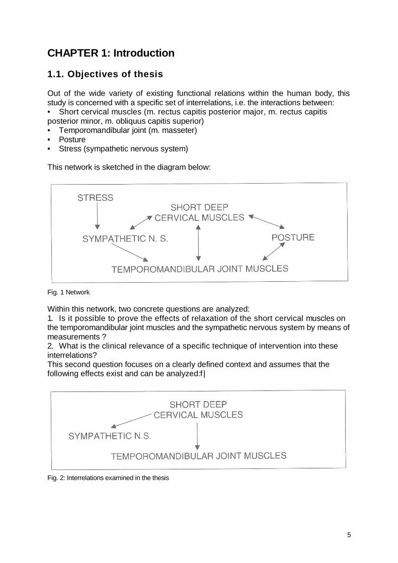
ad a) Regarding interdisciplinary cooperation with dentists and orthodontists: The experiences made in this cooperation as well as a study of the relevant literature1 show, inter alia, that most symptoms of temporomandibular joint dysfunction become manifest as masticatory muscle tension and/or pain, headaches, clicking, popping or snapping on jaw movement, and pain or loss of mobility of the cervical spine. The causes of these complaints are usually believed local in origin. Fig. 3: Pain pattern in temporomandibular joint dysfunction (ace. to Perry) (1) However, it should be borne in mind that symptoms of the cervical and mandibular regions overlap and that in patients with such complaints it is in any case advisable to examine the cervical spine as well.2 3 Experiences made in my own practice confirm this interaction. However, an increasing number of dentists and orthodontists4 know that occlusion and temporomandibular 1 Esposito CJ, Panucci PJ, Farman AG, Associations in 425 patients having temporomandibular disorders, in: J Ky Med Assoc 2000May; 98(5): 213-5 Perry HT, Muscular Changes Associated With Temporomandibular Joint Dysfunction, in: Journal of the American Dental Association, Vol. 54, No. 5 (May) 1957 Perry HT, Temporomandibular joint and occlusion, in: Angle Orthod 1976 Jul; 46(3): 284-93 2 Ciancaglini R, Testa M, Radaelli G, Association of neck pain with symptoms of temporomandibular dysfunction in the general adult population, in: Scand Rehabil Med 1999 Mar; 31(1): 17-22 3 de Wijer A, Steenks MH, de Leeuw JR, Bosman F, Helders PJ, Symptoms of the cervical spine in temporomandibular and cervical spine disorders, in: J Oral Rehabil 1996 Nov; 23(11): 742-50 4 Schottl W, Die cranio-mandibulare Regulation, Heidelberg (Huthig) 1991 Baiters W, Ausgewahlte Schriften und Vortrage, Heidelberg (Hdlzer) 1973 Jankelson RR, Neuromuscular Dental Diagnosis and Treatment, St. Loius 1990 Meyer J, Participation des afferences trigeminals dans la regulation tonique
7
dysfunction may trigger remote effects, it is for example known that: • Class II occlusion causes anterior posture shift5 • Class 111 occlusion causes posterior posture shift6 • Cross bite is frequently associated with elevation of scapula • Laterally open bite often correlates with scoliotic pelvis • Cases of prognathism are often accompanied by atlas in flexion • Abnormal afferences of the trigeminal nerve may influence posture (Meyer) • While these findings have been documented in studies, they only take account of interrelations perceived from the viewpoint of the disciplines directly involved. While thus dentists tend to focus increasingly on the temporomandibular joint and occlusion when trying to analyze the origin of neck pains and posture problems, my practical work has often given me an opportunity to observe that mandibular joint patients presented with a marked improvement of their complaints in the temporomandibular region, were generally more relaxed and also improved their postural pattern following treatment of the upper cervical spine. ad b) Regarding the frequency of this clinical picture: As palpation of the short cervical muscles shows, approx. 80% of my patients present with hypertension of the suboccipital muscles; reasons may include false sitting posture, occupational^ motivated false posture or traumas (e.g. "whiplash injury"). The. fact that the resulting hyperlordosis between C1-C2 is really so frequent may be deducted from radiological studies.7 These considerations and empirical findings show that the short cervical muscles obviously play a key role. In my opinion, the reason for this may be essentially tracked back to a phylogenetic cause.
posturale orthostatique. Interet de I'examen systematique du systeme manducateur chez les sportifs de haut niveau. These 43 55 77, Universite Rene Descartes, Paris 1977 Gole DR, A clinical observation: a relationship of occlusal contacts to distal musculature, in: Cranio 1993 Jan; 11(1): 55-61 5 Nobili A, Adversi R, Relationship between posture and occlusion: a clinical and experimental investigation, in: Cranio 1996 Oct; 14(4): 274-85 6 Nobili A, Adversi R, Relationship between posture and occlusion: a clinical and experimental investigation, in: Cranio 1996 Oct; 14(4): 274-85 7 Hardacker JW, Shuford RF, Capicotto PN, Pryor PW, Radiographic standing cervical segmental alignment in adult volunteers without neck symptoms, in: Spine 1997 Jul;22(13): 1472-80
8
ad c) Regarding the change in the importance of the suboccipital muscles: At a specific stage of the human evolution, it became absolutely essential for survival to optimize the separate movements of head and body and at the same time to achieve a maximum degree of coordination of the eyes, ears and locomotor system.
Fig. 4: Importance of the short cervical muscles in the development of the upright position (Delattre) (9) This stage represented the transition from the evolutionary development of fish, which disposed of a fixed connection between head and body and whose orientation and behavior were exclusively controlled by their sensory organs, towards terrestrial animals: "As in fish head and body form a functional unit, the orientation and behavior of the entire body under conditions of earth gravity can only be controlled from the head via the sensory organs located there."8 The first "terrestrial animals", the amphibians, originally developed an atlanto-occipital joint that enabled them to handle many activities essential for survival ashore (and thus for dealing with gravity): feeding, recognizing enemies, defense. The new, flexible connection between head and body permitted them to move freely but at the same time directed a wide variety of proprioceptive information from the body to the vestibular nuclei. Reptiles, the next link in the evolutionary chain, already featured an additional pivot joint between atlas and axis although the neck did not yet permit unrestricted movement, the system was still relatively undifferentiated and the cervical muscles as yet unstructured. The mammals then completed this development by increasingly fine-tuning and differentiating the structure of the cervical muscles and thus of the proprioceptive system. The available information concerning posture and movement of the body largely derives from this suboccipital region. For this reason, it appears logical to regard the cervical muscles, not primarily as a component of the locomotor system, but rather as a field of receptors for regulating the sense of balance and the proprioceptive coordination of sensory organs and locomotor functions.
8 Wolff HD, Neurophysiologische Aspekte des Bewegungssystems, 3.Aufl., Berlin (Springer) 1996
9
All information is centrally correlated (suboccipital region, eyes, internal ear, cerebellum, foot) and evaluated in order to maintain or recover posture and balance. The muscles must be precisely activated to be able to adapt to gravity vectors by changing muscle tone. Aquatic mammals such as e.g. dolphins have lost most of the mobility of the upper cervical spine. This may serve as yet another piece of proof that the effect of gravity in the vertical position necessitates mobility of this zone. In particular, the gradual assumption of an upright, vertical position of the body entailed a specific transformation in the area of cranium and cervical spine,9 The occiput position changed through anterior rotation; thus the foramen magnum assumed a new, horizontal position. As a consequence of this change, the center of gravity of the head (in the sella turcica region) was now only slightly anterior of the atlanto-occipital turning point. This called for yet another adjustment: the tendency of the head to tilt forward had decreased, and thus the short cervical muscles were no longer primarily needed to prevent the head from tilting forward. Rather, this zone now served to help get the bearings of the surrounding space.
Fig. 5: Adaptation of cranium and cervical region to the vertical position: The neuro-cranium of these four primate species presents a largely identical anterior-posterior diameter. The gradual change from A to D concerns the direction of traction of the cervical fibers. The arrow pointing downwards on the diagrams indicates the position of the center of gravity in each cranium. The arrow pointing upwards indicates the position of the occipital condyles. While in the course of evolution the condyles gradually moved in the anterior direction, the center of gravity assumed a posterior position, which in present-day humans is nearly identical with that of the condyles. (Garlick10)
For the zone of the temporomandibular joint, the new, upright posture made it necessary that the mandible must be kept in its rest position by jaw elevator contraction. This results in the functional unity of mandibular joint and upper cervical spine. When the mouth is opened, the suboccipital muscles must counteract the tilting forward of the head; conversely, when the head is bent backwards, the 9 Delattre A, Fenart R, L'hominisation du crane etudiee par la Methode Vestibulaire, Paris (Editions du Centre National de la Recherche Scientifique) 1960. 10 Garlick D, Proprioception Posture and Emotion, Bankstown (Adept Printing Pty Ltd.) 1982
10
masticatory muscles must be activated to prevent the mouth from opening automatically. Thus a number of new interactions in the suboccipital region of the temporomandibular joint had to be put in place and hence laid the basis for the outstanding importance of the suboccipital region as the - so far - last link in the evolutionary chain. According to phylogeny, the cranio-cervical transition zone also constitutes the ontogenetically oldest region of the body. The first somites developed in this region as the starting-point for the entire trunk. The myotomes of the cranial somites form the muscles of tongue, neck and partially also larynx and pharynx, thus clearly reflecting their functional interaction.11 Some years ago, my interest in this topic first motivated me to carry out a number of preliminary studies, which showed that patients with suboccipital muscle contraction often presented with an increased sympathetic nerve tone, an increased masseter tone and reduced stress tolerance. Moreover, it has been proven12 that biofeedback permits the targeted treatment of this group of symptoms, and that electromyography constitutes an accepted examination method that also entails reproducible findings.13 In 1996, I conducted a pilot study together with Dr. Kropfreiter (as biofeedback specialist), in which three temporomandibular joint patients were monitored before, during and after osteopathic therapy sessions (strain and counterstrain according to Jones) by means of biofeedback measuring. It was shown that the following measured values had changed in all patients after therapy: • the skin conductance level was reduced, • the skin temperature increased, • the pulse amplitude likewise increased in two patients. These findings may be regarded as a clear indication of reduced sympathetic activity. During treatment, the EMG values of the masseter presented increased activity, while the post-therapy muscle tone was lower than before treatment. The above observations were to show that and why this topic is so important and why it has been engaging my heightened interest for a number of years. However, all key questions dealt with in this thesis are of significance for osteopathy as well. Specialists of the locomotor system have for many years regarded the zone covered in this work as particularly important: • For example, Alexander14 treats patients with posture problems by strengthening their awareness of the transition from head to neck. 11 Moore KL, Embryologie. Lehrbuch und Atlas der Entwicklungsgeschichte des Menschen, 3. Aufl., Stuttgart (Schattauer) 1990 12 Slavicek G, Gsellmann B, Gruber R, Rath M, Furhauser R, Biofeedback als Therapieerganzung bei craniomandibularer Dysfunktion, in: IOK, 27. Jhrg. (1995) Nr. 1 13 Ferrario VF, Sforza C, Miani A Jr, D'Addona A, Barbini E, Electromyographic activity of human masticatory muscles in normal young people. Statistical evaluation of reference values for clinical applications Ferrario VF, Sforza C, Miani A Jr, D'Addona A, Reproducibility of electromyographic measures: a statistical analysis 14 MacDonald G, The complete illustrated Guide to the Alexander Technique, (Paperback) 1998
11
• Various chiropractic schools only work with manipulations of C0-C1- C2 to - successfully - combat a variety of complaints. • In Germany, there exists the so-called atlas therapy (according to Arlen), in which treatment consists exclusively of a short impulse acting on the atlas. • The fact that Dr. Andrew T. Still, the father of osteopathy, used to place the back of his head (the suboccipital region) on a piece of taut wire to combat headaches in his childhood may be of historical interest. • The cranio-cervical transition presents a high degree of neurological cross-iinking as well as a great number of muscle spindles15, which communicate a large volume of information and thus permit very precise proprioception. The complaints located in this zone are therefore often very complex and frequently call for interdisciplinary cooperation. • Due to the great number of spindles, this zone can easily become a facilitated segment as described by Korr16. This results in a vicious circle (circulus vitiosus): even minimal (psychological, thermal, etc.) stimuli may entail momentous effects. • Manipulation in this zone C0-C1 is described as potentially hazardous in the relevant literature17. However, this opinion should be relativized since manipulation entails a lower complication rate than non steroidal anti-inflammatory drugs (NSAlDs), the agents most frequently used by physicians to combat pains in that region.18 – For this reason, my study uses a less invasive method (strain-counterstrain). Moreover, the strain and counterstrain technique works very specifically by reprogramming the spindles and hence influencing the proprioceptive system, which is of such great importance in this zone. However, apart from these arguments relating to the subject-matter of the present thesis, it should be emphasized that osteopathy needs more studies that are up to experimental requirements, i.e. that couch the enormous wealth of practical experience in hypotheses to be tested under research conditions.
15 Richmond F.R.J, and Abrahams V.C. (1975) Morphology and distribution of muscle spindles in dorsal muscles of the cat neck. J. NeurophysioL, 38:1322-1339. Richmond F.R.J, and Abrahams V.C. (1979) Physiological properties of muscle spindles in dorsal neck muscles of the cat. J. NeurophysioL, 42: 604-617 Richmond F.R.J., Anstee G.C.B., Sherwin E.A. and Abrahams V.C. (1976) Motor and sensory fibers of neck muscle nerves in the cat. Canad. J. Physiol. Pharmacol., 54: 294-304. 16 Korr IM, The physiological basis of osteopathic medicine, New York (Insight Publishing) 1982 17 Ralf L, Rydell N, Spinal manipulation-treatment associated with a high risk of complications, Stockholm (Personskaderegeling AB) 1999 Hufnagel A, Hammers A, Schonle PW, Bohm KD, Leonhardt G, Stroke following chiropractic manipulation of the cervical spine, in: J Neurol 1999 Aug; 246(8): 683-8 Parent! G, Orlandi G, Bianchi M, Renna M, Martini A, Murri L, Vertebral and carotid artery dissection following chiropractic cervical manipulation, in: Neurosurg Rev 1999 Oct; 22(2-3): 127-9 Hurwitz EL, Aker PD, Adams AH, Meeker WC, Shekelle PG, Manipulation and mobilization of the cervical spine. A systematic review of the literature, in: Spine 1996 Aug 1; 21(15): 1746-59; Di Fabio RP, Manipulation of the cervical spine: risks and benefits, in: Phys Ther 1999 Jan; 79(1): 50-65 Haldemann S, Kohlbeck FJ, McGregor M, Risk factors and precipitating neck movements causing vertebrobasilar artery dissection after cervical trauma and spinal manipulation, in: Spine 1999 Apr 15; 24(8): 785-94 Assendelft WJ, Bouter LM, Knipschild PG, Complications of spinal manipulation: a comprehensive review of the literature, in: J Fam Pract July 1996 18 Dabbs V, Lauretti WJ, A risk assessment of cervical manipulation vs. NSAIDs for the treatment of neck pain, in: J Manipulative Physiol Ther 1996 March
12
1.2. Hypothesis On the basis of the positive results of the preliminary study, the present thesis was planned in accordance with the following working hypothesis: "In patients with at least two temporomandibular joint symptoms and simultaneous dysfunction in the suboccipital muscle zone, the therapy of the short cervical muscles using an osteopathic technique (strain/counterstrain) can lead to relaxation of the masseter and to reduced sympathetic activity." This mode of treatment was selected because it is a method devoid of risks for the cervical spine that may even be used in anxious and tense persons (a very frequent phenomenon in temporomandibular patients) and moreover permits the specific reprogramming of the proprioceptive spindle system and thus the discontinuation of even deeply entrenched false reflexes. Biofeedback was selected as an optimum method of measuring changes in the autonomic nervous system while electromyography serves as a muscle tone parameter.19
1.3. Study design A group of 23 patients aged between 26 and 68 years was selected; each of them presented with at least two temporomandibular joint symptoms (reduced mobility, pain, clicking). At the same time, a tender/trigger point in the suboccipital zone was required as well. This overall group was then divided into a test group and a control group. The test group was treated using the strain / counterstrain method, while the control group was given placebo treatment. The patients were assigned to the groups on a randomized basis. Three biofeedback parameters (skin temperature, skin conductance level, pulse rate) and the electric activity of the masseter on both sides were measured before, during and after treatment.
19 Cooper BC, Cooper DL, Electromyography of Craniomandibular Disorders, in: Laryngoscope 101: February 1991
13
1.4. Overview of relevant literature Due to the great number of publications on this issue, the following list contains only the most important studies in chronological order to provide a better overview. Receptors of the suboccipital region.
1.4.1. Receptors of the suboccipital region Two studies by Voss (1958) and Cooper (1963)20, respectively, identified a particularly great number of spindles in the hyoid muscles, in the sternocleidomastoid muscle and in the suboccipital muscles of humans and cats. Richmond and Abrahams (1975)21 too, have shown that the neck muscles of cats are rich in spindles per gram. The number of spindles per gram was measured: Spienius m. 47-66/g Complexusm. 71 -107/g Rectus major m. 48-84/g Not all neck muscles present this high spindle density. For example, the occipitoscapularis of cats presents a spindle density roughly similar to that of the locomotor muscles of the hindleg (13-19 per gram). (The occipitoscapularis (rhomboideus capitis) of cats partly resembles the human rhomboid muscle and partly also the levator scapulae.) The great number of spindles in the suboccipital muscles indicates that the relation between head and cervical spine is very important for the central coordination of posture. Moreover, the cervical muscle afferences tend towards the cerebellum and the complexes of the ipsilateral vestibular nuclei. In case of hypertonicity of the neck muscles, symptoms such as: - disequilibria - nausea - lack of concentration - neck headaches - impaired vision can be observed. These symptoms often accompany whiplash injuries. The technique selected for the present study is able to normalize the spindles (strain / counterstrain).
20 Voss H. (1958) Zahl und Anordnung der Muskelspindeln in den unteren Zungenbeinmuskeln dem M. sternocleidomastoideus und den Bauch- und tiefen Nackenmuskeln. Anat. Anz. 105: 265-275. Cooper S. and Daniel P.M. (1963) Muscle spindles in man, their morphology in the lumbricals and the deep muscles of the neck. Brain, 86: 563-594. 21 Richmond F.R.J, and Abrahams V.C. (1975) Morphology and distribution of muscle spindles in dorsal muscles of the cat neck. J. Neurophysiol., 38: 1322-1339, Richmond F.R.J, and Abrahams V.C. (1979) Physiological properties of muscle spindles in dorsal neck muscles of the cat. J. Neurophysiol., 42: 604-617 Richmond F.R.J., Anstee G.C.B., Sherwin E.A. and Abrahams V.C. (1976) Motor and sensory fibers of neck muscle nerves in the cat. Canad. J. Physiol. Pharmacol., 54: 294-304.
14
Richmond (1999)22 remarks that the type 1 fibers of the obliquus capitis inferior are distributed as follows: 95-100% are located in the deep layer and less than 10% are located in the superficial layer. A large majority of spindles are concentrated in the type 1 fibers in the deepest layers. It may thus be concluded that the larger part of the neck muscles serves to coordinate posture. In a study by Uhlig (1995)23, a biopsy of the rectus capitis posterior major has shown that disorders of this zone (e.g. following an accident) lead to a transformation of the muscle fibers from "slowly oxidative posture muscles" to "rapidly glycolytic locomotor muscles"; hence stamina and stabilization in this zone become insufficient. These observations permit the conclusion that spindle reprogramming may be especially successful in such cases.
1.4.2. Relationship between occipital nerve and trigeminal nerve Skillern24 (1954) described a form of communication between the occipital nerve (C2) and the first ramus of the trigeminal nerve via the medulla. The results permit the following conclusion: pains caused by a disorder of the C2 root begin in the suboccipital region and radiate up to the vertex and forward behind the ipsiiateral eye. Often patients feel a pain as if the eyeball were torn from the socket. The headache is similar to migraine and often accompanied by nausea, vomiting and blurred vision. Skillern's study may be regarded as a valuable contribution to the understanding of hemicranial attacks resulting from cervical spine dysfunction. These results are confirmed by a study by Schimek (1988)25: He injected healthy test subjects with a hyperosmotic saline solution in one muscle of the deep autochthonous suboccipital neck muscles and recorded their statements regarding the thus triggered subjective pain propagation in the head. For each muscle, this resulted in characteristic pain bands, all of which are located in the sensitive innervation zone of the trigeminal nerve. For their study, Ellis and Kosmorsky (1995)26 selected patients presenting with eye or orbital pain attributed to cetvical dysfunction. The administration of suboccipital injections with a local anesthetic relieved the pain in 80% of the cases. This clearly indicates a link between the cervical region and the trigeminal nerve. The fact that pathological afferences from the "receptor field in the neck" are also linked to the core zone of the trigeminal nerve is documented by other clinical and experimental studies.27
22 Richmond FJ, Corneil BD, Marked non-uniformity of fiber-type composition in the primate suboccipital muscle obfiquus capitis inferior, Exp Brain Res 1999 Mar, 125{1): 14-8 23 Uhlig Y, Weber BR, Grob D, Muntener M, Fiber composition and fiber transformations in neck muscles with dysfunction of the cervical spine, J Orthop Res 1995 Mar, 13(2): 240-9 24 Skillern P.G. (1954) Great occipital-trigeminus syndrome as revealed by induction block, Arch. Neurol. & Psychiat,, 72: 335-340. 25 Schimek JJ, Untersuchungen zum Spannungs-Kopfschmerz, in: Man Med 1988,26:107-112 26 Ellis B.D. and Kosmorsky G.S. (1995) Referred ocular pain relieved by suboccipital injection. Headache, 35(2): 101-103 27 Vadokas V, Lotzmann KU (1995) Funktionelle Storungen des kraniomandibularen Systems in der HWS als differentialdiagnostisches Problem der idiopathischen Trigeminusneuralgie. Der Schmerz
15
1.4.3. Relationship between cervical muscles and eyes The results of the study by Lennerstrand, Han and Velay (1996)28 confirm that neck muscle vibration can induce eye position changes, in its turn, this seems to confirm that the proprioceptive messages originating in the neck muscle are processed together with visual r information of eye position in determining gaze direction. In a similar study, Han (1999)29 has shown that stimulation of neck muscle proprioceptors by means of vibration can induce eye position change and visual illusory movement in healthy subjects. The direction of apparent movement is vertical if the back muscles of the neck are stimulated and horizontal when the lateral-rotation muscles are stimulated. This permits concluding that proprioceptive information from the neck muscles plays an important role in regulating gaze direction. By way of summary, we may say that these studies explain why the eye is of interest in this context. In the above-discussed network, it is the link between short cervical muscles and posture. Thus the eye can influence posture, mandible position and masseter tone. Phylogenetically, the visual system grew tremendously in importance with the evolution from the quadruped to the upright position (all of a sudden the range of vision had increased immensely), contrary to the past when the other senses (hearing, smell) had been dominant. Now the eyes provided substantially more information that had to be coordinated - on the basis of the retinal system (vision) on the one hand and on the basis of the proprioceptive eye muscle system on the other hand.
1.4.4. Relation between cervical and temporomandibular joint muscles Funakoshi and Amano (1972)30 studied the influence of the tonic neck reflex on the jaw muscles of rats, whose ear labyrinths were destroyed after decerebration. Electric activities of the jaw muscles increased or decreased in response to rotation, tilting or flexion of the head. The electromyographic responses to head position were abolished after the first three cervical nerves were cut. It was concluded from these tests that the tonic neck reflex has an influence on the jaw muscles. Urbanowicz (1991)31 established that there exists an interrelationship between muscles and joint positions of the temporomandibular joint on the one hand and 9:29-33 28 Lennerstrand G., Han Y. and Velay J.L (1996) Properties of eye movements induced by activation of neck muscle proprioceptors. Graefes Arch Clin Exp Ophthalmol 1996 Nov., 234(11): 703-709. 29 Han Y, Lennerstrand G, Eye position changes induced by neck muscle vibration in strabismic subjects, in: Graefes Arch Clin Exp Ophthalmol 1999 Jan; 237(1 (: 21-8 30 Funakoshi M. and Amano N. (1973) Effects of the Tonic Neck Reflex on the Jaw Muscles of the Rat. J. Dent. Res. Juty-August 1973, Vol. 52, No.4. 31 Urbanowicz M. (1991) The Journal of Craniomandibular Practice, April 1991, Vol.9, No.2.
16
muscle length and strength and joint position of the occiput and upper cervical spine on the other hand. Physiological dynamic equilibrium of both regions constitutes the ideal case. Decrease of the VDO (vertical dimension) by using an intraoral device will lead to suboccipital compression. Macaluso, De Laat and Pavesi (1996)32 demonstrated that the asymmetric tonic neck reflex has a significant influence on the H-reflex of the human temporal muscle. (The H-reflex is a monosynaptic muscle-stretching reflex triggered by the electric stimulation of the afferent fibers of the muscle nerve. The abbreviation of this term is a reference to Paul Hoffmann, who first identified this reflex)33. Dessem (1999)34 studied the synaptic contacts between masticatory-muscle spindle afferents and brainstem neurons, which project to the cervical spinal cord in rats. It is hypothesized that these pathways are primarily involved in the coordination of jaw and neck movement during mastication. These studies clearly highlight the interaction between the tone of suboccipital and temporomandibular muscles; it is, however, not always clear where the problem actually originates. Often symptoms do not appear where they are caused. Thus treatment of the short cervical muscles can show whether the tone of the masseter and temporal muscles has returned to normal after therapy. This zone often requires an interdisciplinary approach.
1.4.5. Relation between rectus capitis posterior minor and dura mater In 1994, Hallgren and Hack (1995)35 36 37 made a surprising discovery when using a sagittal incision to dissect a human neck region. They found that the rectus capitis posterior minor is directly linked to the dura mater in the atlanto-occipital joint region. However, the authors likewise demonstrated that pressure applied intra-operatively on the posterior zone of the dura mater triggers pain in the suboccipital region.38
Since the myodural bridge has a direct influence on the pain-sensitive dura mater, a possible link between cervical muscles and headaches is postulated. On the basis of these findings, it may be hypothesized that the bridge between rectus capitis minor und dura mater is stretched in a whiplash trauma, which would explain the chronic symptoms of such patients. 32 Macaluso G.M., De Laat A.D. and Pavesi G. (1996) The influence of the asymmetric tonic neck reflex on the H-reflex in human temporal muscle. Minerva Stomatol 45(9): 387-392. 33 Bruggencate, G.T. (1984), Medizinische Neurophysiologie, Thieme 34 Dessem D, Luo P Jaw-muscle spindle afferent feedback to the cervical spinal cord in the rat, in: Exp Brain Res 1999 Oct; 128(4): 451-9 35 Hack G., Koritzer R, Robinson W., Hallgren R. and Greenman P. (1995) Anatomic relationship between rectus capitis posterior minor muscle and the dura mater, Spine 1995, December 1, 20: 23 2484-2486 36 Hallgren R., Clinical implications of a cervical myodural bridge, AAO Journal volume 7, number 4, 1997 37 Hack GD, Lipton JA, The Clinical Implications of a Suboccipital Myodural Bridge, Proceedings of the American Academy of Osteopathie Scientific Convention, 1995 Oct. 29-31, Orlando, FL 38 Northfield D, Some observations of headache, in: Brain, 1938; 61:133-162
17
Two further works confirm the findings of these studies: Mitchell (1998)39 has proved attachments of the ligamentum nuchae to the dura mater as well as to the posterolateral part of the occipital bone. In his study, Rutten (1997)40, too, refers to the anatomical relation between rectus capitis posterior minor and dura mater. The findings of the study by Hu (1995)41 document that dural vascular irritation leads to activation of the neck and jaw muscles; clinically, this points towards a relation to headaches and facial neuralgia. By way of summary, it may be said that hypertonicity of the rectus capitis posterior minor causes permanent tension of the dura mater. As Hu has shown, this entails irritation of the meningeal vascular systems and, in due course, hyperactivity of the jaw and neck muscles. Thus improving the tone of the rectus capitis posterior minor may normalize dural blood flow and hence the tone of the jaw and neck muscles.
1.4.6. Short cervical muscles and posture The fact that severing the neck muscles will induce postural and movement dysfunction was already demonstrated by Longet in animal experiments conducted in 1845.42 A study by Magnus (1926)43 has shown that the upper cervical segment is an important proprioceptor organ for postural processes. The distribution of muscle tone in decerebrated animals can be influenced by passive head movements. After extirpation of the labyrinth, the tonic neck reflexes may be observed individually; in case of dorsal flexion of the neck, they appear as extension of the forelegs and flexion of the hindlegs. Ventroflexion of the neck has the opposite effect, i.e. leads to reinforcement of the flexor tone in the upper and of the extensor tone in the lower extremities. Head rotation causes ipsilateral extension and contralateral flexion of foreleg and hindleg. By anesthetizing the upper cervical muscles, Abrahams (1969)44 caused false posture in laboratory animals. De Jong (1977)45 observed ataxia, dizziness and nystagmus in humans following 39 Mitchell BS, Humphreys BK, O'Sullivan E, Attachments of the iigamentum nuchae to cervical posterior spinal dura and the lateral part of the occipital bone, in: J Manipulative Physiol Ther 1998 Mar-Apr; 21(3): 145-8 40 Rutten HP, Szpak K, van Mameren H, Ten Holter J, de Jong JC, Anatomic relation between the rectus capitis posterior minor and the dura mater, in: Spine 1997 Apr 15; 22 (8): 924-6 41 HU JW, Vernon H, Tatourian I, Changes in neck electromyography associated with meningeal noxious stimulation, in: J Manipulative Physiol Ther 1995 Nov-Dec; 18 (9): 577-81 42 Longet F.A. (1845) Sur les troubles qui surviennent dans I'equilibration, la station et la locomotion des animaux apres la section des parties molles de la nuque. Gaz. Med. France, 13: 565-567 43 Magnus R. (1926) Some results of studies in the physiology of posture (Cameron Prize Lectures). Lancet, 211:531-536. 44 Abrahams V.C. and Falchetto S. (1969) Hind leg ataxia of cervical origin and cervico-lumbar interactions with a supratentorial pathway. J.Physiol. (Lond.), 203: 435-447. 45 De Jong P.T.V.M., De Jong J.M.B.V., Cohen B. and Jongkees L.B.W. (1977) Ataxia and nystagmus
18
injection of local anesthetics in the suboccipital muscles. Fukuda (1981)46 47 studied different posture patterns during various sport activities and compared them with the postural reflexes identified by Magnus. He concluded that these reflexes are latently present and may be used by adults to correct posture and control balance. Taylor (1988)48 discovered that the suboccipital muscles have an influence on proprioception and head-on-body orientation. Somato-sensory inputs from all parts of the body contribute to balance control during quiet stance: according to a corresponding study by Roll (1988)49, minivibrators were used to excite eye, neck and ankle muscles. The study demonstrated that vibration to the eye muscles of a standing subject with eyes closed produced body sway, with the sway direction depending on the muscle vibrated. Body sway also was produced by vibration to the sternocleidomastoideus and soleus. When these muscles were vibrated simultaneously, the effects were additive. This suggests that proprioception from all parts of the body plays an important role in the maintenance of quiet stance body posture. A study by Pollard (1997)50 highlights the remote action of the short cervical muscles: he employed a suboccipital stretching technique, which resulted in increased flexion ROM at the hip. Koskimies (1997)51 has shown that erroneous facilitation of the cervico-ocular reflex may increase neck tension. To quantify postural adjustments over time, Fransson (1998)52 investigated twelve normal subjects using posturography during vibration either towards the calf or the paraspinal neck muscles; with eyes open vs. eyes closed. The stimulus response adjustments over time were found to be almost identical for all test conditions though smaller during eyes-open conditions. In his study, Lekhel (1998)53 used vibration applied unilaterally to the dorsal neck muscles, thereby demonstrating that vibration induced a forward postural deviation in normal subjects.
induced by injection of local anaesthetics in the neck. Ann. Neurol., 1: 240-246. 46 Fukuda T. (1961)Studies on human dynamic postures from the viewpoint of postural reflexes. Acta Otolaryngol. (Stockh.), Sup. 161 47 Fukuda T. (1981) Statokinetic Reflexes in Equilibrium and Movement. University of Tokyo Press, Tokyo 48 Taylor J.L. and McCioskey D.I. (1988) Proprioception in the neck. Exp. Brain Res. 70: 351-360. 49 Roll Jp, Roll R, From eye to foot: a proprioceptive chain involved in postural control, in: Amblard B, Berthoz A, Clarac F, eds. Posture and gait: development, adaptation and modulation. Amsterdam: Elsevier (1988) 155-164 50 Pollard H., Ward G. (1977) J Manipulative Physiol. Ther. 20(7): 443-447 51 Koskimies K, Starck J, Sutinen P, Aalto H, Toppila E, Hirvonen T, Ishizaki H, Aiaranta H und Pyykko I, Postural stability, neck propriozeption and tension neck, Acta Otolaryngol Suppl (1997) 529:95-7 52 Fransson P-A, Magnusson M, Johansson R, Analysis of adaption in anterioposterior dynamics of human postural control, Gait & Posture, Volume 7, Isxsue 1, pp. 64-74 (January 1998) 53 Lekhel H, Popov K, Bronstein a, Gresty M, Postural responses to vibration of neck muscles in patients with uni-and bilateral vestibular loss, in: Gait & Posture, Volume 7, Issue 3, pp. 228-236 (May 1998)
19
Sakuma (1999)54 highlighted the important position of the neck muscles for whole-body balance, and Talis (1999)55 drew attention to the influence on balance exerted by vibration to the short cervical muscles. Berger (1998)56 observed that direction and ränge of arm movements are dependent on head-on-body orientation. All these studies show clearly that a change in Short neck muscle tone entails a change of posture. Thus the function of the Short neck muscles is largely proprioceptive and less a locomotor function. In osteopathy, Hall57 58 has already indicated the connection between posture, jaw position and specific Symptoms. He describes an "anterior type", which corresponds to a pattern with anterior orientation with respect to both posture (pelvic anteversion, hyperextension of Iower extremity, lumbar hyperlordosis) and jaw (mandibula anterior). The "posterior type" presents the opposite picture (pelvic retroversion, flexion of Iower extremity, increased kyphosis of dorsal spine and mandibula posterior). The origin of the postural pattern varies from subject to subject. In any case, it seems possible that dysfunction in the C0-C1 zone can change the entire postural pattern and entail Symptoms. Nicolakis (2000)59 has also described postural and muscular dysfunction as more frequent in patients presenting with cranio-mandibular dysfunction (CMD), which however does not permit a clear answer as to whether postural dysfunction should be regarded as consequence or cause of cranio-mandibular dysfunction. Furtheir studies by Robinson (1966)60 and Goldstein (1984)61 document the marked influence of head posture on the temporomandibular Joint and temporomandibular muscles.
1.4.7. Relation between head posture and temporomandibular Joint
54 Sakuma A, Aihara Y, Influence of propriozeptive input from leg, thigh, trunk and neck muscles in the equilibrium of standing, Nippon Jibiinkoka Gakkai Kaiho (1999) 102(5): 643-9 55 Talis VL, Ivanenko YP, Kazennikov OV, Support stability influences postural responses to muscle vibration in humans, Eur J Neurosci 1999 Feb, 11(2): 647-54 56 Berger M, Lechner-Steinleitner S, Kozlovskaya I, Holzmüiler G, Mescheriakov S, Sokolov A, Gerstenbrand F, The effect of head-to-trunk position on the direction of arm movements before, during and after space flight, J Vestib Res 1998 Sep-Oct, 8(5):341-54 57 Hall, The Mechanis of the Spine and Pelvis , Maidstone College of Osteopathy 58 Amigues, J.P. L1 A.T.M Une Articulation entre UOsteopathe et Le Dentiste, Editions de VERLAQUE 59 Nikolakis P, Relationship between Craniomandibular Disorders and Poor Posture, The Journal of Craniomandibular Practice, April 2000 60 Robinson M.J., The Influence of Head Position on Temporomandibular Joint Dysfunction, Chicago College of Osteopathie 61 Goldstein D.F., Influence of cervical posture on mandibular movement, The Journal of Prosthetic Dentistry, September 1984
20
In his study, Boyd (1987)62 has proved that head posture influences masticatory muscle tone. Extension increases the activity of the temporal muscle, while flexion intensifies the activity of masseter and digastricus. A study by Lee (1995)63 demonstrated that in patients with temporomandibular disorder the head is positioned more forward than in healthy subjects. Gonzalez (1996)64 tried to establish whether a forward head posture is able to influence cranio-facial growth so as to determine morphoskeletal and neuromuscular dysfunction. This head posture was in fact correlated to Class II occlusion. (This is a defective position of the teeth where the upper cover the Iower incisors.65) Zonnenberg (1996)66 established that body posture constitutes an etiologic factor in patients with temporomandibular disorders. A study by Higbie (1999)67 has shown that head position is an important factor in determining the amount and direction of mandibular opening, and Yamada (1999)68 demonstrated that head position influences the direction and stability of mandibular closing. Visscher (2000)69 recorded mandibular movements in ten test subjects without craniomandibular or cervical spine disorders using five different head postures including natural head posture and forward head posture. The study showed that in a military posture, the opening movement path of the incisal point is shifted anteriorly relative to the path in a natural head posture. In a forward head posture, the movement path is shifted posteriorly, anet during lateroflexion, it deviates to the side the head has moved to. In a study conducted by Wright (2000)70, significant correlations between improvements of temporomandibular symptoms and of head posture were established. Temporomandibular symptoms are often alleviated by systematic posture training, in which the head is held farther forward relative to the shoulders.
62 Boyd CH, William FS, Boyd CM, Bryant RW, Wiygul JP, The Effect of Head Position on Electromyographic Evaluations of Representative Mandibular Positioning Muscle Groups, in: The Journal of Craniomandibular Practice, 1987 Jan; Vol. 5, No. 1 63 Lee WY, Okeson JP, Lindroth J, The relationship between forward head posture and temporomandibular disorders, in: J Orofac Pain 1995 Spring; 9(2): 161-7 64 Gonzalez HE, Manns A, Forward head posture: ist structural and functional influence on the stomatognathic System, a conceptual study, in: Cranio 1996 Jan; 14(1): 71-80 65 Angle EH, Malocclusion of the Teeth, 7th ed., Philadelphia (S.S. White Dental Manufact. Co.) 1907 66 Zonnenberg AJ, Van Maanen CJ, Oostendorp RA, Elvers JW, Body posture photographs as a diagnostic aid for musculoskeletal disorders related to temporomandibular disorders (TMD), in: Cranio 1996 Jul; 14(3): 225-32 67 Higbie EJ, Seidel-Cobb D, Taylor LF, Cummings GS, Effect of head position on vertical mandibular opening, in: J Orthop Sports Phys Ther 1999 Feb, 29(2): 127-30 68 Yamada R, Ogawa T, Koyano K} The effect of head posture on direction and stability of mandibular closing movement, in: J Oral Rehabil 1999 Jun; 26(6): 511-20 69 Visscher CM, 70 Wright EF, Domenech MA, Fischer JR Jr, Usefulness of posture training for patients with tempoiromandibular disorders, in: J Am Dent Assoc 2000 Feb; 131(2): 202-10
21
1.4.8. Relation between stress and masseter Schroeder (1991)71 recorded the spontaneous muscular activity of the masseter and its activity under conditions of noise and flickering light. The effect of the applied stimuli on muscular activity was not homogeneous, leading to activation in some cases and inhibition in others. In this, anxious patients showed higher levels of muscular activity than non-anxious patients. One fifth of the patients exhibited neck muscle activity occurring simultaneously with masseter activity. The study by Richardin (1995)72 used an animal model to prove the influence of stress on masticatory movements, inter alia on masseter tension. The animal experiments conducted by Grassi (1996)73 demonstrated that stimulation of the sympathetic cervical nerve in hares simultaneously stimulated the temporomandibular joint muscles. Ruf (1997)74 analyzed the masseter muscles of 15 test subjects. EMG activity during a stress situation was significantly greater than for a non-stress situation. The study by Ruggieri (1999)75 deals with the role of aggressiveness as a modulator of muscular tension. It was shown that aggression can produce chronic muscular tension of the oral region. All these studies demonstrate clearly that stress activates the sympathetic nervous system and thus constitutes an important factor for increased masseter tension.
71 Schroeder H, Siegmund H, Santibanez G, Kluge A, Causes and signs of temporomandibular joint pain and dysfunction: an electromyography! investigation, in: J Oral Rehabil 1991 Jul; 18(4): 301-10 72 Richardin P, Wetphal A, Divry M, Didier G, Influence of stress and occlusal interference on the EMG activity of some masticatory muscles during a single mastication cycle, in: J Oral Rehabil 1995 Oct; 22(10): 775-80 73 Grassi C, Deriu F, Roatta S, Santarelli R, Azzena GB Passatore M, Sympathetic control of skeletal muscle function: possible cooperation between noradrenaline and neuropeptide Y in rabbit jaw muscles, in: Neurosci Lett 1996 Jul 19; 212(3): 204-8 74 Ruf S, Cecere F, Kupfer J, Pancherz H, Stress-induced changes in the functional electromyographic activity of the masticatory muscles, in: Acta Odontol Scand 1997 Jan; 55(1): 44-8 75 Ruggieri V, Persico G, Caputo G, An analysis of the psychophysiological components in subjects with temporomandibular disorders. Myographic tension and the management of aggressiveness, in: Minerva Stomatol 1999 Oct; 48(10): 477-84
22
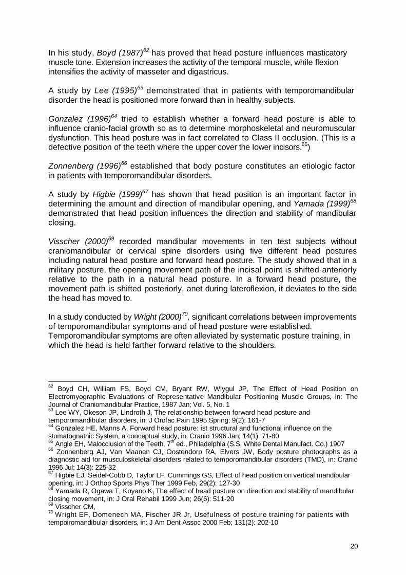
CHAPTER 2: Background 2.1. Anatomy Suboccipital muscles Amongst the muscles extending between occiput and the first two cervical vertebrae, we find representatives of the spinal, inter-transversal and spino-transversal systems. Contrary to the long muscles stretching across the atlanto-occipital joint, they act on the free control of these systems and are well defined as individual muscles. The present study only concerns the posterior suboccipital muscles, which are described in greater detail below. The suboccipital muscles do not present the spatial orientation that might be implied from the classic drawings in manuals of anatomy (Platzer, Sobotta, Netter, Gosling76).
Fig. 6: Short cervical muscles (musculi suboccipitales) according to Platzer The obliquus capitis superior is situated horizontally below the occiput. The rectus capitis minor has an approximately horizontal position. This explains their importance for swaying head movement.
76 Frick H, Allgemeine Anatomie. Spezielle Anatomie (1977); Platzer W, Kahle W, Leonhardt H, Taschenatlas der Anatomie fur Studium und Praxis (1984); Netter FH, Atlas of Human Anatomy (1989); Gosling JA, Human Anatomy (1996); Staubesand J (Hrsg.), Sobotta. Atlas der Anatomie des Menschen (1988)
23
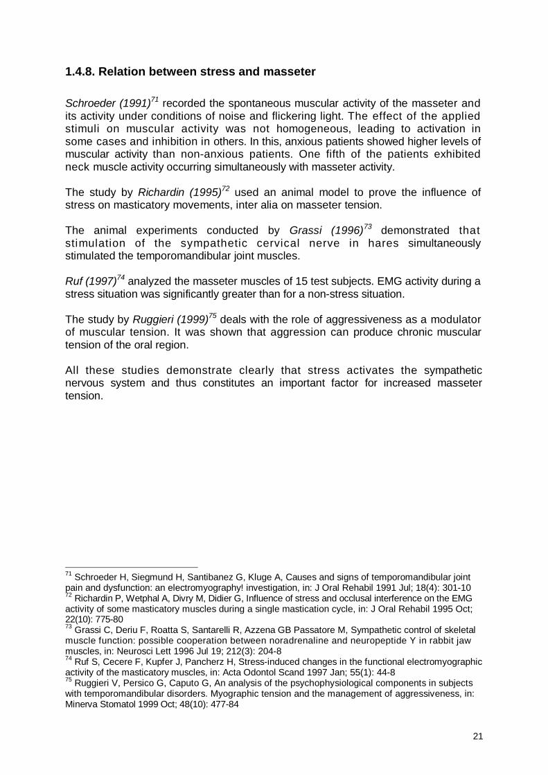
Fig. 7: Short cervical muscles, lateral (Kapandji 77)
Fig. 8: Origin and insertion of suboccipital muscles
77 Kapandji, i.A.,(1985) Funktionelle Anatomie der Gelenke, Ferdinand Enke Verlag, Stuttgart
Name Origin Point of attachment 1. Rectus capitis posterior minor
Atlas (C I), posterior tubercle, short tendons
Below inferior nuchal line
2. Rectus capitis posterior major
Spine axis (C II), short tendons
Inferior nuchal line
3. Obliquus capitis superior
Atlas (C I), lateral mass, short tendons
Inferior nuchal line
4. Obliquus capitis inferior
Spine axis (C II) Lateral mass of atlas (CI)
24
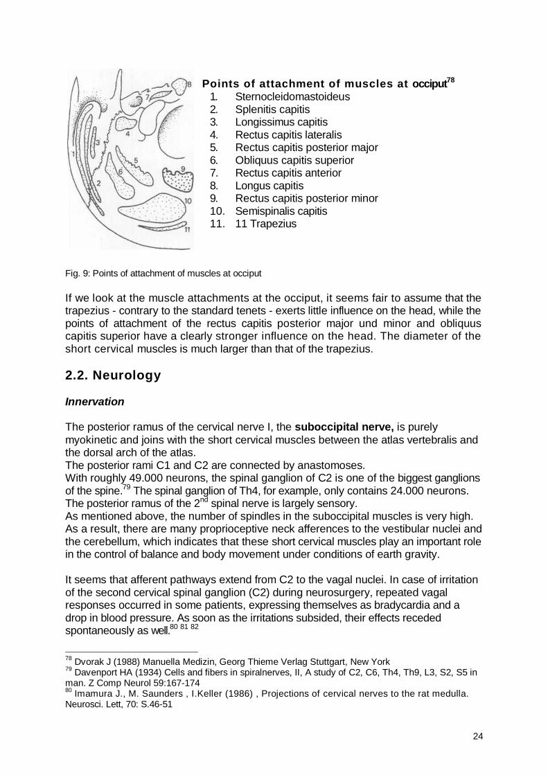
Points of attachment of muscles at occiput78
1. Sternocleidomastoideus 2. Splenitis capitis 3. Longissimus capitis 4. Rectus capitis lateralis 5. Rectus capitis posterior major 6. Obliquus capitis superior 7. Rectus capitis anterior 8. Longus capitis 9. Rectus capitis posterior minor 10. Semispinalis capitis 11. 11 Trapezius
Fig. 9: Points of attachment of muscles at occiput
If we look at the muscle attachments at the occiput, it seems fair to assume that the trapezius - contrary to the standard tenets - exerts little influence on the head, while the points of attachment of the rectus capitis posterior major und minor and obliquus capitis superior have a clearly stronger influence on the head. The diameter of the short cervical muscles is much larger than that of the trapezius. 2.2. Neurology Innervation The posterior ramus of the cervical nerve I, the suboccipital nerve, is purely myokinetic and joins with the short cervical muscles between the atlas vertebralis and the dorsal arch of the atlas. The posterior rami C1 and C2 are connected by anastomoses. With roughly 49.000 neurons, the spinal ganglion of C2 is one of the biggest ganglions of the spine.79 The spinal ganglion of Th4, for example, only contains 24.000 neurons. The posterior ramus of the 2nd spinal nerve is largely sensory. As mentioned above, the number of spindles in the suboccipital muscles is very high. As a result, there are many proprioceptive neck afferences to the vestibular nuclei and the cerebellum, which indicates that these short cervical muscles play an important role in the control of balance and body movement under conditions of earth gravity. It seems that afferent pathways extend from C2 to the vagal nuclei. In case of irritation of the second cervical spinal ganglion (C2) during neurosurgery, repeated vagal responses occurred in some patients, expressing themselves as bradycardia and a drop in blood pressure. As soon as the irritations subsided, their effects receded spontaneously as well.80 81 82
78 Dvorak J (1988) Manuella Medizin, Georg Thieme Verlag Stuttgart, New York 79 Davenport HA (1934) Cells and fibers in spiralnerves, II, A study of C2, C6, Th4, Th9, L3, S2, S5 in man. Z Comp Neurol 59:167-174 80 Imamura J., M. Saunders , I.Keller (1986) , Projections of cervical nerves to the rat medulla. Neurosci. Lett, 70: S.46-51
25
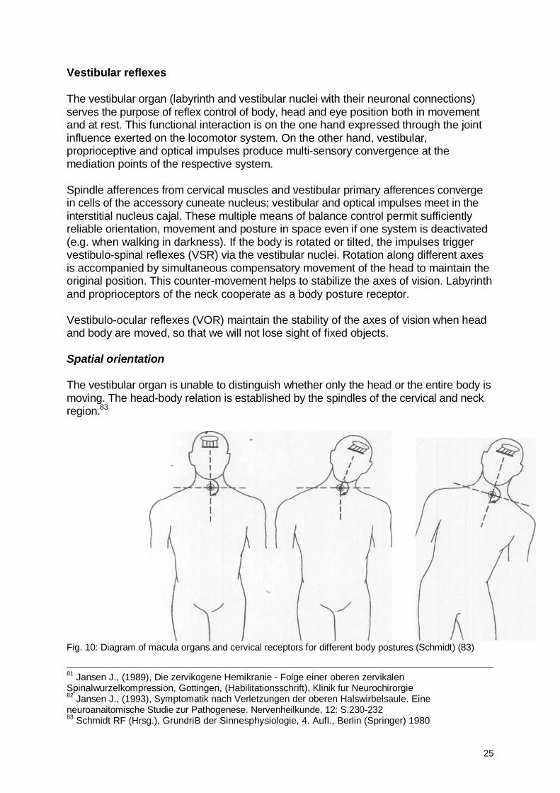
Vestibular reflexes The vestibular organ (labyrinth and vestibular nuclei with their neuronal connections) serves the purpose of reflex control of body, head and eye position both in movement and at rest. This functional interaction is on the one hand expressed through the joint influence exerted on the locomotor system. On the other hand, vestibular, proprioceptive and optical impulses produce multi-sensory convergence at the mediation points of the respective system. Spindle afferences from cervical muscles and vestibular primary afferences converge in cells of the accessory cuneate nucleus; vestibular and optical impulses meet in the interstitial nucleus cajal. These multiple means of balance control permit sufficiently reliable orientation, movement and posture in space even if one system is deactivated (e.g. when walking in darkness). If the body is rotated or tilted, the impulses trigger vestibulo-spinal reflexes (VSR) via the vestibular nuclei. Rotation along different axes is accompanied by simultaneous compensatory movement of the head to maintain the original position. This counter-movement helps to stabilize the axes of vision. Labyrinth and proprioceptors of the neck cooperate as a body posture receptor. Vestibulo-ocular reflexes (VOR) maintain the stability of the axes of vision when head and body are moved, so that we will not lose sight of fixed objects. Spatial orientation The vestibular organ is unable to distinguish whether only the head or the entire body is moving. The head-body relation is established by the spindles of the cervical and neck region.83
Fig. 10: Diagram of macula organs and cervical receptors for different body postures (Schmidt) (83)
81 Jansen J., (1989), Die zervikogene Hemikranie - Folge einer oberen zervikalen Spinalwurzelkompression, Gottingen, (Habilitationsschrift), Klinik fur Neurochirorgie 82 Jansen J., (1993), Symptomatik nach Verletzungen der oberen Halswirbelsaule. Eine neuroanaitomische Studie zur Pathogenese. Nervenheilkunde, 12: S.230-232 83 Schmidt RF (Hrsg.), GrundriB der Sinnesphysiologie, 4. Aufl., Berlin (Springer) 1980
26
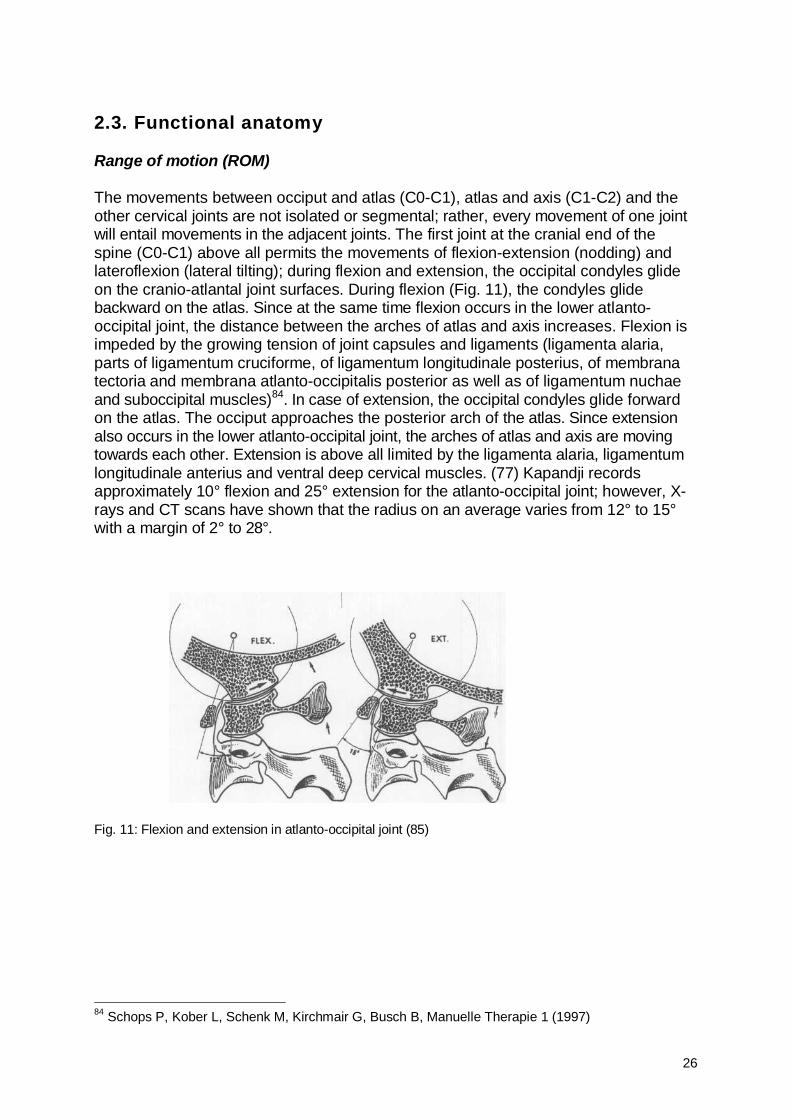
2.3. Functional anatomy Range of motion (ROM) The movements between occiput and atlas (C0-C1), atlas and axis (C1-C2) and the other cervical joints are not isolated or segmental; rather, every movement of one joint will entail movements in the adjacent joints. The first joint at the cranial end of the spine (C0-C1) above all permits the movements of flexion-extension (nodding) and lateroflexion (lateral tilting); during flexion and extension, the occipital condyles glide on the cranio-atlantal joint surfaces. During flexion (Fig. 11), the condyles glide backward on the atlas. Since at the same time flexion occurs in the lower atlanto-occipital joint, the distance between the arches of atlas and axis increases. Flexion is impeded by the growing tension of joint capsules and ligaments (ligamenta alaria, parts of ligamentum cruciforme, of ligamentum longitudinale posterius, of membrana tectoria and membrana atlanto-occipitalis posterior as well as of ligamentum nuchae and suboccipital muscles)84. In case of extension, the occipital condyles glide forward on the atlas. The occiput approaches the posterior arch of the atlas. Since extension also occurs in the lower atlanto-occipital joint, the arches of atlas and axis are moving towards each other. Extension is above all limited by the ligamenta alaria, ligamentum longitudinale anterius and ventral deep cervical muscles. (77) Kapandji records approximately 10° flexion and 25° extension for the atlanto-occipital joint; however, X-rays and CT scans have shown that the radius on an average varies from 12° to 15° with a margin of 2° to 28°.
Fig. 11: Flexion and extension in atlanto-occipital joint (85)
84 Schops P, Kober L, Schenk M, Kirchmair G, Busch B, Manuelle Therapie 1 (1997)
27
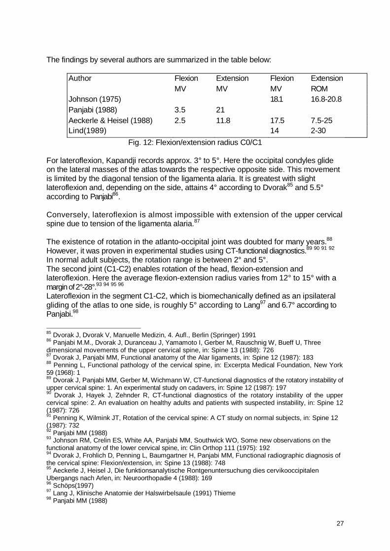
The findings by several authors are summarized in the table below:
Author Flexion Extension Flexion Extension MV MV MV ROM Johnson (1975) 18.1 16.8-20.8 Panjabi (1988) 3.5 21 Aeckerle & Heisel (1988) 2.5 11.8 17.5 7.5-25 Lind(1989) 14 2-30
Fig. 12: Flexion/extension radius C0/C1
For lateroflexion, Kapandji records approx. 3° to 5°. Here the occipital condyles glide on the lateral masses of the atlas towards the respective opposite side. This movement is limited by the diagonal tension of the ligamenta alaria. It is greatest with slight lateroflexion and, depending on the side, attains 4° according to Dvorak85 and 5.5° according to Panjabi86. Conversely, lateroflexion is almost impossible with extension of the upper cervical spine due to tension of the ligamenta alaria.87
The existence of rotation in the atlanto-occipital joint was doubted for many years.88 However, it was proven in experimental studies using CT-functional diagnostics.89 90 91 92 In normal adult subjects, the rotation range is between 2° and 5°. The second joint (C1-C2) enables rotation of the head, flexion-extension and lateroflexion. Here the average flexion-extension radius varies from 12° to 15° with a margin of 2°-28°.93 94 95 96 Lateroflexion in the segment C1-C2, which is biomechanically defined as an ipsilateral gliding of the atlas to one side, is roughly 5° according to Lang97 and 6.7° according to Panjabi.98 85 Dvorak J, Dvorak V, Manuelle Medizin, 4. Aufl., Berlin (Springer) 1991 86 Panjabi M.M., Dvorak J, Duranceau J, Yamamoto I, Gerber M, Rauschnig W, Bueff U, Three dimensional movements of the upper cervical spine, in: Spine 13 (1988): 726 87 Dvorak J, Panjabi MM, Functional anatomy of the Alar ligaments, in: Spine 12 (1987): 183 88 Penning L, Functional pathology of the cervical spine, in: Excerpta Medical Foundation, New York 59 (1968): 1 89 Dvorak J, Panjabi MM, Gerber M, Wichmann W, CT-functional diagnostics of the rotatory instability of upper cervical spine: 1. An experimental study on cadavers, in: Spine 12 (1987): 197 90 Dvorak J, Hayek J, Zehnder R, CT-functional diagnostics of the rotatory instability of the upper cervical spine: 2. An evaluation on healthy adults and patients with suspected instability, in: Spine 12 (1987): 726 91 Penning K, Wilmink JT, Rotation of the cervical spine: A CT study on normal subjects, in: Spine 12 (1987): 732 92 Panjabi MM (1988) 93 Johnson RM, Crelin ES, White AA, Panjabi MM, Southwick WO, Some new observations on the functional anatomy of the lower cervical spine, in: Clin Orthop 111 (1975): 192 94 Dvorak J, Frohlich D, Penning L, Baumgartner H, Panjabi MM, Functional radiographic diagnosis of the cervical spine: Flexion/extension, in: Spine 13 (1988): 748 95 Aeckerle J, Heisel J, Die funktionsanalytische Rontgenuntersuchung dies cervikooccipitalen Ubergangs nach Arlen, in: Neuroorthopadie 4 (1988): 169 96 Schöps(1997) 97 Lang J, Klinische Anatomie der Halswirbelsaule (1991) Thieme 98 Panjabi MM (1988)
28
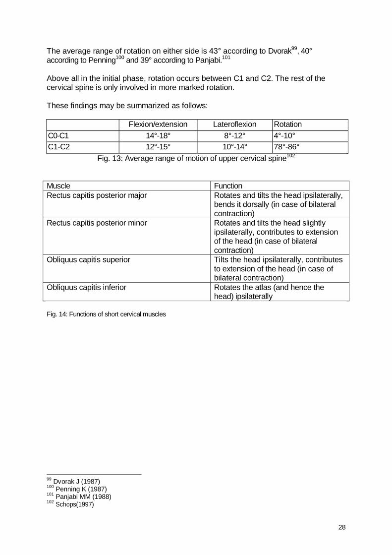
The average range of rotation on either side is 43° according to Dvorak99, 40° according to Penning100 and 39° according to Panjabi.101
Above all in the initial phase, rotation occurs between C1 and C2. The rest of the cervical spine is only involved in more marked rotation. These findings may be summarized as follows: Flexion/extension Lateroflexion Rotation C0-C1 14°-18° 8°-12° 4°-10° C1-C2 12°-15° 10°-14° 78°-86°
Fig. 13: Average range of motion of upper cervical spine102
Muscle Function Rectus capitis posterior major Rotates and tilts the head ipsilaterally,
bends it dorsally (in case of bilateral contraction)
Rectus capitis posterior minor Rotates and tilts the head slightly ipsilaterally, contributes to extension of the head (in case of bilateral contraction)
Obliquus capitis superior Tilts the head ipsilaterally, contributes to extension of the head (in case of bilateral contraction)
Obliquus capitis inferior Rotates the atlas (and hence the head) ipsilaterally
Fig. 14: Functions of short cervical muscles
99 Dvorak J (1987) 100 Penning K (1987) 101 Panjabi MM (1988) 102 Schops(1997)
29
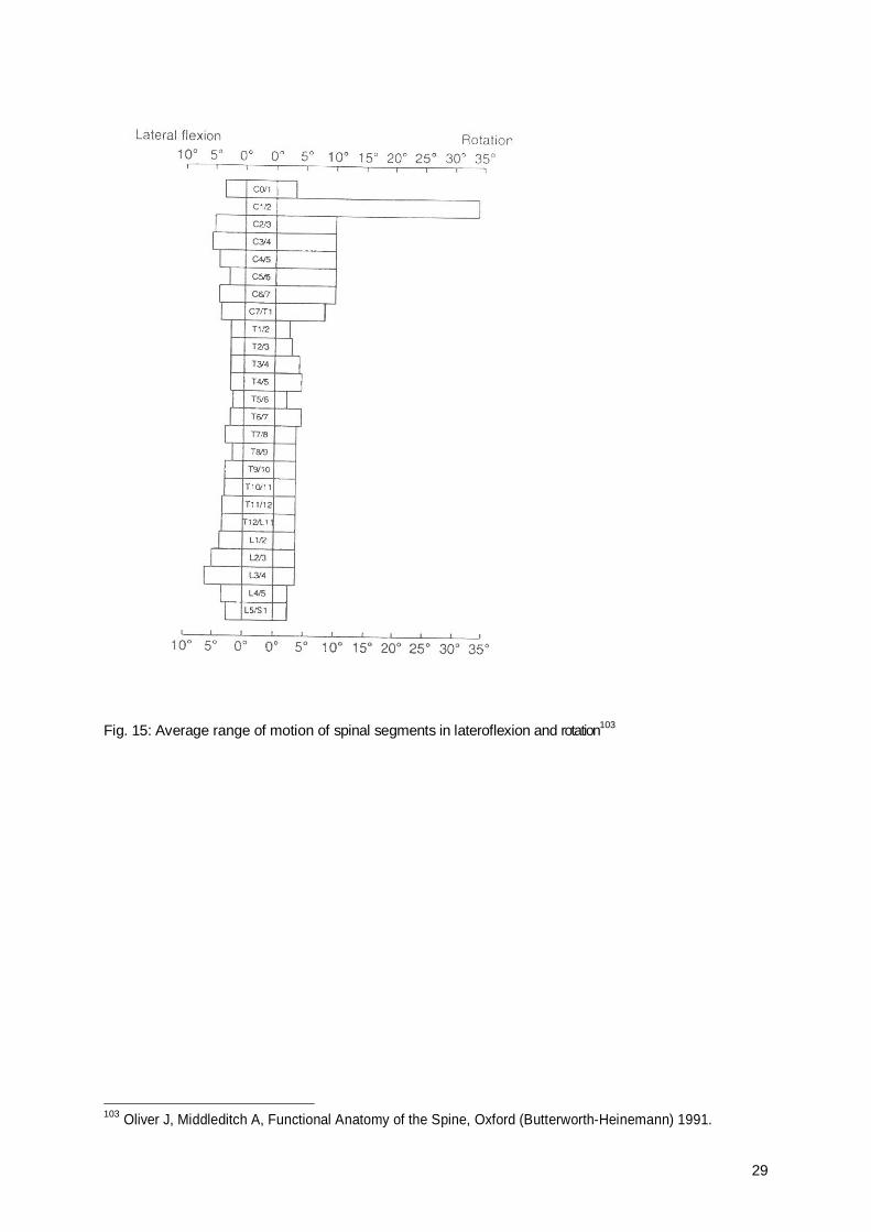
Fig. 15: Average range of motion of spinal segments in lateroflexion and rotation103
103 Oliver J, Middleditch A, Functional Anatomy of the Spine, Oxford (Butterworth-Heinemann) 1991.
30
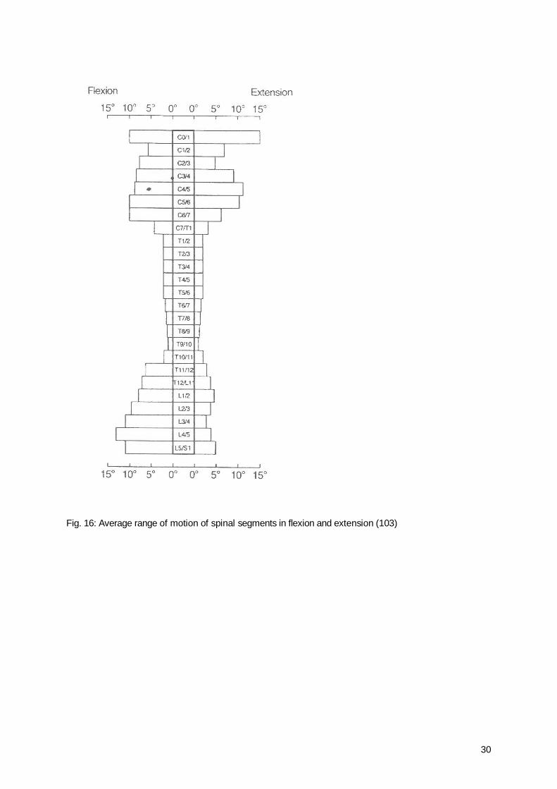
Fig. 16: Average range of motion of spinal segments in flexion and extension (103)
31
CHAPTER 3: Methodology
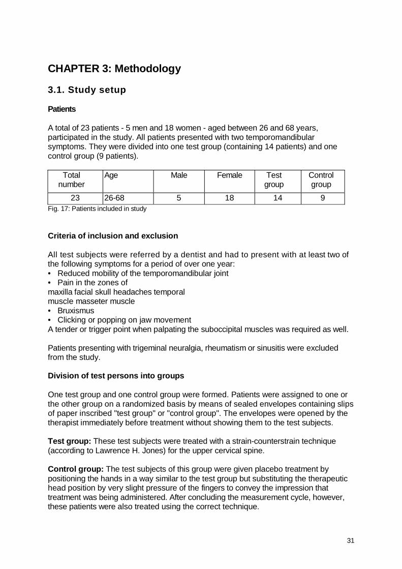
3.1. Study setup Patients A total of 23 patients - 5 men and 18 women - aged between 26 and 68 years, participated in the study. All patients presented with two temporomandibular symptoms. They were divided into one test group (containing 14 patients) and one control group (9 patients).
Total number
Age Male Female Test group
Control group
23 26-68 5 18 14 9 Fig. 17: Patients included in study
Criteria of inclusion and exclusion All test subjects were referred by a dentist and had to present with at least two of the following symptoms for a period of over one year: • Reduced mobility of the temporomandibular joint • Pain in the zones of maxilla facial skull headaches temporal muscle masseter muscle • Bruxismus • Clicking or popping on jaw movement A tender or trigger point when palpating the suboccipital muscles was required as well. Patients presenting with trigeminal neuralgia, rheumatism or sinusitis were excluded from the study. Division of test persons into groups One test group and one control group were formed. Patients were assigned to one or the other group on a randomized basis by means of sealed envelopes containing slips of paper inscribed "test group" or "control group". The envelopes were opened by the therapist immediately before treatment without showing them to the test subjects. Test group: These test subjects were treated with a strain-counterstrain technique (according to Lawrence H. Jones) for the upper cervical spine. Control group: The test subjects of this group were given placebo treatment by positioning the hands in a way similar to the test group but substituting the therapeutic head position by very slight pressure of the fingers to convey the impression that treatment was being administered. After concluding the measurement cycle, however, these patients were also treated using the correct technique.
32
Measuring was carried out with a biofeedback device of the company Insight Instruments Vienna, Austria. The device features two EMG inputs, which were used to measure the masseter tone by means of surface electrodes. Moreover, the device is equipped with a multisensor for recording skin temperature, skin conductance and pulse rate. The measured values permit a statement on sympathetic activity. The biofeedback measuring was carried out before, during and after treatment or placebo treatment. 3.2. Biofeedback What is biofeedback? Mark Schwartz104 has defined biofeedback105 106 107 as a group of therapeutic procedures that utilizes electronic or electromechanical instruments to accurately measure, process, and "feed back" to persons informations with reinforcing properties about their neuromuscular and autonomic activity, both normal and abnormal, in the form of analog or binary, auditory and/or visual feedback signals. Best achieved with a competent biofeedback professional, the objectives are to help persons develop greater awareness and/or under less voluntary control, by first controlling the external signal, and then by the use of internal psychophysiological cues. The various biofeedback parameters include: a) Skin conductance b) Temperature c) Pulse amplitude d) Pulse rate e) Electromyography (EMG) f) Electroencephalography g) Breathing The present study only uses skin conductance, temperature, pulse rate and EMG as parameters. Measuring skin conductance The skin conductance level can only be measured if energy is supplied from the outside. By e.g. applying low voltage to the palm by means of two electrodes, it is possible to measure the electrical current in its variation over time. This variation is dependent on the activity of the sweat glands, whose activity is however not directly measured but can only be recorded indirectly. Since salty sweat induces skin conductivity, moist skin will conduct electricity better than dry skin. The changes in sweat gland activity are dependent on a number of psychological and physical factors, which is why the skin conductance level is particularly interesting for psycho-physiological studies.
104 Schwartz M.,(1998) Biofeedback: A Practitioner's Guide. 105 Criswell-Hanna, Eleanor, Biofeedback and Somatics toward personal evolution, Freeperson Press Novato,California 106 Zeier, H., Biofeedback (1990), Verlag Hans Huber, Bern 107 Seminarunterlagen,"Biofeedback-Grundlagen" (1997) Ausbildungslehrgancj der Österr. Ges. für Biofeedback und Psychophysiologie (OBfP)
33
Standard recording point Fourth finger of the non-dominant hand (multisensor) since the skin tends to be slightly thinner on this side of the body (fewer calluses). Number of sweat glands per cm2 of skin in adults Palms and soles of the feet 2000 Front 360 Thighs 120 Innervation of eccrine sweat glands (production) Sympathetic part of the autonomic nervous system with cholinergic transmission (acetylcholine)! Central efferences 1. Hypothalamus - thermoregulatory sweating 2. Formatio reticularis - sweating during gross motor activity (escape or attack) 3. Pre-motor cortex - sweat gland activity or inhibition associated with minute motor activity Biological significance of sweating of palms and soles 1. Thermal regulation
2. Anticipatory thermal regulation in case of escape or attack
3. Improved gripping ability and protection against injury
Significance for present study Skin conductance usually decreases "spontaneously" during a relaxing situation. Temperature Skin temperature in relaxed position at 21 °C ambient temperature Hand: approx. 28° - 33° centigrade The variation margins are due to physiological and psychological factors (relaxation vs. stress, anxiety, etc.). Finger temperature regulation The blood vessels in the fingers (as in all other acrae - eyes, nose, lips) are influenced on the one hand by the sympathetic nervous system and on the other hand by the ambient temperature. A change in the ambient temperature will lead to vasoconstriction in case of a temperature drop and to vasodilation (reflex) in case of increase. This temperature change occurs very quickly. Conversely, the temperature change controlled by the sympathetic nervous system
34
occurs much more slowly, with increase of the sympathetic tone being accompanied by vasoconstriction while decrease is paralleled by vasodilation. Influence of other parameters: Lack of oxygen vasodilation Local lack of CO2 vasoconstriction Reduced pH value (acidity) vasodilation With respect to the present study, it was therefore important to fix the appointments of the test subjects at 30 minutes before actual treatment to safeguard identical conditions for all subjects by permitting them to adapt to the room temperature. Likewise, the temperature in the test room was to remain constant. Moreover, all forms of extreme stress had to be avoided as well. Standard lead point Fourth finger of the non-dominant hand (multisensor) The change in the blood flow lasts for 5 to 15 seconds. Significance for present study An increase in the temperature measured indicates reduced sympathetic activity. In biofeedback, temperature is measured in degrees with an accuracy of up to tenths of a degree. Pulse amplitude Pulse pressure is measured photoplethysmographically. For this purpose, the photoplethysmograph consists of a (usually red) source of light and a photoelectric converter. Either a) the penetrating light or b) the reflected light is measured. Tissue with higher or lower blood flow is more or less permeable to red light. If the area measured (e.g. the anterior zone of the finger) is well supplied with blood, much light is absorbed and only little (penetrating or reflected) light is measured ("much blood - little light"). If the area is not well supplied with blood, the quantity of light measured is higher. If the vessels are dilated and relaxed, the quantity of blood suffusing these vessels is greater with every heartbeat because they are more flexible. In this case, the difference between maximum blood flow (systolic phase) and minimum blood flow (diastolic return) is greater than in contracted vessels. This difference determines the pulse volume pressure. Peripheral blood flow is controlled by the sympathetic nervous system (alpha-adrenergically). High pulse pressure is recorded if the vessels are dilated, the "quantity of blood" is ample and consequently little light is measured.
35
Fig. 18: Photoplethysmograph
In the present study, the pulse pressure was not measured because the device was unable to record these values and the findings would not have been significantly improved by measuring pulse amplitude in addition to pulse rate. Pulse rate The heart rate is controlled autonomically, above all by the sino-atrial and atrio-ventricular nodes, which excite the working muscles of atrium and ventricle. Sympathetic and parasympathetic impulses exert a modulating influence in that sympathetic activity leads to increased heart rate while parasympathetic activity reduces heart rate. Significance for present study A decrease of the heart rate was observed in the relaxed state. EMG Measuring What is measured here is the electric activity of the muscles, i.e. the potential difference between two electrodes, and more specifically the relative mutual charge in relation to a reference electrode. EMG is measured in microvolt. Preparation Preparing the skin reduces contact resistance and permits a larger part of the biological signal to arrive at the amplifier, which in its turn intensifies the EMG signal. Rubbing the skin with alcohol removes fat, loose callosities and makeup. Electrode paste establishes a strong electric contact and practically serves as a buffer between electrodes and skin. Positioning of electrodes 2 lead electrodes and one neutral electrode are used. The positioning of the electrodes determines which groups of muscles are monitored: 1. If the electrodes are positioned parallel to a muscle or its fibers, the respective muscle contributes most to the signal. 2. In case of leads placed transversally to the muscle (the fibers), the activity of all subjacent muscles is recorded.
36
The neutral electrode must be positioned at an electrically inactive point of the body or at the center of the two lead electrodes. Artifacts • External electrical interference ("power hum") • Movement-related artifacts: normally easy to recognize as high peaks but - under
certain circumstances - difficult to prevent. Remark: Since with the strain-counterstrain technique movements are executed very slowly, this constituted no problem for the present study.
Psychophysiology Muscle function: • Movement • Posture • Expression of emotions (facial expression, gesture) Emotions may find expression both through voluntomotoricity, e.g. facial expression, and through the gamma motor system. In case of the latter, the generally higher activity level (cf. formatio reticularis, limbic system) entails efferent neuronal impulses that may directly lead to muscle tension. For example, it was shown that the back muscles of persons presenting with chronic backache react more intensively under conditions of stress. With respect to a healthy control group, these subjects were characterized by increased EMG values and delayed return to baseline.108
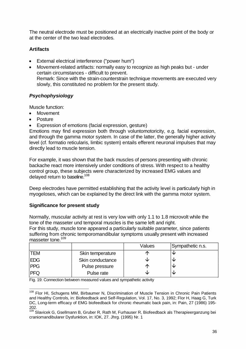
Deep electrodes have permitted establishing that the activity level is particularly high in myogeloses, which can be explained by the direct link with the gamma motor system. Significance for present study Normally, muscular activity at rest is very low with only 1.1 to 1.8 microvolt while the tone of the masseter und temporal muscles is the same left and right. For this study, muscle tone appeared a particularly suitable parameter, since patients suffering from chronic temporomandibular symptoms usually present with increased masseter tone.109 Values Sympathetic n.s. TEM Skin temperature EDG Skin conductance PPG Pulse pressure PFQ Pulse rate Fig. 19: Connection between measured values and sympathetic activity
108 Flor HI, Schugens MM, Birbaumer N, Discrimination of Muscle Tension in Chronic Pain Patients and Healthy Controls, in: Biofeedback and Self-Regulation, Vol. 17, No. 3, 1992; Flor H, Haag G, Turk DC, Long-term efficacy of EMG biofeedback for chronic rheumatic back pain, in: Pain, 27 (1986) 195-202. 109 Slavicek G, Gsellmann B, Gruber R, Rath M, Furhauser R, Biofeedback als Therapieerganzung bei craniomandibularer Dysfunktion, in: IOK, 27. Jhrg. (1995) Nr. 1
37
3.3. Strain-Counterstrain (SCS) The strain-counterstrain method was developed by^ DrTLawrence H. Jones110 111. It is a "gentle" osteopathic method that does not employ high-velocity techniques (HVT) to manipulate joints but rather achieves a slow reprogramming of the proprioceptive system of muscles by means of positioning. For diagnosis, palpation is used to identify a trigger or tender point (TP) in the muscles (after first determining the region with most severely reduced mobility). This TP is used for monitoring to determine the optimum therapeutic position ("treatment by positioning"). For this purpose, the therapist positions the patient passively in as painless a body posture as possible, in which even the TP is almost free of pain. This posture must be maintained for 90 seconds. Simultaneous palpation of the corresponding TP permits to recheck both positioning and treatment after application of the technique. The optimum treatment position is achieved if the patient states to be free of pain and the TP is no longer tense and painful, either. It has often been observed that the therapeutic position is similar to the movement that has triggered the pain and dysfunction. It is the task of the therapist to return the patient, who remains relaxed and passive, to the neutral position. Explanatory neurological model If a muscle remains in a shortened state for a longer period of time (e.g. due to stooping), the spindles transmit little afferent information, inducing the gamma motoneurons to fire at an increased rate. This contributes to preserving the muscle tone but also causes higher spindle sensitivity. If in this state the muscle is suddenly and quickly stretched (e.g. by rising from a stooping position), the alpha motoneurons are overstimulated and the muscle spasm continues. The central nervous system is unable to interpret the sensory signals correctly und responds by intensely stimulating the gamma motoneurons. As a result, the spasm is continued; one might also say, "fixed". Therapeutic shortening of this muscle also entails a shortening of the spindles and hence a normalized firing rate. The central nervous system is again able to interpret the signals correctly and correspondingly normalizes the gamma motoneurons. This process is completed after about 90 seconds. Since the short neck muscles are very rich in spindles, they are excellently suited for this therapeutic method. The 4 therapy steps according to JONES:
1. Determine the somatic dysfunction by means of mobility tests and/or palpation of tender points or trigger points.
2. Identify the position in which TP tension and / or pain have decreased by at least 70%.
3. Hold this position for at least 90 seconds.
4. Return the joint passively and very slowly to the neutral position. 110 1 Jones LH, DO, FAAO, Strain-Counterstrain, 1995 111 Ward R.C. Foundations fur Osteopathic Medicine (1997), Williams & Wilkins

38
These techniques are especially suitable for "hot spots" (such as the upper cervical spine), tense patients and cases where manipulation is contra-indicated (e.g. osteoporosis). This type of technique is therefore appropriate for the present study, in which the upper cervical spine is the subject of treatment. The neck region in particular is a zone often characterized by protracted muscle tension that frequently can only be durably eliminated by means of neuromuscular reprogramming. SCS treatment of the suboccipital region Definition of dysfunction: 1) Dysfunction of C1/C2 posterior (Jones) 2) Dysfunction of occiput unilateral anterior Description of dysfunction:112
One occipital condyle is displaced anteriorly due to a spasm of the obliquus capitis superior and/or the rectus capitis posterior minor and/or major. The other side tries to compensate (often painful). Compensatory dysfunctions of the atlas are very frequent. Mitchell113 describes this as follows: the occiput is in flexion (the head is tilted forward) and the left condyle cannot execute the movement completely. If the movement of the left condyle is blocked, the left condylar joint becomes a turning point. If head flexion were forced beyond this point, the left side of the atlas would be pushed back and cause the atlas to rotate to the left.
Fig. 20: Movement of atlas during anteflexion of the head with immobility of C0-C1 left (113)
112 Ricard F. D.O. Osteopathic Treatment of Pain originating in the Craniocervical Area, Editions DE VERLAQUE 113 Mitchell.F. D.O. F.A.A.O. (1995) The Muscle Engergy Manual , MET Press, East Lansing, Michigan
39
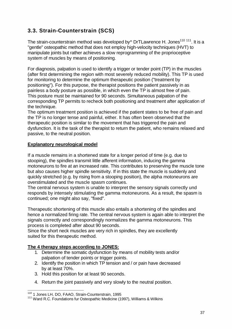
Mobility test: Joint flexion between CO and C1 is restricted on the side of the somatic dysfunction.
Fig. 21: Mobility test C0-C1114
Examination: Palpation of inferior nuchal line between external occipital crest and mastoid process. Transversal palpation across the muscle identifies tense, painful points (= tender points according to Jones) or radiating points (= trigger points according to Travell115). Treatment: • The patient lies down in a supine position • The therapist sits at the head end of the treatment table • The therapist holds the tender point with the fingers of one hand while the other hand establishes broad contact with the whole occiput • The atlanto-occipital joint is brought into an extension position. The occipital condyle glides on the cranial joint surface of the atlas in the anterior direction. Sometimes, minimal lateroflexion and rotation in the ipsilateral direction (= i.e. the side of the tender point) is called for. This shortens the suboccipital muscles. The position must be so that tension at the TP is reduced and the patient states that pain at the TP has decreased by at least 70%
114 Yaw ILK, Glover JC, Counterstrain: A Handbook of Osteopathic Technique, Tulsa (Okla: Y Knot Publishers) 1994 115 Travell JG, Simons DG, Handbuch der Muskel-Triggerpunkte: Obere Extremity Kopf und Thorax, Stuttgart (Gustav Fischer) 1998
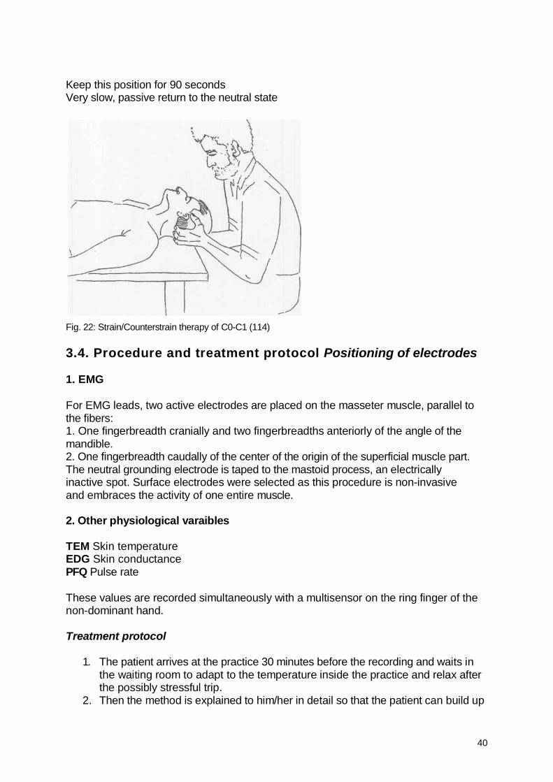
40
Keep this position for 90 seconds Very slow, passive return to the neutral state
Fig. 22: Strain/Counterstrain therapy of C0-C1 (114) 3.4. Procedure and treatment protocol Positioning of electrodes 1. EMG For EMG leads, two active electrodes are placed on the masseter muscle, parallel to the fibers: 1. One fingerbreadth cranially and two fingerbreadths anteriorly of the angle of the mandible. 2. One fingerbreadth caudally of the center of the origin of the superficial muscle part. The neutral grounding electrode is taped to the mastoid process, an electrically inactive spot. Surface electrodes were selected as this procedure is non-invasive and embraces the activity of one entire muscle. 2. Other physiological varaibles TEM Skin temperature EDG Skin conductance PFQ Pulse rate These values are recorded simultaneously with a multisensor on the ring finger of the non-dominant hand. Treatment protocol
1. The patient arrives at the practice 30 minutes before the recording and waits in the waiting room to adapt to the temperature inside the practice and relax after the possibly stressful trip.
2. Then the method is explained to him/her in detail so that the patient can build up
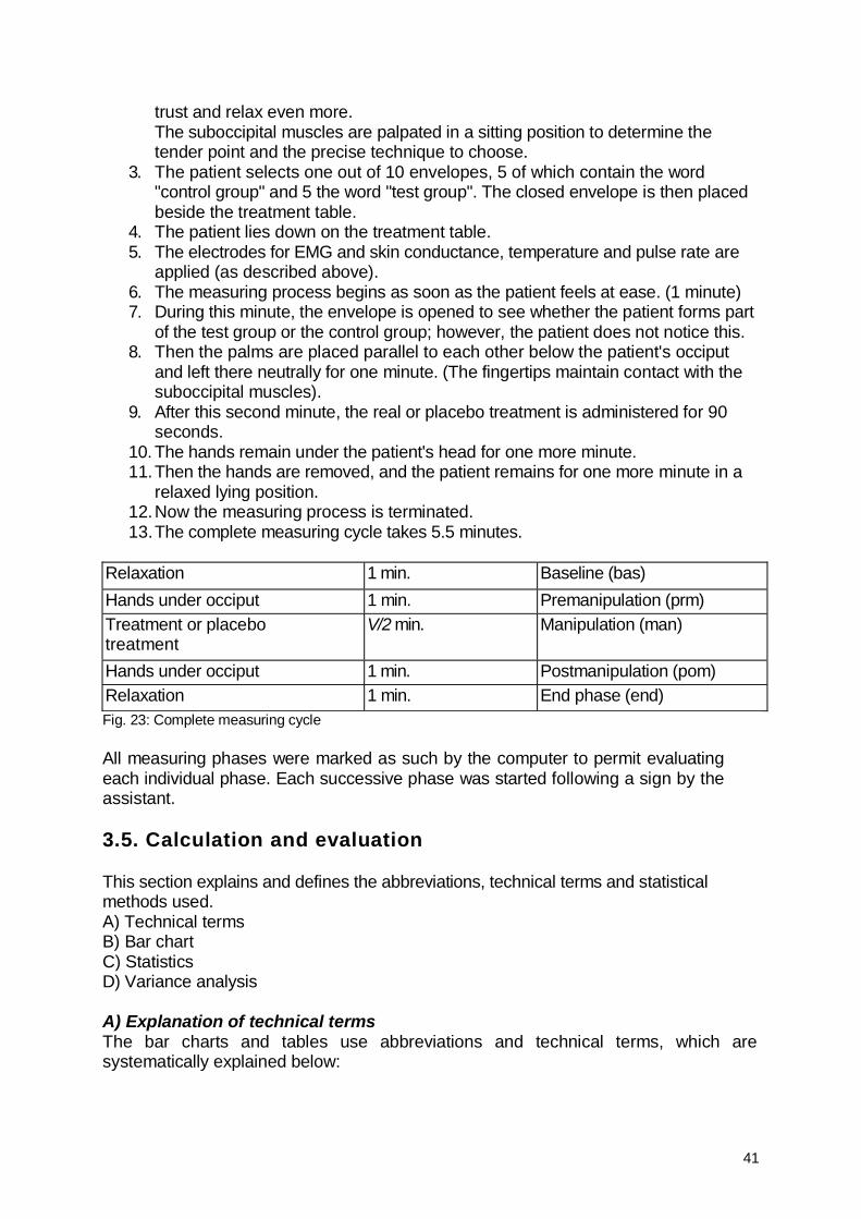
41
trust and relax even more. The suboccipital muscles are palpated in a sitting position to determine the tender point and the precise technique to choose.
3. The patient selects one out of 10 envelopes, 5 of which contain the word "control group" and 5 the word "test group". The closed envelope is then placed beside the treatment table.
4. The patient lies down on the treatment table. 5. The electrodes for EMG and skin conductance, temperature and pulse rate are
applied (as described above). 6. The measuring process begins as soon as the patient feels at ease. (1 minute) 7. During this minute, the envelope is opened to see whether the patient forms part
of the test group or the control group; however, the patient does not notice this. 8. Then the palms are placed parallel to each other below the patient's occiput
and left there neutrally for one minute. (The fingertips maintain contact with the suboccipital muscles).
9. After this second minute, the real or placebo treatment is administered for 90 seconds.
10. The hands remain under the patient's head for one more minute. 11. Then the hands are removed, and the patient remains for one more minute in a
relaxed lying position. 12. Now the measuring process is terminated. 13. The complete measuring cycle takes 5.5 minutes.
Relaxation 1 min. Baseline (bas) Hands under occiput 1 min. Premanipulation (prm) Treatment or placebo treatment
V/2 min. Manipulation (man)
Hands under occiput 1 min. Postmanipulation (pom) Relaxation 1 min. End phase (end) Fig. 23: Complete measuring cycle
All measuring phases were marked as such by the computer to permit evaluating each individual phase. Each successive phase was started following a sign by the assistant.
3.5. Calculation and evaluation This section explains and defines the abbreviations, technical terms and statistical methods used. A) Technical terms B) Bar chart C) Statistics D) Variance analysis A) Explanation of technical terms The bar charts and tables use abbreviations and technical terms, which are systematically explained below:
42
General (Table: page 58) • Person: name of test subject: f.ex. RVA bl 1 = initials and number of the test • Age: age of test subject • Sex: 1 = male, 2 = female • Gruppe (=group): 1 = test group (VG), 2 = control group (KG) Physiological variables (Table: pages 58-61) • scl = skin conductance level • tern = finger temperature • pfq = pulse rate • emg1 = EMG - masseter left • emg2 = EMG - masseter right Phases (Table: pages 43 and 58-61) • bas = baseline • prm = premanipulation phase • man = manipulation phase • pom = postmanipulation phase • end = end phase Mean value (=Mittelwert, page 58) The mean value corresponds to the average in case of normal distribution of the individual values. All values are added and divided by the number of test subjects. Median (page 58) The median value, too, describes the central tendency of a distribution. It divides the volume of data arranged exactly in the middle, so that 50% of the results will be above and 50% below the median. Standard deviation (=Standardabweichung, page 58) The standard deviation indicates the data scatter around the mean value i.e. states the difference between the mean value and the actual results obtained.
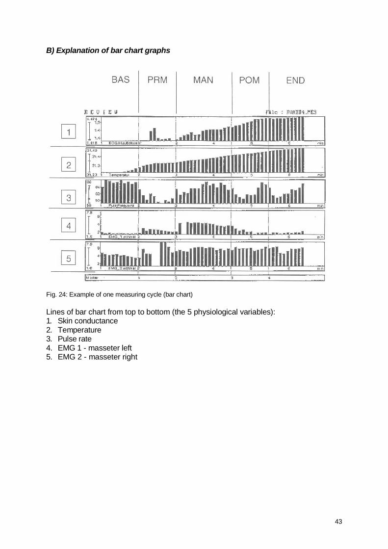
43
B) Explanation of bar chart graphs
Fig. 24: Example of one measuring cycle (bar chart) Lines of bar chart from top to bottom (the 5 physiological variables): 1. Skin conductance 2. Temperature 3. Pulse rate 4. EMG 1 - masseter left 5. EMG 2 - masseter right
44
C) Statistics Two different statistical evaluations were established on the basis of the individual examinations: 1. Start-End 2. Premanipulation - Manipulation 3. Homogenety of population 1. Start (baseline) - End (end phase) (bas - end) It was determined how many values had changed by comparing the baseline (= beginning of measuring cycle before treatment) and the end of treatment. We evaluated: • Test group (VG) - difference bas - end • Control group (KG) - difference bas - end • Difference between test group and control group 2. Premanipulation phase - Manipulation (prm - man) The values of the premanipulation phase were conrpared with the manipulation phase values to establish whether measurable differences had occurred during the therapy phase. We evaluated: • Test group - prm-man • Control group - prm-man • Difference between test group and control group 3. Homogenety of population In observing and comparing the mean values of the test group and the control group, we observed a difference between the baseline values. But in the statistical evaluation of the physiological variability, we found that both groups were not significantly different from each other (p>0,5).
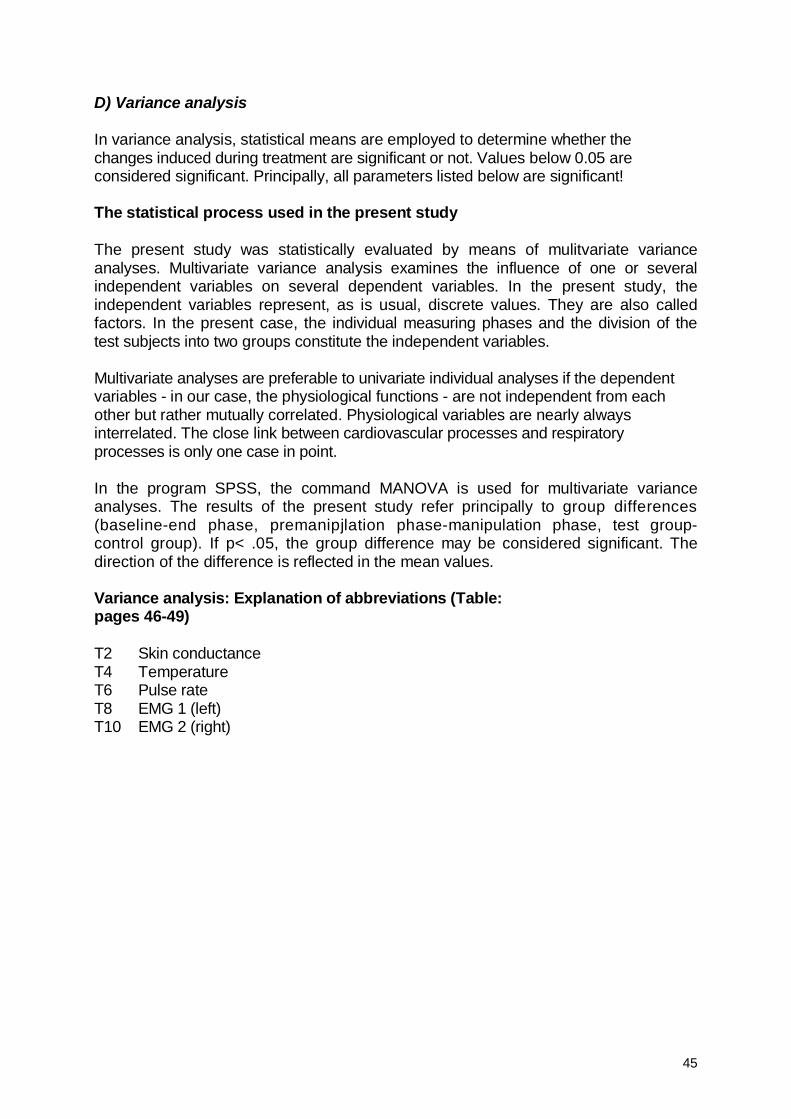
45
D) Variance analysis In variance analysis, statistical means are employed to determine whether the changes induced during treatment are significant or not. Values below 0.05 are considered significant. Principally, all parameters listed below are significant! The statistical process used in the present study The present study was statistically evaluated by means of mulitvariate variance analyses. Multivariate variance analysis examines the influence of one or several independent variables on several dependent variables. In the present study, the independent variables represent, as is usual, discrete values. They are also called factors. In the present case, the individual measuring phases and the division of the test subjects into two groups constitute the independent variables. Multivariate analyses are preferable to univariate individual analyses if the dependent variables - in our case, the physiological functions - are not independent from each other but rather mutually correlated. Physiological variables are nearly always interrelated. The close link between cardiovascular processes and respiratory processes is only one case in point. In the program SPSS, the command MANOVA is used for multivariate variance analyses. The results of the present study refer principally to group differences (baseline-end phase, premanipjlation phase-manipulation phase, test group-control group). If p< .05, the group difference may be considered significant. The direction of the difference is reflected in the mean values. Variance analysis: Explanation of abbreviations (Table: pages 46-49) T2 Skin conductance T4 Temperature T6 Pulse rate T8 EMG 1 (left) T10 EMG 2 (right)
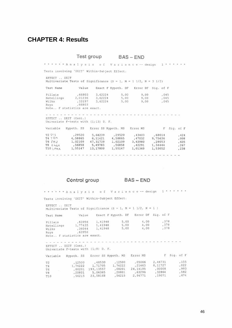
46
CHAPTER 4: Results
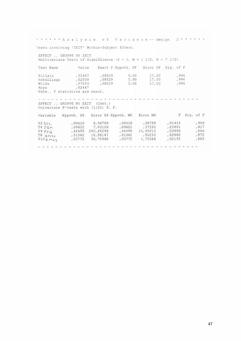
47
48
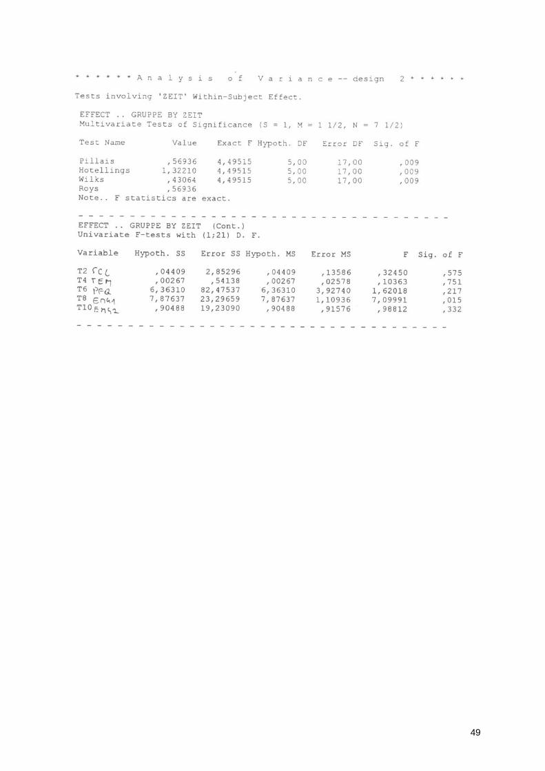
49
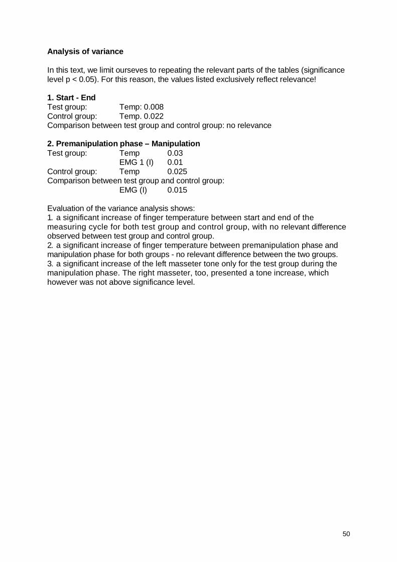
50
Analysis of variance In this text, we limit ourseves to repeating the relevant parts of the tables (significance level p < 0.05). For this reason, the values listed exclusively reflect relevance! 1. Start - End Test group: Temp: 0.008 Control group: Temp. 0.022 Comparison between test group and control group: no relevance 2. Premanipulation phase – Manipulation Test group: Temp 0.03 EMG 1 (I) 0.01 Control group: Temp 0.025 Comparison between test group and control group: EMG (I) 0.015 Evaluation of the variance analysis shows: 1. a significant increase of finger temperature between start and end of the measuring cycle for both test group and control group, with no relevant difference observed between test group and control group. 2. a significant increase of finger temperature between premanipulation phase and manipulation phase for both groups - no relevant difference between the two groups. 3. a significant increase of the left masseter tone only for the test group during the manipulation phase. The right masseter, too, presented a tone increase, which however was not above significance level.
51
CHAPTER 5: Discussion The working hypothesis for the present study was: "In patients with at least two temporomandibular Joint Symptoms and simultaneous dysfunction in the suboccipital muscle zone, the therapy of the Short cervical muscles using an osteopathic technique (strain/counterstrain) can lead to relaxation of the masseter and to reduced sympathetic activity." As the results have shown, this hypothesis was not confirmed in the present test setup. There is a number of reasons for this, which result from the answers to two research questions already formulated in the introduction: 1. What is the clinical relevance of a specific technique of intervention into these interrelations? 2. Is it possible to prove the effects of relaxation of the Short cervical muscles on the temporomandibular Joint muscles and the sympathetic nervous System by means of measurements? We will begin by dealing with the aspects relating to the topic of the study proper, i.e. the first question, and then put the results obtained up for discussion. 5.1. Regarding clinical relevance: With respect to the temporomandibular muscles, measurements at both masseters before, during and after the SCS technique produced interesting results: during the technique, the tone increased on both sides, with a significant increase only on the left side (0,015). At the end of the measuring cycle, the values were again similar to the original levels. The following extract of the table (page 59-61) with the mean values will illustrate this cleariy. In the test group we found the following results: Before the manipulation (PRM) the mean value of the electrical activity of the left masseter was 2,96 milivolt. During the manipulation (MAN) it was 4,37 milivolt and at the end of the measuring time (END) 2,91 milivolt. It cannot be excluded that the tone increase during therapy constitutes a physiologico-mechanical phenomenon. By the end of the measuring cycle, this increase had returned to the original levels; unfortunately, the study was not continued beyond this moment. Other regulatory effects leading to System relaxation in the course of hours to days after therapy are definitely possible and on the basis of clinical experience appear even probable. The fact that in the control group the masseter tone on both sides tended towards relaxation (but without significant levels) only shows that the techniques used in the control group were exclusively of a relaxing character, while Stimuli were applied in the test group (possibly with regulatory effect). The increase of the tonus of the left masseter may be occasioned by the following cause: analysis of the palpation findings for the test subjects of the present study
52
showed that in 90% of cases the dysfunction C0-C1 was located on the right side! This implies a causal link to laterality, which in the majority of the population (approx. 70%) is expressed in a dominance of the right hand and/or eye. Dextrality is very likely to lead directly via the muscle chains to increased activity ancl tension in the zone of the Short cervical muscles on the right side. Via the link with the Short cervical muscles, dominance of the right eye may possibly also induce increased tonicity of the right cervical muscles (as already shown in the overview of relevant literature). In this case of right-side hypertonicity, the therapeutic technique is also fine-tuned so as to accommodate a slight lateroflexion and rotation movement to the right side and thus may engender increased tension of the left masseter. These results of tone increase (in particular of the left masseter) during therapy contradict Standard experience. The above-mentioned study by Boyd (1987)116 observes that the masseter tone decreases during head extension. This contradiction may be due to the fact that the earlier study was conducted on sitting patients. Another difference is that Boyd, contrary to our study (strain-counterstrain), did not employ any therapeutic action of specific, predetermined length. Regarding the temperature increase (finger temperature) between start and end, the results show clearly (the test group presents the same significant temperature increase as the control group) that significant effects (reduced sympathetic activity) can be achieved merely through the contact of the therapist's hands with the patient's head (occiput). However, final proof of this could only be furnished by including a third group in the study design, since only this would permit demonstrating that the effect is not caused by the presence of the therapist in the room. An interesting additional aspect is provided by evaluating the temperatures recorded during the therapy phase for the test group, which shows that the temperature increased very rapidly in this group (from 29,51 to 29,73). Usually, this speedy increase is only observed in case of changes in the ambient temperature and is not for changes in sympathetic activity. Since a change in the ambient temperature must be ruled out, these results may be viewed as a sign of great relevance. Finaliy, it should be emphasized that the therapeutic effects could be studied much better if the measuring cycle were repeated after the end of the therapy (e.g. after 1 hour, 1 day and 1 week). This would render it possible to correct the results for potential initial deteriorations and to take account of later regulatory effects. Due to the high extent of cross-linking within the System, which was already described in the introduction, it is conceivable that response and regulation in the feedback mechanisms may require a certain latency period. 5.2. Methodological aspects The discussion of the setup and impiementation of the present study focuses on possibilities for improvement. 116 Boyd CH, William FS, Boyd CM, Bryant RW, Wiygul JP, The Effect of Head Position on Electromyographic Evaluations of Representative Mandibular Positioning Muscle Groups, in: The Journal of Craniomandibular Practice, 1987 Jan; Vol. 5, No. 1
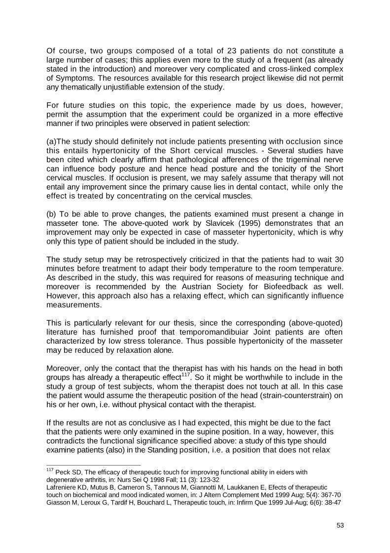
53
Of course, two groups composed of a total of 23 patients do not constitute a large number of cases; this applies even more to the study of a frequent (as already stated in the introduction) and moreover very complicated and cross-linked complex of Symptoms. The resources available for this research project likewise did not permit any thematically unjustifiable extension of the study. For future studies on this topic, the experience made by us does, however, permit the assumption that the experiment could be organized in a more effective manner if two principles were observed in patient selection: (a)The study should definitely not include patients presenting with occlusion since this entails hypertonicity of the Short cervical muscles. - Several studies have been cited which clearly affirm that pathological afferences of the trigeminal nerve can influence body posture and hence head posture and the tonicity of the Short cervical muscles. If occlusion is present, we may safely assume that therapy will not entail any improvement since the primary cause lies in dental contact, while only the effect is treated by concentrating on the cervical muscles. (b) To be able to prove changes, the patients examined must present a change in masseter tone. The above-quoted work by Slavicek (1995) demonstrates that an improvement may only be expected in case of masseter hypertonicity, which is why only this type of patient should be included in the study. The study setup may be retrospectively criticized in that the patients had to wait 30 minutes before treatment to adapt their body temperature to the room temperature. As described in the study, this was required for reasons of measuring technique and moreover is recommended by the Austrian Society for Biofeedback as well. However, this approach also has a relaxing effect, which can significantly influence measurements. This is particularly relevant for our thesis, since the corresponding (above-quoted) literature has furnished proof that temporomandibuiar Joint patients are often characterized by Iow stress tolerance. Thus possible hypertonicity of the masseter may be reduced by relaxation alone. Moreover, only the contact that the therapist has with his hands on the head in both groups has already a therapeutic effect117. So it might be worthwhile to include in the study a group of test subjects, whom the therapist does not touch at all. In this case the patient would assume the therapeutic position of the head (strain-counterstrain) on his or her own, i.e. without physical contact with the therapist. If the results are not as conclusive as I had expected, this might be due to the fact that the patients were only examined in the supine position. In a way, however, this contradicts the functional significance specified above: a study of this type should examine patients (also) in the Standing position, i.e. a position that does not relax
117 Peck SD, The efficacy of therapeutic touch for improving functional ability in eiders with degenerative arthritis, in: Nurs Sei Q 1998 Fall; 11 (3): 123-32 Lafreniere KD, Mutus B, Cameron S, Tannous M, Giannotti M, Laukkanen E, Efects of therapeutic touch on biochemical and mood indicated women, in: J Altern Complement Med 1999 Aug; 5(4): 367-70 Giasson M, Leroux G, Tardif H, Bouchard L, Therapeutic touch, in: Infirm Que 1999 Jul-Aug; 6(6): 38-47
54
the area investigated but rather, due to the effect of gravity, puts a strain on it. Several factors imply that other indicators should be used to prove the effect of this therapy on the temporomandibular Joint. For example, the following three options are suggested for this purpose: ciirect measuring of (a) mobiiity changes in the temporomandibular Joint or (b) of the Short cervical muscles by means of EMG. Moreover, (c) the change in posture (for example head posture) before and after therapy might be measured as well. We derive the justification for this approach from the literature cited in our study, which documents that false head posture is often accompanied by temporomandibular dysfunction (masseter dysfunction). Technically, this could be achieved by means of photographs in profile taken in front of a grid wall. We have considered and enumerated a number of factors that might offer improvements for the investigation of the phenomenon covered in the present study. However, the relevant literature has shown that temporomandibular disorders must also - and perhaps in particular - be regarded as multi-factorial in nature. For this reason, it is very difficult to know whether the results of a study constitute the proverbial "straw that broke the camel's back" or whether they have actually penetrated to the roots of pathogenesis. An important result of the present study and the examination of the respective literature lies in providing a clearer picture of the cross-linking of two perse complex and interacting structures (suboccipital region and cranio-mandibular System). Obviously, these two zones are interconnected in multiple ways, i.e. with respect to • structure (mechanics, anatomy) • nervous System and • reflexes and are moreover preferred target organs of stress (s;ee overview of relevant literature). The below diagram tries to give a schematic overview of the interrelations drawn from literature and developed in the context of the present study (in order to further emphasize the ccmplexity of the Systems, the below diagram uses the more general terms "cervico-cranial transition" (CCT) instead of "Short cervical muscles" and "cranio-mandibular System" (CMS) instead of "temporomandibular Joint muscles"):
55
CHAPTER 6: Summary A summary of the results of the empirical study permits the following brief Statements: • No effect of the strain and counterstrain technique applied to the suboccipital zone on the temporomandibular Joint muscles and the autonomic nervous System was proven". • In the measured period of 3.5 minutes, no significant change in masseter muscle activity was observed between start and end of the measuring cycle. • The tone of the left masseter increased significantly only during the treatment phase proper. • In both groups (test group and control group), a significant temperature change due to reduced sympathetic activity was observed. On the basis of the present study, it seems fair to assume that this result could be achieved merely by positioning the hands under the patient's occiput. These results did not confirm the working hypothesis;. However, by comparing the experience made in the present paper with findings described in the relevant literature, it was possible to draw up several suggestions for further exploration of the question tackled in the present study. Moreover, the study produced a number of theoretical assumptions regarding the complex interrelations of the two regions examined.
56
ABSTRACT It was the objective of the study to evaluate whether an osteopathic strain-counterstrain (SCS) treatment of the suboccipital region has a normalizing effect on the masseter muscle and the autonomic nervous system. Design: 23 patients aged between 26 and 68 and presenting with temporomandibular Symptoms were divided into two groups: one test group (composed of 14 test subjects) and one control group (composed of 9 test subjects). Method: Both groups were monitored before, during and after the strain-counterstrain treatment by means of biofeedback techniques (EMG of masseter) (finger temperature, skin conductance, pulse rate). Variance analysis: 1) Differences before and after the strain-counterstrain treatment 2) Differences before and during the strain-counterstrain treatment Results: 1) No effect of the strain-counterstrain technique applied to the suboccipital zone on the temporomandibular Joint muscles and the autonomic nervous System was proven, While signifikant differences in skin temperature before and after the strain-counterstrain treatment were observed, these apply to both test group and control group. 2) Increased activity of the left masseter was measured during the strain-counterstrain treatment.
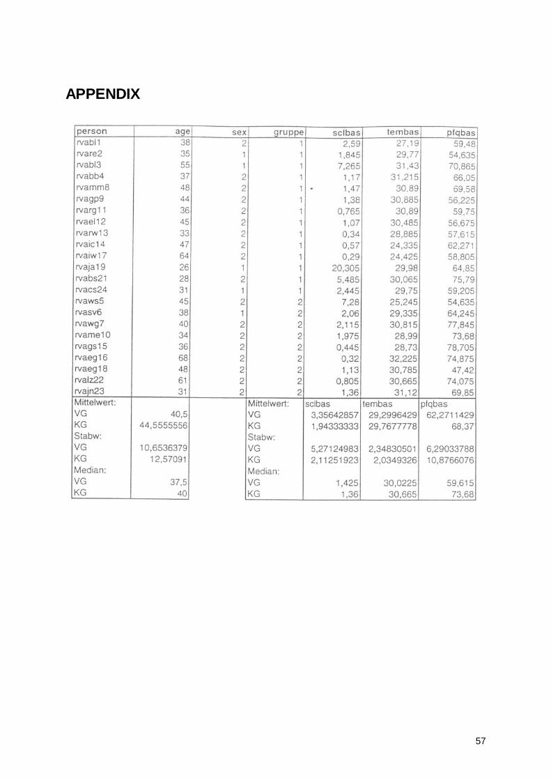
57
APPENDIX
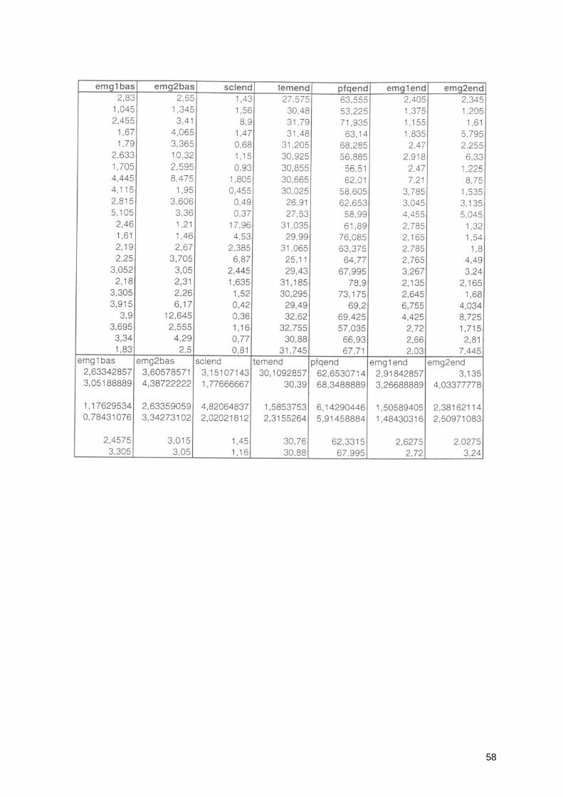
58
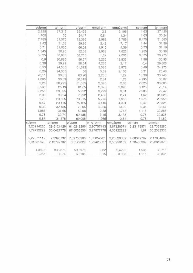
59
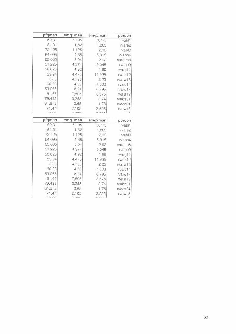
60
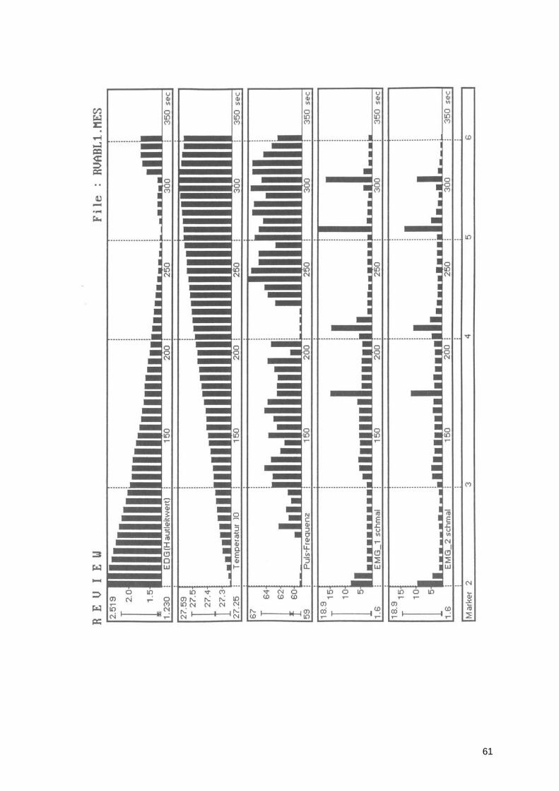
61
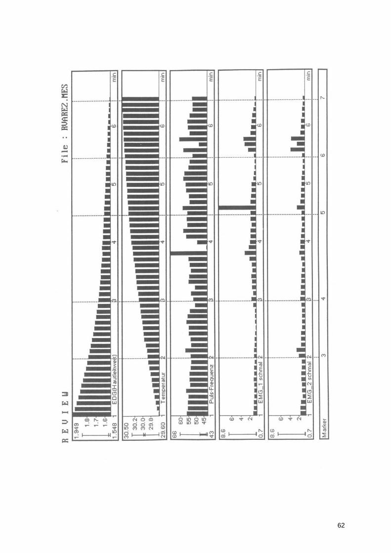
62
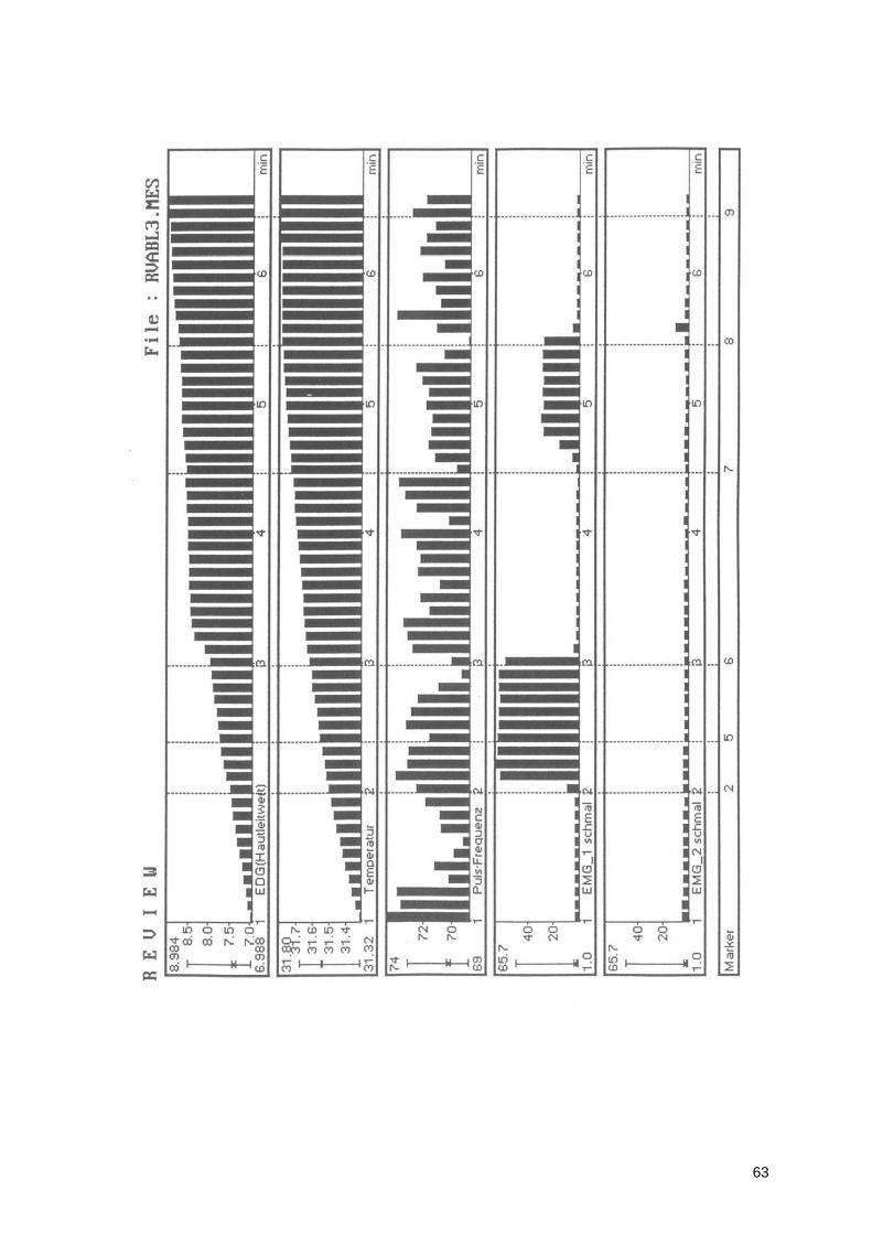
63
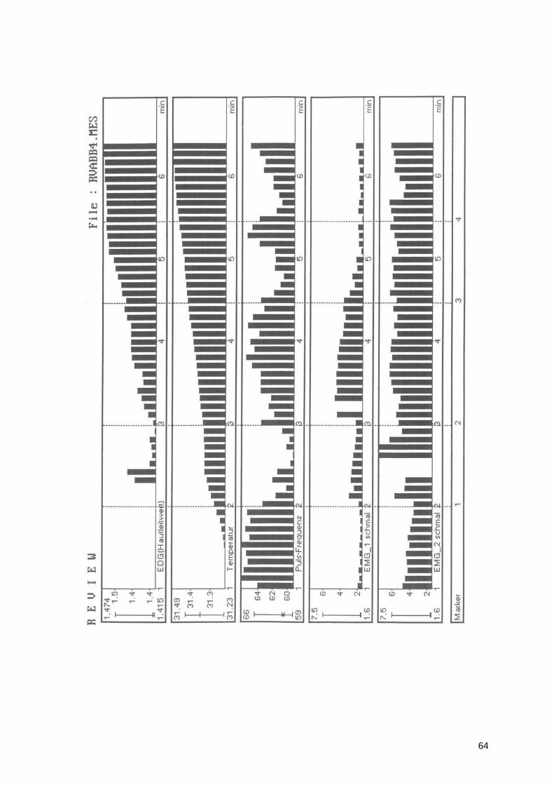
64
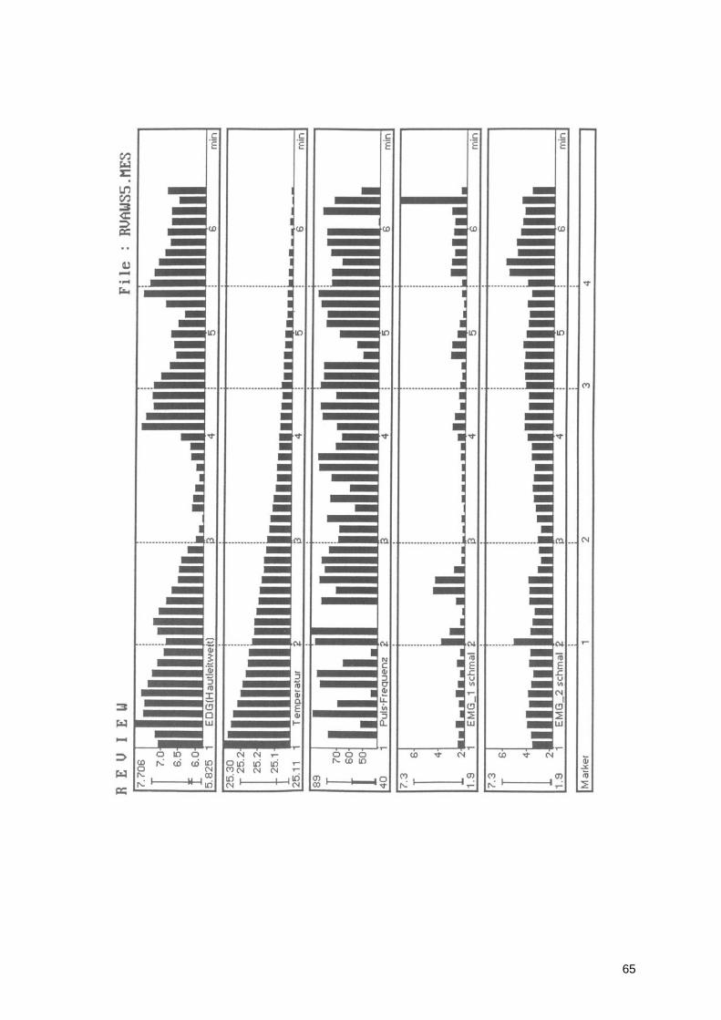
65
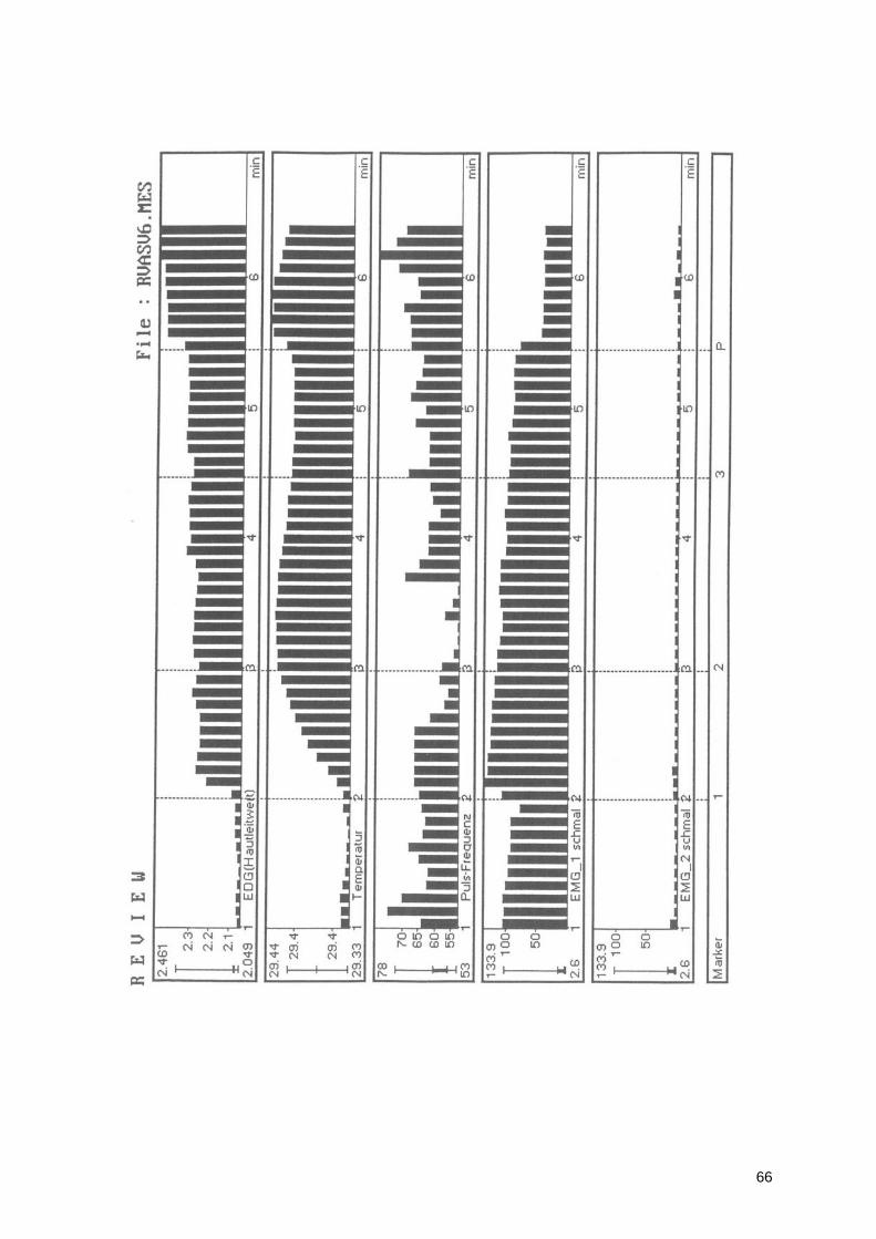
66
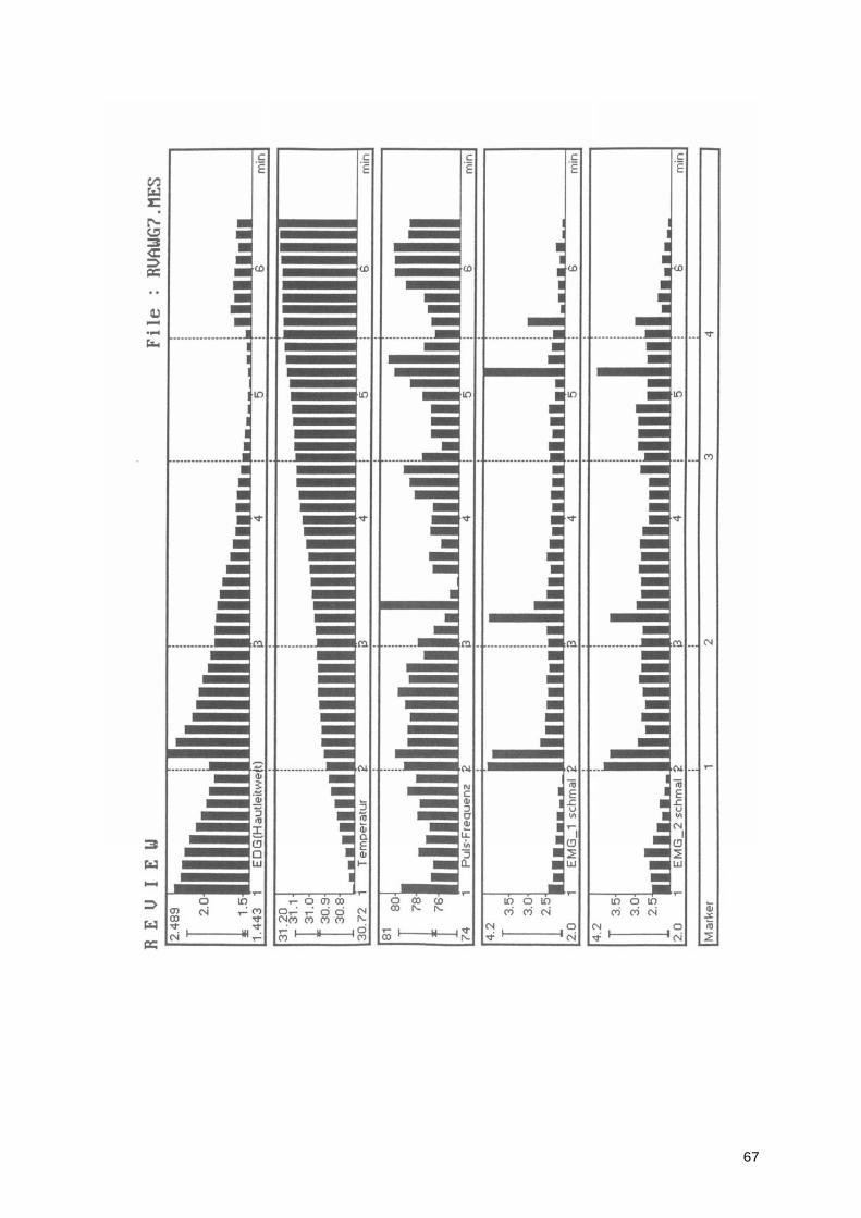
67
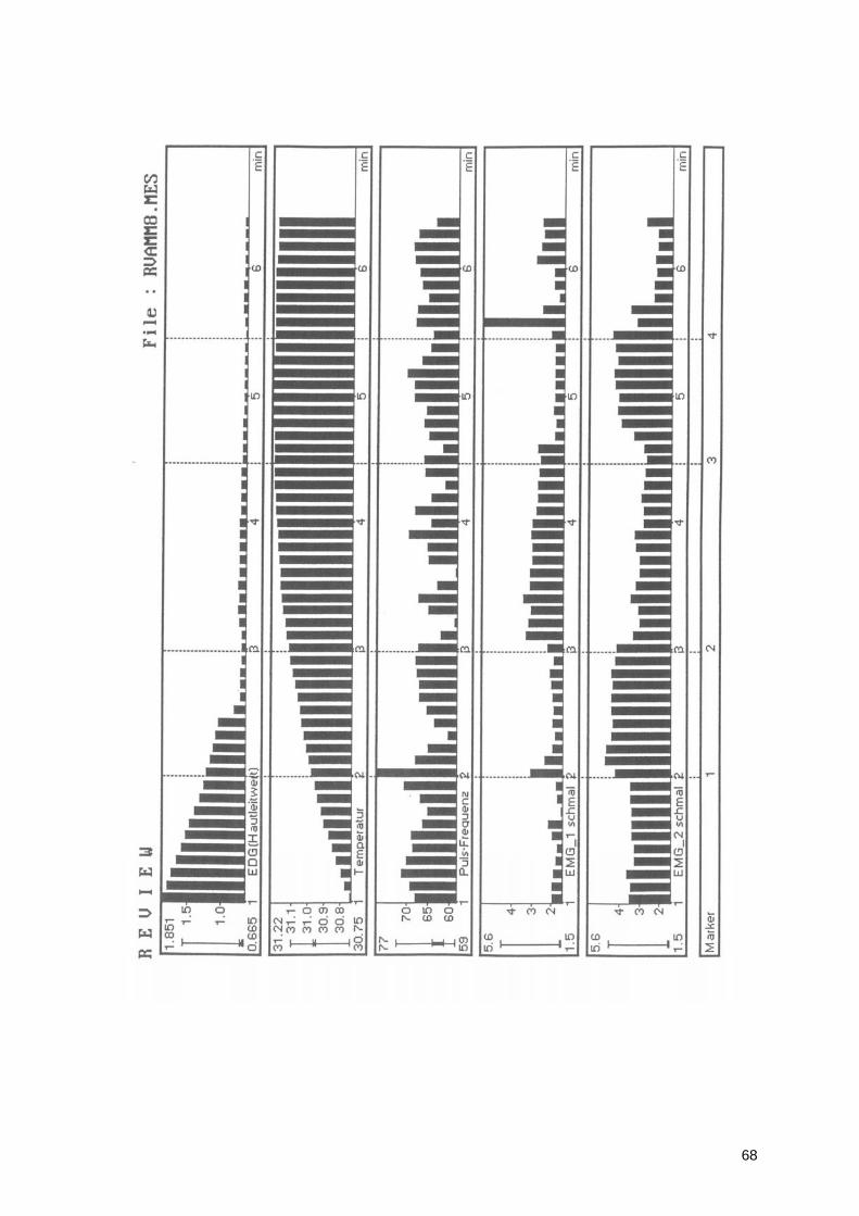
68
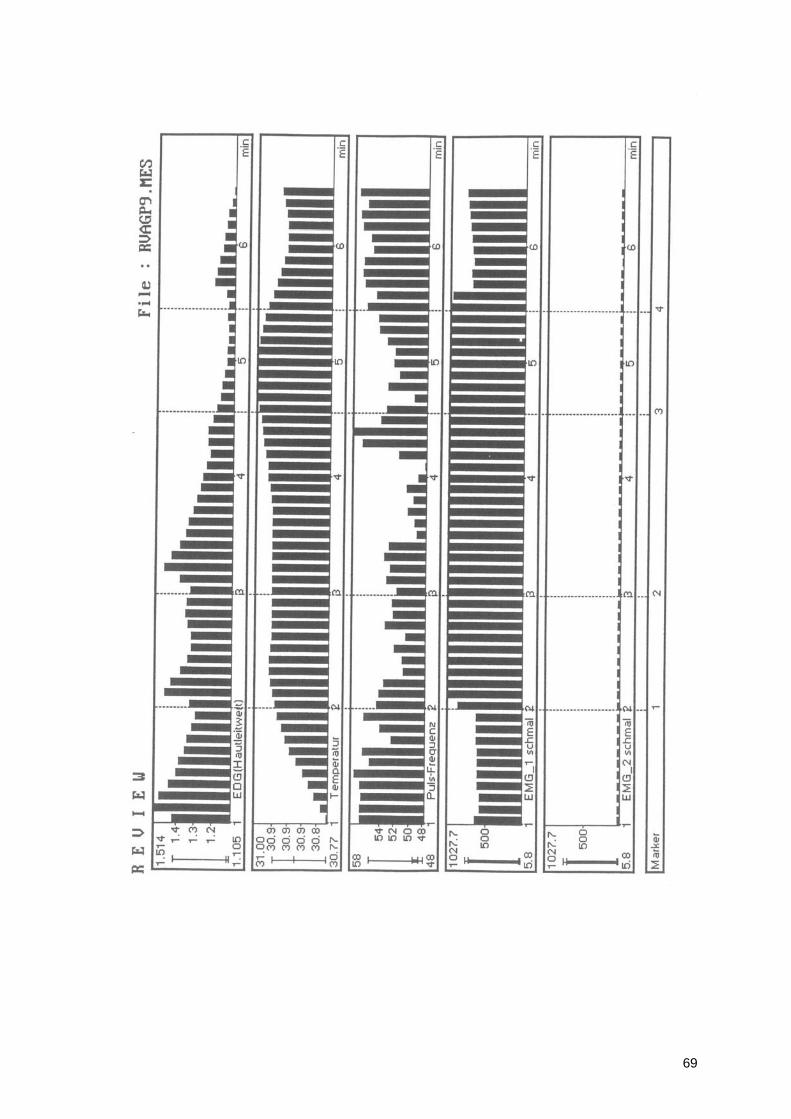
69
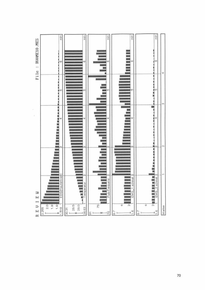
70
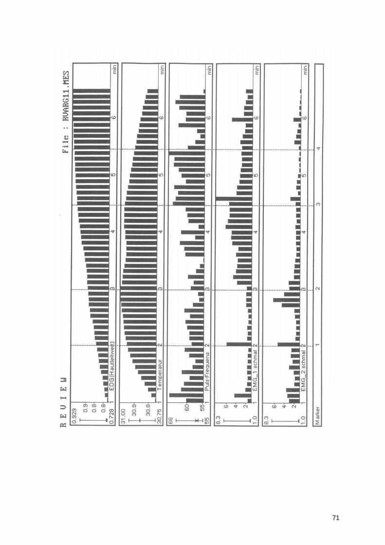
71
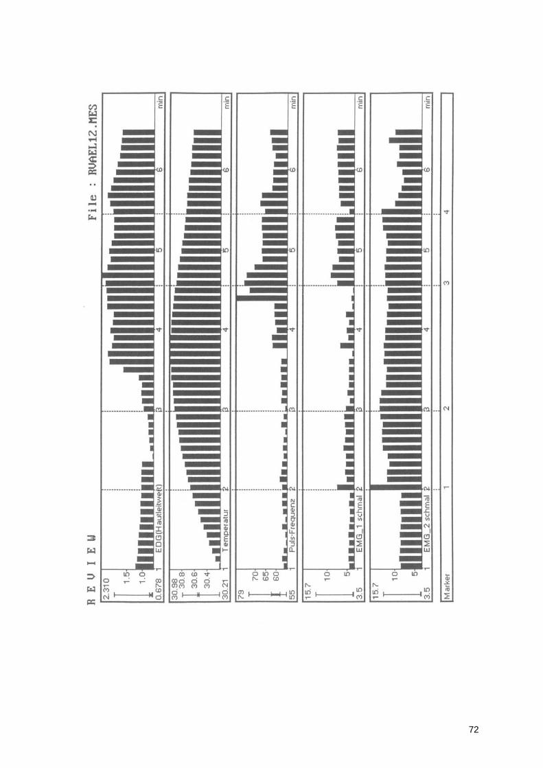
72
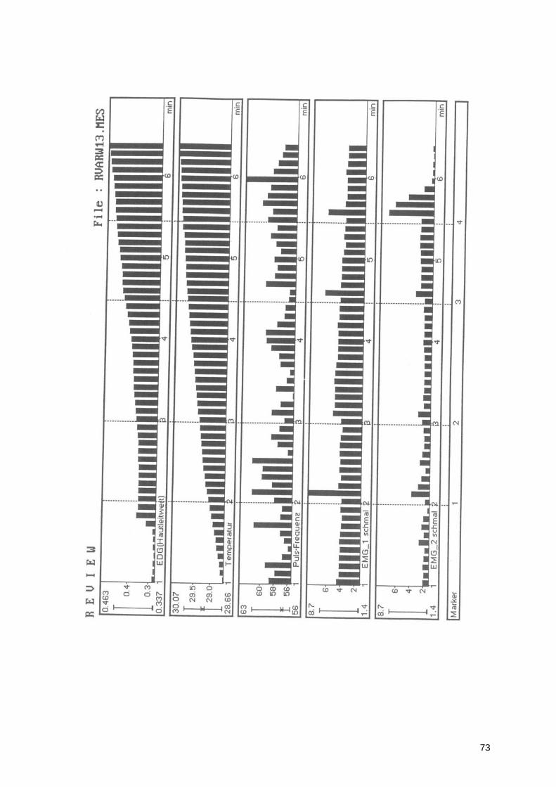
73
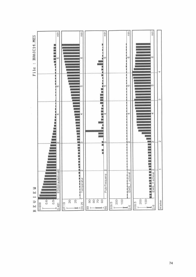
74
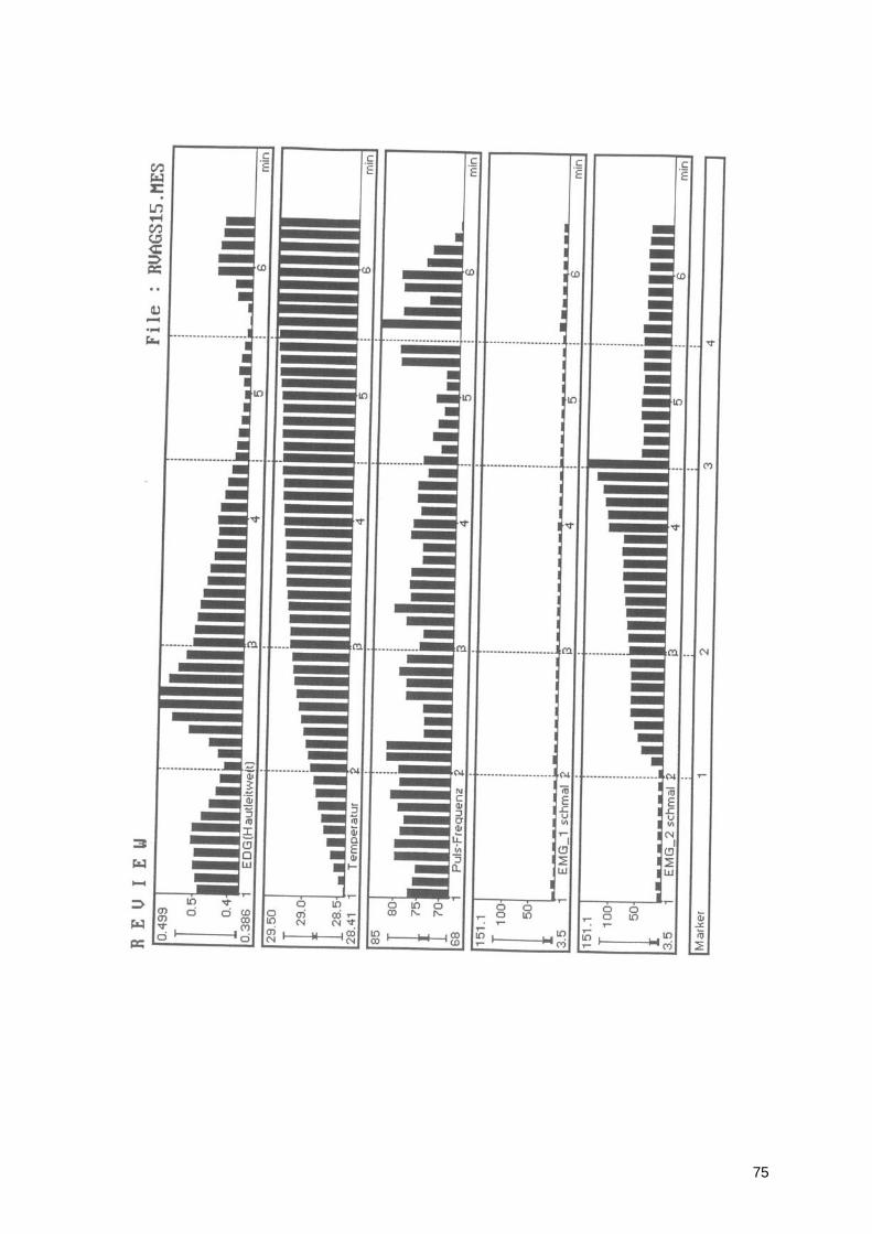
75
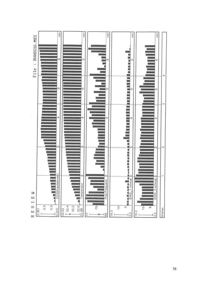
76
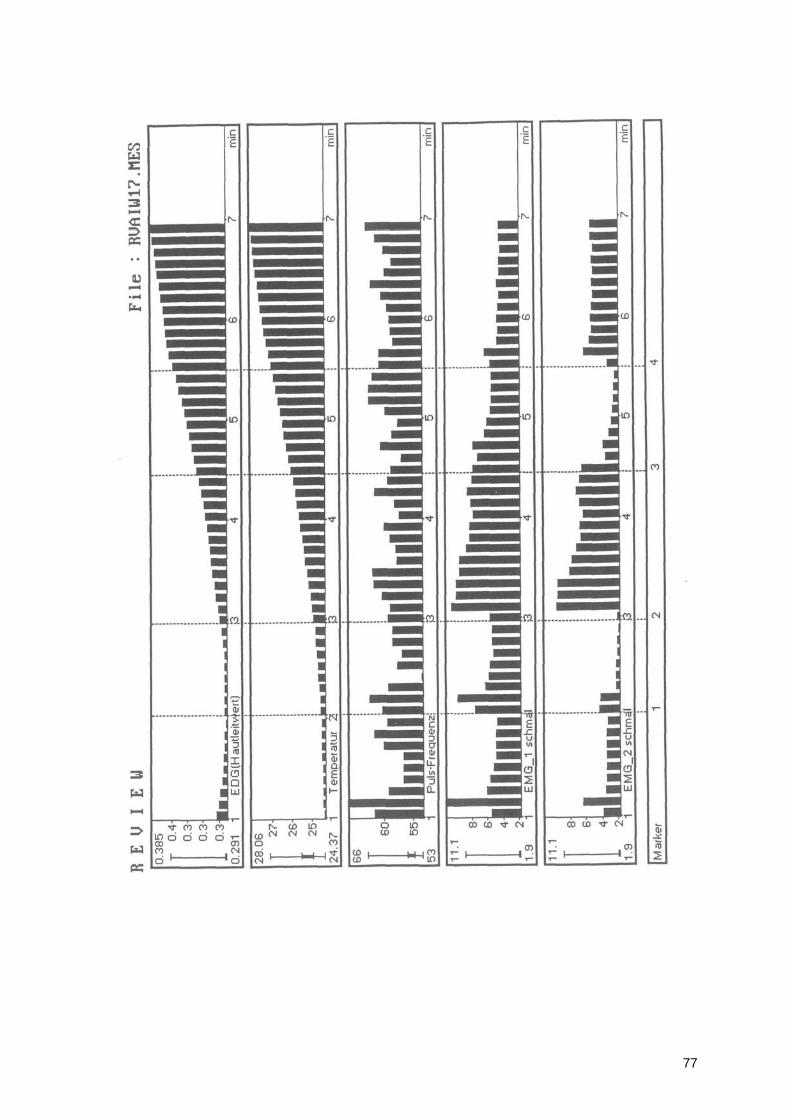
77
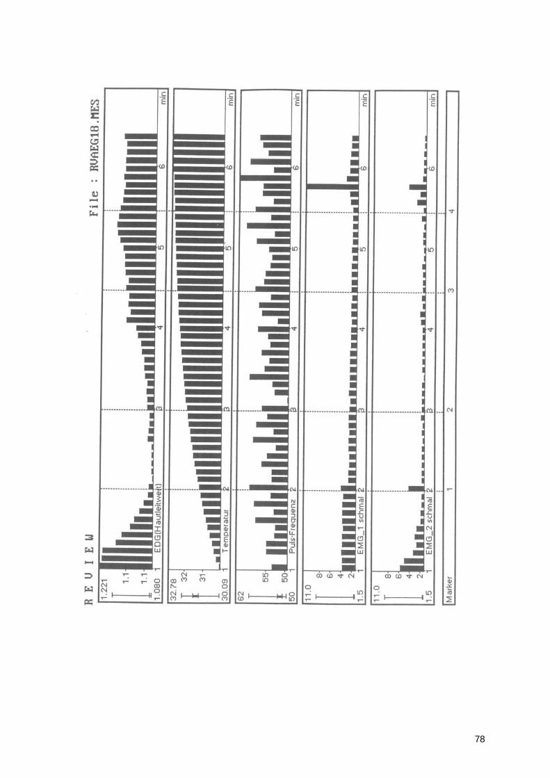
78

79
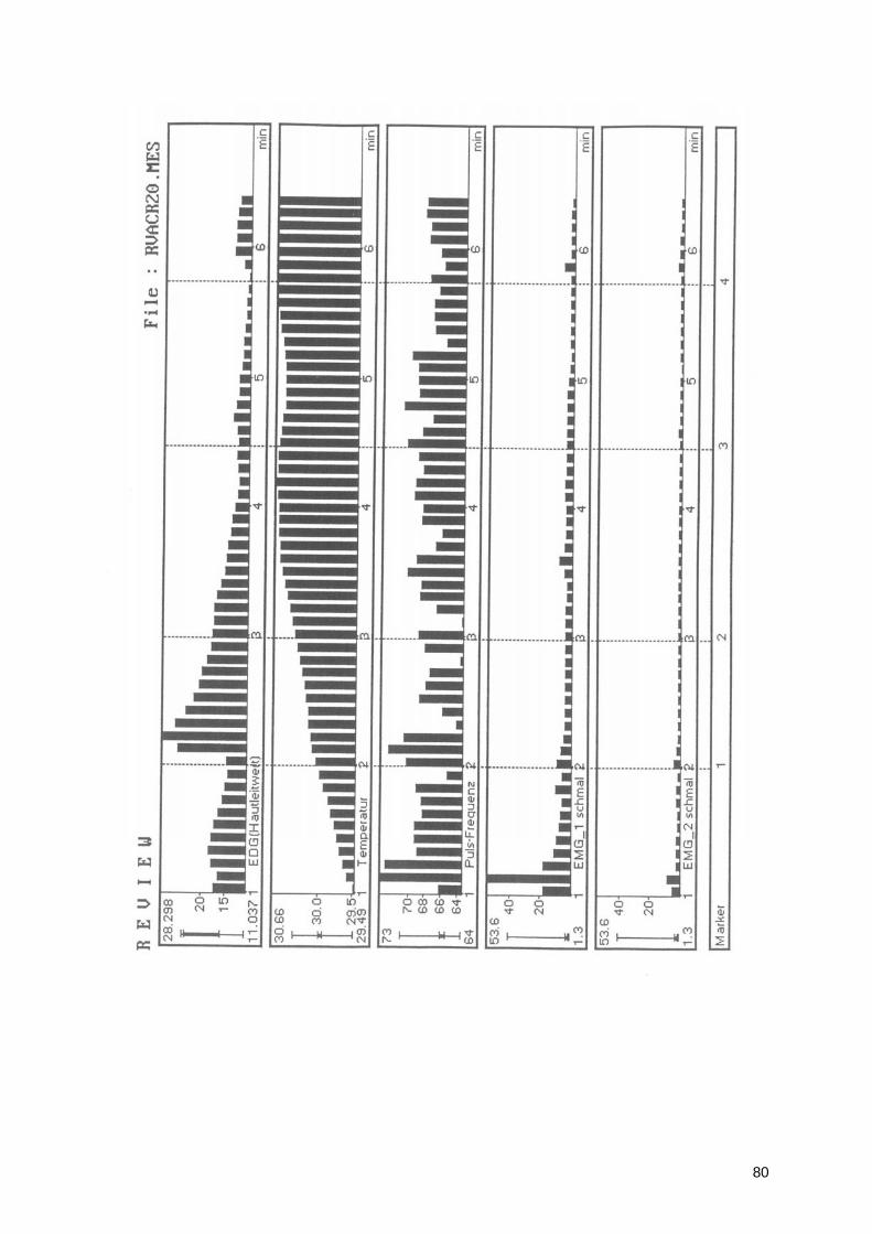
80
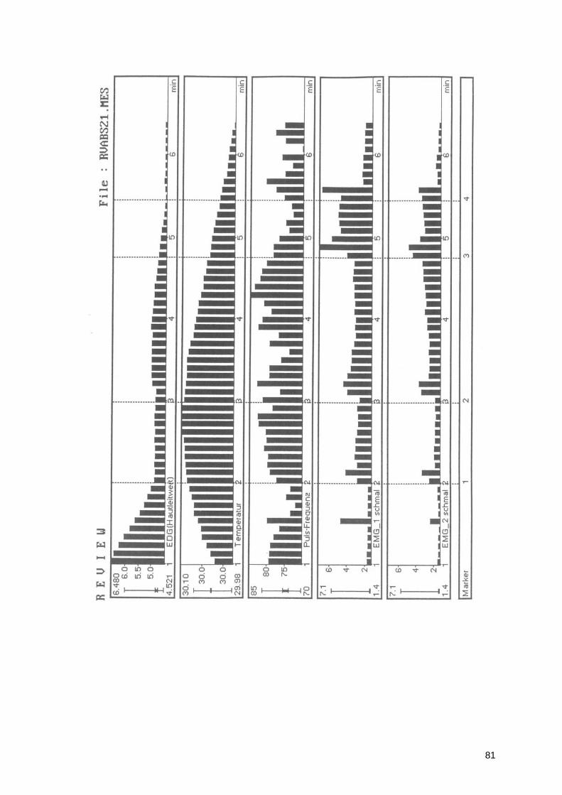
81
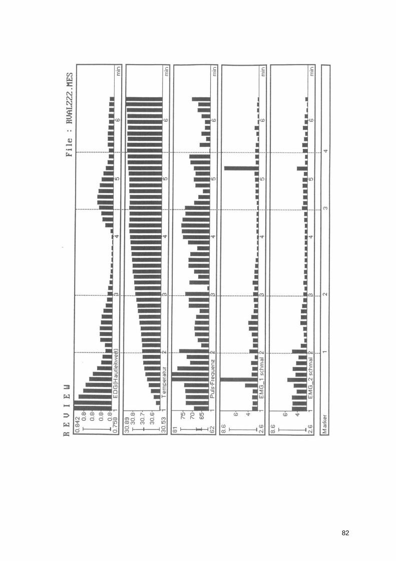
82
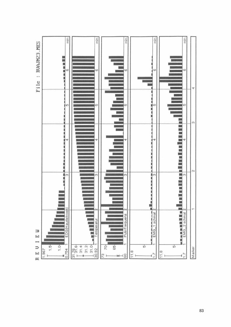
83
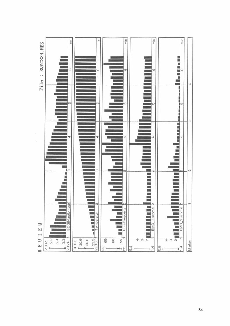
84
85
BIBLIOGRAPHY
• Abrahams V.C. and Falchetto S. (1969) Hind leg ataxia of cervical origin and cervico-lumbar interactions with a supratentorial pathway.
• J.Physiol. (Lond.), 203: 435-447. • Aeckerle J, Heisel J, Die funktionsanalytische Rontgenuntersuchung des
cervikooccipitaien Ubergangs nach Arlen, in: Neuroorthopadie 4 (1988): 169 • Amigues, J.P. L, A.T.M Une Articulation entre L'Osteopathe et Le Dentiste,
Editions de VERLAQUE • Angle EH, Malocclusion of the Teeth, 7th ed., Philadelphia (S.S. White Dental
Manufact. Co.) 1907 • Assendelft WJ, Bouter LM, Knipschild PG, Complications of spinal
manipulation: a comprehensive review of the literature, in: J Fam Pract July 1996
• Baiters W, Ausgewahlte Schriften und Vortrage, Heidelberg (Holzer) 1973 • Berger M, Lechner-Steinleitner S, Kozlovskaya I, Holzmuller G, Mescheriakov
S, Sokolov A, Gerstenbrand F, The effect of head-to-trunk position on the direction of arm movements before, during and after space flight, J Vestib Res 1998 Sep-Oct, 8(5):341-54
• Boyd CH, William FS, Boyd CM, Bryant RW, Wiygul JP, The Effect of Head Position on Electromyographic Evaluations of Representative Mandibular Positioning Muscle Groups, in: The Journal of Craniomandibular Practice, 1987 Jan; Vol. 5, No. 1
• Bronstein AM, Brandt T, Woollacott MH, Clinical disorders of balance, posture and gait, 1996 (Arnold)
• Bruggencate, G.T. (1984), Medizinische Neurophysiologie, Thieme • Ciancaglini R, Testa M, Radaelli G, Association of neck pain with symptoms of
temporomandibular dysfunction in the general adult population, in: Scand Rehabil Med 1999 Mar; 31(1): 17-22
• Cooper BC, Cooper DL, Electromyography of Craniomandibular • Disorders, in: Laryngoscope 101: February 1991 • Criswell-Hanna, Eleanor, Biofeedback and Somatics toward personal
evolution, Freeperson Press Novato,California • Dabbs V, Lauretti WJ, A risk assessment of cervical manipulation vs. NSAIDs
for the treatment of neck pain, in: J Manipulative Physiol Ther 1996 March • Davenport HA (1934) Cells and fibers in spiralnerves, II, A study of C2, C6,
Th4, Th9, L3, S2, S5 in man. Z Comp Neurol 59:167-174 • De Jong P.T.V.M., De Jong J.M.B.V., Cohen B. and Jongkees L.B.W. (1977)
Ataxia and nystagmus induced by injection of local anaesthetics in the neck. Ann. Neurol., 1: 240-246.
• Delattre A, Fenart R, L'hominisation du crane etudiee par la Methode Vestibulaire, Paris (Editions du Centre National de la Recherche Scientifique) 1960.
• Dessem D, Luo P Jaw-muscle spindle afferent feedback to the cervical spinal cord in the rat, in: Exp Brain Res 1999 Oct; 128(4): 451-9
• Dvorak J, Panjabi MM, Functional anatomy of the Alar ligaments, in: Spine 12 (1987): 183
86
• Dvorak J, Panjabi MM, Gerber M, Wichmann W, CT-functional diagnostics of the rotatory instability of upper cervical spine: 1. An experimental study on cadavers, in: Spine 12 (1987): 197
• Dvorak J, Hayek J, Zehnder R, CT-functional diagnostics of the rotatory instability of the upper cervical spine: 2. An evaluation on healthy adults and patients with suspected instability, in: Spine 12 (1987): 726
• Dvorak J, Frohlich D, Penning L, Baumgartner H, Panjabi MM, Functional radiographic diagnosis of the cervical spine: Flexion/extension, in: Spine 13 (1988): 748
• Dvorak J (1988) Manuella Medizin, Georg Thieme Verlag Stuttgart, New York • Dvorak J, Dvorak V, Manuelle Medizin, 4. Aufl., Berlin (Springer) 1991 Ellis
B.D. and Kosmorsky G.S. (1995) Referred ocular pain relieved by suboccipital injection. Headache, 35(2): 101-103.
• Esposito CJ, Panucci PJ, Farman AG, Associations in 425 patients having temporomandibular disorders, in: J Ky Med Assoc 2000May; 98(5): 213-5
• Ferrario VF, Sforza C, Miani A Jr, D'Addona A, Barbini E, Electromyographic activity of human masticatory muscles in normal young people. Statistical evaluation of reference values for clinical applications
• Ferrario VF, Sforza C, Miani A Jr, D'Addona A, Reproducibility of electromyographic measures: a statistical analysis
• Flor H, Schugens MM, Birbaumer N, Discrimination of Muscle Tension in Chronic Pain Patients and Healthy Controls, in: Biofeedback and Self-Regulation, Vol. 17, No. 3, 1992;
• Flor H, Haag G, Turk DC, Long-term efficacy of EMG biofeedback for chronic rheumatic back pain, in: Pain, 27(1986) 195-202.
• Fransson P-A, Magnusson M, Johansson R, Analysis of adaption in anterioposterior dynamics of human postural control, Gait & Posture, Volume 7, Isxsue 1, pp. 64-74 (January 1998)
• Frick H, Allgemeine Anatomie. Spezielle Anatomie (1977) • Fukuda T. (1961)Studies on human dynamic postures from the viewpoint of
postural reflexes. Acta Otolaryngol. (Stockholm), Sup. 161 • Funakoshi M. and Amano N. (1973) Effects of the Tonic Neck Reflex on the
Jaw Muscles of the Rat. J. Dent, Res. July-August 1973, Vol. 52, No.4. • Gagey PM, Weber B, Posturologie. Regulation et dereglements de la station
debout, 1995 (Collection Bois-Larris, Masson) • Garlick D, Proprioception Posture and Emotion, Bankstown (Adept Printing Pty
Ltd.) 1982 • Giasson M, Leroux G, Tardif H, Bouchard L, Therapeutic touch, in: Infirm Que
1999 Jul-Aug; 6(6): 38-47 • Goldstein D.F., Influence of cervical posture on mandibular movement, The
Journal of Prosthetic Dentistry, September 1984 • Gole DR, A clinical observation: a relationship of occlusal contacts to distal
musculature, in: Cranio 1993 Jan; 11(1): 55-61 • Gonzalez HE, Manns A, Forward head posture: ist structural and functional
influence on the stomatognathic system, a conceptual study, in: Cranio 1996 Jan; 14(1): 71-80
• Gosling JA, Human Anatomy (1996) (Mosby-Wolfe) • Grassi C, Deriu F, Roatta S, Santarelli R, Azzena GB Passatore M,
Sympathetic control of skeletal muscle function: possible cooperation between
87
noradrenaline and neuropeptide Y in rabbit jaw muscles, in: Neurosci Lett 1996 Jul 19; 212(3): 204-8
• Hack G., Koritzer R., Robinson W., Hallgren R. and Greenman P. (1995) Anatomic relationship between rectus capitis posterior minor muscle and the dura mater, Spine 1995, December 1, 20: 23 2484-2486.
• Hack GD, Upton JA, The Clinical Implications of a Suboccipital Myodural Bridge, Proceedings of the American Academy of Osteopathie Scientific Convention, 1995 Oct. 29-31, Orlando, FL
• Haldemann S, Kohlbeck FJ, McGregor M, Risk factors and precipitating neck movements causing vertebrobasilar artery dissection after cervical trauma and spinal manipulation, in: Spine 1999 Apr 15; 24(8): 785-94
• Hall, The Mechanis of the Spine and Pelvis , Maidstone College of Osteopathy • Hallgren R., Clinical implications of a cervical myodural bridge, AAO Journal
volume 7, number 4, 1997 • Han Y, Lennerstrand G, Eye position changes induced by neck muscle
vibration in strabismic subjects, in: Graefes Arch Clin Exp Ophthalmol 1999 Jan; 237(1 (: 21-8
• Hardacker JW, Shuford RF, Capicotto PN, Pryor PW, Radiographic standing cervical segmental alignment in adult volunteers without neck symptoms, in: Spine 1997 Jul;22(13): 1472-80
• Higbie EJ, Seidel-Cobb D, Taylor LF, Cummings GS, Effect of head position on vertical mandibular opening, in: J Orthop Sports Phys Ther 1999 Feb, 29(2): 127-30
• Hu JW, Vernon H, Tatourian I, Changes in neck electromyography associated with meningeal noxious stimulation, in: J Manipulative Physiol Ther 1995 Nov-Dec; 18 (9): 577-81
• Hurwitz EL, Aker PD, Adams AH, Meeker WC, Shekelle PG, Manipulation and mobilization of the cervical spine. A systematic review of the literature, in: Spine 1996 Aug 1; 21(15): 1746-59;
• Di Fabio RP, Manipulation of the cervical spine: risks and benefits, in: Phys Ther 1999 Jan; 79(1): 50-65
• Imamura J., M. Saunders , I.Keller (1986) , Projections of cervical nerves to the rat medulla. Neurosci. Lett, 70: S.46-51
• Jankelson RR, Neuromuscular Dental Diagnosis and Treatment, St. Loius 1990
• Jansen J., (1989), Die zervikogene Hemikranie - Folge einer oberen zervikalen Spinalwurzelkompression, Gottingen, (Habilitationsschrift), Klinik fur Neurochirurgie
• Jansen J., (1993), Symptomatik nach Verletzungen der oberen Halswirbelsaule. Eine neuroanatomische Studie zur Pathogenese. Nervenheilkunde, 12: S.230-232
• Johnson RM, Crelin ES, White AA, Panjabi MM, Southwick WO, Some new observations on the functional anatomy of the lower cervical spine, in: Clin Orthop 111 (1975): 192
• Jones LH, DO, FAAO, Strain-Counterstrain, 1995 • Kapandji, I.A.,(1985) Funktionelle Anatomie der Gelenke, Ferdinand Enke
Verlag, Stuttgart • Korr IM, The physiological basis of osteopathic medicine, New York (Insight
Publishing) 1982
88
• Koskimies K, Starck J, Sutinen P, Aalto H, Toppila E, Hirvonen T, Ishizaki H, Alaranta H und Pyykko I, Postural stability, neck propriozeption and tension neck, Acta Otolaryngol Suppl (1997) 529:95-7
• Lafreniere KD, Mutus B, Cameron S, Tannous M, Giannotti M, Laukkanen E, Effects of therapeutic touch on biochemical and mood indicated women, in: J Altern Complement Med 1999 Aug; 5(4): 367-70
• Landouzy JM, Les articulations temporo-mandibulaires. Evaluation, traitements odontologiques et osteopathiques, 1993 (ed. Verlaque)
• Lang J, Klinische Anatomie der Halswirbelsaule (1991) Thieme • Lee WY, Okeson JP, Lindroth J, The relationship between forward head
posture and temporomandibular disorders, in: J Orofac Pain 1995 Spring; 9(2): 161-7
• Lekhel H, Popov K, Bronstein a, Gresty M, Postural responses to vibration of neck muscles in patients with uni-and bilateral vestibular loss, in: Gait & Posture, Volume 7, Issue 3, pp. 228-236 (May 1998)
• Lennerstrand G., Han Y. and Velay J.L. (1996) Properties of eye movements induced by activation of neck muscle proprioceptors. Graefes Arch Clin Exp Ophthalmol 1996 Nov., 234(11): 703-709.
• Longet F.A. (1845) Sur les troubles qui surviennent dans I'equilibration, la station et la locomotion des animaux apres la section des parties molles de la nuque. Gaz. Med. France, 13: 565-567.
• Macaluso G.M., De Laat A.D. and Pavesi G. (1996) The influence of the asymmetric tonic neck reflex on the H-reflex in human temporal muscle. Minerva Stomatol 45(9): 387-392.
• MacDonald G, The complete illustrated Guide to the Alexander Technique, (Paperback) 1998
• Magnus R. (1926) Some results of studies in the physiology of posture (Cameron Prize Lectures). Lancet, 211: 531-536.
• Meyer J, Participation des afferences trigeminals dans la regulation tonique posturale orthostatique. Interet de I'examen systematique du systeme manducateur chez les sportifs de haut niveau. These 43 55 77, Universite Rene Descartes, Paris 1977
• Mitchell,F. D.O. F.A.A.O. (1995) The Muscle Engergy Manual , MET Press, East Lansing, Michigan
• Mitchell BS, Humphreys BK, CTSullivan E, Attachments of the ligamentum nuchae to cervical posterior spinal dura and the lateral part of the occipital bone, in: J Manipulative Physiol Ther 1998 Mar-Apr; 21(3): 145-8
• Moore KL, Embryologie. Lehrbuch und Atlas der Entwicklungsgeschichte des Menschen, 3. Aufl., Stuttgart (Schattauer) 1990
• Netter FH, Atlas of Human Anatomy (1989) (Ciba-Geigy Corporation) • Nikolakis P, Relationship between Craniomandibular Disorders and Poor
Posture, The Journal of Craniomandibular Practice, April 2000 • Nobili A, Adversi R, Relationship between posture and occlusion: a clinical and
experimental investigation, in: Cranio 1996 Oct; 14(4): 274-85 • Nobili A, Adversi R, Relationship between posture and occlusion: a clinical and
experimental investigation, in: Cranio 1996 Oct; 14(4): 274-85 • Northfield D, Some observations of headache, in: Brain, 1938; 61: 133-162
Oliver J, Middleditch A, Functional Anatomy of the Spine, Oxford (Butterworth-Heinemann) 1991.
89
• Panjabi M.M., Dvorak J, Duranceau J, Yamamoto I, Gerber M, Rauschnig W, Bueff U, Three-dimensional movements of the upper cervical spine, in: Spine 13 (1988): 726
• Parent! G, Orlandi G, Bianchi M, Renna M, Martini A, Murri L, Vertebral and carotid artery dissection following chiropractic cervical manipulation, in: Neurosurg Rev 1999 Oct; 22(2-3): 127-9
• Peck SD, The efficacy of therapeutic touch for improving functional ability in elders with degenerative arthritis, in: Nurs Sci Q 1998 Fall; 11(3): 123-32
• Penning L, Functional pathology of the cervical spine, in: Excerpta Medical Foundation, New York 59 (1968): 1
• Penning K, Wilmink JT, Rotation of the cervical spine: A CT study on normal subjects, in: Spine 12 (1987): 732
• Perry HT, Muscular Changes Associated With Temporomandibular Joint Dysfunction, in: Journal of the American Dental Association, Vol. 54, No. 5 (May) 1957
• Perry HT, Temporomandibular joint and occlusion, in: Angle Orthod 1976 Jul; 46(3): 284-93
• Platzer W, Kahle W, Leonhardt H, Taschenatlas der Anatomie für Studium und Praxis (1984)
• Pollard H., Ward G. (1977) J Manipulative Physiol. Ther. 20(7): 443-447. • Ralf L, Rydell N, Spinal manipulation-treatment associated with a high risk of
complications, Stockholm (Personskaderegeling AB) 1999 • Hufnagel A, Hammers A, Schonle PW, Bohm KD, Leonhardt G, Stroke
following chiropractic manipulation of the cervical spine, in: J Neurol 1999 Aug; 246(8): 683-8
• Ricard F. D.O. Osteopathic Treatment of Pain originating in the Craniocervical Area, Editions DE VERLAQUE
• Richardin P, Wetphal A, Divry M, Didier G, Influence of stress and occlusal interference on the EMG activity of some masticatory muscles during a single mastication cycle, in: J Oral Rehabil 1995 Oct; 22(10): 775-80
• Richmond F.R.J, and Abrahams V.C. (1975) Morphology and distribution of muscle spindles in dorsal muscles of the cat neck. J. Neurophysiol., 38: 1322-1339.
• Richmond F.R.J, and Abrahams V.C. (1975) Morphology and distribution of muscle spindles in dorsal muscles of the cat neck. J. Neurophysiol., 38: 1322-1339.
• Richmond F.R.J., Anstee G.C.B., Sherwin E.A. and Abrahams V.C. (1976) Motor and sensory fibers of neck muscle nerves in the cat. Canad. J. Physiol. Pharmacol., 54: 294-304.
• Richmond F.R.J., Anstee G.C.B., Sherwin E.A. and Abrahams V.C. (1976) Motor and sensory fibers of neck muscle nerves in the cat. Canad. J. Physiol. Pharmacol., 54: 294-304.
• Richmond F.R.J, and Abrahams V.C. (1979) Physiological properties of muscle spindles in dorsal neck muscles of the cat. J. Neurophysiol., 42:604-617
• Richmond F.R.J, and Abrahams V.C. (1979) Physiological properties of muscle spindles in dorsal neck muscles of the cat. J. Neurophysiol., 42:604-617
90
• Richmond FJ, Corneil BD, Marked non-uniformity of fiber-type composition in the primate suboccipital muscle obliquus capitis inferior, Exp Brain Res 1999 Mar, 125(1): 14-8
• Robinson M.J., The Influence of Head Position on Temporomandibular Joint Dysfunction, Chicago College of Osteopathic Roll Jp, Roll R, From eye to foot: a proprioceptive chain involved in postural control, in: Amblard B, Berthoz A, Clarac F, eds. Posture and gait: development, adaptation and modulation. Amsterdam: Elsevier (1988) 155-164
• Ruf S, Cecere F, Kupfer J, Pancherz H, Stress-induced changes in the functional electromyographic activity of the masticatory muscles, in: Acta Odontol Scand 1997 Jan; 55(1): 44-8
• Ruggieri V, Persico G, Caputo G, An analysis of the psychophysiological components in subjects with temporomandibular disorders. Myographic tension and the management of aggressiveness, in: Minerva Stomatol 1999 Oct; 48(10): 477-84
• Rutten HP, Szpak K, van Mameren H, Ten Holter J, de Jong JC, Anatomic relation between the rectus capitis posterior minor and the dura mater, in: Spine 1997 Apr 15; 22 (8): 924-6
• Sakuma A, Aihara Y, Influence of propriozeptive input from leg, thigh, trunk and neck muscles in the equilibrium of standing, Nippon Jibiinkoka Gakkai Kaiho (1999) 102(5): 643-9
• Schimek JJ, Untersuchungen zum Spannungs-Kopfschmerz, in: Man Med 1988,26: 107-112
• Schmidt RF (Hrsg.), Grundriß der Sinnesphysiologie, 4. Aufl., Berlin (Springer) 1980
• Schöps P, Kober L, Schenk M, Kirchmair G, Busch B, Manuelle Therapiel (1997)
• Schottl W, Die cranio-mandibulare Regulation, Heidelberg (Huthig) 1991 • Schroeder H, Siegmund H, Santibanez G, Kluge A, Causes and signs of
temporomandibular joint pain and dysfunction: an electromyographical investigation, in: J Oral Rehabil 1991 Jul; 18(4): 301-10
• Schwartz M.,(1998) Biofeedback: A Practitioner's Guide. • Seminarunterlagen/'Biofeedback-Grundlagen" (1997) Ausbildungslehrgang
der Osterr. Ges. fur Biofeedback und Psychophysiologic (OBfP) • Shumway-Cook A, Woollacott MH, Motor Control. Theory and practical
applications, 1995 (Williams & Wilkins) • Skillem P.G. (1954) Great occipital-trigeminus syndrome as revealed by
induction block, Arch. Neurol. & Psychiat, 72: 335-340. • Slavicek G, Gsellmann B, Gruber R, Rath M, Furhauser R, Biofeedback als
Therapieerganzung bei craniomandibularer Dysfunktion, in: IOK, 27. Jhrg. (1995) Nr. 1
• Sobotta. Atlas der Anatomie des Menschen (1988) (Urban & Schwarzenberg) • Talis VL, Ivanenko YP, Kazennikov OV, Support stability influences postural
responses to muscle vibration in humans, Eur J Neurosci 1999 Feb, 11(2): 647-54
• Taylor J.L and McCloskey D.I. (1988) Proprioception in the neck. Exp. Brain Res. 70: 351-360.
• Travell JG, Simons DG, Handbuch der Muskel-Triggerpunkte: Obere Extremitat, Kopf und Thorax, Stuttgart (Gustav Fischer) 1998
91
• Trew M, Everett T, Human Movement, 3rd ed., 1997 (Churchill Livingstone) • Uhlig Y, Weber BR, Grob D, Muntener M, Fiber composition and fiber
transformations in neck muscles with dysfunction of the cervical spine, J Orthop Res 1995 Mar, 13(2): 240-9
• Urbanowicz M. (1991) The Journal of Craniomandfbular Practice, April 1991, Vol.9, No.2.
• Vadokas V, Lotzmann KU (1995) Funktionelle Störungen des kraniomandibularen Systems in der HWS als differentialdiagnostisches Problem der idiopathischen Trigeminusneuralgie. Der Schmerz 9:29-33
• Visscher CM, Wright EF, Domenech MA, Fischer JR Jr, Usefulness of posture training for patients with temporomandibular disorders, in: J Am Dent Assoc 2000 Feb; 131(2): 202-10
• Voss H. (1958) Zahl und Anordnung der Muskelspindeln in den unteren Zungenbeinmuskeln dem M. sternocleidomastoideus und den Bauch- und tiefen Nackenmuskeln. Anat. Anz. 105: 265-275.
• Cooper S. and Daniel P.M. (1963) Muscle spindles in man, their morphology in the lumbricals and the deep muscles of the neck. Brain, 86: 563-594.
• Ward R.C. Foundations fur Osteopathic Medicine (1997), Williams & Wilkins • de Wijer A, Steenks MH, de Leeuw JR, Bosman F, Helders PJ, Symptoms of
the cervical spine in temporomandibular and cervical spine disorders, in: J Oral Rehabil 1996 Nov; 23(11): 742-50
• Wolff HD, Neurophysiologische Aspekte des Bewegungssystems, 3.Aufl., Berlin (Springer) 1996
• Yaw ILK, Glover JC, Counterstrain: A Handbook of Osteopathic Technique, Tulsa (Okla: Y Knot Publishers) 1994
• Zeier, H., Biofeedback (1990), Verlag Hans Huber, Bern • Zonnenberg AJ, Van Maanen CJ, Oostendorp RA, Elvers JW, Body posture
photographs as a diagnostic aid for musculoskeletal disorders related to temporomandibular disorders (TMD), in: Cranio 1996 Jut; 14(3): 225-32
• Yamada R, Ogawa T, Koyano K, The effect of head posture on direction and stability of mandibular closing movement, in: J Oral Rehabil 1999 Jun; 26(6): 511-20