Embed Size (px)
Citation preview
Leishmania infantum:
molecular analysis for identification of
potential virulence factors and
genes of diagnostic use
Inauguraldissertation
zur Erlangung der Würde eines Doktors der Philosophie
vorgelegt der
Philosophisch-Naturwissenschaftlichen Fakultät
der Universität Basel
von
Igor Niederwieser
aus Fällanden (ZH)
Basel, 2004

Genehmigt von der Philosophisch-Naturwissenschaftlichen Fakultät der Universität
Basel auf Antrag der Herren
PD Dr. Hans-Peter Beck, Prof.Dr. Niklaus Weiss und Prof.Dr. Marcel Tanner
Table of contents List of Abbreviations 3 Summary 6 1. Introduction 7 1.1. On the biology of Leishmania 7 1.1.1 Taxonomy 7 1.1.2. The Leishmania genome 8 1.1.3. Life Cycle 8 1.1.4. Immunology 10 1.1.5. Leishmaniasis 14 1.2. Basic molecular biological methods used during the
PhD project 17 1.2.1. RNA 17 1.2.2. DNA 18 1.2.3. Proteins 20 2. Development of an early diagnosis test system for canine
leishmaniasis caused by L.infantum 21 2.1. Background 21 2.2. Methods 22 2.3. Identification of recombinant antigens from Leishmania infantum suitable as early diagnostic tool 23 2.4. Discussion 24 3. Development of a PCR assay for diagnosis of human leishmaniasis
and differentiation of Leishmania species 25 3.1. Background and method 25 3.2. Diagnostic genotyping of Old and New World Leishmania
species by PCR-RFLP 26 3.3. Identification and Differentiation of Leishmania Species in
Clinical Samples by PCR Amplification of the Miniexon Sequence and Subsequent Restriction Fragment Length Polymorphism Analysis 27
3.4. Discussion 28
1
4. Identification of genes encoding potential virulence factors, drug targets and vaccine candidates of L. infantum. 29 4.1. Background 29 4.2. Methods 31 4.3. Differentially expressed genes in Leishmania infantum promastigotes 36 4.4. Additional Methods and Results 37 4.5. Discussion 40
4.5.1. Choice of the approach 40 4.5.2. Choice of the technique 40
5. General discussion 43 5.1. Diagnosis 43 5.2. Vaccines candidates 44 5.3. Virulence factors 45 5.4. Drug targets 46 6. Appendices 48 6.A1. Hybridization buffers for Northern blot analysis 48 6.A2. Construction of spliced leader cDNA 49 6.A3. Isolation of genomic Leishmania DNA 50 6.A4. Selective DNA precipitation using PEG 51 6.A5. Sequencing of GC-rich DNA 52 6.A6. Sequencing of large inserts 52 6.A7. Cycle restriction ligation (CRL) 53 6.A8. Small scale preparation of electro-competent cells 54 6.A9. E. coli transformation 54 6.A10. Modified expression vectors 56 6.A11. Purification of active eukaryotic proteins from inclusion
bodies in E. coli 59 6.A12. Purification of GST Fused Proteins 61 7. Acknowledgments 63 8. References 65 Curriculum Vitae 68
2
List of Abbreviations 3’ 3 prime end 5’ 5 prime end A adenine AA amino acid ACL anthroponotic cutaneous leishmaniasis AVL anthroponotic visceral leishmaniasis AMV Avian Myeloblastosis Virus BLAST basic local alignment search tool bp base pair BSA bovine serum albumin C cytosine CD cluster of differentiation cDNA complementary (to RNA) DNA CL cutaneous leishmaniasis CYT cytochrome DCL diffuse cutaneous leishmaniasis DMSO dimethyl sulfoxide DNA deoxyribonucleic acid DNase deoxyribonuclease dNTP deoxyribunucleotide triphosphate mix ds double stranded DTT dithiotreitol E. Escherichia EDTA ethylenediaminetetraacetic acid ELISA enzyme-linked immunosorbent assay ER endoplasmatic reticulum EtOH ethanol FCS fetal calf serum FT flow through G guanine GST glutathione-S transferase GT glucose transporter His histidine HIS- histone HIV human immunodeficiency virus HEPES 4-(2-Hydroxyethyl)piperazine-1-ethanesulfonic acid HSP heat shock protein
3
IFA immunofluorescence assay IFAT immunofluorescence assay test IG immune globulin IL interleukin IPTG isopropylβ-D-1-thiogalactopyranoside kb kilobase kDa kilodalton L. Leishmania lacZ gene encoding β-galactosidase LPG lipophosphoglycan Lu. Lutzomyia M marker MAC membrane attack complex MAT-1 metacyclogenesis associated transcript 1 MCL mucocutaneous leishmaniasis MHC major histocompatibility complex MOPS 3-(N-Morpholino)propanesulfonic acid miRNA micro RNA mRNA messenger RNA Mb mega base M-MLV Moloney Murine Leukemia Virus NAD(P) nicotinamide adenine dinucleotide (phosphate) n.d. not determined NOS nitric oxide synthase nt nucleotide ORF open reading frame P. Phlebotomus PBS phosphate buffered saline PCR polymerase chain reaction PEG polyethylene glycol Pfu Pyrococcus furiosus PIPES piperazine-1,4-bis(2-ethanesulfonic acid) PKDL post kala-azar dermal leishmaniasis PM peritrophic membrane RBS ribosome binding site RFLP restriction fragment length polymorphism RNA ribonucleic acid RNase ribonuclease RP ribosomal protein rRNA ribosomal RNA
4
RT reverse transcriptase S Svedberg Sarkosyl N-lauroylsarcosine SDS sodium dodecyl sulfate SDS-PAGE sodium dodecyl sulfate polyacryl amide gel electrophoresis SHERP small hydrophilic endoplasmic reticulum-associated protein SL spliced leader SN supernatant SSC saline sodium citrate buffer ss single stranded SSH suppression subtractive hybridization SSPE saline sodium phosphate EDTA buffer STI Swiss Tropical Institute T thymidine T. Trypanosoma Taq Thermus aquaticus TBE tris borate EDTA buffer TE tris EDTA buffer TEM transmission electron microscopy Th T helper cell TM transmembrane domain tris 2-amino-2-(hydroxymethyl)-1,3-propanediol Tris·Cl 2-amino-2-(hydroxymethyl)-1,3-propanediol hydrochloride U units VL visceral leishmaniasis v/v volume per volume WHO World Health Organization w/v weight per volume ZCL zoonotic cutaneous leishmaniasis ZVL zoonotic visceral leishmaniasis
5
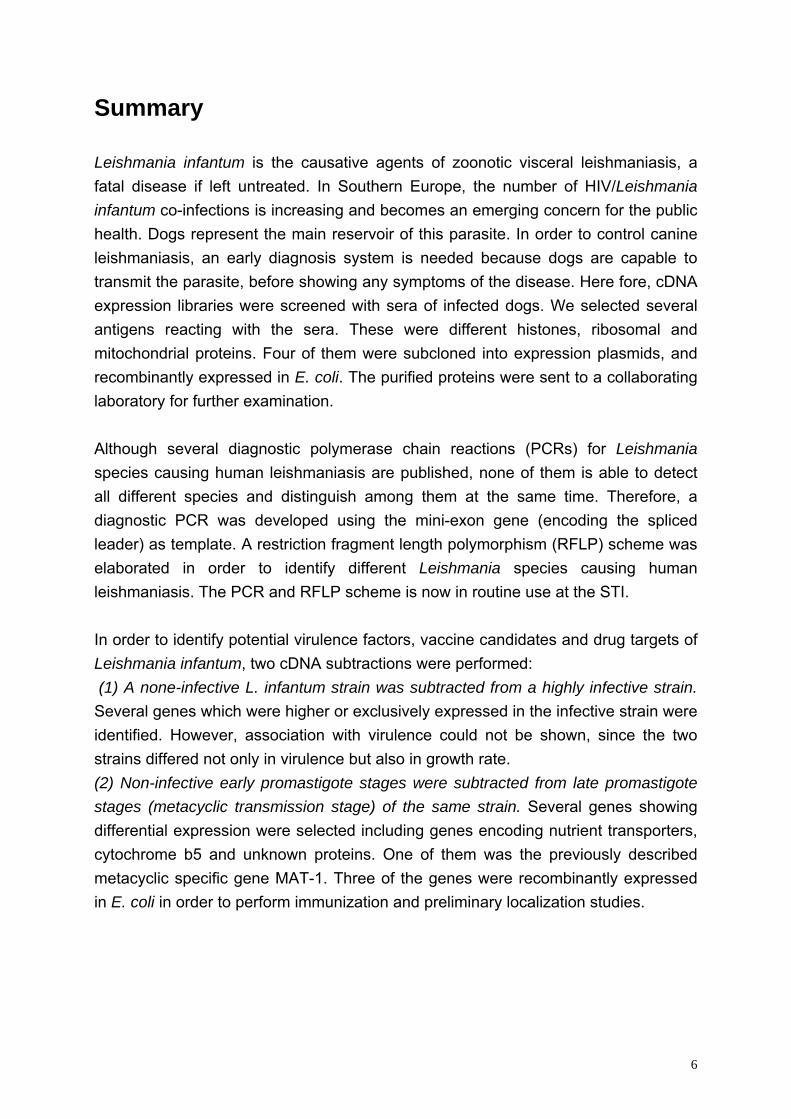
Summary Leishmania infantum is the causative agents of zoonotic visceral leishmaniasis, a fatal disease if left untreated. In Southern Europe, the number of HIV/Leishmania infantum co-infections is increasing and becomes an emerging concern for the public health. Dogs represent the main reservoir of this parasite. In order to control canine leishmaniasis, an early diagnosis system is needed because dogs are capable to transmit the parasite, before showing any symptoms of the disease. Here fore, cDNA expression libraries were screened with sera of infected dogs. We selected several antigens reacting with the sera. These were different histones, ribosomal and mitochondrial proteins. Four of them were subcloned into expression plasmids, and recombinantly expressed in E. coli. The purified proteins were sent to a collaborating laboratory for further examination. Although several diagnostic polymerase chain reactions (PCRs) for Leishmania species causing human leishmaniasis are published, none of them is able to detect all different species and distinguish among them at the same time. Therefore, a diagnostic PCR was developed using the mini-exon gene (encoding the spliced leader) as template. A restriction fragment length polymorphism (RFLP) scheme was elaborated in order to identify different Leishmania species causing human leishmaniasis. The PCR and RFLP scheme is now in routine use at the STI. In order to identify potential virulence factors, vaccine candidates and drug targets of Leishmania infantum, two cDNA subtractions were performed: (1) A none-infective L. infantum strain was subtracted from a highly infective strain. Several genes which were higher or exclusively expressed in the infective strain were identified. However, association with virulence could not be shown, since the two strains differed not only in virulence but also in growth rate. (2) Non-infective early promastigote stages were subtracted from late promastigote stages (metacyclic transmission stage) of the same strain. Several genes showing differential expression were selected including genes encoding nutrient transporters, cytochrome b5 and unknown proteins. One of them was the previously described metacyclic specific gene MAT-1. Three of the genes were recombinantly expressed in E. coli in order to perform immunization and preliminary localization studies.
6
1. Introduction
Three different projects were combined within this PhD work:
• Identification of suitable antigens for an early diagnosis system of canine leishmaniasis.
• Development of a diagnostic PCR for human leishmaniasis. • Identification of potential virulence factors, vaccine candidates and drug
targets of Leishmania infantum.
1.1. On the biology of Leishmania
1.1.1 Taxonomy
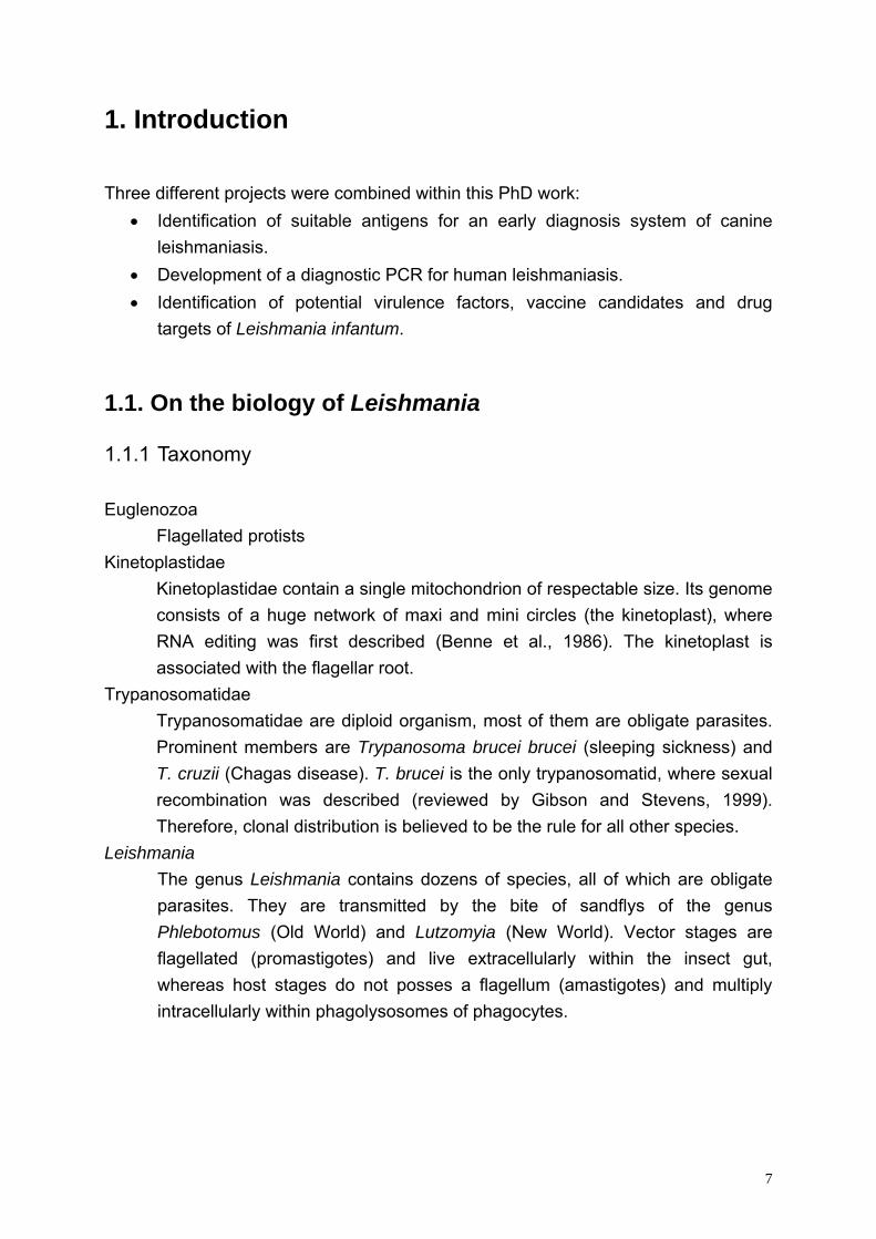
Euglenozoa Flagellated protists Kinetoplastidae
Kinetoplastidae contain a single mitochondrion of respectable size. Its genome consists of a huge network of maxi and mini circles (the kinetoplast), where RNA editing was first described (Benne et al., 1986). The kinetoplast is associated with the flagellar root.
Trypanosomatidae Trypanosomatidae are diploid organism, most of them are obligate parasites. Prominent members are Trypanosoma brucei brucei (sleeping sickness) and T. cruzii (Chagas disease). T. brucei is the only trypanosomatid, where sexual recombination was described (reviewed by Gibson and Stevens, 1999). Therefore, clonal distribution is believed to be the rule for all other species.
Leishmania The genus Leishmania contains dozens of species, all of which are obligate parasites. They are transmitted by the bite of sandflys of the genus Phlebotomus (Old World) and Lutzomyia (New World). Vector stages are flagellated (promastigotes) and live extracellularly within the insect gut, whereas host stages do not posses a flagellum (amastigotes) and multiply intracellularly within phagolysosomes of phagocytes.
7
1.1.2 The Leishmania genome
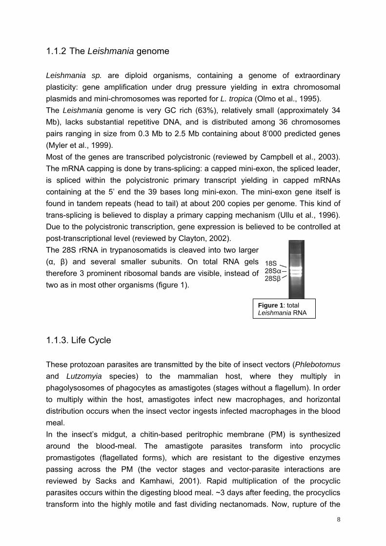
Leishmania sp. are diploid organisms, containing a genome of extraordinary plasticity: gene amplification under drug pressure yielding in extra chromosomal plasmids and mini-chromosomes was reported for L. tropica (Olmo et al., 1995). The Leishmania genome is very GC rich (63%), relatively small (approximately 34 Mb), lacks substantial repetitive DNA, and is distributed among 36 chromosomes pairs ranging in size from 0.3 Mb to 2.5 Mb containing about 8’000 predicted genes (Myler et al., 1999). Most of the genes are transcribed polycistronic (reviewed by Campbell et al., 2003). The mRNA capping is done by trans-splicing: a capped mini-exon, the spliced leader, is spliced within the polycistronic primary transcript yielding in capped mRNAs containing at the 5’ end the 39 bases long mini-exon. The mini-exon gene itself is found in tandem repeats (head to tail) at about 200 copies per genome. This kind of trans-splicing is believed to display a primary capping mechanism (Ullu et al., 1996). Due to the polycistronic transcription, gene expression is believed to be controlled at post-transcriptional level (reviewed by Clayton, 2002). The 28S rRNA in trypanosomatids is cleaved into two larger (α, β) and several smaller subunits. On total RNA gels therefore 3 prominent ribosomal bands are visible, instead of two as in most other organisms (figure 1).
A
1.1.3. Life Cycle
These protozoan parasites are transmitted by the bite of inseand Lutzomyia species) to the mammalian host, wphagolysosomes of phagocytes as amastigotes (stages withto multiply within the host, amastigotes infect new macrodistribution occurs when the insect vector ingests infected mmeal. In the insect’s midgut, a chitin-based peritrophic membraaround the blood-meal. The amastigote parasites trpromastigotes (flagellated forms), which are resistant to passing across the PM (the vector stages and vector-previewed by Sacks and Kamhawi, 2001). Rapid multiplparasites occurs within the digesting blood meal. ~3 days afttransform into the highly motile and fast dividing nectanom
Figure 1: total Leishmania RN
ct vectors (Phlebotomus here they multiply in out a flagellum). In order phages, and horizontal acrophages in the blood
ne (PM) is synthesized ansform into procyclic the digestive enzymes arasite interactions are ication of the procyclic er feeding, the procyclics ads. Now, rupture of the
8
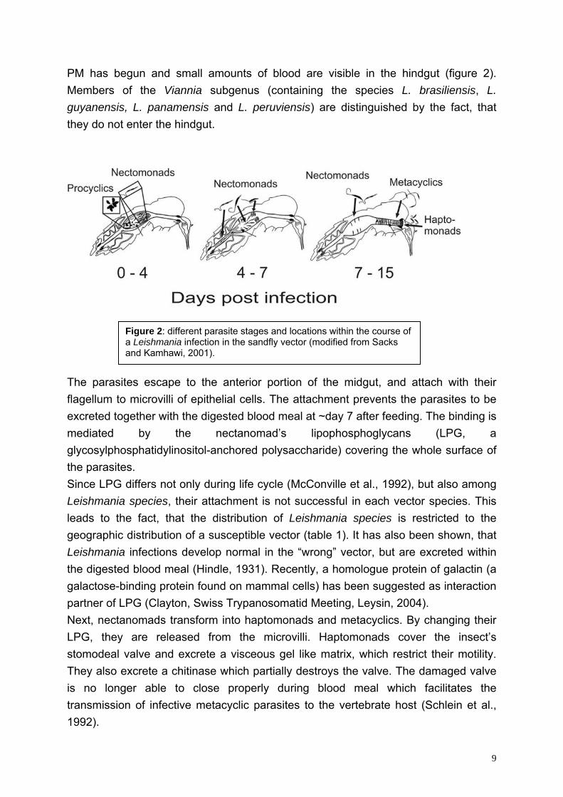
PM has begun and small amounts of blood are visible in the hindgut (figure 2). Members of the Viannia subgenus (containing the species L. brasiliensis, L. guyanensis, L. panamensis and L. peruviensis) are distinguished by the fact, that they do not enter the hindgut.
Figure 2: different parasite stages and locations within the course of a Leishmania infection in the sandfly vector (modified from Sacks and Kamhawi, 2001).
The parasites escape to the anterior portion of the midgut, and attach with their flagellum to microvilli of epithelial cells. The attachment prevents the parasites to be excreted together with the digested blood meal at ~day 7 after feeding. The binding is mediated by the nectanomad’s lipophosphoglycans (LPG, a glycosylphosphatidylinositol-anchored polysaccharide) covering the whole surface of the parasites. Since LPG differs not only during life cycle (McConville et al., 1992), but also among Leishmania species, their attachment is not successful in each vector species. This leads to the fact, that the distribution of Leishmania species is restricted to the geographic distribution of a susceptible vector (table 1). It has also been shown, that Leishmania infections develop normal in the “wrong” vector, but are excreted within the digested blood meal (Hindle, 1931). Recently, a homologue protein of galactin (a galactose-binding protein found on mammal cells) has been suggested as interaction partner of LPG (Clayton, Swiss Trypanosomatid Meeting, Leysin, 2004). Next, nectanomads transform into haptomonads and metacyclics. By changing their LPG, they are released from the microvilli. Haptomonads cover the insect’s stomodeal valve and excrete a visceous gel like matrix, which restrict their motility. They also excrete a chitinase which partially destroys the valve. The damaged valve is no longer able to close properly during blood meal which facilitates the transmission of infective metacyclic parasites to the vertebrate host (Schlein et al., 1992).
9
In the host’s blood, they are phagocytized by ‘professional’ phagocytes (neutrophils, monocytes and macrophages). The transformation of metacylics to amastigotes completes the life cycle.
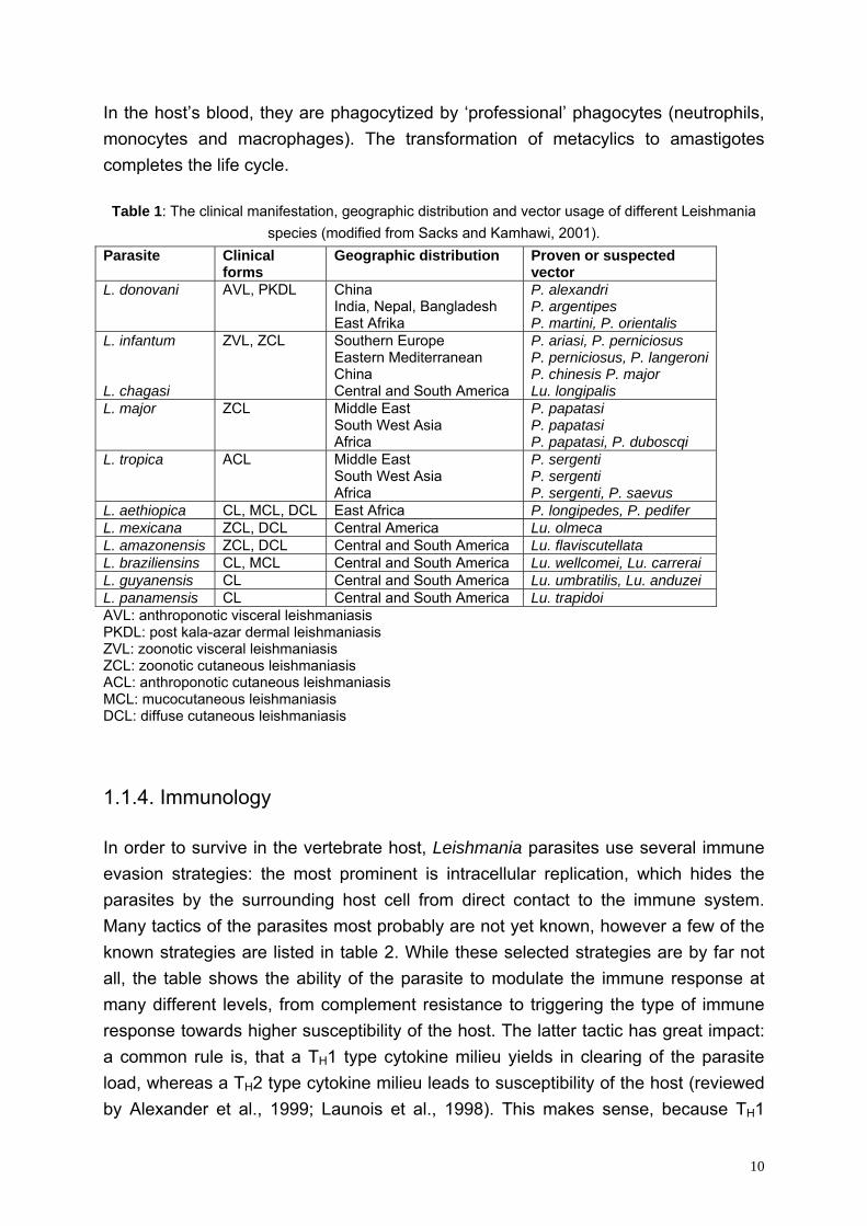
Table 1: The clinical manifestation, geographic distribution and vector usage of different Leishmania species (modified from Sacks and Kamhawi, 2001).
Parasite Clinical forms
Geographic distribution Proven or suspected vector
L. donovani AVL, PKDL China India, Nepal, Bangladesh East Afrika
P. alexandri P. argentipes P. martini, P. orientalis
L. infantum L. chagasi
ZVL, ZCL Southern Europe Eastern Mediterranean China Central and South America
P. ariasi, P. perniciosus P. perniciosus, P. langeroni P. chinesis P. major Lu. longipalis
L. major ZCL Middle East South West Asia Africa
P. papatasi P. papatasi P. papatasi, P. duboscqi
L. tropica ACL Middle East South West Asia Africa
P. sergenti P. sergenti P. sergenti, P. saevus
L. aethiopica CL, MCL, DCL East Africa P. longipedes, P. pedifer L. mexicana ZCL, DCL Central America Lu. olmeca L. amazonensis ZCL, DCL Central and South America Lu. flaviscutellata L. braziliensins CL, MCL Central and South America Lu. wellcomei, Lu. carrerai L. guyanensis CL Central and South America Lu. umbratilis, Lu. anduzei L. panamensis CL Central and South America Lu. trapidoi AVL: anthroponotic visceral leishmaniasis PKDL: post kala-azar dermal leishmaniasis ZVL: zoonotic visceral leishmaniasis ZCL: zoonotic cutaneous leishmaniasis ACL: anthroponotic cutaneous leishmaniasis MCL: mucocutaneous leishmaniasis DCL: diffuse cutaneous leishmaniasis 1.1.4. Immunology
In order to survive in the vertebrate host, Leishmania parasites use several immune evasion strategies: the most prominent is intracellular replication, which hides the parasites by the surrounding host cell from direct contact to the immune system. Many tactics of the parasites most probably are not yet known, however a few of the known strategies are listed in table 2. While these selected strategies are by far not all, the table shows the ability of the parasite to modulate the immune response at many different levels, from complement resistance to triggering the type of immune response towards higher susceptibility of the host. The latter tactic has great impact: a common rule is, that a TH1 type cytokine milieu yields in clearing of the parasite load, whereas a TH2 type cytokine milieu leads to susceptibility of the host (reviewed by Alexander et al., 1999; Launois et al., 1998). This makes sense, because TH1
10
cytokines are able to activate macrophages, which is the major killing mechanism of Leishmania parasites. TH2 cells induce a humoral response, which has little effect for clearance of intracellular parasites. By down regulating a protective TH1 response, the parasite therefore succeeds a dramatic intervention. Nevertheless, antibodies against several Leishmania antigens are present in the blood of both, asymptomatic and symptomatic hosts. Complement Metacyclic parasites resist complement mediated lysis by inactivation of complement factors and shedding of the membrane attack complex (MAC, Brittingham et al., 1995; Hermoso et al., 1991; Puentes et al., 1990). Natural killer cells (NK) Mice deficient in NK were shown to be only modestly less capable of eliminating L. donovani infection than control mice; and there was no difference seen in the course of L. major infections (Kirkpatrick and Farrell, 1982; Kirkpatrick and Farrell, 1984). It therefore appears that NK are, if at all, only involved in the response to visceral leishmaniasis. Here, it has been shown in vitro that L. donovani parasites interact with γ/δ+ human peripheral blood T cells and induce susceptibility to NK cell-mediated lysis (Saha et al., 1999). Humoral response While anti-leishmanial antibodies have been shown to lyse promastigotes in vitro in the presence of complement, there is little evidence for a role in vivo: when antibodies are mixed in large quantities with the parasites before infection, the infectivity of promastigotes was reduced; however, when injected separately, there was no effect on disease development (Anderson et al., 1983). It therefore appears that a humoral response has no central role in clearing the parasite load. Cytotoxic T-cells (CTL, CD8+ T cells) CTL have traditionally been associated with resistance to viral infections, but they may also have an important role in immunity to intracellular microbes like mycobacterium (Chiplunkar et al., 1986). This may be due to direct cytotoxicity or through cytokine production and subsequent macrophage activation (some CD8+ T cells produce interferon γ (INF-γ) as well tumor necrosis factor α (TNF-α), see below). However, it is unclear, whether CD8+ T cells play a major role in the control of leishmaniasis. Since amastigotes multiply within phagolysosomes, presentation of processed Leishmania peptides is mostly restricted to MHC class II molecules. Nevertheless, some parasite proteins might be transported to the cytoplasm, from where they could be loaded to MHC class I molecules, thus allowing activation of
11
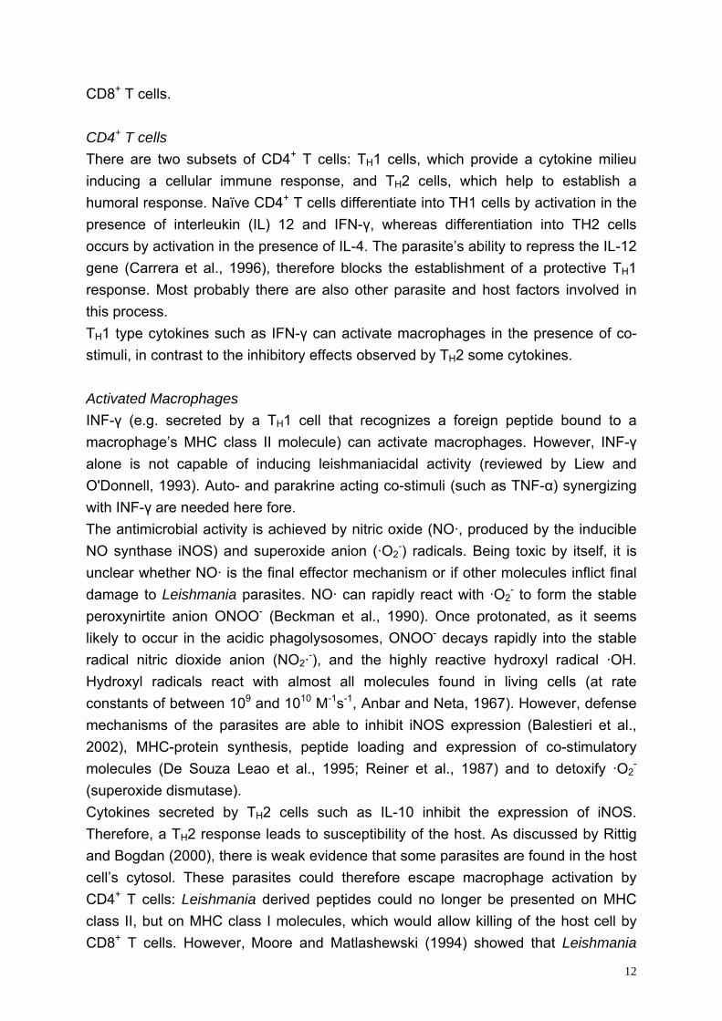
CD8+ T cells. CD4+ T cells There are two subsets of CD4+ T cells: TH1 cells, which provide a cytokine milieu inducing a cellular immune response, and TH2 cells, which help to establish a humoral response. Naïve CD4+ T cells differentiate into TH1 cells by activation in the presence of interleukin (IL) 12 and IFN-γ, whereas differentiation into TH2 cells occurs by activation in the presence of IL-4. The parasite’s ability to repress the IL-12 gene (Carrera et al., 1996), therefore blocks the establishment of a protective TH1 response. Most probably there are also other parasite and host factors involved in this process. TH1 type cytokines such as IFN-γ can activate macrophages in the presence of co-stimuli, in contrast to the inhibitory effects observed by TH2 some cytokines. Activated Macrophages INF-γ (e.g. secreted by a TH1 cell that recognizes a foreign peptide bound to a macrophage’s MHC class II molecule) can activate macrophages. However, INF-γ alone is not capable of inducing leishmaniacidal activity (reviewed by Liew and O'Donnell, 1993). Auto- and parakrine acting co-stimuli (such as TNF-α) synergizing with INF-γ are needed here fore. The antimicrobial activity is achieved by nitric oxide (NO·, produced by the inducible NO synthase iNOS) and superoxide anion (·O2
-) radicals. Being toxic by itself, it is unclear whether NO· is the final effector mechanism or if other molecules inflict final damage to Leishmania parasites. NO· can rapidly react with ·O2
- to form the stable peroxynirtite anion ONOO- (Beckman et al., 1990). Once protonated, as it seems likely to occur in the acidic phagolysosomes, ONOO- decays rapidly into the stable radical nitric dioxide anion (NO2·-), and the highly reactive hydroxyl radical ·OH. Hydroxyl radicals react with almost all molecules found in living cells (at rate constants of between 109 and 1010 M-1s-1, Anbar and Neta, 1967). However, defense mechanisms of the parasites are able to inhibit iNOS expression (Balestieri et al., 2002), MHC-protein synthesis, peptide loading and expression of co-stimulatory molecules (De Souza Leao et al., 1995; Reiner et al., 1987) and to detoxify ·O2
- (superoxide dismutase). Cytokines secreted by TH2 cells such as IL-10 inhibit the expression of iNOS. Therefore, a TH2 response leads to susceptibility of the host. As discussed by Rittig and Bogdan (2000), there is weak evidence that some parasites are found in the host cell’s cytosol. These parasites could therefore escape macrophage activation by CD4+ T cells: Leishmania derived peptides could no longer be presented on MHC class II, but on MHC class I molecules, which would allow killing of the host cell by CD8+ T cells. However, Moore and Matlashewski (1994) showed that Leishmania
12
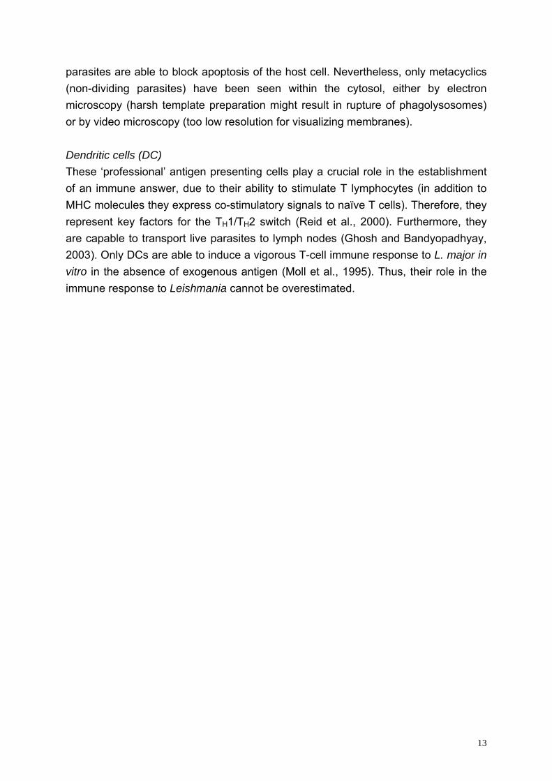
parasites are able to block apoptosis of the host cell. Nevertheless, only metacyclics (non-dividing parasites) have been seen within the cytosol, either by electron microscopy (harsh template preparation might result in rupture of phagolysosomes) or by video microscopy (too low resolution for visualizing membranes). Dendritic cells (DC) These ‘professional’ antigen presenting cells play a crucial role in the establishment of an immune answer, due to their ability to stimulate T lymphocytes (in addition to MHC molecules they express co-stimulatory signals to naïve T cells). Therefore, they represent key factors for the TH1/TH2 switch (Reid et al., 2000). Furthermore, they are capable to transport live parasites to lymph nodes (Ghosh and Bandyopadhyay, 2003). Only DCs are able to induce a vigorous T-cell immune response to L. major in vitro in the absence of exogenous antigen (Moll et al., 1995). Thus, their role in the immune response to Leishmania cannot be overestimated.
13
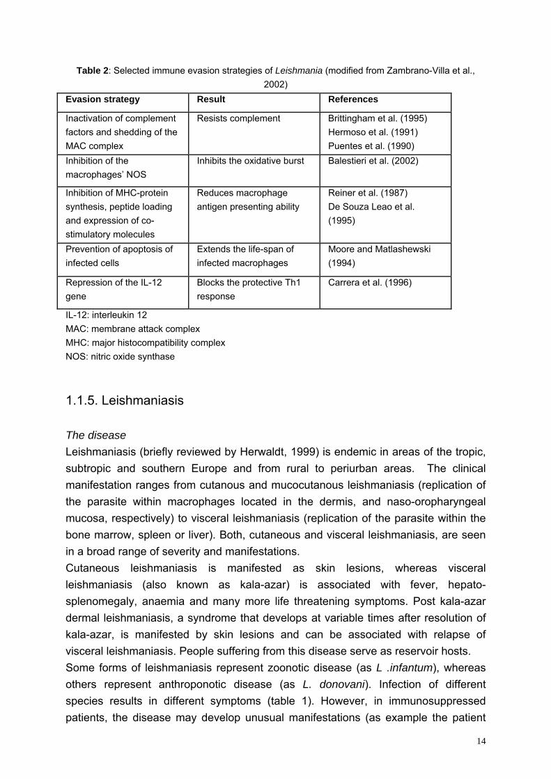
Table 2: Selected immune evasion strategies of Leishmania (modified from Zambrano-Villa et al., 2002)
Evasion strategy Result References
Inactivation of complement factors and shedding of the MAC complex
Resists complement Brittingham et al. (1995) Hermoso et al. (1991) Puentes et al. (1990)
Inhibition of the macrophages’ NOS
Inhibits the oxidative burst Balestieri et al. (2002)
Inhibition of MHC-protein synthesis, peptide loading and expression of co-stimulatory molecules
Reduces macrophage antigen presenting ability
Reiner et al. (1987) De Souza Leao et al. (1995)
Prevention of apoptosis of infected cells
Extends the life-span of infected macrophages
Moore and Matlashewski (1994)
Repression of the IL-12 gene
Blocks the protective Th1 response
Carrera et al. (1996)
IL-12: interleukin 12 MAC: membrane attack complex MHC: major histocompatibility complex NOS: nitric oxide synthase
1.1.5. Leishmaniasis
The disease Leishmaniasis (briefly reviewed by Herwaldt, 1999) is endemic in areas of the tropic, subtropic and southern Europe and from rural to periurban areas. The clinical manifestation ranges from cutanous and mucocutanous leishmaniasis (replication of the parasite within macrophages located in the dermis, and naso-oropharyngeal mucosa, respectively) to visceral leishmaniasis (replication of the parasite within the bone marrow, spleen or liver). Both, cutaneous and visceral leishmaniasis, are seen in a broad range of severity and manifestations. Cutaneous leishmaniasis is manifested as skin lesions, whereas visceral leishmaniasis (also known as kala-azar) is associated with fever, hepato-splenomegaly, anaemia and many more life threatening symptoms. Post kala-azar dermal leishmaniasis, a syndrome that develops at variable times after resolution of kala-azar, is manifested by skin lesions and can be associated with relapse of visceral leishmaniasis. People suffering from this disease serve as reservoir hosts. Some forms of leishmaniasis represent zoonotic disease (as L .infantum), whereas others represent anthroponotic disease (as L. donovani). Infection of different species results in different symptoms (table 1). However, in immunosuppressed patients, the disease may develop unusual manifestations (as example the patient
14
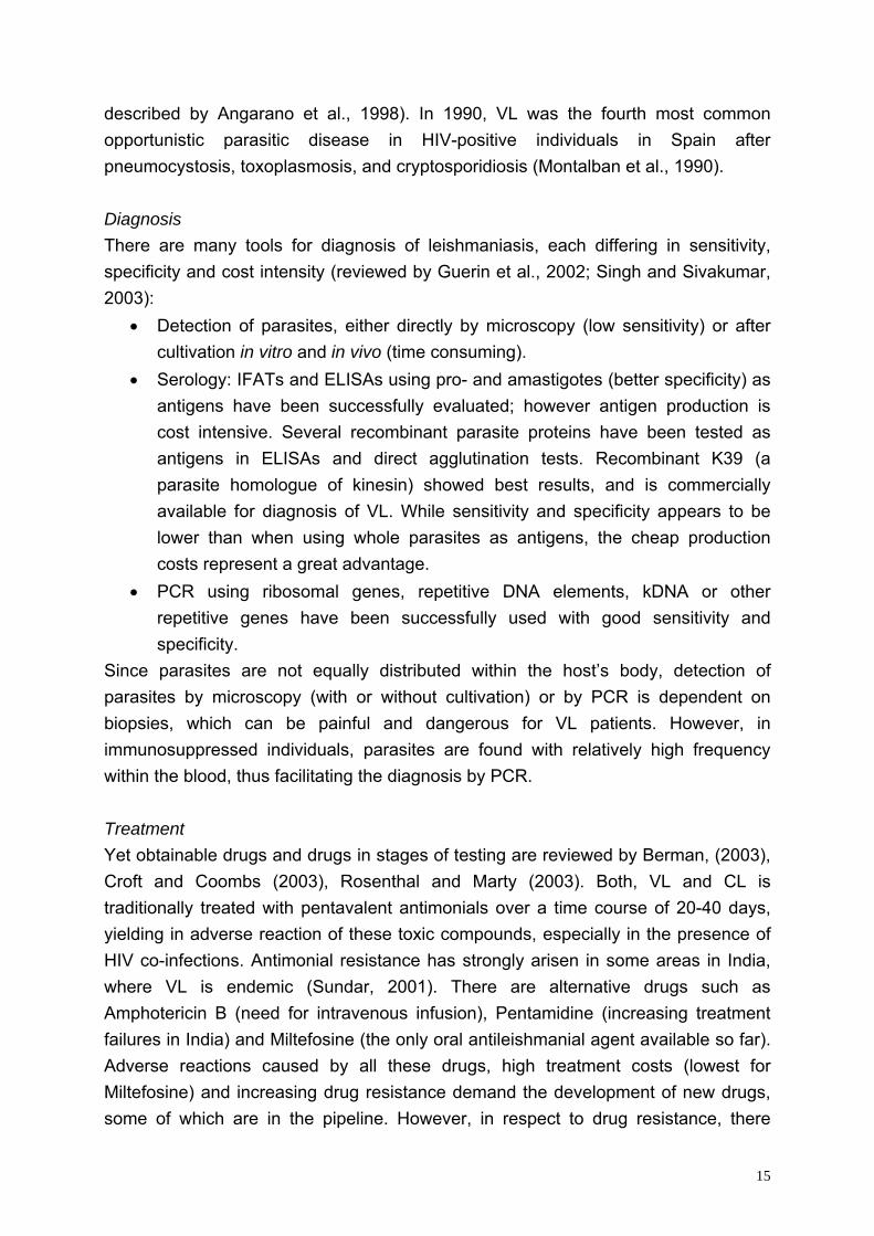
described by Angarano et al., 1998). In 1990, VL was the fourth most common opportunistic parasitic disease in HIV-positive individuals in Spain after pneumocystosis, toxoplasmosis, and cryptosporidiosis (Montalban et al., 1990). Diagnosis There are many tools for diagnosis of leishmaniasis, each differing in sensitivity, specificity and cost intensity (reviewed by Guerin et al., 2002; Singh and Sivakumar, 2003):
• Detection of parasites, either directly by microscopy (low sensitivity) or after cultivation in vitro and in vivo (time consuming).
• Serology: IFATs and ELISAs using pro- and amastigotes (better specificity) as antigens have been successfully evaluated; however antigen production is cost intensive. Several recombinant parasite proteins have been tested as antigens in ELISAs and direct agglutination tests. Recombinant K39 (a parasite homologue of kinesin) showed best results, and is commercially available for diagnosis of VL. While sensitivity and specificity appears to be lower than when using whole parasites as antigens, the cheap production costs represent a great advantage.
• PCR using ribosomal genes, repetitive DNA elements, kDNA or other repetitive genes have been successfully used with good sensitivity and specificity.
Since parasites are not equally distributed within the host’s body, detection of parasites by microscopy (with or without cultivation) or by PCR is dependent on biopsies, which can be painful and dangerous for VL patients. However, in immunosuppressed individuals, parasites are found with relatively high frequency within the blood, thus facilitating the diagnosis by PCR. Treatment Yet obtainable drugs and drugs in stages of testing are reviewed by Berman, (2003), Croft and Coombs (2003), Rosenthal and Marty (2003). Both, VL and CL is traditionally treated with pentavalent antimonials over a time course of 20-40 days, yielding in adverse reaction of these toxic compounds, especially in the presence of HIV co-infections. Antimonial resistance has strongly arisen in some areas in India, where VL is endemic (Sundar, 2001). There are alternative drugs such as Amphotericin B (need for intravenous infusion), Pentamidine (increasing treatment failures in India) and Miltefosine (the only oral antileishmanial agent available so far). Adverse reactions caused by all these drugs, high treatment costs (lowest for Miltefosine) and increasing drug resistance demand the development of new drugs, some of which are in the pipeline. However, in respect to drug resistance, there
15
cannot be enough alternative drugs. Vaccines At present, there is no vaccine in routine use; however, several are in different stages of testing (reviewed by Ghosh and Bandyopadhyay, 2003). It is crucial for a successful vaccine, that a protective TH1 type immune answer is induced (either by the adjuvant or by the vaccine itself).
• Live vaccines In the age of genetically engineering, a practice used for centuries might have to potential for a come back: ‘leishmanization’. People were inoculated with live L. major at an aesthetically acceptable site resulting in live long protection from CL caused by L. major and L. tropica (Modabber, 1989; Nadim et al., 1983). A major draw back of leishmanization was the high risk of complication and the development of a lesion and a scar after healing at the site of inoculation. This problem might be overcome by using genetically altered parasites. Two approaches have already successfully been performed: introduction of suicidal cassettes (Titus et al., 1995) and/or markers which are responsive to external signals for their destruction (Sah et al., 2002; Yan et al., 2001). Another problem when using a live Leishmania vaccine, are the high production costs. Whether leishmanization works also for VL has not yet been shown. Here, development of a clinical episode would be fatal.
• Killed vaccines Killed promastigotes have been used for CL with different success: while there was no protection in Iran, there were promising results in Ecuador and Brazil.
• Recombinant antigens The first tested antigen was GP63 and showed promising results in the animal model, whereas responses of human T-cells were variable. The leishmanial elongation initiation factor (LeiF) is considered to be a promising vaccine candidate due to its ability to induce TH1 cytokines in humans, as well as the Leishmania homologue of the receptor for activated C kinase (LACK, in particular when IL-12 is used as adjuvant).
• Synthetic peptides Several peptides have been successfully used in the animal model, such as peptides derived from GP63.
• DNA vaccines Various naked DNA vaccines have been tested in animal models yielding in promising results, such as the glucose regulated protein, a member of the 70kDa heat-shock protein family.
16
However, in most experimental systems, adjuvants are essential to induce protective immunity. Unfortunately, the most effective adjuvants generally cause strong inflammation, which may be needed for adjanticity, but may exclude their use in humans due to unacceptable side effects. This problem might be overcome when using dendritic cell vaccination (Moll, 2003).
1.2. Basic molecular biological methods used during the PhD project Most of the routine methods were performed after the standard protocols described by Sambrook and Russel (2001).
1.2.1. RNA Isolation RNA was either harvested according to the classical method of Chomczynski and Sacchi (1987) or using TRIZOL (Invitrogen) reagent. For long term storage, RNA was stored as a precipitate at -20°C, otherwise, RNA was dissolved in TE (10mM Tris·Cl pH 8, 0.5mM EDTA) and stored at -80°C.
Northern blots
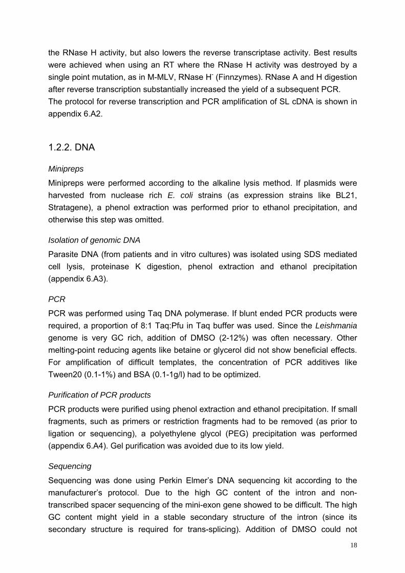
Northern blots were performed using either formaldehyde, or preferentially glyoxal/DMSO as denaturating agents. For highly expressed genes, such as histones, even non-denaturating agarose gels worked very well (figure 3). PEG purified (refer appendix 6.A4) dsDNA probes were 32P-labeled using the High Prime Kit (Roche) and α32P-dCTP and hybridized overnight at 42°C. Beside Ultrahyb (Ambion), the two different hybridization buffers described in appendix 6.A1 were successfully used.
Reverse Transcription
Three different derivates from Moloney Murine Leukesuccessfully used: although unmodified M-MLV exhibishowed to be the better choice for obtaining spliced leSuperscript II (Invitrogen). Superscript II is M-MLV conacids responsible for RNase H activity. Unfortunately, t
Figure 3: Northern blot of histone H2A using a non-denaturating agarose gel.
mia Virus (M-MLV) RT were ts a weak RNase H activity, it ader (SL) cDNA compared to
taining a deletion of the amino he deletion not only eliminates
17
the RNase H activity, but also lowers the reverse transcriptase activity. Best results were achieved when using an RT where the RNase H activity was destroyed by a single point mutation, as in M-MLV, RNase H- (Finnzymes). RNase A and H digestion after reverse transcription substantially increased the yield of a subsequent PCR. The protocol for reverse transcription and PCR amplification of SL cDNA is shown in appendix 6.A2.
1.2.2. DNA
Minipreps
Minipreps were performed according to the alkaline lysis method. If plasmids were harvested from nuclease rich E. coli strains (as expression strains like BL21, Stratagene), a phenol extraction was performed prior to ethanol precipitation, and otherwise this step was omitted.
Isolation of genomic DNA
Parasite DNA (from patients and in vitro cultures) was isolated using SDS mediated cell lysis, proteinase K digestion, phenol extraction and ethanol precipitation (appendix 6.A3).
PCR
PCR was performed using Taq DNA polymerase. If blunt ended PCR products were required, a proportion of 8:1 Taq:Pfu in Taq buffer was used. Since the Leishmania genome is very GC rich, addition of DMSO (2-12%) was often necessary. Other melting-point reducing agents like betaine or glycerol did not show beneficial effects. For amplification of difficult templates, the concentration of PCR additives like Tween20 (0.1-1%) and BSA (0.1-1g/l) had to be optimized.
Purification of PCR products
PCR products were purified using phenol extraction and ethanol precipitation. If small fragments, such as primers or restriction fragments had to be removed (as prior to ligation or sequencing), a polyethylene glycol (PEG) precipitation was performed (appendix 6.A4). Gel purification was avoided due to its low yield.
Sequencing
Sequencing was done using Perkin Elmer’s DNA sequencing kit according to the manufacturer’s protocol. Due to the high GC content of the intron and non-transcribed spacer sequencing of the mini-exon gene showed to be difficult. The high GC content might yield in a stable secondary structure of the intron (since its secondary structure is required for trans-splicing). Addition of DMSO could not
18
improve the quality of the sequences, in contrast to cloning the PCR fragments prior to sequencing, and the adaptation of the sequencing procedure described in appendix 6.A5. Sequencing of large inserts was done as shown appendix 6.A6: the plasmids were cut with polylinker restriction enzymes, which also cut within the inserts. After religation and retransformation, the obtained minipreps contained deletions. Therefore, it was possible to sequence large insert by solely using vector primers and without purchasing an erase a base kit. Theoretically it would be possible to sequence directly after religation, but this simplification was not tested.
Ligation
Ligations were performed using either T/A cloning kit (Promega), cycle restriction ligation (appendix 6.A7) or Quick DNA ligase (New Egland Biolabs).
E. coli Transformation
Electro-poration: since input and harvest is not very profitable using large scale preparation of electro-competent cells, a fast small scale approach was used (appendix 6.A8). Chemo-transformation: this was the method of choice, if many transformations had to be performed with the same E. coli strain. The Inoue method (appendix 6.A9) was used to prepare chemo-competent cells resulting in 108 to 109 colonies/µg supercoiled pUC18 DNA. The advantages compared to electroporations were: many more aliquots of competent cells after large scale production, ligation mixes could directly be transformed without the need of purification and many samples could be transformed at the same time with less handling.
1.2.3. Proteins
Expression
Most of the proteins were expressed as glutathione-S transferase (GST) fusion proteins. However, none of them was in the soluble fraction. Neither lowering the IPTG concentration for induction, nor the temperature for protein expression, nor adding ethanol for chaperon induction, nor combining all variations could change this feature. For expression of genes selected during the cDNA subtraction, a modified pGEX (GST fusion) vector was used (appendix 6.A10).
19
Purification
Recombinant proteins containing a His-tag were preliminary purified as inclusion bodies (appendix 6.A11). Since renaturation did not work for any protein, the inclusion bodies were subsequently purified using Ni-NTA agarose (Qiagen) under denaturing conditions. If the protein was badly soluble in urea, 8M urea was replaced with 6M guanidine chloride in all solutions. Recombinant MAT-1 was affinity purified under mild denaturing conditions using sarkosyl and glutathion sepharose (appendix 6.A12).
Immunofluorescence assays (IFAs)
Parasite cultures were spun, resuspended in fetal calf serum (FCS) and spread on glass slides. After drying, the slides were fixed for 10min at -20°C using acetone alone or 1:1 methanol:acetone. The slides were partitioned using a hydrophobic pen allowing the use of only 10µl of serum or secondary antibody.
20
2. Development of an early diagnosis test system for canine leishmaniasis caused by L. infantum
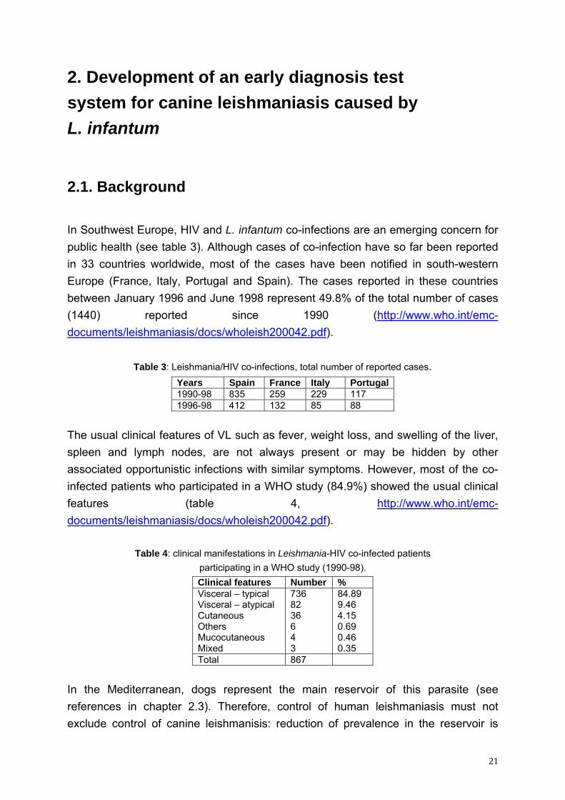
2.1. Background In Southwest Europe, HIV and L. infantum co-infections are an emerging concern for public health (see table 3). Although cases of co-infection have so far been reported in 33 countries worldwide, most of the cases have been notified in south-western Europe (France, Italy, Portugal and Spain). The cases reported in these countries between January 1996 and June 1998 represent 49.8% of the total number of cases (1440) reported since 1990 (http://www.who.int/emc-documents/leishmaniasis/docs/wholeish200042.pdf).
Table 3: Leishmania/HIV co-infections, total number of reported cases. Years Spain France Italy Portugal1990-98 835 259 229 117 1996-98 412 132 85 88
The usual clinical features of VL such as fever, weight loss, and swelling of the liver, spleen and lymph nodes, are not always present or may be hidden by other associated opportunistic infections with similar symptoms. However, most of the co-infected patients who participated in a WHO study (84.9%) showed the usual clinical features (table 4, http://www.who.int/emc-documents/leishmaniasis/docs/wholeish200042.pdf).
Table 4: clinical manifestations in Leishmania-HIV co-infected patients participating in a WHO study (1990-98). Clinical features Number % Visceral – typical Visceral – atypical Cutaneous Others Mucocutaneous Mixed
736 82 36 6 4 3
84.89 9.46 4.15 0.69 0.46 0.35
Total 867 In the Mediterranean, dogs represent the main reservoir of this parasite (see references in chapter 2.3). Therefore, control of human leishmaniasis must not exclude control of canine leishmanisis: reduction of prevalence in the reservoir is
21
expected to lower the transmission rate to humans. However, due to the increasing number of infected humans, parasitized human hosts may also contribute substantially to the transmission. In order to control canine leishmaniasis, an early diagnosis system is needed because dogs are capable to transmit the parasite, before showing any symptoms of the disease. For mass use (in the Mediterranean, there are many street dogs), diagnosis by PCR or microscopy is not feasible. Existing serological tests for canine leishmaniasis are not able to detect parasites within early stages of the disease. Furthermore, some reliable diagnostic methods (such as IFAT based on whole parasite antigens) cannot be used in masses, due their high production costs. Thus, a cheap serological test capable of diagnosis of early canine leishmaniasis is needed. In order to identify antigens suitable for such a diagnostic tool, we screened L. infantum cDNA expression libraries. These would be cheaply produced as recombinant proteins, which then could be used as antigens for Western blots, ELISAs or direct agglutination tests.
2.2. Methods In order to identify antigens suitable for diagnostic use, several cDNA expression libraries were constructed and screened with sera of L. infantum infected dogs. Preliminary, Clontech’s Capfinder Kit was used combined with SMART λ phages. (The capfinder method uses the feature of certain reverse transcriptases to add several G’s at the 3’ end of the cDNA when reverse transcribing capped mRNAs in the presence of Mn2+ ions. This allows adaptor ligation and subsequently PCR amplification of full length cDNA.) However, beside that the cDNA obtained after PCR looked strange (a smear on the gel, from bottom to top), no λ clone reacted with dog sera of L. infantum infected dogs. The simplest explanations for this finding were, that expression in E. coli failed due to presence of stop codons and absence of ribosome binding sites (RBS) within the 5’ UTR of the cloned cDNAs: in prokaryotes, a defined sequence (the RBS) has to be located within a distinct distance from the ATG for proper translation. Since most of the Leishmania full length cDNA apparently lacked the RBS, the obtained cDNA library was not suitable for a serum screen. Therefore, both cDNA amplification and vector were changed. We used the spliced leader as primer binding site instead of the CAP finder adaptor. Furthermore, the amplified cDNA was restriction digested and non-directionally ligated into λgt11 DNA containing 3 different adaptors (appendix 6.A10), resulting in 6 frame expression of all cDNA fragments as lacZ fusion proteins.
22
2.3. Identification of recombinant antigens from Leishmania infantum suitable as early diagnostic tool
23
Identification of recombinant antigens from Leishmania infantum suitable as early diagnostic tool. Igor Niederwieser, Jutta Marfurt, Sylvia Steiger, Jose Alunda, Charles Jaffe, Hans-Peter Beck Swiss Tropical Institute, Switzerland University of Madrid, Spain Hebrew University Jerusalem, Israel Abstract Early diagnosis of canine leishmanaiasis caused by the protozoan parasite Leishmania infantum remains an important requirement for the control of the disease. In order to identify antigens suitable for such an early immunological diagnosis system we generated a phage lambda cDNA expression library and this was screened using sera from naturally and artificially infected dogs. With this approach we selected 8 antigens which strongly reacted with naturally infected dog sera. Subsequent sequencing identified these as histones, ribosomal proteins, and a mitochondrial protein. Upon generation of recombinant protein Western blot analysis was performed to assess the specificity and reactivity of the identified antigens with sera of infected and healthy control dogs. Three of these antigens were further selected for the development of a dipstick test.
23.1
Introduction Domestic dogs represent the main reservoir of Leishmania infantum (Old World) and L. chagasi (New World) and therefore play a key role in the transmission of the disease to humans. A way to control the disease would be to treat dogs found infected with Leishmania rapidly. However, dogs showing already symptoms might have been transmitting for an extended period [1, 2] and therefore a diagnostic assay is need that quickly can determine infectivity in subpatently infected dogs. Although usually transmitted by the bite of an infected sandfly vector, direct dog to dog transmission [3] and blood transfusion routes [4] have been reported. Because the metacyclic stage of Leishmania parasites is responsible for the transmission from the insect vector to the vertebrate host and the establishment of a new infection, this stage is the first to be seen by the host’s immune system. Once phagocytised by macrophages, metacyclic stages develop into amastigotes and multiply within phagolysosomes. In order to identify antigens suitable for an early diagnostic test, a cDNA expression library of L. infantum metacylic stages was generated and screened with a serum pool from L. infantum positive dogs. Most of the mRNAs in trypanosomatids contain a conserved 5’ sequence, the spliced leader [5]. And we exploited this feature to PCR amplify the cDNA obtained after reverse transcription of total RNA using spliced leader sequence specific primers. In order to construct an expression library, the PCR products were restriction digested and ligated into a λ phage which was previously modified with adaptors in three reading frames. Since the cDNA was not directionally cloned into the phage vector, all 6 frames of the PCR fragments were expressed in this λ library. Screening the λ library we identified several fragments of histones, of ribosomal proteins, and a mitochondrial protein. In order to perform Western blot analysis using sera of artificially and naturally infected dogs, some of the identified antigens were sub-cloned in expression plasmids and expressed in E. coli, though we failed to express two of the gene fragments. However, Western blot results of the other clones were promising, in particular those obtained with clones representing epitops within the histones H2A and H4.
23.2
Materials and methods Parasite culture Promastigotes of the L. infantum strain LEM 768-A/ST were cultivated in vitro at 27°C in a 1:1 mixture of SDM-79 [6] and Schneider’s medium [7] containing 15% [v/v] heat inactivated fetal calf serum. 10ml cultures were inoculated at a density of 106 parasites/ml and harvested 14 days later. cDNA preparation After washing the parasites in phosphate buffered saline (PBS), total RNA was extracted after the method of Chomczynski and Sacchi [8] using 0.5ml guanidinium thiocyanate solution per ml in vitro culture. Approximately 2µg RNA were reverse transcribed with 200 U Moloney Murine Leukemia Virus (M-MLV) reverse transcriptase (Invitrogen) using the primer 5’-CGACTGTACGTGAATTCGC(T)20 (all oligonucleotides were manufactured by Invitrogen). Prior to PCR, RNA was removed by RNase A (Boehringer) digestion and cDNA was PCR amplified using the primer above and the spliced leader primer 5’-AACTAACGCTATATAAGTATCAGTTTCTGTACTTTATTG. Taq DNA polymerase (Invitrogen) was added in presence of 10% dimethyl sulfoxide (DMSO) and the amplification was done under the following conditions: 25 cycles 30sec 94°C, 30sec 54°C, 2min 72°C. The PCR products (figure 1A) were NlaIII (all restriction enzymes obtained from New England Biolabs) digested and purified by phenol extraction, and precipitated with ethanol and polyethylene glycol (PEG)[9]. Lambda expression libraries Three different adaptors to provide three reading frames were ligated with T4 DNA ligase (New England Biolabs) to EcoRI cut and dephosphorylated λ-gt11 DNA (Amersham). All adaptors contained phosphorylated 5’ EcoRI overhangs, 3’ NlaIII overhangs and 6x His tags. The adaptor in frame 1 was made with the two oligos 5’-P-AATTCCACCATCACCATCACCACCATG and 5’-GTGATGGTGATGGTGG, frame 2 was made with 5’-P-AATTCCACCATCACCATCACCATGCATG and 5’-CATGGTGATGGTGATGGTGG, and frame 3 was made with 5’-P-AATTCCACCATCACCATCACCACATG and 5’-P-TGGTGATGGTGATGGTGG. After the ligation, the remaining free adaptors were removed by PEG precipitation and the NlaIII digested cDNA was ligated
23.3
into the λ-gt11 DNA using T4 DNA ligase. The phages were packed with the Gigapack® III Gold Packaging Extract (Stratagene). The library was amplified in Y1090r- E. coli strain (Stratagene) after the protocol of Sambrook and Russel [10]. Screening of libraries The libraries were screened with a serum pool of L. infantum positive dogs (kindly by Felix Grimm, University of Zürich) after the protocol of Sambrook et al. [11]. Briefly, the phages were plated at a density of 104 plaque forming units on 30ml Petri dishes and incubated for 4 hours at 42°C. The plates were overlaid with nitrocellulose sheets (Hybond N, Amersham) which were previously saturated in 10mM isopropyl-β-D-1-thiogalactopyranoside (IPTG). The plates were incubated for another 6 hours at 37°C. The sheets were removed afterwards and blocked with PBS containing 5% not fat milk powder. A serum pool from L. infantum-positive dogs was pre-adsorbed with crude E. coli extract and the antibody screen was conducted with 1:500 diluted sera. After 2 h, sheets were washed and incubated in a 1:2000 dilution of an alkaline phosphatase labeled goat anti-dog IgG antibody (Southern). Unbound antibody was washed away and recognized clones were visualized by a BCIP/NBT color reaction (Blotting substrates, BioRad). Areas of positive clones were identified on the master plates, phages were re-plated at a lower density, and screened again with the same serum pool in order to isolate single clones. Specificity of the recognition was assessed using a serum pool of uninfected control dogs (kindly by Felix Grimm, University of Zürich). Single clones which were negative with control sera but positive with sera from infected dogs were PCR amplified directly from the plagues. PCR was performed to amplify the inserts using the λgt11-primers 5’-GGTGGCGACGACTCCTGGAGCC and 5’-GACACCAGACCAACTGGTAATG, and the PCR products were directly sequenced on an ABI automated sequencer. Subcloning in expression plasmids The PCR products were either cloned into pQE32 containing a 6x His tag (Qiagen) or into the glutathion-S-transferase (GST) fusion vector pGEX-1 (Amersham). Prior to ligation into the pQE vector, PCR products of λ clones were NlaIII digested, their ends polished using Pfu DNA polymerase (Promega), and purified by phenol extraction with subsequent ethanol and PEG precipitation. Fragments were then cycle restriction ligated [12] using SmaI and T4 DNA ligase. For the ligation into the pGEX vector, PCR products
23.4
and vector were EcoRI cut and the vector was dephosphorylated using calf intestinal alkaline phosphatase (Promega). Before ligation with T4 ligase, both vector and insert were purified by phenol extraction and subsequently precipitated with ethanol and PEG. pQE clones were expressed in M15 cells (Qiagen) and pGEX clones in BL21 cells (Amersham). Expression in E. coli Expression clones were grown in Luria-Bertani medium (LB) at 37°C until the optical density reached 0.8 and protein expression was induced with 0.5mM IPTG for 4 hours at 37°C. All proteins were found in inclusion bodies. Because pGEX inserts still contained the λ adaptors, proteins expressed from pQE and pGEX both contained 6 x His tags and were purified with Ni-NTA Agarose (Qiagen) under denaturing conditions. Western Blots Recombinant proteins were run on Tricine-SDS polyacrylamide gels [13] manufactured by Bio-Rad (Ready Gel Tris-Tricine Gel, 10–20%). Heat shock protein 70 [14] (HSP-70, kindly obtained from C. Jaffe), and the identified ribosomal protein (RP) L7 and RP-L10 were on the same gel, as well as the selected proteins histone (HIS) H4, HIS-H2A and RP-L7a (figure 1B). After electrophoresis, proteins were transferred onto nitrocellulose membranes. After blocking with 5 % milk powder in TNT (10 mM Tris/HCl pH 8, 150mM NaCl, 0.05% Tween 20) over night at 4°C, blots were incubated for 3 hours at room temperature with a 1:500 dilution (TNT, 1% milk powder) of positive serum pool or a 1:200 dilution of single serum samples. An alkaline phosphatase labeled goat anti-dog IgG at 1:5000 (TNT, 0.1% milk powder) was used as a secondary antibody. Blots were revealed with BCIP/NBT blotting substrate (Bio-Rad). Results and Discussion The development of an early diagnostic assay for canine leishmaniasis would be a major step forward in the control of the disease which is transmitted by sand flies from infected dogs. Few assays are currently being tested either based on the detection of specific DNA or on the detection of antibodies against certain antigens elicited early during the infection. In order to identify
23.5
additional antigens useful for the development of an early antigen based assay, we created a cDNA expression library and screened it with sera from dogs naturally infected with L. infantum. Spliced leader cDNA For the synthesis of cDNA we made use of the fact that all transcribed messengers in the trypanosomidae contain a spliced leader sequence, which increased the yield of specific transcripts. The size distribution of the amplified spliced leader cDNA that we used for construction of the λ libraries is shown in figure 1A. Size distribution of eukaryotic mRNA usually ranges from 0.5 to many kb with most of the mRNA between 2 and 3kb. The spliced leader cDNA we prepared showed an acceptable distribution with a slight under-representation of mRNAs longer than 3kb, but still representing a large fraction of the metacyclic parasite’s transcriptome, which was screened with dog sera. Serum screen With the screen using sera from Leishmania infected dogs we identified fragments of the following genes: histone (HIS) H2A, histone H3, and histone H4. Furthermore, we identified the ribosomal proteins (RP) L7 (2 fragments), L7a, L10, and the mitochondrial 2-oxoglutarate carrier. It seems to be surprising that no surface proteins have been identified, but only highly expressed cytoplasmatic, nuclear, or mitochondrial proteins. One might speculate that because most of the metacyclic stages are killed immediately after transmission [15] into the mammalian host, and because the amastigote stages are intracellular, the antibody response might be mostly induced by antigens descending from lysed parasites. Therefore one might indeed expect antibodies against common and immunogenic antigens regardless of their cellular localization. Nevertheless, protection against cutanous leishmaniasis in vervet monkeys using recombinant HIS-H1 [16] and immunity in mice against visceral leishmaniasis using DNA encoding for the ribosomal protein P0 as vaccines [17] has been reported. The positive effects of both immunizations most probably were derived from cell-mediated immunity. Recombinant proteins The selected epitopes of HIS-H2a, H4 and one of the two epitops of RP-L7 were successfully expressed as GST fusion proteins. The ribosomal proteins RP-L7a and L10 were expressed in the pQE system. However, sub cloning of HIS-H3 and one epitope of RP-L7 failed. Furthermore, no visible amount of
23.6
protein was obtained after induction of the mitochondrial 2-oxoglutarate carrier expression. All other proteins were expressed at high levels and found in inclusion bodies. These antigens were tested in 4 artificially infected dogs, from which sera were collected 60, 120, 180 and 330 days post infection (table 1). HSP-70 showed best results: all sera recognized this antigen 60 days post infection. Also the histones H2A and H4 showed good results, in contrast to the ribosomal proteins. Although only one strain of L. infantum was used to infect the dogs, the results seemed to be transferable to naturally infected dogs: here, HSP-70 and the two histones were recognized very well, in contrast to the ribosomal proteins. 5 of the western blots are shown in figure 1B. However, the starting point of the infection is unknown in naturally infected dogs. Conclusions In Leishmania diagnosis, a battery of tools exists [18] including PCR, ELISA, IFAT, dipstick or agglutination tests, but there are no gold standards. Therefore, the development of a diagnostic tool of high sensitivity, specificity and of low costs remains of high priority. Different methods for detection of L.infantum have been compared [19]. PCR, ELISA and a dipstick (Leishmania RAPITEST), both based on recombinant K39 protein [20] were tested. The serological tests were more sensitive than PCR, suggesting that serology should preferently be used as method for diagnosis, also because of the lower costs and easier implementation. IFAT using amastigotes as antigen showed great sensitivity and specificity [21]. However, high production costs contradict its application in the field. Therefore, a diagnostic test based on dipstick, agglutination or ELISA technology using 2 or more different recombinantly expressed epitopes of L. infantum proteins might be the best solution. Leishmania possesses high variability in HIS-H2A [22] and H4 genes [23]. A diagnostic test using solely histones as antigens therefore might fail to detect all L. infantum strains. Nevertheless, the two selected histones and HSP-70 showed promising results in all of our experiments, in contrast to the ribosomal proteins L7, L7a and L10. However, one epitope of RP L7 was not expressed recombinantely during this work as well as HIS-H3, which could show similar results as HIS-H2A and H4. Further, the mitochondrial oxoglutarat carrier should not be forgotten only because protein production failed in this attempt.
23.7
References 1. Molina R, Amela C, Nieto J, et al. Infectivity of dogs naturally infected with Leishmania infantum
to colonized Phlebotomus perniciosus. Trans R Soc Trop Med Hyg 1994;88:491-3 2. Guarga JL, Lucientes J, Peribanez MA, Molina R, Gracia MJ, Castillo JA. Experimental
infection of Phlebotomus perniciosus and determination of the natural infection rates of Leishmania infantum in dogs. Acta Trop 2000;77:203-7
3. Gaskin AA, Schantz P, Jackson J, et al. Visceral leishmaniasis in a New York foxhound kennel. J Vet Intern Med 2002;16:34-44
4. Owens SD, Oakley DA, Marryott K, et al. Transmission of visceral leishmaniasis through blood transfusions from infected English foxhounds to anemic dogs. J Am Vet Med Assoc 2001;219:1076-83
5. Boothroyd JC, Cross GA. Transcripts coding for variant surface glycoproteins of Trypanosoma brucei have a short, identical exon at their 5' end. Gene 1982;20:281-9
6. Brun R, Schonenberger. Cultivation and in vitro cloning or procyclic culture forms of Trypanosoma brucei in a semi-defined medium. Short communication. Acta Trop 1979;36:289-92
7. Cunningham I. New culture medium for maintenance of tsetse tissues and growth of trypanosomatids. J Protozool 1977;24:325-9
8. Chomczynski P, Sacchi N. Single-step method of RNA isolation by acid guanidinium thiocyanate-phenol-chloroform extraction. Anal Biochem 1987;162:156-9
9. Hartley J, Bowen H. PEG precipitation for selective removal of small DNA fragments. Focus 1996;18:27
10. Sambrook J, Russel DW. Method 2: Amplification of Libraries Constructed in λgt11, λZAP, λZipLox, and Their Derivatives. In: Irwin N, ed. Molecular Cloning: a Laboratory Manual. 3rd ed. Vol. 2. New York: Cold Spring Harbor Laboratory Press, 2001:11.65
11. Sambrook J, Fritsch EF, Maniatis T. Immunological Screening of Expression Libraries. In: Ford N, Nolan C and Ferguson M, eds. Molecular Cloning: a Laboratory Manual. 2nd ed. Vol. 2. New York: Cold Spring Harbor Laboratory Press, 1989:12.16-20
12. Pusch C, Schmitt H, Blin N. Increased cloning efficiency by cycle restriction-ligation (CRL). Technical Tips Online;1:T40071
13. Schagger H, von Jagow G. Tricine-sodium dodecyl sulfate-polyacrylamide gel electrophoresis for the separation of proteins in the range from 1 to 100 kDa. Anal Biochem 1987;166:368-79
14. Rico AI, Girones N, Fresno M, Alonso C, Requena JM. The heat shock proteins, Hsp70 and Hsp83, of Leishmania infantum are mitogens for mouse B cells. Cell Stress Chaperones 2002;7:339-46
15. Dominguez M, Moreno I, Lopez-Trascasa M, Torano A. Complement interaction with trypanosomatid promastigotes in normal human serum. J Exp Med 2002;195:451-9
16. Masina S, M MG, Demotz SO, Fasel NJ. Protection against cutaneous leishmaniasis in outbred vervet monkeys using a recombinant histone H1 antigen. J Infect Dis 2003;188:1250-7
17. Iborra S, Soto M, Carrion J, et al. The Leishmania infantum acidic ribosomal protein P0 administered as a DNA vaccine confers protective immunity to Leishmania major infection in BALB/c mice. Infect Immun 2003;71:6562-72
18. Reithinger R, Davies CR. Canine leishmaniasis: novel strategies for control. Trends Parasitol 2002;18:289-90
19. Reithinger R, Quinnell RJ, Alexander B, Davies CR. Rapid detection of Leishmania infantum infection in dogs: comparative study using an immunochromatographic dipstick test, enzyme-linked immunosorbent assay, and PCR. J Clin Microbiol 2002;40:2352-6
20. Burns JM, Jr., Shreffler WG, Benson DR, Ghalib HW, Badaro R, Reed SG. Molecular characterization of a kinesin-related antigen of Leishmania chagasi that detects specific antibody in African and American visceral leishmaniasis. Proc Natl Acad Sci U S A
23.8

1993;90:775-9 21. Fernandez-Perez FJ, Mendez S, de la Fuente C, Gomez-Munoz MT, Cuquerella M, Alunda
JM. Short report: improved diagnosis and follow-up of canine leishmaniasis using amastigote-based indirect immunofluorescence. Am J Trop Med Hyg 1999;61:652-3
22. Soto M, Quijada L, Larreta R, Iborra S, Alonso C, Requena JM. Leishmania infantum possesses a complex family of histone H2A genes: structural characterization and analysis of expression. Parasitology 2003;127:95-105
23. Lukes J, Maslov DA. Unexpectedly high variability of the histone H4 gene in Leishmania. Parasitol Res 2000;86:259-61
23.9
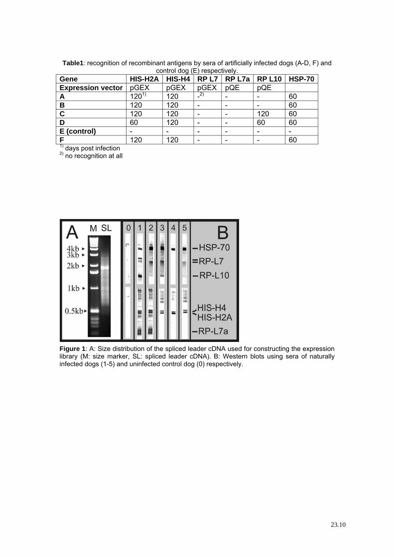
Table1: recognition of recombinant antigens by sera of artificially infected dogs (A-D, F) and
control dog (E) respectively. Gene HIS-H2A HIS-H4 RP L7 RP L7a RP L10 HSP-70 Expression vector pGEX pGEX pGEX pQE pQE A 1201) 120 -2) - - 60 B 120 120 - - - 60 C 120 120 - - 120 60 D 60 120 - - 60 60 E (control) - - - - - - F 120 120 - - - 60 1) days post infection 2) no recognition at all
Figure 1: A: Size distribution of the spliced leader cDNA used for constructing the expression library (M: size marker, SL: spliced leader cDNA). B: Western blots using sera of naturally infected dogs (1-5) and uninfected control dog (0) respectively.
23.10
2.4. Discussion We identified several potential antigens suitable for a diagnostic test. However, much more data are needed concerning their value to recognize early infections of different L. infantum strains responsible for HIV-Leishmania co-infections. Ongoing studies currently address these issues. Several core histones were selected in the serum screen. Here the questions arise: why are the histones from the genus Leishmania highly diverse and immunogenic? What are the advantages for the parasites? So far, they have not been satisfactory answered. Nevertheless, the unusual properties of Leishmania histones might represent a key factor in leishmaniasis. It remains questionable whether a diagnostic test, even when sensitive and specific, would improve the control of leishmaniasis. It would need a huge effort (personnel and cost intensive) to test and treat the majority of the canine population in Southern Europe. From this point of view, a cheap vaccine would be favorable: a vaccine might be allocated as lures (like in Switzerland for control of rabies), and there would be no need for diagnosing and treating infected animals, which most probably would also lead to treatment failures and selecting drug resistance in parasite strains. However, since no such vaccine is available yet against any parasitic disease, any other possible step has to be kept in mind. In Brazil, there has been a study on the impact of using insecticide impregnated dog collars, which yielded satisfactory effects (Reithinger and Davies, 2002). However, this handling prevents only dogs with owners from infective sandfly bites. For street dogs, a similar approach would be to use masses of insecticides which surely has a great impact on environment and most probably also on public health. Mass diagnosis and treatment of street dogs still appears to be a possible approach at present. However, it does not seem to be an elegant way to control leishmaniasis, especially in regard to drug resistance. Furthermore, chemotherapy of canine leishmaniasis is tagged by high relapse rates (Baneth and Shaw, 2002). Nevertheless, an early diagnostic test surely would be a useful tool for veterinarian purpose.
24
3. Development of a PCR assay for diagnosis of human leishmaniasis and differentiation of Leishmania species
3.1. Background and method There are many different approaches for diagnosing human leishmaniasis (reviewed by Guerin et al., 2002; Singh and Sivakumar, 2003): direct detection of the parasite by microscopy (low sensitivity), or cultivation (time consuming and not all strains are equally grown in vitro and in vivo), PCR and serological methods. In Europe, most of the leishmaniasis cases represent HIV co-infections. A recent study compared several different tools for diagnosis of leishmaniasis in HIV patients (Deniau et al., 2003). In respect to sensitivity and specifity, the results suggest the use of a combination of PCR and cultivation. Although several diagnostic PCRs for Leishmania species are published (reviewed by Wilson, 1995), none of them is able to detect all different species and distinguish among them at the same time. Either these PCRs amplify DNA of all species but cannot distinguish between them (as PCRs amplifying ribosomal genes), or they are only capable to amplify DNA deriving from a limited number of species (as PCRs amplifying kinetoplast DNA). We have chosen the mini-exon gene as PCR template, because this gene is abundant in about 200 copies per genome of each Leishmania species (sensitivity) and shows enough polymorphism for distinguishing the different species. It consists of the highly conserved mini-exon, a semi conserved intron and a highly variable non transcribed spacer, resulting in length and sequence polymorphism of the PCR product, which allows differentiation of Leishmania species using restriction length polymorphism.
25
3.2. Diagnostic genotyping of Old and New World Leishmania species by PCR-RFLP
26
Parasitology
Diagnostic genotyping of Old and New World Leishmania species byPCR-RFLP
Jutta Marfurt, Igor Niederwieser, Ntoh Divine Makia, Hans-Peter Beck, Ingrid Felger*Department of Medical Parasitology and Infection Biology, Swiss Tropical Institute, Basel, Switzerland
Abstract
We have designed a new genotyping scheme for molecular diagnosis of the different Leishmania species pathogenic to humans. Thisscheme is based on PCR amplified sequences from the gene for the spliced leader RNA (mini-exon). This target was selected because itis present as tandem repeats (100 to 200 copies) in the genus Leishmania and other kinetoplastida, but is absent from the mammalian hostsand the sandfly vectors. The exon is highly conserved, whereas the intron and non-transcribed spacer region vary in size and sequence amongdifferent species. Thus, it was possible to amplify DNA from both Old and New World pathogenic Leishmania complexes using a singlepair of primers deriving from the conserved region of the mini-exon tandem repeat. Species identification was performed by digestingmini-exon PCR products with one or two different restriction enzymes. Restriction fragment length polymorphism (RFLP) generatedspecies-specific patterns of bands visualized in agarose gels, which allowed to differentiate each species unequivocally. © 2003 ElsevierScience Inc. All rights reserved.
1. Introduction
Leishmaniasis is a parasitic disease which is associatedwith a wide spectrum of clinical manifestations dependingon the species of the parasite, the host’s immune responseand the saliva of the sandfly vector (Grimaldi and Tesh,1993; Gradoni and Gramiccia, 1994). The variety of symp-toms range from self-curing ulcerative or diffuse, non-ul-cerative cutaneous lesions, to persistent and often disfigur-ing mucocutaneous lesions or the potentially fatal visceralform of the disease. The assignment of the parasite speciesbased alone on geographic location or the site of infection isnot satisfactory. Accordingly, correct diagnosis and classi-fication of a pathogenic Leishmania isolate is essential todetermine the clinical prognosis and a species-specific ther-apeutic approach (Navin et al., 1992; Berman, 1997; Ro-mero et al., 2001). In addition, epidemiologic studies aswell as the usage of reference strains in laboratory experi-ments greatly depend on correct species-identification.
The genus Leishmania is divided into the two subgeneraViannia and Leishmania according to their development inthe sandfly vector. Specification within the subgenera de-pends on several factors such as the geographical distribu-
tion of an isolate, the clinical presentation of the disease,and the epidemiology of the vector and the animal reservoir(Lainson and Shaw, 1987; Pearson et al., 2001). Sincemorphologic differentiation of Leishmania species is notpossible, a variety of biochemical, immunologic, or molec-ular criteria were introduced for classification of pathogenicspecies, such as characterization by isoenzyme electro-phoresis (zymodeme analysis) (Kreutzer and Christensen,1980), by monoclonal antibodies (serodeme) (Grimaldi etal., 1987) or by hybridization with species-specific probessuch as minicircle DNA probes (Wirth et al., 1989).
With the advent of the PCR technology, several PCR-based assays for species differentiation were developed. Astargets for amplification served either nuclear DNA, such asthe SSU rRNA gene (van Eys et al., 1992), repetitive se-quences (Piarroux et al., 1995), internal transcribed spacer(ITS) regions (Cupolillo et al., 1995, Eisenberger and Jaffe,1999), the tubulin gene (Luis et al., 1998), the gp63 genelocus (Victoir et al., 1998), microsatellite DNA (Russell etal., 1999), or extrachromosomal DNA, such as the repetitivekinetoplast DNA (kDNA) minicircles (de Bruijn andBarker, 1992; Belli et al., 1998).
While all these different approaches offer a multitude ofvalid taxonomic characters for species identification, a sim-ple assay (one single PCR) which is also comprehensive (allLeishmania species) as well as reliable and applicable in theroutine diagnostic laboratory, has been missing to date.
* Corresponding author. Tel.: �41-61-284-81-17; fax: �41-61-271-86-54.
E-mail address: [email protected] (I. Felger).
Diagnostic Microbiology and Infectious Diseasewww.elsevier.com/locate/diagmicrobio46 (2003) 115–124
0732-8893/03/$ – see front matter © 2003 Elsevier Science Inc. All rights reserved.doi:10.1016/S0732-8893(03)00040-3
Most molecular diagnostic methods developed for genotyp-ing Leishmania species were based on the polymorphickDNA minicircle, which is considered a prime candidate fora sensitive assay because of the presence of 10000 to 20000minicircles per cell. In a first attempt to device a genotypingassay comprising all Leishmania species pathogenic to hu-mans, we performed a sequence analysis of kDNA whichrevealed too great an amount of sequence variation betweenkDNAs of different New and Old World species, thus im-peding the design of universal primers. An alternativekDNA approach based on multiplex PCR has been devel-oped (Belli et al., 1998). However, the yield of amplifica-tion products is generally compromised in multiplex PCRby primer dimer formation resulting in a loss of sensitivityof the assay.
Therefore, we chose the mini-exon (or spliced leader(SL)) gene found in all kinetoplastida as target sequence fora genotyping assay. The mini-exon gene, which is involvedin the trans-splicing process in kintoplastid protozoa, ispresent 100 to 200 times per nuclear genome as tandemlyrepeated copies, and it is absent from the vertebrate host orinvertebrate vector. A detailed study on sequence variationin the mini-exon gene repeat of human pathogenic Leish-mania species had previously shown that the diversity de-tected in the non-transcribed spacers represents an informa-tive phylogenetic marker (Fernandes et al., 1994).
These features, together with a species-specific diversity,which conforms roughly with the species boundaries withinthe genus, designate the mini-exon for being exploited as agenotyping marker. We have used a PCR approach similarto that of Fernandes et al. (1994), but combined it withrestriction digests of the PCR product, a method which isroutinely used for other genotyping tasks (Singh, 1997). Theresulting patterns of restriction fragments were characteris-tic for each species. Thus, we achieve a high resolution andhigh discrimination power, while the assay is still based ona single PCR reaction with universal primers.
2. Materials and methods
2.1. Parasite strains
The strains representative for the different Leishmaniaspecies, which were used in this study are listed in Table 1.In addition, 35 isolates from patients attending the poly-clinic of the Swiss Tropical Institute (STI) were genotyped.
2.2. In vitro cultivation of Leishmania
The promastigote forms were cultivated at 27°C withoutgas phase in a 1:1 mixture of Schneider’s modified Dro-sophila medium (SDM-79) (Brun and Schonenberger,1979) and Schneider’s medium (SM) (Cunningham, 1977)complemented with 15% heat-inactivated fetal bovine se-rum. Leishmania from a 5-day-old culture were harvested
and counted with the Cell Counter and Analyser SystemCASY® 1 (Scharfe System GmbH, Reutlingen, FRG). Thecells were washed twice with 0.15 M phosphate-bufferedsaline (PBS) pH 7.4, pelleted by centrifugation, resus-pended in the appropriate buffer depending on further ex-periments, and processed immediately.
2.3. DNA preparation
Cultured cells, corresponding to about 2 � 108 cells,were transferred into 500 �l digestion buffer (10 mM Tris/HCl pH 8; 5 mM EDTA; 0.5% SDS). Proteinase K (Sigma-Aldrich, Inc., CH) was added to a final concentration of 0.5mg/ml and the sample was incubated over night at 56°C.DNA was extracted successively with one volume of phenolpH 8.0, one volume of phenol/chloroform, and one volumeof chloroform according to standard procedures (Sambrookand Russell, 2001). DNA was precipitated from the aqueousphase with ethanol, the pellet washed with 75% ethanol, airdried and finally resuspended in 100 �l H2Odest.
2.4. PCR
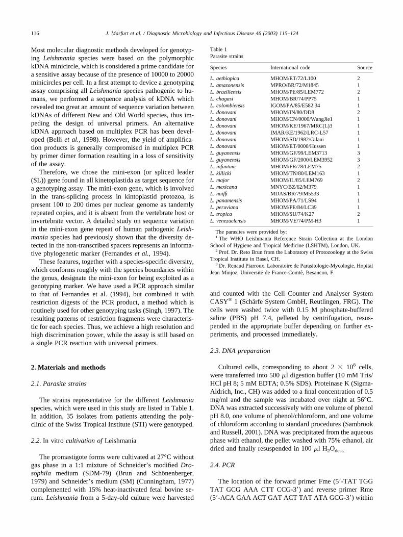
The location of the forward primer Fme (5�-TAT TGGTAT GCG AAA CTT CCG-3�) and reverse primer Rme(5�-ACA GAA ACT GAT ACT TAT ATA GCG-3�) within
Table 1Parasite strains
Species International code Source
L. aethiopica MHOM/ET/72/L100 2L. amazonensis MPRO/BR/72/M1845 1L. braziliensis MHOM/PE/85/LEM772 2L. chagasi MHOM/BR/74/PP75 1L. colombiensis IGOM/PA/85/E582.34 1L. donovani MHOM/IN/80/DD8 2L. donovani MHOM/CN/0000/WangJie1 1L. donovani MHOM/KE/1967/MRC(L)3 1L. donovani IMAR/KE/1962/LRC-L57 1L. donovani MHOM/SD/1982/Gilani 1L. donovani MHOM/ET/0000/Hussen 1L. guyanensis MHOM/GF/99/LEM3713 3L. guyanensis MHOM/GF/2000/LEM3952 3L. infantum MHOM/FR/78/LEM75 2L. killicki MHOM/TN/80/LEM163 1L. major MHOM/IL/85/LEM769 2L. mexicana MNYC/BZ/62/M379 1L. naiffi MDAS/BR/79/M5533 1L. panamensis MHOM/PA/71/LS94 1L. peruviana MHOM/PE/84/LC39 1L. tropica MHOM/SU/74/K27 2L. venezuelensis MHOM/VE/74/PM-H3 1
The parasites were provided by:1 The WHO Leishmania Reference Strain Collection at the London
School of Hygiene and Tropical Medicine (LSHTM), London, UK.2 Prof. Dr. Reto Brun from the Laboratory of Protozoology at the Swiss
Tropical Institute in Basel, CH.3 Dr. Renaud Piarroux, Laboratoire de Parasitologie-Mycologie, Hopital
Jean Minjoz, Universite de France-Comte, Besancon, F.
116 J. Marfurt et al. / Diagnostic Microbiology and Infectious Disease 46 (2003) 115–124
the mini-exon gene repeat is shown in Fig. 1. Two �l ofDNA solution were amplified in a 100 �l reaction contain-ing 50 mM KCl, 20 mM Tris-HCl pH 8. 4 (GIBCO™,Invitrogen™ Life Technologies, CH), 0.2 mM dNTPs (Am-ersham Biosciences Europe, CH), 12% DMSO (FlukaChemika, CH), 40 mM tetramethylammonium chloride(Carl Roth GmbH, FRG), 1. 5 mM MgCl2 (GIBCO™), 0.5�M of each primer (MWG Biotech AG, CH), and 1 U Taqpolymerase (GIBCO™). The PCR conditions were 5 min at94°C followed by 25 to 35 cycles of 30 sec at 94°C, 30 secat 54°C, and 45 sec at 72°C. The number of cycles dependedon the origin and concentration of the template. PCR prod-ucts were separated on a 1.5% agarose gel.
2.5. Sensitivity titration assays
A) Crude Leishmania extracts: To test whether samplescan be directly used in the PCR assay, cultured cells wereresuspended in PBS pH 7. 4 and tenfold serial dilutions of5 � 106 to 2. 5 � 102 cells per millilitre (ml) were prepared,yielding a concentration range from 10000 to 0.5 parasitesper 2 �l. The dilutions were subjected to three freeze-thawcycles, incubated at 95°C for 10 min and 2 �l of thesolutions were directly used in the PCR reaction.
B) Purified Leishmania DNA: DNA concentrations weredetermined by measuring the optical density (OD) at 260nm with a GeneQuant® RNA/DNA Calculator (AmershamBiosciences Europe GmbH, CH). Tenfold serial dilutions of0.15 mg to 1. 5 pg per ml were prepared, yielding a con-centration range from 300 ng to 3 fg per 2 �l which wereused in the PCR assay.
2.6. Specificity assays
The specificity of the mini-exon PCR assay was deter-mined by testing different concentrations of purified DNA
from the human host and other pathogens in the assay.Human DNA was extracted from peripheral blood mono-nuclear cells (PBMCs) from a volunteer as described insection 3. Genomic DNA from Mycobacterium spp. (i.e., M.tuberculosis and M. ulcerans) and Plasmodium falciparumwas kindly provided by the STI laboratories of MolecularImmunology and Molecular Parasitology, respectively. Cul-tured organisms from other kinetoplastida (i.e., Crithidiaspp. and Trypanosoma spp.) were kindly provided by thelaboratory of Protozoology at STI. DNA was extracted andpurified as described in section 3.
2.7. Restriction digests
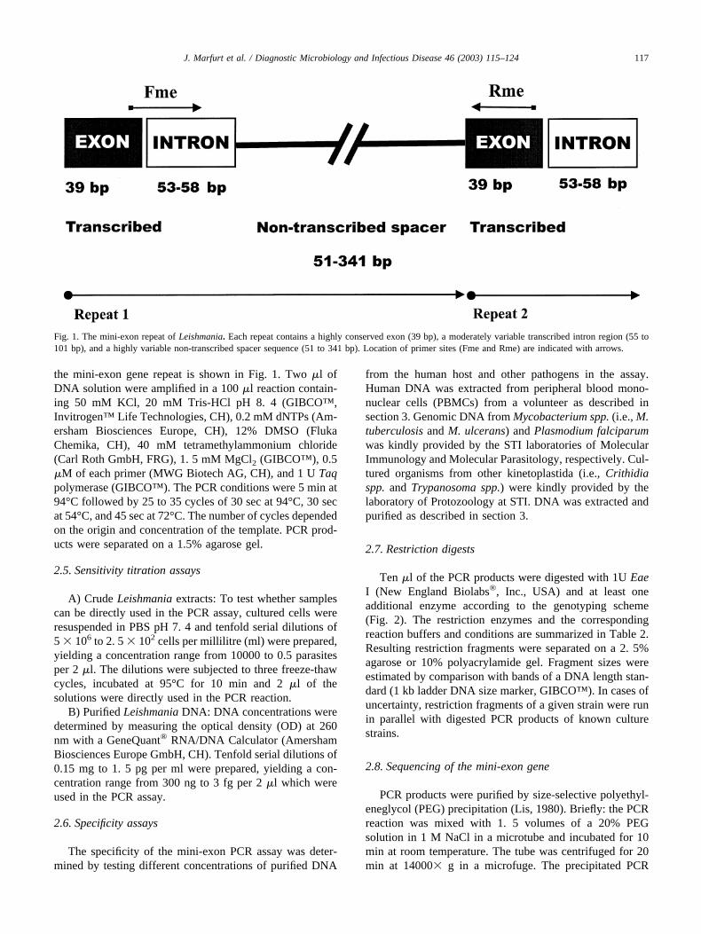
Ten �l of the PCR products were digested with 1U EaeI (New England Biolabs®, Inc., USA) and at least oneadditional enzyme according to the genotyping scheme(Fig. 2). The restriction enzymes and the correspondingreaction buffers and conditions are summarized in Table 2.Resulting restriction fragments were separated on a 2. 5%agarose or 10% polyacrylamide gel. Fragment sizes wereestimated by comparison with bands of a DNA length stan-dard (1 kb ladder DNA size marker, GIBCO™). In cases ofuncertainty, restriction fragments of a given strain were runin parallel with digested PCR products of known culturestrains.
2.8. Sequencing of the mini-exon gene
PCR products were purified by size-selective polyethyl-eneglycol (PEG) precipitation (Lis, 1980). Briefly: the PCRreaction was mixed with 1. 5 volumes of a 20% PEGsolution in 1 M NaCl in a microtube and incubated for 10min at room temperature. The tube was centrifuged for 20min at 14000� g in a microfuge. The precipitated PCR
Fig. 1. The mini-exon repeat of Leishmania. Each repeat contains a highly conserved exon (39 bp), a moderately variable transcribed intron region (55 to101 bp), and a highly variable non-transcribed spacer sequence (51 to 341 bp). Location of primer sites (Fme and Rme) are indicated with arrows.
117J. Marfurt et al. / Diagnostic Microbiology and Infectious Disease 46 (2003) 115–124
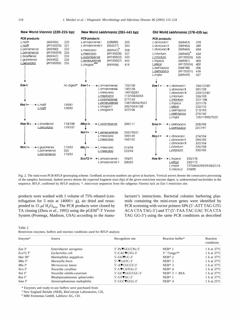
products were washed with 1 volume of 75% ethanol (cen-trifugation for 5 min at 14000� g), air dried and resus-pended in 15 �l H2Odest. The PCR products were cloned byTA cloning (Zhou et al., 1995) using the pGEM®-T VectorSystem (Promega, Madison, USA) according to the manu-
facturer’s instructions. Bacterial colonies harboring plas-mids containing the mini-exon genes were identified byPCR screening with vector primers SP6 (5�-ATT TAG GTGACA CTA TAG-3�) and T7 (5�-TAA TAC GAC TCA CTATAG GG-3�) using the same PCR conditions as described
Fig. 2. The mini-exon PCR-RFLP genotyping scheme. GenBank accession numbers are given in brackets. Vertical arrows denote the consecutive processingof the samples; horizontal, dashed arrows denote the expected fragment sizes (bp) of the given restriction enzyme digest. n, undetermined nucleotides in thesequence; RFLP, confirmed by RFLP analysis; *, mini-exon sequences from the subgenus Viannia lack an Eae I restriction site.
Table 2Restriction enzymes, buffers and reaction conditions used for RFLP analysis
Enzyme* Source Recognition site Reaction buffer* Reactionconditions
Eae Ia Enterobacter aerogenes 5�-Py�GGCCPu-3� NEB� 1 1 h at 37°CEco72 Ib Escherichia coli 5�-CAC�GTG-3� Y�/Tango™ 1 h at 37°CHae IIIa Haemophilus aegypticus 5�-GG�CC-3� NEB� 2 1 h at 37°CMbo Ia Moraxella bovis 5�-�GATC-3� NEB� 3 1 h at 37°CMlu Ia Micrococcus luteus 5�-A�CGCGT-3� NEB� 3 1 h at 37°CNco Ia Nocardia corallina 5�-C�CATGG-3� NEB� 4 1 h at 37°CNot Ia Nocardia otitidis-cavarium 5�-GC�GGCCGC-3� NEB� 3 � BSA 1 h at 37°CRsa Ia Rhodopseudomonas sphaeroides 5�-GT�AC-3� NEB� 1 1 h at 37°CSma Ia Stenotrophomonas maltophilia 5�-CCC�GGG-3� NEB� 4 1 h at 25°C
* Enzymes and ready-to-use buffers were purchased from:a New England Biolabs (NEB), BioConcept Laboratories, CH,b MBI Fermentas GmbH, Labforce AG, CH.
118 J. Marfurt et al. / Diagnostic Microbiology and Infectious Disease 46 (2003) 115–124
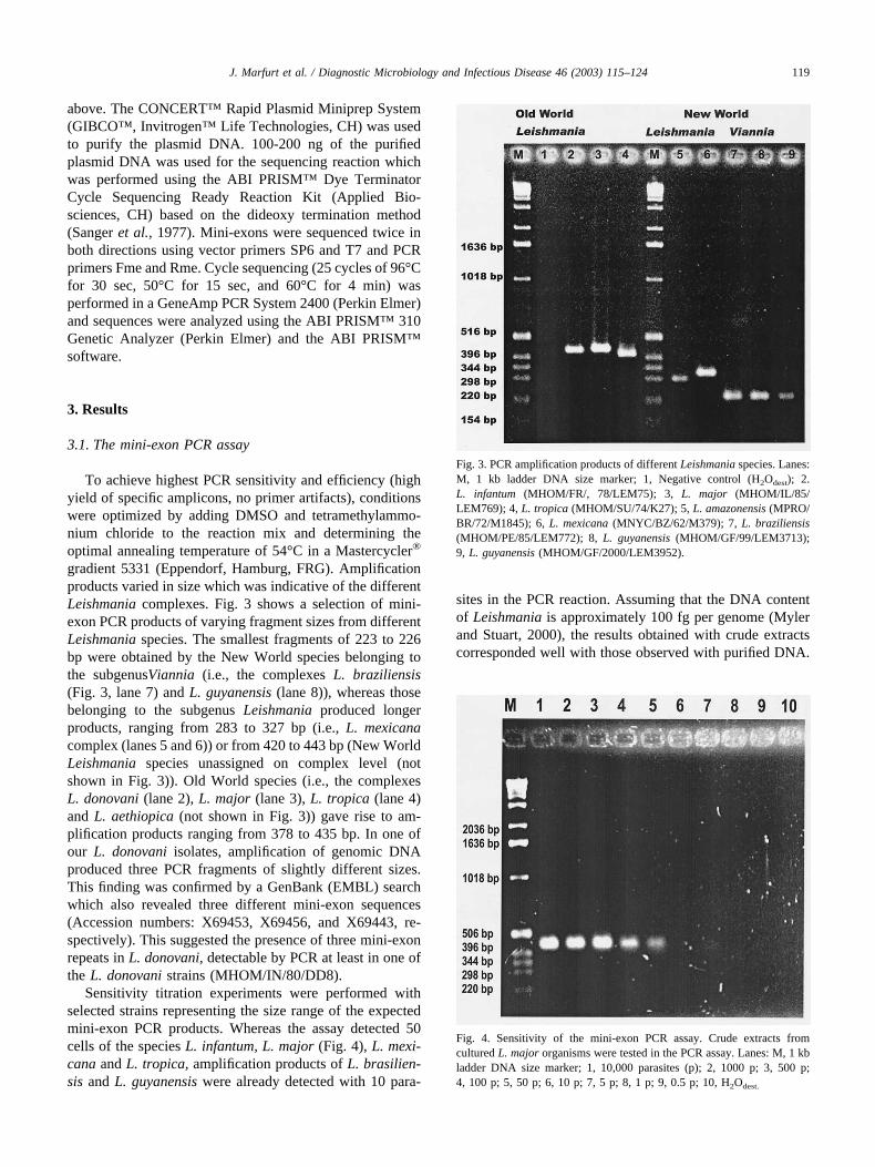
above. The CONCERT™ Rapid Plasmid Miniprep System(GIBCO™, Invitrogen™ Life Technologies, CH) was usedto purify the plasmid DNA. 100-200 ng of the purifiedplasmid DNA was used for the sequencing reaction whichwas performed using the ABI PRISM™ Dye TerminatorCycle Sequencing Ready Reaction Kit (Applied Bio-sciences, CH) based on the dideoxy termination method(Sanger et al., 1977). Mini-exons were sequenced twice inboth directions using vector primers SP6 and T7 and PCRprimers Fme and Rme. Cycle sequencing (25 cycles of 96°Cfor 30 sec, 50°C for 15 sec, and 60°C for 4 min) wasperformed in a GeneAmp PCR System 2400 (Perkin Elmer)and sequences were analyzed using the ABI PRISM™ 310Genetic Analyzer (Perkin Elmer) and the ABI PRISM™software.
3. Results
3.1. The mini-exon PCR assay
To achieve highest PCR sensitivity and efficiency (highyield of specific amplicons, no primer artifacts), conditionswere optimized by adding DMSO and tetramethylammo-nium chloride to the reaction mix and determining theoptimal annealing temperature of 54°C in a Mastercycler®
gradient 5331 (Eppendorf, Hamburg, FRG). Amplificationproducts varied in size which was indicative of the differentLeishmania complexes. Fig. 3 shows a selection of mini-exon PCR products of varying fragment sizes from differentLeishmania species. The smallest fragments of 223 to 226bp were obtained by the New World species belonging tothe subgenusViannia (i.e., the complexes L. braziliensis(Fig. 3, lane 7) and L. guyanensis (lane 8)), whereas thosebelonging to the subgenus Leishmania produced longerproducts, ranging from 283 to 327 bp (i.e., L. mexicanacomplex (lanes 5 and 6)) or from 420 to 443 bp (New WorldLeishmania species unassigned on complex level (notshown in Fig. 3)). Old World species (i.e., the complexesL. donovani (lane 2), L. major (lane 3), L. tropica (lane 4)and L. aethiopica (not shown in Fig. 3)) gave rise to am-plification products ranging from 378 to 435 bp. In one ofour L. donovani isolates, amplification of genomic DNAproduced three PCR fragments of slightly different sizes.This finding was confirmed by a GenBank (EMBL) searchwhich also revealed three different mini-exon sequences(Accession numbers: X69453, X69456, and X69443, re-spectively). This suggested the presence of three mini-exonrepeats in L. donovani, detectable by PCR at least in one ofthe L. donovani strains (MHOM/IN/80/DD8).
Sensitivity titration experiments were performed withselected strains representing the size range of the expectedmini-exon PCR products. Whereas the assay detected 50cells of the species L. infantum, L. major (Fig. 4), L. mexi-cana and L. tropica, amplification products of L. brasilien-sis and L. guyanensis were already detected with 10 para-
sites in the PCR reaction. Assuming that the DNA contentof Leishmania is approximately 100 fg per genome (Mylerand Stuart, 2000), the results obtained with crude extractscorresponded well with those observed with purified DNA.
Fig. 3. PCR amplification products of different Leishmania species. Lanes:M, 1 kb ladder DNA size marker; 1, Negative control (H2Odest); 2.L. infantum (MHOM/FR/, 78/LEM75); 3, L. major (MHOM/IL/85/LEM769); 4, L. tropica (MHOM/SU/74/K27); 5, L. amazonensis (MPRO/BR/72/M1845); 6, L. mexicana (MNYC/BZ/62/M379); 7, L. braziliensis(MHOM/PE/85/LEM772); 8, L. guyanensis (MHOM/GF/99/LEM3713);9, L. guyanensis (MHOM/GF/2000/LEM3952).
Fig. 4. Sensitivity of the mini-exon PCR assay. Crude extracts fromcultured L. major organisms were tested in the PCR assay. Lanes: M, 1 kbladder DNA size marker; 1, 10,000 parasites (p); 2, 1000 p; 3, 500 p;4, 100 p; 5, 50 p; 6, 10 p; 7, 5 p; 8, 1 p; 9, 0.5 p; 10, H2Odest.
119J. Marfurt et al. / Diagnostic Microbiology and Infectious Disease 46 (2003) 115–124
The detection limit was 3 pg DNA with the species yieldingamplification products larger than 250 bp, but reached300 fg with those producing the smallest PCR fragments(223 to 226 bp).
To assess the specificity of our PCR assay, we testedDNA from the human host as well as from other pathogensand related kinetoplastida in the PCR reaction. Whereas themini-exon sequences of Trypanosoma brucei rhodesiense,T. b. gambiense and T. cruzi were not amplified with theseprimers, the other kinetoplastida (i.e., Crithidia bombi,C. lucilia and C. fasciculata) revealed PCR products be-tween 400 and 430 bp in size. The PCR yield was lowerwith Crithidia than with Leishmania species, indicatingmismatches between Crithidia mini-exon and our primersequences. However, Crithidia species were discernible bythe absence of an Eae I restriction site and the production ofa unique pattern of fragments (318 and 88 bp) with the HaeIII digest which was not found with any Leishmania species(data not shown).
3.2. PCR-RFLP genotyping scheme
The observed size difference of PCR products identifiedthe different Leishmania complexes, but did not discrimi-nate between individual species. Therefore, we chose tosubsequently digest the PCR products with restriction en-zymes in order to produce a species-specific RFLP patternin an agarose gel. The resulting pattern of bands depends onboth, mutations in the recognition sequence of the chosenrestriction enzyme, and thus on the number of the fragmentsgenerated, and on size variation of the internal non-tran-scribed spacer fragment. In order to establish such a RFLPgenotyping scheme, we initially performed a GenBank(EMBL) search. The diversity of the mini-exon geneswithin the Leishmania and Viannia subgenus was deter-mined by sequence alignments (ClustalW, BCM SearchLauncher, http://searchlauncher.bcm.tmc.edu/), and re-striction enzymes differentiating between the individualspecies were identified using the Webcutter version 2.0program (http://www.firstmarket.com/firstmarket/cutter/cut2.html). Using this information, restriction maps ofthe mini-exon repeat of different Leishmania specieswere produced. As a result, we defined different digestswhich allowed species discrimination: Eae I proved to bethe most informative restriction enzyme, because it di-gested all mini-exon PCR products of the Leishmaniasubgenus in a species-specific manner, but it did not cutthe mini-exon of the species of the subgenusViannia. Thelatter PCR products were digested with Hae III, Nco I, orRsa I which identified each species unequivocally.Whereas the species belonging to all other complexeswere differentiated by a second round of restriction di-gests as shown in the flow diagram in Fig. 2. This dia-gram depicts the mini-exon RFLP genotyping schemeand indicates the fragment sizes expected from the nu-cleotide sequences submitted to GenBank. This virtual
sequence analysis suggested that all Leishmania speciescan be distinguished by mini-exon PCR-RFLP.
3.3. Validation of the genotyping scheme with culturedLeishmania reference strains
The genotyping scheme deduced from sequence infor-mation from GenBank was validated with culture strainsrepresentative for different species. Fig. 5 shows the RFLPpattern of different species on an agarose gel after diagnos-tic restriction digests with Eae I and Hae III. Two differentL. donovani strains (lane 1 and 2 of Fig. 5) produceddifferent RFLP patterns. This is in agreement with the factthat 3 different mini-exon sequences of L. donovani havebeen found in the GenBank search. The analyzed isolatescorrespond to the restriction map of mini-exon sequenceaccession numbers X69443 and X69453, respectively.
L. panamensis in lane 11 remains uncut by Hae III andtherefore reflects the PCR product. The 468 bp band reflectsoccasional amplification of two consecutive mini-exon re-peat units. This PCR artifact is due to the mini-exon’sarrangement as tandem repeats and is more likely to occurin small Viannia-type repeat units. The 403 bp band in lane4 represents the PCR band of L. tropica which occurred dueto partial digest of the amplified DNA. The unexpected215 bp fragment of L. mexicana in lane 8 might either be anartifact due to partial digest (Eae I site at position 112), orsuggests the co-existence of two different mini-exon repeatunits within this species.
Next we examined whether this new genotyping schemebased on GenBank sequences can be used to characterizefield isolates or applied in molecular epidemiologic studies.23 isolates of L. infantum and 7 isolates of L. major (for-merly typed by isoenzyme analysis) deriving from bonemarrow or skin biopsies of patients attending the polyclinicof the STI were analyzed. All showed the expected restric-tion pattern according to the genotyping scheme (results notshown). In the course of genotyping Leishmania laboratorystrains representative for given species from different ori-gins (see Table 1), we encountered several inconsistenciesbetween the observed RFLP pattern and the genotypingscheme. These might well reflect existing problems inLeishmania taxonomy. The L. chagasi strain (MHOM/BR/74/PP75) used in our study could not be differentiated fromL. infantum (MHOM/FR/78/LEM75) since both strains pro-duced identical RFLP patterns.
In some cases, the expected RFLP pattern based onGenBank sequences could not be confirmed (i.e., forL. aethiopica, L. infantum, L. mexicana, L. naiffi, andL. panamensis). We therefore performed sequence analysisof the mini-exon gene amplified from reference strains ofthese species (Accession numbers: AY155501, AY155503,AY155508, AY155505, and AY155509, respectively). Fig.2 represents the final genotyping scheme, which is con-firmed by RFLP and sequence data from laboratory strainsavailable to us (Mini-exon genes sequenced in our labora-
120 J. Marfurt et al. / Diagnostic Microbiology and Infectious Disease 46 (2003) 115–124
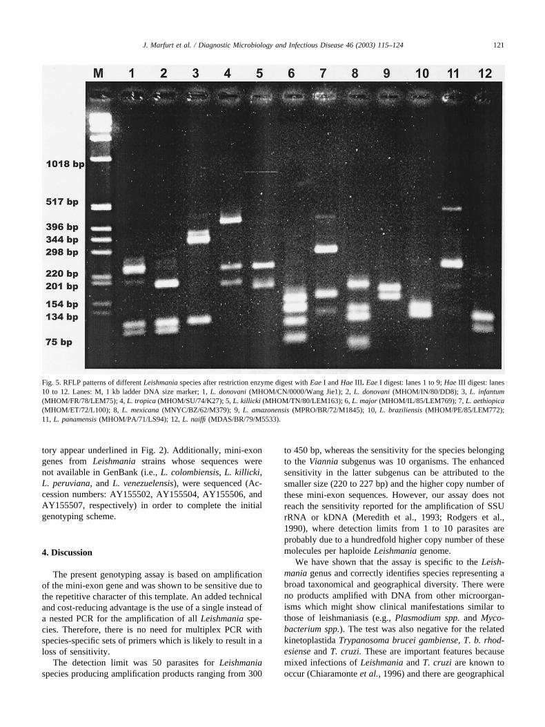
tory appear underlined in Fig. 2). Additionally, mini-exongenes from Leishmania strains whose sequences werenot available in GenBank (i.e., L. colombiensis, L. killicki,L. peruviana, and L. venezuelensis), were sequenced (Ac-cession numbers: AY155502, AY155504, AY155506, andAY155507, respectively) in order to complete the initialgenotyping scheme.
4. Discussion
The present genotyping assay is based on amplificationof the mini-exon gene and was shown to be sensitive due tothe repetitive character of this template. An added technicaland cost-reducing advantage is the use of a single instead ofa nested PCR for the amplification of all Leishmania spe-cies. Therefore, there is no need for multiplex PCR withspecies-specific sets of primers which is likely to result in aloss of sensitivity.
The detection limit was 50 parasites for Leishmaniaspecies producing amplification products ranging from 300
to 450 bp, whereas the sensitivity for the species belongingto the Viannia subgenus was 10 organisms. The enhancedsensitivity in the latter subgenus can be attributed to thesmaller size (220 to 227 bp) and the higher copy number ofthese mini-exon sequences. However, our assay does notreach the sensitivity reported for the amplification of SSUrRNA or kDNA (Meredith et al., 1993; Rodgers et al.,1990), where detection limits from 1 to 10 parasites areprobably due to a hundredfold higher copy number of thesemolecules per haploide Leishmania genome.
We have shown that the assay is specific to the Leish-mania genus and correctly identifies species representing abroad taxonomical and geographical diversity. There wereno products amplified with DNA from other microorgan-isms which might show clinical manifestations similar tothose of leishmaniasis (e.g., Plasmodium spp. and Myco-bacterium spp.). The test was also negative for the relatedkinetoplastida Trypanosoma brucei gambiense, T. b. rhod-esiense and T. cruzi. These are important features becausemixed infections of Leishmania and T. cruzi are known tooccur (Chiaramonte et al., 1996) and there are geographical
Fig. 5. RFLP patterns of different Leishmania species after restriction enzyme digest with Eae I and Hae III. Eae I digest: lanes 1 to 9; Hae III digest: lanes10 to 12. Lanes: M, 1 kb ladder DNA size marker; 1, L. donovani (MHOM/CN/0000/Wang Jie1); 2, L. donovani (MHOM/IN/80/DD8); 3, L. infantum(MHOM/FR/78/LEM75); 4, L. tropica (MHOM/SU/74/K27); 5, L. killicki (MHOM/TN/80/LEM163); 6, L. major (MHOM/IL/85/LEM769); 7, L. aethiopica(MHOM/ET/72/L100); 8, L. mexicana (MNYC/BZ/62/M379); 9, L. amazonensis (MPRO/BR/72/M1845); 10, L. braziliensis (MHOM/PE/85/LEM772);11, L. panamensis (MHOM/PA/71/LS94); 12, L. naiffi (MDAS/BR/79/M5533).
121J. Marfurt et al. / Diagnostic Microbiology and Infectious Disease 46 (2003) 115–124
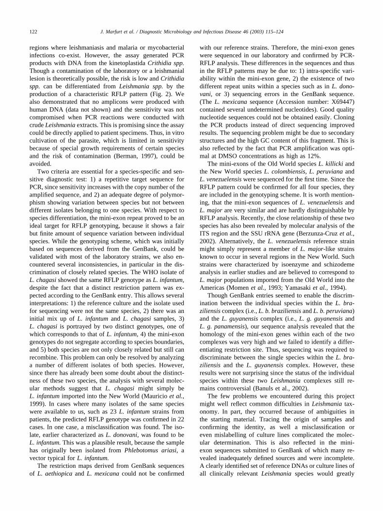
regions where leishmaniasis and malaria or mycobacterialinfections co-exist. However, the assay generated PCRproducts with DNA from the kinetoplastida Crithidia spp.Though a contamination of the laboratory or a leishmaniallesion is theoretically possible, the risk is low and Crithidiaspp. can be differentiated from Leishmania spp. by theproduction of a characteristic RFLP pattern (Fig. 2). Wealso demonstrated that no amplicons were produced withhuman DNA (data not shown) and the sensitivity was notcompromised when PCR reactions were conducted withcrude Leishmania extracts. This is promising since the assaycould be directly applied to patient specimens. Thus, in vitrocultivation of the parasite, which is limited in sensitivitybecause of special growth requirements of certain speciesand the risk of contamination (Berman, 1997), could beavoided.
Two criteria are essential for a species-specific and sen-sitive diagnostic test: 1) a repetitive target sequence forPCR, since sensitivity increases with the copy number of theamplified sequence, and 2) an adequate degree of polymor-phism showing variation between species but not betweendifferent isolates belonging to one species. With respect tospecies differentiation, the mini-exon repeat proved to be anideal target for RFLP genotyping, because it shows a fairbut finite amount of sequence variation between individualspecies. While the genotyping scheme, which was initiallybased on sequences derived from the GenBank, could bevalidated with most of the laboratory strains, we also en-countered several inconsistencies, in particular in the dis-crimination of closely related species. The WHO isolate ofL. chagasi showed the same RFLP genotype as L. infantum,despite the fact that a distinct restriction pattern was ex-pected according to the GenBank entry. This allows severalinterpretations: 1) the reference culture and the isolate usedfor sequencing were not the same species, 2) there was aninitial mix up of L. infantum and L. chagasi samples, 3)L. chagasi is portrayed by two distinct genotypes, one ofwhich corresponds to that of L. infantum, 4) the mini-exongenotypes do not segregate according to species boundaries,and 5) both species are not only closely related but still canrecombine. This problem can only be resolved by analyzinga number of different isolates of both species. However,since there has already been some doubt about the distinct-ness of these two species, the analysis with several molec-ular methods suggest that L. chagasi might simply beL. infantum imported into the New World (Mauricio et al.,1999). In cases where many isolates of the same specieswere available to us, such as 23 L. infantum strains frompatients, the predicted RFLP genotype was confirmed in 22cases. In one case, a misclassification was found. The iso-late, earlier characterized as L. donovani, was found to beL. infantum. This was a plausible result, because the samplehas originally been isolated from Phlebotomus ariasi, avector typical for L. infantum.
The restriction maps derived from GenBank sequencesof L. aethiopica and L. mexicana could not be confirmed
with our reference strains. Therefore, the mini-exon geneswere sequenced in our laboratory and confirmed by PCR-RFLP analysis. These differences in the sequences and thusin the RFLP patterns may be due to: 1) intra-specific vari-ability within the mini-exon gene, 2) the existence of twodifferent repeat units within a species such as in L. dono-vani, or 3) sequencing errors in the GenBank sequence.(The L. mexicana sequence (Accession number: X69447)contained several undetermined nucleotides). Good qualitynucleotide sequences could not be obtained easily. Cloningthe PCR products instead of direct sequencing improvedresults. The sequencing problem might be due to secondarystructures and the high GC content of this fragment. This isalso reflected by the fact that PCR amplification was opti-mal at DMSO concentrations as high as 12%.
The mini-exons of the Old World species L. killicki andthe New World species L. colombiensis, L. peruviana andL. venezuelensis were sequenced for the first time. Since theRFLP pattern could be confirmed for all four species, theyare included in the genotyping scheme. It is worth mention-ing, that the mini-exon sequences of L. venezuelensis andL. major are very similar and are hardly distinguishable byRFLP analysis. Recently, the close relationship of these twospecies has also been revealed by molecular analysis of theITS region and the SSU rRNA gene (Berzunza-Cruz et al.,2002). Alternatively, the L. venezuelensis reference strainmight simply represent a member of L. major-like strainsknown to occur in several regions in the New World. Suchstrains were characterized by isoenzyme and schizodemeanalysis in earlier studies and are believed to correspond toL. major populations imported from the Old World into theAmericas (Momen et al., 1993; Yamasaki et al., 1994).
Though GenBank entries seemed to enable the discrim-ination between the individual species within the L. bra-ziliensis complex (i.e., L. b. braziliensis and L. b. peruviana)and the L. guyanensis complex (i.e., L. g. guyanensis andL. g. panamensis), our sequence analysis revealed that thehomology of the mini-exon genes within each of the twocomplexes was very high and we failed to identify a differ-entiating restriction site. Thus, sequencing was required todiscriminate between the single species within the L. bra-ziliensis and the L. guyanensis complex. However, theseresults were not surprising since the status of the individualspecies within these two Leishmania complexes still re-mains controversial (Banuls et al., 2002).
The few problems we encountered during this projectmight well reflect common difficulties in Leishmania tax-onomy. In part, they occurred because of ambiguities inthe starting material. Tracing the origin of samples andconfirming the identity, as well a misclassification oreven mislabelling of culture lines complicated the molec-ular determination. This is also reflected in the mini-exon sequences submitted to GenBank of which many re-vealed inadequately defined sources and were incomplete.A clearly identified set of reference DNAs or culture lines ofall clinically relevant Leishmania species would greatly
122 J. Marfurt et al. / Diagnostic Microbiology and Infectious Disease 46 (2003) 115–124
assist in the establishment of a reliable and robust diagnostictool. These observations call for an inter-laboratory collab-oration aiming at the establishment of a set of referencesamples for the unequivocal determination of each speciesby all available techniques.
In conclusion, our PCR-RFLP based genotyping assay ischeap, reliable with high sensitivity and specificity, easy toperform, and enables differentiation of all the clinicallyrelevant Leishmania species within a reasonable time-frame.
Acknowledgments
We wish to thank Debbie Nolder at LSHTM for theprovision of strains from the WHO Leishmania ReferenceStrain Collection, as well as Renaud Piarroux and Reto Brunwho kindly provided Leishmania culture lines from theircollections. The project was supported by the EuropeanCommunity grant No. FAIR-CT 98-4104, and the Swissinsurance company ‘National’, Basel, Switzerland.
References
Banuls, A. L., Hide, M., & Tibayrenc, M. (2002). Evolutionary geneticsand molecular diagnosis of Leishmania species. Trans R Soc Trop MedHyg 96 (Suppl 1), S9–13.
Belli, A., Rodriguez, B., Aviles, H., & Harris, E. (1998). Simplifiedpolymerase chain reaction detection of New World Leishmania inclinical specimens of cutaneous leishmaniasis. Am J Trop Med Hyg 58,102–109.
Berman, J. D. (1997). Human leishmaniasis: clinical, diagnostic, and che-motherapeutic developments in the last 10 years. Clin Infect Dis 24,684–703.
Berzunza-Cruz, M., Cabrera, N., Crippa-Rossi, M., Sosa, C. T., Perez-Montfort, R., & Becker, I. (2002). Polymorphism analysis of theinternal transcribed spacer and small subunit of ribosomal RNA genesof Leishmania mexicana. Parasitol Res 88, 918–925.
Brun, R., Schonenberger M. (1979). Cultivation and in vitro cloning ofprocyclic culture forms of Trypanosoma brucei in a semi-definedmedium. Acta Tropica 6, 289-292.
Chiaramonte, M. G., Zwirner, N. W., Caropresi, S. L., Taranto, N. J., &Malchiodi, E. L. (1996). Trypanosoma cruzi and Leishmania spp.human mixed infection. Am J Trop Med Hyg 54, 271–273.
Cunningham, I. (1977). New culture medium for maintenance of tsetsetissues and growth of trypanosomatids. J Protozool 24, 325–329.
Cupolillo, E., Grimaldi, J. G., Momen, H., & Beverley, S. M. (1995).Intergenic region typing (IRT): a rapid molecular approach to thecharacterization and evolution of Leishmania. Mol Biochem Parasitol73, 145–155.
De Bruijn, M. H., & Barker, D. C. (1992). Diagnosis of New Worldleishmaniasis: specific detection of species of the Leishmania brazilien-sis complex by amplification of kinetoplast DNA. Acta Tropica 52,45–58.
Eisenberger, C. L., & Jaffe, C. L. (1999). Leishmania: identification of OldWorld species using a permissively primed intergenic polymorphic-polymerase chain reaction. Exp Parasitol 91, 70–77.
Fernandes, O., Murthy, V. K., Kurath, U., Degrave, W. M., & Campbell,D. A. (1994). Mini-exon gene variation in human pathogenic Leish-mania species. Mol Biochem Parasitol 66, 261–271.
Gradoni, L., & Gramiccia, M. (1994). Leishmania infantum tropism: Straingenotype or host immune status. Parasitology Today 10, 264–267.
Grimaldi, G. Jr, David, J. R., & McMahon-Pratt, D. (1987). Identificationand distribution of New World Leishmania species characterized byserodeme analysis using monoclonal antibodies. Am J Trop Med Hyg36, 270–287.
Grimaldi, G. Jr, & Tesh, R. B. (1993). Leishmaniases of the New World:current concepts and implications for future research. Clin MicrobiolRev 6, 230–250.
Kreutzer, R. D., & Christensen, H. A. (1980). Characterization of Leish-mania spp. by isozyme electrophoresis. Am J Trop Med Hyg 29,199–208.
Lainson, R., & Shaw, J. J. (1987). Evolution, classification and geograph-ical distribution. In: W. Peters, & R. Killick-Kendrick (Eds), TheLeishmaniases in Biology and Medicine. Orlando: Academic Press, pp1–120.
Lis, J. T. (1980). Fractionation of DNA fragments by polyethylene glycolinduced precipitation. Methods Enzymol 65, 347–353.
Luis, L., Ramirez, A., Aguilar, C. M., Eresh, S., Barker, D. C., & Mendoza-Leon, A. (1998). The genomic fingerprinting of the coding region of the�-tubulin gene in Leishmania identification. Acta Tropica 69, 193–204.
Mauricio, I. L., Howard, M. K., Stothard, J. R., & Miles, M. A. (1999).Genomic diversity in the Leishmania donovani complex. Parasitology119 (Pt 3), 237–246.
Meredith, S. E., Zijlstra, E. E., Schoone, G. J., Kroon, C. C., Van Eys, G. J.,Schaeffer, K. U., el Hassan, A. M., & Lawyer, P. G. (1993). Develop-ment and application of the polymerase chain reaction for the detectionand identification of Leishmania parasites in clinical material. Arch del’Institut Pasteur de Tunis 70, 419–431.
Momen, H., Pacheco, R. S., Cupolillo, E., & Grimaldi, J. G. (1993).Molecular evidence for the importation of Old World Leishmania intothe Americas. Biol Res 26, 249–255.
Myler, P. J., & Stuart, K. D. (2000). Recent developments from theLeishmania genome project. Curr Opin Microbiol 3(4), 412–416.
Navin, T. R., Arana, B. A., Arana, F. E., Berman, J. D., & Chajon, J. F.(1992). Placebo-controlled clinical trial of sodium stibogluconate (Pen-tostam) versus ketoconazole for treating cutaneous leishmaniasis inGuatemala. J Infect Dis 165, 528–534.
Pearson, R. D., De Queiroz Sousa, A., & Jeronimo, S. M. B. (2001).Leishmania Species: Visceral (Kala-Azar), Cutaneous and MucosalLeishmaniasis. In: G. L. Mandell, J. E. Bennett, & R. Dolin (Eds),Principles and practice of infectious diseases. New York: ChurchillLivingstone, pp 2831–2845.
Piarroux, R., Fontes, M., Perasso, R., Gambarelli, F., Joblet, C., Dumon,H., & Quilici, M. (1995). Phylogenetic relationships between OldWorld Leishmania strains revealed by analysis of a repetitive DNAsequence. Mol Biochem Parasitol 73, 249–252.
Rodgers, M. R., Popper, S. J., & Wirth, D. F. (1990). Amplification ofkinetoplast DNA as a tool in the detection and diagnosis of Leishmania.Exp Parasitol 71, 267–275.
Romero, G. A., Guerra, M. V., Paes, M. G., & Macedo, V. O. (2001).Comparison of cutaneous leishmaniasis due to Leishmania (Viannia)braziliensis and L. (V.) guyanensis in Brazil: therapeutic response tomeglumine antimoniate. Am J Trop Med Hyg 65, 456–465.
Russell, R., Iribar, M. P., Lambson, B., Brewster, S., Blackwell, J. M., Dye,C., & Ajioka, J. W. (1999). Intra and inter-specific microsatellitevariation in the Leishmania subgenus. Viannia Mol Biochem Parasitol103, 71–77.
Sambrook, J., & Russell, D. W (2001). Molecular Cloning, A LaboratoryManual. Cold Spring Harbor, New York: CSHL Press.
Sanger, F., Nicklen, S., & Coulson, A. R. (1977). DNA sequencing withchain-terminating inhibitors. Proceedings of the National Academy ofSciences 74, 5463–5467.
Singh, B. (1997). Molecular methods for diagnosis and epidemiologicalstudies of parasitic infections. Int J Parasitol 27, 1135–1145.
van Eys, G. J., Schoone, G. J., Kroon, N. C., & Ebeling, S. B. (1992).Sequence analysis of small subunit ribosomal RNA genes and its use
123J. Marfurt et al. / Diagnostic Microbiology and Infectious Disease 46 (2003) 115–124

for detection and identification of Leishmania parasites. Mol BiochemParasitol 51, 133–142.
Victoir, K., Banuls, A. L., Arevalo, J., Llanos-Cuentas, A., Hamers, R., Noel,S., De Doncker, S., Le Ray, D., Tibayrenc, M., & Dujardin, J. C. (1998).The gp63 gene locus, a target for genetic characterization of Leishmaniabelonging to subgenus. Viannia Parasitology 117, 1–13 (Pt 1).
Wirth, D. F., Rogers, W. O., Barker, R., Dourado, H., Suesbang, L., &Albuquerque, B. (1989). Leishmaniasis and malaria: DNA probes for
diagnosis and epidemiologic analysis. Ann NY Acad Sci 569, 183–192.
Yamasaki, H., Agatsuma, T., Pavon, B., Moran, M., Furuya, M., &Aoki, T. (1994). Leishmania major-like parasite, a pathogenic agent ofcutaneous leishmaniasis in Paraguay. Am J Trop Med Hyg 51, 749–757.
Zhou, M. Y., Clark, S. E., & Gomez-Sanchez, C. E. (1995). Universalcloning method by TA strategy. Biotechniques 19, 34–35.
124 J. Marfurt et al. / Diagnostic Microbiology and Infectious Disease 46 (2003) 115–124
3.3 Identification and Differentiation of Leishmania Species in Clinical Samples by PCR Amplification of the Miniexon Sequence and Subsequent Restriction Fragment Length Polymorphism Analysis
27
JOURNAL OF CLINICAL MICROBIOLOGY, July 2003, p. 3147–3153 Vol. 41, No. 70095-1137/03/$08.00�0 DOI: 10.1128/JCM.41.7.3147–3153.2003Copyright © 2003, American Society for Microbiology. All Rights Reserved.
Identification and Differentiation of Leishmania Species in ClinicalSamples by PCR Amplification of the Miniexon Sequence and
Subsequent Restriction Fragment LengthPolymorphism Analysis
Jutta Marfurt,1 Abed Nasereddin,2 Igor Niederwieser,1 Charles L. Jaffe,2Hans-Peter Beck,1 and Ingrid Felger1*
Department of Medical Parasitology and Infection Biology, Swiss Tropical Institute, Basel, Switzerland,1 andDepartment of Parasitology, Hadassah Medical School, Hebrew University, Jerusalem, Israel2
Received 19 November 2002/Returned for modification 12 January 2003/Accepted 28 April 2003
We recently developed a new PCR-restriction fragment length polymorphism (RFLP)-based assay using theminiexon sequence from the genus Leishmania. Here we report the application of this new genotyping methodto naturally infected clinical samples for the differentiation of New and Old World Leishmania species. Of thenewly developed assay and four currently applied diagnostic tests (i.e., in vitro cultivation, serology, and twoother molecular assays using either the small subunit-internal transcribed spacer sequence or a repetitivegenomic sequence), the miniexon assay showed the highest sensitivity, 89.7%, compared to 70.6, 57.1, 51.7, and79.3%, respectively. Species differentiation was robust and reliable compared with that by two other Leishmaniagenotyping techniques. The assay provides a valuable tool for the identification of Leishmania directly fromclinical samples and enables determination of the infecting species by a facile technique with high discrimi-nation power. Since Leishmania causes a broad spectrum of diseases distinguished by different parasite andhost factors, detection and characterization of the infecting species is crucial for the confirmation of adiagnosis as well as the establishment of the clinical prognosis and the initiation of an adequate therapeuticapproach. The miniexon PCR-RFLP assay will facilitate such determination and might improve diagnosis andtreatment of leishmaniasis.
Diagnosis of leishmaniasis can be made on the basis ofclinical and epidemiological data but has to be confirmed bythe demonstration of the parasite to avoid potential misdiag-nosis (29). Because of differences among the Leishmania spe-cies in levels of virulence and in responses to the variouschemotherapeutic regimens, correct identification is essentialin order to determine the clinical prognosis and prescribe anappropriate species-specific therapeutic regimen (3).
At present, the classical methods used for the direct detec-tion of the parasite include the visualization of amastigotes bymicroscopic examination of Giemsa-stained smears and invitro culture of the parasite. Though microscopic examinationis rapid, cheap, and easy to perform, it lacks sensitivity due tothe generally low number of parasites in tissue samples (42).Culture techniques are more sensitive but require a sophisti-cated laboratory setup, are time-consuming, and harbor therisk of contamination (3). Indirect methods based on the anal-ysis of specific immune responses cannot distinguish betweenpast and current infections, and depending on the antigensused in serological assays, diagnosis can be complicated bycross-reaction of antibodies with other pathogens. Anotherdrawback is the limited sensitivity because antibody titers mayvary with the infecting species, tissue tropism, and the immu-nocompetence of the host (1).
Species discrimination is important not only for epidemio-
logical reasons but also for clinical reasons. Leishmania specieswere originally classified by their geographical distributionsand the clinical presentations of the diseases they cause, as wellas by epidemiology, sand fly vectors, or animal reservoirs (20).Since morphological discrimination of Leishmania species isnot possible, a variety of biochemical, immunological, and mo-lecular tools have been developed for the differentiation ofthe pathogenic species, such as isoenzyme, serodeme, andschizodeme analysis and hybridization techniques with spe-cies-specific DNA probes (16, 43).
With the advances in molecular techniques, a number ofmolecular markers and PCR protocols for the detection oridentification of Leishmania on different taxonomical levels(genus, complex, and species) have been reported (11, 43).Target sequences for characterization include either nuclearDNA, such as the small subunit rRNA (SSU rRNA) gene (40),a repetitive genomic sequence (30), the miniexon (splicedleader) gene repeat (18), the beta-tubulin gene region (22), thegp63 gene locus (41), and internal transcribed spacer (ITS)regions (9); microsatellite DNA (36, 37); or kinetoplast DNA,such as minicircle sequences (34).
While all the above-mentioned approaches provide a multi-tude of valid taxonomic characters for differentiation, they areaccompanied by a number of factors which limit their use in aroutine diagnostic laboratory. Some of the assays are restrictedto the identification of Leishmania at the genus level (5, 25)and do not allow species differentiation, whereas others areadapted for individual complex characterization (10, 13), whichlimits their applicability to restricted geographical areas. Other
* Corresponding author. Mailing address: Swiss Tropical Institute,Socinstrasse 57, Postfach, CH-4002 Basel, Switzerland. Phone: 41 61284 81 17. Fax: 41 61 271 86 54. E-mail: [email protected].
3147
molecular approaches involve the combination of PCR ampli-fication with DNA probe hybridization to enhance the sensi-tivity of the assay (33, 35), an elaborate and time-consumingprocedure. The use of random amplified polymorphic DNAanalysis of genomic DNA (12, 38) is limited since it requirespurified parasite DNA and highly standardized PCR condi-tions to guarantee specificity. In addition, the results may bedifficult to interpret. Assays based on the multiplex PCR tech-nique (17) represent yet another approach but harbor the riskof reduced sensitivity caused by primer dimer formation.
The miniexon gene of kinetoplastid protozoa, which is in-volved in the transsplicing process of nuclear mRNA, is pres-ent as 100 to 200 tandemly repeated copies per nuclear ge-nome. Each repeat consists of three major parts: a transcribedregion comprising a highly conserved 39-nucleotide exon; amoderately conserved intron, approximately 55 to 101 bp; anda nontranscribed, intergenic spacer of variable length (51 to1,350 bp) depending on the genus and species (39). Thesefeatures allow the miniexon to be utilized as a genotypingmarker. Based on these characteristics, we recently establisheda PCR assay similar to that described by Fernandes et al. (14).This PCR assay amplifies all the miniexon sequences in a singlereaction using universal primers, allowing preliminary discrim-ination between the major complexes (i.e., Old World Leish-mania, New World Leishmania, and New World Viannia com-plexes) as a result of the variability in sizes of the amplificationproducts. After restriction enzyme digestion of the PCR prod-uct, a characteristic restriction fragment length polymorphism(RFLP) pattern is produced that depends on size variations inthe polymorphic spacer regions as well as mutations in therecognition sites of a chosen restriction enzyme. The miniexonPCR-RFLP genotyping scheme was validated with culturedWorld Health Organization reference strains of Leishmaniaand cultured isolates from patients attending the polyclinic ofthe Swiss Tropical Institute (STI). It has proven to be a valu-able tool showing high resolution, sensitivity, and an ability todiscriminate between all clinically relevant Leishmania species(24).
The aim of this study was to optimize the miniexon PCRassay for routine diagnosis of infections in naturally infectedclinical samples without the need for cultivation and to com-pare our results with those obtained by using current methodsfor diagnosis of leishmaniasis. Moreover, genotyping results ofthe miniexon RFLP system were validated by parallel analysisof the samples with two other PCR-RFLP typing schemeswhich are based on an uncharacterized repetitive genomicsequence of Old World Leishmania (30) or the ITS region ofthe rRNA gene (37a).
MATERIALS AND METHODS
Clinical samples. A total of 50 samples from patients presumed to haveleishmaniasis were used in this study. The samples were either sent to ourdiagnostic unit or obtained from patients returning from areas where the diseaseis endemic who attended the STI polyclinic with either cutaneous signs or atentative diagnosis of visceral leishmaniasis. Three samples were from patientswith a concurrent HIV infection. The samples consisted of 25 skin biopsy spec-imens, 16 bone marrow biopsy specimens, 2 lesion aspirates, 4 blood samples(anticoagulated with EDTA), 2 duodenal biopsy specimens, and 1 gastric biopsyspecimen. Four of the 25 skin biopsy specimens arrived as paraffin-embeddedtissue samples. Samples were split on arrival; one part was used for in vitrocultivation, except in the case of paraffin-embedded tissue samples, and the other
part was processed for PCR analysis. Corresponding serum samples were avail-able from 16 patients, and they were tested for Leishmania-specific antibodies byan indirect fluorescent-antibody test (IFAT). Of these, four samples were de-rived from patients who had traveled to the Americas, six samples were derivedfrom patients for whom there was no information on travel history, and 19 werederived from patients with a history of travel in the Old World. In this study, asample was considered positive when the results of at least one of the assaysperformed were positive. Assays were repeated independently to exclude con-tamination.
Specimen processing and DNA preparation. Skin, duodenal, and gastric bi-opsy specimens arrived in 0.9% NaCl or another physiological transport mediumand were first cut into smaller pieces with a sterile scalpel in a petri dish. Theywere then transferred with the medium into a 2-ml tube, pelleted by centrifuga-tion, and finally resuspended in 500 �l of lysis buffer (10 mM Tris-HCl [pH 8.0],5 mM EDTA, 0.5% sodium dodecyl sulfate).
Lesion aspirates were centrifuged at 8,000 � g for 10 min and resuspended in500 �l of lysis buffer.
Anticoagulated bone marrow samples were centrifuged for 15 min at 800 � g.The buffy coat was transferred to a new tube, washed with phosphate-bufferedsaline (PBS), pH 7.4, and resuspended in 500 �l of lysis buffer. Coagulated clotsof bone marrow samples were treated like skin biopsy samples as describedabove.
EDTA-anticoagulated blood was mixed with an equal volume of PBS, pH 7.4,layered over Ficoll-Paque PLUS (density, 1.077 g/liter; Amersham BiosciencesEurope GmbH, Dubendorf, Switzerland), and spun for 30 min at 400 � g Theinterphase containing peripheral blood mononuclear cells was carefully trans-ferred to a fresh tube, washed twice with PBS, pH 7.4, and resuspended in 500�l of lysis buffer.
Small sections (not more than 25 mg) of paraffin-embedded tissue sampleswere scratched with a sterile scalpel into a 1.5-ml tube. Paraffin was removed bybeing washed once with 1.2 ml of xylene and two times with 1.2 ml of ethanol.Residual ethanol was evaporated by incubating the tube for 15 min in a heatingblock at 37°C. The pellet was resuspended in 180 �l of buffer ATL (QIAampDNA Mini Kit; Qiagen AG, Basel, Switzerland), and DNA was extracted andpurified by using the QIAamp DNA Mini Kit according to the QIAamp DNAMini Tissue protocol.
Generation of the positive control template. A 498-bp fragment of the cysteineprotease cpb2 gene from Leishmania mexicana (accession no. AJ319727; nucle-otides 1302 to 1800), designated as the positive internal control in the miniexonPCR assay (6) was amplified by PCR by using primers cpb2fw (5�-TGA TCACCA CGA AGG AGT GC-3�) and cpb2rev (5�-ACG CGA GGG CAC GCGAGG-3�). Miniexon primer sites were introduced by subsequent nested PCRwith the composite primers cpb2Mifw (5�-TAT TGG TAT GCG AAA CTTCCG TGA TCA CCA CGA AGG AGT GC-3�) and cpbMirev (5�-ACA GAAACT GAT ACT TAT ATA GCG ACG CGA GGG CAC GCG AGG-3�). PCRmixtures (50 �l) and cycling conditions for all the reactions were the same asthose for the miniexon PCR described below. PCR products were purified bysize-selective polyethylene glycol precipitation and cloned by using the pGEM-Tvector system (Promega, Madison, Wis.) according to the manufacturer’s instruc-tions. Plasmid DNA containing the positive control template was purified byusing the CONCERT Rapid Plasmid Miniprep system (Invitrogen Life Tech-nologies, Basel, Switzerland) according to the manufacturer’s manual. Purifiedplasmid DNA was stored in Tris-EDTA, pH 8.0, containing 0.05% W-1 (Invitro-gen Life Technologies).
PCR assays. For repetitive sequence PCR (Leishmania genotyping [LEG]PCR) (28), primers T2 (5�-CGG CTT CGC ACC ATG CGG TG-3�) and B4(5�-ACA TCC CTG CCC ACA TAC GC-3�) were used to amplify a repetitiveDNA sequence from Old World Leishmania. Two microliters of DNA solutionwas amplified in a 100-�l reaction mixture containing 50 mM PCR buffer, 0.2mM deoxynucleoside triphosphates, 1.5 mM MgCl2, 0.5 �M (each) primers, and0.5 U of Taq DNA polymerase (Invitrogen Life Technologies). PCR amplifica-tion was carried out in a DNA Thermal Cycler 480 (Perkin Elmer, Boston,Mass.). After an initial denaturation step (5 min at 96°C), 40 cycles of 96°C for30 s, 56°C for 45 s (annealing), and 72°C for 60 s (extension) were performed. Forminiexon PCR, primers Fme (5�-TAT TGG TAT GCG AAA CTT CCG-3�) andRme (5�-ACA GAA ACT GAT ACT TAT ATA GCG-3�) were used to amplifythe nontranscribed spacer region of the miniexon sequence of Leishmania (24).Two microliters of DNA solution was amplified in a 50-�l reaction mixturecontaining 50 mM PCR buffer, 0.2 mM deoxynucleoside triphosphates, 1.5 mMMgCl2, 40 mM tetramethylammonium chloride, 12% dimethyl sulfoxide, 0.5 �M(each) primers, and 0.5 U of Taq DNA polymerase. The positive control tem-plate was added to a final concentration of 250 fg (corresponding to approxi-mately 70,000 plasmid copies) per reaction mix. The PCR amplification was
3148 MARFURT ET AL. J. CLIN. MICROBIOL.
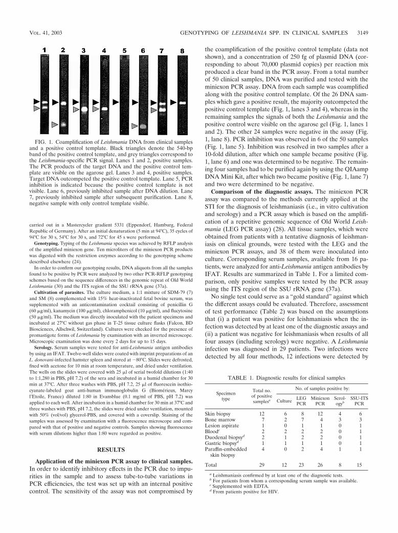
carried out in a Mastercycler gradient 5331 (Eppendorf, Hamburg, FederalRepublic of Germany). After an initial denaturation (5 min at 94°C), 35 cycles of94°C for 30 s, 54°C for 30 s, and 72°C for 45 s were performed.
Genotyping. Typing of the Leishmania species was achieved by RFLP analysisof the amplified miniexon gene. Ten microliters of the miniexon PCR productswas digested with the restriction enzymes according to the genotyping schemedescribed elsewhere (24).
In order to confirm our genotyping results, DNA aliquots from all the samplesfound to be positive by PCR were analyzed by two other PCR-RFLP genotypingschemes based on the sequence differences in the genomic repeat of Old WorldLeishmania (30) and the ITS region of the SSU rRNA gene (37a).
Cultivation of parasites. The culture medium, a 1:1 mixture of SDM-79 (7)and SM (8) complemented with 15% heat-inactivated fetal bovine serum, wassupplemented with an anticontamination cocktail consisting of penicillin G(60 �g/ml), kanamycin (100 �g/ml), chloramphenicol (10 �g/ml), and flucytosine(50 �g/ml). The medium was directly inoculated with the patient specimens andincubated at 27°C without gas phase in T-25 tissue culture flasks (Falcon, BDBiosciences, Allschwil, Switzerland). Cultures were checked for the presence ofpromastigote forms of Leishmania by examination with an inverted microscope.Microscopic examination was done every 2 days for up to 15 days.
Serology. Serum samples were tested for anti-Leishmania antigen antibodiesby using an IFAT. Twelve-well slides were coated with imprint preparations of anL. donovani-infected hamster spleen and stored at �80°C. Slides were defrosted,fixed with acetone for 10 min at room temperature, and dried under ventilation.The wells on the slides were covered with 25 �l of serial twofold dilutions (1:40to 1:1,280 in PBS, pH 7.2) of the sera and incubated in a humid chamber for 30min at 37°C. After three washes with PBS, pH 7.2, 25 �l of fluorescein isothio-cyanate-labeled goat anti-human immunoglobulin G (Biomerieux, Marcyl’Etoile, France) diluted 1:80 in Evansblue (0.1 mg/ml of PBS, pH 7.2) wasapplied to each well. After incubation in a humid chamber for 30 min at 37°C andthree washes with PBS, pH 7.2, the slides were dried under ventilation, mountedwith 50% (vol/vol) glycerol-PBS, and covered with a coverslip. Staining of thesamples was assessed by examination with a fluorescence microscope and com-pared with that of positive and negative controls. Samples showing fluorescencewith serum dilutions higher than 1:80 were regarded as positive.
RESULTS
Application of the miniexon PCR assay to clinical samples.In order to identify inhibitory effects in the PCR due to impu-rities in the sample and to assess tube-to-tube variations inPCR efficiencies, the test was set up with an internal positivecontrol. The sensitivity of the assay was not compromised by
the coamplification of the positive control template (data notshown), and a concentration of 250 fg of plasmid DNA (cor-responding to about 70,000 plasmid copies) per reaction mixproduced a clear band in the PCR assay. From a total numberof 50 clinical samples, DNA was purified and tested with theminiexon PCR assay. DNA from each sample was coamplifiedalong with the positive control template. Of the 26 DNA sam-ples which gave a positive result, the majority outcompeted thepositive control template (Fig. 1, lanes 3 and 4), whereas in theremaining samples the signals of both the Leishmania and thepositive control were visible on the agarose gel (Fig. 1, lanes 1and 2). The other 24 samples were negative in the assay (Fig.1, lane 8). PCR inhibition was observed in 6 of the 50 samples(Fig. 1, lane 5). Inhibition was resolved in two samples after a10-fold dilution, after which one sample became positive (Fig.1, lane 6) and one was determined to be negative. The remain-ing four samples had to be purified again by using the QIAampDNA Mini Kit, after which two became positive (Fig. 1, lane 7)and two were determined to be negative.
Comparison of the diagnostic assays. The miniexon PCRassay was compared to the methods currently applied at theSTI for the diagnosis of leishmaniasis (i.e., in vitro cultivationand serology) and a PCR assay which is based on the amplifi-cation of a repetitive genomic sequence of Old World Leish-mania (LEG PCR assay) (28). All tissue samples, which wereobtained from patients with a tentative diagnosis of leishman-iasis on clinical grounds, were tested with the LEG and theminiexon PCR assays, and 38 of them were inoculated intoculture. Corresponding serum samples, available from 16 pa-tients, were analyzed for anti-Leishmania antigen antibodies byIFAT. Results are summarized in Table 1. For a limited com-parison, only positive samples were tested by the PCR assayusing the ITS region of the SSU rRNA gene (37a).
No single test could serve as a “gold standard” against whichthe different assays could be evaluated. Therefore, assessmentof test performance (Table 2) was based on the assumptionsthat (i) a patient was positive for leishmaniasis when the in-fection was detected by at least one of the diagnostic assays and(ii) a patient was negative for leishmaniasis when results of allfour assays (including serology) were negative. A Leishmaniainfection was diagnosed in 29 patients. Two infections weredetected by all four methods, 12 infections were detected by
FIG. 1. Coamplification of Leishmania DNA from clinical samplesand a positive control template. Black triangles denote the 540-bpband of the positive control template, and grey triangles correspond tothe Leishmania-specific PCR signal. Lanes 1 and 2, positive samples.The PCR products of the target DNA and the positive control tem-plate are visible on the agarose gel. Lanes 3 and 4, positive samples.Target DNA outcompeted the positive control template. Lane 5, PCRinhibition is indicated because the positive control template is notvisible. Lane 6, previously inhibited sample after DNA dilution. Lane7, previously inhibited sample after subsequent purification. Lane 8,negative sample with only control template visible.
TABLE 1. Diagnostic results for clinical samples
Specimentype
Total no.of positivesamplesa
No. of samples positive by:
Culture LEGPCR
MiniexonPCR
Serol-ogyb
SSU-ITSPCR
Skin biopsy 12 6 8 12 4 6Bone marrow 7 2 7 4 3 3Lesion aspirate 1 0 1 1 0 1Bloodc 2 2 2 2 0 1Duodenal biopsyd 2 1 2 2 0 1Gastric biopsyd 1 1 1 1 0 1Paraffin-embedded
skin biopsy4 0 2 4 1 1
Total 29 12 23 26 8 15
a Leishmaniasis confirmed by at least one of the diagnostic tests.b For patients from whom a corresponding serum sample was available.c Supplemented with EDTA.d From patients positive for HIV.
VOL. 41, 2003 GENOTYPING OF LEISHMANIA SPP. IN CLINICAL SAMPLES 3149
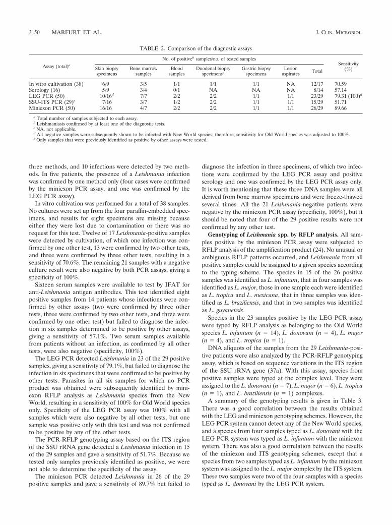
three methods, and 10 infections were detected by two meth-ods. In five patients, the presence of a Leishmania infectionwas confirmed by one method only (four cases were confirmedby the miniexon PCR assay, and one was confirmed by theLEG PCR assay).
In vitro cultivation was performed for a total of 38 samples.No cultures were set up from the four paraffin-embedded spec-imens, and results for eight specimens are missing becauseeither they were lost due to contamination or there was norequest for this test. Twelve of 17 Leishmania-positive sampleswere detected by cultivation, of which one infection was con-firmed by one other test, 13 were confirmed by two other tests,and three were confirmed by three other tests, resulting in asensitivity of 70.6%. The remaining 21 samples with a negativeculture result were also negative by both PCR assays, giving aspecificity of 100%.
Sixteen serum samples were available to test by IFAT foranti-Leishmania antigen antibodies. This test identified eightpositive samples from 14 patients whose infections were con-firmed by other assays (two were confirmed by three othertests, three were confirmed by two other tests, and three wereconfirmed by one other test) but failed to diagnose the infec-tion in six samples determined to be positive by other assays,giving a sensitivity of 57.1%. Two serum samples availablefrom patients without an infection, as confirmed by all othertests, were also negative (specificity, 100%).
The LEG PCR detected Leishmania in 23 of the 29 positivesamples, giving a sensitivity of 79.1%, but failed to diagnose theinfection in six specimens that were confirmed to be positive byother tests. Parasites in all six samples for which no PCRproduct was obtained were subsequently identified by mini-exon RFLP analysis as Leishmania species from the NewWorld, resulting in a sensitivity of 100% for Old World speciesonly. Specificity of the LEG PCR assay was 100% with allsamples which were also negative by all other tests, but onesample was positive only with this test and was not confirmedto be positive by any of the other tests.
The PCR-RFLP genotyping assay based on the ITS regionof the SSU rRNA gene detected a Leishmania infection in 15of the 29 samples and gave a sensitivity of 51.7%. Because wetested only samples previously identified as positive, we werenot able to determine the specificity of the assay.
The miniexon PCR detected Leishmania in 26 of the 29positive samples and gave a sensitivity of 89.7% but failed to
diagnose the infection in three specimens, of which two infec-tions were confirmed by the LEG PCR assay and positiveserology and one was confirmed by the LEG PCR assay only.It is worth mentioning that these three DNA samples were allderived from bone marrow specimens and were freeze-thawedseveral times. All the 21 Leishmania-negative patients werenegative by the miniexon PCR assay (specificity, 100%), but itshould be noted that four of the 29 positive results were notconfirmed by any other test.
Genotyping of Leishmania spp. by RFLP analysis. All sam-ples positive by the miniexon PCR assay were subjected toRFLP analysis of the amplification product (24). No unusual orambiguous RFLP patterns occurred, and Leishmania from allpositive samples could be assigned to a given species accordingto the typing scheme. The species in 15 of the 26 positivesamples was identified as L. infantum, that in four samples wasidentified as L. major, those in one sample each were identifiedas L. tropica and L. mexicana, that in three samples was iden-tified as L. braziliensis, and that in two samples was identifiedas L. guyanensis.
Species in the 23 samples positive by the LEG PCR assaywere typed by RFLP analysis as belonging to the Old Worldspecies L. infantum (n � 14), L. donovani (n � 4), L. major(n � 4), and L. tropica (n � 1).
DNA aliquots of the samples from the 29 Leishmania-posi-tive patients were also analyzed by the PCR-RFLP genotypingassay, which is based on sequence variations in the ITS regionof the SSU rRNA gene (37a). With this assay, species frompositive samples were typed at the complex level. They wereassigned to the L. donovani (n � 7), L. major (n � 6), L. tropica(n � 1), and L. braziliensis (n � 1) complexes.
A summary of the genotyping results is given in Table 3.There was a good correlation between the results obtainedwith the LEG and miniexon genotyping schemes. However, theLEG PCR system cannot detect any of the New World species,and a species from four samples typed as L. donovani with theLEG PCR system was typed as L. infantum with the miniexonsystem. There was also a good correlation between the resultsof the miniexon and ITS genotyping schemes, except that aspecies from two samples typed as L. infantum by the miniexonsystem was assigned to the L. major complex by the ITS system.These two samples were two of the four samples with a speciestyped as L. donovani by the LEG PCR system.
TABLE 2. Comparison of the diagnostic assays
Assay (total)a
No. of positiveb samples/no. of tested samplesSensitivity
(%)Skin biopsyspecimens
Bone marrowsamples
Bloodsamples
Duodenal biopsyspecimensc
Gastric biopsyspecimens
Lesionaspirates Total
In vitro cultivation (38) 6/9 3/5 1/1 1/1 1/1 NA 12/17 70.59Serology (16) 5/9 3/4 0/1 NA NA NA 8/14 57.14LEG PCR (50) 10/16d 7/7 2/2 2/2 1/1 1/1 23/29 79.31 (100)d
SSU-ITS PCR (29)e 7/16 3/7 1/2 2/2 1/1 1/1 15/29 51.71Miniexon PCR (50) 16/16 4/7 2/2 2/2 1/1 1/1 26/29 89.66
a Total number of samples subjected to each assay.b Leishmaniasis confirmed by at least one of the diagnostic tests.c NA, not applicable.d All negative samples were subsequently shown to be infected with New World species; therefore, sensitivity for Old World species was adjusted to 100%.e Only samples that were previously identified as positive by other assays were tested.
3150 MARFURT ET AL. J. CLIN. MICROBIOL.
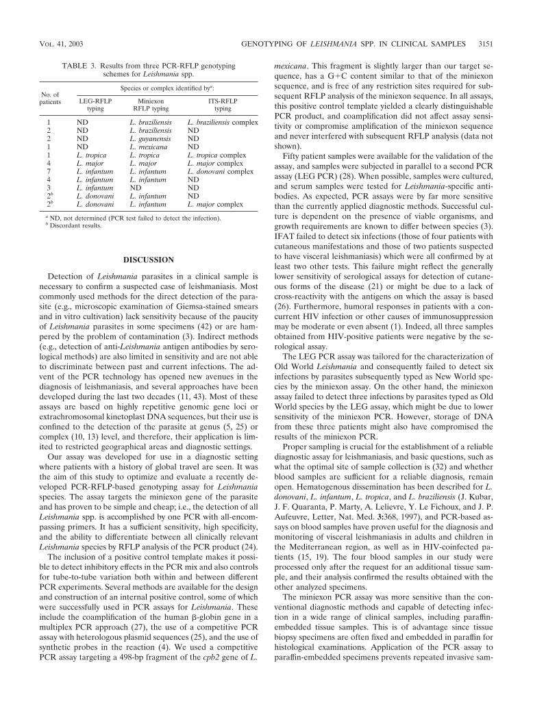
DISCUSSION
Detection of Leishmania parasites in a clinical sample isnecessary to confirm a suspected case of leishmaniasis. Mostcommonly used methods for the direct detection of the para-site (e.g., microscopic examination of Giemsa-stained smearsand in vitro cultivation) lack sensitivity because of the paucityof Leishmania parasites in some specimens (42) or are ham-pered by the problem of contamination (3). Indirect methods(e.g., detection of anti-Leishmania antigen antibodies by sero-logical methods) are also limited in sensitivity and are not ableto discriminate between past and current infections. The ad-vent of the PCR technology has opened new avenues in thediagnosis of leishmaniasis, and several approaches have beendeveloped during the last two decades (11, 43). Most of theseassays are based on highly repetitive genomic gene loci orextrachromosomal kinetoplast DNA sequences, but their use isconfined to the detection of the parasite at genus (5, 25) orcomplex (10, 13) level, and therefore, their application is lim-ited to restricted geographical areas and diagnostic settings.
Our assay was developed for use in a diagnostic settingwhere patients with a history of global travel are seen. It wasthe aim of this study to optimize and evaluate a recently de-veloped PCR-RFLP-based genotyping assay for Leishmaniaspecies. The assay targets the miniexon gene of the parasiteand has proven to be simple and cheap; i.e., the detection of allLeishmania spp. is accomplished by one PCR with all-encom-passing primers. It has a sufficient sensitivity, high specificity,and the ability to differentiate between all clinically relevantLeishmania species by RFLP analysis of the PCR product (24).
The inclusion of a positive control template makes it possi-ble to detect inhibitory effects in the PCR mix and also controlsfor tube-to-tube variation both within and between differentPCR experiments. Several methods are available for the designand construction of an internal positive control, some of whichwere successfully used in PCR assays for Leishmania. Theseinclude the coamplification of the human �-globin gene in amultiplex PCR approach (27), the use of a competitive PCRassay with heterologous plasmid sequences (25), and the use ofsynthetic probes in the reaction (4). We used a competitivePCR assay targeting a 498-bp fragment of the cpb2 gene of L.
mexicana. This fragment is slightly larger than our target se-quence, has a G�C content similar to that of the miniexonsequence, and is free of any restriction sites required for sub-sequent RFLP analysis of the miniexon sequence. In all assays,this positive control template yielded a clearly distinguishablePCR product, and coamplification did not affect assay sensi-tivity or compromise amplification of the miniexon sequenceand never interfered with subsequent RFLP analysis (data notshown).
Fifty patient samples were available for the validation of theassay, and samples were subjected in parallel to a second PCRassay (LEG PCR) (28). When possible, samples were cultured,and serum samples were tested for Leishmania-specific anti-bodies. As expected, PCR assays were by far more sensitivethan the currently applied diagnostic methods. Successful cul-ture is dependent on the presence of viable organisms, andgrowth requirements are known to differ between species (3).IFAT failed to detect six infections (those of four patients withcutaneous manifestations and those of two patients suspectedto have visceral leishmaniasis) which were all confirmed by atleast two other tests. This failure might reflect the generallylower sensitivity of serological assays for detection of cutane-ous forms of the disease (21) or might be due to a lack ofcross-reactivity with the antigens on which the assay is based(26). Furthermore, humoral responses in patients with a con-current HIV infection or other causes of immunosuppressionmay be moderate or even absent (1). Indeed, all three samplesobtained from HIV-positive patients were negative by the se-rological assay.
The LEG PCR assay was tailored for the characterization ofOld World Leishmania and consequently failed to detect sixinfections by parasites subsequently typed as New World spe-cies by the miniexon assay. On the other hand, the miniexonassay failed to detect three infections by parasites typed as OldWorld species by the LEG assay, which might be due to lowersensitivity of the miniexon PCR. However, storage of DNAfrom these three patients might also have compromised theresults of the miniexon PCR.
Proper sampling is crucial for the establishment of a reliablediagnostic assay for leishmaniasis, and basic questions, such aswhat the optimal site of sample collection is (32) and whetherblood samples are sufficient for a reliable diagnosis, remainopen. Hematogenous dissemination has been described for L.donovani, L. infantum, L. tropica, and L. braziliensis (J. Kubar,J. F. Quaranta, P. Marty, A. Lelievre, Y. Le Fichoux, and J. P.Aufeuvre, Letter, Nat. Med. 3:368, 1997), and PCR-based as-says on blood samples have proven useful for the diagnosis andmonitoring of visceral leishmaniasis in adults and children inthe Mediterranean region, as well as in HIV-coinfected pa-tients (15, 19). The four blood samples in our study wereprocessed only after the request for an additional tissue sam-ple, and their analysis confirmed the results obtained with theother analyzed specimens.
The miniexon PCR assay was more sensitive than the con-ventional diagnostic methods and capable of detecting infec-tion in a wide range of clinical samples, including paraffin-embedded tissue samples. This is of advantage since tissuebiopsy specimens are often fixed and embedded in paraffin forhistological examinations. Application of the PCR assay toparaffin-embedded specimens prevents repeated invasive sam-
TABLE 3. Results from three PCR-RFLP genotypingschemes for Leishmania spp.
No. ofpatients
Species or complex identified bya:
LEG-RFLPtyping
MiniexonRFLP typing
ITS-RFLPtyping
1 ND L. braziliensis L. braziliensis complex2 ND L. braziliensis ND2 ND L. guyanensis ND1 ND L. mexicana ND1 L. tropica L. tropica L. tropica complex4 L. major L. major L. major complex7 L. infantum L. infantum L. donovani complex4 L. infantum L. infantum ND3 L. infantum ND ND2b L. donovani L. infantum ND2b L. donovani L. infantum L. major complex
a ND, not determined (PCR test failed to detect the infection).b Discordant results.
VOL. 41, 2003 GENOTYPING OF LEISHMANIA SPP. IN CLINICAL SAMPLES 3151
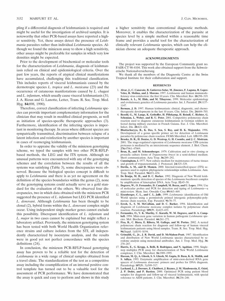
pling if a differential diagnosis of leishmaniasis is required andmight be useful for the investigation of archived samples. It isnoteworthy that other PCR-based assays have reported a high-er sensitivity. Yet, these assays detect the presence of Leish-mania parasites rather than individual Leishmania species. Al-though we found the miniexon assay to show a high sensitivity,other assays might be preferable for samples in which very lowdensities might be expected.
Prior to the development of biochemical or molecular toolsfor the characterization of Leishmania, diagnosis of leishman-iasis relied on clinical and epidemiological criteria. Over thepast few years, the reports of atypical clinical manifestationshave accumulated, challenging this traditional classification.This includes reports of visceral leishmaniasis caused by thedermotropic species L. tropica and L. mexicana (23) and theoccurrence of cutaneous manifestations caused by L. chagasiand L. infantum, which usually cause visceral leishmaniasis (31;J. A. Rioux and G. Lanotte, Letter, Trans. R. Soc. Trop. Med.Hyg. 84:898, 1990).
Therefore, correct classification of infecting Leishmania spe-cies can provide important complementary information for theclinician that may result in modified clinical prognosis, as wellas initiation of species-specific therapeutic approaches (3).Furthermore, identification of the infecting species is impor-tant in monitoring therapy. In areas where different species aresympatrically transmitted, discrimination between relapse of alatent infection and reinfection by another species is importantin cases of reemerging leishmaniasis.
In order to appraise the validity of the miniexon genotypingscheme, we typed the samples with two other PCR-RFLP-based methods, the LEG and the ITS system. Although nounusual patterns were encountered with any of the genotypingschemes and the correlation between the results of all thesystems was satisfying (Table 3), some discrepancies were ob-served. Because the biological species concept is difficult toapply to Leishmania and there is as yet no agreement on thedefinition of the species boundaries within the genus (2), noneof the genotyping systems could actually serve as a gold stan-dard for the evaluation of the others. We observed four dis-crepancies, two in which data obtained with the miniexon PCRsuggested the presence of L. infantum but LEG PCR identifiedL. donovani. Although Leishmania has been thought to beclonal (2), hybrid forms within the L. donovani complex mightoccur. Using independent single marker genes cannot excludethis possibility. Discrepant identification of L. infantum andL. major in two cases cannot be explained but might reflect alaboratory artifact. Previously, the miniexon genotyping systemhas been tested with both World Health Organization refer-ence strains and culture isolates from the STI, all indepen-dently characterized by isoenzyme analysis, and the resultswere in good yet not perfect concordance with the speciesdefinitions (24).
In conclusion, the miniexon PCR-RFLP-based genotypingassay has proven to be a reliable test for the detection ofLeishmania in a wide range of clinical samples obtained froma travel clinic. The standardization of the test as a competitiveassay including the coamplification of an internal positive con-trol template has turned out to be a valuable tool for theassessment of PCR performance. We have demonstrated thatthe assay is quick and easy to perform and shows in this study
a higher sensitivity than conventional diagnostic methods.Moreover, it enables the characterization of the parasite atspecies level by a simple method within a reasonable timeframe and provides a useful tool for the characterization ofclinically relevant Leishmania species, which can help the cli-nician choose an adequate therapeutic approach.
ACKNOWLEDGMENTS
The project was supported by the European Community grant no.FAIR-CT 98-4104. This work also obtained support from the Schweiz-erische Nationalversicherung.
We thank all the members of the Diagnostic Centre at the SwissTropical Institute for their collaboration and support.
REFERENCES
1. Alvar, J., C. Canavate, B. Gutierrez-Solar, M. Jimenez, F. Laguna, R. Lopez-Velez, R. Molina, and J. Moreno. 1997. Leishmania and human immunode-ficiency virus coinfection: the first 10 years. Clin. Microbiol. Rev. 10:298–319.
2. Banuls, A. L., M. Hide, and M. Tibayrenc. 1999. Molecular epidemiologyand evolutionary genetics of Leishmania parasites. Int. J. Parasitol. 29:1137–1147.
3. Berman, J. D. 1997. Human leishmaniasis: clinical, diagnostic, and chemo-therapeutic developments in the last 10 years. Clin. Infect. Dis. 24:684–703.
4. Bezold, G., M. Lange, K. Gethoffer, H. Pillekamp, H. Reindl, C. Richter, G.Schonian, L. Weber, and R. U. Peter. 2001. Competitive polymerase chainreaction used to diagnose cutaneous leishmaniasis in German soldiers in-fected during military exercises in French Guiana. Eur. J. Clin. Microbiol.Infect. Dis. 20:421–424.
5. Bhattacharyya, R., K. Das, S. Sen, S. Roy, and H. K. Majumder. 1996.Development of a genus specific primer set for detection of Leishmaniaparasites by polymerase chain reaction. FEMS Microbiol. Lett. 135:195–200.
6. Brooks, D. R., H. Denise, G. D. Westrop, G. H. Coombs, and J. C. Mottram.2001. The stage-regulated expression of Leishmania mexicana CPB cysteineproteases is mediated by an intercistronic sequence element. J. Biol. Chem.276:47061–47069.
7. Brun, R., and M. Schonenberger. 1979. Cultivation and in vitro cloning orprocyclic culture forms of Trypanosoma brucei in a semi-defined medium.Short communication. Acta Trop. 36:289–292.
8. Cunningham, I. 1977. New culture medium for maintenance of tsetse tissuesand growth of trypanosomatids. J. Protozool. 24:325–329.
9. Davila, A. M., and H. Momen. 2000. Internal-transcribed-spacer (ITS) se-quences used to explore phylogenetic relationships within Leishmania. Ann.Trop. Med. Parasitol. 94:651–654.
10. De Bruijn, M. H., and D. C. Barker. 1992. Diagnosis of New World leish-maniasis: specific detection of species of the Leishmania braziliensis complexby amplification of kinetoplast DNA. Acta Trop. 52:45–58.
11. Degrave, W., O. Fernandes, D. Campbell, M. Bozza, and U. Lopes. 1994. Useof molecular probes and PCR for detection and typing of Leishmania—amini-review. Mem. Inst. Oswaldo Cruz 89:463–469.
12. Eisenberger, C. L., and C. L. Jaffe. 1999. Leishmania: identification of OldWorld species using a permissively primed intergenic polymorphic-poly-merase chain reaction. Exp. Parasitol. 91:70–77.
13. Eresh, S., S. M. McCallum, and D. C. Barker. 1994. Identification anddiagnosis of Leishmania mexicana complex isolates by polymerase chainreaction. Parasitology 109(Pt. 4):423–433.
14. Fernandes, O., V. K. Murthy, U. Kurath, W. M. Degrave, and D. A. Camp-bell. 1994. Mini-exon gene variation in human pathogenic Leishmania spe-cies. Mol. Biochem. Parasitol. 66:261–271.
15. Fisa, R., C. Riera, E. Ribera, M. Gallego, and M. Portus. 2002. A nestedpolymerase chain reaction for diagnosis and follow-up of human visceralleishmaniasis patients using blood samples. Trans. R. Soc. Trop. Med. Hyg.96(Suppl. 1):S191–S194.
16. Grimaldi, G., Jr., J. R. David, and D. McMahon-Pratt. 1987. Identificationand distribution of New World Leishmania species characterized by se-rodeme analysis using monoclonal antibodies. Am. J. Trop. Med. Hyg. 36:270–287.
17. Harris, E., G. Kropp, A. Belli, B. Rodriguez, and N. Agabian. 1998. Single-step multiplex PCR assay for characterization of New World Leishmaniacomplexes. J. Clin. Microbiol. 36:1989–1995.
18. Hassan, M. Q., A. Ghosh, S. S. Ghosh, M. Gupta, D. Basu, K. K. Mallik, andS. Adhya. 1993. Enzymatic amplification of mini-exon-derived RNA genespacers of Leishmania donovani: primers and probes for DNA diagnosis.Parasitology 107(Pt. 5):509–517.
19. Lachaud, L., J. Dereure, E. Chabbert, J. Reynes, J. M. Mauboussin, E. Oziol,J. P. Dedet, and P. Bastien. 2000. Optimized PCR using patient bloodsamples for diagnosis and follow-up of visceral leishmaniasis, with specialreference to AIDS patients. J. Clin. Microbiol. 38:236–240.
3152 MARFURT ET AL. J. CLIN. MICROBIOL.
20. Lainson, R., and J. J. Shaw. 1987. Evolution, classification and geographicaldistribution, p. 1–120. In W. Peters and R. Killick-Kendrick (ed.), Theleishmaniases in biology and medicine. Academic Press, Orlando, Fla.
21. Liew, F. Y., and C. A. O’Donnell. 1993. Immunology of leishmaniasis. Adv.Parasitol. 32:161–181.
22. Luis, L., A. Ramirez, C. M. Aguilar, S. Eresh, D. C. Barker, and A. Mendoza-Leon. 1998. The genomic fingerprinting of the coding region of the beta-tubulin gene in Leishmania identification. Acta Trop. 69:193–204.
23. Magill, A. J., M. Grogl, R. A. Gasser, Jr., W. Sun, and C. N. Oster. 1993.Visceral infection caused by Leishmania tropica in veterans of OperationDesert Storm. N. Engl. J. Med. 328:1383–1387.
24. Marfurt, J., I. Niederwieser, D. Makia, H. P. Beck, and I. Felger. 2003.Diagnostic genotyping of Old and New World Leishmania species by PCR-RFLP. Diagn. Microbiol. Infect. Dis. 46:115–124.
25. Mathis, A., and P. Deplazes. 1995. PCR and in vitro cultivation for detectionof Leishmania spp. in diagnostic samples from humans and dogs. J. Clin.Microbiol. 33:1145–1149.
26. Medrano, F. J., C. Canavate, M. Leal, C. Rey, E. Lissen, and J. Alvar. 1998.The role of serology in the diagnosis and prognosis of visceral leishmaniasisin patients coinfected with human immunodeficiency virus type-1. Am. J.Trop. Med. Hyg. 59:155–162.
27. Meredith, S. E., E. E. Zijlstra, G. J. Schoone, C. C. Kroon, G. J. Van Eys,K. U. Schaeffer, A. M. el Hassan, and P. G. Lawyer. 1993. Development andapplication of the polymerase chain reaction for the detection and identifi-cation of Leishmania parasites in clinical material. Arch. Inst. Pasteur Tunis70:419–431.
28. Minodier, P., R. Piarroux, F. Gambarelli, C. Joblet, and H. Dumon. 1997.Rapid identification of causative species in patients with Old World leish-maniasis. J. Clin. Microbiol. 35:2551–2555.
29. Pearson, R. D., A. De Queiroz Sousa, and S. M. B. Jeronimo. 2001. Leish-mania species: visceral (kala-azar), cutaneous and mucosal leishmaniasis, p.2831–2845. In G. L. Mandell, J. E. Bennett, and R. Dolin (ed.), Principlesand practice of infectious diseases. Churchill Livingstone, New York, N.Y.
30. Piarroux, R., M. Fontes, R. Perasso, F. Gambarelli, C. Joblet, H. Dumon,and M. Quilici. 1995. Phylogenetic relationships between Old World Leish-mania strains revealed by analysis of a repetitive DNA sequence. Mol.Biochem. Parasitol. 73:249–252.
31. Ponce, C., E. Ponce, A. Morrison, A. Cruz, R. Kreutzer, D. McMahon-Pratt,and F. Neva. 1991. Leishmania donovani chagasi: new clinical variant ofcutaneous leishmaniasis in Honduras. Lancet 337:67–70.
32. Ramirez, J. R., S. Agudelo, C. Muskus, J. F. Alzate, C. Berberich, D. Barker,and I. D. Velez. 2000. Diagnosis of cutaneous leishmaniasis in Colombia: thesampling site within lesions influences the sensitivity of parasitologic diag-nosis. J. Clin. Microbiol. 38:3768–3773.
33. Ramos, A., D. A. Maslov, O. Fernandes, D. A. Campbell, and L. Simpson.1996. Detection and identification of human pathogenic Leishmania andTrypanosoma species by hybridization of PCR-amplified mini-exon repeats.Exp. Parasitol. 82:242–250.
34. Rodgers, M. R., S. J. Popper, and D. F. Wirth. 1990. Amplification ofkinetoplast DNA as a tool in the detection and diagnosis of Leishmania. Exp.Parasitol. 71:267–275.
35. Rodriguez, N., B. Guzman, A. Rodas, H. Takiff, B. R. Bloom, and J. Convit.1994. Diagnosis of cutaneous leishmaniasis and species discrimination ofparasites by PCR and hybridization. J. Clin. Microbiol. 32:2246–2252.
36. Rossi, V., P. Wincker, C. Ravel, C. Blaineau, M. Pages, and P. Bastien. 1994.Structural organisation of microsatellite families in the Leishmania genomeand polymorphisms at two (CA)n loci. Mol. Biochem. Parasitol. 65:271–282.
37. Russell, R., M. P. Iribar, B. Lambson, S. Brewster, J. M. Blackwell, C. Dye,and J. W. Ajioka. 1999. Intra and inter-specific microsatellite variation in theLeishmania subgenus Viannia. Mol. Biochem. Parasitol. 103:71–77.
37a.Schonian, G., A. Nasereddin, N. Dinse, C. Schweynoch, D. F. H. Schallig, W.Presber, and C. L. Jaffe. A PCR method for the diagnosis and characteriza-tion of Leishmania in local and imported clinical samples. Diagn. Microbiol.Infect. Dis., in press.
38. Schonian, G., C. Schweynoch, K. Zlateva, L. Oskam, N. Kroon, Y. Graser,and W. Presber. 1996. Identification and determination of the relationshipsof species and strains within the genus Leishmania using single primers in thepolymerase chain reaction. Mol. Biochem. Parasitol. 77:19–29.
39. Ullu, E., C. Tschudi, and A. Gunzl. 1996. Trans-splicing in trypanosomatidprotozoa, p. 115–133. In D. F. Smith and M. Parsons (ed.), Molecular biologyof parasitic protozoa. IRL Press at Oxford University Press, Oxford, En-gland.
40. Van Eys, G. J., G. J. Schoone, N. C. Kroon, and S. B. Ebeling. 1992.Sequence analysis of small subunit ribosomal RNA genes and its use fordetection and identification of Leishmania parasites. Mol. Biochem. Parasi-tol. 51:133–142.
41. Victoir, K., A. L. Banuls, J. Arevalo, A. Llanos-Cuentas, R. Hamers, S. Noel,S. De Doncker, D. Le Ray, M. Tibayrenc, and J. C. Dujardin. 1998. The gp63gene locus, a target for genetic characterization of Leishmania belonging tosubgenus Viannia. Parasitology 117(Pt. 1):1–13.
42. Weigle, K. A., M. de Davalos, P. Heredia, R. Molineros, N. G. Saravia, andA. D’Alessandro. 1987. Diagnosis of cutaneous and mucocutaneous leish-maniasis in Colombia: a comparison of seven methods. Am. J. Trop. Med.Hyg. 36:489–496.
43. Wilson, S. M. 1995. DNA-based methods in the detection of Leishmaniaparasites: field applications and practicalities. Ann. Trop. Med. Parasitol.89(Suppl. 1):95–100.
VOL. 41, 2003 GENOTYPING OF LEISHMANIA SPP. IN CLINICAL SAMPLES 3153
3.4. Discussion Sexual reproduction has not yet been shown in Leishmania, so the term ‘species’ might not be correctly used for all known strains. As example, L. infantum and L. chagasi, as well as L. major and L. venezuelensis are believed to be genetically identical, only the geographic distribution differs. Therefore, L. chagasi and L. venezuelensis most probably derived from dogs infected by L. infantum and L. major respectively, which had been imported during settlement of the Americas by Europeans. Sequencing of the mini-exon genes supports this hypothesis. Furthermore, the mini-exon genes of L. colombiensis and L. naiffi showed high homology. Although the mini-exon gene is present in kinetoplatids, nematodes and even chordates, the specifity of the PCR for Leishmania is given, since DNA of Trypanosoma and nematodes lacks proper binding sites of our primers. However, PCR using DNA of Chritidia (parasitic kinetoplastids, living in insects) as template yielded an amplification of the mini-exon gene but the resulting restriction patterns of the PCR products were completely different from Leishmania patterns. The mini-exon PCR proved to be a great tool for confirming an suspected Leishmania infection and distinguishing the infecting species at the same time. However, it is important to note that when diagnosing Viannia species a single point mutation within the restriction site can change the interpretation of the results. As L. brasiliensis and L. guyanensis differ in the required treatment, but their mini-exon genes differ solely by a few point mutations, another genetic marker should be used for these species. However, the mini-exon PCR was not designed as solely tool used for diagnosis, but as an additional method to confirm interpreting of the symptoms and classical diagnostic tools, such as cultivation of the parasites.
28
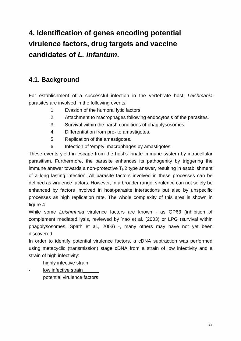
4. Identification of genes encoding potential virulence factors, drug targets and vaccine candidates of L. infantum.
4.1. Background For establishment of a successful infection in the vertebrate host, Leishmania parasites are involved in the following events:
1. Evasion of the humoral lytic factors. 2. Attachment to macrophages following endocytosis of the parasites. 3. Survival within the harsh conditions of phagolysosomes. 4. Differentiation from pro- to amastigotes. 5. Replication of the amastigotes. 6. Infection of ‘empty’ macrophages by amastigotes.
These events yield in escape from the host’s innate immune system by intracellular parasitism. Furthermore, the parasite enhances its pathogenity by triggering the immune answer towards a non-protective TH2 type answer, resulting in establishment of a long lasting infection. All parasite factors involved in these processes can be defined as virulence factors. However, in a broader range, virulence can not solely be enhanced by factors involved in host-parasite interactions but also by unspecific processes as high replication rate. The whole complexity of this area is shown in figure 4. While some Leishmania virulence factors are known - as GP63 (inhibition of complement mediated lysis, reviewed by Yao et al. (2003) or LPG (survival within phagolysosomes, Spath et al., 2003) -, many others may have not yet been discovered. In order to identify potential virulence factors, a cDNA subtraction was performed using metacyclic (transmission) stage cDNA from a strain of low infectivity and a strain of high infectivity: highly infective strain - low infective strain______ potential virulence factors
29
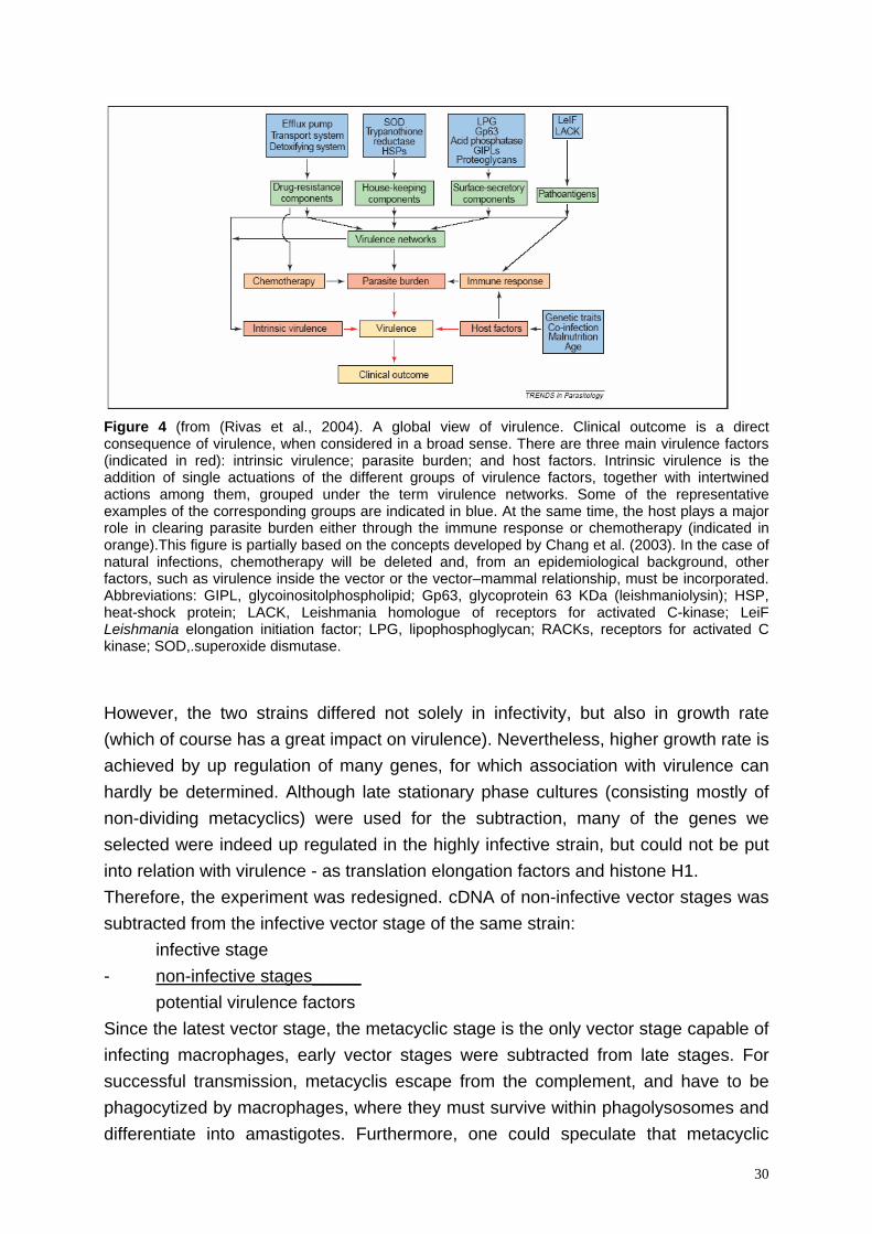
Figure 4 (from (Rivas et al., 2004). A global view of virulence. Clinical outcome is a direct consequence of virulence, when considered in a broad sense. There are three main virulence factors (indicated in red): intrinsic virulence; parasite burden; and host factors. Intrinsic virulence is the addition of single actuations of the different groups of virulence factors, together with intertwined actions among them, grouped under the term virulence networks. Some of the representative examples of the corresponding groups are indicated in blue. At the same time, the host plays a major role in clearing parasite burden either through the immune response or chemotherapy (indicated in orange).This figure is partially based on the concepts developed by Chang et al. (2003). In the case of natural infections, chemotherapy will be deleted and, from an epidemiological background, other factors, such as virulence inside the vector or the vector–mammal relationship, must be incorporated. Abbreviations: GIPL, glycoinositolphospholipid; Gp63, glycoprotein 63 KDa (leishmaniolysin); HSP, heat-shock protein; LACK, Leishmania homologue of receptors for activated C-kinase; LeiF Leishmania elongation initiation factor; LPG, lipophosphoglycan; RACKs, receptors for activated C kinase; SOD,.superoxide dismutase.
However, the two strains differed not solely in infectivity, but also in growth rate (which of course has a great impact on virulence). Nevertheless, higher growth rate is achieved by up regulation of many genes, for which association with virulence can hardly be determined. Although late stationary phase cultures (consisting mostly of non-dividing metacyclics) were used for the subtraction, many of the genes we selected were indeed up regulated in the highly infective strain, but could not be put into relation with virulence - as translation elongation factors and histone H1. Therefore, the experiment was redesigned. cDNA of non-infective vector stages was subtracted from the infective vector stage of the same strain: infective stage - non-infective stages_____ potential virulence factors Since the latest vector stage, the metacyclic stage is the only vector stage capable of infecting macrophages, early vector stages were subtracted from late stages. For successful transmission, metacyclis escape from the complement, and have to be phagocytized by macrophages, where they must survive within phagolysosomes and differentiate into amastigotes. Furthermore, one could speculate that metacyclic
30
parasites are also involved in triggering the host’s immune answer towards a non-protective direction, since metacyclics are the first parasites seen by the immune system. Consequently, many virulence factors can be assumed to be expressed within this parasite stage. For the same reason, transmission blocking vaccine candidates might be identified, if putative membrane proteins expressed at metacyclic’s surface would be discovered (anti-bodies against a flagellum specific epitope were already shown to inhibit adherence of promastigotes to phagocytes, Singla and Vinayak, 1994). Furthermore, identification of proteins of metacyclic parasites which show no homologies to mammal proteins might be regarded as potential drug targets for both, prophylaxis and treatment (for the latter purpose only when these proteins are also expressed in amastigotes).
4.2. Methods We used suppression subtractive hybridization (SSH, Diatchenko et al., 1996) as described below. Since most of the mRNAs in Leishmania contain a 5’ conserved sequence, two simplifications of the SSH method were tested. SSH Using this method (figures 5 and 6A), ds cDNA was the starting material for both, tester and driver (the driver is subtracted from the tester). The ds cDNA was cut with a suitable restriction enzyme leading to fragment sizes of 100 to 500 bases. In contrast to the driver, half of the tester was ligated to adaptor1, the remaining tester to adaptor2. In two hybridization steps, pre-subtracted tester1 and 2 were finally combined. This led to formation of a hybrid of tester 1 and 2. Prior to PCR, the ends were filled in. For selection of the tester hybrid, a suppression PCR was performed using adaptor primers. Since the 5’ ends of both adaptors consisted of a palindromic sequence, DNA strands containing the same adaptors at both ends could form a loop and thus were not efficiently amplified (suppression PCR). In contrast, the tester hybrid containing 2 different adaptors at both ends was amplified exponentially and could be subsequently ligated into a cloning vector. Modification1 In this method (figures 5 and 6B), tester and driver were obtained by generating cDNA using the spliced leader sequence. Tester 1 was amplified using a short spliced leader primer and RT-primer1. For Tester 2, a long spliced leader primer and RT-primer2 was used, and amplification of the driver was done using the short
31
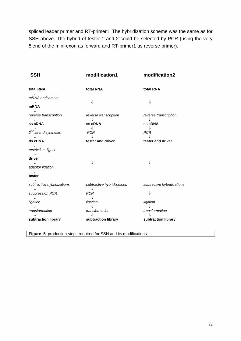
spliced leader primer and RT-primer1. The hybridization scheme was the same as for SSH above. The hybrid of tester 1 and 2 could be selected by PCR (using the very 5’end of the mini-exon as forward and RT-primer1 as reverse primer). SSH modification1 modification2 total RNA total RNA total RNA ↓ mRNA enrichment ↓ ↓ ↓ mRNA ↓ reverse transcription reverse transcription reverse transcription ↓ ↓ ↓ ss cDNA ss cDNA ss cDNA ↓ ↓ ↓ 2nd strand synthesis PCR PCR ↓ ↓ ↓ ds cDNA tester and driver tester and driver ↓ restriction digest ↓ driver ↓ ↓ ↓ adaptor ligation ↓ tester ↓ subtractive hybridizations subtractive hybridizations subtractive hybridizations ↓ ↓ suppression PCR PCR ↓ ↓ ↓ ligation ligation ligation ↓ ↓ ↓ transformation transformation transformation ↓ ↓ ↓ subtraction library subtraction library subtraction library Figure 5: production steps required for SSH and its modifications.
32
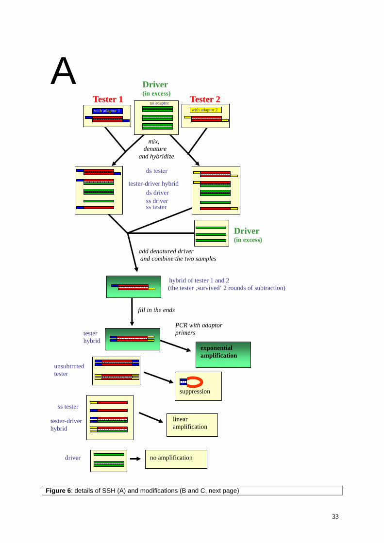
A Driver(in excess)
Tester 1 Tester 2
Figure 6: details of SSH (A) and modifications (B and C, next page)
hybrid of tester 1 and 2 (the tester ‚survived‘ 2 rounds of subtraction)
fill in the ends
PCR with adaptor primers
suppression
linear amplification
mix, denature and hybridize
ds tester
tester-driver hybridds driverss driverss tester
no adaptorwith adaptor 2with adaptor 1
Driver(in excess)
add denatured driver and combine the two samples
tester hybrid
no amplification
exponential amplification
unsubtrcted tester
ss tester
tester-driver hybrid
driver
33
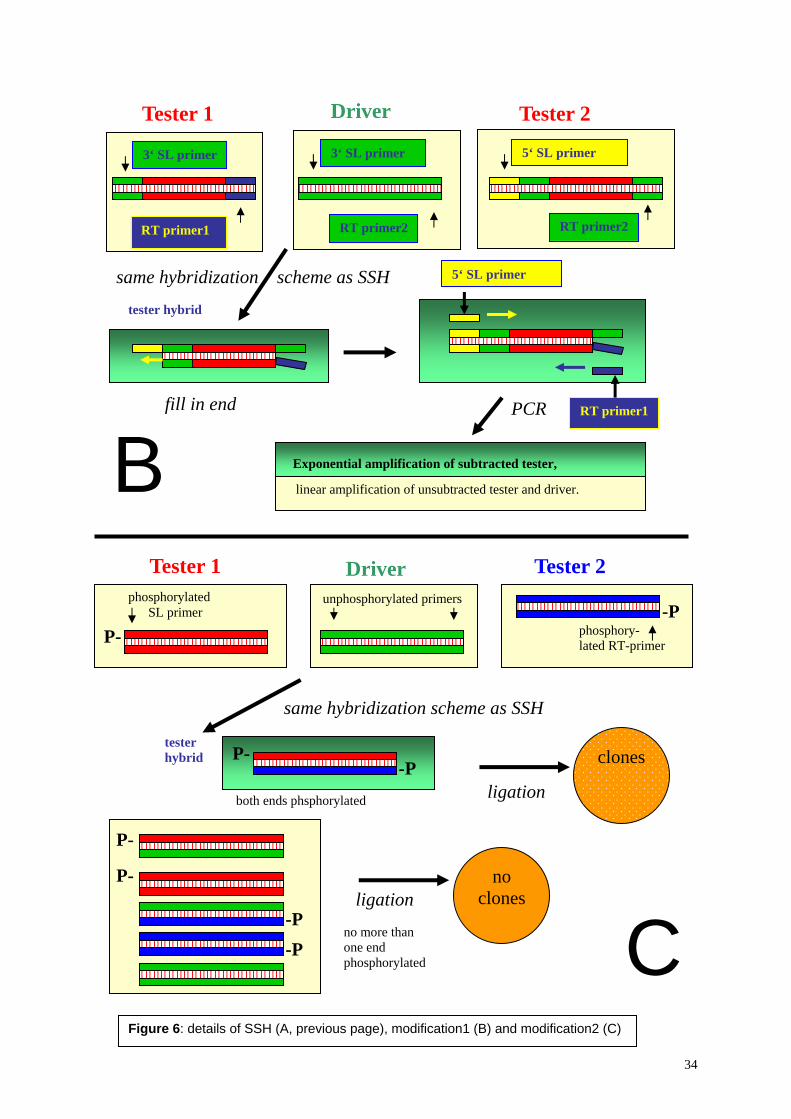
DriverTester 1 Tester 2
same hybridization scheme as SSH
fill in end
tester hybrid
PCR
linear amplification of unsubtracted tester and driver.
Tester 1 Driver Tester 2
Exponential amplification of subtracted tester,B
5‘ SL primer
RT primer1
3‘ SL primer
RT primer1
3‘ SL primer 5‘ SL primer
RT primer2 RT primer2
phosphorylated unphosphorylated primers-P SL primer
phosphory-
P-
P-
P-
-P -P
no more than one end phosphorylated
ligation
tester hybrid
both ends phsphorylated
clones
ligationno
clones
P- -P
C
lated RT-primer
same hybridization scheme as SSH
Figure 6: details of SSH (A, previous page), modification1 (B) and modification2 (C)
34
Again, tester and driver were obtained using spliced leader RT-PCR (figures 5 and 6C). For tester1, the spliced leader primer but not the RT-primer was phophorylated, in contrast to tester2, where the RT-primer but not the spliced leader primer was phosphorylated. The driver was amplified using non-phosphorylated primers. Because the hybrid of tester1 and 2 was the only ds DNA in the hybridizytion mix which was phosphorylated at both ends, it could be selected by ligation. It could either be directly ligated into a blunt end cut and dephosphorylated vector, or ligated to non-phosphorylated adaptors (using subsequently PCR with adaptor primers as method for selection of the subtracted tester). The advantages of both modifications would be:
• total RNA (not only mRNA) could be used as starting material • PCR instead of second strand synthesis
subtraction using limited amounts of RNA possible • no restriction digest of tester and driver
subtraction library would contain full length cDNA resulting in simplification of gene identification.
Furthermore, selecting of different fragments belonging to the same gene would be avoided.
• no adaptor ligation prior to subtraction. Blunt end ligation, as used in SSH, usually lacks high efficiency.
However, changes in hybridization conditions (DNA concentration, buffer, temperature) yielded in completely different band patterns after the final PCR using the modified subtraction methods. Since too many optimization steps would have been necessary to get to work both modifications in a reliable manner, both methods were not developed beyond the state of testing. Nevertheless, the potential of the second modification was promising, due to its simplicity. SSH was used for both strain and stage subtractions. For the strain subtraction, amplified spliced leader cDNA was the starting material, whereas the protocol of Clontech’s PCR select subtraction kit was followed for the stage subtraction. The differentially expression patterns of the clones were performed by Northern blot analysis and their sequence was analyzed using motif scans and basic local alignment search tools (BLAST). In order to perform localization studies, three clones of the stage subtraction library were recombinantly expressed in E. coli, and the purified proteins were used to immunize mice.
35
4.3. Differentially expressed genes in Leishmania infantum promastigotes Submitted to Experimental Parasitology
36
Differentially expressed genes in Leishmania infantum promastigotes
Igor Niederwieser, Jutta Marfurt, Hans-Peter Beck* Swiss Tropical Institute Socinstrasse 57 CH 4002 Basel Switzerland * Corresponding author: Tel: +41-61-284 8116 Fax: +41-61-271 8654 e-mail: [email protected] Keywords: suppression subtractive hybridization, metacyclic stages, stage specific regulation, MAT-1, transporter genes, CytochromeB5-like gene Note: Sequence data reported in this paper are available at GenBankTM under the accession numbers: ……………………………………….. SSH obtained short sequence fragments were submitted separately from sequences obtained by primer walking.
36.1
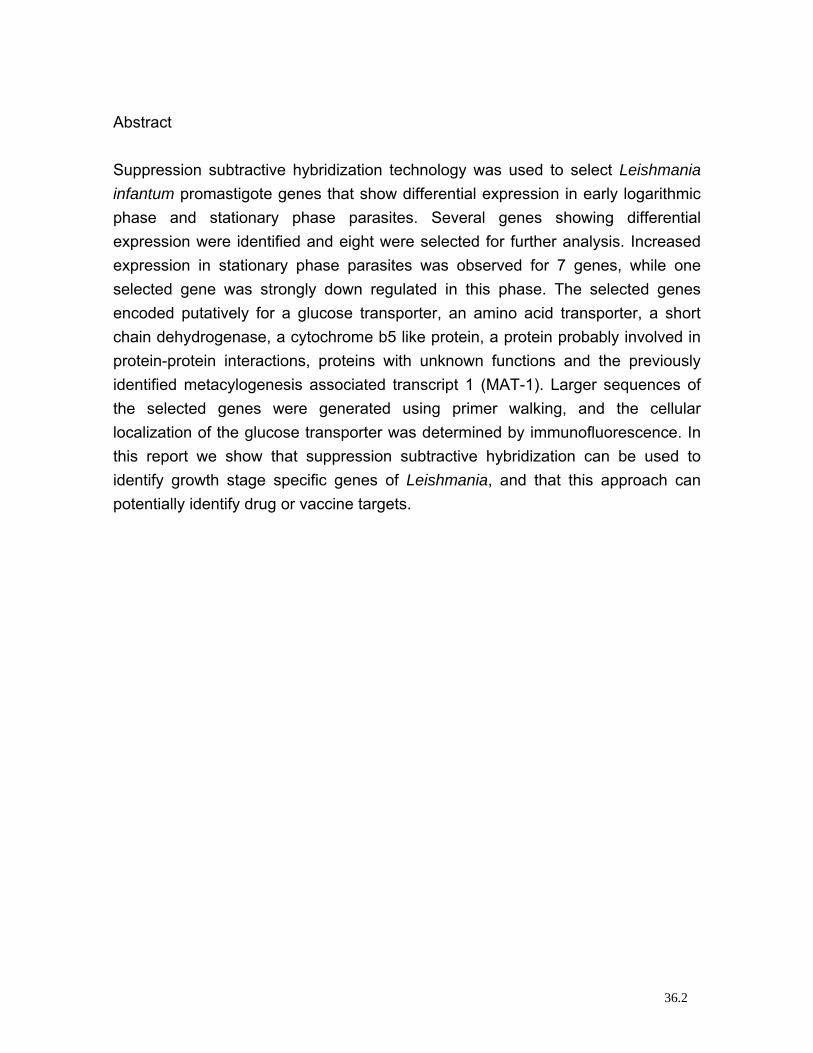
Abstract Suppression subtractive hybridization technology was used to select Leishmania infantum promastigote genes that show differential expression in early logarithmic phase and stationary phase parasites. Several genes showing differential expression were identified and eight were selected for further analysis. Increased expression in stationary phase parasites was observed for 7 genes, while one selected gene was strongly down regulated in this phase. The selected genes encoded putatively for a glucose transporter, an amino acid transporter, a short chain dehydrogenase, a cytochrome b5 like protein, a protein probably involved in protein-protein interactions, proteins with unknown functions and the previously identified metacylogenesis associated transcript 1 (MAT-1). Larger sequences of the selected genes were generated using primer walking, and the cellular localization of the glucose transporter was determined by immunofluorescence. In this report we show that suppression subtractive hybridization can be used to identify growth stage specific genes of Leishmania, and that this approach can potentially identify drug or vaccine targets.
36.2
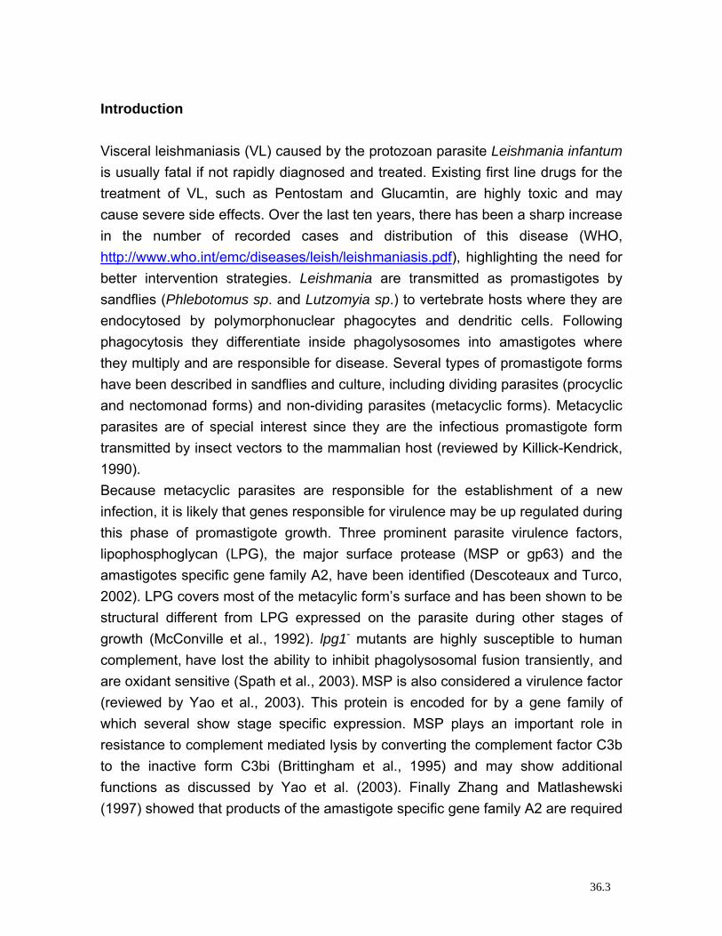
Introduction Visceral leishmaniasis (VL) caused by the protozoan parasite Leishmania infantum is usually fatal if not rapidly diagnosed and treated. Existing first line drugs for the treatment of VL, such as Pentostam and Glucamtin, are highly toxic and may cause severe side effects. Over the last ten years, there has been a sharp increase in the number of recorded cases and distribution of this disease (WHO, http://www.who.int/emc/diseases/leish/leishmaniasis.pdf), highlighting the need for better intervention strategies. Leishmania are transmitted as promastigotes by sandflies (Phlebotomus sp. and Lutzomyia sp.) to vertebrate hosts where they are endocytosed by polymorphonuclear phagocytes and dendritic cells. Following phagocytosis they differentiate inside phagolysosomes into amastigotes where they multiply and are responsible for disease. Several types of promastigote forms have been described in sandflies and culture, including dividing parasites (procyclic and nectomonad forms) and non-dividing parasites (metacyclic forms). Metacyclic parasites are of special interest since they are the infectious promastigote form transmitted by insect vectors to the mammalian host (reviewed by Killick-Kendrick, 1990). Because metacyclic parasites are responsible for the establishment of a new infection, it is likely that genes responsible for virulence may be up regulated during this phase of promastigote growth. Three prominent parasite virulence factors, lipophosphoglycan (LPG), the major surface protease (MSP or gp63) and the amastigotes specific gene family A2, have been identified (Descoteaux and Turco, 2002). LPG covers most of the metacylic form’s surface and has been shown to be structural different from LPG expressed on the parasite during other stages of growth (McConville et al., 1992). lpg1- mutants are highly susceptible to human complement, have lost the ability to inhibit phagolysosomal fusion transiently, and are oxidant sensitive (Spath et al., 2003). MSP is also considered a virulence factor (reviewed by Yao et al., 2003). This protein is encoded for by a gene family of which several show stage specific expression. MSP plays an important role in resistance to complement mediated lysis by converting the complement factor C3b to the inactive form C3bi (Brittingham et al., 1995) and may show additional functions as discussed by Yao et al. (2003). Finally Zhang and Matlashewski (1997) showed that products of the amastigote specific gene family A2 are required
36.3
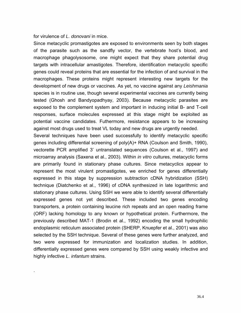
for virulence of L. donovani in mice. Since metacyclic promastigotes are exposed to environments seen by both stages of the parasite such as the sandfly vector, the vertebrate host’s blood, and macrophage phagolysosome, one might expect that they share potential drug targets with intracellular amastigotes. Therefore, identification metacyclic specific genes could reveal proteins that are essential for the infection of and survival in the macrophages. These proteins might represent interesting new targets for the development of new drugs or vaccines. As yet, no vaccine against any Leishmania species is in routine use, though several experimental vaccines are currently being tested (Ghosh and Bandyopadhyay, 2003). Because metacyclic parasites are exposed to the complement system and important in inducing initial B- and T-cell responses, surface molecules expressed at this stage might be exploited as potential vaccine candidates. Futhermore, resistance appears to be increasing against most drugs used to treat VL today and new drugs are urgently needed. Several techniques have been used successfully to identify metacyclic specific genes including differential screening of poly(A)+ RNA (Coulson and Smith, 1990), vectorette PCR amplified 3’ untranslated sequences (Coulson et al., 1997) and microarray analysis (Saxena et al., 2003). Within in vitro cultures, metacyclic forms are primarily found in stationary phase cultures. Since metacyclics appear to represent the most virulent promastigotes, we enriched for genes differentially expressed in this stage by suppression subtraction cDNA hybridization (SSH) technique (Diatchenko et al., 1996) of cDNA synthesized in late logarithmic and stationary phase cultures. Using SSH we were able to identify several differentially expressed genes not yet described. These included two genes encoding transporters, a protein containing leucine rich repeats and an open reading frame (ORF) lacking homology to any known or hypothetical protein. Furthermore, the previously described MAT-1 (Brodin et al., 1992) encoding the small hydrophilic endoplasmic reticulum associated protein (SHERP, Knuepfer et al., 2001) was also selected by the SSH technique. Several of these genes were further analyzed, and two were expressed for immunization and localization studies. In addition, differentially expressed genes were compared by SSH using weakly infective and highly infective L. infantum strains. .
36.4
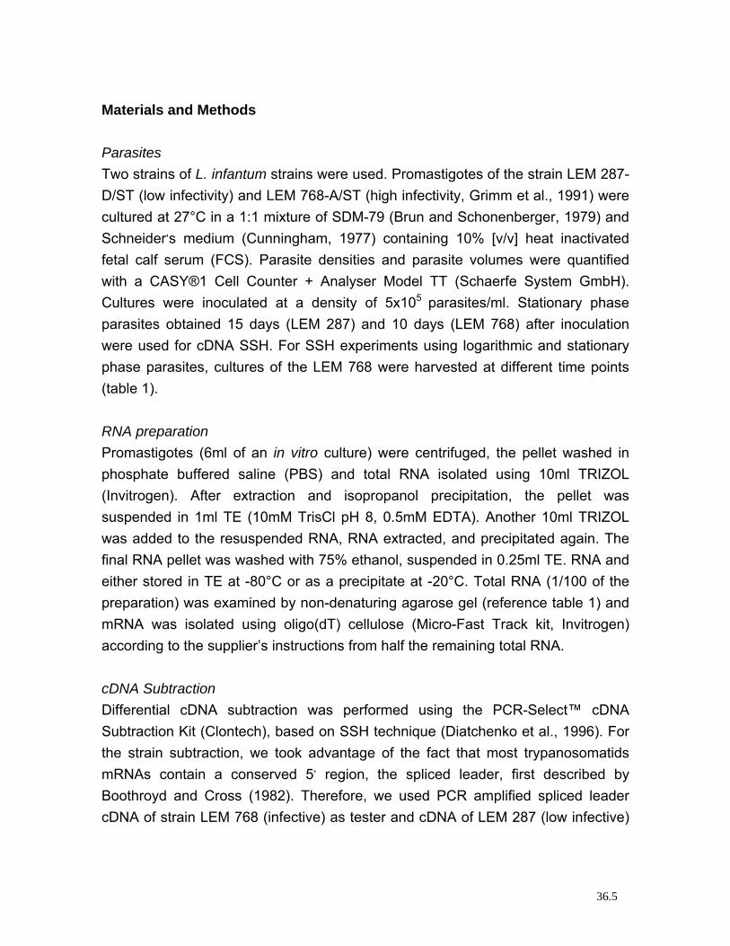
Materials and Methods Parasites Two strains of L. infantum strains were used. Promastigotes of the strain LEM 287-D/ST (low infectivity) and LEM 768-A/ST (high infectivity, Grimm et al., 1991) were cultured at 27°C in a 1:1 mixture of SDM-79 (Brun and Schonenberger, 1979) and Schneider’s medium (Cunningham, 1977) containing 10% [v/v] heat inactivated fetal calf serum (FCS). Parasite densities and parasite volumes were quantified with a CASY®1 Cell Counter + Analyser Model TT (Schaerfe System GmbH). Cultures were inoculated at a density of 5x10⁵ parasites/ml. Stationary phase parasites obtained 15 days (LEM 287) and 10 days (LEM 768) after inoculation were used for cDNA SSH. For SSH experiments using logarithmic and stationary phase parasites, cultures of the LEM 768 were harvested at different time points (table 1). RNA preparation Promastigotes (6ml of an in vitro culture) were centrifuged, the pellet washed in phosphate buffered saline (PBS) and total RNA isolated using 10ml TRIZOL (Invitrogen). After extraction and isopropanol precipitation, the pellet was suspended in 1ml TE (10mM TrisCl pH 8, 0.5mM EDTA). Another 10ml TRIZOL was added to the resuspended RNA, RNA extracted, and precipitated again. The final RNA pellet was washed with 75% ethanol, suspended in 0.25ml TE. RNA and either stored in TE at -80°C or as a precipitate at -20°C. Total RNA (1/100 of the preparation) was examined by non-denaturing agarose gel (reference table 1) and mRNA was isolated using oligo(dT) cellulose (Micro-Fast Track kit, Invitrogen) according to the supplier’s instructions from half the remaining total RNA. cDNA Subtraction Differential cDNA subtraction was performed using the PCR-Select™ cDNA Subtraction Kit (Clontech), based on SSH technique (Diatchenko et al., 1996). For the strain subtraction, we took advantage of the fact that most trypanosomatids mRNAs contain a conserved 5’ region, the spliced leader, first described by Boothroyd and Cross (1982). Therefore, we used PCR amplified spliced leader cDNA of strain LEM 768 (infective) as tester and cDNA of LEM 287 (low infective)
36.5
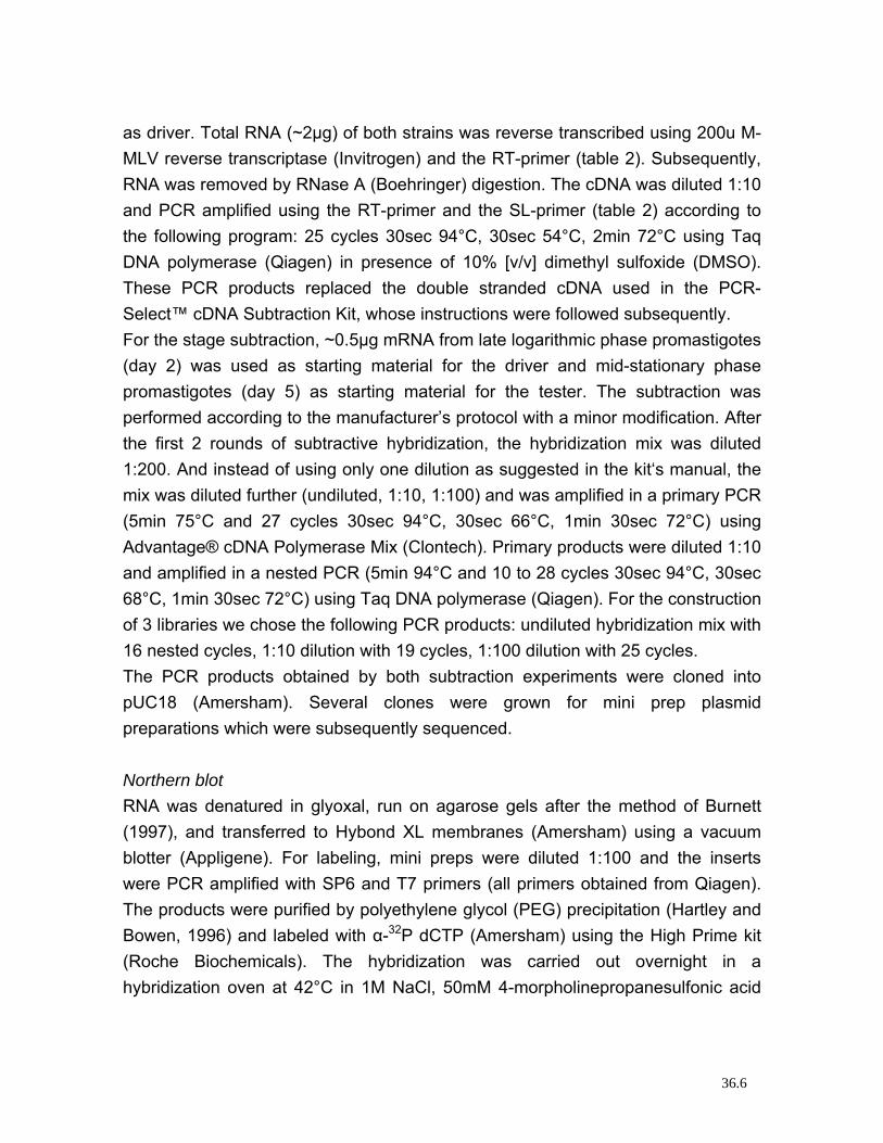
as driver. Total RNA (~2µg) of both strains was reverse transcribed using 200u M-MLV reverse transcriptase (Invitrogen) and the RT-primer (table 2). Subsequently, RNA was removed by RNase A (Boehringer) digestion. The cDNA was diluted 1:10 and PCR amplified using the RT-primer and the SL-primer (table 2) according to the following program: 25 cycles 30sec 94°C, 30sec 54°C, 2min 72°C using Taq DNA polymerase (Qiagen) in presence of 10% [v/v] dimethyl sulfoxide (DMSO). These PCR products replaced the double stranded cDNA used in the PCR-Select™ cDNA Subtraction Kit, whose instructions were followed subsequently. For the stage subtraction, ~0.5µg mRNA from late logarithmic phase promastigotes (day 2) was used as starting material for the driver and mid-stationary phase promastigotes (day 5) as starting material for the tester. The subtraction was performed according to the manufacturer’s protocol with a minor modification. After the first 2 rounds of subtractive hybridization, the hybridization mix was diluted 1:200. And instead of using only one dilution as suggested in the kit‘s manual, the mix was diluted further (undiluted, 1:10, 1:100) and was amplified in a primary PCR (5min 75°C and 27 cycles 30sec 94°C, 30sec 66°C, 1min 30sec 72°C) using Advantage® cDNA Polymerase Mix (Clontech). Primary products were diluted 1:10 and amplified in a nested PCR (5min 94°C and 10 to 28 cycles 30sec 94°C, 30sec 68°C, 1min 30sec 72°C) using Taq DNA polymerase (Qiagen). For the construction of 3 libraries we chose the following PCR products: undiluted hybridization mix with 16 nested cycles, 1:10 dilution with 19 cycles, 1:100 dilution with 25 cycles. The PCR products obtained by both subtraction experiments were cloned into pUC18 (Amersham). Several clones were grown for mini prep plasmid preparations which were subsequently sequenced. Northern blot RNA was denatured in glyoxal, run on agarose gels after the method of Burnett (1997), and transferred to Hybond XL membranes (Amersham) using a vacuum blotter (Appligene). For labeling, mini preps were diluted 1:100 and the inserts were PCR amplified with SP6 and T7 primers (all primers obtained from Qiagen). The products were purified by polyethylene glycol (PEG) precipitation (Hartley and Bowen, 1996) and labeled with α-32P dCTP (Amersham) using the High Prime kit (Roche Biochemicals). The hybridization was carried out overnight in a hybridization oven at 42°C in 1M NaCl, 50mM 4-morpholinepropanesulfonic acid
36.6
(MOPS) pH 7, 50% [w/v] formamide, 0.5% [w/v] sodium dodecyl sulfate (SDS), 1mM EDTA, 18% [w/v] dextran sulfate, 5x Denhardt’s reagent (Denhardt, 1966). After hybridization, membranes were washed twice in 2x SSC (1x SSC is 0.15M NaCl, 15mM sodium citrate), 0.1% SDS and twice in 0.1x SSC, 0.1% SDS at 42°C. The filters were exposed to Kodak MS films for 30min to 48h at -80°C using a Transcreen HE enhancer (Kodak). cDNA walking For cDNA walking we also took advantage of the conserved 5’ region in trypanosomatids. We chose the strategy shown in figure 1 to obtain full length cDNA sequences. mRNA was reverse transcribed by M-MuLV Reverse Transcriptase, RNase H- (Finnzymes) using the RT-primer (table 2). RNA was removed by RNase A and H digestion. For RT-PCR, the cDNA was diluted 1:50 in TE and amplified using insert specific and the RT-primers to obtain the 3’ end, or insert specific and the SL-primers (table 2) to obtain the 5’ end. 40 cycles (30s 94°C, 30s 56°C, 2min 72°C) were performed in Taq buffer in presence or absence of 10% [v/v] DMSO using Taq and Pfu (Promega) DNA polymerases in a 10:1 proportion. PCR products were cloned and sequenced. If no products were obtained, a nested PCR was performed to obtain the 5’ end. For primary PCRs, outer insert primers and the pSL-primer (table 2) were used, for nested PCRs, inner insert primers and the nSL-primer (table 2) were used and 2 x 25 cycles were performed. Cycle conditions and the concentrations of DMSO (0-10%), Tween20 (0-0.5% [v/v]) and BSA (0-0.5g/l) were optimized for each PCR. The primary PCR was performed with Taq/Pfu polymerase (10:1), the nested with Taq polymerase alone. The PCR products were cloned into pGEM-T (Promega) vector and sequenced. Sequence analysis The basic local alignment search tools (Blast) of NCBI (http://www.ncbi.nlm.nih.gov/BLAST/, Blastx, Altschul et al., 1990; Gish and States, 1993) and EBI (Washington University Blast Version 2.0 Parasites, http://www.ebi.ac.uk/blast2/parasites.html) were used, transmembrane domains were predicted at CBS with TMHMM (Krogh et al., 2001, http://www.cbs.dtu.dk/services/TMHMM-2.0/) and motif scans were performed at
36.7
EBI with InterProScan (Mulder et al., 2003, http://www.ebi.ac.uk/interpro/scan.html). Protein expression in E. coli Proteins were expressed as N-terminal GST fusion proteins with a C-terminal 6 x His tag in pGEX-His vector. This vector was obtained by ligation of a His-adaptor made by the two oligo-nucleotides His-fw and His-rev (table 2) into SalI- and NotI-digested (all restriction enzymes obtained from New England Biolabs) pGEX-6p-1 (Amersham). The following fragments were PCR amplified with primers containing a BamHI and SalI site, respectively: the C-terminal part of the putative steroid dehydrogenase (clone1, table 3) with primers 1-fw and 1-rev, and a fragment between two transmembrane domains of the glucose transporter (clone6) with the primers 6-fw and 6-rev (table 2). Prior to ligation, PCR products and the pGEX-His vector were cut with BamHI and SalI and purified by phenol extraction and PEG precipitation. Proteins were expressed in BL21 cells (Amersham) which were grown in Terrific Broth (Tartoff and Hobbs, 1987). When cultures reached an optical density of 1, protein expression was induced with 0.5mM isopropyl-β-D-1-thiogalactopyranoside (IPTG) for 4 hours. All three proteins were insoluble in PBS/Triton X-100. Thus, after purification of the inclusion bodies, proteins 1 and 6 were purified with Ni-NTA Agarose (Qiagen) under denaturing conditions (protein 1 was not soluble in 8M urea, and urea was replaced with 6M guanidine chloride in all solutions). Immunization For each protein, two NMRI mice’s were immunized with 1 and 5µg purified protein using CpG DNA as adjuvant (ImmunEasy™ Mouse Adjuvant, Qiagen). Two booster injections followed 2 and 4 weeks after the first immunization using the same conditions. 6 week after immunization, mice were sacrificed and polyclonal sera were collected. Western blots Proteins were run on 12.5% polyacrylamide gels in SDS/glycine buffer and blotted semi dry on nitrocellulose (Amersham) in 10mM 3-(cyclohexylamino)-1-propanesulfonic acid (CAPS), 10% [v/v] methanol, pH10.5. In order to reduce
36.8
background and binding of anti-GST antibodies, an extract of induced BL21 cells containing the empty pGEX-6p-1 vector was used to adsorb the mouse sera. Immuno fluorescent assays 500µl of a 10 day old Leishmania in vitro culture were spun and the pellet was resuspended in 20µl Fetal Calf Serum (FCS). 2.5µl each were used to prepare several smears on glass slides. Smears were dried, fixed in 1:1 methanol/acetone for 10min at -20°C. The slides were partitioned with a hydrophobic pen. E. coli adsorbed sera were used at a concentration of 1:20 in PBS, 1% BSA. For competition with clone 6, 0.1µg/µl recombinant protein was added, which lead to an urea concentration of 0.8M. Therefore, the positive control in this competition experiment was done in PBS, 1% BSA, 0.8M urea. Cy3 labeled goat anti-mouse IGg antibody (Jackson) was used as secondary antibody 1:200 in PBS, 1% BSA and 1ng/µl 4',6-diamidino-2-phenylindole (DAPI) as DNA stain. Results & Discussion Strain subtraction In order to identify Leishmania virulence factors, we subtracted in a preliminary experiment cDNA of a strain of low infectivity (LEM 287) from a highly infective strain (LEM 768). We used PCR amplified spliced leader cDNA as tester and driver for the SSH (figure 2). Northern blot analysis showed that many of the genes selected were more strongly expressed by the highly virulent strain than by the low virulent strain (figure 3) and that SSH can be used when double stranded cDNA is replaced by PCR amplified spliced leader cDNA (obtained from total RNA). This is of importance in situations where starting material is limited (for instance for in vivo studies). Sequence analysis of the genes identified with this first approach made it difficult to understand their involvement in virulence as it is unclear why genes such as the Histone H1 gene or genes involved in translation elongation should be associated with virulence. An explanation for this apparent experimental failure might lay in the large difference between both strains, not only seen in infectivity, but also possibly in growth rate and other aspects which might be reflected in virulence.
36.9
Nevertheless, the histone H1 locus of L. major was also examined by Belli et al. (2003) and found to be stage specifically expressed as sense and antisense RNA. Stage subtraction Figure 4 shows the outcome of the SSH. It clearly shows, that the band patterns differed, when different dilutions were amplified. This was not suggested in the kit’s manual. We also introduced a hybridization control (figure 4) in which the same amount of DNA was added prior to PCR, but hybridization was eliminated by denaturation (figure 4: - gel). The high background of the undiluted negative control was probably caused by amplification of the most abundant tester cDNAs since PCR cycling allows hybridization of tester cDNA, which leads to amplification of the tester cDNA. Therefore, the background of the undiluted SSH is much higher because hybridization is more efficient in concentrated DNA solutions. We selected 8 differentially expressed genes from the stage subtraction for which the results are shown in table 3. We selected genes which putatively coded for proteins involved in transmembrane transport (2 genes), in redox reactions (2 genes), in protein-protein interactions (1 gene), and 3 genes encoding proteins of unknown functions. Recently, extensive work has been published using microarray analysis (Saxena et al., 2003) and genes were identified which are expressed at higher levels in metacyclics of L. major. These were genes encoding proteins involved in cell division, metabolism, membrane transport, protein synthesis and other functions. Surprisingly, despite MAT-1 none of the genes described here were identified using the microarray technique. In order to test whether the identified genes were indeed differentially expressed genes, northern blots of selected clones were performed (figure 5). Since double stranded DNA was used as probes for northern blots, we cannot determine whether we detected sense or antisense RNA. As expected, most of the selected genes were up regulated in stat phase cultures, but there was a background of constitutively expressed genes, and one clone was even down regulated. This was probably due to primer binding sites located within this gene: the adaptors provided by the SSH kit are composed of very GC rich sequences, which is also the case for the Leishmania genome. Therefore, the background probably increases when using the SSH kit for subtractions within GC rich genomes. Using Blast searches we found that the inserts of clone 2 and 5 were located within
36.10
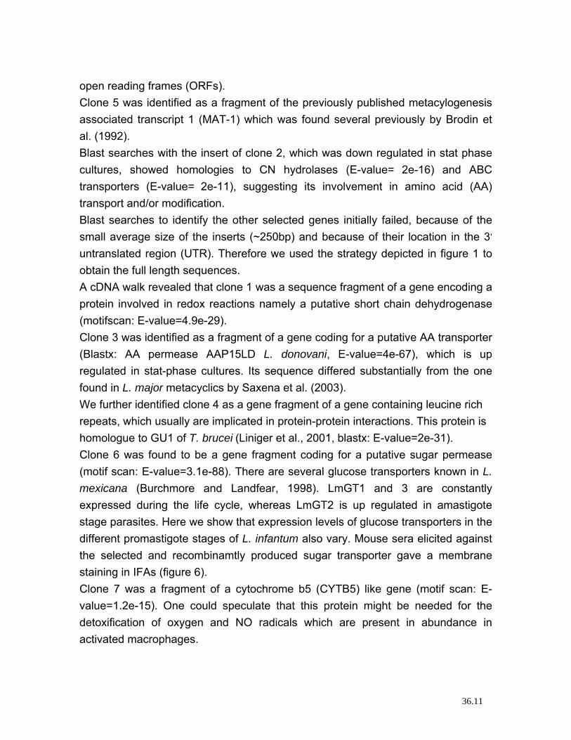
open reading frames (ORFs). Clone 5 was identified as a fragment of the previously published metacylogenesis associated transcript 1 (MAT-1) which was found several previously by Brodin et al. (1992). Blast searches with the insert of clone 2, which was down regulated in stat phase cultures, showed homologies to CN hydrolases (E-value= 2e-16) and ABC transporters (E-value= 2e-11), suggesting its involvement in amino acid (AA) transport and/or modification. Blast searches to identify the other selected genes initially failed, because of the small average size of the inserts (~250bp) and because of their location in the 3’ untranslated region (UTR). Therefore we used the strategy depicted in figure 1 to obtain the full length sequences. A cDNA walk revealed that clone 1 was a sequence fragment of a gene encoding a protein involved in redox reactions namely a putative short chain dehydrogenase (motifscan: E-value=4.9e-29). Clone 3 was identified as a fragment of a gene coding for a putative AA transporter (Blastx: AA permease AAP15LD L. donovani, E-value=4e-67), which is up regulated in stat-phase cultures. Its sequence differed substantially from the one found in L. major metacyclics by Saxena et al. (2003). We further identified clone 4 as a gene fragment of a gene containing leucine rich repeats, which usually are implicated in protein-protein interactions. This protein is homologue to GU1 of T. brucei (Liniger et al., 2001, blastx: E-value=2e-31). Clone 6 was found to be a gene fragment coding for a putative sugar permease (motif scan: E-value=3.1e-88). There are several glucose transporters known in L. mexicana (Burchmore and Landfear, 1998). LmGT1 and 3 are constantly expressed during the life cycle, whereas LmGT2 is up regulated in amastigote stage parasites. Here we show that expression levels of glucose transporters in the different promastigote stages of L. infantum also vary. Mouse sera elicited against the selected and recombinamtly produced sugar transporter gave a membrane staining in IFAs (figure 6). Clone 7 was a fragment of a cytochrome b5 (CYTB5) like gene (motif scan: E-value=1.2e-15). One could speculate that this protein might be needed for the detoxification of oxygen and NO radicals which are present in abundance in activated macrophages.
36.11
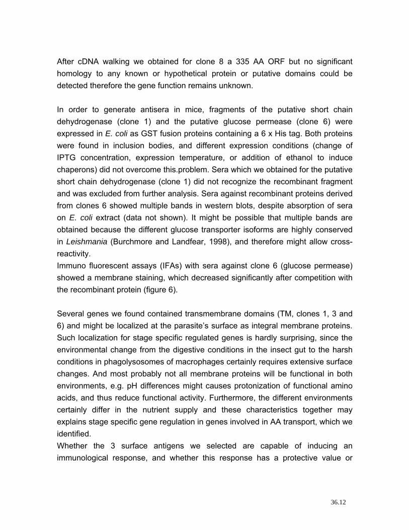
After cDNA walking we obtained for clone 8 a 335 AA ORF but no significant homology to any known or hypothetical protein or putative domains could be detected therefore the gene function remains unknown. In order to generate antisera in mice, fragments of the putative short chain dehydrogenase (clone 1) and the putative glucose permease (clone 6) were expressed in E. coli as GST fusion proteins containing a 6 x His tag. Both proteins were found in inclusion bodies, and different expression conditions (change of IPTG concentration, expression temperature, or addition of ethanol to induce chaperons) did not overcome this.problem. Sera which we obtained for the putative short chain dehydrogenase (clone 1) did not recognize the recombinant fragment and was excluded from further analysis. Sera against recombinant proteins derived from clones 6 showed multiple bands in western blots, despite absorption of sera on E. coli extract (data not shown). It might be possible that multiple bands are obtained because the different glucose transporter isoforms are highly conserved in Leishmania (Burchmore and Landfear, 1998), and therefore might allow cross-reactivity. Immuno fluorescent assays (IFAs) with sera against clone 6 (glucose permease) showed a membrane staining, which decreased significantly after competition with the recombinant protein (figure 6). Several genes we found contained transmembrane domains (TM, clones 1, 3 and 6) and might be localized at the parasite’s surface as integral membrane proteins. Such localization for stage specific regulated genes is hardly surprising, since the environmental change from the digestive conditions in the insect gut to the harsh conditions in phagolysosomes of macrophages certainly requires extensive surface changes. And most probably not all membrane proteins will be functional in both environments, e.g. pH differences might causes protonization of functional amino acids, and thus reduce functional activity. Furthermore, the different environments certainly differ in the nutrient supply and these characteristics together may explains stage specific gene regulation in genes involved in AA transport, which we identified. Whether the 3 surface antigens we selected are capable of inducing an immunological response, and whether this response has a protective value or
36.12
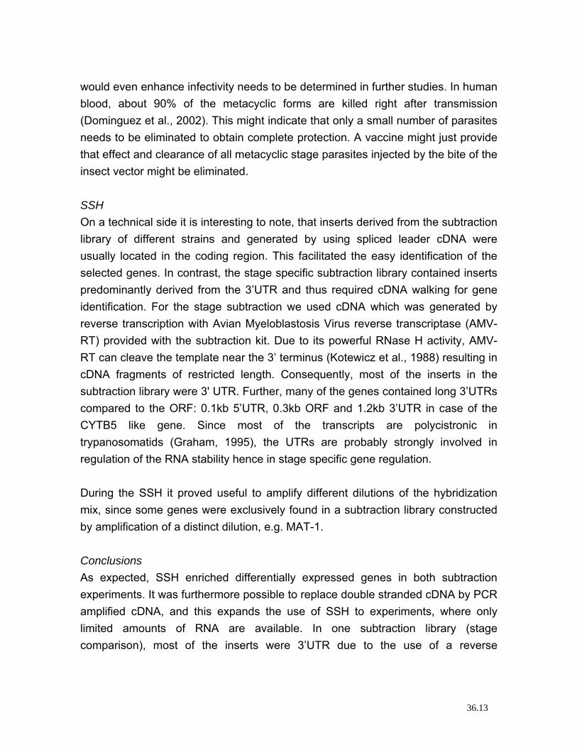
would even enhance infectivity needs to be determined in further studies. In human blood, about 90% of the metacyclic forms are killed right after transmission (Dominguez et al., 2002). This might indicate that only a small number of parasites needs to be eliminated to obtain complete protection. A vaccine might just provide that effect and clearance of all metacyclic stage parasites injected by the bite of the insect vector might be eliminated. SSH On a technical side it is interesting to note, that inserts derived from the subtraction library of different strains and generated by using spliced leader cDNA were usually located in the coding region. This facilitated the easy identification of the selected genes. In contrast, the stage specific subtraction library contained inserts predominantly derived from the 3’UTR and thus required cDNA walking for gene identification. For the stage subtraction we used cDNA which was generated by reverse transcription with Avian Myeloblastosis Virus reverse transcriptase (AMV-RT) provided with the subtraction kit. Due to its powerful RNase H activity, AMV-RT can cleave the template near the 3’ terminus (Kotewicz et al., 1988) resulting in cDNA fragments of restricted length. Consequently, most of the inserts in the subtraction library were 3' UTR. Further, many of the genes contained long 3’UTRs compared to the ORF: 0.1kb 5’UTR, 0.3kb ORF and 1.2kb 3’UTR in case of the CYTB5 like gene. Since most of the transcripts are polycistronic in trypanosomatids (Graham, 1995), the UTRs are probably strongly involved in regulation of the RNA stability hence in stage specific gene regulation. During the SSH it proved useful to amplify different dilutions of the hybridization mix, since some genes were exclusively found in a subtraction library constructed by amplification of a distinct dilution, e.g. MAT-1. Conclusions As expected, SSH enriched differentially expressed genes in both subtraction experiments. It was furthermore possible to replace double stranded cDNA by PCR amplified cDNA, and this expands the use of SSH to experiments, where only limited amounts of RNA are available. In one subtraction library (stage comparison), most of the inserts were 3’UTR due to the use of a reverse
36.13
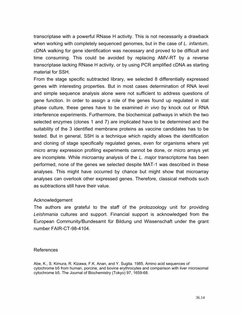
transcriptase with a powerful RNase H activity. This is not necessarily a drawback when working with completely sequenced genomes, but in the case of L. infantum, cDNA walking for gene identification was necessary and proved to be difficult and time consuming. This could be avoided by replacing AMV-RT by a reverse transcriptase lacking RNase H activity, or by using PCR amplified cDNA as starting material for SSH. From the stage specific subtracted library, we selected 8 differentially expressed genes with interesting properties. But in most cases determination of RNA level and simple sequence analysis alone were not sufficient to address questions of gene function. In order to assign a role of the genes found up regulated in stat phase culture, these genes have to be examined in vivo by knock out or RNA interference experiments. Furthermore, the biochemical pathways in which the two selected enzymes (clones 1 and 7) are implicated have to be determined and the suitability of the 3 identified membrane proteins as vaccine candidates has to be tested. But in general, SSH is a technique which rapidly allows the identification and cloning of stage specifically regulated genes, even for organisms where yet micro array expression profiling experiments cannot be done, or micro arrays yet are incomplete. While microarray analysis of the L. major transcriptome has been performed, none of the genes we selected despite MAT-1 was described in these analyses. This might have occurred by chance but might show that microarray analyses can overlook other expressed genes. Therefore, classical methods such as subtractions still have their value. Acknowledgement The authors are grateful to the staff of the protozoology unit for providing Leishmania cultures and support. Financial support is acknowledged from the European Community/Bundesamt für Bildung und Wissenschaft under the grant number FAIR-CT-98-4104. References Abe, K., S. Kimura, R. Kizawa, F.K. Anan, and Y. Sugita. 1985. Amino acid sequences of cytochrome b5 from human, porcine, and bovine erythrocytes and comparison with liver microsomal cytochrome b5. The Journal of Biochemistry (Tokyo) 97, 1659-68.
36.14
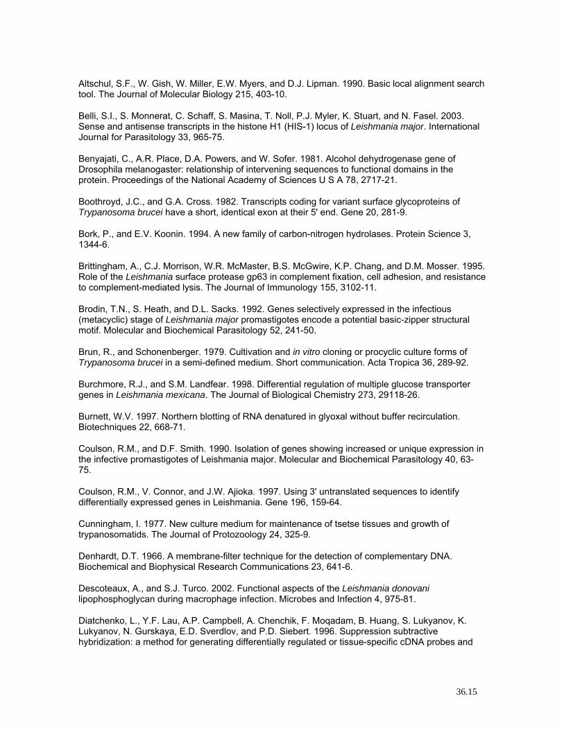
Altschul, S.F., W. Gish, W. Miller, E.W. Myers, and D.J. Lipman. 1990. Basic local alignment search tool. The Journal of Molecular Biology 215, 403-10.
Belli, S.I., S. Monnerat, C. Schaff, S. Masina, T. Noll, P.J. Myler, K. Stuart, and N. Fasel. 2003. Sense and antisense transcripts in the histone H1 (HIS-1) locus of Leishmania major. International Journal for Parasitology 33, 965-75.
Benyajati, C., A.R. Place, D.A. Powers, and W. Sofer. 1981. Alcohol dehydrogenase gene of Drosophila melanogaster: relationship of intervening sequences to functional domains in the protein. Proceedings of the National Academy of Sciences U S A 78, 2717-21.
Boothroyd, J.C., and G.A. Cross. 1982. Transcripts coding for variant surface glycoproteins of Trypanosoma brucei have a short, identical exon at their 5' end. Gene 20, 281-9.
Bork, P., and E.V. Koonin. 1994. A new family of carbon-nitrogen hydrolases. Protein Science 3, 1344-6.
Brittingham, A., C.J. Morrison, W.R. McMaster, B.S. McGwire, K.P. Chang, and D.M. Mosser. 1995. Role of the Leishmania surface protease gp63 in complement fixation, cell adhesion, and resistance to complement-mediated lysis. The Journal of Immunology 155, 3102-11.
Brodin, T.N., S. Heath, and D.L. Sacks. 1992. Genes selectively expressed in the infectious (metacyclic) stage of Leishmania major promastigotes encode a potential basic-zipper structural motif. Molecular and Biochemical Parasitology 52, 241-50.
Brun, R., and Schonenberger. 1979. Cultivation and in vitro cloning or procyclic culture forms of Trypanosoma brucei in a semi-defined medium. Short communication. Acta Tropica 36, 289-92.
Burchmore, R.J., and S.M. Landfear. 1998. Differential regulation of multiple glucose transporter genes in Leishmania mexicana. The Journal of Biological Chemistry 273, 29118-26.
Burnett, W.V. 1997. Northern blotting of RNA denatured in glyoxal without buffer recirculation. Biotechniques 22, 668-71.
Coulson, R.M., and D.F. Smith. 1990. Isolation of genes showing increased or unique expression in the infective promastigotes of Leishmania major. Molecular and Biochemical Parasitology 40, 63-75.
Coulson, R.M., V. Connor, and J.W. Ajioka. 1997. Using 3' untranslated sequences to identify differentially expressed genes in Leishmania. Gene 196, 159-64.
Cunningham, I. 1977. New culture medium for maintenance of tsetse tissues and growth of trypanosomatids. The Journal of Protozoology 24, 325-9.
Denhardt, D.T. 1966. A membrane-filter technique for the detection of complementary DNA. Biochemical and Biophysical Research Communications 23, 641-6.
Descoteaux, A., and S.J. Turco. 2002. Functional aspects of the Leishmania donovani lipophosphoglycan during macrophage infection. Microbes and Infection 4, 975-81.
Diatchenko, L., Y.F. Lau, A.P. Campbell, A. Chenchik, F. Moqadam, B. Huang, S. Lukyanov, K. Lukyanov, N. Gurskaya, E.D. Sverdlov, and P.D. Siebert. 1996. Suppression subtractive hybridization: a method for generating differentially regulated or tissue-specific cDNA probes and
36.15
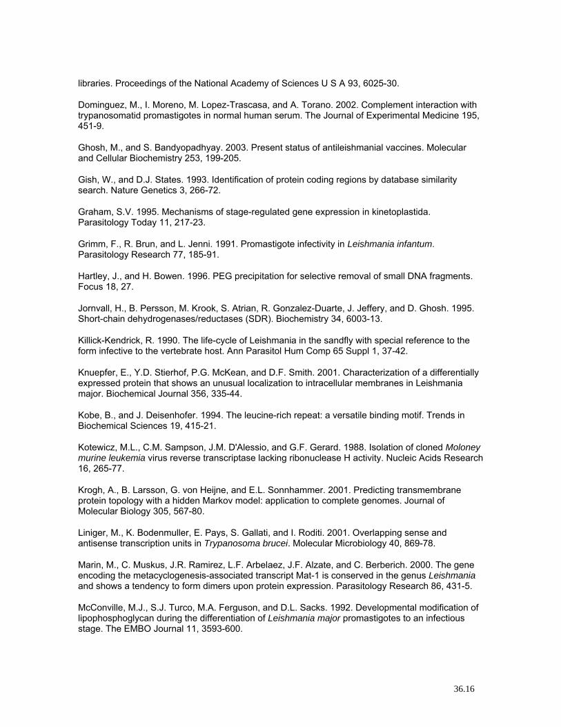
libraries. Proceedings of the National Academy of Sciences U S A 93, 6025-30.
Dominguez, M., I. Moreno, M. Lopez-Trascasa, and A. Torano. 2002. Complement interaction with trypanosomatid promastigotes in normal human serum. The Journal of Experimental Medicine 195, 451-9.
Ghosh, M., and S. Bandyopadhyay. 2003. Present status of antileishmanial vaccines. Molecular and Cellular Biochemistry 253, 199-205.
Gish, W., and D.J. States. 1993. Identification of protein coding regions by database similarity search. Nature Genetics 3, 266-72.
Graham, S.V. 1995. Mechanisms of stage-regulated gene expression in kinetoplastida. Parasitology Today 11, 217-23.
Grimm, F., R. Brun, and L. Jenni. 1991. Promastigote infectivity in Leishmania infantum. Parasitology Research 77, 185-91.
Hartley, J., and H. Bowen. 1996. PEG precipitation for selective removal of small DNA fragments. Focus 18, 27.
Jornvall, H., B. Persson, M. Krook, S. Atrian, R. Gonzalez-Duarte, J. Jeffery, and D. Ghosh. 1995. Short-chain dehydrogenases/reductases (SDR). Biochemistry 34, 6003-13.
Killick-Kendrick, R. 1990. The life-cycle of Leishmania in the sandfly with special reference to the form infective to the vertebrate host. Ann Parasitol Hum Comp 65 Suppl 1, 37-42.
Knuepfer, E., Y.D. Stierhof, P.G. McKean, and D.F. Smith. 2001. Characterization of a differentially expressed protein that shows an unusual localization to intracellular membranes in Leishmania major. Biochemical Journal 356, 335-44.
Kobe, B., and J. Deisenhofer. 1994. The leucine-rich repeat: a versatile binding motif. Trends in Biochemical Sciences 19, 415-21.
Kotewicz, M.L., C.M. Sampson, J.M. D'Alessio, and G.F. Gerard. 1988. Isolation of cloned Moloney murine leukemia virus reverse transcriptase lacking ribonuclease H activity. Nucleic Acids Research 16, 265-77.
Krogh, A., B. Larsson, G. von Heijne, and E.L. Sonnhammer. 2001. Predicting transmembrane protein topology with a hidden Markov model: application to complete genomes. Journal of Molecular Biology 305, 567-80.
Liniger, M., K. Bodenmuller, E. Pays, S. Gallati, and I. Roditi. 2001. Overlapping sense and antisense transcription units in Trypanosoma brucei. Molecular Microbiology 40, 869-78.
Marin, M., C. Muskus, J.R. Ramirez, L.F. Arbelaez, J.F. Alzate, and C. Berberich. 2000. The gene encoding the metacyclogenesis-associated transcript Mat-1 is conserved in the genus Leishmania and shows a tendency to form dimers upon protein expression. Parasitology Research 86, 431-5.
McConville, M.J., S.J. Turco, M.A. Ferguson, and D.L. Sacks. 1992. Developmental modification of lipophosphoglycan during the differentiation of Leishmania major promastigotes to an infectious stage. The EMBO Journal 11, 3593-600.
36.16
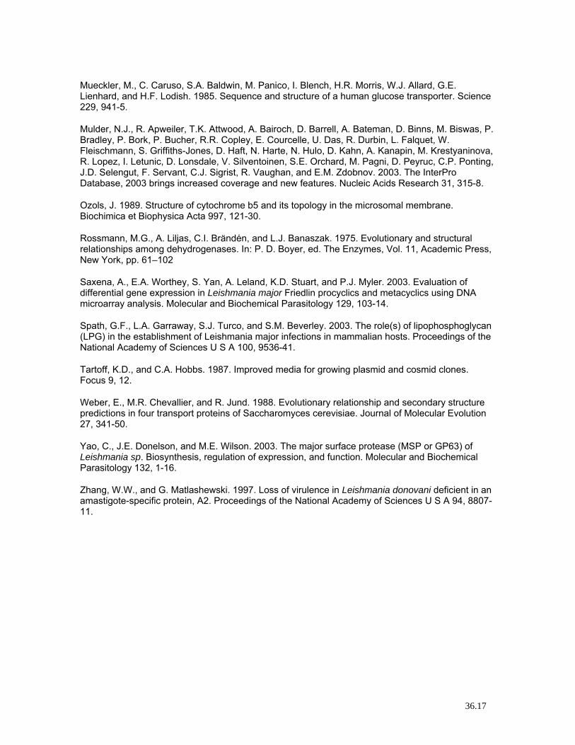
Mueckler, M., C. Caruso, S.A. Baldwin, M. Panico, I. Blench, H.R. Morris, W.J. Allard, G.E. Lienhard, and H.F. Lodish. 1985. Sequence and structure of a human glucose transporter. Science 229, 941-5.
Mulder, N.J., R. Apweiler, T.K. Attwood, A. Bairoch, D. Barrell, A. Bateman, D. Binns, M. Biswas, P. Bradley, P. Bork, P. Bucher, R.R. Copley, E. Courcelle, U. Das, R. Durbin, L. Falquet, W. Fleischmann, S. Griffiths-Jones, D. Haft, N. Harte, N. Hulo, D. Kahn, A. Kanapin, M. Krestyaninova, R. Lopez, I. Letunic, D. Lonsdale, V. Silventoinen, S.E. Orchard, M. Pagni, D. Peyruc, C.P. Ponting, J.D. Selengut, F. Servant, C.J. Sigrist, R. Vaughan, and E.M. Zdobnov. 2003. The InterPro Database, 2003 brings increased coverage and new features. Nucleic Acids Research 31, 315-8.
Ozols, J. 1989. Structure of cytochrome b5 and its topology in the microsomal membrane. Biochimica et Biophysica Acta 997, 121-30.
Rossmann, M.G., A. Liljas, C.I. Brändén, and L.J. Banaszak. 1975. Evolutionary and structural relationships among dehydrogenases. In: P. D. Boyer, ed. The Enzymes, Vol. 11, Academic Press, New York, pp. 61–102
Saxena, A., E.A. Worthey, S. Yan, A. Leland, K.D. Stuart, and P.J. Myler. 2003. Evaluation of differential gene expression in Leishmania major Friedlin procyclics and metacyclics using DNA microarray analysis. Molecular and Biochemical Parasitology 129, 103-14.
Spath, G.F., L.A. Garraway, S.J. Turco, and S.M. Beverley. 2003. The role(s) of lipophosphoglycan (LPG) in the establishment of Leishmania major infections in mammalian hosts. Proceedings of the National Academy of Sciences U S A 100, 9536-41.
Tartoff, K.D., and C.A. Hobbs. 1987. Improved media for growing plasmid and cosmid clones. Focus 9, 12.
Weber, E., M.R. Chevallier, and R. Jund. 1988. Evolutionary relationship and secondary structure predictions in four transport proteins of Saccharomyces cerevisiae. Journal of Molecular Evolution 27, 341-50.
Yao, C., J.E. Donelson, and M.E. Wilson. 2003. The major surface protease (MSP or GP63) of Leishmania sp. Biosynthesis, regulation of expression, and function. Molecular and Biochemical Parasitology 132, 1-16.
Zhang, W.W., and G. Matlashewski. 1997. Loss of virulence in Leishmania donovani deficient in an amastigote-specific protein, A2. Proceedings of the National Academy of Sciences U S A 94, 8807-11.
36.17
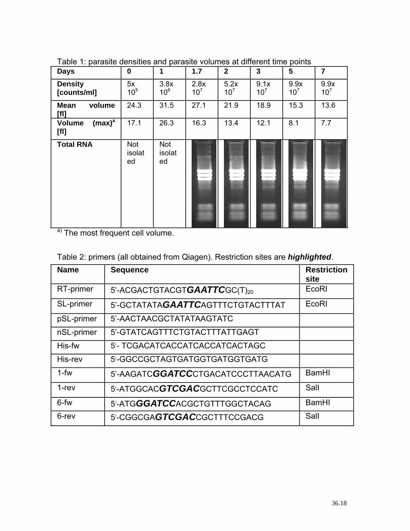
Table 1: parasite densities and parasite volumes at different time points Days 0 1 1.7 2 3 5 7 Density [counts/ml]
5x 10⁵
3.8x 10⁶
2.8x 10⁷
5.2x 10⁷
9.1x 10⁷
9.9x 10⁷
9.9x 10⁷
Mean volume [fl]
24.3 31.5 27.1 21.9 18.9 15.3 13.6
Volume (max)a [fl]
17.1 26.3 16.3 13.4 12.1 8.1 7.7
Total RNA Not isolated
Not isolated
a) The most frequent cell volume. Table 2: primers (all obtained from Qiagen). Restriction sites are highlighted. Name Sequence Restriction
site RT-primer 5’-ACGACTGTACGTGAATTCGC(T)20 EcoRI
SL-primer 5’-GCTATATAGAATTCAGTTTCTGTACTTTAT EcoRI
pSL-primer 5’-AACTAACGCTATATAAGTATC
nSL-primer 5’-GTATCAGTTTCTGTACTTTATTGAGT
His-fw 5’- TCGACATCACCATCACCATCACTAGC
His-rev 5’-GGCCGCTAGTGATGGTGATGGTGATG
1-fw 5’-AAGATCGGATCCCTGACATCCCTTAACATG BamHI
1-rev 5’-ATGGCACGTCGACGCTTCGCCTCCATC SalI
6-fw 5’-ATGGGATCCACGCTGTTTGGCTACAG BamHI
6-rev 5’-CGGCGAGTCGACCGCTTTCCGACG SalI
36.18
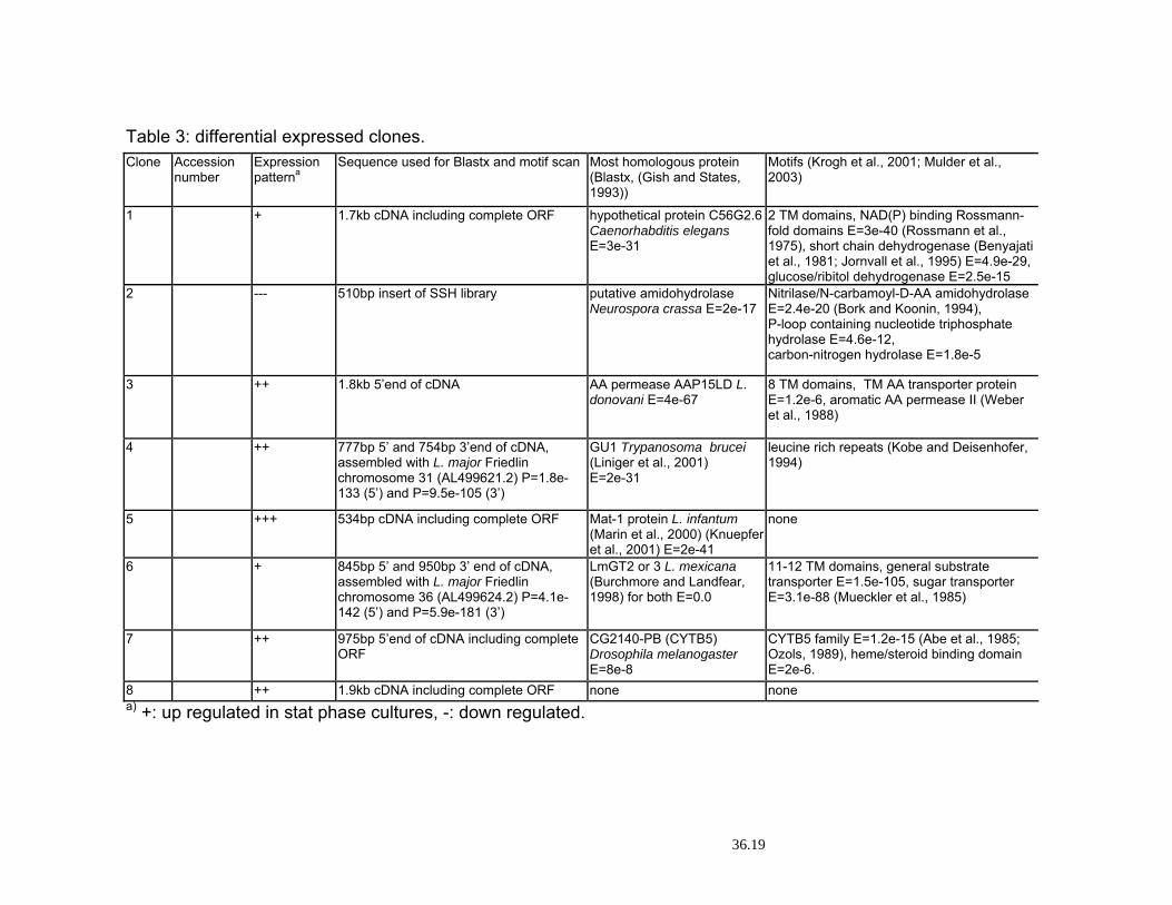
Table 3: differential expressed clones. Clone Accession
number Expression patterna
Sequence used for Blastx and motif scan Most homologous protein (Blastx, (Gish and States, 1993))
Motifs (Krogh et al., 2001; Mulder et al., 2003)
1 + 1.7kb cDNA including complete ORF hypothetical protein C56G2.6 Caenorhabditis elegans E=3e-31
2 TM domains, NAD(P) binding Rossmann-fold domains E=3e-40 (Rossmann et al., 1975), short chain dehydrogenase (Benyajati et al., 1981; Jornvall et al., 1995) E=4.9e-29, glucose/ribitol dehydrogenase E=2.5e-15
2 --- 510bp insert of SSH library putative amidohydrolaseNeurospora crassa E=2e-17
Nitrilase/N-carbamoyl-D-AA amidohydrolase E=2.4e-20 (Bork and Koonin, 1994), P-loop containing nucleotide triphosphate hydrolase E=4.6e-12, carbon-nitrogen hydrolase E=1.8e-5
3 ++ 1.8kb 5’end of cDNA AA permease AAP15LD L. donovani E=4e-67
8 TM domains, TM AA transporter protein E=1.2e-6, aromatic AA permease II (Weber et al., 1988)
4 ++ 777bp 5’ and 754bp 3’end of cDNA, assembled with L. major Friedlin chromosome 31 (AL499621.2) P=1.8e-133 (5’) and P=9.5e-105 (3’)
GU1 Trypanosoma brucei (Liniger et al., 2001) E=2e-31
leucine rich repeats (Kobe and Deisenhofer, 1994)
5 +++ 534bp cDNA including complete ORF Mat-1 protein L. infantum (Marin et al., 2000) (Knuepfer et al., 2001) E=2e-41
none
6 + 845bp 5’ and 950bp 3’ end of cDNA, assembled with L. major Friedlin chromosome 36 (AL499624.2) P=4.1e-142 (5’) and P=5.9e-181 (3’)
LmGT2 or 3 L. mexicana (Burchmore and Landfear, 1998) for both E=0.0
11-12 TM domains, general substrate transporter E=1.5e-105, sugar transporter E=3.1e-88 (Mueckler et al., 1985)
7 ++ 975bp 5’end of cDNA including complete ORF
CG2140-PB (CYTB5) Drosophila melanogaster E=8e-8
CYTB5 family E=1.2e-15 (Abe et al., 1985; Ozols, 1989), heme/steroid binding domain E=2e-6.
8 ++ 1.9kb cDNA including complete ORF none none a) +: up regulated in stat phase cultures, -: down regulated.
36.19
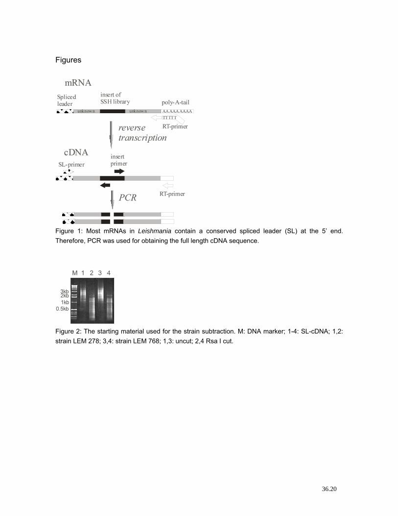
Figures
Figure 1: Most mRNAs in Leishmania contain a conserved spliced leader (SL) at the 5’ end. Therefore, PCR was used for obtaining the full length cDNA sequence.
Figure 2: The starting material used for the strain subtraction. M: DNA marker; 1-4: SL-cDNA; 1,2: strain LEM 278; 3,4: strain LEM 768; 1,3: uncut; 2,4 Rsa I cut.
36.20
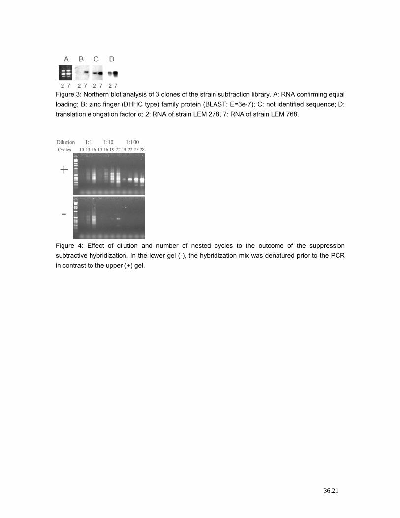
Figure 3: Northern blot analysis of 3 clones of the strain subtraction library. A: RNA confirming equal loading; B: zinc finger (DHHC type) family protein (BLAST: E=3e-7); C: not identified sequence; D: translation elongation factor α; 2: RNA of strain LEM 278, 7: RNA of strain LEM 768.
Figure 4: Effect of dilution and number of nested cycles to the outcome of the suppression subtractive hybridization. In the lower gel (-), the hybridization mix was denatured prior to the PCR in contrast to the upper (+) gel.
36.21
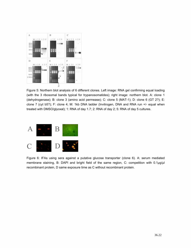
Figure 5: Northern blot analysis of 6 different clones. Left image: RNA gel confirming equal loading (with the 3 ribosomal bands typical for trypanosomatides); right image: northern blot. A: clone 1 (dehydrogenase); B: clone 3 (amino acid permease); C: clone 5 (MAT-1); D: clone 6 (GT 2?); E: clone 7 (cyt b5?); F: clone 4; M: 1kb DNA ladder (Invitrogen, DNA and RNA run +/- equal when treated with DMSO/glyoxal); 1: RNA of day 1.7; 2: RNA of day 2; 5: RNA of day 5 cultures.
Figure 6: IFAs using sera against a putative glucose transporter (clone 6). A: serum mediated membrane staining, B: DAPI and bright field of the same region, C: competition with 0.1µg/µl recombinant protein, D same exposure time as C without recombinant protein.
36.22
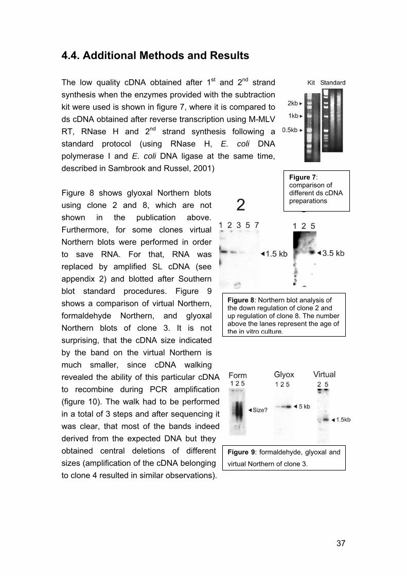
4.4. Additional Methods and Results The low quality cDNA obtained after 1st and 2nd strand synthesis when the enzymes provided with the subtraction kit were used is shown in figure 7, where it is compared to ds cDNA obtained after reverse transcription using M-MLV RT, RNase H and 2nd strand synthesis following a standard protocol (using RNase H, E. coli DNA polymerase I and E. coli DNA ligase at the same time, described in Sambrook and Russel, 2001)
Figure 7: comparison of different ds cDNA preparations
Figure 8 shows glyoxal Northern blots using clone 2 and 8, which are not shown in the publication above. Furthermore, for some clones virtual Northern blots were performed in order to save RNA. For that, RNA was replaced by amplified SL cDNA (see appendix 2) and blotted after Southern blot standard procedures. Figure 9 shows a comparison of virtual Northern, formaldehyde Northern, and glyoxal Northern blots of clone 3. It is not surprising, that the cDNA size indicated by the band on the virtual Northern is much smaller, since cDNA walking revealed the ability of this particular cDNA to recombine during PCR amplification (figure 10). The walk had to be performed in a total of 3 steps and after sequencing it was clear, that most of the bands indeed derived from the expected DNA but they obtained central deletions of different sizes (amplification of the cDNA belonging to clone 4 resulted in similar observations).
Figure 8: Northern blot analysis of the down regulation of clone 2 and up regulation of clone 8. The numberabove the lanes represent the age ofthe in vitro culture.
Figure 9: formaldehyde, glyoxal and
virtual Northern of clone 3.
37
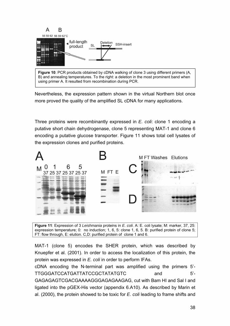
Figure 10: PCR products obtained by cDNA walking of clone 3 using different primers (A, B) and annealing temperatures. To the right: a deletion in the most prominent band when using primer A. It resulted from recombination during PCR.
Nevertheless, the expression pattern shown in the virtual Northern blot once more proved the quality of the amplified SL cDNA for many applications. Three proteins were recombinantly expressed in E. coli: clone 1 encoding a putative short chain dehydrogenase, clone 5 representing MAT-1 and clone 6 encoding a putative glucose transporter. Figure 11 shows total cell lysates of the expression clones and purified proteins.
Figure 11: Expression of 3 Leishmania proteins in E. coli. A: E. coli lysate; M: marker, 37, 25: expression temperature; 0: no induction; 1, 6, 5: clone 1, 6, 5. B: purified protein of clone 5; FT: flow through, E: elution. C,D: purified protein of clone 1 and 6.
MAT-1 (clone 5) encodes the SHER protein, which was described by Knuepfer et al. (2001). In order to access the localization of this protein, the protein was expressed in E. coli in order to perform IFAs. cDNA encoding the N-terminal part was amplified using the primers 5’-TTGGGATCCATGATTATCCGCTATATGTC and 5’-GAGAGAGTCGACGAAAAGGGAGAGAAGAG, cut with Bam HI and Sal I and ligated into the pGEX-His vector (appendix 6.A10). As described by Marin et al. (2000), the protein showed to be toxic for E. coli leading to frame shifts and
38
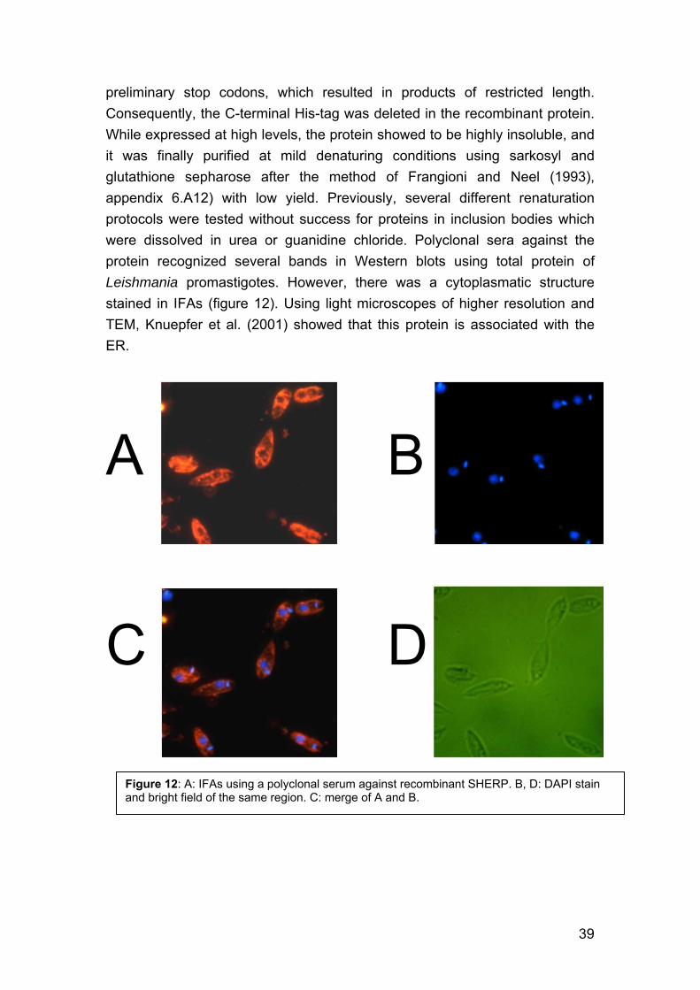
preliminary stop codons, which resulted in products of restricted length. Consequently, the C-terminal His-tag was deleted in the recombinant protein. While expressed at high levels, the protein showed to be highly insoluble, and it was finally purified at mild denaturing conditions using sarkosyl and glutathione sepharose after the method of Frangioni and Neel (1993), appendix 6.A12) with low yield. Previously, several different renaturation protocols were tested without success for proteins in inclusion bodies which were dissolved in urea or guanidine chloride. Polyclonal sera against the protein recognized several bands in Western blots using total protein of Leishmania promastigotes. However, there was a cytoplasmatic structure stained in IFAs (figure 12). Using light microscopes of higher resolution and TEM, Knuepfer et al. (2001) showed that this protein is associated with the ER.
A B
C D
Figure 12: A: IFAs using a polyclonal serum against recombinant SHERP. B, D: DAPI stain and bright field of the same region. C: merge of A and B.
39
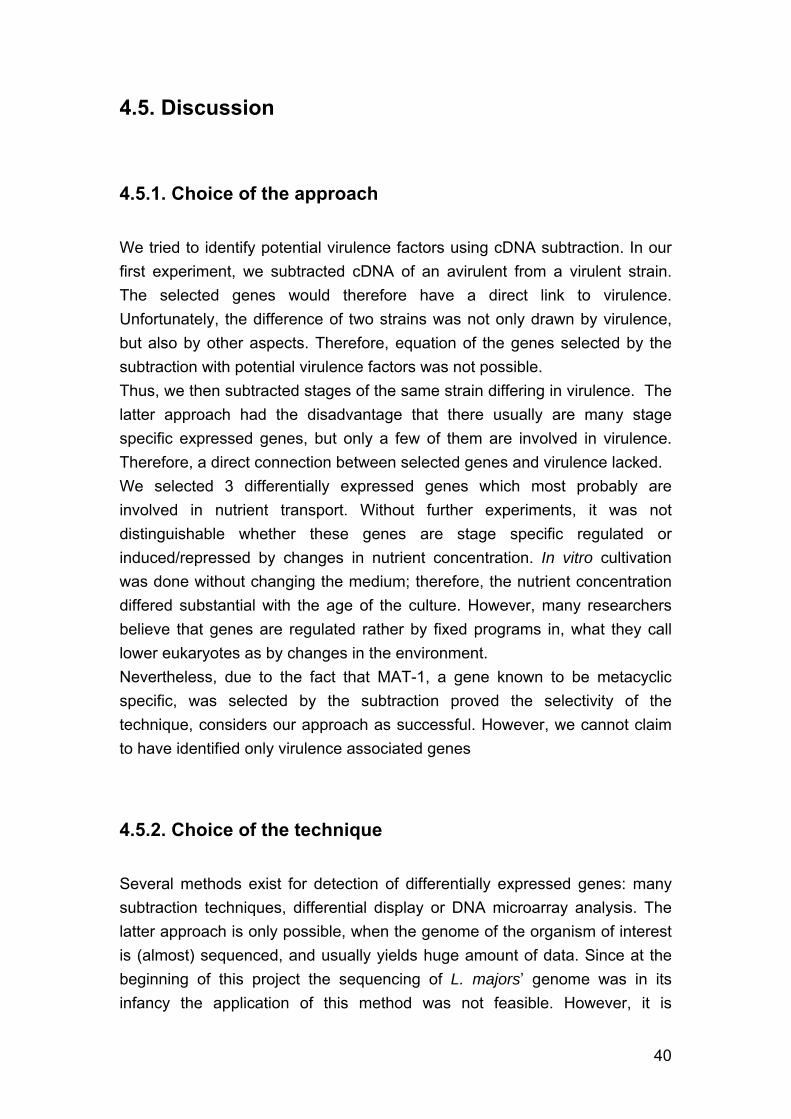
4.5. Discussion 4.5.1. Choice of the approach We tried to identify potential virulence factors using cDNA subtraction. In our first experiment, we subtracted cDNA of an avirulent from a virulent strain. The selected genes would therefore have a direct link to virulence. Unfortunately, the difference of two strains was not only drawn by virulence, but also by other aspects. Therefore, equation of the genes selected by the subtraction with potential virulence factors was not possible. Thus, we then subtracted stages of the same strain differing in virulence. The latter approach had the disadvantage that there usually are many stage specific expressed genes, but only a few of them are involved in virulence. Therefore, a direct connection between selected genes and virulence lacked. We selected 3 differentially expressed genes which most probably are involved in nutrient transport. Without further experiments, it was not distinguishable whether these genes are stage specific regulated or induced/repressed by changes in nutrient concentration. In vitro cultivation was done without changing the medium; therefore, the nutrient concentration differed substantial with the age of the culture. However, many researchers believe that genes are regulated rather by fixed programs in, what they call lower eukaryotes as by changes in the environment. Nevertheless, due to the fact that MAT-1, a gene known to be metacyclic specific, was selected by the subtraction proved the selectivity of the technique, considers our approach as successful. However, we cannot claim to have identified only virulence associated genes 4.5.2. Choice of the technique Several methods exist for detection of differentially expressed genes: many subtraction techniques, differential display or DNA microarray analysis. The latter approach is only possible, when the genome of the organism of interest is (almost) sequenced, and usually yields huge amount of data. Since at the beginning of this project the sequencing of L. majors’ genome was in its infancy the application of this method was not feasible. However, it is
40
interesting to note that, beside MAT-1, none of the genes we selected using SSH was found by micro analysis by Saxena et al. (2003). Whether this is due to chance, or whether it shows that, despite a large amount of generated data, microarray analyses miss a lot of differentially regulated genes is unclear. Furthermore, microarrays will fail to detect RNA editing and alternative splicing, which has been described for more and more genes (however as known so far, introns are very rare in trypanosomatids). Compared to conventional subtraction methods, SSH is an advanced technique, because it incorporates normalization during the first round of subtraction. The method therefore yields fragments of both, abundant and low abundant, differentially expressed RNA. Thus, SSH was the method of choice in our experiments. However, a RNA species becoming more and more important will be missed by all techniques mentioned above: micro RNAs (miRNAs). These usually about 22nt long miRNAs are missed during reverse transcription (e.g. using oligo-dT derived primers) and further, in microarray analysis, because of their restricted length yielding in low melting points (the spotted fragments in micro array usually are 50nt to several 100nt long). Nevertheless, miRNA are highly abundant in both, prokaryotes and eukaryotes, and are involved in as important features as gene repression, regulation of RNA stability and both, translational repression and activation (reviewed by Bartel, 2004). The objective was to identify potential virulence factors, drug targets and vaccine candidates. Whether the genes we have selected by cDNA subtraction fulfil this objective cannot yet be determined. However, some of the selected genes show interesting properties: the gene encoding domains involved in protein-protein interactions (a homologue protein with unknown function has been described in the pathogen T. brucei brucei) and the gene encoding a protein lacking any domains and homologies to known or hypothetical proteins as examples. In our subtraction, the slowest step surely was the identification of the selected genes. Therefore, many more genes might have been selected and identified if the subtraction was repeated using a reverse transcriptase lacking RNase H activity or when we would have managed to get to work the modifications of SSH described chapter 4.2. In order to perform localization experiments three of the selected genes were expressed recombinantly in E. coli. Immunization with clone 1 did not result in useful sera and the localization of MAT-1 encoding SHERP was previously described. In IFAs, antibodies against clone 6 stained the parasite’s surface;
41
this confirmed the sequence analysis, which showed that this clone encoded a glucose transporter. Alternatively, instead of immunization experiments, gene overexpression or knock outs could have been performed in transformation experiments. Since there was no experience in our group on Leishmania transformation, this would have been a high risk project. Whether such an approach would be able to define the effect of this particular gene on virulence remains to be shown. All together it seems that the chosen methods were appropriate within reasonable costs, but modification might yield more results.
42
5. General Discussion Although the three projects can be merged only loosely under the term leishmaniasis, there is a molecular link between them: the mini-exon gene encoding the spliced leader. The gene itself was used as template for the diagnostic PCR, spliced leader cDNA was used to construct expression and subtraction libraries and it enabled cDNA walking for identification of genes selected in subtractions. Histones represent another tie between the subtraction experiment and the serum screen: core histones showed to be of diagnostic value, and histone H1 was higher expressed in metacyclics of the more virulent Leishmania strain. The interesting properties of Leishmania histones (unusual high diversity and differential expression in strains differing in infectivity) might indicate the presence of a goldmine for understanding these parasites. The three projects were performed in order to develop tools for diagnosis of human and canine leishmaniasis, and for identification of vaccine candidates and potential virulence factors. 5.1. Diagnosis A PCR assay allowing diagnosis and species differentiation of Leishmania species causing human leishmaniasis was not yet available. Therefore, we developed a mini-exon PCR which exactly fits into this missing field. While the PCR and RFLP assay now is routine use at the STI showing satisfying results, distinguishing between Viannia species still remains a challenge. Since the required treatment of species belonging to this subgenus differs substantially, this is a major draw back. However, the assay might be able to be improved: restriction digests show only different patterns, if mutations are present within the recognition sequences of the selected enzymes. This problem could be overcome with chip technology, which then could facilitate the differentiation of Viannia species, or by using an additional genetic marker. While many different diagnostic tools are available for canine leishmaniasis, a diagnostic test capable of detection of early stages of infection is still lacking. We selected a few promising antigens suitable for an early diagnostic test for canine leishmaniasis. However, Leishmania histones have been previously
43
described as antigens, inducing strong humoral responses in both, infected dogs and humans. Whether the epitopes we selected incorporated better properties has to be tested using sera of many more naturally and artificially infected dogs. Since we used cDNA derived from a highly infective and virulent strain for the serum screen, the epitopes we identified might be good candidates. Collaboration with a laboratory in Spain will provide this information. However, the goal of an early diagnostic test for canine leishmaniasis was to allow mass diagnosis and treatment street dogs as a control tool for leishmaniasis in Southern Europe. Whether this dodgy approach will succeed, has to be shown in future. It surely will be intensive in costs and work and will not protect treated dogs from relapses and re-infection. Furthermore, the consequences in respect to drug resistance might be fatal. Therefore, it seems that it would be more feasible to concentrate all means on the development of a vaccine for both, humans and dogs, and for development of new drugs. Nevertheless, an early diagnostic test for canine leishmaniasis would be an improvement of veterinarian praxis. 5.2. Vaccine candidates By enriching metacyclic stage specific genes, we also enriched for potential transmission blocking vaccine candidates. Several integral membrane proteins were selected, two transporters and a dehydrogenase. Whether these proteins are suitable as vaccine candidates has to be shown in future studies. Here fore, they must be able to induce a long lasting protective TH1 type immune answer, and must be stable and produced cheaply to allow mass treatment. Although the probability whether these conditions are fulfilled by one of the three proteins we selected is rather low, the immense value of a vaccine as a powerful tool for control of leishmaniasis justifies a slight hope. Nevertheless, the enrichment of potential vaccine candidates was only a byproduct of a subtraction designed for the selection of potential virulence factors. An alternative to designing a vaccine based on single or several parasites’ molecules is to develop live or killed vaccines. However, costs for Leishmania cultivation currently are much too high. By lowering these costs, (e.g. achieving serum-free cultivation in a cheap medium), the use of attenuated parasites would be a true alternative. Furthermore, cultivated parasites are
44
not only worthwhile as vaccine but are also of excellent diagnostic value. 5.3. Virulence factors Classically, virulence factors have been defined as factors causing or enhancing the severity of a disease. However, the distinction of virulence factors (causes for pathology) from factors involved in pathogenicity appears to be artificial. Thus, all parasite factors enabling/enhancing the parasite’s infectivity, survival within the host and factors defining the severity of the disease could be handled as virulence factors. Understanding the mode of action of these factors is crucial for understanding leishmaniasis. We performed two experiments in order to enrich potential virulence factors. However, whether the selected genes are involved in virulence has to be shown using in vivo studies. Due to the complexity of the matter, assigning the role of virulence factors as well as their mode of action is a challenge, as the example of LPG shows. Although LPG was suspected to be a virulence factor many years ago, its impact was unclear or misunderstood. Since initial findings showed that LPG enhances the metacyclic’s resistance to the complement and is involved in the transient inhibition of phagolysosomal fusion, these properties were believed to be the mode of action. However, using lpg- mutants, Spath et al. (2003) showed that at least in mice, complement mediated lysis is an ineffective way of controlling the disease. Lpg- metacyclics were opsonised by complement factors and entered macrophages. They also showed that inhibition of phagolysosomal fusion is not essential for the parasite’s survival within phagocytes. Nevertheless, lpg- mutants showed a significant higher sensitivity to oxidative stress, decreasing the parasite’s ability to resist the oxidative burst generated by activated macrophages. The different observations and conclusions on LPG’s impact were most probably derived from using different approaches: the initial experiments were performed in vitro and using biochemical methods (LPG shares domains with many other Leishmania derived molecules). The example of LPG clearly shows, that defining a gene as virulence factor is intricate: although recent studies confirm that LPG is involved in virulence, this could in principle have been disproved just as well. Furthermore, the results of in vivo studies using mice cannot be directly assigned to humans, due to significant differences in the immune system.
45
Therefore, much care has to be taken when defining virulence factors, since the outcome of experiments depends on the approach, technique and system used. These facts clearly show that identification of virulence factors would be far beyond the reach of this PhD project. Nevertheless, our experiments were designed to select for potential virulence factors. The selected genes were expressed at higher levels in the more infective strain or in the infective parasite stage respectively, which is a first indication, that they could be involved in virulence. 5.4. Drug targets Gene products expressed in metacyclic and amastigote parasites are potential drug targets for prophylaxis and/or treatment, if they are essential for the parasite and differ in the tertiary structure from homologues expressed in the host. Therefore, we enriched also potential drug targets by the cDNA subtraction. However, since expression in amastigotes was not yet shown, the use of these genes might be of limited value as drug targets. Nevertheless, some of the genes, such as the GU1 like gene, have no homologies at all to mammalian proteins known so far (at least in the primary structure). There are some newer antileishmanial drugs in use and in the pipeline, but their number is restricted, and most of them are no oral agents. In order to avoid progression of drug resistance (which already has spread widely to antimonials), alternative drugs are highly welcome. By using several different drugs at different locations and time points, progression of resistance might be successfully suppressed and even reversed. However, in addition to evolution-driven selection of resistant phenotypes, Leishmania parasites can react unspecifically to drug pressure by gene amplification yielding in extrachromosomal plasmids and mini chromosomes. Nevertheless, some chemical and physical properties of a drug can aggravate the development of resistances:
1. A rapid mechanism of action, as observed by the antimalarial artemether, prevents unspecific adaptations of the parasite such as gene amplification. However, drug resistance can still progress by selection of resistant phenotypes formed by chance in the whole parasite population.
2. It is more difficult for the parasite to develop resistance to drugs which
46
don’t need parasite’s transporter molecules to reach the point of action. These are either drugs damaging the parasite surface or hydrophobic molecules which are able to diffuse through biological membranes. However, against the latter drugs, an effective countermeasure is active transport out of the parasites cytosol.
Development of antileishmanials surely is a challenge: Leishmania is endemic in poor regions of the world, therefore production should be of low costs and oral application should be possible (in order to lower treatment costs). Furthermore, the drug should exhibit low rates of adverse reactions. The fact that toxic antimonials still are widely used clearly shows the deficits of today’s antileishmanials. The property of Leishmania parasites to multiply within acidic compartments might be utilized by the drug development: hydrophobic molecules, which are uncharged at neutral pH but protonated within phagolysosomes are able to diffuse through membranes when they are uncharged. Once protonated, membrane diffusion is disabled. This leads to accumulation of high drug concentrations within phagolysosomes, which might enhances the efficiency of the drug concurrent with reduction of adverse reactions. However, it is more likely that new drugs are identified by screening of thousands of compounds than by specific design upon potential drug targets. The selected drugs then could be chemically modified using the criteria mentioned above. Nevertheless, some parasite specific proteins without mammal homologues might ask for direct design of drugs. Whether the GU1 homologue we identified or other genes selected by cDNA subtraction qualify for this purpose cannot yet be answered. However, the cDNA subtraction experiments proved their ability to select genes exhibiting interesting properties not only in respect to potential virulence factors, but also to potential transmission blocking vaccine candidates and to potential drug targets. However, it has to be straightened out that these preliminary results are far from practical application, they only exhibit indications that further research on some genes we identified might yield in powerful tools for control of Leishmania.
47
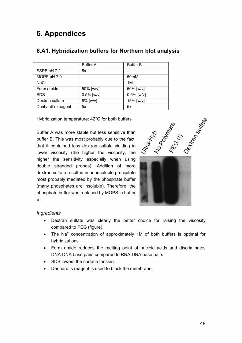
6. Appendices 6.A1. Hybridization buffers for Northern blot analysis Buffer A Buffer B SSPE pH 7.2 5x - MOPS pH 7.0 - 50mM NaCl - 1M Form amide 50% [w/v] 50% [w/v] SDS 0.5% [w/v] 0.5% [w/v] Dextran sulfate 8% [w/v] 15% [w/v] Denhardt’s reagent 5x 5x Hybridization temperature: 42°C for both buffers Buffer A was more stable but less sensitive than buffer B. This was most probably due to the fact, that it contained less dextran sulfate yielding in lower viscosity (the higher the viscosity, the higher the sensitivity especially when using double stranded probes). Addition of more dextran sulfate resulted in an insoluble precipitate most probably mediated by the phosphate buffer (many phosphates are insoluble). Therefore, the phosphate buffer was replaced by MOPS in buffer B. Ingredients
• Dextran sulfate was clearly the better choice for raising the viscosity compared to PEG (figure).
• The Na+ concentration of approximately 1M of both buffers is optimal for hybridizations
• Form amide reduces the melting point of nucleic acids and discriminates DNA-DNA base pairs compared to RNA-DNA base pairs.
• SDS lowers the surface tension. • Denhardt’s reagent is used to block the membrane.
48
6.A2. Construction of spliced leader cDNA
Primers: Eco RI 1str Eco primer 5’-CGACTGTACGTGAATTCGC(T)20
SL Eco primer 5’- GCTATATAAGAATTCAGTTTCTGTACTTTATTG Reverse Transcription
1 To 1.5µg total or mRNA add 14µl 10µM 1str Eco primer and TE to 23.5µl final volume.
2 Incubate 5min @ 70°C, snap cool, quick spin. 3 Add 3µl 10x 1st strand buffer,
1.5µl 10mM dNTPs, 0.5µl RNasin (Invitrogen) 1.5µl M-MLV RT, RNase H- (Finnzymes)
4 Incubate 1hour @ 37°C. When total RNA was used:
5 Heat inactivate RT for 2min @ 95°C. 6 Quickspin, add 1µl RNase A (1g/l) and incubate 15min @ 37°C. Store @ -
20°C. When mRNA was used:
5 Add 22.7µl H2O 6µl 10x RNase H buffer 1µl RNase A (1g/l)
0.3µl RNase H (5U/µl) 6 Incubate 20min @ 37°C. Store @ -20°C and use a 1/10 dilution in TE as PCR
template. PCR
1 Prepare a mastermix containing 1x Taq buffer (1.5mM MgCl2), 10% DMSO, 0.2mM dNTPs, 0.5µM SL Eco and 1str Eco primer, and use Taq:Pfu DNA polymerase in a 8:1 proportion.
2 Add 1-2µl 1st strand cDNA for a 50µl reaction. 3 Perform 20-35 cycles at the following conditions
30sec 94°C 30sec 54°C 2min 72°C
49
6.A3. Isolation of genomic Leishmania DNA Cells are lysed in SDS, proteins are digested by proteinase K and extracted with phenol From in vitro cultures 1 Centrifuge culture 5min @ 1’500g. 2 Wash the cells in PBS. From biopsy 1 Store specimen in 0.9% NaCl @ -20°C. 2 Thaw material quickly. 3 Resuspend in proteinase K buffer (10mM TrisCl pH8, 5mM EDTA, 0.5% SDS)
culture: 108 cells/ml, biopsy: depends on the size of specimen, ~500µl. 4 Add proteinase K to a final concentration of 0.5g/l. 5 Incubate 3 hours to overnight @ 56°C. 6 Extract the aqueous phase once with 1 volume phenol (pH 8), once with 1
volume phenol/chlorophorme, and once with 1 volume chlorophorme. 7 Ethanol precipitate the DNA using 1/10 volume of 3M NaAcetate (pH 5.2) and
2-3 volumes of EtOH abs. 8 Incubate 30min @ RT. 9 Spin 30min @ full speed. 10 Wash the pellet in 75% EtOH. 11 Resuspend the DNA in 50-100µl TE (10mM TrisCl, pH 8, 0.5mM EDTA). 12 Optional: add RNase A to a final concentration of 10mg/l. 13 Store @ -20°C.
50
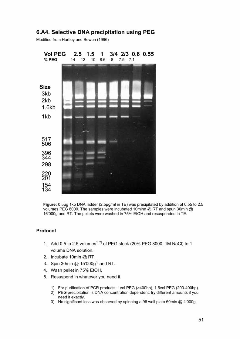
6.A4. Selective DNA precipitation using PEG Modified from Hartley and Bowen (1996)
Vol PEG 2.5 1.5 1 3/4 2/3 0.6 0.55% PEG 14 12 10 8.6 8 7.5 7.1
3kb Size
2kb 1.6kb 1kb
517 506 396 344 298 220 201 154 134
Figure: 0.5µg 1kb DNA ladder (2.5µg/ml in TE) was precipitated by addition of 0.55 to 2.5 volumes PEG 8000. The samples were incubated 10minn @ RT and spun 30min @ 16’000g and RT. The pellets were washed in 75% EtOH and resuspended in TE.
Protocol
1. Add 0.5 to 2.5 volumes1, 2) of PEG stock (20% PEG 8000, 1M NaCl) to 1 volume DNA solution.
2. Incubate 10min @ RT 3. Spin 30min @ 15’000g3) and RT. 4. Wash pellet in 75% EtOH. 5. Resuspend in whatever you need it.
1) For purification of PCR products: 1vol PEG (>400bp), 1.5vol PEG (200-400bp). 2) PEG precipitation is DNA concentration dependent: try different amounts if you
need it exactly. 3) No significant loss was observed by spinning a 96 well plate 60min @ 4’000g.
51
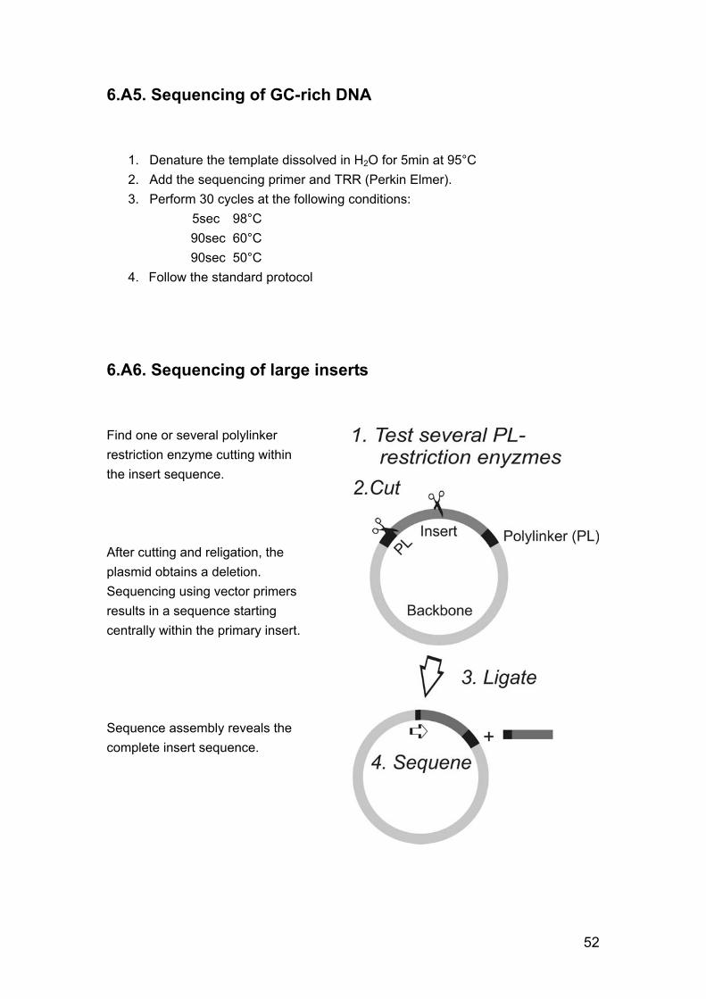
6.A5. Sequencing of GC-rich DNA
1. Denature the template dissolved in H2O for 5min at 95°C 2. Add the sequencing primer and TRR (Perkin Elmer). 3. Perform 30 cycles at the following conditions:
5sec 98°C 90sec 60°C 90sec 50°C 4. Follow the standard protocol
6.A6. Sequencing of large inserts Find one or several polylinker restriction enzyme cutting within the insert sequence. After cutting and religation, the plasmid obtains a deletion. Sequencing using vector primers results in a sequence starting centrally within the primary insert. Sequence assembly reveals the complete insert sequence.
52
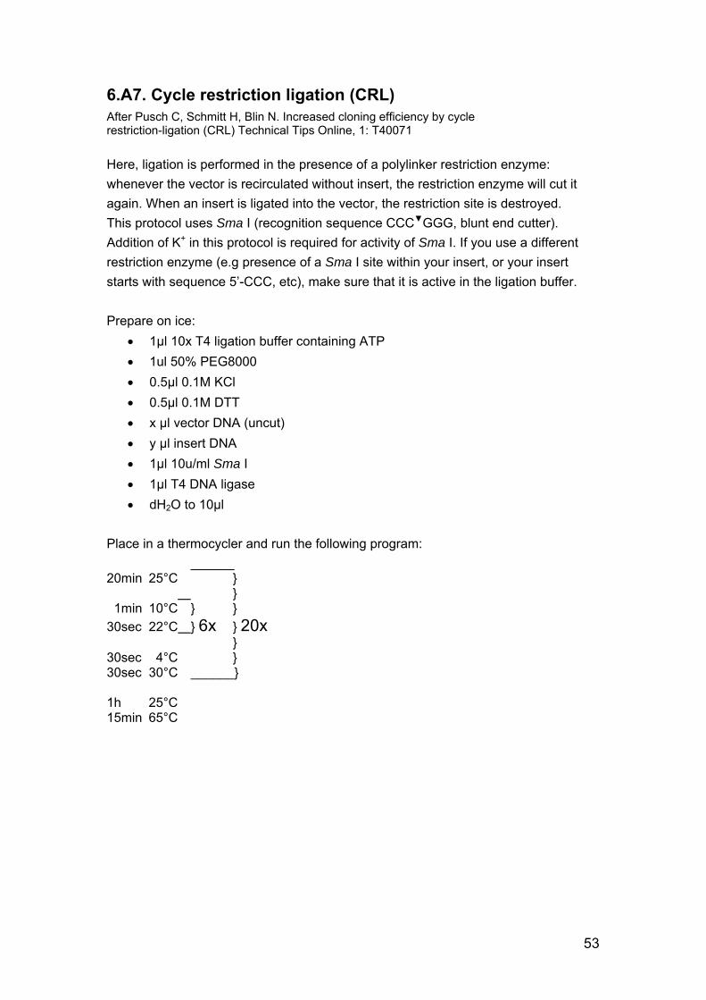
6.A7. Cycle restriction ligation (CRL) After Pusch C, Schmitt H, Blin N. Increased cloning efficiency by cycle restriction-ligation (CRL) Technical Tips Online, 1: T40071 Here, ligation is performed in the presence of a polylinker restriction enzyme: whenever the vector is recirculated without insert, the restriction enzyme will cut it again. When an insert is ligated into the vector, the restriction site is destroyed. This protocol uses Sma I (recognition sequence CCC▼GGG, blunt end cutter). Addition of K+ in this protocol is required for activity of Sma I. If you use a different restriction enzyme (e.g presence of a Sma I site within your insert, or your insert starts with sequence 5’-CCC, etc), make sure that it is active in the ligation buffer. Prepare on ice:
• 1µl 10x T4 ligation buffer containing ATP • 1ul 50% PEG8000 • 0.5µl 0.1M KCl • 0.5µl 0.1M DTT • x µl vector DNA (uncut) • y µl insert DNA • 1µl 10u/ml Sma I • 1µl T4 DNA ligase • dH2O to 10µl
Place in a thermocycler and run the following program: ______ 20min 25°C } } 1min 10°C } } 30sec 22°C } 6x } 20x } 30sec 4°C } 30sec 30°C ______} 1h 25°C 15min 65°C
53
6.A8. Small scale preparation of electro-competent cells
1. Thaw a glycerol stocks of any overnight culture, which was not overgrown too heavily.
2. Spin the culture and discard the supernatant. 3. Wash the pellet 2 times in 1ml ice-cold dH2O. 4. Resuspend the pellet(s) in 50µl ice-cold dH2O.
The cells are now ready for electroporation. Do NOT freeze them, only for immediate use!
6.A9. E. coli transformation Based on Inoue et al. (1990)
Media and buffers SOB medium:
20g/l bacto tryptone 5g/l yeast extract 0.5g/l NaCl 0.19g/l KCl Autoclave. Just before use add MgCl2 to 10mM end concentration (SOB agar plates: 20mM) from an autoclaved 2M stock.
SOC medium: Add 2% filter sterilized 1M glucose to SOB (20mM end concentration).
ITB (Inoue transformation buffer)
1. Dissolve 10mM HEPES or PIPES, 15mM CaCl2, 250mM KCl
in dH2O. 2. Adjust pH to 6.7 with KOH. 3. Add MnCl2 to 55mM. 4. Sterilize with a 0.45µm filter and store @ 4°C.
54
Preparation of competent cells
1. Inoculate 200ml SOB with about 10 large colonies. For best results pick colonies from a freshly grown plate. Use different
amounts for inoculation to have one culture at the desired density the next day.
2. Incubate for about 20 hours at RT (best: 18-22°C) with vigorous shaking (200-250rpm) until OD600=0.45
OD=0.3-0.55 works well. 3. Place the culture on ice for 10min. 4. Spin 10min @ 2’500g and 4°C. 5. Resuspend the pellet in 64ml ice cold ITB, incubate 10min on ice and spin
again. 6. Resuspend the cells in 16ml ice cold ITB. 7. Slowly add 1.2ml DMSO while gently swirling and incubate 10min on ice. 8. Dispense the suspension into 0.2–1ml aliquots, immediately freeze by
immersion into liquid N2. Store @ -80°C. 9. Thaw an aliquot for testing your cells (pos AND neg!).
Expect 108–109 transformants/µg supercoiled plasmid in DH5α. Transformation
1. Thaw cells @ RT and immediately transfer on ice. 2. Add 50-100µl cells to your DNA (the volume of the DNA should not exceed
5%). It is not necessary to precipitate a ligation mix prior to transformation.
3. Incubate on ice for 15min. 4. Heat shock 1min @ 42°C in a water bath, cool in ice water.
Alternatively, you can plate the transformation mix directly on prewarmed plates.
5. Add 900ml SOC, incubate 30min @ 37°C and plate the bacteria.
55
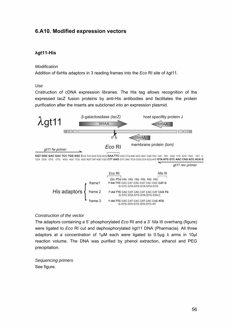
6.A10. Modified expression vectors
λgt11-His Modification Addition of 6xHis adaptors in 3 reading frames into the Eco RI site of λgt11. Use Cnstruction of cDNA expression libraries. The His tag allows recognition of the expressed lacZ fusion proteins by anti-His antibodies and facilitates the protein purification after the inserts are subcloned into an expression plasmid.
Construction of the vector The adaptors containing a 5’ phosphorylated Eco RI and a 3’ Nla III overhang (figure) were ligated to Eco RI cut and dephosphorylated λgt11 DNA (Pharmacia). All three adaptors at a concentration of 1µM each were ligated to 0.5µg λ arms in 10µl reaction volume. The DNA was purified by phenol extraction, ethanol and PEG precipitation. Sequencing primers See figure.
56
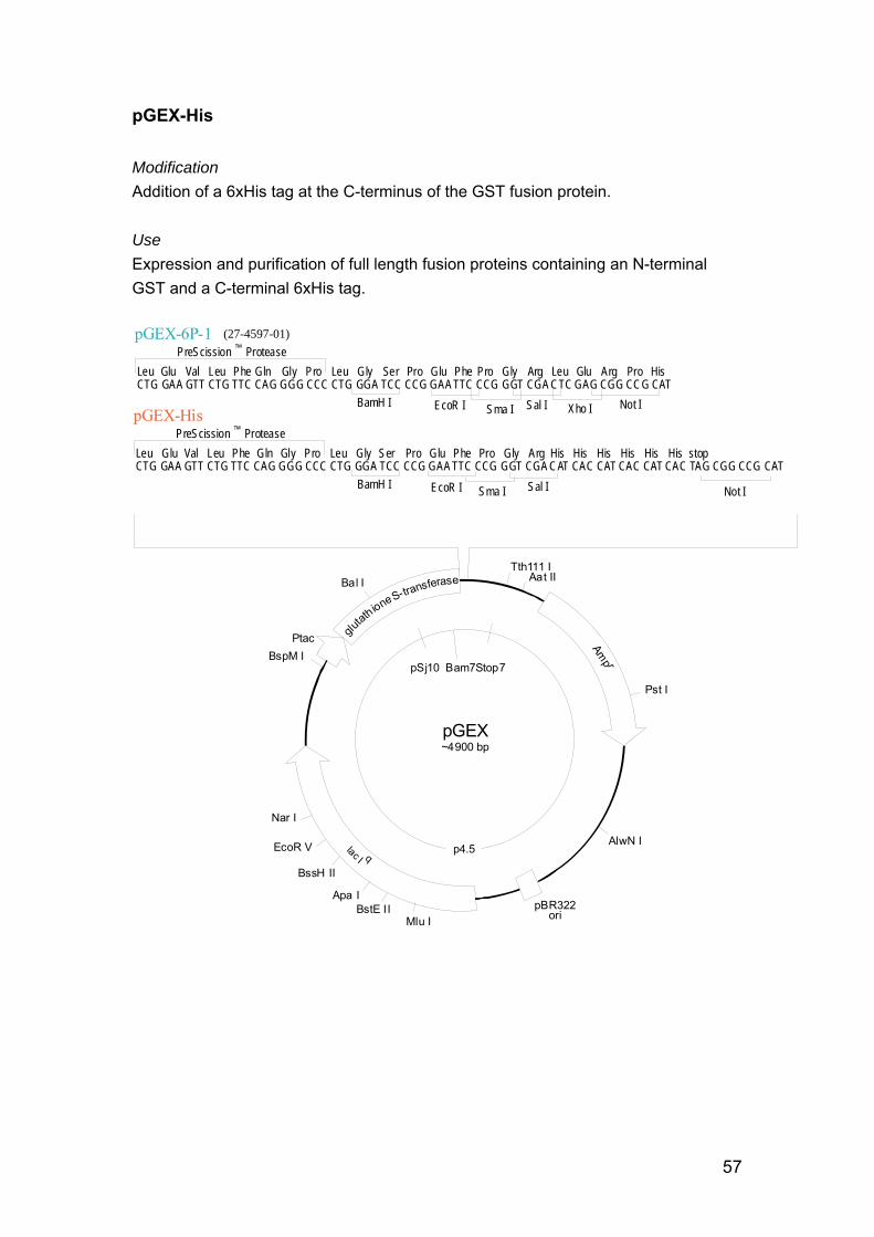
pGEX-His Modification Addition of a 6xHis tag at the C-terminus of the GST fusion protein. Use Expression and purification of full length fusion proteins containing an N-terminal GST and a C-terminal 6xHis tag.
(27-4597-01)
EcoR I Sma I Sal I Xho I Not IBamH I
PreScission ™ ProteaseLeu Glu Val Leu Phe Gln Gly Pro Leu Gly Ser Pro Glu Phe Pro Gly Arg Leu Glu Arg Pro HisCTG GAA GTT CTG TTC CAG GGG CCC CTG GGA TCC CCG GAA TTC CCG GGT CGA CTC GAG CGG CCG CAT
pGEX~4900 bp
pBR322ori
Bal I
BspM I Ptac glu
tathioneS-transferase
Amp r
lac I q
Nar I
EcoR V
BssH II
BstE II Mlu I
Apa I
Tth111 I Aat II
Pst I
p4.5AlwN I
pSj10 Bam7Stop7
EcoR I Sma I Sal IBamH I
PreScission ™ ProteaseLeu Glu Val Leu Phe Gln Gly Pro Leu Gly Ser Pro Glu Phe Pro Gly Arg His His His His His His stop CTG GAA GTT CTG TTC CAG GGG CCC CTG GGA TCC CCG GAA TTC CCG GGT CGA CAT CAC CAT CAC CAT CAC TAG CGG CCG CAT
Not I
57
Construction of the vector The adaptor obtained by annealing the two oligos 5’-AATTCCCGGGTCGACATCACCATCACCATCACTAGC and 5’-GGCCGCTAGTGATGGTGATGGTGATGTCGACCCGGG was ligated into the polylinker of pGEX-6P-1 (Pharmacia). 100ng of pGEX-6P-1 was digested by Sal I and Not I, phenol extracted, ethanol and PEG precipitated. 1µM adaptor was ligated to 50ng of the cut vector in 10µl reaction volume. The ligation mix was used to transform chemo competent Sure cells. 5 colonies were picked to prepare mini preps, which were Xho I digested in order to check for adaptor insertion. 3 minipreps containing the adaptor were sequenced. Sequencing primers pGEX fw: 5’-GGC TGG CAA GCC ACG TTT GG pGEX rev: 5’-GAA ACG CGC GAG GCA GAT CG
58
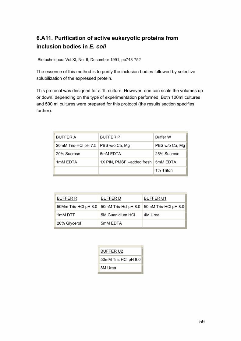
6.A11. Purification of active eukaryotic proteins from inclusion bodies in E. coli
Biotechniques: Vol XI, No. 6, December 1991, pp748-752
The essence of this method is to purify the inclusion bodies followed by selective solubilization of the expressed protein.
This protocol was designed for a 1L culture. However, one can scale the volumes up or down, depending on the type of experimentation performed. Both 100ml cultures and 500 ml cultures were prepared for this protocol (the results section specifies further).
BUFFER A BUFFER P Buffer W
20mM Tris-HCl pH 7.5 PBS w/o Ca, Mg PBS w/o Ca, Mg
20% Sucrose 5mM EDTA 25% Sucrose
1mM EDTA 1X PIN, PMSF,--added fresh 5mM EDTA
1% Triton
BUFFER R BUFFER D BUFFER U1
50Mm Tris-HCl pH 8.0 50mM Tris-Hcl pH 8.0 50mM Tris-HCl pH 8.0
1mM DTT 5M Guanidium HCl 4M Urea
20% Glycerol 5mM EDTA
BUFFER U2
50mM Tris HCl pH 8.0
8M Urea
59
1. The cells from the 1L culture are transformed into two 500ml tubes and are pelleted by centrifugation at 5000rpm/min at 4 degrees C. To prevent contaminating the inclusion bodies by proteins from the outer membrane, the outer membrane is removed by resuspending the cell pellet from 1L culture in 50ml BUFFER A. (If 100ml culture was prepared, only 5 ml Buffer A would be required. This down -scaling of volumes is required whenever a volume is indicated).
2. Incubate on ice for 10 minutes. Pellet the cells by centrifugation at 6000rpm/5min at 4 degrees C. Resuspend in 50ml of ice cold water. Leave on ice for 10 min. Centrifuge at 8200 rpm/5 min at 4 degrees C.
3. Resuspend the pellet in 10ml Buffer P containing protease inhibitors. 4. Sonicate the resuspended mixture 3x (50W) each with a 30sec. pulse with a
30 sec. pause in between pulses. RNase T1 (1.3x10³ U/10ml) + DNase I (400 ug/10ml) are added to the sonicated cell suspension and incubated at room temp for 10 min.
5. The suspension is further diluted by addition of 40 ml of Washing Buffer P and the crude inclusion bodies are pelleted by centrifugation at 11,000 rpm for 30 min at 4 degrees C. Keep aliquot and assay the supernatant for proteins.
6. The pellet (inclusion bodies) is suspended in 40 ml of Buffer W. Incubate on ice for 10 min and centrifuge at 15,000 rpm/5min at 4 degrees C. for 10 min. Keep aliquot of supernatant. Repeat this washing step and save the supernatant.
7. Resuspend the pellet in 10ml of Buffer D. A brief sonication with a 5 sec. pulse (50W) facilitates the solubilization of the aggregated proteins (NOTE: the pellet may also be resuspended in 4M or 8M urea in addition to BUFFER D).
8. The resultant suspension is incubated on ice for an additional 1 hour. 9. Centrifuge at 10,500 rpm for 30 min at 4 degrees C. Add the supernatant to
100ml of Renaturation Buffer R, and stir gently at 4 degrees C. over night to renature the proteins.
10. The next day, the supernatant is clarified by centrifugation at 12,000 rpm for 30 min at 4 degrees C. The supernatant contains highly purified expressed proteins.
60
6.A12. Purification of GST Fused Proteins After Frangioni and Neel (1993), written by Chia Jin Ngee Many people have vented out frustration over insoluble GST-fused proteins. This is a protocol that for enzymatically active soluble GST-fused proteins. All GST-fused proteins are rendered soluble with this technique though enzyme activitiy can range > from 30-90%.
Materials/Reagents
• STE Buffer
10 mM Tris-HCl, pH 8.0 1 mM EDTA 150 mM NaCl
• Lysozyme solution
10 mg/ml in water (make fresh)
• PBS • Elution Buffer
50 mM Tris.Cl, pH 9.0 20 mM GSH
• 10% Sarkosyl in STE Buffer • 10% Triton X-100 in STE Buffer • 1 M DTT • 100 mM IPTG
61
Method
Day 1
1. 1. Set up an overnight culture in 50 ml 2XTY with 150 mg/ml of ampicillin.
Day 2
2. Seed 5 ml of overnight culture to 500 ml 2XTY with 150 mg/ml of ampicillin. 3. Grow at 37oC to an A600 of 0.6 to 0.8. 4. Induce with 0.1 mM to 2 mM of IPTG. Grow for 3 hr at 37oC or grow overnight
at room temperature. Lower IPTG concentrations and lower growing temperatures tend to produce greater solubility at the expense of yield.
5. Pellet cells by centrifuging at 3000 g, 4oC for 10 min. Decant media and resuspend cells in 30 ml ice-cold PBS to wash. Transfer to a 40-ml Oak Ridge tube and centrifuge at 3000 g, 4oC for 10 min. Decant PBS.
6. This is a convenient point to stop and to store pellets at -80oC. Else continue to lyse cells.
7. Thaw pellet on ice if cells are frozen else proceed to the next step. 8. Resuspend pellet in 10 ml of ice cold STE Buffer. 9. Add 100 µl of freshly prepared lyozyme solution, incubate on ice for 15 min.
Just before sonication, add 100 µl of 1 M DTT and 1.4 ml of 10% Sarkosyl. Mix thoroughly by inversion and sonicate for a total time of 1 min.
10. Centrifuge 16,000 rpm for 20 min on the SS34 rotor to pellet debris. Transfer supernatant to a 50-ml conical tube and discard the pellet. Add 4 ml of 10% Triton X-100 and top up with STE Buffer to 20 ml. The effective concentration of Sarkosyl and Triton X-100 will be 0.7% and 2% respectively. Incubate at room temperature for 30 min.
11. Pour the lysate to 1 ml bed of prepared Glutathione Sepharose in PBS. Incubate at room temperature for 30 min to 1 hr with agitation. To prepare the 50% slurry, shake up the media and pipette 2 ml to a 50 ml tube. Fill to 50 ml with PBS, invert tube a few times. Centrifuge to 2000 rpm on a swing bucket centrifuge then switch off. Carefully suck off PBS and resuspend beads with 1 ml of PBS.
12. Wash the beads with 3 X 50 ml of PBS. Finally resuspend in 5 ml of PBS. Pour to a dispo-column. Wash the 50-ml conical tube with an additional 5 ml of PBS. Pool with the first 5 ml in the dispo-column. To wash, use the same centrifugation technique for preparing the beads. When transferring beads to column, do not pipette but pour. The beads tend to stick to pipette tips.
13. If desired, elute with 10 x 1 ml fractions of Elution Buffer. Determine desired fractions with SDS PAGE.
62
7. Acknowledgments First of all I’d like to thank Hans-Peter Beck for enabling and supervising this PhD project. I appreciate the freedom I had in choosing the way to go during this thesis. Thanks also for always having time for answering my questions. I thank Prof. N. Weiss and Prof M. Tanner for being part of my thesis committee and for their support. Many thanks to Jutta Marfurt for adding that many efforts in the diagnostic test project (chapter 2): she did the industrious work (screening the cDNA libraries), whereas I picked the interesting part of the work (constructing the libraries). Thanks also for accepting me and my moods as coworker in the same lab! I also want to thank Sylvia Steiger, who did incredible amounts of SDS-PAGEs for this project. For the mini-exon project (chapter 3), I thank again Jutta Marfurt who did the main job, Divine Makia who elaborated the RFLP scheme among other things, and Ingrid Felger for supervising (but of course also for her spontaneous comments)! Many thanks to the whole group of Hans-Peter Beck for the great working atmosphere, and their help on scientific topics, as an example: Tobias Spielmann had the great idea of adding His adaptors to λgt11 phages (appendix 6.A10). I’d like to thank Reto Brun and his group for making the parasite stabilates available and for introducing me into the field of Leishmania cultivation. I also want to thank the group of Gerd Pluschke for helping out when antibodies and other ingredients for IFAs were used up and for loading the sequencer. Many thanks to Hans-Peter Beck and Ingrid Felger for their great parties and (this was really incredible) the excellent dinners!
63
Thanks a lot to Till Voss, Tobi Spielmann, Roland Degen, Mirjam Kästli, Christian Flück, Cornelia Spycher, Christian Scherer, Sebastrian Rusch, Selina Bopp, Alexandra Lüscher, Diana Diaz, Shinji Okitsu, Attila Regös, Matthias Rottmann, Eric Huber, Michi Oberle, Sonja Bernhard, Martin Nägeli, Erwin Kump, André Tiaden (wow, what a list!) and all the others for the brilliant garden parties, ping pong matches, billiard-, Döggele-, Jass-, poker-evenings and for the Aare-raft! Also thanks for loosing sometimes, but no thanks for winning… I want to thank Till and Tobi for providing bed and breakfast (and more important: beer) in Australia! Thanks also to Cornelia for securing my 85kg live (fat) weight during rock climbing.
64
8. References Alexander, J., A.R. Satoskar, and D.G. Russell. 1999. Leishmania species: models of intracellular parasitism. J Cell Sci 112 Pt 18, 2993-3002. Anbar, M., and P. Neta. 1967. A compilation of specific bimolecular rate constants for the reaction of hydrated electrons, hydrogen atoms and hydroxyl radical with inorganic and organic compounds in aqueous solution. Int J Appl Radiation Isotopes 18, 493-523. Anderson, S., J.R. David, and D. McMahon-Pratt. 1983. In vivo protection against Leishmania mexicana mediated by monoclonal antibodies. J Immunol 131, 1616-8. Angarano, G., P. Maggi, M.A. Rollo, A.M. Larocca, M. Quarto, A. Scalone, and L. Gradoni. 1998. Diffuse necrotic hepatic lesions due to visceral leishmaniasis in AIDS. J Infect 36, 167-9. Balestieri, F.M., A.R. Queiroz, C. Scavone, V.M. Costa, M. Barral-Netto, and A. Abrahamsohn Ide. 2002. Leishmania (L.) amazonensis-induced inhibition of nitric oxide synthesis in host macrophages. Microbes Infect 4, 23-9. Baneth, G., and S.E. Shaw. 2002. Chemotherapy of canine leishmaniosis. Vet Parasitol 106, 315-24. Bartel, D.P. 2004. MicroRNAs: genomics, biogenesis, mechanism, and function. Cell 116, 281-97. Beckman, J.S., T.W. Beckman, J. Chen, P.A. Marshall, and B.A. Freeman. 1990. Apparent hydroxyl radical production by peroxynitrite: implications for endothelial injury from nitric oxide and superoxide. Proc Natl Acad Sci U S A 87, 1620-4. Benne, R., J. Van den Burg, J.P. Brakenhoff, P. Sloof, J.H. Van Boom, and M.C. Tromp. 1986. Major transcript of the frameshifted coxII gene from trypanosome mitochondria contains four nucleotides that are not encoded in the DNA. Cell 46, 819-26. Berman, J. 2003. Current treatment approaches to leishmaniasis. Curr Opin Infect Dis 16, 397-401. Brittingham, A., C.J. Morrison, W.R. McMaster, B.S. McGwire, K.P. Chang, and D.M. Mosser. 1995. Role of the Leishmania surface protease gp63 in complement fixation, cell adhesion, and resistance to complement-mediated lysis. J Immunol 155, 3102-11. Campbell, D.A., S. Thomas, and N.R. Sturm. 2003. Transcription in kinetoplastid protozoa: why be normal? Microbes Infect 5, 1231-40. Carrera, L., R.T. Gazzinelli, R. Badolato, S. Hieny, W. Muller, R. Kuhn, and D.L. Sacks. 1996. Leishmania promastigotes selectively inhibit interleukin 12 induction in bone marrow-derived macrophages from susceptible and resistant mice. J Exp Med 183, 515-26. Chang, K.P., S.G. Reed, B.S. McGwire, and L. Soong. 2003. Leishmania model for microbial virulence: the relevance of parasite multiplication and pathoantigenicity. Acta Trop 85, 375-90. Chiplunkar, S., G. De Libero, and S.H. Kaufmann. 1986. Mycobacterium leprae-specific Lyt-2+ T lymphocytes with cytolytic activity. Infect Immun 54, 793-7. Chomczynski, P., and N. Sacchi. 1987. Single-step method of RNA isolation by acid guanidinium thiocyanate-phenol-chloroform extraction. Anal Biochem 162, 156-9. Clayton, C.E. 2002. Life without transcriptional control? From fly to man and back again. Embo J 21, 1881-8. Croft, S.L., and G.H. Coombs. 2003. Leishmaniasis - current chemotherapy and recent advances in the search for novel drugs. Trends Parasitol 19, 502-8. De Souza Leao, S., T. Lang, E. Prina, R. Hellio, and J.C. Antoine. 1995. Intracellular Leishmania amazonensis amastigotes internalize and degrade MHC class II molecules of their host cells. J Cell Sci 108 ( Pt 10), 3219-31. Deniau, M., C. Canavate, F. Faraut-Gambarelli, and P. Marty. 2003. The biological diagnosis of leishmaniasis in HIV-infected patients. Ann Trop Med Parasitol 97 Suppl 1, 115-33. Diatchenko, L., Y.F. Lau, A.P. Campbell, A. Chenchik, F. Moqadam, B. Huang, S. Lukyanov, K. Lukyanov, N. Gurskaya, E.D. Sverdlov, and P.D. Siebert. 1996. Suppression subtractive hybridization: a method for generating differentially regulated or tissue-specific cDNA probes and libraries. Proc Natl Acad Sci U S A 93, 6025-30. Frangioni, J.V., and B.G. Neel. 1993. Solubilization and purification of enzymatically active glutathione S-transferase (pGEX) fusion proteins. Anal Biochem 210, 179-87. Ghosh, M., and S. Bandyopadhyay. 2003. Present status of antileishmanial vaccines. Mol Cell Biochem 253, 199-205.
65
Gibson, W., and J. Stevens. 1999. Genetic exchange in the trypanosomatidae. Adv Parasitol 43, 1-46. Guerin, P.J., P. Olliaro, S. Sundar, M. Boelaert, S.L. Croft, P. Desjeux, M.K. Wasunna, and A.D. Bryceson. 2002. Visceral leishmaniasis: current status of control, diagnosis, and treatment, and a proposed research and development agenda. Lancet Infect Dis 2, 494-501. Hartley, J., and H. Bowen. 1996. PEG precipitation for selective removal of small DNA fragments. Focus 18, 27. Hermoso, T., Z. Fishelson, S.I. Becker, K. Hirschberg, and C.L. Jaffe. 1991. Leishmanial protein kinases phosphorylate components of the complement system. Embo J 10, 4061-7. Herwaldt, B.L. 1999. Leishmaniasis. Lancet 354, 1191-9. Hindle, E. 1931. The development of various strains of Leishmania in Chinese sandflies. Proc Roy Soc (Series B) 108, 366-83. Inoue, H., H. Nojima, and H. Okayama. 1990. High efficiency transformation of Escherichia coli with plasmids. Gene 96, 23-8. Kirkpatrick, C.E., and J.P. Farrell. 1982. Leishmaniasis in beige mice. Infect Immun 38, 1208-16. Kirkpatrick, C.E., and J.P. Farrell. 1984. Splenic natural killer-cell activity in mice infected with Leishmania donovani. Cell Immunol 85, 201-14. Knuepfer, E., Y.D. Stierhof, P.G. McKean, and D.F. Smith. 2001. Characterization of a differentially expressed protein that shows an unusual localization to intracellular membranes in Leishmania major. Biochem J 356, 335-44. Launois, P., F. Tacchini-Cottier, C. Parra-Lopez, and J.A. Louis. 1998. Cytokines in parasitic diseases: the example of cutaneous leishmaniasis. Int Rev Immunol 17, 157-80. Liew, F.Y., and C.A. O'Donnell. 1993. Immunology of leishmaniasis. Adv Parasitol 32, 161-259. Marin, M., C. Muskus, J.R. Ramirez, L.F. Arbelaez, J.F. Alzate, and C. Berberich. 2000. The gene encoding the metacyclogenesis-associated transcript Mat-1 is conserved in the genus Leishmania and shows a tendency to form dimers upon protein expression. Parasitol Res 86, 431-5. McConville, M.J., S.J. Turco, M.A. Ferguson, and D.L. Sacks. 1992. Developmental modification of lipophosphoglycan during the differentiation of Leishmania major promastigotes to an infectious stage. Embo J 11, 3593-600. Modabber, F. 1989. Experiences with vaccines against cutaneous leishmaniasis: of men and mice. Parasitology 98 Suppl, S49-60. Moll, H. 2003. Dendritic cells as a tool to combat infectious diseases. Immunol Lett 85, 153-7. Moll, H., S. Flohe, and M. Rollinghoff. 1995. Dendritic cells in Leishmania major-immune mice harbor persistent parasites and mediate an antigen-specific T cell immune response. Eur J Immunol 25, 693-9. Montalban, C., J.L. Calleja, A. Erice, F. Laguna, B. Clotet, D. Podzamczer, J. Cobo, J. Mallolas, M. Yebra, and A. Gallego. 1990. Visceral leishmaniasis in patients infected with human immunodeficiency virus. Co-operative Group for the Study of Leishmaniasis in AIDS. J Infect 21, 261-70. Moore, K.J., and G. Matlashewski. 1994. Intracellular infection by Leishmania donovani inhibits macrophage apoptosis. J Immunol 152, 2930-7. Myler, P.J., L. Audleman, T. deVos, G. Hixson, P. Kiser, C. Lemley, C. Magness, E. Rickel, E. Sisk, S. Sunkin, S. Swartzell, T. Westlake, P. Bastien, G. Fu, A. Ivens, and K. Stuart. 1999. Leishmania major Friedlin chromosome 1 has an unusual distribution of protein-coding genes. Proc Natl Acad Sci U S A 96, 2902-6. Nadim, A., E. Javadian, G. Tahvildar-Bidruni, and M. Ghorbani. 1983. Effectiveness of leishmanization in the control of cutaneous leishmaniasis. Bull Soc Pathol Exot Filiales 76, 377-83. Olmo, A., R. Arrebola, V. Bernier, D. Gonzalez-Pacanowska, and L.M. Ruiz-Perez. 1995. Co-existence of circular and multiple linear amplicons in methotrexate-resistant Leishmania. Nucleic Acids Res 23, 2856-64. Puentes, S.M., R.P. Da Silva, D.L. Sacks, C.H. Hammer, and K.A. Joiner. 1990. Serum resistance of metacyclic stage Leishmania major promastigotes is due to release of C5b-9. J Immunol 145, 4311-6. Reid, S.D., G. Penna, and L. Adorini. 2000. The control of T cell responses by dendritic cell subsets. Curr Opin Immunol 12, 114-21. Reiner, N.E., W. Ng, and W.R. McMaster. 1987. Parasite-accessory cell interactions in
66
murine leishmaniasis. II. Leishmania donovani suppresses macrophage expression of class I and class II major histocompatibility complex gene products. J Immunol 138, 1926-32. Reithinger, R., and C.R. Davies. 2002. Canine leishmaniasis: novel strategies for control. Trends Parasitol 18, 289-90. Rittig, M.G., and C. Bogdan. 2000. Leishmania-host-cell interaction: complexities and alternative views. Parasitol Today 16, 292-7. Rivas, L., J. Moreno, C. Canavate, and J. Alvar. 2004. Virulence and disease in leishmaniasis: what is relevant for the patient? Trends Parasitol 20, 297-301. Rosenthal, E., and P. Marty. 2003. Recent understanding in the treatment of visceral leishmaniasis. J Postgrad Med 49, 61-8. Sacks, D., and S. Kamhawi. 2001. Molecular aspects of parasite-vector and vector-host interactions in leishmaniasis. Annu Rev Microbiol 55, 453-83. Sah, J.F., H. Ito, B.K. Kolli, D.A. Peterson, S. Sassa, and K.P. Chang. 2002. Genetic rescue of Leishmania deficiency in porphyrin biosynthesis creates mutants suitable for analysis of cellular events in uroporphyria and for photodynamic therapy. J Biol Chem 277, 14902-9. Saha, A., G. Chakrabarti, S. Sen, and S. Bandyopadhyay. 1999. Leishmania donovani parasites interact with gamma/delta+ human peripheral blood T cells and induce susceptibility to NK cell-mediated lysis. Scand J Immunol 50, 588-95. Sambrook, J., and D.W. Russel. 2001. Molecular Cloning. 3rd ed. Cold Spring Harbor Laboratory Press, New York. Saxena, A., E.A. Worthey, S. Yan, A. Leland, K.D. Stuart, and P.J. Myler. 2003. Evaluation of differential gene expression in Leishmania major Friedlin procyclics and metacyclics using DNA microarray analysis. Mol Biochem Parasitol 129, 103-14. Schlein, Y., R.L. Jacobson, and G. Messer. 1992. Leishmania infections damage the feeding mechanism of the sandfly vector and implement parasite transmission by bite. Proc Natl Acad Sci U S A 89, 9944-8. Singh, S., and R. Sivakumar. 2003. Recent advances in the diagnosis of leishmaniasis. J Postgrad Med 49, 55-60. Singla, N., and V.K. Vinayak. 1994. Leishmania donovani flagellum-specific epitopes mediating host-parasite interactions. FEMS Immunol Med Microbiol 8, 175-81. Spath, G.F., L.A. Garraway, S.J. Turco, and S.M. Beverley. 2003. The role(s) of lipophosphoglycan (LPG) in the establishment of Leishmania major infections in mammalian hosts. Proc Natl Acad Sci U S A 100, 9536-41. Sundar, S. 2001. Drug resistance in Indian visceral leishmaniasis. Trop Med Int Health 6, 849-54. Titus, R.G., F.J. Gueiros-Filho, L.A. de Freitas, and S.M. Beverley. 1995. Development of a safe live Leishmania vaccine line by gene replacement. Proc Natl Acad Sci U S A 92, 10267-71. Ullu, E., C. Tschudi, and A. Gruenzl. 1996. Trans-splicing in trypanosomatid protozoa. In: M. Parsons, ed. Molecular Biology of parasitic protozoa, Vol. Oxford University Press Inc., New York, pp. 115-33 Wilson, S.M. 1995. DNA-based methods in the detection of Leishmania parasites: field applications and practicalities. Ann Trop Med Parasitol 89 Suppl 1, 95-100. Yan, S., P.J. Myler, and K. Stuart. 2001. Tetracycline regulated gene expression in Leishmania donovani. Mol Biochem Parasitol 112, 61-9. Zambrano-Villa, S., D. Rosales-Borjas, J.C. Carrero, and L. Ortiz-Ortiz. 2002. How protozoan parasites evade the immune response. Trends Parasitol 18, 272-8.
67
Curriculum Vitae
Igor Niederwieser
Date/ 1.11.1971, place of birth Fällanden (Schweiz) Education 1989 Matura Typus B (Hohe Promenade, Zürich) 1992-1995 Basic studies in biology at the University of Zürich. 1996 6 months practical course at La Roche (Basel) 1997-1999 Main course in biology II at the Biocenter (Basel) and diploma thesis “analysis
of the expression and diversity of gametocyte specific genes in Plasmodium falciparum” at the Swiss Tropical Institute (STI, Basel). Research advisors: Dr. Hans-Peter Beck (STI) and Prof. Peter Philippsen (Biocenter).
1999-2004 PhD thesis: „Molecular analysis of Leishmania infantum” at the STI. Research
advisors: Dr. Hans-Peter Beck, Prof. Niklaus Weiss, and Prof. Marcel Tanner. Publications First autor: Niederwieser I, Felger I, Beck HP.
Plasmodium falciparum: expression of gametocyte-specific genes in monolayer cultures and malaria-positive blood samples. Exp Parasitol. 2000 Jul;95(3):163-9.
Niederwieser I, Felger I, Beck HP. Limited polymorphism in Plasmodium falciparum sexual-stage antigens. Am J Trop Med Hyg. 2001 Jan-Feb;64(1-2):9-11.
Others: Marfurt J, Niederwieser I, Makia ND, Beck HP, Felger I.
Diagnostic genotyping of Old and New World Leishmania species by PCR-RFLP. Diagn Microbiol Infect Dis. 2003 Jun;46(2):115-24.
Marfurt J, Nasereddin A, Niederwieser I, Jaffe CL, Beck HP, Felger I. Identification and differentiation of Leishmania species in clinical samples by PCR amplification of the miniexon sequence and subsequent restriction fragment length polymorphism analysis. J Clin Microbiol. 2003 Jul;41(7):3147-53.
68
Talks Niederwieser I, Beck HP Enrichment of potential Leishmania infantum virulence factors by cDNA subtraction. Parasitology Meeting for PhD Students, Schloss Münchenwiler,
2001. Niederwieser I, Beck HP. Differntially expressed genes in Leishmania infantum vector stages Swiss Trypanosomatid Meeting, Leysin, 2004.
Posters Niederwieser I, Beck HP. Differentially expressed genes in Leishmania infantum.
Joint Meeting Swiss Society for Microbiology/Infectious Diseases/ Parasitology, Basel, 2003.
During my studies I attended lectures and courses of the following lecturers H.R. Oswald, H. Fischer, H.R. Bosshard, G. Wagnière, H.H. Storrer, M. Noll, R. Wehner, J. Robinson, H.R. Hohl, C.D.K. Cook, H. Rieber, W. Schaffner, A. Dübendorfer, P. Matile, R. Bachofen, D. Rast, V. Ziswiler, H. Weber, B. Wechsler, P. Borner, H. Kummer, R.D. Martin, P. Tardent, C. Thompson, P. Phillipsen, U. Jenal, P. Jenö, M. Tanner, H.P. Beck, W. Keller, W.J. Gehring, M. Rüegg, G.R. Cornelis, T. Bickle, H. Reichert, M. Spiess, H. Riezmann, M.N. Hall, T. Schirmer, A. Engel, J. Seelig, M. Affolter, U.A. Meyer.
69