Embed Size (px)
Citation preview
6/28/2012
1
Life in Soft Elastic Shells
Erich Sackmann (TUM +LMU)
Curvature Elasticity Concept of
Self- Assembly and Biological
Functions of Cell Membranes
Literatur: E. Sackmann + R. Merkel Lehrbuch der Biophysik Wiley 2010
Kapitel 9-13
Farbige Bilder und Lösung der Aufgaben über
http://www.biophy.de frei zugänglich
Ongoing Budding Fission Fusion
The ( coarse grained) dynamic image of cells as assembly of organelles
interconnected by continuous bi-directional material flow .
Dynamic order formation by continuous bi-directional Material Flow
6/28/2012
2
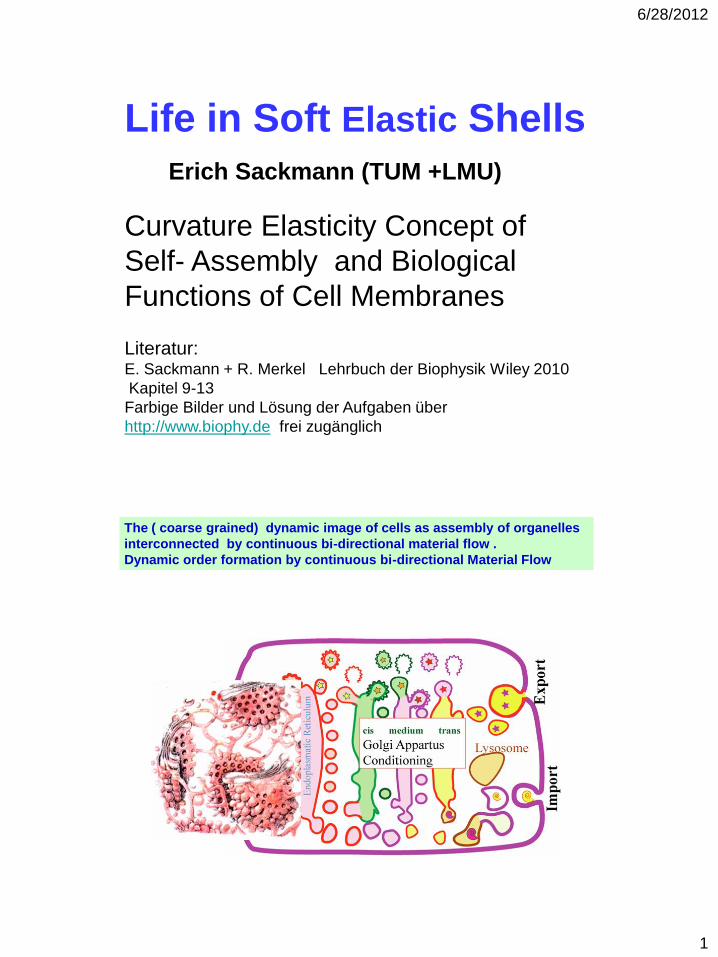
The Mosaic Model of Biological Membranes : (Summary:
Sackmann J. Phys. Condens. Matter 18 (2006) R785–R825)
Biochemical Reaction Centres
Modules
Cytoskeleton
The most simple Prototyp of a Composite
Shell (The Erythrocyte)
6/28/2012
3
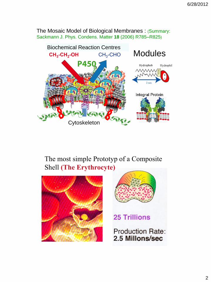
Erythrocyte membrane coupled to quasi-two dimensional macromolecular network
composed of flexible spectrin filaments forming sides and 35 nm long rods of actin
oligomers forming edges of triangular network.
Quasi-hexagonl network Network coupled to membrane proteins
glycophorin and band III
Coupling is dynamic. Band IV.1 actively
bound and unbound by ATP turnover
Fundamental property I
Membranes are soft (fluid) shells as demonstrated by thermally
excited bending fluctuations of giant vesicle and erythrocyte
6/28/2012
4
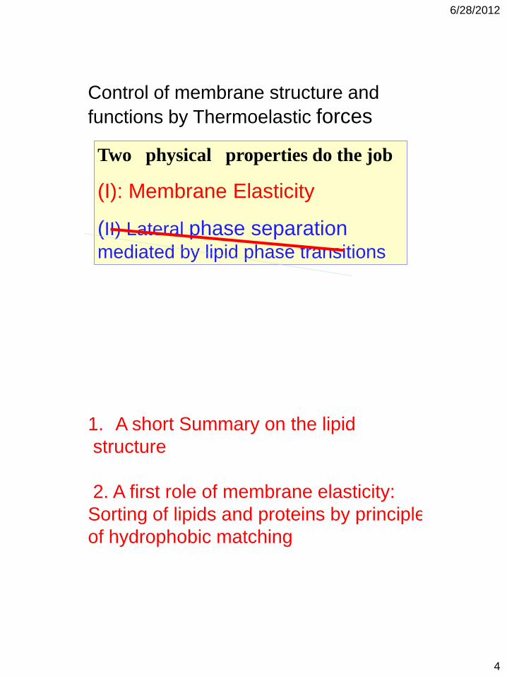
Control of membrane structure and
functions by Thermoelastic forces
Two physical properties do the job
(I): Membrane Elasticity
(II) Lateral phase separation mediated by lipid phase transitions
1. A short Summary on the lipid
structure
2. A first role of membrane elasticity:
Sorting of lipids and proteins by principle
of hydrophobic matching
6/28/2012
5
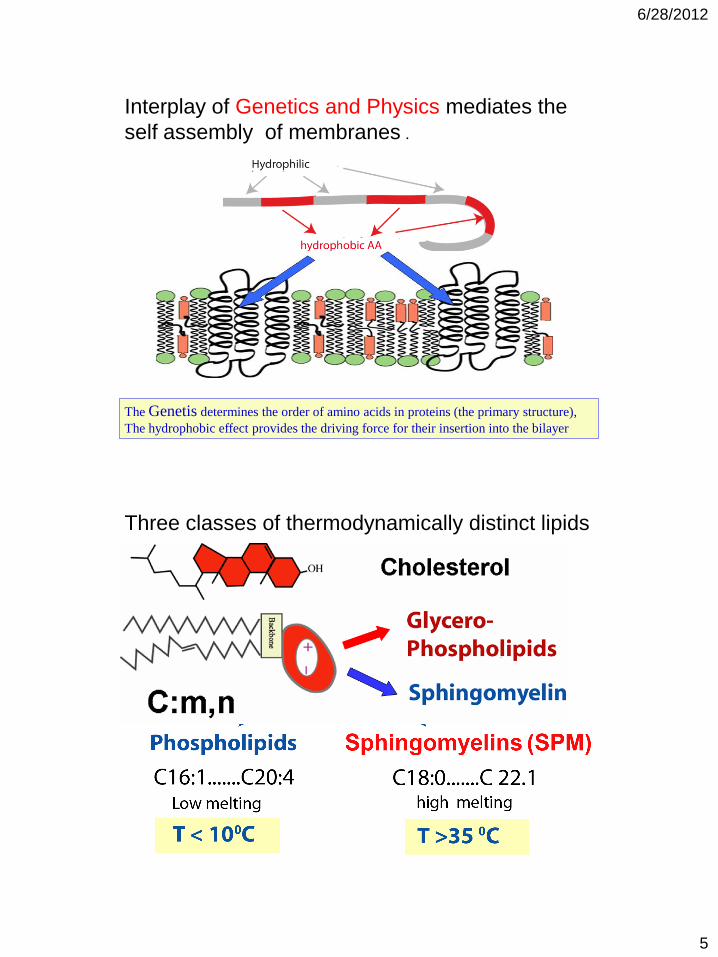
Interplay of Genetics and Physics mediates the
self assembly of membranes .
The Genetis determines the order of amino acids in proteins (the primary structure),
The hydrophobic effect provides the driving force for their insertion into the bilayer
Three classes of thermodynamically distinct lipids
6/28/2012
6
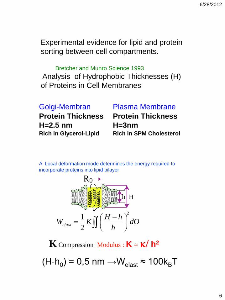
Experimental evidence for lipid and protein
sorting between cell compartments.
Bretcher and Munro Science 1993
Analysis of Hydrophobic Thicknesses (H)
of Proteins in Cell Membranes
Plasma Membrane
Protein Thickness
H=3nm Rich in SPM Cholesterol
Golgi-Membran
Protein Thickness
H=2.5 nm Rich in Glycerol-Lipid
A Local deformation mode determines the energy required to
incorporate proteins into lipid bilayer
K Compression Modulus : K ≈ k/ h2
dO
h
hHKWelast
2
2
1
(H-h0) = 0,5 nm →Welast ≈ 100kBT

6/28/2012
7
Elastic mechanism of Lipid-Potein Sorting
Sphingomyelins + Cholesterol
Glycerol-
Phospholipids
The shape of cells and cellular
organelles determines their function
6/28/2012
8
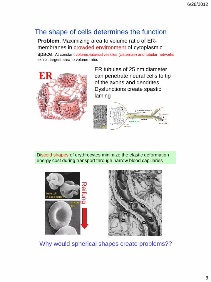
The shape of cells determines the function
Problem: Maximizing area to volume ratio of ER-
membranes in crowded environment of cytoplasmic
space. At constant volume,flattened vesicles (cisternae) and tubular networks
exhibit largest area to volume ratio.
ER tubules of 25 nm diameter
can penetrate neural cells to tip
of the axons and dendrites
Dysfunctions create spastic
laming
Discoid shapes of erythrocytes minimize the elastic deformation
energy cost during transport through narrow blood capillaries
Why would spherical shapes create problems??

6/28/2012
9
How to control the shape ?
Answer
Principle of Minimum Bending
Elastic Energy
E(w); >1MHz
Membranes are elastic shells :Deformation determined by
three modes of deformation: Bending Shearing Tension
Deformation of Erythrocytes in high
frequencz electric field
Force by Maxwell/Wagner Polarisation
6/28/2012
10
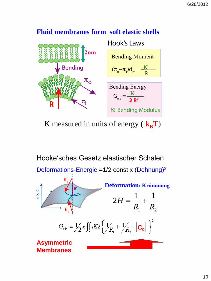
Fluid membranes form soft elastic shells
K measured in units of energy ( kBT)
Deformation: Krümmung
21
112
RRH
Hooke‘sches Gesetz elastischer Schalen
Deformations-Energie =1/2 const x (Dehnung)2
Asymmetric
Membranes
C0
6/28/2012
11
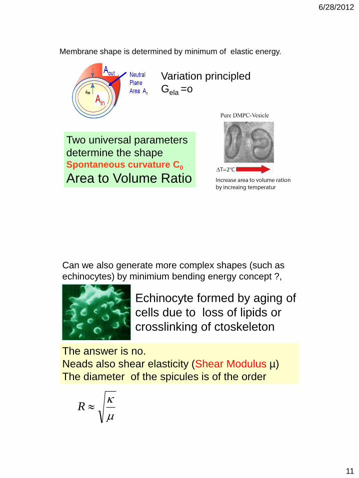
Variation principled
Gela =o
Membrane shape is determined by minimum of elastic energy.
Two universal parameters
determine the shape Spontaneous curvature C0
Area to Volume Ratio
Can we also generate more complex shapes (such as
echinocytes) by minimium bending energy concept ?,
Echinocyte formed by aging of
cells due to loss of lipids or
crosslinking of ctoskeleton
The answer is no.
Neads also shear elasticity (Shear Modulus µ)
The diameter of the spicules is of the order
kR

6/28/2012
12
The concept of induced curvature
determines the intracellular material
transport by vesicle trafficking
Uptake of Iron by Endocytosis
By induced bending followed by budding
6/28/2012
13
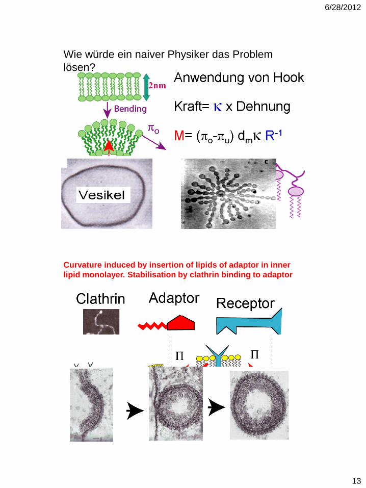
Wie würde ein naiver Physiker das Problem
lösen?
Curvature induced by insertion of lipids of adaptor in inner
lipid monolayer. Stabilisation by clathrin binding to adaptor
6/28/2012
14
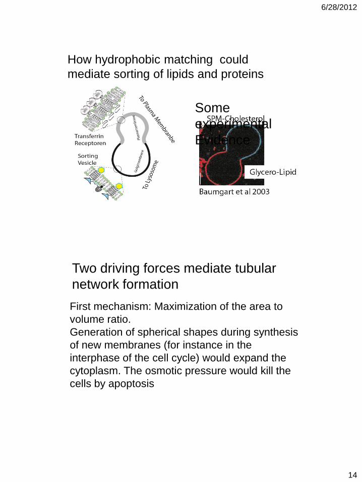
How hydrophobic matching could
mediate sorting of lipids and proteins
Some
experimental
Evidence
Two driving forces mediate tubular
network formation
First mechanism: Maximization of the area to
volume ratio.
Generation of spherical shapes during synthesis
of new membranes (for instance in the
interphase of the cell cycle) would expand the
cytoplasm. The osmotic pressure would kill the
cells by apoptosis
6/28/2012
15
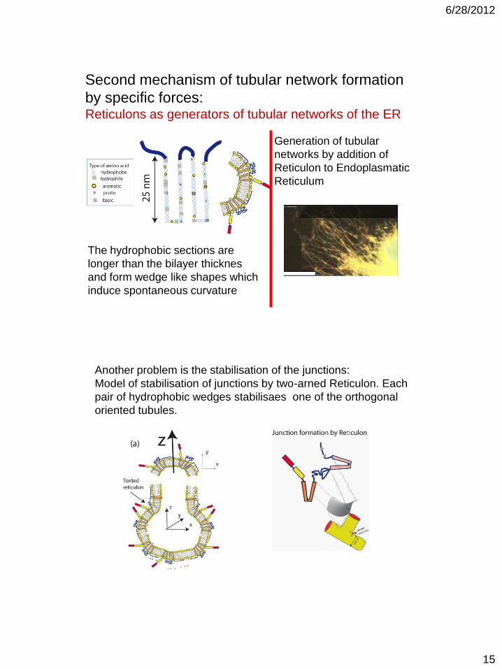
Second mechanism of tubular network formation
by specific forces: Reticulons as generators of tubular networks of the ER
The hydrophobic sections are
longer than the bilayer thicknes
and form wedge like shapes which
induce spontaneous curvature
Generation of tubular
networks by addition of
Reticulon to Endoplasmatic
Reticulum
Another problem is the stabilisation of the junctions:
Model of stabilisation of junctions by two-arned Reticulon. Each
pair of hydrophobic wedges stabilisaes one of the orthogonal
oriented tubules.
6/28/2012
16
The self organisation and function of biomembranes is
controlled by elastic and specific protein-mediated forces
Lipids phase separation and concept of hydrophobic
matching plays a key role for the sorting of lipids and
proteins between the compartments.
Example: Concept of hydrophobic thickness matching.
Conclusions
The global shape of cells and intracellular
organelles determines their function.
The shape is controlled by non-specific elastic
forces and specific curvature-inducing proteins