Embed Size (px)
Citation preview
Hoppe-Seyler's Zeitschrift für Physiologische Chemie
Band 355 — 1. Jahreshälfte
Fortgeführt von A. Kossei, F. Knoop und K. Thomas • Herausgegeben von A. Butenandt, F.Lynen, G.Weitzel
unter Mitwirkung von K. Bernhard, H. Dannenberg, K. Decker, J. Engel, H. Hanson, E. Helmreich, H. Herken, B. Hess, N. Hilschmann, H. Hilz, P. Karlson,
H. L. Kornberg, F . Leuthardt, J. Seelig, G. Siebert, H. Simon, Hj. Staudinger, W. Stoffel, H. Tuppy, H. G. Zachau
Redaktion A. Dillmann, G. Peters
W DE G
1 9 7 4
W A L T E R D E G R U Y T E R • B E R L I N • N E W Y O R K
Eigentum der
önJMSiiils-BüöM M ü n c h e n
Alle Rechte, insbesondere die der Ubersetzung in fremde Sprachen, vorbehalten. Kein Teil dieser Zeitschrift darf ohne schriftliche Genehmigung des Verlages in irgendeiner Form - durch Photokopie, Mikrofilm oder irgendein anderes Verfahren - reproduziert oder in eine von Maschinen, insbesondere von Datenverarbeitungsmaschinen, verwendbare Sprache übertragen oder übersetzt werden. All rights reserved, including those of translations into foreign languages. No part of this journal may be reproduced in any form - by photoprint, microfilm or any other means - or transmitted or translated into a machine language without written permission from the publisher. Nach § 54, Abs. 2 URG ist für die photomechanische, xerographische oder in sonstiger Weise bewirkte Anfertigung von Vervielfältigungen der in dieser Zeitschrift erschienen Beiträge zum eigenen Gebrauch eine Vergütung zu bezahlen, wenn die Vervielfältigung gewerblichen Zwecken dient. Die Vergütung ist nach Maßgabe des zwischen dem Börsenverein des Deutschen Buchhandels e. V., Frankfurt/Main, und dem Bundesverband der Deutschen Industrie in Köln abgeschlossenen Gesamtvertrages vom 15. Juli 1970 zu entrichten. Die Weitergabe von Vervielfältigungen, gleichgültig zu welchem Zwecke sie hergestellt werden, ist eine Urheberrechtsverletzung und wird strafrechtlich verfolgt. Die hier genannten Vervielfältigungen haben den Vermerk über den Hersteller und die Bezahlung der Lizenzen zu tragen. Die Wiedergabe von Gebrauchsnamen, Handelsnamen, Warenbezeichnungen und dergleichen in dieser Zeitschrift berechtigt nicht zu der Annahme, daß solche Namen ohne weiteres von jedermann benutzt werden dürfen. Vielmehr handelt es sich häufig um gesetzlich geschützte, eingetragene Warenzeichen, auch wenn sie nicht eigens als solche gekennzeichnet sind. The quotation of registered names, trade names, trade marks, etc. in this journal does not imply, even in the absence of a specific statement, that such names are exempt from laws and regulations protecting trade marks, etc. and therefore free for general use. © Copyr ight 1974 by Wal te r d e Gruy te r & Co. , Berl in - Anzeigenverwal tung: Merku r -Werbung , D r . K. Jeserich K G , 521 Troisdorf 22, Merkurhaus , Hauptstraße 2 3 - 2 7 , Tel . (0 22 41) 4 20 5 1 . Für den Anzeigentei l verantwort l ich: Ursu la K u n z , Troisdorf . - Pr in ted in G e r m a n y - Satz und Druck : R. Oldenbourg , München.
Autoren-Verzeichnis der ersten Jahreshälfte
Abraham, R. 466 Ackermann, R. H . 576 Adler, J. 105 Agranoff, B. W. 110 Arnold, H. 266
Back. P. 749 Baczko, K. 131 Bahr, W. 600 Bässler, K.-H. 576 Balke, E. 443 Bamberg, E. 104 Bauer, E. 61 Bause, E. 438 Bayer, E. 481 Benz, H. U. 744 Benz, R. 104 Bermek, E. 26 Boie, A. 205 Bonting, S. L. 99 Boos, W. 106 Braun ,D. 131 Brdiczka, D. 731 Breuer, H. 490 Brunfeldt, K. 82 Budzikiewicz, H . 612 Burckhardt, W. 341
Cmelik, S. 19 Czernilofsky, A. P. 89 Därr, W. 54 Debuch, H. 551, 557, 725 Dericks-Tan, J. S. E. 466 Dieminger, L. 171 Dikow, A. L. 233 Diringer, H. 93 Dölken, G. 289, 731
Elbers, R. 378 Engelhard, M. 85
Faillaci, M. G. 716 Falkenberg, F. 233 Feller, U. 321 Fietzek, P. P. 647 Fonseca, E. 19 Fried, W. 222 Fritz, H. 125, 709 Fürniss, H. 557, 725
Ganser, H. 669 Gerisch, G. 108 Gerok, W. 749 Ghraf, R. 543
Gielen, W. 660 Goracci, G. 75 Greene, E. 415 Gromova, O. 626 Gross, R. 471 Grosskopf, M. 305 Grunz, H. 501 Guder, W. 669 Guder, W. G. 273
Haberland, G. L. 229 Häussinger, D. 305 Hallermayer, G. 279 Haiström, J. 82 Hamprecht, B. 109, 669 Hansen, K. 106 Hartter, P. 189, 200 Harzer, K. 744 Heimann, G. 651 Heinrich, P. C. 217 Heinz. E. 612 Heidt, H. W. 378 Hemmasi, B. 481 Hess, B. 255 Hess, M. 85 Heymann, E. 155 Hilschmann, N. 85, 131 Hochstraßer, K. 640 Hoffmann-Ostenhof, O.
222, 633 Hofmann, D. 731 Hohorst, H.-J. 341 Hoshino, J. 4 Hubbard, R. 100 Huber, J. 669
Jaenicke, L. 603 Jancik, J. 395 Jatzekewitz, H. 33 Jost, U. 422 Junge, W. 155
Kaißling, K.-E. 108 Kalckar, H. M. 106 Kannan, R. 551 Kaulen, H. D. 471 Keutmann, H. T. 415 Khandker, R. 687 Kleine, R. 114 Kleinow, W. 300 Kluge, F. 410 Koch, M. A. 93 Königk, E. 427, 431,495 Kohen, C. 451 Kohen, E. 451
Kolb, H. J. 401 Koshland, D. E., Jr. 105 Koväcs, K. 82 Kownatzki, E. 600 Krämer, R. 239 Kraus, H. 164 Krebs, W. 731 Kreutz, W. 102 Krisch, K. 155, 529 Kröger, H. 4 Küchler, E. 89 Kühn, K. 647 Kutzbach, C. 45
Läuger, P. 104 Latzin, S. 669 Latzko, E. 321 Legier, G. 438, 617 Leimcke, H. 205 Liske, R. 217 Lohr, G. W. 266 Löffler, G. 363 Lorenz, B. 300 Loy, E. 239 Lubbers, D. W. 583 Lyakhovich, V. 626 Lynen, R. 171
Malchow, D. 108 Marcussen-Wulff, G. 155 McKenna, W. 336 Menge, U. 603 Mishin, V. 626 Mönkemeyer, H. 26 Mohn, G. 564 Moog, P. 529 Mraz, W. 33 Müller, O.-A. 669 Müllhofer, G. 239
Nachmansohn, D . 111 Nattero, G. 716 Neumann, E. I I I Neupert, W. 279 Niall, H. D. 415 Nordmeyer, J.-P. 427, 495 Nordwig, A. 721 Nowotny, E. 716
Oesterhelt, D. 101 O'Riordan, J. L. H. 415 Osanai, M. 327 Parsons, J. A. 415 Pette, D. 289
Pfleiderer, G. 233 Philipp, E.-M. 564 Pittner, F . 222 Pollow, B. 501, 515 Poliow, K. 501, 515 Porcellati, G. 75 Potts, J. T., Jr. 415
Rafael, J. 341 Raftery, M. A. 112 Rauenbusch, E. 45 Reber, K. 217 Rembold, H. 327 Rietschoten, J. van 415 Robbers, G. 557 Robert, B. 4 Röhle, G. 490 Röhm, K.-H. 675 Rübsamen, H. 687 Rullkötter, J. 612
Sananez, R. D. 716 Sauermann, G. 459 Schachtschabel, D . 451 Schaper, R. 660 Scharmann, W. 443 Schauer, R. 395 Schill, W.-B. 225, 229 Schimassek, H. 353 Schlenker, S. 164 Schleuning, W.-D. 125 Schmalbeck, J. 515 Schmidt, G. 171 Schmidt, U. 273 Schmucker, P. 378 Schorn, K. 640 Schriefers, H. 543 Schwedesky, D. 164 Seitz, U. 266 Seubert, W. 205 Siebler, G. 353 Sies, H. 305, 319 Simon, D. 4 Soboll, S. 378 Sokolowski, G. 501, 515 Spaczynski, K. 749 Spangenberg, P. 114 Stahl, J. 61 Stark, G. 104 Stemberger, A. 721 Stieve, H. 101 Stöffler, G. 89 Stoffel, W. 54, 61, 651 Stumpf, B. 205
Autoren-Verzeichnis der ersten Jahreshälfte Takesue, S. 184 Thorell, B. 451 Thurm, U. 102 Thurman, R. G. 336 Tjiong, H.-B. 551 Tregear, G. W. 415 Tsyrlov, I. 626 Ulbricht, W. 104 Vetter, U. 543 Vogt, W. 171
Wagner, K. 576 Wald, G. 99 Walli, A. K. 353 Wallner, O. 709 Walter, R. D. 427, 431,
495 Watanabe, K. 184 Weber, H. 595 Weber, U. 189, 200 Weis, W. 595 Weiss, H. 300
Weiss, L. 363 Wel, H. van der 107 Wesemann, W. 112 Whittaker, V. P. 111 Wiedemann, H.-R. 551 Wiegandt, H. 11 Wieland, O. H. 363 Wiemer, G. 341 Wiese, H. 378 Witassek, F. 617 Witzel, H. 687
Wodick, R. 583 Woelk, H. 75 Wolf, B. 595 Wollenberg, P. 239 Woloszczuk, W. 633 Wurster, B. 255 Yantorno, C. 716 Zepf, E. 353 Ziegler, W. 11 Zimmermann, G. 321
Hoppe-Seyler's Z. Physiol. Chem. Bd. 355, S. 279-288 , März 1974
Lipid Composition of Mitochondrial Outer and Inner Membranes of Neurospora crassa Gerhanl Hallermayer and Walter Neupert
(Received 5 November 1973)
Dedicated to Prof. Dr. Dr. h. c. Theodor Bücher on the occasion of his 60th birthday
Summary : The lipid composi t ion of outer and inner mi tochondr ia l membranes of Neurospora crassa was analyzed quantitatively. The outer membrane is made u p of 5 9 % lipid and 41 % protein, the inner membrane of 2 3 % lipid and 7 7 % protein. The outer membrane is one of the most lipid-rich membranes described in the l i terature. The main lipid components of the outer membrane are phos pholipid and ergosterol. They occur in a molar ra t io of 3 : 1 . N o ergosterol is found in the inner membrane . Ergosterol is released from the outer membrane in the form of needle-like crystals, when
outer membrane preparat ions are stored as concentrated suspensions. Simultaneously, outer membrane vesicles fuse to generate large m e m b r a n e systems. The phosphol ipid composi t ions of the two membranes are different. This difference is small with respect to the major componen ts phosphatidylcholine and phosphat idyle thanolamine. On the other hand, cardiolipin occurs predominant ly — if no t exclusively — in the inner membrane . T h e carotenoid pigment neurosporaxanth in is localized selectively in the outer mitochondria l membrane .
Lipidzusammensetzung der mitochondrialen Außen- und Innenmembranen von Neurospora crassa Zusammenfassung: Mitochondr ia le Außen- und Innenmembranen von Neurospora crassa wurden einer quanti tat iven Best immung ihrer Lipidzusammensetzung unterzogen. Die Außenmembran enthält 5 9 % Lipid und 41 % Protein, die Innenmembran 2 3 % Lipid u n d 7 7 % Protein. D i e Außenmembran ist eine der lipidreichsten Membranen , die in der Literatur beschrieben sind. D ie wesentlichen Lipidkomponenten der Außenmembran sind Phosphol ipide und Ergosterin. Ihr molares Verhältnis beträgt ca. 3 : 1 . In der I n n e n m e m b r a n k o m m t kein Ergosterin vor. Ergosterin kristallisiert aus der Außenmembran in F o r m von Nade ln aus , wenn Außenmembranpräparationen als konzen
trierte Suspensionen aufbewahrt werden. Gleichzeitig mit diesem Vorgang verschmelzen Außen-membranvesikel . Dies führt zum Auftreten ausgedehnter Membransysteme. Die Phosphol ipidzu-sammensetzungen der beiden M e m b r a n e n sind unterschiedlich. Im Fall der H a u p t k o m p o n e n t e n Phosphatidylcholin u n d Phosphatidyläthanolamin sind die relativen Konzentra t ionsunterschiede n u r gering. Cardiolipin andererseits k o m m t überwiegend — wenn nicht ausschließlich — in der inneren M e m b r a n vor. Der Karotinoidfarbstoff N e u r o sporaxanthin stellt eine spezifische K o m p o n e n t e der mitochondrialen Außenmembran dar.
Address: Priv.-Doz. Dr. Dr. Walter Neupert, Institut für Physiologische Chemie und Physikalische Biochemie der Universität München, D-8 München 2, Goethestr. 33. Enzymes: Kynurenine 3-monooxygenase, L-kynurenine, NAD PH: oxygen oxidoreductase (3-hydroxylating) (EC 1.14.13.9) Succinate dehydrogenase (succinate cytochrome c reductase), succinate: (acceptor) oxidoreductase (EC 1.3.99.1).
2 8 0 G. Hallermayer and W. Neupert Bd. 355 (1974) T h e outer and inner membranes of mitochondria differ with respect to structure and function^1-3!. Whereas the inner membrane can be regarded as a multienzyme complex responsible for the major par t of cellular energy product ion , the outer membrane contains a heterogeneous group of enzymes. Separat ion methods for the two membrane systems were first developed with the aid of mammal ian liver mitochondria , and hi therto most of our informat ion has come from this source. Studies with Neurospora crassa have shown that mitochondrial membranes from organisms phylo-genetically as distant as mammal s and fungi display the same building principles. W i t h respect to the lipid par t of outer a n d inner membranes , da ta are only available for liver mi to--chondr ia t 1 ' 2 ' 4 - 8 ^ They show that each membrane has a characteristic lipid pat tern. The present •communication supplies da ta in answer to the following quest ions: Are the lipid pat terns of the ou te r and inner mitochondrial membranes in Neurospora and liver constant parameters , like the specific distribution of enzymes? H o w are lipids •which are specific for animals, such as cholesterol, subst i tuted in fungi ? Are common physical properties of the individual mitochondria l membranes f r o m different organisms related to a common lipid s tructure ?
Experimental Preparation of mitochondrial membranes Neurospora crassa wild type 74A was grown in VogeFs minimal medium containing 0.8ImM phospha te^ . The •cells were harvested 16 h after inoculation. Mitochondria and other cell fractions were prepared by a procedure described previously[3]. Mitochondrial outer and inner membranes were separated by a combined technique of swelling, shrinking and sonication followed by centrifu-.gation through a linear sucrose density grad ien t^ . The gradient was divided into 11 fractions of 5 ml each. Fraction 1 (top of gradient) (outer membrane) and fraction 9 (inner membrane) were collected and centri-fuged as described. The final pellets were resuspended in Tris-HCl, 10mM, pH 7.2. Labeling of Neurospora cells with [32P]phosphate For the homogeneous labeling of lipid-phosphorus, 1 mCi [32P]orthophosphate (1600-4480 Ci/mmol) (Radiochemical Centre, Amersham, England) was added to a 1 / culture immediately after inoculation.
Enzyme and cytochrome determinations Kynurenine 3-monooxygenase, succinate, cytochrome c reductase and cytochromes aa% and b were measured as described elsewhere^. Protein was determined by the method of Lowry et alP^. Neurosporaxanthin and ergosterol The acidic carotenoid neurosporaxanthin was extracted according to the method of Harding et alS10^. The visible spectrum was recorded in hexane. The content of neurosporaxanthin was calculated from the extinction at 477.5nm (s = 85.4 / x mmol- icm - 1 ^ 1 1 ! . Ergosterol was extracted with methanol and acetone and transferred into hexane. UV-spectra were recorded. The content of ergosterol was estimated from the extinction at 282 nm. Authentic ergosterol (Fa. Fluka, Buchs, Switzerland) was employed as a standard for these analyses. Phospholipids Lipid extraction. The total lipids of cell fractions and mitochondrial membranes were extracted with chloro-form/methanol (2 :1 , by vol.) at 22°C for 12 h. The extracts were washed with one-fifth vol. of 0.034% MgCl2 according to the method of Folch et al.^12K After removal of the upper aqueous phase, samples were either taken for phosphorus analysis, or portions ( 2 0 - 3 5 jxg total phospholipid phosphorus) were vacuum dried for thin layer chromatography. Extracts were stored under nitrogen at -20°C. Two-dimensional thin-layer chromatography. The dried extracts were taken up in chloroform/methanol (2 :1 , by vol.) and applied to silica gel thin-layer plates (20x20 cm, 0.25 mm thick; Merck AG., Darmstadt) under an atmosphere of nitrogen. The plates were developed two-dimensionally using chloroform/metha-nol/25 % ammonia (65:35:5, by vol.) in the first direction, and chloroform/acetone/methanol/acetic acid/ water (56:20:10:10:4, by vol.) in the second direction. The chromatography jars were lined with filter paper and saturated with the appropriate solvent. Two 4 cm lanes were separated along two intersecting edges of the thin-layer plates. These were used for the chromatography of appropriate standards in either direction. Detection of spots. The plates were dried and exposed to iodine vapor. The spots were immediately encircled with a pencil. The individual spots were identified by comparison with the position of the standard compounds which were chromatographed on the two lanes mentioned above. Alternatively, single lipid standards were co-chromatographed with Neurospora lipid extract to obtain accentuated spots. In addition, lipid spots were identified with the following reagents: a) molybdenum reagent I13'14!, for phospholipids; b) ninhydrin reagent (Merck AG., Darmstadt), for phospholipids containing free amino groups; c) orcinol-sulfuric acid
Bd. 355 (1974) Lipids of mitochondrial outer and inner membranes 281 reagentt15 '16], for glycolipids; d) diphenylamine re-agent^16-19], for glycolipids. Phospholipid standards: phosphatidylcholine (lecithin), phosphatidylethanol-amine (cephalin) and sphingomyelin were purchased from C. Roth OHG., Karlsruhe, lysophosphatidyl-choline and lysophosphatidylethanolamine from Sigma Chemical Co., St. Louis, USA. Diphosphatidylglycerol (cardiolipin), phosphatidylserine, phosphatidylinositol and phosphatidic acid were obtained from Koch-Light-Laboratories, Colnbrook, England. Cerebrosides, sulfat-ides and gangliosides were kindly supplied by Dr. E. Mehl, Max-Planck-Institut für Psychiatrie, München. Colorimetric phospholipid phosphorus assay. The procedures of Rouser et a/J20 '21! and McClaret22! were used in a slightly modified way. The spots, detected with iodine vapor, were encircled with a fine dissecting needle. Subsequently most of the iodine was allowed to evaporate. The phospholipid-containing spots and blank areas corresponding in size to these spots were transferred into centrifuge tubes and digested with 0.25 ml 70% perchloric acid at 170-180°C for 2 h. After cooling 4.75 ml of a reagent containing 20 ml perchloric acid (70%), 2.5 g ammonium molybdate and 0.5 g ascorbic acid per 250 ml, were added. After 1 h at 50°C the tubes were centrifuged and the extinction at 660 nm was measured. Analysis of 32P-labeled phospholipids. Phospholipid spots were detected with iodine vapour and outlined. Most of the iodine was allowed to evaporate. In order to avoid quenching effects, the plates were treated with SO2 to reduce the remaining iodine. The scrapings were transferred into scintillation counting flasks and suspended in 15 m/ scintillation mixture (6 g butyl-PBD in 1 / toluene/ethoxyethanol 3:2, by vol.) containing 4 % Cab-O-Sil (Thixotropic Gel Powder, Packard Instruments Co., Frankfurt). Radioactivity was measured in a Packard Tricarb scintillation counter (92% yield).
Results Characteristics of outer and inner mitochondrial membranes The puri ty of the mitochondria l membrane prepara t ions , obtained by a combined technique of swelling-shrinking-sonicationt3^ was checked by determining marke r enzyme activities and cytochrome contents. Kynurenine 3-monooxygenase was chosen as a marker for the outer membraneC23J, succinate cytochrome c reductase and cytochromes aa% a n d b as markers for the inner membrane t3l. Table 1 gives the da ta of a typical experiment. In outer membrane prepara t ions , inner membranes m a k e up 9 - 2 0 % of total protein, while in inner membrane preparat ions , outer membranes make up
Table 1. Marker enzyme activities and cytochrome contents of outer and inner mitochondrial membrane preparations from Neurospora crassa. Kynurenine 3-monooxygenase activity is expressed as (nmol hydroxykynurenine produced) x m in - 1 x (mg prot . ) - 1 , succinate cytochrome c reductase activity as (nmol cytochrome c reduced) x m i n - 1 x (mg prot . ) - 1 ; cytochrome contents are expressed as nmol x (mg prot.)"1 . Enzyme activities and cytochrome contents are related to insoluble membrane protein.
Outer Inner membrane membrane preparation preparation (fraction 1) (fraction 9)
Kynurenine 3-monooxygenase 25.4 0.8 Succinate cytochrome c reductase 85 885 Cytochrome aa% 0.05 0.31 Cytochrome b 0.23 1.15
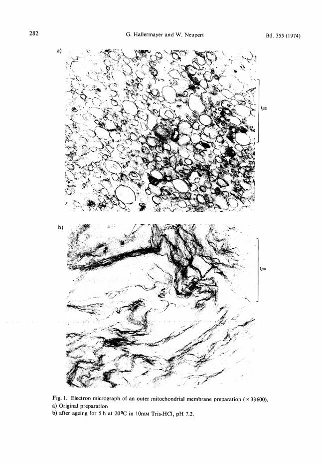
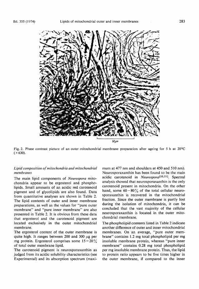
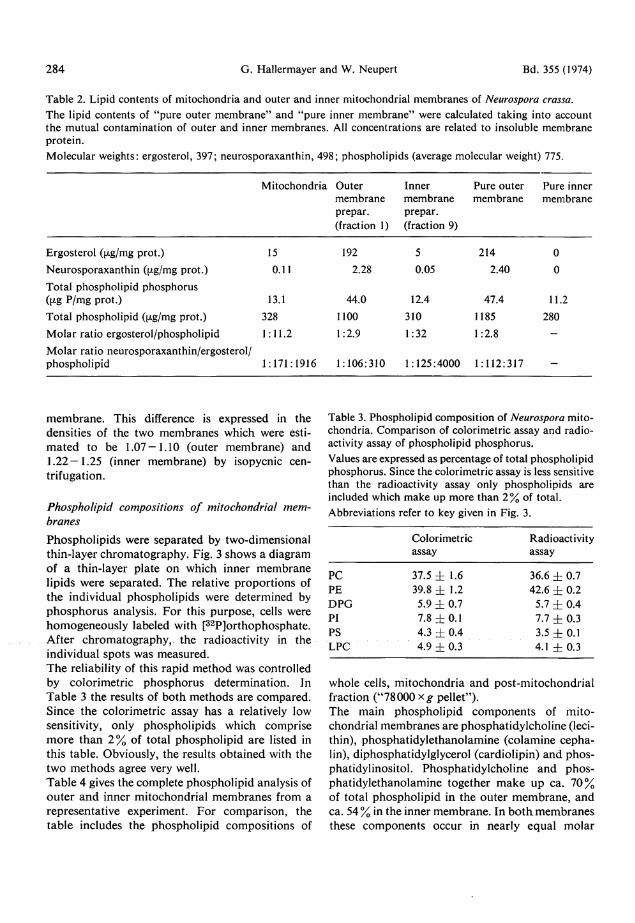
2 - 5 % of total protein. Wi th these data , the lipid contents of " p u r e outer m e m b r a n e s " and "pure inner m e m b r a n e s " were calculated. Outer m e m b r a n e preparat ions display an interesting behavior. Crystals a re formed in pellets or concentrated suspensions of outer membrane if they are allowed to s tand at 0° or 22 °C. These crystals are ergosterol as the following propert ies show: 1) If the membrane is solubilized in sodium dodecyl-sulfate the crystals remain . Fur the r washing yields a pure crystal fraction; 2) The crystals are soluble in chloroform and hexane ; 3) The melt ing point of the crystals agrees with tha t of pu re ergosterol (163°C); 4) U V spectra of solutions of the crystals in hexane are identical with spectra of authentic ergosterol; 5) Using the Liebermann-Burchard reaction E24l, the crystals and ergosterol give a colour with identical spectral characteristics. Electron micrographs were made of outer membrane prepara t ions before and after ergosterol crystals were formed. Fig. l a shows the original preparat ion. It consists of small vesicles of ra ther uniform size (average contour length ca. 0.4 (xm). After s tanding for 5 h at 20 °C large membrane systems are observed (Fig. 1 b). T h e contour length of these membranes is u p to 5 — 10 [im or even more. Simultaneously, in phase contrast pictures, long needles of ergosterol can be seen (Fig. 2).
282 G. Hallermayer and W. Neupert Bd. 355 (1974)
Fig. 1. Electron micrograph of an outer mitochondrial membrane preparation ( x 33600). a) Original preparation b) after ageing for 5 h at 20°C in 10mM Tris-HCI, pH 7.2.
Bd. 355 (1974) Lipids of mitochondrial outer and inner membranes 283
Lipid composition of mitochondria and mitochondrial membranes The main lipid components of Neurospora mi tochondr ia appear to be ergosterol and phospholipids. Small amoun t s of an acidic red carotenoid pigment and of glycolipids are also found. D a t a from quanti tat ive analyses are shown in Table 2. The lipid contents of outer and inner membrane preparat ions , as well as the values for "pu re outer m e m b r a n e " and " p u r e inner m e m b r a n e " are also presented in Table 2. It is obvious from these da ta that ergosterol and the carotenoid pigment a re located exclusively in the outer mitochondrial membrane . The ergosterol content of the outer membrane is quite high. It ranges between 200 and 300 (xg per mg protein. Ergosterol comprises some 1 5 - 2 0 % of total outer m e m b r a n e lipid. The carotenoid pigment is neurosporaxanthin as judged from its acidic solubility characteristics (see Experimental) and its absorpt ion spectrum (maxi
m u m at 477 n m and shoulders at 450 and 510 nm) . Neurosporaxanth in has been found to be the main acidic carotenoid in Neurospora^10Spectral analysis showed that neurosporaxanth in is the only carotenoid present in mi tochondr ia . On the other hand, some 60 — 8 0 % of the total cellular neuro sporaxanthin is recovered in the mitochondria l fraction. Since the outer membrane is par t ly lost during the isolation of mitochondria , it can be concluded that the vast majority of the cellular neurosporaxanth in is located in the outer mi tochondrial membrane . The phosphol ipid contents listed in Table 2 indicate another difference of outer and inner mitochondria l membranes . On an average, " p u r e outer memb r a n e " contains 1.2 mg total phosphol ip id per m g insoluble membrane protein, whereas " p u r e inner m e m b r a n e " contains 0.28 mg total phosphol ip id per mg insoluble membrane protein. Thus , the lipid to protein rat io appears to be five times higher in the outer membrane , if compared to the inner
284 G. Hallermayer and W. Neupert Bd. 355 (1974) Table 2. Lipid contents of mitochondria and outer and inner mitochondrial membranes of Neurospora crassa. The lipid contents of "pure outer membrane" and "pure inner membrane" were calculated taking into account the mutual contamination of outer and inner membranes. All concentrations are related to insoluble membrane protein. Molecular weights: ergosterol, 397; neurosporaxanthin, 498; phospholipids (average molecular weight) 775.
Mitochondria Outer membrane prepar. (fraction 1)
Inner membrane prepar. (fraction 9)
Pure outer membrane
Pure inner membrane
Ergosterol ((xg/mg prot.) 15 192 5 214 0 Neurosporaxanthin (fJ-g/mg prot.) 0.11 2.28 0.05 2.40 0 Total phospholipid phosphorus Gxg P/mg prot.) 13.1 44.0 12.4 47.4 11.2 Total phospholipid (i^g/mg prot.) 328 1100 310 1185 280 Molar ratio ergosterol/phospholipid 1:11.2 1:2.9 1:32 1:2.8 -Molar ratio neurosporaxanthin/ergosterol/ phospholipid 1:171:1916 1:106:310 1:125:4000 1:112:317 -
membrane . This difference is expressed in the densities of the two membranes which were estim a t e d to be 1 .07-1 .10 (outer membrane) and 1.22 — 1.25 (inner membrane) by isopycnic cen-trifugation.
Phospholipid compositions of mitochondrial membranes Phosphol ip ids were separated by two-dimensional thin-layer chromatography. Fig. 3 shows a diagram of a thin-layer plate on which inner membrane lipids were separated. The relative propor t ions of the individual phospholipids were determined by p h o s p h o r u s analysis. F o r this purpose , cells were homogeneous ly labeled with [3 2P]orthophosphate. After chromatography, the radioactivity in the individual spots was measured. T h e reliability of this rapid method was controlled by colorimetric phosphorus determination. In Tab le 3 the results of bo th methods are compared . Since the colorimetric assay has a relatively low sensitivity, only phospholipids which comprise m o r e than 2 % of total phosphol ipid a re listed in this table. Obviously, the results obta ined with the two me thods agree very well. Tab le 4 gives the complete phosphol ipid analysis of ou te r a n d inner mitochondrial membranes from a representat ive experiment. F o r comparison, the table includes the phospholipid composi t ions of
Table 3. Phospholipid composition of Neurospora mitochondria. Comparison of colorimetric assay and radioactivity assay of phospholipid phosphorus. Values are expressed as percentage of total phospholipid phosphorus. Since the colorimetric assay is less sensitive than the radioactivity assay only phospholipids are included which make up more than 2 % of total. Abbreviations refer to key given in Fig. 3.
Colorimetric Radioactivity assay assay
PC 37.5 ± 1.6 36.6 ±0.7 PE 39.8 ± 1.2 42.6 ±0.2 DPG 5.9 ± 0.7 5.7 ± 0.4 PI 7.8 ±0.1 7.7 ±0.3 PS 4.3 ±0.4 3.5 ±0.1 LPC 4.9 ±0.3 4.1 ±0.3
whole cells, mi tochondr ia and post-mitochondria l fraction ("78000 xg pellet") . The main phosphol ipid componen ts of mi tochondrial membranes are phosphat idylchol ine (lecithin), phosphat idyle thanolamine (colamine cepha-lin), diphosphatidylglycerol (cardiolipin) and phos -phatidylinositol . Phosphat idylchol ine and phosphat idyle thanolamine together m a k e up ca. 7 0 % of total phosphol ipid in the outer m e m b r a n e , and ca. 5 4 % in the inner membrane . In b o t h membranes these components occur in nearly equal molar
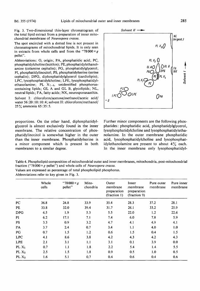
Bd. 355 (1974) Lipids of mitochondrial outer and inner membranes 285 Fig. 3. Two-dimensional thin-layer chromatogram of the total lipid extract from a preparation of inner mitochondrial membrane of Neurospora crassa. The spot encircled with a dotted line is not present in Chromatograms of mitochondrial lipids. It is only seen in extracts from whole cells and from the "78 000 pellet". Abbreviations: O, origin; PA, phosphatidic acid; PC, phosphatidylcholine (lecithin); PE,phosphatidylethanol-amine (colamine cephalin); PG, phosphatidylglycerol; PI, phosphatidylinositol; PS, phosphatidylserine (serine cephalin); DPG, diphosphatidylglycerol (cardiolipin); LPC, lysophosphatidylcholine; LPE, lysophosphatidyl-ethanolamine; PL Xi_4, unidentified phosphorus-containing lipids; GL A and GL B, glycolipids; NL, neutral lipids; FA, fatty acids; NX, neurosporaxanthin. Solvent I: chloroform/acetone/methanol/acetic acid/ water 56:20:10:10:4; solvent I I : chloroform/methanol/ 25% ammonia 65:35:5.
So/vent II NL fergost.J
o
proport ions . On the other hand , diphosphat idylglycerol is a lmost exclusively found in the inner membrane . The relative concentrat ion of phosphatidylinositol is somewhat higher in the outer than the inner membrane . Phosphatidylserine is a minor component which is present in bo th membranes to a similar degree.
Further minor components are the following p h o s phat ides : phosphat id ic acid, phosphatidylglycerol , lysophosphatidylcholine and lysophosphat idyletha-nolamine. In the outer m e m b r a n e phospha t id ic acid, lysophosphatidylcholine a n d lysophosphat-idylethanolamine are present to abou t 4 % each. In the inner membrane only lysophosphat idyl-
Table4. Phospholipid composition of mitochondrial outer and inner membranes, mitochondria, post-mitochondrial fraction ("78000 x # pellet") and whole cells of Neurospora crassa. Values are expressed as percentage of total phospholipid phosphorus. Abbreviations refer to key given in Fig. 3.
Whole "78000 *g Mito- Outer Inner Pure outer Pure inner cells pellet" chondria membrane membrane membrane membrane
preparation preparation (fraction 1) (fraction 9)
PC 36.8 24.8 33.9 35.4 28.3 37.2 28.1 PE 33.8 32.0 39.4 31.7 26.1 33.2 25.9 DPG 4.5 1.9 5.3 5.5 22.0 1.2 22.6 PI 6.2 17.1 7.1 7.4 6.0 7.8 5.9 PS 3.3 0.9 3.2 4.7 4.1 4.9 4.1 PA 3.7 2.4 0.7 3.4 1.1 4.0 1.0 PG 0.7 1.5 1.2 0.6 1.5 0.4 1.5 LPC 4.1 8.6 3.8 4.2 4.3 4.2 4.3 LPE 2.1 3.1 1.1 3.1 0.1 3.9 0.0 PL Xi 0.7 1.1 1.8 2.2 5.4 1.4 5.5 PL X2 2.5 1.5 1.8 0.9 0.5 1.0 0.5 P L X 3 1.6 5.1 0.7 0.4 0.6 0.4 0.6
286 G. Hallermayer and W . Neupert Bd. 355 (1974) choline is found in this concentrat ion. It may well be that these components are decomposit ion products of the major phospholipids. So it has been repor ted that during extraction diphosphat idyl-glycerol may undergo a rapid degradat ion which leads to the appearance of addit ional spots on thin-layer chromatograms [ 2 5 l . F o u r phosphorus-containing spots were detected which [have not yet been identified. They are designa ted PL Xi , P L X 2 , PL X 3 and PL X 4 (see Fig. 3). Incomplete resolution was obtained between phosphatidyl e thanolamine and PL X 4 . However , PL X 4 is a minor component compared to phosphat idyle thanolamine . Therefore, the amoun t of phosphat idyle thanolamine is not influenced in a noticeable way when determined together with PL X 4 . Consequent ly, P L X 4 is not specified in Table 4. Sphingomyelin was neither detected in mi tochondrial membranes no r in whole cells of Neurospora crassa. After incorporat ion of 3H-labeled inositol into Neurospora cells, radioactivity was detected in phosphatidylinositol and in no other spot. Hence, the mitochondrial membranes of Neurospora contain no other inositol-containing lipids, and in par t icular n o phosphoryla ted derivatives of phosphatidylinositol . A m o n g the polar lipids, two glycolipids were detected on chromatograms of extracts of Neurospora cells (GL A and G L B in Fig. 3). They yield a positive reaction with orcinol and diphenylamine. Glycolipid A was found in extracts of mi tochondr ia a n d inner mitochondria l membranes in very low a m o u n t s (less than 2 % of total phosphol ipids , roughly estimated by the color developed with iodine). The outer membrane prepara t ion clearly conta ined more . Quite remarkable quantit ies of this glycolipid were found in the post-mitochondria! fraction ("78000 xg pellet") . So it is highly p rob able that the presence of glycolipid A in the outer mi tochondr ia l membrane prepara t ion is caused by a contaminat ion of mi tochondr ia with the post-mitochondr ia l fraction. Glycolipid B could not be detected in mi tochondr ia , bu t it was found in small amounts in the "78 000 x g pel le t" . This fraction also contains a remarkable a m o u n t of phosphatidylinositol (17%). Since glycolipids and phosphatidylinositol are described as characterist ic components of the cell membrane^2 6! , the postmitochondria l fraction may at least part ly consist of fragments of the cell membrane . •
Discussion The data presented in this report clearly show that outer and inner membranes of mi tochondr ia from Neurospora are different with respect to their lipid contents and their lipid composit ion. The outer membrane is rich in lipid compared to the inner membrane . This is shown by the lipid-protein rat ios of 1.4—1.5 in the outer membranes , and 0.28 in the inner membrane . With a lipid content of some 5 9 % , the outer membrane of Neurospora mi tochondria is one of the most lipid-rich membranes described in the literature (for review see [ 2 ? l) . The inner mitochondrial membrane in contrast has a relatively low lipid content of 23 %. It is interesting to no te that also in the case of mammal ian liver mi tochondr ia the outer membrane has a higher lipid content than the inner membrane^1*2*4-8 '28!. However, in liver the difference between the two membranes is not so large. The lipid contents of the inner mitochondrial membranes of Neurospora and liver are quite similar ( 24% according to Parsons and Y a n o ^ for guinea pig liver, and 2 5 . 4 % for rat liver according to Levy et alS28\ Outer membranes of mi tochondria from guinea pig liver have been repor ted to contain 4 8 % l i p i d ^ , whereas for ra t liver a value of 3 9 % can be calculated from the data available in the literature*28!. The individual lipid components of mitochondrial membranes of Neurospora can be subdivided into two groups. Lipids of the first group occur in bo th outer and inner membranes to a similar extent. The other group consists of lipids which are found exclusively or predominant ly in one of the two membranes . T o the first g roup belong: phospha t idylcholine, phosphat idyle thanolamine, phospha t idylinositol and phosphatidylserine. The second group includes ergosterol and neurosporaxanth in , which are only detected in the outer membrane , and diphosphatidylglycerol (cardiolipin) which is found highly concentrated in the inner membrane . Similar aspects can be pointed out for the c o m p o sition of liver mitochondrial membranes . In mi tochondria of liver as well as of Neurospora the two main phospholipid components are phosphat idylcholine and phosphat idylethanolamine. Together they make up some 60 — 80 % of the total phospholipid in outer as well as in inner membranes . H o w ever, the rat ios of phosphat idylcholine to phos phat idylethanolamine in outer and inner membranes of liver are much higher than those in
Bd. 355 0974) Lipids of mitochondrial outer and inner membranes 287 Neurospora mi tochondr ia l membranes . Fur ther more , in liver the relative content of phosphat idyl-inositol is a b o u t 2 - 4 times higher in the outer membrane t h a n in the inner membrane [4>6>8l Phosphat idylser ine is only detected in traces in liver mi tochondr ia l membranes , whereas in Neurospora mitochondr ia l membranes it is present in an ap preciable a m o u n t (ca. 5 % ) . One of the mos t interesting aspects concerning the phosphol ipid composi t ion of mitochondrial membranes is the high concentra t ion of cardiolipin in the inner mi tochondr ia l membrane , as first shown by Parsons et al.^ with guinea pig liver. Corresponding repor t s were m a d e for ra t liver by Stoffel and S c h i e f e r ^ , Levy a n d Sauner^J and M c M u r r a y and D a w s o n ^ . In this paper it is shown, that also in Neurospora, cardiolipin appears to be a specific componen t of the inner mitochondrial membrane . Moreover , the relative propor t ions of cardiolipin are nearly equal in the different organisms. These observat ions suggest tha t there are some invariable rules concerning the presence and concentrat ions of certain lipid components in mi tochondrial membranes , even in phylogenetically distant organisms. On the other hand, some lipid components seem to be quite variable. Similar considerat ions may be made regarding the presence of ergosterol in the mitochondrial membranes of Neurospora. This sterol is found in an unusually high concentra t ion in the outer membrane . Parsons and Y a n o t 5 l have shown that cholesterol is concentrated in the outer membrane of guinea pig liver mi tochondr ia , though the concentrat ion (30.1 ± 12.8 [xg/mg prot .) is far below that of ergosterol in Neurospora outer mitochondrial membrane . Correspondingly, the molar rat io of cholesterol to phosphol ip id in liver is much lower than the molar rat io of ergosterol to phospholipid in Neurospora. Parsons and Yano have repor ted values of 1:11 — 1:29. F o r rat liver outer mitochondrial membrane Levy et Ö / J 2 8 ^ have found molar ratios of 1:9 — 1:14. In Neurospora outer mitochondrial membrane the molar rat io of ergosterol to phosphol ipid is as high as 1:2.8. This high concentrat ion of ergosterol is probably the reason why it is released from the membrane under certain condit ions to form needle-like crystals. The function of these sterols in the outer mitochondrial m e m b r a n e is not known. It may be speculated that they confer the rigid character to the outer membrane . In contrast to inner mem
branes outer membranes rup ture u p o n swelling of mi tochondr ia and open vesicles are frequently seen. It seems quite reasonable to assume that ergosterol plays the same role in Neurospora mi tochondr ia as cholesterol in liver mi tochondr ia . A par t icular property of Neurospora outer m i t o chondrial membrane is the presence of the ca ro ten-oid p igment neurosporaxanthin . However, the concentrat ion of this component is ra ther low. T h e molar ra t io of neurosporaxanth in to phosphol ip id in the outer membrane is abou t 1:300. The function of this pigment is not known. The authors are grateful to Mr. A. Pfaller for valuable technical support and to Dr. F. Sauer for preparing the electron micrographs. This work was supported by the Deutsche Forschungsgemeinschaft Schwerpunktprogramm „Biochemie der Morphogenese".
Literature 1 Racker, E. (1970) Membranes of Mitochondria and Chloroplasts pp. 1 2 7 - 1 7 1 , Van Nostrand Reinhold Co., New York. 2 Ernster, L. & Kuylenst ierna, B. (1970) Membranes of Mitochondria and Chloroplasts pp. 172-212, Van Nostrand Reinhold Co., New York. 3 Neupert, W. & Ludwig, G . D . (1971) Eur. J. Biochem. 19, 523-532 . 4 Parsons, D. F., Williams, G. R., Thompson, W., Wilson, D . & Chance, B. (1967) in Mitochondrial Structure and Compartmentation pp. 29 — 70, Adriatica Editrice. 5 Parsons, D. F. & Yano, Y. (1967) Biochim. Biophys. Acta 135, 362-364. G Stoffel, W. & Schiefer, H.-G. (1968) this J. 3 4 9 , 1 0 1 7 -1026. 7 Levy, M. & Sauner, M.-T. (1967) C. R. Soc. Biol. 16L 277-279 . 8 McMurray, W. C. & Dawson, R. M. C. (1969) Biochem. J. 112 , 91 -108 . 9 Lowry, H. O., Rosebrough, N. J., Farr, A. L. & Randall, R. J. (1951) / . Biol. Chem. 193, 2 6 5 - 2 7 5 . 10 Harding, R. W., Huang, P. C. & Mitchell, H . K. (1969) Arch. Biochem. Biophys. 129, 696-707 . 11 Aasen, A. J. & Liaaen Jensen, S. (1965) Acta Chem. Scand. 19, 1843-1853. 12 Folch, J., Lees, M. & Sloane Stanley, G. H. (1957) /. Biol. Chem. 226, 497-509 . 13 Dittmer, J. C. & Lester, R. L. (1964) J. Lipid Res. 5, 126-127.
288 Lipids of mitochondrial outer and inner membranes Bd. 355 (1974) 14 Rouser, G. & Fleischer, S. (1967) Methods Enzymol. 10, 385-406, i.e. 400. 15 Svennerholm, L. (1956) / . Neurochem. 1, 4 2 - 5 3 . 16 Skipski, V. P. & Barclay, M. (1969) Methods Enzymol. 14, 530-598 , i.e. 545. 17 Wagner, H., Hörhammer, L. & Wolff, P. (1961) Bio-chem. Z. 334, 175-184. 18 Jatzkewitz, H. & Mehl, E. (1960) this J. 320, 251 -257. 19 Dische, Z. (1929) Mikrochemie 7, 3 3 - 4 0 . 20 Rouser, G., Siakotos, A. N . & Fleischer, S. (1966) Lipids 1, 8 5 - 8 6 . 2 1 Rouser, G. & Fleischer, S. (1967) Methods Enzymol. 10, 385-406, i.e. 404.
22 McClare, C. W. F. (1971) Anal. Biochem. 39, 5 2 7 -530. 23 Cassady, W. E. & Wagner, R. P. (1968) Genetics 60, 168. 24 Stadtman, T. C. (1957) Methods Enzymol. 3, 3 9 2 -394. 25 Fleischer, S., Rouser, G., Fleischer, B., Casu, A. & Kritchevsky, G. (1967) / . Lipid Res. 8, 170-180. 26 Rouser, G., Nelson, G. J., Fleischer, S. & Simon, G. (1968) in Biological Membranes - Physical Fact and Function (Chapman, D., ed.) pp. 5 - 6 9 , i.e. 55, Academic Press, London and New York. 27 Gmdo\\uG.(\912)Annu.Rev.Biochem.41, 731-752 . 28 Levy, M., Toury, R., Sauner, M.-T. & Andre, J. (1969) FEBS Symp. 17, 3 3 - 4 2 .