Embed Size (px)
Citation preview
Author Correction
Lipoamide dehydrogenase mediates retention of coronin-1 on BCGvacuoles, leading to arrest in phagosome maturationAla-Eddine Deghmane, Hafid Soualhine, Horacio Bach, Khalid Sendide, Saotomo Itoh, Andrea Tam,Sanaa Noubir, Amina Talal, Raymond Lo, Satoshi Toyoshima, Yossef Av-Gay and Zakaria Hmama
Journal of Cell Science 120, 3489 (2007) doi:10.1242/jcs.022335
There was an error published in J. Cell Sci. 120, 2796-2806.
The second author’s surname was spelled incorrectly. The correct name is Hafid Soualhine.
2796 Research Article
IntroductionUsually, macrophages ingesting microorganisms generatemembrane-derived vacuoles called ‘phagosomes’ that matureprogressively along the endocytic pathway leading to fusionwith late endosomes and ultimately lysosomes (Desjardins etal., 1994; Vieira et al., 2002). Proper orchestration of theseevents leads to the destruction of the pathogen inphagolysosomes and the initiation of the appropriate innateimmune response (Aderem and Ulevitch, 2000; Desjardins etal., 2005; Chan and Flynn, 2004). In striking contrast, thefailure of phagosomes to fuse with lysosomes is a frequentfinding following macrophage ingestion of Mycobacteriumtuberculosis (Mtb) and the variant M. bovis BCG (Hestvik etal., 2005; Kusner, 2005; Deretic et al., 2006), enabling theseorganisms to reside in safe vacuoles that support their survivaland replication. Based upon the concept that the maturation ofphagosomes to phagolysosomes is critically important to thenormal processing and presentation of antigens to elicitadaptive immunity, several laboratories have focused theirefforts on the study of the biochemical and cell biologic aspectsof mycobacterial phagosomes.
The arrest of phagosome maturation by pathogenicmycobacteria was initially defined as (1) resistance toacidification based upon phagosomal exclusion of the vesicularproton-ATPase (Xu et al., 1994; Sturgill-Koszycki et al., 1994)and (2) segregation from the endosomal-lysosomal pathway asevidenced by a weak staining for LAMP-1, CD63 andcathepsin D markers (Clemens and Horwitz, 1995). Thereafter,studies focusing on molecules interacting with the phagosomalmarker Rab5, using latex bead-containing phagosomes,revealed the recruitment of EEA1 (early endosomalautoantigen 1) to Rab5 and the binding of its FYVE domain tophosphatidylinositol 3-phosphate (PtdIns3P) (Lawe et al.,2000). This phospholipid, which is important for normalmembrane trafficking (Katzmann et al., 2002; Gruenberg andStenmark, 2004), is generated on phagosomal membranes bythe action of another Rab5 effector, hVPS34, a type IIIphosphatidylinositol 3-kinase (Christoforidis et al., 1999). Incontrast to the situation with latex bead-containingphagosomes, on mycobacterial vacuoles the level of PtdIns3Pwas shown to be reduced and this implicated the interferenceof the mycobacterial phosphatidylinositol analog
Mycobacterium tuberculosis evades the innate antimicrobialdefenses of macrophages by inhibiting the maturation of itsphagosome to a bactericidal phagolysosome. Despiteintense studies of the mycobacterial phagosome, themechanism of mycobacterial persistence dependent onprolonged phagosomal retention of the coat proteincoronin-1 is still unclear. The present study demonstratedthat several mycobacterial proteins traffic intracellularly inM. bovis BCG-infected cells and that one of them, with anapparent subunit size of Mr 50,000, actively retains coronin-1 on the phagosomal membrane. This protein was initiallytermed coronin-interacting protein (CIP)50 and was shownto be also expressed by M. tuberculosis but not by thenon-pathogenic species M. smegmatis. Cell-free systemexperiments using a GST-coronin-1 construct showed thatbinding of CIP50 to coronin-1 required cholesterol.Thereafter, mass spectrometry sequencing identified
mycobacterial lipoamide dehydrogenase C (LpdC) as acoronin-1 binding protein. M. smegmatis over-expressingMtb LpdC protein acquired the capacity to maintaincoronin-1 on the phagosomal membrane and thisprolonged its survival within the macrophage. Importantly,IFN�-induced phagolysosome fusion in cells infected withBCG resulted in the dissociation of the LpdC-coronin-1complex by a mechanism dependent, at least in part, onIFN�-induced LRG-47 expression. These findings providefurther support for the relevance of the LpdC-coronin-1interaction in phagosome maturation arrest.
Supplementary material available online athttp://jcs.biologists.org/cgi/content/full/120/16/2796/DC1
Key words: Macrophage, Phagosome biogenesis, Mycobacteriumtuberculosis, Mycobacterium smegmatis, IFN�, LRG-47
Summary
Lipoamide dehydrogenase mediates retention ofcoronin-1 on BCG vacuoles, leading to arrest inphagosome maturation Ala-Eddine Deghmane1, Hafid Soulhine1, Horacio Bach1, Khalid Sendide2, Saotomo Itoh3, Andrea Tam1,Sanaa Noubir1, Amina Talal1, Raymond Lo4, Satoshi Toyoshima3, Yossef Av-Gay1 and Zakaria Hmama1,*1Division of Infectious Diseases, Department of Medicine, University of British Columbia and Vancouver Costal Health Institute, Vancouver, BritishColumbia, V5Z 3J5, Canada2School of Science and Engineering, Al Akhawayn University, PO Box 104, HII Ave, Ifrane 53 000, Morocco3Department of Biochemisty, Hoshi University School of Pharmacy and Pharmaceutical Sciences, 2-4-41 Ebara, Shinagawa-ku, Tokyo 142-8501,Japan4Department of Molecular Biology and Biochemistry, Simon Fraser University, Burnaby, British Columbia, V5A 1S6, Canada*Author for correspondence (e-mail: [email protected])
Accepted 4 June 2007Journal of Cell Science 120, 2796-2806 Published by The Company of Biologists 2007doi:10.1242/jcs.006221
Jour
nal o
f Cel
l Sci
ence
2797Mycobacterial coronin-interacting protein
lipoarabinomannan (LAM) with hVPS34 on the phagosomalmembrane (Vergne et al., 2003). Additionally, within the hostmacrophage mycobacterium was found to secrete an enzyme(SapM), which acts as PtdIns3P phosphatase, thuscomplementing the action of LAM (Fratti et al., 2003; Vergneet al., 2005). Further studies showed that Mtb was able to blockCa2+ signaling and phagosome maturation in humanmacrophages via specific inhibition of sphingosine kinase 1recruitment by nascent phagosomes, which normally inducesCa2+ signaling and phagosome maturation (Malik et al., 2003;Thompson et al., 2005).
Other investigations on the aberrant distribution of hostproteins on the mycobacterial phagosome demonstrated thatthe actin-binding protein coronin-1 (de Hostos, 1999) (alsoknown as p57 or TACO, for tryptophane aspartate-containingcoat protein) associated transiently with normal phagosomesbut remained actively retained by phagosomes containingviable mycobacteria (Ferrari et al., 1999; Gatfield and Pieters,2000). Phagosomal association of coronin-1 depended oncholesterol, a factor shown to be essential for mycobacterialuptake by macrophages (Gatfield and Pieters, 2000). Althoughthe mechanism of abnormal retention of coronin-1 on thephagosome was not clarified, it was proposed that thisprohibited the sequential events driving phagosomes to fusionwith lysosomes, ultimately contributing to the long-termsurvival of pathogens in host macrophages (Gatfield andPieters, 2003).
It is likely that one or more virulence factors responsible forthe retention of coronin-1 are expressed by pathogenicmycobacteria within the macrophage. Discovery of suchfactors would help to identify additional mechanismsresponsible for the arrest of phagosome maturation andintracellular survival of mycobacteria. In the present study, weinvestigated mycobacterial proteins secreted into the host celland their potential interference with phagosome maturation.The results obtained provided evidence for direct involvementof the mycobacterial protein lipoamide dehydrogenase in theprolonged retention of coronin-1 on the phagosomalmembrane.
ResultsM. bovis BCG and Mtb produce a 50 kDa protein thatinteracts with coronin-1Previous studies have shown that coronin-1 localized aroundphagosomes containing BCG and Mtb and remained associatedwith the phagosome for a prolonged period of time (Ferrari etal., 1999; Gatfield and Pieters, 2000). By contrast, coronin-1dissociated from phagosomes containing killed bacilli andredistributed to the sub-cortical plasma membrane indicatingthat retention of coronin-1 is dependent on metabolically activebacteria (Ferrari et al., 1999). Based on the observation thatlive mycobacteria export a variety of proteins intracellularly(Lee and Horwitz, 1995; Beatty and Russell, 2000)(supplementary material Fig. S1), we hypothesized that one ormore of these proteins is a coronin-1-interacting protein. Twoapproaches were used to examine this possibility. First,macrophages were infected with [35S]methionine-labeledmycobacteria and immunoprecipitations were performed oncell lysates with anti-coronin-1 (N-7) antibody. SDS-PAGEand autoradiography revealed a single band that correspondedto an ~50 kDa bacterial protein that associated with coronin-1
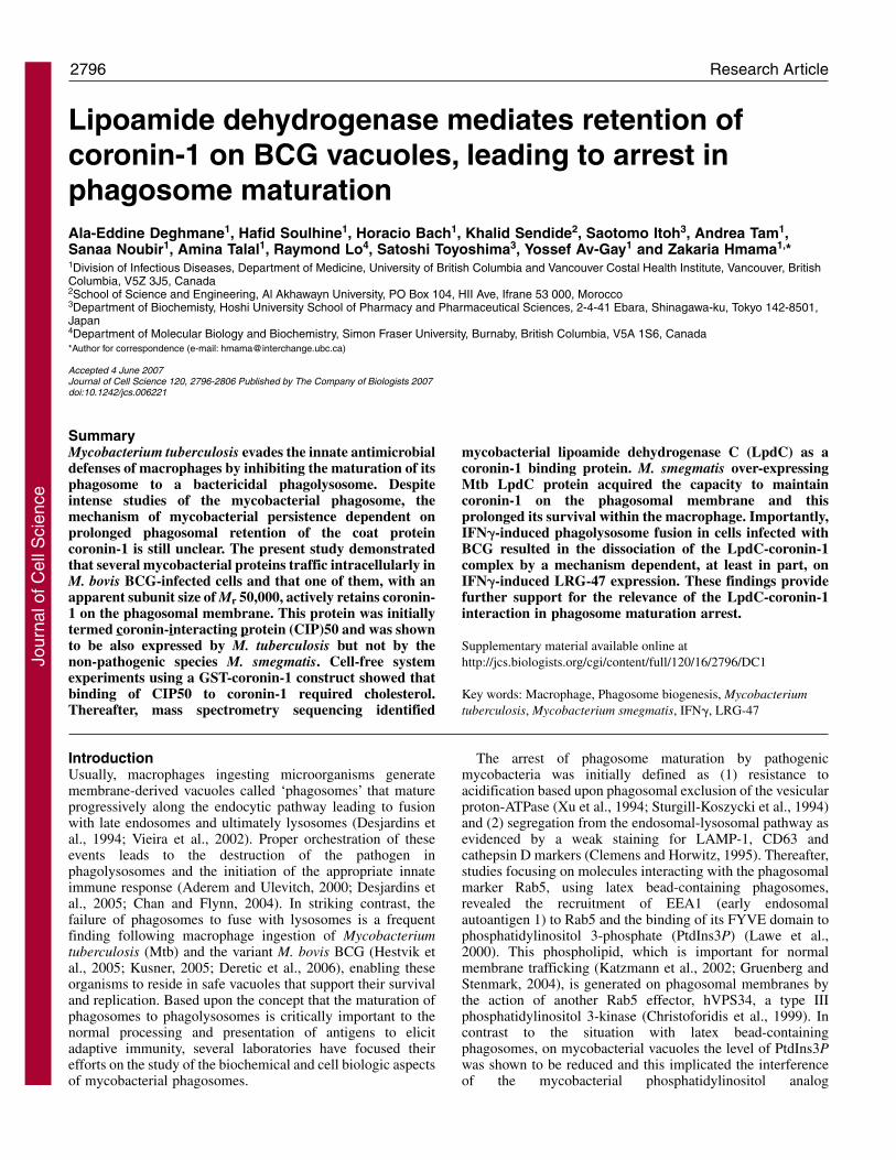
in macrophages infected with live but not killed BCG (Fig.1A). Interestingly, this band was also absent inimmunoprecipitates derived from cells infected with M.smegmatis. This 50 kDa mycobacterial protein was namedCIP50 (for coronin-interacting protein 50 kDa). Furtheranalyses involving competitive inhibition with recombinantcoronin-1 demonstrated that N-7 antibodies specifically pulleddown the 50 kDa bacterial protein associated with coronin-1(Fig. 1B).
To seek additional evidence to support an interactionbetween CIP50 and coronin-1, the soluble fraction frommacrophage lysates – the source of coronin-1 – was added toradioactive (35S) culture filtrate proteins (CFPs) prepared fromradiolabeled BCG, M. smegmatis or Mtb. The results shown inFig. 1C indicate that CIP50 was detected in CFPs derived fromBCG and Mtb, but not from M. smegmatis. These findings ofan in vitro interaction are consistent with the in vivo pull-downdata (Fig. 1A). Furthermore, experiments based on 2D-gelelectrophoresis SDS–PAGE showed that the 50 kDa bandcorresponded to a single protein with an apparent pI of 5.4 (Fig.1D). Taken together, these findings demonstrate that live,pathogenic mycobacteria express a coronin-1-interactingprotein.
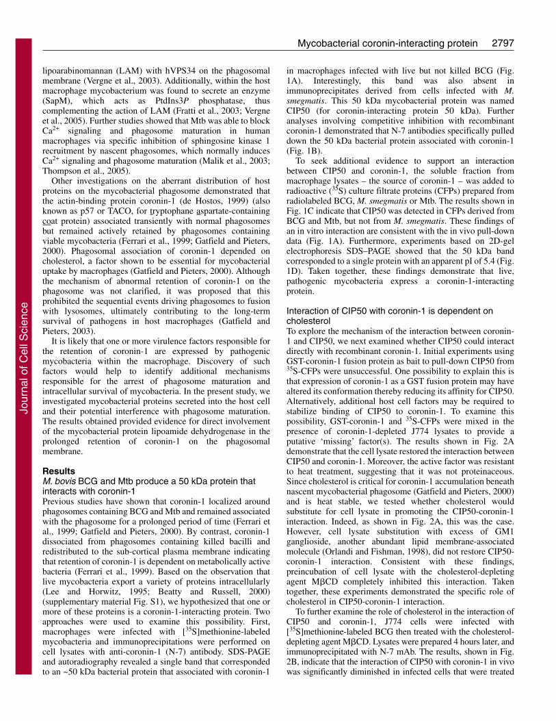
Interaction of CIP50 with coronin-1 is dependent oncholesterolTo explore the mechanism of the interaction between coronin-1 and CIP50, we next examined whether CIP50 could interactdirectly with recombinant coronin-1. Initial experiments usingGST-coronin-1 fusion protein as bait to pull-down CIP50 from35S-CFPs were unsuccessful. One possibility to explain this isthat expression of coronin-1 as a GST fusion protein may havealtered its conformation thereby reducing its affinity for CIP50.Alternatively, additional host cell factors may be required tostabilize binding of CIP50 to coronin-1. To examine thispossibility, GST-coronin-1 and 35S-CFPs were mixed in thepresence of coronin-1-depleted J774 lysates to provide aputative ‘missing’ factor(s). The results shown in Fig. 2Ademonstrate that the cell lysate restored the interaction betweenCIP50 and coronin-1. Moreover, the active factor was resistantto heat treatment, suggesting that it was not proteinaceous.Since cholesterol is critical for coronin-1 accumulation beneathnascent mycobacterial phagosome (Gatfield and Pieters, 2000)and is heat stable, we tested whether cholesterol wouldsubstitute for cell lysate in promoting the CIP50-coronin-1interaction. Indeed, as shown in Fig. 2A, this was the case.However, cell lysate substitution with excess of GM1ganglioside, another abundant lipid membrane-associatedmolecule (Orlandi and Fishman, 1998), did not restore CIP50-coronin-1 interaction. Consistent with these findings,preincubation of cell lysate with the cholesterol-depletingagent M�CD completely inhibited this interaction. Takentogether, these experiments demonstrated the specific role ofcholesterol in CIP50-coronin-1 interaction.
To further examine the role of cholesterol in the interaction ofCIP50 and coronin-1, J774 cells were infected with[35S]methionine-labeled BCG then treated with the cholesterol-depleting agent M�CD. Lysates were prepared 4 hours later, andimmunoprecipitated with N-7 mAb. The results, shown in Fig.2B, indicate that the interaction of CIP50 with coronin-1 in vivowas significantly diminished in infected cells that were treated
Jour
nal o
f Cel
l Sci
ence
2798
with M�CD but was restored by the addition of exogenouscholesterol. These findings demonstrate that cholesterol isinvolved in the stabilization of the interaction between CIP50and coronin-1 and are consistent with previously publishedimmunofluorescence data showing that depleting plasmamembrane cholesterol specifically inhibited the retention ofcoronin-1 on BCG phagosomes (Gatfield and Pieters, 2000).
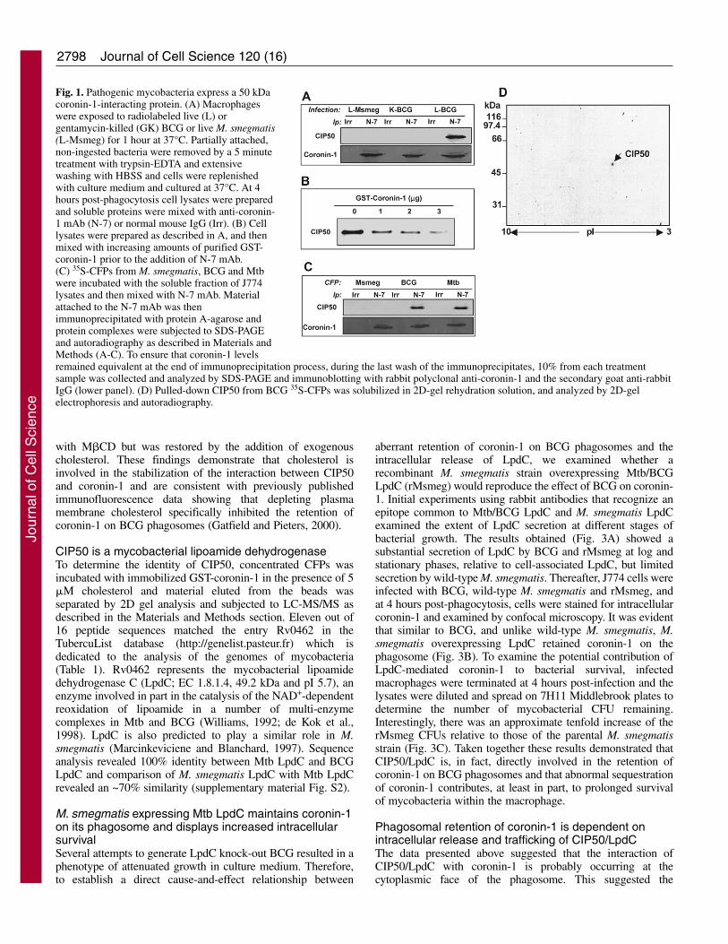
CIP50 is a mycobacterial lipoamide dehydrogenaseTo determine the identity of CIP50, concentrated CFPs wasincubated with immobilized GST-coronin-1 in the presence of 5�M cholesterol and material eluted from the beads wasseparated by 2D gel analysis and subjected to LC-MS/MS asdescribed in the Materials and Methods section. Eleven out of16 peptide sequences matched the entry Rv0462 in theTubercuList database (http://genelist.pasteur.fr) which isdedicated to the analysis of the genomes of mycobacteria(Table 1). Rv0462 represents the mycobacterial lipoamidedehydrogenase C (LpdC; EC 1.8.1.4, 49.2 kDa and pI 5.7), anenzyme involved in part in the catalysis of the NAD+-dependentreoxidation of lipoamide in a number of multi-enzymecomplexes in Mtb and BCG (Williams, 1992; de Kok et al.,1998). LpdC is also predicted to play a similar role in M.smegmatis (Marcinkeviciene and Blanchard, 1997). Sequenceanalysis revealed 100% identity between Mtb LpdC and BCGLpdC and comparison of M. smegmatis LpdC with Mtb LpdCrevealed an ~70% similarity (supplementary material Fig. S2).
M. smegmatis expressing Mtb LpdC maintains coronin-1on its phagosome and displays increased intracellularsurvivalSeveral attempts to generate LpdC knock-out BCG resulted in aphenotype of attenuated growth in culture medium. Therefore,to establish a direct cause-and-effect relationship between
Journal of Cell Science 120 (16)
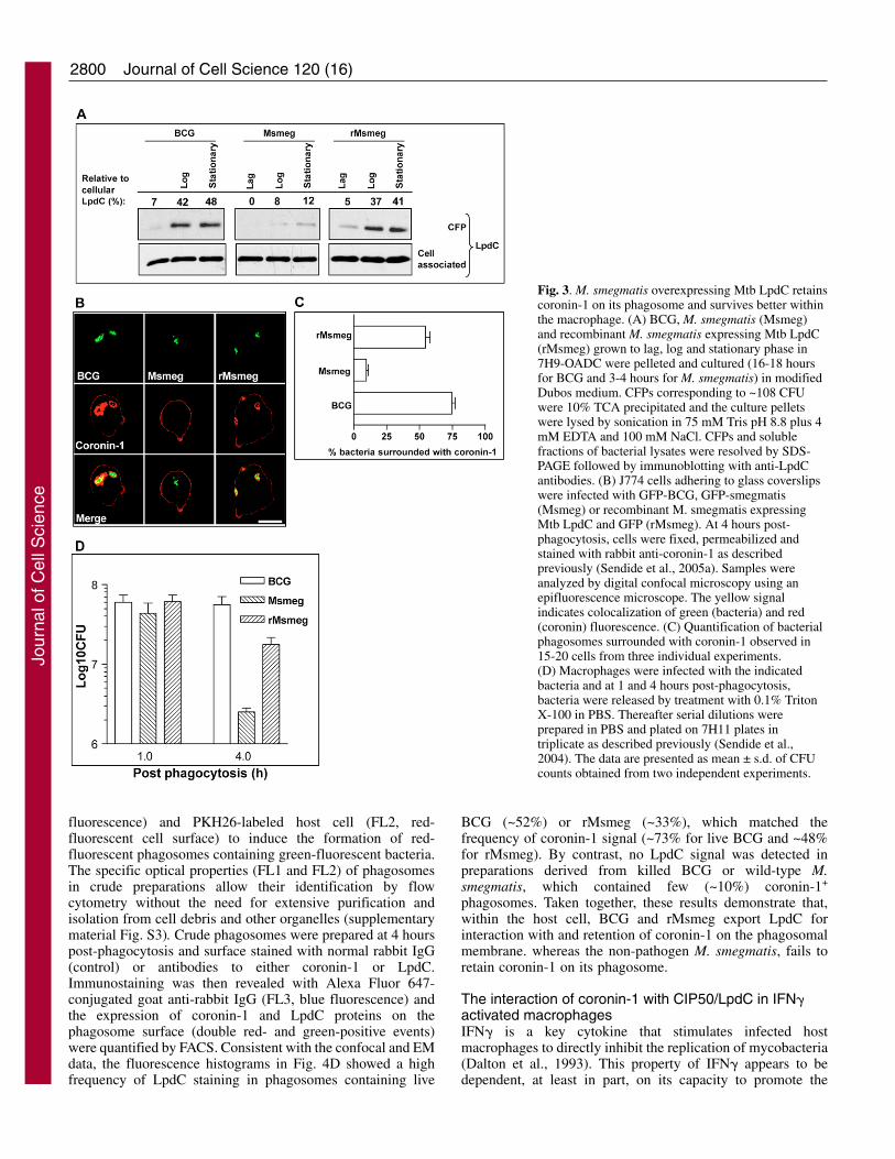
aberrant retention of coronin-1 on BCG phagosomes and theintracellular release of LpdC, we examined whether arecombinant M. smegmatis strain overexpressing Mtb/BCGLpdC (rMsmeg) would reproduce the effect of BCG on coronin-1. Initial experiments using rabbit antibodies that recognize anepitope common to Mtb/BCG LpdC and M. smegmatis LpdCexamined the extent of LpdC secretion at different stages ofbacterial growth. The results obtained (Fig. 3A) showed asubstantial secretion of LpdC by BCG and rMsmeg at log andstationary phases, relative to cell-associated LpdC, but limitedsecretion by wild-type M. smegmatis. Thereafter, J774 cells wereinfected with BCG, wild-type M. smegmatis and rMsmeg, andat 4 hours post-phagocytosis, cells were stained for intracellularcoronin-1 and examined by confocal microscopy. It was evidentthat similar to BCG, and unlike wild-type M. smegmatis, M.smegmatis overexpressing LpdC retained coronin-1 on thephagosome (Fig. 3B). To examine the potential contribution ofLpdC-mediated coronin-1 to bacterial survival, infectedmacrophages were terminated at 4 hours post-infection and thelysates were diluted and spread on 7H11 Middlebrook plates todetermine the number of mycobacterial CFU remaining.Interestingly, there was an approximate tenfold increase of therMsmeg CFUs relative to those of the parental M. smegmatisstrain (Fig. 3C). Taken together these results demonstrated thatCIP50/LpdC is, in fact, directly involved in the retention ofcoronin-1 on BCG phagosomes and that abnormal sequestrationof coronin-1 contributes, at least in part, to prolonged survivalof mycobacteria within the macrophage.
Phagosomal retention of coronin-1 is dependent onintracellular release and trafficking of CIP50/LpdCThe data presented above suggested that the interaction ofCIP50/LpdC with coronin-1 is probably occurring at thecytoplasmic face of the phagosome. This suggested the
Fig. 1. Pathogenic mycobacteria express a 50 kDacoronin-1-interacting protein. (A) Macrophageswere exposed to radiolabeled live (L) orgentamycin-killed (GK) BCG or live M. smegmatis(L-Msmeg) for 1 hour at 37°C. Partially attached,non-ingested bacteria were removed by a 5 minutetreatment with trypsin-EDTA and extensivewashing with HBSS and cells were replenishedwith culture medium and cultured at 37°C. At 4hours post-phagocytosis cell lysates were preparedand soluble proteins were mixed with anti-coronin-1 mAb (N-7) or normal mouse IgG (Irr). (B) Celllysates were prepared as described in A, and thenmixed with increasing amounts of purified GST-coronin-1 prior to the addition of N-7 mAb.(C) 35S-CFPs from M. smegmatis, BCG and Mtbwere incubated with the soluble fraction of J774lysates and then mixed with N-7 mAb. Materialattached to the N-7 mAb was thenimmunoprecipitated with protein A-agarose andprotein complexes were subjected to SDS-PAGEand autoradiography as described in Materials andMethods (A-C). To ensure that coronin-1 levelsremained equivalent at the end of immunoprecipitation process, during the last wash of the immunoprecipitates, 10% from each treatmentsample was collected and analyzed by SDS-PAGE and immunoblotting with rabbit polyclonal anti-coronin-1 and the secondary goat anti-rabbitIgG (lower panel). (D) Pulled-down CIP50 from BCG 35S-CFPs was solubilized in 2D-gel rehydration solution, and analyzed by 2D-gelelectrophoresis and autoradiography.
Jour
nal o
f Cel
l Sci
ence
2799Mycobacterial coronin-interacting protein
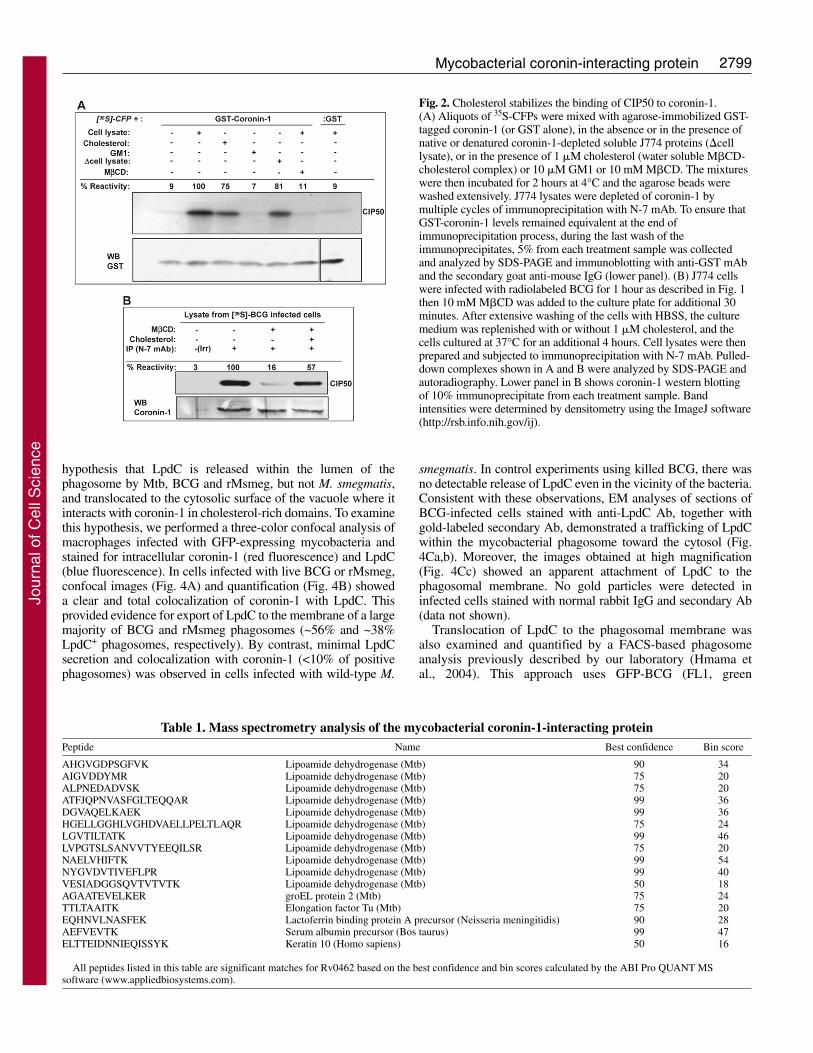
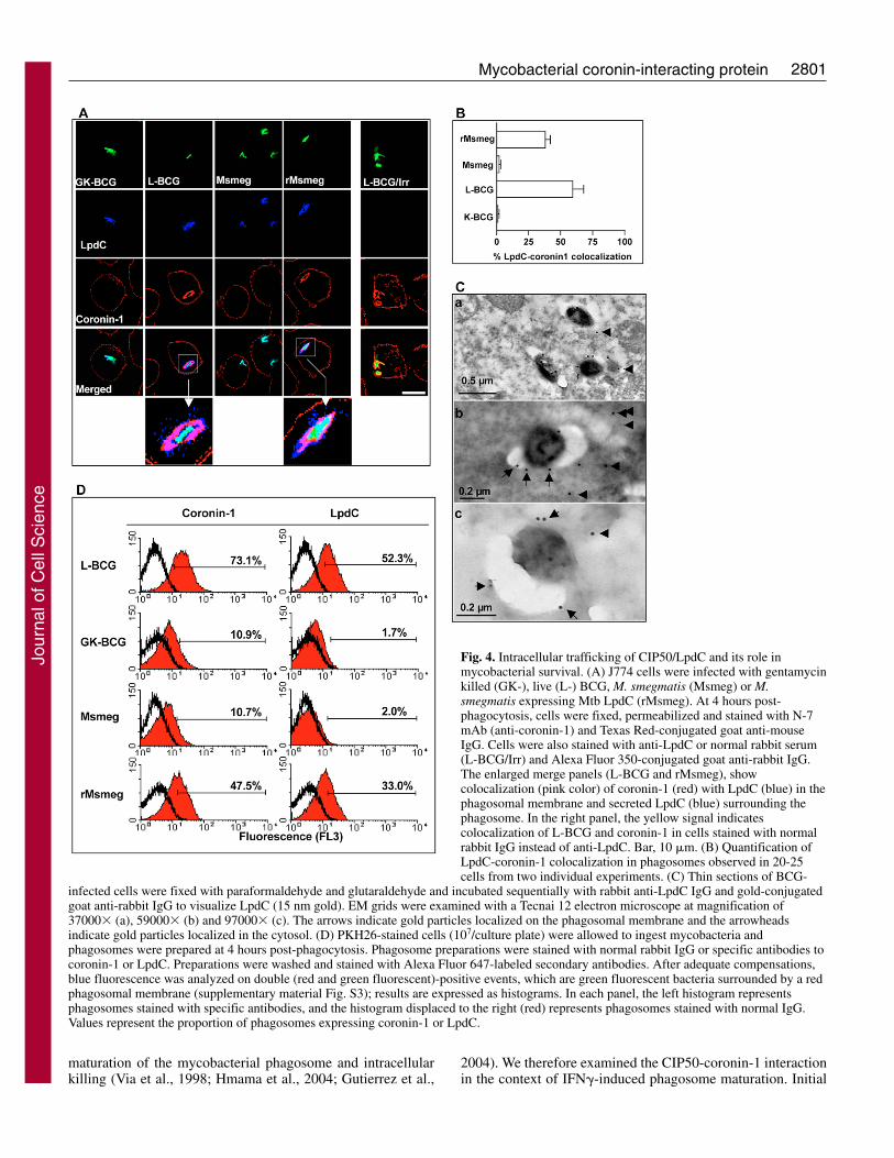
hypothesis that LpdC is released within the lumen of thephagosome by Mtb, BCG and rMsmeg, but not M. smegmatis,and translocated to the cytosolic surface of the vacuole where itinteracts with coronin-1 in cholesterol-rich domains. To examinethis hypothesis, we performed a three-color confocal analysis ofmacrophages infected with GFP-expressing mycobacteria andstained for intracellular coronin-1 (red fluorescence) and LpdC(blue fluorescence). In cells infected with live BCG or rMsmeg,confocal images (Fig. 4A) and quantification (Fig. 4B) showeda clear and total colocalization of coronin-1 with LpdC. Thisprovided evidence for export of LpdC to the membrane of a largemajority of BCG and rMsmeg phagosomes (~56% and ~38%LpdC+ phagosomes, respectively). By contrast, minimal LpdCsecretion and colocalization with coronin-1 (<10% of positivephagosomes) was observed in cells infected with wild-type M.
smegmatis. In control experiments using killed BCG, there wasno detectable release of LpdC even in the vicinity of the bacteria.Consistent with these observations, EM analyses of sections ofBCG-infected cells stained with anti-LpdC Ab, together withgold-labeled secondary Ab, demonstrated a trafficking of LpdCwithin the mycobacterial phagosome toward the cytosol (Fig.4Ca,b). Moreover, the images obtained at high magnification(Fig. 4Cc) showed an apparent attachment of LpdC to thephagosomal membrane. No gold particles were detected ininfected cells stained with normal rabbit IgG and secondary Ab(data not shown).
Translocation of LpdC to the phagosomal membrane wasalso examined and quantified by a FACS-based phagosomeanalysis previously described by our laboratory (Hmama etal., 2004). This approach uses GFP-BCG (FL1, green
Fig. 2. Cholesterol stabilizes the binding of CIP50 to coronin-1. (A) Aliquots of 35S-CFPs were mixed with agarose-immobilized GST-tagged coronin-1 (or GST alone), in the absence or in the presence ofnative or denatured coronin-1-depleted soluble J774 proteins (�celllysate), or in the presence of 1 �M cholesterol (water soluble M�CD-cholesterol complex) or 10 �M GM1 or 10 mM M�CD. The mixtureswere then incubated for 2 hours at 4°C and the agarose beads werewashed extensively. J774 lysates were depleted of coronin-1 bymultiple cycles of immunoprecipitation with N-7 mAb. To ensure thatGST-coronin-1 levels remained equivalent at the end ofimmunoprecipitation process, during the last wash of theimmunoprecipitates, 5% from each treatment sample was collectedand analyzed by SDS-PAGE and immunoblotting with anti-GST mAband the secondary goat anti-mouse IgG (lower panel). (B) J774 cellswere infected with radiolabeled BCG for 1 hour as described in Fig. 1then 10 mM M�CD was added to the culture plate for additional 30minutes. After extensive washing of the cells with HBSS, the culturemedium was replenished with or without 1 �M cholesterol, and thecells cultured at 37°C for an additional 4 hours. Cell lysates were thenprepared and subjected to immunoprecipitation with N-7 mAb. Pulled-down complexes shown in A and B were analyzed by SDS-PAGE andautoradiography. Lower panel in B shows coronin-1 western blottingof 10% immunoprecipitate from each treatment sample. Bandintensities were determined by densitometry using the ImageJ software(http://rsb.info.nih.gov/ij).
Table 1. Mass spectrometry analysis of the mycobacterial coronin-1-interacting protein Peptide Name Best confidence Bin score
AHGVGDPSGFVK Lipoamide dehydrogenase (Mtb) 90 34AIGVDDYMR Lipoamide dehydrogenase (Mtb) 75 20ALPNEDADVSK Lipoamide dehydrogenase (Mtb) 75 20ATFJQPNVASFGLTEQQAR Lipoamide dehydrogenase (Mtb) 99 36DGVAQELKAEK Lipoamide dehydrogenase (Mtb) 99 36HGELLGGHLVGHDVAELLPELTLAQR Lipoamide dehydrogenase (Mtb) 75 24LGVTILTATK Lipoamide dehydrogenase (Mtb) 99 46LVPGTSLSANVVTYEEQILSR Lipoamide dehydrogenase (Mtb) 75 20NAELVHIFTK Lipoamide dehydrogenase (Mtb) 99 54NYGVDVTIVEFLPR Lipoamide dehydrogenase (Mtb) 99 40VESIADGGSQVTVTVTK Lipoamide dehydrogenase (Mtb) 50 18AGAATEVELKER groEL protein 2 (Mtb) 75 24TTLTAAITK Elongation factor Tu (Mtb) 75 20EQHNVLNASFEK Lactoferrin binding protein A precursor (Neisseria meningitidis) 90 28AEFVEVTK Serum albumin precursor (Bos taurus) 99 47ELTTEIDNNIEQISSYK Keratin 10 (Homo sapiens) 50 16
All peptides listed in this table are significant matches for Rv0462 based on the best confidence and bin scores calculated by the ABI Pro QUANT MSsoftware (www.appliedbiosystems.com).
Jour
nal o
f Cel
l Sci
ence
2800
fluorescence) and PKH26-labeled host cell (FL2, red-fluorescent cell surface) to induce the formation of red-fluorescent phagosomes containing green-fluorescent bacteria.The specific optical properties (FL1 and FL2) of phagosomesin crude preparations allow their identification by flowcytometry without the need for extensive purification andisolation from cell debris and other organelles (supplementarymaterial Fig. S3). Crude phagosomes were prepared at 4 hourspost-phagocytosis and surface stained with normal rabbit IgG(control) or antibodies to either coronin-1 or LpdC.Immunostaining was then revealed with Alexa Fluor 647-conjugated goat anti-rabbit IgG (FL3, blue fluorescence) andthe expression of coronin-1 and LpdC proteins on thephagosome surface (double red- and green-positive events)were quantified by FACS. Consistent with the confocal and EMdata, the fluorescence histograms in Fig. 4D showed a highfrequency of LpdC staining in phagosomes containing live
Journal of Cell Science 120 (16)
BCG (~52%) or rMsmeg (~33%), which matched thefrequency of coronin-1 signal (~73% for live BCG and ~48%for rMsmeg). By contrast, no LpdC signal was detected inpreparations derived from killed BCG or wild-type M.smegmatis, which contained few (~10%) coronin-1+
phagosomes. Taken together, these results demonstrate that,within the host cell, BCG and rMsmeg export LpdC forinteraction with and retention of coronin-1 on the phagosomalmembrane. whereas the non-pathogen M. smegmatis, fails toretain coronin-1 on its phagosome.
The interaction of coronin-1 with CIP50/LpdC in IFN�activated macrophagesIFN� is a key cytokine that stimulates infected hostmacrophages to directly inhibit the replication of mycobacteria(Dalton et al., 1993). This property of IFN� appears to bedependent, at least in part, on its capacity to promote the
Fig. 3. M. smegmatis overexpressing Mtb LpdC retainscoronin-1 on its phagosome and survives better withinthe macrophage. (A) BCG, M. smegmatis (Msmeg)and recombinant M. smegmatis expressing Mtb LpdC(rMsmeg) grown to lag, log and stationary phase in7H9-OADC were pelleted and cultured (16-18 hoursfor BCG and 3-4 hours for M. smegmatis) in modifiedDubos medium. CFPs corresponding to ~108 CFUwere 10% TCA precipitated and the culture pelletswere lysed by sonication in 75 mM Tris pH 8.8 plus 4mM EDTA and 100 mM NaCl. CFPs and solublefractions of bacterial lysates were resolved by SDS-PAGE followed by immunoblotting with anti-LpdCantibodies. (B) J774 cells adhering to glass coverslipswere infected with GFP-BCG, GFP-smegmatis(Msmeg) or recombinant M. smegmatis expressingMtb LpdC and GFP (rMsmeg). At 4 hours post-phagocytosis, cells were fixed, permeabilized andstained with rabbit anti-coronin-1 as describedpreviously (Sendide et al., 2005a). Samples wereanalyzed by digital confocal microscopy using anepifluorescence microscope. The yellow signalindicates colocalization of green (bacteria) and red(coronin) fluorescence. (C) Quantification of bacterialphagosomes surrounded with coronin-1 observed in15-20 cells from three individual experiments. (D) Macrophages were infected with the indicatedbacteria and at 1 and 4 hours post-phagocytosis,bacteria were released by treatment with 0.1% TritonX-100 in PBS. Thereafter serial dilutions wereprepared in PBS and plated on 7H11 plates intriplicate as described previously (Sendide et al.,2004). The data are presented as mean ± s.d. of CFUcounts obtained from two independent experiments.
Jour
nal o
f Cel
l Sci
ence
2801Mycobacterial coronin-interacting protein
maturation of the mycobacterial phagosome and intracellularkilling (Via et al., 1998; Hmama et al., 2004; Gutierrez et al.,
2004). We therefore examined the CIP50-coronin-1 interactionin the context of IFN�-induced phagosome maturation. Initial
Fig. 4. Intracellular trafficking of CIP50/LpdC and its role inmycobacterial survival. (A) J774 cells were infected with gentamycinkilled (GK-), live (L-) BCG, M. smegmatis (Msmeg) or M.smegmatis expressing Mtb LpdC (rMsmeg). At 4 hours post-phagocytosis, cells were fixed, permeabilized and stained with N-7mAb (anti-coronin-1) and Texas Red-conjugated goat anti-mouseIgG. Cells were also stained with anti-LpdC or normal rabbit serum(L-BCG/Irr) and Alexa Fluor 350-conjugated goat anti-rabbit IgG.The enlarged merge panels (L-BCG and rMsmeg), showcolocalization (pink color) of coronin-1 (red) with LpdC (blue) in thephagosomal membrane and secreted LpdC (blue) surrounding thephagosome. In the right panel, the yellow signal indicatescolocalization of L-BCG and coronin-1 in cells stained with normalrabbit IgG instead of anti-LpdC. Bar, 10 �m. (B) Quantification ofLpdC-coronin-1 colocalization in phagosomes observed in 20-25cells from two individual experiments. (C) Thin sections of BCG-
infected cells were fixed with paraformaldehyde and glutaraldehyde and incubated sequentially with rabbit anti-LpdC IgG and gold-conjugatedgoat anti-rabbit IgG to visualize LpdC (15 nm gold). EM grids were examined with a Tecnai 12 electron microscope at magnification of37000� (a), 59000� (b) and 97000� (c). The arrows indicate gold particles localized on the phagosomal membrane and the arrowheadsindicate gold particles localized in the cytosol. (D) PKH26-stained cells (107/culture plate) were allowed to ingest mycobacteria andphagosomes were prepared at 4 hours post-phagocytosis. Phagosome preparations were stained with normal rabbit IgG or specific antibodies tocoronin-1 or LpdC. Preparations were washed and stained with Alexa Fluor 647-labeled secondary antibodies. After adequate compensations,blue fluorescence was analyzed on double (red and green fluorescent)-positive events, which are green fluorescent bacteria surrounded by a redphagosomal membrane (supplementary material Fig. S3); results are expressed as histograms. In each panel, the left histogram representsphagosomes stained with specific antibodies, and the histogram displaced to the right (red) represents phagosomes stained with normal IgG.Values represent the proportion of phagosomes expressing coronin-1 or LpdC.
Jour
nal o
f Cel
l Sci
ence
2802
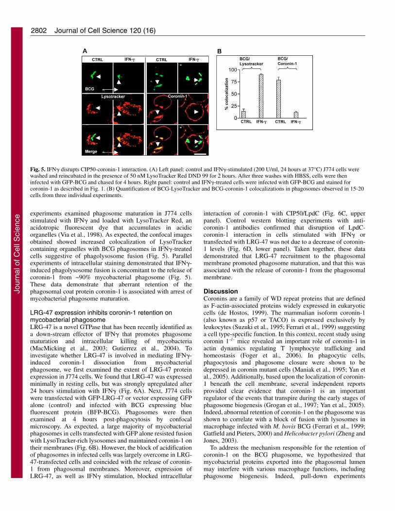
experiments examined phagosome maturation in J774 cellsstimulated with IFN� and loaded with LysoTracker Red, anacidotropic fluorescent dye that accumulates in acidicorganelles (Via et al., 1998). As expected, the confocal imagesobtained showed increased colocalization of LysoTrackercontaining organelles with BCG phagosomes in IFN�-treatedcells suggestive of phagolysosome fusion (Fig. 5). Parallelexperiments of intracellular staining demonstrated that IFN�-induced phagolysosome fusion is concomitant to the release ofcoronin-1 from ~90% mycobacterial phagosome (Fig. 5).These data demonstrate that aberrant retention of thephagosomal coat protein coronin-1 is associated with arrest ofmycobacterial phagosome maturation.
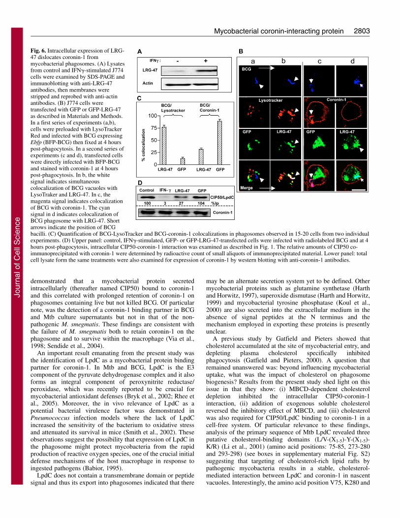
LRG-47 expression inhibits coronin-1 retention onmycobacterial phagosomeLRG-47 is a novel GTPase that has been recently identified asa down-stream effector of IFN� that promotes phagosomematuration and intracellular killing of mycobacteria(MacMicking et al., 2003; Gutierrez et al., 2004). Toinvestigate whether LRG-47 is involved in mediating IFN�-induced coronin-1 dissociation from mycobacterialphagosome, we first examined the extent of LRG-47 proteinexpression in J774 cells. We found that LRG-47 was expressedminimally in resting cells, but was strongly upregulated after24 hours stimulation with IFN� (Fig. 6A). Next, J774 cellswere transfected with GFP-LRG-47 or vector expressing GFPalone (control) and infected with BCG expressing bluefluorescent protein (BFP-BCG). Phagosomes were thenexamined at 4 hours post-phagocytosis by confocalmicroscopy. As expected, a large majority of mycobacterialphagosomes in cells transfected with GFP alone resisted fusionwith LysoTracker-rich lysosomes and maintained coronin-1 ontheir membranes (Fig. 6B). However, the block of acidificationof phagosomes in infected cells was largely overcome in LRG-47-transfected cells and coincided with the release of coronin-1 from phagosomal membranes. Moreover, expression ofLRG-47, as well as IFN� stimulation, blocked intracellular
Journal of Cell Science 120 (16)
interaction of coronin-1 with CIP50/LpdC (Fig. 6C, upperpanel). Control western blotting experiments with anti-coronin-1 antibodies confirmed that disruption of LpdC-coronin-1 interaction in cells stimulated with IFN� ortransfected with LRG-47 was not due to a decrease of coronin-1 levels (Fig. 6D, lower panel). Taken together, these datademonstrated that LRG-47 recruitment to the phagosomalmembrane promoted phagosome maturation, and that this wasassociated with the release of coronin-1 from the phagosomalmembrane.
DiscussionCoronins are a family of WD repeat proteins that are definedas F-actin-associated proteins widely expressed in eukaryoticcells (de Hostos, 1999). The mammalian isoform coronin-1(also known as p57 or TACO) is expressed exclusively byleukocytes (Suzuki et al., 1995; Ferrari et al., 1999) suggestinga cell type-specific function. In this context, recent study usingcoronin 1–/– mice revealed an important role of coronin-1 inactin dynamics regulating T lymphocyte trafficking andhomeostasis (Foger et al., 2006). In phagocytic cells,phagocytosis and phagosome closure were shown to bedepressed in coronin mutant cells (Maniak et al., 1995; Yan etal., 2005). Additionally, based upon the localization of coronin-1 beneath the cell membrane, several independent reportsprovided clear evidence that coronin-1 is an importantregulator of the events that transpire during the early stages ofphagosome biogenesis (Grogan et al., 1997; Yan et al., 2005).Indeed, abnormal retention of coronin-1 on the phagosome wasshown to correlate with a block of fusion with lysosomes inmacrophage infected with M. bovis BCG (Ferrari et al., 1999;Gatfield and Pieters, 2000) and Helicobacter pylori (Zheng andJones, 2003).
To address the mechanism responsible for the retention ofcoronin-1 on the BCG phagosome, we hypothesized thatmycobacterial proteins exported into the phagosomal lumenmay interfere with various macrophage functions, includingphagosome biogenesis. Indeed, pull-down experiments
Fig. 5. IFN� disrupts CIP50-coronin-1 interaction. (A) Left panel: control and IFN�-stimulated (200 U/ml, 24 hours at 37°C) J774 cells werewashed and reincubated in the presence of 50 nM LysoTracker Red DND 99 for 2 hours. After three washes with HBSS, cells were theninfected with GFP-BCG and chased for 4 hours. Right panel: control and IFN�-treated cells were infected with GFP-BCG and stained forcoronin-1 as described in Fig. 1. (B) Quantification of BCG-LysoTracker and BCG-coronin-1 colocalizations in phagosomes observed in 15-20cells from three individual experiments.
Jour
nal o
f Cel
l Sci
ence
2803Mycobacterial coronin-interacting protein
demonstrated that a mycobacterial protein secretedintracellularly (thereafter named CIP50) bound to coronin-1and this correlated with prolonged retention of coronin-1 onphagosomes containing live but not killed BCG. Of particularnote, was the detection of a coronin-1 binding partner in BCGand Mtb culture supernatants but not in that of the non-pathogenic M. smegmatis. These findings are consistent withthe failure of M. smegmatis both to retain coronin-1 on thephagosome and to survive within the macrophage (Via et al.,1998; Sendide et al., 2004).
An important result emanating from the present study wasthe identification of LpdC as a mycobacterial protein bindingpartner for coronin-1. In Mtb and BCG, LpdC is the E3component of the pyruvate dehydrogenase complex and it alsoforms an integral component of peroxynitrite reductase/peroxidase, which was recently reported to be crucial formycobacterial antioxidant defenses (Bryk et al., 2002; Rhee etal., 2005). Moreover, the in vivo relevance of LpdC as apotential bacterial virulence factor was demonstrated inPneumococcus infection models where the lack of LpdCincreased the sensitivity of the bacterium to oxidative stressand attenuated its survival in mice (Smith et al., 2002). Theseobservations suggest the possibility that expression of LpdC inthe phagosome might protect mycobacteria from the rapidproduction of reactive oxygen species, one of the crucial initialdefense mechanisms of the host macrophage in response toingested pathogens (Babior, 1995).
LpdC does not contain a transmembrane domain or peptidesignal and thus its export into phagosomes indicated that there
may be an alternate secretion system yet to be defined. Othermycobacterial proteins such as glutamine synthetase (Harthand Horwitz, 1997), superoxide dismutase (Harth and Horwitz,1999) and mycobacterial tyrosine phosphatase (Koul et al.,2000) are also secreted into the extracellular medium in theabsence of signal peptides at the N terminus and themechanism employed in exporting these proteins is presentlyunclear.
A previous study by Gatfield and Pieters showed thatcholesterol accumulated at the site of mycobacterial entry, anddepleting plasma cholesterol specifically inhibitedphagocytosis (Gatfield and Pieters, 2000). A question thatremained unanswered was: beyond influencing mycobacterialuptake, what was the impact of cholesterol on phagosomebiogenesis? Results from the present study shed light on thisissue in that they show: (i) MBCD-dependent cholesteroldepletion inhibited the intracellular CIP50-coronin-1interaction, (ii) addition of exogenous soluble cholesterolreversed the inhibitory effect of MBCD, and (iii) cholesterolwas also required for CIP50/LpdC binding to coronin-1 in acell-free system. Of particular relevance to these findings,analysis of the primary sequence of Mtb LpdC revealed threeputative cholesterol-binding domains (L/V-(X1-5)-Y-(X1-5)-K/R) (Li et al., 2001) (amino acid positions: 75-85, 273-280and 293-298) (see boxes in supplementary material Fig. S2)suggesting that targeting of cholesterol-rich lipid rafts bypathogenic mycobacteria results in a stable, cholesterol-mediated interaction between LpdC and coronin-1 in nascentvacuoles. Interestingly, the amino acid position V75, K280 and
Fig. 6. Intracellular expression of LRG-47 dislocates coronin-1 frommycobacterial phagosomes. (A) Lysatesfrom control and IFN�-stimulated J774cells were examined by SDS-PAGE andimmunoblotting with anti-LRG-47antibodies, then membranes werestripped and reprobed with anti-actinantibodies. (B) J774 cells weretransfected with GFP or GFP-LRG-47as described in Materials and Methods.In a first series of experiments (a,b),cells were preloaded with LysoTrackerRed and infected with BCG expressingEbfp (BFP-BCG) then fixed at 4 hourspost-phagocytosis. In a second series ofexperiments (c and d), transfected cellswere directly infected with BFP-BCGand stained with coronin-1 at 4 hourspost-phagocytosis. In b, the whitesignal indicates simultaneouscolocalization of BCG vacuoles withLysoTraker and LRG-47. In c, themagenta signal indicates colocalizationof BCG with coronin-1. The cyansignal in d indicates colocalization ofBCG phagosome with LRG-47. Shortarrows indicate the position of BCGbacilli. (C) Quantification of BCG-LysoTracker and BCG-coronin-1 colocalizations in phagosomes observed in 15-20 cells from two individualexperiments. (D) Upper panel: control, IFN�-stimulated, GFP- or GFP-LRG-47-transfected cells were infected with radiolabeled BCG and at 4hours post-phagocytosis, intracellular CIP50-coronin-1 interaction was examined as described in Fig. 1. The relative amounts of CIP50 co-immunoprecipitated with coronin-1 were determined by radioactive count of small aliquots of immunoprecipitated material. Lower panel: totalcell lysate form the same treatments were also examined for expression of coronin-1 by western blotting with anti-coronin-1 antibodies.
Jour
nal o
f Cel
l Sci
ence
2804 Journal of Cell Science 120 (16)
V293 in the cholesterol-binding motifs of Mtb and BCG LpdCare substituted in the M. smegmatis LpdC sequence with A75,A280 and I293, respectively (supplementary material Fig. S2).Therefore, it is likely that these amino acid substitutions playa role in the binding of cholesterol to LpdC or in its secretion.Such an assumption is supported by the finding that M.smegmatis transfected with vector expressing the Mtb lpdCgene, was able to secrete substantial amount of LpdC andbehaved like BCG in terms of prolonged retention of coronin-1 on the phagosome. These observations also established adirect cause-and-effect relationship between abnormalretention of coronin-1 on the phagosome and the intracellularrelease of LpdC.
It was important to demonstrate the relevance of LpdC-mediated retention of coronin-1 on BCG vacuoles to the arrestof phagosome maturation. We reasoned that if prolongedretention of coronin-1 modulated phagosome biogenesis ininfected cells, it would be observed to dissociate relativelyrapidly from phagosomes under conditions wheremycobacterial vacuoles normally fuse with lysosomes. Indeed,in macrophages incubated with IFN� [a cytokine known topromote maturation of mycobacterial phagosomes andinhibition of intracellular replication (Schaible et al., 1998; Viaet al., 1998)] BCG vacuoles intersected LysoTracker-containing lysosomes concomitant with the release of coronin-1 from the bacterial vacuole.
IFN�-induced expression of the antimicrobial enzymeNOS2 has been considered largely responsible for restrictingMtb replication via NO generation (Chan et al., 1992;MacMicking et al., 1997). However, the finding that themacrophage drastically reshapes its transcriptome in responseto IFN� [over 1000 genes are transcriptionally activated afterstimulation of macrophages with IFN� (Ehrt et al., 2001)]strongly suggested the possibility of additional TB defensepathways elicited by this cytokine. In this respect, recent invivo studies have revealed the existence of NOS2-independenteffector mechanisms against TB, mediated at least in part by anovel, small GTPase, LRG-47 (MacMicking et al., 2003).Furthermore, it was demonstrated that LRG-47 is a criticalregulator of autophagy-dependent disposal of mycobacterialvacuoles (Gutierrez et al., 2004). In keeping with thesefindings, the present study showed that the block inacidification of BCG vacuoles was largely overcome in cellstransfected with LRG-47. Moreover, LRG-47 recruitment tophagosome membranes was coincident with both thedissociation of coronin-1 from LpdC and its release from BCGvacuoles. These findings provide an additional basis forunderstanding how IFN�-induced LRG-47 expressionparticipates in the regulation of phagosome maturation.
In summary, the results presented above demonstrate thedirect involvement of BCG/Mtb LpdC in the prolongedretention of coronin-1 on the phagosome, thus promotingphagosome maturation arrest. More importantly, they showthat under the influence of IFN�, LRG-47 is recruited to thephagosome and mediates dislocation of coronin-1 leadingultimately to phagosome acidification and recruitment oflysosomal vacuoles and phagolyosome fusion. Future studiesshould address the mechanisms of LRG-47-induceddissociation of the LpdC-coronin-1 complex from thephagosomal membrane and confirm the contribution ofcholesterol binding motifs in LpdC-coronin-1 interaction.
Materials and MethodsReagents and chemicalsEndotoxin-free culture reagents were from StemCell Technologies (Vancouver,British Columbia, Canada). Protease inhibitor mixture, PMSF, trypsin-EDTA,methyl-beta-cyclodextrin (M�CD), ganglioside GM1 and glutathione-agarosebeads were purchased from Sigma-Aldrich (St Louis, MO). Protein A-agarosebeads were from Bio-Rad laboratories (Hercules, CA). [35S]methionine (specificactivity 1 mCi/mmole) was from PerkinElmer (Boston, MA) and murine rIFN� wasa generous gift of Genentech (South San Francisco, CA).
AntibodiesMouse mAb (N-7) and rabbit polyclonal anti-coronin-1 were described previously(Oku et al., 2003). Polyclonal anti-LpdC antibodies were raised in New Zealandwhite rabbits immunized with KLH conjugated synthetic peptide(LPNEDADVSKEIEKQ) corresponding to LpdC amino acids 207-220. Specificanti-LpdC antibodies were immunopurified using an immobilized peptide columnprepared using the Sulfolink® kit (Pierce Biotechnology, Rockford, IL). Rabbitpolyclonal anti-LRG-47 antibody was provided by Gregory Taylor (Duke UniversityMedical Center, Durham, NC) and rabbit anti-coronin-1 was a gift from Jean Pieters(Basel Institute for Immunology, Switzerland). All secondary antibodies werepurchased from Caltag Laboratories (Burlingame, CA) except Alexa Fluor 647-conjugated goat anti-rabbit IgG which was purchased from Invitrogen Canada Inc(Burlington, Ontario, Canada).
Cell cultureThe murine macrophagic cell line J774A.1 (American Type Culture Collection,Manassas, VA), was cultured in DMEM supplemented with 2 mM L-glutamine,non-essential amino acids and 10% FCS (HyClone, Logan, UT). Prior to infectionwith mycobacteria, cells were seeded at a density of 105/cm2 on coverslips or in 10cm diameter culture dishes (Corning Inc., Corning, NY) and allowed to adhere at37°C in a humidified atmosphere of 5% CO2.
Mycobacterial strainsM. bovis BCG (Pasteur 1173P2) and M. smegmatis (mc2155) strains expressingGFP (Cowley and Av-Gay, 2001) were used in this study along with Mtb H37Rv.BCG expressing Ebfp, a gene encoding a blue fluorescent protein (BFP-BCG) wasprepared using the plasmid construct pMN431 as described previously (Kaps etal., 2001). pMN431 was a gift from Michael Niederweis, University of Alabama,AL). Mycobacteria were grown in Middlebrook 7H9 broth (Difco, Franklin Lakes,NJ) supplemented with 10% (v/v) OADC (oleic acid, albumin and dextrosesolution; Difco) and 0.05% (v/v) Tween 80 (Sigma-Aldrich) at 37°C on a shaker.Bacteria were harvested by centrifugation and pellets were suspended in completemedium plus 10% glycerol. Mycobacterial cultures were stored in aliquots(~5�108/vial) at –70°C. To generate radioactive culture filtrate proteins(35S-CFPs), mycobacteria grown in 7H9-OADC were pelleted and cultured(for 16-18 hours for BCG and 3-4 hours for M. smegmatis) at 37°C in modifiedDubos medium (0.1% KH2PO4, 0.05% Na2HPO4, 0.12% sodium citrate, 0.06%MgSO4, 2% asparagine and 0.1% Tween 80) supplemented with 100 �Ci/mlof [35S]methionine. The culture supernatants were cleared from bacteria byhigh speed centrifugation and filtration through 0.22-�m-pore size filters.Bacteria were killed by 2 hours incubation at 37°C in the presence of 50 �g/mlgentamycin.
Recombinant M. smegmatis expressing Mtb LpdCRecombinant M. smegmatis expressing GFP and Mtb LpdC (rMsmeg) wasgenerated as follow: primers lpd-1: CCCAAAGGATCCGTGACCCACTATGA -CGTCGT (with a 5� BamHI adaptor) and lpd-2: CCCTTTGATATCTCAGAAA -ATTGATCATGTGG (with a 5� EcoRV adaptor) were used to amplify the entireopen reading frame (ORF) of lpdC from Mtb genomic DNA. The resulting ampliconwas then cloned between the BamHI and EcoRV sites of E. coli/Mycobacteriumshuttle vector pSC301 (Kaps et al., 2001) to obtain the recombinant plasmid pSC-lpdC in which lpdC was placed under the control of the mycobacterial superoxidedismutase promoter in transcriptional fusion with the promoterless gfp cassette. Therecombinant plasmid pSC-lpdC was then used to transform M. smegmatis mc2strain. Transformants were selected on standard 7H10 agar medium in the presenceof 50 �g/ml hygromycin.
Intracellular stainingIntracellular staining was performed on fixed and permeabilized cells as describedpreviously (Hmama et al., 1999; Sendide et al., 2005a). Coverslips were mountedin FluorSaveTM (Calbiochem-Novabiochem Corp., La Jolla CA) to minimizephotobleaching. Then slides were examined by digital confocal microscopy usingan Axioplan II epifluorescence microscope (Carl Zeiss Inc., Thornwood, NY)equipped with 63�/1.4 Plan-Apochromat objective (Carl Zeiss Inc). Images wererecorded using a CCD digital camera (Retiga EX, QImaging, Burnaby, BC, Canada)coupled to the Northern Eclipse software (Empix Imaging, Inc., Mississauga, ON,Canada).
Jour
nal o
f Cel
l Sci
ence
2805Mycobacterial coronin-interacting protein
Expression and purification of recombinant coronin-1Recombinant coronin-1 protein was produced in E. coli JM109 as fusion proteinswith GST as described previously (Oku et al., 2003).
Pull-down assaysRadiolabeled mycobacteria were extensively washed with PBS-0.1% Tween 80 andused to infect J774 macrophages. Cells were then lysed in extraction buffer (25 mMTris-HCl pH, 7.5, 1 mM EDTA, 1 mM EGTA, 100 mM NaCl, 1% Triton X-100,0.5% NP-40, 0.2 mM PMSF and protease inhibitor cocktail) for 20 minutes at 4°Cand debris was removed by high speed centrifugation. Five hundred �g of solubleproteins were mixed with 5 �g of anti-coronin-1 mAb N-7 or irrelevant normalmouse IgG and incubated for 1 hour at 4°C. Alternatively, soluble proteins fromnon-infected J774 cells were mixed with 100 �l of 35S-CFPs and incubated for 2hours at 4°C prior to the addition of N-7 mAb. Protein A-agarose beads were addedto the mixtures and the samples were incubated for an additional 30 minutes at 4°C.In other experiments, 50 �g of GST-fusion protein immobilized on agarose beads,were mixed with 100 �l of 35S-CFPs and incubated for 2 hours at 4°C. Agarosebeads were washed extensively and protein complexes were solubilized in 1�Laemmeli buffer and submitted to SDS-PAGE and X-ray radiography. To ensurethat coronin-1 levels were equivalent at the end of the immunoprecipitation, 10%from each treatment sample was collected during the last wash in a separate tubeand analyzed by SDS-PAGE and immunoblotting with polyclonal rabbit anti-coronin-1 IgG.
Immunoelectron microscopyImmunogold staining was conducted at the EM Facility of iCAPTURE Centre(Saint Paul Hospital, Vancouver, BC, Canada). In brief, BCG-infected macrophageswere fixed with 2% paraformaldehyde + 0.5% glutaraldehyde and embedded in 4%low melting point agarose. Immuno-gold labeling was performed on 55 nm sectionsobtained with a Leica UC6 Ultracut microtome (Leica Microsystems Inc,Bannockburn, IL) and the grids were examined with a Tecnai 12 electronmicroscope (FEI Company, Hillsboro, OR).
Two dimensional gel electrophoresisPulled-down proteins were solubilized in the rehydration solution (8 M urea, 2%CHAPS, 2% IPG buffer, 20 mM DTT and 0.002% Bromophenol Blue). Proteinswere first separated according to their isoelectric point (pI) in the immobilin Dry-Strip using IPGphor isoelectric focusing system (Amersham Biosciences,Piscataway, NJ). After equilibration, proteins separated on the strips were layeredon 10% SDS-PAGE gels and subjected to electrophoresis. Gels were then dried andexposed to X-ray films.
Nano-capillary HPLC ion trap mass spectrometry (LC-MS/MS)To identify mycobacterial coronin-1 interacting protein, culture filtrate proteins(CFPs) were concentrated ~100 fold and incubated with GST-coronin-1 cross-linkedto glutathione-agarose bead for 2 hours at 4°C. The material eluted from the beadswas then separated by two-dimensional (2D) gel analysis and subjected to silverstaining. The spot of interest was analyzed with LC-MS/MS of in-gel tryptic digestsat the Uvic Proteomics Centre Facilities (University of Victoria, Victoria, BCCanada).
Transient transfections with LRG-47pF25/LRG-47 plasmid carrying GFP-LRG-47 fusion protein (Gutierrez et al., 2004)(kindly provided by Gregory Taylor) and pEGFP-2C plasmid carrying GFP alone(Clontech, San Jose, CA) were prepared using Endofree plasmid maxiprep kit(Sigma-Aldrich). J774 cells adhering to 12-mm-diameter tissue culture-treatedcoverslips (Fisher Scientific, Nepean, ON, Canada) or 60-mm culture dishes weretransfected with the GFP constructs using the Lipofectamine 2000 reagent(Invitrogen) as described previously (Sendide et al., 2005b).
We thank Neil E. Reiner for critically reviewing the manuscript.We also thank Genentech Inc. for the gift of mouse recombinant IFN�,G. Taylor for the gift of the anti-LRG47 antibodies and GFP-LRG-47expression plasmid, J. Pieters for the gift of anti-coronin-1 antibodies,M. Niederweis for the gift of blue fluorescent protein expressionplasmid and Fanny Chu for assistance with EM work. This work wassupported by operating grants from the Canadian Institutes of HealthResearch (CIHR) MOP-67232 and BC Lung Association. Z.H. wassupported by scholar awards from MSFHR and the CIHR. K.S., R.L.and A.T. were supported by the TBVets Charitable Foundation. A.D.is recipient of a MSFHR and a CIHR postdoctoral fellowship.
ReferencesAderem, A. and Ulevitch, R. J. (2000). Toll-like receptors in the induction of the innate
immune response. Nature 406, 782-787.Babior, B. M. (1995). The respiratory burst oxidase. Curr. Opin. Hematol. 2, 55-60.
Beatty, W. L. and Russell, D. G. (2000). Identification of mycobacterial surface proteinsreleased into subcellular compartments of infected macrophages. Infect. Immun. 68,6997-7002.
Bryk, R., Lima, C. D., Erdjument-Bromage, H., Tempst, P. and Nathan, C. (2002).Metabolic enzymes of mycobacteria linked to antioxidant defense by a thioredoxin-like protein. Science 295, 1073-1077.
Chan, J. and Flynn, J. (2004). The immunological aspects of latency in tuberculosis.Clin. Immunol. 110, 2-12.
Chan, J., Xing, Y., Magliozzo, R. S. and Bloom, B. R. (1992). Killing of virulentMycobacterium tuberculosis by reactive nitrogen intermediates produced by activatedmurine macrophages. J. Exp. Med. 175, 1111-1122.
Christoforidis, S., Miaczynska, M., Ashman, K., Wilm, M., Zhao, L., Yip, S. C.,Waterfield, M. D., Backer, J. M. and Zerial, M. (1999). Phosphatidylinositol-3-OHkinases are Rab5 effectors. Nat. Cell Biol. 1, 249-252.
Clemens, D. and Horwitz, M. A. (1995). Characterization of the Mycobacteriumtuberculosis phagosome and evidence that phagosomal maturation is inhibited. J. Exp.Med. 181, 257-270.
Cowley, S. C. and Av-Gay, Y. (2001). Monitoring promoter activity and proteinlocalization in Mycobacterium spp. using green fluorescent protein. Gene 264, 225-231.
Dalton, D. K., Pitts-Meek, S., Keshav, S., Figari, I. S., Bradley, A. and Stewart, T. A.(1993). Multiple defects of immune cell function in mice with disrupted interferon-gamma genes. Science 259, 1739-1742.
de Hostos, E. L. (1999). The coronin family of actin-associated proteins. Trends CellBiol. 9, 345-350.
de Kok, A., Hengeveld, A. F., Martin, A. and Westphal, A. H. (1998). The pyruvatedehydrogenase multi-enzyme complex from Gram-negative bacteria. Biochim.Biophys. Acta 1385, 353-366.
Deretic, V., Singh, S., Master, S., Harris, J., Roberts, E., Kyei, G., Davis, A., de Haro,S., Naylor, J., Lee, H. H. and Vergne, I. (2006). Mycobacterium tuberculosisinhibition of phagolysosome biogenesis and autophagy as a host defence mechanism.Cell. Microbiol. 8, 719-727.
Desjardins, M., Huber, L. A., Parton, R. G. and Griffiths, G. (1994). Biogenesis ofphagolysosomes proceeds through a sequential series of interactions with the endocyticapparatus. J. Cell Biol. 124, 677-688.
Desjardins, M., Houde, M. and Gagnon, E. (2005). Phagocytosis: the convoluted wayfrom nutrition to adaptive immunity. Immunol. Rev. 207, 158-165.
Ehrt, S., Schnappinger, D., Bekiranov, S., Drenkow, J., Shi, S., Gingeras, T. R.,Gaasterland, T., Schoolnik, G. and Nathan, C. (2001). Reprogramming of themacrophage transcriptome in response to interferon-gamma and Mycobacteriumtuberculosis: signaling roles of nitric oxide synthase-2 and phagocyte oxidase. J. Exp.Med. 194, 1123-1140.
Ferrari, G., Langen, H., Naito, M. and Pieters, J. (1999). A coat protein on phagosomesinvolved in the intracellular survival of mycobacteria. Cell 97, 435-447.
Foger, N., Rangell, L., Danilenko, D. M. and Chan, A. C. (2006). Requirement forcoronin 1 in T lymphocyte trafficking and cellular homeostasis. Science. 313, 839-842.
Fratti, R. A., Chua, J., Vergne, I. and Deretic, V. (2003). Mycobacterium tuberculosisglycosylated phosphatidylinositol causes phagosome maturation arrest. Proc. Natl.Acad. Sci. USA 100, 5437-5442.
Gatfield, J. and Pieters, J. (2000). Essential role for cholesterol in entry of mycobacteriainto macrophages. Science 288, 1647-1650.
Gatfield, J. and Pieters, J. (2003). Molecular mechanisms of host-pathogen interaction:entry and survival of mycobacteria in macrophages. Adv. Immunol. 81, 45-96.
Grogan, A., Reeves, E., Keep, N., Wientjes, F., Totty, N. F., Burlingame, A. L., Hsuan,J. J. and Segal, A. W. (1997). Cytosolic phox proteins interact with and regulate theassembly of coronin in neutrophils. J. Cell Sci. 110, 3071-3081.
Gruenberg, J. and Stenmark, H. (2004). The biogenesis of multivesicular endosomes.Nat. Rev. Mol. Cell Biol. 5, 317-323.
Gutierrez, M. G., Master, S. S., Singh, S. B., Taylor, G. A., Colombo, M. I. andDeretic, V. (2004). Autophagy is a defense mechanism inhibiting BCG andMycobacterium tuberculosis survival in infected macrophages. Cell 119, 753-766.
Harth, G. and Horwitz, M. A. (1997). Expression and efficient export of enzymaticallyactive Mycobacterium tuberculosis glutamine synthetase in Mycobacterium smegmatisand evidence that the information for export is contained within the protein. J. Biol.Chem. 272, 22728-22735.
Harth, G. and Horwitz, M. A. (1999). Export of recombinant Mycobacteriumtuberculosis superoxide dismutase is dependent upon both information in the proteinand mycobacterial export machinery. A model for studying export of leaderless proteinsby pathogenic mycobacteria. J. Biol. Chem. 274, 4281-4292.
Hestvik, A. L., Hmama, Z. and Av-Gay, Y. (2005). Mycobacterial manipulation of thehost cell. FEMS Microbiol. Rev. 29, 1041-1050.
Hmama, Z., Gabathuler, R., Jefferies, W. A., de Jon, G. and Reiner, N. E. (1998).Attenuation of HLA-DR expression by mononuclear phagocytes infected withMycobacterium tuberculosis is related to intracellular sequestration of immature classII heterdimers. J. Immunol. 161, 4882-4893.
Hmama, Z., Sendide, K., Talal, A., Garcia, R., Dobos, K. and Reiner, N. E. (2004).Quantitative analysis of phagolysosome fusion in intact cells: inhibition bymycobacterial lipoarabinomannan and rescue by an 1alpha,25-dihydroxyvitamin D3-phosphoinositide 3-kinase pathway. J. Cell Sci. 117, 2131-2140.
Kaps, I., Ehrt, S., Seeber, S., Schnappinger, D., Martin, C., Riley, L. W. andNiederweis, M. (2001). Energy transfer between fluorescent proteins using acolocexpression system in Mycobacterium smegmatis. Gene 278, 115-124.
Katzmann, D. J., Odorizzi, G. and Emr, S. D. (2002). Receptor downregulation andmultivesicular-body sorting. Nat. Rev. Mol. Cell Biol. 3, 893-905.
Jour
nal o
f Cel
l Sci
ence
Koul, A., Choidas, A., Treder, M., Tyagi, A. K., Drlica, K., Singh, Y. and Ullrich, A.(2000). Cloning and characterization of secretory tyrosine phosphatases ofMycobacterium tuberculosis. J. Bacteriol. 182, 5425-5432.
Kusner, D. J. (2005). Mechanisms of mycobacterial persistence in tuberculosis. Clin.Immunol. 114, 239-247.
Lawe, D. C., Patki, V., Heller-Harrison, R., Lambright, D., Corvera, S., Patki, V.,Lawe, D. C., Corvera, S., Virbasius, J. V. and Chawla, A. (2000). The FYVE domainof early endosome antigen 1 is required for both phosphatidylinositol 3-phosphate andRab5 binding. Critical role of this dual interaction for endosomal localization Afunctional PtdIns(3)P-binding motif. J. Biol. Chem. 275, 3699-3705.
Lee, B. Y. and Horwitz, M. A. (1995). Identification of macrophage and stress-inducedproteins of Mycobacterium tuberculosis. J. Clin. Invest. 96, 245-249.
Li, H., Yao, Z., Degenhardt, B., Teper, G. and Papadopoulos, V. (2001). Cholesterolbinding at the cholesterol recognition/ interaction amino acid consensus (CRAC) of theperipheral-type benzodiazepine receptor and inhibition of steroidogenesis by an HIVTAT-CRAC peptide. Proc. Natl. Acad. Sci. USA 98, 1267-1272.
MacMicking, J. D., North, R. J., LaCourse, R., Mudgett, J. S., Shah, S. K. andNathan, C. F. (1997). Identification of nitric oxide synthase as a protective locusagainst tuberculosis. Proc. Natl. Acad. Sci. USA 94, 5243-5248.
MacMicking, J. D., Taylor, G. A. and McKinney, J. D. (2003). Immune control oftuberculosis by IFN-gamma-inducible LRG-47. Science 302, 654-659.
Malik, Z. A., Thompson, C. R., Hashimi, S., Porter, B., Iyer, S. S. and Kusner, D. J.(2003). Cutting edge: mycobacterium tuberculosis blocks Ca2+ signaling andphagosome maturation in human macrophages via specific inhibition of sphingosinekinase. J. Immunol. 170, 2811-2815.
Maniak, M., Rauchenberger, R., Albrecht, R., Murphy, J. and Gerisch, G. (1995).Coronin involved in phagocytosis: dynamics of particle-induced relocalizationvisualized by a green fluorescent protein Tag. Cell 83, 915-924.
Marcinkeviciene, J. and Blanchard, J. S. (1997). Catalytic properties of lipoamidedehydrogenase from Mycobacterium smegmatis. Arch. Biochem. Biophys. 340, 168-176.
Orlandi, P. A. and Fishman, P. H. (1998). Filipin-dependent inhibition of cholera toxin:evidence for toxin internalization and activation through caveolae-like domains. J. CellBiol. 141, 905-915.
Oku, T., Itoh, S., Okano, M., Suzuki, A., Suzuki, K., Nakajin, S., Tsuji, T., Nauseef,W. M. and Toyoshima, S. (2003). Two regions responsible for the actin binding ofp57, a mammalian coronin family actin-binding protein. Biol. Pharm. Bull. 26, 409-416.
Rhee, K. Y., Erdjument-Bromage, H., Tempst, P. and Nathan, C. F. (2005). S-nitrosoproteome of Mycobacterium tuberculosis: Enzymes of intermediary metabolism andantioxidant defense. Proc. Natl. Acad. Sci. USA 102, 467-472.
Schaible, U. E., Sturgill-Koszycki, S., Schlesinger, P. H. and Russell, D. G. (1998).Cytokine activation leads to acidification and increases maturation of Mycobacteriumavium-containing phagosomes in murine macrophages. J. Immunol. 160, 1290-1296.
Sendide, K., Deghmane, A. E., Reyrat, J. M., Talal, A. and Hmama, Z. (2004).Mycobacterium bovis BCG urease attenuates major histocompatibility complex classII trafficking to the macrophage cell surface. Infect. Immun. 72, 4200-4209.
Sendide, K., Deghmane, A. E., Pechkovsky, D., Av-Gay, Y., Talal, A. and Hmama, Z.(2005a). Mycobacterium bovis BCG attenuates surface expression of mature class IImolecules through IL-10-dependent inhibition of cathepsin S. J. Immunol. 175, 5324-5332.
Sendide, K., Reiner, N. E., Lee, J. S., Bourgoin, S., Talal, A. and Hmama, Z. (2005b).Cross-talk between CD14 and complement receptor 3 promotes phagocytosis ofmycobacteria: regulation by phosphatidylinositol 3-kinase and cytohesin-1. J.Immunol. 174, 4210-4219.
Smith, A. W., Roche, H., Trombe, M. C., Briles, D. E. and Hakansson, A. (2002).Characterization of the dihydrolipoamide dehydrogenase from Streptococcuspneumoniae and its role in pneumococcal infection. Mol. Microbiol. 44, 431-448.
Sturgill-Koszycki, S., Schlesinger, P. H., Chakraborty, P., Haddix, P. L., Collins, H.L., Fok, A. K., Allen, R. D., Gluck, S. L., Heuser, J. and Russell, D. G. (1994). Lackof acidification in Mycobacterium phagosomes produced by exclusion of the vesicularproton-ATPase. Science 263, 678-681.
Suzuki, K., Nishihata, J., Arai, Y., Honma, N., Yamamoto, K., Irimura, T. andToyoshima, S. (1995). Molecular cloning of a novel actin-binding protein, p57, witha WD repeat and a leucine zipper motif. FEBS Lett. 364, 283-288.
Thompson, C. R., Iyer, S. S., Melrose, N., Vanoosten, R., Johnson, K., Pitson, S. M.,Obeid, L. M. and Kusner, D. J. (2005). Sphingosine kinase 1 (SK1) is recruited tonascent phagosomes in human macrophages: inhibition of SK1 translocation bymycobacterium tuberculosis. J. Immunol. 174, 3551-3561.
Via, L. E., Fratti, R. A., McFalone, M., Pagan-Ramos, E., Deretic, D. and Deretic, V.(1998). Effects of cytokines on mycobacterial phagosome maturation. J. Cell Sci. 111,897-905.
Vergne, I., Chua, J. and Deretic, V. (2003). Tuberculosis toxin blocking phagosomematuration inhibits a novel Ca2+/calmodulin-PI3K hVPS34 cascade. J. Exp. Med. 198,653-659.
Vergne, I., Chua, J., Lee, H. H., Lucas, M., Belisle, J. and Deretic, V. (2005).Mechanism of phagolysosome biogenesis block by viable Mycobacterium tuberculosis.Proc. Natl. Acad. Sci. USA 102, 4033-4038.
Vieira, O. V., Botelho, R. J. and Grinstein, S. (2002). Phagosome maturation: aginggracefully. Biochem. J. 366, 689-704.
Williams, C. H. (1992). Lipoamide dehydrogenase, glutathione reductase, thioredoxinreductase, and mercuric reductase. A family of flavoenzyme transhydrogenases. InChemistry and Biochemistry of Flavoenzymes (ed. F. Müller), pp. 121-211. Boca Raton,FL: CRC.
Xu, S., Cooper, A., Sturgill-Koszycki, S., van Heyningen, T., Chatterjee, D., Orme,I., Allen, P. and Russell, D. G. (1994). Intracellular trafficking in Mycobacteriumtuberculosis and Mycobacterium avium-infected macrophages. J. Immunol. 153, 2568-2578.
Yan, M., Collins, R. F., Grinstein, S. and Trimble, W. S. (2005). Coronin-1 function isrequired for phagosome formation. Mol. Biol. Cell 7, 3077-3087.
Zheng, P. Y. and Jones, N. L. (2003). Helicobacter pylori strains expressing thevacuolating cytotoxin interrupt phagosome maturation in macrophages by recruitingand retaining TACO (coronin 1) protein. Cell. Microbiol. 5, 25-40.
Journal of Cell Science 120 (16)2806
Jour
nal o
f Cel
l Sci
ence