Embed Size (px)
Citation preview
Abstract
Lüftinger, Mona: Aetiology of idiopathic scoliosis Seite 1
Aetiology of idiopathic scoliosis: Current biomedical research and osteopathic theories Master Thesis zur Erlangung des Grades Master of Science in Osteopathie an der Donau Universität Krems niedergelegt an der Wiener Schule für Osteopathie von Mona Lüftinger Wien, Juni 2008 teilweise übersetzt von Dr. Margit Ozvalda und Dr. Renée Fürst
Abstract
Lüftinger, Mona: Aetiology of idiopathic scoliosis Seite 2
Abstract
The question of the aetiology of the three-dimensional spinal deformity of idiopathic scoliosis
(IS) has not been answered so far. Current biomedical studies and osteopathic theories that
offer possible explanations for the development of IS are reviewed to identify similarities or
diametrical differences of the two approaches. The results of the biomedical studies show
various possible influences on the development of IS: Genetic factors, structural anomalies,
anatomical asymmetrical patterns, neurological dysfunctions. But none of them seems to be
completely accepted in biomedical research.
Osteopathic theories for the aetiology of IS are dysfunctions in the embryology, trauma
indicated by the birth process, SSB-dysfunctions, dysfuncions on a bony-, membranous-, or
fluid-level induced by traumata. Also fascial distortions are discussed as possibilities that
initiate the development of scoliosis. The known osteopathic theories for the development of
IS are not based on empirical studies and the so called “dysfunctions” are diagnosed via
palpation so that the reliability of the postulated theories therefore is doubtful.
Similarities between biomedicine and osteopathy can be found in some hypotheses about
possible causes of IS like disturbances in the embryology or in the last months of pregnancy
but in both cases they have so far not been proven by clinical research.
In order to find an answer to the question of the aetiology of idiopathic scoliosis further
interdisciplinary studies are needed.
Key words: Aetiology of IS, biomedical studies, osteopathic theories, similarities or diametral
contradictions
Eidesstattliche Erklärung
Lüftinger, Mona: Aetiology of idiopathic scoliosis Seite 3
Eidesstattliche Erklärung
„Ich erkläre hiermit an Eides Statt, dass ich die vorliegende Arbeit selbständig angefertigt habe. Die aus fremden Quellen direkt oder indirekt übernommenen Gedanken sind als solche kenntlich gemacht. Die Arbeit wurde bisher weder in gleicher noch in ähnlicher Form einer anderen Prüfungsbehörde vorgelegt und auch noch nicht veröffentlicht.“ ……………………………… ……….……………………………… Ort, Datum (Mona Lüftinger)
Acknowledgement
Lüftinger, Mona: Aetiology of idiopathic scoliosis Seite 4
Acknowledgement
I would like to thank many people who helped me accomplish this dissertation. They were
either a source of inspiration for me or supported me in one way or another. I would like to
mention a few people here, even though I am not able to thank each and every person
involved.
First I would like to thank my parents, Maria and Franz for the mental support and their
overwhelming confidence.
Thanks to my sisters Theresa, Roswitha, Silvia and my brothers Franz and Stefan. They
helped me in many ways through difficult times. Special thanks to Stefan, my "little brother“.
He often showed me the light at the end of the tunnel in desperate moments and was my “IT
support”.
Renee Fürst, thanks a lot for giving me „structure, form and function“ and especially thanks
for the great editing job you and Margit did!
I also owe a lot to my teachers at the “Wiener Schule für Osteopathie – WSO” - Hanneke
Nusselein, Bernard Ligner, Sarah Wallace, Patrick van den Heede, Peter Sommerfeld,
Raphael van Asche and many others. Their way of imparting their knowledge of osteopathy,
was a good mixture of theoretical lectures, experience of their „thinking“ hands and some
critical statements which helped „to keep the feet on the ground“. All this together will always
accompany and shape me. Special thanks to Katharina Musil, she supported me a lot in
writing my master’s thesis.
Also thanks to my friends, Birgit, Caro, Heidi and Heike and the whole special group
„occiput“, who has accompanied me for years.
Index
Lüftinger, Mona: Aetiology of idiopathic scoliosis Seite 5
Index
1. Introduction........................................................................................................................ 7
2. History of scoliosis............................................................................................................ 9
3. Pathology of scoliosis..................................................................................................... 18 3.1. Definition of scoliosis ...................................................................................................... 18 3.2. Classification of idiopathic scoliosis................................................................................ 19 3.2.1. Three-curved scolioses................................................................................................ 20 3.2.2. Four-curved scolioses.................................................................................................. 20 3.3. Division according to age of manifestation of scoliosis................................................... 21 3.4. Classification of scoliosis according to their aetiology .................................................... 21
4. Diagnostics ...................................................................................................................... 22 4.1. Clinical parameters ......................................................................................................... 22 4.2. Metrical assessment ....................................................................................................... 23 4.3. Diagnostic imaging techniques....................................................................................... 23
5. Treatment options in scoliosis....................................................................................... 25 5.1. Conservative treatments................................................................................................. 25 5.2. Orthopaedic methods ..................................................................................................... 26 5.2.1. Ortheses ...................................................................................................................... 26 5.2.2. Operative treatment ..................................................................................................... 26
6. Causes of scoliosis in a biomedical view ..................................................................... 28 6.1. Genetic and epigenetic factors ....................................................................................... 28 6.2. Structural anomalies in idiopathic scoliosis .................................................................... 29 6.3. Anatomical asymmetric patterns in idiopathic scoliosis .................................................. 31 6.4. Neurological dysfunctions in idiopathic scoliosis ............................................................ 34 6.5. Other relevant studies..................................................................................................... 40
7. Osteopathic theories for the aetiology of scoliosis ..................................................... 47
8. Similarities or diametrical differences........................................................................... 62 8.1. Asymmetrical pattern ...................................................................................................... 62 8.2. Neurological Dysfunctions .............................................................................................. 64
9. Conclusion ....................................................................................................................... 66
10. Summary ........................................................................................................................ 68
11. Table of Figures............................................................................................................. 71
Index
Lüftinger, Mona: Aetiology of idiopathic scoliosis Seite 6
12. Bibliography................................................................................................................... 72
Introduction
Lüftinger, Mona: Aetiology of idiopathic scoliosis Seite 7
1. Introduction
Scoliosis – as a condition that affects the spines of many children, teenagers and adults –
has for a long time been an important research area in orthopedics and the focus of various
kinds of clinical studies over the last few decades. The word scoliosis derives from the Greek
and means curvature. Hippocrates was one of the first to describe and discuss
various normal and abnormal curvatures of the spine. He treated scoliosis using traction but
regarded this treatment as difficult and ineffective. (Moe 1978). Galenus first used the terms
kyphosis, lordosis and scoliosis.
Ever since Greek and Roman times a series of new discoveries has resulted from research
in various fields. Some of these discoveries are still of great importance today, e.g. Meijer’s
discovery of 1866. Meijer found out that kyphosis is an important feature of a non-
progressive scoliosis. Other examples are the preventive exam by Adams (1882) or the
discovery of the relevance of the torsion (Jach 1892). Nevertheless, the aetiology or
pathogenesis of idiopathic scoliosis remains unclear to this day.
In my own work as an osteopath I have met a large number of adolescent scoliosis patients
and have thus been repeatedly confronted with this disease and the questions related to it.
The techniques I chose for the osteopathic treatment of these patients were diverse and very
individual. Although the results were on average very good (reduction or at least prevention
of progression of spine curvature measured in Cobb degrees) I could never find a clear
pattern with AIS patients. I have asked myself repeatedly: When and where did scoliosis
originate? Can the development really be explained by a primary lesion or is it a multifactorial
pathophysiology in which the development of scoliosis results from an accumulation of
various kinds of dysfunctions at different points in time? This leads to the question of the
causality of idiopathic scoliosis as a research topic.
The aim of this dissertation is to take a close look at the aetiology of idiopathic scoliosis and
to review the results of the current biomedical research and osteopathic theories.
Furthermore I will discuss similarities and diametrical contradictions between certain results
of biomedical research and osteopathic theories with regard to the development of idiopathic
scoliosis. Thus I would like to improve my osteopathic way of thinking and working with
regard to the treatment of adolescent scoliosis patients.
To reach this aim I would like to give you a brief overview of the history of diagnosis and
treatment of scoliosis in chapter 2. Chapter 3, 4 and 5 will deal with the basic elements of
scoliosis like the pathology, the current status of diagnostics and the possibilities of
treatment. In chapter 6 I will present the possible causes of scoliosis from a biomedical
viewpoint and in chapter 7 osteopathic theories regarding scoliosis will be explained. In
Introduction
Lüftinger, Mona: Aetiology of idiopathic scoliosis Seite 8
chapter 8 I will compare the biomedical and the osteopathic viewpoints and point out
similarities and contradictions between the two.
History of scoliosis
Lüftinger, Mona: Aetiology of idiopathic scoliosis Seite 9
2. History of scoliosis
This chapter will give an overview of the history of scoliosis diagnosis and scoliosis therapy
from antiquity to the 21st century.
The history of the diagnosis and treatment of spinal column diseases goes back to antiquity.
The spinal column has been a research field of great variety and depth for many physicians
for over 2 000 years, with new insights and knowledge constantly being gained in the areas
of anatomy, physiology, pathology, and therapy.
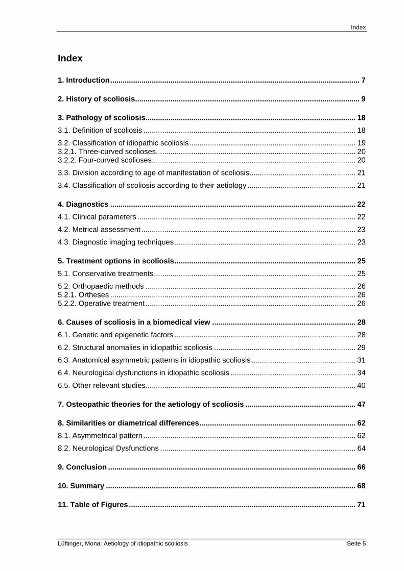
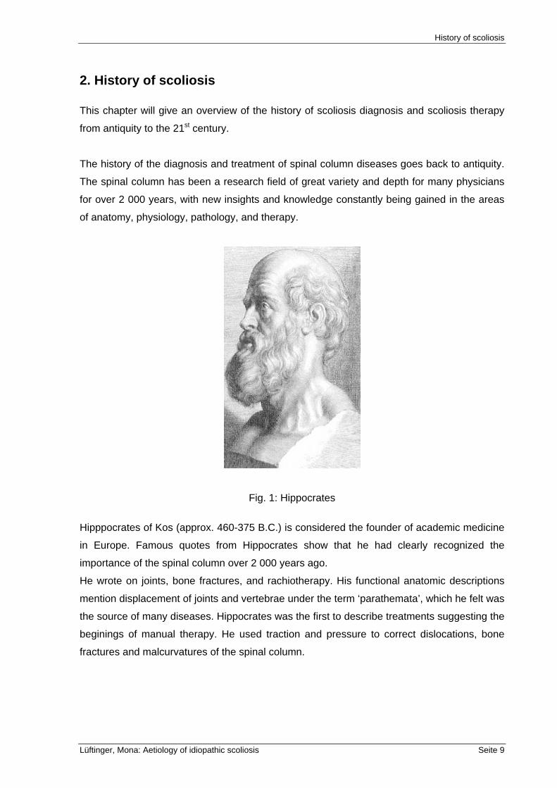
Fig. 1: Hippocrates
Hipppocrates of Kos (approx. 460-375 B.C.) is considered the founder of academic medicine
in Europe. Famous quotes from Hippocrates show that he had clearly recognized the
importance of the spinal column over 2 000 years ago.
He wrote on joints, bone fractures, and rachiotherapy. His functional anatomic descriptions
mention displacement of joints and vertebrae under the term ‘parathemata’, which he felt was
the source of many diseases. Hippocrates was the first to describe treatments suggesting the
beginings of manual therapy. He used traction and pressure to correct dislocations, bone
fractures and malcurvatures of the spinal column.
History of scoliosis
Lüftinger, Mona: Aetiology of idiopathic scoliosis Seite 10
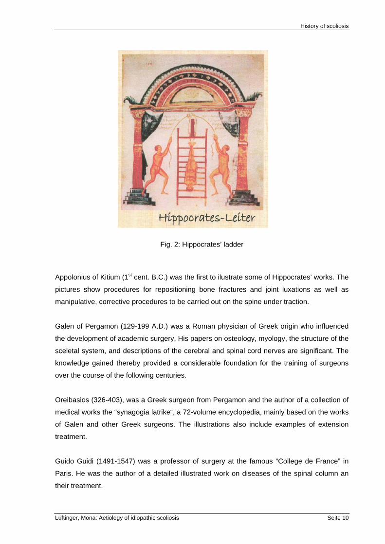
Fig. 2: Hippocrates’ ladder
Appolonius of Kitium (1st cent. B.C.) was the first to ilustrate some of Hippocrates’ works. The
pictures show procedures for repositioning bone fractures and joint luxations as well as
manipulative, corrective procedures to be carried out on the spine under traction.
Galen of Pergamon (129-199 A.D.) was a Roman physician of Greek origin who influenced
the development of academic surgery. His papers on osteology, myology, the structure of the
sceletal system, and descriptions of the cerebral and spinal cord nerves are significant. The
knowledge gained thereby provided a considerable foundation for the training of surgeons
over the course of the following centuries.
Oreibasios (326-403), was a Greek surgeon from Pergamon and the author of a collection of
medical works the “synagogia latrike“, a 72-volume encyclopedia, mainly based on the works
of Galen and other Greek surgeons. The illustrations also include examples of extension
treatment.
Guido Guidi (1491-1547) was a professor of surgery at the famous “College de France” in
Paris. He was the author of a detailed illustrated work on diseases of the spinal column an
their treatment.
History of scoliosis
Lüftinger, Mona: Aetiology of idiopathic scoliosis Seite 11
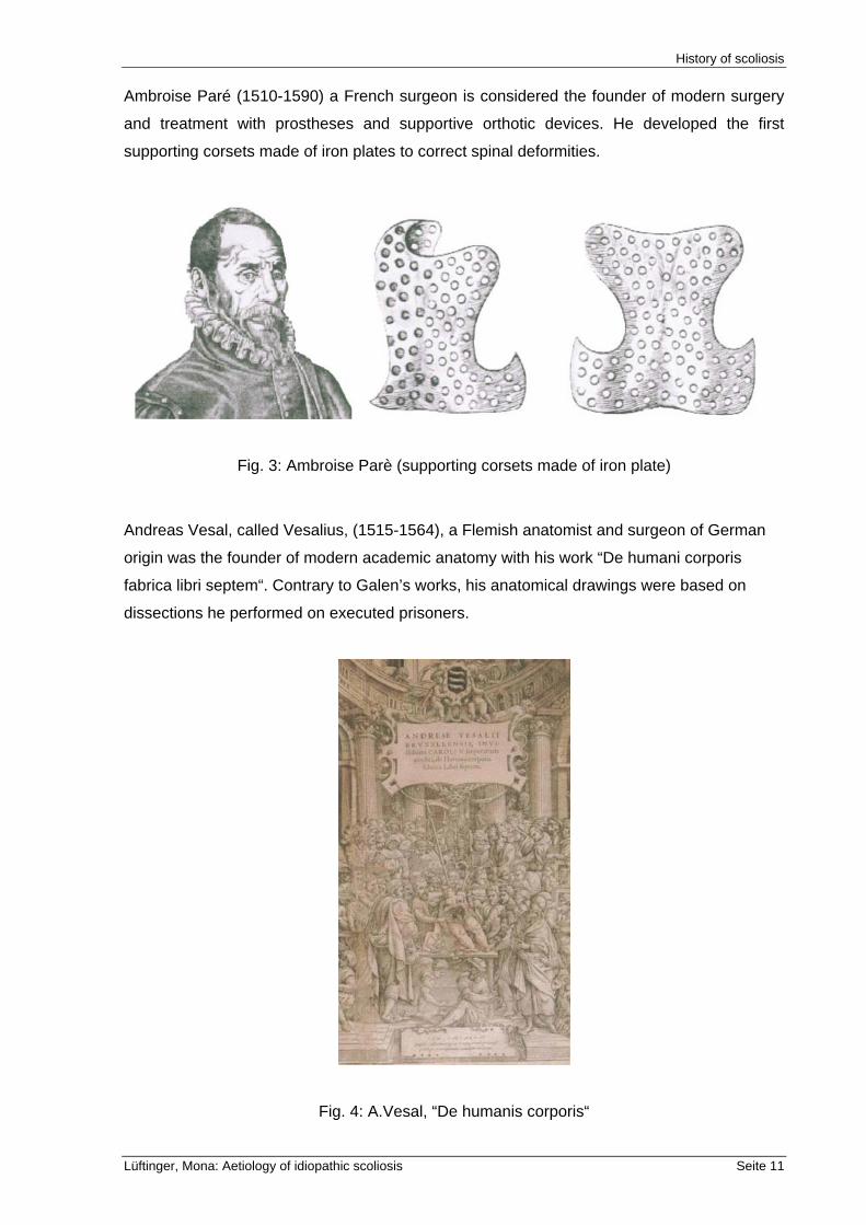
Ambroise Paré (1510-1590) a French surgeon is considered the founder of modern surgery
and treatment with prostheses and supportive orthotic devices. He developed the first
supporting corsets made of iron plates to correct spinal deformities.
Fig. 3: Ambroise Parè (supporting corsets made of iron plate)
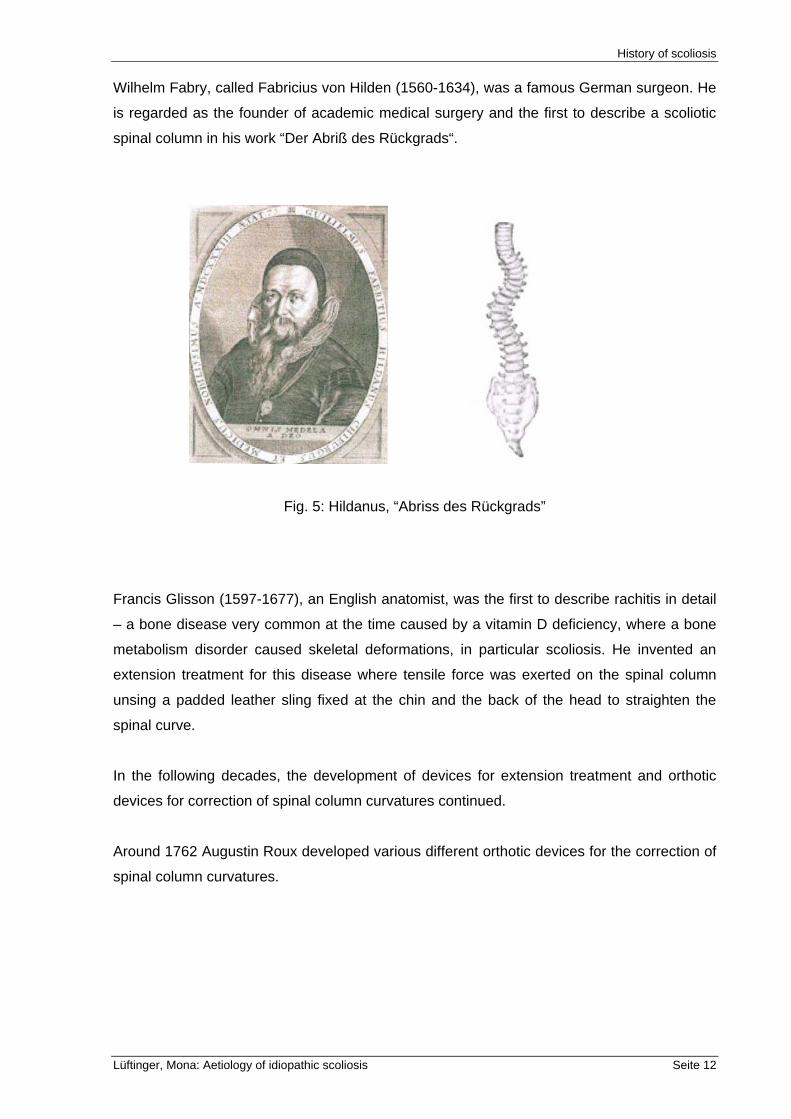
Andreas Vesal, called Vesalius, (1515-1564), a Flemish anatomist and surgeon of German
origin was the founder of modern academic anatomy with his work “De humani corporis
fabrica libri septem“. Contrary to Galen’s works, his anatomical drawings were based on
dissections he performed on executed prisoners.
Fig. 4: A.Vesal, “De humanis corporis“
History of scoliosis
Lüftinger, Mona: Aetiology of idiopathic scoliosis Seite 12
Wilhelm Fabry, called Fabricius von Hilden (1560-1634), was a famous German surgeon. He
is regarded as the founder of academic medical surgery and the first to describe a scoliotic
spinal column in his work “Der Abriß des Rückgrads“.
Fig. 5: Hildanus, “Abriss des Rückgrads”
Francis Glisson (1597-1677), an English anatomist, was the first to describe rachitis in detail
– a bone disease very common at the time caused by a vitamin D deficiency, where a bone
metabolism disorder caused skeletal deformations, in particular scoliosis. He invented an
extension treatment for this disease where tensile force was exerted on the spinal column
unsing a padded leather sling fixed at the chin and the back of the head to straighten the
spinal curve.
In the following decades, the development of devices for extension treatment and orthotic
devices for correction of spinal column curvatures continued.
Around 1762 Augustin Roux developed various different orthotic devices for the correction of
spinal column curvatures.
History of scoliosis
Lüftinger, Mona: Aetiology of idiopathic scoliosis Seite 13
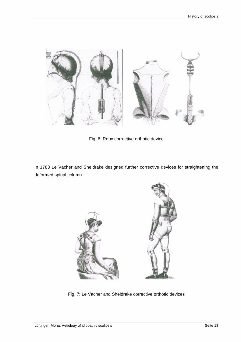
Fig. 6: Roux corrective orthotic device
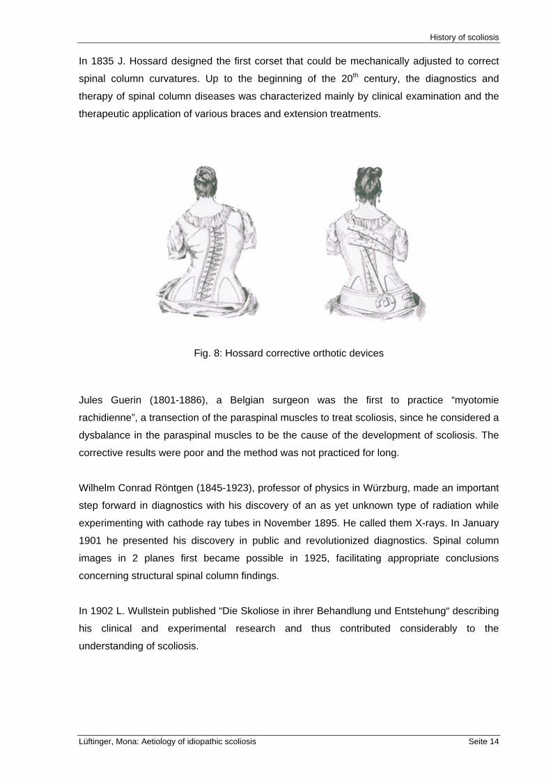
In 1783 Le Vacher and Sheldrake designed further corrective devices for straightening the
deformed spinal column.
Fig. 7: Le Vacher and Sheldrake corrective orthotic devices
History of scoliosis
Lüftinger, Mona: Aetiology of idiopathic scoliosis Seite 14
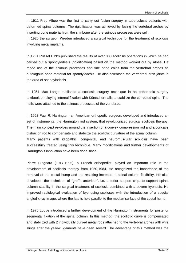
In 1835 J. Hossard designed the first corset that could be mechanically adjusted to correct
spinal column curvatures. Up to the beginning of the 20th century, the diagnostics and
therapy of spinal column diseases was characterized mainly by clinical examination and the
therapeutic application of various braces and extension treatments.
Fig. 8: Hossard corrective orthotic devices
Jules Guerin (1801-1886), a Belgian surgeon was the first to practice “myotomie
rachidienne”, a transection of the paraspinal muscles to treat scoliosis, since he considered a
dysbalance in the paraspinal muscles to be the cause of the development of scoliosis. The
corrective results were poor and the method was not practiced for long.
Wilhelm Conrad Röntgen (1845-1923), professor of physics in Würzburg, made an important
step forward in diagnostics with his discovery of an as yet unknown type of radiation while
experimenting with cathode ray tubes in November 1895. He called them X-rays. In January
1901 he presented his discovery in public and revolutionized diagnostics. Spinal column
images in 2 planes first became possible in 1925, facilitating appropriate conclusions
concerning structural spinal column findings.
In 1902 L. Wullstein published “Die Skoliose in ihrer Behandlung und Entstehung“ describing
his clinical and experimental research and thus contributed considerably to the
understanding of scoliosis.
History of scoliosis
Lüftinger, Mona: Aetiology of idiopathic scoliosis Seite 15
In 1911 Fred Albee was the first to carry out fusion surgery in tuberculosis patients with
deformed spinal columns. The rigidification was achieved by fusing the vertebral arches by
inserting bone material from the shinbone after the spinous processes were split.
In 1920 the surgeon Wreden introduced a surgical technique for the treatment of scoliosis
involving metal implants.
In 1931 Russel Hibbs published the results of over 300 scoliosis operations in which he had
carried out a spondylodesis (rigidification) based on the method worked out by Albee. He
made use of the spinous processes and fine bone chips from the vertrebral arches as
autologous bone material for spondylodesis. He also sclerosed the vertebreal arch joints in
the area of spondylodesis.
In 1951 Max Lange published a scoliosis surgery technique in an orthopedic surgery
textbook employing internal fixation with Küntscher nails to stabilize the corrected spine. The
nails were attached to the spinous processes of the vertebrae.
In 1962 Paul R. Harrington, an American orthopedic surgeon, developed and introduced an
set of instruments, the Harrington rod system, that revolutionized surgical scoliosis therapy.
The main concept revolves around the insertion of a convex compression rod and a concave
distracion rod to compensate and stabilize the scoliotic curvature of the spinal column.
Many patients with idiopathic, congenital, and neuromuscular scoliosis have been
successfully treated using this technique. Many modifications and further developments of
Harrington’s innovation have been done since.
Pierre Stagnara (1917-1995), a French orthopedist, played an important role in the
development of scoliosis therapy from 1950-1984. He recognized the importance of the
removal of the costal hump and the resulting increase in spinal column flexibility. He also
developed the technique of “greffe anterieur”, i.e. anterior support chip, to support spinal
column stability in the surgical treatment of scoliosis combined with a severe kyphosis. He
improved radiological evaluation of kyphosing scolioses with the introduction of a special
angled x-ray image, where the late is held parallel to the median surface of the costal hump.
In 1975 Luque introduced a further development of the Harrington instruments for posterior
segmental fixation of the spinal column. In this method, the scoliotic curve is compensated
and stabilized with 2 individually curved metal rods attached to the vertebral arches with wire
slings after the yellow ligaments have geen severd. The advantage of this method was the
History of scoliosis
Lüftinger, Mona: Aetiology of idiopathic scoliosis Seite 16
high level of post-surgical biomechanical stability achieved: Practically no follow-up brace
therapy was required. The drawback was the very high risk of neurological complications.
In 1973 Dwyer developed a surgical access route from the front (anterior or ventral) for the
correction of scoliosis. The preparatory work for the ventral access breakthrough was done
by Hodgson and Stock, who had operated on many tuberculosis patients via transpleural-
retroperitoneal access, thus establishing a standardized access to the lumbar spine and
lower thoracic spine.
This ventral access approach reduced the number of neurological complications, the fusion
length was shortened, and stability was improved by means of intercorporeal fusion.
Drawbacks of this method consisted of the necessity of several months of follow-up
treatment in a brace and the lack of derotation of the vertebrae.
In 1975 Klaus Zielke presented the ventral derotation spondylodesis (VDS) method,
developed on the basis of the Dwyer method. The main improvement was a new
compression technique and use of a derotator. Using this method, excellent results were
obtained in the frontal plane. The drawback was that the method paid too little attention to
the sagittal profile of the spinal column with the elimination of the lumbar lordosis.
In 1984 Yves Cotrel und Jean Dubousset invented a surgical method based on the Luque
method. The objective of the procedure was a three-dimensional correction of the spinal
column by applying translation, distraction, and compression for improved derotation and an
improved sagittal profile of the scoliotic spinal column in combination with primary stability.
This basic concept is still the basis for dorsal instrumentation of scoliosis as it is done today.
The neurological complications resulting from Luque’s procedure with wire cerclages around
the vertebral arches were avoided in this method, since the two metal rods are directly
inserted into the bone using hooks and screws placed in the vertebral pedicles. The
disadvantage of the method was the insufficient restoration of the sagittal profile of the spinal
column and frequent decompensation of the non-instrumented sections of the spinal column.
Physiotherapeutical Approach
A systematic orthopedic remedial gymnastics was only beginning to establish at the end of
the 19th century. In special clinics corsets were applied in long sessions, supervised by
doctors and additionally patients were made to use special devices and do gymnastic
exercises.
History of scoliosis
Lüftinger, Mona: Aetiology of idiopathic scoliosis Seite 17
Rudolf Klapp (1873-1949), a surgeon from Cologne, invented a set of exercises for scoliosis
patients. The spine is mobilized and the spine muscles are strengthened and stretched by
performing specific exercises at creep speed while standing on hands and knees with leather
braces applied to knees and hands.
Around 1905 Albert Hoffa introduced bend-and-stretch-exercises to scoliosis therapy. At the
beginning of the 20th century the gymnastics method after Ling was widespread. Resistance
exercises were done in sitting and standing positions as well as lying on the back and front
and hanging.
In 1913 Oldevig noticed the drawback of having to work one to one only in therapy sessions
and introduced new techniques using straps so the patient could work actively on his own.
Max Lange regarded scoliosis as a disorder of the muscular equilibrium which he over-
corrected by using resistance devices.
August Blenke (1913) viewed the Klapp crawling exercises rather critically and held the
opinion that scoliosis should only be treated individually, according to the specific situation
and condition of each patient.
Katharina Schroth (1894-1985) invented the three-dimensional scoliosis therapy. Based
upon her own experience she developed a holistic therapeutical method by combining
specific correction techniques with a special corrective breathing technique called „Dreh-
Winkel-Atmung“.
Vaclav Vojta started from the idea that with the help of facilitation of reflex movements the
muscular dysbalance of scoliosis patients can be compensated by central mechanisms. He
first treated spastic children and in the 1960s he started to apply his concept to scoliosis
patients as well.
Pathology of scoliosis
Lüftinger, Mona: Aetiology of idiopathic scoliosis Seite 18
3. Pathology of scoliosis
This chapter will present the fundamental basics of the pathology of scoliosis, starting with
the definition, followed by the division of idiopathic scoliosis and the classification of scoliosis
according to their aetiology will be presented.
3.1. Definition of scoliosis
Scoliosis can be defined as a partly fixated lateral curvature of the spine which cannot be
completely straightened up again (Meister 1980).
Idiopathic scoliosis is a (partly) fixated lateral curvature of one or more parts of the spine,
which co-occurs with a rotation, a torsion, and a structural change of the vertebrae (Humpke
2002).
Scoliosis is a lateral curvature of the spine which represents a rotational malalignment of one
vertebra on another. Rotation and side-bending occur to opposite sides. Ribs are rotated
posteriorly and are prominent on the convex side of the curve. The positional strain is
exacerbated in forward flexion, producing a rib hump (Jane Carreiro 2003).
Structural scolioses are fixated lateral curvatures of the spine (Lindemann 1957). They result
from intrinsic changes in the anatomy of one vertebra or several vertebrae and/or the
surrounding tissue, and lead to an irreversible restriction in spine movement in one or more
directions. In this case a complete correction of the spinal curvature through a conservative
method is no longer possible.
The most striking sign of a structural scoliosis is the fixated rotation of one or more vertebrae,
the deformity of these vertebrae, a bulge in the loin or a rib hump.
You need to distinguish between a rotation and a torsion of the vertebrae. Rotation refers to
a rotation of single vertebrae against each other in their craniocaudal axis (Ebenbichler
1994). A torsion, by contrast, refers to the torsion of the bodies of vertebra of two
consecutive vertebrae and the helical/spiral torsion of the final parts of the spine as a whole.
Three components of the torsion can be distinguished: the rotatory moment in the axial
plane, the lateralisation between the vertebrae in the frontal plane, and the hyperextension in
the sagittal plane (Pedriolle 1985).
X-rays (Pedriolle et al. 1984), clinical (Mau 1982) as well as experimental examinations
(Dickinson et al. 1984) showed that the patients' vertebral body growth plates are ventrally
higher than dorsally, which leads to a consecutive lordosis at the height of the scoliotic apex.
Pathology of scoliosis
Lüftinger, Mona: Aetiology of idiopathic scoliosis Seite 19
In addition to this asymmetry of the spine there is very often an asymmetry of the spine in the
frontal plane. In a growth spurt – idiopathic scoliosis always being an illness brought on by
growth – strain and flexion of the spine bring about scoliosis with a torsion.
Unlike scolioses of known aetiologies, idiopathic scoliosis occurs without any obvious cause
before the onset of bone maturation (Heine 1992, Perdriolle and Vidal 1985). Idiopathic
scoliosis accounts for the largest part of scolioses vis-á-vis those scolioses with known
causes (i.e. 80-90%).
Scoliosis is diagnosed by full-length standing spine X-rays. These x-rays are then assessed
through measuring the Cobb angle (Cobb 1948), the vertebral rotation, and through
ascertaining bone maturation.
Curvatures of less than ten degrees according to Cobb are not regarded as scolioses.
Females are affected by idiopathic scoliosis more often than males in a proportion of 4:1.
Admittedly, with curvatures below 10 degrees, the male-female distribution is equal, but the
stronger the curvature gets, the more marked is the predominance of the female sex
(Weinstein 1985).
Statements about progress show that small curvatures have been known to take a
favourable course (Brooks et al. 1975, Rogala et al. 1978). Curvatures of a larger degree
tend proportionally towards an increased likelihood of progress (Lonstein and Carlson 1984).
The degrees of curvature are classified by the U.S. American Scoliosis Research Society
according to the angle as follows:
Grade 1 Curvature angle between 5 and 20 degrees
Grade 2 Curvature angle between 21 and 30 degrees
Grade 3 Curvature angle between 31 and 50 degrees
Grade 4 Curvature angle between 51 and 75 degrees
Grade 5 Curvature angle between 76 and 100 degrees
Grade 6 Curvature angle between 191 and 125 degrees
Grade 7 Curvature angle above 125 degrees
3.2. Classification of idiopathic scoliosis
Through localising the curvature the following groups of idiopathic scoliosis can be
distinguished:
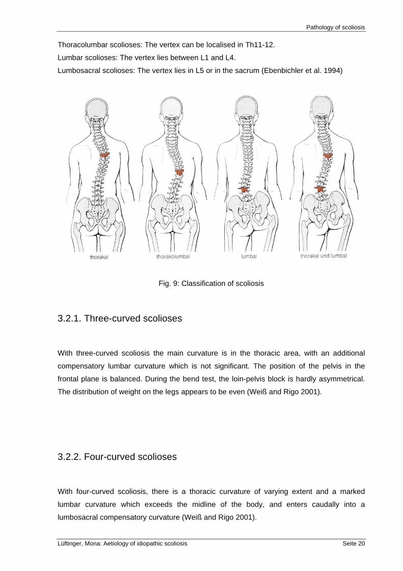
Thoracic scolioses: The vertex lies above and including Th2, with semi-thoracic cases down
to Th3, and with thoracic scolioses down to Th10.
Pathology of scoliosis
Lüftinger, Mona: Aetiology of idiopathic scoliosis Seite 20
Thoracolumbar scolioses: The vertex can be localised in Th11-12.
Lumbar scolioses: The vertex lies between L1 and L4.
Lumbosacral scolioses: The vertex lies in L5 or in the sacrum (Ebenbichler et al. 1994)
Fig. 9: Classification of scoliosis
3.2.1. Three-curved scolioses
With three-curved scoliosis the main curvature is in the thoracic area, with an additional
compensatory lumbar curvature which is not significant. The position of the pelvis in the
frontal plane is balanced. During the bend test, the loin-pelvis block is hardly asymmetrical.
The distribution of weight on the legs appears to be even (Weiß and Rigo 2001).
3.2.2. Four-curved scolioses
With four-curved scoliosis, there is a thoracic curvature of varying extent and a marked
lumbar curvature which exceeds the midline of the body, and enters caudally into a
lumbosacral compensatory curvature (Weiß and Rigo 2001).
Pathology of scoliosis
Lüftinger, Mona: Aetiology of idiopathic scoliosis Seite 21
3.3. Division according to age of manifestation of scoliosis
Congenital scoliosis: 0-2 years
Infantile scoliosis: 3-7 years
Juvenile scoliosis: 7 years to onset of puberty
Adolescent scoliosis: puberty to epiphyseal closure
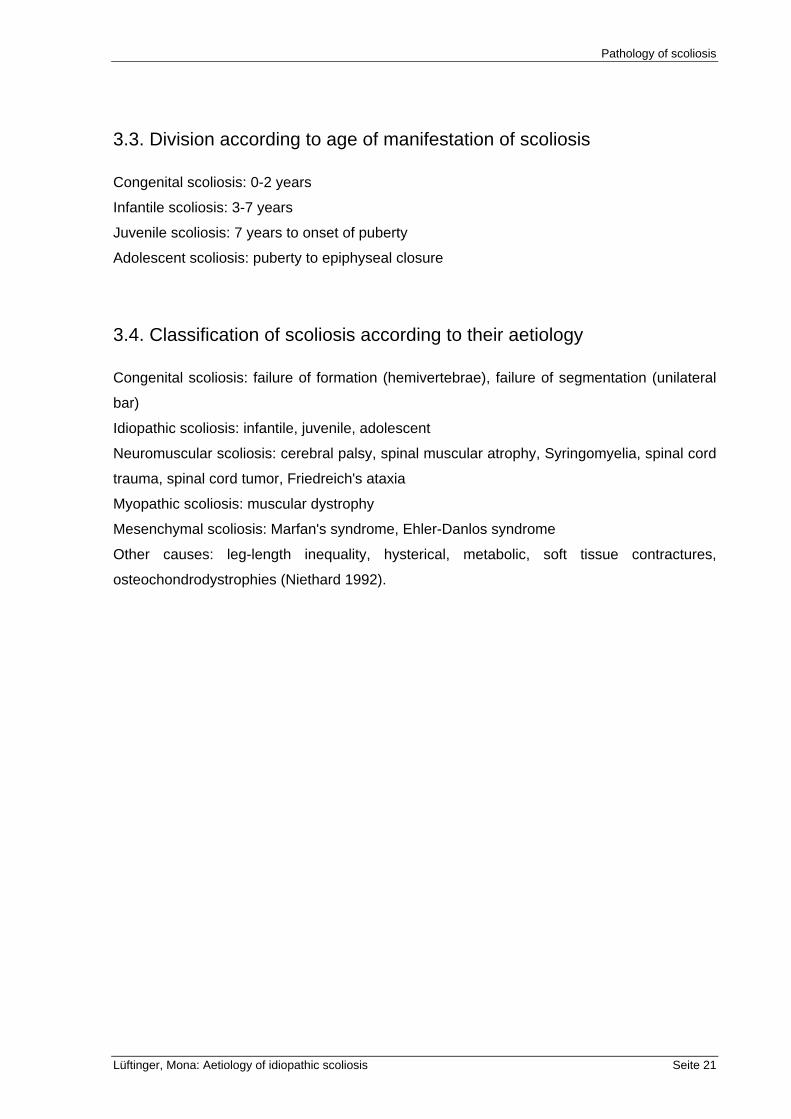
3.4. Classification of scoliosis according to their aetiology
Congenital scoliosis: failure of formation (hemivertebrae), failure of segmentation (unilateral
bar)
Idiopathic scoliosis: infantile, juvenile, adolescent
Neuromuscular scoliosis: cerebral palsy, spinal muscular atrophy, Syringomyelia, spinal cord
trauma, spinal cord tumor, Friedreich's ataxia
Myopathic scoliosis: muscular dystrophy
Mesenchymal scoliosis: Marfan's syndrome, Ehler-Danlos syndrome
Other causes: leg-length inequality, hysterical, metabolic, soft tissue contractures,
osteochondrodystrophies (Niethard 1992).
Diagnostics
Lüftinger, Mona: Aetiology of idiopathic scoliosis Seite 22
4. Diagnostics
This chapter will give you an overview of the current diagnostic methods from general clinical
assessment, to metrical assessment and diagnostic imaging techniques.
4.1. Clinical parameters
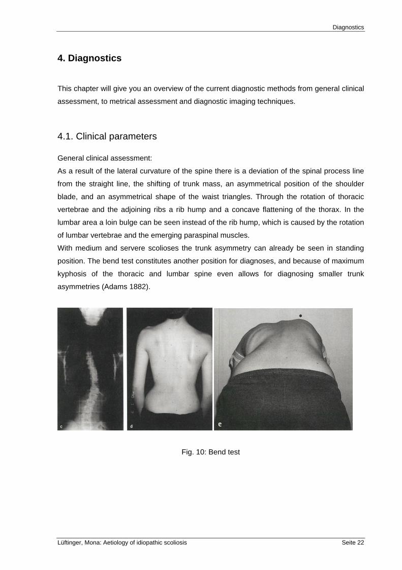
General clinical assessment:
As a result of the lateral curvature of the spine there is a deviation of the spinal process line
from the straight line, the shifting of trunk mass, an asymmetrical position of the shoulder
blade, and an asymmetrical shape of the waist triangles. Through the rotation of thoracic
vertebrae and the adjoining ribs a rib hump and a concave flattening of the thorax. In the
lumbar area a loin bulge can be seen instead of the rib hump, which is caused by the rotation
of lumbar vertebrae and the emerging paraspinal muscles.
With medium and servere scolioses the trunk asymmetry can already be seen in standing
position. The bend test constitutes another position for diagnoses, and because of maximum
kyphosis of the thoracic and lumbar spine even allows for diagnosing smaller trunk
asymmetries (Adams 1882).
Fig. 10: Bend test
Diagnostics
Lüftinger, Mona: Aetiology of idiopathic scoliosis Seite 23
4.2. Metrical assessment
There are various metrical diagnostic methods which serve to ascertain the severity of the
curvature and are of some prognostic value.
Whether a spine is statically compensated or decompensated can be determined by
dropping a perpendicular from processus spinosus C7 to the rima ani. If the perpendicular
does not fall through the rima ani, the curvature of the spine can regarded as
decompensated. The deviation from the rima ani will be measured, documented, and
matched with the corresponding degree of severity.
In order to clinically assess trunk asymmetries a measurement instrument which was
designed according to the principle by Bunnell (1984) is used. This scoliometer is placed
above the spinous processes at the level of maximal paraspinal prominence. Through the
resulting inclination, the corresponding angular dimension is shown on a scale. In addition to
this specific diagnosis, chest expansion and lung capacity are ascertained.
4.3. Diagnostic imaging techniques
X-ray diagnostics complement clinical assessment, and serves the purposes of ascertaining
status, observing progress, and of checking obtained correction results.
X-ray screenings of scolioses consist of two full-length standing spine radiographs, with one
being a postanterior radiograph, the other a lateral radiograph, in order to obtain a three-
dimensional picture of the scope of scoliosis. An evaluation of these total standing spine X-
rays starts with ascertaining lateral spine curvature according to Cobb, and of the vertebral
rotation after Pedriolle's (1985) or after Raimondi's technique (Weiss 1995).
Diagnostics
Lüftinger, Mona: Aetiology of idiopathic scoliosis Seite 24
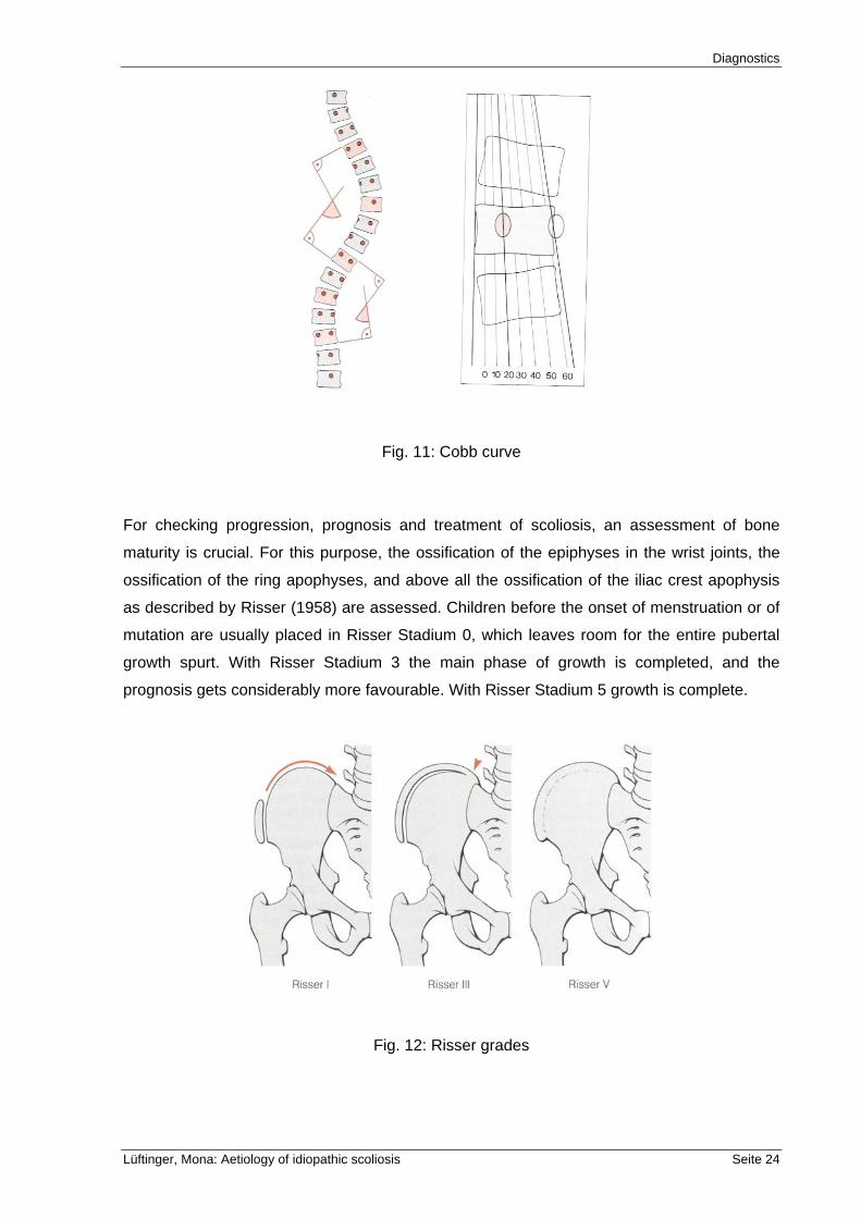
Fig. 11: Cobb curve
For checking progression, prognosis and treatment of scoliosis, an assessment of bone
maturity is crucial. For this purpose, the ossification of the epiphyses in the wrist joints, the
ossification of the ring apophyses, and above all the ossification of the iliac crest apophysis
as described by Risser (1958) are assessed. Children before the onset of menstruation or of
mutation are usually placed in Risser Stadium 0, which leaves room for the entire pubertal
growth spurt. With Risser Stadium 3 the main phase of growth is completed, and the
prognosis gets considerably more favourable. With Risser Stadium 5 growth is complete.
Fig. 12: Risser grades
Treatment options in scoliosis
Lüftinger, Mona: Aetiology of idiopathic scoliosis Seite 25
5. Treatment options in scoliosis
In this chapter current treatment options are being discussed, starting with conservative
treatments, followed by orthopedic methods like ortheses and finally operations with the aim
of correcting the curvature.
For identifying the best kind of treatment it is important to know the aetiology of scoliosis
(various progression tendencies), the patient's age (for remaining spine growth), and the
scope of deformity.
Treatment is tripartite. With incipient scoliosis (up to 20 degrees after Cobb) physiotherapy is
carried out. Scolioses between approximately 20 and 50 degrees are treated by wearing a
corset or bracing in addition to physiotherapy. If there is a curvature of more than 50 degrees
after Cobb, an operation is recommended.
This three-stage plan for treatment shows how important it is to diagnose scoliosis already
early on, as with incipient growth deformities less invasive methods are feasible (Niethard
and Pfeil 1992).
5.1. Conservative treatments
Osteopathy offers a wide spectrum for treating scoliosis through techniques which regulate
strain in various tissue structures and planes. Structural, visceral and cranio-sacral
techniques are applied according to diagnostic findings on individual cases of scoliosis. It is
the overall aim to reduce the rigidity of scoliosis, to balance out dysbalances caused by strain
in myofascial, ligament and membrane tissue, to harmonise cranio-sacral dysfunctions, to
improve metabolism in general, and thus to reduce the curvature degree of the spine, to stop
or slow down the progression of scoliosis, and to prevent restrictions in the cardio-pulmonary
tract. According to studies by Mandl-Weber (2000), and Phillipi et al. (2004) osteopathic
treatment leads to better therapy results with scoliosis than in control groups treated with
traditional methods.
The three-dimensional scoliosis therapy according to Katharina Schroth is an active therapy
concept, in which specific correction mechanisms and corrective breathing (Dreh-Winkel-
Atmung) are meant to influence scoliosis through a change in body image.
Treatment options in scoliosis
Lüftinger, Mona: Aetiology of idiopathic scoliosis Seite 26
This is the leading therapy concept, next to treatments based on developmental kinesiology
like Vojta (which is used to treat scoliosis in its early stages), and general physiotherapy.
5.2. Orthopaedic methods
5.2.1. Ortheses
Bracing is an invasive but usually inevitable form of therapy, which is indicated with scolioses
between 25 and 40 degrees after Cobb (Lohnstein and Carlson 1984).
Since scoliosis is a growth deformity, it is recommended to wear the brace 23 hours a day in
order to reduce the progression of scoliosis. The correct fit of the brace needs to be checked
every four months (Ebenbichler et al. 1994), and the brace needs to be worn till the end of
the bone growth phase (Risser V).
The various forms of braces can be summarised as follows:
the Milwaukee brace
the Chenau brace
the Boston brace
the Lyon brace (Stagnara brace)
Bending brace
Wilmington brace
EDF-plaster cast is an extension, derotation and flexion plaster which is nowadays only
rarely used with severe scolioses after preceding extension treatment on the Cotrel table.
Electrotherapy stimulation of convex musculature cannot be recommended any longer, as
clinical studies have shown that slight corrections of the scoliotic spine were achieved only
initially (O'Donell et al. 1988).
5.2.2. Operative treatment
Operative treatment is indicated with adolescent patients suffering from idiopathic scoliosis
with a curvature of more than 40-45 degrees after Cobb, and adult patients with curvatures of
more than 50 degrees after Cobb.
Pre-operative traction procedures are used in order to interoperatively facilitate as secure
and good a correction of scoliosis as possible. Ventral and dorsal invasions are either
Treatment options in scoliosis
Lüftinger, Mona: Aetiology of idiopathic scoliosis Seite 27
employed in isolation or in combination in scoliosis surgery, with the aim of correcting the
curvature in the frontal as well as the sagittal plane. Spinal fusion (reinforcement of certain
spinal segments) is an obligatory part of every scoliosis operation. In the past, intraoperative
correction was achieved with plaster casts, while now correction and stabilisation are
achieved through metal rods.
The most common operative procedures are as follows:
Spinal fusion (spondylosyndesis) according to Harrington
Luque-Instrumentation
Operation accordting to Cotel and Dubousset
ISOLA-Instrumentation (Niethard 1992)
Causes of scoliosis in a biomedical view
Lüftinger, Mona: Aetiology of idiopathic scoliosis Seite 28
6. Causes of scoliosis in a biomedical view
In this chapter I would like to give you an overview of the biomedical studies in current
aetiology research on idiopathic scoliosis, and their results as a basis for analyzing
similarities and differences between biomedical research and osteopathic theories.
During the 19th century, three main concepts of causation of IS emerged:
myopathic (Guerin)
malpostural (Lovett)
osteopathic (Schulthess).
The latest research shows that there are a lot of causes for IS being discussed, like genetic
factors, structural anomalies, anatomical asymmetric patterns and neurological dysfunctions.
The question of aetiology must be answered if logical preventive and therapeutic measures
are to be devised. The word aetiology is used to embrace all aspects of causation for IS
although the discussion here relates more to pathogenesis and pathomechanism than to
aetiology.
The following studies I am going to present should give an overview of the different
discussions and hypotheses regarding the development of idiopathic scoliosis.
6.1. Genetic and epigenetic factors
One of the questions discussed in research on idiopathic scoliosis is if there is a genetic
determination or an association between structural genes encoded for different elements of
the extracellular matrix. The following two studies are related to this question.
Zaidman et al. (2006) proved a genetic determination of idiopathic scoliosis: Previous studies
of Axenovich et al. (1996) of representative samples of pedigrees (360 families in which the
proband had II-IV grade IS) had proved major gene control of high grade forms of this
pathology. Zaidman et al. (2006) included a search for markers of gene pathology and found
them in proteoglycans – the most important parts of the growth plate cartilage matrix. The
change in aggrecan gene (they are in all zones of the growth plate) results in disturbance of
the growth plate main functions, such as metabolic diffusion, barrier and signal transduction,
Causes of scoliosis in a biomedical view
Lüftinger, Mona: Aetiology of idiopathic scoliosis Seite 29
chondroblast contact interactions, regulation of cell and matrix reproduction by type of
contact inhibition or stimulation of proliferative activity. In this study the point of asymmetry is
explained as follows: each vertebral anlage forms automatically, and gene regulation also
occurs automatically. Depending on localization of mutant gene expression, either right-side
or left-side scoliotic deformity forms.
Pathogenetic mechanism of IS is a manifestation of spine deformity during the time of
intensive growth, which is based on an asymmetrical growth disorder (Zaidman et al. 2006).
Zaidman et al. (2006) postulated that the process of development assumes the presence of
multilevel system of cell specialization, which is a time parameter of the mutant gene
switching. Disruption of aggrecan gene transcription occurs in periods of intensive growth;
therefore cell matrix reproduction is violated. Aggrecan provides a diffusion of metabolics and
a disruption results in fibrotization and dystrophic degeneration of the disc.
From their results Zaidman et al. (2006) defined IS as a “genetically dependent spinal
deformity inherited by autosomal-dominant type, with incomplete gender- and age-related
penetrance of genotype. Pathogenetic mechanism of spine deformity formation is a mutation
in aggrecan gene which encodes synthesis and modification of lateral parts of vertebral
bodies” (ZAIDMAN et al., 2006, 16).
In another study Miller et al. (1996) provided the evidence of an association between
structural genes encoded for different elements of the extracellular matrix in adolescents
from the same family where at least one adolescent had IS. They recruited 96 individuals
from 11 different families, 52 of them diagnosed with IS. The authors could not establish an
association of genes, including the gene defining the collagen type I, between the members
of the same family. No association could be determined between individuals of different
families and they also excluded the genes linked to the cause of IS. They affirmed that as
scoliosis is transmitted between individuals of the same family, research in larger populations
including more families should be incorporated.
6.2. Structural anomalies in idiopathic scoliosis
In this chapter I would like to present some studies dealing with structural anomalies found in
idiopathic scoliosis (IS). There are some studies which present imbalance in the connective
tissue like elastic fiber imbalance in the intervertebral disc (Yu and Fairbank 2005) or
collagen fibers imbalance of the annulus fibrosus in conjunction with spinal growth (Heidari et
al. 2003) in IS. A further study shows a unilateral postponement of growth of ligamentum
flavum and intertransverse ligament (Van der Plaats 2007).
Causes of scoliosis in a biomedical view
Lüftinger, Mona: Aetiology of idiopathic scoliosis Seite 30
Yu and Fairbank (2005) investigated the elastic fiber network organization in scoliotic discs in
comparison to that in normal human intervertebral discs (IVD), which I will briefly summarise
in the following.
33 human IVD were obtained from patients aged 12 to 22 years, undergoing surgery for
idiopathic or neuromuscular scoliosis and 2 non-scoliotic patients, one aged 12 after
astrocytoma tumor, and the other 17 years old after a spinal trauma. They took snap frozen
slices in a radial profile of the disc. The organization of the network varies in the different
region of the disc. The organization and the distribution of this network indicate that it fulfils a
mechanical role. The elastic fiber network of the non-scoliotic patient's discs reveals a high
level of organization. The elastic fibers seen in the idiopathic scoliotic disc tissues appeared
sparse and less well organized. There also appears to be a difference between the
organization of collagen and elastic fiber network in idiopathic and neuromuscular scoliosis.
The neuromuscular patient discs are more disorganized and elastic fibers appear to be less
dense. Also markedly more disorganized fibers were found in ligamentum flavum of some
idiopathic scoliosis patients (Yu and Fairbank 2005).
Heidari et al. (2003) also focused on the role of collagen fiber imbalance of the anulus
fibrosus but they also wanted to find out in their study if there is a conjunction between spinal
deformity and degree of fiber imbalance and if this is influenced by axial growth.
The results support the hypothesis of the project and imply that a greater fiber imbalance will
cause a more severe curve. During adolescent growth the curve becomes severe, indicating
that, while the induced rotation is independent of fiber elongation, the resulting deformity is
directly influenced by the magnitude of the vertical translation or growth.
The study is presented in a mathematical model of the contribution of the collagenous anulus
to the spinal deformity. The model was used to study the effect of the fiber imbalance in
scoliosis initiation and progression.
The final curvature is influenced by axial growth and fiber ratio, with higher fiber imbalance
resulting in more severe spinal deformity.
Van der Plaats et al. (2007) investigated the asymmetrically altered growth in IS by means of
a model study. They developed a new finite element model to simulate the mechanical
behavior of the human spine, composed by the vertebrae, intervertebral disc, facet joints,
spinal ligaments and mm rotators.
The following three theories were analyzed:
1. Buckling of the vertebral column
Causes of scoliosis in a biomedical view
Lüftinger, Mona: Aetiology of idiopathic scoliosis Seite 31
The underlying assumption is that buckling phenomenon causes an initial lateral curvature
with axial rotation, which progresses by growth. Dickson (1992), and Millner and Dickson
(1996) mentioned buckling as an initiation of IS by using the linear Euler buckling theory. But
this was a linear model that did not include ligaments, articulations or muscles.
2. The theory of Veldhuizen and Herman was “assuming unilaterally postponement in growth
of muscles, resulting in asymmetric muscle properties that could lead to asymmetric behavior
of the spine.” (Van der Plaats et al., 2007, 1207)
3. Van der Plaats et al.'s own theory was that unilateral postponement of growth of mm
rotators (MMR) or of ligamentum flavum and intertransverse ligament could initiate the
development of IS (Van der Plaats et al., 2007).
Their model was validated by the earlier stiffness data provided by Panjabi et al. (1976). After
a small correction of the prestrain of some ligaments and the mm rotators the model was
valid. To investigate the buckling of the human spine as a possible initiation of IS instability of
the spine due to an axial downward force was examined.
In the second hypothesis study the postponement in growth was translated in the numerical
model in an asymmetrical stiffness. The spine resulting deformation was analyzed for the
presence of the coupling of lateral deviation and axial rotation that is characteristic for
scoliosis after the spine was loaded axially.
The result of the studies by Van der Plaats et al. (2007) showed that only unilateral
postponement of growth of ligamentum flavum and intertransverse ligament appeared to
initiate scoliosis. Buckling did not initiate scoliosis, nor did asymmetric stiffness of mm
rotators.
6.3. Anatomical asymmetric patterns in idiopathic scoliosis
This chapter deals with studies in which asymmetrical features in the morphology of
idiopathic scoliosis were found. Anatomical asymmetric patterns were found in occlusion
(Ben-Bassat et al. 2006), and in the sacropelvic morphology (Karski et al. 2006) of idiopathic
scoliosis. Mau et al. (1979) described the pattern as “syndrome of contractures” found in
newborns and babies. Years later Karski et al. (1995-2006) analyzed children with
“syndrome of contractures”, and noted its relevance to some clinical symptoms in children
with scoliosis.
Causes of scoliosis in a biomedical view
Lüftinger, Mona: Aetiology of idiopathic scoliosis Seite 32
The occlusions of patients with idiopathic scoliosis and random controls were examined by
Ben-Bassat et al. (2006) to elucidate possible relationships between these two conditions.
96 female and male scoliosis patients aged 13.9 ± 3.5 years, and 703 school aged children
in a control group of random were examined by an orthopedic and an orthodontic.
They looked for the
molar relationship
canine relationship
upper midline deviation
lower midline deviation
anterior crossbite
posterior crossbite
Only in A/P dimension the frequency of asymmetrical molar relationships was identical in the
scoliosis and the control groups. In the groups of patients with idiopathic scoliosis compared
with random controls almost all other parameters of occlusal asymmetry were significantly
more prevalent.
Ben-Bassat et al. (2006) showed in their study that patients with idiopathic scoliosis have
more asymmetrical features of malocclusion than a random group, and that early detection of
asymmetric malocclusion can sound the alarm about possible underlying orthopedic
problems.
In the orthopedic literature, Floman (1998) indicated a possible connection between thoracic
scoliosis and restricted neck motion in a report of 6 AIS patients. Floman found a marked
limitation of neck flexion although the radiological examination including MRI of the entire
spine failed to disclose the mechanism which caused the limitation of neck motion. And the
discussion is if such a restriction in neck motion has a secondary influence on scoliosis
or/and occlusion (Floman 1998).
Karski et al. (2006) presented another possible aetiology of IS in the context of anatomical
asymmetrical pattern. They postulated that the malformations of skeletal system can already
be taking place in the last months of pregnancy. The deformations are called “syndrome of
contractures“, (“Siebener Kontrakturen Syndrom“).
This syndrome has been described by Mau (1979; 1982), Hensinger (1979), Howorth (1977)
and others. The causes of the “syndrome of contractures” can be related with fetus itself or
with mother conditions (Karski 2006).
Mau gave a detailed description of “Siebener Kontrakturen Syndrom”: 1. scull deformity/plagiocephaly – flattening of left forehead and temple regions, left
chick atrophy, eyes asymmetry, nose and ears deformations
Causes of scoliosis in a biomedical view
Lüftinger, Mona: Aetiology of idiopathic scoliosis Seite 33
2. torticollis- usually left sided. Can be related with plagiocephaly and lack of proper head positioning, and also with primary shortening of stenocleidomastoideus muscle, torticollis with tumor neonatorym
3. scoliosis infantilis – usually right convex lumbo-thoracis curve. This type of spine deformity was during many years improperly added to the group of idiopathic scoliosis. This scoliosis usually recedes spontaneusly (disappearence 80% to 100%).
4. contracture of adductor muscles of the left hip. Untreated contracture can lead to developement of hip dysplasia, which primarily can be obseved only at 10% of newbornes. The remaining 90% of dysplasia are cases of secondary deformity resulting from the contracture and are classified as „developemental hip dysplasia“ (DDH). Untreated contracture of adductors enlarges dysplasia.
5. contracture of the abductor muscle of the right hip described as Haltungsschwäche by Mau. This contracture may cause oblique positioning of pelvic bone observed at hip joint X-ray picture of babies and young children. With time it may lead to disturbances of biomechanics (asymmetry during gait, asymmetry in growth and developement) and „permanent habit of standing on free only on the right leg“ (the right leg is stronger and more stable due to the contracture!) which in result leads to developement of the so called idiopathic scoliosis (Karski; 1995-2006) .
6. pelvic bone asymmetry – the abduction contacture can influence the pelvis positioning visible during X-ray examination for hip joint screening
7. feet deformities - such as: pes equino-varus, pes equino-valgus, pes calcaneo-valgus, or pes calcaneo-valgus adductus. (KARSKI et al., 2006, 34)
Further Karski et al. (2006) reported that the most common first fetus position is left-sided
(80-85% of all pregnancies).
The fetus’ body, meaning head, trunk and pelvis, is pressed to the left side of mother’s spine,
and this may result in some typical deformations (primarily unfixed) of the skeletal system.
Karski et al. (2006) focused in their study on newborns. The analysis was conducted in 1999-
2001 on 300 histories of babies and children. The age of the children was 3 weeks to 12
months.
In 97 children from this group they noted different symptoms of “syndrome of contractures”
(74 girls and 23 boys). The “syndrome of contractures” of the left side was noted in 55
children, of the right side in 42. The relation of left to right was different from fetus positioning
(85% :15%).
The analysis by another consulting specialist showed that most of these children were from
first pregnancies (80%), mothers had small bellies during pregnancy (mothers' report), and
usually the newborns at birth were heavier or longer than normal.
Karski et al. (2006) also analyzed children already diagnosed with scoliosis. The analysis
was conducted on 100 histories of children aged 5 to 8 years. In 20 of them they noted
abduction contracture of the right hip ranging from 5 to 10 degrees, or adduction movement
from 0 degrees, but the left hip at the same time the adduction was 35-50 degrees. In these
children, initial stages of the so called idiopathic scoliosis were noted at X-ray examination.
Causes of scoliosis in a biomedical view
Lüftinger, Mona: Aetiology of idiopathic scoliosis Seite 34
The second group of 80 children with primary “syndrome of contractures“ showed only
limitation of right hip adduction in comparison to the left one; adduction of the right hip 10-25
degree, of the left hip 35-50 degree.
In these children Karski et al. (2006) noted initial stages of the socalled idiopathic scoliosis –
lumbar left convex, or sacro-lumbar left convex, or lumbo-thoracic left convex.
In another study concerning an anatomical asymmetrical pattern in IS, Mac-Thiong et al.
(2006) compared the sacropelvic morphology between normal adolescents and AIS.
There were 27 normal adolescents (normal group), 10 boys and 17 girls, and 29 in the AIS
group, 5 boys and 24 girls, aged between 11-15,8 years. The mean Cobb angle of the
primary curve was 30-73 degrees.
By radiographs of the spine and complete sacropelvis they examined postero-anterior (PA)
and lateral standing (LAT). Based on 26 anatomical landmarks on the PA radiograph and 13
on LAT radiograph and 19 sagittal parameters of the pelvis are computed automatically.
These parameters were used to characterize the complete pelvic morphology for each
subject.
The results of this study showed that in the sagittal plane there was no significant difference
in sacropelvic morphology between the two groups. Significant differences between AIS
subjects and controls were found for coronal parameters involving pelvic height and width
(Mac-Thiong et al. 2006).
The significantly different parameters were found in height measurements of the pelvis,
which is in the right pelvic length (0,045) and in the right iliac height (0,014). Different
parameters were also found in width measurements of the left pubic length (<10), of the right
pubic length (0,010), in the left obturator foramen width (0,007), in bicristal distance (0,006),
in bituberal distance (0,005), in biacetabular distance (0,001), in pubic symphysis width
(0,003), in pelvic inlet (<10), and in the subpubic angle (0,041).
6.4. Neurological dysfunctions in idiopathic scoliosis
In this chapter I would like to present studies which analyze neurological dysfunctions in
connection with the aetiology of IS. Hypotheses like maturational delay of the CNS involving
undetected neuromuscular dysfunction (Burwell et al. 2006a), and disturbances in the
longitudinal growth from bones which results in anomalous extra-spinal left-right skeletal
Causes of scoliosis in a biomedical view
Lüftinger, Mona: Aetiology of idiopathic scoliosis Seite 35
length asymmetries in the upper limbs, periapical ribs, ilia and lower limbs in AIS were
postulated. It is also being discussed if genes and the environment (nature/nurture) may
interact pre- and/or post-natally to explain the deformity of AIS (Burwell et al. 2006b).
Differences in dynamic balance between AIS and healthy children were found by Filipovic
and Viskic-Stalec (2005). They did investigations and analyses of neurological differences
between healthy children and children suffering from AIS. Their study showed weak postural
control mechanism and proprioception in AIS. A trend that tonsilar ectopia appeared more
often in IS patients, especially those with thoracic or thoraco-lumbar curves, was detected in
MRI studies by Sun et al. (2006). Increased tension in the spinal cord which induces the
development of IS is hypothized by Royo-Salvador (1996).
Burwell et al. (2006a) tried to explain the aetiologic theories of AIS in their
neurodevelopmental concept of maturational delay of the CNS.
The current thinking then was that a defect of central control or processing in the central
nervous system (CNS) affects a growing spine with a primary pathology involving the hind
brain. How the CNS may be involved in curve progression is still unknown. It is generally
considered to result from neuromuscular activity acting on the spine and trunk. But in the
absence of evidence either way, curve progression may equally result from a failure of the
CNS to control a curve-initiating process at a time of rapid adolescent growth. This may be
the result if there is a maturational delay of the CNS body schema.
Burwell et al. (2006a) postulated four theoretical requirements about the CNS body schema
concept for the development of AIS. 1. Curve-initiation process produced by left-right spinal asymmetry caused by
vertebral body growth plates, or possibility periapical rib length asymmetry as a relative concave rib overgrowth or neuromuscular mechanism may cause the left – right asymmetry and initiate scoliosis in some subjects.
2. Rapid spinal elongation in adolescent growth spurt, principally of vertebral body growth plates under the influence of steroid hormones particularly estrogen.
3. Maturational delay of CNS body schema, this causes neuromuscular adjustments to a deforming and rapidly elongating spine. There is also a requirement that focal brain atrophy in progressive AIS will be shown.
4. Upright posture and movement of spine and trunk suggests in scoliosis curve progression. (BURWELL et al., 2006a, 74-75)
A study by Arkin (1949) stated that rest in bed may halt the progress of IS in children. He
kept over 30 scoliotic children in bed for 22 hours a day. Except for one case, no progress
was noted after 3 months.
Burwell et al. (2006a) introduced 4 hypotheses to explain where in the CNS body schema
concept maturational delay may arise and cause AIS:
Causes of scoliosis in a biomedical view
Lüftinger, Mona: Aetiology of idiopathic scoliosis Seite 36
1. Impaired sensory input; the basic problem may lie in muscle spindles or other endings. In AIS patients they found abnormal reflex processing which may be associated with delay in maturation of the CNS body schema.
2. Primarily in the brain; parts of the brain that may contribute to maturational delay of the CNS body schema include in the parietal lobe, the somatosensory cortex (personal space of “self”), temporparietal junction, temporal lobes, frontal lobes, and visual cortex (extra personal space).
3. Impaired motor output; in a study by Herman et al (1985) it is shown that processing of vestibular signals within the CNS yielded the highest degree of correlation with curve magnitude. They considered that IS was a motor control problem. A higher level CNS disturbance was thought to be responsible for visuo-spatial perceptual impairment, motor adaption and learning deficits. These lead to a recalibration of proprioceptive signals from axial musculature causing IS.
4. Relation to the NOTOM hypothesis; the CNS body schema concept can be viewed as resulting from a abnormality in neuroosseous timing of maturation (NOTOM) (BURWELL et al., 2006a, 76)
In a further study, Burwell et al. (2006b) developed theories about disturbances in the
longitudinal growth of paired bones (long limb bones, ribs, ilia) and united paired bones
(vertebrae, sternum, skull, mandibulae).
They postulated that it is evident that human vertebral body growth plates like other physes,
during their years of functional activity liberate cascades of cells that respond symmetrically
to successive hormones during growth. Hormones are secreted as postnatal development
proceeds. These are in fetal life insulin and IGF-1, in the early postnatal life: growth
hormone; and in puberty steroids including estrogen and androgens. Receptors on the cell
surface or nucleus respond to these hormones.
Burwell et al. (2006b) claimed that genetic and environmental factors may disturb symmetry
control in separate and united enantiomorph bones. These factors are said to be acting
directly or indirectly on developing skeletal primordial in early embryonic life as a very
complex disorder of differential growth in the skeleton.
Burwell et al. (2006b) also reported that progressive Adolescent Idiopathic Scoliosis (AIS),
which mainly affects girls is generally attributed to relative anterior spinal overgrowth from a
mechanical mechanism (torsion) during the adolescent growth spurt, and that established
biological risk factors for the development of AIS are growth velocity and potential residual
spinal growth assessed by maturity indicators.
Goldberg et al. (2000) reviewed left-right directional asymmetries in AIS and wrote: “Scoliosis
is not a disease or group of diseases but a symptom or sign of environmental stress,
Causes of scoliosis in a biomedical view
Lüftinger, Mona: Aetiology of idiopathic scoliosis Seite 37
significant enough to overwhelm the intrinsic stability of the morphological genom. As such
there is no specific etiology but a large number of precipitating stressors“ (GOLDBERG et al.,
2000, 327).
There are also chemical risk factors and some dietary factors discussed by Mac Master et al.
(2004). They reported evidence that some infants exposed to indoor swimming pools in the
first years of life show an association with progressive AIS and in controls spinosus process
asymmetry.
Barker et al. (2002) showed that the origins of important chronic system diseases of adult life
including stroke, coronary heart disease and type2 diabetes as well as rates of aging, may lie
in fetal responses to the intra uterine environment. It is termed the “fetal origins hypothesis“,
and has led to national medical research projects being developed in the UK and USA.
The breaking of bilateral symmetry was obtained in a further study by Burwell et al. (2006c),
in which they focused on mechanisms initiated in embryonic life including a disturbance of
bilateral (left-right or mirror-image) symmetry highly conserved in vertebrates. They stated
that normal external bilateral symmetry of vertebrates results from a default process involving
mesodermal somites. The normal internal asymmetry of the heart, lungs, gut with its glands,
major blood vessels is also highly conserved among vertebrates. It results from the breaking
of the initial bilateral symmetry by a binary asymmetry switch mechanism producing
asymmetric gene expression around the embryonic node and/or in the lateral plate
mesoderm. In the mouse, this switch occurs during gastrulation by cilia, driving a leftward
flow of fluid and morphogens at the embryonic node (nodal flow) that favors precursors of the
heart, great vessels, and viscera of the left. The hypothesis of this study is that an anomaly
of the binary asymmetry switch explains the excess of right/left thoracic AIS. They think that
there is evidence that vertebrates within their bilateralized shell retain an archaic left-right
asymmetric visceral body organization evident in thoracic and abdominal organs (Burwell et
al. 2006c).
Kouwenhoven et al. (2007) presented in a cross sectional magnetic resonance imagine
study, vertebral rotation measurements of the normal, non-scoliotic spine of persons with a
complete mirror image reversal of the internal body organs, called situs inversus totalis. The
results showed in the normal spine of humans with situs inversus totalis a pre-existent
pattern of vertebral rotation opposite of what is seen in humans with normal organ anatomy,
that is a predominant rotation to the left side of the mid and lower thoracic vertebrae, and to
the right side of the upper thoracic and lumbar vertebrae.
Causes of scoliosis in a biomedical view
Lüftinger, Mona: Aetiology of idiopathic scoliosis Seite 38
Also focusing on neurological dysfunction in IS, Filipovic and Viskic-Stalec (2006) observed
the mobility capabilities of AIS persons (total 38) and a control group (total 36). In the AIS
group there were 21 persons with a Cobb angle < 25° and 17 persons with a Cobb angle >
26°.
The age in all groups was between 9-14 years; there were 2 different step tests applied; the
left and the right step test. The tests were distinguished according to step on a 16inch tall
bench and step down onto the platform for the base reaction force within 15 seconds in a
normal rhythm. The amplitudes of force were considered by electromyography from muscles
(m gluteus maximus sinister and dexter, mm erector spinae sinister and dexter) and by the
platform for the base force reaction.
The results of this study showed that there was a significant difference between AIS and the
healthy group on the left step test, and that the pathological form of AIS highly affects
dynamic balance.
The values of the lumbar erector muscles and right gluteus maximus, and the side-to-side
reaction of the platform are more pronounced than the other variables. There was no
significant difference between the various Cobb angles in the AIS groups.
Filipovic and Viskic-Stalec (2006) presented that AIS affect dynamic balance and illustrated
the compensational functioning of mobility, especially when there is a lack of normal mobility
forms and there are weak postural control mechanism and proprioception.
Another study by Weiss and Lehmkuhl (1996) also determined that persons with AIS had the
ability to stand, walk, run, and jump over barriers, but the movements involved are not
economical, and that such participants tend to tire sooner, be slower and less coordinated
than the healthy ones. Their movement function is reduced.
Dynamic balance is affected in AIS patients (Filipovic and Viskic-Stalec, 2006) which may
indicate disturbances in the cerebellum, Sun et al. (2006) investigated the position of the
cerebellar tonsils in AIS patients by a MRI-study.
The group comprised 205 AIS patients with a Cobb angle of more than 40 degrees,
consisting of 27 boys and 178 girls. In the control group there were 86 healthy adolescents,
43 boys and 43 girls.
All patients were aged from 12–18 years. MRI examinations of the whole spine from foramen
magnum to the sacrum were performed in both groups.
The connecting line between the basion and the opisthion (BO line) of the occiput were
drawn, representing the level of the foramen magnum.
Then the most inferior part of cerebellum or tips of the cerebellar tonsils was selected for
further measurements. The perpendicular distance from the inferior part of the cerebellar
tonsil to the BO line was determined.
Causes of scoliosis in a biomedical view
Lüftinger, Mona: Aetiology of idiopathic scoliosis Seite 39
The study by Sun et al. (2006) showed that AIS patients had lower positions of cerebellar
tonsils. Extend of tonsilar ectopia of more than 2 mm and 5 mm below the BO line was found
in 13.3% (27/205). The incidences of tonsilar ectopia in AIS were 35.1% (72/205) in controls
5.8% (5/86).
In AIS patients they found no significant correlation between the tonsil positions and curve
severity. The most frequent incidence of tonsilar ectopia was 62.5% in patients with a double
thoracic curve, 39.3% in a right thoracic and left lumbar curve, 37.3% in a right thoracic
curve, 36.4% in a thoraco-lumbar curve and 21.6% in a lumbar curve.
In the patients with a left thoracic curve, the tonsil position was identified to be 3.2 mm above
the BO line. Patients with a lumbar curve had a significantly lower incidence than with
thoracic or thoraco-lumbar curves. The results of Sun et al. (2006) also suggest that there
might be associations of proprioception defects with tonsilar ectopia in AIS.
While Sun et al. (2006) in their MRI study showed the lower positions of the cerebellar tonsil
in AIS, Royo-Salvador (1996) had hypothized already years before that tensions between
cranium and sacrum are not transmitted through the dura mater spinalis but via the spinal
cord, that this induces the lower position of the cerebellar tonsils, and also causes the
development of scoliosis. He noticed an abnormal increase in tension of the medullar traction
in IS and syringomyelia patients. According to Royo-Salvador (1996), an abnormal intensity
of the medullar traction leads to the following effects on the cranial level: caudal traction at
the truncus cerebralis and rise of tension of the surrounding dural meninges and the periostal
attachment of the meninges, e.g. tentorium cerebella. The tonsillae cerebella are drawn to
inferior and compressed, resulting in a deformation of the 4th ventricle, an increase in the
basal cranium angle, a deformation of the clivus as well as an approximation of the pars
petrosa (os temporale) and os sacrum. The cerebellar hemispheres are pushed into the
fossa cranialis posterior, which leads to a deformation of the foramen magnum.
On a cervical level the caudal traction on C1/C2 level is related to an anteroposterior force
and thus related to a posterior swinging movement of the dens of C2. In the cervical area the
nerve tissue is most affected, resulting in compression, ischemia and necrosis.
In the thoracic region the result of the study provides an explanation for the development of
idiopathic scoliosis. The spine tries to reduce the tension caused by the spinal cord. The
formation of a thoracic curvature reduces the effect of the medullar traction. An abnormal
traction of the spinal cord especially in the thoracic area leads to the development of
scoliosis. They are caused by compressions that develop increasingly during the growth
Causes of scoliosis in a biomedical view
Lüftinger, Mona: Aetiology of idiopathic scoliosis Seite 40
period. Increase in pressure on the vertebral body growth plates (plaques epiphysaire)
decelerates the growth of this part of the vertebra relative to the other parts and thus causes
a deformation of the vertebral column.
The rising pressure leads to an increase in the density and the trabeculae inside the
vertebra. An increased intensity of the medullar traction leads to lowering of the conus
medullaris in the lumbo-sacral region and a rise in tension at the filum terminale. A rise in
tension at the filum terminal can affect the dural bag and harm it. According to Royo-
Salvador (1996), a power transmission takes place via the spinal cord from the os sacrum to
the inside of the cranium.
6.5. Other relevant studies
In this chapter I will present other relevant studies dealing with the aetiology of scoliosis from
a biomedical view. The contents of these studies emphasise various aspects of scoliosis.
They deal with visual deficiency (Grivas et al. 2006), handedness and spinal deformity
(Goldberg et al. 2006), the specific morphological manifestations in idiopathic scoliosis
(Sevastik 2006), the degree of mineralization in IS (Yeung et al. 2006), and the incidence
and outcome of scoliosis in children with pleural infection (Mukherjee et al. 2006).
Grivas et al. (2006) investigated if there is an association between visual deficiency and IS.
26 totally blind Greek women aged 20-67 were screened for scoliosis. In the forward bending
test using the Pruijis scoliometer, 11 of 26 women had more than 7 degrees and this was a
cut off criterion for radiological examination.
In 11 of 26 persons, they identified an average Cobb angle of 19 degrees (range 12-28).
Thoracolumbar was the most common type of curve identified (9 out of 11; 6 were to the
right, and 3 to the left). Furthermore, Grivas et al. (2006) recorded the circadian rhythm of the
probands. None of the blind women reported any sleeping difficulties, and they had a normal
circadian rhythm related to a 24-hour day. The result showed a prevalence of scoliosis in
women with visual impairment at 42.3% against that of the general population in Greece of
2.9%.
Grivas et al. (2006) concluded that there is an association between idiopathic scoliosis and
postural control of the trunk in which vision is involved. The results of the study also suggest
that melatonin might be involved in the pathogenesis of human idiopathic scoliosis in a way
that could explain the higher prevalence of scoliosis in people with visual impairment.
Causes of scoliosis in a biomedical view
Lüftinger, Mona: Aetiology of idiopathic scoliosis Seite 41
The melatonin overproduction in these people leads to late menarche and longer exposure to
possible coexisting detrimental factors in the pathogenesis of idiopathic scoliosis (Grivas et
al. 2006).
Another topic discussed with regard to the aetiology of scoliosis is if there is a connection
between handedness and spinal deformity. Because of the hypothesis that handedness
causes curve patterns in scoliosis or vice versa, Goldberg et al. (2006) did a study with
patients from the computerized scoliosis database. Altogether they examined 1,477 patients
in five groups (minor asymmetry, AIS, juvenile idiopathic scoliosis, infantile idiopathic
scoliosis, and congenital vertebral anomalies). The data recorded included gender,
diagnosis, curve pattern and preferred writing hand.
The group with the minor asymmetry (500 in total, not confirmed by radiograph) showed that
the incidence of left-handedness was 10% in boys as well as in girls, and does not differ from
that observed in the wider human population. Statistically there was a significant association
between hand preference and lateralization of asymmetry.
In the group with AIS (total 673, confirmed by radiograph) the handedness in boys does not
differ from the population mean and does not correlate with scoliosis patterns. The incidence
in left-handedness in girls with AIS was 6.9% which is less than that for the normal
population. There was also a statistically significant correlation between scoliosis patterns
and hand preference.
In the group with juvenile idiopathic scoliosis (total 102, and age 4-10 years), the
handedness in boys was the same as the population mean and did not correlate with
scoliosis patterns. The incidence of left-handedness in girls was 16.7%, which is significantly
more than that for the normal population. Left thoracic patterns in girls were 6.7% and
therefore much lower than usual (20%) but did not correlate with hand preference.
In the group with infantile idiopathic scoliosis (total 50, and age < 4 years), the incidence of
left-handedness in boys was 26%, in girls 48%, which is significantly higher than that in the
normal population. The curve pattern does not match the usual 80% right thoracic. There
was no correlation between handedness and curve pattern.
In the group with congenital vertebral anomaly (total 152); the girls had an increased
incidence of left-handedness (18%) but there was no correlation between curve pattern and
handedness in boys and girls.
Causes of scoliosis in a biomedical view
Lüftinger, Mona: Aetiology of idiopathic scoliosis Seite 42
The results of a study by Milenkovic et al. (2004) showed that left-handedness was
significantly associated with scoliosis in girls after screening 2,546 schoolchildren. But they
made no reference to radiological investigations. There was no association found in the
study by Goldberg et al. (2006), so maybe Milenkovic’s study was based on physical
examination only.
To suppose that handedness can determine a posture that is so detrimental as to cause real
spine deformity is to simply not understand how scoliosis evolves. Furthermore, Goldberg et
al. (2006) concluded that the non-correlation between hand-preference and scoliosis pattern
refutes this concept.
In a review which Sevastik (2006) called “the thoracospinal concept” he postulated that the
ethiopathogenesis of the pathological complex of the right convex female adolescent
scoliosis demands research from new standpoints. He showed that the majority of all cases
of idiopathic scoliosis are characterized by a combination of specific morphological
manifestations and signs of physiological abnormalities.
Sevastik (2006) defines the specific morphological manifestations as follows:
1. Laterality and pattern of the curve
Right to left convex curves in infantile idiopathic scoliosis is 86 to 14 with 11 % resolving, 42
% are thoracic, 16% lumbosacral and lumbar and 22% are double primary.
2. Height and weight
Girls with double primary and thoracolumbal-curved adolescent idiopathic scoliosis have
been reported to be significantly taller at the time of menarche than girls with right convex
thoracal curves. The weight of the girls with all curve patterns was significantly lower than
that of the controls, essentially in infantile idiopathic scoliosis and juvenile idiopathic
scoliosis.
The specific physiological characteristics according to Sevastik (2006) are:
1. Gender: the mean reported percentage of girls-to-boys ratio is 8 to 52 in IIS, 76 to 24 in
JIS, and 85 to 15 in AIS.
2. Menarche: AIS girls with a double primary or thoracolumbar curve were significantly older
and taller than the control girls or girls with right convex thoracal curves at the time of
menarche.
Causes of scoliosis in a biomedical view
Lüftinger, Mona: Aetiology of idiopathic scoliosis Seite 43
3. Sympathetic dysfunction: the vascularity of the breasts in girls with AIS and matched
normal controls was measured. In normal girls there was no significant difference in the AIS
group while the vascularity of the left breast on the concavity of the thoracic curve was
significantly increased as compared with the right one.
4. Abnormalities of muscle fibers and platelets
A decreased number of type II fibers in paravertebral muscles was reported by Yarom et al.
(1979) and an increased number of type I fibers on the concavity rather than the convexity of
the curve were found.
Also larger platelets than normal, decreased activity of intracellular contractile proteins with
decreased platelet aggregation and increased platelet calmodulin levels were reported by
Lowe et al. (2000), for patients with AIS.
5. Osteoporosis
Reports by Cheng et al. (1997) provided the evidence that girls with AIS are affected by
osteoporosis which persists until late in life (Velis et al. 1989).
In his review article Sevastik (2006) also mentioned an investigation of the impact of
increased vascularity of the concave hemithorax and rib length measurement. The ribs were
measured from cadaveric specimens of 10 elderly women, 8 men with normal spines, and 15
women with scoliosis.
The result showed that in the scoliotic group the rib length was significantly different from the
normal one. The ribs on the concave side were 4-7mm longer than those on the convex side
(Sevastik 2006).
In the same review Sevastik (2006) also mentions another study by Yarom et al. (1979),
done on growing rabbits to determine that IS is induced by rib length asymmetry. Yarom et
al. (1979) shortened the ribs of growing rabbits with the result that the scoliosis developed
progressively with a concavity to the side of the shortened ribs.
The studies led to two different results: Sevastik (2006) postulated the longer ribs on the
concave side, in the other study by Yarom et al. (1979) the progression of scoliosis appeared
with a concavity to the shortened ribs. They clearly do not correspond to each other.
Causes of scoliosis in a biomedical view
Lüftinger, Mona: Aetiology of idiopathic scoliosis Seite 44
Another factor discussed in the context of the aetiology of IS is the lower degree of
mineralization found in IS. Yeung et al. (2006) observed the degree of mineralization of the
bone matrix in the radius.
78 girls with AIS aged 15-18 were recruited from a scoliosis clinic; they were divided into two
groups: AIS girls with a Cobb angle between 20-40° and more than 40°. The control group
consisted of 45 age-matched healthy girls. The midshaft and the distal region of non-
dominant radius were measured with a high-precision peripheral quantitative computed
tomography. The length of the radius as the distance from the radiale to the stylion was also
measured.
The cortical bone mineral density (cBMD) of AIS girls was significantly lower than that of the
control subjects by 1.7%; there was no significant difference in the length of the radius.
Yeung et al. (2006) explained the lower degree of mineralization of cortical bone in AIS girls
by the rate of bone modeling during puberty. When remodeling activity is high, there will be
more unmineralized osteoid and more “young“ bone matrix, which has not yet completed
mineralization.
It indirectly indicates that AIS girls may have a higher remodeling rate to cope with rapid
growth. During pubertal growth, the bone modeling of the cortical bone is mainly through
membranous ossification which regulates the bone size and the quantity of bone. The low
cortical BMD in AIS girls indicates the presence of an abnormality in membranous
ossification (Yeung et al. 2006).
With reference to the fascia system and the influence of this connective tissue I would like to
present a study which shows the incidence and outcome of scoliosis in children with pleural
infection. Mukherjee et al. (2007) ascertain the incidence and outcome of secondary scoliosis
associated with parapneumonic effusion/empyema. In a pedriatic respiratory center in
London they reviewed 122 children in a 3-year period 2002-2005 with a diagnosis of pleural
infection by digitalized erect chest radiographs. Two observers measured the Cobb angle of
the thoracic spine. The children were aged between 4 months to 15.8 years. There were 73
boys and 49 girls, all receiving intravenous antibiotics, and chest drains were inserted in
103/122 (84%). In all the cases, there was a single thoracic curve with the direction of
scoliosis towards the side of the unaffected lung. There was no association between the
presence of scoliosis, age, gender, size or type of effusion, and inflammatory marker.
Overall, there were 87/122 (71%) children with a scoliosis from 10-30° at some stage during
Causes of scoliosis in a biomedical view
Lüftinger, Mona: Aetiology of idiopathic scoliosis Seite 45
the admission. At follow-up, 6 (5%) had a mild residual scoliosis but all subsequently
resolved.
Mukherjee et al. (2007) further reported that whilst pain from pleuritic irritation or a drain may
be an important factor it cannot be the only one since the children receive major analgesia
and non steroidal anti-inflammatory drugs for most or all of the time their drain was in place.
Summary The unknown aetiology of idiopathic scoliosis has generated numerous hypotheses
supported by clinical observations, the results of experimental studies or by
pathophysiological concepts. To sum up the results of these studies and critically consider
should be the aim of this conclusion.
Genetic factors: A deviation of the vertebral column is found in IS but is also present in other
syndromes (Marfan syndrome, etc.). With the aid of genetics, mutations of certain genes
have been held responsible (Zaidman et al. 2006) for the emergence of these pathologies
but further research is required to prove this.
Structural anomalies: Differences in the organization of connective tissue in IS were found by
Yu and Fairbank (2005). The elastic fiber network appeared less dense und unorganized in
the IVD and also in the ligamentum flavum. The model studies showed that axial growth and
higher fiber imbalance result in more severe spinal deformity (Heidari et al. 2003). Also
unilateral postponement of growth of ligamentum flavum and intertransverse ligament
appeared to initiate scoliosis (Van der Plaats et al. 2007). It is not at all clear, however,
whether these defects are primary or secondary, whether function governs structure or vice
versa.
Asymmetrical patterns: More asymmetrical features of malocclusion in IS than in healthy
children were found (Ben-Bassat et al. 2006). Also significant differences in the pelvic height
and width in AIS were shown (Mac-Thiong et al. 2006). In the study by Karski et al. (2006),
further asymmetries like scull deformities, torticollis, hipabductor and hipadductor
contractures, pelvic bone asymmetries and feet deformities were postulated. The cause of
the development of these asymmetries which are maybe initiating scoliosis is unclear.
Hypotheses are discussed that deformations were already taking place in the last months of
pregnancy (Karski et al. 2006).
Causes of scoliosis in a biomedical view
Lüftinger, Mona: Aetiology of idiopathic scoliosis Seite 46
To evaluate the danger of oncoming scoliosis, newborns, babies and children should be
examined in detail so that early detection can sound the alarm about possible underlying
orthopedic problems.
Neurological dysfunctions: In one study AIS coincided with lower positions of cerebellar
tonsils with a possibility of an association of proprioception defects (Sun et al. 2006).
Abnormal reflex processing which may be associated in maturation of the CNS and
abnormality in neuro-osseous timing of maturation are discussed (Burwell et al. 2006a).
Disturbances in the longitudinal growth of bones caused by genetic and environmental
factors which disturb symmetry control in the growth of the bones are described (Burwell et
al. 2006b). According to Royo-Salvador (1996), significantly more tension in the medulla
traction leads to more tension in the dural membranes and causes a deformation in the
foramen magnum and initiates scoliosis in the thoracic spine. Also the breaking of the initial
bilateral symmetry by a binary asymmetry switch mechanism producing asymmetric gene
expression around the embryonic node and/or in the lateral plate mesoderm are discussed
(Burwell et al. 2006c).
Cheng et al. (1997) concluded also that there is a clear association between AIS and
generalized osteopenia and opined that intra-skeletal mechanisms can contribute to the
pathogenesis of AIS.
Lowe et al. (2000) reported that some of these abnormalities appear to be related to a defect
in the cell-membrane in patients with AIS.
Multiple pathological biochemical and histological changes in IS patients have been
presented in studies by Cheng et al. 1997; Yeung et al. 2006; Lowe et al. 2000 and others.
The importance of the research of these complex and probably multi-factorial processes
seems obvious.
It is likely that the cause of scoliosis is multi-factorial with the factors assuming different
degrees of importance in individual patients. In the study titled "Incidence and outcome of
scoliosis in children with pleural infection" (Mukherjee et al. 2007), it is stated that
inflammatory processes in the trunk could be a possible cause in the development of
scoliosis at some stage. It is interesting to follow up and investigate over a longer period of
time if during the time of growth there would be a further incidence for the development of
scoliosis.
Altogether, the current stage of knowledge of the aetiology of idiopathic scoliosis is based on
the results of studies like the ones presented above but none of the existing studies offers a
comprehensive explanation of the causes of scoliosis.
Osteopathic theories for the aetiology of scoliosis
Lüftinger, Mona: Aetiology of idiopathic scoliosis Seite 47
7. Osteopathic theories for the aetiology of scoliosis
This chapter deals with osteopathic models which can serve to explain the development of
idiopathic scoliosis.
Literature research regarding this topic has not yielded much data.
(Research and personal communication at the European School of Osteopathy in Maidstone
and at the Wiener Schule für Osteopathie / Vienna School of Osteopathy (WSO); literature
search: MEDLINE, EMBASE, CINAHL, BIOSIS Previews, EBM Data Bases, and Osteopathic
Research Web).
The following discussion is based on personal communication and on some statements in
the osteopathic literature about the aetiology of idiopathic scoliosis.
First I would like to comment on osteopathic terms like "dysfunction", "midline", and "SSB
torsion". These terms are, especially within the cranial concept of osteopathy, "created
terms", which are based on palpation experience and on accounts by experienced
osteopaths. There is hardly any empirical evidence of causal relations which are postulated
for the cranial sphere.
The term "dysfunction" is used frequently in osteopathy and also in this chapter of my survey,
and describes a "Veränderung der physiologischen Beziehung innerhalb eines Gewebes
oder zwischen verschiedenen Organstrukturen“ (a change in the physiological links within
tissue or between distinct organ structures; LIEM 2001, 10). Mechanical or physiological
reactions to external or internal influences occur on osseous, fascial, muscular, visceral or
fluidal levels, according to Liem (2001).
Liem (2001) also lists these influences which are genetic, intrauterine or traumatic impacts,
the aftermaths of operations, dental invasion, illness, dietary or environmental influences.
These lead first to subtle restrictions in movement in the tissue affected and to disruptions of
physiological processes (Liem 2001).
“Dysfunction” in this paper refers to the disruption of a physiological process, which
osteopathy takes for granted but which cannot be proven biomedically, and which may lead
to the development of idiopathic scoliosis.
Finally, I would like to report the results of my personal communication with three
experienced osteopaths who teach at the WSO as well as the results of my literature
research regarding the development of idiopathic scoliosis, as postulated by the following
authors: Blechschmidt (1982, 2002), Carreiro (2003), Frymann (2007), Fossum (2003), Liem
(1998, 2001, 2006), Magoun (1973), Möckel (2006), Sergueef (1995) and Zink (1979).
Osteopathic theories for the aetiology of scoliosis
Lüftinger, Mona: Aetiology of idiopathic scoliosis Seite 48
Personal communication was rather restricted contentwise as I had to raise my questions
during breaks and classes, and I at this point still expected more substantial literature
regarding this topic.
Patrick Van den Heede (2006), e.g., regards the development of idiopathic scoliosis as an
embryonic dysfunction in the build-up of the brain and the heart. Hanneke Nusselein (2006),
by contrast, answered that there are really many influences discussed, which may cause
scoliosis, for instance that it starts in the embryology and the possibilities of dysfunctions in
the time of development, or dysfunctions during the birth process, as SSB torsion, and/or
dysfunctions in the pelvic region, especially in the sacrum. Also dysfunctions on a bony,
membranous or fluid level during growing up have been discussed as possibly inducing the
development of scoliosis.
Jane Carreiro (2005) stated – from a biomedical view – that it is unclear whether there is
merely "one cause“ which initiates the development of scoliosis. Influences such as
vestibular dysfunctions or cortical asymmetries and uneven leg lengths are discussed.
Higher levels in platelet calmodulin in skeletally immature IS patients have also been found.
Finally, she thought that we may need to view scoliosis as a symptom common to several
pathologies, which is the same she postulated in her publication (Carreiro 2003).
Nusselein (2006) also stated in our conversation that the development of idiopathic scoliosis
shows multifactoral origins whose primary cause can already be attributed to a dysfunction in
embryology.
Scoliosis is defined as a partial structural lateral curvature of one or several parts of the spine
which co-occurs with a rotation, torsion and structural alteration of the spine (Humpke 2002).
Curvatures of less than 10 Cobb degrees are not considered scoliosis (Weinstein 1985),
which means that in scoliosis there is a more severe asymmetry. It is not clear what the
cause fort his increased deviation of the spine from the midline is.
According to Liem (2006), however, there is no complete symmetry in the body, the brain or
the spine.
"Symmetry“ in the skeletal system is thus a sensitive topic, and the idea of "norm" behind
deviation in the spine or the entire skeletal system should be reconsidered in light of new
research findings. A MRI-study by Kouwenhoven et al. (2007) shows, for instance, that in
humans the mid and lower thoracic vertebrae of the normal, non-scoliotic spine shows a pre-
existent pattern of rotation to the right side. This rotational pattern is similar to what is seen in
most of the prevalent types of adolescent idiopathic scoliosis and, therefore, probably plays
Osteopathic theories for the aetiology of scoliosis
Lüftinger, Mona: Aetiology of idiopathic scoliosis Seite 49
an important role in determining the direction of spinal curvature once scoliosis starts to
develop. The cause of this pre-existent rotation, however, is unknown (Kouwenhoven et al.
2007). If this pre-existent pattern of rotation to the right side were the "norm" and only
increased deviation of the spine induced a scoliosis, the question would arise as to which
influences cause it.
"Symmetry“, "middle“, and "midline“ are expressions which will occur more frequently when
dealing with my topic, and will therefore be investigated in greater detail by looking at select
postulations. The term "midline", e.g., is used frequently in human embryology research and
in osteopathy. The well-known human embryology scientist Erich Blechschmidt (1902-1992)
assumed that every structure realigns itself around a certain primary midline or matrix of a
fluid potency as e.g. the linea primitiva as a fluctuating midline or as one positioned in the
chorda dorsalis. Blechschmidt (2002) states that spiraling development movements are
always excentric in view of the midline. Furthermore, Blechschmidt (2002) describes
structures of the midline which are an integrative point for asymmetries and fulfill a
symmetry-stabilizing function. Blechschmidt did not explicitly comment on the development
of scoliosis but hypothetically, and with a view at my topic, a dysfunction in this
developmental stage can cause later scoliosis.
Emotions also orient themselves towards a certain midline, according to Liem (2006).
Traumata and other factors can disturb the relationship of median and peripheral structures
to a primary midline (Liem 2006).
In osteopathy, "midline" denotes an entity determined by palpation through experienced
osteopaths which cannot be scientically proven, and is described differently by different
people. Dunshirn's osteopathy master's thesis (2007) presented the various interpretations of
the term “midline”. In this thesis by Dunshirn (2007), seven experienced osteopaths who
have expert knowledge of midline osteopathy were interviewed and the analysis of the
interviews revealed different possible approaches.
Accordingly, there are various definitions of the term, postulated by e.g. Patrick van den
Heede (cf. Liem 2006), who differentiates the "midline" into:
-- a dorsal midline: neural tube; the point of equilibrium is the Sutherland fulcrum, in which
exchange and memorization occur.
-- a middle, fluid midline, formerly ventral midline; from the chorda dorsalis to the sphenoid
and the cellulae ethmoidales; point of reference: nasion to sacrum; point of equilibrium: the
heart; serves as support of the body.
Osteopathic theories for the aetiology of scoliosis
Lüftinger, Mona: Aetiology of idiopathic scoliosis Seite 50
-- anterior midline: its development is caused the former two; nose-hyoid-sternum-xiphoid-
linea alba-symphysis pubis; develops through the contact points of flushing-out dorsoventral
growth movement.
Point of equilibrium: the hyoid; in the anterior midline the potential body manifests itself.
According to Van den Heede (cf. Liem, 2006), expansion occurs through the lateralization of
the midline.
For the expansion to develop, the concentrical power of the midline is required as polarity.
Following a statement by Van den Heede (2006) that describes the cause of idiopathic
scoliosis as a disruption in the build-up of the brain and the heart in embryology, the
assumption suggests itself that a "dysfunction" in the middle fluid midline occurs.
James S. Jealous describes according to Liem (2006) the midline as a bioelectric line, which
results from the development of the chorda dorsalis, and which represents a primary
orientation for the spatial organization in the organism.
I would like to return to Van den Heede's assumption (2006) that the cause of idiopathic
scoliosis can be found in a dysfunction in the build-up of the brain and the heart during
embryology, and that this developmental stage can be located during gastrulation (14th-21st
day) up to the eighth week, and provide a brief overview of embryology in this period, based
on Rohen (2004):
Day 14-21
Organization of the primary trophoblast villi into secondary villi.
After the growing-in of embryonic blood vessels tertiary villi develop.
Reduplication and branching of chorionic villi which are anchored in the endometrium
(decidua). Development of a consistent intervillous space which is filled with maternal blood.
In the epiblast of the germinal disc a primitive streak forms, as an infolding zone in which
cells can commute between epiblast and hypoblast. The bottom of the amnionic cavity turns
into the ectoderm of the embryo. At this stage, the definite S/I midline takes shape (superior-
inferior midline). The midline develops in two parts: first, the caudal part (primitive streak and
primitive cavity), then the cranial part (notochord and neural plate). These two parts meet in
one point, the primitive node (a small bulge in the germinal disc, where the ectoderm aligns
closely with the entoderm).
Determination of bilateral symmetry of the later embryonic body.
Development of the third amnionic layer (mesoderm) between epiblast (ectoderm), and the
entoderm instead of the hypoblast.
Infolding of the notochordal canal ("gastrulation“) and induction of the neural tube
(neurulation) in the ectoderm above. From a biodynamic perspective, the neural pit is
Osteopathic theories for the aetiology of scoliosis
Lüftinger, Mona: Aetiology of idiopathic scoliosis Seite 51
transformed into the neural tube, because the future pia-mater side of the cells grows faster
than the ventral side. The occlusion of the cranial end of the neural tube is performed by the
lamina terminalis.
This structure of the midline also exists in grown-ups as an anterior wall of the third ventricle,
and turns into the pivotal point of all neural movements.
Division of the middle amnionic layer (mesoderm) into four parts:
1. Notochord (axial mesoderm, axis of the embryonic body)
2. Somites (paraxial mesoderm, cube-shaped "proto-vertebrae")
3. Somitic stems (intermediary mesoderm; they constitute the link between somites and
lateral plates)
4. Lateral plates (somatopleura dorsal and splanchnopleura ventral)
Embryogenesis (22nd to 56th day) The large organ systems develop (Rohen, 2004):
Conversion of the flat germinal disc into a three-dimensional embryo, and development of the
embryonic body (through developing the intraembryonic mesenchym).
Development of elementary functional systems (neural tube, gut tube, cardio-vascular
system etc.)
Rudimentary limbs develop
4th week (Rohen, 2004)
22nd day a uniform heart tube is formed and starts to pulsate
24th day the neural tube is still open at both ends
26th day pharyngeal arches and pouches develop
28th day closure of the neural tube and development of the brain vesicle, the lens placode
and the otic vesicle
Strong flexion of the embryonic body and differentiation of the umbilical chord.
In order to elaborate on the statement by Van den Heede (2006) even further, I would like to
integrate brief descriptions by Blechschmidt, according to which the visage lies in the field
between the brain and the heart ("das Antlitz im Wirkungsfeld zwischen Gehirn und Herz
liegt“; BLECHSCHMIDT 2002, 56). Following Blechschmidt (2002), already in the fourth
week, when the somites evolve, the dura tightly embraces the neural tube ventrally and
thickens ventrally much more considerably than dorsally throughout development. Through
this growth trait it causes the development of the main sensitive ganglia and the dorsal nerve
root dorsally at the neural tube. The embryonic heart follows the diaphragm in a descending
movement, observes Blechschmidt (2002), while the brain in its growth process increasingly
Osteopathic theories for the aetiology of scoliosis
Lüftinger, Mona: Aetiology of idiopathic scoliosis Seite 52
ascends. Blechschmidt describes further that the dura of the sinciput with the falx moves
further away during the ascending growth process of the brain from the descending ligament
system of the cervical viscera. Thus the facial connective tissue tightens muzzle-like between
falx and hyoid, and a "long face" develops (Blechschmidt 2002).
Dysfunctions in this growth stage may give rise to the causality of the development of
idiopathic scoliosis. Similarly, a dysfunction in this area can possibly explain the reinforced
asymmetry of the cranium which is again hypothetically connected to the development of
idiopathic scoliosis (Liem 1998).
5th week (Rohen, 2004)
Intensive development of the head (brain and sense organs)
Differentiation of the rudimentary thyroid and lungs
Development of the heart loop and pharyngeal arch artery
Development of the embryonic kidney from the uretheric buds and the metanephric tissue
Limb buds start to evolve (arm buds on day 33)
First spontaneous movements of limbs and torso
6th week (Rohen, 2004)
Strong flexure of the head (cervical flexure of the brain)
Extension of the telencephalon over the diencephalon
Rudiments of auricle and external auditory meatus
Pigmentation of rudimentary eyes
Development of the primary palate and nasal pits
Lung buds start to branch
Separation of heart atria and development of foramen ovale secundum
Development of uretheric buds and metanephric system
Rotation of the umbilical loop
7th week (Rohen, 2004)
Involution of the yolk sac, connection only via thin ductus omphaloentericus
Differentiation of ductus semicirculares and cochlea
Facial swellings fuse
Separation of the heart through septum aorticopulmonale und endocardial cushions
Occlusion of foramen interventriculare and formation of two ventricles
Physiological umbilical hernia through shift intestinal loops into the umbilicus
Differentiation of ductus paramesonephricus.
Osteopathic theories for the aetiology of scoliosis
Lüftinger, Mona: Aetiology of idiopathic scoliosis Seite 53
8th week: End of the embryonic period (Rohen, 2004)
Face develops human features (eye lids, auricle)
Back-formation of the cauda
Beginning differentiation of external genitalia
Elongation of the limbs and clear differentiation of fingers and toes
Reflectory reactions of the embryo to tactile stimuli
After this account of embryology I would like to move on to dysfunctions in the cranial area.
In order to illustrate Nusselein's opinion, which among others refers to SSB-torsion as one of
the possible causes of idiopathic scoliosis, the term SSB-torsion will be explained in greater
detail. According to Liem, the synchondrose sphenobasilaris (SSB) ossifies between 18 and
25, but normally retains certain flexibility until old age (Liem 1998). SSB torsion is defined in
osteopathy as the reverse rotation of os sphenoidale and os occiput around an antero-
posterior axis (Liem 2001).
Osteopathic theories for the aetiology of scoliosis
Lüftinger, Mona: Aetiology of idiopathic scoliosis Seite 54
Fig. 13: Torsion-Strain
An SSB-torsion is diagnosed through palpation by osteopaths, which is why it is a rather
vague model, not proven by any scientific test.
A master's thesis by Fuchs (2007) tried to find out shifts in the SSB (in 138 persons aged 20
to 86 years) by a computed tomography study in the sagittal plane. The results of this study
presented that shifts of the SSB do exist, but to a large extent were minimal, and high-quality
scans with specific investigations in the area of interest were required. In 4 cases Fuchs
detected minimal changes in the position of the SSB but from an osteopathic view he could
not see whether the presented changes were intraosseous lesions, torsions or side-
bending/rotation dysfunctions of the SSB.
Osteopathic theories for the aetiology of scoliosis
Lüftinger, Mona: Aetiology of idiopathic scoliosis Seite 55
The causes of SSB-torsions are stated as primary traumatic, e.g. occurring at birth, or
adaptative compensatory with disruptions in the muscular-fascial-skeletal system, with
visceral or other disruptions (Liem 2001).
The possibility of a torsional dysfunction of the SSB at birth in the fetal engagement or
dropping phase with simultaneous rotation is described by Nicette Sergueef (1995) as
follows: Caused by antagonism of the os pubis against the squama occipitalis, a right-hand
rotation of the entire cranium is initiated; uterus contractions press the head downwards, with
the right side already lying lower at that time, which means that the left side has to move
more around an a/p axis. If this is a large movement, it may lead to a torsional dysfunction of
the SSB, which explains the development of scoliosis according to Nusselein (2006) and
Liem (2001). Furthermore, Sergueef (1995) describes how protracted labor can result in SSB
torsion through compression forces.
The use of a delivery forceps can also cause SSB torsion, Nusselein (2006) finds. In the
subsequent development of the baby, infant, child, or youth, a fall on the os sacrum can lead
to a torsional position of the sacrum, (complete ossification, according to Liem 2001,
occurring between 20 and 25 years of age), and can, via the link through the dura mater
spinalis, can cause an SSB torsion (Liem 2001 und Nusselein 2006). These assumptions
are, however, not based on clinical studies.
Viola Frymann (2007) shows that a scoliotic spine in childhood or youth is very often the
result of perinatal cranial scoliosis. In a study by Frymann in 1966 (cf. Frymann 2007), 1250
randomly chosen newborns were examined in order to investigate a possible connection
between anatomical-physiological malfunctions in the cranio-sacral mechanism and its
symptoms. She succeeded in demonstrating that 10% of the children had suffered perinatal
or postnatal severe visible traumata in the head. In further 78%, experienced osteopaths
could diagnose membranous joint strain in the cranium. Furthermore, in keeping with
Frymann, it is assumed that there is a significant connection between SSB torsion with
restrictions of the os temporale and circulatory and respiratory symptoms. These may be
interesting findings, whose scientific value is doubtful, however, as the examination was
done through palpation, which is not a reliable test method. Sommerfeld (2006), e.g.
investigated the reliability of PRM-palpation carried out by two osteopaths on 49 healthy test
probands, with the result that both inter-examiner as well as intra-examiner agreement could
not be described beyond chance agreement, as the 95% limits of agreement showed the
expected difference. Sommerfeld (2006) concluded that PRM-rates could not be palpated
reliably and under certain conditions were influenced by the examiners' respiratory rates.
Also Podlesnic (2006) investigated the reliability of the global listening as a general
diagnostic tool. He performed this test on 15 test persons examined by 14 osteopaths. The
results showed that for approximately half of the comparisons between the therapists, the k-
Osteopathic theories for the aetiology of scoliosis
Lüftinger, Mona: Aetiology of idiopathic scoliosis Seite 56
indices indicate only agreement on the level of chance, and he stated that the extent of the
inter-examiner reliability is very heterogeneous for different pairs of osteopaths. Podlesnic
(2006) concluded that the global listening test is not universally valid in the way it was
performed.
Regarding the aetiology of scoliosis, Frymann (2007) as well as Liem (1998) postulated a
connection between scoliosis capitis and the development of scoliosis. Arbuckle (1971)
describes the unilateral flattening of the head on the occiput together with a flattening of the
opposite side of the face as “scoliosis capitis”. For Arbuckle, a fetal defective positioning in
the last months of pregnancy is to be held responsible fort his. The consequences of
scoliosis capitis can be, in Liem's (1998) and Frymann's (2007) opinions, scoliosis, nervous
and vascular symptoms, as well as pelvic and shoulder asymmetries. These signs normally
only begin to show years later (Liem, 1998). Alternatively, according to Liem's assumption,
after a new trauma (fall, accident etc.) years or decades later, neurological symptoms appear
to be much stronger than warranted by the recent trauma.
In the same article Liem (1998) describes that intra-ossal dysfunction of the os sphenoidale,
caused by forces on the sphenoid, (pre-, peri-, or post-natal) can lead to scoliosis.
Furthermore he demonstrates that asymmetries of the cranium (intra-ossal or intra-sutural)
cause changes in the cervical spine. These asymmetries can at first only be palpated as
abnormal tensions in the head-neck area. When the child raises its head and tries to stand
upright and walk, it also tries to keep its eyes and balance organ symmetrical, which will
balance out scoliosis capitis and the dysfunction in the atlanto-occipital joint in another
region, i.e. the spine (Liem, 1998; this explanatory model was also advocated by Hanneke
Nusselein while teaching at the WSO in 2004). Liem (1998) observes, however, that at this
time scoliosis is hardly visible yet. It will become more marked with increasing growth, and
will often be diagnosed as idiopathic scoliosis by a doctor at a later time. According to
assumptions by Liem (1998), Nusselein (2006) and other osteopaths, the tension
dysbalances palpated by osteopaths can trigger IS before IS can be diagnosed biomedically.
There is no scientific evidence for this, however.
Frymann (2007), Zink (1979), Nusselein (2006), Liem (1998), and other osteopaths maintain
that certain cranial dysfunctions such as scoliosis capitis (Liem 1998) and SSB torsions
(Liem 2001) can result in scoliosis.
What really causes scoliosis capitis or SSB-torsion is based on assumptions which cannot be
scientifically proven. Nonetheless, in the book “Gesammelte Schriften von Viola Frymann”
(Collected works by Viola Frymann; 2007), the illustrations of the heads of newborns which
show increased asymmetries are impressive. These pictures are meant to help reconsider
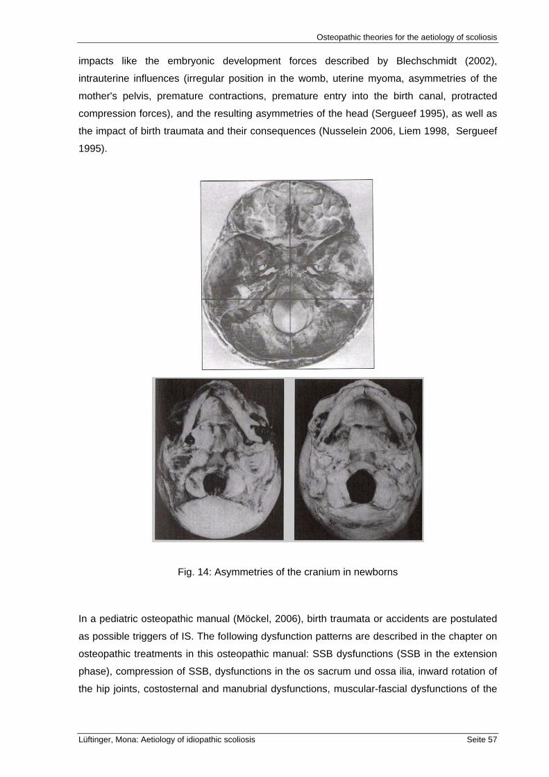
Osteopathic theories for the aetiology of scoliosis
Lüftinger, Mona: Aetiology of idiopathic scoliosis Seite 57
impacts like the embryonic development forces described by Blechschmidt (2002),
intrauterine influences (irregular position in the womb, uterine myoma, asymmetries of the
mother's pelvis, premature contractions, premature entry into the birth canal, protracted
compression forces), and the resulting asymmetries of the head (Sergueef 1995), as well as
the impact of birth traumata and their consequences (Nusselein 2006, Liem 1998, Sergueef
1995).
Fig. 14: Asymmetries of the cranium in newborns
In a pediatric osteopathic manual (Möckel, 2006), birth traumata or accidents are postulated
as possible triggers of IS. The foIlowing dysfunction patterns are described in the chapter on
osteopathic treatments in this osteopathic manual: SSB dysfunctions (SSB in the extension
phase), compression of SSB, dysfunctions in the os sacrum und ossa ilia, inward rotation of
the hip joints, costosternal and manubrial dysfunctions, muscular-fascial dysfunctions of the
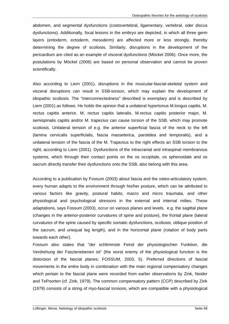
Osteopathic theories for the aetiology of scoliosis
Lüftinger, Mona: Aetiology of idiopathic scoliosis Seite 58
abdomen, and segmental dysfunctions (costovertebral, ligamentary, vertebral, oder discus
dysfunctions). Additionally, focal lesions in the embryo are depicted, in which all three germ
layers (entoderm, ectoderm, mesoderm) are affected more or less strongly, thereby
determining the degree of scoliosis. Similarly, disruptions in the development of the
pericardium are cited as an example of visceral dysfunctions (Möckel 2006). Once more, the
postulations by Möckel (2006) are based on personal observation and cannot be proven
scientifically.
Also according to Liem (2001), disruptions in the muscular-fascial-skeletal system and
visceral disruptions can result in SSB-torsion, which may explain the development of
idiopathic scoliosis. The "interconnectedness" described is exemplary and is described by
Liem (2001) as follows. He holds the opinion that a unilateral hypertonus M.longus capitis, M.
rectus capitis anterior, M. rectus capitis lateralis, M.rectus capitis posterior major, M.
semispinalis capitis and/or M. trapezius can cause torsion of the SSB, which may promote
scoliosis. Unilateral tension of e.g. the anterior superficial fascia of the neck to the left
(lamina cervicalis superficialis, fascia masseterica, parotidea and temporalis), and a
unilateral tension of the fascia of the M. Trapezius to the right effects an SSB torsion to the
right, according to Liem (2001). Dysfunctions of the intracranial and intraspinal membranous
systems, which through their contact points on the os occipitale, os sphenoidale and os
sacrum directly transfer their dysfunctions onto the SSB, also belong with this area.
According to a publication by Fossum (2003) about fascia and the osteo-articulatory system,
every human adapts to the environment through his/her posture, which can be attributed to
various factors like gravity, postural habits, macro and micro traumata, and other
physiological and psychological stressors in the external and internal milieu. These
adaptations, says Fossum (2003), occur on various planes and levels, e.g. the sagittal plane
(changes in the anterior-posterior curvatures of spine and posture), the frontal plane (lateral
curvatures of the spine caused by specific somatic dysfunctions, scoliosis, oblique position of
the sacrum, and unequal leg length), and in the horizontal plane (rotation of body parts
towards each other).
Fossum also states that "der schlimmste Feind der physiologischen Funktion, die
Verdrehung der Faszienebenen ist“ (the worst enemy of the physiological function is the
distorsion of the fascial planes; FOSSUM, 2003, 5). Preferred directions of fascial
movements in the entire body in combination with the main regional compensatory changes
which pertain to the fascial plane were recorded from earlier observations by Zink, Neider
and TePoorten (cf. Zink, 1979). The common compensatory pattern (CCP) described by Zink
(1979) consists of a string of myo-fascial torsions, which are compatible with a physiological
Osteopathic theories for the aetiology of scoliosis
Lüftinger, Mona: Aetiology of idiopathic scoliosis Seite 59
function until the primary organ system, the muscular-skeletal system is under stress. Zink
(1979) believed that the lumbar spine of the growing child was especially vulnerable to
repeated minor traumata that resulted in a twisting of the torso. He also felt that the ideal
physiological pattern was best suited for locomotion, and that while the CCP was not as
efficient a pattern, it was very adaptive. Implicit in these statements is the reasoning that
during childhood development, as the infant attains the ability to crawl and then eventually to
stand and walk children will adopt the more adaptive rotational pattern of the CCP. In other
words, as a consequence of repeated minor traumata the lumbar spine develops a twist or
bias of rotation. Then, through the reciprocating rotational motions of walking, this torsional
bias is transmitted to other junctional regions of the spine. The regions of most restriction can
be found in the so-called transitional zones which Zink (1979) considered “anatomical weak
points.” Magoun (1973) also confirmed the fact that fascial tension and healthy alignment of
the spine or fascial tension and skeletal system are closely linked.
Fossum (2003) states that the fascial torsion patterns include the longitudinal and horizontal
organizational planes of the fascia, as well as their connections to adjacent parts of the
cranium, the spine, and the pelvis. In this model, fasciae directly or indirectly influence the
body's health via coordination with the muscular-skeletal system, via coordination with
circulating body fluids, and by providing extensive passages to the nerves. Disruptions within
the fascial areas can manifest themselves in venous and lymphatic stasis, in restricted
breathing, abnormal reflexes, and a reduced range of movements (due to the impact on bony
levers and joints). Moreover, Fossum (2003) claims, several visceral restrictions can be
direct (mobility) and indirect (reflexion) consequences of the tension in the fascial areas.
Once more, the connections drawn by Fossum (2003) and Zink (1979) are only models and
hypotheses which cannot be scientifically proven.
Following Liem (2001), there are several strings of muscular fascia such as the one from the
os metatarsale I, via the caput fibulae, the tuber ischiadicum and the sacrum to occiput via
M. peroneus, M. biceps femoris, lig. sacrotuberale, fascia sacrolumbale and M.erector
spinae.
In this framework, tension dysbalances in these integrated myo-fascial chains can spur the
development of scoliosis, possibly also brought forward by additional dysfunctions (growth
spurt, stressors,…).
Also visceral disruptions like e.g. a ptosis of the stomach, have been found (cf. Liem 2001) to
reach the SSB via fascial connections, which constitutes yet another explanatory model of
idiopathic scoliosis.
Osteopathic theories for the aetiology of scoliosis
Lüftinger, Mona: Aetiology of idiopathic scoliosis Seite 60
The "Tensegrity-Model" invented by Buckminster-Fuller (cf. Liem 2006) is of further interest,
albeit not explicitly mentioned or postulated in the context of scoliosis. It constitutes a
structural system with rigid discontinuous structures which are connected by continuous
tensile structures. Within this system, pressure is distributed discontinuously while traction
forces are spread continuously. Tensegrity-structures cannot only be characterized by high
stability but also by their ability to transmit mechanical energy and information throughout the
entire system. Tensions are thus distributed continuously among all structural elements so
that increasing tension in one element will always lead to increasing tension in other
elements of the system as well. This increase in tension will be balanced out by an increase
in compression in certain elements of the system. Thus the system stabilizes itself. From an
osteopathic viewpoint, the boney skeletal system represents the rigid parts, while the
muscles, ligaments, and fascia constitute the continuous tensile structures within the body.
Ingber described this reciprocal tensile system at cellular level (Liem, 2006). Additionally, the
psychoneurotic-immunological network deserves to be mentioned as it touches upon my
topic, and may also influence the development of idiopathic scoliosis.
According to Liem (2006), psychoneuro-immunology can serve to explain that part of
osteopathic interactions in which findings allow for the conclusion that emotions can be a
pivotal point between body and mind. Biochemical substrates of emotions transmit
information between brain structures and endocrinal, gastro-intestinal and immunological
systems. Regarding the development of scoliosis, this aspect has not yet been sufficiently
investigated but since it is known by now that protracted stress (distress) can weaken the
immune system and even the gene expression of immunological cells (Liem 2006),
psychoneuro-immunology could also influence the development of idiopathic scoliosis.
Conclusio:
Research findings regarding osteopathic explanatory models of the development of
idiopathic scoliosis are vague and restricted to hypothetical assumptions.
The survey by Frymann et al. (2007) of 1250 newborns, in which anatomical disruptions in
the cranio-sacral system are presented as related to the newborns' symptoms, shows that,
among other factors, SSB torsions can be linked to circulatory and respiratory problems.
“Scoliosis” was not mentioned explicitly as a symptom, however. It is hardly possible to
diagnose an already existing increased deviation of the spine, unless it is an obvious one like
scoliosis capitis, torticollis, or congenital scoliosis, since spine X-rays of newborns for mere
research purposes cannot be justified ethically. Furthermore, there is an X-ray study by
Greenman (1970) related to SSB dysfunctions and their symptoms as postulated in
Osteopathic theories for the aetiology of scoliosis
Lüftinger, Mona: Aetiology of idiopathic scoliosis Seite 61
osteopathy, in which he looked into the relationship between the sphenoid body and the
basioocciput. The study was conducted with 25 patients and he was able to show abnormal
relationships between these bones that demonstrated the lesions defined by Sutherland as
flexion, extension, torsion, side-bending, vertical strain, and lateral strain. Greenman (1970)
found no correlation with clinical symptoms as described in osteopathy, however.
In order to prove or disprove the symptoms resulting from SSB torsions (e.g. scoliosis) as
they are postulated by osteopathy, further research needs to be done.
The theory that idiopathic scoliosis is caused by a dysfunction in embryonic development is a
rather complicated guiding idea. The period of embryology alone with its complex processes,
which could only be dealt with briefly here, constitutes a large field with many possible
dysfunctions, and can thus only be grouped among the ”hypotheses about the development
of idiopathic scoliosis"
Overall, there is little to no evidence of causal relationships in the development of IS and the
explanatory models postulated in osteopathy. There is no scientific proof that embryonic
developmental disruptions, birth traumata, SSB torsion, or cranial and sacro-pelvic
dysfunctions in general, or visceral and fascial dysfunctions clearly lead to idiopathic
scoliosis.
A good explanatory model for the complexities in dynamic processes and interactions is
offered by the Tensegrity-Model. The extra-cellular matrix creates a continuum in our body
which corresponds to the model's principles, establishing a connection between organs,
structures and systems. A disruption in this system like those previously described may
explain the development of scoliosis. This Tensegrity-Model, however, when applied to the
human body, is also merely a model, and offers yet another possible hypothesis which
cannot be scientifically proven.
This "lack of clarity" in the aetiology of scoliosis complicates osteopathic treatment on the
one hand, but on the other hand offers a chance because of the manifold approaches to the
complex connections and their specific osteopathic treatments, which may well lead to better
results in IS treatment, as small-scale studies have already pointed out (Philippi et al. 2006,
und Mandl-Weber 2000).
In order to render the therapy of idiopathic scoliosis even more efficient, osteopathy is
required to carry out more studies and submit results which can be substantiated and thus
better argued for.
Similarities or diametrical differences
Lüftinger, Mona: Aetiology of idiopathic scoliosis Seite 62
8. Similarities or diametrical differences
In this chapter some biomedical results pertaining to the aetiology of idiopathic scoliosis will
be juxtaposed with osteopathic explanatory models in order to show similarities or
diametrical differences between the two.
8.1. Asymmetrical pattern
In the study by Karski et al. (2006), the syndrome of contractures according to Mau is
presented. This describes scull deformities like the flattening of left forehead and temple
regions, eye asymmetry, nose and ear deformation, torticollis, scoliosis infantilis, contracture
of the left hip adductor muscles, contracture of the right hip abductor muscles, pelvis bone
asymmetries, feet deformities. The hypothesis for these deformities was the conjunction with
biomechanical aetiology of so-called idiopathic scoliosis caused by the fetus position during
the last months of pregnancy.
The deformities described above and the increased deviations from symmetry can – from an
osteopathic perspective – be explained by dysfunctions in embryology, intrauterine
dysfunctons, or birth traumata. According to experienced osteopaths like Van den Heede,
Nusselein or Liem, embryological dysfunctions can cause later idiopathic scoliosis. Birth
traumata are also postulated as a cause for the development of idiopathic scoliosis
(Sergueef, 1995; Liem, 2001).
In both fields (biomedicine and osteopathy) only hypotheses and models are being currently
published. The causes of these deviations from symmetry which are already diagnosed in
new-borns remain unclear.
Therefore newborns and babies require detailed examination to discover early symptoms of
scoliosis or other skeletal malformations.
Ben-Bassat et al. (2006) showed in their study that patients with scoliosis have asymmetrical
features of malocclusion compared with a random population. Also a theoretical model of a
possible relationship between stresses on the cervical column and vertical occlusal alteration
are discussed.
Malocclusion can be caused by embryologically induced cranial dysfunctions. The
dysfunctions mentioned above like SSB torsion, SSB compression und SSB in extension do
lead to idiopathic scoliosis but not to malocclusion, according to Liem (2001). SSB-
Similarities or diametrical differences
Lüftinger, Mona: Aetiology of idiopathic scoliosis Seite 63
dysfunctions which co-occur with malocclusion are identified in the literature (Liem, 2001) as
vertical strain and lateral flexion and rotation of SSB, which cannot be linked to the
development of scoliosis, however.
According to Sergueef (1995), the birth phase of "shoulders being pushed out" can require
considerable rotation and side-bending of the cervical spine, which in turn can lead to a
dysfunctions in the cervical spine, the cervico-thoracic junction and the upper thoracic spine.
Ben-Bassat et al. (2006) state that dysfunctions in the cervical spine can be linked to a
malocclusion, which has been frequently diagnosed in scoliotic children and adolescents.
As indicated earlier, it is important to critically review the symptoms postulated for a SBB-
dysfunction and initiate new research in this field.
Mac-Thiong et al. (2006) stated that the coronal sacropelvic morphology is distorted in AIS.
Two hypotheses are discussed: the increased values of height and width parameters in AIS
may be related to the altered growth rate (maturation), or may be due to an altered sacro-
pelvic development which could be in association with the process involved in the
progression of the spinal deformity. Sacro-pelvic dysfunction can – according to osteopathic models – result from embryologically
induced dysfunctions or be intrauterine (caused by "pathological" fetal positions; cf. Liem,
2001; Sergueef, 1995). Sergueef (1995) also postulated that distorsion patterns in
membranes and fasciae, which reflect the recorded spiraling rotation of the birth canal, can
induce dysfunctions in the sacro-pelvic area if it is a difficult birth. Intra-osseous dysfunctions
or torsion of the sacrum, dysfunctions of the ossa ilia, dysfunctions of the hip joints, unequal
leg length and/or traumata in this area during growth can lead to dysfunctions in the sacrum
and the pelvis (Liem, 2001; Nusselein, 2006; Sergueef, 1995; Zink 1979). Sacro-pelvic
dysfunctions can lead to the development of idiopathic scoliosis according to the views
expressed by Liem (2001), Nusselein (2006), and Zink (1979). A possible model of the
development of sacro-pelvic dysfunctions is offered by Schults et al. (1996); they showed in
a study that pressure or tension in one area of the embryo results in increased secretion of
connective tissue fibers in that area, and that these fibers tend to organize themselves along
lines of tension. It is possible that dysfunctions in the forming forces during the embryonic
phase and/or intrauterine dysbalances in the fetal phase lead to increased tensional
dysbalances in connective tissue. Such dysbalances can already be diagnosed in newborns,
Liem (2001) and Sergueef (1995) observed. Also fascial torsions in the sacro-pelvic area,
which also match this explanatory model, can cause IS, according to observations by
Fossum (2003), Liem (2001) and Möckel (2006).
Similarities or diametrical differences
Lüftinger, Mona: Aetiology of idiopathic scoliosis Seite 64
The dysfunctions described in osteopathy are dysfunctions that are "diagnosed" through
palpation at osseous, fascial, muscular, visceral or fluid levels, which cannot be scientifically
proven. This study may serve to stress the importance of the fascial structure, which has
recently been neglected in biomedicine, with its complex anatomical connections and
physiological functions, and may initiate further research in this area.
8.2. Neurological Dysfunctions
Various studies have been presented in the field of neurological dysfunctions and their
connection with the development of IS, in which vague links can be demonstrated.
Sun et al. (2006), e.g., found lower positions of the cerebellar tonsils in AIS patients. Filipovic
and Viskic-Stalec (2006) stated in their study that AIS affects dynamic balance and illustrated
the compensational functioning of mobility, especially when there is a lack of normal mobility
forms and there are weak postural control mechanisms and proprioception.
Royo-Salvador (1996) postulated that an increased tonus of the medullary traction can result
in the following changes: increased traction on the brain stem leads to increased tonus of the
meninges, and, via their periosteal attachment, to increased traction in the tentorium
cerebelli. Thus the tonsillae cerebelli are drawn lower and compressed. Furthermore, Royo-
Salvador maintains that the cerebellar hemispheres are pressed into the fossa cranialis
posterior, resulting in a deformation of the foramen magnum. In the cervical area of C1/C2,
compression of the nervous tissue occurs, and in the thoracic area, especially during growth,
idiopathic scoliosis develops. Royo-Salvador's explanation (1996) for this is that through the
thoracic curvature medullary traction is relieved. Carreiro (2005) stated in personal communication and also in her publication (2003) that
hypotheses like vestibular dysfunction to cortical asymmetries are discussed.
Van den Heede (2006) sees in the development of idiopathic scoliosis an embryonic
dysfunction in the build-up of the brain and the heart.
The statements by Carreiro (2005) and Van den Heede (2006) rest on very brief
communication, and can in this form only serve as the starting point for further conclusions.
Interviews with experienced osteopaths would probably have been more conducive.
In order to at least illustrate the connection between the three biomedical studies presented
and the dysfunctions of the brain, and the cerebellum in particular, as discussed in
osteopathy, I would like to refer to a brief overview of the physiology of the cerebellum.
The cerebellum is divided into three sections in keeping with its development and its cell
structure, as elaborated by Van den Berg et al. (2000):
Similarities or diametrical differences
Lüftinger, Mona: Aetiology of idiopathic scoliosis Seite 65
-- Vestibulocerebellum, whose function consists of integrating information yielded by the
sense of balance into sensory motor activity. It has a close connection to the spinal chord,
and contributes to the coordination of posture and locomotion.
-- Paleocerebellum or spinocerebellum is subject to influences from the spinal chord. Its
function is to support posture and to coordinate posture and locomotion, whose effect
evolves via the cerebellar cores along the nucleus ruber.
-- Neocerebellum or pontocerebellum occupies the largest part of the cerebellar hemisphere,
and is closely connected via the bridge (pons) with various regions of the cerebral cortex. Its
function ist he coordination of target and support motor activity.
The studies by Sun et al. (2006) about the "lower position of cerebellar tonsils“, the study by
Royo-Salvador (1996) about "tonus increase in the spinal chord", which leads to the
development of idiopathic scoliosis, and the study by Filipovic and Viskic-Stalec (2006), who
postulated that in AIS-patients the dynamic balance and proprioception are reduced, all more
or less explicitly refer to a dysfunction in the cerebellum.
When and where a cerebellal dysfunction occurs, and if a causal relationship can be
established in IS development remains unclear, also in the studies mentioned above.
Nonetheless, the results presented can be related to each other, and at least a hypothetical
connection in IS development can be established.
Conclusion
Lüftinger, Mona: Aetiology of idiopathic scoliosis Seite 66
9. Conclusion
The increased curvature of the spine was already diagnosed by Hippocrates (460-375 B.C.),
who treated it with traction, but up to the 21st century the underlying causality of this illness
has remained unclear. The present vast range of research into the aetiology of idiopathic
scoliosis (IS) in the biomedical field reveals that various hypothesis are being discussed.
None of the studies, however, can solely claim to explain the cause of scoliosis (cf. among
others, Goldberg et al. 2006, Miller et al. 1996, Sevastik et al. 2006 etc). The results show
which structural, physiological, and functional changes have been found with IS but where
the cause(s) of these changes lie, which result in an increased deviation of the spine, could
not be clarified.
Biomedical hypotheses which imply that neurological dysfunctions lie at the root of the
development of IS are increasingly being presented. Also in this area, however, there is no
scientific evidence to support the tenability of these hypotheses. During my research I also
found a tendency to report multiple pathogenesis for IS (Goldberg et al. 2006; Ben-Bassat et
al. 2006; Heidari et al. 2003; and others). Thus Goldberg also concluded: “It may be
associated that many pathological conditions and no specific pathology that belong to
scoliosis alone has been identified” (GOLDBERG et al. 2006, 447).
Regarding osteopathic theories about the aetiology of idiopathic scoliosis I could only find
few publications. Qualitative interviews would probably have been the more adequate
method of data generation. Besides, I found that only models about the aetiology of scoliosis
had been published which are not scientifically proven. In the study by Frymann (2007)
briefly referred to, in which the connection of disruptions in the cranio-sacral mechanism with
the symptoms in 1,250 newborns has been examined, the scale of the study is impressive
while its reliability is rather dubious since the diagnostic method chosen is palpation. Thus,
the dysfunction diagnosed by osteopathic lack any scientific basis, and in view of the
recognition of our profession we need to reconsider the role of palpation as a diagnostic tool,
proceed with more caution in our statements, and initiate as much research within
osteopathy as possible.
In the chapter about "Similarities and diametrical differences", in which biomedical research
results about the aetiology of IS were contrasted with osteopathic explanatory models,
hypotheses can be found on either side; those from biomedicine are better substantiated by
previous research, however.
In osteopathy, by contrast, there are no studies about this rather common clinical picture of
idiopathic scoliosis. There are, however, several models which are plausible but not
Conclusion
Lüftinger, Mona: Aetiology of idiopathic scoliosis Seite 67
scientifically proven or provable, since the causes mentioned in osteopathy like SSB torsion,
dysfunctions of the sacrum, ileum or the hip joints, and dysfunctions on a bony,
membranous, or fluid level, as well as fascial distorsions are not reliable.
Clearly, reliability is an important scientific factor, which ought to be given wider currency in
osteopathy too, since this will improve the quality of "osteopathic doing and thinking". In this
context I would like to quote the sentence Sommerfeld postulated in his master's theses:
"The results of scientific-reliability testing can give certain support for clinical osteopathic
acting“ (SOMMERFELD, 2006, 112).
The initial intention of this thesis – to gain more insight into the treatment of scoliotic patients
through the results of recent biomedical research and through the osteopathic theories
postulated about the aetiology of idiopathic scoliosis – has been eventually somewhat
modified. Owing to the wide spectrum of hypotheses about the aetiology of IS on either side,
yet more open questions have emerged.
My own experience in the treatment of idiopathic scoliosis in adolescents shows on average
good results which can also be proven clinically by X-rays. Which of the osteopathic
techniques applied in particular really does bring about change, and demonstrably improves
or at least stabilizes the degree of scoliosis, remains unclear to me and requires studies
which yield empirical evidence for causal relationships, especially in the field of cranial
osteopathy. As Andrew Taylor Still already remarked, a successful man not only pursues
theory, his motto is 'prove it'. ("Der erfolgreiche Mann verfolgt nicht nur die Theorie. Sein
Motto heißt ausschließlich beweisen!“, STILL, 2002, w. Vorbemerkungen)
In order to obtain qualitatively better answers to the question about IS causality
interdisciplinary studies in biomedicine and osteopathy are desirable.
Summary
Lüftinger, Mona: Aetiology of idiopathic scoliosis Seite 68
10. Summary
This work has reviewed and analysed various current biomedical studies and osteopathic
theories for the aetiology of idiopathic scoliosis.
Looking at possible genetic and epigenetic causes of IS, Zaidman et al. (2006) came to the
conclusion that IS is a "genetically dependent spinal deformity inherited by autosomal-
dominant type, with incomplete gender- and age related penetrance of genotype presented",
while Miller et al. (1996) stated that no clear association could be determined that genes are
linked to the cause of IS.
Some studies showed structural anomalies like imbalance of the connective tissue in IS
patients. Fiber imbalance in the intervertebral disc and also in ligamantum flavum were
stated by Yu and Fairbank (2005). Heidari et al. (2003) found out that higher fiber imbalance
results in more severe spinal deformity. According to a model study by Van der Plaats et al.
(2007), unilateral postponement of growth in os ligamantum flavum and intertransverse
ligament appeared to initiate scoliosis. Above all, however, it is not clear whether these
defects are primary or secondary, whether function governs structure or vice versa.
Other studies proved anatomical asymmetrical patterns in IS. Ben-Bassat et al. (2006) found
more asymmetric features of malocclusion in IS patients. The “syndrome of contractures”
was already diagnosed in newbornes and children by Karski et al. (2006). In these children
they noted initial stages of IS and they concluded that the malformations of skeletal system
can already be taking place in the last months of pregnancy. The sacropelvic morphology in
the coronal plane of AIS patients showed significant differences in comparison to normal
adolescents but it is unclear from which cause this asymmetric pattern do result.
In some studies neurological dysfunctions are hypothized to cause IS. Sun et al. (2006)
proved that cerebellar tonsils have lower positions in AIS patients than in normal
adolescents. Burwell et al. (2006a) hypothised that maturational delay in the CNS may arise
and cause AIS. In a further study Burwell et al. (2006b) developed theories about
disturbances in the longitudinal growth of paired (long limb bones, ribs, ilia) and united paired
bones (vertebrae, sternum, skull, mandibulae). Differences in dynamic balance between AIS
and healthy children are presented in a study by Filipovic and Viskic-Stalec (2006). An
increase in tension in the spinal cord which further induces the developement of IS is
presented in a study by Royo-Salvador (1996). Burwell et al. (2006) claimed that a
disturbance of bilateral symmetry in embryonic life results from a default process involving
mesodermal somites which causes the excess of right/left thoracic in AIS.
Other relevant studies looked at the connection of IS with visual defiency, a lower degree of
mineralisation in IS and pleural infection.
Summary
Lüftinger, Mona: Aetiology of idiopathic scoliosis Seite 69
The study by Goldberg (2006) on handedness did not show any evident connection between
preferred hand and the development of IS.
Although a large number of studies has been done over the last few years, the aetiology of
the three-dimensional deformity of idiopathic scoliosis remains unknown.
Osteopathic theories for the aetiology of scoliosis are scarce. The major source of
information was personal communication with three experienced osteopaths.
Hypotheses like dysfunctions in the embryology are discussed by Van den Heede (2006),
Nusselein (2006), and Möckel (2006). Malformation of skeletal system taking place in the
later months of pregnancy which can induce the development of IS are discussed by Liem
(1998), Nusselein (2006) and Sergueef (1995). Frymann (2007), Liem (2001), and Sergueef
(1995) postulated that birth traumata can influence the incidence and outcome of scoliosis.
In osteopathy SSB-torsions can indicate different symptoms, amongst them scoliosis. These
dysfunctions in the SSB are based on palpational diagnostics which is a not reliable test
method. Further dysfunctions in the sacropelvic region, ossa ilia, the hip joints, or distorsions
costosternal and in the manubrium of the sternum are published in osteopathic literature. But
there is no scientific proof for these hypotheses.
Distorsions in the myofascial system and visceral dysfuntions inducing the development of IS
are also some of the evidence cited in osteopathic publications (Fossum 2003; Liem 2001;
Magoun 1973; Zink 1979).
Several similarities and contradictions between the two views have been pointed out.
Anatomical asymmetrical patterns were diagnosed already in newborns by Karski et al.
(2007) which they claim to be caused by the fetus position during the last months of
pregnancy. Also osteopaths like Liem (1998, 2001), Möckel (2006), Nusselein (2006),
Sergueef (1995), and Van den Heede (2006) stated that intrauterine dysfunctions can induce
IS.
For both sides only hypotheses are presented and in-depth research needs to be done to
help discover the aetiology of IS.
More asymmetrical features of occlusion in IS patients was proved by Ben-Bassat et al.
(2006). From an osteopathic perspective, cranial dysfunctions can be caused by embryonic
dysfunctions, birth traumatas or SSB-torsions. The postulated symptoms of the SSB-torsion
(Liem, 2001), however, which are discussed in the context of the development of IS, do not
agree with malocclusion. Mac-Thiong et al. (2006) claim that sacropelvic morphology is
distorted in the coronal plane of AIS patients. The osteopathic theories for sacropelvic
morphology are embryological dysfunctions, birth traumata or traumata inducing dysfunctions
Summary
Lüftinger, Mona: Aetiology of idiopathic scoliosis Seite 70
on a bony, membranous or fluid level (Liem 2001; Nusselein 2006; Sergueef 1995; Zink
1979). But also in this case there is also no scientific base for osteopathic hypotheses.
With regard to the neurological dysfunction and its connection with the development of IS
Sun et al. (2006) found anatomical features like lower positions of the cerebellar tonsils
found in IS patients. Roya-Salvador (1996) postulated in his study that this is induced by an
increased tension in the spinal cord, which also causes the development of scoliosis.
Filipovic and Vaskic-Stalec (2006) showed that dynamic balance is affected in AIS patients
and this seems also to indicate a dysfunction in the cerebellum. From an osteopathic point of
view, Van den Heede (2006) stated that IS is caused by an embryologic dysfunction in the
build-up of the brain and the heart.
Finally it has to be said, if and where the cause for a cerebellar dysfunction is involved and
whether there is a context in the aetiology of IS remains unclear.
Table of Figures
Lüftinger, Mona: Aetiology of idiopathic scoliosis Seite 71
11. Table of Figures
Fig. 1: Hippocrates................................................................................................................... 9 Fig. 2: Hippocrates’ ladder..................................................................................................... 10 Fig. 3: Ambroise Parè (supporting corsets made of iron plate) ............................................. 11 Fig. 4: A.Vesal, “De humanis corporis“ .................................................................................. 11 Fig. 5: Hildanus, “Abriss des Rückgrads” .............................................................................. 12 Fig. 6: Roux corrective orthotic device................................................................................... 13 Fig. 7: Le Vacher and Sheldrake corrective orthotic devices................................................. 13 Fig. 8: Hossard corrective orthotic devices............................................................................ 14 Fig. 9: Classification of scoliosis ............................................................................................ 20 Fig. 10: Bend test................................................................................................................... 22 Fig. 11: Cobb curve ............................................................................................................... 24 Fig. 12: Risser grades............................................................................................................ 24 Fig. 13: Torsion-Strain ........................................................................................................... 54 Fig. 14: Asymmetries of the cranium in newborns................................................................. 57
Bibliography
Lüftinger, Mona: Aetiology of idiopathic scoliosis Seite 72
12. Bibliography
[1] Adams W.1882. Lectures on the pathology and treatment of lateral and other forms of
curvature of the spine.London:Churchill [2] Arkin M. 1949. The mechanism of the structural changes in scoliosis. J Bone Jt Surg
31-A:519-528 [3] Axenovich TI. Zaidman AM. Zorkoltseva IV. 1996.Segregation analysis of idiopathic
scoliosis demonstration of major gene effect. Am. J. Med. Genet. 86:389-394 [4] Barker DJP. Erikson JG. Forsen M. 2002. Fetal origins of adult disease: strength of
effects and biological basis. Int J Epidemiolog 31;6:1235-1239. [5] Ben-Bassat Y,Yitschaky M, Kaplan L, Brin I. 2006. Occlusal patterns in patients with
idiopathic scoliosis. Am J Orthod Dentofacial Orthop;130:629-33 [6] Blechschmidt E.1982. Sein und Werden. Stuttgart:Urachaus [7] Blechschmidt E.2002. Wie beginnt das menschliche Leben.Stein am Rhein:Christiana [8] Bunell WP.1984. An objective criterion for scoliosis screening. J Bone Joint Surg
66A:1381 [9] Burwell RG. Freeman BJC. Dangerfield PH. Aujla RK. Cole AA. Kirby AS. Polak F. Pratt
RK. Webb JK. Moulton A. 2006a . Etiologic Theories of Idiopathic Scoliosis: Neurodevelopmental Concept of Maturational Delay of the CNS Body Schema („Body in the Brain“). Research into spinal Deformities;5:72-79
[10] Burwell RG, Freeman BJC, Dangerfield PH, Aujla RK, Cole AA, Kirby AS, Polak F, Pratt
RK, Webb JK, Moulton A. 2006b. Etiologic Theoris of Idiopathic Scoliosis: Enantiomorph Disorder Concept of Bilateral Symmetry, Physeally-created Growth Conflicts and possible Prevention. Research into spinal Deformities 5:391-397
[11] Burwell RG.Dangerfield PH. Freeman BJ. Aujla RK. Cole AA. Kirby AS. Pratt RK. Webb
JK. Moulton A. 2006c. Etiologic theories of idiopathic scoliosis:the breaking of bilateral symmetry in relation to the left-right asymmetry of internal organs, right thoracic adolescent idiopathic scoliosis (AIS) and vertebrate evolution. Stud Health Technol Inform.123:385-390
[12] Carreiro J. 2003. An Osteopathic Approach to Children.Maine:Churchill Livingstone [13] Carreiro J. 2005. Personal communication.WSO [14] Cheng JCY. Guo X.1997. Osteopenia in adolescent idiopathic scoliosis. Spine;22:1716-
1720 [15] Cobb JR.1948. Outline for the study of scoliosis. International Course Lectures, The
American Academy of Orthopedic Surgeons, Ann Arbor [16] Dickson RA. Lawton JO. Archer AI. Butt WP.1984. The pathogesis of idiopathic scoliosis.
Biplanar spinal asymmetry. J Bone Joint Surg 66B:8
Bibliography
Lüftinger, Mona: Aetiology of idiopathic scoliosis Seite 73
[17] Dickson RA. The ethiology and pathogenesis of idiopathic scoliosis. 1992a. Acta Orthop. Belg. 58(Suppl 1):21-25
[18] Dickson RA. The scientific basis of treatment of idiopathic thoracis scoliosis. 1992b.
Acta Orthop. Belg. 58(Suppl 1):107-110 [19] Dunshirn M. 2007. Die Mittellinie in der Osteopathie. Krems:Masterthese.Universität
Krems [20] Ebenbichler G. Liederer A. Lack W. 1994. Die Skoliose und ihre konservativen
Behandlungsmöglichkeiten. Wien Klin Wschr 24:593 [21] Filipovic V, Viskic-Stalec N. 2006.The Mobility Capabilities of Persons With Adolescent
Idiopathic Scoliosis. Spine 19:2237-2242 [22] Floman Y. 1998. Thoracic scoliosis and restricted neck motion: a new syndrome? A
report of six cases. Eur Spine J 2:155-157 [23] Fossum C. 2003. Faszien, das osteoartikuläre System und das allgemeine
Kompensationsmuster in der Osteopathie. J.Osteopathische Medizin 4:4-12 [24] Frymann V. 2007. Die gesammelten Schriften von Viola M. Frymann,
DO.London/Deutschland: Jolandos Verlag [25] Fuchs B. 2007. Die Synchondrosis Spenobasilaris (SSB) als zentrales Element der
Schädelbasis. Krems:Masterthese. Universität. Krems [26] Greenman PE. 1970. Roentgen findings in the cranioSacral Mechanism.Journal of the
American Osteopathic Association,70:1 [27] Goldberg CJ, Moore DP, Fogarty EE, Dowling FE. 2006.Handedness and Spinal
Deformity. Research into spinal Deformities 5:442-448 [28] Goldberg CJ. 2000. Symmetry control. State of the Art Reviews. Spine 14(2):327-328 [29] Grivas T, Savvidou O, Vasiliadis E, Psarakis S, Koufopoulos G. 2006.Prevalence of
scoliosis in Women with visual Deficiency. Research into spinal Deformities 5:52-56 [30] Harms, J.2008. Geschichte der Skoliosetherapie. Convention,
(online),Available:http://www.harms-spinesurgery.com (8. April 2008) [31] Heidari B, Fitzpatrick D, Synott K, McCormack D. 2004.Modelling of annulus fibrosus
imbalance as an aetiological factor in adolescent idiopathic scoliosis. Clinical Biomechanics 19:217-224
[32] Hensinger RN. 1979.Congenital dislocation of the hip. Clinical Symp. 31 [33] Herman R. Mixon J. Fischer A. Maulucci R. Stuyck J. 1985.Idiopathic scoliosis and the
central nervous system: a motor control problem. Spine 10:1-14 [34] Howorth B. 1977.The Etiology of the congenital dislocation of the hip. Clin.Orthop.
29:164-179 [35] Humpke T. Rompe JD. Heine J. Carstens C.2002. Leitlinien der Orthopädie. Köln:
Dt.Ärzte-Verlag
Bibliography
Lüftinger, Mona: Aetiology of idiopathic scoliosis Seite 74
[36] Jach G.1892.Klinische Studien über das Verhalten der Torsion bei Skoliose. Z Orth
Chir.1:252 [37] Karski T, Kalakucki J, Karski J. 2006. ”Syndrome of contractures“(according to Mau) with
the Abduction Contracture of the Right Hip as Causative Factor for Development of the So-called Idiopathic Scoliosis. Research into spinal Deformities 5:34-39
[38] Karski T. 2000.Etiology of Adolescent Idiopathic Scoliosis(w:) Burwell, Dangerfield (red)
Spine. Etiology of Adolescent Idiopathic Scoliosis:Current trends and Relevance to New Treatment Approaches- Volume 14/2,Hanley&Belfus,Inc, Philadelphia. 324
[39] Kouwenhoven JW. Bartels L. Vincken K. Viergever M. Verbout A. Delhaas T. Castelein
R.2007.The relation between organ anatomy and pre-existent vertebral rotation in the normal spine: MRI –study in humans with situs inversus totalis. Spine 32(10):1123-1128
[40] Lehnert-Schroth, C. 2000. Dreidimensionale Skoliosebehandlung. Deutschland:Urban
und Fischer Verlag [41] Liem T. 2006. Morphodynamik in der Osteopathie. Hamburg: Hippokrates [42] Liem T. 2001. Kraniosakrale Osteopathie. Stuttgart:Hippokrates [43] Liem T.1998. Skoliosis capitis und frühkindliche Traumata.J.Orginalia:10-16 [44] Lindemann K. 1957. Ätiologie und Pathogenese der Skoliose. In: Hohmann G.
Hackenbroich M. Lindemann K.Handbuch der Orthopädie, Bd 2.Stuttgart:Thieme [45] LonsteinJE Und Carlson JH. 1984. The prediction of curve progression in untreated
idiopathic scoliosis during growth. J Bone Joint Surg 66A:1061 [46] LoweTG. EdgarM. Margulies JY. MillerNH. RasoVJ. ReinkerKA. Rivard CH. 2000.
Etiology of idiopathic Scoliosis:Current trends in research. Journal of bone and Joint surgery. 82-A:1157-1168
[47] Mac-Thiong, Labelle H, de Guise J. 2006. Comparison of Sacropelvic Morphology
between Normal Adolescents and Subjects with Adolescent Idiopathic Scoliosis. Research into spinal Deformities 5:195-200
[48] Magoun HI. 1973. Idiopahtic Adolescent Spinal Scoliosis: A Reasonable Etiology. D.O.
Magazine.13(6):151-160 [49] Mandl-Weber U. 2000.Osteopathic Treatment of idiopathic scoliosis with emphasis on
the fascial system. Österreich.WSO. Thesis-Osteopathy [50] Mau H. 1982. Differentialdiagnose der beginnenden Skoliose beim M. Scheuermann
gegeüber der idiopathischen Skoliose. Z Orthop 120,58 [51] Mau H. Die Ätiopathogenese der Skoliose. 1982. Bücherei des Orthopäden, Band 33:1-
110 Stuttgart: Enke [52] Mau H. 1979.Zur Ätiopathogenese von Skoliose, Hüftdysplasie und Schiefhals im
Säuglingsalter. Zeitschrift f. Orthop. 5:601-605
Bibliography
Lüftinger, Mona: Aetiology of idiopathic scoliosis Seite 75
[53] McMaster M. Lee AJ. Burwell RG. 2004. Physical activities of patients with adolescent idiopathic scoliosis compared with a control group: implications for etiology and possible prevention. In: Bonita.Sawatzky Editor, International Research Society of Spinal Deformities Symposium Vancouver, Canada:University of British Columbia, pp68-71.
[54] Meijer H.1866. Die Mechanik der Skoliose. Virchov Archiv für Path Anat Bd. 35:225 [55] Meister R.1980. Atemfunktion und Lungenkreislauf bei thorakaler Skoliose. In: Müller
RW, Ferlinz R(eds.) Bücher des Pneumologen, Bd 5.Stuttgart:Thieme [56] Milenkovic SM. Kocijancic RI. Belojevic GA. 2004. Left handedness and spine
deformities in early adolescence. European Journal of Epidemiology.19:969-972 [57] Millner PA und Dickson RA.1996. Idiopathic scoliosis:biomechanics and biology.Eur
Spine J5:362 [58] Moe JH, Winter RB, Bradford DS, Lonstein JE .1978. Scoliosis and other spinal
deformities.Philadephia :W.B. Saunders [59] Möckel E. Mitha N. 2006. Handbuch der pädiatrischen Osteopathie. München:Elsevier [60] Mukherjee S, Langroudi B, Rosenthal M, Balfour-Lynn. 2007.Incidence and Outcome of
Scoliosis in Children with Pleural Infection. Pedriatic Pulmonology 42:221-224 [61] Niethard F.and Pfeil J.1992. Orthopädie (Duale Reihe). Stuttgart: Hippokrates [62] Nusselein H.2006. Personal communication.WSO [63] Panjabi MM. Brand RA. 1976.White AA. Three dimensional flexibility and stiffness
properties of the human thoracic spine. J. Biomech. 9:185-192 [64] Pedriolle R und Vidal J. 1985. Thoracic idiopathic scoliosis curve. Evaluation and
prognosis. Spine10:785 [65] Pert CB. 1999. Molecules of emotion. New York:Touchstone [66] Philippi H. Faldum A. Papst B. 2006. Infantile postural asymmetry and osteopathic
treatment: a randomized therapeutic trial. Heidelberg,Deutschland.Developmental Medicine&Child Neurology.48:5-9
[67] Podlesnic W. 2006. Local Listening – a General Diagnostic Tool? An Experimental
Examination of its Reliability. Krems: Masterthese. Universität Krems [68] Risser JC.1958. The iliac apohysis: an invaluable sign in the management of scoliosis.
Clin Orthop 11:111 [69] Rohen. and Lütjen-Drecoll. 2004. Funktionelle Embryologie. Stuttgart:Schattauer [70] Royo-Salvador MB. 1996.Siringomielia,escoliosis y malformacíon de Arnaol-Chiari
idiopáticas. Etiologíca común.Rev.Neurolo. 24:937-959 [71] Schults R. Feitis R.1996. The Endless Web: Fascial Anatomie and Physical Reality.
Berkley: North Atlantic Books. p. 11-17
Bibliography
Lüftinger, Mona: Aetiology of idiopathic scoliosis Seite 76
[72] Sergueef,N. 1995. Die kraniosakrale Osteopathie bei Kindern. Deutschland:Verlag für ganzheitliche Medizin
[73] Sevastik J. 2006. Right Convex Thoracic Female Adolescent Scoliosis in the Light of the
Thoracospinal Concept. Research into spinal Deformities 5:552-558 [74] Sommerfeld P. 2006. Touching Reliability. Krems: Masterthese. Universität Krems [75] Still AT. 2002.Das große Still-Kompendium. Deutschland:Jolandos [76] Sun X, QIU Y, Zhu Z. 2006. Variations of the Position of the cerebellar Tonsil in
Adolescent Idiopathic Scoliosis with Severe Curves: A MRI Study. Research into spinal Deformities 5:565-570
[77] Van den Berg F.2000. Angewandte Physiologie. Stuttgart:Thieme Verlag [78] Van den Heede P.2006.Personal communication.WSO [79] Van Der Plaats A, Veldhuizen A, Verkerke G. 2007.Numerical Simulation of
Asymmetrically Altered Growth as Initiation Mechanism of Scoliosis. Annals of Biomedical Engineering 7:1206-1215
[80] Velhuizen AG. Weber DJ. Webb PJ. 2000. The aetiology of idiopathic
scoliosis:biomechanical and neuromuscular factors. Eur.Spine J.9:178-184 [81] Velis KP. Healey JH. Schneider R. 1989. Peak sceletal mass assesment in young girls
with idiopathic scoliosis. Spine;14:706-711 [82] Weinstein SL und Ponsetti IV.1985. Curve progression in idiopathic scoliosis. J Bone
Joint Surg 65A:447 [83] Weiss EL. Lehmkuhl LD. Smith LK.Standing and walking.1996. In: Smith LK. Weiss EL.
Lehmkuhl LD. eds. Brunnstrom´s Clinical Kinesiology. Philadelphia.PA:F.A. Davis Co:401-434
[84] Weiß, H.and Rigo, M. 2001. Befundgerechte Physiotherapie bei Skoliose. München:
Pflaum Verlag [85] Yarom R. More R. Meyer S. 1985. Platelet and muscle abnormalities in idiopathic
scoliosis. Warner J.O,Metha MH (eds). Scolisis Prevention. N.Y.Praeger: 3-22 [86] Yeung H, Hung V,Lee K, Guo X, NgBWK, Cheng J. 2006. Lower Degree of
Mineralisation Found in Cortical Bone of Adolescent Idiopathic Scoliosis (AIS). Research into spinal Deformities 5:599-604
[87] Yu J, Fairbank J, Roberts S, Urban J. 2005. The Elastic Fiber Network of the Anulus
Fibrosus of the Normal and Scoliotic Human Intervertebral Disc. Spine 30:1815-1820 [88] Zaidman A, Zaidman M, Korel A, Mikhailovsky M, Eshchenko T, Grigorjeva E. 2006.
Aggrecan Gene Expression Disorder as Aetiologic Factor of Idiopathic Scoliosis. Research into spinal Deformities 5:14-17
[89] Zink GJ. Lawson WB.1979. An osteopathic structural examination and functional
interpretation of the soma.Osteopathic Annals 7:12-17