Embed Size (px)
Citation preview
RESEARCH Open Access
Plumbagin inhibits invasion and migration ofbreast and gastric cancer cells by downregulatingthe expression of chemokine receptor CXCR4Kanjoormana Aryan Manu1, Muthu K Shanmugam1, Peramaiyan Rajendran1, Feng Li1, Lalitha Ramachandran1,Hui Sin Hay1,2, Radhamani Kannaiyan1, Shivananju Nanjunda Swamy1, Shireen Vali3, Shweta Kapoor3,Bhargavi Ramesh3, Pradeep Bist4,5, Evelyn S Koay6, Lina HK Lim4,5, Kwang Seok Ahn7, Alan Prem Kumar1,2,8* andGautam Sethi1,2*
Abstract
Background: Increasing evidence indicates that the interaction between the CXC chemokine receptor-4 (CXCR4)and its ligand CXCL12 is critical in the process of metastasis that accounts for more than 90% of cancer-relateddeaths. Thus, novel agents that can downregulate the CXCR4/CXCL12 axis have therapeutic potential in inhibitingcancer metastasis.
Methods: In this report, we investigated the potential of an agent, plumbagin (5-hydroxy-2-methyl-1, 4-naphthoquinone), for its ability to modulate CXCR4 expression and function in various tumor cells using Westernblot analysis, DNA binding assay, transient transfection, real time PCR analysis, chromatin immunoprecipitation, andcellular migration and invasion assays.
Results: We found that plumbagin downregulated the expression of CXCR4 in breast cancer cells irrespective oftheir HER2 status. The decrease in CXCR4 expression induced by plumbagin was not cell type-specific as theinhibition also occurred in gastric, lung, renal, oral, and hepatocellular tumor cell lines. Neither proteasomeinhibition nor lysosomal stabilization had any effect on plumbagin-induced decrease in CXCR4 expression. Detailedstudy of the underlying molecular mechanism(s) revealed that the regulation of the downregulation of CXCR4 wasat the transcriptional level, as indicated by downregulation of mRNA expression, inhibition of NF-�B activation, andsuppression of chromatin immunoprecipitation activity. In addition, using a virtual, predictive, functionalproteomics-based tumor pathway platform, we tested the hypothesis that NF-�B inhibition by plumbagin causesthe decrease in CXCR4 and other metastatic genes. Suppression of CXCR4 expression by plumbagin was found tocorrelate with the inhibition of CXCL12-induced migration and invasion of both breast and gastric cancer cells.
Conclusions: Overall, our results indicate, for the first time, that plumbagin is a novel blocker of CXCR4 expressionand thus has the potential to suppress metastasis of cancer.
BackgroundMetastasis is a highly complex and non-spontaneousprocess that generally affects vital organs such as brain,lung, liver, bone or lymph nodes [1-3] in the later stagesof cancer progression. At present, there are no approveddrugs that can specifically target the metastatic process
[4,5], and little is known about the molecular mechan-ism(s) regulating the process of metastasis [5]. Chemo-kines are a large family of small chemotactic cytokinesthat regulate multiple biological processes such as leu-kocyte trafficking, hematopoiesis, adhesion, and angio-genesis [6-8]. Based on the position of the first twoconserved cysteine residues, the chemokines can be clas-sified into four subfamilies, CXC, CC, C, and CX3C, andact on leukocytes via selective membrane-bound G pro-tein-coupled receptors [2,3].
* Correspondence: [email protected]; [email protected] of Pharmacology, Yong Loo Lin School of Medicine, NationalUniversity of Singapore, Singapore 117597Full list of author information is available at the end of the article
Manu et al. Molecular Cancer 2011, 10:107http://www.molecular-cancer.com/content/10/1/107
© 2011 Manu et al; licensee BioMed Central Ltd. This is an Open Access article distributed under the terms of the Creative CommonsAttribution License (http://creativecommons.org/licenses/by/2.0), which permits unrestricted use, distribution, and reproduction inany medium, provided the original work is properly cited.
It has been well documented that the CXCL12/CXCR4 signaling cascade plays a crucial role in cancerproliferation, migration and metastasis [9]. CXCR4 isexpressed by various types of tumor cells, includingbreast [10], colorectal [11], gastric [12], ovarian [13],pancreatic [14], prostate [15], lung [16], melanoma [17],and brain [18] tumor cells. The SDF-1a/CXCR4 attrac-tion leads breast cancer cells to leave the circulation andmigrate into organs that express large amounts of che-mokines, where the cancer cells proliferate, induceangiogenesis and form metastatic tumors [7,19]. AsCXCR4 expression has been correlated with poor overallsurvival rate in patients with breast cancer [20], and col-orectal cancer [21], CXCR4 has been considered as apotential therapeutic target for inhibiting cancer metas-tasis [22].In the present report, we studied the effect of plumba-
gin (5-hydroxy-2-methyl-1, 4-naphthoquinone, an analo-gue of vitamin K3) as a novel regulator of the CXCL12/CXCR4 signaling axis. Plumbagin, a naturally occurringyellow pigment predominantly found in the plants ofthe Plumbaginaceae, Ancestrocladaceae, and Dionco-phyllaceae families, has been reported to exhibit signifi-cant anti-proliferative, pro-apoptotic, chemopreventingand radiosensitizing activities in different tumor cellsand animal models [23-30]. Because CXCR4 is knownto mediate proliferation, invasion and metastasis oftumor cells, we postulated that plumbagin may modu-late the expression of CXCR4 and inhibit tumor cellinvasion. Our results demonstrate, for the first time,that plumbagin can downregulate CXCR4 expression invarious tumor cells, including HER2-overexpressingbreast cancer cells, and this could be through its inhibi-tion of NF-�B activation. We also found that plumbagincan significantly inhibit CXCL12-induced migration andinvasion of breast and gastric tumor cells.Alongside testing the effects of plumbagin experimen-
tally in various tumor cells, we also tested the hypoth-esis of plumbagin-mediated NF-�B inhibition as the keyreason for the reduction in CXCR4 and other metastaticgenes, in a virtual, predictive, tumor cell system. Thevirtual epithelial tumor cell platform on which predictiveNF-�B inhibition studies were conducted is a compre-hensive integrated functional proteomics based, dynamicrepresentation of the pathways representing the key can-cer phenotypes of proliferation, apoptosis, angiogenesis,metastasis and conditions of tumor microenvironmentincluding tumor-associated inflammation [31-33]. In thisvirtual tumor cell system, we can manipulate any pro-tein or gene by over-expression or knockdown and getquantitative readouts and insights on the impact of thischange on the various markers in the system. This pre-dictive analysis helps in reconfirming the experimental
hypothesis and giving mechanistic insights into under-standing the trends in biomarker and phenotypechanges. This novel approach has facilitated the analysisof the impact of plumbagin on metastatic genes basedon the premise that this quinone mediates its affects pri-marily via modulation of NF-�B activation. The combi-nation of predictive experiments coupled with guidedexperimental validations provide a more integrated ana-lysis and a better understanding of the efficacy andmechanisms of action of specific anti-cancer drugs onphysiological endpoints.
MethodsReagentsPlumbagin, Tris, glycine, NaCl, SDS, lactacystin, andchloroquine were purchased from Sigma-Aldrich (St.Louis, MO, USA). Plumbagin was dissolved in dimethyl-sulfoxide as a 20 mM stock solution and stored at 4°C.Further dilution was done in cell culture medium. RPMI1640, DMEM, fetal bovine serum (FBS), 0.4% trypanblue vital stain, antibiotic-antimycotic mixture, andHRP-conjugated secondary antibodies were obtainedfrom Invitrogen (Carlsbad, CA, USA). Antibodies againstCXCR4 and HER2 were obtained from Abcam (Cam-bridge, MA, USA). CXCL12 was purchased from Pro-Spec-Tany TechnoGene Ltd. (Rehovot, Israel).
Cell LinesHuman breast cancer MDA-MB-231, BT474, and oraladenosquamous carcinoma CAL27 cells were obtainedfrom American Type Culture Collection (Manassass,VA, USA).AGS, MKN45, and SNU16 (gastric cancer) cells were
kindly provided by Prof. Patrick Tan, DUKE-NUS Grad-uate Medical School, Singapore. Hep3B (hepatocellularcarcinoma) cells were kindly provided by Prof. Hui KamMan, National Cancer Centre, Singapore. 786-O (renalcell carcinoma) cells were kindly provided by Dr. JohnYuen, Singapore General Hospital, Singapore. H1299(lung adenocarcinoma) cells were kindly provided byProf. Bharat B. Aggarwal, M.D. Anderson Cancer Cen-ter, Houston, TX, USA. MDA-MB-231, AGS, MKN45,SNU16, 786-O, and H1299 cells were cultured in RPMI1640 medium with 10% FBS, 100 U/mL penicillin, and100 μg/mL streptomycin. BT474 cells were cultured inDMEM F12 medium with 10% FBS, 100 U/mL penicil-lin, and 100 μg/mL streptomycin. Hep3B cells were cul-tured in DMEM supplemented with 10% FBS, 100 U/mL penicillin, and 100 μg/mL streptomycin. CAL27cells were cultured in DMEM containing 10% FBS, and1 mM pyruvate, 100 U/mL penicillin, and 100 μg/mLstreptomycin and were maintained at 37°C in an atmo-sphere of 5% CO2-95% air.
Manu et al. Molecular Cancer 2011, 10:107http://www.molecular-cancer.com/content/10/1/107
Page 2 of 14
Western blot analysisFor detection of CXCR4 and HER2, plumbagin-treatedwhole-cell extracts were lysed in lysis buffer (20 mMTris (pH 7.4), 250 mM NaCl, 2 mM EDTA (pH 8.0),0.1% Triton X-100, 0.01 mg/mL aprotinin, 0.005 mg/mLleupeptin, 0.4 mM PMSF, and 4 mM NaVO4). Lysateswere then spun at 14,000 rpm for 10 min to removeinsoluble material and resolved on a 10% SDS gel. Afterelectrophoresis, the proteins were electrotransferred to anitrocellulose membrane, blocked with 5% nonfat milkto minimize non-specific binding, and probed with anti-CXCR4 or HER2 antibodies (1:3000) overnight at 4°C.The blot was washed, exposed to HRP-conjugated sec-ondary antibodies for 2 h, and the CXCR4/HER2expression was detected by chemiluminescence emission(ECL; GE Healthcare, Little Chalfont, Buckinghamshire,UK). The densitometric analysis of the scanned blotswas done using Image J software and the results areexpressed as fold change relative to the control.
Nuclear extract preparationNuclear extracts were prepared at various time pointsafter treatment for subsequent NF-�B DNA-bindingactivity assay. Cell nuclear fractions were extractedusing a nuclear extraction kit (Active Motif, Carlsbad,CA, USA). Briefly, cells were washed, collected in ice-cold PBS in the presence of phosphatase inhibitors, andthen centrifuged at 300 g for 5 min. Cell pellets wereresuspended in a hypotonic buffer, treated with deter-gent, and centrifuged at 14,000 g for 30 s. After collec-tion of the cytoplasmic fraction, the nuclei were lysed,and nuclear proteins were solubilized in lysis buffer andprotein concentrations were determined by the Bradfordprotein assay (Bio-Rad Laboratories, Hercules, CA,USA).
NF-�B DNA-binding activity assayNF-�B DNA-binding activity was analyzed using theTransAM NF-�B p65 transcription factor assay kit(Active Motif, Carlsbad, CA, USA), following the manu-facturer’s instructions. Briefly, nuclear extracts (5 μg)from plumbagin-treated cells were incubated in a 96-well plate coated with oligonucleotide containing theNF-�B consensus-binding sequence 5’-GGGACTTTCC-3’. Bound NF-�B was then detected by a specific pri-mary antibody. An HRP-conjugated secondary antibodywas then applied to detect the bound primary antibodyand provided the basis for colorimetric quantification.The enzymatic product was measured at 450 nm with amicroplate reader (Tecan Systems, San Jose, CA, USA).
NF-�B luciferase reporter assayMDA-MB-231 cells were plated in 96-well plates with 1× 10⁴ cells per well in 10% FBS-supplemented RPMI
medium. After overnight incubation, the cells weretransfected with the NF-�B reporter plasmid linked to aluciferase gene or with the dominant-negative I�Ba(I�Ba-DN) plasmid. NF-�B luciferase plasmid wasobtained from Stratagene (La Jolla, CA). Transfectionswere done according to the manufacturer’s protocolsusing FuGENE-6 (Roche). At 24 h post-transfection,cells were treated with indicated concentrations ofplumbagin for 2 h and then washed and lysed in lucifer-ase lysis buffer (Promega), and the luciferase activity wasmeasured with a luminometer using a luciferase assaykit (Promega) and was normalized to b-galactosidaseactivity. All the experiments were done in triplicates andrepeated two or more times.
RNA extraction and Real-time PCR analysisTotal RNA was extracted using the Trizol reagent (Invi-trogen, Carlsbad, CA, USA), according to the manufac-turer’s instructions. Reverse transcription (RT) was thencarried out at 37°C for 1 h. Each RT reaction contains 1μg of total RNA, 1× RT buffer, 5 mM MgCl2, 425 μMeach of dNTPs, 2 μM random hexamers, 0.35 U/μLRNase inhibitor, 1.1 U/μL MultiScribe reverse transcrip-tase and made up to 10 μL with sterile water. The rela-tive expression of CXCR4 was then analyzed usingquantitative RT-PCR (ABI PRISM 7500, Applied Biosys-tems, Foster City, CA, USA) with 18sRNA as an internalcontrol. Primers and probes for human CXCR4 and18sRNA were purchased as Assays-on-Demand kits(Applied Biosystems).
Chromatin immunoprecipitation (ChIP) assayThe cells were processed for the ChIP assay as per theprotocol described by Saccani et al., 2002 [34]. The anti-body used for the ChIP was NF-kB (p65)Ab (Santa CruzBiotechnology, SantaCruz, CA, USA). The sequence forhuman CXCR4 gene promoter was as follows: sense pri-mer, 5’-ACAGAGAGACGCGTTCCTAG-3’ and anti-sense primer, 5’-AGCCCAGGGGACCC TGCTG-3’.The PCR products were analyzed on 2% agarose gelelectrophoresis and documented.
Wound Healing AssayMDA-MB-231 and AGS cells were treated with 5 μMplumbagin in RPMI medium containing 1% serum.Before plating the cells, two parallel lines were drawn atthe underside of the wells, to serve as fiducial marksdemarcating the wound areas to be analyzed. Prior toinflicting the wound, the cells should be fully confluent.The growth medium was aspirated off and replaced bycalcium-free PBS to prevent killing of the cells at theedge of the wound by exposure to high calcium concen-trations before two parallel scratch wounds were madeperpendicular to the marker lines with a sterile 1000-μL
Manu et al. Molecular Cancer 2011, 10:107http://www.molecular-cancer.com/content/10/1/107
Page 3 of 14
automated pipette tip. Thereafter, the calcium-free PBSwas then changed to medium with or without plumba-gin. After incubation for 6 h, the growth medium wasthen changed to basal medium with or withoutCXCL12. 24 h later, the wounds were observed usingbright field microscopy and multiple images were takenat areas flanking the intersections of the wound and themarker lines at the start and end of the experiment.Gap distance of the wound was measured at three dif-ferent sites using Photoshop software, and the data werenormalized to the average of the control. Graphs wereplotted against the percentage of migration distance thecells moved before and after treatment, normalized tocontrol.
Invasion assayThe in vitro invasion assay was performed using Bio-Coat Matrigel invasion assay system (BD Biosciences,San Jose, CA, USA), according to the manufacturer’sinstructions. MDA-MB-231 and AGS cells (2 × 10⁵
cells) were suspended in serum-free RPMI medium andseeded into the Matrigel transwell chambers consistingof polycarbonate membranes with 8-μm pores. Afterpre-incubation with or without plumbagin for 6 h, thetranswell chambers were then placed into appropriatewells of a 24-well plate, in which either the basal med-ium only or basal medium containing CXCL12 hadbeen added. After incubation for 24 h, the upper sur-faces of the transwell chambers were wiped with cottonswabs and the invading cells were fixed and stained withcrystal violet solution. The invading cell numbers werecounted in five randomly selected microscope fields.
Predictive Experiments on Virtual Tumor CellPredictive experiments were performed using the phy-siologically aligned and qualified Virtual Tumor Celltechnology (Cellworks Group Inc (CWG), CA, USA[31,35]. The Cellworks Tumor cell platform provides adynamic and transparent view of tumor cell physiologyat the functional proteomics abstraction level. The plat-form’s open-access architecture provides a frameworkfor different ‘what-if’ analysis and studies in an auto-mated high-throughput methodology.
Platform descriptionThe virtual Tumor Cell Platform consists of a dynamicand kinetic representation of the signaling pathwaysunderlying tumor physiology at the bio-molecular level.All the key relevant protein players and associated geneand mRNA species are comprehensively included in thesystem with their relationship quantitatively represented.Pathways and signaling for different cancer phenotypescomprise 75 major signaling networks with more than3500 intracellular molecules and 12000 cross talks and
links. The platform includes important signaling path-ways comprising growth factors like EGFR, PDGFRA,FGFR, c-MET, VEGFR and IGF-1R, cell cycle regulators,mTOR signaling, p53 signaling cascade, cytokine path-ways like IL1, IL4, IL6, IL12, TNF; TGF-b, hypoxiamediated regulation, angiogenic promoters, lipid media-tors and tumor metabolism (Figure 1B). It has a widecoverage of kinases and transcription factors associatedwith tumor physiology network. The platform has beencorrelated against an extensive set of pre-defined invitro and in vivo studies.The starting control state of the system is based on
normal epithelial cell physiology that is non-tumori-genic. The user can control the transition of the normalsystem to a neoplastic disease state aligning with specifictumor mutation profiles. This is accomplished as anexample through over-expression of the tumorigenicgenes like EGFR, IGF-1R; knock-downs of the tumor-suppressors like p53, PTEN; and increased states ofhypoxia and oxidative stress. Knockdowns or over-expressions of biomolecular species can be done at theexpression or activity levels. When a drug is introducedinto the system with a specific mechanism of action, thedrug concentration in the virtual experiments is expli-citly assumed to be post ADME (Absorption, Distribu-tion, Metabolism, and Excretion).
Predictive Study Experimental ProtocolThe virtual Tumor cell is simulated in the proprietaryCellworks computational backplane and initialized to acontrol state wherein all molecules attain the controlsteady state values, following which the triggers areintroduced into the system.The experiments were conducted in two different dis-
ease state baselines corresponding to MDA-MB-231human breast cancer cell line and AGS human gastriccancer cell line. MDA-MB-231 baseline has an over-expression of BRAF, is KRAS dominant, P53 mutantand CDKN2A depleted cell line. AGS is a PI3KA over-expressed, KRAS dominant, b-catenin over-expressedand CDH1 (E-cadherin) depleted cell line. These base-lines for MDA-MB-231 and AGS cell lines were createdby overlaying the specific mutations on the control sys-tem to attain dynamic disease state. On these cell lines,NF-�B activation was inhibited by 50% and 100% toemulate plumbagin mediated NF-�B inhibition. Theexperiment is simulated and the system is allowed todynamically settle to a different steady-state from thebaseline and the biomarker trends evaluated as percen-tage change from baseline values with respect to 50%and 100% inhibition of NF-�B activation. The impact ofNF-�B inhibition in these virtual cell lines on CXCR4,VEGFA and other metastatic markers was assayed andcompared with experimental data for plumbagin.
Manu et al. Molecular Cancer 2011, 10:107http://www.molecular-cancer.com/content/10/1/107
Page 4 of 14
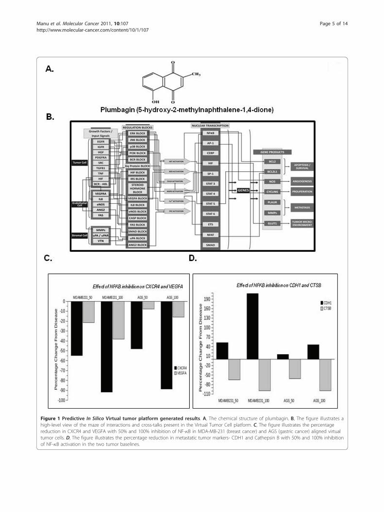
Figure 1 Predictive In Silico Virtual tumor platform generated results. A, The chemical structure of plumbagin. B, The figure illustrates ahigh-level view of the maze of interactions and cross-talks present in the Virtual Tumor Cell platform. C, The figure illustrates the percentagereduction in CXCR4 and VEGFA with 50% and 100% inhibition of NF-�B in MDA-MB-231 (breast cancer) and AGS (gastric cancer) aligned virtualtumor cells. D, The figure illustrates the percentage reduction in metastatic tumor markers- CDH1 and Cathepsin B with 50% and 100% inhibitionof NF-�B activation in the two tumor baselines.
Manu et al. Molecular Cancer 2011, 10:107http://www.molecular-cancer.com/content/10/1/107
Page 5 of 14
Statistical analysisThe experiments were carried out in triplicates andrepeated at least twice. Data are expressed as the mean± S.E.M. In all figures, vertical error bars denote the S.E.M. The significance of differences between groups wasevaluated by Student’s t-test and one way analysis ofvariance, (ANOVA). A p value of less than 0.05 wasconsidered statistically significant.
Results and DiscussionThe present study was designed to investigate the effectof plumbagin (with structure shown in Figure 1A) onCXCR4 expression and also on cellular migration andinvasion in various tumor cells.
Predictive analysis of inhibition of NF-�B activation intumor cellsTo test whether inhibition of NF-�B activation is pri-marily causing the plumbagin-mediated impact on meta-static markers in tumor cells, we tested this hypothesisin the virtual tumor cells aligned to a breast cancer cellline MDA-MB-231 and a gastric cell line cell line AGS(Figure 1B). The inhibition of NF-�B activation by 50%and 100% leads to marked decrease in the expression ofCXCR4 and slight decrease in VEGF expression in boththese cell line profiles, as depicted in Figure 1C. Thesepredictive results corroborate with the experimentaldata and support the hypothesis that plumbagin effectson metastatic phenotypes in tumor cells are mainlythrough inhibition of NF-�B activation. Additionalmetastatic markers such as Cathepsin B and E-Cadherinwere also monitored. A predictive increase in CDH1and a decrease in Cathepsin B (Figure 1D) also indicatethat plumbagin would have a good impact on inhibitingmetastasis phenotype in tumor cells.
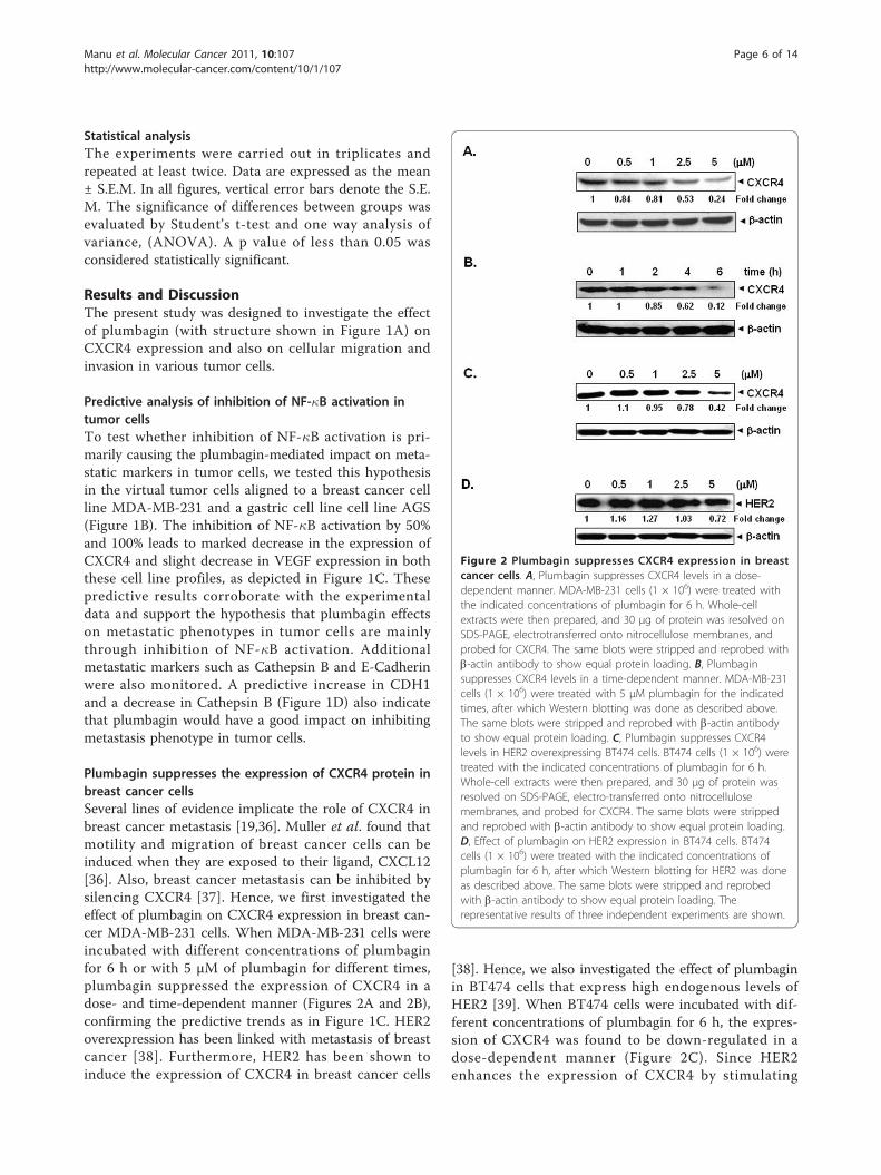
Plumbagin suppresses the expression of CXCR4 protein inbreast cancer cellsSeveral lines of evidence implicate the role of CXCR4 inbreast cancer metastasis [19,36]. Muller et al. found thatmotility and migration of breast cancer cells can beinduced when they are exposed to their ligand, CXCL12[36]. Also, breast cancer metastasis can be inhibited bysilencing CXCR4 [37]. Hence, we first investigated theeffect of plumbagin on CXCR4 expression in breast can-cer MDA-MB-231 cells. When MDA-MB-231 cells wereincubated with different concentrations of plumbaginfor 6 h or with 5 μM of plumbagin for different times,plumbagin suppressed the expression of CXCR4 in adose- and time-dependent manner (Figures 2A and 2B),confirming the predictive trends as in Figure 1C. HER2overexpression has been linked with metastasis of breastcancer [38]. Furthermore, HER2 has been shown toinduce the expression of CXCR4 in breast cancer cells
[38]. Hence, we also investigated the effect of plumbaginin BT474 cells that express high endogenous levels ofHER2 [39]. When BT474 cells were incubated with dif-ferent concentrations of plumbagin for 6 h, the expres-sion of CXCR4 was found to be down-regulated in adose-dependent manner (Figure 2C). Since HER2enhances the expression of CXCR4 by stimulating
Figure 2 Plumbagin suppresses CXCR4 expression in breastcancer cells. A, Plumbagin suppresses CXCR4 levels in a dose-dependent manner. MDA-MB-231 cells (1 × 10⁶) were treated withthe indicated concentrations of plumbagin for 6 h. Whole-cellextracts were then prepared, and 30 μg of protein was resolved onSDS-PAGE, electrotransferred onto nitrocellulose membranes, andprobed for CXCR4. The same blots were stripped and reprobed withb-actin antibody to show equal protein loading. B, Plumbaginsuppresses CXCR4 levels in a time-dependent manner. MDA-MB-231cells (1 × 10⁶) were treated with 5 μM plumbagin for the indicatedtimes, after which Western blotting was done as described above.The same blots were stripped and reprobed with b-actin antibodyto show equal protein loading. C, Plumbagin suppresses CXCR4levels in HER2 overexpressing BT474 cells. BT474 cells (1 × 10⁶) weretreated with the indicated concentrations of plumbagin for 6 h.Whole-cell extracts were then prepared, and 30 μg of protein wasresolved on SDS-PAGE, electro-transferred onto nitrocellulosemembranes, and probed for CXCR4. The same blots were strippedand reprobed with b-actin antibody to show equal protein loading.D, Effect of plumbagin on HER2 expression in BT474 cells. BT474cells (1 × 10⁶) were treated with the indicated concentrations ofplumbagin for 6 h, after which Western blotting for HER2 was doneas described above. The same blots were stripped and reprobedwith b-actin antibody to show equal protein loading. Therepresentative results of three independent experiments are shown.
Manu et al. Molecular Cancer 2011, 10:107http://www.molecular-cancer.com/content/10/1/107
Page 6 of 14
CXCR4 translation and attenuating CXCR4 degradation[40], we also examined whether plumbagin downregu-lates CXCR4 expression through regulation of HER2expression. For this, HER2-overexpressing BT474 cellswere incubated with different concentrations of plumba-gin for 6 h and then examined for HER2 expression byWestern blot analysis using specific antibodies. Wefound that HER2 expression was minimally affectedafter plumbagin treatment (Figure 2D), thus suggestingthat downregulation of CXCR4 expression by plumbaginis not due to modulation of HER2 expression.
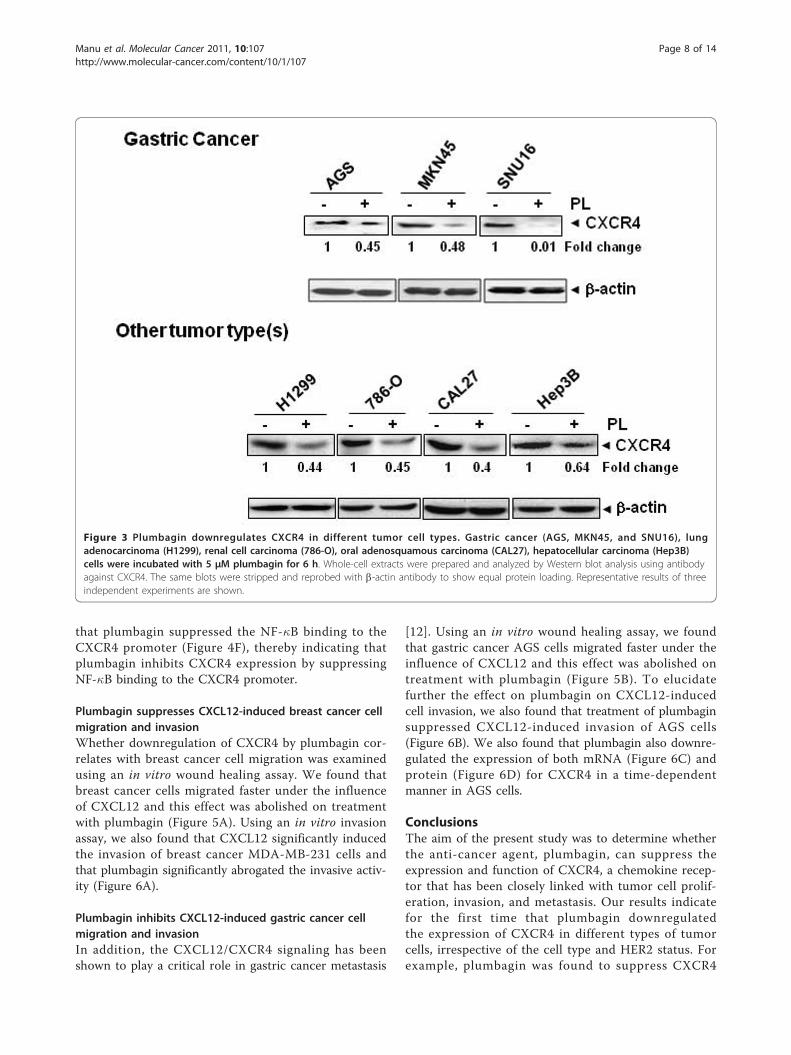
Plumbagin downregulates CXCR4 in differenttumor cell typesUp to this point, all of the afore-mentioned studies werecarried out with breast cancer cell lines. However,CXCR4 is known to be overexpressed in a wide varietyof tumor cells [41]. Hence, we carried out the sameexperiment to find out whether plumbagin downregu-lates expression of CXCR4 in gastric (AGS, MKN45,and SNU16) cancer cell lines, which has never beeninvestigated before. Cells were treated with 5 μM plum-bagin for 6 h before assessing the resultant effect onCXCR4 expression. Figure 3 clearly demonstrates thatplumbagin substantially downregulated CXCR4 expres-sion in all three gastric cancer cell lines, also confirmingthe predictive results from the virtual AGS cell line(Figure 1C). Upon further extension of studying theeffect of plumbagin on CXCR4 expression in lung ade-nocarcinoma (H1299), renal cell carcinoma (786-O),oral adenosquamous carcinoma (CAL27), hepatocellularcarcinoma (Hep3B) tumor cell lines, we also found thatplumbagin dramatically downregulated the CXCR4expression in all these cell lines (Figure 3). This showedconvincingly that CXCR4 downregulation by plumbaginis not cell type-specific.
Downregulation of CXCR4 expression by plumbagin isnot mediated through its degradationBecause plumbagin could downregulate CXCR4 expres-sion by enhancing its degradation, and CXCR4 has beenshown to undergo ubiquitination at its lysine residuefollowed by degradation [42,43], we next investigatedthe possibility that plumbagin may enhance the rate ofCXCR4 degradation via the activation of proteasomes.To determine this, we examined the ability of lactacys-tin, a proteasome inhibitor, to block plumbagin -induceddegradation of CXCR4. MDA-MB-231 cells were pre-treated with lactacystin for 1 h before being exposed toplumbagin. As shown in Figure 4A, lactacystin had noeffect on plumbagin-induced degradation of CXCR4,suggesting that this is an unlikely basis for the suppres-sion of CXCR4 expression by plumbagin.
We also examined the ability of chloroquine, a lysoso-mal inhibitor, to block plumbagin-induced degradationof CXCR4, as CXCR4 has been shown to undergoligand-dependent lysosomal degradation [43]. The cellswere pretreated with chloroquine for 1 h before expo-sure to plumbagin. Our results showed that chloroquineat 200 μM only slightly prevented the degradation ofCXCR4 (Figure 4B), suggesting that this was arguablynot the primary pathway for suppression of expressionof CXCR4.
Downregulation of CXCR4 by plumbagin occurs at thetranscriptional levelSince plumbagin did not downregulate CXCR4 expres-sion by enhancing its degradation, we investigatedwhether suppression occurs at the transcriptional levelinstead. Cells were treated with plumbagin for differenttimes and then the mRNA was extracted for analysis byreal-time PCR. As shown in Figure 4C, plumbagininduced the downregulation of CXCR4 mRNA in atime-dependent manner.
Plumbagin suppresses constitutive activation of NF-�B inMDA-MB-231 cellsThe promoter of CXCR4 is known to contain severalNF-�B binding sites [44]. In addition, HER2 oncogenehas been shown to activate NF-�B in breast cancer cells[45]. Thus it is possible that plumbagin manifests itseffect on CXCR4 by suppressing NF-�B activation. Weused a DNA-binding assay to assess the effect of plum-bagin on constitutive NF-�B activation in MDA-MB-231 cells, and found that the treatment with plumbagincan suppress constitutive NF-�B activation in a dose-dependent manner (Figure 4D). This result suggests thatplumbagin may downregulate CXCR4 expressionthrough inhibition of NF-�B activation. However, DNAbinding alone is not always associated with NF-�B-dependent gene transcription [46], suggesting that addi-tional regulatory steps are involved. Subsequent resultsalso indicated that plumbagin inhibited NF-�B reporteractivity in a dose-dependent manner in MDA-MB-231cells (Figure 4E). This hypothesis was also tested simul-taneously through the virtual system and confirms thatinhibition of NF-�B plays a pivotal role in plumbagin-mediated reduction of CXCR4 (Figure 1C) and othermetastatic markers (Figure 1D).
Plumbagin inhibits binding of NF-�B to the CXCR4promoterWhether the downregulation of CXCR4 by plumbagin inMDA-MB-231 cells was due to suppression of NF-�Bactivation in vivo was examined by a ChIP assay target-ing NF-�B binding in the CXCR4 promoter. We found
Manu et al. Molecular Cancer 2011, 10:107http://www.molecular-cancer.com/content/10/1/107
Page 7 of 14
that plumbagin suppressed the NF-�B binding to theCXCR4 promoter (Figure 4F), thereby indicating thatplumbagin inhibits CXCR4 expression by suppressingNF-�B binding to the CXCR4 promoter.
Plumbagin suppresses CXCL12-induced breast cancer cellmigration and invasionWhether downregulation of CXCR4 by plumbagin cor-relates with breast cancer cell migration was examinedusing an in vitro wound healing assay. We found thatbreast cancer cells migrated faster under the influenceof CXCL12 and this effect was abolished on treatmentwith plumbagin (Figure 5A). Using an in vitro invasionassay, we also found that CXCL12 significantly inducedthe invasion of breast cancer MDA-MB-231 cells andthat plumbagin significantly abrogated the invasive activ-ity (Figure 6A).
Plumbagin inhibits CXCL12-induced gastric cancer cellmigration and invasionIn addition, the CXCL12/CXCR4 signaling has beenshown to play a critical role in gastric cancer metastasis
[12]. Using an in vitro wound healing assay, we foundthat gastric cancer AGS cells migrated faster under theinfluence of CXCL12 and this effect was abolished ontreatment with plumbagin (Figure 5B). To elucidatefurther the effect on plumbagin on CXCL12-inducedcell invasion, we also found that treatment of plumbaginsuppressed CXCL12-induced invasion of AGS cells(Figure 6B). We also found that plumbagin also downre-gulated the expression of both mRNA (Figure 6C) andprotein (Figure 6D) for CXCR4 in a time-dependentmanner in AGS cells.
ConclusionsThe aim of the present study was to determine whetherthe anti-cancer agent, plumbagin, can suppress theexpression and function of CXCR4, a chemokine recep-tor that has been closely linked with tumor cell prolif-eration, invasion, and metastasis. Our results indicatefor the first time that plumbagin downregulatedthe expression of CXCR4 in different types of tumorcells, irrespective of the cell type and HER2 status. Forexample, plumbagin was found to suppress CXCR4
Figure 3 Plumbagin downregulates CXCR4 in different tumor cell types. Gastric cancer (AGS, MKN45, and SNU16), lungadenocarcinoma (H1299), renal cell carcinoma (786-O), oral adenosquamous carcinoma (CAL27), hepatocellular carcinoma (Hep3B)cells were incubated with 5 μM plumbagin for 6 h. Whole-cell extracts were prepared and analyzed by Western blot analysis using antibodyagainst CXCR4. The same blots were stripped and reprobed with b-actin antibody to show equal protein loading. Representative results of threeindependent experiments are shown.
Manu et al. Molecular Cancer 2011, 10:107http://www.molecular-cancer.com/content/10/1/107
Page 8 of 14
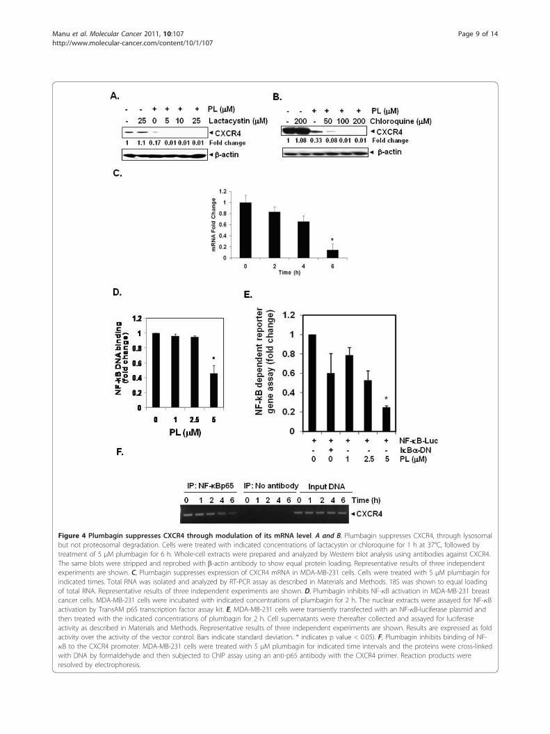
Figure 4 Plumbagin suppresses CXCR4 through modulation of its mRNA level. A and B, Plumbagin suppresses CXCR4, through lysosomalbut not proteosomal degradation. Cells were treated with indicated concentrations of lactacystin or chloroquine for 1 h at 37°C, followed bytreatment of 5 μM plumbagin for 6 h. Whole-cell extracts were prepared and analyzed by Western blot analysis using antibodies against CXCR4.The same blots were stripped and reprobed with b-actin antibody to show equal protein loading. Representative results of three independentexperiments are shown. C, Plumbagin suppresses expression of CXCR4 mRNA in MDA-MB-231 cells. Cells were treated with 5 μM plumbagin forindicated times. Total RNA was isolated and analyzed by RT-PCR assay as described in Materials and Methods. 18S was shown to equal loadingof total RNA. Representative results of three independent experiments are shown. D, Plumbagin inhibits NF-�B activation in MDA-MB-231 breastcancer cells. MDA-MB-231 cells were incubated with indicated concentrations of plumbagin for 2 h. The nuclear extracts were assayed for NF-�Bactivation by TransAM p65 transcription factor assay kit. E, MDA-MB-231 cells were transiently transfected with an NF-�B-luciferase plasmid andthen treated with the indicated concentrations of plumbagin for 2 h. Cell supernatants were thereafter collected and assayed for luciferaseactivity as described in Materials and Methods. Representative results of three independent experiments are shown. Results are expressed as foldactivity over the activity of the vector control. Bars indicate standard deviation. * indicates p value < 0.05). F, Plumbagin inhibits binding of NF-�B to the CXCR4 promoter. MDA-MB-231 cells were treated with 5 μM plumbagin for indicated time intervals and the proteins were cross-linkedwith DNA by formaldehyde and then subjected to ChIP assay using an anti-p65 antibody with the CXCR4 primer. Reaction products wereresolved by electrophoresis.
Manu et al. Molecular Cancer 2011, 10:107http://www.molecular-cancer.com/content/10/1/107
Page 9 of 14
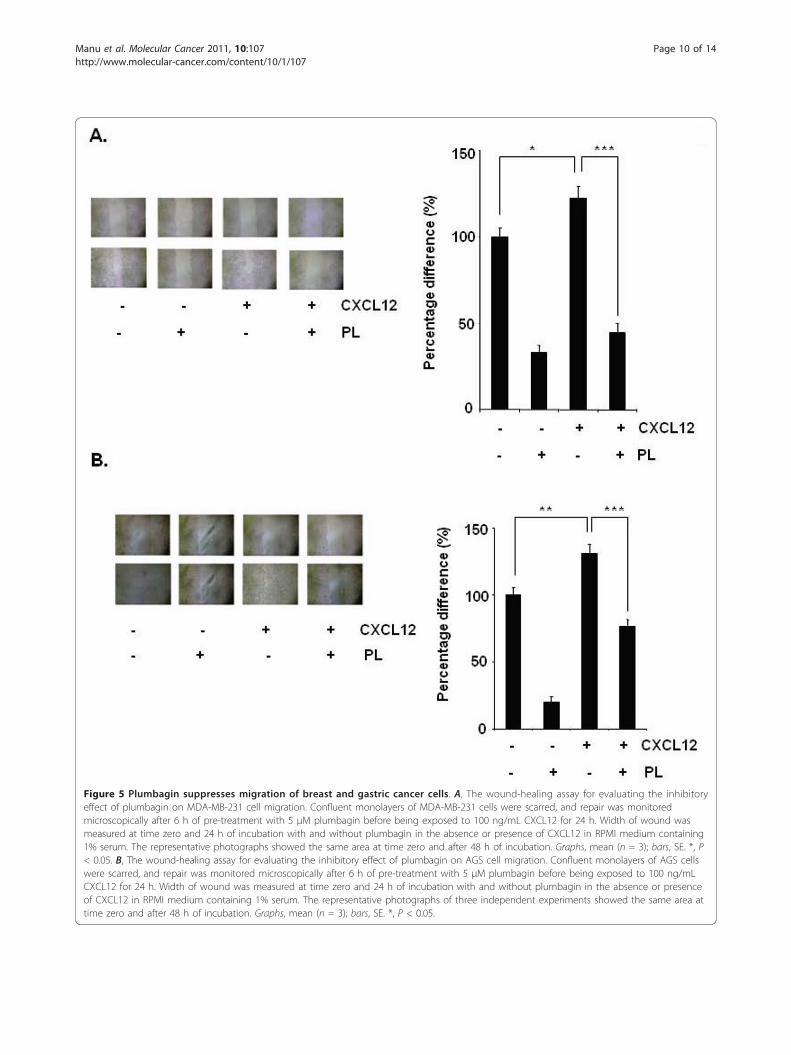
Figure 5 Plumbagin suppresses migration of breast and gastric cancer cells. A, The wound-healing assay for evaluating the inhibitoryeffect of plumbagin on MDA-MB-231 cell migration. Confluent monolayers of MDA-MB-231 cells were scarred, and repair was monitoredmicroscopically after 6 h of pre-treatment with 5 μM plumbagin before being exposed to 100 ng/mL CXCL12 for 24 h. Width of wound wasmeasured at time zero and 24 h of incubation with and without plumbagin in the absence or presence of CXCL12 in RPMI medium containing1% serum. The representative photographs showed the same area at time zero and after 48 h of incubation. Graphs, mean (n = 3); bars, SE. *, P< 0.05. B, The wound-healing assay for evaluating the inhibitory effect of plumbagin on AGS cell migration. Confluent monolayers of AGS cellswere scarred, and repair was monitored microscopically after 6 h of pre-treatment with 5 μM plumbagin before being exposed to 100 ng/mLCXCL12 for 24 h. Width of wound was measured at time zero and 24 h of incubation with and without plumbagin in the absence or presenceof CXCL12 in RPMI medium containing 1% serum. The representative photographs of three independent experiments showed the same area attime zero and after 48 h of incubation. Graphs, mean (n = 3); bars, SE. *, P < 0.05.
Manu et al. Molecular Cancer 2011, 10:107http://www.molecular-cancer.com/content/10/1/107
Page 10 of 14
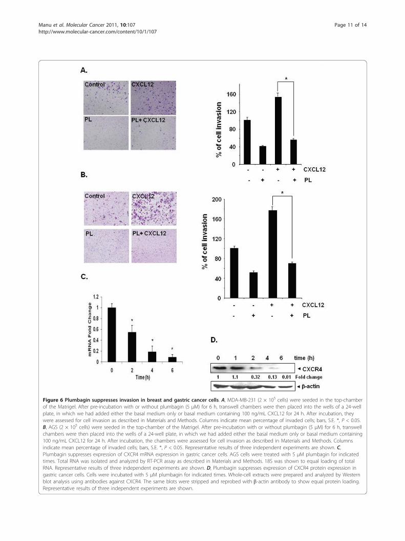
Figure 6 Plumbagin suppresses invasion in breast and gastric cancer cells. A, MDA-MB-231 (2 × 10⁵ cells) were seeded in the top-chamberof the Matrigel. After pre-incubation with or without plumbagin (5 μM) for 6 h, transwell chambers were then placed into the wells of a 24-wellplate, in which we had added either the basal medium only or basal medium containing 100 ng/mL CXCL12 for 24 h. After incubation, theywere assessed for cell invasion as described in Materials and Methods. Columns indicate mean percentage of invaded cells; bars, S.E. *, P < 0.05.B, AGS (2 × 10⁵ cells) were seeded in the top-chamber of the Matrigel. After pre-incubation with or without plumbagin (5 μM) for 6 h, transwellchambers were then placed into the wells of a 24-well plate, in which we had added either the basal medium only or basal medium containing100 ng/mL CXCL12 for 24 h. After incubation, the chambers were assessed for cell invasion as described in Materials and Methods. Columnsindicate mean percentage of invaded cells; bars, S.E. *, P < 0.05. Representative results of three independent experiments are shown. C,Plumbagin suppresses expression of CXCR4 mRNA expression in gastric cancer cells. AGS cells were treated with 5 μM plumbagin for indicatedtimes. Total RNA was isolated and analyzed by RT-PCR assay as described in Materials and Methods. 18S was shown to equal loading of totalRNA. Representative results of three independent experiments are shown. D, Plumbagin suppresses expression of CXCR4 protein expression ingastric cancer cells. Cells were incubated with 5 μM plumbagin for indicated times. Whole-cell extracts were prepared and analyzed by Westernblot analysis using antibodies against CXCR4. The same blots were stripped and reprobed with b-actin antibody to show equal protein loading.Representative results of three independent experiments are shown.
Manu et al. Molecular Cancer 2011, 10:107http://www.molecular-cancer.com/content/10/1/107
Page 11 of 14
expression in HER2 overexpressing BT474 breast cancercells. Our results showed that downregulation ofCXCR4 did not occur through proteolytic degradationof the receptor but rather through downregulation ofthe transcript. Furthermore, suppression of receptorexpression led to downregulation of migration and inva-sion induced by the ligand CXCL12 in both breast andgastric cancer cells.The CXCR4 chemokine receptor has been found to be
overexpressed in different tumors, including breast can-cer, gastric cancer, ovarian cancer, glioma, pancreaticcancer, prostate cancer, acute myeloid leukemia, chroniclymphoblastic leukemia (CLL), B-CLL, melanoma,cervical cancer, colon carcinoma, rhabdomyosarcoma,astrocytoma, small-cell lung carcinoma, renal cancer,and non-Hodgkin’s lymphoma, as compared to normalcells which show little or no CXCR4 expression[13,22,40,47-49]. Although it is still unclear what leads tothe overexpression of CXCR4 in tumor cells, studiespoint to genetic and microenvironmental factors [50].PAX3- and PAX7-FKHR gene fusion [51], mutations inthe von Hippel Lindau tumor suppressor gene [52],hypoxia in the tumor microenvironment [53], NF-�B[44], and inflammatory cytokines such as vascularendothelial growth factor [54] and tumor necrosis factoralpha [50], have all been implicated in CXCR4 overex-pression. Recently, the epidermal growth factor receptor,c-erbB2, and its encoding gene, HER2/neu, have alsobeen implicated in the positive regulation of CXCR4expression at the post-transcriptional level [55,56]. Giventhat CXCR4 has been linked with the metastasis of var-ious cancers and CXCR4 expression has been correlatedwith poor prognosis and poor overall patient survival[57], CXCR4 appears an ideal therapeutic target for theinvestigation of novel therapeutic interventions for theprevention of metastatic cancer.Our results clearly indicate that plumbagin suppressed
CXCR4 expression in both HER2-lacking and -overex-pressing breast cancer cells, but had minimal effect onHER2 expression in BT474 breast cancer cells. Our dataalso showed that plumbagin suppressed CXCR4 expres-sion on various tumor cell lines including gastric cancer,lung adenocarcinoma, renal cell carcinoma, oral andhepatocellular carcinoma, thereby indicating that theeffect of plumbagin on CXCR4 is not limited to a singletumor cell type. The ligand-dependent downregulationof the CXCR4 receptor by lysosomal degradation is welldocumented [40]. Recent reports suggest that degrada-tion involves atrophin-interacting protein (AIP)-4mediated ubiquitination and degradation [43]. However,our data indicate that plumbagin does not downregulatethe CXCR4 through this mechanism. As such, withdownregulation of CXCR4 by plumbagin arguably notoccurring at the post-translational level, we postulated
that the inhibition of CXCR4 expression by this quininecould occur at the transcriptional level. Indeed, wefound that plumbagin downregulated the expression ofCXCR4 mRNA in breast and gastric cancer cells.Plumbagin has been previously reported to downregu-
late NF-�B activation in various tumor cells [23]. Inter-estingly, the NF-�B binding site has also been identifiedin the proximal region of the CXCR4 promoter and pos-tulated to play a role in CXCR4 expression in humanbreast cancer cells [44]. Therefore, it is possible thatdownregulation of CXCR4 by plumbagin occurs via thesuppression of NF-�B. Indeed, we found that inhibitionof constitutive NF-�B activation by plumbagin leads todownregulation of CXCR4 in MDA-MB-231 cells. TheNF-�B mediated regulation of CXCR4 in the tumorcells was also tested in the virtual system and the pre-dictions corroborated with the experimental results.Whether mechanism(s) other than suppression of NF-�B activation are involved in downregulation of CXCR4by plumbagin, cannot currently be confirmed or ruledout. Furthermore, besides CXCR4, the activation of NF-�B also induces the expression of various moleculesincluding cyclooxygenase-2, matrix metallopeptidase-9,and adhesion molecules such as intracellular adhesionmolecule 1, vascular cell adhesion molecule 1, andendothelial-leukocyte adhesion molecule 1, all of whichhave been linked with cancer cell migration, invasion,and metastasis [58]. Because plumbagin can inhibit bothDNA binding ability and transcriptional activation ofNF-�B, as shown in this study, it is possible that plum-bagin can suppress the expression of other NF-�Bregulated molecules as well in breast cancer cells.We further investigated the effect of plumbagin onCXCL12-induced migration and invasion of both breastand gastric cancer cells. We found that preincubation ofcells with plumbagin completely blocked CXCL12-induced migration and invasion of both breast and gas-tric cancer cells.Plumbagin has been shown to inhibit different aspects
of tumor initiation and progression, including prolifera-tion, invasion, angiogenesis, and metastasis in varioustumor cell lines and animal models [23-30]. Our datashows for the first time that plumbagin downregulatesexpression of CXCR4 in a variety of tumor cells, a keyreceptor involved in the cross-talk between tumor cellsand its microenvironment, and thus, that some of theanti-tumor effects of plumbagin are possibly mediatedthrough CXCR4 regulation. Further in vivo studies arebeing planned to demonstrate the relevance of theseobservations to cancer treatment.
List of AbbreviationsCXCR4: chemokine receptor-4; ChIP: Chromatin immunoprecipitation; FBS:fetal bovine serum.
Manu et al. Molecular Cancer 2011, 10:107http://www.molecular-cancer.com/content/10/1/107
Page 12 of 14
AcknowledgementsThis work was supported by grants from NUS Academic Research Fund[Grants R-184-000-170-112 and R-184-000-177-112], National MedicalResearch Council of Singapore [Grant R-184-000-201-275], and Nationalkidney Foundation [Grant R-184-000-196-592] to GS; National MedicalResearch Council of Singapore [Grant R-713-000-124-213] and CancerScience Institute of Singapore, Experimental Therapeutics I Program [Grant R-713-001-011-271] to APK; Biomedical Research Council of Singapore [GrantR185-000-163-305] to LL. SNS was supported by Boycast Fellowship fromDepartment of Science and Technology, Government of India.
Author details1Department of Pharmacology, Yong Loo Lin School of Medicine, NationalUniversity of Singapore, Singapore 117597. 2Cancer Science Institute ofSingapore, National University of Singapore, 28 Medical Drive, Singapore117456. 3Cellworks Group Inc., Saratoga, California, 95070; USA; CellworksResearch India Pvt. Ltd, Bangalore, 560066, India. 4Department of Physiology,Yong Loo Lin School of Medicine, National University of Singapore,Singapore 117597. 5Immunology Program, National University of Singapore,Singapore 117597. 6Department of Pathology, National University ofSingapore, Singapore 117597. 7College of Oriental Medicine, Kyung HeeUniversity, Seoul 130-701, Republic of Korea. 8School of Anatomy andHuman Biology, The University of Western Australia, Crawley, Perth, WesternAustralia 6009.
Authors’ contributionsKAM, MKS, PR, FL, LR, HSH, RK, SNS, SK, BR, PB performed all theexperiments. SV, ESK, LHKL, KSA, APK, and GS analyzed the data and wrotethe manuscript. All the authors have read and approved the manuscript.
Competing interestsThe authors declare that they have no competing interests.
Received: 24 November 2010 Accepted: 1 September 2011Published: 1 September 2011
References1. Nguyen DX, Bos PD, Massague J: Metastasis: from dissemination to organ-
specific colonization. Nat Rev Cancer 2009, 9:274-284.2. Lazennec G, Richmond A: Chemokines and chemokine receptors: new
insights into cancer-related inflammation. Trends Mol Med 2010,16:133-144.
3. Teicher BA, Fricker SP: CXCL12 (SDF-1)/CXCR4 pathway in cancer. ClinCancer Res 2010, 16:2927-2931.
4. Zlotnik A: Chemokines and cancer. Int J Cancer 2006, 119:2026-2029.5. Nguyen DX, Massague J: Genetic determinants of cancer metastasis. Nat
Rev Genet 2007, 8:341-352.6. Richmond A: Nf-kappa B, chemokine gene transcription and tumour
growth. Nat Rev Immunol 2002, 2:664-674.7. Balkwill F: Cancer and the chemokine network. Nat Rev Cancer 2004,
4:540-550.8. Kruizinga RC, Bestebroer J, Berghuis P, de Haas CJ, Links TP, de Vries EG,
Walenkamp AM: Role of chemokines and their receptors in cancer. CurrPharm Des 2009, 15:3396-3416.
9. Kucia M, Reca R, Miekus K, Wanzeck J, Wojakowski W, Janowska-Wieczorek A, Ratajczak J, Ratajczak MZ: Trafficking of normal stem cellsand metastasis of cancer stem cells involve similar mechanisms: pivotalrole of the SDF-1-CXCR4 axis. Stem Cells 2005, 23:879-894.
10. Andre F, Cabioglu N, Assi H, Sabourin JC, Delaloge S, Sahin A, Broglio K,Spano JP, Combadiere C, Bucana C, et al: Expression of chemokinereceptors predicts the site of metastatic relapse in patients with axillarynode positive primary breast cancer. Ann Oncol 2006, 17:945-951.
11. Zeelenberg IS, Ruuls-Van Stalle L, Roos E: The chemokine receptor CXCR4is required for outgrowth of colon carcinoma micrometastases. CancerRes 2003, 63:3833-3839.
12. Lee HJ, Kim SW, Kim HY, Li S, Yun HJ, Song KS, Kim S, Jo DY: Chemokinereceptor CXCR4 expression, function, and clinical implications in gastriccancer. Int J Oncol 2009, 34:473-480.
13. Porcile C, Bajetto A, Barbero S, Pirani P, Schettini G: CXCR4 activationinduces epidermal growth factor receptor transactivation in an ovariancancer cell line. Ann N Y Acad Sci 2004, 1030:162-169.
14. Marchesi F, Monti P, Leone BE, Zerbi A, Vecchi A, Piemonti L, Mantovani A,Allavena P: Increased survival, proliferation, and migration in metastatichuman pancreatic tumor cells expressing functional CXCR4. Cancer Res2004, 64:8420-8427.
15. Taichman RS, Cooper C, Keller ET, Pienta KJ, Taichman NS, McCauley LK:Use of the stromal cell-derived factor-1/CXCR4 pathway in prostatecancer metastasis to bone. Cancer Res 2002, 62:1832-1837.
16. Phillips RJ, Burdick MD, Lutz M, Belperio JA, Keane MP, Strieter RM: Thestromal derived factor-1/CXCL12-CXC chemokine receptor 4 biologicalaxis in non-small cell lung cancer metastases. Am J Respir Crit Care Med2003, 167:1676-1686.
17. Cardones AR, Murakami T, Hwang ST: CXCR4 enhances adhesion of B16tumor cells to endothelial cells in vitro and in vivo via beta(1) integrin.Cancer Res 2003, 63:6751-6757.
18. Sehgal A, Keener C, Boynton AL, Warrick J, Murphy GP: CXCR-4, achemokine receptor, is overexpressed in and required for proliferationof glioblastoma tumor cells. J Surg Oncol 1998, 69:99-104.
19. Epstein RJ: The CXCL12-CXCR4 chemotactic pathway as a target ofadjuvant breast cancer therapies. Nat Rev Cancer 2004, 4:901-909.
20. Holm NT, Byrnes K, Li BD, Turnage RH, Abreo F, Mathis JM, Chu QD:Elevated levels of chemokine receptor CXCR4 in HER-2 negative breastcancer specimens predict recurrence. J Surg Res 2007, 141:53-59.
21. Kim J, Mori T, Chen SL, Amersi FF, Martinez SR, Kuo C, Turner RR, Ye X,Bilchik AJ, Morton DL, Hoon DS: Chemokine receptor CXCR4 expression inpatients with melanoma and colorectal cancer liver metastases and theassociation with disease outcome. Ann Surg 2006, 244:113-120.
22. Proudfoot AE: Chemokine receptors: multifaceted therapeutic targets.Nat Rev Immunol 2002, 2:106-115.
23. Sandur SK, Ichikawa H, Sethi G, Ahn KS, Aggarwal BB: Plumbagin (5-hydroxy-2-methyl-1,4-naphthoquinone) suppresses NF-kappaB activationand NF-kappaB-regulated gene products through modulation of p65and IkappaBalpha kinase activation, leading to potentiation of apoptosisinduced by cytokine and chemotherapeutic agents. J Biol Chem 2006,281:17023-17033.
24. Kuo PL, Hsu YL, Cho CY: Plumbagin induces G2-M arrest and autophagyby inhibiting the AKT/mammalian target of rapamycin pathway inbreast cancer cells. Mol Cancer Ther 2006, 5:3209-3221.
25. Nair S, Nair RR, Srinivas P, Srinivas G, Pillai MR: Radiosensitizing effects ofplumbagin in cervical cancer cells is through modulation of apoptoticpathway. Mol Carcinog 2008, 47:22-33.
26. Aziz MH, Dreckschmidt NE, Verma AK: Plumbagin, a medicinal plant-derived naphthoquinone, is a novel inhibitor of the growth andinvasion of hormone-refractory prostate cancer. Cancer Res 2008,68:9024-9032.
27. Gomathinayagam R, Sowmyalakshmi S, Mardhatillah F, Kumar R,Akbarsha MA, Damodaran C: Anticancer mechanism of plumbagin, anatural compound, on non-small cell lung cancer cells. Anticancer Res2008, 28:785-792.
28. Powolny AA, Singh SV: Plumbagin-induced apoptosis in humanprostate cancer cells is associated with modulation of cellular redoxstatus and generation of reactive oxygen species. Pharm Res 2008,25:2171-2180.
29. Chen CA, Chang HH, Kao CY, Tsai TH, Chen YJ: Plumbagin, isolated fromPlumbago zeylanica, induces cell death through apoptosis in humanpancreatic cancer cells. Pancreatology 2009, 9:797-809.
30. Sandur SK, Pandey MK, Sung B, Aggarwal BB: 5-hydroxy-2-methyl-1,4-naphthoquinone, a vitamin K3 analogue, suppresses STAT3 activationpathway through induction of protein tyrosine phosphatase, SHP-1:potential role in chemosensitization. Mol Cancer Res 2010, 8:107-118.
31. Vali S, Pallavi R, Kapoor S, Tatu U: Virtual prototyping study showsincreased ATPase activity of Hsp90 to be the key determinant of cancerphenotype. Syst Synth Biol 2009.
32. Roy KR, Reddy GV, Maitreyi L, Agarwal S, Achari C, Vali S, Reddanna P:Celecoxib inhibits MDR1 expression through COX-2-dependentmechanism in human hepatocellular carcinoma (HepG2) cell line. CancerChemother Pharmacol 65:903-911.
33. Equils O, Nambiar P, Hobel CJ, Smith R, Simmons CF, Vali S: A computersimulation of progesterone and Cox2 inhibitor treatment for pretermlabor. PLoS One 5:e8502.
34. Saccani S, Pantano S, Natoli G: p38-Dependent marking of inflammatorygenes for increased NF-kappa B recruitment. Nat Immunol 2002, 3:69-75.
Manu et al. Molecular Cancer 2011, 10:107http://www.molecular-cancer.com/content/10/1/107
Page 13 of 14
35. Rajendran P, Ong TH, Chen L, Li F, Shanmugam MK, Vali S, Abbasi T,Kapoor S, Sharma A, Kumar AP, et al: Suppression of signal transducerand activator of transcription 3 activation by butein inhibits growth ofhuman hepatocellular carcinoma in vivo. Clin Cancer Res 2011,17:1425-1439.
36. Muller A, Homey B, Soto H, Ge N, Catron D, Buchanan ME, McClanahan T,Murphy E, Yuan W, Wagner SN, et al: Involvement of chemokine receptorsin breast cancer metastasis. Nature 2001, 410:50-56.
37. Smith MC, Luker KE, Garbow JR, Prior JL, Jackson E, Piwnica-Worms D,Luker GD: CXCR4 regulates growth of both primary and metastaticbreast cancer. Cancer Res 2004, 64:8604-8612.
38. Li YM, Pan Y, Wei Y, Cheng X, Zhou BP, Tan M, Zhou X, Xia W,Hortobagyi GN, Yu D, Hung MC: Upregulation of CXCR4 is essential forHER2-mediated tumor metastasis. Cancer Cell 2004, 6:459-469.
39. Chua AW, Hay HS, Rajendran P, Shamugam MK, Li F, Bist P, Koay ES,Klim LH, Kumar AP, Sethi G: Butein Downregulates Chemokine ReceptorCXCR4 Expression and Function through suppression of NF-kappaBActivation in Breast and Pancreatic Tumor cells. Biochem Pharmacol 2010.
40. Fernandis AZ, Prasad A, Band H, Klosel R, Ganju RK: Regulation of CXCR4-mediated chemotaxis and chemoinvasion of breast cancer cells.Oncogene 2004, 23:157-167.
41. Kakinuma T, Hwang ST: Chemokines, chemokine receptors, and cancermetastasis. J Leukoc Biol 2006, 79:639-651.
42. Marchese A, Benovic JL: Agonist-promoted ubiquitination of the Gprotein-coupled receptor CXCR4 mediates lysosomal sorting. J Biol Chem2001, 276:45509-45512.
43. Bhandari D, Trejo J, Benovic JL, Marchese A: Arrestin-2 interacts with theubiquitin-protein isopeptide ligase atrophin-interacting protein 4 andmediates endosomal sorting of the chemokine receptor CXCR4. J BiolChem 2007, 282:36971-36979.
44. Helbig G, Christopherson KW, Bhat-Nakshatri P, Kumar S, Kishimoto H,Miller KD, Broxmeyer HE, Nakshatri H: NF-kappaB promotes breast cancercell migration and metastasis by inducing the expression of thechemokine receptor CXCR4. J Biol Chem 2003, 278:21631-21638.
45. Biswas DK, Iglehart JD: Linkage between EGFR family receptors andnuclear factor kappaB (NF-kappaB) signaling in breast cancer. J CellPhysiol 2006, 209:645-652.
46. Nasuhara Y, Adcock IM, Catley M, Barnes PJ, Newton R: Differential IkappaBkinase activation and IkappaBalpha degradation by interleukin-1betaand tumor necrosis factor-alpha in human U937 monocytic cells.Evidence for additional regulatory steps in kappaB-dependenttranscription. J Biol Chem 1999, 274:19965-19972.
47. Murphy PM: Chemokines and the molecular basis of cancer metastasis.N Engl J Med 2001, 345:833-835.
48. Uchida D, Begum NM, Almofti A, Nakashiro K, Kawamata H, Tateishi Y,Hamakawa H, Yoshida H, Sato M: Possible role of stromal-cell-derivedfactor-1/CXCR4 signaling on lymph node metastasis of oral squamouscell carcinoma. Exp Cell Res 2003, 290:289-302.
49. Gschwind A, Prenzel N, Ullrich A: Lysophosphatidic acid-inducedsquamous cell carcinoma cell proliferation and motility involvesepidermal growth factor receptor signal transactivation. Cancer Res 2002,62:6329-6336.
50. Kulbe H, Hagemann T, Szlosarek PW, Balkwill FR, Wilson JL: Theinflammatory cytokine tumor necrosis factor-alpha regulates chemokinereceptor expression on ovarian cancer cells. Cancer Res 2005,65:10355-10362.
51. Libura J, Drukala J, Majka M, Tomescu O, Navenot JM, Kucia M, Marquez L,Peiper SC, Barr FG, Janowska-Wieczorek A, Ratajczak MZ: CXCR4-SDF-1signaling is active in rhabdomyosarcoma cells and regulates locomotion,chemotaxis, and adhesion. Blood 2002, 100:2597-2606.
52. Staller P, Sulitkova J, Lisztwan J, Moch H, Oakeley EJ, Krek W: Chemokinereceptor CXCR4 downregulated by von Hippel-Lindau tumoursuppressor pVHL. Nature 2003, 425:307-311.
53. Schioppa T, Uranchimeg B, Saccani A, Biswas SK, Doni A, Rapisarda A,Bernasconi S, Saccani S, Nebuloni M, Vago L, et al: Regulation of thechemokine receptor CXCR4 by hypoxia. J Exp Med 2003, 198:1391-1402.
54. Bachelder RE, Wendt MA, Mercurio AM: Vascular endothelial growth factorpromotes breast carcinoma invasion in an autocrine manner byregulating the chemokine receptor CXCR4. Cancer Res 2002, 62:7203-7206.
55. Benovic JL, Marchese A: A new key in breast cancer metastasis. CancerCell 2004, 6:429-430.
56. Arya M, Ahmed H, Silhi N, Williamson M, Patel HR: Clinical importance andtherapeutic implications of the pivotal CXCL12-CXCR4 (chemokineligand-receptor) interaction in cancer cell migration. Tumour Biol 2007,28:123-131.
57. Burger JA, Kipps TJ: CXCR4: a key receptor in the crosstalk betweentumor cells and their microenvironment. Blood 2006, 107:1761-1767.
58. Sethi G, Tergaonkar V: Potential pharmacological control of theNF-kappaB pathway. Trends Pharmacol Sci 2009, 30:313-321.
doi:10.1186/1476-4598-10-107Cite this article as: Manu et al.: Plumbagin inhibits invasion andmigration of breast and gastric cancer cells by downregulating theexpression of chemokine receptor CXCR4. Molecular Cancer 2011 10:107.
Submit your next manuscript to BioMed Centraland take full advantage of:
• Convenient online submission
• Thorough peer review
• No space constraints or color figure charges
• Immediate publication on acceptance
• Inclusion in PubMed, CAS, Scopus and Google Scholar
• Research which is freely available for redistribution
Submit your manuscript at www.biomedcentral.com/submit
Manu et al. Molecular Cancer 2011, 10:107http://www.molecular-cancer.com/content/10/1/107
Page 14 of 14