Embed Size (px)
Citation preview
Contemporary Research Journal of Medical Sciences 2018;2(1 ): 9-13
CONTEMPORARY RESEARCH JOURNAL OF
MEDICAL SCIENCES CASE REPORT Naik A & Gade J Contemp Res J Med Sci 2018 June 2(1 ): 9-13
PROSTHETIC REHABILITATION OF THE PATIENT WITH CUSTOM
MADE OCULAR PROSTHESIS: A CASE REPORT
dr. ashish naik , dr. jaykumar gade, Dr. Aditi Kumbhalwar
ABSTRACT
Patients who have lost ocular structures through orbital evisceration or
orbital enucleation which was necessary as a surgical intervention for a
congenital defect, pathology or an accident need to be rehabilitated
with ocular prosthesis. The disfigurement associated with eye loss can
cause significant physical and emotional disturbance. This article
describes detailed technique of custom ocular prosthesis fabrication
for a patient with enucleation.
Keywords: Custom Ocular Prosthesis, Prosthetic eye, Maxillofacial
Prosthodontics.
Correspondence : Dr. Ashish Naik
Post graduate student,
Department of Prosthodontics,
Swargiya Dadasaheb Kalmegh Smruti Dental College& Hospital , Nagpur
Email [email protected]
NAIK A & GADE J CUSTOM MADE OCULAR PROSTHESIS 10
Contemporary Research Journal of Medical Sciences 2018;2(1 ): 9-13
INTRODUCTION
Physical defects that compromise
appearance or function prevent an individual
from leading a normal life. The loss of eye is a
visible facial defect and often undermines the
patient’s confidence. A congenital anomaly
or pathology may necessitate an orbital
evisceration or an orbital enucleation. The
surgical procedure of evisceration is where
the contents of the globe are removed,
leaving the sclera intact. A more invasive
procedure is enucleation where the entire
eyeball is severed from the muscles and
optic nerve. Exenteration, the most radical,
involves removal of the contents of the orbit.[1]
The rehabilitation of a patient who has
suffered the psychological trauma of an
ocular loss requires a prosthesis that will
provide the optimum cosmetic and functional
results. Patients with evisceration defects or
ocular atrophy can be treated with custom-
made ocular prostheses or modified stock
eyes.[3-5] The shell prosthesis covers the entire
surface of the eye, restoring it to a natural
appearance. The prosthesis is commonly
made of polymethyl methacrylate resin
which is superior to other ocular prosthetic
materials in terms of tissue compatibility,
esthetic capabilities, durability, and color
permanence, adaptability of form, cost, and
availability.[6] In the Indian scenario, patients
may not be able to afford surgical
reconstruction or major cosmetic treatments.
However, the scleral shell prosthesis as
described below gives the patient a much
more cost-effective treatment whilst
achieving satisfactory esthetics.[7,8]
CASE REPORT
A 68 year old male patient reported with the
defect of right eye to the Department of
Prosthodontics, Swargiya Dadasaheb
Kalmegh Smruti Dental College, Nagpur. His
past medical history revealed that he has
been undergone for enucleation of the right
eye to treat septicemia resulting from a
shrapnel injury. Hence, the patient was
seeking artificial eye replacement. On
examination, defect with a shrunken orbit
and intact tissue bed were observed. In
accord to standard procedure; the palpebral
fissure was observed both in open and
closed position to rule out any abnormality.
Evaluation of the muscular control of the
palpebrae and the internal anatomy of the
socket in resting position and full excursive
movement was performed. Mobility of the
posterior wall of the defect was assessed.
Condition of conjunctiva, depth of fornices,
and presence of cul de sac was noted.
The custom ocular prosthesis was advised to
the patient as treatment option. The
procedure and its limitations were explained
to the patient to allay apprehension and elicit
cooperation.
IMPRESSION TECHNIQUE
An ophthalmic socket was anesthetized with
topical anesthetic followed by light
lubrication of same side eyebrow and
eyelashes to make the procedure more
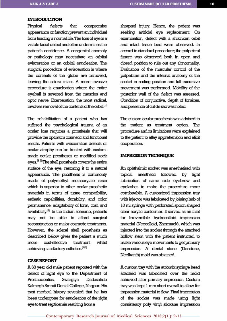
comfortable. A customized impression tray
with injector was fabricated by joining hub of
10 ml syringe with perforated spoon shaped
clear acrylic conformer. It served as an inlet
for Irreversible hydrocolloid impression
material (Neocolloid, Zhermack), which was
injected into the socket through the attached
hollow stem with the patient instructed to
make various eye movements to get primary
impression. A dental stone (Denstone,
Neelkanth) mold was obtained.
A custom tray with the automix syringe head
attached was fabricated over the mold
achieved after primary impression. Custom
tray was kept 1 mm short overall to allow for
impression material to flow. Final impression
of the socket was made using light
consistency poly vinyl siloxane impression
NAIK A & GADE J CUSTOM MADE OCULAR PROSTHESIS 11
Contemporary Research Journal of Medical Sciences 2018;2(1 ): 9-13
material (Extreme, Medicept). The automix
syringe was attached to the automix gun and
impression was made of the socket. After
seting impression was removed and
inspected for any errors. The mold was
fabricated in two steps. First part was poured
in a small container after setting separating
medium was applied. Then the second part
was poured to achieve a three part separable
mold. Wax pattern was thus fabricated by
pouring molten modelling wax over the
mold.
FABRICATION OF PROSTHESIS
The impression was removed, and the sclera
wax pattern was prepared by pouring
molten wax into the resultant mold. The
sclera wax pattern was smoothed, polished,
and tried in for eye socket contour and lid
movements. The fit was also confirmed by
gently lifting the eyelids and observing the
pattern extensions into the fornices. The
eyelids should be able to completely close
over the wax pattern to reduce potential
irritation of the adjacent tissues. The height of
convexity of the sclera pattern was verified to
be centered over the pupil area.
After the wax trial, processing of the
prosthesis was done with white color heat
polymerizing acrylic resin (DPI Tooth
Moulding Powder; Dental Products of India
LTD, Wallace Road, Mumbai). Care was
taken that shade of the acrylic resin matches
with a scleral shade of the left eye. The scleral
portion was custom fabricated by using white
colored acrylic resin and red acrylic fibers to
reproduce conjunctival vascular patterns and
acrylic stains.
An iris button was obtained from
prefabricated ocular shell prosthesis, and its
position was determined with the help of
anatomical landmarks making the patient
look straight. Final try-in was done keeping
the iris in its defined position. A 10 min
waiting period was kept to adapt to any
protective blepharospasm.
Flasking was done securing the iris to the
counter flask. The first packing was done with
the selected heat cured tooth colored acrylic.
The prosthesis was cured and finished.
Figure 1: Custom occular tray
Figure 2: Final Impression
Figure 3: Try in of wax pattern
Figure 4: Wax pattern investment
NAIK A & GADE J CUSTOM MADE OCULAR PROSTHESIS 12
Contemporary Research Journal of Medical Sciences 2018;2(1 ): 9-13
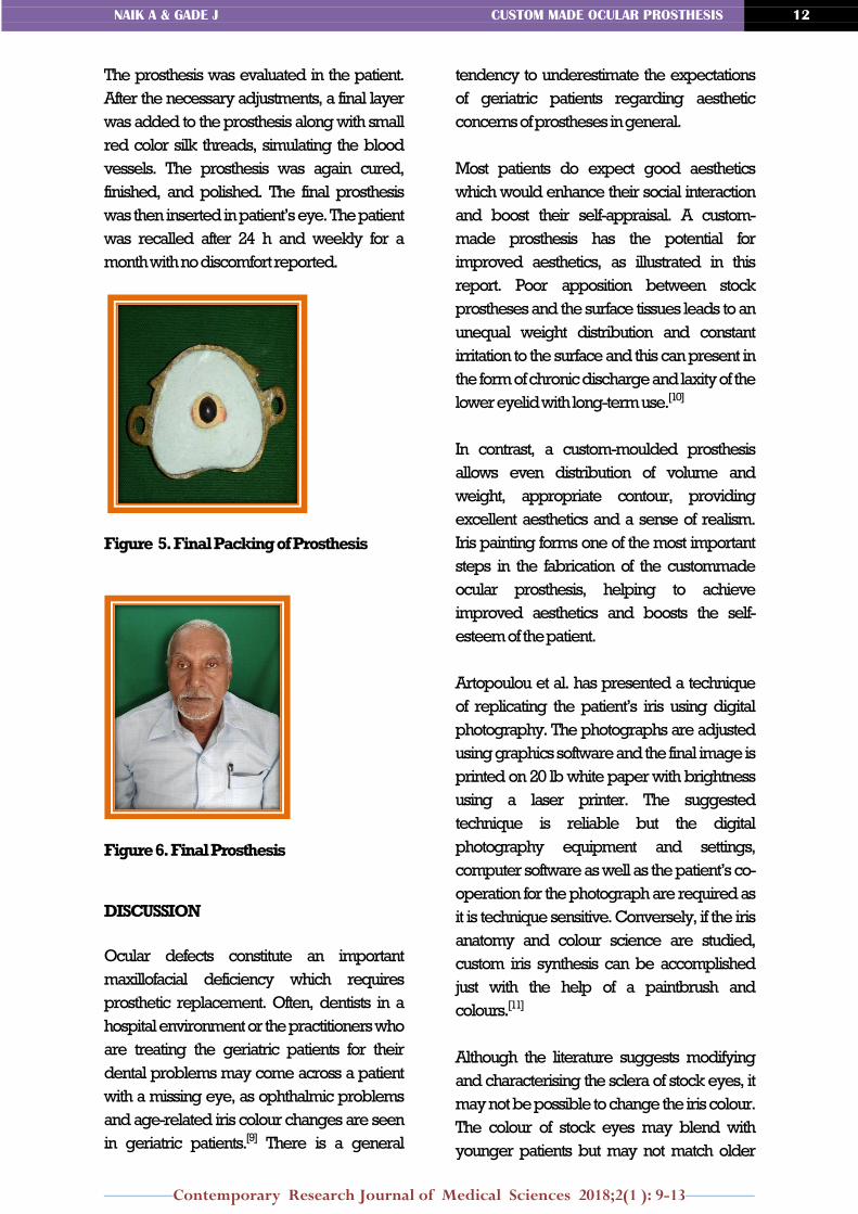
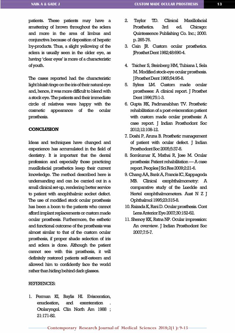
The prosthesis was evaluated in the patient.
After the necessary adjustments, a final layer
was added to the prosthesis along with small
red color silk threads, simulating the blood
vessels. The prosthesis was again cured,
finished, and polished. The final prosthesis
was then inserted in patient’s eye. The patient
was recalled after 24 h and weekly for a
month with no discomfort reported.
Figure 5. Final Packing of Prosthesis
Figure 6. Final Prosthesis
DISCUSSION
Ocular defects constitute an important
maxillofacial deficiency which requires
prosthetic replacement. Often, dentists in a
hospital environment or the practitioners who
are treating the geriatric patients for their
dental problems may come across a patient
with a missing eye, as ophthalmic problems
and age-related iris colour changes are seen
in geriatric patients.[9] There is a general
tendency to underestimate the expectations
of geriatric patients regarding aesthetic
concerns of prostheses in general.
Most patients do expect good aesthetics
which would enhance their social interaction
and boost their self-appraisal. A custom-
made prosthesis has the potential for
improved aesthetics, as illustrated in this
report. Poor apposition between stock
prostheses and the surface tissues leads to an
unequal weight distribution and constant
irritation to the surface and this can present in
the form of chronic discharge and laxity of the
lower eyelid with long-term use.[10]
In contrast, a custom-moulded prosthesis
allows even distribution of volume and
weight, appropriate contour, providing
excellent aesthetics and a sense of realism.
Iris painting forms one of the most important
steps in the fabrication of the custommade
ocular prosthesis, helping to achieve
improved aesthetics and boosts the self-
esteem of the patient.
Artopoulou et al. has presented a technique
of replicating the patient’s iris using digital
photography. The photographs are adjusted
using graphics software and the final image is
printed on 20 lb white paper with brightness
using a laser printer. The suggested
technique is reliable but the digital
photography equipment and settings,
computer software as well as the patient’s co-
operation for the photograph are required as
it is technique sensitive. Conversely, if the iris
anatomy and colour science are studied,
custom iris synthesis can be accomplished
just with the help of a paintbrush and
colours.[11]
Although the literature suggests modifying
and characterising the sclera of stock eyes, it
may not be possible to change the iris colour.
The colour of stock eyes may blend with
younger patients but may not match older
NAIK A & GADE J CUSTOM MADE OCULAR PROSTHESIS 13
Contemporary Research Journal of Medical Sciences 2018;2(1 ): 9-13
patients. These patients may have a
smattering of brown throughout the sclera
and more in the area of limbus and
conjunctiva because of deposition of hepatic
by-products. Thus, a slight yellowing of the
sclera is usually seen in the older eye, as
having ‘clear eyes’ is more of a characteristic
of youth.
The cases reported had the characteristic
light bluish tinge on the iris of their natural eye
and, hence, it was more difficult to blend with
a stock eye. The patients and their immediate
circle of relatives were happy with the
cosmetic appearance of the ocular
prosthesis.
CONCLUSION
Ideas and techniques have changed and
experience has accumulated in the field of
dentistry. It is important that the dental
profession and especially those practicing
maxillofacial prosthetics keep their current
knowledge. The method described here is
undemanding and can be carried out in a
small clinical set‑up, rendering better service
to patient with anophthalmic socket defect.
The use of modified stock ocular prosthesis
has been a boon to the patients who cannot
afford implant replacements or custom made
ocular prosthesis. Furthermore, the esthetic
and functional outcome of the prosthesis was
almost similar to that of the custom ocular
prosthesis, if proper shade selection of iris
and sclera is done. Although the patient
cannot see with this prosthesis, it will
definitely restored patients self‑esteem and
allowed him to confidently face the world
rather than hiding behind dark glasses.
REFERENCES:
1. Perman KI, Baylis HI. Evisceration,
enucleation, and exenteration .
Otolaryngol. Clin North Am 1988 ;
21:171-82.
2. Taylor TD. Clinical Maxillofacial
Prosthetics. 3rd ed. Chicago:
Quintessence Publishing Co. Inc.; 2000.
p. 265-76.
3. Cain JR. Custom ocular prosthetics.
JProsthet Dent 1982;48:690-4.
4. Taicher S, Steinberg HM, Tubiana I, Sela
M. Modified stock-eye ocular prosthesis.
J Prosthet Dent 1985;54:95-8.
5. Sykes LM. Custom made ocular
prostheses: A clinical report. J Prosthet
Dent 1996;75:1-3.
6. Gupta RK, Padmanabhan TV. Prosthetic
rehabilitation of a post evisceration patient
with custom made ocular prosthesis: A
case report. J Indian Prosthodont Soc
2012;12:108-12.
7. Doshi P, Aruna B. Prosthetic management
of patient with ocular defect. J Indian
Prosthodont Soc 2005;5:37-8.
8. Somkumar K, Mathai R, Jose M. Ocular
prosthesis: Patient rehabilitation — A case
report. Peoples J Sci Res 2009;2:21-6.
9. Chang AA, Bank A, Francis IC, Kappagoda
MB. Clinical exophthalmometry: A
comparative study of the Luedde and
Hertel exophthalmometers. Aust N Z J
Ophthalmol 1995;23:315-8.
10. Raizada K, Rani D. Ocular prosthesis. Cont
Lens Anterior Eye 2007;30:152-62.
11. Shenoy KK, Ratna NP. Ocular impression:
An overview. J Indian Prosthodont Soc
2007;7:5-7.
NAIK A & GADE J CUSTOM MADE OCULAR PROSTHESIS 13
Contemporary Research Journal of Medical Sciences 2018;2(1 ): 9-13
Submit your next manuscript to CRJMS and take full advantage of :
Convenient manuscript submission (through website) and by mail ([email protected]). Thorough Peer review
Author friendly Article Processing Charges No space constraints or extra color figure charges Immediate publication on acceptance Inclusion in International Database including Google
Scholar Manuscript writing assistance for New Authors
Authors Information
Dr. Ashish Naik
PG student,
Department of Prosthetic Dentistry,
Swargiya Dadasaheb Kalmegh Smruti
Dental College & Hospital , Nagpur
Dr. Jaykumar Gade
Professor & Head,
Department of Prosthetic Dentistry,
Swargiya Dadasaheb Kalmegh Smruti
Dental College & Hospital , Nagpur
Dr. Aditi Kumbhalwar
PG student,
Department of Prosthetic Dentistry,
Swargiya Dadasaheb Kalmegh Smruti
Dental College & Hospital , Nagpur