Embed Size (px)
Citation preview
Zeitschrift für Kraniomandibuläre Funktion 2012;4(1):79–94 79
practice revisions praxistipp
Zusammenfassung
In der Therapie der kraniomandibulären Dysfunktion (CMD) kann die Rekonstruktion der statischen und dyna-mischen Okklusion von entscheidender Bedeutung für den Behandlungserfolg sein. Im Einzelfall ist dabei eine enge Zusammenarbeit zwischen der Zahnmedizin, Ortho-pädie und manuellen Medizin notwendig. Der Zahnarzt soll die Okklusion den Änderungen der Ober- und Unter-kieferrelation anpassen, die sich aus einer Normalisierung der Muskelfunktion und einer verbesserten Körper- und Kopfhaltung ergeben. Das primäre therapeutische Mittel ist eine spezielle Okklusionsschiene, die herausnehmba-re kraniomandibuläre, orthopädische Positionierungs-Apparatur (COPA). Durch eine reversible Korrektur der Okklusion kann sie pathologische Wirkungen im Sinne absteigender Störungen der Kiefergelenkdysfunktion auf-heben. Nach Abschluss der interdisziplinären Behandlung kann eine weiterführende kieferorthopädische und/oder restaurative Therapie notwendig werden, sofern sich im Rahmen der Behandlung die Kiefergelenkposition verän-dert hat, um dann eine stabile, interferenzfreie Okklusion dauerhaft zu sichern. Sofern mit der Okklusionsschie-ne eine Infraokklusion klinisch erfolgreich ausgeglichen wurde und weiter Behandlungsbedarf besteht, wird im Bei-trag die Überführung der herausnehmbaren Okklusions-schiene in fest aufklebbare okklusale Schienenelemente als
1 Dr. med. dent. Wolfgang Boisserée, niedergelassener Zahnarzt, Köln
2 Dr. med. dent. Werner Schupp, Fachzahnarzt für Kieferorthopädie in niedergelassener Praxis, Köln
1 Dr. med. dent. Wolfgang Boisserée, private practice, Cologne, Germany
2 Dr. med. dent. Werner Schupp, private practice, Cologne, Germany
W. Boisserée1, W. Schupp2
Two-stage approach to mandibular splint therapy with craniomandibular orthopedic positioning appliances
Zweiphasiges Konzept zum Okklusions ausgleich durch Unterkiefer-Okklusionsschienen
abstract
The restoration of normal temporomandibular joint func-tion in static and dynamic occlusion can be the key the successful treatment of temporomandibular disorders (TMD), in many cases. An interdisciplinary approach characterized by close cooperation between the dentist, orthopedist and manual therapist is required for the indi-vidual patient. The dentist’s job is to adjust the occlusion to the changes in jaw relationships that occur in response to functional normalization of the involved muscles, and to improve the patient’s head and body posture. The cra-niomandibular orthopedic positioning appliance (COPA), a removable interocclusal appliance for reversible correc-tion of the occlusion, is designed to eliminate the patho-logical interferences associated with temporomandibular joint (TMJ) dysfunction. As such, it is predestined for use as a primary therapeutic instrument in these indications. If the position of the temporomandibular joint changes over the course of treatment, further orthodontic and/or pros-thodontic treatment may be needed after the completion of interdisciplinary treatment, to ensure the long-term, interference-free stabilization of the occlusion. If the clin ical findings indicate that the occlusal splint has suc-cessfully eliminated the problems of infraocclusion, and that further treatment is needed, the removable occlusal splint can be transformed into bonded onlay elements in
Copyrig
ht
by
N
otfor
Qu
in
tessence
Not
forPublication
practice revisions Boisserée/schupp Splint concept for occlusal therapy
Journal of Craniomandibular Function 2012;4(1):79–9480
eine Möglichkeit der Übertragung der Schienenposition unter Wahrung der erreichten therapeutischen Kiefer-gelenkposition vorgestellt. Auf diese Weise lässt sich die vorgesehene therapeutische Okklusion vor weiterfüh-renden irreversiblen okklusalen Maßnahmen simulieren, um bestmöglichen Aufschluss über die Wirksamkeit der therapeutischen Okklusion sowohl im subjektiven Patien-tenempfinden als auch in der interdisziplinären Funktions-diagnostik zu erhalten.
indizes: CMD, kraniomandibuläres System, muskuloskelettales System, Funktionstherapie, herausnehmbare Okklusionsschiene, aufklebbare Okklusionsschiene, biodynamisches Aufwachskonzept nach Polz, statische Okklusion, dynamische Okklusion, Kieferorthopädie, Prothetik
einleitung
In der komplexen Ätiologie der CMD können okklusale Störungen mit zu den auslösenden Faktoren zählen.1–5 Die Korrektur einer fehlerhaften Okklusion ist Aufgabe des Zahnarztes. Ihm obliegt es, die okklusale Beteili-gung am Krankheitsbild zu erkennen und zu behandeln. Erschwerend ist die enge Verkettung des kraniomandibu-lären (CMS) mit dem muskuloskelettalen System (MSS). Funktionsstörungen aus dem kraniomandibulären System wirken sich dominant auf das muskuloskelettale System im Sinne einer primären Läsion aus.6 Durch diese Ver-kettung kann die Okklusion absteigend zu Störungen im MSS führen.7–10 Diese können sich unter anderem als Kopfschmerzen, Hals-Wirbel-Säulen (HWS)- und Rücken-beschwerden äußern.11 Aufgrund dessen erfordern die CMD-Diagnostik und -Therapie im Einzelfall eine inter-disziplinäre Zusammenarbeit mit der manuellen Medizin.
Lotzmann formulierte, dass letztlich jede Änderung der Kieferrelation eine orthopädische Maßnahme darstellt, da die Stellung von Körpergelenken geändert wird. Die Entscheidung des zahnmedizinischen, manualmedizini-schen und orthopädischen Behandlungsteams über die zahnmedizinische/medizinische Notwendigkeit zur Neu-einstellung des Unterkiefers, setzt profunde diagnosti-sche und therapeutische Kenntnisse voraus.12 In diesem Sinne darf eine Okklusionskorrektur nur innerhalb eines verantwortungsbewussten und klaren interdisziplinären Behandlungskonzepts geplant und durchgeführt werden.
Um dieser Forderung gerecht zu werden ist es not-wendig, die therapeutische Okklusion vor weiterführen-den irreversiblen Maßnahmen zur Okklusionskorrektur
order to transfer the splint position with the highest pos-sible precision, while maintaining the temporomandibular joint position achieved in the first stage of splint therapy. This approach allows the clinician to simulate the planned therapeutic occlusion before irreversible occlusal treat-ment meas ures are performed, in order to obtain the most accurate assessment of the effectiveness of the therapeutic occlusion based on the patient’s subjective impression, as well as the results of interdisciplinary functional analysis.
Keywords: biomechanical waxup technique according to Polz, bonded occlusal splint, craniomandibular system (CMS), dynamic occlusion, functional treatment, musculoskeletal system (MSS), orthodontics, prosthodontics, removable occlusal splint, static occlusion, temporomandibular disorders (TMD)
introduction
Occlusal interference can serve as a trigger contributing to the complex etiology of temporomandibular disorders (TMD)1-5. The dentist’s job is to eliminate the problem of malocclusion, and to identify and treat bite problems con-tributing to temporomandibular joint syndrome. The close association between the craniomandibular system (CMS) and the musculoskeletal system (MSS) is an aggravating factor. Functional disorders of the craniomandibular system have an overbearing effect on the musculoskeletal system, similar to a primary disease6. Occlusal disturbances can set off a chain reaction radiating to the musculoskeletal system7-10. This is manifested in typical symptoms such as headache, neck pain and back ache11. Due to this inter-action, interdisciplinary cooperation between a dentist and a manual therapist is required for proper diagnosis and treatment of TMD in a given case.
As Lotzmann put it, any procedure that changes jaw relationships can be classified as an orthopedic procedure, because it also changes the positions of the joints in the body. Any decision by the dental, manual therapy and orthopedic treatment team concerning the dental and/or medical necessity to change the position of the mandible requires profound diagnostic and therapeutic expertise12. Consequently, occlusal correction should always be planned and performed within the scope of a clear and responsible interdisciplinary treatment strategy.
In order to meet this requirement, the therapeutic occlusion must be thoroughly tested before any more invasive and irreversible occlusal treatment measures are performed. Frequently this is not possible with removable
Copyrig
ht
by
N
otfor
Qu
in
tessence
Not
forPublication
Zeitschrift für Kraniomandibuläre Funktion 2012;4(1):79–94 81
Boisserée/schupp Schienenkonzept zum Okklusionsausgleich praxistipp
eingehend zu testen. Das ist mit herausnehmbaren Okklu-sionsschienen alleine häufig nur bedingt möglich, da sie, auch in Form von Positionierungsschienen, die therapeu-tische Okklusion oft nicht ganz exakt simulieren können. Die Gründe liegen in der materialtechnisch bedingten Konstruktion, die häufig eine vertikale Überdimensionie-rung nötig macht, um der Okklusionsschiene eine ausrei-chende Stabilität zu verleihen. Entsprechend ist dann die Aufbissschienenokklusion in der Vertikaldimension höher eingestellt, als die spätere okklusale Rehabilitation sein wird. Außerdem werden Okklusionsschienen häufig nur nachts getragen und vermitteln damit nur zeitlich begrenzt den therapeutischen Effekt und das Gefühl einer zukünf-tigen Okklusion. Zudem können sie nicht während des Essens getragen werden.
okklusionsschienentherapie in zwei Behandlungsphasen
Das hier vorgestellte Verfahren zeigt ein zweizeitiges Vorgehen, bei der eine notwendige Rehabilitation der Okklusion im Falle von Infraokklusion innerhalb der Funk-tionstherapie möglichst exakt für die weiterführende irre-versible Therapie vorbereitet wird.
In der primären reversiblen Phase wird eine spezielle Okklusionsschiene für den Unterkiefer eingegliedert, die möglichst tags und nachts getragen werden kann. Durch das okklusale Relief soll die statische und dynamische Okklusion möglichst physiologisch rehabilitiert werden. Die Okklusionsschiene ist unter besonderer Berücksich-tigung des MSS angefertigt, indem sie Verkettungssyn-drome, die durch primäre Störungen im CMS ausgelöst werden, aufhebt. Entsprechend besteht in dieser Behand-lungsphase eine interdisziplinäre Zusammenarbeit von Diagnostik und Therapie mit der manuellen Medizin.13 Die Behandlungsdauer der ersten Behandlungsphase soll-te mit der hier beschriebenen Okklusionsschiene nicht wesentlich länger als sechs Monate betragen. In der Regel kann in diesem Zeitraum abgeklärt werden, ob eine wei-terführende okklusale Therapie notwendig ist.
Bei therapeutischer Wirksamkeit und der Notwendig-keit weiterführender okklusaler Rehabilitationen (kieferor-thopädisch und/oder prothetisch) werden in einer zweiten Phase fest aufklebbare okklusale Schienenelemente im Unterkiefer eingegliedert, unter Beibehaltung der erreich-ten Unterkiefer- und Kondylenposition. Diese nehmen die zukünftige therapeutische Okklusion präzise in vertikaler und horizontaler Relation vorweg. Der Patient ist nun gewissermaßen „gezwungen“, mit dieser Okklusion zu leben. Die Behandlungsdauer beträgt in der Regel vier
occlusal splints alone, even if they are repositioning splints, because the appliance fails to provide an entirely accurate reproduction of the therapeutic occlusion. One of the main reasons for this is that, due to the nature of the construc-tion materials, it is often necessary to over-dimension the occlusal appliance in the vertical plane in order to achieve adequate stability. Consequently, the vertical dimension of the occlusion with the splint inserted will be higher than that of the final occlusal position after rehabilitation. Secondly, many occlusal splints are only worn at night, so they only convey the therapeutic effect and sensation of the future occlusion for a limited time. Furthermore, the splints cannot be worn during meals.
two-stage approach to occlusal splint therapy
The method presented here is a two-stage approach to the treatment of patients with infraocclusion. With this approach, splint therapy achieves restoration of the occlu-sion with maximum accuracy and within the scope of holis-tic functional therapy, before definitive occlusal therapy.
The first stage consists of reversible occlusal therapy, using a special mandibular occlusal splint that is designed to be worn both day and night. The goal is to provide maximum occlusal relief for restoration of physiological function in both static and dynamic occlusion. Considering the close relationship between the craniomandibular system and musculoskeletal system, the proposed occlusal splint was designed to break the vicious cycle of symptoms trig-gered by primary disorders of the craniomandibular system. Interdisciplinary cooperation between the clinician estab-lishing the diagnosis and the therapist performing manual treatment is therefore indispensable during this stage of treatment13. In the first stage of occlusal therapy with the proposed occlusal splint, treatment duration should not be much longer than six months. It is generally possible to determine whether more intensive occlusal therapy will be necessary by the end of the first stage of treatment.
The indication for further occlusal treatment is generally based on the patient’s response to splint therapy and the need for definitive treatment (orthodontic and/or pros-thodontic) for restoration of the occlusion. In the second stage of treatment, the splint is reduced to two segments of craniomandibular orthopedic positioning appliance (COPA) onlays, which are directly bonded to the mandibular teeth in order to maintain the mandibular and condylar positions achieved in the first stage of splint therapy. The onlays accurately reproduce the vertical and horizontal dimensions of the therapeutic occlusion. They “force” the patient to assume the therapeutic bite position. The second stage of
Copyrig
ht
by
N
otfor
Qu
in
tessence
Not
forPublication
practice revisions Boisserée/schupp Splint concept for occlusal therapy
Journal of Craniomandibular Function 2012;4(1):79–9482
bis acht Wochen. In dieser Phase kann die therapeutische Okklusion im subjektiven Empfinden des Patienten und in wiederholter reevaluierender Funktionsdiagnostik getes-tet werden. Auch die manuelle Medizin kann diagnos-tisch und therapeutisch eingebunden werden. Zu lange Tragezeiten sind zu vermeiden, damit sich die erreichte Kieferposition nicht attritionsbedingt verändert.
Erst wenn diese zweite Behandlungsphase erfolgreich abschlossen ist, sollten die weiterführenden, irreversiblen Maßnahmen erfolgen, die zu einer langfristigen Rehabili-tation der Okklusion führen. Dieses schrittweise Vorgehen in der Okklusionskorrektur mit Aufbissschienen erhöht den Behandlungserfolg der weiterführenden okklusalen Therapie unter der Voraussetzung, dass die eingestell-te Kiefer- und Kondylenposition im Laufe der weiteren Behandlung nicht verloren geht.
Forderungen an die okklusionsschiene
Im Zentrum primärer okklusaler Maßnahmen steht die herausnehmbare Okklusionsschiene. Ihr kommt entschei-dende Bedeutung zu, da sie eine reversible Veränderung der Kieferposition und der Okklusion ermöglicht.14–17 Grundlage der Behandlung mit der Okklusionsschiene ist die zentrische Kieferrelationsbestimmung. Als Besonder-heit des hier beschriebenen Vorgehens wird im Rahmen funktionsdiagnostischer Untersuchungen die Auswirkung der eingestellten Kieferposition auf das kraniomandibuläre System und insbesondere auf das muskuloskelettale Sys-tem untersucht und die registrierte Position gegebenen-falls korrigiert.9 Dies erfolgt mittels jenes Registrats und unter Ausschaltung der bisherigen okklusalen Führung.
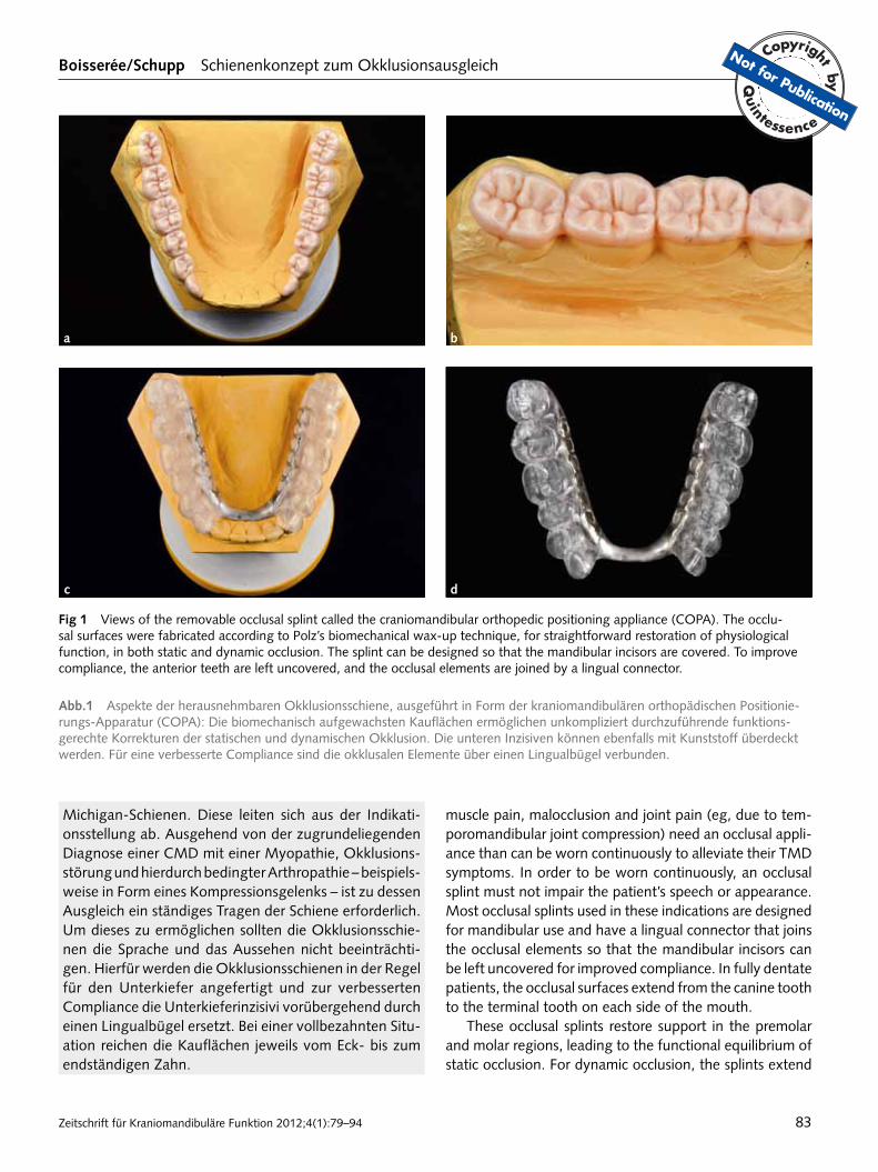
Sofern im Rahmen der Therapie mit einer Aufbissschie-ne zur muskulären Relaxation auch eine Okklusionskor-rektur angestrebt ist, sollten die Schienen – außer zum Essen – möglichst Tag und Nacht getragen werden, um während der gesamten Zeit interdisziplinärer zahnärzt-lich-manualmedizinischer Therapie Kontakt zwischen den natürlichen Zähnen zu vermeiden. Der Grund hierfür liegt in der schnellen Reprogrammierung des alten Stereotyps.4 Das kontinuierliche Tragen gibt außerdem im subjektiven Patientenempfinden und in der interdisziplinären Diag-nostik verlässliche Hinweise auf die therapeutische Wirk-samkeit der Okklusionskorrektur im kraniomandibulären und im muskuloskelettalen System (Abb. 1).
Konstruktionsmerkmale dieser okklusionsschiene
Die hier beschriebene Art von Okklusionsschienen unter-scheidet sich in vielerlei Hinsicht von den klassischen
splint therapy with the COPA onlays generally takes 4 to 8 weeks. During this time, the therapeutic occlusion can be repeatedly re-evaluated, based on the patient’s subjective impression and the objective results of functional tests. A manual therapist (chiropractor, physical therapist, etc) can also be involved in the diagnostic and therapeutic process. Excessive wearing times should be avoided in order to pre-vent attrition-related changes in the achieved jaw position.
Irreversible measures for long-term rehabilitation of the dental occlusion should not be performed before the second stage of splint therapy has been completed. This step-by-step approach to occlusal therapy increases the chances of success with subsequent occlusal treatment proced ures, provided that the jaw relations and condylar positions established by splint therapy are properly reproduced and maintained.
requirements for occlusal splints
The removable occlusal splint is the central element in the first stage of the proposed method of occlusal therapy. Removable appliances are important because they allow for reversible adjustment of the occlusion and jaw relation14-17. Accurate recording of centric relation is the key to success-ful bite splint therapy. A special feature of the approach described is that it allows the clinician to analyze the impact of splint-related changes in jaw relation on both the cranio-mandibular and musculoskeletal system in particular, and to adjust the jaw relation as needed in the scope of functional diagnosis9. This is done using the appropriate registration, while eliminating the former occlusal guidance.
If splint therapy is also performed for muscle relax-ation in addition to occlusal adjustment, the occlusal appliance should be worn both day and night (except at meals) during the entire interdisciplinary dental-manual occlusal treatment period, in order to prevent contact between the natural teeth. The goal is rapid reprogram-ming of the old stereotype4. Continuous splint wearing also allows the clinician to gather subjective and objec-tive information from the patient and interdisciplinary diagnostic tests regarding the impact of the therapeutic changes in the occlusion on the craniomandibular and musculoskeletal systems (Fig 1).
Design features of the copa splint
The characteristics of the COPA splint differ from those of the classic Michigan splint in many ways. The COPA splint was designed for a specific indication. Patients diagnosed with temporomandibular dysfunction associated with
Copyrig
ht
by
N
otfor
Qu
in
tessence
Not
forPublication
Zeitschrift für Kraniomandibuläre Funktion 2012;4(1):79–94 83
Boisserée/schupp Schienenkonzept zum Okklusionsausgleich praxistipp
Michigan-Schienen. Diese leiten sich aus der Indikati-onsstellung ab. Ausgehend von der zugrundeliegenden Diagnose einer CMD mit einer Myopathie, Okklusions-störung und hierdurch bedingter Arthropathie – beispiels-weise in Form eines Kompressionsgelenks – ist zu dessen Ausgleich ein ständiges Tragen der Schiene erforderlich. Um dieses zu ermöglichen sollten die Okklusionsschie-nen die Sprache und das Aussehen nicht beeinträchti-gen. Hierfür werden die Okklusionsschienen in der Regel für den Unterkiefer angefertigt und zur verbesserten Compliance die Unterkieferinzisivi vorübergehend durch einen Lingualbügel ersetzt. Bei einer vollbezahnten Situ-ation reichen die Kauflächen jeweils vom Eck- bis zum endständigen Zahn.
Fig 1 Views of the removable occlusal splint called the craniomandibular orthopedic positioning appliance (COPA). The occlu-sal surfaces were fabricated according to Polz’s biomechanical wax-up technique, for straightforward restoration of physiological function, in both static and dynamic occlusion. The splint can be designed so that the mandibular incisors are covered. To improve compliance, the anterior teeth are left uncovered, and the occlusal elements are joined by a lingual connector.
abb.1 Aspekte der herausnehmbaren Okklusionsschiene, ausgeführt in Form der kraniomandibulären orthopädischen Positionie-rungs-Apparatur (COPA): Die biomechanisch aufgewachsten Kauflächen ermöglichen unkompliziert durchzuführende funktions-gerechte Korrekturen der statischen und dynamischen Okklusion. Die unteren Inzisiven können ebenfalls mit Kunststoff überdeckt werden. Für eine verbesserte Compliance sind die okklusalen Elemente über einen Lingualbügel verbunden.
a b
c d
muscle pain, malocclusion and joint pain (eg, due to tem-poromandibular joint compression) need an occlusal appli-ance than can be worn continuously to alleviate their TMD symptoms. In order to be worn continuously, an occlusal splint must not impair the patient’s speech or appearance. Most occlusal splints used in these indications are designed for mandibular use and have a lingual connector that joins the occlusal elements so that the mandibular incisors can be left uncovered for improved compliance. In fully dentate patients, the occlusal surfaces extend from the canine tooth to the terminal tooth on each side of the mouth.
These occlusal splints restore support in the premolar and molar regions, leading to the functional equilibrium of static occlusion. For dynamic occlusion, the splints extend
Copyrig
ht
by
N
otfor
Qu
in
tessence
Not
forPublication
practice revisions Boisserée/schupp Splint concept for occlusal therapy
Journal of Craniomandibular Function 2012;4(1):79–9484
Die Schienen rekonstruieren die Stützzonen im Prä-molaren- und Molarengebiet und führen damit zu einem möglichst funktionsgerechten Ausgleich der statischen Okklusion. Für die Dynamik reichen die Schienen bis zu den Eckzähnen, die die Führung in Protrusion und Laterotrusion übernehmen, bei gleichzeitiger Disklusion im Seitenzahngebiet.
Die unteren Inzisivi sind nicht bedeckt. Der Grund hier-für liegt neben den Vorzügen hinsichtlich der Compliance darin, dass der Unterkiefer sich während der Tragedauer der Okklusionsschiene horizontal frei einstellen kann, ohne dass ein erneuter Frontzahnkontakt entsteht. Da die Schie-nentherapie auf vier bis sechs Monate beschränkt ist, spielt die Gefahr einer Stellungsveränderung von Frontzähnen eine untergeordnete Rolle.18 Bei absehbarer längerfristiger Tragedauer ist es auch möglich, die Unterkieferinzisivi in die Okklusionsschiene mit einzubeziehen. Dabei darf es nicht zu einem Frontkontakt kommen, der zu einem re trusiven Impuls auf die Unterkieferposition führen könnte.
Die Kauflächengestaltung erfolgt in Abhängigkeit von den zuvor erhobenen Funktionsbefunden: • Bei eindeutigen Befunden und reproduzierbar
bestimmter Kieferrelation können die Kauflächen nach dem biodynamischem Aufwachskonzept von Polz19,20 gestaltet werden. Hier gibt die Okklusionsschiene im Sinne einer Positionierungsschiene genau eine Kiefer-position vor, allerdings mit einer erleichterten Latero- und Mediotrusion.
• Bei „nicht-zentrikfähigen“ Patienten, d. h. bei Patien-ten, die so starke Dysfunktionen aufweisen, dass im Rahmen der Kieferrelationsbestimmung nicht oder nur bedingt reproduzierbare Kieferpositionen auftraten, wird nur die vertikale Dimension festgelegt. Ansonsten hat die Schiene keine unmittelbar wirksam werdenden horizontalen Stopps (Relaxierungsschiene). Der Pati-ent hat dann somit einen gewissen horizontalen Frei-raum bei gleichzeitiger Abstützung in der Vertikalen.
Als Kunststoffmaterial dient das Kaltpolimerisat Pala-Xpress (Heraeus Kulzer, Hanau), das im Gussverfahren verarbeitet wird. Der Lingualbügel wird auch im anterioren Bereich von Kunststoff ummantelt.
Zur erleichterten Abgrenzung dieser Okklusions-schienen gegenüber klassischen Michiganschienen und in Anlehnung an die in der Kieferorthopädie üblichen Nomenklaturen werden diese Relaxierungs- beziehungs-weise Positionierungsschienen auch als kraniomandibu-läre orthopädische Positionierungs-Apparaturen (COPA) bezeichnet, was ihren Bezug von Diagnostik und Therapie zum muskuloskelettalen System herausstellt.
to the canines to provide occlusion with canine guidance during protrusion and laterotrusion, together with disoc-clusion of the posterior teeth.
The mandibular incisors are left uncovered. This not only improves compliance but also ensures that the lower jaw can freely adapt in the horizontal plane without creating new anterior contacts during the course of bite splint therapy. As the splints are only worn for 4 to 6 months, the risk of altering the position of the anterior teeth is minor18. If long-term splint therapy is required, the splint can be designed so that the mandibular incisors are covered. However, anterior tooth contact must be avoided, as this could generate retru-sive forces that could alter the position of the mandible: • The occlusal surfaces of the splint are designed on data
based from the prior functional analysis. Polz’s bio-mechanical wax-up technique19,20 can be used to design the occlusal surfaces of the splint, provided a confirmed diagnosis and reproducible jaw relation records are avail-able. The occlusal splint, which functions as a reposition-ing splint, provides a reproduction of jaw relation that is exact but with slight laterotrusion/mediotrusion.
• It may not be possible to obtain an accurate centric relation record in certain cases, for example, if the dysfunction is so severe that it is difficult to impossible to obtain a reproducible registration of jaw relations. If so, only the vertical dimension can be established. The occlusal splint has no other immediately effective hori-zontal stops (relaxation splint). This gives the patient a degree of freedom of movement in the horizontal plane, while providing adequate support in the verti-cal dimension.
The COPA splint is made of PalaXpress (Heraeus Kulzer, Hanau, Germany), a synthetic, cold-curing resin processed by the casting technique. In the anterior region, the lingual connector is also covered with resin.
To more clearly distinguish between the new occlusal splint and the conventional Michigan splint (and in con-formity with the widely used orthodontic nomenclature), this relaxation or repositioning splint is also referred to as the craniomandibular orthopedic positioning appliance (COPA). The name aptly emphasizes the impact of the diag-nosis and treatment of disorders of the craniomandibular system on the musculoskeletal system.
case study
The craniomandibular orthopedic positioning appliance (COPA), an occlusal splint worn over the mandibular teeth,
Copyrig
ht
by
N
otfor
Qu
in
tessence
Not
forPublication
Zeitschrift für Kraniomandibuläre Funktion 2012;4(1):79–94 85
Boisserée/schupp Schienenkonzept zum Okklusionsausgleich praxistipp
Fallbeispiel
Die im Unterkiefer getragenen Okklusionsschienen stellen eine praxisnahe Methode für ein durchgängiges Konzept zur Okklusionskorrektur dar. Die häufigste okklusale Ursa-che einer CMD ist die fehlende posteriore Abstützung, die infolge einer retrokranialen Mandibulaverlagerung zu einer Kompression von Kiefergelenkstrukturen führt.21 An einem entsprechenden Fallbeispiel soll das Konzept nachfolgend vorgestellt werden.
Die 52-jährige Patientin litt an Wirbelsäulenbeschwer-den, Kopfschmerzen und Kiefergelenkbeschwerden infol-ge einer okklusionsbedingten CMD. Okklusale Ursache war ein anteriorer Tiefbiss bei gleichzeitig fehlender pos-teriorer Abstützung. Der vorhandene Zahnersatz im Unterkiefer war vertikal unterdimensioniert und führ-te damit zu einem unzureichenden Vertikalniveau der Seiten zähne. In habitueller Okklusion bestand eine man-dibuläre Re tralverlagerung mit beidseitiger Kiefergelenk-kompression. Die funktionsdiagnostische Untersuchung ergab, dass die okklusale Problematik offensichtlich mit den beklagten Beschwerden zusammenhing.
vorangegangene kieferorthopädische Korrektur der Front
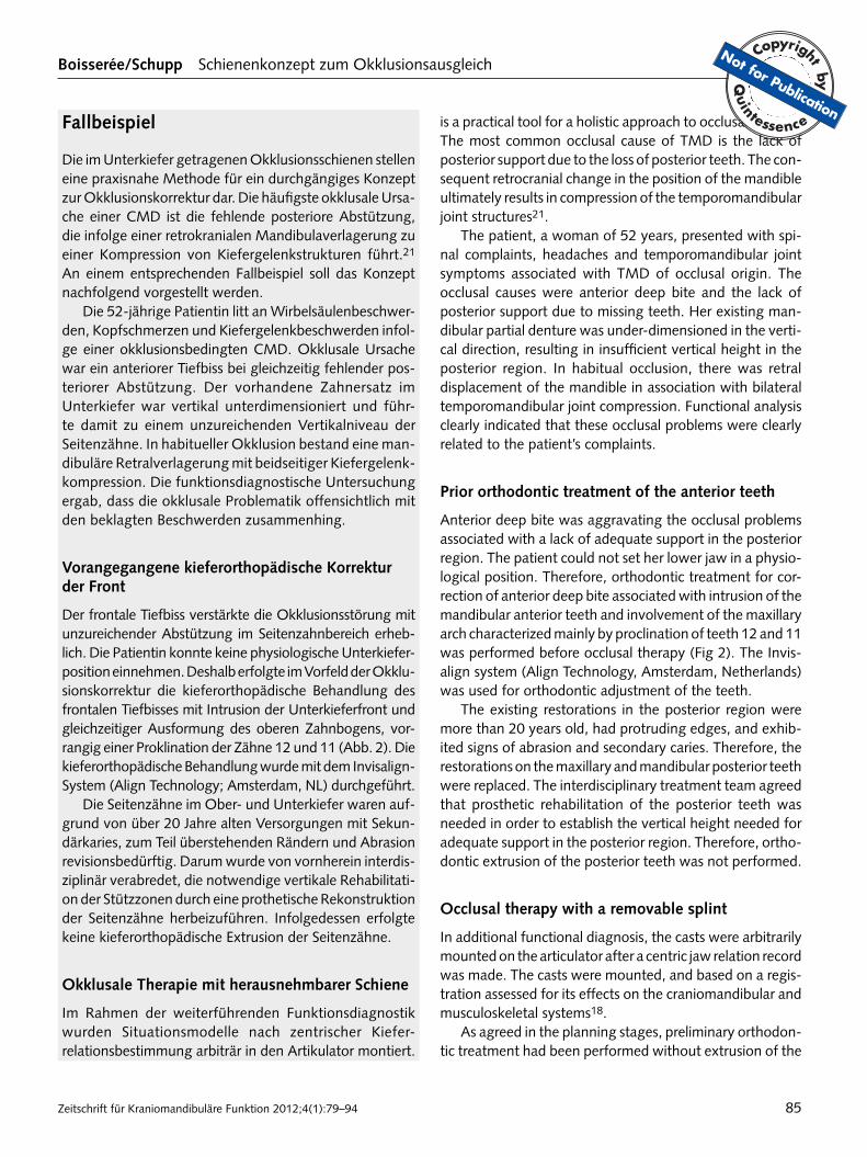
Der frontale Tiefbiss verstärkte die Okklusionsstörung mit unzureichender Abstützung im Seitenzahnbereich erheb-lich. Die Patientin konnte keine physiologische Unterkiefer-position einnehmen. Deshalb erfolgte im Vorfeld der Okklu-sionskorrektur die kieferorthopädische Behandlung des frontalen Tiefbisses mit Intrusion der Unterkieferfront und gleichzeitiger Ausformung des oberen Zahnbogens, vor-rangig einer Proklination der Zähne 12 und 11 (Abb. 2). Die kieferorthopädische Behandlung wurde mit dem Invisalign-System (Align Technology; Amsterdam, NL) durchgeführt.
Die Seitenzähne im Ober- und Unterkiefer waren auf-grund von über 20 Jahre alten Versorgungen mit Sekun-därkaries, zum Teil überstehenden Rändern und Abrasion revisionsbedürftig. Darum wurde von vornherein interdis-ziplinär verabredet, die notwendige vertikale Rehabilitati-on der Stützzonen durch eine prothetische Rekonstruktion der Seitenzähne herbeizuführen. Infolgedessen erfolgte keine kieferorthopädische Extrusion der Seitenzähne.
okklusale therapie mit herausnehmbarer schiene
Im Rahmen der weiterführenden Funktionsdiagnostik wurden Situationsmodelle nach zentrischer Kiefer-relations bestimmung arbiträr in den Artikulator montiert.
is a practical tool for a holistic approach to occlusal therapy. The most common occlusal cause of TMD is the lack of posterior support due to the loss of posterior teeth. The con-sequent retrocranial change in the position of the mandible ultimately results in compression of the temporomandibular joint structures21.
The patient, a woman of 52 years, presented with spi-nal complaints, headaches and temporomandibular joint symptoms associated with TMD of occlusal origin. The occlusal causes were anterior deep bite and the lack of posterior support due to missing teeth. Her existing man-dibular partial denture was under-dimensioned in the verti-cal direction, resulting in insufficient vertical height in the posterior region. In habitual occlusion, there was retral displacement of the mandible in association with bilateral temporomandibular joint compression. Functional analysis clearly indicated that these occlusal problems were clearly related to the patient’s complaints.
prior orthodontic treatment of the anterior teeth
Anterior deep bite was aggravating the occlusal problems associated with a lack of adequate support in the posterior region. The patient could not set her lower jaw in a physio-logical position. Therefore, orthodontic treatment for cor-rection of anterior deep bite associated with intrusion of the mandibular anterior teeth and involvement of the maxillary arch characterized mainly by proclination of teeth 12 and 11 was performed before occlusal therapy (Fig 2). The Invis-align system (Align Technology, Amsterdam, Netherlands) was used for orthodontic adjustment of the teeth.
The existing restorations in the posterior region were more than 20 years old, had protruding edges, and exhib-ited signs of abrasion and secondary caries. Therefore, the restorations on the maxillary and mandibular posterior teeth were replaced. The interdisciplinary treatment team agreed that prosthetic rehabilitation of the posterior teeth was needed in order to establish the vertical height needed for adequate support in the posterior region. Therefore, ortho-dontic extrusion of the posterior teeth was not performed.
occlusal therapy with a removable splint
In additional functional diagnosis, the casts were arbitrarily mounted on the articulator after a centric jaw relation record was made. The casts were mounted, and based on a regis-tration assessed for its effects on the craniomandibular and musculoskeletal systems18.
As agreed in the planning stages, preliminary orthodon-tic treatment had been performed without extrusion of the
Copyrig
ht
by
N
otfor
Qu
in
tessence
Not
forPublication
practice revisions Boisserée/schupp Splint concept for occlusal therapy
Journal of Craniomandibular Function 2012;4(1):79–9486
Grundlage der Montage ist das Registrat, das zuvor auf seine Wirkung auf das kraniomandibuläre und das mus-kuloskelettale System hin untersucht wurde.18
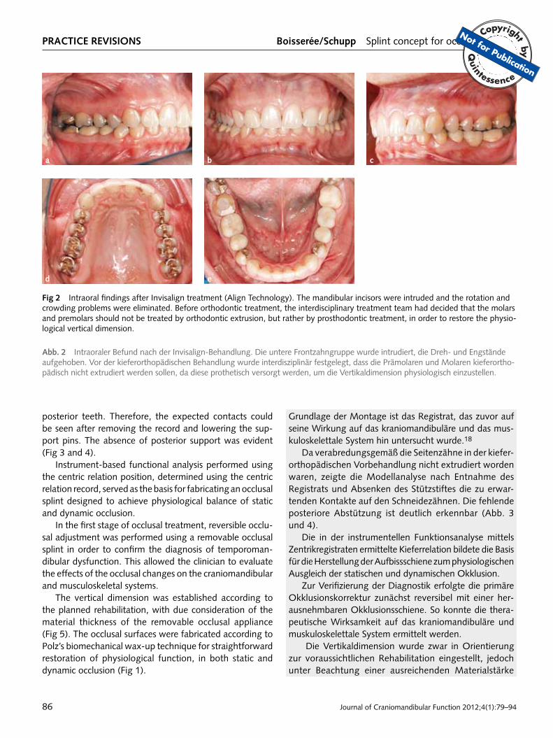
Da verabredungsgemäß die Seitenzähne in der kiefer-orthopädischen Vorbehandlung nicht extrudiert worden waren, zeigte die Modellanalyse nach Entnahme des Registrats und Absenken des Stützstiftes die zu erwar-tenden Kontakte auf den Schneidezähnen. Die fehlende posteriore Abstützung ist deutlich erkennbar (Abb. 3 und 4).
Die in der instrumentellen Funktionsanalyse mittels Zentrikregistraten ermittelte Kieferrelation bildete die Basis für die Herstellung der Aufbissschiene zum physiologischen Ausgleich der statischen und dynamischen Okklusion.
Zur Verifizierung der Diagnostik erfolgte die primäre Okklusionskorrektur zunächst reversibel mit einer her-ausnehmbaren Okklusionsschiene. So konnte die thera-peutische Wirksamkeit auf das kraniomandibuläre und muskuloskelettale System ermittelt werden.
Die Vertikaldimension wurde zwar in Orientierung zur voraussichtlichen Rehabilitation eingestellt, jedoch unter Beachtung einer ausreichenden Materialstärke
Fig 2 Intraoral findings after Invisalign treatment (Align Technology). The mandibular incisors were intruded and the rotation and crowding problems were eliminated. Before orthodontic treatment, the interdisciplinary treatment team had decided that the molars and premolars should not be treated by orthodontic extrusion, but rather by prosthodontic treatment, in order to restore the physio-logical vertical dimension.
abb. 2 Intraoraler Befund nach der Invisalign-Behandlung. Die untere Frontzahngruppe wurde intrudiert, die Dreh- und Engstände aufgehoben. Vor der kieferorthopädischen Behandlung wurde interdisziplinär festgelegt, dass die Prämolaren und Molaren kieferortho-pädisch nicht extrudiert werden sollen, da diese prothetisch versorgt werden, um die Vertikaldimension physiologisch einzustellen.
a b
d e
c
posterior teeth. Therefore, the expected contacts could be seen after removing the record and lowering the sup-port pins. The absence of posterior support was evident (Fig 3 and 4).
Instrument-based functional analysis performed using the centric relation position, determined using the centric relation record, served as the basis for fabricating an occlusal splint designed to achieve physiological balance of static and dynamic occlusion.
In the first stage of occlusal treatment, reversible occlu-sal adjustment was performed using a removable occlusal splint in order to confirm the diagnosis of temporoman-dibular dysfunction. This allowed the clinician to evaluate the effects of the occlusal changes on the craniomandibular and musculoskeletal systems.
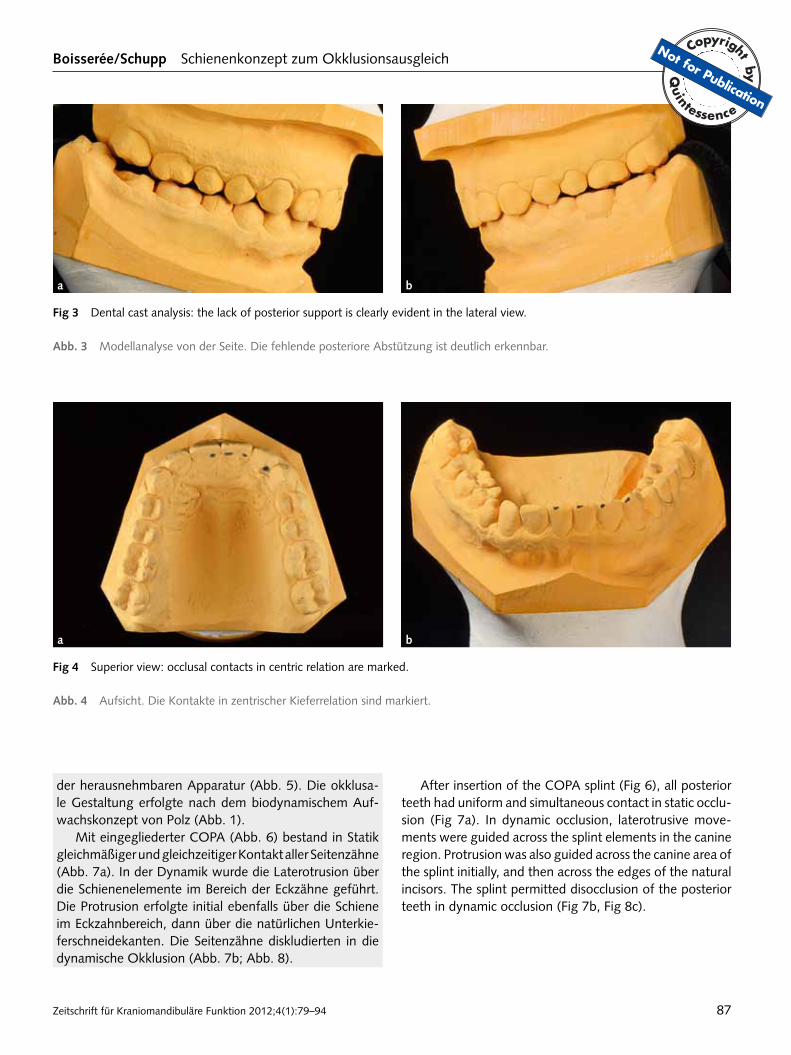
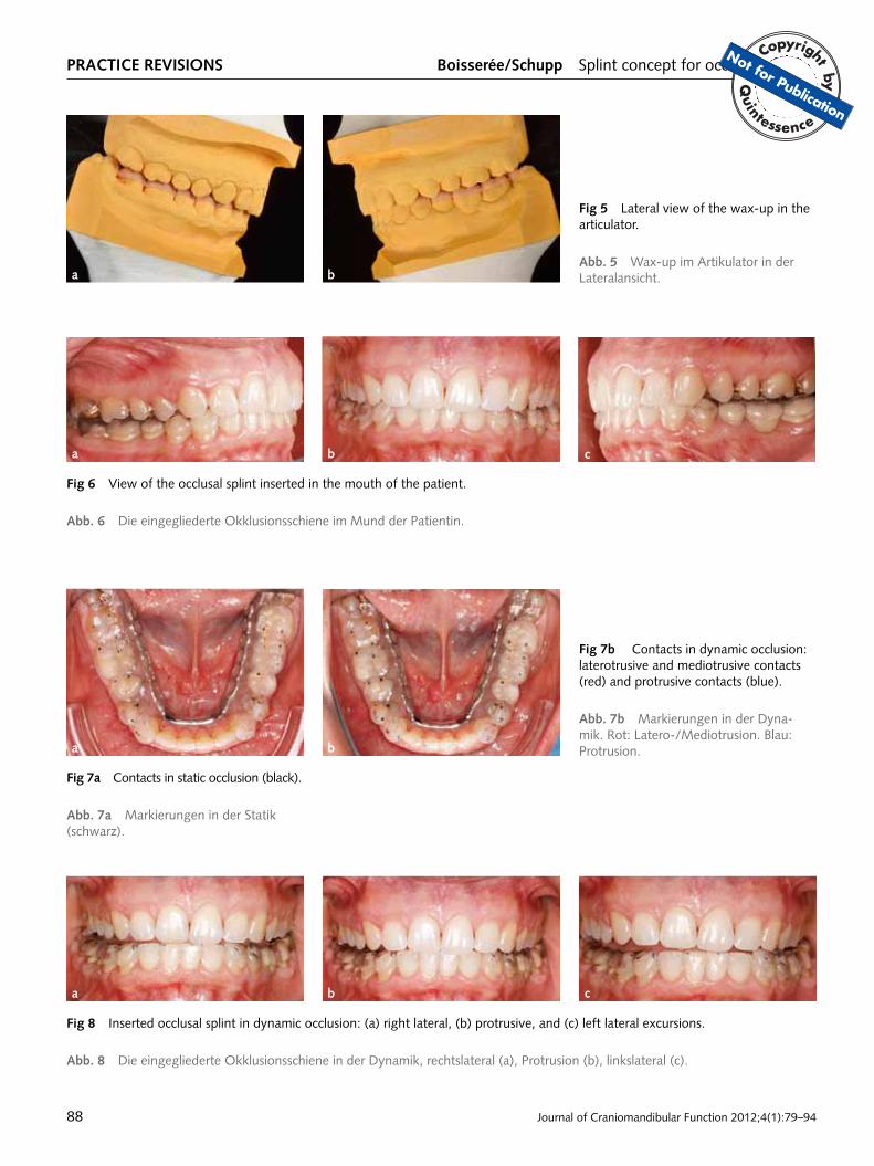
The vertical dimension was established according to the planned rehabilitation, with due consideration of the material thickness of the removable occlusal appliance (Fig 5). The occlusal surfaces were fabricated according to Polz’s biomechanical wax-up technique for straightforward restoration of physiological function, in both static and dynamic occlusion (Fig 1).
Copyrig
ht
by
N
otfor
Qu
in
tessence
Not
forPublication
Zeitschrift für Kraniomandibuläre Funktion 2012;4(1):79–94 87
Boisserée/schupp Schienenkonzept zum Okklusionsausgleich praxistipp
a b
Fig 3 Dental cast analysis: the lack of posterior support is clearly evident in the lateral view.
abb. 3 Modellanalyse von der Seite. Die fehlende posteriore Abstützung ist deutlich erkennbar.
a b
Fig 4 Superior view: occlusal contacts in centric relation are marked.
abb. 4 Aufsicht. Die Kontakte in zentrischer Kieferrelation sind markiert.
der herausnehmbaren Apparatur (Abb. 5). Die okklusa-le Gestaltung erfolgte nach dem biodynamischem Auf-wachskonzept von Polz (Abb. 1).
Mit eingegliederter COPA (Abb. 6) bestand in Statik gleichmäßiger und gleichzeitiger Kontakt aller Seiten zähne (Abb. 7a). In der Dynamik wurde die Laterotrusion über die Schienenelemente im Bereich der Eckzähne geführt. Die Protrusion erfolgte initial ebenfalls über die Schiene im Eckzahnbereich, dann über die natürlichen Unterkie-ferschneidekanten. Die Seitenzähne diskludierten in die dynamische Okklusion (Abb. 7b; Abb. 8).
After insertion of the COPA splint (Fig 6), all posterior teeth had uniform and simultaneous contact in static occlu-sion (Fig 7a). In dynamic occlusion, laterotrusive move-ments were guided across the splint elements in the canine region. Protrusion was also guided across the canine area of the splint initially, and then across the edges of the natural incisors. The splint permitted disocclusion of the posterior teeth in dynamic occlusion (Fig 7b, Fig 8c).
Copyrig
ht
by
N
otfor
Qu
in
tessence
Not
forPublication
practice revisions Boisserée/schupp Splint concept for occlusal therapy
Journal of Craniomandibular Function 2012;4(1):79–9488
Fig 5 Lateral view of the wax-up in the articulator.
abb. 5 Wax-up im Artikulator in der Lateralansicht.a b
Fig 7a Contacts in static occlusion (black).
abb. 7a Markierungen in der Statik (schwarz).
a b c
Fig 6 View of the occlusal splint inserted in the mouth of the patient.
abb. 6 Die eingegliederte Okklusionsschiene im Mund der Patientin.
a b
Fig 7b Contacts in dynamic occlusion: laterotrusive and mediotrusive contacts (red) and protrusive contacts (blue).
abb. 7b Markierungen in der Dyna-mik. Rot: Latero-/Mediotrusion. Blau: Protrusion.
a b c
Fig 8 Inserted occlusal splint in dynamic occlusion: (a) right lateral, (b) protrusive, and (c) left lateral excursions.
abb. 8 Die eingegliederte Okklusionsschiene in der Dynamik, rechtslateral (a), Protrusion (b), linkslateral (c).
Copyrig
ht
by
N
otfor
Qu
in
tessence
Not
forPublication
Zeitschrift für Kraniomandibuläre Funktion 2012;4(1):79–94 89
Boisserée/schupp Schienenkonzept zum Okklusionsausgleich praxistipp
Klinisches vorgehen zur Kontrolle und Korrektur der okklusionsschieneKorrekturen an der Schienenokklusion erfolgen fast immer unmittelbar nach manueller Vorbehandlung.9,22 Die erste Kontrolle des Aufbisses auf die Okklusionsschiene findet nach einer Woche statt. Die weiteren Kontrollen finden im Wochenabstand, je nach Behandlungssituation bis zur vierten oder fünften Woche statt. Danach werden monat-liche Intervalle eingehalten. Nach vier bis sechs Monaten erfolgt eine Reevaluation der bisherigen Funktionstherapie. Neben der Überprüfung der Gesamtsituation wird dabei die therapeutische Effizienz der primären, reversiblen Okklusi-onskorrektur als Grundlage der weiteren Behandlungspla-nung überprüft.
Im Patientenbeispiel waren nach drei Monaten inter-disziplinärer Funktionstherapie die Kiefergelenkbeschwer-den, Kopfschmerzen und HWS-Befunde beseitigt und die Wirbelsäulenbeschwerden wesentlich verbessert.
Die Abbildungen 9 bis 12 zu diesem Fallbeispiel erläu-tern das als Teil des Praxistipps beschriebene Konzept der funktionstherapeutischen Weiterbehandlung.
Überführung der schienenokklusion
Herausnehmbare okklusionsschiene zu aufklebbaren „copa-onlays“
Sofern eine Diskrepanz zwischen der klinisch erfolgreich überprüften therapeutischen Kieferrelation sowie der
a b
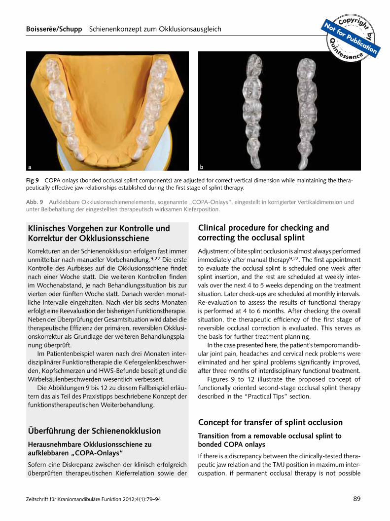
Fig 9 COPA onlays (bonded occlusal splint components) are adjusted for correct vertical dimension while maintaining the thera-peutically effective jaw relationships established during the first stage of splint therapy.
abb. 9 Aufklebbare Okklusionsschienenelemente, sogenannte „COPA-Onlays“, eingestellt in korrigierter Vertikaldimension und unter Beibehaltung der eingestellten therapeutisch wirksamen Kieferposition.
clinical procedure for checking and correcting the occlusal splint
Adjustment of bite splint occlusion is almost always performed immediately after manual therapy9,22. The first appointment to evaluate the occlusal splint is scheduled one week after splint insertion, and the rest are scheduled at weekly inter-vals over the next 4 to 5 weeks depending on the treatment situation. Later check-ups are scheduled at monthly intervals. Re-evaluation to assess the results of functional therapy is performed at 4 to 6 months. After checking the overall situation, the therapeutic efficiency of the first stage of reversible occlusal correction is evaluated. This serves as the basis for further treatment planning.
In the case presented here, the patient’s temporomandib-ular joint pain, headaches and cervical neck problems were eliminated and her spinal problems significantly improved, after three months of interdisciplinary functional treatment.
Figures 9 to 12 illustrate the proposed concept of functionally oriented second-stage occlusal splint therapy described in the “Practical Tips” section.
concept for transfer of splint occlusion
transition from a removable occlusal splint to bonded copa onlays
If there is a discrepancy between the clinically-tested thera-peutic jaw relation and the TMJ position in maximum inter-cuspation, if permanent occlusal therapy is not possible
Copyrig
ht
by
N
otfor
Qu
in
tessence
Not
forPublication
practice revisions Boisserée/schupp Splint concept for occlusal therapy
Journal of Craniomandibular Function 2012;4(1):79–9490
Gelenkstellung in maximaler Interkuspidation besteht, eine dauerhafte Schienentherapie nicht infrage kommt und/oder ohnehin dentaler Sanierungsbedarf besteht, wird eine Überführung der Schienenokklusion auf die Zähne erforderlich.
Wie eingangs erwähnt, wird als diagnostischer Zwi-schenschritt die herausnehmbare Schiene in fest aufkleb-bare okklusale Schienenelemente, sogenannte COPA-Onlays, überführt. Der Grund liegt in der möglichst präzisen Simulation der zukünftigen therapeutischen Okklusion in horizontaler und vertikaler Relation.
Auf Basis einer erneuten funktionsdiagnostischen Untersuchung und erneuter Kieferrelationsbestimmung, unter Beibehaltung der erreichten Kiefergelenkposition, werden COPA-Onlays wiederum aus Kaltpolymerisat (PalaXpress) hergestellt. Die Vertikaldimension wird dafür präzise in Bezug zur zukünftig vorgesehenen Okklusion eingestellt (Abb. 9).
Nur in seltenen Fällen, wenn die vertikale und hori-zontale Kieferrelation bereits durch die herausnehmbare Okklusionsschiene therapeutisch ideal eingestellt werden konnte und noch kein Verschleiß der Okklusionsschiene durch Attrition stattgefunden hat, können die okklusalen Elemente aus der Schiene herausgetrennt und als Onlays
a
b
c
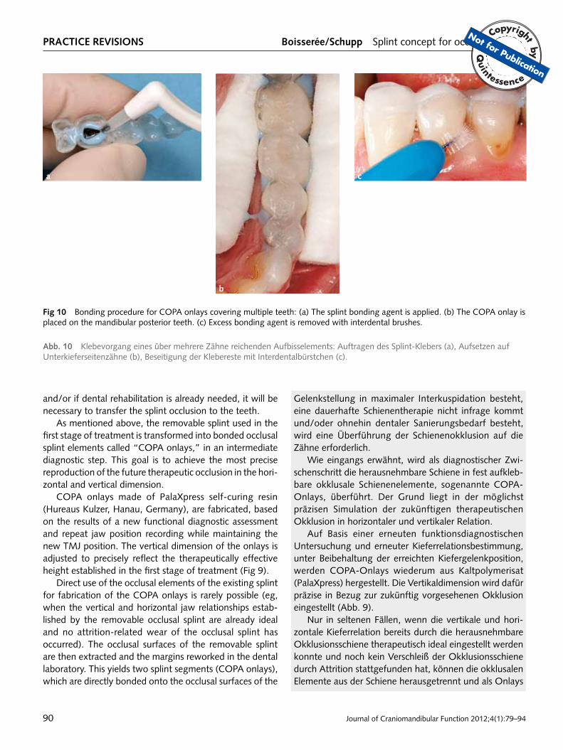
Fig 10 Bonding procedure for COPA onlays covering multiple teeth: (a) The splint bonding agent is applied. (b) The COPA onlay is placed on the mandibular posterior teeth. (c) Excess bonding agent is removed with interdental brushes.
abb. 10 Klebevorgang eines über mehrere Zähne reichenden Aufbisselements: Auftragen des Splint-Klebers (a), Aufsetzen auf Unterkieferseitenzähne (b), Beseitigung der Klebereste mit Interdentalbürstchen (c).
and/or if dental rehabilitation is already needed, it will be necessary to transfer the splint occlusion to the teeth.
As mentioned above, the removable splint used in the first stage of treatment is transformed into bonded occlusal splint elements called “COPA onlays,” in an intermediate diagnostic step. This goal is to achieve the most precise reproduction of the future therapeutic occlusion in the hori-zontal and vertical dimension.
COPA onlays made of PalaXpress self-curing resin (Hureaus Kulzer, Hanau, Germany), are fabricated, based on the results of a new functional diagnostic assessment and repeat jaw position recording while maintaining the new TMJ position. The vertical dimension of the onlays is adjusted to precisely reflect the therapeutically effective height established in the first stage of treatment (Fig 9).
Direct use of the occlusal elements of the existing splint for fabrication of the COPA onlays is rarely possible (eg, when the vertical and horizontal jaw relationships estab-lished by the removable occlusal splint are already ideal and no attrition-related wear of the occlusal splint has occurred). The occlusal surfaces of the removable splint are then extracted and the margins reworked in the dental laboratory. This yields two splint segments (COPA onlays), which are directly bonded onto the occlusal surfaces of the
Copyrig
ht
by
N
otfor
Qu
in
tessence
Not
forPublication
Zeitschrift für Kraniomandibuläre Funktion 2012;4(1):79–94 91
Boisserée/schupp Schienenkonzept zum Okklusionsausgleich praxistipp
a b
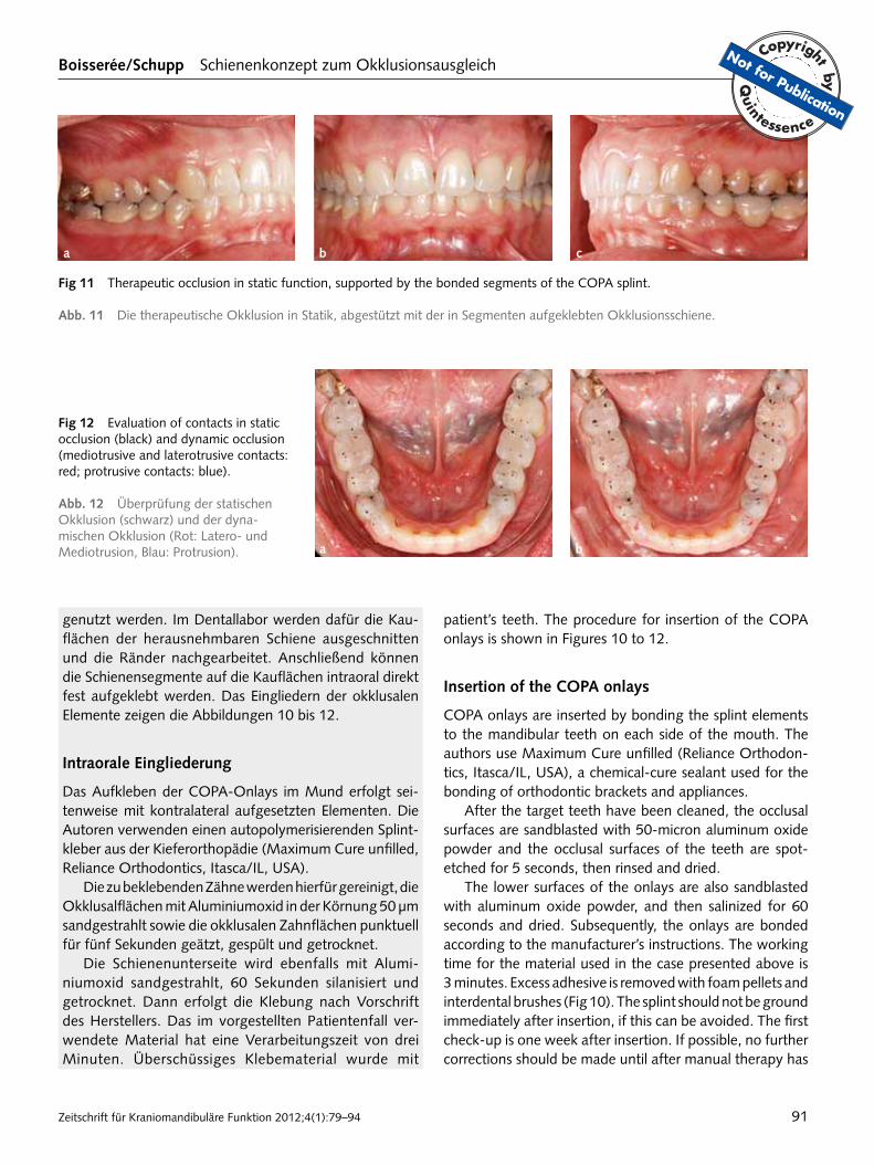
Fig 11 Therapeutic occlusion in static function, supported by the bonded segments of the COPA splint.
abb. 11 Die therapeutische Okklusion in Statik, abgestützt mit der in Segmenten aufgeklebten Okklusionsschiene.
c
a b
Fig 12 Evaluation of contacts in static occlusion (black) and dynamic occlusion (mediotrusive and laterotrusive contacts: red; protrusive contacts: blue).
abb. 12 Überprüfung der statischen Okklusion (schwarz) und der dyna-mischen Okklusion (Rot: Latero- und Mediotrusion, Blau: Protrusion).
genutzt werden. Im Dentallabor werden dafür die Kau-flächen der herausnehmbaren Schiene ausgeschnitten und die Ränder nachgearbeitet. Anschließend können die Schienensegmente auf die Kauflächen intraoral direkt fest aufgeklebt werden. Das Eingliedern der okklusalen Elemente zeigen die Abbildungen 10 bis 12.
intraorale eingliederung
Das Aufkleben der COPA-Onlays im Mund erfolgt sei-tenweise mit kontralateral aufgesetzten Elementen. Die Autoren verwenden einen autopolymerisierenden Splint-kleber aus der Kieferorthopädie (Maximum Cure unfilled, Reliance Orthodontics, Itasca/IL, USA).
Die zu beklebenden Zähne werden hierfür gereinigt, die Okklusalflächen mit Aluminiumoxid in der Körnung 50 µm sandgestrahlt sowie die okklusalen Zahnflächen punktuell für fünf Sekunden geätzt, gespült und getrocknet.
Die Schienenunterseite wird ebenfalls mit Alumi-niumoxid sandgestrahlt, 60 Sekunden silanisiert und getrocknet. Dann erfolgt die Klebung nach Vorschrift des Herstellers. Das im vorgestellten Patientenfall ver-wendete Material hat eine Verarbeitungszeit von drei Minuten. Überschüssiges Klebematerial wurde mit
patient’s teeth. The procedure for insertion of the COPA onlays is shown in Figures 10 to 12.
insertion of the copa onlays
COPA onlays are inserted by bonding the splint elements to the mandibular teeth on each side of the mouth. The authors use Maximum Cure unfilled (Reliance Orthodon-tics, Itasca/IL, USA), a chemical-cure sealant used for the bonding of orthodontic brackets and appliances.
After the target teeth have been cleaned, the occlusal surfaces are sandblasted with 50-micron aluminum oxide powder and the occlusal surfaces of the teeth are spot-etched for 5 seconds, then rinsed and dried.
The lower surfaces of the onlays are also sandblasted with aluminum oxide powder, and then salinized for 60 seconds and dried. Subsequently, the onlays are bonded according to the manufacturer’s instructions. The working time for the material used in the case presented above is 3 minutes. Excess adhesive is removed with foam pellets and interdental brushes (Fig 10). The splint should not be ground immediately after insertion, if this can be avoided. The first check-up is one week after insertion. If possible, no further corrections should be made until after manual therapy has
Copyrig
ht
by
N
otfor
Qu
in
tessence
Not
forPublication
practice revisions Boisserée/schupp Splint concept for occlusal therapy
Journal of Craniomandibular Function 2012;4(1):79–9492
Schaumstoffpellets und Interdentalbürstchen entfernt (Abb. 10). Die Schiene soll nach dem Einsetzen möglichst nicht sofort eingeschliffen werden. Die erste Kontrolle erfolgte nach einer Woche. Weitere Korrekturen finden wiederum möglichst nach manualmedizinischer Vor-behandlung statt. Die okklusale Gestaltung ermöglicht unkomplizierte funktionsgerechte Einschleifmaßnahmen (Abb. 11 und 12).
Nach vier- bis achtwöchiger Beschwerdefreiheit und erneuter funktionsdiagnostischer Untersuchung kann die prothetische Folgetherapie eingeleitet werden. Selbst-verständlich werden nur jene Zähne in die prothetische Behandlung einbezogen, die ohnehin restaurationsbe-dürftig sind oder bei denen eine vertikale Veränderung zur Abstützung des Gegenkiefers unabdingbar ist.21,23
vor- und nachteile des vorgehens
Der Vorteil der vorgestellten Methode liegt im geradlini-gen Behandlungsverlauf. In der ersten Behandlungsphase wird die Wirksamkeit der therapeutischen Okklusion eru-iert. Ist eine weiterführende Behandlung notwendig, wird in einer zweiten Behandlungsphase, vor weiterführenden kieferorthopädischen und/ oder prothetischen Maßnah-men, die definitiv angestrebte Okklusion durch fest auf-klebbare okklusale Schienenelemente praxisnah getestet.
Im Falle einer prothetischen Folgebehandlung kann anschließend auf der Null-Ebene des Artikulators weiter-gearbeitet werden, da das Vertikalniveau bereits thera-peutisch korrekt eingestellt ist und nicht mehr verändert werden muss. Das Verfahren ist in der Literatur beschrie-ben.24,25 Alternativ dazu kann eine weiterführende okklu-sale Rehabilitation mit speziellen Repositions-Onlays wei-tergeführt werden.21,23
Nachteilig ist die eingeschränkte Interdentalraumhygi-ene, diese entspricht in etwa der Situation mit verblockten Kronen und sollte bei sorgfältiger Entfernung aller inter-dentalen Klebereste beherrschbar sein.
Infolge der Anfälligkeit des Schienenmaterials gegen-über Attrition besteht während der Tragezeit der COPA-Onlays das Risiko einer Stellungsveränderung der Unter-kieferposition. Dies begründet die vergleichsweise kurze Tragezeitz der Onlays von in der Regel vier bis acht Wochen.
Schließlich ist jeweils im Einzelfall zu prüfen, ob die verwendeten Materialien für die hier vorgestellte Anwen-dung als Medizinprodukt zugelassen sind.
Die Indikation des Verfahrens mit fest aufgekleb-ten okklusalen Schienenelementen ist zudem auf Pa tientenfälle mit Infraokklusion beschränkt, bei denen
been performed. The occlusal design allows uncomplicated, functionally oriented grinding (Fig 11 and 12).
Prosthodontic treatment can be initiated after 4 to 8 weeks without symptoms and after a follow-up functional analysis has been performed. Naturally, this applies to only those teeth requiring prosthodontic treatment, ie, those already in need of restoration and/or those where vertical height adjustment is absolutely necessary in order to pro-vide support for the opposing jaw21,23.
advantages and disadvantages of the proposed method
The advantage of the proposed method is that treatment is relatively straightforward. The first stage of splint therapy serves to test the effectiveness of therapeutic occlusion. If further treatment is needed, the second stage of splint therapy with COPA onlays is performed prior to orthodontic and/or prosthodontic treatment. The onlays are bonded to the teeth in order to evaluate the therapeutic occlusion and to determine the definitive occlusion in situ.
If subsequent prosthetic treatment is needed, this can be done using the articulator at the zero level, because the vertical height was already corrected in occlusal therapy and does not require further adjustment. A description of the method can be found in the related literature24,25. Another option is to perform further occlusal rehabilitation using special repositioning onlays21,23.
The disadvantage is impaired hygiene in the interdental spaces. The situation is similar to that with splinted crowns, but should be controllable if all excess adhesive is carefully removed from the interdental spaces.
Because the splint material is subject to attrition, there is a risk that the position of the mandible might change while the patient is wearing the COPA onlays. For this reason, the onlays are usually worn for only a relatively short period of time (4 to 8 weeks).
Ultimately, it is necessary to determine whether the materials used for the proposed application are approved for use in medical devices in a given case.
Furthermore, occlusal treatment with bonded COPA onlays is indicated only in patients with infraocclusion requiring additive procedures to increase the occlusal ver-tical dimension.
Within this population, special restrictions apply to patients with excessive loss of tooth structure and/or increased vertical discrepancies, because the resulting lack of anterior guidance usually prevents proper func-tional restoration of the occlusion with COPA onlays. As
Copyrig
ht
by
N
otfor
Qu
in
tessence
Not
forPublication
Zeitschrift für Kraniomandibuläre Funktion 2012;4(1):79–94 93
Boisserée/schupp Schienenkonzept zum Okklusionsausgleich praxistipp
die Notwendigkeit zur additiven Rekonstruktion der Ver-tikaldimension besteht.
Innerhalb dieser Gruppe besteht eine besondere Ein-schränkung des Verfahrens bei großen Zahnhartsubstanz-verlusten und/oder erhöhten Vertikaldiskrepanzen, da in solchen Fällen die Okklusion mit COPA-Onlays meist, aufgrund der fehlenden Frontführung, nicht funktionsge-recht rekonstruiert werden kann. Als Alternative bietet sich vor diesem Hintergrund die indirekte Fertigung spezieller Repositions-Onlays aus hierfür zugelassenem Material mit nachfolgender adhäsiver intraoraler Befestigung an. Die-ses Vorgehen ist bereits in der Literatur beschrieben.21,23
Auch in Fällen von Supraokklusion kann die Methode nicht zur Anwendung kommen, da der Okklusionsaus-gleich in diesen Situationen nicht additiv, sondern in der Regel subtraktiv erfolgen kann.
Fazit für die praxis
Voraussetzungen für eine kieferorthopädische und/ oder restaurative Folgebehandlung innerhalb der CMD-Thera-pie ist die ideale Vorbereitung der therapeutischen Okklu-sion. Sie sollte bereits vor Behandlungsbeginn der endgül-tig angestrebten Kieferrelation entsprechen. Der Patient sollte in der therapeutischen Okklusion beschwerdefrei oder deutlich beschwerdereduziert sein.14
an alternative, indirectly fabricated individual repositioning onlays made of approved materials could be fabricated and bonded to the patient’s teeth with an adhesive agent. This procedure has been described in the literature21,23.
Supraocclusion of the teeth is another contraindication to the proposed method, because subtractive rather than additive procedures are generally used to restore the occlu-sion in these cases.
conclusions and implications
Ideal preparation of therapeutic occlusion is the key to successful orthodontic and/or prosthodontic treatment, following splint therapy for the treatment of temporo-mandibular disorders. The prepared therapeutic occlusion should correspond to the desired final jaw relation. In the therapeutic occlusion, the patient’s temporomandibular joint symptoms should be completely eliminated or signifi-cantly reduced14.
Copyrig
ht
by
N
otfor
Qu
in
tessence
Not
forPublication
practice revisions Boisserée/schupp Splint concept for occlusal therapy
Journal of Craniomandibular Function 2012;4(1):79–9494
references
1. Fink M, Tschernitschek H, Stiesch-Scholz M, Währling K. Kraniomandibuläres System und Wirbelsäule. Man Med 2003;41:476–480.
2. Ash MM, Ramfjord SP (Hrsg.). Okklusion und Funktion. Berlin: Quintessenz; 1988.
3. Kopp S, Seebald WG. Kraniomandibuläre Dysfunk-tion – Versuch einer bewertenden Übersicht. Man Med 2008;46:389–392.
4. Plato G, Kopp S. Kiefergelenk und Schmerzsyndrome. Man Med 1999;37:143–151.
5. Keil B, Keil H. Dysfunction in connection with functional disorders of the cervical spine. Dtsch Stomatol 1991;41: 249–252.
6. Honikel M. Das Craniomandibuläre System und seine Effekte auf die Körperhaltung – Teil III. Osteopath Med 2007;8:4–9.
7. Plato G, Kopp S. Der Weg zur Chronifizierung der kranio-mandibulären Dysfunktionen (CMD). Man Med 2008;46: 384–385.
8. Slavicek R. Funktion – die Haltung. In Slavicek R (Hrsg.) Das Kauorgan: Funktionen und Dysfunktionen. Klosterneuburg: Gamma Medizinisch-wissenschaftliche Fortbildungsgesell-schaft; 2000.
9. Marx G. Über die Zusammenarbeit mit der Kieferorthopädie und Zahnheilkunde in der manuellen Medizin. Man Med 2000;38:342–345.
10. Saito ET, Akashi PM, Sacco Ide C. Global body posture evalua-tion in patients with temporomandibular joint disorder. Clinics (Sao Paulo) 2009;64:35–39.
11. Cooper BC, Kleinberg I. Relationship of temporomandibular disorders to muscle tension-type headaches and a neuro-muscular orthosis approach to treatment. Cranio 2009;27: 101–108.
12. Lotzmann U. Okklusion, Kiefergelenk und Wirbelsäule. ZM 2002;9:1004–1010.
13. Schupp W, Boisseree W, Haubrich J, et al. Interdisziplinären Zusammenarbeit zwischen Zahnmedizin und manueller Med-izin. Man Med 2010;3:192–200.
14. Ahlers MO, Freesmeyer WB, Fussnegger M, et al. Zur Therapie der Erkrankungen des kraniomandibulären Sys-tems. Gemeinsame Wissenschaftliche Stellungnahme der Deutschen Gesellschaft für Funktionsdiagnostik und Thera-pie (DGFDT) in der DGZMK, der Deutschen Gesellschaft für Prothetische Zahnheilkunde und Biomaterialien (DGPro), der Deutschen Gesellschaft für Mund-, Kiefer- und Gesichts-
chirurgie (DGMKG), der Arbeitsgemeinschaft für Kiefer-chirurgie (AGKi) und der Deutschen Gesellschaft für Kief-erorthopädie (DGKFO) und der Deutschen Gesellschaft für Zahn-, Mund- und Kieferheilkunde (DGZMK) 2005;60:10.
15. Freesmeyer WB. Okklusionsschienen. In: Koeck B. Praxis der Zahnheilkunde. München: Urban und Schwarzenberg; 1995.
16. Ottl P, Lauer HC. Okklusionsschienentherapie – Indikation und Wertung aus heutiger Sicht. Hess Zahnärzte Magazin 2002;2:36–43.
17. Ash MM, Berhardt O, Meyer G. Schienentherapie. München: Urban und Fischer; 2006.
18. Gelb H. New Concepts in Craniomandibular and Chronic Pain Management. St. Louis, Barcelona: Mosby-Wolfe; 1994.
19. Polz MH. Die biomechanische Aufwachstechnik bei Inlay- und Onlay-Restaurationen. In: Caesar H (Hrsg.) Inlay- und Onlay-Techniken. München: Neuer Merkur; 1987.
20. Schulz D. Der Natur auf der Spur. Das biomechanische Auf-wachskonzept und seine Anwendung. In: Suckert R (Hrsg.) Okklusionskonzepte. München: Neuer Merkur; 1999.
21. Ahlers MO, Möller K. Labortechnische Herstellung von Repositions-Onlays und -Veneers. Quintessenz Zahntech 2010;36:498–511.
22. Danner H. Orthopädische Einflüsse auf die Funktion des Kauorgans. In: Ahlers MO, Jakstat H (Hrsg.) Klinische Funk-tionsanalyse, 4. Aufl. Hamburg: dentaConcept: 2011.
23. Ahlers MO, Möller K. Repositions-Onlays und -Veneers zur atraumatischen Restauration einer physiologischen Kiefer- und Kondylenposition. Quintessenz 2011;62:211-222.
24. Boisserée W. Zahnärztlich prothetische Maßnahmen nach Therapie einer kraniomandibulären Dysfunktion. Man Med 2003;41:224-229;386–392.
25. Boisserée W, Schupp W, Läkamp M. Das biomechanische Okklusionskonzept nach M.H. Polz in der interdisziplinären Funktionstherapie. Quintessenz Zahntech 2010;36: 1048–1061.
address/ adresse
Dr. med. dent. Wolfgang BoisseréeHeidelweg 4, 50999 Köln-Sürth, GermanyTel.: 0 22 36-96 659 0Fax: 0 22 36-96 659 59www.dr-boisseree.deE-Mail: [email protected]
Copyrig
ht
by
N
otfor
Qu
in
tessence
Not
forPublication