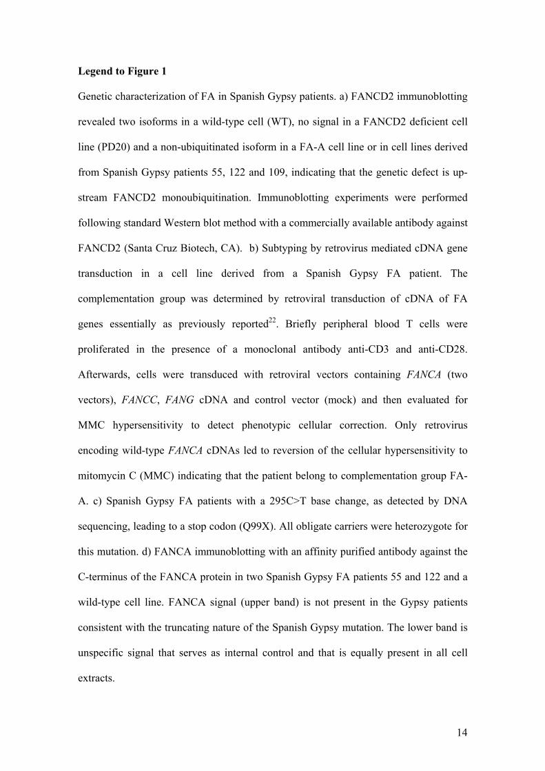
Embed Size (px)
Citation preview
Mutation Research 504 (2002) 75–83
Relationship between chromosome fragility, aneuploidy andseverity of the haematological disease in Fanconi anaemia
Elsa Calléna, Marıa J. Ramıreza, Amadeu Creusa, Ricard Marcosa, Juan J. Ortegab,Teresa Olivéb, Isabel Badellc, Jordi Surrallésa,∗
a Group of Mutagenesis, Department of Genetics and Microbiology, Universitat Autònoma de Barcelona, 08193 Bellaterra, Barcelona, Spainb Bone Marrow Transplantation Unit, Service of Paediatric Hematology-Oncology, Vall d’Hebron Hospital, 08035 Barcelona, Spain
c Bone Marrow Transplantation Unit, Department of Paediatrics, Sant Pau Hospital, 08025 Barcelona, Spain
Received 25 October 2001; received in revised form 5 January 2002; accepted 17 January 2002
Abstract
Fanconi anemia (FA) is a chromosome instability syndrome, characterized by progressive pancytopenia and cancer suscepti-bility. Other cellular features of FA cells are hypersensitivity to DNA cross-linking agents and accelerated telomere shortening.We have quantified overall genome chromosome fragility and euploidy as well as chromosomes 7 and 8 aneuploidy in periph-eral blood lymphocytes from a group of FA patients and age-matched controls that were previously measured for telomerelength. The haematology of FA samples were also characterized in terms of whole blood cell, neuthrophil and platelet counts,transfusion dependency, requirement of androgens, cortico-steroids or bone marrow transplantation, and the development ofbone marrow clonal cytogenetic abnormalities, myelodysplastic syndrome or acute myeloid leukemia. As expected, a highfrequency of spontaneous chromosome breaks was observed in FA patients, especially of chromatid-type. No differences inchromosomes 7 and 8 monosomy, polysomy and non-disjunction were detected between FA patients and controls. The samewas true for overall genome haploidy or polyploidy. Interestingly, the spontaneous levels of chromosome fragility but not ofnumerical abnormalities were correlated to the severity of the haematological disease in FA. None of the variables includedin the present investigation (chromosome fragility, chromosome numerical abnormalities and haematological status) werecorrelated to telomere length. © 2002 Elsevier Science B.V. All rights reserved.
Keywords: Aneuploidy; Chromosome fragility; Fanconi anaemia; Haematological abnormalities; Telomeres
1. Introduction
Fanconi anemia (FA) is a rare autosomal recessivegenetic disease characterized by increased sponta-neous and induced chromosome instability, a diverseassortment of congenital malformations, progressivepancytopenia and cancer susceptibility, especiallyacute myeloid leukaemia (AML), but also solid
∗ Corresponding author. Tel.:+34-93-581-1830;fax: +34-93-581-2387.E-mail address: [email protected] (J. Surralles).
tumors. FA cells are hypersensitive to cross-linkingagents and, to a lesser extent, to ionizing radiation.Other features of FA are abnormal cell cycle regu-lation, oversensitivity to oxidative stress, high levelof apoptosis, overproduction of tumor necrosis fac-tor, deficient induction of P53, and genomic instability(reviewed in[1]).
There are at least eight different genes involved inFA (FANCA, B, C, D1, D2, E, F andG), all of them butFANCB andFANCD1 have been identified[2–8]. Mostof the FA proteins (A, C, E, F, and G) assemble in anuclear complex that is required for the activation, via
0027-5107/02/$ – see front matter © 2002 Elsevier Science B.V. All rights reserved.PII: S0027-5107(02)00081-7
76 E. Callen et al. / Mutation Research 504 (2002) 75–83
monoubiquitination, of FANCD2 in response to DNAdamage[9]. The recently clonedFANCD2 gene is theonly FA gene conserved in evolution[8] and it is tho-ught to be a key player downstream in the FA pathwaywhere ubiquitinated isoform of FANCD2 moves toDNA damage-induced nuclear foci in association withthe double strand break repair protein BRCA1[9].
Although the exact molecular defect is not known,FA cells are directly or indirectly deficient in DNArepair as they show abnormal rearrangements asso-ciated with V(D)J recombination[10] and impairedfidelity in blunt DNA end-joining [11,12]. One ofthe major consequences of this defect is increasedchromosome fragility especially after the treatmentswith DNA cross-linking agents. Also resemblingother chromosome fragility syndromes such as ataxiatelangiectasia and Nijmegen breakage syndromes(reviewed in[13]), FA patients show an acceleratedtelomere shortening[14–16]. Telomeres play an im-portant role in chromosome stability and segregation[17–19]. Elevated frequencies of aneuploidy havebeen reported in telomerase KO mice with shorttelomeres[18]. A causative role of telomere short-ening in the well-documented X-chromosome aneu-ploidy in ageing humans has also been proposed[20].In addition, telomere integrity is also crucial in termsof organismal viability at the haematological level[21,22]. We have therefore, measured chromosomefragility and numerical abnormalities in blood cellsfrom haematologically characterized FA patients andage-matched controls that were previously quantifiedfor telomere length[16]. The aims of this study areto (i) evaluate the spontaneous level of chromosomenumerical abnormalities in FA patients compared tocontrol, (ii) unravel whether spontaneous chromo-some fragility and numerical abnormalities correlatewith the severity of the haematological disease in FAand (iii) investigate a potential modulating role oftelomere shortening in these variables.
2. Materials and methods
2.1. Subjects
Blood samples were obtained, with informed con-sent, from 11 unrelated FA patients during routineclinic visits and from 10 healthy individuals. All
patients belong to complementation group A, as de-termined by complementation analysis after retroviralmediated gene transfer (data not shown), except twopatients whose complementation group is not knownfor technical reasons. The chromosome fragility anal-yses were performed in nine of these patients (threemales and six females; 10.2 ± 1.5 years old, meanand standard error), and in nine age-matched healthyindividuals (four males and five females; 10.3 ± 1.3years old). Aneuploidy data were obtained from 10patients (eight of them included in the chromosomefragility study) and six controls (five of them includedin the chromosome fragility study). Haematologicalvariables were obtained from all FA patients includedin the chromosome fragility study. The telomerelength of all patients and controls included in thechromosome fragility study was previously deter-mined by quantitative fluorescence in situ hybridiza-tion (Q-FISH) [16]. This study was approved by theUniversity Ethics Committee on Human Research.
2.2. Cell culturing and slide preparation
Five hundred microliter of whole blood were in-cubated in 5 ml cultures as described elsewhere[20]for 48 and 72 h in order to get metaphases and cyto-kinesis blocked binucleated cells, respectively. Toobtain metaphases, colcemid (0.1�g/ml; Gibco) wasadded 2 h prior harvesting following standard cyto-genetic procedures. Cells were dropped onto cleanslides and air-dried overnight. Some of the slides werestained with Giemsa and the remaining were used fortelomere length measurement by Q-FISH in an ex-periment published elsewhere with the same donors[16]. To obtain binucleated cells, cytochalasin-B(Sigma; 6�g/ml) was added at 44 h in order to blockcytokinesis and identify first division cells by theirbinucleated appearance. At harvesting time, cell pel-lets were resuspended in cold 0.075 M KCl and im-mediately centrifuged. The cells were then fixed inmethanol:acetic acid (3:1), dropped onto clean slidesand air-dried. The slides were kept at−20◦C in thedark until FISH was performed.
2.3. Structural chromosome abnormalities
Slides were stained with 10% Giemsa in phosphatebuffer, pH 6.8. A total of 26–100 (50 in most cases)
E. Callen et al. / Mutation Research 504 (2002) 75–83 77
complete metaphases per sample were analysed forchromosome aberrations including gaps, chromosomeand chromatid breaks, acentric fragments and chro-mosome and chromatid exchanges.
2.4. Aneuploidy studies
Chromosome numerical abnormalities were studiedby multicolor chromosome specific interphase FISHin mononucleated and binucleated cells. The probesused were centromeric specific for chromosomes 7 and8, and labeled with Spectrum Green (Vysis) and Cy3(Cambio), respectively. The chromosome 8 probe washeated 5 min at 37◦C and then mixed with the chromo-some 7 probe in its commercial buffer before dropping10�l onto the slides and denaturing 5 min at 85◦C.After hybridizing at 37◦C overnight, the slides werewashed twice at 37◦C for 5 min in 2× SSC, twice in60% formamide/2× SSC, twice for 5 min at room tem-perature in 2× SSC and air-dried. The cells were coun-terstained with 20�l DAPI (0.1�g/ml), cover slippedand sealed. Cells were classified according to the num-ber of 7 and 8 chromosome signals. Chromosomes 7and 8 monosomy (2n − 1) and polysomy (2n + 1;2n + 2), and overall haploidy (n) or polyploidy (3nor 4n) were evaluated by scoring 2000 mononucle-ated cells per sample in 72 h cultures. Non-disjunctionwas evaluated in the same slides by scoring 1000 bin-ucleated cells harboring eight signals (correspondingto two chromosomes 7 and two chromosomes 8 pernuclei in case of a normal binucleated cell) per sam-ple and classified according to the distribution of thesesignals in the daughter nuclei[23,24]. These analyseswere performed with Olympus BX-50 fluorescencemicroscope equipped with a 100 W mercury lamp anda 1000× magnification objective with iris aperture.
2.5. Haematological variables
Haematological variables were obtained the sameday of the extraction of the blood used for cytoge-netic studies. These variables include whole bloodcell counts, neuthrophils, platelets, transfusion depen-dency, requirement of androgens, cortico-steroids orbone marrow transplantation (BMT), and the develop-ment of bone marrow clonal cytogenetic abnormali-ties, myelodisplastic syndrome (MDS) or AML. Noneof the patients underwent BMT, suffered AML or pre-
sented bone marrow clonal cytogenetic abnormalitiesinvolving chromosomes 7 and 8 before extracting theblood used in the present investigation. The patientswere classified as being severe or mild. The criteriafor this classification were basically based on the cellcounts corrected for the age of the patients and the re-quirement of specific treatments to recover the low cellcounts, either androgens and/or corticoesteroids and/ortransfusions and, in the most severe cases, the require-ment of bone marrow transplantation after MDS.
2.6. Statistics
The relationship between chromosome fragility ornumerical abnormalities and severity of the haema-tological disease was performed with a one-factorANOVA. The Student’st-test was used to compareFA patients and controls with respect to chromosomebreaks and numerical abnormalities. The relationshipbetween chromosome breaks, numerical abnormali-ties or haematological status and telomere length wasdetermined with a regression analysis. All data wereanalysed using the statistical packages SPSS andCSS:StatisticaTM.
3. Results
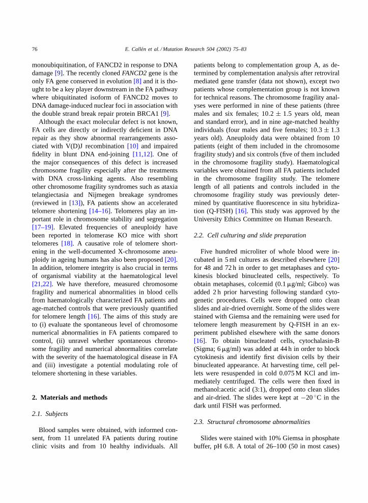
Detailed data on chromosome structural abnormal-ities are shown inTable 1. The frequency of chro-mosome breaks was very low in healthy individuals(four chromatid breaks and one chromosome breakin a total of 427 metaphases analyzed, most of themin a single donor). As expected, spontaneous chro-mosome fragility was detected in FA cells, especiallychromatid-type breaks. In 512 metaphases, a total of68 chromatid-type breaks and 14 chromosome-typebreaks were found. Thus, spontaneous chromosomefragility was >10-fold increased in FA patients whencompared to controls (P < 0.001). The observedinterindividual heterogeneity in spontaneous chromo-some fragility was not explained by heterogeneoustelomere length, since no correlation was found be-tween such variables.
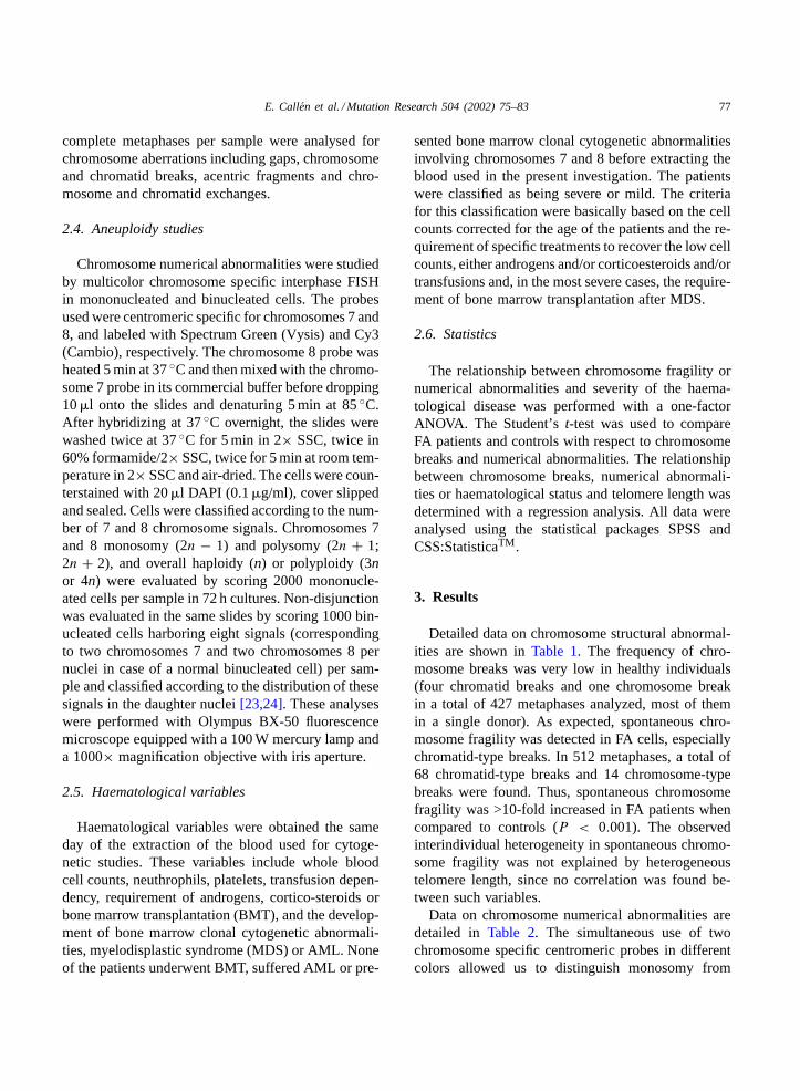
Data on chromosome numerical abnormalities aredetailed in Table 2. The simultaneous use of twochromosome specific centromeric probes in differentcolors allowed us to distinguish monosomy from
78 E. Callen et al. / Mutation Research 504 (2002) 75–83
Table 1Spontaneous chromosome breakage in FA patients and age-matched healthy controls
Donor code Donor age FA Cells scored Chromosome breakage
Chromatidtype
Chromosometype
Totalbreaks
Breaks per100 cells
1 3 Yes 50 0 0 0 02 14 Yes 100 7 1 8 83 7 Yes 36 9 0 9 254 9 Yes 50 11 5 16 325 11 Yes 100 18 4 22 226 10 Yes 50 1 0 1 27 19 Yes 26 2 1 3 11.58 10 Yes 50 12 2 14 289 9 Yes 50 8 1 9 18
Mean± S.D. 16.3 ± 11.5
12 6 Not 50 0 1 1 213 14 Not 50 0 0 0 014 7 Not 50 0 0 0 015 8 Not 27 0 0 0 016 11 Not 50 0 0 0 017 10 Not 50 3 0 3 618 19 Not 50 1 1 2 419 9 Not 50 0 0 0 020 9 Not 50 0 0 0 0
Mean± S.D. 1.3 ± 2.2
haploidy and polysomy from polyploidy. In addition,the use of cytochalasin B-induced binucleated cellspermitted the study of the segregation of chromo-somes by visualizing the reciprocal products of themitosis [23]. The frequency of non-disjunction ofchromosomes 7 and 8 was similar in FA patients andcontrols (P > 0.05). The frequency of chromosome 8monosomy was again very similar in FA patients andcontrols (P > 0.05). A tendency towards higher fre-quencies of monosomy 7 in FA patients was observed,but it did not reach statistical significance (P = 0.07).Similar non-significant results were obtained for otherchromosome numerical abnormalities including hap-loidy and all types of polysomies and polyploidies.No relationship was found between any of the mark-ers of numerical abnormalities and telomere length.It is therefore concluded that FA patients had normallevels of chromosome numerical abnormalities andthat telomere shortening in FA is not associated toany parallel increase in chromosome aneuploidy.
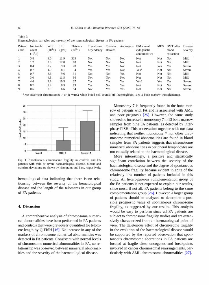
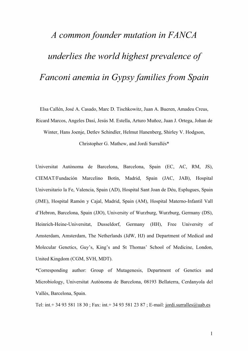
Chromosome abnormalities were also correlatedwith the severity of the haematological disease in FA
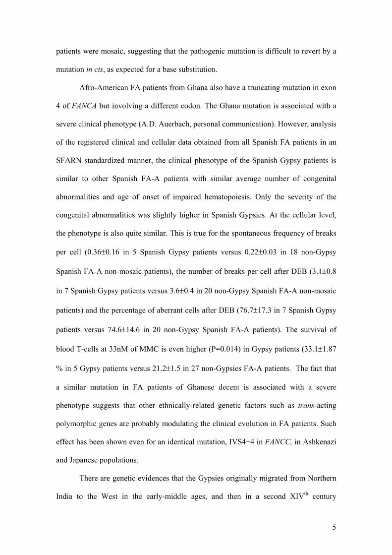
patients in terms of whole blood cell counts, neuthro-phyls, platelets, transfusion dependency, requirementof androgens, cortico-steroids or bone marrow trans-plantation, and the development of bone marrowclonal cytogenetic abnormalities, MDS or AML. Allhematological variables are shown for each FA patientin Table 3. None of the FA patients underwent bonemarrow transplantation or presented AML or bonemarrow cytogenetic abnormalities involving chromo-somes 7 and 8 at the day of blood extraction. To allowmeaningful statistical comparisons, and consideringthat the blood cell counts in children is known to behighly dependent on age, the severity of the hemato-logical disease in FA patients was classified as beingmild or severe. A statistically significant positivecorrelation was found between the degree of chromo-some fragility and the severity of the hematologicaldisease (P < 0.05; Fig. 1). On the contrary, none ofthe markers of chromosome numerical abnormalitieswere correlated to the hematological status of thepatients. Finally, no relationship was found betweenthe previously determined telomere length and the
E.
Callen
etal./M
utationR
esearch504
(2002)75–83
79
80 E. Callen et al. / Mutation Research 504 (2002) 75–83
Table 3Haematological variables and severity of the haematological disease in FA patients
Patientcode
Neutrophilcount(109/l)
WBC(109/l)
Hb(g/dl)
Platelets(109/l)
Transfusiondependency
Cortico-steroids
Androgens BM clonalcytogeneticabnormalities
MDS BMT afterbloodextraction
Diseaseseverity
1 3.8 9.6 11.9 335 Not Not Not Not Not Not Mild2 1.7 3.3 12.8 88 Not Not Not Not Not Not Mild3 0.4 8.7 9.3 28 Yes Yes Not Not Yes Yes Severe4 0.7 1.9 8.1 4 Yes Yes Not Yesa Not Not Severe5 0.7 3.6 9.6 31 Not Not Yes Not Not Not Mild6 3.0 4.8 11.5 86 Not Not Not Not Not Not Mild7 4.6 3.9 10.5 27 Yes Yes Yes Yesa Yes Yes Severe8 0.7 2.4 8.3 19 Yes Not Yes Not Not Not Severe9 0.6 3.0 6.6 54 Not Yes Yes Not Not Not Severe
a Not involving chromosomes 7 or 8; WBC: white blood cell counts; Hb: haemoglobin; BMT: bone marrow transplantation.
Fig. 1. Spontaneous chromosome fragility in controls and FApatients with mild or severe haematological disease. Means andstandard deviations are shown by histograms and bars, respectively.
hematological data indicating that there is no rela-tionship between the severity of the hematologicaldisease and the length of the telomeres in our groupof FA patients.
4. Discussion
A comprehensive analysis of chromosome numeri-cal abnormalities have been performed in FA patientsand controls that were previously quantified for telom-ere length by Q-FISH[16]. No increase in any of themarkers of chromosome numerical abnormalities wasdetected in FA patients. Consistent with normal levelsof chromosome numerical abnormalities in FA, no re-lationship was observed between numerical abnormal-ities and the severity of the haematological disease.
Monosomy 7 is frequently found in the bone mar-row of patients with FA and is associated with AMLand poor prognosis[25]. However, the same studyshowed no increase in monosomy 7 in 13 bone marrowsamples from nine FA patients, as detected by inter-phase FISH. This observation together with our dataindicating that neither monosomy 7 nor other chro-mosome numerical abnormalities are found in bloodsamples from FA patients suggests that chromosomenumerical abnormalities in peripheral lymphocytes arenot causally related to the haematological disease.
More interestingly, a positive and statisticallysignificant correlation between the severity of thehaematological disease and the degree of spontaneouschromosome fragility became evident in spite of therelatively low number of patients included in thisstudy. An heterogeneous complementation group ofthe FA patients is not expected to explain our results,since most, if not all, FA patients belong to the samecomplementation group[26]. However, a larger groupof patients should be analyzed to determine a pos-sible prognostic value of spontaneous chromosomefragility, as suggested by our results. This analysiswould be easy to perform since all FA patients aresubject to chromosome fragility studies and are exten-sively characterized from an haematological point ofview. The deleterious effect of chromosome fragilityin the evolution of the haematological disease wouldbe supported by the reported observation that spon-taneous chromosome aberrations in FA patients arelocated at fragile sites, oncogenes and breakpointsinvolved in cancer chromosomal rearrangements, par-ticularly with AML chromosome abnormalities[27].
E. Callen et al. / Mutation Research 504 (2002) 75–83 81
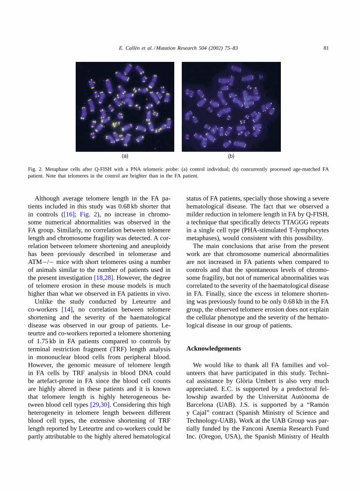
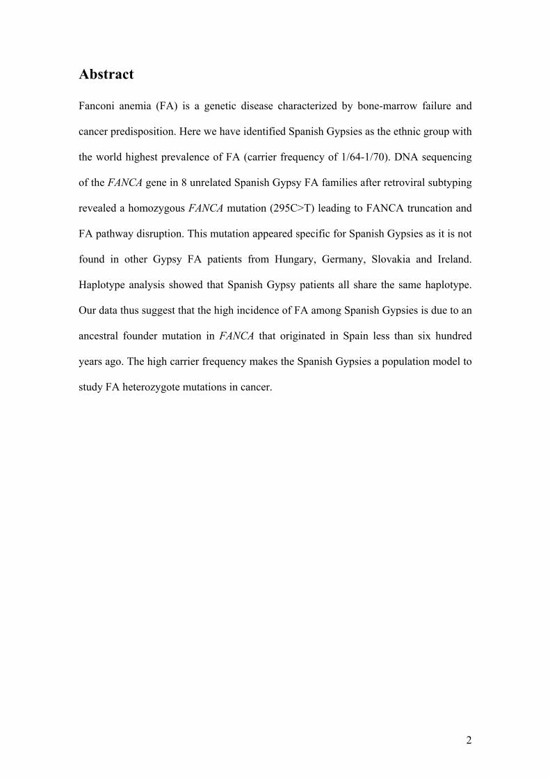
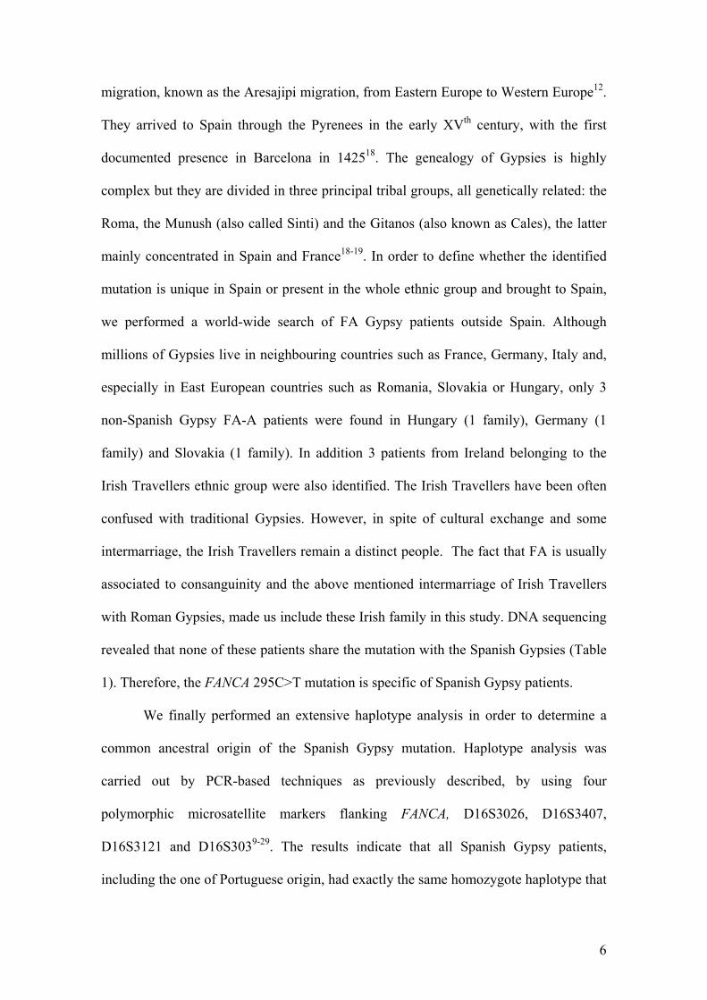
Fig. 2. Metaphase cells after Q-FISH with a PNA telomeric probe: (a) control individual; (b) concurrently processed age-matched FApatient. Note that telomeres in the control are brighter than in the FA patient.
Although average telomere length in the FA pa-tients included in this study was 0.68 kb shorter thatin controls ([16]; Fig. 2), no increase in chromo-some numerical abnormalities was observed in theFA group. Similarly, no correlation between telomerelength and chromosome fragility was detected. A cor-relation between telomere shortening and aneuploidyhas been previously described in telomerase andATM−/− mice with short telomeres using a numberof animals similar to the number of patients used inthe present investigation[18,28]. However, the degreeof telomere erosion in these mouse models is muchhigher than what we observed in FA patients in vivo.
Unlike the study conducted by Leteurtre andco-workers [14], no correlation between telomereshortening and the severity of the haematologicaldisease was observed in our group of patients. Le-teurtre and co-workers reported a telomere shorteningof 1.75 kb in FA patients compared to controls byterminal restriction fragment (TRF) length analysisin mononuclear blood cells from peripheral blood.However, the genomic measure of telomere lengthin FA cells by TRF analysis in blood DNA couldbe artefact-prone in FA since the blood cell countsare highly altered in these patients and it is knownthat telomere length is highly heterogeneous be-tween blood cell types[29,30]. Considering this highheterogeneity in telomere length between differentblood cell types, the extensive shortening of TRFlength reported by Leteurtre and co-workers could bepartly attributable to the highly altered hematological
status of FA patients, specially those showing a severehematological disease. The fact that we observed amilder reduction in telomere length in FA by Q-FISH,a technique that specifically detects TTAGGG repeatsin a single cell type (PHA-stimulated T-lymphocytesmetaphases), would consistent with this possibility.
The main conclusions that arise from the presentwork are that chromosome numerical abnormalitiesare not increased in FA patients when compared tocontrols and that the spontaneous levels of chromo-some fragility, but not of numerical abnormalities wascorrelated to the severity of the haematological diseasein FA. Finally, since the excess in telomere shorten-ing was previously found to be only 0.68 kb in the FAgroup, the observed telomere erosion does not explainthe cellular phenotype and the severity of the hemato-logical disease in our group of patients.
Acknowledgements
We would like to thank all FA families and vol-unteers that have participated in this study. Techni-cal assistance by Glòria Umbert is also very muchappreciated. E.C. is supported by a predoctoral fel-lowship awarded by the Universitat Autònoma deBarcelona (UAB). J.S. is supported by a “Ramóny Cajal” contract (Spanish Ministry of Science andTechnology-UAB). Work at the UAB Group was par-tially funded by the Fanconi Anemia Research FundInc. (Oregon, USA), the Spanish Ministry of Health
82 E. Callen et al. / Mutation Research 504 (2002) 75–83
and Consumption (FIS, 99/1214), and the Commissionof the European Union (Euratom, F1S5-1999-00071).
References
[1] H. Joenje, K.J. Patel, The emerging genetic and molecularbasis of Fanconi anaemia, Nat. Rev. Genet. 2 (2001) 446–457.
[2] C.A. Strathdee, H. Gavish, W.R. Shannon, M. Buchwald,Cloning of cDNAs for Fanconi’s anaemia by functionalcomplementation, Nature 356 (1992) 763–767.
[3] The Fanconi Anaemia/Breast Cancer Consortium Positionalcloning of the Fanconi anemia group A gene. Nat. Genet.Vol. 14, 1996, 324–328.
[4] J.R. Lo Ten Foe, M.A. Rooimans, L. Bosnoyan-Collins, N.Alon, M. Wijker, L. Parker, J. Lightfoot, M. Carreau, D.F.Callen, A. Savoia, N.C. Cheng, C.G. van Berkel, M.H. Strunk,J.J. Gille, G. Pals, F.A. Kruyt, J.C. Pronk, F. Arwert, M.Buchwald, H. Joenje, Expression cloning of a cDNA for themajor Fanconi anaemia gene, FAA, Nat. Genet. 14 (1996)320–323.
[5] J.P. De Winter, Q. Waisfisz, M.A. Rooimans, C.G. van Berkel,L. Bosnoyan-Collins, N. Alon, M. Carreau, O. Bender, I.Demuth, D. Schindler, J.C. Pronk, F. Arwert, H. Hoehn,M. Digweed, M. Buchwald, H. Joenje, The Fanconi anemiagroup G gene FANCG is identical with XRCC9, Nat. Genet.20 (1998) 281–283.
[6] J.P. De Winter, M.A. Rooimans, L. van Der Weel, C.G. vanBerkel, N. Alon, L. Bosnoyan-Collins, J. de Groot, Y. Zhi, Q.Waisfisz, J.C. Pronk, F. Arwert, C.G. Mathew, R.J. Scheper,M.E. Hoatlin, M. Buchwald, H. Joenje, The Fanconi anemiagene FANCF encodes a novel protein with homology to ROM,Nat. Genet. 24 (2000) 15–16.
[7] J.P. De Winter, F. Leveille, C.G. van Berkel, M.A. Rooimans,L. van Der Weel, J. Steltenpool, I. Demuth, N.V. Morgan, N.Alon, L. Bosnoyan-Collins, J. Lightfoot, P.A. Leegwater, Q.Waisfisz, K. Komatsu, F. Arwert, J.C. Pronk, C.G. Mathew,M. Digweed, M. Buchwald, H. Joenje, Isolation of a cDNArepresenting the Fanconi anemia complementation group Egene, Am. J. Hum. Genet. 67 (2000) 1306–1308.
[8] C. Timmers, T. Taniguchi, J. Hejna, C. Reifsteck, L. Lucas,D. Bruun, M. Thayer, B. Cox, S. Olson, A.D. D’Andrea, R.Moses, M. Grompe, Positional cloning of a novel Fanconianemia gene,FANCD2, Mol. Cell 7 (2001) 241–248.
[9] I. Garcia-Higuera, T. Taniguchi, S. Ganesan, M.S. Meyn, C.Timmers, J. Hejna, M. Grompe, A.D. D’Andrea, Interactionof the Fanconi anemia proteins and BRCA1 in a commonpathway, Mol. Cell 7 (2001) 249–262.
[10] J. Smith, J.C. Andrau, S. Kallenbach, A. Laquerbe, N. Doyen,D. Papadopoulo, Abnormal rearrangements associated withV(D)J recombination in Fanconi anemia, J. Mol. Biol. 281(1998) 815–825.
[11] M. Escarceller, S. Rousset, E. Moustacchi, D. Papadopoulo,The fidelity of double strand breaks processing is impairedin complementation groups B and D of Fanconi anemia, agenetic instability syndrome, Somat. Cell Mol. Genet. 23(1997) 401–411.
[12] M. Escarceller, M. Buchwald, B.K. Singleton, P.A. Jeggo,S.P. Jackson, E. Moustacchi, D. Papadopoulo, Fanconi anemiaC gene product plays a role in the fidelity of blunt DNAend-joining, J. Mol. Biol. 279 (1998) 375–385.
[13] P.M. Lansdorp, Repair of telomeric DNA prior to replicativesenescence, Mech. Ageing. Dev. 118 (2000) 23–34.
[14] F. Leteurtre, X. Li, P. Guardiola, G. Le Roux, J.C. Sergère, P.Richard, E.D. Carosella, E. Gluckman, Accelerated telomereshortening and telomerase activation in Fanconi’s anaemia,Br. J. Hematol. 105 (1999) 883–893.
[15] H. Hanson, C.G. Mathew, Z. Docherty, C. Mackie Ogilvie,Telomere shortening in Fanconi anaemia demonstrated by adirect FISH approach, Cytogenet. Cell Genet. 93 (2001) 203–206.
[16] E. Callén, E. Samper, M.J. Ramırez, A. Creus, R. Marcos, J.J.Ortega, T. Olivé, I. Badell, M.A. Blasco, J. Surrallés, Breaksat telomeres and TRF2-independent end-fusions in Fanconianemia, Hum. Mol. Genet. 11 (2002) 439–444.
[17] C.W. Greider, Telomere length regulation, Ann. Rev.Biochem. 65 (1996) 337–365.
[18] M.A. Blasco, H.W. Lee, M.P. Hande, E. Samper,P.M. Lansdorp, R.A. DePinho, C.W. Greider, Telomeraseshortening and tumor formation by mouse cells lackingtelomerase RNA, Cell 91 (1997) 25–34.
[19] M.P. Hande, E. Samper, P. Lansdorp, M.A. Blasco, Telomerelength dynamics and chromosomal instability in cells derivedfrom telomerase null mice, J. Cell Biol. 144 (1999) 589–601.
[20] J. Surrallés, M.P. Hande, R. Marcos, P.M. Lansdorp,Accelerated telomere shortening in the human inactive Xchromosome, Am. J. Hum. Genet. 65 (1999) 1616–1622.
[21] H.W. Lee, M.A. Blasco, G.J. Gottlieb, J.W. Horner II., C.W.Greider, R.A. DePinho, Essential role of mouse telomerasein highly proliferative organs, Nature 392 (1998) 569–574.
[22] E. Herrera, E. Samper, J. Martın-Caballero, J.M. Flores, H.W.Lee, M.A. Blasco, Disease states associated with telomerasedeficiency appear earlier in mice with short telomeres, EMBOJ. 18 (1999) 2950–2960.
[23] A. Zijno, F. Marcon, P. Leopardi, R. Crebelli, Simultaneousdetection of X-chromosome loss and non-disjunctionin cytokinesis-blocked human lymphocytes by in situhybridization with a centromeric DNA probe implicationsfor the human lymphocyte in vitro micronucleus assay usingcytochalasin B, Mutagenesis 3 (1994) 225–232.
[24] J. Surrallés, P. Jeppesen, H. Morrison, A.T. Natarajan,Analysis of loss of inactive X chromosome in interphasecells, Am. J. Hum. Genet. 59 (1996) 1091–1096.
[25] V.C. Thurston, T.M. Ceperich, G.H. Vance, N. A, Detectionof monosomy 7 in bone marrow by fluorescence in situhybridization: a study of Fanconi anemia patients and reviewof the literature, Cancer Genet. Cytogenet. 109 (1999) 154–160.
[26] J.A. Casado, J.C. Segovia, M. Lamana, M.L. Lozano, E.Callén, J. Surrallés, S. Lobitz, H. Hanenberg, J.A. Bueren,Subtyping of Fanconi Anemia patients from Spain using theretroviral complementation assay. Fanconi Anemia ResearchFund Symposium, Abstract Book, Portland, Oregon, 2001.

E. Callen et al. / Mutation Research 504 (2002) 75–83 83
[27] A. Fundia, A. Gorla, I. Larripa, Spontaneous chromosomeaberrations in Fanconi’s anemia are located at fragile sites andacute myeloid leukemia breakpoints, Hereditas 120 (1994)47–50.
[28] M.P. Hande, A.S. Balajee, A. Tchirkov, A. Wynshaw-Boris,P.M. Lansdorp, Extra-chromosomal telomeric DNA in cellsfrom Atm−/− mice and patients with ataxia-telangiectasia,Hum. Mol. Genet. 10 (2001) 519–528.
[29] N.P. Weng, B.L. Levine, C.H. June, R.J. Hodes, Human naiveand memory T lymphocytes differ in telomeric length andreplicative potential, Proc. Natl. Acad. Sci. U.S.A. 92 (1995)11091–11094.
[30] N. Rufer, W. Dragowska, G. Thornbury, E. Roosnek, P.M.Lansdorp, Telomere length dynamics in human lymphocytesubpopulations measured by flow cytometry, Nat. Biotechnol.16 (1998) 743–747.
Applied Aspects/Cancer
Cytogenet Genome Res 104:341–345 (2004)DOI: 10.1159/000077513
Quantitative PCR analysis reveals a highincidence of large intragenic deletions in theFANCA gene in Spanish Fanconi anemiapatientsE. Callén,a M.D. Tischkowitz,b A. Creus,a R. Marcos,a J.A. Bueren,c
J.A. Casado,c C.G. Mathewb and J. Surrallésa
a Universitat Autònoma de Barcelona, Barcelona (Spain), b Division of Genetics and Development,Guy’s, King’s and St Thomas’ School of Medicine, King’s College London, London (UK) andc CIEMAT/Fundacion Marcelino Botın, Madrid (Spain)
Partially funded by the Universitat Autònoma de Barcelona (UAB), the CIEMAT/Marcelino Botın Foundation, the Generalitat de Catalunya (project SGR-00197-2002), the Spanish Ministry of Health and Consumption (projects FIS PI020145and FIS-Red G03/073), the Spanish Ministry of Science and Technology (Pro-jects SAF 2002-03234 and SAF 2003-00328) and the Commission of the Euro-pean Union (project FIGH-CT-2002-00217). E.C. was supported by a predocto-ral fellowship awarded by the UAB. Her visit to CGM laboratory was supportedby a short-term travel fellowship by the UAB. M.D.T. is supported by CancerResearch UK. J.S. is supported by a “Ramon y Cajal” project entitled “Genomestability and DNA repair” awarded by the Spanish Ministry of Science and Tech-nology and co-financed by the UAB.
Received 10 September 2003; manuscript accepted 3 December 2003.
Request reprints from Dr. Jordi Surrallés, Group of MutagenesisDepartment of Genetics and Microbiology, Universitat Autònoma de Barcelona08193 Bellaterra, Barcelona (Spain); telephone: +34 93 581 18 30;fax: +34 93 581 23 87; e-mail: [email protected]
ABC Fax + 41 61 306 12 34E-mail [email protected]
© 2004 S. Karger AG, Basel0301–0171/04/1044–0341$21.00/0
Accessible online at:www.karger.com/cgr
Abstract. Fanconi anaemia is an autosomal recessive dis-ease characterized by chromosome fragility, multiple congeni-tal abnormalities, progressive bone marrow failure and a highpredisposition to develop malignancies. Most of the Fanconianaemia patients belong to complementation group FA-A dueto mutations in the FANCA gene. This gene contains 43 exonsalong a 4.3-kb coding sequence with a very heterogeneousmutational spectrum that makes the mutation screening ofFANCA a difficult task. In addition, as the FANCA gene is richin Alu sequences, it was reported that Alu-mediated recombi-nation led to large intragenic deletions that cannot be detectedin heterozygous state by conventional PCR, SSCP analysis, orDNA sequencing. To overcome this problem, a method basedon quantitative fluorescent multiplex PCR was proposed todetect intragenic deletions in FANCA involving the most fre-
quently deleted exons (exons 5, 11, 17, 21 and 31). Here weapply the proposed method to detect intragenic deletions in 25Spanish FA-A patients previously assigned to complementa-tion group FA-A by FANCA cDNA retroviral transduction. Atotal of eight heterozygous deletions involving from one tomore than 26 exons were detected. Thus, one third of thepatients carried a large intragenic deletion that would have notbeen detected by conventional methods. These results are inagreement with previously published data and indicate thatlarge intragenic deletions are one of the most frequent muta-tions leading to Fanconi anaemia. Consequently, this technolo-gy should be applied in future studies on FANCA to improvethe mutation detection rate.
Copyright © 2003 S. Karger AG, Basel
Fanconi anaemia (FA) is an autosomal recessive diseasecharacterized by genomic instability, multiple congenital ab-normalities, progressive bone marrow failure and a high predis-position to develop malignancies such as acute myeloid leu-kaemia and squamous cell carcinoma (Auerbach et al., 1993).FA cells are characterized by chromosomal fragility and arehypersensitive to DNA cross-linking agents such as diepoxybu-tane and mitomycin C. These features are used as a diagnostictool (Auerbach, 1993).
To date eight different complementation groups have beendescribed (FA-A, -B, -C, -D1, -D2, -E, -F, -G), and the genescorresponding to seven of these complementation groups(FANCA, FANCC, FANCD1/BRCA2, FANCD2, FANCE,
342 Cytogenet Genome Res 104:341–345 (2004)
FANCF and FANCG) have been cloned and characterized(Strathdee et al., 1992; Lo Ten Foe et al., 1996; The FanconiAnemia/Breast Cancer Consortium, 1996; de Winter et al.,1998, 2000a,b; Timmers et al., 2001; Howlett et al., 2002). It isknown that although their protein products do not share signifi-cant homology to each other or with other genes in the data-bases, all of them participate in a common pathway (reviewedin Bogliolo et al., 2002). The FANCA, -C, -G, -E and -F proteinsassemble in a multisubunit nuclear complex (de Winter et al.,2000c; Medhurst et al., 2001) required for monoubiquitin-mediated activation of FANCD2 after DNA damage or duringS-phase and later targeting to nuclear foci containing BRCA1and Rad51 (Garcia-Higuera et al., 2001; Taniguchi et al.,2002). This suggests a role for the FA pathway in DNA repairby homologous recombination during S-phase (Taniguchi et al.,2002).
The genetic heterogeneity of FA makes the subtyping of FApatients a difficult task, but over 90% of the FA populationworldwide is represented by complementation groups FA-A, -Cor -G, FA-A being the most prevalent, accounting for F65% ofall FA cases (Kutler et al., 2003). There are, however, regionaland ethnic differences. It is estimated that about 80% of all FApatients belong to complementation group FA-A in Spain (Cas-ado et al., 2002) or even more in Italy (Savino et al., 1997).Therefore, a proper detection of FANCA mutations is of criti-cal clinical significance.
The FANCA gene contains 43 exons along a 4.3-kb codingsequence (Ianzano et al., 1997) with a very heterogeneousmutational spectrum, with more than 100 different FANCAmutations described to date, including all types of possiblepoint mutations such as frame-shift mutations, small insertionsor deletions, splicing defects, and nucleotide substitutions (TheFanconi Anemia/Breast Cancer Consortium, 1996; Lo Ten Foeet al., 1996; Levran et al., 1997; Savino et al., 1997; Morgan etal., 1999; Wijker et al., 1999). This makes the mutation screen-ing of FANCA a very difficult task. As the FANCA gene is richin Alu sequences, it was suggested and later reported that Alu-mediated recombination is an important mechanism for thegeneration of FANCA mutations (Centra et al., 1998; Levran etal., 1998), mainly large deletions that cannot be detected byconventional methods such as SSCP analysis or DNA sequenc-ing. To overcome this problem, a method based on quantitativefluorescent multiplex PCR was developed. This method wasused to screen 26 cell lines from FA patients belonging to com-plementation group A (Morgan et al., 1999), and a high fre-quency of large deletions was found. In some populations withhigh prevalence of FA such as the South African Afrikaner, thismethod has also been employed to scan the FANCA gene, andit was found that exon 12–31 deletion is the most commonmutation in this population due to a founder effect (Tipping etal., 2001).
Taking advantage of this novel method developed by Mor-gan and co-workers (Morgan et al., 1999), we applied the pro-posed strategy to screen a group of 25 FA Spanish patients pre-viously assigned to complementation group FA-A by retroviraltransduction (Hanenberg et al., 2002; Casado et al., 2002). Thesuggested multiplex PCR was performed with some minormodifications and optimisations to amplify the five most com-
monly deleted exons of the FANCA gene and a control exonfrom an unrelated gene. After analyzing DNA samples from 25Spanish FA-A patients, a total of eight different large deletionsfrom one to more than 26 exons were detected, all of them inheterozygous state, confirming that this type of mutation is oneof the most prevalent amongst FA-A patients.
Materials and methods
PatientsBlood samples were obtained from 25 Spanish FA patients with
informed consent. All of them were previously diagnosed on the basis of theirclinical traits and cellular hypersensitivity to diepoxybutane, and assigned tocomplementation group FA-A by retroviral transduction (Casado et al.,2002). In some cases, lymphoblastoid cell lines established by Epstein-Barrvirus infection were used.
Quantitative fluorescent multiplex PCR reactionGenomic DNA was extracted directly from peripheral blood samples or
lymphoblastoid cell lines by salt/chloroform or phenol/chloroform standardmethods, respectively. DNA concentration was determined spectrophotome-trically and a sample was diluted to 25 ng/Ìl to perform PCR.
Exons 5, 11, 17, 21 and 31 from the FANCA gene were simultaneouslyamplified together with exon 1 of the myelin protein zero (MPZ1) gene as anexternal control with respect to the FANCA gene exons in the same reaction.At least four non-FA control samples were included together with the FAsample in every PCR round as normal controls. PCR amplifications wereperformed in 25-Ìl reactions with 125 ng DNA, 1× Taq DNA polymerasebuffer, 250 ÌM each dNTP and 1.8 mM MgCl2. Primer concentrations andsequences are shown in Table 1. All primers were fluorescently labelled withphosphoramidite 6-FAM dye (PE Biosystems). PCR conditions were an ini-tial denaturation step at 94 ° C for 5 min, followed by 21 cycles of denatur-ation at 94 °C for 45 s, annealing step at 55 °C for 45 s, and extension for1 min at 72 °C. A final extension step at 72 ° C for 5 min was performed. A3-Ìl aliquot of the PCR product was then mixed with 15 Ìl of formamide and0.2 Ìl of Genescan ROX-500 size standard (PE Biosystems). Samples weredenatured at 94 °C for 5 min and run and analysed on an ABI 310 GeneAnalyzer, by means of the GeneScan and Genotyper software, as described inMorgan et al. (1999). Briefly, apart from the FANCA exons (FAAX), anotherinternal control exon from a different gene (exon 1 of the Myelin proteinzero; MPZX1) was co-amplified to detect heterozygous deletions. For each ofthe patients, the peak areas corresponding to the MPZX and each of the fivetested FANCA exons were determined in samples from the patient (pt) andfrom a healthy donor (c). This comparison gave a value called dosage quo-tient (DQ) that corresponds to the following formula: (ptFAAX peak area/ptMPZX1 peak area)/(cFAAX peak area/cMPZX1 peak area). The DQsrange from 0.75 to 1.25 when the DNA copy number of a specific exon isnormal, or half/double this value when there is a heterozygous deletion.
Results and discussion
Mutations in the FANCA gene are the most prevalent causeof FA. More than 70 polymorphisms in this gene and over 100different mutations have been accumulated in the Internation-al Fanconi Anemia Registry Database, most of them being in aheterozygous state. This is a clear indication of the complexityof detecting mutations in this gene. Although several studieshave focused in finding pathogenic mutations among thesepatients, in some cases the successful detection rates were poor(Savino et al., 1997) in part due to the high rate of intragenicdeletions (Centra et al., 1998; Levran et al., 1998; Wijker et al.,1999). This high rate of intragenic deletions along the FANCAgene sequence is the consequence of Alu-mediated intragenic
Cytogenet Genome Res 104:341–345 (2004) 343
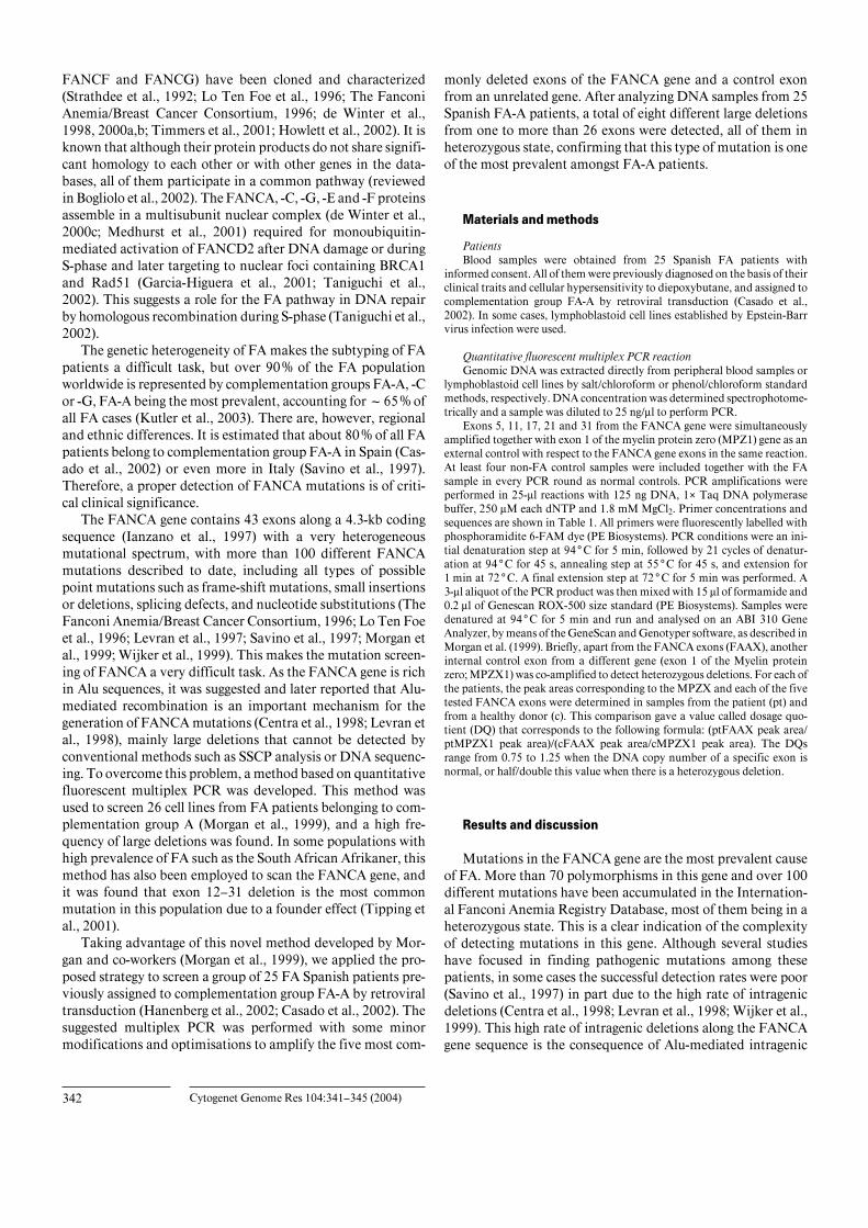
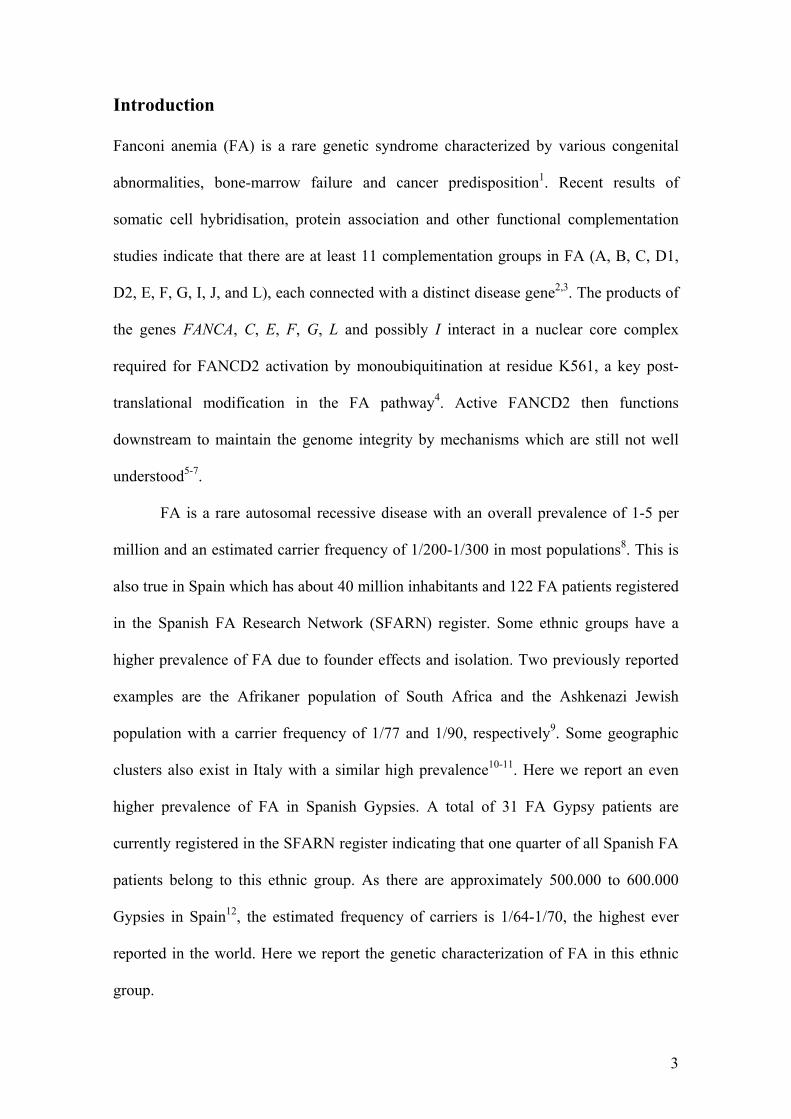
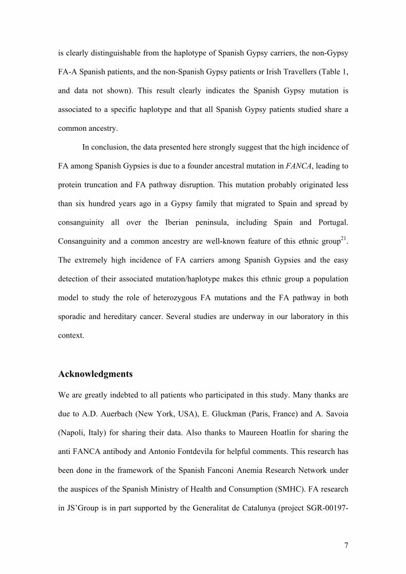
Fig. 1. Electrophoretogram showing the peakpattern of the multiplex products from a healthycontrol (a), compared with an exon 5 deletion car-rier (b). Note that the peak area of exon 5 in thepatient is half of the same exon in the healthyindividual.
Table 1. Sequence, size and concentration ofevery primer of the set used Gene:
Exon Primers (5’ 3’)
Amplicon size (bp)
Concentration (µM)
FANCA: Exon 5 Forward: ACC TGC CCG TTG TTA CTT TTA
Reverse: AGA ACA TTG CCT GGA ACA CTG 250 0.4
0.2 Exon 11 Forward: GAT GAG CCT GAG CCA CAG TTT GTG
Reverse: AGA ATT CCT GGC ATC TCC AGT CAG 301 0.4
0.2 Exon 17 Forward: CCA TGC CCA CTC CTC ACA CC
Reverse: GTG AAA AGA AAC TGG ACC TTT GCA 205 0.08
0.2 Exon 21 Forward: TAA GCC ATA GCT GAC TTA ATT
Reverse: GCA CAA GTC CCA GAG TGG ACA AG 156 0.6
0.2 Exon 31 Forward: CAC ACT GTC AGA GAA GCA CAG CCA
Reverse: CAC CGC GCC TGG CAA TAA ATA TC 285 0.08
0.2
Myelin protein zero: Exon 1 Forward: CAG TGG ACA CAA AGC CCT CTG TGT A
Reverse: GAC ACC TGA GTC CCA AGA CTC CCA G 389 0.2
0.2
recombination (Centra et al., 1998). Here we have applied animproved fluorescent PCR-based method with conditions sim-ilar to those proposed by Morgan et al. (1999), using primersthat amplify exons 5, 11, 17, 21 and 31 of the FANCA gene.These exons were chosen based on previous observations show-ing that at least one of these five exons was absent in all FAN-CA deletions observed to date (Morgan et al., 1999). Someminor modifications in the sequence and concentration ofsome primers used by Morgan et al. (1999) were introduced toimprove the efficiency of the technique.
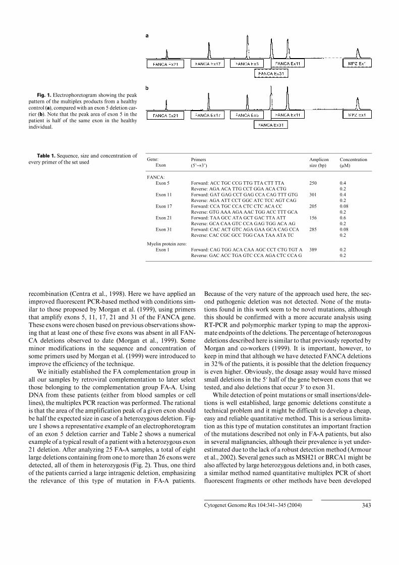
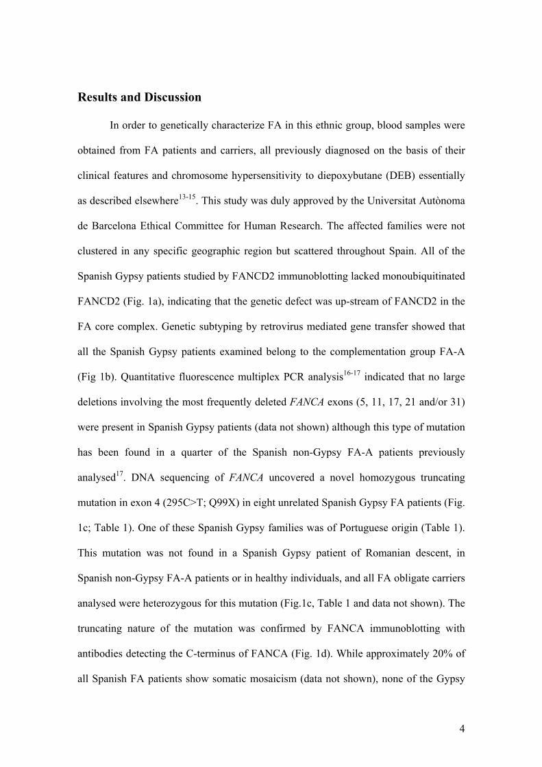
We initially established the FA complementation group inall our samples by retroviral complementation to later selectthose belonging to the complementation group FA-A. UsingDNA from these patients (either from blood samples or celllines), the multiplex PCR reaction was performed. The rationalis that the area of the amplification peak of a given exon shouldbe half the expected size in case of a heterozygous deletion. Fig-ure 1 shows a representative example of an electrophoretogramof an exon 5 deletion carrier and Table 2 shows a numericalexample of a typical result of a patient with a heterozygous exon21 deletion. After analyzing 25 FA-A samples, a total of eightlarge deletions containing from one to more than 26 exons weredetected, all of them in heterozygosis (Fig. 2). Thus, one thirdof the patients carried a large intragenic deletion, emphasizingthe relevance of this type of mutation in FA-A patients.
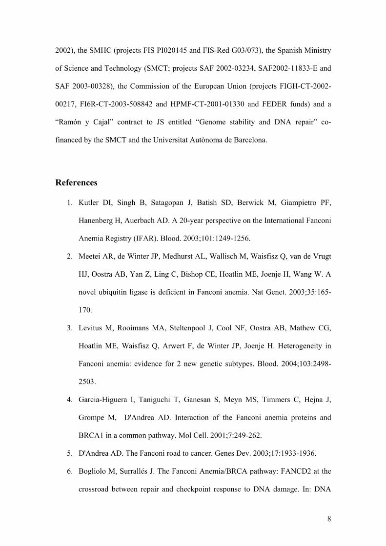
Because of the very nature of the approach used here, the sec-ond pathogenic deletion was not detected. None of the muta-tions found in this work seem to be novel mutations, althoughthis should be confirmed with a more accurate analysis usingRT-PCR and polymorphic marker typing to map the approxi-mate endpoints of the deletions. The percentage of heterozogousdeletions described here is similar to that previously reported byMorgan and co-workers (1999). It is important, however, tokeep in mind that although we have detected FANCA deletionsin 32% of the patients, it is possible that the deletion frequencyis even higher. Obviously, the dosage assay would have missedsmall deletions in the 5) half of the gene between exons that wetested, and also deletions that occur 3) to exon 31.
While detection of point mutations or small insertions/dele-tions is well established, large genomic deletions constitute atechnical problem and it might be difficult to develop a cheap,easy and reliable quantitative method. This is a serious limita-tion as this type of mutation constitutes an important fractionof the mutations described not only in FA-A patients, but alsoin several malignancies, although their prevalence is yet under-estimated due to the lack of a robust detection method (Armouret al., 2002). Several genes such as MSH21 or BRCA1 might bealso affected by large heterozygous deletions and, in both cases,a similar method named quantitative multiplex PCR of shortfluorescent fragments or other methods have been developed
344 Cytogenet Genome Res 104:341–345 (2004)
Fig. 2. Scheme of the large deletions detected in the 25 samples analyzed, along the schematic figure of FANCA gene exons.The amplified exons are shown in grey colour. Continuous lines refer to the exons that are deleted and stippled lines refer to thefact that the exact break points are not detected by this method.
Table 2. Results of a dosage multiplex assay ofan exon 21 deletion carrier Exon Peak area Dosage quotient
Healthy donor
FA patient
MPZX1 FAAX5 FAAX11 FAAX17 FAAX21 FAAX31
MPZX1 62062 17523 – 1.28 1.17 1.05 2.30 1.07 FAAX5 36569 8083 0.78 – 0.91 0.82 1.80 0.84 FAAX11 56996.5 13794 0.86 1.09 – 0.90 1.97 0.92 FAAX17 19361 5218 0.95 1.22 1.11 – 2.19 1.02 FAAX21 23321 2864 0.43 0.56 0.51 0.46 – 0.47 FAAX31 14868 3914 0.93 1.19 1.09 0.98 2.14 –
for this same purpose (Charbonnier et al., 2000; Casilli et al.,2002; Wang et al., 2002, 2003). We proposed that the approachdescribed here could also be applied to the above mentionedgenes by just adapting the primers and PCR conditions. Mostmethods used to date are based on PCR although other tech-niques should be considered using cytogenetic tools or South-ern-blot (Armour et al., 2002). Although PCR is basically aqualitative technique, several modifications can be introducedto get quantitative or semi-quantitative results, for example byusing control samples and internal controls to coamplify andcompare with the sample under study, in a manner similar tothe method described here.
In conclusion, we have found that one third of the SpanishFA-A patients bear large monoallelic deletions in FANCA. Thisresult confirms the suitability of quantitative PCR to findotherwise undetectable pathogenic mutations and hence toincrease the mutation detection rate in FA. This would lead to abetter molecular tool in pre- and postnatal diagnosis and in car-rier detection.
Acknowledgements
We would like to thank all FA families and haemato-oncologists thathave participated in this study.
References
Armour JAL, Barton DE, Cockburn DJ, Taylor GR:The detection of large deletions or duplications ingenomic DNA. Hum Mutat 20:325–337 (2002).
Auerbach AD: Fanconi anemia diagnosis and the die-poxybutane (DEB) test. Exp Hematol 21:731–733(1993).
Auerbach AD, Buchwald M, Joenje H: Fanconi Ane-mia, in Volgestein B, Kinzler KW (eds): The Ge-netic Basis of Human Cancer, pp 317–332(McGraw-Hill, New York 1993).
Bogliolo M, Cabré O, Callén E, Castillo V, Creus A,Marcos R, Surrallés J: The Fanconi anaemia ge-nome stability and tumour suppressor network.Mutagenesis 17:529–538 (2002).
Casado JA, Callén E, Surrallés J, Segovia JC, Rio P,Lobitz S, Hanenberg H, Bueren JA: Progress in thesubtyping analysis of Fanconi anemia patientsfrom Spain using retroviral mediated gene transferof FA genes and FANCD2 Western blot analysis.Abstract. Twelfth Annual Fanconi Anemia Scien-tific Symposium, Philadelphia (2002).
Casilli F, Di Rocco ZC, Gad S, Tournier I, Stoppa-Lyonnet D, Frebourg T, Tosi M: Rapid detectionof novel BRCA1 rearrangements in high-riskbreast-ovarian cancer families using multiplexPCR of short fluorescent fragments. Hum Mutat20:218–226 (2002).
Centra M, Memeo E, d’Apolito M, Savino M, IanzanoL, Notarangelo A, Liu J, Doggett NA, Zelante L,Savoia A: Fine exon-intron structure of the Fan-coni anemia group A (FAA) gene and characteriza-tion of two genomic deletions. Genomics 51:463–467 (1998).
Charbonnier F, Raux G, Wang Q, Drouot N, Cordier F,Limacher JM, Saurin J, Puisieux A, Olschwang S,Frebourg T: Detection of exon deletions and dupli-cations of the mismatch repair genes in hereditarynonpolyposis colorectal cancer families using mul-tiplex polymerase chain reaction of short fluores-cent fragments. Cancer Res 60:2760–2763 (2000).
Fanconi Anemia/ Breast Cancer Consortium: Position-al cloning of the Fanconi anemia group A gene.Nature Genet 14:324–328 (1996).
Garcıa-Higuera I, Taniguchi T, Ganesan S, Meyn MS,Timmers C, Hejna J, Grompe M, D’Andrea AD:Interaction of the Fanconi anemia proteins andBRCA1 in a common pathway. Mol Cell 7:249–262 (2001).
Cytogenet Genome Res 104:341–345 (2004) 345
Hanenberg H, Batish SD, Pollok KE, Vieten L, Verlan-der PC, Leurs C, Cooper RJ, Gottsche K, HanelineL, Clapp DW, Lobitz S, Williams DA, AuerbachAD: Phenotypic correction of primary Fanconianemia T cells with retroviral vectors as a diagnos-tic tool. Exp Hematol 30:410–420 (2002).
Howlett NG, Taniguchi T, Olson S, Cox B, Waisfisz Q,de Die-Smulders C, Persky N, Grompe M, JoenjeH, Pals G, Ikeda H, Fox EA, D’Andrea AD: Biallel-ic inactivation of BRCA2 in Fanconi anemia.Science 297:606–609 (2002).
Ianzano L, D’Apolito M, Centra M, Savino M, LevranO, Auerbach AD, Cleton-Jansen AM, Doggett NA,Pronk JC, Tipping AJ, Gibson RA, Mathew CG,Whitmore SA, Apostolou S, Callen DF, Zelante L,Savoia A: The genomic organization of the Fan-coni anemia group A (FAA) gene. Genomics41:309–314 (1997).
Joenje H, Lo Ten Foe JR, Oostra AB, van Berkel CG,Rooimans MA, Schroeder-Kurth T, Wegner RD,Gille JJ, Buchwald M, Arwert F: Classification ofFanconi anemia patients by complementationanalisis: evidence for a fifth genetic subtype. Blood86:2156–2160 (1995).
Kutler DI, Singh B, Satagopan J, Batish SD, BerwickM, Giampietro PF, Hanenberg H, Auerbach AD:A 20-year perspective on the International FanconiAnemia Registry (IFAR). Blood 101:1249–1256(2003).
Levran O, Erlich T, Magdalena N, Gregory JJ, BatishSD, Verlander PC, Auerbach AD: Sequence varia-tion in the Fanconi anemia gene FAA. Proc natlAcad Sci, USA 94:13051–13056 (1997).
Levran O, Doggett NA, Auerbach AD: Identification ofAlu-mediated deletions in the Fanconi anemiagene FAA. Hum Mutat 12:145–152 (1998).
Lo Ten Foe JR, Rooimans MA, Bosnoyan-Collins L,Alon N, Wijker M, Parker L, Lightfoot J, CarreauM, Callen DF, Savoia A, Cheng NC, van BerkelCG, Strunk MH, Gille JJ, Pals G, Kruyt FA, PronkJC, Arwert F, Buchwald M, Joenje H: Expressioncloning of a cDNA for the major Fanconi anemiagene, FAA. Nature Genet 14:320–323 (1996).
Medhurst AL, Huber PA, Waisfisz Q, de Winter JP,Mathew CG: Direct interactions of the five knownFanconi anemia proteins suggest a common func-tional pathway. Hum molec Genet 10:423–429(2001).
Morgan NV, Tipping AJ, Joenje H, Mathew CG: Highfrequency of large intragenic deletions in the Fan-coni anemia group A gene. Am J hum Genet65:1330–1341 (1999).
Savino M, Ianzano L, Strippoli P, Ramenghi U, Arslan-ian A, Bagnara GP, Joenje H, Zelante L, Savoia A:Mutations of the Fanconi anemia group A gene(FAA) in Italian patients. Am J hum Genet 61:1246–1253 (1997).
Strathdee CA, Duncan AM, Buchwald M: Evidence forat least four Fanconi anaemia genes includingFACC on chromosome 9. Nature Genet 1:196–198(1992).
Taniguchi T, Garcia-Higuera I, Andreassen PR, Grego-ry RC, Grompe M, D’Andrea AD: S-phase-specificinteraction of the Fanconi anemia protein,FANCD2, with BRCA1 and RAD51. Blood 100:2414–2420 (2002).
Timmers C, Taniguchi T, Hejna J, Reifsteck C, LucasL, Bruun D, Thayer M, Cox B, Olson S, D’AndreaAD, Moses R, Grompe M: Positional cloning of anovel Fanconi anemia gene, FANCD2. Mol Cell7:241–248 (2001).
Tipping AJ, Pearson T, Morgan NV, Gibson RA, KuytLP, Havenga C, Gluckman E, Joenje H, de RavelT, Jansen S, Mathew CG: Molecular and genealogi-cal evidence for a founder effect in Fanconi anemiafamilies of the Afrikaner population of South Afri-ca. Proc natl Acad Sci, USA 98:5734–5739 (2001).
Wang Y, Friedl W, Sengteller M, Jungck M, Filges I,Propping P, Mangold E: A modified multiplexPCR assay for detection of large deletions inMSH2 and MSH1. Hum Mutat 19:279–286(2002).
Wang Y, Friedl W, Lamberti C, Jungck M, Mathiak M,Pagenstecher C, Propping P, Mangold E: Heredi-tary nonpolyposis colorectal cancer: Frequent oc-currence of large genomic deletions in MSH2 andMLH1 genes. Int J Cancer 103:636–641 (2003).
Wijker M, Morgan NV, Herterich S, van Berkel CG,Tipping AJ, Gross HJ, Gille JJ, Pals G, Savino M,Altay C, Mohan S, Dokal I, Cavenagh J, Marsh J,van Weel M, Ortega JJ, Schuler D, Samochatova E,Karwacki M, Bekassy AN, Abecasis M, Ebell W,Kwee ML, de Ravel T, Mathew CG: Heterogenousspectrum of mutations in the Fanconi anaemiagroup A gene. Eur J hum Genet 7:52–59 (1999).
de Winter JP, Waisfisz Q, Rooimans MA, van BerkelCGM, Bosnoyan-Collins L, Alon N, Bender O,Demuth I, Schindler D, Pronk JC, Arwert F,Hoehn H, Digweed M, Buchwald M, Joenje H: TheFanconi anaemia group G gene FANCG is identi-cal with XRCC9. Nature Genet 20:281–283(1998).
de Winter JP, Leveille F, Waisfisz Q, van Berkel CG,Rooimans MA, van Der Weel L, Steltenpool J,Demuth I, Morgan NV, Alon N, Bosnoyan-CollinsL, Lightfoot J, Leegwater PA, Waisfisz Q, KomatsuK, Arwert F, Pronk JC, Mathew CG, Digweed M,Buchwald M, Joenje H: Isolation of a cDNA repre-senting the Fanconi anemia complementationgoup E gene. Am J hum Genet 67:1306–1308(2000a).
de Winter JP, Rooimans MA, van Der Weel L, van Ber-kel CG, Alon N, Bosnoyan-Collins L, de Groot J,Zhi Y, Waisfisz Q, Pronk JC, Arwert F, MathewCG, Scheper RJ, Hoatlin ME, Buchwald M, JoenjeH: The Fanconi anaemia gene FANCF encodes anovel protein with homology to ROM. NatureGenet 24:15–16 (2000b).
de Winter JP, van der Weel L, de Groot J, Stone S,Waisfisz Q, Arwert F, Scheper RJ, Kruyt FA,Hoatlin ME, Joenje H: The Fanconi anemia pro-tein FANCF forms a nuclear complex with FAN-CA, FANCC and FANCG. Hum molec Genet9:2665–2674 (2000c).
A common founder mutation in FANCA
underlies the world highest prevalence of
Fanconi anemia in Gypsy families from Spain
Elsa Callén, José A. Casado, Marc D. Tischkowitz, Juan A. Bueren, Amadeu Creus,
Ricard Marcos, Angeles Dasí, Jesús M. Estella, Arturo Muñoz, Juan J. Ortega, Johan de
Winter, Hans Joenje, Detlev Schindler, Helmut Hanenberg, Shirley V. Hodgson,
Christopher G. Mathew, and Jordi Surrallés*
Universitat Autònoma de Barcelona, Barcelona, Spain (EC, AC, RM, JS),
CIEMAT/Fundación Marcelino Botín, Madrid, Spain (JAC, JAB), Hospital
Universitario la Fe, Valencia, Spain (AD), Hospital Sant Joan de Déu, Esplugues, Spain
(JME), Hospital Ramón y Cajal, Madrid, Spain (AM), Hospital Materno-Infantil Vall
d’Hebron, Barcelona, Spain (JJO), University of Wurzburg, Wurzburg, Germany (DS),
Heinrich-Heine-Universitat, Dusseldorf, Germany (HH), Free University of
Amsterdam, Amsterdam, The Netherlands (JdW, HJ) and Department of Medical and
Molecular Genetics, Guy’s, King’s and St Thomas’ School of Medicine, London,
United Kingdom (CGM, SVH, MDT).
*Corresponding author: Group of Mutagenesis, Department of Genetics and
Microbiology, Universitat Autònoma de Barcelona, 08193 Bellaterra, Cerdanyola del
Vallès, Barcelona, Spain.
Tel: int.+ 34 93 581 18 30 ; Fax: int.+ 34 93 581 23 87 ; E-mail: [email protected]
1
Abstract
Fanconi anemia (FA) is a genetic disease characterized by bone-marrow failure and
cancer predisposition. Here we have identified Spanish Gypsies as the ethnic group with
the world highest prevalence of FA (carrier frequency of 1/64-1/70). DNA sequencing
of the FANCA gene in 8 unrelated Spanish Gypsy FA families after retroviral subtyping
revealed a homozygous FANCA mutation (295C>T) leading to FANCA truncation and
FA pathway disruption. This mutation appeared specific for Spanish Gypsies as it is not
found in other Gypsy FA patients from Hungary, Germany, Slovakia and Ireland.
Haplotype analysis showed that Spanish Gypsy patients all share the same haplotype.
Our data thus suggest that the high incidence of FA among Spanish Gypsies is due to an
ancestral founder mutation in FANCA that originated in Spain less than six hundred
years ago. The high carrier frequency makes the Spanish Gypsies a population model to
study FA heterozygote mutations in cancer.
2
Introduction
Fanconi anemia (FA) is a rare genetic syndrome characterized by various congenital
abnormalities, bone-marrow failure and cancer predisposition1. Recent results of
somatic cell hybridisation, protein association and other functional complementation
studies indicate that there are at least 11 complementation groups in FA (A, B, C, D1,
D2, E, F, G, I, J, and L), each connected with a distinct disease gene2,3. The products of
the genes FANCA, C, E, F, G, L and possibly I interact in a nuclear core complex
required for FANCD2 activation by monoubiquitination at residue K561, a key post-
translational modification in the FA pathway4. Active FANCD2 then functions
downstream to maintain the genome integrity by mechanisms which are still not well
understood5-7.
FA is a rare autosomal recessive disease with an overall prevalence of 1-5 per
million and an estimated carrier frequency of 1/200-1/300 in most populations8. This is
also true in Spain which has about 40 million inhabitants and 122 FA patients registered
in the Spanish FA Research Network (SFARN) register. Some ethnic groups have a
higher prevalence of FA due to founder effects and isolation. Two previously reported
examples are the Afrikaner population of South Africa and the Ashkenazi Jewish
population with a carrier frequency of 1/77 and 1/90, respectively9. Some geographic
clusters also exist in Italy with a similar high prevalence10-11. Here we report an even
higher prevalence of FA in Spanish Gypsies. A total of 31 FA Gypsy patients are
currently registered in the SFARN register indicating that one quarter of all Spanish FA
patients belong to this ethnic group. As there are approximately 500.000 to 600.000
Gypsies in Spain12, the estimated frequency of carriers is 1/64-1/70, the highest ever
reported in the world. Here we report the genetic characterization of FA in this ethnic
group.
3
Results and Discussion
In order to genetically characterize FA in this ethnic group, blood samples were
obtained from FA patients and carriers, all previously diagnosed on the basis of their
clinical features and chromosome hypersensitivity to diepoxybutane (DEB) essentially
as described elsewhere13-15. This study was duly approved by the Universitat Autònoma
de Barcelona Ethical Committee for Human Research. The affected families were not
clustered in any specific geographic region but scattered throughout Spain. All of the
Spanish Gypsy patients studied by FANCD2 immunoblotting lacked monoubiquitinated
FANCD2 (Fig. 1a), indicating that the genetic defect was up-stream of FANCD2 in the
FA core complex. Genetic subtyping by retrovirus mediated gene transfer showed that
all the Spanish Gypsy patients examined belong to the complementation group FA-A
(Fig 1b). Quantitative fluorescence multiplex PCR analysis16-17 indicated that no large
deletions involving the most frequently deleted FANCA exons (5, 11, 17, 21 and/or 31)
were present in Spanish Gypsy patients (data not shown) although this type of mutation
has been found in a quarter of the Spanish non-Gypsy FA-A patients previously
analysed17. DNA sequencing of FANCA uncovered a novel homozygous truncating
mutation in exon 4 (295C>T; Q99X) in eight unrelated Spanish Gypsy FA patients (Fig.
1c; Table 1). One of these Spanish Gypsy families was of Portuguese origin (Table 1).
This mutation was not found in a Spanish Gypsy patient of Romanian descent, in
Spanish non-Gypsy FA-A patients or in healthy individuals, and all FA obligate carriers
analysed were heterozygous for this mutation (Fig.1c, Table 1 and data not shown). The
truncating nature of the mutation was confirmed by FANCA immunoblotting with
antibodies detecting the C-terminus of FANCA (Fig. 1d). While approximately 20% of
all Spanish FA patients show somatic mosaicism (data not shown), none of the Gypsy
4
patients were mosaic, suggesting that the pathogenic mutation is difficult to revert by a
mutation in cis, as expected for a base substitution.
Afro-American FA patients from Ghana also have a truncating mutation in exon
4 of FANCA but involving a different codon. The Ghana mutation is associated with a
severe clinical phenotype (A.D. Auerbach, personal communication). However, analysis
of the registered clinical and cellular data obtained from all Spanish FA patients in an
SFARN standardized manner, the clinical phenotype of the Spanish Gypsy patients is
similar to other Spanish FA-A patients with similar average number of congenital
abnormalities and age of onset of impaired hematopoiesis. Only the severity of the
congenital abnormalities was slightly higher in Spanish Gypsies. At the cellular level,
the phenotype is also quite similar. This is true for the spontaneous frequency of breaks
per cell (0.36±0.16 in 5 Spanish Gypsy patients versus 0.22±0.03 in 18 non-Gypsy
Spanish FA-A non-mosaic patients), the number of breaks per cell after DEB (3.1±0.8
in 7 Spanish Gypsy patients versus 3.6±0.4 in 20 non-Gypsy Spanish FA-A non-mosaic
patients) and the percentage of aberrant cells after DEB (76.7±17.3 in 7 Spanish Gypsy
patients versus 74.6±14.6 in 20 non-Gypsy Spanish FA-A patients). The survival of
blood T-cells at 33nM of MMC is even higher (P=0.014) in Gypsy patients (33.1±1.87
% in 5 Gypsy patients versus 21.2±1.5 in 27 non-Gypsies FA-A patients. The fact that
a similar mutation in FA patients of Ghanese decent is associated with a severe
phenotype suggests that other ethnically-related genetic factors such as trans-acting
polymorphic genes are probably modulating the clinical evolution in FA patients. Such
effect has been shown even for an identical mutation, IVS4+4 in FANCC, in Ashkenazi
and Japanese populations.
There are genetic evidences that the Gypsies originally migrated from Northern
India to the West in the early-middle ages, and then in a second XIVth century
5
migration, known as the Aresajipi migration, from Eastern Europe to Western Europe12.
They arrived to Spain through the Pyrenees in the early XVth century, with the first
documented presence in Barcelona in 142518. The genealogy of Gypsies is highly
complex but they are divided in three principal tribal groups, all genetically related: the
Roma, the Munush (also called Sinti) and the Gitanos (also known as Cales), the latter
mainly concentrated in Spain and France18-19. In order to define whether the identified
mutation is unique in Spain or present in the whole ethnic group and brought to Spain,
we performed a world-wide search of FA Gypsy patients outside Spain. Although
millions of Gypsies live in neighbouring countries such as France, Germany, Italy and,
especially in East European countries such as Romania, Slovakia or Hungary, only 3
non-Spanish Gypsy FA-A patients were found in Hungary (1 family), Germany (1
family) and Slovakia (1 family). In addition 3 patients from Ireland belonging to the
Irish Travellers ethnic group were also identified. The Irish Travellers have been often
confused with traditional Gypsies. However, in spite of cultural exchange and some
intermarriage, the Irish Travellers remain a distinct people. The fact that FA is usually
associated to consanguinity and the above mentioned intermarriage of Irish Travellers
with Roman Gypsies, made us include these Irish family in this study. DNA sequencing
revealed that none of these patients share the mutation with the Spanish Gypsies (Table
1). Therefore, the FANCA 295C>T mutation is specific of Spanish Gypsy patients.
We finally performed an extensive haplotype analysis in order to determine a
common ancestral origin of the Spanish Gypsy mutation. Haplotype analysis was
carried out by PCR-based techniques as previously described, by using four
polymorphic microsatellite markers flanking FANCA, D16S3026, D16S3407,
D16S3121 and D16S3039-29. The results indicate that all Spanish Gypsy patients,
including the one of Portuguese origin, had exactly the same homozygote haplotype that
6
is clearly distinguishable from the haplotype of Spanish Gypsy carriers, the non-Gypsy
FA-A Spanish patients, and the non-Spanish Gypsy patients or Irish Travellers (Table 1,
and data not shown). This result clearly indicates the Spanish Gypsy mutation is
associated to a specific haplotype and that all Spanish Gypsy patients studied share a
common ancestry.
In conclusion, the data presented here strongly suggest that the high incidence of
FA among Spanish Gypsies is due to a founder ancestral mutation in FANCA, leading to
protein truncation and FA pathway disruption. This mutation probably originated less
than six hundred years ago in a Gypsy family that migrated to Spain and spread by
consanguinity all over the Iberian peninsula, including Spain and Portugal.
Consanguinity and a common ancestry are well-known feature of this ethnic group21.
The extremely high incidence of FA carriers among Spanish Gypsies and the easy
detection of their associated mutation/haplotype makes this ethnic group a population
model to study the role of heterozygous FA mutations and the FA pathway in both
sporadic and hereditary cancer. Several studies are underway in our laboratory in this
context.
Acknowledgments
We are greatly indebted to all patients who participated in this study. Many thanks are
due to A.D. Auerbach (New York, USA), E. Gluckman (Paris, France) and A. Savoia
(Napoli, Italy) for sharing their data. Also thanks to Maureen Hoatlin for sharing the
anti FANCA antibody and Antonio Fontdevila for helpful comments. This research has
been done in the framework of the Spanish Fanconi Anemia Research Network under
the auspices of the Spanish Ministry of Health and Consumption (SMHC). FA research
in JS’Group is in part supported by the Generalitat de Catalunya (project SGR-00197-
7
2002), the SMHC (projects FIS PI020145 and FIS-Red G03/073), the Spanish Ministry
of Science and Technology (SMCT; projects SAF 2002-03234, SAF2002-11833-E and
SAF 2003-00328), the Commission of the European Union (projects FIGH-CT-2002-
00217, FI6R-CT-2003-508842 and HPMF-CT-2001-01330 and FEDER funds) and a
“Ramón y Cajal” contract to JS entitled “Genome stability and DNA repair” co-
financed by the SMCT and the Universitat Autònoma de Barcelona.
References
1. Kutler DI, Singh B, Satagopan J, Batish SD, Berwick M, Giampietro PF,
Hanenberg H, Auerbach AD. A 20-year perspective on the International Fanconi
Anemia Registry (IFAR). Blood. 2003;101:1249-1256.
2. Meetei AR, de Winter JP, Medhurst AL, Wallisch M, Waisfisz Q, van de Vrugt
HJ, Oostra AB, Yan Z, Ling C, Bishop CE, Hoatlin ME, Joenje H, Wang W. A
novel ubiquitin ligase is deficient in Fanconi anemia. Nat Genet. 2003;35:165-
170.
3. Levitus M, Rooimans MA, Steltenpool J, Cool NF, Oostra AB, Mathew CG,
Hoatlin ME, Waisfisz Q, Arwert F, de Winter JP, Joenje H. Heterogeneity in
Fanconi anemia: evidence for 2 new genetic subtypes. Blood. 2004;103:2498-
2503.
4. Garcia-Higuera I, Taniguchi T, Ganesan S, Meyn MS, Timmers C, Hejna J,
Grompe M, D'Andrea AD. Interaction of the Fanconi anemia proteins and
BRCA1 in a common pathway. Mol Cell. 2001;7:249-262.
5. D'Andrea AD. The Fanconi road to cancer. Genes Dev. 2003;17:1933-1936.
6. Bogliolo M, Surrallés J. The Fanconi Anemia/BRCA pathway: FANCD2 at the
crossroad between repair and checkpoint response to DNA damage. In: DNA
8
Repair and Human Diseases, Ed. Landes Bioscience Ed., edited by Adayabalam
Balajee. New York. 2004 (in press).
7. Surrallés J, Jackson SP, Jasin M, Kastan MB, West SC, Joenje H. Molecular
cross talk among chromosome fragility syndromes. Genes Dev. 2004;18:1359-
1370.
8. Joenje H, Patel KJ. The emerging genetic and molecular basis of Fanconi
anaemia. Nat Rev Genet. 2001;2:446-457.
9. Tipping AJ, Pearson T, Morgan NV, Gibson RA, Kuyt LP, Havenga C,
Gluckman E, Joenje H, de Ravel T, Jansen S, Mathew CG. Molecular and
genealogical evidence for a founder effect in Fanconi anemia families of the
Afrikaner population of South Africa. Proc Natl Acad Sci U S A. 2001;98:5734-
5739.
10. Savoia A, Zatterale A, Del Principe D, Joenje H. Fanconi anaemia in Italy: high
prevalence of complementation group A in two geographic clusters. Hum Genet.
1996;97:599-603.
11. Savino M, Ianzano L, Strippoli P, Ramenghi U, Arslanian A, Bagnara GP,
Joenje H, Zelante L, Savoia A. Mutations of the Fanconi anemia group A gene
(FAA) in Italian patients. Am J Hum Genet. 1997;61:1246-1253.
12. Ramal LM, de Pablo R, Guadix MJ, Sanchez J, Garrido A, Garrido F, Jimenez-
Alonso J, Lopez-Nevot MA. HLA class II allele distribution in the Gypsy
community of Andalusia, southern Spain. Tissue Antigens. 2001;57:138-143.
13. Callén E, Ramirez MJ, Creus A, Marcos R, Frias S, Molina B, Badell I, Olive T,
Ortega JJ, Surrallés J. The clastogenic response of the 1q12 heterochromatic
region to DNA cross-linking agents is independent of the Fanconi anaemia
pathway. Carcinogenesis. 2002;23:1267-1271.
9
14. Callén E, Ramirez MJ, Creus A, Marcos R, Ortega JJ, Olive T, Badell I,
Surralles J. Relationship between chromosome fragility, aneuploidy and severity
of the haematological disease in Fanconi anaemia. Mutat Res. 2002;504:75-83.
15. Callén E, Samper E, Ramirez MJ, Creus A, Marcos R, Ortega JJ, Olive T,
Badell I, Blasco MA, Surrallés J. Breaks at telomeres and TRF2-independent
end fusions in Fanconi anemia. Hum Mol Genet. 2002;11:439-444.
16. Morgan NV, Tipping AJ, Joenje H, Mathew CG. High frequency of large
intragenic deletions in the Fanconi anemia group A gene Am J Hum Genet.
1999;65:1330-1341.
17. Callén E, Tischkowitz MD, Creus A, Marcos R, Bueren JA, Casado JA, Mathew
CG, Surrallés J. Quantitative PCR analysis reveals a high incidence of large
intragenic deletions in the FANCA gene in Spanish Fanconi anemia patients.
Cytogenet Genome Res. 2004;104:341-345.
18. Singhal DP. Gypsies: Indians in exile. Published by Archana Publications.
Folklore Institute. P.O. Box 1142, Berkeley CA USA. 1982.
19. Cohn W. The Gypsies. Addison-Wesley Publishing Company, Reading
Massachusetts, Menlo Park, Califormia. 1973.
20. Pronk JC, Gibson RA, Savoia A, Wijker M, Morgan NV, Melchionda S, Ford D,
Temtamy S, Ortega JJ, Jansen S, et al.. Localisation of the Fanconi anaemia
complementation group A gene to chromosome16q24.3. Nat Genet. 1995;
11:338-340.
21. Martinez-Frias ML, Bermejo E. Prevalence of congenital anomaly syndromes in
a Spanish Gypsy population. J Med Genet. 1992;29:483-486.
22. Hanenberg H, Batish SD, Pollok KE, Vieten L, Verlander PC, Leurs C, Cooper
RJ, Gottsche K, Haneline L, Clapp DW, Lobitz S, Williams DA, Auerbach AD.
10

Phenotypic correction of primary Fanconi anemia T cells with retroviral vectors
as a diagnostic tool. Exp Hematol. 2002. 30: 410-420.
11
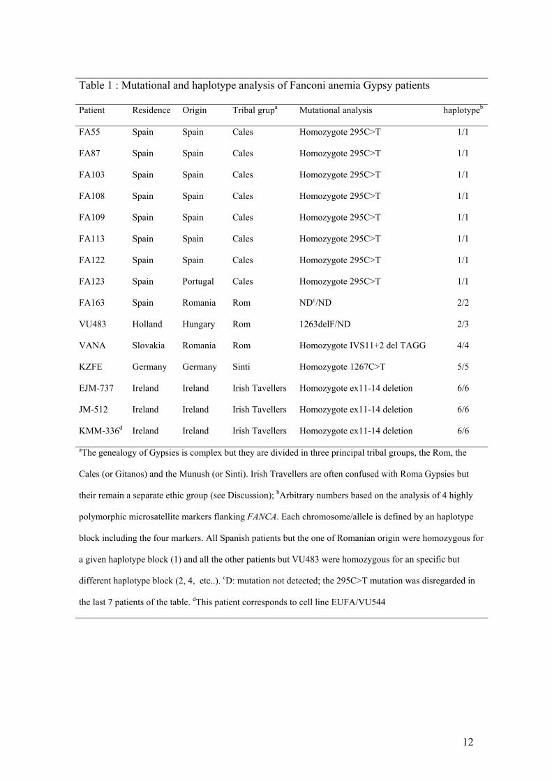
Table 1 : Mutational and haplotype analysis of Fanconi anemia Gypsy patients
Patient Residence Origin Tribal grupa Mutational analysis haplotypeb
FA55 Spain Spain Cales Homozygote 295C>T 1/1
FA87 Spain Spain Cales Homozygote 295C>T 1/1
FA103 Spain Spain Cales Homozygote 295C>T 1/1
FA108 Spain Spain Cales Homozygote 295C>T 1/1
FA109 Spain Spain Cales Homozygote 295C>T 1/1
FA113 Spain Spain Cales Homozygote 295C>T 1/1
FA122 Spain Spain Cales Homozygote 295C>T 1/1
FA123 Spain Portugal Cales Homozygote 295C>T 1/1
FA163 Spain Romania Rom NDc/ND 2/2
VU483 Holland Hungary Rom 1263delF/ND 2/3
VANA Slovakia Romania Rom Homozygote IVS11+2 del TAGG 4/4
KZFE Germany Germany Sinti Homozygote 1267C>T 5/5
EJM-737 Ireland Ireland Irish Tavellers Homozygote ex11-14 deletion 6/6
JM-512 Ireland Ireland Irish Tavellers Homozygote ex11-14 deletion 6/6
KMM-336d Ireland Ireland Irish Tavellers Homozygote ex11-14 deletion 6/6
aThe genealogy of Gypsies is complex but they are divided in three principal tribal groups, the Rom, the
Cales (or Gitanos) and the Munush (or Sinti). Irish Travellers are often confused with Roma Gypsies but
their remain a separate ethic group (see Discussion); bArbitrary numbers based on the analysis of 4 highly
polymorphic microsatellite markers flanking FANCA. Each chromosome/allele is defined by an haplotype
block including the four markers. All Spanish patients but the one of Romanian origin were homozygous for
a given haplotype block (1) and all the other patients but VU483 were homozygous for an specific but
different haplotype block (2, 4, etc..). cD: mutation not detected; the 295C>T mutation was disregarded in
the last 7 patients of the table. dThis patient corresponds to cell line EUFA/VU544
12
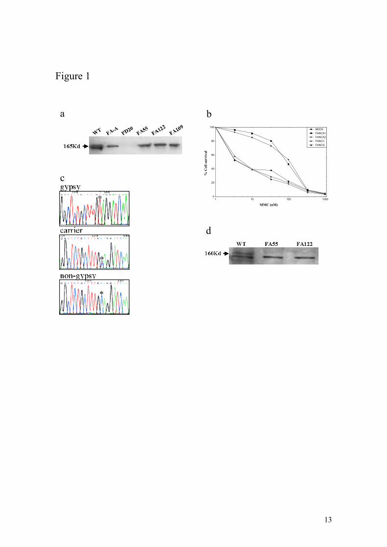
Figure 1
a b
MMC (nM)%
Cel
l sur
viva
l
0
20
40
60
80
100
1 10 100 1000
MOCKFANCA1FANCA2FANCCFANCG
c
d
13
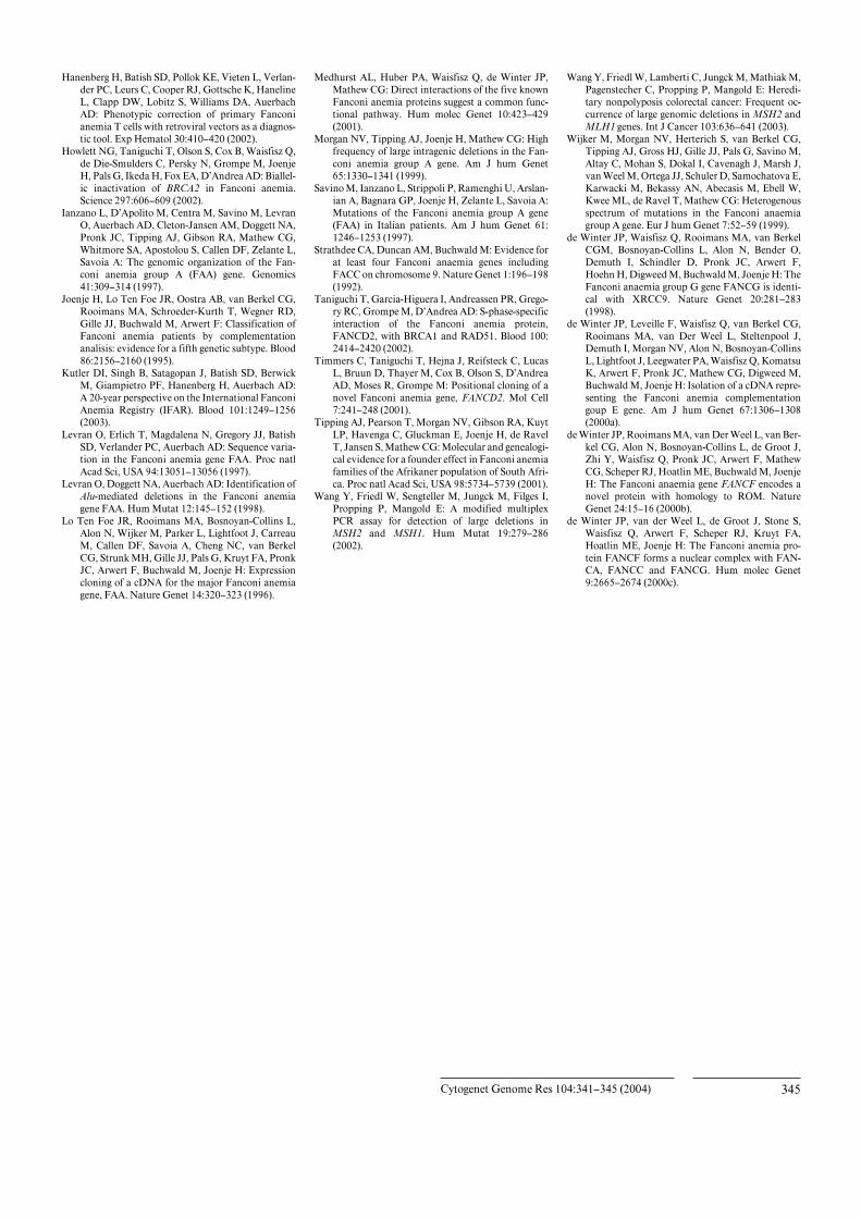
Legend to Figure 1
Genetic characterization of FA in Spanish Gypsy patients. a) FANCD2 immunoblotting
revealed two isoforms in a wild-type cell (WT), no signal in a FANCD2 deficient cell
line (PD20) and a non-ubiquitinated isoform in a FA-A cell line or in cell lines derived
from Spanish Gypsy patients 55, 122 and 109, indicating that the genetic defect is up-
stream FANCD2 monoubiquitination. Immunoblotting experiments were performed
following standard Western blot method with a commercially available antibody against
FANCD2 (Santa Cruz Biotech, CA). b) Subtyping by retrovirus mediated cDNA gene
transduction in a cell line derived from a Spanish Gypsy FA patient. The
complementation group was determined by retroviral transduction of cDNA of FA
genes essentially as previously reported22. Briefly peripheral blood T cells were
proliferated in the presence of a monoclonal antibody anti-CD3 and anti-CD28.
Afterwards, cells were transduced with retroviral vectors containing FANCA (two
vectors), FANCC, FANG cDNA and control vector (mock) and then evaluated for
MMC hypersensitivity to detect phenotypic cellular correction. Only retrovirus
encoding wild-type FANCA cDNAs led to reversion of the cellular hypersensitivity to
mitomycin C (MMC) indicating that the patient belong to complementation group FA-
A. c) Spanish Gypsy FA patients with a 295C>T base change, as detected by DNA
sequencing, leading to a stop codon (Q99X). All obligate carriers were heterozygote for
this mutation. d) FANCA immunoblotting with an affinity purified antibody against the
C-terminus of the FANCA protein in two Spanish Gypsy FA patients 55 and 122 and a
wild-type cell line. FANCA signal (upper band) is not present in the Gypsy patients
consistent with the truncating nature of the Spanish Gypsy mutation. The lower band is
unspecific signal that serves as internal control and that is equally present in all cell
extracts.
14