Embed Size (px)
Citation preview
Picaud et al. Journal of Ovarian Research 2014, 7:40http://www.ovarianresearch.com/content/7/1/40
RESEARCH Open Access
Evaluation of the effects of hyaluronicacid-carboxymethyl cellulose barrier on ovariantumor progressionLaetitia Picaud1,2, Benoît Thibault1, Eliane Mery1, Monia Ouali3, Alejandra Martinez2, Jean-Pierre Delord1,4,Bettina Couderc1,4* and Gwenael Ferron1,2
Abstract
Background: Hyaluronic acid is a prognostic factor in ovarian cancers. It is also a component of HyaluronicAcid-Carboxymethyl Cellulose (HA-CMC) barrier, an anti-adhesion membrane widely used during abdominal surgeriesin particular for ovarian carcinosis. 70% of patients who undergo ovarian surgery will relapse due to the persistence ofcancer cells. This study’s objective was to determine the oncological risk from use of this material, in the presenceof residual disease, despite the benefit gained by it decreasing post-surgical adhesions in order to provide anunambiguous assessment of its appropriateness for use in ovarian surgical management.
Methods: We assessed the effects of HA-CMC barrier on the in vitro proliferation of human ovarian tumor cell lines(OVCAR-3, IGROV-1 and SKOV-3). We next evaluated, in vivo in nude mice, the capacity of this biomaterial toregulate the tumor progression of subcutaneous and intraperitoneal models of ovarian tumor xenografts.
Results: We showed that HA-CMC barrier does not increase in vitro proliferation of ovarian cancer cell linescompared to control. In vivo, HA-CMC barrier presence with subcutaneous xenografts induced neither an increasein tumor volume nor cell proliferation (Ki67 and mitotic index). With the exception of an increased murinecarcinosis score in peritoneum, the presence of HA-CMC barrier with intraperitoneal xenografts modified neithermacro nor microscopic tumor growth. Finally, protein analysis of survival (Akt), proliferation (ERK) and adhesion(FAK) pathways highlighted no activation on the xenografts imputable to HA-CMC barrier.
Conclusions: For the most part, our results support the lack of tumor progression activation due to HA-CMCbarrier. We conclude that the benefits gained from using HA-CMC barrier membrane during ovarian cancersurgeries seem to outweigh the potential oncological risks.
Keywords: Ovarian adenocarcinoma, Surgery, Anti-adhesion membranes, Hyaluronic acid
BackgroundOvarian cancer is the leading cause of mortality amongthe gynecological diseases occurring in developed coun-tries with 225,500 new cases and 140,200 estimateddeaths worldwide [1,2]. This high rate of mortality is duein part because most patients presenting epithelial ovar-ian cancer have an advanced disease at the time of diag-nosis [1]. After surgical resection, residual disease then
* Correspondence: [email protected], Institut Claudius Regaud, 20-24 rue du pont St Pierre, 31052Toulouse, France4Université de Toulouse, Université Paul Sabatier, 118, Route de Narbonne,F-31062 Toulouse, FranceFull list of author information is available at the end of the article
© 2014 Picaud et al.; licensee BioMed CentralCommons Attribution License (http://creativecreproduction in any medium, provided the orDedication waiver (http://creativecommons.orunless otherwise stated.
represents the major pejorative predictive factor forsurvival [2]. For optimum results, surgery must be ascomplete as possible which generally means perform-ing en-bloc resections of bowel, reproductive organsand genital tract, and peritonectomies.The corollary of these “radical” surgeries is the induction
of intra-abdominal adhesions that lead to the stickingtogether of two tissues normally moving freely past eachother. As well as causing bowel obstruction and chronicpain, these adhesions can also induce heterogeneity ofdrug diffusion and compromise the delivery of adjuvantintraperitoneal chemotherapy [3]. Moreover, they cancomplicate any new surgical procedures, which in ovarian
Ltd. This is an Open Access article distributed under the terms of the Creativeommons.org/licenses/by/2.0), which permits unrestricted use, distribution, andiginal work is properly credited. The Creative Commons Public Domaing/publicdomain/zero/1.0/) applies to the data made available in this article,
Picaud et al. Journal of Ovarian Research 2014, 7:40 Page 2 of 12http://www.ovarianresearch.com/content/7/1/40
cancer disease are highly frequent due to the high rate ofrecurrence (70%). The place of secondary cytoreductivesurgery is actually evaluated in several clinical trials with acomplete resection as a gold standard. It is clear thereforethat the reduction of adhesive disease has many immedi-ate and delayed functional and therapeutic consequencesin ovarian cancer bearing patients. An exhaustive reviewpublished by Ward B.C describes all adhesion preventionstrategies including non-invasive surgical strategies,cellular strategies, pharmaceutical approaches, fluidand gel barriers, and lastly solid barriers includingHyaluronic Acid-Carboxymethyl Cellulose (HA-CMC)barrier [4].HA-CMC barrier is an implantable and bioabsorb-
able synthetic membrane, composed of hyaluronan (alsocalled hyaluronic acid or HA) and carboxymethylcellulose(CMC) which turns into a gel within 24 hours, beforebeing absorbed from the surgical site within one weekfor elimination from the organism in less than 30 days.It became available in France in 2003 for indications ofbenign abdominal surgeries and since 2007, followingOikonomakis [5] and Kusonoki’s publications [6] for re-spectively colon and rectal cancers, is also used for car-cinologic surgeries. Since then, many preclinical, animaland clinical works studying HA-CMC barrier in digestivecancers have been published. A meta-analysis performedby Diamond MP describes this material as « biocompat-ible, non-mutagenic, non-pyrogenic, non-irritating andnon-toxic » [7]. However, some animal studies have givencontradictory results revealing no effect of HA-CMCin colon tumor metastasis but a local increase in tumorgrowth [8], and in some cases, an increase in rate ofabdominal metastasis [9].The major component of HA-CMC barrier, HA, is a
physiological component of extracellular matrix in asso-ciation with collagen, proteoglycans (such as fibronectin,laminin and vitronectin) and elastin. Its main receptor isCD44 which is present at the surface of various normalor tumor cells. Another HA receptor, CD168, also calledreceptor hyaluronic acid mediated motility (RHAMM),has been described in CD44 knock-out mice [10]. The HAexpression level is modified in the context of ovarian can-cer and high levels are correlated with poor prognosis[11]. In histological retrospective studies, tumor stromalHA concentration has been linked with tumor aggressive-ness, and stromal HA has been shown to be an independ-ent prognostic factor for disease free and overall survival[12,13]. Moreover, high level of HA synthase 1 (HAS1),but not HAS2 and HAS3, is correlated with reducedoverall survival in ovarian cancer [14]. Furthermore, CD44expression level of ovarian cancer cells is inversely propor-tional to patient survival [15].Many studies, mainly in vitro, have allowed us to define
more precisely the implication of HA in ovarian cancer.
As an example, HA from mesothelial cells, the major cellpopulation in the peritoneum, is an adhesion factor forCD44-positive ovarian cancer cells [16]. These data havebeen confirmed by adhesion tests using hyaluronidase,anti-CD44 antibodies and CD44 targeted siRNA [17-19].Mesothelial extracellular HA is also involved in ovariancarcinoma cell motility and dose-dependent chemotact-ism. Indeed, Boyden chamber tests highlighted than ovar-ian cancer cells preferentially migrate towards mesothelialcells as compared to control conditions, and that thiseffect is abolished using hyaluronidase [20]. Moreover,HA-CD44 interaction in SKOV-3 ovarian cancer cellscan induce F-actin oriented polymerization using twosignaling pathways involving Src or N-WASP [20-22].Among non-gynaecological cancers, a retrospective
study showed that HA-CMC barrier use has been asso-ciated with a 13% increased risk of abscess after colec-tomy [23]. On the contrary, HA-CMC barrier has notbeen showed to be associated with increased complica-tions in thyroid surgery despite an uncertain anti-adhesiveeffect [24].While some studies regarding HA-CMC barrier utilization
in gynecological surgery have been published [25-29]none describe the potential effects of this biomaterial inovarian metastatic process in animals. Of the mainlyclinical studies, most are retrospective. Only one is pro-spective including 14 patients with HA-CMC barrier,and concern in half of the cases cervical and uterinecancers. Krill et al. evaluated the risk of postoperativecomplications related to HA-CMC utilization in ovariancancers after cytoreductive surgery and showed that thisbiomaterial is not responsible for major complicationsbut may be associated with increased risk of pelvic abscess[30]. However, they did not study the effects of HA-CMCon ovarian cancer progression and recurrence.Our concern was to evaluate the potential effect of
HA-CMC in a context where residual cancer cells werepresent. Indeed, after ovarian surgery, and because 70%of patients will relapse, it is clear that those cells couldbe affected by HA-CMC presence.Accordingly, we decided to perform a preclinical study
in order to evaluate HA-CMC barrier impact on ovariantumor progression. In vitro experimentations were per-formed to analyze the effects of HA-CMC barrier on theproliferation of several ovarian cancer cell lines and onthe activation of proliferative, survival and adhesionsignaling pathways. In vivo mice xenograft models wereused to evaluate the oncological risk of HA-CMC barrierutilization in comparison with expected benefits fromadhesion prevention. Our ultimate aim was to providea clear assessment of the appropriateness of using HA-CMC barrier in the surgical management of ovariancancer. We demonstrated that in vitro, HA-CMC bar-rier did not increase tumor cell proliferation and that
Picaud et al. Journal of Ovarian Research 2014, 7:40 Page 3 of 12http://www.ovarianresearch.com/content/7/1/40
in vivo, except for anterior and lateral peritoneumimplantation, HA-CMC barrier did not induce tumorgrowth.
MethodsBiomaterialsHA-CMC barrier membranes and absorbable materialwere granted from the surgical department of ClaudiusRegaud Institute (Toulouse France) without industrialfounds.
Cell cultureNIH ovarian adenocarcinoma cells (OVCAR-3 and SKOV-3)and HeLA cells were obtained from the American TypeCulture Collection (ATCC® numbers HTB-161, HTB-77and CCL-2). IGROV-1 ovarian adenocarcinoma cellswere a gift from the Gustave Roussy Institute (Villejuif,France) and the REH lymphoblastic line was a gift fromthe hematology research unit of Toulouse UniversityHospital (France). All cells were cultured in RPMImedium supplemented with 10% fetal calf serum (FCS),penicillin/streptomycin (100 IU/mL/100 μg/mL) and2 mM L-glutamine (Cambrex biosciences, Milan, Italy).Cell lines were routinely checked for mycoplasma.
Cell proliferationOVCAR-3, IGROV-1 and SKOV-3 cells were seeded in6-well plates at a concentration of 2 × 10⁵ cells per well.Three different conditions were used: culture with RPMIalone or with 1 cm2 pieces of HA-CMC barrier or a controlabsorbable material. Cell proliferation kinetics was assessedby counting cells on a hemocytometer after 1, 2, 4 and6 days of culture. Cell population doubling time duringthe exponential growth phase was calculated accordingto the following formula: T(hours) = t × [ln(2)/[ln(C1) ‐ ln(C0)]] in which t corresponds to the exponential growthphase in hours, C0 to the initial cell concentration at thebeginning of this phase and C1 to the final cell concentra-tion at the end of the phase.
CD44 expression evaluation by flow cytometryEach cell line (106 OVCAR-3, IGROV-1, SKOV-3,HeLA or REH lymphoblast cells) was incubated during45 minutes with a primary antibody directed againstCD44 (1/100, [F10-44-2] (ab6124), Abcam, Paris, France),or against a control isotype (κ isotype Ctrl PE MouseIgG1, BioLegend). Cells were then washed with PBS, cen-trifuged 5 minutes at 300 g, then incubated 45 minuteswith an anti-mouse secondary antibody (1/50, BD Bio-sciences, Le pont de Claix, France). Cells were washedwith PBS and the fluorescence was measured with aFACS Calibur cytometer and analyzed with Cell QuestPro software.
AnimalsFour to five week-old female Swiss Nude athymic mice(Charles River laboratories, l’Abresle, France) were usedafter approval from Claudius Regaud Institute animalethics committee (ICR-2012-017). They were housedaccording to the European Laboratory Animal ScienceAssociation standards. Mice experimentations began afterone week of quarantine.
HA-CMC barrier mouse absorption validationA 2.25 cm2 (15 × 15 mm) piece of HA-CMC barrier wasinserted into the abdomens of mice by median laparotomyafter intraperitoneal general anesthesia (ketamine 50 mg/mL,xylazine 20 mg/mL and NaCl: 10 μg per gram of mouse).Mice were then sacrificed via cervical dislocation 1, 3 or7 days after surgery in order to proceed to histologicalanalysis.
Subcutaneous (s.c.) xenografts107 SKOV-3 cells were injected subcutaneously intoeach of the right and left flanks of 16 mice. The day fol-lowing this injection (D1), after cutaneous debridementwith chlorhexidine, an incision was made under generalanesthesia at the two injection sites and a 1 cm2 (10 ×10 mm) piece of HA-CMC barrier was alternativelyinserted into one of the two sites. The other side wasopened and then closed to reproduce the inflammationcaused by the surgical procedure. The well-being of themice was checked every 2 to 3 days. Tumor lengths (L)and widths (w) were also measured every two to three daysand the tumor volume extrapolated using the formula:(π × length × width2)/6. Mice were sacrificed 21 days viacervical dislocation after the injection. Tumors weresampled then conditioned for histological analysis (for-mol fixation) or western blot (cryopreservation in liquidnitrogen).
Intraperitoneal (i.p.) xenografts2.5 × 107 SKOV-3 cells were injected intraperitoneallyinto each of 22 mice. The day following the injection(D1), after cutaneous debridement with chlorhexidine, alaparotomy was performed under general anesthesiaaccording to one of two procedures: (A) 11 mice under-went a “white” laparotomy (incision, abdominal openingand closure) and (B) 11 mice received an i.p. 2.25 cm2
(15 × 15 mm) piece of HA-CMC barrier. For bothgroups, parietal closure consisted in three separate ab-sorbable material 3/0 sutures. The well-being of themice was checked during 21 days and their weight wasmeasured three times a week. The mice were sacrificedby cervical dislocation 21 days after the injection of cells.Autopsies were then performed by two double-blindedspecialist surgeons in ovarian cancer surgery to evaluate
Picaud et al. Journal of Ovarian Research 2014, 7:40 Page 4 of 12http://www.ovarianresearch.com/content/7/1/40
the abdominal carcinosis index and tumors were removedfor histological and western blot analysis.
Histological analysis, Ki67 staining and mitotic indexdeterminationProliferative indices of tumor from s.c. or i.p. injectionswere assessed by immunohistochemical staining of paraf-fin embedded tumor sections for the proliferation markerKi67 using mouse monoclonal MIB1 antibody. Ki67 posi-tive nuclei were counted in random fields. The mitoticindex was assessed by evaluating the number of cells inmitosis per high-power field (10 high-power fields pertumor).
Western blotWestern blot was performed to evaluate in vitro proteinexpression in CD168 cell lines and to analyze in vivo theactivation of signaling pathways within the xenografts. Invitro, proteins in OVCAR-3, IGROV-1, SKOV-3, HeLaand REH lymphoblastic cells were studied. In vivo, cryo-preserved xenograft tumors were pooled according toprotocol (subcutaneous versus intraperitoneal, with orwithout HA-CMC barrier). Cells and tumor samples werecrushed then prepared with RIPA lysis buffer (Tris 1 M atpH 7.4, NaCl 150 mM, Triton, SDS 20%, MgCl2, NaF andprotease inhibitors). The concentration of proteins wasdetermined using the « Bradford Assay » (Bio-Rad) beforetheir separation by SDS-PAGE in a 10% polyacrylamidegel and transfer onto a PVDF membrane (AmershamHybond) previously activated with methanol. These mem-branes were saturated 45 minutes in TBS (50 mM Tris,150 mM NaCl) / Tween 0.2% (TBST) supplemented with5% milk and then incubated 90 minutes with a rabbit pri-mary antibody against CD168 (1/1000, ab108339, Abcam)for cells lines, or Akt, p-Akt, p-ERK or FAK (1/1000, CellSignaling Technology) for tumor samples. Membraneswere washed three times with TBST and incubated90 minutes with an anti-rabbit secondary antibodycoupled with horseradish peroxidase (HRP) (1/2000,Cell Signaling Technology, St Quentin, France). Mem-branes were washed three times with TBST and twicewith TBS before immunocomplexes were revealed withEnhanced Chemoluminescence (GE Healthcare) and vi-sualized with a photon camera (Bio-Rad).
StatisticsQuantitative variables were presented by median andrange and qualitative data by frequency and percent.Group comparisons were made using the Wilcoxonsigned rank test (matched) or Mann–Whitney test (inde-pendent) for non-parametric data and by the Student’s ttest for parametric ones. For this entire study, statisticalsignificance was reached for p < 0.05. In vivo, with astandard deviation of 0.1, to reach a statistical difference
mean of 15% in terms of tumor proliferation with a statis-tical power of 80% and α risk of 5%, 22 mice per experi-mentation were necessary. All analyses were performedusing STATA software (Version 12.0; Stata Corporation,College Station, TX, USA).
ResultsHA implication in ovarian tumor progression has alreadybeen described. The goal of this study was to determinewhether HA-CMC barrier, being mainly composed of HA,was implicated in ovarian tumor progression and spread.
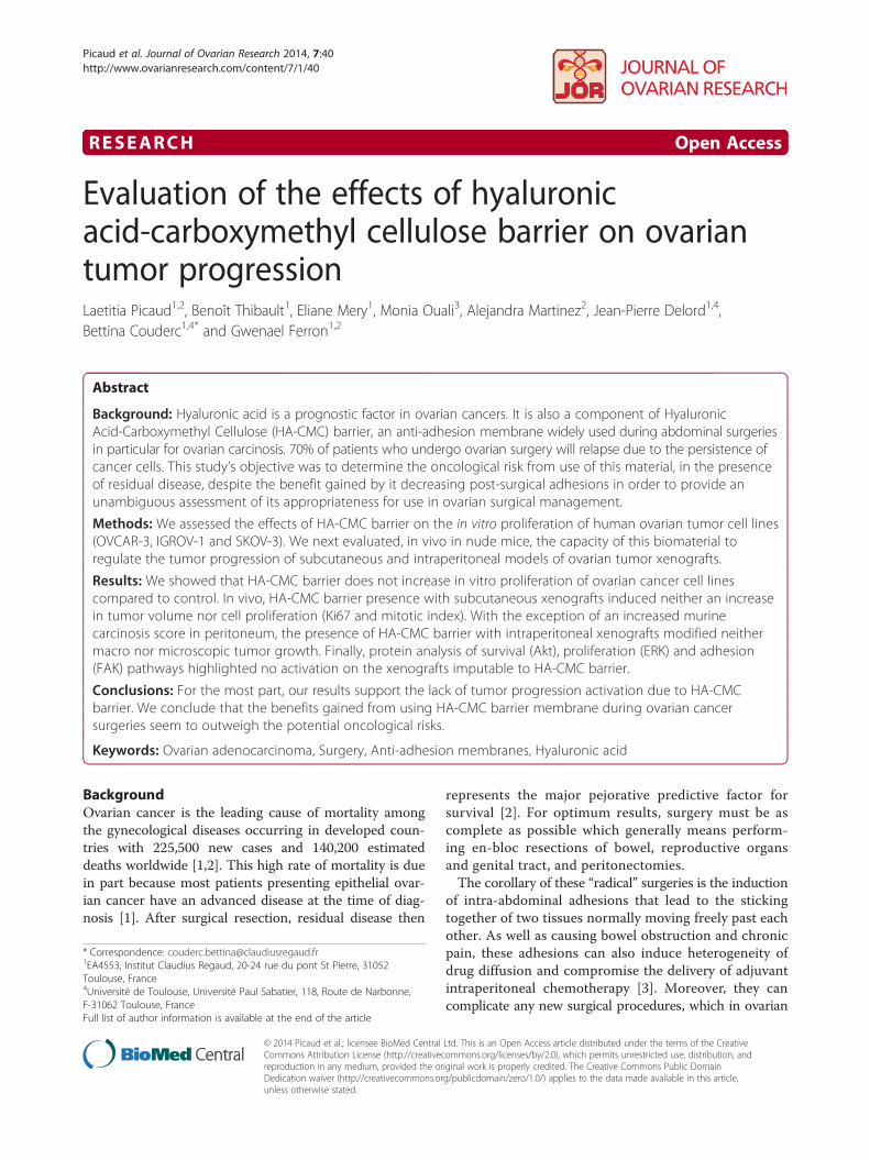
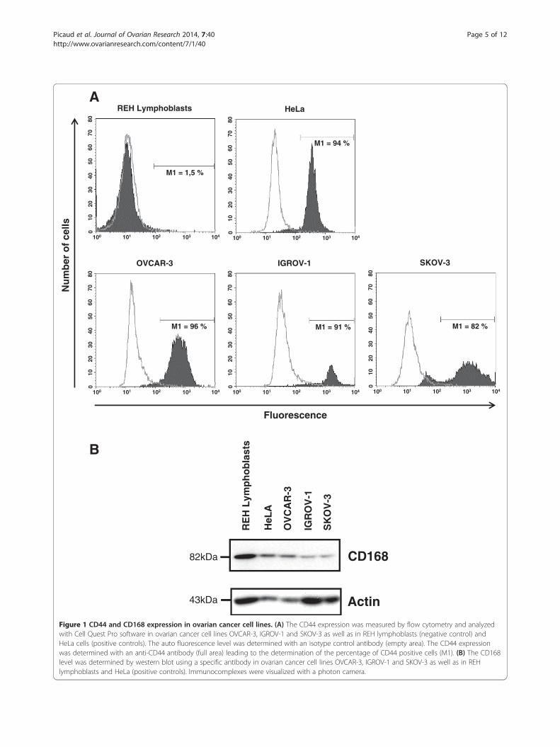
OVCAR-3, IGROV-1 and SKOV-3 ovarian cancer cellsexpress CD44 and CD168OVCAR-3, IGROV-1 and SKOV-3 cell lines differ interms of their genetic alteration and chemosensitivity. Inorder to confirm the capacity of these cells to interactwith HA-CMC barrier, we firstly studied their expressionof HA receptors, CD44 and CD168 by respectively flowcytometry (Figure 1A) and western blotting (Figure 1B).As a negative control of CD44 expression, we used REHlymphoblast cells and as our positive control HeLa cells.REH lymphoblast cells and HeLa cells constituted ourpositive control for CD168 expression. All three ovariancancer cell lines were shown to express both CD44 andCD168.
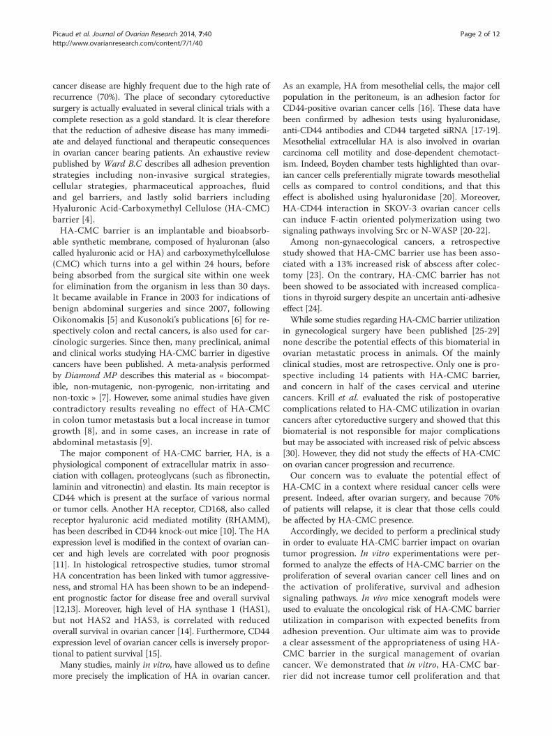
HA-CMC barrier does not promote OVCAR-3, IGROV-1 orSKOV-3 ovarian cancer cell proliferation in vitroTo evaluate the effect of HA-CMC barrier on ovarian can-cer cell proliferation, OVCAR-3, IGROV-1 and SKOV-3cells were seeded in medium alone or with 1 cm2 pieces ofHA-CMC barrier or a control absorbable material whichis a biomaterial used in abdominal surgery that, as anadded exogenous material induces inflammation in vivo.In vitro, absorbable material was used as a control for bio-material presence. We used an hemocytometer to countthe cells for each condition 1, 2, 4 and 6 days after seeding(Figures 2A, B and C) and calculated the doubling timefor each cell line during the exponential proliferative phasebetween the 2nd and 4th day (Figure 2D).Comparison to the control condition (RPMI medium
alone) revealed a lack of any cell proliferation induced byeither HA-CMC barrier or absorbable material (Figure 2A,B and C). Indeed, less cells could be defined at day 6 inthe HA-CMC barrier group even though there was nostatistical difference regarding cell line doubling timesbetween the 2 conditions.Absorbable material absorption was longer than that
for HA-CMC barrier which, considering the potentialto lead to chronic inflammation in mice, we decided toexclude this biomaterial from in vivo experiments.
HeL
A
RE
H L
ymp
ho
bla
sts
SK
OV
-3
IGR
OV
-1
OV
CA
R-3
CD16882kDa
43kDa Actin
B
A
Nu
mb
er o
f ce
lls
080
7060
5040
3020
10
100 101 102 103 104
M1 = 1,5 %
REH Lymphoblasts
100 101 102 103 104
M1 = 94 %
HeLa
080
7060
5040
3020
10
100 101 102 103 104
M1 = 96 %
OVCAR-3
080
7060
5040
3020
10
100 101 102 103 104
M1 = 91 %
IGROV-1
080
7060
5040
3020
10
Fluorescence
100 101 102 103 104
M1 = 82 %
SKOV-3
080
7060
5040
3020
10
Figure 1 CD44 and CD168 expression in ovarian cancer cell lines. (A) The CD44 expression was measured by flow cytometry and analyzedwith Cell Quest Pro software in ovarian cancer cell lines OVCAR-3, IGROV-1 and SKOV-3 as well as in REH lymphoblasts (negative control) andHeLa cells (positive controls). The auto fluorescence level was determined with an isotype control antibody (empty area). The CD44 expressionwas determined with an anti-CD44 antibody (full area) leading to the determination of the percentage of CD44 positive cells (M1). (B) The CD168level was determined by western blot using a specific antibody in ovarian cancer cell lines OVCAR-3, IGROV-1 and SKOV-3 as well as in REHlymphoblasts and HeLa (positive controls). Immunocomplexes were visualized with a photon camera.
Picaud et al. Journal of Ovarian Research 2014, 7:40 Page 5 of 12http://www.ovarianresearch.com/content/7/1/40
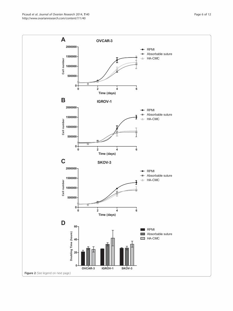
Figure 2 (See legend on next page.)
Picaud et al. Journal of Ovarian Research 2014, 7:40 Page 6 of 12http://www.ovarianresearch.com/content/7/1/40
(See figure on previous page.)Figure 2 Effects of HA-CMC barrier on ovarian cancer cell line proliferation in vitro. (A, B, C) Ovarian cancer cell lines OVCAR-3 (A), IGROV-1(B) and SKOV-3 (C) were cultured in the presence of control medium (RPMI), 1 cm2 of control absorbable material, or 1 cm2 of HA-CMC barrier. After 1,2, 4 and 6 days of culture, cell number was counted with a hemocytometer. (Mean number of cells +/− SEM, n = 3). (D) Doubling times werecalculated from previous proliferation curves according to the following formula: doubling time (hours) = t x [ln (2) / [ln (C1) – ln (C0)]] with tcorresponding to the duration of the exponential phase, C0 to the initial cell concentration and C1 to the final cell concentration (between thebeginning and the end of the exponential phase). (Mean doubling time +/− SEM, n = 3).
Picaud et al. Journal of Ovarian Research 2014, 7:40 Page 7 of 12http://www.ovarianresearch.com/content/7/1/40
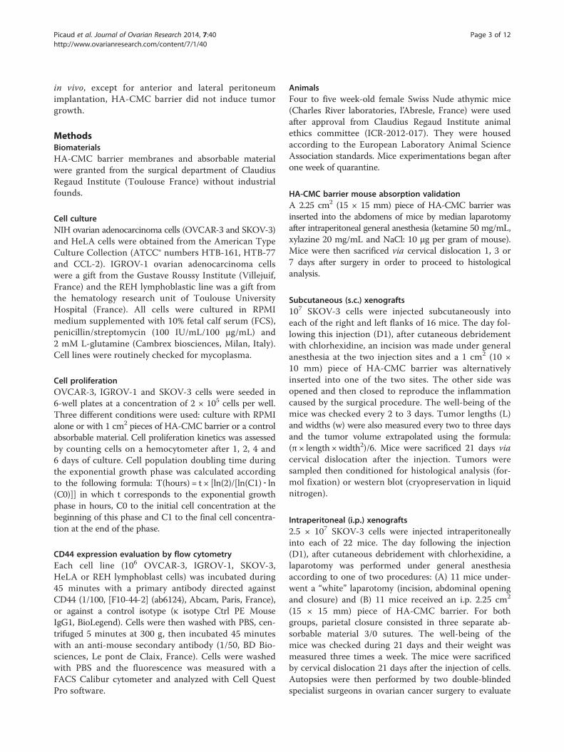
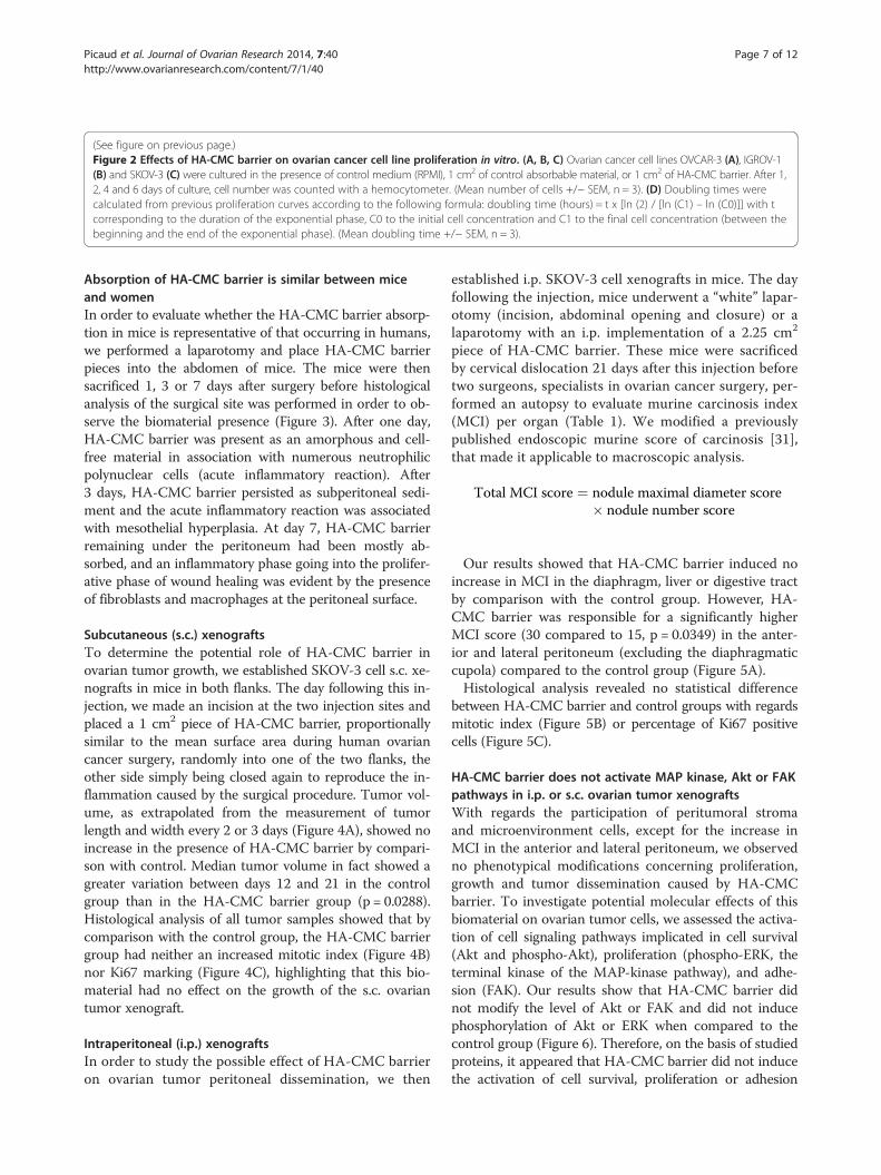
Absorption of HA-CMC barrier is similar between miceand womenIn order to evaluate whether the HA-CMC barrier absorp-tion in mice is representative of that occurring in humans,we performed a laparotomy and place HA-CMC barrierpieces into the abdomen of mice. The mice were thensacrificed 1, 3 or 7 days after surgery before histologicalanalysis of the surgical site was performed in order to ob-serve the biomaterial presence (Figure 3). After one day,HA-CMC barrier was present as an amorphous and cell-free material in association with numerous neutrophilicpolynuclear cells (acute inflammatory reaction). After3 days, HA-CMC barrier persisted as subperitoneal sedi-ment and the acute inflammatory reaction was associatedwith mesothelial hyperplasia. At day 7, HA-CMC barrierremaining under the peritoneum had been mostly ab-sorbed, and an inflammatory phase going into the prolifer-ative phase of wound healing was evident by the presenceof fibroblasts and macrophages at the peritoneal surface.
Subcutaneous (s.c.) xenograftsTo determine the potential role of HA-CMC barrier inovarian tumor growth, we established SKOV-3 cell s.c. xe-nografts in mice in both flanks. The day following this in-jection, we made an incision at the two injection sites andplaced a 1 cm2 piece of HA-CMC barrier, proportionallysimilar to the mean surface area during human ovariancancer surgery, randomly into one of the two flanks, theother side simply being closed again to reproduce the in-flammation caused by the surgical procedure. Tumor vol-ume, as extrapolated from the measurement of tumorlength and width every 2 or 3 days (Figure 4A), showed noincrease in the presence of HA-CMC barrier by compari-son with control. Median tumor volume in fact showed agreater variation between days 12 and 21 in the controlgroup than in the HA-CMC barrier group (p = 0.0288).Histological analysis of all tumor samples showed that bycomparison with the control group, the HA-CMC barriergroup had neither an increased mitotic index (Figure 4B)nor Ki67 marking (Figure 4C), highlighting that this bio-material had no effect on the growth of the s.c. ovariantumor xenograft.
Intraperitoneal (i.p.) xenograftsIn order to study the possible effect of HA-CMC barrieron ovarian tumor peritoneal dissemination, we then
established i.p. SKOV-3 cell xenografts in mice. The dayfollowing the injection, mice underwent a “white” lapar-otomy (incision, abdominal opening and closure) or alaparotomy with an i.p. implementation of a 2.25 cm2
piece of HA-CMC barrier. These mice were sacrificedby cervical dislocation 21 days after this injection beforetwo surgeons, specialists in ovarian cancer surgery, per-formed an autopsy to evaluate murine carcinosis index(MCI) per organ (Table 1). We modified a previouslypublished endoscopic murine score of carcinosis [31],that made it applicable to macroscopic analysis.
Total MCI score ¼ nodule maximal diameter score� nodule number score
Our results showed that HA-CMC barrier induced noincrease in MCI in the diaphragm, liver or digestive tractby comparison with the control group. However, HA-CMC barrier was responsible for a significantly higherMCI score (30 compared to 15, p = 0.0349) in the anter-ior and lateral peritoneum (excluding the diaphragmaticcupola) compared to the control group (Figure 5A).Histological analysis revealed no statistical difference
between HA-CMC barrier and control groups with regardsmitotic index (Figure 5B) or percentage of Ki67 positivecells (Figure 5C).
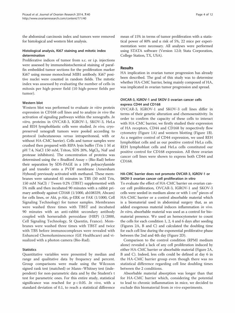
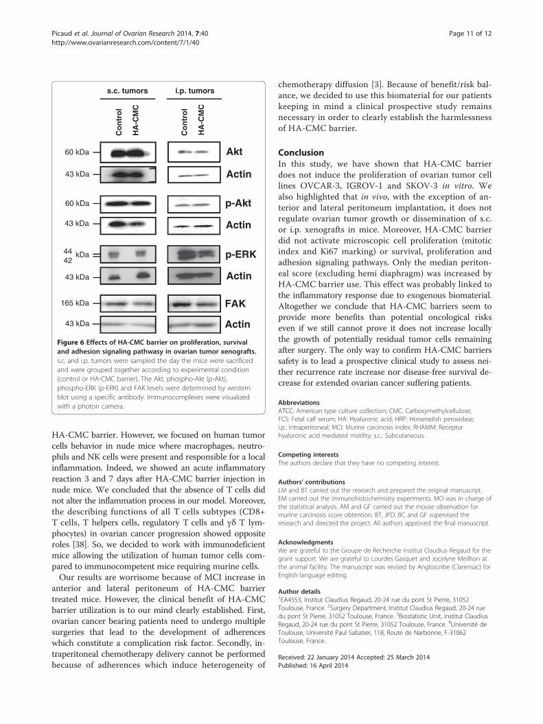
HA-CMC barrier does not activate MAP kinase, Akt or FAKpathways in i.p. or s.c. ovarian tumor xenograftsWith regards the participation of peritumoral stromaand microenvironment cells, except for the increase inMCI in the anterior and lateral peritoneum, we observedno phenotypical modifications concerning proliferation,growth and tumor dissemination caused by HA-CMCbarrier. To investigate potential molecular effects of thisbiomaterial on ovarian tumor cells, we assessed the activa-tion of cell signaling pathways implicated in cell survival(Akt and phospho-Akt), proliferation (phospho-ERK, theterminal kinase of the MAP-kinase pathway), and adhe-sion (FAK). Our results show that HA-CMC barrier didnot modify the level of Akt or FAK and did not inducephosphorylation of Akt or ERK when compared to thecontrol group (Figure 6). Therefore, on the basis of studiedproteins, it appeared that HA-CMC barrier did not inducethe activation of cell survival, proliferation or adhesion
D1
D7
*
*
*
D3
Figure 3 Evolution of the HA-CMC barrier absorption in nudeimmunosuppressed mice. After general anesthesia and cutaneousdebridement, a 15 x 15 mm piece of HA-CMC barrier was placed inmice abdomens. 1 (D1), 3 (D3) and 7 (D7) days after surgery, micewere sacrificed and abdominal walls were analyzed by histology(hematein-eosin coloration). HA-CMC barrier pieces or residues weremarked with a star (*).
Picaud et al. Journal of Ovarian Research 2014, 7:40 Page 8 of 12http://www.ovarianresearch.com/content/7/1/40
signaling pathways in either s.c. or i.p. ovarian tumorxenografts.
DiscussionComplete cytoreductive surgery for advanced ovariancancer can induce extended abdominal adhesions. Theseadhesions may prevent optimal intraperitoneal treatmentdelivery and can cause bowel obstruction and pain. Theneed to prevent such adhesions during abdominal sur-gery is a unanimously agreed objective. Among the solidbarriers placed to decrease adhesive disease, HA-CMCbarrier is one of the most widely used, and yet its essen-tial component HA is a prognostic factor of survival inovarian cancer bearing patients. Despite our in-depthstudy of the literature, we found no conclusive data re-garding the oncological risk of using HA-CMC barrierin these patients.Our results show that HA-CMC barrier does not acti-
vate the proliferation of the ovarian tumor cells OVCAR-3,IGROV-1 or SKOV-3 in vitro. Moreover, it cannot beexcluded that the biomaterials may have actually actedas a physical constraint of cell proliferation. Indeed,whether the biomaterial used was HA-CMC barrier oran absorbable material, cell proliferation was less activethan without the biomaterial. Particularly with IGROV-1cells, HA-CMC seemed to present an anti-proliferativeeffect. Observing the same number of IGROV-1 cells ineach condition at day 1 (Figure 2B and D), we did notconclude on an anti-adhesive effect of HA-CMC barrieron IGROV-1 cells. Considering that in vitro experimentsexclude microenvironment participation, we studied in vivothe potential implication of HA-CMC barrier in ovariantumor progression in mice.Nearly all the results obtained in vivo, whether they
concerned subcutaneous tumor cell injections (tumorvolume, mitotic index or Ki67 percentage) or intraperito-neal xenografts (mean weight of mice, median carcinosisscore of the diaphragm, liver and intestine, mitotic indexor Ki67 percentage), we observed no proliferation increaseassociated with the HA-CMC barrier. Our findings cor-roborate other studies concerning non gynecological can-cer in which HA-CMC barrier was described to protecttrocar sites from gall bladder metastasis and abdominalwall implantation [32] while not decreasing overall sur-vival for colon and rectal cancers [33].However, our study does highlight that an i.p. HA-CMC
barrier implantation can induce a statistically significantincrease in the peritoneum carcinosis score (excludinghemi diaphragm). While this was the only detected pejora-tive impact of HA-CMC barrier, Hubbard et al. did pub-lish similar findings for KM12-L4 colon cancer xenograftsin mice, observing a local increase in tumor volume in thesidewall of the HA-CMC barrier and absorbable materialgroups. This increase was thought to be due to local
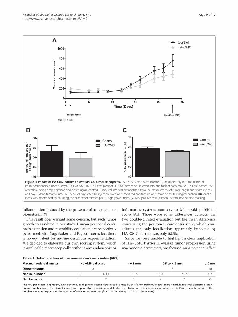
Figure 4 Impact of HA-CMC barrier on ovarian s.c. tumor xenografts. (A) SKOV-3 cells were injected subcutaneously into the flanks ofimmunosuppressed mice at day 0 (D0). At day 1 (D1), a 1 cm2 piece of HA-CMC barrier was inserted into one flank of each mouse (HA-CMC barrier), theother flank being simply opened and closed again (control). Tumor volume was extrapolated from the measurement of tumor length and width every 2or 3 days. (Mean tumor volume +/− SEM) 23 days after the injection, mice were sacrificed and tumors were sampled for histological analysis. (B) Mitoticindex was determined by counting the number of mitoses per 10 high-power fields. (C) Ki67 positive cells (%) were determined by Ki67 marking.
Picaud et al. Journal of Ovarian Research 2014, 7:40 Page 9 of 12http://www.ovarianresearch.com/content/7/1/40
inflammation induced by the presence of an exogenousbiomaterial [8].This result does warrant some concern, but such tumor
growth was isolated in our study. Human peritoneal carci-nosis extension and resecability evaluation are respectivelyperformed with Sugarbaker and Fagotti scores but thereis no equivalent for murine carcinosis experimentation.We decided to elaborate our own scoring system, whichis applicable macroscopically without any endoscopic or
Table 1 Determination of the murine carcinosis index (MCI)
Maximal nodule diameter No visible disease
Diameter score 0
Nodule number 1-5 6-10
Number score 1 2
The MCI per organ (diaphragm, liver, peritoneum, digestive tract) is determined in mnodule number score. The diameter score corresponds to the maximal nodule diamnumber score corresponds to the number of nodules in the organ (from 1-5 nodule
informatics systems contrary to Matsuzaki publishedscore [31]. There were some differences between thetwo double-blinded evaluation but the mean differenceconcerning the peritoneal carcinosis score, which con-stitutes the only localization apparently impacted byHA-CMC barrier, was only 6.83%.Since we were unable to highlight a clear implication
of HA-CMC barrier in ovarian tumor progression usingmacroscopic parameters, we focused on a potential effect
< 0.5 mm 0.5 to < 2 mm ≥ 2 mm
1 5 10
11-15 16-20 21-25 >25
3 4 5 6
ice by the following formula: total score = nodule maximal diameter score ×eter (from non-visible nodules to nodules up to 2 mm diameter or over). Thes up to 25 nodules or over).
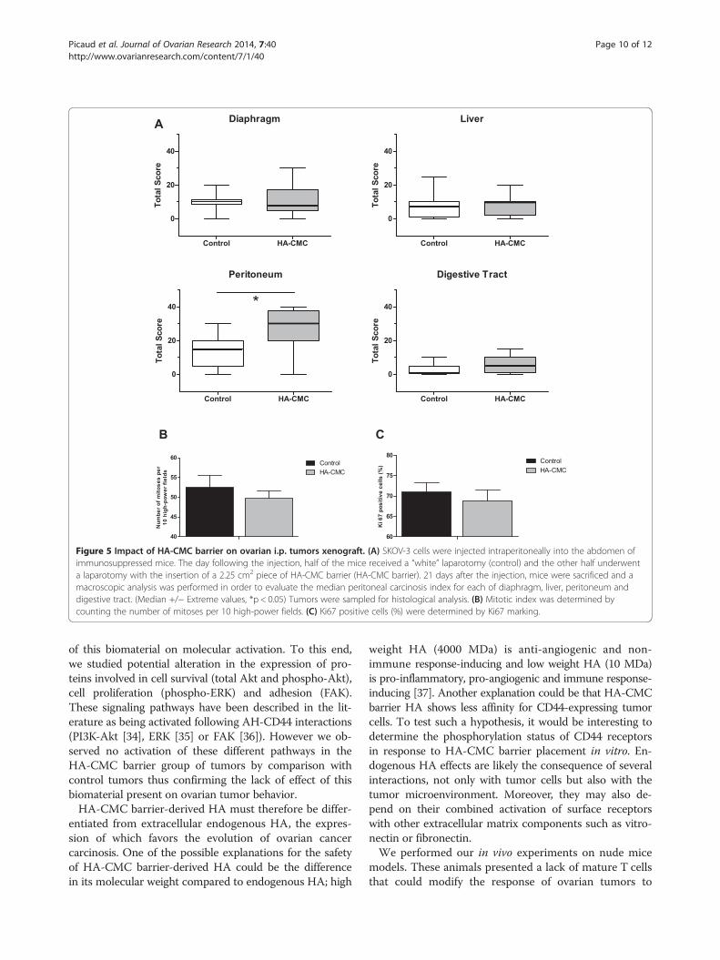
Figure 5 Impact of HA-CMC barrier on ovarian i.p. tumors xenograft. (A) SKOV-3 cells were injected intraperitoneally into the abdomen ofimmunosuppressed mice. The day following the injection, half of the mice received a “white” laparotomy (control) and the other half underwenta laparotomy with the insertion of a 2.25 cm2 piece of HA-CMC barrier (HA-CMC barrier). 21 days after the injection, mice were sacrificed and amacroscopic analysis was performed in order to evaluate the median peritoneal carcinosis index for each of diaphragm, liver, peritoneum anddigestive tract. (Median +/− Extreme values, *p < 0.05) Tumors were sampled for histological analysis. (B) Mitotic index was determined bycounting the number of mitoses per 10 high-power fields. (C) Ki67 positive cells (%) were determined by Ki67 marking.
Picaud et al. Journal of Ovarian Research 2014, 7:40 Page 10 of 12http://www.ovarianresearch.com/content/7/1/40
of this biomaterial on molecular activation. To this end,we studied potential alteration in the expression of pro-teins involved in cell survival (total Akt and phospho-Akt),cell proliferation (phospho-ERK) and adhesion (FAK).These signaling pathways have been described in the lit-erature as being activated following AH-CD44 interactions(PI3K-Akt [34], ERK [35] or FAK [36]). However we ob-served no activation of these different pathways in theHA-CMC barrier group of tumors by comparison withcontrol tumors thus confirming the lack of effect of thisbiomaterial present on ovarian tumor behavior.HA-CMC barrier-derived HA must therefore be differ-
entiated from extracellular endogenous HA, the expres-sion of which favors the evolution of ovarian cancercarcinosis. One of the possible explanations for the safetyof HA-CMC barrier-derived HA could be the differencein its molecular weight compared to endogenous HA; high
weight HA (4000 MDa) is anti-angiogenic and non-immune response-inducing and low weight HA (10 MDa)is pro-inflammatory, pro-angiogenic and immune response-inducing [37]. Another explanation could be that HA-CMCbarrier HA shows less affinity for CD44-expressing tumorcells. To test such a hypothesis, it would be interesting todetermine the phosphorylation status of CD44 receptorsin response to HA-CMC barrier placement in vitro. En-dogenous HA effects are likely the consequence of severalinteractions, not only with tumor cells but also with thetumor microenvironment. Moreover, they may also de-pend on their combined activation of surface receptorswith other extracellular matrix components such as vitro-nectin or fibronectin.We performed our in vivo experiments on nude mice
models. These animals presented a lack of mature T cellsthat could modify the response of ovarian tumors to
Akt
HA
-CM
C
Co
ntr
ol
p-Akt
s.c. tumors
Actin
Actin
60 kDa
43 kDa
60 kDa
43 kDa
i.p. tumors
HA
-CM
C
Co
ntr
ol
p-ERK4442
43 kDa Actin
kDa
165 kDa FAK
43 kDa Actin
Figure 6 Effects of HA-CMC barrier on proliferation, survivaland adhesion signaling pathwasy in ovarian tumor xenografts.s.c. and i.p. tumors were sampled the day the mice were sacrificedand were grouped together according to experimental condition(control or HA-CMC barrier). The Akt, phospho-Akt (p-Akt),phospho-ERK (p-ERK) and FAK levels were determined by westernblot using a specific antibody. Immunocomplexes were visualizedwith a photon camera.
Picaud et al. Journal of Ovarian Research 2014, 7:40 Page 11 of 12http://www.ovarianresearch.com/content/7/1/40
HA-CMC barrier. However, we focused on human tumorcells behavior in nude mice where macrophages, neutro-phils and NK cells were present and responsible for a localinflammation. Indeed, we showed an acute inflammatoryreaction 3 and 7 days after HA-CMC barrier injection innude mice. We concluded that the absence of T cells didnot alter the inflammation process in our model. Moreover,the describing functions of all T cells subtypes (CD8+T cells, T helpers cells, regulatory T cells and γδ T lym-phocytes) in ovarian cancer progression showed oppositeroles [38]. So, we decided to work with immunodeficientmice allowing the utilization of human tumor cells com-pared to immunocompetent mice requiring murine cells.Our results are worrisome because of MCI increase in
anterior and lateral peritoneum of HA-CMC barriertreated mice. However, the clinical benefit of HA-CMCbarrier utilization is to our mind clearly established. First,ovarian cancer bearing patients need to undergo multiplesurgeries that lead to the development of adherenceswhich constitute a complication risk factor. Secondly, in-traperitoneal chemotherapy delivery cannot be performedbecause of adherences which induce heterogeneity of
chemotherapy diffusion [3]. Because of benefit/risk bal-ance, we decided to use this biomaterial for our patientskeeping in mind a clinical prospective study remainsnecessary in order to clearly establish the harmlessnessof HA-CMC barrier.
ConclusionIn this study, we have shown that HA-CMC barrierdoes not induce the proliferation of ovarian tumor celllines OVCAR-3, IGROV-1 and SKOV-3 in vitro. Wealso highlighted that in vivo, with the exception of an-terior and lateral peritoneum implantation, it does notregulate ovarian tumor growth or dissemination of s.c.or i.p. xenografts in mice. Moreover, HA-CMC barrierdid not activate microscopic cell proliferation (mitoticindex and Ki67 marking) or survival, proliferation andadhesion signaling pathways. Only the median periton-eal score (excluding hemi diaphragm) was increased byHA-CMC barrier use. This effect was probably linked tothe inflammatory response due to exogenous biomaterial.Altogether we conclude that HA-CMC barriers seem toprovide more benefits than potential oncological riskseven if we still cannot prove it does not increase locallythe growth of potentially residual tumor cells remainingafter surgery. The only way to confirm HA-CMC barrierssafety is to lead a prospective clinical study to assess nei-ther recurrence rate increase nor disease-free survival de-crease for extended ovarian cancer suffering patients.
AbbreviationsATCC: American type culture collection; CMC: Carboxymethylcellulose;FCS: Fetal calf serum; HA: Hyaluronic acid; HRP: Horseradish peroxidase;i.p.: Intraperitoneal; MCI: Murine carcinosis index; RHAMM: Receptorhyaluronic acid mediated motility; s.c.: Subcutaneous.
Competing interestsThe authors declare that they have no competing interest.
Authors’ contributionsLM and BT carried out the research and prepared the original manuscript.EM carried out the immunohistochemistry experiments. MO was in charge ofthe statistical analysis. AM and GF carried out the mouse observation formurine carcinosis score obtention. BT, JPD, BC and GF supervised theresearch and directed the project. All authors approved the final manuscript.
AcknowledgmentsWe are grateful to the Groupe de Recherche Institut Claudius Regaud for thegrant support. We are grateful to Lourdes Gasquet and Jocelyne Meilhon atthe animal facility. The manuscript was revised by Angloscribe (Clarensac) forEnglish language editing.
Author details1EA4553, Institut Claudius Regaud, 20-24 rue du pont St Pierre, 31052Toulouse, France. 2Surgery Department, Institut Claudius Regaud, 20-24 ruedu pont St Pierre, 31052 Toulouse, France. 3Biostatistic Unit, Institut ClaudiusRegaud, 20-24 rue du pont St Pierre, 31052 Toulouse, France. 4Université deToulouse, Université Paul Sabatier, 118, Route de Narbonne, F-31062Toulouse, France.
Received: 22 January 2014 Accepted: 25 March 2014Published: 16 April 2014
Picaud et al. Journal of Ovarian Research 2014, 7:40 Page 12 of 12http://www.ovarianresearch.com/content/7/1/40
References1. Baiocchi G, Cestari LA, Macedo MP, Oliveira RAR, Fukazawa EM, Faloppa CC,
Kumagai LY, Badiglian-Filho L, Menezes ANO, Cunha IW, Soares FA: Surgicalimplications of mesenteric lymph node metastasis from advanced ovariancancer after bowel Resection. J Surg Oncol 2011, 104:250–254.
2. Chéreau E, Ballester M, Rouzier R, Coutant C, Daraï E: Advanced ovariancancer: criteria of resectability. Bull Cancer 2009, 96:1189–1197.
3. Gladieff L, Chatelut E, Dalenc F, Ferron G: Pharmacological bases ofintraperitoneal chemotherapy. Bull Cancer 2009, 96:1235–1242.
4. Ward BC, Panitch A: Abdominal adhesions: current and novel therapies.J Surg Res 2011, 165:91–111.
5. Oikonomakis I, Wexner SD, Gervaz P, You S-Y, Secic M, Giamundo P: Seprafilm:a retrospective preliminary evaluation of the impact on short-term oncologicoutcome in colorectal cancer. Dis Colon Rectum 2002, 45:1376–1380.
6. Kusunoki M, Ikeuchi H, Yanagi H, Noda M, Tonouchi H, Mohri Y, Uchida K,Inoue Y, Kobayashi M, Miki C, Yamamura T: Bioresorbable hyaluronate-carboxymethylcellulose membrane (Seprafilm) in surgery for rectalcarcinoma: a prospective randomized clinical trial. Surg Today 2005,35:940–945.
7. Diamond MP, Burns EL, Accomando B, Mian S, Holmdahl L: Seprafilm®adhesion barrier: (1) a review of preclinical, animal, and humaninvestigational studies. Gynecol Surg 2012, 9:237–245.
8. Hubbard SC, Burns JW: Effects of a hyaluronan-based membrane (Seprafilm)on intraperitoneally disseminated human colon cancer cell growth in anude mouse model. Dis Colon Rectum 2002, 45:334–341. discussion 341–344.
9. Tan B, Wang JH, Wu QD, Kirwan WO, Redmond HP: Sodium hyaluronateenhances colorectal tumour cell metastatic potential in vitro and in vivo.Br J Surg 2001, 88:246–250.
10. Nedvetzki S, Gonen E, Assayag N, Reich R, Williams RO, Thurmond RL,Huang J-F, Neudecker BA, Wang F-S, Wang F-S, Turley EA, Naor D: RHAMM,a receptor for hyaluronan-mediated motility, compensates for CD44 ininflamed CD44-knockout mice: a different interpretation of redundancy.Proc Natl Acad Sci U S A 2004, 101:18081–18086.
11. Ricciardelli C, Rodgers RJ: Extracellular matrix of ovarian tumors. SeminReprod Med 2006, 24:270–282.
12. Hiltunen EL, Anttila M, Kultti A, Ropponen K, Penttinen J, Yliskoski M,Kuronen AT, Juhola M, Tammi R, Tammi M, Kosma VM: Elevatedhyaluronan concentration without hyaluronidase activation in malignantepithelial ovarian tumors. Cancer Res 2002, 62:6410–6413.
13. Anttila MA, Tammi RH, Tammi MI, Syrjänen KJ, Saarikoski SV, Kosma VM:High levels of stromal hyaluronan predict poor disease outcome inepithelial ovarian cancer. Cancer Res 2000, 60:150–155.
14. Yabushita H, Noguchi M, Kishida T, Fusano K, Noguchi Y, Itano N, Kimata K,Noguchi M: Hyaluronan synthase expression in ovarian cancer. Oncol Rep2004, 12:739–743.
15. Kayastha S, Freedman AN, Piver MS, Mukkamalla J, Romero-Guittierez M,Werness BA: Expression of the hyaluronan receptor, CD44S, in epithelialovarian cancer is an independent predictor of survival. Clin Cancer Res1999, 5:1073–1076.
16. Cannistra SA, Kansas GS, Niloff J, DeFranzo B, Kim Y, Ottensmeier C: Bindingof ovarian cancer cells to peritoneal mesothelium in vitro is partlymediated by CD44H. Cancer Res 1993, 53:3830–3838.
17. Jones LM, Gardner MJ, Catterall JB, Turner GA: Hyaluronic acid secreted bymesothelial cells: a natural barrier to ovarian cancer cell adhesion. ClinExp Metastasis 1995, 13:373–380.
18. Gardner MJ, Catterall JB, Jones LM, Turner GA: Human ovarian tumour cellscan bind hyaluronic acid via membrane CD44: a possible step inperitoneal metastasis. Clin Exp Metastasis 1996, 14:325–334.
19. Li C-Z, Liu B, Wen Z-Q, Li H-Y: Inhibition of CD44 expression by smallinterfering RNA to suppress the growth and metastasis of ovarian cancercells in vitro and in vivo. Folia Biol (Praha) 2008, 54:180–186.
20. Carpenter PM, Dao AV: The role of hyaluronan in mesothelium-inducedmotility of ovarian carcinoma cells. Anticancer Res 2003, 23:3985–3990.
21. Bourguignon LY, Zhu H, Shao L, Chen YW: CD44 interaction with c-Src kinasepromotes cortactin-mediated cytoskeleton function and hyaluronic acid-dependent ovarian tumor cell migration. J Biol Chem 2001, 276:7327–7336.
22. Bourguignon LY, Peyrollier K, Gilad E, Brightman A: Hyaluronan-CD44 interactionwith neural Wiskott-Aldrich syndrome protein (N-WASP) promotes actinpolymerization and ErbB2 activation leading to beta-catenin nucleartranslocation, transcriptional up-regulation, and cell migration inovarian tumor cells. J Biol Chem 2007, 282:1265–1280.
23. Bashir S, Ananth CV, Lewin SN, Burke WM, Lu Y-S, Neugut AI, Herzog TJ,Hershman DL, Wright JD: Utilization and safety of sodium hyaluronate-carboxymethylcellulose adhesion barrier. Dis Colon Rectum 2013,56:1174–1184.
24. Bae DS, Woo J-W, Paek SH, Kwon H, Chai YJ, Kim S-J, Choi JY, Lee KE, YounY-K: Antiadhesive effect and safety of sodium hyaluronate-carboxymethyl cellulose membrane in thyroid surgery. J Korean Surg Soc2013, 85:199–204.
25. Leitao MM Jr, Byrum GV 3rd, Abu-Rustum NR, Brown CL, Chi DS, Sonoda Y,Levine DA, Gardner GJ, Barakat RR: Postoperative intra-abdominal collectionsusing a sodium hyaluronate-carboxymethylcellulose (HA-CMC) barrier atthe time of laparotomy for uterine or cervical cancers. Gynecol Oncol 2010,119:208–211.
26. Tabata T, Kihira T, Shiozaki T, Tanida K, Kondo E, Nagao K, Okugawa T,Sagawa N: Efficacy of a sodium hyaluronate-carboxycellulose membrane(seprafilm) for reducing the risk of early postoperative small bowelobstruction in patients with gynecologic malignancies. Int J GynecolCancer 2010, 20:188–193.
27. Tan A, Argenta P, Ramirez R, Bliss R, Geller M: The use of sodiumhyaluronate-carboxymethylcellulose (HA-CMC) barrier in gynecologicmalignancies: a retrospective review of outcomes. Ann Surg Oncol 2009,16:499–505.
28. Bristow RE, Montz FJ: Prevention of adhesion formation after radicaloophorectomy using a sodium hyaluronate-carboxymethylcellulose(HA-CMC) barrier. Gynecol Oncol 2005, 99:301–308.
29. Bristow RE, Santillan A, Diaz-Montes TP, Gardner GJ, Giuntoli RL 2nd, PeelerST: Prevention of adhesion formation after radical hysterectomy using asodium hyaluronate-carboxymethylcellulose (HA-CMC) barrier: acost-effectiveness analysis. Gynecol Oncol 2007, 104:739–746.
30. Krill LS, Ueda SM, Gerardi M, Bristow RE: Analysis of postoperativecomplications associated with the use of anti-adhesion sodiumhyaluronate-carboxymethylcellulose (HA-CMC) barrier after cytoreductivesurgery for ovarian, fallopian tube and peritoneal cancers. Gynecol Oncol2011, 120:220–223.
31. Bourdel N, Matsuzaki S, Bazin J-E, Darcha C, Pouly J-L, Mage G, Canis M:Postoperative peritoneal dissemination of ovarian cancer cells is notpromoted by carbon-dioxide pneumoperitoneum at low intraperitonealpressure in a syngenic mouse laparoscopic model with controlledrespiratory support: a pilot study. J Minim Invasive Gynecol 2008, 15:321–326.
32. Sasaki T, Shimura H, Tanaka T, Nakashima K, Matsuo K, Ikeda S: Protectionof trocar sites from gallbladder cancer implantation by sodiumhyaluronate carboxymethylcellulose-based bioresorbable membrane(Seprafilm) in a murine model [corrected]. Surg Endosc 2004, 18:246–251.
33. Pucciarelli S, Codello L, Rosato A, Del Bianco P, Vecchiato G, Lise M: Effectof antiadhesive agents on peritoneal carcinomatosis in an experimentalmodel. Br J Surg 2003, 90:66–71.
34. Misra S, Heldin P, Hascall VC, Karamanos NK, Skandalis SS, Markwald RR,Ghatak S: Hyaluronan-CD44 interactions as potential targets for cancertherapy. FEBS J 2011, 278:1429–1443.
35. Ween MP, Hummitzsch K, Rodgers RJ, Oehler MK, Ricciardelli C: Versicaninduces a pro-metastatic ovarian cancer cell behavior which can beinhibited by small hyaluronan oligosaccharides. Clin Exp Metastasis 2011,28:113–125.
36. Kouvidi K, Berdiaki A, Nikitovic D, Katonis P, Afratis N, Hascall VC, KaramanosNK, Tzanakakis GN: Role of receptor for hyaluronic acid-mediated motility(RHAMM) in low molecular weight hyaluronan (LMWHA)-mediatedfibrosarcoma cell adhesion. J Biol Chem 2011, 286:38509–38520.
37. Mizrahy S, Raz SR, Hasgaard M, Liu H, Soffer-Tsur N, Cohen K, Dvash R,Landsman-Milo D, Bremer MGEG, Moghimi SM, Peer D: Hyaluronan-coatednanoparticles: the influence of the molecular weight on CD44-hyaluronan interactions and on the immune response. J Control Release2011, 156:231–238.
38. Thibault B, Castells M, Delord J-P, Couderc B: Ovarian cancer microenvironment:implications for cancer dissemination and chemoresistance acquisition. CancerMetastasis Rev 2013. doi:10.1007/s10555-013-9456-2.
doi:10.1186/1757-2215-7-40Cite this article as: Picaud et al.: Evaluation of the effects of hyaluronicacid-carboxymethyl cellulose barrier on ovarian tumor progression.Journal of Ovarian Research 2014 7:40.