Embed Size (px)
Citation preview
Role of irritation and mast cell mediators on thymic stromal
lymphopoietin (TSLP) expression in the skin and its impact on
severity of atopic dermatitis
Dissertation
Zur Erlangung des akademischen Grades
Doktor rerum naturalium
(Dr. rer. nat.)
im Fach Biologie
eingereicht an der
Lebenswissenschaftlichen Fakultät
der Humboldt-Universität zu Berlin
von
M.Sc. Davender Redhu
Präsidentin der Humboldt-Universität zu Berlin
Prof. Dr.-Ing. Dr. Sabine Kunst
Dekan der Lebenswissenschaftlichen Fakultät
Prof. Dr. Bernhard Grimm
Gutacher/innen: 1. Prof. Dr. Hans-Dieter Volk
2. Prof. Dr. Margitta Worm
3. Prof. Dr. Philipp Franken
Tag der mündlichen Prüfung: 19.11.2019

DEDICATION
2
DEDICATION
I dedicate this work to my parents (Maa Smt. Sheela Devi and Babuji
Shri Ramniwas Redhu) and my Uncle Shri Satbir Singh Redhu
TABLE OF CONTENTS
3
TABLE OF CONTENTS
LIST OFABBREVIATIONS………………………………………………………………6
ABSTRACT…………………………………………………………………………………10
ZUSAMMENFASSUNG…………………………………………………………………...11
1 INTRODUCTION………………………………………………………………………...13
1.1 SKIN STRUCTURE…………………………………………………………………….13
1.2 EPIDERMAL BARRIER AND IT’S DISRUPTION IN ATOPIC DISEASES…….14
1.2.2 Atopic dermatitis (AD)……………………………………………………………...16
1.3 KERATINOCYTES…………………………………………………………………….18
1.3.1 Role of keratinocytes in skin irritation……………………………………………..18
1.3.2 Role of keratinocytes in AD…………………………………………………………18
1.4 MAST CELLS…………………………………………………………………………..19
1.4.1 Role of mast cells in AD……………………………………………………………..19
1.5 PROTEASE ACTIVATED RECEPTOR (PAR)-2…………………………………...20
1.5.1 Role of PAR-2 in AD………………………………………………………………...21
1.6 THYMIC STROMAL LYMPHOPOIETIN (TSLP)…………………………………21
1.6.1 Role of TSLP in AD………………………………………………………………….22
1.7 OBJECTIVES…………………………………………………………………………...24
2 MATERIALS AND METHODS…………………………………………………………25
2.1 MATERIALS……………………………………………………………………………25
2.2 METHODS………………………………………………………………………………30
2.2.1 Animal experiments…………………………………………………………………30
2.3 MOUSE MODELS……………………………………………………………………...30
2.3.1 In vivo AD model…………………………………………………………………….30
2.3.2 Ex vivo skin irritation model………………………………………………………..31
2.3.3 In vivo skin irritation model………………………………………………………...31
2.3.4 Ex vivo mast cell degranulation model……………………………………………..32
2.3.5 In vivo mast cell degranulation model……………………………………………...32
2.4 CELL CULTURE METHODS………………………………………………………...33
2.4.1 Isolation, culturing and treatment of human primary keratinocytes……………33
2.4.2 Enzyme linked immunosorbent assay (ELISA)…………………………………...33
2.4.3 RNA isolation………………………………………………………………………...34
2.4.4 Reverse transcription………………………………………………………………..34
2.4.5 Real-time polymerase chain reaction………………………………………………35
2.4.6 Immunoprecipitation assay (IP)……………………………………………………36
TABLE OF CONTENTS
4
2.4.7 Immunoblotting……………………………………………………………………...36
2.4.8 Chromatin Immunoprecipitation (ChIP) assay…………………………………...37
2.4.9 Plasmid Construction, transfection and luciferase assay…………………………39
2.5 STATISTICAL ANALYSIS……………………………………………………………40
3 RESULTS………………………………………………………………………………….41
3.1 TSLPR-/- MICE ARE PROTECTED FROM ENDOGENOUS TNF-DEFICIENCY-
MEDIATED AD DEVELOPMENT……………………………………………………….41
3.2 SKIN IRRITATION INDUCES TSLP PRODUCTION IN MURINE SKIN EX
VIVO…………………………………………………………………………………………42
3.2.1 Skin irritation induces TSLP production in murine skin by IL-1 and PAR-2
dependent pathways ex vivo………………………………………………………………42
3.2.2 IL-1 and PAR-2 pathways collaborate in physical irritation mediated TSLP
production in vivo………………………………………………………………………….44
3.2.3 PAR-2 agonist and IL-1 collectively induce TSLP in primary human
keratinocytes……………………………………………………………………………….45
3.2.4 PAR-2 and IL-1 pathways converge on the TSLP promoter by concerted
recruitment of NF-ĸB……………………………………………………………………..45
3.2.5 PAR-2 and IL-1 induce transcriptional activation of the TSLP promoter in
human keratinocytes………………………………………………………………………49
3.3 MAST CELLS CONTRIBUTE TO TSLP PRODUCTION…………………………50
3.3.1 Murine skin produces TSLP in a mast cell tryptase and PAR-2-dependent
mechanism ex vivo…………………………………………………………………………50
3.3.2 PAR-2 plays an important role in the compound 48/80-mediated induction of
TSLP in vivo………………………………………………………………………………..54
3.3.3 Intradermal C48/80-triggered TSLP production in murine skin in vivo depends
on mMCP6…………………………………………………………………………………55
3.3.4 MC knockout mice are protected from C48/80 mediated TSLP induction ex
vivo………………………………………………………………………………………….56
3.3.5 MC biomolecules trigger TSLP responses in human keratinocytes by tryptase in
vitro…………………………………………………………………………………………57
3.3.6 MC biomolecules act in concert with IL-1 to induce TSLP production by human
keratinocytes……………………………………………………………………………….58
4 DISCUSSION……………………………………………………………………………...61
4.1 TNF-/- MICE DEVELOP AGGRAVATED AD WHICH COULD BE RESCUED
BY THE ABSENSE OF TSLPR EXPRESSION………………………………………….61
4.2 SKIN IRRITATION-MEDIATED TSLP PRODUCTION DEPENDS ON IL-1 AND
PAR-2 PATHWAYS………………………………………………………………………..63
4.3 MAST CELLS INSTRUCT KERATINOCYTES TO PRODUCE TSLP…………..65
4.4 CONCLUSION AND OUTLOOK……………………………………………………..69

TABLE OF CONTENTS
5
REFERENCES……………………………………………………………………………...72
APPENDIX………………………………………………………………………………….83
ACKNOWLEDGEMENTS………………………………………………………………...85
SELBSTÄNDIGKEITSERKLÄRUNG / DECLARATION……………………………..87
LIST OF ABBREVIATIONS
6
LIST OFABBREVIATIONS
-/-
αh
αm
ANOVA
AD
β-Me
bp
BSA
C48/80
C57BL/6
CASY
CCL
CD
ChIP
DNA
cDNA
dsDNA
CLA
CT
CXCL8
DC
dDCs
EDTA
ELISA
FBS
Fc
FcεRI
Fig.
Knockout
Anti-human
Anti-mouse
Analysis of variance
Atopic dermatitis
β-mercaptoethanol
Base pair
Bovine serum albumin
Compound 48/80
C57 black 6
CASY® Cell Counter
Chemokine ligand
Cluster of differentiation
Chromatin immunoprecipitation
Deoxyribonucleic acid
Complementary deoxyribonucleic acid
Double-Stranded DNA
Cutaneous lymphocyte-associated antigen
Threshold cycle value
CXC ligand 8
Dendritic cell
Dermal dendritic cells
Ethylene diamine tetra acetic acid
Enzyme linked immunosorbent assay
Fetal Bovine Serum
Fragment crystallizable of Ig
Fc epsilon receptor I
Figure
LIST OF ABBREVIATIONS
7
g
GM-CSF
H1R
H2O2
H4R
HCl
HMGB1
HPRT
hrs
HRP
IFNγ
Ig
ICAM-1
IP
IL-
IL-7Rα
IL-1Ra
i.d
JAK
JNK
KCs
kDa
LTα
LTC4
MΦ
MAP
MCs
MDM2
MgCl2
Acceleration of gravity
Granulocyte-macrophage colony-stimulating factor
Histamine 1 receptor
Hydrogen peroxide
Histamine 4 receptor
Hydrochloric acid
High mobility group box chromosomal Protein 1
Hypoxanthine-guanine phosphoribosyltransferase
Hours
Horseradish peroxidase
Interferon gamma
Immunoglobulin
Intercellular adhesion molecule-1
Immunopreciptation
Interleukin-
Interleukin-7 receptor alpha
Interleukin-1 receptor antagonist
Intradermal
Janus Activated Kinase
c-Jun N-terminal kinases
Keratinocytes
Kilodalton
Lymphotoxin α
Leukotriene C4
Macrophage
Mitogen-activated protein
Mast cells
Murine double minute 2
Magnesium Chloride
LIST OF ABBREVIATIONS
8
mMCP6
mRNA
NFAT
NF-κB
NHBE
NK
p38
PBS
PBST
PCR
PE
Pen/Strep
PGD2
Plcb 3
PMA
Poly I:C
RANTES
rh
rm
RNA
rpm
RT
SB
SEM
SC
SCF
SDS
SG
SLS
SS
STAT6
TAE
Mouse Mast Cell Protease 6
Messenger ribonucleic acid
Nuclear factor of activated T cells
Nuclear factor kappa-light-chain-enhancer of activated B cells
Normal Human Bronchial Epithelial
Natural killer
Phospho 38
Phosphate buffered saline
Phosphate buffered saline + Tween-20
Polymerase chain reaction
phycoerythrin
Penicillin and streptomycin
Prostaglandin D2
Phospholipase C-Beta 3
Phorbol Myristate Acetate
Polyinosinic:polycytidylic acid
Regulated on Activation Normal
T Cell Expressed and Secreted
Recombinant human
Recombinant mouse
Ribonucleic acid
Revolutions per minute
Reverse transcriptase
Stratum basale
Standard error of the mean
Stratum corneum
Stem cell factor
Sodium dodecyl sulphate
Stratum granulosum
Sodium lauryl sulphate
Stratum spinosum
Signal Transducers and Activators of Transcription 6
TRIS-Acetat-EDTA
LIST OF ABBREVIATIONS
9
TBS
TEWL
TGF-β
Th
TLR
TNF-α
TNFR
TPA
Treg
TSLP
TSLPR
qPCR
UTR
UV
Wt
Tris-buffered saline
Transepidermal water loss
Transforming growth factor beta
T-helper
Toll like receptor
Tumor necrosis factor-α
Tumor necrosis factor receptor
12-o-Tetradecanoylphorbol-13- acetate
Regulatory T cell
Thymic stromal lymphopoietin
Thymic stromal lymphopoietin receptor
quantitative PCR
Untranslated region
Ultraviolet
Wildtype (C57BL/6)
ABSTRACT
10
ABSTRACT
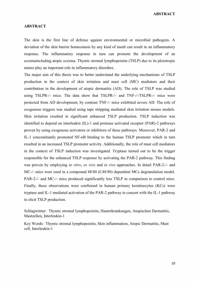
The skin is the first line of defense against environmental or microbial pathogens. A
deviation of the skin barrier homeostasis by any kind of insult can result in an inflammatory
response. The inflammatory response in turn can promote the development of an
eczemaincluding atopic eczema. Thymic stromal lymphopoietin (TSLP) due to its pleiotropic
nature play an important role in inflammatory disorders.
The major aim of this thesis was to better understand the underlying mechanisms of TSLP
production in the context of skin irritation and mast cell (MC) mediators and their
contribution in the development of atopic dermatitis (AD). The role of TSLP was studied
using TSLPR-/- mice. The data show that TSLPR-/- and TNF-/-/TSLPR-/- mice were
protected from AD development, by contrast TNF-/- mice exhibited severe AD. The role of
exogenous triggers was studied using tape stripping mediated skin irritation mouse models.
Skin irritation resulted in significant enhanced TSLP production. TSLP induction was
identified to depend on interleukin (IL)-1 and protease activated receptor (PAR)-2 pathways
proven by using exogenous activators or inhibitors of these pathways. Moreover, PAR-2 and
IL-1 concomitantly promoted NF-κB binding to the human TSLP promoter which in turn
resulted in an increased TSLP promoter activity. Additionally, the role of mast cell mediators
in the context of TSLP induction was investigated. Tryptase turned out to be the trigger
responsible for the enhanced TSLP response by activating the PAR-2 pathway. This finding
was proven by employing in vitro, ex vivo and in vivo approaches. In detail PAR-2-/- and
MC-/- mice were used in a compound 48/80 (C48/80) dependent MCs degranulation model.
PAR-2-/- and MC-/- mice produced significantly less TSLP in comparison to control mice.
Finally, these observations were confirmed in human primary keratinocytes (KCs) were
tryptase and IL-1 mediated activation of the PAR-2 pathway in concert with the IL-1 pathway
to elicit TSLP production.
Schlagwörter: Thymic stromal lymphopoietin, Hauterkrankungen, Atopischen Dermatitis,
Mastzellen, Interleukin-1
Key Words: Thymic stromal lymphopoietin, Skin inflammation, Atopic Dermatitis, Mast
cell, Interleukin-1
ZUSAMMENFASSUNG
ZUSAMMENFASSUNG
Die Haut ist das größte Organ des Menschen und stellt die primäre Barriere gegen
Umwelteinflüsse und Pathogene dar. Eine Dysbalance der Hautbarriere birgt die Gefahr einer
nachfolgenden Entzündungsreaktion. Bleibt diese bestehen, können sich Hautkrankheiten wie
zum Beispiel die atopische Dermatitis (AD) entwickeln. Verschiedene Zytokine wie Thymic
Stromal Lymphopoietin (TSLP) werden bei entzündlichen Hauterkrankungen eine
bedeutende Rolle zugeschrieben.
Hauptziel der vorliegenden Arbeit war die Aufklärung von Mechanismen, die durch
Hautirritation ausgelöst oder durch Mastzellmediatoren zu einem Anstieg von TSLP als
Wegbereiter einer Entzündungsreaktion in der Haut stattfinden.
Die Bedeutung von TSLP wurde zunächst anhand mehrerer Knockout-Mausstämme
untersucht. Hier zeigte sich, dass TSLPR-KO und TNF-TSLPR-DKO Mäuse im Gegensatz
zu TNF-KO Mäusen keine bzw. weniger Zeichen einer Entzündungsreaktion in der Haut
entwickeln. Die Rolle äußerer Einflüsse auf die TSLP-Produktion wurde anhand eines
Irritationsmodells ebenfalls in Mäusen untersucht. Dabei führte die Hautirritation, ausgelöst
durch Abtragen der oberen Hautschichten mittels eines Tesa-Abriss, zu einem signifikanten
Anstieg von TSLP in der Haut?. Mit Hilfe von Agonisten und Inhibitoren konnte gezeigt
werden, dass dieser Irritations-vermittelte TSLP-Anstieg über Interleukin-1 (IL-1) und
Protease Activated Receptor 2 (PAR-2) vermittelt wird. In diesem Zusammenhang wurde
auch gezeigt, dass die Aktivierung von IL-1- sowie PAR-2-abhängigen Signalwegen zu einer
gesteigerten Aktivität des TSLP-Promotors führte.
Die Untersuchung der Wirkung verschiedener Mastzellmediatoren auf die TSLP-Expression
ergab, dass Tryptase, über die Aktivierung von PAR-2, der wichtigste Mediator für den
Anstieg von TSLP nach der Degranulation von Mastzellen ist. Dieses Ergebnis wurde mittels
verschiedener in vitro, in vivo und ex vivo Experimentalansätze belegt. So konnte in einem
c48/80-abhängigen Degranulationsmodell in Mäusen gezeigt werden, dss PAR-2- sowie
Mastzell-KO Mäuse im Vergleich zu Wildtypen nach Injektion von c48/80 signifikant
weniger TSLP exprimierten. Abschließend konnte das Zusammenspiel von PAR-2- und IL-1-
vermittelten Signalwegen in Bezug auf TSLP in humanen Keratinozyten bestätigt werden.
Schlagwörter: Thymic stromal lymphopoietin, Hauterkrankungen, Atopischen Dermatitis,
Mastzellen, Interleukin-1
Key Words: Thymic stromal lymphopoietin, Skin inflammation, Atopic Dermatitis, Mast
cell, Interleukin-1

INTRODUCTION
12
INTRODUCTION
13
1 INTRODUCTION
1.1 SKIN STRUCTURE
The skin acts as a protective barrier and protects the body from a wide range of potential
harmful pathogens by separating inner and outer environment.1 The skin is built of three
layers, the outer epidermis, the dermis and the subcutis respectively.2 The epidermis is of
utmost relevance for the barrier integrity of the skin and confers the body with physical,
chemical or biochemical protections. The epidermis consists of various layers of
keratinocytes which go through the process of differentiation. These are the innermost
stratum basale, the stratum spinosum, the stratum granulosum and upper most the stratum
corneum.1,3,4 The stratum basale layer contains basal stem cells, which are capable of
proliferation to generate keratinocytes and can expand the cell numbers.5 The stratum
spinosum is defined by evident desmosomes, which help the appearance of spindle shaped
cells. Early differentiation marker cytokeratin 10 is expressed by the cells of this layer.
Differentiation of cells can be seen from bottom to top by the visibility of involucrin, an
intermediate differentiation marker, in upper cell lyers but not in the lower spinous cell
layers. The center part of the skin mainly consists of flat anucleated corneocytes. These
anucleated cells contain keratin filaments which symbolize differentiated keratinocytes of the
outer stratum granulosum layer.1,3,4 The stratum granulosum is composed of 3-5 layers of
cells and is characterized by the presence of lamellar bodies as well as keratohyalin granules.
The two late differentiation markers filaggrin and loricrin are expressed and processed by
these cell layers.6 The stratum corneum predominantly forms the primary skin barrier against
cutaneous infiltration of chemicals, microbes and mechanical injuries.1,7 In the stratum
corneum cells are held together by lipid bilayers, which form a rigid and insoluble structure
known as cornified envelope. The stratum corneum is also involved in various active
processes like regulation of water loss to outer environment from the skin, known as
transepidermal water loss (TEWL).1,7
The thickest part of the skin is the dermis containing sweat glands, sebaceous glands and hair
follicles among other structures.8 The dermis also harbors a complex network of blood
vessels and capillaries in addition to connective tissue. Thermoregulation of body is regulated
by the dilation or contraction of these blood vessels.8 Collagen and elastin, which are present
in the dermis provide plasticity.9
INTRODUCTION
14
1.2 EPIDERMAL BARRIER AND IT’S DISRUPTION IN ATOPIC DISEASES
The skin is metabolically active and several physiological phenomena help to maintain the
skin barrier intact. Protection of the inner body from microorganisms in addition to physical,
chemical, thermal and mechanical danger is the fundamental function of the skin (Fig.1).10
Several factors are required to sustain the skin barrier function. These include prevention of
excessive water loss, renewal of skin cells, cell to cell communication and interaction with
the immune system. Upon epidermal barrier disruption, the initiatory reaction to cellular
damage of the epidermal cells is a stimulatory alert to substitute the damaged cells and to
maintain the homeostasis in the skin.11 Immune cells like epidermal Langerhans cells (LCs),
dendritic cells (DCs) which are also known as skin-resident immune cells, are major players
in homeostatic reestablishment.10 In response to skin injury, keratinocytes (KCs) produce
pro-inflammatory cytokines such as tumor necrosis factor-α (TNF-α), Interleukin (IL)-1α, β,
IL-6 and IL-18. These cytokines activate dermal DCs in the presence or absence of antigens.
When KCs get activated due to stress signaling, they participate in further activation of
dermal DCs by secreting interferon-α (IFN-α) (Fig. 1). Upon activation DCs boost skin-
resident CD4+ or CD8+ T cell proliferation (Fig. 1). Activated T cells amplify the
inflammatory response in skin by producing chemokines and cytokines which in turn act on
epithelial and mesenchymal cells such as KCs and fibroblasts (Fig. 1).10
1.2.1 Skin irritants - physical and chemical
The skin is susceptible to exposure by different irritants which can result in detrimental
effects on barrier function and a subsequent damage of the epithelial cells.12 Several studies
have been conducted to better understand the underlying mechanisms of acute and chronic
irritation12. However, to study the pathogenesis of irritation at a cellular level in humans is
difficult due to ethical reasons. Therefore mouse models were employed to study the physico-
chemical phenomena behind these reactions. Many studies have been reported using different
chemical and physical irritants such as sodium dodecyl sulphate (SDS), croton oil, acetone,
tape stripping.13 Perturbed skin barrier assessment is analyzed by measuring TEWL,
electrical capacitance (stratum corenum hydration), percutaneous drug transport and skin
color reflectance (erythema).13,14 A strong inflammatory response has been observed by
Willis et al. (1993) on exposure of murine skin to 5% SDS for 48 hours (hrs).15 It has been
shown that SDS at higher concentrations leads to down regulation of HLA-DR expression in
INTRODUCTION
15
the LCs.16 Tape stripping mediated barrier disruption is another common method used to
study irritation, with less cytopathic effect on epithelial cells. Adhesive tape strip is
commonly used to remove the layers of stratum corneum.17 Upon stratum corneum
disruption, TEWL increases and leads to the production of different pro-inflammatory
mediator.17,18
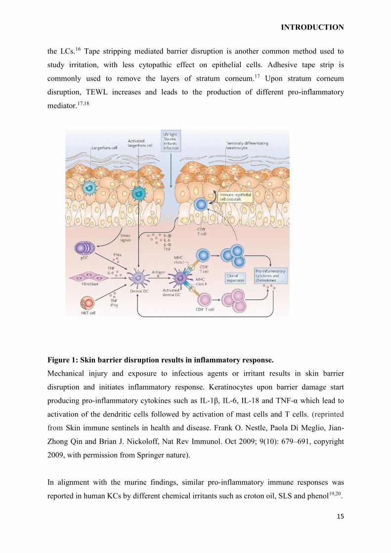
Figure 1: Skin barrier disruption results in inflammatory response.
Mechanical injury and exposure to infectious agents or irritant results in skin barrier
disruption and initiates inflammatory response. Keratinocytes upon barrier damage start
producing pro-inflammatory cytokines such as IL-1β, IL-6, IL-18 and TNF-α which lead to
activation of the dendritic cells followed by activation of mast cells and T cells. (reprinted
from Skin immune sentinels in health and disease. Frank O. Nestle, Paola Di Meglio, Jian-
Zhong Qin and Brian J. Nickoloff, Nat Rev Immunol. Oct 2009; 9(10): 679–691, copyright
2009, with permission from Springer nature).
In alignment with the murine findings, similar pro-inflammatory immune responses was
reported in human KCs by different chemical irritants such as croton oil, SLS and phenol19,20.
INTRODUCTION
16
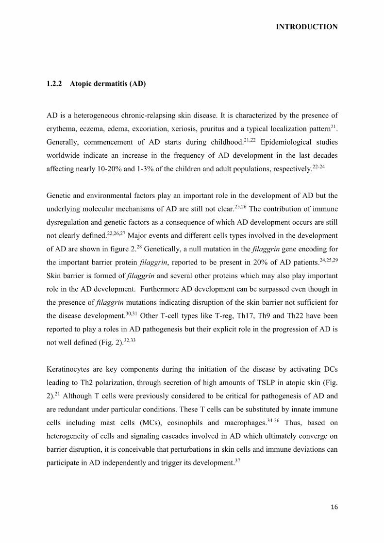
1.2.2 Atopic dermatitis (AD)
AD is a heterogeneous chronic-relapsing skin disease. It is characterized by the presence of
erythema, eczema, edema, excoriation, xeriosis, pruritus and a typical localization pattern21.
Generally, commencement of AD starts during childhood.21,22 Epidemiological studies
worldwide indicate an increase in the frequency of AD development in the last decades
affecting nearly 10-20% and 1-3% of the children and adult populations, respectively.22-24
Genetic and environmental factors play an important role in the development of AD but the
underlying molecular mechanisms of AD are still not clear.25,26 The contribution of immune
dysregulation and genetic factors as a consequence of which AD development occurs are still
not clearly defined.22,26,27 Major events and different cells types involved in the development
of AD are shown in figure 2.28 Genetically, a null mutation in the filaggrin gene encoding for
the important barrier protein filaggrin, reported to be present in 20% of AD patients.24,25,29
Skin barrier is formed of filaggrin and several other proteins which may also play important
role in the AD development. Furthermore AD development can be surpassed even though in
the presence of filaggrin mutations indicating disruption of the skin barrier not sufficient for
the disease development.30,31 Other T-cell types like T-reg, Th17, Th9 and Th22 have been
reported to play a roles in AD pathogenesis but their explicit role in the progression of AD is
not well defined (Fig. 2).32,33
Keratinocytes are key components during the initiation of the disease by activating DCs
leading to Th2 polarization, through secretion of high amounts of TSLP in atopic skin (Fig.
2).21 Although T cells were previously considered to be critical for pathogenesis of AD and
are redundant under particular conditions. These T cells can be substituted by innate immune
cells including mast cells (MCs), eosinophils and macrophages.34-36 Thus, based on
heterogeneity of cells and signaling cascades involved in AD which ultimately converge on
barrier disruption, it is conceivable that perturbations in skin cells and immune deviations can
participate in AD independently and trigger its development.37
INTRODUCTION
17
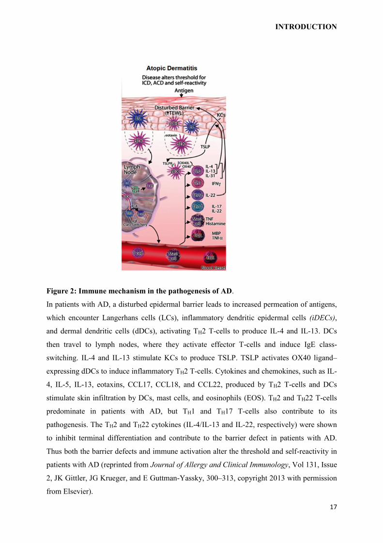
Figure 2: Immune mechanism in the pathogenesis of AD.
In patients with AD, a disturbed epidermal barrier leads to increased permeation of antigens,
which encounter Langerhans cells (LCs), inflammatory dendritic epidermal cells (iDECs),
and dermal dendritic cells (dDCs), activating TH2 T-cells to produce IL-4 and IL-13. DCs
then travel to lymph nodes, where they activate effector T-cells and induce IgE class-
switching. IL-4 and IL-13 stimulate KCs to produce TSLP. TSLP activates OX40 ligand–
expressing dDCs to induce inflammatory TH2 T-cells. Cytokines and chemokines, such as IL-
4, IL-5, IL-13, eotaxins, CCL17, CCL18, and CCL22, produced by TH2 T-cells and DCs
stimulate skin infiltration by DCs, mast cells, and eosinophils (EOS). TH2 and TH22 T-cells
predominate in patients with AD, but TH1 and TH17 T-cells also contribute to its
pathogenesis. The TH2 and TH22 cytokines (IL-4/IL-13 and IL-22, respectively) were shown
to inhibit terminal differentiation and contribute to the barrier defect in patients with AD.
Thus both the barrier defects and immune activation alter the threshold and self-reactivity in
patients with AD (reprinted from Journal of Allergy and Clinical Immunology, Vol 131, Issue
2, JK Gittler, JG Krueger, and E Guttman-Yassky, 300–313, copyright 2013 with permission
from Elsevier).
INTRODUCTION
18
1.3 KERATINOCYTES
KCs are epithelial cells which conserve the physical and biochemical integrity of skin.38,39
During differentiation to form skin barrier, KCs undergo complex morphological and
cytostructural changes with the expression of different differentiation-dependent structural
proteins such as involucrin, filaggrin, claudin etc. in the spinous and granular layers.38 KCs
play crucial role in the cellular communication, pathogenesis of diseases and in maintaining
the immune response.31,40,41 Deviation from skin homeostasis or barrier disruption act as
activation signal for KCs in response to which they start producing different pro-
inflammatory cytokines such as TNF-α, IL-1α, TSLP to facilitate inflammation.10
1.3.1 Role of keratinocytes in skin irritation
As pointed out above, for maintaining skin homeostasis KCs are the crucial cells. By
undergoing differentiation, they form rigid structure and consecutively enter into cell cycle
arrest phase.42 IL-1α produced by KCs acts as primary alarm signal upon skin irritation or
other skin disruption in the inflammatory pathway.43 Numerous, studies have demonstrated
the ability of different irritants to induce IL-1α production in KCs, which further boost the
production of other pro-inflammatory cytokines such as IL-1β, IL-6, IL,18, TNF-α by dermal
and epidermal cells.42,44-47 Physical or chemical skin irritation leads to activation of proteases
which cleave the pro-IL-1β into biologically active IL-1β which along with IL-1α support the
activation of DCs and T cells.48
1.3.2 Role of keratinocytes in AD
Barrier deficiency in AD development and progression is primarily caused by KCs.49 KCs
produce peculiar set of cytokines and chemokine’s such as high levels of chemokine ligand
(CCL)5 (RANTES), thymic stromal lymphopoietin (TSLP) upon stimulation with IL-1and
TNF-α to promote AD environment.20,50 It has been reported that KCs derived from AD
patients produce high amount of granulocytes-macrophage colony-stimulation factor (GMC-
SF) in addition to TNF-α.51 Recent studies, demonstrating the involvement of KCs derived
cytokines such as TSLP on the inflammatory response provide a deeper insight for the role of
KCs not only in barrier formation, but also as collaborative cells along with DCs activation to
primer T cells to induce production of IL-4 and IL-13.50,52 TSLP mediated activation of DCs
INTRODUCTION
19
results in production of chemokines such as CCL17, macrophage derived CCL22, which
further foster infiltration of Th2 cells in the lesional AD skin.28 It has been shown that upon
activation KCs produce IL-25 and IL-33 which act on mast cells (MCs), DCs and LCs.31,34
1.4 MAST CELLS
MCs are bone marrow derived cells which migrate to tissue through blood for maturation
under the influence of stem cell factor (SCF). They can be identified by staining for tryptase.
Skin MCs are belong to the connective tissue type and contain both chymase and tryptase
(MCTC) while the mucosal MCs contains only tryptase and no chymase (MCT).53-55 MCs are
deemed to be among the first cells to respond to an allergen/injury and are considered as the
instigating players in the IgE-mediated immediate type hypersensitivity. MCs degranulation
can be mediated by human G protein coupled receptor MAS Related GPR Family Member
X2 (MRGPRx2) activation by certain drugs, substance P or compound 48/80 (C48/80) in
addition to classical FcεRI crosslinking by polyvalent allergen/agent through binding to IgE
present on FcεRI.56, 57,58
1.4.1 Role of mast cells in AD
Profound degranulation of MCs along with their recruitment is observed in the lesions of AD
skin.54,59 MCs degranulation has been shown to correlate with the severity of AD.60 MCs can
regulate the recruitment as well as functions of cells participating in the skin inflammation
through the production of cytokines, chemokines and growth factors (Fig. 3).61 For instance,
by virtue of IL-4, IL-13 and TNF-α they induce cell adhesion molecule on endothelium,
which can contribute in the recruitment of leukocytes.62-64 They can also control the
differentiation of naïve T cells to Th1 or Th2 subtypes and increase the activation of T
cells.65,66 MCs can also modulate primary B cell development and induce IgE synthesis in B
cells.66-68
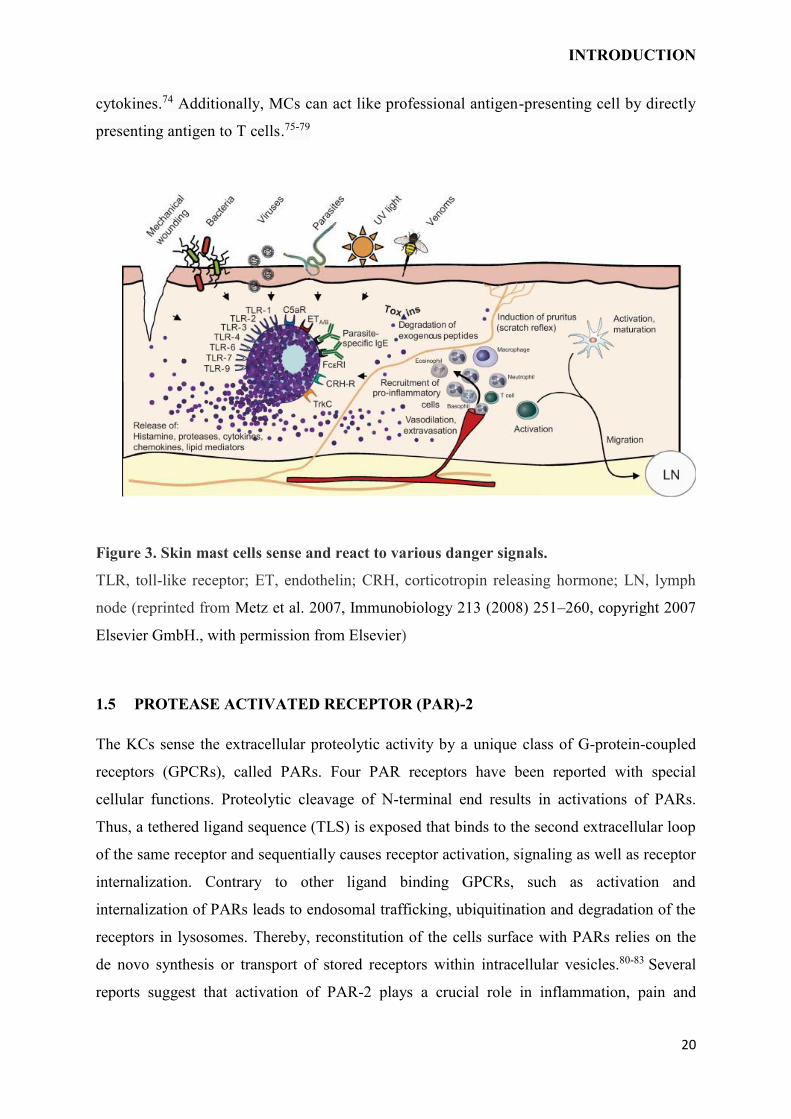
MCs can interact with KCs, DCs and LCs by their mediators. By secreting tryptase
(endogenous PAR-2 agonist) and histamine, MCs can stimulate KCs to express pro-
inflammatory chemokines and cytokines, growth factors and adhesion proteins.69,70 They
can induce integrin on the LCs by TNF-α and promote their migration to lymph
nodes.71,72,73 Moreover, MCs affect DCs polarization to Th1/Th2 through mediators and
INTRODUCTION
20
cytokines.74 Additionally, MCs can act like professional antigen-presenting cell by directly
presenting antigen to T cells.75-79
Figure 3. Skin mast cells sense and react to various danger signals.
TLR, toll-like receptor; ET, endothelin; CRH, corticotropin releasing hormone; LN, lymph
node (reprinted from Metz et al. 2007, Immunobiology 213 (2008) 251–260, copyright 2007
Elsevier GmbH., with permission from Elsevier)
1.5 PROTEASE ACTIVATED RECEPTOR (PAR)-2
The KCs sense the extracellular proteolytic activity by a unique class of G-protein-coupled
receptors (GPCRs), called PARs. Four PAR receptors have been reported with special
cellular functions. Proteolytic cleavage of N-terminal end results in activations of PARs.
Thus, a tethered ligand sequence (TLS) is exposed that binds to the second extracellular loop
of the same receptor and sequentially causes receptor activation, signaling as well as receptor
internalization. Contrary to other ligand binding GPCRs, such as activation and
internalization of PARs leads to endosomal trafficking, ubiquitination and degradation of the
receptors in lysosomes. Thereby, reconstitution of the cells surface with PARs relies on the
de novo synthesis or transport of stored receptors within intracellular vesicles.80-83 Several
reports suggest that activation of PAR-2 plays a crucial role in inflammation, pain and
INTRODUCTION
21
allergic responses.84-88 PAR-2 activation is mediated by a broad range of endogenous
proteases including serine proteases (e.g. KLK5, KLK14, tryptase) and exogenous proteases
such as house dust mite (HDM) antigen Der p1.89-92 PAR-2 activation has been shown to
induce itch, either directly upon activation of receptor on sensory nerve fibers in the skin, or
indirectly by activating KCs or other immune cells (e.g. MCs), and consequently elicit a
cascade resulting in release of pruritogens which in turn activate the sensory nerve fibers
innervating the skin.93-96
1.5.1 Role of PAR-2 in AD
Severe skin barrier disruption leads to excessive dehydration, chronic skin inflammation, itch
and enhanced risk of skin infections. Patients with AD, netherton syndrome (NS) or
ichthyosis, suffer from extensive itch and ultimately develop erythematous scaly skin as a
result of faulty skin barrier.36,97,98
Along with kallikreins (KLKs) and endogenous protease inhibitors, PAR-2 as well as its
activating proteases are important regulators of KC differentiation and skin barrier
homeostasis. Interestingly, hyperkeratosis in different inflammatory skin diseases (e.g. AD or
NS) is coincided by enhanced expression of PAR-2 and PAR-2 activating proteases.36,97,99
1.6 THYMIC STROMAL LYMPHOPOIETIN (TSLP)
TSLP is an IL-7 like cytokine and has been first identified in the mouse thymic stromal cell
culture supernatants. TSLP promote the differentiation and growth of B cells in addition to
proliferation of T cells.100,101 Several studies have reported that high affinity binding of TSLP
needs concomitant binding to the TSLP receptor (TSLPR) and α-chain of IL-7 receptor.102,103
Primarily TSLP is expressed by epithelia cells of skin, gut, thymus and tonsils along with
stromal cells.104-106 TSLP leads to differentiation of Treg cells by instructing the thymic
DCs.107 Intriguingly, human TSLP does not exert the same effects as the murine TSLP,
despite that it activate immature CD11c+ myeloid DCs.106,108 Thereby, human DCs can
induce naïve CD4+ T cell proliferation and triggers the production of IL-4, IL-5, IL-13 as
well as TNF-α (fig. 7). On the other hand, TSLP activated DCs inhibits the production of
anti-inflammatory cytokines IL-10 and IFN-γ.108 As a broad range of cells are influenced by
TSLP, it has been implied to play a major role in numerous ailments like cancer, infections
and inflammatory bowel diseases.109-111 However, TSLP being primarily an epithelial
cytokines, has been expected to play a vital role in allergic diseases such as asthma and
AD.112 In line with the expected role, TSLP has been found upregulated in mouse models of
INTRODUCTION
22
allergic asthma as well as AD and found responsible for defective airway inflammation and
skin inflammation.113-115
1.6.1 Role of TSLP in AD
Skin barrier integrity is compromised upon acute injury or perturbation to the stratum
corneum leading to induction of positive and negative alarm signals resulting in triggering of
both homeostatic and inflammatory reactions in the skin.18,116 The damaged skin barrier
further fuels the production of particular cytokines to promote skin inflammation.99,117,118
TSLP is one of the cytokine, produced by KCs on skin barrier damage or stimulation with
inflammatory cytokines (fig. 4).52 Although, the importance of TSLP in allergic
inflammations is firmly established, the mechanisms behind the induction of TSLP
production are not well known.35,119,120 Primary human KCs and skin explants have been
shown to produce enhanced TSLP in response to viral, bacterial or inflammatory stimuli or
upon physical/chemical perturbation to skin barrier.20,121,122 The role of TSLP in the
manifestation of AD was not clear until it was reported that TSLP overexpression in the
murine skin results in development of spontaneous dermatitis, the hallmark of human
AD.34,106 Since epithelial cells are the primary source of TSLP, this further indicate KCs are
the trigger factor in the AD development (Fig. 4).123 Subsequently, several studies verified
TSLP as a primary initiator of AD.35,36,124 Development of AD like skin lesions have been
observed upon direct administration of TSLP in skin. TSLP promotes the proliferation and
differentiation of Th2 cells and consequently expression of TNF-α, IL-4, IL-5 and IL-13.106
Furthermore, it was observed that expression of TSLP in KCs from AD patients is highly
increased. Likewise, TSLP is associated with migration and activation of dermal DCs.125
Thus, TSLP was considered as one of the key factors in triggering AD development. Mice
overexpressing KCs specific TSLP, but lacking T cells developed skin inflammation,
suggesting that for the disease progression T cells are not obligatory.34 In agreement with the
previous study, other studies by using different AD models demonstrated that TSLP is
essential for AD development.115,126
INTRODUCTION
23
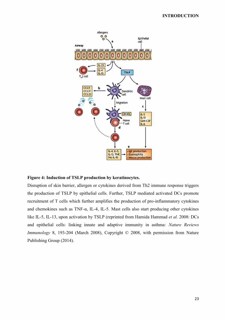
Figure 4: Induction of TSLP production by keratinocytes.
Disruption of skin barrier, allergen or cytokines derived from Th2 immune response triggers
the production of TSLP by epithelial cells. Further, TSLP mediated activated DCs promote
recruitment of T cells which further amplifies the production of pro-inflammatory cytokines
and chemokines such as TNF-α, IL-4, IL-5. Mast cells also start producing other cytokines
like IL-5, IL-13, upon activation by TSLP (reprinted from Hamida Hammad et al. 2008: DCs
and epithelial cells: linking innate and adaptive immunity in asthma: Nature Reviews
Immunology 8, 193-204 (March 2008), Copyright © 2008, with permission from Nature
Publishing Group (2014).
INTRODUCTION
24
1.7 OBJECTIVES
Over the years, TSLP has been well established as critical pro-inflammatory cytokines with
implications in inflammatory disorders. TSLP promotes pro-allergic Th2-type inflammatory
responses through activating leukocytes. However, the mechanisms of TSLP regulation in
skin irritation, contribution of endogenous MCs mediators in TSLP production and its role in
AD development is not clear. In this thesis, mechanisms underlying the TSLP production by
skin irritation, MC mediators and its contribution in the AD development was investigated. In
this thesis the following questions were tackled.
1. Are the TSLPR-/- mice protected from AD development under the TNF deficiency?
2. Is skin barrier disruption responsible for elicited TSLP production and what are the
signaling cascades involved? Do the responsible pathways operate cooperatively or
independently and at what levels?
3. Are the mast cells plays a role in TSLP production by the keratinocytes? And if so
what are the mediators and mechanisms behind MCs and KCs cross-talk in the
production of TSLP?
Answer to these questions will help in better understanding of the mechanisms underlying
inflammatory processes in the AD development and may help in designing more irresolute
clinical management by exclusive aiming of pathways involved individually or collectively
depending on the situation.
MATERIALS AND METHODS
2 MATERIALS AND METHODS
2.1 MATERIALS
Detailed list of used reagents, antibodies, solutions, labwares and softwere are given below:
Details about antibodies, instruments, chemicals, buffers, solutions, reagents, labwares and
software used are listed below:
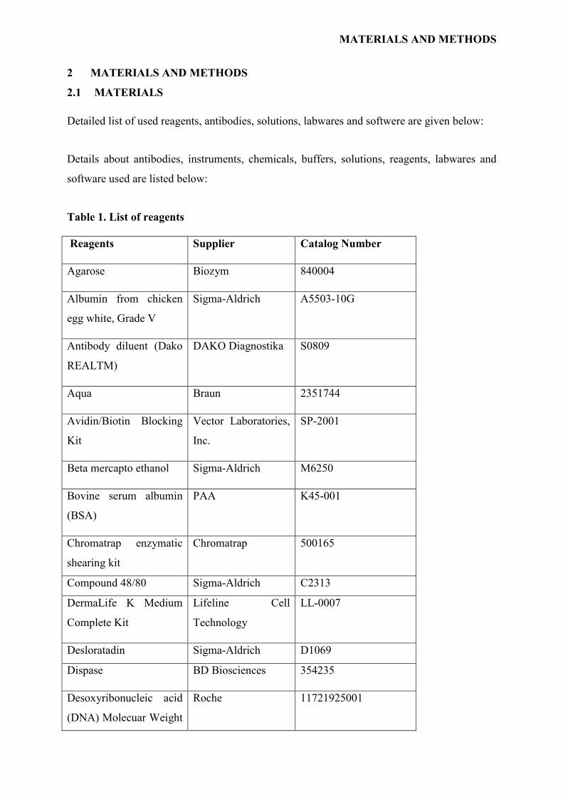
Table 1. List of reagents
Reagents Supplier Catalog Number
Agarose Biozym 840004
Albumin from chicken
egg white, Grade V
Sigma-Aldrich A5503-10G
Antibody diluent (Dako
REALTM)
DAKO Diagnostika S0809
Aqua Braun 2351744
Avidin/Biotin Blocking
Kit
Vector Laboratories,
Inc.
SP-2001
Beta mercapto ethanol Sigma-Aldrich M6250
Bovine serum albumin
(BSA)
PAA K45-001
Chromatrap enzymatic
shearing kit
Chromatrap 500165
Compound 48/80 Sigma-Aldrich C2313
DermaLife K Medium
Complete Kit
Lifeline Cell
Technology
LL-0007
Desloratadin Sigma-Aldrich D1069
Dispase BD Biosciences 354235
Desoxyribonucleic acid
(DNA) Molecuar Weight
Roche 11721925001
MATERIALS AND METHODS
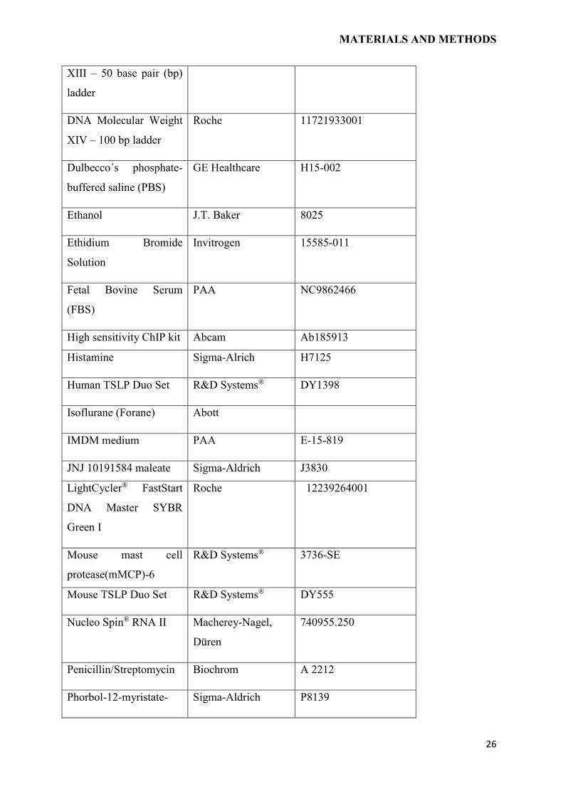
26
XIII – 50 base pair (bp)
ladder
DNA Molecular Weight
XIV – 100 bp ladder
Roche 11721933001
Dulbecco´s phosphate-
buffered saline (PBS)
GE Healthcare H15-002
Ethanol J.T. Baker 8025
Ethidium Bromide
Solution
Invitrogen 15585-011
Fetal Bovine Serum
(FBS)
PAA NC9862466
High sensitivity ChIP kit Abcam Ab185913
Histamine Sigma-Alrich H7125
Human TSLP Duo Set R&D Systems® DY1398
Isoflurane (Forane) Abott
IMDM medium PAA E-15-819
JNJ 10191584 maleate Sigma-Aldrich J3830
LightCycler® FastStart
DNA Master SYBR
Green I
Roche 12239264001
Mouse mast cell
protease(mMCP)-6
R&D Systems® 3736-SE
Mouse TSLP Duo Set R&D Systems® DY555
Nucleo Spin® RNA II Macherey-Nagel,
Düren
740955.250
Penicillin/Streptomycin Biochrom A 2212
Phorbol-12-myristate- Sigma-Aldrich P8139
MATERIALS AND METHODS
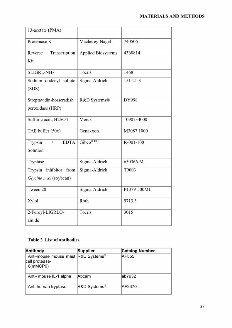
27
13-acetate (PMA)
Proteinase K Macherey-Nagel 740506
Reverse Transcription
Kit
Applied Biosystems 4368814
SLIGRL-NH2 Tocris 1468
Sodium dodecyl sulfate
(SDS)
Sigma-Aldrich 151-21-3
Streptavidin-horseradish
peroxidase (HRP)
R&D Systems® DY998
Sulfuric acid, H2SO4 Merck 1090734000
TAE buffer (50x) Genaxxon M3087.1000
Trypsin / EDTA
Solution
Gibco® BD R-001-100
Tryptase Sigma-Aldrich 650366-M
Trypsin inhibitor from
Glycine max (soybean)
Sigma-Aldrich T9003
Tween 20 Sigma-Aldrich P1379-500ML
Xylol Roth 9713.3
2-Furoyl-LIGRLO-
amide
Tocris 3015
Table 2. List of antibodies
Antibody Supplier Catalog Number
Anti-mouse mouse mast cell protease- 6(mMCP6)
R&D Systems®
AF555
Anti- mouse IL-1 alpha Abcam ab7632
Anti-human tryptase R&D Systems®
AF2370
MATERIALS AND METHODS
28
Anti-NF-κB Abcam Ab19870
Anti-PAR-2 (SAM11) Santa Cruz Biotechnology, Inc.
sc-13504
Mouse IgG2a R&D Systems® MAB003
Normal goat IgG control R&D Systems® AB-108-C
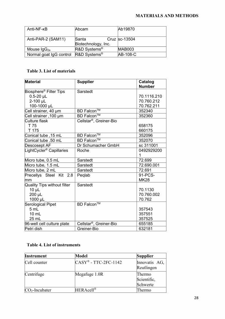
Table 3. List of materials
Material Supplier Catalog Number
Biosphere® Filter Tips 0.5-20 µL 2-100 µL 100-1000 µL
Sarstedt 70.1116.210 70.760.212 70.762.211
Cell strainer, 40 µm BD FalconTM 352340
Cell strainer ,100 µm BD FalconTM 352360
Culture flask T 75 T 175
Cellstar®, Greiner-Bio 658175 660175
Conical tube ,15 mL BD FalconTM 352096
Conical tube ,50 mL BD FalconTM 352070
Descosept AF Dr Schumacher GmbH sc 311001
LightCycler® Capillaries Roche 04929292001
Micro tube, 0.5 mL Sarstedt 72.699
Micro tube, 1.5 mL Sarstedt 72.690.001
Micro tube, 2 mL Sarstedt 72.691
Precellys Steel Kit 2.8 mm
Peqlab 91-PCS-MK28
Quality Tips without filter 10 µL 200 µL 1000 µL
Sarstedt 70.1130 70.760.002 70.762
Serological Pipet 5 mL 10 mL 25 mL
BD FalconTM 357543 357551 357525
96-well cell culture plate Cellstar®, Greiner-Bio 655185
Petri dish Greiner-Bio 632181
Table 4. List of instruments
Instrument Model Supplier
Cell counter CASY® - TTC-2FC-1142 Innovatis AG,
Reutlingen
Centrifuge Megafuge 1.0R Thermo
Scientific,
Schwerte
CO2-Incubater HERAcell® Thermo
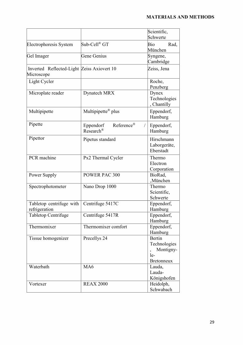
MATERIALS AND METHODS
29
Scientific,
Schwerte
Electrophoresis System Sub-Cell® GT Bio Rad,
München
Gel Imager Gene Genius Syngene,
Cambridge
Inverted Reflected-Light
Microscope
Zeiss Axiovert 10 Zeiss, Jena
Light Cycler Roche,
Penzberg
Microplate reader Dynatech MRX Dynex
Technologies
, Chantilly
Multipipette Multipipette® plus Eppendorf,
Hamburg
Pipette Eppendorf Reference® /
Research®
Eppendorf,
Hamburg
Pipettor Pipetus standard Hirschmann
Laborgeräte,
Eberstadt
PCR machine Px2 Thermal Cycler Thermo
Electron
Corporation
Power Supply POWER PAC 300 BioRad,
‚München
Spectrophotometer Nano Drop 1000 Thermo
Scientific,
Schwerte
Tabletop centrifuge with
refrigeration
Centrifuge 5417C Eppendorf,
Hamburg
Tabletop Centrifuge Centrifuge 5417R Eppendorf,
Hamburg
Thermomixer Thermomixer comfort Eppendorf,
Hamburg
Tissue homogenizer Precellys 24 Bertin
Technologies
, Montigny-
le-
Bretonneux
Waterbath MA6 Lauda,
Lauda-
Königshofen
Vortexer REAX 2000 Heidolph,
Schwabach
MATERIALS AND METHODS
30
2.2 METHODS
2.2.1 Animal experiments
All experimental procedures were approved by the local State office of Health and Social
Affairs and performed in agreement with their protocols. Mice were either purchased from
Jackson, Charles River or generated and breed at animal facility of Forschungseinrichtungen
für Experimentelle Medizin (FEM).
2.3 MOUSE MODELS
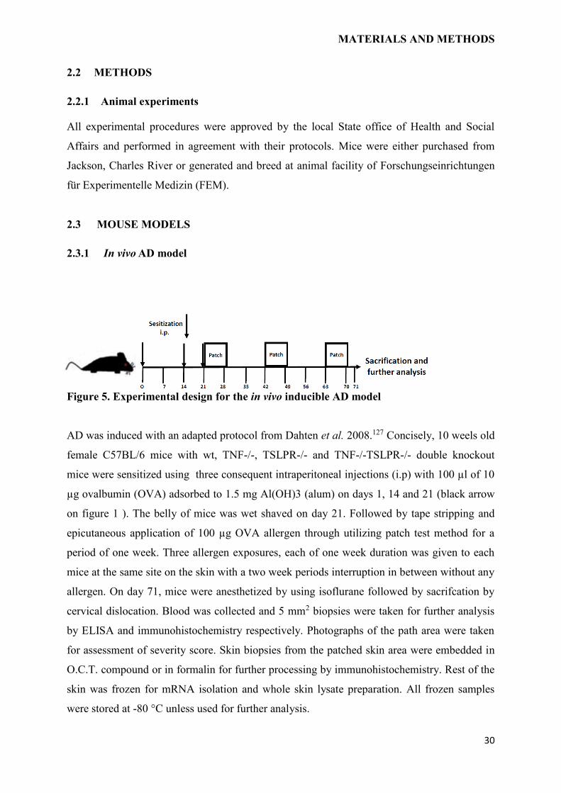
2.3.1 In vivo AD model
Figure 5. Experimental design for the in vivo inducible AD model
AD was induced with an adapted protocol from Dahten et al. 2008.127 Concisely, 10 weels old
female C57BL/6 mice with wt, TNF-/-, TSLPR-/- and TNF-/-TSLPR-/- double knockout
mice were sensitized using three consequent intraperitoneal injections (i.p) with 100 µl of 10
µg ovalbumin (OVA) adsorbed to 1.5 mg Al(OH)3 (alum) on days 1, 14 and 21 (black arrow
on figure 1 ). The belly of mice was wet shaved on day 21. Followed by tape stripping and
epicutaneous application of 100 µg OVA allergen through utilizing patch test method for a
period of one week. Three allergen exposures, each of one week duration was given to each
mice at the same site on the skin with a two week periods interruption in between without any
allergen. On day 71, mice were anesthetized by using isoflurane followed by sacrifcation by
cervical dislocation. Blood was collected and 5 mm2 biopsies were taken for further analysis
by ELISA and immunohistochemistry respectively. Photographs of the path area were taken
for assessment of severity score. Skin biopsies from the patched skin area were embedded in
O.C.T. compound or in formalin for further processing by immunohistochemistry. Rest of the
skin was frozen for mRNA isolation and whole skin lysate preparation. All frozen samples
were stored at -80 °C unless used for further analysis.
MATERIALS AND METHODS
31
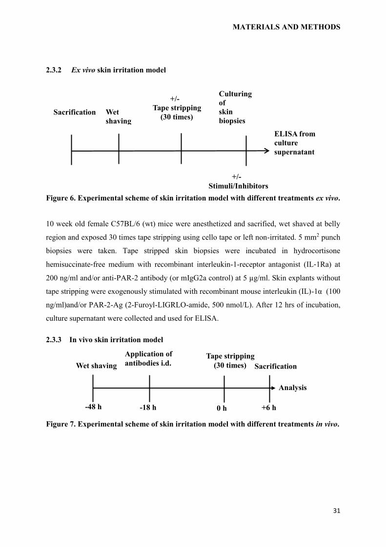
2.3.2 Ex vivo skin irritation model
Figure 6. Experimental scheme of skin irritation model with different treatments ex vivo.
10 week old female C57BL/6 (wt) mice were anesthetized and sacrified, wet shaved at belly
region and exposed 30 times tape stripping using cello tape or left non-irritated. 5 mm2 punch
biopsies were taken. Tape stripped skin biopsies were incubated in hydrocortisone
hemisuccinate‐free medium with recombinant interleukin-1-receptor antagonist (IL‐1Ra) at
200 ng/ml and/or anti-PAR-2 antibody (or mIgG2a control) at 5 µg/ml. Skin explants without
tape stripping were exogenously stimulated with recombinant mouse interleukin (IL)-1α (100
ng/ml)and/or PAR-2-Ag (2-Furoyl-LIGRLO-amide, 500 nmol/L). After 12 hrs of incubation,
culture supernatant were collected and used for ELISA.
2.3.3 In vivo skin irritation model
Figure 7. Experimental scheme of skin irritation model with different treatments in vivo.
Wet shaving
-48 h
Application of
antibodies i.d.
-18 h
Tape stripping
(30 times) Sacrification
+6 h 0 h
Analysis
Culturing
of
skin
biopsies
+/-
Stimuli/Inhibitors
+/-
Tape stripping
(30 times)
ELISA from
culture
supernatant
Wet
shaving
Sacrification
MATERIALS AND METHODS
32
In vivo skin irritation
The experimental procedure is depicted in figure 7. After scarification, mouse skin was
chopped and homogenized by pre-chilled precellys homogenization (PEQLAB, Erlangen,
Germany) in lysis buffer (2 mmol/L EDTA, 1 mmol/L dithiothreitol, 25 mmol/L Tris [pH
7.8], 1% Triton X-100 and 10% glycerol) with protease inhibitor cocktail (Roche, Basel,
Switzerland) and used for ELISA. Protein was quantified by bis-cinchinonic acid (BCA)
assay (Pierce Laboratories, Rockford, USA).
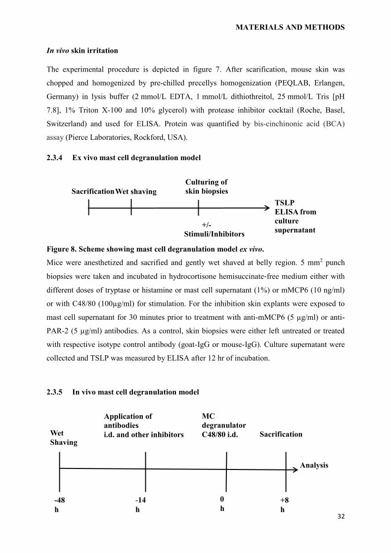
2.3.4 Ex vivo mast cell degranulation model
Figure 8. Scheme showing mast cell degranulation model ex vivo.
Mice were anesthetized and sacrified and gently wet shaved at belly region. 5 mm2 punch
biopsies were taken and incubated in hydrocortisone hemisuccinate‐free medium either with
different doses of tryptase or histamine or mast cell supernatant (1%) or mMCP6 (10 ng/ml)
or with C48/80 (100µg/ml) for stimulation. For the inhibition skin explants were exposed to
mast cell supernatant for 30 minutes prior to treatment with anti-mMCP6 (5 µg/ml) or anti-
PAR-2 (5 µg/ml) antibodies. As a control, skin biopsies were either left untreated or treated
with respective isotype control antibody (goat-IgG or mouse-IgG). Culture supernatant were
collected and TSLP was measured by ELISA after 12 hr of incubation.
2.3.5 In vivo mast cell degranulation model
TSLP
ELISA from
culture
supernatant
Wet shaving Sacrification
Culturing of
skin biopsies
+/-
Stimuli/Inhibitors
-14
h
Application of
antibodies
i.d. and other inhibitors
0
h
MC
degranulator
C48/80 i.d.
+8
h
Sacrification
-48
h
Wet
Shaving
Analysis
MATERIALS AND METHODS
33
Figure 9. Scheme depicting mast cell degranulation model with different treatments in
vivo.
The experimental procedure is depicted in figure 9. After scarification, mouse skin was
chopped and homogenized by pre-chilled precellys homogenization (PEQLAB, Erlangen,
Germany) in lysis buffer (2 mmol/L EDTA, 1 mmol/L dithiothreitol, 25 mmol/L Tris [pH
7.8], 1% Triton X-100 and 10% glycerol) with protease inhibitor cocktail (Roche, Basel,
Switzerland) and used for ELISA. Protein was quantified by bis-cinchinonic acid (BCA)
assay (Pierce Laboratories, Rockford, USA).
2.4 CELL CULTURE METHODS
2.4.1 Isolation, culturing and treatment of human primary keratinocytes
Human KCs were isolated from foreskin and processed as described previously. The fore skin
was obtained after circumcisions, with informed consent of the patients and approval by the
university Ethics committee. All the experiments were conducted according to the Declaration
of Helsinki Principles. After the 2nd passage, 7.5x10³ cells/well were seeded in a 96-well
plate in KC medium and grown to 70-80% confluence. For Luciferase assay 40-50%
confluent cells were used. After reaching confluence, the medium was changed to
hydrocortisone hemisuccinate free KC medium for 24 hr, and cells were stimulated with
various concentrations of IL-1(2,5,10,20 ng/ml), nafamostat 5 µg/ml, rhIL-1Ra 200 ng/ml,
SLIGRL 100 µM/ml and 0.1% MC lysate 24 hr. Supernatants were collected and measured by
a human TSLP ELISA Kit (R&D Systems).
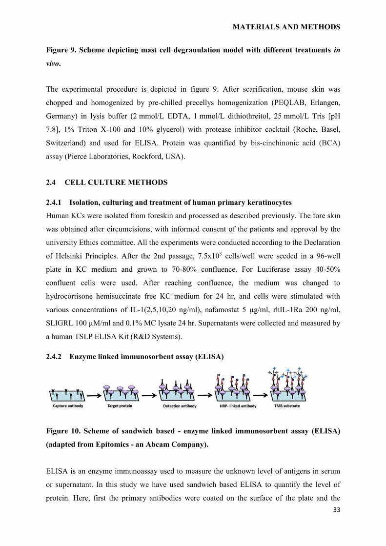
2.4.2 Enzyme linked immunosorbent assay (ELISA)
Figure 10. Scheme of sandwich based - enzyme linked immunosorbent assay (ELISA)
(adapted from Epitomics - an Abcam Company).
ELISA is an enzyme immunoassay used to measure the unknown level of antigens in serum
or supernatant. In this study we have used sandwich based ELISA to quantify the level of
protein. Here, first the primary antibodies were coated on the surface of the plate and the
MATERIALS AND METHODS
34
target protein from serum or supernatant were incubated for specific binding. The detection
antibodies were incubated over the surface of bound specific antigen. In the next step, the
plates were incubated with Horseradish peroxidase (HRP) linked biotinylated antibodies,
which can convert a chromogenic substrate. The enzymatic reaction leads to the color change
which was measured by spectrophotometer. The concentration of protein in the samples was
calculated by the means of standard curve. All the steps were performed at room temperature
and in dark from HRP-linked antibody.
2.4.2.1 Mouse and human TSLP ELISA
In vitro, ex vivo, or in vivo experiments were performed and cell free supernatant or serum
from mice and human epidermal sheet were obtained and measured for mouse and human
TSLP levels. Analysis was performed based on TSLP ELISA kit from R&D system (mouse)
and ebiosciences (human) according to manufacturer’s instructions.
2.4.3 RNA isolation
Frozen skin samples from mice were homogenized by pre-chilled precellys homogenisation
(PEQLAB, Germany) beads tube in 500 μl RA1 lysis buffer (NucleoSpin® RNA isolation kit)
with 5 μl β-mercaptoethanol (β-Me) at 5500 rpm for 2 times for 30 sec with 5 sec pause.
Homogenized samples were transferred to NucleoSpin filter and centrifuged at 11,000 g for 2
min at room temperature. Supernatant was transferred to sterile eppendorf tubes carefully
without disturbing the pellet and 500 μl of RNase-free water was added with 10% proteinase
K and mixed well for tissue digestion. The lysate was incubated for 15 min at 55 °C. After 15
min, lysate was centrifuged at 10,000 g for 3 min. For RNA isolation from the keratinocytes
lysis was performed by using 300 µl RA1 buffer with 3 µl β-mercaptoethanol (β-Me).
Further, RNA isolation was performed according to manufacturer’s instruction along with
DNase digestion step for 15 min at room temperature. RNA was eluted with 60 μl of RNase-
free water. Using NanoDrop UV-Vis spectrophotometer, RNA concentration was measured at
260 nm. Later, quality of RNA was checked by 2% agarose gel. The eluted samples were
stored at -80 °C for further analysis.
2.4.4 Reverse transcription
Reverse transcription of total RNA into single stranded cDNA was performed with TaqMan®
reverse transcription reagent according to manufacturer’s instruction. The kit contains a
recombinant Moloney Murine Leukemia Virus Reverse Transcriptase, random hexamers and
oligo d(T). 1 μg of total RNA was used for reverse transcribtion in to cDNA in thermo cycler
with following steps as given in table 5.
MATERIALS AND METHODS
35
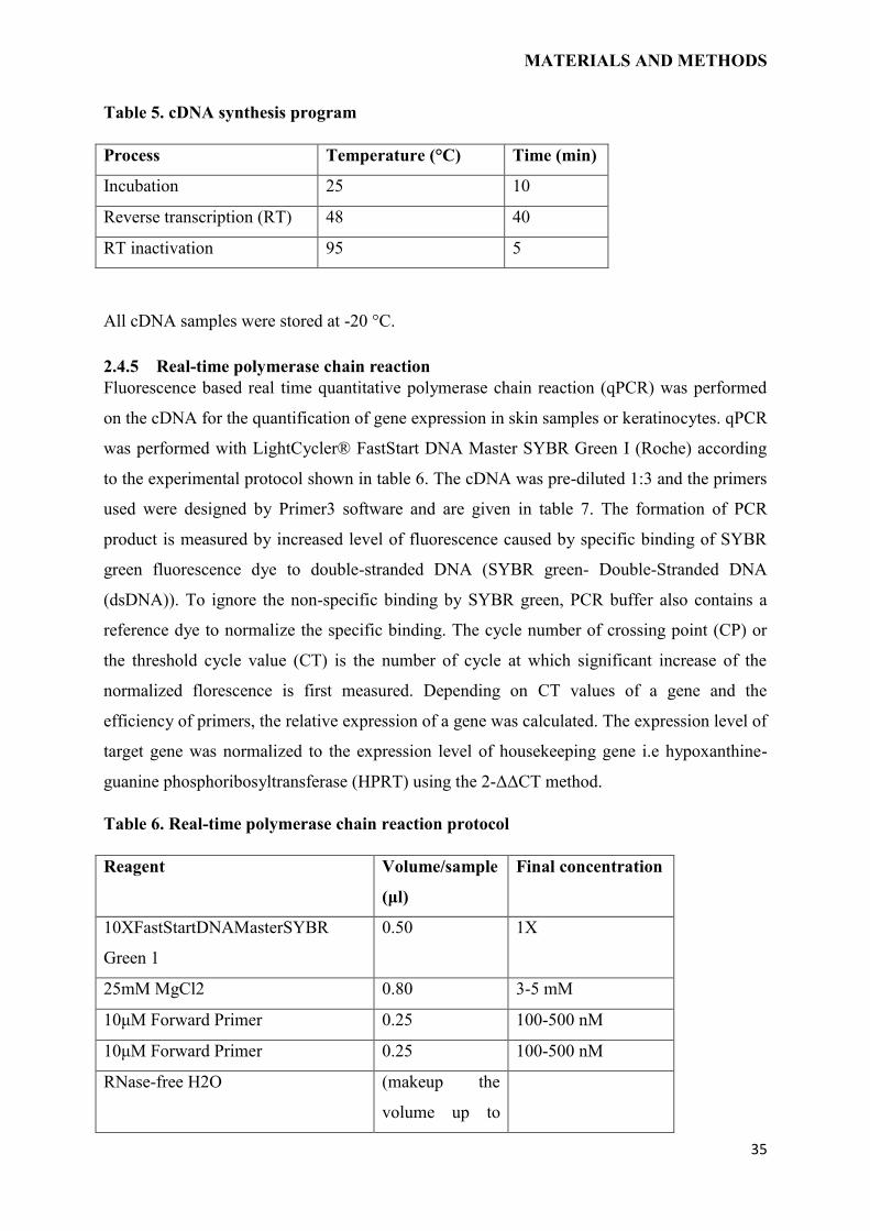
Table 5. cDNA synthesis program
Process Temperature (°C) Time (min)
Incubation 25 10
Reverse transcription (RT) 48 40
RT inactivation 95 5
All cDNA samples were stored at -20 °C.
2.4.5 Real-time polymerase chain reaction
Fluorescence based real time quantitative polymerase chain reaction (qPCR) was performed
on the cDNA for the quantification of gene expression in skin samples or keratinocytes. qPCR
was performed with LightCycler® FastStart DNA Master SYBR Green I (Roche) according
to the experimental protocol shown in table 6. The cDNA was pre-diluted 1:3 and the primers
used were designed by Primer3 software and are given in table 7. The formation of PCR
product is measured by increased level of fluorescence caused by specific binding of SYBR
green fluorescence dye to double-stranded DNA (SYBR green- Double-Stranded DNA
(dsDNA)). To ignore the non-specific binding by SYBR green, PCR buffer also contains a
reference dye to normalize the specific binding. The cycle number of crossing point (CP) or
the threshold cycle value (CT) is the number of cycle at which significant increase of the
normalized florescence is first measured. Depending on CT values of a gene and the
efficiency of primers, the relative expression of a gene was calculated. The expression level of
target gene was normalized to the expression level of housekeeping gene i.e hypoxanthine-
guanine phosphoribosyltransferase (HPRT) using the 2-ΔΔCT method.
Table 6. Real-time polymerase chain reaction protocol
Reagent Volume/sample
(μl)
Final concentration
10XFastStartDNAMasterSYBR
Green 1
0.50 1X
25mM MgCl2 0.80 3-5 mM
10μM Forward Primer 0.25 100-500 nM
10μM Forward Primer 0.25 100-500 nM
RNase-free H2O (makeup the
volume up to
MATERIALS AND METHODS
36
3μl)
cDNA 2
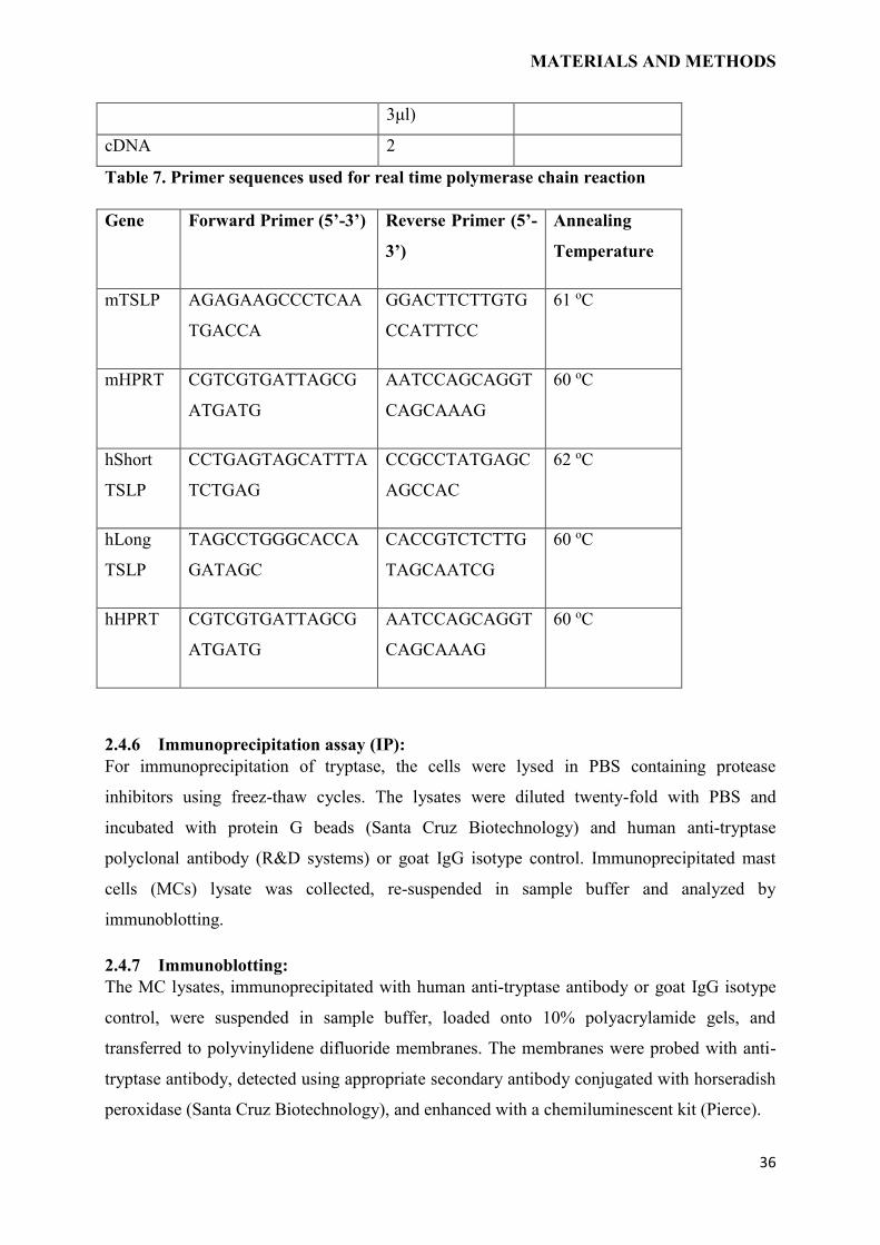
Table 7. Primer sequences used for real time polymerase chain reaction
Gene Forward Primer (5’-3’) Reverse Primer (5’-
3’)
Annealing
Temperature
mTSLP AGAGAAGCCCTCAA
TGACCA
GGACTTCTTGTG
CCATTTCC
61 oC
mHPRT CGTCGTGATTAGCG
ATGATG
AATCCAGCAGGT
CAGCAAAG
60 oC
hShort
TSLP
CCTGAGTAGCATTTA
TCTGAG
CCGCCTATGAGC
AGCCAC
62 oC
hLong
TSLP
TAGCCTGGGCACCA
GATAGC
CACCGTCTCTTG
TAGCAATCG
60 oC
hHPRT CGTCGTGATTAGCG
ATGATG
AATCCAGCAGGT
CAGCAAAG
60 oC
2.4.6 Immunoprecipitation assay (IP):
For immunoprecipitation of tryptase, the cells were lysed in PBS containing protease
inhibitors using freez-thaw cycles. The lysates were diluted twenty-fold with PBS and
incubated with protein G beads (Santa Cruz Biotechnology) and human anti-tryptase
polyclonal antibody (R&D systems) or goat IgG isotype control. Immunoprecipitated mast
cells (MCs) lysate was collected, re-suspended in sample buffer and analyzed by
immunoblotting.
2.4.7 Immunoblotting:
The MC lysates, immunoprecipitated with human anti-tryptase antibody or goat IgG isotype
control, were suspended in sample buffer, loaded onto 10% polyacrylamide gels, and
transferred to polyvinylidene difluoride membranes. The membranes were probed with anti-
tryptase antibody, detected using appropriate secondary antibody conjugated with horseradish
peroxidase (Santa Cruz Biotechnology), and enhanced with a chemiluminescent kit (Pierce).
MATERIALS AND METHODS
37
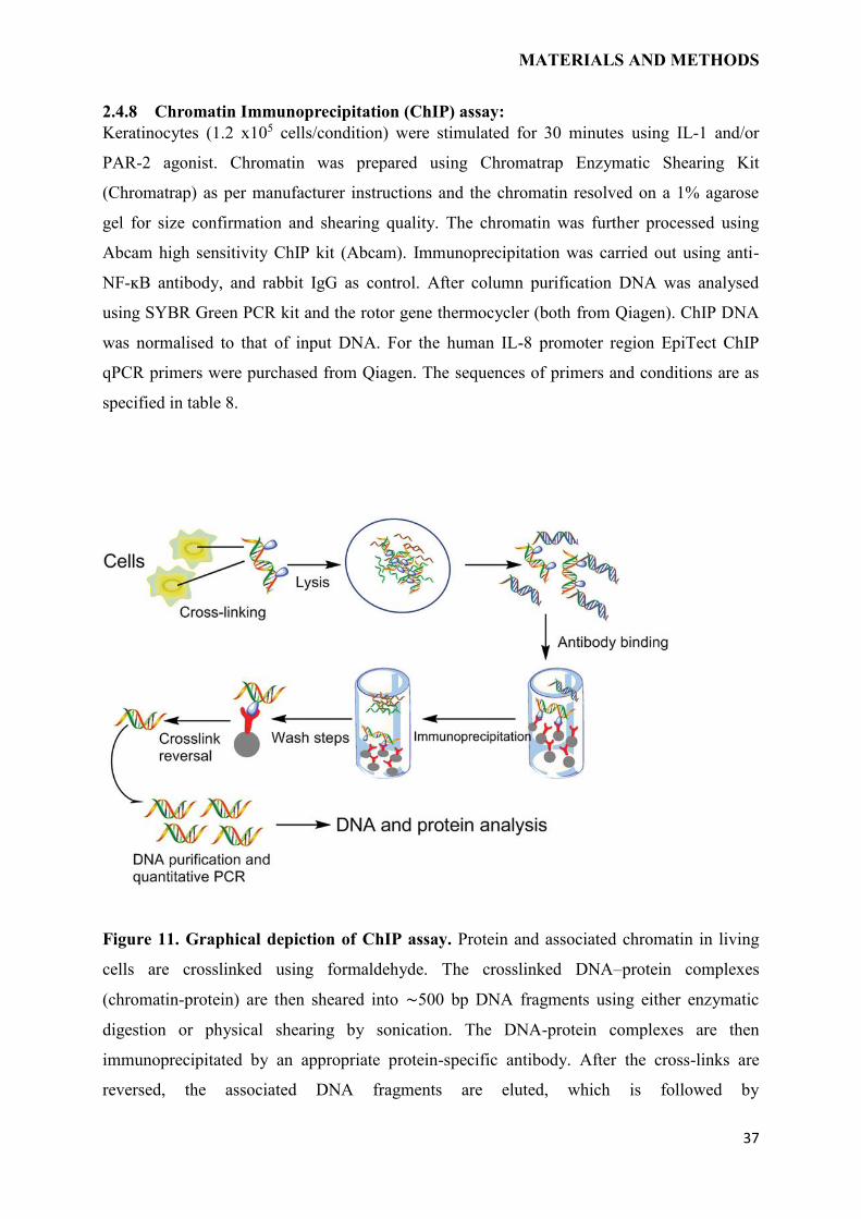
2.4.8 Chromatin Immunoprecipitation (ChIP) assay:
Keratinocytes (1.2 x10⁵ cells/condition) were stimulated for 30 minutes using IL-1 and/or
PAR-2 agonist. Chromatin was prepared using Chromatrap Enzymatic Shearing Kit
(Chromatrap) as per manufacturer instructions and the chromatin resolved on a 1% agarose
gel for size confirmation and shearing quality. The chromatin was further processed using
Abcam high sensitivity ChIP kit (Abcam). Immunoprecipitation was carried out using anti-
NF-ĸB antibody, and rabbit IgG as control. After column purification DNA was analysed
using SYBR Green PCR kit and the rotor gene thermocycler (both from Qiagen). ChIP DNA
was normalised to that of input DNA. For the human IL-8 promoter region EpiTect ChIP
qPCR primers were purchased from Qiagen. The sequences of primers and conditions are as
specified in table 8.
Figure 11. Graphical depiction of ChIP assay. Protein and associated chromatin in living
cells are crosslinked using formaldehyde. The crosslinked DNA–protein complexes
(chromatin-protein) are then sheared into ∼500 bp DNA fragments using either enzymatic
digestion or physical shearing by sonication. The DNA-protein complexes are then
immunoprecipitated by an appropriate protein-specific antibody. After the cross-links are
reversed, the associated DNA fragments are eluted, which is followed by
MATERIALS AND METHODS
38
immunoprecipitation of the crosslinked complexes and analysis of the resultant DNA by
qPCR. (adapted from Sigma-Aldrich)
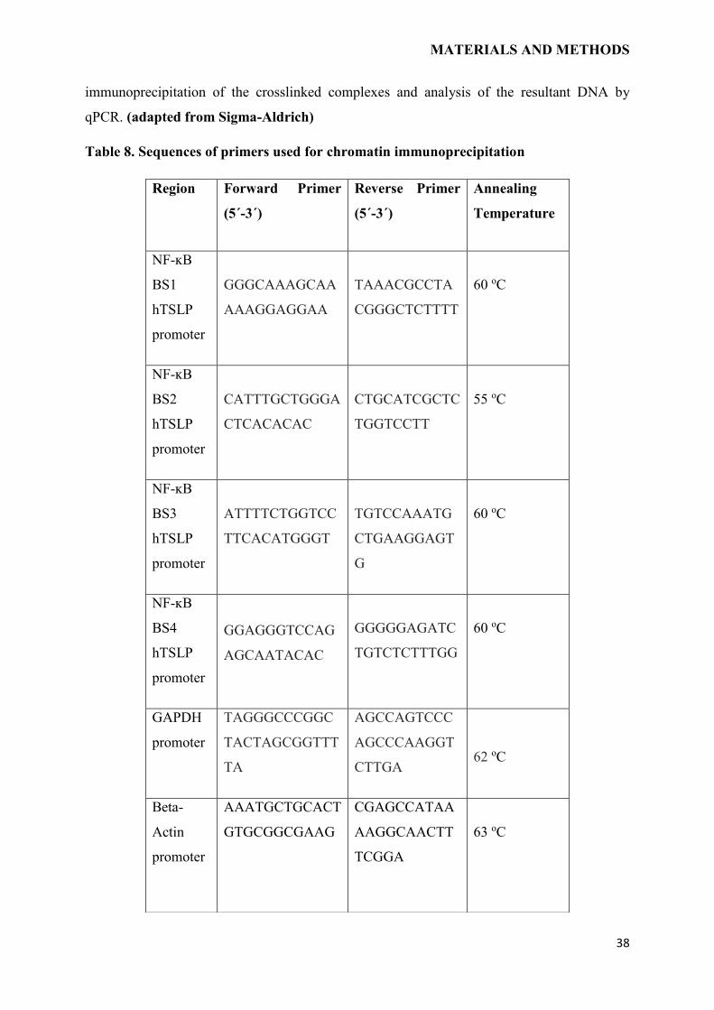
Table 8. Sequences of primers used for chromatin immunoprecipitation
Region Forward Primer
(5´-3´)
Reverse Primer
(5´-3´)
Annealing
Temperature
NF-ĸB
BS1
hTSLP
promoter
GGGCAAAGCAA
AAAGGAGGAA
TAAACGCCTA
CGGGCTCTTTT
60 oC
NF-ĸB
BS2
hTSLP
promoter
CATTTGCTGGGA
CTCACACAC
CTGCATCGCTC
TGGTCCTT
55 oC
NF-ĸB
BS3
hTSLP
promoter
ATTTTCTGGTCC
TTCACATGGGT
TGTCCAAATG
CTGAAGGAGT
G
60 oC
NF-ĸB
BS4
hTSLP
promoter
GGAGGGTCCAG
AGCAATACAC
GGGGGAGATC
TGTCTCTTTGG
60 oC
GAPDH
promoter
TAGGGCCCGGC
TACTAGCGGTTT
TA
AGCCAGTCCC
AGCCCAAGGT
CTTGA
62 oC
Beta-
Actin
promoter
AAATGCTGCACT
GTGCGGCGAAG
CGAGCCATAA
AAGGCAACTT
TCGGA
63 oC
MATERIALS AND METHODS
39
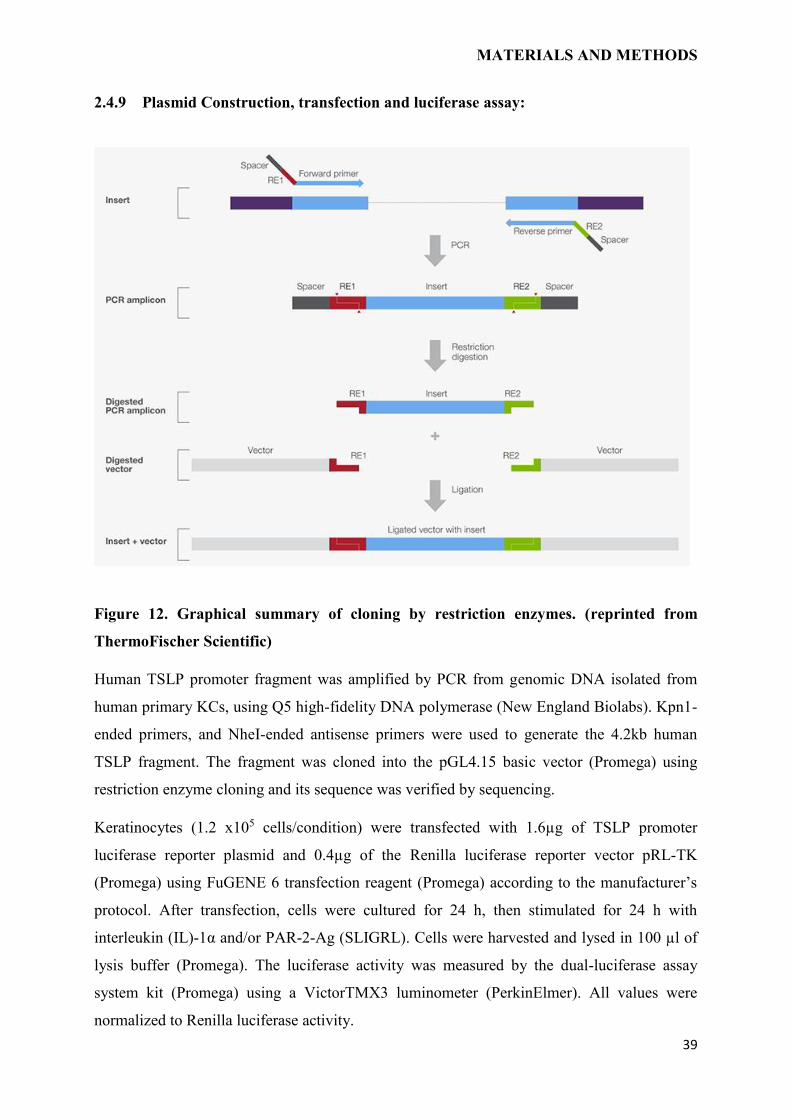
2.4.9 Plasmid Construction, transfection and luciferase assay:
Figure 12. Graphical summary of cloning by restriction enzymes. (reprinted from
ThermoFischer Scientific)
Human TSLP promoter fragment was amplified by PCR from genomic DNA isolated from
human primary KCs, using Q5 high-fidelity DNA polymerase (New England Biolabs). Kpn1-
ended primers, and NheI-ended antisense primers were used to generate the 4.2kb human
TSLP fragment. The fragment was cloned into the pGL4.15 basic vector (Promega) using
restriction enzyme cloning and its sequence was verified by sequencing.
Keratinocytes (1.2 x10⁵ cells/condition) were transfected with 1.6µg of TSLP promoter
luciferase reporter plasmid and 0.4µg of the Renilla luciferase reporter vector pRL-TK
(Promega) using FuGENE 6 transfection reagent (Promega) according to the manufacturer’s
protocol. After transfection, cells were cultured for 24 h, then stimulated for 24 h with
interleukin (IL)-1α and/or PAR-2-Ag (SLIGRL). Cells were harvested and lysed in 100 µl of
lysis buffer (Promega). The luciferase activity was measured by the dual-luciferase assay
system kit (Promega) using a VictorTMX3 luminometer (PerkinElmer). All values were
normalized to Renilla luciferase activity.
MATERIALS AND METHODS
40
2.5 STATISTICAL ANALYSIS
Normally distributed data are depicted as mean ± SEM and non-normally distributed data are
shown as median ± range. Experiments with only two groups were analyzed using t-test
(paired or unpaired) or Wilcoxon matched paired test, when groups were not normally
distributed; for more than 2 groups, depending on the data distribution, 1-way analysis of
variance (ANOVA) was used, followed by Bonferroni multiple comparisons test or Kruskal-
Wallis test and Holm-Sidak multiples comparison test. Statistical analyses were performed
with GraphPad Prism version 6 (GraphPad Software, USA). P value less than 0.05 was
considered as statistically significant.
RESULTS
3 RESULTS
3.1 TSLPR-/- MICE ARE PROTECTED FROM ENDOGENOUS TNF-
DEFICIENCY- MEDIATED AD DEVELOPMENT
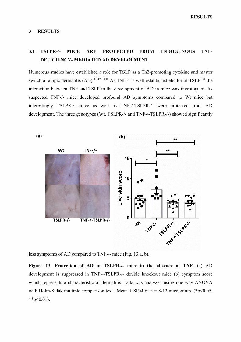
Numerous studies have established a role for TSLP as a Th2-promoting cytokine and master
switch of atopic dermatitis (AD).41,128-130 As TNF-α is well established elicitor of TSLP131 the
interaction between TNF and TSLP in the development of AD in mice was investigated. As
suspected TNF-/- mice developed profound AD symptoms compared to Wt mice but
interestingly TSLPR-/- mice as well as TNF-/-TSLPR-/- were protected from AD
development. The three genotypes (Wt, TSLPR-/- and TNF-/-TSLPR-/-) showed significantly
less symptoms of AD compared to TNF-/- mice (Fig. 13 a, b).
Figure 13. Protection of AD in TSLPR-/- mice in the absence of TNF. (a) AD
development is suppressed in TNF-/-TSLPR-/- double knockout mice (b) symptom score
which represents a characteristic of dermatitis. Data was analyzed using one way ANOVA
with Holm-Sidak multiple comparison test. Mean ± SEM of n = 8-12 mice/group. (*p<0.05,
**p<0.01).
Wt TNF-/-
TSLPR-/- TNF-/-TSLPR-/-
(a) (b)
RESULTS
42
These results show that TNF deficiency predisposes to AD in a TSLP dependent manner. In
contrast, at the baseline the skin of TNF-/- mice was normal and healthy as Wt, TSLPR-/- and
TNF-/-TSLPR-/- genotypes with comparable MC number, T cells, epidermal and dermal
thickness (data not shown), indicating that the skin structure does not require TNF-α and/or
TSLPR for development and maintenance.
3.2 SKIN IRRITATION INDUCES TSLP PRODUCTION IN MURINE SKIN EX
VIVO
3.2.1 Skin irritation induces TSLP production in murine skin by IL-1 and PAR-2
dependent pathways ex vivo
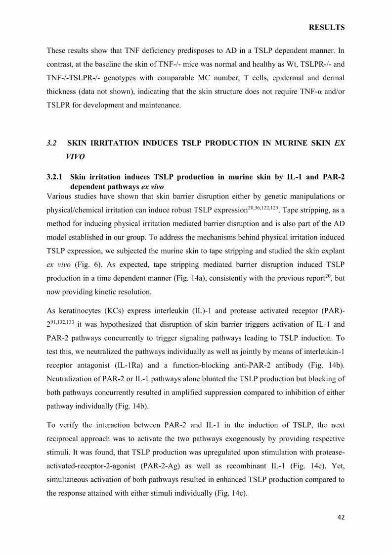
Various studies have shown that skin barrier disruption either by genetic manipulations or
physical/chemical irritation can induce robust TSLP expression20,36,122,123. Tape stripping, as a
method for inducing physical irritation mediated barrier disruption and is also part of the AD
model established in our group. To address the mechanisms behind physical irritation induced
TSLP expression, we subjected the murine skin to tape stripping and studied the skin explant
ex vivo (Fig. 6). As expected, tape stripping mediated barrier disruption induced TSLP
production in a time dependent manner (Fig. 14a), consistently with the previous report20, but
now providing kinetic resolution.
As keratinocytes (KCs) express interleukin (IL)-1 and protease activated receptor (PAR)-
291,132,133 it was hypothesized that disruption of skin barrier triggers activation of IL-1 and
PAR-2 pathways concurrently to trigger signaling pathways leading to TSLP induction. To
test this, we neutralized the pathways individually as well as jointly by means of interleukin-1
receptor antagonist (IL-1Ra) and a function-blocking anti-PAR-2 antibody (Fig. 14b).
Neutralization of PAR-2 or IL-1 pathways alone blunted the TSLP production but blocking of
both pathways concurrently resulted in amplified suppression compared to inhibition of either
pathway individually (Fig. 14b).
To verify the interaction between PAR-2 and IL-1 in the induction of TSLP, the next
reciprocal approach was to activate the two pathways exogenously by providing respective
stimuli. It was found, that TSLP production was upregulated upon stimulation with protease-
activated-receptor-2-agonist (PAR-2-Ag) as well as recombinant IL-1 (Fig. 14c). Yet,
simultaneous activation of both pathways resulted in enhanced TSLP production compared to
the response attained with either stimuli individually (Fig. 14c).
RESULTS
43
Figure 14. Skin irritation induces thymic stromal lymphopoietin (TSLP) expression via
interleukin (IL-1) and protease-activated receptor (PAR)-2 ex vivo. (a) Kinetics of tape
stripping induced TSLP production. TSLP was measured in culture supernatant by using
enzyme-linked immunosorbent assay (ELISA) after different incubation times. (b) Murine
skin explants after tape stripping, were incubated with anti-PAR-2 antibody (mIgG2a served
as the isotype control) or interleukin-1 receptor antagonist (IL-1Ra) either alone or in
combination. (c) Naïve murine skin biopsies (i.e. without tape stripping) were incubated with
mIL-1α alone or protease-activated receptor-2-agonist (PAR-2-Ag) or in combination. (b) and
(c) TLSP was measured by ELISA after 12 h. The data, given as mean ± SEM from 12
independent experiments, was analyzed using (a) paired t-test (n = 4) or (b, c) Kruskall-Wallis
(a)
(b) (c)
RESULTS
44
with Dunn’s multiple comparison test on the normalized data (*p<0.05, **p<0.01,
***p<0.001, ****p<0.0001).
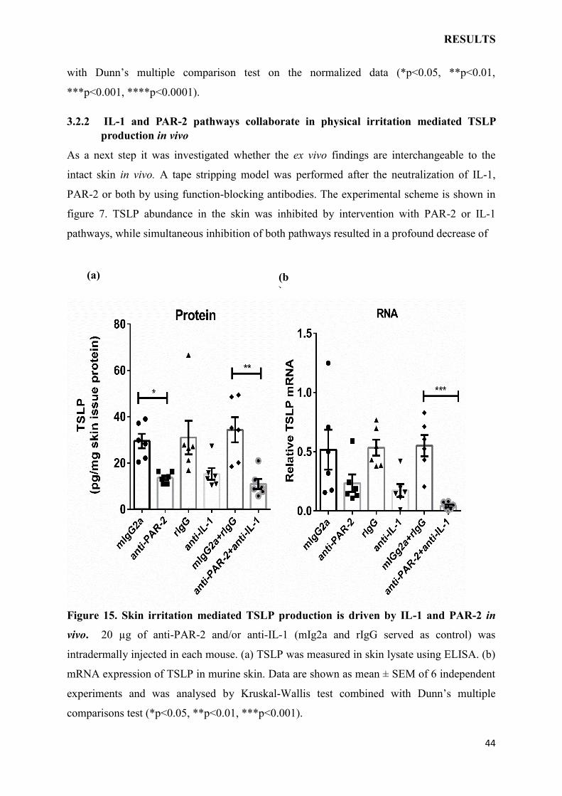
3.2.2 IL-1 and PAR-2 pathways collaborate in physical irritation mediated TSLP
production in vivo
As a next step it was investigated whether the ex vivo findings are interchangeable to the
intact skin in vivo. A tape stripping model was performed after the neutralization of IL-1,
PAR-2 or both by using function-blocking antibodies. The experimental scheme is shown in
figure 7. TSLP abundance in the skin was inhibited by intervention with PAR-2 or IL-1
pathways, while simultaneous inhibition of both pathways resulted in a profound decrease of
Figure 15. Skin irritation mediated TSLP production is driven by IL-1 and PAR-2 in
vivo. 20 µg of anti-PAR-2 and/or anti-IL-1 (mIg2a and rIgG served as control) was
intradermally injected in each mouse. (a) TSLP was measured in skin lysate using ELISA. (b)
mRNA expression of TSLP in murine skin. Data are shown as mean ± SEM of 6 independent
experiments and was analysed by Kruskal-Wallis test combined with Dunn’s multiple
comparisons test (*p<0.05, **p<0.01, ***p<0.001).
(a) (b
)
RESULTS
45
TSLP (Fig. 15a). The inhibition at mRNA expression level was similar and TSLP transcript
levels dropped to more than 40% of control when IL-1 and PAR-2 pathways were
concomitantly blocked (Fig. 15b). These results indicate that irritation mediated TSLP
response results from concurrent activation of IL-1 and PAR-2 pathways.
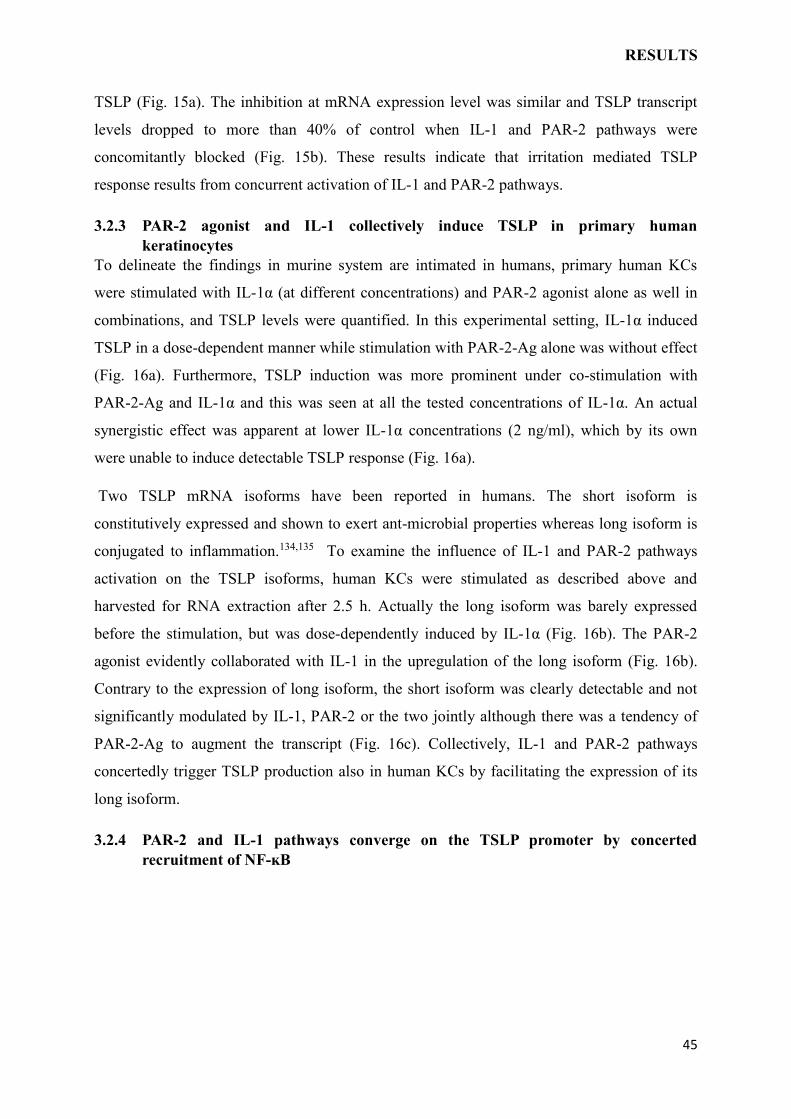
3.2.3 PAR-2 agonist and IL-1 collectively induce TSLP in primary human
keratinocytes
To delineate the findings in murine system are intimated in humans, primary human KCs
were stimulated with IL-1α (at different concentrations) and PAR-2 agonist alone as well in
combinations, and TSLP levels were quantified. In this experimental setting, IL-1α induced
TSLP in a dose-dependent manner while stimulation with PAR-2-Ag alone was without effect
(Fig. 16a). Furthermore, TSLP induction was more prominent under co-stimulation with
PAR-2-Ag and IL-1α and this was seen at all the tested concentrations of IL-1α. An actual
synergistic effect was apparent at lower IL-1α concentrations (2 ng/ml), which by its own
were unable to induce detectable TSLP response (Fig. 16a).
Two TSLP mRNA isoforms have been reported in humans. The short isoform is
constitutively expressed and shown to exert ant-microbial properties whereas long isoform is
conjugated to inflammation.134,135 To examine the influence of IL-1 and PAR-2 pathways
activation on the TSLP isoforms, human KCs were stimulated as described above and
harvested for RNA extraction after 2.5 h. Actually the long isoform was barely expressed
before the stimulation, but was dose-dependently induced by IL-1α (Fig. 16b). The PAR-2
agonist evidently collaborated with IL-1 in the upregulation of the long isoform (Fig. 16b).
Contrary to the expression of long isoform, the short isoform was clearly detectable and not
significantly modulated by IL-1, PAR-2 or the two jointly although there was a tendency of
PAR-2-Ag to augment the transcript (Fig. 16c). Collectively, IL-1 and PAR-2 pathways
concertedly trigger TSLP production also in human KCs by facilitating the expression of its
long isoform.
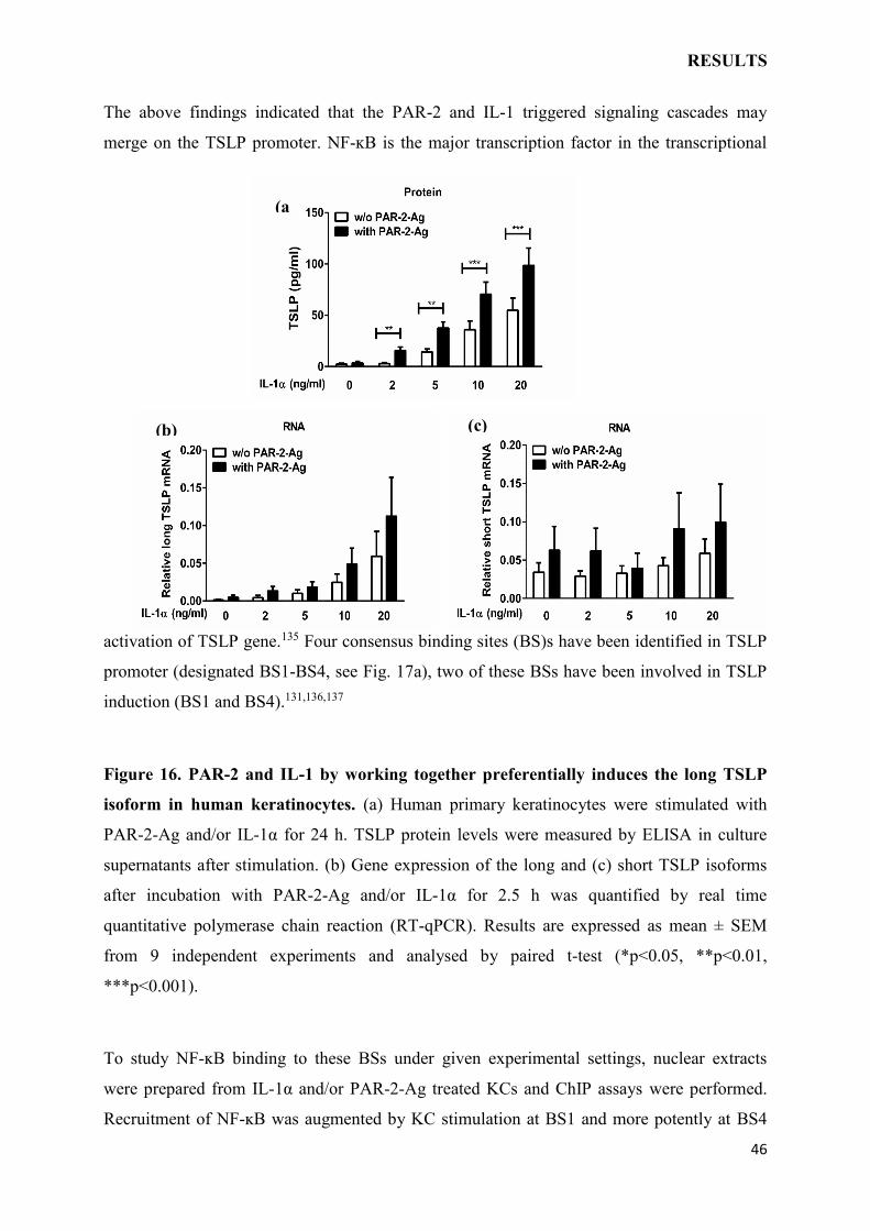
3.2.4 PAR-2 and IL-1 pathways converge on the TSLP promoter by concerted
recruitment of NF-ĸB
RESULTS
46
The above findings indicated that the PAR-2 and IL-1 triggered signaling cascades may
merge on the TSLP promoter. NF-ĸB is the major transcription factor in the transcriptional
activation of TSLP gene.135 Four consensus binding sites (BS)s have been identified in TSLP
promoter (designated BS1-BS4, see Fig. 17a), two of these BSs have been involved in TSLP
induction (BS1 and BS4).131,136,137
Figure 16. PAR-2 and IL-1 by working together preferentially induces the long TSLP
isoform in human keratinocytes. (a) Human primary keratinocytes were stimulated with
PAR-2-Ag and/or IL-1α for 24 h. TSLP protein levels were measured by ELISA in culture
supernatants after stimulation. (b) Gene expression of the long and (c) short TSLP isoforms
after incubation with PAR-2-Ag and/or IL-1α for 2.5 h was quantified by real time
quantitative polymerase chain reaction (RT-qPCR). Results are expressed as mean ± SEM
from 9 independent experiments and analysed by paired t-test (*p<0.05, **p<0.01,
***p<0.001).
To study NF-ĸB binding to these BSs under given experimental settings, nuclear extracts
were prepared from IL-1α and/or PAR-2-Ag treated KCs and ChIP assays were performed.
Recruitment of NF-ĸB was augmented by KC stimulation at BS1 and more potently at BS4
(a
)
(b) (c)
RESULTS
47
(Fig. 17b,c), but not at BS2 or BS3 (Fig. 18). Combined stimulation with IL-1α and PAR-2-
Ag resulted in concerted recruitment. Specially, NF-ĸB binding to BS4 was barely detectable
upon IL-1 or PAR-2 activation alone yet became profound when the signaling cascades were
simultaneously triggered (Fig. 17c).
Figure 17. NF-ĸB binding to the TSLP promoter is promoted by IL-1 and PAR-2. (a)
Diagrammatic representation of the consensus NF-ĸB binding sites in -3.8kb region of the
human TSLP promoter. (b) Recruitment of NF-κB to BS1 and (c) Recruitment of NF-κB to
BS4 of the human TSLP promoter upon stimulation with PAR-2-Agonist (PAR-2-Ag) and/or
IL-1α. (d, e) Beta-actin and GAPDH promoter regions were used to verify that PAR-2-
Agonist (PAR-2-Ag) and/or IL-1α stimulations do not alter binding affinity of RNA
polymerase II to meaningful regions of housekeeping genes. Results from one representative
experiment of 3 independent experiments are shown.
Exon1 Exon2
BS
1 BS
2 BS
3 BS
4
-3.8 kb
+1
-0.37 kb -1.5 kb -3.4 kb
(b
)
(d
) (e
)
(c
)
(a
)
RESULTS
48
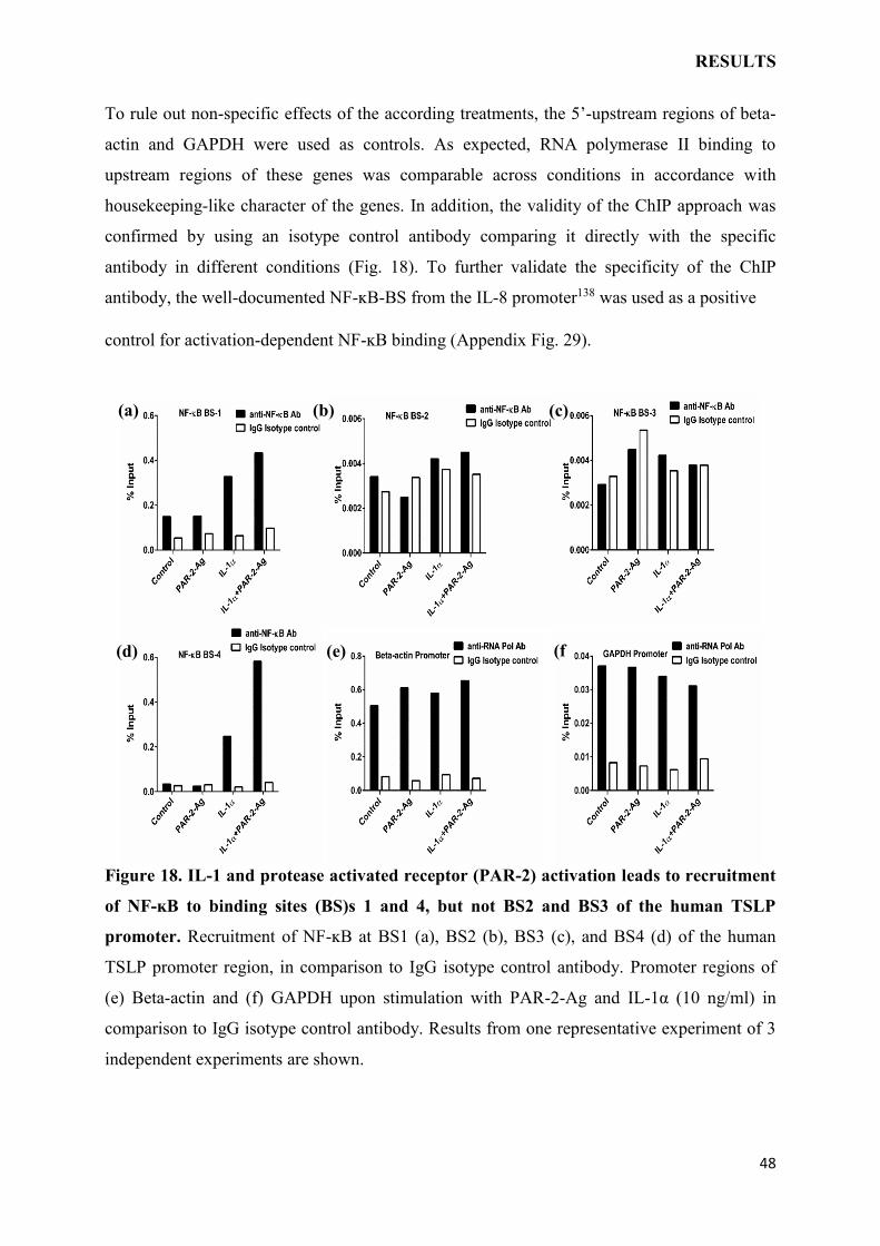
To rule out non-specific effects of the according treatments, the 5’-upstream regions of beta-
actin and GAPDH were used as controls. As expected, RNA polymerase II binding to
upstream regions of these genes was comparable across conditions in accordance with
housekeeping-like character of the genes. In addition, the validity of the ChIP approach was
confirmed by using an isotype control antibody comparing it directly with the specific
antibody in different conditions (Fig. 18). To further validate the specificity of the ChIP
antibody, the well-documented NF-ĸB-BS from the IL-8 promoter138 was used as a positive
control for activation-dependent NF-ĸB binding (Appendix Fig. 29).
Figure 18. IL-1 and protease activated receptor (PAR-2) activation leads to recruitment
of NF-ĸB to binding sites (BS)s 1 and 4, but not BS2 and BS3 of the human TSLP
promoter. Recruitment of NF-ĸB at BS1 (a), BS2 (b), BS3 (c), and BS4 (d) of the human
TSLP promoter region, in comparison to IgG isotype control antibody. Promoter regions of
(e) Beta-actin and (f) GAPDH upon stimulation with PAR-2-Ag and IL-1α (10 ng/ml) in
comparison to IgG isotype control antibody. Results from one representative experiment of 3
independent experiments are shown.
(a)
(f
) (e) (d)
(c) (b)
RESULTS
49
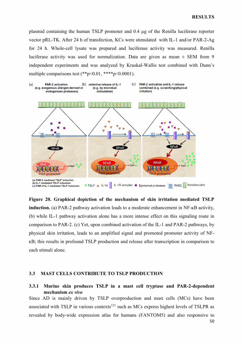
Taken together, the presented results suggest that PAR-2-and IL-1-mediated TSLP induction
is triggered by their joint activity at the TSLP promoter through collaborated recruitment of
NF-ĸB to its most biologically meaningful binding site.
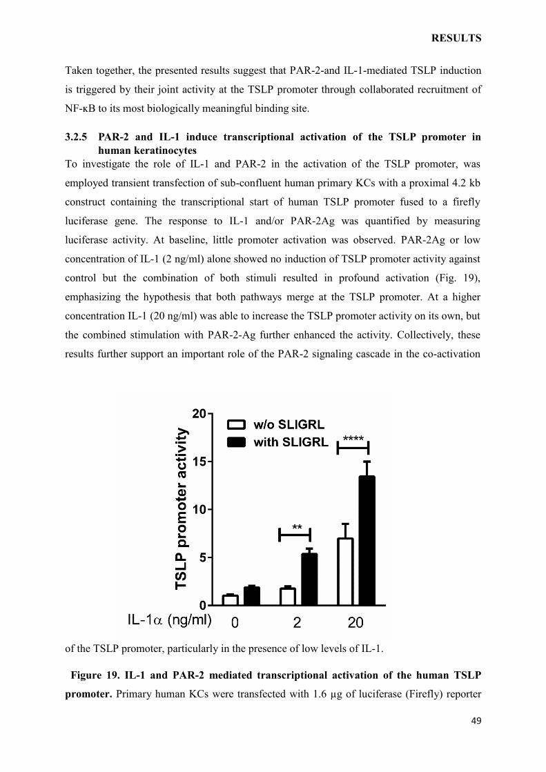
3.2.5 PAR-2 and IL-1 induce transcriptional activation of the TSLP promoter in
human keratinocytes
To investigate the role of IL-1 and PAR-2 in the activation of the TSLP promoter, was
employed transient transfection of sub-confluent human primary KCs with a proximal 4.2 kb
construct containing the transcriptional start of human TSLP promoter fused to a firefly
luciferase gene. The response to IL-1 and/or PAR-2Ag was quantified by measuring
luciferase activity. At baseline, little promoter activation was observed. PAR-2Ag or low
concentration of IL-1 (2 ng/ml) alone showed no induction of TSLP promoter activity against
control but the combination of both stimuli resulted in profound activation (Fig. 19),
emphasizing the hypothesis that both pathways merge at the TSLP promoter. At a higher
concentration IL-1 (20 ng/ml) was able to increase the TSLP promoter activity on its own, but
the combined stimulation with PAR-2-Ag further enhanced the activity. Collectively, these
results further support an important role of the PAR-2 signaling cascade in the co-activation
of the TSLP promoter, particularly in the presence of low levels of IL-1.
Figure 19. IL-1 and PAR-2 mediated transcriptional activation of the human TSLP
promoter. Primary human KCs were transfected with 1.6 µg of luciferase (Firefly) reporter
RESULTS
50
plasmid containing the human TSLP promoter and 0.4 µg of the Renilla luciferase reporter
vector pRL-TK. After 24 h of transfection, KCs were stimulated with IL-1 and/or PAR-2-Ag
for 24 h. Whole-cell lysate was prepared and luciferase activity was measured. Renilla
luciferase activity was used for normalization. Data are given as mean ± SEM from 9
independent experiments and was analyzed by Kruskal-Wallis test combined with Dunn’s
multiple comparisons test (**p<0.01, ****p<0.0001).
Figure 20. Graphical depiction of the mechanism of skin irritation mediated TSLP
induction. (a) PAR-2 pathway activation leads to a moderate enhancement in NF-ĸB activity,
(b) while IL-1 pathway activation alone has a more intense effect on this signaling route in
comparison to PAR-2. (c) Yet, upon combined activation of the IL-1 and PAR-2 pathways, by
physical skin irritation, leads to an amplified signal and promoted promoter activity of NF-
ĸB; this results in profound TSLP production and release after transcription in comparison to
each stimuli alone.
3.3 MAST CELLS CONTRIBUTE TO TSLP PRODUCTION
3.3.1 Murine skin produces TSLP in a mast cell tryptase and PAR-2-dependent
mechanism ex vivo
Since AD is mainly driven by TSLP overproduction and mast cells (MCs) have been
associated with TSLP in various contexts121 such as MCs express highest levels of TSLPR as
revealed by body-wide expression atlas for humans (FANTOM5) and also responsive to
RESULTS
51
TSLP. Furthermore, MCs also enhance TSLP production by epithelial cells121,139. Based on
literature and data produced in our group it was hypothesized that MCs act as intermediaries
in the biological cascade promoting TSLP production, thereby boosting AD. To address the
mechanisms behind MCs induced TSLP expression, murine skin was treated with purified
MC mediators as well as MCs supernatant and the skin explant was studied ex vivo (Fig. 21).
The results give strong evidence that MCs products specifically tryptase is crucial for the
enhanced TSLP production (Fig. 21a) by murine skin explants, while histamine showed no
impact on TSLP production (Fig. 21b). Murine skin explant produced excessive TSLP in
response to tryptase in a dose dependent manner (Fig. 21a), while being neutral to histamine
dose response (Fig. 21b). To study the mechanism behind the tryptase mediated exaggerated
TSLP production by murine skin, it was investigated whether the murine skin was prone to
enhanced TSLP production also upon exposure to MC supernatant, or whether the boost in
TSLP production requires purified tryptase. As expected, the presence of MC supernatant
induced robust TSLP response in murine skin ex vivo. MC supernatant mediated enhanced
TSLP induction was significantly inhibited by anti-mMCP6 antibody (Fig. 21c).
As shown earlier, PAR-2 was involved in skin irritation-mediated TSLP production, therefore
it was asked whether the mediators in the MC supernatant act via PAR-2. To investigate this,
murine skin was exposed to the MC supernatants followed by anti-PAR-2 antibody. The
results demonstrated that endogenous PAR-2 is essential for the MC supernatant mediated
TSLP ex vivo, as anti-PAR-2 antibody drastically inhibited the TSLP production (Fig.21d). To
validate the interplay between tryptase and PAR-2 during the induction of TSLP, the next
complementary approach was to stimulate Wt and PAR-2-/- murine skin explants
exogenously by providing mMCP6 (murine equivalent of human tryptase).
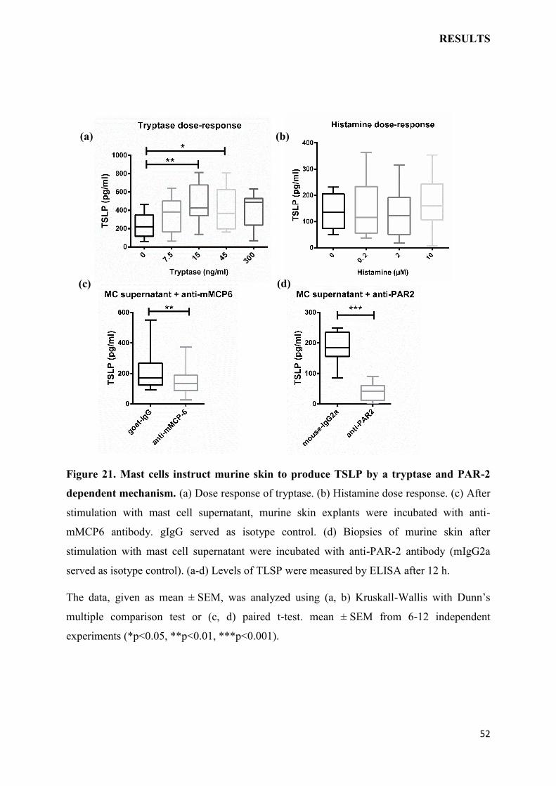
RESULTS
52
Figure 21. Mast cells instruct murine skin to produce TSLP by a tryptase and PAR-2
dependent mechanism. (a) Dose response of tryptase. (b) Histamine dose response. (c) After
stimulation with mast cell supernatant, murine skin explants were incubated with anti-
mMCP6 antibody. gIgG served as isotype control. (d) Biopsies of murine skin after
stimulation with mast cell supernatant were incubated with anti-PAR-2 antibody (mIgG2a
served as isotype control). (a-d) Levels of TLSP were measured by ELISA after 12 h.
The data, given as mean ± SEM, was analyzed using (a, b) Kruskall-Wallis with Dunn’s
multiple comparison test or (c, d) paired t-test. mean ± SEM from 6-12 independent
experiments (*p<0.05, **p<0.01, ***p<0.001).
(a)
(c) (d)
(b)
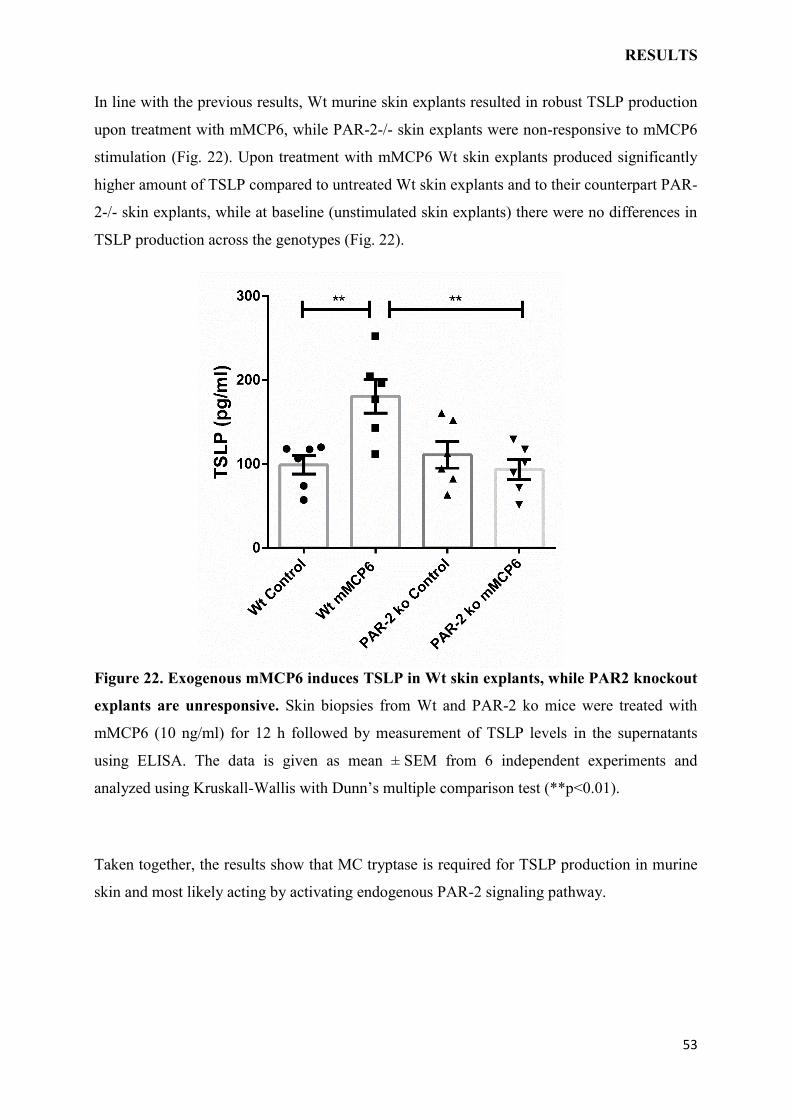
RESULTS
53
In line with the previous results, Wt murine skin explants resulted in robust TSLP production
upon treatment with mMCP6, while PAR-2-/- skin explants were non-responsive to mMCP6
stimulation (Fig. 22). Upon treatment with mMCP6 Wt skin explants produced significantly
higher amount of TSLP compared to untreated Wt skin explants and to their counterpart PAR-
2-/- skin explants, while at baseline (unstimulated skin explants) there were no differences in
TSLP production across the genotypes (Fig. 22).
Figure 22. Exogenous mMCP6 induces TSLP in Wt skin explants, while PAR2 knockout
explants are unresponsive. Skin biopsies from Wt and PAR-2 ko mice were treated with
mMCP6 (10 ng/ml) for 12 h followed by measurement of TSLP levels in the supernatants
using ELISA. The data is given as mean ± SEM from 6 independent experiments and
analyzed using Kruskall-Wallis with Dunn’s multiple comparison test (**p<0.01).
Taken together, the results show that MC tryptase is required for TSLP production in murine
skin and most likely acting by activating endogenous PAR-2 signaling pathway.
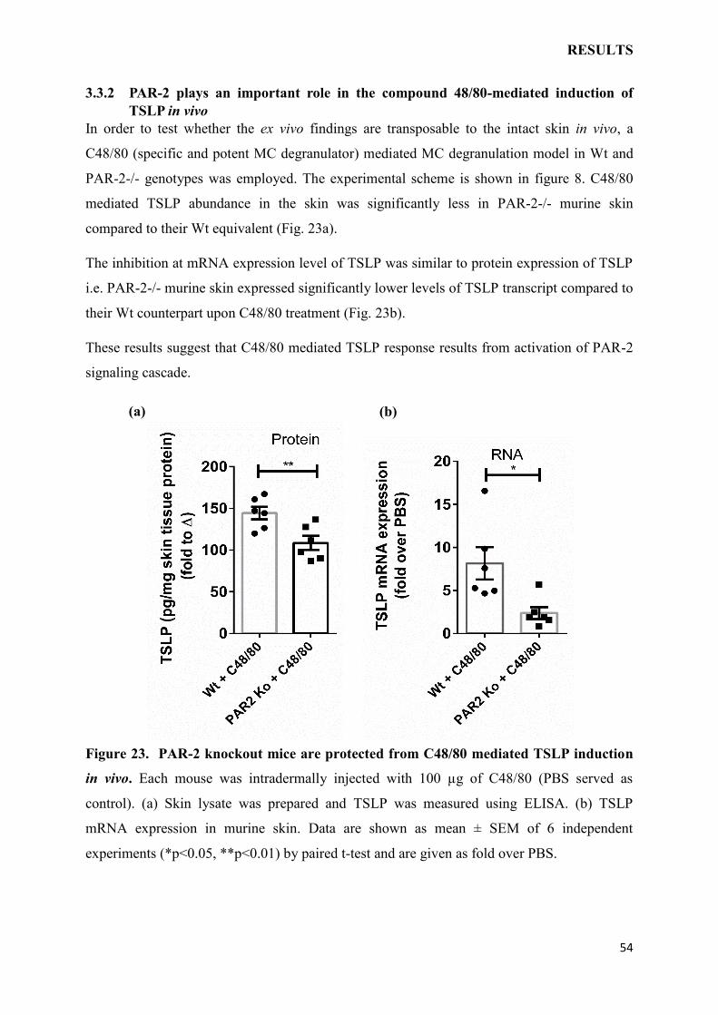
RESULTS
54
3.3.2 PAR-2 plays an important role in the compound 48/80-mediated induction of
TSLP in vivo
In order to test whether the ex vivo findings are transposable to the intact skin in vivo, a
C48/80 (specific and potent MC degranulator) mediated MC degranulation model in Wt and
PAR-2-/- genotypes was employed. The experimental scheme is shown in figure 8. C48/80
mediated TSLP abundance in the skin was significantly less in PAR-2-/- murine skin
compared to their Wt equivalent (Fig. 23a).
The inhibition at mRNA expression level of TSLP was similar to protein expression of TSLP
i.e. PAR-2-/- murine skin expressed significantly lower levels of TSLP transcript compared to
their Wt counterpart upon C48/80 treatment (Fig. 23b).
These results suggest that C48/80 mediated TSLP response results from activation of PAR-2
signaling cascade.
Figure 23. PAR-2 knockout mice are protected from C48/80 mediated TSLP induction
in vivo. Each mouse was intradermally injected with 100 µg of C48/80 (PBS served as
control). (a) Skin lysate was prepared and TSLP was measured using ELISA. (b) TSLP
mRNA expression in murine skin. Data are shown as mean ± SEM of 6 independent
experiments (*p<0.05, **p<0.01) by paired t-test and are given as fold over PBS.
(a) (b)
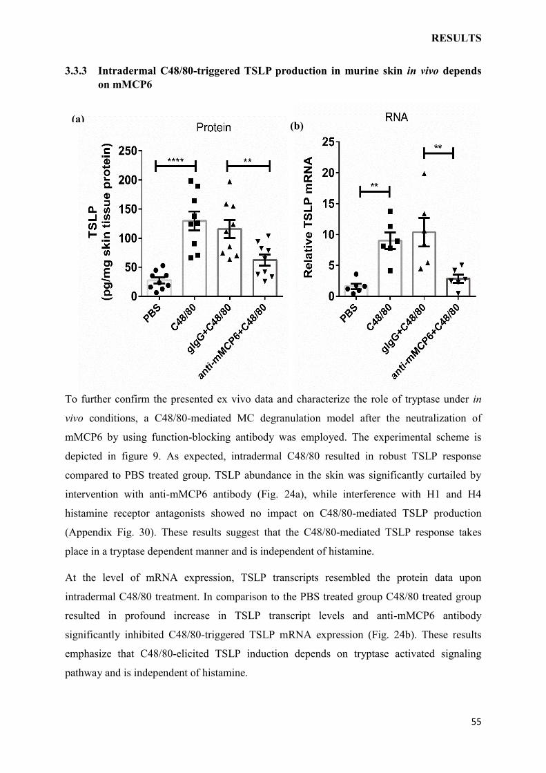
RESULTS
55
3.3.3 Intradermal C48/80-triggered TSLP production in murine skin in vivo depends
on mMCP6
To further confirm the presented ex vivo data and characterize the role of tryptase under in
vivo conditions, a C48/80-mediated MC degranulation model after the neutralization of
mMCP6 by using function-blocking antibody was employed. The experimental scheme is
depicted in figure 9. As expected, intradermal C48/80 resulted in robust TSLP response
compared to PBS treated group. TSLP abundance in the skin was significantly curtailed by
intervention with anti-mMCP6 antibody (Fig. 24a), while interference with H1 and H4
histamine receptor antagonists showed no impact on C48/80-mediated TSLP production
(Appendix Fig. 30). These results suggest that the C48/80-mediated TSLP response takes
place in a tryptase dependent manner and is independent of histamine.
At the level of mRNA expression, TSLP transcripts resembled the protein data upon
intradermal C48/80 treatment. In comparison to the PBS treated group C48/80 treated group
resulted in profound increase in TSLP transcript levels and anti-mMCP6 antibody
significantly inhibited C48/80-triggered TSLP mRNA expression (Fig. 24b). These results
emphasize that C48/80-elicited TSLP induction depends on tryptase activated signaling
pathway and is independent of histamine.
(b) (a)
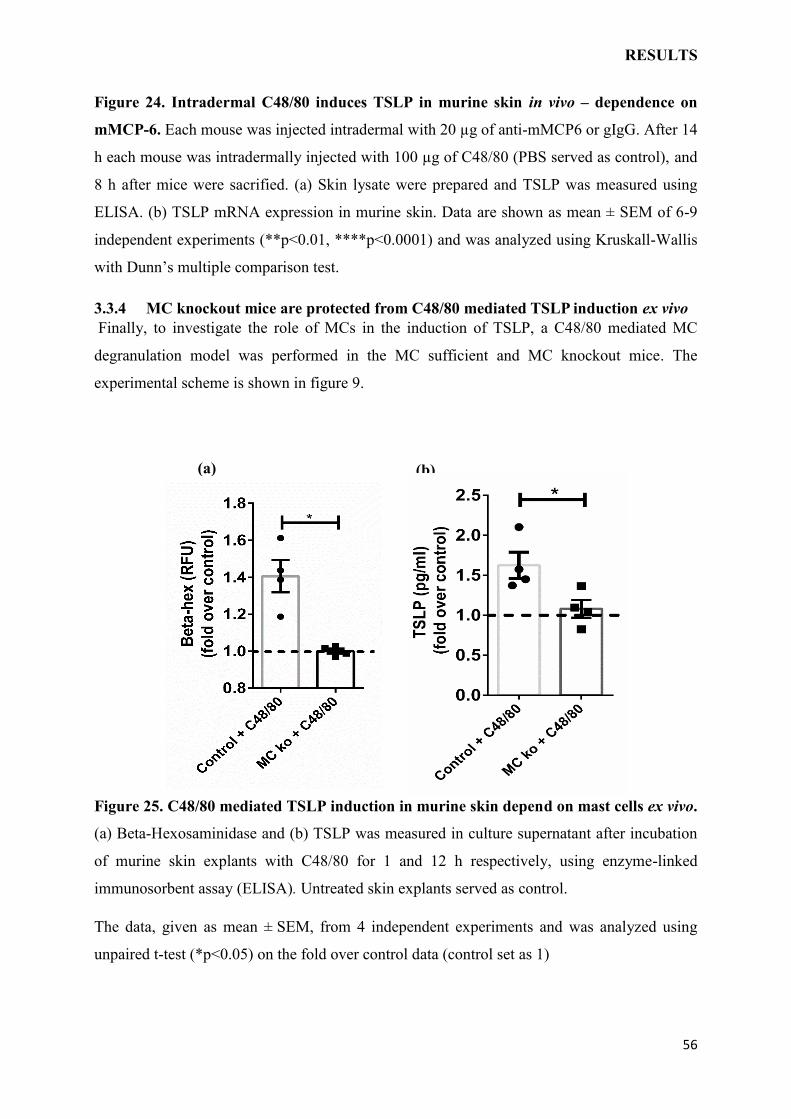
RESULTS
56
Figure 24. Intradermal C48/80 induces TSLP in murine skin in vivo – dependence on
mMCP-6. Each mouse was injected intradermal with 20 µg of anti-mMCP6 or gIgG. After 14
h each mouse was intradermally injected with 100 µg of C48/80 (PBS served as control), and
8 h after mice were sacrified. (a) Skin lysate were prepared and TSLP was measured using
ELISA. (b) TSLP mRNA expression in murine skin. Data are shown as mean ± SEM of 6-9
independent experiments (**p<0.01, ****p<0.0001) and was analyzed using Kruskall-Wallis
with Dunn’s multiple comparison test.
3.3.4 MC knockout mice are protected from C48/80 mediated TSLP induction ex vivo
Finally, to investigate the role of MCs in the induction of TSLP, a C48/80 mediated MC
degranulation model was performed in the MC sufficient and MC knockout mice. The
experimental scheme is shown in figure 9.
Figure 25. C48/80 mediated TSLP induction in murine skin depend on mast cells ex vivo.
(a) Beta-Hexosaminidase and (b) TSLP was measured in culture supernatant after incubation
of murine skin explants with C48/80 for 1 and 12 h respectively, using enzyme-linked
immunosorbent assay (ELISA). Untreated skin explants served as control.
The data, given as mean ± SEM, from 4 independent experiments and was analyzed using
unpaired t-test (*p<0.05) on the fold over control data (control set as 1)
(a) (b)
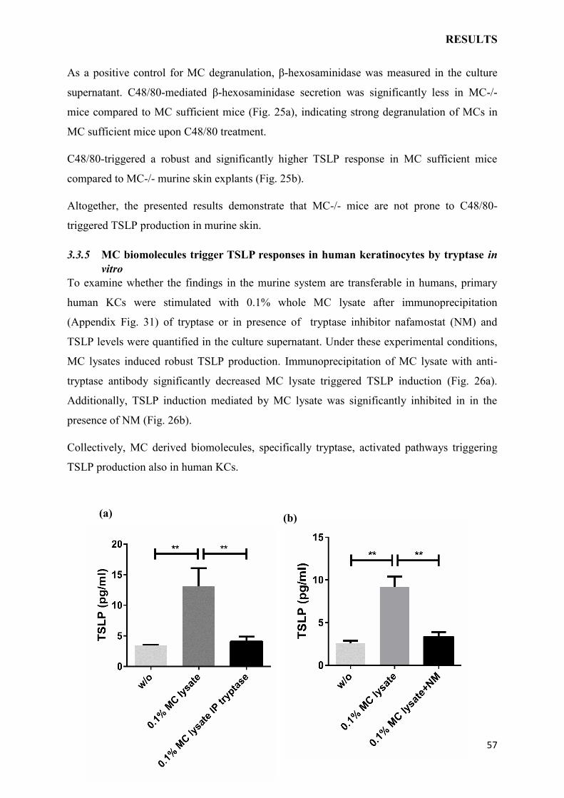
RESULTS
57
As a positive control for MC degranulation, β-hexosaminidase was measured in the culture
supernatant. C48/80-mediated β-hexosaminidase secretion was significantly less in MC-/-
mice compared to MC sufficient mice (Fig. 25a), indicating strong degranulation of MCs in
MC sufficient mice upon C48/80 treatment.
C48/80-triggered a robust and significantly higher TSLP response in MC sufficient mice
compared to MC-/- murine skin explants (Fig. 25b).
Altogether, the presented results demonstrate that MC-/- mice are not prone to C48/80-
triggered TSLP production in murine skin.
3.3.5 MC biomolecules trigger TSLP responses in human keratinocytes by tryptase in
vitro
To examine whether the findings in the murine system are transferable in humans, primary
human KCs were stimulated with 0.1% whole MC lysate after immunoprecipitation
(Appendix Fig. 31) of tryptase or in presence of tryptase inhibitor nafamostat (NM) and
TSLP levels were quantified in the culture supernatant. Under these experimental conditions,
MC lysates induced robust TSLP production. Immunoprecipitation of MC lysate with anti-
tryptase antibody significantly decreased MC lysate triggered TSLP induction (Fig. 26a).
Additionally, TSLP induction mediated by MC lysate was significantly inhibited in in the
presence of NM (Fig. 26b).
Collectively, MC derived biomolecules, specifically tryptase, activated pathways triggering
TSLP production also in human KCs.
(a) (b)
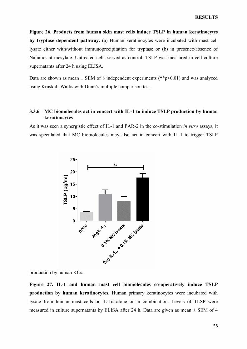
RESULTS
58
Figure 26. Products from human skin mast cells induce TSLP in human keratinocytes
by tryptase dependent pathway. (a) Human keratinocytes were incubated with mast cell
lysate either with/without immunoprecipitation for tryptase or (b) in presence/absence of
Nafamostat mesylate. Untreated cells served as control. TSLP was measured in cell culture
supernatants after 24 h using ELISA.
Data are shown as mean ± SEM of 8 independent experiments (**p<0.01) and was analyzed
using Kruskall-Wallis with Dunn’s multiple comparison test.
3.3.6 MC biomolecules act in concert with IL-1 to induce TSLP production by human
keratinocytes
As it was seen a synergistic effect of IL-1 and PAR-2 in the co-stimulation in vitro assays, it
was speculated that MC biomolecules may also act in concert with IL-1 to trigger TSLP
production by human KCs.
Figure 27. IL-1 and human mast cell biomolecules co-operatively induce TSLP
production by human keratinocytes. Human primary keratinocytes were incubated with
lysate from human mast cells or IL-1α alone or in combination. Levels of TLSP were
measured in culture supernatants by ELISA after 24 h. Data are given as mean ± SEM of 4
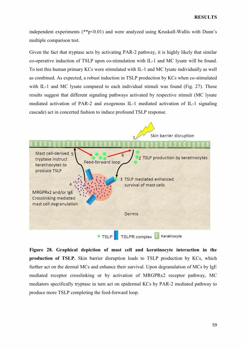
RESULTS
59
independent experiments (**p<0.01) and were analyzed using Kruskall-Wallis with Dunn’s
multiple comparison test.
Given the fact that tryptase acts by activating PAR-2 pathway, it is highly likely that similar
co-operative induction of TSLP upon co-stimulation with IL-1 and MC lysate will be found.
To test this human primary KCs were stimulated with IL-1 and MC lysate individually as well
as combined. As expected, a robust induction in TSLP production by KCs when co-stimulated
with IL-1 and MC lysate compared to each individual stimuli was found (Fig. 27). These
results suggest that different signaling pathways activated by respective stimuli (MC lysate
mediated activation of PAR-2 and exogenous IL-1 mediated activation of IL-1 signaling
cascade) act in concerted fashion to induce profound TSLP response.
Figure 28. Graphical depiction of mast cell and keratinocyte interaction in the
production of TSLP. Skin barrier disruption leads to TSLP production by KCs, which
further act on the dermal MCs and enhance their survival. Upon degranulation of MCs by IgE
mediated receptor crosslinking or by activation of MRGPRx2 receptor pathway, MC
mediators specifically tryptase in turn act on epidermal KCs by PAR-2 mediated pathway to
produce more TSLP completing the feed-forward loop.

RESULTS
60
DISCUSSION
4 DISCUSSION
TSLP is a well-known pro-inflammatory cytokine which plays a crucial role in inflammatory
diseases41,140. It is highly upregulated in keratinocytes (KCs) upon skin barrier
perturbation36,141 and considered to be the initiator of AD. Murine skin overexpressing TSLP
has been reported to develop spontaneous dermatitis alike to characteristics of human
AD34,106. Despite the significant advances in the understanding of the role of TSLP in the
development of Th2-mediated inflammation in skin and allergic diseases, the understanding
of the regulatory mechanisms of endogenous TSLP production in skin inflammation and
particularly in AD are still in its infancy.
In this thesis, the role of TSLP in the progression of AD and mechanisms behind skin-
irritation as well as mast cells mediators mediated TSLP production was investigated.
Additional aim was to better understand the role of endogenous TNF under the micro-milieu
of AD and in relation to TSLPR in vivo.. Furthermore, in recent studies MCs were emerging
as another important component of AD pathogenesis. MCs have been reported to be
increased in the lesional skin of AD in case of both human as well as mice.142 To examine the
contribution of MCs in the induction of TSLP production by human KCs and in murine skin
MC degranulation model in vivo as well as ex vivo was employed. Furthermore, MC lysate
and purified MC mediators were analyzed in more detail ex vivo and in vitro.
4.1 TNF-/- MICE DEVELOP AGGRAVATED AD WHICH COULD BE RESCUED
BY THE ABSENSE OF TSLPR EXPRESSION
AD is a complex, chronic and pruritic inflammatory skin disease. Immune dysregulation in
combination with epidermal barrier defect cause development of AD.143 Beside
immunological components, pharmacological abnormalities are critical in the disease
development and progression.22
TNF-α is a pleiotropic cytokines crucial for host defense and known for its ability to trigger
inflammatory response concurrently with an immune dysregulation.144,145 Anti-TNF
therapeutics for autoimmune disorders like inflammatory bowel disease, psoriasis and
rheumatoid arthritis are authorized and proving effective with rather few side-effects146. Most
common therapies in use for disrupting the TNF function in autoimmune diseases are
monoclonal antibodies, adalimumab as well as infliximab, in addition to the soluble receptor
etanercept.147,148 In the initiation and progression of AD the functional role of TNF is not
DISCUSSION
62
clear. Based on literature analysis, there are more indications of positive rather than negative
impact of TNF on AD. Takahashi et al. (1992) reported that TNF-α was decreased in PBMCs
derived from AD patients in comparison to healthy individuals.149 In alignment, it was
observed that the expression levels of TNF-α, IL-1β and IFN-γ were reduced in the skin
samples of AD patients compared to skin samples obtained from psoriasis patients.150
Furthermore, TNF-α producing dermal inflammatory dendritic cells (DCs) were reported to
be lower in the skin of AD patients compared to skin of psoriasis patients.151 The
epidemiological studies from our institution as well as from different clinics provided the
most noticeable evidence, which stated the manifestation of symptoms similar to AD due to
side effects of anti-TNF therapy.152-156 These observations hint towards a protective role of
TNF in the framework of AD, and previously in our group we had seen that TNF deficient
mice developed exaggerated AD, providing experimental evidence.216
TSLP is a well-known important proinflammatory cytokine in AD and asthma.157 Increased
TSLP expression has been reported in lesional skin of AD patients but not in contact
dermatitis, induced by nickel.106 TSLP is a member of IL-2 family and IL-7 type of cytokine.
It can act on DCs and promote the Th2 immune response by differentiation and recruitment
of Th2 cells. Naïve CD4+ cells can also be directly targeted by TSLP to induce proliferation,
in response to antigen.114 It has been shown that epithelial cell-derived TSLP can activate
DCs, T cells and MCs.130 TSLP overexpression in murine skin resulted in the development of
spontaneous dermatitis.34,106 In alignment with the literature, TSLP was significantly
upregulated in AD skin of TNF-/- mice and correlated with disease severity. TNF-α is known
to induce TSLP expression. Brandt et al. (2011) reported that human skin explants produce
higher amount of TSLP, when stimulated with a combination of TNF-α, IL-1α and Th2
cytokines, but not individually.31 Complementary findings were reported from KC
cultures.158,159 To understand why TNF-/- mice exhibit aggravated AD and weather TSLP is
prime reason for AD development in these mice, a murine model was employed. To define
the underlying mechanisms, an AD mouse model was performed in Wt, TNF-/- ,TSLPR-/-
and TNF-/- TSLPR-/- double knockout mice to mimic the human AD scenario. The results
clearly indicate that TNF-α plays a protective role against development of AD as reported in
the literature. Significant worsening of the severity of AD-like lesions in the TNF-/- mice in
comparison to Wt, TSLPR-/- and TNF-/-TSLPR-/- double knock out mice was observed. The
analysis of the severity in the AD model was performed based on SCORAD score, a tool to
evaluate clinical severity in human AD.160 Interestingly, TSLPR-/- and TNF-/-TSLPR-/-
DISCUSSION
63
double knockout mice were protected from development of AD symptoms suggesting TSLP
as another essential factor in the development of AD. Previously, it has been shown that MC
but not the T cells were enhanced in AD lesions of TNF-/- mice and correlated with disease
severity as well as to increased mRNA levels of TSLP in lesional skin.216 It was shown by
Han et al. that TSLP was able to induce the proliferation and differentiation of MCs as well
as the number of MC was significantly higher in different organs of Wt mice in comparison
to TSLP-deficient mice.161
4.2 SKIN IRRITATION-MEDIATED TSLP PRODUCTION DEPENDS ON IL-1
AND PAR-2 PATHWAYS
TSLP is subject to several studies and well-known as an important factor of allergic disorders
in the lung, skin, and gut organs.41,140 TSLP is abundantly expressed in KCs when the skin
barrier is disrupted by genetic intervention.36,141,162-165 Skin barrier compromised by irritation
triggers TSLP response in similar manner, supporting a further connection between damaged
skin integrity and TSLP expression.20,117,121,122 Thus TSLP can be considered as initial and
sensitive signal of skin barrier disruption of various sorts, making it necessary to explore the
molecular mechanism of its regulation.
How skin irritation-mediated barrier disruption leads to TSLP production is an open question.
Separately from limited contribution of IL-120, the mediators and underlying mechanisms are
unclear in spite of the regular physical insults to which the skin is continually exposed in
daily life and the applicability to the itch-scratch cycle to AD manifestation.
The role of IL-1 in skin inflammation is well-established. Indeed, both IL-1α and IL-1β,
founding members of the family, are key coordinators to induce clinically distinct signs of
skin inflammation by predominantly upregulating the expression of the entire range of
inflammatory genes during sterile inflammation, skin infections and other abuses.166-168 In
response to skin injury KCs release pre-formed IL-1α169 which in turn act as alarmin to
recruit T cells to further enhance IL-1 (e.g. by inducing IL-1β expression in KCs).170,171 IL-1α
and IL-1β by acting alone or in combination with other pro-inflammatory cytokines sustain
the inflammatory micro-milieu by further supporting the cytokine production by immune
cells in a feed-forward loop.170,172-174
Beside its well-known contribution to skin inflammation, IL-1 played a role in skin
irritation-mediated TSLP induction, still could support an incomplete explanation only, as
DISCUSSION
64
inhibition yielded in 20-50% decrease in TSLP production, relaying on the setup.20 Thus, the
question arose what alternate pathways may be activated in the irritated skin to induce TSLP
production. PAR-2 turned out to be a major route, which can be activated upon barrier
disruption.
Regardless of the reproduction of the cross-talk between IL-1 and PAR-2 in human and
mouse, there were also discrepancies. In the mouse activation of PAR-2 alone was able to
induce TSLP, and the neutralization of endogenous PAR-2 resulted in reversal of TSLP
induction mediated by barrier disruption. This is of importance given the fact that efficacy of
IL-1 to induce TSLP was extensively weaker in comparison to human20, so that PAR-2
triggering on its own may adopt a major role in the mouse. However, and a key finding of
this study, it was the simultaneous activation of IL-1 and PAR-2 cascades that facilitated
robust induction of TSLP in both species.
The presented findings indicate that under the conditions of subclinical inflammation or mild
skin irritation, as shown by minute concentrations of IL-1, could be incited measurable TSLP
production when PAR-2 is concomitantly activated. The proteases to achieve this may be
endogenous but also exogenous and would by themselves result in moderate TSLP induction
but would be emancipated to do so in the occupancy of low levels of IL-1. In fact, various
activated proteases in the skin habitat can lead to activation of endogenous PAR-2, including
kallikreins 5 and 14.92,175-177 In other conditions, the proteases to activate PAR-2 may be
exogenic. Noteworthy, several allergens have protease activity, and presumably activate
PAR-2 on KCs.178-180 Therefore, PAR-2 is reckoned as the major sensor of proteases in the
skin with numerous pathophysiological implications.181
Despite the importance of both signaling cascades to skin biology in health and disease, there
are strangely no studies addressing their likely cooperation in the skin, and only few reports
altogether. Chhabra et al. (2007) reported in airway smooth muscle cells, enhanced GM-CSF
production by stimulation with combination of protease and IL-1 in comparison to IL-1
alone, but this apparently proceeded via a mechanism independent of PAR-2.182 Fyfe et al.
(2005) on the other hand, showed synergistic effect on IL-8 production by IL-1 and PAR-2,
in the Caco-2 adenocarcinoma line.183 To the best of our knowledge the last mentioned is the
only report that provided evidence for cooperation between IL-1 and PAR-2.
The level at which merging of IL-1 and PAR-2 occur to increase TSLP production was
demonstrated in this thesis. Noticeably, canonical NF-κB pathway can also be triggered by
DISCUSSION
65
PAR-2 in addition to well-known IL-1 mediated activation. It has been reported for multiples
cell lines that PAR-2 triggering leads to biologically significant NF-κB pathway
activation.180,184-188
TSLP expression majorly regulated by NF-κB and four consensus NF-κB binding sites (BSs)
have been reported in its ≈4-kb-promoter region.136,137 Stimulation with IL-1 leads to
recruitment of NF-κB to two of these BSs present at -0.37 kb and -3.8 kb upstream of
transcription start site, respectively.137
It was found that upon stimulation with IL-1 alone, NF-κB was recruited to both BSs while
treatment with PAR-2-Ag alone, had no measurable impact. Interestingly the combination of
PAR-2-Ag with IL-1 strongly enhanced the binding to BS4, the major site associated with
activation of TSLP promoter. The same phenomena was observed even at low IL-1
concentrations.
Probably, the two functionally active NF-κB BSs differently contribute to the regulation of
TSLP in an organ specific manner, e.g. between gut and skin tissue.189 It’s hypothesized that
the TSLP expression at baseline and upon induction is differentially regulated by these two
BSs.190 Indeed, IL-1 (with or without PAR-2 activation) is more potent in recruiting NF-κB at
BS4 in comparison to BS1, the latter rather similar to housekeeping-like functions. Increased
NF-κB recruitment was reflected by enhanced activity of TSLP promoter upon co-stimulation
with IL-1 and PAR-2-Ag, suggesting that binding represented the efficiency of transcriptional
output.
Together, our results indicate that increased binding of NF-κB to its BSs regulate the
collaboration between IL-1 and PAR-2.
The findings of this study and the sequential events are graphically depicted in figure 20.
4.3 MAST CELLS INSTRUCT KERATINOCYTES TO PRODUCE TSLP
Based on previous studies that TSLP is the initiator of AD under TNF deficiency. Moreover,
enhanced number of MCs in lesional skin of TNF-/- mice and its strong correlation with the
severity of AD indicates that MCs are playing a crucial role in the induction of TSLP.216 On
this background, it was presumed that MCs are upstream of TSLP in this scenario. MCs have
been associated with TSLP under different conditions such as they are shown to produce
TSLP themselves,106,191 are reported to respond to TSLP,121 (Hazzan et al. submitted) and can
DISCUSSION
66
increase TSLP production by epithelial cells.139 Further, as shown earlier, PAR-2 plays an
important role in the induction of TSLP. It has been known that MCs mediators can activate
PAR-2.192 Based on this background, the underlying mechanism behind the cross talk
between MCs and KCs in TSLP production was investigated.
For this purpose, the effect of MCs supernatants/lysates and mast cell mediators was studied
in the context of the induction of TSLP production. Different experimental approaches with
MCs, skin explants and KCs were used for that purpose.
MCs degranulate upon stimulation with anti-IgE as a result of crosslinking of the FcεRI,
which eventually leads to the release of MC mediators such as histamine and proteases.193,194
As Total protein content of MCs consist of between 30-50% MC proteases.195,196 We selected
tryptase and histamine, as they are the two most abundant MC mediators, for their ability to
induce the production of TSLP. In order to examine the role of MC mediators that may act on
KCs in the skin to induce TSLP production, skin explants were incubated with the purified
MC mediators or supernatants of resting MCs ex vivo.
.
Histamine is believed to be the key factor in the communication between MCs and KCs.69,197
Human KCs and organotypic skin models showed that histamine downregulates the
expression of differentiation related proteins like filagrin, loricrin and Keratin in addition to
desmosomal and tight junction proteins.198 These data indicate that activation of MCs and
release of histamine contribute to the initiation of AD by promoting skin barrier defects.198
Additionally, histamine is considered as one of the prominent pruritogenic mediators. Itch is
an integral part of AD, although histamine 1 receptor (H1R) antagonist was not able to
ameliorate the itch in lesional skin of AD patients.199 However it has been reported that
simultaneous treatment with a H1R and histamine 4 receptor (H4R) antagonists showed anti-
pruritogenic and anti-inflammatory effects, in a model of chronic allergic dermatitis in
NC/Nga mice.200
Based on this literature and presented data, it seems clear that one of the major mast cell
mediator does not appear to play a role for increased TSLP levels. Intriguingly, upon
stimulation of skin biopsies with supernatants from resting MCs, TSLP levels were
significantly increased. These data suggest that some of the MCs mediators other than
histamine, instruct KCs to produce TSLP.
DISCUSSION
67
However, the supernatant obtained from unstimulated resting MCs consists of only the
mediators released spontaneously (e.g. by piecemeal degranulation201), mediators associated
with granules are at lower concentrations in the supernatants from unstimulated MCs.202
Interestingly, skin explants exposed to tryptase produced significantly higher amounts of
TSLP in a dose dependent manner. On the other hand, histamine failed to enhance TSLP
production on performing the dose response, as no single concentration of histamine resulted
in enhanced TSLP production compared to untreated skin biopsies.
Therefore, tryptase was tested next as a possible elicitor of TSLP. Tryptase has been shown
to induce scratching behavior by activating PAR-258,203 and associated with severity of
AD.204 Thus, it was investigated whether tryptase act via PAR-2 signaling cascade in skin to
trigger TSLP production. PAR-2 turned out to be an essential route, which can be activated
by tryptase. mMCP6 is the mouse analog of human β-tryptase, which was indeed resulted in
enhanced TSLP production.
To investigate the role of PAR-2 mediated TSLP induction in the skin by tryptase, different
approaches were performed. Tryptase, histamine as well as MCs supernatants/lysates were
used to induce TSLP production. In one experimental model tryptase resulting from MCs
supernatants and endogenous PAR-2 were blocked using anti-PAR-2 and anti-mMCP6
antibodies. In a second approach, mMCP6 was exogenously applied to activate PAR-2
pathway both in Wt and PAR-2 knockout mice. In the first scenario, we found inhibition of
MCs supernatants mediated TSLP induction by anti-mMCP6 and anti-PAR-2 antibodies was
found. In exogenous setting, induction of tryptase mediated PAR-2 dependent induction of
TSLP, verifying our previous findings, was seen. To delineate whether degranulation of the
MCs induce TSLP production in PAR-2 dependent manner in vivo, it was confirmed that
C48/80 (a potent MC degranulator57,58)-mediated degranulation of MCs induce robust TSLP
production in Wt mice compared to their counterpart PAR-2 knockout mice. This points
towards interaction between MCs mediators and KCs through activation of PAR-2 also under
in vivo conditions.
Next the role of MCs mediators in the in vivo settings was investigated. Intradermal
administration of anti-mMCP6 antibodies resulted in a significant reduction in the C48/80
induced TSLP production, in comparison to isotype control. In contrast, intervention with
H1R and H4R showed no inhibition. These data confirm the ex vivo findings that tryptase but
not histamine is the trigger factor for inducing TSLP production in the skin. Further, to
DISCUSSION
68
explore the role of MCs in the induction of TSLP MC deficient mice were used. Skin
biopsies from control mice and MC deficient mice were incubated in presence of C48/80. As
expected, MC deficient mice produced significantly less amount of TSLP in comparison to
the control mice. These data suggest that MC instruct KCs to produce enhanced TSLP levels
through activation of PAR-2 by tryptase.
To answer the question whether the findings in murine system apply to humans whole MCs
lysates was used to stimulate human primary KCs. Interestingly, it turned out that MCs
lysates were able to induce TSLP production in a tryptase dependent manner, as
immunoprecipitation (IP) of tryptase lead to significant inhibition of MCs lysate-induced
production of TSLP. As a complimentary approach to validate the role of MCs tryptase in
inducing TSLP response nafamostat (potent tryptase inhibitor) was employed. Clearly,
incubation with nafamostat resulted in significant reduction in MCs lysates mediated
induction of TSLP expression. Further, in alignment with the previous findings of PAR-2 and
IL-1 mediated cooperative TSLP induction, co-stimulation of KCs with MCs lysates and IL-
1α resulted in robust TSLP induction. These data suggest typtase mediated activation of
PAR-2 act in concert with IL-1 to induce TSLP expression by KCs.
Based on presented data it appears to be clear that tryptase is the main mediator from the
MCs that results in triggering a robust TSLP production by both murine skin and human
KCs. In support, it was showed by Thakurdas et al. (2007) that mMCP6-/- mice had a
decreased capability to fight K. pneumonia infections, indicating an important immune
protective role of mMCP6 in microbial/bacterial infections. mMCP6 was not found essential
for the maturation, retention and migration of MC-committed progenitors.205 It was reported
that lesional skin of AD patients express highly upregulated tryptase like enzymes in the
stratum cornneum.206 Furthermore, elevated tryptase levels were observed in MCs from AD
patients.207 However, two studies have analyzed tryptase serum levels for suitability as a
serum marker for AD, but reported no correlation between severity of AD and levels of
tryptase in the serum.208,209 In contrast, recently Sahiner et.al. (2018) reported serum basal
tryptase levels are associated with disease severity and atopy.204 Tryptase has been associated
with AD mediated itch and scratching.58,210 Recently, it was reported that MCs degranulation
and PAR-2 activation are involved in scratching behavior and the authors reported similar
increase in TSLP production in their AD model which further supports our findings.211
Additionally, Tomoaki Ando et al. (2014) have reported in Plcb3-/- mice that MCs but not
the B or T cells are crucial for the onset of spontaneous dermatitis.212
DISCUSSION
69
The recently published first body-wide expression atlas for humans (FANTOM5), revealed
that MCs express highest levels of the TSLPR (CRLF2) of all cells in the body.213,214 In
agreement with receptor expression TSLP has been shown to increase MC survival.161
(Hazzan et al. Submitted). Together with recent achievements in human lung MCs, which
also reported functional TSLPR in these cells.215 These findings fit the concept that MCs
might be highly significant targets of the cytokine whose production they promote.
Collectively, MCs and KCs form an intricate cellular network unit in pathophysiology of
TSLP by acting as regulator, producer and targets of TSLP. MCs induce TSLP by their
proteases, specifically tryptase regulates TSLP production by KCs in a PAR-2 dependent
manner. Furthermore, combined stimulation with MCs lysates and IL-1 resulted in enhanced
TSLP response by KCs. TSLP then acts back on MCs favoring survival161 (Hazzan et. al.
Submitted), thereby creating a positive feed-forward loop (Fig. 28). Histamine failed to
modulate the TSLP production, in contrast mMCP6 significantly induced TSLP expression
by KCs in the skin biopsies, suggesting that MCs mediator tryptase is involved in instructing
KCs by activating PAR-2 to produce TSLP.
4.4 CONCLUSION AND OUTLOOK
The skin is largest organ of the body and forms part of the first line of defense against
pathogens or external insults. The epidermis is the outermost layer of the skin and provides
the anatomical structure to form the skin barrier. Any kind of impairment to the skin barrier
homeostasis triggers an inflammatory response which may lead to the development of skin
diseases. In this thesis mechanisms of TSLP regulation either by barrier disruption or by MCs
mediators have been investigated.
The results presented provide newer insights into the role of IL-1-PAR-2 pathways
interaction in regulating TSLP in the skin. The data shown revealed that upon barrier
disruption or irritant exposure keratinocytes rapidly produce robust TSLP. Moreover
disturbances in skin homeostasis by mild physical irritation or upon exposure to MCs
mediators resulted in induction of TSLP production. These findings indicate that TSLP in the
skin acts as an alarmin, upon encounter to any kind of skin injury. The mechanistic analysis
demonstrated that mild skin irritation-mediated TSLP induction results from co-activation of
IL-1 and PAR-2 pathways in vitro, ex vivo and in vivo. IL-1 a well-established reciprocator
DISCUSSION
70
upon skin perturbation, was partially contributed in tape stripping-mediated TSLP production
and PAR-2 turned out to be another important pathway involved in TSLP production. Using
the ChIP and functional reporter assay it was demonstrated that the signals from IL-1-and-
PAR-2 pathway converse on the TSLP promoter in human keratinocytes. To this end, skin
biopsies, 3D keratinocyte cultures and co-culture of mast cells with keratinocytes will be
employed. Furthermore, potential mediators and/or signaling pathways will be studied using
in vivo mouse models. Different antibodies, agonist, antagonists and inhibitors will be used to
treat the skin biopsies and cell cultures.
To better understand the importance of TSLP in absence of TNF in the development of AD,
mouse model of dermatitis was performed. TNF-/- mice developed high AD severity and the
severity was abolished in TSLPR-/-TNF-/- double knockout mice, similar to TSLPR-/- mice.
Up to this point, mast cell number is significantly increased in the skin lesions of TNF-/-
mice, indicating the involvement of mast cells in this AD model.216 Further, analysis of these
mice will include histological examination of skin, study of single cell types and factors in
serum in different genotypes. To get better insight in the role of mast cells these data should
be confirmed using TNF-/-MC-/- double knockout mice to prove the role of mast cells under
the deficiency of TNF in AD progression.
As previous findings indicated the involvement of mast cells in the development of AD, it
was asked whether mast cells can directly induce TSLP production as they have been
reported to produce TSLP191 or whether they can direct keratinocytes to TSLP during
promotion of AD, as they are shown to instruct cells of epithelia to enhance TSLP
production.139 It was observed previously that bone marrow derived mast cells were not able
to produce TSLP either after activation with anti-IgE alone or in combination with other
cytokines.216 Based on these findings it was hypothesized that mast cells can instruct
keratinocytes to produce TSLP. To verify this hypothesis, skin biopsies were stimulated with
purified mast cell mediators and resting mast cell supernatants. Tryptase as well as
supernatants from resting mast cells induced significant TSLP production while histamine
failed to do so. Further, to investigate the mechanism behind tryptase-mediated TSLP
induction skin explants from PAR-2 knockout mice were stimulated using mMCP6.
Surprisingly, only Wt murine skin explants responded to mMCP6 treatment and produced
significantly high levels of TSLP, PAR-2 knockout skin explants failed to respond. These
results indicated that mMCP6 induces TSLP by activating a PAR-2 pathway. Further, a
DISCUSSION
71
C48/80 mediated mast cell degranulation in vivo model was used, anti-mMCP6 in addition to
H1R and H4R antagonists were employed. It was observed that anti-mMCP6 antibody
inhibited C48/80 mediated TSLP induction while H1R and H4R antagonists had no effects.
In the skin of PAR-2 knockout mice found significantly less TSLP expression was found
compared to Wt mice upon application of C48/80.
To better understand the role of mast cells in the induction of TSLP, mast cell knockout mice
were used. As expected, significantly less TSLP in the culture supernatants of mast cell
knockout mice in response to C48/80 treatment was found. After observing the involvement
of tryptase in the induction of TSLP with several approaches, human primary keratinocytes
were investigated. Primary human keratinocytes were stimulated with lysates of mast cells, to
ensure that the effects on TSLP production were from tryptase immunoprecipitation of
tryptase on whole mast cell lysates was performed. Additionally, nafamostat was used to
confirm the findings. In the presence of immunoprecipitated lysates or nafamostat
keratinocytes did not produce enhanced TSLP in comparison to cell treated with mast cells
lysates without immunoprecipitation or in the absence of nafamostat. Finally, after
investigating the role of tryptase and PAR-2 in the TSLP production, it was asked if mast cell
lysate can act in co-operation of low levels of IL-1, as seen before with PAR-2-Ag. To test
this stimulated the human keratinocytes were stimulated with mast cells lysate individually as
well as in combination with low levels of IL-1 (2 ng/ml) and found the co-stimulation
resulted in highest increase in the induction of TSLP.
Inflammation and irritation of skin is an intricate system which involves the interaction of
resident skin cells like keratinocytes and mast cells. Both IL-1 and PAR-2 contribute to this
interaction in this scenario with a significant magnitude. Mouse skin models can be utilized
to gain further insights into the mechanism of this complex network by implementing
knockout mice models and by selective or collective targeting of major effector mediators in
appropriate disease models.
In summary these studies reveal an important role of skin irritation and mast cells mediators
in the production of TSLP and provide insights into how PAR-2 and IL-1 pathways promote
early inflammatory responses in the skin.
REFERENCES
REFERENCES
1 Proksch E, Brandner JM, Jensen JM. The skin: an indispensable barrier. Exp Dermatol 2008; 17: 1063-72.
2 Oyoshi MK, Murphy GF, Geha RS. Filaggrin-deficient mice exhibit TH17-dominated skin inflammation and permissiveness to epicutaneous sensitization with protein antigen. J Allergy Clin Immunol 2009; 124: 485-93, 93 e1.
3 Hanel KH, Cornelissen C, Luscher B et al. Cytokines and the skin barrier. Int J Mol Sci 2013; 14: 6720-45.
4 Fuchs E. Skin stem cells: rising to the surface. J Cell Biol 2008; 180: 273-84. 5 Kirschner N, Brandner JM. Barriers and more: functions of tight junction proteins in the skin.
Ann N Y Acad Sci 2012; 1257: 158-66. 6 Hildenbrand M, Rhiemeier V, Hartenstein B et al. Impaired skin regeneration and remodeling
after cutaneous injury and chemically induced hyperplasia in taps-transgenic mice. J Invest Dermatol 2010; 130: 1922-30.
7 Madison KC. Barrier function of the skin: "la raison d'etre" of the epidermis. J Invest Dermatol 2003; 121: 231-41.
8 Richardson M. Understanding the structure and function of the skin. Nurs Times 2003; 99: 46-8.
9 Kanitakis J. Anatomy, histology and immunohistochemistry of normal human skin. Eur J Dermatol 2002; 12: 390-9; quiz 400-1.
10 Nestle FO, Di Meglio P, Qin JZ et al. Skin immune sentinels in health and disease. Nat Rev Immunol 2009; 9: 679-91.
11 Kalinin AE, Kajava AV, Steinert PM. Epithelial barrier function: assembly and structural features of the cornified cell envelope. Bioessays 2002; 24: 789-800.
12 Angelova-Fischer I, Dapic I, Hoek AK et al. Skin barrier integrity and natural moisturising factor levels after cumulative dermal exposure to alkaline agents in atopic dermatitis. Acta Derm Venereol 2014; 94: 640-4.
13 Le M, Schalkwijk J, Siegenthaler G et al. Changes in keratinocyte differentiation following mild irritation by sodium dodecyl sulphate. Arch Dermatol Res 1996; 288: 684-90.
14 Varani J, Perone P, Spahlinger DM et al. Human skin in organ culture and human skin cells (keratinocytes and fibroblasts) in monolayer culture for assessment of chemically induced skin damage. Toxicol Pathol 2007; 35: 693-701.
15 Willis CM, Stephens CJ, Wilkinson JD. Differential patterns of epidermal leukocyte infiltration in patch test reactions to structurally unrelated chemical irritants. J Invest Dermatol 1993; 101: 364-70.
16 Jacobs JJ, Lehe CL, Hasegawa H et al. Skin irritants and contact sensitizers induce Langerhans cell migration and maturation at irritant concentration. Exp Dermatol 2006; 15: 432-40.
17 Escobar-Chavez JJ, Merino-Sanjuan V, Lopez-Cervantes M et al. The tape-stripping technique as a method for drug quantification in skin. J Pharm Pharm Sci 2008; 11: 104-30.
18 Nickoloff BJ, Naidu Y. Perturbation of epidermal barrier function correlates with initiation of cytokine cascade in human skin. J Am Acad Dermatol 1994; 30: 535-46.
19 Wilmer JL, Burleson FG, Kayama F et al. Cytokine induction in human epidermal keratinocytes exposed to contact irritants and its relation to chemical-induced inflammation in mouse skin. J Invest Dermatol 1994; 102: 915-22.
20 Kumari V, Babina M, Hazzan T et al. Thymic stromal lymphopoietin induction by skin irritation is independent of tumour necrosis factor-alpha, but supported by interleukin-1. Br J Dermatol 2015; 172: 951-60.
21 Bieber T. Atopic dermatitis. N Engl J Med 2008; 358: 1483-94. 22 Leung DY, Boguniewicz M, Howell MD et al. New insights into atopic dermatitis. J Clin Invest
2004; 113: 651-7.
REFERENCES
73
23 Worm M, Forschner K, Lee HH et al. Frequency of atopic dermatitis and relevance of food allergy in adults in Germany. Acta Derm Venereol 2006; 86: 119-22.
24 Wuthrich B, Cozzio A, Roll A et al. Atopic eczema: genetics or environment? Ann Agric Environ Med 2007; 14: 195-201.
25 Maintz L, Novak N. Getting more and more complex: the pathophysiology of atopic eczema. Eur J Dermatol 2007; 17: 267-83.
26 Oyoshi MK, He R, Kumar L et al. Cellular and molecular mechanisms in atopic dermatitis. Adv Immunol 2009; 102: 135-226.
27 Bieber T. Atopic dermatitis. Ann Dermatol 2010; 22: 125-37. 28 Ong PY. New insights in the pathogenesis of atopic dermatitis. Pediatr Res 2014; 75: 171-5. 29 Bohme M, Soderhall C, Kull I et al. Filaggrin mutations increase the risk for persistent dry skin
and eczema independent of sensitization. J Allergy Clin Immunol 2012; 129: 1153-5. 30 Thyssen JP, Linneberg A, Carlsen BC et al. A possible association between a dysfunctional
skin barrier (filaggrin null-mutation status) and diabetes: a cross-sectional study. BMJ Open 2011; 1: e000062.
31 Brandt EB, Sivaprasad U. Th2 Cytokines and Atopic Dermatitis. J Clin Cell Immunol 2011; 2. 32 Gittler JK, Shemer A, Suarez-Farinas M et al. Progressive activation of T(H)2/T(H)22 cytokines
and selective epidermal proteins characterizes acute and chronic atopic dermatitis. J Allergy Clin Immunol 2012; 130: 1344-54.
33 Nakajima S, Kitoh A, Egawa G et al. IL-17A as an inducer for Th2 immune responses in murine atopic dermatitis models. J Invest Dermatol 2014; 134: 2122-30.
34 Yoo J, Omori M, Gyarmati D et al. Spontaneous atopic dermatitis in mice expressing an inducible thymic stromal lymphopoietin transgene specifically in the skin. J Exp Med 2005; 202: 541-9.
35 Li M, Hener P, Zhang Z et al. Topical vitamin D3 and low-calcemic analogs induce thymic stromal lymphopoietin in mouse keratinocytes and trigger an atopic dermatitis. Proc Natl Acad Sci U S A 2006; 103: 11736-41.
36 Briot A, Deraison C, Lacroix M et al. Kallikrein 5 induces atopic dermatitis-like lesions through PAR2-mediated thymic stromal lymphopoietin expression in Netherton syndrome. J Exp Med 2009; 206: 1135-47.
37 Niebuhr M, Werfel T. Innate immunity, allergy and atopic dermatitis. Curr Opin Allergy Clin Immunol 2010; 10: 463-8.
38 Eckert RL, Rorke EA. Molecular biology of keratinocyte differentiation. Environ Health Perspect 1989; 80: 109-16.
39 Barker JN, Mitra RS, Griffiths CE et al. Keratinocytes as initiators of inflammation. Lancet 1991; 337: 211-4.
40 Dhingra N, Gulati N, Guttman-Yassky E. Mechanisms of contact sensitization offer insights into the role of barrier defects vs. intrinsic immune abnormalities as drivers of atopic dermatitis. J Invest Dermatol 2013; 133: 2311-4.
41 He R, Geha RS. Thymic stromal lymphopoietin. Ann N Y Acad Sci 2010; 1183: 13-24. 42 Hoefakker S, Caubo M, van 't Erve EH et al. In vivo cytokine profiles in allergic and irritant
contact dermatitis. Contact Dermatitis 1995; 33: 258-66. 43 Wood LC, Elias PM, Calhoun C et al. Barrier disruption stimulates interleukin-1 alpha
expression and release from a pre-formed pool in murine epidermis. J Invest Dermatol 1996; 106: 397-403.
44 Smith HR, Basketter DA, McFadden JP. Irritant dermatitis, irritancy and its role in allergic contact dermatitis. Clin Exp Dermatol 2002; 27: 138-46.
45 Grangsjo A, Leijon-Kuligowski A, Torma H et al. Different pathways in irritant contact eczema? Early differences in the epidermal elemental content and expression of cytokines after application of 2 different irritants. Contact Dermatitis 1996; 35: 355-60.
REFERENCES
74
46 de Jongh CM, Lutter R, Verberk MM et al. Differential cytokine expression in skin after single and repeated irritation by sodium lauryl sulphate. Exp Dermatol 2007; 16: 1032-40.
47 McKenzie RC, Sauder DN. The role of keratinocyte cytokines in inflammation and immunity. J Invest Dermatol 1990; 95: 105S-7S.
48 Lee HY, Stieger M, Yawalkar N et al. Cytokines and chemokines in irritant contact dermatitis. Mediators Inflamm 2013; 2013: 916497.
49 Irvine AD, McLean WH. Breaking the (un)sound barrier: filaggrin is a major gene for atopic dermatitis. J Invest Dermatol 2006; 126: 1200-2.
50 Akdis CA, Akdis M, Bieber T et al. Diagnosis and treatment of atopic dermatitis in children and adults: European Academy of Allergology and Clinical Immunology/American Academy of Allergy, Asthma and Immunology/PRACTALL Consensus Report. J Allergy Clin Immunol 2006; 118: 152-69.
51 Ong PY, Leung DY. Atopic dermatitis. Clin Allergy Immunol 2002; 16: 355-79. 52 Jessup HK, Brewer AW, Omori M et al. Intradermal administration of thymic stromal
lymphopoietin induces a T cell- and eosinophil-dependent systemic Th2 inflammatory response. J Immunol 2008; 181: 4311-9.
53 Welle M. Development, significance, and heterogeneity of mast cells with particular regard to the mast cell-specific proteases chymase and tryptase. J Leukoc Biol 1997; 61: 233-45.
54 Irani AM, Schwartz LB. Human mast cell heterogeneity. Allergy Proc 1994; 15: 303-8. 55 Moon TC, St Laurent CD, Morris KE et al. Advances in mast cell biology: new understanding
of heterogeneity and function. Mucosal Immunol 2010; 3: 111-28. 56 Stone KD, Prussin C, Metcalfe DD. IgE, mast cells, basophils, and eosinophils. J Allergy Clin
Immunol 2010; 125: S73-80. 57 Takubo M, Ueda Y, Yatsuzuka R et al. Characteristics of scratching behavior induced by some
chemical mediators in hairless mice. J Pharmacol Sci 2006; 100: 285-8. 58 Inagaki N, Igeta K, Kim JF et al. Involvement of unique mechanisms in the induction of
scratching behavior in BALB/c mice by compound 48/80. Eur J Pharmacol 2002; 448: 175-83. 59 Soter NA. Morphology of atopic eczema. Allergy 1989; 44 Suppl 9: 16-9. 60 Badertscher K, Bronnimann M, Karlen S et al. Mast cell chymase is increased in chronic
atopic dermatitis but not in psoriasis. Arch Dermatol Res 2005; 296: 503-6. 61 Liu FT, Goodarzi H, Chen HY. IgE, mast cells, and eosinophils in atopic dermatitis. Clin Rev
Allergy Immunol 2011; 41: 298-310. 62 Walsh LJ, Trinchieri G, Waldorf HA et al. Human dermal mast cells contain and release tumor
necrosis factor alpha, which induces endothelial leukocyte adhesion molecule 1. Proc Natl Acad Sci U S A 1991; 88: 4220-4.
63 Schleimer RP, Sterbinsky SA, Kaiser J et al. IL-4 induces adherence of human eosinophils and basophils but not neutrophils to endothelium. Association with expression of VCAM-1. J Immunol 1992; 148: 1086-92.
64 Sironi M, Sciacca FL, Matteucci C et al. Regulation of endothelial and mesothelial cell function by interleukin-13: selective induction of vascular cell adhesion molecule-1 and amplification of interleukin-6 production. Blood 1994; 84: 1913-21.
65 Nakae S, Suto H, Iikura M et al. Mast cells enhance T cell activation: importance of mast cell costimulatory molecules and secreted TNF. J Immunol 2006; 176: 2238-48.
66 Nakae S, Suto H, Kakurai M et al. Mast cells enhance T cell activation: Importance of mast cell-derived TNF. Proc Natl Acad Sci U S A 2005; 102: 6467-72.
67 Pawankar R, Okuda M, Yssel H et al. Nasal mast cells in perennial allergic rhinitics exhibit increased expression of the Fc epsilonRI, CD40L, IL-4, and IL-13, and can induce IgE synthesis in B cells. J Clin Invest 1997; 99: 1492-9.
68 Gauchat JF, Henchoz S, Mazzei G et al. Induction of human IgE synthesis in B cells by mast cells and basophils. Nature 1993; 365: 340-3.
REFERENCES
75
69 Kohda F, Koga T, Uchi H et al. Histamine-induced IL-6 and IL-8 production are differentially modulated by IFN-gamma and IL-4 in human keratinocytes. J Dermatol Sci 2002; 28: 34-41.
70 Kanda N, Watanabe S. Histamine enhances the production of granulocyte-macrophage colony-stimulating factor via protein kinase Calpha and extracellular signal-regulated kinase in human keratinocytes. J Invest Dermatol 2004; 122: 863-72.
71 Ioffreda MD, Whitaker D, Murphy GF. Mast cell degranulation upregulates alpha 6 integrins on epidermal Langerhans cells. J Invest Dermatol 1993; 101: 150-4.
72 Jawdat DM, Albert EJ, Rowden G et al. IgE-mediated mast cell activation induces Langerhans cell migration in vivo. J Immunol 2004; 173: 5275-82.
73 Suto H, Nakae S, Kakurai M et al. Mast cell-associated TNF promotes dendritic cell migration. J Immunol 2006; 176: 4102-12.
74 Mazzoni A, Siraganian RP, Leifer CA et al. Dendritic cell modulation by mast cells controls the Th1/Th2 balance in responding T cells. J Immunol 2006; 177: 3577-81.
75 Frandji P, Oskeritzian C, Cacaraci F et al. Antigen-dependent stimulation by bone marrow-derived mast cells of MHC class II-restricted T cell hybridoma. J Immunol 1993; 151: 6318-28.
76 Fox CC, Jewell SD, Whitacre CC. Rat peritoneal mast cells present antigen to a PPD-specific T cell line. Cell Immunol 1994; 158: 253-64.
77 Frandji P, Tkaczyk C, Oskeritzian C et al. Presentation of soluble antigens by mast cells: upregulation by interleukin-4 and granulocyte/macrophage colony-stimulating factor and downregulation by interferon-gamma. Cell Immunol 1995; 163: 37-46.
78 Frandji P, Tkaczyk C, Oskeritzian C et al. Exogenous and endogenous antigens are differentially presented by mast cells to CD4+ T lymphocytes. Eur J Immunol 1996; 26: 2517-28.
79 Malaviya R, Twesten NJ, Ross EA et al. Mast cells process bacterial Ags through a phagocytic route for class I MHC presentation to T cells. J Immunol 1996; 156: 1490-6.
80 Bohm SK, Khitin LM, Grady EF et al. Mechanisms of desensitization and resensitization of proteinase-activated receptor-2. J Biol Chem 1996; 271: 22003-16.
81 Bohm SK, Kong W, Bromme D et al. Molecular cloning, expression and potential functions of the human proteinase-activated receptor-2. Biochem J 1996; 314 ( Pt 3): 1009-16.
82 Dery O, Thoma MS, Wong H et al. Trafficking of proteinase-activated receptor-2 and beta-arrestin-1 tagged with green fluorescent protein. beta-Arrestin-dependent endocytosis of a proteinase receptor. J Biol Chem 1999; 274: 18524-35.
83 Roosterman D, Schmidlin F, Bunnett NW. Rab5a and rab11a mediate agonist-induced trafficking of protease-activated receptor 2. Am J Physiol Cell Physiol 2003; 284: C1319-29.
84 Kempkes C, Rattenholl A, Buddenkotte J et al. Proteinase-activated receptors 1 and 2 regulate invasive behavior of human melanoma cells via activation of protein kinase D1. J Invest Dermatol 2012; 132: 375-84.
85 Lam DK, Dang D, Zhang J et al. Novel animal models of acute and chronic cancer pain: a pivotal role for PAR2. J Neurosci 2012; 32: 14178-83.
86 Lohman RJ, Cotterell AJ, Barry GD et al. An antagonist of human protease activated receptor-2 attenuates PAR2 signaling, macrophage activation, mast cell degranulation, and collagen-induced arthritis in rats. FASEB J 2012; 26: 2877-87.
87 Poole DP, Amadesi S, Veldhuis NA et al. Protease-activated receptor 2 (PAR2) protein and transient receptor potential vanilloid 4 (TRPV4) protein coupling is required for sustained inflammatory signaling. J Biol Chem 2013; 288: 5790-802.
88 Steinhoff M, Vergnolle N, Young SH et al. Agonists of proteinase-activated receptor 2 induce inflammation by a neurogenic mechanism. Nat Med 2000; 6: 151-8.
89 Bocheva G, Rattenholl A, Kempkes C et al. Role of matriptase and proteinase-activated receptor-2 in nonmelanoma skin cancer. J Invest Dermatol 2009; 129: 1816-23.
REFERENCES
76
90 Cattaruzza F, Lyo V, Jones E et al. Cathepsin S is activated during colitis and causes visceral hyperalgesia by a PAR2-dependent mechanism in mice. Gastroenterology 2011; 141: 1864-74 e1-3.
91 Frateschi S, Camerer E, Crisante G et al. PAR2 absence completely rescues inflammation and ichthyosis caused by altered CAP1/Prss8 expression in mouse skin. Nat Commun 2011; 2: 161.
92 Stefansson K, Brattsand M, Roosterman D et al. Activation of proteinase-activated receptor-2 by human kallikrein-related peptidases. J Invest Dermatol 2008; 128: 18-25.
93 Steinhoff M, Neisius U, Ikoma A et al. Proteinase-activated receptor-2 mediates itch: a novel pathway for pruritus in human skin. J Neurosci 2003; 23: 6176-80.
94 Akiyama T, Merrill AW, Zanotto K et al. Scratching behavior and Fos expression in superficial dorsal horn elicited by protease-activated receptor agonists and other itch mediators in mice. J Pharmacol Exp Ther 2009; 329: 945-51.
95 Akiyama T, Carstens MI, Carstens E. Enhanced scratching evoked by PAR-2 agonist and 5-HT but not histamine in a mouse model of chronic dry skin itch. Pain 2010; 151: 378-83.
96 Akiyama T, Tominaga M, Davoodi A et al. Roles for substance P and gastrin-releasing peptide as neurotransmitters released by primary afferent pruriceptors. J Neurophysiol 2013; 109: 742-8.
97 Briot A, Lacroix M, Robin A et al. Par2 inactivation inhibits early production of TSLP, but not cutaneous inflammation, in Netherton syndrome adult mouse model. J Invest Dermatol 2010; 130: 2736-42.
98 Fortugno P, Bresciani A, Paolini C et al. Proteolytic activation cascade of the Netherton syndrome-defective protein, LEKTI, in the epidermis: implications for skin homeostasis. J Invest Dermatol 2011; 131: 2223-32.
99 Elias PM, Steinhoff M. "Outside-to-inside" (and now back to "outside") pathogenic mechanisms in atopic dermatitis. J Invest Dermatol 2008; 128: 1067-70.
100 Friend SL, Hosier S, Nelson A et al. A thymic stromal cell line supports in vitro development of surface IgM+ B cells and produces a novel growth factor affecting B and T lineage cells. Exp Hematol 1994; 22: 321-8.
101 Sims JE, Williams DE, Morrissey PJ et al. Molecular cloning and biological characterization of a novel murine lymphoid growth factor. J Exp Med 2000; 192: 671-80.
102 Pandey A, Ozaki K, Baumann H et al. Cloning of a receptor subunit required for signaling by thymic stromal lymphopoietin. Nat Immunol 2000; 1: 59-64.
103 Park LS, Martin U, Garka K et al. Cloning of the murine thymic stromal lymphopoietin (TSLP) receptor: Formation of a functional heteromeric complex requires interleukin 7 receptor. J Exp Med 2000; 192: 659-70.
104 Quentmeier H, Drexler HG, Fleckenstein D et al. Cloning of human thymic stromal lymphopoietin (TSLP) and signaling mechanisms leading to proliferation. Leukemia 2001; 15: 1286-92.
105 Rimoldi M, Chieppa M, Salucci V et al. Intestinal immune homeostasis is regulated by the crosstalk between epithelial cells and dendritic cells. Nat Immunol 2005; 6: 507-14.
106 Soumelis V, Reche PA, Kanzler H et al. Human epithelial cells trigger dendritic cell mediated allergic inflammation by producing TSLP. Nat Immunol 2002; 3: 673-80.
107 Watanabe N, Wang YH, Lee HK et al. Hassall's corpuscles instruct dendritic cells to induce CD4+CD25+ regulatory T cells in human thymus. Nature 2005; 436: 1181-5.
108 Reche PA, Soumelis V, Gorman DM et al. Human thymic stromal lymphopoietin preferentially stimulates myeloid cells. J Immunol 2001; 167: 336-43.
109 Olkhanud PB, Rochman Y, Bodogai M et al. Thymic stromal lymphopoietin is a key mediator of breast cancer progression. J Immunol 2011; 186: 5656-62.
110 Roan F, Bell BD, Stoklasek TA et al. The multiple facets of thymic stromal lymphopoietin (TSLP) during allergic inflammation and beyond. J Leukoc Biol 2012; 91: 877-86.
REFERENCES
77
111 Taylor BC, Zaph C, Troy AE et al. TSLP regulates intestinal immunity and inflammation in mouse models of helminth infection and colitis. J Exp Med 2009; 206: 655-67.
112 West EE, Kashyap M, Leonard WJ. TSLP: A Key Regulator of Asthma Pathogenesis. Drug Discov Today Dis Mech 2012; 9.
113 Zhou B, Comeau MR, De Smedt T et al. Thymic stromal lymphopoietin as a key initiator of allergic airway inflammation in mice. Nat Immunol 2005; 6: 1047-53.
114 Al-Shami A, Spolski R, Kelly J et al. A role for TSLP in the development of inflammation in an asthma model. J Exp Med 2005; 202: 829-39.
115 He R, Oyoshi MK, Garibyan L et al. TSLP acts on infiltrating effector T cells to drive allergic skin inflammation. Proc Natl Acad Sci U S A 2008; 105: 11875-80.
116 Elias PM. Stratum corneum defensive functions: an integrated view. J Invest Dermatol 2005; 125: 183-200.
117 Angelova-Fischer I, Fernandez IM, Donnadieu MH et al. Injury to the stratum corneum induces in vivo expression of human thymic stromal lymphopoietin in the epidermis. J Invest Dermatol 2010; 130: 2505-7.
118 Elias PM, Wood LC, Feingold KR. Epidermal pathogenesis of inflammatory dermatoses. Am J Contact Dermat 1999; 10: 119-26.
119 Lee HC, Headley MB, Iseki M et al. Cutting edge: Inhibition of NF-kappaB-mediated TSLP expression by retinoid X receptor. J Immunol 2008; 181: 5189-93.
120 Liu YJ. Thymic stromal lymphopoietin: master switch for allergic inflammation. J Exp Med 2006; 203: 269-73.
121 Allakhverdi Z, Comeau MR, Jessup HK et al. Thymic stromal lymphopoietin is released by human epithelial cells in response to microbes, trauma, or inflammation and potently activates mast cells. J Exp Med 2007; 204: 253-8.
122 Bogiatzi SI, Fernandez I, Bichet JC et al. Cutting Edge: Proinflammatory and Th2 cytokines synergize to induce thymic stromal lymphopoietin production by human skin keratinocytes. J Immunol 2007; 178: 3373-7.
123 Ziegler SF, Artis D. Sensing the outside world: TSLP regulates barrier immunity. Nat Immunol 2010; 11: 289-93.
124 Zhang Z, Hener P, Frossard N et al. Thymic stromal lymphopoietin overproduced by keratinocytes in mouse skin aggravates experimental asthma. Proc Natl Acad Sci U S A 2009; 106: 1536-41.
125 Yoshimoto T, Tsutsui H, Tominaga K et al. IL-18, although antiallergic when administered with IL-12, stimulates IL-4 and histamine release by basophils. Proc Natl Acad Sci U S A 1999; 96: 13962-6.
126 Li M, Hener P, Zhang Z et al. Induction of thymic stromal lymphopoietin expression in keratinocytes is necessary for generating an atopic dermatitis upon application of the active vitamin D3 analogue MC903 on mouse skin. J Invest Dermatol 2009; 129: 498-502.
127 Dahten A, Koch C, Ernst D et al. Systemic PPARgamma ligation inhibits allergic immune response in the skin. J Invest Dermatol 2008; 128: 2211-8.
128 Locksley RM. Asthma and allergic inflammation. Cell 2010; 140: 777-83. 129 Spergel JM, Paller AS. Atopic dermatitis and the atopic march. J Allergy Clin Immunol 2003;
112: S118-27. 130 Ziegler SF, Roan F, Bell BD et al. The biology of thymic stromal lymphopoietin (TSLP). Adv
Pharmacol 2013; 66: 129-55. 131 Redhu NS, Saleh A, Halayko AJ et al. Essential role of NF-kappaB and AP-1 transcription
factors in TNF-alpha-induced TSLP expression in human airway smooth muscle cells. Am J Physiol Lung Cell Mol Physiol 2011; 300: L479-85.
132 Lee EB, Kim KW, Hong JY et al. Increased serum thymic stromal lymphopoietin in children with atopic dermatitis. Pediatr Allergy Immunol 2010; 21: e457-60.
REFERENCES
78
133 Sakamoto A, Andoh T, Kuraishi Y. Involvement of mast cells and proteinase-activated receptor 2 in oxaliplatin-induced mechanical allodynia in mice. Pharmacol Res 2016; 105: 84-92.
134 Fornasa G, Tsilingiri K, Caprioli F et al. Dichotomy of short and long thymic stromal lymphopoietin isoforms in inflammatory disorders of the bowel and skin. J Allergy Clin Immunol 2015; 136: 413-22.
135 Li Y, Lund C, Nervik I et al. Characterization of signaling pathways regulating the expression of pro-inflammatory long form thymic stromal lymphopoietin upon human metapneumovirus infection. Sci Rep 2018; 8: 883.
136 Lee HC, Ziegler SF. Inducible expression of the proallergic cytokine thymic stromal lymphopoietin in airway epithelial cells is controlled by NFkappaB. Proc Natl Acad Sci U S A 2007; 104: 914-9.
137 Cultrone A, de Wouters T, Lakhdari O et al. The NF-kappaB binding site located in the proximal region of the TSLP promoter is critical for TSLP modulation in human intestinal epithelial cells. Eur J Immunol 2013; 43: 1053-62.
138 Martone R, Euskirchen G, Bertone P et al. Distribution of NF-kappaB-binding sites across human chromosome 22. Proc Natl Acad Sci U S A 2003; 100: 12247-52.
139 Miyata M, Hatsushika K, Ando T et al. Mast cell regulation of epithelial TSLP expression plays an important role in the development of allergic rhinitis. Eur J Immunol 2008; 38: 1487-92.
140 Miazgowicz MM, Headley MB, Larson RP et al. Thymic stromal lymphopoietin and the pathophysiology of atopic disease. Expert Rev Clin Immunol 2009; 5: 547-56.
141 Demehri S, Liu Z, Lee J et al. Notch-deficient skin induces a lethal systemic B-lymphoproliferative disorder by secreting TSLP, a sentinel for epidermal integrity. PLoS Biol 2008; 6: e123.
142 Urb M, Sheppard DC. The role of mast cells in the defence against pathogens. PLoS Pathog 2012; 8: e1002619.
143 Danso MO, van Drongelen V, Mulder A et al. TNF-alpha and Th2 cytokines induce atopic dermatitis-like features on epidermal differentiation proteins and stratum corneum lipids in human skin equivalents. J Invest Dermatol 2014; 134: 1941-50.
144 Eigler A, Sinha B, Hartmann G et al. Taming TNF: strategies to restrain this proinflammatory cytokine. Immunol Today 1997; 18: 487-92.
145 Kruglov AA, Kuchmiy A, Grivennikov SI et al. Physiological functions of tumor necrosis factor and the consequences of its pathologic overexpression or blockade: mouse models. Cytokine Growth Factor Rev 2008; 19: 231-44.
146 Palladino MA, Bahjat FR, Theodorakis EA et al. Anti-TNF-alpha therapies: the next generation. Nat Rev Drug Discov 2003; 2: 736-46.
147 Prodanovich S, Ricotti C, Glick BP et al. Etanercept: an evolving role in psoriasis and psoriatic arthritis. Am J Clin Dermatol 2010; 11 Suppl 1: 3-9.
148 Youdim A, Vasiliauskas EA, Targan SR et al. A pilot study of adalimumab in infliximab-allergic patients. Inflamm Bowel Dis 2004; 10: 333-8.
149 Takahashi T, Sasaki Y, Hama K et al. Production of IL-4, IL-2, IFN-gamma, and TNF-alpha by peripheral blood mononuclear cells of patients with atopic dermatitis. J Dermatol Sci 1992; 3: 172-80.
150 Nomura I, Goleva E, Howell MD et al. Cytokine milieu of atopic dermatitis, as compared to psoriasis, skin prevents induction of innate immune response genes. J Immunol 2003; 171: 3262-9.
151 Guttman-Yassky E, Lowes MA, Fuentes-Duculan J et al. Major differences in inflammatory dendritic cells and their products distinguish atopic dermatitis from psoriasis. J Allergy Clin Immunol 2007; 119: 1210-7.
REFERENCES
79
152 Lee HH, Song IH, Friedrich M et al. Cutaneous side-effects in patients with rheumatic diseases during application of tumour necrosis factor-alpha antagonists. Br J Dermatol 2007; 156: 486-91.
153 Esmailzadeh A, Yousefi P, Farhi D et al. Predictive factors of eczema-like eruptions among patients without cutaneous psoriasis receiving infliximab: a cohort study of 92 patients. Dermatology 2009; 219: 263-7.
154 Chan JL, Davis-Reed L, Kimball AB. Counter-regulatory balance: atopic dermatitis in patients undergoing infliximab infusion therapy. J Drugs Dermatol 2004; 3: 315-8.
155 Lai-Cheong J, Warren R, Bucknall R et al. Etanercept-induced dermatitis in a patient with rheumatoid arthritis. J Eur Acad Dermatol Venereol 2006; 20: 614-5.
156 Mangge H, Gindl S, Kenzian H et al. Atopic dermatitis as a side effect of anti-tumor necrosis factor-alpha therapy. J Rheumatol 2003; 30: 2506-7.
157 Demehri S, Morimoto M, Holtzman MJ et al. Skin-derived TSLP triggers progression from epidermal-barrier defects to asthma. PLoS Biol 2009; 7: e1000067.
158 Kinoshita H, Takai T, Le TA et al. Cytokine milieu modulates release of thymic stromal lymphopoietin from human keratinocytes stimulated with double-stranded RNA. J Allergy Clin Immunol 2009; 123: 179-86.
159 Le TA, Takai T, Vu AT et al. Flagellin induces the expression of thymic stromal lymphopoietin in human keratinocytes via toll-like receptor 5. Int Arch Allergy Immunol 2011; 155: 31-7.
160 Choi YJ, Lee HJ, Lee DH et al. Therapeutic effects and immunomodulation of suanbo mineral water therapy in a murine model of atopic dermatitis. Ann Dermatol 2013; 25: 462-70.
161 Han NR, Oh HA, Nam SY et al. TSLP induces mast cell development and aggravates allergic reactions through the activation of MDM2 and STAT6. J Invest Dermatol 2014; 134: 2521-30.
162 Tsai M, Chen CC, Mukai K et al. Thymic stromal lymphopoietin contributes to myeloid hyperplasia and increased immunoglobulins, but not epidermal hyperplasia, in RabGEF1-deficient mice. Am J Pathol 2010; 177: 2411-20.
163 Weber S, Niessen MT, Prox J et al. The disintegrin/metalloproteinase Adam10 is essential for epidermal integrity and Notch-mediated signaling. Development 2011; 138: 495-505.
164 Murthy A, Shao YW, Narala SR et al. Notch activation by the metalloproteinase ADAM17 regulates myeloproliferation and atopic barrier immunity by suppressing epithelial cytokine synthesis. Immunity 2012; 36: 105-19.
165 Wang Z, Zhang LJ, Guha G et al. Selective ablation of Ctip2/Bcl11b in epidermal keratinocytes triggers atopic dermatitis-like skin inflammatory responses in adult mice. PLoS One 2012; 7: e51262.
166 Dinarello CA. Immunological and inflammatory functions of the interleukin-1 family. Annu Rev Immunol 2009; 27: 519-50.
167 Masters SL, Simon A, Aksentijevich I et al. Horror autoinflammaticus: the molecular pathophysiology of autoinflammatory disease (*). Annu Rev Immunol 2009; 27: 621-68.
168 Ertam I, Itirli G, Onay H et al. Interleukin-1 receptor antagonist and tumour necrosis factor-alpha gene polymorphisms in Turkish patients with allergic contact dermatitis. Contact Dermatitis 2009; 61: 86-90.
169 Cohen I, Rider P, Carmi Y et al. Differential release of chromatin-bound IL-1alpha discriminates between necrotic and apoptotic cell death by the ability to induce sterile inflammation. Proc Natl Acad Sci U S A 2010; 107: 2574-9.
170 Sanmiguel JC, Olaru F, Li J et al. Interleukin-1 regulates keratinocyte expression of T cell targeting chemokines through interleukin-1 receptor associated kinase-1 (IRAK1) dependent and independent pathways. Cell Signal 2009; 21: 685-94.
171 Dinarello CA. A clinical perspective of IL-1beta as the gatekeeper of inflammation. Eur J Immunol 2011; 41: 1203-17.
REFERENCES
80
172 Gruaz D, Didierjean L, Grassi J et al. Interleukin 1 alpha and beta in psoriatic skin: enzymoimmunoassay, immunoblot studies and effect of systemic retinoids. Dermatologica 1989; 179: 202-6.
173 Mee JB, Cork MJ, di Giovine FS et al. Interleukin-1: a key inflammatory mediator in psoriasis? Cytokine 2006; 33: 72-8.
174 Renne J, Schafer V, Werfel T et al. Interleukin-1 from epithelial cells fosters T cell-dependent skin inflammation. Br J Dermatol 2010; 162: 1198-205.
175 Hansson L, Stromqvist M, Backman A et al. Cloning, expression, and characterization of stratum corneum chymotryptic enzyme. A skin-specific human serine proteinase. J Biol Chem 1994; 269: 19420-6.
176 Brattsand M, Egelrud T. Purification, molecular cloning, and expression of a human stratum corneum trypsin-like serine protease with possible function in desquamation. J Biol Chem 1999; 274: 30033-40.
177 Komatsu N, Saijoh K, Kuk C et al. Aberrant human tissue kallikrein levels in the stratum corneum and serum of patients with psoriasis: dependence on phenotype, severity and therapy. Br J Dermatol 2007; 156: 875-83.
178 Jeong SK, Kim HJ, Youm JK et al. Mite and cockroach allergens activate protease-activated receptor 2 and delay epidermal permeability barrier recovery. J Invest Dermatol 2008; 128: 1930-9.
179 Kato T, Takai T, Fujimura T et al. Mite serine protease activates protease-activated receptor-2 and induces cytokine release in human keratinocytes. Allergy 2009; 64: 1366-74.
180 Day SB, Zhou P, Ledford JR et al. German cockroach frass proteases modulate the innate immune response via activation of protease-activated receptor-2. J Innate Immun 2010; 2: 495-504.
181 Rattenholl A, Steinhoff M. Proteinase-activated receptor-2 in the skin: receptor expression, activation and function during health and disease. Drug News Perspect 2008; 21: 369-81.
182 Chhabra J, Li YZ, Alkhouri H et al. Histamine and tryptase modulate asthmatic airway smooth muscle GM-CSF and RANTES release. Eur Respir J 2007; 29: 861-70.
183 Fyfe M, Bergstrom M, Aspengren S et al. PAR-2 activation in intestinal epithelial cells potentiates interleukin-1beta-induced chemokine secretion via MAP kinase signaling pathways. Cytokine 2005; 31: 358-67.
184 Bretschneider E, Kaufmann R, Braun M et al. Evidence for proteinase-activated receptor-2 (PAR-2)-mediated mitogenesis in coronary artery smooth muscle cells. Br J Pharmacol 1999; 126: 1735-40.
185 Yoshida N, Katada K, Handa O et al. Interleukin-8 production via protease-activated receptor 2 in human esophageal epithelial cells. Int J Mol Med 2007; 19: 335-40.
186 Rudack C, Steinhoff M, Mooren F et al. PAR-2 activation regulates IL-8 and GRO-alpha synthesis by NF-kappaB, but not RANTES, IL-6, eotaxin or TARC expression in nasal epithelium. Clin Exp Allergy 2007; 37: 1009-22.
187 Gouin O, L'Herondelle K, Buscaglia P et al. Major Role for TRPV1 and InsP3R in PAR2-Elicited Inflammatory Mediator Production in Differentiated Human Keratinocytes. J Invest Dermatol 2018; 138: 1564-72.
188 Hara T, Phuong PT, Fukuda D et al. Protease-Activated Receptor-2 Plays a Critical Role in Vascular Inflammation and Atherosclerosis in Apolipoprotein E-Deficient Mice. Circulation 2018.
189 Ganti KP, Mukherji A, Surjit M et al. Similarities and differences in the transcriptional control of expression of the mouse TSLP gene in skin epidermis and intestinal epithelium. Proc Natl Acad Sci U S A 2017; 114: E951-E60.
190 Harada M, Hirota T, Jodo AI et al. Functional analysis of the thymic stromal lymphopoietin variants in human bronchial epithelial cells. Am J Respir Cell Mol Biol 2009; 40: 368-74.
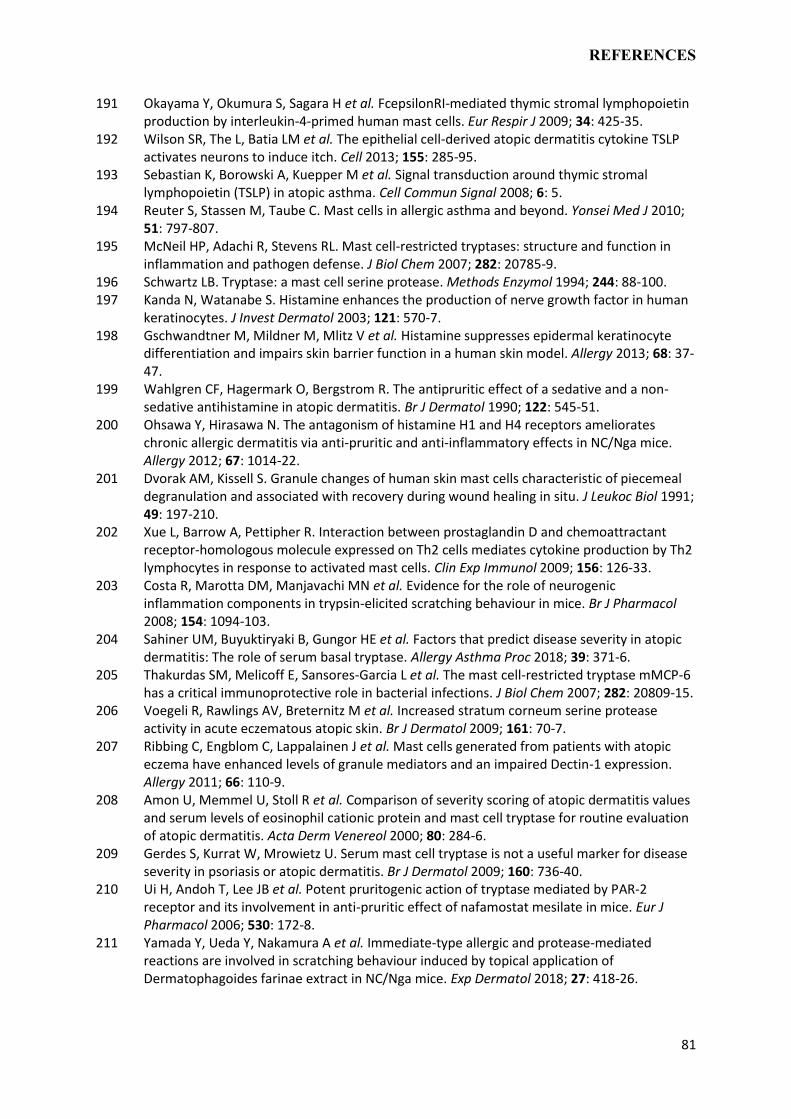
REFERENCES
81
191 Okayama Y, Okumura S, Sagara H et al. FcepsilonRI-mediated thymic stromal lymphopoietin production by interleukin-4-primed human mast cells. Eur Respir J 2009; 34: 425-35.
192 Wilson SR, The L, Batia LM et al. The epithelial cell-derived atopic dermatitis cytokine TSLP activates neurons to induce itch. Cell 2013; 155: 285-95.
193 Sebastian K, Borowski A, Kuepper M et al. Signal transduction around thymic stromal lymphopoietin (TSLP) in atopic asthma. Cell Commun Signal 2008; 6: 5.
194 Reuter S, Stassen M, Taube C. Mast cells in allergic asthma and beyond. Yonsei Med J 2010; 51: 797-807.
195 McNeil HP, Adachi R, Stevens RL. Mast cell-restricted tryptases: structure and function in inflammation and pathogen defense. J Biol Chem 2007; 282: 20785-9.
196 Schwartz LB. Tryptase: a mast cell serine protease. Methods Enzymol 1994; 244: 88-100. 197 Kanda N, Watanabe S. Histamine enhances the production of nerve growth factor in human
keratinocytes. J Invest Dermatol 2003; 121: 570-7. 198 Gschwandtner M, Mildner M, Mlitz V et al. Histamine suppresses epidermal keratinocyte
differentiation and impairs skin barrier function in a human skin model. Allergy 2013; 68: 37-47.
199 Wahlgren CF, Hagermark O, Bergstrom R. The antipruritic effect of a sedative and a non-sedative antihistamine in atopic dermatitis. Br J Dermatol 1990; 122: 545-51.
200 Ohsawa Y, Hirasawa N. The antagonism of histamine H1 and H4 receptors ameliorates chronic allergic dermatitis via anti-pruritic and anti-inflammatory effects in NC/Nga mice. Allergy 2012; 67: 1014-22.
201 Dvorak AM, Kissell S. Granule changes of human skin mast cells characteristic of piecemeal degranulation and associated with recovery during wound healing in situ. J Leukoc Biol 1991; 49: 197-210.
202 Xue L, Barrow A, Pettipher R. Interaction between prostaglandin D and chemoattractant receptor-homologous molecule expressed on Th2 cells mediates cytokine production by Th2 lymphocytes in response to activated mast cells. Clin Exp Immunol 2009; 156: 126-33.
203 Costa R, Marotta DM, Manjavachi MN et al. Evidence for the role of neurogenic inflammation components in trypsin-elicited scratching behaviour in mice. Br J Pharmacol 2008; 154: 1094-103.
204 Sahiner UM, Buyuktiryaki B, Gungor HE et al. Factors that predict disease severity in atopic dermatitis: The role of serum basal tryptase. Allergy Asthma Proc 2018; 39: 371-6.
205 Thakurdas SM, Melicoff E, Sansores-Garcia L et al. The mast cell-restricted tryptase mMCP-6 has a critical immunoprotective role in bacterial infections. J Biol Chem 2007; 282: 20809-15.
206 Voegeli R, Rawlings AV, Breternitz M et al. Increased stratum corneum serine protease activity in acute eczematous atopic skin. Br J Dermatol 2009; 161: 70-7.
207 Ribbing C, Engblom C, Lappalainen J et al. Mast cells generated from patients with atopic eczema have enhanced levels of granule mediators and an impaired Dectin-1 expression. Allergy 2011; 66: 110-9.
208 Amon U, Memmel U, Stoll R et al. Comparison of severity scoring of atopic dermatitis values and serum levels of eosinophil cationic protein and mast cell tryptase for routine evaluation of atopic dermatitis. Acta Derm Venereol 2000; 80: 284-6.
209 Gerdes S, Kurrat W, Mrowietz U. Serum mast cell tryptase is not a useful marker for disease severity in psoriasis or atopic dermatitis. Br J Dermatol 2009; 160: 736-40.
210 Ui H, Andoh T, Lee JB et al. Potent pruritogenic action of tryptase mediated by PAR-2 receptor and its involvement in anti-pruritic effect of nafamostat mesilate in mice. Eur J Pharmacol 2006; 530: 172-8.
211 Yamada Y, Ueda Y, Nakamura A et al. Immediate-type allergic and protease-mediated reactions are involved in scratching behaviour induced by topical application of Dermatophagoides farinae extract in NC/Nga mice. Exp Dermatol 2018; 27: 418-26.
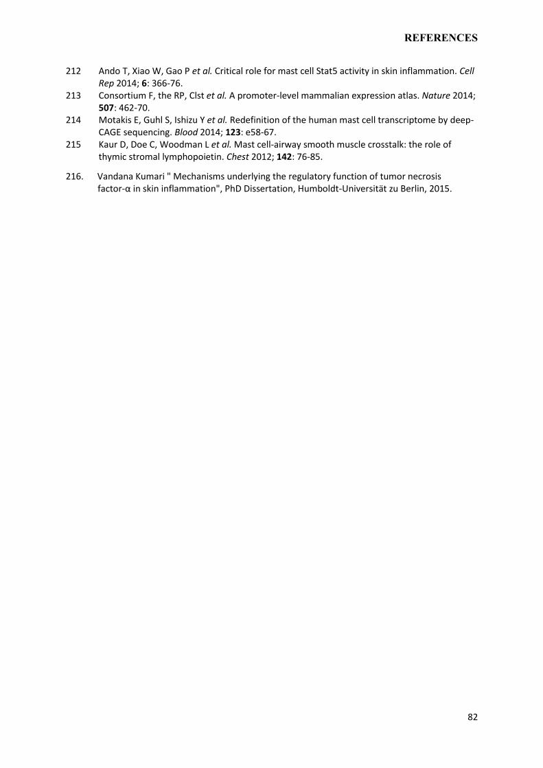
REFERENCES
82
212 Ando T, Xiao W, Gao P et al. Critical role for mast cell Stat5 activity in skin inflammation. Cell Rep 2014; 6: 366-76.
213 Consortium F, the RP, Clst et al. A promoter-level mammalian expression atlas. Nature 2014; 507: 462-70.
214 Motakis E, Guhl S, Ishizu Y et al. Redefinition of the human mast cell transcriptome by deep-CAGE sequencing. Blood 2014; 123: e58-67.
215 Kaur D, Doe C, Woodman L et al. Mast cell-airway smooth muscle crosstalk: the role of thymic stromal lymphopoietin. Chest 2012; 142: 76-85.
216. Vandana Kumari " Mechanisms underlying the regulatory function of tumor necrosis factor-α in skin inflammation", PhD Dissertation, Humboldt-Universität zu Berlin, 2015.
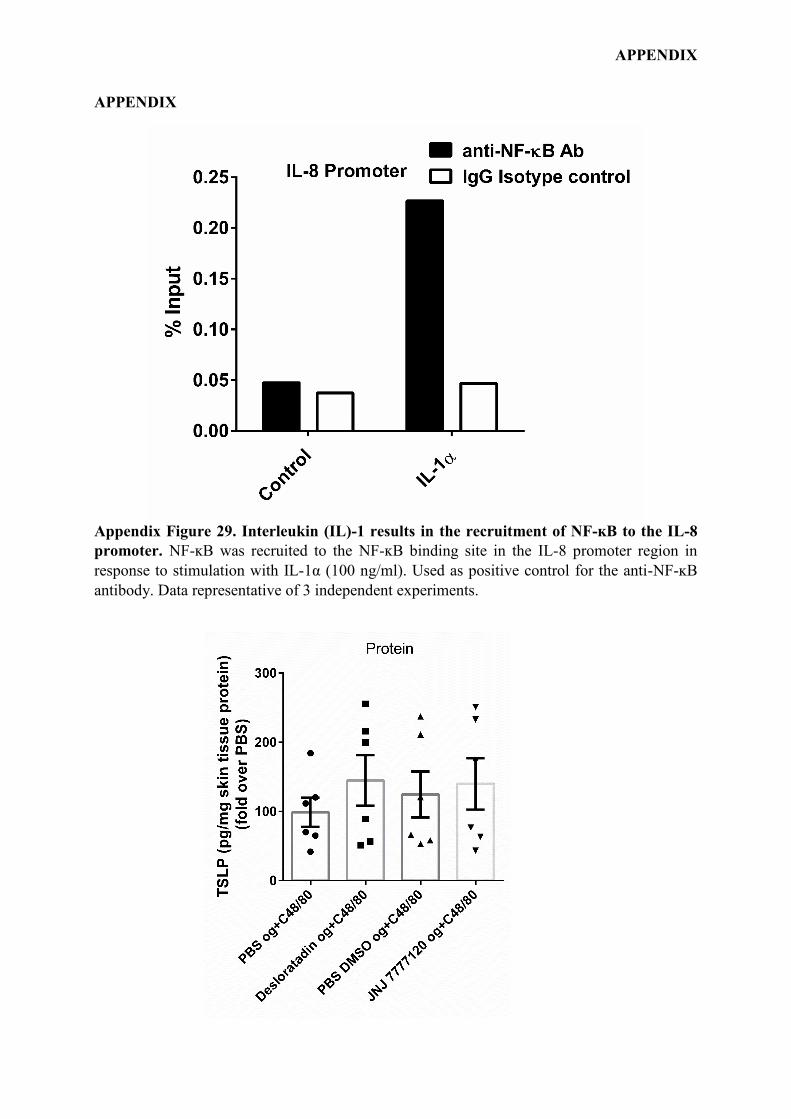
APPENDIX
APPENDIX
Appendix Figure 29. Interleukin (IL)-1 results in the recruitment of NF-ĸB to the IL-8
promoter. NF-ĸB was recruited to the NF-ĸB binding site in the IL-8 promoter region in
response to stimulation with IL-1α (100 ng/ml). Used as positive control for the anti-NF-ĸB
antibody. Data representative of 3 independent experiments.
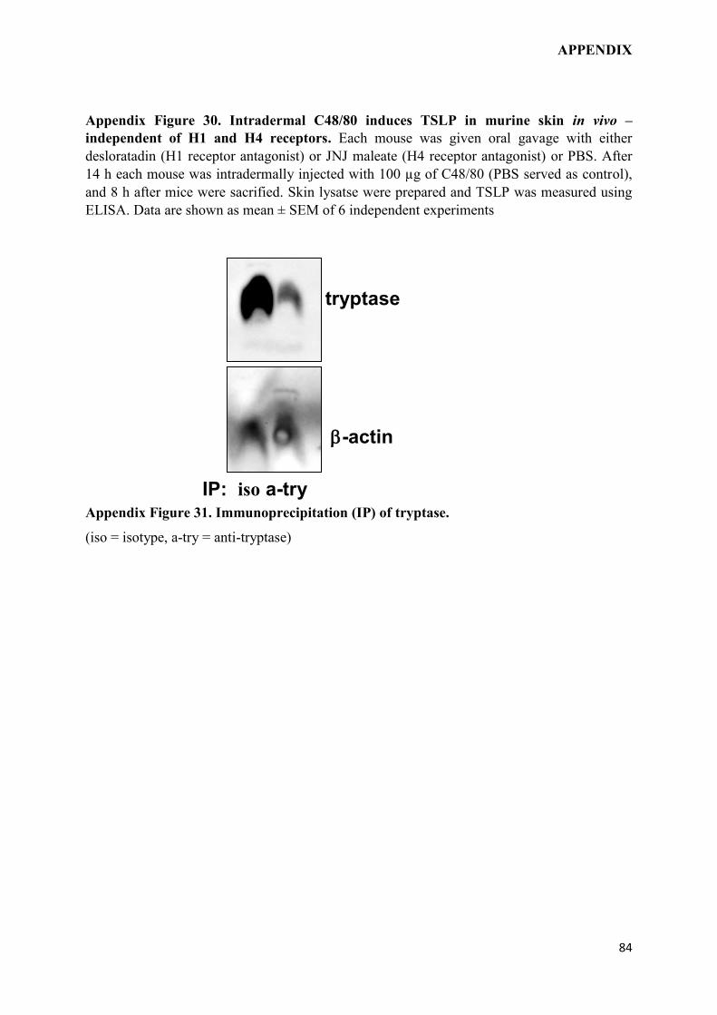
APPENDIX
84
Appendix Figure 30. Intradermal C48/80 induces TSLP in murine skin in vivo –
independent of H1 and H4 receptors. Each mouse was given oral gavage with either
desloratadin (H1 receptor antagonist) or JNJ maleate (H4 receptor antagonist) or PBS. After
14 h each mouse was intradermally injected with 100 µg of C48/80 (PBS served as control),
and 8 h after mice were sacrified. Skin lysatse were prepared and TSLP was measured using
ELISA. Data are shown as mean ± SEM of 6 independent experiments
Appendix Figure 31. Immunoprecipitation (IP) of tryptase.
(iso = isotype, a-try = anti-tryptase)
-actin
tryptase
IP: iso a-try
ACKNOWLEDGEMENTS
ACKNOWLEDGEMENTS
I would like to express my deepest gratitude towards my supervisor Prof. Dr. Margitta Worm
for giving me opportunity to do my PhD thesis in her lab. Many thanks for her continuous
support, guidance, patience, and encouragement during this training period. I have learnt a lot
from her during this period that her lessons will go above and beyond the contents of this
thesis. Her suggestions has helped me during all the times of research and writing of this
thesis, while at the same time also giving me opportunity to become an independent
researcher.
I am equally grateful to my advisory committee members Prof. Dr. Franz Bartl, Prof. Dr.
Hans-Dieter Volk, Prof. M Worm, Prof. Dr. P Franken and Prof. Dr. Emanuel Heitlinger for
their support and critical review. Their encouragement, motivation, and mentorship is deeply
appreciated.
I am truly indebted to the motivation and an incredible training atmosphere provided by the
lab members.
I wish to sincerely thank Dr. Magda Babina for fostering in me the principles of academic
excellence and to introducing me to the area of immunomodulation. I deeply appreciate her
valuable suggestions to my work and constant help during the writing of publication.
I owe my special thanks to Dr. Kristin Franke, Dr. Wojciech Francuzik, Dr. Vandana Kumari,
Sandra Treptow, Tarek Hazzan and Dennis Ernst for their friendship, and assistance during
my research. Their expert help and friendly discussions always made things simpler than they
would have been. Special thanks are due to Dr. Marina Aparicio-Soto for her supporting
work in my thesis. I also wish to acknowledge the friendly and supportive atmosphere
provided by other past and present lab members Dr. Guido Heine, Dr. Christin Weise, Dr.
Vandana Kumari, Dr. Marria Nassiri, Dr. Diana Willmes, Dr. Kristijan Pazur, Josephine,
Pdamavati, Shruti Rastogi, Zhao Wang, Julia Scholz, Aida Abadpour, Tina Krause, Dr. Julian
Lindner, Dr. Gennadiy Drozdenko, Dr. Sabine Dölle, Dr. Magdalina Kraft and Dr. Marcel
Wittenberg. The all-time help and administrative support provided by Mr. Lars Rudolf should
not go unmentioned, thank you so much.
A huge thanks to Dr. Naresh Singh Redhu for being my ideal, guide, elder brother who made
me reach this point and always helped in my toughest times. Without you, on my side, it
might have not been possible to reach this far. Special thanks to my friends in Berlin,

ACKNOWLEDGEMENTS
86
Parshant, Rajdilawer Singh, Sumit Tanwer, Vinay Malik, Sobinathan Balasundurum,
Ashutosh Singh, Siddarth Dhanda and Krishna Kumar who made my stay in Berlin homely,
enjoyable and memorable one.
Sincere thanks are due to Dr. Jasbir Singh, Dr. Suman Dhanda, Dr. Tejindar Pal Khaket and
Dr. Vikash Pahal for being my mentors, inspirations, and for guiding me through difficult
times to achieve my goals.
I owe a lot to my friends from India- Dr. Gulabo, Ramsharan Bhardwaj, Sombir Sahu, Anil
Redhu, Sombir Redhu, Brijesh Redhu, Dr. Kanu Priya, Dr. Suman Kandola, Anjali Sharma,
Dr. Mohini Sharma, Sandeep Dhanda, Amit Sharma, Amrit Kaushik, Sunil Chahal, Dr.
Vinod Khatri, Kapil Puniya and many more for being there for me. You always instilled in
me the confidence of being myself.
Above all, I cannot thank enough my parents (Maa Smt. Sheela Devi and Bbauji Shri
Ramniwas Redhu), my uncle (Tauji) Shri Satbir Singh Redhu and other family members who
have grafted a sense in me that education is the best treasure one can have, no matter who is
teaching. Even though my mom and dad never went to school, but they made sure that I will
get all the resources to earn highest level of education. Your love, emotions and faith have
kept me going all the way to the finish line. I am highly thankful to my younger brother
Sandeep, Mandeep and sister Pooja for taking care of Maa and Babuji in my absence.
If I have missed anyone in this journey, I request their forgiveness for my short-sightedness

DECLARATION
SELBSTÄNDIGKEITSERKLÄRUNG / DECLARATION
Hiermit versichere ich, Davender Redhu, die vorliegende Dissertation selbständig erarbeitet
und verfasst zu haben. Es wurden keine weiteren Quellen und Hilfsmittel als die hier
angegebenen verwendet.
I hereby declare that I, Davender Redhu, have worked and wrote this dissertation
independently and did not use other than the listed support. This thesis does not exist neither
in the same or similar form nor is it submitted to another examination procedure.