Embed Size (px)
Citation preview
biomolecules
Review
Sequence Features of Mitochondrial TransporterProtein Families
Gergely Gyimesi * and Matthias A. Hediger
Membrane Transport Discovery Lab, Department of Nephrology and Hypertension, and Department ofBiomedical Research, Inselspital, University of Bern, Kinderklinik, Freiburgstrasse 15,CH-3010 Bern, Switzerland; [email protected]* Correspondence: [email protected]; Tel.: +41-31-632-2293
Received: 20 October 2020; Accepted: 22 November 2020; Published: 28 November 2020 �����������������
Abstract: Mitochondrial carriers facilitate the transfer of small molecules across the inner mitochondrialmembrane (IMM) to support mitochondrial function and core cellular processes. In addition to theclassical SLC25 (solute carrier family 25) mitochondrial carriers, the past decade has led to thediscovery of additional protein families with numerous members that exhibit IMM localizationand transporter-like properties. These include mitochondrial pyruvate carriers, sideroflexins,and mitochondrial cation/H+ exchangers. These transport proteins were linked to vital physiologicalfunctions and disease. Their structures and transport mechanisms are, however, still largely unknownand understudied. Protein sequence analysis per se can often pinpoint hotspots that are of functional orstructural importance. In this review, we summarize current knowledge about the sequence features ofmitochondrial transporters with a special focus on the newly included SLC54, SLC55 and SLC56 familiesof the SLC solute carrier superfamily. Taking a step further, we combine sequence conservation analysiswith transmembrane segment and secondary structure prediction methods to extract residue positionsand sequence motifs that likely play a role in substrate binding, binding site gating or structural stability.We hope that our review will help guide future experimental efforts by the scientific community tounravel the transport mechanisms and structures of these novel mitochondrial carriers.
Keywords: mitochondrial carriers; SLC transporters; SLC25; MCF; SLC54; MPC; SLC55; LETM;SLC56; sideroflexin; ABC transporter; sequence analysis; protein targeting
1. Introduction
Mitochondria are believed to have evolved through an endosymbiotic event, where anα-proteobacteria has been engulfed by a host cell, possibly an archaeon [1,2]. This event is thought tohave arisen only once, and in the last 2 billion years, mitochondria have evolved together and in closeconcordance with their host cells [1,3,4]. During this time, significant changes in the genome of theendosymbiont have taken place, which involved the transfer of most mitochondrial proteins to thenucleus, as well as the emergence of novel protein families in the nuclear genome that are targeted tomitochondria [4,5]. The vestigial mitochondrial genomes of vertebrates in general code for 13 internalmembrane proteins that are involved in electron transport and coupled oxidative phosphorylation [6,7],while the part of the mitochondrial proteome related to transmembrane transport, i.e., the exchange ofmetabolites and ions with the host cell, is almost exclusively of eukaryotic origin [4,5].
Proteins in the human inner mitochondrial membrane (IMM) are quite distinct from one anotherin terms of their structure and sequence features as well as their trafficking and import mechanismsinto mitochondria. Based on this diversity, they are likely polyphyletic in origin and presumablyarose independently of each other. Membrane proteins that take part in transmembrane solutetransport across the IMM include members of the following families: SLC25 (mitochondrial carriers),
Biomolecules 2020, 10, 1611; doi:10.3390/biom10121611 www.mdpi.com/journal/biomolecules
Biomolecules 2020, 10, 1611 2 of 18
SLC8 (SLC8B1/NCLX Na+/Ca2+/Li+ exchanger), SLC54 (MPC, mitochondrial pyruvate carriers),SLC55 (LETM, leucine zipper-EF-hand-containing transmembrane proteins), SLC56 (sideroflexins),ATP-binding cassette (ABC) transporters (ABCB7, ABCB8, ABCB10) and various ion channels [8].The discovery, biological function, physiological role and disease involvement of many of thesetransport proteins and channels are comprehensively presented in excellent articles of the presentreview series. In particular, ion channels are covered in detail in the review by Szabo et al. and willnot be discussed here. Metabolite and ion transport between the cytoplasm and the mitochondriaalso require the broad-specificity channels of the outer mitochondrial membrane (OMM). The mostprominent class of such channels, the voltage-dependent anion channels (VDACs), is discussed indetail by Shoshan-Barmatz et al. in this review series [9], but other OMM transporters with unknownsubstrate specificity may also be present [10]. In this review, we focus on the discussion of specificstructure and sequence features that shape trafficking and functional properties of transporter-likeproteins in the IMM.
Trafficking of Membrane Transporters into Mitochondria
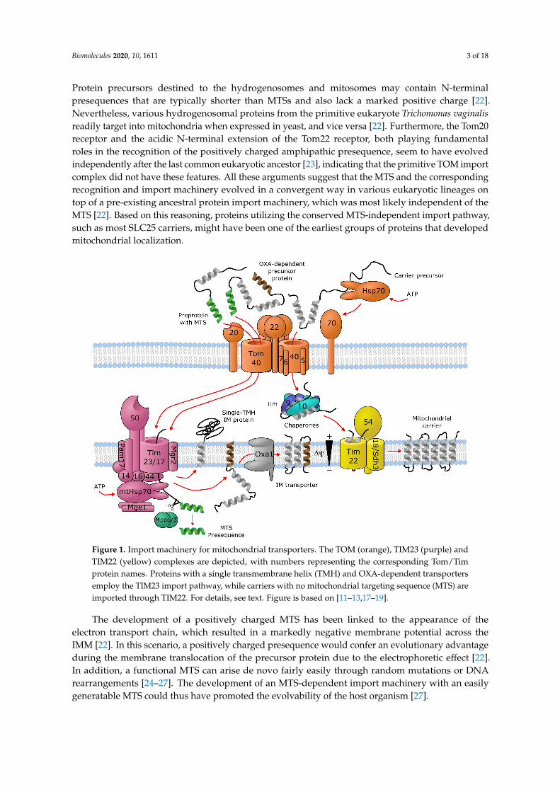
Transporters of the mitochondrial inner membrane are synthesized in the cytoplasm and importedinto the mitochondria through specialized import machinery [11]. In contrast to most mitochondrialproteins, most mitochondrial carriers typically do not contain an N-terminal mitochondrial targetingsequence (MTS). Instead, the nascent precursor transporter proteins bind to the ATP-hydrolyzingHsp70 and Hsp90 chaperones in the cytoplasm, which deliver them to the translocase of the outermembrane (TOM) complex. Here, the Tom70 receptor, part of TOM, binds both the precursorprotein and the chaperones, and transfers the precursor protein to Tom22, where it then getstranslocated in a loop-wise fashion through Tom40, the channel component of TOM [12,13]. Once in theintermembrane space, the hydrophobic regions of the precursor transporter proteins are shielded by theheterohexameric chaperone complex Tim9-Tim10-Tim12. This complex of the precursor protein and thechaperones then binds to the receptor-like protein Tim54, which is part of the translocase of the innermembrane (TIM22) complex in the IMM. Here, another member of TIM22, the channel-forming Tim22protein, then inserts the precursor protein into the inner mitochondrial membrane [11] (Figure 1).
As an alternative import mechanism, certain precursor transporter proteins do carry the MTSon their N-termini, which typically forms a short (15–50 residues) amphipathic helix with a netpositive charge [14,15]. Transporters containing an N-terminal MTS, such as SLC55/LETM andmitochondrial ABCB transporters, are delivered from the cytoplasm by the Tom20 receptor of theTOM complex, recognizing the hydrophobic side of the amphipathic helix formed by the MTS [16].Upon transfer to the intermembrane space through Tom40, the targeting sequence binds to the Tim50receptor component of another inner membrane complex, TIM23 [17]. This activates the Tim23channel subunit of the TIM23 complex to allow the translocation of the bound precursor proteinthrough the IMM [11]. Precursor proteins that are destined to the lipid bilayer contain a hydrophobicstop-transfer signal sequence, which is recognized within the IMM by the small transmembrane proteinMgr2, initiating the lateral release of the imported precursor protein into the membrane [11,18,19](Figure 1). Interestingly, an alternative mechanism exists, where Oxa1, the main component ofthe oxidase assembly (OXA) translocase complex, inserts the hydrophobic segment into the innermembrane bilayer after they pass through the Tim23 pore [11] (Figure 1). It has been shown thateven a single protein with multiple membrane-spanning segments can use different mechanismsto import individual transmembrane segments into the membrane, such as the yeast protein Mdl1,a mitochondrial ABCB family homolog [20]. Further details about the molecular machinery forimporting metabolite transporters into mitochondria are discussed by Rampelt et al. as part of thepresent special review series.
Hydrogenosomes and mitosomes are cellular organelles that share a common evolutionaryorigin with mitochondria [21], and the comparison of their protein import machineries has shedlight on important events shaping the early evolution of the mitochondrial import machinery [22].
Biomolecules 2020, 10, 1611 3 of 18
Protein precursors destined to the hydrogenosomes and mitosomes may contain N-terminalpresequences that are typically shorter than MTSs and also lack a marked positive charge [22].Nevertheless, various hydrogenosomal proteins from the primitive eukaryote Trichomonas vaginalisreadily target into mitochondria when expressed in yeast, and vice versa [22]. Furthermore, the Tom20receptor and the acidic N-terminal extension of the Tom22 receptor, both playing fundamentalroles in the recognition of the positively charged amphipathic presequence, seem to have evolvedindependently after the last common eukaryotic ancestor [23], indicating that the primitive TOM importcomplex did not have these features. All these arguments suggest that the MTS and the correspondingrecognition and import machinery evolved in a convergent way in various eukaryotic lineages ontop of a pre-existing ancestral protein import machinery, which was most likely independent of theMTS [22]. Based on this reasoning, proteins utilizing the conserved MTS-independent import pathway,such as most SLC25 carriers, might have been one of the earliest groups of proteins that developedmitochondrial localization.
Figure 1. Import machinery for mitochondrial transporters. The TOM (orange), TIM23 (purple) andTIM22 (yellow) complexes are depicted, with numbers representing the corresponding Tom/Timprotein names. Proteins with a single transmembrane helix (TMH) and OXA-dependent transportersemploy the TIM23 import pathway, while carriers with no mitochondrial targeting sequence (MTS) areimported through TIM22. For details, see text. Figure is based on [11–13,17–19].
The development of a positively charged MTS has been linked to the appearance of theelectron transport chain, which resulted in a markedly negative membrane potential across theIMM [22]. In this scenario, a positively charged presequence would confer an evolutionary advantageduring the membrane translocation of the precursor protein due to the electrophoretic effect [22].In addition, a functional MTS can arise de novo fairly easily through random mutations or DNArearrangements [24–27]. The development of an MTS-dependent import machinery with an easilygeneratable MTS could thus have promoted the evolvability of the host organism [27].
Biomolecules 2020, 10, 1611 4 of 18
2. SLC25—Mitochondrial Carrier Family (MCF)
The largest protein family of mitochondrial solute transporters is the SLC25 (mitochondrialcarrier) family. In human there are a total of 53 members that fulfil the vital roles of uniport orexchange of ions, metabolites and other solutes across the IMM [28]. It has been recognized earlyon that the sequence of the ADP/ATP translocase (SLC25A4) has a repeating sequence element thatcontains two hydrophobic segments and repeats 3 times in the sequence [29]. Such an internal repeatsymmetry is commonly found in membrane transporters [30]. Indeed, such features were later found inseveral other members of the protein family [31–34], along with conserved proline, glycine and acidicamino acid residues [35]. The conserved residues were later compiled into a consensus characteristicor “signature motif” for mitochondrial carriers, Px(D/E)xx(K/R) [36], and the conserved chargedresidues were indeed found to take part in specific conserved salt-bridge contacts, termed the matrixnetwork [37–42], in the 3D structure of the proteins [43,44]. The signature motif can be extendedin the C-terminal direction to include a proximal glutamine (Q) residue, which helps stabilize thesalt-bridge contacts on the matrix side of the carrier, forming the “Q brace” [44,45]. There is a similarcluster of charged residues on the intermembrane side of the even-numbered TM helices, forming thecytoplasmic salt-bridge network, where the charged residues of a (Y/F)(D/E)xx(K/R) motif engage insalt-bridge contacts [42,44]. This network is stabilized by the so-called tyrosine (Y) brace, formed by thehydrogen bonding of the tyrosine residues of the motif to these salt bridges [45]. A further sequencemotif, (Y/W/L/F)(K/R)GxxP, present in the connecting loop between each short matrix helix and thefollowing transmembrane helix, has been described, replacement of which disrupts the function of thetransporter [46]. In addition, close helix-helix contacts in the matrix-facing state are formed by twoconserved sequence motifs, πGπxπG on the odd-numbered, and πxxxπ on the even-numbered helices,where π stands for a residue with a small side-chain [45,47,48]. Further details of these sequence motifs,including their roles in the transport mechanism and a functional interpretation of individual aminoacid residues is covered in detail by Kunji et al. in this special issue. In particular, disease-causingmutations in context of the 3D structure of SLC25 carriers and their sequence motifs are extensivelyreviewed by Palmieri et al. in the present review series [49].
Interestingly, the sequence motif (QYKGxxDCxRK) in the short matrix helices has also beendescribed, which is only conserved in a subset of mitochondrial carriers, such as ADP/ATP(SLC25A4–6, SLC25A31), aspartate/glutamate (SLC25A12–13), ornithine (SLC25A2, SLC25A15),glutamate (SLC25A18, SLC25A22), and carnitine (SLC25A20) carriers, one ATP/Pi carrier (SLC25A24)and three carriers with unknown function (SLC25A9, SLC25A34, SLC25A45) [50]. This motif wasproposed to harbor residues that go through post-translational modification thereby locally alteringthe protein structure and thus modulating function. One example for this is acetylation at K163 ofSLC25A5 (AAC2), corresponding to the last residue of the motif, while the cysteine residue of themotif might interact with oxidizing/reducing agents [50]. Further investigation is required to revealthe precise functional role of this sequence motif.
Most SLC25 family proteins do not contain an MTS at their N-termini. Instead, they seem to holdmitochondrial targeting information in all three segments of their three-fold repeat sequence [51,52].SLC25 proteins are embedded into the IMM by the TIM22 machinery, as described in the previouschapter. It was suggested that the three repeating segments of SLC25 proteins act in a cooperativemanner to facilitate receptor binding and translocation into mitochondria [53], nevertheless, a singleunit consisting of a matrix loop and the following transmembrane helix is enough for mitochondriallocalization [52]. The net positive charge of the short matrix loop helices have been shown to beessential for import into mitochondria, and these regions are thought to interact with the Tom40channel component of the TOM complex, which effectively functions as a selectivity filter [52].
Interestingly, certain SLC25 proteins, such as the mitochondrial phosphate carrier (SLC25A3)and citrate/tricarboxylate carrier (SLC25A1) have been proposed to harbor an N-terminal MTS,based on physicochemical composition of their N-terminal sequences and observation of matureprotein forms truncated at the anticipated cleavage site [31,54]. This is also partially supported by
Biomolecules 2020, 10, 1611 5 of 18
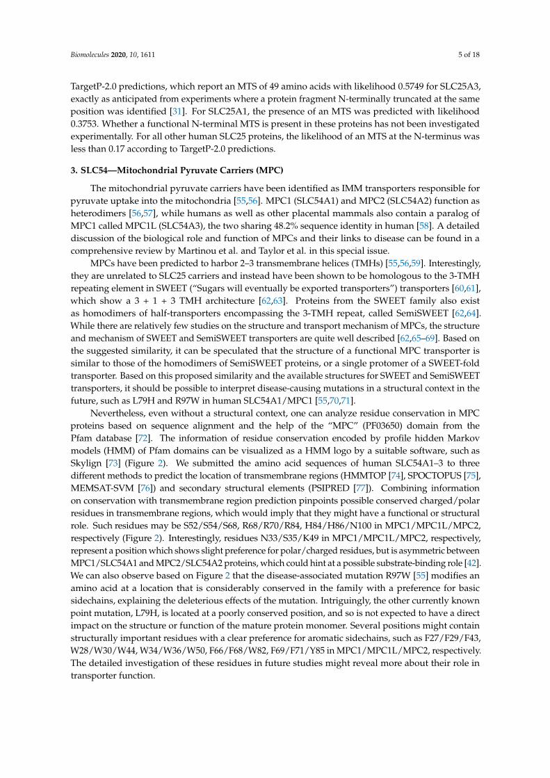
TargetP-2.0 predictions, which report an MTS of 49 amino acids with likelihood 0.5749 for SLC25A3,exactly as anticipated from experiments where a protein fragment N-terminally truncated at the sameposition was identified [31]. For SLC25A1, the presence of an MTS was predicted with likelihood0.3753. Whether a functional N-terminal MTS is present in these proteins has not been investigatedexperimentally. For all other human SLC25 proteins, the likelihood of an MTS at the N-terminus wasless than 0.17 according to TargetP-2.0 predictions.
3. SLC54—Mitochondrial Pyruvate Carriers (MPC)
The mitochondrial pyruvate carriers have been identified as IMM transporters responsible forpyruvate uptake into the mitochondria [55,56]. MPC1 (SLC54A1) and MPC2 (SLC54A2) function asheterodimers [56,57], while humans as well as other placental mammals also contain a paralog ofMPC1 called MPC1L (SLC54A3), the two sharing 48.2% sequence identity in human [58]. A detaileddiscussion of the biological role and function of MPCs and their links to disease can be found in acomprehensive review by Martinou et al. and Taylor et al. in this special issue.
MPCs have been predicted to harbor 2–3 transmembrane helices (TMHs) [55,56,59]. Interestingly,they are unrelated to SLC25 carriers and instead have been shown to be homologous to the 3-TMHrepeating element in SWEET (“Sugars will eventually be exported transporters”) transporters [60,61],which show a 3 + 1 + 3 TMH architecture [62,63]. Proteins from the SWEET family also existas homodimers of half-transporters encompassing the 3-TMH repeat, called SemiSWEET [62,64].While there are relatively few studies on the structure and transport mechanism of MPCs, the structureand mechanism of SWEET and SemiSWEET transporters are quite well described [62,65–69]. Based onthe suggested similarity, it can be speculated that the structure of a functional MPC transporter issimilar to those of the homodimers of SemiSWEET proteins, or a single protomer of a SWEET-foldtransporter. Based on this proposed similarity and the available structures for SWEET and SemiSWEETtransporters, it should be possible to interpret disease-causing mutations in a structural context in thefuture, such as L79H and R97W in human SLC54A1/MPC1 [55,70,71].
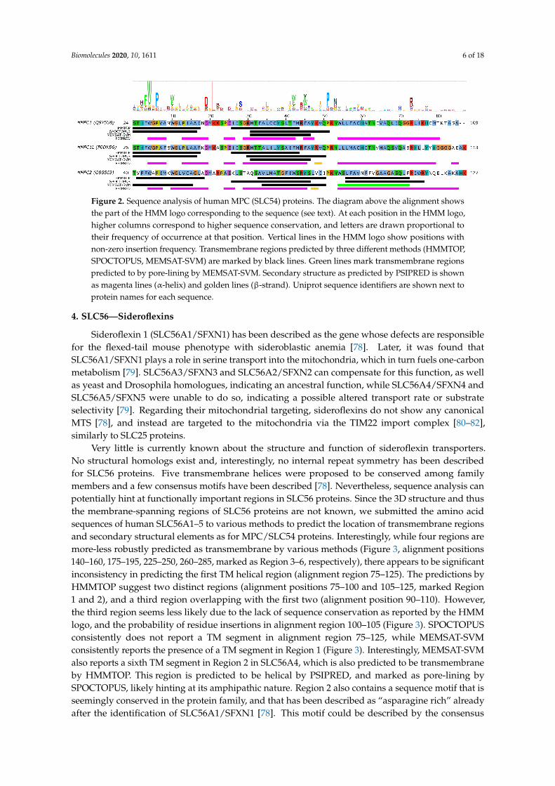
Nevertheless, even without a structural context, one can analyze residue conservation in MPCproteins based on sequence alignment and the help of the “MPC” (PF03650) domain from thePfam database [72]. The information of residue conservation encoded by profile hidden Markovmodels (HMM) of Pfam domains can be visualized as a HMM logo by a suitable software, such asSkylign [73] (Figure 2). We submitted the amino acid sequences of human SLC54A1–3 to threedifferent methods to predict the location of transmembrane regions (HMMTOP [74], SPOCTOPUS [75],MEMSAT-SVM [76]) and secondary structural elements (PSIPRED [77]). Combining informationon conservation with transmembrane region prediction pinpoints possible conserved charged/polarresidues in transmembrane regions, which would imply that they might have a functional or structuralrole. Such residues may be S52/S54/S68, R68/R70/R84, H84/H86/N100 in MPC1/MPC1L/MPC2,respectively (Figure 2). Interestingly, residues N33/S35/K49 in MPC1/MPC1L/MPC2, respectively,represent a position which shows slight preference for polar/charged residues, but is asymmetric betweenMPC1/SLC54A1 and MPC2/SLC54A2 proteins, which could hint at a possible substrate-binding role [42].We can also observe based on Figure 2 that the disease-associated mutation R97W [55] modifies anamino acid at a location that is considerably conserved in the family with a preference for basicsidechains, explaining the deleterious effects of the mutation. Intriguingly, the other currently knownpoint mutation, L79H, is located at a poorly conserved position, and so is not expected to have a directimpact on the structure or function of the mature protein monomer. Several positions might containstructurally important residues with a clear preference for aromatic sidechains, such as F27/F29/F43,W28/W30/W44, W34/W36/W50, F66/F68/W82, F69/F71/Y85 in MPC1/MPC1L/MPC2, respectively.The detailed investigation of these residues in future studies might reveal more about their role intransporter function.
Biomolecules 2020, 10, 1611 6 of 18
Figure 2. Sequence analysis of human MPC (SLC54) proteins. The diagram above the alignment showsthe part of the HMM logo corresponding to the sequence (see text). At each position in the HMM logo,higher columns correspond to higher sequence conservation, and letters are drawn proportional totheir frequency of occurrence at that position. Vertical lines in the HMM logo show positions withnon-zero insertion frequency. Transmembrane regions predicted by three different methods (HMMTOP,SPOCTOPUS, MEMSAT-SVM) are marked by black lines. Green lines mark transmembrane regionspredicted to by pore-lining by MEMSAT-SVM. Secondary structure as predicted by PSIPRED is shownas magenta lines (α-helix) and golden lines (β-strand). Uniprot sequence identifiers are shown next toprotein names for each sequence.
4. SLC56—Sideroflexins
Sideroflexin 1 (SLC56A1/SFXN1) has been described as the gene whose defects are responsiblefor the flexed-tail mouse phenotype with sideroblastic anemia [78]. Later, it was found thatSLC56A1/SFXN1 plays a role in serine transport into the mitochondria, which in turn fuels one-carbonmetabolism [79]. SLC56A3/SFXN3 and SLC56A2/SFXN2 can compensate for this function, as wellas yeast and Drosophila homologues, indicating an ancestral function, while SLC56A4/SFXN4 andSLC56A5/SFXN5 were unable to do so, indicating a possible altered transport rate or substrateselectivity [79]. Regarding their mitochondrial targeting, sideroflexins do not show any canonicalMTS [78], and instead are targeted to the mitochondria via the TIM22 import complex [80–82],similarly to SLC25 proteins.
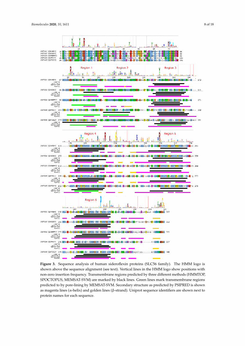
Very little is currently known about the structure and function of sideroflexin transporters.No structural homologs exist and, interestingly, no internal repeat symmetry has been describedfor SLC56 proteins. Five transmembrane helices were proposed to be conserved among familymembers and a few consensus motifs have been described [78]. Nevertheless, sequence analysis canpotentially hint at functionally important regions in SLC56 proteins. Since the 3D structure and thusthe membrane-spanning regions of SLC56 proteins are not known, we submitted the amino acidsequences of human SLC56A1–5 to various methods to predict the location of transmembrane regionsand secondary structural elements as for MPC/SLC54 proteins. Interestingly, while four regions aremore-less robustly predicted as transmembrane by various methods (Figure 3, alignment positions140–160, 175–195, 225–250, 260–285, marked as Region 3–6, respectively), there appears to be significantinconsistency in predicting the first TM helical region (alignment region 75–125). The predictions byHMMTOP suggest two distinct regions (alignment positions 75–100 and 105–125, marked Region1 and 2), and a third region overlapping with the first two (alignment position 90–110). However,the third region seems less likely due to the lack of sequence conservation as reported by the HMMlogo, and the probability of residue insertions in alignment region 100–105 (Figure 3). SPOCTOPUSconsistently does not report a TM segment in alignment region 75–125, while MEMSAT-SVMconsistently reports the presence of a TM segment in Region 1 (Figure 3). Interestingly, MEMSAT-SVMalso reports a sixth TM segment in Region 2 in SLC56A4, which is also predicted to be transmembraneby HMMTOP. This region is predicted to be helical by PSIPRED, and marked as pore-lining bySPOCTOPUS, likely hinting at its amphipathic nature. Region 2 also contains a sequence motif that isseemingly conserved in the protein family, and that has been described as “asparagine rich” alreadyafter the identification of SLC56A1/SFXN1 [78]. This motif could be described by the consensus
Biomolecules 2020, 10, 1611 7 of 18
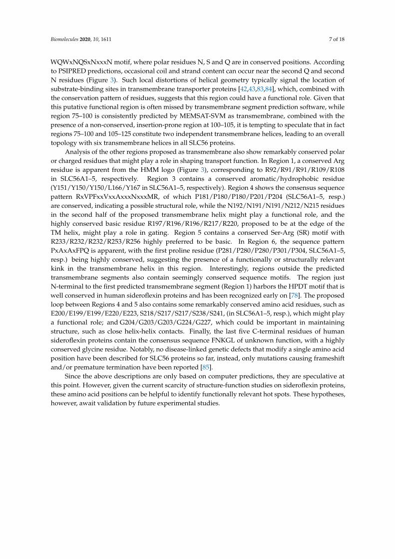
WQWxNQSxNxxxN motif, where polar residues N, S and Q are in conserved positions. Accordingto PSIPRED predictions, occasional coil and strand content can occur near the second Q and secondN residues (Figure 3). Such local distortions of helical geometry typically signal the location ofsubstrate-binding sites in transmembrane transporter proteins [42,43,83,84], which, combined withthe conservation pattern of residues, suggests that this region could have a functional role. Given thatthis putative functional region is often missed by transmembrane segment prediction software, whileregion 75–100 is consistently predicted by MEMSAT-SVM as transmembrane, combined with thepresence of a non-conserved, insertion-prone region at 100–105, it is tempting to speculate that in factregions 75–100 and 105–125 constitute two independent transmembrane helices, leading to an overalltopology with six transmembrane helices in all SLC56 proteins.
Analysis of the other regions proposed as transmembrane also show remarkably conserved polaror charged residues that might play a role in shaping transport function. In Region 1, a conserved Argresidue is apparent from the HMM logo (Figure 3), corresponding to R92/R91/R91/R109/R108in SLC56A1–5, respectively. Region 3 contains a conserved aromatic/hydrophobic residue(Y151/Y150/Y150/L166/Y167 in SLC56A1–5, respectively). Region 4 shows the consensus sequencepattern RxVPFxxVxxAxxxNxxxMR, of which P181/P180/P180/P201/P204 (SLC56A1–5, resp.)are conserved, indicating a possible structural role, while the N192/N191/N191/N212/N215 residuesin the second half of the proposed transmembrane helix might play a functional role, and thehighly conserved basic residue R197/R196/R196/R217/R220, proposed to be at the edge of theTM helix, might play a role in gating. Region 5 contains a conserved Ser-Arg (SR) motif withR233/R232/R232/R253/R256 highly preferred to be basic. In Region 6, the sequence patternPxAxAxFPQ is apparent, with the first proline residue (P281/P280/P280/P301/P304, SLC56A1–5,resp.) being highly conserved, suggesting the presence of a functionally or structurally relevantkink in the transmembrane helix in this region. Interestingly, regions outside the predictedtransmembrane segments also contain seemingly conserved sequence motifs. The region justN-terminal to the first predicted transmembrane segment (Region 1) harbors the HPDT motif that iswell conserved in human sideroflexin proteins and has been recognized early on [78]. The proposedloop between Regions 4 and 5 also contains some remarkably conserved amino acid residues, such asE200/E199/E199/E220/E223, S218/S217/S217/S238/S241, (in SLC56A1–5, resp.), which might playa functional role; and G204/G203/G203/G224/G227, which could be important in maintainingstructure, such as close helix-helix contacts. Finally, the last five C-terminal residues of humansideroflexin proteins contain the consensus sequence FNKGL of unknown function, with a highlyconserved glycine residue. Notably, no disease-linked genetic defects that modify a single amino acidposition have been described for SLC56 proteins so far, instead, only mutations causing frameshiftand/or premature termination have been reported [85].
Since the above descriptions are only based on computer predictions, they are speculative atthis point. However, given the current scarcity of structure-function studies on sideroflexin proteins,these amino acid positions can be helpful to identify functionally relevant hot spots. These hypotheses,however, await validation by future experimental studies.
Biomolecules 2020, 10, 1611 8 of 18
Figure 3. Sequence analysis of human sideroflexin proteins (SLC56 family). The HMM logo isshown above the sequence alignment (see text). Vertical lines in the HMM logo show positions withnon-zero insertion frequency. Transmembrane regions predicted by three different methods (HMMTOP,SPOCTOPUS, MEMSAT-SVM) are marked by black lines. Green lines mark transmembrane regionspredicted to by pore-lining by MEMSAT-SVM. Secondary structure as predicted by PSIPRED is shownas magenta lines (α-helix) and golden lines (β-strand). Uniprot sequence identifiers are shown next toprotein names for each sequence.
Biomolecules 2020, 10, 1611 9 of 18
5. The SLC55/LETM Mitochondrial Cation/Proton Exchanger Family
LETM1 (SLC55A1) has had a controversial role, as it was first proposed to be a part of themitochondrial K+/H+ exchanger (KHE) pathway [86,87]. However, later, through a genome-widegenetic screen, it was found to be responsible for altered Ca2+ levels in mitochondria [88],and subsequently the purified LETM1 protein in liposomes was shown to mediate Ca2+/H+
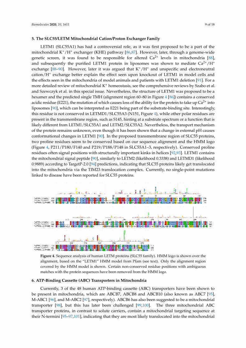
exchange [88–90]. However, later it was argued that K+/H+ and unspecific and electroneutralcation/H+ exchange better explain the effect seen upon knockout of LETM1 in model cells andthe effects seen in the mitochondria of model animals and patients with LETM1 deletion [91]. For amore detailed review of mitochondrial K+ homeostasis, see the comprehensive reviews by Szabo et al.and Szewczyk et al. in this special issue. Nevertheless, the structure of LETM1 was proposed to be ahexamer and the predicted single TMH (alignment region 60–80 in Figure 4 [86]) contains a conservedacidic residue (E221), the mutation of which causes loss of the ability for the protein to take up Ca2+ intoliposomes [90], which can be interpreted as E221 being part of the substrate-binding site. Interestingly,this residue is not conserved in LETMD1/SLC55A3 (N151, Figure 4), while other polar residues arepresent in the transmembrane region, such as S145, hinting at a substrate spectrum or a function that islikely different from LETM1/SLC55A1 and LETM2/SLC55A2. Nevertheless, the transport mechanismof the protein remains unknown, even though it has been shown that a change in external pH causesconformational changes in LETM1 [90]. In the proposed transmembrane region of SLC55 proteins,two proline residues seem to be conserved based on our sequence alignment and the HMM logo(Figure 4, P211/P180/F140 and P219/P188/P148 in SLC55A1–3, respectively). Conserved prolineresidues often signal positions with structurally important kinks in helices [92,93]. LETM1 containsthe mitochondrial signal peptide [90], similarly to LETM2 (likelihood 0.3338) and LETMD1 (likelihood0.9889) according to TargetP-2.0 [94] predictions, indicating that SLC55 proteins likely get translocatedinto the mitochondria via the TIM23 translocation complex. Currently, no single-point mutationslinked to disease have been reported for SLC55 proteins.
Figure 4. Sequence analysis of human LETM proteins (SLC55 family). HMM logo is shown over thealignment, based on the “LETM1” HMM model from Pfam (see text). Only the alignment regioncovered by the HMM model is shown. Certain non-conserved residue positions with ambiguousmatches with the protein sequences have been removed from the HMM logo.
6. ATP-Binding Cassette (ABC) Transporters in Mitochondria
Currently, 3 of the 48 human ATP-binding cassette (ABC) transporters have been shown tobe present in mitochondria, which are ABCB7, ABCB8 and ABCB10 (also known as ABC7 [95],M-ABC1 [96], and M-ABC2 [97], respectively). ABCB6 has also been suggested to be a mitochondrialtransporter [98], but this has later been challenged [99,100]. The three mitochondrial ABCtransporter proteins, in contrast to solute carriers, contain a mitochondrial targeting sequence attheir N-termini [95–97,101], indicating that they are most likely translocated into the mitochondrial
Biomolecules 2020, 10, 1611 10 of 18
inner membrane via the TIM23 machinery. The presence of an N-terminal cleavable targeting signal isalso supported by TargetP-2.0 predictions (likelihoods 0.9634, 0.7184, and 0.207 for human ABCB7,ABCB8, and ABCB10, respectively), while human ABCB6 was not predicted to contain a signal peptide(likelihood 0.0001).
A yeast ABC transporter protein (Mdl1) showing considerable sequence similarity to humanABCB proteins [102], was shown to use both the conservative Oxa1-mediated import machinery andlateral release from the TIM23 complex to translocate to the IMM [20]. Based on this, it is likelythat human ABCB proteins also use the same pathway for mitochondrial targeting. In addition,the homologous yeast Mdl2 protein and Atm1 (human ABCB7 ortholog) have also been suggested toemploy OXA-mediated membrane insertion into the IMM [103]. Nevertheless, while it is plausiblethat human mitochondrial ABCB proteins also employ a similar mechanism, this has not yet beenexperimentally studied.
ABCB transporters in the mitochondria are so-called “half transporters”, consisting of onenucleotide-binding domain (NBD) and one transmembrane domain (TMD), with homodimericcomplexes forming the functional transporter unit. Recent structural studies of human ABCB10 [104]and yeast Atm1 [105] have highlighted conserved sequence motifs that are located along cavitiesforming the putative substrate-binding site in the TMD. Interestingly, they are not lying in overlappinglocations in the two different proteins. For human ABCB7, residues R315, R319, N378, N425, T429,R432 and E433 (corresponding to R280, R284, N343, N390, S394, R397, D398 in yeast Atm1, respectively)in TMH4, TMH5 and TMH6 near the cytoplasmic membrane interface have been proposed to take partin substrate binding based on the yeast Atm1 structure with bound glutathione [105]. While one ofthese positions, E433 have been found to be mutated to lysine in patients with X-linked sideroblasticanemia (XLSA) [106], biochemical validation of these binding site residues is still missing [105].For human ABCB10, a conserved signature sequence of (N/I)xxR (containing N229 and R232) andNxxDGxR (containing N289, D292, R295) in TMH2 and TMH3, respectively, were found and havebeen proposed to take part in substrate binding [104]. In the case of ABCB10, the missing identity ofthe transported substrate hampers biochemical studies on the functional role of individual residues.It has been proposed that ABCB10 transports an intermediate in the heme biosynthesis pathway [107].Structure-function studies elucidating functionally relevant residues or sequence motifs of humanABCB8 are still missing. In neither ABCB8 nor in ABCB10 have disease-linked single-point mutationsbeen described yet.
7. Mitochondrial Calcium Transport via SLC8 Family
SLC8B1 (NCLX, Na+/Ca2+/Li+ exchanger) was identified as the ion exchanger protein responsiblefor the exit of Ca2+ from the mitochondria [108]. SLC8B1 is thought to exchange 3 Na+ ions for 1 Ca2+
ion based on similarity to other Na+/Ca2+ exchangers [109–111], and is unique in the property that Li+
ions can replace Na+ ions in transport [112]. Recently, it has been reported that Na+ taken up into themitochondrial matrix by SLC8B1 in exchange for Ca2+ derived from calcium precipitates upon matrixacidification controls hypoxic signaling via the mitochondrial respiratory chain [113]. Specifically,it was reported that the Na+ imported into the matrix reduces membrane fluidity through interactionwith phospholipids. This in turn was shown to lead to the generation of reactive oxygen species (ROS)by altering certain elements of the electron transport chain, thereby promoting an adaptive short-timeelevation of mitochondrial complex III-dependent ROS production during acute hypoxia. Indeed,inhibition of Na+ import via SLC8B1 was sufficient to prevent this pathway leading to adaptation toacute hypoxia [113].
While the mechanism of mitochondrial targeting of SLC8B1 has not yet been studied, the sequenceof human SLC8B1 does not seem to contain a mitochondrial targeting sequence according to TargetP-2.0predictions (likelihood 0.0016). This suggests that other internal targeting signals are present thatdirect the protein into the mitochondria, possibly similarly to SLC25, MPC and sideroflexin proteins.
Biomolecules 2020, 10, 1611 11 of 18
Disease-releated mutations have not been reported for SLC8B1 thus far. However, residuesresponsible for the unique Li+-exchange capacity of SLC8B1 have been investigated in detail. Based onthe determined structure of an archaeal NCX homologue, NCX_Mj, it was found that only 3 of the 12ion-coordinating residues were shared with human SLC8B1 [114]. By mutating the 9 different residuesto their human counterparts, it was possible to engineer a mutant of NCX_Mj that can also mediateLi+-dependent Ca2+ exchange [114]. In a later study, it was found that mutation of residue D471 toalanine can shift the selectivity toward Na+, while mutations at several positions can render SLC8B1a Li+-selective exchanger [115]. All these positions cluster close to the Na+-binding sites shown bythe X-ray structures of NCX_Mj [116,117] and the homologous H+/Ca2+ exchanger CAX_Af from theeuryarchaeota A. fulgidus [118].
8. Additional Families with Members Proposed to be Localized in the IMM
8.1. The SLC9 Na+/H+ Exchanger Family
SLC9B2 (NHA2, Na+/H+ antiporter 2) might localize to the mitochondria [119,120], but this hasbeen disputed [121]. Otherwise, SLC9B2 is more similar to prokaryotic Na+/H+ exchangers (NHEs)than to eukaryotic ones [121], and is the only human member of the Cation/Proton Antiporter 2(CPA2) subfamily [122]. SLC9B2 does not seem to contain a MTS according to TargetP-2.0 predictions(likelihood 0).
8.2. The SLC1 Glutamate/Neutral Amino Acid Transporter Family
A splice variant of SLC1A5 (SLC1A5_var) was recently reported to have a mitochondriallocalization and to function as the “long sought-after mitochondrial glutamine transporter” [123].However, several inconsistencies urge us to treat this conclusion with caution. Firstly, the software theauthors used for signal peptide detection (PrediSi) is designed to detect the signal peptides of proteinssecreted through the Sec pathway [124], and is therefore unsuitable to detect a MTS. In contrast,methods that were specifically developed to detect MTS, such as TargetP-2.0, do not detect the presenceof an MTS in SLC1A5_var (likelihood 0). Furthermore, the antibodies used by the authors to detectSLC1A5_var are claimed to “recognize the SLC1A5_var after peptide-N-glycosidase F (PNGase F)treatment” [123]. However, the only N-linked glycosylation sites on SLC1A5 are N163 and N212 [125],which are present in exon 1 that is in fact not present in the splice variant SLC1A5_var. Therefore,PNGase F treatment should not affect SLC1A5_var recognition. Due to the missing N-glycosylationsites, it is also likely, contrary to what the authors claim, that SLC1A5_var is not glycosylated. Finally,SLC1A5_var, in line with what is reported by the authors in their Figure 1A, is missing residues 2–203of, but is otherwise identical to, canonical SLC1A5. According to sequence alignment of human SLC1family members and the structure of a thermostable variant of the paralogous human SLC1A3 [126],this would mean that SLC1A5_var is missing the first 4 TMHs of the transporter protein, constitutingmost of the scaffold subdomain of the transporter. The importance of this region in transport isunderlined by the fact that TMH3 forms part of the binding site for the allosteric SLC1A3 inhibitorUCPH101, and TMH1 has been proposed to interact extensively with the lipid bilayer, harboring apossible lipid-binding site [126]. Thus, these regions are likely to be important in SLC1A5 as well,and it is questionable whether SLC1A5_var could function as a transporter with such an N-terminaltruncation. Therefore, given the above-mentioned issues with the antibody used and the detection ofan MTS, it is somewhat doubtful whether SLC1A5_var is truly a mitochondrial glutamine transporter.As an alternative, glutamine may be converted to glutamate in the mitochondrial intermembranespace via the phosphate-dependent glutaminase GLS [127] that is thought to be attached to the outersurface of the inner mitochondrial membrane [128]. Glutamate that is generated may then cross theIMM by the mitochondrial glutamate carriers SLC25A22 or SLC25A12. On the other hand, shouldGLS face the intra-mitochondrial matrix, a mitochondrial glutamine carrier is required. The need forsuch a carrier has been reviewed in detail in [129]. However, to clarify this subject matter, a conclusive
Biomolecules 2020, 10, 1611 12 of 18
subcellular localization study of GLS in the mitochondrial inner membrane is still required, in order toreveal whether the enzyme is active on the intermembrane space or within the mitochondrial matrix.
9. Conclusions and Open Questions
The spectrum of primary and secondary active transporters in the mitochondrial inner membranehas greatly broadened in the past decade through the functional identification of mitochondrialpyruvate carriers, sideroflexins, and other mitochondrial transporters such as SLC8B1. This pluralityof IMM transporters that show marked dissimilarity to SLC25 carriers, with no apparent commonevolutionary history to the SLC25 family, hints that many other, as of yet unidentified secondarytransporter families could exist in mitochondria. For the classical SLC25 mitochondrial carriers,their structure, targeting mechanism and transport properties are quite well-studied, but for the morerecently identified proteins, structural information, and a general understanding of their transportmechanisms are still lacking. Interestingly, the sideroflexin (SLC56) protein family seems to share nosignificant sequence similarity to any protein with a known structure, and is therefore likely to possessa yet undescribed and novel structural fold. Further studies would be needed to clarify residuesinvolved in substrate binding for sideroflexins. Another structurally enigmatic family of proteins arethe LETM/SLC55 transporters, which likely function in a hexameric unit that can change conformationupon changes in pH, which can be a basis for an alternating-access mechanism [90]. Nevertheless,the transport mechanism of any single-helix membrane-spanning ion exchanger such as LETM1 has notbeen described yet. Despite the lack of information on many of these proteins, we aimed to summarizesequence elements involved in targeting and function of mitochondrial transporters, and havealso suggested residues that could have a functional relevance based on sequence analysis of lesswell-characterized transporter families. The subsequent verification of the resulting hypotheses couldgreatly contribute to our understanding of their transport mechanisms. In this review, we have omittedthe discussion of disease-causing mutations of SLC25 carriers, as these have been reported in detail inother articles of the present review series [49]. However, for non-SLC25 proteins, we have discussed thelimited number of point mutations that are known to be linked to disease and involve single-residuechanges. For these proteins, on the one hand, more information about their biological role and diseaseinvolvement would be desirable. On the other hand, for some transporters, e.g., pyruvate carriers ofthe SLC54 family, structural model building could help understand their transport mechanism andinterpret certain disease-associated mutations. These endeavors can also potentially aid the generationof therapeutic modulators for future clinical applications.
Funding: This work has been supported by the Schweizerischer Nationalfonds (Swiss National ScienceFoundation) Grant Sinergia #CRSII5_180326, entitled “The role of mitochondrial carriers in metabolic tuning andreprogramming by calcium flow across membrane contact sites”.
Conflicts of Interest: The authors declare no conflict of interest.
References
1. Koonin, E.V. The Origin and Early Evolution of Eukaryotes in the Light of Phylogenomics. Genome Biol.2010, 11, 209. [CrossRef] [PubMed]
2. Gray, M.W. Mitochondrial Evolution. Cold Spring Harb. Perspect. Biol. 2012, 4, a011403. [CrossRef] [PubMed]3. Sicheritz-Pontén, T.; Kurland, C.G.; Andersson, S.G. A Phylogenetic Analysis of the Cytochrome b
and Cytochrome c Oxidase I Genes Supports an Origin of Mitochondria from within the Rickettsiaceae.Biochim. Biophys. Acta 1998, 1365, 545–551. [CrossRef]
4. Kurland, C.G.; Andersson, S.G. Origin and Evolution of the Mitochondrial Proteome. Microbiol. Mol.Biol. Rev. 2000, 64, 786–820. [CrossRef]
5. Karlberg, O.; Canbäck, B.; Kurland, C.G.; Andersson, S.G. The Dual Origin of the Yeast MitochondrialProteome. Yeast 2000, 17, 170–187. [CrossRef]
Biomolecules 2020, 10, 1611 13 of 18
6. Gray, M.W.; Lang, B.F.; Cedergren, R.; Golding, G.B.; Lemieux, C.; Sankoff, D.; Turmel, M.; Brossard, N.;Delage, E.; Littlejohn, T.G.; et al. Genome Structure and Gene Content in Protist Mitochondrial DNAs.Nucleic Acids Res. 1998, 26, 865–878. [CrossRef]
7. Wolstenholme, D.R. Animal Mitochondrial DNA: Structure and Evolution. Int. Rev. Cytol. 1992, 141, 173–216.[CrossRef]
8. Vothknecht, U.C.; Szabo, I. Mitochondrial Ion Channels and Transporters in Plants: Prediction and Facts.Mitochondrion 2020, 53, 224–233, [CrossRef]
9. Shoshan-Barmatz, V.; Shteinfer-Kuzmine, A.; Verma, A. VDAC1 at the Intersection of Cell Metabolism,Apoptosis, and Diseases. Biomolecules 2020, 10, 1485, [CrossRef]
10. Becker, T.; Wagner, R. Mitochondrial Outer Membrane Channels: Emerging Diversity in Transport Processes.Bioessays News Rev. Mol. Cell. Dev. Biol. 2018, 40, e1800013. [CrossRef]
11. Wiedemann, N.; Pfanner, N. Mitochondrial Machineries for Protein Import and Assembly.Annu. Rev. Biochem. 2017, 86, 685–714. [CrossRef] [PubMed]
12. Mokranjac, D.; Neupert, W. Cell Biology: Architecture of a Protein Entry Gate. Nature 2015, 528, 201–202.[CrossRef] [PubMed]
13. Shiota, T.; Imai, K.; Qiu, J.; Hewitt, V.L.; Tan, K.; Shen, H.H.; Sakiyama, N.; Fukasawa, Y.; Hayat, S.;Kamiya, M.; et al. Molecular Architecture of the Active Mitochondrial Protein Gate. Science 2015,349, 1544–1548. [CrossRef]
14. von Heijne, G.; Steppuhn, J.; Herrmann, R.G. Domain Structure of Mitochondrial and Chloroplast TargetingPeptides. Eur. J. Biochem. 1989, 180, 535–545. [CrossRef] [PubMed]
15. Garg, S.G.; Gould, S.B. The Role of Charge in Protein Targeting Evolution. Trends Cell Biol. 2016, 26, 894–905.[CrossRef] [PubMed]
16. Abe, Y.; Shodai, T.; Muto, T.; Mihara, K.; Torii, H.; Nishikawa, S.; Endo, T.; Kohda, D. Structural Basis ofPresequence Recognition by the Mitochondrial Protein Import Receptor Tom20. Cell 2000, 100, 551–560.[CrossRef]
17. Mokranjac, D.; Neupert, W. The Many Faces of the Mitochondrial TIM23 Complex. Biochim. Biophys. Acta2010, 1797, 1045–1054. [CrossRef]
18. Ieva, R.; Schrempp, S.G.; Opalinski, L.; Wollweber, F.; Höß, P.; Heißwolf, A.K.; Gebert, M.; Zhang, Y.;Guiard, B.; Rospert, S.; et al. Mgr2 Functions as Lateral Gatekeeper for Preprotein Sorting in theMitochondrial Inner Membrane. Mol. Cell 2014, 56, 641–652. [CrossRef]
19. Steffen, J.; Koehler, C.M. The Great Escape: Mgr2 of the Mitochondrial TIM23 Translocon Is a GatekeeperTasked with Releasing Membrane Proteins. Mol. Cell 2014, 56, 613–614. [CrossRef]
20. Bohnert, M.; Rehling, P.; Guiard, B.; Herrmann, J.M.; Pfanner, N.; van der Laan, M. Cooperation ofStop-Transfer and Conservative Sorting Mechanisms in Mitochondrial Protein Transport. Curr. Biol.2010, 20, 1227–1232. [CrossRef]
21. van der Giezen, M.; Slotboom, D.J.; Horner, D.S.; Dyal, P.L.; Harding, M.; Xue, G.P.; Embley, T.M.; Kunji, E.R.S.Conserved Properties of Hydrogenosomal and Mitochondrial ADP/ATP Carriers: A Common Origin forBoth Organelles. EMBO J. 2002, 21, 572–579. [CrossRef] [PubMed]
22. Garg, S.; Stölting, J.; Zimorski, V.; Rada, P.; Tachezy, J.; Martin, W.F.; Gould, S.B. Conservation of TransitPeptide-Independent Protein Import into the Mitochondrial and Hydrogenosomal Matrix. Genome Biol. Evol.2015, 7, 2716–2726. [CrossRef] [PubMed]
23. Fukasawa, Y.; Oda, T.; Tomii, K.; Imai, K. Origin and Evolutionary Alteration of the Mitochondrial ImportSystem in Eukaryotic Lineages. Mol. Biol. Evol. 2017, 34, 1574–1586. [CrossRef] [PubMed]
24. Baker, A.; Schatz, G. Sequences from a Prokaryotic Genome or the Mouse Dihydrofolate Reductase GeneCan Restore the Import of a Truncated Precursor Protein into Yeast Mitochondria. Proc. Natl. Acad. Sci. USA1987, 84, 3117–3121. [CrossRef] [PubMed]
25. Kaiser, C.A.; Preuss, D.; Grisafi, P.; Botstein, D. Many Random Sequences Functionally Replace the SecretionSignal Sequence of Yeast Invertase. Science 1987, 235, 312–317. [CrossRef]
26. Lemire, B.D.; Fankhauser, C.; Baker, A.; Schatz, G. The Mitochondrial Targeting Function of RandomlyGenerated Peptide Sequences Correlates with Predicted Helical Amphiphilicity. J. Biol. Chem.1989, 264, 20206–20215.
27. Dunn, C.D.; Paavilainen, V.O. Wherever I May Roam: Organellar Protein Targeting and Evolvability.Curr. Opin. Genet. Dev. 2019, 58, 9–16. [CrossRef]
Biomolecules 2020, 10, 1611 14 of 18
28. Palmieri, F. The Mitochondrial Transporter Family SLC25: Identification, Properties and Physiopathology.Mol. Asp. Med. 2013, 34, 465–484. [CrossRef]
29. Saraste, M.; Walker, J.E. Internal Sequence Repeats and the Path of Polypeptide in Mitochondrial ADP/ATPTranslocase. FEBS Lett. 1982, 144, 250–254. [CrossRef]
30. Forrest, L.R. Structural Symmetry in Membrane Proteins. Annu. Rev. Biophys. 2015, 44, 311–337. [CrossRef]31. Runswick, M.J.; Powell, S.J.; Nyren, P.; Walker, J.E. Sequence of the Bovine Mitochondrial Phosphate Carrier
Protein: Structural Relationship to ADP/ATP Translocase and the Brown Fat Mitochondria UncouplingProtein. EMBO J. 1987, 6, 1367–1373. [CrossRef] [PubMed]
32. Runswick, M.J.; Walker, J.E.; Bisaccia, F.; Iacobazzi, V.; Palmieri, F. Sequence of the Bovine2-Oxoglutarate/Malate Carrier Protein: Structural Relationship to Other Mitochondrial Transport Proteins.Biochemistry 1990, 29, 11033–11040. [CrossRef] [PubMed]
33. Indiveri, C.; Iacobazzi, V.; Giangregorio, N.; Palmieri, F. The Mitochondrial Carnitine Carrier Protein: cDNACloning, Primary Structure and Comparison with Other Mitochondrial Transport Proteins. Biochem. J. 1997,321 (Pt 3), 713–719. [CrossRef]
34. Palmieri, F. Mitochondrial Carrier Proteins. FEBS Lett. 1994, 346, 48–54. [CrossRef]35. Walker, J.E. The Mitochondrial Transporter Family. Curr. Opin. Struct. Biol. 1992, 2, 519–526. [CrossRef]36. Nelson, D.R.; Felix, C.M.; Swanson, J.M. Highly Conserved Charge-Pair Networks in the Mitochondrial
Carrier Family. J. Mol. Biol. 1998, 277, 285–308. [CrossRef]37. Falconi, M.; Chillemi, G.; Di Marino, D.; D’Annessa, I.; Morozzo della Rocca, B.; Palmieri, L.; Desideri, A.
Structural Dynamics of the Mitochondrial ADP/ATP Carrier Revealed by Molecular Dynamics SimulationStudies. Proteins 2006, 65, 681–691. [CrossRef]
38. Giangregorio, N.; Tonazzi, A.; Indiveri, C.; Palmieri, F. Conformation-Dependent Accessibility of Cys-136and Cys-155 of the Mitochondrial Rat Carnitine/Acylcarnitine Carrier to Membrane-Impermeable SHReagents. Biochim. Biophys. Acta 2007, 1767, 1331–1339. [CrossRef]
39. Cappello, A.R.; Miniero, D.V.; Curcio, R.; Ludovico, A.; Daddabbo, L.; Stipani, I.; Robinson, A.J.; Kunji, E.R.S.;Palmieri, F. Functional and Structural Role of Amino Acid Residues in the Odd-Numbered TransmembraneAlpha-Helices of the Bovine Mitochondrial Oxoglutarate Carrier. J. Mol. Biol. 2007, 369, 400–412. [CrossRef]
40. Palmieri, F. Diseases Caused by Defects of Mitochondrial Carriers: A Review. Biochim. Biophys. Acta2008, 1777, 564–578. [CrossRef]
41. Lauria, G.; Sanchez, P.; Della Rocca, B.M.; Pierri, C.L.; Polizio, F.; Stipani, I.; Desideri, A. Structural-DynamicalProperties of the Transmembrane Segment VI of the Mitochondrial Oxoglutarate Carrier Studied by SiteDirected Spin-Labeling. Mol. Membr. Biol. 2008, 25, 236–244. [CrossRef] [PubMed]
42. Robinson, A.J.; Overy, C.; Kunji, E.R.S. The Mechanism of Transport by Mitochondrial Carriers Based onAnalysis of Symmetry. Proc. Natl. Acad. Sci. USA 2008, 105, 17766–17771. [CrossRef] [PubMed]
43. Pebay-Peyroula, E.; Dahout-Gonzalez, C.; Kahn, R.; Trézéguet, V.; Lauquin, G.J.M.; Brandolin, G. Structure ofMitochondrial ADP/ATP Carrier in Complex with Carboxyatractyloside. Nature 2003, 426, 39–44. [CrossRef][PubMed]
44. Ruprecht, J.J.; Hellawell, A.M.; Harding, M.; Crichton, P.G.; McCoy, A.J.; Kunji, E.R.S. Structures of YeastMitochondrial ADP/ATP Carriers Support a Domain-Based Alternating-Access Transport Mechanism.Proc. Natl. Acad. Sci. USA 2014, 111, E426–E434. [CrossRef] [PubMed]
45. Ruprecht, J.J.; Kunji, E.R.S. The SLC25 Mitochondrial Carrier Family: Structure and Mechanism.Trends Biochem. Sci. 2020, 45, 244–258. [CrossRef]
46. Cappello, A.R.; Curcio, R.; Valeria Miniero, D.; Stipani, I.; Robinson, A.J.; Kunji, E.R.S.; Palmieri, F.Functional and Structural Role of Amino Acid Residues in the Even-Numbered TransmembraneAlpha-Helices of the Bovine Mitochondrial Oxoglutarate Carrier. J. Mol. Biol. 2006, 363, 51–62. [CrossRef]
47. Ruprecht, J.J.; Kunji, E.R. Structural Changes in the Transport Cycle of the Mitochondrial ADP/ATP Carrier.Curr. Opin. Struct. Biol. 2019, 57, 135–144. [CrossRef]
48. Ruprecht, J.J.; King, M.S.; Zögg, T.; Aleksandrova, A.A.; Pardon, E.; Crichton, P.G.; Steyaert, J.; Kunji, E.R.S.The Molecular Mechanism of Transport by the Mitochondrial ADP/ATP Carrier. Cell 2019, 176,435.e15–447.e15. [CrossRef]
49. Palmieri, F.; Scarcia, P.; Monné, M. Diseases Caused by Mutations in Mitochondrial Carrier Genes SLC25:A Review. Biomolecules 2020, 10, 655. [CrossRef]
Biomolecules 2020, 10, 1611 15 of 18
50. Pierri, C.L.; Palmieri, F.; De Grassi, A. Single-Nucleotide Evolution Quantifies the Importance of Each Sitealong the Structure of Mitochondrial Carriers. Cell. Mol. Life Sci. 2014, 71, 349–364. [CrossRef]
51. Brix, J.; Rüdiger, S.; Bukau, B.; Schneider-Mergener, J.; Pfanner, N. Distribution of Binding Sequences for theMitochondrial Import Receptors Tom20, Tom22, and Tom70 in a Presequence-Carrying Preprotein and aNon-Cleavable Preprotein. J. Biol. Chem. 1999, 274, 16522–16530. [CrossRef] [PubMed]
52. Kreimendahl, S.; Schwichtenberg, J.; Günnewig, K.; Brandherm, L.; Rassow, J. The Selectivity Filter of theMitochondrial Protein Import Machinery. BMC Biol. 2020, 18, 156. [CrossRef] [PubMed]
53. Wiedemann, N.; Pfanner, N.; Ryan, M.T. The Three Modules of ADP/ATP Carrier Cooperate in ReceptorRecruitment and Translocation into Mitochondria. EMBO J. 2001, 20, 951–960. [CrossRef] [PubMed]
54. Kaplan, R.S.; Mayor, J.A.; Wood, D.O. The Mitochondrial Tricarboxylate Transport Protein. cDNACloning, Primary Structure, and Comparison with Other Mitochondrial Transport Proteins. J. Biol. Chem.1993, 268, 13682–13690.
55. Bricker, D.K.; Taylor, E.B.; Schell, J.C.; Orsak, T.; Boutron, A.; Chen, Y.C.; Cox, J.E.; Cardon, C.M.;Van Vranken, J.G.; Dephoure, N.; et al. A Mitochondrial Pyruvate Carrier Required for Pyruvate Uptake inYeast, Drosophila, and Humans. Science 2012, 337, 96–100. [CrossRef]
56. Herzig, S.; Raemy, E.; Montessuit, S.; Veuthey, J.L.; Zamboni, N.; Westermann, B.; Kunji, E.R.S.; Martinou, J.C.Identification and Functional Expression of the Mitochondrial Pyruvate Carrier. Science 2012, 337, 93–96.[CrossRef]
57. Tavoulari, S.; Thangaratnarajah, C.; Mavridou, V.; Harbour, M.E.; Martinou, J.C.; Kunji, E.R. The YeastMitochondrial Pyruvate Carrier Is a Hetero-Dimer in Its Functional State. EMBO J. 2019, 38. [CrossRef]
58. Vanderperre, B.; Cermakova, K.; Escoffier, J.; Kaba, M.; Bender, T.; Nef, S.; Martinou, J.C. MPC1-like Is aPlacental Mammal-Specific Mitochondrial Pyruvate Carrier Subunit Expressed in Postmeiotic Male GermCells. J. Biol. Chem. 2016, 291, 16448–16461. [CrossRef]
59. Bender, T.; Pena, G.; Martinou, J.C. Regulation of Mitochondrial Pyruvate Uptake by Alternative PyruvateCarrier Complexes. EMBO J. 2015, 34, 911–924. [CrossRef]
60. Chen, L.Q.; Hou, B.H.; Lalonde, S.; Takanaga, H.; Hartung, M.L.; Qu, X.Q.; Guo, W.J.; Kim, J.G.;Underwood, W.; Chaudhuri, B.; et al. Sugar Transporters for Intercellular Exchange and Nutrition ofPathogens. Nature 2010, 468, 527–532. [CrossRef]
61. Chen, L.Q. SWEET Sugar Transporters for Phloem Transport and Pathogen Nutrition. New Phytol.2014, 201, 1150–1155. [CrossRef] [PubMed]
62. Xu, Y.; Tao, Y.; Cheung, L.S.; Fan, C.; Chen, L.Q.; Xu, S.; Perry, K.; Frommer, W.B.; Feng, L. Structures ofBacterial Homologues of SWEET Transporters in Two Distinct Conformations. Nature 2014, 515, 448–452.[CrossRef]
63. Medrano-Soto, A.; Ghazi, F.; Hendargo, K.J.; Moreno-Hagelsieb, G.; Myers, S.; Saier, M.H. Expansion of theTransporter-Opsin-G Protein-Coupled Receptor Superfamily with Five New Protein Families. PLoS ONE2020, 15, e0231085. [CrossRef]
64. Xuan, Y.H.; Hu, Y.B.; Chen, L.Q.; Sosso, D.; Ducat, D.C.; Hou, B.H.; Frommer, W.B. Functional Role ofOligomerization for Bacterial and Plant SWEET Sugar Transporter Family. Proc. Natl. Acad. Sci. USA2013, 110, E3685–E3694. [CrossRef] [PubMed]
65. Wang, J.; Yan, C.; Li, Y.; Hirata, K.; Yamamoto, M.; Yan, N.; Hu, Q. Crystal Structure of a Bacterial Homologueof SWEET Transporters. Cell Res. 2014, 24, 1486–1489. [CrossRef] [PubMed]
66. Jaehme, M.; Guskov, A.; Slotboom, D.J. Crystal Structure of the Vitamin B3 Transporter PnuC, a Full-LengthSWEET Homolog. Nat. Struct. Mol. Biol. 2014, 21, 1013–1015. [CrossRef]
67. Lee, Y.; Nishizawa, T.; Yamashita, K.; Ishitani, R.; Nureki, O. Structural Basis for the Facilitative DiffusionMechanism by SemiSWEET Transporter. Nat. Commun. 2015, 6, 6112. [CrossRef]
68. Han, L.; Zhu, Y.; Liu, M.; Zhou, Y.; Lu, G.; Lan, L.; Wang, X.; Zhao, Y.; Zhang, X.C. Molecular Mechanism ofSubstrate Recognition and Transport by the AtSWEET13 Sugar Transporter. Proc. Natl. Acad. Sci. USA 2017,114, 10089–10094. [CrossRef]
69. Latorraca, N.R.; Fastman, N.M.; Venkatakrishnan, A.J.; Frommer, W.B.; Dror, R.O.; Feng, L. Mechanism ofSubstrate Translocation in an Alternating Access Transporter. Cell 2017, 169, 96.e12–107.e12. [CrossRef]
70. Brivet, M.; Garcia-Cazorla, A.; Lyonnet, S.; Dumez, Y.; Nassogne, M.C.; Slama, A.; Boutron, A.; Touati, G.;Legrand, A.; Saudubray, J.M. Impaired Mitochondrial Pyruvate Importation in a Patient and a Fetus at Risk.Mol. Genet. Metab. 2003, 78, 186–192. [CrossRef]
Biomolecules 2020, 10, 1611 16 of 18
71. Oonthonpan, L.; Rauckhorst, A.J.; Gray, L.R.; Boutron, A.C.; Taylor, E.B. Two Human Patient MitochondrialPyruvate Carrier Mutations Reveal Distinct Molecular Mechanisms of Dysfunction. JCI Insight 2019, 5.[CrossRef] [PubMed]
72. El-Gebali, S.; Mistry, J.; Bateman, A.; Eddy, S.R.; Luciani, A.; Potter, S.C.; Qureshi, M.; Richardson, L.J.;Salazar, G.A.; Smart, A.; et al. The Pfam Protein Families Database in 2019. Nucleic Acids Res.2019, 47, D427–D432. [CrossRef] [PubMed]
73. Wheeler, T.J.; Clements, J.; Finn, R.D. Skylign: A Tool for Creating Informative, Interactive LogosRepresenting Sequence Alignments and Profile Hidden Markov Models. BMC Bioinform. 2014, 15, 7.[CrossRef]
74. Tusnády, G.E.; Simon, I. The HMMTOP Transmembrane Topology Prediction Server. Bioinformatics2001, 17, 849–850. [CrossRef]
75. Viklund, H.; Bernsel, A.; Skwark, M.; Elofsson, A. SPOCTOPUS: A Combined Predictor of Signal Peptidesand Membrane Protein Topology. Bioinformatics 2008, 24, 2928–2929. [CrossRef] [PubMed]
76. Nugent, T.; Jones, D.T. Detecting Pore-Lining Regions in Transmembrane Protein Sequences. BMC Bioinform.2012, 13, 169. [CrossRef]
77. Jones, D.T. Protein Secondary Structure Prediction Based on Position-Specific Scoring Matrices. J. Mol. Biol.1999, 292, 195–202. [CrossRef] [PubMed]
78. Fleming, M.D.; Campagna, D.R.; Haslett, J.N.; Trenor, C.C.; Andrews, N.C. A Mutation in a MitochondrialTransmembrane Protein Is Responsible for the Pleiotropic Hematological and Skeletal Phenotype ofFlexed-Tail (f/f) Mice. Genes Dev. 2001, 15, 652–657. [CrossRef] [PubMed]
79. Kory, N.; Wyant, G.A.; Prakash, G.; Uit de Bos, J.; Bottanelli, F.; Pacold, M.E.; Chan, S.H.;Lewis, C.A.; Wang, T.; Keys, H.R.; et al. SFXN1 Is a Mitochondrial Serine Transporter Required forOne-Carbon Metabolism. Science 2018, 362. [CrossRef]
80. Acoba, M.G.; Alpergin, E.S.S.; Renuse, S.; Fernández-del-Río, L.; Lu, Y.W.; Clarke, C.F.; Pandey, A.;Wolfgang, M.J.; Claypool, S.M. The Mitochondrial Carrier SFXN1 Is Critical for Complex III Integrityand Cellular Metabolism. bioRxiv 2020. [CrossRef]
81. Jackson, T.D.; Hock, D.; Palmer, C.S.; Kang, Y.; Fujihara, K.M.; Clemons, N.J.; Thorburn, D.R.; Stroud, D.A.;Stojanovski, D. The TIM22 Complex Regulates Mitochondrial One-Carbon Metabolism by Mediating theImport of Sideroflexins. bioRxiv 2020. [CrossRef]
82. Horten, P.; Colina-Tenorio, L.; Rampelt, H. Biogenesis of Mitochondrial Metabolite Carriers. Biomolecules2020, 10, 1008. [CrossRef] [PubMed]
83. Yamashita, A.; Singh, S.K.; Kawate, T.; Jin, Y.; Gouaux, E. Crystal Structure of a Bacterial Homologue ofNa+/Cl—Dependent Neurotransmitter Transporters. Nature 2005, 437, 215–223. [CrossRef] [PubMed]
84. Boudker, O.; Ryan, R.M.; Yernool, D.; Shimamoto, K.; Gouaux, E. Coupling Substrate and Ion Binding toExtracellular Gate of a Sodium-Dependent Aspartate Transporter. Nature 2007, 445, 387–393. [CrossRef]
85. Hildick-Smith, G.J.; Cooney, J.D.; Garone, C.; Kremer, L.S.; Haack, T.B.; Thon, J.N.; Miyata, N.; Lieber, D.S.;Calvo, S.E.; Akman, H.O.; et al. Macrocytic Anemia and Mitochondriopathy Resulting from a Defect inSideroflexin 4. Am. J. Hum. Genet. 2013, 93, 906–914. [CrossRef]
86. Nowikovsky, K.; Froschauer, E.M.; Zsurka, G.; Samaj, J.; Reipert, S.; Kolisek, M.; Wiesenberger, G.;Schweyen, R.J. The LETM1/YOL027 Gene Family Encodes a Factor of the Mitochondrial K+ Homeostasiswith a Potential Role in the Wolf-Hirschhorn Syndrome. J. Biol. Chem. 2004, 279, 30307–30315. [CrossRef]
87. Froschauer, E.; Nowikovsky, K.; Schweyen, R.J. Electroneutral K+/H+ Exchange in Mitochondrial MembraneVesicles Involves Yol027/Letm1 Proteins. Biochim. Biophys. Acta 2005, 1711, 41–48. [CrossRef]
88. Jiang, D.; Zhao, L.; Clapham, D.E. Genome-Wide RNAi Screen Identifies Letm1 as a Mitochondrial Ca2+/H+
Antiporter. Science 2009, 326, 144–147. [CrossRef]89. Tsai, M.F.; Jiang, D.; Zhao, L.; Clapham, D.; Miller, C. Functional Reconstitution of the Mitochondrial
Ca2+/H+ Antiporter Letm1. J. Gen. Physiol. 2014, 143, 67–73. [CrossRef]90. Shao, J.; Fu, Z.; Ji, Y.; Guan, X.; Guo, S.; Ding, Z.; Yang, X.; Cong, Y.; Shen, Y. Leucine Zipper-EF-Hand
Containing Transmembrane Protein 1 (LETM1) Forms a Ca2+/H+ Antiporter. Sci. Rep. 2016, 6, 34174.[CrossRef]
91. Nowikovsky, K.; Bernardi, P. LETM1 in Mitochondrial Cation Transport. Front. Physiol. 2014, 5, 83.[CrossRef] [PubMed]
Biomolecules 2020, 10, 1611 17 of 18
92. Cordes, F.S.; Bright, J.N.; Sansom, M.S.P. Proline-Induced Distortions of Transmembrane Helices. J. Mol. Biol.2002, 323, 951–960. [CrossRef]
93. Law, E.C.; Wilman, H.R.; Kelm, S.; Shi, J.; Deane, C.M. Examining the Conservation of Kinks in AlphaHelices. PLoS ONE 2016, 11, e0157553. [CrossRef] [PubMed]
94. Almagro Armenteros, J.J.; Salvatore, M.; Emanuelsson, O.; Winther, O.; von Heijne, G.; Elofsson, A.;Nielsen, H. Detecting Sequence Signals in Targeting Peptides Using Deep Learning. Life Sci. Alliance 2019, 2.[CrossRef] [PubMed]
95. Csere, P.; Lill, R.; Kispal, G. Identification of a Human Mitochondrial ABC Transporter, the FunctionalOrthologue of Yeast Atm1p. FEBS Lett. 1998, 441, 266–270. [CrossRef]
96. Hogue, D.L.; Liu, L.; Ling, V. Identification and Characterization of a Mammalian MitochondrialATP-Binding Cassette Membrane Protein. J. Mol. Biol. 1999, 285, 379–389. [CrossRef]
97. Zhang, F.; Hogue, D.L.; Liu, L.; Fisher, C.L.; Hui, D.; Childs, S.; Ling, V. M-ABC2, a New HumanMitochondrial ATP-Binding Cassette Membrane Protein. FEBS Lett. 2000, 478, 89–94. [CrossRef]
98. Krishnamurthy, P.C.; Du, G.; Fukuda, Y.; Sun, D.; Sampath, J.; Mercer, K.E.; Wang, J.;Sosa-Pineda, B.; Murti, K.G.; Schuetz, J.D. Identification of a Mammalian Mitochondrial PorphyrinTransporter. Nature 2006, 443, 586–589. [CrossRef]
99. Kiss, K.; Brozik, A.; Kucsma, N.; Toth, A.; Gera, M.; Berry, L.; Vallentin, A.; Vial, H.; Vidal, M.; Szakacs, G.Shifting the Paradigm: The Putative Mitochondrial Protein ABCB6 Resides in the Lysosomes of Cells and inthe Plasma Membrane of Erythrocytes. PLoS ONE 2012, 7, e37378. [CrossRef]
100. Kiss, K.; Kucsma, N.; Brozik, A.; Tusnady, G.E.; Bergam, P.; van Niel, G.; Szakacs, G. Role of the N-TerminalTransmembrane Domain in the Endo-Lysosomal Targeting and Function of the Human ABCB6 Protein.Biochem. J. 2015, 467, 127–139. [CrossRef]
101. Graf, S.A.; Haigh, S.E.; Corson, E.D.; Shirihai, O.S. Targeting, Import, and Dimerization of a MammalianMitochondrial ATP Binding Cassette (ABC) Transporter, ABCB10 (ABC-Me). J. Biol. Chem. 2004,279, 42954–42963. [CrossRef] [PubMed]
102. Dean, M.; Allikmets, R.; Gerrard, B.; Stewart, C.; Kistler, A.; Shafer, B.; Michaelis, S.; Strathern, J. Mappingand Sequencing of Two Yeast Genes Belonging to the ATP-Binding Cassette Superfamily. Yeast 1994,10, 377–383. [CrossRef] [PubMed]
103. Stiller, S.B.; Höpker, J.; Oeljeklaus, S.; Schütze, C.; Schrempp, S.G.; Vent-Schmidt, J.; Horvath, S.E.;Frazier, A.E.; Gebert, N.; van der Laan, M.; et al. Mitochondrial OXA Translocase Plays a Major Rolein Biogenesis of Inner-Membrane Proteins. Cell Metab. 2016, 23, 901–908. [CrossRef] [PubMed]
104. Shintre, C.A.; Pike, A.C.W.; Li, Q.; Kim, J.I.; Barr, A.J.; Goubin, S.; Shrestha, L.; Yang, J.; Berridge, G.;Ross, J.; et al. Structures of ABCB10, a Human ATP-Binding Cassette Transporter in Apo- andNucleotide-Bound States. Proc. Natl. Acad. Sci. USA 2013, 110, 9710–9715. [CrossRef] [PubMed]
105. Srinivasan, V.; Pierik, A.J.; Lill, R. Crystal Structures of Nucleotide-Free and Glutathione-BoundMitochondrial ABC Transporter Atm1. Science 2014, 343, 1137–1140. [CrossRef] [PubMed]
106. Bekri, S.; Kispal, G.; Lange, H.; Fitzsimons, E.; Tolmie, J.; Lill, R.; Bishop, D.F. Human ABC7 Transporter:Gene Structure and Mutation Causing X-Linked Sideroblastic Anemia with Ataxia with Disruption ofCytosolic Iron-Sulfur Protein Maturation. Blood 2000, 96, 3256–3264. [CrossRef]
107. Seguin, A.; Takahashi-Makise, N.; Yien, Y.Y.; Huston, N.C.; Whitman, J.C.; Musso, G.; Wallace, J.A.;Bradley, T.; Bergonia, H.A.; Kafina, M.D.; et al. Reductions in the Mitochondrial ABC Transporter Abcb10Affect the Transcriptional Profile of Heme Biosynthesis Genes. J. Biol. Chem. 2017, 292, 16284–16299.[CrossRef]
108. Palty, R.; Silverman, W.F.; Hershfinkel, M.; Caporale, T.; Sensi, S.L.; Parnis, J.; Nolte, C.; Fishman, D.;Shoshan-Barmatz, V.; Herrmann, S.; et al. NCLX Is an Essential Component of Mitochondrial Na+/Ca2+
Exchange. Proc. Natl. Acad. Sci. USA 2010, 107, 436–441. [CrossRef]109. Khananshvili, D. Distinction between the Two Basic Mechanisms of Cation Transport in the Cardiac
Na+-Ca2+ Exchange System. Biochemistry 1990, 29, 2437–2442. [CrossRef]110. Pitts, B.J. Stoichiometry of Sodium-Calcium Exchange in Cardiac Sarcolemmal Vesicles. Coupling to the
Sodium Pump. J. Biol. Chem. 1979, 254, 6232–6235.111. Reeves, J.P.; Hale, C.C. The Stoichiometry of the Cardiac Sodium-Calcium Exchange System. J. Biol. Chem.
1984, 259, 7733–7739. [PubMed]
Biomolecules 2020, 10, 1611 18 of 18
112. Palty, R.; Ohana, E.; Hershfinkel, M.; Volokita, M.; Elgazar, V.; Beharier, O.; Silverman, W.F.; Argaman, M.;Sekler, I. Lithium-Calcium Exchange Is Mediated by a Distinct Potassium-Independent Sodium-CalciumExchanger. J. Biol. Chem. 2004, 279, 25234–25240. [CrossRef] [PubMed]
113. Hernansanz-Agustín, P.; Choya-Foces, C.; Carregal-Romero, S.; Ramos, E.; Oliva, T.; Villa-Piña, T.;Moreno, L.; Izquierdo-Álvarez, A.; Cabrera-García, J.D.; Cortés, A.; et al. Na+ Controls Hypoxic Signallingby the Mitochondrial Respiratory Chain. Nature 2020, 586, 287–291. [CrossRef] [PubMed]
114. Refaeli, B.; Giladi, M.; Hiller, R.; Khananshvili, D. Structure-Based Engineering of Lithium-TransportCapacity in an Archaeal Sodium-Calcium Exchanger. Biochemistry 2016, 55, 1673–1676. [CrossRef]
115. Roy, S.; Dey, K.; Hershfinkel, M.; Ohana, E.; Sekler, I. Identification of Residues That Control Li+ versus Na+
Dependent Ca2+ Exchange at the Transport Site of the Mitochondrial NCLX. Biochim. Biophys. Acta. Mol.Cell Res. 2017, 1864, 997–1008. [CrossRef]
116. Liao, J.; Li, H.; Zeng, W.; Sauer, D.B.; Belmares, R.; Jiang, Y. Structural Insight into the Ion-ExchangeMechanism of the Sodium/Calcium Exchanger. Science 2012, 335, 686–690. [CrossRef]
117. Liao, J.; Marinelli, F.; Lee, C.; Huang, Y.; Faraldo-Gómez, J.D.; Jiang, Y. Mechanism of ExtracellularIon Exchange and Binding-Site Occlusion in a Sodium/Calcium Exchanger. Nat. Struct. Mol. Biol.2016, 23, 590–599. [CrossRef]
118. Nishizawa, T.; Kita, S.; Maturana, A.D.; Furuya, N.; Hirata, K.; Kasuya, G.; Ogasawara, S.; Dohmae, N.;Iwamoto, T.; Ishitani, R.; et al. Structural Basis for the Counter-Transport Mechanism of a H2+/Ca2+
Exchanger. Science 2013, 341, 168–172. [CrossRef]119. Battaglino, R.A.; Pham, L.; Morse, L.R.; Vokes, M.; Sharma, A.; Odgren, P.R.; Yang, M.; Sasaki, H.;
Stashenko, P. NHA-Oc/NHA2: A Mitochondrial Cation-Proton Antiporter Selectively Expressed inOsteoclasts. Bone 2008, 42, 180–192. [CrossRef]
120. Fuster, D.G.; Zhang, J.; Shi, M.; Bobulescu, I.A.; Andersson, S.; Moe, O.W. Characterization of theSodium/Hydrogen Exchanger NHA2. J. Am. Soc. Nephrol. 2008, 19, 1547–1556. [CrossRef]
121. Donowitz, M.; Ming Tse, C.; Fuster, D. SLC9/NHE Gene Family, a Plasma Membrane and Organellar Familyof Na+/H+ Exchangers. Mol. Asp. Med. 2013, 34, 236–251. [CrossRef]
122. Masrati, G.; Dwivedi, M.; Rimon, A.; Gluck-Margolin, Y.; Kessel, A.; Ashkenazy, H.; Mayrose, I.; Padan, E.;Ben-Tal, N. Broad Phylogenetic Analysis of Cation/Proton Antiporters Reveals Transport Determinants.Nat. Commun. 2018, 9, 4205. [CrossRef]
123. Yoo, H.C.; Park, S.J.; Nam, M.; Kang, J.; Kim, K.; Yeo, J.H.; Kim, J.K.; Heo, Y.; Lee, H.S.; Lee, M.Y.; et al.A Variant of SLC1A5 Is a Mitochondrial Glutamine Transporter for Metabolic Reprogramming in CancerCells. Cell Metab. 2020, 31, 267–283.e12. [CrossRef]
124. Hiller, K.; Grote, A.; Scheer, M.; Münch, R.; Jahn, D. PrediSi: Prediction of Signal Peptides and Their CleavagePositions. Nucleic Acids Res. 2004, 32, W375–W379. [CrossRef]
125. Console, L.; Scalise, M.; Tarmakova, Z.; Coe, I.R.; Indiveri, C. N-Linked Glycosylation of Human SLC1A5(ASCT2) Transporter Is Critical for Trafficking to Membrane. Biochim. Biophys. Acta 2015, 1853, 1636–1645.[CrossRef]
126. Canul-Tec, J.C.; Assal, R.; Cirri, E.; Legrand, P.; Brier, S.; Chamot-Rooke, J.; Reyes, N. Structure and AllostericInhibition of Excitatory Amino Acid Transporter 1. Nature 2017, 544, 446–451. [CrossRef]
127. Kandasamy, P.; Gyimesi, G.; Kanai, Y.; Hediger, M.A. Amino Acid Transporters Revisited: New Views inHealth and Disease. Trends Biochem. Sci. 2018, 43, 752–789. [CrossRef]
128. Welbourne, T.; Routh, R.; Yudkoff, M.; Nissim, I. The Glutamine/Glutamate Couplet and Cellular Function.News Physiol. Sci. 2001, 16, 157–160. [CrossRef]
129. Scalise, M.; Pochini, L.; Galluccio, M.; Console, L.; Indiveri, C. Glutamine Transport and MitochondrialMetabolism in Cancer Cell Growth. Front. Oncol. 2017, 7, 306. [CrossRef]
Publisher’s Note: MDPI stays neutral with regard to jurisdictional claims in published maps and institutionalaffiliations.
c© 2020 by the authors. Licensee MDPI, Basel, Switzerland. This article is an open accessarticle distributed under the terms and conditions of the Creative Commons Attribution(CC BY) license (http://creativecommons.org/licenses/by/4.0/).