Embed Size (px)
Citation preview

Technische Universität München
Max-Planck-Institut für Biochemie
Abteilung Strukturforschung
Biologische NMR-Arbeitsgruppe
Small molecule inhibitors of the p53-Mdm2 interaction
Arkadiusz Eugeniusz Sikora
Vollständiger Abdruck der von der Fakultät für Chemie der Technischen Universität
München zur Erlangung des akademischen Grades eines
Doktors der Naturwissenschaften
genehmigten Dissertation.
Vorsitzender: Univ.-Prof. Dr. Steffen J. Glaser
Prüfer der Dissertation: 1. Univ.-Prof. Dr. Christian F. W. Becker
2. Priv.-Doz. Dr. Gerd Gemmecker
Die Dissertation wurde am 18.04.2011 bei der Technischen Universität München eingereicht
und durch die Fakultät für Chemie am 14.06.2011 angenommen.
Acknowledgement
This PHD thesis was done in the Max Planck Institute for Biochemistry. I would like
to thank everyone who contributed to this work.
I would like to thank my supervisor Dr. Tad A. Holak, for his support, discussions,
patience and help with the experiments and to Prof. Dr. Christian Becker for being my
Doktorvater.
Especially I would like to thank Tomasz Sitar for help with protein expression and
purification, Grzegorz Popowicz for help with molecular docking, Michał Biśta for
providing help with NMR and Siglinde Wolf for help with FP measurements. I would
like to thank Anna Ducka, Weronika Janczyk, Marcelino Castro, Kaja Kowalska,
Michał Grzejszczyk,Dr. Gerd Hübener and Sonja Golla for support and discussion.

Manuscripts submitted and under preparation:
Spiro[oxindole-3,4’-(4’H-pyran)] and 3,4-dihydripyrimidin-2(1H)-ones as the MDM2-
p53 binding inhibitors. Arkadiusz E. Sikora, Siglinde Wolf, Weronika Janczyk, Kaja
Kowalska, Tad A. Holak, Grzegorz M. Popowicz
Bioorganic & Medicinal Chemistry (2011) submitted.
The multicomponent synthesis of spiro[oxindole-3,4’-(4’H-pyrane)]s. Arkadiusz E.
Sikora, Tad A. Holak, Alexander Dömling, Grzegorz M. Popowicz
Manuscript in preparation (2011).

Table of contents
Chapter 1. Introduction 1
1.1. p53 1
1.2. Mdm2 2
1.2.1. Structural domains of Mdm2 and their functions 2
1.2.2. Controlling the activity of MDM2 and p53 3
1.2.3. The structure of the p53 binding domain of Mdm2 3
1.3. The inhibitors of p53-Mdm2 interaction 4
1.3.1. Chalcones 4
1.3.2. Nutlins 5
1.3.3. 6-chloroindole-2-carboxylic acid 7
1.3.4. 1,4-benzodiazepine-2,5-diones 8
1.3.5. Chromenotriazolopyrimidines 12
1.3.6. Spiro(oxindole-33’-pyrrolidine)s 14
1.3.7. isoquinolinones 17
1.3.8. Terphenyls 18
1.3.9. Sulphonamides 19
Chapter 2. Goals of the study 20
Chapter 3. Materials and methods 21
3.1. Equipment 21
3.2. Columns 22
3.3. Stock solutions 23
3.4. Cell growth media 24
3.5. Buffers 24
3.5.1. Ion exchange and exclusion chromatography buffers 24
3.5.2. Buffers for immobilized metal-chelate chromatography
under native conditions. 25
3.5.3. Buffers for immobilized metal-chelate chromatography
Under native conditions 26
3.5.4. Buffer for DNA agarose gel electrophoresis 27
3.6. Reagents and buffers for SDS-Page 27
3.7. E. coli strains and plasmids 29
3.8. Enzymes and other proteins 30

3.9. Kits and reagents 30
3.10. Protein and nucleic acids markers 30
3.11. Substrates for chemical synthesis 31
3.11.1. 6-chloroisatin 31
3.11.2. Cyclopropylurea 34
3.11.3. 4-aminoimidazole 34
3.11.4. 6-chloroindole-3-aldehyde 35
3.11.5. 3-(dicyano)-methyloxindole 37
3.11.6. 4-chloroisatoic anhydride 38
3.11.7. Aliphatic cyanoacetamides 39
3.11.8. The synthesis of methoxyethyl acetoacetamide
using 2,2,6-trimethyl-4H-1,3-dioxin-4-one. 39
3.11.8. The microwave synthesis of aromatic acetoacetamides 40
3.11.10. The synthesis of indole-3-carboxylic acid 41
3.12. Proteins 43
3.12.1. Mdm2 preparation and purification 43
3.12.2. The preparation and purification of the complex of Mdm2 and p53 44
3.13. Mdm2’s ligands synthesis 46
3.13.1. Synthesis and purification of 3,4-dihydripyrimidin-2(1H)-ones 46
3.13.2. N-3 alkylation of 3,4-dihydripyrimidin-2(1H)-ones 47
3.13.3. Synthesis and purification of spiro[oxindole-3,4’-(4’H-pyran)]
Compounds 48
3.13.4. The synthesis and purification of 2,3-dihydroquinazolin-4(1H)-ones and
7-chloro-2,3-dihydroquinazolin-4(1H)-ones 49
3.13.5. The synthesis and purification of 1'H-spiro[6-chloro-isoindole-1,2'-
quinazoline]-3,4'(3'H)-diones 50
3.14. Laboratory procedures 51
3.14.1. Preparation of chemically competent cells 51
3.14.2. In silico screening 51
3.14.3. Ligand optimization 51
3.14.4. Fluorescence polarization measurements 52
3.14.4. NMR Measurements 52
3.14.5. Electrophoresis of DNA on agarose gel 52
3.14.6. Transformation of chemically competent cells 53
3.14.7. Sonication 53
3.14.8. Ni affinity chromatography 53
3.14.9. Electrophoresis in SDS polyacrylamide gel 54
3.14.10. Visualization of proteins on polyacrylamide gel 54
Chapter 4. Results and discussion 55
4.1. 3,4-dihydripyrimidin-2(1H)-ones and spiro[oxindole-3,4’-(4’Hpyran)]
es 55
4.2. 2,3-dihydroquinazolin-4(1H)-ones 83
4.3. 1'H-spiro[6-chloro-isoindole-1,2'-quinazoline]-3,4'(3'H)-diones 93
Chapter 5. Summary 99
5.1. Summary 99
5.2. Zusammenfassung 101
6. Appendix 103
6.1. Abbreviations 103
6.2. Protein and peptide sequences 105
7. References 106

Chapter 1 Introduction
1
1.Introduction
1.1. p53
The tumor suppressor protein p53 is involved in many cellular mechanisms,
like for example, cell cycle arrest, DNA repair, apoptosis, and aging (Lane 1992). In
healthy cells it is maintained at low concentration (Oren 1999), which increases upon
cellular stress. Increased concentration of p53 in nucleus can cause the arrest of cell
cycle or apoptosis - both prevent proliferation of the cells with defective DNA
(Vousden et al., 2002). These processes could prevent the cell from becoming
cancerous.
Human p53 consists of 393 aminoacids. It has 5 functional domains (Figure 1).
Figure 1: Schematic organization of functional domains of the p53 protein.
The role of the C-terminal domain of p53 (aminoacids 356-393) is not yet well
understood; though it is known that it is a place in which many posttranslational
modifications can be made to regulate the protein activity (Joerger et al., 2008). p53
is active as a tetramer and its tetramerization is made through aminoacids 325-356
located near the C-terminus (Okorokov et al., 2006). In the center of the p53
sequence there is a DNA binding domain, which is able to bind specifically to certain
DNA sequences (El-Deiry et al., 1992). At the N-terminus of the protein there is
transactivation domain, which is a binding site for many proteins responsible for
transcription (Lu et al., 1995, Lello et al., 2006, Thut et al., 1995, Gu et al., 1997,
Teufel et al., 2007; Joerger et al., 2008, Kussie et al., 1996, Marine et al., 2004). This
domain is natively unfolded, but upon binding to one of target proteins a part of it
becomes locally ordered; for example, when binding to Mdm2 a short fragment
(aminoacids 15-29) adopts a helical structure (Kussie et al., 1996). Between the
transactivation domain and DNA binding domain there is a proline rich region which
seems to be a linker between these two domains.

Chapter 1 Introduction
2
Many of human cancers have mutations or deletions in the p53 gene. Most of
other cancer cells are capable to express active p53, though the p53 pathway is
inactivated by overexpression of Mdm2 and Mdmx (Marine et al., 2004, Wade et al.,
2010). Mdm2 and MdmX are structurally related and have similar functions.
1.2. Mdm2
1.2.1. Structural domains of Mdm2 and their functions
The human Mdm2 protein is made of 491 aminoacids, and contains several
functional domains, which are shown in Figure 2.
Figure 2: Schematic organization of the Mdm2 protein mapping its functional
domains, nuclear localization, and nuclear export signals.
The N-terminal aminoacids 19-108 of Mdm2 bind the transactivation domain of
p53 disablin the transactivation activity of p53 (Joerger et al., 2008, Kussie et al.,
1996). Mdm2 has nuclear localization and nuclear export signals (Juven-Gershon et
al., 1999) followed by an acidic region (223-247), which contain several aspartic acid
and glutamic acid residues, which can bind to the ribosomal L5 protein. Mdm2 has
also a zinc finger motif (305-322) and the RING finger domain; the RING domain is
able to sequence-specifically bind RNA (Elenbaas et al., 1996) and possesses the
E3 ubiquitin ligase activity targeting p53 (Honda et al., 1997).
1.2.2. Controlling the activity of MDM2 and p53
Mdm2 regulates the p53 activity by binding to the p53 transactivation domain
and thus blocks the interaction with transcription related proteins and makes p53
inactive (Oliner et al., 1993). Additionally, Mdm2 regulates transcription of the p53
gene (Thut et al., 1997). The Mdm2 protein has the ubiquitin ligase activity targeting
p53 for proteosomal degradation and therefore decrease its concentration (Honda et
al., 1997; Haupt et al., 1997, Kubbutat et al., 1997).

Chapter 1 Introduction
3
The transcription of Mdm2 gene is induced by p53 (Perry et al., 1993,
Saucedo et al., 1998). Mdm2 protein is quickly cleared through ubiquitination and
proteasome degradation (Chang et al., 1998).
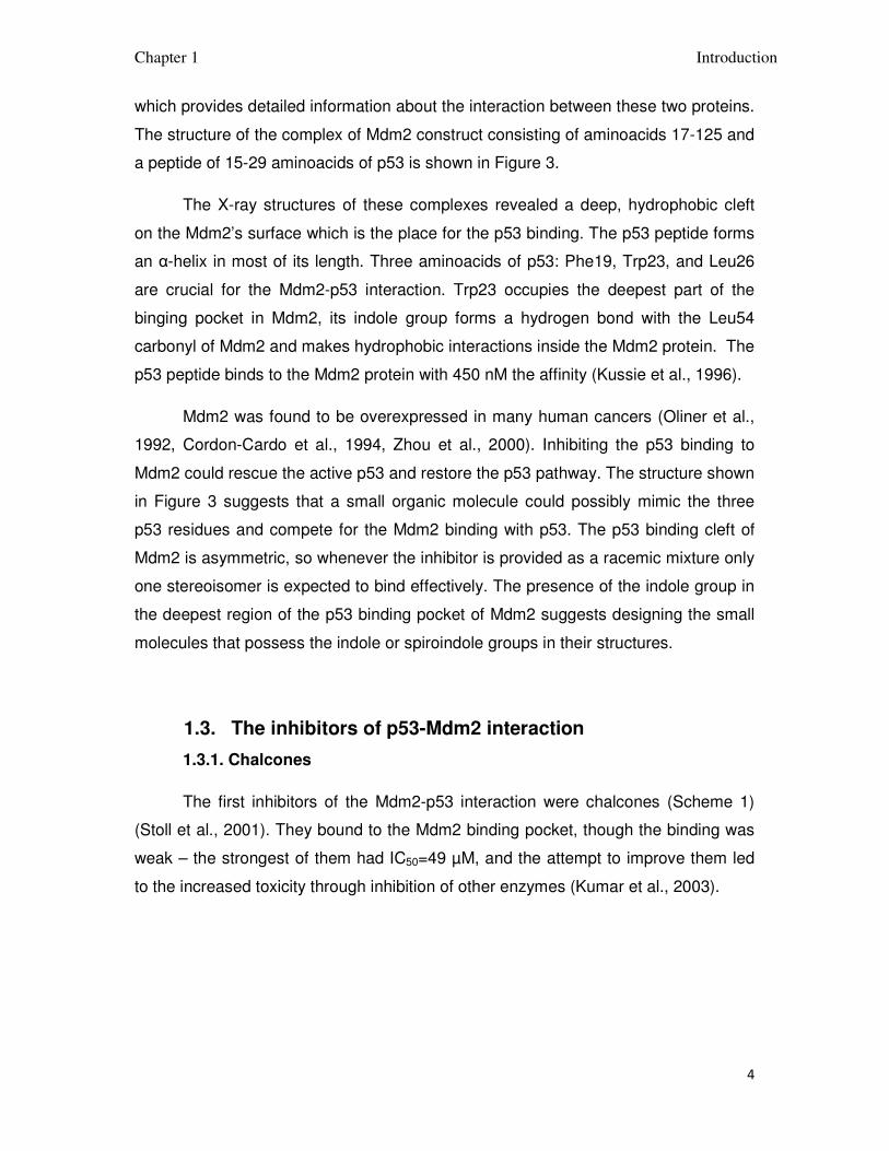
1.2.3. The structure of the p53 binding domain of Mdm2
Figure 3: The structure of the complex of the p53 binding domain of Mdm2 with the
short p53 peptide.
The crystallographic structure of the p53 binding domain of Mdm2 with a short
peptide of the p53 transactivaction domain has been solved by Kussie et al. (1996),
Chapter 1 Introduction
4
which provides detailed information about the interaction between these two proteins.
The structure of the complex of Mdm2 construct consisting of aminoacids 17-125 and
a peptide of 15-29 aminoacids of p53 is shown in Figure 3.
The X-ray structures of these complexes revealed a deep, hydrophobic cleft
on the Mdm2’s surface which is the place for the p53 binding. The p53 peptide forms
an α-helix in most of its length. Three aminoacids of p53: Phe19, Trp23, and Leu26
are crucial for the Mdm2-p53 interaction. Trp23 occupies the deepest part of the
binging pocket in Mdm2, its indole group forms a hydrogen bond with the Leu54
carbonyl of Mdm2 and makes hydrophobic interactions inside the Mdm2 protein. The
p53 peptide binds to the Mdm2 protein with 450 nM the affinity (Kussie et al., 1996).
Mdm2 was found to be overexpressed in many human cancers (Oliner et al.,
1992, Cordon-Cardo et al., 1994, Zhou et al., 2000). Inhibiting the p53 binding to
Mdm2 could rescue the active p53 and restore the p53 pathway. The structure shown
in Figure 3 suggests that a small organic molecule could possibly mimic the three
p53 residues and compete for the Mdm2 binding with p53. The p53 binding cleft of
Mdm2 is asymmetric, so whenever the inhibitor is provided as a racemic mixture only
one stereoisomer is expected to bind effectively. The presence of the indole group in
the deepest region of the p53 binding pocket of Mdm2 suggests designing the small
molecules that possess the indole or spiroindole groups in their structures.
1.3. The inhibitors of p53-Mdm2 interaction
1.3.1. Chalcones
The first inhibitors of the Mdm2-p53 interaction were chalcones (Scheme 1)
(Stoll et al., 2001). They bound to the Mdm2 binding pocket, though the binding was
weak – the strongest of them had IC50=49 µM, and the attempt to improve them led
to the increased toxicity through inhibition of other enzymes (Kumar et al., 2003).

Chapter 1 Introduction
5
Scheme 1: Examples of chalcones capable to interact with Mdm2.
1.3.2. Nutlins.
The first compounds capable of disrupting the Mdm2-p53 interaction in vivo
were cis-imidazolines called nutlins (Scheme 2) (Vassilev et al., 2004). These
compounds bind to Mdm2 with IC50 between 100 and 300 nM. They were
synthesized in multistep reaction as racemats and the enantiomers were separated
on chiral columns. There were significant differences in the activities between
enatniomers, for Nutlin-3 the difference was 150 times.
Scheme 2: Nutlins.
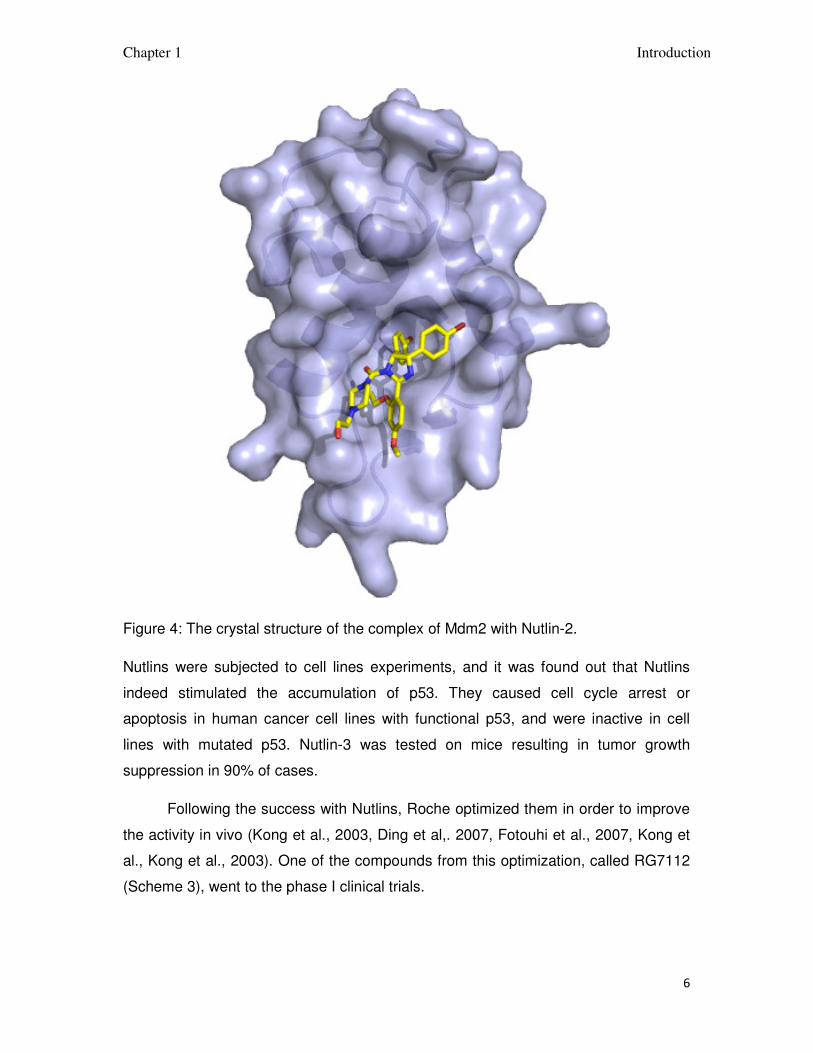
Nutlin-2 was co-crystalized with Mdm2 and the structure of the complex was
determined (Figure 4). Nutlin-2 binds to the p-53 binding pocket of Mdm2 utilizing
many hydrophobic interactions. One bromophenyl group is placed inside the Trp23
binding pocket, another bromophenyl moiety is in the Leu26 pocket and the ethyl
ether group is in the Phe19 binding pocket.
Chapter 1 Introduction
6
Figure 4: The crystal structure of the complex of Mdm2 with Nutlin-2.
Nutlins were subjected to cell lines experiments, and it was found out that Nutlins
indeed stimulated the accumulation of p53. They caused cell cycle arrest or
apoptosis in human cancer cell lines with functional p53, and were inactive in cell
lines with mutated p53. Nutlin-3 was tested on mice resulting in tumor growth
suppression in 90% of cases.
Following the success with Nutlins, Roche optimized them in order to improve
the activity in vivo (Kong et al., 2003, Ding et al,. 2007, Fotouhi et al., 2007, Kong et
al., Kong et al., 2003). One of the compounds from this optimization, called RG7112
(Scheme 3), went to the phase I clinical trials.

Chapter 1 Introduction
7
Scheme 3: RG7112.
1.3.3. 6-chloroindole-2-carboxylic acid
The Novartis company (Boettcher et al., 2008) and Doemling et al. (Popowicz
et al., 2010) independently developed the Mdm2-p53 binding inhibitors, like the one
in Scheme 4, which have a 6-chloroindole-2-carboxylic acid or its amide bound to
imidazole.
Scheme 4: 6-chloro-3-[1-(4-chlorobenzyl)-4-phenyl-imidazol-5-yl]-indole-2-carboxylic
acid.
The crystal structure of the complex of the compound in Scheme 4 with Mdm2
(Figure 5) confirms the expectation that the 6-chloroindole moiety mimics the Trp23
residue from p53 and fills its binding pocket, the chlorine is situated in the deep part
of the p53 binding pocket of Mdm2, which in not used while interacting with p53.

Chapter 1 Introduction
8
Figure 5: The crystal structure of the complex between compound from Scheme 4
with Mdm2.
1.3.4. 1,4-benzodiazepine-2,5-diones
The Jonson & Jonson company developed the inhibitors of the p53-Mdm2
binding based on the 1,4-benzodiazepine-2,5-dione scaffold as small molecule
compounds mimicking the Phe19, Trp23 and Leu26 residues of the p53 peptide
(Cummings et al., 2006). They were discovered by screening of compounds library
using the ThermoFluor technology, in which the melting temperature of a protein is
measured in the presence of the compound and compared with a control sample.
The hits were identified as the samples in which the protein’s melting temperature is
3 times higher than the standard deviation (Parks et al., 2005). The hits obtained by
the TermoFluor technology were confirmed by the fluorescence polarization assay.

Chapter 1 Introduction
9
The compounds were optimized to improve the binding to Mdm2. The sets of
compounds were synthesized using the condensation reaction followed by cyclization
(Scheme 5).
Scheme 5: The synthesis of 1,4-benzodiazepine-2,5-diones.
In each set of the compounds one of the moieties was altered while the rest of
the compound’s structure remained unchanged. Synthesized compounds were then
subjected to the Thermofluor and Fluorescence polarization assays. The result of
this optimization was compound TDP222669 shown in Scheme 6 (Raboisson et al.,
2005).
Scheme 6: 1,4-benzodiazepine-2,5-dione (TDP222669).
The co-crystal of the compound above bound to Mdm2 was solved
(Grassberger et al., 2005) and it showed that the predictions of its binding with Mdm2
were correct and the compound occupies the same binding place as the p53 peptide.

Chapter 1 Introduction
10
The chlorophenyl moieties reside in the Trp23 and Leu26 binding pockets and
iodobenzene goes to the Phe19 binding pocket.
Figure 6: The crystal structure of 1,4-benzodiazepine-2,5-dione shown in Scheme 6
co-crystalized with the Mdm2 protein.
Compound TDP222669 was tested in cell lines. In the cells expressing wild
type p53 the decrease of proliferation was observed at IC50 = 30 µM. The cells which
did not express the active p53 were insensitive to the compound (Grassberger et al.,
2005).
Tested in vivo, the compound shown poor bioavailability and rapid clearance,
its solubility was low and it did not pass the cell membranes well, most probably
because of the carboxyl group ionization (Parks et al., 2006), thus detailed

Chapter 1 Introduction
11
optimization was carried out. Neither amidation nor esterification of the carboxyl
group improved cell membrane permeability, and often caused significant loss of
potency, finally the carboxylic group was replaced with methyl (Scheme 7b). Attempts
to replace iodide with ethyl or chloride of the phenyl group with larger, hydrophobic
trifluoromethyl did not lead to any improvement of the compound properties (Parks et
al., 2006). Introducing the o-amino (Scheme 7c) or o-hydroxy (Scheme 7d) group in
the benzylic moiety added the extra hydrogen bond between the compound and
Val93 of Mdm2, which increased the compound’s potency. Alkylation of the N1
nitrogen with solubilizing groups was investigated (Parks et al., 2006, Leonard et al.,
2006), valeric acid (Scheme 7a) improved activity in cell lines. Replacing valeric acid
with methoxyethoxyethyl (Scheme 7e) decreased the binding in FP assay, but led to
better penetration of the cell and increased activity in the cells. N1 alkylation with
morpholino moieties with short linkers (Scheme 7f) further increased the potency of
the compound, while N-methylpiperazine or dimethylamine moieties caused
significant drop in potency (Leonard et al,. 2006).
Scheme 7: The optimization of 1,4-benzodiazepine-2,5-diones.
Chapter 1 Introduction
12
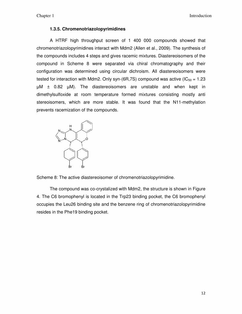
1.3.5. Chromenotriazolopyrimidines
A HTRF high throughput screen of 1 400 000 compounds showed that
chromenotriazolopyrimidines interact with Mdm2 (Allen et al., 2009). The synthesis of
the compounds includes 4 steps and gives racemic mixtures. Diastereoisomers of the
compound in Scheme 8 were separated via chiral chromatography and their
configuration was determined using circular dichroism. All diastereoisomers were
tested for interaction with Mdm2. Only syn-(6R,7S) compound was active (IC50 = 1.23
µM ± 0.82 µM). The diastereoisomers are unstable and when kept in
dimethylsulfoxide at room temperature formed mixtures consisting mostly anti
stereoisomers, which are more stable. It was found that the N11-methylation
prevents racemization of the compounds.
Scheme 8: The active diastereoisomer of chromenotriazolopyrimidine.
The compound was co-crystalized with Mdm2, the structure is shown in Figure
4. The C6 bromophenyl is located in the Trp23 binding pocket, the C6 bromophenyl
occupies the Leu26 binding site and the benzene ring of chromenotriazolopyrimidine
resides in the Phe19 binding pocket.

Chapter 1 Introduction
13
Figure 7: The crystal structure of chromenotriazolopyrimidine (shown in Scheme 8)
bound to Mdm2 protein.
The chromenotriazolopyrimidine was optimized to improve its activity. In the
optimization racemic mixtures of compounds were used. Changing bromine to
chloride in the phenyl ring resulted in better binding to Mdm2 (IC50 = 0.89 µM ± 0.20
µM), probing nitrile and ethyl substituents gave the compounds with similar activity as
the starting one, fluorine and big substituents diminished the activity. The substitution
of chromenotriazolopyrimidine’s benzene ring was checked in order to improve the
binding in the Phe19 binding pocket. The binding increased after adding methoxy
group at position C1 (IC50 = 0.30 µM ±0.06 µM), or methyl at C2 (IC50 = 0.44 µM ±
0.08 µM); however, combining these modification decreased potency of the

Chapter 1 Introduction
14
compound. The compound with methyl at C2 and fluorine at C3 had IC50 = 0.44 µM ±
0.02 µM. The syn-(6R, 7S) isomers of the best compounds were tested. The best of
the compounds (Scheme 9) had IC50 = 0.20 µM ± 0.011 µM.
Scheme 9: The most active chromenotriazolopyrimidine.
In cancer cell lines the chromenotriazolopyrimidine shown above caused
apoptosis, due to the p53 activation.
1.3.6. Spiro(oxindole-3,3’-pyrrolidine)s.
As the Trp23 sidechain of p53 is the most buried in the p53 binding cavity of
Mdm2, it seems to be the most crucial element for the p53-Mdm2 interaction. The
spiroxindole scaffold was found to perfectly mimic the tryptophan sidechain in the
interaction with Mdm2. It forms a hydrogen bond with the carbonyl group of Mdm2
and has a hydrophobic interaction with the binding pocket. Several spiroxindoles
were docked to Mdm2 to find the core scaffold, which could be used as a starting
point in the design of the inhibitor of the Mdm2-p53 interaction (Ding et al., 2005).
The spiro(oxindole-3,3’-pyrrolidine) core was found to be the best, because it
provided a rigid scaffold from which the substituents can be projected into the Phe19
and Leu26 binding pockets.

Chapter 1 Introduction
15
A library of spiro(oxindole-3,3’-pyrrolidine)s with different substituents was
designed and docked to Mdm2 resulting in a compound which well fits the p53-
binding cavity of Mdm2.
Adding the 6-chloro substituent to spiroxindole helped to fill the small space
located deeply in the protein, which is not used by the Trp23 of p53. The designed
compound is shown in Scheme 10 (Ding et al., 2005) and its interaction with Mdm2
was tested via fluorescence polarization assay giving the Ki = 8.46 µM.
Scheme 10: Spiro(oxindole-3,3’-pyrrolidine) able to interact with the Mdm2 protein.
The compound was optimized by adding a m-chloro substituent in the phenyl
ring and changing iso-propyl into a larger iso-buthyl group. As these modifications
caused the compound better filled the binding cavity of Mdm2, the binding should
also improve (Ki = 300 nM). Despite of this improvement the iso-buthyl group was not
optimal and it was replaced with 2,2-dimethylpropyl group (Scheme 11a). That
compound had Ki = 86 nM (Ding et al., 2005).
The compounds were found to decrease cancer cell growth for the cell line
with wild type p53, but they were ineffective on the cell lines with deleted p53, which
confirmed that the growth inhibition was caused by activation of the p53 pathway.
The compound shown in Scheme 10 had IC50 = 9.7 µM and the optimized compound
shown in Scheme 11a had IC50 = 1.9 µM. The compounds were also tested for
toxicity against the healthy cells and they were much less toxic to them than to
cancer cells (Ding et al., 2005).
The researchers noticed that the Mdm2 binding peptides utilize the Leu22
binding pocket which was not exploited by the small organic molecule inhibitors. The
Leu22 binding pocket is shallow and exposed to water, so more hydrophilic moiety is

Chapter 1 Introduction
16
needed to mimic this aminoacid sidechain, which would additionally improve the
compounds solubility (Ding et al., 2006). The 2-morpholinyl-4-yl-ethylamine group
was introduced (Scheme 11b). According to the docking such a moiety fills the Leu22
binding pocket and additionally forms the electrostatic interaction with Lys90 of
Mdm2, which interacts with Glu17 of p53. The compound was synthesized and
tested via fluorescence polarization assay (Ki = 13nM). For further improvement of
this compound, the fluorine was introduced to the m-chlorophenyl ring, this
modification raised Ki value twice. The compound shown in Scheme 11b inhibited the
cancer cell growth with IC50 = 800 nM.
Scheme 11: Optomization of the spiro(oxindole-3,3’-pyrrolidine) scaffold.
The compounds were further optimized for better oral availability: The
researchers synthesized the compounds which had different groups than 2-
morpholinyl-4-yl-ethylamine and tested the interaction in the Mdm2 binding assay
and their influence on the cell lines growth (Yu et al., 2009). The compound MI-126,
with methylpiperazyl group (Scheme 11c) had Ki = 1.5 nM, and MI-122 - with
Chapter 1 Introduction
17
methylpiperidiyl (Scheme 11d) - Ki = 2.0 nM. Both of them inhibited the growth of
cancer cells with the wild type p53 and shown good selectivity over the cells without
the functional p53. However, compounds MI-126 and MI-122 are protonated at
physiological conditions and it was necessary to design a compound which would
stay neutral. The compounds MI-142 (Scheme 11e) and MI-147 (Scheme 11f) were
synthesized. They were also very potent in the MDM2 binding assay MI-142 had Ki =
0.8 nM while MI-147 had Ki = 0.6 nM, furthermore they were very selective in cell
lines; MI-147 inhibited the growth of the cells with wild type p53 91 times better than
the growth of the cells with the deleted p53. The compound MI-142 had better
pharmacokinetic profile and the fluoride substitution was investigated revealing that
that the 4-fluoro substituent in the spiroxindole ring (Scheme 11g,f) caused the loss
of potency, but the improvement of pharmacokinetic profile. Further modification of
the butanodiol sidechain and chiral carbons configuration did not lead to any
improvement of the compound. The compounds MI-147, MI-219 (Scheme 11g), and
MI-319 (Scheme 11f) were tested on mice, both inhibited tumor growth.
1.3.7. Isoquinolinones
Isoquinolinones were discovered using the in-silico screening, and tested with
the NMR-based binary titration and the AIDA-NMR assay (Rothweiler et al., 2008).
The strongest of these compounds, named NXN-7 (Scheme 7), showed the KD = 5
µM in the NMR binary titration and KD = 2 µM in the AIDA-NMR assay. In cells lines
NXN-7 decreased proliferation of the cancer cells expressing the wild type p53, and
with an IC50 = 27.1 µM, it caused some antiproliferative effects in the cells which did
not expressed the wild type p53, but with IC50 = 62.5 µM. The compound caused
apoptosis of the cancer cells expressing the wild type p53 when applied at
concentration of 40 µM.

Chapter 1 Introduction
18
Scheme 12: NXN 7 and NXN-561.
NXN-7 had been optimized in order to increase the solubility and activity in the
cells, resulting in compound NXN-561 (Figure 7). This compound shown better
activity in the cells.
1.3.8. Terphenyls
The terphenyl scaffold was used to design the substances mimicking the α-
helix and hydrophobic residues of p53 and thus interact with Mdm2. The designed
compounds were synthesized and tested (Chen et al., 2005). They bound to Mdm2
(IC50 = 10 to 20 µM) and stimulated p53 accumulation in cancer cells. The
investigation of the structure-activity relationship revealed that the active compounds
possessed a hydrophobic substituents at 2, 2’, 6’ 2’’ positions of the phenyls, and the
1-carboxy group was necessary for p53 accumulation in cancer cells.
Scheme 13: The best terphenyl Mdm2-p53 inhibitor.
Chapter 1 Introduction
19
The compounds were optimized to improve their Mdm2 affinity (Yin et al.,
2005). It was found that the compound shown in Scheme 13 with isobuthyls at 2 and
2’’ and β-methyl-naphatalene at 2’ was the most potent, it gave Ki = 182 nM in the
fluorescence polarization assay.
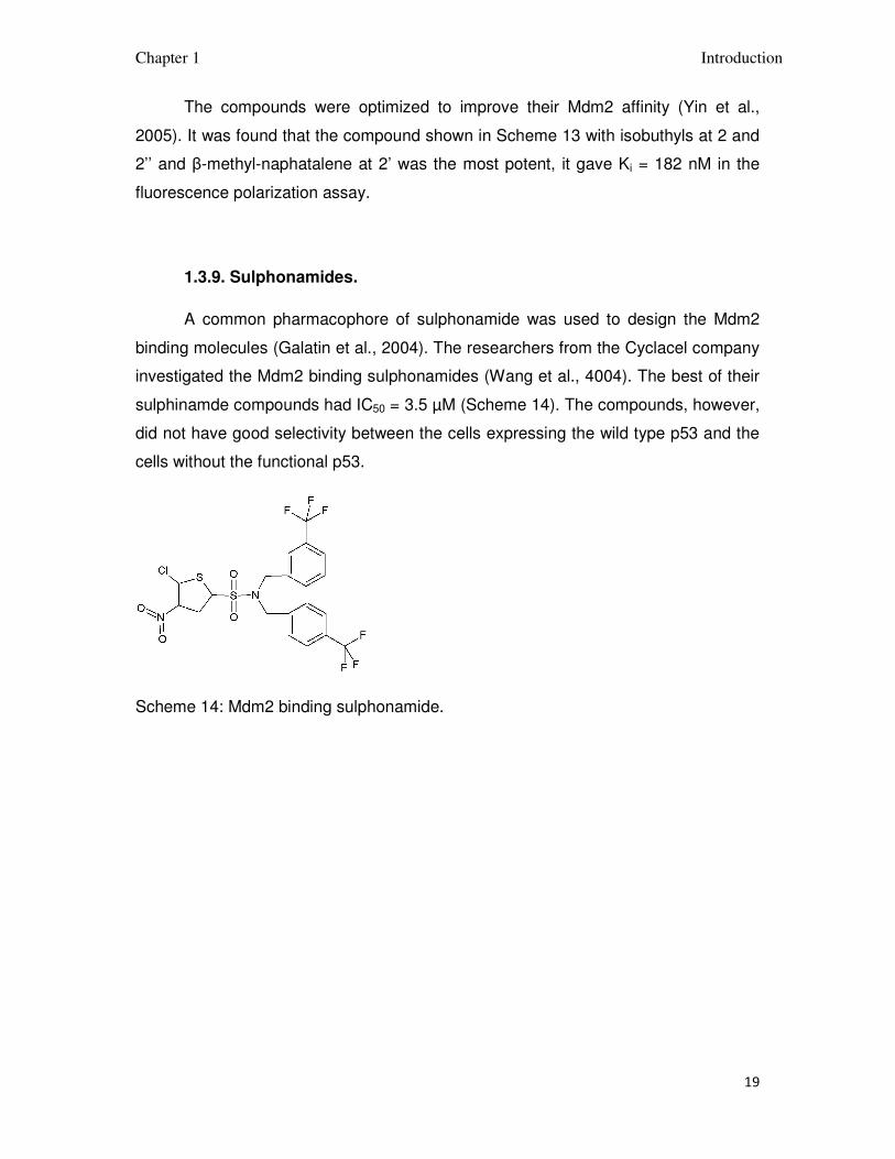
1.3.9. Sulphonamides.
A common pharmacophore of sulphonamide was used to design the Mdm2
binding molecules (Galatin et al., 2004). The researchers from the Cyclacel company
investigated the Mdm2 binding sulphonamides (Wang et al., 4004). The best of their
sulphinamde compounds had IC50 = 3.5 µM (Scheme 14). The compounds, however,
did not have good selectivity between the cells expressing the wild type p53 and the
cells without the functional p53.
Scheme 14: Mdm2 binding sulphonamide.
Chapter 2 Goals of the study
20
2. Goals of the study.
In many human cancers the tumor suppressor protein p53 retains its native
structure and could potentially inhibit tumor growth, though it is inactivated through
the binding to Mdm2 and Mdmx proteins. Small molecular weight organic molecules
capable to disrupt the complex of p53 with these proteins would release the active
wild type p53, which would prevent the cancer cells form uncontrolled proliferation.
The goal of this work was to develop novel, small molecule inhibitors of the
interaction between p53 and Mdm2/x proteins. The p53 binding pocket of Mdm2 was
chosen as a target for the designed molecules. The basic scaffolds of the molecules
were identified by the computer based approach and the hits were tested, and if
successful, further optimization was undertaken.
Chapter 3 Materials and methods
21
3. Materials and methods
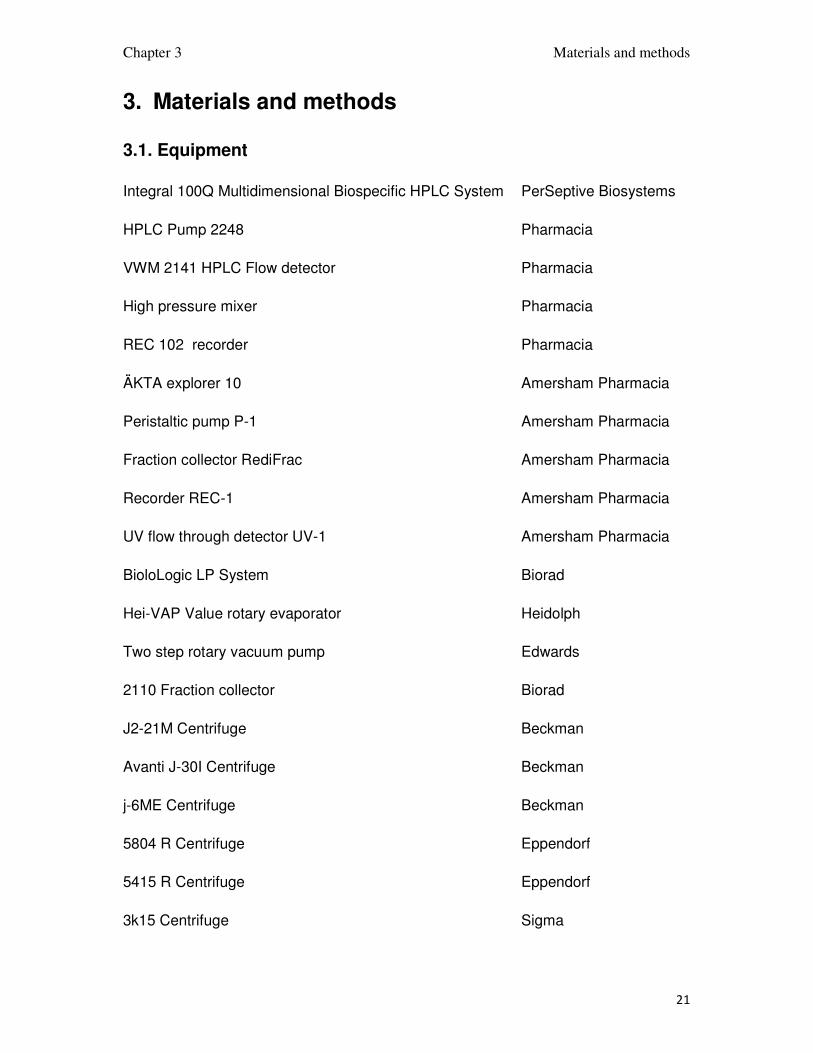
3.1. Equipment
Integral 100Q Multidimensional Biospecific HPLC System PerSeptive Biosystems
HPLC Pump 2248 Pharmacia
VWM 2141 HPLC Flow detector Pharmacia
High pressure mixer Pharmacia
REC 102 recorder Pharmacia
ÄKTA explorer 10 Amersham Pharmacia
Peristaltic pump P-1 Amersham Pharmacia
Fraction collector RediFrac Amersham Pharmacia
Recorder REC-1 Amersham Pharmacia
UV flow through detector UV-1 Amersham Pharmacia
BioloLogic LP System Biorad
Hei-VAP Value rotary evaporator Heidolph
Two step rotary vacuum pump Edwards
2110 Fraction collector Biorad
J2-21M Centrifuge Beckman
Avanti J-30I Centrifuge Beckman
j-6ME Centrifuge Beckman
5804 R Centrifuge Eppendorf
5415 R Centrifuge Eppendorf
3k15 Centrifuge Sigma

Chapter 3 Materials and methods
22
3.2. Columns
250/10mm 5µ HyPurity Advance Hypersil
VP 250/21 NUCLEODUR C18 ec Macherey-Nagel
Phusion-RP 80-A Phenomenex
Source 5RPC ST4,6/150 Pharmacia
Supelcosil LC-18 Supelco
HiLoad 26/60 Superdex S75pg Amersham Pharmacia
HiLoad 16/60 Superdex S75pg Amersham Pharmacia
HiLoad 16/60 Superdex S200pg Amersham Pharmacia
HiLoad 10/300 Superdex S75pg Amersham Pharmacia
HiLoad 10/300 Superdex S200pg Amersham Pharmacia
Mono Q HR 5/5, 10/10 Amersham Pharmacia
Mono S HR 5/5, 10/10 Amersham Pharmacia
NiNTA-agarose Qiagen
ProBond Resin Invitrogen
NTA Superflow Qiagen
Glutathione Sepharose 4 Fast Flow GE Healthcare
Octadecyl-Si100 Serva
µBondpack Phenyl Waters

Chapter 3 Materials and methods
23
3.3. Stock solutions
Ampicillin: 100 mg/ml of ampicillin dissoved in H2O, sterilized by filtration, stored in
aliquots at -20oC until used. Working concentration: 150 µg/ml.
Chloramphenicol: 34 mg/ml dissolved in ethanol (0.34 g/10 ml). Working
concentration: 34 µg/ml.
Streptomycin: 60 mg/ml of streptomycin dissolved in H2O, sterile filtered and stored
in aliquots at -20oC until usage. Working concentration 60 µg/ml.
Kanamycin: 100 mg/ml of kanamycin dissolved in H2O, sterile filtrated and stored in
aliquots at -20oC until used. Working concentration: 100 µg/ml.
IPTG: A sterile filtered 1 M stock of IPTG in dissolved distilled water was prepared
and stored in aliquots at -20oC until used.
Glucose: 20% solution in H2O, autoclaved.
Thiamin: 1% solution in H2O, sterilized by filtration.
MgSO4: 1 M solution in H2O, sterilized by filtration.
Zn-EDTA solution: 5 mg/ml EDTA
8.4 mg/ml Zn(CH3COO)2
Trace elements solution: 2.5 g/l H3BO3
2.0 g/l CoCl2 x H2O
1.13 g/l CuCl2 x H2O
9.8 g/l MnCl2 x 2H2O
2.0 g/l Na2MoO4 x 2H2O
pH lowered with citric acid or HCl.

Chapter 3 Materials and methods
24
3.4. Cell growth media
LB medium: 10 g/l tryptone
5 g/l yeast extract
10 g/l NaCl
pH was adjusted to 7.0. For the preparation of agar plates the medium was
supplemented with 15 g agar.
Minimal medium (MM) for uniform enrichment with 15N:
0.5 g/l NaCl
1.3 ml/l trace elements solution
1 g/l citric acid monohydrate
36 mg/l ferrous citrate
4.02 g/l KH2PO4
7.82 g/l K2HPO4 x 3H2O
1 ml/l Zn-EDTA solution
1 g/l NH4Cl or 15NH4Cl
pH was adjusted to 7.0 with NaOH, the mixture was autoclaved, upon cooling
separately sterilized solutions were added: 25 ml/l glucose, 560 µl/l thiamin,
antibiotics, 2 ml/l MgSO4 stock.
3.5. Buffers
3.5.1 Ion exchange and exclusion chromatography buffers
Buffer P(0) 8 mM KH2PO4
6 mM Na2HPO4
0.05% NaN3
Chapter 3 Materials and methods
25
pH 7.2
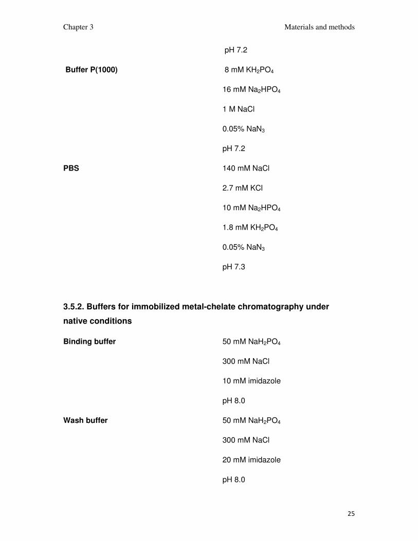
Buffer P(1000) 8 mM KH2PO4
16 mM Na2HPO4
1 M NaCl
0.05% NaN3
pH 7.2
PBS 140 mM NaCl
2.7 mM KCl
10 mM Na2HPO4
1.8 mM KH2PO4
0.05% NaN3
pH 7.3
3.5.2. Buffers for immobilized metal-chelate chromatography under
native conditions
Binding buffer 50 mM NaH2PO4
300 mM NaCl
10 mM imidazole
pH 8.0
Wash buffer 50 mM NaH2PO4
300 mM NaCl
20 mM imidazole
pH 8.0

Chapter 3 Materials and methods
26
Elution buffer 50 mM NaH2PO4
300 mM NaCl
250 mM imidazole
pH 8.0
3.5.3. Buffers for immobilized metal-chelate chromatography under
native conditions
Biding buffer 6 M guanidinium chloride
100 mM NaH2PO4 x H2O
10 mM Tris
10 mM β-mercaptoethanol
pH 8.0
Wash buffer 6 M guanidinium chloride
100 mM NaH2PO4 x H2O
10 mM Tris
10 mM β-mercaptoethanol
pH 6.5
Elution buffer 6 M guanidinium chloride
100 mM CH3COONa x 3H2O
10 mM β-mercaptoethanol
pH 4.0
Dialysis buffer 6 M guanidinium chloride

Chapter 3 Materials and methods
27
pH 3.0
Refolding buffer 200 mM arginine HCl
1 mM EDTA
100 mM Tris
2 mM reduced GSH
2 mM oxidized GSH
10% glycerol
0.05% NaN3
pH 8.4
3.5.4. Buffer for DNA agarose gel electrophoresis
50X TAE buffer
40 mM Tris-acetate 242 g/l of Tris base
1 mM EDTA 100 ml/l of 0.5 M EDTA (pH 8.0)
acetic acid 57.1 ml/l
2.5.5 Dyes for DNA electrophoresis
6x DNA Loading Dye Fermentas
SYBR ® Safe DNA gel stain Invitrogen
3.6. Reagents and buffers for the SDS-PAGE
Anode buffer 200 mM Tris pH 8.9
Cathode buffer 100 mM Tris pH 8.25
100 mM tricine
0.1% SDS

Chapter 3 Materials and methods
28
Separation buffer 1 M Tris pH 8.8
0.3% SDS
Stacking buffer 1 M Tris pH 6.8
0.3% SDS
Separation acrylamide 48% acrylamide
1.5% bis-acrylamide
Stacking acrylamide 30% acrylamide
0.8% bis-acrylamide
Separation gel 1.675 ml H2O
2.5 ml separation buffer
2.5 ml separation acrylamide
0.8 ml glycerol
25 µl 10% (NH4)2S2O8
2.5 µl TEMED
Intermediate gel 1.725 ml H2O
1.25 ml separation buffer
0.75 ml separation acrylamide
12.5 µl 10% (NH4)2S2O8
1.25 µl TEMED
Stacking gel 2.575 ml H2O
0.475 ml stacking buffer
0.625 ml stacking acrylamide
12.5 µl 0.5 M EDTA, pH 8.0

Chapter 3 Materials and methods
29
37.5 µl 10% (NH4)2S2O8
1.9 µl TEMED
Protein visualization
Coomassie-blue solution 45% ethanol
10% acetic acid
1% Coomassie Brilliant Blue R
Destaining solution 5% ethanol
10% acetic acid
5x protein loading dye: 0.225 M Tris-HCl, pH 6.8
50% glycerol
5% SDS
0.05% bromophenol blue
0.25 M DTT
3.7. E. coli strains and plasmids
Cloning strains
One Shot TOP10 Invitrogen
GigaSingles Novagen
Protein expression strains
BL21 Star Invitrogen
BL21 Star(DE3) Invitrogen
Chapter 3 Materials and methods
30
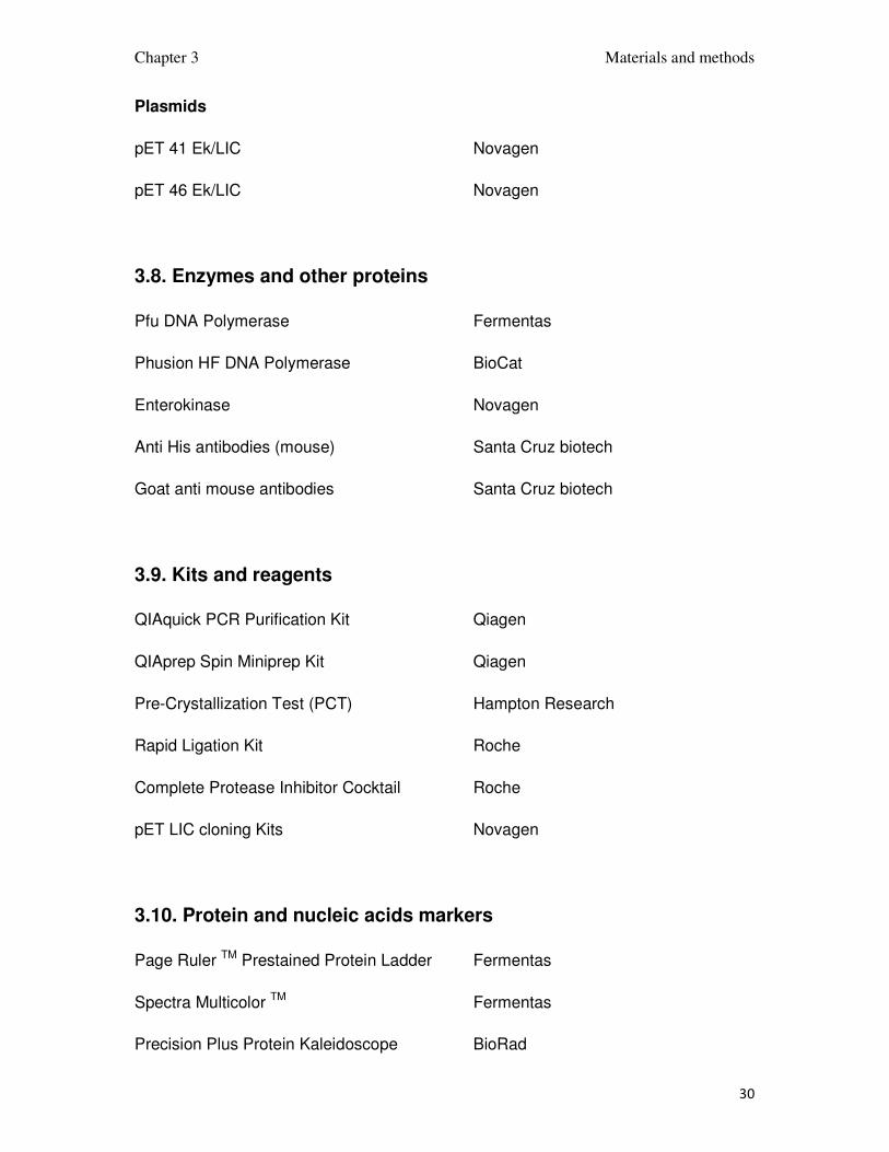
Plasmids
pET 41 Ek/LIC Novagen
pET 46 Ek/LIC Novagen
3.8. Enzymes and other proteins
Pfu DNA Polymerase Fermentas
Phusion HF DNA Polymerase BioCat
Enterokinase Novagen
Anti His antibodies (mouse) Santa Cruz biotech
Goat anti mouse antibodies Santa Cruz biotech
3.9. Kits and reagents
QIAquick PCR Purification Kit Qiagen
QIAprep Spin Miniprep Kit Qiagen
Pre-Crystallization Test (PCT) Hampton Research
Rapid Ligation Kit Roche
Complete Protease Inhibitor Cocktail Roche
pET LIC cloning Kits Novagen
3.10. Protein and nucleic acids markers
Page Ruler TM Prestained Protein Ladder Fermentas
Spectra Multicolor TM Fermentas
Precision Plus Protein Kaleidoscope BioRad

Chapter 3 Materials and methods
31
Gene Ruler TM DNA Ladder Fermentas
100 BP DNA marker New England BioLabs
1Kb DNA marker New England BioLabs
3.11. Substrates for chemical synthesis
3.11.1. 6-chloroisatin
6-chloroisatin was prepared in two step synthesis as it is shown on Scheme 15
analogously to the published synthesis of isatin (Marvel and Hiers, 1925). In a 20 l.
round-bottomed flask were placed 180 g (1.08 mol) of chloral hydrate, 4 l of water
and 1.5 kg of anhydrous sodium sulfate. Obtained mixture was stirred for 1 h to
dissolve sodium sulfate. In 1 l beaker equipped with magnetic stirrer 86 ml (1.04 mol)
of concentrated hydrochloric acid was diluted with 600 ml water and added stepwise
127 g (1 mol) of 3-chloroaniline. Obtained solution was added to the reaction
mixture in 20 l flask and stirred. Finally a solution of 220 g (3.16 mol) of
hydroxylamine hydrochloride in 1 l of water was added. The solution was refluxed 20
min on the heating mushroom. After one to two minutes of vigorous boiling the
reaction is complete. The 3-chloro-isonitrosoacetanilide precipitated as a porous
yellow-brown substance. The mixture was cooled to room temperature.
The 3-chloro-isonitrosoacetanilide was filtered off, washed with 1 l of water
and dried for a week under vacuum.
652 ml of concentrated sulfuric acid was warmed to 50oC in a 2 l round-
bottomed flask equipped with magnetic stirrer and, 3-chloro-isonitrosoacetanilide
obtained in the previous step was added gradually while the flask was ice-cooled to
keep the temperature below 70oC. After the compound was added, the solution was
heated to 80oC for 15 minutes.

Chapter 3 Materials and methods
32
Scheme 15: The synthesis of 6-chloroisatin.
The mixture was poured into a 2 l beaker with ice and stirred. The product was
filtered, washed with 1 l of water and dried under vacuum for 2 days. Except of the
desired 6-chloroisatin, some 4-chloroisatin was expected to form, the purity amount
of 6-chloroisatin was checked on the silica TLC with solvent mixture of ethyl
acetate/heptan in ratios: 1/5, 1/1, 5/1 . The photos of the TLC plates in the UV light
are shown in Figure 8.
Figure 8: TLC plates with separated products of the reaction of Scheme 15.

Chapter 3 Materials and methods
33
The reaction produced approximately 50% of 6-chloroisatin and 50% of 4-
chloroisatin. The isomers of chloroisatin were separated on a silica column with
mixture of ethyl acetate/heptane 5/1 as a solvent. 6-chloroisatin was eluted as the
first yellow band. The identity of 6-chloroisatin was confirmed by mass spectrometry
and NMR. The 1D 1H NMR spectrum and the peak assignment is shown in Figure 9.
Figure 9: The NMR 1D proton spectrum of 6-chloroisatin and the NMR peak
assignment, the peak positions are in ppm. The peak at 2.07 ppm comes from the
residual acetone, and the one at 2.84 ppm from water.

Chapter 3 Materials and methods
34
3.11.2. Cyclopropylurea
Cyclopropylureas was prepared analogously to described protocol for
cyclohexylurea (Kehm et al., 1963). In a 250 ml round bottomed flask equipped with
magnetic stirrer 15 ml of concentrated hydrochloric acid was dissolved in 150ml
water. 10 ml (0.15 mol) of cyclopropylamine was added stepwise while stirring. After
the addition of whole cyclopropylamine the solution was heated to 60oC. While slowly
stirred 15 g (0.185 mol) of potassium cyanate was added in small steps to avoid
releasing a gas bubbles.
After adding potassium cyanate the reaction was heated for 1 h to complete the
reaction.
The product was extracted into three volumes of ethyl acetate 500 ml each.
The solvent was evaporated on rotary evaporator and the product was dried on air for
2 days.
Scheme 16: The synthesis of cyclopropylurea.
The product’s identity was checked by NMR.
3.11.3. 4-aminoimidazole
4-aminoimidazole was supposed to be obtained in two step synthesis shown
on Scheme 17. The nitration of imidazole was done as published (Novikov et al.,
1970). In 500 ml round bottomed flask 100 ml concentrated nitric acid was mixed with
100 ml concentrated sulfuric acid. 50 g (0.73 mol) imidazole was added stepwise
while the mixture was stirred. After the addition of imidazole the solution was refluxed
for 1 h to complete the reaction. The mixture was cooled to room temperature and

Chapter 3 Materials and methods
35
poured on ice. The product was filtered off, washed with water and dried under
vacuum for a day. 4-nitroimidazole was obtained as a white powder.
The reduction was done as published (Fargher, 1920). In 500 ml round
bottomed ice cooled flask equipped with magnetic stirrer was placed 33 g (0.3 mol) of
4-nitroimidazole and 100 g concentrated hydrochloric acid. The solution of 70 g (0.37
mol) tin(II) chloride in 160 ml concentrated hydrochloric acid was added dropwise
while the mixture in the flask was stirred and it's temperature was maintained below
5oC. After the complete addition of tin (II) chloride the mixture was stirred and kept in
the temperature below 5oC for 1 h. The volume of solution was decreased by
evaporation and the crystals of salts were collected. In the remaining dark-brown oil
4-aminoimidazole was detected by mass spectrometry, but the purification was
unsuccessful.
Scheme 17: The synthesis of 4-aminoimidazole.
3.11.4. 6-chloroindole-3-aldehyde
6-chloroindole-3-aldehyde was prepared analogously to the indole-3-aldehyde
which synthesis was published (James and Snyder, 1959). In a 100 ml round
bottomed flask equipped with stirrer 3 g (0.02 mol) of 6-chloroindole was dissolved in
N,N-dimethylformamide. The mixture was stirred and the flask was cooled on ice and
4 ml (0.04 mol) of phosphorus (V) oxychloride was added dropwise in such a rate
that the temperature of the reaction mixture did not rise over 5oC. The mixture was
cooled and stirred for 1 h and stirred at room temperature for 2 h.

Chapter 3 Materials and methods
36
Scheme 18: The synthesis of 6-chloroidole-3-aldehyde
After that time the solution was poured into 300 ml of water and was allowed
to precipitate for 5 days. The product was filtered, washed with water and dried under
vacuum for a day. The purity of the product was checked by TLC, the image of the
plate is shown in Figure 10. The product seems to be pure, only a single band is
visible in TLC.
Figure 10: TLC pf 6-chloroindole-3-aldehyde.
The identity of the product was confirmed by MS and NMR. The 1D NMR
proton spectrum and the peak assignments are shown in Figure 11.

Chapter 3 Materials and methods
37
Figure 11: The NMR 1D proton spectrum of 6-chloroisatin and the NMR peak
assignments, the peak positions are in ppm. The peak at 2.07 ppm originates from
the residual acetone, and the one at 2.88 ppm from water.
3.11.5. 3-(dicyano)-methyloxindole
3-(dicyano)-methylox-6-chloroindole was synthesized as published (Walter
1902). In 400 ml beaker 20 g (0.13 mol) of 6-chloroisatin and 10 g (0.15 mol) of
malononitrile were dissolved in 200 ml ethanol. The solution was stirred and 3 drops
of 36% sodium hydroxide were added. The solution was stirred for overnight.
After the reaction was completed the solution was boiled to dissolve the
substance and slowly cooled. The solution was kept for overnight in refrigerator for
product crystallization. 3-(dicyano)-methylox-6-chloroindole formed dark purple
crystals which were filtered out, washed with water and dried under vacuum. The
identity of the substance was confirmed by mass spectrometry and its purity was

Chapter 3 Materials and methods
38
investigated by HPLC on silica, the result suggested that only 6-chloroisatin reacted
and 4-chloroisatin which was the impurity did not reacted or was removed during the
crystallization of the product.
Scheme 19: The synthesis of 3-(dicyano)-methylox-6-chloroindole.
3.11.6. 4-chloroisatoic anhydride
4-chloroisatoic anhydride was prepared from 6-chloroisatin as published
(Reissenweber, 1980). In 250 ml round bottomed flask equipped with magnetic stirrer
10 g (0.055 mol) of 6-chloroisatin was dissolved in 100 ml of concentrated acetic
acid. The solution was stirred and 10 ml of 30% hydrogen peroxide was added
dropowise. The reaction was the stirred for 30 minutes to complete.
The reaction mixture was poured into water and the product was filtered off,
washed with water and dried under vacuum. The identity of the compound was
checked on mass spectrometry and its purity on analytical RP HPLC.
Scheme 20: The synthesis of 4-chloroisatoic anhydride.

Chapter 3 Materials and methods
39
3.11.7 Aliphatic cyanoacetamides
Various cyanoacetamides needed for further synthesis were prepared
according to the published method (Wang et al., 2009). In 100 ml beaker equipped
with stirrer was placed 0.2 mol of primary aliphatic amine and 0.2 mol of methyl
cyanoacetate. The mixture was stirred for 3-16 h at room temperature, during this
time white crystals of product appeared.
After the reaction was completed the product was filtered off, washed with
ethyl ether and dried on air.
Scheme 21: An example of cyanoacetamide synthesis.
3.11.8. The synthesis of methoxyethyl acetoacetamide using 2,2,6-trimethyl-4H-
1,3-dioxin-4-one.
Methoxyethyl acetoacetate was prepared as published (Clemens and Hyatt,
1984). 50 ml of methoxyethylamine, 100 ml of 2,2,6-trimethyl-4H-1,3-dioxin-4-one
acetone solution and 100 ml of toluene were placed in a pressure container with
magnetic stirrer. The container was placed in a heating coat placed on the magnetic
stirrer. The reaction was heated to 140 oC for 6 h. After that time the container was
removed from the heating coat and allowed to cool for overnight and when cold it was
opened and the inner container with the substance.
The product was purified by fractional distillation under vacuum. The fractions
were identified by mass spectrometry, the desired product was found in the fraction
boiling at 130 oC. The product was washed with 50 ml water and dried under vacuum
for a day.

Chapter 3 Materials and methods
40
Scheme 22: The synthesis of methoxyethyl acetoacetamide using 2,2,6-trimethyl-4H-
1,3-dioxin-4-one as a substrate.
3.11.8. The microwave synthesis of aromatic acetoacetamides
The synthesis of some aromatic acetoacetamides needed for further synthesis
was performed as published (Suri et al., 2000). In 1 L flask were placed 0.2 mol of
primary aromatic amine and 1 mol of ethyl acetoacetate. The flask was placed
opened in a microwave and irradiated for 2-15 min at 800W.
The product was washed with ethyl ether, and dried. The identity of the
product was checked on mass spectrometry.
Scheme 23: An example of a microwave synthesis of aromatic acetoacetamide.
3.11.9. The synthesis of acetoacetamides with diketene
Acetoacetamides which could not be easily synthesized and purified using the
methods described above were prepared analogously to the published protocol
(Williams and Krynitsky 1955). In 250ml round bottomed flask equipped with
magnetic stirrer was placed 0.1 mol of amine and 100 ml of dichloromethan or ethyl
ether depending of the amine solubility in both of them. The low boiling solvent added
in large amount helped to not overheat the reaction and was easy to remove after the
reaction was completed. While the amine solution was vigorously stirred under the

Chapter 3 Materials and methods
41
fume hood 0.1 mol of diketene was added dropwise, the heat formed by the reaction
was removed by boiling solvent.
The excess of solvent was removed on rotary evaporator in case of
dichloromethane or by slow evaporation on air in case of ethyl ether. The identity of
acetoacetamides was confirmed on mass spectrometry.
Scheme 24: An example of a synthesis of acetoacetamide using diketene as a
substrate.
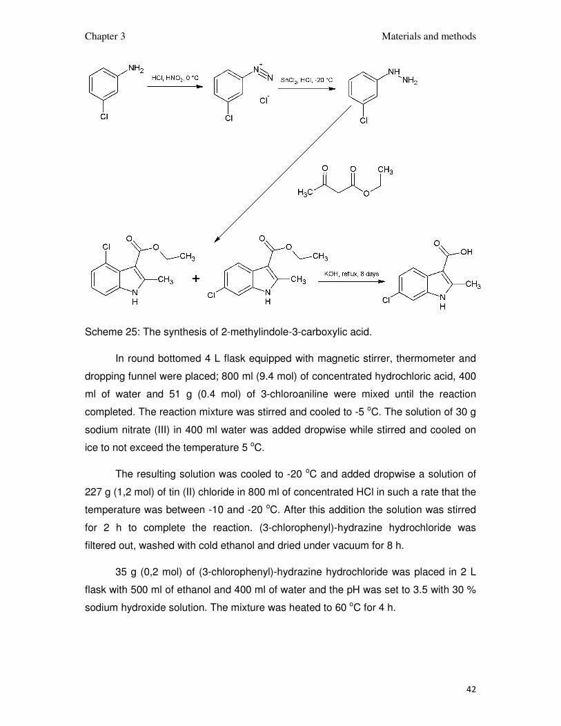
3.11.10. The synthesis of indole-3-carboxylic acid
The synthesis was done in 4 steps presented on scheme 25. First the
diazonium compound was prepared, then reduced to substituted hydrazine, from
which the 2-methyl-6-chloroidole-3-carboxylic acid's ester was prepared and
hydrolysed in last step. First 3 steps were done analogously to the published
synthesis (Brodfuehrer et al., 1997).
Chapter 3 Materials and methods
42
Scheme 25: The synthesis of 2-methylindole-3-carboxylic acid.
In round bottomed 4 L flask equipped with magnetic stirrer, thermometer and
dropping funnel were placed; 800 ml (9.4 mol) of concentrated hydrochloric acid, 400
ml of water and 51 g (0.4 mol) of 3-chloroaniline were mixed until the reaction
completed. The reaction mixture was stirred and cooled to -5 oC. The solution of 30 g
sodium nitrate (III) in 400 ml water was added dropwise while stirred and cooled on
ice to not exceed the temperature 5 oC.
The resulting solution was cooled to -20 oC and added dropwise a solution of
227 g (1,2 mol) of tin (II) chloride in 800 ml of concentrated HCl in such a rate that the
temperature was between -10 and -20 oC. After this addition the solution was stirred
for 2 h to complete the reaction. (3-chlorophenyl)-hydrazine hydrochloride was
filtered out, washed with cold ethanol and dried under vacuum for 8 h.
35 g (0,2 mol) of (3-chlorophenyl)-hydrazine hydrochloride was placed in 2 L
flask with 500 ml of ethanol and 400 ml of water and the pH was set to 3.5 with 30 %
sodium hydroxide solution. The mixture was heated to 60 oC for 4 h.

Chapter 3 Materials and methods
43
After the reaction was completed ethanol was removed on rotary evaporator
and the product was extracted with ethyl acetate. The organic phase was washed
with water, evaporated on rotary evaporator and dried in vacuum for overnight.
In 1 L round bottomed flask equipped with magnetic stirrer 20 g (0.09 mol) of
the obtained mixture of esters was placed with 400 ml of water and 160 g (4 mol) of
potassium hydroxide. The mixture was vigorously stirred and refluxed for 8 days.
After this time the reaction mixture was cooled to room temperature. The
remaining substrate was extracted with chlorophorm. Water phase was moved into 1
L beaker with 500 ml of ethyl acetate. While stirred the excess of acetic acid was
added and the product was extracted into organic phase.
The solution of the product was evaporated on rotary evaporator, than the
substance was dried in vacuum for a day.
3.12. Proteins
3.12.1. Mdm2 preparation and purification
The recombinant human Mdm2 protein containing the first 118 N-terminal
aminoacids was obtained in Escherichia coli BL21(DE3) RIL expression system using
a pET46 Ek/LIC vector. The bacteria were grown at 37oC in LB medium with 100 ml/L
of ampicillin and 34 mg/L of chloramphenicol for unlabeled protein or in minimal
medium for uniform enrichment with 15N with 50 mg/L of ampicillin and 17 mg/L of
chloramphenicol if labeled protein was needed. The bacteria were induced at an
OD600nm of 0.7-0.8 for with 1 mM of IPTG. The protein was expressed for 4 h at
37oC.
The cells were centrifuged at 5000 rpm for 15 min and the pelet was
suspended in PBS buffer. The bacteria were lysed by sonication at 0oC. The lysat
was centrifuged at 12000 rpm for 30 minutes. The protein was expressed into
inclusion bodies which were inside the pelet. The inclusion bodies were washed twice
with PBS buffer containing 0.05% Triton X-100 with subsequent centrifugation at
12000 rpm for 30 min. The inclusion bodies were dissolved in the buffer with pH 8.0
containing 6 M guanidine hydrochloride, 100 mM tris 1 mM EDTA and 10 mM DTT

Chapter 3 Materials and methods
44
and centrifuged at 20000 rpm for 40 min to remove cell membranes. The protein
solution was dialysed for overnight into the buffer with pH 3.5 containing 4 M
guanidine hydrochloide, 100 mM tris, 1 mM EDTA and 10 mM DTT. The protein was
refolded by dropwise dilution 1:100 into the buffer with pH 7.0 containing 10 mM tris,
1 mM EDTA and 10 mM DTT. After dilution the solution was stirred for overnight at
4oC to complete the refolding. (NH4)2SO4 was added to concentration 1.5 M and the
solution was stirred for 1 h, then 7-10 ml Buthyl Sepharose 4 Fast Flow was added
and the mixture was stirred for 2 h. The refolded protein was eluted with pH 7.2 buffer
containing 0.1 M tris and 5 mM DTT. The final step of purification was done by gel
filtration on HiLoad 26/60 Superdex 75 pg column with the running buffer containing
50 mM KH2PO4, 50 mM Na2HPO4, 150 mM NaCl 5 mM DTT. The Mdm2's elution
volume was compared with the standards, it eluted between 13 kDa and 25 kDa
standards, what is expected from 14 kDa protein. The fractions containing
monomeric Mdm2 were pooled.
Plot 1: Chromatogram of size exclusion chromatography of MDM2 protein.
The mass of the protein was confirmed by gel electrophoresis in denaturing
conditions.

Chapter 3 Materials and methods
45
3.12.2. The preparation and purification of the complex of Mdm2 and p53
Human P53 protein containing the first 321 N-terminal aminoacids was
obtained in Escherichia coli BL21(DE3) RIL expression system using a pET46 Ek/LIC
vector. The bacteria were grown at 37oC in LB medium with 100 mg/L of ampicillin
and 34 mg/L of chloramphenicol. They were induced at an OD600nm of 0.7-0.8 for
with 1 mM of IPTG. The protein was expressed for 12 h at 20oC.
The cells were centrifuged at 5000 rpm for 15 min and the pelet was
suspended in binding buffer for immobilized metal-chelate chromatography in native
conditions. The bacteria were lysed by sonication at 0oC. The lysat was centrifuged
at 20000 rpm for 30 minutes. The protein was bound to Ni-NTA column at 4oC,
washed with wash buffer until steady absorbance at 280 nm and then eluted with
elution buffer. The final step of purification was done by gel filtration on HiLoad 26/60
Superdex 200 column with the running buffer with pH 0.2 containing 10 mM,
Na2HPO4, 1.8 mM KH2PO4, 140 mM NaCl, 2.7 mM KCl, 0.05% NaN3, 5 mM DTT.
Plot 2: Chromatogram of size exclusion chromatography of p53 protein.

Chapter 3 Materials and methods
46
P53 is eluted earlier than standards with similar mass because it is not folded
and can have a different shape than globular.
The Mdm2-p53 complex was prepared by mixing p53 with excess of Mdm2. It
was purified by gel filtration on HiLoad 16/60 Superdex 200 column with running
buffer containing 10 mM Na2HPO4, 1.8 mM, KH2PO4, 140 mM NaCl, 2.7 mM KCl,
0.05% NaN3, 5 mM β-mercaptoethanol, pH 7.2. During this step the excess of Mdm2
was removed.
Plot 3: Chromatogram of size exclusion chromatography of p53-Mdm2 complex.
3.13. Mdm2's ligands synthesis
3.13.1. Synthesis and purification of 3,4-dihydripyrimidin-2(1H)-ones
3,4-dihydripyrimidin-2(1H)-ones were prepared accordingly to the method
published by (Ryabukhin et al., 2007). 5 mmol p-chlorobenzaldehyde, 5 mmol
acetoacetate or acetoacetamide and 5 mmol the N-substituted urea were placed in a
50ml round bottomed flask and dissolved in 20 ml of N,N-dimethylformamide. After
obtaining clear solution 20 mmol of trimethylchlorosilane was added dropwise while
stirring. The mixture was stirred for 3 days to complete the reaction.
Chapter 3 Materials and methods
47
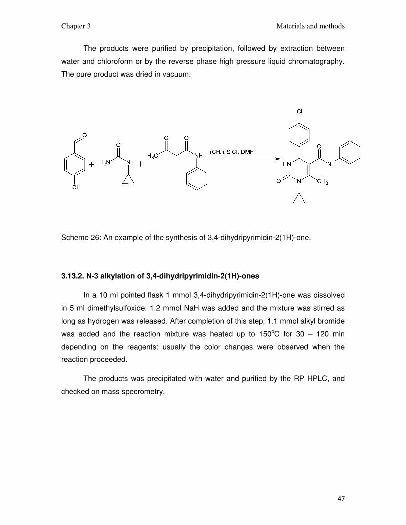
The products were purified by precipitation, followed by extraction between
water and chloroform or by the reverse phase high pressure liquid chromatography.
The pure product was dried in vacuum.
Scheme 26: An example of the synthesis of 3,4-dihydripyrimidin-2(1H)-one.
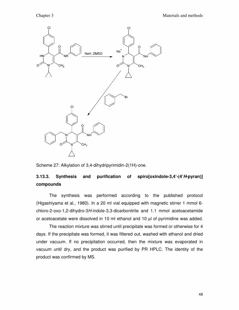
3.13.2. N-3 alkylation of 3,4-dihydripyrimidin-2(1H)-ones
In a 10 ml pointed flask 1 mmol 3,4-dihydripyrimidin-2(1H)-one was dissolved
in 5 ml dimethylsulfoxide. 1.2 mmol NaH was added and the mixture was stirred as
long as hydrogen was released. After completion of this step, 1.1 mmol alkyl bromide
was added and the reaction mixture was heated up to 150oC for 30 – 120 min
depending on the reagents; usually the color changes were observed when the
reaction proceeded.
The products was precipitated with water and purified by the RP HPLC, and
checked on mass specrometry.
Chapter 3 Materials and methods
48
Scheme 27: Alkylation of 3,4-dihydripyrimidin-2(1H)-one.
3.13.3. Synthesis and purification of spiro[oxindole-3,4’-(4’H-pyran)]
compounds
The synthesis was performed according to the published protocol
(Higashiyama et al., 1980). In a 20 ml vial equipped with magnetic stirrer 1 mmol 6-
chloro-2-oxo-1,2-dihydro-3H-indole-3,3-dicarbonitrile and 1.1 mmol acetoacetamide
or acetoacetate were dissolved in 10 ml ethanol and 10 µl of pyrimidine was added.
The reaction mixture was stirred until precipitate was formed or otherwise for 4
days. If the precipitate was formed, it was filtered out, washed with ethanol and dried
under vacuum. If no precipitation occurred, then the mixture was evaporated in
vacuum until dry, and the product was purified by PR HPLC. The identity of the
product was confirmed by MS.

Chapter 3 Materials and methods
49
Scheme 28: An example synthesis of spiro[oxindole-3,4’-(4’H-pyran)].
3.13.4. The synthesis and purification of 2,3-dihydroquinazolin-4(1H)-ones and
7-chloro-2,3-dihydroquinazolin-4(1H)-ones
The substances were prepared analogously to the published protocol (Dabiri
et al., 2005). Preparation of 2,3-dihydroquinazolin-4(1H)-ones was carried out in the
ethanol solution with KAl(SO4)2 as a catalyst 20 ml of absolute ethanol was placed in
a 50 ml round bottomed flask, 2 mmol of isatoic anhydride or its derivative, 2 mmol of
a primary amine, 2 mmol of aryl aldehyde and 200 mg KAl(SO4)2 were added, and
the reaction mixture was stirred and the mixture was refluxed for 6 h for 2,3-
dihydroquinazolin-4(1H)-ones and 12h for 7-chloro-2,3-dihydroquinazolin-4(1H)-ones.
Scheme 29: An example of the synthesis of 2,3-dihydroquinazolin-4(1H)-one.
Chapter 3 Materials and methods
50
After the reaction was completed, the mixture was poured into water, and
depending on the precipitate´s shape, centrifuged or filtered and washed with water.
The substance was then either recrystalized from ethanol and dried under vacuum
for a day or purified on the RP HPLC and dried.
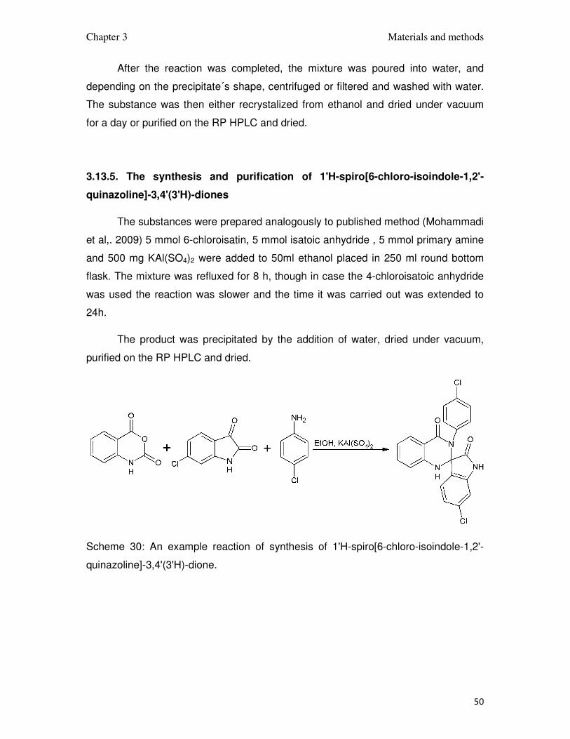
3.13.5. The synthesis and purification of 1'H-spiro[6-chloro-isoindole-1,2'-
quinazoline]-3,4'(3'H)-diones
The substances were prepared analogously to published method (Mohammadi
et al,. 2009) 5 mmol 6-chloroisatin, 5 mmol isatoic anhydride , 5 mmol primary amine
and 500 mg KAl(SO4)2 were added to 50ml ethanol placed in 250 ml round bottom
flask. The mixture was refluxed for 8 h, though in case the 4-chloroisatoic anhydride
was used the reaction was slower and the time it was carried out was extended to
24h.
The product was precipitated by the addition of water, dried under vacuum,
purified on the RP HPLC and dried.
Scheme 30: An example reaction of synthesis of 1'H-spiro[6-chloro-isoindole-1,2'-
quinazoline]-3,4'(3'H)-dione.

Chapter 3 Materials and methods
51
3.14. Laboratory procedures
3.14.1. Preparation of chemically competent cells
A single colony of overnight grown bacteria from a LB agar plate was grown in
20 ml of LB media in a 100 ml flask at 37oC until OD ~0.6. The bacteria were
transferred to sterile, disposable, ice-cold 50 ml polypropylene tube and cooled down
to 4oC on ice for 10 min. they were centrifuged at 5000 rpm for 10 min at 4oC.
Supernatant was removed and the pelets were resuspended in 10 ml of the ice-
cooled 0.1 M MgCl2 solution. Again, cells were centrifuged at 5000 rpm for 10 min at
4oC. Supernatant was removed and the cells were resuspended in 5 ml of ice-cooled
solution containing 0.1 M CaCl2 and 15% glycerol. At the end the bacteria were
aliquoted of 50 µl into 2ml sterile, polypropylene tubes and frozen in liquid nitrogen.
The aliquots were stored at -80oC.
3.14.2. In silico screening
The ZINC database (Irwin and Shoichet, 2005) was filtered to create two
subsets. First subset contained compounds with two p-halogenated phenyl rings,
second one compounds with indole moiety double substituted at possition 3 and
unsubstituted at positions 4, 5 and 7. Sets were subsequently docked to Mdm2
receptor protein (PDB ID: 1YCR, Kussie et al.,1996) using DOCK software (Ewing et
al.,1997). Docked ligands were inspected manually. Two families of compounds were
chosen for in vitro testing. One family was based on 3,4-dihydripyrimidin-2(1H)-one
scaffold, the second on spiro[oxindole-3,4’-(4’H-pyran)].
3.14.3. Ligand optimization
Confirmed in-silico hits were optimized by creating small, virtual derivative
libraries that were subsequently docked to the Mdm2 structure (PDB ID: 1RV1 chain
B) using eHits software (SimBioSys Inc, Zsolt Zsoldos et al., 2007). Compounds that
have good score and probable binding poses were synthesized.

Chapter 3 Materials and methods
52
3.14.4. Fluorescence polarization measurements
Compounds were measured for their ability to disrupt Mdm2-p53 complex by
fluorescence polarization assay as described previously (Czarna et al., 2009). Briefly,
the fluorescent-labelled peptide was bound to Mdm2 protein. The amount of peptide
displaced by a measured compound was measured by observing polarization of
emitted light after excitation with polarized beam. For each compound a 12-point
measurement curve was prepared for the concentrations ranging from 200 µM to
100nM. IC50 was calculated by fitting a 4-parameter logistic curve to the measured
points. Ki was calculated according to (Huang, 2003).
3.14.4. NMR Measurements
Molecules that were showing good affinity in fluorescence polarization assay
were subjected to NMR assay called AIDA (Antagonist-Induced Dissociation Assay)
using previously published methodology. (D’Silva et al., 2005). Briefly, all NMR
spectra were acquired at 300K on a Bruker DRX 600 MHz spectrometer equipped
with a cryoprobe. Typically, protein samples of p53-Mdm2 complex obtained from gel
filtration, as described before were concentrated or diluted to the desired
concentration of 10 to 30 µM and mixed with up to 10% (v/v) of D2O. Stock solutions
of the compounds used in the titrations were prepared in D6-dimethyl sulfoxide. The
spectra were processed with the Topspin software version 1.3. 1D 1H spectra were
recorded using SEI pulse sequence (2) and total 300 ms of acquisition and relaxation
period. A total of 4k points were acquired during t1 evolution, zero filled to 32k and
subjected to Fourier transform and polynominal baseline correction.
3.14.5. Electrophoresis of DNA on agarose gel
1% agarose in a 30 - 100 ml TAE buffer was heated until agarose dissolved.
3-10 µl of SYBR stain was added and stirred to get homegenous sulution, which was
then poured into the electrolyser and allowed to coagulate.
Chapter 3 Materials and methods
53
The DNA samples were mixed with the 6x sample buffer prior to loading. DNA
samples were run along with the 1 kb DNA ladder (NEB or pEQ lab) at 100-120 V
DC. Results were vizualized using UV illumination.
3.14.6. Transformation of chemically competent cells
1 - 5 µl of plasmid DNA solution was added to 50 µl of chemically competent
cells. The mixture was incubated on ice for 30 min followed by a heat shock of 45 s at
42oC and 2 min cooling on ice. After this 250 µl of LB medium was added and the
cells were incubated for 1 h of at 37oC. 50-100 µl of the mixture was spread out on
LB agar plates containig the anitbiotics on which the bacteria should be resistant
after the transformation. The plates were kept for overnight at 37oC.
The plasmid used for transformation contained the gene responsible for an
antibiotic resistance and only the bacteria which contained the plasmid grew on the
plate forming colonies. Some of the colonies were chosen to further research.
3.14.7. Sonication
Sonication was used to disrupt bacteria's cell membrains. Sonication was
carried out on ice, in the sonication flask. It was done in 3 steps, 5 min each, with 5
min intervals between steps, to avoid overheating of the sample.
3.14.8. Ni affinity chromatography
Nickel affinity chromatography was used as a first step of purification of
soluble proteins with His-tag. The pH of the solution containing the protein was
adjusted to 8.0 with 1 M NaOH, which is optimal for the binding to the Ni-NTA resin.
A Ni-NTA slurry was added and the mixture was agitated for 1-2 h. The ratio of the
Ni-NTA resin used to the amount of the His-tagged protein is crucial for purity of the
protein. It is more efficient to use less resin and perform a stepwise elution, obtaining
a pure, concentrated protein in a shorter time.
Chapter 3 Materials and methods
54
3.14.9. Electrophoresis in SDS polyacrylamide gel
The SDS polyacrylamide gel electrophoresis was performed at various stages
of purification to check the purity and identity of proteins. It was done according to
published procedure (Schagger and von Jagow, 1987). The protein samples were
prepared by mixing 10 µl of protein solution with 10 µl of sample buffer and heating
for 5 min at 100oC.
3.14.10. Visualization of proteins on polyacrylamide gel
The gel was stained in a Coomassie-blue solution with microwave irradiation.
Background was cleared by boiling the gel in an ethanol - water mixture in the
microwave. If not cleared enough the solution was changed to fresh one and boiling
was repeated.

Chapter 4 Results and discussion
55
4. Results and discussion
4.1. 3,4-dihydripyrimidin-2(1H)-ones and spiro[oxindole-3,4’-(4’H-
pyran)]es
Two of the highest ranking hits from docking; one 3,4-dihydripyrimidin-2(1H)-
one shown in Figure 12 and one spiro[oxindole-3,4’-(4’H-pyran)] shown in Figure 13
were chosen for optimization.
Figure 12: The formula of 3,4-dihydripyrimidin-2(1H)-one, named synt10, and the
model of its interaction with Mdm2.
Chapter 4 Results and discussion
56
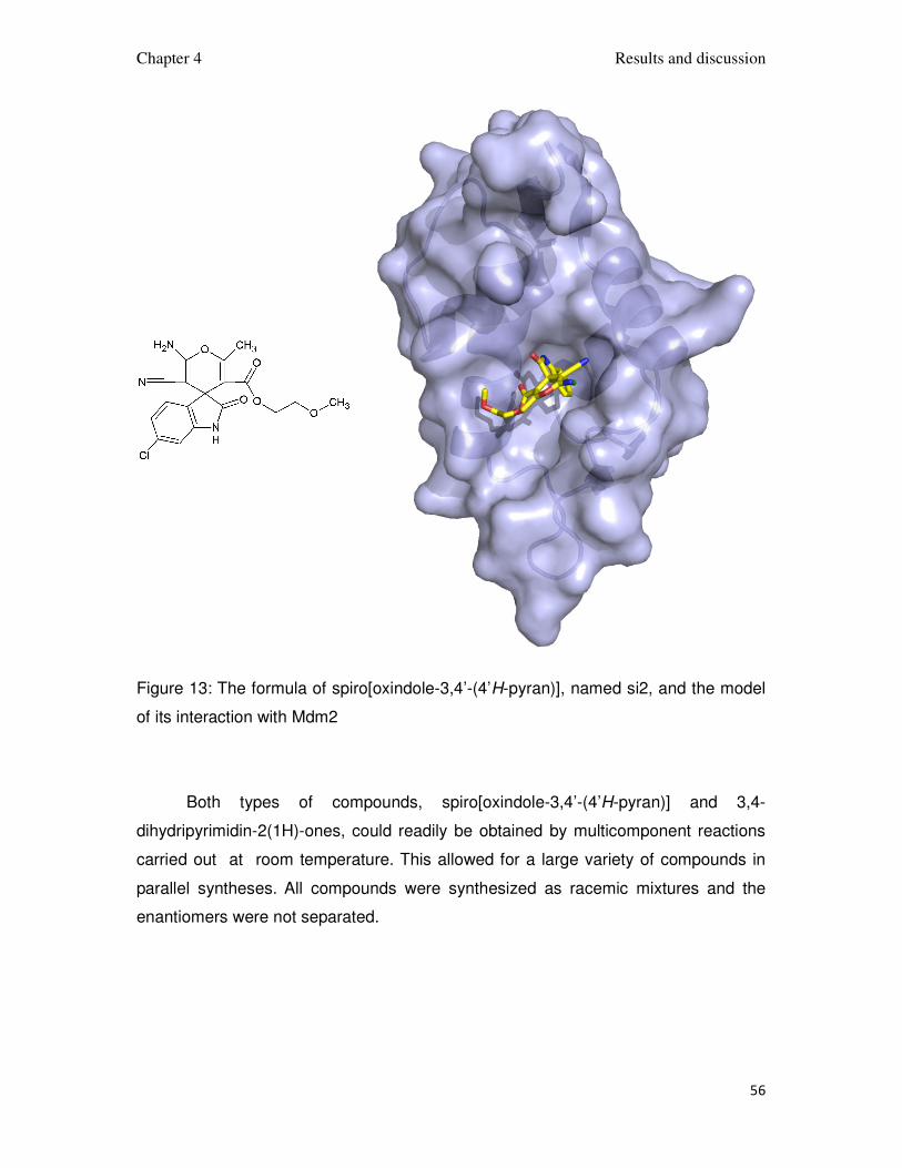
Figure 13: The formula of spiro[oxindole-3,4’-(4’H-pyran)], named si2, and the model
of its interaction with Mdm2
Both types of compounds, spiro[oxindole-3,4’-(4’H-pyran)] and 3,4-
dihydripyrimidin-2(1H)-ones, could readily be obtained by multicomponent reactions
carried out at room temperature. This allowed for a large variety of compounds in
parallel syntheses. All compounds were synthesized as racemic mixtures and the
enantiomers were not separated.
Chapter 4 Results and discussion
57
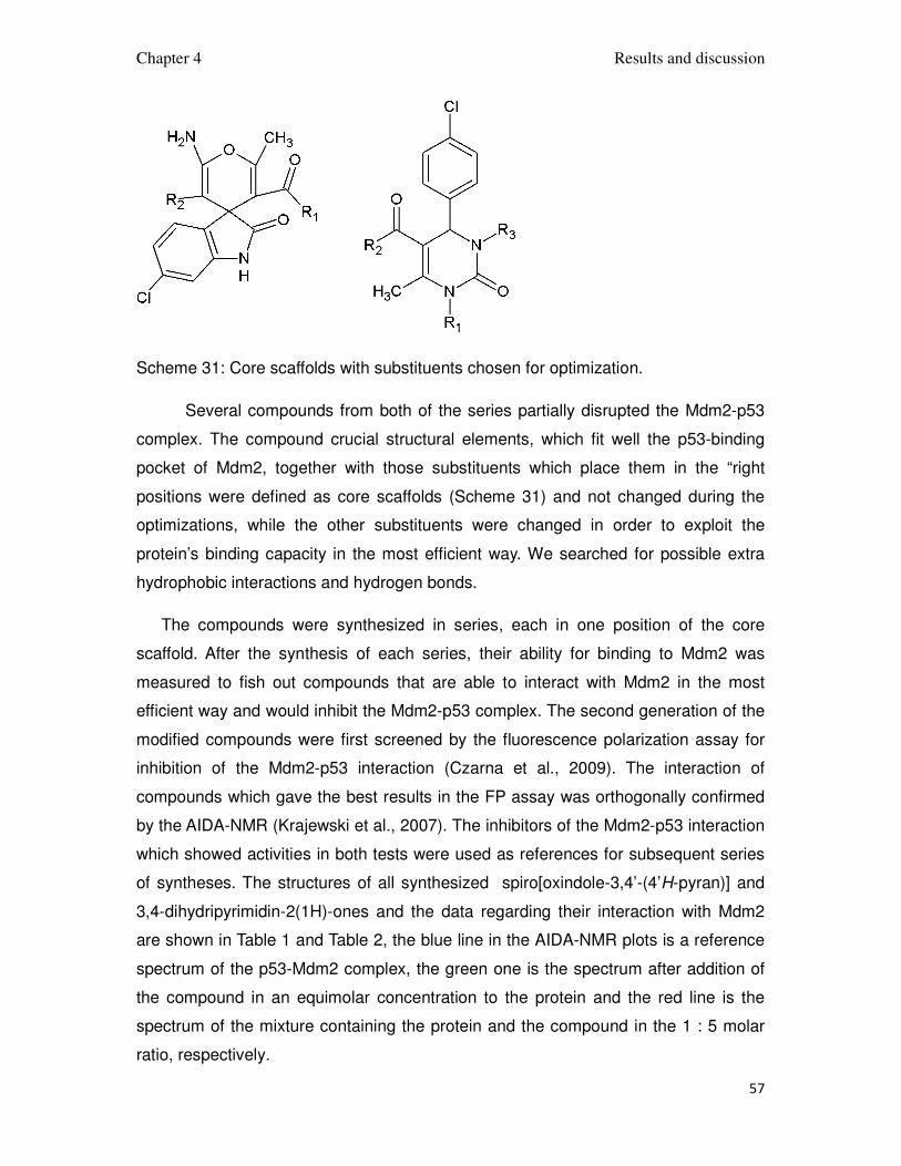
Scheme 31: Core scaffolds with substituents chosen for optimization.
Several compounds from both of the series partially disrupted the Mdm2-p53
complex. The compound crucial structural elements, which fit well the p53-binding
pocket of Mdm2, together with those substituents which place them in the “right
positions were defined as core scaffolds (Scheme 31) and not changed during the
optimizations, while the other substituents were changed in order to exploit the
protein’s binding capacity in the most efficient way. We searched for possible extra
hydrophobic interactions and hydrogen bonds.
The compounds were synthesized in series, each in one position of the core
scaffold. After the synthesis of each series, their ability for binding to Mdm2 was
measured to fish out compounds that are able to interact with Mdm2 in the most
efficient way and would inhibit the Mdm2-p53 complex. The second generation of the
modified compounds were first screened by the fluorescence polarization assay for
inhibition of the Mdm2-p53 interaction (Czarna et al., 2009). The interaction of
compounds which gave the best results in the FP assay was orthogonally confirmed
by the AIDA-NMR (Krajewski et al., 2007). The inhibitors of the Mdm2-p53 interaction
which showed activities in both tests were used as references for subsequent series
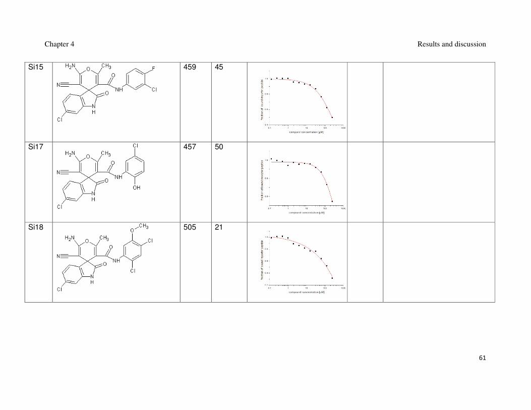
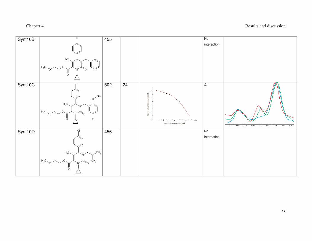
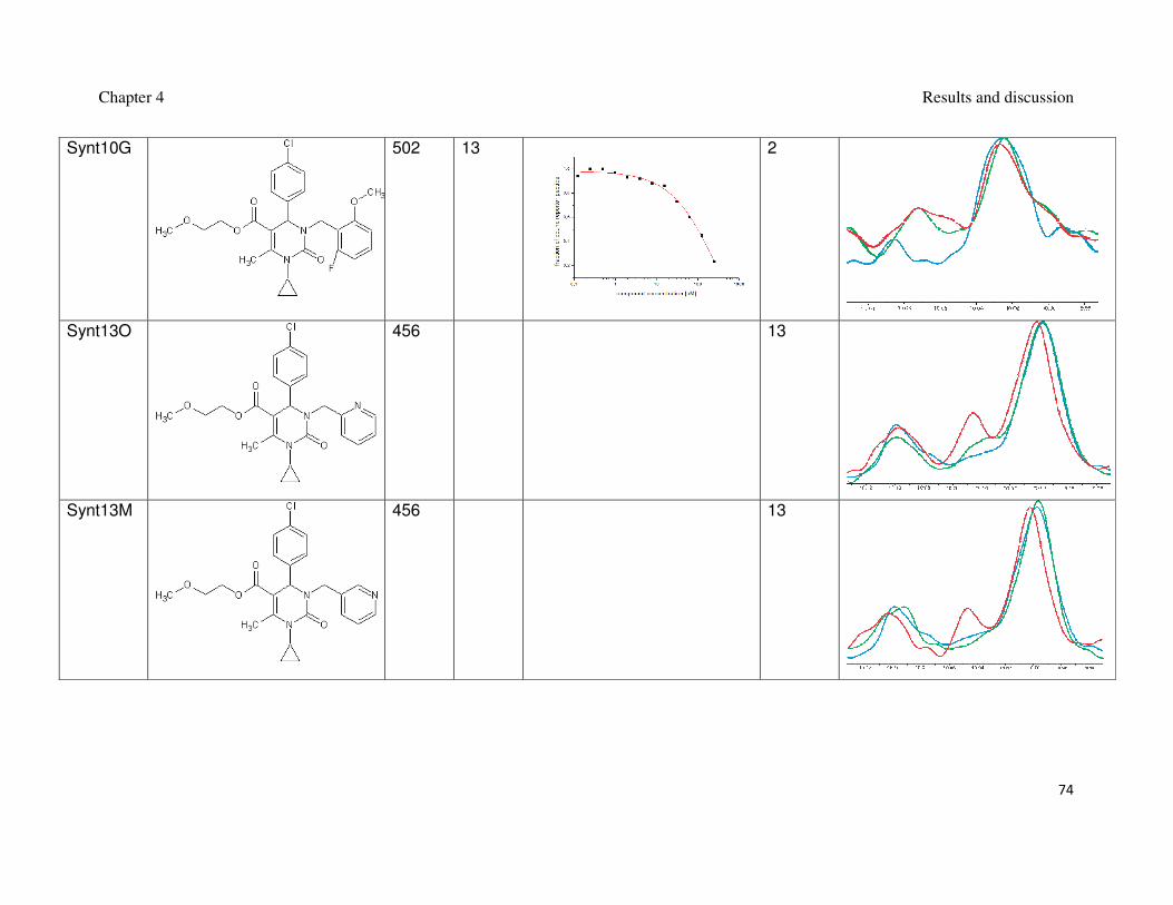
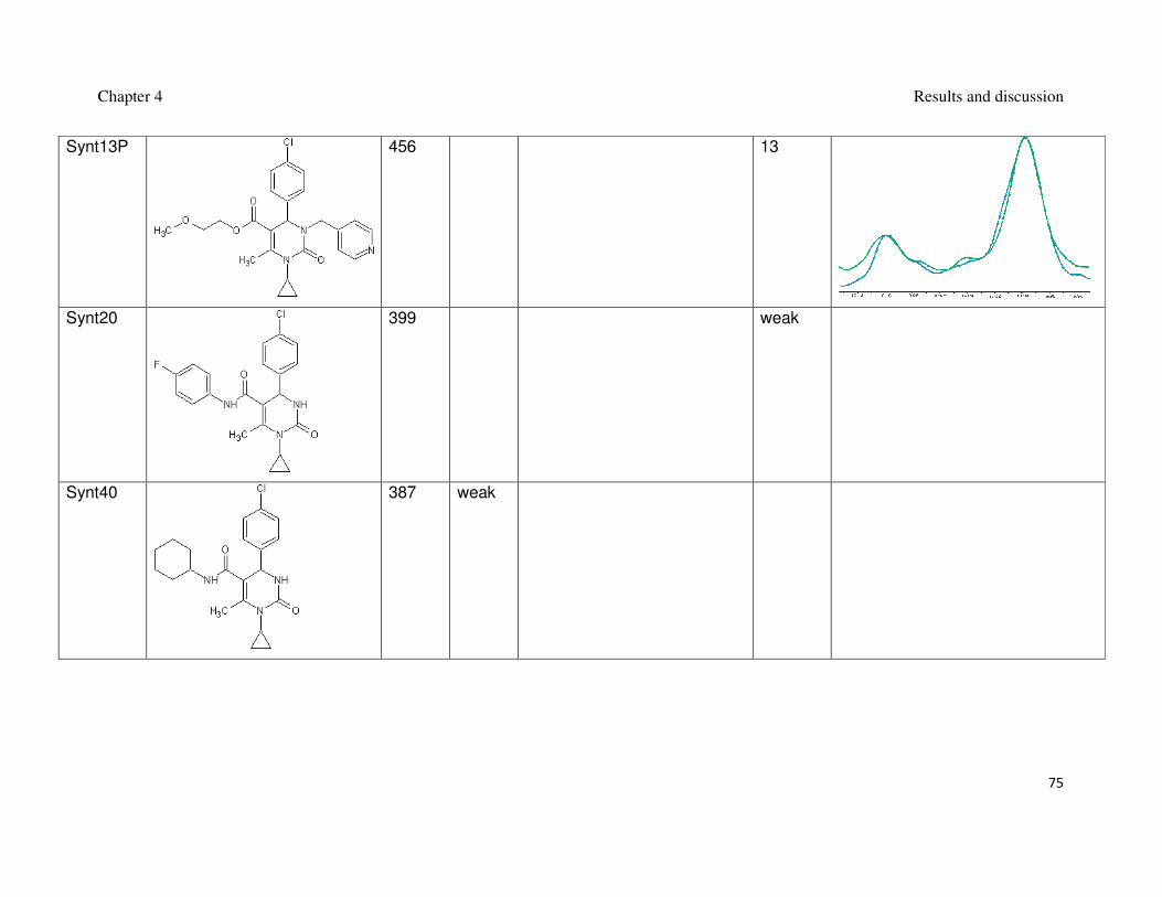
of syntheses. The structures of all synthesized spiro[oxindole-3,4’-(4’H-pyran)] and
3,4-dihydripyrimidin-2(1H)-ones and the data regarding their interaction with Mdm2
are shown in Table 1 and Table 2, the blue line in the AIDA-NMR plots is a reference
spectrum of the p53-Mdm2 complex, the green one is the spectrum after addition of
the compound in an equimolar concentration to the protein and the red line is the
spectrum of the mixture containing the protein and the compound in the 1 : 5 molar
ratio, respectively.

Chapter 4 Results and discussion
58
Table 1: Synthesized spiro[oxindole-3,4’-(4’H-pyran)]es and their interaction with Mdm2.
Name Formula MW
[g/mol]
FP
Ki [µM]
FP plot AIDA
Ki [µM]
AIDA plot
Si2
389 3
Si3
441 10 1.5
Si6
406 60

Chapter 4 Results and discussion
59
Si7
441 29
Si8
436 25
Si9
420 19

Chapter 4 Results and discussion
60
Si11
436 Weak
Si13
424 50
Si14
503 55
Chapter 4 Results and discussion
61
Si15
459 45
Si17
457 50
Si18
505 21

Chapter 4 Results and discussion
62
Si19
410 Weak
Si20
441 Weak
Si21
420 Weak
Si22
412 Weak

Chapter 4 Results and discussion
63
Si24
386 Weak
Si25
386 Weak
Si28
386 No
interaction
Si29
388 Weak

Chapter 4 Results and discussion
64
Si30
370 No
interaction
Si32
434 No
interaction
Si33
374 Weak
Si34
384 Weak

Chapter 4 Results and discussion
65
Si36
473 Weak
Si38
520 Weak
Si39
485 29

Chapter 4 Results and discussion
66
Si40
582 33
Si41
520 48
Si42
459 Weak

Chapter 4 Results and discussion
67
Si48
499 Weak
Si50
539 No
interaction
Si51 517 No
interaction
Si52 515 No
interaction

Chapter 4 Results and discussion
68
Si53
541 No
interaction
Si54
499 No
interaction
Si55
503 No
interaction
Si56
549 No
interaction

Chapter 4 Results and discussion
69
Si57
529 Weak
Si58
535 No
interaction
Si59
515 Weak
Si60
529 No
interaction

Chapter 4 Results and discussion
70
Table 2: Synthesized 3,4-dihydripyrimidin-2(1H)-ones and their interaction with Mdm2.
Name Formula MW
[g/mol]
FP
Ki [µM]
AIDA
Ki [µM]
AIDA plot
Synt2
401 weak
Si61
515 17

Chapter 4 Results and discussion
71
Synt3
436 weak
Synt4
369 weak
Synt8
334 40

Chapter 4 Results and discussion
72
Synt9
352 No
interaction
Synt10
364 4
Synt10A
418 20
Chapter 4 Results and discussion
73
Synt10B
455 No
interaction
Synt10C
502 24 4
Synt10D
456 No
interaction
Chapter 4 Results and discussion
74
Synt10G 502 13 2
Synt13O
456 13
Synt13M
456 13
Chapter 4 Results and discussion
75
Synt13P
456 13
Synt20
399 weak
Synt40
387 weak

Chapter 4 Results and discussion
76
Synt41
361 weak
Synt44
381 weak
Synt45
361 weak

Chapter 4 Results and discussion
77
Synt48
395 weak

Chapter 4 Results and discussion
78
The optimization lead to the stronger binding to Mdm2, which increased the
compound’s capability of disrupting the Mdm2-p53 complex. This was achieved by
adding large chlorinated-aromatic substituents that were able to exploit the
hydrophobic space inside the p53 binding pocket of Mdm2 and if possible adding
structural elements able to form hydrogen bonds with Mdm2 (Figure 14 and Figure
15).
Figure 14: The formula of synt10G, the model of its interaction with Mdm2.
Chapter 4 Results and discussion
79
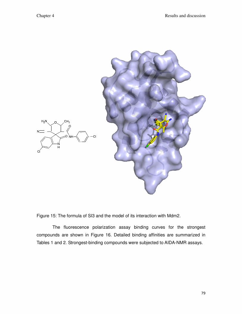
Figure 15: The formula of SI3 and the model of its interaction with Mdm2.
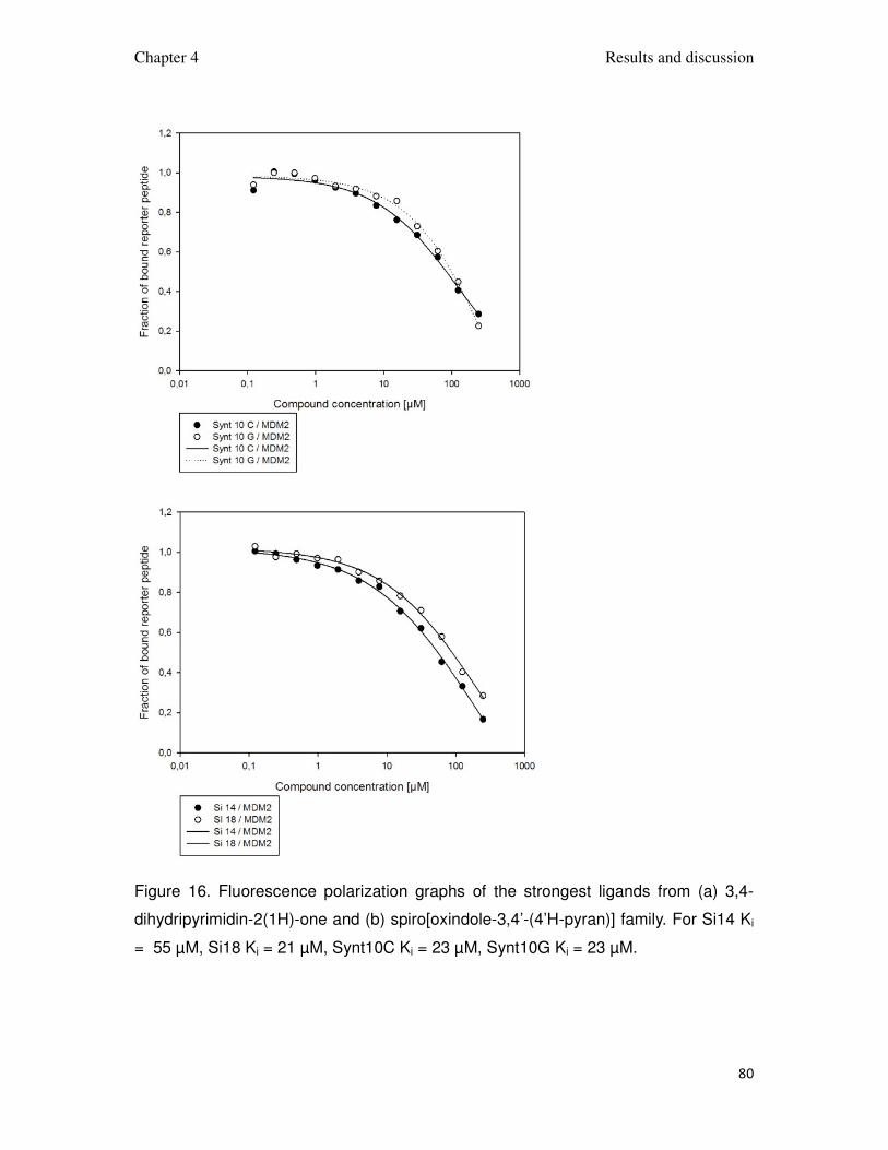
The fluorescence polarization assay binding curves for the strongest
compounds are shown in Figure 16. Detailed binding affinities are summarized in
Tables 1 and 2. Strongest-binding compounds were subjected to AIDA-NMR assays.
Chapter 4 Results and discussion
80
Figure 16. Fluorescence polarization graphs of the strongest ligands from (a) 3,4-
dihydripyrimidin-2(1H)-one and (b) spiro[oxindole-3,4’-(4’H-pyran)] family. For Si14 Ki
= 55 µM, Si18 Ki = 21 µM, Synt10C Ki = 23 µM, Synt10G Ki = 23 µM.
Chapter 4 Results and discussion
81
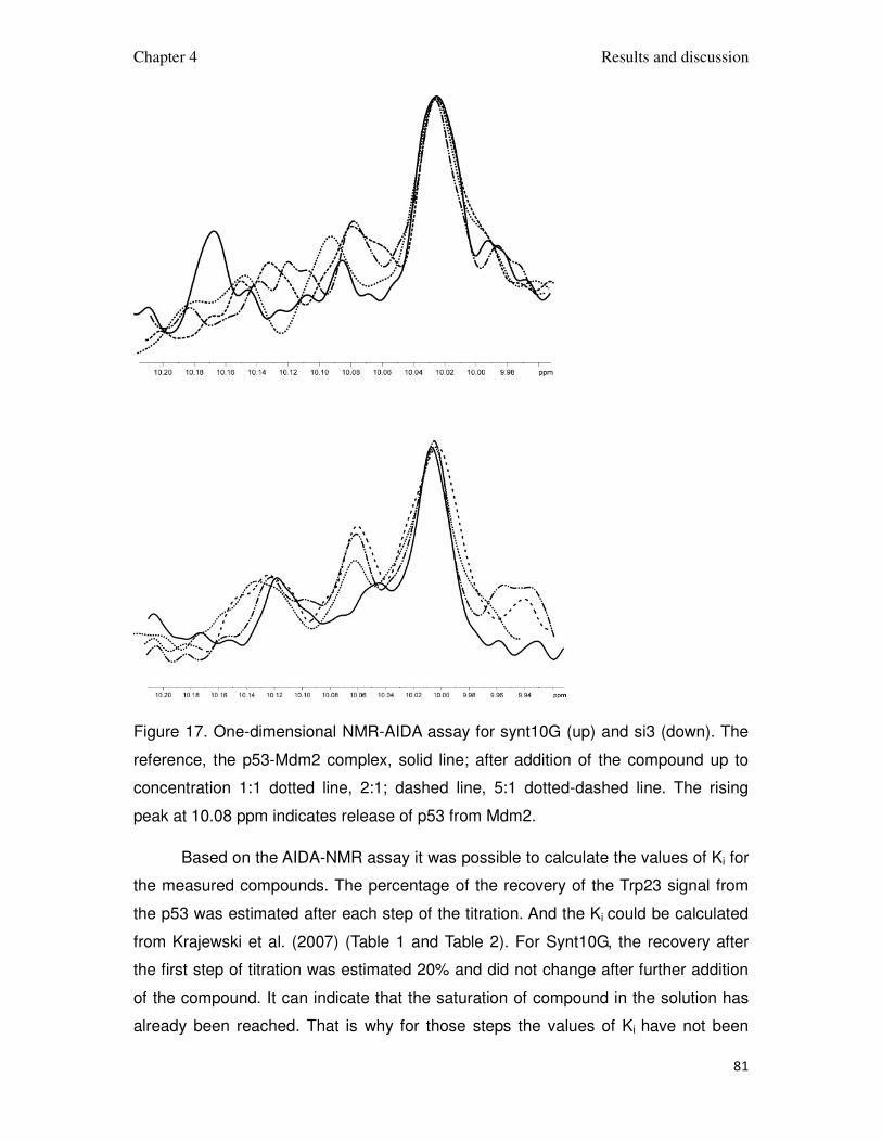
Figure 17. One-dimensional NMR-AIDA assay for synt10G (up) and si3 (down). The
reference, the p53-Mdm2 complex, solid line; after addition of the compound up to
concentration 1:1 dotted line, 2:1; dashed line, 5:1 dotted-dashed line. The rising
peak at 10.08 ppm indicates release of p53 from Mdm2.
Based on the AIDA-NMR assay it was possible to calculate the values of Ki for
the measured compounds. The percentage of the recovery of the Trp23 signal from
the p53 was estimated after each step of the titration. And the Ki could be calculated
from Krajewski et al. (2007) (Table 1 and Table 2). For Synt10G, the recovery after
the first step of titration was estimated 20% and did not change after further addition
of the compound. It can indicate that the saturation of compound in the solution has
already been reached. That is why for those steps the values of Ki have not been
Chapter 4 Results and discussion
82
calculated. For compound Si3, the recovery 50% has been reached in the second
step. It did not change after further titration (Figure 17). In that case the Ki has been
calculated for both steps. For Synt10G Ki = 4 µM, for Si3 Ki = 1.5 µM. The collected
data allowed to verify the models of interaction of 3,4-dihydripyrimidin-2(1H)-ones
and spiro[oxindole-3,4’-(4’H-pyran)]es.
The 2-fluoro-5-methoxybenzyl of synt10G mimics Phe19 of p53 and thus
increases the binding. In addition, the 2-methoxyethyl ester group is exposed into the
solvent and increases the solubility of the compound, which is otherwise low.
Attempts to use this position to fill in the Leu26 failed, probably due to the decreased
solubility, the compounds with more hydrophobic esters or amides gave barely
detectable interaction with Mdm2, even without any hydrophobic substituent at the
N1 nitrogen. The function of the cyclopropyl group exposed into solvent is not well
understood; replacing it with a hydrophilic substituent decreases the binding. The
same is true for larger hydrophobic groups.
The potency of spiro[oxindole-3,4’-(4’H-pyran)]es improved after replacing the
2-methoxyethyl group with 4-chorophenyl group, which decreased the compounds
solubility, but created additional interactions with the protein in its Phe26 binding
pocket. Larger chlorinated, aromatic systems in this position did not lead to any
improvement, especially if there was the substitution in the ortho or meta positions of
the phenyl ring, the compounds´ binding was greatly decreased. The compounds
with aliphatic amides showed, for the best cases, only weak interaction with Mdm2.
The compounds with the nitrile group replaced by various amides were tested in
order to exploit the potential interaction in the Leu26 binging pocket, though none of
them was able to disrupt the Mdm2 binding. In the model of the complex of Si3 with
Mdm2 in Figure 15 not much space is visible in the Leu26 pocket, which can be the
reason why the compounds with large groups targeting this place may not fit into the
binding cleft of Mdm2.

Chapter 4 Results and discussion
83
4.2. 2,3-dihydroquinazolin-4(1H)-ones
7-chloro-2,3-dihydroquinazolin-4(1H)-one of Figure 18 showed an interaction
with Mdm2 in the fluorescence polarization assay.
Figure 18: 7-chloro-2,3-dihydroquinazolin-4(1H)-one and the corresponding
fluorescence polarization assay of its binding to Mdm2.
The calculated Ki was 20 µM, which qualified it for the optimization. The
compounds were synthesized in a one step, multicomponent reaction, carried out
under reflux in ethanol as a solvent. A big variety of primary amines and aldehydes
are commercially available and relatively cheap, which made the optimization easy
from the synthetic point of view. The use of 4-chloroisatoic anhydride caused the
reaction to proceed much slower and with significantly lower yield than with the
unsubstituted isatoic anhydride. The reactions with 4-chloroisatoic anhydride required
much more time to complete and more care during the product purification. All the
compounds were synthesized as racemic mixtures and the enantiomers were not
separated.
The 2,3-dihydroquinazolin-4(1H)-one scaffold (Scheme 32) was kept the same
during the optimization.
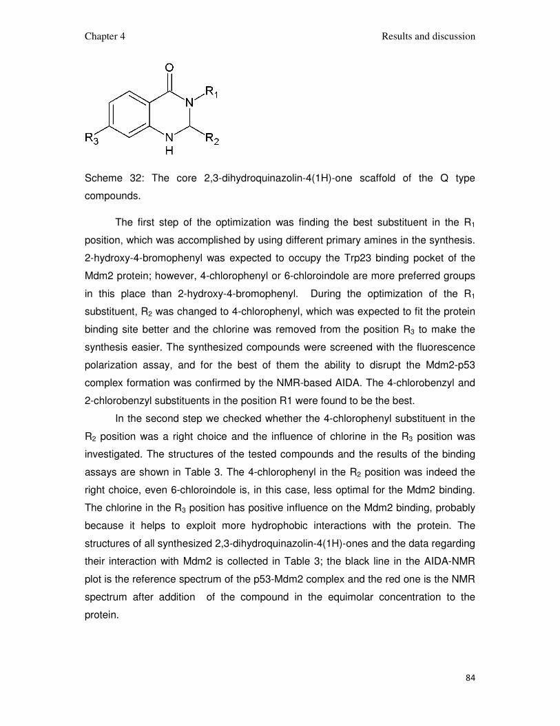
Chapter 4 Results and discussion
84
Scheme 32: The core 2,3-dihydroquinazolin-4(1H)-one scaffold of the Q type
compounds.
The first step of the optimization was finding the best substituent in the R1
position, which was accomplished by using different primary amines in the synthesis.
2-hydroxy-4-bromophenyl was expected to occupy the Trp23 binding pocket of the
Mdm2 protein; however, 4-chlorophenyl or 6-chloroindole are more preferred groups
in this place than 2-hydroxy-4-bromophenyl. During the optimization of the R1
substituent, R2 was changed to 4-chlorophenyl, which was expected to fit the protein
binding site better and the chlorine was removed from the position R3 to make the
synthesis easier. The synthesized compounds were screened with the fluorescence
polarization assay, and for the best of them the ability to disrupt the Mdm2-p53
complex formation was confirmed by the NMR-based AIDA. The 4-chlorobenzyl and
2-chlorobenzyl substituents in the position R1 were found to be the best.
In the second step we checked whether the 4-chlorophenyl substituent in the
R2 position was a right choice and the influence of chlorine in the R3 position was
investigated. The structures of the tested compounds and the results of the binding
assays are shown in Table 3. The 4-chlorophenyl in the R2 position was indeed the
right choice, even 6-chloroindole is, in this case, less optimal for the Mdm2 binding.
The chlorine in the R3 position has positive influence on the Mdm2 binding, probably
because it helps to exploit more hydrophobic interactions with the protein. The
structures of all synthesized 2,3-dihydroquinazolin-4(1H)-ones and the data regarding
their interaction with Mdm2 is collected in Table 3; the black line in the AIDA-NMR
plot is the reference spectrum of the p53-Mdm2 complex and the red one is the NMR
spectrum after addition of the compound in the equimolar concentration to the
protein.
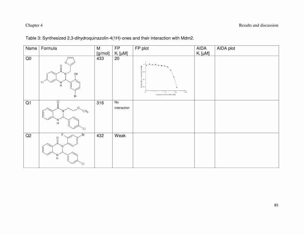
Chapter 4 Results and discussion
85
Table 3: Synthesized 2,3-dihydroquinazolin-4(1H)-ones and their interaction with Mdm2. Name Formula M
[g/mol] FP Ki [µM]
FP plot AIDA Ki [µM]
AIDA plot
Q0
433 20
Q1
316 No
interaction
Q2
432 Weak

Chapter 4 Results and discussion
86
Q3
334 No
interaction
Q4
348 No
interaction
Q5
386 No
interaction
Q6
382 25 No
interaction

Chapter 4 Results and discussion
87
Q7
382 No
interaction
Q8
368 No
interaction
Q9
352 No
interaction
Q10
382 25
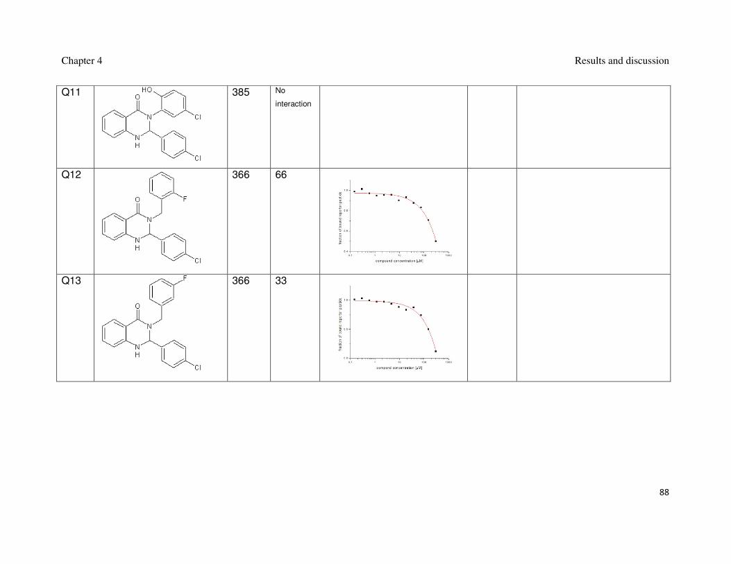
Chapter 4 Results and discussion
88
Q11
385 No
interaction
Q12
366 66
Q13
366 33
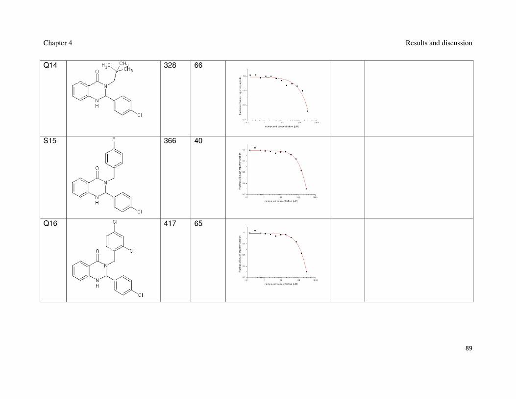
Chapter 4 Results and discussion
89
Q14
328 66
S15
366 40
Q16
417 65

Chapter 4 Results and discussion
90
Q17 401 No
interaction
Q19
338 No
interaction
Q20
338 24

Chapter 4 Results and discussion
91
Q21
373 17 No
interaction
Q22
417 8 No
interaction
Q23
422 No
interaction No
interaction

Chapter 4 Results and discussion
92
Q24
455 No
interaction No
interaction

Chapter 4 Results and discussion
93
Among all tested 2,3-dihydroquinazolin-4(1H)-ones, compound Q22 showed
the highest activity in the FP assay. It probably makes many hydrophobic interactions
with the Mdm2 protein. Surprisingly, the compounds with 6-chloroindole at the R2
position did not interact with Mdm2 at all, probably because the 6-chloroindole moiety
in this position was too large for the Mdm2’s binding cleft, thus 4-chlorophenyl group
is the best in this position. From many tested substituents in the R1 position, 4-
chlorophenyl and 2-chlorophenyl had the most positive influence on the compounds´
potency. 4-chlorination of the quinazolinone system further improved the binding to
Mdm2.
The AIDA assay indicated no interaction with Mdm2-p53 complex due to low
solubility of the compounds in water, despite of the addition of 10% DMSO and 0.7%
of the Brij detergent, the compounds remained almost insoluble, only very low
concentration of them was detectable in NMR.
4.3. 1'H-spiro[6-chloro-isoindole-1,2'-quinazoline]-3,4'(3'H)-diones
1'H-spiro[6-chloro-isoindole-1,2'-quinazoline]-3,4'(3'H)-dione of Figure 19
showed the interaction with Mdm2 in the fluorescence polarization assay as shown in
the right panel of Figure 19.
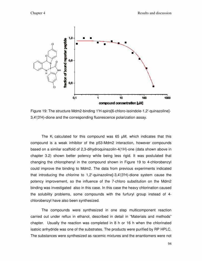
Chapter 4 Results and discussion
94
Figure 19: The structure Mdm2-binding 1'H-spiro[6-chloro-isoindole-1,2'-quinazoline]-
3,4'(3'H)-dione and the corresponding fluorescence polarization assay.
The Ki calculated for this compound was 65 µM, which indicates that this
compound is a weak inhibitor of the p53-Mdm2 interaction, however compounds
based on a similar scaffold of 2,3-dihydroquinazolin-4(1H)-one (data shown above in
chapter 3.2) shown better potency while being less rigid. It was postulated that
changing the chlorophenyl in the compound shown in Figure 19 to 4-chlorobenzyl
could improve the binding to Mdm2. The data from previous experiments indicated
that introducing the chlorine to 1,2'-quinazoline]-3,4'(3'H)-dione system cause the
potency improvement, so the influence of the 7-chloro substitution on the Mdm2
binding was investigated also in this case. In this case the heavy chlorination caused
the solubility problems, some compounds with the furfuryl group instead of 4-
chlorobenzyl have also been synthesized.
The compounds were synthesized in one step multicomponent reaction
carried out under reflux in ethanol, described in detail in “Materials and methods”
chapter. Usually the reaction was completed in 8 h or 16 h when the chlorinated
isatoic anhydride was one of the substrates. The products were purified by RP HPLC.
The substances were synthesized as racemic mixtures and the enantiomers were not

Chapter 4 Results and discussion
95
separated. The yields of the substances were low and they were isolated in only
small amounts (2-7 mg). The purity was checked on TLC, of which the UV photo is
shown in Figure 20. The compounds purity was estimated to be around 90% which
was enough to make the measurements of the compounds activity for the p53-Mdm2
inhibition.
Figure 20: A photo of silica TLC plate with SQ compounds. The compounds were
applied onto a plate as DMSO solutions and a running mixture was
methanol/chlorophorm/water 50/20/4. The large, dark spots are the desired
compounds, some minor amounts of impurities and visible.
The compounds were subjected to the FP assay and the capability of
disrupting the p53-Mdm2 complex of the best of them was confirmed by the AIDA-
NMR. The results of the measurements are collected in Table 4 below, the black line
in the AIDA plot is the reference NMR spectrum of the p53-Mdm2 complex and the
red one is the NMR spectrum after adding the compound in equimolar concentration
to the protein.

Chapter 4 Results and discussion
96
Table 4: Synthesized 1'H-spiro[6-chloro-isoindole-1,2'-quinazoline]-3,4'(3'H)-diones and their interaction with Mdm2
Name Formula M [g/mol]
FP Ki [µM]
FP plot AIDA Ki [µM]
AIDA plot
SQ1
410 65
SQ2
379 55
SQ3
424 63
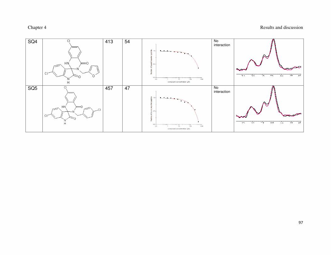
Chapter 4 Results and discussion
97
SQ4
413 54
No interaction
SQ5
457 47
No interaction

Chapter 4 Results and discussion
98
An attempt to improve the binding of 1'H-spiro[6-chloro-isoindole-1,2'-
quinazoline]-3,4'(3'H)-diones did not succeed, all synthesized compounds had
comparable Ki in FP assay. The connection between the quinazolinone and 6-
chlorospiroxindole moieties in these compounds is very rigid. Further structural
analysis indicated that quinazolinone and 6-chlorospiroxindole moieties are in
unfavorable relative positions to each other, so if the 6-chlorospiroxindole binds in the
Trp23 binding pocket of Mdm2, than the quinazolinone system is placed in parallel to
the surface of the protein, so it cannot interact with either the Phe19 or the Leu26
binding pockets. The AIDA assay confirmed that these compounds did not interact
“well” with the Mdm2 protein.

Chapter 5 Summary
99
5.1. Summary
This thesis presents my research on the discovery, design, and development
of small molecule antagonists for the p53-Mdm2 interaction. The tumor suppressor
protein p53, protects the organism from cancer. In order to escape the p53 mediated
safeguard system, most of human cancers have either mutated the p53 or decreased
effectiveness of the p53 pathway. In tumors which retain the wildtype p53, it is mostly
inactivated by its negative regulators, the Mdm2 and Mdmx proteins. Mdm2, the first
to gain the distinction of a principal inhibitor of p53, promotes ubiquitination of p53
followed by degradation in the proteasome.
The restoration of the impaired function of p53, by disrupting the Mdm2-p53
and Mdmx-p53 interactions, offers a new paradigm for anticancer therapy across a
broad spectrum of cancers. Cancer cells are extremely sensitive to the restoration of
p53.
Four groups of low molecular weight, organic compounds have been
synthesized, tested and optimized in order to achieve the strongest interaction with
the Mdm2 protein. The four group comprised the scaffolds of 3,4-dihydripyrimidin-
2(1H)-ones, spiro[oxindole-3,4’-(4’H-pyran)]es, 2,3-dihydroquinazolin-4(1H)-ones,
1'H-spiro[6-chloro-isoindole-1,2'-quinazoline]-3,4'(3'H)-diones. These structures were
extended with variants that possessed an array of different substituents in order to
exploit more possible interactions between the compounds and the protein. Using the
multicomponent reactions a large variety of compounds with different substituents
were synthesized and tested in the fluorescence polarization assay and in the NMR-
based antagonist induced dissociation assay, AIDA-NMR. The best of the herein
described inhibitors have single digit micromolar potency for disrupting the p53-
Mdm2 interaction.
The aqueous solubility of the current derivatives of the scaffolds described in
this thesis is low. This is because the protein-protein interaction of p53 and Mdm2 is
highly hydrophobic and naturally results in very hydrophobic initial antagonists. In
order to achieve high enough concentrations of the antagonist in the aqueous
solution of the target protein for NMR measurements, we often used Tween as a
solubilizer. For the future, we would need to improve water solubility of the p53-

Chapter 5 Summary
100
Mdm2 antagonists discovered for this thesis. We are working toward next
generations of antagonists with sufficient water solubility that will not need a
solubilizing additive. In general, the proposed way to improve the potency and water
solubility of the best scaffolds involves systematic variation of different substitutents
around their cores; for example, for the improved water solubility via introduction of a
hydroxyl methyl or dialkylamino methyl groups.
Chapter 5 Summary
101
5.2. Zusammenfassung
In dieser Arbeit werden die Forschungsergebnisse über die Entdeckung,
Entwurf und Entwicklung von niedermolekularen Antagonisten der p53-Mdm2
Interaktion präsentiert. Der Tumorsupressor p53 beschüzt den Organismus vor
Krebs. Um das p53-regulierte Sicherheitssystem zu umgehen besitzen die meisten
Krebsarten entweder ein mutiertes p53 Gen oder eine verminderte Aktivität im p53-
Weg. In Tumoren, die das Wildtyp-p53 Gen exprimieren, wird p53 durch seine
negativen Regulatoren Mdm2 und Mdmx inaktiviert. Mdm2, der als erstes als
hauptsächlichen Inhibitor von p53 entdeckt wurde, fördert die Ubiquitinilierung von
p53, was die Degradierung durch das Proteosom zur Folge hat.
Die Wiederherstellung der beeinträchtigten p53-Funktion, durch die Trennung
der p53-Mdm2 und p53-MdmxKomplexe, steigert die Chancen einer Krebstherapie
gegen einer vielfalt von Krebsarten. Krebszellen sind sehr sensitiv gegenüber der
Restaurierung von p53.
Vier Gruppen organischer Moleküle wurden synthetisiert, getestet und
optimiert, um die stärkste Interaktion mit Mdm2 zu erhalten. Diese vier Gruppen
bestehen aus 3,4-dihydripyrimidin-2(1H)-on, spiro[oxindole-3,4’-(4’H-pyran)], 2,3-
dihydroquinazolin-4(1H)-on und 1'H-spiro[6-chloro-isoindole-1,2'-quinazoline]-
3,4'(3'H)-dion. Diese Strukturen wurden mit Varianten verschiedener Substituenten
verlängert, um mögliche Interaktionen zwischen der Verbindung und dem Protein zu
entdecken. Eine große Vielfalt von Verbindungen mit verschiedenen Substituenten
wurden durch die Multikomponentenreaktion synthetisiert, die durch der fluoreszent
polarisations Untersuchung und der NMR basiertem „antagonistinduced dissociation“
Untersuchung getestet worden sind. Die Besten hier beschriebenen Inhibitoren sind
im mikromolarem Bereich Wiksam.
Die Wasserlöslichkeit der Derivaten der hier beschriebenen
Verbindungsklassen ist niedrig. Der Grund hierfür ist, dass die Interaktionsfläche
zwischen p53 und Mdm2 sehr hydrophob ist, wodurch zunächst auch sehr
hydrophobe Antagonisten entstehen. Um für die NMR-Messungen eine genügend
hohe Konzentration in wässriger Lösung des p53-Mdm2 Antagonisten zu erlangen,
wurde zur Solubilisierung Tween hinzugefügt.

Chapter 5 Summary
102
Wir arbeiten an der Entwicklung von Antagonisten mit ausreichender
Wasserlöslichkeit, wo die Zugabe von solubilisierenden Agentien überflüssig wird.
Generell verbessert man die Löslichkeit der besten Verbindungsklassen durch eine
systematische Variierung verschiedener Substituenten um deren Grundkörper, z.B.
wird ie Wasserlöslichkeit durch die Einführung einer hydroxymethyl oder
dialkylaminomethylgruppe verbessert.

Chapter 6 Appendix
103
6. Appendix
6.1. Abbreviations:
1D one-dimensional
AIDA antagonist induced dissociation assay
bp base pair
Da Dalton (g/mol)
DMSO dimethylsulfoxide
DNA deoxyribonucleic acid
DMF N,N-dimethylformamide
DMSO dimethylsulfoxide
DTT dithiothreitol
EDTA ethylenediamine tetraacetic acid
Et ethyl
FP Fluorescence polarization assay
g gravitational acceleration (9.81 m/s2)
h hour
HPLC high pressure liquid chromatography
IC50 half maximal inhibitory concentration
IPTG isopropyl-β-thiogalactopyranoside
Ki inhibition constant
LB lysogeny broth medium
Me methyl
Mdm2 Murine double minute - 2
Chapter 6 Appendix
104
MdmX Murine double minute – 4
min minute
MW molecular weight
NiNTA nickel-nitrilotriacetic acid
NMR nuclear magnetic resonance spectroscopy
OD optical density
PAGE polyacrylamide gel electrophoresis
PBS phosphate-buffered saline
PDB Protein Data Bank
ppm parts per million
RNA ribonucleic acid
RP reversed phase
rpm revolutions per mimute
SDS sodium dodecyl sulfate
TAE tris – acetate – EDTA buffer
TEMED N,N,N’,N’-tetramethylethylendiamine
Tris tris(hydroxymethyl)aminomethane
TLC thin layer chromatography
MS mass spectrometry
Mdm2 Murine double minute - 2
Mdmx, Mdm4 Murine double minute - 4
Chapter 6 Appendix
105
6.2. Protein and peptide sequences:
Mdm2 (1-125):
MCNTNMSVPT DGAVTTSQIP ASEQETLVRP KPLLLKLLKS VGAQKDTYTM
KEVLFYLGQY IMTKRLYDEK QQHIVYCSND LLGDLFGVPS FSVKEHRKIY
TMIYRNLVVV NQQESSDSGT SVSEN
p53 (1-321):
MEEPQSDPSV EPPLSQETFS DLWKLLPENN VLSPLPSQAM DDLMLSPDDI
EQWFTEDPGP DEAPRMPEAA PPVAPAPAAP TPAAPAPAPS WPLSSSVPSQ
KTYQGSYGFR LGFLHSGTAK SVTCTYSPAL NKMFCQLAKT CPVQLWVDST
PPPGTRVRAM AIYKQSQHMT EVVRRCPHHE RCSDSDGLAP PQHLIRVEGN
LRVEYLDDRN TFRHSVVVPY EPPEVGSDCT TIHYNYMCNS SCMGGMNRRP
ILTIITLEDS SGNLLGRNSF EVRVCACPGR DRRTEEENLR KKGEPHHELP
PGSTKRALPN NTSSSPQPKK K
High affinity Mdm2 binding peptide for FP:
LTFEHYWAQLTS

References
106
7. References
J.G. Allen, M.P. Bourbeau, G.E. Wohlhieter, M.D. Bartberger, K. Michelsen, R.
Hungate, R.C. Gadwood, R.D. Gaston, B. Evans, L.W. Mann, M.E. Matison, S.
Schneider, X. Huang, D. Yu, P.S. Andrews, A. Reichelt, A.M. Long, P. Yakowec,
E.Y.Yang, T.A. Lee, J.D. Oliner, Discovery and optimization of chromenotriazolo-
pyrimidines as potent inhibitors of the mouse double minute 2-tumor protein 53
protein-protein interaction. J. Med. Chem. 52 (2009) 7044-7053.
M. Bista, K. Kowalska, W. Janczyk, A. Dömling and T.A. Holak, Robust NMR
screening for lead compounds using tryptophan-containing proteins. J. Amer. Chem.
Soc. 131 (2009) 7500-7501.
A. Boettcher, N. Buschmann, P. Furet, J. Groell, J. Kallen, L.J. Hergovich, K.
Masuya, L. Mayr, A. Vaupel, 3-Imidazolyl-Indoles for the Treatment of Proliferative
Diseases. WO 2008/119741.
P. R. Brodfuehrer, B. Chen, T. R. Sattelberg, Sr., P. R. Smith, J. P. Reddy, Derron R.
Stark, S. L. Quinlan, and J. G. Reid, An Efficient Fischer Indole Synthesis of
Avitriptan, a Potent 5-HT1D Receptor Agonist. J. Org. Chem. 62 (1997) 9192-9202.
R. J. Clemens and J. A. Hyatt, Acetoacetylation with 2,2,6-trimethyl-4H-1,3-dioxin-4-
one: A Convenient Alternative to Diketene. J. Org. Chem. 50 (1984) 2431-2435.
M.D. Cummings, C. Schubert, D.J. Parks, R.R. Calvo, L.V. LaFrance, J. Lattanze,
K.L. Milkiewicz, T. Lu, Substituted 1,4-benzodiazepine-2,5-diones as alpha-helix
mimetic antagonists of the HDM2-p53 protein-protein interaction. Chem. Biol. Drug.
Des. 67 (2006) 201-205.
Y.C. Chang, Y.S. Lee, T. Tejima. Mdm2 and bax, downstream mediators of the p53
response, are degraded by the ubiquitin-proteasome pathway. Cell Growth Differ. 9
(1998) 79-84.
L. Chen, H. Yin, B. Farooqi, S. Sebti, A.D. Hamilton, J. Chen. P53 alpha-helix
mimetics antagonize p53/MDM2 interaction and activate p53. Mol. Cancer. Ther. 4
(2005) 1019-1025.
References
107
C. Cordon-Cardo, E. Latres, M. Drobujak, M.R. Oliva, D. Pollack, J.M. Woodruff, V.
Marechal, J. Chen, M.F Brennan, A.J. Levine, Molecular Abnormalities of MDM2 and
p53 Genes in Adult Soft Tissue Sarcomas. Cancer Res. 54 (1994) 794-799.
A. Czarna, G.M. Popowicz, A. Pecak, S. Wolf, G. Dubin and T.A. Holak, High affinity
interaction of the p53 peptide-analogue with human Mdm2 and MdmX. Cell Cycle 8
(2009) 1174-1183.
M. Dabiri, P. Salehi, S. Otokesh, M. Baghbanzadeh, G. Kozehgarya and A. A.
Mohammadia, Efficient synthesis of mono- and disubstituted 2,3-dihydroquinazolin-
4(1H)-ones using KAl(SO4)2 x12 H2O as a reusable catalyst in water and ethanol.
Tet. Lett. 46 (2005) 6123–6126.
W.S. El-Deiry, S.E. Kern, J.A. Pietenpol, K.W. Kinzler, B. Vogelstein. Definition of a
consensus binding site for p53. Nat. Genet. 1 (1992) 45-49.
K. Ding, Y. Lu, Z. Nikolovska-Coleska, G. Wang, S. Qiu, S. Shangary, W. Gao, D.
Qin, J. Stuckey, K. Krajewski, P.P. Roller, S. Wang, Structure-based design of spiro-
oxindoles as potent, specific small-molecule inhibitors of theMDM2-p53 interaction. J.
Med. Chem. 49 (2006) 3432-3435.
K. Ding, G. Wang, J.R. Deschamps, D.A. Parrishb, S. Wang, Synthesis of
spirooxindoles via asymmetric 1,3-dipolar cycloaddition. Tetrahedron Lett. 46 (2005)
5949-5951.
Q. Ding, B.J. Graves, N. Kong, J. Liu, A.J. Lovey, G. Pizzolato, J.L. Roberts, S.
Sungsau, B.T. Vu, P.M. Wovkulich, 4,4,5,5, Tetrasubstituted Imidazolines. US
2007/129416.
L. D'Silva, P. Ozdowy, M. Krajewski, U. Rothweiler, M. Singh and T.A. Holak,
Monitoring the effects of antagonists on protein-protein interactions with NMR
spectroscopy. J. Amer. Chem. Soc. 127 (2005) 13220-13226.
B. Elenbaas, M. Dobbelstein, J. Roth, T. Shenk, and A. J. Levine. The MDM2
oncoprotein binds specifically to RNA through its RING finger domain. Mol Med.
1996 2 (1996) 439–451

References
108
T. Ewing, I.D Kuntz, Critical evaluation of search algorithms for automated molecular
docking and database screening. J. Comput. Chem. 18 (1997) 1175-1189.
R. G. Fargher, Orientationion of the Nitro- and Arylazo- glyoxalines. Fission of the
Glyoxalone nucleus. J. Chem. Soc., Trans. 117 (1920) 668 – 680.
N. Fotouhi, G.J. Haley, K.B. Simonsen, B.T. Vu, S.E. Webber. Cis-imidazolines. US
2007/167437.
P.S. Galatin, D.J. Abraham. A nonpeptidic sulfonamide inhibits the p53-Mdm2
interaction and activates p53-dependent transcription in Mdm2-overexpressing cells.
J. Med. Chem. 47 (2004) 4163-4165.
B.L. Grasberger, T. Lu , C. Schubert, D.J. Parks, T.E. Carver, H.K. Koblish, M.D.
Cummings, L.V. LaFrance, K.L. Milkiewicz, R.R. Calvo, D. Maguire, J. Lattanze, C.F.
Franks, S. Zhao, K. Ramachandren, G.R. Bylebyl, M. Zhang, C.L. Manthey, E.C.
Petrella, M.W. Pantoliano, I.C. Deckman, J.C. Spurlino, A.C. Maroney, B.E.
Tomczuk, C.J. Molloy, R.F. Bone, Discovery and cocrystal structure of
benzodiazepinedione HDM2 antagonists that activate p53 in cells. J. Med. Chem. 48
(2005) 909-912.
W. Gu, Xiao-Lu Shi, R.G. Roeder. Synergistic activation of transcription by CBP and
p53. Nature 387 (1997) 819–23.
Y. Haupt, R. Maya, A. Kazaz, M. Oren. Mdm2 promotes the rapid degradation of
p53. Nature 387 (1997) 296-299.
K. Higashiyama, H. Otomasu, Spiro Heterocyclic Compounds. III. Synthesis of
Spiro[oxindole-3,4‘-(4’H-pyran)] Compounds. Chem. Pharm. Bull. 28 (1980) 648-651.
R. Honda, H. Tanaka, H. Yasuda, Oncoprotein is ubiquitin ligase E3 for tumor
suppressor p53. FEBS Lett. 420 (1997) 25-27.
X. Huang. Fluorescence Polarization Competition Assay: The Range of Resolvable
Inhibitor Potency Is Limited by the Affinity of the Fluorescent Ligand. J. Biomol.
Screen. 8 (2003) 34-38.
J.J Irwin and B.K. Shoichet, ZINC − A Free Database of Commercially Available
Compounds for Virtual Screening. J. Chem. Inf. Model. 45(1) (2005) 177-82.
References
109
P. N. James and H. N. Snyder, Indole-3-aldehyde. Organic Syntheses 39 (1959) 30.
A.C. Joerger, A.R. Fersht, Structural Biology of the Tumor Suppressor p53. Ann.
Rev. Bioch. 77 (2008) 557.
T. Juven-Gershon and M. Oren Mdm2: The Ups and Downs. Mol. Med. 5 (1999) 71-
83
B.B. Kehm and C.W. Whitehead. 3-n-heptyl-5-cyanocytosine. Organic Syntheses. 4
(1963) 515
H.K. Koblish, S. Zhao, C.F. Franks, R.R. Donatelli, R.M. Tominovich, L.V. LaFrance,
K.A. Leonard, J.M. Gushue, D.J. Parks, R.R. Calvo, K.L. Milkiewicz, J. JoséMarugán,
P. Raboisson, M.D. Cummings, B.L. Grasberger, D.L. Johnson, T. Lu, C.J. Molloy,
A.C. Maroney, Benzodiazepinedione inhibitors of the Hdm2 : p53 complex suppress
human tumor cell proliferation in vitro and sensitize tumors to doxorubicin in vivo.
Mol. Cancer. Ther. 5 (2006) 160-169.
N. Kong, E.A. Liu, B.T. Vu, Cis-imidazolines. US 6617346 (2003).
N. Kong, E.A. Liu, B.T. Vu, Cis-2,4,5-triphenyl-imidazolines and their Use in the
Treatment of Tumors. WO2003/051359.
M. Krajewski, U. Rothweiler, L. D'Silva, S. Majumdar, C. Klein and T.A Holak, An
NMR-based antagonist induced dissociation assay for targeting the ligand-protein
and protein-protein interactions in competition binding experiments. J. Med. Chem.
50 (2007) 4382-4387.
M.H. Kubbutat, S.N. Jones,K.H. Vousden. Regulation of p53 stability by Mdm2.
Nature 387 (1997) 299-303.
S.K. Kumar, E. Hager, C. Pettit, H. Gurulingappa, N.E. Davidson, S.R. Khan, Design,
synthesis, and evaluation of novel boronic-chalcone derivatives as antitumor agents.
J. Med. Chem. 46 (2003) 2813-2815.
P.H. Kussie, S. Gorina,V. Marechal, B. Elenbaas, J. Moreau, A.J Levine, and N.P.
Pavletich, Structure of the MDM2 oncoprotein bound tho the p53 tumor supressor
transcativation domain. Science 274 (1996) 948-953.

References
110
D.P. Lane, p53, guardian of the genome. Nature, 358 (1992) 15–16.
J. T. Lee, W. Gu, The multiple levels of regulation by p53 ubiquitination. Cell Death
Differ. 17 (2010) 86.
K. Leonard, J.J. Marugan, P. Raboisson, R. Calvo, J.M. Gushue, H.K. Koblish, J.
Lattanze, S. Zhao, M.D. Cummings, M.R. Player, A.C. Maroney, T. Lu, Novel 1,4-
benzodiazepine-2,5-diones as Hdm2 antagonists with improved cellular activity.
Bioorg. Med. Chem. Lett. 16 (2006)3463-3468.
H. Lu and A.J. Levine. Human TAFII31 is a transcriptional coactivator of the p53
protein. Proc. Natl. Acad. Sci. USA, 92 (1995) 5154-5158.
C.S. Marvel and G.S. Hiers, Isatin. Organic Syntheses 5 (1925) 71.
J.C. Marine, G. Lozano, p53 stabilization: the importance of nuclear import. Cell
Death Differ. 17 (2010) 93.
J.C. Marine, A.G. Jochemsen, MDMX and MDM2: brothers in arms? Cell Cycle 3
(2004) 900.
Ali A. Mohammadi, M. Dabiri, H. Qaraat, A regioselective three-component reaction
for synthesis of novel 10 H-spiro[isoindoline-1,20 -quinazoline]-3,40 (30 H)-dione
derivatives. Tet. Lett. 65 (2009) 3804–3808.
S. S. Novikov, L. I. Khmel'nitskii, O. V. Lebedev, V. V Sevast'ianova, L. V Epishina,
Nitration of imidazoles with various nitrating agents. Khimiya Geterotsiklicheskikh
Soedinenii 6 (1970) 503 -507.
A.L. Okorokov, M.B. Sherman, C. Plisson, V. Grinkevich, K. Sigmundsson, G.
Selivanova, J. Milner and E.V. Orlova. The structure of p53 tumour suppressor
protein reveals the basis for its functional plasticity. EMBO J. 25 (2006) 5191–200.
J.D. Oliner, K.W. Kinler, P.S. Meltzer, D.L. George, Amplification of a gene encoding
a p53-protein in human sarcomas. Nature 358 (1992) 80-83.
J. D. Oliner, J.A. Pietenpol, S. Thiagalingam, J. Gyuris, K.W. Kinzler, B. Vogelstein.
Oncoprotein MDM2 conceals the activation domain of tumour suppressor p53.
Nature 362 (1993) 857-860.

References
111
M. Oren, Regulation of the p53 tumor suppressor protein. 274 (1999) 36031-36034
D.J. Parks, L.V. LaFrance, R.R. Calvo, K.L. Milkiewicz, V. Gupta, J. Lattanze, K.
Ramachandren, T.E. Carver, E.C. Petrella, M.D. Cummings, D. Maguire, B.L.
Grasberger, T. Lu, 1,4-benzodiazepine-2,5-diones as small molecule antagonists of
the HDM2-p53 interaction: discovery and SAR. Bioorg. Med. Chem. Lett. 15 (2005)
765-770.
D.J. Parks, L.V. LaFrance, R.R. Calvo, K.L. Milkiewicz, J.J. Marugán, P. Raboisson,
C. Schubert, H.K. Koblish, S. Zhao, C.F. Franks, J. Lattanze, T.E. Carver, M.D.
Cummings, D. Maguire, B.L. Grasberger, A.C. Maroney, T. Lu, Enhanced
pharmacokinetic properties of 1,4-benzodiazepine-2,5-dione antagonists of the
HDM2-p53 protein-protein interaction through structure-based drug design. Bioorg.
Med. Chem. Lett. 16 (2006) 3310-3314.
M.E. Perry, J. Piette, J.A. Zawadzki, D. Harvey, A.J. Levine. The mdm-2 gene is
induced in response to UV light in a p53-dependent manner. Proc. Natl. Acad. Sci.
U.S.A. 90 (1993) 11623-11627.
G.M. Popowicz, A. Czarna, S. Wolf, K. Wang, W. Wang, A. Dömling and T.A. Holak,
Structures of low molecular weight inhibitors bound to MDMX and MDM2 reveal new
approaches for p53-MDMX/MDM2 antagonist drug discovery. Cell Cycle 9 (2010) 1-
8.
P. Raboisson, J.J. Marugán, C. Schubert, H.K. Koblish, T. Lu, S. Zhao, M.R. Player,
A.C. Maroney, R.L. Reed, N.D. Huebert, J. Lattanze, D.J. Parks, M.D. Cummings,
Structure-based design, synthesis, and biological evaluation of novel 1,4-diazepines
as HDM2 antagonists. Bioorg. Med. Chem. Lett. 15 (2005) 1857-1861.
G. Reissenweber Preparation of isatoic anhydrides. US patent 4316020 (1982).
U. Rothweiler, A. Czarna, M. Krajewski, J. Ciombor, C. Kalinski, V. Khazak, G. Ross,
N. Skobeleva, L. Weber, T.A.Holak. Isoquinolin-1-one inhibitors of the MDM2-p53
interaction. Chem. Med, Chem. 3 (2008) 1118-1128.
S.V. Ryabukhin, A.S. Plaskon, E.N. Ostapchuk, D.M. Volochnyuk, A.A. Tolmachev,
N-Substituted Ureas and Thioureas in Biginelli Reaction Promoted by

References
112
Chlorotrimethylsilane: Convenient Synthesis of N1-Alkyl, N1-Aryl-, and N1,N3-Dialkyl
-3,4-Dihydropyrimidin-2(1H)-(thi)ones. Synthesis 3 (2007) 0417–0427.
L.J. Saucedo, B.P. Carstens, S.E. Seavey, L.D. Albee, M.E. Perry. Regulation of
transcriptional activation of Mdm2 gene by p53 in response to UV radiation. Cell
Growth Differ. 9 (1998) 119-130.
H. Schagger, G. von Jagow, Tricine-sodium dodecyl sulfate-polyacrylamide gel
electrophoresis for the separation of proteins in the range from 1 to 100 kDa. Anal
Biochem. 1987 166, 368-79.
R. Stoll, C. Renner, S. Hansen, S. Palme, C. Klein, A. Belling, W. Zeslawski, M.
Kamionka, T. Rehm, P. Mühlhahn, R. Schumacher, F. Hesse, B. Kaluza, W. Voelter,
R.A. Engh, T.A. Holak, Chalcone derivatives antagonize interactions between the
human oncoprotein MDM2 and p53. Biochemistry-Us 40 (2001) 336-344.
O.P. Suri, N.K. Satti, K.A. Suri, Microwave induced acetoacetylation of hetaryl and
aryl amines. Synthetic communications 30 (2000) 3709-3718.
P. Di Lello, L.M. Miller Jenkins, T.N. Jones, B.D. Nguyen, T. Hara, H. Yamaguchi,
J.D. Dikeakos, E. Appella, P. Legault, J.G. Omichinski. Structure of the Tfb1/p53
Complex: Insights into the Interaction between the p62/Tfb1 Subunit of TFIIH and the
Activation Domain of p53. Mol. Cell 22 (2006) 731–40.
D.P. Teufel, S.M. Freund, M. Bycroft, A.R. Fersht. Four domains of p300 each bind
tightly to a sequence spanning both transactivation subdomains of p53. Proc. Natl.
Acad. Sci. USA 104 (2007) 7009–14.
C.J. Thut, J.L. Chen , R. Klemm, R. Tjian. p53 transcriptional activation mediated by
coactivators TAFII40 and TAFII60. Science 267 (1995) 100–4.
C.J. Thut, J.A. Goodrich, R. Tjian. Repression of p53-mediated transcription by
MDM2: a dual mechanism. Genes Dev. 11(1997) 1974-1986.
Vassilev LT, Vu BT, Graves B, Carvajal D, Podlaski F, Filipovic Z, Kong N, Kammlott
U, Lukacs C, Klein C, Fotouhi N, Liu EA. In Vivo Activation of the p53 Pathway by
Small-Molecule Antagonists of MDM2. Science 303 (2004) 844-848.
References
113
K.H. Vousden, X. Lu, Live or let die: the cell’s response to p53. Nature Rev. Cancer 2
(2002) 594-604.
M. Wade, Y.V. Wang, G. M. Wahl, The p52 oerchestra: MDM2 and MDMX set the
tone. Trends Cell Biol. 20 (2010) 299
Z. Wang. An exact mathematical expression for describing competitive binding of two
different ligands to a protein molecule. Z. FEBS Lett. 360 (1995) 111-114.
S. Wang, D. Gibson, K. Duncan, K. Bailey, M. Thomas, D. Maccallun, D. Zheleva.
Bisarylsulfonamide Compounds and Their Use in Cancer Therapy. WO 2004/005278.
K. Wang, K. Nguyen, Y. Huang, A. Dӧmling, Cyanoacetamide Multicomponent
Reaction (I): Parallel Synthesis Of Cyanoacetamides. J. Comb. Chem. 11 (2009)
920-927.
W. Walter, Ueber Condensationsproducte aus aromatischen Aldehyden und
Malonitril. Chem. Ber. 34 (1902) 1302.
J. W. Williams and J. A. Krynitsky, Acetoacetamide. Organic Syntheses 21 (1941) 4.
H. Yin, G. Lee, H.S. Park, G.A. Payne, J.M. Rodriguez, S.M. Sebti, A.D. Hamilton,
Terphenyl-based helical mimetics that disrupt the p53/HDM2 interaction. Angew.
Chem. Int. Edit. 44 (2005) 2704-2707.
S. Yu, D. Qin, S. Shangary, J. Chen, G. Wang, K. Ding, D. McEachern, S. Qiu, Z.
Nikolovska-Coleska, R. Miller, S. Kang, D. Yang, S. Wang. Potent and Orally Active
Small-MoleculeInhibitors of the MDM2-p53 Interaction. J. Med. Chem. 52 (2009)
7970-7973.
M. Zhou, L. Gu, T. Abshire, A. Homans, A. Billett, A. Yeager, H. Findley, Incidence
and prognostic significance of MDM2 oncoprotein overexpression in relapsed
childhood acute lymphoblastic leukemia. Leukemia 14 (2000) 61-67.
Z. Zsoldos, D. Reid, A. Simon, S.B. Sadjad and A.P. Johnson eHiTS: A new fast,
exhaustive flexible ligand docking system. J. Mol. Graph. Model. 26 (2007) 198-212.