Embed Size (px)
Citation preview
Diplomarbeit
Spontaneous Pneumothorax: Evaluation of the histology
of wedge resections and clinical-pathological correlation –
a pilot study
eingereicht von
Klara Barthofer
zur Erlangung des akademischen Grades
Doktorin der gesamten Heilkunde
(Dr. med. univ.)
an der
Medizinischen Universität Graz
ausgeführt am
Institut für Pathologie
unter der Anleitung von
Priv.-Doz.in Dr.in Elvira Stacher-Priehse
und an der
Klinischen Abteilung für Thoraxchirurgie und Hyperbare
Chirurgie
unter der Anleitung von
Assoz.-Prof. Priv.-Doz. Dr. Jörg Lindenmann
Graz, am 20.7.2016 Klara Barthofer

1
Eidesstattliche Erklärung
Ich erkläre ehrenwörtlich, dass ich die vorliegende Arbeit selbstständig und ohne fremde
Hilfe verfasst habe, andere als die angegebenen Quellen nicht verwendet habe und die den
benutzten Quellen wörtlich oder inhaltlich entnommenen Stellen als solche kenntlich
gemacht habe.
Graz, am 20.7.2016 Klara Barthofer eh.
2
Danksagungen
Ich möchte mich ganz herzlich bei meiner Betreuerin Priv.-Doz.in
Dr.in
Elvira Stacher-
Priehse für das Thema der Diplomarbeit, die ausgezeichnete Betreuung, Zuverlässigkeit
und die Bereitstellung eines Arbeitsplatzes bedanken. Sie stand mir ständig mit Rat und
Tat zur Seite. Bei Jörg Lindenmann möchte ich mich ebenfalls herzlich bedanken, er hat
mir die chirurgische Sicht auf den Pneumothorax näher gebracht.
Weiters möchte ich mich bei dem Institut für Pathologie für die Bereitstellung der
Räumlichkeiten, des Arbeitsplatzes und Mikroskops bedanken.
Besonderen Dank gilt auch Berit Süsskoch, ohne die ich die Arbeit niemals zu Ende
gebracht hätte.
Aufrichtiger Dank gilt auch meiner Familie, meinen Brüdern Jeremias und Severin und vor
allem meiner Mama Kristina Barthofer, die mich ständig bestätigt und ermutigt hat, mir
aber auch immer eine Stütze war und vor allem mich meinen eigenen Weg im Leben gehen
hat lassen.
Weiters bedanke ich mich auch bei Robert Glattau, der mir viele Erfahrungen des Lebens
erst ermöglicht hat.
Herzlich bedanke möchte ich mich bei meinen Freunden, die egal was auch immer für
mich da waren und mir die Studienzeit versüßt haben: Ruth Meyer, Anna Tamussino,
Magdalena Schröckenfuchs, Katharina Zink, Klaus Deuretzbacher, Ruta Grike, Akino
Distelberger, Sarah Fichtinger, Daniela Scheuchelbauer. Besonders bedanken möchte ich
mich bei Lisa Pichler, für die nette Gastfreundschaftlichkeit und Motivation.
Auch möchte ich mich bei zwei großen Vorbildern bedanken, Dr.med.univ. Alois
Obernberger und Dr.med.univ. Gunther Cichocki, dass sie ihre Erfahrungen mit mir geteilt
und mich so großartig unterstützt haben.
„Wir behalten uns von unseren Studien am Ende doch nur das, was wir praktisch
anwenden.“
Johann Wolfgang von Goethe
3
Abstract BACKGROUND: Spontaneous pneumothorax (PTX) may occur with no apparent
underlying disease or may be associated with a broad variety of pulmonary disorders.
Recently, the presence of fibroblastic foci in the lung tissue of PTX has been described,
however, this has not been investigated systematically so far. The aim of our study was to
elucidate the frequency of these lesions in pulmonary tissue and to illustrate any
clinicopathologic correlations.
METHODS: One-hundred and fifty nine consecutive cases of spontaneous pneumothorax,
treated with surgery and obtainment of the lung tissue, were enrolled. Cases of PTX were
retrieved for a 10-year period between 1994 and 2014. Patients were analysed regarding
their personal and histological features with a special focus on fibroblastic foci. Statistical
analysis was performed to reveal the commonalities within the study group and also to
illustrate the differences between patients with or without fibroblastic foci, patients with no
relapse and relapses and patients with a different number of relapses.
RESULTS: Out of 159 patients 74% are male, 26% are female. The midpoint for female
patients shows an age of 36 years, 1.65m in height, 57kg in weight. The midpoint for male
patients shows an age of 30 years, 1.82m in height, 70kg in weight. 67% of all male
patients are smokers with an amount of 9 PY, 57% of all female patients are smokers with
an amount of 8 PY. 73% of all patients present a history of lung diseases, 16% present a
history of systemic diseases. 31% show no relapse, 52% a single relapse, 25% a two-times
relapse and 18% three-times or more relapses.
89% of the cases present interstitial inflammation with lymphocytes dominating, 85%
present smoker´s macrophages, 79% fibrosis, 73% pleuritis, 58% mesothelial reaction,
55% fibroblastic foci, and 42% bullae. The presence of fibroblastic foci is associated with
male sex, younger age, height, a lower BMI and a lower amount of PY compared to the
group without fibroblastic foci. Also fibrosis, bullae and lymphocytes are more frequently
found in patients with fibroblastic foci. There are no significant associations between
fibroblastic lesions and the number of episodes of PTX (relapses/no relapses).
CONCLUSION: Patients with pneumothoraces show common personal and histological
features. Both, physical predispositions like slim, tall habitus with an underweight BMI,
smoking and lung disease as well as changes of the lung tissue including smoker´s
macrophages, fibroblastic foci, fibrosis, eosinophilic granulocytes, mesothelial reaction
and interstitial inflammation with lymphocytes are seen in tissue of patients with PTX.
Young patients who were assumed as healthy without any underlying lung disease may

4
show histopathologic changes of the lung tissue inlcuding a high incidence of fibroblastic
foci, fibrosis, bullae as well as interstitial inflammation. Fibroblastic foci are surprisingly
often seen in our cohort of patients with PTX. However, it still cannot be determined
whether these lesions contribute to the pathogenesis of this disease or whether they are a
hallmark of wound healing in affected tissue. Their role in the context of PTX remains
elusive. Further studies in this respect are warranted to gain more information.
5
Table of content
Danksagungen .............................................................................................................................. 2
Table of content ........................................................................................................................... 5
List of figures ................................................................................................................................. 9
List of tables ............................................................................................................................... 10
1 Introduction ........................................................................................................................ 11 1.1 Pneumothorax .......................................................................................................................... 11 1.2 Spontaneous pneumothorax ............................................................................................... 11
1.2.1 Definition and epidemiology ...................................................................................................... 11 1.2.2 Etiology and pathogenesis .......................................................................................................... 11 1.2.3 Forms ................................................................................................................................................... 14
1.2.3.1 Primary spontaneous pneumothorax (1° PTX) ........................................................................... 14 1.2.3.2 Secondary spontaneous pneumothorax (2° PTX) ....................................................................... 14 1.2.3.3 Catamenial pneumothorax .................................................................................................................... 15 1.2.3.4 Tension pneumothorax .......................................................................................................................... 15
1.3 Diagnostics ................................................................................................................................. 15 1.3.1 Clinical observation........................................................................................................................ 16
1.3.1.1 Medical history ........................................................................................................................................... 16 1.3.1.2 Symptoms ..................................................................................................................................................... 16 1.3.1.3 Physical examination ............................................................................................................................... 16
1.3.2 Body-imaging .................................................................................................................................... 17 1.3.2.1 Chest X-ray in expiration........................................................................................................................ 17
1.3.2.1.1 Direct signs ......................................................................................................................................... 17 1.3.2.1.2 Indirect signs ..................................................................................................................................... 18
1.3.3 Computed tomography of the thorax ..................................................................................... 18 1.3.3.1 Transthoracic sonography .................................................................................................................... 18
1.3.4 Pathological features ..................................................................................................................... 19 1.3.5 Staging and evaluation of the pneumothorax extent ....................................................... 22
1.4 Therapy ....................................................................................................................................... 22 1.4.1 Therapy decision ............................................................................................................................. 22 1.4.2 Observation range .......................................................................................................................... 24 1.4.3 Conservative treatment ................................................................................................................ 24 1.4.4 Surgery ................................................................................................................................................ 26
1.4.4.1 VATS ................................................................................................................................................................ 26 1.4.4.2 Thoracotomy ............................................................................................................................................... 27 1.4.4.3 Transaxillary mini-thoracotomy ........................................................................................................ 27 1.4.4.4 Pleurodesis ................................................................................................................................................... 27
1.4.5 Complications ................................................................................................................................... 28 1.4.6 Relapse therapy ............................................................................................................................... 28 1.4.7 Follow-up care.................................................................................................................................. 29 1.4.8 Differential diagnosis .................................................................................................................... 29 1.4.9 Prognosis and development ....................................................................................................... 30
2 Materials and methods and Aim of the study .......................................................... 30 2.1 Study population ...................................................................................................................... 30 2.2 Data collection .......................................................................................................................... 31
2.2.1 Personal data .................................................................................................................................... 31 2.2.2 Histological criteria ........................................................................................................................ 32
2.3 Statistical analysis ................................................................................................................... 33
3 Results ................................................................................................................................... 33 3.1 Personal data ............................................................................................................................ 33 3.2 Histological criteria ................................................................................................................ 40 3.3 Comparison fibroblastic foci - no fibroblastic foci ...................................................... 44

6
3.4 Comparison no relapse – relapse ....................................................................................... 46 3.5 Comparison one relapse – more relapses ....................................................................... 46
4 Discussion ............................................................................................................................ 47
5 Bibliography ........................................................................................................................ 50
7
Abbreviations
PTX pneumothorax
1° PTX primary spontaneous pneumothorax
2° PTX secondary spontaneous pneumothorax
ELCs emphysema-like changes
CT computed tomography
Chr chromosome
EEA exogenous allergic alveolitis
COPD chronic obstructive pulmonary disease
CF cystic fibrosis
AIDS autoimmune deficiency syndrome
X-ray X-radiation
HIV human immundeficiency virus
RB-ILD respiratory bronchiolitis associated interstitial lung disease
HR-CT high resolution computed tomography
G gauge
Fr french
VATS video-assisted thoracoscopic surgery
ARDS acute respiratory distress syndrome
min minute
mm millimeter
O2 oxygen
H2O water
% percent
PY packyear
ASA American Society of Anesthesiologists
ILD interstitial lung disease
SRIF smoking-related interstitial fibrosis
H&E hematoxylin and eosin
ADHD attention deficit hyperactivity disorder
M mean
SD standard deviation
t t-test for independent sampling
χ2 Pearson´s chi-square test for independence

8
n number in a subsample
N total number in a sample
P probability
9
List of figures Figure 1: Erect chest X-ray (PA) of a right sided PTX. Typical hairline marking the
collapsed lung . (Kindly provided by Assoz.Prof.PD Dr. Jörg Lindenmann) ............. 18 Figure 2: CT scan right sided PTX. Lung margin. (Kindly provided by Assoz.Prof.PD Dr.
Jörg Lindenmann) ........................................................................................................ 18 Figure 3: Algorithm for treatment of spontaneous pneumothorax (1) ............................... 23 Figure 4: Before and after treatment. Figure 4a shows a right sided total PTX, illustrating
the folded lung. Figure 4b shows fully extended lungs after efficient treatment with
intercostal tube. (Kindly provided by Assoz.Prof.PD Dr. Jörg Lindenmann)............. 29 Figure 5: Sex distribution .................................................................................................... 35 Figure 6: Age distribution ................................................................................................... 35 Figure 7: Smokers and non-smokers ................................................................................... 36 Figure 8: Smoking habits divided into packyears ............................................................... 37 Figure 9: ASA score distribution ......................................................................................... 39 Figure 10: Relapse rate ........................................................................................................ 40 Figure 11: Histological features .......................................................................................... 40 Figure 12a and b: Smoker´s macrophages: numerous heavily brown pigmented
macrophages (hematoxylin-eosin, original magnification x200 [A], x400[B]) .......... 42 Figure 13: Fibroblastic focus (hematoxylin-eosin, original magnification x100) ............... 42 Figure 14: Fibrosis. Formation of subpleural and septal fibrous connective ...................... 42 tissue, thickening of alveolar walls (hematoxylin-eosin, original magnification x20) ....... 42 Figure 15: Mesothelial reaction. Proliferation of mesothelial cells (hematoxylin-eosin,
original magnification x 40) ........................................................................................ 43 Figure 16: Bulla. Well-defined cavitiy > 1cm in size with a thin wall (hematoxylin-eosin,
original magnification x10) ......................................................................................... 43 Figure 17: Lung tissue with numerous lymphocytes (hematoxylin-eosin, original
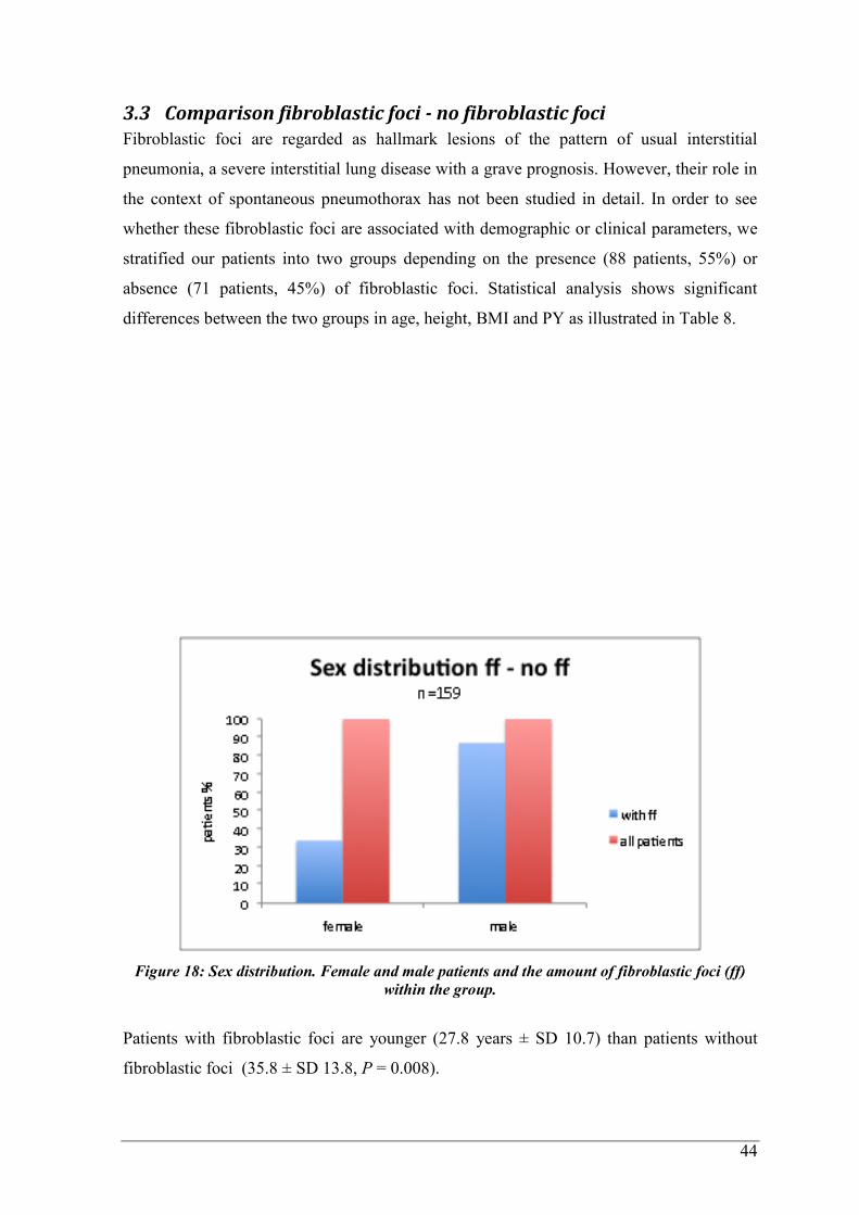
magnification x40) ...................................................................................................... 43 Figure 18: Sex distribution. Female and male patients and the amount of fibroblastic foci
(ff) within the group. ................................................................................................... 44

10
List of tables Table 1: Pathological features (8)........................................................................................10
Table 1: Staging of the pneumothorax (2)............................................................................13
Table 3: Personal criteria......................................................................................................22
Table 4: Histological criteria................................................................................................23
Table 5: Demographic details of personal criteria. Data are shown as ± SD.......................24
Table 6: Lung diseases – distribution...................................................................................28
Table 7: Demographic table of histological criteria.............................................................31
11
1 Introduction
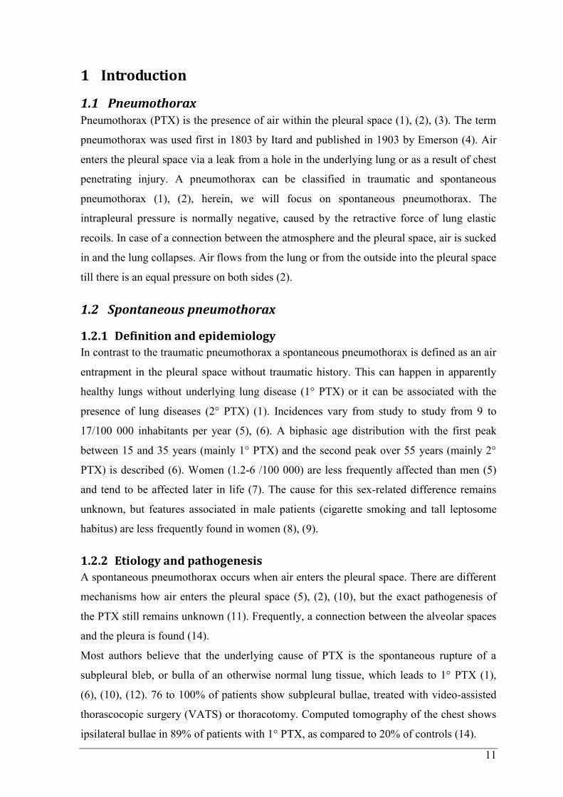
1.1 Pneumothorax Pneumothorax (PTX) is the presence of air within the pleural space (1), (2), (3). The term
pneumothorax was used first in 1803 by Itard and published in 1903 by Emerson (4). Air
enters the pleural space via a leak from a hole in the underlying lung or as a result of chest
penetrating injury. A pneumothorax can be classified in traumatic and spontaneous
pneumothorax (1), (2), herein, we will focus on spontaneous pneumothorax. The
intrapleural pressure is normally negative, caused by the retractive force of lung elastic
recoils. In case of a connection between the atmosphere and the pleural space, air is sucked
in and the lung collapses. Air flows from the lung or from the outside into the pleural space
till there is an equal pressure on both sides (2).
1.2 Spontaneous pneumothorax
1.2.1 Definition and epidemiology
In contrast to the traumatic pneumothorax a spontaneous pneumothorax is defined as an air
entrapment in the pleural space without traumatic history. This can happen in apparently
healthy lungs without underlying lung disease (1° PTX) or it can be associated with the
presence of lung diseases (2° PTX) (1). Incidences vary from study to study from 9 to
17/100 000 inhabitants per year (5), (6). A biphasic age distribution with the first peak
between 15 and 35 years (mainly 1° PTX) and the second peak over 55 years (mainly 2°
PTX) is described (6). Women (1.2-6 /100 000) are less frequently affected than men (5)
and tend to be affected later in life (7). The cause for this sex-related difference remains
unknown, but features associated in male patients (cigarette smoking and tall leptosome
habitus) are less frequently found in women (8), (9).
1.2.2 Etiology and pathogenesis
A spontaneous pneumothorax occurs when air enters the pleural space. There are different
mechanisms how air enters the pleural space (5), (2), (10), but the exact pathogenesis of
the PTX still remains unknown (11). Frequently, a connection between the alveolar spaces
and the pleura is found (14).
Most authors believe that the underlying cause of PTX is the spontaneous rupture of a
subpleural bleb, or bulla of an otherwise normal lung tissue, which leads to 1° PTX (1),
(6), (10), (12). 76 to 100% of patients show subpleural bullae, treated with video-assisted
thorascocopic surgery (VATS) or thoracotomy. Computed tomography of the chest shows
ipsilateral bullae in 89% of patients with 1° PTX, as compared to 20% of controls (14).

12
The mechanism of bullae formation remains speculative, but blebs or bullae may originate
from early emphysematous-like changes (ELCs) without underlying clinical disease (2),
(5), (10). ELCs are described as pneumatized epipleural chambers which are linked to the
lung parenchyma. Often the rupture of the pleura is found in the area of ELCs. The bullae
or the ELC can be caused by genetic predisposition, respiratory bronchiolitis or disorders
of the contralateral ventilation (5), (13). On CT scans ELCs were present in 89% of
patients on the ipsilateral side and in up to 80% on both lung sides (15), (16). In the control
group, consisting of healthy volunteers matched for age and smoking behaviour, only 20%
showed ELCs (16). ELCs were also present in 81% of patients with healed 1° PTX, who
had never smoked (17).
Inflammatory changes also play an important role in the formation of ELCs and bullae
(18). Inflammation, also caused by smoking, leads to a degradation of elastic fibers. The
so-called elastolysis is caused by an imbalance between proteases and antiproteases and
oxidants and antioxidants and a higher amount of macrophages and neutrophiles in that
tissue (19), (20), (21), (22). This also leads to bronchiolar wall fibrosis and destruction of
the pulmonary parenchyma, resulting in ELCs (23).
Smoking increases the number of inflammatory cells, especially macrophages in small
airways. Macrophages release potent chemotactic factors, resulting in accumulation of
neutrophiles in small airways (24), (25). Cigarette smoke itself leads to an influx of
neutrophiles caused by loss of functional activity of chemotactic factors inactivator (26).
All the inflammatory cells accumulate in the endobronchial system between the pulmonary
parenchyma and the bronchial tree leading to an endobronchial obstruction. The
obstruction induces an overpressure in the alveolar tissue resulting in rupture of pulmonary
parenchyma (27), (28).
Pleural perforation can also occure with inflammation caused by destroying abscesses,
tumors (lung carcinoma, lung metastasis) or other pathological processes of the lung (e.g.
pulmonal Langerhans-all-Histiocytosis) (5).
Most pneumothoraces occur in the lung apex as ELCs are mostly located in the lung
apices. In addition, the interpleural pressure is more negative in the lung apex than in the
basal lung area (5), (10), (29). This results in a higher wall tension in the alveolar space,
which leads to a higher predisposition to tissue rupture.
13
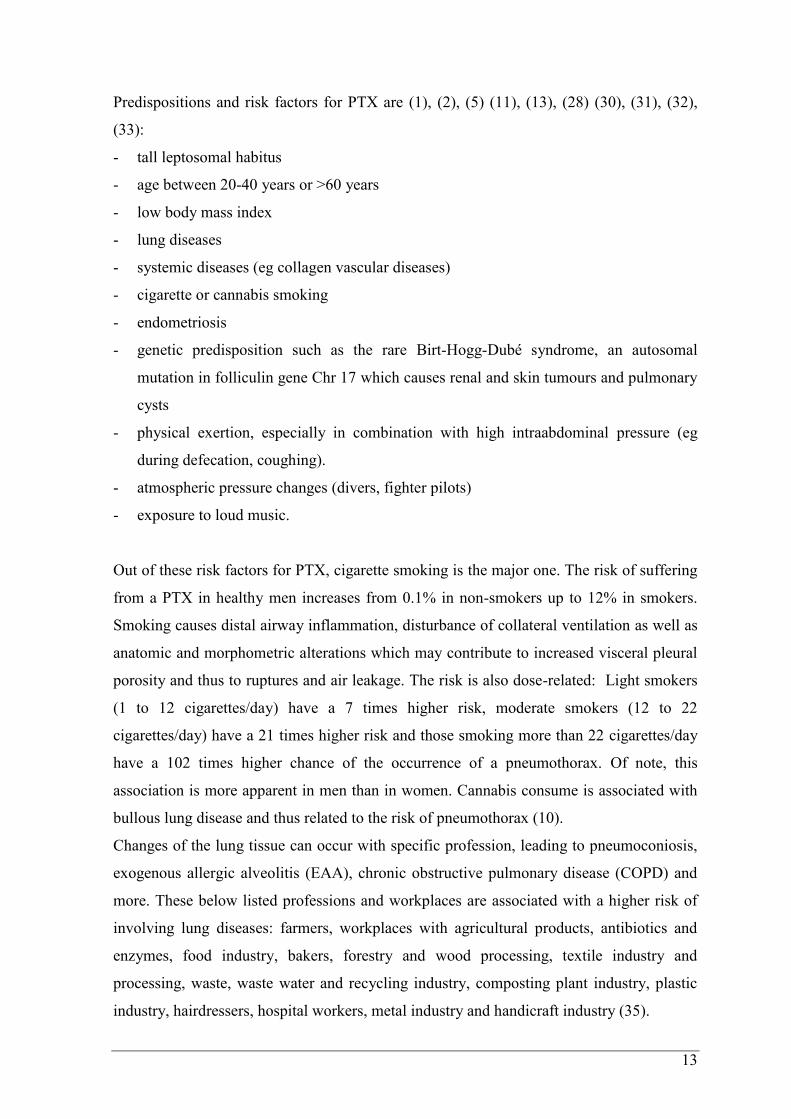
Predispositions and risk factors for PTX are (1), (2), (5) (11), (13), (28) (30), (31), (32),
(33):
- tall leptosomal habitus
- age between 20-40 years or >60 years
- low body mass index
- lung diseases
- systemic diseases (eg collagen vascular diseases)
- cigarette or cannabis smoking
- endometriosis
- genetic predisposition such as the rare Birt-Hogg-Dubé syndrome, an autosomal
mutation in folliculin gene Chr 17 which causes renal and skin tumours and pulmonary
cysts
- physical exertion, especially in combination with high intraabdominal pressure (eg
during defecation, coughing).
- atmospheric pressure changes (divers, fighter pilots)
- exposure to loud music.
Out of these risk factors for PTX, cigarette smoking is the major one. The risk of suffering
from a PTX in healthy men increases from 0.1% in non-smokers up to 12% in smokers.
Smoking causes distal airway inflammation, disturbance of collateral ventilation as well as
anatomic and morphometric alterations which may contribute to increased visceral pleural
porosity and thus to ruptures and air leakage. The risk is also dose-related: Light smokers
(1 to 12 cigarettes/day) have a 7 times higher risk, moderate smokers (12 to 22
cigarettes/day) have a 21 times higher risk and those smoking more than 22 cigarettes/day
have a 102 times higher chance of the occurrence of a pneumothorax. Of note, this
association is more apparent in men than in women. Cannabis consume is associated with
bullous lung disease and thus related to the risk of pneumothorax (10).
Changes of the lung tissue can occur with specific profession, leading to pneumoconiosis,
exogenous allergic alveolitis (EAA), chronic obstructive pulmonary disease (COPD) and
more. These below listed professions and workplaces are associated with a higher risk of
involving lung diseases: farmers, workplaces with agricultural products, antibiotics and
enzymes, food industry, bakers, forestry and wood processing, textile industry and
processing, waste, waste water and recycling industry, composting plant industry, plastic
industry, hairdressers, hospital workers, metal industry and handicraft industry (35).
14
1.2.3 Forms
There are two big groups of spontaneous pneumothorax, the primary spontaneous
pneumothorax (1° PTX) and the secondary spontaneous pneumothorax (2° PTX). Rare
forms of spontaneous pneumothoraces are the catamenial pneumothorax and the tension
pneumothorax (2), (10). Differentiation between these groups is important leading to
specific management and prognosis (10).
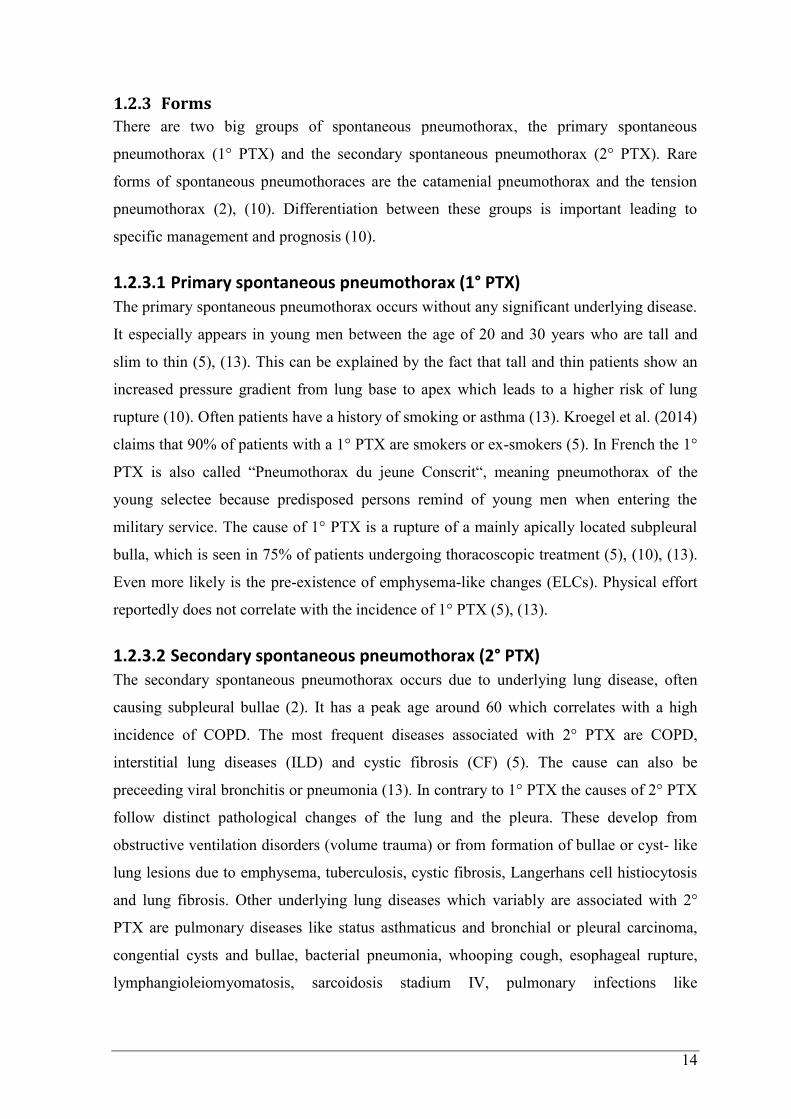
1.2.3.1 Primary spontaneous pneumothorax (1° PTX)
The primary spontaneous pneumothorax occurs without any significant underlying disease.
It especially appears in young men between the age of 20 and 30 years who are tall and
slim to thin (5), (13). This can be explained by the fact that tall and thin patients show an
increased pressure gradient from lung base to apex which leads to a higher risk of lung
rupture (10). Often patients have a history of smoking or asthma (13). Kroegel et al. (2014)
claims that 90% of patients with a 1° PTX are smokers or ex-smokers (5). In French the 1°
PTX is also called “Pneumothorax du jeune Conscrit“, meaning pneumothorax of the
young selectee because predisposed persons remind of young men when entering the
military service. The cause of 1° PTX is a rupture of a mainly apically located subpleural
bulla, which is seen in 75% of patients undergoing thoracoscopic treatment (5), (10), (13).
Even more likely is the pre-existence of emphysema-like changes (ELCs). Physical effort
reportedly does not correlate with the incidence of 1° PTX (5), (13).
1.2.3.2 Secondary spontaneous pneumothorax (2° PTX)
The secondary spontaneous pneumothorax occurs due to underlying lung disease, often
causing subpleural bullae (2). It has a peak age around 60 which correlates with a high
incidence of COPD. The most frequent diseases associated with 2° PTX are COPD,
interstitial lung diseases (ILD) and cystic fibrosis (CF) (5). The cause can also be
preceeding viral bronchitis or pneumonia (13). In contrary to 1° PTX the causes of 2° PTX
follow distinct pathological changes of the lung and the pleura. These develop from
obstructive ventilation disorders (volume trauma) or from formation of bullae or cyst- like
lung lesions due to emphysema, tuberculosis, cystic fibrosis, Langerhans cell histiocytosis
and lung fibrosis. Other underlying lung diseases which variably are associated with 2°
PTX are pulmonary diseases like status asthmaticus and bronchial or pleural carcinoma,
congential cysts and bullae, bacterial pneumonia, whooping cough, esophageal rupture,
lymphangioleiomyomatosis, sarcoidosis stadium IV, pulmonary infections like
15
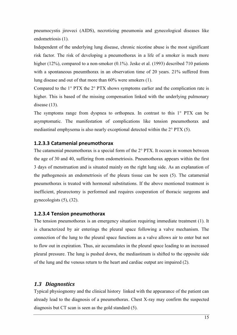
pneumocystis jiroveci (AIDS), necrotizing pneumonia and gynecological diseases like
endometriosis (1).
Independent of the underlying lung disease, chronic nicotine abuse is the most significant
risk factor. The risk of developing a pneumothorax in a life of a smoker is much more
higher (12%), compared to a non-smoker (0.1%). Jeske et al. (1993) described 710 patients
with a spontaneous pneumthorax in an observation time of 20 years. 21% suffered from
lung disease and out of that more than 60% were smokers (1).
Compared to the 1° PTX the 2° PTX shows symptoms earlier and the complication rate is
higher. This is based of the missing compensation linked with the underlying pulmonary
disease (13).
The symptoms range from dyspnea to orthopnea. In contrast to this 1° PTX can be
asymptomatic. The manifestation of complications like tension pneumothorax and
mediastinal emphysema is also nearly exceptional detected within the 2° PTX (5).
1.2.3.3 Catamenial pneumothorax
The catamenial pneumothorax is a special form of the 2° PTX. It occurs in women between
the age of 30 and 40, suffering from endometriosis. Pneumothorax appears within the first
3 days of menstruation and is situated mainly on the right lung side. As an explanation of
the pathogenesis an endometriosis of the pleura tissue can be seen (5). The catamenial
pneumothorax is treated with hormonal substitutions. If the above mentioned treatment is
inefficient, pleurectomy is performed and requires cooperation of thoracic surgeons and
gynecologists (5), (32).
1.2.3.4 Tension pneumothorax
The tension pneumothorax is an emergency situation requiring immediate treatment (1). It
is characterized by air enterings the pleural space following a valve mechanism. The
connection of the lung to the pleural space functions as a valve allows air to enter but not
to flow out in expiration. Thus, air accumulates in the pleural space leading to an increased
pleural pressure. The lung is pushed down, the mediastinum is shifted to the opposite side
of the lung and the venous return to the heart and cardiac output are impaired (2).
1.3 Diagnostics Typical physiognomy and the clinical history linked with the appearance of the patient can
already lead to the diagnosis of a pneumothorax. Chest X-ray may confirm the suspected
diagnosis but CT scan is seen as the gold standard (5).
16
1.3.1 Clinical observation
1.3.1.1 Medical history
- young men, <30 years, leptosomal habitus (tall, thin, slim and light bones) are
associated with 1° PTX
- clinical history of pulmonary diseases and other systemic diseases like cystic fibrosis,
tuberculosis, lungfibrosis, lymphangioleiomyomatosis, HIV infection, COPD,
emphysema, alpha-1 antitrypsin deficiency are more associated with 2° PTX (5).
1.3.1.2 Symptoms
Symptoms depend on the dimension of the pneumothorax and the underlying disease. It
generally occurs unilateral (29). Small amounts of air trapped in the pleura cavity can be
asymptomatic and it may resolve itself without any intervention within a few days (5),
(29). But only 5% of pneumothoraces are asymptomatic (13). A significant pneumothorax
shows a sudden onset, starting with localized pain at the affected thorax side and
breathlessness (5), (13), (29). In 2° PTX dyspnea is the most prominent clinical feature
whereas dyspnea is often absent or mild in 1° PTX. In 1° PTX patients more often suffer
from a sudden ipsilateral chest pain (11).
Healthy young adults may tolerate a pneumothorax quite well and do not complain about
impairing symptoms (2). Therefore, younger patients are often hospitalized 1-2 days after
the real onset of the lung collapse (13). Older patients with underlying lung diseases often
develop severe respiratory distress (2).
Cough can also appear in an early stage of PTX. Tussis occurs because the two pleural
layers vanish apart from each other. In the beginning the sudden pain is depending on the
breathing movement caused by the touching of the pleural layers while breathing. For this
reason patients avoid inhaling deeply, called inspiration-blockage or doorstop-
phenomenon. In the later stage pain can diminish or desist when the interpleural air
contempt is large enough to keep the two pleural layers apart from each other (5).
Instead of this dyspnoe (exertional) may appear caused by the increased lung collapse.
If air is trapped in the pleural space, it is called a tension pneumothorax. In that case
tachypnoea, cyanosis, cardiovascular depression and later on pulsus paradoxus may occur.
The arterial blood gas analysis shows hypoxia and hypercapnia when disturbance or
underlying disease is substantial (5).
1.3.1.3 Physical examination
On physical examination suspecting a pneumothorax these listed features may be seen:
17
- tachypnoea
- relieving posture (13)
- hypersonic percussion sound “acoustic sound of a carton“
- diminished or missing vocal fremitus
- attenuated or abolished breathing sounds (5)
- Hamman´s sign, a click on auscultation in time with the heart sound
- bubbles and crackles under the skin of the torso and neck if there is subcutaneous
emphysema (1).
The hypersonic percussion sound results of the higher amount of air in the chest cavity and
so forth a hollow percussion tone, like drumming on a thick carton (“carton ton“) (5).
A tension pneumothorax can also present with tachycardia, pulsus paradoxus, marked
jugular venous distension, decreased blood pressure, cyanosis, mediastinal emphysema till
to cardiogenic schock (13).
1.3.2 Body-imaging
1.3.2.1 Chest X-ray in expiration
1.3.2.1.1 Direct signs
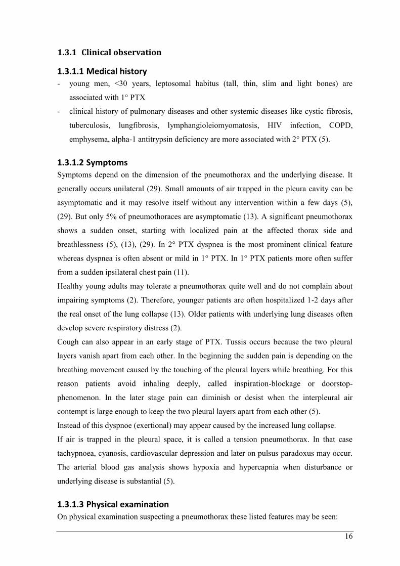
The chest X-ray, performed in two levels (anterior-posterior as well as lateral), still
represents the gold standard in imaging. A typical chest X-ray in expiration of a patient
with pneumothorax shows a structure free airspace between the collapsed lung and the
chest wall, seen in figure 1. The features of a pneumthorax on a posteroanterior (PA) erect
chest X-ray are:
- visible pleura line, hairline (see Figure 1)
- enhanced transparency lateral of the collapsed lung
- absent pulmonary vascular markings (5).
In addition tension pneumothorax shows
- an asymmetry of volume, as the affected lung appears larger
- the mediastinum can be shifted to the direction of the healthy thorax side
- widened intercostal space at the affected lung side
- the diaphragm can be flattened and deeper.
These signs can be missing if the visceral pleura and the parietal pleura are stucked
together by adhesions (5).
18
1.3.2.1.2 Indirect signs
The above described direct signs can not be seen when the X-ray is made on a recumbent
patient. The leaked air accumulates in the ventral thorax while the lateral lung still sticks to
the chest wall. The characteristical indirect features of a recumbent chest X-ray are
- accented border of heart, mediastinum and diaphragm caused by the missing contact
between lung parenchym and mediastinum, heart or diaphragm
- increased transparency especially in the lower lung areas
- a wide and deep costodiaphragmatical cavity (“deep sulcus sign“) caused by the
missing negative pressure in the pleural space.
Skin folds seen on a reclining patient may lead to false positive diagnosis of a
pneumothorax (5). Tiny pneumothoraces which are often not detected on an erect chest X-
ray may be diagnosed by lateral chest or decubitus radiographs (1). The identification of a
convex pleural line may help to distinguish between a pneumothorax and large bullae (2).
Figure 1: Erect chest X-ray (PA) of a right sided PTX. Typical hairline marking the collapsed
lung ( ). (Kindly provided by Assoz.Prof.PD Dr. Jörg Lindenmann)
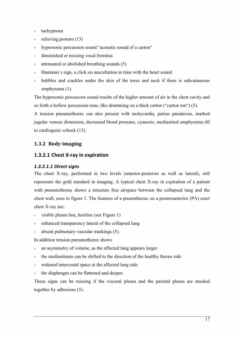
1.3.3 Computed tomography of the thorax
The computed tomography (CT) scan of the thorax is the best diagnostic option to confirm
or to exclude a pneumothorax which is not clearly visible on the chest X-ray (5). A typical
presentation of a pneumothorax is seen in Figure 2. Small amounts of air in the pleura
space can be detected in the so called lung window. The identification of a pneumothorax
of an anterior (small) or anterolateral (bigger) may be useful for the implantation of an
intercostal suction drainage (5). In some cases CT scans may help to distinguish between
emphysema, bullae, other bullous diseases and pneumothorax (1),(5). Furthermore CT may
be useful in detecting underlying lung diseases (1).
Figure 2: CT scan right sided PTX. Lung margin ( ) . (Kindly provided by
Assoz.Prof.PD Dr. Jörg Lindenmann)
1.3.3.1 Transthoracic sonography
The ultrasound of the thorax describing a pneumothorax is characterised by
- the absence of the synchronic breathing movement of the hyperechoic pleura lung
border reflex, seen as a “slide sign“ or “curtain phenomenon“
19
- missing of the “tail of the comet phenomenon artifact“ or torch phenomenon
- the absence of the slightly pulse synchronic movability of the visceral pleura
- increased regular replication echoes (reverberation) of the air filled pleura space (5)
- the passage between the air filled lung and pneumothorax (6).
1.3.4 Pathological features
Jeske et al. (1993) described 710 patients with PTX: 80% had a totally collapsed lung, 20%
had an apical pneumothorax and less than 10% developed bilateral pneumothoraces (13),
(34). Pneumomothorax commonly occurs unilateral (29). PTX can affect one of both sides
of the lung. It is not shown that one of the lung side is affected more often (1).
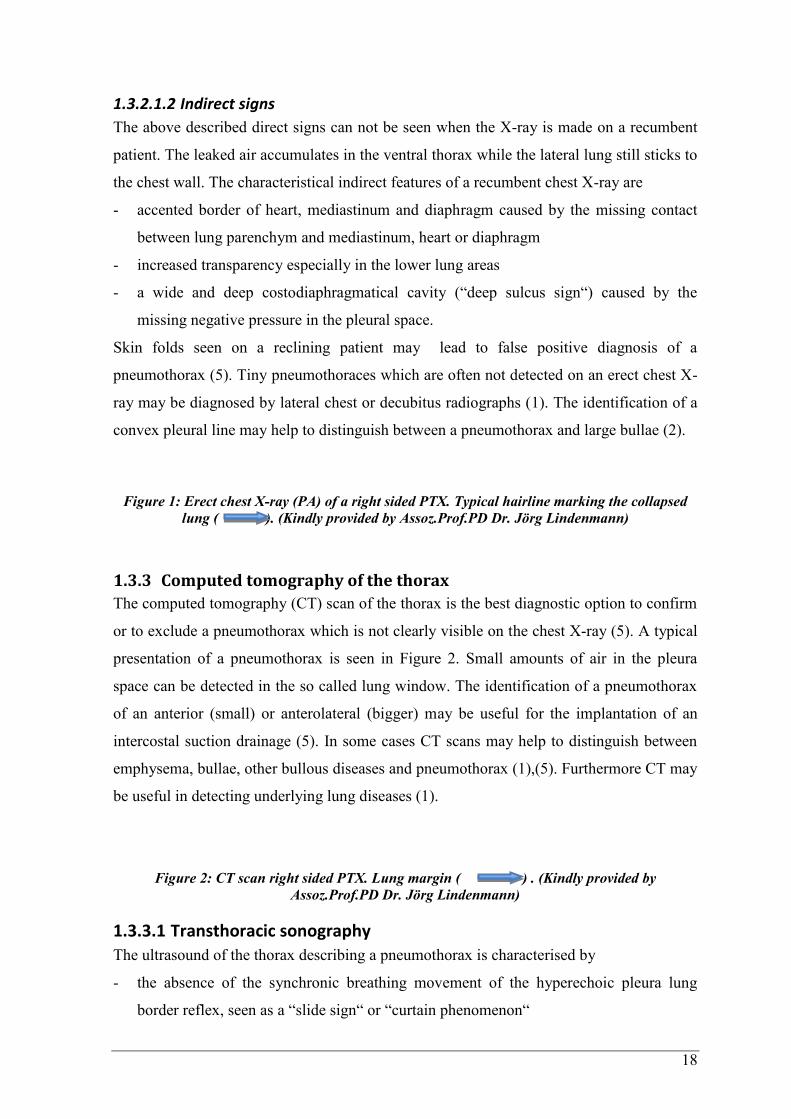
Spontaneous pneumothorax is caused due to some pathological changes of the lung,
usually emphysema. Especially in healthy young individuals the lung appear to be normal.
But when a thoracotomy was performed these people show common histological changes,
illustrated in table 1 (29).
Pathological features
- Emphysema
- Fibrosis
- Bullae
- Pleural porosity
- Inflammation
- Alveolar collapse
- Mesothelial hyperplasia
- Eosinophilic pleuritis
- Fibroblastic foci
- Pigmented macrophages
Table 1: Pathological features (29)
Fibrosis is usually found at the apex of the lung and can extend up to 2-3 cm. If fibrosis is
overlapped of one or more bullae ( > 1cm) it is called apical cap. There can be a hole in the
pleural surface, connecting the pleura and the bullae (29), although only a minority of
20
blebs are actually ruptured at the time of thoracoscopy or surgery. Therefore, blebs and
bullae are related to the occurrence of pneumothorax but account seldomly as the
triggering factor (36), (37), (38).
More important are other lesions, especially pleural porosity, areas of disrupted
mesothelial cells at the visceral pleura, replaced by an inflammatory elastofibrotic layer
with increased porosity, allowing air leak into the pleural space (11).
Histology shows alveolar collapse, chronic inflammation and reactive mesothelial
hyperplasia with eosinophils dominating. One of the most common seen pathological
features in spontaneous pneumothorax is pulmonary fibrosis. Pulmonary fibrosis is an
unspecific reaction of lung tissue (29). It especially occurs in alveolar septa or as a
circumscribed lesion, a so called scar (39). Pulmonary fibrosis involves the gradual change
of normal lung parenchyma with fibrotic tissue, represented by an excessive fibrous
connective tissue. The replacement of normal lung with scar tissue causes irreversible
decrease in oxygen diffusion capacity and the resulting stiffness or decreased compliance
makes pulmonary fibrosis a restrictive lung disease (40). Pulmonary fibrosis is maintained
by aberrant wound healing, rather than chronic inflammation (41). The cause of pulmonary
fibrosis can be a secondary effect of other diseases like autoimmune disorders, viral
infections, bacterial infections (tuberculosis), toxic inhalation (cigarette smoking,
asbestosis, silicosis) leading to interstitial lung disease (41). If the cause is unknown it is
called idiopathic pulmonary fibrosis, also known as usual interstitial pneumonia (UIP)
(42).
Normally, the reactive eosinophilic pleuritis affects only the surface of the pleura and
penetrates only a short distance into the lung tissue. If air is trapped in the pleural cavity
for a longer time also mesothelial changes, especially squamous changes can occur. These
can develope into malignant formations. Often there is no underlying disease or
tuberculosis to be found (29).
Especially inflammatory changes lead to obstruction check-valve mechanism that can be
seen as the real cause of pneumothorax (11).
The changes in the apical region of the lung may develop themselves out of rapid somatic
growth in tall, thin men. When these people stay erect the apices are being particularly
poorly perfused and cause ischaemia. This leads to direct damage of the apices or fibrosis.
This apical changes often affects both lung sides therefore there is a high risk of recurrence
on the opposite side. Rarely, there can be minor anatomical anomalies of the bronchi
detected when pneumothorax occurs cumulative in one family (29).
21
Cigarette smoking is the major risk factor of evolving a pneumothorax (10). Smoking may
cause degradation of elastic fibers due to influx of neutrophils and macrophages in the
lung. This degradation causes an imbalance in the protease-antiprotease and oxidant-
antioxidant system. After bullae have been formed, inflammation-induced obstruction of
the small airways increases alveolar pressure, resulting in an air leakage into the lung
interstitium. Air then moves into the hilum, causing pneumomediastinum. This leads to a
higher mediastinal pressure and rupture of the mediastinal parietal pleura, causing
pneumothorax (14). The phagocytic capabilities of macrophages provide the first line of
defense against toxic agents in the distal lung and is also shown by ingested coarse-grained
pigments resulting in brown macrophages (43), (44), (45). Macrophages have a defensive
role in the lung and at the same time they may be important in the pathogenesis of
pulmonary disease. The release of enzymes and chemical mediators from alveolar
macrophages may result in pulmonary fibrosis and also play an important role in the
pathogenesis of emphysema (46), (47). Cigarette smoke not only induces changes of
macrophages, it also leads to an accumulation of pigmented alveolar macrophages (48).
Nevertheless, blebs and bullae also occur in up to 15% of normal lung tissues without any
pathological features (30), (49), (50).
For the first time, Deborah et al. (2012) identified in a 92 case study a distinct pattern of
pneumothorax associated with fibroblastic lesions in a subset of cases of PTX. 12% of all
patients with PTX presented a pattern of pleural fibrosis with islands of fibroblastic foci
within a myxoid stroma at the pleural-parenchymal interface or leading edge. It still
remains unknown whether this fibroblastic pattern are related to the pathogenesis (51).
Fibroblastic lesions (see Figure 13) usually are found in paraseptal and subpleural areas
where they protrude into the alveolar spaces. They are covered with epithelium, show a
myxoid stroma and usually lack inflammatory infiltrates. They are thougt to represent the
manisfestion of wound healing in ongoing lung injury. In general, fibroblastic lesions are
regarded as the hallmark lesion in the histological pattern of usual interstitial pneumonia
(UIP), seen in idiopathic pulmonary fibrosis (IPF), a rare idiopathic interstitial pneumonia.
This enigmatic disease which carries a grave prognosis usually is diagnosed in male
patients above the age of 50.
Furthermore, smokers often present respiratory bronchiolitis associated interstitial lung
disease (RB-ILD). RB-ILD is almost exclusively apparent in heavy smokers and it is
defined as accumulation of pigmented macrophages in the alveolus and interstitium with
chronic bronchitis and a mild fibrotic-inflammatory interstitial reaction (45), (52).
22
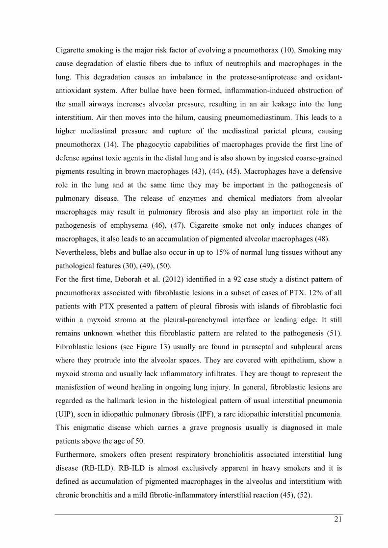
1.3.5 Staging and evaluation of the pneumothorax extent
Pneumothorax can be staged with histological criterias (Verschoof) (5). Nowadays the
characterization of Vanderschueren using high resolution computed tomography (HR-CT),
as illustrated in table 2, is more common (5), (53).
Evaluation of the pneumothorax extent can be made easily with chest X-ray. Chest X-ray
allows to measure the extent of the pneumothorax. The width of the rim of air surrounding
the lung is used to classify pneumothoraces into small (rim of air measured at the level of
hilum ≤ 2cm) and larger ( >2cm). A 2cm rim of air approximately equates to a 50%
pneumothorax in volume (1), (2).
Stage Vanderschueren Verschoof
I Idiopathic pneumothorax,
endoscopic normal lung
Pneumothorax without
histological verifiable
results
II Pneumothorax with
pleuropulmonal adhesions
Pneumothorax with an
apical bullae
III Pneumothorax with a single
bulla ( < 2cm in diameter)
Pneumothorax with general
cystic lung disease
IV Pneumothorax with multiple
bullae ( >2cm diameter)
Table 2: Staging of the pneumothorax (5)
1.4 Therapy
1.4.1 Therapy decision
There are three international guidelines (the American College of Chest Physicians Delphi
consensus statement, the British Thoracic Society guidelines and the Belgian Society of
Pulmonology) which contrast sharply in many aspects of proposed treatment (7).
Treatments of pneumothorax shown in Figure 3 are mostly based on the guidelines of the
he British Thoracic Society.
The aim of the pneumothorax therapy is the evacuation of the pleura space as soon as
possible. This will lead to a re-dilatation of the collapsed lung (5).
23
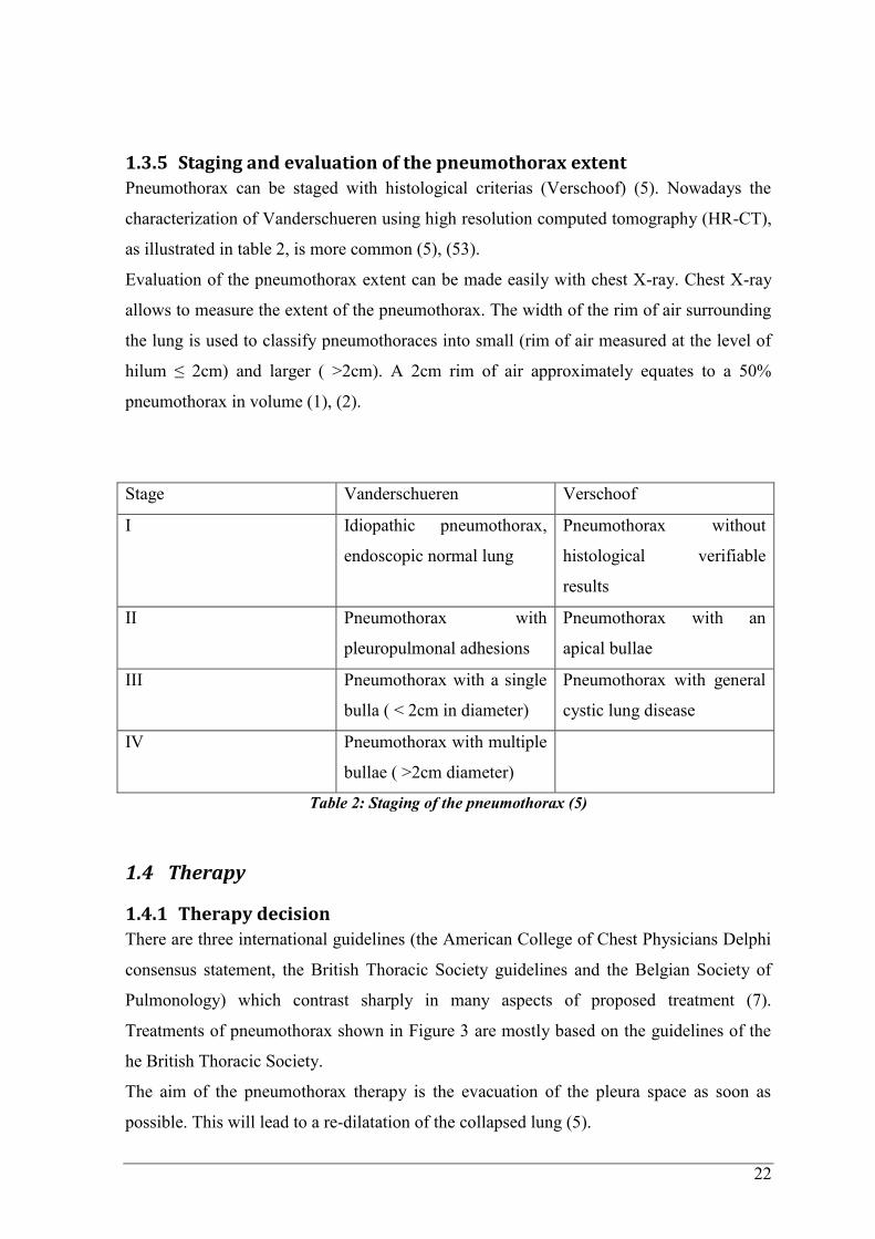
For initial management it should be determined whether the pneumothorax is 1° or 2°.
Patients with a 2° PTX tolerate the pneumothorax less well and also show a significant
higher mortality with 10% and therefore should be managed more aggressively (1), (32).
Therefore, patients with 2° PTX always have to be hospitalized for at least 24 hours (32).
The underlying lung disease should also be treated (1).
One of the most important factors for therapy decision is the severity of breathlessness.
Breathlessness indicates the need for active intervention as well as supportive treatment
(32). The decision of the therapy of a pneumothorax based on clinical presentation is
illustrated in figure 3.
Figure 3: Algorithm for treatment of spontaneous pneumothorax (1)
Furthermore the decision of therapy depends on the following facts:
- clinical presentation of the patient including breathlessness, hypoxia, haemodynamic
compromise
- presence of underlying lung disease
24
- probability of spontaneous regression
- probability of a relapse
- progression of the pneumothorax between 9 and 96 hours
- current dimension of the pneumothorax (5).
1.4.2 Observation range
The evaluation of a patient with pneumothorax include:
- re-evaluation of the clinical stage
- chest X-ray controls
- re-evaluation of the size of the pneumothorax.
There are no guidelines regarding chest X-ray controls.
After an uncomplicated pneumothorax the lung may expands itself 3 weeks after the event
(5).
1.4.3 Conservative treatment
Discharge combined with frequent controls: Patients with a small 1° PTX without
breathlessness can be considered for discharge with early outpatient review. These patients
should receive clear written advice to return in the event of worsening breathlessness (32).
Observation (without oxygen): If the patient has no symptoms and the pneumothorax is
small the patient can be observed and controlled within small breaks. The precondition is
that there is no progression of the PTX (5). Observation is the treatment of choice
especially in small 1° PTX without any significant breathlessness. Also selected
asymptomatic patients with a large 1° PTX may be managed by observation alone (32).
Each day 1.25% of the PTX-volumes will be reabsorbed spontaneously (5). Such patients
may be discharged advised to return to the hospital as soon as symptoms worsen (2).
Observation (with oxygen): The reabsorbed range is 4 times bigger if the patient gets high-
flow oxygen (> 3L/min) trough a nasal cannula, with appropriate caution in patients with
COPD (1), (2) . Oxygen also reduces dyspnea and hypoxaemia. However acording to
Stephen Chapam (1) every hospitalized patient should get high flow (10L/min) inspired
O2. Inhalation of oxygen displaces the nitrogen in the pleura space which leads to a bigger
difference between the total gas pressure in the pleural space and the pressure in the pleural
capillaries. This leads to faster reabsorption of the air which is trapped in the pleura space
(2).
Aspiration: A simple needle aspiration using a small gauge-needle (12-14 French) to suck
out air of the pleural space can relieve symptoms. A part of the PTX-volume can be
25
aspirated by hand. The patient has to sit up straight and before inserting the needle local
anesthetic should be injected (2), (5). Between the 2nd
and 3rd
intercostal space the needle
is placed medioclaviculary. After aspiration of 150-500ml the needle is removed.
Indication for small needle aspirations are small and uncomplicated pneumothoraces. In
70% of PTX cases this treatment is sufficient. Small needle aspiration accelerates the
degeneration of the pneumothorax and also the duration of hospitalization can be reduced
(5), (32). Also a 14-16 gauge-needle is as effective as large-bore (> 20 French) chest drains
and should be preferred. A needle aspiration should not be repeated unless there were
technical difficulties. If a needle aspiration failed a small bore (< 14 French) chest drain
insertion is indicated (32).
Small caliber drainage: A plastic or teflon drainage (e.g. Matthys-catheter, pleura-cath) is
inserted into the 2nd
or 3rd
intercostal space mediaclaviculary using the Seldinger technique
(2). There is a minor risk of hurting the expanded lung because of the elastic material
which is used (5). In spontaneous pneumothorax, small caliber (smaller than 14 French)
tubes may be inserted, larger tubes do not have an advantage (54).
Portable chest tube or intercostal tube drainage: Chest tubes are usually not required in the
majority of patients with a 1° PTX. They are more common in the management of critical
ill patients with severe diseases including a higher risk of morbidity and mortality (1).
Small caliber chest drains are preferred for chest tube treatment (32). The external part of
the chest tube stays open with a one-way valve system like the Heimlich valve or with a
water seal. The Heimlich valve is more often used because it can be used without
hospitalization and it may even reduce the duration of the hospital stay. This sort of drain
allows the air to escape but not to re-enter without an extra negative pressure circuit and
also avoids a tension pneumothorax. The normal movement of the patient leads to a higher
interpleural pressure so that air escapes through out the drainage. 75% of the patients with
a portable drainage may not suffer from a relapse. It can also be used if patients have to be
transported (5). The tube is left in place until no air is seen to escape from it for a period of
time, and X-rays confirm re-expansion of the lung (54), (55). Chest tubes are used first-line
when pneumothorax occurs in people with AIDS, frequently due to underlying
pneumocystis pneumonia, as this condition is associated with prolonged air leakage.
Bilateral pneumothorax is relatively common in people with pneumocystis pneumonia, and
surgery is often required (54).
Thoracotomy with continuous negative pressure circuit: It is used as the first-line therapy
of a 2° PTX or if there is a high risk of relapse and persistent leakage. Frequently small
26
calibre (7-14 Fr) chest tubes are sufficient. Bigger caliber tubes (> 24 Fr) are used if higher
sucking is needed to expand the lung (5). High-volume low-pressure suction systems with
10-30cm H2O are recommended (5), (32).
Big caliber tubes are inserted into the 4th
-7th
intercostal space in the middle or frontal
axillary line after a local anesthetic is applied (5).
A chest tube is indicated
- when the distance between the apical thorax and the lung apex is bigger or even
3cm
- if the pneumothorax takes more than 15% of the size of the hemithorax
- if there is a high risk of progression or relapse
- if the patient is cardiopulmonary prestressed (5).
Removal of the chest tube
The chest tube can be removed after several (5-7) days when there is no suspicion of a
persistent air leak and the daily chest X-ray shows a fully expanded lung. The most
important step is that a positive intrapleural pressure is preserved at the moment of
removal. The patient should be in a comfortable position and should take a deep breath.
While the patient exhales, the drain is pulled out as a whole at one time. 12-24 hours after
the removal a chest X-ray is indicated (5). The sucessful therapy is shown in Figure 4.
1.4.4 Surgery
Usually a first time pneumothrax can be treated with conservative measures. The aim of
the surgical treatment is the repair of the apical hole or bleb and to close the pleural space.
A relapse of a pneumothorax is an indication for surgical therapy. As well as persistent air
leak over 7 days, second ipsilateral 1° PTX, first contralateral 1° PTX, bilateral 1° PTX,
spontaneous haemothorax, first time 1° PTX in patients with profession at risk like pilots
or underwater divers and first time manifestation of a 2° PTX in patients with underlying
diseases (1), (5).
1.4.4.1 VATS
Video-assisted thoracoscopic surgery (VATS) is a type of thoracic surgery performed
using a small video camera that is introduced into the patient's chest via a scope.
Instruments can be inserted through separate holes in the chest wall called “ports”. Because
of the small skin entrances the risk of infektion or wound dehiscence is minor. This leads
27
to faster and better wound healing and a quicker postoperative recovery (56). Nowadays,
VATS is standard due to minimal invasion, little postoperative pain and minor
complication rate. The pleura can be observed, bullae can be removed and also partial
pleural abrasion can be performed. VATS also prevents big skin scaring, offers a shorter
in-hospital stay and reduces postoperative lung problems (5), (54). 2° PTX, re-occurred 1°
PTX as well as primary 1° PTX can be treated with VATS. Especially in young patients
with 1° PTX VATS is the gold standard (1). The relapse rate after VATS is 2-14% which
is much smaller than the relapse rate after chest tube therapy (30%) (5).
1.4.4.2 Thoracotomy
A thoracotomy is much more complex than VATS. Also the patient has to be hospitalized
longer then treated with VATS. In contrast to this, thoracotomy has the lowest relapse rate
(0-7 %) (5).
1.4.4.3 Transaxillary mini-thoracotomy
Transacillary mini-thoracotomy is a variation of thoracotomy using a minimal axillary
incision. It is a reasonable alternative to the conventional thoracotomy due to less
invasiveness (1).
1.4.4.4 Pleurodesis
If a persistent leak allows air to flow out of the pleura space a pleurodesis is indicated. As
first-line therapy talcum or tretracycline is used to provoke a sclerosis to artificially
obliterate the pleural space (1), (5). It can be instilled through an intercostal drain or
directly during VATS, thoracotomy or pleurectomy. As a requirement the lung has to
touch the pleura. After pleurodesis patients have the smallest relapse rate (<10 %). But
chemical pleurodesis has a failure rate of 10-20% (1). The talcum is instilled via the chest
tube or directly and after 3 hours a suction is applied to ensure a pleuropleural contact.
Five minutes after instilling the talcum patients claim about lot of pain thus local and
intrapleural anesthesia with xylocain is necessary. Furthermore a systemic pain killer like
opiates or sedatives can be used. As a rare complication acute respiratory distress
syndrome (ARDS) may occur especially when the talcum dose needed is higher than 4 g
(5).
28
1.4.5 Complications
Not only the lungs can collapse but there may occur other complications. Also specific
situations like tension pneumothorax and catamenial pneumothorax require different
management strategy.
Subcutaneous emphysema: Air is trapped in the layer of skin under pressure from the
pleural space, coming from the chest cavity travelling along the fascia. The cause can be a
large air leakage, especially when underlying lung disease such as COPD is present. In
most cases it is harmless but rarely can cause a respiratory compromise from upper airway
compression. Subcutaneous emphysema can be treated with high-flow (10L/min) inspired
O2. If the emphysema resists the treatment can be expanded with large-caliber chest drain
on suction (1).
Re-expansion pulmonary edema: In 14% of pneumothorax cases patients suffer from
breathlessness and cough, accompanied by edema in the re-expanded lung, visible in the
chest X-ray. It occurs more frequently in young patients with large 1° PTX and if the use
of a chest drain on suction is applied between the first 48 hours. In most cases it is self-
resolving and no treatment is needed.
Tension pneumothorax: A tension pneumothorax is a case of emergency. If there is any
suspicion of a tension pneumothorax high-flow O2 is indicated and the insertion of a large
bore cannula is needed. If the sound of hiss of escaping air is noticeable the diagnosis of a
tension pneumothorax can be made. Also immediate chest X-ray should be done. Tension
pneumothorax is treated with air aspiration until the the patient is less distressed and after
that a chest drain should be inserted and should stay there until no air comes out and the
underwater seal is bubbling satisfactorily.
Catamenial pneumothorax: When the occurrence of pneumothorax happens at time of
menstruation and it is recurrent, the diagnosis of catamenial pneumothorax is likely. It can
be treated with VATS, pleurodesis and/or ovulation-suppressing drugs (1).
1.4.6 Relapse therapy
Young patients without any underlying disease should have surgery to prevent the
recurrence of a pneumothorax. Nowadays VATS is standard due to minimal invasion, little
postoperative pain and a minor complication rate. The pleura can be observed
macroscopically, bullae can be removed and also partial pleural abrasion can be performed.
VATS also prevents big skin scars, offers shorter in-hospital stays and reduces postsurgical
lung problems. The relapse rate is lower than 1% after surgical intervention (5), (54).
29
The most significant risk factors for recurrence of a pneumothorax are large size
pneumothoraces with persistent air leakage treated with conservative therapy. Other factors
such as smoking status, BMI and number of bullae are not showing a significantly higher
recurrence rate (57). Although rarely seen in women the occurrence of 1° PTX in women
of childbearing age is not unusual. There appears to be an increased risk of recurrence of
pneumothorax during pregnancy and during parturition (32).
1.4.7 Follow-up care
After successful re-dilatation of the collapsed lung a chest X-ray should be performed 7-10
days after discharge. Patients also should be advised not to travel by plane or do longer
travels until the lung is again fully extended (1), (5). During a plane flight, barometic
pressure is reduced at altitudes and this can cause expansion of enclosed thoracis air
pockets (2). Also patients should never dive in the future, unless he or she has undergone
definitive surgical procedure. Especially during therapy the patient should avoid smoking
but also in long-term prognosis chronical smoking should be stopped (1), (5), (32).
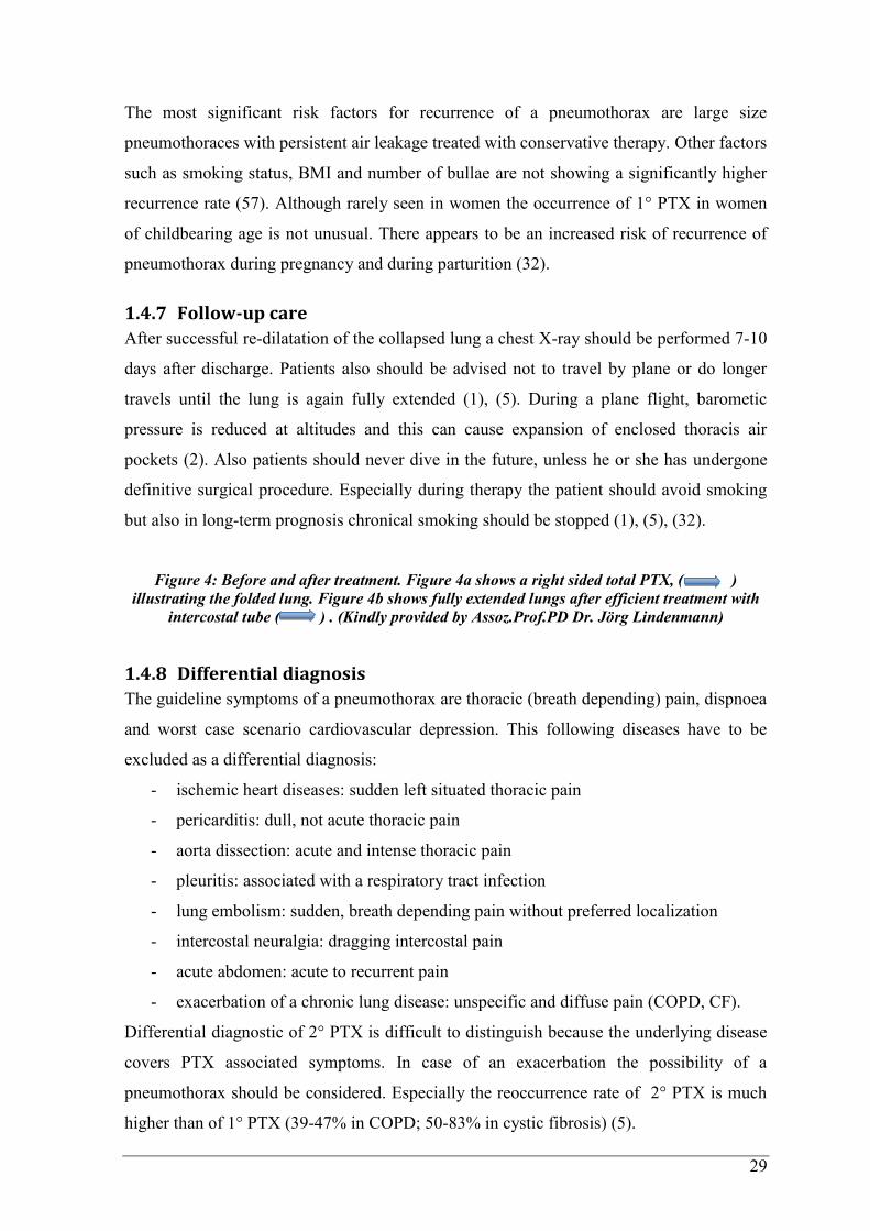
Figure 4: Before and after treatment. Figure 4a shows a right sided total PTX, ( )
illustrating the folded lung. Figure 4b shows fully extended lungs after efficient treatment with
intercostal tube ( ) . (Kindly provided by Assoz.Prof.PD Dr. Jörg Lindenmann)
1.4.8 Differential diagnosis
The guideline symptoms of a pneumothorax are thoracic (breath depending) pain, dispnoea
and worst case scenario cardiovascular depression. This following diseases have to be
excluded as a differential diagnosis:
- ischemic heart diseases: sudden left situated thoracic pain
- pericarditis: dull, not acute thoracic pain
- aorta dissection: acute and intense thoracic pain
- pleuritis: associated with a respiratory tract infection
- lung embolism: sudden, breath depending pain without preferred localization
- intercostal neuralgia: dragging intercostal pain
- acute abdomen: acute to recurrent pain
- exacerbation of a chronic lung disease: unspecific and diffuse pain (COPD, CF).
Differential diagnostic of 2° PTX is difficult to distinguish because the underlying disease
covers PTX associated symptoms. In case of an exacerbation the possibility of a
pneumothorax should be considered. Especially the reoccurrence rate of 2° PTX is much
higher than of 1° PTX (39-47% in COPD; 50-83% in cystic fibrosis) (5).
30
1.4.9 Prognosis and development
The development of an uncomplicated pneumothorax can take from hours to days. In
contrast, tension pneumothorax may develop rapidly within a few minutes.
Generally speaking there is always the possibility of a recurrence after the first
pneumothorax. The relapse rate lies between 20 and 30% after the first occasion of a
pneumothorax. With every ipsilateral relapse the probability rises (50% after the first, 80-
100% after the second relapse) (5). For 1° PTX there is a probability of 30% (13-54.2 %)
and for 2° PTX there is 39-47% chance of a second episode of a pneumothorax (1).
2 Materials and methods and Aim of the study This retrospective study was carried out at the Department of Thoracic Surgery and at the
Department of Pathology of Medical University Graz. One hundred and fifty nine
consecutive cases of spontaneous pneumothoraces, treated with surgery and obtainment of
lung tissue, were enrolled. Cases of PTX between 1994 and 2014 were retrieved. We
aimed to illustrate the histopathologic findings in a cohort of PTX; in particular we were
interested in the frequency of fibroblastic lesions as the presence of those only recently was
described in PTX. Next, we were interested whether histopathologic findings are
associated with clinical findings.
2.1 Study population The computer files of all patients were searched in the open medocs system for stationary
patients who had a wedge resection and the diagnosis spontaneous PTX at the ward for
thoracic surgery. All cases of PTX were retrieved, regardless of clinical impression of
etiology (primary or secondary). One hundred and fifty nine patients fulfilled the inclusion
criteria, demonstrated in table 3. The study was approved by the local ethical committee of
the Medical University Graz (EK number: 27-119 ex 14/15).
Inclusion criteria:
Patients with the diagnosis of spontaneous PTX and available lung tissue were recruited to
this study. Patient`s age ranged from 18-99 years. Furthermore, there had to be the
complete information about the personal criteria’s such as birth date, sex, body height,
body weight, affected lung side, number of relapses, general diseases, lung diseases, ASA
31
classification, number of the histological sample and smoking habits in pack-years. When
the information about the patient`s profession was present, it was also included.
Exclusion criteria:
Patients with another kind of pneumothorax, than spontaneous pneumothorax were
excluded. Also patients who had a spontaneous pneumothorax, but the obtainment of lung
tissue was not available were excluded from the study. If one personal criterion was
missing the patient was not included.
2.2 Data collection
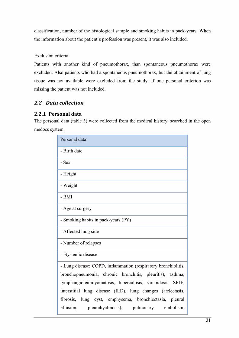
2.2.1 Personal data
The personal data (table 3) were collected from the medical history, searched in the open
medocs system.
Personal data
- Birth date
- Sex
- Height
- Weight
- BMI
- Age at surgery
- Smoking habits in pack-years (PY)
- Affected lung side
- Number of relapses
- Systemic disease
- Lung disease: COPD, inflammation (respiratory bronchiolitis,
bronchopneumonia, chronic bronchitis, pleuritis), asthma,
lymphangioleiomyomatosis, tuberculosis, sarcoidosis, SRIF,
interstitial lung disease (ILD), lung changes (atelectasis,
fibrosis, lung cyst, emphysema, bronchiectasia, pleural
effusion, pleurahyalinosis), pulmonary embolism,
32
Table 3: Personal data
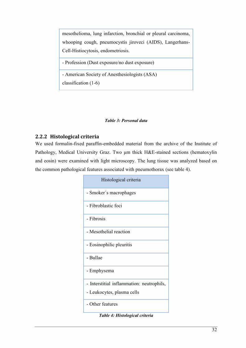
2.2.2 Histological criteria
We used formalin-fixed paraffin-embedded material from the archive of the Institute of
Pathology, Medical University Graz. Two μm thick H&E-stained sections (hematoxylin
and eosin) were examined with light microscopy. The lung tissue was analyzed based on
the common pathological features associated with pneumothorax (see table 4).
Histological criteria
- Smoker´s macrophages
- Fibroblastic foci
- Fibrosis
- Mesothelial reaction
- Eosinophilic pleuritis
- Bullae
- Emphysema
- Interstitial inflammation: neutrophils,
- Leukocytes, plasma cells
- Other features
Table 4: Histological criteria
mesothelioma, lung infarction, bronchial or pleural carcinoma,
whooping cough, pneumocystis jiroveci (AIDS), Langerhans-
Cell-Histiocytosis, endometriosis.
- Profession (Dust exposure/no dust exposure)
- American Society of Anesthesiologists (ASA)
classification (1-6)
33
2.3 Statistical analysis Statistical analysis was performed by using the Statistical Program for Social Sience (IBM
SPSS Statistic for Windows, Version 22 Armonk, NY: IBM). The student test for
independent samples was used to compare continous variables and the Chi-squared test
was used to compare the nominal data. The regression analysis was used to check if there
was a connection between two continous variables. All statistical hypothesis tests were two
tailed and a P value of less than 0.05 was considered to be significant.
3 Results
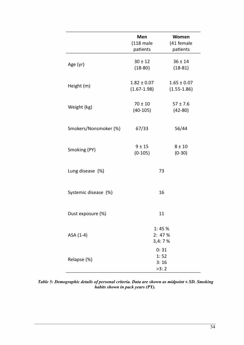
3.1 Personal data The information about age, height, weight, smoking habit, lung disease, systemic disease,
occupation, ASA classification and number of relapses was analyzed in every patient. The
results of the statistical analysis of the personal criteria are summarized in Table 5.
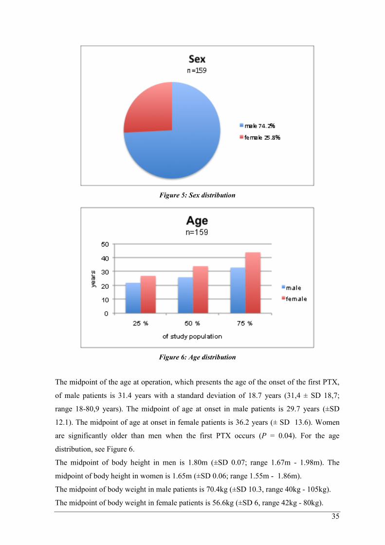
The analysis of the personal data shows that there are in total 159 patients who were
diagnosed with spontaneous pneumothorax between the years 1994 and 2014. Of these,
118 (74 %) patients are male and 41 (26 %) patients are female (see Figure 5).
34
Table 5: Demographic details of personal criteria. Data are shown as midpoint ± SD. Smoking
habits shown in pack years (PY).
35
Figure 5: Sex distribution
Figure 6: Age distribution
The midpoint of the age at operation, which presents the age of the onset of the first PTX,
of male patients is 31.4 years with a standard deviation of 18.7 years (31,4 ± SD 18,7;
range 18-80,9 years). The midpoint of age at onset in male patients is 29.7 years (±SD
12.1). The midpoint of age at onset in female patients is 36.2 years (± SD 13.6). Women
are significantly older than men when the first PTX occurs (P = 0.04). For the age
distribution, see Figure 6.
The midpoint of body height in men is 1.80m (±SD 0.07; range 1.67m - 1.98m). The
midpoint of body height in women is 1.65m (±SD 0.06; range 1.55m - 1.86m).
The midpoint of body weight in male patients is 70.4kg (±SD 10.3, range 40kg - 105kg).
The midpoint of body weight in female patients is 56.6kg (±SD 6, range 42kg - 80kg).
36
The midpoint of the BMI in male patients is 21.3 (±SD 3.1, range 14.17 - 38.08),
indicating that 16% of men have a lower BMI then 18.5 and are therefore called
underweight.
The midpoint of the BMI in female patients is 20.7 (±SD 2.8, range 16.6 - 30.5), indicating
that 27% of women are underweight (BMI ≤ 18.5).
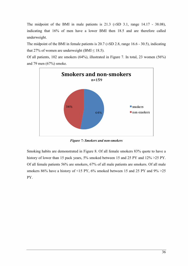
Of all patients, 102 are smokers (64%), illustrated in Figure 7. In total, 23 women (56%)
and 79 men (67%) smoke.
Figure 7: Smokers and non-smokers
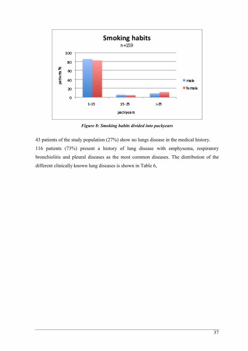
Smoking habits are demonstrated in Figure 8. Of all female smokers 83% quote to have a
history of lower than 15 pack years, 5% smoked between 15 and 25 PY and 12% >25 PY.
Of all female patients 56% are smokers, 67% of all male patients are smokers. Of all male
smokers 86% have a history of <15 PY, 6% smoked between 15 and 25 PY and 9% >25
PY.
37
Figure 8: Smoking habits divided into packyears
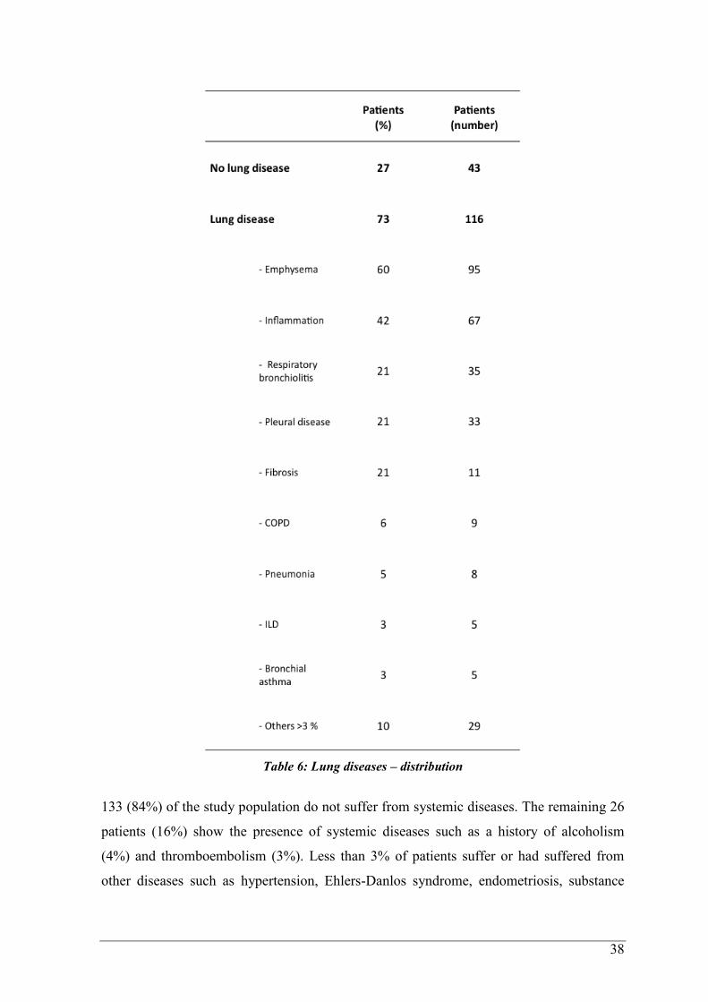
43 patients of the study population (27%) show no lungs disease in the medical history.
116 patients (73%) present a history of lung disease with emphysema, respiratory
bronchiolitis and pleural diseases as the most common diseases. The distribution of the
different clinically known lung diseases is shown in Table 6,
38
Table 6: Lung diseases – distribution
133 (84%) of the study population do not suffer from systemic diseases. The remaining 26
patients (16%) show the presence of systemic diseases such as a history of alcoholism
(4%) and thromboembolism (3%). Less than 3% of patients suffer or had suffered from
other diseases such as hypertension, Ehlers-Danlos syndrome, endometriosis, substance
39
abuse, hepatitis A, Scheuermann`s disease, testicular carcinoma, ADHD, epilepsy, stroke,
renal carcinoma and Crohn´s disease.
Out of 124 patients, 107 male and female patients (in total 67%) do not show a history of
occupational dust exposure. Of note, information regarding the profession is missing in 35
cases. 17 patients (11%) experienced dust exposure in their occupational careers.
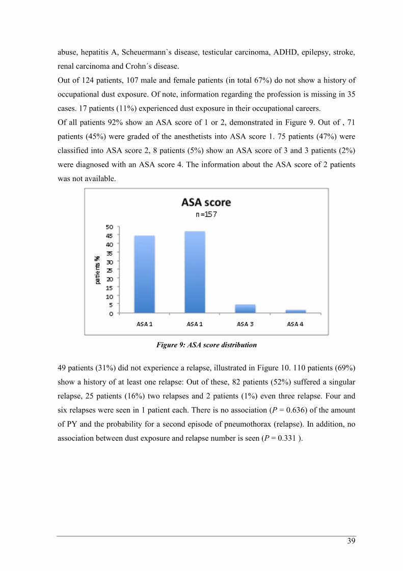
Of all patients 92% show an ASA score of 1 or 2, demonstrated in Figure 9. Out of , 71
patients (45%) were graded of the anesthetists into ASA score 1. 75 patients (47%) were
classified into ASA score 2, 8 patients (5%) show an ASA score of 3 and 3 patients (2%)
were diagnosed with an ASA score 4. The information about the ASA score of 2 patients
was not available.
Figure 9: ASA score distribution
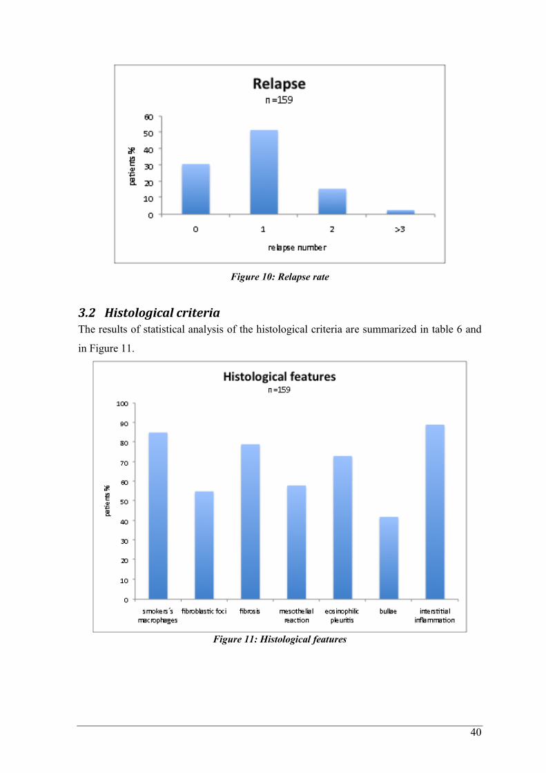
49 patients (31%) did not experience a relapse, illustrated in Figure 10. 110 patients (69%)
show a history of at least one relapse: Out of these, 82 patients (52%) suffered a singular
relapse, 25 patients (16%) two relapses and 2 patients (1%) even three relapse. Four and
six relapses were seen in 1 patient each. There is no association (P = 0.636) of the amount
of PY and the probability for a second episode of pneumothorax (relapse). In addition, no
association between dust exposure and relapse number is seen (P = 0.331 ).
40
Figure 10: Relapse rate
3.2 Histological criteria The results of statistical analysis of the histological criteria are summarized in table 6 and
in Figure 11.
Figure 11: Histological features
41
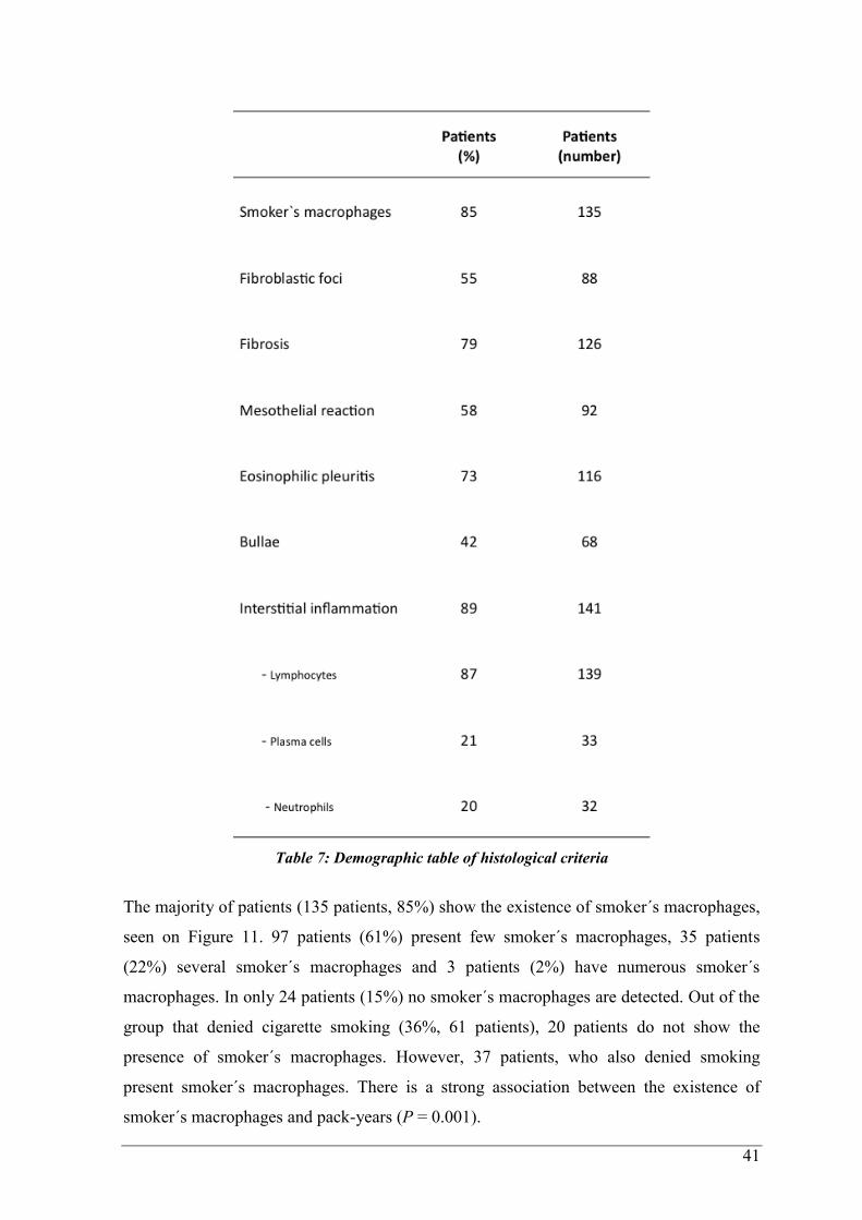
Table 7: Demographic table of histological criteria
The majority of patients (135 patients, 85%) show the existence of smoker´s macrophages,
seen on Figure 11. 97 patients (61%) present few smoker´s macrophages, 35 patients
(22%) several smoker´s macrophages and 3 patients (2%) have numerous smoker´s
macrophages. In only 24 patients (15%) no smoker´s macrophages are detected. Out of the
group that denied cigarette smoking (36%, 61 patients), 20 patients do not show the
presence of smoker´s macrophages. However, 37 patients, who also denied smoking
present smoker´s macrophages. There is a strong association between the existence of
smoker´s macrophages and pack-years (P = 0.001).
42
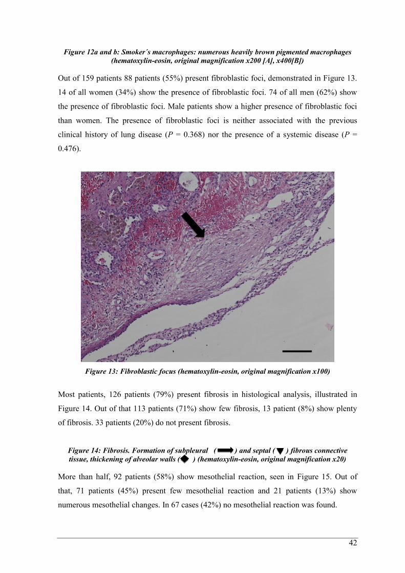
Figure 12a and b: Smoker´s macrophages: numerous heavily brown pigmented macrophages
(hematoxylin-eosin, original magnification x200 [A], x400[B])
Out of 159 patients 88 patients (55%) present fibroblastic foci, demonstrated in Figure 13.
14 of all women (34%) show the presence of fibroblastic foci. 74 of all men (62%) show
the presence of fibroblastic foci. Male patients show a higher presence of fibroblastic foci
than women. The presence of fibroblastic foci is neither associated with the previous
clinical history of lung disease (P = 0.368) nor the presence of a systemic disease (P =
0.476).
Figure 13: Fibroblastic focus (hematoxylin-eosin, original magnification x100)
Most patients, 126 patients (79%) present fibrosis in histological analysis, illustrated in
Figure 14. Out of that 113 patients (71%) show few fibrosis, 13 patient (8%) show plenty
of fibrosis. 33 patients (20%) do not present fibrosis.
Figure 14: Fibrosis. Formation of subpleural ( ) and septal ( ) fibrous connective
tissue, thickening of alveolar walls ( ) (hematoxylin-eosin, original magnification x20)
More than half, 92 patients (58%) show mesothelial reaction, seen in Figure 15. Out of
that, 71 patients (45%) present few mesothelial reaction and 21 patients (13%) show
numerous mesothelial changes. In 67 cases (42%) no mesothelial reaction was found.
43
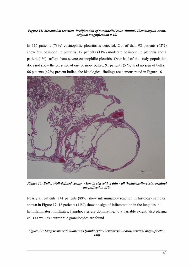
Figure 15: Mesothelial reaction. Proliferation of mesothelial cells ( ) (hematoxylin-eosin,
original magnification x 40)
In 116 patients (73%) eosinophilic pleuritis is detected. Out of that, 98 patients (62%)
show few eosinophilic pleuritis, 17 patients (11%) moderate eosinophilic pleuritis and 1
patient (1%) suffers from severe eosinophilic pleuritis. Over half of the study population
does not show the presence of one or more bullae, 91 patients (57%) had no sign of bullae.
66 patients (42%) present bullae, the histological findings are demonstrated in Figure 16.
Figure 16: Bulla. Well-defined cavitiy > 1cm in size with a thin wall (hematoxylin-eosin, original
magnification x10)
Nearly all patients, 141 patients (89%) show inflammatory reaction in histology samples,
shown in Figure 17. 18 patients (11%) show no sign of inflammation in the lung tissue.
In inflammatory infiltrates, lymphocytes are dominating, to a variable extent, also plasma
cells as well as neutrophile granulocytes are found.
Figure 17: Lung tissue with numerous lymphocytes (hematoxylin-eosin, original magnification
x40)
44
3.3 Comparison fibroblastic foci - no fibroblastic foci Fibroblastic foci are regarded as hallmark lesions of the pattern of usual interstitial
pneumonia, a severe interstitial lung disease with a grave prognosis. However, their role in
the context of spontaneous pneumothorax has not been studied in detail. In order to see
whether these fibroblastic foci are associated with demographic or clinical parameters, we
stratified our patients into two groups depending on the presence (88 patients, 55%) or
absence (71 patients, 45%) of fibroblastic foci. Statistical analysis shows significant
differences between the two groups in age, height, BMI and PY as illustrated in Table 8.
Figure 18: Sex distribution. Female and male patients and the amount of fibroblastic foci (ff)
within the group.
Patients with fibroblastic foci are younger (27.8 years ± SD 10.7) than patients without
fibroblastic foci (35.8 ± SD 13.8, P = 0.008).
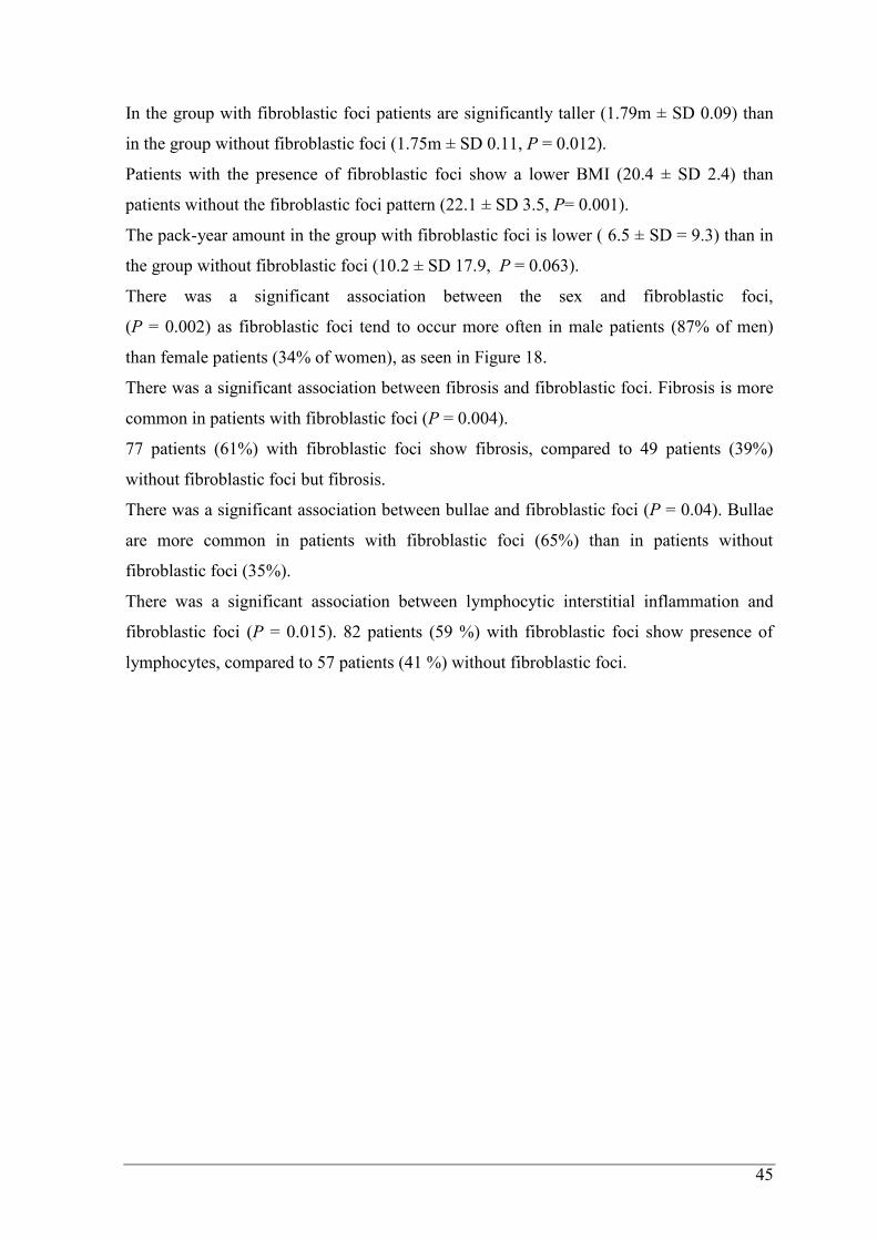
45
In the group with fibroblastic foci patients are significantly taller (1.79m ± SD 0.09) than
in the group without fibroblastic foci (1.75m ± SD 0.11, P = 0.012).
Patients with the presence of fibroblastic foci show a lower BMI (20.4 ± SD 2.4) than
patients without the fibroblastic foci pattern (22.1 ± SD 3.5, P= 0.001).
The pack-year amount in the group with fibroblastic foci is lower ( 6.5 ± SD = 9.3) than in
the group without fibroblastic foci (10.2 ± SD 17.9, P = 0.063).
There was a significant association between the sex and fibroblastic foci,
(P = 0.002) as fibroblastic foci tend to occur more often in male patients (87% of men)
than female patients (34% of women), as seen in Figure 18.
There was a significant association between fibrosis and fibroblastic foci. Fibrosis is more
common in patients with fibroblastic foci (P = 0.004).
77 patients (61%) with fibroblastic foci show fibrosis, compared to 49 patients (39%)
without fibroblastic foci but fibrosis.
There was a significant association between bullae and fibroblastic foci (P = 0.04). Bullae
are more common in patients with fibroblastic foci (65%) than in patients without
fibroblastic foci (35%).
There was a significant association between lymphocytic interstitial inflammation and
fibroblastic foci (P = 0.015). 82 patients (59 %) with fibroblastic foci show presence of
lymphocytes, compared to 57 patients (41 %) without fibroblastic foci.
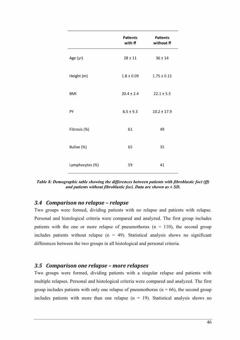
46
Table 8: Demographic table showing the differences between patients with fibroblastic foci (ff)
and patients without fibroblastic foci. Data are shown as ± SD.
3.4 Comparison no relapse – relapse Two groups were formed, dividing patients with no relapse and patients with relapse.
Personal and histological criteria were compared and analyzed. The first group includes
patients with the one or more relapse of pneumothorax (n = 110), the second group
includes patients without relapse (n = 49). Statistical analysis shows no significant
differences between the two groups in all histological and personal criteria.
3.5 Comparison one relapse – more relapses Two groups were formed, dividing patients with a singular relapse and patients with
multiple relapses. Personal and histological criteria were compared and analyzed. The first
group includes patients with only one relapse of pneumothorax (n = 66), the second group
includes patients with more than one relapse (n = 19). Statistical analysis shows no
47
significant differences between the two groups regarding all histological and personal
criteria.
4 Discussion Spontaneous pneumothorax may occur with no underlying disease or may be associated
with a wide variety of disorders (lung diseases and systemic diseases alike). Most authors
believe that the underlying cause of PTX is a spontaneous rupture of a subpleural bleb or
bulla. In 2012, the presence of fibroblastic foci in a subset of spontaneous PTX was
described by Deborah for the first time. With that in mind we carefully reanalysed the lung
tissue of 159 patients with PTX as well as their clinical history to see if and to which extent
this specific pattern also exists in our study group. In addition, patients with and without
relapses and also in the group of different relapse frequency were compared and analysed.
Furthermore, we aimed to elucidate associtions of certain histopathological and clinical
findings.
Analyses show that the average pneumothorax patient is male and around 1.8 m tall. The
BMI is in the range between underweight and normal. In general, PTX patients are
smokers, but interestingly, the number of PY is in the lower range. The bigger part of the
group shows the presence of lung disease, in most cases emphysema and interstitital
inflammation. Systemic diseases are not frequently encountered. Patients show an ASA
score between 1 and 2, meaning most patients are healthy or show a mild systemic disease.
PTX may occur once, however, it may come in several episodes. If PTX occurs in female
patients, the onset of the first PTX is later than in men. More women present with an
underweight BMI than men.
Histological features with a high incidence are smoker´s macrophages, fibroblastic foci,
fibrosis, eosinophilic granulocytes, mesothelial reaction and interstitial inflammation with
lymphocytes predominating. In the majority of patients fibroblastic lesions are seen.
Patients with fibroblastic foci tend to be male, younger than the other PTX patients, taller,
show a lower BMI and a shorter history of smoking. Patients with fibroblastic lesions also
show a higher incidence of fibrosis, bullae and interstitial lymphocytic infiltrates.
There are no significant differences between patients with one PTX, two or more.
Bösch et al. (2014) claims that there is a biphasic age distribution, the first peak
between 15 and 35 years (mainly 1° PTX) and the second peak over 55 years (mainly 2°
PTX) (6), however, in our cohort we cannot confirm that finding. Most of the patients
(75%) are younger than 37 years old, although we did not divide the study population in 1°
48
PTX and 2° PTX. Further studies are required with a higher number of patients in order
that a statement can be made.
The epidemiological study by Gupta et al. (2000) found out that in women the PTX onset
is later (between 30 and 34 years) than in men (7). Our research confirms that with the
median age of 36 in female patients, compared to men with the median age of 30. Gupta et
al. (2000) also argues that the cause for this gender-related difference remains still
unknown, but features associated in male patients (cigarette smoking and tall leptosome
habitus) are less frequently found in women (8), (9). Also, we can not confirm the
leptosome habitus in women because we do not have a control group with healthy lungs
for body weight and size which is the major limitation of the study presented herein.
Women also show a lower incidence of smoking than men. Therefore, the lower amount of
smoking may be the real reason for a lower incidence of PTX in female patients.
Predispositions such as tall slim habitus, low BMI, lung dissease (1), (2), (5), (30), (31) are
also found in our majority of the study population and may be assumed to take part of the
cause of the formation of a PTX.
Kroegel (2014) claims that the relapse rate is between 20 and 30% after the first episode of
a pneumothorax (5). Our study shows that the relapse rate is in fact much higher as even
52% suffer from a second pneumothorax after the first onset. Only 30% suffer from a one-
time PTX. It may be possible that our relapse rate is higher than compared to other studies
because our study includes already selected patients. Histological features such as
emphysema, fibrosis, bullae, inflammation, mesothelial reaction and pigmented
macrophages (29) can also be seen in the majority of our cases. Altough we claim that not
only the signs mentioned above are the common pathological features, also fibroblastic
foci are a feature frequently encountered in PTX. Deborah et al. (2012) reports an
association between the incidence of fibroblastic foci and PTX for the first time. We can
confirm this, although we do not have a control group with healthy lungs. Our study also
demonstrates that fibroblastic foci occur more often in younger, taller PTX patients and
show a lower BMI compared to the other PTX patients. Although patients with fibroblastic
foci are younger than the patients without fibroblastic foci the incidence of fibrosis, bullae
and lymphocytes are higher in the group with fibroblastic foci. Kroegel (2014) and Hien
(2014) say that patients, who suffer from 1° PTX are normally younger and do not present
any underlying lung disease (5), (13). In our studies younger patients with fibrolastic foci
show a higher incidence of fibrosis, bullae and lymphocytes. There are no differences in
patients with only one PTX or one or even more relapses. Chih-Yung Chiu et al. (2014)
49
also claims that factors such as smoking status, BMI and number of bullae are not showing
a significant higher recurrence rate (57). We assume that the incidence of relapses does not
correlate with our analysed features. Also the lungs may not change from one relapse to
another. Spezialised researches show more detailed information, which would have gone
beyond of scope in this thesis.
Patients with pneumothoraces show common personal and histological features. On the
one hand predispositions like slim, tall habitus with an underweight BMI, smoking and
lung disease lead to PTX and the other hand changes of the lung tissue including smoker´s
macrophages, fibroblastic foci, fibrosis, eosinophilic granulocytes, mesothelial reaction
and interstitial inflammation are frequently seen. Young patients who were assumed as
healthy without any underlying lung disease show definitive changes of the lung tissue
inlcuding a higher incidence of fibroblastic foci, fibrosis, bullae and lymphocytes.
Fibroblastic foci are surprisingly often seen in our cohort of patients with PTX. However,
it still cannot be determined whether these lesions contribute to the pathogenesis of this
disease or whether they are a hallmark of wound healing in affected tissue. Their role in
the context of PTX remains elusive. Further studies in this respect are warranted to gain
more information.

50
5 Bibliography (1) Stephen Chapman, Grace Robinson, John Stradling, Sophie West, John Wrightson: Oxford
Handbook of Respiratory Medicine. Oxford University Press, 2014. P. 371-381 (2) Stephen J. Bourke and Graham P. Burns: Respiratory Medicine lecture Notes. Eight Edition, John
Wiley & Son, 2011. P. 167-173
(3) Fraser John H Brims, Nick A Maskell: Ambulatory treatment in the management of pneumothorax:
a systematic review of the literature. Thorax 2013; 68: 664–669. doi:10.1136/thoraxjnl-2012-
202875
(4) Emerson C. Pneumothorax: a historical, clinical, and experimental study. John Hopkins Hosp Rep
1903;11:1–450
(5) Claus Kroegel, Urlich Costabel: Klinische Pneumologie. Thieme Verlag, 2014. P. 525-538
(6) Dennis Bösch, Jan Steffel, Thomas Lüscher: Lunge und Atemwege. Springer Verlag, 2014. ISBN
978-3-642-28222-5. P. 141-145 (german)
(7) Gupta D, Hansell A, Nichols T,et al: Epidemiology of pneumothorax in England. Thorax
2000;55:666–71
(8) Lippert HL, Lund O, Blegvad S, et al.: Independent risk factors for cumulative recurrence rate after
first spontaneous pneumothorax. Eur Respir J 1991;4:324–31
(9) Bense L, Eklund G, Wiman LG. Smoking and increased risk of contracting spontaneous
pneumothorax. Chest 1987;92:1009–12
(10) Stephen G. Spiro, Gerard A. Silvestri, Alvar Agusti: Clinical Respiratory Medicine. Fourth edition,
Elsevier Saunders, 2012. P. 831-836
(11) Noppen M, Schramel F. Pneumothorax. Eur Respir 2002; 22: 279–296
(12) Light RW. Management of spontaneous pneumothorax. Am Rev Respir Dis 1993; 148: P.245–248
(13) Hien: Praktische Pneumologie. 2.Auflage. Springer Verlag Berlin, Heidelberg, 2014. (German)
10.1007/978-3-642-10209-7. ISBN 978-3-642-10207-3
(14) Steven A. Sahn, M.D., and John E. Heffner, M.D.: Spontaneous Pneumothorax. N Engl J Med 2000;
342:868-874. DOI: 10.1056/NEJM200003233421207
(15) Mitlehner W , Friedrich M, Dissmann W.: Value of com-puter tomography in the detection of bullae
and blebs in patients with primary spontaneous pneumothorax. Respiration 1992; 59: 221–227
(16) Lesur O, Delorme N, Fromaget JM, et al.: Computed tomography in the etiologic assessment of
idiopathic spontaneous pneumothorax. Chest 1990; 98: 341–347
(17) Lesur O, Delorme N, Fromaget JM, Bernadac P, Polu JM.: Computed tomography in the etiologic
assessment of idiopathic spontaneous pneumothorax. Chest 1990; 98: 341–347
(18) Schramel, Postmus, Vanderschueren.: Current aspects of spontaneous pneumothorax. Eur Respir J
1997; 10: 1372–1379 DOI: 10.1183/09031936.97.10061372
(19) Fukuda Y , Haraguchi S, T anaka S, Y amanaka N.: Pathogenesis of blebs and bullae of patients
with spontaneous pneumothorax: ultrastructural and immunohis- tochemical studies. Am J Respir
Crit Care Med 1994; 149: A1022
(20) Tetely TD. Proteinase imbalance: its role in lung disease. Thorax 1993; 48: 560–565.
(21) Wallaert B, Gressier B, Marquette CH, et al.: Inactivation of alpha1-proteinase inhibitor by alveolar
inflammatory cells from smoking patients with or without emphyse- ma. Am Rev Respir Dis 1993;
147: 1537–1543
(22) Kondo T, Tagami S, Yohioka A, Nishiruma M, Kawakami Y.: Current smoking of elderly men
reduces antioxidants in alveolar macrophages. Am Rev Respir Dis 1991; 143: 144–149
(23) Adesina AM, Vallyathan V, McQuillen EN, Weaver SO, Craighead JE.: Bronchiolar inflammation
and fibrosis associated with smoking: a morphologic cross-sectional population analysis. Am Rev
Respir Dis 1991; 143: 144–149
(24) Hunninghake GW, Crystal RG.: Cigarette smoking and lung destruction: accumulation of
neutrophils in the lungs of cigarette smokers. Am Rev Respir Dis 1983; 128: 833–838
(25) Sibille Y., Reynolds HY : Macrophages and polymor- phonuclear neutrophils in lung defense and
injury: state of the art. Am Rev Respir Dis 1990; 141: 471–501
(26) Robbins RA, Gossman GL, Nelson KJ, Koyama S, Thompson AB, Rennard SI.: Inactivation of
chemotacic factor inactivator by cigarette smoke: a potential mechanism of modulating neutrophil
recruitment to the lung. Am Rev Respir Dis 1990; 142: 763–768
(27) Dermksian G, Lamb LE.: Spontaneous pneumothorax in apparently healthy flying personnel. Ann
Intern Med 1959; 51: 39–51
(28) Weiss LD, van Meter KW.: Cerebral air embolism in asthmatic scuba divers in a swimming pool.
Chest 1995; 107: 1653–1654
(29) Bryan Corrin, Andrew G. Nicholson: Pathology of the lungs. Second Edition, Churchill Livingstone,
2006. P. 694-695
51
(30) Amjadi K, Alvarez GG, Vanderhelst E, et al.: The prevalence of blebs and bullae among young
healthy adults: a thoracoscopic evaluation. Chest 2007; 132: 1140–1145. doi:
10.1631/jzus.B1000131
(31) Coxson HO, Chan IHT, Mayo JR, et al.:Early emphysema in patients with anorexia nervosa. Am J
Respir Crit Care Med 2004; 170: 748–752
(32) MacDuff A, Arnold A, Harvey J: BTS Pleural Disease Guideline Group (December 2010).
"Management of spontaneous pneumothorax: British Thoracic Society pleural disease guideline
2010". Thorax 65 (8): ii18–ii31. doi:10.1136/thx.2010.136986. PMID 20696690.
(33) Dermksian G, Lamb LE.: Spontaneous pneumothorax in apparently healthy flying personnel. Ann
Intern Med 1959; 51: 39–51
(34) Wolfgang Hien: Arbeitspapiere Berufskrankheiten Nr. 1 Arbeits- und berufsbedingte obstruktive
Lungenerkrankungen – Kenntnisstand und Hinweise für die Praxis des Berufskrankheiten-
Verfahrens. Bremen 2014 (german)
(35) Jeske A et al. (1993) Results of thoracocscopy and pleurodesis with talc poudrage and thoracotomy.
Chest. 103:345-347
(36) Randomsky JBD, Hartel W.: Pleuraporosität bei idiopatischen Spontanpneumothorax. Pneumologie
1989; 43: 250-253.
(37) Masshof W, Hofer W: Zur Pathologie der sogenanntenn idiopathischen Spontanpneumothorax.
Deutsche Medizinische Wochenzeitschrift 1973; 98: 801- 805
(38) Olhata M, Suzuki H: Pathogenesis of spontaneous pneumothorax. With special reference to the
ultrastructure of emphysematous bullae. Chest 1980; 77: 771-776
(39) C. Thomas et al.: Spezielle Pathologie. Schattauer Verlag, 1996. ISBN 3-7945-2326-1. P. 129
(german)
(40) Mayo Clinic Staff. "Complications". Mayo Foundation for Medical Education and Research.
Retrieved 26th
July 2014
(41) Gross TJ, Hunninghake GW. (2001).: "Idiopathic pulmonary fibrosis". N Engl J Med 345: 517–525.
doi:10.1056/NEJMra003200
(42) Mayo Clinic Staff. "Causes". Mayo Foundation for Medical Education and Research. Retrieved 26th
July 2014
(43) Reynold HY.: Respiratory infections may reflect deficiencies in host defence mechanisms. Diseases
Monthly 1985;31:1-98
(44) Sibille Y, Reynolds HZ.: Macrophages and polymorphonuclear neutrophils in lung defence and
injury. State of the Art. Am Rec Respir Dis 1990;141:471-501
(45) Konietzko, Wendel, Wiesner: Erkrankungen der Lunge. De Gruyter. Berlin, 1994. ISBN: 3-11-
012130-1. P. 214 (german)
(46) Rennard SI, Hnninghake GW, Bitterman PB, Crystal RG.: Production of fibronectin by the human
alveolar macrophage: mechanisms of the recruitment of fibroblasts to sites of tissue injury in
interstitial lung disease. Proc Natl Acad Sci 1981;78:7147-51
(47) D Macridou, D Laws: Case Report: Respiratory bronchiolitis associated interstitial lung disease
(RB-ILD): a case of an acute presentation.Thorax 2004;59:910-911 doi:10.1136/thx.2003.011080
(48) Dennis E. Niewoehner, M.D., Jerome Kleinerman, M.D., and Donald B. Rice, M. Div: Pathologic
Changes in the Peripheral Airways of Young Cigarette Smokers. N Engl J Med 1974; 291:755-758
October 10, 1974DOI: 10.1056/NEJM197410102911503
(49) Bense L, Lewander R, Eklund G, et al.: Non-smoking, non-alpha 1 antitrypsin deficiency-induced
emphysema in nonsmokers with healed spontaneous pneumothorax, identified by computed
tomography of the lungs. Chest 1993; 103: 433–438
(50) Lesur O, Delorme N, Fromaget JM, et al.: Computed tomography in the ethiologic assessment of
idiopathic spontaneous pneumothorax. Chest 1990; 98: 341-347
(51) Deborah A. Belchis, MD; Kris Shekitka, MD; Christopher D. Gocke, MD.A Unique,
Histopathologic Lesion in a Subset of Patients With Spontaneous Pneumothorax. Arch Pathol Lab
Med. 2012;136:1522–1527; doi: 10.5858/arpa.2012-0330-OA
(52) KM Müller, M Kenter: Tabak-assoziierte siderophile und conchoide Speicherphänomene in
Rauchermakrophagen mit Riesenzellen. Pneumologie 2013; P. 67 - 44
DOI: 10.1055/s-0033-1334697. (german)
(53) http://www.vbdo.at/artikel/einblick/blick11/index.htm Austria: VBDO – Verband für Bildgebende
Diagnostik Österreich, updated June 2001, seen on 30th
July 2015 (german)
(54) MacDuff A, Arnold A, Harvey J: Management of spontaneous pneumothorax: British Thoracic
Society Pleural Disease Guideline 2010. Thorax 65(Suppl 2):ii18–ii31.
doi:10.1136/thx.2010.136986
(55) Baumann MH, Strange C, Heffner JE, Light R, Kirby TJ, Klein J, Luketich JD, Panacek EA, Sahn
SA (February 2001): "Management of spontaneous pneumothorax: an American College of Chest

52
Physicians Delphi consensus statement". Chest 119 (2): 590–602. doi:10.1378/chest.119.2.590.
PMID 11171742
(56) Shields, Thomas: General Thoracic Surgery. Lippincott Williams & Wilkins, 2004. P. 524
ISBN 978-0-7817-3889-7


![Merial Oktober 2016 [Kompatibilitätsmodus] - pferdeklinik.atAtemwege.pdf · Hernia diaphragmatica. 21.10.2016 17 Nach Pfählungsverletzung T/0253232 Pneumothorax Ultraschall. 21.10.2016](https://img.pdfslide.org/doc/110x75/5e04c17ffa470a3ddf4ea3c1/merial-oktober-2016-kompatibilittsmodus-atemwegepdf-hernia-diaphragmatica.jpg)

![Pneumothorax [J93.9] · Pneumothorax ist 4x schneller wenn Sauerstoff verabreicht wird. - Kontroll-Rx nach 6 h → wenn der Pneumothorax signifikant kleiner geworden ist, kann eine](https://img.pdfslide.org/doc/110x75/605e013975b1447c8a4ce1a2/pneumothorax-j939-pneumothorax-ist-4x-schneller-wenn-sauerstoff-verabreicht-wird.jpg)