Embed Size (px)
Citation preview
VVBVVB LAUFERSWEILER VERLAG
édition scientifique
VVB LAUFERSWEILER VERLAGSTAUFENBERGRING 15D-35396 GIESSEN
Tel: 0641-5599888 Fax: [email protected]
VVB LAUFERSWEILER VERLAGédition scientifique
9 7 8 3 8 3 5 9 6 4 1 7 4
ISBN: 978-3-8359-6417-4
Photo cover: ©
STEFFI LA
UB
NER
M
RT-B
EFU
ND
E B
EI H
UN
DEN
M
IT H
YD
RO
ZEP
HA
LU
S
STEFFI LAUBNER
Zusammenhang zwischen MRT-Befunden
und klinischen Symptomen bei Hunden mit
Hydrozephalus im Vergleich zu Hunden
mit Ventrikulomegalie
Inaugural-Dissertation zur Erlangung des Grades eines
Dr. med. vet.
beim Fachbereich Veterinärmedizin der Justus-Liebig-Universität Gießen
VVBVERLAG
Das Werk ist in allen seinen Teilen urheberrechtlich geschützt.
Die rechtliche Verantwortung für den gesamten Inhalt dieses Buches liegt ausschließlich bei den Autoren dieses Werkes.
Jede Verwertung ist ohne schriftliche Zustimmung der Autoren oder des Verlages unzulässig. Das gilt insbesondere für Vervielfältigungen, Übersetzungen, Mikroverfilmungen
und die Einspeicherung in und Verarbeitung durch elektronische Systeme.
1. Auflage 2016
All rights reserved. No part of this publication may be reproduced, stored in a retrieval system, or transmitted,
in any form or by any means, electronic, mechanical, photocopying, recording, or otherwise, without the prior
written permission of the Authors or the Publisher.
st1 Edition 2016
© 2016 by VVB LAUFERSWEILER VERLAG, GiessenPrinted in Germany
VVB LAUFERSWEILER VERLAG
STAUFENBERGRING 15, D-35396 GIESSENTel: 0641-5599888 Fax: 0641-5599890
email: [email protected]
www.doktorverlag.de
édition scientifique
Aus dem Klinikum Veterinärmedizin Klinik für Kleintiere, Chirurgie
der Justus-Liebig-Universität Gießen
Betreuer: PD Dr. med. vet. (habil.) M. Schmidt
Zusammenhang zwischen MRT-Befunden und klinischen
Symptomen bei Hunden mit Hydrozephalus im Vergleich zu
Hunden mit Ventrikulomegalie
INAUGURAL-DISSERTATION zur Erlangung des Grades
eines Dr. med. vet. beim Fachbereich Veterinärmedizin der Justus-Liebig-Universität Gießen
vorgelegt von
Steffi Laubner Tierärztin aus Friedberg
Gießen 2016
Mit Genehmigung des Fachbereichs Veterinärmedizin
der Justus-Liebig-Universität Gießen
Dekan: Prof. Dr. Dr. h.c. Martin Kramer
1. Gutachter: PD Dr. Martin Schmidt
2. Gutachter: Prof. Dr. Christiane Herden
:
Tag der Disputation: 13.01.2016

Für meine Familie
Inhaltsverzeichnis
II
Inhaltsverzeichnis
ABKÜRZUNGSVERZEICHNIS IV
1. EINLEITUNG 1
2. LITERATURÜBERSICHT 3
2.1 ANATOMISCHE GRUNDLAGEN 3
2.1.1 DAS VENTRIKELSYSTEM 3 2.1.2 DER PLEXUS CHOROIDEUS 4 2.2 PHYSIOLOGISCHE GRUNDLAGEN 5
2.2.1 LIQUORPRODUKTION 5 2.2.2 LIQUORBEWEGUNG 7 2.2.3 LIQUORABSORPTION 7 2.3 DER HYDROZEPHALUS INTERNUS 9
2.3.1 DEFINITION UND EINTEILUNG 9 2.3.2 PATHOPHYSIOLOGIE 11 2.4 ERKRANKUNG BEIM MENSCHEN 13
2.4.1 KONGENITALER HYDROZEPHALUS 13 2.4.2 HYDROZEPHALUS DES ERWACHSENEN (NORMALDRUCK-HYDROZEPHALUS, NPH) 14 2.4.3 SEKUNDÄRER HYDROZEPHALUS 14 2.5 ERKRANKUNG BEIM HUND 15
2.5.1 KLINISCHE SYMPTOME 15 2.5.2 DIAGNOSTIK 17 2.6 THERAPIE 21
2.6.1 MEDIKAMENTELLE THERAPIE 21 2.6.2 CHIRURGISCHE THERAPIE 23
3. PUBLIKATIONEN 25
3.1 PUBLIKATION 1 25
3.2 PUBLIKATION 2 54
4. DISKUSSION 81
4.1 DIE PHYSIOLOGISCHE VENTRIKELGRÖßE BEIM HUND 81
4.2 IST ES MÖGLICH DIE VENTRIKULOMEGALIE UND DEN HYDROCEPHALUS INTERNUS
VONEINANDER ABZUGRENZEN? 81
4.3 IST EINE VENTRIKULOMEGALIE ALS PHYSIOLOGISCHER ZUSTAND EINZUORDNEN? 87
4.4 AUSBLICK 88

Inhaltsverzeichnis
III
5. ZUSAMMENFASSUNG 89
6. SUMMARY 90
7. LITERATURVERZEICHNIS 91
Abkürzungsverzeichnis
IV
Abkürzungsverzeichnis ANP Atriales Natriuretisches Peptid AQP 1 Aquaporin 1 AVIM asymptomatic ventriculomegaly with features of iNPH on magnetic resonance imaging AVP Arginin-Vasopressin ca. circa CA Callosal Angle CBF Cerebraler Blutfluss Cl- Chlorid CLM Chiari-like malformation cm Zentimeter CSF Cerebrospinal fluid CT Computertomographie dl Deziliter ECF Extrazelluläre Flüssigkeit et al. et alli, und weitere HCO3 Hydrogencarbonat, Bicarbonat HE Hämatoxylin-Eosin H2O Wasser ICP Intrakranieller Druck iNPH idiopathischer Normaldruck-Hydrozephalus K+ Kalium kg Kilogramm m Meter mg Milligramm ml Milliliter µl Mikroliter min. Minute Na+ Natrium MRT Magnet-Resonanz-Tomographie NPH Normaldruck-Hydrozephalus s Sekunde s.o. siehe oben V/B Ventricle/Brain z.B. Zum Beispiel
ZNS Zentralnervensystem
Einleitung
Einleitung 1.
Trotz großer Unterschiede in der Kopfform verschiedener Hunderassen sind die
Unterschiede in der Morphologie des Gehirns nur gering ausgeprägt (SEIFERLE,
1966; ROBERTS et al., 2010; OBUSSIER, 1995). Die topographische Anordnung der
Oberflächenfaltung und die proportionale Differenzierung funktioneller Hirnanteile
scheinen sich zwischen den Rassen kaum zu unterscheiden. Formunterschiede
werden eher sekundär durch die Schädelform bedingt. Aufgrund des
eingeschränkten Längenwachstums der Schädelbasis bei brachyzephalen Hunden
zeigt das Gehirn eine geringere longitudinale Ausdehnung (ROBERTS et al., 2010;
SCHMIDT et al., 2012) und erhält eine breite und stämmige Erscheinung mit nach
ventral orientierten Bulbus olfactorii, ähnlich dem Gehirn eines Welpen (ROBERTS et
al., 2010; SCHMIDT et al., 2011). Insgesamt wird von einer Verjugendlichung
(Neotenie) einiger oder sogar aller Hunderassen gegenüber dem Habitus des
Wolfes, oder ursprünglicher wolfartiger Rassen ausgegangen (LÜPS, 1973). Trotz
der Gleichförmigkeit des morphologischen Aufbaus des Gehirns wird aber immer
wieder ein vergrößertes Volumen der Seitenventrikel bei brachyzephalen Rassen
beschrieben (DEXLER, 1923; JAMES und SHUT, 1978; FOLTZ und AINE, 1981;
RYAN et al., 2014; VITE et al., 1997). Es ist allgemein akzeptiert, dass diese
Erweiterung der Ventrikel nicht mit klinischen Symptomen assoziiert ist (BAGLEY et
al., 2009; HECHT, 2011; LU et al., 2003; DRIVER et al., 2013). Versuche der
Bestimmung einer physiologischen Ventrikelgröße waren nicht erfolgreich, vielmehr
zeigten mehrere Studien unabhängig voneinander, dass die Größe der Ventrikel
zwischen, aber auch innerhalb einer brachyzephalen Rasse bei Tieren mit
vergleichbarem Körpergewicht erheblich variieren kann (DE HAAN et al., 1994;
SPAULDING und SHARP, 1990; RYAN et al., 2014). Daher entwickelte sich die
Hypothese, dass keine bestimme Ventrikelgröße für eine Hunderasse existiere und
eine Bandbreite physiologischer Volumina vorliege. Diese Theorie hat sich sowohl in
den Lehrbüchern der Veterinäranatomie als auch denen der Bildgebenden Verfahren
etabliert. Die Bezeichnung „Ventrikulomegalie“ wurde entwickelt, um diese
anatomische Besonderheit, die als Nebenbefund gewertet wird, von einem klinisch
relevanten Hydrocephalus internus abzugrenzen. Als morphologische Auffälligkeit
einiger Hunderassen stellt dies den Untersucher bei der Interpretation magnet-
resonanz-tomografischer Bilder vor eine große Herausforderung. Die Aufweitung der
1
Einleitung
Ventrikel im Rahmen eines Hydrocephalus internus ist bei brachyzephalen
Hunderassen eine häufig diagnostizierte Erkrankung und kann lebensbedrohlich
sein. Bei der Beurteilung von Magnet-Resonanz-Tomografie-(MRT) Bildern ergibt
sich in diesem Zusammenhang allerdings die Schwierigkeit, dass aufgrund des
Volumens der Ventrikel zwischen einer Ventrikulomegalie und einem Hydrocephalus
internus in vielen Fällen nicht sicher unterschieden werden kann (ESTEVE-RATSCH
et al., 2001; RIVERS und WALTER, 1992; VULLO et al., 1997; DE HAAN et al.,
1994). Anhand der Größe der Ventrikel allein kann also nicht ausgemacht werden,
ob die gezeigten Symptome auf eine Schädigung des Gehirns aufgrund eines
erhöhten intraventrikulären Druckes zurückzuführen sind. Hier bietet sich viel Raum
für Fehldiagnosen, da eine Ventrikulomegalie oft fälschlicherweise als Ursache der
klinischen Symptomatik eingestuft wird, da sie die einzige strukturelle Veränderung
ist, die vor allem in der Computer-Tomografie (CT), aber auch in der MRT sichtbar ist
(HECHT, 2011; PLATT und GAROSI, 2012). Entzündliche Erkrankungen des
Gehirns müssen nicht in jedem Fall strukturelle Befunde in der MRT hervorrufen
(LAMB et al., 2005) und können als eigentliche Ursache für klinische Symptome
fehlinterpretiert werden.
Im ersten Teil dieser Dissertation wird daher versucht, in Schnittbildern von Hunden
mit klinisch relevantem Hydrocephalus internus Befunde zu identifizieren, die auf
einen akut vorliegenden Überdruck hinweisen, welche bei einer Ventrikulomegalie
gegebenenfalls nicht zu finden sind. Darüber hinaus ergibt sich die Frage, ob eine
Ventrikulomegalie auch bei nicht vorhandenen Anzeichen eines intraventrikulären
Hochdrucks als physiologische Erscheinung gewertet werden kann.
2
Literaturübersicht
Literaturübersicht 2.
2.1 Anatomische Grundlagen
2.1.1 Das Ventrikelsystem
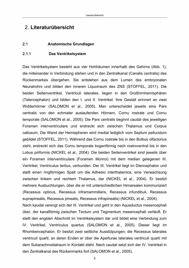
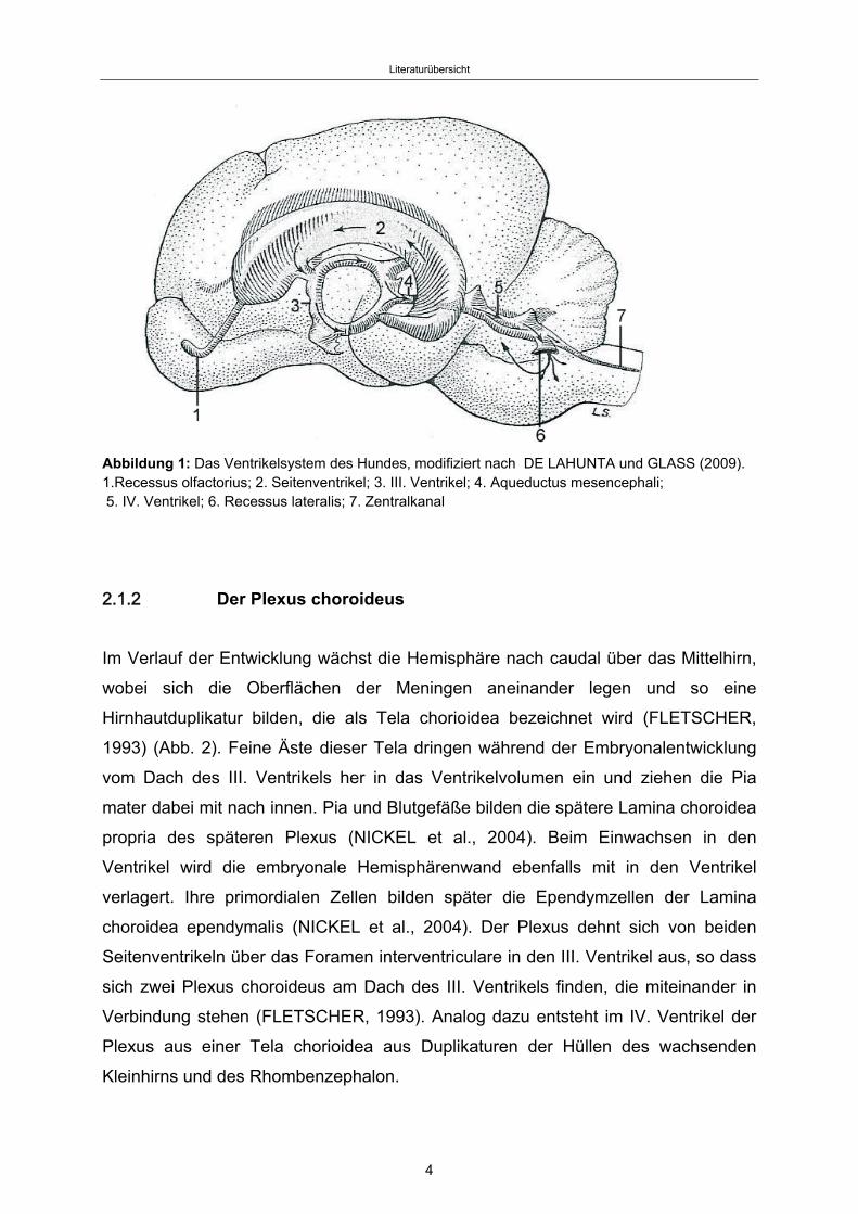
Das Ventrikelsystem besteht aus vier Hohlräumen innerhalb des Gehirns (Abb. 1),
die miteinander in Verbindung stehen und in den Zentralkanal (Canalis centralis) des
Rückenmarkes übergehen. Sie entstehen aus dem Lumen des embryonalen
Neuralrohrs und bilden den inneren Liquorraum des ZNS (STOFFEL, 2011). Die
beiden Seitenventrikel, Ventriculi laterales, liegen in den Großhirnhemisphären
(Telencephalon) und bilden den I. und II. Ventrikel. Ihre Gestalt erinnert an zwei
Widderhörner (SALOMON et al., 2005). Man unterscheidet jeweils eine Pars
centralis von den schmaler auslaufenden Hörnern, Cornu rostrale und Cornu
temporale (SALOMON et al., 2005). Die Pars centralis beginnt caudal des jeweiligen
Foramen interventriculare und erstreckt sich zwischen Thalamus und Corpus
callosum. Die Wand der Hemispheren wird medial lediglich vom Septum pellucidum
gebildet (STOFFEL, 2011). Während das Cornu rostrale bis in den Bulbus olfactorius
zieht, erstreckt sich das Cornu temporale bogenförmig nach rostroventral bis in den
Lobus piriformis (NICKEL et al., 2004). Die beiden Seitenventrikel sind jeweils über
ein Foramen interventriculare (Foramen Monroi) mit dem median gelegenen III.
Ventrikel, Ventriculus tertius, verbunden. Der III. Ventrikel liegt im Diencephalon und
stellt einen ringförmigen Spalt um die Adhesio interthalamica, eine Verwachsung
zwischen linkem und rechtem Thalamus, dar (NICKEL et al., 2004). Er besitzt
mehrere Ausbuchtungen, über die er mit unterschiedlichen Hirnarealen kommuniziert
(Recessus opticus, Recessus inframammillaris, Recessus infundibuli, Recessus
suprapinealis, Recessus pinealis, Recessus infrapinealis) (NICKEL et al., 2004).
Nach kaudal verengt sich der III. Ventrikel und geht in den Aqueductus mesencephali
über, der kanalförmig zwischen Tectum und Tegmentum mesencephali verläuft. Er
stellt den engsten Abschnitt im Ventrikelsystem dar und bildet eine Verbindung zum
IV. Ventrikel, Ventriculus quartus (SALOMON et al., 2005). Dieser liegt im
Rhombencephalon. Er besitzt zwei seitliche Ausstülpungen, die Recessus laterales
ventriculi quarti, an deren Enden er über die Aperturae laterales ventriculi quarti mit
dem Subarachnoidalraum in Kontakt steht. Nach caudal setzt sich der IV. Ventrikel in
den Zentralkanal des Rückenmarks fort (SALOMON et al., 2005).
3
Literaturübersicht
Abbildung 1: Das Ventrikelsystem des Hundes, modifiziert nach DE LAHUNTA und GLASS (2009). 1.Recessus olfactorius; 2. Seitenventrikel; 3. III. Ventrikel; 4. Aqueductus mesencephali; 5. IV. Ventrikel; 6. Recessus lateralis; 7. Zentralkanal
2.1.2 Der Plexus choroideus
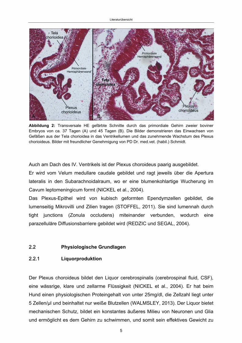
Im Verlauf der Entwicklung wächst die Hemisphäre nach caudal über das Mittelhirn,
wobei sich die Oberflächen der Meningen aneinander legen und so eine
Hirnhautduplikatur bilden, die als Tela chorioidea bezeichnet wird (FLETSCHER,
1993) (Abb. 2). Feine Äste dieser Tela dringen während der Embryonalentwicklung
vom Dach des III. Ventrikels her in das Ventrikelvolumen ein und ziehen die Pia
mater dabei mit nach innen. Pia und Blutgefäße bilden die spätere Lamina choroidea
propria des späteren Plexus (NICKEL et al., 2004). Beim Einwachsen in den
Ventrikel wird die embryonale Hemisphärenwand ebenfalls mit in den Ventrikel
verlagert. Ihre primordialen Zellen bilden später die Ependymzellen der Lamina
choroidea ependymalis (NICKEL et al., 2004). Der Plexus dehnt sich von beiden
Seitenventrikeln über das Foramen interventriculare in den III. Ventrikel aus, so dass
sich zwei Plexus choroideus am Dach des III. Ventrikels finden, die miteinander in
Verbindung stehen (FLETSCHER, 1993). Analog dazu entsteht im IV. Ventrikel der
Plexus aus einer Tela chorioidea aus Duplikaturen der Hüllen des wachsenden
Kleinhirns und des Rhombenzephalon.
4
Literaturübersicht
Abbildung 2: Transversale HE gefärbte Schnitte durch das primordiale Gehirn zweier boviner Embryos von ca. 37 Tagen (A) und 45 Tagen (B). Die Bilder demonstrieren das Einwachsen von Gefäßen aus der Tela chorioidea in das Ventrikellumen und das zunehmende Wachstum des Plexus chorioideus. Bilder mit freundlicher Genehmigung von PD Dr. med.vet. (habil.) Schmidt.
Auch am Dach des IV. Ventrikels ist der Plexus choroideus paarig ausgebildet.
Er wird vom Velum medullare caudale gebildet und ragt jeweils über die Apertura
lateralis in den Subarachnoidalraum, wo er eine blumenkohlartige Wucherung im
Cavum leptomeningicum formt (NICKEL et al., 2004).
Das Plexus-Epithel wird von kubisch geformten Ependymzellen gebildet, die
lumenseitig Mikrovilli und Zilien tragen (STOFFEL, 2011). Sie sind lumennah durch
tight junctions (Zonula occludens) miteinander verbunden, wodurch eine
parazelluläre Diffusionsbarriere gebildet wird (REDZIC und SEGAL, 2004).
2.2 Physiologische Grundlagen
2.2.1 Liquorproduktion
Der Plexus choroideus bildet den Liquor cerebrospinalis (cerebrospinal fluid, CSF),
eine wässrige, klare und zellarme Flüssigkeit (NICKEL et al., 2004). Er hat beim
Hund einen physiologischen Proteingehalt von unter 25mg/dl, die Zellzahl liegt unter
5 Zellen/µl und beinhaltet nur weiße Blutzellen (WALMSLEY, 2013). Der Liquor bietet
mechanischen Schutz, bildet ein konstantes äußeres Milieu von Neuronen und Glia
und ermöglicht es dem Gehirn zu schwimmen, und somit sein effektives Gewicht zu
5
Literaturübersicht
verringern (LATERRA und GOLDSTEIN, 2002). Er ermöglicht eine Anpassung an
wechselnde Druckverhältnisse innerhalb des Schädels (DE LAHUNTA und GLASS,
2009). Durch seine gerichtete Bewegung (bulk flow) von den Ventrikeln über den
Subarachnoidalraum in die Venen der Arachnoidea ermöglicht er den Abtransport
von Metaboliten und kann als ein spezialisiertes Lymphsystem des Gehirns
angesehen werden (SALOMON et al., 2005). Darüber hinaus können Polypeptid-
Hormone durch Sekretion in den Liquor in andere Gehirnabschnitte und auch in den
Körperkreislauf gelangen (DE LAHUNTA und GLASS, 2009).
Der vom Plexus choroideus produzierte Liquor ist keine Filtrationsflüssigkeit des
Blutes, sondern wird aktiv von spezialisierten Zellen der Innenauskleidung des
Ventrikels (Ependymzellen) sezerniert (REDZIC und SEGAL, 2004). Na+, Cl-, K+ und
HCO3- werden von den Ependymzellen über spezielle Transporter und Ionenkanäle
in das Ventrikellumen abgegeben und so ein osmotischer Gradient erzeugt.
Flüssigkeit folgt diesem Gradienten über Wasserkanäle (Aquaporine) nach (REDZIC
und SEGAL, 2004). Neben der Produktion durch den Plexus choroideus wird Liquor
cerebrospinalis auch durch die Produktion von extrazellulärer Flüssigkeit
(extracellular fluid, ECF) im Parenchym des Gehirns und des Rückenmarkes gebildet
(DE LAHUNTA und GLASS, 2009; REKATE, 1997). ECF entsteht passiv als
Nebenprodukt der Stoffwechselvorgänge im Parenchym (MILHORAT und HAMMOK,
1971). Sie besitzt dieselbe chemische Zusammensetzung wie der Liquor (REKATE,
1997). SATO und BERING (1967) beschreiben eine Produktionsrate des Liquors
innerhalb des intrakranialen Subarachnoidalraumes von 0.014ml/min bei 15 bis 20kg
schweren Hunden. Die extraventrikuläre Produktionsrate beschreiben SATO et al.
(1972) mit 0.018ml/min bei Hunden mit einem Körpergewicht von 12 bis 15kg. Die
vom Rückenmark produzierte ECF wird durch den bulk flow nach kranial transportiert
und mischt sich im IV. Ventrikel mit dem intrakranial produzierten Liquor (REKATE,
1997). Der Hund besitzt insgesamt ca. 13-22ml Liquor cerebrospinalis. Die
Produktion ist so hoch, dass dieser 3-5 Mal täglich erneuert wird (DE LAHUNTA und
GLASS, 2009).
Die Liquorproduktionsrate unterliegt der Regulation durch das autonome
Nervensystem. Sympathische Afferenzen steigern, cholinerge Signale senken die
Liquorproduktion am Plexus choroideus (SAKKA et al., 2011). Auf der Oberfläche der
Plexusepithelien befinden sich darüber hinaus auch Rezeptoren für verschiedene
Monoamide und Neuropeptide. Dopamin, Serotonin, Melatonin, Atriales
Natriuretisches Peptid (ANP) und Arginin-Vasopressin (AVP) Rezeptoren wurden
6

Literaturübersicht
beschrieben, wobei ANP und AVP die Liquorproduktion über Runterregulation von
Aquaporin 1 im Epithel senken (SAKKA et al., 2011).
2.2.2 Liquorbewegung
Durch die rhythmischen Pulsationen der intrakraniellen Blutgefäße ist der Liquor
ständig im Fluss (COATES et al., 2006; REKATE, 1997). Er fließt rostrocaudal von
den Seitenventrikeln über das Foramen interventriculare in den III. Ventrikel, weiter
über den Aqueductus mesencephali in den IV. Ventrikel und von hier aus in den
Subarachnoidalraum, wo es zu einer ungerichteten Durchmischung kommt (HABERL
et al., 2007). Die auslösende Kraft der Liquorbewegung ist die Pulsation der
intrakraniellen Gefäße (GREITZ, 2004). Da das Gesamtvolumen der Komponenten
Blut, Liquor und Parenchym innerhalb des Schädels konstant bleibt (Monro-Kellie-
Doktrin) (GREITZ et al., 1992), kommt es in der Systole durch Zunahme an
Blutvolumen innerhalb der intrakraniellen Arterien zu einem rapiden Anstieg des
Liquordruckes und dadurch zu einem Liquorfluss nach caudal durch das Foramen
magnum (DE LAHUNTA und GLASS, 2009).
2.2.3 Liquorabsorption
Die Mechanismen der Liquorabsorption sind nicht abschließend geklärt (HABERL et
al., 2007). Bisher werden als Haupt-Absorptionsort arachnoidale Villi angesehen, die
sich in die Hirnsinus und oberflächlichen Hirnvenen vorstülpen (REKATE, 1997). Der
normale intrakranielle Druck liegt bei 5-15cm H2O (SAHAR et al., 1971). Die
arachnoidalen Villi sollen eine Art Klappensystem darstellen (WELCH und
FRIEDMANN; 1960), mit einem Öffnungsdruck von 7cm H2O zwischen dem
Subarachnoidalraum und dem Sagittalsinus (SAHAR et al., 1971). Unterhalb dieses
Öffnungsdruckes findet keine Absorption statt. Mit steigendem intrakraniellen Druck
steigt auch die Absorptionsrate, so dass ein gewisses Maß an Überproduktion
kompensiert werden kann (SAHAR et al., 1971). Weder eine Absorption über
arachnoidale Villi, noch eine bestehende Klappenfunktion dieser konnten bisher
nachgewiesen werden (GREITZ, 2004).
Gegen die arachnoidalen Villi als Haupt-Absorptionsort spricht auch, dass die
7
Literaturübersicht
meisten Säugetiere ohne arachnoidale Villi geboren werden und sich diese beim
Menschen erst mit dem zweiten Lebensjahr ausbilden, ohne dass Neugeborene oder
Kinder einen Hydrozephalus entwickeln (HABERL et al., 2007).
GREITZ (2004) beschreibt die Kapillaren des Zentralnervensystems als Haupt-
Absorptionsort des Liquors.
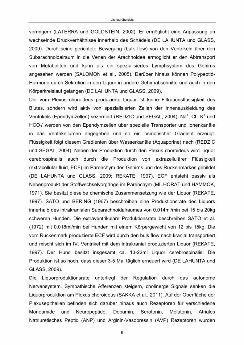
Nachgewiesen wurde eine Absorption über die spinalen Nervenwurzeln und den
Truncus olfactorius (REDZIC und SEGAL, 2004), über den subarachnoidalen Raum
der Riechfäden, die in der Nasenmukosa mit dem lymphatischen System in
Verbindung stehen (FOLTZ et al., 1984; SAKKA et al., 2011), sowie über
Lymphgefäße und Venen um die spinalen Nervenwurzeln (Abb. 3) (DE LAHUNTA
und GLASS, 2009; SAKKA et al., 2011).
Abbildung 3: Liquorabsorption durch spinale arachnoidale Villi und spinale Nervenwurzeln nach
SAKKA et al. (2011). Die spinalen arachnoidalen Villi stehen mit dem epiduralen Venenplexus (a)
und angrenzenden spinalen Nervenwurzeln (b) in Kontakt. Auch im meningealen Spalt der
abgehenden spinalen Nervenwurzel findet sich eine Absorptionsoberfläche (c).
8
Literaturübersicht
2.3 Der Hydrozephalus internus
2.3.1 Definition und Einteilung
Definition und Einteilung des Hydrozephalus beim Hund sind bis heute nicht
einheitlich (THOMAS, 1999; REKATE, 2011). MORI und Mitarbeiter (1995) definieren
einen Hydrozephalus als ein Krankheitsbild, bei dem es durch Störung der
Liquorzirkulation zu einer Ansammlung von Liquor cerebrospinalis innerhalb des
Ventrikelsystems und zu einer fortschreitenden Ausweitung der Ventrikel kommt.
Eine Einteilung kann anhand verschiedener Kriterien vorgenommen werden:
Nach dem betroffenen anatomischen Kompartiment kann man den Hydrocephalus
internus mit Ansammlung von CSF im Ventrikelsystem vom Hydrocephalus externus
mit Ausweitung des Subarachnoidalraumes unterscheiden (THOMAS, 1999).
Nach der Pathogenese können drei Formen unterschieden werden:
1. Hydrocephalus ex vacuo: Hierbei handelt es sich um eine kompensatorische
Liquoransammlung nach Schwund von Hirngewebe, die nicht mit einer
Erweiterung der Ventrikel und einem Anstieg des intraventrikulären Druckes
einhergeht. Er wird auch als kompensatorischer Hydrozephalus bezeichnet
(DE LAHUNTA und GLASS, 2009). Nach der oben genannten Definition sollte
er nicht als Hydrozephalus klassifiziert werden (REKATE, 2009).
2. Hypersekretionshydrozephalus: Ein Hydrozephalus durch gesteigerte
Liquorproduktion kommt selten vor (HECHT, 2011) und wird durch Choroid-
Plexus Tumoren verursacht (REKATE, 2009). Ein Fallbericht beschreibt eine
Überproduktion durch Hyperplasie des Plexus choroideus (SMITH et al.,
2007).
3. Hydrozephalus durch Liquorzirkulationsstörungen, die zum Druckanstieg und
zur Erweiterung der Ventrikel führen. Hierbei wird weiter in eine angeborene
(kongenitale) und eine erworbene Form unterteilt (DE LAHUNTA und GLASS,
2009).
Der kongenitale Hydrozephalus wird beim Hund häufiger in brachyzephalen und Toy-
Rassen beschrieben, wie dem Malteser, Zwergspitz, Boston Terrier, Mops, Yorkshire
Terrier, Pekinesen oder der Englischen Bulldogge (ESTEVE-RATSCH et al., 2001;
VULLO et al., 1997). Als mögliche Ursachen kommen genetische Faktoren,
9
Literaturübersicht
Anomalien in der Entwicklung, intrauterine oder perinatale Infektionen, sowie
Blutungen in Frage (THOMAS, 2010). Ein angeborener Hydrozephalus kann mit
anderen Anomalien vergesellschaftet sein, wie zum Beispiel Chiari-Malformationen,
Dandy-Walker-Syndromen oder Meningomyelocelen (THOMAS, 2010). Ein
kongenitaler Hydozephalus, der mit weiteren Veränderungen des Gehirns
einhergeht, wird auch als komplizierter Hydrozephalus bezeichnet (MORI et al.,
1995).
Zum erworbenen Hydrozephalus kann es beispielsweise durch Neoplasien oder
Entzündungen kommen (DE LAHUNTA und GLASS, 2009; THOMAS, 2010).
Auch die ursprünglich von DANDY und BLACKFAN 1914 eingeführte
Unterscheidung zwischen kommunizierendem und nicht-kommunizierendem
Hydrozephalus, abhängig davon, ob ein in die Ventrikel injizierter Farbstoff im
Subarachnoidalraum nachgewiesen werden kann, findet heute noch Verwendung.
Dabei beschreibt der nicht-kommunizierende Hydrozephalus eine Obstruktion
innerhalb des Ventrikelsystems, der kommunizierende Hydrozephalus wird auf eine
Obstruktion im Bereich des Subarachnoidalraumes oder der arachnoidalen Villi
zurückgeführt (REKATE, 2009). GREITZ (2004) beschreibt den kommunizierenden
Hydrozephalus als Folge einer Störung der Liquordynamik, die nicht zwingend auf
einer Obstruktion beruht.
Eine Unterteilung in primär (idiopathisch) und sekundär zu einer bekannten Ursache
ist außerdem möglich (THOMAS, 2010).
Aufgrund der unterschiedlichen Pathophysiologie wird heute häufig eine Unterteilung
in akuten und chronischen Hydrozephalus vorgenommen, wobei die chronische
Form weiter in den chronisch obstruktiven und den kommunizierenden Subtyp
unterteilt werden kann (GREITZ, 2004).
10
Literaturübersicht
2.3.2 Pathophysiologie
Der Mechanismus der Erweiterung der Ventrikel ist nicht endgültig geklärt, die „bulk
flow“-Theorie und die Hydrodynamische Theorie werden diskutiert (KAMALO, 2013).
1. „Bulk flow“-Theorie
Grundlage ist die Arbeit von DANDY und BLACKFAN 1914. Sie haben den Plexus
choroideus als Produktionsort des Liquor cerebrospinalis definiert und
Hydrozephalus als einen Rückstau von Liquor durch eine Obstruktion innerhalb des
Ventrikelsystems (nicht-kommunizierender Hydrozephalus) oder kaudal des
Ventrikelsystems (kommunizierender Hydrozephalus) definiert. RANSOHOFF und
Mitarbeiter haben 1960 die Bezeichnungen intraventrikulär obstruktiver
Hydrozephalus und extraventrikulär obstruktiver Hydrozephalus eingeführt. Die
Theorie beschreibt ein Mißverhältnis zwischen Liquorproduktion und –absorption,
das, mit Ausnahme der Überproduktion von Liquor durch Choroid Plexus Papillome,
auf einer Obstruktion basiert (REKATE, 1997). Die Obstruktion kann innerhalb des
Ventrikelsystems liegen, oder auf Höhe der arachnoidalen Villi (kommunizierender
Hydrozephalus) (REKATE, 1997). Nicht mit der „bulk flow“-Theorie zu erklären sind
die Ergebnisse der Arbeit von DI ROCCO und Mitarbeitern 1978, die durch Pulsation
eines Ballons im lateralen Ventrikel eine Ventrikelerweiterung verursachen konnten,
ohne die Beteiligung einer Obstruktion.
2. Hydrodynamische Theorie
GREITZ (2004) ist Begründer dieser Theorie. Er sieht in der „bulk flow“-Theorie keine
Erklärung für den kommunizierenden Hydrozephalus, da eine Malabsorption durch
Obstruktion auf Höhe der arachnoidalen Villi zu einer Erweiterung des
Subarachnoidalraums führen müsste und nicht zu einer Erweiterung des
Ventrikelsystems (GREITZ, 2007). Als Ursache für einen chronischen
kommunizierenden Hydrozephalus beschreibt er eine verminderte Compliance, die
zum Beispiel durch Störungen im Subarachnoidalraum, wie Adhäsionen nach
Meningitiden oder Blutungen, auftreten kann. Kortikale Venen, vor allem im Bereich
ihres Ausstroms in die Sinus, werden komprimiert, was zu venöser Kongestion und
somit neben erhöhtem intraventrikulärem Druck auch zu steigendem intrakraniellem
Druck führt. Die venöse Kongestion und der erhöhte Druck im Parenchym wirken der
11
Literaturübersicht
fortschreitenden Erweiterung der Ventrikel entgegen, so dass es zur Ausbildung
eines Gleichgewichts mit erhöhten Druckverhältnissen kommen kann (GREITZ,
2004). Die Pathophysiologie des chronisch obstruktiven Hydrozephalus gleicht der
des kommunizierenden Hydrozephalus (GREITZ, 2004).
Die Pathogenese der Schädigung des Gehirns ist multifaktoriell (DEL BIGIO, 2010).
Art und Ausprägung der Schäden sind abhängig vom Alter des Patienten, der
Geschwindigkeit der Erweiterung der Ventrikel, dem Ausmaß der ventrikulären
Volumenzunahme und der Nähe einer Struktur des Gehirns zum Ventrikel (DEL
BIGIO, 1993). Bei der Entstehung unterscheidet man primäre und sekundäre
pathophysiologische Abläufe (MCALLISTER et al., 1998). Zu den primären
pathophysiologischen Mechanismen, die im frühen Stadium des Hydrozephalus
auftreten, gehören vor allem Kompression und Dehnung, aber auch Ischämie,
intraventrikuläre Blutungen, interstitielle Ödeme und Störungen der Blut-Hirn-
Schranke (MCALLISTER et al., 1998). Sekundäre Mechanismen resultieren aus den
primären und umfassen beispielsweise den Untergang von Neuronen, verminderte
Konnektivität zwischen Neuronen oder eine verminderte Anzahl von Kapillaren in der
periventrikulären weißen Substanz. Insgesamt kommt es vor allem zur Schädigung
und Atrophie der weißen Substanz (DEL BIGIO, 1993).
12
Literaturübersicht
2.4 Erkrankung beim Menschen
Beim Menschen unterscheidet man den frühkindlichen Hydrozephalus vom
Hydrozephalus des Erwachsenen (MORI et al., 1995). Auch beim Menschen
bestehen verschiedene Klassifikationen (REKATE, 2009).
MORI und Mitarbeiter (1995) haben eine Einteilung nach Alter und Ätiologie
vorgenommen:
Frühkindlicher Hydrozephalus:
1. Fetaler Hydrozephalus
2. Infantiler Hydrozephalus
3. Hydrozephalus assoziiert mit Enzephalozelen oder Myelomeningozelen
4. Posthämorrhagischer Hydrozephalus bei Neonaten
5. Postmeningitischer Hydrozephalus
Hydrozephalus des Erwachsenen:
6. Hydrozephalus nach subarachnoidalen Blutungen
7. Idiopathischer Hydrozephalus des Erwachsenen
8. Posttraumatischer Hydrozephalus
Durch Tumoren bedingter Hydrozephalus wurde von der Einteilung ausgeschlossen.
Für die einzelnen Punkte wurden jeweils Einschluss- und Ausschlusskriterien, sowie
zusätzliche Kriterien definiert, die bei der Diagnosestellung helfen (MORI et al.,
1995). Die acht Untetergruppen können auch drei großen Gruppen zugeordnet
werden: kongenitaler Hydrozephalus, idiopathischer Hydrozephalus des
Erwachsenen und sekundärer Hydrozephalus (MORI et al., 1995).
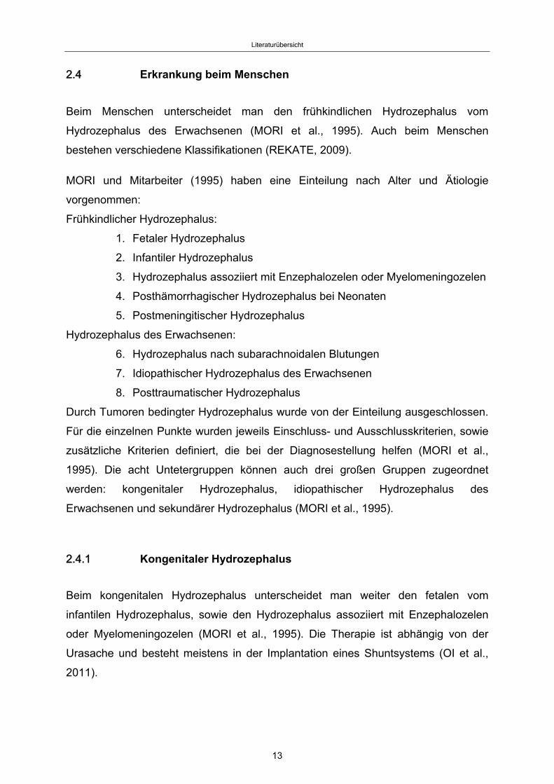
2.4.1 Kongenitaler Hydrozephalus
Beim kongenitalen Hydrozephalus unterscheidet man weiter den fetalen vom
infantilen Hydrozephalus, sowie den Hydrozephalus assoziiert mit Enzephalozelen
oder Myelomeningozelen (MORI et al., 1995). Die Therapie ist abhängig von der
Urasache und besteht meistens in der Implantation eines Shuntsystems (OI et al.,
2011).
13
Literaturübersicht
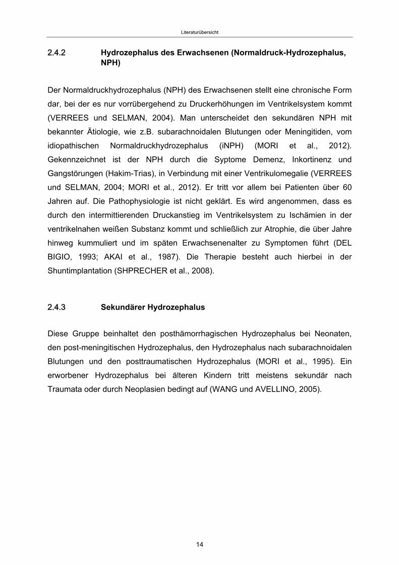
2.4.2 Hydrozephalus des Erwachsenen (Normaldruck-Hydrozephalus, NPH)
Der Normaldruckhydrozephalus (NPH) des Erwachsenen stellt eine chronische Form
dar, bei der es nur vorrübergehend zu Druckerhöhungen im Ventrikelsystem kommt
(VERREES und SELMAN, 2004). Man unterscheidet den sekundären NPH mit
bekannter Ätiologie, wie z.B. subarachnoidalen Blutungen oder Meningitiden, vom
idiopathischen Normaldruckhydrozephalus (iNPH) (MORI et al., 2012).
Gekennzeichnet ist der NPH durch die Syptome Demenz, Inkortinenz und
Gangstörungen (Hakim-Trias), in Verbindung mit einer Ventrikulomegalie (VERREES
und SELMAN, 2004; MORI et al., 2012). Er tritt vor allem bei Patienten über 60
Jahren auf. Die Pathophysiologie ist nicht geklärt. Es wird angenommen, dass es
durch den intermittierenden Druckanstieg im Ventrikelsystem zu Ischämien in der
ventrikelnahen weißen Substanz kommt und schließlich zur Atrophie, die über Jahre
hinweg kummuliert und im späten Erwachsenenalter zu Symptomen führt (DEL
BIGIO, 1993; AKAI et al., 1987). Die Therapie besteht auch hierbei in der
Shuntimplantation (SHPRECHER et al., 2008).
2.4.3 Sekundärer Hydrozephalus
Diese Gruppe beinhaltet den posthämorrhagischen Hydrozephalus bei Neonaten,
den post-meningitischen Hydrozephalus, den Hydrozephalus nach subarachnoidalen
Blutungen und den posttraumatischen Hydrozephalus (MORI et al., 1995). Ein
erworbener Hydrozephalus bei älteren Kindern tritt meistens sekundär nach
Traumata oder durch Neoplasien bedingt auf (WANG und AVELLINO, 2005).
14
Literaturübersicht
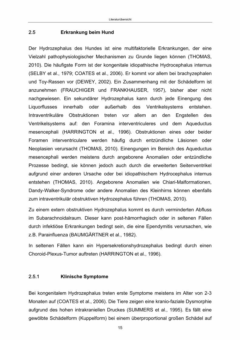
2.5 Erkrankung beim Hund
Der Hydrozephalus des Hundes ist eine multifaktorielle Erkrankungen, der eine
Vielzahl pathophysiologischer Mechanismen zu Grunde liegen können (THOMAS,
2010). Die häufigste Form ist der kongenitale idiopathische Hydrocephalus internus
(SELBY et al., 1979; COATES et al., 2006). Er kommt vor allem bei brachyzephalen
und Toy-Rassen vor (DEWEY, 2002). Ein Zusammenhang mit der Schädelform ist
anzunehmen (FRAUCHIGER und FRANKHAUSER, 1957), bisher aber nicht
nachgewiesen. Ein sekundärer Hydrozephalus kann durch jede Einengung des
Liquorflusses innerhalb oder außerhalb des Ventrikelsystems entstehen.
Intraventrikuläre Obstruktionen treten vor allem an den Engstellen des
Ventrikelsystems auf: den Foramina interventriculeres und dem Aqueductus
mesencephali (HARRINGTON et al., 1996). Obstruktionen eines oder beider
Foramen interventriculare werden häufig durch entzündliche Läsionen oder
Neoplasien verursacht (THOMAS, 2010). Einengungen im Bereich des Aqueductus
mesencephali werden meistens durch angeborene Anomalien oder entzündliche
Prozesse bedingt, sie können jedoch auch durch die erweiterten Seitenventrikel
aufgrund einer anderen Ursache oder bei idiopathischem Hydrocephalus internus
entstehen (THOMAS, 2010). Angeborene Anomalien wie Chiari-Malformationen,
Dandy-Walker-Syndrome oder andere Anomalien des Kleinhirns können ebenfalls
zum intraventrikulär obstruktiven Hydrozephalus führen (THOMAS, 2010).
Zu einem extern obstruktiven Hydrozephalus kommt es durch verminderten Abfluss
im Subarachnoidalraum. Dieser kann post-hämorrhagisch oder in seltenen Fällen
durch infektiöse Erkrankungen bedingt sein, die eine Ependymitis verursachen, wie
z.B. Parainfluenza (BAUMGÄRTNER et al., 1982).
In seltenen Fällen kann ein Hypersekretionshydrozephalus bedingt durch einen
Choroid-Plexus-Tumor auftreten (HARRINGTON et al., 1996).
2.5.1 Klinische Symptome
Bei kongenitalem Hydrozephalus treten erste Symptome meistens im Alter von 2-3
Monaten auf (COATES et al., 2006). Die Tiere zeigen eine kranio-faziale Dysmorphie
aufgrund des hohen intrakraniellen Druckes (SUMMERS et al., 1995). Es fällt eine
gewölbte Schädelform (Kuppelform) bei einem überproportional großen Schädel auf
15
Literaturübersicht
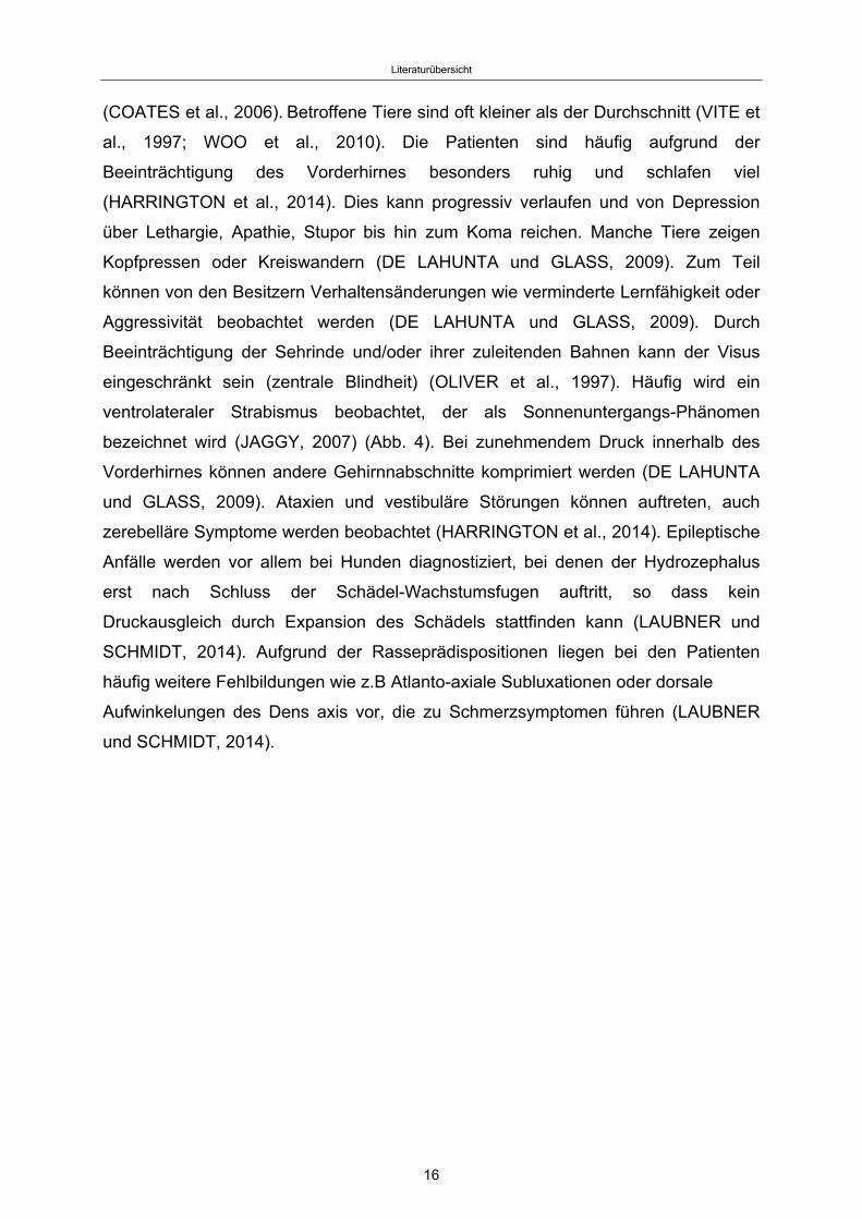
(COATES et al., 2006). Betroffene Tiere sind oft kleiner als der Durchschnitt (VITE et
al., 1997; WOO et al., 2010). Die Patienten sind häufig aufgrund der
Beeinträchtigung des Vorderhirnes besonders ruhig und schlafen viel
(HARRINGTON et al., 2014). Dies kann progressiv verlaufen und von Depression
über Lethargie, Apathie, Stupor bis hin zum Koma reichen. Manche Tiere zeigen
Kopfpressen oder Kreiswandern (DE LAHUNTA und GLASS, 2009). Zum Teil
können von den Besitzern Verhaltensänderungen wie verminderte Lernfähigkeit oder
Aggressivität beobachtet werden (DE LAHUNTA und GLASS, 2009). Durch
Beeinträchtigung der Sehrinde und/oder ihrer zuleitenden Bahnen kann der Visus
eingeschränkt sein (zentrale Blindheit) (OLIVER et al., 1997). Häufig wird ein
ventrolateraler Strabismus beobachtet, der als Sonnenuntergangs-Phänomen
bezeichnet wird (JAGGY, 2007) (Abb. 4). Bei zunehmendem Druck innerhalb des
Vorderhirnes können andere Gehirnnabschnitte komprimiert werden (DE LAHUNTA
und GLASS, 2009). Ataxien und vestibuläre Störungen können auftreten, auch
zerebelläre Symptome werden beobachtet (HARRINGTON et al., 2014). Epileptische
Anfälle werden vor allem bei Hunden diagnostiziert, bei denen der Hydrozephalus
erst nach Schluss der Schädel-Wachstumsfugen auftritt, so dass kein
Druckausgleich durch Expansion des Schädels stattfinden kann (LAUBNER und
SCHMIDT, 2014). Aufgrund der Rasseprädispositionen liegen bei den Patienten
häufig weitere Fehlbildungen wie z.B Atlanto-axiale Subluxationen oder dorsale
Aufwinkelungen des Dens axis vor, die zu Schmerzsymptomen führen (LAUBNER
und SCHMIDT, 2014).
16
Literaturübersicht
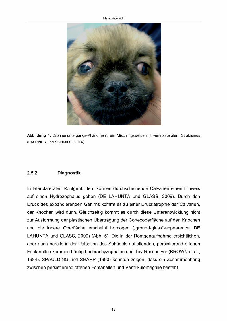
Abbildung 4: „Sonnenuntergangs-Phänomen“: ein Mischlingswelpe mit ventrolateralem Strabismus
(LAUBNER und SCHMIDT, 2014).
2.5.2 Diagnostik
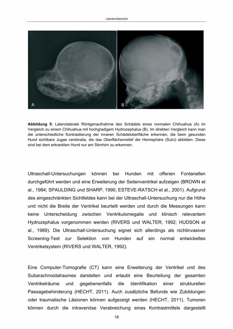
In laterolateralen Röntgenbildern können durchscheinende Calvarien einen Hinweis
auf einen Hydrozephalus geben (DE LAHUNTA und GLASS, 2009). Durch den
Druck des expandierenden Gehirns kommt es zu einer Druckatrophie der Calvarien,
der Knochen wird dünn. Gleichzeitig kommt es durch diese Unterentwicklung nicht
zur Ausformung der plastischen Übertragung der Cortexoberfläche auf den Knochen
und die innere Oberfläche erscheint homogen („ground-glass“-appearence, DE
LAHUNTA und GLASS, 2009) (Abb. 5). Die in der Röntgenaufnahme ersichtlichen,
aber auch bereits in der Palpation des Schädels auffallenden, persistierend offenen
Fontanellen kommen häufig bei brachyzephalen und Toy-Rassen vor (BROWN et al.,
1984). SPAULDING und SHARP (1990) konnten zeigen, dass ein Zusammenhang
zwischen persistierend offenen Fontanellen und Ventrikulomegalie besteht.
17
Literaturübersicht
Abbildung 5: Laterolaterale Röntgenaufnahme des Schädels eines normalen Chihuahua (A) im Vergleich zu einem Chihuahua mit hochgradigem Hydrozephalus (B). Im direkten Vergleich kann man die unterschiedliche Kontrastierung der inneren Schädeloberfläche erkennen, die beim gesunden Hund sichtbare Jugae cerebralia, die das Oberflächenrelief der Hemisphäre (Sulci) abbilden. Diese sind bei dem erkrankten Hund nur am Stirnhirn zu erkennen.
Ultraschall-Untersuchungen können bei Hunden mit offenen Fontanellen
durchgeführt werden und eine Erweiterung der Seitenventrikel aufzeigen (BROWN et
al., 1984; SPAULDING und SHARP, 1990; ESTEVE-RATSCH et al., 2001). Aufgrund
des eingeschränkten Sichtfeldes kann bei der Ultraschall-Untersuchung nur die Höhe
und nicht die Breite der Ventrikel beurteilt werden und durch die Messungen kann
keine Unterscheidung zwischen Ventrikulomegalie und klinisch relevantem
Hydrozephalus vorgenommen werden (RIVERS und WALTER, 1992; HUDSON et
al., 1989). Die Ultraschall-Untersuchung eignet sich allerdings als nichtinvasiver
Screening-Test zur Selektion von Hunden auf ein normal entwickeltes
Ventrikelsystem (RIVERS und WALTER, 1992).
Eine Computer-Tomografie (CT) kann eine Erweiterung der Ventrikel und des
Subarachnoidalraumes darstellen und erlaubt eine Beurteilung der gesamten
Ventrikelräume und gegebenenfalls die Identifikation einer strukturellen
Passagebehinderung (HECHT, 2011). Auch zusätzliche Befunde wie Zubildungen
oder traumatische Läsionen können aufgezeigt werden (HECHT, 2011). Tumoren
können durch die intravenöse Verabreichung eines Kontrastmittels dargestellt
18
Literaturübersicht
werden (HENNINGER und HITTMAIR, 1994). Es besteht jedoch die Gefahr der
Fehldiagnose, da die genaue Struktur der Kortex nicht sicher darstellbar ist.
Missbildungen wie Hydranenzephalie, Porenzephalie oder Lissenzephalie können
aufgrund der mangelnden Detaildarstellung übersehen werden. Zusätzlich werden
entzündliche Grunderkrankungen nicht sicher dargestellt und erweiterte
Ventrikelräume können fälschlicherweise als Ursache neurologischer Symptome
angesprochen werden (LAUBNER und SCHMIDT, 2014).
Die Magnet-Resonanz-Tomographie (MRT) stellt das Mittel der Wahl bei der
Diagnose des Hydrozephalus beim Menschen (FRITSCH und MEHDORN, 2007;
MORI et al., 2012; KARTAL und ALGIN, 2014) und beim Tier (PLATT et al., 2012;
DE LAHUNTA und GLASS, 2009; LEIGH et al., 2008) dar. Bei diesem Verfahren
können nicht nur die erweiterten Ventrikel dargestellt werden, sondern auch
Ursachen für eine Obstruktion oder Missbildungen aufgezeigt werden (ADAMIAK et
al., 2012; THOMAS, 2010). Veränderungen, die durch den erhöhten Druck im
Ventrikelsystem verursacht werden, können detektiert werden (EL GAMMAL et al.,
1987). Zusätzliche Erkrankungen, wie zum Beispiel entzündliche
Gehirnerkrankungen, können erkannt werden, die die Wahl der Therapie und die
Prognose beeinflussen (LAUBNER und SCHMIDT, 2014). Die Diagnose des
Hydrocephalus internus wird mit Hilfe der Magnet-Resonanz-Tomografie beim Hund
meist aufgrund einer subjektiven Erweiterung der Ventrikel gestellt (PLATT et al.,
2012). Beim Menschen können verschiedene Kriterien in der MRT zur
Diagnosestellung herangezogen werden:
1) Größe der Seiten-Ventrikel: Zunächst wird eine Aufweitung und Abrundung
der Temporalhörner beschrieben, dann der Frontalhörner (EDELMANN et al.,
2006). Die Seiten-Ventrikel können eine ballonartige Form aufweisen
(EDELMANN et al., 2006). Der Evans‘ Index (maximale Ausdehnung der
Frontalhörne geteilt durch den maximalen biparietalen Diameter) liegt bei über
0,3 (FRITSCH und MEHDORN, 2007; KARTAL und ALGIN, 2013).
2) Erweiterung des III. Ventrikels (EL GAMMAL et al., 1987; SEGEV et al.,
2001): Eine Abrundung und ballonartige Aufweitung sind typische Anzeichen
eines Hydrozephalus (EDELMANN et al., 2006). Hierbei wird der Boden des
III. Ventrikels nach kaudal verlagert (FRITSCH und MEHDORN, 2007). EL
GAMMAL und Mitarbeiter (1987) beschreiben eine Aufweitung des vorderen
19
Literaturübersicht
Recessus (Recessus infundibuli) des III. Ventrikels als hilfreich bei der
Differenzierung zwischen Hydrozephalus-Patienten und solchen mit Atrophie
bedingter Erweiterung des Ventrikelsystems.
3) Erweiterung des IV. Ventrikels (EDELMANN et al., 2006): Sie ist hinweisend
auf einen kommunizierenden Hydrozephalus (EDELMANN et al., 2006).
4) Verminderte mamillopontine Distanz (KARTAL und ALGIN, 2013): Die
mamillopontine Distanz wird im sagittalen Schnitt gemessen zwischen dem
vorderen Corpus mamillare zur Pons (EL GAMMAL et al., 1987). Durch
Erweiterung des III. Ventrikels und die dadurch entstehende Verschiebung
des Thalamus nach unten vermindert sich die mamillopontine Distanz bei
Patienten mit Hydrozephalus (EL GAMMAL et al., 1987).
5) Corpus callosum-Winkel (Callosal angle, CA) <90°: ISHII et al. 2008 haben
den Winkel zwischen den Seitenventrikeln im transversalen Schnitt auf Höhe
der Commisura posterior gemessen und einen spitzen Winkel <90° als
verdächtig für einen Normaldruck-Hydrozephalus beschrieben. In einer
retrospektiven Studie von VIRHAMMER und Mitarbeitern (2014) war der CA
vor Shunt-Implantation deutlich kleiner bei Patienten, die später postoperativ
eine Verbesserung zeigten.
6) Dehnung/Ausdünnung und Anhebung des Corpus callosum sowie Absenkung
des Fornix im vorderen Anteil (KARTAL und ALGIN, 2013, SEGEV et al.,
2001, HOFMANN et al., 1995).
7) Verschmälerung oder Verstreichen der kortikalen Sulki. Durch die Erweiterung
der Ventrikel kann der kortikale Subarachnoidalraum komprimiert werden
(MATSUMAE et al., 1996; FRITSCH und MEHDORN, 2007; REKATE et al.,
2008).
8) Periventrikuläre Ödeme (KARTAL und ALGIN, 2013): Periventrikuläre Ödeme
treten vor allem in der akuten und subakuten Phase des Hydrozephalus auf
(EDELMANN et al., 2006). Sie entstehen vermutlich durch die
transependymale Absorption von Liquor cerebrospinalis aufgrund des
Druckgradienten vom Ventrikel zum Parenchym der weißen Substanz
(ZIMMERMANN et al., 1986; HABERL et al., 2007; MATSUMAE et al., 1996).
Eine andere Theorie beschreibt periventrikuläre Ödeme als Bereiche mit
verminderter Resorptionsfähigkeit von extrazellulärer Flüssigkeit (DEL BIGIO,
1993). Periventrikuläre Ödeme werden auch bei anderen Erkrankungen
beobachtet, die mit einer Demyelinisierung einhergehen, wie beispielsweise
20
Literaturübersicht
die Multiple Sklerose oder die subkortikale arteriosklerotische
Enzephalopathie (ZIMMERMANN et al., 1986).
9) „Flow-void Phänomen“ im Aqueductus mesencephali in T2-gewichteten MRT-
Schnitten (KARTAL und ALGIN, 2013): Als Ursache wird ein Reflux von CSF
vom Aqueductus mesencephali in den III. Ventrikel angenommen (KARTAL
und ALGIN, 2013).
2.6 Therapie
2.6.1 Medikamentelle Therapie
Die medikamentelle Therapie wird bei Hunden mit Hydrocephalus internus eingesetzt
um einen chirurgischen Eingriff zu verzögern, wenn eine operative Therapie keine
Option darstellt (THOMAS, 2010). Mit ihr kann meistens keine langfristige Besserung
der Symptome erreicht werden (COATES et al., 2006; SHIHAB et al., 2009). Sie zielt
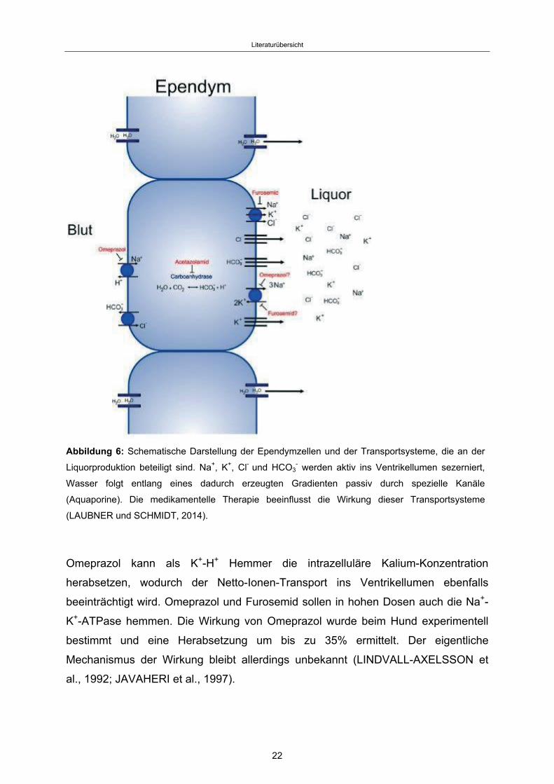
auf die Herabsetzung der Liquorproduktion (Abb. 6). Acetazolamid inhibiert die
Carboanydrase und setzt so die Sekretion von HCO3- herab. Eine experimentelle
Studie konnte für Acetazolamid keinerlei Reduktion der Liquorproduktion bestimmen
(MILLER et al., 1986), während andere eine maximale kurzfristige Reduktion um bis
zu 40-50% dokumentieren konnten (POCA und SAHUQUILLO, 2005). Der Effekt von
Acetazolamid auf den intrakraniellen Druck (intracranial pressure, ICP) ist jedoch
variabel, es kann durch einen Anstieg des cerebralen Blutflusses (cerebral blood
flow, CBF) auch zum Anstieg des ICP kommen (POCA und SAHUQUILLO, 2005).
Mögliche toxische Effekte auf die Myelinisierung bei jungen Individuen wurden
beschrieben, so dass die Sicherheit des Einsatzes hier unklar ist (HACK und
COHEN, 1998). Als Nebenwirkung kann es unter Therapie mit Acetazolamid zur
metabolischen Azidose durch Verlust von Bikarbonat über die Nieren kommen
(GILMORE, 1990).
Furosemid kann den Na+-K+-Cl-Symporter der Ependymzellen hemmen und dadurch
den Ionentransport in das Ventrikellumen vermindern. Experimentell konnte eine
verminderte Liquorproduktionsrate und ein verminderter ICP gezeigt werden
(GREENE et al., 1985). Aufgrund der Nebenwirkungen (erhöhte Calciumexkretion)
ist Furosemid nicht als langfristige Therapie einsetzbar (POCA und SAHUQUILLO,
2005).
21
Literaturübersicht
Abbildung 6: Schematische Darstellung der Ependymzellen und der Transportsysteme, die an der
Liquorproduktion beteiligt sind. Na+, K+, Cl- und HCO3- werden aktiv ins Ventrikellumen sezerniert,
Wasser folgt entlang eines dadurch erzeugten Gradienten passiv durch spezielle Kanäle
(Aquaporine). Die medikamentelle Therapie beeinflusst die Wirkung dieser Transportsysteme
(LAUBNER und SCHMIDT, 2014).
Omeprazol kann als K+-H+ Hemmer die intrazelluläre Kalium-Konzentration
herabsetzen, wodurch der Netto-Ionen-Transport ins Ventrikellumen ebenfalls
beeinträchtigt wird. Omeprazol und Furosemid sollen in hohen Dosen auch die Na+-
K+-ATPase hemmen. Die Wirkung von Omeprazol wurde beim Hund experimentell
bestimmt und eine Herabsetzung um bis zu 35% ermittelt. Der eigentliche
Mechanismus der Wirkung bleibt allerdings unbekannt (LINDVALL-AXELSSON et
al., 1992; JAVAHERI et al., 1997).
22
Literaturübersicht
Eine Effektivität der medikamentellen Therapie beim Hund konnte jedoch bisher nicht
in klinischen Studien belegt werden (THOMAS, 2010). Ein großes Problem bei der
Interpretation und beim Vergleich verschiedener Studien ist wie beim Menschen die
fehlende einheitliche Definition des Hydrozephalus (GILMORE, 1990). Über die
Wirksamkeit der Medikamente zur Beeinflussung der Liquorproduktion herrscht keine
einheitliche Meinung. Klinische Studien, die Kurz- und Langzeitergebnisse zur
Wirksamkeit liquorhemmender Medikamente in der Therapie des Hydrozephalus mit
der chirurgischen Therapie vergleichen, fehlen (GILMORE, 1990).
2.6.2 Chirurgische Therapie
Aufgrund der schlechten Wirksamkeit der zur Verfügung stehenden Medikamente in
der Langzeitbehandlung, stellt die chirurgische Shunt-Implantation die Therapie der
Wahl dar (DE STEFANI et al., 2009; BIEL et al., 2013). Verwendet werden
humanmedizinische Shuntsysteme (PLATT et al., 2012), durch die der Abfluss des
Liquors in eine andere Körperhöhle, meistens das Peritoneum, ermöglicht wird
(SHIHAB et al., 2011). Das Ziel der Shuntimplantation ist eine Senkung des
intrakraniellen Druckes und damit eine Verbesserung der neurologischen Symptome
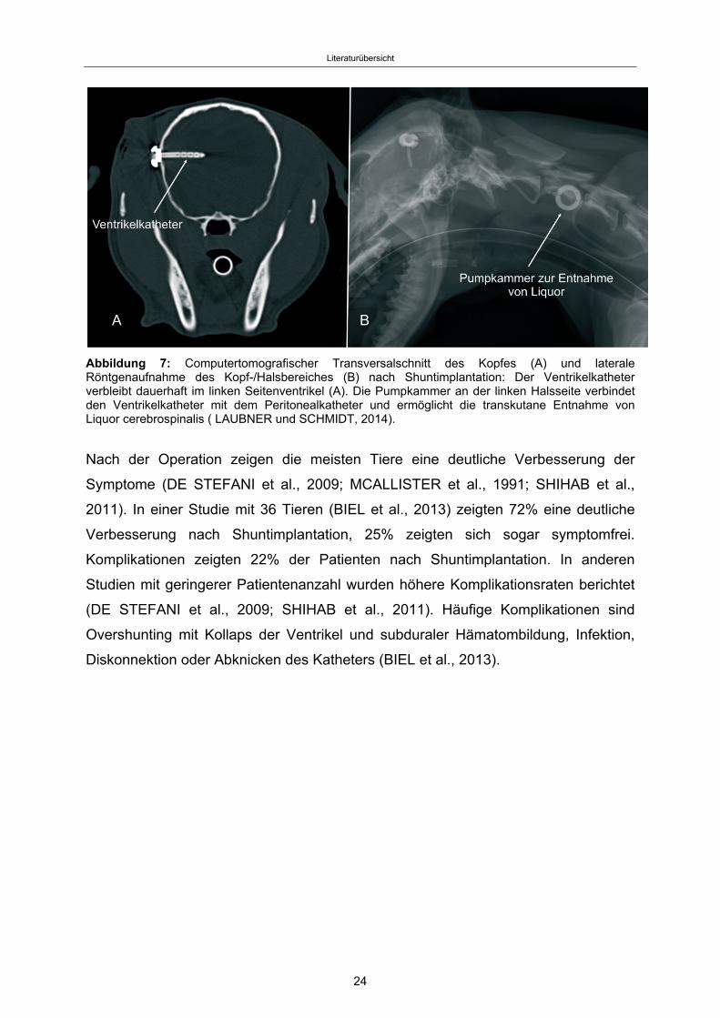
(DE STEFANI, 2009). Nach BIEL und Mitarbeitern (2013) werden die Hunde auf der
rechten Seite gelagert. Durch ein Bohrloch im Schädel wird der Ventrikelkatheter
transkortikal in den Seitenventrikel eingesetzt (Abb. 7) und durch einen
Bohrlochumlenker fixiert. Der Katheter wird im Halsbereich mit einer Pumpkammer
verbunden, an die der Peritonealkatheter anschließt. Dieser wird unter der Haut
getunnelt und hinter dem Rippenbogen in den Peritonealraum gebracht und fixiert.
23
Literaturübersicht
Abbildung 7: Computertomografischer Transversalschnitt des Kopfes (A) und laterale Röntgenaufnahme des Kopf-/Halsbereiches (B) nach Shuntimplantation: Der Ventrikelkatheter verbleibt dauerhaft im linken Seitenventrikel (A). Die Pumpkammer an der linken Halsseite verbindet den Ventrikelkatheter mit dem Peritonealkatheter und ermöglicht die transkutane Entnahme von Liquor cerebrospinalis ( LAUBNER und SCHMIDT, 2014).
Nach der Operation zeigen die meisten Tiere eine deutliche Verbesserung der
Symptome (DE STEFANI et al., 2009; MCALLISTER et al., 1991; SHIHAB et al.,
2011). In einer Studie mit 36 Tieren (BIEL et al., 2013) zeigten 72% eine deutliche
Verbesserung nach Shuntimplantation, 25% zeigten sich sogar symptomfrei.
Komplikationen zeigten 22% der Patienten nach Shuntimplantation. In anderen
Studien mit geringerer Patientenanzahl wurden höhere Komplikationsraten berichtet
(DE STEFANI et al., 2009; SHIHAB et al., 2011). Häufige Komplikationen sind
Overshunting mit Kollaps der Ventrikel und subduraler Hämatombildung, Infektion,
Diskonnektion oder Abknicken des Katheters (BIEL et al., 2013).
24
Publikationen
Publikationen 3.
3.1 Publikation 1
Das folgende Manuskript „Magnetic resonance imaging signs of high intraventricular
pressure - comparison of findings in dogs with clinically relevant internal
hydrocephalus and asymptomatic dogs with ventriculomegaly” wurde am 01.08.2015
von “BMC Veterinary Research” veröffentlicht. Die elektronische Version des Artikels
kann auf http://www.biomedcentral.com/1746-6148/11/181 gefunden werden.
The following manuscript entitled “Magnetic resonance imaging signs of high
intraventricular pressure - comparison of findings in dogs with clinically relevant
internal hydrocephalus and asymptomatic dogs with ventriculomegaly” has been
published in “BMC Veterinary Research” on Aug 1, 2015. The electronic version of
this article can be found online at: http://www.biomedcentral.com/1746-6148/11/181.
Received: 13 February 2015
Accepted: 13 July 2015
Published:1 August 2015
© 2015 Laubner et al.
25
Publikationen
Magnetic resonance imaging signs of high intraventricular pressure -
comparison of findings in dogs with clinically relevant internal hydrocephalus
and asymptomatic dogs with ventriculomegaly
Steffi Laubner1, Nele Ondreka1, Klaus Failing2, Martin Kramer1, Martin J. Schmidt1*
1Department of Veterinary Clinical Sciences, Small Animal Clinic, Justus-Liebig-
University, Frankfurter Straße 108, 35392 Giessen, Germany
2Department of Biomathematics, Justus-Liebig-University, Frankfurter Straße 95,
35392 Giessen, Germany
Email addresses:
* Corresponding author:
* [email protected] (Martin Schmidt)
[email protected] (Steffi Laubner)
[email protected] (Klaus Failing)
[email protected] (Martin Kramer)
[email protected] (Nele Ondreka)
26
Publikationen
Abstract
Background
Magnetic resonance imaging (MRI) findings of canine brains with enlarged ventricles
in asymptomatic dogs were compared to those in dogs with clinically relevant internal
hydrocephalus, in order to determine the imaging findings indicative of a relevant
increase in intraventricular pressure. Discrimination between clinically relevant
hydrocephalus and ventriculomegaly based on MRI findings has not been
established yet and is anything but trivial because of the wide variation in ventricular
size in different dog breeds and individuals.
Material & methods
The MRI scans of the brains of sixty-seven dogs of various breeds, skull
conformation and weight were reviewed retrospectively. Based on clinical and
imaging findings, the dogs were divided into three groups: a normal group (n=20), a
group with clinically silent ventriculomegaly (n=25) and a group with severe clinically
relevant internal hydrocephalus (n=22). In addition to the ventricle/brain-index, a
number of potential subjective signs of increased intraventricular pressure were
recorded and compared between the groups.
Results
The ventricle/brain-index was significantly higher in dogs with relevant hydrocephalus
(p<0.001) and a threshold value of 0.6 was specified as a discriminator between
internal hydrocephalus and ventriculomegaly. Other MR imaging findings associated
with clinically relevant hydrocephalus were an elevation of the corpus callosum
(p<0.01), dorsoventral flattening of the interthalamic adhesion (p<0.0001),
periventricular edema (p<0.0001), dilation of the olfactory recesses (p<0.0001),
thinning of the cortical sulci (p<0.0001) and/or the subarachnoid space (p<0.0027)
and disruption of the internal capsule adjacent to the caudate nucleus (p<0.0001).
Conclusion
A combination of the above mentioned criteria may support a diagnosis of
hydrocephalus that requires treatment.
27
Publikationen
Keywords
Hydrocephalus, Ventriculomegaly, Dog, Intraventricular Pressure, Brain Malformation
Background
One of the ongoing challenges in veterinary neuroradiology is to differentiate
clinically relevant hydrocephalus from ventricular enlargement in dogs. In fact, large
ventricles are a common incidental finding in brachycephalic dog breeds [1-4] and
have been referred to as “ventriculomegaly” to differentiate this finding from relevant
internal hydrocephalus. These dogs are considered to be asymptomatic and are not
thought to have associated increased intraventricular pressure (IVP) [5-7]. However,
there is no threshold level of ventricular volume that discriminates the two conditions
[8-11]. Assessment of ventricular size alone is therefore not helpful to evaluate
whether neurological signs are a potential consequence of brain damage due to an
existing high IVP and ventricular dilation. This is of particular importance because
inflammatory/infectious brain disease, which might be present in addition to
ventricular enlargement and may lack other specific imaging findings, can remain
undetected. Hence, secondary ventriculomegaly is thought to sometimes be
misdiagnosed as relevant internal hydrocephalus and interpreted to be the cause of
clinical signs in dogs affected by inflammatory/infectious disorders.
Magnetic resonance imaging (MRI) is the method of choice for the assessment of
hydrocephalus in humans [12, 13] and animals [14] and has been used as the
primary means of diagnosis for internal hydrocephalus in our institution. Detailed
morphological abnormalities indicative of increased IVP beyond ventricular dilation
may be identified by means of MRI [15, 16]. The aim of this study was to determine
the morphological and morphometric findings indicating a high IVP by documenting
their presence in MRI studies of dogs with symptomatic internal hydrocephalus in
contrast to dogs with asymptomatic ventriculomegaly and normal dogs.
28
Publikationen
Material & methods
Animals
The archive of MRI scans at the Justus Liebig University (JLU), Germany, was
retrospectively searched for MR image reports including the diagnoses “within normal
limits”, “internal hydrocephalus“ and “ventriculomegaly” or “enlarged ventricles”. MRI
reports for each series were reviewed by board-certified radiologists. MR imaging
had to include sagittal, transverse and dorsal scans of the entire brain. The sex, age
and body weight of the dogs at the time of scanning were recorded.
Subjects were divided into the following groups. Group 1 included 20 dogs, whose
brain and ventricles had been determined to be “within normal limits”. Group 2
included 25 dogs, in which a distension of the lateral cerebral ventricles had been
noted as an incidental finding. The presence of ventriculomegaly was based on the
following criteria. The majority of dogs have very narrow and slit-like horns of the
lateral ventricles. In the finding of large ventricles/ventriculomegaly, the interpreter
subjectively noted a greater proportion of the intracranial volume occupied by the
lateral ventricles. The closely spaced walls of the temporal horns and/or the olfactory
recesses were separated by cerebrospinal fluid (CSF) in these brains and the lacking
of a septum pellucidum created a large connection between the first and second
ventricle [17]. Dogs in Groups 1 and 2 were examined for diseases not primarily
related to the brain, as e.g. intraorbital inflammation, facial nerve paralysis, middle
ear disease, etc., or seizures. None of these dogs showed signs of parenchymal
changes of the brain. Group 3 included 22 dogs with internal hydrocephalus and
clinical signs of forebrain disease that subsided after implantation of a
ventriculoperitoneal shunt.
Approval from the ethics committee of the Justus-Liebig-University was not sought as
retrospective studies of images stored in the archive are not subject to ethical review.
Imaging technique
Imaging was performed using a 1.0 Tesla MRI scanner (Phillips Intera Gyroscan,
Phillips Healthcare, Hamburg, Germany). Images included sagittal, transverse and
dorsal T2-weighted (Turbo Spin Echo, TR: 1900, TE: 108, slice thickness 3 mm) and
transverse fluid-attenuated inversion recovery (FLAIR) sequences with three-
dimensional Fast Field Echo (FFE) T1-weighted pre- and post-contrast medium
29
Publikationen
administration (TR: 588, TE: 15, slice thickness 1 mm).
Image analysis
Imaging features indicating a high IVP were reviewed referring to human studies.
Furthermore, gross and histopathological examinations of the brain of dogs with
naturally occurring as well as experimentally induced hydrocephalus yielded
characteristic findings assigned to increased IVP identifiable on MRI scans [18-20].
One PhD student and a board certified neurologist reviewed the MRI studies for the
presence of findings associated with internal hydrocephalus for the study groups and
controls. The experiments were performed using anonymized and randomized image
data sets. The observers were blinded to the breed and diagnosis of the individual
dogs. All of the following measurements/interpretations were made by the two
observers independently to determine interobserver variability.
Morphological criteria
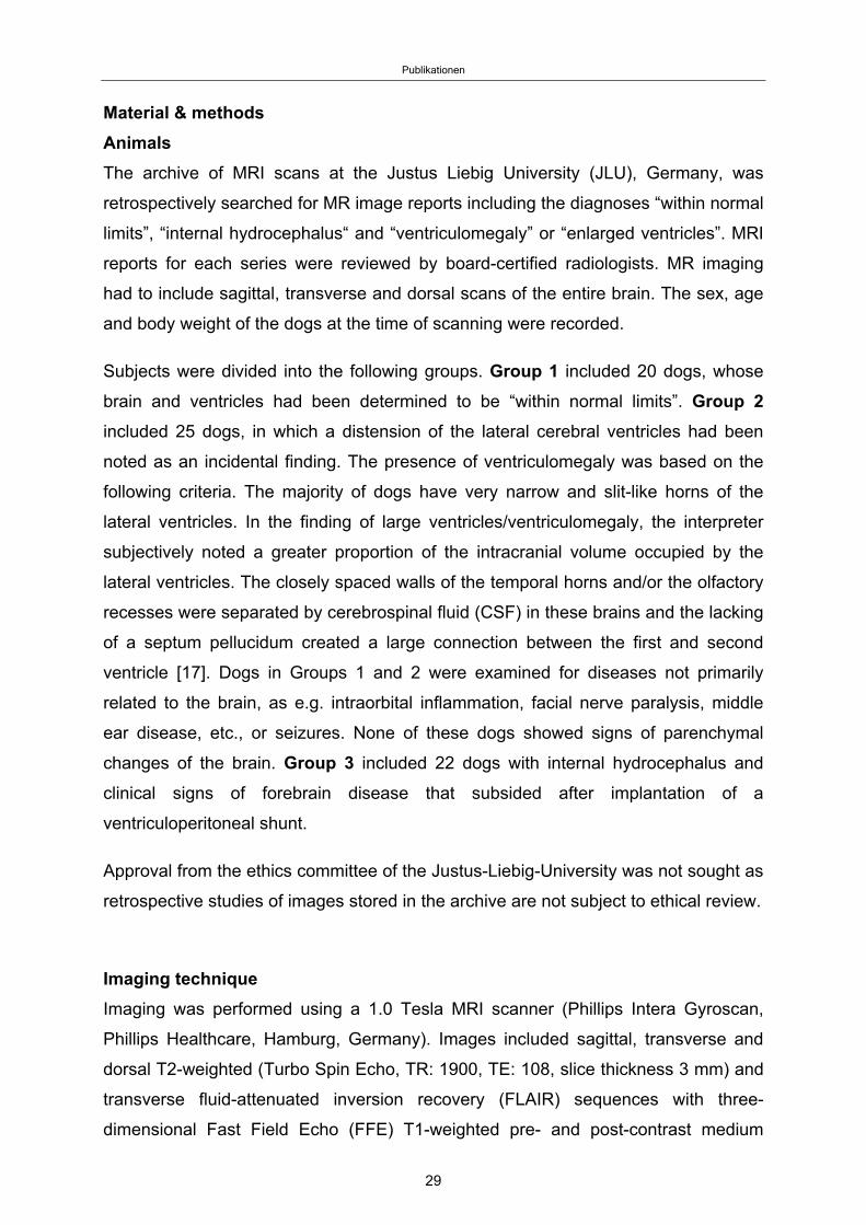
1. Expansion of the third ventricle represented by flattening of the interthalamic
adhesion and a diminished suprasellar cistern [3, 21]. A deformation of the
interthalamic adhesion was assumed when it was not distinctly circular on midsagittal
plane images (Figure 1 E). Narrowing of the suprasellar cistern was diagnosed in
transversal images based on the lateral bulging contours of the hypothalamus
diminishing the CSF of the adjacent cistern (Figure 1 B/C).
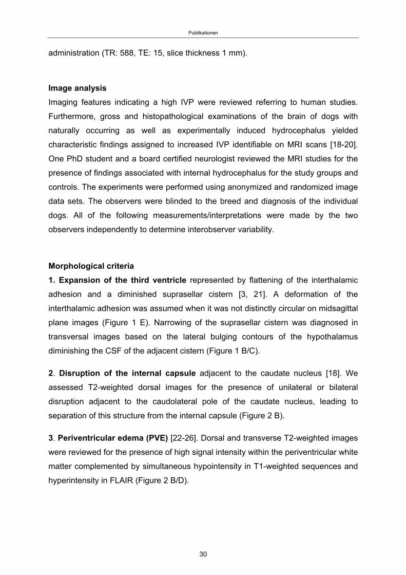
2. Disruption of the internal capsule adjacent to the caudate nucleus [18]. We
assessed T2-weighted dorsal images for the presence of unilateral or bilateral
disruption adjacent to the caudolateral pole of the caudate nucleus, leading to
separation of this structure from the internal capsule (Figure 2 B).
3. Periventricular edema (PVE) [22-26]. Dorsal and transverse T2-weighted images
were reviewed for the presence of high signal intensity within the periventricular white
matter complemented by simultaneous hypointensity in T1-weighted sequences and
hyperintensity in FLAIR (Figure 2 B/D).
30
Publikationen
Figure 1 – MRI-signs of increased intraventricular pressure
Transverse (A-C) and sagittal (D, E) T2-weighted MR-images of a normal dog brain
(A, D), a dog with ventriculomegaly (B, E) and with internal hydrocephalus (C). The
finding of an expanded third ventricle and the measurement of the corpus callosum
height (callosal height) and dimensions of the fourth ventricle is demonstrated.
4. Narrowing of cerebral sulci and obliteration of the subarachnoid space
around the dorsal convexity of the cerebral hemispheres [13, 20]. The absence of a
hyperintense subarachnoid space and/or the presence of narrowed cortical sulci
were recorded on transverse T2-weighted images at the level of the interthalamic
adhesion (Figure 3 C).
5. Dilation of the olfactory recess(es) [15, 20, 27]. Transverse and dorsal T2-
weighted images were evaluated for the presence of a hyperintense signal (CSF)
within the olfactory bulb continuous with the frontal horns of the lateral ventricles
(Figure 2C).
31
Publikationen
Figure 2 – MRI-signs of increased intraventricular pressure
Dorsal (A-C) and transverse (D) MR-images of dogs with internal hydrocephalus
showing signs of increased intraventricular pressure (IVP). The amount of distension
is measured by the ventricle/brain-index (A). The IVP leads do dilation of the
olfactory recesses (C). Periventricular edema occurs if the intraventricular pressure
exerts the compliance of the brain parenchyma (D). This can also lead to lacerations
of the white matter adjacent to the caudate nucleus (B).
6. Presence of cerebellar deviation [19, 28, 29]. A cerebellar deviation was
considered when the cerebellum protruded to the level of or through the foramen
magnum [30].
32
Publikationen
Morphometric criteria
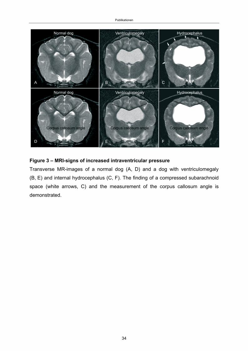
1. Corpus callosum angle (callosal angle) [12, 16, 31]. The callosal angle
describes the angle between the corpus callosum as the center and the dorsomedial
internal surfaces of the lateral ventricles as angle legs on a transverse MR image.
We determined it in analogy to human patients on T2-weighted images at the level of
the pituitary gland (Figure 3 D-F).
2. Corpus callosum height (callosal height) [32-34]. The elevation of the corpus
callosum was measured in the midsagittal image using a straight line connecting the
splenium and rostrum (callosal line [33]) and a perpendicular line to the dorsal-most
extension of the body of the corpus callosum (Figure 1 D/E).
3. Ventricle/brain (VB)-index. The ventricle/brain index was evaluated on dorsal T2
images. The VB-index was defined as the maximum continuous distance between
the internal borders of the ventricles divided by the maximum width of the brain
parenchyma in the same image (Figure 2A).
4. Expansion of the fourth ventricle [20, 35, 36]. In normal dogs, the cerebellum is
in contact with the medulla in midsagittal images. The height of the fourth ventricle
was determined on a T2-weighted midsagittal plane image at its widest extension in
the dorso-ventral direction. Additionally, the width of the fastigial recess was
determined at its widest extension in the rostro-caudal direction (Figure 1E).
33
Publikationen
Figure 3 – MRI-signs of increased intraventricular pressure
Transverse MR-images of a normal dog (A, D) and a dog with ventriculomegaly
(B, E) and internal hydrocephalus (C, F). The finding of a compressed subarachnoid
space (white arrows, C) and the measurement of the corpus callosum angle is
demonstrated.
34
Publikationen
Statistical analysis
All statistical analyses were performed using the statistical software package BMDP
[37]. With respect to Groups 2 and 3, for each of the qualitative criteria, the statistical
significance of the differences between these groups was assessed by considering
the two-way frequency table and by performing the Chi-square test for homogeneity
or Fisher’s exact test depending of the size of the smallest expected value in the
table. These tests were not performed incorporating the normal group since, by
definition, these criteria do not occur.
For the quantitative criteria, the linear relationship to body weight was verified by
regression analysis and scatterplots. The deviation from the normal distribution was
checked using the normal probability plot of the model residuals (Q-Q-plot) for each
variable. Subsequently, a one-way analysis of covariance was performed for each of
the individual criteria to include the influence of the body mass and the groups’
influence on the means of the measured quantities simultaneously. Variables with a
dependence on body weight were calculated for the adjusted group means. If there
were global significant differences in the mean values between the groups, a
pairwise comparison of group means by the Student-Newman-Keuls test was
performed. The parameters expansion of the fourth ventricle and callosal height were
logarithmically transformed throughout the analysis because the distribution of their
values was skewed to the right. In all statistical test procedures, a significance level
of p=0.05 was used.
The V/B index was only calculated for Groups 2 and 3. As a dependence on body
weight was not present for the V/B-index, the group comparison was performed using
a simple t-test for independent samples. For the estimation of a threshold value, a
parametric reference interval calculation was subsequently performed [38]. In
addition, the upper 95% confidence margin was determined for Group 2. Receiver
operating characteristic (ROC) analysis was performed in order to optimize the
selection of the cut-off value to achieve maximal sensitivity and specificity.
The precision of the interrater variability of the quantitative findings was determined
using Bland-Altman analysis to compare the differences between the first and second
measurements of each dog. The differences between the two measurements were
then plotted against the average (mean) of the two measurements. Good
reproducibility was assumed when 95% of the differences were within two standard
35
Publikationen
deviations. Interrater variability of the qualitative findings was assessed using kappa
statistics.
Results
Animals
Information regarding the breed, bodyweight, age, gender and indication for MRI/final
diagnosis of all dogs are summarized in Additional file 1.
Analysis of morphological criteria
All qualitative criteria were found to differ significantly between Groups 2 and 3,
except for cerebellar deviation. Expansion of the third ventricle, as represented by
deformation of the interthalamic adhesion, was significantly more frequent in dogs
with a relevant hydrocephalus (p<0.0001). The same applies for the presence of
periventricular edema (p<0.0001) and dilation of the olfactory recess(es) (p<0.0001),
both of which were only present in dogs with hydrocephalus. Thinning of the cerebral
sulci (p<0.0001) and thinning of the subarachnoid space (p=0.0027) as well as
disruptive lesions of the internal capsule adjacent to the caudate nucleus (p<0.0001)
were also found exclusively in the hydrocephalic group. A kappa value of 1 revealed
excellent interobserver agreement.
Analysis of the quantitative criteria
The mean V/B-index was 0.54 (range: 0.44-0.65) in the dogs with ventriculomegaly
(Group 2) and 0.73 (range 0.58-0.92) in the dogs with hydrocephalus (Group 3),
which represents a significant difference between these groups (p<0.001). The upper
95% reference limit for the V/B-index between ventriculomegaly and hydrocephalus
was calculated to be 0.62. In order to further optimize sensitivity and specificity, ROC
analysis was performed and an optimal cut-off value of 0.605 was calculated. At a
cut-off value of 0.605 for sensitivity and a specificity of 92% were determined.
The callosal height in group one (normal dogs) ranged from 3.7 mm to 7.3 mm with a
36
Publikationen
mean of 5.2 mm. In Group 2 (ventriculomegaly), it ranged from 5.0 mm to 13.7 mm
(mean 8.7), and in Group 3 (hydrocephalus) from 6.6 mm to 23.5 mm (mean 12.6
mm). The mean values were significantly different between all groups (p<0.01).
ANCOVA revealed a significant influence of body weight on this value (p=0.001).
Therefore, the values were related to the adjusted means of the body weight of all
groups. Differences between all groups remained significant. Adjusted means of
callosal height were significantly higher in dogs with ventriculomegaly than in normal
dogs (p<0.01) and significantly higher in hydrocephalus dogs compared to the
ventriculomegaly group (p<0.01). The 95% reference limits for callosal height can be
calculated for dogs with different body weights using the following equation:
10.2 x 10 0.006 x BW
No statistically significant difference could be detected between dogs with
hydrocephalus (Group 3) and those with enlarged ventricles (Group 2) regarding the
corpus callosum angle (p=0.961). However, both groups differed significantly from
the normal group (p<0.01). Body weight did not affect this measurement value.
Expansion of the fourth ventricle in normal dogs differed significantly from dogs with
ventriculomegaly (p<0.05) and dogs with hydrocephalus (p<0.01), but there was no
significant difference between the ventriculomegaly and the hydrocephalus group
(p=0.842). Body mass is suspicious to affect this measurement value (p=0.064).
Width of the fastigial recess revealed no significant difference between dogs of each
group (p=0.879). Body weight did not affect this measurement value.
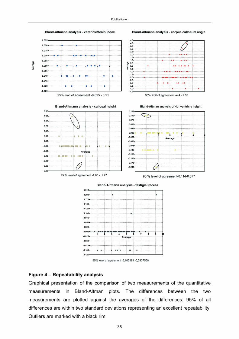
The assessment of repeatability is shown in Figure 4. 95% of the differences
between the first and second measurement are less than ± 2 standard deviations
(SD’s) from the mean difference. The Bland-Altmann analysis revealed significant
agreement between the raters.
37
Publikationen
Figure 4 – Repeatability analysis
Graphical presentation of the comparison of two measurements of the quantitative
measurements in Bland-Altman plots. The differences between the two
measurements are plotted against the averages of the differences. 95% of all
differences are within two standard deviations representing an excellent repeatability.
Outliers are marked with a black rim.
38
Publikationen
Discussion
In certain clinical situations, the diagnosis of hydrocephalus may be difficult to
establish, particularly in small brachycephalic dog breeds that tend to have relatively
larger ventricles in comparison to mesaticephalic dogs [8,39]. Ventricular
enlargement may also be characteristic of other neurodegenerative diseases and
normal aging in dogs [40]. Mere determination of the ventricular volume was
previously not successful in identifying clinically relevant hydrocephalus [7,8]. The
identification of clinically relevant ventricular distension is extremely important for the
indication for CSF shunting. We have seen a number of dogs with neurological signs,
which have been referred to our hospital for ventriculo-peritoneal shunting, in which a
diagnosis of internal hydrocephalus was made based on the finding of ventricular
enlargement alone. CSF examination, however, often revealed idiopathic
inflammatory diseases (necrotizing encephalitides), which primarily require medical
treatment. Inflammatory brain diseases may easily be overlooked, and large
ventricles can be easily misinterpreted as the underlying cause for present
neurological signs in those dogs. Therefore, we aimed to identify further
characteristic morphological changes using MRI studies of the dog’s brain related to
an assumed increase in intraventricular pressure. Our results show that
morphological differences exist between hydrocephalus and ventriculomegaly, which
might be useful in the differentiation of these two entities.
The findings in the hydrocephalus group indicate parenchymal changes based on
pressure forces on the parenchyma. The gradual expansion of the ventricular system
follows a predetermined sequence, starting with the temporal horn of the lateral
ventricles. Subsequently, the remainder of the lateral ventricles expands, followed by
the fourth ventricle [20]. The third ventricle is the last one to show distension. It has
been proposed that periventricular white matter of the ventricles is exposed to
expansive stress, especially in the region of the ventricular horns. At the same time,
expansion of the ventricles leads to compressive forces on the thalamus, rendering
high pressures mandatory in order to expand the thalamus that surrounds the third
ventricle [41]. In human neuroradiology, dilation of the third ventricle has been shown
to indicate a high pressure gradient between the ventricle and the ventral
subarachnoid spaces (interpeduncular cistern and hypophyseal cistern). Such a
finding serves as a major indication for third ventriculostomy in children [42] and
might also prove useful for canine patients in the future.
39
Publikationen
There are many ways of measuring relative ventricular size in human medicine, using
linear ratios, area and volumetric measurements [43]. The most common
measurement is the Evans' index that describes the ratio of the transverse diameter
of the anterior horns (rostral horns in dogs) of the lateral ventricles to the largest
diameter of the brain in humans. Evans’ index has also been used to measure the
ratio between the lateral ventricles and brain parenchyma in hydrocephalic dogs.
However, it has been found to underestimate the degree of ventricular distension
[44]. Other measurements estimating the relation between frontal horn diameter and
brain parenchyma have been used on different levels and image planes in the dog
brain using different imaging modalities [7, 9, 44-49]. Only one study found a
significant difference between dogs with ventriculomegaly and hydrocephalus [46].
As mentioned above, experimental studies have shown that the temporal horns of
the ventricles dilate first in dogs, followed by other parts of the ventricle [20]. This
was also reported in children [50] and in mathematical models of hydrocephalus
using finite element analysis [41]. The maximum continuous extension from one
ventricle to the other was measured in temporal horns in our study rather than
classical measurement of the frontal horns based on Evans’ method, which may
explain the significant results of our study in contrast to those of previous studies.
Further studies with three-dimensional-morphometric measurements are needed to
determine whether our VB-index might underestimate ventricular distension as well.
Platt and Garosi [15] described dilation of the olfactory recess to be suggestive of
increased IVP. Dilation of the olfactory bulb cavity was also noted in experimentally
induced hydrocephalus [20, 28]. It was interpreted as transmission of high pressure
from the frontal horns to the normally non-expanded recesses that occurs only in late
stages of increased IVP.
Changes in the appearance of the corpus callosum have been reported in human
patients with hydrocephalus. Structural changes due to high intraventricular pressure
include stretching and upward displacement of the body of the corpus callosum and
concurrent downward depression of the fornix in humans [33-35]. This was also
found in dogs with experimental hydrocephalus [20]. Demyelination of the callosal
axons has been suggested to be the underlying cause of increased compliance of
the commissural fibers, leading to the upward bowing of the corpus callosum with
increased pressure [49]. Other than the parenchyma surrounding the lateral
ventricles, the corpus callosum is situated beneath the rigid falx. The connective
40
Publikationen
tissue of this structure exerts additional resistance against the expanding ventricle
and higher pressures are needed to elevate the corpus callosum.
The corpus callosum angle reflects dorsal distension of the dilated lateral ventricles
[16]. In human medicine, a callosal angle of less than 90° is a criterion for the
identification of high intraventricular pressure [12]. Virhammar et al. [31] showed that
patients with a smaller callosal angle are more likely to respond to shunting, both
clinically and with reconstitution of the cerebral parenchyma. To our knowledge, the
callosal angle has not been measured in canine patients before. There were no
significant differences between the callosal angles of dogs with ventriculomegaly and
those with hydrocephalus. This might be related to the high callosal height in the
hydrocephalus group. Moving the center of the angle (i.e. the corpus callosum)
upwards automatically leads to a wider angle in these dogs, which can explain the
lack of difference between the groups.
Focal periventricular white matter edema has been attributed to the transependymal
absorption of cerebrospinal fluid that follows the pressure gradient from the ventricle
to the parenchyma [51-53]. Another interpretation describes PVE as areas with
restricted absorption capacities for extracellular water that flows from the
parenchyma towards the ventricles [54]. This has been found in acute and subacute
phases of human hydrocephalus [55], experimental hydrocephalus in dogs [22-24] as
well as in idiopathic hydrocephalus [26]. Water diffusion within periventricular white
matter has been found to be highly dependent on age in children and the diffusion
capacity dramatically decreases with normal brain maturation [56]. Although the
median age is lower in the hydrocephalus group, both groups include immature and
mature dogs, which is why early age does not seem to be a prerequisite for the
occurrence of PVE.
Expansion of the ventricles can cause compression of the cortical subarachnoid CSF
space [53, 57], also leading to decreased space within the sulci. This finding has
been seen in hydrocephalic humans [13] and animals [20]. High pressure in the
ventricles and low pressure in the subarachnoid space (SAC) are prerequisites for a
constricted CSF space. The pressure from the ventricular system has been reported
to not be transmitted homogeneously over the entire surface of the brain
hemispheres, but primarily into the dorsolateral direction [58, 59]. Due to this fact, we
examined the SAC and the sulci on the cerebral convexity on the level of the
41
Publikationen
interthalamic adhesion for consistency.
A disrupted ependymal lining and formation of false diverticula have been found in
human patients with severe hydrocephalus [54], as well as in dogs [60,61]. Stretching
and eventual destruction of the internal capsule adjacent to the caudate nucleus was
observed as a consequence of overpressure in experimental [20] and naturally
occurring hydrocephalus in dogs [18].
The determination of the presented qualitative changes of the brain parenchyma
should be assumed to be a subjective method and may be less accurate than
quantitative methods. However, we were able to show that simple visual evaluation
has a high interrater agreement.
In dogs, skull and brain size and morphology differ between brachycephalic and
mesaticephalic breeds. In brachycephalic dogs, growth reduction of the skull base
has been documented [62], which has a potential influence on the morphology of the
brain stem and the measured distances in this area as well. Breed-specific values
are necessary to use the same parameters as in humans. For this reason, we
decided to confine our analysis to a simple assessment of the third ventricle based
on the shape of the interthalamic adhesion. A deformation of the interthalamic
adhesion has been identified in dogs with high intraventricular pressure and was also
interpreted as a sign of third ventricular distention due to overpressure [3, 20, 21].
The distension of the cerebral ventricles and with it the whole forebrain can lead to
caudal deviation of other brain regions [63]. Deviation of the cerebellum through the
foramen magnum has been described in the development of experimental
hydrocephalus [19, 28, 29] as well as in naturally occurring hydrocephalus [64].
However, the impression of cerebellar deviation can be found in a number of small
brachycephalic dog breeds, even with normal ventricles, which explain the lack of
significance of this finding. Enlargement of the fourth ventricle has been described in
human hydrocephalus [55], and fourth ventricle dimensions are significantly larger in
human patients with intraventricular overpressure. Dilation of the fourth ventricle has
also been observed in animals with experimental communicating hydrocephalus
[20,35,36]. The reasons behind the lack of a difference between the groups
concerning this finding remain undetermined.
It is widely assumed that elevated pressure is a direct result of an impaired CSF flow
42
Publikationen
from the ventricles to its point of absorption [54, 57, 63, 65]. This leads to a
progressive destruction of white matter in dogs as a consequence of high IVP, which
has been thought to not be present in ventriculomegaly. However both dogs with
ventriculomegaly and hydrocephalus were significantly different from normal dogs for
most of the quantitative MRI findings that we examined. Based on these rather
gradual than principal differences, it would be unsound to assume that
ventriculomegaly is not a consequence of overpressure. Long-term studies
measuring IVP in dogs with dilated ventricles are needed. Accumulation of CSF and
distension of the ventricles might occur very slowly in dogs with ventriculomegaly,
giving the brain time to adapt to changes in periventricular perfusion and other
pathological changes. It is reasonable to expect that the degree of intraventricular
pressure determines the speed and severity of parenchymal injury and thereby
functional brain deficits. Moderately increased pressure or intermittently high
intraventricular CSF pressure might produce temporary phases of ischemia in the
periventricular white matter. The chronic cumulative effects of these ischemic events
could produce very slowly progressing periventricular tissue atrophy. Although raised
IVP might cause slowly progressing tissue damage, this does not necessarily
produce clinical signs. Signs of dementia, such as those found in humans, might also
pass unnoticed in a classical neurological examination. The clinical use of the
identified imaging findings must be judicious. Although some parameters revealed a
clear cut-off, it is likely that the combination of all these measurements may yield a
better evaluation about high intraventricular pressure using morphological MRI. A
more detailed classification of canine hydrocephalus and the clear discrimination of
the underlying causes are required to confirm their usefulness in each individual dog.
Conclusion
A V/B-index greater than 0.6 together with an elevated corpus callosum and the
presence of a deformed intermediate mass, periventricular edema, dilation of the
olfactory recess, thinning of the sulci and/or the subarachnoid space or disruption of
the internal capsule adjacent to the caudate nucleus are highly suspicious for
clinically relevant dilation of the lateral cerebral ventricles due to increased IVP.
43
Publikationen
Abbeviations
ANCOVA: Analysis of covariance; CSF: Cerebrospinal fluid; FFE: Fast field echo;
FLAIR: Fluid-attenuated inversion recovery; IVP: Intraventricular pressure; JLU:
Justus Liebig University; MRI: Magnetic resonance imaging; PVE: Periventricular
edema; ROC: Receiver operating characteristic; SAC: Subarachnoid space; SD:
Standard deviation; V/B-index: Ventricle/brain-index.
Competing interests
The authors declare that they have no competing interests.
Authors' contributions
All authors helped to draft the manuscript and participated in its design. SL collected
the data for the study and drafted the manuscript. MJS and SL analyzed the data. KF
performed the statistical analysis of the data. NO participated in the design of the
study. MK participated in its design and coordination. All authors read and approved
the final manuscript.
Acknowledgements
The authors would like to thank Marion Sparenberg and Andreas Schaubmar for their
helpful feedback throughout the project and Dr. Bastian Laubner for the critical
revision of the manuscript.
44
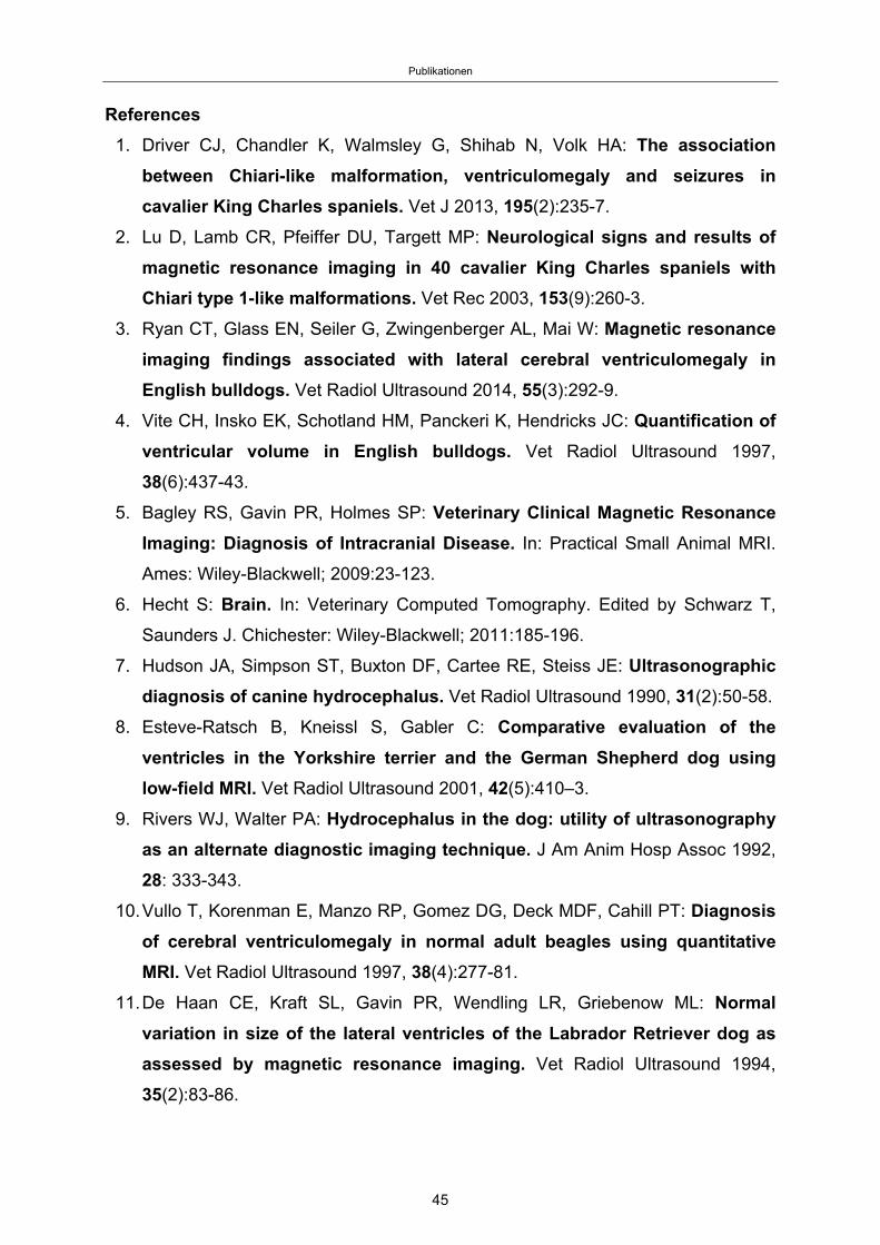
Publikationen
References
1. Driver CJ, Chandler K, Walmsley G, Shihab N, Volk HA: The association
between Chiari-like malformation, ventriculomegaly and seizures in
cavalier King Charles spaniels. Vet J 2013, 195(2):235-7.
2. Lu D, Lamb CR, Pfeiffer DU, Targett MP: Neurological signs and results of
magnetic resonance imaging in 40 cavalier King Charles spaniels with
Chiari type 1-like malformations. Vet Rec 2003, 153(9):260-3.
3. Ryan CT, Glass EN, Seiler G, Zwingenberger AL, Mai W: Magnetic resonance
imaging findings associated with lateral cerebral ventriculomegaly in
English bulldogs. Vet Radiol Ultrasound 2014, 55(3):292-9.
4. Vite CH, Insko EK, Schotland HM, Panckeri K, Hendricks JC: Quantification of
ventricular volume in English bulldogs. Vet Radiol Ultrasound 1997,
38(6):437-43.
5. Bagley RS, Gavin PR, Holmes SP: Veterinary Clinical Magnetic Resonance
Imaging: Diagnosis of Intracranial Disease. In: Practical Small Animal MRI.
Ames: Wiley-Blackwell; 2009:23-123.
6. Hecht S: Brain. In: Veterinary Computed Tomography. Edited by Schwarz T,
Saunders J. Chichester: Wiley-Blackwell; 2011:185-196.
7. Hudson JA, Simpson ST, Buxton DF, Cartee RE, Steiss JE: Ultrasonographic
diagnosis of canine hydrocephalus. Vet Radiol Ultrasound 1990, 31(2):50-58.
8. Esteve-Ratsch B, Kneissl S, Gabler C: Comparative evaluation of the
ventricles in the Yorkshire terrier and the German Shepherd dog using
low-field MRI. Vet Radiol Ultrasound 2001, 42(5):410–3.
9. Rivers WJ, Walter PA: Hydrocephalus in the dog: utility of ultrasonography
as an alternate diagnostic imaging technique. J Am Anim Hosp Assoc 1992,
28: 333-343.
10. Vullo T, Korenman E, Manzo RP, Gomez DG, Deck MDF, Cahill PT: Diagnosis
of cerebral ventriculomegaly in normal adult beagles using quantitative
MRI. Vet Radiol Ultrasound 1997, 38(4):277-81.
11. De Haan CE, Kraft SL, Gavin PR, Wendling LR, Griebenow ML: Normal
variation in size of the lateral ventricles of the Labrador Retriever dog as
assessed by magnetic resonance imaging. Vet Radiol Ultrasound 1994,
35(2):83-86.
45
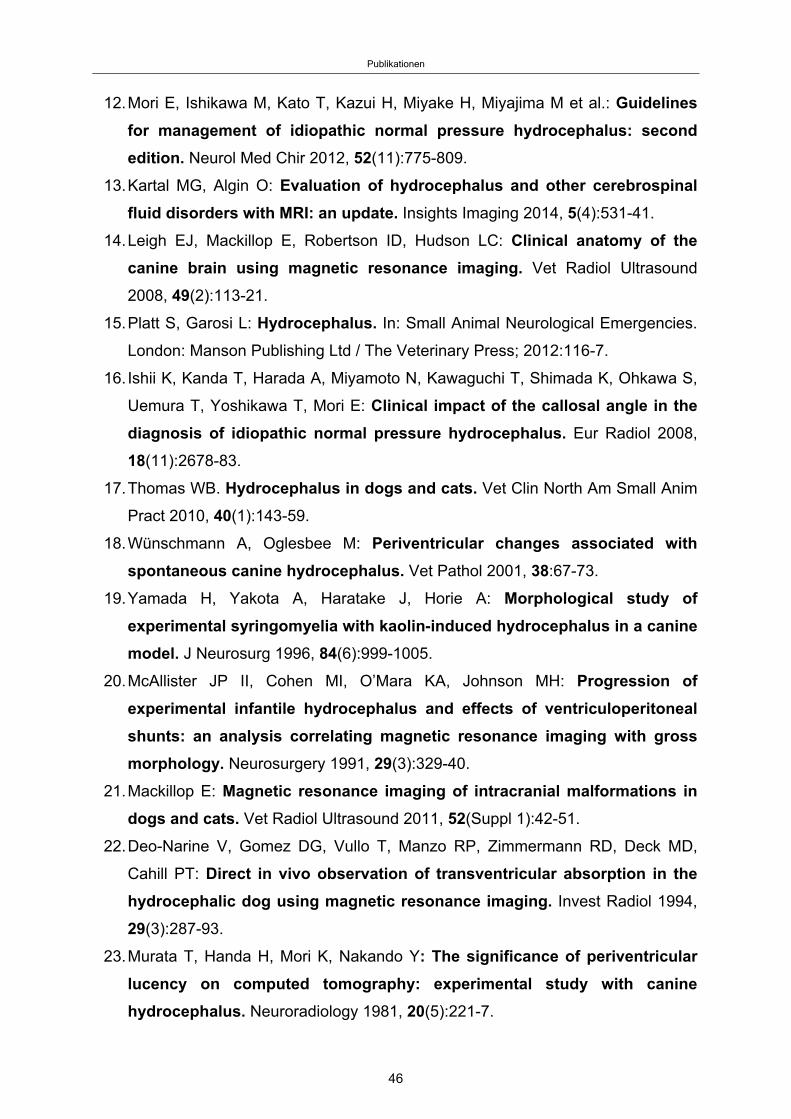
Publikationen
12. Mori E, Ishikawa M, Kato T, Kazui H, Miyake H, Miyajima M et al.: Guidelines
for management of idiopathic normal pressure hydrocephalus: second
edition. Neurol Med Chir 2012, 52(11):775-809.
13. Kartal MG, Algin O: Evaluation of hydrocephalus and other cerebrospinal
fluid disorders with MRI: an update. Insights Imaging 2014, 5(4):531-41.
14. Leigh EJ, Mackillop E, Robertson ID, Hudson LC: Clinical anatomy of the
canine brain using magnetic resonance imaging. Vet Radiol Ultrasound
2008, 49(2):113-21.
15. Platt S, Garosi L: Hydrocephalus. In: Small Animal Neurological Emergencies.
London: Manson Publishing Ltd / The Veterinary Press; 2012:116-7.
16. Ishii K, Kanda T, Harada A, Miyamoto N, Kawaguchi T, Shimada K, Ohkawa S,
Uemura T, Yoshikawa T, Mori E: Clinical impact of the callosal angle in the
diagnosis of idiopathic normal pressure hydrocephalus. Eur Radiol 2008,
18(11):2678-83.
17. Thomas WB. Hydrocephalus in dogs and cats. Vet Clin North Am Small Anim
Pract 2010, 40(1):143-59.
18. Wünschmann A, Oglesbee M: Periventricular changes associated with
spontaneous canine hydrocephalus. Vet Pathol 2001, 38:67-73.
19. Yamada H, Yakota A, Haratake J, Horie A: Morphological study of
experimental syringomyelia with kaolin-induced hydrocephalus in a canine
model. J Neurosurg 1996, 84(6):999-1005.
20. McAllister JP II, Cohen MI, O’Mara KA, Johnson MH: Progression of
experimental infantile hydrocephalus and effects of ventriculoperitoneal
shunts: an analysis correlating magnetic resonance imaging with gross
morphology. Neurosurgery 1991, 29(3):329-40.
21. Mackillop E: Magnetic resonance imaging of intracranial malformations in
dogs and cats. Vet Radiol Ultrasound 2011, 52(Suppl 1):42-51.
22. Deo-Narine V, Gomez DG, Vullo T, Manzo RP, Zimmermann RD, Deck MD,
Cahill PT: Direct in vivo observation of transventricular absorption in the
hydrocephalic dog using magnetic resonance imaging. Invest Radiol 1994,
29(3):287-93.
23. Murata T, Handa H, Mori K, Nakando Y: The significance of periventricular
lucency on computed tomography: experimental study with canine
hydrocephalus. Neuroradiology 1981, 20(5):221-7.
46
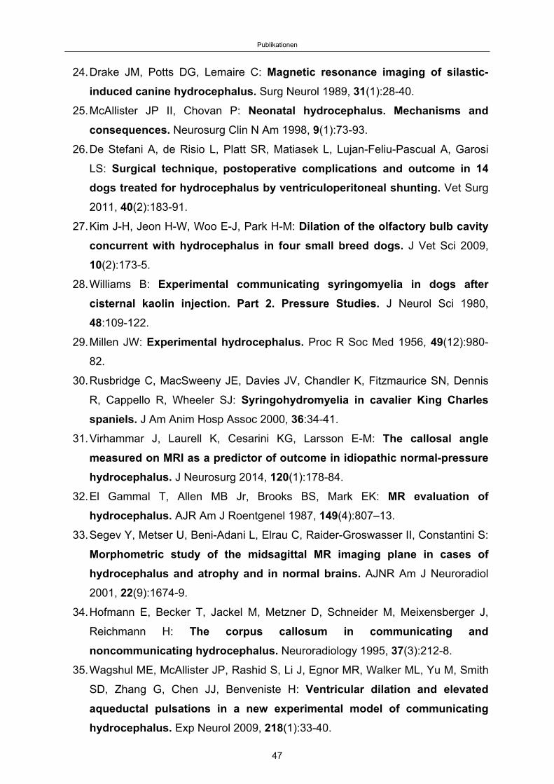
Publikationen
24. Drake JM, Potts DG, Lemaire C: Magnetic resonance imaging of silastic-
induced canine hydrocephalus. Surg Neurol 1989, 31(1):28-40.
25. McAllister JP II, Chovan P: Neonatal hydrocephalus. Mechanisms and
consequences. Neurosurg Clin N Am 1998, 9(1):73-93.
26. De Stefani A, de Risio L, Platt SR, Matiasek L, Lujan-Feliu-Pascual A, Garosi
LS: Surgical technique, postoperative complications and outcome in 14
dogs treated for hydrocephalus by ventriculoperitoneal shunting. Vet Surg
2011, 40(2):183-91.
27. Kim J-H, Jeon H-W, Woo E-J, Park H-M: Dilation of the olfactory bulb cavity
concurrent with hydrocephalus in four small breed dogs. J Vet Sci 2009,
10(2):173-5.
28. Williams B: Experimental communicating syringomyelia in dogs after
cisternal kaolin injection. Part 2. Pressure Studies. J Neurol Sci 1980,
48:109-122.
29. Millen JW: Experimental hydrocephalus. Proc R Soc Med 1956, 49(12):980-
82.
30. Rusbridge C, MacSweeny JE, Davies JV, Chandler K, Fitzmaurice SN, Dennis
R, Cappello R, Wheeler SJ: Syringohydromyelia in cavalier King Charles
spaniels. J Am Anim Hosp Assoc 2000, 36:34-41.
31. Virhammar J, Laurell K, Cesarini KG, Larsson E-M: The callosal angle
measured on MRI as a predictor of outcome in idiopathic normal-pressure
hydrocephalus. J Neurosurg 2014, 120(1):178-84.
32. El Gammal T, Allen MB Jr, Brooks BS, Mark EK: MR evaluation of
hydrocephalus. AJR Am J Roentgenel 1987, 149(4):807–13.
33. Segev Y, Metser U, Beni-Adani L, Elrau C, Raider-Groswasser II, Constantini S:
Morphometric study of the midsagittal MR imaging plane in cases of
hydrocephalus and atrophy and in normal brains. AJNR Am J Neuroradiol
2001, 22(9):1674-9.
34. Hofmann E, Becker T, Jackel M, Metzner D, Schneider M, Meixensberger J,
Reichmann H: The corpus callosum in communicating and
noncommunicating hydrocephalus. Neuroradiology 1995, 37(3):212-8.
35. Wagshul ME, McAllister JP, Rashid S, Li J, Egnor MR, Walker ML, Yu M, Smith
SD, Zhang G, Chen JJ, Benveniste H: Ventricular dilation and elevated
aqueductal pulsations in a new experimental model of communicating
hydrocephalus. Exp Neurol 2009, 218(1):33-40.
47
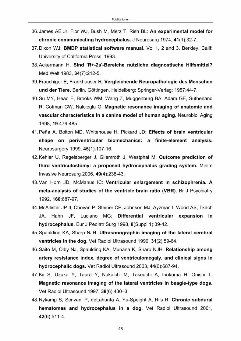
Publikationen
36. James AE Jr, Flor WJ, Bush M, Merz T, Rish BL: An experimental model for
chronic communicating hydrocephalus. J Neurosurg 1974, 41(1):32-7.
37. Dixon WJ: BMDP statistical software manual. Vol 1, 2 and 3. Berkley, Calif:
University of California Press; 1993.
38. Ackermann H. Sind 'R+-2s'-Bereiche nützliche diagnostische Hilfsmittel?
Med Welt 1983, 34(7):212-5.
39. Frauchiger E, Frankhauser R: Vergleichende Neuropathologie des Menschen
und der Tiere. Berlin, Göttingen, Heidelberg: Springer-Verlag; 1957:44-7.
40. Su MY, Head E, Brooks WM, Wang Z, Muggenburg BA, Adam GE, Sutherland
R, Cotman CW, Nalcioglu O: Magnetic resonance imaging of anatomic and
vascular characteristics in a canine model of human aging. Neurobiol Aging
1998, 19:479-485.
41. Peña A, Bolton MD, Whitehouse H, Pickard JD: Effects of brain ventricular
shape on periventricular biomechanics: a finite-element analysis.
Neurosurgery 1999, 45(1):107-16.
42. Kehler U, Regelsberger J, Gliemroth J, Westphal M: Outcome prediction of
third ventriculostomy: a proposed hydrocephalus grading system. Minim
Invasive Neurosurg 2006, 49(4):238-43.
43. Van Horn JD, McManus IC: Ventricular enlargement in schizophrenia. A
meta-analysis of studies of the ventricle:brain ratio (VBR). Br J Psychiatry
1992, 160:687-97.
44. McAllister JP II, Chovan P, Steiner CP, Johnson MJ, Ayzman I, Wood AS, Tkach
JA, Hahn JF, Luciano MG: Differential ventricular expansion in
hydrocephalus. Eur J Pediatr Surg 1998, 8(Suppl 1):39-42.
45. Spaulding KA, Sharp NJH: Ultrasonographic imaging of the lateral cerebral
ventricles in the dog. Vet Radiol Ultrasound 1990, 31(2):59-64.
46. Saito M, Olby NJ, Spaulding KA, Munana K, Sharp NJH: Relationship among
artery resistance index, degree of ventriculomegaly, and clinical signs in
hydrocephalic dogs. Vet Radiol Ultrasound 2003, 44(6):687-94.
47. Kii S, Uzuka Y, Taura Y, Nakaichi M, Takeuchi A, Inokuma H, Onishi T:
Magnetic resonance imaging of the lateral ventricles in beagle-type dogs.
Vet Radiol Ultrasound 1997, 38(6):430–3.
48. Nykamp S, Scrivani P, deLahunta A, Yu-Speight A, Riis R: Chronic subdural
hematomas and hydrocephalus in a dog. Vet Radiol Ultrasound 2001,
42(6):511-4.
48
Publikationen
49. Cardoso EJR, Lachat JJ, Lopes LS, Santos AC, Colli BO: Changes caused by
hydrocephalus, induced by kaolin, in the corpus callosum of adult dogs.
Acta Cir Bras 2011, 26(Suppl 2):8-14.
50. Fletcher JM, McCauley SR, Brandt ME, Bohan TP, Kramer LA, Francis DJ,
Thorstad K, Brookshire BL: Regional brain tissue composition in children
with hydrocephalus. Relationships with cognitive development. Arch
Neurol 1996, 53(6):549-57.
51. Haberl H, Michael T, Thomale UW: Hydrocephalus. Pädiatrie update 2007,
1(2.Jahrgang):25-44.
52. Zimmerman RD, Fleming CA, Lee BCP, Saint-Louis LA, Deck MDF:
Periventricular hyperintensity as seen by magnetic resonance: prevalence
and significance. AJR Am J Roentgenol 1986, 146(3):443-50.
53. Matsumae M, Kikinis R, Morocz I, Lorenzo AV, Albert MS, Black PMcL, Jolesz
FA: Intracranial compartment volumes in patients with enlarged ventricles
assessed by magnetic resonance-based image processing. J Neurosurg
1996, 84(6):972-81.
54. Del Bigio MR: Neuropathological changes caused by hydrocephalus. Acta
Neuropathol 1993, 85(6):573-85.
55. Edelman RR, Hesselink JR, Zlatkin MB, Crues JV: Hydrocephalus. In: Clinical
Magnetic Resonance Imaging. 2nd edition. Philadelphia: Saunders Elsevier;
2006. p. 1646-55.
56. Volpe JJ: Hypoxic-ischemic encephalopathy: clinical aspects. In: Neurology
of the newborn. 5th edition. Philadelphia: Saunders/Elsevier; 2008. p. 400-480.
57. Rekate HL, Nadkarni TD, Wallace D: The importance of the cortical
subarachnoid space in understanding hydrocephalus. J Neurosurg Pediatr
2008, 2(1):1-11.
58. Nagashima T, Tamaki N, Matsumoto S, Horwitz B, Seguchi Y: Biomechanics
of hydrocephalus: a new theoretical model. Neurosurgery 1987, 21(6):898-
904.
59. Sasaki M, Honda S, Yuasa T, Iwamura A, Shibata E, Ohba H: Narrow CSF
space at high convexity and high midline areas in idiopathic normal
pressure hydrocephalus detected by axial and coronal MRI. Neuroradiology
2008, 50(2):117-22.
49
Publikationen
60. Sahar A, Hochwald GM, Kay WJ, Ransohoff J: Spontaneous canine
hydrocephalus: cerebrospinal fluid dynamics. J Neurol Neurosurg Psychiatry
1971, 34(3):308-15.
61. Summers BA, Cummings JF, De Lahunta A: Malformations of the central
nervous system. In: Veterinary neuropathology. St. Louis: Mosby 1995. p. 68-
94.
62. Schmidt MJ, Neumann AC, Amort KH, Failing K, Kramer M: Cephalometric
measurements and determination of general skull type of Cavalier King
Charles Spaniels. Vet Radiol Ultrasound 2011, 52(4):436-40.
63. De Lahunta A, Glass EN: Cerebrospinal Fluid and Hydrocephalus. In:
Veterinary Neuroanatomy and Clinical Neurologie. 3rd edition. St. Louis:
Saunders; 2009.
64. Braund KG: Clinical Syndromes in Veterinary Neurology. 2nd edition. St.
Louis: Mosby; 1994:113-4.
65. Ransohoff J, Shulman K, Fishman RA: Hydrocephalus: A review of etiology
and treatment. J Pediatr 1960, 56:399-411.
50
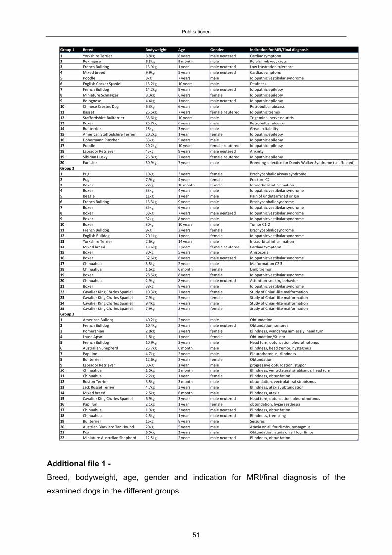
Publikationen
Additional file 1 -
Breed, bodyweight, age, gender and indication for MRI/final diagnosis of the
examined dogs in the different groups.
Group 1 Breed Bodyweight Age Gender Indication for MRI/Final diagnosis
1 Yorkshire Terrier 8,8kg 8 years male neutered Cardiac symptoms
2 Pekingese 6,3kg 5 month male Pelvic limb weakness
3 French Bulldog 13,9kg 1 year male neutered Low frustration tolerance
4 Mixed breed 9,9kg 5 years male neutered Cardiac symptoms
5 Poodle 8kg 7 years male Idiopathic vestibular syndrome
6 English Cocker Spaniel 13,2kg 10 years male Deafness
7 French Bulldog 14,2kg 9 years male neutered Idiopathic epilepsy
8 Miniature Schnauzer 8,3kg 6 years female Idiopathic epilepsy
9 Bolognese 4,4kg 1 year male neutered Idiopathic epilepsy
10 Chinese Crested Dog 6,3kg 6 years male Retrobulbar abscess
11 Basset 26,5kg 7 years female neutered Idiopathic tremor
12 Staffordshire Bullterrier 35,6kg 10 years male Trigeminal nerve neuritis
13 Boxer 25,7kg 6 years male Retrobulbar abscess
14 Bullterrier 18kg 3 years male Great exitability
15 American Staffordshire Terrier 20,2kg 1 year female Idiopathic epilepsy
16 Dobermann Pinscher 33kg 5 years male Idiopathic epilepsy
17 Poodle 20,2kg 10 years female neutered Idiopathic epilepsy
18 Labrador Retriever 45kg 9 years male neutered Anxiety
19 Sibirian Husky 26,8kg 7 years female neutered Idiopathic epilepsy
20 Eurasier 30,9kg 7 years male Breeding selection for Dandy Walker Syndrome (unaffected)
Group 2
1 Pug 10kg 3 years female Brachycephalic airway syndrome
2 Pug 7,9kg 4 years female Fracture C2
3 Boxer 27kg 10 month female Intraorbital inflammation
4 Boxer 33kg 4 years male Idiopathic vestibular syndrome
5 Beagle 11kg 1 year male Pain of undetermined origin
6 French Bulldog 13,3kg 9 years male Brachycephalic syndrome
7 Boxer 35kg 6 years male Idiopathic vestibular syndrome
8 Boxer 38kg 7 years male neutered Idiopathic vestibular syndrome
9 Boxer 32kg 8 years male Idiopathic vestibular syndrome
10 Boxer 30kg 10 years male Tumor C1‐2
11 French Bulldog 9kg 2 years female Brachycephalic syndrome
12 English Bulldog 20,1kg 1 year female Idiopathic vestibular syndrome
13 Yorkshire Terrier 2,6kg 14 years male Intraorbital inflammation
14 Mixed breed 13,6kg 7 years female neutered Cardiac symptoms
15 Boxer 30kg 5 years male Anisocoria
16 Boxer 32,6kg 8 years male neutered Idiopathic vestibular syndrome
17 Chihuahua 3,5kg 2 years male Malformation C2‐3
18 Chihuahua 1,6kg 6 month female Limb tremor
19 Boxer 28,5kg 8 years female Idiopathic vestibular syndrome
20 Chihuahua 2,9kg 8 years male neutered Attention‐seeking behavior
21 Boxer 38kg 8 years male Idiopathic vestibular syndrome
22 Cavalier King Charles Spaniel 10,3kg 7 years female Study of Chiari‐like malformation
23 Cavalier King Charles Spaniel 7,9kg 5 years female Study of Chiari‐like malformation
24 Cavalier King Charles Spaniel 9,4kg 7 years male Study of Chiari‐like malformation
25 Cavalier King Charles Spaniel 7,9kg 2 years female Study of Chiari‐like malformation
Group 3
1 American Bulldog 40,2kg 2 years male Obtundation
2 French Bulldog 10,4kg 2 years male neutered Obtundation, seizures
3 Pomeranian 2,8kg 2 years female Blindness, wandering aimlessly, head turn
4 Lhasa Apso 1,8kg 1 year female Obtundation/Stupor
5 French Bulldog 10,9kg 3 years male Head turn, obtundation pleurothotonus
6 Australian Shepherd 25,7kg 6 month male Blindness, head tremor, nystagmus
7 Papillon 4,7kg 2 years male Pleurothotonus, blindness
8 Bullterrier 12,6kg 2 years female Obtundation
9 Labrador Retriever 30kg 1 year male progressive obtundation, stupor
10 Chihuahua 2,5kg 3 month male Blindness, ventrolateral strabismus, head turn
11 Chihuahua 2,2kg 1 year female Blindness, obtundation
12 Boston Terrier 3,5kg 3 month male obtundation, ventrolateral strabismus
13 Jack Russel Terrier 4,7kg 3 years male Blindness, ataxia , obtundation
14 Mixed breed 2,5kg 6 month male Blindness, ataxia
15 Cavalier King Charles Spaniel 6,9kg 3 years male neutered Head turn, obtundation, pleurothotonus
16 Papillon 2,1kg 1 year female obtundation, hyperaesthesia
17 Chihuahua 1,9kg 3 years male neutered Blindness, obtundation
18 Chihuahua 2,5kg 1 year male neutered Blindness, trembling
19 Bullterrier 16kg 8 years male Seizures
20 Austrian Black and Tan Hound 20kg 5 years male Ataxia on all four limbs, nystagmus
21 Pug 9,5kg 2 years male Obtundation, ataxia on all four limbs
22 Miniature Australian Shepherd 12,5kg 2 years male neutered Blindness, obtundation
51
Publikationen
3.2 Publikation 2
Das folgende Manuskript „Comparison of the relationship between cerebral white
matter and grey matter in normal dogs and dogs with lateral ventricular enlargement”
wurde von “PLOS ONE” am 04.05.2015 publiziert: Plos One 2015 10 (5): e0124174.
Eine elektronische Version des Artikels kann unter folgendem Link gefunden werden:
http://journals.plos.org/plosone/article?id=10.1371/journal.pone.0124174.
The following manuscript entitled “Comparison of the relationship between cerebral
white matter and grey matter in normal dogs and dogs with lateral ventricular
enlargement” has been published in “PLOS ONE” on May 04, 2015: Plos One 2015
10 (5): e0124174. The electronic version of this article can be found online at:
http://journals.plos.org/plosone/article?id=10.1371/journal.pone.0124174.
52
Publikationen
Comparison of the relationship between cerebral white matter and grey matter
in normal dogs and dogs with lateral ventricular enlargement.
Martin J. Schmidt 1*, Steffi Laubner 1, Malgorzata Kolecka1, Klaus Failing2,
Andreas Moritz1, Martin Kramer1, Nele Ondreka1
1Department of Veterinary Clinical Sciences, Clinic for Small Animals, Justus-Liebig
University-Giessen, Germany.
2Unit for Biomathematics and Data Processing Faculty of Veterinary Medicine Justus
Liebig-University, Germany
Abstract
Large cerebral ventricles are a frequent finding in brains of dogs with brachycephalic
skull conformation in comparison with mesaticephalic dogs. It remains unclear
whether oversized ventricles represent a normal variant or a pathological condition in
brachycephalic dogs. There is a distinct relationship between white matter and grey
matter in the cerebrum of all eutherian mammals. The aim of this study was to
determine if this physiological proportion between white matter and grey matter of the
forebrain still exists in brachycephalic dogs with oversized ventricles.The relative
cerebral grey matter, white matter and cerebrospinal fluid volume in dogs were
determined based on magnetic-resonance-imaging datasets using graphical
software. In an analysis of covariance (ANCOVA) using body mass as the covariate
the adjusted means of the brain tissue volumes of two groups of dogs were
compared. Group 1 included 37 mesaticephalic dogs of different sizes with no
53
Publikationen
apparent changes in brain morphology and subjectively normal ventricle size. Group
2 included 35 brachycephalic dogs in which subjectively enlarged cerebral ventricles
were noted as an incidental finding in their magnetic-resonance-imaging
examination. Whereas no significant different adjusted means of the grey matter
could be determined, the group of brachycephalic dogs had significantly larger
adjusted means of lateral cerebral ventricles and significantly less adjusted means of
relative white matter volume. This indicates that brachycephalic dogs with subjective
ventriculomegaly have less white matter, as expected based on their body weight
and cerebral volume. Our study suggests that ventriculomegaly in brachycephalic
dogs is not a normal variant of ventricular volume. Based on the changes in the
relative proportion of WM, and CSF volume and the unchanged GM proportions in
dogs with ventriculomegaly we rather suggest that distension of the lateral ventricles
might be the underlying cause of pressure related periventricular loss of white matter
tissue as occurs in internal hydrocephalus.
Introduction
Despite the high diversity of head conformation in dog breeds, differences in canine
brain morphology are comparably small [1-3]. Due to the restricted longitudinal skull
growth in brachycephalic dogs their brains show reduced longitudinal extension [2,
4]. As a result the external morphology resembles the juvenile state of canine brains,
showing a wide and stocky appearance and ventrally orientated olfactory bulbs [2, 5].
Nevertheless, the general morphology of the brain including the cortical pattern of
sulci and gyri is the same amongst dogs with different head conformations [1, 3].
However, a frequent finding in brachycephalic dog brains is relatively large lateral
cerebral ventricles in comparison with mesaticephalic dogs [6-8]. It has been widely
54
Publikationen
accepted that this increase in ventricular volume is not associated with clinical signs
and that most small-breed dogs normally have large lateral ventricles as a breed
characteristic and are not truly hydrocephalic [9-12]. As most brachycephalic dogs
are small toy-breeds it has been suggested that ventricular size follows negative
allometric growth principles [13]. The term “constitutional hydrocephalus” has been
used to describe the common association of large ventricles with short stature in
brachycephalic dogs [14]. On the other hand the terms “ventricular enlargement” and
“ventriculomegaly” have been used to describe the large ventricles in these breeds,
which would imply a pathological condition to some extent [11, 15, 16].
Several studies aiming to determine the physiological ventricular dimensions in dogs
suggested that ventricular size can vary in individual dogs of the same breed and
size [17, 18]. However, the following morphological rule conflict with this suggestion:
The expansion of the cerebral cortex is one of the most distinctive morphological
features of the mammalian brain. Its functional organization is subject to defined
spatial, electro-physiological and metabolic constraints within a limited volume [19].
In the process of evolution this has led to brains with a defined relationship between
white matter (WM) and grey matter (GM) mass [19].
As brain size has increased during evolution, the various parts of the brain have not
simply increased proportionally. In brains of large animals the WM mass in the
neocortex has increased disproportionately relative to GM [20-22]. This reflects
changes in the diameter of axons, with larger brains having thicker axons and thicker
myelin sheaths [23] and more cortico-cortical connections per neuron [20]. Both, the
defined relationship between WM and GM and its defined increase with body size are
universal in all mammalian species (eutheria) and can be expressed by allometric
scaling laws [19, 20, 24, 25]. We would therefore expect a characteristic relationship
between WM and GM mass to exist in different dog breeds and that WM would also
55
Publikationen
hyperscale with increasing body weight in dogs. If large ventricles would represent a
normal morphologic variation, a WM/GM relationship similar to dogs with normal
ventricles should be preserved in dog brains with large ventricular volume.
Aberrations from the WM/GM relationship would indicate that ventricular enlargement
is associated with a reduction of brain tissue as the sum of brain tissues and CSF is
constant (Monroe-Kellie-doctrine). This hypothesis states that CSF and brain tissue
(including blood vessels) create a state of volume equilibrium. Any increase in
volume of one of the cranial constituents must be compensated by a decrease in
volume of another. In clinical analyses of brain images of dogs with subjectively
normal lateral ventricles and dogs with ventriculomegaly it was our observation that
the periventricular WM is reduced in individuals with larger ventricles, as occurs in
hydrocephalus. We therefore hypothesize that the relationship between WM and GM
is decreased in dogs with “oversized” ventricles.
Ethics Statement
The MRI data were obtained for diagnostic purposes and retrospectively analyzed in
this study. Therefore approval from the ethics committee of the Justus Liebig
University and government of the county Hessen (Regierungspräsidium) was not
sought as it is the policy of the ethics committee not to subject retrospective studies
of images stored in the archive to ethical review. The “Deutsches Tierschutzgesetz”
(German Animal Protection Law) does not request a written waiver for retrospective
studies. MR-imaging data of five healthy Beagles were also taken from an
experimental study of brain diffusion and perfusion in normal dogs. This study was
carried out in strict accordance with the recommendations in the Guidelines for Care
and Use of Laboratory Animals of the German Animal Protection Law. The protocol
56
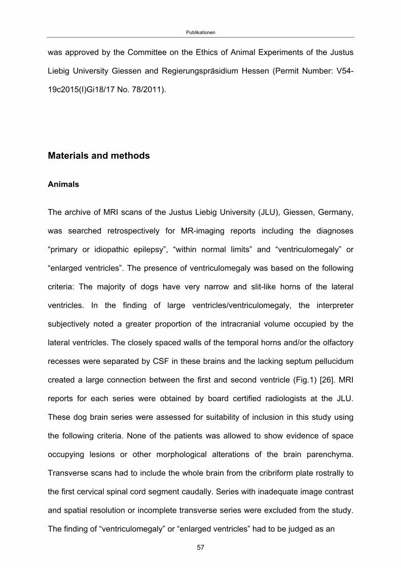
Publikationen
was approved by the Committee on the Ethics of Animal Experiments of the Justus
Liebig University Giessen and Regierungspräsidium Hessen (Permit Number: V54-
19c2015(I)Gi18/17 No. 78/2011).
Materials and methods
Animals
The archive of MRI scans of the Justus Liebig University (JLU), Giessen, Germany,
was searched retrospectively for MR-imaging reports including the diagnoses
“primary or idiopathic epilepsy”, “within normal limits” and “ventriculomegaly” or
“enlarged ventricles”. The presence of ventriculomegaly was based on the following
criteria: The majority of dogs have very narrow and slit-like horns of the lateral
ventricles. In the finding of large ventricles/ventriculomegaly, the interpreter
subjectively noted a greater proportion of the intracranial volume occupied by the
lateral ventricles. The closely spaced walls of the temporal horns and/or the olfactory
recesses were separated by CSF in these brains and the lacking septum pellucidum
created a large connection between the first and second ventricle (Fig.1) [26]. MRI
reports for each series were obtained by board certified radiologists at the JLU.
These dog brain series were assessed for suitability of inclusion in this study using
the following criteria. None of the patients was allowed to show evidence of space
occupying lesions or other morphological alterations of the brain parenchyma.
Transverse scans had to include the whole brain from the cribriform plate rostrally to
the first cervical spinal cord segment caudally. Series with inadequate image contrast
and spatial resolution or incomplete transverse series were excluded from the study.
The finding of “ventriculomegaly” or “enlarged ventricles” had to be judged as an
57
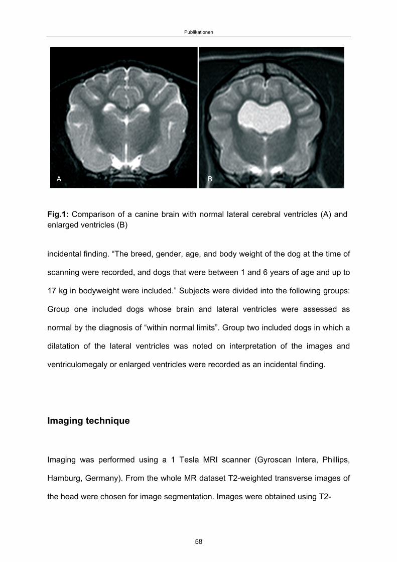
Publikationen
Fig.1: Comparison of a canine brain with normal lateral cerebral ventricles (A) and enlarged ventricles (B)
incidental finding. “The breed, gender, age, and body weight of the dog at the time of
scanning were recorded, and dogs that were between 1 and 6 years of age and up to
17 kg in bodyweight were included.” Subjects were divided into the following groups:
Group one included dogs whose brain and lateral ventricles were assessed as
normal by the diagnosis of “within normal limits”. Group two included dogs in which a
dilatation of the lateral ventricles was noted on interpretation of the images and
ventriculomegaly or enlarged ventricles were recorded as an incidental finding.
Imaging technique
Imaging was performed using a 1 Tesla MRI scanner (Gyroscan Intera, Phillips,
Hamburg, Germany). From the whole MR dataset T2-weighted transverse images of
the head were chosen for image segmentation. Images were obtained using T2-
58
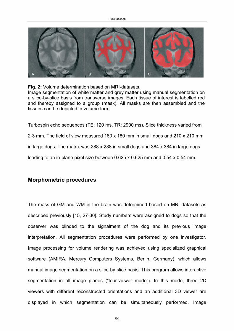
Publikationen
Fig. 2: Volume determination based on MRI-datasets. Image segmentation of white matter and grey matter using manual segmentation on a slice-by-slice basis from transverse images. Each tissue of interest is labelled red and thereby assigned to a group (mask). All masks are then assembled and the tissues can be depicted in volume form.
Turbospin echo sequences (TE: 120 ms, TR: 2900 ms). Slice thickness varied from
2-3 mm. The field of view measured 180 x 180 mm in small dogs and 210 x 210 mm
in large dogs. The matrix was 288 x 288 in small dogs and 384 x 384 in large dogs
leading to an in-plane pixel size between 0.625 x 0.625 mm and 0.54 x 0.54 mm.
Morphometric procedures
The mass of GM and WM in the brain was determined based on MRI datasets as
described previously [15, 27-30]. Study numbers were assigned to dogs so that the
observer was blinded to the signalment of the dog and its previous image
interpretation. All segmentation procedures were performed by one investigator.
Image processing for volume rendering was achieved using specialized graphical
software (AMIRA, Mercury Computers Systems, Berlin, Germany), which allows
manual image segmentation on a slice-by-slice basis. This program allows interactive
segmentation in all image planes (“four-viewer mode”). In this mode, three 2D
viewers with different reconstructed orientations and an additional 3D viewer are
displayed in which segmentation can be simultaneously performed. Image
59
Publikationen
Fig. 3: Volume rendering of brain tissues of interest. 3D viewer mode of the graphical software AMIRA. The voxels of the tissue of interest (white matter/grey matter) of each slice have been assembled and are now displayed as a 3D model. Each tissue can be displayed solid or transparent. The localizer lines support the segmentation process. As they are displayed in both the 2D images and the 3D model, the thalamus, medulla and cerebellum can be accurately separated from the volume of interest.
segmentation in this context describes the manual tracing of the WM and GM in each
image based on their differential signal intensity. All voxels corresponding to a single
anatomical structure in the images are selected and assigned to the same value in
the mask. The final mask thus contains information about all selected anatomical
structures and in combination with the original data and polygonal surface
reconstruction algorithms, allows the visualization of different structures in the
images [29]. Segmentation was performed manually in transverse orientation from
individual slices (Fig.2). Only the GM and WM of the cerebrum (neocortex and
paleocortex) including the basal ganglia were determined. The ammon`s horn
60
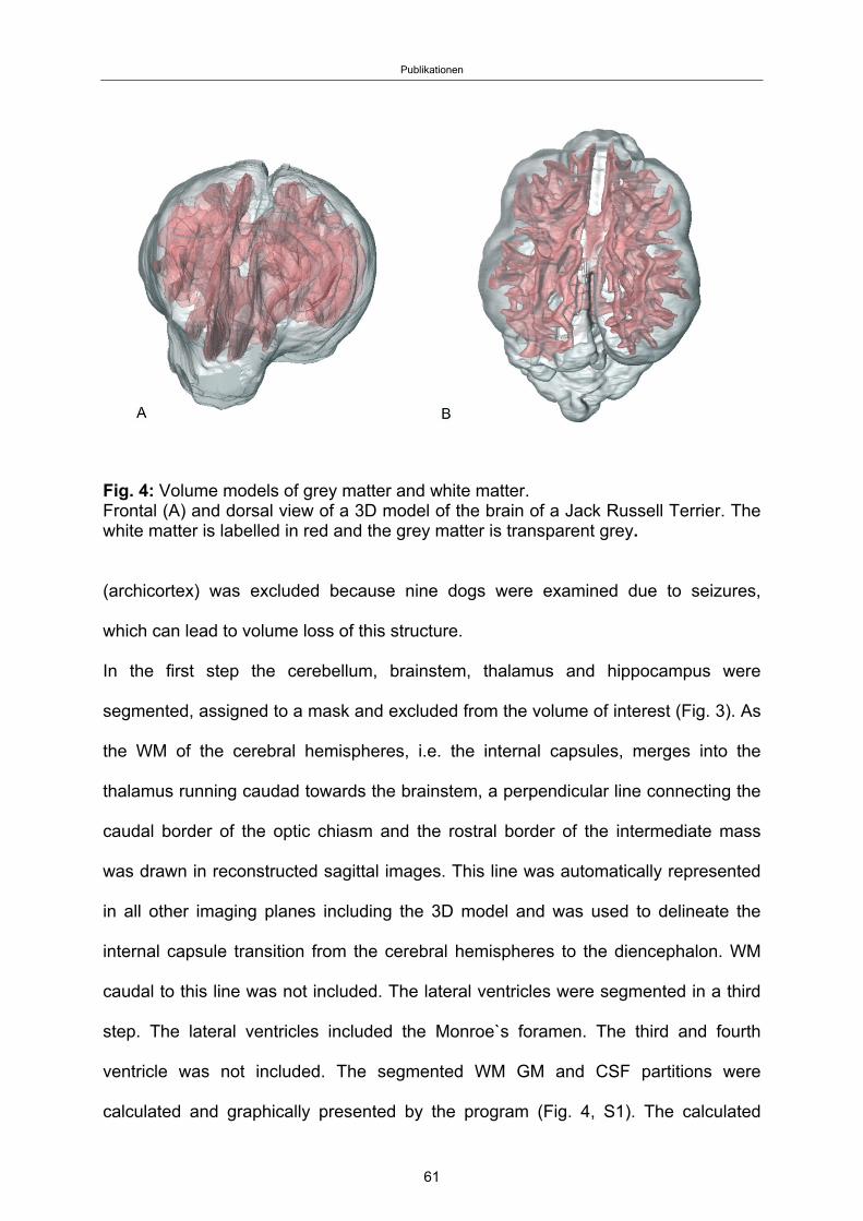
Publikationen
Fig. 4: Volume models of grey matter and white matter. Frontal (A) and dorsal view of a 3D model of the brain of a Jack Russell Terrier. The white matter is labelled in red and the grey matter is transparent grey.
(archicortex) was excluded because nine dogs were examined due to seizures,
which can lead to volume loss of this structure.
In the first step the cerebellum, brainstem, thalamus and hippocampus were
segmented, assigned to a mask and excluded from the volume of interest (Fig. 3). As
the WM of the cerebral hemispheres, i.e. the internal capsules, merges into the
thalamus running caudad towards the brainstem, a perpendicular line connecting the
caudal border of the optic chiasm and the rostral border of the intermediate mass
was drawn in reconstructed sagittal images. This line was automatically represented
in all other imaging planes including the 3D model and was used to delineate the
internal capsule transition from the cerebral hemispheres to the diencephalon. WM
caudal to this line was not included. The lateral ventricles were segmented in a third
step. The lateral ventricles included the Monroe`s foramen. The third and fourth
ventricle was not included. The segmented WM GM and CSF partitions were
calculated and graphically presented by the program (Fig. 4, S1). The calculated
61
Publikationen
volumes were put into relation to the whole cerebral volume and expressed as the
relative CSF volume and relative GM- and WM volume. Furthermore the relationship
between WM and GM was expressed as the WM/GM ratio.
Statistical analysis
Statistical analysis was performed using the commercial statistical software package
BMDP (BMDP Statistical Software, Inc., Los Angeles, USA). To test homogeneity of
the groups the relative frequency of male and female animals in the groups was
compared using Fisher`s exact test.
Relating the group comparison of target variables, in a first step the relative GM
volume, the relative WM volume, relative CSF-volume and the WM/GM ratio was
plotted against the body weight of the animals. Then the residuals of the linear
regression model including group effects were checked for normality using a Q-Q-
plot. If the conditions for its application are given the one-way analysis of covariance
(ANCOVA) was used to assess the significance of the difference between the groups
including the relationship between the independent variable body weight (X-axis) and
the dependent variables relative GM-, WM- and CSF-volume as well as WM/GM
ratio, (Y-axis). The slopes of the regression lines were compared between groups,
testing the null hypothesis that for a special dependent variable the slopes are all
identical (the lines are parallel). In a second part of the analysis the adjusted means
(sample means adjusted for the common mean body weight and a common slope) of
all groups were calculated and checked for significant differences between
groups[30].
62
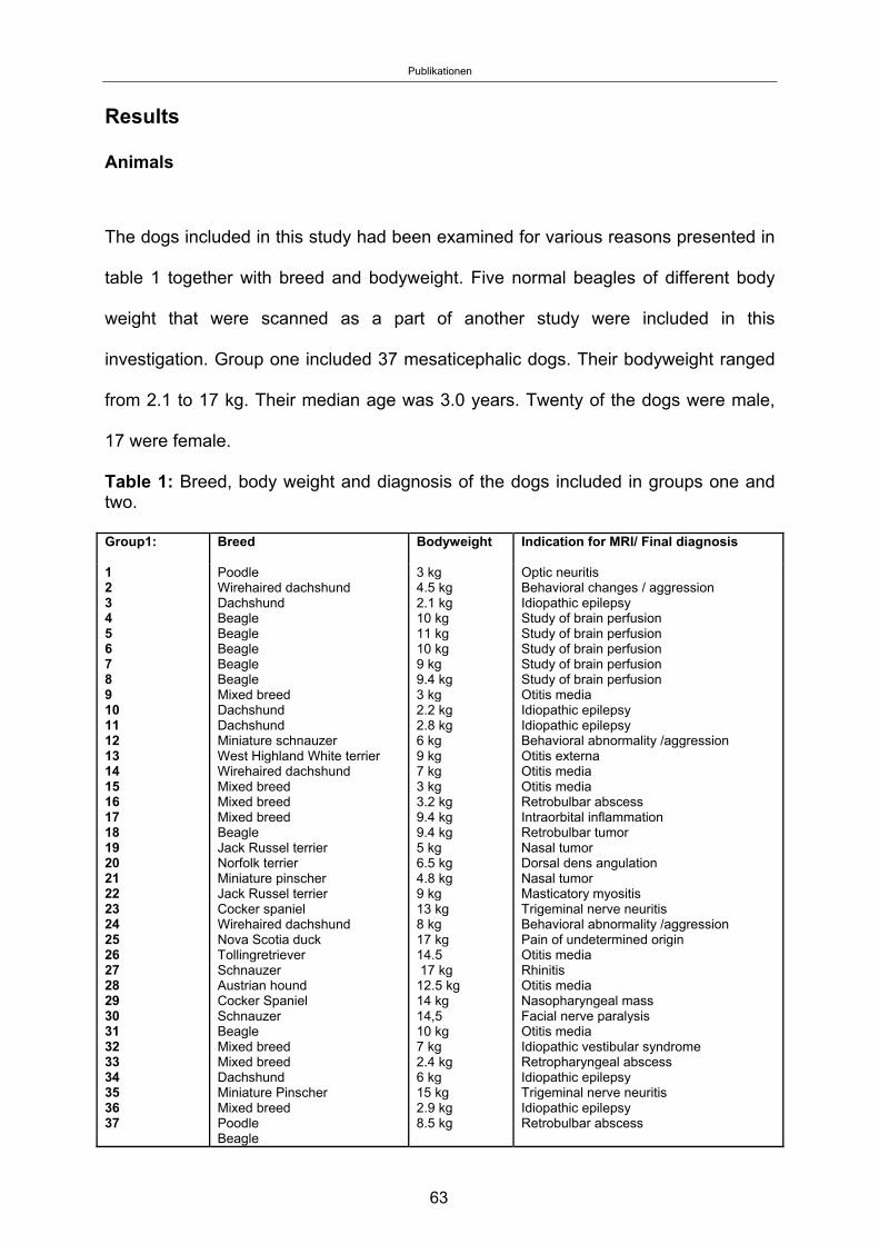
Publikationen
63
Results
Animals
The dogs included in this study had been examined for various reasons presented in
table 1 together with breed and bodyweight. Five normal beagles of different body
weight that were scanned as a part of another study were included in this
investigation. Group one included 37 mesaticephalic dogs. Their bodyweight ranged
from 2.1 to 17 kg. Their median age was 3.0 years. Twenty of the dogs were male,
17 were female.
Table 1: Breed, body weight and diagnosis of the dogs included in groups one and two. Group1:
Breed Bodyweight Indication for MRI/ Final diagnosis
1 2 3 4 5 6 7 8 9 10 11 12 13 14 15 16 17 18 19 20 21 22 23 24 25 26 27 28 29 30 31 32 33 34 35 36 37
Poodle Wirehaired dachshund Dachshund Beagle Beagle Beagle Beagle Beagle Mixed breed Dachshund Dachshund Miniature schnauzer West Highland White terrier Wirehaired dachshund Mixed breed Mixed breed Mixed breed Beagle Jack Russel terrier Norfolk terrier Miniature pinscher Jack Russel terrier Cocker spaniel Wirehaired dachshund Nova Scotia duck Tollingretriever Schnauzer Austrian hound Cocker Spaniel Schnauzer Beagle Mixed breed Mixed breed Dachshund Miniature Pinscher Mixed breed Poodle Beagle
3 kg 4.5 kg 2.1 kg 10 kg 11 kg 10 kg 9 kg 9.4 kg 3 kg 2.2 kg 2.8 kg 6 kg 9 kg 7 kg 3 kg 3.2 kg 9.4 kg 9.4 kg 5 kg 6.5 kg 4.8 kg 9 kg 13 kg 8 kg 17 kg 14.5 17 kg 12.5 kg 14 kg 14,5 10 kg 7 kg 2.4 kg 6 kg 15 kg 2.9 kg 8.5 kg
Optic neuritis Behavioral changes / aggression Idiopathic epilepsy Study of brain perfusion Study of brain perfusion Study of brain perfusion Study of brain perfusion Study of brain perfusion Otitis media Idiopathic epilepsy Idiopathic epilepsy Behavioral abnormality /aggression Otitis externa Otitis media Otitis media Retrobulbar abscess Intraorbital inflammation Retrobulbar tumor Nasal tumor Dorsal dens angulation Nasal tumor Masticatory myositis Trigeminal nerve neuritis Behavioral abnormality /aggression Pain of undetermined origin Otitis media Rhinitis Otitis media Nasopharyngeal mass Facial nerve paralysis Otitis media Idiopathic vestibular syndrome Retropharyngeal abscess Idiopathic epilepsy Trigeminal nerve neuritis Idiopathic epilepsy Retrobulbar abscess
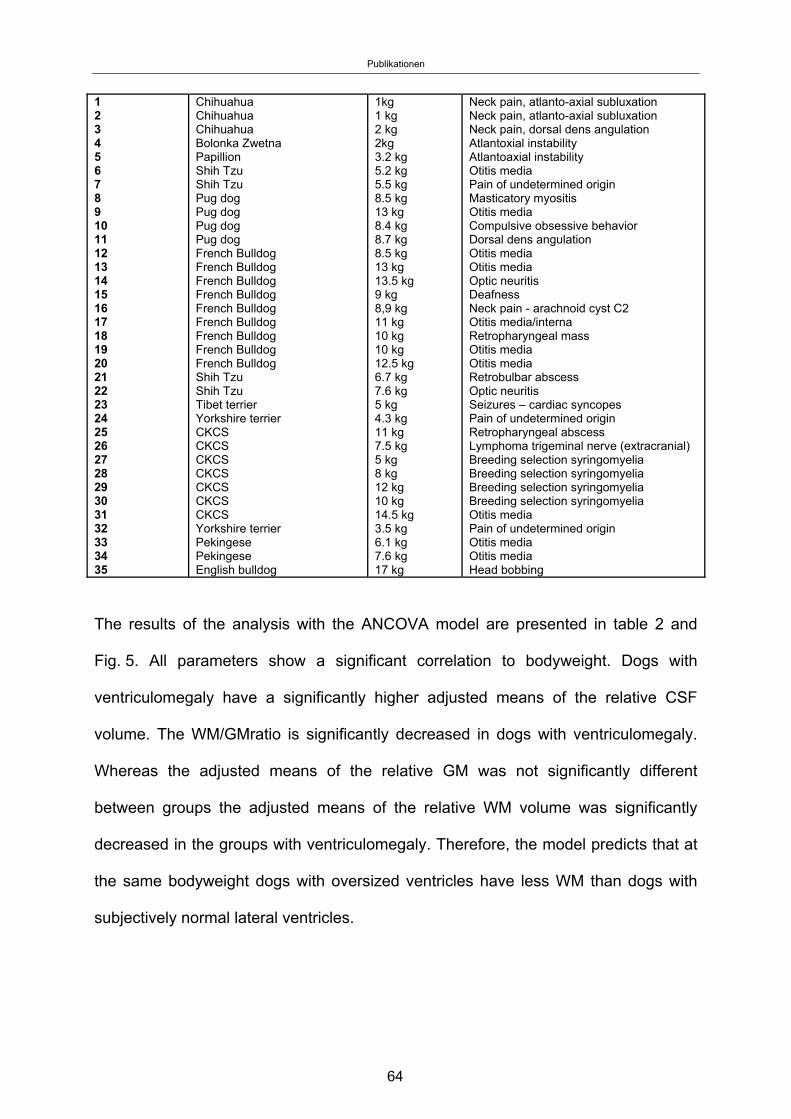
Publikationen
64
1 2 3 4 5 6 7 8 9 10 11 12 13 14 15 16 17 18 19 20 21 22 23 24 25 26 27 28 29 30 31 32 33 34 35
Chihuahua Chihuahua Chihuahua Bolonka Zwetna Papillion Shih Tzu Shih Tzu Pug dog Pug dog Pug dog Pug dog French Bulldog French Bulldog French Bulldog French Bulldog French Bulldog French Bulldog French Bulldog French Bulldog French Bulldog Shih Tzu Shih Tzu Tibet terrier Yorkshire terrier CKCS CKCS CKCS CKCS CKCS CKCS CKCS Yorkshire terrier Pekingese Pekingese English bulldog
1kg 1 kg 2 kg 2kg 3.2 kg 5.2 kg 5.5 kg 8.5 kg 13 kg 8.4 kg 8.7 kg 8.5 kg 13 kg 13.5 kg 9 kg 8,9 kg 11 kg 10 kg 10 kg 12.5 kg 6.7 kg 7.6 kg 5 kg 4.3 kg 11 kg 7.5 kg 5 kg 8 kg 12 kg 10 kg 14.5 kg 3.5 kg 6.1 kg 7.6 kg 17 kg
Neck pain, atlanto-axial subluxation Neck pain, atlanto-axial subluxation Neck pain, dorsal dens angulation Atlantoxial instability Atlantoaxial instability Otitis media Pain of undetermined origin Masticatory myositis Otitis media Compulsive obsessive behavior Dorsal dens angulation Otitis media Otitis media Optic neuritis Deafness Neck pain - arachnoid cyst C2 Otitis media/interna Retropharyngeal mass Otitis media Otitis media Retrobulbar abscess Optic neuritis Seizures – cardiac syncopes Pain of undetermined origin Retropharyngeal abscess Lymphoma trigeminal nerve (extracranial) Breeding selection syringomyelia Breeding selection syringomyelia Breeding selection syringomyelia Breeding selection syringomyelia Otitis media Pain of undetermined origin Otitis media Otitis media Head bobbing
The results of the analysis with the ANCOVA model are presented in table 2 and
Fig. 5. All parameters show a significant correlation to bodyweight. Dogs with
ventriculomegaly have a significantly higher adjusted means of the relative CSF
volume. The WM/GMratio is significantly decreased in dogs with ventriculomegaly.
Whereas the adjusted means of the relative GM was not significantly different
between groups the adjusted means of the relative WM volume was significantly
decreased in the groups with ventriculomegaly. Therefore, the model predicts that at
the same bodyweight dogs with oversized ventricles have less WM than dogs with
subjectively normal lateral ventricles.
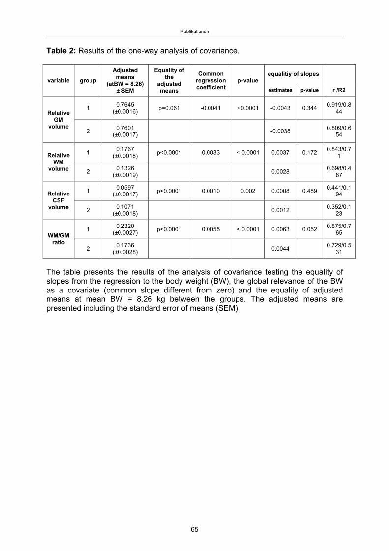
Publikationen
65
Table 2: Results of the one-way analysis of covariance.
variable group
Adjusted means
(atBW = 8.26) ± SEM
Equality of the
adjusted means
Common regression coefficient
p-value equalitiy of slopes
estimates p-value r /R2
Relative GM
volume
1 0.7645
(±0.0016) p=0.061 -0.0041 <0.0001 -0.0043 0.344
0.919/0.844
2 0.7601
(±0.0017) -0.0038
0.809/0.654
Relative WM
volume
1 0.1767
(±0.0018) p<0.0001 0.0033 < 0.0001 0.0037 0.172
0.843/0.71
2 0.1326
(±0.0019) 0.0028
0.698/0.487
Relative CSF
volume
1 0.0597
(±0.0017) p<0.0001 0.0010 0.002 0.0008 0.489
0.441/0.194
2 0.1071
(±0.0018) 0.0012
0.352/0.123
WM/GM ratio
1 0.2320
(±0.0027) p<0.0001 0.0055 < 0.0001 0.0063 0.052
0.875/0.765
2 0.1736
(±0.0028) 0.0044
0.729/0.531
The table presents the results of the analysis of covariance testing the equality of slopes from the regression to the body weight (BW), the global relevance of the BW as a covariate (common slope different from zero) and the equality of adjusted means at mean BW = 8.26 kg between the groups. The adjusted means are presented including the standard error of means (SEM).
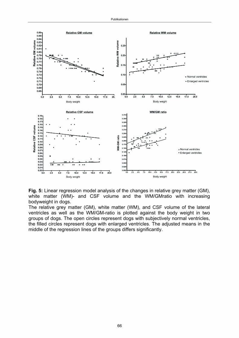
Publikationen
Fig. 5: Linear regression model analysis of the changes in relative grey matter (GM), white matter (WM)- and CSF volume and the WM/GMratio with increasing bodyweight in dogs. The relative grey matter (GM), white matter (WM), and CSF volume of the lateral ventricles as well as the WM/GM-ratio is plotted against the body weight in two groups of dogs. The open circles represent dogs with subjectively normal ventricles, the filled circles represent dogs with enlarged ventricles. The adjusted means in the middle of the regression lines of the groups differs significantly.
66
Publikationen
Discussion
The dimensions of the cerebral ventricular volume in dogs have been reported to
vary [17, 18]. In brachycephalic dogs in particular, the ventricles can be larger than
expected. This has been referred to as ventriculomegaly to differentiate this condition
from clinically relevant internal hydrocephalus. However, the designation of normal
ventricles and enlarged ventricles remain poorly defined and are usually assessed
subjectively [26].
It is unclear whether ventriculomegaly should be interpreted as one variant of a
spectrum of ventricular dimensions in dogs or rather as a pathological condition. It
was our hypothesis that the larger ventricles in brachycephalic dogs are a
pathological condition as indicated by concurrent presence of WM loss due to
ventricular distention as found in internal hydrocephalus. To investigate this
hypothesis we examined the relative WM, GM, and CSF volume in dogs with
different ventricular dimensions as well as their WM/GM ratio in proportion to their
body weight and cerebral volume. The age of the animal has an important influence
on the volumes of neural tissues, myelination and ventricular size. As cortical atrophy
and ventricular enlargement have been identified as characteristic features of the
aged canine brain [31], only dogs < 6 years of age were included in the study. Brain
volume, myelination and ventricular volume change during brain growth [6, 32-34].
Therefore, only dogs with a minimum age of 12 months were included. Testosterone
levels are positively correlated with myelinogenesis [35]. This could lead to larger
white matter volumes in male dogs. The potential influences of sexual dimorphism
can be ignored in our study as the proportion of male and female dogs did not differ
significantly between groups.
Epilepsy was one reason for MRI examination in 9 dogs of our control group.
67
Publikationen
Although seizure activity can lead to brain damage, epileptic dogs have previously
been used to study brain volumetrics [26, 28] as damage only occurs at the
microscopic level [36, 37]. However, the hippocampus was a priori excluded from
grey matter volume because variable loss of hippocampal volume identified on MR-
images has been linked to seizure activity in dogs [38, 39].
MRI-based volume measurements of the brain parts are routinely performed in
human and veterinary medicine [13, 15, 28, 30, 38, 40, 41]. The accuracy of the
technique even for small volumes has been proven in both veterinary [8, 17, 42] and
human neuroradiology [42, 43]. However, manual segmentation approaches can be
subject to investigator bias. The validity of our approach would have further
increased with multiple examinations but we refrained from the determination of
interobserver-variability as the manual segmentation process is extremely time
consuming. This is the main limitation of the present study. Large and small dog
heads were scanned using different receiver coils (knee coil, surface coil). It is
possible that using different coils could have caused a bias in our measurements due
to random differences in contrast or resolution. We only included image sets with
sufficient resolution and contrast.
Our approach to comparing WM and GM volume in dogs followed the assumption of
a basic uniformity of the neocortex and its connections in all dog breeds. We did not
consider other non-allometric factors based on selection related to behavioural and
environmental factors affecting brain size independently from selection for body size
by breeders. Some breeds have been selectively bred for the “quality of learning,” but
this has been upstaged in other breeds in favour of other characteristics.
Mesaticephalic dogs have been reported to have higher trainability than
brachycephalic dogs, which may have an influence on the number and connectivity
of neurons [44]. This could influence the calculated relations if such dogs have more
68
Publikationen
GM or WM at a given bodyweight (grade shift of brain mass), especially given that
group two exclusively included brachycephalic dogs. Analysis of large homogenous
groups of dogs would be necessary to reveal principle differences in brain
morphology between dog breeds.
We also not consider the influence of obesity. The fat free body weight would have
been the ideal parameter to show the precise allometric change of the brain tissues.
The present study indicates that ventriculomegaly in brachycephalic dogs without
morphological abnormalities of the brain parenchyma is linked to a reduction in WM
volume in relation to the GM mass of the cerebrum. As the constant relationship
between WM and GM is universal in all mammalian brains, the reduced WM in
combination with it the larger ventricles in these dogs should not be considered a
physiological condition.
The origin of the WM loss is not clear. A primary white matter disease and distension
of the ventricles “ex vacuo” would theoretically be possible. Leukencephalitis,
leukodystrophies, hypomyelination and spongy degeneration must be considered in
this context. However, such diseases would cause distinct MRI-findings beyond white
matter atrophy [9]. Moreover, primary white matter diseases would cause severe
neurological signs, such as tremor, ataxia, which were not diagnosed in our patients.
Internal hydrocephalus can lead to progressive destruction of WM in dogs [45, 46]
as a consequence of high intraventricular pressure. It is reasonable to expect that the
degree of intraventricular pressure determines the speed and severity of white matter
injury and thereby functional brain deficits. Moderately increased pressure or
intermittently high intraventricular CSF pressure might play an important role in the
pathogenesis of WM loss in dogs with ventriculomegaly as proposed for normal
pressure hydrocephalus (NPH) in humans [47]. It has been suggested that in NPH
intermittent CSF pressure waves can produce temporary phases of ischaemia in the
69
Publikationen
periventricular white matter. The chronic cumulative effects of these ischaemic
events can produce WM atrophy that does not arise until late adulthood [47, 48].
Brain perfusion studies have revealed reduced regional cerebral blood flow in the
periventricular region in human patients with NPH, which has been attributed to
disturbances in CSF flow [49].
Ventriculomegaly has also been attributed to disturbances in CSF dynamics in dogs
by some authors [15, 50]. Impaired CSF flow can be caused by obstruction of the
foramen magnum, as found in dogs with morphological changes in the cranio-
cervical junction referred to as Chiari-like malformation [50, 51]. It has been proposed
that impaired CSF flow can result in syringomyelia as well as ventricular dilatation
[52, 53]. However, this has only been proposed for the Cavalier King Charles spaniel
and the Brussels Griffon and the prevalence of Chiari-like malformation in other
breeds with ventriculomegaly needs closer evaluation.
Human patients with enlarged ventricles in association with NPH show characteristic
signs of neural function deficits. The cardinal symptoms of NPH are gait impairment,
dementia and urinary incontinence (“Hakim triad”) [54]. However, it has also been
proposed that the full symptom triad represents an advanced stage of the disease
and that NPH can be diagnosed in the presence of only two or even just one of the
symptoms [54]. In contrast, the association of solitary ventriculomegaly with
neurological deficits in dogs has been rejected [9-11, 13]. The lack of clinical signs as
a consequence of ventricular enlargement in dogs as opposed to humans may
initially be striking. However, control of locomotion is far more complex in humans
and the motor cortex and its connections play a substantial role even during
undemanding steady-state walking [55]. The cerebral control of locomotion plays a
minor role in dogs and diseases affecting the forebrain may have only minimal
influence on locomotion [56]. Urinary incontinence was not reported in the dogs of
70
Publikationen
this study either. Although the control of micturition involves cortical centres in
carnivores, pontine micturition centres maintain function without cerebral control [56].
Signs of dementia are difficult to diagnose in dogs. In human medicine, a diagnosis
of dementia will be considered based on the occurrence of distinct clinical signs. The
most important criteria are memory impairment, disturbed language function, inability
to carry out purposeful movement, and failure to recognize people [54]. Such brain
functions cannot be assessed in dogs, at least not in a clinical setting, and the real
status of canine cognitive abilities cannot be determined with absolute certainty. On
the other hand, it has been shown that even severe ventricular dilation is compatible
with normal physical and intellectual development in humans and that the ventricular
volumes of hydrocephalic patients did not correlate well with their intelligence
quotient [57, 58].
The influence of ventriculomegaly on brain function in dogs is unclear. Detailed
behavioural studies of the impact of WM loss on the full functional integration of the
nervous system are necessary to clarify whether ventriculomegaly might be an
indication for CSF shunting procedures in dogs. If clinical or experimental data for
cognitive impairment or intermittently high CSF pressure waves could be found,
naturally occurring ventriculomegaly in dogs might be an interesting animal model for
human NPH.
Conclusion
Brachycephalic dogs with ventriculomegaly can have reduced cerebral white matter.
As a predetermined relationship exists between WM and GM mass in the brain of
mammals this aberration cannot be interpreted as a physiological condition.
71
Publikationen
References
[1] Seiferle E (1966) Zur Topographie des Gehirns bei lang - und kurzköpfigen
Hunderassen. Acta Anat 63: 346-362.
[2] Roberts T, McGreevy P, Valenzuela M (2010) Human induced rotation and
reorganization of the brain of domestic dogs. PLoS One 26: e11946.
[3] Oboussier H (1955) Behavior of cerebral sulcus formation in crossbreeding of
extreme breed types of dogs. Z Mensch Vererb Konstitutionsl 33:1-9.
[4] Schmidt MJ, Amort K, Kramer M (2012) Postnatal development of the cerebral
gyrification in the canine brain. Vet Radiol & Ultrasound 53: 643-649.
[5] Schmidt MJ, Neumann AC, Amort KH,Failin K, Kramer M (2011) Cephalometric
measurements and determination of the general skull type of Cavalier King Charles
Spaniels. Vet Radiol & Ultrasound 52: 436-440.
[6] Kii S, Uzuka Y, Taura Y, Nakaichi M, Takeuchi A, Inokuma H, et al.(1997) Magnetic
resonance imaging of the lateral ventricles in beagle-type dogs. Vet Radiol &
Ultrasound 38: 430-433.
[7] Schröder H, Meyer-Lindenberg A, Nolte I (2006) Comparative examination of the
lateral cerebral ventricles of different dog breeds using quantitative computed
tomography. Berl Munch Tierarztl Wochenschr 119: 506-511.
[8] Vullo T, Korenman E, Manzo RP, Gomez DG, Deck MD, Cahill PT (1997) Diagnosis
of cerebral ventriculomegaly in normal adult beagles using quantitative MRI. Vet
Radiol & Ultrasound 38: 277-281.
[9] Gavin PR, Bagley RS (2011) Practical Small Animal MRI. Wiley Blackwell, Iowa
USA. 64p
[10] Schwarz T, Saunders J (2012) Veterinary Computed Tomography. Wiley Blackwell,
Iowa USA. 185 p
[11] Lu D, Lamb CR, Pfeiffer DU, Targett MF (2003) Neurological signs and results of
magnetic resonance imaging in 40 cavalier King Charles spaniels with Chiari type 1-
72
Publikationen
like malformations. Vet Rec 153: 60-63.
[12] Driver CJ, Chandler K, Walmsley G,Shibab N, Volk HA (2013) The association
between Chiari-like malformation, ventriculomegaly and seizures in cavalier King
Charles spaniels. Vet J 195: 235-237.
[13] Vite CH, Insko EK, Schotland HM, Panckeri K, Hendricks JC (1997) Quantification of
cerebral ventricular volume in English bulldogs. Vet Radiol & Ultrasound 38: 437-443.
[14] Dexler H (1923) Über die konstitutionelle innere Hydrocephalie beim Hunde. Prag
Tierärztl Arch 3: 103-268.
[15] Driver CJ, Rusbridge C, Cross, HR, McGonnell I, Volk HA (2010) Relationship of
brain parenchyma within the caudal cranial fossa and ventricle size to syringomyelia
in cavalier King Charles spaniels. J Small Anim Pract 51: 382-386.
[16] Saito M, Olby NJ, Spaulding K, Muñana K, Sharp NJ. (2003) Relationship among
basilar artery resistance index, degree of ventriculomegaly, and clinical signs in
hydrocephalic dogs. Vet Radiol & Ultrasound 44: 687-694.
[17] De Haan CE, Kraft SL, Gavin PR,Wendling LR (1994) Normal variation in size of
the lateral ventricles of the Labrador Retriever dog as assessed by magnetic
resonance imaging. Vet Radiol & Ultrasound 35: 83-86.
[18] Spaulding KA, Sharp NJH (1990) Ultrasonographic imaging of the lateral
cerebral ventricles in the dog. Vet Radiol & Ultrasound 31: 59-64.
[19] Zhang K, Sejnowski TJ (2000) A universal scaling law between gray matter and
white matter of cerebral cortex. Proc Natl Acad Sci USA 97: 5621-5626.
[20] Frahm HD, Stephan H, Stephan M (1982) Comparison of brain structure volumes in
Insectivora and Primates. I. Neocortex. J Hirnforsch 23: 375-389.
[21] Rilling JK, Insel TR (1999) Differential expansion of neural projection systems in
primate brain evolution. Neuroreport 10: 1453-1459.
[22] Changizi MA (2001) Principles underlying mammalian neocortical scaling. Biol
Cybern 84: 207-215.
[23] Roth G, Dickie U (2005) Evolution of the brain and intelligence. Trends Cogn Sci.
9: 250-257.
73
Publikationen
[24] Purves D, Augustine GJ, Fitzpatrick D, Katz LC, LaMantia AS, McNamara JO,
Williams SM.editors (2001) Neuroscience. 2nd edition. Sunderland (MA): Sinauer
Associates.
[25] Carrera I, Dennis R, Mellor DJ, Penderis J, Sullivan M (2009) Use of magnetic
resonance Cavalier King Charles imaging for morphometric analysis of the caudal
cranial fossa in Spaniels. Am J Vet Res 70: 340-345.
[26] Thomas B (2010) Hydrocephalus in dogs and cats. Vet Clin Small Anim 40: 143-159.
[27] Cross HR, Capello R, Rusbridge C (2009) Comparison of cerebral cranium volumes
between Cavalier King Charles Spaniels with Chiari-like malformation, small breed
dogs and Labradors. J Small Anim Pract 50: 399-405.
[28] Schmidt MJ, Biel M, Klumpp S, Schneider M, Kramer M (2009) Evaluation of the
volumes of cranial cavities in Cavalier King Charles spaniels with Chiari- like
malformation and other brachycephalic dogs as measured via computed tomography.
Am J Vet Res. 70: 508-512.
[29] Shaw TA, McGonnell IM, Driver CJ, Rusbridge C, Volk HA (2012) Increase in
cerebellar volume in Cavalier King Charles Spaniels with Chiari-like malformation and
its role in the development of syringomyelia. PLoS One 7(4):e33660.
[30] Barton RA, Harvey PH (2000) Mosaic evolution of brain structure in mammals.
Nature 29: 1055-1058.
[31] Su MY, Head E, Brooks WM (1998) Magnetic resonance imaging of anatomic and
vascular characteristics in a canine model of human aging. Neurobiol Ageing19: 479-
485.
[32] Gross B, Garcia-Tapia D, Riedesel E, Ellinwood NM, Jens JK (2010) Normal canine
brain maturation at magnetic resonance imaging. Vet Radiol & Ultrasound 51: 361-
373.
[33] Arant BS, Gooch WM (1982) Developmental changes in the mongrel canine brain
during postnatal life. Early Hum Dev 7: 179-194.
[34] Fox MW (1964) The postnatal growth of the canine brain and correlated anatomical
and behavioural change during neuro-ontogenesis. Growth 28: 135-141.
74
Publikationen
[35] Martini L, Melcangi RC (1991) Androgen metabolism in the brain. J Steroid
Biochem Mol Biol 39: 819-828.
[36] Montgomery DL, Lee AC (1983) Brain damage in the epileptic Beagle dog. Vet
Pathol 20: 160-169.
[37] Morita T, Shimada A, Ohama E, Umemura T, Fukuda S (1999) Oligodendroglia
vacuolar degeneration in the bilateral motor cortices and astrocytosis in epileptic
Beagle dogs. J Vet Med Sci 61: 107-111.
[38] Yamasaki H, Furuoka H, Takechi M, Itakura C (1991) Neuronal loss and gliosis in
limbic system in an epileptic dog. Vet Pathol 28: 540-542.
[39] Breitschwerdt EB, Breazile JE, Broadhurst JJ (1979) Clinical and
electroencephalographic findings associated with ten cases of suspected limbic
epilepsy in the dog. J Am Anim Hosp Assoc 15: 37-50.
[40] Milne ME, Anderson GA, Chow KE, O'Brien TJ, Moffat BA, Long SN (2013)
Description of technique and lower reference limit for magnetic resonance
imaging of hippocampal volumetry in dogs. Am J Vet Res 74: 224-231.
[41] Thames R, Robertson ID, Flegel T, Henke D, O'Brien DP, Coates JR, et al.
(2010) Development of a morphometric Magnetic Resonance Image parameter
suitable for distinguishing between normal dogs and dogs with cerebellar atrophy. Vet
Radiol & Ultrasound 51: 246-253.
[42] Rifa H, Bloch I, Hutchinson S, et al. (2000) Segmentation of the skull in MRI
volumes using deformable model and taking the partial volume effect into
account. Med Image Anal 4: 219-233.
[43] Vullo T, Deo-Narine V, Stallmeyer MJ, Wiart J, Garnero L (1996) Quantitation of
normal canine hippocampus formation volume: correlation of MRI with gross
histology. Magn Reson Imaging 14: 657-662.
[44] Helton WS (2009) Cephalic index and perceived dog trainability. Behav Proc 82: 355-
358.
[45] Wünschmann A, Oglesbee M (2001) Periventricular changes associated with
spontaneous canine hydrocephalus. Vet Pathol 38: 67-73.
75
Publikationen
[46] Yamada H, Yokota A, Haratake J, Horie A (1996) Morphological study of
experimental syringomyelia with kaolin-induced hydrocephalus in a canine
model. J Neurosurg 84: 999-1005.
[47] Del Bigio MR (1993) Neuropathological changes caused by hydrocephalus. Acta
Neuropathol 85: 573-585.
[48] Akai K, Uchigasaki S, Tanaka U, Komatsu A (1987) Normal pressure
hydrocephalus: neuropathological study. Acta Pathol Jpn 37: 97-110.
[49] Momjian S, Owler BK, Czosnyka Z, Czosnyka M, Pena A, Pickard JD (2004) Pattern
of white matter regional cerebral blood flow and autoregulation in normal pressure
hydrocephalus. Brain.127: 965-972.
[50] Rusbridge C, Knowler SP, Pieterse L Mc Fadyen A(2009) Chiari-like malformation in
the Griffon Bruxellosis. J Small Anim Pract 50: 386-393.
[51] Rusbridge C, MacSweeney JRE, Davies SJ Chandler K, Fitzmaurice SN,
Dennis R, Cappello R, Wheeler SJ. (2000) Syringohydromyelia in Cavalier King
Charles Spaniels. J Am Anim Hosp Assoc 36: 34-41.
[52] Levine DN (2004) The pathogenesis of syringomyelia associated with lesions at the
foramen magnum: a critical review of existing theories and proposal of a new
hypothesis. J Neurol Sci 220: 3-21.
[53] Rusbridge C, Knower SP (2004) Inheritance of occipital bone hypoplasia
(ChiariI malformation) in Cavalier King Charles spaniels. J Vet Int Med 18:673 678.
[54] Kiefer M, Unterberg A (2012) The differential diagnosis and treatment of normal-
pressure hydrocephalus. Dtsch Arztebl Int 109: 15-25.
[55] Miyai I, Tanabe HC, Sase I, Eda H, Oda I, Konishi I, Tsunazawa Y, Suzuki T,
Yanagida T, Kubota K (2001) Cortical mapping of gait in humans: A near-infrared
spectroscopic topography study. Neuroimage 141186-1192.
[56] DeLahunta A, Glass E (2009) Veterinary neuroanatomy and clinical neurology.
Saunders Elsevier, Missouri. 37p.
[57] Lorber J (1983) Hydrocephalus in frühen Kindersalter. Edited by Stuttgart VD. Enke:
2-14.
76
Publikationen
[58] Jackson PH, Lorber J (1984) Brain and ventricular volume in hydrocephalus. Z
Kinderchir 39 (Suppl 2): 91-93.
77
Diskussion
Diskussion 4.
4.1 Die physiologische Ventrikelgröße beim Hund
Einige Studien beschäftigen sich mit dem Ventrikelsystem des Hundes und seinen
physiologischen Dimensionen (SPAULDING und SHARP, 1990; DEO-NARINE et al.,
1994; DE HAAN et al., 1994; KII et al., 1997; VULLO et al., 1997; ESTEVE-RATSCH
et al., 2001; RYAN et al., 2014). Nach ELLENBERGER und BAUM (1891) werden
nach der Gesamtformation des Kopfes zwei Gruppen von Hunderassen
unterschieden: dolichozephale (langköpfige) und brachyzephale (kurzköpfige). Eine
Zwischenform wird als mesozephal bezeichnet (EVANS, 1993). Bei brachyzephalen
und Zwerghunde-Rassen wurden bereits im 20. Jahrhundert erweiterte Ventrikel
beschrieben, die von DEXLER (1923) als konstitutiver Hydrozephalus, von anderen
Autoren auch als „arrested hydrocephalus“ bezeichnet wurden (JAMES und SHUT,
1978; FOLTZ und AINE, 1981). Pathologische Studien an Hundehirnen wurden in
jüngerer Vergangenheit immer mehr durch Studien mit modernen bildgebenden
Verfahren ergänzt. Eine retrospektive Studie an 50 Englischen Bulldoggen (RYAN et
al., 2014) konnte ebenfalls eine Variation der Ventrikelgröße bei dieser Rasse
dokumentieren. Auch beim Beagle wurde eine Variation der Ventrikelgröße
beschrieben (KII et al., 1997). Diese Variation wurde von den Autoren als
Normvariante innerhalb eines physiologischen Spektrums beschrieben. Die klinische
Relevanz einer solchen Erweiterung wurde dabei immer in Frage gestellt und
lediglich von einer Ventrikulomegalie gesprochen, um die Abgrenzung zum klinisch
relevanten Hydrozephalus zu verdeutlichen. Nach wie vor gibt es in der Literatur
keine Angaben zur „normalen“ relativen Größe der Ventrikel beim erwachsenen
Hund (DE HAAN et al., 1994).
4.2 Ist es möglich die Ventrikulomegalie und den Hydrocephalus
internus voneinander abzugrenzen?
Es konnten bisher keine Messwerte der Ventrikelgröße festgelegt werden, mit deren
Hilfe man zwischen symptomatischer und asymptomatischer Erweiterung der
Ventrikel unterscheiden kann (DE HAAN et al., 1994; HECHT, 2011; ESTEVE-
78
Diskussion
RATSCH et al., 2001). Zu einer Erweiterung des Ventrikelsystems kann es beim
Hund auch im Rahmen neurodegenerativer Erkrankungen, entzündlicher
Erkrankungen oder durch eine altersabhängige Atrophie von Hirngewebe kommen
(GONZALES-SORIANO et al., 2001; SU et al., 1998). Dies macht es schwieriger
einen klinisch relevanten Hydrozephalus zu diagnostizieren, der eine chirurgische
Therapie erfordert. In mehreren Studien konnte für das Ausmaß der Ventrikel allein
keine gute Korrelation mit der klinischen Symptomatik festgestellt werden (ESTEVE-
RATSCH et al., 2001; RIVERS und WALTER, 1992; VULLO et al., 1997; DE HAAN
et al., 1994; MCALLISTER et al., 1998; SPAULDING und SHARP, 1990; KII et al.,
1997; HUDSON et al., 1990). Nur eine Studie (SAITO et al., 2003) konnte einen
signifikanten Unterschied zwischen Hydrozephalus und Ventrikulomegalie durch
Messen eines linearen Ventricle/Brain (V/B) Index im Ultraschall aufzeigen. Die
Verwendung der Ultraschalltechnik ist auf Hunde mit offenen Fontanellen
beschränkt, da diese das einzige akustische Fenster für das Eindringen der
Ultraschallwellen darstellen. Ein Einsatz bei älteren Hunden ohne persistierende
Fontanelle ist somit nicht möglich (SPAULDING und SHARP, 1990).
In der vorliegenden Studie konnte ein signifikanter Unterschied zwischen Hunden mit
Hydrozephalus und asymptomatischen Hunden mit Ventrikulomegalie bei der
Messung eines V/B Index im dorsalen Schnitt in der MRT gezeigt werden. Im
Vergleich zu der Untersuchung mit Ultraschall sind die Schnittebenen wesentlich
eindeutiger zu setzen und unterliegen einem internationalen Standard. Im Gegensatz
zu vorangegangen Messungen wurden die Temporalhörner der Ventrikel an ihrer
maximalen Ausprägung gemessen. Die Temporalhörner erweitern sich beim Hund
als erstes, gefolgt von anderen Teilen des Ventrikelsystems (PENA et al., 1999;
MCALLISTER et al., 1991). Es konnte ein Grenzwert von 0.6 mit einer Sensitivität
und Spezifität von 92% ermittelt werden. Hunde mit einem V/B Index >0.6 sind somit
verdächtig für einen Hydrocephalus internus. Weiterführende Studien mit
dreidimensionalen volumetrischen Messungen werden nötig sein um zu zeigen,
inwieweit dieser V/B Index den tatsächlichen Dimensionen entspricht.
Zur Unterscheidung eines Hydrozephalus, bei dem der Patient von einer Shunt-
Implantation profitiert, von einer Ventrikulomegalie ohne klinische Relevanz in der
Magnetresonanztomographie wurden einige Kriterien aus der Humanmedizin
übernommen, beziehungsweise adaptiert.
Die Größe und Morphologie des III. Ventrikels variiert beim Hund aufgrund der
79
Diskussion
unterschiedlichen Schädelformen verschiedener Rassen. Brachyzephale Rassen
zeigen ein reduziertes Wachstum der Schädelbasis (SCHMIDT et al., 2011), was
einen Einfluss auf den Hirnstamm und somit auf manche Messungen haben kann.
Rassespezifische Normwerte wären notwendig, um genaue Abmessungen des III.
Ventrikels vergleichbar zu machen. Deshalb haben wir uns entschieden, die Form
der Adhesio interthalamica im mitsagittalen Schnitt als Kriterium für eine Erweiterung
des III. Ventrikels zu wählen. Bei allen 20 Hunden der normalen Gruppe war diese
kreisrund. Eine Deformation war signifikant häufiger in der Hydrozephalus-Gruppe zu
detektieren und wurde somit als sekundäres Anzeichen einer Erweiterung des III.
Ventrikels aufgrund eines erhöhten intraventrikulären Druckes gewertet.
Aufgrund der oben beschriebenen unterschiedlichen Morphologie bei brachy- und
mesozephalen Hunderassen haben wir uns gegen die Messung der mamillo-
pontinen Distanz (EL GAMMAL et al., 1987) entschieden.
Eine Erweiterung des IV. Ventrikels ist beim Menschen mit erhöhtem
intraventrikulärem Druck beschrieben (EDELMANN et al., 2006) und wurde auch
beim Hund beobachtet (MCALLISTER et al., 1991; WAGSHUL et al., 2009; JAMES
et al., 1974). Warum in der vorliegenden Studie kein signifikanter Unterschied
zwischen den Gruppen feststellbar war, ist unklar.
Strukturelle Veränderungen des Corpus callosum umfassen eine Dehnung und somit
Ausdünnung. Durch den gesteigerten Druck und die Ausdehnung der Ventrikel wird
das Corpus callosum angehoben, gleichzeitig kommt es zur Absenkung des Fornix
im vorderen Anteil. Wir haben die Anhebung des Corpus callosum gemessen und
konnten einen signifikanten Unterschied zwischen den Mittelwerten der drei Gruppen
feststellen. Aufgrund der teilweisen Überlappung der Ergebnisse konnten allerdings
keine Grenzwerte festgelegt werden.
Der Corpus callosum-Winkel wurde analog zur Humanmedizin gemessen (ISHII et
al., 2008). Hier stellt ein Winkel von weniger als 90° ein Kriterium für erhöhten
intraventrikulären Druck dar (MORI et al., 2012; KIEFER und UNTERBERG, 2012).
In der vorliegenden Studie konnte kein signifikanter Unterschied zwischen Hunden
mit Hydrozephalus und solchen mit Ventrikulomegalie festgestellt werden.
Möglicherweise steht dies in Verbindung mit der Höhe der Anhebung des Corpus
callosum in der Hydrozephalus-Gruppe, wodurch das Zentrum des Winkels nach
oben verschoben wird und der Winkel sich erweitert.
80
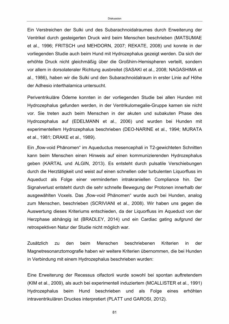
Diskussion
Ein Verstreichen der Sulki und des Subarachnoidalraumes durch Erweiterung der
Ventrikel durch gesteigerten Druck wird beim Menschen beschrieben (MATSUMAE
et al., 1996; FRITSCH und MEHDORN, 2007; REKATE, 2008) und konnte in der
vorliegenden Studie auch beim Hund mit Hydrozephalus gezeigt werden. Da sich der
erhöhte Druck nicht gleichmäßig über die Großhirn-Hemispheren verteilt, sondern
vor allem in dorsolateraler Richtung ausbreitet (SASAKI et al., 2008; NAGASHIMA et
al., 1986), haben wir die Sulki und den Subarachnoidalraum in erster Linie auf Höhe
der Adhesio interthalamica untersucht.
Periventrikuläre Ödeme konnten in der vorliegenden Studie bei allen Hunden mit
Hydrozephalus gefunden werden, in der Ventrikulomegalie-Gruppe kamen sie nicht
vor. Sie treten auch beim Menschen in der akuten und subakuten Phase des
Hydrozephalus auf (EDELMANN et al., 2006) und wurden bei Hunden mit
experimentellem Hydrozephalus beschrieben (DEO-NARINE et al., 1994; MURATA
et al., 1981; DRAKE et al., 1989).
Ein „flow-void Phänomen“ im Aqueductus mesencephali in T2-gewichteten Schnitten
kann beim Menschen einen Hinweis auf einen kommunizierenden Hydrozephalus
geben (KARTAL und ALGIN, 2013). Es entsteht durch pulsatile Verschiebungen
durch die Herztätigkeit und weist auf einen schnellen oder turbulenten Liquorfluss im
Aqueduct als Folge einer verminderten intrakraniellen Compliance hin. Der
Signalverlust entsteht durch die sehr schnelle Bewegung der Protonen innerhalb der
ausgewählten Voxels. Das „flow-void Phänomen“ wurde auch bei Hunden, analog
zum Menschen, beschrieben (SCRIVIANI et al., 2008). Wir haben uns gegen die
Auswertung dieses Kriteriums entschieden, da der Liquorfluss im Aqueduct von der
Herzphase abhängig ist (BRADLEY, 2014) und ein Cardiac gating aufgrund der
retrospektiven Natur der Studie nicht möglich war.
Zusätzlich zu den beim Menschen beschriebenen Kriterien in der
Magnetresonanztomografie haben wir weitere Kriterien übernommen, die bei Hunden
in Verbindung mit einem Hydrozephalus beschrieben wurden:
Eine Erweiterung der Recessus olfactorii wurde sowohl bei spontan auftretendem
(KIM et al., 2009), als auch bei experimentell induziertem (MCALLISTER et al., 1991)
Hydrozephalus beim Hund beschrieben und als Folge eines erhöhten
intraventrikulären Druckes interpretiert (PLATT und GAROSI, 2012).
81
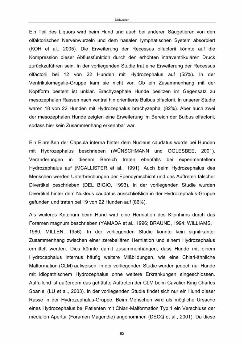
Diskussion
Ein Teil des Liquors wird beim Hund und auch bei anderen Säugetieren von den
olfaktorischen Nervenwurzeln und dem nasalen lymphatischen System absorbiert
(KOH et al., 2005). Die Erweiterung der Recessus olfactorii könnte auf die
Kompression dieser Abflussfunktion durch den erhöhten intraventrikulären Druck
zurückzuführen sein. In der vorliegenden Studie trat eine Erweiterung der Recessus
olfactorii bei 12 von 22 Hunden mit Hydrozephalus auf (55%). In der
Ventrikulomegalie-Gruppe kam sie nicht vor. Ob ein Zusammenhang mit der
Kopfform besteht ist unklar. Brachyzephale Hunde besitzen im Gegensatz zu
mesozephalen Rassen nach ventral hin orientierte Bulbus olfactorii. In unserer Studie
waren 18 von 22 Hunden mit Hydrozephalus brachyzephal (82%). Aber auch zwei
der mesozephalen Hunde zeigten eine Erweiterung im Bereich der Bulbus olfactorii,
sodass hier kein Zusammenhang erkennbar war.
Ein Einreißen der Capsula interna hinter dem Nucleus caudatus wurde bei Hunden
mit Hydrozephalus beschrieben (WÜNSCHMANN und OGLESBEE, 2001).
Veränderungen in diesem Bereich treten ebenfalls bei experimentellem
Hydrozephalus auf (MCALLISTER et al., 1991). Auch beim Hydrozephalus des
Menschen werden Unterbrechungen der Ependymschicht und das Auftreten falscher
Divertikel beschrieben (DEL BIGIO, 1993). In der vorliegenden Studie wurden
Divertikel hinter dem Nukleus caudatus ausschließlich in der Hydrozephalus-Gruppe
gefunden und traten bei 19 von 22 Hunden auf (86%).
Als weiteres Kriterium beim Hund wird eine Herniation des Kleinhirns durch das
Foramen magnum beschrieben (YAMADA et al., 1996; BRAUND, 1994; WILLIAMS,
1980; MILLEN, 1956). In der vorliegenden Studie konnte kein signifikanter
Zusammenhang zwischen einer zerebellären Herniation und einem Hydrozephalus
ermittelt werden. Dies könnte damit zusammenhängen, dass Hunde mit einem
Hydrocephalus internus häufig weitere Mißbildungen, wie eine Chiari-ähnliche
Malformation (CLM) aufweisen. In der vorliegenden Studie wurden jedoch nur Hunde
mit idiopathischem Hydrozephalus ohne weitere Erkrankungen eingeschlossen.
Auffallend ist außerdem das gehäufte Auftreten der CLM beim Cavalier King Charles
Spaniel (LU et al., 2003). In der vorliegenden Studie findet sich nur ein Hund dieser
Rasse in der Hydrozephalus-Gruppe. Beim Menschen wird als mögliche Ursache
eines Hydrozephalus bei Patienten mit Chiari-Malformation Typ 1 ein Verschluss der
medialen Apertur (Foramen Magendie) angenommen (DECQ et al., 2001). Da diese
82
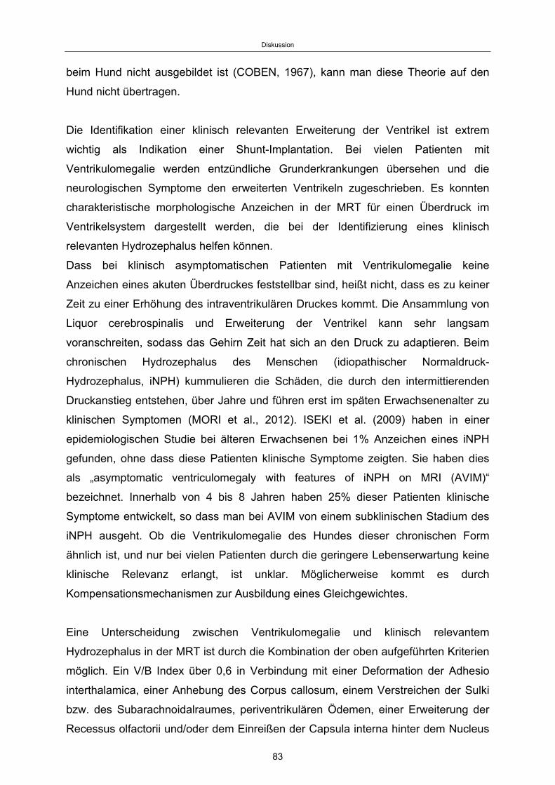
Diskussion
beim Hund nicht ausgebildet ist (COBEN, 1967), kann man diese Theorie auf den
Hund nicht übertragen.
Die Identifikation einer klinisch relevanten Erweiterung der Ventrikel ist extrem
wichtig als Indikation einer Shunt-Implantation. Bei vielen Patienten mit
Ventrikulomegalie werden entzündliche Grunderkrankungen übersehen und die
neurologischen Symptome den erweiterten Ventrikeln zugeschrieben. Es konnten
charakteristische morphologische Anzeichen in der MRT für einen Überdruck im
Ventrikelsystem dargestellt werden, die bei der Identifizierung eines klinisch
relevanten Hydrozephalus helfen können.
Dass bei klinisch asymptomatischen Patienten mit Ventrikulomegalie keine
Anzeichen eines akuten Überdruckes feststellbar sind, heißt nicht, dass es zu keiner
Zeit zu einer Erhöhung des intraventrikulären Druckes kommt. Die Ansammlung von
Liquor cerebrospinalis und Erweiterung der Ventrikel kann sehr langsam
voranschreiten, sodass das Gehirn Zeit hat sich an den Druck zu adaptieren. Beim
chronischen Hydrozephalus des Menschen (idiopathischer Normaldruck-
Hydrozephalus, iNPH) kummulieren die Schäden, die durch den intermittierenden
Druckanstieg entstehen, über Jahre und führen erst im späten Erwachsenenalter zu
klinischen Symptomen (MORI et al., 2012). ISEKI et al. (2009) haben in einer
epidemiologischen Studie bei älteren Erwachsenen bei 1% Anzeichen eines iNPH
gefunden, ohne dass diese Patienten klinische Symptome zeigten. Sie haben dies
als „asymptomatic ventriculomegaly with features of iNPH on MRI (AVIM)“
bezeichnet. Innerhalb von 4 bis 8 Jahren haben 25% dieser Patienten klinische
Symptome entwickelt, so dass man bei AVIM von einem subklinischen Stadium des
iNPH ausgeht. Ob die Ventrikulomegalie des Hundes dieser chronischen Form
ähnlich ist, und nur bei vielen Patienten durch die geringere Lebenserwartung keine
klinische Relevanz erlangt, ist unklar. Möglicherweise kommt es durch
Kompensationsmechanismen zur Ausbildung eines Gleichgewichtes.
Eine Unterscheidung zwischen Ventrikulomegalie und klinisch relevantem
Hydrozephalus in der MRT ist durch die Kombination der oben aufgeführten Kriterien
möglich. Ein V/B Index über 0,6 in Verbindung mit einer Deformation der Adhesio
interthalamica, einer Anhebung des Corpus callosum, einem Verstreichen der Sulki
bzw. des Subarachnoidalraumes, periventrikulären Ödemen, einer Erweiterung der
Recessus olfactorii und/oder dem Einreißen der Capsula interna hinter dem Nucleus
83

Diskussion
caudatus führen zur Diagnose des Hydrocephalus internus beim Hund.
4.3 Ist eine Ventrikulomegalie als physiologischer Zustand
einzuordnen?
Wir konnten zeigen, dass Ventrikulomegalie bei brachyzephalen Hunden mit einem
Verlust an weißer Substanz einhergeht. Das Verhältnis von weißer Substanz zu
grauer Substanz ist bei allen Säugetieren in Abhängigkeit vom Körpergewicht gleich
(ZHANG und SEJNOWSKI, 2000). Da brachyzephale Hunde mit Ventrikulomegalie
weniger weiße Substanz aufweisen, als Hunde mit demselben Körpergewicht ohne
Erweiterung der Ventrikel, ist die Ventrikulomegalie als ein pathologischer Prozess
anzusehen.
Die Ursache für den Verlust von weißer Substanz ist unklar. Beim Hydrozephalus tritt
ein solcher Verlust in Folge der Schädigung durch den erhöhten intraventrikulären
Druck auf. Eine geringgradige oder intermittierende Druckerhöhung könnte eine
Ursache für den Verlust an weißer Substanz bei Hunden mit Ventrikulomegalie sein.
Eine intermittierende Druckerhöhung im Ventrikelsystem wird auch als mögliche
Ursache für den chronischen Hydrozephalus des Erwachsenen (iNPH) vermutet
(SHERMAN, 2005; VERREES und SELMAN, 2004). Es wird angenommen, dass es
durch den intermittierenden Druckanstieg im Ventrikelsystem zu Ischämien in der
ventrikelnahen weißen Substanz kommt und schließlich zur Atrophie, die über die
Jahre hinweg kummuliert und im späten Erwachsenenalter zu Symptomen führt (DEL
BIGIO, 1993; AKAI et al., 1987). Typische Symptome bei iNPH sind Demenz,
Inkortinenz und Gangstörungen (Hakim-Trias) (VERREES und SELMAN, 2004;
MORI et al., 2012). Im Gegensatz dazu beschreiben Besitzer von Hunden mit
Ventrikulomegalie oft keine klinischen Symptome. Zu beachten ist aber, dass beim
Hund die zerebrale Kontrolle der Lokomotion nur eine kleine Rolle spielt und Hunde
mit Vorderhirnsymptomatik häufig nur minimale Gangstörungen zeigen (DE
LAHUNTA und GLASS, 2009). Desweiteren kann die Blasenfunktion beim Hund
durch pontine Miktionszentren ohne zerebralen Einfluss gesteuert werden (DE
LAHUNTA und GLASS, 2009). Demenz ist bei Hunden schwer nachweisbar. Beim
Menschen auffallende Symptome wie Sprachstörungen, schlechtes
Erinnerungsvermögen, oder die mangelnde Fähigkeit Personen wieder zu erkennen
(KIEFER und UNTERBERG, 2012) sind beim Hund nicht zu überprüfen. Somit bleibt
84
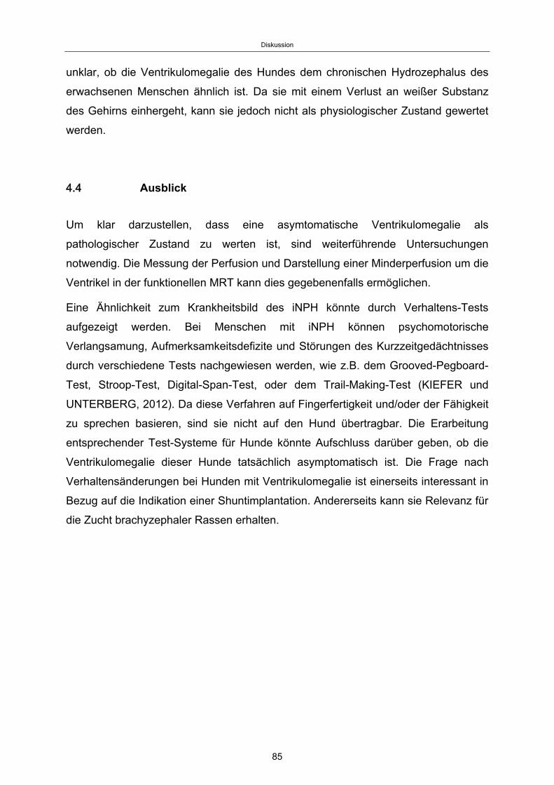
Diskussion
unklar, ob die Ventrikulomegalie des Hundes dem chronischen Hydrozephalus des
erwachsenen Menschen ähnlich ist. Da sie mit einem Verlust an weißer Substanz
des Gehirns einhergeht, kann sie jedoch nicht als physiologischer Zustand gewertet
werden.
4.4 Ausblick
Um klar darzustellen, dass eine asymtomatische Ventrikulomegalie als
pathologischer Zustand zu werten ist, sind weiterführende Untersuchungen
notwendig. Die Messung der Perfusion und Darstellung einer Minderperfusion um die
Ventrikel in der funktionellen MRT kann dies gegebenenfalls ermöglichen.
Eine Ähnlichkeit zum Krankheitsbild des iNPH könnte durch Verhaltens-Tests
aufgezeigt werden. Bei Menschen mit iNPH können psychomotorische
Verlangsamung, Aufmerksamkeitsdefizite und Störungen des Kurzzeitgedächtnisses
durch verschiedene Tests nachgewiesen werden, wie z.B. dem Grooved-Pegboard-
Test, Stroop-Test, Digital-Span-Test, oder dem Trail-Making-Test (KIEFER und
UNTERBERG, 2012). Da diese Verfahren auf Fingerfertigkeit und/oder der Fähigkeit
zu sprechen basieren, sind sie nicht auf den Hund übertragbar. Die Erarbeitung
entsprechender Test-Systeme für Hunde könnte Aufschluss darüber geben, ob die
Ventrikulomegalie dieser Hunde tatsächlich asymptomatisch ist. Die Frage nach
Verhaltensänderungen bei Hunden mit Ventrikulomegalie ist einerseits interessant in
Bezug auf die Indikation einer Shuntimplantation. Andererseits kann sie Relevanz für
die Zucht brachyzephaler Rassen erhalten.
85
Zusammenfassung
Zusammenfassung 5.
Erweiterte Ventrikel kommen häufig bei brachyzephalen Hunden vor, ohne dass die
Tiere klinische Symptome zeigen. In diesem Fall wurde bisher die in der MRT
gefundene Ventrikulomegalie als Normalbefund gewertet. Definierte Kriterien zur
Abgrenzung einer Ventrikulomegalie von einem klinisch relevanten Hydrocephalus
internus in der MRT fehlen bisher beim Hund.
In einer retrospektiven Studie wurden MRT-Befunde von asymptomatischen Hunden
mit erweiterten Ventrikeln mit solchen mit klinisch relevantem Hydrocephalus
internus verglichen, um Anzeichen einer intraventrikulären Druckerhöhung zu
bestimmen.
In einer zweiten Studie wurde das relative Volumen an grauer Substanz, weißer
Substanz und Liquor von mesozephalen Hunden und brachyzephalen Hunden mit
erweiterten Ventrikeln verglichen, um festzustellen, ob das physiologische Verhältnis
von weißer und grauer Substanz, welches bei allen Säugetieren gleich ist, auch bei
brachyzephalen Hunden mit Ventrikulomegalie gilt.
In dieser Arbeit konnte gezeigt werden, dass brachyzephale Hunde mit
Ventrikulomegalie weniger weiße Substanz aufweisen, so dass die Ventrikulomegalie
nicht als physiologischer Zustand angesehen werden kann.
Hoch verdächtig für einen klinisch relevanten Hydrozephalus aufgrund von erhöhtem
intraventrikulärem Druck ist eine Kombination der folgenden Kriterien: ein V/B Index
größer als 0.6, eine Anhebung des Corpus callosum, eine Deformation der Adhesio
interthalamica, periventrikuläre Ödeme, Erweiterung der Recessi olfactorii,
Verstreichen der kortikalen Sulki und/oder des Subarachnoidalraumes und/oder
Läsionen in der Capsula interna hinter dem Nucleus caudatus.
86
Summary
Summary 6.
Enlarged ventricles often occur in brachycephalic dogs without associated clinical
symptoms. So far the ventriculomegaly found on MRI in these cases was considered
a normal variation. There are no defined criteria for distinguishing a ventriculomegaly
from a clinically relevant hydrocephalus internus.
In a retrospective study MRT findings of asymptomatic dogs with enlarged ventricles
were compared to those of dogs with clinically relevant hydrocephalus internus in
order to determine signs of a high intraventricular pressure.
In a second study the relative volume of grey matter, white matter and CSF was
determined in mesaticephalic dogs and brachycephalic dogs with ventriculomegaly in
order to find out whether the physiological relationship between white and grey
matter, which is the same in all eutherian mammals, also exists in brachycephalic
dogs with ventriculomegaly.
Our data shows that brachycephalic dogs with ventriculomegaly have reduced
cerebral white matter, so that the ventriculomegaly cannot be interpreted as a
physiological condition.
Highly suspicious for clinically relevant dilation of the lateral ventricles due to
increased intraventricular pressure is a V/B index greater than 0.6 together with an
elevated corpus callosum and the presence of deformed intermediate mass,
periventricular edema, dilation of the olfactory recess, thinning of the sulci and/or the
subarachnoid space and/or disruption of the internal capsule adjacent to the caudate
nucleus.
87
Literaturverzeichnis
Literaturverzeichnis 7.
Adamiak Z, Jaskolska M, Pomianowski A. Low-field magnetic resonance imaging
of canine hydrocephalus. Pak Vet J. 2012 Jan-Mar; 32(1):128-30.
Akai U, Uchigasaki S, Tanaka U, Komatsu A. Normal pressure hydrocephalus:
neuropathological study. Acta Pathol Jpn. 1987 Jan;37(1):97-110.
Bagley RS, Gavin PR, Holmes SP. Veterinary Clinical Magnetic Resonance
Imaging: Diagnosis of Intracranial Disease. In: Gavin PR, Bagley RS, editors.
Practical Small Animal MRI. Ames: Wiley-Blackwell; 2009. p. 23-123.
Baumgärtner WK, Krakowka S, Koestner A, Evermann J. Ultrastructural
evaluation of acute encephalitis and hydrocephalus in dogs caused by canine
parainfluenza virus. Vet Pathol. 1982 May;19(3):305-14.
Biel M, Kramer M, Forterre F, Jurina K, Lautersack O, Failing K, Schmidt MJ.
Outcome of ventriculoperitoneal shunt implantation for treatment of congenital
internal hydrocephalus in dogs and cats: 36 cases (2001-2009). J Am Vet Med
Assoc. 2013 April;242(7):948-58.
Bradley WG Jr. CSF flow in the brain in the context of normal pressure
Hydrocephalus. AJNR Am J Neuroradiol. 2014 Oct 29.
Braund KG. Clinical Syndromes in Veterinary Neurology. 2nd ed. St. Louis: Mosby;
1994. p. 113-4.
Brown JA, Rachlin J, Rubin JM, Wollmann RI. Ultrasound evaluation of
experimental hydrocephalus in dogs. Surg Neurol. 1984;22:273-6.
Carrera I, Dennis R, Mellor DJ, Pederis J, Sullivan M. Use of magnetic resonance
imaging for morphometric analysis of the caudal cranial fossa in Cavalier King
Charles Spaniels. Am J Vet Res. 2009 Mar;70(3):340-5.
88
Literaturverzeichnis
Coates JR, Axlund TW, Dewey CW, Smith J. Hydrocephalus in dogs and cats.
Comp Cont Educ Pract Vet. 2006 Feb;28(2):136-50.
Coben LA. Absence of a foramen of Magendie in the dog, cat, rabbit and goat. Arch
Neurol. 1967 May;16(5):524-8.
Dandy WE, Blackfan KD. Internal hydrocephalus. An experimental, clinical and
pathological study. Am J Dis Child. 1914;8:406-81.
De Haan CE, Kraft SL, Gavin PR, Wendling LR, Griebenow ML. Normal variation
in size of the lateral ventricles of the Labrador Retriever dog as assessed by
magnetic resonance imaging. Vet Radiol Ultrasound. 1994 Mar;35(2):83-6.
De Lahunta A, Glass EN. Veterinary Neuroanatomy and clinical Neurology. 3rd ed.
St. Louis: Saunders; 2009. p.37-76.
Del Bigio MR. Neuropathology and structural changes in Hydrocephalus. Dev
Disabil Res Rev. 2010;16(1):16-22.
Del Bigio MR. Neuropathological changes caused by hydrocephalus. Acta
Neuropathol. 1993;85(6):573-85.
Deo-Narine V, Gomez DG, Vullo T, Manzo RP, Zimmermann RD, Deck MD, Cahill
PT. Direct in vivo observation of transventricular absorption in the hydrocephalic dog
using magnetic resonance imaging. Invest Radiol. 1994 Mar;29(3):287-93.
Decq P, Le Gurinel C, Brugieres P, Djindjian M, Nguyen JP. Chiari I malformation:
a rare cause of noncommunicating hydrocephalus treated by third vebtriculostomy. J
Neurosurg. 2001 Nov;95(5):783-90.
De Stefani A, de Risio L, Platt SR, Matiasek L, Lujan-Feliu-Pascual A, Garosi
LS. Surgical technique, postoperative complications and outcome in 14 dogs treated
for hydrocephalus by ventriculoperitoneal shunting. Vet Surg. 2011 Feb;40(2):183-
91.
89
Literaturverzeichnis
Dewey CW. External Hydrocephalus in a dog with suspected bacterial
Meningoencephalitis. J Am Anim Hosp Assoc. 2002 Nov-Dez;38(6):563-7.
Dexler H. Über konstitutionelle Hydrocephalie beim Hund. Prag Arch. 1923:103-267.
Di Rocco C. Surgical outcome of neonatal Hydrocephalus. In: Matsumoto S, Tamaki
N, editors. Hydrocephalus. Pathogenesis and treatment. Tokyo, Berlin, Heidelberg:
Springer Verlag; 1991. p. 546-51.
Drake JM, Potts DG, Lemaire C. Magnetic resonance imaging of silastic-induced
canine hydrocephalus. Surg Neurol. 1989 Jan; 31(1):28-40.
Driver CJ, Chandler K, Walmsley G, Shihab N, Volk HA. The association between
Chiari-like malformation, ventriculomegaly and seizures in cavalier King Charles
spaniels. Vet J. 2013 Feb; 195(2):235-7.
Edelman RR, Hesselink JR, Zlatkin MB, Crues JV. Hydrocephalus. In: Clinical
Magnetic Resonance Imaging. 2nd ed. Philadelphia: Saunders Elsevier; 2006: 1646-
55.
El Gammal T, Allen MB Jr, Brooks BS, Mark EK. MR evaluation of hydrocephalus.
AJR Am J Roentgenel. 1987 Oct;149(4):807–13.
Esteve-Ratsch B, Kneissl S, Gabler C. Comparative evaluation of the ventricles in
the Yorkshire terrier and the German Shepherd dog using low-field MRI. Vet Radiol
Ultrasound. 2001 Sep-Oct;42(5):410–3.
Evans HE. The skeleton. Miller’s anatomy of the dog. 3rd ed. Philadelphia: Saunders
Elsevier; 1993. p. 122-218.
Fletscher TF. Spinal cord and meninges. In: Evans HE, editor. Miller’s anatomy of
the dog. 3th ed. Philadelphia: Saunders Elsevier; 1993. p. 800.
90
Literaturverzeichnis
Foltz E, Blanks J, Morton ME. Experimental transcerebral fistula. Perineural
olfactory CSF flow in the normal, hydrocephalic, and postoperative hydrocephalic
dog shown by radionuclide ventriculography. J Neurosurg. 1984 Aug;61(2):355-64.
Foltz E, Aine C. Diagnosis of hydrocephalus by CSF pulse-wave analysis: a clinical
study. Surg Neurol. 1981 Apr;15(4):283-93.
Frahm HD, Stephan H, Stephan M. Comparison of brain structure volumes in
Insectivora and Primates. I Neocortex J Hirnforsch. 1982;23(4):375-89.
Frauchiger E, Frankhauser R. Vergleichende Neuropathologie des Menschen und
der Tiere. Berlin, Göttingen, Heidelberg: Springer-Verlag; 1957. p. 44-7.
Fritsch MJ, Mehdorn HM. Hydrozephalus. In: Fehlbildungen und frühkindliche
Schädigung des ZNS. In: Jansen O, Stephani U, editors. Stuttgart: Thieme; 2007. p.
87-93.
Gilmore HE. Medical Treatment of Hydrocephalus. In: Voncepts in neurosurgery:
Hydrocephalus. Scott M, editor. 3rd ed. Baltimore, Hong Kong, London, Sydney:
Williams and Wilkins; 1990. p. 37-46.
Gonzáles-Soriano J, Marin Garcia P, Contreras-Rodriguez J, Martinez-Sainz P,
Rodriguez-Veiga E. Age-related changes in the ventricular system of the dog brain.
Ann Anat. 2001 May;183(3):283-91.
Greene CS Jr, Lorenzo AV, Hornig G, Welch K. The lowering of cerebral spinal
fluid and brain interstitial pressure of preterm and term rabbits by furosemide. Z
Kinderchir. 1985 Dec;40 Suppl 1:5-8.
Greitz D, Wirestam R, Franck A. Pulsatile brain movement and associated
hydrodynamics studied by magnetic resonance phase imaging. The Monro-Kellie
doctrine revisited. Neuroradiology. 1992;34(5):370-80.
Greitz D. Radiological assessment of hydrocephalus: new theories and implications
for therapy. Neurosurg Rev. 2004 Jul;27(3): 145-65.
91
Literaturverzeichnis
Greitz D. Paradigm shift in hydrocephalus research in legacy of Dandy’s pioneering
work: rationale for third ventriculostomy in communicating hydrocephalus. Childs
Nerv Syst. 2007 May;23(5):487-9.
Haberl H, Michael T, Thomale UW. Pädiatrie up2date. Stuttgart, New York: Georg
Thieme Verlag; 2007. p. 25-44.
Hack M, Cohen AR. Acetazolamide plus furosemide for periventricular dilatation:
lessons for drug therapy in children. Lancet. 1998 Aug;352(9126):418-9.
Harrington ML, Bagley RS, Moore MP. Hydrocephalus. Vet Clin North Am Small
Anim Pract. 1996 Jul;26(4):843-56.
Hecht S. Chapter nineteen: Brain. In: Schwarz T, Saunders J, editors. Veterinary
Computed Tomography. Chichester: Wiley-Blackwell; 2011. p. 185-96.
Henninger W, Hittmair K. CT and Ultrasonographic diagnosis of canine
hydrocephalus. Vet Q. 1994 Apr;16 Suppl 1:59S.
Hofmann E, Becker T, Jackel M, Metzner D, Schneider M, Meixensberger J,
Reichmann H. The corpus callosum in communicating and noncommunicating
hydrocephalus. Neuroradiology. 1995 Apr; 37(3):212-8.
Hudson JA, Simpson ST, Buxton DF, Cartee RE, Steiss JE. Ultrasonographic
diagnosis of canine hydrocephalus. Vet Radiol Ultrasound. 1990 Mar;31(2):50-8.
Iseki C, Kawanami T, Nagasawa H, Wada M, Koyama S, Kikuchi K, Arawaka S,
Kurita K, Daimon M, Mori E, Kato T. Asymptomatic ventriculomegaly with features
of idiopathic normal pressure on MRT (AVIM) in the elderly: a prospective study in a
Japanese population. J Neurol Sci. 2009 Feb;277(1-2):54-7.
92
Literaturverzeichnis
Ishii K, Kanda T, Harada A, Miyamoto N, Kawaguchi T, Shimada K, Ohkawa S,
Uemura T, Yoshikawa T, Mori E. Clinical impact of the callosal angle in the
diagnosis of idiopathic normal pressure hydrocephalus. Eur Radiol. 2008 Nov;
18(11):2678-83.
Jaggy A. Atlas und Lehrbuch der Kleintierneurologie. 2. Aufl. Hannover:
Schlütersche; 2007. p. 403-5.
Javaheri S, Corbett WS, Simbartl LA, Mehta S, Khosla A. Different effects of
omeprazole and Sch 28080 on canine cerebrospinal fluid production. Brain Res.
1997 Apr;754(1-2):321-4.
James AE Jr, Flor WJ, Bush M, Merz T, Rish BL. An experimental model for
chronic communicating hydrocephalus. J Neurosurg. 1974 Jul; 41(1):32-7.
James HE, Shut L. Pitfalls in the diagnosis of arrested hydrocephalus. Acta
Neurochir.1978;43(1-2):13-7.
Kamalo P. Exit ventriculoperitoneal shunt; enter endoscopic third ventriculostomy
(ETV): contemporary views on hydrocephalus and their implications on management.
Malawi Med J. 2013 Sep;25(3):78-82.
Kartal MG, Algin O. Evaluation of hydrocephalus and other cerebrospinal fluid
disorders with MRI: an update. Insights Imaging. 2014 Aug; 5(4):531-41.
Kiefer M, Unterberg A. The differential diagnosis and treatment of normal-pressure
hydrocephalus. Dtsch Arztbl Int. 2012 Jan;109(1-2):15-25.
Kii S, Uzuka Y, Taura Y, Nakaichi M, Takeuchi A, Inokuma H, Onishi T. Magnetic
resonance imaging of the lateral ventricles in beagle-type dogs. Vet Radiol
Ultrasound. 1997 Nov-Dec;38(6):430–3.
Kim J-H, Jeon H-W, Woo E-J, Park H-M. Dilation of the olfactory bulb cavity
concurrent with hydrocephalus in four small breed dogs. J Vet Sci. 2009 Jun;
10(2):173-5.
93
Literaturverzeichnis
Koh L, Zakharov A, Johnston M. Integration of the subarachnoid space and
lymphatics: is it time to embrace a new concept of cerebrospinal fluid absorption?
Cerebrospinal Fluid Res. 2005 Sep;2:6.
Lamb CR, Croson PJ, Capello R, Cherubini GB. Magnetic resonance imaging
findings in 25 dogs with inflammatory cerebrospinal fluid. Vet Radiol Ultrasound.
2005 Jan-Feb; 46(1):17-22.
Laterra J, Goldstein GW. Ventricular Organization of Cerebrospinal Fluid: Blood-
Brain Barrier, Brain Edema, and Hydrocephalus. In: Kandel ER, Schwartz JH, Jessel
T, editors. Principles of Neural Science. 4th ed. New York: Chapman & Hall; 2002. p.
1288-301.
Laubner S, Schmidt JM. Diagnostik und Behandlung des kongenitalen
idiopathischen Hydrocephalus internus beim Hund. Prakt Tierarzt. 2014 Jan;1:40-4.
Leigh EJ, Mackillop E, Robertson ID, Hudson LC. Clinical Anatomy of the canine
brain using magnetic resonance imaging. Vet Radiol Ultrasound. 2008 Mar-
Apr;49(2):113-21.
Lindvall-Axelsson M, Nilsson C, Owman C, Winbladh B. Inhibition of
cerebrospinal fluid formation by omeprazole. Exp Neurol. 1992 Mar;115(3): 394-9.
Lu D, Lamb CR, Pfeiffer DU, Targett MP. Neurological signs and results of
magnetic resonance imaging in 40 cavalier King Charles spaniels with Chiari type 1-
like malformations. Vet Rec. 2003 Aug;153(9):260-3.
Lüps P. Biometrische Untersuchungen an der Schädelbasis des Haushundes. Zool
Anz Jena. 1973 Sep; 192(5/6):383-413.
Matsumae M, Kikinis R, Morocz I, Lorenzo AV, Albert MS, Black PMcL, Jolesz
FA. Intracranial compartment volumes in patients with enlarged ventricles assessed
by magnetic resonance-based image processing. J Neurosurg. 1996 Jun;84(6):972-
81.
94
Literaturverzeichnis
McAllister JP 2nd, Cohen MI, O’Mara KA, Johnson MH. Progression of
experimental infantile hydrocephalus and effects of ventriculoperitoneal shunts: an
analysis correlating magnetic resonance imaging with gross morphology.
Neurosurgery. 1991 Sep;29(3):329-40.
McAllister JP II, Chovan P, Steiner CP, Johnson MJ, Ayzman I, Wood AS, Tkach
JA, Hahn JF, Luciano MG. Differential ventricular expansion in hydrocephalus. Eur
J Pediatr Surg. 1998 Dez;8 Suppl 1:39-42.
Milhorat TH, Hammock MK. Cerebrospinal fluid production by the choroid plexus
and brain. Science. 1971 Jul;173:330-2.
Milhorat TH. Pathology of hydrocephalus. In: Hydrocephalus and the Cerebrospinal
Fluid. Baltimore: Williams & Wilkins; 1971.
Millen JW. Experimental hydrocephalus. Proc R Soc Med. 1956 Dez; 49(12):980-2.
Miller TB, Wilkinson HA, Rosenfeld SA, Furuta T. Intracranial hypertension and
cerebrospinal fluid production in dogs: effects of furosemide. Exp Neurol. 1986
Oct;94(1):66-80.
Mori E, Ishikawa M, Kato T, Kazui H, Miyake H, Miyajima M et al. Guidelines for
management of idiopathic normal pressure hydrocephalus: second edition. Neurol
Med Chir 2012;52(11):775-809.
Mori K, Shimada J, Kurisaka M, Sato K, Watanabe K. Classification of
hydrocephalus and outcome of treatment. Brain Dev. 1995 Sep-Oct;17(5):338-48.
Murata T, Handa H, Mori K, Nakando Y. The significance of periventricular lucency
on computed tomography: experimental study with canine hydrocephalus.
Neuroradiology. 1981 Jan; 20(5):221-7.
Nagashima T, Tamaki N, Matsumoto S, Horwitz B, Seguchi Y. Biomechanics of
hydrocephalus: a new theoretical model. Neurosurgery. 1987 Dec;21(6):898-904.
95
Literaturverzeichnis
Nickel R, Schummer A, Seiferle E, Frewein J, Wille KH. Band 4: Nervensystem,
Sinnesorgane, Endokrine Drüsen. 4. Unveränderte Auflage: Berlin, Hamburg: Paul
Parey Verlag; 2004.
Oboussier H. Behavior of cerebral sulcus formation in crossbreeding of extreme
breed types of dogs. Z Mensch Vererb Konstitutionsl. 1955;33(1):1-9.
Oi S, Inagaki T, Shinoda M, Takahashi S, Ono S, Date I, Nomura S, Miwa T,
Araki T, Ito S, Uchikado H, Takemoto O, Shirane R, Nishimoto H, Tashiro Y,
Matsumura A. Guideline for management and treatment of fetal and congenital
hydrocephalus: Center Of Excellence-Fetal and Congenital Hydrocephalus Top 10
Japan Guideline 2011. Childs Nerv Syst. 2011 Oct;27(10):1563-70.
Oliver JE, Lorenz MD, Kornegay JN. Handbook of veterinary neurology. 3rd ed.
Philadelphia: Saunders; 1997. p. 303-12.
Peña A, Bolton MD, Whitehouse H, Pickard JD. Effects of brain ventricular shape
on periventricular biomechanics: a finite-element analysis. Neurosurgery. 1999
Jul;45(1):107-16.
Platt SR, McConnell JF, Matiasek L. Imaging Diagnosis – Ventriculoperitoneal
shunt associated infection in a dog. Vet Radiol Ultrasound. 2012 Jan-Feb;53(1):80-3.
Platt S, Garosi L. Small Animal Neurological Emergencies. London: Manson
Publishing Ltd / The Veterinary Press; 2012. p. 116-7.
Poca MA, Sahuquillo J. Short-term medical management of hydrocephalus. Expert
Opin Pharmacother. 2005 Aug;6(9):1525-38.
Purves D, Augustine GJ, Fitzpatrick D, Katz LC, LaMantia AS, McNamara JO,
Williams SM. Neuroscience. 2end ed. Saunderland (MA): Sinauer Associates; 2001.
Ransohoff J, Shulman K, Fishman RA. Hydrocephalus: A review of etiology and
treatment. J Pediatr. 1960 Mar;56:399-411.
96
Literaturverzeichnis
Redzic ZB, Segal MB. The structure of the choroid plexus and the physiology of the
choroid plexus epithelium. Adv Drug Deliv Rev. 2004;56: 1695-1716.
Rekate HL. Recent advances in the understanding and treatment of hydrocephalus.
Semin Ped Neurol. 1997; 4:167-78.
Rekate HL, Nadkarni TD, Wallace D. The importance of the cortical subarachnoid
space in understanding hydrocephalus. J Neurosurg Pediatr. 2008 Jul;2(1):1-11.
Rekate HL. A contemporary definition and classification of hydrocephalus. Semin
Pediatr Neurol. 2009 Mar; 16(1):9-15.
Rekate HL. A consensus on the classification of hydrocephalus: its utility in the
assessment of abnormalities of cerebrospinal fluid dynamics.Childs Nerv Syst. 2011
Oct;27:1535-41.
Rivers WJ, Walter PA. Hydrocephalus in the dog: Utility of Ultrasonography as an
Alternate Diagnostic Imaging Technique. J Am Anim Hosp Assoc. 1992 Jul-
Aug;28:333-43.
Roberts T, McGreevy P, Valenzuela M. Human induced rotation and reorganization
of the brain of domestic dogs. PLoS One. 2010 Jul; 5(7):e11946.
Russell DS. Observations on the pathology of hydrocephalus. Br J Surg. 1949 Jul;
37(145):126-38.
Ryan CT, Glass EN, Seiler G, Zwingenberger AL, Mai W. Magnetic resonance
imaging findings associated with lateral cerebral ventriculomegaly in English
bulldogs. Vet Radiol Ultrasound. 2014 May-Jun;55(3):292-9.
Sahar A, Hochwald GM, Kay WJ, Ransohoff J. Spontaneous canine
hydrocephalus: cerebrospinal fluid dynamics. J Neurolog Neurosurg Psychiat. 1971
Jun; 34(3):308-15.
97
Literaturverzeichnis
Saito M, Olby NJ, Spaulding KA, Munana K, Sharp NJH. Relationship among
artery resistance index, degree of ventriculomegaly, and clinical signs in
hydrocephalic dogs. Vet Radiol Ultrasound. 2003 Nov-Dec;44(6):687-94.
Sakka L, Coll G, Chazal J. Anatomy and physiology of cerebrospinal fluid. Eur Ann
Otorhinolaryngol Head Neck Dis. 2011 Dec; 128(6):309-16.
Salomon FV, Geyer H, Gille U. Anatomie für die Tiermedizin. Stuttgart: Enke
Verlag; 2005. p. 523-8.
Sasaki M, Honda S, Yuasa T, Iwamura A, Shibata E, Ohba H. Narrow CSF space
at high convexity and high midline areas in idiopathic normal pressure hydrocephalus
detected by axial and coronal MRI. Neuroradiology. 2008 Feb;50(2):117-22.
Sato O, Asai T, Amano Y, Hara M, Tsugane R, Yagi M. Extraventricular origin of
the cerebrospinal fluid: formation rate quantitatively measured in the spinal
subarachnoid space of dogs. J Neurosurg. 1972 Mar;36(3):276-82.
Sato O, Bering EA Jr. Extraventricular formation of cerebrospinal fluid. Brain Nerv.
1967;19:31-3.
Schmidt MJ, Amort K, Kramer M. Postnatal development oft he cerebral gyrification
in the canine brain. Vet Radiol Ultrasound. 2012 Nov-Dec;53(6):643-9.
Schmidt MJ, Neumann AC, Amort KH, Failing K, Kramer M. Cephalometric
measurements and determination of general skull type of Cavalier King Charles
Spaniels. Vet Radiol Ultrasound. 2011 Jul-Aug;52(4):436-40.
Schröder H, Meyer-Lindenberg A, Nolte I. Comparative examination of the lateral
cerebral ventricles of different dog breeds using quantitative computed tomography.
Berl Munch Tierarztl Wochenschr. 2006 Nov-Dec;119(11-12):506-11.
Scrivani PV, Freer SR, Dewey CW, Cerda-Gonzalez S. Cerebrospinal fluid signal-
void sign in dogs. Vet Radiol Ultrasound. 2009 May-Jun;50(3):269-75.
98
Literaturverzeichnis
Segev Y, Metser U, Beni-Adani L, Elrau C, Raider-Groswasser II, Constantini S.
Morphometric study of the midsagittal MR imaging plane in cases of hydrocephalus
and atrophy and in normal brains. AJNR Am J Neuroradiol. 2001 Oct; 22(9):1674-9.
Seiferle E. Zur Topographie des Gehirns bei lang- und kurzköpfigen Hunderassen.
Acta Anat. 1966;63:346-62.
Selby LA, Hayes HM Jr, Becker SV. Epizootiologic features of canine
hydrocephalus. Am J Vet Res. 1979 Mar;40(3):411-13.
Sherman C. Hydrocephalus in Adults. In: Ellenbogen RG, Abdulrauf SI, Sekhar LN,
editors. Principles of Neurological Surgery. Philadelphia: Elsevier, Saunders;
2005. p. 136-43.
Shihab N, Davies E, Kenny PJ, Loderstedt S, Volk HA. Treatment of
hydrocephalus with ventriculoperitoneal shunting in twelve dogs. Vet Surg. 2011
Jun;40(4):477-84.
Shprecher D, Schwalb J, Kurlan R. Normal Pressure Hydrocephalus: Diagnosis
and Treatment. Curr Neurol Neurosci Rep. 2008 Sep;8(5):371-76.
Smith ZA, Moftakhar P, Malkasian D, Xiong Z, Vinters HV, Lazareff JA. Choroid
plexus hyperplasia: surgical treatment and immunohistochemical results. Case
report. J Neurosurg. 2007 Sep;107(3 Suppl):255-62.
Spaulding KA, Sharp NJH. Ultrasonographic imaging of the lateral cerebral
ventricles in the dog. Vet Radiol. 1990 Mar;31(2):59-64.
Stoffel MH. Funktionelle Neuroanatomie für die Tiermedizin. Stuttgart: Enke Verlag;
2011. p. 106-11.
Su MY, Head E, Brooks WM, Wang Z, Muggenburg BA, Adam GE, Sutherland R,
Cotman CW, Nalcioglu O. Magnetic resonance imaging of anatomic and vascular
characteristics in a canine model of human aging. Neurobiol Aging. 1998 Sep-
Oct;19(5):479-85.
99
Literaturverzeichnis
Summers BA, Cummings JF, De Lahunta A. Malformations of the central nervous
system. In: Veterinary neuropathology. St. Louis: Mosby; 1995. p. 68-94.
Thomas WB. Nonneoplastic disorders of the brain. Clin Tech Small Anim Pract.
1999 Aug;14(3):125-47.
Thomas WB. Hydrocephalus in dogs and cats. Vet Clin North Am Small Anim Pract.
2010 Jan;40(1):143-59.
Verrees M, Selman WR. Management of normal pressure Hydrocephalus. Am Fam
Physician. 2004 Sep;70(6):1071-8.
Virhammar J, Laurell K, Cesarini KG, Larsson E-M. The callosal angle measured
on MRI as a predictor of outcome in idiopathic normal-pressure hydrocephalus. J
Neurosurg. 2014 Jan; 120(1):178-84.
Vite CH, Insko EK, Schotland HM, Panckeri K, Hendricks JC. Quantification of
ventricular volume in English bulldogs. Vet Radiol Ultrasound. 1997 Nov-
Dec;38(6):437-43.
Vullo T, Korenman E, Manzo RP, Gomez DG, Deck MDF, Cahill PT. Diagnosis of
cerebral ventriculomegaly in normal adult beagles using quantitative MRI. Vet Radiol
Ultrasound. 1997 Jul-Aug;38(4):277-81.
Wagshul ME, McAllister JP, Rashid S, Li J, Egnor MR, Walker ML, Yu M, Smith
SD, Zhang G, Chen JJ, Benveniste H. Ventricular dilation and elevated aqueductal
pulsations in a new experimental model of communicating hydrocephalus. Exp
Neurol. 2009 Jul; 218(1):33-40.
Walmsley H. Clinical pathology. In: Platt S, Olby N, editors. BSAVA Manual of
Canine and Feline Neurology. 4th ed. Quedgeley: British Small Animal Veterinary
Association; 2013. p. 50-1.
100
Literaturverzeichnis
Wang PP, Avellino AM. Hydrocephalus in Children. In: Ellenbogen RG, Abdulrauf
SI, Sekhar LN, editors. Principles of Neurological Surgery. Philadelphia: Elsevier,
Saunders; 2005. p. 117-35.
Welch K, Friedmann V. The cerebrospinal fluid valves. Brain. 1960 Sep;83:454-69.
Williams B. Experimental communicating syringomyelia in dogs after cisternal kaolin
injection. Part 2. Pressure Studies. J Neurol Sci. 1980 Oct;48(1):109-122.
Wolpert SM. Radiological investigation of pediatric hydrocephalus. In: Scott RM,
editor. Hydrocephalus. Baltimore, Hong Kong, London, Sydney: Williams & Wilkins;
1990. p. 23-36.
Woo DC, Choi CB, Nam JW, Ryu KN, Jahng GH, Lee SH, Lee DW, Kim SY, Kim
HY, Ahn KJ, Choe BY. Quantitative analysisof hydrocephalic ventricular alterations
in Yorkshire terriers using magnetic resonance imaging. Veterinarni Medicina
2010;55:125-32.
Wünschmann A, Oglesbee M. Periventricular changes associated with
spontaneous canine hydrocephalus. Vet Pathol. 2001 Jan; 38(1):67-73.
Yamada H, Yakota A, Haratake J, Horie A. Morphological study of experimental
syringomyelia with kaolin-induced hydrocephalus in a canine model. J Neurosurg.
1996 Jun; 84(6):999-1005.
Zhang K, Sejnowski TJ. A universal scaling law between gray matter and white
matter of cerebra cortex. Proc Natl Acad Sci USA. 2000 Mai;97(10):5621-6.
Zimmerman RD, Fleming CA, Lee BCP, Saint-Louis LA, Deck MDF.
Periventricular hyperintensity as seen by magnetic resonance: prevalence and
significance. AJR Am J Roentgenol. 1986 Mar;146(3):443-50
101
Danksagung
Danksagung
Ich danke allen Freunden und Kollegen, die mich während der Erstellung meiner
Doktorarbeit unterstützt und ermutigt haben.
Mein erster Dank gilt PD Dr. Martin Schmidt, der mir ein so spannendes
Dissertationsthema gestellt hat. Ich danke ihm ganz besonders für die Betreuung und
Unterstützung, sowie für die zahlreichen Anregungen.
Prof. Dr. Dr. h.c. Martin Kramer möchte ich für die freundliche Aufnahme in der
chirurgischen Abteilung der Kleintierklinik der Justus-Liebig-Universität Gießen
danken, für seine ermutigenden Worte und besonders dafür, dass er als Chef immer
hinter mir stand.
Dem gesamten Team der Neurologie möchte ich für die aufregende Zeit, die Hilfe in
allen Lebenslagen und für ihren Humor danken, der mein Leben jeden Tag ein
bisschen schöner gemacht hat. Besonderer Dank gilt Dr. Malgorzata Kolecka, die mir
immer „den Rücken freigehalten“ und mich unterstützt hat.
Dr. Sylke Gralla danke ich für ihre Hilfe bei der Korrektur und dem Layout, ohne die
diese Arbeit wohl nie ein Ende gefunden hätte, ihre unermüdliche Unterstützung, die
Motivation und das immer da sein, egal zu welcher Zeit.
Den Mitarbeitern der Abteilung für Bildgebende Verfahren, insbesondere Ella Wenz,
danke ich für ihre Geduld, ihre Hilfe bei der Suche nach Datensätzen und die
zahlreichen Tipps.
Dr. Klaus Failing, Marion Sparenberg und Andreas Schaubmar danke ich für ihre
Hilfe bei der statistischen Auswertung und insbesondere für ihre unendliche Geduld
bei der Beseitigung von Unklarheiten.
Meinen Eltern danke ich von ganzem Herzen dafür, dass sie mir alles ermöglicht
haben. Meinem Bruder Bastian gilt ein besonderer Dank für das unermüdliche
Korrekturlesen und die Hilfe bei der Orientierung im Dschungel der englischen
Sprache. Ohne ihn wäre eine internationale Veröffentlichung nicht möglich gewesen.
Ein ganz besonderer Dank gilt meinem Freund Paul, der mich immer motiviert und
unterstützt und in schwierigen Phasen die Welt wieder in Ordnung gebracht hat.
102
Selbstständigkeitserklärung
Selbstständigkeitserklärung
Ich erkläre: „Ich habe die vorgelegte Dissertation selbstständig und ohne unerlaubte
fremde Hilfe und nur mit den Hilfen angefertigt, die ich in der Dissertation angegeben
habe. Alle Textstellen, die wörtlich oder sinngemäß aus veröffentlichten oder nicht
veröffentlichten Schriften entnommen sind, und alle Angaben, die auf mündlichen
Auskünften beruhen, sind als solche kenntlich gemacht. Bei den von mir
durchgeführten und in der Dissertation erwähnten Untersuchungen habe ich die
Grundsätze guter wissenschaftlicher Praxis, wie sie in der „Satzung der Justus-
Liebig-Universität Gießen zur Sicherung guter wissenschaftlicher Praxis“
niedergelegt sind, eingehalten.“
Steffi Laubner
103

VVBVVB LAUFERSWEILER VERLAG
édition scientifique
VVB LAUFERSWEILER VERLAGSTAUFENBERGRING 15D-35396 GIESSEN
Tel: 0641-5599888 Fax: [email protected]
VVB LAUFERSWEILER VERLAGédition scientifique
9 7 8 3 8 3 5 9 6 4 1 7 4
ISBN: 978-3-8359-6417-4
Photo cover: ©
STEFFI LA
UB
NER
M
RT-B
EFU
ND
E B
EI H
UN
DEN
M
IT H
YD
RO
ZEP
HA
LU
S
STEFFI LAUBNER
Zusammenhang zwischen MRT-Befunden
und klinischen Symptomen bei Hunden mit
Hydrozephalus im Vergleich zu Hunden
mit Ventrikulomegalie
Inaugural-Dissertation zur Erlangung des Grades eines
Dr. med. vet.
beim Fachbereich Veterinärmedizin der Justus-Liebig-Universität Gießen
VVBVERLAG


![2.6 Verlustfaktor . Verlustfaktor - Ingenieur-Buch.de€¦ · 2.6 Verlustfaktor ˘ 10-5 10-4 10-3 10-2 10-1 1 10 Verlustfaktor η [-] Kupfer Zink SiO2 Titan Al2O3 SiC Ton Aluminium](https://img.pdfslide.org/doc/110x75/605e4d284e7f5c01b07c4263/26-verlustfaktor-verlustfaktor-ingenieur-buchde-26-verlustfaktor-10-5.jpg)