Embed Size (px)
Citation preview
Technische Universität München
Lehrstuhl für biomolekulare NMR‐Spektroskopie
Department Chemie
Structural studies on cooperative 3’ splice site
recognition by the SF1‐U2AF65 complex
Yun Zhang
Vollständiger Abdruck der von der Fakultät für Chemie der Technischen Universität
München zur Erlangung des akademischen Grades eines
Doktors der Naturwissenschaften
genehmigten Dissertation.
Vorsitzender: …..………………………………………
Prüfer der Dissertation:
1. ….……………………………………
2. ……….………………………………
Die Dissertation wurde am ……….……….. bei der Technischen Universität München
eingereicht und durch die Fakultät für Chemie am ………….…….. angenommen.
Univ.‐Prof. Dr. St. A. Sieber
Univ.‐Prof. Dr. M. Sattler
Univ.‐Prof. Dr. A. Itzen
19.03.2013
22.04.2013
Immer nur lernen, ohne dabei nachzudenken, das
führt zu Verwirrung. Immer nur nachdenken, ohne
dabei zu lernen, das führt zu Erschöpfung.
Konfuzius

DECLARATION
I hereby declare that parts of this Thesis are already published in
scientific journal:
Zhang, Y., Madl, T. (shared first author), Bagdiul, I., Kern, T., Kang, H.S., Zou, P., Mausbacher, N., Sieber, S.A., Kramer, A., and Sattler, M. (2013). Structure, phosphorylation and U2AF65 binding of the N‐terminal domain of splicing factor 1 during 3'‐splice site recognition. Nucleic Acids Res 41, 1343‐1354.
ABSTRACT
I
I. ABSTRACT
Recognition of the 3’ splice site is a key step in pre‐mRNA splicing and accomplished by a
dynamic complex comprising splicing factor 1 (SF1) and the U2 snRNP auxiliary factor 65‐kDa
subunit (U2AF65). Both proteins mediate protein‐protein and protein‐RNA interactions for
cooperative RNA‐binding during spliceosome assembly. This work presents structural
investigations of cooperative recognition of 3’ splice site by SF1‐U2AF65 complex. The main
focus is on the N‐terminal domain of SF1, which is posttranslationally modified by
phosphorylation. Additionally, the contribution of the U2AF65 RS (arginine‐serine‐rich)
domain to binding of the intronic branch point sequence (BPS) by SF1 was investigated.
Structural analysis by NMR methods combined with SAXS analysis provided novel insight
about mechanisms of cooperative recognition of 3’ splice site.
Chapter 1.1 introduces the biological background of pre‐mRNA alternative splicing and
splicing factors involving in recognition of 3’ splice site. Chapter 1.2 reviews aspects of NMR
methods used for structural analysis and characterization of biomolecules in solution. In
Chapter 2, materials and methodology employed for biochemical experiments and
structural analysis are documented.
Chapter 3.1 presents NMR and SAXS data that characterize the structural basis for
cooperative RNA binding by SF1‐U2AF65. The solution structure of a novel helix hairpin
domain in the N‐terminal region of SF1 (SF1NTD) is described. The structure of a complex of
the N‐terminal domain of SF1 (SF1NTD) with the C‐terminal U2AF homology motif domain of
U2AF65 (U2AF65UHM) reveals that, in addition to the known U2AF65UHM‐SF1 interaction, the
helix‐hairpin domain forms a secondary, hydrophobic interface with U2AF65UHM, which locks
the orientation of the two subunits. Mutational analysis shows that the helix hairpin is
essential for cooperative formation of the ternary SF1‐U2AF65‐RNA complex. Chapter 3.2
describes NMR and biochemical investigation of SF1 phosphorylation. Structural and
dynamic data reveal that tandem serine phosphorylation of a conserved SPSP motif rigidifies
a long unstructured linker in the SF1 helix hairpin. Phosphorylation does not significantly
alter the overall conformation of SF1, SF1‐U2AF65 or the SF1‐U2AF65‐RNA complexes, but
ABSTRACT
II
nevertheless enhances RNA binding by SF1‐U2AF65. Chapter 3.3 addresses the studies on
U2AF65 RS domain. NMR chemical shifts and paramagnetic relaxation enhancement data
indicate that the U2AF65 RS domain exhibits secondary structural elements and tertiary
contacts in solution. Chapter 4 reviews the results of this thesis and discusses the role of
phosphorylation and U2AF65‐binding of SF1 during 3’ splice site recognition.
The work presented in this thesis provides a structural investigation of the cooperative
recognition of the 3’ splice site by SF1‐U2AF65, and reports on the effects of SF1
phosphorylation. The results indicate that the helix hairpin domain of SF1 is required for
cooperative 3’ splice site recognition presumably by stabilizing a unique quaternary
arrangement of the SF1‐U2AF65‐RNA complex. NMR analysis of the U2AF65 RS domain
shows that the RS domain comprises two α‐helical regions, which could be interaction
interfaces with SF1 KH‐QUA2. Altogether, this work provides a significant step towards
solving the overall structure of the ternary SF1‐U2AF65‐RNA complex and thus
understanding the molecular basis of 3’ splice site recognition and regulation.
II. TA
I. ABS
II. TAB
1 INT
1.1 Bi1.1.11.1.21.11.1
1.1.31.11.11.1
1.1.41.1.51.1.6
1.2 Nu1.2.11.2.21.2.31.2.41.2.51.2.6
1.3 Sc
2 MA
2.1 Ma2.1.12.1.22.1.32.12.1
2.2 Bi2.2.12.2.22.2.32.2.42.2.52.2.6
ABLE OF C
STRACT...
BLEOFCO
TRODUCTI
ologicalbacAlternativePre‐mRNA
.2.1 Thesp
.2.2 SpliceoStructural
.3.1 Compl
.3.2 5’splic
.3.3 3’splicSplicingfacSF1phosphTheU2AF6
uclearmagnTheprincipNMRforprProteindynParamagneNMRstudieStructurec
copeoftheT
ATERIALSA
aterials........ChemicalsBacterialstRecipes......
.3.1 Media
.3.2 Buffer
ochemicalmCloning......ProteinexpInvitrophoIsothermalCircularDiSpinlabelin
CONTEN
....................
ONTENTS...
ION.............
ckgroundintepre‐mRNAssplicingbytpliceosome.....osomeassemstudiesofColexE..................cingsite..........cesite..............ctor1(SF1)..horylation.....5 arginine‐Ser
neticresonanplesofNMR..roteinanalysnamicsbyNMeticrelaxationesforproteincalculation.....
Thesis.............
ANDMETH
.........................andenzymestrainsandve...........................andbuffersfsforprotein
methods...................................pressionandosphorylatioltitrationcalochroism.........ng......................
NTS
....................
....................
....................
troduction...splicing...........hespliceosom...........................mbly...................mplexE................................................................................................................................................rine‐rich (RS)
nce(NMR)sp...........................is.......................MR....................nenhancemen‐ligandinter...........................
........................
HODS..........
........................s.........................ectors..........................................forproteinexpurification.
........................
...........................NMRsamplen.......................orimetry..............................................................
III
....................
....................
....................
........................
..........................me....................................................................................................................................................................................................................................domain..........
pectroscopy..............................................................................ent(PRE).......raction......................................
........................
....................
........................
..........................
..........................
..........................xpression................................
........................
..........................epreparation........................................................................................................
....................
....................
....................
.................................................................................................................................................................................................................................................................................................................................
y.........................................................................................................................................................................................
........................
....................
...............................................................................................................................................................
...................................................n......................................................................................................................................
TABLE
....................
....................
....................
.................................................................................................................................................................................................................................................................................................................................
..........................................................................................................................................................................................
........................
....................
...............................................................................................................................................................
..........................................................................................................................................................................................
EOFCONTE
....................
....................
....................
.......................................................................................................................................................................................................................................................................................................................
....................................................................................................................................................................................
.........................
....................
...........................................................................................................................................................
.....................................................................................................................................................................................
ENTS
.............I
..........III
............1
...............1
.................1
.................4
.................4
.................5
.................7
.................7
.................8
..............10
..............12
..............13
..............15
.............17
..............17
..............21
..............23
..............24
..............26
..............27
.............29
..........31
.............31
..............31
..............33
..............34
..............34
..............36
.............39
..............39
..............41
..............42
..............42
..............42
..............43
2.2.7
2.3 NM2.3.12.3.22.3.32.3.42.3.5
3 RES
3.1 Str3.1.13.1.23.1.33.1.43.1.53.1.6
3.2 Str3.2.13.2.23.2.33.2.43.2.53.2.63.2.7SF1‐U
3.3 NM3.3.13.3.23.3.33.3.4
4 DIS
4.1 Amrecognit
4.2 Co
5 LIT
6 LIS
III. APP
III.I DN
Electropho
MRandSAXSNMRmeasSecondaryRelaxationStructurecSmall‐angle
SULTS.......
ructuralbasSF1construNMRanalySF1NTDproInteractionCooperativDiscussion
ructuralanaPhosphoryPhosphoryNMRanalySpinlabelinEffectsofpRoleofphoDiscussion
U2AF65‐RNA
MRcharactePreparatioNMRassignU2AF65RSDiscussion
SCUSSION.
modelforthtion................
onclusions...
TERATURE
TOFFIGU
PENDIX....
NAsequence
oreticmobilit
Stechniquesurementsanchemicalshianalysis.........calculations...eX‐rayscatte
....................
sisofSF1‐U2ucts..................ysisandstrucvidesaseconnsbetweenSFverecognition:Cooperative
alysisofSF1ylationmimicylationofSF1ysisofSF1NTng......................phosphorylatiosphorylation:Theinfluencomplex.........
erizationoftnofU2AF65nmentandNSandSF1‐BP:RSdomainc
....................
heroleofph.........................
.........................
EREFEREN
URES............
....................
es.....................
tyshiftassays
s......................dassignmentftanalysisan......................................................ering(SAXS)
....................
2AF65coope...........................ctureoftheNndaryinterfacF1andRNA..nofRNAbySebindingofS
phosphorylcsofSF1(S80onS80andSDandSF1ph...........................iononSF1stnofSF1forcceofphosph...........................
theU2AF65aRSdomain...MRanalysisSinteractioncontactsSF1
....................
osphorylatio........................
........................
NCES............
....................
....................
........................
IV
s(EMSAs).....
........................ts......................ndchemicals..............................................................................
....................
erativeRNAb..........................‐terminalregceintheSF1N
..........................SF1‐U2AF65cSF1‐U2AF65t
lation.............0E,S82E).......S82...................hosphorylatio..........................tructure..........ooperativeRorylationofS..........................
arginine‐ser..........................ofU2AF65RSn.........................andRNAatt
....................
onandU2AF........................
........................
....................
....................
....................
........................
...........................
...................................................shiftperturba.................................................................................
....................
binding...................................gionofSF1....NTD‐U2AF65U
...........................complex.........toPre‐mRNA
..............................................................................on............................................................................RNAbinding...SF1oncoope...........................
rine‐rich(RS...........................Sdomain..................................thebranchpo
....................
F65‐binding........................
........................
....................
....................
....................
........................
TABLE
...........................
...................................................ations................................................................................................
....................
..............................................................................UHMcomplex........................................................A.........................
..........................................................................................................................................................................................erativeassem...........................
S)domains...................................................................................oint...................
....................
gofSF1durin........................
........................
....................
....................
....................
........................
EOFCONTE
..........................
..........................................................................................................................................................
....................
.....................................................................................................................................................................................
....................................................................................................................................................................................
mblyofthe..........................
.................................................................................................................................
....................
ng3’splices.........................
.........................
....................
....................
....................
.........................
ENTS
..............43
.............45
..............45
..............45
..............46
..............47
..............47
..........49
.............49
..............49
..............52
..............58
..............65
..............67
..............69
.............71
..............71
..............74
..............77
..............79
..............81
..............84
..............86
.............88
..............88
..............90
..............93
..............96
..........98
site.............98
..........101
.......102
.......112
..........VI
.............VI
III.II Pr
III.III Re
III.IVAb
ACKNOW
CURRICU
roteinseque
esonanceass
bbreviations
WLADGEMENT
ULUM VITAE...
ences..............
signmentso
s.......................
TS.....................
.........................
........................
fSF1NTD........
........................
........................
........................
V
........................
........................
........................
........................
........................
........................
........................
........................
........................
........................
TABLE
........................
........................
........................
........................
........................
EOFCONTE
.........................
........................
.........................
.........................
.........................
ENTS
............VII
..........VIII
...........XXI
........XXIII
........XXIV
1 IN
B1.1
1.1.1
In Febr
(Lander
genes, m
by cont
indicate
major f
essentia
comple
2010).
Alterna
coding
distinct
human
2002; P
alternat
intronic
respect
alternat
can act
splice s
compet
cognate
TRODUC
Biological b
Alternative
uary 2001,
r et al., 200
much less t
trast, the ge
e the lack o
factor for
al roles in
xity of high
tive splicin
sequences
t transcripts
multi‐exon
Pan et al., 20
tive splicing
c splicing e
tively). The
tive exons,
tivate adjac
ites or enh
ting influen
e RNA‐bindi
CTION
background
e pre‐mRNA
the first dr
01; Venter
han the ori
enome of th
of an assoc
this pheno
expanding
her eukary
g, the rem
(exons), is a
s are gener
n genes are
008; Wang
g is regulat
enhancers
ese auxiliary
deciding w
cent splice
ancers. Exo
ces, which
ing activato
d introduct
A splicing
raft of the h
et al., 2001
ginally pred
he nematod
iation betw
omenon is
the functio
otes (Grave
moval of no
an crucial st
rated from
e estimated
et al., 2008
ted by a n
(ESEs and
y elements
which exon
sites or an
on inclusion
in turn mig
or and repre
1
tion
human geno
1), it surpris
dicted numb
de C. elega
ween gene
proposed
onal compl
eley, 2001;
n‐encoding
tep in euka
a single ge
d to underg
8). In additio
umber of a
ISEs, resp
s are invo
is removed
ntagonize s
n or skippin
ght be dete
essor protei
ome seque
singly conta
bers of gen
ns contains
number an
to be alte
lexity, prot
Kim et al.
g sequences
ryotic gene
ne (Gravele
go alternati
on to the sp
auxiliary ele
pectively) a
lved in de
d and which
silencers, w
ng is determ
rmined by
ins (Matlin e
I
nce was su
ains only a
es (Roest C
s about 20,0
d organism
ernative spl
ein diversit
., 2007; Ni
s (introns)
e expression
ey, 2001). U
ive splicing
plice‐site co
ements kno
nd silence
efining both
h exon is in
whereas sile
mined by th
relative con
et al., 2005
INTRODUCT
uccessfully r
pproximate
rollius et al
000. These
mal complex
licing, whic
ty, and org
lsen and G
and the jo
n by which m
Up to 92%~
(Modrek a
nsensus seq
own as exo
rs (ESSs an
h constitut
ncluded. En
encers can
he balance o
ncentration
) (Figure 1.1
TION
revealed
e 23,000
., 2000),
findings
xity. The
ch plays
ganismal
Graveley,
oining of
multiple
~94% of
and Lee,
quences,
onic and
nd ISSs,
tive and
nhancers
repress
of these
ns of the
1A).
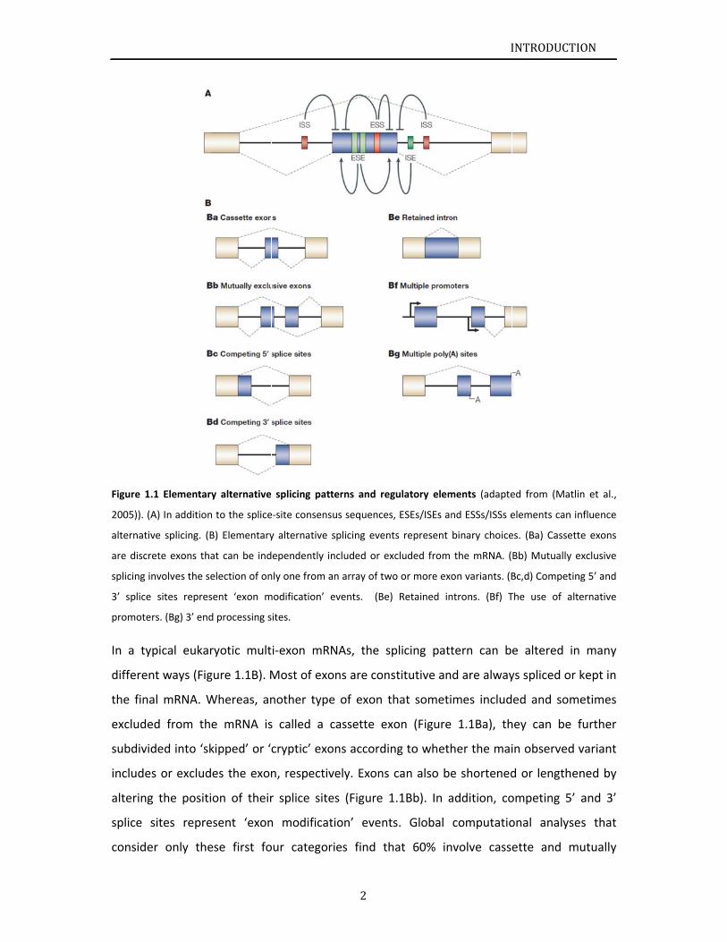
Figure 1.
2005)). (A
alternativ
are discr
splicing i
3’ splice
promote
In a ty
differen
the fina
exclude
subdivid
include
altering
splice s
conside
.1 Elementary
A) In addition
ve splicing. (B
rete exons tha
nvolves the se
sites repres
rs. (Bg) 3’ end
pical eukar
nt ways (Fig
al mRNA. W
ed from th
ded into ‘sk
s or exclud
g the positi
sites repre
er only the
y alternative
n to the splice
B) Elementary
at can be ind
election of on
sent ‘exon m
d processing s
ryotic mult
gure 1.1B). M
Whereas, an
e mRNA is
kipped’ or ‘
es the exon
on of their
esent ‘exon
ese first fo
splicing patt
e‐site consens
y alternative
ependently in
nly one from a
modification’ e
ites.
i‐exon mRN
Most of exo
nother type
s called a c
cryptic’ exo
n, respectiv
r splice site
n modificat
our categor
2
terns and reg
us sequences
splicing event
ncluded or ex
an array of tw
events. (Be)
NAs, the sp
ons are cons
e of exon t
cassette ex
ons accordin
vely. Exons
es (Figure 1
tion’ event
ries find th
gulatory elem
s, ESEs/ISEs an
ts represent
xcluded from
wo or more exo
) Retained in
plicing patt
stitutive an
hat someti
xon (Figure
ng to wheth
can also be
1.1Bb). In a
ts. Global
hat 60% in
I
ments (adapte
nd ESSs/ISSs e
binary choice
the mRNA. (
on variants. (B
ntrons. (Bf) T
tern can b
d are alway
mes includ
1.1Ba), th
her the mai
e shortened
addition, co
computatio
volve cass
INTRODUCT
ed from (Mat
elements can
es. (Ba) Casse
Bb) Mutually
Bc,d) Compet
The use of a
e altered i
ys spliced or
ed and som
hey can be
in observed
d or length
ompeting 5’
onal analys
ette and m
TION
tlin et al.,
influence
ette exons
exclusive
ing 5’ and
lternative
n many
r kept in
metimes
further
d variant
ened by
’ and 3’
ses that
mutually
INTRODUCTION
3
exclusive exons, whereas 40% are exon modifications (Clark and Thanaraj, 2002) (Figure
1.1Bc,d). The remaining categories include retained introns (Figure 1.1Be). Alternative
splicing in conjunction with the use of alternative promoters (Figure 1.1Bf) or 3’ end
processing sites (Figure 1.1Bg). Regulation of the last two categories need not be at the level
of splicing.
Canonical mechanism of alternative splicing suggest that serine/arginine (SR) proteins
typically bind to ESEs and heterogeneous nuclear ribonucleoproteins (hnRNPs) recognize
ESSs or ISSs (Chen and Manley, 2009). SR proteins have a modular structure with one or two
N‐terminal RNA Recognition motif (RRM)‐type domains that bind RNA, and C‐terminal
domains that are enriched in arginine and serine residues (RS domains). RS domains are also
found in other core splicing factors such as U2AF65 (U2 auxiliary factor 65 kDa) and U2AF35.
They mediate both protein‐protein and protein‐RNA contacts (Shen et al., 2004). Detailed
descriptions of U2AF65 RS domain can be found in section 1.1.8.
Pre‐mRNA splicing is carried out by the spliceosome, a 60S protein‐RNA assembly that
consists of five uridine‐rich small nuclear ribonucleoprotein particles (snRNPs), U1, U2, U4,
U5 and U6 and many non‐snRNP proteins (Black, 2003; Wahl et al., 2009). Initial recognition
of the correct pairs of 5' and 3' splice sites by the spliceosome is a critical step in the
processing of both constitutively and alternatively spliced pre‐mRNAs. In the early complex
of spliceosome, the 3’ splice site is recognized exclusively by proteins for constitutive
splicing. Furthermore, choices of alternative splicing also have long been thought to be
achieved at the stages of splice site recognition and early spliceosome assembly. This raises
the importance of understanding the mechanism of splice site recognition during
spliceosome assembly, which will be discussed in the following section (s. 1.1.2).
1.1.2
1.1.2.1
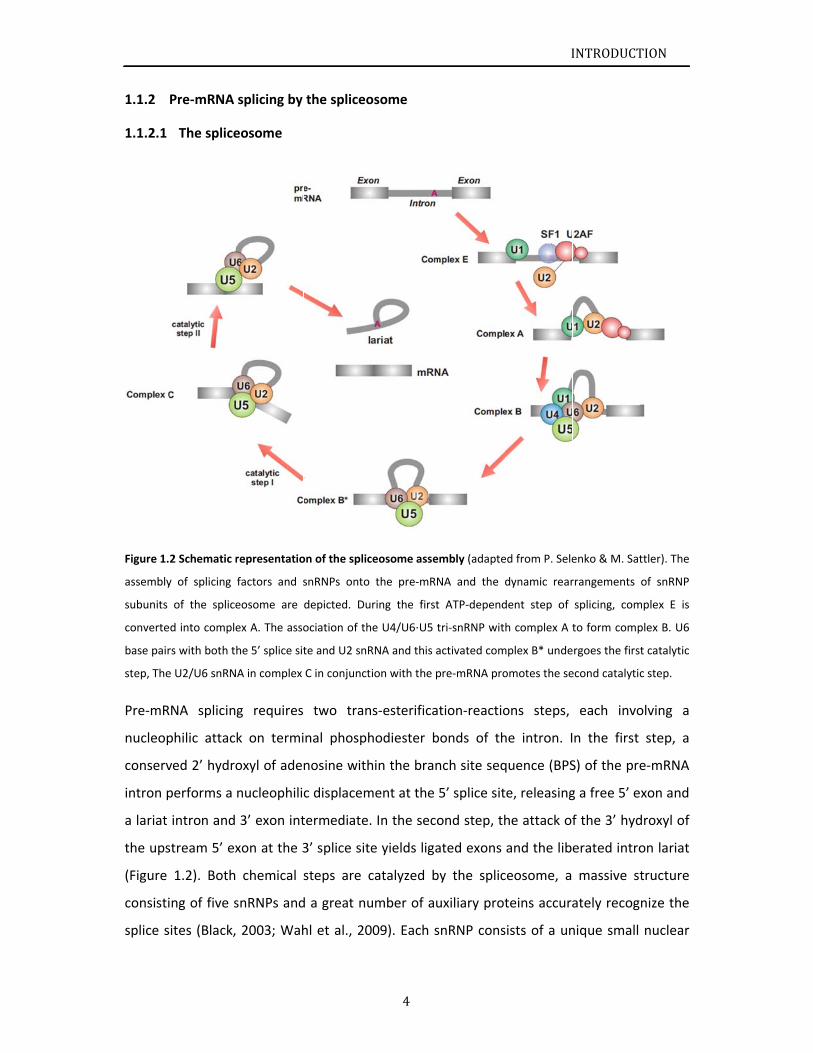
Figure 1.
assembly
subunits
converte
base pair
step, The
Pre‐mR
nucleop
conserv
intron p
a lariat
the ups
(Figure
consisti
splice s
Pre‐mRNA
The splice
2 Schematic r
y of splicing
of the splice
ed into comple
rs with both th
e U2/U6 snRN
RNA splicin
philic attac
ved 2’ hydro
performs a
intron and
stream 5’ ex
1.2). Both
ing of five s
sites (Black,
splicing by
eosome
representatio
factors and s
eosome are
ex A. The asso
he 5’ splice sit
A in complex
g requires
k on termi
oxyl of ade
nucleophili
3’ exon int
xon at the 3
chemical
snRNPs and
2003; Wah
the spliceo
on of the splic
snRNPs onto
depicted. Du
ociation of th
te and U2 snR
C in conjunct
two tran
nal phosph
nosine with
c displacem
termediate.
3’ splice site
steps are c
d a great nu
hl et al., 20
4
osome
ceosome asse
the pre‐mRN
ring the first
he U4/U6⋅U5 t
RNA and this a
ion with the p
ns‐esterifica
hodiester b
hin the bran
ment at the
. In the seco
e yields liga
catalyzed b
umber of au
009). Each s
mbly (adapte
NA and the d
t ATP‐depend
tri‐snRNP wit
activated com
pre‐mRNA pro
tion‐reactio
bonds of th
nch site seq
5’ splice sit
ond step, th
ated exons
by the splic
uxiliary prot
snRNP cons
I
ed from P. Sele
dynamic rear
dent step of
h complex A t
mplex B* unde
omotes the se
ons steps,
he intron. I
quence (BPS
e, releasing
he attack of
and the lib
ceosome, a
teins accura
ists of a un
INTRODUCT
enko & M. Sat
rrangements
splicing, com
to form comp
rgoes the firs
econd catalytic
each invo
n the first
S) of the pre
g a free 5’ e
f the 3’ hyd
erated intro
massive st
ately recog
nique small
TION
ttler). The
of snRNP
mplex E is
plex B. U6
t catalytic
c step.
olving a
step, a
e‐mRNA
xon and
droxyl of
on lariat
tructure
nize the
nuclear
INTRODUCTION
5
RNA (snRNA) as well as specific proteins. There are several stages of spliceosome formation,
in which conserved intron sequences direct the assembly of snRNPs onto the pre‐mRNA.
Both of the major (or U2‐type) spliceosome and minor (or U12‐type) spliceosome present
many similar conserved features, which mediates splicing of a class of rare metazoan introns
(Dietrich et al., 1997; Sharp and Burge, 1997; Tarn and Steitz, 1997; Will et al., 1999).
1.1.2.2 Spliceosome assembly
As a factory for pre‐mRNA alternative splicing, spliceosome assembly begins with the base
pairing of U1 snRNA to the conserved 5’ splice site and the recognition of non‐snRNP
proteins to the 3’ splice site. Splicing Factor 1 or Branchpoint Binding Protein (SF1/BBP)
binds to the branch point sequence (Berglund et al., 1997) in an ATP‐independent manner
to form the E’ complex (or commitment complex in S. cerevisiae), The E’ complex can be
converted into the E complex by the recruitment of U2 auxiliary factor (U2AF) heterodimer
(comprising U2AF65 and U2AF35) to the polypyrimidine tract and 3’ terminal AG158.
The ATP‐independent E complex is converted into the ATP‐dependent pre‐spliceosome
complex A. In complex A, the U2 snRNP, which is only loosely associated with the pre‐mRNA
in complex E (Das et al., 2000; Hong et al., 1997), now becomes tightly bound, inducing the
replacement of SF1 by U2 snRNP at the branch point sequence. Several of the U2
snRNP‐associated SF3a/b subunits contact the pre‐mRNA (Gozani et al., 1998; McPheeters
and Muhlenkamp, 2003). Additionally, a cross‐intron interaction bridging the U1 and U2
snRNPs is mediated by the RNA‐dependent DExD/H helicase Prp5 in yeast and mammals (Xu
et al., 2004).
Further recruitment of the U4/U6–U5 tri‐snRNP leads to the formation of the B complex,
which contains all spliceosomal subunits that carry out pre‐mRNA splicing. Subsequently,
base pairing interactions involving U1 snRNA/5’ splice and U4/U6 snRNA are disrupted, and
the U1 and U4 snRNPs leave. U6 base pairs with both the 5’ splice site and U2 snRNA and
this activated spliceosome (complex B*) undergoes the first trans‐esterification step (Figure
1.2) (Makarov et al., 2002). This is followed by extensive conformational changes and
remodeling, including the loss of U1 and U4 snRNPs, ultimately resulting in the formation of
the C complex, the mature spliceosome (Reed, 2000; Staley and Guthrie, 1998). The U2/U6
snRNA in complex C in conjunction with the pre‐mRNA is thought to form the active site of
INTRODUCTION
6
the spliceosome and promotes the second catalytic step (Collins and Guthrie, 2000;
Makarov et al., 2002; Valadkhan and Manley, 2001).
A great numbers of studies have investigated the structural information of some
spliceosomal factors, Table 1.1 presents the studies of SF1, U2AF65 and their binding
partners.
Table 1.1 Structural studies of SF1, U2AF65 and their binding partners.
Factors Organism Reference
SF3b155 human (Bessonov et al., 2010;
Cass and Berglund, 2006)
SF3b155 human (Spadaccini et al., 2006)
SF1 KH‐QUA2 human (Liu et al., 2001)
U2AF65 UHM human (Selenko et al., 2003)
U2AF35 human (Kielkopf et al., 2001)
SPF45 human (Corsini et al., 2007)
Prp40p WW‐WW yeast (Wiesner et al., 2002)
FBP21 WW‐WW human (Klippel et al., 2011)
SF3b human (Golas et al., 2003)
U2AF65 RRM12 human (Mackereth et al., 2011)
SR45 human (Day et al., 2012)
1.1.3
1.1.3.1
Figure 1.
5' splice s
red) inte
Within in
in the fir
splice site
The for
in the
stage o
with U1
splice s
point s
(U2AF6
respect
end of
nucleot
located
Structural s
Complex
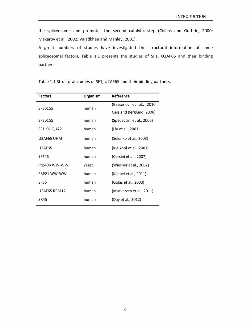
.3 Arrangeme
site is specific
racts with the
n the BPS the
rst trans‐este
es are shown
mation of C
regulation
f splicing si
1 snRNA by
ite. Splicing
sequence (B
5 and U2A
tively. In ma
the intron
tides upstre
upstream t
studies of C
E
ent of protein
cally recognize
e Py tract and
BPA (magenta
erification rea
in grey.
Complex E,
of splicing.
tes definitio
y base pair
g Factor 1 o
BPS), and
AF35) bind
ammals, the
was define
eam and ex
to Py tract e
Complex E
ns and pre‐mR
ed by the U1
d the 3’ AG, re
a) acts as the
action. Protein
as the initia
. Choices o
on in the Co
ring. Non‐s
or Branchpo
the two su
d to the p
ese sequenc
ed by the A
xtending fo
extending t
7
RNA in compl
snRNP (green
espectively. SF
nucleophile a
n‐protein inte
al stage in t
of alternativ
omplex E. In
nRNP prote
oint Binding
ubunits of
polypyrimid
ce elements
AG dinucleo
or ten or m
o 30 nucleo
ex E (adapted
n), while the U
F1/BBP (blue)
attacking the p
eractions brid
the spliceos
ve splicing
n this comp
eins simulta
Protein (SF
the hetero
dine (Py) t
s are arrang
otide (3’ AG
more nucleo
otides.
I
d from P. Sele
U2AF heterodi
) binds to the
phosphate gro
dging compon
some assem
are initially
plex, the 5’
aneously in
F1/BBP) rec
odimeric U
ract and t
ged in a typ
G), the Py tr
otides, and
INTRODUCT
enko & M. Sat
imer (U2AF65
BPS further u
oup at the 5’
nents at the
mbly, is a ea
y controlled
splice site i
nteract with
cognizes the
U2 Auxiliary
the 3’ spli
pical way, th
ract located
the BPS g
TION
ttler). The
5/U2AF35,
upstream.
splice site
5’ and 3’
arly step
d at the
s bound
h the 3’
e branch
y Factor
ice site,
hat the 3’
d at five
generally
The se
comple
protein
Smith a
1000 n
contact
Maniat
Prp40/F
pre‐mR
U2AF a
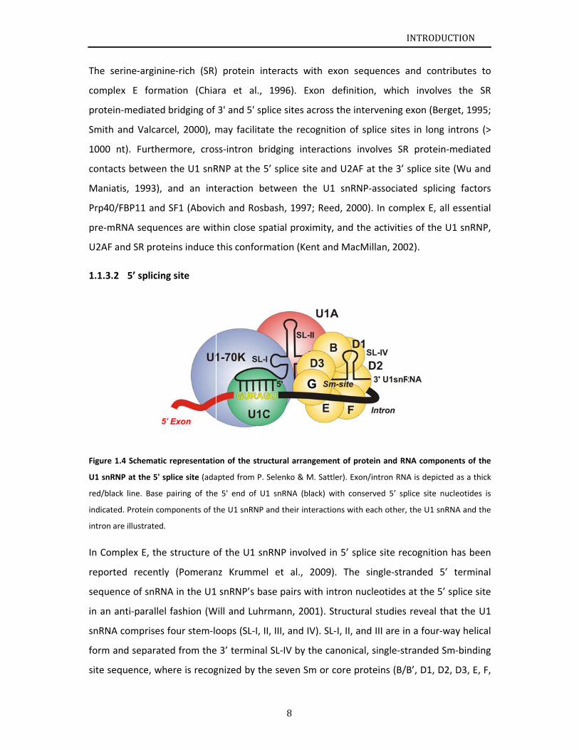
1.1.3.2
Figure 1.
U1 snRN
red/black
indicated
intron ar
In Com
reporte
sequen
in an an
snRNA
form an
site seq
rine‐arginin
x E forma
‐mediated
and Valcarc
nt). Further
ts between
is, 1993), a
FBP11 and
RNA sequen
nd SR prote
5’ splicing
.4 Schematic
P at the 5' sp
k line. Base p
d. Protein com
e illustrated.
plex E, the
ed recently
ce of snRNA
nti‐parallel
comprises f
nd separate
quence, whe
ne‐rich (SR)
ation (Chia
bridging of
cel, 2000), m
rmore, cros
the U1 snR
and an int
SF1 (Abovic
ces are wit
eins induce
g site
representatio
plice site (ada
pairing of the
mponents of t
structure o
y (Pomeran
A in the U1
fashion (W
four stem‐lo
ed from the
ere is recog
) protein in
ara et al.,
3' and 5' sp
may facilita
ss‐intron b
RNP at the 5
teraction b
ch and Ros
thin close sp
this conform
on of the stru
apted from P.
e 5' end of U
he U1 snRNP
of the U1 sn
nz Krumme
snRNP’s ba
Will and Luh
oops (SL‐I,
e 3’ termina
gnized by th
8
nteracts wi
1996). Ex
plice sites ac
ate the reco
ridging inte
5’ splice sit
etween th
bash, 1997
patial proxi
mation (Ken
uctural arrang
Selenko & M
U1 snRNA (bla
and their inte
nRNP involv
el et al., 2
ase pairs w
rmann, 200
II, III, and IV
al SL‐IV by t
he seven Sm
ith exon se
xon definit
cross the in
ognition of
eractions i
e and U2AF
e U1 snRN
; Reed, 200
mity, and t
nt and Mac
gement of pr
M. Sattler). Exo
ack) with con
eractions with
ved in 5’ sp
2009). The
ith intron n
01). Structu
V). SL‐I, II, a
he canonica
m or core p
I
equences a
ion, which
ntervening e
splice sites
nvolves SR
F at the 3’ s
NP‐associate
00). In comp
he activitie
Millan, 200
rotein and RN
on/intron RNA
nserved 5’ sp
h each other,
plice site rec
single‐stra
nucleotides
ural studies
and III are in
al, single‐st
roteins (B/B
INTRODUCT
and contrib
h involves
exon (Berge
s in long in
R protein‐m
splice site (
ed splicing
plex E, all e
s of the U1
02).
NA componen
A is depicted
lice site nucle
the U1 snRNA
cognition h
anded 5’ t
at the 5’ sp
reveal that
n a four‐way
randed Sm‐
B’, D1, D2,
TION
butes to
the SR
et, 1995;
trons (>
mediated
(Wu and
factors
essential
1 snRNP,
nts of the
as a thick
eotides is
A and the
has been
terminal
plice site
t the U1
y helical
‐binding
D3, E, F,
INTRODUCTION
9
and G). These Sm proteins are common in all snRNPs and assemble around the Sm‐binding
site to form the core snRNP. In addition, mammalian U1 snRNP contains four U1‐specific
proteins (U1A, U1C, and U1‐70K) (Figure 1.4), that U1‐70K and U1A bind to SL‐I and II,
respectively, U1C incorporation into the snRNP requires its interaction with U1‐70K (Figure
1.4).
Interaction of the U1 snRNP with proteins that associate with the pre‐mRNA in the vicinity
of the 5’ end of the intron additionally contributes to the 5’ splice site definition. For instant,
binding of U1 snRNP to protein TIA‐1 is essential for U1 snRNP recognizes 5’ splice sites
followed by uridine (U)‐rich sequences (Del Gatto‐Konczak et al., 2000; Forch et al., 2000).
These U‐rich sequences are also bound by proteins that would otherwise interact with
pyrimidine‐rich sequences (i.e. the Py tract) close to the 3’ splice site. For example, U2AF65
and the Drosophila female specific sex lethal (Sxl) protein, which are involved in 3’ splice site
selection, are claimed to interact with these U‐rich stretches and directly compete with
TIA‐1 (Forch et al., 2001). In addition, U2AF65 RS domain is proposed to be a critical factor
to promote U1 snRNP association with 5’ splice sites followed by U‐rich sequences (Forch et
al., 2003).
Combining biochemical data with known three‐dimensional structures of the individual U1
snRNP components, the structural arrangement of proteins and the U1 snRNA was
determined. Furthermore, more than a decade ago, electron microscopy was used to
determined the fully assembled human U1 snRNP structure at 10 Å resolution (Stark et al.,
2001). And crystal structures of U1 snRNP are released recently (Pomeranz Krummel et al.,
2009; Weber et al., 2010), showing the core RNP presents multiple attachment sites for the
U1‐specific 70K protein. Each of the seven Sm proteins recognize one nucleotide of the Sm
site RNA. Proteins D1 and D2 move the snRNA through the Sm ring, and proteins F and E
mediate a direct interaction between the Sm site termini. Terminal extensions of proteins
D1, D2 and B/B’, and extended internal loops in D2 and B/B’ support a four‐way RNA
junction and a 3’ terminal stem‐loop on opposite sides of the Sm core RNP, respectively
(Weber et al., 2010).
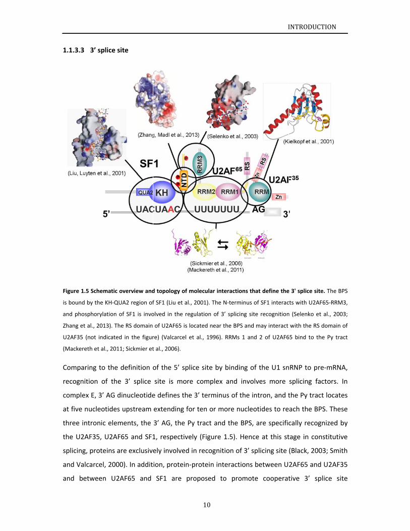
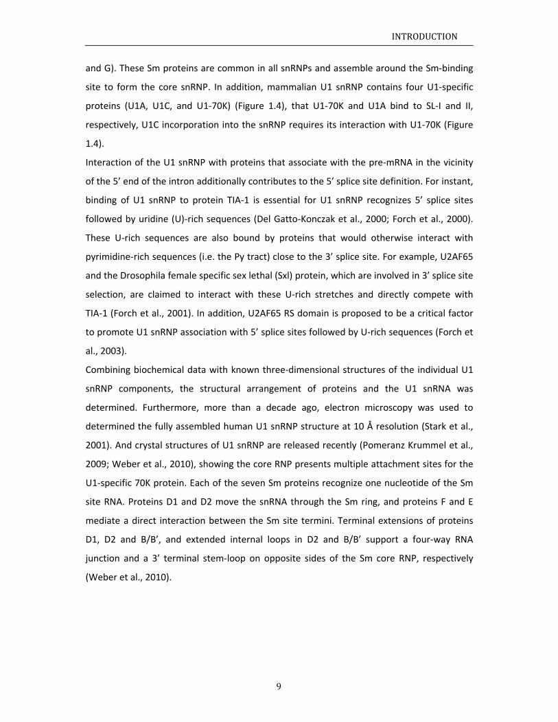
1.1.3.3
Figure 1.
is bound
and phos
Zhang et
U2AF35
(Mackere
Compar
recogni
comple
at five n
three in
the U2A
splicing
and Val
and be
3’ splice s
.5 Schematic
by the KH‐QU
sphorylation o
al., 2013). Th
(not indicated
eth et al., 201
ring to the
ition of the
x E, 3’ AG d
nucleotides
ntronic elem
AF35, U2AF
g, proteins a
lcarcel, 200
etween U2
site
overview and
UA2 region of
of SF1 is invo
he RS domain
d in the figur
1; Sickmier et
definition o
e 3’ splice
dinucleotide
s upstream
ments, the
F65 and SF1
are exclusive
00). In addit
2AF65 and
d topology of
f SF1 (Liu et a
olved in the r
of U2AF65 is
re) (Valcarcel
t al., 2006).
of the 5’ sp
site is mo
e defines th
extending f
3’ AG, the
1, respectiv
ely involved
ion, protein
SF1 are p
10
f molecular in
l., 2001). The
regulation of
s located near
et al., 1996)
plice site by
re complex
he 3’ termin
for ten or m
Py tract an
vely (Figure
d in recogni
n‐protein in
proposed t
nteractions th
e N‐terminus o
3’ splicing sit
r the BPS and
. RRMs 1 and
y binding of
x and invol
nus of the in
more nucleo
nd the BPS,
1.5). Henc
ition of 3’ sp
nteractions
to promote
I
at define the
of SF1 interac
e recognition
may interact
d 2 of U2AF6
f the U1 sn
lves more
ntron, and t
otides to re
are specific
ce at this st
plicing site
between U
e cooperat
INTRODUCT
e 3' splice site
cts with U2AF6
n (Selenko et
with the RS d
65 bind to the
nRNP to pre
splicing fac
the Py tract
ach the BPS
cally recogn
tage in cons
(Black, 2003
2AF65 and
tive 3’ spl
TION
e. The BPS
65‐RRM3,
al., 2003;
domain of
e Py tract
e‐mRNA,
ctors. In
t locates
S. These
nized by
stitutive
3; Smith
U2AF35
lice site
INTRODUCTION
11
recognition in complex E and ensure high accuracy of 3’ splice site definition. The
phosphorylation of splicing factors, SF1 and U2AF heterodimer RS domain for instance,
provides opportunities of post‐translational regulations in pre‐mRNA alternative splicing.
The protein‐protein and protein‐RNA interactions in complex E are relative weak, which
presumably supports the dynamic assembly of the spliceosome and facilitates the
subsequent replacement of SF1 by the U2 snRNP in complex A (Figure 1.2).
A recent study has revealed that the U2AF65 tandem RRM domains 1 and 2 (RNA
recognition motif, RRM1‐2) recognize the Py tract, induces a population shift between
“closed” and “open” conformational arrangement. They have shown that the correlation
relationship between the length of the Py tract and the binding affinity of U2AF65 RRM1‐2,
that further indicates how the ‘strength’ of a given Py tract is coupled to the efficiency of
spliceosome assembly. They adopted a molecular rheostat‐like model to describe the
equilibrium between the two conformations that quantitatively connects the natural
variations in polypyrimidine tract nucleotide composition, length and functional strength to
the efficiency to recruit U2 snRNP to the intron during spliceosome assembly (Mackereth et
al., 2011).
SF1, one of the major factors involving in 3’ splice site recognition, binds to BPS within
pre‐mRNA and simultaneously interacts with U2AF65 via UHM Ligand Motif (ULM) ‐ U2AF
Homology Motif domain (UHM) interaction, it has been proposed that the N‐terminus of
SF1 (SF1NTD) comprises a helix hairpin fold (SF1HH) providing an additional binding interface
to U2AF65UHM. More descriptions of SF1 in detail are presented in the next section.

INTRODUCTION
12
1.1.4 Splicing factor 1 (SF1)
As described in section 1.1.2, there are two trans‐esterification‐reactions steps required for
the pre‐mRNA alternative splicing. In the first step, the 2’ hydroxyl group of adenosine
within the branch site sequence (BPS) attacks the phosphate at the 5’ splice site, which
leads to the formation of a 2’‐5’ phosphodiester bond in the lariat structure (Figure 1.2).
Thus, this branch point adenosine (BPA) can be considered as a key nucleotide in the
pre‐mRNA intron. It is part of the seven‐nucleotide BPS and is highly conserved in S.
cerevisiae (UACUAAC, the underlined adenosine denotes the BPA), but more divergent in
other organisms, including S. pombe and higher eukaryotes. In mammals, the branch point
consensus sequence is YNCURAY (Y = pyrimidine; R = purine; N = any nucleotide). In the S.
cerevisiae commitment complex, the branch point binding protein (BBP) recognizes the BPS.
Whereas, in the mammalian complex E, SF1, ortholog of BBP, was reported to be involved in
this interaction (Berglund et al., 1997).
The solution structure of the human SF1 KH‐QUA2 domain in complex with a branch point
RNA demonstrated that this region is necessary and sufficient for recognition of the BPS (Liu
et al., 2001). The KH domain of SF1 consists of a three stranded anti‐parallel β‐sheet packed
against three α‐helices in the type of β1−α1−α2−β2−β3−α3 secondary structure. The QUA2
domain adopts an additional amphipathic α‐helix (α4) that packs on top of helices α1 and α3.
In conjunction with the core KH domain, this entity forms a compact structure which defines
an enlarged KH fold. The BPA is deeply buried in a hydrophobic pocket, form by the β2, α1,
α2 and the QUA2 helix α4 and thus shielded from potential interaction with other molecules.
Therefore, the BPA base is positioned in a pre‐bulged orientation that may prime it for
subsequent base pairing with U2 snRNA. In addition to hydrophobic contacts, electrostatic
interactions are another effect between conserved positively charged side chains in SF1
KH‐QUA2 domain and the solvent‐exposed phosphate backbone of the BPS RNA.
Electrostatic contacts further define this adenosine‐specific binding pocket.
In addition to the protein‐RNA interaction, the N terminus of SF1 binds to U2AF65 UHM (or
RRM3) (Rain et al., 1998), the C‐terminal non‐canonical amino acid sequence RRM. Due to
the complex is sensitive to salt concentration, it is reasonable to suggest electrostatic effects
contribute to the SF1‐U2AF65UHM interaction (Rain et al., 1998). Study of the
three‐dimensional structure of the SF1‐U2AF65UHM protein complex revealed the
INTRODUCTION
13
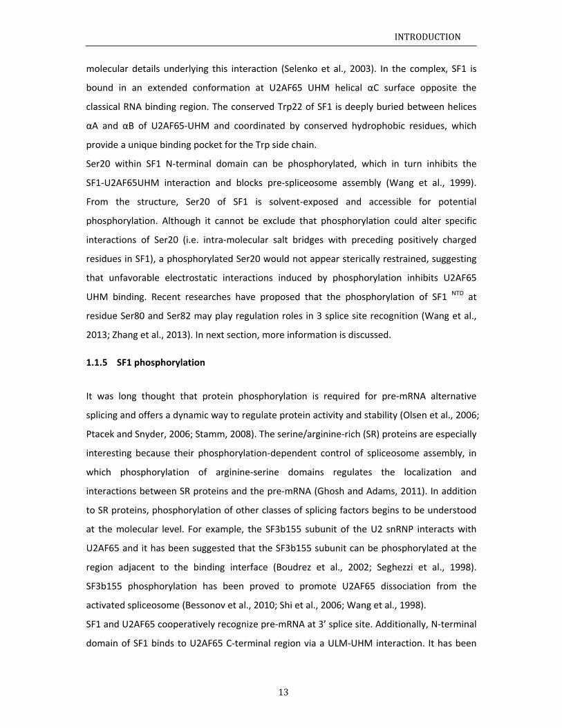
molecular details underlying this interaction (Selenko et al., 2003). In the complex, SF1 is
bound in an extended conformation at U2AF65 UHM helical αC surface opposite the
classical RNA binding region. The conserved Trp22 of SF1 is deeply buried between helices
αA and αB of U2AF65‐UHM and coordinated by conserved hydrophobic residues, which
provide a unique binding pocket for the Trp side chain.
Ser20 within SF1 N‐terminal domain can be phosphorylated, which in turn inhibits the
SF1‐U2AF65UHM interaction and blocks pre‐spliceosome assembly (Wang et al., 1999).
From the structure, Ser20 of SF1 is solvent‐exposed and accessible for potential
phosphorylation. Although it cannot be exclude that phosphorylation could alter specific
interactions of Ser20 (i.e. intra‐molecular salt bridges with preceding positively charged
residues in SF1), a phosphorylated Ser20 would not appear sterically restrained, suggesting
that unfavorable electrostatic interactions induced by phosphorylation inhibits U2AF65
UHM binding. Recent researches have proposed that the phosphorylation of SF1 NTD at
residue Ser80 and Ser82 may play regulation roles in 3 splice site recognition (Wang et al.,
2013; Zhang et al., 2013). In next section, more information is discussed.
1.1.5 SF1 phosphorylation
It was long thought that protein phosphorylation is required for pre‐mRNA alternative
splicing and offers a dynamic way to regulate protein activity and stability (Olsen et al., 2006;
Ptacek and Snyder, 2006; Stamm, 2008). The serine/arginine‐rich (SR) proteins are especially
interesting because their phosphorylation‐dependent control of spliceosome assembly, in
which phosphorylation of arginine‐serine domains regulates the localization and
interactions between SR proteins and the pre‐mRNA (Ghosh and Adams, 2011). In addition
to SR proteins, phosphorylation of other classes of splicing factors begins to be understood
at the molecular level. For example, the SF3b155 subunit of the U2 snRNP interacts with
U2AF65 and it has been suggested that the SF3b155 subunit can be phosphorylated at the
region adjacent to the binding interface (Boudrez et al., 2002; Seghezzi et al., 1998).
SF3b155 phosphorylation has been proved to promote U2AF65 dissociation from the
activated spliceosome (Bessonov et al., 2010; Shi et al., 2006; Wang et al., 1998).
SF1 and U2AF65 cooperatively recognize pre‐mRNA at 3’ splice site. Additionally, N‐terminal
domain of SF1 binds to U2AF65 C‐terminal region via a ULM‐UHM interaction. It has been
INTRODUCTION
14
discussed in section 1.1.4 that the phosphorylation of Ser20 within SF1 N‐terminal domain
cGMP‐dependent protein kinase‐I (PKG‐I) inhibits the SF1‐U2AF65UHM interaction and
blocks pre‐spliceosome assembly (Wang et al., 1999). Later on, three dimensional structural
information of SF1ULM‐U2AF65UHM complex was released, showing that Ser20 of SF1 is
solvent‐exposed and favorably accessible for potential phosphorylation. The Ser20,
phosphorylation site, is not involved in the directly binding to U2AF65UHM (Selenko et al.,
2003). Their studies have proposed a combination of steric interference and unfavorable
electrostatic contacts of phosphorylated Ser20 with the negatively charged U2AF65UHM helix
A induces the inhibition of SF1‐U2AF65 interaction and spliceosome assembly.
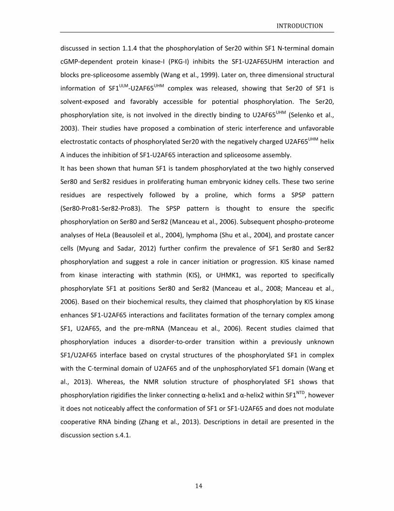
It has been shown that human SF1 is tandem phosphorylated at the two highly conserved
Ser80 and Ser82 residues in proliferating human embryonic kidney cells. These two serine
residues are respectively followed by a proline, which forms a SPSP pattern
(Ser80‐Pro81‐Ser82‐Pro83). The SPSP pattern is thought to ensure the specific
phosphorylation on Ser80 and Ser82 (Manceau et al., 2006). Subsequent phospho‐proteome
analyses of HeLa (Beausoleil et al., 2004), lymphoma (Shu et al., 2004), and prostate cancer
cells (Myung and Sadar, 2012) further confirm the prevalence of SF1 Ser80 and Ser82
phosphorylation and suggest a role in cancer initiation or progression. KIS kinase named
from kinase interacting with stathmin (KIS), or UHMK1, was reported to specifically
phosphorylate SF1 at positions Ser80 and Ser82 (Manceau et al., 2008; Manceau et al.,
2006). Based on their biochemical results, they claimed that phosphorylation by KIS kinase
enhances SF1‐U2AF65 interactions and facilitates formation of the ternary complex among
SF1, U2AF65, and the pre‐mRNA (Manceau et al., 2006). Recent studies claimed that
phosphorylation induces a disorder‐to‐order transition within a previously unknown
SF1/U2AF65 interface based on crystal structures of the phosphorylated SF1 in complex
with the C‐terminal domain of U2AF65 and of the unphosphorylated SF1 domain (Wang et
al., 2013). Whereas, the NMR solution structure of phosphorylated SF1 shows that
phosphorylation rigidifies the linker connecting α‐helix1 and α‐helix2 within SF1NTD, however
it does not noticeably affect the conformation of SF1 or SF1‐U2AF65 and does not modulate
cooperative RNA binding (Zhang et al., 2013). Descriptions in detail are presented in the
discussion section s.4.1.
INTRODUCTION
15
1.1.6 The U2AF65 arginine‐serine‐rich (RS) domain
Reams of different proteins are involved in the individual splicing steps and the regulatory
processes that lead for example to alternative products (alternative splicing), an essential
one of them is SR protein family, which is a class of general factor that in constitutive
splicing and can also modulate alternative splicing. All SR proteins share a similar bipartite
structure composed of two functional domains: an N‐terminal RNA binding domain,
consisting of several RNA recognition motifs (RRMs) and a C‐terminal arginine‐ and
serine‐rich RS domain. The RRMs are sufficient sequence‐specific RNA binding, whereas the
RS domain is believed to be an essential mediator and regulator in pre‐mRNA alternative
splicing.
Several key interactions with components of the splicing machinery were already
highlighted and indicate that RS domains serve as a dynamic and versatile binding platform
for proteins, RNA, and ligands. RS domains promote for example protein‐protein
interactions that facilitate recruitment of the (pre)spliceosome (Kohtz et al., 1994; Wu and
Maniatis, 1993) and contact the pre‐mRNA directly via the Branch Point and the 5’ splicing
site (Hertel and Graveley, 2005; Shen et al., 2004; Shen and Green, 2004). Furthermore, they
participate in many other cellular processes such as in mRNA nuclear export,
nonsense‐mediated mRNA decay and mRNA translation (Caceres et al., 1997; Long and
Caceres, 2009).
In vivo, RS domains are found to be extensively phosphorylated on serine residues enabling
post‐translational modification of the interaction network. It is believed that the subcellular
localization as well as the activity is regulated on the level of phosphorylation (Lin and Fu,
2007).
As RS domains are involved in several different tasks, disturbance of the protein integrity
and interactions by mutation lead to human disease. Evidence emerged that defects of SR
proteins are related with cancer, SMA (spinal muscular atrophy) and HIV (Long and Caceres,
2009).
RS domains are not only detected in SR proteins, but also exist in other splicing factors (i.e.
essential splicing factors U2AF65/U2AF35 (Valcarcel et al., 1996)) and play significant roles
in constitutive and regulated splicing (Black, 2003; Graveley, 2000). U2AF65 and U2AF35
bind to the polypyrimidine (Py) tract and 3’ AG splice site, respectively, and initiate
INTRODUCTION
16
spliceosome assembly by promoting the interaction between U2 snRNP and the branchpoint.
It has been shown that the binding of U2AF65 to the Py tract direct the RS domain to
contact BPS and promotes base pairing with U2 snRNP (Nolen et al., 2001; Shen et al.,
2004).
Despite all the available functional data, detailed insight into the molecular mechanisms
remains elusive. This is probably due to a common feature of all RS domains: although they
share a high content of arginine and serine residues, they seem to be intrinsically
unstructured and highly dynamic. Furthermore, inclusion of phosphorylation in the
structural studies is crucial in order to understand how the binding‐strength and ‐specificity
of RS domains for their interaction partners is modulated.
Therefore, in this thesis, the molecular details of the U2AF65 RS domain and its interactions
were studied in solution using Nuclear Magnetic Resonance (NMR) Spectroscopy. The main
focus will be on the RS domain of the essential splicing factor U2AF65 which acts at a central
position of the splicing pathway and interconnects RNA and proteins(Valcarcel et al., 1996),
potentially in a phosphorylation‐dependent mechanism (Olsen et al., 2006). As described in
section 1.1.3.3, U2AF65 RS domain forms a ternary protein‐RNA‐protein complex with
SF1KH‐QUA2 domain at the branch point. Therefore, together with our structural studies of
SF1, the structural insight for RNA‐ and protein interaction by RS domains will contribute
significantly to understanding the mechanisms of 3’ splice site recognition and its regulation.
N1.2
1.2.1
Nuclear
immers
Some n
possess
number
where
eigenva
I can
that re
magnet
a NMR
and 31P,
can be
labeling
Stable i
an exte
separat
magnet
field; th
field (Fi
laborat
Nuclear ma
The princip
r magnetic
sed in a stat
nuclei exper
s an intrins
r, for any gi
Ψ is the w
alue for the
be zero or
presents a
tic moment
spectrosco
, because t
relatively
g technique
sotopes 1H,
ernal magne
ted by a d
tic moment
he one of th
igure 1.6). T
ory coordin
agnetic reso
ples of NMR
c resonance
tic magneti
rience this p
sic property
ven wave f
wavefunctio
system.
can have in
spinning c
. Only the s
py. The one
hese nuclei
easily enric
es.
, 13C, 15N an
etic field giv
iscrete ene
t of the low
he higher en
The equatio
nate system
onance (N
R
e, or NMR
ic field and
phenomeno
y called sp
unction, wh
on of the p
ntegral/fract
charge in t
spins of nuc
es that are
i are either
ched by inc
nd 31P are sp
ves rise to t
ergy differe
wer energy
nergy with
on that des
m is
17
MR) spect
R, occurs w
affected by
on and othe
pin. The sp
hich is obtai
HΨ EΨ
article, H t
tional value
terms of cl
clei which h
common u
present in
corporation
pin I= nu
two differe
ence, chara
y state
spin s
scribes the
troscopy
when the
y an additio
ers do not, d
in is define
ined from t
Ψ
he Hamilto
es. If the sp
lassical me
ave non‐ze
sed in biolo
biological
n through d
uclei. The in
nt spin stat
acterized by
is aligned
state is opp
energy of e
I
nuclei of c
onal oscillat
dependent
ed by I, th
he Schrödin
on operator
in value is n
chanics, is
ro I‐values
ogical NMR
molecules i
different un
nteraction o
tes with a c
y I = a
d with the
posed to the
each state i
INTRODUCT
certain ato
ting magnet
upon wheth
he fourth q
nger equati
r and E the
non‐zero a
associated
can be dete
are 1H, 2H,
in high amo
niform or s
of these nuc
certain ener
and I =
external m
e external m
n the conve
TION
oms are
tic field.
her they
quantum
on:
e energy
nucleus,
d with a
ected by
13C, 15N
ounts or
selective
clei with
rgy level
. The
magnetic
magnetic
entional
where γ
the exte
The en
depend
where ω
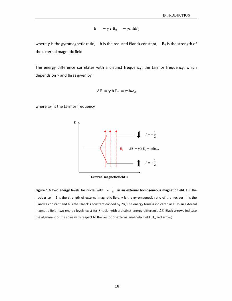
Figure 1.
nuclear s
Planck’s c
magnetic
the align
γ is the gyro
ernal magn
ergy differe
ds on γ and
ω0 is the La
.6 Two energ
spin, B is the
constant and
c field, two en
ment of the s
omagnetic r
etic field
ence correl
B0 as given
rmor frequ
gy levels for
strength of e
ħ is the Planc
nergy levels e
pins with resp
E
ratio; ħis
lates with a
by
ΔE
ency
nuclei with I
external magn
ck’s constant d
exist for I nuc
pect to the ve
18
γ B
the reduce
a distinct f
γħB
= in an
netic field, γ is
divided by 2π
lei with a dist
ctor of extern
γmħB
ed Planck co
frequency,
mħω
external hom
s the gyroma
π, The energy t
tinct energy d
nal magnetic f
I
onstant; B
the Larmor
mogeneous m
gnetic ratio o
term is indica
difference ΔE
field (B0, red a
INTRODUCT
B0 is the stre
r frequency
magnetic field
of the nucleus
ted as E. In an
. Black arrow
arrow).
TION
ength of
y, which
d. I is the
s, h is the
n external
s indicate
INTRODUCTION
19
The population of the energy states is given in the Boltzmann equation
Nhigher
Nlowere /
Here Nhigher and Nlower represent the number of spins on the higher and lower energy levels
respectively and ΔE is the energy difference between the two levels, T is the temperature in
Kelvin and k represents the Boltzmann constant.
The sum of a large ensemble of spins in the external magnetic field can be described as a
macroscopically observable equilibrium magnetization M in the vector model. In the
presence of an external magnetic field B0, the orientation of vector M in respect to a
coordinate system in which the orientation of the external magnetic field corresponds to
the z axis. Without additional external influences, the magnetic moment of the spins is in
equilibrium. They can align with or opposite to the external magnetic field B0 with the
distribution of the two spin states follows Boltzmann equation. Whereas, the numbers of
spins on the higher (Nhigher) and lower energy (Nlower) levels are not equal, thus the
difference between Nhigher and Nlower creates a net bulk magnetization, represented as a
vector M, along the direction of the magnetic field. An additional electromagnetic field B1 in
the transverse plane can affect the orientation of M. In an NMR spectrometer, an
radiofrequency ( rf ) pulse along the x‐axis turn M towards the –y‐axis. The angle α will
depend on the length of the pulse. The direction of the rotation follows the right‐hand rule
based on physics of electromagnetism. The magnetization will start a precession around the
z‐axis or external magnetic field B0 with the Larmor frequency ω, generating the free
induction decay (FID) in NMR detection coil. The measured FID is a time‐domain signal and
contains all the frequencies the sample in a sum of cosine waves. Fourier Transformation (FT)
is able to transfer the time‐domain FID into the frequency‐domain signal, which is the
output spectrum in an NMR experiment.
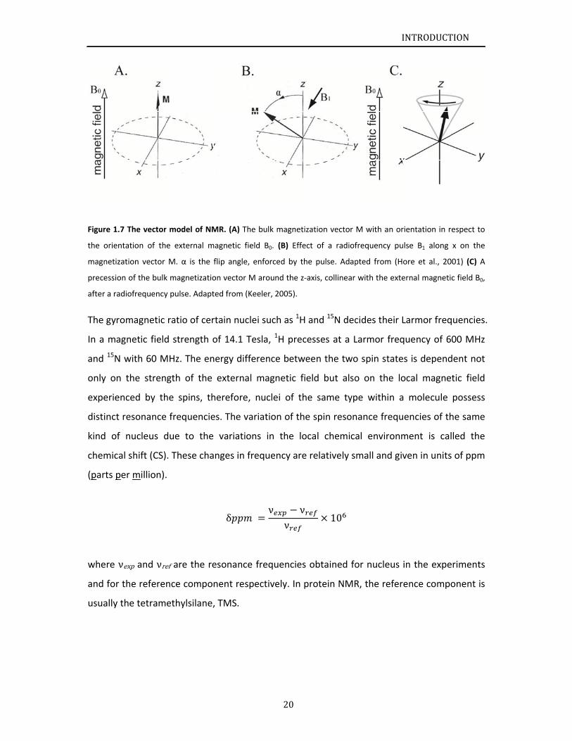
Figure 1.
the orien
magnetiz
precessio
after a ra
The gyr
In a ma
and 15N
only on
experie
distinct
kind of
chemica
(parts p
where ν
and for
usually
.7 The vector
ntation of th
zation vector
on of the bulk
adiofrequency
romagnetic
agnetic field
N with 60 M
n the stren
enced by th
t resonance
f nucleus d
al shift (CS)
per million).
νexp and νre
r the referen
the tetram
r model of NM
e external m
M. α is the
k magnetizatio
y pulse. Adapt
ratio of cer
d strength o
MHz. The en
gth of the
he spins, th
frequencie
due to the
. These cha
.
ef are the re
nce compo
ethylsilane,
MR. (A) The b
magnetic field
flip angle, en
on vector M a
ted from (Kee
rtain nuclei
of 14.1 Tes
nergy differe
external m
herefore, n
es. The varia
variations
anges in freq
δ
esonance fr
nent respec
, TMS.
20
bulk magnetiz
B0. (B) Effec
nforced by th
around the z‐a
ler, 2005).
such as 1H
la, 1H prece
ence betwe
magnetic fie
uclei of the
ation of the
in the loc
quency are
ν νν
requencies
ctively. In p
zation vector
ct of a radiof
e pulse. Adap
axis, collinear
and 15N dec
esses at a L
een the two
eld but also
e same typ
e spin reson
cal chemica
relatively s
10
obtained fo
protein NMR
I
M with an or
frequency pu
pted from (H
with the exte
cides their L
Larmor freq
o spin state
o on the lo
pe within a
nance frequ
al environm
mall and giv
or nucleus
R, the refer
INTRODUCT
rientation in r
ulse B1 along
ore et al., 20
ernal magneti
Larmor freq
quency of 6
es is depend
ocal magne
a molecule
uencies of th
ment is ca
ven in units
in the expe
ence comp
TION
respect to
x on the
001) (C) A
ic field B0,
quencies.
600 MHz
dent not
etic field
possess
he same
lled the
s of ppm
eriments
onent is
INTRODUCTION
21
1.2.2 NMR for protein analysis
The application of NMR to study structure and dynamics of biomolecules (i.e. proteins,
nucleic acids) on atomic level is one of the greatest achievements in the last century. On the
way towards the successful NMR measurement of proteins, two main limiting factors should
be taken in account, one is the signal overlap and the other is the fast transverse relaxation
rates. Proteins contains a large number of nuclei of the same atom type, thus, in the early
protein NMR experiments, measurements of the naturally abundant NMR‐active isotope 1H
in one‐dimensional spectra can only provide limit information due to lack of resolution of
the signals. In addition, the large size of proteins and nucleic acids causes fast transverse
relaxation rates, that broadening the line‐width of the signal. Developments in both
molecular biology field as well as NMR technology have made a remarkable progress in
overcoming these problems. The naturally abundant isotope 12C and 14N can be respectively
replaced by the NMR‐active nuclei 13C and 15N, by expressing proteins in corresponding
isotopic minimal medium. The recombinant expression of proteins in bacteria enables the
13C and 15N labeled protein purified and enriched. These samples provide a way for develop
the multidimensional heteronuclear NMR method, which correlates two or more nuclei
NMR signals via J‐coupling and dipolar‐coupling. 3D NMR generates extremely higher signal
resolution then 1D NMR. The proton density, a major source of relaxation, can be reduced
by expressing protein in a partially deuterated or perdeuterated background and thus large
proteins are accessible for NMR analysis. Specific labeling schemes enable specifically
isotope labeling on certain amino acids or chemical groups, contributing to the
simplification of NMR spectra. At the same time, the progress in hardware of NMR
spectrometers increases magnetic field strength, in additional, cryoprobes enhance the
sensitivity of the machines. These ongoing advances in NMR extend the research of
biomolecules sizes larger than 300 kDa.
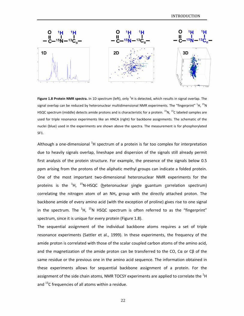
Figure 1.
signal ov
HSQC spe
used for
nuclei (b
SF1.
Althoug
due to
first an
ppm ar
One of
protein
correlat
backbo
in the
spectru
The se
resonan
amide p
and the
same re
these e
assignm
and 13C
.8 Protein NM
verlap can be
ectrum (midd
triple resona
lue) used in t
gh a one‐di
heavily sig
alysis of th
rising from
f the most
s is the
ting the ni
ne amide o
spectrum.
um, since it
quential as
nce experim
proton is co
e magnetiza
esidue or th
experiment
ment of the
frequencie
MR spectra. In
reduced by h
dle) detects am
ance experime
the experime
mensional
nals overla
he protein s
the protons
t important
1H, 15N‐HS
trogen ato
of every am
The 1H, 15
is unique fo
ssignment
ments (Satt
orrelated wi
ation of the
he previous
ts allows f
side chain a
es of all atom
n 1D spectrum
eteronuclear
mide protons
ents like an H
nts are show
1H spectrum
p, lineshap
structure. F
s of the alip
t two‐dime
SQC (heter
om of an N
ino acid (w
5N HSQC s
or every pro
of the ind
tler et al., 1
ith those of
e amide pro
s one in the
for sequen
atoms, NM
ms within a
22
m (left), only 1H
multidimensi
and is charac
HNCA (right) f
n above the s
m of a prot
pe and disp
For example
phatic meth
ensional he
ronuclear s
NHx group
with the exce
pectrum is
otein (Figure
dividual bac
1999). In th
f the scalar
oton can be
e amino acid
tial backbo
R TOCSY ex
residue.
H is detected,
ional NMR ex
teristic for a p
for backbone
spectra. The m
tein is far to
ersion of t
e, the prese
hyl groups
eteronuclea
single qua
with the d
eption of p
often refe
e 1.8).
ckbone ato
hese exper
coupled ca
e transferre
d sequence
one assignm
periments a
I
, which result
periments. Th
protein. 15N, 1
assignments
measurement
oo complex
he signals s
ence of the
can indicat
ar NMR ex
ntum corr
directly atta
roline) give
erred to as
oms requir
iments, the
rbon atoms
ed to the C
e. The inform
ment of a
are applied
INTRODUCT
s in signal ove
he “fingerprin
13C labeled sa
. The schema
t is for phosp
x for interp
still already
e signals be
e a folded
xperiments
relation sp
ached prot
es rise to on
s the “fing
es a set o
e frequency
s of the ami
CO, Cα or C
mation obt
a protein.
to correlat
TION
erlap. The
nt” 1H, 15N
mples are
atic of the
horylated
retation
y permit
elow 0.5
protein.
for the
pectrum)
on. The
ne signal
gerprint”
of triple
y of the
ino acid,
β of the
ained in
For the
e the 1H
INTRODUCTION
23
The Nuclear Overhauser Effect (NOE), which transfers polarization by dipolar interactions
between atoms close in space, is one of the most important effects in NMR. The NMR
structure calculation is based on the distance restraints derived from many hundreds of
NOE‐peaks. In isotope‐edited NOESY‐HSQC experiments, magnetization is transferred to
protons nearby and the HSQC sequence selects protons bound to either 15N or 13C, which
can provide sufficient resolution, Information from backbone and side‐chain experiments is
combined to obtain complete resonance assignments of proteins.
1.2.3 Protein dynamics by NMR
NMR of proteins allows for studying not only the structure but also the dynamic properties
of proteins. For investigation of protein dynamics, two relaxation pathways are usually
monitored and the rate constants are measured. The longitudinal relaxation (also called the
spin‐lattice relaxation), characterized by a single time constant T1 or the corresponding
relaxation rateR , is induced by the interaction of the spins with their surrounding
(the lattice). The transverse relaxation (also called the spin‐spin relaxation), described by a
single time constant T2 or the relaxation rateR , is caused by the interaction of the
nuclear spins.
Measurements of 15N relaxation provide information about backbone dynamics covering a
picosecond to second timescale. Dynamics on the pico‐ to microsecond timescale like
molecular tumbling and internal motion are reflected by T1. T2 is additionally influenced by
dynamics in the micro‐ to millisecond timescale and thus sensitive to chemical exchange.
The correlation time τc describes molecular tumbling and can be calculated from the ratio
between both relaxation rates including information about the 15N frequency. In 1H‐15N
heteronuclear NOE experiments, motions of distinct N‐H bond vectors are sampled. Those
that undergo motion faster than the overall tumbling of the molecules show a decreased
NOE intensity relative to the average observed for the majority of the residues. The average
value for the 1H‐15N heteronuclear NOE ratio is approximately 0.77, higher values indicating
that the N‐H vector is rigid with respect to the rest of the protein and lower values for parts
with increased backbone mobility (Kay et al., 1989).
INTRODUCTION
24
1.2.4 Paramagnetic relaxation enhancement (PRE)
In the 1950s, Solomon and Bloembergen demonstrated that the presence of unpaired
electrons in the molecular system enhances the magnetic field modulation at a nucleus and
nuclear spin relaxation due to nucleus dipolar couplings. This effect is named paramagnetic
relaxation enhancement (PRE) (Solomon, 1955; Solomon and Bloembergen, 1956). In
protein NMR experiments, PREs are generated by spin labeling, attaching nitroxide stable
radicals such as iodoacetamido proxyl to the protein for example. Compared to NOE
(similarly generated by dipolar couplings, the distance between two spins limited up to 6 Å),
PREs can extend the effects to a distance of more than 40 Å (Otting, 2008). Therefore, PRE is
increasingly becoming a method to provide long‐range distance information that can
complement NOE restraints. Distances between the spin label and nuclei can be determined
from the increased R2 relaxation rates.
In the protein NMR, PREs can be monitored on the HN‐resonances in the 1H, 15N HSQC
spectra. The intensity of the peaks is acquired from the spectra in the presence of a
paramagnetic spin label (or oxidized state) and diamagnetic analogue (or reduced state).
Then, PRE represented in R2 relaxation rates can be calculated using equation(Battiste and
Wagner, 2000):
where Iox and Ired are the peak intensities corresponding to amides in the 1H15N‐HSQC for a
sample with a spin label in the oxidized and the reduced state respectively, R2 is the R2
relaxation rate for each amide 1H, t is the total evolution time of the HSQC and R2PRE the
paramagnetic rate enhancement coming from the spin label.
R2PRE were converted into distances by use of the following equation for the effect of
paramagnetic spins on nuclear magnetic relaxation
43
1
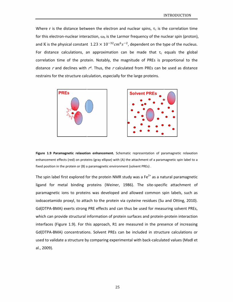
Where
for this
and K is
For dis
correlat
distance
restrain
Figure 1
enhancem
fixed pos
The spi
ligand
parama
iodoace
Gd(DTP
which c
interfac
Gd(DTP
used to
al., 200
r is the dis
electron‐nu
s the physic
stance calc
tion time o
e r and dec
ns for the st
1.9 Paramagn
ment effects
sition in the pr
n label first
for metal
agnetic ions
etamido pro
PA‐BMA) ex
can provide
ces (Figure
PA‐BMA) co
o validate a
9).
stance betw
uclear inter
cal constant
culations, a
of the prot
clines with
tructure calc
netic relaxatio
(red) on prote
rotein or (B) a
t explored fo
binding
s to protein
oxyl, to atta
erts strong
structural
1.9). For th
oncentration
structure b
ween the el
raction, ωh
t 1.23 10
an approxim
tein. Notab
r6. Thus, t
culation, es
on enhancem
eins (gray ellip
a paramagnet
or the prote
proteins (W
ns was dev
ach to the
PRE effect
information
his approac
ns. Solvent
y comparin
25
ectron and
is the Larm
0
mation can
bly, the ma
he r calcula
specially for
ment. Schem
pse) with (A)
ic environmen
ein NMR st
Weiner, 19
veloped and
protein via
s and can t
n of protein
ch, R1 are
PREs can
g experime
d nuclear sp
or frequenc
, depende
n be made
agnitude of
ated from
r the large p
atic represen
the attachme
nt (solvent PR
udy was a F
986). The
d allowed
cysteine re
thus be use
n surfaces a
measured
be include
ental with b
I
pins, τc is th
cy of the nu
ent on the t
e that τc
f PREs is p
PREs can b
proteins.
ntation of pa
ent of a param
REs) .
Fe2+ as a na
site‐specif
common s
esidues (Su
d for meas
nd protein‐
in the pres
d in struct
ack‐calculat
INTRODUCT
he correlati
uclear spin (
ype of the
equals the
roportiona
be used as d
aramagnetic r
magnetic spin
tural param
fic attachm
pin labels,
and Otting
uring solve
‐protein inte
sence of inc
ure calcula
ted values (
TION
on time
(proton),
nucleus.
e global
l to the
distance
relaxation
label to a
magnetic
ment of
such as
g, 2010).
nt PREs,
eraction
creasing
tions or
(Madl et
INTRODUCTION
26
1.2.5 NMR studies for protein‐ligand interaction.
NMR titration, recording a series of 1H, 15N HSQC spectrum of the protein in presence of
increasing ligand concentrations, is a common tool to investigate protein‐ligand interactions
such as protein‐protein and protein‐nucleic acid interactions. Chemical shift perturbations
provide information of the amide proton signals of involved residues. The changes observed
in the spectrum depend on the exchange rates between free and ligand‐bound state. The
exchange constant kex is
L
where [L] is the ligand concentration. kon and koff are the association and dissociation rates,
respectively.
On the NMR timescale,
≫ ∆ω fast exchange
∆ω intermediate exchange
≪ ∆ω slow exchange
where Δω is the difference in frequencies of the same resonance in the free and bound
state.
When ligand binding in fast exchange, signals possess an average chemical shift between
free and bound state depended on the population of each state, thus, the position of the
peak is gradually shifted until saturation. Intermediate exchange induces line broadening
and thus decreased signal intensity. In a slow exchange regime, the population of the free
and bound state can be observed simultaneously, giving rise to two separate signals with
intensities corresponding to the percentage of free and bound protein. Thus, NMR titrations
allow for a quick and residue‐specific determination of the protein‐ligand interaction
interface.
INTRODUCTION
27
1.2.6 Structure calculation
Combination of information, such as chemical shift assignments, distance restrains and
torsion angles are used for NMR structure calculation. The chemical shift already contains
useful structural information. 13Cα and 13Cβ resonances of a specific amino acid have
characteristic values given for a random coil environment. The chemical shift of 13Cα and
13Cβ altered in structured regions, the difference from the 13Cα and 13Cβ values of
structured region to the random coil values is named secondary chemical shift. In an α‐helix,
13Cα carbons shift to downfield and 13Cβ values move to upfield, whereas, opposite effects
are observed for carbon atoms in a β‐sheet. Therefore, the secondary chemical shift for
α‐helical form is indicated in positive values and for β‐sheet conformation is shown in
negative values. The distance restrains are derived from the signal intensity of cross‐peaks
in NOESY spectra by program, such as CYANA3.0 (Guntert, 2009). Torsion angles of each
residue in protein can be predicted by the program, TALOS+ (Shen et al., 2009), using the
chemical shifts of atoms from the protein backbone. Nowadays, additionally restraints
obtained from paramagnetic relaxation enhancement (PRE) and residual dipolar coupling
(RDC) measurements are employed to determine the relative position of structural
elements within the molecule.
NMR structure calculation is based on a restrained molecular dynamics simulation using a
target function which combines different potential energy functions. The target function
includes energy terms for known parameters as bond lengths, angles, van der Waals‐ and
electrostatic forces, the so‐called force field. Additionally, energy functions for NMR data
such as distance restraints from NOEs and torsion angles derived from chemical shift and
sequence information are incorporated. The program starts with approximately 100
randomized structures at high temperature. In the following steps of the calculation the
temperature is lowered and the different restraints are varied randomly to reduce violations
and minimize the target function in a simulated annealing process. Here are some of the
most used NMR structure calculation programs. Ambiguous Restraints for Iterative
Assignment (ARIA)(Nilges et al., 1997), Combined assignment and dynamics algorithm for
NMR applications (CYANA) (Schmucki et al., 2009) and CNS/ARIA (Linge et al., 2003). The
root mean square coordinate deviation (RMSD) describes the precision of the output
structure between the structures in the final ensemble.Accuracy of the structure is judged

INTRODUCTION
28
by the absence of substantial violations for distance restraints. Structure validation will be
done in the program like the iCING server (Doreleijers et al., 2012).
Sc1.3
Remova
by the
essentia
inclusio
importa
splicing
interfer
mutatio
factor 1
(U2AF3
by extra
to 3’ sp
Ser82)
SF1‐U2A
the stru
been pr
and SF1
structu
The aim
U2AF65
project
ULM re
structu
weight
NMR w
(Ser80‐
to char
the con
of nove
spectro
cope of th
al of introns
spliceosom
al to gene
on or exclus
ance of splic
g and muta
re with spl
ons in genes
1 (SF1), and
5) have bee
acellular sig
plice site rec
in SF1 h
AF65‐RNA c
uctural and
roposed tha
1 KH‐QUA2, ind
ral details o
ms of this
5 as key pr
was starte
egions, dete
ral details o
of SF1HH is
was chosen
Pro81‐Ser8
acterize the
nformationa
el interactio
oscopy. The
e Thesis
s from pre‐
me, a large a
rate differe
sion of cod
cing for gen
tions withi
iceosome a
s of splicing
d the U2 sn
en linked to
gnals and si
cognition ph
have been
complex in
functional
at binding o
ducing a co
of U2AF65 R
thesis are
roteins in t
ed structura
ermined by
of SF1HH do
13.7 kDa,
to study S
2‐Pro83) m
e specifical
al changes u
ns in the SF
erefore, by
mRNAs is a
and dynam
ent protein
ing sequen
ne expressio
n the cons
assembly (C
g factors tha
RNP auxilia
o tumorigen
ignal transd
hosphorylat
n implicate
vitro (Manc
roles of SF1
of U2AF65 R
ompact SF1‐
RS domain w
to study m
he early co
al informati
y Sattler’s g
omain betw
which is id
SF1HH. Struc
motif in SF1
ly phospho
upon phosp
F1‐U2AF65‐
y combining
29
key step in
ic ribonucle
ns from a
ces (exons)
on, numero
sensus sequ
Cooper et
at mediate
ary factor (U
nesis (Yoshid
duction casc
tion of cons
ed in reg
ceau et al.,
1 phosphor
RRM1‐2 to
‐U2AF65RRM
were descri
molecular a
omplex E d
ion of SF1 w
group (Liu
een KH‐QU
deal size for
ctural roles
HH remain u
rylated SF1
phorylation.
RNA compl
g NMR an
n mammalia
eoprotein p
given prim
) (Nilsen an
ous diseases
uences at t
al., 2009).
3’ splice sit
U2AF) large
da et al., 20
cades is poo
served serin
gulating th
2006; Wan
rylation rem
Py trace dri
M123‐RNA co
bed.
and structu
during splice
was limited
et al., 200
UA2 and UL
r protein N
s of tandem
unclear. Ma
1HH and NM
. For structu
ex, which m
nd Small‐An
I
an gene exp
particle. Alt
mary transcr
nd Graveley
s have been
the 5’ and
Recently,
te recogniti
e (U2AF65)
011). The re
orly unders
ne residues
e stability
g et al., 199
main elusive
ives RS dom
nformation
ural details
eosome ass
d to KH‐QU
01; Selenko
M is unkno
MR. Theref
m phospho
ass Spectro
MR was sele
ural and bio
molecular si
ngle X‐ray
INTRODUCT
pression pe
ternative sp
ript by diff
y, 2010). Gi
n linked to a
3’ ends of
frequent m
on, such as
and small s
egulation of
stood. With
(Ser20, Ser
y of the
99). Howev
e. In additio
main to cont
n, whereas,
involving S
sembly. Wh
A2 and N‐t
et al., 200
own. The m
fore, solutio
orylation of
ometry was
cted to inv
ochemical A
ize is large f
Scattering
TION
rformed
plicing is
ferential
iven the
aberrant
f introns
missense
splicing
subunits
f splicing
respect
r80, and
ternary
ver, both
n, it has
tact BPS
none of
SF1 and
hen this
terminal
03). The
olecular
on state
f a SPSP
applied
vestigate
Analyses
for NMR
(SAXS),

INTRODUCTION
30
structural information of the ternary complex was explored. 3D molecular structure of
highly dynamic U2AF65 RS domain is hard to be determined by X‐ray crystallography.
Thereby, NMR and the methods studying molecular dynamics, like relaxation time analysis,
were employed to reveal the structural details of U2AF65 RS domain.
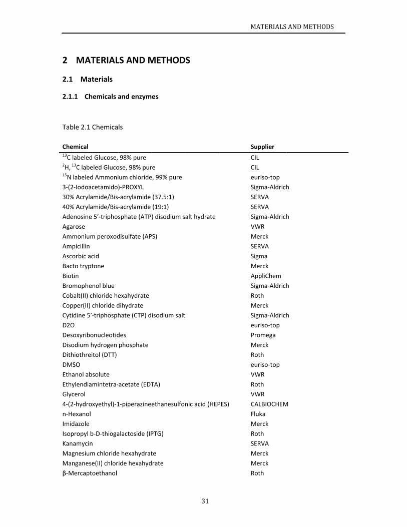
2 M
M2.1
2.1.1
Table 2
Chemica13C label2H, 13C la15N labe
3‐(2‐Iod
30% Acr
40% Acr
Adenosi
Agarose
Ammon
Ampicill
Ascorbic
Bacto tr
Biotin
Bromop
Cobalt(I
Copper(
Cytidine
D2O
Desoxyr
Disodium
Dithioth
DMSO
Ethanol
Ethylend
Glycerol
4‐(2‐hyd
n‐Hexan
Imidazo
Isopropy
Kanamy
Magnes
Mangan
β‐Merca
ATERIAL
Materials
Chemicals a
.1 Chemica
al
led Glucose,
abeled Gluco
led Ammoni
oacetamido)
rylamide/Bis‐
rylamide/Bis‐
ne 5′‐triphos
e
ium peroxod
in
c acid
yptone
phenol blue
I) chloride he
II) chloride d
e 5′‐triphosph
ribonucleotid
m hydrogen
hreitol (DTT)
absolute
diamintetra‐
l
droxyethyl)‐1
nol
le
yl b‐D‐thioga
ycin
ium chloride
nese(II) chlor
aptoethanol
LS AND M
and enzyme
ls
98% pure
ose, 98% pur
ium chloride
)‐PROXYL
‐acrylamide
‐acrylamide
sphate (ATP)
disulfate (AP
exahydrate
dihydrate
hate (CTP) d
des
phosphate
acetate (EDT
1‐piperazinee
alactoside (IP
e hexahydrat
ide hexahyd
METHOD
es
e
, 99% pure
(37.5:1)
(19:1)
) disodium sa
S)
isodium salt
TA)
ethanesulfon
PTG)
te
rate
31
S
alt hydrate
nic acid (HEP
M
S
C
C
e
S
S
S
S
V
M
S
S
M
A
S
R
M
S
e
P
M
R
e
V
R
V
PES) C
F
M
R
S
M
M
R
MATERIALS
Supplier
CIL
CIL
euriso‐top
Sigma‐Aldrich
SERVA
SERVA
Sigma‐Aldrich
VWR
Merck
SERVA
Sigma
Merck
AppliChem
Sigma‐Aldrich
Roth
Merck
Sigma‐Aldrich
euriso‐top
Promega
Merck
Roth
euriso‐top
VWR
Roth
VWR
CALBIOCHEM
Fluka
Merck
Roth
SERVA
Merck
Merck
Roth
ANDMETH
h
h
h
h
M
HODS
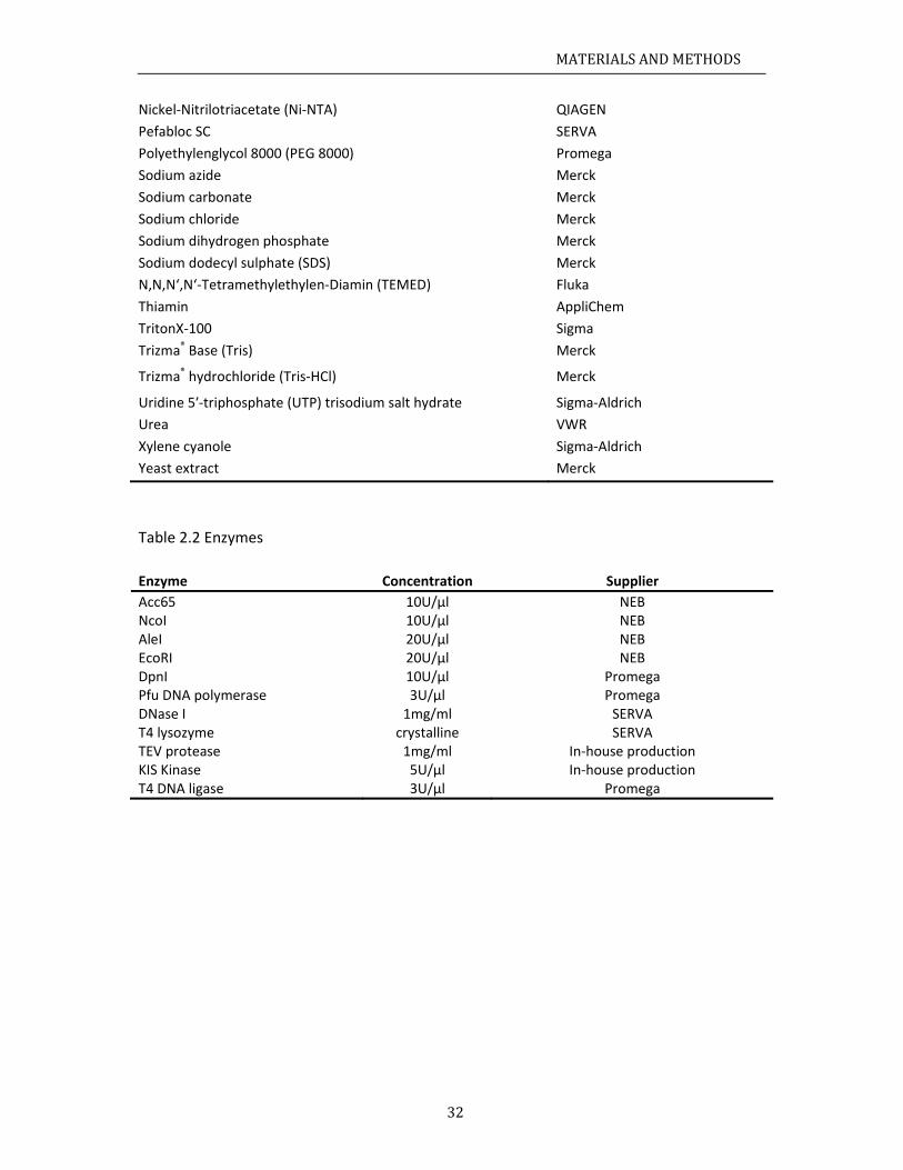
MATERIALSANDMETHODS
32
Nickel‐Nitrilotriacetate (Ni‐NTA) QIAGEN
Pefabloc SC SERVA
Polyethylenglycol 8000 (PEG 8000) Promega
Sodium azide Merck
Sodium carbonate Merck
Sodium chloride Merck
Sodium dihydrogen phosphate Merck
Sodium dodecyl sulphate (SDS) Merck
N,N,N‘,N‘‐Tetramethylethylen‐Diamin (TEMED) Fluka
Thiamin AppliChem
TritonX‐100 Sigma
Trizma® Base (Tris) Merck
Trizma® hydrochloride (Tris‐HCl) Merck
Uridine 5′‐triphosphate (UTP) trisodium salt hydrate Sigma‐Aldrich
Urea VWR
Xylene cyanole Sigma‐Aldrich
Yeast extract Merck
Table 2.2 Enzymes
Enzyme Concentration Supplier
Acc65 10U/µl NEB NcoI 10U/µl NEB AleI 20U/µl NEB EcoRI 20U/µl NEB DpnI 10U/µl Promega Pfu DNA polymerase 3U/µl Promega DNase I 1mg/ml SERVA T4 lysozyme crystalline SERVA TEV protease 1mg/ml In‐house production KIS Kinase 5U/µl In‐house production T4 DNA ligase 3U/µl Promega
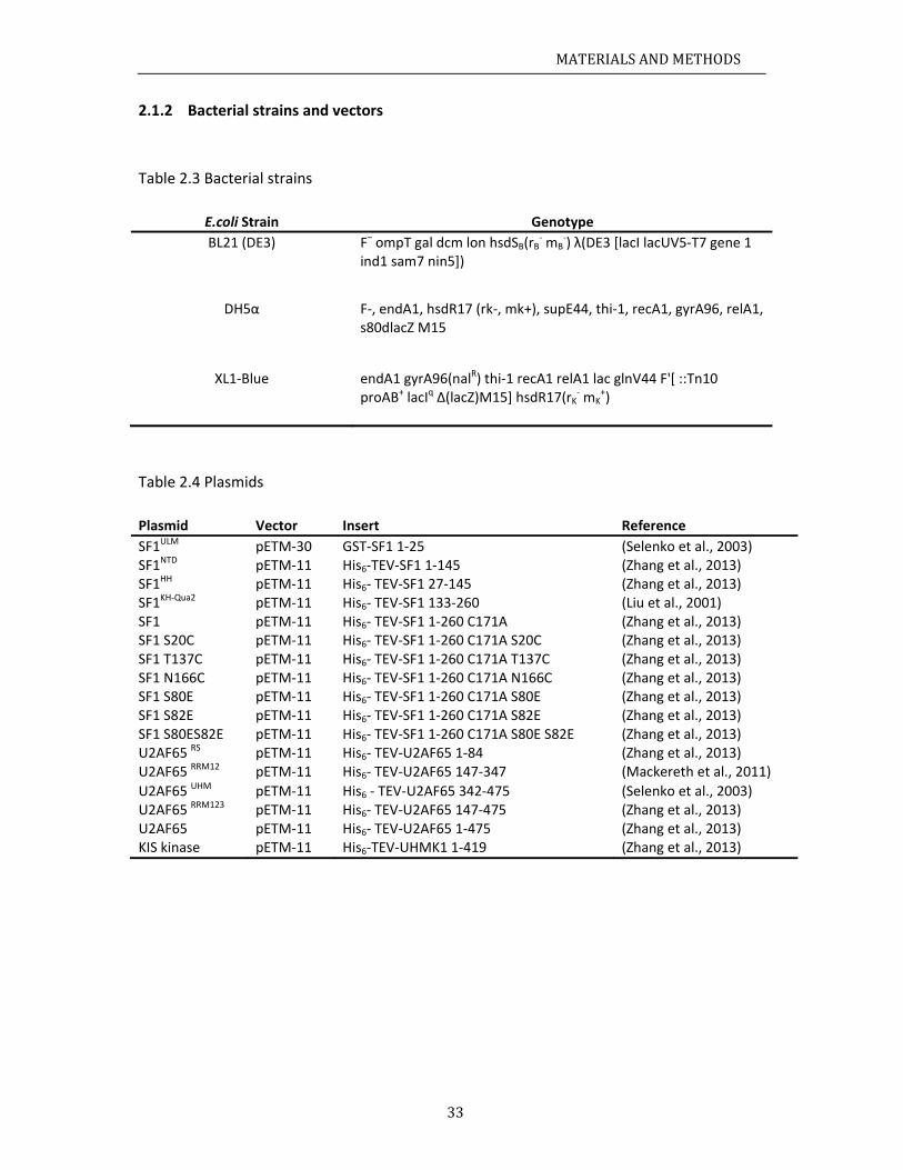
MATERIALSANDMETHODS
33
2.1.2 Bacterial strains and vectors
Table 2.3 Bacterial strains
E.coli Strain Genotype
BL21 (DE3)
F– ompT gal dcm lon hsdSB(rB‐ mB
‐) λ(DE3 [lacI lacUV5‐T7 gene 1 ind1 sam7 nin5])
DH5α
F‐, endA1, hsdR17 (rk‐, mk+), supE44, thi‐1, recA1, gyrA96, relA1, s80dlacZ M15
XL1‐Blue
endA1 gyrA96(nalR) thi‐1 recA1 relA1 lac glnV44 F'[ ::Tn10 proAB+ lacIq Δ(lacZ)M15] hsdR17(rK
‐ mK+)
Table 2.4 Plasmids
Plasmid Vector Insert Reference
SF1ULM pETM‐30 GST‐SF1 1‐25 (Selenko et al., 2003) SF1NTD pETM‐11 His6‐TEV‐SF1 1‐145 (Zhang et al., 2013) SF1HH pETM‐11 His6‐ TEV‐SF1 27‐145 (Zhang et al., 2013)SF1KH‐Qua2 pETM‐11 His6‐ TEV‐SF1 133‐260 (Liu et al., 2001) SF1 pETM‐11 His6‐ TEV‐SF1 1‐260 C171A (Zhang et al., 2013) SF1 S20C pETM‐11 His6‐ TEV‐SF1 1‐260 C171A S20C (Zhang et al., 2013)SF1 T137C pETM‐11 His6‐ TEV‐SF1 1‐260 C171A T137C (Zhang et al., 2013) SF1 N166C pETM‐11 His6‐ TEV‐SF1 1‐260 C171A N166C (Zhang et al., 2013) SF1 S80E pETM‐11 His6‐ TEV‐SF1 1‐260 C171A S80E (Zhang et al., 2013)SF1 S82E pETM‐11 His6‐ TEV‐SF1 1‐260 C171A S82E (Zhang et al., 2013) SF1 S80ES82E pETM‐11 His6‐ TEV‐SF1 1‐260 C171A S80E S82E (Zhang et al., 2013) U2AF65 RS pETM‐11 His6‐ TEV‐U2AF65 1‐84 (Zhang et al., 2013)U2AF65 RRM12 pETM‐11 His6‐ TEV‐U2AF65 147‐347 (Mackereth et al., 2011)
U2AF65 UHM pETM‐11 His6 ‐ TEV‐U2AF65 342‐475 (Selenko et al., 2003)U2AF65 RRM123 pETM‐11 His6‐ TEV‐U2AF65 147‐475 (Zhang et al., 2013) U2AF65 pETM‐11 His6‐ TEV‐U2AF65 1‐475 (Zhang et al., 2013) KIS kinase pETM‐11 His6‐TEV‐UHMK1 1‐419 (Zhang et al., 2013)
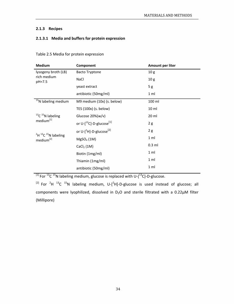
MATERIALSANDMETHODS
34
2.1.3 Recipes
2.1.3.1 Media and buffers for protein expression
Table 2.5 Media for protein expression
Medium Component Amount per liter
lysogeny broth (LB) rich medium pH=7.5
Bacto Tryptone
NaCl
yeast extract
antibiotic (50mg/ml)
10 g
10 g
5 g
1 ml
15N labeling medium 13C 15N labeling medium[1] 2H 13C 15N labeling medium[2]
M9 medium (10x) (s. below)
TES (100x) (s. below)
Glucose 20%(w/v)
or U‐[13C]‐D‐glucose[1]
or U‐[2H]‐D‐glucose[2]
MgSO4 (1M)
CaCl2 (1M)
Biotin (1mg/ml)
Thiamin (1mg/ml)
antibiotic (50mg/ml)
100 ml
10 ml
20 ml
2 g
2 g
1 ml
0.3 ml
1 ml
1 ml
1 ml
[1] For 13C 15N labeling medium, glucose is replaced with U‐[13C]‐D‐glucose.
[2] For 2H 13C 15N labeling medium, U‐[2H]‐D‐glucose is used instead of glucose; all
components were lyophilized, dissolved in D2O and sterile filtrated with a 0.22µM filter
(Millipore)
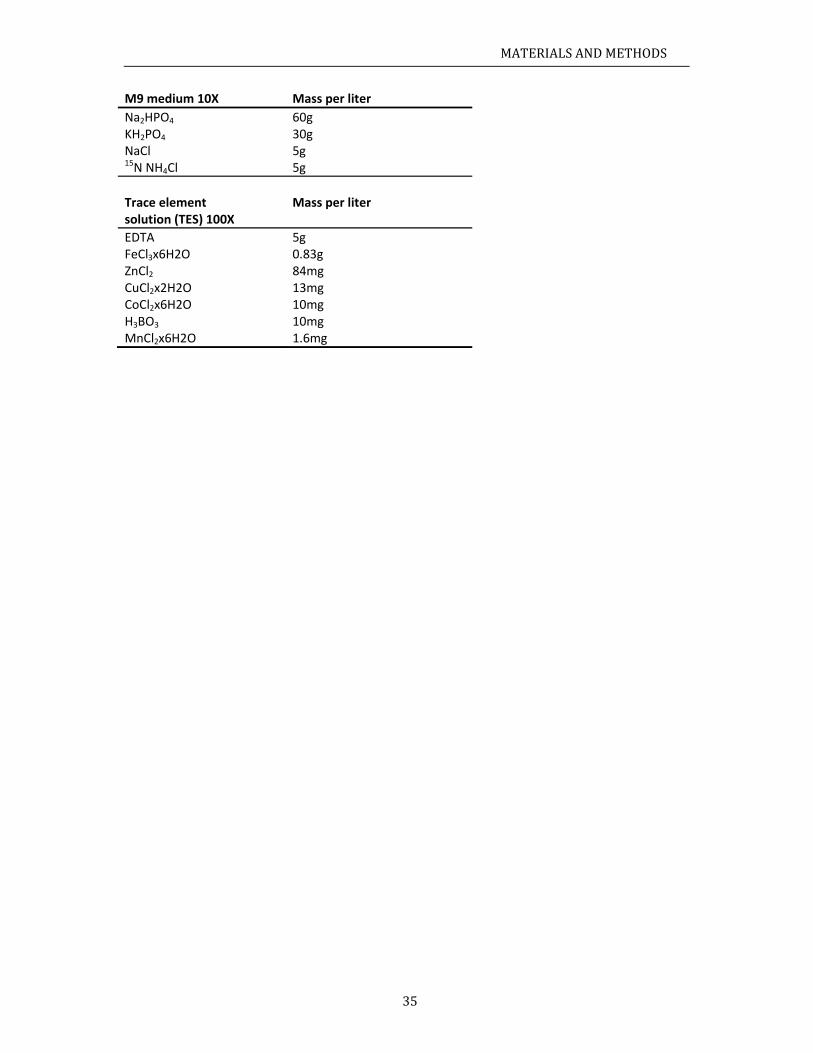
MATERIALSANDMETHODS
35
M9 medium 10X Mass per liter
Na2HPO4 60g KH2PO4 30g NaCl 5g 15N NH4Cl 5g
Trace element Mass per liter solution (TES) 100X
EDTA 5g FeCl3x6H2O 0.83g ZnCl2 84mg CuCl2x2H2O 13mg CoCl2x6H2O 10mg H3BO3 10mg MnCl2x6H2O 1.6mg
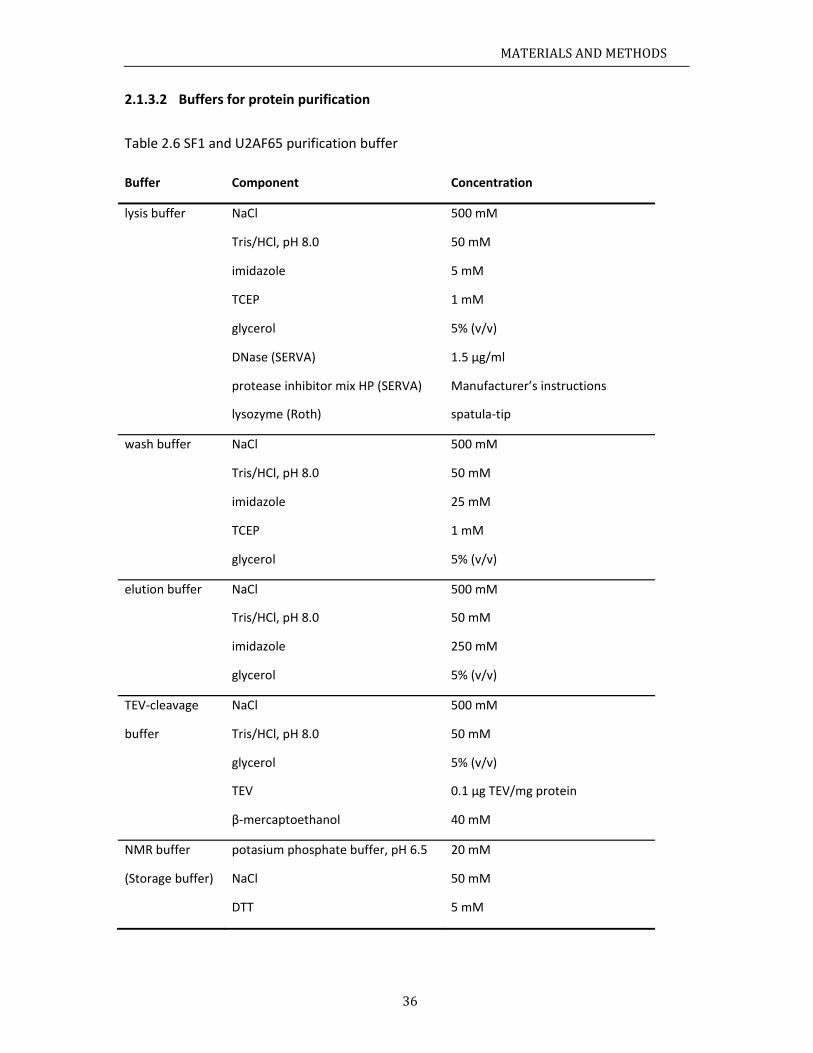
MATERIALSANDMETHODS
36
2.1.3.2 Buffers for protein purification
Table 2.6 SF1 and U2AF65 purification buffer
Buffer Component Concentration
lysis buffer NaCl 500 mM
Tris/HCl, pH 8.0 50 mM
imidazole 5 mM
TCEP 1 mM
glycerol 5% (v/v)
DNase (SERVA) 1.5 µg/ml
protease inhibitor mix HP (SERVA) Manufacturer’s instructions
lysozyme (Roth) spatula‐tip
wash buffer NaCl 500 mM
Tris/HCl, pH 8.0 50 mM
imidazole 25 mM
TCEP 1 mM
glycerol 5% (v/v)
elution buffer NaCl 500 mM
Tris/HCl, pH 8.0 50 mM
imidazole 250 mM
glycerol 5% (v/v)
TEV‐cleavage NaCl 500 mM
buffer Tris/HCl, pH 8.0 50 mM
glycerol 5% (v/v)
TEV 0.1 μg TEV/mg protein
β‐mercaptoethanol 40 mM
NMR buffer potasium phosphate buffer, pH 6.5 20 mM
(Storage buffer) NaCl 50 mM
DTT 5 mM
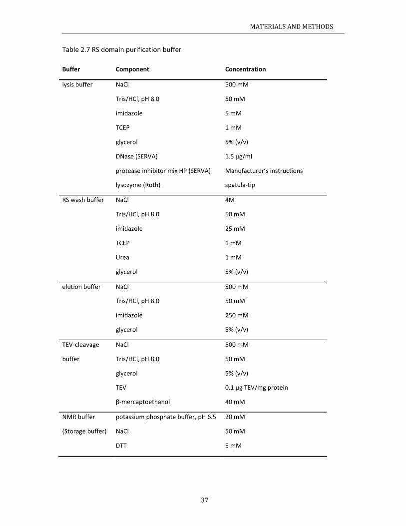
MATERIALSANDMETHODS
37
Table 2.7 RS domain purification buffer
Buffer Component Concentration
lysis buffer NaCl 500 mM
Tris/HCl, pH 8.0 50 mM
imidazole 5 mM
TCEP 1 mM
glycerol 5% (v/v)
DNase (SERVA) 1.5 µg/ml
protease inhibitor mix HP (SERVA) Manufacturer’s instructions
lysozyme (Roth) spatula‐tip
RS wash buffer NaCl 4M
Tris/HCl, pH 8.0 50 mM
imidazole 25 mM
TCEP 1 mM
Urea 1 mM
glycerol 5% (v/v)
elution buffer NaCl 500 mM
Tris/HCl, pH 8.0 50 mM
imidazole 250 mM
glycerol 5% (v/v)
TEV‐cleavage NaCl 500 mM
buffer Tris/HCl, pH 8.0 50 mM
glycerol 5% (v/v)
TEV 0.1 μg TEV/mg protein
β‐mercaptoethanol 40 mM
NMR buffer potassium phosphate buffer, pH 6.5 20 mM
(Storage buffer) NaCl 50 mM
DTT 5 mM
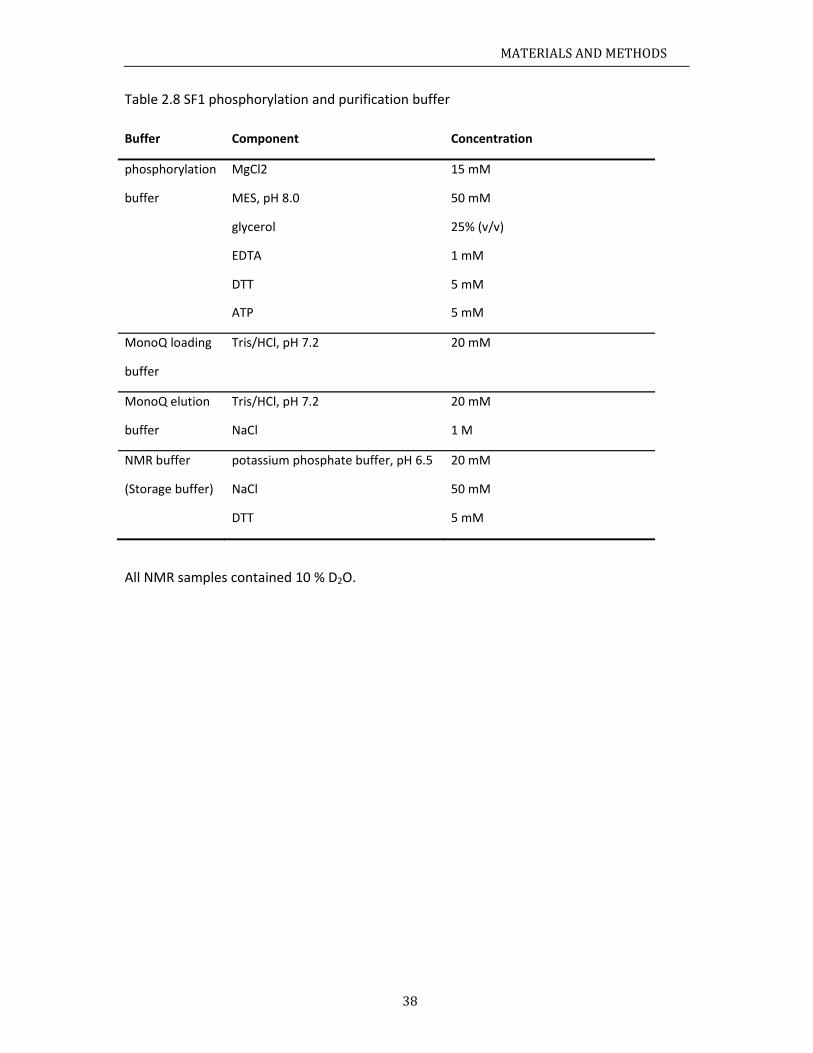
MATERIALSANDMETHODS
38
Table 2.8 SF1 phosphorylation and purification buffer
Buffer Component Concentration
phosphorylation MgCl2 15 mM
buffer MES, pH 8.0 50 mM
glycerol 25% (v/v)
EDTA 1 mM
DTT 5 mM
ATP 5 mM
MonoQ loading Tris/HCl, pH 7.2 20 mM
buffer
MonoQ elution Tris/HCl, pH 7.2 20 mM
buffer NaCl 1 M
NMR buffer potassium phosphate buffer, pH 6.5 20 mM
(Storage buffer) NaCl 50 mM
DTT 5 mM
All NMR samples contained 10 % D2O.
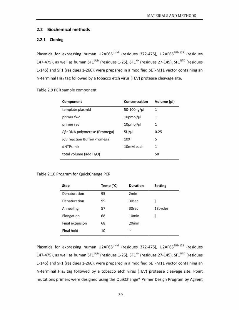
B2.2
2.2.1
Plasmid
147‐475
1‐145) a
N‐term
Table 2
Table 2
Plasmid
147‐475
1‐145) a
N‐term
mutatio
Biochemica
Cloning
ds for expr
5), as well a
and SF1 (re
inal His6 tag
.9 PCR sam
.10 Program
Step
Dena
Dena
Anne
Elong
Final
Final
ds for expr
5), as well a
and SF1 (re
inal His6 ta
ons primers
Com
temp
prim
prim
Pfu D
Pfu r
dNTP
total
al methods
ressing hum
as human S
sidues 1‐26
g followed b
ple compon
m for QuickC
p
aturation
aturation
ealing
gation
l extension
l hold
ressing hum
as human S
sidues 1‐26
ag followed
s were desig
mponent
plate plasmid
mer fwd
mer rev
DNA polyme
reaction Buff
Ps mix
l volume (ad
s
man U2AF6
SF1ULM (resid
60), were pr
by a tobacco
nent
Change PCR
Temp
95
95
57
68
68
10
man U2AF6
SF1ULM (resid
60), were pr
d by a toba
gned using
d
rase (Prome
fer(Promega
d H2O)
39
65UHM (resi
dues 1‐25),
repared in a
o etch virus
R
p (°C)
65UHM (resi
dues 1‐25),
repared in a
acco etch v
the QuikCh
Con
50‐
10p
10p
ga) 5U/
) 10X
10m
M
idues 372‐4
SF1HH (resid
a modified
s (TEV) prot
Duration
2min
30sec
30sec
10min
20min
~
idues 372‐4
SF1HH (resid
a modified
virus (TEV)
hange® Prim
ncentration
‐100ng/µl
pmol/µl
pmol/µl
/µl
X
mM each
MATERIALS
475), U2AF
dues 27‐145
pET‐M11 ve
ease cleava
Setting
]
18cycles
]
475), U2AF
dues 27‐145
pET‐M11 ve
protease c
mer Design
Volume
1
1
1
0.25
5
1
50
ANDMETH
F65RRM123 (r
5), SF1NTD (r
ector conta
age site.
F65RRM123 (r
5), SF1NTD (r
ector conta
leavage sit
Program by
(µl)
HODS
residues
residues
ining an
residues
residues
ining an
e. Point
y Agilent
MATERIALSANDMETHODS
40
Technologies and purchased from Eurofins MWG Operon. Application of that technique
allowed conveniently induced the point mutations into the plasmid; the whole mutated
plasmid sequence was amplified in a polymerase chain reaction (PCR) and could be
transformed directly into the competent cell. Pfu polymerase which is capable of proof
reading (Lundberg et al., 1991) was used to increase the amplification fidelity. PCR sample
composition and PCR program are listed in Table 2.9 and Table 2.10. The PCR products were
incubated with 1 µl DpnI for 1h at 37 °C. After heat inactivation at 60 °C for 60 min, 10 µl of
the sample were transformed into E. coli XL1‐Blue cells. SF1 deletions of helix α1 (Δ35‐68),
α2 (Δ96‐128), the complete helix hairpin (Δ35‐128), or the α1‐α2 linker (Δ75‐90) were
generated by PCR of SF12‐320 in pTRCHis A (Rain et al., 1998) with reverse and forward
primers complementary to sequences upstream and downstream, respectively, of the
desired deletion. The primers contained KpnI restriction sites and PCR products were ligated
after KpnI digestion. The deleted protein sequences were thus replaced by Gly‐Thr.
Full‐length KIS kinase (comprising the kinase domain and a UHM domain) was cloned by PCR
ligation of the DNA encoding the short KIS isoform (residues 1‐344) and the KIS UHM
(residues 315‐419). All plasmids were verified by sequencing at GATC Biotech and were
purified using the Wizard® Plus SV Minipreps DNA Purification System (Promega).
MATERIALSANDMETHODS
41
2.2.2 Protein expression and NMR sample preparation
Expression plasmids were transformed into E. coli BL21(DE3) cells, grown in standard LB
medium or in minimal M9T medium supplemented with 2 g/L [U‐13C]‐glucose and/or 1 g/L
[15N]‐ammonium chloride as the sole carbon and nitrogen sources (s. 2.1.3.1). SF1 (residues
1‐260) and U2AF65RRM123 were prepared as [U‐2H,15N]‐labelled proteins for NMR titrations
and relaxation measurements or as [U‐2H,13C,15N] deuterated samples with methyl
protonation of isoleucine, leucine and valine residues as described (Tugarinov et al., 2006)
for chemical shift assignments. Protein synthesis was induced by the addition of 0.5 mM
IPTG at OD600 ~0.8. After protein expression for 16 h at 25°C cells were collected by
centrifugation, lysed by sonication in the presence of lysozyme and EDTA‐free Complete
protease inhibitor (Roche Applied Science), and then re‐suspended in lysis buffer (Table 2.6).
The sample was loaded onto Ni‐NTA chromatography resin (Qiagen), and washed with 20
column volumes of lysis buffer followed by five column volumes of wash buffer (Table 2.6),
and then the sample was eluted with the elution buffer (Table 2.6). Removal of the His6‐tag
with 0.1 μg TEV/mg protein protease required 12 h at 4°C. The TEV protease, the
histidine‐tag and uncleaved protein were removed by a second passage of the sample
through Ni‐NTA resin. The eluate was further purified by gelfiltration on a Superdex 75
column (GE) using sodium phosphate (pH 6.5), 50 mM NaCl, and 1mM DTT as buffer. NMR
samples were concentrated to 100 to 600 µM in NMR buffer (Table 2.6).
KIS kinase was expressed in standard LB medium following the same protocol. The protein
was purified at 4°C without cleavage and removal of the His6‐tag. Aliquots of the
recombinant KIS kinase were stored at ‐80°C in buffer consisting of 50 mM MES (pH 8.0), 15
mM MgCl2, 25% (v/v) glycerol, 2 mM DTT, and 1 mM EDTA.
His6‐tagged SF12‐320 and deletion mutants for gel shift experiments were expressed in E. coli
and purified as described (Rain et al., 1998). Proteins were dialyzed against 20 mM
Hepes‐KOH pH 7.9, 100 mM KCl, 20% (v/v) glycerol, 0.2 mM EDTA and 0.5 mM DTT.
A 20‐mer RNA containing the BPS and the Py tract (5’‐UAUACUAACAAUUUUUUUUU‐3’) was
purchased from BioSpring GmbH, dissolved in H2O to a final concentration of 10 mM and
added to the protein samples before the NMR and SAXS measurements.
MATERIALSANDMETHODS
42
2.2.3 In vitro phosphorylation
Phosphorylation of SF1NTD and SF1 for NMR analysis was performed in 10 ml
phosphorylation buffer (Table 2.8) in the presence of 1 ml 10 × phos‐stop (Roche), 40 µg of
KIS kinase and 2 mg of substrate. 100mM ATP stock solution was prepared by dissolving ATP
powder (Sigma) in kinase buffer and adjusting the pH to 8.0. Aliquots were stored at ‐20°C.
The reaction typically required 24‐48h at 30°C to fully phosphorylate both Ser80 and Ser82.
The phosphorylation products were purified with a MonoQ ion exchange column (GE). The
reaction products were buffer exchanged to MonoQ loading buffer (Table 2.8) and loaded
onto the column. The NaCl concentration in the elution buffer was gradually increased over
40 column volumes from 0‐1 M, and 1‐ml fractions were collected. The phosphorylation
products were separated depending on the phosphorylation state. The
double‐phosphorylation of Ser80 and Ser82 in phosphorylated SF1NTD (pSF1NTD) and SF1
(pSF1) was confirmed by SDS‐PAGE, mass spectrometry (Figure 3.22) and, for isotope
labelled samples, by NMR‐spectroscopy (Figure 3.24).
2.2.4 Isothermal titration calorimetry
Calorimetric titrations were performed using an iTC200 microcalorimeter (MicroCal) at 25°C.
The buffer used for the protein and ligand samples was 20 mM sodium phosphate (pH 6.5),
50 mM NaCl. The 200‐µl sample cell was filled with a 5 or 10 µM solution of protein and the
40‐µl injection syringe with 50 or 100 µM of the titrating ligand. Each titration consisted of a
preliminary 0.2‐µl injection followed by 20 subsequent 2‐µl injections. The heat of the
injections was corrected for the heat of dilution of every ligand into the buffer. At least two
replicas were performed for each experiment. Binding thermodynamic models
corresponding to bimolecular complex formation were fitted using routines provided by the
manufacturer.
2.2.5 Circular Dichroism
The stability of phosphorylated protein was determined by using Circular Dichroism (CD)
spectrum. In this study, 230nm wavelength was chosen to monitor. 300 µL of 10 µM protein
in phosphate buffer (NMR) is good for the CD measurements, Tris buffer is not appropriate
MATERIALSANDMETHODS
43
for thermal denaturation and it should avoid to use high NaCl concentration. The
temperature varied from 10 to 60 °C and the heating rate was 2 °C per minute. The curves
were fitted with Boltzmann function.
2.2.6 Spin labeling
A stable nitroxide radical, 3‐(2‐iodoacetamido)‐PROXYL (Sigma‐Aldrich), was attached to
single cysteine variants of SF1, in which the wildtype cysteine 171 was mutated to alanine,
were generated as described in 2.2.1. Single cysteine residues were introduced into variants
SF1 at position S20, T137 and N166, respectively (s. Table 2.4). 15N labeled SF1 were
expressed and purified according to the description in 2.2.2. Variants SF1 were
phosphorylated as described in 2.2.3. 5 mM DTT was adjusted in the storage buffer to
ascertain a complete reduction of the cysteine. For the spin labeling, the buffer was
exchanged to 1 M Tris; pH 8.0 using a PD‐10 desalting column (GE Healthcare). High
concentration of Tris buffers the hydriodic acid, which was produced during the labeling
reaction. A five‐fold excess of iodoacetamide proxyl was added and incubated in the dark at
room temperature overnight. The unbound spin label was removed and the buffer was
exchanged to NMR buffer via a PD‐10 desalting column (GE Healthcare) and concentrated in
an Amicon Ultra‐15 Centrifugal Filter Units (Millipore) to 100 µM. After recording a 1H, 15N
HSQC spectrum in the oxidized state the spin label was reduced by adding a three‐fold
excess of ascorbate; pH 6.5, and incubating for at least 30 min. Another 1H, 15N HSQC
spectrum was measured and the PRE effect on backbone amide protons was determined by
calculating the signal intensities in the spectra as (Iox the signal intensity in the oxidized
state, Ired the signal intensity in the reduced state). Spectra were recorded on a Bruker
Avance III 800MHz spectrometer at 298 K.
2.2.7 Electrophoretic mobility shift assays (EMSAs)
The 3’ splice site RNA (GGUCAUACUAACCCUGUCCCUUUUUUUUCCACAG|C; | denotes the 3’
splice site) is derived from the 3’ splice site of AdML intron 1 by replacing the original BPS
with a consensus BPS (underlined). The RNA was synthesized with the T7‐MEGA shortscript
kit (Ambion) in the presence of [α‐32P] UTP and gel‐purified. RNA binding was performed for
15 min at room temperature in 10‐µl reactions containing U2AF65 RRM123, SF1 proteins and
MATERIALSANDMETHODS
44
50 pmol [α‐32P] UTP‐RNA. Assays with SF12‐320 and deletion mutants were performed in the
presence of 12 % (v/v) glycerol, 12 mM Hepes‐KOH (pH 7.9), 4 mM potassium‐phosphate
(pH 6.5), 60 mM KCl, 10 mM NaCl, 1.8 mM MgCl2, 0.5 mM DTT, 0.12 mM EDTA, 5 µg tRNA
and 8U RiboShield™ ribonuclease inhibitor (Dundee Cell Products). Assays with SF1 and pSF1
contained final concentrations of 8 mM potassium‐phosphate (pH 6.5), 20 mM NaCl, 0.5
mM DTT, 5 µg tRNA and 8U RiboShield™ ribonuclease inhibitor. Reaction products were
resolved in native 5% polyacrylamide gels (acrylamide:bisacryamide = 80:1) in 0.5 x TBE at
4°C for 3 h at 150 V.
N2.3
2.3.1
All sam
spectra
spectro
excepti
1995) a
Protein
compar
2001; S
from H
1999).
well‐res
HN‐C’ r
experim
AG, Reg
2.3.2
13Cα an
in whic
coil che
Chemic
where Δ
NMR and SA
NMR meas
ples were m
were reco
ometers, eq
on of the A
and analyze
backbone
rison of 1H,
Selenko et
HCCH‐TOCSY
Intermolec
solved peak
residual di
ments (Sattl
gensburg, G
Secondary
d 13Cβ seco
,
ch δ13C(α,β)
emical shift
cal shift pert
Δδ = chemic
AXS exper
surements a
measured a
rded on AV
quipped w
AVIII 750M
ed in Sparky
assignmen
,15N‐HSQC
al., 2003).
Y, 15N‐ and
cular NOEs
ks in the 3
ipolar coup
er et al., 19
Germany) (H
chemical sh
ondary chem
)(exp), expe
values (Wis
turbations o
cal shift diff
iments
and assignm
at 298 K in N
VIII 500, AV
with cryoge
MHz). Data w
y 3 (T. D. G
nts were ob
and ‐TROSY
Amino acid
d 13C‐edited
s between
3D 13C‐ and
plings for
999). Alignm
Hansen et a
hift analysi
mical shifts w
erimentally
shart and Sy
of signals in
ference of t
45
ments
NMR buffer
VIII 600, AVI
enic triple
were proce
Goddard an
btained fro
Y spectra w
d side chain
d NOESY‐HS
the U2AF
15N‐edited
SF1HH we
ment media
l., 1998).
s and chem
were calcul
obtained c
ykes, 1994)
n 15N, 1H HSQ
the signal in
M
r with 10% 2
II 750, AVII
resonance
essed in NM
d D. G. Kne
om HNCACB
with previo
n resonance
SQC/HMQC
F65UHM and
d NOESY‐HS
re recorde
consisted o
mical shift pe
ated accord
chemical sh
.
QC spectra
6
n the differe
MATERIALS
2H2O added
II 800 or AV
gradient
MRPipe/Dra
eller, Unive
B and HNC
ously report
e assignme
C experime
SF1NTD w
SQC experi
ed using H
of 15 mg/m
erturbation
ding to
hift and δ13
were calcu
ent spectra.
ANDMETH
d for the loc
VI 900 Bruk
probes (w
aw (Delaglio
ersity of Cal
CA spectra,
ted data(Liu
ents were o
nts (Sattle
were identif
ments. HN‐
HNCO‐base
ml Pf1 phage
ns
C(α,β)(rc),
lated accor
HODS
ck. NMR
ker NMR
with the
o et al.,
lifornia).
and by
u et al.,
obtained
r et al.,
fied for
N, N‐C’,
d NMR
e (Profos
random
rding to
MATERIALSANDMETHODS
46
2.3.3 Relaxation analysis
15N R1, R1ρ relaxation rates and 1H‐15N heteronuclear NOEs of the SF1NTD and U2AF65UHM
complexes were recorded at 750 MHz, of SF1NTD at 600 MHz and of SF1 at 800 MHz proton
Larmor frequency at 298 K as described (Farrow et al., 1994). Values were determined from
the ratio between signal intensity in the experiment with and without saturation with the
error calculated from experimental noise. Two delays in each experiment were recorded in
duplicates to define the experimental error. Spectra were processed with NMRPipe. Local
correlation times were derived from the 15N R2/R1 ratio according to
≅1
2
32
76
with ν 15N = the resonance frequency of 15N in Hz (Farrow et al., 1994).
MATERIALSANDMETHODS
47
2.3.4 Structure calculations
The structure of SF1HH was determined using standard approaches. Automated NOESY
cross‐peak assignments and structure calculations with torsion angle dynamics were
performed with CYANA 3.0 (Guntert, 2009). NOE‐derived distance restraints derived from
CYANA together with φ and ψ backbone dihedral angle restraints derived from TALOS+
(Shen et al., 2009) based on secondary chemical shifts and residual dipolar couplings were
used during the structure calculation and for final water refinement (Linge et al., 2003).
Structures were validated with iCing (http://nmr.cmbi.ru.nl/icing/). Molecular images were
generated with PyMol (Schrödinger).
The structure of the SF1NTD‐U2AF65UHM complex was calculated using our previously
reported protocol (Simon et al., 2010). The SF1HH structure determined here and the
structure of the U2AF65UHM/SF1ULM complex (Selenko et al., 2003) were used as input
structures for semi‐rigid calculation of the complex structure. The input structures were
maintained using non‐crystallographic symmetry (NCS) restraints with a modified version of
Aria1.2/CNS (Simon et al., 2010). Distance restraints derived from intermolecular NOEs and
backbone torsion angle restraints derived from TALOS+ (Shen et al., 2009) were employed
during molecular dynamics and simulated annealing. The final structures were refined in a
shell of water molecules (Linge et al., 2003).
2.3.5 Small‐angle X‐ray scattering (SAXS)
SAXS data for solutions of SF1NTD, pSF1NTD, SF1, pSF1, U2AF65UHM, U2AF65RRM123,
SF1NTD‐U2AF65UHM, pSF1NTD‐U2AF65UHM, SF1‐U2AF65RRM123, pSF1‐U2AF65RRM123,
SF1‐U2AF65RRM123‐RNA, and pSF1‐U2AF65RRM123‐RNA were recorded at the X33 beamline of
the European Molecular Biology Laboratory (EMBL) at Deutsches Elektronen Synchrotron
(DESY) (Hamburg) using a MAR345 image plate detector. The scattering patterns were
measured with a 2‐min exposure time (8 frames, each 15 seconds) for several solute
concentrations in the range from 1 to 10 mg/ml. Radiation damage was excluded based on a
comparison of individual frames of the 2‐min exposures, where no changes were detected.
Using the sample‐detector distance of 2.7 m, a range of momentum transfer of 0.01 <s< 0.6
Å‐1 was covered (s = 4π sin(θ)/λ, where 2θ is the scattering angle and λ = 1.5 Å is the X‐ray
wavelength).
MATERIALSANDMETHODS
48
All SAXS data were analyzed with the package ATSAS. The data were processed using
standard procedures and extrapolated to infinite dilution with the program PRIMUS
(Konarev et al., 2003). The forward scattering, I(0), and the radius of gyration, Rg, were
evaluated using the Guinier approximation. The values of I(0) and Rg, as well as the
maximum dimension, Dmax, and the inter‐atomic distance distribution functions, (P(R)), were
computed with the program GNOM (Svergun, 1992). The scattering from the high resolution
models was computed with the program CRYSOL (Svergun et al., 1995). The masses of the
solutes were evaluated by comparison of the forward scattering intensity with that of a
bovine serum albumin reference solution (molecular mass 66 kDa). For back‐calculation of
SAXS data from the NMR ensemble of the SF1NTD‐U2AF65UHM complex, the flexible regions
(SF1NTD: 1‐12, 26‐38, 70‐95, 128‐145) were randomized with CORAL (Petoukhov and Svergun,
2005).
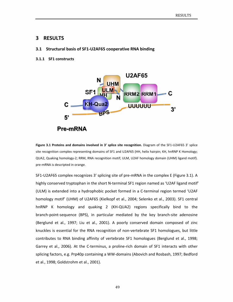
3 RE
St3.1
3.1.1
Figure 3.
site recog
QUA2, Q
pre‐mRN
SF1‐U2A
highly c
(ULM) i
homolo
hnRNP
branch‐
(Berglu
knuckle
contrib
Garrey
splicing
et al., 1
ESULTS
tructural b
SF1 constru
.1 Proteins an
gnition comp
Quaking homo
NA is descripte
AF65 comp
conserved t
is extended
ogy motif’ (
K homol
‐point‐sequ
nd et al.,
es is essent
utes to RN
et al., 200
g factors, e.g
998; Goldst
basis of SF1
ucts
nd domains i
lex represent
logy‐2; RRM,
ed in orange.
lex recogniz
ryptophan
d into a hyd
UHM) of U
logy and
uence (BPS)
1997; Liu e
ial for the
A binding a
6). At the C
g. Prp40p co
trohm et al
1‐U2AF65
nvolved in 3’
ing domains o
RNA recognit
zes 3’ splici
in the short
drophobic p
2AF65 (Kie
quaking 2
), in partic
et al., 2001
RNA recogn
affinity of v
C‐terminus,
ontaining a
., 2001).
49
cooperativ
’ splice site re
of SF1 and U2
tion motif; UL
ng site of p
t N‐termina
pocket form
lkopf et al.,
2 (KH‐QUA
cular media
1). A poorl
nition of no
vertebrate
, a proline‐
WW‐doma
ve RNA bin
ecognition. D
2AF65 (HH, he
LM, U2AF hom
pre‐mRNA in
al SF1 region
med in a C‐t
, 2004; Sele
A2) region
ated by th
ly conserve
on‐vertebra
SF1 homol
‐rich domai
ains (Abovic
nding
Diagram of the
elix hairpin; K
mology domai
n the comp
n named as
terminal reg
enko et al.,
ns specifica
e key bran
ed domain
ate SF1 hom
ogues (Ber
n of SF1 in
ch and Rosb
RESU
e SF1‐U2AF65
H, hnRNP K H
n (UHM) ligan
lex E (Figure
‘U2AF ligan
gion terme
2003). SF1
ally bind
nch‐site ad
composed
mologues, b
rglund et al
nteracts wit
bash, 1997;
ULTS
5 3’ splice
Homology;
nd motif).
e 3.1). A
nd motif’
d ‘U2AF
1 central
to the
denosine
of zinc
but little
l., 1998;
th other
Bedford
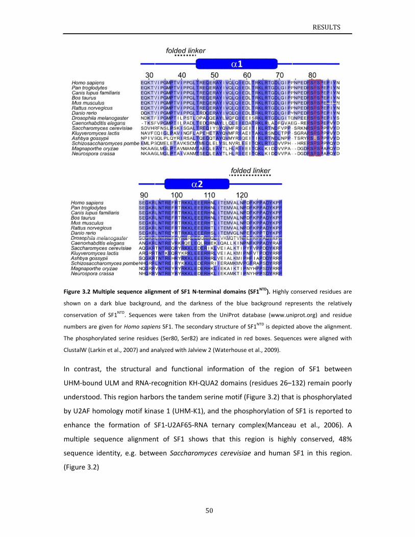
Figure 3.
shown o
conserva
numbers
The phos
ClustalW
In cont
UHM‐b
underst
by U2A
enhanc
multiple
sequen
(Figure
.2 Multiple se
on a dark blu
ation of SF1NT
s are given for
sphorylated s
W (Larkin et al.,
trast, the
ound ULM
tood. This r
AF homology
e the form
e sequence
ce identity,
3.2)
equence align
ue backgroun
TD. Sequences
r Homo sapie
serine residue
, 2007) and an
structural
and RNA‐re
egion harbo
y motif kina
mation of S
e alignmen
, e.g. betw
nment of SF1
nd, and the
s were taken
ns SF1. The se
es (Ser80, Ser
nalyzed with J
and functi
ecognition
ors the tand
ase 1 (UHM
SF1‐U2AF65
nt of SF1 s
ween Saccha
50
1 N‐terminal d
darkness of
n from the U
econdary stru
r82) are indica
Jalview 2 (Wat
onal inform
KH‐QUA2 d
dem serine
M‐K1), and t
5‐RNA tern
shows that
aromyces c
domains (SF1
the blue bac
niProt databa
ucture of SF1N
ated in red b
terhouse et a
mation of
domains (re
motif (Figu
he phospho
nary compl
t this regio
erevisiae a
1NTD). Highly c
ckground rep
ase (www.un
NTD is depicted
boxes. Sequen
l., 2009).
the region
sidues 26–1
ure 3.2) that
orylation of
ex(Mancea
on is highl
nd human
RESU
conserved res
presents the
iprot.org) an
d above the a
nces were alig
n of SF1 b
132) remain
t is phospho
f SF1 is repo
u et al., 2
y conserve
SF1 in this
ULTS
sidues are
relatively
d residue
alignment.
gned with
between
n poorly
orylated
orted to
2006). A
ed, 48%
s region.
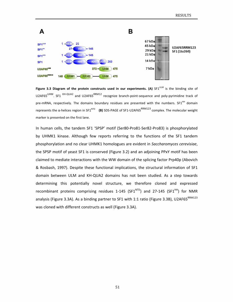
Figure 3
U2AF65U
pre‐mRN
represen
marker is
In hum
by UHM
phosph
the SPS
claimed
& Rosb
domain
determ
recomb
analysis
was clo
.3 Diagram o
UHM. SF1
KH‐Q
NA, respective
nts the α‐helic
s presented o
an cells, th
MK1 kinase
orylation a
SP motif of
d to mediat
bash, 1997).
n between
ining this
binant prot
s (Figure 3.3
ned with di
of the protei
QUA2 and U2A
ely. The dom
ces region in S
n the first lan
he tandem S
e. Although
nd no clear
yeast SF1 is
e interactio
. Despite th
ULM and K
potentiall
eins comp
3A). As a bi
ifferent con
n constructs
AF65RRM12
re
ains boundar
SF1NTD (B) SD
e.
SF1 ‘SPSP’
h few repo
r UHMK1 ho
s conserved
ons with the
hese functio
KH‐QUA2 d
y novel s
rising resid
nding partn
nstructs as w
51
used in our
cognize bran
ry residues a
DS‐PAGE of SF
motif (Ser8
rts referrin
omologues
d (Figure 3.
e WW doma
onal implica
domains ha
structure,
dues 1‐145
ner to SF1 w
well (Figure
experiments
ch‐point‐sequ
re presented
F1‐U2AF65RRM
80‐Pro81‐Se
ng to the f
are evident
2) and an a
ain of the s
ations, the
as not been
we theref
(SF1NTD) a
with 1:1 rat
3.3A).
s. (A) SF1ULM
uence and po
d with the nu
M123 complex
er82‐Pro83)
functions o
t in Sacchar
adjoining PP
plicing facto
structural
n studied.
fore clone
and 27‐145
io (Figure 3
RESU
is the bindin
oly‐pyrimidine
umbers. SF1H
. The molecul
is phospho
of the SF1
romyces cer
PxY motif h
or Prp40p (
information
As a step t
d and ex
5 (SF1HH) fo
3.3B), U2AF
ULTS
ng site of
e track of
H domain
lar weight
orylated
tandem
revisiae,
has been
Abovich
n of SF1
towards
xpressed
or NMR
65RRM123
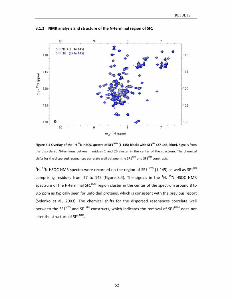
3.1.2
Figure 3.
the disor
shifts for
1H, 15N
compris
spectru
8.5 ppm
(Selenk
betwee
alter th
NMR analy
.4 Overlay of
rdered N‐term
r the dispersed
HSQC NMR
sing residu
um of the N
m as typical
o et al., 2
en the SF1N
e structure
ysis and stru
the 1H 15N HS
minus betwee
d resonances
R spectra w
es from 27
‐terminal S
ly seen for
003). The
TD and SF1H
of SF1NTD.
ucture of th
SQC spectra o
en residues 1
correlate wel
were recorde
7 to 145 (F
SF1ULM regio
unfolded p
chemical s
HH construc
52
he N‐termin
of SF1NTD (1‐14
and 26 clust
l between the
ed on the r
Figure 3.4).
on cluster in
roteins, wh
shifts for th
cts, which in
nal region o
45; black) wit
ter in the cen
e SF1NTD and S
region of SF
The signa
n the cente
ich is consis
he disperse
ndicates the
of SF1
th SF1HH (27‐1
nter of the sp
SF1HH
constru
F1 NTD (1‐14
ls in the 1
r of the spe
stent with t
ed resonan
e removal o
RESU
145, blue). Sig
pectrum. The
cts.
45) as well a
H, 15N HSQ
ectrum arou
the previou
nces correla
of SF1ULM d
ULTS
gnals from
chemical
as SF1HH
QC NMR
und 8 to
s report
ate well
does not
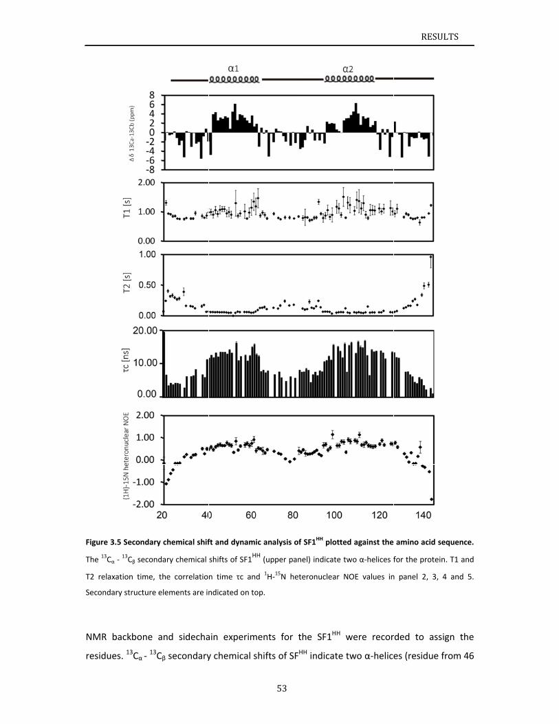
Figure 3.
The 13Cα
T2 relaxa
Secondar
NMR b
residue
.5 Secondary
‐ 13Cβ second
ation time, th
ry structure e
backbone a
es. 13Cα ‐ 13Cβ
chemical shif
ary chemical
he correlatio
lements are in
nd sidecha
β secondary
ft and dynam
shifts of SF1H
n time τc an
ndicated on to
ain experim
y chemical s
53
ic analysis of
HH (upper pan
nd 1H‐15N het
op.
ments for t
shifts of SFH
f SF1HH plotted
el) indicate tw
teronuclear N
he SF1HH w
HH indicate t
d against the
wo α‐helices f
NOE values in
were recor
two α‐helice
RESU
amino acid s
for the protei
n panel 2, 3,
rded to ass
es (residue
ULTS
sequence.
in. T1 and
4 and 5.
sign the
from 46
RESULTS
54
to 67 and from 97 to 119) connected by an unstructured loop region (residue from 68 to
96).
Dynamic properties of SF1HH were analyzed by recording T1 and T2 relaxation time as well as
the 1H‐15N heteronuclear NOE (Figure 3.5). Quantitative evaluation was limited to
approximately 70% of the peaks due to signal overlap in 1H, 15N HSQC spectra. T1 relaxation
time of the two α‐helices very similar and T2 relaxation time is increased for the loop region
connecting the two α‐helices.
These residues in the loop region also have a decreased correlation time (Figure 3.5). The
correlation time τc for the two helices is similar suggesting the helix hairpin is tumbling as a
unit. The overall correlation time τc (10 ns) indicates monomeric tumbling of SF1HH. 1H‐15N
heteronuclear NOE data of SF1HH describe a rigid helix hairpin domain with values of
approximately 0.75 and a flexible linker in between from residue 68 to 96 with very low
rates between 0.3 to ‐0.3 which is indicative for dynamics on a picosecond to nanosecond
timescale. Based on distance, dihedral angle, and residual dipolar coupling restraints (Table
3.1, Figure 3.7), we determined the three‐dimensional structure of SF1HH.
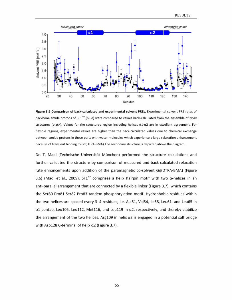
Figure 3.
backbone
structure
flexible r
between
because
Dr. T. M
further
rate en
3.6) (M
anti‐pa
the Ser
the two
α1 cont
the arra
with As
.6 Compariso
e amide proto
es (black). Va
regions, expe
amide proto
of transient b
Madl (Tech
validated t
nhancement
Madl et al.,
rallel arrang
r80‐Pro81‐S
o helices ar
tact Leu105
angement o
sp128 C‐term
n of back‐calc
ons of SF1HH (
lues for the s
rimental valu
ns in these pa
binding to Gd(
hnische Uni
the structu
ts upon ad
2009). SF1
gement tha
Ser82‐Pro83
e spaced ev
5, Leu112,
of the two h
minal of he
culated and e
blue) were co
structured re
ues are highe
arts with wate
DTPA‐BMA).T
iversität M
re by comp
dition of th
1HH compris
at are conne
3 tandem p
very 3–4 re
Met116, an
helices. Arg
lix α2 (Figur
55
experimental
ompared to va
gion including
r than the ba
er molecules w
The secondary
ünchen) pe
parison of m
he paramag
ses a helix
ected by a f
hosphoryla
esidues, i.e.
nd Leu119 i
g109 in helix
re 3.7).
solvent PREs
alues back‐ca
g helices α1‐
ack‐calculated
which experie
y structure is d
erformed th
measured a
gnetic co‐so
hairpin mo
flexible linke
ation motif.
Ala51, Val5
in α2, respe
x α2 is enga
s. Experiment
lculated from
α2 are in exc
d values due
ence a large re
depicted abov
he structur
and back‐ca
olvent Gd(D
otif with tw
er (Figure 3
Hydropho
54, Ile58, Le
ectively, an
aged in a p
RESU
tal solvent PR
m the ensembl
cellent agreem
to chemical
elaxation enha
ve the diagram
re calculatio
alculated re
DTPA‐BMA)
wo α‐helice
3.7), which c
bic residue
eu61, and L
nd thereby
otential sal
ULTS
E rates of
e of NMR
ment. For
exchange
ancement
m.
ons and
elaxation
) (Figure
es in an
contains
s within
Leu65 in
stabilize
lt bridge
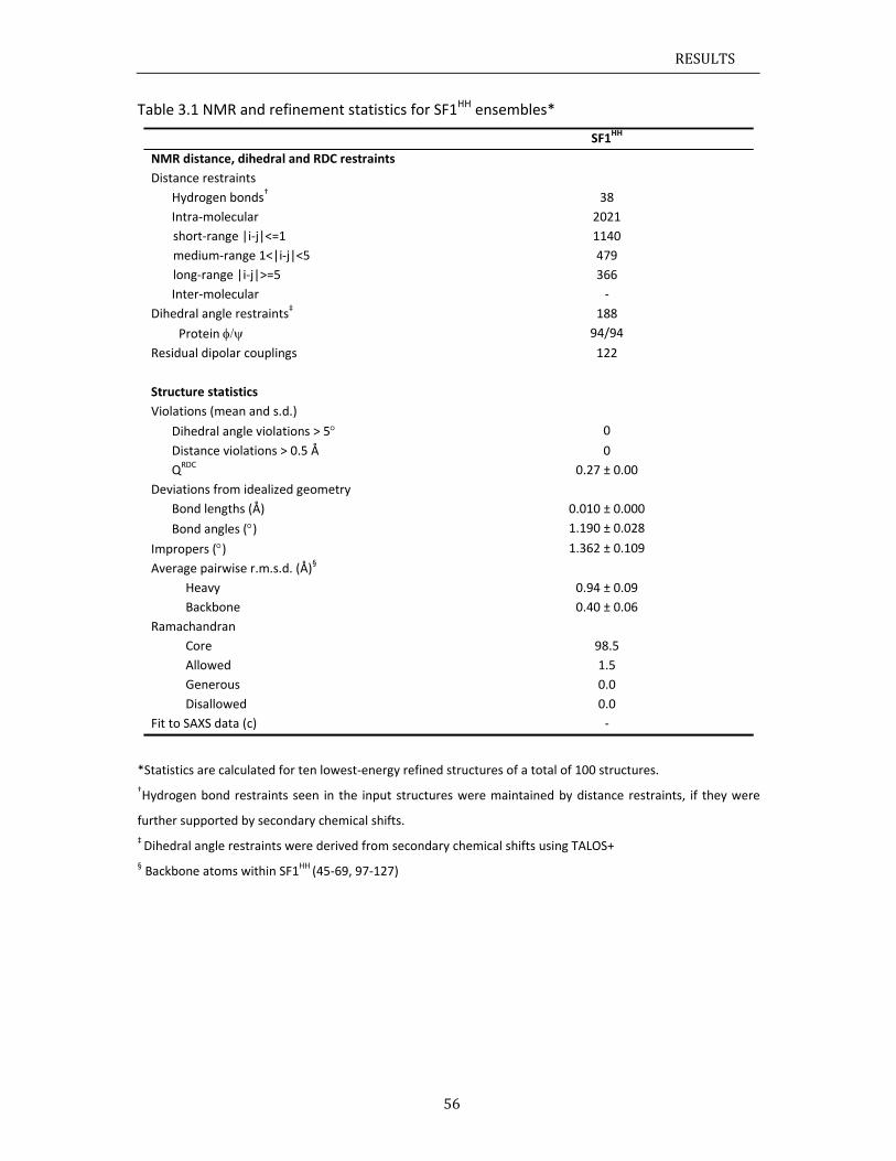
RESULTS
56
Table 3.1 NMR and refinement statistics for SF1HH ensembles*
SF1HH
NMR distance, dihedral and RDC restraints
Distance restraints
Hydrogen bonds† 38
Intra‐molecular 2021
short‐range |i‐j|<=1
medium‐range 1<|i‐j|<5
long‐range |i‐j|>=5
1140
479
366
Inter‐molecular ‐
Dihedral angle restraints‡ 188
Protein 94/94
Residual dipolar couplings 122
Structure statistics
Violations (mean and s.d.)
Dihedral angle violations > 5 0
Distance violations > 0.5 Å 0
QRDC 0.27 ± 0.00
Deviations from idealized geometry
Bond lengths (Å) 0.010 ± 0.000
Bond angles () 1.190 ± 0.028
Impropers () 1.362 ± 0.109
Average pairwise r.m.s.d. (Å)§
Heavy 0.94 ± 0.09
Backbone 0.40 ± 0.06
Ramachandran
Core 98.5
Allowed 1.5
Generous 0.0
Disallowed 0.0
Fit to SAXS data (c) ‐
*Statistics are calculated for ten lowest‐energy refined structures of a total of 100 structures.
†Hydrogen bond restraints seen in the input structures were maintained by distance restraints, if they were
further supported by secondary chemical shifts.
‡ Dihedral angle restraints were derived from secondary chemical shifts using TALOS+
§ Backbone atoms within SF1HH (45‐69, 97‐127)
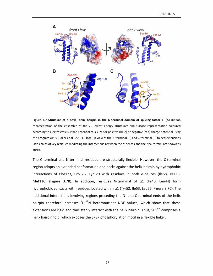
Figure 3
represen
according
the progr
Side chai
sticks.
The C‐t
region a
interact
Met116
hydrop
addition
hairpin
extensi
helix ha
.7 Structure
ntation of the
g to electrosta
ram APBS (Ba
ns of key resi
terminal an
adopts an e
tions of Ph
6) (Figure
hobic conta
nal interact
therefore
ons are rigi
airpin fold, w
of a novel h
e ensemble o
atic surface p
ker et al., 200
dues mediatin
nd N‐termin
extended co
he123, Pro
3.7B). In
acts with re
tions involv
increases
id and thus
which expo
helix hairpin
of the 20 low
otential at 3 k
01). Close‐up v
ng the interac
nal residues
onformation
o126, Tyr12
addition,
sidues loca
ving regions
1H‐15N he
stably inte
ses the SPS
57
in the N‐ter
west energy s
kT/e for posit
view of the N‐
ctions betwee
s are struct
n and packs
29 with res
residues N
ted within
s preceding
teronuclear
eract with th
SP phosphor
rminal doma
structures an
ive (blue) or n
‐terminal (B) a
en the α‐helice
turally flexi
s against the
sidues in b
N‐terminal
α1 (Tyr52, I
the N‐ and
r NOE valu
he helix ha
rylation mo
in of splicing
nd surface re
negative (red)
and C‐termina
es and the N/
ble. Howev
e helix hairp
both α‐heli
of α1 (Ile
Ile53, Leu56
d C‐termina
ues, which
irpin. Thus,
otif in a flexi
RESU
g factor 1. (A
presentation
charge poten
al (C) folded e
C‐termini are
ver, the C‐t
pin by hydr
ices (Ile58,
e40, Leu44
6; Figure 3.7
al ends of t
show tha
SF1HH com
ble linker.
ULTS
A) Ribbon
coloured
ntial using
extensions.
shown as
terminal
ophobic
Ile113,
4) form
7C). The
he helix
at these
mprises a
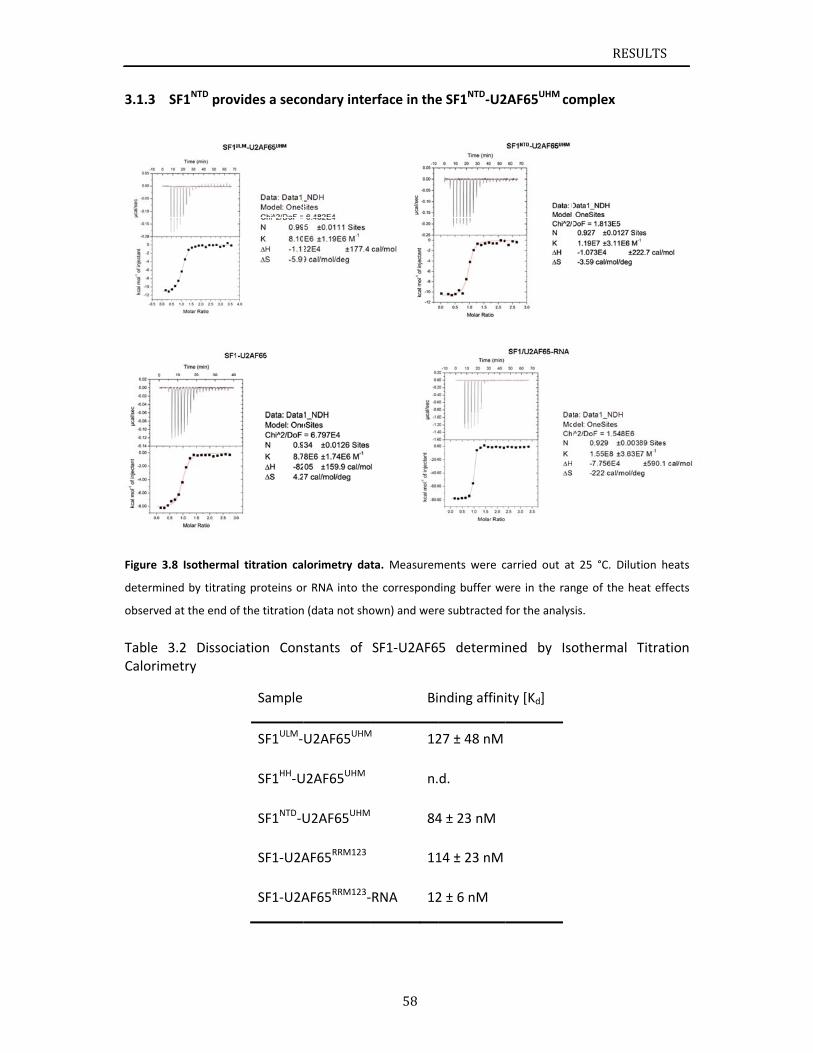
3.1.3
Figure 3.
determin
observed
Table 3Calorim
SF1NTD prov
.8 Isotherma
ned by titratin
d at the end o
3.2 Dissocimetry
vides a seco
l titration ca
ng proteins o
f the titration
iation Con
Sample
SF1ULM‐
SF1HH‐U
SF1NTD‐U
SF1‐U2A
SF1‐U2A
ondary inte
lorimetry dat
r RNA into th
n (data not sho
stants of
U2AF65UHM
U2AF65UHM
U2AF65UHM
AF65RRM123
AF65RRM123‐
58
erface in the
ta. Measurem
he correspond
own) and wer
SF1‐U2AF6
Bi
M 12
n.
84
11
RNA 12
e SF1NTD‐U2
ments were c
ding buffer we
re subtracted
5 determin
nding affini
27 ± 48 nM
d.
4 ± 23 nM
14 ± 23 nM
2 ± 6 nM
2AF65UHM co
carried out at
ere in the ran
for the analys
ned by Iso
ity [Kd]
RESU
omplex
t 25 °C. Dilut
nge of the he
sis.
othermal T
ULTS
tion heats
eat effects
Titration
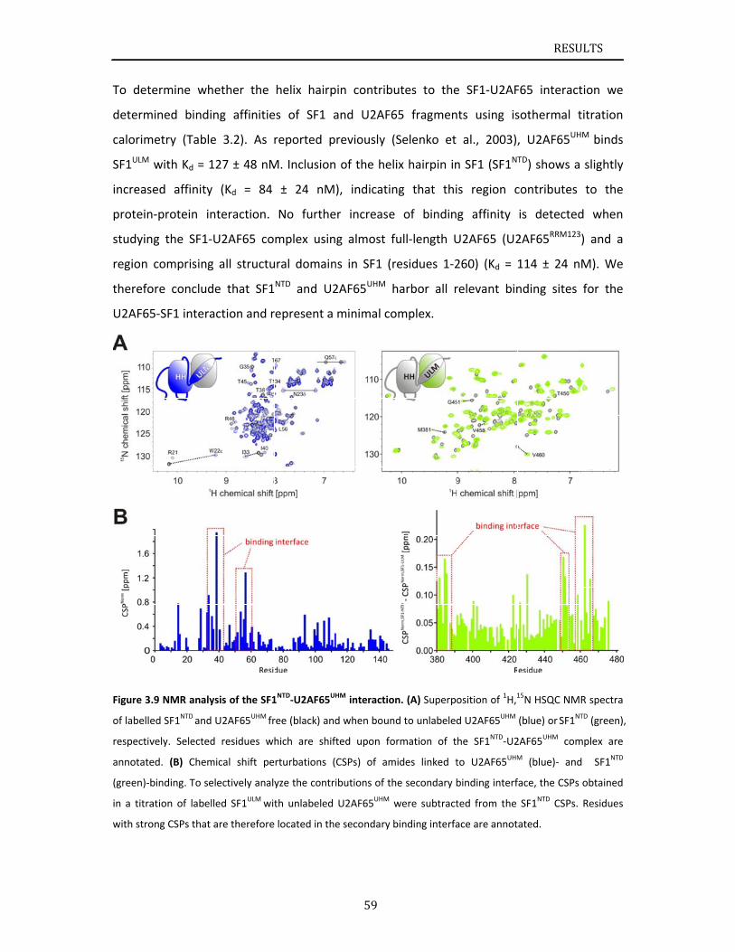
To dete
determ
calorim
SF1ULM
increase
protein
studyin
region
therefo
U2AF65
Figure 3.
of labelle
respectiv
annotate
(green)‐b
in a titra
with stro
ermine wh
ined bindin
metry (Table
with Kd = 1
ed affinity
‐protein in
g the SF1‐U
comprising
ore conclud
5‐SF1 intera
.9 NMR analy
ed SF1NTD and
vely. Selected
ed. (B) Chem
binding. To se
ation of labell
ong CSPs that
ether the
ng affinitie
e 3.2). As r
127 ± 48 nM
(Kd = 84
nteraction.
U2AF65 co
all structu
e that SF1
action and r
ysis of the SF1
U2AF65UHM fr
d residues wh
mical shift pe
lectively analy
led SF1ULM wit
are therefore
helix hairp
es of SF1 a
reported p
M. Inclusion
± 24 nM
No furthe
mplex usin
ural domain
NTD and U2
represent a
1NTD‐U2AF65U
ree (black) and
hich are shif
rturbations (
yze the contr
th unlabeled
located in the
59
pin contribu
and U2AF6
reviously (S
n of the heli
), indicatin
er increase
g almost f
ns in SF1 (r
2AF65UHM h
minimal co
HM interaction
d when bound
fted upon fo
CSPs) of am
ibutions of th
U2AF65UHM w
e secondary b
utes to the
65 fragmen
Selenko et
ix hairpin in
ng that thi
of bindin
ull‐length U
esidues 1‐2
harbor all r
omplex.
n. (A) Superpo
d to unlabeled
rmation of t
ides linked t
he secondary b
were subtract
binding interfa
e SF1‐U2AF
nts using is
al., 2003),
n SF1 (SF1NT
s region c
g affinity
U2AF65 (U2
260) (Kd =
relevant bin
osition of 1H,1
d U2AF65UHM
he SF1NTD‐U2
to U2AF65UHM
binding interf
ted from the
ace are annot
RESU
F65 interac
sothermal t
, U2AF65UH
TD) shows a
contributes
is detected
2AF65RRM123
114 ± 24 n
nding sites
15N HSQC NM
(blue) or SF1N
2AF65UHM com
M (blue)‐ and
face, the CSPs
SF1NTD CSPs.
ated.
ULTS
tion we
titration
HM binds
a slightly
to the
d when
3) and a
nM). We
for the
MR spectra
NTD (green),
mplex are
d SF1NTD
s obtained
Residues
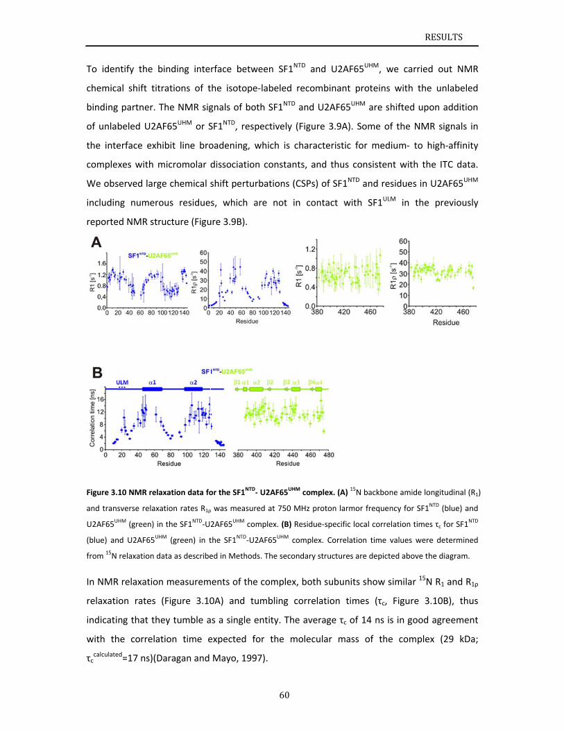
To iden
chemica
binding
of unla
the inte
comple
We obs
includin
reporte
Figure 3.
and tran
U2AF65U
(blue) an
from 15N
In NMR
relaxati
indicati
with th
τccalculate
ntify the bi
al shift titr
g partner. T
beled U2AF
erface exhi
xes with m
served large
ng numero
ed NMR stru
10 NMR relax
sverse relaxat
UHM (green) in
nd U2AF65UHM
relaxation da
R relaxation
ion rates (
ng that the
he correlat
ed=17 ns)(Da
inding inte
rations of t
he NMR sig
F65UHM or S
bit line bro
micromolar d
e chemical s
us residue
ucture (Figu
xation data fo
tion rates R1ρ
the SF1NTD‐U
M (green) in t
ata as describe
measurem
Figure 3.10
ey tumble a
ion time e
aragan and
rface betw
the isotope
gnals of bot
SF1NTD, resp
oadening, w
dissociation
shift pertur
s, which a
ure 3.9B).
or the SF1NTD‐
ρ was measure
2AF65UHM com
the SF1NTD‐U2
ed in Methods
ments of the
0A) and tu
as a single e
expected fo
Mayo, 199
60
ween SF1NTD
‐labeled re
th SF1NTD a
pectively (F
which is ch
n constants
rbations (CS
are not in
‐ U2AF65UHM c
ed at 750 MH
mplex. (B) Res
2AF65UHM com
s. The second
e complex, b
umbling co
entity. The a
or the mo
7).
D and U2AF
ecombinant
nd U2AF65
igure 3.9A)
aracteristic
s, and thus
SPs) of SF1N
contact w
complex. (A) 1
Hz proton larm
sidue‐specific
mplex. Correla
ary structures
both subun
rrelation ti
average τc o
lecular ma
F65UHM, we
proteins w
UHM are shi
. Some of t
c for mediu
consistent
NTD and resid
with SF1ULM
15N backbone
mor frequenc
local correlat
ation time va
s are depicted
its show sim
imes (τc, F
of 14 ns is
ss of the
RESU
e carried o
with the un
fted upon a
the NMR si
um‐ to high
with the IT
dues in U2A
in the pr
e amide longit
y for SF1NTD (
tion times τc f
lues were de
d above the d
milar 15N R1
igure 3.10B
in good agr
complex (2
ULTS
ut NMR
nlabeled
addition
ignals in
h‐affinity
TC data.
AF65UHM
eviously
udinal (R1)
blue) and
for SF1NTD
etermined
iagram.
and R1ρ
B), thus
reement
29 kDa;
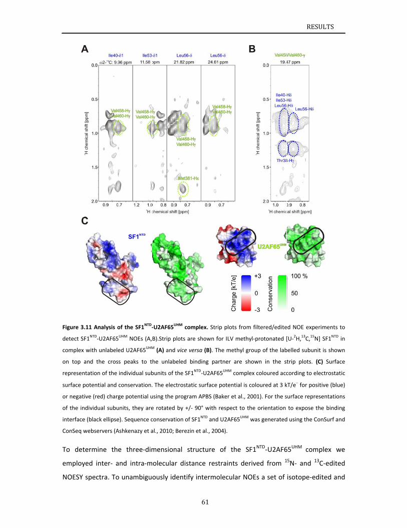
Figure 3.
detect SF
complex
on top a
represen
surface p
or negati
of the in
interface
ConSeq w
To det
employ
NOESY
.11 Analysis o
F1NTD‐U2AF65
with unlabele
and the cros
ntation of the
potential and
ive (red) charg
dividual subu
e (black ellipse
webservers (A
termine the
yed inter‐ a
spectra. To
of the SF1NTD‐
5UHM NOEs (A,
ed U2AF65UHM
s peaks to t
individual sub
conservation.
ge potential u
units, they are
e). Sequence c
Ashkenazy et a
e three‐dim
and intra‐m
o unambigu
‐U2AF65UHM c
B).Strip plots
M (A) and vice
he unlabeled
bunits of the S
. The electros
using the prog
e rotated by +
conservation o
al., 2010; Bere
mensional
molecular di
ously ident
61
complex. Strip
are shown fo
e versa (B). Th
d binding par
SF1NTD‐U2AF6
tatic surface p
gram APBS (Ba
+/‐ 90° with r
of SF1NTD and
ezin et al., 200
structure
stance rest
tify intermo
p plots from
or ILV methyl
he methyl gro
rtner are sho
65UHM complex
potential is co
aker et al., 20
respect to the
U2AF65UHM w
04).
of the SF
traints deriv
olecular NO
filtered/edite
‐protonated [
oup of the lab
own in the s
x coloured acc
oloured at 3 k
001). For the s
e orientation
was generated
1NTD‐U2AF6
ved from 1
Es a set of
RESU
ed NOE exper
[U‐2H,13C,15N]
belled subunit
trip plots. (C
cording to ele
kT/e for posit
surface repres
to expose th
d using the Co
65UHM comp
15N‐ and 13C
isotope‐edi
ULTS
iments to
SF1NTD in
t is shown
C) Surface
ectrostatic
tive (blue)
sentations
he binding
nSurf and
plex we
C‐edited
ited and
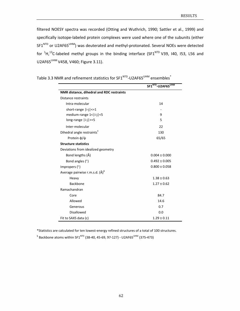
RESULTS
62
filtered NOESY spectra was recorded (Otting and Wuthrich, 1990; Sattler et al., 1999) and
specifically isotope‐labeled protein complexes were used where one of the subunits (either
SF1NTD or U2AF65UHM) was deuterated and methyl‐protonated. Several NOEs were detected
for 1H,13C‐labeled methyl groups in the binding interface (SF1NTD V39, I40, I53, L56 and
U2AF65UHM V458, V460; Figure 3.11).
Table 3.3 NMR and refinement statistics for SF1NTD‐U2AF65UHM ensembles*
SF1NTD‐U2AF65UHM
NMR distance, dihedral and RDC restraints
Distance restraints
Intra‐molecular 14
short‐range |i‐j|<=1
medium‐range 1<|i‐j|<5
long‐range |i‐j|>=5
‐
9
5
Inter‐molecular 22
Dihedral angle restraints‡ 130
Protein φ/ψ 65/65
Structure statistics
Deviations from idealized geometry
Bond lengths (Å) 0.004 ± 0.000
Bond angles () 0.492 ± 0.005
Impropers () 0.800 ± 0.058
Average pairwise r.m.s.d. (Å)§
Heavy 1.38 ± 0.63
Backbone 1.27 ± 0.62
Ramachandran
Core 84.7
Allowed 14.6
Generous 0.7
Disallowed 0.0
Fit to SAXS data (c) 1.29 ± 0.11
*Statistics are calculated for ten lowest‐energy refined structures of a total of 100 structures.
§ Backbone atoms within SF1NTD (38‐40, 45‐69, 97‐127) ‐ U2AF65UHM (375‐473)
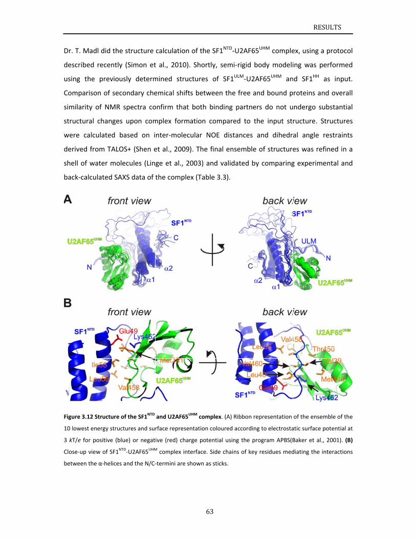
Dr. T. M
describ
using t
Compar
similarit
structu
were c
derived
shell of
back‐ca
Figure 3.
10 lowes
3 kT/e fo
Close‐up
between
Madl did the
ed recently
the previou
rison of sec
ty of NMR
ral changes
calculated b
d from TALO
f water mol
alculated SA
12 Structure
st energy struc
or positive (b
view of SF1N
the α‐helices
e structure
y (Simon et
usly determ
condary che
spectra co
s upon com
based on i
OS+ (Shen e
lecules (Lin
AXS data of
of the SF1NTD
ctures and sur
lue) or negat
NTD‐U2AF65UHM
s and the N/C‐
calculation
t al., 2010).
mined struc
emical shift
onfirm that
mplex form
nter‐molec
et al., 2009
ge et al., 2
the comple
and U2AF65U
rface represe
ive (red) char
M complex int
‐termini are s
63
of the SF1N
. Shortly, se
ctures of S
ts between
both bindi
ation comp
ular NOE d
). The final
003) and va
ex (Table 3.3
UHM complex.
ntation colou
rge potential
terface. Side c
hown as stick
NTD‐U2AF65
emi‐rigid bo
SF1ULM‐U2A
the free an
ing partner
pared to th
distances a
ensemble
alidated by
3).
(A) Ribbon re
red according
using the pro
chains of key
ks.
5UHM comple
ody modeli
AF65UHM an
nd bound p
rs do not u
he input str
and dihedr
of structure
y comparing
presentation
g to electrosta
ogram APBS(B
residues med
RESU
ex, using a p
ing was pe
nd SF1HH as
roteins and
ndergo sub
ructure. Str
al angle re
es was refin
g experimen
of the ensem
atic surface po
Baker et al., 2
diating the int
ULTS
protocol
rformed
s input.
d overall
bstantial
ructures
estraints
ned in a
ntal and
ble of the
otential at
2001). (B)
teractions
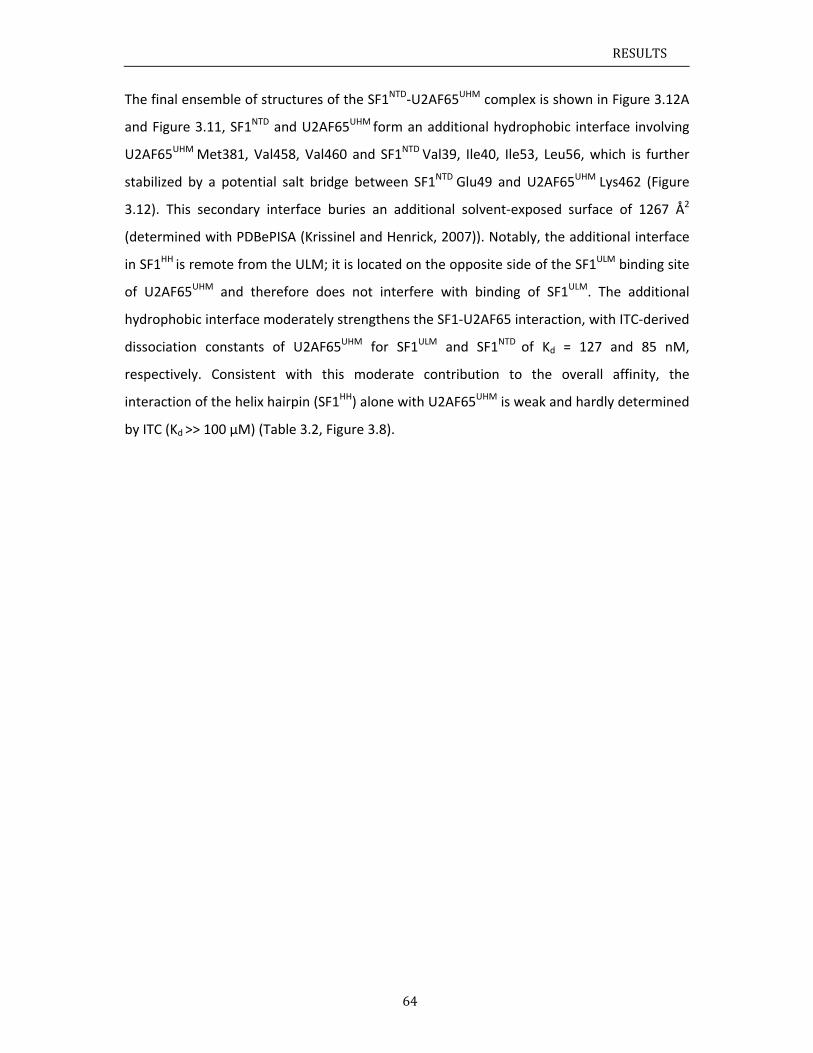
RESULTS
64
The final ensemble of structures of the SF1NTD‐U2AF65UHM complex is shown in Figure 3.12A
and Figure 3.11, SF1NTD and U2AF65UHM form an additional hydrophobic interface involving
U2AF65UHM Met381, Val458, Val460 and SF1NTD Val39, Ile40, Ile53, Leu56, which is further
stabilized by a potential salt bridge between SF1NTD Glu49 and U2AF65UHM Lys462 (Figure
3.12). This secondary interface buries an additional solvent‐exposed surface of 1267 Å2
(determined with PDBePISA (Krissinel and Henrick, 2007)). Notably, the additional interface
in SF1HH is remote from the ULM; it is located on the opposite side of the SF1ULM binding site
of U2AF65UHM and therefore does not interfere with binding of SF1ULM. The additional
hydrophobic interface moderately strengthens the SF1‐U2AF65 interaction, with ITC‐derived
dissociation constants of U2AF65UHM for SF1ULM and SF1NTD of Kd = 127 and 85 nM,
respectively. Consistent with this moderate contribution to the overall affinity, the
interaction of the helix hairpin (SF1HH) alone with U2AF65UHM is weak and hardly determined
by ITC (Kd >> 100 µM) (Table 3.2, Figure 3.8).
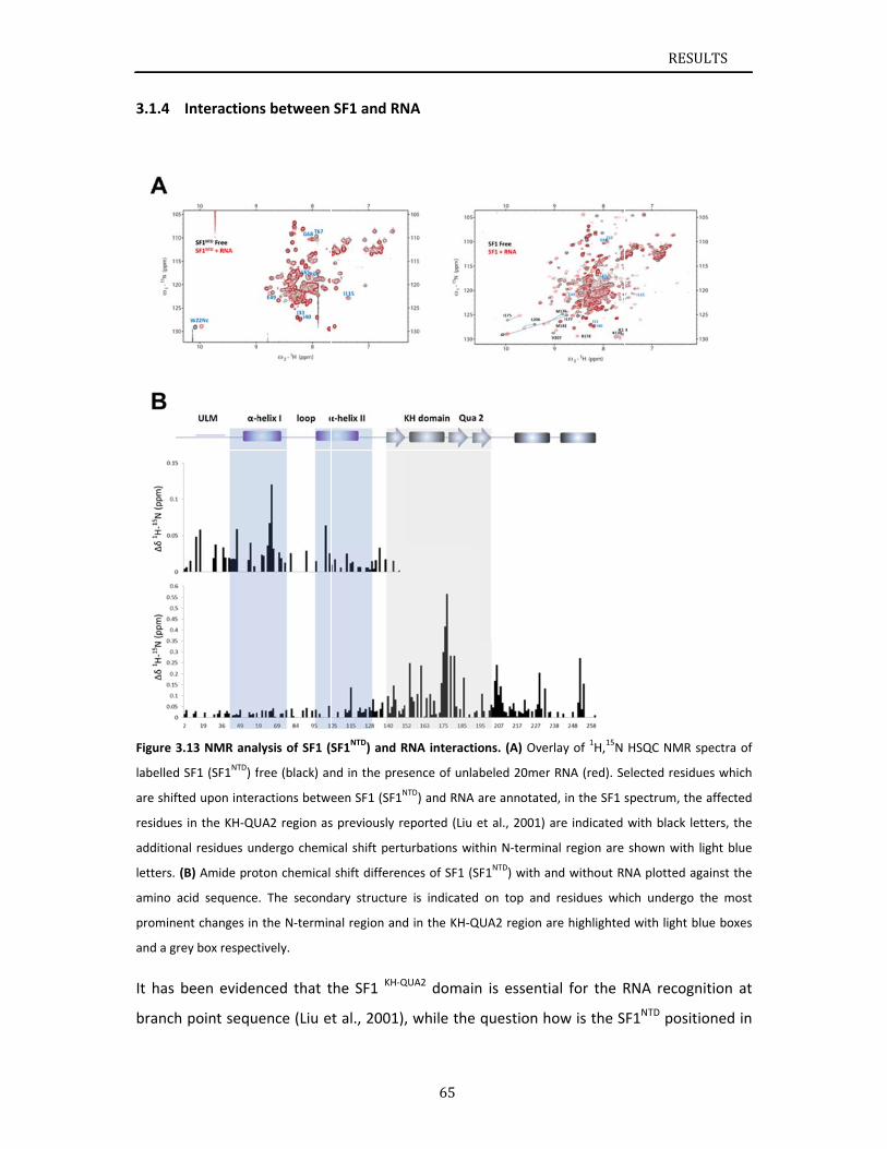
3.1.4
Figure 3.
labelled S
are shifte
residues
additiona
letters. (
amino ac
prominen
and a gre
It has b
branch
Interaction
.13 NMR ana
SF1 (SF1NTD) f
ed upon inter
in the KH‐QU
al residues un
B) Amide pro
cid sequence
nt changes in
ey box respect
been eviden
point sequ
ns between
lysis of SF1 (S
free (black) an
ractions betwe
UA2 region as
ndergo chemi
oton chemical
. The second
the N‐termin
tively.
nced that t
ence (Liu e
SF1 and RN
SF1NTD) and R
nd in the pres
een SF1 (SF1N
s previously re
ical shift pert
shift differen
dary structure
nal region and
the SF1 KH‐Q
t al., 2001),
65
NA
RNA interactio
sence of unla
NTD) and RNA
eported (Liu e
urbations wit
nces of SF1 (S
e is indicated
d in the KH‐Q
QUA2 domain
, while the
ons. (A) Over
beled 20mer
are annotated
et al., 2001) a
thin N‐termin
SF1NTD) with a
on top and
QUA2 region a
n is essenti
question h
rlay of 1H,15N
RNA (red). Se
d, in the SF1 s
are indicated
al region are
nd without R
residues wh
are highlighted
al for the R
ow is the S
RESU
HSQC NMR s
elected residu
spectrum, the
with black le
shown with
NA plotted ag
ich undergo
d with light b
RNA recogn
F1NTD posit
ULTS
spectra of
ues which
e affected
etters, the
light blue
gainst the
the most
lue boxes
nition at
ioned in
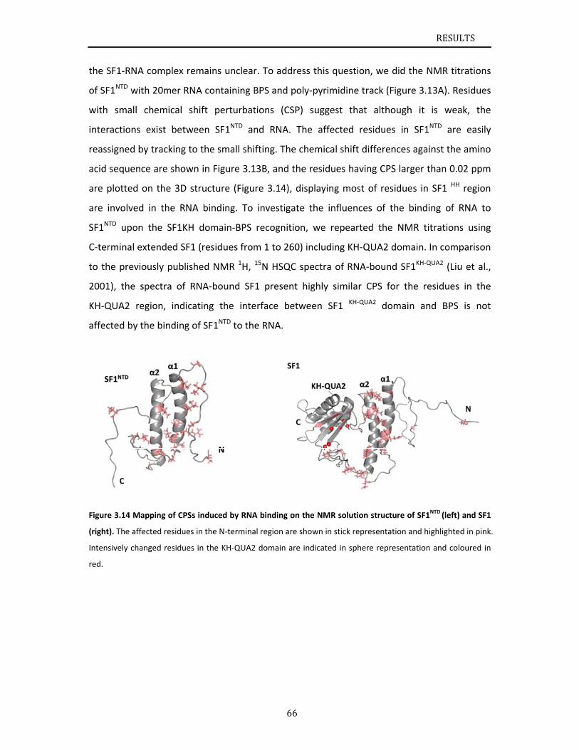
the SF1
of SF1NT
with sm
interact
reassign
acid seq
are plot
are inv
SF1NTD
C‐termi
to the p
2001),
KH‐QUA
affected
Figure 3.
(right). T
Intensive
red.
1‐RNA comp
TD with 20m
mall chem
tions exist
ned by trac
quence are
tted on the
olved in th
upon the S
inal extende
previously p
the spectra
A2 region,
d by the bin
.14 Mapping o
The affected re
ely changed re
plex remain
mer RNA con
ical shift p
between S
king to the
shown in F
e 3D structu
he RNA bin
SF1KH dom
ed SF1 (resi
published N
a of RNA‐b
indicating
nding of SF1
of CPSs induc
esidues in the
esidues in the
ns unclear. T
ntaining BP
perturbatio
SF1NTD and
small shifti
Figure 3.13B
ure (Figure
ding. To in
main‐BPS re
idues from
NMR 1H, 15N
bound SF1
the interfa
1NTD to the R
ced by RNA b
e N‐terminal re
e KH‐QUA2 do
66
To address
S and poly‐
ns (CSP) s
RNA. The
ng. The che
B, and the r
3.14), disp
nvestigate t
ecognition,
1 to 260) in
N HSQC spe
present hig
ace betwee
RNA.
binding on the
egion are sho
omain are ind
this questio
pyrimidine
suggest tha
affected
emical shift
residues hav
playing mos
the influenc
we repeart
ncluding KH‐
ectra of RN
ghly similar
en SF1 KH‐Q
e NMR solutio
wn in stick re
dicated in sph
on, we did
track (Figu
at although
residues in
differences
ving CPS lar
st of residu
ces of the
ted the NM
‐QUA2 dom
A‐bound SF
r CPS for t
QUA2 domai
on structure o
presentation
here represen
RESU
the NMR ti
re 3.13A). R
h it is we
n SF1NTD ar
s against th
rger than 0.
es in SF1 H
binding of
MR titration
main. In com
F1KH‐QUA2 (Li
he residues
in and BPS
of SF1NTD (left
and highlight
ntation and co
ULTS
itrations
Residues
eak, the
re easily
e amino
.02 ppm
H region
RNA to
ns using
mparison
iu et al.,
s in the
S is not
t) and SF1
ed in pink.
oloured in
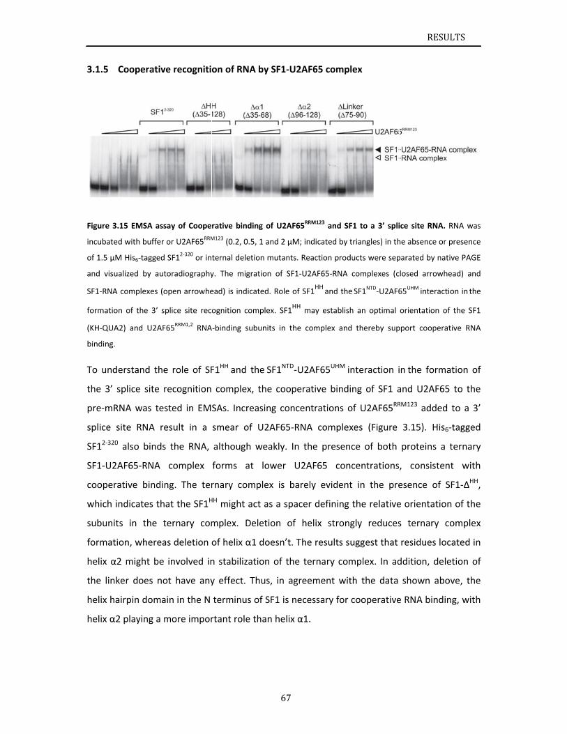
3.1.5
Figure 3.
incubate
of 1.5 µM
and visu
SF1‐RNA
formatio
(KH‐QUA
binding.
To und
the 3’ s
pre‐mR
splice s
SF12‐320
SF1‐U2A
coopera
which i
subunit
formati
helix α2
the link
helix ha
helix α2
Cooperativ
.15 EMSA ass
d with buffer
M His6‐tagged
alized by aut
complexes (o
n of the 3’ s
A2) and U2AF
erstand the
splice site r
RNA was tes
site RNA re
0 also binds
AF65‐RNA
ative bindin
ndicates th
ts in the t
on, wherea
2 might be
ker does no
airpin doma
2 playing a m
ve recogniti
say of Coope
or U2AF65RRM
SF12‐320 or in
toradiography
open arrowhe
plice site rec
F65RRM1,2
RNA
e role of SF
recognition
sted in EM
esult in a
s the RNA,
complex
ng. The ter
at the SF1H
ternary com
as deletion
involved in
ot have any
ain in the N
more impor
on of RNA
rative bindin
M123 (0.2, 0.5,
ternal deletio
y. The migrat
ead) is indicat
ognition com
A‐binding subu
F1HH and th
complex, t
SAs. Increa
smear of U
although w
forms at
rnary comp
HH might act
mplex. Del
of helix α1
n stabilizati
y effect. Th
terminus o
rtant role th
67
by SF1‐U2A
g of U2AF65R
1 and 2 µM; i
on mutants. R
tion of SF1‐U
ted. Role of S
mplex. SF1HH
m
units in the c
he SF1NTD‐U2
the cooper
sing conce
U2AF65‐RN
weakly. In t
lower U2
plex is bare
t as a space
etion of h
doesn’t. Th
on of the t
hus, in agre
of SF1 is nec
han helix α1
AF65 compl
RRM123 and SF
ndicated by t
eaction produ
U2AF65‐RNA c
SF1HH and the
may establish
complex and
2AF65UHM in
ative bindin
ntrations o
NA complex
the presenc
AF65 conc
ely evident
er defining t
helix strong
he results su
ternary com
eement wit
cessary for c
1.
ex
1 to a 3’ spli
riangles) in th
ucts were sep
complexes (c
SF1NTD‐U2AF6
h an optimal
thereby sup
nteraction i
ng of SF1 a
f U2AF65RR
xes (Figure
ce of both
centrations
in the pre
the relative
gly reduces
uggest that
mplex. In ad
h the data
cooperative
RESU
ice site RNA.
he absence or
parated by nat
losed arrowh
65UHM interact
orientation o
pport coopera
in the form
and U2AF65
RM123 added
3.15). His6
proteins a
, consisten
esence of S
e orientatio
s ternary c
residues lo
ddition, del
shown abo
e RNA bindi
ULTS
RNA was
presence
tive PAGE
head) and
tion in the
of the SF1
ative RNA
ation of
5 to the
d to a 3’
6‐tagged
ternary
nt with
SF1‐∆HH,
n of the
complex
cated in
etion of
ove, the
ng, with
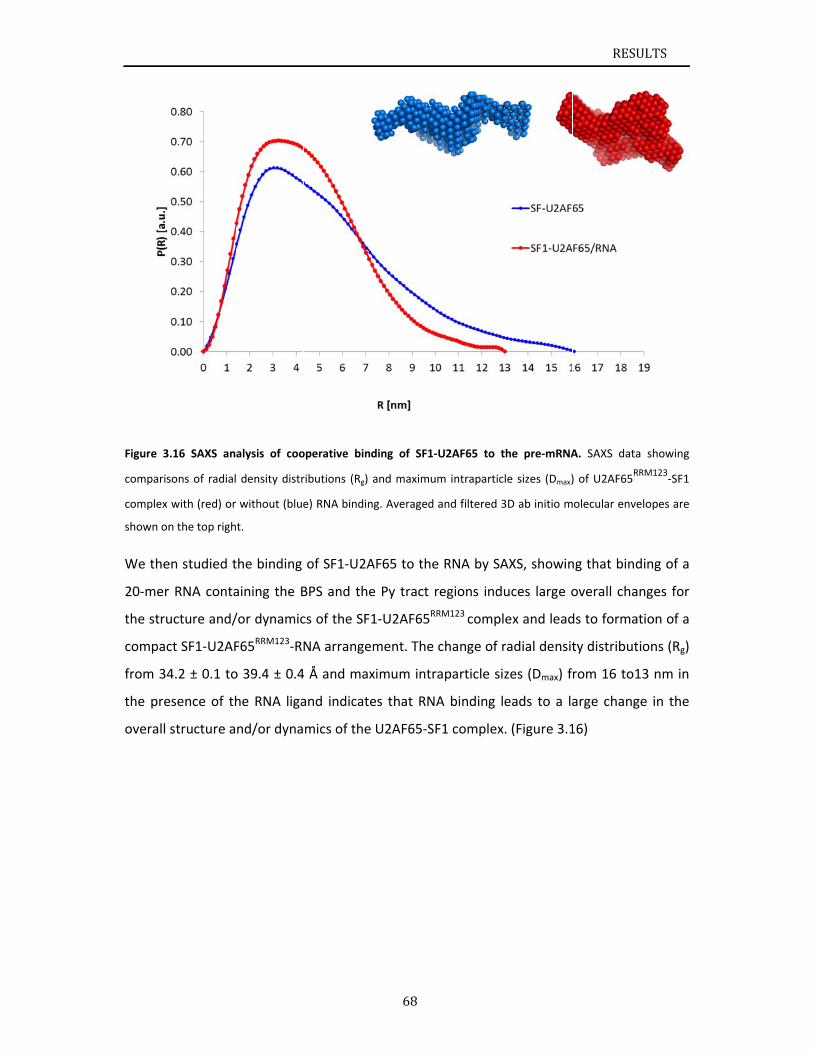
Figure 3
comparis
complex
shown on
We the
20‐mer
the stru
compac
from 34
the pre
overall
.16 SAXS an
sons of radial
with (red) or
n the top righ
en studied t
RNA conta
ucture and/
ct SF1‐U2AF
4.2 ± 0.1 to
esence of th
structure a
alysis of coo
density distr
without (blue
t.
the binding
aining the B
/or dynamic
F65RRM123‐RN
o 39.4 ± 0.4
he RNA liga
nd/or dyna
operative bin
ributions (Rg)
e) RNA bindin
of SF1‐U2A
BPS and the
cs of the SF
NA arrange
4 Å and max
and indicate
mics of the
68
ding of SF1‐
and maximum
ng. Averaged
AF65 to the
e Py tract r
1‐U2AF65RR
ement. The
ximum intra
es that RNA
U2AF65‐SF
U2AF65 to t
m intraparticl
and filtered 3
e RNA by SA
regions indu
RM123 comple
change of r
aparticle siz
A binding le
F1 complex.
he pre‐mRNA
le sizes (Dmax)
3D ab initio m
AXS, showin
uces large o
ex and lead
radial densit
zes (Dmax) f
eads to a la
. (Figure 3.1
RESU
A. SAXS data
) of U2AF65RR
olecular enve
ng that bind
overall chan
ds to format
ty distributi
rom 16 to1
arge chang
16)
ULTS
a showing
RM123‐SF1
elopes are
ding of a
nges for
tion of a
ions (Rg)
13 nm in
e in the
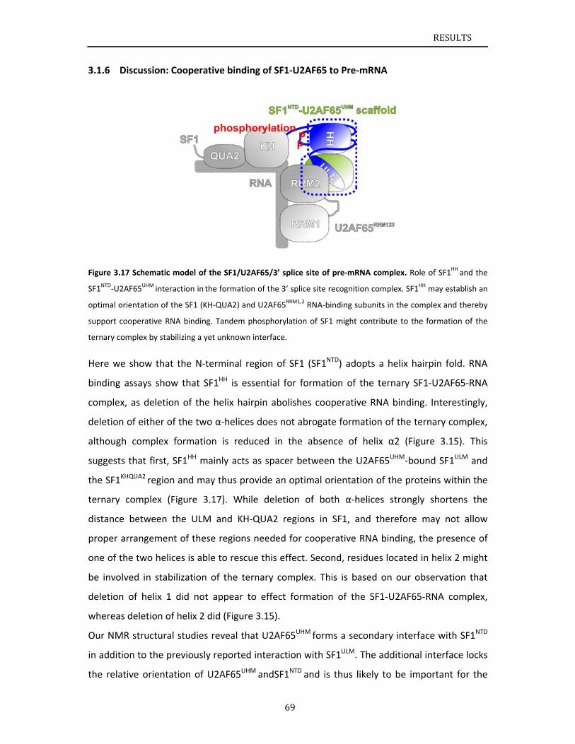
3.1.6
Figure 3.
SF1NTD‐U
optimal o
support c
ternary c
Here w
binding
comple
deletion
althoug
suggest
the SF1
ternary
distance
proper
one of t
be invo
deletion
wherea
Our NM
in addit
the rela
Discussion:
.17 Schematic
2AF65UHM inte
orientation of
cooperative R
complex by sta
we show tha
g assays sho
x, as deleti
n of either
gh complex
ts that first,
1KHQUA2 regio
y complex
e between
arrangeme
the two hel
olved in sta
n of helix 1
as deletion o
MR structur
tion to the p
ative orient
: Cooperativ
c model of th
eraction in the
f the SF1 (KH‐
RNA binding.
abilizing a yet
at the N‐te
ow that SF1
ion of the h
of the two
x formation
, SF1HH mai
on and may
(Figure 3.1
the ULM
ent of these
lices is able
bilization o
1 did not a
of helix 2 di
al studies r
previously r
tation of U2
ve binding
he SF1/U2AF6
e formation o
‐QUA2) and U
Tandem phos
t unknown int
rminal regi
1HH is essen
helix hairpi
α‐helices d
n is reduce
inly acts as
y thus provid
17). While
and KH‐QU
e regions ne
e to rescue t
of the terna
appear to e
d (Figure 3.
reveal that
reported in
2AF65UHM a
69
of SF1‐U2A
65/3’ splice s
f the 3’ splice
U2AF65RRM1,2 R
sphorylation
erface.
on of SF1 (
ntial for for
n abolishes
oes not abr
ed in the a
spacer bet
de an optim
deletion o
UA2 region
eeded for co
this effect.
ary complex
effect form
.15).
U2AF65UHM
teraction w
ndSF1NTD an
AF65 to Pre‐
ite of pre‐mR
e site recognit
RNA‐binding s
of SF1 might
(SF1NTD) ado
rmation of
s cooperativ
rogate form
absence of
tween the U
mal orientat
of both α‐h
s in SF1, a
ooperative
Second, res
x. This is b
mation of th
M forms a se
with SF1ULM.
nd is thus l
‐mRNA
RNA complex
ion complex.
subunits in the
contribute to
opts a helix
the ternary
ve RNA bin
mation of th
helix α2
U2AF65UHM
tion of the
helices stro
and therefo
RNA bindin
sidues locat
ased on ou
he SF1‐U2A
econdary int
The additio
likely to be
RESU
. Role of SF1H
SF1HH may es
e complex an
o the formati
x hairpin fo
y SF1‐U2AF
nding. Intere
he ternary c
(Figure 3.1
‐bound SF1
proteins wi
ongly short
ore may no
ng, the pres
ted in helix
ur observat
AF65‐RNA c
terface with
onal interfa
e important
ULTS
HH and the
stablish an
d thereby
on of the
old. RNA
F65‐RNA
estingly,
omplex,
15). This
1ULM and
ithin the
ens the
ot allow
sence of
2 might
ion that
omplex,
h SF1NTD
ace locks
t for the
RESULTS
70
specific quaternary arrangement of the proteins in the SF1‐U2AF65 complex. In addition to
providing the proper geometry of SF1 and U2AF65 for RNA binding, the secondary interface
involving the SF1HH and U2AF65UHM may reduce entropic costs linked to SF1‐U2AF65‐RNA
complex formation by providing a pre‐arranged protein‐protein scaffold for RNA binding.
Thus, although the secondary interface has a rather small contribution to the overall affinity
between SF1 and U2AF65 (Table 3.2), it is an essential feature for formation of the ternary
complex. A specific arrangement of SF1 and U2AF enforced by the helix hairpin of SF1 may
be required to accommodate variations in the distance between the BPS and Py tract
regions of introns by providing a prearranged scaffold of the SF1 KH‐QUA2 and U2AF65RRM1,2
regions.
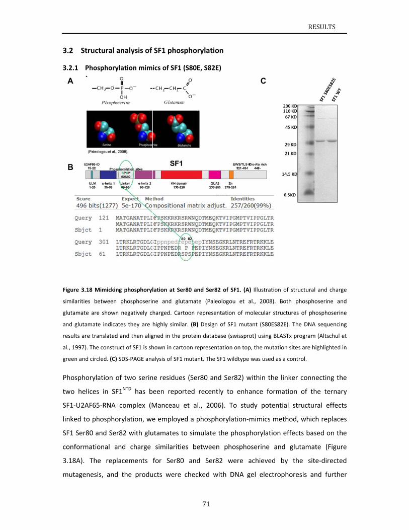
St3.2
3.2.1
Figure 3.
similaritie
glutamat
and gluta
results a
al., 1997)
green an
Phosph
two he
SF1‐U2A
linked t
SF1 Ser
conform
3.18A).
mutage
tructural a
Phosphory
.18 Mimickin
es between
te are shown
amate indicat
re translated
). The constru
d circled. (C) S
orylation o
elices in SF
AF65‐RNA
to phospho
r80 and Ser
mational an
The repla
enesis, and
analysis of
lation mim
g phosphoryl
phosphoserin
negatively ch
tes they are
and then alig
uct of SF1 is sh
SDS‐PAGE ana
of two serin
1NTD has b
complex (M
rylation, we
82 with glu
nd charge
acements
the produ
SF1 phosp
ics of SF1 (S
lation at Ser8
ne and gluta
harged. Carto
highly similar
gned in the pr
hown in carto
alysis of SF1 m
ne residues
een report
Manceau e
e employed
utamates to
similarities
for Ser80
ucts were c
71
phorylation
S80E, S82E)
80 and Ser82
amate (Paleo
oon represent
r. (B) Design o
rotein databa
on representa
mutant. The SF
(Ser80 and
ted recently
et al., 2006
d a phospho
o simulate t
s between
and Ser82
checked wit
n
)
2 of SF1. (A) I
ologou et al.
tation of mol
of SF1 mutan
se (swissprot
ation on top, t
F1 wildtype w
d Ser82) wit
y to enhan
6). To study
orylation‐m
the phospho
phosphose
2 were ac
th DNA ge
Illustration of
, 2008). Bot
lecular struct
nt (S80ES82E)
) using BLAST
the mutation
was used as a c
thin the lin
nce format
y potential
mimics meth
orylation ef
erine and
chieved by
l electroph
RESU
f structural an
th phosphose
ures of phosp
. The DNA se
Tx program (A
sites are high
control.
ker connec
ion of the
l structural
hod, which r
ffects based
glutamate
the site‐d
horesis and
ULTS
nd charge
erine and
phoserine
equencing
Altschul et
hlighted in
cting the
ternary
effects
replaces
d on the
(Figure
directed
further
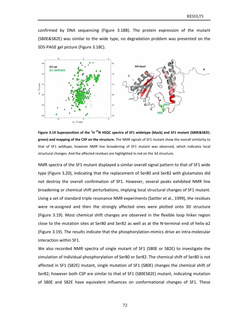
confirm
(S80E&
SDS‐PA
Figure 3.
green) an
that of S
structura
NMR sp
type (Fi
not des
broade
Using a
were re
(Figure
close to
(Figure
interact
We also
simulat
affected
Ser82; h
of S80E
med by DN
S82E) was
GE gel pictu
.19 Superposi
nd mapping o
SF1 wildtype,
al changes. An
pectra of th
igure 3.20),
stroy the o
ning or che
set of stan
e‐assigned
3.19). Mos
o the mutat
3.19). The
tion within
o recorded
ion of indiv
d in SF1 (S8
however bo
E and S82E
NA sequenc
similar to t
ure (Figure
ition of the 1H
of the CSP on
, however NM
nd the affected
e SF1 muta
, indicating
overall conf
mical shift
dard triple
and then
st chemical
tion sites at
results indi
SF1.
NMR spec
vidual phosp
82E) mutan
oth CSP are
E have equ
cing (Figure
the wide ty
3.18C).
H 15N HSQC s
the structure
MR line broa
d residues are
ant displaye
that the re
firmation of
perturbatio
resonance
the strong
shift chan
t Ser80 and
icate that th
ctra of sing
phorylation
nt, single m
e similar to
uivalent infl
72
e 3.18B). T
ype, no deg
spectra of SF1
e. The NMR sig
adening of SF
e highlighted i
ed a similar
eplacement
f SF1. How
ons, implyin
NMR exper
gly affected
ges are ob
d Ser82 as w
he phospho
le mutant o
of Ser80 or
utation of S
that of SF1
luences on
The protein
gradation pr
1 widetype (b
gnals of SF1 m
F1 mutant w
in red on the
overall sign
of Ser80 a
wever, seve
ng local stru
riments (Sat
d ones wer
bserved in t
well as at t
orylation‐m
of SF1 (S80
r Ser82. The
SF1 (S80E)
1 (S80ES82E
conformat
n expressio
roblem was
black) and SF1
mutant show t
was observed,
3d structure.
nal pattern
nd Ser82 w
ral peaks e
uctural chan
ttler et al.,
re plotted
the flexible
he N‐termi
imics drive
0E or S82E)
e chemical s
changes th
E) mutant, i
tional chan
RESU
on of the
s presented
1 mutant (S8
the overall sim
which indica
to that of S
with glutam
exhibited N
nges of SF1
1999), the r
onto 3D st
loop linke
nal end of
an intra‐m
to investig
shift of Ser8
he chemical
ndicating m
nges of SF1
ULTS
mutant
d on the
0E&S82E;
milarity to
ates local
SF1 wide
ates did
MR line
mutant.
residues
tructure
r region
helix α2
olecular
gate the
80 is not
shift of
mutation
1. These
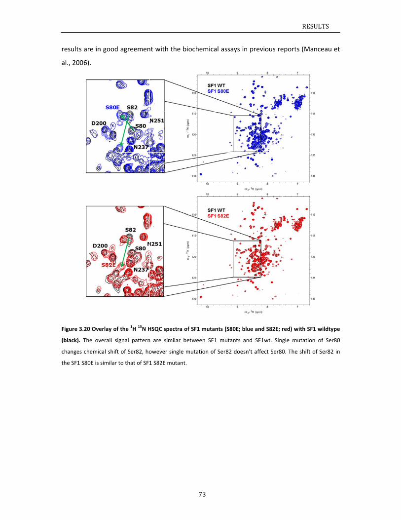
results
al., 200
Figure 3.
(black). T
changes
the SF1 S
are in good
6).
.20 Overlay o
The overall s
chemical shif
S80E is similar
d agreemen
f the 1H 15N H
signal pattern
ft of Ser82, ho
r to that of SF1
nt with the
HSQC spectra
n are similar
owever single
1 S82E mutan
73
biochemica
of SF1 mutan
between SF1
e mutation of
t.
al assays in
nts (S80E; blu
1 mutants an
Ser82 doesn’
previous re
ue and S82E; r
d SF1wt. Sing
’t affect Ser80
RESU
eports (Man
red) with SF1
gle mutation
0. The shift o
ULTS
nceau et
1 wildtype
of Ser80
f Ser82 in
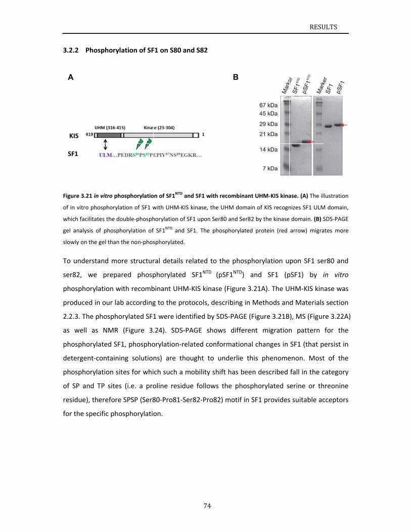
3.2.2
Figure 3.
of in vitr
which fac
gel analy
slowly on
To und
ser82,
phosph
produce
2.2.3. T
as wel
phosph
deterge
phosph
of SP a
residue
for the
Phosphory
21 in vitro ph
o phosphoryl
cilitates the d
ysis of phosp
n the gel than
erstand mo
we prepa
orylation w
ed in our la
The phospho
l as NMR
orylated SF
ent‐contain
orylation si
and TP sites
e), therefore
specific pho
lation of SF
hosphorylatio
ation of SF1 w
ouble‐phosph
horylation of
the non‐phos
ore structur
ared phosp
with recomb
b according
orylated SF1
(Figure 3.
F1, phospho
ing solution
ites for whi
s (i.e. a pro
e SPSP (Ser8
osphorylatio
F1 on S80 an
on of SF1NTD a
with UHM‐KIS
horylation of S
f SF1NTD and S
sphorylated.
ral details r
phorylated
binant UHM
g to the pro
1 were iden
.24). SDS‐P
orylation‐re
ns) are tho
ch such a m
oline residu
80‐Pro81‐Se
on.
74
nd S82
nd SF1 with r
S kinase, the U
SF1 upon Ser8
SF1. The pho
related to t
SF1NTD (p
M‐KIS kinase
otocols, des
ntified by SD
PAGE show
elated confo
ought to u
mobility shif
ue follows
er82‐Pro82)
recombinant U
UHM domain
80 and Ser82
osphorylated
the phospho
pSF1NTD) an
e (Figure 3.2
cribing in M
DS‐PAGE (Fi
ws different
ormational
nderlie this
ft has been
the phosph
) motif in SF
UHM‐KIS kina
of KIS recogn
by the kinase
protein (red
orylation u
nd SF1 (p
21A). The U
Methods an
igure 3.21B
t migration
changes in
s phenome
described
horylated s
F1 provides
RESU
ase. (A) The il
nizes SF1 ULM
domain. (B) S
arrow) migra
pon SF1 se
pSF1) by i
HM‐KIS kin
d Materials
), MS (Figur
n pattern
SF1 (that p
enon. Most
fall in the c
erine or th
s suitable ac
ULTS
lustration
M domain,
SDS‐PAGE
ates more
er80 and
in vitro
nase was
s section
re 3.22A)
for the
persist in
t of the
category
hreonine
cceptors
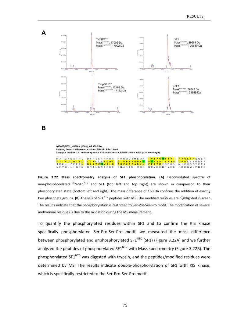
Figure 3
non‐phos
phospho
two phos
The resu
methioni
To qua
specific
betwee
analyze
phosph
determ
which is
3.22 Mass
sphorylated
rylated state
sphate groups
lts indicate th
ine residues is
antify the
cally phosp
en phospho
ed the pepti
orylated SF
ined by M
s specificall
spectrometry
15N‐SF1NTD a
(bottom left
s. (B) Analysis
hat the phosph
s due to the o
phosphory
phorylated
rylated and
ides of phos
F1NTD was d
S. The res
y restricted
y analysis
nd SF1 (top
and right). Th
s of SF1 NTD pe
horylation is r
oxidation durin
lated resid
Ser‐Pro‐Se
d unphosph
sphorylated
igested wit
ults indicat
d to the Ser‐
75
of SF1 pho
p left and to
he mass diffe
ptides with M
restricted to S
ng the MS me
dues within
r‐Pro moti
orylated SF
d SF1NTD wit
th trypsin, a
te double‐p
‐Pro‐Ser‐Pro
osphorylation
op right) are
rence of 160
MS. The modif
Ser‐Pro‐Ser‐Pr
easurement.
n SF1 and
if, we me
F1NTD (SF1)
th Mass spe
and the pep
phosphoryla
o motif.
n. (A) Deco
e shown in
Da confirms
ied residues a
ro motif. The
to confir
asured the
(Figure 3.22
ectrometry
ptides/mod
ation of SF
RESU
onvoluted sp
comparison
the addition
are highlighted
modification
m the KIS
e mass dif
2A) and we
(Figure 3.22
ified residu
F1 with KIS
ULTS
pectra of
to their
of exactly
d in green.
of several
S kinase
fference
e further
2B). The
es were
kinase,
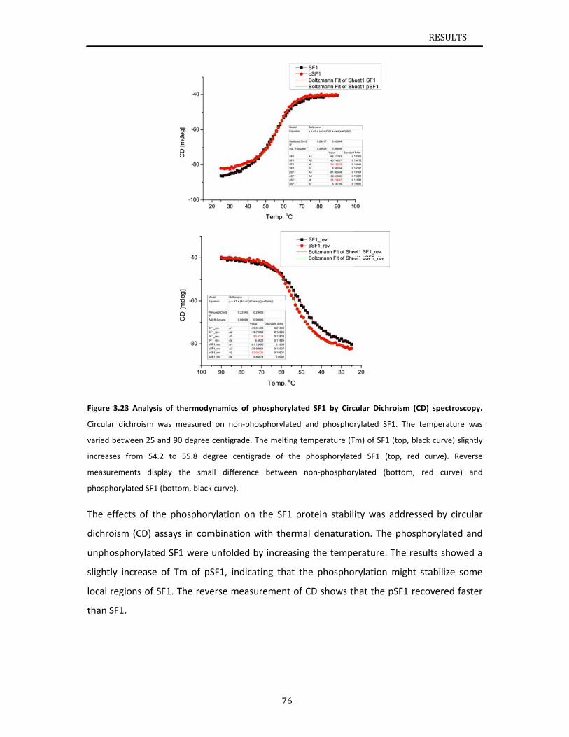
Figure 3.
Circular
varied be
increases
measure
phospho
The eff
dichrois
unphos
slightly
local re
than SF
.23 Analysis
dichroism wa
etween 25 an
s from 54.2
ments displa
rylated SF1 (b
fects of the
sm (CD) ass
sphorylated
increase o
egions of SF
F1.
of thermody
as measured
nd 90 degree c
to 55.8 deg
ay the sma
bottom, black
e phosphory
says in com
SF1 were u
of Tm of pS
F1. The reve
namics of ph
on non‐phos
centigrade. T
gree centigra
ll difference
curve).
ylation on t
mbination w
unfolded by
SF1, indicat
erse measu
76
hosphorylated
sphorylated a
he melting te
ade of the p
between n
the SF1 pro
with therma
y increasing
ting that th
rement of C
d SF1 by Circ
and phospho
emperature (T
phosphorylate
non‐phosphor
otein stabil
al denaturat
g the tempe
he phospho
CD shows t
cular Dichrois
rylated SF1.
Tm) of SF1 (to
ed SF1 (top,
rylated (bott
ity was add
tion. The p
erature. Th
orylation mi
hat the pSF
RESU
sm (CD) spec
The tempera
op, black curv
, red curve).
om, red cu
dressed by
hosphoryla
e results sh
ight stabiliz
F1 recovere
ULTS
ctroscopy.
ature was
e) slightly
. Reverse
rve) and
circular
ted and
howed a
ze some
ed faster
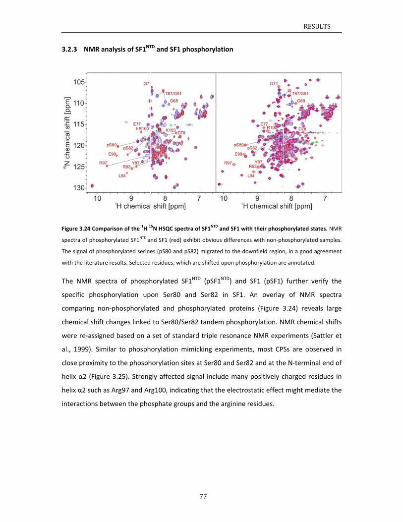
3.2.3
Figure 3.
spectra o
The signa
with the
The NM
specific
compar
chemica
were re
al., 199
close pr
helix α2
helix α2
interact
NMR analy
24 Compariso
of phosphoryl
al of phospho
literature res
MR spectra
c phosphor
ring non‐ph
al shift chan
e‐assigned b
99). Similar
roximity to
2 (Figure 3.
2 such as Ar
tions betwe
ysis of SF1NT
on of the 1H 1
ated SF1NTD a
orylated serine
ults. Selected
of phosph
rylation up
hosphorylat
nges linked
based on a
to phosph
the phosph
.25). Strong
rg97 and Ar
een the pho
TD and SF1 p
15N HSQC spec
nd SF1 (red) e
es (pS80 and
residues, wh
horylated S
pon Ser80
ted and ph
to Ser80/S
set of stan
orylation m
horylation s
gly affected
rg100, indic
osphate gro
77
phosphoryl
ctra of SF1NTD
exhibit obviou
pS82) migrate
ich are shifted
SF1NTD (pSF
and Ser82
hosphorylat
Ser82 tande
ndard triple
mimicking e
ites at Ser8
d signal incl
ating that t
ups and the
ation
D and SF1 with
us differences
ed to the dow
d upon phosp
1NTD) and S
2 in SF1. A
ted protein
em phospho
e resonance
experiments
80 and Ser82
ude many
the electros
e arginine re
h their phosp
s with non‐ph
wnfield region
phorylation are
SF1 (pSF1)
An overlay
ns (Figure 3
orylation. N
e NMR expe
s, most CPS
2 and at the
positively c
static effect
esidues.
RESU
horylated sta
hosphorylated
n, in a good a
e annotated.
further ve
y of NMR
3.24) revea
MR chemic
eriments (Sa
Ss are obse
e N‐termina
charged res
might med
ULTS
ates. NMR
d samples.
greement
erify the
spectra
als large
cal shifts
attler et
erved in
al end of
idues in
diate the
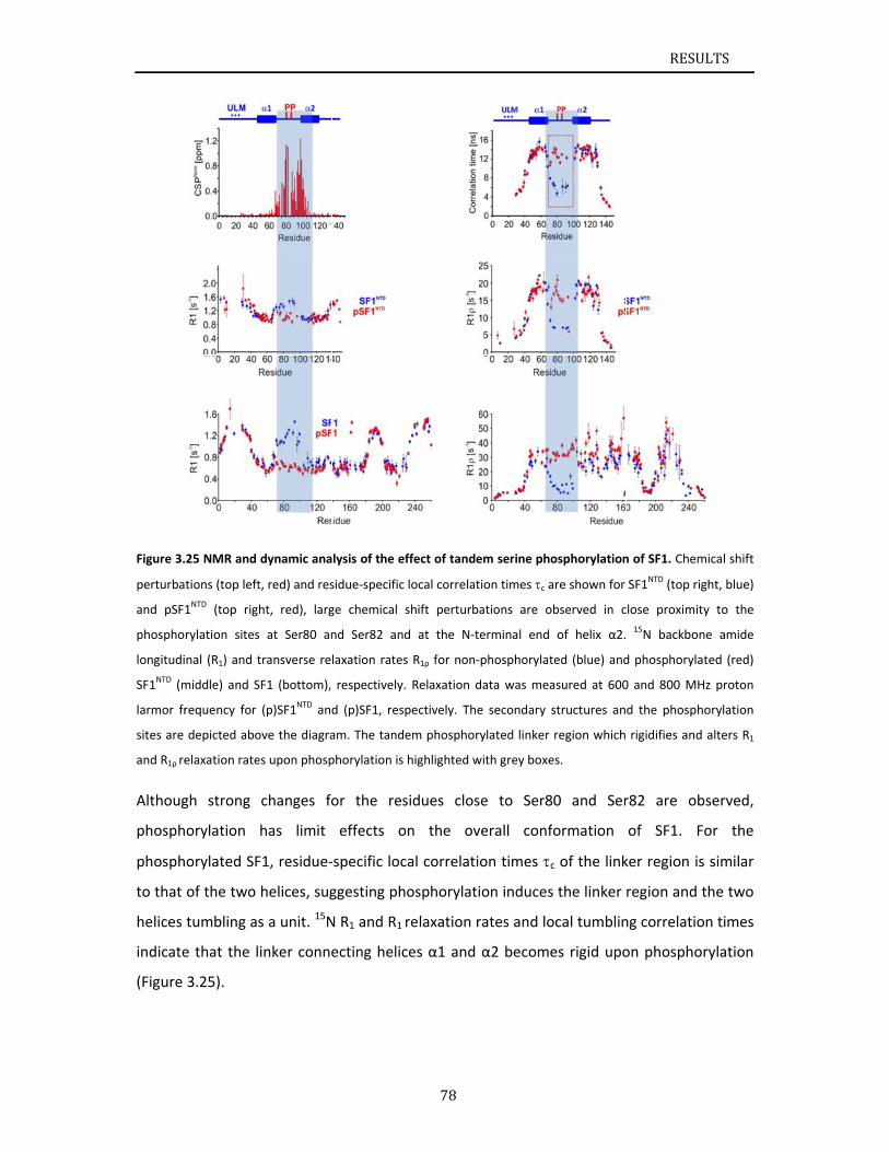
Figure 3.
perturba
and pSF
phospho
longitudi
SF1NTD (m
larmor fr
sites are
and R1ρ re
Althoug
phosph
phosph
to that
helices
indicate
(Figure
.25 NMR and
tions (top left
1NTD (top rig
rylation sites
nal (R1) and t
middle) and S
requency for
depicted abo
elaxation rate
gh strong
orylation
orylated SF
of the two
tumbling as
e that the l
3.25).
dynamic ana
t, red) and res
ht, red), larg
s at Ser80 an
transverse re
SF1 (bottom),
(p)SF1NTD and
ove the diagra
es upon phosp
changes fo
has limit
F1, residue‐
helices, sug
s a unit. 15N
inker conne
lysis of the ef
sidue‐specific
ge chemical s
nd Ser82 and
laxation rates
respectively.
d (p)SF1, resp
am. The tande
phorylation is
or the res
effects o
‐specific loc
ggesting ph
N R1 and R1
ecting helic
78
ffect of tande
local correlat
shift perturb
d at the N‐t
s R1ρ for non‐
. Relaxation d
pectively. The
em phosphor
highlighted w
sidues clos
on the ov
cal correlati
osphorylati
relaxation r
ces α1 and
em serine pho
tion times c a
ations are o
erminal end
‐phosphorylat
data was mea
e secondary s
rylated linker
with grey boxe
se to Ser8
verall conf
on times c
ion induces
rates and lo
α2 become
osphorylation
are shown for
bserved in c
of helix α2.
ted (blue) and
asured at 600
structures an
region which
es.
0 and Ser
ormation
c of the link
s the linker
ocal tumblin
es rigid upo
RESU
n of SF1. Chem
SF1NTD (top ri
close proximit
15N backbon
d phosphoryla
0 and 800 MH
d the phosph
rigidifies and
r82 are ob
of SF1. F
ker region is
region and
ng correlatio
on phospho
ULTS
mical shift
ight, blue)
ty to the
ne amide
ated (red)
Hz proton
horylation
d alters R1
bserved,
For the
s similar
the two
on times
orylation
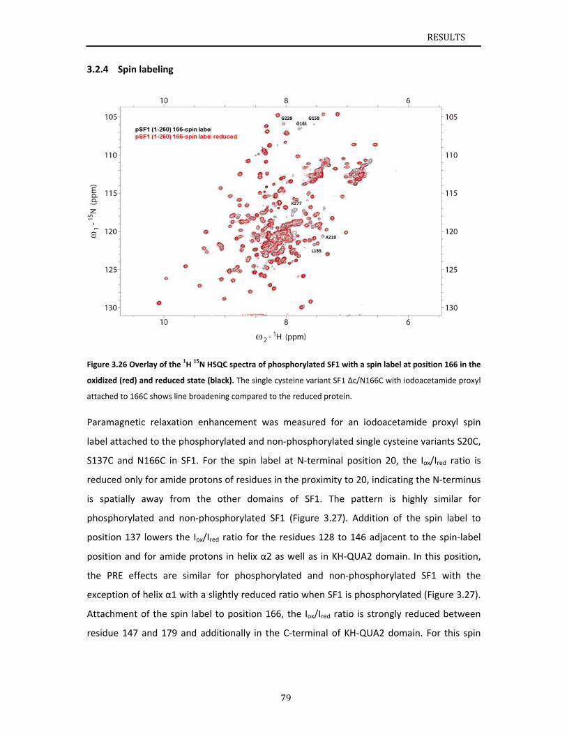
3.2.4
Figure 3.
oxidized
attached
Parama
label at
S137C a
reduced
is spat
phosph
position
position
the PR
excepti
Attachm
residue
Spin labelin
26 Overlay of
(red) and red
to 166C show
agnetic rela
ttached to t
and N166C
d only for a
ially away
orylated an
n 137 lowe
n and for a
E effects a
on of helix
ment of the
e 147 and 1
ng
f the 1H 15N H
duced state (b
ws line broade
axation enh
the phospho
C in SF1. Fo
mide proto
from the
nd non‐pho
rs the Iox/Ir
mide proto
are similar
α1 with a s
e spin label
179 and add
HSQC spectra o
black). The sin
ening compar
hancement
orylated an
or the spin
ons of residu
other do
osphorylate
red ratio for
ons in helix
for phosp
slightly redu
to position
ditionally in
79
of phosphory
ngle cysteine
ed to the redu
was meas
d non‐phos
label at N‐
ues in the p
mains of S
ed SF1 (Fig
the residu
α2 as well
phorylated
uced ratio w
n 166, the
n the C‐term
ylated SF1 wit
variant SF1 Δ
uced protein.
ured for a
sphorylated
‐terminal p
proximity to
SF1. The p
ure 3.27).
es 128 to 1
as in KH‐Q
and non‐p
when SF1 is
Iox/Ired ratio
minal of KH
th a spin labe
c/N166C with
n iodoacet
d single cyst
osition 20,
o 20, indicat
pattern is
Addition of
146 adjacen
QUA2 doma
phosphoryla
phosphory
o is strongly
H‐QUA2 do
RESU
l at position 1
h iodoacetami
tamide pro
teine varian
the Iox/Ired
ting the N‐t
highly sim
f the spin
nt to the sp
in. In this p
ated SF1 w
ylated (Figur
y reduced b
main. For t
ULTS
166 in the
ide proxyl
xyl spin
nts S20C,
ratio is
erminus
milar for
label to
pin‐label
position,
with the
re 3.27).
between
this spin
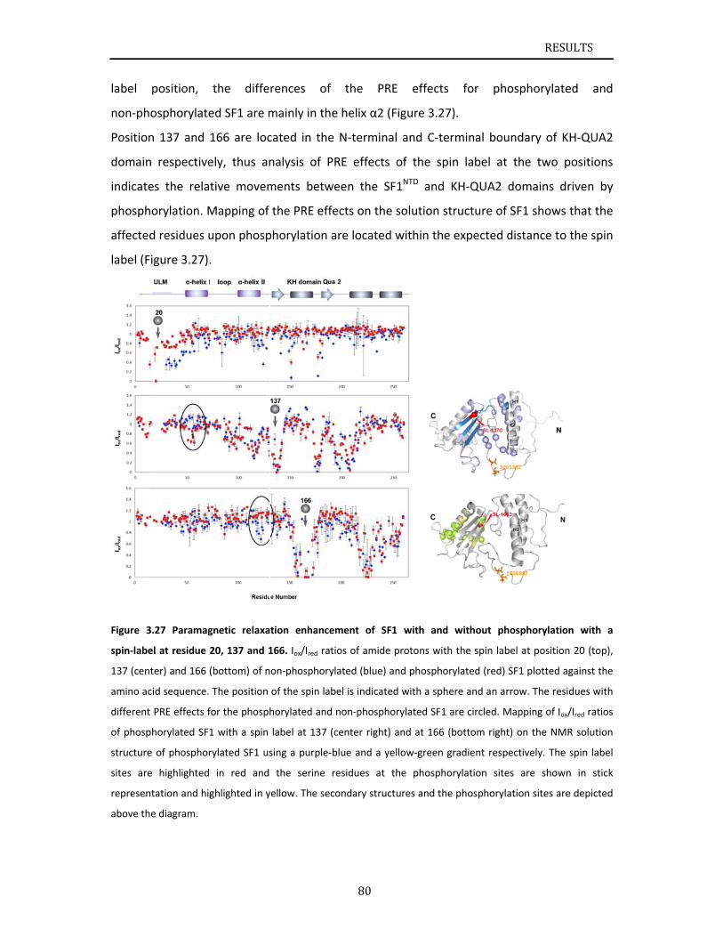
label
non‐ph
Position
domain
indicate
phosph
affected
label (F
Figure 3
spin‐labe
137 (cent
amino ac
different
of phosp
structure
sites are
represen
above th
position,
osphorylate
n 137 and
n respective
es the rela
orylation. M
d residues u
igure 3.27).
3.27 Paramag
el at residue 2
ter) and 166 (
cid sequence.
PRE effects f
phorylated SF1
e of phosphor
e highlighted
ntation and hig
e diagram.
the diffe
ed SF1 are m
166 are loc
ely, thus an
tive movem
Mapping of
upon phosp
.
gnetic relaxa
20, 137 and 1
(bottom) of n
The position
for the phosph
1 with a spin
rylated SF1 us
in red and
ghlighted in y
rences of
mainly in th
cated in the
nalysis of P
ments betw
the PRE eff
phorylation
tion enhance
166. Iox/Ired ra
on‐phosphory
of the spin la
horylated and
label at 137
sing a purple‐
the serine
yellow. The se
80
f the PR
he helix α2 (
e N‐termina
PRE effects
ween the S
fects on the
are located
ement of SF
tios of amide
ylated (blue)
bel is indicate
d non‐phosph
(center right)
‐blue and a y
residues at
condary struc
RE effects
(Figure 3.27
al and C‐te
of the sp
F1NTD and
e solution st
d within the
F1 with and
e protons with
and phosphor
ed with a sphe
orylated SF1
) and at 166 (
ellow‐green g
the phospho
ctures and the
for pho
7).
rminal bou
in label at
KH‐QUA2 d
tructure of
e expected d
without ph
h the spin lab
rylated (red) S
ere and an ar
are circled. M
(bottom right
gradient respe
orylation site
e phosphoryla
RESU
osphorylate
ndary of K
the two p
domains dr
SF1 shows
distance to
osphorylation
bel at position
SF1 plotted ag
row. The resid
Mapping of Iox/
t) on the NMR
ectively. The
s are shown
ation sites are
ULTS
ed and
H‐QUA2
positions
riven by
that the
the spin
n with a
n 20 (top),
gainst the
dues with
/Ired ratios
R solution
spin label
n in stick
e depicted
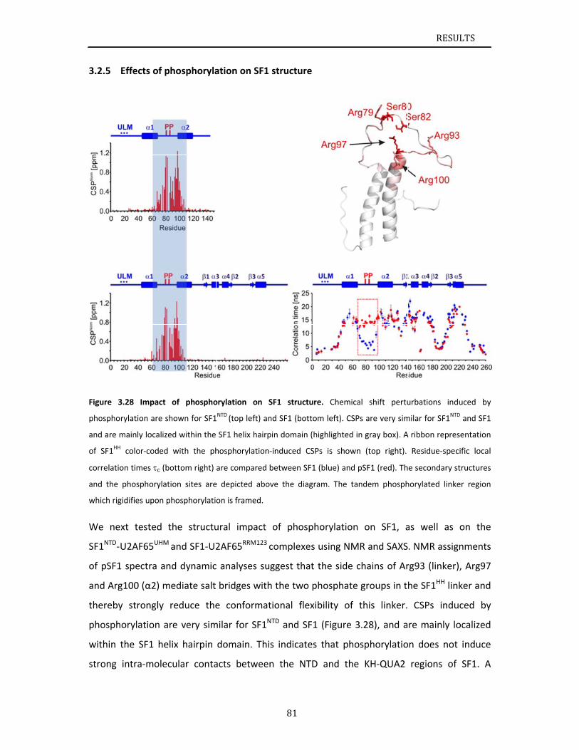
3.2.5
Figure 3
phospho
and are m
of SF1HH
correlatio
and the
which rig
We ne
SF1NTD‐
of pSF1
and Arg
thereby
phosph
within t
strong
Effects of p
3.28 Impact
rylation are s
mainly localize
color‐coded
on times c (b
phosphorylat
gidifies upon p
xt tested t
U2AF65UHM
1 spectra an
g100 (α2) m
y strongly
orylation a
the SF1 he
intra‐molec
phosphoryla
of phospho
hown for SF1
ed within the
with the ph
bottom right)
tion sites are
phosphorylati
the structu
M and SF1‐U2
nd dynamic
mediate salt
reduce the
re very sim
lix hairpin
cular conta
ation on SF
orylation on
1NTD (top left) a
SF1 helix hai
hosphorylatio
are compared
e depicted ab
on is framed.
ural impact
2AF65RRM123
c analyses s
bridges wit
e conforma
milar for SF1
domain. Th
acts betwee
81
1 structure
SF1 structu
and SF1 (bott
rpin domain (
on‐induced CS
d between SF
bove the diag
t of phosp
3 complexes
uggest that
th the two
ational flex
1NTD and SF
his indicate
en the NT
ure. Chemica
tom left). CSP
(highlighted in
SPs is shown
1 (blue) and p
gram. The tan
phorylation
s using NM
t the side c
phosphate
xibility of t
1 (Figure 3
es that pho
D and the
al shift pertu
s are very sim
n gray box). A
n (top right).
pSF1 (red). Th
ndem phosph
on SF1, a
R and SAXS
chains of Ar
groups in t
this linker.
.28), and a
sphorylatio
KH‐QUA2
RESU
urbations ind
milar for SF1NT
A ribbon repre
Residue‐spec
e secondary s
horylated link
as well as
. NMR assig
rg93 (linker
he SF1HH lin
CSPs indu
re mainly l
on does not
regions of
ULTS
duced by
TD and SF1
esentation
cific local
structures
ker region
on the
gnments
r), Arg97
nker and
uced by
ocalized
t induce
f SF1. A
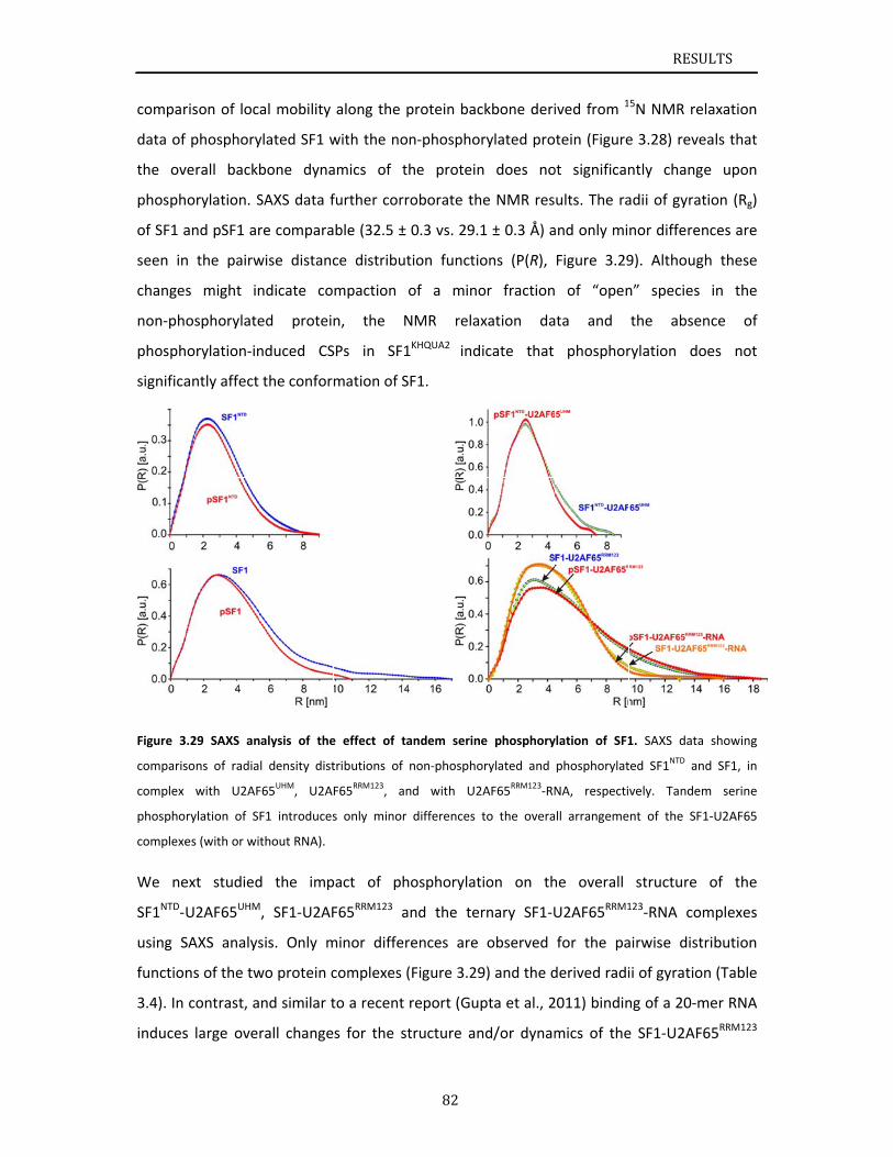
compar
data of
the ov
phosph
of SF1 a
seen in
change
non‐ph
phosph
significa
Figure 3
comparis
complex
phospho
complexe
We ne
SF1NTD‐
using S
function
3.4). In
induces
rison of loc
f phosphory
verall backb
orylation. S
and pSF1 ar
n the pairw
s might in
osphorylate
orylation‐in
antly affect
.29 SAXS an
sons of radia
with U2AF
rylation of S
es (with or wi
ext studied
U2AF65UHM
SAXS analys
ns of the tw
contrast, a
s large ove
al mobility
ylated SF1 w
bone dyna
SAXS data f
re compara
wise distanc
ndicate co
ed protei
nduced CS
the confor
nalysis of the
l density dist
F65UHM, U2A
F1 introduces
thout RNA).
d the imp
M, SF1‐U2AF
sis. Only m
wo protein c
nd similar t
rall change
along the p
with the no
amics of t
further corr
ble (32.5 ±
ce distribut
mpaction
n, the N
SPs in SF1
mation of S
e effect of ta
tributions of
F65RRM123, an
s only minor
pact of ph
F65RRM123 a
minor differ
complexes (
to a recent
s for the st
82
protein bac
on‐phospho
he protein
roborate th
0.3 vs. 29.1
tion functio
of a mino
NMR relax
1KHQUA2 ind
SF1.
andem serine
non‐phospho
nd with U2
differences
hosphorylat
and the ter
rences are
(Figure 3.29
report (Gup
tructure an
ckbone deri
rylated pro
n does no
e NMR res
1 ± 0.3 Å) a
ons (P(R),
or fraction
xation da
icate that
e phosphory
orylated and
2AF65RRM123‐RN
to the overa
ion on th
rnary SF1‐U
observed
9) and the d
pta et al., 2
nd/or dynam
ived from 1
tein (Figure
ot significan
ults. The ra
and only mi
Figure 3.29
of “open
ta and
phosphor
ylation of SF1
phosphorylat
NA, respecti
ll arrangeme
he overall
U2AF65RRM1
for the pa
derived radi
2011) bindin
mics of the
RESU
5N NMR re
e 3.28) reve
ntly chang
adii of gyrat
nor differen
9). Althoug
n” species
the abse
rylation do
1. SAXS data
ted SF1NTD an
vely. Tandem
nt of the SF
structure
123‐RNA com
airwise dist
ii of gyratio
ng of a 20‐m
e SF1‐U2AF
ULTS
elaxation
eals that
e upon
tion (Rg)
nces are
gh these
in the
nce of
oes not
showing
nd SF1, in
m serine
1‐U2AF65
of the
mplexes
tribution
n (Table
mer RNA
65RRM123
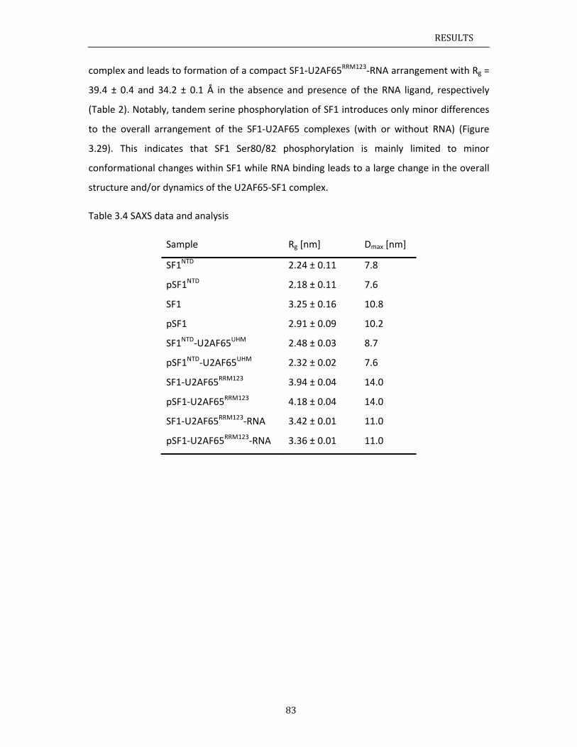
RESULTS
83
complex and leads to formation of a compact SF1‐U2AF65RRM123‐RNA arrangement with Rg =
39.4 ± 0.4 and 34.2 ± 0.1 Å in the absence and presence of the RNA ligand, respectively
(Table 2). Notably, tandem serine phosphorylation of SF1 introduces only minor differences
to the overall arrangement of the SF1‐U2AF65 complexes (with or without RNA) (Figure
3.29). This indicates that SF1 Ser80/82 phosphorylation is mainly limited to minor
conformational changes within SF1 while RNA binding leads to a large change in the overall
structure and/or dynamics of the U2AF65‐SF1 complex.
Table 3.4 SAXS data and analysis
Sample Rg [nm] Dmax [nm]
SF1NTD 2.24 ± 0.11 7.8
pSF1NTD 2.18 ± 0.11 7.6
SF1 3.25 ± 0.16 10.8
pSF1 2.91 ± 0.09 10.2
SF1NTD‐U2AF65UHM 2.48 ± 0.03 8.7
pSF1NTD‐U2AF65UHM 2.32 ± 0.02 7.6
SF1‐U2AF65RRM123 3.94 ± 0.04 14.0
pSF1‐U2AF65RRM123 4.18 ± 0.04 14.0
SF1‐U2AF65RRM123‐RNA 3.42 ± 0.01 11.0
pSF1‐U2AF65RRM123‐RNA 3.36 ± 0.01 11.0
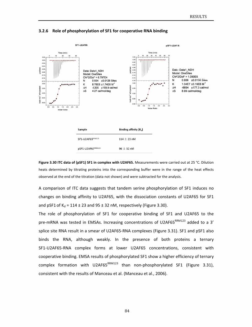
3.2.6
Figure 3.
heats de
observed
A comp
change
and pSF
The rol
pre‐mR
splice s
binds
SF1‐U2A
coopera
comple
consiste
Role of pho
30 ITC data o
etermined by
d at the end o
parison of I
s on bindin
F1 of Kd = 11
le of phosp
RNA was tes
ite RNA res
the RNA,
AF65‐RNA
ative bindin
x formatio
ent with the
osphorylati
of (pSF1) SF1 i
titrating prot
f the titration
TC data sug
ng affinity t
14 ± 23 and
phorylation
sted in EM
sult in a sme
although
complex
ng. EMSA re
on with U
e results of
on of SF1 fo
in complex w
teins into the
n (data not sho
ggests that
to U2AF65,
95 ± 32 nM
n of SF1 fo
SAs. Increa
ear of U2AF
weakly.
forms at
esults of pho
U2AF65RRM1
Manceau e
84
or cooperat
ith U2AF65. M
e correspondi
own) and wer
t tandem se
with the d
M, respectiv
or cooperat
sing conce
F65‐RNA co
In the pr
lower U2
osphorylate
23 than n
et al. (Manc
tive RNA bi
Measurement
ng buffer we
re subtracted
erine phosp
dissociation
vely (Figure
tive binding
ntrations o
omplexes (F
resence of
AF65 conc
ed SF1 show
non‐phosph
eau et al., 2
nding
ts were carrie
ere in the ran
for the analys
phorylation
constants
3.30).
g of SF1 an
f U2AF65RR
igure 3.31)
f both pr
centrations
w a higher e
orylated S
2006).
RESU
d out at 25 °C
nge of the he
sis.
of SF1 ind
of U2AF65
nd U2AF65
RM123 added
. SF1 and p
roteins a
, consisten
efficiency of
SF1 (Figure
ULTS
C. Dilution
at effects
uces no
for SF1
5 to the
d to a 3’
SF1 also
ternary
nt with
f ternary
e 3.31),
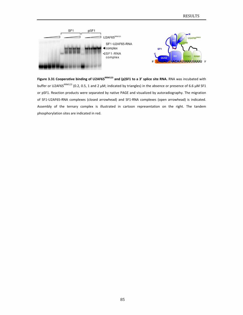
Figure 3.
buffer or
or pSF1.
of SF1‐U
Assembly
phospho
.31 Cooperati
r U2AF65RRM12
Reaction pro
2AF65‐RNA c
y of the ter
rylation sites
ive binding of
23 (0.2, 0.5, 1 a
ducts were se
complexes (clo
rnary comple
are indicated
f U2AF65RRM1
and 2 µM; ind
eparated by n
osed arrowhe
ex is illustrat
in red.
85
23 and (p)SF1
dicated by tria
native PAGE a
ead) and SF1
ted in carto
to a 3’ splice
angles) in the
and visualized
‐RNA comple
on represent
e site RNA. RN
absence or p
by autoradio
exes (open ar
tation on th
RESU
NA was incub
presence of 6.
ography. The
rowhead) is i
he right. The
ULTS
ated with
.6 µM SF1
migration
indicated.
e tandem
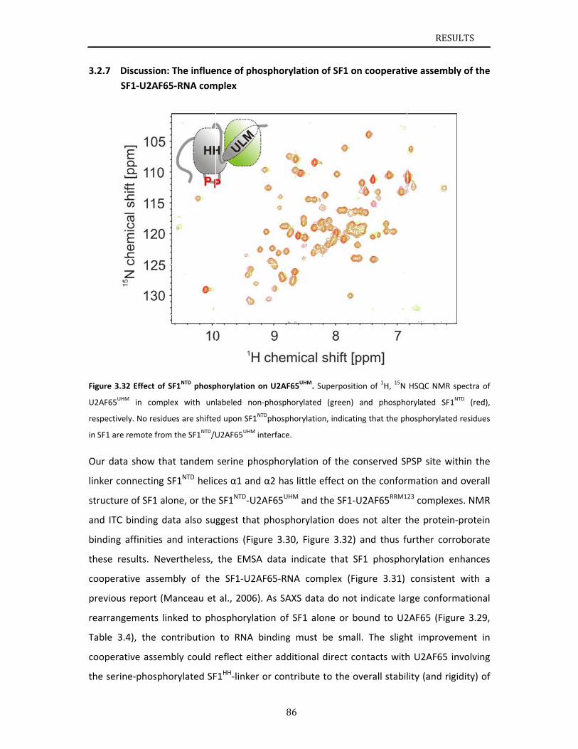
3.2.7
Figure 3.
U2AF65U
respectiv
in SF1 are
Our dat
linker c
structu
and ITC
binding
these r
coopera
previou
rearran
Table 3
coopera
the seri
Discussion:
SF1‐U2AF6
.32 Effect of
UHM in comp
vely. No residu
e remote from
ta show tha
onnecting S
re of SF1 al
C binding da
g affinities a
results. Nev
ative assem
us report (M
ngements lin
3.4), the co
ative assem
ine‐phosph
: The influe
65‐RNA com
SF1NTD phosp
lex with un
ues are shifte
m the SF1NTD/U
at tandem
SF1NTD helic
one, or the
ata also sug
and interac
vertheless,
mbly of the
Manceau et
nked to ph
ontribution
mbly could r
orylated SF
ence of phos
mplex
phorylation on
labeled non‐
ed upon SF1NT
U2AF65UHM in
serine pho
ces α1 and
SF1NTD‐U2A
ggest that
ctions (Figu
the EMSA
e SF1‐U2AF
al., 2006).
osphorylati
to RNA b
reflect eithe
F1HH‐linker o
86
sphorylatio
n U2AF65UHM
‐phosphorylat
Dphosphoryla
terface.
sphorylatio
α2 has little
AF65UHM an
phosphoryl
re 3.30, Fig
data indic
F65‐RNA co
As SAXS da
ion of SF1
inding mus
er addition
or contribut
on of SF1 on
. Superpositio
ted (green)
tion, indicatin
on of the co
e effect on
d the SF1‐U
lation does
gure 3.32)
ate that SF
omplex (Fig
ata do not
alone or bo
st be small
al direct co
te to the ov
n cooperati
on of 1H, 15N
and phospho
ng that the ph
onserved SP
the confor
U2AF65RRM1
not alter t
and thus f
F1 phospho
gure 3.31)
indicate lar
ound to U2
l. The sligh
ontacts with
verall stabil
RESU
ve assembl
HSQC NMR s
orylated SF1N
hosphorylated
PSP site wit
rmation and
23 complexe
the protein
urther corr
orylation e
consistent
rge conform
2AF65 (Figu
ht improvem
h U2AF65 in
lity (and rig
ULTS
ly of the
spectra of
NTD (red),
d residues
thin the
d overall
es. NMR
‐protein
roborate
nhances
with a
mational
ure 3.29,
ment in
nvolving
gidity) of
RESULTS
87
a pre‐arranged SF1‐U2AF65 complex, by reducing entropy loss linked to RNA binding.
Structural studies to analyze these effects in the ternary complex are currently on‐going in
our lab.
Tandem serine phosphorylation of SF1 may have additional roles beyond a function in
cooperative assembly of the constitutive splicing complex. Previous studies in mammalian
and yeast systems have established that SF1 mainly affects the kinetics of spliceosome
assembly, as genetic or biochemical depletion of SF1 does not abolish splicing (Guth and
Valcarcel, 2000; Rutz and Seraphin, 1999). In S. cerevisiae it has been shown that SF1 is
involved in removing introns with suboptimal splice sites and in nuclear pre‐mRNA retention
(Galy et al., 2004; Rutz and Séraphin, 2000), whereas knockdown of human SF1 in HeLa cells
did not result in a general splicing phenotype (Tanackovic and Kramer, 2005). However,
Corioni et al. (Corioni et al., 2011) have recently shown that SF1 is not a constitutive splicing
factor, but is required for the splicing of certain introns and affects alternative splice site
choice. In this respect, SF1 phosphorylation may mediate interactions with other factors
that could regulate alternative splicing by modulating 3’ splice site recognition. For example,
it has been suggested that the phosphorylated SPSP motif of SF1 is recognized by the
tandem WW domains of FBP11 (Ingham et al., 2005) and the interaction of the SF1 and
FBP11 orthologues of S. cerevisiae has been implicated in mediating intron definition by
connecting the 5’ and 3’ splice sites (Abovich and Rosbash, 1997). However, we have not
detected any interaction of tandem WW domains with phosphorylated SF1 (data not
shown). Nevertheless, the substantial conformational changes in the SF1NTD linker
associated with tandem serine phosphorylation by KIS kinase suggest that interactions with
so far unknown factors are modulated that may contribute to regulate alternative splicing
by SF1.
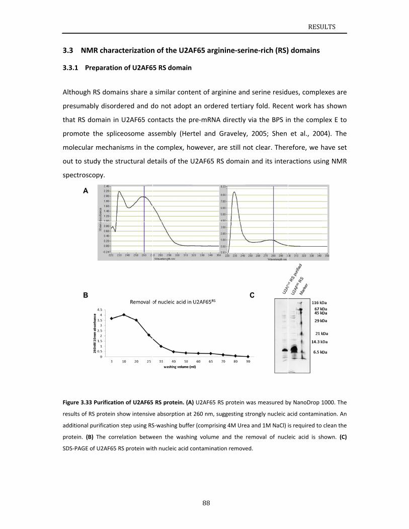
N3.3
3.3.1
Althoug
presum
that RS
promot
molecu
out to s
spectro
Figure 3.
results of
additiona
protein.
SDS‐PAG
NMR chara
Preparation
gh RS doma
mably disord
domain in
te the splic
lar mechan
study the st
oscopy.
.33 Purificatio
f RS protein s
al purification
(B) The corr
E of U2AF65 R
cterization
n of U2AF6
ains share a
dered and d
U2AF65 co
ceosome as
nisms in the
tructural de
on of U2AF65
show intensive
step using RS
elation betw
RS protein wit
n of the U2
5 RS domai
similar con
do not adop
ontacts the
ssembly (H
e complex,
etails of the
5 RS protein. (
e absorption
S‐washing buf
een the was
th nucleic acid
88
2AF65 argi
in
ntent of arg
pt an order
pre‐mRNA
ertel and G
however, a
e U2AF65 R
(A) U2AF65 R
at 260 nm, su
ffer (comprisin
hing volume
d contaminati
nine‐serin
ginine and s
red tertiary
directly via
Graveley, 2
are still not
S domain a
RS protein was
uggesting stro
ng 4M Urea a
and the rem
on removed.
e‐rich (RS)
serine resid
fold. Recen
a the BPS in
2005; Shen
clear. Ther
and its inter
s measured b
ongly nucleic a
nd 1M NaCl) i
moval of nucl
RESU
) domains
ues, comple
nt work has
n the comp
et al., 200
refore, we h
ractions usi
by NanoDrop 1
acid contamin
is required to
eic acid is sh
ULTS
exes are
s shown
plex E to
04). The
have set
ng NMR
1000. The
nation. An
clean the
hown. (C)
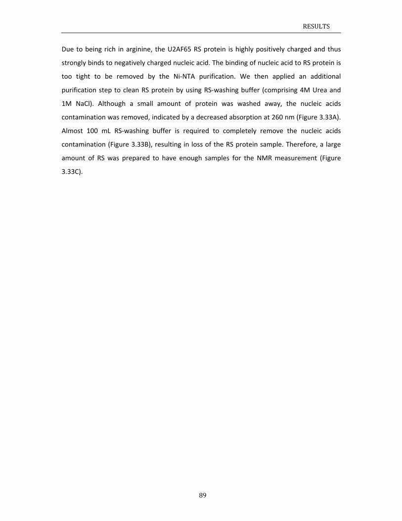
RESULTS
89
Due to being rich in arginine, the U2AF65 RS protein is highly positively charged and thus
strongly binds to negatively charged nucleic acid. The binding of nucleic acid to RS protein is
too tight to be removed by the Ni‐NTA purification. We then applied an additional
purification step to clean RS protein by using RS‐washing buffer (comprising 4M Urea and
1M NaCl). Although a small amount of protein was washed away, the nucleic acids
contamination was removed, indicated by a decreased absorption at 260 nm (Figure 3.33A).
Almost 100 mL RS‐washing buffer is required to completely remove the nucleic acids
contamination (Figure 3.33B), resulting in loss of the RS protein sample. Therefore, a large
amount of RS was prepared to have enough samples for the NMR measurement (Figure
3.33C).
3.3.2
Figure 3.
region of
assignme
A 1H, 15
residue
cluster
pattern
standar
signal o
assigne
seconda
NMR assign
.34 1H, 15N HS
f the spectru
ents of RS wer
N HSQC NM
es from 1 to
in the regio
n for unstru
rd NMR me
overlap. Th
d to a certa
ary chemica
nment and
SQC spectrum
m between 8
re done using
MR spectru
o 84. The m
on of the sp
uctured pro
easurement
hus, approx
ain amide p
al shifts usin
NMR analy
m of U2AF65
8 and 8.5 ppm
a series of sta
um was rec
majority of t
pectrum bet
otein. NMR
ts. A large
ximately 30
roton in the
ng the assig
90
ysis of U2AF
RS. Signals fr
m, which is a
andard NMR m
orded on is
he signals i
tween 8 and
R assignme
amount o
0% of NM
e 1H, 15N HS
gnments of
F65 RS dom
rom the U2AF
typical patte
measurement
sotope‐labe
n the 1H, 15
d 8.5 ppm (
nts of RS
f similar Se
R signals c
SQC (Figure
U2AF65 RS
main
F65 RS (residu
ern for unstru
ts.
elled U2AF6
5N HSQC NM
Figure 3.34
were done
er‐Arg mot
could not
3.34). We t
.
RESU
ues 1‐84) clus
uctured prote
65 RS doma
MR spectru
4), which is a
e using a s
tifs lead to
be unamb
then calcula
ULTS
ster in the
eins. NMR
ain with
um of RS
a typical
eries of
serious
iguously
ated the
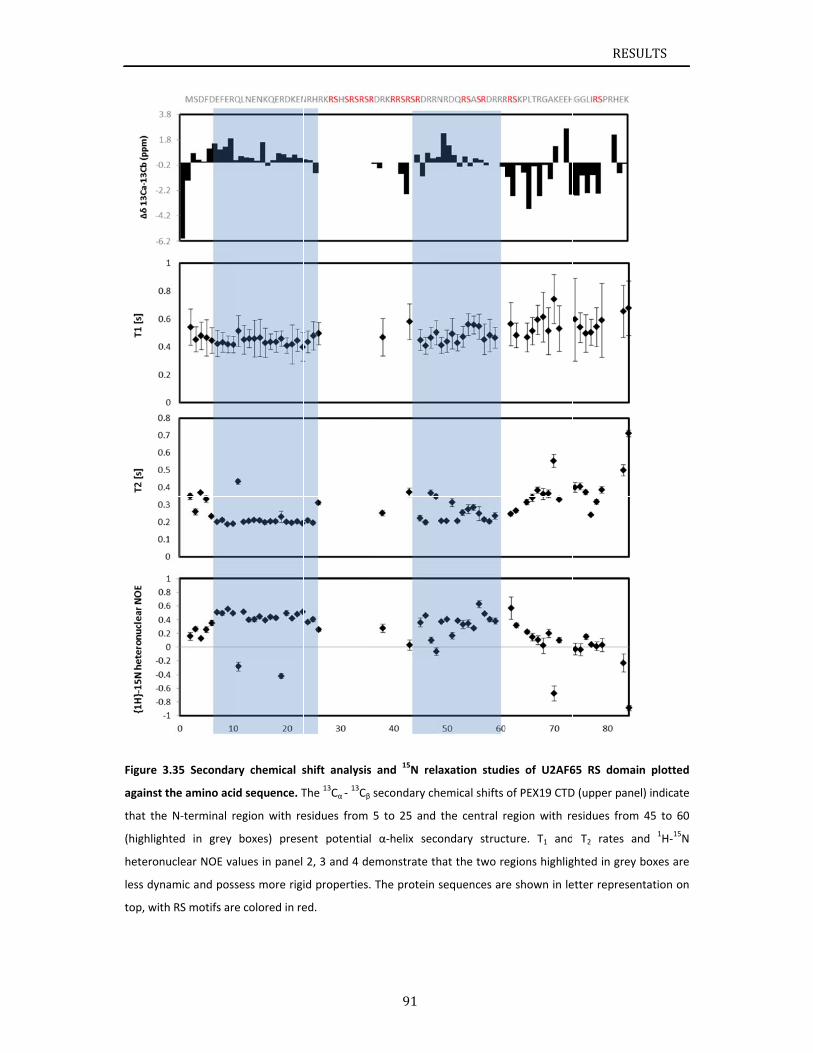
Figure 3
against t
that the
(highlight
heteronu
less dyna
top, with
.35 Secondar
the amino acid
N‐terminal r
ted in grey
uclear NOE va
amic and poss
h RS motifs are
ry chemical s
d sequence. T
egion with re
boxes) prese
alues in panel
sess more rigi
e colored in re
shift analysis
The 13Cα ‐ 13Cβ
esidues from
ent potential
2, 3 and 4 de
id properties.
ed.
91
s and 15N rel
secondary ch
5 to 25 and
α‐helix seco
emonstrate th
The protein
axation stud
hemical shifts
the central r
ondary struc
hat the two re
sequences ar
ies of U2AF6
of PEX19 CTD
region with re
ture. T1 and
egions highlig
e shown in le
RESU
65 RS domain
D (upper pane
esidues from
T2 rates an
ghted in grey
etter represen
ULTS
n plotted
l) indicate
45 to 60
nd 1H‐15N
boxes are
ntation on
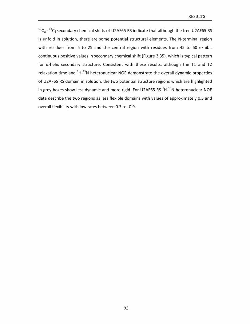
RESULTS
92
13Cα ‐ 13Cβ
secondary chemical shifts of U2AF65 RS indicate that although the free U2AF65 RS
is unfold in solution, there are some potential structural elements. The N‐terminal region
with residues from 5 to 25 and the central region with residues from 45 to 60 exhibit
continuous positive values in secondary chemical shift (Figure 3.35), which is typical pattern
for α‐helix secondary structure. Consistent with these results, although the T1 and T2
relaxation time and 1H‐15N heteronuclear NOE demonstrate the overall dynamic properties
of U2AF65 RS domain in solution, the two potential structure regions which are highlighted
in grey boxes show less dynamic and more rigid. For U2AF65 RS 1H‐15N heteronuclear NOE
data describe the two regions as less flexible domains with values of approximately 0.5 and
overall flexibility with low rates between 0.3 to ‐0.9.
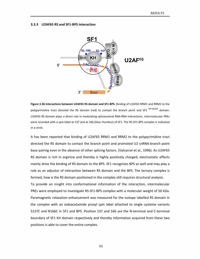
3.3.3
Figure 3.
polypyrim
U2AF65
were rec
in a circle
It has b
directed
base pa
RS dom
mainly
role as
formed
To prov
PREs w
Parama
the com
S137C a
bounda
position
U2AF65 RS
36 Interactio
midine tract
RS domain pl
corded with a
e.
been report
d the RS do
airing even
main is rich
drive the b
an adjusto
, how is the
vide an ins
ere employ
agnetic rela
mplex with
and N166C
ary of SF1 K
ns is able to
and SF1‐BP
ns between U
directed the
ays a direct r
spin‐label at
ted that bin
omain to co
in the abse
in arginine
inding of RS
r of interac
e RS domain
sight into c
yed to inves
xation enha
an iodoace
in SF1 and
KH domain r
o cover the e
PS interacti
U2AF65 RS do
RS domain (
ole in modula
137 and at 16
nding of U2
ontact the b
nce of othe
e and there
S domain to
ction betwe
n positioned
conformatio
stigate RS‐S
ancement w
etamide pr
d BPS. Posit
respectively
entire comp
93
ion
omain and SF1
(red) to cont
ating spliceos
66 (blue rhom
2AF65 RRM
branch poin
er splicing fa
by is highly
o the BPS. S
een RS dom
d in the com
onal inform
SF1‐BPS com
was measur
oxyl spin la
ion 137 and
y and there
plex.
1‐BPS. Binding
act the bran
omal RNA‐RN
mbus) of SF1. T
1 and RRM
nt and prom
actors. (Valc
y positively
SF1 recogniz
main and the
mplex still re
ation of th
mplex with
red for the
abel attach
d 166 are t
eby informa
g of U2AF65 R
ch point and
NA interaction
The RS‐SF1‐BP
M2 to the po
moted U2 sn
carcel et al.
charged, e
zes BPS as w
e BPS. The
equires stru
he interacti
a molecula
isotope lab
ed to single
the N‐termi
ation acquir
RESU
RRM1 and RRM
d SF1 KH‐QUA2
ns. Intermolec
PS complex is
olypyrimidi
nRNA‐branc
, 1996). As
electrostatic
well and ma
ternary com
uctural anal
on, interm
r weight of
belled RS do
e cysteine
inal and C‐t
red from th
ULTS
M2 to the
2 domain.
cular PREs
indicated
ne tract
ch point
U2AF65
c effects
ay play a
mplex is
ysis.
olecular
50 kDa.
omain in
variants
terminal
ese two
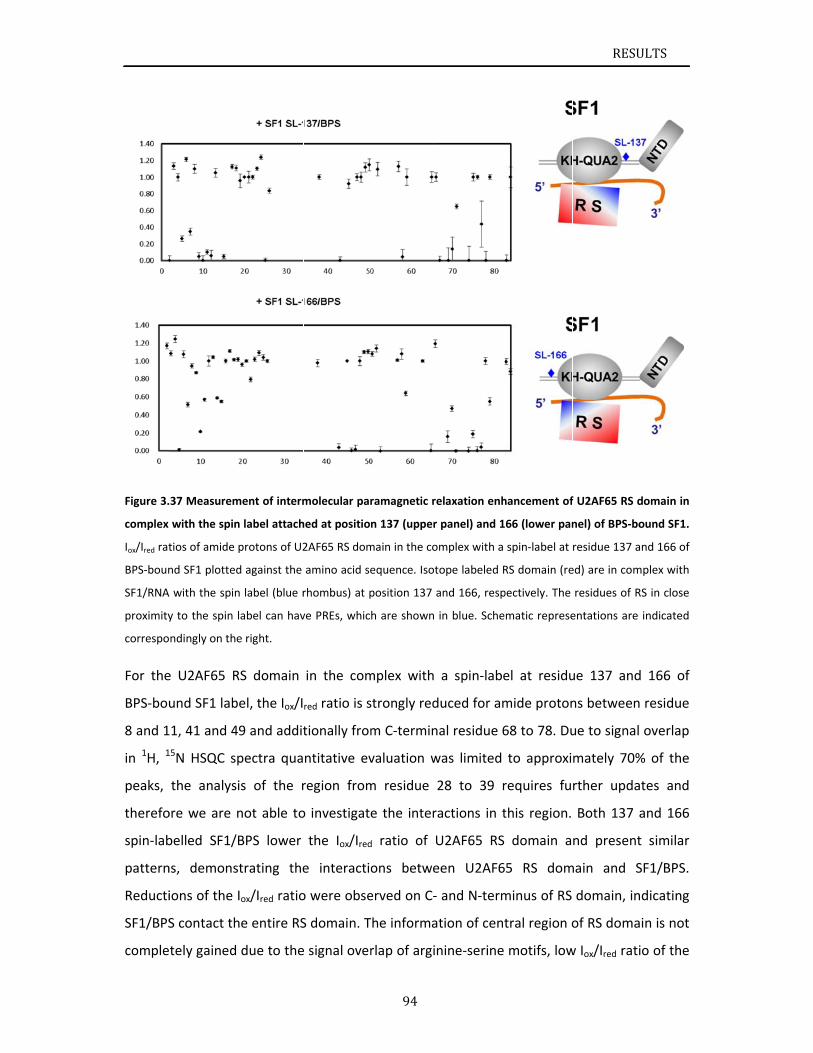
Figure 3.
complex
Iox/Ired rat
BPS‐boun
SF1/RNA
proximity
correspo
For the
BPS‐bou
8 and 1
in 1H, 1
peaks,
therefo
spin‐lab
pattern
Reducti
SF1/BPS
comple
.37 Measurem
with the spin
tios of amide
nd SF1 plotted
A with the spin
y to the spin
ndingly on th
e U2AF65 R
und SF1 lab
11, 41 and 4
15N HSQC s
the analys
ore we are
belled SF1/
ns, demons
ions of the
S contact th
tely gained
ment of interm
n label attach
protons of U2
d against the
n label (blue r
label can hav
e right.
RS domain
bel, the Iox/I
49 and addit
pectra qua
is of the r
not able to
/BPS lower
strating the
Iox/Ired ratio
he entire RS
due to the
molecular par
hed at positio
2AF65 RS dom
amino acid se
rhombus) at p
ve PREs, whic
in the com
red ratio is s
tionally from
ntitative ev
region from
o investigate
the Iox/Ired
e interactio
o were obse
S domain. T
e signal over
94
ramagnetic re
n 137 (upper
main in the co
equence. Isot
position 137 a
h are shown
mplex with
strongly red
m C‐termin
valuation w
m residue 2
e the intera
d ratio of U
ons betwe
erved on C‐
The informa
rlap of argin
elaxation enh
panel) and 1
mplex with a
tope labeled R
and 166, resp
in blue. Sche
a spin‐labe
duced for am
al residue 6
was limited
28 to 39 r
actions in t
U2AF65 RS
een U2AF6
and N‐term
tion of cent
nine‐serine
hancement of
66 (lower pan
spin‐label at
RS domain (re
pectively. The
matic represe
el at residu
mide proton
68 to 78. Du
to approxi
requires fu
this region.
domain a
5 RS dom
minus of RS
tral region o
motifs, low
RESU
f U2AF65 RS d
nel) of BPS‐bo
residue 137 a
ed) are in com
residues of R
entations are
ue 137 and
ns between
ue to signal
mately 70%
rther upda
Both 137 a
nd present
main and S
domain, in
of RS doma
w Iox/Ired rati
ULTS
domain in
ound SF1.
and 166 of
mplex with
RS in close
indicated
d 166 of
residue
overlap
% of the
tes and
and 166
t similar
SF1/BPS.
ndicating
in is not
io of the

RESULTS
95
potential structure region from residue 45 to 50 suggests this less flexible α‐helix‐like region
is involved in the RS domain and SF1/BPS interactions.
RESULTS
96
3.3.4 Discussion: RS domain contacts SF1 and RNA at the branch point
Previous studies have shown that the RS domain present in splicing factors of SR family
(Zahler et al., 1992) mediate protein‐protein interactions (Kohtz et al., 1994; Wu and
Maniatis, 1993). Whereas, it is reported the target of the U2AF65 RS domain is the
pre‐mRNA branch point, and not a protein (Valcarcel et al., 1996). Due to intrinsically
unstructured and highly dynamic feature of U2AF65 RS domains, structural insight into
molecular mechanisms of the interactions remains elusive. Because U2AF65 RS domain is
rich in positively charged residues (arginine), resulting in intensively binding to negatively
charged nucleic acid. Thereby, purification of RS protein, removal of nucleic acids
contamination, could prove a challenge. In this study, RS protein containing an N‐terminal
Histidine‐tag bound to Ni‐NTA column and was washed with high urea concentration and
high salt concentration buffer. High concentration of urea is able to unfold the protein to
expose the nucleic acids and salt plays role as an eliminator of electrostatic effects between
RS protein and nucleic acids. The RS domain contacts the branch point and promoted U2
snRNA branch point base pairing, which might be explained by highly positively charged
property of RS domain. The binding of RS domain can reduce the negative charges of the
branch point, might further facilitate RNA‐RNA interactions by indirectly diminish the
electrostatic repulsion effects. In addition, SF1 KH‐QUA2 domain has been reported recognize
branch point (Liu et al., 2001),
Different from conventional view of intrinsically unstructured and highly dynamic feature, in
this study we propose that RS domain tends to adopt structural elements. The N‐terminal
region with residues from 5 to 25 and the central region with residues from 45 to 60 exhibit
typical pattern for α‐helix secondary structure and show less flexibility and less dynamics.
Intermolecular paramagnetic relaxation enhancements demonstrate that the α‐helix‐like
region might mediate the RS domain‐SF1KH‐QUA2 interactions, in good agreement with our
hypothesis of formation of RS‐SF1‐BPS complex.
In vivo, RS domains are found to be extensively phosphorylated on serine residues enabling
post‐translational modification of the interaction network. It is believed that the subcellular
localization as well as the activity is regulated on the level of phosphorylation (Lin and Fu,
2007). Inclusion of phosphorylation in the structural studies is crucial in order to understand
how the binding‐strength and ‐specificity of RS domains for their interaction partners is
RESULTS
97
modulated. The impact of the phosphorylation patterns will reveal how the dynamic
interaction network is modulated on a molecular level. Study of in vivo phosphorylation of
U2AF65 RS domain will further describes its functional roles in regulation of the formation
of SF1‐U2AF65‐mRNA complex. However, limited amount of RS protein was purified from
our protocol, which to some extent increases the difficulty in preparation of phosphorylated
RS protein in NMR‐scale amount. There is no denying that structural analysis of
phosphorylated U2AF65 RS domain will brighten the insight into molecular mechanisms of
regulation of pre‐mRNA splicing.
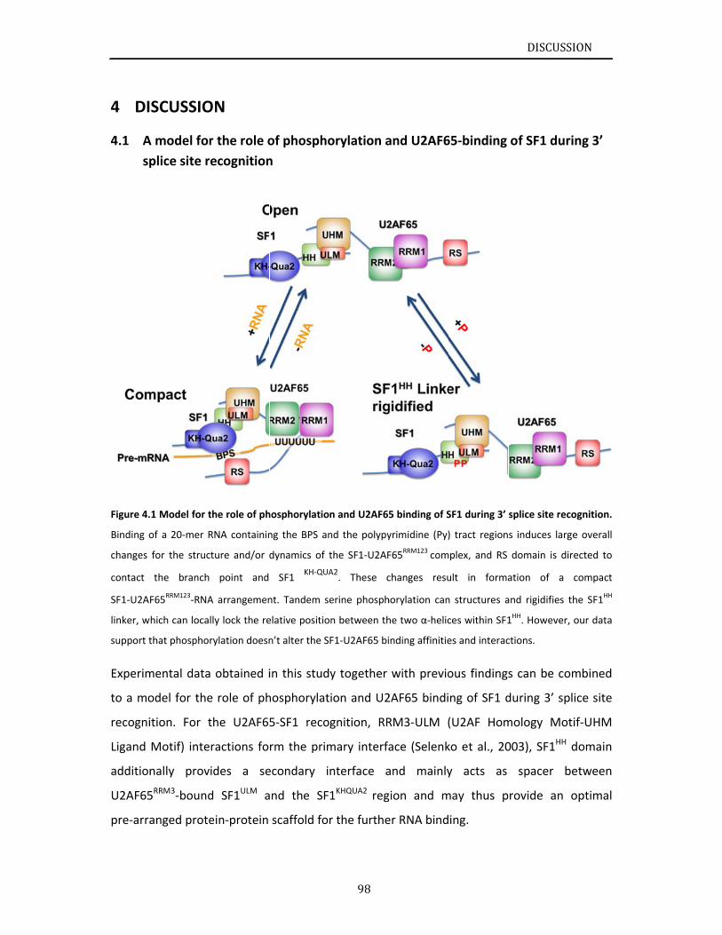
4 DI
A4.1
sp
Figure 4.
Binding o
changes
contact
SF1‐U2AF
linker, w
support t
Experim
to a mo
recogni
Ligand
addition
U2AF65
pre‐arra
SCUSSIO
A model for
plice site r
1 Model for t
of a 20‐mer R
for the struc
the branch
F65RRM123‐RNA
hich can loca
that phosphor
mental data
odel for the
ition. For t
Motif) inte
nally prov
5RRM3‐bound
anged prote
ON
r the role o
ecognition
the role of ph
RNA containin
cture and/or d
point and
A arrangemen
lly lock the re
rylation doesn
a obtained i
e role of ph
the U2AF65
ractions fo
ides a se
d SF1ULM a
ein‐protein
of phospho
n
hosphorylatio
ng the BPS an
dynamics of t
SF1 KH‐QUA
nt. Tandem s
elative positio
n’t alter the S
in this stud
hosphorylat
5‐SF1 reco
rm the prim
econdary i
and the SF
scaffold fo
98
orylation a
n and U2AF65
nd the polypy
the SF1‐U2AF
A2. These c
erine phosph
on between th
F1‐U2AF65 bi
y together
ion and U2
gnition, RR
mary interfa
nterface a
1KHQUA2 reg
r the furthe
and U2AF6
5 binding of S
yrimidine (Py)
F65RRM123 com
changes resu
horylation can
he two α‐helic
nding affinitie
with previo
2AF65 bindi
RM3‐ULM (
ace (Selenk
and mainly
gion and m
er RNA bind
65‐binding
SF1 during 3’
) tract region
plex, and RS
ult in forma
n structures a
ces within SF1
es and interac
ous findings
ng of SF1 d
(U2AF Hom
ko et al., 20
y acts as
may thus p
ing.
DISCUSS
of SF1 dur
splice site rec
s induces larg
domain is di
ation of a
and rigidifies
1HH. However,
ctions.
s can be co
during 3’ sp
mology Mo
003), SF1HH
spacer b
rovide an
SION
ring 3’
cognition.
ge overall
irected to
compact
the SF1HH
, our data
ombined
plice site
tif‐UHM
domain
between
optimal
DISCUSSION
99
Our study has shown that tandem serine phosphorylation of the conserved SPSP motif
rigidifies the linker connecting SF1HH helices α1 and α2, and thereby lock the protein‐protein
scaffold, however our data also suggest that it has little effect on the conformation and
overall structure of SF1 alone and the SF1‐U2AF65RRM123 complex. The crystal structure of
pSF1NTD‐U2AF65UHM proved that the SF1HH mediates an interface with U2AF65 and
phosphorylation induces an arginine claw surrounding the phosphorylated SPSP motif
(Wang et al., 2013). Their findings show a good agreement with the results in this thesis.
However, although local structural changes linked to phosphorylation of SF1 are shown in
their paper and in this thesis, we hardly observed global conformational changes (C‐shape
formation) in the pSF1‐U2AF65‐RNA complex, which is claimed in their paper. As they only
employed SAXS data to generate the C‐shape structure, the limit of resolution on atomic
level leads to this controversial conclusion. To clarify the conformational arrangements of
pSF1‐U2AF65‐RNA complex, more experiments and high‐resolution techniques are required.
Nevertheless, the EMSA data indicate that SF1 phosphorylation enhances cooperative
assembly of the SF1‐U2AF65‐RNA complex (Figure 3.31); although there are no large
conformational rearrangements, our SAXS data to some extend corroborate these results.
RNA recognition by the tandem RNA recognition motifs (RRM1‐RRM2) of U2AF65 involves
equilibrium between two distinct domain arrangements (Mackereth et al., 2011). Without
binding of RNA to U2AF65 RRM1‐2 keeps the conformational equilibrium in close state. The
equilibrium is shifted from the closed state towards the open state in the presence of RNA.
From unpublished data in our group, the relative domain arrangements in the minimal U2AF
heterodimer are modulated by RNA binding and the conformational equilibrium in U2AF65
is affected by the presence of the U2AF35 RRM.
Binding of U2AF65 RRM1‐2 to the Py tract directed the RS domain to contact the branch point
and SF1 KH‐QUA2. The changes of electrostatic effects may interpret the promotions of U2
snRNA branch point base pairing upon on RS binding. Although RS domain is intrinsically
unstructured and highly dynamic feature, in this study we propose that it tends to have
potential structural elements. The NMR relaxation results and intermolecular PRE reveal
that these potential structure regions are involved in the RS‐SF1‐BPS interactions. In vivo, RS
domains are able to be extensively phosphorylated on serine residues and enabling
post‐translational modification of the interaction network. Therefore, structural studies of
DISCUSSION
100
the phosphorylated U2AF65 RS domain will give a detail insight into the molecular
mechanisms of regulation of pre‐mRNA splicing.
Similar to a recent report (Gupta et al., 2011), SAXS results demonstrate binding of RNA
induces large overall changes for the structure of the SF1‐U2AF65RRM123 complex and leads
to formation of a compact SF1‐U2AF65RRM123‐RNA arrangement. The conformational
information of SF1‐U2AF65RRM123‐RNA will describe the mechanism of 3’ splice site
recognition.
C4.2
This st
recogni
phosph
have re
and SF1
structu
phosph
little ef
Howeve
SF1‐U2A
with so
Binding
These i
U2 snR
presenc
In this t
act as
These d
In addit
splicing
splicing
seconda
these f
splicing
pre‐mR
onclusions
udy has a
ition in the
orylation a
evealed a ha
1NTD and p
ral biology
orylation o
ffects on th
er, EMSA r
AF65‐RNA
far unknow
g of U2AF65
nteractions
NA branch
ce of 20mer
thesis, the
a spacer de
data contrib
tion, invest
g factors exh
g. Furtherm
ary structur
indings are
g site reco
RNA splicing
s
ddressed o
e complex
nd the arra
airpin scaffo
rovides the
of SF1 pho
f SF1 on SP
he overall c
results imp
complex. It
wn factors m
5 RRM1‐2 to
s may be m
point base
r RNA conta
NMR struct
efining the
bute to stud
tigations of
hibit signific
more, NMR
re elements
e a major st
gnition an
g.
open ques
E with a f
ngements o
old structur
e proper ge
sphorylatio
SP motif rig
onformatio
ply that ph
t has been
may contrib
o Py trace d
mediated by
e pairing. A
aining of BP
ture of SF1N
relative o
dies of the d
f post‐trans
cances in u
studies of
s and may i
tep forward
d decipher
101
tions on t
focus on th
of SF1‐U2AF
e, which loc
eometry of
on have eluc
gidifies the
onal arrange
osphorylate
assumed t
bute to regu
drives the R
y some pote
A compact S
PS and Py tra
NTD was det
rientation o
detailed str
slational mo
nderstandin
the U2AF6
nteract wit
d in unders
ring the sp
the molecu
he structur
F65‐RNA co
cks the rela
f SF1 and U
cidated tha
linker conn
ement of th
ed SF1 pla
that interac
ulate alterna
RS domain t
ential struct
SF1‐U2AF65
ace.
termined, re
of the subu
ructural info
odifications
ng of regula
65 RS dom
h the prote
standing th
plicing cod
ular details
re of SF1NT
omplex. NM
tive orienta
U2AF65 for
at although
necting SF1H
he SF1‐U2A
ys roles in
ctions of p
ative splicin
to contact B
ture region
5RRM123‐RNA
evealing th
units in the
ormation of
s, such as p
ation of pre
main show
ein‐RNA com
e molecula
de that reg
DISCUSS
s of 3’ spl
TD, the role
MR studies o
ation of U2A
RNA bindi
the tandem
HH α1 and α
AF65‐RNA c
the forma
phosphoryla
ng.
BPS and SF1
ns and prom
A is induced
at the SF1 H
e ternary c
f SF1‐U2AF
phosphoryla
e‐mRNA alte
that it ma
mplex. All to
ar mechanis
gulates alte
SION
lice site
e of SF1
of SF1NTD
AF65UHM
ing. The
m serine
α2, it has
omplex.
ation of
ated SF1
1 KH‐QUA2.
motes of
d in the
HH might
complex.
65‐RNA.
ation, of
ernative
y adopt
ogether,
sm of 3’
ernative
LITERATUREREFERENCES
102
5 LITERATURE REFERENCES
Abovich, N., and Rosbash, M. (1997). Cross‐intron bridging interactions in the yeast commitment complex are conserved in mammals. Cell 89, 403‐412.
Altschul, S.F., Madden, T.L., Schaffer, A.A., Zhang, J., Zhang, Z., Miller, W., and Lipman, D.J. (1997). Gapped BLAST and PSI‐BLAST: a new generation of protein database search programs. Nucleic Acids Res 25, 3389‐3402.
Ashkenazy, H., Erez, E., Martz, E., Pupko, T., and Ben‐Tal, N. (2010). ConSurf 2010: calculating evolutionary conservation in sequence and structure of proteins and nucleic acids. Nucleic Acids Res 38, W529‐533.
Baker, N.A., Sept, D., Joseph, S., Holst, M.J., and McCammon, J.A. (2001). Electrostatics of nanosystems: application to microtubules and the ribosome. Proc Natl Acad Sci U S A 98, 10037‐10041.
Battiste, J.L., and Wagner, G. (2000). Utilization of site‐directed spin labeling and high‐resolution heteronuclear nuclear magnetic resonance for global fold determination of large proteins with limited nuclear overhauser effect data. Biochemistry 39, 5355‐5365.
Beausoleil, S.A., Jedrychowski, M., Schwartz, D., Elias, J.E., Villen, J., Li, J., Cohn, M.A., Cantley, L.C., and Gygi, S.P. (2004). Large‐scale characterization of HeLa cell nuclear phosphoproteins. Proc Natl Acad Sci U S A 101, 12130‐12135.
Bedford, M.T., Reed, R., and Leder, P. (1998). WW domain‐mediated interactions reveal a spliceosome‐associated protein that binds a third class of proline‐rich motif: The proline glycine and methionine‐rich motif. P Natl Acad Sci USA 95, 10602‐10607.
Berezin, C., Glaser, F., Rosenberg, J., Paz, I., Pupko, T., Fariselli, P., Casadio, R., and Ben‐Tal, N. (2004). ConSeq: the identification of functionally and structurally important residues in protein sequences. Bioinformatics 20, 1322‐1324.
Berget, S.M. (1995). Exon recognition in vertebrate splicing. J Biol Chem 270, 2411‐2414.
Berglund, J.A., Abovich, N., and Rosbash, M. (1998). A cooperative interaction between U2AF65 and mBBP/SF1 facilitates branchpoint region recognition. Gene Dev 12, 858‐867.
Berglund, J.A., Chua, K., Abovich, N., Reed, R., and Rosbash, M. (1997). The splicing factor BBP interacts specifically with the pre‐mRNA branchpoint sequence UACUAAC. Cell 89, 781‐787.
Bessonov, S., Anokhina, M., Krasauskas, A., Golas, M.M., Sander, B., Will, C.L., Urlaub, H., Stark, H., and Luhrmann, R. (2010). Characterization of purified human Bact spliceosomal complexes reveals compositional and morphological changes during spliceosome activation and first step catalysis. RNA 16, 2384‐2403.
LITERATUREREFERENCES
103
Black, D.L. (2003). Mechanisms of alternative pre‐messenger RNA splicing. Annual review of biochemistry 72, 291‐336.
Boudrez, A., Beullens, M., Waelkens, E., Stalmans, W., and Bollen, M. (2002). Phosphorylation‐dependent interaction between the splicing factors SAP155 and NIPP1. J Biol Chem 277, 31834‐31841.
Caceres, J.F., Misteli, T., Screaton, G.R., Spector, D.L., and Krainer, A.R. (1997). Role of the modular domains of SR proteins in subnuclear localization and alternative splicing specificity. J Cell Biol 138, 225‐238.
Cass, D.M., and Berglund, J.A. (2006). The SF3b155 N‐terminal domain is a scaffold important for splicing. Biochemistry 45, 10092‐10101.
Chen, M., and Manley, J.L. (2009). Mechanisms of alternative splicing regulation: insights from molecular and genomics approaches. Nature reviews Molecular cell biology 10, 741‐754.
Chiara, M.D., Gozani, O., Bennett, M., Champion‐Arnaud, P., Palandjian, L., and Reed, R. (1996). Identification of proteins that interact with exon sequences, splice sites, and the branchpoint sequence during each stage of spliceosome assembly. Mol Cell Biol 16, 3317‐3326.
Clark, F., and Thanaraj, T.A. (2002). Categorization and characterization of transcript‐confirmed constitutively and alternatively spliced introns and exons from human. Human molecular genetics 11, 451‐464.
Collins, C.A., and Guthrie, C. (2000). The question remains: is the spliceosome a ribozyme? Nature structural biology 7, 850‐854.
Cooper, T.A., Wan, L., and Dreyfuss, G. (2009). RNA and disease. Cell 136, 777‐793.
Corioni, M., Antih, N., Tanackovic, G., Zavolan, M., and Kramer, A. (2011). Analysis of in situ pre‐mRNA targets of human splicing factor SF1 reveals a function in alternative splicing. Nucleic Acids Res 39, 1868‐1879.
Corsini, L., Bonnal, S., Basquin, J., Hothorn, M., Scheffzek, K., Valcarcel, J., and Sattler, M. (2007). U2AF‐homology motif interactions are required for alternative splicing regulation by SPF45. Nat Struct Mol Biol 14, 620‐629.
Daragan, V.A., and Mayo, K.H. (1997). Motional model analyses of protein and peptide dynamics using 13C and 15N NMR relaxation. Prog NMR Spectrosc 31, 63‐105.
Das, R., Zhou, Z., and Reed, R. (2000). Functional association of U2 snRNP with the ATP‐independent spliceosomal complex E. Mol Cell 5, 779‐787.
Day, I.S., Golovkin, M., Palusa, S.G., Link, A., Ali, G.S., Thomas, J., Richardson, D.N., and Reddy, A.S. (2012). Interactions of SR45, an SR‐like protein, with spliceosomal proteins and an intronic sequence: insights into regulated splicing. The Plant journal : for cell and molecular biology 71, 936‐947.
LITERATUREREFERENCES
104
Del Gatto‐Konczak, F., Bourgeois, C.F., Le Guiner, C., Kister, L., Gesnel, M.C., Stevenin, J., and Breathnach, R. (2000). The RNA‐binding protein TIA‐1 is a novel mammalian splicing regulator acting through intron sequences adjacent to a 5' splice site. Mol Cell Biol 20, 6287‐6299.
Delaglio, F., Grzesiek, S., Vuister, G.W., Zhu, G., Pfeifer, J., and Bax, A. (1995). NMRPipe: a multidimensional spectral processing system based on UNIX pipes. J Biomol NMR 6, 277‐293.
Dietrich, R.C., Incorvaia, R., and Padgett, R.A. (1997). Terminal intron dinucleotide sequences do not distinguish between U2‐ and U12‐dependent introns. Mol Cell 1, 151‐160.
Doreleijers, J.F., Vranken, W.F., Schulte, C., Markley, J.L., Ulrich, E.L., Vriend, G., and Vuister, G.W. (2012). NRG‐CING: integrated validation reports of remediated experimental biomolecular NMR data and coordinates in wwPDB. Nucleic Acids Res 40, D519‐524.
Farrow, N.A., Muhandiram, R., Singer, A.U., Pascal, S.M., Kay, C.M., Gish, G., Shoelson, S.E., Pawson, T., Formankay, J.D., and Kay, L.E. (1994). Backbone Dynamics of a Free and a Phosphopeptide‐Complexed Src Homology‐2 Domain Studied by N‐15 Nmr Relaxation. Biochemistry 33, 5984‐6003.
Forch, P., Merendino, L., Martinez, C., and Valcarcel, J. (2001). Modulation of msl‐2 5' splice site recognition by Sex‐lethal. RNA 7, 1185‐1191.
Forch, P., Merendino, L., Martinez, C., and Valcarcel, J. (2003). U2 small nuclear ribonucleoprotein particle (snRNP) auxiliary factor of 65 kDa, U2AF65, can promote U1 snRNP recruitment to 5' splice sites. The Biochemical journal 372, 235‐240.
Forch, P., Puig, O., Kedersha, N., Martinez, C., Granneman, S., Seraphin, B., Anderson, P., and Valcarcel, J. (2000). The apoptosis‐promoting factor TIA‐1 is a regulator of alternative pre‐mRNA splicing. Mol Cell 6, 1089‐1098.
Galy, V., Gadal, O., Fromont‐Racine, M., Romano, A., Jacquier, A., and Nehrbass, U. (2004). Nuclear retention of unspliced mRNAs in yeast is mediated by perinuclear Mlp1. Cell 116, 63‐73.
Garrey, S.M., Voelker, R., and Berglund, J.A. (2006). An extended RNA binding site for the yeast branch point‐binding protein and the role of its zinc knuckle domains in RNA binding. J Biol Chem 281, 27443‐27453.
Ghosh, G., and Adams, J.A. (2011). Phosphorylation mechanism and structure of serine‐arginine protein kinases. FEBS J 278, 587‐597.
Golas, M.M., Sander, B., Will, C.L., Luhrmann, R., and Stark, H. (2003). Molecular architecture of the multiprotein splicing factor SF3b. Science 300, 980‐984.
Goldstrohm, A.C., Albrecht, T.R., Sune, C., Bedford, M.T., and Garcia‐Blanco, M.A. (2001). The transcription elongation factor CA150 interacts with RNA polymerase II and the pre‐mRNA splicing factor SF1. Mol Cell Biol 21, 7617‐7628.
LITERATUREREFERENCES
105
Gozani, O., Potashkin, J., and Reed, R. (1998). A potential role for U2AF‐SAP 155 interactions in recruiting U2 snRNP to the branch site. Mol Cell Biol 18, 4752‐4760.
Graveley, B.R. (2000). Sorting out the complexity of SR protein functions. RNA‐Publ RNA Soc 6, 1197‐1211.
Graveley, B.R. (2001). Alternative splicing: increasing diversity in the proteomic world. Trends in genetics : TIG 17, 100‐107.
Guntert, P. (2009). Automated structure determination from NMR spectra. Eur Biophys J Biophy 38, 129‐143.
Gupta, A., Jenkins, J.L., and Kielkopf, C.L. (2011). RNA induces conformational changes in the SF1/U2AF65 splicing factor complex. J Mol Biol 405, 1128‐1138.
Guth, S., and Valcarcel, J. (2000). Kinetic role for mammalian SF1/BBP in spliceosome assembly and function after polypyrimidine tract recognition by U2AF. J Biol Chem 275, 38059‐38066.
Hansen, M.R., Mueller, L., and Pardi, A. (1998). Tunable alignment of macromolecules by filamentous phage yields dipolar coupling interactions. Nat Struct Biol 5, 1065‐1074.
Hertel, K.J., and Graveley, B.R. (2005). RS domains contact the pre‐mRNA throughout spliceosome assembly. Trends in biochemical sciences 30, 115‐118.
Hong, W., Bennett, M., Xiao, Y., Feld Kramer, R., Wang, C., and Reed, R. (1997). Association of U2 snRNP with the spliceosomal complex E. Nucleic Acids Res 25, 354‐361.
Hore, P.J., Jones, J.A., and Wimperis, S. (2001). NMR: The Toolkit (Oxford University Press, USA).
Ingham, R.J., Colwill, K., Howard, C., Dettwiler, S., Lim, C.S., Yu, J., Hersi, K., Raaijmakers, J., Gish, G., Mbamalu, G., et al. (2005). WW domains provide a platform for the assembly of multiprotein networks. Mol Cell Biol 25, 7092‐7106.
Kay, L.E., Torchia, D.A., and Bax, A. (1989). Backbone dynamics of proteins as studied by 15N inverse detected heteronuclear NMR spectroscopy: application to staphylococcal nuclease. Biochemistry 28, 8972‐8979.
Keeler, J. (2005). Understanding NMR Spectroscopy. (John Wiley & Sons).
Kent, O.A., and MacMillan, A.M. (2002). Early organization of pre‐mRNA during spliceosome assembly. Nature structural biology 9, 576‐581.
Kielkopf, C.L., Lucke, S., and Green, M.R. (2004). U2AF homology motifs: protein recognition in the RRM world. Genes Dev 18, 1513‐1526.
LITERATUREREFERENCES
106
Kielkopf, C.L., Rodionova, N.A., Green, M.R., and Burley, S.K. (2001). A novel peptide recognition mode revealed by the X‐ray structure of a core U2AF35/U2AF65 heterodimer. Cell 106, 595‐605.
Kim, N., Alekseyenko, A.V., Roy, M., and Lee, C. (2007). The ASAP II database: analysis and comparative genomics of alternative splicing in 15 animal species. Nucleic Acids Res 35, D93‐98.
Klippel, S., Wieczorek, M., Schumann, M., Krause, E., Marg, B., Seidel, T., Meyer, T., Knapp, E.W., and Freund, C. (2011). Multivalent binding of formin‐binding protein 21 (FBP21)‐tandem‐WW domains fosters protein recognition in the pre‐spliceosome. J Biol Chem 286, 38478‐38487.
Kohtz, J.D., Jamison, S.F., Will, C.L., Zuo, P., Luhrmann, R., Garciablanco, M.A., and Manley, J.L. (1994). Protein‐Protein Interactions and 5'‐Splice‐Site Recognition in Mammalian Messenger‐Rna Precursors. Nature 368, 119‐124.
Konarev, P.V., Volkov, V.V., Sokolova, A.V., Koch, M.H.J., and Svergun, D.I. (2003). PRIMUS: a Windows PC‐based system for small‐angle scattering data analysis. J Appl Crystallogr 36, 1277‐1282.
Krissinel, E., and Henrick, K. (2007). Inference of macromolecular assemblies from crystalline state. J Mol Biol 372, 774‐797.
Lander, E.S., Linton, L.M., Birren, B., Nusbaum, C., Zody, M.C., Baldwin, J., Devon, K., Dewar, K., Doyle, M., FitzHugh, W., et al. (2001). Initial sequencing and analysis of the human genome. Nature 409, 860‐921.
Larkin, M.A., Blackshields, G., Brown, N.P., Chenna, R., McGettigan, P.A., McWilliam, H., Valentin, F., Wallace, I.M., Wilm, A., Lopez, R., et al. (2007). Clustal W and clustal X version 2.0. Bioinformatics 23, 2947‐2948.
Lin, S.R., and Fu, X.D. (2007). SR proteins and related factors in alternative splicing. In Alternative Splicing in the Postgenomic Era (Berlin: Springer‐Verlag Berlin), pp. 107‐122.
Linge, J.P., Williams, M.A., Spronk, C.A., Bonvin, A.M., and Nilges, M. (2003). Refinement of protein structures in explicit solvent. Proteins 50, 496‐506.
Liu, Z., Luyten, I., Bottomley, M.J., Messias, A.C., Houngninou‐Molango, S., Sprangers, R., Zanier, K., Krämer, A., and Sattler, M. (2001). Structural basis for recognition of the intron branch site RNA by splicing factor 1. Science 294, 1098‐1102.
Long, J.C., and Caceres, J.F. (2009). The SR protein family of splicing factors: master regulators of gene expression. Biochem J 417, 15‐27.
Lundberg, K.S., Shoemaker, D.D., Adams, M.W.W., Short, J.M., Sorge, J.A., and Mathur, E.J. (1991). High‐fidelity amplification using a thermostable DNA polymerase isolated from Pyrococcus furiosus. Gene 108, 1‐6.
LITERATUREREFERENCES
107
Mackereth, C.D., Madl, T., Bonnal, S., Simon, B., Zanier, K., Gasch, A., Rybin, V., Valcarcel, J., and Sattler, M. (2011). Multi‐domain conformational selection underlies pre‐mRNA splicing regulation by U2AF. Nature 475, 408‐411.
Madl, T., Bermel, W., and Zangger, K. (2009). Use of relaxation enhancements in a paramagnetic environment for the structure determination of proteins using NMR spectroscopy. Angew Chem Int Ed Engl 48, 8259‐8262.
Makarov, E.M., Makarova, O.V., Urlaub, H., Gentzel, M., Will, C.L., Wilm, M., and Luhrmann, R. (2002). Small nuclear ribonucleoprotein remodeling during catalytic activation of the spliceosome. Science 298, 2205‐2208.
Manceau, V., Kielkopf, C.L., Sobel, A., and Maucuer, A. (2008). Different requirements of the kinase and UHM domains of KIS for its nuclear localization and binding to splicing factors. J Mol Biol 381, 748‐762.
Manceau, V., Swenson, M., Le Caer, J.P., Sobel, A., Kielkopf, C.L., and Maucuer, A. (2006). Major phosphorylation of SF1 on adjacent Ser‐Pro motifs enhances interaction with U2AF65. FEBS J 273, 577‐587.
Matlin, A.J., Clark, F., and Smith, C.W. (2005). Understanding alternative splicing: towards a cellular code. Nature reviews Molecular cell biology 6, 386‐398.
McPheeters, D.S., and Muhlenkamp, P. (2003). Spatial organization of protein‐RNA interactions in the branch site‐3' splice site region during pre‐mRNA splicing in yeast. Mol Cell Biol 23, 4174‐4186.
Modrek, B., and Lee, C. (2002). A genomic view of alternative splicing. Nature genetics 30, 13‐19.
Myung, J.K., and Sadar, M.D. (2012). Large scale phosphoproteome analysis of LNCaP human prostate cancer cells. Molecular bioSystems 8, 2174‐2182.
Nilges, M., Macias, M.J., O'Donoghue, S.I., and Oschkinat, H. (1997). Automated NOESY interpretation with ambiguous distance restraints: the refined NMR solution structure of the pleckstrin homology domain from beta‐spectrin. J Mol Biol 269, 408‐422.
Nilsen, T.W., and Graveley, B.R. (2010). Expansion of the eukaryotic proteome by alternative splicing. Nature 463, 457‐463.
Nolen, B., Yun, C.Y., Wong, C.F., McCammon, J.A., Fu, X.D., and Ghosh, G. (2001). The structure of Sky1p reveals a novel mechanism for constitutive activity. Nature structural biology 8, 176‐183.
Olsen, J.V., Blagoev, B., Gnad, F., Macek, B., Kumar, C., Mortensen, P., and Mann, M. (2006). Global, in vivo, and site‐specific phosphorylation dynamics in signaling networks. Cell 127, 635‐648.
Otting, G. (2008). Prospects for lanthanides in structural biology by NMR. J Biomol NMR 42, 1‐9.
LITERATUREREFERENCES
108
Otting, G., and Wuthrich, K. (1990). Heteronuclear filters in two‐dimensional [1H,1H]‐NMR spectroscopy: combined use with isotope labelling for studies of macromolecular conformation and intermolecular interactions. Q Rev Biophys 23, 39‐96.
Paleologou, K.E., Schmid, A.W., Rospigliosi, C.C., Kim, H.Y., Lamberto, G.R., Fredenburg, R.A., Lansbury, P.T., Jr., Fernandez, C.O., Eliezer, D., Zweckstetter, M., et al. (2008). Phosphorylation at Ser‐129 but not the phosphomimics S129E/D inhibits the fibrillation of alpha‐synuclein. J Biol Chem 283, 16895‐16905.
Pan, Q., Shai, O., Lee, L.J., Frey, B.J., and Blencowe, B.J. (2008). Deep surveying of alternative splicing complexity in the human transcriptome by high‐throughput sequencing. Nature genetics 40, 1413‐1415.
Petoukhov, M.V., and Svergun, D.I. (2005). Global rigid body modeling of macromolecular complexes against small‐angle scattering data. Biophys J 89, 1237‐1250.
Pomeranz Krummel, D.A., Oubridge, C., Leung, A.K., Li, J., and Nagai, K. (2009). Crystal structure of human spliceosomal U1 snRNP at 5.5 A resolution. Nature 458, 475‐480.
Ptacek, J., and Snyder, M. (2006). Charging it up: global analysis of protein phosphorylation. Trends in genetics : TIG 22, 545‐554.
Rain, J.C., Rafi, Z., Rhani, Z., Legrain, P., and Krämer, A. (1998). Conservation of functional domains involved in RNA binding and protein‐ protein interactions in human and Saccharomyces cerevisiae pre‐mRNA splicing factor SF1. RNA 4, 551‐565.
Reed, R. (2000). Mechanisms of fidelity in pre‐mRNA splicing. Current opinion in cell biology 12, 340‐345.
Roest Crollius, H., Jaillon, O., Bernot, A., Dasilva, C., Bouneau, L., Fischer, C., Fizames, C., Wincker, P., Brottier, P., Quetier, F., et al. (2000). Estimate of human gene number provided by genome‐wide analysis using Tetraodon nigroviridis DNA sequence. Nature genetics 25, 235‐238.
Rutz, B., and Seraphin, B. (1999). Transient interaction of BBP/ScSF1 and Mud2 with the splicing machinery affects the kinetics of spliceosome assembly. RNA 5, 819‐831.
Rutz, B., and Séraphin, B. (2000). A dual role for BBP/ScSF1 in nuclear pre‐mRNA retention and splicing. EMBO J 19, 1873‐1886.
Sattler, M., Schleucher, J., and Griesinger, C. (1999). Heteronuclear multidimensional NMR experiments for the structure determination of proteins in solution employing pulsed field gradients. Prog Nucl Mag Res Sp 34, 93‐158.
Schmucki, R., Yokoyama, S., and Guntert, P. (2009). Automated assignment of NMR chemical shifts using peak‐particle dynamics simulation with the DYNASSIGN algorithm. J Biomol NMR 43, 97‐109.
LITERATUREREFERENCES
109
Seghezzi, W., Chua, K., Shanahan, F., Gozani, O., Reed, R., and Lees, E. (1998). Cyclin E associates with components of the pre‐mRNA splicing machinery in mammalian cells. Mol Cell Biol 18, 4526‐4536.
Selenko, P., Gregorovic, G., Sprangers, R., Stier, G., Rhani, Z., Kramer, A., and Sattler, M. (2003). Structural Basis for the Molecular Recognition between Human Splicing Factors U2AF(65) and SF1/mBBP. Mol Cell 11, 965‐976.
Sharp, P.A., and Burge, C.B. (1997). Classification of introns: U2‐type or U12‐type. Cell 91, 875‐879.
Shen, H., Kan, J.L., and Green, M.R. (2004). Arginine‐serine‐rich domains bound at splicing enhancers contact the branchpoint to promote prespliceosome assembly. Mol Cell 13, 367‐376.
Shen, H.H., and Green, M.R. (2004). A pathway of sequential arginine‐serine‐rich domain‐splicing signal interactions during mammalian spliceosome assembly. Mol Cell 16, 363‐373.
Shen, Y., Delaglio, F., Cornilescu, G., and Bax, A. (2009). TALOS plus : a hybrid method for predicting protein backbone torsion angles from NMR chemical shifts. Journal of Biomolecular Nmr 44, 213‐223.
Shi, Y., Reddy, B., and Manley, J.L. (2006). PP1/PP2A phosphatases are required for the second step of Pre‐mRNA splicing and target specific snRNP proteins. Mol Cell 23, 819‐829.
Shu, H., Chen, S., Bi, Q., Mumby, M., and Brekken, D.L. (2004). Identification of phosphoproteins and their phosphorylation sites in the WEHI‐231 B lymphoma cell line. Molecular & cellular proteomics : MCP 3, 279‐286.
Sickmier, E.A., Frato, K.E., Shen, H., Paranawithana, S.R., Green, M.R., and Kielkopf, C.L. (2006). Structural basis for polypyrimidine tract recognition by the essential pre‐mRNA splicing factor U2AF65. Mol Cell 23, 49‐59.
Simon, B., Madl, T., Mackereth, C.D., Nilges, M., and Sattler, M. (2010). An efficient protocol for NMR‐spectroscopy‐based structure determination of protein complexes in solution. Angew Chem Int Ed Engl 49, 1967‐1970.
Smith, C.W., and Valcarcel, J. (2000). Alternative pre‐mRNA splicing: the logic of combinatorial control. Trends in biochemical sciences 25, 381‐388.
Solomon, I. (1955). Relaxation Processes in a System of Two Spins. Physical Review 99, 559.
Solomon, I., and Bloembergen, N. (1956). Nuclear Magnetic Interactions in the HF Molecule J Chem Phys 25, 261‐266.
Spadaccini, R., Reidt, U., Dybkov, O., Will, C., Frank, R., Stier, G., Corsini, L., Wahl, M.C., Luhrmann, R., and Sattler, M. (2006). Biochemical and NMR analyses of an SF3b155‐p14‐U2AF‐RNA interaction network involved in branch point definition during pre‐mRNA splicing. RNA 12, 410‐425.
LITERATUREREFERENCES
110
Staley, J.P., and Guthrie, C. (1998). Mechanical devices of the spliceosome: motors, clocks, springs, and things. Cell 92, 315‐326.
Stamm, S. (2008). Regulation of alternative splicing by reversible protein phosphorylation. J Biol Chem 283, 1223‐1227.
Stark, H., Dube, P., Luhrmann, R., and Kastner, B. (2001). Arrangement of RNA and proteins in the spliceosomal U1 small nuclear ribonucleoprotein particle. Nature 409, 539‐542.
Su, X.C., and Otting, G. (2010). Paramagnetic labelling of proteins and oligonucleotides for NMR. J Biomol NMR 46, 101‐112.
Svergun, D., Barberato, C., and Koch, M.H.J. (1995). CRYSOL ‐ A program to evaluate x‐ray solution scattering of biological macromolecules from atomic coordinates. J Appl Crystallogr 28, 768‐773.
Svergun, D.I. (1992). Determination of the Regularization Parameter in Indirect‐Transform Methods Using Perceptual Criteria. J Appl Crystallogr 25, 495‐503.
Tanackovic, G., and Kramer, A. (2005). Human splicing factor SF3a, but not SF1, is essential for pre‐mRNA splicing in vivo. Mol Biol Cell 16, 1366‐1377.
Tarn, W.Y., and Steitz, J.A. (1997). Pre‐mRNA splicing: the discovery of a new spliceosome doubles the challenge. Trends in biochemical sciences 22, 132‐137.
Tugarinov, V., Kanelis, V., and Kay, L.E. (2006). Isotope labeling strategies for the study of high‐molecular‐weight proteins by solution NMR spectroscopy. Nat Protoc 1, 749‐754.
Valadkhan, S., and Manley, J.L. (2001). Splicing‐related catalysis by protein‐free snRNAs. Nature 413, 701‐707.
Valcarcel, J., Gaur, R.K., Singh, R., and Green, M.R. (1996). Interaction of U2AF65 RS region with pre‐mRNA branch point and promotion of base pairing with U2 snRNA [corrected]. Science 273, 1706‐1709.
Venter, J.C., Adams, M.D., Myers, E.W., Li, P.W., Mural, R.J., Sutton, G.G., Smith, H.O., Yandell, M., Evans, C.A., Holt, R.A., et al. (2001). The sequence of the human genome. Science 291, 1304‐1351.
Wahl, M.C., Will, C.L., and Luhrmann, R. (2009). The spliceosome: design principles of a dynamic RNP machine. Cell 136, 701‐718.
Wang, C., Chua, K., Seghezzi, W., Lees, E., Gozani, O., and Reed, R. (1998). Phosphorylation of spliceosomal protein SAP 155 coupled with splicing catalysis. Genes Dev 12, 1409‐1414.
Wang, E.T., Sandberg, R., Luo, S., Khrebtukova, I., Zhang, L., Mayr, C., Kingsmore, S.F., Schroth, G.P., and Burge, C.B. (2008). Alternative isoform regulation in human tissue transcriptomes. Nature 456, 470‐476.
LITERATUREREFERENCES
111
Wang, W., Maucuer, A., Gupta, A., Manceau, V., Thickman, K.R., Bauer, W.J., Kennedy, S.D., Wedekind, J.E., Green, M.R., and Kielkopf, C.L. (2013). Structure of Phosphorylated SF1 Bound to U2AF(65) in an Essential Splicing Factor Complex. Structure 21, 197‐208.
Wang, X., Bruderer, S., Rafi, Z., Xue, J., Milburn, P.J., Krämer, A., and Robinson, P.J. (1999). Phosphorylation of splicing factor SF1 on Ser20 by cGMP‐dependent protein kinase regulates spliceosome assembly. EMBO J 18, 4549‐4559.
Waterhouse, A.M., Procter, J.B., Martin, D.M.A., Clamp, M., and Barton, G.J. (2009). Jalview Version 2‐a multiple sequence alignment editor and analysis workbench. Bioinformatics 25, 1189‐1191.
Weber, G., Trowitzsch, S., Kastner, B., Luhrmann, R., and Wahl, M.C. (2010). Functional organization of the Sm core in the crystal structure of human U1 snRNP. The EMBO journal 29, 4172‐4184.
Weiner, L.M. (1986). Magnetic resonance study of the structure and functions of cytochrome P450. CRC critical reviews in biochemistry 20, 139‐200.
Wiesner, S., Stier, G., Sattler, M., and Macias, M.J. (2002). Solution structure and ligand recognition of the WW domain pair of the yeast splicing factor Prp40. J Mol Biol 324, 807‐822.
Will, C.L., and Luhrmann, R. (2001). Spliceosomal UsnRNP biogenesis, structure and function. Current opinion in cell biology 13, 290‐301.
Will, C.L., Schneider, C., Reed, R., and Luhrmann, R. (1999). Identification of both shared and distinct proteins in the major and minor spliceosomes. Science 284, 2003‐2005.
Wishart, D.S., and Sykes, B.D. (1994). The 13C chemical‐shift index: a simple method for the identification of protein secondary structure using 13C chemical‐shift data. J Biomol NMR 4, 171‐180.
Wu, J.Y., and Maniatis, T. (1993). Specific interactions between proteins implicated in splice site selection and regulated alternative splicing. Cell 75, 1061‐1070.
Xu, Y.Z., Newnham, C.M., Kameoka, S., Huang, T., Konarska, M.M., and Query, C.C. (2004). Prp5 bridges U1 and U2 snRNPs and enables stable U2 snRNP association with intron RNA. The EMBO journal 23, 376‐385.
Yoshida, K., Sanada, M., Shiraishi, Y., Nowak, D., Nagata, Y., Yamamoto, R., Sato, Y., Sato‐Otsubo, A., Kon, A., Nagasaki, M., et al. (2011). Frequent pathway mutations of splicing machinery in myelodysplasia. Nature 478, 64‐69.
Zahler, A.M., Lane, W.S., Stolk, J.A., and Roth, M.B. (1992). SR proteins: a conserved family of pre‐mRNA splicing factors. Genes Dev 6, 837‐847.
Zhang, Y., Madl, T., Bagdiul, I., Kern, T., Kang, H.S., Zou, P., Mausbacher, N., Sieber, S.A., Kramer, A., and Sattler, M. (2013). Structure, phosphorylation and U2AF65 binding of the N‐terminal domain of splicing factor 1 during 3'‐splice site recognition. Nucleic Acids Res 41, 1343‐1354.
FIGURES
112
6 LIST OF FIGURES
Figure 1.1 Elementary alternative splicing patterns and regulatory elements (Matlin et al.,
2005)................................................................................................................................... 2
Figure 1.2 Schematic representation of the spliceosome assembly (adapted from P.
Selenko & M. Sattler). ........................................................................................................ 4
Figure 1.3 Arrangement of proteins and pre‐mRNA in complex E (adapted from P.
Selenko & M. Sattler). ........................................................................................................ 7
Figure 1.4 Schematic representation of the structural arrangement of protein and RNA
components of the U1 snRNP at the 5' splice site ............................................................. 8
Figure 1.5 Schematic overview and topology of molecular interactions that define the 3'
splice site. ......................................................................................................................... 10
Figure 1.6 Two energy levels for nuclei with I = 1/2 in an external homogeneous
magnetic field. .................................................................................................................. 18
Figure 1.7 The vector model of NMR................................................................................................. 20
Figure 1.8 Protein NMR spectra. ....................................................................................................... 22
Figure 1.9 Paramagnetic relaxation enhancement. .......................................................................... 25
Figure 3.1 Proteins and domains involved in 3’ splice site recognition.. .......................................... 49
Figure 3.2 Multiple sequence alignment of SF1 N‐terminal domains (SF1NTD). ................................. 50
Figure 3.3 Diagram of the protein constructs used in our experiments. .......................................... 51
Figure 3.4 Overlay of the 1H 15N HSQC spectra of SF1NTD (1‐145; black) with SF1HH (27‐145,
blue).................................................................................................................................. 52
Figure 3.5 Secondary chemical shift and dynamic analysis of SF1HH plotted against the
amino acid sequence. ....................................................................................................... 53
Figure 3.6 Comparison of back‐calculated and experimental solvent PREs. ..................................... 55
Figure 3.7 Structure of a novel helix hairpin in the N‐terminal domain of splicing factor 1. ............ 57
Figure 3.8 Isothermal titration calorimetry data. .............................................................................. 58
Figure 3.9 NMR analysis of the SF1NTD‐U2AF65UHM interaction. ........................................................ 59
Figure 3.10 NMR relaxation data for the SF1NTD‐ U2AF65UHM complex. ............................................ 60
Figure 3.11 Analysis of the SF1NTD‐U2AF65UHM complex. ................................................................... 61
Figure 3.12 Structure of the SF1NTD and U2AF65UHM complex. .......................................................... 63
Figure 3.13 NMR analysis of SF1 (SF1NTD) and RNA interactions. ...................................................... 65
Figure 3.14 Mapping of CPSs induced by RNA binding on the NMR solution structure of
SF1NTD (left) and SF1 (right). .............................................................................................. 66
FIGURES
113
Figure 3.15 EMSA assay of Cooperative binding of U2AF65RRM123 and SF1 to a 3’ splice site
RNA.. ................................................................................................................................. 67
Figure 3.16 SAXS analysis of cooperative binding of SF1‐U2AF65 to the pre‐mRNA. ....................... 68
Figure 3.17 Schematic model of the SF1/U2AF65/3’ splice site of pre‐mRNA complex. .................. 69
Figure 3.18 Mimicking phosphorylation at Ser80 and Ser82 of SF1. ................................................. 71
Figure 3.19 Superposition of the 1H 15N HSQC spectra of SF1 widetype (black) and SF1
mutant (S80E&S82E; green) and mapping of the CSP on the structure. ......................... 72
Figure 3.20 Overlay of the 1H 15N HSQC spectra of SF1 mutants (S80E; blue and S82E; red)
with SF1 wildtype (black). ................................................................................................ 73
Figure 3.21 in vitro phosphorylation of SF1NTD and SF1 with recombinant UHM‐KIS kinase. ........... 74
Figure 3.22 Mass spectrometry analysis of SF1 phosphorylation. .................................................... 75
Figure 3.23 Analysis of thermodynamics of phosphorylated SF1 by Circular Dichroism (CD)
spectroscopy. ................................................................................................................... 76
Figure 3.24 Comparison of the 1H 15N HSQC spectra of SF1NTD and SF1 with their
phosphorylated states. ..................................................................................................... 77
Figure 3.25 NMR and dynamic analysis of the effect of tandem serine phosphorylation of
SF1. ................................................................................................................................... 78
Figure 3.26 Overlay of the 1H 15N HSQC spectra of phosphorylated SF1 with a spin label at
position 166 in the oxidized (red) and reduced state (black). ......................................... 79
Figure 3.27 Paramagnetic relaxation enhancement of SF1 with and without
phosphorylation with a spin‐label at residue 20, 137 and 166. ....................................... 80
Figure 3.28 Impact of phosphorylation on SF1 structure. ................................................................. 81
Figure 3.29 SAXS analysis of the effect of tandem serine phosphorylation of SF1. .......................... 82
Figure 3.30 ITC data of (pSF1) SF1 in complex with U2AF65. ............................................................ 84
Figure 3.31 Cooperative binding of U2AF65RRM123 and (p)SF1 to a 3’ splice site RNA. ...................... 85
Figure 3.32 Effect of SF1NTD phosphorylation on U2AF65UHM. ........................................................... 86
Figure 3.33 Purification of U2AF65 RS protein. ................................................................................. 88
Figure 3.34 1H, 15N HSQC spectrum of U2AF65 RS. ........................................................................... 90
Figure 3.35 Secondary chemical shift analysis and 15N relaxation studies of U2AF65 RS
domain plotted against the amino acid sequence.. ......................................................... 91
Figure 3.36 Interactions between U2AF65 RS domain and SF1‐BPS. ................................................ 93
Figure 3.37 Measurement of intermolecular paramagnetic relaxation enhancement of
U2AF65 RS domain in complex with the spin label attached at position 137
(upper panel) and 166 (lower panel) of BPS‐bound SF1. I ............................................... 94
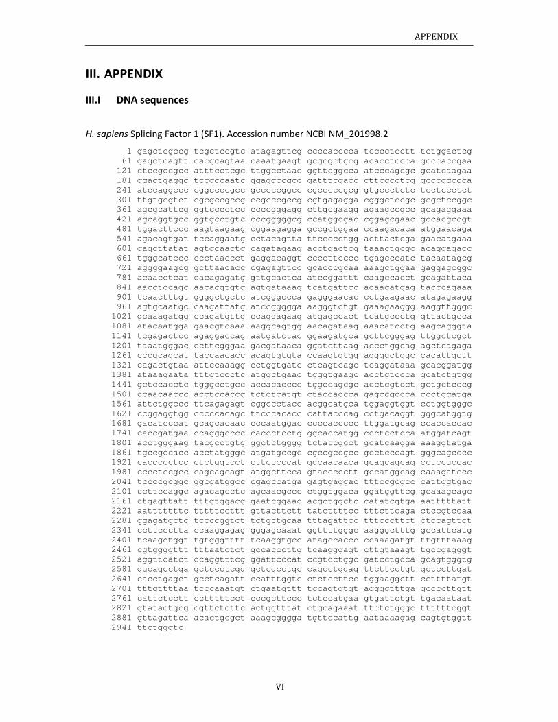
APPENDIX
VI
III. APPENDIX
III.I DNA sequences
H. sapiens Splicing Factor 1 (SF1). Accession number NCBI NM_201998.2
1 gagctcgccg tcgctccgtc atagagttcg ccccacccca tcccctcctt tctggactcg 61 gagctcagtt cacgcagtaa caaatgaagt gcgcgctgcg acacctccca gcccaccgaa 121 ctccgccgcc atttcctcgc ttggcctaac ggttcggcca atcccagcgc gcatcaagaa 181 ggactgaggc tccgccaatc ggaggccgcc gatttcgacc cttcgcctcg gcccggccca 241 atccaggccc cggccccgcc gcccccggcc cgcccccgcg gtgccctctc tcctccctct 301 ttgtgcgtct cgcgccgccg ccgcccgccg cgtgagagga cgggctccgc gcgctccggc 361 agcgcattcg ggtcccctcc ccccgggagg cttgcgaagg agaagccgcc gcagaggaaa 421 agcaggtgcc ggtgcctgtc cccgggggcg ccatggcgac cggagcgaac gccacgccgt 481 tggacttccc aagtaagaag cggaagagga gccgctggaa ccaagacaca atggaacaga 541 agacagtgat tccaggaatg cctacagtta ttccccctgg acttactcga gaacaagaaa 601 gagcttatat agtgcaactg cagatagaag acctgactcg taaactgcgc acaggagacc 661 tgggcatccc ccctaaccct gaggacaggt ccccttcccc tgagcccatc tacaatagcg 721 aggggaagcg gcttaacacc cgagagttcc gcacccgcaa aaagctggaa gaggagcggc 781 acaacctcat cacagagatg gttgcactca atccggattt caagccacct gcagattaca 841 aacctccagc aacacgtgtg agtgataaag tcatgattcc acaagatgag tacccagaaa 901 tcaactttgt ggggctgctc atcgggccca gagggaacac cctgaagaac atagagaagg 961 agtgcaatgc caagattatg atccggggga aagggtctgt gaaagaaggg aaggttgggc 1021 gcaaagatgg ccagatgttg ccaggagaag atgagccact tcatgccctg gttactgcca 1081 atacaatgga gaacgtcaaa aaggcagtgg aacagataag aaacatcctg aagcagggta 1141 tcgagactcc agaggaccag aatgatctac ggaagatgca gcttcgggag ttggctcgct 1201 taaatgggac ccttcgggaa gacgataaca ggatcttaag accctggcag agctcagaga 1261 cccgcagcat taccaacacc acagtgtgta ccaagtgtgg aggggctggc cacattgctt 1321 cagactgtaa attccaaagg cctggtgatc ctcagtcagc tcaggataaa gcacggatgg 1381 ataaagaata tttgtccctc atggctgaac tgggtgaagc acctgtccca gcatctgtgg 1441 gctccacctc tgggcctgcc accacacccc tggccagcgc acctcgtcct gctgctcccg 1501 ccaacaaccc acctccaccg tctctcatgt ctaccaccca gagccgccca ccctggatga 1561 attctggccc ttcagagagt cggccctacc acggcatgca tggaggtggt cctggtgggc 1621 ccggaggtgg cccccacagc ttcccacacc cattacccag cctgacaggt gggcatggtg 1681 gacatcccat gcagcacaac cccaatggac ccccaccccc ttggatgcag ccaccaccac 1741 caccgatgaa ccagggcccc caccctcctg ggcaccatgg ccctcctcca atggatcagt 1801 acctgggaag tacgcctgtg ggctctgggg tctatcgcct gcatcaagga aaaggtatga 1861 tgccgccacc acctatgggc atgatgccgc cgccgccgcc gcctcccagt gggcagcccc 1921 caccccctcc ctctggtcct cttcccccat ggcaacaaca gcagcagcag cctccgccac 1981 cccctccgcc cagcagcagt atggcttcca gtaccccctt gccatggcag caaagatccc 2041 tccccgcggc ggcgatggcc cgagccatga gagtgaggac tttccgcgcc cattggtgac 2101 ccttccaggc agacagcctc agcaacgccc ctggtggaca ggatggttcg gcaaagcagc 2161 ctgagttatt tttgtggacg gaatcggaac acgctggctc catatcgtga aatttttatt 2221 aatttttttc tttttccttt gttacttctt tatcttttcc tttcttcaga ctccgtccaa 2281 ggagatgctc tccccggtct tctgctgcaa tttagattcc tttcccttct ctccagttct 2341 ccttccctta ccaaggagag gggagcaaat ggttttgggc aagggctttg gccattcatg 2401 tcaagctggt tgtgggtttt tcaaggtgcc atagccaccc ccaaagatgt ttgtttaaag 2461 cgtggggttt tttaatctct gccacccttg tcaagggagt cttgtaaagt tgccgagggt 2521 aggttcatct ccaggtttcg ggattcccat ccgtcctggc gatcctgcca gcagtgggtg 2581 ggcagcctga gctccctcgg gctcgcctgc cagcctggag ttcttcctgt gctccttgat 2641 cacctgagct gcctcagatt ccatttggtc ctctccttcc tggaaggctt ccttttatgt 2701 tttgttttaa tcccaaatgt ctgaatgttt tgcagtgtgt aggggtttga gccccttgtt 2761 cattctcctt cctttttcct cccgcttccc tctccatgaa gtgattctgt tgacaataat 2821 gtatactgcg cgttctcttc actggtttat ctgcagaaat ttctctgggc ttttttcggt 2881 gttagattca acactgcgct aaagcgggga tgttccattg aataaaagag cagtgtggtt 2941 ttctgggtc
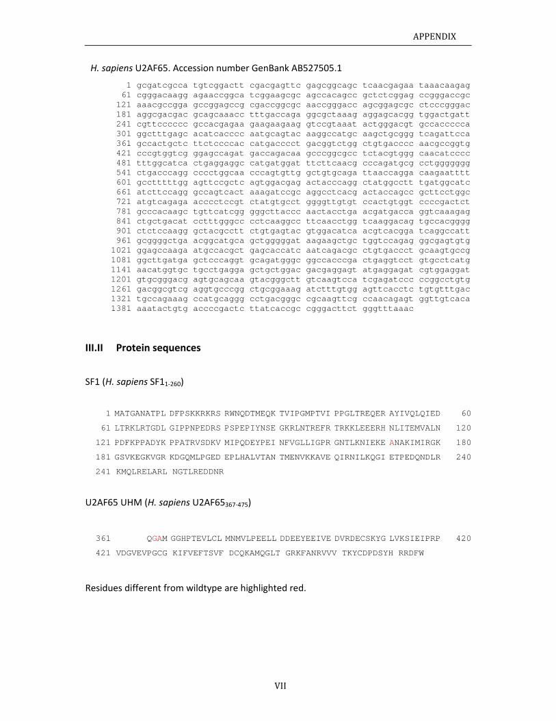
APPENDIX
VII
H. sapiens U2AF65. Accession number GenBank AB527505.1
1 gcgatcgcca tgtcggactt cgacgagttc gagcggcagc tcaacgagaa taaacaagag 61 cgggacaagg agaaccggca tcggaagcgc agccacagcc gctctcggag ccgggaccgc 121 aaacgccgga gccggagccg cgaccggcgc aaccgggacc agcggagcgc ctcccgggac 181 aggcgacgac gcagcaaacc tttgaccaga ggcgctaaag aggagcacgg tggactgatt 241 cgttcccccc gccacgagaa gaagaagaag gtccgtaaat actgggacgt gccaccccca 301 ggctttgagc acatcacccc aatgcagtac aaggccatgc aagctgcggg tcagattcca 361 gccactgctc ttctccccac catgacccct gacggtctgg ctgtgacccc aacgccggtg 421 cccgtggtcg ggagccagat gaccagacaa gcccggcgcc tctacgtggg caacatcccc 481 tttggcatca ctgaggaggc catgatggat ttcttcaacg cccagatgcg cctggggggg 541 ctgacccagg cccctggcaa cccagtgttg gctgtgcaga ttaaccagga caagaatttt 601 gcctttttgg agttccgctc agtggacgag actacccagg ctatggcctt tgatggcatc 661 atcttccagg gccagtcact aaagatccgc aggcctcacg actaccagcc gcttcctggc 721 atgtcagaga acccctccgt ctatgtgcct ggggttgtgt ccactgtggt ccccgactct 781 gcccacaagc tgttcatcgg gggcttaccc aactacctga acgatgacca ggtcaaagag 841 ctgctgacat cctttgggcc cctcaaggcc ttcaacctgg tcaaggacag tgccacgggg 901 ctctccaagg gctacgcctt ctgtgagtac gtggacatca acgtcacgga tcaggccatt 961 gcggggctga acggcatgca gctgggggat aagaagctgc tggtccagag ggcgagtgtg 1021 ggagccaaga atgccacgct gagcaccatc aatcagacgc ctgtgaccct gcaagtgccg 1081 ggcttgatga gctcccaggt gcagatgggc ggccacccga ctgaggtcct gtgcctcatg 1141 aacatggtgc tgcctgagga gctgctggac gacgaggagt atgaggagat cgtggaggat 1201 gtgcgggacg agtgcagcaa gtacgggctt gtcaagtcca tcgagatccc ccggcctgtg 1261 gacggcgtcg aggtgcccgg ctgcggaaag atctttgtgg agttcacctc tgtgtttgac 1321 tgccagaaag ccatgcaggg cctgacgggc cgcaagttcg ccaacagagt ggttgtcaca 1381 aaatactgtg accccgactc ttatcaccgc cgggacttct gggtttaaac
III.II Protein sequences
SF1 (H. sapiens SF11‐260)
1 MATGANATPL DFPSKKRKRS RWNQDTMEQK TVIPGMPTVI PPGLTREQER AYIVQLQIED 60
61 LTRKLRTGDL GIPPNPEDRS PSPEPIYNSE GKRLNTREFR TRKKLEEERH NLITEMVALN 120
121 PDFKPPADYK PPATRVSDKV MIPQDEYPEI NFVGLLIGPR GNTLKNIEKE ANAKIMIRGK 180
181 GSVKEGKVGR KDGQMLPGED EPLHALVTAN TMENVKKAVE QIRNILKQGI ETPEDQNDLR 240
241 KMQLRELARL NGTLREDDNR
U2AF65 UHM (H. sapiens U2AF65367‐475)
361 QGAM GGHPTEVLCL MNMVLPEELL DDEEYEEIVE DVRDECSKYG LVKSIEIPRP 420
421 VDGVEVPGCG KIFVEFTSVF DCQKAMQGLT GRKFANRVVV TKYCDPDSYH RRDFW
Residues different from wildtype are highlighted red.
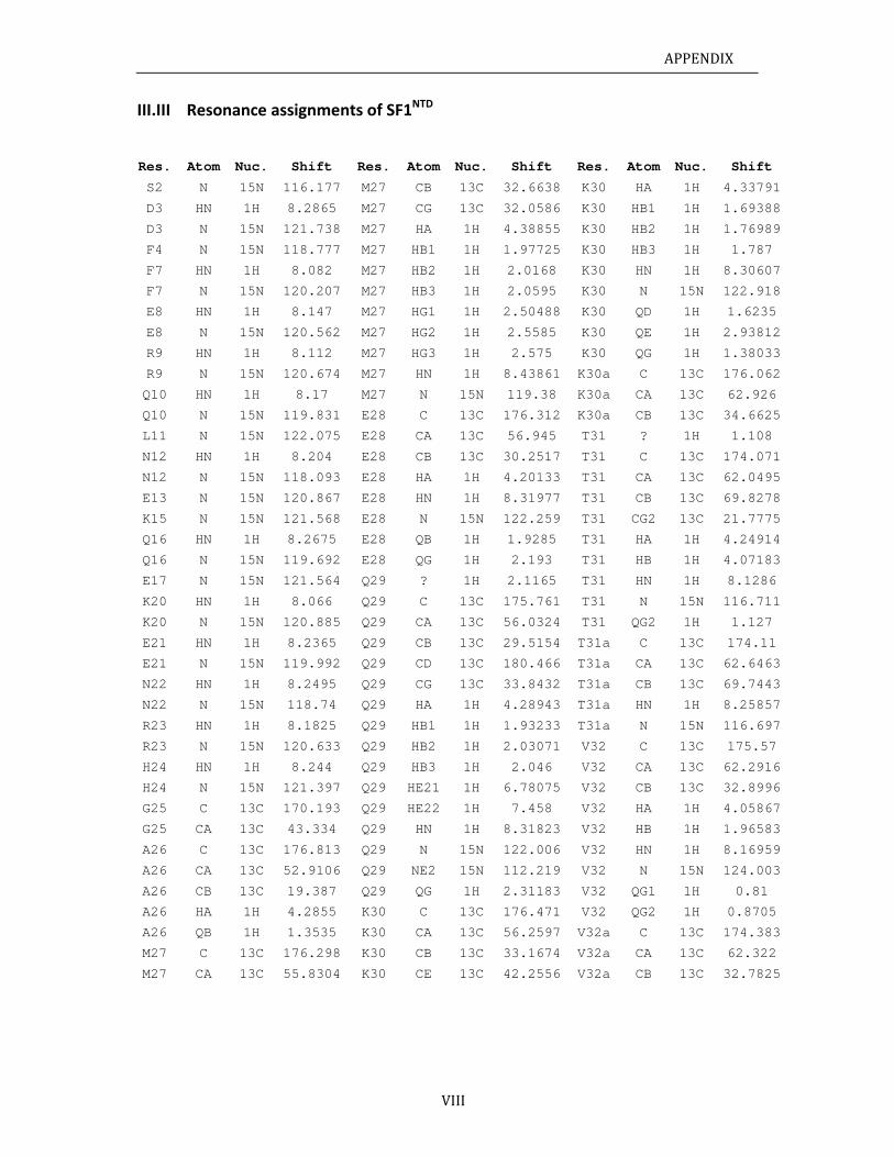
APPENDIX
VIII
III.III Resonance assignments of SF1NTD
Res. Atom Nuc. Shift Res. Atom Nuc. Shift Res. Atom Nuc. Shift
S2 N 15N 116.177 M27 CB 13C 32.6638 K30 HA 1H 4.33791
D3 HN 1H 8.2865 M27 CG 13C 32.0586 K30 HB1 1H 1.69388
D3 N 15N 121.738 M27 HA 1H 4.38855 K30 HB2 1H 1.76989
F4 N 15N 118.777 M27 HB1 1H 1.97725 K30 HB3 1H 1.787
F7 HN 1H 8.082 M27 HB2 1H 2.0168 K30 HN 1H 8.30607
F7 N 15N 120.207 M27 HB3 1H 2.0595 K30 N 15N 122.918
E8 HN 1H 8.147 M27 HG1 1H 2.50488 K30 QD 1H 1.6235
E8 N 15N 120.562 M27 HG2 1H 2.5585 K30 QE 1H 2.93812
R9 HN 1H 8.112 M27 HG3 1H 2.575 K30 QG 1H 1.38033
R9 N 15N 120.674 M27 HN 1H 8.43861 K30a C 13C 176.062
Q10 HN 1H 8.17 M27 N 15N 119.38 K30a CA 13C 62.926
Q10 N 15N 119.831 E28 C 13C 176.312 K30a CB 13C 34.6625
L11 N 15N 122.075 E28 CA 13C 56.945 T31 ? 1H 1.108
N12 HN 1H 8.204 E28 CB 13C 30.2517 T31 C 13C 174.071
N12 N 15N 118.093 E28 HA 1H 4.20133 T31 CA 13C 62.0495
E13 N 15N 120.867 E28 HN 1H 8.31977 T31 CB 13C 69.8278
K15 N 15N 121.568 E28 N 15N 122.259 T31 CG2 13C 21.7775
Q16 HN 1H 8.2675 E28 QB 1H 1.9285 T31 HA 1H 4.24914
Q16 N 15N 119.692 E28 QG 1H 2.193 T31 HB 1H 4.07183
E17 N 15N 121.564 Q29 ? 1H 2.1165 T31 HN 1H 8.1286
K20 HN 1H 8.066 Q29 C 13C 175.761 T31 N 15N 116.711
K20 N 15N 120.885 Q29 CA 13C 56.0324 T31 QG2 1H 1.127
E21 HN 1H 8.2365 Q29 CB 13C 29.5154 T31a C 13C 174.11
E21 N 15N 119.992 Q29 CD 13C 180.466 T31a CA 13C 62.6463
N22 HN 1H 8.2495 Q29 CG 13C 33.8432 T31a CB 13C 69.7443
N22 N 15N 118.74 Q29 HA 1H 4.28943 T31a HN 1H 8.25857
R23 HN 1H 8.1825 Q29 HB1 1H 1.93233 T31a N 15N 116.697
R23 N 15N 120.633 Q29 HB2 1H 2.03071 V32 C 13C 175.57
H24 HN 1H 8.244 Q29 HB3 1H 2.046 V32 CA 13C 62.2916
H24 N 15N 121.397 Q29 HE21 1H 6.78075 V32 CB 13C 32.8996
G25 C 13C 170.193 Q29 HE22 1H 7.458 V32 HA 1H 4.05867
G25 CA 13C 43.334 Q29 HN 1H 8.31823 V32 HB 1H 1.96583
A26 C 13C 176.813 Q29 N 15N 122.006 V32 HN 1H 8.16959
A26 CA 13C 52.9106 Q29 NE2 15N 112.219 V32 N 15N 124.003
A26 CB 13C 19.387 Q29 QG 1H 2.31183 V32 QG1 1H 0.81
A26 HA 1H 4.2855 K30 C 13C 176.471 V32 QG2 1H 0.8705
A26 QB 1H 1.3535 K30 CA 13C 56.2597 V32a C 13C 174.383
M27 C 13C 176.298 K30 CB 13C 33.1674 V32a CA 13C 62.322
M27 CA 13C 55.8304 K30 CE 13C 42.2556 V32a CB 13C 32.7825
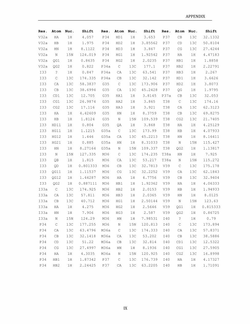
APPENDIX
IX
Res. Atom Nuc. Shift Res. Atom Nuc. Shift Res. Atom Nuc. Shift
V32a HA 1H 4.057 P34 HD1 1H 3.653 P37 CB 13C 32.1332
V32a HB 1H 1.975 P34 HD2 1H 3.85562 P37 CD 13C 50.8104
V32a HN 1H 8.1122 P34 HD3 1H 3.867 P37 CG 13C 27.4244
V32a N 15N 124.019 P34 HG1 1H 1.92542 P37 HA 1H 4.47183
V32a QG1 1H 0.8635 P34 HG2 1H 2.0235 P37 HB1 1H 1.8858
V32a QG2 1H 0.822 P34a C 13C 177.1 P37 HB2 1H 2.22791
I33 ? 1H 0.847 P34a CA 13C 63.541 P37 HB3 1H 2.267
I33 C 13C 174.335 P34a CB 13C 32.142 P37 HD1 1H 3.6626
I33 CA 13C 58.3837 G35 C 13C 173.906 P37 HD2 1H 3.8073
I33 CB 13C 38.6994 G35 CA 13C 45.2428 P37 QG 1H 1.9795
I33 CD1 13C 12.705 G35 HA1 1H 3.8145 P37a CB 13C 32.053
I33 CG1 13C 26.9874 G35 HA2 1H 3.865 T38 C 13C 174.16
I33 CG2 13C 17.116 G35 HA3 1H 3.921 T38 CA 13C 62.3123
I33 HA 1H 4.42609 G35 HN 1H 8.3759 T38 CB 13C 69.8275
I33 HB 1H 1.8124 G35 N 15N 109.539 T38 CG2 13C 21.7405
I33 HD11 1H 0.804 G35 QA 1H 3.868 T38 HA 1H 4.25129
I33 HG11 1H 1.1215 G35a C 13C 173.99 T38 HB 1H 4.07933
I33 HG12 1H 1.446 G35a CA 13C 45.2213 T38 HN 1H 8.16411
I33 HG21 1H 0.885 G35a HN 1H 8.31033 T38 N 15N 115.427
I33 HN 1H 8.27164 G35a N 15N 109.337 T38 QG2 1H 1.13817
I33 N 15N 127.335 M36 C 13C 174.235 T38a HN 1H 7.925
I33 QB 1H 1.815 M36 CA 13C 53.217 T38a N 15N 115.272
I33 QD 1H 0.801333 M36 CB 13C 32.7813 V39 C 13C 175.178
I33 QG11 1H 1.11537 M36 CG 13C 32.2252 V39 CA 13C 62.1843
I33 QG12 1H 1.44287 M36 HA 1H 4.7756 V39 CB 13C 32.9404
I33 QG2 1H 0.887111 M36 HB1 1H 1.92362 V39 HA 1H 4.06333
I33a C 13C 174.925 M36 HB2 1H 2.0153 V39 HB 1H 1.94933
I33a CA 13C 57.811 M36 HB3 1H 2.0365 V39 HN 1H 8.0125
I33a CB 13C 40.712 M36 HG1 1H 2.50144 V39 N 15N 123.63
I33a HA 1H 4.275 M36 HG2 1H 2.5646 V39 QG1 1H 0.815333
I33a HN 1H 7.906 M36 HG3 1H 2.587 V39 QG2 1H 0.86725
I33a N 15N 124.29 M36 HN 1H 7.98531 I40 ? 1H 0.79
P34 C 13C 177.255 M36 N 15N 120.813 I40 C 13C 173.894
P34 CA 13C 63.4796 M36a C 13C 174.333 I40 CA 13C 57.8371
P34 CB 13C 32.1418 M36a CA 13C 53.202 I40 CB 13C 38.5886
P34 CD 13C 51.22 M36a CB 13C 32.814 I40 CD1 13C 12.5322
P34 CG 13C 27.4997 M36a HN 1H 8.1936 I40 CG1 13C 27.5905
P34 HA 1H 4.3035 M36a N 15N 120.925 I40 CG2 13C 16.8998
P34 HB1 1H 1.87342 P37 C 13C 176.739 I40 HA 1H 4.17327
P34 HB2 1H 2.24425 P37 CA 13C 63.2205 I40 HB 1H 1.71091
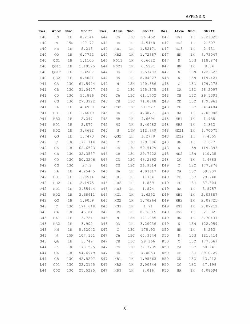
APPENDIX
X
Res. Atom Nuc. Shift Res. Atom Nuc. Shift Res. Atom Nuc. Shift
I40 HN 1H 8.2144 L44 CG 13C 26.452 E47 HG1 1H 2.21325
I40 N 15N 127.77 L44 HA 1H 4.5448 E47 HG2 1H 2.397
I40 NH 1H 8.213 L44 HB1 1H 1.52171 E47 HG3 1H 2.431
I40 QD 1H 0.7752 L44 HB2 1H 1.72887 E47 HN 1H 8.73047
I40 QG1 1H 1.1105 L44 HD11 1H 0.6622 E47 N 15N 118.874
I40 QG11 1H 1.10525 L44 HD21 1H 0.5981 R47 HN 1H 8.34
I40 QG12 1H 1.4507 L44 HG 1H 1.53483 R47 N 15N 122.523
I40 QG2 1H 0.8021 L44 HN 1H 8.04027 N48 N 15N 119.421
P41 CA 13C 61.5924 L44 N 15N 120.886 Q48 C 13C 179.278
P41 CB 13C 31.0477 T45 C 13C 175.375 Q48 CA 13C 58.2097
P41 CD 13C 50.886 T45 CA 13C 61.1702 Q48 CB 13C 29.5393
P41 CG 13C 27.3922 T45 CB 13C 71.0048 Q48 CD 13C 179.961
P41 HA 1H 4.4938 T45 CG2 13C 21.527 Q48 CG 13C 34.4484
P41 HB1 1H 1.6619 T45 HA 1H 4.38771 Q48 HA 1H 4.06088
P41 HB2 1H 2.247 T45 HB 1H 4.6694 Q48 HB1 1H 1.958
P41 HD1 1H 2.877 T45 HN 1H 8.40482 Q48 HB2 1H 2.332
P41 HD2 1H 3.4682 T45 N 15N 112.949 Q48 HE21 1H 6.70075
P41 QG 1H 1.7473 T45 QG2 1H 1.2778 Q48 HE22 1H 7.4355
P42 C 13C 177.714 R46 C 13C 179.306 Q48 HN 1H 7.677
P42 CA 13C 62.6523 R46 CA 13C 59.5179 Q48 N 15N 119.393
P42 CB 13C 32.3537 R46 CB 13C 29.7922 Q48 NE2 15N 110.35
P42 CD 13C 50.3206 R46 CD 13C 43.2992 Q48 QG 1H 2.4388
P42 CG 13C 27.3 R46 CG 13C 26.9514 E49 C 13C 177.876
P42 HA 1H 4.25475 R46 HA 1H 4.03617 E49 CA 13C 59.937
P42 HB1 1H 1.8514 R46 HB1 1H 1.784 E49 CB 13C 29.748
P42 HB2 1H 2.1975 R46 HB2 1H 1.859 E49 CG 13C 37.304
P42 HD1 1H 3.55444 R46 HB3 1H 1.874 E49 HA 1H 3.8757
P42 HD2 1H 3.68611 R46 HG1 1H 1.6252 E49 HB1 1H 2.03887
P42 QG 1H 1.9059 R46 HG2 1H 1.70264 E49 HB2 1H 2.09725
G43 C 13C 174.648 R46 HG3 1H 1.71 E49 HG1 1H 2.07212
G43 CA 13C 45.84 R46 HN 1H 8.76815 E49 HG2 1H 2.332
G43 HA1 1H 3.724 R46 N 15N 121.085 E49 HN 1H 8.70437
G43 HA2 1H 3.902 R46 QD 1H 3.20036 E49 N 15N 122.059
G43 HN 1H 8.32042 E47 C 13C 178.93 D50 HN 1H 8.253
G43 N 15N 107.151 E47 CA 13C 60.3644 D50 N 15N 121.414
G43 QA 1H 3.749 E47 CB 13C 29.166 R50 C 13C 177.567
L44 C 13C 178.575 E47 CG 13C 37.3735 R50 CA 13C 58.241
L44 CA 13C 54.4949 E47 HA 1H 4.0053 R50 CB 13C 29.0729
L44 CB 13C 42.5297 E47 HB1 1H 1.95663 R50 CD 13C 43.012
L44 CD1 13C 22.3155 E47 HB2 1H 2.00644 R50 CG 13C 27.199
L44 CD2 13C 25.5225 E47 HB3 1H 2.016 R50 HA 1H 4.08594
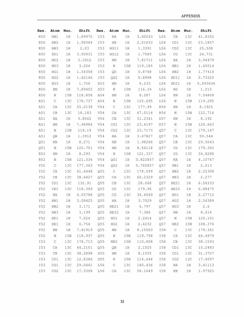
APPENDIX
XI
Res. Atom Nuc. Shift Res. Atom Nuc. Shift Res. Atom Nuc. Shift
R50 HB1 1H 1.89975 I53 HA 1H 3.60243 L56 CB 13C 41.8352
R50 HB2 1H 1.99364 I53 HB 1H 2.01833 L56 CD1 13C 23.1857
R50 HB3 1H 2.03 I53 HG11 1H 1.3391 L56 CD2 13C 25.508
R50 HD1 1H 3.00931 I53 HG12 1H 1.7585 L56 CG 13C 26.731
R50 HD2 1H 3.1812 I53 HN 1H 7.81711 L56 HA 1H 3.94479
R50 HD3 1H 3.204 I53 N 15N 119.185 L56 HB1 1H 1.45014
R50 HG1 1H 1.54358 I53 QD 1H 0.8748 L56 HB2 1H 1.77414
R50 HG2 1H 1.62146 I53 QG2 1H 0.8998 L56 HD11 1H 0.73325
R50 HG3 1H 1.756 S53 HN 1H 8.233 L56 HD21 1H 0.693636
R50 HN 1H 7.89452 S53 N 15N 116.34 L56 HG 1H 1.515
R50 N 15N 118.858 A54 HN 1H 8.287 L56 HN 1H 7.54808
A51 C 13C 178.737 A54 N 15N 125.695 L56 N 15N 119.295
A51 CA 13C 55.2138 V54 C 13C 177.99 R56 HN 1H 8.1925
A51 CB 13C 18.183 V54 CA 13C 67.0114 R56 N 15N 122.716
A51 HA 1H 3.8062 V54 CB 13C 31.2341 D57 HN 1H 8.192
A51 HN 1H 7.94984 V54 CG1 13C 23.4197 D57 N 15N 120.663
A51 N 15N 119.19 V54 CG2 13C 23.7173 Q57 C 13C 179.167
A51 QB 1H 1.3912 V54 HA 1H 3.47827 Q57 CA 13C 59.544
Q51 HN 1H 8.271 V54 HB 1H 1.98286 Q57 CB 13C 29.5043
Q51 N 15N 120.751 V54 HN 1H 8.54118 Q57 CD 13C 179.343
R52 HN 1H 8.293 V54 N 15N 121.337 Q57 CG 13C 34.8288
R52 N 15N 121.336 V54 QG1 1H 0.822857 Q57 HA 1H 4.10767
Y52 C 13C 177.363 V54 QG2 1H 0.765857 Q57 HB1 1H 2.013
Y52 CA 13C 61.6448 Q55 C 13C 178.599 Q57 HB2 1H 2.22308
Y52 CB 13C 38.6427 Q55 CA 13C 60.2329 Q57 HB3 1H 2.277
Y52 CD1 13C 132.91 Q55 CB 13C 28.068 Q57 HE21 1H 6.56333
Y52 CE1 13C 118.369 Q55 CD 13C 179.96 Q57 HE22 1H 6.88475
Y52 HA 1H 4.05788 Q55 CG 13C 34.4069 Q57 HG1 1H 2.27712
Y52 HB1 1H 3.08425 Q55 HA 1H 3.7029 Q57 HG2 1H 2.56389
Y52 HB2 1H 3.171 Q55 HE21 1H 6.797 Q57 HG3 1H 2.6
Y52 HB3 1H 3.199 Q55 HE22 1H 7.386 Q57 HN 1H 8.014
Y52 HD1 1H 7.024 Q55 HG1 1H 2.2414 Q57 N 15N 120.101
Y52 HE1 1H 6.754 Q55 HG2 1H 2.4232 Q57 NE2 15N 108.376
Y52 HN 1H 7.81919 Q55 HN 1H 8.15083 I58 C 13C 179.361
Y52 N 15N 118.937 Q55 N 15N 119.794 I58 CA 13C 66.4979
I53 C 13C 178.713 Q55 NE2 15N 110.808 I58 CB 13C 38.1593
I53 CA 13C 64.2101 Q55 QB 1H 2.1025 I58 CD1 13C 15.2483
I53 CB 13C 38.2898 S55 HN 1H 8.1555 I58 CG1 13C 31.3707
I53 CD1 13C 12.8386 S55 N 15N 114.448 I58 CG2 13C 17.4597
I53 CG1 13C 29.0441 L56 C 13C 180.436 I58 HA 1H 3.41113
I53 CG2 13C 17.3308 L56 CA 13C 58.1649 I58 HB 1H 1.97925
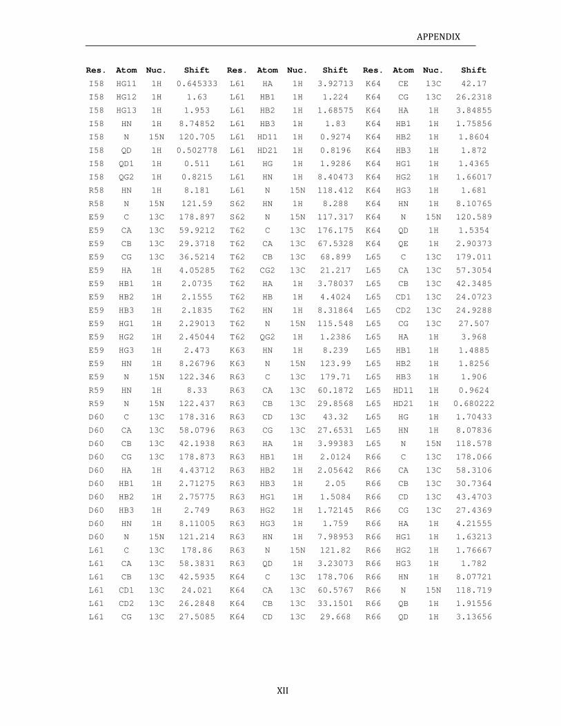
APPENDIX
XII
Res. Atom Nuc. Shift Res. Atom Nuc. Shift Res. Atom Nuc. Shift
I58 HG11 1H 0.645333 L61 HA 1H 3.92713 K64 CE 13C 42.17
I58 HG12 1H 1.63 L61 HB1 1H 1.224 K64 CG 13C 26.2318
I58 HG13 1H 1.953 L61 HB2 1H 1.68575 K64 HA 1H 3.84855
I58 HN 1H 8.74852 L61 HB3 1H 1.83 K64 HB1 1H 1.75856
I58 N 15N 120.705 L61 HD11 1H 0.9274 K64 HB2 1H 1.8604
I58 QD 1H 0.502778 L61 HD21 1H 0.8196 K64 HB3 1H 1.872
I58 QD1 1H 0.511 L61 HG 1H 1.9286 K64 HG1 1H 1.4365
I58 QG2 1H 0.8215 L61 HN 1H 8.40473 K64 HG2 1H 1.66017
R58 HN 1H 8.181 L61 N 15N 118.412 K64 HG3 1H 1.681
R58 N 15N 121.59 S62 HN 1H 8.288 K64 HN 1H 8.10765
E59 C 13C 178.897 S62 N 15N 117.317 K64 N 15N 120.589
E59 CA 13C 59.9212 T62 C 13C 176.175 K64 QD 1H 1.5354
E59 CB 13C 29.3718 T62 CA 13C 67.5328 K64 QE 1H 2.90373
E59 CG 13C 36.5214 T62 CB 13C 68.899 L65 C 13C 179.011
E59 HA 1H 4.05285 T62 CG2 13C 21.217 L65 CA 13C 57.3054
E59 HB1 1H 2.0735 T62 HA 1H 3.78037 L65 CB 13C 42.3485
E59 HB2 1H 2.1555 T62 HB 1H 4.4024 L65 CD1 13C 24.0723
E59 HB3 1H 2.1835 T62 HN 1H 8.31864 L65 CD2 13C 24.9288
E59 HG1 1H 2.29013 T62 N 15N 115.548 L65 CG 13C 27.507
E59 HG2 1H 2.45044 T62 QG2 1H 1.2386 L65 HA 1H 3.968
E59 HG3 1H 2.473 K63 HN 1H 8.239 L65 HB1 1H 1.4885
E59 HN 1H 8.26796 K63 N 15N 123.99 L65 HB2 1H 1.8256
E59 N 15N 122.346 R63 C 13C 179.71 L65 HB3 1H 1.906
R59 HN 1H 8.33 R63 CA 13C 60.1872 L65 HD11 1H 0.9624
R59 N 15N 122.437 R63 CB 13C 29.8568 L65 HD21 1H 0.680222
D60 C 13C 178.316 R63 CD 13C 43.32 L65 HG 1H 1.70433
D60 CA 13C 58.0796 R63 CG 13C 27.6531 L65 HN 1H 8.07836
D60 CB 13C 42.1938 R63 HA 1H 3.99383 L65 N 15N 118.578
D60 CG 13C 178.873 R63 HB1 1H 2.0124 R66 C 13C 178.066
D60 HA 1H 4.43712 R63 HB2 1H 2.05642 R66 CA 13C 58.3106
D60 HB1 1H 2.71275 R63 HB3 1H 2.05 R66 CB 13C 30.7364
D60 HB2 1H 2.75775 R63 HG1 1H 1.5084 R66 CD 13C 43.4703
D60 HB3 1H 2.749 R63 HG2 1H 1.72145 R66 CG 13C 27.4369
D60 HN 1H 8.11005 R63 HG3 1H 1.759 R66 HA 1H 4.21555
D60 N 15N 121.214 R63 HN 1H 7.98953 R66 HG1 1H 1.63213
L61 C 13C 178.86 R63 N 15N 121.82 R66 HG2 1H 1.76667
L61 CA 13C 58.3831 R63 QD 1H 3.23073 R66 HG3 1H 1.782
L61 CB 13C 42.5935 K64 C 13C 178.706 R66 HN 1H 8.07721
L61 CD1 13C 24.021 K64 CA 13C 60.5767 R66 N 15N 118.719
L61 CD2 13C 26.2848 K64 CB 13C 33.1501 R66 QB 1H 1.91556
L61 CG 13C 27.5085 K64 CD 13C 29.668 R66 QD 1H 3.13656
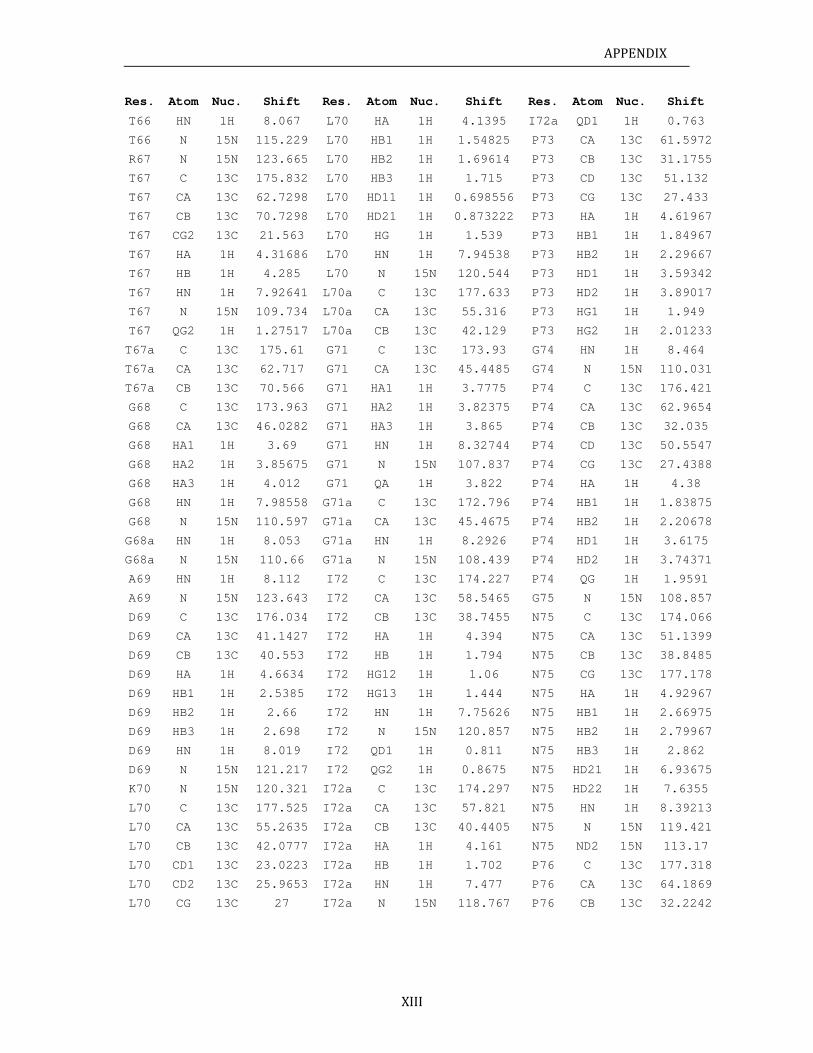
APPENDIX
XIII
Res. Atom Nuc. Shift Res. Atom Nuc. Shift Res. Atom Nuc. Shift
T66 HN 1H 8.067 L70 HA 1H 4.1395 I72a QD1 1H 0.763
T66 N 15N 115.229 L70 HB1 1H 1.54825 P73 CA 13C 61.5972
R67 N 15N 123.665 L70 HB2 1H 1.69614 P73 CB 13C 31.1755
T67 C 13C 175.832 L70 HB3 1H 1.715 P73 CD 13C 51.132
T67 CA 13C 62.7298 L70 HD11 1H 0.698556 P73 CG 13C 27.433
T67 CB 13C 70.7298 L70 HD21 1H 0.873222 P73 HA 1H 4.61967
T67 CG2 13C 21.563 L70 HG 1H 1.539 P73 HB1 1H 1.84967
T67 HA 1H 4.31686 L70 HN 1H 7.94538 P73 HB2 1H 2.29667
T67 HB 1H 4.285 L70 N 15N 120.544 P73 HD1 1H 3.59342
T67 HN 1H 7.92641 L70a C 13C 177.633 P73 HD2 1H 3.89017
T67 N 15N 109.734 L70a CA 13C 55.316 P73 HG1 1H 1.949
T67 QG2 1H 1.27517 L70a CB 13C 42.129 P73 HG2 1H 2.01233
T67a C 13C 175.61 G71 C 13C 173.93 G74 HN 1H 8.464
T67a CA 13C 62.717 G71 CA 13C 45.4485 G74 N 15N 110.031
T67a CB 13C 70.566 G71 HA1 1H 3.7775 P74 C 13C 176.421
G68 C 13C 173.963 G71 HA2 1H 3.82375 P74 CA 13C 62.9654
G68 CA 13C 46.0282 G71 HA3 1H 3.865 P74 CB 13C 32.035
G68 HA1 1H 3.69 G71 HN 1H 8.32744 P74 CD 13C 50.5547
G68 HA2 1H 3.85675 G71 N 15N 107.837 P74 CG 13C 27.4388
G68 HA3 1H 4.012 G71 QA 1H 3.822 P74 HA 1H 4.38
G68 HN 1H 7.98558 G71a C 13C 172.796 P74 HB1 1H 1.83875
G68 N 15N 110.597 G71a CA 13C 45.4675 P74 HB2 1H 2.20678
G68a HN 1H 8.053 G71a HN 1H 8.2926 P74 HD1 1H 3.6175
G68a N 15N 110.66 G71a N 15N 108.439 P74 HD2 1H 3.74371
A69 HN 1H 8.112 I72 C 13C 174.227 P74 QG 1H 1.9591
A69 N 15N 123.643 I72 CA 13C 58.5465 G75 N 15N 108.857
D69 C 13C 176.034 I72 CB 13C 38.7455 N75 C 13C 174.066
D69 CA 13C 41.1427 I72 HA 1H 4.394 N75 CA 13C 51.1399
D69 CB 13C 40.553 I72 HB 1H 1.794 N75 CB 13C 38.8485
D69 HA 1H 4.6634 I72 HG12 1H 1.06 N75 CG 13C 177.178
D69 HB1 1H 2.5385 I72 HG13 1H 1.444 N75 HA 1H 4.92967
D69 HB2 1H 2.66 I72 HN 1H 7.75626 N75 HB1 1H 2.66975
D69 HB3 1H 2.698 I72 N 15N 120.857 N75 HB2 1H 2.79967
D69 HN 1H 8.019 I72 QD1 1H 0.811 N75 HB3 1H 2.862
D69 N 15N 121.217 I72 QG2 1H 0.8675 N75 HD21 1H 6.93675
K70 N 15N 120.321 I72a C 13C 174.297 N75 HD22 1H 7.6355
L70 C 13C 177.525 I72a CA 13C 57.821 N75 HN 1H 8.39213
L70 CA 13C 55.2635 I72a CB 13C 40.4405 N75 N 15N 119.421
L70 CB 13C 42.0777 I72a HA 1H 4.161 N75 ND2 15N 113.17
L70 CD1 13C 23.0223 I72a HB 1H 1.702 P76 C 13C 177.318
L70 CD2 13C 25.9653 I72a HN 1H 7.477 P76 CA 13C 64.1869
L70 CG 13C 27 I72a N 15N 118.767 P76 CB 13C 32.2242
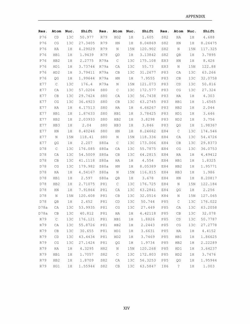
APPENDIX
XIV
Res. Atom Nuc. Shift Res. Atom Nuc. Shift Res. Atom Nuc. Shift
P76 CD 13C 50.977 R79 HG2 1H 1.605 S82 HA 1H 4.688
P76 CG 13C 27.3405 R79 HN 1H 8.04869 S82 HN 1H 8.26475
P76 HA 1H 4.29029 R79 N 15N 120.902 S82 N 15N 117.325
P76 HB1 1H 1.9439 R79 QD 1H 3.13842 S82 QB 1H 3.7895
P76 HB2 1H 2.2775 R79a C 13C 175.108 E83 HN 1H 8.426
P76 HD1 1H 3.73744 R79a CA 13C 55.73 E83 N 15N 122.88
P76 HD2 1H 3.79411 R79a CB 13C 31.2677 P83 CA 13C 63.266
P76 QG 1H 1.99644 R79a HN 1H 7.9555 P83 CB 13C 32.0758
E77 C 13C 176.4 R79a N 15N 121.073 P83 CD 13C 50.816
E77 CA 13C 57.0204 S80 C 13C 172.577 P83 CG 13C 27.324
E77 CB 13C 29.7424 S80 CA 13C 56.7438 P83 HA 1H 4.303
E77 CG 13C 36.4923 S80 CB 13C 63.2745 P83 HB1 1H 1.6565
E77 HA 1H 4.17513 S80 HA 1H 4.66267 P83 HB2 1H 2.044
E77 HB1 1H 1.87633 S80 HB1 1H 3.78425 P83 HD1 1H 3.646
E77 HB2 1H 2.03933 S80 HB2 1H 3.8298 P83 HD2 1H 3.756
E77 HB3 1H 2.04 S80 HB3 1H 3.846 P83 QG 1H 1.98367
E77 HN 1H 8.40246 S80 HN 1H 8.24662 E84 C 13C 174.546
E77 N 15N 118.41 S80 N 15N 118.336 E84 CA 13C 54.4726
E77 QG 1H 2.207 S80a C 13C 173.006 E84 CB 13C 29.8373
D78 C 13C 176.085 S80a CA 13C 55.7875 E84 CG 13C 36.0753
D78 CA 13C 54.5009 S80a CB 13C 64.2815 E84 HA 1H 4.49412
D78 CB 13C 41.1118 S80a HA 1H 4.554 E84 HB1 1H 1.8325
D78 CG 13C 179.982 S80a HN 1H 8.05389 E84 HB2 1H 1.95771
D78 HA 1H 4.54167 S80a N 15N 116.815 E84 HB3 1H 1.986
D78 HB1 1H 2.597 S80a QB 1H 3.678 E84 HN 1H 8.20817
D78 HB2 1H 2.71075 P81 C 13C 176.725 E84 N 15N 122.184
D78 HN 1H 7.91864 P81 CA 13C 63.2841 E84 QG 1H 2.256
D78 N 15N 120.608 P81 CB 13C 32.0516 K84 N 15N 127.065
D78 QB 1H 2.652 P81 CD 13C 50.744 P85 C 13C 176.022
D78a CA 13C 53.9935 P81 CG 13C 27.449 P85 CA 13C 63.2058
D78a CB 13C 40.812 P81 HA 1H 4.42118 P85 CB 13C 32.078
R79 C 13C 176.121 P81 HB1 1H 1.8826 P85 CD 13C 50.7787
R79 CA 13C 55.8726 P81 HB2 1H 2.2443 P85 CG 13C 27.2778
R79 CB 13C 30.655 P81 HD1 1H 3.6631 P85 HA 1H 4.4152
R79 CD 13C 43.4434 P81 HD2 1H 3.7469 P85 HB1 1H 1.86625
R79 CG 13C 27.1424 P81 QG 1H 1.9734 P85 HB2 1H 2.22289
R79 HA 1H 4.3295 H82 N 15N 120.268 P85 HD1 1H 3.64237
R79 HB1 1H 1.7057 S82 C 13C 172.803 P85 HD2 1H 3.7476
R79 HB2 1H 1.8709 S82 CA 13C 56.3253 P85 QG 1H 1.95944
R79 HG1 1H 1.55944 S82 CB 13C 63.5847 I86 ? 1H 1.003
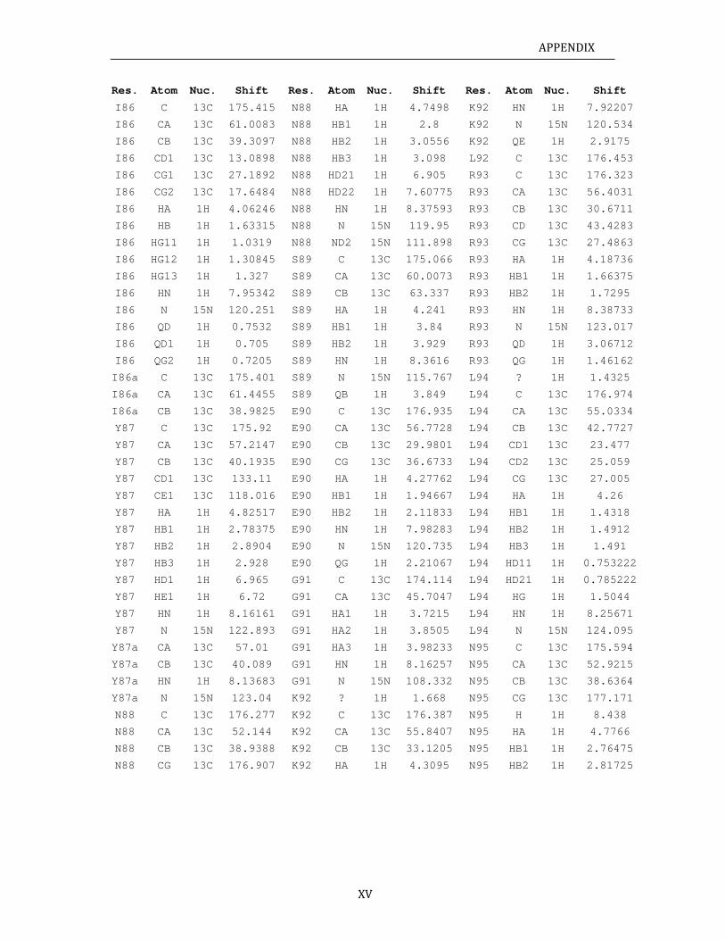
APPENDIX
XV
Res. Atom Nuc. Shift Res. Atom Nuc. Shift Res. Atom Nuc. Shift
I86 C 13C 175.415 N88 HA 1H 4.7498 K92 HN 1H 7.92207
I86 CA 13C 61.0083 N88 HB1 1H 2.8 K92 N 15N 120.534
I86 CB 13C 39.3097 N88 HB2 1H 3.0556 K92 QE 1H 2.9175
I86 CD1 13C 13.0898 N88 HB3 1H 3.098 L92 C 13C 176.453
I86 CG1 13C 27.1892 N88 HD21 1H 6.905 R93 C 13C 176.323
I86 CG2 13C 17.6484 N88 HD22 1H 7.60775 R93 CA 13C 56.4031
I86 HA 1H 4.06246 N88 HN 1H 8.37593 R93 CB 13C 30.6711
I86 HB 1H 1.63315 N88 N 15N 119.95 R93 CD 13C 43.4283
I86 HG11 1H 1.0319 N88 ND2 15N 111.898 R93 CG 13C 27.4863
I86 HG12 1H 1.30845 S89 C 13C 175.066 R93 HA 1H 4.18736
I86 HG13 1H 1.327 S89 CA 13C 60.0073 R93 HB1 1H 1.66375
I86 HN 1H 7.95342 S89 CB 13C 63.337 R93 HB2 1H 1.7295
I86 N 15N 120.251 S89 HA 1H 4.241 R93 HN 1H 8.38733
I86 QD 1H 0.7532 S89 HB1 1H 3.84 R93 N 15N 123.017
I86 QD1 1H 0.705 S89 HB2 1H 3.929 R93 QD 1H 3.06712
I86 QG2 1H 0.7205 S89 HN 1H 8.3616 R93 QG 1H 1.46162
I86a C 13C 175.401 S89 N 15N 115.767 L94 ? 1H 1.4325
I86a CA 13C 61.4455 S89 QB 1H 3.849 L94 C 13C 176.974
I86a CB 13C 38.9825 E90 C 13C 176.935 L94 CA 13C 55.0334
Y87 C 13C 175.92 E90 CA 13C 56.7728 L94 CB 13C 42.7727
Y87 CA 13C 57.2147 E90 CB 13C 29.9801 L94 CD1 13C 23.477
Y87 CB 13C 40.1935 E90 CG 13C 36.6733 L94 CD2 13C 25.059
Y87 CD1 13C 133.11 E90 HA 1H 4.27762 L94 CG 13C 27.005
Y87 CE1 13C 118.016 E90 HB1 1H 1.94667 L94 HA 1H 4.26
Y87 HA 1H 4.82517 E90 HB2 1H 2.11833 L94 HB1 1H 1.4318
Y87 HB1 1H 2.78375 E90 HN 1H 7.98283 L94 HB2 1H 1.4912
Y87 HB2 1H 2.8904 E90 N 15N 120.735 L94 HB3 1H 1.491
Y87 HB3 1H 2.928 E90 QG 1H 2.21067 L94 HD11 1H 0.753222
Y87 HD1 1H 6.965 G91 C 13C 174.114 L94 HD21 1H 0.785222
Y87 HE1 1H 6.72 G91 CA 13C 45.7047 L94 HG 1H 1.5044
Y87 HN 1H 8.16161 G91 HA1 1H 3.7215 L94 HN 1H 8.25671
Y87 N 15N 122.893 G91 HA2 1H 3.8505 L94 N 15N 124.095
Y87a CA 13C 57.01 G91 HA3 1H 3.98233 N95 C 13C 175.594
Y87a CB 13C 40.089 G91 HN 1H 8.16257 N95 CA 13C 52.9215
Y87a HN 1H 8.13683 G91 N 15N 108.332 N95 CB 13C 38.6364
Y87a N 15N 123.04 K92 ? 1H 1.668 N95 CG 13C 177.171
N88 C 13C 176.277 K92 C 13C 176.387 N95 H 1H 8.438
N88 CA 13C 52.144 K92 CA 13C 55.8407 N95 HA 1H 4.7766
N88 CB 13C 38.9388 K92 CB 13C 33.1205 N95 HB1 1H 2.76475
N88 CG 13C 176.907 K92 HA 1H 4.3095 N95 HB2 1H 2.81725
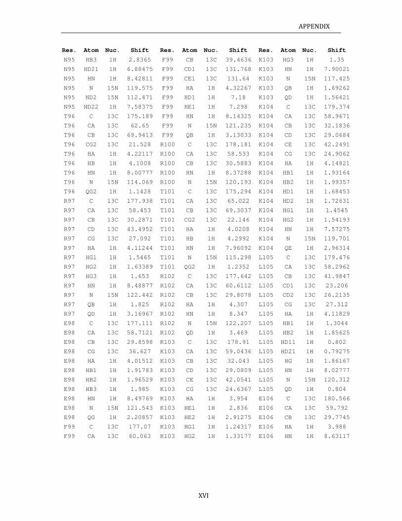
APPENDIX
XVI
Res. Atom Nuc. Shift Res. Atom Nuc. Shift Res. Atom Nuc. Shift
N95 HB3 1H 2.8365 F99 CB 13C 39.4636 K103 HG3 1H 1.35
N95 HD21 1H 6.88475 F99 CD1 13C 131.768 K103 HN 1H 7.90021
N95 HN 1H 8.42811 F99 CE1 13C 131.64 K103 N 15N 117.425
N95 N 15N 119.575 F99 HA 1H 4.32267 K103 QB 1H 1.69262
N95 ND2 15N 112.471 F99 HD1 1H 7.18 K103 QD 1H 1.56421
N95 ND22 1H 7.58375 F99 HE1 1H 7.298 K104 C 13C 179.374
T96 C 13C 175.189 F99 HN 1H 8.14325 K104 CA 13C 58.9671
T96 CA 13C 62.65 F99 N 15N 121.235 K104 CB 13C 32.1836
T96 CB 13C 69.9413 F99 QB 1H 3.13033 K104 CD 13C 29.0684
T96 CG2 13C 21.528 R100 C 13C 178.181 K104 CE 13C 42.2491
T96 HA 1H 4.22117 R100 CA 13C 58.533 K104 CG 13C 24.9062
T96 HB 1H 4.1008 R100 CB 13C 30.5883 K104 HA 1H 4.14821
T96 HN 1H 8.00777 R100 HN 1H 8.37288 K104 HB1 1H 1.93164
T96 N 15N 114.069 R100 N 15N 120.193 K104 HB2 1H 1.99357
T96 QG2 1H 1.1428 T101 C 13C 175.294 K104 HD1 1H 1.68453
R97 C 13C 177.938 T101 CA 13C 65.022 K104 HD2 1H 1.72631
R97 CA 13C 58.453 T101 CB 13C 69.3037 K104 HG1 1H 1.4545
R97 CB 13C 30.2871 T101 CG2 13C 22.146 K104 HG2 1H 1.54193
R97 CD 13C 43.4952 T101 HA 1H 4.0208 K104 HN 1H 7.57275
R97 CG 13C 27.092 T101 HB 1H 4.2992 K104 N 15N 119.701
R97 HA 1H 4.11244 T101 HN 1H 7.96092 K104 QE 1H 2.96314
R97 HG1 1H 1.5465 T101 N 15N 115.298 L105 C 13C 179.476
R97 HG2 1H 1.63389 T101 QG2 1H 1.2352 L105 CA 13C 58.2962
R97 HG3 1H 1.653 R102 C 13C 177.642 L105 CB 13C 41.9847
R97 HN 1H 8.48877 R102 CA 13C 60.6112 L105 CD1 13C 23.206
R97 N 15N 122.442 R102 CB 13C 29.8078 L105 CD2 13C 26.2135
R97 QB 1H 1.825 R102 HA 1H 4.307 L105 CG 13C 27.312
R97 QD 1H 3.16967 R102 HN 1H 8.347 L105 HA 1H 4.11829
E98 C 13C 177.111 R102 N 15N 122.207 L105 HB1 1H 1.3044
E98 CA 13C 58.7121 R102 QD 1H 3.469 L105 HB2 1H 1.85625
E98 CB 13C 29.8598 K103 C 13C 178.91 L105 HD11 1H 0.802
E98 CG 13C 36.627 K103 CA 13C 59.0436 L105 HD21 1H 0.79275
E98 HA 1H 4.01512 K103 CB 13C 32.043 L105 HG 1H 1.86167
E98 HB1 1H 1.91783 K103 CD 13C 29.0809 L105 HN 1H 8.02777
E98 HB2 1H 1.96529 K103 CE 13C 42.0541 L105 N 15N 120.312
E98 HB3 1H 1.985 K103 CG 13C 24.6367 L105 QD 1H 0.804
E98 HN 1H 8.49769 K103 HA 1H 3.954 E106 C 13C 180.566
E98 N 15N 121.543 K103 HE1 1H 2.836 E106 CA 13C 59.792
E98 QG 1H 2.20857 K103 HE2 1H 2.91275 E106 CB 13C 29.7745
F99 C 13C 177.07 K103 HG1 1H 1.24317 E106 HA 1H 3.988
F99 CA 13C 60.063 K103 HG2 1H 1.33177 E106 HN 1H 8.63117
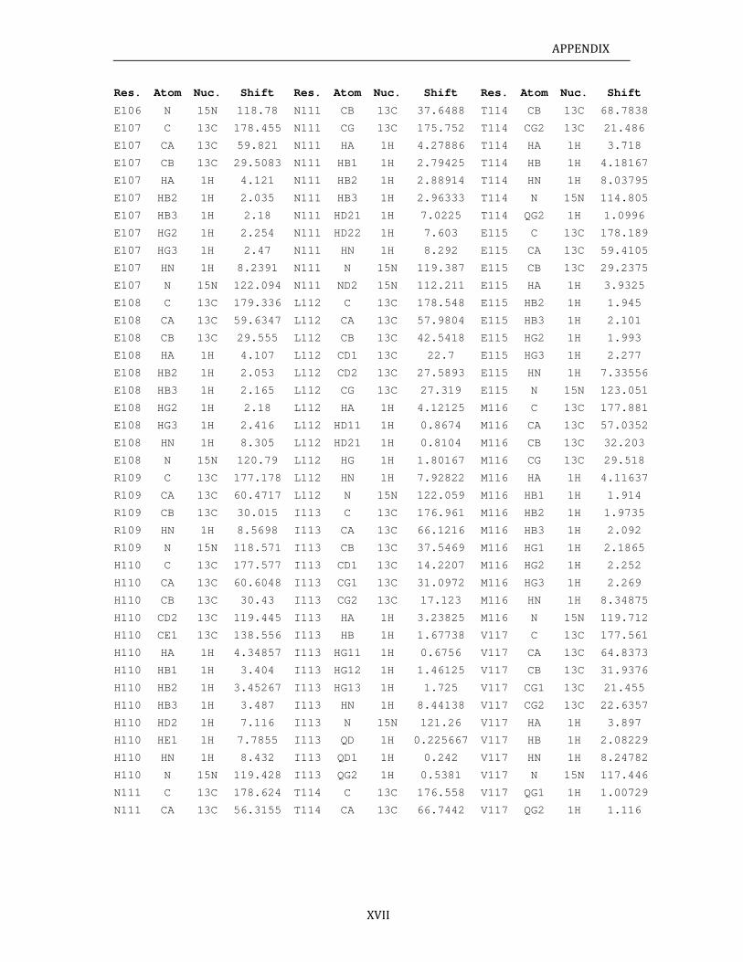
APPENDIX
XVII
Res. Atom Nuc. Shift Res. Atom Nuc. Shift Res. Atom Nuc. Shift
E106 N 15N 118.78 N111 CB 13C 37.6488 T114 CB 13C 68.7838
E107 C 13C 178.455 N111 CG 13C 175.752 T114 CG2 13C 21.486
E107 CA 13C 59.821 N111 HA 1H 4.27886 T114 HA 1H 3.718
E107 CB 13C 29.5083 N111 HB1 1H 2.79425 T114 HB 1H 4.18167
E107 HA 1H 4.121 N111 HB2 1H 2.88914 T114 HN 1H 8.03795
E107 HB2 1H 2.035 N111 HB3 1H 2.96333 T114 N 15N 114.805
E107 HB3 1H 2.18 N111 HD21 1H 7.0225 T114 QG2 1H 1.0996
E107 HG2 1H 2.254 N111 HD22 1H 7.603 E115 C 13C 178.189
E107 HG3 1H 2.47 N111 HN 1H 8.292 E115 CA 13C 59.4105
E107 HN 1H 8.2391 N111 N 15N 119.387 E115 CB 13C 29.2375
E107 N 15N 122.094 N111 ND2 15N 112.211 E115 HA 1H 3.9325
E108 C 13C 179.336 L112 C 13C 178.548 E115 HB2 1H 1.945
E108 CA 13C 59.6347 L112 CA 13C 57.9804 E115 HB3 1H 2.101
E108 CB 13C 29.555 L112 CB 13C 42.5418 E115 HG2 1H 1.993
E108 HA 1H 4.107 L112 CD1 13C 22.7 E115 HG3 1H 2.277
E108 HB2 1H 2.053 L112 CD2 13C 27.5893 E115 HN 1H 7.33556
E108 HB3 1H 2.165 L112 CG 13C 27.319 E115 N 15N 123.051
E108 HG2 1H 2.18 L112 HA 1H 4.12125 M116 C 13C 177.881
E108 HG3 1H 2.416 L112 HD11 1H 0.8674 M116 CA 13C 57.0352
E108 HN 1H 8.305 L112 HD21 1H 0.8104 M116 CB 13C 32.203
E108 N 15N 120.79 L112 HG 1H 1.80167 M116 CG 13C 29.518
R109 C 13C 177.178 L112 HN 1H 7.92822 M116 HA 1H 4.11637
R109 CA 13C 60.4717 L112 N 15N 122.059 M116 HB1 1H 1.914
R109 CB 13C 30.015 I113 C 13C 176.961 M116 HB2 1H 1.9735
R109 HN 1H 8.5698 I113 CA 13C 66.1216 M116 HB3 1H 2.092
R109 N 15N 118.571 I113 CB 13C 37.5469 M116 HG1 1H 2.1865
H110 C 13C 177.577 I113 CD1 13C 14.2207 M116 HG2 1H 2.252
H110 CA 13C 60.6048 I113 CG1 13C 31.0972 M116 HG3 1H 2.269
H110 CB 13C 30.43 I113 CG2 13C 17.123 M116 HN 1H 8.34875
H110 CD2 13C 119.445 I113 HA 1H 3.23825 M116 N 15N 119.712
H110 CE1 13C 138.556 I113 HB 1H 1.67738 V117 C 13C 177.561
H110 HA 1H 4.34857 I113 HG11 1H 0.6756 V117 CA 13C 64.8373
H110 HB1 1H 3.404 I113 HG12 1H 1.46125 V117 CB 13C 31.9376
H110 HB2 1H 3.45267 I113 HG13 1H 1.725 V117 CG1 13C 21.455
H110 HB3 1H 3.487 I113 HN 1H 8.44138 V117 CG2 13C 22.6357
H110 HD2 1H 7.116 I113 N 15N 121.26 V117 HA 1H 3.897
H110 HE1 1H 7.7855 I113 QD 1H 0.225667 V117 HB 1H 2.08229
H110 HN 1H 8.432 I113 QD1 1H 0.242 V117 HN 1H 8.24782
H110 N 15N 119.428 I113 QG2 1H 0.5381 V117 N 15N 117.446
N111 C 13C 178.624 T114 C 13C 176.558 V117 QG1 1H 1.00729
N111 CA 13C 56.3155 T114 CA 13C 66.7442 V117 QG2 1H 1.116
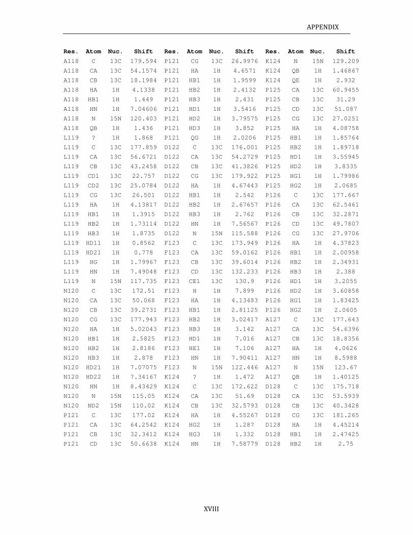
APPENDIX
XVIII
Res. Atom Nuc. Shift Res. Atom Nuc. Shift Res. Atom Nuc. Shift
A118 C 13C 179.594 P121 CG 13C 26.9976 K124 N 15N 129.209
A118 CA 13C 54.1574 P121 HA 1H 4.6571 K124 QB 1H 1.46867
A118 CB 13C 18.1984 P121 HB1 1H 1.9599 K124 QE 1H 2.932
A118 HA 1H 4.1338 P121 HB2 1H 2.4132 P125 CA 13C 60.9455
A118 HB1 1H 1.449 P121 HB3 1H 2.431 P125 CB 13C 31.29
A118 HN 1H 7.04606 P121 HD1 1H 3.5416 P125 CD 13C 51.087
A118 N 15N 120.403 P121 HD2 1H 3.79575 P125 CG 13C 27.0251
A118 QB 1H 1.436 P121 HD3 1H 3.852 P125 HA 1H 4.08758
L119 ? 1H 1.868 P121 QG 1H 2.0206 P125 HB1 1H 1.85764
L119 C 13C 177.859 D122 C 13C 176.001 P125 HB2 1H 1.89718
L119 CA 13C 56.6721 D122 CA 13C 54.2729 P125 HD1 1H 3.55945
L119 CB 13C 43.2458 D122 CB 13C 41.3826 P125 HD2 1H 3.8335
L119 CD1 13C 22.757 D122 CG 13C 179.922 P125 HG1 1H 1.79986
L119 CD2 13C 25.0784 D122 HA 1H 4.67443 P125 HG2 1H 2.0685
L119 CG 13C 26.501 D122 HB1 1H 2.542 P126 C 13C 177.667
L119 HA 1H 4.13817 D122 HB2 1H 2.67657 P126 CA 13C 62.5461
L119 HB1 1H 1.3915 D122 HB3 1H 2.762 P126 CB 13C 32.2871
L119 HB2 1H 1.73114 D122 HN 1H 7.56567 P126 CD 13C 49.7807
L119 HB3 1H 1.8735 D122 N 15N 115.588 P126 CG 13C 27.9706
L119 HD11 1H 0.8562 F123 C 13C 173.949 P126 HA 1H 4.37823
L119 HD21 1H 0.778 F123 CA 13C 59.0162 P126 HB1 1H 2.00958
L119 HG 1H 1.79967 F123 CB 13C 39.6014 P126 HB2 1H 2.34931
L119 HN 1H 7.49048 F123 CD 13C 132.233 P126 HB3 1H 2.388
L119 N 15N 117.735 F123 CE1 13C 130.9 P126 HD1 1H 3.2055
N120 C 13C 172.51 F123 H 1H 7.899 P126 HD2 1H 3.60858
N120 CA 13C 50.068 F123 HA 1H 4.13483 P126 HG1 1H 1.83425
N120 CB 13C 39.2731 F123 HB1 1H 2.81125 P126 HG2 1H 2.0605
N120 CG 13C 177.943 F123 HB2 1H 3.02417 A127 C 13C 177.643
N120 HA 1H 5.02043 F123 HB3 1H 3.142 A127 CA 13C 54.6396
N120 HB1 1H 2.5825 F123 HD1 1H 7.016 A127 CB 13C 18.8356
N120 HB2 1H 2.8186 F123 HE1 1H 7.106 A127 HA 1H 4.0626
N120 HB3 1H 2.878 F123 HN 1H 7.90411 A127 HN 1H 8.5988
N120 HD21 1H 7.07075 F123 N 15N 122.446 A127 N 15N 123.67
N120 HD22 1H 7.34167 K124 ? 1H 1.472 A127 QB 1H 1.40125
N120 HN 1H 8.43429 K124 C 13C 172.622 D128 C 13C 175.718
N120 N 15N 115.05 K124 CA 13C 51.69 D128 CA 13C 53.5939
N120 ND2 15N 110.02 K124 CB 13C 32.5793 D128 CB 13C 40.3428
P121 C 13C 177.02 K124 HA 1H 4.55267 D128 CG 13C 181.265
P121 CA 13C 64.2542 K124 HG2 1H 1.287 D128 HA 1H 4.45214
P121 CB 13C 32.3412 K124 HG3 1H 1.332 D128 HB1 1H 2.47425
P121 CD 13C 50.6638 K124 HN 1H 7.58779 D128 HB2 1H 2.75
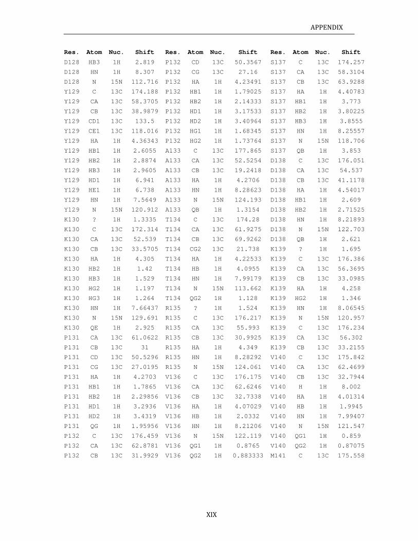
APPENDIX
XIX
Res. Atom Nuc. Shift Res. Atom Nuc. Shift Res. Atom Nuc. Shift
D128 HB3 1H 2.819 P132 CD 13C 50.3567 S137 C 13C 174.257
D128 HN 1H 8.307 P132 CG 13C 27.16 S137 CA 13C 58.3104
D128 N 15N 112.716 P132 HA 1H 4.23491 S137 CB 13C 63.9288
Y129 C 13C 174.188 P132 HB1 1H 1.79025 S137 HA 1H 4.40783
Y129 CA 13C 58.3705 P132 HB2 1H 2.14333 S137 HB1 1H 3.773
Y129 CB 13C 38.9879 P132 HD1 1H 3.17533 S137 HB2 1H 3.80225
Y129 CD1 13C 133.5 P132 HD2 1H 3.40964 S137 HB3 1H 3.8555
Y129 CE1 13C 118.016 P132 HG1 1H 1.68345 S137 HN 1H 8.25557
Y129 HA 1H 4.36343 P132 HG2 1H 1.73764 S137 N 15N 118.706
Y129 HB1 1H 2.6055 A133 C 13C 177.865 S137 QB 1H 3.853
Y129 HB2 1H 2.8874 A133 CA 13C 52.5254 D138 C 13C 176.051
Y129 HB3 1H 2.9605 A133 CB 13C 19.2418 D138 CA 13C 54.537
Y129 HD1 1H 6.941 A133 HA 1H 4.2706 D138 CB 13C 41.1178
Y129 HE1 1H 6.738 A133 HN 1H 8.28623 D138 HA 1H 4.54017
Y129 HN 1H 7.5649 A133 N 15N 124.193 D138 HB1 1H 2.609
Y129 N 15N 120.912 A133 QB 1H 1.3154 D138 HB2 1H 2.71525
K130 ? 1H 1.3335 T134 C 13C 174.28 D138 HN 1H 8.21893
K130 C 13C 172.314 T134 CA 13C 61.9275 D138 N 15N 122.703
K130 CA 13C 52.539 T134 CB 13C 69.9262 D138 QB 1H 2.621
K130 CB 13C 33.5705 T134 CG2 13C 21.738 K139 ? 1H 1.695
K130 HA 1H 4.305 T134 HA 1H 4.22533 K139 C 13C 176.386
K130 HB2 1H 1.42 T134 HB 1H 4.0955 K139 CA 13C 56.3695
K130 HB3 1H 1.529 T134 HN 1H 7.99179 K139 CB 13C 33.0985
K130 HG2 1H 1.197 T134 N 15N 113.662 K139 HA 1H 4.258
K130 HG3 1H 1.264 T134 QG2 1H 1.128 K139 HG2 1H 1.346
K130 HN 1H 7.66437 R135 ? 1H 1.524 K139 HN 1H 8.06545
K130 N 15N 129.691 R135 C 13C 176.217 K139 N 15N 120.957
K130 QE 1H 2.925 R135 CA 13C 55.993 K139 C 13C 176.234
P131 CA 13C 61.0622 R135 CB 13C 30.9925 K139 CA 13C 56.302
P131 CB 13C 31 R135 HA 1H 4.349 K139 CB 13C 33.2155
P131 CD 13C 50.5296 R135 HN 1H 8.28292 V140 C 13C 175.842
P131 CG 13C 27.0195 R135 N 15N 124.061 V140 CA 13C 62.4699
P131 HA 1H 4.2703 V136 C 13C 176.175 V140 CB 13C 32.7944
P131 HB1 1H 1.7865 V136 CA 13C 62.6246 V140 H 1H 8.002
P131 HB2 1H 2.29856 V136 CB 13C 32.7338 V140 HA 1H 4.01314
P131 HD1 1H 3.2936 V136 HA 1H 4.07029 V140 HB 1H 1.9945
P131 HD2 1H 3.4319 V136 HB 1H 2.0332 V140 HN 1H 7.99407
P131 QG 1H 1.95956 V136 HN 1H 8.21206 V140 N 15N 121.547
P132 C 13C 176.459 V136 N 15N 122.119 V140 QG1 1H 0.859
P132 CA 13C 62.8781 V136 QG1 1H 0.8765 V140 QG2 1H 0.87075
P132 CB 13C 31.9929 V136 QG2 1H 0.883333 M141 C 13C 175.558
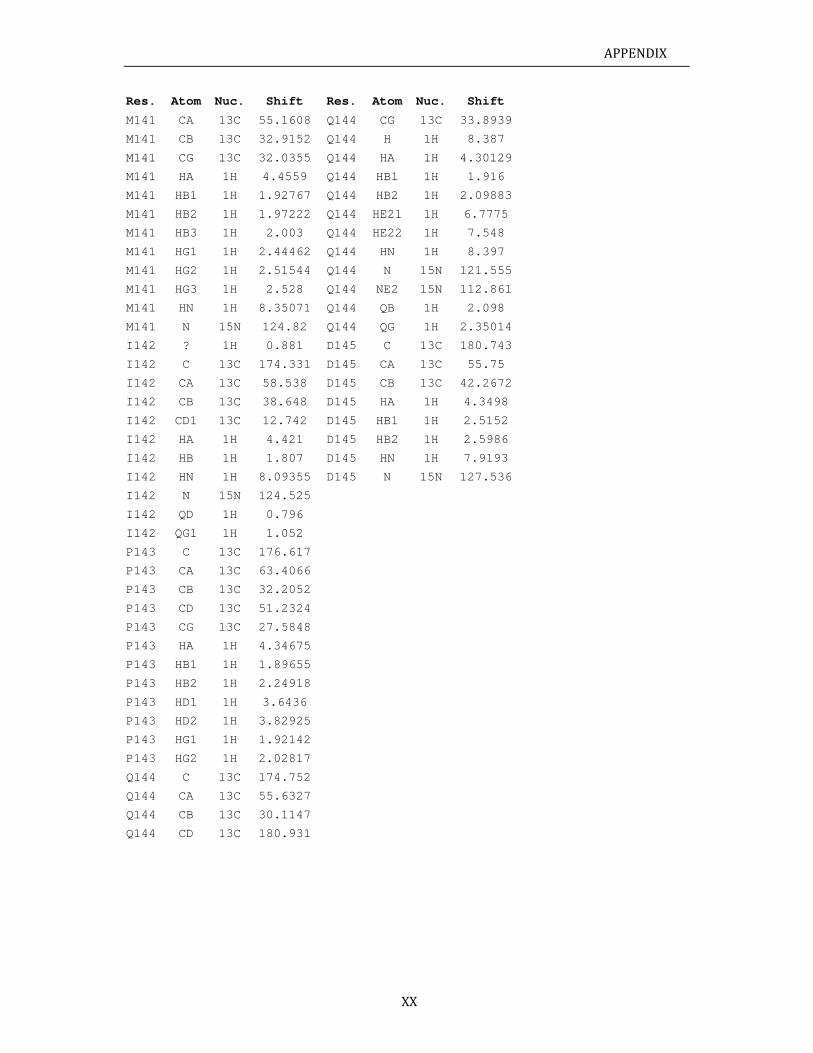
APPENDIX
XX
Res. Atom Nuc. Shift Res. Atom Nuc. Shift
M141 CA 13C 55.1608 Q144 CG 13C 33.8939
M141 CB 13C 32.9152 Q144 H 1H 8.387
M141 CG 13C 32.0355 Q144 HA 1H 4.30129
M141 HA 1H 4.4559 Q144 HB1 1H 1.916
M141 HB1 1H 1.92767 Q144 HB2 1H 2.09883
M141 HB2 1H 1.97222 Q144 HE21 1H 6.7775
M141 HB3 1H 2.003 Q144 HE22 1H 7.548
M141 HG1 1H 2.44462 Q144 HN 1H 8.397
M141 HG2 1H 2.51544 Q144 N 15N 121.555
M141 HG3 1H 2.528 Q144 NE2 15N 112.861
M141 HN 1H 8.35071 Q144 QB 1H 2.098
M141 N 15N 124.82 Q144 QG 1H 2.35014
I142 ? 1H 0.881 D145 C 13C 180.743
I142 C 13C 174.331 D145 CA 13C 55.75
I142 CA 13C 58.538 D145 CB 13C 42.2672
I142 CB 13C 38.648 D145 HA 1H 4.3498
I142 CD1 13C 12.742 D145 HB1 1H 2.5152
I142 HA 1H 4.421 D145 HB2 1H 2.5986
I142 HB 1H 1.807 D145 HN 1H 7.9193
I142 HN 1H 8.09355 D145 N 15N 127.536
I142 N 15N 124.525
I142 QD 1H 0.796
I142 QG1 1H 1.052
P143 C 13C 176.617
P143 CA 13C 63.4066
P143 CB 13C 32.2052
P143 CD 13C 51.2324
P143 CG 13C 27.5848
P143 HA 1H 4.34675
P143 HB1 1H 1.89655
P143 HB2 1H 2.24918
P143 HD1 1H 3.6436
P143 HD2 1H 3.82925
P143 HG1 1H 1.92142
P143 HG2 1H 2.02817
Q144 C 13C 174.752
Q144 CA 13C 55.6327
Q144 CB 13C 30.1147
Q144 CD 13C 180.931
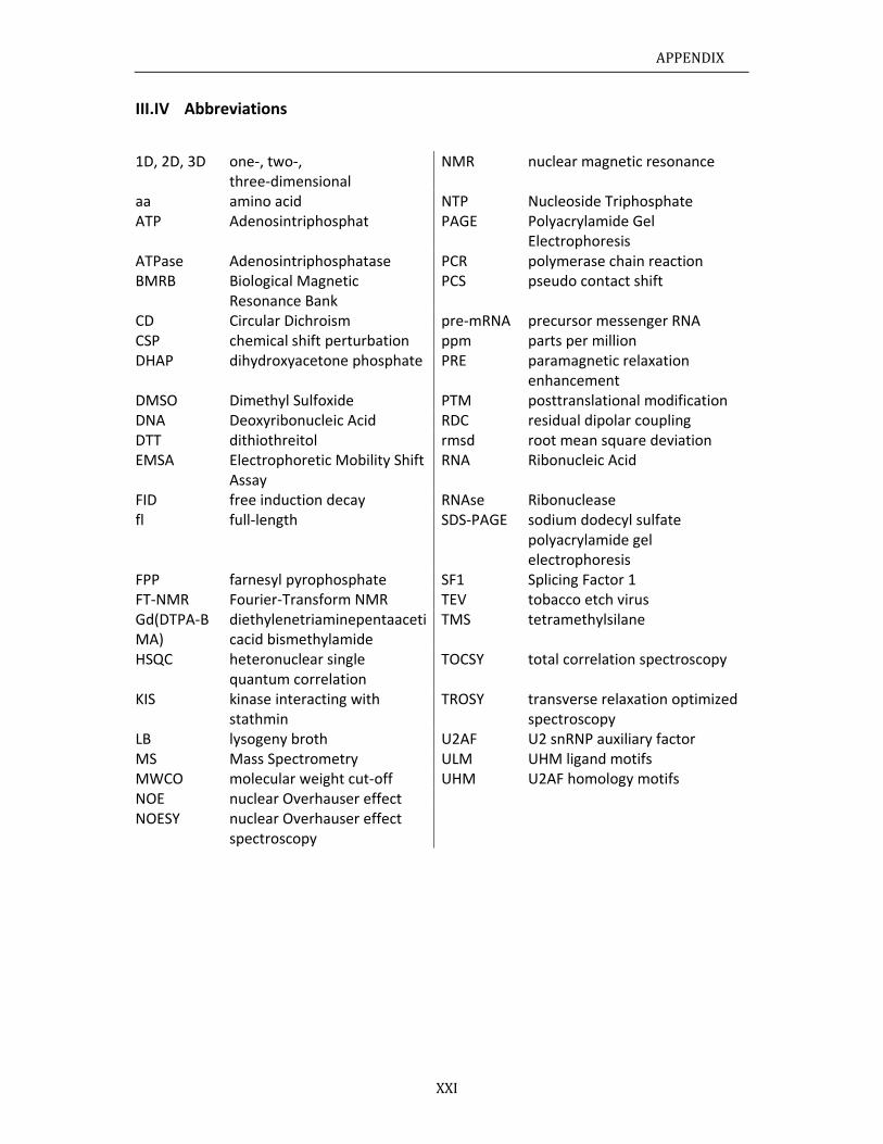
APPENDIX
XXI
III.IV Abbreviations
1D, 2D, 3D one‐, two‐, three‐dimensional
NMR nuclear magnetic resonance
aa amino acid NTP Nucleoside Triphosphate ATP Adenosintriphosphat PAGE Polyacrylamide Gel
Electrophoresis ATPase Adenosintriphosphatase PCR polymerase chain reaction BMRB Biological Magnetic
Resonance Bank PCS pseudo contact shift
CD Circular Dichroism pre‐mRNA precursor messenger RNA CSP chemical shift perturbation ppm parts per million DHAP dihydroxyacetone phosphate PRE paramagnetic relaxation
enhancement DMSO Dimethyl Sulfoxide PTM posttranslational modificationDNA Deoxyribonucleic Acid RDC residual dipolar coupling DTT dithiothreitol rmsd root mean square deviation EMSA Electrophoretic Mobility Shift
Assay RNA Ribonucleic Acid
FID free induction decay RNAse Ribonucleasefl full‐length SDS‐PAGE sodium dodecyl sulfate
polyacrylamide gel electrophoresis
FPP farnesyl pyrophosphate SF1 Splicing Factor 1 FT‐NMR Fourier‐Transform NMR TEV tobacco etch virus Gd(DTPA‐BMA)
diethylenetriaminepentaaceticacid bismethylamide
TMS tetramethylsilane
HSQC heteronuclear single quantum correlation
TOCSY total correlation spectroscopy
KIS kinase interacting with stathmin
TROSY transverse relaxation optimized spectroscopy
LB lysogeny broth U2AF U2 snRNP auxiliary factor MS Mass Spectrometry ULM UHM ligand motifs MWCO molecular weight cut‐off UHM U2AF homology motifs NOE nuclear Overhauser effect NOESY nuclear Overhauser effect
spectroscopy
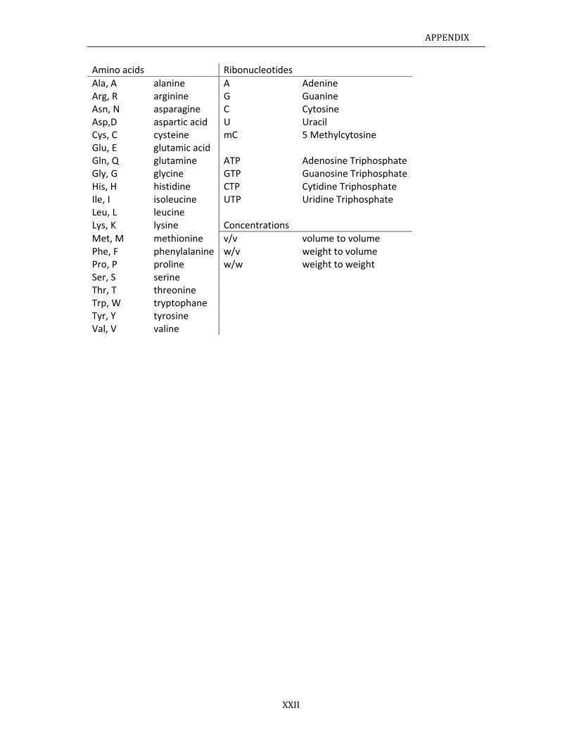
APPENDIX
XXII
Amino acids Ribonucleotides
Ala, A alanine A Adenine Arg, R arginine G GuanineAsn, N asparagine C Cytosine Asp,D aspartic acid U UracilCys, C cysteine mC 5 Methylcytosine Glu, E glutamic acid Gln, Q glutamine ATP Adenosine Triphosphate Gly, G glycine GTP Guanosine Triphosphate His, H histidine CTP Cytidine Triphosphate Ile, I isoleucine UTP Uridine Triphosphate Leu, L leucine Lys, K lysine Concentrations
Met, M methionine v/v volume to volume Phe, F phenylalanine w/v weight to volumePro, P proline w/w weight to weight Ser, S serine Thr, T threonine Trp, W tryptophane Tyr, Y tyrosine Val, V valine
ACKNOWLEDGEMENTS
XXIII
ACKNOWLEDGEMENTS First and foremost, I would like to show my deepest gratitude to my supervisor Michael
Sattler, a scholar full of enthusiasm about work and with passion for science. I would like to
thank him for taking me up in his lab and giving me the opportunity to work on this
interesting and exiting project. When I was about to lose confidence in phosphorylation
experiments, his suggestions and encouragements pulled me through.
I am greatly indebted to my colleague Tobias Madl, whom I would like to consider as my
second supervisor. Tobias is a brilliant and hardworking scientist. To work with him was a
challenging, informative and fruitful experience. I am very lucky to know him as a colleague,
a supervisor as well as a reliable and kind friend.
My sincere regards go to the members of my Thesis Advisory Committee Prof. Dr. Marius
Ueffing from the University Eye Hospital Tuebingen and Prof. Dr. Christian Becker from
University of Vienna, for their guidance and support during my PhD.
I would like to thank Fatiha Kateb and Konstantinos Tripsianes, who helped me a lot with
NMR and NMR spectrometers. Peijian Zou contributed to the wet lab works. I will never
forget his help when I was looking for an apartment in Munich. Hanso’s carefulness and
brilliance made the final success of SF1 phosphorylation. Ana Messias, without her valuable
advices, I would never finish this project. I would like to acknowledge Thomas Kern, who
helped me with analysis of relaxation data, and Waltraud Wolfson for taking care of my
incredibly large amounts of paperwork.
With particular gratitude, I would like to thank all of my former and present colleagues in
the Sattler group for interesting 4.5 years. My special thanks go to Hamed Kooshapur, Alex
Beribisky, Elke Prade and Diana Rodriguez. To work with them made life much more fun,
and because of them, I had a lot of fun in the group retreats. I would like to thank André
Mourão, who picked me up at the airport when I came to Munich. Going to Oktoberfest in
my first working day was a good beginning of the doctoral studying.
Last but not least, I owe my deepest gratitude to my love, Yuxiang Zhou, who has
incessantly supported me all along this challenging 4.5 years. Life means nothing to me
without you. And I would like to thank my parents for their understanding of my studying
abroad. I know it was not easy for you without the only son’s company during the Spring
Festival in the past three years. And now I am coming back home …
CURR
EDUCAT09/200809/200409/199909/1996 WORK E09/2008
09/2004
PUBLICA
1.
2.
3.
4.
5.
RICULUM
TION 8 to present 4 – 07/2007 9 – 07/2003 6 – 07/1999
EXPERIENCE 8 to present
4 – 08/2008
ATIONS Zhang, Y., MN., Sieber, Sbinding of thAcids Res 41Deng, Z., Zhorange CdTsimultaneou
12024‐1203Zhang, Y., Ddots as a pbiochemistryLiu, X., Zhansingle virus. Liu, X., Zhaounilamellar reversible A1770, 1620‐
M VITAE
Doctora Master
Bachelo High Sch
Doctoral Tthe Deparof Structu Title: Strusite and it
8 Master TInstitute o Title: Usinapplying t
Madl, T. (sharS.A., Kramer,he N‐termina1, 1343‐1354hang, Y. (shaTe quantumus and indep
1. (HighlightDeng, Z., Yueproton flux sy 364, 122‐1ng, Y., Yue, J.Biochemicao, R., Zhanglipid vesicl
ATP synthesi1626.
NAME
DATE OF B
NATIONAL
ADDRESS (
al student, Ter of Science ior of Sciencehool at Taoyu
Thesis Researtment of Chral Biology, Huctural studys regulation.Thesis Reseaof Biophysicsng cadmiumo detect H9
red first auth, A., and Satal domain of4. ared first aum dots as endent dete
ted by Nature, J., Tang, F.sensor and 127. ., Jiang, P., al and biophyg, Y., Jiang, le micro‐pas/hydrolysis
XXIV
IRTH
LITY
work)
echnische Un Biology, Bee in Chemistruan, Hunan,
arch at the Chemistry, TecHelmholtz Zey of SF1/U2A. Supervisor:rch at The s, Chinese Acm telluride qavian influen
hor), Bagdiulttler, M. (20f splicing fact
uthor), Yue, effective p
ection of viru
e Nanotechn., and Wei, Qapplying to
and Zhang, Zysical researcX., Yue, J., Jtterns as uof F0F1‐AT
Zhang Yu
30.10.19
Chinese
Technica
Biomolec
Lichtenb
85747 Ga
Germany
E‐mail: z
niversität Meijing Normary, Xiangtan UChina
Chair of Biochnische Unentrum MünAF65 in coo Prof. Dr. M.National Labcademy of Scquantum dotnza virus. Su
, I., Kern, T., 13). Structurtor 1 during
J., Tang, F., pH‐sensitive uses. The jou
nology. doi:1Q. (2007). Usdetect H9
. (2006). F0Fch communicJiang, P., anultrasmall rPase directly
CURR
un
981
al University
cular NMR S
ergstr. 4
arching
y
hangyun@m
ünchen al University University
molecular Niversität München perative rec. Sattler boratory of ciences. ts as a protpervisor: Pro
Kang, H.S., Zre, phospho3'‐splice site
and Wei, Qfluorescen
urnal of phys
10.1038/nnasing cadmiuavian influe
F1‐ATPase ascations 342, nd Zhang, Z.eaction cony. Biochimic
RICULUMVI
of Munich
pectroscopy
mytum.de
NMR Spectroünchen, and
cognition of
Biomacrom
ton flux senof. Dr. J.C. Yu
Zou, P., Maurylation ande recognition
Q. (2007). Grnt probes fsical chemist
no.2007.361m telluride qenza virus. A
s biosensor t1319‐1322. (2007). Usintainers to a et biophy
ITAE
y
oscopy in Institute
3’ splice
olecules,
nsor and ue
usbacher, U2AF65 n. Nucleic
reen and for dual ry B 111,
1) quantum Analytical
to detect
ing giant observe sica acta


![BMC Bioinformatics BioMed Central · – conserved, essential ones [26], whereas Borrelia has numerous plasmids that mostly encode poorly conserved genes [27]. Probably more telling](https://img.pdfslide.org/doc/110x75/5f6c9bf3f6891336585163a7/bmc-bioinformatics-biomed-central-a-conserved-essential-ones-26-whereas-borrelia.jpg)