Embed Size (px)
Citation preview
Technische Universität Kaiserslautern
Fachbereich Chemie
Structure and Bonding Studies of Paramagnetic
Metallocenes and Their Adducts of the d- and f-Block
Metals
Vom Fachbereich Chemie
der Technischen Universität Kaiserslautern
zur Erlangung des akademischen Grades
„Doktor der Naturwissenschaften“
genehmigte Dissertation
(D 386)
vorgelegt von
Dipl.-Chem. Marc D. Walter aus Ludwigshafen
Betreuer der Arbeit: Prof. Dr. H. Sitzmann
Tag der wissenschaftlichen Aussprache: 05. September 2005
Kaiserslautern 2005
Dem Fachbereich Chemie der Technischen Universität Kaiserslautern am 22. August 2005
als Dissertation eingereicht.
Dekan: Prof. H.-J. Krüger, Ph.D.
Prüfungskommission:
Vorsitzender: Prof. Dr. H.-G. Kuball
1. Berichterstatter: Prof. Dr. H. Sitzmann
2. Berichterstatter: Prof. Dr. R.A. Andersen
Die vorliegende Arbeit entstand in der Zeit vom Oktober 2001 bis Juli 2005 in den
Arbeitskreisen von Prof. Dr. H. Sitzmann (Fachbereich Chemie, Technische
Universität Kaiserslautern) und Prof. Dr. R.A. Andersen (Department of
Chemistry, University of California at Berkeley)
Herrn Prof. Dr. Sitzmann danke ich recht herzlich für den mir gewährten Freiraum bei der
Gestaltung meines Themas, vor allem aber für seine stete Diskussionsbereitschaft und
Begeisterungsfähigkeit. Furthermore I am very grateful to Prof. Dr. R.A. Andersen for giving
me the opportunity to work in his laboratory, his continuous guidance, inspiration and
enthusiasm.

To my parents
Table of Abbreviations
For the purpose of clarity and conciseness, the following abbreviations have been used throughout this work.
Me: -CH3
Et: -CH2CH3
OMe: -OCH3
bipy: 2,2’-bipyridyl
bipy-R: 4,4’-R substituted 2,2’-dipyridyl
phen: 1,10-phenanthroline
Ln: unspecified lanthanide metal
Cp’: unspecified cyclopentadienide anion
R: unspecified organic group
HOMO: Highest occupied molecular orbital
LUMO: Lowest unoccupied molecular orbital
COT: cyclooctatetraene dianion
CNT: cyclononatetraenide
THF: Tetrahydrofuran; C4H8O
dme: dimethoxyethane; CH3OCH2CH2OCH3
pc: phthalocyanato, C32H16N82-
tmtaa: 5,7,12,14-Tetramethyl-2,3:9,10-dibenzo-[14]hexaenato-N4, tmtaa2-
EXAFS: Extended X-Ray Absorption Fine Structure
XANES: X-Ray Absorption Near Edge Structure
Table of Contents
Chapter 1:
Heavy- Alkaline Earth and Lanthanide Complexes with 10π Aromatic
Ligands 1
Chapter 2:
The Effect of Substituted Cyclopentadienyl Ligands on the Electronic
Structure of Manganocenes 21
Chapter 3:
Lanthanide(III) complexes with sterically demanding cyclopentadienyl
ligands 82
Chapter 4:
Cerocene revisited 100
Chapter 5:
Non-cyclopentadienyl cerium compounds 118
Chapter 6:
The electronic structure of bipyridyl and diazabutadienyl complexes of
Ytterbocenes 133
Chapter 7:
Titanium Complexes with 1,3-Di(tert.-butyl)cyclopentadienyl Ligands 206
Chapter 8:
Experimental Section 237
Chapter 9:
X-ray Crystallography 271

Chapter 1: Heavy-Alkaline Earth and Lanthanide Complexes with 10π Aromatic Ligands
1
Chapter 1: Heavy-Alkaline Earth and Lanthanide Complexes with 10ππππ Aromatic
Ligands
Introduction
Since their development in their early 1900s, Grignard reagents have proven to be immensely useful in synthetic
chemistry and are among the most common organometallic reagents. In contrast, information about beryllium,
calcium, strontium, and barium analogues is scarce; organometallic compounds are difficult to access due to
their enhanced reactivity and decreased stability. The high electropositive character and large ionic radii of the
heavy alkaline earth metals result in a chemistry mostly governed by electrostatic and steric requirements.
Consequently, early studies of organo compounds of the heavy alkaline earth metals1,2 were impeded by their
high and unselective reactivity as well as a lack of solubility and stability, progress was achieved by the
introduction of the cyclopentadienyl anion as a π ligand.3,4 Later soluble, well-characterized organometallic
compounds of calcium, strontium, and barium could be obtained with bulky hydrocarbyl anions like tris-5 or
bis(trimethylsilyl)methyl,6 silylated allyl7 or benzyl ligands8 or benzyl ligands with a nitrogen donor function in
the side chain,9 open pentadienyl ligands like 2,4-di(tert.butyl)pentadienyl,10 or cyclopentadienyl ligands with
bulky substituents, e. g. C5Me5 (Cp*),11-13 C5H2(SiMe3)3-1,2,4,14 C5H(CHMe2)4 (4Cp),15 C5H2(CMe3)3-1,2,4,16,17
or C5(CHMe2)5 (5Cp).18
Organometallic compounds of the heavy alkaline earth metals are attracting interest as synthetic reagents in
organic synthesis or as initiators of anionic styrene polymerization.8,9,19-21 Furthermore they may function as
MOCVD precursors with sufficient volatility and stability for growth of thin films with high-temperature
superconducting (HTS) or ferroelectric properties.22 Basic research has focused primarily on metallocene and
related compounds. 23
The development of synthetically useful half-sandwich complexes of these large cations as precursors for a
variety of functionalized derivatives presents a challenge and a synthetic goal. Introduction of the sterically
demanding tris(trimethylsilyl)cyclopentadienyl ligand recently allowed the crystallographic characterization of
dimeric calcium and strontium derivatives, [{(C5H2(SiMe3)3)Ca(µ-I)(THF)}2] and [{(C5H2(SiMe3)3)Sr(µ-
I)(THF)2}2], and the barium chain polymer [{(C5H2(SiMe3)3)Ba(µ-I)(THF)} ∞ ].24
Tris(trimethylsilyl)cyclopentadienide loss upon attempted nucleophilic substitution of the halide in these
complexes hampers their possible use as starting materials. The only accessible substitution product to date is the
crystallographically characterized borate complex, [(C5H2(SiMe3)3)Ca(HBEt3)(THF)2].25 However, nucleophilic
substitution has been successful with mono(alkylcyclopenadienyl)calcium halide complexes and yielded amide
and aryloxide derivatives. 26,27
Sitzmann et al. demonstrated that extremely bulky alkylcyclopentadienides such as 1,3,4-tri-tert-
butylcyclopentadienide and tetraisopropylcyclopentandide are well suited to kinetically stabilize
mono(cyclopentadienyl) halides of the heavy alkaline earth metals with additional tetrahydrofuran or
dimethoxyethane donor ligands.28 This was demonstrated for strontium and barium compounds, despite a recent
report to the contrary.24
Chapter 1: Heavy-Alkaline Earth and Lanthanide Complexes with 10π Aromatic Ligands
2
Objective
A different approach of exerting steric bulk uses anionic carbocycles with more than five ring atoms.
Cyclooctatetraene complexes of the heavy alkaline earth metals have been reported in analogy to lanthanide
COT complexes, but suffered from a lack of solubility and crystallinity.29 Later on, triple-decker and inverse
sandwich complexes of the lanthanides using COT as middle deck have been prepared and structurally
characterized.30
Functionalization of the recently prepared mono(cyclopentadienyl) halides of the heavy alkaline earth metals
with cyclooctatraene dianion, Na2C8H8, and cyclononatetraenide, KC9H9, should be explored.
Results and discussion
The most promising starting compounds for the synthesis of mono(cyclopentadienyl) complexes of the heavy
alkaline earth metals are monohalides of the [CpMX(solvent)m]n type (M = Ca, Sr, Ba; X = Cl, Br, I; m = 1-3;
n= 1,2,∞). The only halide substitution reactions in this context have been reported for the introduction of
anionic nucleophiles into calcium compounds of the type stated before with penta- or tetraalkylated
cyclopentadienyl ligands.25-27,31 In order to extend the feasibility of such substitution reactions to strontium and
barium, recently the iodo complexes [{(C5HR4)BaI(THF)2}2] (1-Ba; R = CHMe2) and [{(C5HR4)SrI(THF)2}2]
(1-Sr; R = CHMe2) have been prepared,28 which could be used as starting compounds for reactions with
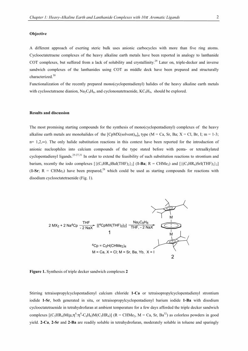
disodium cyclooctatetraenide (Fig. 1).
Figure 1. Synthesis of triple decker sandwich complexes 2
Stirring tetraisopropylcyclopentadienyl calcium chloride 1-Ca or tetraisopropylcyclopentadienyl strontium
iodide 1-Sr, both generated in situ, or tetraisopropylcyclopentadienyl barium iodide 1-Ba with disodium
cyclooctatetraenide in tetrahydrofuran at ambient temperature for a few days afforded the triple decker sandwich
complexes [(C5HR4)M(µ,η8:η8-C8H8)M(C5HR4)] (R = CHMe2, M = Ca, Sr, Ba32) as colorless powders in good
yield. 2-Ca, 2-Sr and 2-Ba are readily soluble in tetrahydrofuran, moderately soluble in toluene and sparingly
M
M
Na2C8H8THF, - 2 NaX[{4CpMX(THF)2}2]
4Cp = C5H(CHMe2)4
1
2M = Ca, X = Cl; M = Sr, Ba, Yb, X = I
2 MX2 + 2 Na4Cp - 2 NaXTHF
Chapter 1: Heavy-Alkaline Earth and Lanthanide Complexes with 10π Aromatic Ligands
3
soluble in pentane and they can be crystallized from saturated benzene solution. 2-Yb behaves differently, the
solubility in aliphatic solvents (like n-hexane) is fairly good and it can be crystallized from a saturated n-hexane
solution. In an oil pump vacuum the heavy alkaline earth compounds can be sublimed without decomposition at
130 °C (2-Ca), 175 °C (2-Sr), 215 °C (2-Ba) and unchanged melting points have been found after heating 2-Ca,
2-Sr or 2-Ba to 250 °C under an argon atmosphere. In air the compounds turn yellow immediately and orange-
red within seconds. Finally a pale yellow residue is obtained and a strong COT smell is developed.
In EI mass spectra the molecular ion as well as signals for the fragments [4CpM(COT)M]+ (parent peak for M =
Ca) and [4CpM]+ (parent peak for M = Ba) could be detected and shown to exhibit the correct isotope patterns
for all three heavy-alkaline earth complexes. However, for 2-Yb no metal containing fragment is detected under
these conditions.
In NMR spectra, one set of signals for the tetraisopropylcyclopentadienyl ligand and one signal for the
cyclooctatetraene ligand has been observed, whose 13C NMR signal has been recorded at 89.6 ppm for 2-Ca,
91.6 for 2-Sr, 95.3 ppm for 2-Ba and 89.8 for 2-Yb, with almost identical 1JC,H coupling constants of 157, 157,
158 and 163 Hz, respectively. For comparison, {[(Me3Si)2N]Yb(thf)}2(µ-C8H8) shows the COT signal at 90.8
ppm in the 13C NMR.33
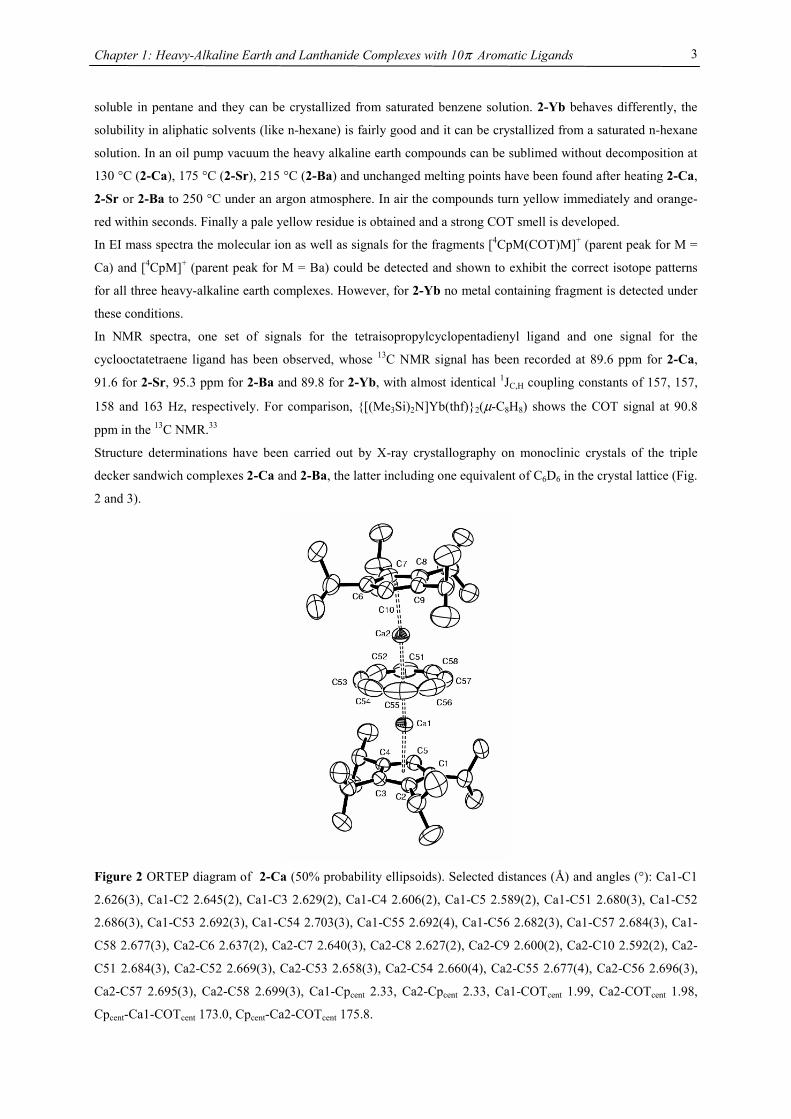
Structure determinations have been carried out by X-ray crystallography on monoclinic crystals of the triple
decker sandwich complexes 2-Ca and 2-Ba, the latter including one equivalent of C6D6 in the crystal lattice (Fig.
2 and 3).
Figure 2 ORTEP diagram of 2-Ca (50% probability ellipsoids). Selected distances (Å) and angles (°): Ca1-C1
2.626(3), Ca1-C2 2.645(2), Ca1-C3 2.629(2), Ca1-C4 2.606(2), Ca1-C5 2.589(2), Ca1-C51 2.680(3), Ca1-C52
2.686(3), Ca1-C53 2.692(3), Ca1-C54 2.703(3), Ca1-C55 2.692(4), Ca1-C56 2.682(3), Ca1-C57 2.684(3), Ca1-
C58 2.677(3), Ca2-C6 2.637(2), Ca2-C7 2.640(3), Ca2-C8 2.627(2), Ca2-C9 2.600(2), Ca2-C10 2.592(2), Ca2-
C51 2.684(3), Ca2-C52 2.669(3), Ca2-C53 2.658(3), Ca2-C54 2.660(4), Ca2-C55 2.677(4), Ca2-C56 2.696(3),
Ca2-C57 2.695(3), Ca2-C58 2.699(3), Ca1-Cpcent 2.33, Ca2-Cpcent 2.33, Ca1-COTcent 1.99, Ca2-COTcent 1.98,
Cpcent-Ca1-COTcent 173.0, Cpcent-Ca2-COTcent 175.8.
Chapter 1: Heavy-Alkaline Earth and Lanthanide Complexes with 10π Aromatic Ligands
4
The Ca-C(4Cp) distance average of 2.62 Å and Ba-C(4Cp) of 2.96 Å are comparable to the values found for the
respective octaisopropylmetallocenes of calcium (2.64 Å) or barium (2.94 Å)15 and for the
tetraisopropylcyclopentadienyl iodide dimer of barium (2.97 Å)28 and slightly shorter than the value found for
[{4CpCa(µ-I)(THF)}2] (2.67 Å).31 The metal-C(COT) distances are longer, for 2-Ca the respective distance
range is 2.660(3) – 2.703(3) Å (2.683 Å average) and for 2-Ba values between 2.994(5) and 3.016(6) Å (3.006 Å
average) have been observed. Despite the longer metal-carbon distances the metal approaches the
cyclooctatetraene plane much closer than the cyclopentadienyl plane because of the large diameter of the eight-
membered ring.
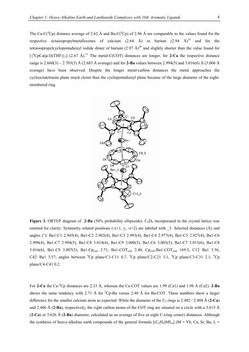
Figure 3. ORTEP diagram of 2-Ba (50% probability ellipsoids). C6D6 incorporated in the crystal lattice was
omitted for clarity. Symmetry related positions (-x+1, y, -z+2) are labeled with _3. Selected distances (Å) and
angles (°): Ba1-C1 2.945(4), Ba1-C2 2.982(4), Ba1-C3 2.993(4), Ba1-C4 2.977(4), Ba1-C5 2.927(4), Ba1-C6
2.998(4), Ba1-C7 2.994(5), Ba1-C8 3.014(4), Ba1-C9 3.000(5), Ba1-C6 3.003(5), Ba1-C7 3.015(6), Ba1-C8
3.016(6), Ba1-C9 3.007(5), Ba1-Cpcent 2.71, Ba1-COTcent 2.40, Cpcent-Ba1-COTcent 169.5, C12…Ba1 3.56,
C42…Ba1 3.57; angles between 4Cp plane/C1-C11 0.7, 4Cp plane/C2-C21 3.1, 4Cp plane/C3-C31 2.1, 4Cp
plane/C4-C41 0.2.
For 2-Ca the Ca-4Cp distances are 2.33 Å, whereas the Ca-COT values are 1.99 (Ca1) and 1.98 Å (Ca2). 2-Ba
shows the same tendency with 2.71 Å for 4Cp-Ba versus 2.40 Å for Ba-COT. These numbers show a larger
difference for the smaller calcium atom as expected. While the diameter of the C5 rings is 2.402 / 2.404 Å (2-Ca)
and 2.406 Å (2-Ba), respectively, the eight carbon atoms of the COT ring are situated on a circle with a 3.613 Å
(2-Ca) or 3.626 Å (2-Ba) diameter, calculated as an average of five or eight C-(ring center) distances. Although
the synthesis of heavy-alkaline earth compounds of the general formula [(C8H8)MLn] (M = Yb, Ca, Sr, Ba, L =
Chapter 1: Heavy-Alkaline Earth and Lanthanide Complexes with 10π Aromatic Ligands
5
THF, pyridine) by metal cocondensation with cycloctatetraene and solvent extraction has been reported, their
low solubility, high air sensitivity and their microcrystalline nature precluded a structural analysis.29 However,
the ytterbium(II) derivative, [(C8H8)Yb(pyridine)3], has been crystallographically characterized and shows
geometrical features of the Yb(C8H8) substructure comparable to those of 2-Ca with Yb-C distances of 2.57-2.69
Å and an Yb-ring distance of 1.91 Å.34 Yb-C distances from 2.71 to 2.78 Å have been found for the dipotassium
salt [K(diglyme)]2[(η8-C8H8)2Yb(II)]35 and the Yb-C(COT) average distances found for the triple decker
[(C5Me5)Yb(µ,η8:η8-C8H8)Yb(C5Me5)] are 2.65 Å (Yb1) and 2.67 Å (Yb2).36 The short contacts between two
methyl groups (C12, C42) and the barium central atom of 2-Ba (3.56 / 3.57 Å) correspond to almost complete
absence of outward bending of the respective methine carbon atoms C11 and C41 as well as rotation of the
respective isopropyl groups towards the barium center. These features have not been observed in 2-Ca, where
the closest C…Ca contacts are still larger than 3.6 Å (3.65 / 3.67 Å for Ca2…C82 and Ca1…C12).
Complexes 2 are the only neutral species among the few triple decker sandwich complexes known with main
group central atoms and show a very low degree of bending with 4Cpcent-M-COTcent angles of 173.0/175.8° (2-
Ca) and 168.5° (2-Ba), compared to the Cpcent-M-Cpcent angles of 155/152° for [(C5Me5)3Sn2]+,37 134° for
[Cp3Tl2]-,38 124/130° for [(η6-C7H8)2(µ,η5:η5-C5Me5)In2]+,37 or 116° for [Cp3Cs2]-.39 Even
octaisopropylmetallocenes of calcium (162°) or barium (154°)15 are more strongly bent than 2-Ca or 2-Ba.
It has been demonstrated that divalent lanthanide and alkaline earth metallocenes prefer bent sandwich structures
in gas as well as condensed phases.11,12,40-43 Different models have been proposed to rationalize this geometrical
behavior, including a molecular orbital model,44-46 an electrostatic (polarized-ion) model,47,48 and a model based
on attractive van der Waals forces.49,50 No single explanation has been generally accepted so far. Limits to the
extend of bending of ML2 molecules originated from a repulsion as the ligands approach each other closer than
their sum of Van der Waals radii. This repulsion is related to the metal radius, as larger metals are capable of
increased bending before the ligands approach each other too closely. Because bending energies are quite small,
on the order of 1-5 kcal/mol, no experiment to date has been able to disprove any of these models.44,45,51
In order to describe this trend empirically Hanusa et al. followed an analysis by Raymond & Eigenbrot52 and
correlated the available data on bending angles with the ionic radii for decamethylmetallocenes of the
lanthanides and heavy alkaline metals.41,53 Evans et al. provided more information on the bending angles in triple
decker complexes of Eu, Sm and Yb with Cp* ligands.54 Such a correlation has not been available for the
sterically even more demanding ligands, e.g. tetraisopropylcyclopentadienyl and tris(tert-butyl)cyclopentadienyl
(Tab. 1). Although only two triple decker complexes are structurally characterized, they most probably will also
follow the linear relationship between metal radius and bonding angle as observed in its metallocene complexes,
with a greater tendency towards parallel planes with smaller metals. Furthermore the replacement of a Cp unit
for a COT deck does increase the tendency to a more linear arrangement by a constant additional increment of
roughly 13°, as demonstrated by almost parallel linear fits for (C5Me5)2M and [(C5Me5)M]2(COT) and 4Cp2M
and (4CpM)2(COT), respectively.
Chapter 1: Heavy-Alkaline Earth and Lanthanide Complexes with 10π Aromatic Ligands
6
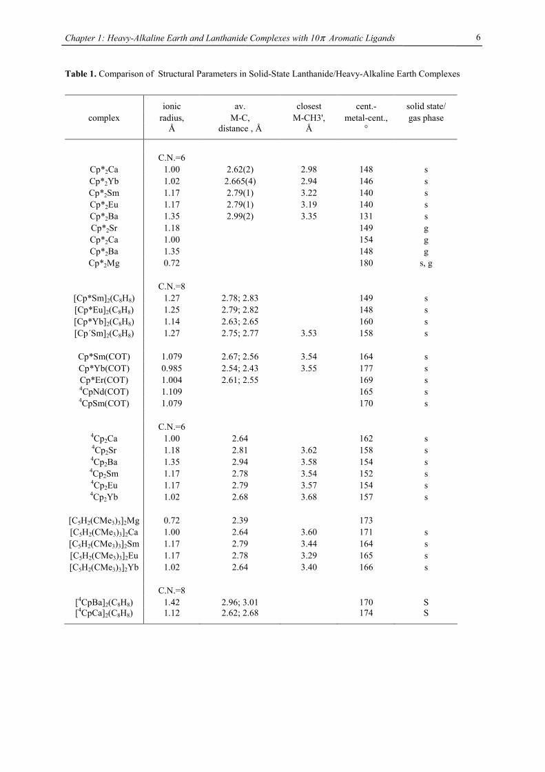
Table 1. Comparison of Structural Parameters in Solid-State Lanthanide/Heavy-Alkaline Earth Complexes
ionic av. closest cent.- solid state/ complex radius, M-C, M-CH3', metal-cent., gas phase
Å distance , Å Å °
C.N.=6
Cp*2Ca 1.00 2.62(2) 2.98 148 s Cp*2Yb 1.02 2.665(4) 2.94 146 s Cp*2Sm 1.17 2.79(1) 3.22 140 s Cp*2Eu 1.17 2.79(1) 3.19 140 s Cp*2Ba 1.35 2.99(2) 3.35 131 s Cp*2Sr 1.18 149 g Cp*2Ca 1.00 154 g Cp*2Ba 1.35 148 g Cp*2Mg 0.72 180 s, g
C.N.=8
[Cp*Sm]2(C8H8) 1.27 2.78; 2.83 149 s [Cp*Eu]2(C8H8) 1.25 2.79; 2.82 148 s [Cp*Yb]2(C8H8) 1.14 2.63; 2.65 160 s [Cp´Sm]2(C8H8) 1.27 2.75; 2.77 3.53 158 s
Cp*Sm(COT) 1.079 2.67; 2.56 3.54 164 s Cp*Yb(COT) 0.985 2.54; 2.43 3.55 177 s Cp*Er(COT) 1.004 2.61; 2.55 169 s 4CpNd(COT) 1.109 165 s 4CpSm(COT) 1.079 170 s
C.N.=6
4Cp2Ca 1.00 2.64 162 s 4Cp2Sr 1.18 2.81 3.62 158 s 4Cp2Ba 1.35 2.94 3.58 154 s 4Cp2Sm 1.17 2.78 3.54 152 s 4Cp2Eu 1.17 2.79 3.57 154 s 4Cp2Yb 1.02 2.68 3.68 157 s
[C5H2(CMe3)3]2Mg 0.72 2.39 173 [C5H2(CMe3)3]2Ca 1.00 2.64 3.60 171 s [C5H2(CMe3)3]2Sm 1.17 2.79 3.44 164 s [C5H2(CMe3)3]2Eu 1.17 2.78 3.29 165 s [C5H2(CMe3)3]2Yb 1.02 2.64 3.40 166 s
C.N.=8
[4CpBa]2(C8H8) 1.42 2.96; 3.01 170 S [4CpCa]2(C8H8) 1.12 2.62; 2.68 174 S
Chapter 1: Heavy-Alkaline Earth and Lanthanide Complexes with 10π Aromatic Ligands
7
0.7 0.8 0.9 1.0 1.1 1.2 1.3 1.4 1.5
130
140
150
160
170
180
[1,3,4-(Me3C)
3C
5H
2]2M
4Cp2M (C
5Me
5)
2M
[ (C5Me5)M ]2(COT) [ 4Cp
2M ]
2(COT)
Cen
troid
-M-C
entro
id A
ngle
/ °
Ionic Radius / pm
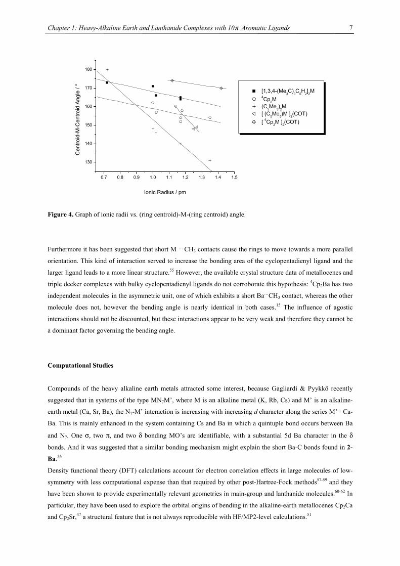
Figure 4. Graph of ionic radii vs. (ring centroid)-M-(ring centroid) angle.
Furthermore it has been suggested that short M … CH3 contacts cause the rings to move towards a more parallel
orientation. This kind of interaction served to increase the bonding area of the cyclopentadienyl ligand and the
larger ligand leads to a more linear structure.55 However, the available crystal structure data of metallocenes and
triple decker complexes with bulky cyclopentadienyl ligands do not corroborate this hypothesis: 4Cp2Ba has two
independent molecules in the asymmetric unit, one of which exhibits a short Ba…CH3 contact, whereas the other
molecule does not, however the bending angle is nearly identical in both cases.15 The influence of agostic
interactions should not be discounted, but these interactions appear to be very weak and therefore they cannot be
a dominant factor governing the bending angle.
Computational Studies
Compounds of the heavy alkaline earth metals attracted some interest, because Gagliardi & Pyykkö recently
suggested that in systems of the type MN7M’, where M is an alkaline metal (K, Rb, Cs) and M’ is an alkaline-
earth metal (Ca, Sr, Ba), the N7-M’ interaction is increasing with increasing d character along the series M’= Ca-
Ba. This is mainly enhanced in the system containing Cs and Ba in which a quintuple bond occurs between Ba
and N7. One σ, two π, and two δ bonding MO’s are identifiable, with a substantial 5d Ba character in the δ
bonds. And it was suggested that a similar bonding mechanism might explain the short Ba-C bonds found in 2-
Ba.56
Density functional theory (DFT) calculations account for electron correlation effects in large molecules of low-
symmetry with less computational expense than that required by other post-Hartree-Fock methods57-59 and they
have been shown to provide experimentally relevant geometries in main-group and lanthanide molecules.60-62 In
particular, they have been used to explore the orbital origins of bending in the alkaline-earth metallocenes Cp2Ca
and Cp2Sr,47 a structural feature that is not always reproducible with HF/MP2-level calculations.51

Chapter 1: Heavy-Alkaline Earth and Lanthanide Complexes with 10π Aromatic Ligands
8
Accordingly, DFT calculations were performed on a variety of model compounds with various DFT functionals.
The studies focused on optimizing the geometry of the complexes compared to the X-ray crystallographic results
and on the investigation of the molecular orbitals/NBO charges to give an insight into the bonding situation.
Geometry optimizations were carried out without symmetry restrictions (C1 symmetry). The nature of the
minima was verified by analytical frequency calculations and the zero-point energy (ZPE) was estimated within
the harmonic potential approximation. The 4Cp ligand was modeled by C5H5. Although there is a large difference
in steric size between these ligands, which is a severe approximation especially when some significant
geometrical reorganization can occur,62,63 Hanusa et al. pointed out that three different cyclopentadienyl ligands
(Cp, C5Me5 and 1,3,4-(Me3Si)3C5H2) are virtually interchangeable in their contributions to the geometric
parameters (especially distances) and to the reaction enthalpy.24
Geometry
Geometry optimization of [(C5H5)M]2(COT) (M= Ca, Sr, Ba) revealed structural parameters that agree favorably
with structures of 2-Ca and 2-Ba determined by X-ray crystallography (Tab. 2).
Table 2. Selected Bond Distances (Å) and Angles (°) of Calculated (B3PW91, G-311G for C, H and Ca, SDD
for Ba and Sr) and Experimental Structures
Calc Exp Delta %error
Cp(cent)1-Ba1 2.739 2.71 0.029 1.1
Ba1-COT(cent) 2.424 2.397 0.027 1.1
COT(cent)-Ba2 2.424
Ba2-Cp(cent)2 2.738
Cp1(cent)-Ba1-COT(cent) 175.47 168.46 7.01 4.2
Cp2(cent)-Ba2-COT(cent) 176.27
Cp(cent)1-Sr1 2.540
Sr1-COT(cent) 2.206
COT(cent)-Sr2 2.203
Sr2-Cp(cent)2 2.548
Cp1(cent)-Sr1-COT(cent) 179.10
Cp2(cent)-Sr2-COT(cent) 174.61
Cp(cent)1-Ca1 2.341 2.327 0.014 0.60
Ca1-COT(cent) 1.998 1.989 0.009 0.45
COT(cent)-Ca2 1.997 1.979 0.018 0.91
Ca2-Cp(cent)2 2.340 2.328 0.012 0.52
Cp1(cent)-Ca1-COT(cent) 178.52 172.99 5.53 3.20
Chapter 1: Heavy-Alkaline Earth and Lanthanide Complexes with 10π Aromatic Ligands
9
Even the bend angles agree surprisingly well with the experimental values. Due to the smaller size of C5H5
relative to 4Cp a more dramatic difference in the Cp(centroid)-M-COT(centroid) angle has been anticipated
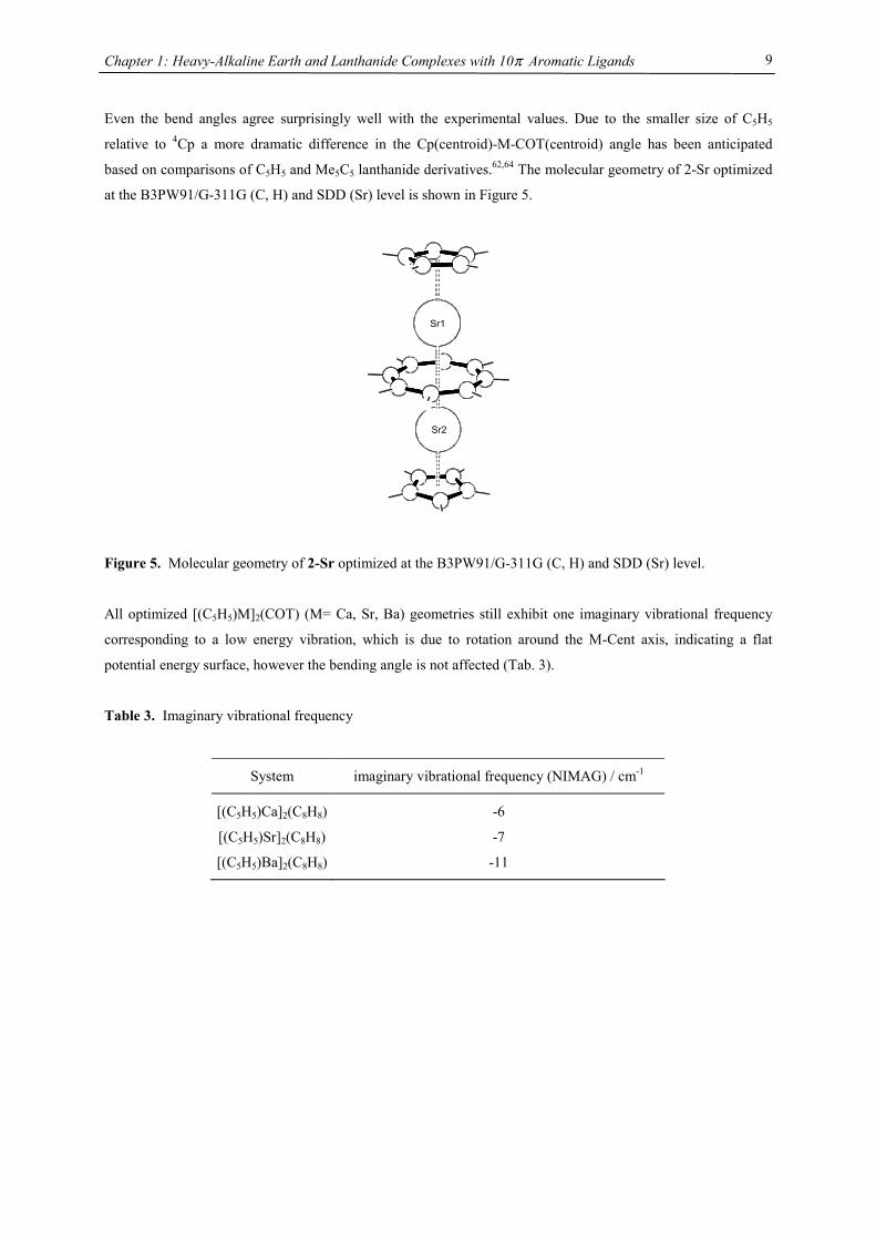
based on comparisons of C5H5 and Me5C5 lanthanide derivatives.62,64 The molecular geometry of 2-Sr optimized
at the B3PW91/G-311G (C, H) and SDD (Sr) level is shown in Figure 5.
Sr1
Sr2
Figure 5. Molecular geometry of 2-Sr optimized at the B3PW91/G-311G (C, H) and SDD (Sr) level.
All optimized [(C5H5)M]2(COT) (M= Ca, Sr, Ba) geometries still exhibit one imaginary vibrational frequency
corresponding to a low energy vibration, which is due to rotation around the M-Cent axis, indicating a flat
potential energy surface, however the bending angle is not affected (Tab. 3).
Table 3. Imaginary vibrational frequency
System imaginary vibrational frequency (NIMAG) / cm-1
[(C5H5)Ca]2(C8H8) -6
[(C5H5)Sr]2(C8H8) -7
[(C5H5)Ba]2(C8H8) -11
Chapter 1: Heavy-Alkaline Earth and Lanthanide Complexes with 10π Aromatic Ligands
10
Ligand redistribution
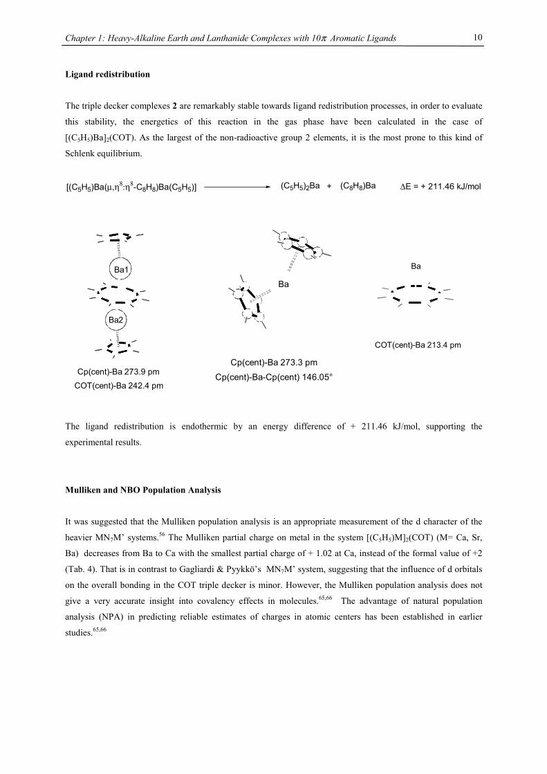
The triple decker complexes 2 are remarkably stable towards ligand redistribution processes, in order to evaluate
this stability, the energetics of this reaction in the gas phase have been calculated in the case of
[(C5H5)Ba]2(COT). As the largest of the non-radioactive group 2 elements, it is the most prone to this kind of
Schlenk equilibrium.
[(C5H5)Ba(µ,η8:η8-C8H8)Ba(C5H5)] (C5H5)2Ba + (C8H8)Ba ∆E = + 211.46 kJ/mol
The ligand redistribution is endothermic by an energy difference of + 211.46 kJ/mol, supporting the
experimental results.
Mulliken and NBO Population Analysis
It was suggested that the Mulliken population analysis is an appropriate measurement of the d character of the
heavier MN7M’ systems.56 The Mulliken partial charge on metal in the system [(C5H5)M]2(COT) (M= Ca, Sr,
Ba) decreases from Ba to Ca with the smallest partial charge of + 1.02 at Ca, instead of the formal value of +2
(Tab. 4). That is in contrast to Gagliardi & Pyykkö’s MN7M’ system, suggesting that the influence of d orbitals
on the overall bonding in the COT triple decker is minor. However, the Mulliken population analysis does not
give a very accurate insight into covalency effects in molecules.65,66 The advantage of natural population
analysis (NPA) in predicting reliable estimates of charges in atomic centers has been established in earlier
studies.65,66
Ba1
Ba2
Cp(cent)-Ba 273.9 pm
COT(cent)-Ba 242.4 pm
Ba
Cp(cent)-Ba 273.3 pm
Cp(cent)-Ba-Cp(cent) 146.05°
Ba
COT(cent)-Ba 213.4 pm
Chapter 1: Heavy-Alkaline Earth and Lanthanide Complexes with 10π Aromatic Ligands
11
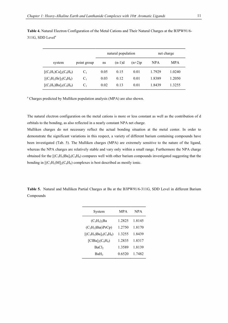
Table 4. Natural Electron Configuration of the Metal Cations and Their Natural Charges at the B3PW91/6-
311G, SDD Levela
natural population net charge
system point group ns (n-1)d (n+2)p NPA MPA
[(C5H5)Ca]2(C8H8) C1 0.05 0.15 0.01 1.7929 1.0240
[(C5H5)Sr]2(C8H8) C1 0.03 0.12 0.01 1.8389 1.2050
[(C5H5)Ba]2(C8H8) C1 0.02 0.13 0.01 1.8439 1.3255
a Charges predicted by Mulliken population analysis (MPA) are also shown.
The natural electron configuration on the metal cations is more or less constant as well as the contribution of d
orbitals to the bonding, as also reflected in a nearly constant NPA net charge.
Mulliken charges do not necessary reflect the actual bonding situation at the metal center. In order to
demonstrate the significant variations in this respect, a variety of different barium containing compounds have
been investigated (Tab. 5). The Mulliken charges (MPA) are extremely sensitive to the nature of the ligand,
whereas the NPA charges are relatively stable and vary only within a small range. Furthermore the NPA charge
obtained for the [(C5H5)Ba]2(C8H8) compares well with other barium compounds investigated suggesting that the
bonding in [(C5H5)M]2(C8H8) complexes is best described as mostly ionic.
Table 5. Natural and Mulliken Partial Charges at Ba at the B3PW91/6-311G, SDD Level in different Barium
Compounds
System MPA NPA
(C5H5)2Ba 1.2825 1.8145
(C5H5)Ba(iPrCp) 1.2750 1.8170
[(C5H5)Ba]2(C8H8) 1.3255 1.8439
[ClBa]2(C8H8) 1.2835 1.8317
BaCl2 1.3589 1.8139
BaH2 0.6520 1.7482
Chapter 1: Heavy-Alkaline Earth and Lanthanide Complexes with 10π Aromatic Ligands
12
Alternative Synthesis for Neutral Triple-Decker Complexes
The synthesis of metallocenes from cyclopentadienyl radicals and metal is probably the most convenient, and
conceptually the simplest synthesis. This synthesis method was realized by the successful synthesis of
decaisopropylmetallocenes [(C5R5)2M] (M= Ca, Sr, Ba 18, Sm, Eu Yb 67; R= CHMe2) from the elements and two
equivalents of the stable pentaisopropylcyclopentadienyl radical. A salt free synthesis was also employed for the
preparation of (1,4-(Me3Si)2C8H6)Yb[Me3CNCHCHNCMe3].68
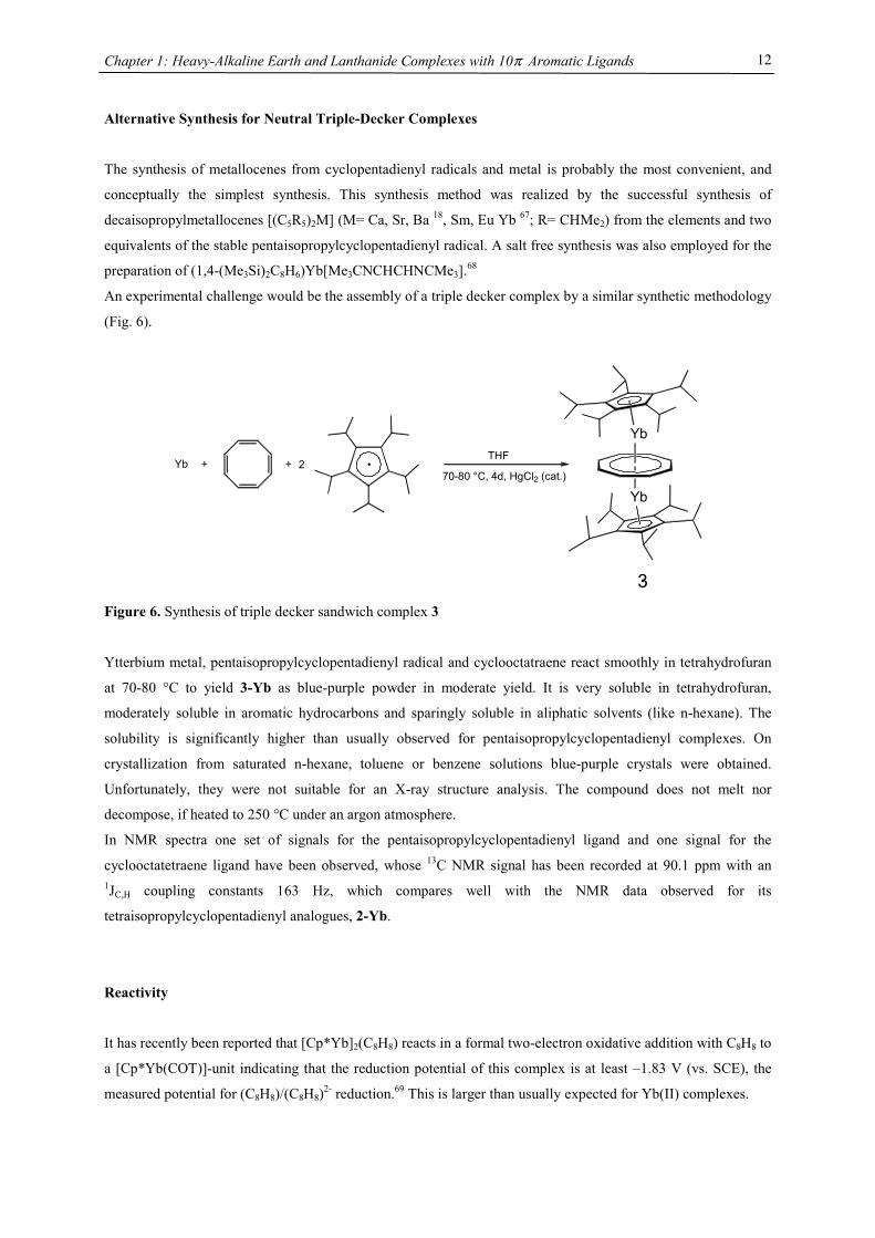
An experimental challenge would be the assembly of a triple decker complex by a similar synthetic methodology
(Fig. 6).
Yb
Yb
3
•
•
Yb + + 2THF
70-80 °C, 4d, HgCl2 (cat.)
Figure 6. Synthesis of triple decker sandwich complex 3
Ytterbium metal, pentaisopropylcyclopentadienyl radical and cyclooctatraene react smoothly in tetrahydrofuran
at 70-80 °C to yield 3-Yb as blue-purple powder in moderate yield. It is very soluble in tetrahydrofuran,
moderately soluble in aromatic hydrocarbons and sparingly soluble in aliphatic solvents (like n-hexane). The
solubility is significantly higher than usually observed for pentaisopropylcyclopentadienyl complexes. On
crystallization from saturated n-hexane, toluene or benzene solutions blue-purple crystals were obtained.
Unfortunately, they were not suitable for an X-ray structure analysis. The compound does not melt nor
decompose, if heated to 250 °C under an argon atmosphere.
In NMR spectra one set of signals for the pentaisopropylcyclopentadienyl ligand and one signal for the
cyclooctatetraene ligand have been observed, whose 13C NMR signal has been recorded at 90.1 ppm with an 1JC,H coupling constants 163 Hz, which compares well with the NMR data observed for its
tetraisopropylcyclopentadienyl analogues, 2-Yb.
Reactivity
It has recently been reported that [Cp*Yb]2(C8H8) reacts in a formal two-electron oxidative addition with C8H8 to
a [Cp*Yb(COT)]-unit indicating that the reduction potential of this complex is at least –1.83 V (vs. SCE), the
measured potential for (C8H8)/(C8H8)2- reduction.69 This is larger than usually expected for Yb(II) complexes.
Chapter 1: Heavy-Alkaline Earth and Lanthanide Complexes with 10π Aromatic Ligands
13
However, complex 3 does not react with C8H8 in benzene at room temperature suggesting that the sterically
encumbered pentaisopropylcyclopentadienyl ligand is able to effectively shield the redox active Yb(II) center
and therefore prevents this oxidative addition process in contrast to the pentamethylcyclopentadienyl ligand.
Cyclononatetraenyl Barium Complexes
Although the cyclononatetraenyl anion (C9H9-, CNT) and its alkali metal salts have been known for many years,
authentic η9-C9H9- containing organometallic compounds are unknown.70-75 The only reported organometallic
complex (C5H5)Ti(C9H9) is best described as (η5-C5H5)Ti((η7-C9H9) based on IR and 1H NMR studies.76 The
synthesis of neutral main group triple-decker complexes employing the C8H82- (COT) ligand, also a 10 π-
electron system,32 spurred the interest in this ligand. To accommodate such a demanding ring system (diameter=
ca. 4.1 Å) a reasonable ionic radius is an absolute requirement and metals of choice are heavy main-group metals
(like Ba, Pb or Bi), lanthanides or actinides. Interestingly, the failed synthesis of (C8H8)Ce(C9H9) resulted in the
development of a new class of cyclooctatetraene dianion containing lanthanide complexes, [(C8H8)LnCl(thf)2]2.77
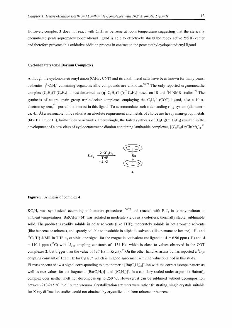
Figure 7. Synthesis of complex 4
KC9H9 was synthesized according to literature procedures 74,75 and reacted with BaI2 in tetrahydrofuran at
ambient temperatures. Ba(C9H9)2 (4) was isolated in moderate yields as a colorless, thermally stable, sublimable
solid. The product is readily soluble in polar solvents (like THF), moderately soluble in hot aromatic solvents
(like benzene or toluene), and sparely soluble to insoluble in aliphatic solvents (like pentane or hexane). 1H- and 13C{1H}-NMR in THF-d8 exhibits one signal for the magnetic equivalent cnt ligand at δ = 6.96 ppm (1H) and δ
= 110.1 ppm (13C) with 1JC,H coupling constants of 151 Hz, which is close to values observed in the COT
complexes 2, but bigger than the value of 137 Hz in K(cnt).75 On the other hand Anastassiou has reported a 1JC,H
coupling constant of 152.5 Hz for C9H9-,73 which is in good agreement with the value obtained in this study.
EI mass spectra show a signal corresponding to a monomeric [Ba(C9H9)2]+-ion with the correct isotope pattern as
well as m/e values for the fragments [Ba(C9H9)]+ and [(C9H9)]+. In a capillary sealed under argon the Ba(cnt)2
complex does neither melt nor decompose up to 250 ºC. However, it can be sublimed without decomposition
between 210-215 ºC in oil pump vacuum. Crystallization attempts were rather frustrating, single crystals suitable
for X-ray diffraction studies could not obtained by crystallization from toluene or benzene.
Ba2 KC9H9THF
- 2 KIBaI2
4
Chapter 1: Heavy-Alkaline Earth and Lanthanide Complexes with 10π Aromatic Ligands
14
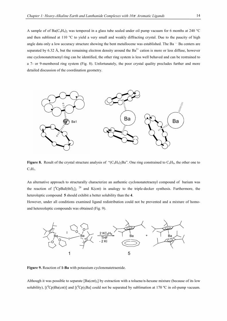
A sample of of Ba(C9H9)2 was tempered in a glass tube sealed under oil pump vacuum for 6 months at 240 °C
and then sublimed at 110 °C to yield a very small and weakly diffracting crystal. Due to the paucity of high
angle data only a low accuracy structure showing the bent metallocene was established. The Ba .... Ba centers are
separated by 6.32 Å, but the remaining electron density around the Ba2+ cation is more or less diffuse, however
one cyclononatetraenyl ring can be identified, the other ring system is less well behaved and can be restrained to
a 7- or 9-membered ring system (Fig. 8). Unfortunately, the poor crystal quality precludes further and more
detailed discussion of the coordination geometry.
Figure 8. Result of the crystal structure analysis of “(C9H9)2Ba”. One ring constrained to C9H9, the other one to
C7H7.
An alternative approach to structurally characterize an authentic cyclononatetraenyl compound of barium was
the reaction of [4CpBaI(thf)2]2 28 and K(cnt) in analogy to the triple-decker synthesis. Furthermore, the
heteroleptic compound 5 should exhibit a better solubility than the 4.
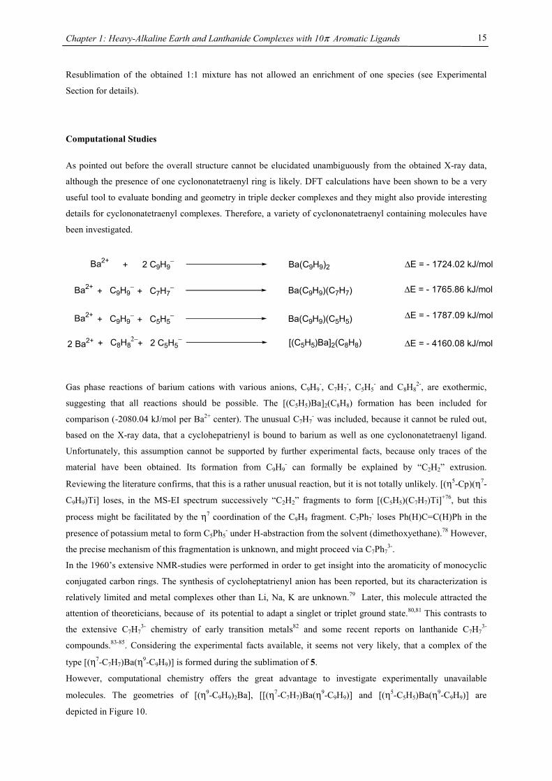
However, under all conditions examined ligand redistribution could not be prevented and a mixture of homo-
and hetereoleptic compounds was obtained (Fig. 9).
BaBa
O O
I
IBa
OO 2 KC9H9THF
- 2 KIBa+
51
Figure 9. Reaction of 1-Ba with potassium cyclononatetraenide.
Although it was possible to separate [Ba(cnt)2] by extraction with a toluene/n-hexane mixture (because of its low
solubility), [(4Cp)Ba(cnt)] and [(4Cp)2Ba] could not be separated by sublimation at 170 ºC in oil-pump vacuum.
Ba Ba
Chapter 1: Heavy-Alkaline Earth and Lanthanide Complexes with 10π Aromatic Ligands
15
Resublimation of the obtained 1:1 mixture has not allowed an enrichment of one species (see Experimental
Section for details).
Computational Studies
As pointed out before the overall structure cannot be elucidated unambiguously from the obtained X-ray data,
although the presence of one cyclononatetraenyl ring is likely. DFT calculations have been shown to be a very
useful tool to evaluate bonding and geometry in triple decker complexes and they might also provide interesting
details for cyclononatetraenyl complexes. Therefore, a variety of cyclononatetraenyl containing molecules have
been investigated.
Ba2+ + 2 C9H9− Ba(C9H9)2
Ba2+ + C9H9− Ba(C9H9)(C7H7)+ C7H7
−
Ba2+ + C9H9− Ba(C9H9)(C5H5)+ C5H5
−
∆E = - 1724.02 kJ/mol
∆E = - 1765.86 kJ/mol
∆E = - 1787.09 kJ/mol
2 Ba2+ + C8H82− [(C5H5)Ba]2(C8H8)+ 2 C5H5
− ∆E = - 4160.08 kJ/mol
Gas phase reactions of barium cations with various anions, C9H9-, C7H7
-, C5H5- and C8H8
2-, are exothermic,
suggesting that all reactions should be possible. The [(C5H5)Ba]2(C8H8) formation has been included for
comparison (-2080.04 kJ/mol per Ba2+ center). The unusual C7H7- was included, because it cannot be ruled out,
based on the X-ray data, that a cyclohepatrienyl is bound to barium as well as one cyclononatetraenyl ligand.
Unfortunately, this assumption cannot be supported by further experimental facts, because only traces of the
material have been obtained. Its formation from C9H9- can formally be explained by “C2H2” extrusion.
Reviewing the literature confirms, that this is a rather unusual reaction, but it is not totally unlikely. [(η5-Cp)(η7-
C9H9)Ti] loses, in the MS-EI spectrum successively “C2H2” fragments to form [(C5H5)(C7H7)Ti]+76, but this
process might be facilitated by the η7 coordination of the C9H9 fragment. C7Ph7- loses Ph(H)C=C(H)Ph in the
presence of potassium metal to form C5Ph5- under H-abstraction from the solvent (dimethoxyethane).78 However,
the precise mechanism of this fragmentation is unknown, and might proceed via C7Ph73-.
In the 1960’s extensive NMR-studies were performed in order to get insight into the aromaticity of monocyclic
conjugated carbon rings. The synthesis of cycloheptatrienyl anion has been reported, but its characterization is
relatively limited and metal complexes other than Li, Na, K are unknown.79 Later, this molecule attracted the
attention of theoreticians, because of its potential to adapt a singlet or triplet ground state.80,81 This contrasts to
the extensive C7H73- chemistry of early transition metals82 and some recent reports on lanthanide C7H7
3-
compounds.83-85. Considering the experimental facts available, it seems not very likely, that a complex of the
type [(η7-C7H7)Ba(η9-C9H9)] is formed during the sublimation of 5.
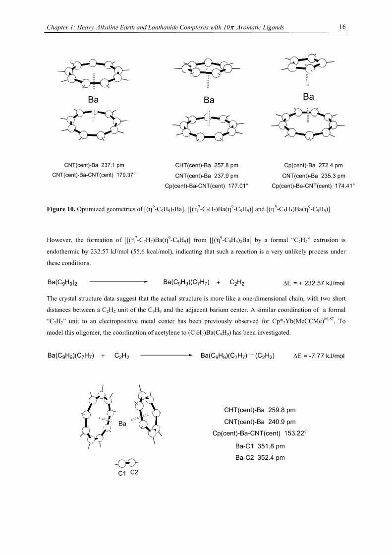
However, computational chemistry offers the great advantage to investigate experimentally unavailable
molecules. The geometries of [(η9-C9H9)2Ba], [[(η7-C7H7)Ba(η9-C9H9)] and [(η5-C5H5)Ba(η9-C9H9)] are
depicted in Figure 10.
Chapter 1: Heavy-Alkaline Earth and Lanthanide Complexes with 10π Aromatic Ligands
16
Figure 10. Optimized geometries of [(η9-C9H9)2Ba], [[(η7-C7H7)Ba(η9-C9H9)] and [(η5-C5H5)Ba(η9-C9H9)] However, the formation of [[(η7-C7H7)Ba(η9-C9H9)] from [[(η9-C9H9)2Ba] by a formal “C2H2” extrusion is
endothermic by 232.57 kJ/mol (55.6 kcal/mol), indicating that such a reaction is a very unlikely process under
these conditions.
Ba(C9H9)(C7H7)Ba(C9H9)2 + C2H2 ∆E = + 232.57 kJ/mol
The crystal structure data suggest that the actual structure is more like a one-dimensional chain, with two short
distances between a C2H2 unit of the C9H9 and the adjacent barium center. A similar coordination of a formal
“C2H2” unit to an electropositive metal center has been previously observed for Cp*2Yb(MeCCMe)86,87. To
model this oligomer, the coordination of acetylene to (C7H7)Ba(C9H9) has been investigated.
∆E = -7.77 kJ/molBa(C9H9)(C7H7) + C2H2 Ba(C9H9)(C7H7) ... (C2H2)
Ba Ba Ba
CNT(cent)-Ba 237.1 pm
CNT(cent)-Ba-CNT(cent) 179.37° CHT(cent)-Ba 257.8 pm
CNT(cent)-Ba 237.9 pm
Cp(cent)-Ba-CNT(cent) 177.01°
Cp(cent)-Ba 272.4 pm
CNT(cent)-Ba 235.3 pm
Cp(cent)-Ba-CNT(cent) 174.41°
Ba
C1 C2
CHT(cent)-Ba 259.8 pm
CNT(cent)-Ba 240.9 pm
Cp(cent)-Ba-CNT(cent) 153.22°
Ba-C1 351.8 pm
Ba-C2 352.4 pm
Chapter 1: Heavy-Alkaline Earth and Lanthanide Complexes with 10π Aromatic Ligands
17
Ba(C9H9)(C7H7) is able to bind C2H2 very weakly (∆E= -7.77 kJ/mol). Most significantly, the bending angle is
decreased by 23° suggesting, that the potential energy curve is extremely flat for the bending motion.
However, such a weak interaction cannot be driving force for a “C2H2” extrusion as required for the formation of
Ba(C9H9)(C7H7).
Conclusions
Tetraisopropylcyclopentadienyl halides of the heavy alkaline earth metals are suitable starting compounds for
nucleophilic substitution reactions. With a ring as large as the cyclooctatetraene dianion coming close to the
dications of calcium, strontium, or barium, steric bulk at least comparable to that of a
tetraisopropylcyclopentadienyl ligand can be achieved without bulky substituents, bending of 4Cpcent-M-COTcent
angles can effectively be restricted to values close to linearity and thermally stable structures can be established.
In extension of earlier studies by Sitzmann et al. the concept of cyclopentadienyl radicals as synthons in the
metallocene synthesis has been successfully transferred to triple decker systems as demonstrated in the synthesis
of [(C5(CHMe2)5)Yb(µ,η8:η8-C8H8)Yb(C5(CHMe2)5)], 3. The synthetic approaches used in this work may be
generally useful for the synthesis of neutral triple decker sandwich compounds with main group central atoms.
Although the reaction of [4CpBaI(thf)2]2 with KC9H9 failed to produce pure, mixed barocenes,
bis(cyclononatetraenyl)barium, Ba(C9H9)2, can be synthesized in moderate yield as thermally stable, sublimable
material.
References
[1] Beckmann, E. Chem. Ber. 1905, 38, 904-906.
[2] Gilman, H.; F., S. J. Am. Chem. Soc. 1926, 48, 2463-2467.
[3] Zerger, R.; G., S. J. Organomet. Chem. 1974, 80, 7-17.
[4] Ziegler, K.; Froitzheim-Kühlhorn, H.; Hafner, H. Chem. Ber. 1956, 89, 434-443.
[5] Eaborn, C.; Hawkes, S. A.; Hitchcock, P. B.; Smith, J. D. J. Chem. Soc., Chem. Commun. 1997, 1961-
1962.
[6] Cloke, F. G. N.; Hitchcock, P. B.; Lappert, M. F.; Lawless, G. A.; Royo, B. J. Chem. Soc., Chem.
Commun. 1997, 724.
[7] Harvey, M. J.; Hanusa, T. P.; Young, V. G. Angew. Chem. 1999, 111, 241-242.
[8] Feil, F.; Harder, S. Organometallics 2000, 19, 5010-5015.
[9] Harder, S.; Feil, F.; Weeber, A. Organometallics 2001, 20, 1044-1046.
[10] Overby, J. S.; Hanusa, T. P. Angew. Chem. Int. Ed. Engl. 1994, 33, 2191-2193.
[11] Williams, R. A.; Hanusa, T. P.; Huffmann, J. C. Organometallics 1990, 9, 1128-1134.
[12] Andersen, R. A.; Blom, R.; Boncella, J. M.; Burns, C. J.; Volden, H. V. Acta Chim. Scand. A 1987, 41,
24-35.
[13] McCormick, M. J.; Sockwell, S. C.; Davies, C. E. H.; Hanusa, T. P.; Huffman, J. C. Organometallics
1989, 8, 2044-2049.
Chapter 1: Heavy-Alkaline Earth and Lanthanide Complexes with 10π Aromatic Ligands
18
[14] Harvey, M. J.; Quisenberry, K. T.; Hanusa, T. P.; Young, V. G. Eur. J. Inorg. Chem. 2003, 3383-3390.
[15] Williams, R. A.; Tesh, K. F.; Hanusa, T. P. J. Am. Chem. Soc. 1991, 113, 4843-4851.
[16] Harvey, M. J.; Hanusa, T. P.; Young, V. G. J. Organomet. Chem. 2001, 626, 43-48.
[17] Weber, F.; Sitzmann, H.; Schultz, M.; Sofield, C. D.; Andersen, R. A. Organometallics 2002, 21, 3139-
3146.
[18] Sitzmann, H.; Dezember, T.; Ruck, M. Angew. Chem. Int. Ed. Engl. 1998, 37, 3114-3116.
[19] Feil, F.; Harder, S. Eur. J. Inorg. Chem. 2003, 3401-3408.
[20] Harder, S. Angew. Chem. Int. Ed. Engl. 2004, 43, 2714-2718.
[21] Weeber, A.; Harder, S.; Brintzinger, H. H.; Knoll, K. Organometallics 2000, 19, 1325-1332.
[22] Wojtczak, W. A.; Fleig, P. F.; Hampden-Smith, M. J. Adv. Organomet. Chem. 1996, 40, 215-340.
[23] Hatanpää, T.; Vehkamäki, M.; Mutikainen, I.; Kansikas, J.; Ritala, M.; Leskelä, M. Dalton Trans. 2004,
1181-1188.
[24] Harvey, M. J.; Hanusa, T. P. Organometallics 2000, 19, 1556-1566.
[25] Harvey, M. J.; Hanusa, T. P.; Pink, M. Chem. Commun. 2000, 489-490.
[26] Burkey, D. J.; Hansua, T. P. Organometallics 1996, 15, 4971-4976.
[27] Sockwell, S. C.; Hanusa, T. P.; Huffman, J. C. J. Am. Chem. Soc. 1992, 114, 3393-3399.
[28] Sitzmann, H.; Weber, F.; Walter, M. D.; Wolmershäuser, G. Organometallics 2003, 22, 1931-1936.
[29] Hutchings, D. S.; Junk, P. C.; Patalinghug, W. C.; Raston, C. L.; White, A. H. J. Chem. Soc., Chem.
Commun. 1989, 973-974.
[30] Edelmann, F. T. New J. Chem. 1995, 19, 535-550.
[31] Burkey, D. J.; Alexander, E. K.; Hansua, T. P. Organometallics 1994, 13, 2773-2786.
[32] Sitzmann, H.; Walter, M. D.; Wolmershäuser, G. Angew. Chem. Int. Ed. Engl. 2002, 41, 2315-2316.
[33] Evans, W. J.; Johnston, M. A.; Clark, R. D.; Anwander, R.; Ziller, J. W. Polyhedron 2001, 20, 2483-
2490.
[34] Wayda, A. L.; Mukerji, I.; Dye, J. L.; Rogers, R. D. Organometallics 1987, 6, 1328-1332.
[35] Kinsley, S. A.; Streitwieser, A.; Zalkin, A. Organometallics 1985, 4, 52-57.
[36] Evans, W. J.; Johnston, M. A.; Greci, M. A.; Ziller, J. W. Organometallics 1999, 18, 1460-1464.
[37] Cowley, A. H.; MacDonald, C. L. B.; Silverman, J. S.; Gorden, J. D.; Voigt, A. J. Chem. Soc., Chem.
Commun. 2001, 175-176.
[38] Armstrong, D. R.; Edwards, A. J.; Moncrieff, D.; Paver, M. A.; Raithby, P. R.; Rennie, M.-A.; Russel,
C. A.; Wright, D. S. J. Chem. Soc., Chem. Commun. 1989, 927.
[39] Harder, S.; Prosenc, M. H. Angew. Chem. Int. Ed. Engl. 1996, 35, 97-99.
[40] Blom, R.; Faegari, K.; Volden, H. V. Organometallics 1990, 9, 372-379.
[41] Hanusa, T. P. Chem. Rev. 1993, 93q, 1023-1036.
[42] Schultz, M.; Burns, C. J.; Schwartz, D. J.; Andersen, R. A. Organometallics 2000, 19, 781-789.
[43] Evans, W. J.; Hughes, L. A.; Hanusa, T. P. Organometallics 1986, 5, 1285-1296.
[44] Green, J. C.; Hohl, D.; Rösch, N. Organometallics 1987, 6, 712-720.
[45] DeKock, R. L.; Peterson, M. A.; Timmer, L. K. Polyhedron 1990, 9, 1919-1934.
[46] Green, J. C. Chem. Soc. Rev. 1998, 27, 263-271.
[47] Bridgeman, A. J. J. Chem. Soc., Dalton Trans. 1997, 2887-2893.
[48] Guido, M.; Gigli, G. J. Chem. Phys. 1976, 65, 1397-1402.
Chapter 1: Heavy-Alkaline Earth and Lanthanide Complexes with 10π Aromatic Ligands
19
[49] Hollis, T. K.; Burdett, J. K.; Bosnich, B. Organometallics 1993, 12, 3385-3386.
[50] Timofeeva, T. V.; Lii, J.-H.; Allinger, N. L. J. Am. Chem. Soc. 1995, 117, 7452-7459.
[51] Kaupp, M.; Schleyer, P. v. R.; Dolg, M.; Stoll, H. J. Am. Chem. Soc. 1992, 114, 8202-8208.
[52] Raymond, K. N.; Eigenbrot, C. W. Acc. Chem. Res. 1980, 13, 276-283.
[53] Sockwell, S. C.; Hanusa, T. P. Inorg. Chem. 1990, 29, 76-80.
[54] Evans, W. J.; Johnston, M. A.; Clark, R. D.; Ziller, J. W. J. Chem. Soc., Dalton Trans. 2000, 1609-
1612.
[55] Evans, W. J.; Clark, R. D.; Ansari, M. A.; Ziller, J. W. J. Am. Chem. Soc. 1998, 120, 9555-9563.
[56] Gagliardi, L.; Pyykkö, P. Theor. Chem. Acc. 2003, 110, 205-210.
[57] Parr, R. G.; Yang, W. Density Functional Theory of Atoms and Molecules; Oxford University Press:
Oxford, 1989.
[58] Labanowski, J. K.; Andelm, J. W., Density Functional Methods in Chemistry. ed.; Springer: New York,
1991.
[59] Ziegler, T. Chem. Rev. 1991, 91, 651-667.
[60] Smith, J. D.; Hanusa, T. P. Organometallics 2001, 20, 3056-3062.
[61] Cotton, F. A.; Cowley, A. H.; Feng, X. J. Am. Chem. Soc. 1998, 120, 1795-1799.
[62] Maron, L.; Werkema, E. L.; Perrin, L.; Eisenstein, O.; Andersen, R. A. J. Am. Chem. Soc. 2005, 127,
279-292.
[63] Perrin, L.; Maron, L.; Eisenstein, O. Inorg. Chem. 2002, 41, 4355-4362.
[64] Maron, L.; Eisenstein, O.; Alary, F.; Poteau, R. J. Phys. Chem. A 2002, 106, 1797-1801.
[65] Reed, A. E.; Curtiss, L. A.; Weinhold, F. Chem. Rev. 1988, 88, 899-926.
[66] Reed, A. E.; Weinstock, R. B.; Weinhold, F. J. Chem. Phys. 1985, 83, 735-.
[67] Sitzmann, H.; Dezember, T.; Schmitt, O.; Weber, F.; Wolmershäuser, G. Z. Anorg. Allgem. Chem.
2000, 626, 2241-2244.
[68] Poremba, P.; Edelmann, F. T. J. Organomet. Chem. 1997, 549, 101-104.
[69] Evans, W. J.; Gonzales, S. L.; Ziller, J. W. J. Am. Chem. Soc. 1994, 116, 2600-.
[70] Boche, G.; Weber, H.; Bieberbach, A. Chem. Ber. 1978, 111, 2833-2849.
[71] Boche, G.; Weber, H.; Martens, D.; Bieberbach, A. Chem. Ber. 1978, 111, 2480-2496.
[72] Boche, G.; Bieberbach, A. Chem. Ber. 1978, 111, 2850-2858.
[73] Anastassiou, A. G.; Reichmanis, E. J. Am. Chem. Soc. 1976, 98, 8266-8267.
[74] Katz, T. J.; Garratt, P. J. J. Am. Chem. Soc. 1964, 86, 5194-5202.
[75] Lancette, E. A.; Benson, R. E. J. Am. Chem. Soc. 1965, 87, 1941-1946.
[76] Verkouw, H. T.; Veldman, M. E. E.; Groenenboom, C. J.; van Oven, H. O.; de Liefde Meijer, H. J. J.
Organomet. Chem. 1975, 102, 49-56.
[77] Mares, F.; Hodgson, K. O.; Streitwieser, A. J. Organomet. Chem. 1971, 25, C24-C26.
[78] Breslow, R.; Chang, H. W. J. Am. Chem. Soc. 1965, 87, 2200-2203.
[79] Dauben, H. J.; Rifi, M. R. J. Am. Chem. Soc. 1963, 85, 3041-3043.
[80] Gogonea, V.; Schleyer, P. v. R.; Schreiner, P. R. Angew. Chem. 1998, 110.
[81] Wiberg, K. B. Chem. Rev. 2001, 101, 1317-1331.
[82] Green, M. L. H.; Ng, D. K. P. Chem. Rev. 1995, 95, 439-473.
[83] Li, J.; Bursten, B. E. J. Am. Chem. Soc. 1997, 119, 9021-9032.
Chapter 1: Heavy-Alkaline Earth and Lanthanide Complexes with 10π Aromatic Ligands
20
[84] Arligne, T.; Lance, M.; Nierlich, M.; Ephritikhine, M. J. Chem. Soc., Dalton Trans. 1997, 2501-2504.
[85] Arligne, T.; Lance, M.; Nierlich, M.; Vigner, J.; Ephritikhine, M. J. Chem. Soc., Chem. Commun. 1994,
847-848.
[86] Perrin, L.; Maron, L.; Eisenstein, O.; Schwartz, D. J.; Burns, C. J.; Andersen, R. A. Organometallics
2003, 22, 5447-5453.
[87] Burns, C. J.; Andersen, R. A. J. Am. Chem. Soc. 1987, 109, 941-942.
Chapter 2: Manganocenes
21
Chapter 2: The Effect of Substituted Cyclopentadienyl Ligands on the Electronic
Structure of Manganocenes
Introduction
Molecular switching in inorganic solids closely relates to the vibronic lability of molecular units, previously
introduced by J.A. Ammeter.1 A recent presentation of the underlying concept of “molecular bistablity” was
given by O. Kahn,2-5 on the example of spin-crossover (SCO) solids. The bistable properties at the molecular
level are adequately described through a molecular configurational diagram, i.e. a plot of the adiabatic energies
vs. the distortion coordinate of the molecular system (Fig.1). Usually, for SCO, a fully symmetric distortion is
considered, associated with a change in average metal-ligand distance. Due to the large atomic displacements
upon spin conversion, the physical properties of such a complex change dramatically. This crossover can be
followed by several methods as there are e.g. mössbauer, EPR, IR, UV/Vis spectroscopy, NMR, magnetic
susceptibility, crystal structure analysis and EXAFS investigations. In Fig. 1, the configurational diagram suited
to a SCO, in the case of the low-spin (LS) ground state is presented. Extensive studies by Ammeter confirmed
that the environment in molecular solids affect the configurational diagram by changing the E(HS) – E(LS) gap.6
This correlatively affects the energy barrier of the low-spin-high-spin transition, changes the equilibrium
temperature T1/2 and the lifetime of the HS state.
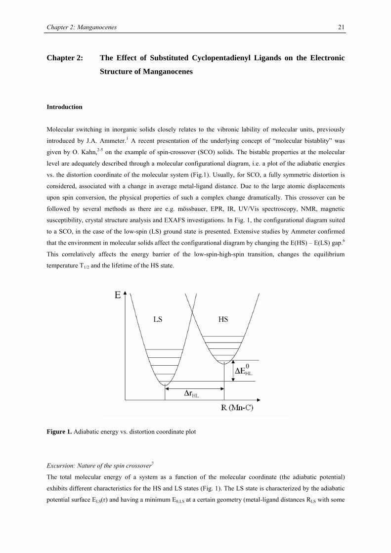
Figure 1. Adiabatic energy vs. distortion coordinate plot
Excursion: Nature of the spin crossover7
The total molecular energy of a system as a function of the molecular coordinate (the adiabatic potential)
exhibits different characteristics for the HS and LS states (Fig. 1). The LS state is characterized by the adiabatic
potential surface ELS(r) and having a minimum E0,LS at a certain geometry (metal-ligand distances RLS with some
Chapter 2: Manganocenes
22
curvature (the second derivation representing the force constant fLS)). Within this parabolic function
approximation a set of vibrational levels ei,LS occurs, the lowest one corresponding to the zero-point vibration,
e0,LS. The high-spin state differs in these characteristics in such a way that the relationships
E0,LS < E0,HS
RLS < RHS
fLS > fHS
are obeyed. Thus the enthalpy change for a LS-HS transition is
∆H0LH = (E0,HS + e0,HS) – (E0,LS + e0,LS) > 0
The entropy change is expressed through the thermodynamic probability
∆S0LH = k ln(gHS) – k ln(gLS) = k ln(gHS/gLS) > 0
Where the gHS (gLS) stands for the degeneracy of the HS (LS) state; thus ∆S0LS is always positive. Then the Gibbs
enthalpy change
∆GLH = ∆H0LH – T ∆S0
LH
Just at the transition temperature, T1/2, where xLS=xHS= 0.5, the following equation holds true
∆GLH(T1/2) = 0 → T1/2= ∆H0LH/∆S0
LH
The driving force for the LS to HS transition is the drop of the Gibbs energy to a negative value above T1/2.
Therefore it depends upon the actual values of enthalpy and entropy whether the spin transition is observable or
not.
Motivation
Spin-state conversions involving Fe(II), Fe(III) and Mn(III) have been extensively studied over the last two
decades,8,9 mainly because of the biological significance of these ions. Manganese (involved in photosystem II)10
attracted attention due to its broad range of stable or at least meta-stable oxidation states (+II � + VII), its very
labile metal-ligand binding and its tendency towards formation of high-spin states. With these properties
manganese is unique among the first row transition elements.
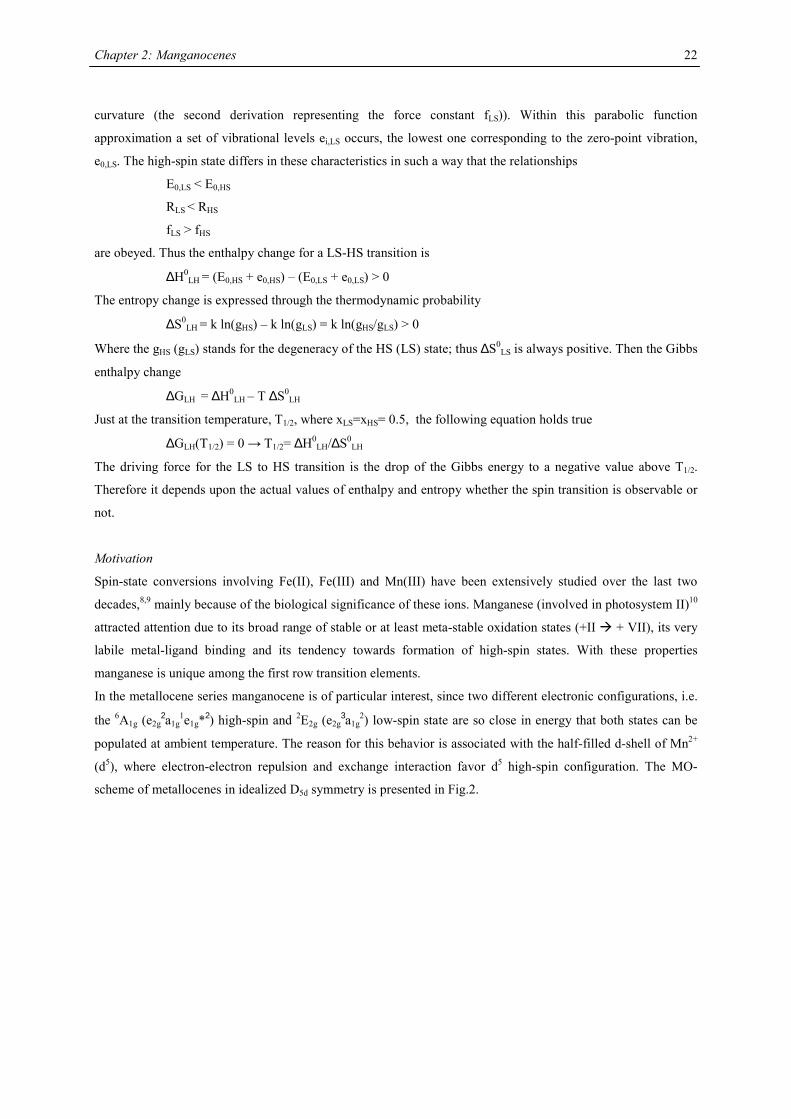
In the metallocene series manganocene is of particular interest, since two different electronic configurations, i.e.
the 6A1g (e2g2a1g
1e1g*2) high-spin and 2E2g (e2g3a1g
2) low-spin state are so close in energy that both states can be
populated at ambient temperature. The reason for this behavior is associated with the half-filled d-shell of Mn2+
(d5), where electron-electron repulsion and exchange interaction favor d5 high-spin configuration. The MO-
scheme of metallocenes in idealized D5d symmetry is presented in Fig.2.
Chapter 2: Manganocenes
23
Mn
e2g
e2u
e1g
e1u
a2u
a1g
e1ge2ga1g
a1g
a2ue1u
Mn2+
a1g
a2u
e1g
e1u
e2g
a1g
e1g*
e2u
a1g*
e2g*
a2u*e1u*
3d
4s
4p
Figure 2. Idealized D5d orbital diagram of manganocene.11
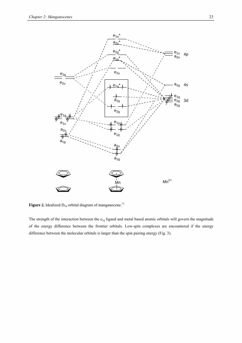
The strength of the interaction between the e1g ligand and metal based atomic orbitals will govern the magnitude
of the energy difference between the frontier orbitals. Low-spin complexes are encountered if the energy
difference between the molecular orbitals is larger than the spin pairing energy (Fig. 3).
Chapter 2: Manganocenes
24
∆∆
e2g
a1g
e1g*
weak orbital overlap
e2g (δ)
a1g (σ)
e1g* (π)
strong orbital overlap
∆ < P ∆ > P
Figure 3: Frontier orbital scheme for d5-metallocene with idealized D5d symmetry
The magnetism of substituted manganocenes provides a brilliant example how subtle changes in the ligand
environment effect the electronic configuration. Substituted cyclopentadienyl ligands have been employed to
realize high-spin and low-spin configurations as well as spin-equilibria.12,13 A variety of techniques have been
used to evaluate the underlying thermodynamics of these spin-state equilibria.
Molecular Magnetism and Formula
The susceptibility of a material to the presence of a magnetic field, χ, is the scalar ratio of the magnetization and
the magnitude of the magnetic field,
χ= M/H
The susceptibility χ is the algebraic sum of two contributions associated with different phenomena:
χ = χdia +χpara
where χdia and χpara represent the diamagnetic and paramagnetic susceptibilities, respectively. The former is
negative and the latter positive. Where χdia dominates the sample is called diamagnetic, and it is repelled by the
magnetic field. When χpara is the dominating contribution the sample is said to be paramagnetic, and it is
attracted by the field. The diamagnetism is an underlying property of matter and originates from the interaction
of the magnetic field with the motions of the electrons in their orbitals. The diamagnetic susceptibility, χdia, is
independent of temperature and applied magnetic field.
The paramagnetism is generated by the tendency of the magnetic angular moment to orient itself in the magnetic
field and its usually 1 to 3 orders of magnitude larger than the diamagnetism. Pierre Curie established
empirically that the magnetic susceptibility of a sample is inversely proportional to the temperature:
χ = C/T
Theoretical calculations proved that the Curie constant, C, for an orbitally quenched ion is dependent on the
number of unpaired electrons and the g value of the compound. The Curie constant for the spin only magnetic
susceptibility is:
kSSNg
C B
3)1(22 +
=µ
Chapter 2: Manganocenes
25
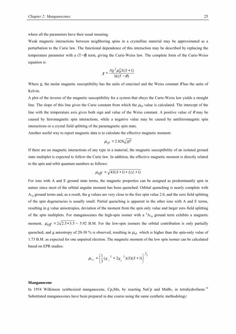
where all the parameters have their usual meaning.
Weak magnetic interactions between neighboring spins in a crystalline material may be approximated as a
perturbation to the Curie law. The functional dependence of this interaction may be described by replacing the
temperature parameter with a (T−θ) term, giving the Curie-Weiss law. The complete form of the Curie-Weiss
equation is:
)(3)1(22
θµχ
−+=
TkSSNg B
Where χ, the molar magnetic susceptibility has the units of emu/mol and the Weiss constant θ has the units of
Kelvin.
A plot of the inverse of the magnetic susceptibility for a system that obeys the Curie-Weiss law yields a straight
line. The slope of this line gives the Curie constant from which the µeff value is calculated. The intercept of the
line with the temperature axis gives both sign and value of the Weiss constant. A positive value of θ may be
caused by ferromagnetic spin interactions, while a negative value may be caused by antiferromagntic spin
interactions or a crystal field splitting of the paramagnetic spin state.
Another useful way to report magnetic data is to calculate the effective magnetic moment:
Teff χµ 828.2=
If there are no magnetic interactions of any type in a material, the magnetic susceptibility of an isolated ground
state multiplet is expected to follow the Curie law. In addition, the effective magnetic moment is directly related
to the spin and orbit quantum numbers as follows:
)1()1(4 +++= LLSSeffµ
For ions with A and E ground state terms, the magnetic properties can be assigned as predominantly spin in
nature since most of the orbital angular moment has been quenched. Orbital quenching is nearly complete with
A1g ground terms and, as a result, the g values are very close to the free spin value 2.0, and the zero field splitting
of the spin degeneracies is usually small. Partial quenching is apparent in the other ions with A and E terms,
resulting in g value anisotropies, deviation of the moment from the spin only value and larger zero field splitting
of the spin multiplets. For manganocenes the high-spin isomer with a 6A1g ground term exhibits a magnetic
moment, 5.35.22 ×=effµ = 5.92 B.M. For the low-spin isomers the orbital contribution is only partially
quenched, and g anisotropy of 20-30 % is observed, resulting in µoff which is higher than the spin-only value of
1.73 B.M. as expected for one unpaired electron. The magnetic moment of the low spin isomer can be calculated
based on EPR studies:
21
22.. )1)()(2(
31
++=
⊥SSggslµ
Manganocene
In 1954 Wilkinson synthesized manganocene, Cp2Mn, by reacting NaCp and MnBr2 in tetrahydrofuran.14
Substituted manganocenes have been prepared in due course using the same synthetic methodology:
Chapter 2: Manganocenes
26
+- 2 MX
Cp'2Mn2 MCp' MnX2
M= Li, Na, K; X= Cl, Br
THF
Anhydrous manganese salts are insoluble in most aprotic solvents, but sufficiently soluble in tetrahydrofuran to
achieve reasonable rates of conversion. The tetrahydrofuran adducts of MnI215,16 and MnCl2
17,18 have been
described, and possess increased kinetic solubility, and can be purified on crystallization from tetrahydrofuran.
In recent years a variety of substituted manganocenes have been synthesized and characterized. The most notable
contributions in this field were done by Wilkinson,14,19,20 Robbins,21 Ammeter,6,22,23 Köhler24-26, Hanusa27 and
Sitzmann28. In each case the general methodology of Wilkinson has been used and the products have been
purified by sublimation, crystallization or distillation.
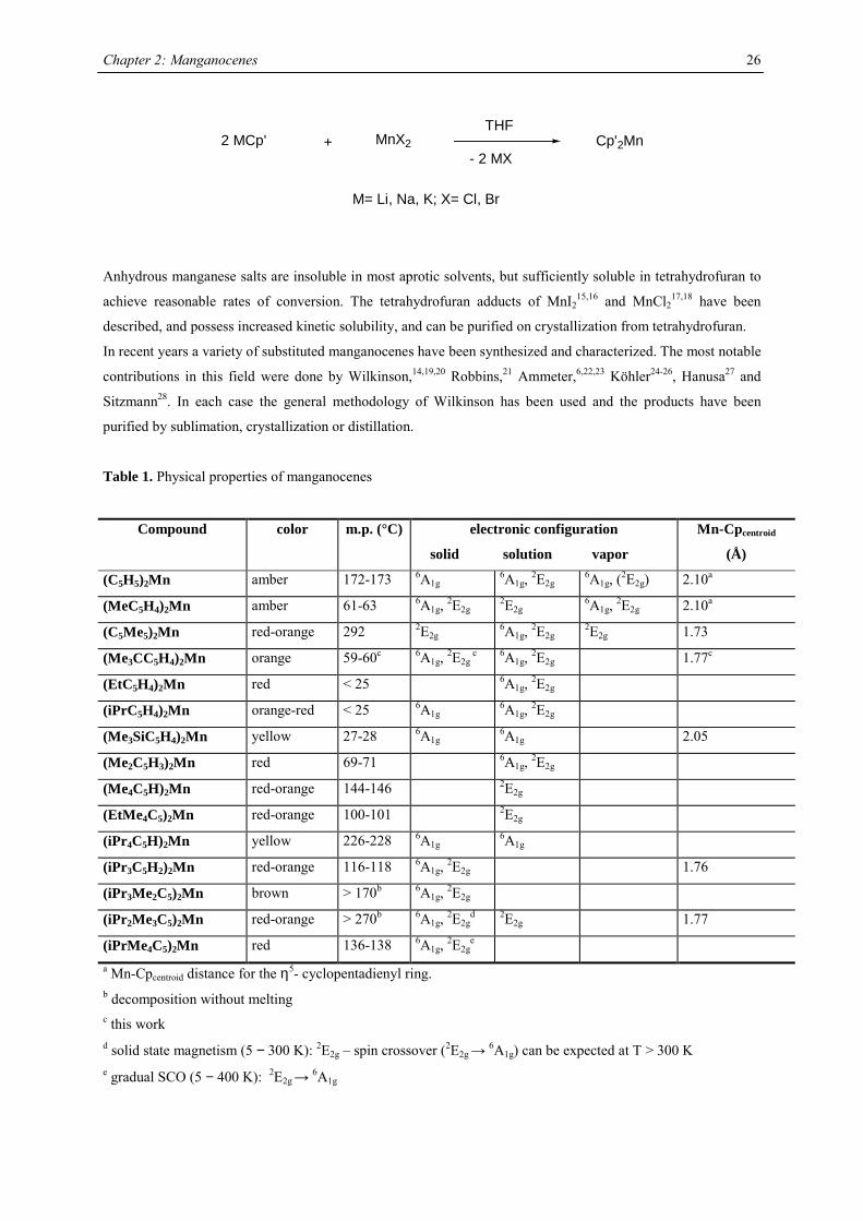
Table 1. Physical properties of manganocenes
Compound color m.p. (°C) electronic configuration
solid solution vapor
Mn-Cpcentroid
(Å)
(C5H5)2Mn amber 172-173 6A1g 6A1g, 2E2g 6A1g, (2E2g) 2.10a
(MeC5H4)2Mn amber 61-63 6A1g, 2E2g 2E2g 6A1g, 2E2g 2.10a
(C5Me5)2Mn red-orange 292 2E2g 6A1g, 2E2g 2E2g 1.73
(Me3CC5H4)2Mn orange 59-60c 6A1g, 2E2g c 6A1g, 2E2g 1.77c
(EtC5H4)2Mn red < 25 6A1g, 2E2g
(iPrC5H4)2Mn orange-red < 25 6A1g 6A1g, 2E2g
(Me3SiC5H4)2Mn yellow 27-28 6A1g 6A1g 2.05
(Me2C5H3)2Mn red 69-71 6A1g, 2E2g
(Me4C5H)2Mn red-orange 144-146 2E2g
(EtMe4C5)2Mn red-orange 100-101 2E2g
(iPr4C5H)2Mn yellow 226-228 6A1g 6A1g
(iPr3C5H2)2Mn red-orange 116-118 6A1g, 2E2g 1.76
(iPr3Me2C5)2Mn brown > 170b 6A1g, 2E2g
(iPr2Me3C5)2Mn red-orange > 270b 6A1g, 2E2gd 2E2g 1.77
(iPrMe4C5)2Mn red 136-138 6A1g, 2E2ge
a Mn-Cpcentroid distance for the η5- cyclopentadienyl ring. b decomposition without melting c this work d solid state magnetism (5 − 300 K): 2E2g – spin crossover (2E2g
→ 6A1g) can be expected at T > 300 K e gradual SCO (5 − 400 K): 2E2g
→ 6A1g
Chapter 2: Manganocenes
27
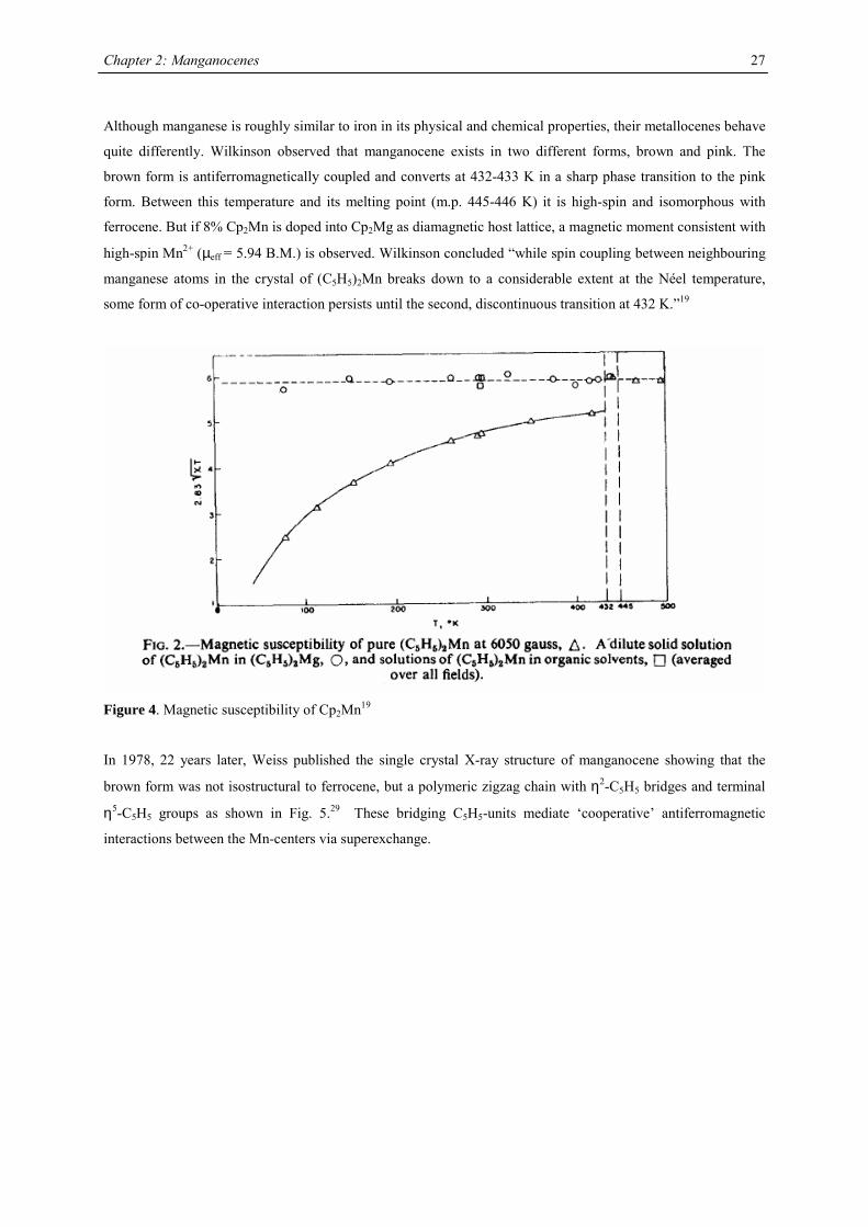
Although manganese is roughly similar to iron in its physical and chemical properties, their metallocenes behave
quite differently. Wilkinson observed that manganocene exists in two different forms, brown and pink. The
brown form is antiferromagnetically coupled and converts at 432-433 K in a sharp phase transition to the pink
form. Between this temperature and its melting point (m.p. 445-446 K) it is high-spin and isomorphous with
ferrocene. But if 8% Cp2Mn is doped into Cp2Mg as diamagnetic host lattice, a magnetic moment consistent with
high-spin Mn2+ (µeff = 5.94 B.M.) is observed. Wilkinson concluded “while spin coupling between neighbouring
manganese atoms in the crystal of (C5H5)2Mn breaks down to a considerable extent at the Néel temperature,
some form of co-operative interaction persists until the second, discontinuous transition at 432 K.”19
Figure 4. Magnetic susceptibility of Cp2Mn19
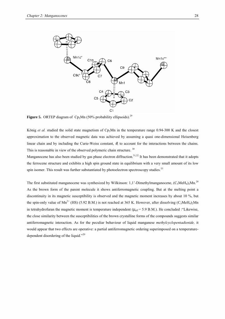
In 1978, 22 years later, Weiss published the single crystal X-ray structure of manganocene showing that the
brown form was not isostructural to ferrocene, but a polymeric zigzag chain with η2-C5H5 bridges and terminal
η5-C5H5 groups as shown in Fig. 5.29 These bridging C5H5-units mediate ‘cooperative’ antiferromagnetic
interactions between the Mn-centers via superexchange.
Chapter 2: Manganocenes
28
Figure 5. ORTEP diagram of Cp2Mn (50% probability ellipsoids).29
König et al. studied the solid state magnetism of Cp2Mn in the temperature range 0.94-300 K and the closest
approximation to the observed magnetic data was achieved by assuming a quasi one-dimensional Heisenberg
linear chain and by including the Curie-Weiss constant, θ, to account for the interactions between the chains.
This is reasonable in view of the observed polymeric chain structure. 30
Manganocene has also been studied by gas phase electron diffraction.31,32 It has been demonstrated that it adopts
the ferrocene structure and exhibits a high spin ground state in equilibrium with a very small amount of its low
spin isomer. This result was further substantiated by photoelectron spectroscopy studies.33
The first substituted manganocene was synthesized by Wilkinson: 1,1’-Dimethylmanganocene, (C5MeH4)2Mn.20
As the brown form of the parent molecule it shows antiferromagnetic coupling. But at the melting point a
discontinuity in its magnetic susceptibility is observed and the magnetic moment increases by about 10 %, but
the spin-only value of Mn2+ (HS) (5.92 B.M.) is not reached at 365 K. However, after dissolving (C5MeH4)2Mn
in tetrahydrofuran the magnetic moment is temperature independent (µeff = 5.9 B.M.). He concluded :“Likewise,
the close similarity between the susceptibilities of the brown crystalline forms of the compounds suggests similar
antiferromagnetic interaction. As for the peculiar behaviour of liquid manganese methylcyclopentadienide, it
would appear that two effects are operative: a partial antiferromagnetic ordering superimposed on a temperature-
dependent disordering of the liquid.”20
Chapter 2: Manganocenes
29
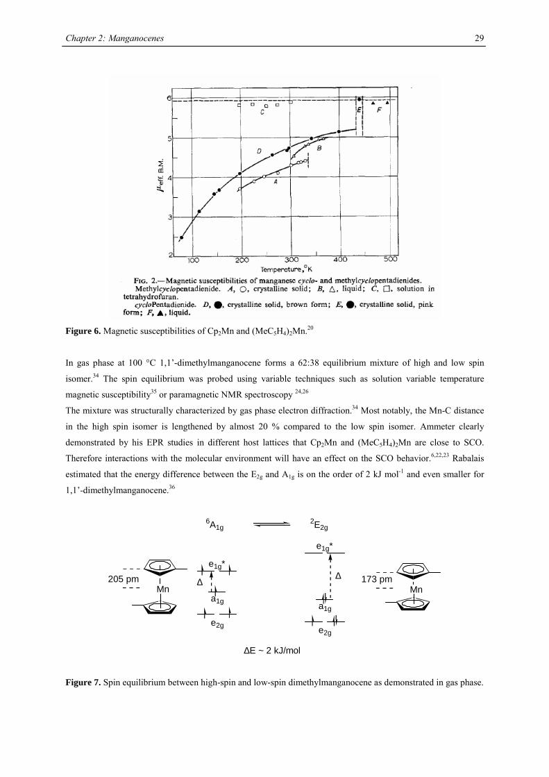
Figure 6. Magnetic susceptibilities of Cp2Mn and (MeC5H4)2Mn.20
In gas phase at 100 °C 1,1’-dimethylmanganocene forms a 62:38 equilibrium mixture of high and low spin
isomer.34 The spin equilibrium was probed using variable techniques such as solution variable temperature
magnetic susceptibility35 or paramagnetic NMR spectroscopy 24,26
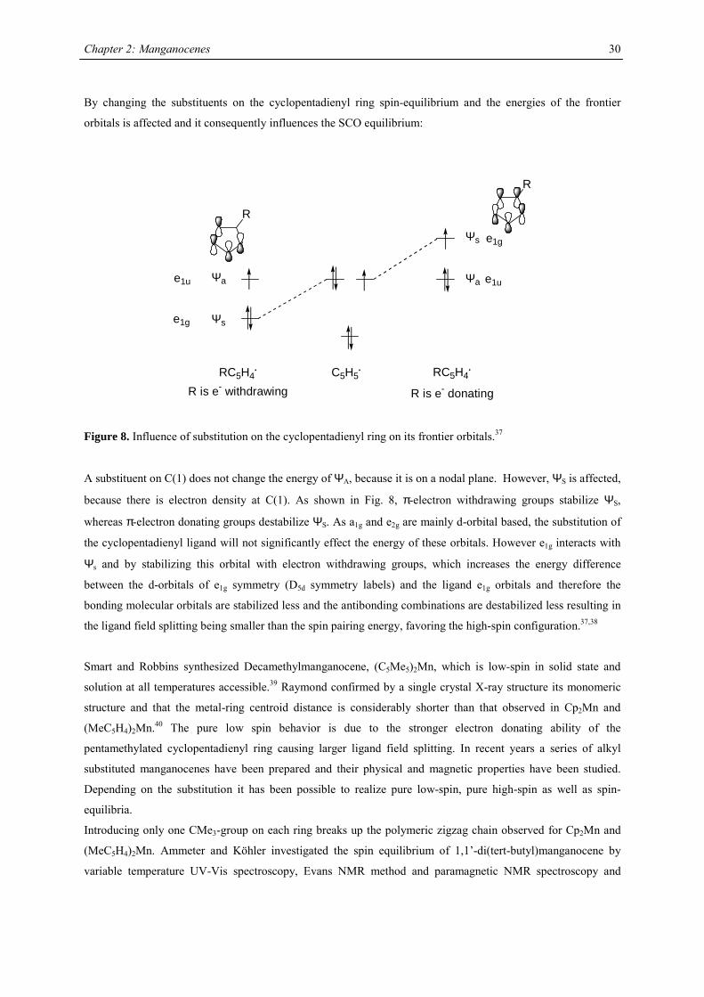
The mixture was structurally characterized by gas phase electron diffraction.34 Most notably, the Mn-C distance
in the high spin isomer is lengthened by almost 20 % compared to the low spin isomer. Ammeter clearly
demonstrated by his EPR studies in different host lattices that Cp2Mn and (MeC5H4)2Mn are close to SCO.
Therefore interactions with the molecular environment will have an effect on the SCO behavior.6,22,23 Rabalais
estimated that the energy difference between the E2g and A1g is on the order of 2 kJ mol-1 and even smaller for
1,1’-dimethylmanganocene.36
∆∆
e2g
a1g
e1g*
e2g
a1g
e1g*
∆E ~ 2 kJ/mol
6A1g2E2g
Mn205 pm
Mn173 pm
Figure 7. Spin equilibrium between high-spin and low-spin dimethylmanganocene as demonstrated in gas phase.
Chapter 2: Manganocenes
30
By changing the substituents on the cyclopentadienyl ring spin-equilibrium and the energies of the frontier
orbitals is affected and it consequently influences the SCO equilibrium:
R
R
Ψa
Ψs
Ψs
Ψa
RC5H4. RC5H4
.C5H5.
R is e- withdrawing R is e- donating
e1u
e1g
e1g
e1u
Figure 8. Influence of substitution on the cyclopentadienyl ring on its frontier orbitals.37
A substituent on C(1) does not change the energy of ΨA, because it is on a nodal plane. However, ΨS is affected,
because there is electron density at C(1). As shown in Fig. 8, π-electron withdrawing groups stabilize ΨS,
whereas π-electron donating groups destabilize ΨS. As a1g and e2g are mainly d-orbital based, the substitution of
the cyclopentadienyl ligand will not significantly effect the energy of these orbitals. However e1g interacts with
Ψs and by stabilizing this orbital with electron withdrawing groups, which increases the energy difference
between the d-orbitals of e1g symmetry (D5d symmetry labels) and the ligand e1g orbitals and therefore the
bonding molecular orbitals are stabilized less and the antibonding combinations are destabilized less resulting in
the ligand field splitting being smaller than the spin pairing energy, favoring the high-spin configuration.37,38
Smart and Robbins synthesized Decamethylmanganocene, (C5Me5)2Mn, which is low-spin in solid state and
solution at all temperatures accessible.39 Raymond confirmed by a single crystal X-ray structure its monomeric
structure and that the metal-ring centroid distance is considerably shorter than that observed in Cp2Mn and
(MeC5H4)2Mn.40 The pure low spin behavior is due to the stronger electron donating ability of the
pentamethylated cyclopentadienyl ring causing larger ligand field splitting. In recent years a series of alkyl
substituted manganocenes have been prepared and their physical and magnetic properties have been studied.
Depending on the substitution it has been possible to realize pure low-spin, pure high-spin as well as spin-
equilibria.
Introducing only one CMe3-group on each ring breaks up the polymeric zigzag chain observed for Cp2Mn and
(MeC5H4)2Mn. Ammeter and Köhler investigated the spin equilibrium of 1,1’-di(tert-butyl)manganocene by
variable temperature UV-Vis spectroscopy, Evans NMR method and paramagnetic NMR spectroscopy and

Chapter 2: Manganocenes
31
determined the thermodynamics of this SCO process.6,26 However, solid state magnetic susceptibility data are not
available for this molecule.
On the other hand, the spin equilibrium can be shifted to pure high spin behavior by introducing only one
trimethylsilyl substituent at each ring. The spin state was assigned based on paramagnetic NMR studies and an
X-ray structure investigation. The crystal structure of 1,1’-bis(trimethylsilyl)manganocene shows no steric strain
with trans orientated silyl groups and therefore leaving the electronic effect of SiMe3 as the sole explanation for
exclusive high spin character.26
Chapter 2: Manganocenes
32
A series of Manganocenes and “Old” Manganocenes re-visited
Chadwick D. Sofield initially synthesized and characterized the tetra-substituted manganocenes; this includes
variable temperature UV-Vis spectroscopy, EPR studies and X-ray crystallography on these compounds. He also
obtained the X-ray structure of (C5H4Me)2Mn.41 His results are also presented in the following chapter, because
they are significant contributions to the complete story. All molecules have been re-made, and their solid state
magnetism has been investigated.
Synthesis
In the course of this work a series of substituted manganocenes have been prepared and structurally
characterized, where applicable. The synthetic methodology used was slightly different from Wilkinson`s
procedure: Magnesocenes were used instead of Li, Na or K salts of the cyclopentadienes, where possible.
Magnesocenes have some advantages over other cyclopentadienyl salts as ligand transfer reagent: The
compound can be crystallized, so that impurities are eliminated and the stoichiometric uncertainty can be
minimized, magnesium salts have a lesser tendency towards formation of metal-halide adducts with the product
than, in particular, lithium salts, and are very soluble in common solvents, so the reaction rate is not impeded by
its solubility.
The tetrahydrofuran adduct of manganese iodide, MnI2(thf)2,15,16 was treated with magnesium
cyclopentadienides in tetrahydrofuran at ambient temperature to yield substituted manganocenes in high yield.
Cp'2Mg
+ MnI2(thf)2thf
- MgI2Cp'2Mn
soluble in pentane,except for Cp2Mn
or
2 KCp' or NaCp'
Unfortunately, this methodology is not applicable for trisubstituted cyclopentadienides as well as 1-
(trimethylsilyl)cyclopentadienide under all conditions examined. In these cases a mixture of manganocene and
magnesocene was obtained, which could not be separated by sublimation, distillation or crystallization. It was
therefore necessary to introduce these cyclopentadienyl rings via their potassium or sodium salts in boiling
tetrahydrofuran.

Chapter 2: Manganocenes
33
Table 2. Characterization data for substituted manganocenes
compound (Cp’) color m.p. (°C) Tsub (°C) a Mn-C (ave) (Å) 1H NMR (δδδδ) c
C5H5 amber 172-173 2.41d
MeC5H4 amber 62-64 2.42d
C5Me5 orange 292 90-95 2.11
Me3CC5H4 red 59-60 40-50 2.14 12.7 (3100)
Me3SiC5H4 yellow < 25 92-93b 2.38 13.0 (524)
1,3-(Me3C)2C5H3 red 145-146 55-60 2.13 14.5 (2700)
1,3-(Me3Si)2C5H3 ivory 90-91 50-55 2.37 11.8 (980)
1,2,4-(Me3C)3C5H2 light
yellow
308-309 90-95 2.43 14.7 (2810)
1,2,4-(Me3Si)3C5H2 ivory 286-288 70-80 2.40 ~10 (1050),
~7 (750)
1,3-(Me3C)(Me3Si)C5H3 orange 106-107 50-60 19.9 (2500)
12.3 (1750) a sublimation temperature in diffusion pump vacuum b distillation in diffusion pump vacuum at 92-93 °C c Recorded in d6-benzene at 20 °C. Chemical shifts are given in ppm. Line width at half peak height (Hz) is
given in parentheses. Methine resonances have not been observed in tetra- and hexasubstituted manganocenes. d Averaged Mn-C distance of the η5-coordinated Cp-ring.
All manganocenes have very well defined melting points, and they sublime intact between 40-95 °C in diffusion
pump vacuum. The trimethylsilyl derivatives have a tendency to reduced melting point compared to their tert-
butyl analogues. In the 1H NMR spectra tetra- and hexasubstituted manganocenes generally exhibit only one
signal with the exception of [1,3-(Me3C)(Me3Si)C5H3]2Mn and [1,3,4-(Me3Si)3C5H2]2Mn. The observed
resonances are very broad, a consequence of the short longitudinal relaxation time, T2, for paramagnetic
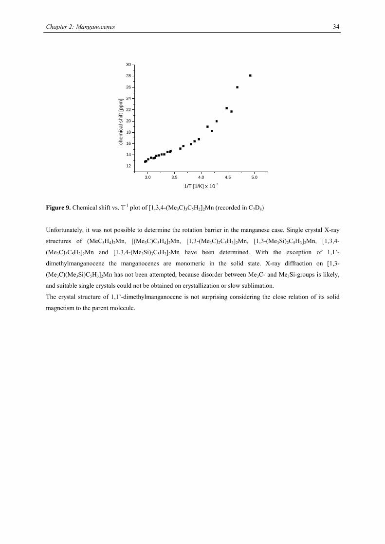
compounds,42 and the other resonances are presumably broadened into the base line. Variable temperature NMR
studies were undertaken in the case of [1,3,4-(Me3C)3C5H2]2Mn, because previous reports on the spectroscopic
properties28 differed from the data obtained in this work. At all accessible temperatures only one resonance for
both tert-butyl groups has been observed, and the δ vs. T-1 plot obeys the Curie(-Weiss) law at high
temperatures, showing deviations and significant line-broadening at low temperature (Fig. 9). This deviation can
be due to a low-spin-high-spin equilibrium as previously observed for other manganocenes by Köhler et al. or,
more likely, due to hindered rotation of the 1,3,4-tri(tert-butyl)cyclopentadienyl ligands relative to each other. In
the solid state a spin-equilibrium has not been observed and the molecule displays high-spin configuration at all
accessible temperatures as demonstrated by SQUID measurements. Furthermore a hindered cyclopentadienyl
rotation has also been observed in hexa(tert.-butyl)ferrocene and the thermodynamic data for this process have
been determined:
∆H‡= 58.4 ± 4.2 kJ mol-1; ∆S‡= 3.99 ± 2 J mol-1 K-1 and ∆G‡= 58.44 ± 0.8 kJ mol-1.
Chapter 2: Manganocenes
34
3.0 3.5 4.0 4.5 5.0
12
14
16
18
20
22
24
26
28
30
chem
ical
shi
ft [p
pm]
1/T [1/K] x 10−3
Figure 9. Chemical shift vs. T-1 plot of [1,3,4-(Me3C)3C5H2]2Mn (recorded in C7D8)
Unfortunately, it was not possible to determine the rotation barrier in the manganese case. Single crystal X-ray
structures of (MeC5H4)2Mn, [(Me3C)C5H4]2Mn, [1,3-(Me3C)2C5H3]2Mn, [1,3-(Me3Si)2C5H3]2Mn, [1,3,4-
(Me3C)3C5H2]2Mn and [1,3,4-(Me3Si)3C5H2]2Mn have been determined. With the exception of 1,1’-
dimethylmanganocene the manganocenes are monomeric in the solid state. X-ray diffraction on [1,3-
(Me3C)(Me3Si)C5H3]2Mn has not been attempted, because disorder between Me3C- and Me3Si-groups is likely,
and suitable single crystals could not be obtained on crystallization or slow sublimation.
The crystal structure of 1,1’-dimethylmanganocene is not surprising considering the close relation of its solid
magnetism to the parent molecule.
Chapter 2: Manganocenes
35
Crystal Structure Investigations
Disubstituted Manganocenes
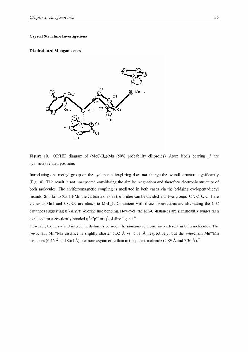
Figure 10. ORTEP diagram of (MeC5H4)2Mn (50% probability ellipsoids). Atom labels bearing _3 are
symmetry related positions
Introducing one methyl group on the cyclopentadienyl ring does not change the overall structure significantly
(Fig 10). This result is not unexpected considering the similar magnetism and therefore electronic structure of
both molecules. The antiferromagnetic coupling is mediated in both cases via the bridging cyclopentadienyl
ligands. Similar to (C5H5)2Mn the carbon atoms in the bridge can be divided into two groups: C7, C10, C11 are
closer to Mn1 and C8, C9 are closer to Mn1_3. Consistent with these observations are alternating the C-C
distances suggesting η3-allyl/η2-olefine like bonding. However, the Mn-C distances are significantly longer than
expected for a covalently bonded η5-Cp43 or η2-olefine ligand.44
However, the intra- and interchain distances between the manganese atoms are different in both molecules: The
intrachain Mn…Mn distance is slightly shorter 5.32 Å vs. 5.38 Å, respectively, but the interchain Mn…Mn
distances (6.46 Å and 8.63 Å) are more asymmetric than in the parent molecule (7.89 Å and 7.36 Å).29
Chapter 2: Manganocenes
36
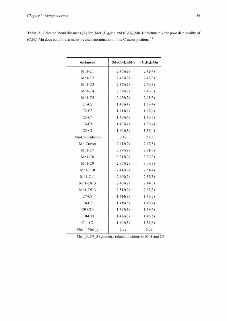
Table 3. Selected bond distances (Å) for (MeC5H4)2Mn and (C5H5)2Mn. Unfortunately the poor data quality of
(C5H5)2Mn does not allow a more precise determination of the C-atom positions.29
distances (MeC5H4)2Mn (C5H5)2Mn
Mn1-C1 2.468(2) 2.42(4)
Mn1-C2 2.437(2) 2.42(3)
Mn1-C3 2.379(2) 2.40(3)
Mn1-C4 2.379(2) 2.40(3)
Mn1-C5 2.426(2) 2.43(3)
C1-C2 1.408(4) 1.39(4)
C2-C3 1.411(4) 1.42(4)
C3-C4 1.409(4) 1.36(5)
C4-C5 1.403(4) 1.38(4)
C5-C1 1.408(3) 1.34(4)
Mn-Cp(centroid) 2.10 2.10
Mn-C(ave) 2.418(2) 2.42(3)
Mn1-C7 2.997(2) 2.81(3)
Mn1-C8 3.313(2) 3.30(3)
Mn1-C9 2.997(2) 3.09(3)
Mn1-C10 2.416(2) 2.51(4)
Mn1-C11 2.409(2) 2.37(3)
Mn1-C8_3 2.409(2) 2.44(3)
Mn1-C9_3 2.518(2) 2.62(3)
C7-C8 1.414(3) 1.45(5)
C8-C9 1.419(3) 1.43(4)
C9-C10 1.397(3) 1.38(5)
C10-C11 1.428(3) 1.45(5)
C11-C7 1.408(3) 1.54(6)
Mn1 ... Mn1_3 5.32 5.38
Mn1_3, C9_3 symmetry related positions to Mn1 and C9
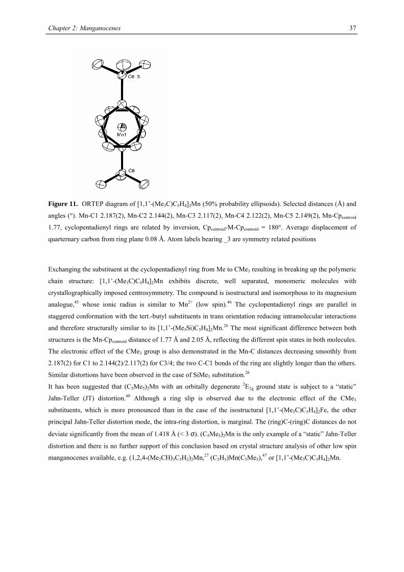
Chapter 2: Manganocenes
37
Figure 11. ORTEP diagram of [1,1’-(Me3C)C5H4]2Mn (50% probability ellipsoids). Selected distances (Å) and
angles (°). Mn-C1 2.187(2), Mn-C2 2.144(2), Mn-C3 2.117(2), Mn-C4 2.122(2), Mn-C5 2.149(2), Mn-Cpcentroid
1.77, cyclopentadienyl rings are related by inversion, Cpcentroid-M-Cpcontroid = 180°. Average displacement of
quarternary carbon from ring plane 0.08 Å. Atom labels bearing _3 are symmetry related positions
Exchanging the substituent at the cyclopentadienyl ring from Me to CMe3 resulting in breaking up the polymeric
chain structure: [1,1’-(Me3C)C5H4]2Mn exhibits discrete, well separated, monomeric molecules with
crystallographically imposed centrosymmetry. The compound is isostructural and isomorphous to its magnesium
analogue,45 whose ionic radius is similar to Mn2+ (low spin).46 The cyclopentadienyl rings are parallel in
staggered conformation with the tert.-butyl substituents in trans orientation reducing intramolecular interactions
and therefore structurally similar to its [1,1’-(Me3Si)C5H4]2Mn.26 The most significant difference between both
structures is the Mn-Cpcentroid distance of 1.77 Å and 2.05 Å, reflecting the different spin states in both molecules.
The electronic effect of the CMe3 group is also demonstrated in the Mn-C distances decreasing smoothly from
2.187(2) for C1 to 2.144(2)/2.117(2) for C3/4; the two C-C1 bonds of the ring are slightly longer than the others.
Similar distortions have been observed in the case of SiMe3 substitution.26
It has been suggested that (C5Me5)2Mn with an orbitally degenerate 2E2g ground state is subject to a “static”
Jahn-Teller (JT) distortion.40 Although a ring slip is observed due to the electronic effect of the CMe3
substituents, which is more pronounced than in the case of the isostructural [1,1’-(Me3C)C5H4]2Fe, the other
principal Jahn-Teller distortion mode, the intra-ring distortion, is marginal. The (ring)C-(ring)C distances do not
deviate significantly from the mean of 1.418 Å (< 3 σ). (C5Me5)2Mn is the only example of a “static” Jahn-Teller
distortion and there is no further support of this conclusion based on crystal structure analysis of other low spin
manganocenes available, e.g. (1,2,4-(Me2CH)3C5H2)2Mn,27 (C5H5)Mn(C5Me5),47 or [1,1’-(Me3C)C5H4]2Mn.
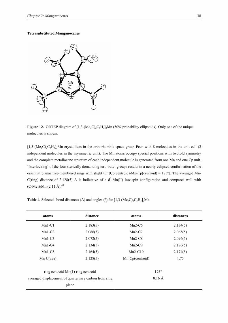
Chapter 2: Manganocenes
38
Tetrasubstituted Manganocenes
Figure 12. ORTEP diagram of [1,3-(Me3C)2C5H3]2Mn (50% probability ellipsoids). Only one of the unique
molecules is shown.
[1,3-(Me3C)2C5H3]2Mn crystallizes in the orthorhombic space group Pccn with 8 molecules in the unit cell (2
independent molecules in the asymmetric unit). The Mn atoms occupy special positions with twofold symmetry
and the complete metallocene structure of each independent molecule is generated from one Mn and one Cp unit.
‘Interlocking’ of the four sterically demanding tert.-butyl groups results in a nearly eclipsed conformation of the
essential planar five-membered rings with slight tilt [Cp(centroid)-Mn-Cp(centroid) = 175°]. The averaged Mn-
C(ring) distance of 2.128(5) Å is indicative of a d5-Mn(II) low-spin configuration and compares well with
(C5Me5)2Mn (2.11 Å).40
Table 4. Selected bond distances (Å) and angles (°) for [1,3-(Me3C)2C5H3]2Mn
atoms distance atoms distances
Mn1-C1 2.183(5) Mn2-C6 2.134(5)
Mn1-C2 2.086(5) Mn2-C7 2.065(5)
Mn1-C3 2.072(5) Mn2-C8 2.094(5)
Mn1-C4 2.134(5) Mn2-C9 2.176(5)
Mn1-C5 2.164(5) Mn2-C10 2.174(5)
Mn-C(ave) 2.128(5) Mn-Cp(centroid) 1.75
ring centroid-Mn(1)-ring centroid 175°
averaged displacement of quarternary carbon from ring
plane
0.16 Å
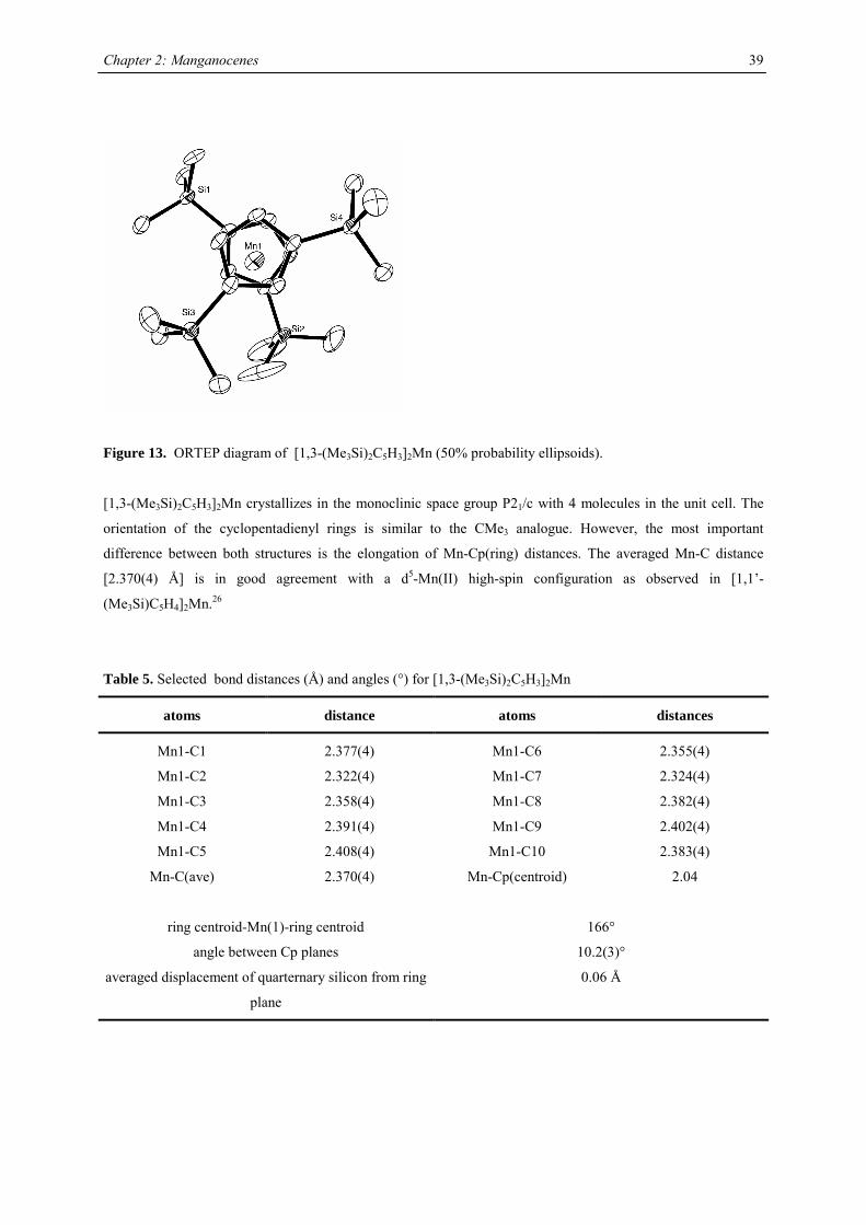
Chapter 2: Manganocenes
39
Figure 13. ORTEP diagram of [1,3-(Me3Si)2C5H3]2Mn (50% probability ellipsoids).
[1,3-(Me3Si)2C5H3]2Mn crystallizes in the monoclinic space group P21/c with 4 molecules in the unit cell. The
orientation of the cyclopentadienyl rings is similar to the CMe3 analogue. However, the most important
difference between both structures is the elongation of Mn-Cp(ring) distances. The averaged Mn-C distance
[2.370(4) Å] is in good agreement with a d5-Mn(II) high-spin configuration as observed in [1,1’-
(Me3Si)C5H4]2Mn.26
Table 5. Selected bond distances (Å) and angles (°) for [1,3-(Me3Si)2C5H3]2Mn
atoms distance atoms distances
Mn1-C1 2.377(4) Mn1-C6 2.355(4)
Mn1-C2 2.322(4) Mn1-C7 2.324(4)
Mn1-C3 2.358(4) Mn1-C8 2.382(4)
Mn1-C4 2.391(4) Mn1-C9 2.402(4)
Mn1-C5 2.408(4) Mn1-C10 2.383(4)
Mn-C(ave) 2.370(4) Mn-Cp(centroid) 2.04
ring centroid-Mn(1)-ring centroid 166°
angle between Cp planes 10.2(3)°
averaged displacement of quarternary silicon from ring
plane
0.06 Å
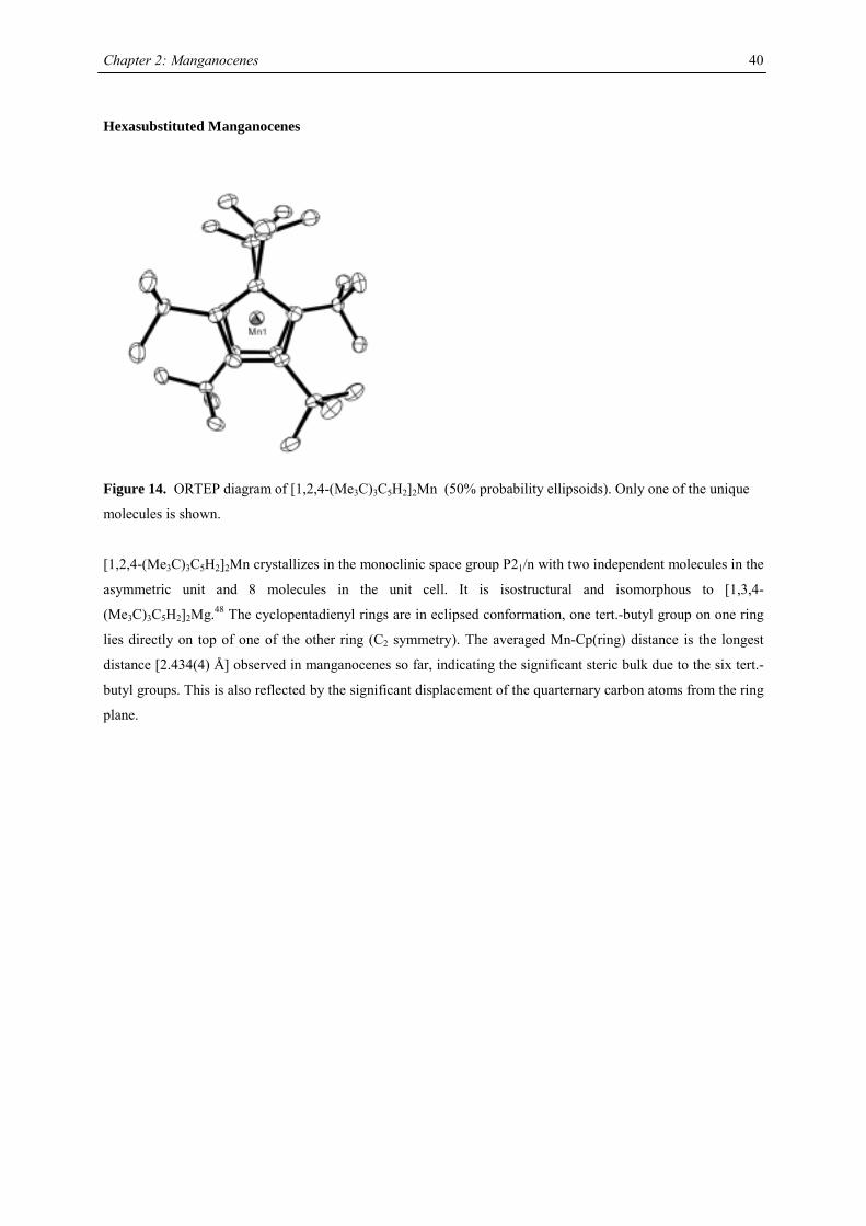
Chapter 2: Manganocenes
40
Hexasubstituted Manganocenes
Figure 14. ORTEP diagram of [1,2,4-(Me3C)3C5H2]2Mn (50% probability ellipsoids). Only one of the unique
molecules is shown.
[1,2,4-(Me3C)3C5H2]2Mn crystallizes in the monoclinic space group P21/n with two independent molecules in the
asymmetric unit and 8 molecules in the unit cell. It is isostructural and isomorphous to [1,3,4-
(Me3C)3C5H2]2Mg.48 The cyclopentadienyl rings are in eclipsed conformation, one tert.-butyl group on one ring
lies directly on top of one of the other ring (C2 symmetry). The averaged Mn-Cp(ring) distance is the longest
distance [2.434(4) Å] observed in manganocenes so far, indicating the significant steric bulk due to the six tert.-
butyl groups. This is also reflected by the significant displacement of the quarternary carbon atoms from the ring
plane.
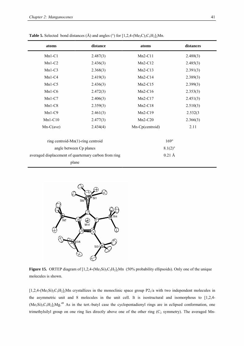
Chapter 2: Manganocenes
41
Table 5. Selected bond distances (Å) and angles (°) for [1,2,4-(Me3C)3C5H2]2Mn.
atoms distance atoms distances
Mn1-C1 2.487(3) Mn2-C11 2.488(3)
Mn1-C2 2.436(3) Mn2-C12 2.485(3)
Mn1-C3 2.368(3) Mn2-C13 2.391(3)
Mn1-C4 2.419(3) Mn2-C14 2.389(3)
Mn1-C5 2.436(3) Mn2-C15 2.399(3)
Mn1-C6 2.472(3) Mn2-C16 2.353(3)
Mn1-C7 2.406(3) Mn2-C17 2.451(3)
Mn1-C8 2.359(3) Mn2-C18 2.510(3)
Mn1-C9 2.461(3) Mn2-C19 2.532(3
Mn1-C10 2.477(3) Mn2-C20 2.366(3)
Mn-C(ave) 2.434(4) Mn-Cp(centroid) 2.11
ring centroid-Mn(1)-ring centroid 169°
angle between Cp planes 8.1(2)°
averaged displacement of quarternary carbon from ring
plane
0.21 Å
Figure 15. ORTEP diagram of [1,2,4-(Me3Si)3C5H2]2Mn (50% probability ellipsoids). Only one of the unique
molecules is shown.
[1,2,4-(Me3Si)3C5H2]2Mn crystallizes in the monoclinic space group P21/a with two independent molecules in
the asymmetric unit and 8 molecules in the unit cell. It is isostructural and isomorphous to [1,2,4-
(Me3Si)3C5H2]2Mg.49 As in the tert.-butyl case the cyclopentadienyl rings are in eclipsed conformation, one
trimethylsilyl group on one ring lies directly above one of the other ring (C2 symmetry). The averaged Mn-
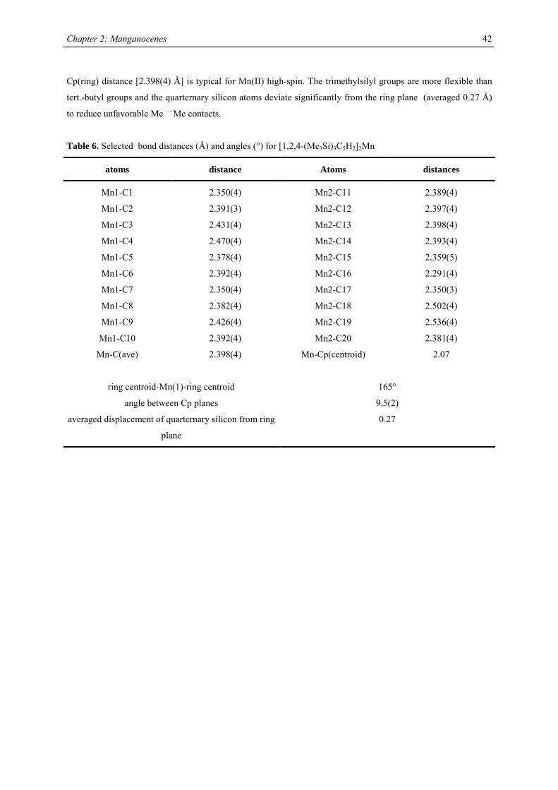
Chapter 2: Manganocenes
42
Cp(ring) distance [2.398(4) Å] is typical for Mn(II) high-spin. The trimethylsilyl groups are more flexible than
tert.-butyl groups and the quarternary silicon atoms deviate significantly from the ring plane (averaged 0.27 Å)
to reduce unfavorable Me … Me contacts.
Table 6. Selected bond distances (Å) and angles (°) for [1,2,4-(Me3Si)3C5H2]2Mn
atoms distance Atoms distances
Mn1-C1 2.350(4) Mn2-C11 2.389(4)
Mn1-C2 2.391(3) Mn2-C12 2.397(4)
Mn1-C3 2.431(4) Mn2-C13 2.398(4)
Mn1-C4 2.470(4) Mn2-C14 2.393(4)
Mn1-C5 2.378(4) Mn2-C15 2.359(5)
Mn1-C6 2.392(4) Mn2-C16 2.291(4)
Mn1-C7 2.350(4) Mn2-C17 2.350(3)
Mn1-C8 2.382(4) Mn2-C18 2.502(4)
Mn1-C9 2.426(4) Mn2-C19 2.536(4)
Mn1-C10 2.392(4) Mn2-C20 2.381(4)
Mn-C(ave) 2.398(4) Mn-Cp(centroid) 2.07
ring centroid-Mn(1)-ring centroid 165°
angle between Cp planes 9.5(2)
averaged displacement of quarternary silicon from ring
plane
0.27
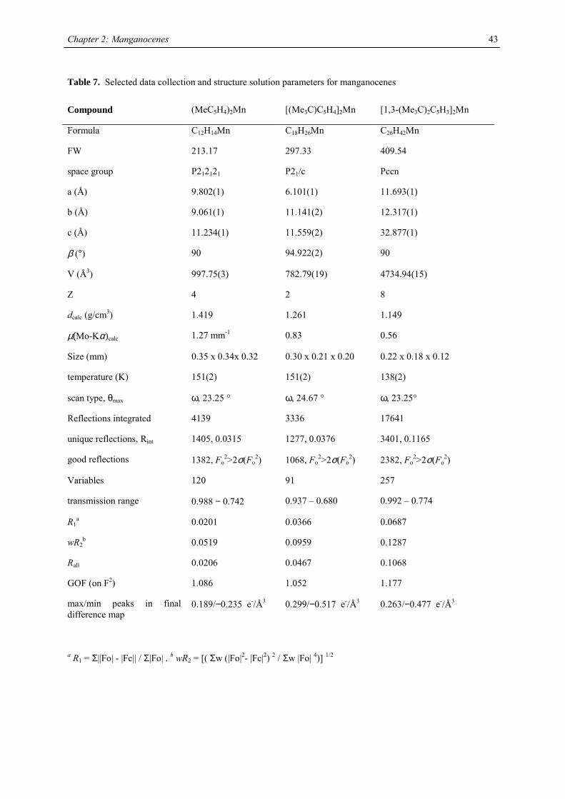
Chapter 2: Manganocenes
43
Table 7. Selected data collection and structure solution parameters for manganocenes
Compound (MeC5H4)2Mn [(Me3C)C5H4]2Mn [1,3-(Me3C)2C5H3]2Mn
Formula C12H14Mn C18H26Mn C26H42Mn
FW 213.17 297.33 409.54
space group P212121 P21/c Pccn
a (Å) 9.802(1) 6.101(1) 11.693(1)
b (Å) 9.061(1) 11.141(2) 12.317(1)
c (Å) 11.234(1) 11.559(2) 32.877(1)
β (°) 90 94.922(2) 90
V (Å3) 997.75(3) 782.79(19) 4734.94(15)
Z 4 2 8
dcalc (g/cm3) 1.419 1.261 1.149
µ(Mo-Kα)calc 1.27 mm-1 0.83 0.56
Size (mm) 0.35 x 0.34x 0.32 0.30 x 0.21 x 0.20 0.22 x 0.18 x 0.12
temperature (K) 151(2) 151(2) 138(2)
scan type, θmax ω, 23.25 ° ω, 24.67 ° ω, 23.25°
Reflections integrated 4139 3336 17641
unique reflections, Rint 1405, 0.0315 1277, 0.0376 3401, 0.1165
good reflections 1382, Fo2>2σ(Fo
2) 1068, Fo2>2σ(Fo
2) 2382, Fo2>2σ(Fo
2)
Variables 120 91 257
transmission range 0.988 − 0.742 0.937 – 0.680 0.992 – 0.774
R1a 0.0201 0.0366 0.0687
wR2b 0.0519 0.0959 0.1287
Rall 0.0206 0.0467 0.1068
GOF (on F2) 1.086 1.052 1.177
max/min peaks in final difference map
0.189/−0.235 e-/Å3
0.299/−0.517 e-/Å3 0.263/−0.477 e-/Å3
a R1 = Σ||Fo| - |Fc|| / Σ|Fo| . b wR2 = [( Σw (|Fo|2- |Fc|2) 2 / Σw |Fo| 4)] 1/2
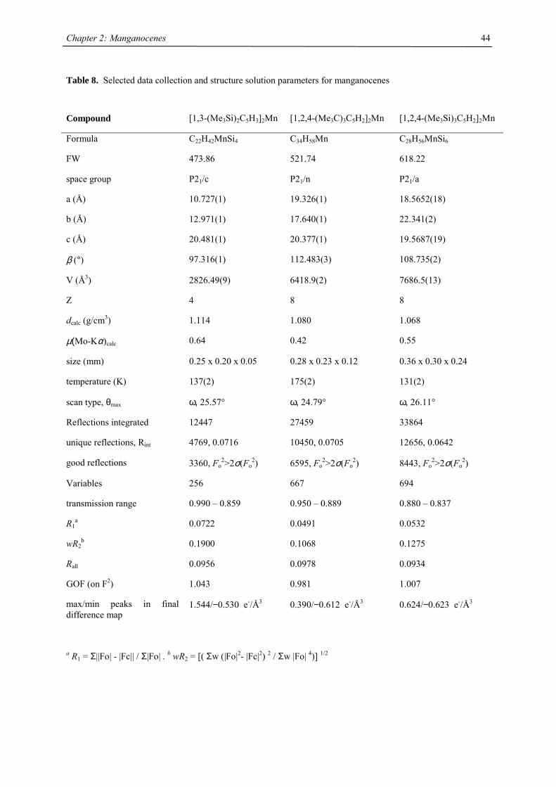
Chapter 2: Manganocenes
44
Table 8. Selected data collection and structure solution parameters for manganocenes
Compound [1,3-(Me3Si)2C5H3]2Mn [1,2,4-(Me3C)3C5H2]2Mn [1,2,4-(Me3Si)3C5H2]2Mn
Formula C22H42MnSi4 C34H58Mn C28H56MnSi6
FW 473.86 521.74 618.22
space group P21/c P21/n P21/a
a (Å) 10.727(1) 19.326(1) 18.5652(18)
b (Å) 12.971(1) 17.640(1) 22.341(2)
c (Å) 20.481(1) 20.377(1) 19.5687(19)
β (°) 97.316(1) 112.483(3) 108.735(2)
V (Å3) 2826.49(9) 6418.9(2) 7686.5(13)
Z 4 8 8
dcalc (g/cm3) 1.114 1.080 1.068
µ(Mo-Kα)calc 0.64 0.42 0.55
size (mm) 0.25 x 0.20 x 0.05 0.28 x 0.23 x 0.12 0.36 x 0.30 x 0.24
temperature (K) 137(2) 175(2) 131(2)
scan type, θmax ω, 25.57° ω, 24.79° ω, 26.11°
Reflections integrated 12447 27459 33864
unique reflections, Rint 4769, 0.0716 10450, 0.0705 12656, 0.0642
good reflections 3360, Fo2>2σ(Fo
2) 6595, Fo2>2σ(Fo
2) 8443, Fo2>2σ(Fo
2)
Variables 256 667 694
transmission range 0.990 – 0.859 0.950 – 0.889 0.880 – 0.837
R1a 0.0722 0.0491 0.0532
wR2b 0.1900 0.1068 0.1275
Rall 0.0956 0.0978 0.0934
GOF (on F2) 1.043 0.981 1.007
max/min peaks in final difference map
1.544/−0.530 e-/Å3
0.390/−0.612 e-/Å3
0.624/−0.623 e-/Å3
a R1 = Σ||Fo| - |Fc|| / Σ|Fo| . b wR2 = [( Σw (|Fo|2- |Fc|2) 2 / Σw |Fo| 4)] 1/2
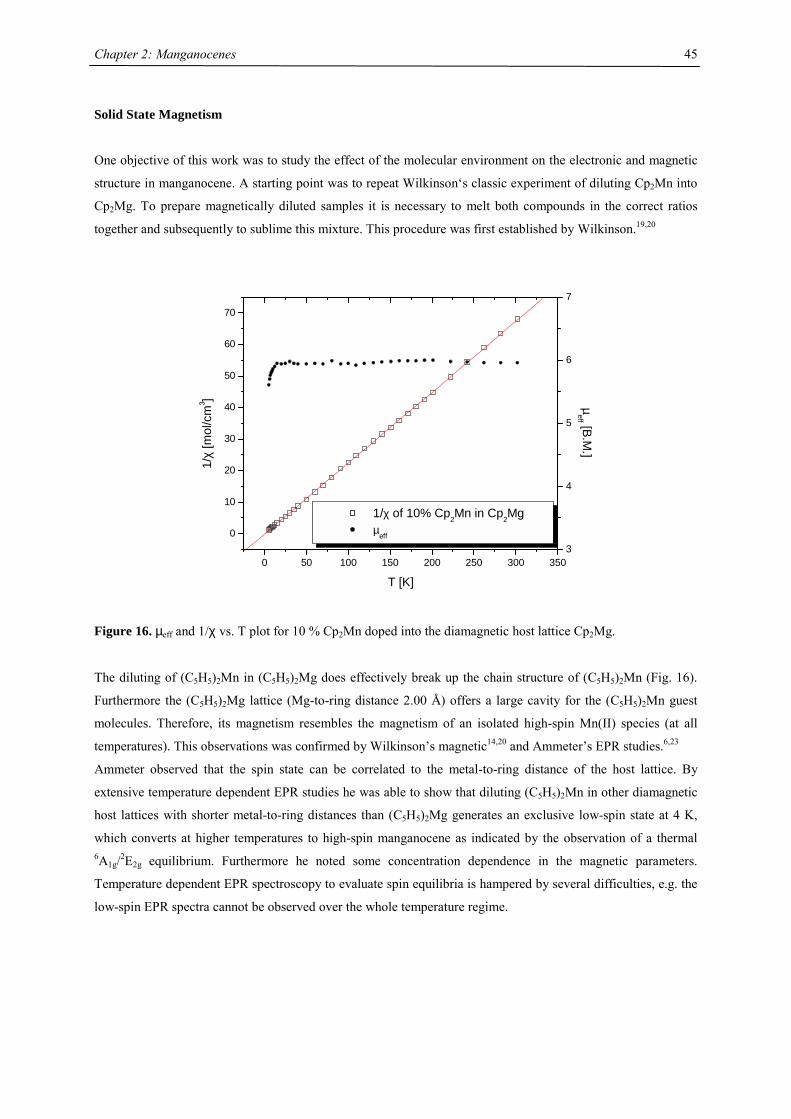
Chapter 2: Manganocenes
45
Solid State Magnetism
One objective of this work was to study the effect of the molecular environment on the electronic and magnetic
structure in manganocene. A starting point was to repeat Wilkinson‘s classic experiment of diluting Cp2Mn into
Cp2Mg. To prepare magnetically diluted samples it is necessary to melt both compounds in the correct ratios
together and subsequently to sublime this mixture. This procedure was first established by Wilkinson.19,20
0 50 100 150 200 250 300 350
0
10
20
30
40
50
60
70
1/χ of 10% Cp2Mn in Cp2Mg µeff
T [K]
1/χ
[mol
/cm
3 ]
3
4
5
6
7
µeff [B.M
.]
Figure 16. µeff and 1/χ vs. T plot for 10 % Cp2Mn doped into the diamagnetic host lattice Cp2Mg.
The diluting of (C5H5)2Mn in (C5H5)2Mg does effectively break up the chain structure of (C5H5)2Mn (Fig. 16).
Furthermore the (C5H5)2Mg lattice (Mg-to-ring distance 2.00 Å) offers a large cavity for the (C5H5)2Mn guest
molecules. Therefore, its magnetism resembles the magnetism of an isolated high-spin Mn(II) species (at all
temperatures). This observations was confirmed by Wilkinson’s magnetic14,20 and Ammeter’s EPR studies.6,23
Ammeter observed that the spin state can be correlated to the metal-to-ring distance of the host lattice. By
extensive temperature dependent EPR studies he was able to show that diluting (C5H5)2Mn in other diamagnetic
host lattices with shorter metal-to-ring distances than (C5H5)2Mg generates an exclusive low-spin state at 4 K,
which converts at higher temperatures to high-spin manganocene as indicated by the observation of a thermal 6A1g/2E2g equilibrium. Furthermore he noted some concentration dependence in the magnetic parameters.
Temperature dependent EPR spectroscopy to evaluate spin equilibria is hampered by several difficulties, e.g. the
low-spin EPR spectra cannot be observed over the whole temperature regime.
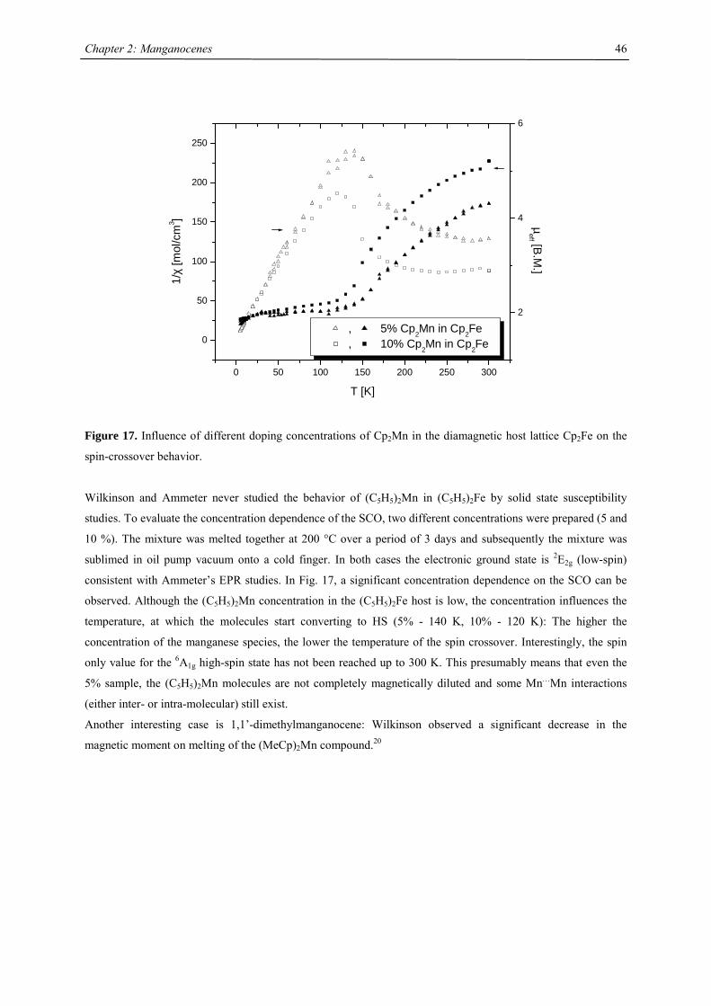
Chapter 2: Manganocenes
46
0 50 100 150 200 250 300
0
50
100
150
200
250
, 5% Cp2Mn in Cp2Fe, 10% Cp2Mn in Cp2Fe
T [K]
1/χ
[mol
/cm
3 ]
2
4
6
µeff [B
.M.]
Figure 17. Influence of different doping concentrations of Cp2Mn in the diamagnetic host lattice Cp2Fe on the
spin-crossover behavior.
Wilkinson and Ammeter never studied the behavior of (C5H5)2Mn in (C5H5)2Fe by solid state susceptibility
studies. To evaluate the concentration dependence of the SCO, two different concentrations were prepared (5 and
10 %). The mixture was melted together at 200 °C over a period of 3 days and subsequently the mixture was
sublimed in oil pump vacuum onto a cold finger. In both cases the electronic ground state is 2E2g (low-spin)
consistent with Ammeter’s EPR studies. In Fig. 17, a significant concentration dependence on the SCO can be
observed. Although the (C5H5)2Mn concentration in the (C5H5)2Fe host is low, the concentration influences the
temperature, at which the molecules start converting to HS (5% - 140 K, 10% - 120 K): The higher the
concentration of the manganese species, the lower the temperature of the spin crossover. Interestingly, the spin
only value for the 6A1g high-spin state has not been reached up to 300 K. This presumably means that even the
5% sample, the (C5H5)2Mn molecules are not completely magnetically diluted and some Mn…Mn interactions
(either inter- or intra-molecular) still exist.
Another interesting case is 1,1’-dimethylmanganocene: Wilkinson observed a significant decrease in the
magnetic moment on melting of the (MeCp)2Mn compound.20
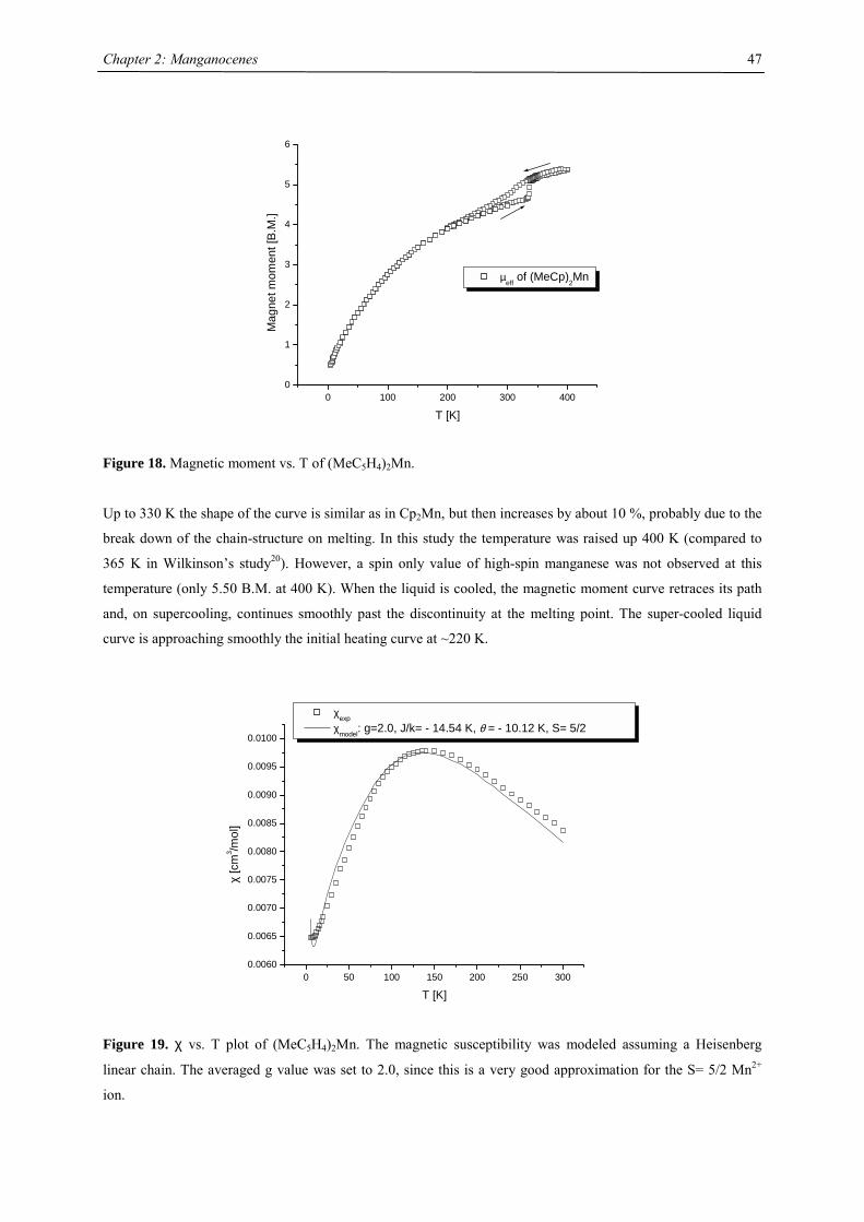
Chapter 2: Manganocenes
47
0 100 200 300 4000
1
2
3
4
5
6
µeff
of (MeCp)2Mn
Mag
net m
omen
t [B
.M.]
T [K]
Figure 18. Magnetic moment vs. T of (MeC5H4)2Mn.
Up to 330 K the shape of the curve is similar as in Cp2Mn, but then increases by about 10 %, probably due to the
break down of the chain-structure on melting. In this study the temperature was raised up 400 K (compared to
365 K in Wilkinson’s study20). However, a spin only value of high-spin manganese was not observed at this
temperature (only 5.50 B.M. at 400 K). When the liquid is cooled, the magnetic moment curve retraces its path
and, on supercooling, continues smoothly past the discontinuity at the melting point. The super-cooled liquid
curve is approaching smoothly the initial heating curve at ~220 K.
0 50 100 150 200 250 3000.0060
0.0065
0.0070
0.0075
0.0080
0.0085
0.0090
0.0095
0.0100
χ [c
m3 /m
ol]
T [K]
χexp χmodel: g=2.0, J/k= - 14.54 K, θ = - 10.12 K, S= 5/2
Figure 19. χ vs. T plot of (MeC5H4)2Mn. The magnetic susceptibility was modeled assuming a Heisenberg
linear chain. The averaged g value was set to 2.0, since this is a very good approximation for the S= 5/2 Mn2+
ion.
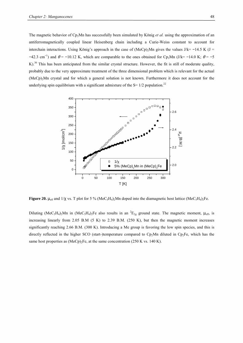
Chapter 2: Manganocenes
48
The magnetic behavior of Cp2Mn has successfully been simulated by König et al. using the approximation of an
antiferromagnetically coupled linear Heisenberg chain including a Curie-Weiss constant to account for
interchain interactions. Using König’s approach in the case of (MeCp)2Mn gives the values J/k= −14.5 K (J =
−42.3 cm−1) and θ = −10.12 K, which are comparable to the ones obtained for Cp2Mn (J/k= −14.0 K; θ = −5
K).30 This has been anticipated from the similar crystal structure. However, the fit is still of moderate quality,
probably due to the very approximate treatment of the three dimensional problem which is relevant for the actual
(MeCp)2Mn crystal and for which a general solution is not known. Furthermore it does not account for the
underlying spin equilibrium with a significant admixture of the S= 1/2 population.22
0 50 100 150 200 250 300
0
50
100
150
200
250
300
350
400
1/χ 5% (MeCp)2Mn in (MeCp)2Fe
T [K]
1/χ
[mol
/cm
3 ]
2.0
2.2
2.4
2.6
µ
eff [B.M.]
Figure 20. µeff and 1/χ vs. T plot for 5 % (MeC5H4)2Mn doped into the diamagnetic host lattice (MeC5H4)2Fe.
Diluting (MeC5H4)2Mn in (MeC5H4)2Fe also results in an 2E2g ground state. The magnetic moment, µeff, is
increasing linearly from 2.05 B.M (5 K) to 2.39 B.M. (250 K), but then the magnetic moment increases
significantly reaching 2.66 B.M. (300 K). Introducing a Me group is favoring the low spin species, and this is
directly reflected in the higher SCO (start-)temperature compared to Cp2Mn diluted in Cp2Fe, which has the
same host properties as (MeCp)2Fe, at the same concentration (250 K vs. 140 K).
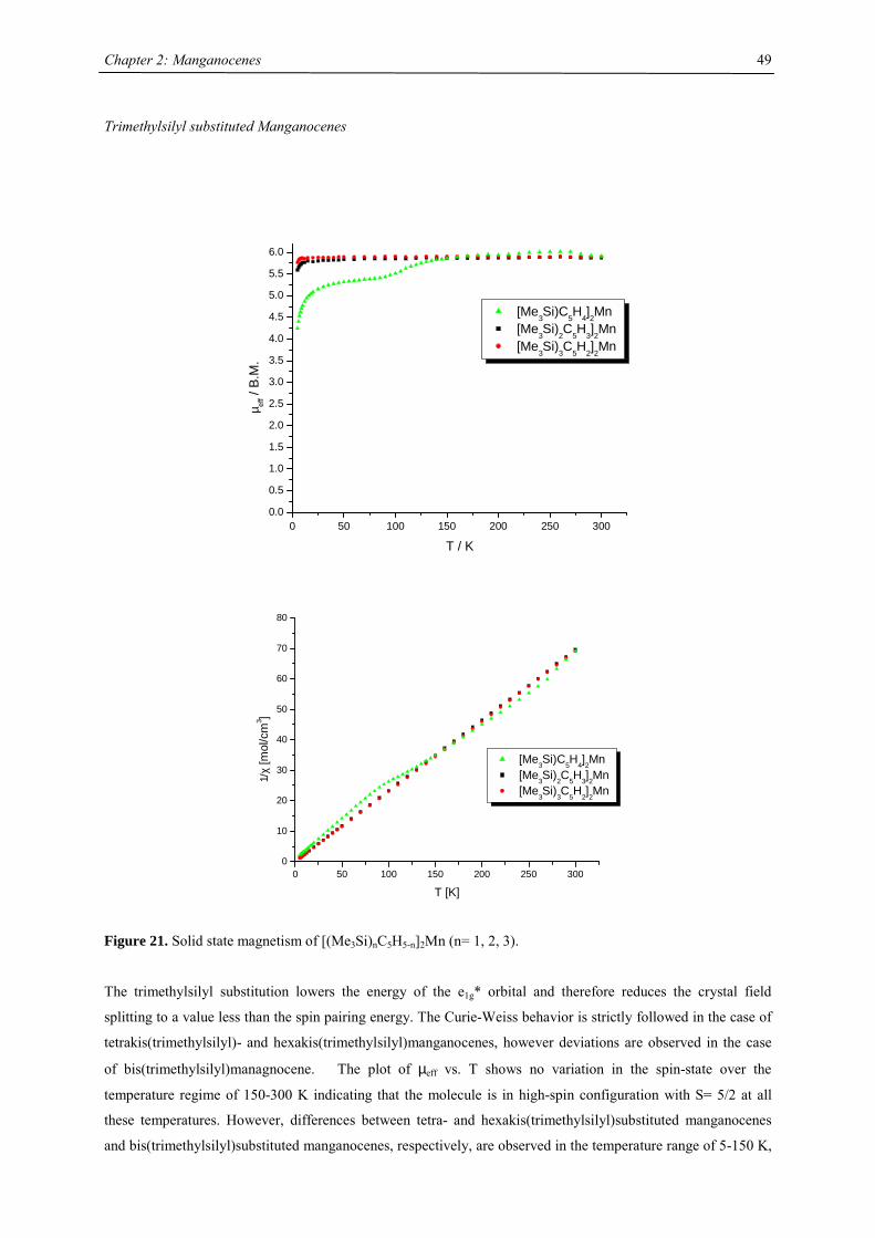
Chapter 2: Manganocenes
49
Trimethylsilyl substituted Manganocenes
0 50 100 150 200 250 3000.0
0.5
1.0
1.5
2.0
2.5
3.0
3.5
4.0
4.5
5.0
5.5
6.0
[Me3Si)C5H4]2Mn [Me
3Si)
2C
5H
3]2Mn
[Me3Si)
3C
5H
2]2Mn
µ eff /
B.M
.
T / K
0 50 100 150 200 250 3000
10
20
30
40
50
60
70
80
[Me3Si)C5H4]2Mn [Me3Si)2C5H3]2Mn [Me
3Si)
3C
5H
2]2Mn
1/χ
[mol
/cm
3 ]
T [K]
Figure 21. Solid state magnetism of [(Me3Si)nC5H5-n]2Mn (n= 1, 2, 3).
The trimethylsilyl substitution lowers the energy of the e1g* orbital and therefore reduces the crystal field
splitting to a value less than the spin pairing energy. The Curie-Weiss behavior is strictly followed in the case of
tetrakis(trimethylsilyl)- and hexakis(trimethylsilyl)manganocenes, however deviations are observed in the case
of bis(trimethylsilyl)managnocene. The plot of µeff vs. T shows no variation in the spin-state over the
temperature regime of 150-300 K indicating that the molecule is in high-spin configuration with S= 5/2 at all
these temperatures. However, differences between tetra- and hexakis(trimethylsilyl)substituted manganocenes
and bis(trimethylsilyl)substituted manganocenes, respectively, are observed in the temperature range of 5-150 K,
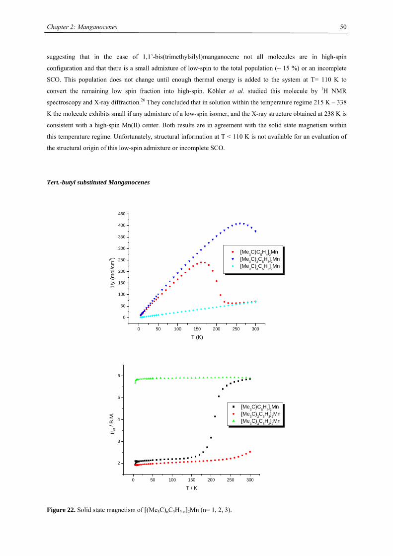
Chapter 2: Manganocenes
50
suggesting that in the case of 1,1’-bis(trimethylsilyl)manganocene not all molecules are in high-spin
configuration and that there is a small admixture of low-spin to the total population (~ 15 %) or an incomplete
SCO. This population does not change until enough thermal energy is added to the system at T= 110 K to
convert the remaining low spin fraction into high-spin. Köhler et al. studied this molecule by 1H NMR
spectroscopy and X-ray diffraction.26 They concluded that in solution within the temperature regime 215 K – 338
K the molecule exhibits small if any admixture of a low-spin isomer, and the X-ray structure obtained at 238 K is
consistent with a high-spin Mn(II) center. Both results are in agreement with the solid state magnetism within
this temperature regime. Unfortunately, structural information at T < 110 K is not available for an evaluation of
the structural origin of this low-spin admixture or incomplete SCO.
Tert.-butyl substituted Manganocenes
0 50 100 150 200 250 300
0
50
100
150
200
250
300
350
400
450
[Me3C)C
5H
4]2Mn
[Me3C)2C5H3]2Mn [Me
3C)
3C
5H
2]2Mn
1/χ
(mol
/cm
3 )
T (K)
0 50 100 150 200 250 300
2
3
4
5
6
µ eff /
B.M
.
T / K
[Me3C)C
5H
4]2Mn
[Me3C)2C5H3]2Mn [Me
3C)
3C
5H
2]2Mn
Figure 22. Solid state magnetism of [(Me3C)nC5H5-n]2Mn (n= 1, 2, 3).
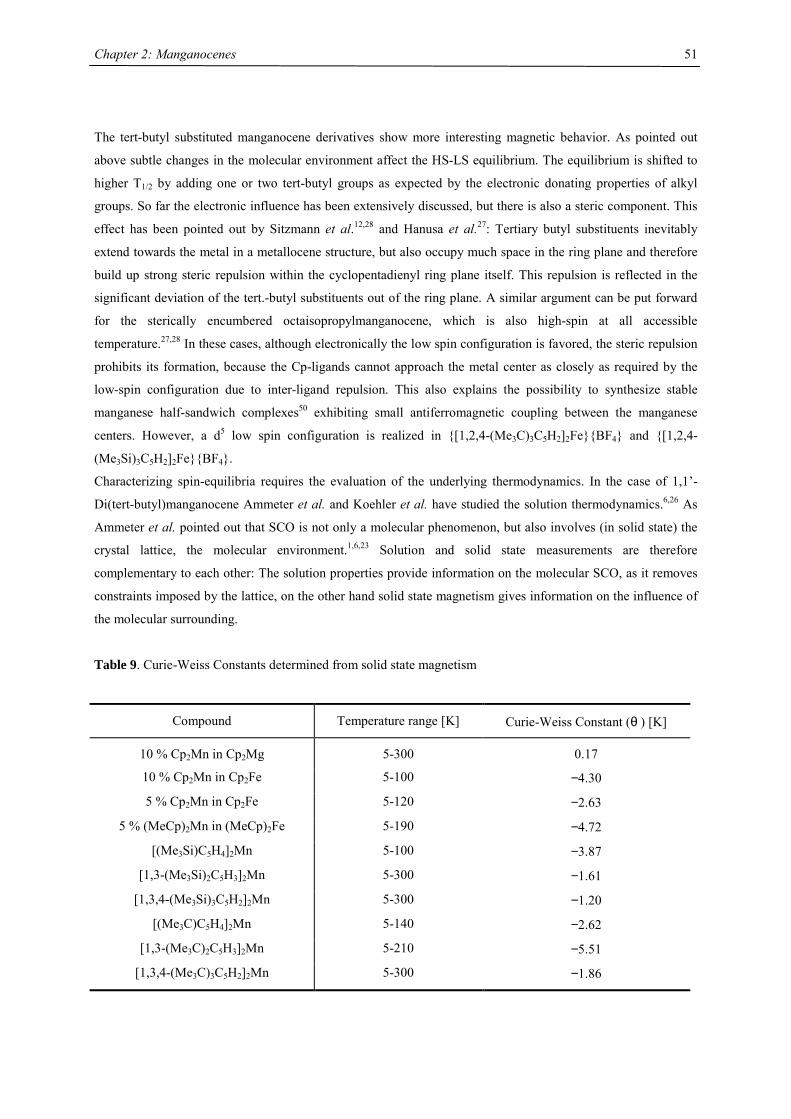
Chapter 2: Manganocenes
51
The tert-butyl substituted manganocene derivatives show more interesting magnetic behavior. As pointed out
above subtle changes in the molecular environment affect the HS-LS equilibrium. The equilibrium is shifted to
higher T1/2 by adding one or two tert-butyl groups as expected by the electronic donating properties of alkyl
groups. So far the electronic influence has been extensively discussed, but there is also a steric component. This
effect has been pointed out by Sitzmann et al.12,28 and Hanusa et al.27: Tertiary butyl substituents inevitably
extend towards the metal in a metallocene structure, but also occupy much space in the ring plane and therefore
build up strong steric repulsion within the cyclopentadienyl ring plane itself. This repulsion is reflected in the
significant deviation of the tert.-butyl substituents out of the ring plane. A similar argument can be put forward
for the sterically encumbered octaisopropylmanganocene, which is also high-spin at all accessible
temperature.27,28 In these cases, although electronically the low spin configuration is favored, the steric repulsion
prohibits its formation, because the Cp-ligands cannot approach the metal center as closely as required by the
low-spin configuration due to inter-ligand repulsion. This also explains the possibility to synthesize stable
manganese half-sandwich complexes50 exhibiting small antiferromagnetic coupling between the manganese
centers. However, a d5 low spin configuration is realized in {[1,2,4-(Me3C)3C5H2]2Fe}{BF4} and {[1,2,4-
(Me3Si)3C5H2]2Fe}{BF4}.
Characterizing spin-equilibria requires the evaluation of the underlying thermodynamics. In the case of 1,1’-
Di(tert-butyl)manganocene Ammeter et al. and Koehler et al. have studied the solution thermodynamics.6,26 As
Ammeter et al. pointed out that SCO is not only a molecular phenomenon, but also involves (in solid state) the
crystal lattice, the molecular environment.1,6,23 Solution and solid state measurements are therefore
complementary to each other: The solution properties provide information on the molecular SCO, as it removes
constraints imposed by the lattice, on the other hand solid state magnetism gives information on the influence of
the molecular surrounding.
Table 9. Curie-Weiss Constants determined from solid state magnetism
Compound Temperature range [K] Curie-Weiss Constant (θ ) [K]
10 % Cp2Mn in Cp2Mg 5-300 0.17
10 % Cp2Mn in Cp2Fe 5-100 −4.30
5 % Cp2Mn in Cp2Fe 5-120 −2.63
5 % (MeCp)2Mn in (MeCp)2Fe 5-190 −4.72
[(Me3Si)C5H4]2Mn 5-100 −3.87
[1,3-(Me3Si)2C5H3]2Mn 5-300 −1.61
[1,3,4-(Me3Si)3C5H2]2Mn 5-300 −1.20
[(Me3C)C5H4]2Mn 5-140 −2.62
[1,3-(Me3C)2C5H3]2Mn 5-210 −5.51
[1,3,4-(Me3C)3C5H2]2Mn 5-300 −1.86
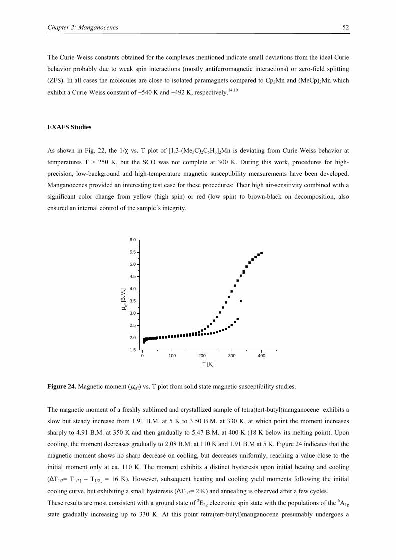
Chapter 2: Manganocenes
52
The Curie-Weiss constants obtained for the complexes mentioned indicate small deviations from the ideal Curie
behavior probably due to weak spin interactions (mostly antiferromagnetic interactions) or zero-field splitting
(ZFS). In all cases the molecules are close to isolated paramagnets compared to Cp2Mn and (MeCp)2Mn which
exhibit a Curie-Weiss constant of −540 K and −492 K, respectively.14,19
EXAFS Studies
As shown in Fig. 22, the 1/χ vs. T plot of [1,3-(Me3C)2C5H3]2Mn is deviating from Curie-Weiss behavior at
temperatures T > 250 K, but the SCO was not complete at 300 K. During this work, procedures for high-
precision, low-background and high-temperature magnetic susceptibility measurements have been developed.
Manganocenes provided an interesting test case for these procedures: Their high air-sensitivity combined with a
significant color change from yellow (high spin) or red (low spin) to brown-black on decomposition, also
ensured an internal control of the sample´s integrity.
0 100 200 300 4001.5
2.0
2.5
3.0
3.5
4.0
4.5
5.0
5.5
6.0
µ eff [
B.M
.]
T [K]
Figure 24. Magnetic moment (µeff) vs. T plot from solid state magnetic susceptibility studies.
The magnetic moment of a freshly sublimed and crystallized sample of tetra(tert-butyl)manganocene exhibits a
slow but steady increase from 1.91 B.M. at 5 K to 3.50 B.M. at 330 K, at which point the moment increases
sharply to 4.91 B.M. at 350 K and then gradually to 5.47 B.M. at 400 K (18 K below its melting point). Upon
cooling, the moment decreases gradually to 2.08 B.M. at 110 K and 1.91 B.M at 5 K. Figure 24 indicates that the
magnetic moment shows no sharp decrease on cooling, but decreases uniformly, reaching a value close to the
initial moment only at ca. 110 K. The moment exhibits a distinct hysteresis upon initial heating and cooling
(∆T1/2= T1/2↑ – T1/2↓ = 16 K). However, subsequent heating and cooling yield moments following the initial
cooling curve, but exhibiting a small hysteresis (∆T1/2= 2 K) and annealing is observed after a few cycles.
These results are most consistent with a ground state of 2E2g electronic spin state with the populations of the 6A1g
state gradually increasing up to 330 K. At this point tetra(tert-butyl)manganocene presumably undergoes a
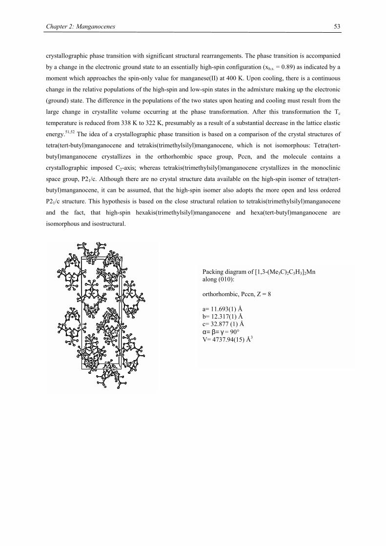
Chapter 2: Manganocenes
53
crystallographic phase transition with significant structural rearrangements. The phase transition is accompanied
by a change in the electronic ground state to an essentially high-spin configuration (xh.s. = 0.89) as indicated by a
moment which approaches the spin-only value for manganese(II) at 400 K. Upon cooling, there is a continuous
change in the relative populations of the high-spin and low-spin states in the admixture making up the electronic
(ground) state. The difference in the populations of the two states upon heating and cooling must result from the
large change in crystallite volume occurring at the phase transformation. After this transformation the Tc
temperature is reduced from 338 K to 322 K, presumably as a result of a substantial decrease in the lattice elastic
energy.51,52 The idea of a crystallographic phase transition is based on a comparison of the crystal structures of
tetra(tert-butyl)manganocene and tetrakis(trimethylsilyl)manganocene, which is not isomorphous: Tetra(tert-
butyl)manganocene crystallizes in the orthorhombic space group, Pccn, and the molecule contains a
crystallographic imposed C2-axis; whereas tetrakis(trimethylsilyl)manganocene crystallizes in the monoclinic
space group, P21/c. Although there are no crystal structure data available on the high-spin isomer of tetra(tert-
butyl)manganocene, it can be assumed, that the high-spin isomer also adopts the more open and less ordered
P21/c structure. This hypothesis is based on the close structural relation to tetrakis(trimethylsilyl)manganocene
and the fact, that high-spin hexakis(trimethylsilyl)manganocene and hexa(tert-butyl)manganocene are
isomorphous and isostructural.
Packing diagram of [1,3-(Me3C)2C5H3]2Mn along (010): orthorhombic, Pccn, Z = 8 a= 11.693(1) Å b= 12.317(1) Å c= 32.877 (1) Å α= β= γ = 90° V= 4737.94(15) Å3
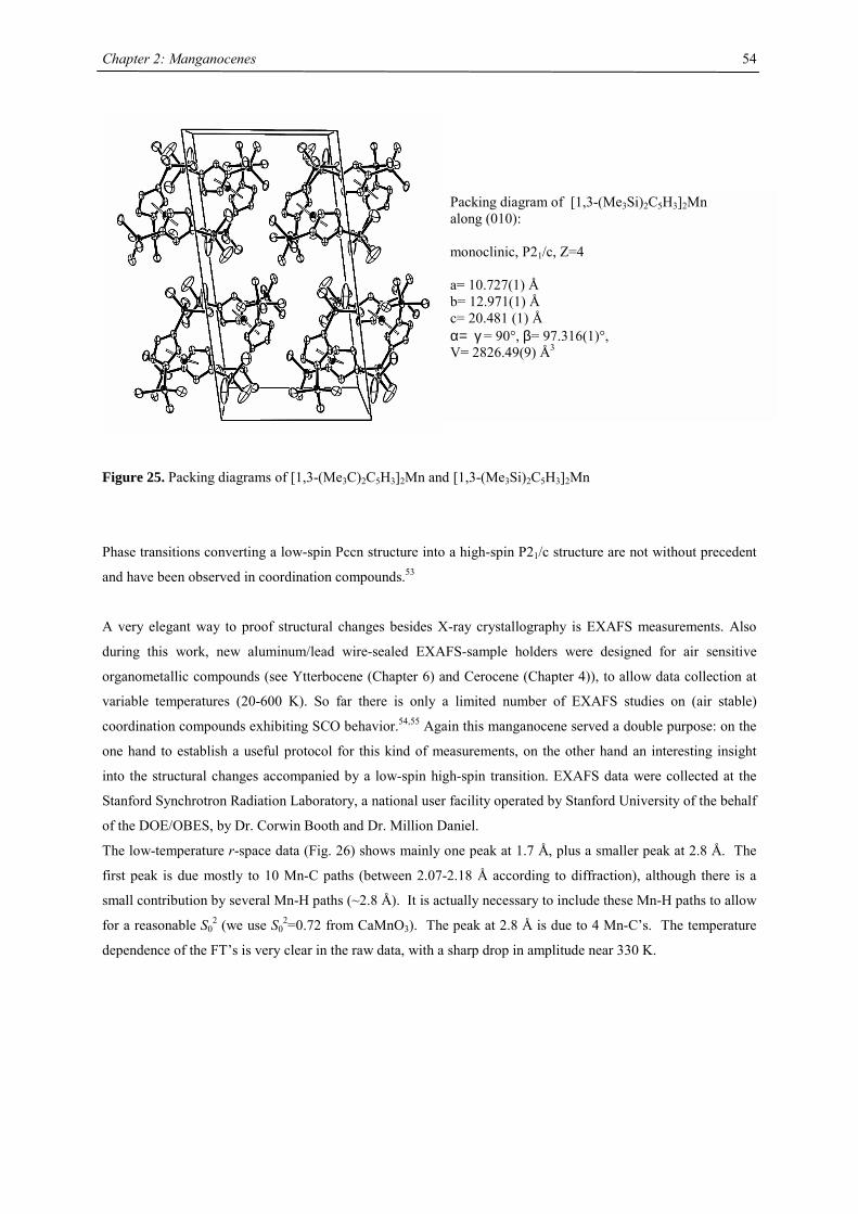
Chapter 2: Manganocenes
54
Figure 25. Packing diagrams of [1,3-(Me3C)2C5H3]2Mn and [1,3-(Me3Si)2C5H3]2Mn
Phase transitions converting a low-spin Pccn structure into a high-spin P21/c structure are not without precedent
and have been observed in coordination compounds.53
A very elegant way to proof structural changes besides X-ray crystallography is EXAFS measurements. Also
during this work, new aluminum/lead wire-sealed EXAFS-sample holders were designed for air sensitive
organometallic compounds (see Ytterbocene (Chapter 6) and Cerocene (Chapter 4)), to allow data collection at
variable temperatures (20-600 K). So far there is only a limited number of EXAFS studies on (air stable)
coordination compounds exhibiting SCO behavior.54,55 Again this manganocene served a double purpose: on the
one hand to establish a useful protocol for this kind of measurements, on the other hand an interesting insight
into the structural changes accompanied by a low-spin high-spin transition. EXAFS data were collected at the
Stanford Synchrotron Radiation Laboratory, a national user facility operated by Stanford University of the behalf
of the DOE/OBES, by Dr. Corwin Booth and Dr. Million Daniel.
The low-temperature r-space data (Fig. 26) shows mainly one peak at 1.7 Å, plus a smaller peak at 2.8 Å. The
first peak is due mostly to 10 Mn-C paths (between 2.07-2.18 Å according to diffraction), although there is a
small contribution by several Mn-H paths (~2.8 Å). It is actually necessary to include these Mn-H paths to allow
for a reasonable S02 (we use S0
2=0.72 from CaMnO3). The peak at 2.8 Å is due to 4 Mn-C’s. The temperature
dependence of the FT’s is very clear in the raw data, with a sharp drop in amplitude near 330 K.
Packing diagram of [1,3-(Me3Si)2C5H3]2Mn along (010): monoclinic, P21/c, Z=4 a= 10.727(1) Å b= 12.971(1) Å c= 20.481 (1) Å α= γ = 90°, β= 97.316(1)°, V= 2826.49(9) Å3
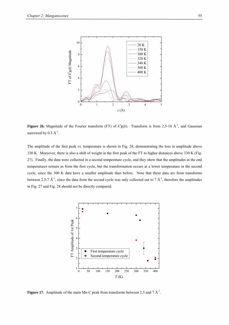
Chapter 2: Manganocenes
55
0 1 2 3 4 50
2
4
6
8
10
20 K 150 K 300 K 320 K 340 K 360 K 400 K
FT o
f k3 χ
(k) M
agni
tude
r (Å)
Figure 26. Magnitude of the Fourier transform (FT) of k3χ(k). Transform is from 2.5-10 Å-1, and Gaussian
narrowed by 0.3 Å-1.
The amplitude of the first peak vs. temperature is shown in Fig. 28, demonstrating the loss in amplitude above
330 K. Moreover, there is also a shift of weight in the first peak of the FT to higher distances above 330 K (Fig.
27). Finally, the data were collected in a second temperature cycle, and they show that the amplitudes at the end
temperatures remain as from the first cycle, but the transformation occurs at a lower temperature in the second
cycle, since the 300 K data have a smaller amplitude than before. Note that these data are from transforms
between 2.5-7 Å-1, since the data from the second cycle was only collected out to 7 Å-1, therefore the amplitudes
in Fig. 27 and Fig. 28 should not be directly compared.
0 50 100 150 200 250 300 350 4001
2
3
4
5
6
7
FT A
mpl
itude
of 1
st Pe
ak
T (K)
First temperature cycle Second temperature cycle
Figure 17. Amplitude of the main Mn-C peak from transforms between 2.5 and 7 Å-1.
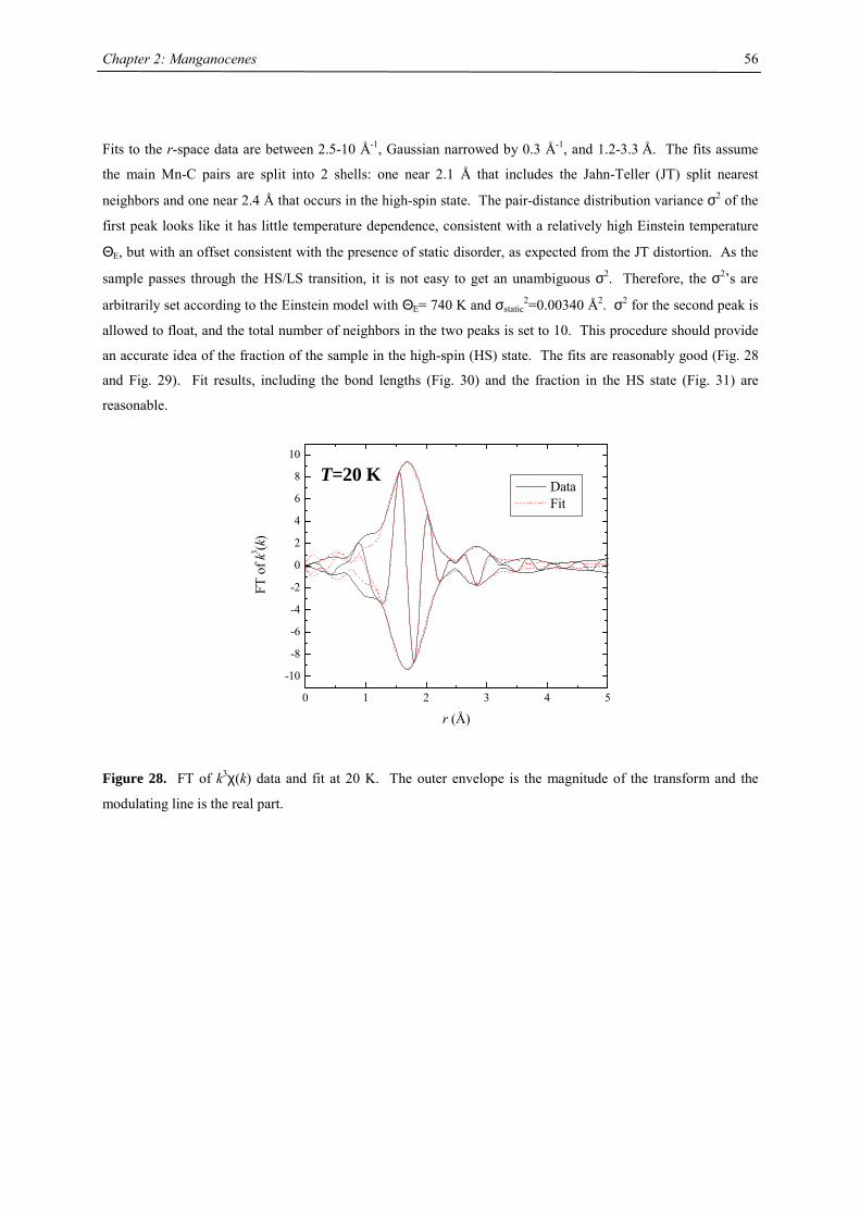
Chapter 2: Manganocenes
56
Fits to the r-space data are between 2.5-10 Å-1, Gaussian narrowed by 0.3 Å-1, and 1.2-3.3 Å. The fits assume
the main Mn-C pairs are split into 2 shells: one near 2.1 Å that includes the Jahn-Teller (JT) split nearest
neighbors and one near 2.4 Å that occurs in the high-spin state. The pair-distance distribution variance σ2 of the
first peak looks like it has little temperature dependence, consistent with a relatively high Einstein temperature
ΘE, but with an offset consistent with the presence of static disorder, as expected from the JT distortion. As the
sample passes through the HS/LS transition, it is not easy to get an unambiguous σ2. Therefore, the σ2’s are
arbitrarily set according to the Einstein model with ΘE= 740 K and σstatic2=0.00340 Å2. σ2 for the second peak is
allowed to float, and the total number of neighbors in the two peaks is set to 10. This procedure should provide
an accurate idea of the fraction of the sample in the high-spin (HS) state. The fits are reasonably good (Fig. 28
and Fig. 29). Fit results, including the bond lengths (Fig. 30) and the fraction in the HS state (Fig. 31) are
reasonable.
0 1 2 3 4 5
-10
-8
-6
-4
-2
0
2
4
6
8
10
T=20 K
FT o
f k3 (k
)
r (Å)
Data Fit
Figure 28. FT of k3χ(k) data and fit at 20 K. The outer envelope is the magnitude of the transform and the
modulating line is the real part.
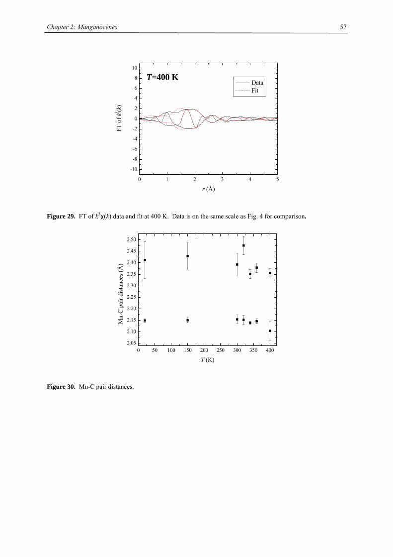
Chapter 2: Manganocenes
57
0 1 2 3 4 5
-10
-8
-6
-4
-2
0
2
4
6
8
10
T=400 K
FT o
f k3 (k
)
r (Å)
Data Fit
Figure 29. FT of k3χ(k) data and fit at 400 K. Data is on the same scale as Fig. 4 for comparison.
0 50 100 150 200 250 300 350 4002.05
2.10
2.15
2.20
2.25
2.30
2.35
2.40
2.45
2.50
Mn-
C pa
ir di
stanc
es (Å
)
T (K)
Figure 30. Mn-C pair distances.
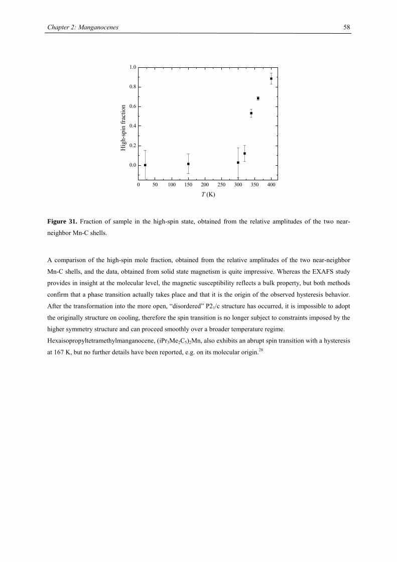
Chapter 2: Manganocenes
58
0 50 100 150 200 250 300 350 400
0.0
0.2
0.4
0.6
0.8
1.0
Hig
h-sp
in fr
actio
n
T (K)
Figure 31. Fraction of sample in the high-spin state, obtained from the relative amplitudes of the two near-
neighbor Mn-C shells.
A comparison of the high-spin mole fraction, obtained from the relative amplitudes of the two near-neighbor
Mn-C shells, and the data, obtained from solid state magnetism is quite impressive. Whereas the EXAFS study
provides in insight at the molecular level, the magnetic susceptibility reflects a bulk property, but both methods
confirm that a phase transition actually takes place and that it is the origin of the observed hysteresis behavior.
After the transformation into the more open, “disordered” P21/c structure has occurred, it is impossible to adopt
the originally structure on cooling, therefore the spin transition is no longer subject to constraints imposed by the
higher symmetry structure and can proceed smoothly over a broader temperature regime.
Hexaisopropyltetramethylmanganocene, (iPr3Me2C5)2Mn, also exhibits an abrupt spin transition with a hysteresis
at 167 K, but no further details have been reported, e.g. on its molecular origin.28
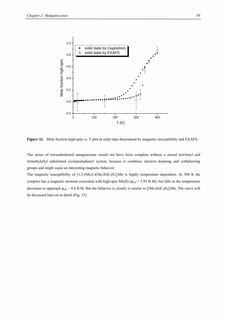
Chapter 2: Manganocenes
59
0 100 200 300 400-0.2
0.0
0.2
0.4
0.6
0.8
1.0
Mol
e fra
ctio
n hi
gh-s
pin
T (K)
solid state by magnetism solid state by EXAFS
Figure 32. Mole fraction high-spin vs. T plot in solid state determined by magnetic susceptibility and EXAFS.
The series of tetrasubstituted manganocene would not have been complete without a mixed tert-butyl and
trimethylsilyl substituted cyclopentadienyl system, because it combines electron donating and withdrawing
groups and might cause an interesting magnetic behavior.
The magnetic susceptibility of [1,3-(Me3C)(Me3Si)C5H3]2Mn is highly temperature dependent: At 300 K the
complex has a magnetic moment consistent with high-spin Mn(II) (µeff = 5.93 B.M), but falls as the temperature
decreases to approach µeff ~ 4.4 B.M. But the behavior is closely is similar to [(Me3Si)C5H4]2Mn. The curve will
be discussed later on in detail (Fig. 33).
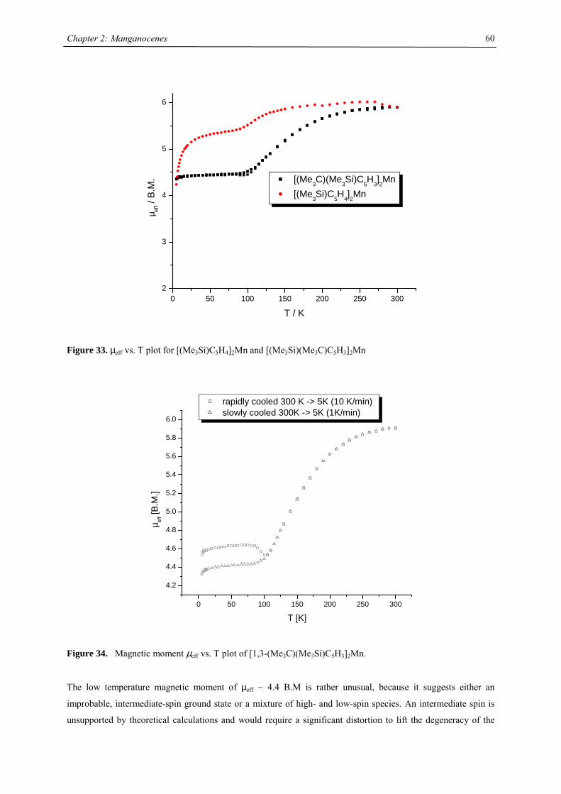
Chapter 2: Manganocenes
60
0 50 100 150 200 250 3002
3
4
5
6
µ eff /
B.M
.
T / K
[(Me3C)(Me3Si)C5H3]2Mn [(Me3Si)C5H4]2Mn
Figure 33. µeff vs. T plot for [(Me3Si)C5H4]2Mn and [(Me3Si)(Me3C)C5H3]2Mn
0 50 100 150 200 250 300
4.2
4.4
4.6
4.8
5.0
5.2
5.4
5.6
5.8
6.0
µ eff [B
.M.]
T [K]
rapidly cooled 300 K -> 5K (10 K/min) slowly cooled 300K -> 5K (1K/min)
Figure 34. Magnetic moment µeff vs. T plot of [1,3-(Me3C)(Me3Si)C5H3]2Mn.
The low temperature magnetic moment of µeff ~ 4.4 B.M is rather unusual, because it suggests either an
improbable, intermediate-spin ground state or a mixture of high- and low-spin species. An intermediate spin is
unsupported by theoretical calculations and would require a significant distortion to lift the degeneracy of the
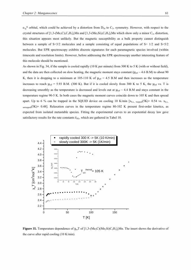
Chapter 2: Manganocenes
61
e1g* orbital, which could be achieved by a distortion from D5d to C2v symmetry. However, with respect to the
crystal structures of [1,3-(Me3C)2C5H3]2Mn and [1,3-(Me3Si)2C5H3]2Mn which show only a minor C2v distortion,
this situation appears most unlikely. But the magnetic susceptibility as a bulk property cannot distinguish
between a sample of S=3/2 molecules and a sample consisting of equal populations of S= 1/2 and S=5/2
molecules. But EPR spectroscopy exhibits discrete signatures for each paramagnetic species involved (within
timescale and resolution limits). However, before addressing the EPR spectroscopy another interesting feature of
this molecule should be mentioned.
As shown in Fig. 34, if the sample is cooled rapidly (10 K per minute) from 300 K to 5 K (with or without field),
and the data are then collected on slow heating, the magnetic moment stays constant (µeff ~ 4.6 B.M) to about 90
K, then it is dropping to a minimum at 105-110 K of µeff = 4.5 B.M and then increases as the temperature
increases to reach µeff = 5.93 B.M. (300 K). But if it is cooled slowly from 300 K to 5 K, the µeff vs. T is
decreasing smoothly as the temperature is decreased and levels out at µeff ~ 4.4 B.M and stays constant in the
temperature regime 90-5 K. In both cases the magnetic moment curves coincide down to 105 K and then spread
apart. Up to 6 % can be trapped in the SQUID device on cooling 10 K/min [xh.s., trapped(5K)= 0.54 vs. xh.s.,
untrapped(5K)= 0.48]. Relaxation curves in the temperature regime 80-102 K present first-order kinetics, as
expected from isolated metastable species. Fitting the experimental curves to an expotential decay law gave
satisfactory results for the rate constants kHL, which are gathered in Tabel 10.
Figure 35. Temperature dependence of χmT of [1,3-(Me3C)(Me3Si)C5H3]2Mn. The insert shows the derivative of
the curve after rapid cooling (10 K/min).
0 50 100 150
2.2
2.4
2.6
2.8
3.0
3.2
3.4
3.6
3.8
4.0
4.2
4.4
T1/2thermal= 105 K
40 60 80 100 120 140 160 180
-0.010
-0.005
0.000
0.005
0.010
0.015
0.020
δ(χ m
T)/δ
T
T [K]
χmT
[cm
3 mol
-1K
]
T [K]
rapidly cooled 300 K -> 5K (10 K/min) slowly cooled 300K -> 5K (1K/min)
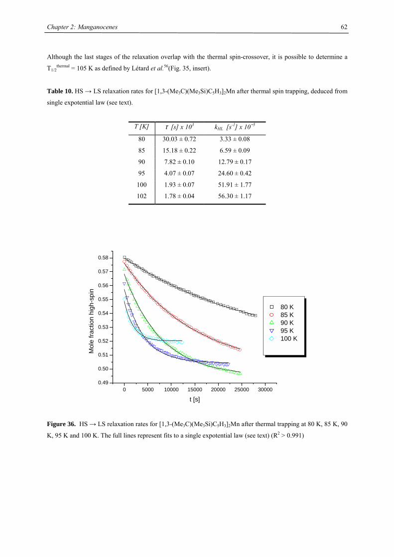
Chapter 2: Manganocenes
62
Although the last stages of the relaxation overlap with the thermal spin-crossover, it is possible to determine a
T1/2thermal = 105 K as defined by Létard et al.56(Fig. 35, insert).
Table 10. HS → LS relaxation rates for [1,3-(Me3C)(Me3Si)C5H3]2Mn after thermal spin trapping, deduced from
single expotential law (see text).
T [K] τ [s] x 103 kHL [s-1] x 10−5
80 30.03 ± 0.72 3.33 ± 0.08
85 15.18 ± 0.22 6.59 ± 0.09
90 7.82 ± 0.10 12.79 ± 0.17
95 4.07 ± 0.07 24.60 ± 0.42
100 1.93 ± 0.07 51.91 ± 1.77
102 1.78 ± 0.04 56.30 ± 1.17
0 5000 10000 15000 20000 25000 300000.49
0.50
0.51
0.52
0.53
0.54
0.55
0.56
0.57
0.58
Mol
e fra
ctio
n hi
gh-s
pin
t [s]
80 K 85 K 90 K 95 K 100 K
Figure 36. HS → LS relaxation rates for [1,3-(Me3C)(Me3Si)C5H3]2Mn after thermal trapping at 80 K, 85 K, 90
K, 95 K and 100 K. The full lines represent fits to a single expotential law (see text) (R2 > 0.991)
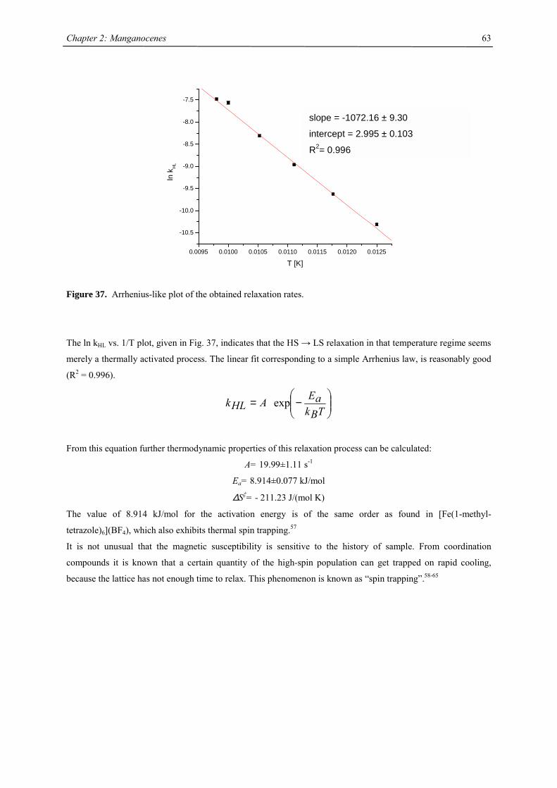
Chapter 2: Manganocenes
63
0.0095 0.0100 0.0105 0.0110 0.0115 0.0120 0.0125
-10.5
-10.0
-9.5
-9.0
-8.5
-8.0
-7.5
ln k
HL
T [K]
Figure 37. Arrhenius-like plot of the obtained relaxation rates.
The ln kHL vs. 1/T plot, given in Fig. 37, indicates that the HS → LS relaxation in that temperature regime seems
merely a thermally activated process. The linear fit corresponding to a simple Arrhenius law, is reasonably good
(R2 = 0.996).
−=
TBkaEAHLk exp
From this equation further thermodynamic properties of this relaxation process can be calculated:
A= 19.99±1.11 s-1
Ea= 8.914±0.077 kJ/mol
∆S‡= - 211.23 J/(mol K)
The value of 8.914 kJ/mol for the activation energy is of the same order as found in [Fe(1-methyl-
tetrazole)6](BF4), which also exhibits thermal spin trapping.57
It is not unusual that the magnetic susceptibility is sensitive to the history of sample. From coordination
compounds it is known that a certain quantity of the high-spin population can get trapped on rapid cooling,
because the lattice has not enough time to relax. This phenomenon is known as “spin trapping”.58-65
slope = -1072.16 ± 9.30
intercept = 2.995 ± 0.103
R2= 0.996
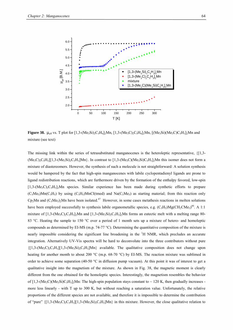
Chapter 2: Manganocenes
64
0 50 100 150 200 250 300
2.0
2.5
3.0
3.5
4.0
4.5
5.0
5.5
6.0
µ eff [
B.M
.]
T [K]
[1,3-(Me3Si)2C5H3]2Mn [1,3-(Me3C)2C5H3]2Mn mixture [1,3-(Me3C)(Me3Si)C5H3]2Mn
Figure 38. µeff vs. T plot for [1,3-(Me3Si)2C5H4]2Mn, [1,3-(Me3C)2C5H4]2Mn, [(Me3Si)(Me3C)C5H3]2Mn and
mixture (see text)
The missing link within the series of tetrasubstituted manganocenes is the heteroleptic representative, {[1,3-
(Me3C)2C5H3][1,3-(Me3Si)2C5H3]Mn}. In contrast to [1,3-(Me3C)(Me3Si)C5H3]2Mn this isomer does not form a
mixture of diastereomers. However, the synthesis of such a molecule is not straightforward: A solution synthesis
would be hampered by the fact that high-spin manganocenes with labile cyclopentadienyl ligands are prone to
ligand redistribution reactions, which are furthermore driven by the formation of the enthalpy favored, low-spin
[1,3-(Me3C)2C5H3]2Mn species. Similar experience has been made during synthetic efforts to prepare
(C5Me5)Mn(C5H5) by using (C5H5)MnCl(tmed) and Na(C5Me5) as starting material; from this reaction only
Cp2Mn and (C5Me5)2Mn have been isolated.47 However, in some cases metathesis reactions in molten solutions
have been employed successfully to synthesis labile organometallic species, e.g. (C5H5)Mg(CH2CMe3)66. A 1:1
mixture of [1,3-(Me3C)2C5H3]2Mn and [1,3-(Me3Si)2C5H3]2Mn forms an eutectic melt with a melting range 80-
83 °C. Heating the sample to 150 °C over a period of 1 month sets up a mixture of hetero- and homoleptic
compounds as determined by EI-MS (m.p. 74-77 °C). Determining the quantitative composition of the mixture is
nearly impossible considering the significant line broadening in the 1H NMR, which precludes an accurate
integration. Alternatively UV-Vis spectra will be hard to deconvolute into the three contributors without pure
{[1,3-(Me3C)2C5H3][1,3-(Me3Si)2C5H3]Mn} available. The qualitative composition does not change upon
heating for another month to about 200 °C (m.p. 68-70 °C) by EI-MS. The reaction mixture was sublimed in
order to achieve some separation (40-50 °C in diffusion pump vacuum). At this point it was of interest to get a
qualitative insight into the magnetism of the mixture. As shown in Fig. 38, the magnetic moment is clearly
different from the one obtained for the homoleptic species. Interestingly, the magnetism resembles the behavior
of [1,3-(Me3C)(Me3Si)C5H3]2Mn: The high-spin population stays constant to ~ 120 K, then gradually increases -
more less linearly - with T up to 300 K, but without reaching a saturation value. Unfortunately, the relative
proportions of the different species are not available, and therefore it is impossible to determine the contribution
of “pure” {[1,3-(Me3C)2C5H3][1,3-(Me3Si)2C5H3]Mn} in this mixture. However, the close qualitative relation to
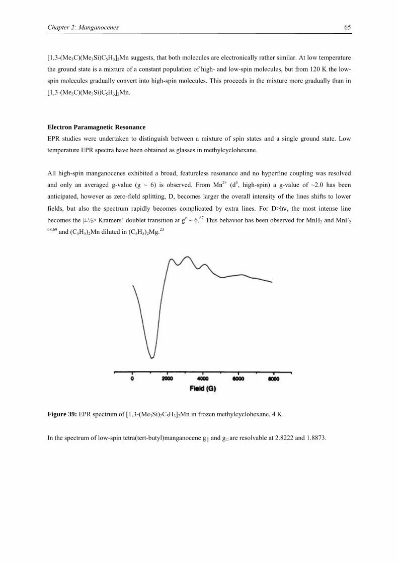
Chapter 2: Manganocenes
65
[1,3-(Me3C)(Me3Si)C5H3]2Mn suggests, that both molecules are electronically rather similar. At low temperature
the ground state is a mixture of a constant population of high- and low-spin molecules, but from 120 K the low-
spin molecules gradually convert into high-spin molecules. This proceeds in the mixture more gradually than in
[1,3-(Me3C)(Me3Si)C5H3]2Mn.
Electron Paramagnetic Resonance
EPR studies were undertaken to distinguish between a mixture of spin states and a single ground state. Low
temperature EPR spectra have been obtained as glasses in methylcyclohexane.
All high-spin manganocenes exhibited a broad, featureless resonance and no hyperfine coupling was resolved
and only an averaged g-value (g ~ 6) is observed. From Mn2+ (d5, high-spin) a g-value of ~2.0 has been
anticipated, however as zero-field splitting, D, becomes larger the overall intensity of the lines shifts to lower
fields, but also the spectrum rapidly becomes complicated by extra lines. For D>hν, the most intense line
becomes the |±½> Kramers’ doublet transition at ge ~ 6.67 This behavior has been observed for MnH2 and MnF2 68,69 and (C5H5)2Mn diluted in (C5H5)2Mg.23
Figure 39: EPR spectrum of [1,3-(Me3Si)2C5H3]2Mn in frozen methylcyclohexane, 4 K.
In the spectrum of low-spin tetra(tert-butyl)manganocene g║ and g⊥ are resolvable at 2.8222 and 1.8873.
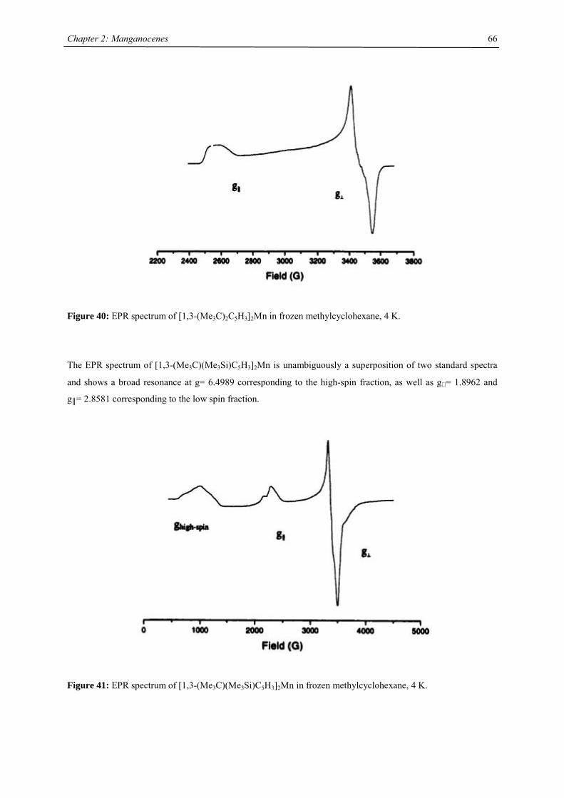
Chapter 2: Manganocenes
66
Figure 40: EPR spectrum of [1,3-(Me3C)2C5H3]2Mn in frozen methylcyclohexane, 4 K.
The EPR spectrum of [1,3-(Me3C)(Me3Si)C5H3]2Mn is unambiguously a superposition of two standard spectra
and shows a broad resonance at g= 6.4989 corresponding to the high-spin fraction, as well as g⊥ = 1.8962 and
g║= 2.8581 corresponding to the low spin fraction.
Figure 41: EPR spectrum of [1,3-(Me3C)(Me3Si)C5H3]2Mn in frozen methylcyclohexane, 4 K.
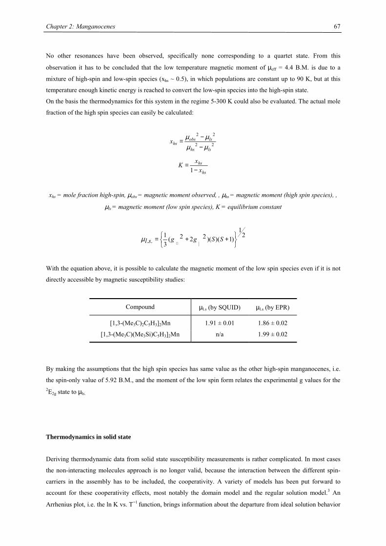
Chapter 2: Manganocenes
67
No other resonances have been observed, specifically none corresponding to a quartet state. From this
observation it has to be concluded that the low temperature magnetic moment of µeff = 4.4 B.M. is due to a
mixture of high-spin and low-spin species (xhs ~ 0.5), in which populations are constant up to 90 K, but at this
temperature enough kinetic energy is reached to convert the low-spin species into the high-spin state.
On the basis the thermodynamics for this system in the regime 5-300 K could also be evaluated. The actual mole
fraction of the high spin species can easily be calculated:
22
22
lshs
lsobshsx
µµµµ
−−=
hs
hs
xxK−
=1
xhs = mole fraction high-spin, µobs = magnetic moment observed, , µhs = magnetic moment (high spin species), ,
µls = magnetic moment (low spin species), K = equilibrium constant
21
)1)()(222(31
..
++=
⊥SSggslµ
With the equation above, it is possible to calculate the magnetic moment of the low spin species even if it is not
directly accessible by magnetic susceptibility studies:
Compound µl.s (by SQUID) µl.s (by EPR)
[1,3-(Me3C)2C5H3]2Mn 1.91 ± 0.01 1.86 ± 0.02
[1,3-(Me3C)(Me3Si)C5H3]2Mn n/a 1.99 ± 0.02
By making the assumptions that the high spin species has same value as the other high-spin manganocenes, i.e.
the spin-only value of 5.92 B.M., and the moment of the low spin form relates the experimental g values for the 2E2g state to µls.
Thermodynamics in solid state
Deriving thermodynamic data from solid state susceptibility measurements is rather complicated. In most cases
the non-interacting molecules approach is no longer valid, because the interaction between the different spin-
carriers in the assembly has to be included, the cooperativity. A variety of models has been put forward to
account for these cooperativity effects, most notably the domain model and the regular solution model.3 An
Arrhenius plot, i.e. the ln K vs. T−1 function, brings information about the departure from ideal solution behavior
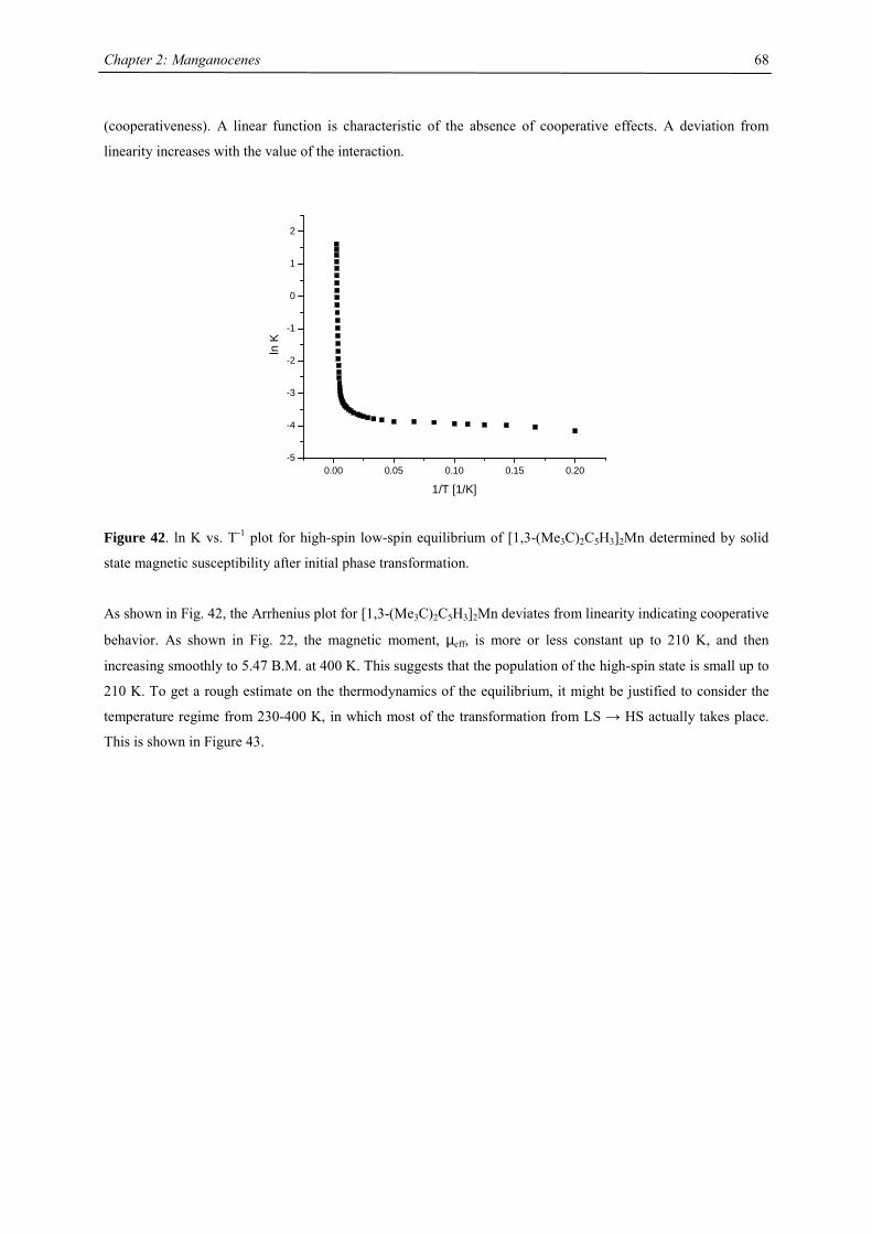
Chapter 2: Manganocenes
68
(cooperativeness). A linear function is characteristic of the absence of cooperative effects. A deviation from
linearity increases with the value of the interaction.
0.00 0.05 0.10 0.15 0.20-5
-4
-3
-2
-1
0
1
2
ln K
1/T [1/K]
Figure 42. ln K vs. T-1 plot for high-spin low-spin equilibrium of [1,3-(Me3C)2C5H3]2Mn determined by solid
state magnetic susceptibility after initial phase transformation.
As shown in Fig. 42, the Arrhenius plot for [1,3-(Me3C)2C5H3]2Mn deviates from linearity indicating cooperative
behavior. As shown in Fig. 22, the magnetic moment, µeff, is more or less constant up to 210 K, and then
increasing smoothly to 5.47 B.M. at 400 K. This suggests that the population of the high-spin state is small up to
210 K. To get a rough estimate on the thermodynamics of the equilibrium, it might be justified to consider the
temperature regime from 230-400 K, in which most of the transformation from LS → HS actually takes place.
This is shown in Figure 43.
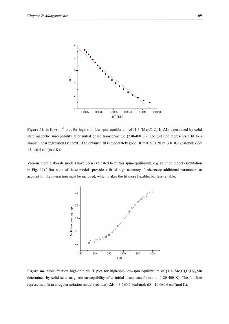
Chapter 2: Manganocenes
69
0.0025 0.0030 0.0035 0.0040 0.0045 0.0050-3
-2
-1
0
1
2
ln K
1/T [1/K]
Figure 43. ln K vs. T-1 plot for high-spin low-spin equilibrium of [1,3-(Me3C)2C5H3]2Mn determined by solid
state magnetic susceptibility after initial phase transformation (230-400 K). The full line represents a fit to a
simple linear regression (see text). The obtained fit is moderately good (R2= 0.973). ∆H= 3.8±0.2 kcal/mol; ∆S=
12.1±0.5 cal/(mol K).
Various more elaborate models have been evaluated to fit this spin-equilibrium, e.g. solution model (simulation
in Fig. 44).3 But none of these models provide a fit of high accuracy, furthermore additional parameters to
account for the interaction must be included, which makes the fit more flexible, but less reliable.
150 200 250 300 350 400
0.0
0.2
0.4
0.6
0.8
Mol
e fra
ctio
n hi
gh-s
pin
T [K]
Figure 44. Mole fraction high-spin vs. T plot for high-spin low-spin equilibrium of [1,3-(Me3C)2C5H3]2Mn
determined by solid state magnetic susceptibility after initial phase transformation (180-400 K). The full line
represents a fit to a regular solution model (see text). ∆H= 3.5±0.2 kcal/mol; ∆S= 10.6±0.6 cal/(mol K).
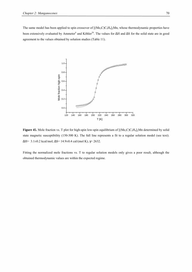
Chapter 2: Manganocenes
70
The same model has been applied to spin crossover of [(Me3C)C5H4]2Mn, whose thermodynamic properties have
been extensively evaluated by Ammeter6 and Köhler26. The values for ∆H and ∆S for the solid state are in good
agreement to the values obtained by solution studies (Table 11).
120 140 160 180 200 220 240 260 280 300 320
0.0
0.2
0.4
0.6
0.8
1.0
Mol
e fra
ctio
n hi
gh-s
pin
T [K]
Figure 45. Mole fraction vs. T plot for high-spin low-spin equilibrium of [(Me3C)C5H4]2Mn determined by solid
state magnetic susceptibility (150-300 K). The full line represents a fit to a regular solution model (see text).
∆H= 3.1±0.2 kcal/mol; ∆S= 14.9±0.4 cal/(mol K), γ= 2632.
Fitting the normalized mole fractions vs. T to regular solution models only gives a poor result, although the
obtained thermodynamic values are within the expected regime.
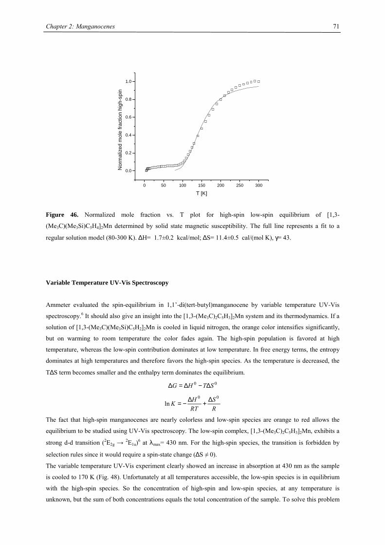
Chapter 2: Manganocenes
71
0 50 100 150 200 250 300
0.0
0.2
0.4
0.6
0.8
1.0
Nor
mal
ized
mol
e fra
ctio
n hi
gh-s
pin
T [K]
Figure 46. Normalized mole fraction vs. T plot for high-spin low-spin equilibrium of [1,3-
(Me3C)(Me3Si)C5H4]2Mn determined by solid state magnetic susceptibility. The full line represents a fit to a
regular solution model (80-300 K). ∆H= 1.7±0.2 kcal/mol; ∆S= 11.4±0.5 cal/(mol K), γ= 43.
Variable Temperature UV-Vis Spectroscopy
Ammeter evaluated the spin-equilibrium in 1,1’-di(tert-butyl)manganocene by variable temperature UV-Vis
spectroscopy.6 It should also give an insight into the [1,3-(Me3C)2C5H3]2Mn system and its thermodynamics. If a
solution of [1,3-(Me3C)(Me3Si)C5H2]2Mn is cooled in liquid nitrogen, the orange color intensifies significantly,
but on warming to room temperature the color fades again. The high-spin population is favored at high
temperature, whereas the low-spin contribution dominates at low temperature. In free energy terms, the entropy
dominates at high temperatures and therefore favors the high-spin species. As the temperature is decreased, the
T∆S term becomes smaller and the enthalpy term dominates the equilibrium. 00 STHG ∆−∆=∆
RS
RTHK
00ln ∆+∆−=
The fact that high-spin manganocenes are nearly colorless and low-spin species are orange to red allows the
equilibrium to be studied using UV-Vis spectroscopy. The low-spin complex, [1,3-(Me3C)2C5H3]2Mn, exhibits a
strong d-d transition (2E2g → 2E1u)6 at λmax= 430 nm. For the high-spin species, the transition is forbidden by
selection rules since it would require a spin-state change (∆S ≠ 0).
The variable temperature UV-Vis experiment clearly showed an increase in absorption at 430 nm as the sample
is cooled to 170 K (Fig. 48). Unfortunately at all temperatures accessible, the low-spin species is in equilibrium
with the high-spin species. So the concentration of high-spin and low-spin species, at any temperature is
unknown, but the sum of both concentrations equals the total concentration of the sample. To solve this problem
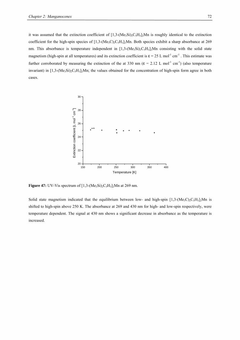
Chapter 2: Manganocenes
72
it was assumed that the extinction coefficient of [1,3-(Me3Si)2C5H3]2Mn is roughly identical to the extinction
coefficient for the high-spin species of [1,3-(Me3C)2C5H3]2Mn. Both species exhibit a sharp absorbance at 269
nm. This absorbance is temperature independent in [1,3-(Me3Si)2C5H3]2Mn consisting with the solid state
magnetism (high-spin at all temperatures) and its extinction coefficient is ε = 25 L mol-1 cm-1 . This estimate was
further corroborated by measuring the extinction of the at 330 nm (ε = 2.12 L mol-1 cm-1) (also temperature
invariant) in [1,3-(Me3Si)2C5H3]2Mn; the values obtained for the concentration of high-spin form agree in both
cases.
150 200 250 300 350 40020
22
24
26
28
30
Ext
inct
ion
coef
ficie
nt [L
mol
−1 c
m−1
]
Temperature [K]
Figure 47: UV-Vis spectrum of [1,3-(Me3Si)2C5H3]2Mn at 269 nm.
Solid state magnetism indicated that the equilibrium between low- and high-spin [1,3-(Me3C)2C5H3]2Mn is
shifted to high-spin above 250 K. The absorbance at 269 and 430 nm for high- and low-spin respectively, were
temperature dependent. The signal at 430 nm shows a significant decrease in absorbance as the temperature is
increased.
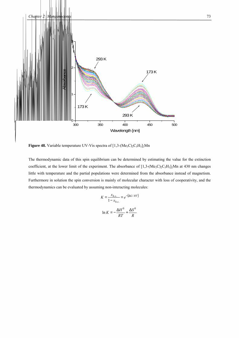
Chapter 2: Manganocenes
73
300 350 400 450 5000
1
2
3
293 K
293 K
173 K
173 K
Abs
orba
nce
Wavelength [nm]
Figure 48. Variable temperature UV-Vis spectra of [1,3-(Me3C)2C5H3]2Mn
The thermodynamic data of this spin equilibrium can be determined by estimating the value for the extinction
coefficient, at the lower limit of the experiment. The absorbance of [1,3-(Me3C)2C5H3]2Mn at 430 nm changes
little with temperature and the partial populations were determined from the absorbance instead of magnetism.
Furthermore in solution the spin conversion is mainly of molecular character with loss of cooperativity, and the
thermodynamics can be evaluated by assuming non-interacting molecules:
( )RTG
sh
sh ex
xK /
..
..
1∆−=
−=
RS
RTHK
00ln ∆+∆−=
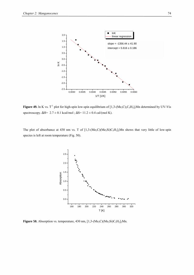
Chapter 2: Manganocenes
74
0.0030 0.0035 0.0040 0.0045 0.0050 0.0055 0.0060-2.5
-2.0
-1.5
-1.0
-0.5
0.0
0.5
1.0
1.5
2.0
ln K
1/T [1/K]
lnK linear regression
Figure 49. ln K vs. T-1 plot for high-spin low-spin equilibrium of [1,3-(Me3C)2C5H3]2Mn determined by UV-Vis
spectroscopy. ∆H= 2.7 ± 0.1 kcal/mol ; ∆S= 11.2 ± 0.4 cal/(mol K).
The plot of absorbance at 430 nm vs. T of [1,3-(Me3C)(Me3Si)C5H2]2Mn shows that very little of low-spin
species is left at room temperature (Fig. 50).
160 180 200 220 240 260 280 300 320
0.0
0.5
1.0
1.5
2.0
2.5
Abs
orpt
ion
T [K]
Figure 50. Absorption vs. temperature, 430 nm, [1,3-(Me3C)(Me3Si)C5H2]2Mn.
slope = -1356.44 ± 41.90
intercept = 5.616 ± 0.186
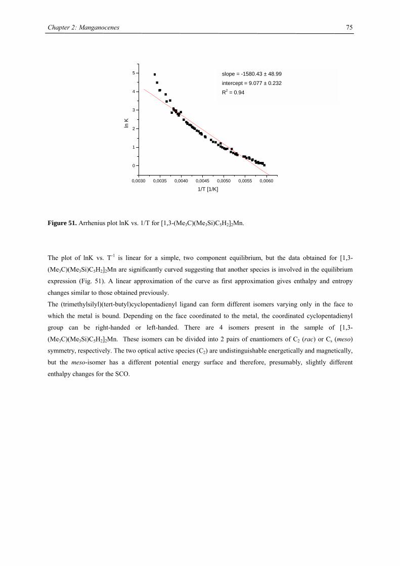
Chapter 2: Manganocenes
75
0,0030 0,0035 0,0040 0,0045 0,0050 0,0055 0,0060
0
1
2
3
4
5
ln K
1/T [1/K]
Figure 51. Arrhenius plot lnK vs. 1/T for [1,3-(Me3C)(Me3Si)C5H2]2Mn.
The plot of lnK vs. T-1 is linear for a simple, two component equilibrium, but the data obtained for [1,3-
(Me3C)(Me3Si)C5H2]2Mn are significantly curved suggesting that another species is involved in the equilibrium
expression (Fig. 51). A linear approximation of the curve as first approximation gives enthalpy and entropy
changes similar to those obtained previously.
The (trimethylsilyl)(tert-butyl)cyclopentadienyl ligand can form different isomers varying only in the face to
which the metal is bound. Depending on the face coordinated to the metal, the coordinated cyclopentadienyl
group can be right-handed or left-handed. There are 4 isomers present in the sample of [1,3-
(Me3C)(Me3Si)C5H2]2Mn. These isomers can be divided into 2 pairs of enantiomers of C2 (rac) or Cs (meso)
symmetry, respectively. The two optical active species (C2) are undistinguishable energetically and magnetically,
but the meso-isomer has a different potential energy surface and therefore, presumably, slightly different
enthalpy changes for the SCO.
slope = -1580.43 ± 48.99
intercept = 9.077 ± 0.232
R2 = 0.94
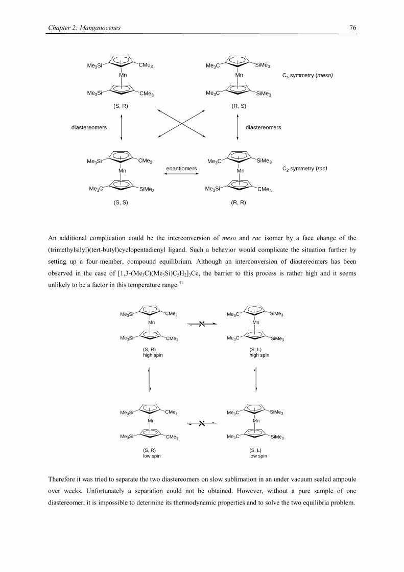
Chapter 2: Manganocenes
76
Mn
Me3Si CMe3
Me3Si CMe3
Mn
Me3C SiMe3
Me3C SiMe3
Mn
Me3Si CMe3
Me3C SiMe3
Mn
Me3C SiMe3
Me3Si CMe3
(S, R) (R, S)
(S, S) (R, R)
diastereomers diastereomers
enantiomers
Cs symmetry (meso)
C2 symmetry (rac)
An additional complication could be the interconversion of meso and rac isomer by a face change of the
(trimethylsilyl)(tert-butyl)cyclopentadienyl ligand. Such a behavior would complicate the situation further by
setting up a four-member, compound equilibrium. Although an interconversion of diastereomers has been
observed in the case of [1,3-(Me3C)(Me3Si)C5H2]3Ce, the barrier to this process is rather high and it seems
unlikely to be a factor in this temperature range.41
Mn
Me3Si CMe3
Me3Si CMe3
Mn
Me3C SiMe3
Me3C SiMe3
(S, R)high spin
(S, L)high spin
Mn
Me3Si CMe3
Me3Si CMe3
(S, R)low spin
Mn
Me3C SiMe3
Me3C SiMe3
(S, L)low spin
Therefore it was tried to separate the two diastereomers on slow sublimation in an under vacuum sealed ampoule
over weeks. Unfortunately a separation could not be obtained. However, without a pure sample of one
diastereomer, it is impossible to determine its thermodynamic properties and to solve the two equilibria problem.
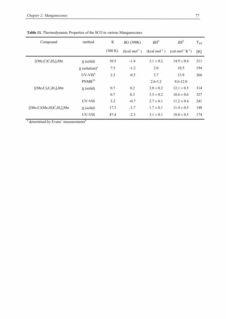
Chapter 2: Manganocenes
77
Table 11. Thermodynamic Properties of the SCO in various Manganocenes
Compound method K
(300 K)
∆G (300K)
(kcal mol-1 )
∆H0
(kcal mol-1 )
∆S0
(cal mol-1 K-1)
Τ1/2
[Κ]
[(Me3C)C5H4]2Mn χ (solid) 10.5 -1.4 3.1 ± 0.2 14.9 ± 0.4 211
χ (solution)a 7.5 -1.2 2.0 10.5 194
UV-VIS6 2.3 -0.5 3.7 13.9 266
PNMR26 2.6-3.2 9.6-12.0
[(Me3C)2C5H3]2Mn χ (solid) 0.7
0.7
0.2
0.3
3.8 ± 0.2
3.5 ± 0.2
12.1 ± 0.5
10.6 ± 0.6
314
327
UV-VIS 3.2 -0.7 2.7 ± 0.1 11.2 ± 0.4 241
[(Me3C)(Me3Si)C5H3]2Mn χ (solid) 17.3 -1.7 1.7 ± 0.1 11.4 ± 0.5 148
UV-VIS 47.4 -2.3 3.1 ± 0.1 18.0 ± 0.5 174 a determined by Evans’ measurements6
Chapter 2: Manganocenes
78
Conclusions
A series of manganocenes have been synthesized and the influence of substituents on the electronic structures
has been evaluated by UV-Vis, EPR, crystal structure determination, solid state magnetic susceptibility studies
and by the determination of thermodynamics of the SCO process.
Methylation of cyclopentadienyl rings has consistently been found to favor the low-spin form of manganocenes.
Manganocene itself is essentially high spin at room temperature and above (5.50 B.M. at 373 K). Diluting
manganocene in diamagnetic host lattices also influences the spin state: In magnesocene, which provides a rather
big cavity, manganocene is high-spin at all accessible temperatures. In ferrocene manganocene is low-spin at 5
K, but starts converting into the high-spin species on heating. This SCO temperature depends on the
concentration of Cp2Mn diluted in Cp2Fe; the higher the concentration of Cp2Mn, the higher the SCO
temperature. 1,1’-(MeCp)2Mn is found to be a chain polymer in solid state comparable to Cp2Mn and its
magnetism can also be simulated by a linear Heisenberg chain including the Curie-Weiss constant to account for
inter-chain interactions. Gaseous (MeCp)2Mn is a mixture of both low- and high-spin species in almost equal
populations. This preference for low-spin is also reflected by dilution experiment in (MeCp)2Fe, in which the
SCO temperature is significantly higher than in the analogue experiment of Cp2Mn.
Magnetic susceptibility measurements and X-ray data confirm that tert-butyl substituted manganocenes
[(Me3C)nC5H5-n]2Mn (n= 1, 2) follow the trend previously observed with the methylated manganocenes, i.e. the
more electron donating groups are attached to the Cp ring the more stabilized the low-spin electronic ground
state relative to Cp2Mn and the higher the SCO temperature. However, introducing three CMe3 groups on each
ring realizes a temperature invariant high-spin state. The origin of the high-spin state in [1,2,4-(Me3C)3C5H2]2Mn
is due to the significant bulk of the [1,2,4-(Me3C)3C5H2]- ligand, which is sufficient to generate severe inter-ring
steric strain preventing the realization of the low spin state. Interestingly, the spin transition in [1,3-
(Me3C)2C5H3]2Mn is accompanied by a phase transition resulting in a significant irreversible hysteresis (∆Tc =
16 K). This structural transition was also observed by EXAFS measurements.
Magnetic susceptibility studies and X-ray data on SiMe3 substituted manganocenes [(Me3Si)nC5H5-n]2Mn (n= 1,
2, 3) show clear high-spin configuration. Although tetra- and hexasubstituted manganocenes are high spin at all
accessible temperatures, the disubstituted manganocenes exhibit a small low-spin admixture at low temperature.
In this respect it behaves similar to [(Me3C)(Me3Si)C5H3]2Mn, which has a constant low spin admixture up to 90
K and then gradually converts to high-spin.
In the case of [(Me3C)(Me3Si)C5H3]2Mn thermal spin-trapping on rapid cooling can be observed.
Chapter 2: Manganocenes
79
References
[1] Ammeter, J. H. Nouv. J. Chim. 1980, 4, 631-637.
[2] Kahn, O.; Martinez, C. J. Science 1998, 279, 44-48.
[3] Kahn, O. Molecular Magnetism; VCH: New York, 1993.
[4] Zarembowitch, J.; Kahn, O. New J. Chem. 1981, 15, 181-190.
[5] Bolvich, H.; Kahn, O.; Vekhter, B. New J. Chem. 1991, 15, 889-895.
[6] Ammeter, J. H.; Zoller, L.; Bachmann, J.; Baltzer, P.; Gamp, E.; Bucher, R.; Deiss, E. Helv. Chim. Acta
1981, 64, 1063-1082.
[7] Boca, R. Theoretical Foundations of Molecular Magnetism; Elsevier: Amsterdam, 1999.
[8] Gütlich, P.; van Koningsbruggen, P. J.; Renz, F. Struct. Bonding 2004, 107, 24-75; and references
therein.
[9] Garcia, Y.; Gütlich, P. Top. Curr. Chem. 2004, 234, 49-62; and references cited therein.
[10] Hoganson, C. W.; Babcock, G. T. Science 1997, 277, 1953.
[11] Albright, T. A.; Burdett, J. K.; M.-H., W. Orbital Interactions in Chemistry; J. Wiley & Son: New
York, 1985.
[12] Sitzmann, H. Coord. Chem. Rev. 2001, 214, 287-327.
[13] Hays, M. L.; Hanusa, T. P. Adv. Organomet. Chem. 1996, 40, 117-170.
[14] Wilkinson, G.; Cotton, F. A. Chem. Ind. (London) 1954, 11, 307.
[15] Heyn, B.; Hipler, B.; Kreisel, G.; Schreer, H.; Walther, D. Anorganische Synthesechemie; Springer-
Verlag: Berlin, 1986.
[16] Riemscheider, R.; Kassahu, H.-G.; Schneider, W. Z. Naturforsch. 1960, 15b, 547-551.
[17] Kern, R. J. J. Inorg. Nucl. Chem. 1962, 24, 1105-1109.
[18] Sobota, P.; Utko, J.; Jerzykiewicz, L. Inorg. Chem. 1998, 37, 3428-3431.
[19] Wilkinson, G.; Cotton, F. A.; Birmingham, J. M. J. Inorg. Nucl. Chem. 1956, 2, 95-113.
[20] Reynolds, L. T.; Wilkinson, G. J. Inorg. Nucl. Chem. 1958, 9, 86-92.
[21] Robbins, J. L.; Edelstein, N. M.; Cooper, S. R.; Smart, J. C. J. Am. Chem. Soc. 1979, 101, 3853-3857.
[22] Ammeter, J. H.; Bucher, R.; Oswald, N. J. Am. Chem. Soc. 1974, 96, 7833-7835.
[23] Ammeter, J. H. J. Magn. Reson. 1978, 30, 299-325.
[24] Köhler, F. H.; Schlesinger, B. Inorg. Chem. 1992, 31, 2853-2859.
[25] Köhler, F. H.; Hebendanz, N. Chem. Ber. 1983, 116, 1261-1263.
[26] Hebendanz, N.; Köhler, F. H.; Müller, G.; Riede, J. J. Am. Chem. Soc. 1986, 108, 3281-3289.
[27] Hays, M. L.; Burkey, D. J.; Overby, J. S.; Hanusa, T. P.; Sellers, S. P.; Yee, G. T.; Young, V. G.
Organometallics 1998, 17, 5521-5527.
[28] Sitzmann, H.; Schär, M.; Dormann, E.; Kelemen, M. Z. Allgem. Anorg. Chem. 1997, 623, 1609-1613.
[29] Bünder, W.; Weiss, E. Z. Naturforsch. 1978, 33b, 1237-1238.
[30] König, E.; Desai, V. P.; Kanellakopulos, B.; Klenze, R. Chem. Phys. 1980, 54, 109-113.
[31] Haaland, A. Top. Curr. Chem. 1975, 53, 1-23.
[32] Haaland, A. Inorg. Nucl. Chem. Lett. 1979, 15, 267-269.
[33] Evans, S.; Green, M. L. H.; Jewitt, B.; King, G. H.; Orchard, A. F. J. Chem. Soc., Faraday II 1974, 70,
356-376.
Chapter 2: Manganocenes
80
[34] Almenningen, A.; Haaland, A.; Samdal, S. J. Organomet. Chem. 1978, 149, 219-229.
[35] Rettig, M. F.; Switzer, M. E. J. Am. Chem. Soc. 1974, 96, 7669-7674.
[36] Rabalais, J. W.; Werme, L. O.; Bergmark, T.; Karlsson, L.; Hussain, M.; Siegbahn, K. J. Chem. Phys.
1972, 57, 1185-1192.
[37] Barker, P. J.; Davies, A. G.; Tse, M. J. Chem. Soc. Perkin II 1980, 941-948.
[38] Kira, M.; Watanabe, M.; Sakurai, H. J. Am. Chem. Soc. 1980, 102, 5202-5207.
[39] Smart, J. C.; Robbins, J. L. J. Am. Chem. Soc. 1978, 100, 3936-3937.
[40] Freyberg, D. P.; Robbins, J. L.; Raymond, K. N.; Smart, J. C. J. Am. Chem. Soc. 1979, 101, 892-897.
[41] Sofield, C. D., Ph.D. Thesis, University of California, Berkeley: Berkeley, 2000.
[42] La Mar, G. N.; Horrocks, W. D.; Holm, R. H., Eds. NMR of Paramagnetic Molecules; Academic Press:
New York, 1973.
[43] Berndt, A. F.; Marsh, R. E. Acta Cryst. 1963, 16, 118.
[44] Granoff, B.; Jacobson, R. A. Inorg. Chem. 1968, 7, 2328.
[45] Gardiner, M. G.; Raston, C. L.; Kennard, C. H. L. Organometallics 1991, 10, 3680-3686.
[46] Shannon, R. D. Acta Cryst. A 1976, A32, 751-767.
[47] Andersen, R. A., private communication.
[48] Weber, F.; Sitzmann, H.; Schultz, M.; Sofield, C. D.; Andersen, R. A. Organometallics 2002, 21, 3139-
3146.
[49] Morley, C. P.; Jutzi, P.; Krüger, C.; Wallis, J. M. Organometallics 1987, 6, 1084-1090.
[50] Krinsky, J. L.; Stavis, M. N.; Walter, M. D. Acta Cryst. E 2003, 59, m497-m499.
[51] Spiering, H.; Meissner, E.; Köppen, H.; Müller, E. W.; Gütlich, P. Chem. Phys. 1982, 68, 65-71.
[52] Adler, P.; Wiehl, L.; Meissner, E.; Köhler, C. P.; Spiering, H.; Gütlich, P. J. Phys. Chem. Sol. 1987, 48,
517-525.
[53] Guionneau, P.; Létard, J.-F.; Yufit, D. S.; Chasseau, D.; Bravic, G.; Goeta, A. E.; Howard, J. A. K.;
Kahn, O. J. Mater. Chem. 1999, 9, 985-994.
[54] Okamoto, K.; Nagai, K.; Miyawaki, J.; Kondoh, H.; Ohta, T. Chem. Phys. Lett. 2003, 371, 707-712.
[55] Thuery, P.; Zarembowitch, J.; Michalowicz, A.; Kahn, O. Inorg. Chem. 1987, 26, 851-855.
[56] Létard, J.-F.; Capes, L.; Chastanet, G.; Moliner, N.; Létard, S.; Real, J.-A.; Kahn, O. Chem. Phys. Lett.
1999, 313, 115-120.
[57] Roubeau, O.; Stassen, A. F.; Gramage, I. F.; Codjovi, E.; Linarès, J.; Varret, F.; Haasnoot, J. G.;
Reedijk, J. Polyhedron 2001, 20, 1709-1716.
[58] Buchen, T.; Gütlich, P.; Sugiyarto, K. H.; Goodwin, H. A. Chem. Eur. J. 1996, 2, 1134-1138.
[59] Buchen, T.; Gütlich, P.; Goodwin, H. A. Inorg. Chem. 1994, 33, 4573-4576.
[60] Hauser, A. Chem. Phys. Lett. 1992, 192, 65-70.
[61] Hauser, A. Comments Inorg. Chem. 1995, 17, 17-40.
[62] Marchivie, M.; Guionneau, P.; Létard, J.-F.; Chasseau, D.; Howard, J. A. K. J. Phys. Chem. Sol. 2004,
65, 17-23.
[63] Roubeau, O.; deVos, M.; Stassen, A. F.; Burriel, R.; Haasnoot, J. G.; Reedijk, J. J. Phys. Chem. Sol.
2003, 64, 1003-1013.
[64] Ritter, G.; König, E.; Irler, W.; Goodwin, H. A. Inorg. Chem. 1978, 17, 224-228.
[65] Yu, Z.; Liu, K.; Tao, J. Q.; Zhong, Z. J.; You, X. Z.; Siu, G. G. Appl. Phys. Lett. 1999, 74, 4029-4031.

Chapter 2: Manganocenes
81
[66] Andersen, R. A.; Blom, R.; Haaland, A.; Schilling, B. E. R.; Volden, H. V. Acta Chim. Scand. A 1985,
39, 563-569.
[67] Weltner, W. Magnetic Atoms and Molecules; Dover Publications: New York, 1983.
[68] Van Zee, R. J.; Brown, C. M.; Weltner, W. Chem. Phys. Lett. 1979, 64, 325-328.
[69] DeVore, T. C.; Van Zee, R. J.; Weltner, W. J. Chem. Phys. 1978, 68, 3522-3527.
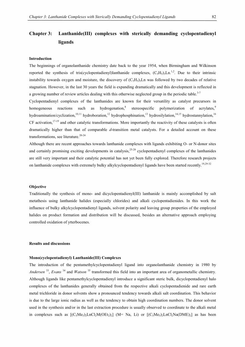
Chapter 3: Lanthanide Complexes with Sterically Demanding Cyclopentadienyl Ligands
82
Chapter 3: Lanthanide(III) complexes with sterically demanding cyclopentadienyl
ligands
Introduction
The beginnings of organolanthanide chemistry date back to the year 1954, when Birmingham and Wilkinson
reported the synthesis of tris(cyclopentadienyl)lanthanide complexes, (C5H5)3Ln.1,2. Due to their intrinsic
instability towards oxygen and moisture, the discovery of (C5H5)3Ln was followed by two decades of relative
stagnation. However, in the last 30 years the field is expanding dramatically and this development is reflected in
a growing number of review articles dealing with this otherwise neglected group in the periodic table.3-7
Cyclopentadienyl complexes of the lanthanides are known for their versatility as catalyst precursors in
homogeneous reactions such as hydrogenation,8 stereospecific polymerization of acrylates,9
hydroamination/cyclization,10,11 hydroboration,12 hydrophosphination,13 hydrosilylation,14,15 hydrostannylation,16
CF activation,17-19 and other catalytic transformations. More importantly the reactivity of these catalysts is often
dramatically higher than that of comparable d-transition metal catalysts. For a detailed account on these
transformations, see literature.20-24
Although there are recent approaches towards lanthanide complexes with ligands exhibiting O- or N-donor sites
and certainly promising exciting developments in catalysis,25-28 cyclopentadienyl complexes of the lanthanides
are still very important and their catalytic potential has not yet been fully explored. Therefore research projects
on lanthanide complexes with extremely bulky alkylcyclopentadienyl ligands have been started recently.19,29-32
Objective
Traditionally the synthesis of mono- and dicyclopentadienyl(III) lanthanide is mainly accomplished by salt
metathesis using lanthanide halides (especially chlorides) and alkali cyclopentadienides. In this work the
influence of bulky alkylcyclopentadienyl ligands, solvent polarity and leaving group properties of the employed
halides on product formation and distribution will be discussed, besides an alternative approach employing
controlled oxidation of ytterbocenes.
Results and discussions
Mono(cyclopentadienyl) Lanthanide(III) Complexes
The introduction of the pentamethylcyclopentadienyl ligand into organolanthanide chemistry in 1980 by
Andersen 33, Evans 34 and Watson 35 transformed this field into an important area of organometallic chemistry.
Although ligands like pentamethylcyclopentadienyl introduce a significant steric bulk, dicyclopentadienyl halo
complexes of the lanthanides generally obtained from the respective alkali cyclopentadienide and rare earth
metal trichloride in donor solvents show a pronounced tendency towards alkali salt coordination. This behavior
is due to the large ionic radius as well as the tendency to obtain high coordination numbers. The donor solvent
used in the synthesis and/or in the last extraction procedure is usually observed to coordinate to the alkali metal
in complexes such as [(C5Me5)2LnCl2M(OEt2)2] (M= Na, Li) or [(C5Me5)2LnCl2Na(DME)2] as has been
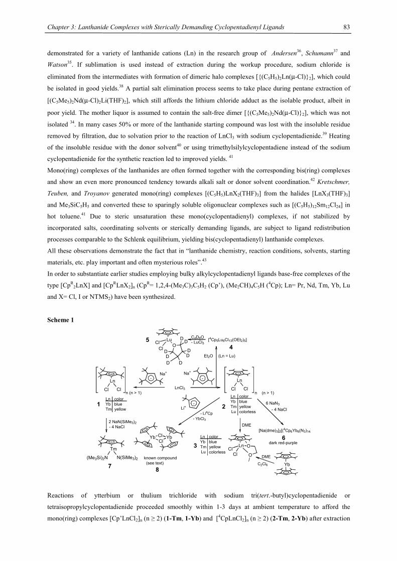
Chapter 3: Lanthanide Complexes with Sterically Demanding Cyclopentadienyl Ligands
83
demonstrated for a variety of lanthanide cations (Ln) in the research group of Andersen36, Schumann37 and
Watson35. If sublimation is used instead of extraction during the workup procedure, sodium chloride is
eliminated from the intermediates with formation of dimeric halo complexes [{(C5H5)2Ln(µ-Cl)}2], which could
be isolated in good yields.38 A partial salt elimination process seems to take place during pentane extraction of
[(C5Me5)2Nd(µ-Cl)2Li(THF)2], which still affords the lithium chloride adduct as the isolable product, albeit in
poor yield. The mother liquor is assumed to contain the salt-free dimer [{(C5Me5)2Nd(µ-Cl)}2], which was not
isolated 34. In many cases 50% or more of the lanthanide starting compound was lost with the insoluble residue
removed by filtration, due to solvation prior to the reaction of LnCl3 with sodium cyclopentadienide.39 Heating
of the insoluble residue with the donor solvent40 or using trimethylsilylcyclopentadiene instead of the sodium
cyclopentadienide for the synthetic reaction led to improved yields. 41
Mono(ring) complexes of the lanthanides are often formed together with the corresponding bis(ring) complexes
and show an even more pronounced tendency towards alkali salt or donor solvent coordination.42 Kretschmer,
Teuben, and Troyanov generated mono(ring) complexes [(C5H5)LnX2(THF)3] from the halides [LnX3(THF)3]
and Me3SiC5H5 and converted these to sparingly soluble oligonuclear complexes such as [(C5H5)12Sm12Cl24] in
hot toluene.41 Due to steric unsaturation these mono(cyclopentadienyl) complexes, if not stabilized by
incorporated salts, coordinating solvents or sterically demanding ligands, are subject to ligand redistribution
processes comparable to the Schlenk equilibrium, yielding bis(cyclopentadienyl) lanthanide complexes.
All these observations demonstrate the fact that in “lanthanide chemistry, reaction conditions, solvents, starting
materials, etc. play important and often mysterious roles”.43
In order to substantiate earlier studies employing bulky alkylcyclopentadienyl ligands base-free complexes of the
type [CpR2LnX] and [CpRLnX2]n (CpR= 1,2,4-(Me3C)3C5H2 (Cp’), (Me2CH)4C5H (4Cp); Ln= Pr, Nd, Tm, Yb, Lu
and X= Cl, I or NTMS2) have been synthesized.
Scheme 1
LnCl3
-Na+
Ln
Cl Cln (n > 1)
-Na+
Ln
Cl Cln (n > 1)
Ln colorYb blueTm yellow2
DME
Ln
ClCl O
O
6 NaN3
- 4 NaCl
36
dark red-purple
-Li+
2 NaN(SiMe3)2- 4 NaCl
known compound(see text)
Tm
(Me3Si)2N N(SiMe3)2
7
(Ln = Lu)
[4Cp5Lu6Cl13(OEt2)5]C4D8O- LuCl35 Lu
ClCl O
DDD D
DD
DD
Et2O
YbYbClCl
[Na(dme)3]2[(4Cp6Yb6(N3)14]
YbC2Cl6
DME
8
Lu colorless
Ln colorYb blueTm yellowLu colorless
4
- Li4Cp- YbCl3
Ln colorYb blueTm yellow
1
Reactions of ytterbium or thulium trichloride with sodium tri(tert.-butyl)cyclopentadienide or
tetraisopropylcyclopentadienide proceeded smoothly within 1-3 days at ambient temperature to afford the
mono(ring) complexes [Cp’LnCl2]n (n ≥ 2) (1-Tm, 1-Yb) and [4CpLnCl2]n (n ≥ 2) (2-Tm, 2-Yb) after extraction
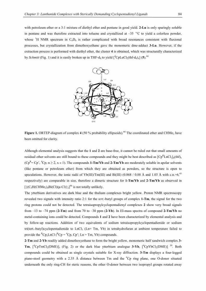
Chapter 3: Lanthanide Complexes with Sterically Demanding Cyclopentadienyl Ligands
84
with petroleum ether or a 3:1 mixture of diethyl ether and pentane in good yield. 2-Lu is only sparingly soluble
in pentane and was therefore extracted into toluene and crystallized at –35 ´°C to yield a colorless powder,
whose 1H NMR spectrum in C6D6 is rather complicated with broad resonances consistent with fluxional
processes, but crystallization from dimethoxyethane gave the monomeric dme-adduct 3-Lu. However, if the
extraction process is performed with diethyl ether, the cluster 4 is obtained, which was structurally characterized
by Schmitt (Fig. 1) and it is easily broken up in THF-d8 to yield [4CpLuCl2(thf-d8)x] (5).44
Lu1
Lu3
Lu5
Lu6
Lu2
Lu4
Cl1
Cl2
Cl13
Cl5
Cl3
Cl9
Cl7
Cl4
Cl11
Figure 1. ORTEP-diagram of complex 4 (50 % probability ellipsoids).44 The coordinated ether and CHMe2 have
been omitted for clarity.
Although elemental analysis suggests that the 1 and 2 are base-free, it cannot be ruled out that small amounts of
residual ether solvents are still bound to these compounds and they might be best described as [CpRLnCl2]n(thf)x
(CpR= Cp’, 4Cp; n ≥ 2, x « 1). The compounds 1-Tm/Yb and 2-Tm/Yb are moderately soluble in apolar solvents
(like pentane or petroleum ether) from which they are obtained as powders, so the structure is open to
speculations. However, the ionic radii of Yb(III)/Tm(III) and Bi(III) (0.868 / 0.88 Å and 1.03 Å with c.n.=6,45
respectively) are comparable in size, therefore a dimeric structure for 1-Tm/Yb and 2-Tm/Yb as observed in
[{(C5H(CHMe2)4Bi(Cl)(µ-Cl)}2]46 is not totally unlikely.
The ytterbium derivatives are dark blue and the thulium complexes bright yellow. Proton NMR spectroscopy
revealed two signals with intensity ratio 2:1 for the tert.-butyl groups of complex 1-Tm, the signal for the two
ring protons could not be detected. The tetraisopropylcyclopentadienyl complexes 2 show very broad signals
from –13 to –74 ppm (2-Tm) and from 70 to –30 ppm (2-Yb). In EI-mass spectra of compound 2-Tm/Yb no
metal-containing ions could be detected. Compounds 1 and 2 have been characterized by elemental analysis and
by follow-up reactions. Addition of two equivalents of sodium tetraisopropylcyclopentadienide or sodium
tri(tert.-butyl)cyclopentadienide to LnCl3 (Ln= Tm, Yb) in tetrahydrofuran at ambient temperature failed to
provide the RCp2LnCl (RCp = 4Cp, Cp’; Ln = Tm, Yb) compounds.
2-Tm and 2-Yb readily added dimethoxyethane to form the bright yellow, monomeric half sandwich complex 3-
Tm, [4CpTmCl2(DME)], (Fig. 2) or the dark blue ytterbium analogue 3-Yb, [4CpYbCl2(DME)] 30. Both
compounds could be obtained as single crystals suitable for X-ray diffraction. 3-Tm displays a four-legged
piano-stool geometry with a 2.35 Å distance between Tm and the 4Cp ring plane, one O-donor situated
underneath the only ring-CH for steric reasons, the other O-donor between two isopropyl groups rotated away
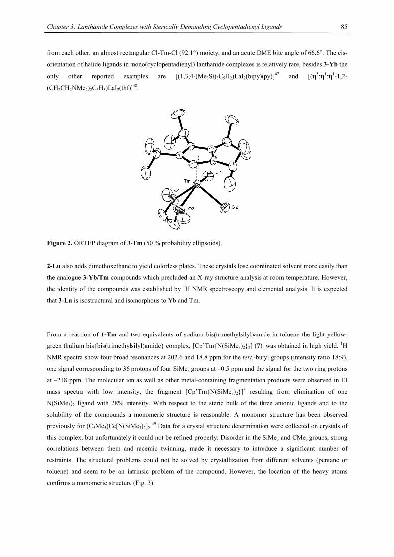
Chapter 3: Lanthanide Complexes with Sterically Demanding Cyclopentadienyl Ligands
85
from each other, an almost rectangular Cl-Tm-Cl (92.1°) moiety, and an acute DME bite angle of 66.6°. The cis-
orientation of halide ligands in mono(cyclopentadienyl) lanthanide complexes is relatively rare, besides 3-Yb the
only other reported examples are [(1,3,4-(Me3Si)3C5H2)LaI2(bipy)(py)]47 and [(η5:η1:η1-1,2-
(CH2CH2NMe2)2C5H3)LaI2(thf)]48.
Figure 2. ORTEP diagram of 3-Tm (50 % probability ellipsoids).
2-Lu also adds dimethoxethane to yield colorless plates. These crystals lose coordinated solvent more easily than
the analogue 3-Yb/Tm compounds which precluded an X-ray structure analysis at room temperature. However,
the identity of the compounds was established by 1H NMR spectroscopy and elemental analysis. It is expected
that 3-Lu is isostructural and isomorphous to Yb and Tm.
From a reaction of 1-Tm and two equivalents of sodium bis(trimethylsilyl)amide in toluene the light yellow-
green thulium bis{bis(trimethylsilyl)amide} complex, [Cp’Tm{N(SiMe3)2}2] (7), was obtained in high yield. 1H
NMR spectra show four broad resonances at 202.6 and 18.8 ppm for the tert.-butyl groups (intensity ratio 18:9),
one signal corresponding to 36 protons of four SiMe3 groups at –0.5 ppm and the signal for the two ring protons
at –218 ppm. The molecular ion as well as other metal-containing fragmentation products were observed in EI
mass spectra with low intensity, the fragment [Cp’Tm{N(SiMe3)2}]+ resulting from elimination of one
N(SiMe3)2 ligand with 28% intensity. With respect to the steric bulk of the three anionic ligands and to the
solubility of the compounds a monomeric structure is reasonable. A monomer structure has been observed
previously for (C5Me5)Ce[N(SiMe3)2]2.49 Data for a crystal structure determination were collected on crystals of
this complex, but unfortunately it could not be refined properly. Disorder in the SiMe3 and CMe3 groups, strong
correlations between them and racemic twinning, made it necessary to introduce a significant number of
restraints. The structural problems could not be solved by crystallization from different solvents (pentane or
toluene) and seem to be an intrinsic problem of the compound. However, the location of the heavy atoms
confirms a monomeric structure (Fig. 3).
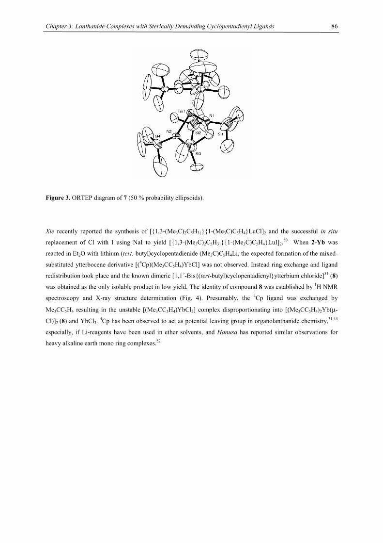
Chapter 3: Lanthanide Complexes with Sterically Demanding Cyclopentadienyl Ligands
86
Figure 3. ORTEP diagram of 7 (50 % probability ellipsoids).
Xie recently reported the synthesis of [{1,3-(Me3C)2C5H3}}{1-(Me3C)C5H4}LuCl]2 and the successful in situ
replacement of Cl with I using NaI to yield [{1,3-(Me3C)2C5H3}}{1-(Me3C)C5H4}LuI]2.50 When 2-Yb was
reacted in Et2O with lithium (tert.-butyl)cyclopentadienide (Me3C)C5H4Li, the expected formation of the mixed-
substituted ytterbocene derivative [(4Cp)(Me3CC5H4)YbCl] was not observed. Instead ring exchange and ligand
redistribution took place and the known dimeric [1,1´-Bis{(tert-butyl)cyclopentadienyl}ytterbium chloride]51 (8)
was obtained as the only isolable product in low yield. The identity of compound 8 was established by 1H NMR
spectroscopy and X-ray structure determination (Fig. 4). Presumably, the 4Cp ligand was exchanged by
Me3CC5H4 resulting in the unstable [(Me3CC5H4)YbCl2] complex disproportionating into [(Me3CC5H4)2Yb(µ-
Cl)]2 (8) and YbCl3. 4Cp has been observed to act as potential leaving group in organolanthanide chemistry,31,44
especially, if Li-reagents have been used in ether solvents, and Hanusa has reported similar observations for
heavy alkaline earth mono ring complexes.52
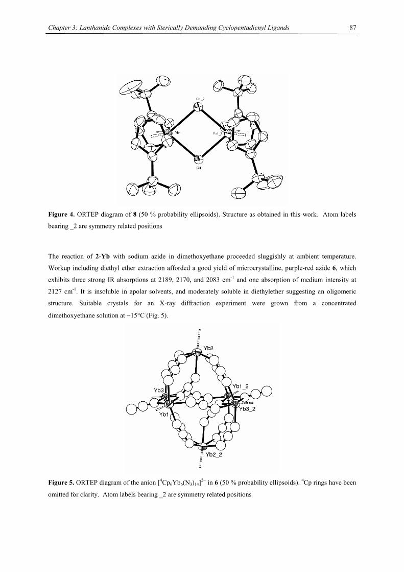
Chapter 3: Lanthanide Complexes with Sterically Demanding Cyclopentadienyl Ligands
87
Figure 4. ORTEP diagram of 8 (50 % probability ellipsoids). Structure as obtained in this work. Atom labels
bearing _2 are symmetry related positions
The reaction of 2-Yb with sodium azide in dimethoxyethane proceeded sluggishly at ambient temperature.
Workup including diethyl ether extraction afforded a good yield of microcrystalline, purple-red azide 6, which
exhibits three strong IR absorptions at 2189, 2170, and 2083 cm-1 and one absorption of medium intensity at
2127 cm-1. It is insoluble in apolar solvents, and moderately soluble in diethylether suggesting an oligomeric
structure. Suitable crystals for an X-ray diffraction experiment were grown from a concentrated
dimethoxyethane solution at −15°C (Fig. 5).
Figure 5. ORTEP diagram of the anion [4Cp6Yb6(N3)14]2− in 6 (50 % probability ellipsoids). 4Cp rings have been
omitted for clarity. Atom labels bearing _2 are symmetry related positions
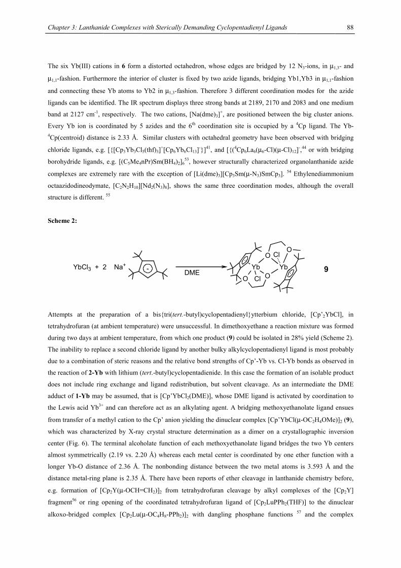
Chapter 3: Lanthanide Complexes with Sterically Demanding Cyclopentadienyl Ligands
88
The six Yb(III) cations in 6 form a distorted octahedron, whose edges are bridged by 12 N3-ions, in µ1,3- and
µ1,1-fashion. Furthermore the interior of cluster is fixed by two azide ligands, bridging Yb1,Yb3 in µ1,1-fashion
and connecting these Yb atoms to Yb2 in µ1,3-fashion. Therefore 3 different coordination modes for the azide
ligands can be identified. The IR spectrum displays three strong bands at 2189, 2170 and 2083 and one medium
band at 2127 cm-1, respectively. The two cations, [Na(dme)3]+, are positioned between the big cluster anions.
Every Yb ion is coordinated by 5 azides and the 6th coordination site is occupied by a 4Cp ligand. The Yb-4Cp(centroid) distance is 2.33 Å. Similar clusters with octahedral geometry have been observed with bridging
chloride ligands, e.g. [{[Cp3Yb3Cl5(thf)3]+[Cp6Yb6Cl13]-}]41, and [{(4Cp6La6(µ6-Cl)(µ-Cl)12]-,44 or with bridging
borohydride ligands, e.g. [(C5Me4nPr)Sm(BH4)2]653, however structurally characterized organolanthanide azide
complexes are extremely rare with the exception of [Li(dme)3][Cp3Sm(µ-N3)SmCp3]. 54 Ethylenediammonium
octaazidodineodymate, [C2N2H10][Nd2(N3)8], shows the same three coordination modes, although the overall
structure is different. 55
Scheme 2:
YbCl3 + 2 Na+ - DMEYb
Cl
O
YbO
ClO
O9
Attempts at the preparation of a bis{tri(tert.-butyl)cyclopentadienyl}ytterbium chloride, [Cp’2YbCl], in
tetrahydrofuran (at ambient temperature) were unsuccessful. In dimethoxyethane a reaction mixture was formed
during two days at ambient temperature, from which one product (9) could be isolated in 28% yield (Scheme 2).
The inability to replace a second chloride ligand by another bulky alkylcyclopentadienyl ligand is most probably
due to a combination of steric reasons and the relative bond strengths of Cp’-Yb vs. Cl-Yb bonds as observed in
the reaction of 2-Yb with lithium (tert.-butyl)cyclopentadienide. In this case the formation of an isolable product
does not include ring exchange and ligand redistribution, but solvent cleavage. As an intermediate the DME
adduct of 1-Yb may be assumed, that is [Cp’YbCl2(DME)], whose DME ligand is activated by coordination to
the Lewis acid Yb3+ and can therefore act as an alkylating agent. A bridging methoxyethanolate ligand ensues
from transfer of a methyl cation to the Cp’ anion yielding the dinuclear complex [Cp’YbCl(µ-OC2H4OMe)]2 (9),
which was characterized by X-ray crystal structure determination as a dimer on a crystallographic inversion
center (Fig. 6). The terminal alcoholate function of each methoxyethanolate ligand bridges the two Yb centers
almost symmetrically (2.19 vs. 2.20 Å) whereas each metal center is coordinated by one ether function with a
longer Yb-O distance of 2.36 Å. The nonbonding distance between the two metal atoms is 3.593 Å and the
distance metal-ring plane is 2.35 Å. There have been reports of ether cleavage in lanthanide chemistry before,
e.g. formation of [Cp2Y(µ-OCH=CH2)]2 from tetrahydrofuran cleavage by alkyl complexes of the [Cp2Y]
fragment56 or ring opening of the coordinated tetrahydrofuran ligand of [Cp2LuPPh2(THF)] to the dinuclear
alkoxo-bridged complex [Cp2Lu(µ-OC4H8-PPh2)]2 with dangling phosphane functions 57 and the complex
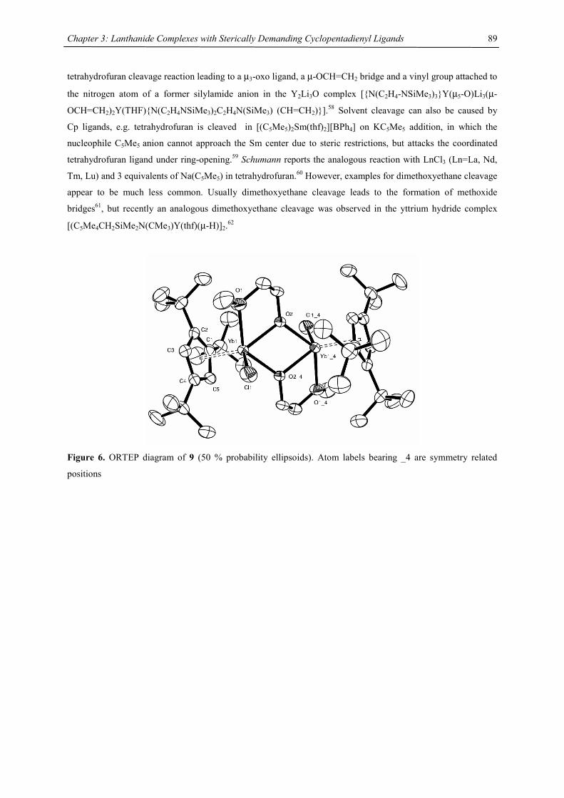
Chapter 3: Lanthanide Complexes with Sterically Demanding Cyclopentadienyl Ligands
89
tetrahydrofuran cleavage reaction leading to a µ3-oxo ligand, a µ-OCH=CH2 bridge and a vinyl group attached to
the nitrogen atom of a former silylamide anion in the Y2Li3O complex [{N(C2H4-NSiMe3)3}Y(µ5-O)Li3(µ-
OCH=CH2)2Y(THF){N(C2H4NSiMe3)2C2H4N(SiMe3) (CH=CH2)}].58 Solvent cleavage can also be caused by
Cp ligands, e.g. tetrahydrofuran is cleaved in [(C5Me5)2Sm(thf)2][BPh4] on KC5Me5 addition, in which the
nucleophile C5Me5 anion cannot approach the Sm center due to steric restrictions, but attacks the coordinated
tetrahydrofuran ligand under ring-opening.59 Schumann reports the analogous reaction with LnCl3 (Ln=La, Nd,
Tm, Lu) and 3 equivalents of Na(C5Me5) in tetrahydrofuran.60 However, examples for dimethoxyethane cleavage
appear to be much less common. Usually dimethoxyethane cleavage leads to the formation of methoxide
bridges61, but recently an analogous dimethoxyethane cleavage was observed in the yttrium hydride complex
[(C5Me4CH2SiMe2N(CMe3)Y(thf)(µ-H)]2.62
Figure 6. ORTEP diagram of 9 (50 % probability ellipsoids). Atom labels bearing _4 are symmetry related
positions
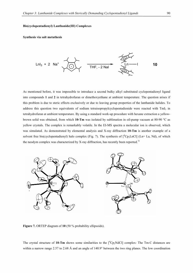
Chapter 3: Lanthanide Complexes with Sterically Demanding Cyclopentadienyl Ligands
90
Bis(cyclopentadienyl) Lanthanide(III) Complexes
Synthesis via salt metathesis
10LnI3 + 2 Na+THF, - 2 NaI
Ln I-
As mentioned before, it was impossible to introduce a second bulky alkyl substituted cyclopentadienyl ligand
into compounds 1 and 2 in tetrahydrofuran or dimethoxyethane at ambient temperature. The question arises if
this problem is due to steric effects exclusively or due to leaving group properties of the lanthanide halides. To
address this question two equivalents of sodium tetraisopropylcyclopentadienide were reacted with TmI3 in
tetrahydrofuran at ambient temperature. By using a standard work-up procedure with hexane extraction a yellow-
brown solid was obtained, from which 10-Tm was isolated by sublimation in oil-pump vacuum at 80-90 °C as
yellow crystals. The complex is remarkably volatile. In the EI-MS spectra a molecular ion is observed, which
was simulated. As demonstrated by elemental analysis and X-ray diffraction 10-Tm is another example of a
solvent free bis(cyclopentadienyl) halo complex (Fig. 7). The synthesis of [4Cp2LnCl] (Ln= La, Nd), of which
the neodym complex was characterized by X-ray diffraction, has recently been reported.31
Figure 7. ORTEP diagram of 10 (50 % probability ellipsoids).
The crystal structure of 10-Tm shows some similarities to the [4Cp2NdCl] complex: The Tm-C distances are
within a narrow range 2.57 to 2.68 Å and an angle of 140.9° between the two ring planes. The low coordination
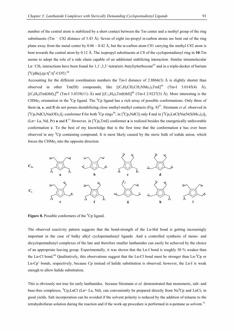
Chapter 3: Lanthanide Complexes with Sterically Demanding Cyclopentadienyl Ligands
91
number of the central atom is stabilized by a short contact between the Tm center and a methyl group of the ring
substituents (Tm … C82 distance of 3.43 Å). Seven of eight iso-propyl α-carbon atoms are bent out of the ring
plane away from the metal center by 0.06 – 0.42 Å, but the α-carbon atom C81 carrying the methyl C82 atom is
bent towards the central atom by 0.12 Å. The isopropyl substituents at C8 of the cyclopentadienyl ring in 10-Tm
seems to adopt the role of a side chain capable of an additional stabilizing interaction. Similar intramolecular
Ln…CH3 interactions have been found for 1,1´,3,3´-tetra(tert.-butyl)ytterbocene63 and in a triple-decker of barium
[4CpBa]2(µ:η8:η8-COT).64
Accounting for the different coordination numbers the Tm-I distance of 2.8866(3) Å is slightly shorter than
observed in other Tm(III) compounds, like [(C5H4CH2CH2NMe2)2TmI]65 (Tm-I 3.0145(4) Å),
[(C8H8)TmI(thf)2]66 (Tm-I 3.0338(11) Å) and [(C12H8)2TmI(thf)]66 (Tm-I 2.9227(3) Å). More interesting is the
CHMe2 orientation in the 4Cp ligand. The 4Cp ligand has a rich array of possible conformations. Only three of
them (a, e, and f) do not posses destabilizing close methyl-methyl contacts (Fig. 8)67. Sitzmann et al. observed in
[4Cp2NdCl2Na(OEt2)]2 conformer f for both 4Cp rings30, in [4Cp2NdCl] only f and in [4Cp2LnCl(Na(N(SiMe3)2)]2
(Ln= La, Nd, Pr) a and f.31 However, in [4Cp2TmI] conformer a is realized besides the energetically unfavorable
conformation c. To the best of my knowledge that is the first time that the conformation c has ever been
observed in any 4Cp containing compound. It is most likely caused by the steric bulk of iodide anion, which
forces the CHMe2 into the opposite direction.
f g h i
b ca d
e
C2v
Cs
Figure 8. Possible conformers of the 4Cp ligand.
The observed reactivity pattern suggests that the bond-strength of the Ln-Hal bond is getting increasingly
important in the case of bulky alkyl cyclopentadienyl ligands. And a controlled synthesis of mono- and
dicyclopentadienyl complexes of the late and therefore smaller lanthanides can easily be achieved by the choice
of an appropriate leaving group. Experimentally, it was shown that the Ln-I bond is roughly 30 % weaker than
the Ln-Cl bond.68 Qualitatively, this observations suggest that the Ln-Cl bond must be stronger than Ln-4Cp or
Ln-Cp’ bonds, respectively, because Cp instead of halide substitution is observed; however, the Ln-I is weak
enough to allow halide substitution.
This is obviously not true for early lanthanides, because Sitzmann et al. demonstrated that monomeric, salt- and
base-free complexes, 4Cp2LnCl (Ln= La, Nd), can conveniently be prepared directly from Na4Cp and LnCl3 in
good yields. Salt incorporation can be avoided if the solvent polarity is reduced by the addition of toluene to the
tetrahydrofuran solution during the reaction and if the work-up procedure is performed in n-pentane as solvent.31
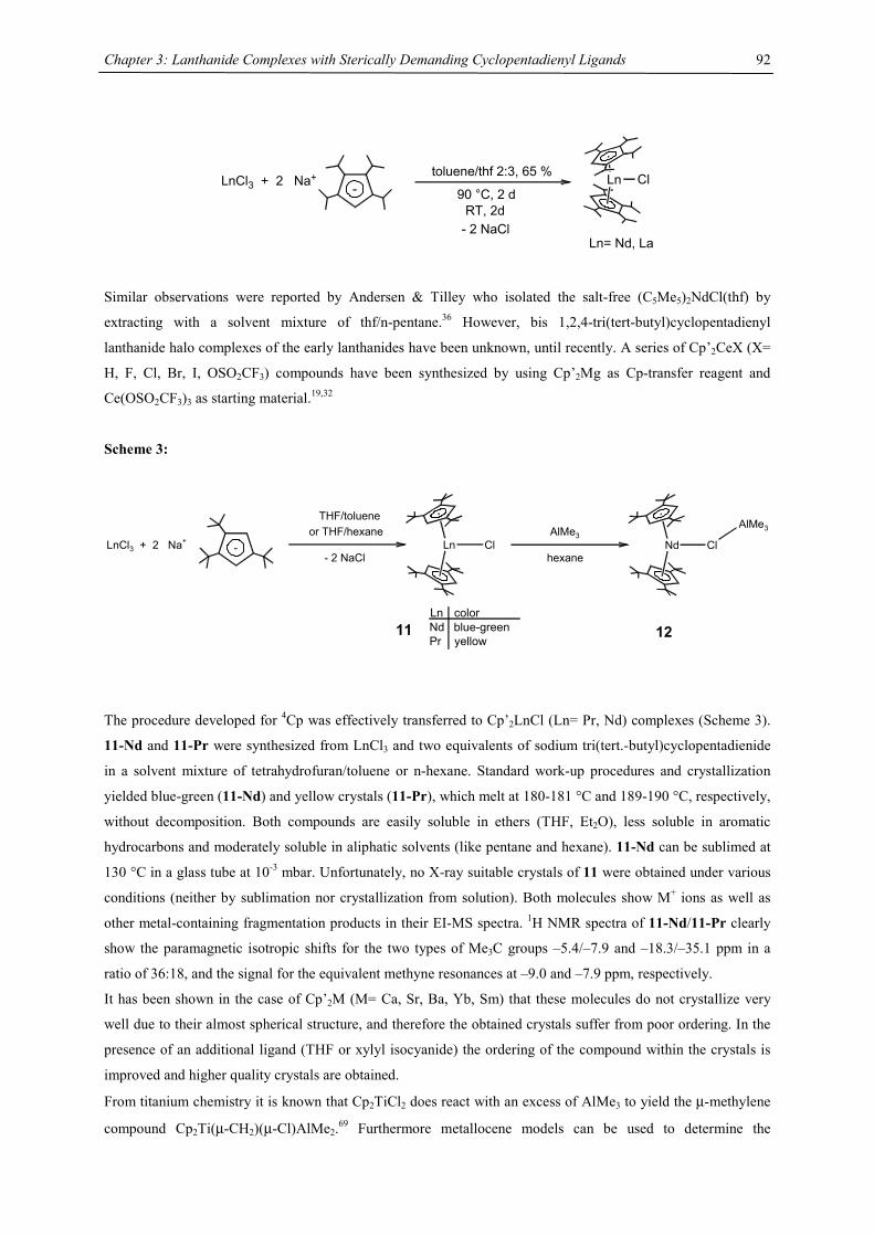
Chapter 3: Lanthanide Complexes with Sterically Demanding Cyclopentadienyl Ligands
92
LnCl3 + 2 Na+
- 2 NaCl
Ln Cl-
toluene/thf 2:3, 65 %
90 °C, 2 dRT, 2d
Ln= Nd, La
Similar observations were reported by Andersen & Tilley who isolated the salt-free (C5Me5)2NdCl(thf) by
extracting with a solvent mixture of thf/n-pentane.36 However, bis 1,2,4-tri(tert-butyl)cyclopentadienyl
lanthanide halo complexes of the early lanthanides have been unknown, until recently. A series of Cp’2CeX (X=
H, F, Cl, Br, I, OSO2CF3) compounds have been synthesized by using Cp’2Mg as Cp-transfer reagent and
Ce(OSO2CF3)3 as starting material.19,32
Scheme 3:
LnCl3 + 2 Na+-
THF/tolueneor THF/hexane
Ln Cl
11Ln colorNd blue-greenPr yellow
Nd Cl
AlMe3
hexane
AlMe3
12
- 2 NaCl
The procedure developed for 4Cp was effectively transferred to Cp’2LnCl (Ln= Pr, Nd) complexes (Scheme 3).
11-Nd and 11-Pr were synthesized from LnCl3 and two equivalents of sodium tri(tert.-butyl)cyclopentadienide
in a solvent mixture of tetrahydrofuran/toluene or n-hexane. Standard work-up procedures and crystallization
yielded blue-green (11-Nd) and yellow crystals (11-Pr), which melt at 180-181 °C and 189-190 °C, respectively,
without decomposition. Both compounds are easily soluble in ethers (THF, Et2O), less soluble in aromatic
hydrocarbons and moderately soluble in aliphatic solvents (like pentane and hexane). 11-Nd can be sublimed at
130 °C in a glass tube at 10-3 mbar. Unfortunately, no X-ray suitable crystals of 11 were obtained under various
conditions (neither by sublimation nor crystallization from solution). Both molecules show M+ ions as well as
other metal-containing fragmentation products in their EI-MS spectra. 1H NMR spectra of 11-Nd/11-Pr clearly
show the paramagnetic isotropic shifts for the two types of Me3C groups –5.4/–7.9 and –18.3/–35.1 ppm in a
ratio of 36:18, and the signal for the equivalent methyne resonances at –9.0 and –7.9 ppm, respectively.
It has been shown in the case of Cp’2M (M= Ca, Sr, Ba, Yb, Sm) that these molecules do not crystallize very
well due to their almost spherical structure, and therefore the obtained crystals suffer from poor ordering. In the
presence of an additional ligand (THF or xylyl isocyanide) the ordering of the compound within the crystals is
improved and higher quality crystals are obtained.
From titanium chemistry it is known that Cp2TiCl2 does react with an excess of AlMe3 to yield the µ-methylene
compound Cp2Ti(µ-CH2)(µ-Cl)AlMe2.69 Furthermore metallocene models can be used to determine the
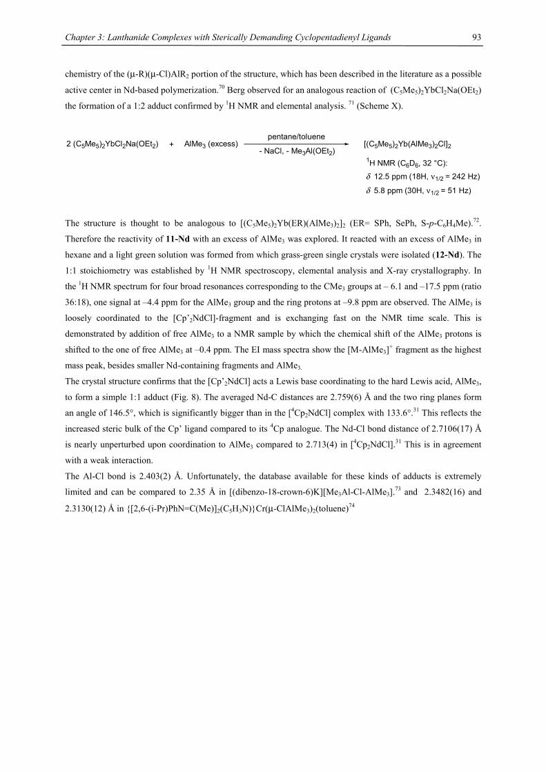
Chapter 3: Lanthanide Complexes with Sterically Demanding Cyclopentadienyl Ligands
93
chemistry of the (µ-R)(µ-Cl)AlR2 portion of the structure, which has been described in the literature as a possible
active center in Nd-based polymerization.70 Berg observed for an analogous reaction of (C5Me5)2YbCl2Na(OEt2)
the formation of a 1:2 adduct confirmed by 1H NMR and elemental analysis. 71 (Scheme X).
2 (C5Me5)2YbCl2Na(OEt2) + AlMe3 (excess)pentane/toluene
- NaCl, - Me3Al(OEt2)[(C5Me5)2Yb(AlMe3)2Cl]2
1H NMR (C6D6, 32 °C):
δ 12.5 ppm (18H, ν1/2 = 242 Hz)
δ 5.8 ppm (30H, ν1/2 = 51 Hz)
The structure is thought to be analogous to [(C5Me5)2Yb(ER)(AlMe3)2]2 (ER= SPh, SePh, S-p-C6H4Me).72.
Therefore the reactivity of 11-Nd with an excess of AlMe3 was explored. It reacted with an excess of AlMe3 in
hexane and a light green solution was formed from which grass-green single crystals were isolated (12-Nd). The
1:1 stoichiometry was established by 1H NMR spectroscopy, elemental analysis and X-ray crystallography. In
the 1H NMR spectrum for four broad resonances corresponding to the CMe3 groups at – 6.1 and –17.5 ppm (ratio
36:18), one signal at –4.4 ppm for the AlMe3 group and the ring protons at –9.8 ppm are observed. The AlMe3 is
loosely coordinated to the [Cp’2NdCl]-fragment and is exchanging fast on the NMR time scale. This is
demonstrated by addition of free AlMe3 to a NMR sample by which the chemical shift of the AlMe3 protons is
shifted to the one of free AlMe3 at –0.4 ppm. The EI mass spectra show the [M-AlMe3]+ fragment as the highest
mass peak, besides smaller Nd-containing fragments and AlMe3.
The crystal structure confirms that the [Cp’2NdCl] acts a Lewis base coordinating to the hard Lewis acid, AlMe3,
to form a simple 1:1 adduct (Fig. 8). The averaged Nd-C distances are 2.759(6) Å and the two ring planes form
an angle of 146.5°, which is significantly bigger than in the [4Cp2NdCl] complex with 133.6°.31 This reflects the
increased steric bulk of the Cp’ ligand compared to its 4Cp analogue. The Nd-Cl bond distance of 2.7106(17) Å
is nearly unperturbed upon coordination to AlMe3 compared to 2.713(4) in [4Cp2NdCl].31 This is in agreement
with a weak interaction.
The Al-Cl bond is 2.403(2) Å. Unfortunately, the database available for these kinds of adducts is extremely
limited and can be compared to 2.35 Å in [(dibenzo-18-crown-6)K][Me3Al-Cl-AlMe3].73 and 2.3482(16) and
2.3130(12) Å in {[2,6-(i-Pr)PhN=C(Me)]2(C5H3N)}Cr(µ-ClAlMe3)2(toluene)74
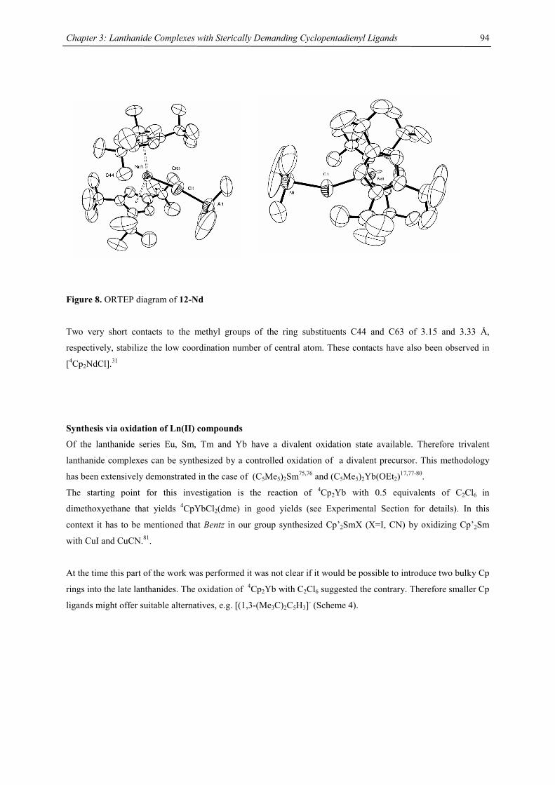
Chapter 3: Lanthanide Complexes with Sterically Demanding Cyclopentadienyl Ligands
94
Figure 8. ORTEP diagram of 12-Nd
Two very short contacts to the methyl groups of the ring substituents C44 and C63 of 3.15 and 3.33 Å,
respectively, stabilize the low coordination number of central atom. These contacts have also been observed in
[4Cp2NdCl].31
Synthesis via oxidation of Ln(II) compounds
Of the lanthanide series Eu, Sm, Tm and Yb have a divalent oxidation state available. Therefore trivalent
lanthanide complexes can be synthesized by a controlled oxidation of a divalent precursor. This methodology
has been extensively demonstrated in the case of (C5Me5)2Sm75,76 and (C5Me5)2Yb(OEt2)17,77-80.
The starting point for this investigation is the reaction of 4Cp2Yb with 0.5 equivalents of C2Cl6 in
dimethoxyethane that yields 4CpYbCl2(dme) in good yields (see Experimental Section for details). In this
context it has to be mentioned that Bentz in our group synthesized Cp’2SmX (X=I, CN) by oxidizing Cp’2Sm
with CuI and CuCN.81.
At the time this part of the work was performed it was not clear if it would be possible to introduce two bulky Cp
rings into the late lanthanides. The oxidation of 4Cp2Yb with C2Cl6 suggested the contrary. Therefore smaller Cp
ligands might offer suitable alternatives, e.g. [(1,3-(Me3C)2C5H3]- (Scheme 4).
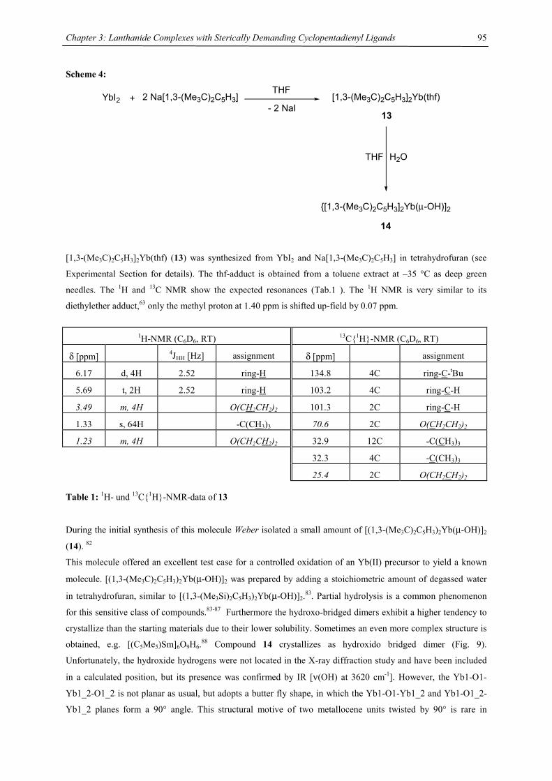
Chapter 3: Lanthanide Complexes with Sterically Demanding Cyclopentadienyl Ligands
95
Scheme 4:
YbI2 + 2 Na[1,3-(Me3C)2C5H3]THF
- 2 NaI[1,3-(Me3C)2C5H3]2Yb(thf)
THF H2O
{[1,3-(Me3C)2C5H3]2Yb(µ-OH)]2
13
14
[1,3-(Me3C)2C5H3]2Yb(thf) (13) was synthesized from YbI2 and Na[1,3-(Me3C)2C5H3] in tetrahydrofuran (see
Experimental Section for details). The thf-adduct is obtained from a toluene extract at –35 °C as deep green
needles. The 1H and 13C NMR show the expected resonances (Tab.1 ). The 1H NMR is very similar to its
diethylether adduct,63 only the methyl proton at 1.40 ppm is shifted up-field by 0.07 ppm.
1H-NMR (C6D6, RT) 13C{1H}-NMR (C6D6, RT)
δ [ppm] 4JHH [Hz] assignment δ [ppm] assignment
6.17 d, 4H 2.52 ring-H 134.8 4C ring-C-tBu
5.69 t, 2H 2.52 ring-H 103.2 4C ring-C-H
3.49 m, 4H O(CH2CH2)2 101.3 2C ring-C-H
1.33 s, 64H -C(CH3)3 70.6 2C O(CH2CH2)2
1.23 m, 4H O(CH2CH2)2 32.9 12C -C(CH3)3
32.3 4C -C(CH3)3
25.4 2C O(CH2CH2)2 Table 1: 1H- und 13C{1H}-NMR-data of 13 During the initial synthesis of this molecule Weber isolated a small amount of [(1,3-(Me3C)2C5H3)2Yb(µ-OH)]2
(14). 82
This molecule offered an excellent test case for a controlled oxidation of an Yb(II) precursor to yield a known
molecule. [(1,3-(Me3C)2C5H3)2Yb(µ-OH)]2 was prepared by adding a stoichiometric amount of degassed water
in tetrahydrofuran, similar to [(1,3-(Me3Si)2C5H3)2Yb(µ-OH)]2.83. Partial hydrolysis is a common phenomenon
for this sensitive class of compounds.83-87 Furthermore the hydroxo-bridged dimers exhibit a higher tendency to
crystallize than the starting materials due to their lower solubility. Sometimes an even more complex structure is
obtained, e.g. [(C5Me5)Sm]6O9H6.88 Compound 14 crystallizes as hydroxido bridged dimer (Fig. 9).
Unfortunately, the hydroxide hydrogens were not located in the X-ray diffraction study and have been included
in a calculated position, but its presence was confirmed by IR [ν(OH) at 3620 cm-1]. However, the Yb1-O1-
Yb1_2-O1_2 is not planar as usual, but adopts a butter fly shape, in which the Yb1-O1-Yb1_2 and Yb1-O1_2-
Yb1_2 planes form a 90° angle. This structural motive of two metallocene units twisted by 90° is rare in
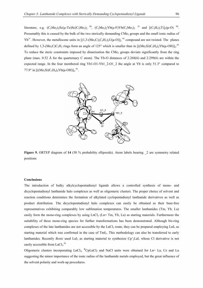
Chapter 3: Lanthanide Complexes with Sterically Demanding Cyclopentadienyl Ligands
96
literature, e.g. (C5Me5)2Sc(µ-Te)Sc(C5Me5)2 89, (C5Me5)2Yb(µ-F)Yb(C5Me5)2 18 and [(C5H5)2Ti]2(µ-O) 90.
Presumably this is caused by the bulk of the two sterically demanding CMe3 groups and the small ionic radius of
Yb3+. However, the metallocene units in [(1,3-(Me3C)2C5H3)2U(µ-O)]2 91 compound are not twisted. The planes
defined by 1,3-(Me3C)C5H3 rings form an angle of 125° which is smaller than in [((Me3Si)C5H4)2Yb(µ-OH)]2.83
To reduce the steric constraints imposed by dimerisation the CMe3 groups deviate significantly from the ring
plane (max. 0.52 Å for the quarternary C atom). The Yb-O distances of 2.268(6) and 2.299(6) are within the
expected range. In the four membered ring Yb1-O1-Yb1_2-O1_2 the angle at Yb is only 51.3° compared to
77.9° in [((Me3Si)C5H4)2Yb(µ-OH)]2.83.
Figure 9. ORTEP diagram of 14 (50 % probability ellipsoids). Atom labels bearing _2 are symmetry related
positions
Conclusions
The introduction of bulky alkylcyclopentadienyl ligands allows a controlled synthesis of mono- and
dicyclopentadienyl lanthanide halo complexes as well as oligomeric clusters. The proper choice of solvent and
reaction conditions determines the formation of alkylated cyclopentadienyl lanthanide derivatives as well as
product distribution. The dicyclopentadienyl halo complexes can easily be obtained as their base-free
representatives exhibiting comparably low sublimation temperatures. The smaller lanthanides (Tm, Yb, Lu)
easily form the mono-ring complexes by using LnCl3 (Ln= Tm, Yb, Lu) as starting materials. Furthermore the
suitability of these mono-ring species for further transformations has been demonstrated. Although bis-ring
complexes of the late lanthanides are not accessible by the LnCl3 route, they can be prepared employing LnI3 as
starting material which was confirmed in the case of TmI3. This methodology can also be transferred to early
lanthanides. Recently Bentz used LaI3 as starting material to synthesize Cp’2LaI, whose Cl derivative is not
easily accessible from LaCl3.81
Oligomeric clusters incorporating LnCl3, RCpLnCl2 and NaCl units were obtained for Ln= La, Ce and Lu
suggesting the minor importance of the ionic radius of the lanthanide metals employed, but the great influence of
the solvent polarity and work-up procedures.
Chapter 3: Lanthanide Complexes with Sterically Demanding Cyclopentadienyl Ligands
97
Therefore the reactivity of the lanthanide halides towards bulky cyclopentadienyl ligands is a delicate balance
between leaving group properties (e.g. metal-halide bond strength), the introduced steric bulk and solvent
polarity.
Cp’2LnCl and Cp’2LnI (Ln= La, Pr, Nd) complexes might be interesting starting materials for further
functionalizations and reaction chemistry as demonstrated by Andersen et al. in the case of Cp’2CeX
compounds. 19,32
References
[1] Wilkinson, G.; Birmingham, J. M. J. Am. Chem. Soc. 1954, 72, 6120.
[2] Wilkinson, G.; Birmingham, J. M. J. Am. Chem. Soc. 1956, 78, 42-44.
[3] Schumann, H.; Meese-Marktscheffel, J. A.; Esser, L. Chem. Rev. 1995, 95, 865-986.
[4] Edelmann, F. T. In Comprehensive Organonmetallic Chemsitry II; Abel, E. W. S., F.G.A.; Wilkinson,
G., Ed.; Pergamon: Exeter, 1995; Vol. 4, pp 11-270.
[5] Edelmann, F. T.; Freckmann, D. M. M.; Schumann, H. Chem. Rev. 2002, 102, 1851-1896.
[6] Arndt, S.; Okuda, J. Chem. Rev. 2002, 102, 1953-1976.
[7] Schaverien, C. J. Adv. Organomet. Chem. 1994, 35, 283-361.
[8] Haar, C. M.; Stern, C. L.; Marks, T. J. Organometallics 1996, 15, 1765-1784.
[9] Mao, L.; Shen, Q.; Sun, J. J. Organomet. Chem. 1998, 566, 9-14.
[10] Motta, A.; Lanza, G.; Fragalà, I. L.; Marks, T. J. Organometallics 2004, X.
[11] Douglass, M. R.; Ogasawara, M.; Hong, S.; Metz, M. V.; Marks, T. J. Organometallics 2002, 21, 283-.
[12] Harrison, K. N.; Marks, T. J. J. Am. Chem. Soc. 1992, 114, 9220-9221.
[13] Douglass, M. R.; Marks, T. J. J. Am. Chem. Soc. 2000, 122, 1824-1825.
[14] Molander, G. A.; Schmitt, M. H. J. Org. Chem. 2000, 65, 3767-?
[15] Muci, A. R.; Bercaw, J. E. Tetrahedron Lett. 2000, 41.
[16] Voskobynikov, A. Z.; Beletskaya, I. P. New J. Chem. 1995, 19, 723-.
[17] Watson, P. L.; Tulip, T. H.; Williams, I. Organometallics 1990, 9, 1999-2009.
[18] Burns, C. J.; Andersen, R. A. J. Chem. Soc., Chem. Commun. 1989, 136-137.
[19] Maron, L.; Werkema, E. L.; Perrin, L.; Eisenstein, O.; Andersen, R. A. J. Am. Chem. Soc. 2005, 127,
279-292.
[20] Molander, G. A.; Romero, J. A. C. Chem. Rev. 2002.
[21] Zhou, X.; Zhu, M. J. Organomet. Chem. 2002, 647.
[22] Anwander, R. In Applied Homogenous Catalysis with Organometallic Compounds; Cornils, B. H.,
W.A., Ed.; Wiley-VCH: Weinheim, 2000, pp 866-892.
[23] Edelmann, F. T. In Topics in Current Chemistry; Hermann, W. A., Ed.; Springer: Berlin, Heidelberg,
1996; Vol. 179, pp 247-276.
[24] Watson, P. L.; Parshall, G. W. Acc. Chem. Res. 1985, 18, 51-56.
[25] Estler, F.; Eickerling, G.; Herdtweck, E.; Anwander, R. Organometallics 2003, 22, 1212-1222.
[26] Kempe, R. Angew. Chem. 2000, 112, 478-504.
[27] Cui, C.; Shafir, A.; Reeder, C. L.; Arnold, J. Organometallics 2003, 22, 3357-3359.
[28] Fischbach, A.; Perdih, F.; Sirsch, P.; Scherer, W.; Anwander, R. Organometallics 2002, 21, 4569-4571.
Chapter 3: Lanthanide Complexes with Sterically Demanding Cyclopentadienyl Ligands
98
[29] Sitzmann, H.; Dezember, T.; Schmitt, O.; Weber, F.; Wolmershäuser, G. Z. Anorg. Allgem. Chem.
2000, 626, 2241-2244.
[30] Sitzmann, H.; Schmitt, O.; Weber, F.; Wolmershäuser, G. Z. Anorg. Allgem. Chem. 2001, 627, 12-14.
[31] Schmitt, O.; Wolmershäuser, G.; Sitzmann, H. Eur. J. Inorg. Chem. 2003, 3105-3109.
[32] Werkema, E. L.; Andersen, R. A., unpublished results.
[33] Tilley, T. D.; Andersen, R. A.; Spencer, B.; Ruben, H.; Zalkin, A.; Templeton, D. H. Inorg. Chem.
1980, 19, 2999-3003.
[34] Wayda, A. L.; Evans, W. J. Inorg. Chem. 1980, 19, 2190-2191.
[35] Watson, P. L. J. Chem. Soc., Chem. Commun. 1980, 652-653.
[36] Tilley, T. D.; Andersen, R. A. Inorg. Chem. 1981, 20, 3267-3270.
[37] Schumann, H.; Albrecht, I.; Loebel, J.; Hahn, E. Organometallics 1986, 5, 1296-1304.
[38] Maginn, R. E.; Monastyrskyi, S.; Dubeck, M. J. Am. Chem. Soc. 1963, 85, 672-676.
[39] Hazin, P. N.; Huffman, J. C.; Bruno, J. W. Organometallics 1987, 6, 23-27.
[40] Day, C. S.; Day, V. W.; Ernst, R. D.; Vollmer, S. H. Organometallics 1982, 1982, 998-1003.
[41] Kretschmer, W. P.; J.H., T.; Troyanov, S. I. Angew. Chem. Int. Ed. Engl. 1998, 37, 88-90.
[42] Poli, R. Chem. Rev. 1991, 91, 509-551.
[43] Sofield, C. D.; Andersen, R. A. J. Organomet. Chem. 1995, 501, 271-276, and references cited therein.
[44] Schmitt, O. Ph.D. Thesis, University of Kaiserslautern, 2001.
[45] Shannon, R. D. Acta Cryst. A 1976, A32, 751-767.
[46] Sitzmann, H.; Wolmershäuser, G. Chem. Ber. 1994, 127, 1335-1342.
[47] Giesbrecht, G. R.; Clark, D. L.; Gordon, J. C.; Scott, B. L. Appl. Organomet. Chem. 2003, 17, 473-479.
[48] Fedushkin, I. L.; Dechert, S.; Schumann, H. Organometallics 2000, 19, 4066-4076.
[49] Heeres, H. J.; Meetsma, A.; Teuben, J. H. Organometallics 1989, 8, 2637-2646.
[50] Xie, Z.; Liu, Z.; Xue, F.; Zhang, Z.; Mak, T. C. W. J. Organomet. Chem. 1997, 542, 285-289.
[51] Ren, J.-S.; Shen, Q.; Hu, J.-Y.; Lin, Y.-H.; Xing, Y. Jiegou Huaxue (Chin.) (J. Struct. Chem.) 1997, 16,
380-383.
[52] Harvey, M. J.; Hanusa, T. P. Organometallics 2000, 19, 1556-1566.
[53] Bonnet, F.; Visseaux, M.; Barbier-Baudry, D.; Hafid, A.; Vigier, E.; Kubicki, M. M. Inorg. Chem.
2004, 43, 3682-3690.
[54] Schumann, H.; Janiak, C.; Pickardt, J. J. Organomet. Chem. 1988, 349, 117-122.
[55] Starynowicz, P.; Bukietynska, K.; Rybe-Romanowski, W.; Dominiak-Dzik, G.; Golab, S. Polyhedron
1994, 13, 1069-1075.
[56] Evans, W. J.; Dominguez, R.; Hanusa, T. P. Organometallics 1986, 5, 1291-1296.
[57] Schumann, H.; Palamidos, E.; Loebel, J. J. Organomet. Chem. 1990, 384, C49-C52.
[58] Aspinall, H. C.; Tillotson, M. R. Inorg. Chem. 1996, 35, 2163-2164.
[59] Evans, W. J.; Ulibarri, T. A.; Chamberlain, L. R.; Ziller, J. W.; Alvarez, D. Organometallics 1990, 9,
2124-2130.
[60] Schumann, H.; Glanz, M.; Hemling, H.; Görlitz, F. H. J. Organomet. Chem. 1993, 462, 155-161.
[61] Gun'ko, Y. K.; Hitchcock, P. B.; Lappert, M. F. J. Organomet. Chem. 1995, 499, 213-219.
[62] Trifonov, A. A.; Spaniol, T. P.; Okuda, J. Dalton Trans. 2004, 2245-2250.
[63] Schultz, M.; Burns, C. J.; Schwartz, D. J.; Andersen, R. A. Organometallics 2000, 19, 781-789.
Chapter 3: Lanthanide Complexes with Sterically Demanding Cyclopentadienyl Ligands
99
[64] Sitzmann, H.; Walter, M. D.; Wolmershäuser, G. Angew. Chem. Int. Ed. Engl. 2002, 41, 2315-2316.
[65] Fedushkin, I. L.; Girgsdies, F.; Schumann, H.; Bochkarev, M. N. Eur. J. Inorg. Chem. 2001, 2405-2410.
[66] Fedushkin, I. L.; Bochkarev, M. N.; Dechert, S.; Schumann, H. Chem. Eur. J. 2001, 7, 3558-3563.
[67] Williams, R. A.; Tesh, K. F.; Hanusa, T. P. J. Am. Chem. Soc. 1991, 113, 4843-4851.
[68] Cordfunke, E. H. P.; Konings, R. J. M. Thermochimica Acta 2001, 375, 17-50.
[69] Tebbe, F. N.; Parshall, G. W.; Reddy, G. S. J. Am. Chem. Soc. 1978, 100, 3611-3613.
[70] Evans, W. J.; Champagne, T. M.; Giarikos, D. G.; Ziller, J. W. Organometallics 2005, 24, 570-579; and
references cited therein.
[71] Berg, D. J. In Chemistry; University of California, Berkeley: Berkeley, 1987.
[72] Berg, D. J.; Andersen, R. A. Organometallics 2003, 22, 627-632.
[73] Atwood, J. L.; Rogers, R. D.; Hrncir, D. C.; Zaworotko, M. J.; Hunter, W. E. Acta Cryst. E 1981, 37A,
C83-C84.
[74] Sugiyama, H.; Aharonian, G.; Gambrotta, S.; Yap, G. P. A.; Budzelaar, P. H. M. J. Am. Chem. Soc.
2002, 124, 12268-12274.
[75] Finke, R. G.; Keenan, S. R.; Schiraldi, D. A.; Watson, P. L. Organometallics 1987, 6, 1356-1358.
[76] Evans, W. J. Coord. Chem. Rev. 2000, 206-207, 263-283.
[77] Berg, D. J.; Andersen, R. A. Organometallics 1988, 7, 1858-1863.
[78] Finke, R. G.; Keenan, S. R.; Watson, P. L. Organometallics 1989, 8, 263-277.
[79] Finke, R. G.; Keenan, S. R.; Schiraldi, D. A.; Watson, P. L. Organometallics 1986, 5, 598-601.
[80] Finke, R. G.; Keenan, S. R.; Watson, P. L. Organometallics 1989, 8, 263-277.
[81] Bentz, D., private communication.
[82] Weber, F., Ph.D. Thesis, University of Kaiserslautern: Kaiserslautern, 2001.
[83] Hitchcock, P. B.; Lappert, M. F.; Prashar, S. J. Organomet. Chem. 1991, 413, 79-90.
[84] Xie, Z.; Liu, Z.; Chui, K.; Xue, F.; Mak, T. C. W. J. Organomet. Chem. 1999, 588, 78-82.
[85] Herrmann, W. A.; Anwander, R.; Kleine, M.; Öfele, K.; Riede, J.; Scherer, W. Chem. Ber. 1992, 125,
2391-2397.
[86] Evans, W. J.; Hozbor, M. A.; Bott, S. G.; Robinson, G. H.; Atwood, J. L. Inorg. Chem. 1988, 27, 1990-
1993.
[87] Beletskaya, I. P.; Voskoboynikov, A. Z.; Chuklanova, E. B.; Kirillova, N. L.; Shestakova, A. K.;
Parshina, I. N.; Gusev, A. I.; Magomedov, G. K.-I. J. Am. Chem. Soc. 1993, 115, 3156-3166.
[88] Evans, W. J.; Allen, N. T.; Greci, M. A.; Ziller, J. W. Organometallics 2001, 20, 2936-2937.
[89] Piers, W. E.; MacGillivrary, L.; Zoworotko, M. Organometallics 1993, 12, 4723-4725.
[90] Honold, B.; Thewalt, U.; Herberhold, M.; Alt, H. G.; Kool, L. B.; Rausch, M. D. J. Organomet. Chem.
1986, 314, 105-111.
[91] Lukens, W. W.; Allen, P. G.; Bucher, J. J.; Edelstein, N. M.; Hudson, E. A.; Shuh, D. K.; Reich, T.;
Andersen, R. A. Organometallics 1999, 18, 1253-1258.
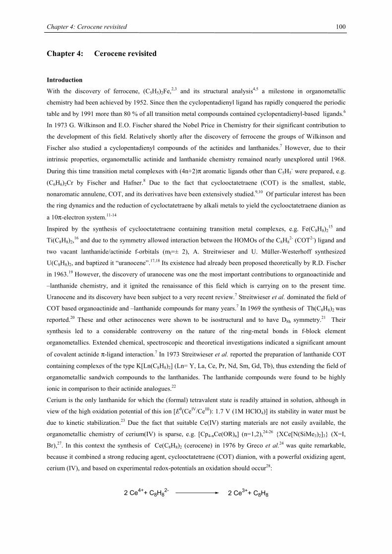
Chapter 4: Cerocene revisited
100
Chapter 4: Cerocene revisited
Introduction
With the discovery of ferrocene, (C5H5)2Fe,2,3 and its structural analysis4,5 a milestone in organometallic
chemistry had been achieved by 1952. Since then the cyclopentadienyl ligand has rapidly conquered the periodic
table and by 1991 more than 80 % of all transition metal compounds contained cyclopentadienyl-based ligands.6
In 1973 G. Wilkinson and E.O. Fischer shared the Nobel Price in Chemistry for their significant contribution to
the development of this field. Relatively shortly after the discovery of ferrocene the groups of Wilkinson and
Fischer also studied a cyclopentadienyl compounds of the actinides and lanthanides.7 However, due to their
intrinsic properties, organometallic actinide and lanthanide chemistry remained nearly unexplored until 1968.
During this time transition metal complexes with (4n+2)π aromatic ligands other than C5H5- were prepared, e.g.
(C6H6)2Cr by Fischer and Hafner.8 Due to the fact that cyclooctatetraene (COT) is the smallest, stable,
nonaromatic annulene, COT, and its derivatives have been extensively studied.9,10 Of particular interest has been
the ring dynamics and the reduction of cycloctatetraene by alkali metals to yield the cyclooctatetraene dianion as
a 10π-electron system.11-14
Inspired by the synthesis of cyclooctatetraene containing transition metal complexes, e.g. Fe(C8H8)215 and
Ti(C8H8)2,16 and due to the symmetry allowed interaction between the HOMOs of the C8H82- (COT2-) ligand and
two vacant lanthanide/actinide f-orbitals (ml=± 2), A. Streitwieser and U. Müller-Westerhoff synthesized
U(C8H8)2, and baptized it “uranocene”.17,18 Its existence had already been proposed theoretically by R.D. Fischer
in 1963.19 However, the discovery of uranocene was one the most important contributions to organoactinide and
–lanthanide chemistry, and it ignited the renaissance of this field which is carrying on to the present time.
Uranocene and its discovery have been subject to a very recent review.7 Streitwieser et al. dominated the field of
COT based organoactinide and –lanthanide compounds for many years.7 In 1969 the synthesis of Th(C8H8)2 was
reported.20 These and other actinocenes were shown to be isostructural and to have D8h symmetry.21 Their
synthesis led to a considerable controversy on the nature of the ring-metal bonds in f-block element
organometallics. Extended chemical, spectroscopic and theoretical investigations indicated a significant amount
of covalent actinide π-ligand interaction.7 In 1973 Streitwieser et al. reported the preparation of lanthanide COT
containing complexes of the type K[Ln(C8H8)2] (Ln= Y, La, Ce, Pr, Nd, Sm, Gd, Tb), thus extending the field of
organometallic sandwich compounds to the lanthanides. The lanthanide compounds were found to be highly
ionic in comparison to their actinide analogues.22
Cerium is the only lanthanide for which the (formal) tetravalent state is readily attained in solution, although in
view of the high oxidation potential of this ion [E0(CeIV/CeIII): 1.7 V (1M HClO4)] its stability in water must be
due to kinetic stabilization.23 Due the fact that suitable Ce(IV) starting materials are not easily available, the
organometallic chemistry of cerium(IV) is sparse, e.g. [Cp4-nCe(OR)n] (n=1,2),24-26 {XCe[N(SiMe3)2]3} (X=I,
Br),27. In this context the synthesis of Ce(C8H8)2 (cerocene) in 1976 by Greco et al.24 was quite remarkable,
because it combined a strong reducing agent, cyclooctatetraene (COT) dianion, with a powerful oxidizing agent,
cerium (IV), and based on experimental redox-potentials an oxidation should occur28:
2 Ce4++ C8H82- 2 Ce3++ C8H8
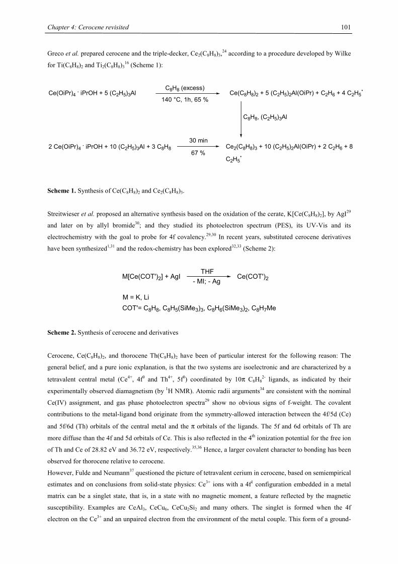
Chapter 4: Cerocene revisited
101
Greco et al. prepared cerocene and the triple-decker, Ce2(C8H8)3,24 according to a procedure developed by Wilke
for Ti(C8H8)2 and Ti2(C8H8)316 (Scheme 1):
Ce(OiPr)4 . iPrOH + 5 (C2H5)3Al Ce(C8H8)2 + 5 (C2H5)2Al(OiPr) + C2H6 + 4 C2H5.C8H8 (excess)
140 °C, 1h, 65 %
Ce2(C8H8)3 + 10 (C2H5)2Al(OiPr) + 2 C2H6 + 8
C2H5.67 %
30 min2 Ce(OiPr)4 . iPrOH + 10 (C2H5)3Al + 3 C8H8
C8H8, (C2H5)3Al
Scheme 1. Synthesis of Ce(C8H8)2 and Ce2(C8H8)3.
Streitwieser et al. proposed an alternative synthesis based on the oxidation of the cerate, K[Ce(C8H8)2], by AgI29
and later on by allyl bromide30; and they studied its photoelectron spectrum (PES), its UV-Vis and its
electrochemistry with the goal to probe for 4f covalency.29,30 In recent years, substituted cerocene derivatives
have been synthesized1,31 and the redox-chemistry has been explored32,33 (Scheme 2):
M[Ce(COT')2] + AgITHF
- MI; - AgCe(COT')2
M = K, LiCOT'= C8H8, C8H5(SiMe3)3, C8H6(SiMe3)2, C8H7Me
Scheme 2. Synthesis of cerocene and derivatives
Cerocene, Ce(C8H8)2, and thorocene Th(C8H8)2 have been of particular interest for the following reason: The
general belief, and a pure ionic explanation, is that the two systems are isoelectronic and are characterized by a
tetravalent central metal (Ce4+, 4f0 and Th4+, 5f0) coordinated by 10π C8H82- ligands, as indicated by their
experimentally observed diamagnetism (by 1H NMR). Atomic radii arguments34 are consistent with the nominal
Ce(IV) assignment, and gas phase photoelectron spectra29 show no obvious signs of f-weight. The covalent
contributions to the metal-ligand bond originate from the symmetry-allowed interaction between the 4f/5d (Ce)
and 5f/6d (Th) orbitals of the central metal and the π orbitals of the ligands. The 5f and 6d orbitals of Th are
more diffuse than the 4f and 5d orbitals of Ce. This is also reflected in the 4th ionization potential for the free ion
of Th and Ce of 28.82 eV and 36.72 eV, respectively.35,36 Hence, a larger covalent character to bonding has been
observed for thorocene relative to cerocene.
However, Fulde and Neumann37 questioned the picture of tetravalent cerium in cerocene, based on semiempirical
estimates and on conclusions from solid-state physics: Ce3+ ions with a 4f1 configuration embedded in a metal
matrix can be a singlet state, that is, in a state with no magnetic moment, a feature reflected by the magnetic
susceptibility. Examples are CeAl3, CeCu6, CeCu2Si2 and many others. The singlet is formed when the 4f
electron on the Ce3+ and an unpaired electron from the environment of the metal couple. This form of a ground-
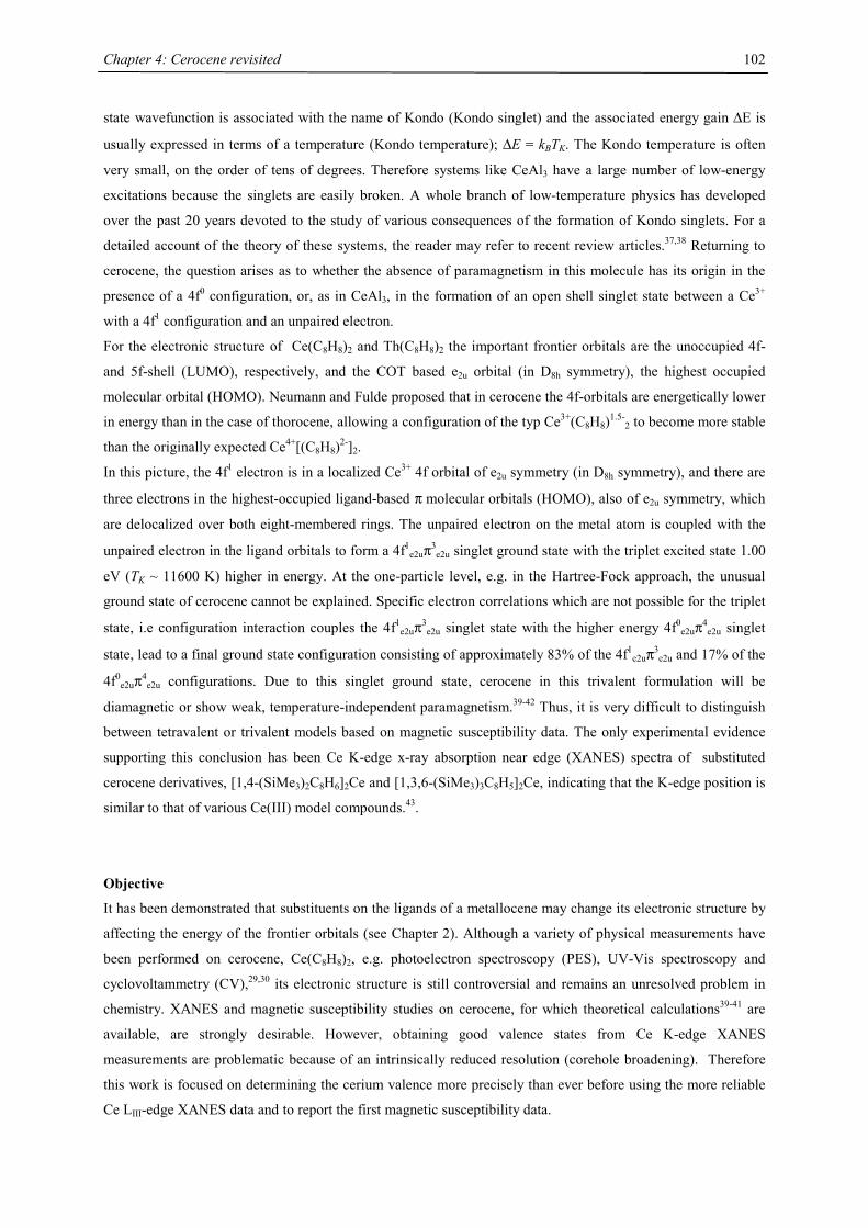
Chapter 4: Cerocene revisited
102
state wavefunction is associated with the name of Kondo (Kondo singlet) and the associated energy gain ∆E is
usually expressed in terms of a temperature (Kondo temperature); ∆E = kBTK. The Kondo temperature is often
very small, on the order of tens of degrees. Therefore systems like CeAl3 have a large number of low-energy
excitations because the singlets are easily broken. A whole branch of low-temperature physics has developed
over the past 20 years devoted to the study of various consequences of the formation of Kondo singlets. For a
detailed account of the theory of these systems, the reader may refer to recent review articles.37,38 Returning to
cerocene, the question arises as to whether the absence of paramagnetism in this molecule has its origin in the
presence of a 4f0 configuration, or, as in CeAl3, in the formation of an open shell singlet state between a Ce3+
with a 4f1 configuration and an unpaired electron.
For the electronic structure of Ce(C8H8)2 and Th(C8H8)2 the important frontier orbitals are the unoccupied 4f-
and 5f-shell (LUMO), respectively, and the COT based e2u orbital (in D8h symmetry), the highest occupied
molecular orbital (HOMO). Neumann and Fulde proposed that in cerocene the 4f-orbitals are energetically lower
in energy than in the case of thorocene, allowing a configuration of the typ Ce3+(C8H8)1.5-2 to become more stable
than the originally expected Ce4+[(C8H8)2-]2.
In this picture, the 4f1 electron is in a localized Ce3+ 4f orbital of e2u symmetry (in D8h symmetry), and there are
three electrons in the highest-occupied ligand-based π molecular orbitals (HOMO), also of e2u symmetry, which
are delocalized over both eight-membered rings. The unpaired electron on the metal atom is coupled with the
unpaired electron in the ligand orbitals to form a 4f1e2uπ3
e2u singlet ground state with the triplet excited state 1.00
eV (TK ~ 11600 K) higher in energy. At the one-particle level, e.g. in the Hartree-Fock approach, the unusual
ground state of cerocene cannot be explained. Specific electron correlations which are not possible for the triplet
state, i.e configuration interaction couples the 4f1e2uπ3
e2u singlet state with the higher energy 4f0e2uπ4
e2u singlet
state, lead to a final ground state configuration consisting of approximately 83% of the 4f1e2uπ3
e2u and 17% of the
4f0e2uπ4
e2u configurations. Due to this singlet ground state, cerocene in this trivalent formulation will be
diamagnetic or show weak, temperature-independent paramagnetism.39-42 Thus, it is very difficult to distinguish
between tetravalent or trivalent models based on magnetic susceptibility data. The only experimental evidence
supporting this conclusion has been Ce K-edge x-ray absorption near edge (XANES) spectra of substituted
cerocene derivatives, [1,4-(SiMe3)2C8H6]2Ce and [1,3,6-(SiMe3)3C8H5]2Ce, indicating that the K-edge position is
similar to that of various Ce(III) model compounds.43.
Objective
It has been demonstrated that substituents on the ligands of a metallocene may change its electronic structure by
affecting the energy of the frontier orbitals (see Chapter 2). Although a variety of physical measurements have
been performed on cerocene, Ce(C8H8)2, e.g. photoelectron spectroscopy (PES), UV-Vis spectroscopy and
cyclovoltammetry (CV),29,30 its electronic structure is still controversial and remains an unresolved problem in
chemistry. XANES and magnetic susceptibility studies on cerocene, for which theoretical calculations39-41 are
available, are strongly desirable. However, obtaining good valence states from Ce K-edge XANES
measurements are problematic because of an intrinsically reduced resolution (corehole broadening). Therefore
this work is focused on determining the cerium valence more precisely than ever before using the more reliable
Ce LIII-edge XANES data and to report the first magnetic susceptibility data.
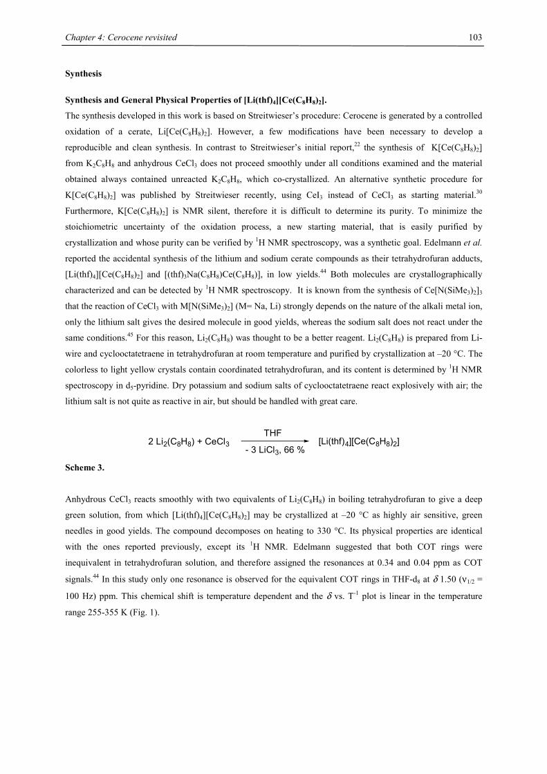
Chapter 4: Cerocene revisited
103
Synthesis
Synthesis and General Physical Properties of [Li(thf)4][Ce(C8H8)2].
The synthesis developed in this work is based on Streitwieser’s procedure: Cerocene is generated by a controlled
oxidation of a cerate, Li[Ce(C8H8)2]. However, a few modifications have been necessary to develop a
reproducible and clean synthesis. In contrast to Streitwieser’s initial report,22 the synthesis of K[Ce(C8H8)2]
from K2C8H8 and anhydrous CeCl3 does not proceed smoothly under all conditions examined and the material
obtained always contained unreacted K2C8H8, which co-crystallized. An alternative synthetic procedure for
K[Ce(C8H8)2] was published by Streitwieser recently, using CeI3 instead of CeCl3 as starting material.30
Furthermore, K[Ce(C8H8)2] is NMR silent, therefore it is difficult to determine its purity. To minimize the
stoichiometric uncertainty of the oxidation process, a new starting material, that is easily purified by
crystallization and whose purity can be verified by 1H NMR spectroscopy, was a synthetic goal. Edelmann et al.
reported the accidental synthesis of the lithium and sodium cerate compounds as their tetrahydrofuran adducts,
[Li(thf)4][Ce(C8H8)2] and [(thf)3Na(C8H8)Ce(C8H8)], in low yields.44 Both molecules are crystallographically
characterized and can be detected by 1H NMR spectroscopy. It is known from the synthesis of Ce[N(SiMe3)2]3
that the reaction of CeCl3 with M[N(SiMe3)2] (M= Na, Li) strongly depends on the nature of the alkali metal ion,
only the lithium salt gives the desired molecule in good yields, whereas the sodium salt does not react under the
same conditions.45 For this reason, Li2(C8H8) was thought to be a better reagent. Li2(C8H8) is prepared from Li-
wire and cyclooctatetraene in tetrahydrofuran at room temperature and purified by crystallization at –20 °C. The
colorless to light yellow crystals contain coordinated tetrahydrofuran, and its content is determined by 1H NMR
spectroscopy in d5-pyridine. Dry potassium and sodium salts of cyclooctatetraene react explosively with air; the
lithium salt is not quite as reactive in air, but should be handled with great care.
2 Li2(C8H8) + CeCl3THF
- 3 LiCl3, 66 %[Li(thf)4][Ce(C8H8)2]
Scheme 3.
Anhydrous CeCl3 reacts smoothly with two equivalents of Li2(C8H8) in boiling tetrahydrofuran to give a deep
green solution, from which [Li(thf)4][Ce(C8H8)2] may be crystallized at –20 °C as highly air sensitive, green
needles in good yields. The compound decomposes on heating to 330 °C. Its physical properties are identical
with the ones reported previously, except its 1H NMR. Edelmann suggested that both COT rings were
inequivalent in tetrahydrofuran solution, and therefore assigned the resonances at 0.34 and 0.04 ppm as COT
signals.44 In this study only one resonance is observed for the equivalent COT rings in THF-d8 at δ 1.50 (ν1/2 =
100 Hz) ppm. This chemical shift is temperature dependent and the δ vs. T-1 plot is linear in the temperature
range 255-355 K (Fig. 1).
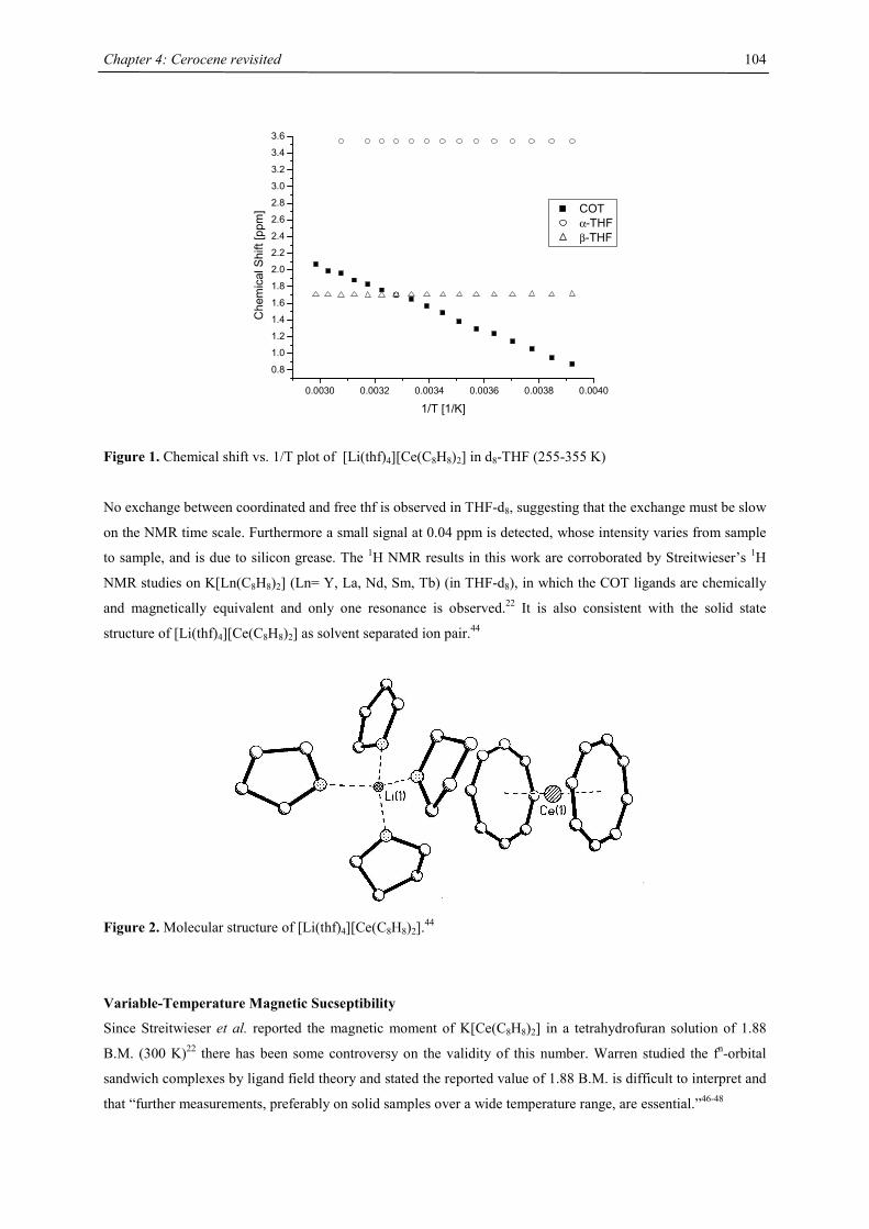
Chapter 4: Cerocene revisited
104
0.0030 0.0032 0.0034 0.0036 0.0038 0.0040
0.8
1.0
1.2
1.4
1.6
1.8
2.0
2.2
2.4
2.6
2.8
3.0
3.2
3.4
3.6
Che
mic
al S
hift
[ppm
]
1/T [1/K]
COT α-THF β-THF
Figure 1. Chemical shift vs. 1/T plot of [Li(thf)4][Ce(C8H8)2] in d8-THF (255-355 K)
No exchange between coordinated and free thf is observed in THF-d8, suggesting that the exchange must be slow
on the NMR time scale. Furthermore a small signal at 0.04 ppm is detected, whose intensity varies from sample
to sample, and is due to silicon grease. The 1H NMR results in this work are corroborated by Streitwieser’s 1H
NMR studies on K[Ln(C8H8)2] (Ln= Y, La, Nd, Sm, Tb) (in THF-d8), in which the COT ligands are chemically
and magnetically equivalent and only one resonance is observed.22 It is also consistent with the solid state
structure of [Li(thf)4][Ce(C8H8)2] as solvent separated ion pair.44
Figure 2. Molecular structure of [Li(thf)4][Ce(C8H8)2].44
Variable-Temperature Magnetic Sucseptibility
Since Streitwieser et al. reported the magnetic moment of K[Ce(C8H8)2] in a tetrahydrofuran solution of 1.88
B.M. (300 K)22 there has been some controversy on the validity of this number. Warren studied the fn-orbital
sandwich complexes by ligand field theory and stated the reported value of 1.88 B.M. is difficult to interpret and
that “further measurements, preferably on solid samples over a wide temperature range, are essential.”46-48
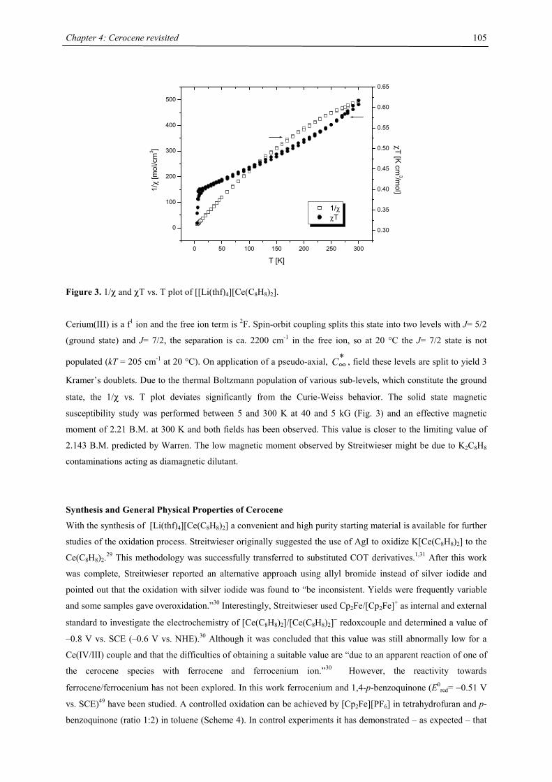
Chapter 4: Cerocene revisited
105
0 50 100 150 200 250 300
0
100
200
300
400
500
1/χ χT
T [K]
1/χ
[mol
/cm
3 ]
0.30
0.35
0.40
0.45
0.50
0.55
0.60
0.65
χT [K cm
3/mol]
Figure 3. 1/χ and χT vs. T plot of [[Li(thf)4][Ce(C8H8)2].
Cerium(III) is a f1 ion and the free ion term is 2F. Spin-orbit coupling splits this state into two levels with J= 5/2
(ground state) and J= 7/2, the separation is ca. 2200 cm-1 in the free ion, so at 20 °C the J= 7/2 state is not
populated (kT = 205 cm-1 at 20 °C). On application of a pseudo-axial, *∞C , field these levels are split to yield 3
Kramer’s doublets. Due to the thermal Boltzmann population of various sub-levels, which constitute the ground
state, the 1/χ vs. T plot deviates significantly from the Curie-Weiss behavior. The solid state magnetic
susceptibility study was performed between 5 and 300 K at 40 and 5 kG (Fig. 3) and an effective magnetic
moment of 2.21 B.M. at 300 K and both fields has been observed. This value is closer to the limiting value of
2.143 B.M. predicted by Warren. The low magnetic moment observed by Streitwieser might be due to K2C8H8
contaminations acting as diamagnetic dilutant.
Synthesis and General Physical Properties of Cerocene
With the synthesis of [Li(thf)4][Ce(C8H8)2] a convenient and high purity starting material is available for further
studies of the oxidation process. Streitwieser originally suggested the use of AgI to oxidize K[Ce(C8H8)2] to the
Ce(C8H8)2.29 This methodology was successfully transferred to substituted COT derivatives.1,31 After this work
was complete, Streitwieser reported an alternative approach using allyl bromide instead of silver iodide and
pointed out that the oxidation with silver iodide was found to “be inconsistent. Yields were frequently variable
and some samples gave overoxidation.”30 Interestingly, Streitwieser used Cp2Fe/[Cp2Fe]+ as internal and external
standard to investigate the electrochemistry of [Ce(C8H8)2]/[Ce(C8H8)2]− redoxcouple and determined a value of
–0.8 V vs. SCE (–0.6 V vs. NHE).30 Although it was concluded that this value was still abnormally low for a
Ce(IV/III) couple and that the difficulties of obtaining a suitable value are “due to an apparent reaction of one of
the cerocene species with ferrocene and ferrocenium ion.”30 However, the reactivity towards
ferrocene/ferrocenium has not been explored. In this work ferrocenium and 1,4-p-benzoquinone (E0red= −0.51 V
vs. SCE)49 have been studied. A controlled oxidation can be achieved by [Cp2Fe][PF6] in tetrahydrofuran and p-
benzoquinone (ratio 1:2) in toluene (Scheme 4). In control experiments it has demonstrated – as expected – that
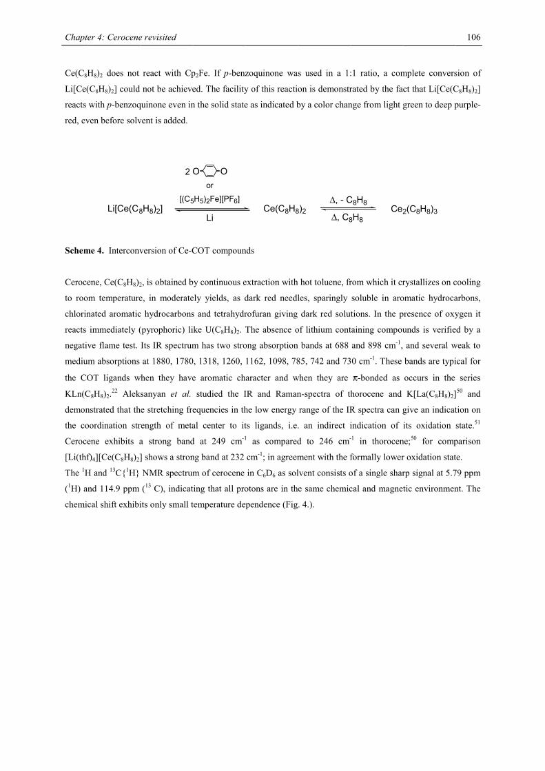
Chapter 4: Cerocene revisited
106
Ce(C8H8)2 does not react with Cp2Fe. If p-benzoquinone was used in a 1:1 ratio, a complete conversion of
Li[Ce(C8H8)2] could not be achieved. The facility of this reaction is demonstrated by the fact that Li[Ce(C8H8)2]
reacts with p-benzoquinone even in the solid state as indicated by a color change from light green to deep purple-
red, even before solvent is added.
Li[Ce(C8H8)2] Ce(C8H8)2
O O
Li
or
[(C5H5)2Fe][PF6] ∆, - C8H8
∆, C8H8Ce2(C8H8)3
2
Scheme 4. Interconversion of Ce-COT compounds
Cerocene, Ce(C8H8)2, is obtained by continuous extraction with hot toluene, from which it crystallizes on cooling
to room temperature, in moderately yields, as dark red needles, sparingly soluble in aromatic hydrocarbons,
chlorinated aromatic hydrocarbons and tetrahydrofuran giving dark red solutions. In the presence of oxygen it
reacts immediately (pyrophoric) like U(C8H8)2. The absence of lithium containing compounds is verified by a
negative flame test. Its IR spectrum has two strong absorption bands at 688 and 898 cm-1, and several weak to
medium absorptions at 1880, 1780, 1318, 1260, 1162, 1098, 785, 742 and 730 cm-1. These bands are typical for
the COT ligands when they have aromatic character and when they are π-bonded as occurs in the series
KLn(C8H8)2.22 Aleksanyan et al. studied the IR and Raman-spectra of thorocene and K[La(C8H8)2]50 and
demonstrated that the stretching frequencies in the low energy range of the IR spectra can give an indication on
the coordination strength of metal center to its ligands, i.e. an indirect indication of its oxidation state.51
Cerocene exhibits a strong band at 249 cm-1 as compared to 246 cm-1 in thorocene;50 for comparison
[Li(thf)4][Ce(C8H8)2] shows a strong band at 232 cm-1; in agreement with the formally lower oxidation state.
The 1H and 13C{1H} NMR spectrum of cerocene in C6D6 as solvent consists of a single sharp signal at 5.79 ppm
(1H) and 114.9 ppm (13 C), indicating that all protons are in the same chemical and magnetic environment. The
chemical shift exhibits only small temperature dependence (Fig. 4.).
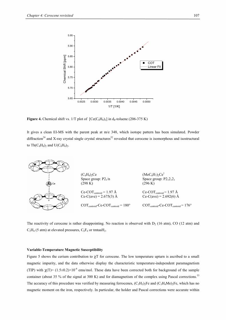
Chapter 4: Cerocene revisited
107
0.0025 0.0030 0.0035 0.0040 0.0045 0.00505.65
5.70
5.75
5.80
5.85
5.90
5.95
Che
mic
al S
hift
[ppm
]
1/T [1/K]
COT Linear Fit
Figure 4. Chemical shift vs. 1/T plot of [Ce(C8H8)2] in d8-toluene (206-375 K)
It gives a clean EI-MS with the parent peak at m/e 348, which isotope pattern has been simulated. Powder
diffraction24 and X-ray crystal single crystal structures52 revealed that cerocene is isomorphous and isostructural
to Th(C8H8)2 and U(C8H8)2.
The reactivity of cerocene is rather disappointing. No reaction is observed with D2 (16 atm), CO (12 atm) and
C2H4 (5 atm) at elevated pressures, C6F6 or tmtaaH2.
Variable-Temperature Magnetic Susceptibility
Figure 5 shows the cerium contribution to χT for cerocene. The low temperature upturn is ascribed to a small
magnetic impurity, and the data otherwise display the characteristic temperature-independent paramagnetism
(TIP) with χ(T)= (1.5±0.2)×10-4 emu/mol. These data have been corrected both for background of the sample
container (about 35 % of the signal at 300 K) and for diamagnetism of the complex using Pascal corrections.53
The accuracy of this procedure was verified by measuring ferrocenes, (C5H5)2Fe and (C5H4Me)2Fe, which has no
magnetic moment on the iron, respectively. In particular, the holder and Pascal corrections were accurate within
(C8H8)2Ce Space group: P21/n (298 K) Ce-COTcentroid = 1.97 Å Ce-C(ave) = 2.675(3) Å COTcentroid-Ce-COTcentroid = 180°
(MeC8H7)2Ce1 Space group: P212121 (296 K) Ce-COTcentroid = 1.97 Å Ce-C(ave) = 2.692(6) Å COTcentroid-Ce-COTcentroid = 176°
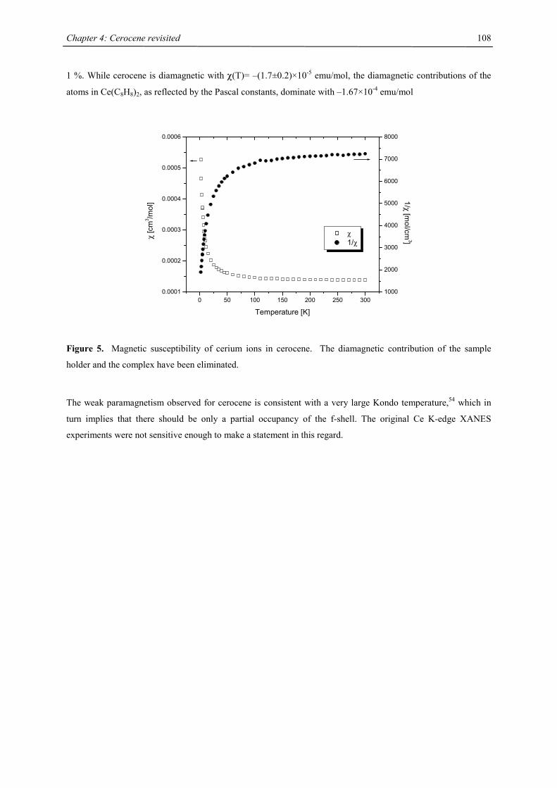
Chapter 4: Cerocene revisited
108
1 %. While cerocene is diamagnetic with χ(T)= –(1.7±0.2)×10-5 emu/mol, the diamagnetic contributions of the
atoms in Ce(C8H8)2, as reflected by the Pascal constants, dominate with –1.67×10⁻⁴ emu/mol
0 50 100 150 200 250 3000.0001
0.0002
0.0003
0.0004
0.0005
0.0006
χ 1/χ
Temperature [K]
χ [c
m3 /m
ol]
1000
2000
3000
4000
5000
6000
7000
8000
1/χ [mol/cm
3]
Figure 5. Magnetic susceptibility of cerium ions in cerocene. The diamagnetic contribution of the sample
holder and the complex have been eliminated.
The weak paramagnetism observed for cerocene is consistent with a very large Kondo temperature,54 which in
turn implies that there should be only a partial occupancy of the f-shell. The original Ce K-edge XANES
experiments were not sensitive enough to make a statement in this regard.
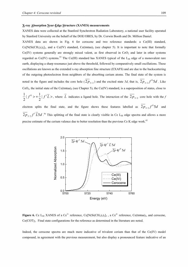
Chapter 4: Cerocene revisited
109
X-ray Absorption Near-Edge Structure (XANES) measurements
XANES data were collected at the Stanford Synchrotron Radiation Laboratory, a national user facility operated
by Stanford University on the behalf of the DOE/OBES, by Dr. Corwin Booth and Dr. Million Daniel.
XANES data are shown in Fig. 6 for cerocene and two reference standards: a Ce(III) standard,
Ce[N(Si(CH3)3)2]3, and a Ce(IV) standard, Ce(tmtaa)2 (see chapter 5). It is important to note that formally
Ce(IV) systems generally are strongly mixed valent, as first observed in CeO2 and later in other systems
regarded as Ce(IV) systems.55 The Ce(III) standard has XANES typical of the LIII edge of a monovalent rare
earth, displaying a sharp resonance just above the threshold, followed by comparatively small oscillations. These
oscillations are known as the extended x-ray absorption fine structure (EXAFS) and are due to the backscattering
of the outgoing photoelectron from neighbors of the absorbing cerium atoms. The final state of the system is
noted in the figure and includes the core hole ( 2/32 p ) and the excited state 5d, that is, dfp 52 02/3 . Like
CeO2, the initial state of the Ce(tmtaa)2 (see Chapter 5), the Ce(IV) standard, is a superposition of states, close to
>+> Lff 10 |21|
21
, where L indicates a ligand hole. The interaction of the 2/32 p core hole with the f
electron splits the final state, and the figure shows these features labelled as dfp 52 02/3 and
dLfp 52 12/3 .56 This splitting of the final state is clearly visible in Ce LIII edge spectra and allows a more
precise estimate of the cerium valence due to better resolution than the previous Ce K edge work.43
5700 5720 5740 57600.0
0.5
1.0
1.5
2.0___
2p 4f 1 L 5d__2p 4f 0 5d
__2p 4f 1 5d
Nor
mal
ized
Abs
orpt
ion
Energy (eV)
Ce(III) Ce(IV) Cerocene
Figure 6. Ce LIII XANES of a Ce3+ reference, Ce[N(Si(CH3)3)2]3 , a Ce4+ reference, Ce(tmtaa)2, and cerocene,
Ce(COT)2. Final state configurations for the reference as determined in the literature are noted.
Indeed, the cerocene spectra are much more indicative of trivalent cerium than that of the Ce(IV) model
compound, in agreement with the previous measurement, but also display a pronounced feature indicative of an
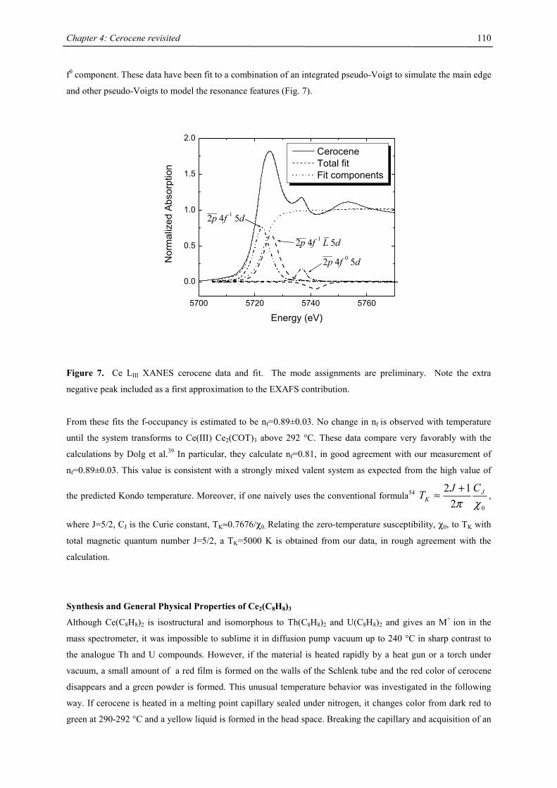
Chapter 4: Cerocene revisited
110
f0 component. These data have been fit to a combination of an integrated pseudo-Voigt to simulate the main edge
and other pseudo-Voigts to model the resonance features (Fig. 7).
5700 5720 5740 5760
0.0
0.5
1.0
1.5
2.0
___2p 4f 1 L 5d
__2p 4f 0 5d
__2p 4f 1 5d
Nor
mal
ized
Abs
orpt
ion
Energy (eV)
Cerocene Total fit Fit components
Figure 7. Ce LIII XANES cerocene data and fit. The mode assignments are preliminary. Note the extra
negative peak included as a first approximation to the EXAFS contribution.
From these fits the f-occupancy is estimated to be nf=0.89±0.03. No change in nf is observed with temperature
until the system transforms to Ce(III) Ce2(COT)3 above 292 °C. These data compare very favorably with the
calculations by Dolg et al.39 In particular, they calculate nf=0.81, in good agreement with our measurement of
nf=0.89±0.03. This value is consistent with a strongly mixed valent system as expected from the high value of
the predicted Kondo temperature. Moreover, if one naively uses the conventional formula54 02
12χπ
JK
CJT +≈ ,
where J=5/2, CJ is the Curie constant, TK≈0.7676/χ0. Relating the zero-temperature susceptibility, χ0, to TK with
total magnetic quantum number J=5/2, a TK=5000 K is obtained from our data, in rough agreement with the
calculation.
Synthesis and General Physical Properties of Ce2(C8H8)3
Although Ce(C8H8)2 is isostructural and isomorphous to Th(C8H8)2 and U(C8H8)2 and gives an M+ ion in the
mass spectrometer, it was impossible to sublime it in diffusion pump vacuum up to 240 °C in sharp contrast to
the analogue Th and U compounds. However, if the material is heated rapidly by a heat gun or a torch under
vacuum, a small amount of a red film is formed on the walls of the Schlenk tube and the red color of cerocene
disappears and a green powder is formed. This unusual temperature behavior was investigated in the following
way. If cerocene is heated in a melting point capillary sealed under nitrogen, it changes color from dark red to
green at 290-292 °C and a yellow liquid is formed in the head space. Breaking the capillary and acquisition of an
Chapter 4: Cerocene revisited
111
1H NMR spectrum identifies free COT, but no Ce(C8H8)2. The green powder was identified by IR spectroscopy
as Ce2(C8H8)3 (Scheme 4). This process has also been verified by EXAFS and XANES studies. Ce2(C8H8)3 has
been reported recently by Greco et al. as the only identifiable product, if the cerocene synthesis is carried out at
lower temperatures (110-115 °C) in cycloheptatriene as solvent, and at a molar ratio C8H8/Ce < 5. (Scheme 1).24
Alternatively this molecule has been prepared by co-condensation of cerium metal atoms and C8H8 at –196 °C.57
It is highly air sensitive (pyrophoric) and is decomposed immediately by water and alcohols; its extremely low
solubility in organic solvents (< 0.5 mmol/L in THF) and its microcrystalline nature frustrated attempts at its
characterization by 1H NMR spectroscopy and X-ray diffraction (single crystal investigations). However, the
crystal structure of [Nd(C8H8)(thf)2][Nd(C8H8)2] has been determined.57 Cerium also forms a
bis(tetrahydrofuran) adduct on prolonged extraction with hot tetrahydrofuran, which is isolated as golden
crystals. These crystals easily lose tetrahydrofuran on standing at room temperature and collapse to a green
powder.57 A molecular ion for Ce2(C8H8)3 is not detected by EI-MS, the only cerium containing fragment is
[(C8H8)Ce]+ (m/e= 244 (30 %)). This observation is consistent with Greco’s data.24 The IR spectra indicate the
presence of two different types of COT rings in the complex. In fact, the out-of-plane bending mode due to C-H
(between 680 and 700 cm-1) and on the band due to the benzenoid system (in the region 1600-1900 cm-1) are
split into two components. A very nice and sensitive instrument to detect the presence of Ce2(C8H8)3 is the low
energy range the IR spectrum: The triple-decker exhibits three bands in this regime: 240 (sh), 232 (vs) and 210
(s) compared to only one band at 249 cm-1 in Ce(C8H8)2.
The reactivity of the Ce(C8H8)2/Ce2(C8H8)3 couple closely resembles the behavior of its Ti analogue,16 in both
cases the species can be interconverted easily into each other. Ce(C8H8)2 converts quantitatively at 290-292 °C
into the Ce2(C8H8)3 by COT elimination, on the other side Ce2(C8H8)3 is converted to Ce(C8H8)2 in the presence
of free COT. This later process can be achieved in the presence of a ca. 70 fold excess of COT in C6D6 at 65 °C
over a time span of 4 months, or in the presence of catalytic amounts of Al(C2H5)3 and a 70 fold excess of COT
at 130 °C within 1 h. The rate difference is probably due to the insolubility of Ce2(C8H8)3 in C6D6 at 65 °C. It
should be pointed out that during the extraction process of cerocene with hot toluene (b.p. 110 °C), a small
amount of Ce2(C8H8)3 is formed and to obtain absolutely pure cerocene, it is necessary to recrystallize the
contaminated material from toluene at –20 °C. Based on these observations, it can be concluded that the
cerocene synthesis of Greco et al.24 only worked that well, because it was performed in COT as solvent and the
triple-decker contamination was immediately converted to cerocene under the conditions employed.
Variable-Temperature Magnetic Sucseptibility
Triple-decker complexes are not only structurally quite interesting molecules as are their magnetic properties.
The paramagnetic cerium(III), f1, spin carriers are separated by about 4 Å, as estimated from the averaged
COTcentroid-Ce distances in the [Ce(C8H8)2]- anions.44 The question arises, whether these spin carriers can
effectively communicate via the COT-bridge, or if they behave as independent spin carriers. The solid state
magnetism was recorded at 2-300 K. At high temperature the 1/χ vs. T plot is as expected for two uncorrelated
spin carriers and yield an effective magnetic moment of 2.8 B.M. However, at low temperature the value of χ
increases normally with decreasing temperature until a maximum is reached and then declines rapidly with
decreasing temperature. This behavior is typical of an antiferromagnetic spin exchange interaction and the
temperature at which the maximum susceptibility occurs is called the Néel temperature (TN) of 8 K (Fig. 8).
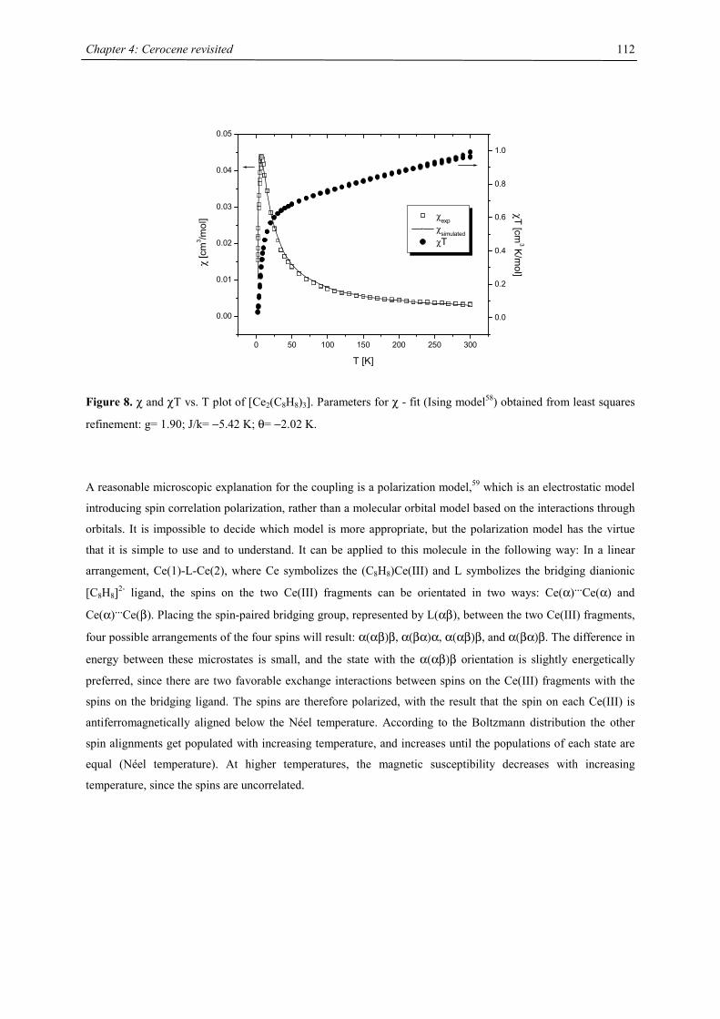
Chapter 4: Cerocene revisited
112
0 50 100 150 200 250 300
0.00
0.01
0.02
0.03
0.04
0.05
χexp χ
simulated χT
T [K]
χ [c
m3 /m
ol]
0.0
0.2
0.4
0.6
0.8
1.0
χT [cm3 K
/mol]
Figure 8. χ and χT vs. T plot of [Ce2(C8H8)3]. Parameters for χ - fit (Ising model58) obtained from least squares
refinement: g= 1.90; J/k= −5.42 K; θ= −2.02 K.
A reasonable microscopic explanation for the coupling is a polarization model,59 which is an electrostatic model
introducing spin correlation polarization, rather than a molecular orbital model based on the interactions through
orbitals. It is impossible to decide which model is more appropriate, but the polarization model has the virtue
that it is simple to use and to understand. It can be applied to this molecule in the following way: In a linear
arrangement, Ce(1)-L-Ce(2), where Ce symbolizes the (C8H8)Ce(III) and L symbolizes the bridging dianionic
[C8H8]2- ligand, the spins on the two Ce(III) fragments can be orientated in two ways: Ce(α)…Ce(α) and
Ce(α)…Ce(β). Placing the spin-paired bridging group, represented by L(αβ), between the two Ce(III) fragments,
four possible arrangements of the four spins will result: α(αβ)β, α(βα)α, α(αβ)β, and α(βα)β. The difference in
energy between these microstates is small, and the state with the α(αβ)β orientation is slightly energetically
preferred, since there are two favorable exchange interactions between spins on the Ce(III) fragments with the
spins on the bridging ligand. The spins are therefore polarized, with the result that the spin on each Ce(III) is
antiferromagnetically aligned below the Néel temperature. According to the Boltzmann distribution the other
spin alignments get populated with increasing temperature, and increases until the populations of each state are
equal (Néel temperature). At higher temperatures, the magnetic susceptibility decreases with increasing
temperature, since the spins are uncorrelated.
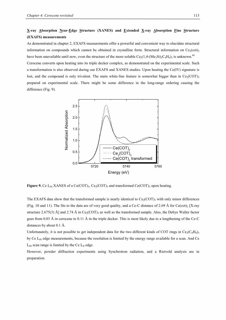
Chapter 4: Cerocene revisited
113
X-ray Absorption Near-Edge Structure (XANES) and Extended X-ray Absorption Fine Structure
(EXAFS) measurements
As demonstrated in chapter 2, EXAFS measurements offer a powerful and convenient way to elucidate structural
information on compounds which cannot be obtained in crystalline form. Structural information on Ce2(cot)3
have been unavailable until now, even the structure of the more soluble Ce2(1,4-(Me3Si)2C8H6)3 is unknown.60
Cerocene converts upon heating into its triple decker complex, as demonstrated on the experimental scale. Such
a transformation is also observed during our EXAFS and XANES studies. Upon heating the Ce(IV) signature is
lost, and the compound is only trivalent. The main white-line feature is somewhat bigger than in Ce2(COT)3
prepared on experimental scale. There might be some difference in the long-range ordering causing the
difference (Fig. 9).
5720 5740 57600.0
0.5
1.0
1.5
2.0
2.5
Nor
mal
ized
Abs
orpt
ion
Energy (eV)
Ce(COT)2 Ce2(COT)3 Ce(COT)2 transformed
Figure 9. Ce LIII XANES of a Ce(COT)2, Ce2(COT)3 and transformed Ce(COT)2 upon heating.
The EXAFS data show that the transformed sample is nearly identical to Ce2(COT)3 with only minor differences
(Fig. 10 and 11). The fits to the data are of very good quality, and a Ce-C distance of 2.69 Å for Ce(cot)2 [X-ray
structure 2.675(3) Å] and 2.74 Å in Ce2(COT)3 as well as the transformed sample. Also, the Debye Waller factor
goes from 0.03 Å in cerocene to 0.11 Å in the triple decker. This is most likely due to a lengthening of the Ce-C
distances by about 0.1 Å.
Unfortunately, it is not possible to get independent data for the two different kinds of COT rings in Ce2(C8H8)3
by Ce LIII edge measurements, because the resolution is limited by the energy range available for a scan. And Ce
LIII scan range is limited by the Ce LII edge.
However, powder diffraction experiments using Synchrotron radiation, and a Rietveld analysis are in
preparation.
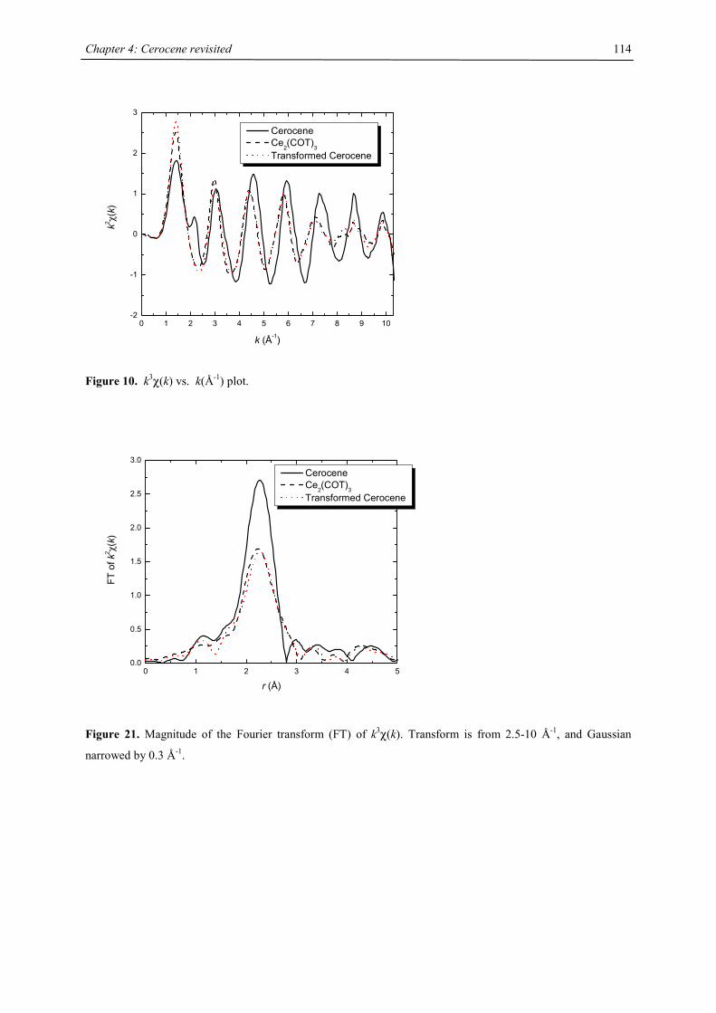
Chapter 4: Cerocene revisited
114
0 1 2 3 4 5 6 7 8 9 10-2
-1
0
1
2
3
k2 χ(
k)
k (Å-1)
Cerocene Ce
2(COT)
3 Transformed Cerocene
Figure 10. k3χ(k) vs. k(Å-1) plot.
0 1 2 3 4 50.0
0.5
1.0
1.5
2.0
2.5
3.0
FT o
f k2 χ(k)
r (Å)
Cerocene Ce2(COT)3 Transformed Cerocene
Figure 21. Magnitude of the Fourier transform (FT) of k3χ(k). Transform is from 2.5-10 Å-1, and Gaussian
narrowed by 0.3 Å-1.
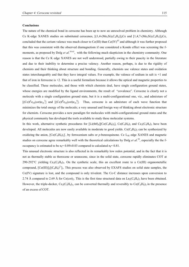
Chapter 4: Cerocene revisited
115
Conclusions
The nature of the chemical bond in cerocene has been up to now an unresolved problem in chemistry. Although
Ce K-edge XANES studies on substituted cerocenes, [(1,4-(Me3Si)2C8H6)]2Ce and [1,4,7-(Me3Si)3C8H5)]2Ce,
concluded that the cerium valence was much closer to Ce(III) than Ce(IV)43 and although it was further proposed
that this was consistent with the observed diamagnetism if one considered a Kondo effect was screening the f-
moments, as proposed by Dolg et al.39-41, with the following much skepticism in the chemistry community. One
reason is that the Ce K edge XANES are not well understood, partially owing to their paucity in the literature
and due to their inability to determine a precise valency. Another reason, perhaps, is due to the rigidity of
chemists and their thinking about structure and bonding. Generally, chemists use valence states and oxidation
states interchangeably and that they have integral values. For example, the valence of sodium in salt is +1 and
that of iron in ferrocene is +2. This is a useful formalism because it allows the optical and magnetic properties to
be classified. These molecules, and those with which chemists deal, have single configuration ground states,
whose energies are modified by the ligand environments, the result of “covalence”. Cerocene is clearly not a
molecule with a single configuration ground state, but it is a multi-configurational one, viz., and admixture of
[(Ce)f1e2u(cot)e2u3] and [(Ce)f0e2u(cot)e2u
4]. Thus, cerocene is an admixture of each wave function that
minimizes the total energy of the molecule; a very unusual and foreign way of thinking about electronic structure
for chemists. Cerocene provides a new paradigm for molecules with multi-configurational ground states and the
physical community has developed the tools available to study these molecular systems.
In this work, alternative synthetic procedures for [Li(thf)4][Ce(C8H8)2], Ce(C8H8)2 and Ce2(C8H8)3 have been
developed. All molecules are now easily available in moderate to good yields. Ce(C8H8)2 can be synthesized by
oxidizing the anion, [Ce(C8H8)2]-, by ferrocenium salts or p-benzoquinone. Ce LIII edge XANES and magnetic
studies on cerocene agree remarkably well with the theoretical calculations by Dolg et al.39, especially the the f-
occupancy is estimated to be nf= 0.89±0.03 compared to calculated nf= 0.81.
This unusual electronic structure is also reflected in its remarkably low redox potential, and in the fact that it is
not as thermally stable as thorocene or uranocene, since in the solid state, cerocene rapidly eliminates COT at
290-292°C yielding Ce2(C8H8)3. On the synthetic scale, this an excellent route to a Ce(III) organometallic
compound, [Ce(III)]2[(C8H8)2-]3. This process was also observed by EXAFS studies on solid state samples, the
Ce(IV) signature is lost, and the compound is only trivalent. The Ce-C distance increases upon conversion to
2.74 Å compared to 2.69 Å for Ce(cot)2. This is the first time structural data on Ln2(C8H8)3 have been obtained.
However, the triple-decker, Ce2(C8H8)3, can be converted thermally and reversibly to Ce(C8H8)2 in the presence
of an excess of COT.
Chapter 4: Cerocene revisited
116
References
[1] Boussie, T. R.; Eisenberg, D. C.; Rigsbee, J.; Streitwieser, A.; Zalkin, A. Organometallics 1991, 10,
1022-1028.
[2] Kealy, T. J.; Pauson, P. L. Nature 1951, 168, 1039-1040.
[3] Miller, S. A.; Tebboth, J. A.; Tremaine, J. F. J. Chem. Soc. 1952, 632-635.
[4] Wilkinson, G.; Rosenblum, M.; Whiting, M. C.; Woodward, R. B. J. Am. Chem. Soc. 1952, 74, 2125-
2126.
[5] Fischer, E. O.; Pfab, W. Z. Naturforsch B 1952, 7, 377-379.
[6] Janiak, C.; Schumann, H. Adv. Organomet. Chem. 1991, 33, 291-393.
[7] Seyferth, D. Organometallics 2004, 23, 3562-3583; and references therein.
[8] Fischer, E. O.; Hafner, W. Z. Naturforsch B 1955, 10, 665.
[9] Paquette, L. A. Tetrahedron 1975, 31, 2855-2883.
[10] Fray, G. I.; Saxton, R. G. The Chemistry of Cyclooctatetraene and Its Derivatives; Cambridge
University Press: New York, 1978.
[11] Anet, F. A.; Bourn, A. J. R.; Lin, Y. S. J. Am. Chem. Soc. 1964, 86, 3576.
[12] Katz, T. J.; Reinmuth, W. H.; Smith, D. E. J. Am. Chem. Soc. 1962, 84, 802-808.
[13] Katz, T. J. J. Am. Chem. Soc. 1960, 82, 3784-3785.
[14] Strauss, H. L.; Katz, T. J.; Fraenkel, G. K. J. Am. Chem. Soc. 1963, 85, 2360-2364.
[15] Carbonaro, A.; Greco, A.; Dall'Asta, G. Tetrahedron Lett. 1967, 22, 2037-2040.
[16] Wilke, G.; Breil, H. Angew. Chem. Int. Ed. Engl. 1966, 5, 898-899.
[17] Streitwieser, A.; Müller-Westerhoff, U.; Sonnichsen, G.; Mares, F.; Morrell, D. G.; Hodgson, K. O.;
Harmon, C. A. J. Am. Chem. Soc. 1973, 95, 8644-8649.
[18] Streitwieser, A.; Müller-Westerhoff, U. J. Am. Chem. Soc. 1968, 90, 7364.
[19] Fischer, R. D. Theoret. Chim. Acta 1963, 1, 418-431.
[20] Streitwieser, A.; Yoshida, N. J. Am. Chem. Soc. 1969, 91, 7528.
[21] Adeef, A.; Raymond, K. N.; Hodgson, K. O.; Zalkin, A. Inorg. Chem. 1972, 11, 1083-1088.
[22] Hodgson, K. O.; Mares, F.; Starks, D. F.; Streitwieser, A. J. Am. Chem. Soc. 1973, 95, 8650-8658.
[23] Cotton, S. Lanthanides and Actinides; Macmillian: Riverside, NJ, 1991.
[24] Greco, A.; Cesca, S.; Bertolini, G. J. Organomet. Chem. 1976, 119, 321-330.
[25] Evans, W. J.; Deming, T. J.; Ziller, J. W. Organometallics 1989, 8, 1581-1583.
[26] Gulino, A.; Casarin, M.; Conticello, V. P.; Gaudiello, J. G.; Mauermann, H.; Fragalá, I. L.; Marks, T. J.
Organometallics 1988, 7, 2360-2364.
[27] Hitchcock, P. B.; Hulkes, A. G.; Lappert, M. F. Inorg. Chem. 2004, 43, 1031-1038.
[28] Fischer, R. D. Angew. Chem. Int. Ed. Engl. 1994, 33, 2165-2168.
[29] Streitwieser, A.; Kinsley, S. A.; Rigsbee, J. T.; Fragala, I. L.; Ciliberto, E.; Rosch, N. J. Am. Chem. Soc.
1985, 107, 7786-7788.
[30] Streitwieser, A.; Kinsley, S. A.; Jenson, C. H.; Rigsbee, J. T. Organometallics 2004, 23, 5169-5175.
[31] Kilimann, U.; Herbst-Irmer, R.; Stalke, D.; Edelmann, F. T. Angew. Chem. 1994, 106, 1684-1687.
[32] Edelmann, F. T. New J. Chem. 1995, 19, 535-550.
[33] Reissmann, U.; Lameyer, L.; Stalke, D.; Poremba, P.; Edelmann, F. T. Chem. Commun. 1999, 1865-
1866.
Chapter 4: Cerocene revisited
117
[34] Raymond, K. N.; Eigenbrot, C. W. Acc. Chem. Res. 1980, 13, 276-283.
[35] Martin, W. C.; Hagan, L.; Reader, J.; Sugar, J. J. Phys. Chem. Ref. Data 1974, 3, 771.
[36] Sugar, J. J. Opt. Soc. Am. 1975, 65, 1366.
[37] Fulde, P. J. Phys. F: Met. Phys. 1988, 18, 601-639.
[38] Santini, P.; Lémanski, R.; Erdös, P. Adv. Phys. 1999, 48, 537-653.
[39] Dolg, M.; Fulde, P.; Stoll, H.; Preuss, H.; Chang, A.; Pitzer, R. M. Chem. Phys. 1995, 195, 71-82.
[40] Dolg, M.; Fulde, P.; Kuchle, W.; Neumann, C. S.; Stoll, H. J. Chem. Phys. 1991, 94, 3011-3017.
[41] Dolg, M.; Fulde, P. Chem. Eur. J. 1998, 4, 200-204.
[42] Liu, W. J.; Dolg, M.; Fulde, P. J. Chem. Phys. 1997, 107, 3584-3591.
[43] Edelstein, N. M.; Allen, P. G.; Bucher, J. J.; Shuh, D. K.; Sofield, C. D.; Kaltsoyannis, N.; Maunder, G.
H.; Russo, M. R.; Sella, A. J. Am. Chem. Soc. 1996, 118, 13115-13116.
[44] Kilimann, U.; Schäfer, M.; Herbst-Irmer, R.; Edelmann, F. T. J. Organomet. Chem. 1994, 469, C15-
C18.
[45] Andersen, R. A., private communication.
[46] Warren, K. D. Struct. Bond. 1977, 33, 97-138.
[47] Warren, K. D. Inorg. Chem. 1975, 14, 3095-3103.
[48] Clack, D. W.; Warren, K. D. J. Organomet. Chem. 1976, 122, C28-C30.
[49] Kavarnos, G. J.; Turro, N. J. Chem. Rev. 1986, 86, 401-449.
[50] Aleksanyan, V. T.; Garbusova, I. A.; Chernyshova, T. M.; Todres, Z. V.; Leonov, M. R.; Gramateeva,
N. I. J. Organomet. Chem. 1981, 217, 169-177.
[51] Berg, D. J.; Andersen, R. A. Organometallics 2003, 22, 627-632.
[52] Kurras, E.; Krüger, C., Max-Planck für Kohlenforschung Mühlheim, private communication.
[53] Theory and Application of Molecular Diamagnetism; Wiley-Interscience: New York, 1976.
[54] Rajan, V. T. Phys. Rev. Lett. 1983, 51, 308.
[55] Kaindl, G.; Schmiester, G.; Sampathkumaran, E. V.; Wachter, P. Phys. Rev. B 1988, 38, 10174-10177.
[56] Kotani, A.; Jo, T.; Parlebas, J. C. Adv. Phys. 1988, 37, 37.
[57] DeKock, C. W.; Ely, S. R.; Hopkins, T. E.; Brault, M. A. Inorg. Chem. 1978, 17, 625-631.
[58] O'Connor, C. J. In Progress in Inorganic Chemistry; Lippard, S. J., Ed.; J. Wiley & Sons: New York,
1982; Vol. 29, pp 203-285.
[59] Kahn, O. Molecular Magnetism; VCH: New York, 1993.
[60] Poremba, P.; Edelmann, F. T. J. Organomet. Chem. 1998, 553, 393-395.
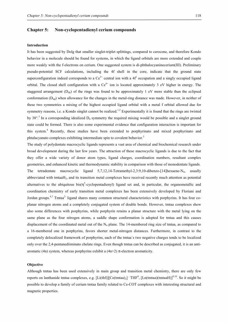
Chapter 5: Non-cyclopentadienyl cerium compounds
118
Chapter 5: Non-cyclopentadienyl cerium compounds
Introduction
It has been suggested by Dolg that smaller singlet-triplet splittings, compared to cerocene, and therefore Kondo
behavior in a molecule should be found for systems, in which the ligand orbitals are more extended and couple
more weakly with the f-electrons on cerium. One suggested system is di-phthalocyaninecerium(III). Preliminary
pseudo-potential SCF calculations, including the 4f shell in the core, indicate that the ground state
superconfiguration indeed corresponds to a Ce3+ central ion with a 4f1 occupation and a singly occupied ligand
orbital. The closed shell configuration with a Ce4+ ion is located approximately 3 eV higher in energy. The
staggered arrangement (D4h) of the rings was found to be approximately 1 eV more stable than the eclipsed
conformation (D4d) when allowance for the changes in the metal-ring distance was made. However, in neither of
these two symmetries a mixing of the highest occupied ligand orbital with a metal f orbital allowed due for
symmetry reasons, i.e. a Kondo singlet cannot be realized.1,2 Experimentally it is found that the rings are twisted
by 38°.3 In a corresponding idealized D4 symmetry the required mixing would be possible and a singlet ground
state could be formed. There is also some experimental evidence that configuration interaction is important for
this system.4 Recently, these studies have been extended to porphyrinato and mixed porphyrinato and
phtalacyanato complexes exhibiting intermediate spin to covalent behavior.5
The study of polydentate macrocyclic ligands represents a vast area of chemical and biochemical research under
broad development during the last few years. The attraction of these macrocyclic ligands is due to the fact that
they offer a wide variety of donor atom types, ligand charges, coordination numbers, resultant complex
geometries, and enhanced kinetic and thermodynamic stability in comparison with those of monodentate ligands.
The tetradentate macrocyclic ligand 5,7,12,14-Tetramethyl-2,3:9,10-dibenzo-[14]hexaene-N4, usually
abbreviated with tmtaaH2, and its transition metal complexes have received recently much attention as potential
alternatives to the ubiquitous bis(η5-cyclopentadienyl) ligand set and, in particular, the organometallic and
coordination chemistry of early transition metal complexes has been extensively developed by Floriani and
Jordan groups.6,7 Tmtaa2- ligand shares many common structural characteristics with porphyrins. It has four co-
planar nitrogen atoms and a completely conjugated system of double bonds. However, tmtaa complexes show
also some differences with porphyrins, while porphyrin retains a planar structure with the metal lying on the
same plane as the four nitrogen atoms, a saddle shape conformation is adopted for tmtaa and this causes
displacement of the coordinated metal out of the N4 plane. The 14-membered ring size of tmtaa, as compared to
a 16-membered one in porphyrins, favors shorter metal-nitrogen distances. Furthermore, in contrast to the
completely delocalized framework of porphyrins, each of the tmtaa`s two negative charges tends to be localized
only over the 2,4-pentanediiminato chelate rings. Even though tmtaa can be described as conjugated, it is an anti-
aromatic (4n) system, whereas porphyrins exhibit a (4n+2) π-electron aromaticity.
Objective
Although tmtaa has been used extensively in main group and transition metal chemistry, there are only few
reports on lanthanide tmtaa complexes, e.g. [Li(thf)][Ce(tmtaa)2] . THF8, [Ln(tmtaa)(tmtaaH)]9,10. So it might be
possible to develop a family of cerium tmtaa family related to Ce-COT complexes with interesting structural and
magnetic properties.
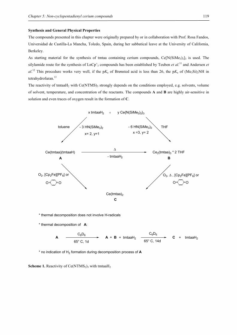
Chapter 5: Non-cyclopentadienyl cerium compounds
119
Synthesis and General Physical Properties
The compounds presented in this chapter were originally prepared by or in collaboration with Prof. Rosa Fandos,
Universidad de Castilla-La Mancha, Toledo, Spain, during her sabbatical leave at the University of California,
Berkeley.
As starting material for the synthesis of tmtaa containing cerium compounds, Ce[N(SiMe3)2]3 is used. The
silylamide route for the synthesis of LnCp’3 compounds has been established by Teuben et al.11 and Andersen et
al.12 This procedure works very well, if the pKa of Brønsted acid is less than 26, the pKa of (Me3Si)2NH in
tetrahydrofuran.13
The reactivity of tmtaaH2 with Ce(NTMS)3 strongly depends on the conditions employed, e.g. solvents, volume
of solvent, temperature, and concentration of the reactants. The compounds A and B are highly air-sensitive in
solution and even traces of oxygen result in the formation of C.
x tmtaaH2 y Ce{N(SiMe3)2}3
* thermal decomposition does not involve H-radicals
* thermal decomposition of A:
AC6D6
65° C, 1dA + B + tmtaaH2
C6D6
65° C, 14dC + tmtaaH2
* no indication of H2 formation during decomposition process of A
- 3 HN(SiMe3)2
x= 2, y=1
toluene - 6 HN(SiMe3)2
x =3, y= 2THF
Ce(tmtaa)(tmtaaH)
ACe2(tmtaa)3 * 2 THF
B
∆
- tmtaaH2
Ce(tmtaa)2
O2, [Cp2Fe][PF6] or
O O
O2, ∆ , [Cp2Fe][PF6] or
O O
C
Scheme 1. Reactivity of Ce(NTMS2)3 with tmtaaH2
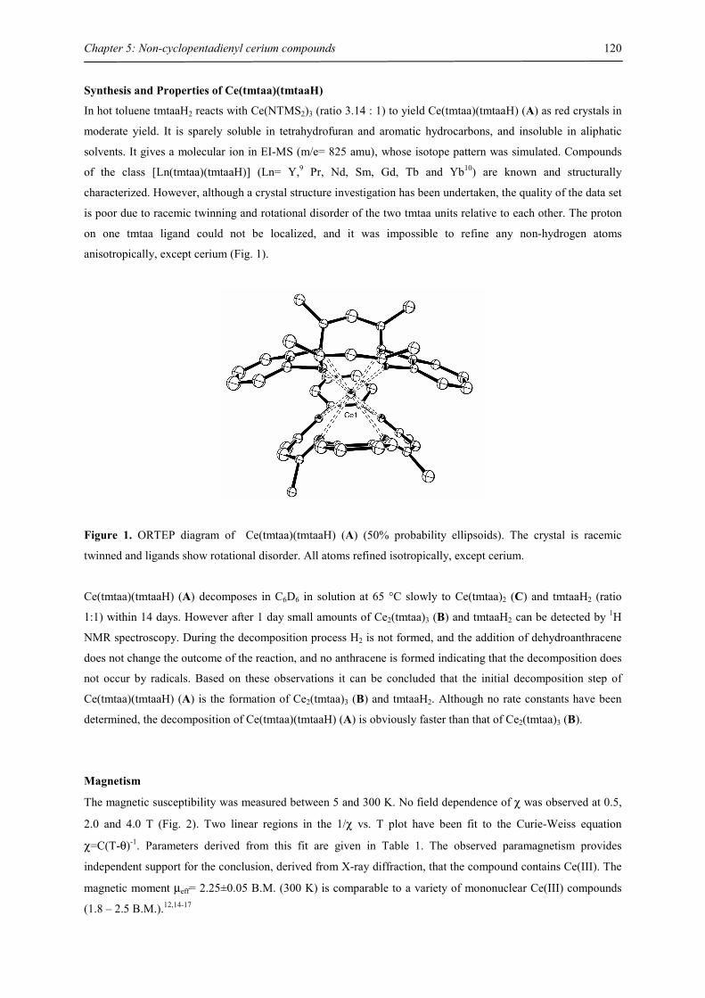
Chapter 5: Non-cyclopentadienyl cerium compounds
120
Synthesis and Properties of Ce(tmtaa)(tmtaaH)
In hot toluene tmtaaH2 reacts with Ce(NTMS2)3 (ratio 3.14 : 1) to yield Ce(tmtaa)(tmtaaH) (A) as red crystals in
moderate yield. It is sparely soluble in tetrahydrofuran and aromatic hydrocarbons, and insoluble in aliphatic
solvents. It gives a molecular ion in EI-MS (m/e= 825 amu), whose isotope pattern was simulated. Compounds
of the class [Ln(tmtaa)(tmtaaH)] (Ln= Y,9 Pr, Nd, Sm, Gd, Tb and Yb10) are known and structurally
characterized. However, although a crystal structure investigation has been undertaken, the quality of the data set
is poor due to racemic twinning and rotational disorder of the two tmtaa units relative to each other. The proton
on one tmtaa ligand could not be localized, and it was impossible to refine any non-hydrogen atoms
anisotropically, except cerium (Fig. 1).
Figure 1. ORTEP diagram of Ce(tmtaa)(tmtaaH) (A) (50% probability ellipsoids). The crystal is racemic
twinned and ligands show rotational disorder. All atoms refined isotropically, except cerium.
Ce(tmtaa)(tmtaaH) (A) decomposes in C6D6 in solution at 65 °C slowly to Ce(tmtaa)2 (C) and tmtaaH2 (ratio
1:1) within 14 days. However after 1 day small amounts of Ce2(tmtaa)3 (B) and tmtaaH2 can be detected by 1H
NMR spectroscopy. During the decomposition process H2 is not formed, and the addition of dehydroanthracene
does not change the outcome of the reaction, and no anthracene is formed indicating that the decomposition does
not occur by radicals. Based on these observations it can be concluded that the initial decomposition step of
Ce(tmtaa)(tmtaaH) (A) is the formation of Ce2(tmtaa)3 (B) and tmtaaH2. Although no rate constants have been
determined, the decomposition of Ce(tmtaa)(tmtaaH) (A) is obviously faster than that of Ce2(tmtaa)3 (B).
Magnetism
The magnetic susceptibility was measured between 5 and 300 K. No field dependence of χ was observed at 0.5,
2.0 and 4.0 T (Fig. 2). Two linear regions in the 1/χ vs. T plot have been fit to the Curie-Weiss equation
χ=C(T-θ)-1. Parameters derived from this fit are given in Table 1. The observed paramagnetism provides
independent support for the conclusion, derived from X-ray diffraction, that the compound contains Ce(III). The
magnetic moment µeff= 2.25±0.05 B.M. (300 K) is comparable to a variety of mononuclear Ce(III) compounds
(1.8 – 2.5 B.M.).12,14-17
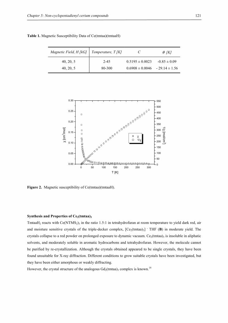
Chapter 5: Non-cyclopentadienyl cerium compounds
121
Table 1. Magnetic Susceptibility Data of Ce(tmtaa)(tmtaaH)
Magnetic Field, H [kG] Temperature, T [K] C θ [K]
40, 20, 5 2-45 0.5195 ± 0.0023 -0.85 ± 0.09
40, 20, 5 80-300 0.6908 ± 0.0046 - 29.14 ± 1.56
0 50 100 150 200 250 3000.00
0.05
0.10
0.15
0.20
0.25
0.30
χ 1/χ
T [K]
χ [c
m3 /m
ol]
0
50
100
150
200
250
300
350
400
450
500
550
1/χ [mol/cm
3]
Figure 2. Magnetic susceptibility of Ce(tmtaa)(tmtaaH).
Synthesis and Properties of Ce2(tmtaa)3
TmtaaH2 reacts with Ce(NTMS2)3 in the ratio 1.5:1 in tetrahydrofuran at room temperature to yield dark red, air
and moisture sensitive crystals of the triple-decker complex, [Ce2(tmtaa)3] . THF (B) in moderate yield. The
crystals collapse to a red powder on prolonged exposure to dynamic vacuum. Ce2(tmtaa)3 is insoluble in aliphatic
solvents, and moderately soluble in aromatic hydrocarbons and tetrahydrofuran. However, the molecule cannot
be purified by re-crystallization. Although the crystals obtained appeared to be single crystals, they have been
found unsuitable for X-ray diffraction. Different conditions to grow suitable crystals have been investigated, but
they have been either amorphous or weakly diffracting.
However, the crystal structure of the analogous Gd2(tmtaa)3 complex is known.18
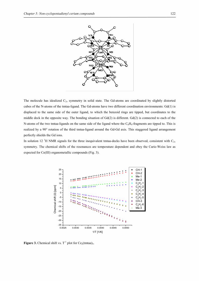
Chapter 5: Non-cyclopentadienyl cerium compounds
122
The molecule has idealized C2v symmetry in solid state. The Gd-atoms are coordinated by slightly distorted
cubes of the N-atoms of the tmtaa-ligand. The Gd-atoms have two different coordination environments: Gd(1) is
displaced to the same side of the outer ligand, to which the benzoid rings are tipped, but coordinates to the
middle deck in the opposite way. The bonding situation of Gd(2) is different. Gd(2) is connected to each of the
N-atoms of the two tmtaa-ligands on the same side of the ligand where the C6H4-fragments are tipped to. This is
realized by a 90° rotation of the third tmtaa-ligand around the Gd-Gd axis. This staggered ligand arrangement
perfectly shields the Gd ions.
In solution 12 1H NMR signals for the three inequivalent tmtaa-decks have been observed, consistent with C2v
symmetry. The chemical shifts of the resonances are temperature dependent and obey the Curie-Weiss law as
expected for Ce(III) organometallic compounds (Fig. 3).
0.0025 0.0030 0.0035 0.0040 0.0045 0.0050-35
-30
-25
-20
-15
-10
-5
0
5
10
15
20
25
Che
mic
al s
hift
(δ) [
ppm
]
1/T [1/K]
CH-1 CH-2 Me-1 Me-2 C6H4-1 C6H4-2 C
6H
4-3
C6H4-4 C
6H
4-5
CH-3 C6H4-6 Me-3
Figure 3. Chemical shift vs. T-1 plot for Ce2(tmtaa)3.
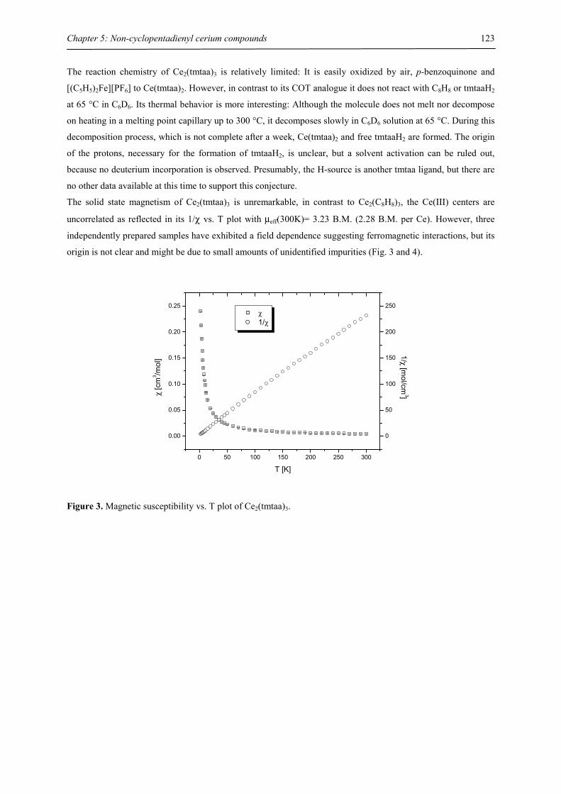
Chapter 5: Non-cyclopentadienyl cerium compounds
123
The reaction chemistry of Ce2(tmtaa)3 is relatively limited: It is easily oxidized by air, p-benzoquinone and
[(C5H5)2Fe][PF6] to Ce(tmtaa)2. However, in contrast to its COT analogue it does not react with C8H8 or tmtaaH2
at 65 °C in C6D6. Its thermal behavior is more interesting: Although the molecule does not melt nor decompose
on heating in a melting point capillary up to 300 °C, it decomposes slowly in C6D6 solution at 65 °C. During this
decomposition process, which is not complete after a week, Ce(tmtaa)2 and free tmtaaH2 are formed. The origin
of the protons, necessary for the formation of tmtaaH2, is unclear, but a solvent activation can be ruled out,
because no deuterium incorporation is observed. Presumably, the H-source is another tmtaa ligand, but there are
no other data available at this time to support this conjecture.
The solid state magnetism of Ce2(tmtaa)3 is unremarkable, in contrast to Ce2(C8H8)3, the Ce(III) centers are
uncorrelated as reflected in its 1/χ vs. T plot with µeff(300K)= 3.23 B.M. (2.28 B.M. per Ce). However, three
independently prepared samples have exhibited a field dependence suggesting ferromagnetic interactions, but its
origin is not clear and might be due to small amounts of unidentified impurities (Fig. 3 and 4).
0 50 100 150 200 250 300
0.00
0.05
0.10
0.15
0.20
0.25 χ 1/χ
T [K]
χ [c
m3 /m
ol]
0
50
100
150
200
250
1/χ [mol/cm
3]
Figure 3. Magnetic susceptibility vs. T plot of Ce2(tmtaa)3.
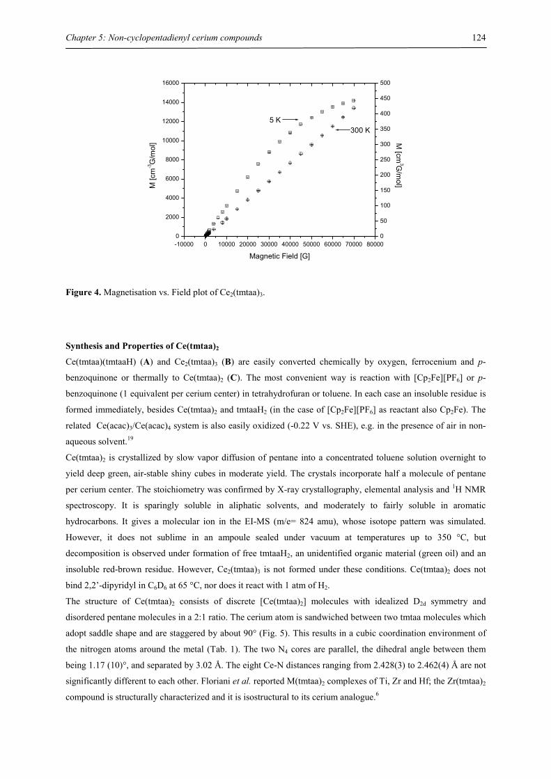
Chapter 5: Non-cyclopentadienyl cerium compounds
124
-10000 0 10000 20000 30000 40000 50000 60000 70000 800000
2000
4000
6000
8000
10000
12000
14000
16000
Magnetic Field [G]
M [c
m-3G
/mol
]
0
50
100
150
200
250
300
350
400
450
500
300 K5 K
M [cm
3G/m
ol]
Figure 4. Magnetisation vs. Field plot of Ce2(tmtaa)3.
Synthesis and Properties of Ce(tmtaa)2
Ce(tmtaa)(tmtaaH) (A) and Ce2(tmtaa)3 (B) are easily converted chemically by oxygen, ferrocenium and p-
benzoquinone or thermally to Ce(tmtaa)2 (C). The most convenient way is reaction with [Cp2Fe][PF6] or p-
benzoquinone (1 equivalent per cerium center) in tetrahydrofuran or toluene. In each case an insoluble residue is
formed immediately, besides Ce(tmtaa)2 and tmtaaH2 (in the case of [Cp2Fe][PF6] as reactant also Cp2Fe). The
related Ce(acac)3/Ce(acac)4 system is also easily oxidized (-0.22 V vs. SHE), e.g. in the presence of air in non-
aqueous solvent.19
Ce(tmtaa)2 is crystallized by slow vapor diffusion of pentane into a concentrated toluene solution overnight to
yield deep green, air-stable shiny cubes in moderate yield. The crystals incorporate half a molecule of pentane
per cerium center. The stoichiometry was confirmed by X-ray crystallography, elemental analysis and 1H NMR
spectroscopy. It is sparingly soluble in aliphatic solvents, and moderately to fairly soluble in aromatic
hydrocarbons. It gives a molecular ion in the EI-MS (m/e= 824 amu), whose isotope pattern was simulated.
However, it does not sublime in an ampoule sealed under vacuum at temperatures up to 350 °C, but
decomposition is observed under formation of free tmtaaH2, an unidentified organic material (green oil) and an
insoluble red-brown residue. However, Ce2(tmtaa)3 is not formed under these conditions. Ce(tmtaa)2 does not
bind 2,2’-dipyridyl in C6D6 at 65 °C, nor does it react with 1 atm of H2.
The structure of Ce(tmtaa)2 consists of discrete [Ce(tmtaa)2] molecules with idealized D2d symmetry and
disordered pentane molecules in a 2:1 ratio. The cerium atom is sandwiched between two tmtaa molecules which
adopt saddle shape and are staggered by about 90° (Fig. 5). This results in a cubic coordination environment of
the nitrogen atoms around the metal (Tab. 1). The two N4 cores are parallel, the dihedral angle between them
being 1.17 (10)°, and separated by 3.02 Å. The eight Ce-N distances ranging from 2.428(3) to 2.462(4) Å are not
significantly different to each other. Floriani et al. reported M(tmtaa)2 complexes of Ti, Zr and Hf; the Zr(tmtaa)2
compound is structurally characterized and it is isostructural to its cerium analogue.6
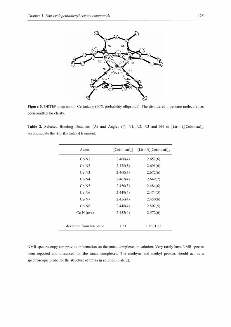
Chapter 5: Non-cyclopentadienyl cerium compounds
125
Figure 5. ORTEP diagram of Ce(tmtaa)2 (50% probability ellipsoids). The disordered n-pentane molecule has
been omitted for clarity.
Table 2. Selected Bonding Distances (Å) and Angles (°). N1, N2, N3 and N4 in [Li(thf)][Ce(tmtaa)]2
accommodate the [(thf)Li(tmtaa)] fragment.
Atoms [Ce(tmtaa)2] [Li(thf)][Ce(tmtaa)]2
Ce-N1 2.460(4) 2.652(6)
Ce-N2 2.428(3) 2.691(6)
Ce-N3 2.460(3) 2.672(6)
Ce-N4 2.462(4) 2.649(7)
Ce-N5 2.450(3) 2.484(6)
Ce-N6 2.449(4) 2.474(5)
Ce-N7 2.456(4) 2.450(6)
Ce-N8 2.448(4) 2.502(5)
Ce-N (ave) 2.452(4) 2.572(6)
deviation from N4 plane 1.51 1.83, 1.53
NMR spectroscopy can provide information on the tmtaa complexes in solution. Very rarely have NMR spectra
been reported and discussed for the tmtaa complexes. The methyne and methyl protons should act as a
spectroscopic probe for the structure of tmtaa in solution (Tab. 2).
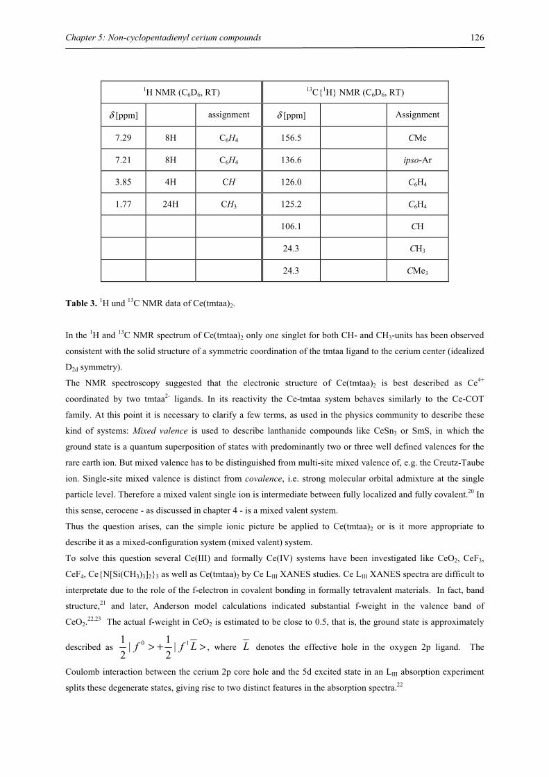
Chapter 5: Non-cyclopentadienyl cerium compounds
126
1H NMR (C6D6, RT) 13C{1H} NMR (C6D6, RT)
δ [ppm] assignment δ [ppm] Assignment
7.29 8H C6H4 156.5 CMe
7.21 8H C6H4 136.6 ipso-Ar
3.85 4H CH 126.0 C6H4
1.77 24H CH3 125.2 C6H4
106.1 CH
24.3 CH3
24.3 CMe3
Table 3. 1H und 13C NMR data of Ce(tmtaa)2.
In the 1H and 13C NMR spectrum of Ce(tmtaa)2 only one singlet for both CH- and CH3-units has been observed
consistent with the solid structure of a symmetric coordination of the tmtaa ligand to the cerium center (idealized
D2d symmetry).
The NMR spectroscopy suggested that the electronic structure of Ce(tmtaa)2 is best described as Ce4+
coordinated by two tmtaa2- ligands. In its reactivity the Ce-tmtaa system behaves similarly to the Ce-COT
family. At this point it is necessary to clarify a few terms, as used in the physics community to describe these
kind of systems: Mixed valence is used to describe lanthanide compounds like CeSn3 or SmS, in which the
ground state is a quantum superposition of states with predominantly two or three well defined valences for the
rare earth ion. But mixed valence has to be distinguished from multi-site mixed valence of, e.g. the Creutz-Taube
ion. Single-site mixed valence is distinct from covalence, i.e. strong molecular orbital admixture at the single
particle level. Therefore a mixed valent single ion is intermediate between fully localized and fully covalent.20 In
this sense, cerocene - as discussed in chapter 4 - is a mixed valent system.
Thus the question arises, can the simple ionic picture be applied to Ce(tmtaa)2 or is it more appropriate to
describe it as a mixed-configuration system (mixed valent) system.
To solve this question several Ce(III) and formally Ce(IV) systems have been investigated like CeO2, CeF3,
CeF4, Ce{N[Si(CH3)3]2}3 as well as Ce(tmtaa)2 by Ce LIII XANES studies. Ce LIII XANES spectra are difficult to
interpretate due to the role of the f-electron in covalent bonding in formally tetravalent materials. In fact, band
structure,21 and later, Anderson model calculations indicated substantial f-weight in the valence band of
CeO2.22,23 The actual f-weight in CeO2 is estimated to be close to 0.5, that is, the ground state is approximately
described as >+> Lff 10 |21|
21
, where L denotes the effective hole in the oxygen 2p ligand. The
Coulomb interaction between the cerium 2p core hole and the 5d excited state in an LIII absorption experiment
splits these degenerate states, giving rise to two distinct features in the absorption spectra.22
Chapter 5: Non-cyclopentadienyl cerium compounds
127
These effects are visible in the Ce LIII XANES of Ce(tmtaa)2 shown in Fig. 6 which are very similar to those
from the CeO2. The final state, including the cerium 2p core hole ( p2 ) and the 5d excited-state electron
generated by the absorption experiment, are noted in this plot. The leading edge feature peaked near 5726 eV is
due to the dLfp 52 1 configuration, while the peak at ~5737 eV is the main tetravalent component with a
dfp 52 0 configuration. This feature shows some subtle differences compared to a true trivalent dfp 52 1
configuration, including a substantial shift to higher energy, as can be seen by comparison with the Ce(III)
standard in the Fig. 6. It is important to note that the >Lf 1| ground-state component, while providing a kind
of mixed valence, is fundamentally different from the usual mixed valence from hybridization with metallic
states. A dramatic demonstration of this difference can be seen in spectra taken under external pressure. In the
usual mixed valence picture, applied pressure drives cerium more towards an f0 state. However, in CeO2,
pressure increases the covalency, thus increasing the dLfp 52 1 weight and decreasing the dfp 52 0 weight
in the XANES.24
5700 5720 5740 57600.0
0.5
1.0
1.5
2.0___
2p 4f 1 L 5d__2p 4f 0 5d
__2p 4f 1 5d
Nor
mal
ized
Abs
orpt
ion
Energy (eV)
Ce(III) Ce(IV)
Figure 6. Ce LIII XANES of a Ce(III) reference, Ce[N(Si(CH3)3)2]3 and a Ce(IV) reference, Ce(tmtaa)2. Final
state configurations for the reference as determined in the literature are noted.
The involvement of the 4f1 state in the covalent bonding is also reflected in a TIP magnetism (Fig. 7).
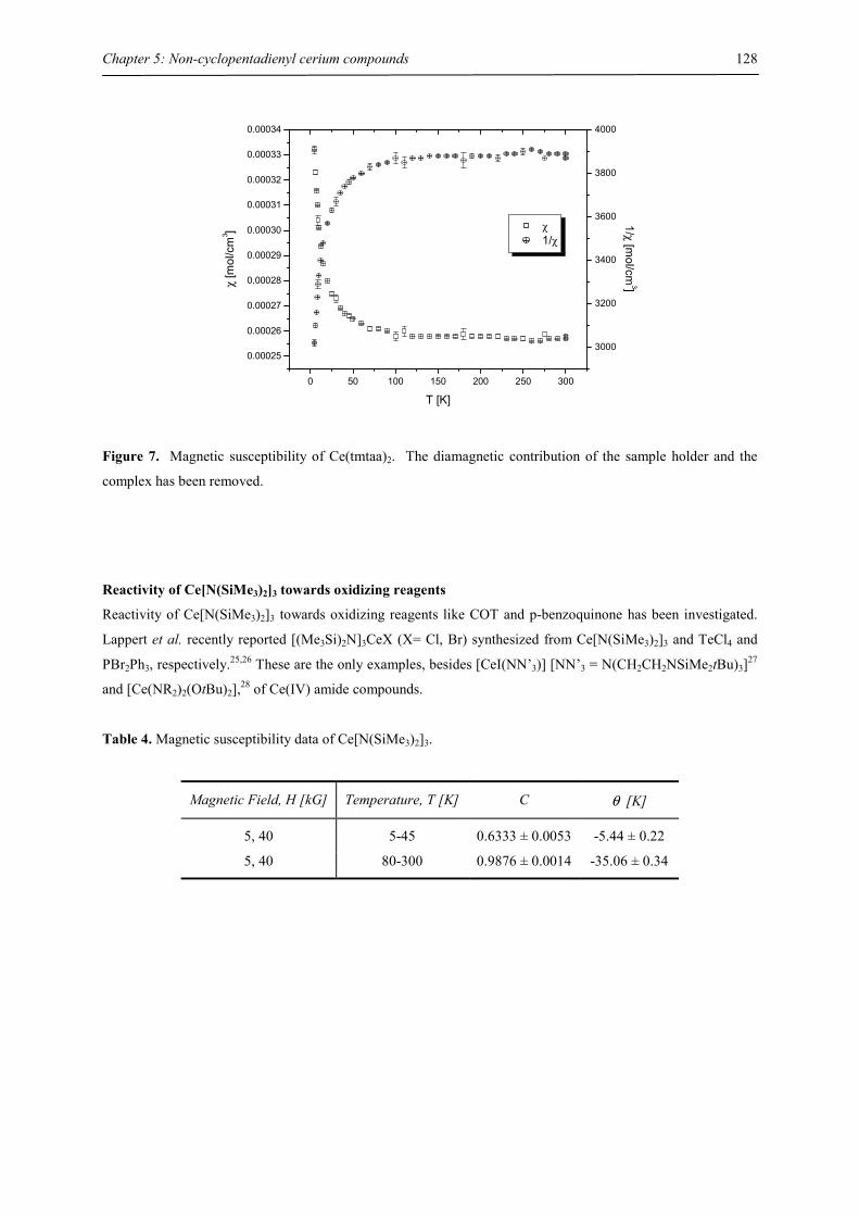
Chapter 5: Non-cyclopentadienyl cerium compounds
128
0 50 100 150 200 250 300
0.00025
0.00026
0.00027
0.00028
0.00029
0.00030
0.00031
0.00032
0.00033
0.00034
χ 1/χ
T [K]
χ [m
ol/c
m3 ]
3000
3200
3400
3600
3800
4000
1/χ [mol/cm
3]
Figure 7. Magnetic susceptibility of Ce(tmtaa)2. The diamagnetic contribution of the sample holder and the
complex has been removed.
Reactivity of Ce[N(SiMe3)2]3 towards oxidizing reagents
Reactivity of Ce[N(SiMe3)2]3 towards oxidizing reagents like COT and p-benzoquinone has been investigated.
Lappert et al. recently reported [(Me3Si)2N]3CeX (X= Cl, Br) synthesized from Ce[N(SiMe3)2]3 and TeCl4 and
PBr2Ph3, respectively.25,26 These are the only examples, besides [CeI(NN’3)] [NN’3 = N(CH2CH2NSiMe2tBu)3]27
and [Ce(NR2)2(OtBu)2],28 of Ce(IV) amide compounds.
Table 4. Magnetic susceptibility data of Ce[N(SiMe3)2]3.
Magnetic Field, H [kG] Temperature, T [K] C θ [K]
5, 40 5-45 0.6333 ± 0.0053 -5.44 ± 0.22
5, 40 80-300 0.9876 ± 0.0014 -35.06 ± 0.34
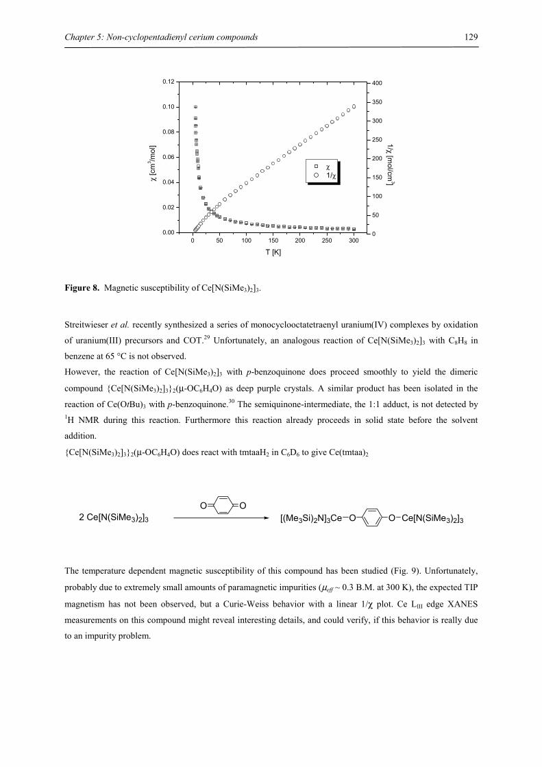
Chapter 5: Non-cyclopentadienyl cerium compounds
129
0 50 100 150 200 250 3000.00
0.02
0.04
0.06
0.08
0.10
0.12
χ 1/χ
T [K]
χ [c
m3 /m
ol]
0
50
100
150
200
250
300
350
400
1/χ [mol/cm
3]
Figure 8. Magnetic susceptibility of Ce[N(SiMe3)2]3.
Streitwieser et al. recently synthesized a series of monocyclooctatetraenyl uranium(IV) complexes by oxidation
of uranium(III) precursors and COT.29 Unfortunately, an analogous reaction of Ce[N(SiMe3)2]3 with C8H8 in
benzene at 65 °C is not observed.
However, the reaction of Ce[N(SiMe3)2]3 with p-benzoquinone does proceed smoothly to yield the dimeric
compound {Ce[N(SiMe3)2]3}2(µ-OC6H4O) as deep purple crystals. A similar product has been isolated in the
reaction of Ce(OtBu)3 with p-benzoquinone.30 The semiquinone-intermediate, the 1:1 adduct, is not detected by 1H NMR during this reaction. Furthermore this reaction already proceeds in solid state before the solvent
addition.
{Ce[N(SiMe3)2]3}2(µ-OC6H4O) does react with tmtaaH2 in C6D6 to give Ce(tmtaa)2
2 Ce[N(SiMe3)2]3OO
OO Ce[N(SiMe3)2]3[(Me3Si)2N]3Ce
The temperature dependent magnetic susceptibility of this compound has been studied (Fig. 9). Unfortunately,
probably due to extremely small amounts of paramagnetic impurities (µeff ~ 0.3 B.M. at 300 K), the expected TIP
magnetism has not been observed, but a Curie-Weiss behavior with a linear 1/χ plot. Ce LIII edge XANES
measurements on this compound might reveal interesting details, and could verify, if this behavior is really due
to an impurity problem.
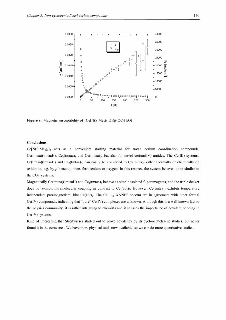
Chapter 5: Non-cyclopentadienyl cerium compounds
130
0 50 100 150 200 250 3000.0000
0.0005
0.0010
0.0015
0.0020
0.0025
0.0030
χ µeff
T [K]
χ [c
m3 /m
ol]
0
5000
10000
15000
20000
25000
30000
35000
40000
1/χ [mol/cm
3]
Figure 9. Magnetic susceptibility of {Ce[N(SiMe3)2]3}2(µ-OC6H4O)
Conclusions
Ce[N(SiMe3)2]3 acts as a convenient starting material for tmtaa cerium coordination compounds,
Ce(tmtaa)(tmtaaH), Ce2(tmtaa)3 and Ce(tmtaa)2, but also for novel cerium(IV) amides. The Ce(III) systems,
Ce(tmtaa)(tmtaaH) and Ce2(tmtaa)3, can easily be converted to Ce(tmtaa)2 either thermally or chemically on
oxidation, e.g. by p-benzoquinone, ferrocenium or oxygen. In this respect, the system behaves quite similar to
the COT systems.
Magnetically Ce(tmtaa)(tmtaaH) and Ce2(tmtaa)3 behave as simple isolated f1 paramagnets, and the triple decker
does not exhibit intramolecular coupling in contrast to Ce2(cot)3. However, Ce(tmtaa)2 exhibits temperature
independent paramagnetism, like Ce(cot)2. The Ce LIII XANES spectra are in agreement with other formal
Ce(IV) compounds, indicating that “pure” Ce(IV) complexes are unknown. Although this is a well known fact to
the physics community, it is rather intriguing to chemists and it stresses the importance of covalent bonding in
Ce(IV) systems.
Kind of interesting that Streitwieser started out to prove covalency by its cyclooctatetraene studies, but never
found it in the cerocenes. We have more physical tools now available, so we can do more quantitative studies.
Chapter 5: Non-cyclopentadienyl cerium compounds
131
References
[1] Dolg, M.; Stoll, H. In Handbook of Chemistry and Physics of Rare Earths; Gschneider, K. A., Eyring,
L., Eds.; Elsevier: Amsterdam, 1996; Vol. 22, pp 607-729.
[2] Dolg, M., Habilitation, University of Stuttgart: Stuttgart, 1997.
[3] Haghighi, M. S.; Teske, C. L.; Homborg, H. Z. Allgem. Anorg. Chem. 1992, 608, 73-80.
[4] Isago, H.; Shimoda, M. Chem. Lett. 1992, 147-150.
[5] Bian, Y.; Jiang, J.; Tao, Y.; Choi, M. T. M.; Li, R.; Ng, A. C. H.; Zhu, P.; Pan, N.; Sun, X.; Arnold, D.
P.; Zhou, Z.-Y.; Li, H.-W.; Mak, T. C. W.; Ng, D. K. P. J. Am. Chem. Soc. 2003, 125, 12257-12267.
[6] De Angelis, S.; Solari, E.; Gallo, E.; Floriani, C.; Chiesi-Villa, A.; Rizzoli, C. Inorg. Chem. 1992, 31,
2520-2527.
[7] Solari, E.; De Angelis, S.; Floriani, C.; Chiesi-Villa, A.; Rizzoli, C. Inorg. Chem. 1992, 31, 96-101.
[8] Magull, J.; Simon, A. Z. Allgem. Anorg. Chem. 1992, 615, 77-80.
[9] Wang, Z.; Sakata, K.; Hashimato, M. Polyhedron 1998, 17, 4451-4456.
[10] Wang, Z.; Hu, N.; Sakata, K.; Hashimato, M. J. Chem. Soc., Dalton Trans. 1999, 1999, 1695-1700.
[11] Booij, M.; Kiers, N. H.; Heeres, H. J.; Teuben, J. H. J. Organomet. Chem. 1989, 364, 79-86.
[12] Stults, S. D.; Andersen, R. A. Organometallics 1990, 9, 115-122.
[13] Fraser, R. R.; Mansour, T. S.; Savard, S. J. Org. Chem. 1985, 50, 3232.
[14] Evans, W. J.; Hozbor, M. A. J. Organomet. Chem. 1987, 326, 299.
[15] Fischer, R. D. In Fundamental and Technological Aspects of Organo-f-Element Chemistry; Marks, T. J.,
Fragala, I. L., Eds.; D. Reidel: Dordrecht, The Netherlands, 1985, p 277.
[16] Reddmann, H.; Schultze, H.; Amberger, H.-D.; Apostolidis, C.; Kanellakopulos, B. Spectrochim. Acta
1990, 46A, 1223-1229.
[17] Amberger, H.-D.; Reddmann, H.; Schultze, H.; Jank, S.; Kanellakopulos, B.; Apostolidis, C.
Spectrochim. Acta 2003, 59A, 2527-2539.
[18] Magull, J., private communication.
[19] Behrsing, T.; Bond, A. M.; Deacon, G. B.; Forsyth, C. M.; Forsyth, M.; Kamble, K. J.; Skelton, B. W.;
White, A. H. Inorg. Chim. Acta 2003, 352, 229-237.
[20] LaBute, M. X.; Kulkarni, R. V.; Endres, R. G.; Cox, D. L. J. Chem. Phys. 2002, 116, 3681-3689.
[21] Koelling, D. D.; Boring, A. M.; Wood, J. H. Solid State Commun. 1983, 47, 227.
[22] Kotani, A.; Jo, T.; Parlebas, J. C. Adv. Phys. 1988, 37, 37.
[23] Soldatov, A. V.; Ivanchenko, T. S.; Kotani, A.; Bianconi, A. Physica B 1995, 208-209, 53-55.
[24] Kaindl, G.; Schmiester, G.; Sampathkumaran, E. V.; Wachter, P. Phys. Rev. B 1988, 38, 10174-10177.
[25] Eisenstein, O.; Hitchcock, P. B.; Hulkes, A. G.; Lappert, M. F.; Maron, L. Chem. Commun. 2001, 1560-
1561.
[26] Hitchcock, P. B.; Hulkes, A. G.; Lappert, M. F. Inorg. Chem. 2004, 43, 1031-1038.
[27] Morton, C.; Alcock, N. W.; Lees, M. R.; Munslow, I. J.; Sanders, C. J.; Scott, P. J. Am. Chem. Soc.
1999, 121, 11255-11256.
[28] England, A. F.; Frankom, T. M.; Burns, C. J.; Buchwald, S. In 207th National Meeting of the American
Chemical Society: San Diego, CA, March 13-17, 1994, 1994.
[29] Boussie, T. R.; Moore, R. M.; Streitwieser, A.; Zalkin, A.; Brennan, J.; Smith, K. A. Organometallics
1990, 9, 2010-2016.

Chapter 5: Non-cyclopentadienyl cerium compounds
132
[30] Sen, A.; Stecher, H. A.; Rheingold, A. L. Inorg. Chem. 1992, 31, 473-479.
Chapter 6: The electronic structure of bipyridyl and diazabutadienyl complexes of ytterbocenes
133
Chapter 6: The electronic structure of bipyridyl and diazabutadienyl complexes of
ytterbocenes
Bipyridyl Adducts of Ytterbocenes
Introduction
Complexes of α,α’-diimine (mostly 2,2’-bipyridyl and 1,10-phenanthroline) have been prepared for most metals
and, for a given metal center, may persist over a range of oxidation states.1 It has long been recognized that, in a
number of these complexes the metal center apparently adopts a very low oxidation state, e.g. Mg0(bipy)2,
reduced bipy is present rather than a reduced metal center, Mg2+(bipy•−)2. The reduction site and therefore the
electronic structure is not a priori obvious and some ingenuity may be required in elucidating the nature of the
electronic configurations in these complexes.2
E. König et al. reported extensive synthetic and magnetic studies on 2,2’-bipyridyl complexes of transition, main
group metals, lanthanides and actinides. In 1969 Feistel & Mathai prepared Eu(bipy)4, Yb(bipy)4 and Yb(phen)4,
studied their solution magnetism, and concluded based on their magnetic studies that the electronic ground state
was best described as Ln2+(bipy0)2(bipy•−)2, and in the case of Ln= Eu the system showed “a new type of
intramolecular antiferromagnetism.”3 This unusual behavior prompted Bochkarev & Schumann to re-investigate
the properties of complexes of the type Ln(bipy)4 (Ln= La-Lu) by X-ray crystallography, IR, magnetic
susceptibility (at 298 K) and EPR. Based on their results they concluded that the electronic structure was more
appropriately described as [Ln3+(bipy0)2(bipy•−)(bipy2−)], with the exception of Eu(bipy)4, whose magnetism was
consistent with [Eu2+(bipy0)3(bipy2−)].4 This was expected with respect to enhanced stability of the divalent
oxidation state of Eu, compared to the other lanthanide metals. Recently the magnetic susceptibilities of these
compounds were studied at variable temperature and the results were in agreement with this prediction (see
Appendix, Fig. A1 and A2).
A related complex, Yb(tBuNCHCHNtBu)3, was synthesized and exhibited a temperature dependent effective
magnetic moment that was interpreted in terms of an electron transfer equilibrium between two complexes,
Yb(II)(L•−)2(L) and Yb(III)(L•−)3, where the latter is favored at high temperatures.5 More recently, the complex
Yb3(bipy)3(thf)6 was prepared, in which each 2,2’-bipyridyl unit is dianionic and bridges two Yb(II) centers.6
Considering the problems encountered during acquisition of sufficiently precise information on the electronic
ground state, it appeared to be worthwhile to replace all non-innocent ligands, except one, by innocent spectator
ligands, because this should simplify the problem. During an initial screening of different Lewis bases
coordinated to the (C5Me5)2Yb or (C5Me5)2Eu fragment Andersen et al. studied 2,2’-bipyridyl adducts and later
expanded this research to various N-heterocyclic bases.7-13 Parallel to this study Evans et al. and Edelmann et al.
investigated the behavior of the corresponding (C5Me5)2Sm(thf)2 system towards similar substrates.14,15 16 The
samarocene bipyridyl and diazabutadienyl adduct, (C5Me5)2Sm(bipy)15 and (C5Me5)2Sm(RNCHCHNR),16 were
postulated to be Sm(III) complexes of 2,2’-bipyridyl and diazabutadienyl radical anion, respectively. This
conclusion was mainly based on crystal structure investigations, IR spectroscopy and in the case of the bipyridyl
on an Evans` susceptibility measurement at room temperature. The solution magnetism suggested uncorrelated
spin carriers at 300 K.15 However, no solid state variable temperature magnetic susceptibility data been reported.
Chapter 6: The electronic structure of bipyridyl and diazabutadienyl complexes of ytterbocenes
134
This contrasts with the extensive solid state magnetic susceptibility studies by Andersen et al. .8-13 Various N-
heterocyclic bases with redox-potentials in the range of –1.0 to –2.6 V were investigated and their behavior was
classified into five classes depending on their stoichiometry and magnetic behavior. The 1:1 adducts were either
diamagnetic or exhibited uncorrelated spins to 5 K. The 2:1 adducts with two (C5Me5)2Yb(III) units linked by a
dianionic ligand either showed no magnetic exchange or antiferromagnetic coupling at low temperatures
depending on the redox potentials of the bridging ligand. All these observations can readily be explained.
However, the 1:1 adduct with 2,2’-bipyridyl is more complicated. Although the adducts of
decamethylytterbocene with pyridine and 2,2’-bipyridyl, (C5Me5)2Yb(py)2 and (C5Me5)2Yb(bipy), have the same
general molecular structure as determined by X-ray diffraction, their electronic structure is very different.13 The
paramagnetism is due to an electron transfer from the ytterbocene fragment to the LUMO of 2,2’-bipyridyl,
giving a biradical of the form (C5Me5)2Yb(III)(bipy•−), in which the electrons are correlated in the ground state
(details are discussed below). Although it was demonstrated that the coupling between the spin carriers is
antiferromagnetic, the origin of this unusual behavior has not been addressed. Schultz expanded the
(C5Me5)2Yb(bipy) system to 2,2’-bipyridyl and 1,10-phenanthroline complexes with a variety of substituted
ytterbocene derivatives.13 A magnetic behavior analogous to (C5Me5)2Yb(bipy) was recently observed for
(C5Me5)2Yb(terpy) (terpy= 2,2’:6’,2’’-terpyridine).17
Objective
In cerocene the high Kondo temperature, Tk, which corresponds to the singlet-triplet splitting, prevents the
observation of any temperature-dependent effects in our physical studies (see Chapter 4). Ytterbium is the hole
analogue of cerium and the magnetic susceptibility of (C5Me5)2Yb(bipy) suggests this might be an example of a
molecule with a lower Tk, exhibiting clear temperature-dependent effects in its susceptibility, hence the initial
studies on these system performed in the Andersen group should be expanded. In contrast to cerocene, the
electrons in the f orbitals couple to the bipy-π* orbitals, but not with the empty π*-orbitals of the canted C5Me5
ring, because the energy difference is large. If it is assumed that the coupling between the organic radical and the
ytterbium(III) centers is related to the relative energy of the frontier orbitals of the involved species, then
changing the substituents on the cyclopentadienyl ring and the 2,2’-bipyridyl fragment – both parts can easily be
modified on substitution. In order to evaluate this influence a series of 4,4’-disubstituted 2,2’-bipyridyl (bipy-R)
and 1,4-diaza-2,3-butadienyl adducts on [(C5Me5)2Yb] fragment have been prepared and their electronic
structure has been investigated.
As described in Chapters 2, 4 and 5 the introduction of the new high-precision magnetic susceptibility low-
background method allows reproducible data in quartz tubes – even on small moment samples and we have
calibrated this method repeatedly. Schultz reported a series of α,α’-diimine (bipy, phen) adducts of substituted
ytterbocenes and characterized these adduct complexes extensively.12,13 Considering the discrepancy between
solid state magnetism (recorded in high-background KEL-F buckets) and 1H NMR or UV/Vis results in some
cases, it was decided to reinvestigate these complexes employing the new quartz tube technology.
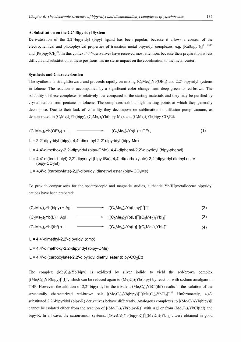
Chapter 6: The electronic structure of bipyridyl and diazabutadienyl complexes of ytterbocenes
135
A. Substitution on the 2,2’-Bipyridyl System
Derivatisation of the 2,2’-bipyridyl (bipy) ligand has been popular, because it allows a control of the
electrochemical and photophysical properties of transition metal bipyridyl complexes, e.g. [Ru(bipy’)3]2+,18,19
and [Pt(bipy)Cl2]20. In this context 4,4’-derivatives have received most attention, because their preparation is less
difficult and substitution at these positions has no steric impact on the coordination to the metal center.
Synthesis and Characterization
The synthesis is straightforward and proceeds rapidly on mixing (C5Me5)2Yb(OEt2) and 2,2’-bipyridyl systems
in toluene. The reaction is accompanied by a significant color change from deep green to red-brown. The
solubility of these complexes is relatively low compared to the starting materials and they may be purified by
crystallization from pentane or toluene. The complexes exhibit high melting points at which they generally
decompose. Due to their lack of volatility they decompose on sublimation in diffusion pump vacuum, as
demonstrated in (C5Me5)2Yb(bipy), (C5Me5)2Yb(bipy-Me), and (C5Me5)2Yb(bipy-CO2Et).
(C5Me5)2Yb(OEt2) + L (C5Me5)2Yb(L) + OEt2
L = 2,2'-dipyridyl (bipy), 4,4'-dimethyl-2,2'-dipyridyl (bipy-Me)
L = 4,4'-dimethoxy-2,2'-dipyridyl (bipy-OMe), 4,4'-diphenyl-2,2'-dipyridyl (bipy-phenyl)
L = 4,4'-di(tert.-butyl)-2,2'-dipyridyl (bipy-tBu), 4,4'-di(carboxylate)-2,2'-dipyridyl diethyl ester (bipy-CO2Et)
L = 4,4'-di(carboxylate)-2,2'-dipyridyl dimethyl ester (bipy-CO2Me)
(1)
To provide comparisons for the spectroscopic and magnetic studies, authentic Yb(III)metallocene bipyridyl
cations have been prepared:
(C5Me5)2Yb(L) + AgI [(C5Me5)2Yb(L)]+[(C5Me5)2YbI2]-
L = 4,4'-dimethyl-2,2'-dipyridyl (dmb)
L = 4,4'-dimethoxy-2,2'-dipyridyl (bipy-OMe)
L = 4,4'-di(carboxylate)-2,2'-dipyridyl diethyl ester (bipy-CO2Et)
(C5Me5)2YbI(thf) + L [(C5Me5)2Yb(L)]+[(C5Me5)2YbI2]-
(C5Me5)2Yb(bipy) + AgI [(C5Me5)2Yb(bipy)]+[I]- (2)
(3)
(4)
The complex (Me5C5)2Yb(bipy) is oxidized by silver iodide to yield the red-brown complex
[(Me5C5)2Yb(bipy)]+[I]−, which can be reduced again to (Me5C5)2Yb(bipy) by reaction with sodium amalgam in
THF. However, the addition of 2,2’-bipyridyl to the trivalent (Me5C5)2YbCl(thf) results in the isolation of the
structurally characterized red-brown salt [(Me5C5)2Yb(bipy)]+[(Me5C5)2YbCl2]−.13 Unfortunately, 4,4’-
substituted 2,2’-bipyridyl (bipy-R) derivatives behave differently. Analogous complexes to [(Me5C5)2Yb(bipy)]I
cannot be isolated either from the reaction of [(Me5C5)2Yb(bipy-R)] with AgI or from (Me5C5)2YbCl(thf) and
bipy-R. In all cases the cation-anion systems, [(Me5C5)2Yb(bipy-R)]+[(Me5C5)2YbI2]−, were obtained in good
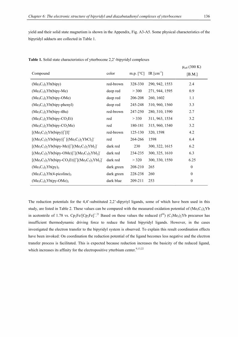
Chapter 6: The electronic structure of bipyridyl and diazabutadienyl complexes of ytterbocenes
136
yield and their solid state magnetism is shown in the Appendix, Fig. A3-A5. Some physical characteristics of the
bipyridyl adducts are collected in Table 1.
Table 1. Solid state characteristics of ytterbocene 2,2’-bipyridyl complexes
Compound
color
m.p. [°C]
IR [cm-1]
µeff (300 K)
[Β.Μ.]
(Me5C5)2Yb(bipy) red-brown 328-330 290, 942, 1553 2.4
(Me5C5)2Yb(bipy-Me) deep red > 300 271, 944, 1595 0.9
(Me5C5)2Yb(bipy-OMe) deep red 206-208 260, 1602 1.1
(Me5C5)2Yb(bipy-phenyl) deep red 245-248 310, 960, 1560 3.3
(Me5C5)2Yb(bipy-tBu) red-brown 247-250 280, 310, 1590 2.7
(Me5C5)2Yb(bipy-CO2Et) red > 330 311, 963, 1534 3.2
(Me5C5)2Yb(bipy-CO2Me) red 180-181 315, 960, 1540 3.2
[(Me5C5)2Yb(bipy)]+[I]- red-brown 125-130 320, 1598 4.2
[(Me5C5)2Yb(bipy)]+ [(Me5C5)2YbCl2]- red 264-266 1598 6.4
[(Me5C5)2Yb(bipy-Me)]+[(Me5C5)2YbI2]- dark red 230 300, 322, 1615 6.2
[(Me5C5)2Yb(bipy-OMe)]+[(Me5C5)2YbI2]- dark red 234-235 300, 325, 1610 6.3
[(Me5C5)2Yb(bipy-CO2Et)]+[(Me5C5)2YbI2]- dark red > 320 300, 330, 1550 6.25
(Me5C5)2Yb(py)2 dark green 208-210 265 0
(Me5C5)2Yb(4-picoline)2 dark green 228-238 260 0
(Me5C5)2Yb(py-OMe)2 dark blue 209-211 253 0
The reduction potentials for the 4,4’-substituted 2,2’-dipyriyl ligands, some of which have been used in this
study, are listed in Table 2. These values can be compared with the measured oxidation potential of (Me5C5)2Yb
in acetontrile of 1.78 vs. Cp2Fe/[Cp2Fe]+.21 Based on these values the reduced (f14) (C5Me5)2Yb precursor has
insufficient thermodynamic driving force to reduce the listed bipyridyl ligands. However, in the cases
investigated the electron transfer to the bipyridyl system is observed. To explain this result coordination effects
have been invoked: On coordination the reduction potential of the ligand becomes less negative and the electron
transfer process is facilitated. This is expected because reduction increases the basicity of the reduced ligand,
which increases its affinity for the electropositive ytterbium center.9,13,22
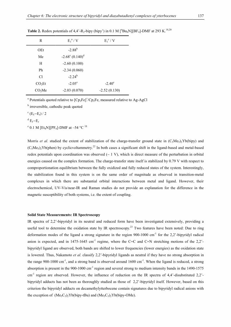
Chapter 6: The electronic structure of bipyridyl and diazabutadienyl complexes of ytterbocenes
137
Table 2. Redox potentials of 4,4’-R2-bipy (bipy’) in 0.1 M [nBu4N][BF4]-DMF at 293 K.18,20
R E1a / V E1
c / V
OEt -2.88b
Me -2.68c (0.140)d
H -2.60 (0.100)
Ph -2.34 (0.060)
Cl
CO2Et
-2.24b
-2.05e
-2.40e
CO2Me -2.03 (0.070) -2.52 (0.130)
a Potentials quoted relative to [Cp2Fe]+/Cp2Fe, measured relative to Ag-AgCl b irreversible, cathodic peak quoted c (Ef −Er) / 2 d Ef −Er e 0.1 M [Et4N][PF6]-DMF at –54 °C 18
Morris et al. studied the extent of stabilization of the charge-transfer ground state in (C5Me5)2Yb(bipy) and
(C5Me5)2Yb(phen) by cyclicvoltammetry.22 In both cases a significant shift in the ligand-based and metal-based
redox potentials upon coordination was observed (~ 1 V), which is direct measure of the perturbation in orbital
energies caused on the complex formation. The charge-transfer state itself is stabilized by 0.79 V with respect to
comproportionation equilibrium between the fully oxidized and fully reduced states of the system. Interestingly,
the stabilization found in this system is on the same order of magnitude as observed in transition-metal
complexes in which there are substantial orbital interactions between metal and ligand. However, their
electrochemical, UV-Vis/near-IR and Raman studies do not provide an explanation for the difference in the
magnetic susceptibility of both systems, i.e. the extent of coupling.
Solid State Measurements: IR Spectroscopy
IR spectra of 2,2’-bipyridyl in its neutral and reduced form have been investigated extensively, providing a
useful tool to determine the oxidation state by IR spectroscopy.23 Two features have been noted: Due to ring
deformation modes of the ligand a strong signature in the region 900-1000 cm-1 for the 2,2’-bipyridyl radical
anion is expected, and in 1475-1645 cm-1 regime, where the C=C and C=N stretching motions of the 2,2’-
bipyridyl ligand are observed, both bands are shifted to lower frequencies (lower energies) as the oxidation state
is lowered. Thus, Nakamoto et al. classify 2,2’-bipyridyl ligands as neutral if they have no strong absorption in
the range 900-1000 cm-1, and a strong band is observed around 1600 cm-1. When the ligand is reduced, a strong
absorption is present in the 900-1000 cm-1 region and several strong to medium intensity bands in the 1490-1575
cm-1 region are observed. However, the influence of reduction on the IR spectra of 4,4’-disubstituted 2,2’-
bipyridyl adducts has not been as thoroughly studied as those of 2,2’-bipyridyl itself. However, based on this
criterion the bipyridyl adducts on decamethylytterbocene contain signatures due to bipyridyl radical anions with
the exception of (Me5C5)2Yb(bipy-tBu) and (Me5C5)2Yb(bipy-OMe).
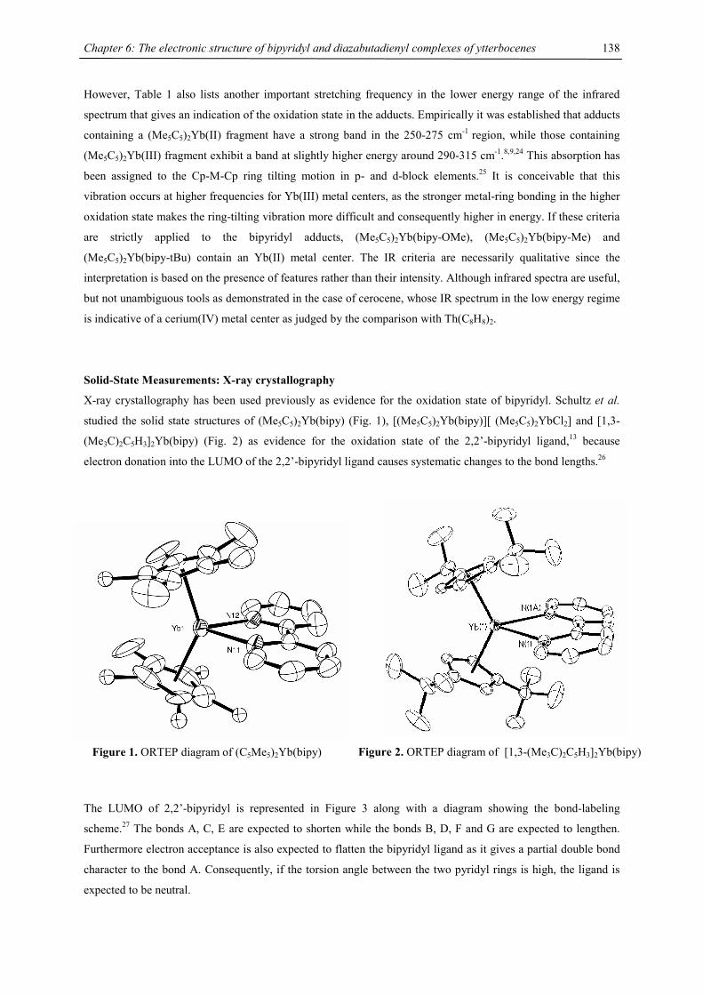
Chapter 6: The electronic structure of bipyridyl and diazabutadienyl complexes of ytterbocenes
138
However, Table 1 also lists another important stretching frequency in the lower energy range of the infrared
spectrum that gives an indication of the oxidation state in the adducts. Empirically it was established that adducts
containing a (Me5C5)2Yb(II) fragment have a strong band in the 250-275 cm-1 region, while those containing
(Me5C5)2Yb(III) fragment exhibit a band at slightly higher energy around 290-315 cm-1.8,9,24 This absorption has
been assigned to the Cp-M-Cp ring tilting motion in p- and d-block elements.25 It is conceivable that this
vibration occurs at higher frequencies for Yb(III) metal centers, as the stronger metal-ring bonding in the higher
oxidation state makes the ring-tilting vibration more difficult and consequently higher in energy. If these criteria
are strictly applied to the bipyridyl adducts, (Me5C5)2Yb(bipy-OMe), (Me5C5)2Yb(bipy-Me) and
(Me5C5)2Yb(bipy-tBu) contain an Yb(II) metal center. The IR criteria are necessarily qualitative since the
interpretation is based on the presence of features rather than their intensity. Although infrared spectra are useful,
but not unambiguous tools as demonstrated in the case of cerocene, whose IR spectrum in the low energy regime
is indicative of a cerium(IV) metal center as judged by the comparison with Th(C8H8)2.
Solid-State Measurements: X-ray crystallography
X-ray crystallography has been used previously as evidence for the oxidation state of bipyridyl. Schultz et al.
studied the solid state structures of (Me5C5)2Yb(bipy) (Fig. 1), [(Me5C5)2Yb(bipy)][ (Me5C5)2YbCl2] and [1,3-
(Me3C)2C5H3]2Yb(bipy) (Fig. 2) as evidence for the oxidation state of the 2,2’-bipyridyl ligand,13 because
electron donation into the LUMO of the 2,2’-bipyridyl ligand causes systematic changes to the bond lengths.26
The LUMO of 2,2’-bipyridyl is represented in Figure 3 along with a diagram showing the bond-labeling
scheme.27 The bonds A, C, E are expected to shorten while the bonds B, D, F and G are expected to lengthen.
Furthermore electron acceptance is also expected to flatten the bipyridyl ligand as it gives a partial double bond
character to the bond A. Consequently, if the torsion angle between the two pyridyl rings is high, the ligand is
expected to be neutral.
Figure 1. ORTEP diagram of (C5Me5)2Yb(bipy) Figure 2. ORTEP diagram of [1,3-(Me3C)2C5H3]2Yb(bipy)
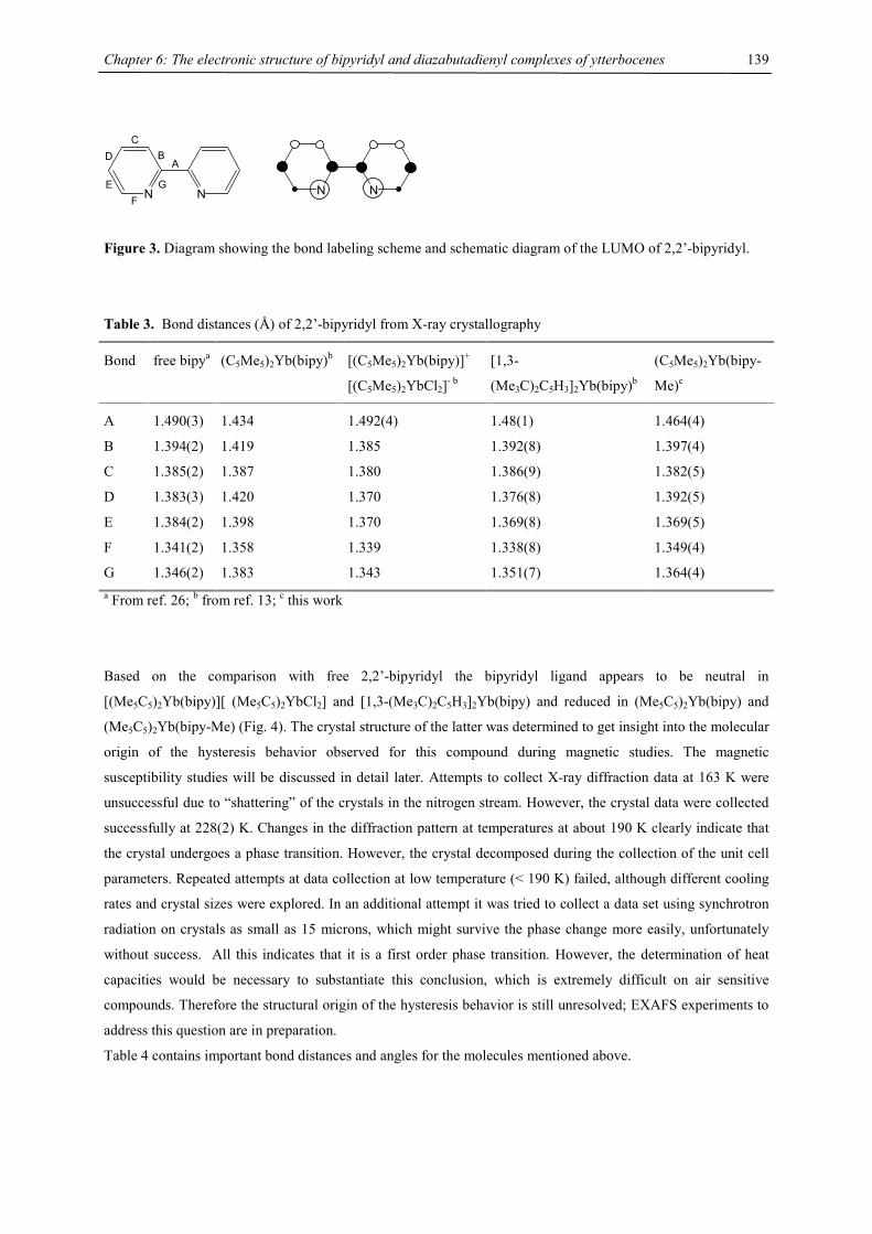
Chapter 6: The electronic structure of bipyridyl and diazabutadienyl complexes of ytterbocenes
139
N N
C
EF
A
N NG
D B
Figure 3. Diagram showing the bond labeling scheme and schematic diagram of the LUMO of 2,2’-bipyridyl.
Table 3. Bond distances (Å) of 2,2’-bipyridyl from X-ray crystallography
Bond free bipya (C5Me5)2Yb(bipy)b [(C5Me5)2Yb(bipy)]+
[(C5Me5)2YbCl2]- b
[1,3-
(Me3C)2C5H3]2Yb(bipy)b
(C5Me5)2Yb(bipy-
Me)c
A 1.490(3) 1.434 1.492(4) 1.48(1) 1.464(4)
B 1.394(2) 1.419 1.385 1.392(8) 1.397(4)
C 1.385(2) 1.387 1.380 1.386(9) 1.382(5)
D 1.383(3) 1.420 1.370 1.376(8) 1.392(5)
E 1.384(2) 1.398 1.370 1.369(8) 1.369(5)
F 1.341(2) 1.358 1.339 1.338(8) 1.349(4)
G 1.346(2) 1.383 1.343 1.351(7) 1.364(4) a From ref. 26; b from ref. 13; c this work
Based on the comparison with free 2,2’-bipyridyl the bipyridyl ligand appears to be neutral in
[(Me5C5)2Yb(bipy)][ (Me5C5)2YbCl2] and [1,3-(Me3C)2C5H3]2Yb(bipy) and reduced in (Me5C5)2Yb(bipy) and
(Me5C5)2Yb(bipy-Me) (Fig. 4). The crystal structure of the latter was determined to get insight into the molecular
origin of the hysteresis behavior observed for this compound during magnetic studies. The magnetic
susceptibility studies will be discussed in detail later. Attempts to collect X-ray diffraction data at 163 K were
unsuccessful due to “shattering” of the crystals in the nitrogen stream. However, the crystal data were collected
successfully at 228(2) K. Changes in the diffraction pattern at temperatures at about 190 K clearly indicate that
the crystal undergoes a phase transition. However, the crystal decomposed during the collection of the unit cell
parameters. Repeated attempts at data collection at low temperature (< 190 K) failed, although different cooling
rates and crystal sizes were explored. In an additional attempt it was tried to collect a data set using synchrotron
radiation on crystals as small as 15 microns, which might survive the phase change more easily, unfortunately
without success. All this indicates that it is a first order phase transition. However, the determination of heat
capacities would be necessary to substantiate this conclusion, which is extremely difficult on air sensitive
compounds. Therefore the structural origin of the hysteresis behavior is still unresolved; EXAFS experiments to
address this question are in preparation.
Table 4 contains important bond distances and angles for the molecules mentioned above.

Chapter 6: The electronic structure of bipyridyl and diazabutadienyl complexes of ytterbocenes
140
Figure 4. ORTEP diagram of (C5Me5)2Yb(bipy-Me)
Table 4. Selected Bond Distances (Å) and Angles (°) of (C5Me5)2Yb(bipy), (C5Me5)2Yb(bipy-Me),
[(C5Me5)2Yb(bipy)]+[(C5Me5)2YbCl2]- and [1,3-(Me3C)2C5H3]2Yb(bipy).
(C5Me5)2Yb
(bipy)a
[(C5H5)2Yb(bipy)]+
[(C5H5)2YbCl2]- a
(C5Me5)2Yb
(bipy-Me)b
[1,3-(Me3C)2C5H3]2Yb
(bipy)a
Yb-Cring (mean) 2.62 2.59 2.67 2.75
Yb-Cring (range) 2.592(9)-2.647(7) 2.582(3)-2.614(3) 2.624(5)-2.695(3) 2.674(6)-2.803(6)
Yb-centroid 2.34; 2.34 2.30 2.40 2.47
centroid-Yb-centroid 139; 140 142 143 132
Yb-N 2.32 2.37 2.40 2.50
torsion angle
N-C-C-N
3
7 0.7 15
torsion angle
C-C-C-C
3
8 0.2 19
a from ref. 13; b this work
The metal-ligand distance is correlated with the oxidation state of the metal, as the ionic radius is smaller in a
higher oxidation state.28 Examination of the mean metal-ring carbon distance, the metal-centroid distance and the
metal-nitrogen distances indicates that the ytterbium center is Yb(III) in [(Me5C5)2Yb(bipy)][(Me5C5)2YbCl2],
(C5Me5)2Yb(bipy) and (C5Me5)2Yb(bipy-Me), while in [1,3-(Me3C)2C5H3]2Yb(bipy) it is Yb(II). Although the
coordination environment of (C5Me5)2Yb(bipy-Me) and (C5Me5)2Yb(bipy) is very similar, the Yb-N and Yb-
centroid distances of the former are significantly elongated and tend to the values found in Yb(II) compounds.
These values can also be compared with the corresponding values for (C5Me5)2Yb(py)2, in which the metal is
divalent. In that molecule, the mean metal ring-carbon distance is 2.74 Å and the metal-nitrogen distance is 2.56
Å.7
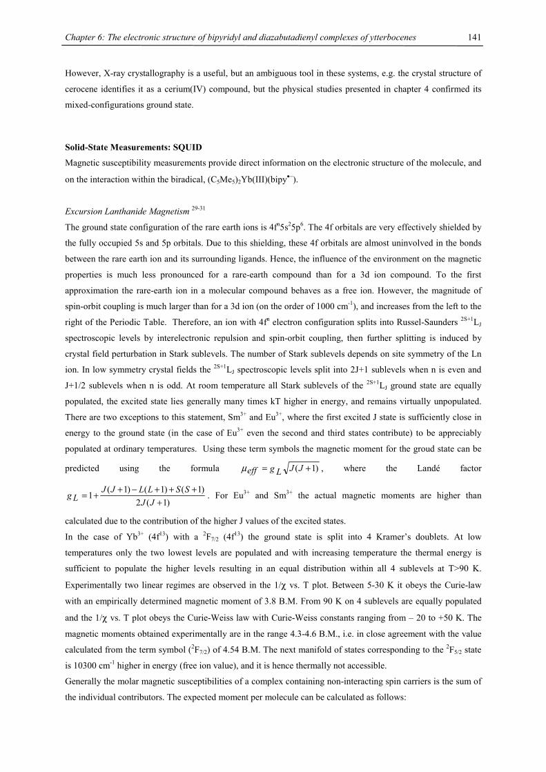
Chapter 6: The electronic structure of bipyridyl and diazabutadienyl complexes of ytterbocenes
141
However, X-ray crystallography is a useful, but an ambiguous tool in these systems, e.g. the crystal structure of
cerocene identifies it as a cerium(IV) compound, but the physical studies presented in chapter 4 confirmed its
mixed-configurations ground state.
Solid-State Measurements: SQUID
Magnetic susceptibility measurements provide direct information on the electronic structure of the molecule, and
on the interaction within the biradical, (C5Me5)2Yb(III)(bipy•−).
Excursion Lanthanide Magnetism 29-31
The ground state configuration of the rare earth ions is 4fn5s25p6. The 4f orbitals are very effectively shielded by
the fully occupied 5s and 5p orbitals. Due to this shielding, these 4f orbitals are almost uninvolved in the bonds
between the rare earth ion and its surrounding ligands. Hence, the influence of the environment on the magnetic
properties is much less pronounced for a rare-earth compound than for a 3d ion compound. To the first
approximation the rare-earth ion in a molecular compound behaves as a free ion. However, the magnitude of
spin-orbit coupling is much larger than for a 3d ion (on the order of 1000 cm-1), and increases from the left to the
right of the Periodic Table. Therefore, an ion with 4fn electron configuration splits into Russel-Saunders 2S+1LJ
spectroscopic levels by interelectronic repulsion and spin-orbit coupling, then further splitting is induced by
crystal field perturbation in Stark sublevels. The number of Stark sublevels depends on site symmetry of the Ln
ion. In low symmetry crystal fields the 2S+1LJ spectroscopic levels split into 2J+1 sublevels when n is even and
J+1/2 sublevels when n is odd. At room temperature all Stark sublevels of the 2S+1LJ ground state are equally
populated, the excited state lies generally many times kT higher in energy, and remains virtually unpopulated.
There are two exceptions to this statement, Sm3+ and Eu3+, where the first excited J state is sufficiently close in
energy to the ground state (in the case of Eu3+ even the second and third states contribute) to be appreciably
populated at ordinary temperatures. Using these term symbols the magnetic moment for the groud state can be
predicted using the formula )1( += JJg Leffµ , where the Landé factor
)1(2)1()1()1(
1+
+++−++=
JJSSLLJJ
Lg . For Eu3+ and Sm3+ the actual magnetic moments are higher than
calculated due to the contribution of the higher J values of the excited states.
In the case of Yb3+ (4f13) with a 2F7/2 (4f13) the ground state is split into 4 Kramer’s doublets. At low
temperatures only the two lowest levels are populated and with increasing temperature the thermal energy is
sufficient to populate the higher levels resulting in an equal distribution within all 4 sublevels at T>90 K.
Experimentally two linear regimes are observed in the 1/χ vs. T plot. Between 5-30 K it obeys the Curie-law
with an empirically determined magnetic moment of 3.8 B.M. From 90 K on 4 sublevels are equally populated
and the 1/χ vs. T plot obeys the Curie-Weiss law with Curie-Weiss constants ranging from – 20 to +50 K. The
magnetic moments obtained experimentally are in the range 4.3-4.6 B.M., i.e. in close agreement with the value
calculated from the term symbol (2F7/2) of 4.54 B.M. The next manifold of states corresponding to the 2F5/2 state
is 10300 cm-1 higher in energy (free ion value), and it is hence thermally not accessible.
Generally the molar magnetic susceptibilities of a complex containing non-interacting spin carriers is the sum of
the individual contributors. The expected moment per molecule can be calculated as follows:
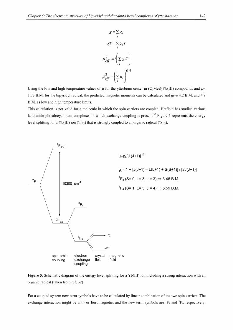
Chapter 6: The electronic structure of bipyridyl and diazabutadienyl complexes of ytterbocenes
142
∑=i
iχχ
Ti
iT ∑= χχ
∑= Ti
ieff χµ 82
5.02
∑=i
ieff µµ
Using the low and high temperature values of µ for the ytterbium center in (C5Me5)2Yb(III) compounds and µ=
1.73 B.M. for the bipyridyl radical, the predicted magnetic moments can be calculated and give 4.2 B.M. and 4.8
B.M. as low and high temperature limits.
This calculation is not valid for a molecule in which the spin carriers are coupled. Hatfield has studied various
lanthanide-phthalocyaninato complexes in which exchange coupling is present.32 Figure 5 represents the energy
level splitting for a Yb(III) ion (2F7/2) that is strongly coupled to an organic radical (2S1/2).
2F
2F1/2
2F7/2
3F4
1F3
spin-orbit coupling
electronexchangecoupling
crystalfield
magneticfield
10300 cm-1
Figure 5. Schematic diagram of the energy level splitting for a Yb(III) ion including a strong interaction with an
organic radical (taken from ref. 32)
For a coupled system new term symbols have to be calculated by linear combination of the two spin carriers. The
exchange interaction might be anti- or ferromagnetic, and the new term symbols are 1F3 and 3F4, respectively.
µ=gL[J (J+1)]1/2
gL= 1 + [J(J+1) – L(L+1) + S(S+1)] / [2J(J+1)] 1F3 (S= 0, L= 3, J = 3) ⇒ 3.46 B.M. 1F4 (S= 1, L= 3, J = 4) ⇒ 5.59 B.M.
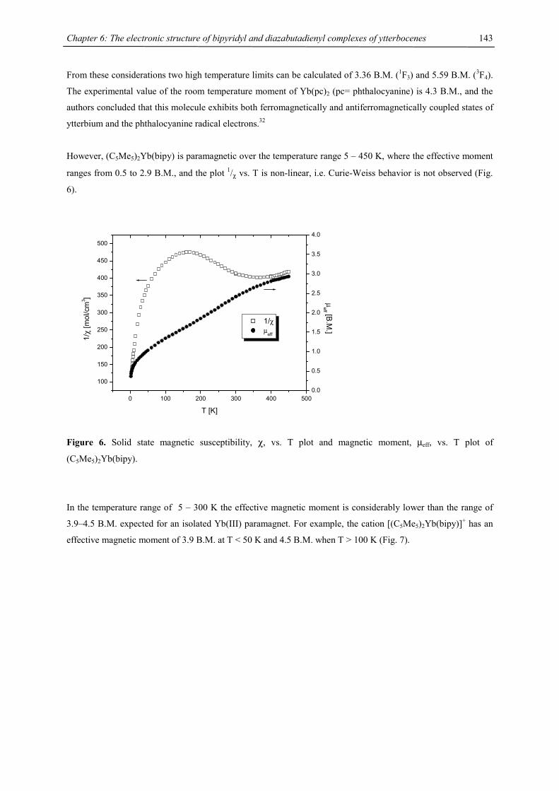
Chapter 6: The electronic structure of bipyridyl and diazabutadienyl complexes of ytterbocenes
143
From these considerations two high temperature limits can be calculated of 3.36 B.M. (1F3) and 5.59 B.M. (3F4).
The experimental value of the room temperature moment of Yb(pc)2 (pc= phthalocyanine) is 4.3 B.M., and the
authors concluded that this molecule exhibits both ferromagnetically and antiferromagnetically coupled states of
ytterbium and the phthalocyanine radical electrons.32
However, (C5Me5)2Yb(bipy) is paramagnetic over the temperature range 5 – 450 K, where the effective moment
ranges from 0.5 to 2.9 B.M., and the plot 1/χ vs. T is non-linear, i.e. Curie-Weiss behavior is not observed (Fig.
6).
0 100 200 300 400 500
100
150
200
250
300
350
400
450
500
1/χ µeff
T [K]
1/χ
[mol
/cm
3 ]
0.0
0.5
1.0
1.5
2.0
2.5
3.0
3.5
4.0µ
eff [B.M.]
Figure 6. Solid state magnetic susceptibility, χ, vs. T plot and magnetic moment, µeff, vs. T plot of
(C5Me5)2Yb(bipy).
In the temperature range of 5 – 300 K the effective magnetic moment is considerably lower than the range of
3.9–4.5 B.M. expected for an isolated Yb(III) paramagnet. For example, the cation [(C5Me5)2Yb(bipy)]+ has an
effective magnetic moment of 3.9 B.M. at T < 50 K and 4.5 B.M. when T > 100 K (Fig. 7).
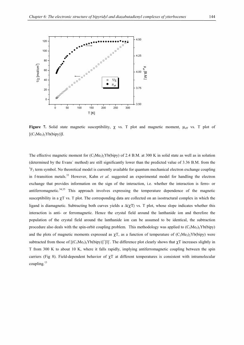
Chapter 6: The electronic structure of bipyridyl and diazabutadienyl complexes of ytterbocenes
144
0 50 100 150 200 250 300
0
20
40
60
80
100
120
1/χ µ
eff
T [K]
1/χ
[mol
/cm
3 ]
3.50
3.75
4.00
4.25
4.50
µeff [B
.M.]
Figure 7. Solid state magnetic susceptibility, χ vs. T plot and magnetic moment, µeff vs. T plot of
[(C5Me5)2Yb(bipy)]I.
The effective magnetic moment for (C5Me5)2Yb(bipy) of 2.4 B.M. at 300 K in solid state as well as in solution
(determined by the Evans` method) are still significantly lower than the predicted value of 3.36 B.M. from the 1F3 term symbol. No theoretical model is currently available for quantum mechanical electron exchange coupling
in f-transition metals.33 However, Kahn et al. suggested an experimental model for handling the electron
exchange that provides information on the sign of the interaction, i.e. whether the interaction is ferro- or
antiferromagnetic.34,35 This approach involves expressing the temperature dependence of the magnetic
susceptibility in a χT vs. T plot. The corresponding data are collected on an isostructural complex in which the
ligand is diamagnetic. Subtracting both curves yields a ∆(χΤ) vs. T plot, whose slope indicates whether this
interaction is anti- or ferromagnetic. Hence the crystal field around the lanthanide ion and therefore the
population of the crystal field around the lanthanide ion can be assumed to be identical, the subtraction
procedure also deals with the spin-orbit coupling problem. This methodology was applied to (C5Me5)2Yb(bipy)
and the plots of magnetic moments expressed as χT, as a function of temperature of (C5Me5)2Yb(bipy) were
subtracted from those of [(C5Me5)2Yb(bipy)]+[I]-. The difference plot clearly shows that χT increases slightly in
T from 300 K to about 10 K, where it falls rapidly, implying antiferromagnetic coupling between the spin
carriers (Fig 8). Field-dependent behavior of χT at different temperatures is consistent with intramolecular
coupling.13
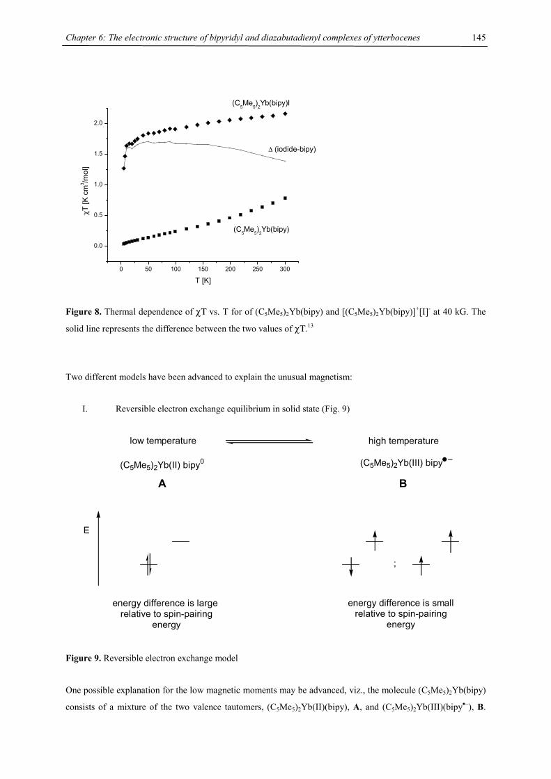
Chapter 6: The electronic structure of bipyridyl and diazabutadienyl complexes of ytterbocenes
145
0 50 100 150 200 250 300
0.0
0.5
1.0
1.5
2.0
∆ (iodide-bipy)
(C5Me
5)2Yb(bipy)I
(C5Me5)2Yb(bipy)
χT [K
cm
3 /mol
]
T [K]
Figure 8. Thermal dependence of χΤ vs. T for of (C5Me5)2Yb(bipy) and [(C5Me5)2Yb(bipy)]+[I]- at 40 kG. The
solid line represents the difference between the two values of χΤ.13
Two different models have been advanced to explain the unusual magnetism:
I. Reversible electron exchange equilibrium in solid state (Fig. 9)
low temperature high temperature
(C5Me5)2Yb(II) bipy0 (C5Me5)2Yb(III) bipy
;
energy difference is large relative to spin-pairing
energy
energy difference is smallrelative to spin-pairing
energy
E
A B
Figure 9. Reversible electron exchange model
One possible explanation for the low magnetic moments may be advanced, viz., the molecule (C5Me5)2Yb(bipy)
consists of a mixture of the two valence tautomers, (C5Me5)2Yb(II)(bipy), A, and (C5Me5)2Yb(III)(bipy•−), B.
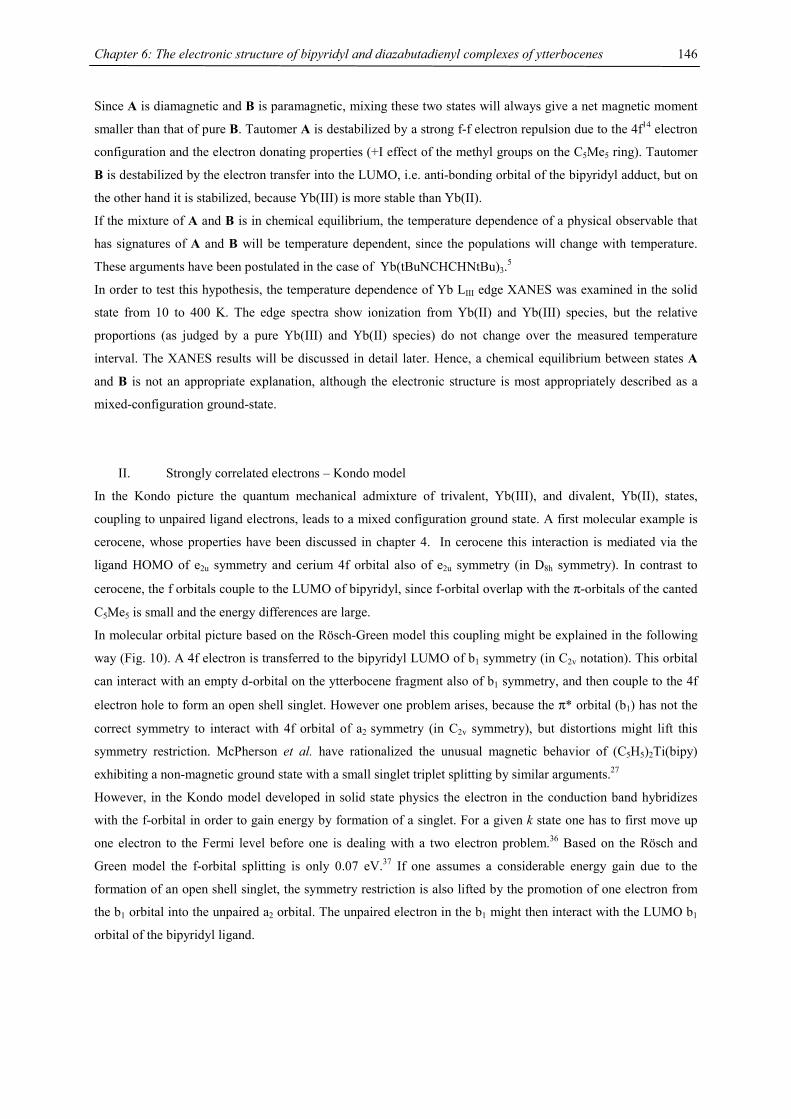
Chapter 6: The electronic structure of bipyridyl and diazabutadienyl complexes of ytterbocenes
146
Since A is diamagnetic and B is paramagnetic, mixing these two states will always give a net magnetic moment
smaller than that of pure B. Tautomer A is destabilized by a strong f-f electron repulsion due to the 4f14 electron
configuration and the electron donating properties (+I effect of the methyl groups on the C5Me5 ring). Tautomer
B is destabilized by the electron transfer into the LUMO, i.e. anti-bonding orbital of the bipyridyl adduct, but on
the other hand it is stabilized, because Yb(III) is more stable than Yb(II).
If the mixture of A and B is in chemical equilibrium, the temperature dependence of a physical observable that
has signatures of A and B will be temperature dependent, since the populations will change with temperature.
These arguments have been postulated in the case of Yb(tBuNCHCHNtBu)3.5
In order to test this hypothesis, the temperature dependence of Yb LIII edge XANES was examined in the solid
state from 10 to 400 K. The edge spectra show ionization from Yb(II) and Yb(III) species, but the relative
proportions (as judged by a pure Yb(III) and Yb(II) species) do not change over the measured temperature
interval. The XANES results will be discussed in detail later. Hence, a chemical equilibrium between states A
and B is not an appropriate explanation, although the electronic structure is most appropriately described as a
mixed-configuration ground-state.
II. Strongly correlated electrons – Kondo model
In the Kondo picture the quantum mechanical admixture of trivalent, Yb(III), and divalent, Yb(II), states,
coupling to unpaired ligand electrons, leads to a mixed configuration ground state. A first molecular example is
cerocene, whose properties have been discussed in chapter 4. In cerocene this interaction is mediated via the
ligand HOMO of e2u symmetry and cerium 4f orbital also of e2u symmetry (in D8h symmetry). In contrast to
cerocene, the f orbitals couple to the LUMO of bipyridyl, since f-orbital overlap with the π-orbitals of the canted
C5Me5 is small and the energy differences are large.
In molecular orbital picture based on the Rösch-Green model this coupling might be explained in the following
way (Fig. 10). A 4f electron is transferred to the bipyridyl LUMO of b1 symmetry (in C2v notation). This orbital
can interact with an empty d-orbital on the ytterbocene fragment also of b1 symmetry, and then couple to the 4f
electron hole to form an open shell singlet. However one problem arises, because the π* orbital (b1) has not the
correct symmetry to interact with 4f orbital of a2 symmetry (in C2v symmetry), but distortions might lift this
symmetry restriction. McPherson et al. have rationalized the unusual magnetic behavior of (C5H5)2Ti(bipy)
exhibiting a non-magnetic ground state with a small singlet triplet splitting by similar arguments.27
However, in the Kondo model developed in solid state physics the electron in the conduction band hybridizes
with the f-orbital in order to gain energy by formation of a singlet. For a given k state one has to first move up
one electron to the Fermi level before one is dealing with a two electron problem.36 Based on the Rösch and
Green model the f-orbital splitting is only 0.07 eV.37 If one assumes a considerable energy gain due to the
formation of an open shell singlet, the symmetry restriction is also lifted by the promotion of one electron from
the b1 orbital into the unpaired a2 orbital. The unpaired electron in the b1 might then interact with the LUMO b1
orbital of the bipyridyl ligand.
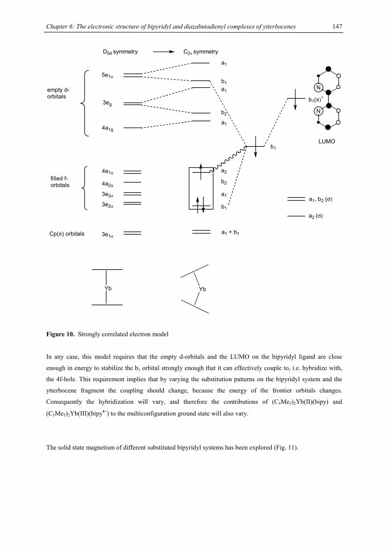
Chapter 6: The electronic structure of bipyridyl and diazabutadienyl complexes of ytterbocenes
147
Yb
D5d symmetry
5e1u
3eg
4a1g
4e1u
4a2u
3e2u
3e2u
3e1u
empty d-orbitals
filled f-orbitals
Cp(π) orbitals
C2v symmetry
a1
b2
a1
a2
b2
a1
b1
a1 + b1
Yb
N
N
b1(π)∗
a1
b1
LUMO
a1, b2 (σ)
a2 (σ)
b1
Figure 10. Strongly correlated electron model
In any case, this model requires that the empty d-orbitals and the LUMO on the bipyridyl ligand are close
enough in energy to stabilize the b1 orbital strongly enough that it can effectively couple to, i.e. hybridize with,
the 4f-hole. This requirement implies that by varying the substitution patterns on the bipyridyl system and the
ytterbocene fragment the coupling should change, because the energy of the frontier orbitals changes.
Consequently the hybridization will vary, and therefore the contributions of (C5Me5)2Yb(II)(bipy) and
(C5Me5)2Yb(III)(bipy•−) to the multiconfiguration ground state will also vary.
The solid state magnetism of different substituted bipyridyl systems has been explored (Fig. 11).
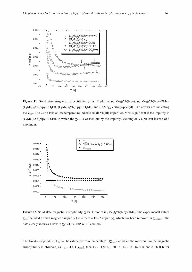
Chapter 6: The electronic structure of bipyridyl and diazabutadienyl complexes of ytterbocenes
148
-50 0 50 100 150 200 250 300 350 400 4500.000
0.002
0.004
0.006
0.008
0.010
0.012
(C5Me
5)2Yb(bipy-phenyl)
(C5Me5)2Yb(bipy) (C5Me5)2Yb(bipy-OMe) (C
5Me
5)2Yb(bipy-CO
2Et)
(C5Me5)2Yb(bipy-CO2Me)
χ [c
m3 /m
ol]
T [K]
Figure 11. Solid state magnetic susceptibility, χ vs. T plot of (C5Me5)2Yb(bipy), (C5Me5)2Yb(bipy-OMe),
(C5Me5)2Yb(bipy-CO2Et), (C5Me5)2Yb(bipy-CO2Me) and (C5Me5)2Yb(bipy-phenyl). The arrows are indicating
the χmax. The Curie-tails at low temperature indicate small Yb(III) impurities. Most significant is the impurity in
(C5Me5)2Yb(bipy-CO2Et), in which the χmax is washed out by the impurity, yielding only a plateau instead of a
maximum.
0 50 100 150 200 250 300
0.0000
0.0002
0.0004
0.0006
0.0008
0.0010
0.0012
0.0014
0.0016
0.0018
χ [c
m3 /m
ol]
T [K]
χexp Yb(III) impurity (~ 0.6 %) χcorrected
Figure 12. Solid state magnetic susceptibility, χ vs. T plot of (C5Me5)2Yb(bipy-OMe). The experimental values
χexp included a small magnetic impurity (~0.6 % of a J=7/2 impurity), which has been removed in χcorrected. The
data clearly shows a TIP with χ0= (4.19±0.05)x10-4 emu/mol.
The Kondo temperature, TK, can be estimated from temperature T(χmax), at which the maximum in the magnetic
susceptibility is observed, as TK ~ 4.4 T(χmax), then TK~ 1170 K, 1300 K, 1630 K, 1670 K and > 1800 K for
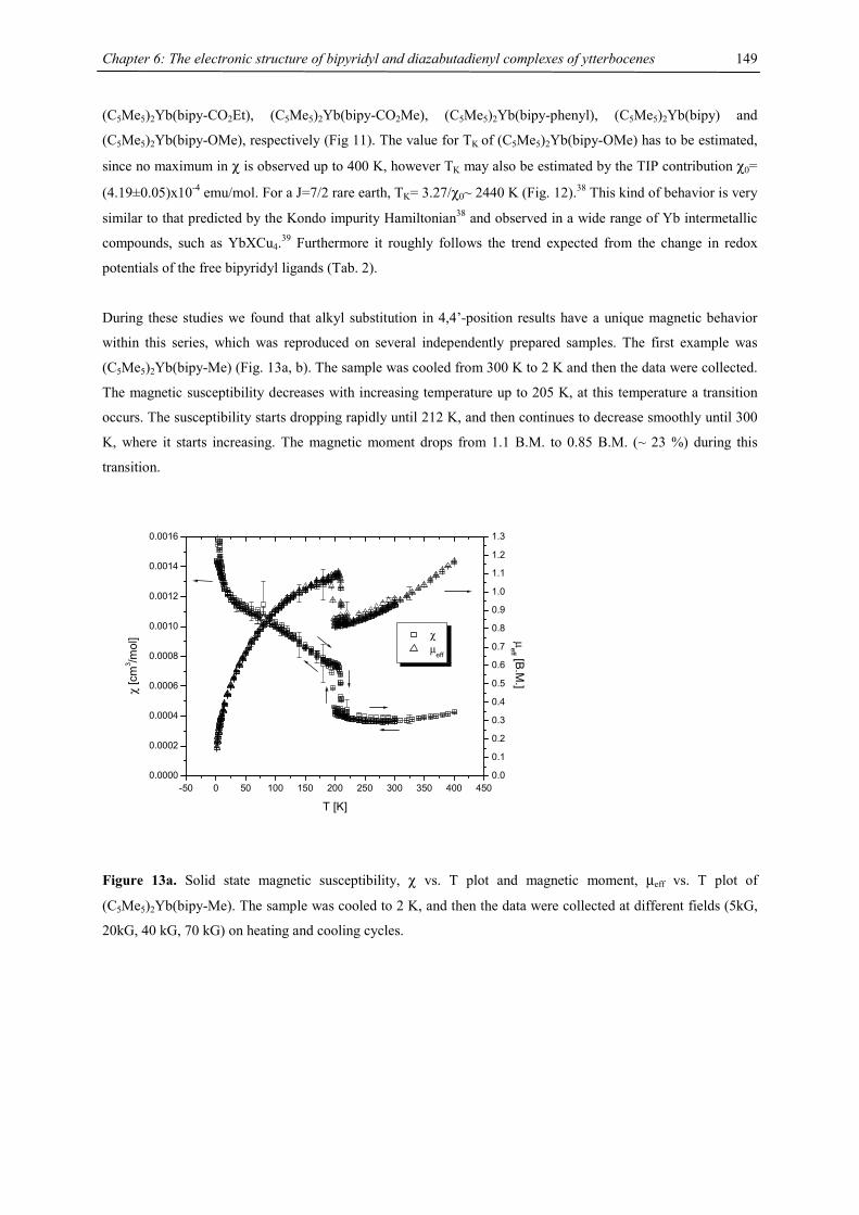
Chapter 6: The electronic structure of bipyridyl and diazabutadienyl complexes of ytterbocenes
149
(C5Me5)2Yb(bipy-CO2Et), (C5Me5)2Yb(bipy-CO2Me), (C5Me5)2Yb(bipy-phenyl), (C5Me5)2Yb(bipy) and
(C5Me5)2Yb(bipy-OMe), respectively (Fig 11). The value for TK of (C5Me5)2Yb(bipy-OMe) has to be estimated,
since no maximum in χ is observed up to 400 K, however TK may also be estimated by the TIP contribution χ0=
(4.19±0.05)x10-4 emu/mol. For a J=7/2 rare earth, TK= 3.27/χ0~ 2440 K (Fig. 12).38 This kind of behavior is very
similar to that predicted by the Kondo impurity Hamiltonian38 and observed in a wide range of Yb intermetallic
compounds, such as YbXCu4.39 Furthermore it roughly follows the trend expected from the change in redox
potentials of the free bipyridyl ligands (Tab. 2).
During these studies we found that alkyl substitution in 4,4’-position results have a unique magnetic behavior
within this series, which was reproduced on several independently prepared samples. The first example was
(C5Me5)2Yb(bipy-Me) (Fig. 13a, b). The sample was cooled from 300 K to 2 K and then the data were collected.
The magnetic susceptibility decreases with increasing temperature up to 205 K, at this temperature a transition
occurs. The susceptibility starts dropping rapidly until 212 K, and then continues to decrease smoothly until 300
K, where it starts increasing. The magnetic moment drops from 1.1 B.M. to 0.85 B.M. (~ 23 %) during this
transition.
-50 0 50 100 150 200 250 300 350 400 4500.0000
0.0002
0.0004
0.0006
0.0008
0.0010
0.0012
0.0014
0.0016
χ µeff
T [K]
χ [c
m3 /m
ol]
0.0
0.1
0.2
0.3
0.4
0.5
0.6
0.7
0.8
0.9
1.0
1.1
1.2
1.3
µeff [B.M
.]
Figure 13a. Solid state magnetic susceptibility, χ vs. T plot and magnetic moment, µeff vs. T plot of
(C5Me5)2Yb(bipy-Me). The sample was cooled to 2 K, and then the data were collected at different fields (5kG,
20kG, 40 kG, 70 kG) on heating and cooling cycles.
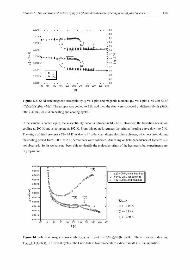
Chapter 6: The electronic structure of bipyridyl and diazabutadienyl complexes of ytterbocenes
150
180 185 190 195 200 205 210 215 220 225 2300.0000
0.0002
0.0004
0.0006
0.0008
0.0010
0.0012
0.0014
0.0016
χ µ
eff
T [K]
χ [c
m3 /m
ol]
0.0
0.1
0.2
0.3
0.4
0.5
0.6
0.7
0.8
0.9
1.0
1.1
1.2
1.3
µeff [B.M
.]
Figure 13b. Solid state magnetic susceptibility, χ vs. T plot and magnetic moment, µeff vs. T plot (180-230 K) of
(C5Me5)2Yb(bipy-Me). The sample was cooled to 2 K, and then the data were collected at different fields (5kG,
20kG, 40 kG, 70 kG) on heating and cooling cycles.
If the sample is cooled again, the susceptibility curve is retraced until 212 K. However, the transition occurs on
cooling at 200 K and is complete at 192 K. From this point it retraces the original heating curve down to 5 K.
The origin of this hysteresis (∆T= 14 K) is due to 1st order crystallographic phase change, which occurred during
the cooling period from 300 K to 2 K, before data were collected. Annealing or field dependence of hysteresis is
not observed. So far we have not been able to identify the molecular origin of the hysteresis, but experiments are
in preparation.
-50 0 50 100 150 200 250 300 350 400 4500.0012
0.0014
0.0016
0.0018
0.0020
0.0022
0.0024
0.0026
0.0028
0.0030
0.0032
0.0034
0.0036
T(2) T(3)
T(1)
χ [c
m3 /m
ol]
T [K]
χ (2-400 K, initial heating) χ (400-2 K, 1st cooling) χ (2-400 K, 2nd heating)
Figure 14. Solid state magnetic susceptibility, χ vs. T plot of (C5Me5)2Yb(bipy-tBu). The arrows are indicating
T(χmax), T(1)-T(3), in different cycles. The Curie tails at low temperature indicate small Yb(III) impurities.
T(χχχχmax)
T(1) ~ 345 K
T(2) ~ 215 K
T(3) ~ 260 K
Chapter 6: The electronic structure of bipyridyl and diazabutadienyl complexes of ytterbocenes
151
The behavior of (C5Me5)2Yb(bipy-tBu) is even more complicated, because it initially exhibits the same behavior
as (C5Me5)2Yb(bipy) and the susceptibility reaches a maximum at T(1)= 345 K, while the magnetic moment
increases monotonically from 0.3 B.M at 2 K to 3.00 B.M. at 350 K. Between 355 K and 365 K a transition
occurs accompanied by a 50 % drop in the magnetic susceptibility, also reflected in a reduction of the magnetic
moment from 3.00 B.M. to 2.20 B.M. In various cooling/heating cycles the initial heating curve is only reached
at T < 25K, where the Yb(III) impurities are dominant in the Curie-tail, and at T > 365 K. The maximum in the
magnetic susceptibility is reached T(2)= 215K on cooling, and at T(3)= 260 K on heating, while the magnetic
moments vary between 0.3 B.M. at 2 K and 2.16 B.M. at 400 K (Fig. 14). A field dependence on these maxima
(up to 7 Tesla) was not observed.
In any case, the last two examples clearly indicate that the observed magnetism is not only due to a molecular
phenomenon, but also due to crystal packing effects, which might introduce structural changes. While there is no
structural information available at this point to explain this behavior, EXAFS and XANES measurements are in
progress to address the structural origin.
Solid-State Measurements: X-Ray Absorption Near Edge Spectra (XANES)
XANES data were collected at the Stanford Synchrotron Radiation Laboratory, a national user facility operated
by Stanford University on the behalf of the DOE/OBES, by Dr. Corwin Booth, Dr. Million Daniel and Dr.
Wayne W. Lukens.
XANES spectroscopy allows for the measurement of either the f-electron (for cerium) or the f-hole (for
ytterbium) occupancy. In both cases this value is called nf. In a true intermediate valence system, the f-shell is
partially occupied, as opposed to a distribution of full or empty occupancies throughout the material. Typically,
one can measure the valence in any system by measuring the position of a core x-ray absorption edge and
comparing the edge position to model compounds. There are several exceptions to this rule, and one occurs
here. Rather than measuring a single, energy-shifted edge for the rare-earth intermediate valence systems, two
edges are observed. This situation is due to a final state effect: the presence of the core hole breaks the Kondo
singlet. In any case, the total absorption is generally fit to a linear sum of reference spectra as a model of the
true, monovalent lineshapes. For instance, in Ytterbium one fits µtot(E)=(1-nf)µ2+(E)+nfµ3+(E), where the
lineshapes for µ2+(E) and µ3+(E) might come from a lutetium analogue. The absolute error in this procedure is
on the order of 5%. Alternatively, one can fit to pseudo-Voigt functions to obtain nearly the same accuracy,
although more interpretation may be required to identify the source (electronic, structural, etc.) of each fitted
feature. These measurements are of fundamental importance for these molecular systems because the
susceptibility measurements indicate strong intermediate valency for these materials. Within the non-crossing
approximation (NCA),40 the f-levels should then be partially occupied, and the systems with a lower estimated
Kondo temperature should approach nf=1. Therefore we expect that as the estimated Kondo temperature from
χ(T) goes down, the ytterbium valency should approach a trivalent state.
The initial discussion is centered on the well established (C5Me5)2Yb(bipy) system to explain the principles.
(C5Me5)2Yb(OEt2) and [(C5Me5)2Yb(bipy)]I served as Yb(II) and Yb(III) reference compounds. Figure X shows
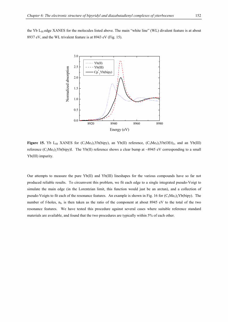
Chapter 6: The electronic structure of bipyridyl and diazabutadienyl complexes of ytterbocenes
152
the Yb LIII edge XANES for the molecules listed above. The main “white line” (WL) divalent feature is at about
8937 eV, and the WL trivalent feature is at 8943 eV (Fig. 15).
8920 8940 8960 89800.0
0.5
1.0
1.5
2.0
2.5
3.0
Nor
mal
ized
abs
orpt
ion
Energy (eV)
Yb(II) Yb(III) Cp*
2Yb(bipy)
Figure 15. Yb LIII XANES for (C5Me5)2Yb(bipy), an Yb(II) reference, (C5Me5)2Yb(OEt)2, and an Yb(III)
reference (C5Me5)2Yb(bipy)I. The Yb(II) reference shows a clear bump at ~8945 eV corresponding to a small
Yb(III) impurity.
Our attempts to measure the pure Yb(II) and Yb(III) lineshapes for the various compounds have so far not
produced reliable results. To circumvent this problem, we fit each edge to a single integrated pseudo-Voigt to
simulate the main edge (in the Lorentzian limit, this function would just be an arctan), and a collection of
pseudo-Voigts to fit each of the resonance features. An example is shown in Fig. 16 for (C5Me5)2Yb(bipy). The
number of f-holes, nf, is then taken as the ratio of the component at about 8945 eV to the total of the two
resonance features. We have tested this procedure against several cases where suitable reference standard
materials are available, and found that the two procedures are typically within 5% of each other.
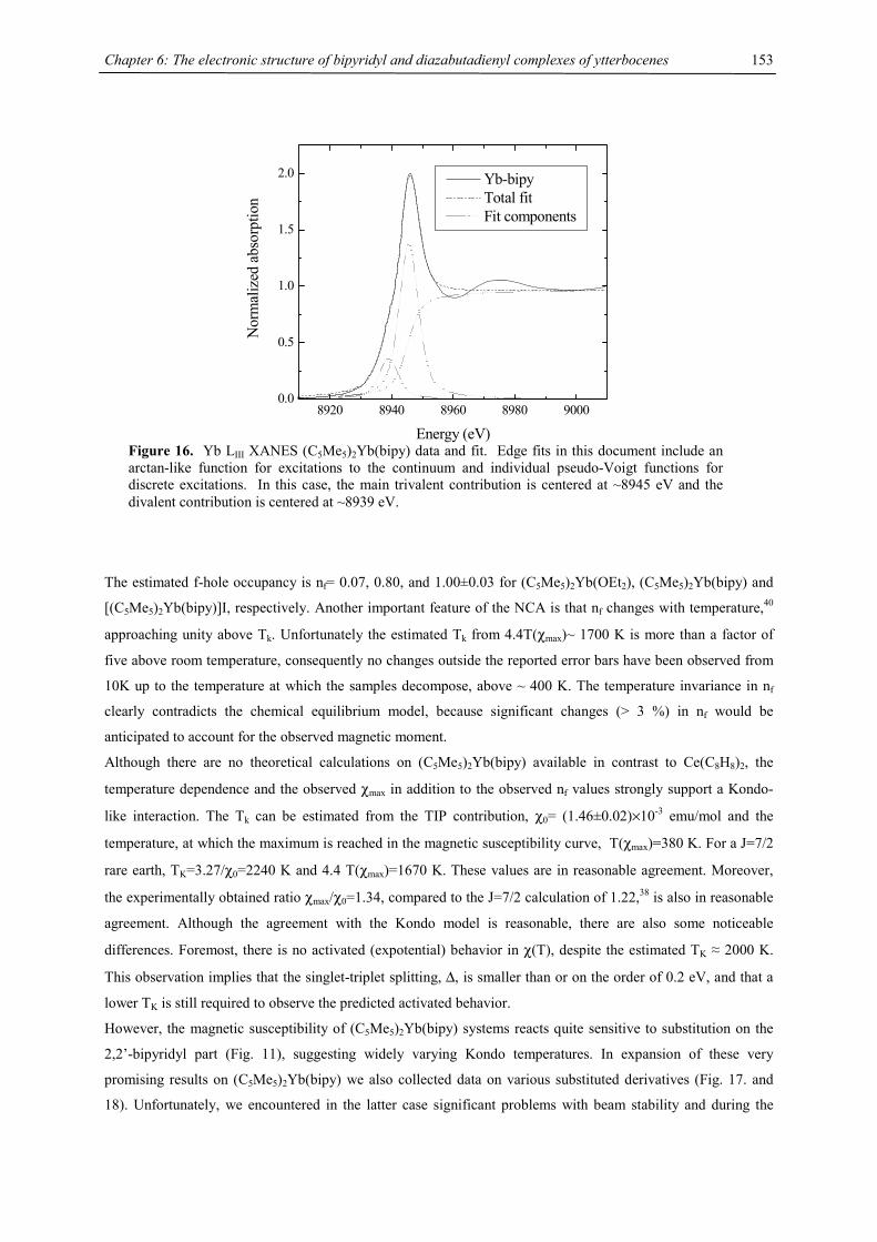
Chapter 6: The electronic structure of bipyridyl and diazabutadienyl complexes of ytterbocenes
153
The estimated f-hole occupancy is nf= 0.07, 0.80, and 1.00±0.03 for (C5Me5)2Yb(OEt2), (C5Me5)2Yb(bipy) and
[(C5Me5)2Yb(bipy)]I, respectively. Another important feature of the NCA is that nf changes with temperature,40
approaching unity above Tk. Unfortunately the estimated Tk from 4.4T(χmax)~ 1700 K is more than a factor of
five above room temperature, consequently no changes outside the reported error bars have been observed from
10K up to the temperature at which the samples decompose, above ~ 400 K. The temperature invariance in nf
clearly contradicts the chemical equilibrium model, because significant changes (> 3 %) in nf would be
anticipated to account for the observed magnetic moment.
Although there are no theoretical calculations on (C5Me5)2Yb(bipy) available in contrast to Ce(C8H8)2, the
temperature dependence and the observed χmax in addition to the observed nf values strongly support a Kondo-
like interaction. The Tk can be estimated from the TIP contribution, χ0= (1.46±0.02)×10-3 emu/mol and the
temperature, at which the maximum is reached in the magnetic susceptibility curve, T(χmax)=380 K. For a J=7/2
rare earth, TK=3.27/χ0=2240 K and 4.4 T(χmax)=1670 K. These values are in reasonable agreement. Moreover,
the experimentally obtained ratio χmax/χ0=1.34, compared to the J=7/2 calculation of 1.22,38 is also in reasonable
agreement. Although the agreement with the Kondo model is reasonable, there are also some noticeable
differences. Foremost, there is no activated (expotential) behavior in χ(T), despite the estimated TK ≈ 2000 K.
This observation implies that the singlet-triplet splitting, ∆, is smaller than or on the order of 0.2 eV, and that a
lower TK is still required to observe the predicted activated behavior.
However, the magnetic susceptibility of (C5Me5)2Yb(bipy) systems reacts quite sensitive to substitution on the
2,2’-bipyridyl part (Fig. 11), suggesting widely varying Kondo temperatures. In expansion of these very
promising results on (C5Me5)2Yb(bipy) we also collected data on various substituted derivatives (Fig. 17. and
18). Unfortunately, we encountered in the latter case significant problems with beam stability and during the
8920 8940 8960 8980 90000.0
0.5
1.0
1.5
2.0
Nor
mal
ized
abs
orpt
ion
Energy (eV)
Yb-bipy Total fit Fit components
Figure 16. Yb LIII XANES (C5Me5)2Yb(bipy) data and fit. Edge fits in this document include an arctan-like function for excitations to the continuum and individual pseudo-Voigt functions for discrete excitations. In this case, the main trivalent contribution is centered at ~8945 eV and the divalent contribution is centered at ~8939 eV.
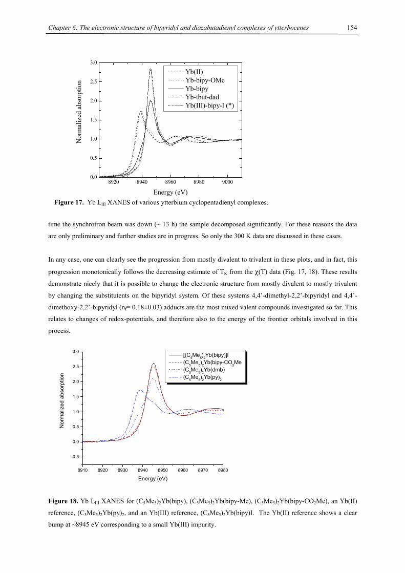
Chapter 6: The electronic structure of bipyridyl and diazabutadienyl complexes of ytterbocenes
154
time the synchrotron beam was down (~ 13 h) the sample decomposed significantly. For these reasons the data
are only preliminary and further studies are in progress. So only the 300 K data are discussed in these cases.
In any case, one can clearly see the progression from mostly divalent to trivalent in these plots, and in fact, this
progression monotonically follows the decreasing estimate of TK from the χ(T) data (Fig. 17, 18). These results
demonstrate nicely that it is possible to change the electronic structure from mostly divalent to mostly trivalent
by changing the substitutents on the bipyridyl system. Of these systems 4,4’-dimethyl-2,2’-bipyridyl and 4,4’-
dimethoxy-2,2’-bipyridyl (nf= 0.18±0.03) adducts are the most mixed valent compounds investigated so far. This
relates to changes of redox-potentials, and therefore also to the energy of the frontier orbitals involved in this
process.
8910 8920 8930 8940 8950 8960 8970 8980
-0.5
0.0
0.5
1.0
1.5
2.0
2.5
3.0
Nor
mal
ized
abs
orpt
ion
Energy (eV)
[(C5Me5)2Yb(bipy)]I (C
5Me
5)2Yb(bipy-CO
2Me
(C5Me5)2Yb(dmb) (C5Me5)2Yb(py)2
Figure 18. Yb LIII XANES for (C5Me5)2Yb(bipy), (C5Me5)2Yb(bipy-Me), (C5Me5)2Yb(bipy-CO2Me), an Yb(II)
reference, (C5Me5)2Yb(py)2, and an Yb(III) reference, (C5Me5)2Yb(bipy)I. The Yb(II) reference shows a clear
bump at ~8945 eV corresponding to a small Yb(III) impurity.
8920 8940 8960 8980 90000.0
0.5
1.0
1.5
2.0
2.5
3.0
Nor
mal
ized
abs
orpt
ion
Energy (eV)
Yb(II) Yb-bipy-OMe Yb-bipy Yb-tbut-dad Yb(III)-bipy-I (*)
Figure 17. Yb LIII XANES of various ytterbium cyclopentadienyl complexes.
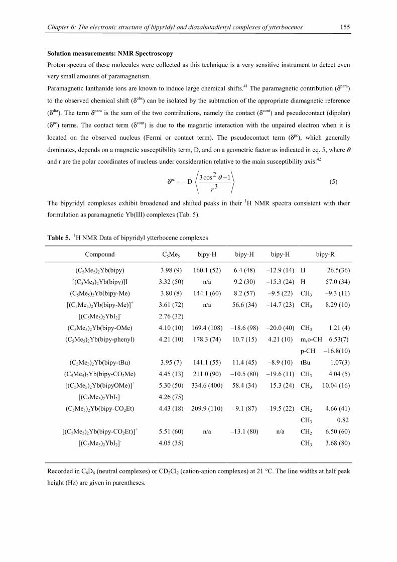
Chapter 6: The electronic structure of bipyridyl and diazabutadienyl complexes of ytterbocenes
155
Solution measurements: NMR Spectroscopy
Proton spectra of these molecules were collected as this technique is a very sensitive instrument to detect even
very small amounts of paramagnetism.
Paramagnetic lanthanide ions are known to induce large chemical shifts.41 The paramagnetic contribution (δpara)
to the observed chemical shift (δobs) can be isolated by the subtraction of the appropriate diamagnetic reference
(δdia). The term δpara is the sum of the two contributions, namely the contact (δcont) and pseudocontact (dipolar)
(δpc) terms. The contact term (δcont) is due to the magnetic interaction with the unpaired electron when it is
located on the observed nucleus (Fermi or contact term). The pseudocontact term (δpc), which generally
dominates, depends on a magnetic susceptibility term, D, and on a geometric factor as indicated in eq. 5, where θ
and r are the polar coordinates of nucleus under consideration relative to the main susceptibility axis:42
δpc = − D 3
12cos3
r
−θ (5)
The bipyridyl complexes exhibit broadened and shifted peaks in their 1H NMR spectra consistent with their
formulation as paramagnetic Yb(III) complexes (Tab. 5).
Table 5. 1H NMR Data of bipyridyl ytterbocene complexes
Compound C5Me5 bipy-H bipy-H bipy-H bipy-R
(C5Me5)2Yb(bipy) 3.98 (9) 160.1 (52) 6.4 (48) –12.9 (14) H 26.5(36)
[(C5Me5)2Yb(bipy)]I 3.32 (50) n/a 9.2 (30) –15.3 (24) H 57.0 (34)
(C5Me5)2Yb(bipy-Me) 3.80 (8) 144.1 (60) 8.2 (57) –9.5 (22) CH3 –9.3 (11)
[(C5Me5)2Yb(bipy-Me)]+
[(C5Me5)2YbI2]-
3.61 (72)
2.76 (32)
n/a 56.6 (34) –14.7 (23) CH3 8.29 (10)
(C5Me5)2Yb(bipy-OMe) 4.10 (10) 169.4 (108) –18.6 (98) –20.0 (40) CH3 1.21 (4)
(C5Me5)2Yb(bipy-phenyl) 4.21 (10) 178.3 (74) 10.7 (15) 4.21 (10) m,o-CH 6.53(7)
p-CH –16.8(10)
(C5Me5)2Yb(bipy-tBu) 3.95 (7) 141.1 (55) 11.4 (45) –8.9 (10) tBu 1.07(3)
(C5Me5)2Yb(bipy-CO2Me) 4.45 (13) 211.0 (90) –10.5 (80) –19.6 (11) CH3 4.04 (5)
[(C5Me5)2Yb(bipyOMe)]+
[(C5Me5)2YbI2]-
5.30 (50)
4.26 (75)
334.6 (400) 58.4 (34) –15.3 (24) CH3 10.04 (16)
(C5Me5)2Yb(bipy-CO2Et) 4.43 (18) 209.9 (110) –9.1 (87) –19.5 (22) CH2 4.66 (41)
CH3 0.82
[(C5Me5)2Yb(bipy-CO2Et)]+
[(C5Me5)2YbI2]-
5.51 (60)
4.05 (35)
n/a –13.1 (80) n/a CH2 6.50 (60)
CH3 3.68 (80)
Recorded in C6D6 (neutral complexes) or CD2Cl2 (cation-anion complexes) at 21 °C. The line widths at half peak
height (Hz) are given in parentheses.
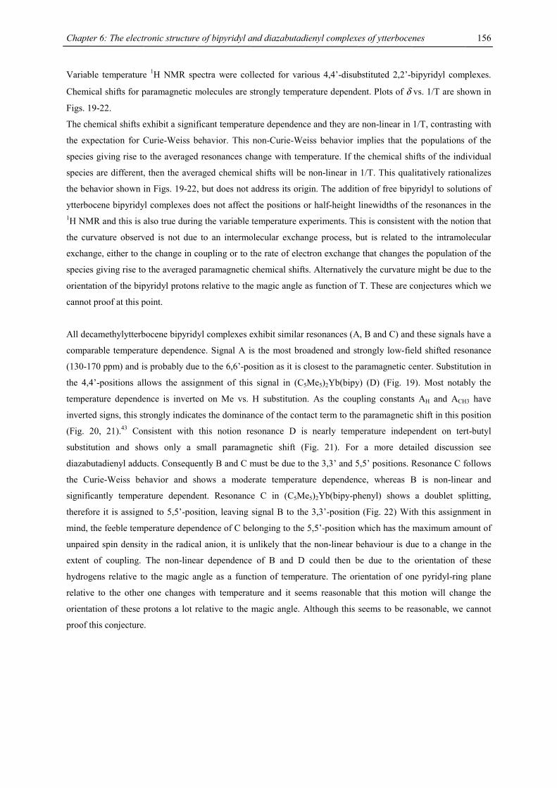
Chapter 6: The electronic structure of bipyridyl and diazabutadienyl complexes of ytterbocenes
156
Variable temperature 1H NMR spectra were collected for various 4,4’-disubstituted 2,2’-bipyridyl complexes.
Chemical shifts for paramagnetic molecules are strongly temperature dependent. Plots of δ vs. 1/T are shown in
Figs. 19-22.
The chemical shifts exhibit a significant temperature dependence and they are non-linear in 1/T, contrasting with
the expectation for Curie-Weiss behavior. This non-Curie-Weiss behavior implies that the populations of the
species giving rise to the averaged resonances change with temperature. If the chemical shifts of the individual
species are different, then the averaged chemical shifts will be non-linear in 1/T. This qualitatively rationalizes
the behavior shown in Figs. 19-22, but does not address its origin. The addition of free bipyridyl to solutions of
ytterbocene bipyridyl complexes does not affect the positions or half-height linewidths of the resonances in the 1H NMR and this is also true during the variable temperature experiments. This is consistent with the notion that
the curvature observed is not due to an intermolecular exchange process, but is related to the intramolecular
exchange, either to the change in coupling or to the rate of electron exchange that changes the population of the
species giving rise to the averaged paramagnetic chemical shifts. Alternatively the curvature might be due to the
orientation of the bipyridyl protons relative to the magic angle as function of T. These are conjectures which we
cannot proof at this point.
All decamethylytterbocene bipyridyl complexes exhibit similar resonances (A, B and C) and these signals have a
comparable temperature dependence. Signal A is the most broadened and strongly low-field shifted resonance
(130-170 ppm) and is probably due to the 6,6’-position as it is closest to the paramagnetic center. Substitution in
the 4,4’-positions allows the assignment of this signal in (C5Me5)2Yb(bipy) (D) (Fig. 19). Most notably the
temperature dependence is inverted on Me vs. H substitution. As the coupling constants AH and ACH3 have
inverted signs, this strongly indicates the dominance of the contact term to the paramagnetic shift in this position
(Fig. 20, 21).43 Consistent with this notion resonance D is nearly temperature independent on tert-butyl
substitution and shows only a small paramagnetic shift (Fig. 21). For a more detailed discussion see
diazabutadienyl adducts. Consequently B and C must be due to the 3,3’ and 5,5’ positions. Resonance C follows
the Curie-Weiss behavior and shows a moderate temperature dependence, whereas B is non-linear and
significantly temperature dependent. Resonance C in (C5Me5)2Yb(bipy-phenyl) shows a doublet splitting,
therefore it is assigned to 5,5’-position, leaving signal B to the 3,3’-position (Fig. 22) With this assignment in
mind, the feeble temperature dependence of C belonging to the 5,5’-position which has the maximum amount of
unpaired spin density in the radical anion, it is unlikely that the non-linear behaviour is due to a change in the
extent of coupling. The non-linear dependence of B and D could then be due to the orientation of these
hydrogens relative to the magic angle as a function of temperature. The orientation of one pyridyl-ring plane
relative to the other one changes with temperature and it seems reasonable that this motion will change the
orientation of these protons a lot relative to the magic angle. Although this seems to be reasonable, we cannot
proof this conjecture.
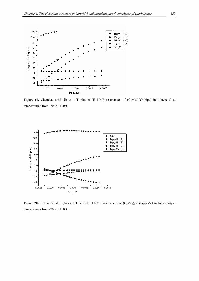
Chapter 6: The electronic structure of bipyridyl and diazabutadienyl complexes of ytterbocenes
157
Figure 19. Chemical shift (δ) vs. 1/T plot of 1H NMR resonances of (C5Me5)2Yb(bipy) in toluene-d8 at
temperatures from -70 to +100°C.
0.0025 0.0030 0.0035 0.0040 0.0045 0.0050 0.0055
-40
-20
0
20
40
60
80
100
120
140
Che
mic
al s
hift
[ppm
]
1/T [1/K]
Cp* bipy-H (A) bipy-H (B) bipy-H (C) bipy-Me (D)
Figure 20a. Chemical shift (δ) vs. 1/T plot of 1H NMR resonances of (C5Me5)2Yb(bipy-Me) in toluene-d8 at
temperatures from -70 to +100°C.
- (D) - (B) - (C) - (A)
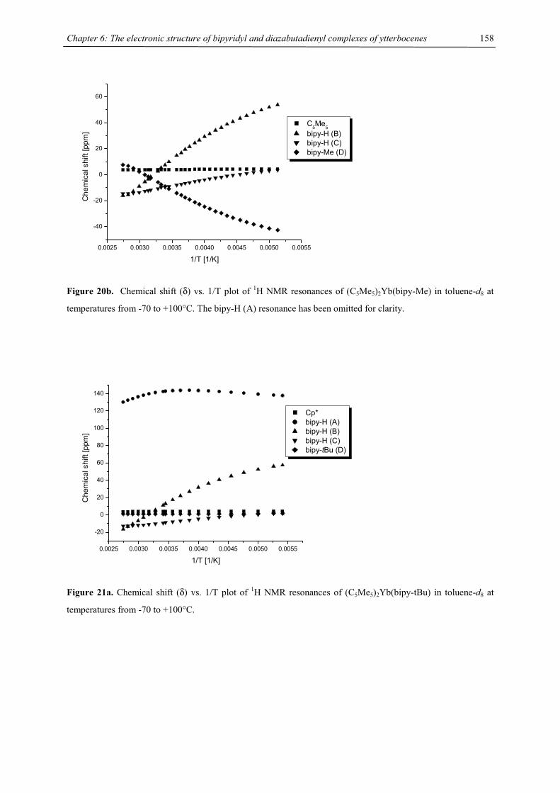
Chapter 6: The electronic structure of bipyridyl and diazabutadienyl complexes of ytterbocenes
158
0.0025 0.0030 0.0035 0.0040 0.0045 0.0050 0.0055
-40
-20
0
20
40
60
Che
mic
al s
hift
[ppm
]
1/T [1/K]
C5Me5 bipy-H (B) bipy-H (C) bipy-Me (D)
Figure 20b. Chemical shift (δ) vs. 1/T plot of 1H NMR resonances of (C5Me5)2Yb(bipy-Me) in toluene-d8 at
temperatures from -70 to +100°C. The bipy-H (A) resonance has been omitted for clarity.
0.0025 0.0030 0.0035 0.0040 0.0045 0.0050 0.0055
-20
0
20
40
60
80
100
120
140
Che
mic
al s
hift
[ppm
]
1/T [1/K]
Cp* bipy-H (A) bipy-H (B) bipy-H (C) bipy-tBu (D)
Figure 21a. Chemical shift (δ) vs. 1/T plot of 1H NMR resonances of (C5Me5)2Yb(bipy-tBu) in toluene-d8 at
temperatures from -70 to +100°C.
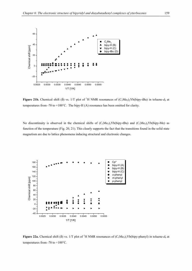
Chapter 6: The electronic structure of bipyridyl and diazabutadienyl complexes of ytterbocenes
159
0.0025 0.0030 0.0035 0.0040 0.0045 0.0050 0.0055
-20
0
20
40
60
Che
mic
al s
hift
[ppm
]
1/T [1/K]
C5Me5 bipy-H (B) bipy-H (C) bipy-tBu (D)
Figure 21b. Chemical shift (δ) vs. 1/T plot of 1H NMR resonances of (C5Me5)2Yb(bipy-tBu) in toluene-d8 at
temperatures from -70 to +100°C. The bipy-H (A) resonance has been omitted for clarity.
No discontinuity is observed in the chemical shifts of (C5Me5)2Yb(bipy-tBu) and (C5Me5)2Yb(bipy-Me) as
function of the temperature (Fig. 20, 21). This clearly supports the fact that the transitions found in the solid state
magnetism are due to lattice phenomena inducing structural and electronic changes.
0.0025 0.0030 0.0035 0.0040 0.0045 0.0050 0.0055-40
-20
0
20
40
60
80
100
120
140
160
180
Che
mic
al s
hift
[ppm
]
1/T [1/K]
Cp* bipy-H (A) bipy-H (B) bipy-H (C) o-phenyl m-phenyl p-phenyl
Figure 22a. Chemical shift (δ) vs. 1/T plot of 1H NMR resonances of (C5Me5)2Yb(bipy-phenyl) in toluene-d8 at
temperatures from -70 to +100°C.
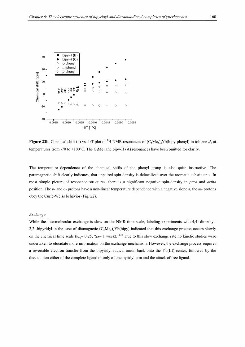
Chapter 6: The electronic structure of bipyridyl and diazabutadienyl complexes of ytterbocenes
160
0.0025 0.0030 0.0035 0.0040 0.0045 0.0050 0.0055-40
-20
0
20
40
60
Che
mic
al s
hift
[ppm
]
1/T [1/K]
bipy-H (B) bipy-H (C) o-phenyl m-phenyl p-phenyl
Figure 22b. Chemical shift (δ) vs. 1/T plot of 1H NMR resonances of (C5Me5)2Yb(bipy-phenyl) in toluene-d8 at
temperatures from -70 to +100°C. The C5Me5 and bipy-H (A) resonances have been omitted for clarity.
The temperature dependence of the chemical shifts of the phenyl group is also quite instructive. The
paramagnetic shift clearly indicates, that unpaired spin density is delocalized over the aromatic substituents. In
most simple picture of resonance structures, there is a significant negative spin-density in para and ortho
position. The p- and o- protons have a non-linear temperature dependence with a negative slope a, the m- protons
obey the Curie-Weiss behavior (Fig. 22).
Exchange
While the intermolecular exchange is slow on the NMR time scale, labeling experiments with 4,4’-dimethyl-
2,2’-bipyridyl in the case of diamagnetic (C5Me5)2Yb(bipy) indicated that this exchange process occurs slowly
on the chemical time scale (keq= 0.25, τ1/2= 1 week).12,13 Due to this slow exchange rate no kinetic studies were
undertaken to elucidate more information on the exchange mechanism. However, the exchange process requires
a reversible electron transfer from the bipyridyl radical anion back onto the Yb(III) center, followed by the
dissociation either of the complete ligand or only of one pyridyl arm and the attack of free ligand.
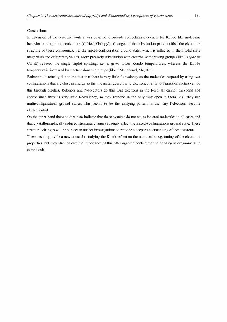
Chapter 6: The electronic structure of bipyridyl and diazabutadienyl complexes of ytterbocenes
161
Conclusions
In extension of the cerocene work it was possible to provide compelling evidences for Kondo like molecular
behavior in simple molecules like (C5Me5)2Yb(bipy’). Changes in the substitution pattern affect the electronic
structure of these compounds, i.e. the mixed-configuration ground state, which is reflected in their solid state
magnetism and different nf values. More precisely substitution with electron withdrawing groups (like CO2Me or
CO2Et) reduces the singlet-triplet splitting, i.e. it gives lower Kondo temperatures, whereas the Kondo
temperature is increased by electron donating groups (like OMe, phenyl, Me, tBu).
Perhaps it is actually due to the fact that there is very little f-covalancy so the molecules respond by using two
configurations that are close in energy so that the metal gets close to electroneutrality. d-Transition metals can do
this through orbitals, π-donors and π-acceptors do this. But electrons in the f-orbitals cannot backbond and
accept since there is very little f-covalency, so they respond in the only way open to them, viz., they use
multiconfigurations ground states. This seems to be the unifying pattern in the way f-electrons become
electroneutral.
On the other hand these studies also indicate that these systems do not act as isolated molecules in all cases and
that crystallographically induced structural changes strongly affect the mixed-configurations ground state. These
structural changes will be subject to further investigations to provide a deeper understanding of these systems.
These results provide a new arena for studying the Kondo effect on the nano-scale, e.g. tuning of the electronic
properties, but they also indicate the importance of this often-ignored contribution to bonding in organometallic
compounds.
Chapter 6: The electronic structure of bipyridyl and diazabutadienyl complexes of ytterbocenes
162
B. Substitution on the Ytterbocene System
Synthesis and Characterization
Most of the 2,2’-bipyridyl complexes of substituted ytterbocenes have been reported previously.12,13 The
synthesis is straightforward and proceeds rapidly on mixing ytterbocene (mostly an ether adduct) and 2,2’-
bipyridyl in toluene (eqs. 6 and 7). The reaction is accompanied by a significant color change. The solubility of
these complexes is relatively low compared with the starting materials, which makes low-temperature solution
measurements difficult in some cases. Purification of these complexes can only be accomplished by
crystallization due to their lack of volatility.
Cp'2Yb(OEt2) + bipy Cp'2Yb(bipy) + OEt2
Cp'= Me5C5, 1,3-(Me3C)2C5H3, Me4C5H, 1,3-(Me3Si)2C5H3
Cp'2= ansa-[2,4-(Me3C)2C5H2](SiMe2)
Cp'2Yb + bipy Cp'2Yb(bipy)
(Eq. 6)
(Eq. 7)
Cp'= 1,2,4-(Me3C)3C5H2, 1,2,4-(Me3Si)3C5H2
The parent compound, (C5H5)2Yb(bipy), is prepared by reaction of KC5H5 with YbI2 in THF followed by
addition of the filtrate to a stoichiometric amount of 2,2’-bipyridyl.12 This compound is only sparingly soluble in
aliphatic and aromatic solvents, but can be re-crystallized from hot THF. Base-free hexa-substituted ytterbocenes
bearing bulky trimethylsilyl or tert.-butyl groups are easily accessible by the reaction of YbI2 and the
corresponding sodium salt of the cyclopentadienyl ligand. Coordinated tetrahydrofuran is lost on exposure to
dynamic vacuum. Both molecules sublime in vacuum and they are readily soluble in aliphatic solvents from
which they may be crystallized. They react with 2,2’-bipyridyl to yield hexasubstituted ytterbocene
2,2’-bipyridyl adducts (Eq. 7) in good yield. These molecules have been included for the sake of completeness,
but they also anchor this study in two directions: As shown in the case of manganocenes, the tert-butyl
substitution provides significant steric bulk around the metal center, but its electron donating properties should
also stabilize the Yb3+ oxidation state, whereas trimethylsilyl substituents with their electron withdrawing
properties stabilize the Yb2+ oxidation states.
Some physical characteristics of the bipyridyl complexes are collected in Table 6.
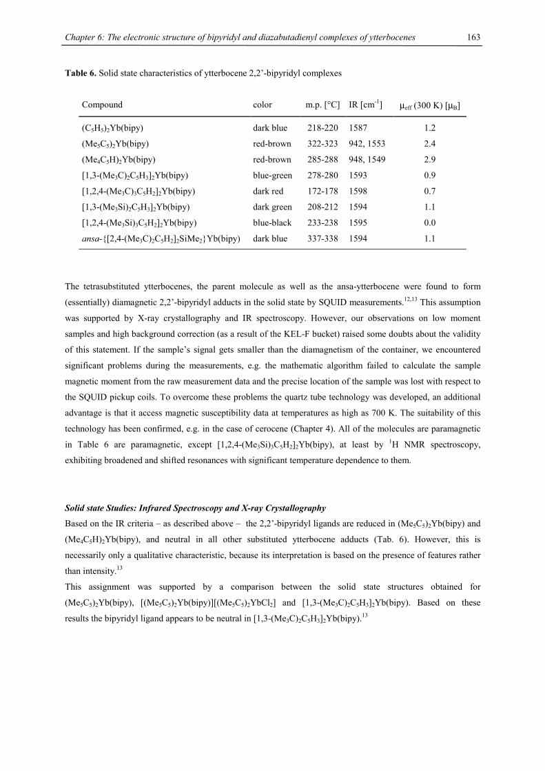
Chapter 6: The electronic structure of bipyridyl and diazabutadienyl complexes of ytterbocenes
163
Table 6. Solid state characteristics of ytterbocene 2,2’-bipyridyl complexes
Compound color m.p. [°C] IR [cm-1] µeff (300 K) [µB]
(C5H5)2Yb(bipy) dark blue 218-220 1587 1.2
(Me5C5)2Yb(bipy) red-brown 322-323 942, 1553 2.4
(Me4C5H)2Yb(bipy) red-brown 285-288 948, 1549 2.9
[1,3-(Me3C)2C5H3]2Yb(bipy) blue-green 278-280 1593 0.9
[1,2,4-(Me3C)3C5H2]2Yb(bipy) dark red 172-178 1598 0.7
[1,3-(Me3Si)2C5H3]2Yb(bipy) dark green 208-212 1594 1.1
[1,2,4-(Me3Si)3C5H2]2Yb(bipy) blue-black 233-238 1595 0.0
ansa-{[2,4-(Me3C)2C5H2]2SiMe2}Yb(bipy) dark blue 337-338 1594 1.1
The tetrasubstituted ytterbocenes, the parent molecule as well as the ansa-ytterbocene were found to form
(essentially) diamagnetic 2,2’-bipyridyl adducts in the solid state by SQUID measurements.12,13 This assumption
was supported by X-ray crystallography and IR spectroscopy. However, our observations on low moment
samples and high background correction (as a result of the KEL-F bucket) raised some doubts about the validity
of this statement. If the sample’s signal gets smaller than the diamagnetism of the container, we encountered
significant problems during the measurements, e.g. the mathematic algorithm failed to calculate the sample
magnetic moment from the raw measurement data and the precise location of the sample was lost with respect to
the SQUID pickup coils. To overcome these problems the quartz tube technology was developed, an additional
advantage is that it access magnetic susceptibility data at temperatures as high as 700 K. The suitability of this
technology has been confirmed, e.g. in the case of cerocene (Chapter 4). All of the molecules are paramagnetic
in Table 6 are paramagnetic, except [1,2,4-(Me3Si)3C5H2]2Yb(bipy), at least by 1H NMR spectroscopy,
exhibiting broadened and shifted resonances with significant temperature dependence to them.
Solid state Studies: Infrared Spectroscopy and X-ray Crystallography
Based on the IR criteria – as described above – the 2,2’-bipyridyl ligands are reduced in (Me5C5)2Yb(bipy) and
(Me4C5H)2Yb(bipy), and neutral in all other substituted ytterbocene adducts (Tab. 6). However, this is
necessarily only a qualitative characteristic, because its interpretation is based on the presence of features rather
than intensity.13
This assignment was supported by a comparison between the solid state structures obtained for
(Me5C5)2Yb(bipy), [(Me5C5)2Yb(bipy)][(Me5C5)2YbCl2] and [1,3-(Me3C)2C5H3]2Yb(bipy). Based on these
results the bipyridyl ligand appears to be neutral in [1,3-(Me3C)2C5H3]2Yb(bipy).13

Chapter 6: The electronic structure of bipyridyl and diazabutadienyl complexes of ytterbocenes
164
Solid state Studies: Magnetic susceptibility (SQUID)
We investigated the solid state magnetism of [1,3-(Me3C)2C5H3]2Yb(bipy), [1,3-(Me3Si)2C5H3]2Yb(bipy), [1,2,4-
(Me3C)3C5H2]2Yb(bipy), (C5H5)2Yb(bipy) and ansa-{[2,4-(Me3C)2C5H2]2SiMe2}Yb(bipy) using the quartz tube
technology at various magnetic fields. The magnetic moments obtained for the bipyridyl adducts are low ranging
from 0.6 to 1.3 B.M. at 300 K (Fig. 23a.).
0 50 100 150 200 250 3000.0
0.2
0.4
0.6
0.8
1.0
1.2
µ eff [
B.M
.]
T [K]
[1,3-(Me3Si)
2C
5H
3]2Yb(bipy)
[1,3-(Me3C)2C5H3]2Yb(bipy) [1,2,4-(Me
3C)
3C
5H
2]2Yb(bipy)
(C5H5)2Yb(bipy) (ansa-{[2,4-(Me
3C)
2C
5H
2]2SiMe
2}Yb(bipy)
Figure 23a. Solid state magnetic susceptibility, µeff vs. T plot of Cp’2Yb(bipy).
0 50 100 150 200 250 300
0
1000
2000
3000
4000
5000
6000
1/χ
[mol
/cm
3 ]
T [K]
[1,3-(Me3Si)2C5H3]2Yb(bipy) [1,3-(Me
3C)
2C
5H
3]2Yb(bipy)
[1,2,4-(Me3C)
3C
5H
2]2Yb(bipy)
(C5H5)2Yb(bipy) (ansa-{[2,4-(Me
3C)
2C
5H
2]2SiMe
2}Yb(bipy)
Figure 23b. Solid state magnetic susceptibility, 1/χ vs. T plot of Cp’2Yb(bipy).
The 1/χ vs. T plots are significantly curved (Fig. 23b), but does follow the behavior of (Me5C5)2Yb(bipy) and
(Me4C5H)2Yb(bipy) (Fig. 6). Due to the air sensitivity of these molecules, one might argue that the low observed
moments are due to paramagnetic, i.e. Yb(III), impurities. Hence it was necessary to address this point. To
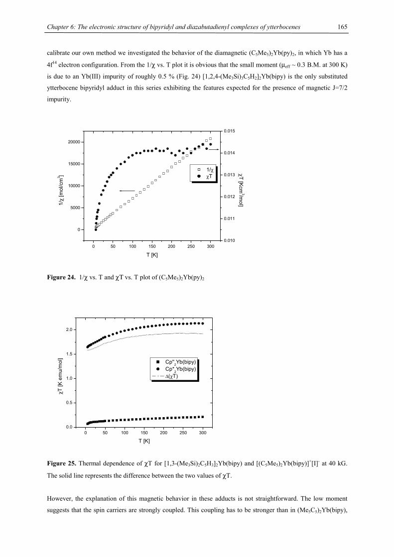
Chapter 6: The electronic structure of bipyridyl and diazabutadienyl complexes of ytterbocenes
165
calibrate our own method we investigated the behavior of the diamagnetic (C5Me5)2Yb(py)2, in which Yb has a
4f14 electron configuration. From the 1/χ vs. T plot it is obvious that the small moment (µeff ~ 0.3 B.M. at 300 K)
is due to an Yb(III) impurity of roughly 0.5 % (Fig. 24) [1,2,4-(Me3Si)3C5H2]2Yb(bipy) is the only substituted
ytterbocene bipyridyl adduct in this series exhibiting the features expected for the presence of magnetic J=7/2
impurity.
0 50 100 150 200 250 300
0
5000
10000
15000
20000
1/χ χT
T [K]
1/χ
[mol
/cm
3 ]
0.010
0.011
0.012
0.013
0.014
0.015
χT [Kcm
3/mol]
Figure 24. 1/χ vs. T and χΤ vs. T plot of (C5Me5)2Yb(py)2
0 50 100 150 200 250 3000.0
0.5
1.0
1.5
2.0
χT [K
em
u/m
ol]
T [K]
Cp''2Yb(bipy) Cp*
2Yb(bipy)
∆(χT)
Figure 25. Thermal dependence of χT for [1,3-(Me3Si)2C5H3]2Yb(bipy) and [(C5Me5)2Yb(bipy)]+[I]- at 40 kG.
The solid line represents the difference between the two values of χT.
However, the explanation of this magnetic behavior in these adducts is not straightforward. The low moment
suggests that the spin carriers are strongly coupled. This coupling has to be stronger than in (Me5C5)2Yb(bipy),
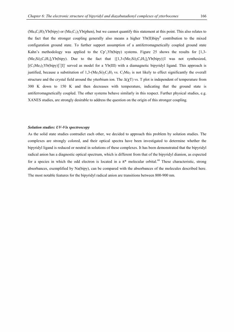
Chapter 6: The electronic structure of bipyridyl and diazabutadienyl complexes of ytterbocenes
166
(Me4C5H)2Yb(bipy) or (Me5C5)2Yb(phen), but we cannot quantify this statement at this point. This also relates to
the fact that the stronger coupling generally also means a higher Yb(II)bipy0 contribution to the mixed
configuration ground state. To further support assumption of a antiferromagnetically coupled ground state
Kahn’s methodology was applied to the Cp’2Yb(bipy) systems. Figure 25 shows the results for [1,3-
(Me3Si)2C5H3]2Yb(bipy). Due to the fact that {[1,3-(Me3Si)2C5H3]2Yb(bipy)}I was not synthesized,
[(C5Me5)2Yb(bipy)]+[I]- served as model for a Yb(III) with a diamagnetic bipyridyl ligand. This approach is
justified, because a substitution of 1,3-(Me3Si)2C5H3 vs. C5Me5 is not likely to effect significantly the overall
structure and the crystal field around the ytterbium ion. The ∆(χT) vs. T plot is independent of temperature from
300 K down to 150 K and then decreases with temperature, indicating that the ground state is
antiferromagnetically coupled. The other systems behave similarly in this respect. Further physical studies, e.g.
XANES studies, are strongly desirable to address the question on the origin of this stronger coupling.
Solution studies: UV-Vis spectroscopy
As the solid state studies contradict each other, we decided to approach this problem by solution studies. The
complexes are strongly colored, and their optical spectra have been investigated to determine whether the
bipyridyl ligand is reduced or neutral in solutions of these complexes. It has been demonstrated that the bipyridyl
radical anion has a diagnostic optical spectrum, which is different from that of the bipyridyl dianion, as expected
for a species in which the odd electron is located in a π* molecular orbital.44 These characteristic, strong
absorbances, exemplified by Na(bipy), can be compared with the absorbances of the molecules described here.
The most notable features for the bipyridyl radical anion are transitions between 800-900 nm.
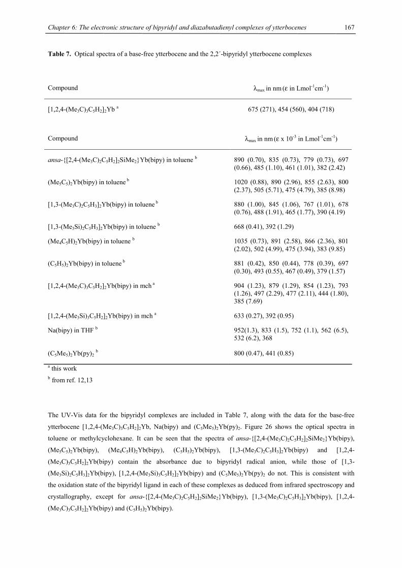
Chapter 6: The electronic structure of bipyridyl and diazabutadienyl complexes of ytterbocenes
167
Table 7. Optical spectra of a base-free ytterbocene and the 2,2´-bipyridyl ytterbocene complexes
Compound λmax in nm (ε in Lmol-1cm-1)
[1,2,4-(Me3C)3C5H2]2Yb a 675 (271), 454 (560), 404 (718)
Compound λmax in nm (ε x 10-3 in Lmol-1cm-1)
ansa-{[2,4-(Me3C)2C5H2]2SiMe2}Yb(bipy) in toluene b 890 (0.70), 835 (0.73), 779 (0.73), 697 (0.66), 485 (1.10), 461 (1.01), 382 (2.42)
(Me5C5)2Yb(bipy) in toluene b 1020 (0.88), 890 (2.96), 855 (2.63), 800 (2.37), 505 (5.71), 475 (4.79), 385 (8.98)
[1,3-(Me3C)2C5H3]2Yb(bipy) in toluene b 880 (1.00), 845 (1.06), 767 (1.01), 678 (0.76), 488 (1.91), 465 (1.77), 390 (4.19)
[1,3-(Me3Si)2C5H3]2Yb(bipy) in toluene b 668 (0.41), 392 (1.29)
(Me4C5H)2Yb(bipy) in toluene b 1035 (0.73), 891 (2.58), 866 (2.36), 801 (2.02), 502 (4.99), 475 (3.94), 383 (9.85)
(C5H5)2Yb(bipy) in toluene b 881 (0.42), 850 (0.44), 778 (0.39), 697 (0.30), 493 (0.55), 467 (0.49), 379 (1.57)
[1,2,4-(Me3C)3C5H2]2Yb(bipy) in mch a 904 (1.23), 879 (1.29), 854 (1.23), 793 (1.26), 497 (2.29), 477 (2.11), 444 (1.80), 385 (7.69)
[1,2,4-(Me3Si)3C5H2]2Yb(bipy) in mch a 633 (0.27), 392 (0.95)
Na(bipy) in THF b 952(1.3), 833 (1.5), 752 (1.1), 562 (6.5), 532 (6.2), 368
(C5Me5)2Yb(py)2 b 800 (0.47), 441 (0.85)
a this work b from ref. 12,13
The UV-Vis data for the bipyridyl complexes are included in Table 7, along with the data for the base-free
ytterbocene [1,2,4-(Me3C)3C5H2]2Yb, Na(bipy) and (C5Me5)2Yb(py)2. Figure 26 shows the optical spectra in
toluene or methylcyclohexane. It can be seen that the spectra of ansa-{[2,4-(Me3C)2C5H2]2SiMe2}Yb(bipy),
(Me5C5)2Yb(bipy), (Me4C5H)2Yb(bipy), (C5H5)2Yb(bipy), [1,3-(Me3C)2C5H3]2Yb(bipy) and [1,2,4-
(Me3C)3C5H2]2Yb(bipy) contain the absorbance due to bipyridyl radical anion, while those of [1,3-
(Me3Si)2C5H3]2Yb(bipy), [1,2,4-(Me3Si)3C5H2]2Yb(bipy) and (C5Me5)2Yb(py)2 do not. This is consistent with
the oxidation state of the bipyridyl ligand in each of these complexes as deduced from infrared spectroscopy and
crystallography, except for ansa-{[2,4-(Me3C)2C5H2]2SiMe2}Yb(bipy), [1,3-(Me3C)2C5H3]2Yb(bipy), [1,2,4-
(Me3C)3C5H2]2Yb(bipy) and (C5H5)2Yb(bipy).
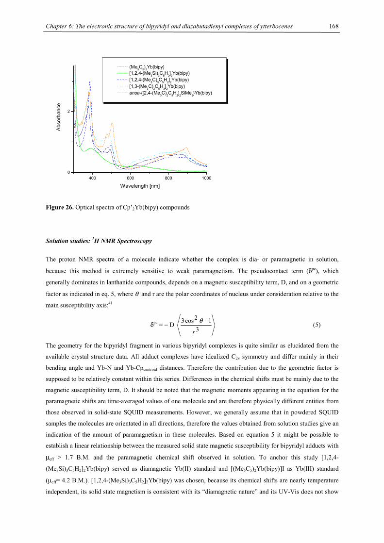
Chapter 6: The electronic structure of bipyridyl and diazabutadienyl complexes of ytterbocenes
168
400 600 800 10000
2
Abs
orba
nce
Wavelength [nm]
(Me5C5)2Yb(bipy) [1,2,4-(Me3Si)3C5H2]2Yb(bipy) [1,2,4-(Me3C)3C5H2]2Yb(bipy) [1,3-(Me3C)2C5H3]2Yb(bipy) ansa-{[2,4-(Me3C)2C5H2]2SiMe2}Yb(bipy)
Figure 26. Optical spectra of Cp’2Yb(bipy) compounds
Solution studies: 1H NMR Spectroscopy
The proton NMR spectra of a molecule indicate whether the complex is dia- or paramagnetic in solution,
because this method is extremely sensitive to weak paramagnetism. The pseudocontact term (δpc), which
generally dominates in lanthanide compounds, depends on a magnetic susceptibility term, D, and on a geometric
factor as indicated in eq. 5, where θ and r are the polar coordinates of nucleus under consideration relative to the
main susceptibility axis:41
δpc = − D 3
12cos3
r
−θ (5)
The geometry for the bipyridyl fragment in various bipyridyl complexes is quite similar as elucidated from the
available crystal structure data. All adduct complexes have idealized C2v symmetry and differ mainly in their
bending angle and Yb-N and Yb-Cpcentroid distances. Therefore the contribution due to the geometric factor is
supposed to be relatively constant within this series. Differences in the chemical shifts must be mainly due to the
magnetic susceptibility term, D. It should be noted that the magnetic moments appearing in the equation for the
paramagnetic shifts are time-averaged values of one molecule and are therefore physically different entities from
those observed in solid-state SQUID measurements. However, we generally assume that in powdered SQUID
samples the molecules are orientated in all directions, therefore the values obtained from solution studies give an
indication of the amount of paramagnetism in these molecules. Based on equation 5 it might be possible to
establish a linear relationship between the measured solid state magnetic susceptibility for bipyridyl adducts with
µeff > 1.7 B.M. and the paramagnetic chemical shift observed in solution. To anchor this study [1,2,4-
(Me3Si)3C5H2]2Yb(bipy) served as diamagnetic Yb(II) standard and [(Me5C5)2Yb(bipy)]I as Yb(III) standard
(µeff= 4.2 B.M.). [1,2,4-(Me3Si)3C5H2]2Yb(bipy) was chosen, because its chemical shifts are nearly temperature
independent, its solid state magnetism is consistent with its “diamagnetic nature” and its UV-Vis does not show
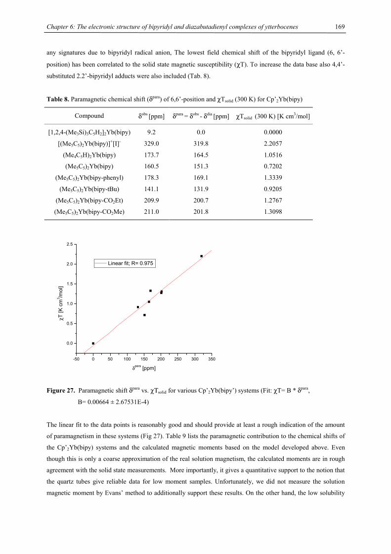
Chapter 6: The electronic structure of bipyridyl and diazabutadienyl complexes of ytterbocenes
169
any signatures due to bipyridyl radical anion, The lowest field chemical shift of the bipyridyl ligand (6, 6’-
position) has been correlated to the solid state magnetic susceptibility (χT). To increase the data base also 4,4’-
substituted 2.2’-bipyridyl adducts were also included (Tab. 8).
Table 8. Paramagnetic chemical shift (δpara) of 6,6’-position and χTsolid (300 K) for Cp’2Yb(bipy)
Compound δobs [ppm] δpara = δobs - δdia [ppm] χTsolid (300 K) [K cm3/mol]
[1,2,4-(Me3Si)3C5H2]2Yb(bipy) 9.2 0.0 0.0000
[(Me5C5)2Yb(bipy)]+[I]- 329.0 319.8 2.2057
(Me4C5H)2Yb(bipy) 173.7 164.5 1.0516
(Me5C5)2Yb(bipy) 160.5 151.3 0.7202
(Me5C5)2Yb(bipy-phenyl) 178.3 169.1 1.3339
(Me5C5)2Yb(bipy-tBu) 141.1 131.9 0.9205
(Me5C5)2Yb(bipy-CO2Et) 209.9 200.7 1.2767
(Me5C5)2Yb(bipy-CO2Me) 211.0 201.8 1.3098
-50 0 50 100 150 200 250 300 350
0.0
0.5
1.0
1.5
2.0
2.5
χT [K
cm
3 /mol
]
δpara [ppm]
Linear fit; R= 0.975
Figure 27. Paramagnetic shift δpara vs. χΤsolid for various Cp’2Yb(bipy’) systems (Fit: χT= B * δpara,
B= 0.00664 ± 2.67531E-4)
The linear fit to the data points is reasonably good and should provide at least a rough indication of the amount
of paramagnetism in these systems (Fig 27). Table 9 lists the paramagnetic contribution to the chemical shifts of
the Cp’2Yb(bipy) systems and the calculated magnetic moments based on the model developed above. Even
though this is only a coarse approximation of the real solution magnetism, the calculated moments are in rough
agreement with the solid state measurements. More importantly, it gives a quantitative support to the notion that
the quartz tubes give reliable data for low moment samples. Unfortunately, we did not measure the solution
magnetic moment by Evans’ method to additionally support these results. On the other hand, the low solubility
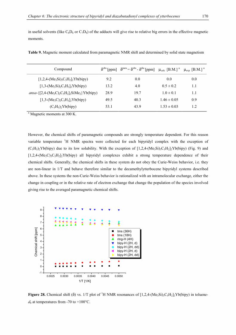
Chapter 6: The electronic structure of bipyridyl and diazabutadienyl complexes of ytterbocenes
170
in useful solvents (like C6D6 or C7D8) of the adducts will give rise to relative big errors in the effective magnetic
moments.
Table 9. Magnetic moment calculated from paramagnetic NMR shift and determined by solid state magnetism
Compound δobs [ppm] δpara = δobs - δdia [ppm] µcalc [B.M.] a µexp [B.M.] a
[1,2,4-(Me3Si)3C5H2]2Yb(bipy) 9.2 0.0 0.0 0.0
[1,3-(Me3Si)2C5H3]2Yb(bipy) 13.2 4.0 0.5 ± 0.2 1.1
ansa-{[2,4-(Me3C)2C5H2]2SiMe2}Yb(bipy) 28.9 19.7 1.0 ± 0.1 1.1
[1,3-(Me3C)2C5H3]2Yb(bipy) 49.5 40.3 1.46 ± 0.05 0.9
(C5H5)2Yb(bipy) 53.1 43.9 1.53 ± 0.03 1.2 a Magnetic moments at 300 K.
However, the chemical shifts of paramagnetic compounds are strongly temperature dependent. For this reason
variable temperature 1H NMR spectra were collected for each bipyridyl complex with the exception of
(C5H5)2Yb(bipy) due to its low solubility. With the exception of [1,2,4-(Me3Si)3C5H2]2Yb(bipy) (Fig. 9) and
[1,2,4-(Me3C)3C5H2]2Yb(bipy) all bipyridyl complexes exhibit a strong temperature dependence of their
chemical shifts. Generally, the chemical shifts in these system do not obey the Curie-Weiss behavior, i.e. they
are non-linear in 1/T and behave therefore similar to the decamethylytterbocene bipyridyl systems described
above. In these systems the non-Curie-Weiss behavior is rationalized with an intramolecular exchange, either the
change in coupling or in the relative rate of electron exchange that change the population of the species involved
giving rise to the averaged paramagnetic chemical shifts.
0.0025 0.0030 0.0035 0.0040 0.0045 0.0050-1
0
1
2
3
4
5
6
7
8
9
Che
mic
al s
hift
[ppm
]
1/T [1/K]
tms (36H) tms (18H) ring-H (4H) bipy-H (2H, d) bipy-H (2H, dd) bipy-H (2H, d) bipy-H (2H, dd)
Figure 28. Chemical shift (δ) vs. 1/T plot of 1H NMR resonances of [1,2,4-(Me3Si)3C5H2]2Yb(bipy) in toluene-
d8 at temperatures from -70 to +100°C.
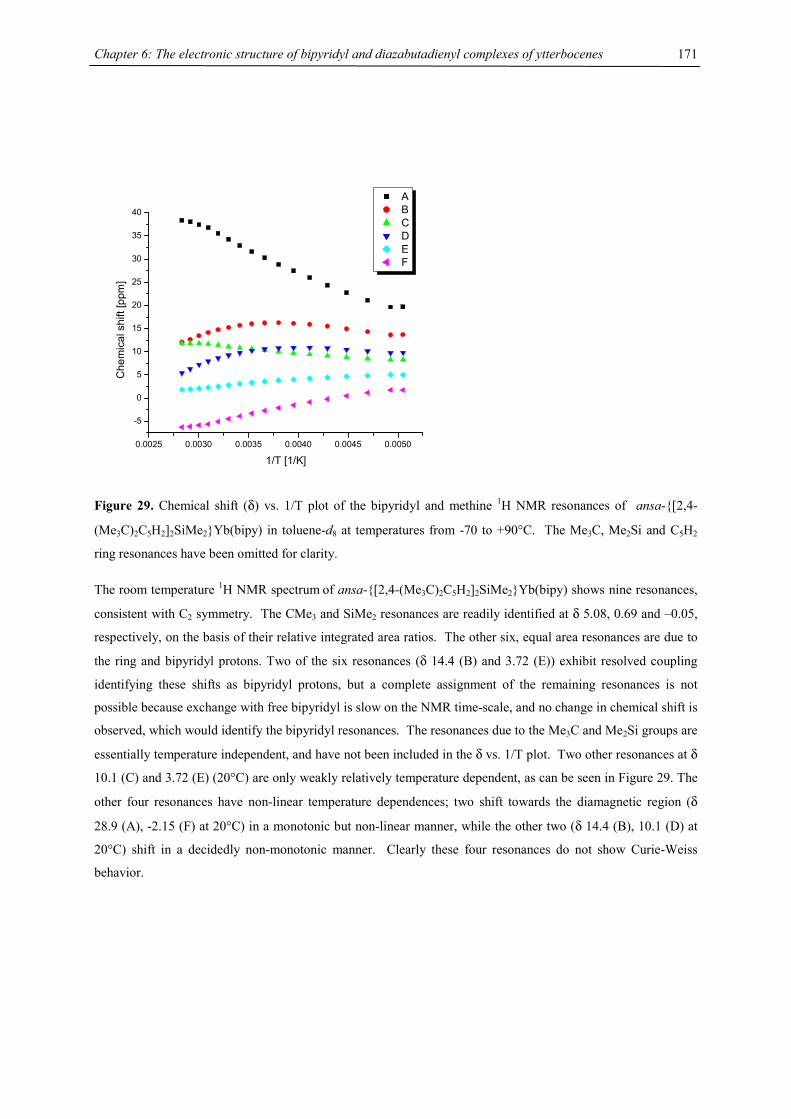
Chapter 6: The electronic structure of bipyridyl and diazabutadienyl complexes of ytterbocenes
171
0.0025 0.0030 0.0035 0.0040 0.0045 0.0050
-5
0
5
10
15
20
25
30
35
40
Che
mic
al s
hift
[ppm
]
1/T [1/K]
A B C D E F
Figure 29. Chemical shift (δ) vs. 1/T plot of the bipyridyl and methine 1H NMR resonances of ansa-{[2,4-
(Me3C)2C5H2]2SiMe2}Yb(bipy) in toluene-d8 at temperatures from -70 to +90°C. The Me3C, Me2Si and C5H2
ring resonances have been omitted for clarity.
The room temperature 1H NMR spectrum of ansa-{[2,4-(Me3C)2C5H2]2SiMe2}Yb(bipy) shows nine resonances,
consistent with C2 symmetry. The CMe3 and SiMe2 resonances are readily identified at δ 5.08, 0.69 and –0.05,
respectively, on the basis of their relative integrated area ratios. The other six, equal area resonances are due to
the ring and bipyridyl protons. Two of the six resonances (δ 14.4 (B) and 3.72 (E)) exhibit resolved coupling
identifying these shifts as bipyridyl protons, but a complete assignment of the remaining resonances is not
possible because exchange with free bipyridyl is slow on the NMR time-scale, and no change in chemical shift is
observed, which would identify the bipyridyl resonances. The resonances due to the Me3C and Me2Si groups are
essentially temperature independent, and have not been included in the δ vs. 1/T plot. Two other resonances at δ
10.1 (C) and 3.72 (E) (20°C) are only weakly relatively temperature dependent, as can be seen in Figure 29. The
other four resonances have non-linear temperature dependences; two shift towards the diamagnetic region (δ
28.9 (A), -2.15 (F) at 20°C) in a monotonic but non-linear manner, while the other two (δ 14.4 (B), 10.1 (D) at
20°C) shift in a decidedly non-monotonic manner. Clearly these four resonances do not show Curie-Weiss
behavior.
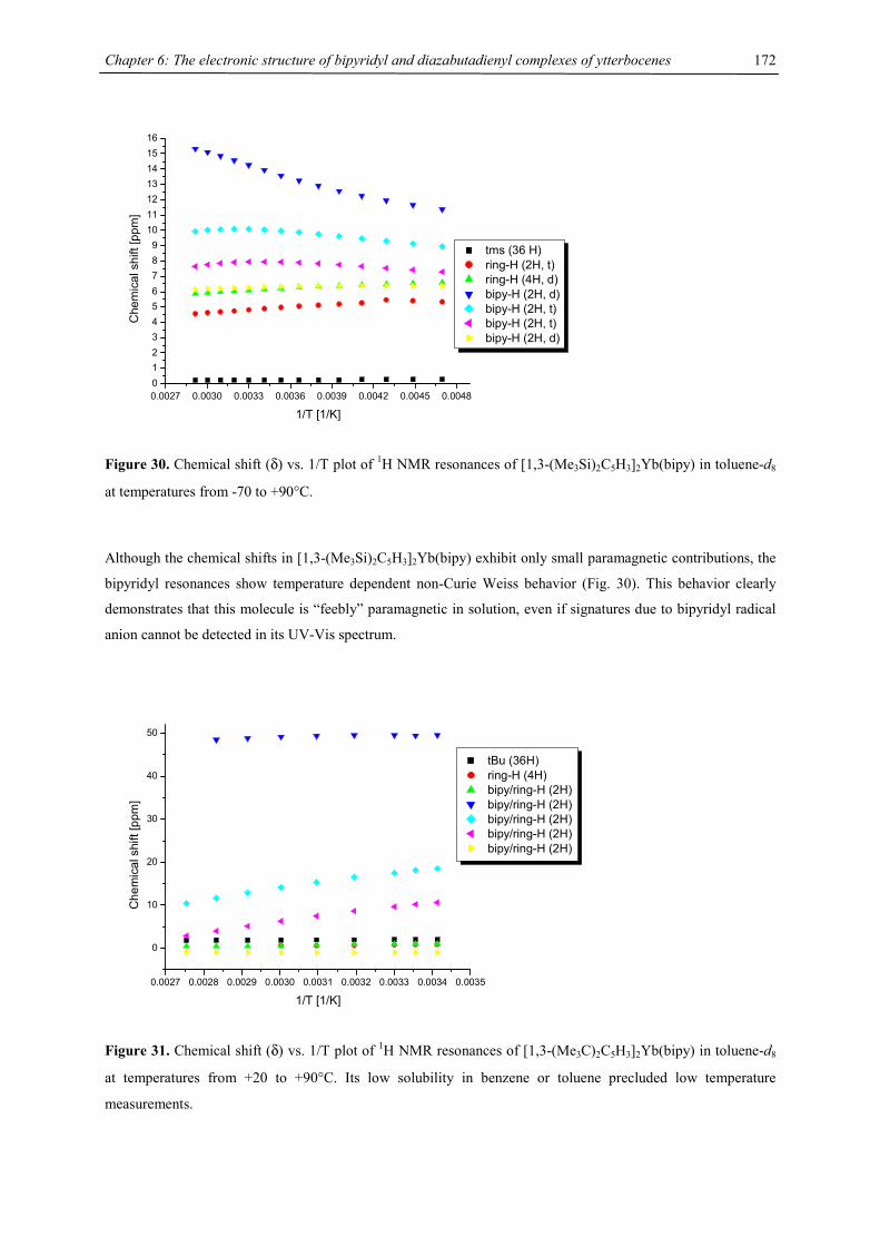
Chapter 6: The electronic structure of bipyridyl and diazabutadienyl complexes of ytterbocenes
172
0.0027 0.0030 0.0033 0.0036 0.0039 0.0042 0.0045 0.00480123456789
10111213141516
Che
mic
al s
hift
[ppm
]
1/T [1/K]
tms (36 H) ring-H (2H, t) ring-H (4H, d) bipy-H (2H, d) bipy-H (2H, t) bipy-H (2H, t) bipy-H (2H, d)
Figure 30. Chemical shift (δ) vs. 1/T plot of 1H NMR resonances of [1,3-(Me3Si)2C5H3]2Yb(bipy) in toluene-d8
at temperatures from -70 to +90°C.
Although the chemical shifts in [1,3-(Me3Si)2C5H3]2Yb(bipy) exhibit only small paramagnetic contributions, the
bipyridyl resonances show temperature dependent non-Curie Weiss behavior (Fig. 30). This behavior clearly
demonstrates that this molecule is “feebly” paramagnetic in solution, even if signatures due to bipyridyl radical
anion cannot be detected in its UV-Vis spectrum.
0.0027 0.0028 0.0029 0.0030 0.0031 0.0032 0.0033 0.0034 0.0035
0
10
20
30
40
50
Che
mic
al s
hift
[ppm
]
1/T [1/K]
tBu (36H) ring-H (4H) bipy/ring-H (2H) bipy/ring-H (2H) bipy/ring-H (2H) bipy/ring-H (2H) bipy/ring-H (2H)
Figure 31. Chemical shift (δ) vs. 1/T plot of 1H NMR resonances of [1,3-(Me3C)2C5H3]2Yb(bipy) in toluene-d8
at temperatures from +20 to +90°C. Its low solubility in benzene or toluene precluded low temperature
measurements.
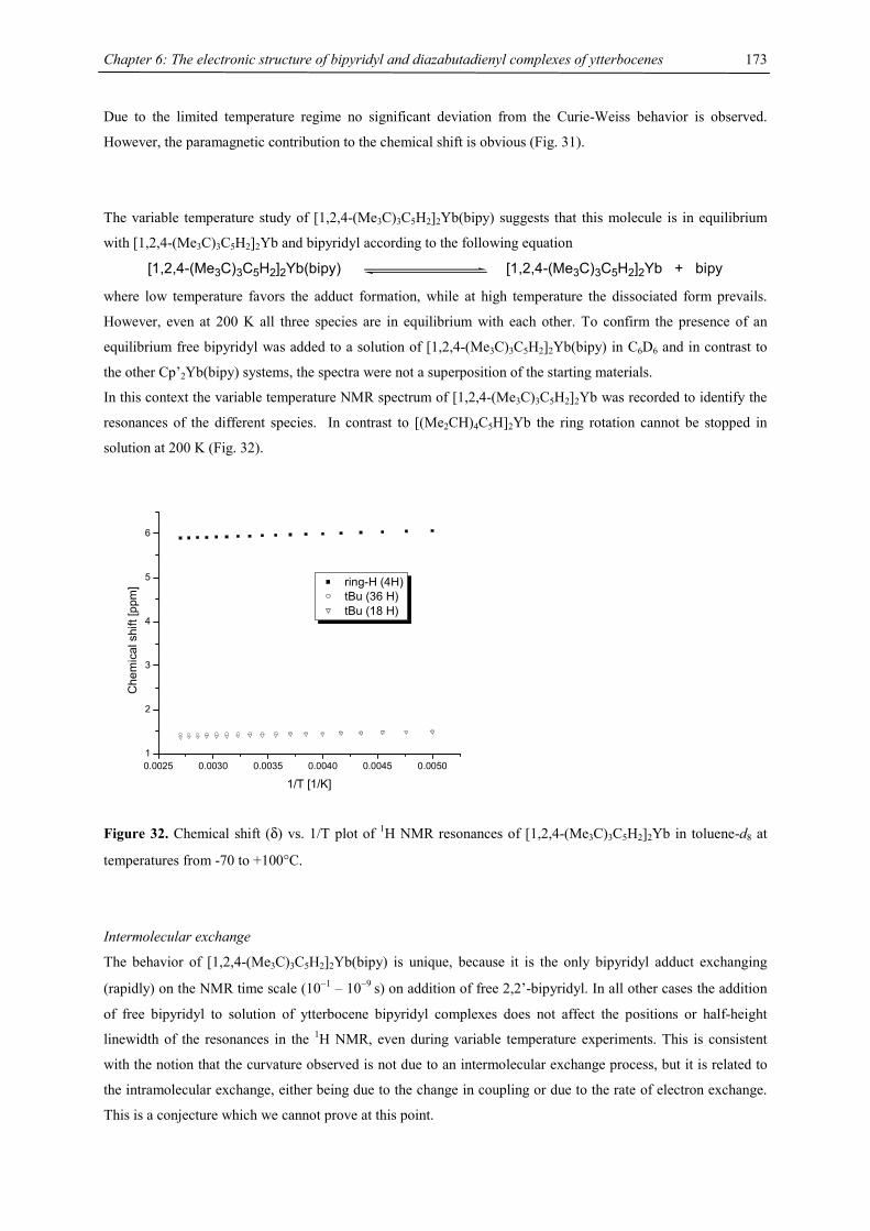
Chapter 6: The electronic structure of bipyridyl and diazabutadienyl complexes of ytterbocenes
173
Due to the limited temperature regime no significant deviation from the Curie-Weiss behavior is observed.
However, the paramagnetic contribution to the chemical shift is obvious (Fig. 31).
The variable temperature study of [1,2,4-(Me3C)3C5H2]2Yb(bipy) suggests that this molecule is in equilibrium
with [1,2,4-(Me3C)3C5H2]2Yb and bipyridyl according to the following equation
[1,2,4-(Me3C)3C5H2]2Yb(bipy) [1,2,4-(Me3C)3C5H2]2Yb + bipy where low temperature favors the adduct formation, while at high temperature the dissociated form prevails.
However, even at 200 K all three species are in equilibrium with each other. To confirm the presence of an
equilibrium free bipyridyl was added to a solution of [1,2,4-(Me3C)3C5H2]2Yb(bipy) in C6D6 and in contrast to
the other Cp’2Yb(bipy) systems, the spectra were not a superposition of the starting materials.
In this context the variable temperature NMR spectrum of [1,2,4-(Me3C)3C5H2]2Yb was recorded to identify the
resonances of the different species. In contrast to [(Me2CH)4C5H]2Yb the ring rotation cannot be stopped in
solution at 200 K (Fig. 32).
0.0025 0.0030 0.0035 0.0040 0.0045 0.00501
2
3
4
5
6
Che
mic
al s
hift
[ppm
]
1/T [1/K]
ring-H (4H) tBu (36 H) tBu (18 H)
Figure 32. Chemical shift (δ) vs. 1/T plot of 1H NMR resonances of [1,2,4-(Me3C)3C5H2]2Yb in toluene-d8 at
temperatures from -70 to +100°C.
Intermolecular exchange
The behavior of [1,2,4-(Me3C)3C5H2]2Yb(bipy) is unique, because it is the only bipyridyl adduct exchanging
(rapidly) on the NMR time scale (10−1 – 10−9 s) on addition of free 2,2’-bipyridyl. In all other cases the addition
of free bipyridyl to solution of ytterbocene bipyridyl complexes does not affect the positions or half-height
linewidth of the resonances in the 1H NMR, even during variable temperature experiments. This is consistent
with the notion that the curvature observed is not due to an intermolecular exchange process, but it is related to
the intramolecular exchange, either being due to the change in coupling or due to the rate of electron exchange.
This is a conjecture which we cannot prove at this point.
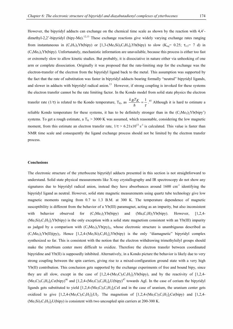
Chapter 6: The electronic structure of bipyridyl and diazabutadienyl complexes of ytterbocenes
174
However, the bipyridyl adducts can exchange on the chemical time scale as shown by the reaction with 4,4’-
dimethyl-2,2’-bipyridyl (bipy-Me).12,13 These exchange reactions give widely varying exchange rates ranging
from instantaneous in (C5H5)2Yb(bipy) or [1,3-(Me3Si)2C5H3]2Yb(bipy) to slow (Keq= 0.25; τ1/2= 7 d) in
(C5Me5)2Yb(bipy). Unfortunately, mechanistic information are unavailable, because this process is either too fast
or extremely slow to allow kinetic studies. But probably, it is dissociative in nature either via unhooking of one
arm or complete dissociation. Originally it was proposed that the rate-limiting step for the exchange was the
electron-transfer of the electron from the bipyridyl ligand back to the metal. This assumption was supported by
the fact that the rate of substitution was faster in bipyridyl adducts bearing formally “neutral” bipyridyl ligands,
and slower in adducts with bipyridyl radical anion.13 However, if strong coupling is invoked for these systems
the electron transfer cannot be the rate limiting factor. In the Kondo model from solid state physics the electron
transfer rate (1/τ) is related to the Kondo temperature, TK, as τ1=
�
KTBk.45 Although it is hard to estimate a
reliable Kondo temperature for these systems, it has to be definitely stronger than in the (C5Me5)2Yb(bipy’)
systems. To get a rough estimate, a TK > 3000 K was assumed, which reasonable, considering the low magnetic
moment, from this estimate an electron transfer rate, 1/τ > 6.21x10¹³ s-1 is calculated. This value is faster than
NMR time scale and consequently the ligand exchange process should not be limited by the electron transfer
process.
Conclusions
The electronic structure of the ytterbocene bipyridyl adducts presented in this section is not straightforward to
understand. Solid state physical measurements like X-ray crystallography and IR spectroscopy do not show any
signatures due to bipyridyl radical anion, instead they have absorbances around 1600 cm-1 identifying the
bipyridyl ligand as neutral. However, solid state magnetic measurements using quartz tube technology give low
magnetic moments ranging from 0.7 to 1.3 B.M. at 300 K. The temperature dependence of magnetic
susceptibility is different from the behavior of a Yb(III) paramagnet, acting as an impurity, but also inconsistent
with behavior observed for (C5Me5)2Yb(bipy) and (Me4C5H)2Yb(bipy). However, [1,2,4-
(Me3Si)3C5H2]2Yb(bipy) is the only exception with a solid state magnetism consistent with an Yb(III) impurity
as judged by a comparison with (C5Me5)2Yb(py)2, whose electronic structure is unambiguous described as
(C5Me5)2Yb(II)(py)2. Hence [1,2,4-(Me3Si)3C5H2]2Yb(bipy) is the only “diamagnetic” bipyridyl complex
synthesized so far. This is consistent with the notion that the electron withdrawing trimethylsilyl groups should
make the ytterbium center more difficult to oxidize. Therefore the electron transfer between coordinated
bipyridine and Yb(II) is supposedly inhibited. Alternatively, in a Kondo picture the behavior is likely due to very
strong coupling between the spin carriers, giving rise to a mixed-configuration ground state with a very high
Yb(II) contribution. This conclusion gets supported by the exchange experiments of free and bound bipy, since
they are all slow, except in the case of [1,2,4-(Me3C)3C5H2]2Yb(bipy), and by the reactivity of [1,2,4-
(Me3C)3C5H2]2Ce(bipy)46 and [1,2,4-(Me3C)3C5H2]2U(bipy)47 towards AgI. In the case of cerium the bipyridyl
ligands gets substituted to yield [1,2,4-(Me3C)3C5H2]2CeI and in the case of uranium, the uranium center gets
oxidized to give [1,2,4-(Me3C)3C5H2]2UI2. The magnetism of [1,2,4-(Me3C)3C5H2]2Ce(bipy) and [1,2,4-
(Me3Si)3C5H2]2U(bipy) is consistent with two uncoupled spin carriers at 200-300 K.
Chapter 6: The electronic structure of bipyridyl and diazabutadienyl complexes of ytterbocenes
175
Solution studies on this series are also inconsistent in some cases: The UV-Vis spectra of the adducts, with the
exception of [1,3-(Me3Si)2C5H3]2Yb(bipy) and [1,2,4-(Me3Si)3C5H2]2Yb(bipy), show absorptions due to the
radical anion in the neighborhood of 800 nm. Whereas, the 1H NMR spectra have paramagnetically shifted
resonances for all adduct complexes with the exception of [1,2,4-(Me3Si)3C5H2]2Yb(bipy), but [1,3-
(Me3Si)2C5H3]2Yb(bipy) has the smallest paramagnetic shift as referenced to [1,2,4-(Me3Si)3C5H2]2Yb(bipy).
The chemical shifts are temperature dependent and do not obey the Curie-Weiss law. This behavior might be due
to the intramolecular exchange, either to the change in coupling or to the rate of electron exchange that change
the population of the species giving rise to the averaged paramagnetic chemical shifts.
Based on these results, it is difficult to propose a physical model to account for these contradictions, without
further physical measurements, like XANES spectra in hands. But it seems likely that the substituted ytterbocene
bipyridyl complexes are strongly coupled systems within the Kondo model and their ground state is mostly
determined by Yb(II)(bipy0) configuration. The amount of coupling is presumably related to the substitution
pattern on the cyclopentadienyl ring. This is indicated by a reduced paramagnetism on the introduction of
trimethylsilyl groups (judged by 1H NMR spectroscopy). The trend in the tert.-butyl case is less obvious.
However, probably due to steric constraints [1,2,4-(Me3C)3C5H2]2Yb(bipy) is the only example of a bipyridyl
adduct exchanging rapidly with free bipyridyl on the NMR time scale.
In summary, more physical studies on these systems are strongly desirable to explain the origin of this unusual
magnetic susceptibility behavior and to confirm the mixed configurations ground state model.
Chapter 6: The electronic structure of bipyridyl and diazabutadienyl complexes of ytterbocenes
176
Diazabutadienyl Adducts of Ytterbocenes
Introduction
Closely related to 2,2’-bipyridyl are the 1,4-diazabutadiene systems. These ligands have found extensive and
varied application in transition metal coordination chemistry. One of their principal modes of bonding is σ-
coordination to a metal via the lone pairs on the two nitrogen atoms to form a five membered, highly stabilized,
planar ring system, in which the existence of an energetically low-lying ligand π* orbital stabilizes low oxidation
state compounds via π-acceptor bonding.48
In recent years diazabutadiene ligands have been frequently employed with lanthanides in which they are
preferably bonded as radical anions. Among the first examples were the homoleptic lanthanide complexes
(Me3CNCHCHNCMe3)3Ln (Ln= Y, Sm, Yb), prepared by co-condensation of the ligand with the corresponding
metal vapor.49,50 Previously, the solid state magnetism and crystal structure of Yb(tBudad)3 have been
determined to elucidate its electronic structure and to compare these results with Yb(bipy)4. The solid state
magnetism was qualitatively interpretated by an equilibrium between the bivalent [YbII(dad(H)-tBu•−)2(dad(H)-
tBu)] and the trivalent [YbIII(dad(H)-tBu•−)3] species. At low temperature the divalent species predominates and
as the temperature increases the trivalent one predominates in solid state.5 A heteroleptic example is
(C5Me5)2Sm(thf)2, which readily adds diazabutadienes to yield dark brown samarium(III) complexes,
(C5Me5)2Sm(dad).16 Similar complexes have also been synthesized for lanthanum and yttrium.51 More recently,
Scholz et al. have demonstrated that [(dad)Li] units can act as Cp-like ligands in organolanthanide complexes.51
Recently, Trifonov investigated the reactivity of various ytterbocenes, (C5H5)2Yb,52 (C5Me5)2Yb53 and Flu2Yb
(Flu= Fluorenyl, C13H9)54 with diazabutadiene ligands. Whereas tBuNCHCHNtBu forms a simple 1:1 adduct
(solid state magnetism, see Appendix Fig. A6), the reactivity of Flu2Yb(thf)2 towards [(2,6-iPr2C6H3-N=CR-
CR=N-(2,6-iPr2C6H3)] (R= H, Me)55 is unexpected and results either in the coupling of fluorenyl and dad
fragments or in proton abstraction from the dad molecule, depending on the substituents R on the carbon atom of
the diazabutadiene group.
Ligand synthesis
1,4-Diaza-1,3-butadienes, RN=CR’-CR’=NR, abbreviated dad(R’)-R, are easily available by a Knövennagel
condensation involving either glyoxal,56-58 α-ketoaldehydes,59,60 or α,β-diketones60,61 with primary amines,
RNH2 (eq. 9). In this respect dad ligands have a few advantages compared to 2,2’-bipyridyl systems, because the
starting materials are inexpensive, offer a great flexibility in tuning electronic properties (e.g. redox potentials)
and steric demand, and an enhanced solubility. However, there are also some limitations in the synthesis,
because some of the dad(R’)-R ligands are not very stable as free molecules and therefore must be synthesized in
the coordination sphere of a metal, or cannot be isolated at all. This is especially true for dad-ligands bearing
electron withdrawing groups on the primary amine component, R.
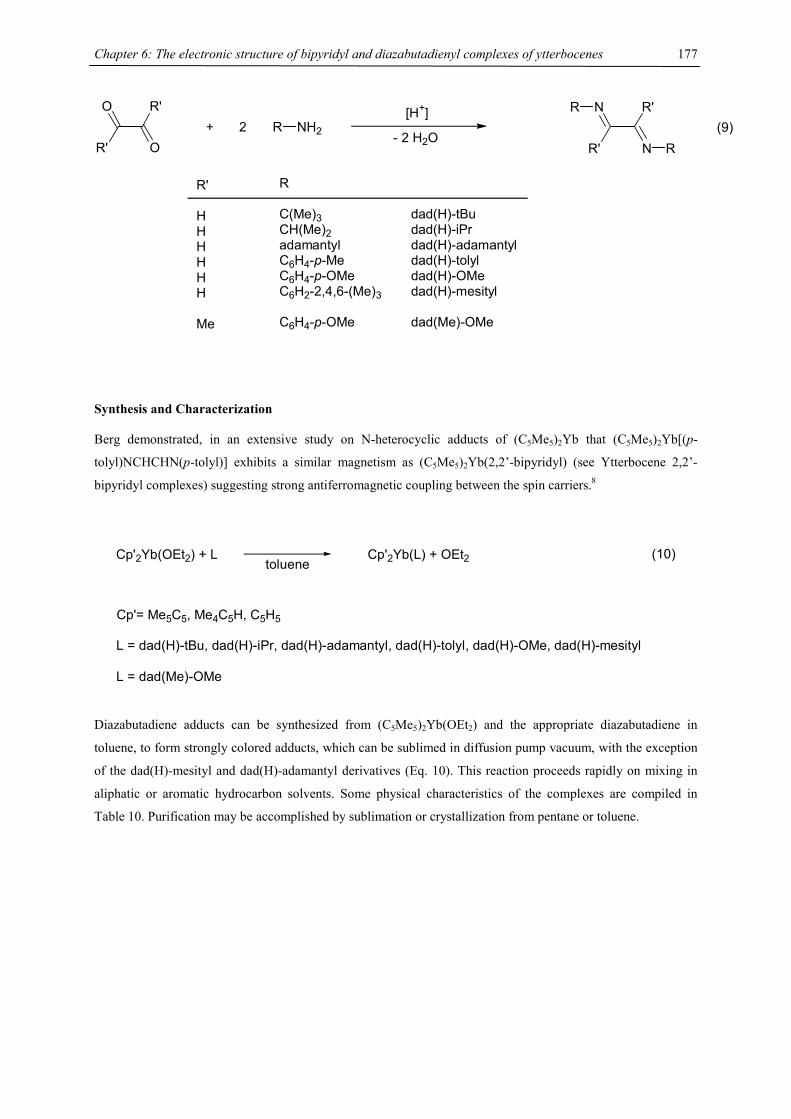
Chapter 6: The electronic structure of bipyridyl and diazabutadienyl complexes of ytterbocenes
177
R'
R'
O
O+ 2 R NH2
[H+]
- 2 H2OR'
R'
N
N
R
R
R'
HHHHHH
Me
R
C(Me)3CH(Me)2adamantylC6H4-p-MeC6H4-p-OMeC6H2-2,4,6-(Me)3
C6H4-p-OMe
dad(H)-tBudad(H)-iPrdad(H)-adamantyldad(H)-tolyldad(H)-OMedad(H)-mesityl
dad(Me)-OMe
(9)
Synthesis and Characterization
Berg demonstrated, in an extensive study on N-heterocyclic adducts of (C5Me5)2Yb that (C5Me5)2Yb[(p-
tolyl)NCHCHN(p-tolyl)] exhibits a similar magnetism as (C5Me5)2Yb(2,2’-bipyridyl) (see Ytterbocene 2,2’-
bipyridyl complexes) suggesting strong antiferromagnetic coupling between the spin carriers.8
Cp'2Yb(OEt2) + L Cp'2Yb(L) + OEt2
Cp'= Me5C5, Me4C5H, C5H5
L = dad(H)-tBu, dad(H)-iPr, dad(H)-adamantyl, dad(H)-tolyl, dad(H)-OMe, dad(H)-mesityl
L = dad(Me)-OMe
toluene(10)
Diazabutadiene adducts can be synthesized from (C5Me5)2Yb(OEt2) and the appropriate diazabutadiene in
toluene, to form strongly colored adducts, which can be sublimed in diffusion pump vacuum, with the exception
of the dad(H)-mesityl and dad(H)-adamantyl derivatives (Eq. 10). This reaction proceeds rapidly on mixing in
aliphatic or aromatic hydrocarbon solvents. Some physical characteristics of the complexes are compiled in
Table 10. Purification may be accomplished by sublimation or crystallization from pentane or toluene.
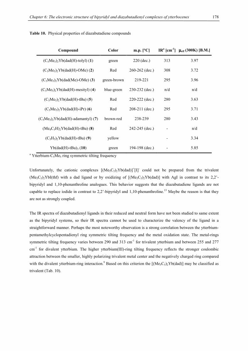
Chapter 6: The electronic structure of bipyridyl and diazabutadienyl complexes of ytterbocenes
178
Table 10. Physical properties of diazabutadiene compounds
Compound Color m.p. [°C] IRa [cm-1] µµµµeff (300K) [B.M.]
(C5Me5)2Yb(dad(H)-tolyl) (1) green 220 (dec.) 313 3.97
(C5Me5)2Yb(dad(H)-OMe) (2) Red 260-262 (dec.) 308 3.72
(C5Me5)2Yb(dad(Me)-OMe) (3) green-brown 219-221 295 3.96
(C5Me5)2Yb(dad(H)-mesityl) (4) blue-green 230-232 (dec.) n/d n/d
(C5Me5)2Yb(dad(H)-tBu) (5) Red 220-222 (dec.) 280 3.63
(C5Me5)2Yb(dad(H)-iPr) (6) Red 208-211 (dec.) 295 3.71
(C5Me5)2Yb(dad(H)-adamantyl) (7) brown-red 238-239 280 3.43
(Me4C5H)2Yb(dad(H)-tBu) (8) Red 242-245 (dec.) - n/d
(C5H5)2Yb(dad(H)-tBu) (9) yellow - 3.34
Yb(dad(H)-tBu)3 (10) green 194-198 (dec.) - 5.85 a Ytterbium-C5Me5 ring symmetric tilting frequency
Unfortunately, the cationic complexes [(Me5C5)2Yb(dad)]+[I]- could not be prepared from the trivalent
(Me5C5)2YbI(thf) with a dad ligand or by oxidizing of [(Me5C5)2Yb(dad)] with AgI in contrast to its 2,2’-
bipyridyl and 1,10-phenanthroline analogues. This behavior suggests that the diazabutadiene ligands are not
capable to replace iodide in contrast to 2,2’-bipyridyl and 1,10-phenanthroline.13 Maybe the reason is that they
are not as strongly coupled.
The IR spectra of diazabutadienyl ligands in their reduced and neutral form have not been studied to same extent
as the bipyridyl systems, so their IR spectra cannot be used to characterize the valency of the ligand in a
straightforward manner. Perhaps the most noteworthy observation is a strong correlation between the ytterbium-
pentamethylcyclopentadienyl ring symmetric tilting frequency and the metal oxidation state. The metal-rings
symmetric tilting frequency varies between 290 and 313 cm-1 for trivalent ytterbium and between 255 and 277
cm-1 for divalent ytterbium. The higher ytterbium(III)-ring tilting frequency reflects the stronger coulombic
attraction between the smaller, highly polarizing trivalent metal center and the negatively charged ring compared
with the divalent ytterbium-ring interaction.9 Based on this criterion the [(Me5C5)2Yb(dad)] may be classified as
trivalent (Tab. 10).
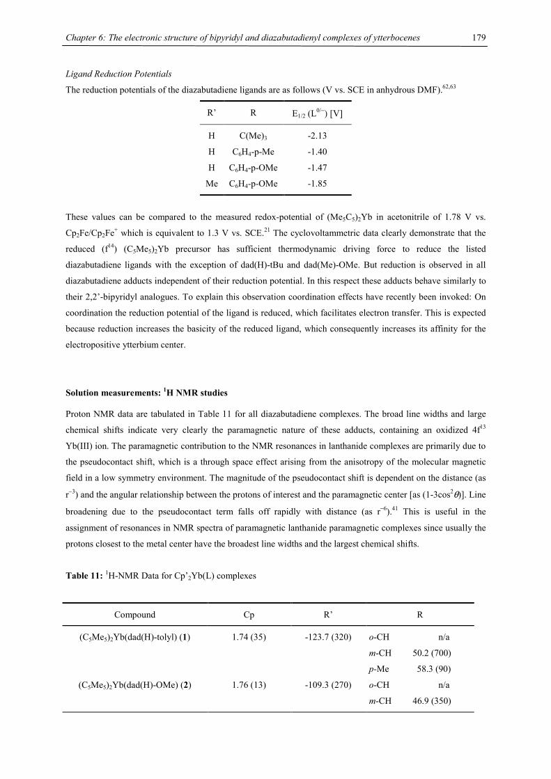
Chapter 6: The electronic structure of bipyridyl and diazabutadienyl complexes of ytterbocenes
179
Ligand Reduction Potentials
The reduction potentials of the diazabutadiene ligands are as follows (V vs. SCE in anhydrous DMF).62,63
R’ R E1/2 (L0/−) [V]
H C(Me)3 -2.13
H C6H4-p-Me -1.40
H C6H4-p-OMe -1.47
Me C6H4-p-OMe -1.85
These values can be compared to the measured redox-potential of (Me5C5)2Yb in acetonitrile of 1.78 V vs.
Cp2Fe/Cp2Fe+ which is equivalent to 1.3 V vs. SCE.21 The cyclovoltammetric data clearly demonstrate that the
reduced (f14) (C5Me5)2Yb precursor has sufficient thermodynamic driving force to reduce the listed
diazabutadiene ligands with the exception of dad(H)-tBu and dad(Me)-OMe. But reduction is observed in all
diazabutadiene adducts independent of their reduction potential. In this respect these adducts behave similarly to
their 2,2’-bipyridyl analogues. To explain this observation coordination effects have recently been invoked: On
coordination the reduction potential of the ligand is reduced, which facilitates electron transfer. This is expected
because reduction increases the basicity of the reduced ligand, which consequently increases its affinity for the
electropositive ytterbium center.
Solution measurements: 1H NMR studies
Proton NMR data are tabulated in Table 11 for all diazabutadiene complexes. The broad line widths and large
chemical shifts indicate very clearly the paramagnetic nature of these adducts, containing an oxidized 4f13
Yb(III) ion. The paramagnetic contribution to the NMR resonances in lanthanide complexes are primarily due to
the pseudocontact shift, which is a through space effect arising from the anisotropy of the molecular magnetic
field in a low symmetry environment. The magnitude of the pseudocontact shift is dependent on the distance (as
r−3) and the angular relationship between the protons of interest and the paramagnetic center [as (1-3cos2θ)]. Line
broadening due to the pseudocontact term falls off rapidly with distance (as r−6).41 This is useful in the
assignment of resonances in NMR spectra of paramagnetic lanthanide paramagnetic complexes since usually the
protons closest to the metal center have the broadest line widths and the largest chemical shifts.
Table 11: 1H-NMR Data for Cp’2Yb(L) complexes
Compound Cp R’ R
(C5Me5)2Yb(dad(H)-tolyl) (1) 1.74 (35) -123.7 (320) o-CH n/a
m-CH 50.2 (700)
p-Me 58.3 (90)
(C5Me5)2Yb(dad(H)-OMe) (2) 1.76 (13) -109.3 (270) o-CH n/a
m-CH 46.9 (350)
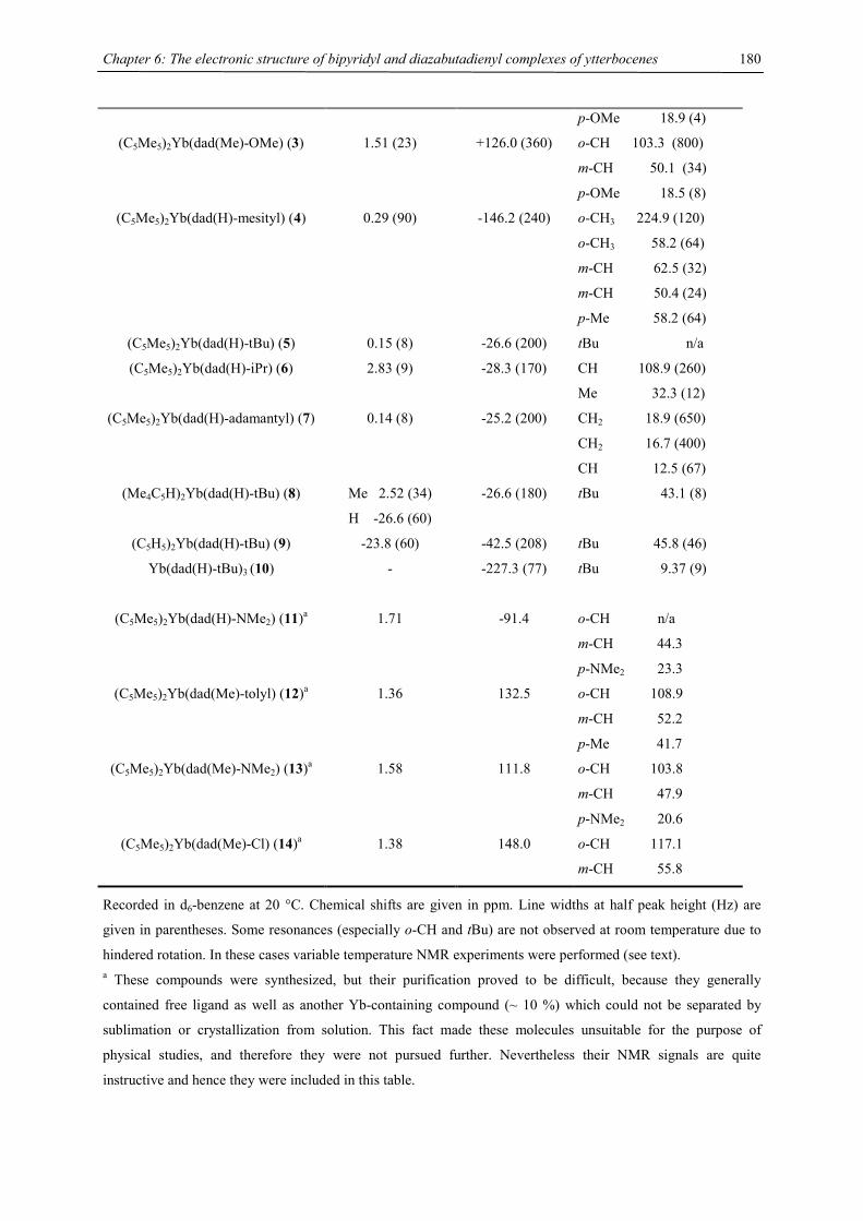
Chapter 6: The electronic structure of bipyridyl and diazabutadienyl complexes of ytterbocenes
180
p-OMe 18.9 (4)
(C5Me5)2Yb(dad(Me)-OMe) (3) 1.51 (23) +126.0 (360) o-CH 103.3 (800)
m-CH 50.1 (34)
p-OMe 18.5 (8)
(C5Me5)2Yb(dad(H)-mesityl) (4) 0.29 (90) -146.2 (240) o-CH3 224.9 (120)
o-CH3 58.2 (64)
m-CH 62.5 (32)
m-CH 50.4 (24)
p-Me 58.2 (64)
(C5Me5)2Yb(dad(H)-tBu) (5) 0.15 (8) -26.6 (200) tBu n/a
(C5Me5)2Yb(dad(H)-iPr) (6) 2.83 (9) -28.3 (170) CH 108.9 (260)
Me 32.3 (12)
(C5Me5)2Yb(dad(H)-adamantyl) (7) 0.14 (8) -25.2 (200) CH2 18.9 (650)
CH2 16.7 (400)
CH 12.5 (67)
(Me4C5H)2Yb(dad(H)-tBu) (8) Me 2.52 (34)
H -26.6 (60)
-26.6 (180) tBu 43.1 (8)
(C5H5)2Yb(dad(H)-tBu) (9) -23.8 (60) -42.5 (208) tBu 45.8 (46)
Yb(dad(H)-tBu)3 (10) - -227.3 (77) tBu 9.37 (9)
(C5Me5)2Yb(dad(H)-NMe2) (11)a 1.71 -91.4 o-CH n/a
m-CH 44.3
p-NMe2 23.3
(C5Me5)2Yb(dad(Me)-tolyl) (12)a 1.36 132.5 o-CH 108.9
m-CH 52.2
p-Me 41.7
(C5Me5)2Yb(dad(Me)-NMe2) (13)a 1.58 111.8 o-CH 103.8
m-CH 47.9
p-NMe2 20.6
(C5Me5)2Yb(dad(Me)-Cl) (14)a 1.38 148.0 o-CH 117.1
m-CH 55.8
Recorded in d6-benzene at 20 °C. Chemical shifts are given in ppm. Line widths at half peak height (Hz) are
given in parentheses. Some resonances (especially o-CH and tBu) are not observed at room temperature due to
hindered rotation. In these cases variable temperature NMR experiments were performed (see text). a These compounds were synthesized, but their purification proved to be difficult, because they generally
contained free ligand as well as another Yb-containing compound (~ 10 %) which could not be separated by
sublimation or crystallization from solution. This fact made these molecules unsuitable for the purpose of
physical studies, and therefore they were not pursued further. Nevertheless their NMR signals are quite
instructive and hence they were included in this table.
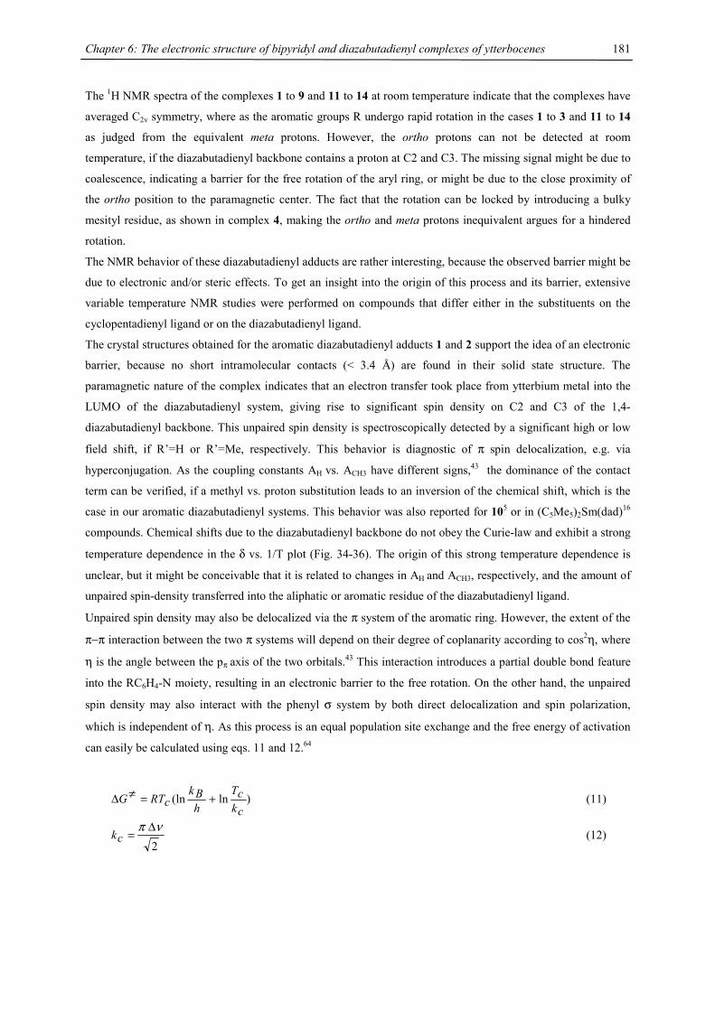
Chapter 6: The electronic structure of bipyridyl and diazabutadienyl complexes of ytterbocenes
181
The 1H NMR spectra of the complexes 1 to 9 and 11 to 14 at room temperature indicate that the complexes have
averaged C2v symmetry, where as the aromatic groups R undergo rapid rotation in the cases 1 to 3 and 11 to 14
as judged from the equivalent meta protons. However, the ortho protons can not be detected at room
temperature, if the diazabutadienyl backbone contains a proton at C2 and C3. The missing signal might be due to
coalescence, indicating a barrier for the free rotation of the aryl ring, or might be due to the close proximity of
the ortho position to the paramagnetic center. The fact that the rotation can be locked by introducing a bulky
mesityl residue, as shown in complex 4, making the ortho and meta protons inequivalent argues for a hindered
rotation.
The NMR behavior of these diazabutadienyl adducts are rather interesting, because the observed barrier might be
due to electronic and/or steric effects. To get an insight into the origin of this process and its barrier, extensive
variable temperature NMR studies were performed on compounds that differ either in the substituents on the
cyclopentadienyl ligand or on the diazabutadienyl ligand.
The crystal structures obtained for the aromatic diazabutadienyl adducts 1 and 2 support the idea of an electronic
barrier, because no short intramolecular contacts (< 3.4 Å) are found in their solid state structure. The
paramagnetic nature of the complex indicates that an electron transfer took place from ytterbium metal into the
LUMO of the diazabutadienyl system, giving rise to significant spin density on C2 and C3 of the 1,4-
diazabutadienyl backbone. This unpaired spin density is spectroscopically detected by a significant high or low
field shift, if R’=H or R’=Me, respectively. This behavior is diagnostic of π spin delocalization, e.g. via
hyperconjugation. As the coupling constants AH vs. ACH3 have different signs,43 the dominance of the contact
term can be verified, if a methyl vs. proton substitution leads to an inversion of the chemical shift, which is the
case in our aromatic diazabutadienyl systems. This behavior was also reported for 105 or in (C5Me5)2Sm(dad)16
compounds. Chemical shifts due to the diazabutadienyl backbone do not obey the Curie-law and exhibit a strong
temperature dependence in the δ vs. 1/T plot (Fig. 34-36). The origin of this strong temperature dependence is
unclear, but it might be conceivable that it is related to changes in AH and ACH3, respectively, and the amount of
unpaired spin-density transferred into the aliphatic or aromatic residue of the diazabutadienyl ligand.
Unpaired spin density may also be delocalized via the π system of the aromatic ring. However, the extent of the
π−π interaction between the two π systems will depend on their degree of coplanarity according to cos2η, where
η is the angle between the pπ axis of the two orbitals.43 This interaction introduces a partial double bond feature
into the RC6H4-N moiety, resulting in an electronic barrier to the free rotation. On the other hand, the unpaired
spin density may also interact with the phenyl σ system by both direct delocalization and spin polarization,
which is independent of η. As this process is an equal population site exchange and the free energy of activation
can easily be calculated using eqs. 11 and 12.64
)ln(lnckcT
hBk
cRTG +=≠∆ (11)
2νπ ∆
=ck (12)
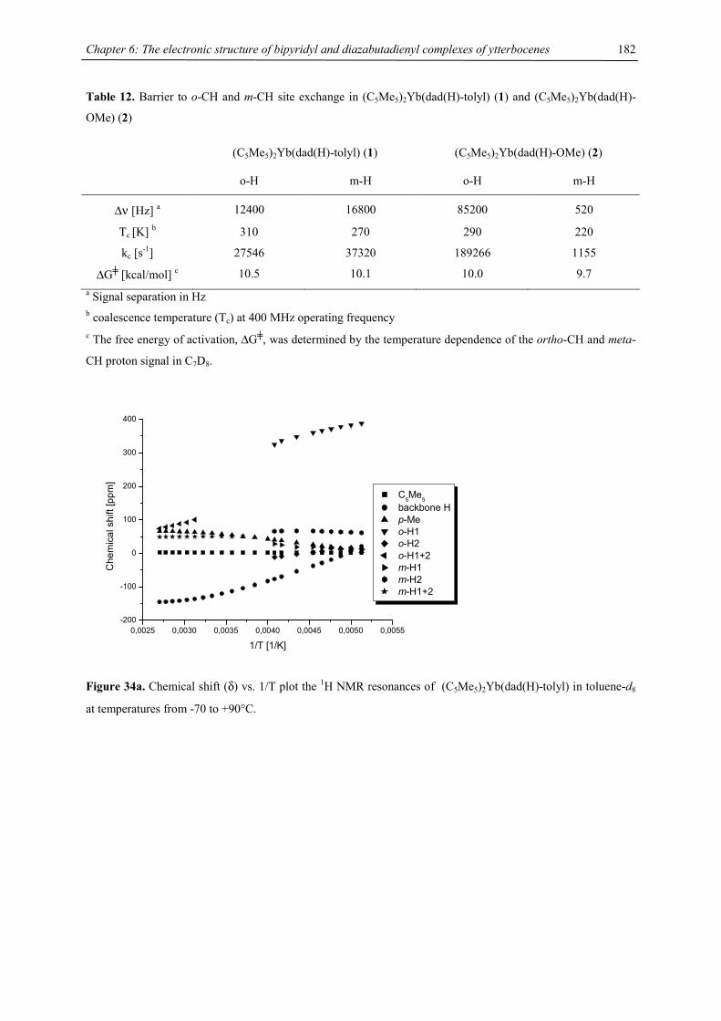
Chapter 6: The electronic structure of bipyridyl and diazabutadienyl complexes of ytterbocenes
182
Table 12. Barrier to o-CH and m-CH site exchange in (C5Me5)2Yb(dad(H)-tolyl) (1) and (C5Me5)2Yb(dad(H)-
OMe) (2)
(C5Me5)2Yb(dad(H)-tolyl) (1) (C5Me5)2Yb(dad(H)-OMe) (2)
o-H m-H o-H m-H
∆ν [Hz] a 12400 16800 85200 520
Tc [K] b 310 270 290 220
kc [s-1] 27546 37320 189266 1155
∆G╪ [kcal/mol] c 10.5 10.1 10.0 9.7 a Signal separation in Hz b coalescence temperature (Tc) at 400 MHz operating frequency c The free energy of activation, ∆G╪, was determined by the temperature dependence of the ortho-CH and meta-
CH proton signal in C7D8.
0,0025 0,0030 0,0035 0,0040 0,0045 0,0050 0,0055-200
-100
0
100
200
300
400
Che
mic
al s
hift
[ppm
]
1/T [1/K]
C5Me5 backbone H p-Me o-H1 o-H2 o-H1+2 m-H1 m-H2 m-H1+2
Figure 34a. Chemical shift (δ) vs. 1/T plot the 1H NMR resonances of (C5Me5)2Yb(dad(H)-tolyl) in toluene-d8
at temperatures from -70 to +90°C.
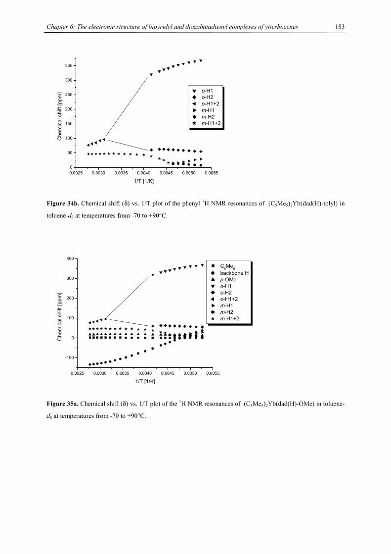
Chapter 6: The electronic structure of bipyridyl and diazabutadienyl complexes of ytterbocenes
183
0.0025 0.0030 0.0035 0.0040 0.0045 0.0050 0.00550
50
100
150
200
250
300
350C
hem
ical
shi
ft [p
pm]
1/T [1/K]
o-H1 o-H2 o-H1+2 m-H1 m-H2 m-H1+2
Figure 34b. Chemical shift (δ) vs. 1/T plot of the phenyl 1H NMR resonances of (C5Me5)2Yb(dad(H)-tolyl) in
toluene-d8 at temperatures from -70 to +90°C.
0.0025 0.0030 0.0035 0.0040 0.0045 0.0050 0.0055
-100
0
100
200
300
400
Che
mic
al s
hift
[ppm
]
1/T [1/K]
C5Me5 backbone H p-OMe o-H1 o-H2 o-H1+2 m-H1 m-H2 m-H1+2
Figure 35a. Chemical shift (δ) vs. 1/T plot of the 1H NMR resonances of (C5Me5)2Yb(dad(H)-OMe) in toluene-
d8 at temperatures from -70 to +90°C.
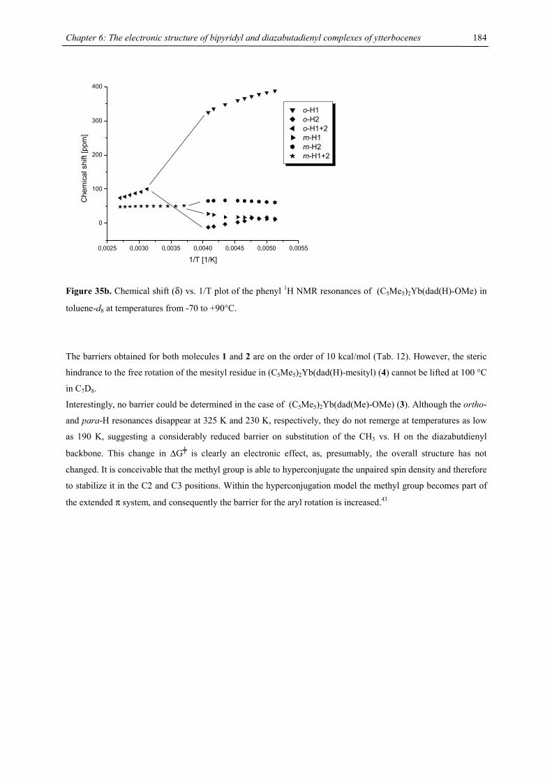
Chapter 6: The electronic structure of bipyridyl and diazabutadienyl complexes of ytterbocenes
184
0,0025 0,0030 0,0035 0,0040 0,0045 0,0050 0,0055
0
100
200
300
400C
hem
ical
shi
ft [p
pm]
1/T [1/K]
o-H1 o-H2 o-H1+2 m-H1 m-H2 m-H1+2
Figure 35b. Chemical shift (δ) vs. 1/T plot of the phenyl 1H NMR resonances of (C5Me5)2Yb(dad(H)-OMe) in
toluene-d8 at temperatures from -70 to +90°C.
The barriers obtained for both molecules 1 and 2 are on the order of 10 kcal/mol (Tab. 12). However, the steric
hindrance to the free rotation of the mesityl residue in (C5Me5)2Yb(dad(H)-mesityl) (4) cannot be lifted at 100 °C
in C7D8.
Interestingly, no barrier could be determined in the case of (C5Me5)2Yb(dad(Me)-OMe) (3). Although the ortho-
and para-H resonances disappear at 325 K and 230 K, respectively, they do not remerge at temperatures as low
as 190 K, suggesting a considerably reduced barrier on substitution of the CH3 vs. H on the diazabutdienyl
backbone. This change in ∆G╪ is clearly an electronic effect, as, presumably, the overall structure has not
changed. It is conceivable that the methyl group is able to hyperconjugate the unpaired spin density and therefore
to stabilize it in the C2 and C3 positions. Within the hyperconjugation model the methyl group becomes part of
the extended π system, and consequently the barrier for the aryl rotation is increased.43
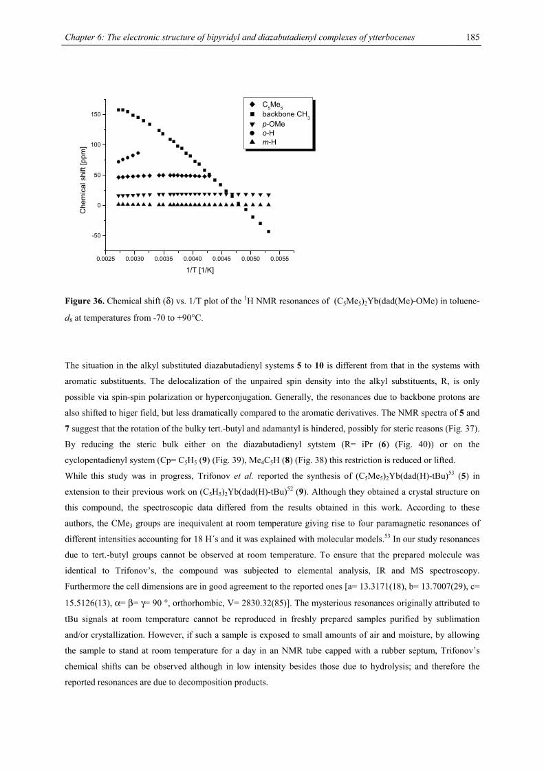
Chapter 6: The electronic structure of bipyridyl and diazabutadienyl complexes of ytterbocenes
185
0.0025 0.0030 0.0035 0.0040 0.0045 0.0050 0.0055
-50
0
50
100
150
Che
mic
al s
hift
[ppm
]
1/T [1/K]
C5Me5 backbone CH
3 p-OMe o-H m-H
Figure 36. Chemical shift (δ) vs. 1/T plot of the 1H NMR resonances of (C5Me5)2Yb(dad(Me)-OMe) in toluene-
d8 at temperatures from -70 to +90°C.
The situation in the alkyl substituted diazabutadienyl systems 5 to 10 is different from that in the systems with
aromatic substituents. The delocalization of the unpaired spin density into the alkyl substituents, R, is only
possible via spin-spin polarization or hyperconjugation. Generally, the resonances due to backbone protons are
also shifted to higer field, but less dramatically compared to the aromatic derivatives. The NMR spectra of 5 and
7 suggest that the rotation of the bulky tert.-butyl and adamantyl is hindered, possibly for steric reasons (Fig. 37).
By reducing the steric bulk either on the diazabutadienyl sytstem (R= iPr (6) (Fig. 40)) or on the
cyclopentadienyl system (Cp= C5H5 (9) (Fig. 39), Me4C5H (8) (Fig. 38) this restriction is reduced or lifted.
While this study was in progress, Trifonov et al. reported the synthesis of (C5Me5)2Yb(dad(H)-tBu)53 (5) in
extension to their previous work on (C5H5)2Yb(dad(H)-tBu)52 (9). Although they obtained a crystal structure on
this compound, the spectroscopic data differed from the results obtained in this work. According to these
authors, the CMe3 groups are inequivalent at room temperature giving rise to four paramagnetic resonances of
different intensities accounting for 18 H´s and it was explained with molecular models.53 In our study resonances
due to tert.-butyl groups cannot be observed at room temperature. To ensure that the prepared molecule was
identical to Trifonov’s, the compound was subjected to elemental analysis, IR and MS spectroscopy.
Furthermore the cell dimensions are in good agreement to the reported ones [a= 13.3171(18), b= 13.7007(29), c=
15.5126(13), α= β= γ= 90 °, orthorhombic, V= 2830.32(85)]. The mysterious resonances originally attributed to
tBu signals at room temperature cannot be reproduced in freshly prepared samples purified by sublimation
and/or crystallization. However, if such a sample is exposed to small amounts of air and moisture, by allowing
the sample to stand at room temperature for a day in an NMR tube capped with a rubber septum, Trifonov’s
chemical shifts can be observed although in low intensity besides those due to hydrolysis; and therefore the
reported resonances are due to decomposition products.
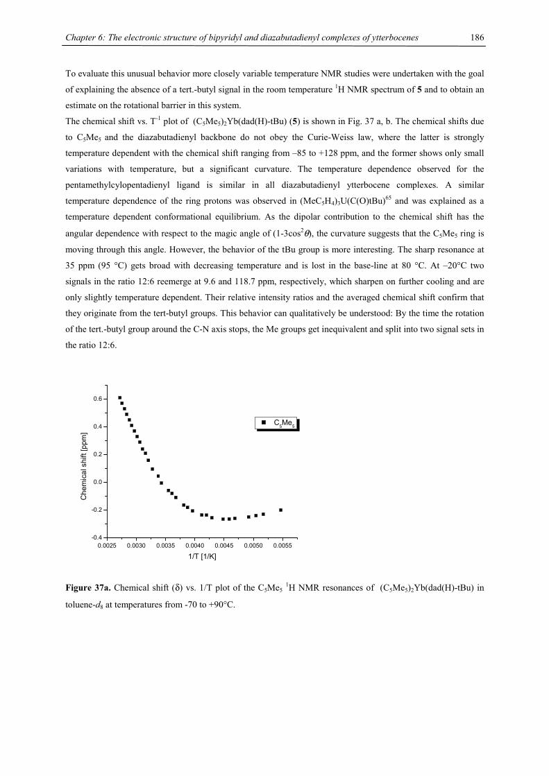
Chapter 6: The electronic structure of bipyridyl and diazabutadienyl complexes of ytterbocenes
186
To evaluate this unusual behavior more closely variable temperature NMR studies were undertaken with the goal
of explaining the absence of a tert.-butyl signal in the room temperature 1H NMR spectrum of 5 and to obtain an
estimate on the rotational barrier in this system.
The chemical shift vs. T-1 plot of (C5Me5)2Yb(dad(H)-tBu) (5) is shown in Fig. 37 a, b. The chemical shifts due
to C5Me5 and the diazabutadienyl backbone do not obey the Curie-Weiss law, where the latter is strongly
temperature dependent with the chemical shift ranging from –85 to +128 ppm, and the former shows only small
variations with temperature, but a significant curvature. The temperature dependence observed for the
pentamethylcylopentadienyl ligand is similar in all diazabutadienyl ytterbocene complexes. A similar
temperature dependence of the ring protons was observed in (MeC5H4)3U(C(O)tBu)65 and was explained as a
temperature dependent conformational equilibrium. As the dipolar contribution to the chemical shift has the
angular dependence with respect to the magic angle of (1-3cos2θ), the curvature suggests that the C5Me5 ring is
moving through this angle. However, the behavior of the tBu group is more interesting. The sharp resonance at
35 ppm (95 °C) gets broad with decreasing temperature and is lost in the base-line at 80 °C. At –20°C two
signals in the ratio 12:6 reemerge at 9.6 and 118.7 ppm, respectively, which sharpen on further cooling and are
only slightly temperature dependent. Their relative intensity ratios and the averaged chemical shift confirm that
they originate from the tert-butyl groups. This behavior can qualitatively be understood: By the time the rotation
of the tert.-butyl group around the C-N axis stops, the Me groups get inequivalent and split into two signal sets in
the ratio 12:6.
0.0025 0.0030 0.0035 0.0040 0.0045 0.0050 0.0055-0.4
-0.2
0.0
0.2
0.4
0.6
Che
mic
al s
hift
[ppm
]
1/T [1/K]
C5Me5
Figure 37a. Chemical shift (δ) vs. 1/T plot of the C5Me5 1H NMR resonances of (C5Me5)2Yb(dad(H)-tBu) in
toluene-d8 at temperatures from -70 to +90°C.
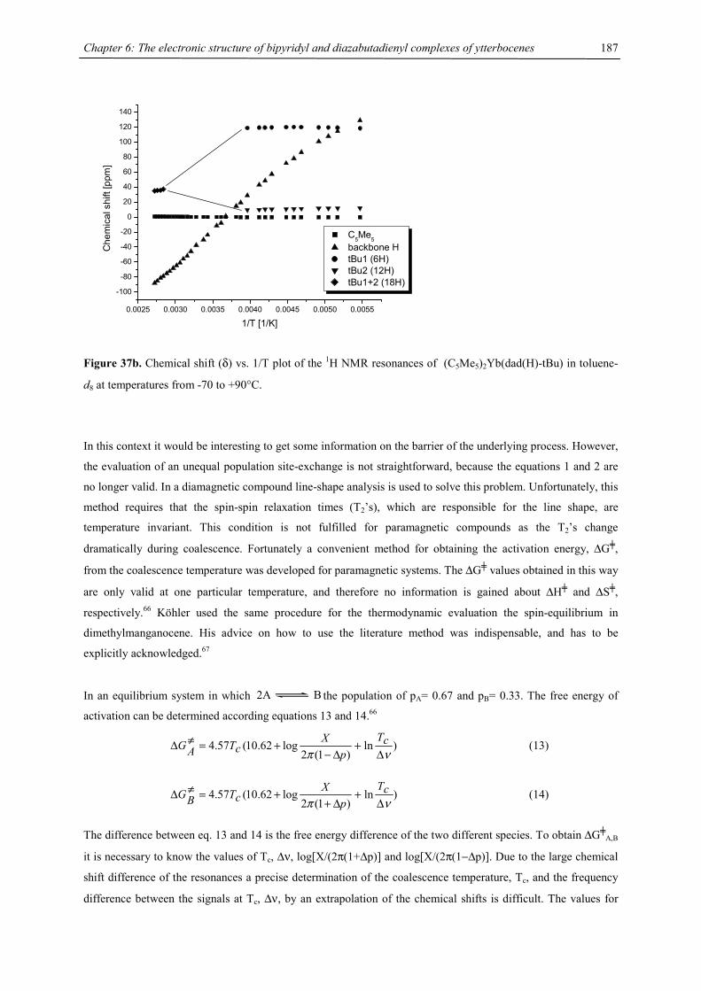
Chapter 6: The electronic structure of bipyridyl and diazabutadienyl complexes of ytterbocenes
187
0.0025 0.0030 0.0035 0.0040 0.0045 0.0050 0.0055
-100
-80
-60
-40
-20
0
20
40
60
80
100
120
140C
hem
ical
shi
ft [p
pm]
1/T [1/K]
C5Me5 backbone H tBu1 (6H) tBu2 (12H) tBu1+2 (18H)
Figure 37b. Chemical shift (δ) vs. 1/T plot of the 1H NMR resonances of (C5Me5)2Yb(dad(H)-tBu) in toluene-
d8 at temperatures from -70 to +90°C.
In this context it would be interesting to get some information on the barrier of the underlying process. However,
the evaluation of an unequal population site-exchange is not straightforward, because the equations 1 and 2 are
no longer valid. In a diamagnetic compound line-shape analysis is used to solve this problem. Unfortunately, this
method requires that the spin-spin relaxation times (T2’s), which are responsible for the line shape, are
temperature invariant. This condition is not fulfilled for paramagnetic compounds as the T2’s change
dramatically during coalescence. Fortunately a convenient method for obtaining the activation energy, ∆G╪,
from the coalescence temperature was developed for paramagnetic systems. The ∆G╪ values obtained in this way
are only valid at one particular temperature, and therefore no information is gained about ∆H╪ and ∆S╪,
respectively.66 Köhler used the same procedure for the thermodynamic evaluation the spin-equilibrium in
dimethylmanganocene. His advice on how to use the literature method was indispensable, and has to be
explicitly acknowledged.67
In an equilibrium system in which 2A B the population of pA= 0.67 and pB= 0.33. The free energy of
activation can be determined according equations 13 and 14.66
)ln)1(2
log62.10(57.4νπ ∆
+∆−
+=≠∆ cTp
XcTAG (13)
)ln)1(2
log62.10(57.4νπ ∆
+∆+
+=≠∆ cTp
XcTBG (14)
The difference between eq. 13 and 14 is the free energy difference of the two different species. To obtain ∆G╪A,B
it is necessary to know the values of Tc, ∆ν, log[X/(2π(1+∆p)] and log[X/(2π(1−∆p)]. Due to the large chemical
shift difference of the resonances a precise determination of the coalescence temperature, Tc, and the frequency
difference between the signals at Tc, ∆ν, by an extrapolation of the chemical shifts is difficult. The values for
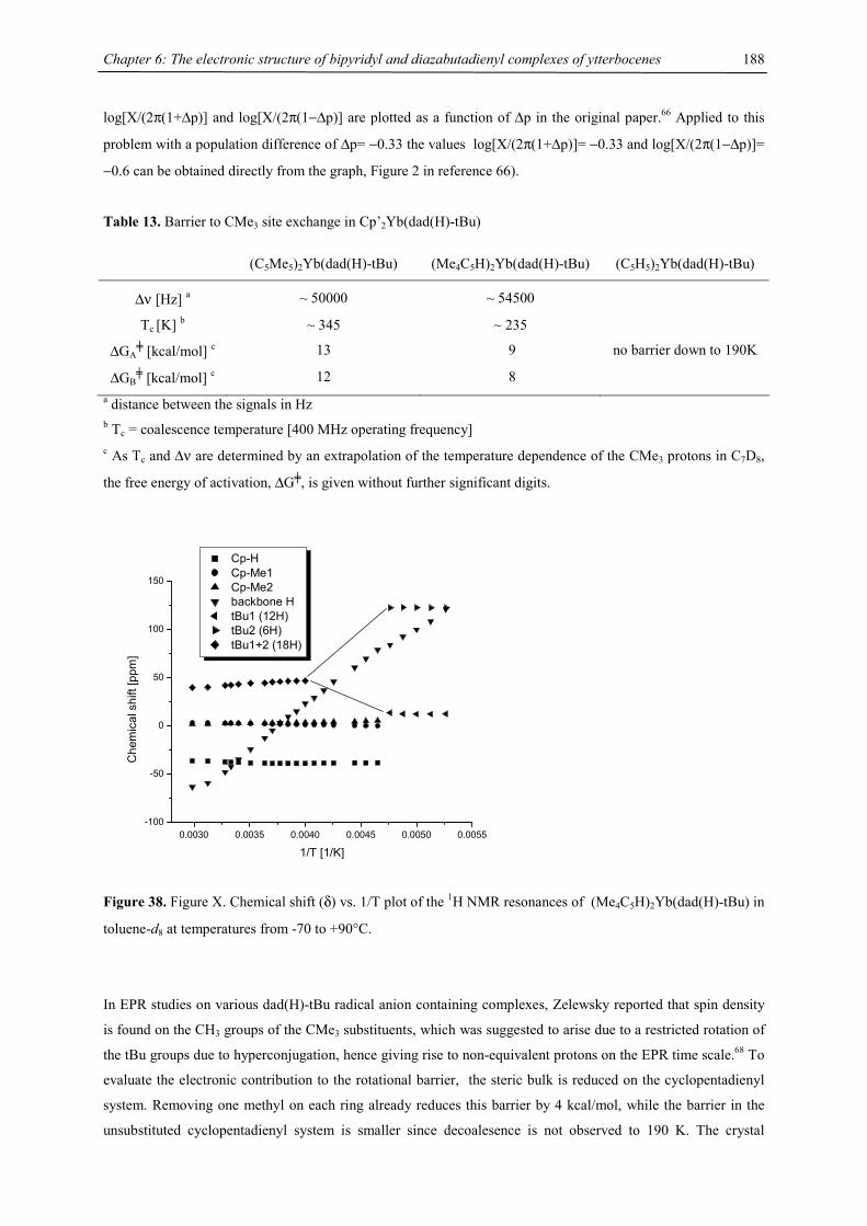
Chapter 6: The electronic structure of bipyridyl and diazabutadienyl complexes of ytterbocenes
188
log[X/(2π(1+∆p)] and log[X/(2π(1−∆p)] are plotted as a function of ∆p in the original paper.66 Applied to this
problem with a population difference of ∆p= −0.33 the values log[X/(2π(1+∆p)]= −0.33 and log[X/(2π(1−∆p)]=
−0.6 can be obtained directly from the graph, Figure 2 in reference 66).
Table 13. Barrier to CMe3 site exchange in Cp’2Yb(dad(H)-tBu)
(C5Me5)2Yb(dad(H)-tBu) (Me4C5H)2Yb(dad(H)-tBu) (C5H5)2Yb(dad(H)-tBu)
∆ν [Hz] a ~ 50000 ~ 54500
Tc [K] b ~ 345 ~ 235
∆GA╪ [kcal/mol] c 13 9 no barrier down to 190K
∆GB╪ [kcal/mol] c 12 8
a distance between the signals in Hz b Tc = coalescence temperature [400 MHz operating frequency] c As Tc and ∆ν are determined by an extrapolation of the temperature dependence of the CMe3 protons in C7D8,
the free energy of activation, ∆G╪, is given without further significant digits.
0.0030 0.0035 0.0040 0.0045 0.0050 0.0055-100
-50
0
50
100
150
Che
mic
al s
hift
[ppm
]
1/T [1/K]
Cp-H Cp-Me1 Cp-Me2 backbone H tBu1 (12H) tBu2 (6H) tBu1+2 (18H)
Figure 38. Figure X. Chemical shift (δ) vs. 1/T plot of the 1H NMR resonances of (Me4C5H)2Yb(dad(H)-tBu) in
toluene-d8 at temperatures from -70 to +90°C.
In EPR studies on various dad(H)-tBu radical anion containing complexes, Zelewsky reported that spin density
is found on the CH3 groups of the CMe3 substituents, which was suggested to arise due to a restricted rotation of
the tBu groups due to hyperconjugation, hence giving rise to non-equivalent protons on the EPR time scale.68 To
evaluate the electronic contribution to the rotational barrier, the steric bulk is reduced on the cyclopentadienyl
system. Removing one methyl on each ring already reduces this barrier by 4 kcal/mol, while the barrier in the
unsubstituted cyclopentadienyl system is smaller since decoalesence is not observed to 190 K. The crystal
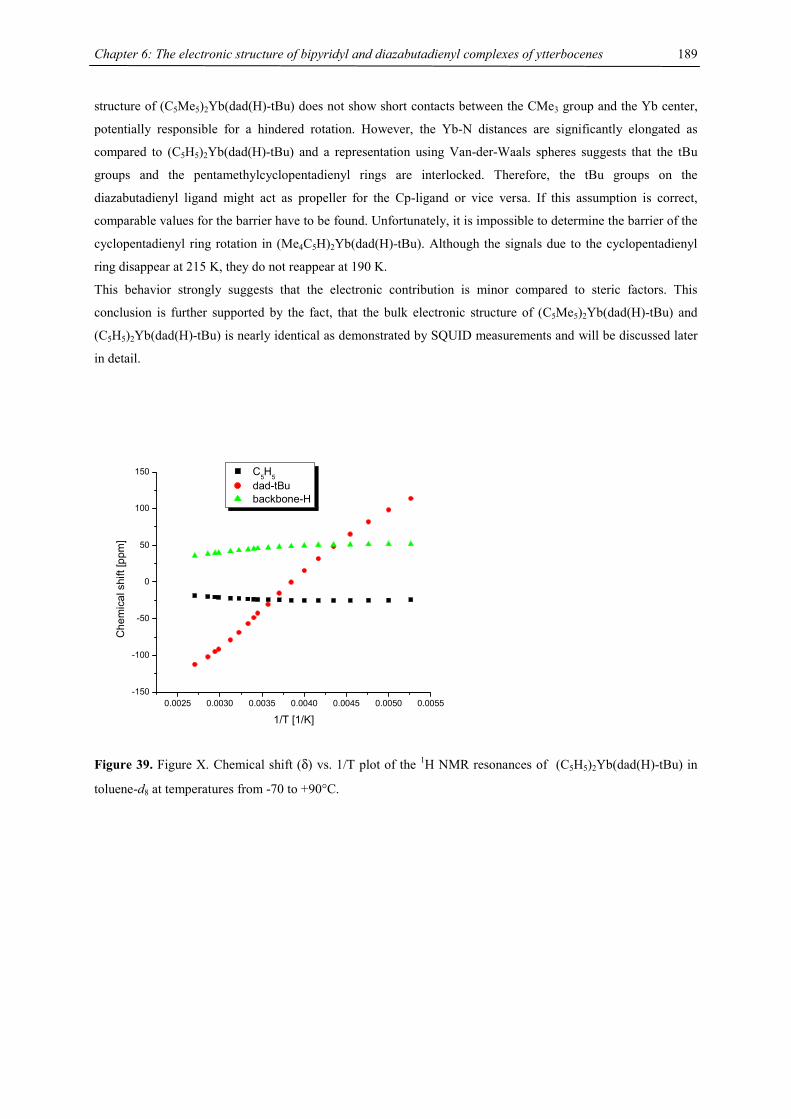
Chapter 6: The electronic structure of bipyridyl and diazabutadienyl complexes of ytterbocenes
189
structure of (C5Me5)2Yb(dad(H)-tBu) does not show short contacts between the CMe3 group and the Yb center,
potentially responsible for a hindered rotation. However, the Yb-N distances are significantly elongated as
compared to (C5H5)2Yb(dad(H)-tBu) and a representation using Van-der-Waals spheres suggests that the tBu
groups and the pentamethylcyclopentadienyl rings are interlocked. Therefore, the tBu groups on the
diazabutadienyl ligand might act as propeller for the Cp-ligand or vice versa. If this assumption is correct,
comparable values for the barrier have to be found. Unfortunately, it is impossible to determine the barrier of the
cyclopentadienyl ring rotation in (Me4C5H)2Yb(dad(H)-tBu). Although the signals due to the cyclopentadienyl
ring disappear at 215 K, they do not reappear at 190 K.
This behavior strongly suggests that the electronic contribution is minor compared to steric factors. This
conclusion is further supported by the fact, that the bulk electronic structure of (C5Me5)2Yb(dad(H)-tBu) and
(C5H5)2Yb(dad(H)-tBu) is nearly identical as demonstrated by SQUID measurements and will be discussed later
in detail.
0.0025 0.0030 0.0035 0.0040 0.0045 0.0050 0.0055-150
-100
-50
0
50
100
150
Che
mic
al s
hift
[ppm
]
1/T [1/K]
C5H5 dad-tBu backbone-H
Figure 39. Figure X. Chemical shift (δ) vs. 1/T plot of the 1H NMR resonances of (C5H5)2Yb(dad(H)-tBu) in
toluene-d8 at temperatures from -70 to +90°C.
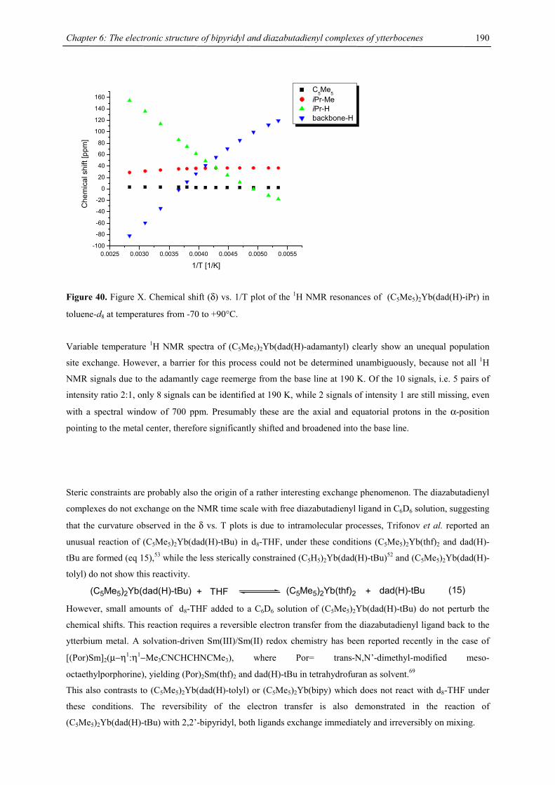
Chapter 6: The electronic structure of bipyridyl and diazabutadienyl complexes of ytterbocenes
190
0.0025 0.0030 0.0035 0.0040 0.0045 0.0050 0.0055-100
-80
-60
-40
-20
0
20
40
60
80
100
120
140
160
Che
mic
al s
hift
[ppm
]
1/T [1/K]
C5Me
5 iPr-Me iPr-H backbone-H
Figure 40. Figure X. Chemical shift (δ) vs. 1/T plot of the 1H NMR resonances of (C5Me5)2Yb(dad(H)-iPr) in
toluene-d8 at temperatures from -70 to +90°C.
Variable temperature 1H NMR spectra of (C5Me5)2Yb(dad(H)-adamantyl) clearly show an unequal population
site exchange. However, a barrier for this process could not be determined unambiguously, because not all 1H
NMR signals due to the adamantly cage reemerge from the base line at 190 K. Of the 10 signals, i.e. 5 pairs of
intensity ratio 2:1, only 8 signals can be identified at 190 K, while 2 signals of intensity 1 are still missing, even
with a spectral window of 700 ppm. Presumably these are the axial and equatorial protons in the α-position
pointing to the metal center, therefore significantly shifted and broadened into the base line.
Steric constraints are probably also the origin of a rather interesting exchange phenomenon. The diazabutadienyl
complexes do not exchange on the NMR time scale with free diazabutadienyl ligand in C6D6 solution, suggesting
that the curvature observed in the δ vs. T plots is due to intramolecular processes, Trifonov et al. reported an
unusual reaction of (C5Me5)2Yb(dad(H)-tBu) in d8-THF, under these conditions (C5Me5)2Yb(thf)2 and dad(H)-
tBu are formed (eq 15),53 while the less sterically constrained (C5H5)2Yb(dad(H)-tBu)52 and (C5Me5)2Yb(dad(H)-
tolyl) do not show this reactivity.
(C5Me5)2Yb(dad(H)-tBu) + THF (C5Me5)2Yb(thf)2 + dad(H)-tBu (15) However, small amounts of d8-THF added to a C6D6 solution of (C5Me5)2Yb(dad(H)-tBu) do not perturb the
chemical shifts. This reaction requires a reversible electron transfer from the diazabutadienyl ligand back to the
ytterbium metal. A solvation-driven Sm(III)/Sm(II) redox chemistry has been reported recently in the case of
[(Por)Sm]2(µ−η1:η1−Me3CNCHCHNCMe3), where Por= trans-N,N’-dimethyl-modified meso-
octaethylporphorine), yielding (Por)2Sm(thf)2 and dad(H)-tBu in tetrahydrofuran as solvent.69
This also contrasts to (C5Me5)2Yb(dad(H)-tolyl) or (C5Me5)2Yb(bipy) which does not react with d8-THF under
these conditions. The reversibility of the electron transfer is also demonstrated in the reaction of
(C5Me5)2Yb(dad(H)-tBu) with 2,2’-bipyridyl, both ligands exchange immediately and irreversibly on mixing.
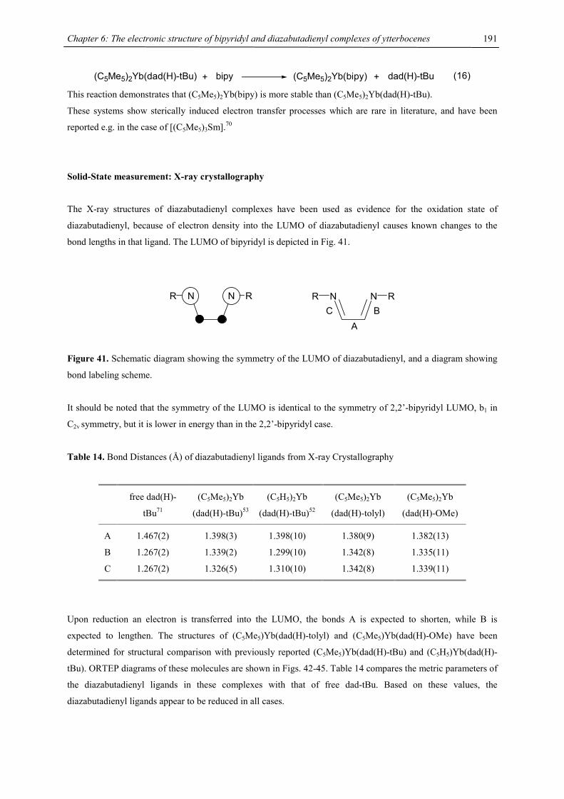
Chapter 6: The electronic structure of bipyridyl and diazabutadienyl complexes of ytterbocenes
191
(C5Me5)2Yb(dad(H)-tBu) + bipy (C5Me5)2Yb(bipy) + dad(H)-tBu (16) This reaction demonstrates that (C5Me5)2Yb(bipy) is more stable than (C5Me5)2Yb(dad(H)-tBu).
These systems show sterically induced electron transfer processes which are rare in literature, and have been
reported e.g. in the case of [(C5Me5)3Sm].70
Solid-State measurement: X-ray crystallography
The X-ray structures of diazabutadienyl complexes have been used as evidence for the oxidation state of
diazabutadienyl, because of electron density into the LUMO of diazabutadienyl causes known changes to the
bond lengths in that ligand. The LUMO of bipyridyl is depicted in Fig. 41.
N NR R N NR R
ABC
Figure 41. Schematic diagram showing the symmetry of the LUMO of diazabutadienyl, and a diagram showing
bond labeling scheme.
It should be noted that the symmetry of the LUMO is identical to the symmetry of 2,2’-bipyridyl LUMO, b1 in
C2v symmetry, but it is lower in energy than in the 2,2’-bipyridyl case.
Table 14. Bond Distances (Å) of diazabutadienyl ligands from X-ray Crystallography
free dad(H)-
tBu71
(C5Me5)2Yb
(dad(H)-tBu)53
(C5H5)2Yb
(dad(H)-tBu)52
(C5Me5)2Yb
(dad(H)-tolyl)
(C5Me5)2Yb
(dad(H)-OMe)
A 1.467(2) 1.398(3) 1.398(10) 1.380(9) 1.382(13)
B 1.267(2) 1.339(2) 1.299(10) 1.342(8) 1.335(11)
C 1.267(2) 1.326(5) 1.310(10) 1.342(8) 1.339(11)
Upon reduction an electron is transferred into the LUMO, the bonds A is expected to shorten, while B is
expected to lengthen. The structures of (C5Me5)Yb(dad(H)-tolyl) and (C5Me5)Yb(dad(H)-OMe) have been
determined for structural comparison with previously reported (C5Me5)Yb(dad(H)-tBu) and (C5H5)Yb(dad(H)-
tBu). ORTEP diagrams of these molecules are shown in Figs. 42-45. Table 14 compares the metric parameters of
the diazabutadienyl ligands in these complexes with that of free dad-tBu. Based on these values, the
diazabutadienyl ligands appear to be reduced in all cases.
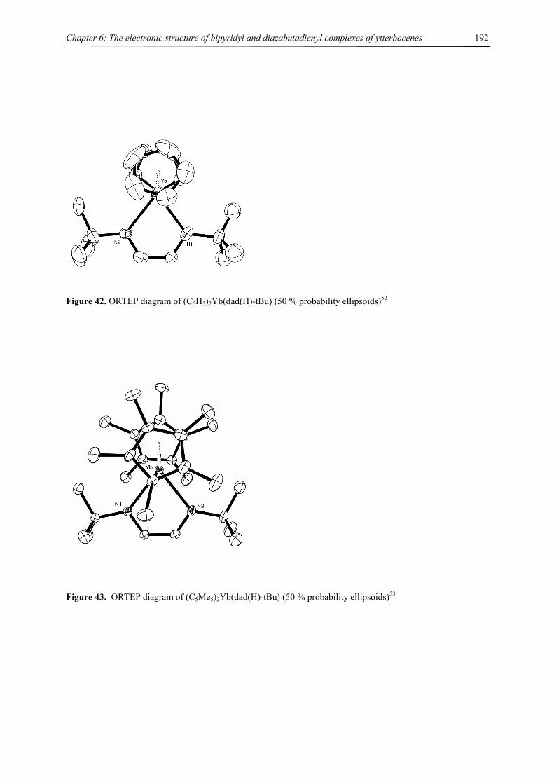
Chapter 6: The electronic structure of bipyridyl and diazabutadienyl complexes of ytterbocenes
192
Figure 42. ORTEP diagram of (C5H5)2Yb(dad(H)-tBu) (50 % probability ellipsoids)52
Figure 43. ORTEP diagram of (C5Me5)2Yb(dad(H)-tBu) (50 % probability ellipsoids)53
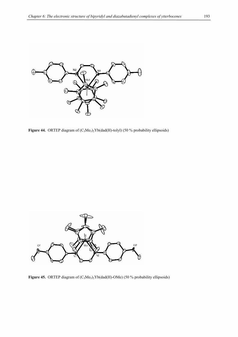
Chapter 6: The electronic structure of bipyridyl and diazabutadienyl complexes of ytterbocenes
193
Figure 44. ORTEP diagram of (C5Me5)2Yb(dad(H)-tolyl) (50 % probability ellipsoids)
Figure 45. ORTEP diagram of (C5Me5)2Yb(dad(H)-OMe) (50 % probability ellipsoids)
Chapter 6: The electronic structure of bipyridyl and diazabutadienyl complexes of ytterbocenes
194
Table 15. Selected Bond Distances (Å) and Angles (°) of (C5Me5)2Yb(dad(H)-tBu), (C5Me5)2Yb(dad(H)-tolyl),
(C5Me5)2Yb(dad(H)-OMe) and (C5H5)2Yb(dad(H)-tBu).
(C5H5)2Yb
(dad(H)-tBu)
(C5Me5)2Yb
(dad(H)-tBu)
(C5Me5)2Yb
(dad(H)-tolyl)
(C5Me5)2Yb
(dad(H)-OMe)
Yb-Cring (mean) 2.60 2.69 2.65 2.64
Yb-Cring (range) 2.585(14)-2.610(14) 2.656(3)-2.716(1) 2.623(6)-2.683(6) 2.623(9)-2.658(9)
Yb-centroid 2.33, 2.33 2.41; 2.40 2.37; 2.34 2.36; 2.35
centroid-Yb-centroid 128.1 130.3 137.2 138.8
Yb-N 2.306(9); 2.306(9) 2.385(3); 2.394(3) 2.340(5); 2.368(5) 2.339(7); 2.337(6)
N-Yb-N 74.7(3) 75.3(1) 73.4(2) 72.5(2)
bond A 1.398(10) 1.398(5) 1.380(9) 1.382(13)
R-N-C (averg) 117.4; 116.5 113.3; 113.4 118.2; 118.4 120.0; 119.1
torsion angle
N-C-C-N
4.0 0.1 0.5 0.5
torsion angle
N-C-C6-C6
- - 19.1, 23.9 2.8, 6.4
The distance from a ligand to a metal is correlated with the oxidation state of the metal, as the ionic radius is
smaller in a higher oxidation state.28 Examination of the mean metal-ring carbon distance, the metal-centroid
distance and the metal-nitrogen distances indicate that the ytterbium atom is indeed Yb(III) (Tab. 15). These
values can further be compared with the value for (C5Me5)2Yb(py)2, in which the metal is divalent. In that
molecule, the mean metal ring-carbon distance is 2.74 Å and the metal-nitrogen distance is 2.56 Å.7 The steric
repulsion between the tBu groups and the pentamethylcyclopentadienyl rings is obvious since there is a
significant elongation of the Yb-N distance compared to the unsubstituted derivative. An interesting feature is
the asymmetric twist of the aromatic ring relative to the diazabutadienyl unit, which is also reflected in slightly
asymmetric Yb-N distances.
Magnetic Susceptibility Studies
Magnetic susceptibility measurements provide direct information on the electronic structure of the molecule, and
the interaction of the unpaired ligand spins with the paramagnetic Yb(III) center. The plots of 1/χ vs. T and χT
vs. T for various diazabutadienyl adducts are shown in Figures 46 and 47. These data are very similar to those
previously measured for (Me5C5)2Yb(bipy). This indicates that the electronic structure in both systems is
comparable. However, the maxima and minima of 1/χ change with substituents, hence might reflect the
suggested influence of the frontier orbitals on the coupling in these systems. Interestingly, the diazabutadienyl
ytterbocene can be separated into two magnetic classes: aromatic and aliphatic substituted diazabutadienyl
system. Although the shape is slightly different the 1/χ vs. T plots of the aromatic systems show minima ~ 270 K
(TK~ 1200 K) (with the exception of dad(H)-OMe), whereas the alkyl systems have minima ~360 K (TK~ 1600
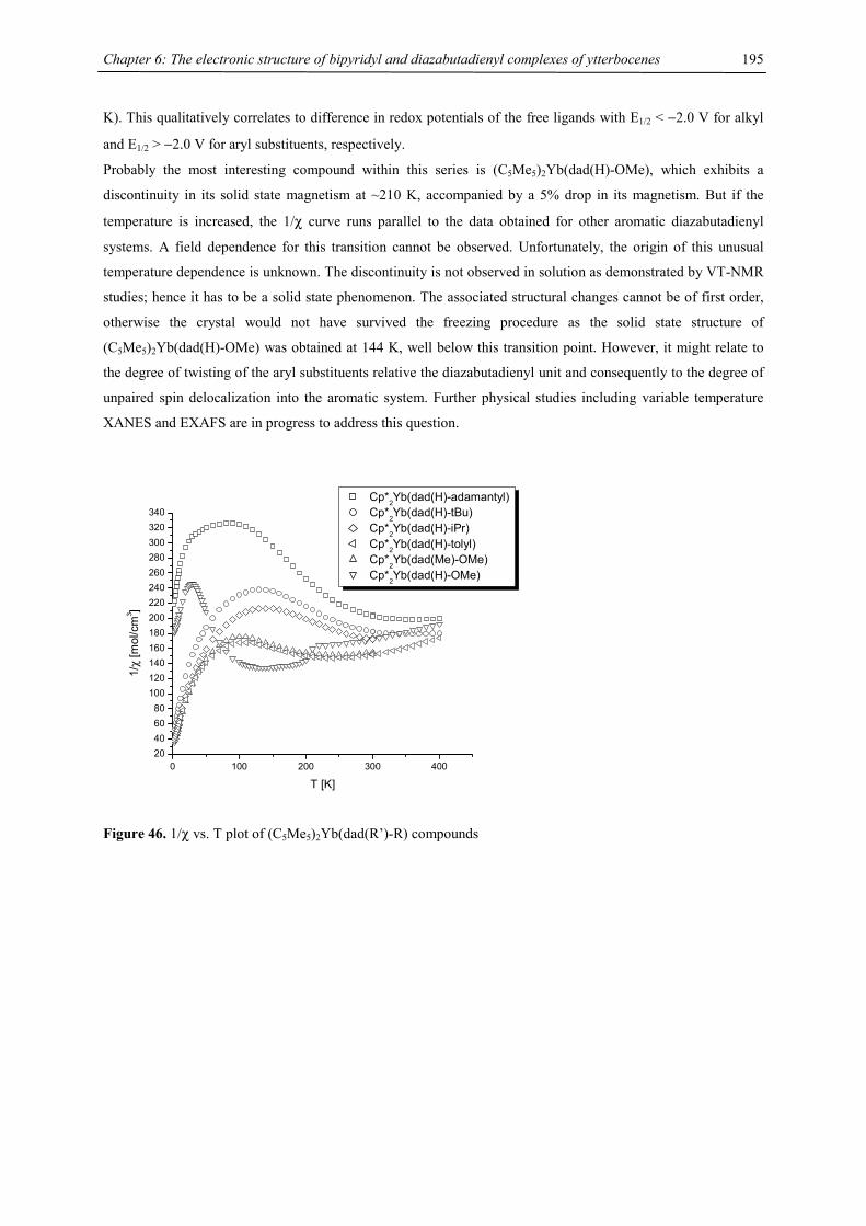
Chapter 6: The electronic structure of bipyridyl and diazabutadienyl complexes of ytterbocenes
195
K). This qualitatively correlates to difference in redox potentials of the free ligands with E1/2 < −2.0 V for alkyl
and E1/2 > −2.0 V for aryl substituents, respectively.
Probably the most interesting compound within this series is (C5Me5)2Yb(dad(H)-OMe), which exhibits a
discontinuity in its solid state magnetism at ~210 K, accompanied by a 5% drop in its magnetism. But if the
temperature is increased, the 1/χ curve runs parallel to the data obtained for other aromatic diazabutadienyl
systems. A field dependence for this transition cannot be observed. Unfortunately, the origin of this unusual
temperature dependence is unknown. The discontinuity is not observed in solution as demonstrated by VT-NMR
studies; hence it has to be a solid state phenomenon. The associated structural changes cannot be of first order,
otherwise the crystal would not have survived the freezing procedure as the solid state structure of
(C5Me5)2Yb(dad(H)-OMe) was obtained at 144 K, well below this transition point. However, it might relate to
the degree of twisting of the aryl substituents relative the diazabutadienyl unit and consequently to the degree of
unpaired spin delocalization into the aromatic system. Further physical studies including variable temperature
XANES and EXAFS are in progress to address this question.
0 100 200 300 40020406080
100120140160180200220240260280300320340
1/χ
[mol
/cm
3 ]
T [K]
Cp*2Yb(dad(H)-adamantyl) Cp*
2Yb(dad(H)-tBu)
Cp*2Yb(dad(H)-iPr) Cp*2Yb(dad(H)-tolyl) Cp*
2Yb(dad(Me)-OMe)
Cp*2Yb(dad(H)-OMe)
Figure 46. 1/χ vs. T plot of (C5Me5)2Yb(dad(R’)-R) compounds
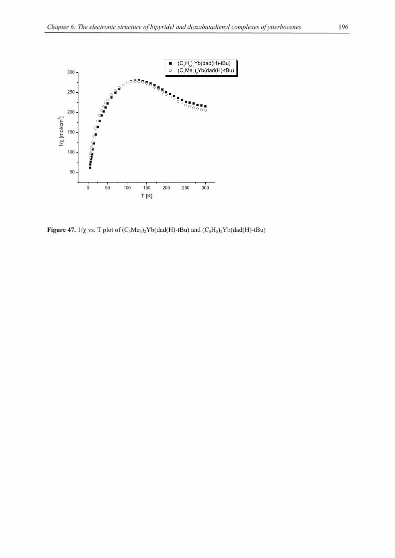
Chapter 6: The electronic structure of bipyridyl and diazabutadienyl complexes of ytterbocenes
196
0 50 100 150 200 250 300
50
100
150
200
250
300
1/χ
[mol
/cm
3 ]
T [K]
(C5H
5)2Yb(dad(H)-tBu)
(C5Me5)2Yb(dad(H)-tBu)
Figure 47. 1/χ vs. T plot of (C5Me5)2Yb(dad(H)-tBu) and (C5H5)2Yb(dad(H)-tBu)

Chapter 6: The electronic structure of bipyridyl and diazabutadienyl complexes of ytterbocenes
197
Solid-state measurement: X-ray Absorption Near Edge Spectra (XANES)
Preliminary XANES Yb LIII edge investigations on (C5Me5)2Yb(dad(R’)-R) compounds clearly indicate that the
diazabutadienyl compounds belong to the same class of mixed configurations compounds as its 2,2’-bipyridyl
analogues. The nf values are generally on the order of nf > 0.8, indicating the significant contributions of the
Yb(III)(dad•−) wave function to the ground state. These results have been anticipated from its solid state
magnetism.
8920 8930 8940 8950 8960 8970
0.0
0.5
1.0
1.5
2.0
2.5
3.0
Nor
mal
ized
Abs
orpt
ion
Energy [eV]
[Cp*2Yb(bipy)]I Yb(III) standard [Cp*
2Yb(dad(H)-OMe)
[Cp*2Yb(dad(H)-tBu) [Cp*2Yb(dad(H)-tolyl) Cp*
2Yb(py)
2 Yb(II) standard
Figure 48. Yb LIII XANES for (C5Me5)2Yb(dad(H)-OMe), (C5Me5)2Yb(dad(H)-tBu), (C5Me5)2Yb(dad(H)-tolyl),
a Yb(II) reference, (C5Me5)2Yb(py)2, and a Yb(III) reference (C5Me5)2Yb(bipy)I. The Yb(II) reference shows a
clear bump at ~8945 eV corresponding to a small Yb(III) impurity.
Chapter 6: The electronic structure of bipyridyl and diazabutadienyl complexes of ytterbocenes
198
Conclusion
It was possible to synthesize a series of diazabutadienyl complexes of ytterbocenes which exhibit a similar
electronic and magnetic structure like (C5Me5)2Yb(bipy). However our studies suggest that the diazabutadienyl
systems do not offer as much flexibility in tuning the electronic structure as the bipyridyl adducts. The maxima
in the magnetic susceptibilities are found in the range 270 K (TK ~ 1200 K) to 360 K (TK ~ 1600 K) for the
aromatic and alkyl substituted diazabutadienyl adducts, respectively. This trend follows changes in the redox
potential of the free ligand upon substitution of the diazabutadienyl moiety and supports the involvement of
frontier orbitals in the formation of the mixed configuration ground state. This series of diazabutadienyl and
bipyridyl complexes provides an excellent experimental database for theoretical calculations to evaluate these
systems with their unusual properties more precisely.
References
[1] Constable, E. C. Adv. Inorg. Chem. 1989, 34, 1-63; and references therein.
[2] Creutz, C. Comments Inorg. Chem. 1982, 1, 293-311; and references therein.
[3] Feistel, G. R.; Mathai, T. P. J. Am. Chem. Soc. 1968, 90, 2988-2989.
[4] Fedushkin, I. L.; Petrovs.kaya, T. V.; Girgsdies, F.; Nevodchikov, V. I.; Weimann, R.; Schumann, H.;
Bochkarev, M. N. Russ. Chem. Bull. 2000, 49, 1869-1876.
[5] Bochkarev, M. N.; Trifonov, A. A.; Cloke, F. G. N.; Dalby, C. I.; Matsunaga, P. T.; Andersen, R. A.;
Schumann, H.; Loebel, J.; Hemling, H. J. Organomet. Chem. 1995, 486, 177-182.
[6] Fedushkin, I. L.; Petrovs.kaya, T. V.; Girgsdies, F.; Köhn, R. D.; Bochkarev, M. N.; Schumann, H.
Angew. Chem., Int. Ed. Engl. 1999, 38, 2262-2264.
[7] Tilley, T. D.; Andersen, R. A.; Spencer, B.; Zalkin, A. Inorg. Chem. 1982, 21, 2647-2649.
[8] Berg, D. J., Ph.D. Thesis, University of California: Berkeley, 1987.
[9] Berg, D. J.; Boncella, J. M.; Andersen, R. A. Organometallics 2002, 21, 4622-4631.
[10] Berg, D. J.; Burns, C. J.; Andersen, R. A.; Zalkin, A. Organometallics 1989, 8, 1865-1870.
[11] Berg, D. J.; Andersen, R. A.; Zalkin, A. Organometallics 1988, 7, 1858-1863.
[12] Schultz, M., Ph.D. Thesis, University of California: Berkeley, 2000.
[13] Schultz, M.; Boncella, J. M.; Berg, D. J.; Tilley, T. D.; Andersen, R. A. Organometallics 2002, 21, 460-
472.
[14] Evans, W. J.; Gonzales, S. L.; Ziller, J. W. J. Am. Chem. Soc. 1994, 116, 2600-2608.
[15] Evans, W. J.; Drummond, D. K. J. Am. Chem. Soc. 1989, 111, 3329-3335.
[16] Recknagel, A.; Noltemeyer, M.; Edelmann, F. T. J. Organomet. Chem. 1991, 410, 53-61.
[17] Kuehl, C. J.; Da Re, R. E.; Scott, B. L.; Morris, D. E.; John, K. D. Chem. Commun. 2003, 2336-2337.
[18] Ohsawa, Y.; Whangbo, M.-H.; Hanck, K. W.; DeArmond, M. K. Inorg. Chem. 1984, 23, 3426-3428.
[19] Elliot, C. M.; Hershenhart, E. J. J. Am. Chem. Soc. 1982, 104, 7519-7526.
[20] McInnes, E. J. L.; Farley, R. D.; Rowlands, C. C.; Welch, A. J.; Rovatti, L.; Yellowlees, L. J. J. Chem.
Soc., Dalton Trans. 1999, 4203-4208.
[21] Finke, R. G.; Keenan, S. R.; Schiraldi, D. A.; Watson, P. L. Organometallics 1986, 5, 598-601.

Chapter 6: The electronic structure of bipyridyl and diazabutadienyl complexes of ytterbocenes
199
[22] Da Re, R. E.; Kuehl, C. J.; Brown, M. G.; Rocha, R. C.; Bauer, E. D.; John, K. D.; Morris, D. E.;
Shreve, A. P.; Sarrao, J. L. Inorg. Chem. 2003, 42, 5551-5559.
[23] Saito, Y.; Takemoto, J.; Hutchinson, B.; Nakamoto, K. Inorganic Chemistry 1972, 11, 2003-2011.
[24] Burns, C. J., Ph.D. Thesis, University of California: Berkeley, 1987.
[25] Aleksanyan, V. T. In Vibrational Spectra and Structure; Durig, J. R., Ed.; Elsevier: Amsterdam, 1982,
pp 115-144.
[26] Chisholm, M. H.; Huffman, J. C.; Rothwell, I. P.; Bradley, P. G.; Kress, N.; Woodruff, W. H. J. Am.
Chem. Soc. 1981, 103, 4945-4947.
[27] McPherson, A. M.; Fieselmann, B. F.; Lichtenberger, D. L.; McPherson, G. L.; Stucky, G. D. J. Am.
Chem. Soc. 1979, 101, 3425-3430.
[28] Raymond, K. N.; Eigenbrot, C. W. Acc. Chem. Res. 1980, 13, 276-283.
[29] Kahn, O. Molecular Magnetism; VCH Wíley: New York, 1993.
[30] van Vleck, J. H. The Theory of Electric and Magnetic Susceptibilities; Clarendon Press: Oxford, 1932.
[31] Boudreaux, E. A.; Mulay, L. N. Theory and Applications of Molecular Paramagnetism; Wiley-
Interscience: New York, 1976.
[32] Trojan, K. L.; Kendall, J. L.; Kepler, K. D.; Hatfield, W. E. Inorg. Chim. Acta 1992, 198-200, 795-803.
[33] Edelstein, N. In Fundamental and Technological Aspects of Organo-f-Element Chemistry; Marks, T. J.,
Fragala, I. L., Eds.; D Reidel Publishing Company: Dordrecht, Netherlands, 1984, pp 229-276.
[34] Kahn, M. L.; Sutter, J.-P.; Golhen, S.; Guionneau, P.; Ouahab, L.; Kahn, O.; Chasseau, D. J. Am. Chem.
Soc. 2000, 122, 3413-3421.
[35] Costes, J.-P.; Dahan, F.; Dupuis, A.; Laurent, J.-P. Chem. - Eur. J. 1998, 4, 1616-1620.
[36] Fulde, P. J. Phys. F: Met. Phys. 1988, 18, 601-639.
[37] Green, J. C.; Hohl, D.; Rösch, N. Organometallics 1987, 6, 712-720.
[38] Rajan, V. T. Phys. Rev. Lett. 1983, 51, 308-318.
[39] Sarrao, J. L.; Immer, C. D.; Fisk, Z.; Booth, C. H.; Figueroa, E.; Lawrence, J. M.; Modler, R.;
Cornelius, A. L.; Hundley, M. F.; Kwei, G. H.; Thompson, J. D.; Bridges, F. Phys. Rev. B. 1999, 59,
6855-6866.
[40] Bickers, N. E.; Cox, D. L.; Wilkins, J. W. Phys. Rev. B. 1987, 36, 2036-2079.
[41] Fischer, R. D. In NMR of Paramagnetic Molecules; La Mar, G. N., Horrocks, W. D., Holm, R. H., Eds.;
Academic Press: New York, 1973, pp 521-553.
[42] Horrocks, W. D. In NMR of Paramagnetic Molecules; La Mar, G. N., Horrocks, W. D., Holm, R. H.,
Eds.; Academic Press: New York, 1973, pp 479-519.
[43] La Mar, G. N.; Horrocks, W. D.; Holm, R. H., Eds. NMR of Paramagnetic Molecules; Academic Press:
New York, 1973.
[44] König, E.; Kremer, S. Chem. Phys. Lett. 1970, 5, 87-90.
[45] Coleman, P. In AIP Conference Proceedings (2002), 629(Lectures on the Physics of Highly Correlated
Electron Systems VI), 2002, pp 79-160.
[46] Werkema, E. L.; Andersen, R. A., unpublished results.
[47] Zi, G.; Andersen, R. A., unpublished results.
[48] van Koten, G.; Vrieze, K. Adv. Organomet. Chem. 1982, 21, 151-239.
[49] Cloke, F. G. N.; de Lemos, H. C.; Sameh, A. A. J. Chem. Soc., Chem. Commun. 1986, 1344-1345.
Chapter 6: The electronic structure of bipyridyl and diazabutadienyl complexes of ytterbocenes
200
[50] Brennan, J. G.; Cloke, F. G. N.; Sameh, A. A.; Zalkin, A. J. Chem. Soc., Chem. Commun. 1987, 1668.
[51] Görls, H.; Neumüller, B.; Scholz, A.; Scholz, J. Angew. Chem. 1995, 107, 732-735.
[52] Trifonov, A. A.; Kirillov, E. N.; Bochkarev, M. N.; Schumann, H.; Muehle, S. Russ. Chem. Bull. 1999,
48, 381-384.
[53] Trifonov, A. A.; Kurskii, Y. A.; Bochkarev, M. N.; Muehle, S.; Dechert, S.; Schumann, H. Russ. Chem.
Bull. 2003, 52, 601-606.
[54] Trifonov, A. A.; Kirillov, E. N.; Dechert, S.; Schumann, H.; Bochkarev, M. N. Eur. J. Inorg. Chem.
2001, 2509-2514.
[55] Trifonov, A. A.; Fedorova, E. A.; Fukin, G. K.; Druzhkov, N. O.; Bochkarev, M. N. Angew. Chem.
2004, 116, 5155-5158.
[56] Kliegman, J. M.; Barnes, R. K. J. Org. Chem. 1970, 35, 3140-3143.
[57] Kliegman, J. M.; Barnes, R. K. Tetrahedron 1970, 26, 2555-2560.
[58] tom Dieck, H.; Renk, I. W. Chem. Ber. 1971, 104, 92-109.
[59] tom Dieck, H.; Svoboda, M.; Greiser, T. Z. Naturforsch. 1981, 36b, 823-832.
[60] Svoboda, M.; tom Dieck, H.; Krüger, C.; Tsay, Y.-H. Z. Naturforsch. 1981, 36b, 814-822.
[61] Erlenmeyer, H.; Lehr, H. Helv. Chim. Acta 1946, 29, 69-71.
[62] Coulombeix, J.; Emmenegger, F.-P. Helv. Chim. Acta 1985, 68, 248-254.
[63] tom Dieck, H.; Renk, I. W. Chem. Ber. 1971, 104, 110-130.
[64] Friebolin, H. Ein- und zweidimensionale NMR Spectroskopie; VCH: Weinheim, 1992.
[65] Weydert, M.; Brennan, J. G.; Andersen, R. A.; Bergman, R. G. Organometallics 1995, 14, 3942-3951.
[66] Shanan-Atidi, H.; Bar-Elli, K. H. J. Phys. Chem. 1970, 74, 961-963.
[67] Köhler, F. H.; Schlesinger, B. Inorg. Chem. 1992, 31, 2853-2859.
[68] Clopath, P.; v. Zelewsky, A. Helv. Chim. Acta 1972, 55, 52-66.
[69] Wang, J.; Amos, R. I. J.; Frey, A. S. P.; Gardiner, M. G.; Cole, M. L.; Junk, P. C. Organometallics
2005, 24, 2259-2261.
[70] Evans, W. J.; Davis, B. L. Chem. Rev. 2002, 102, 2119-2136.
[71] Huige, C. J. M.; Spek, A. L.; de Boer, J. L. Acta Cryst. 1983, C41, 113-116.
[72] Garelli, N.; Vierling, P. J. Org. Chem. 1992, 57, 3046-3051.
[73] tom Dieck, H.; Franz, K.-D.; Majunke, W. Z. Naturforsch. 1975, 30b, 922-925.

Chapter 6: The electronic structure of bipyridyl and diazabutadienyl complexes of ytterbocenes
201
Appendix
0 50 100 150 200 250 300
0
5
10
15
20
25
30
35
40
1/χ χT
T [K]
1/χ
[mol
/cm
3 ]
4
6
8
χT [cm3K
/mol]
Figure A1. 1/χ vs. T and χT vs. T plot for Eu(bipy)4.
0 50 100 150 200 250 3000
20
40
60
80
100
120
1/χ χT
T [K]
1/χ
[mol
/cm
3 ]
0.0
0.5
1.0
1.5
2.0
2.5
3.0
χT [cm3K
/mol]
Figure A2. 1/χ vs. T and χT vs. T plot for Yb(bipy)4.
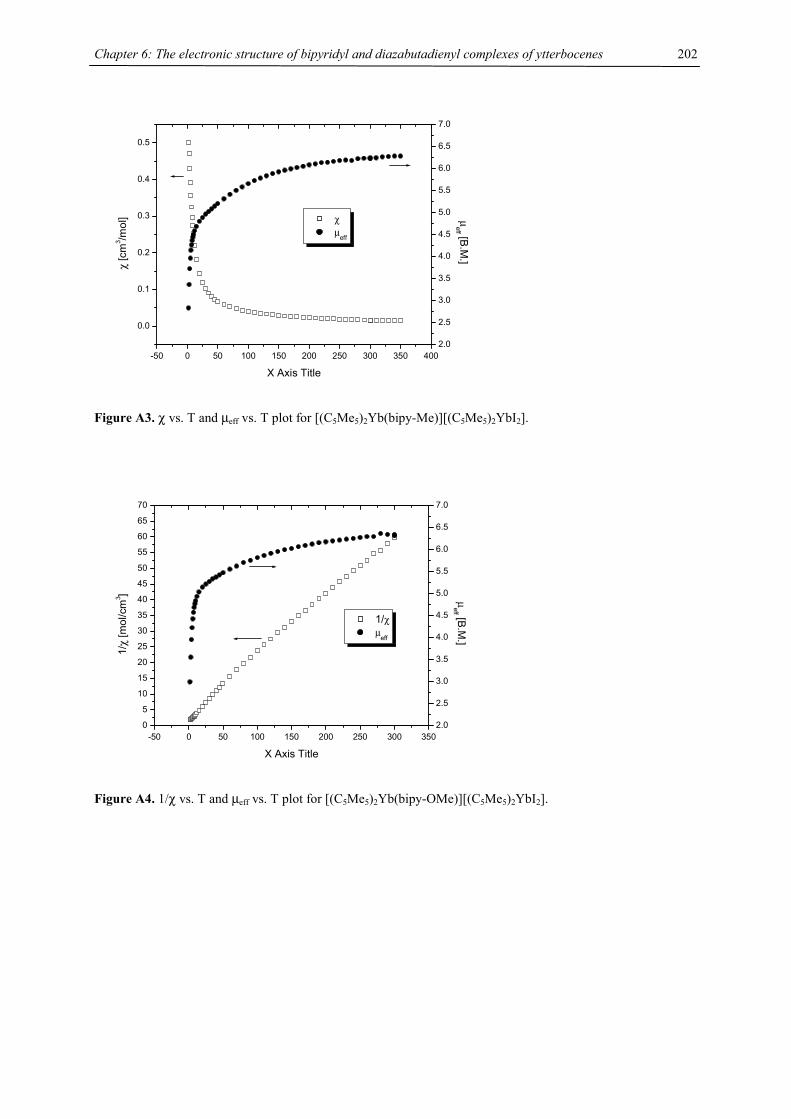
Chapter 6: The electronic structure of bipyridyl and diazabutadienyl complexes of ytterbocenes
202
-50 0 50 100 150 200 250 300 350 400
0.0
0.1
0.2
0.3
0.4
0.5
χ µeff
X Axis Title
χ [c
m3 /m
ol]
2.0
2.5
3.0
3.5
4.0
4.5
5.0
5.5
6.0
6.5
7.0
µeff [B.M
.]
Figure A3. χ vs. T and µeff vs. T plot for [(C5Me5)2Yb(bipy-Me)][(C5Me5)2YbI2].
-50 0 50 100 150 200 250 300 3500
5
10
15
20
25
30
35
40
45
50
55
60
65
70
1/χ µ
eff
X Axis Title
1/χ
[mol
/cm
3 ]
2.0
2.5
3.0
3.5
4.0
4.5
5.0
5.5
6.0
6.5
7.0
µeff [B.M
.]
Figure A4. 1/χ vs. T and µeff vs. T plot for [(C5Me5)2Yb(bipy-OMe)][(C5Me5)2YbI2].
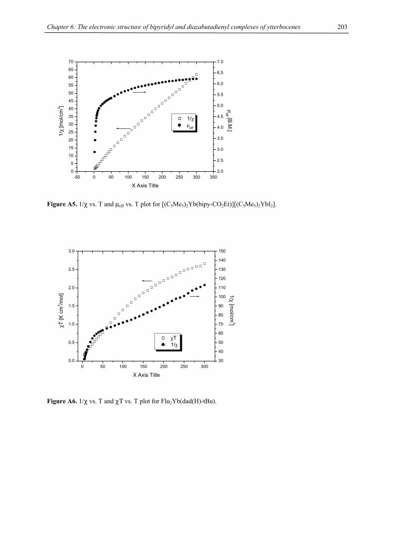
Chapter 6: The electronic structure of bipyridyl and diazabutadienyl complexes of ytterbocenes
203
-50 0 50 100 150 200 250 300 3500
5
10
15
20
25
30
35
40
45
50
55
60
65
70
1/χ µ
eff
X Axis Title
1/χ
[mol
/cm
3 ]
2.0
2.5
3.0
3.5
4.0
4.5
5.0
5.5
6.0
6.5
7.0
µeff [B.M
.]
Figure A5. 1/χ vs. T and µeff vs. T plot for [(C5Me5)2Yb(bipy-CO2Et)][(C5Me5)2YbI2].
0 50 100 150 200 250 3000.0
0.5
1.0
1.5
2.0
2.5
3.0
χT 1/χ
X Axis Title
χT [K
cm
3 /mol
]
30
40
50
60
70
80
90
100
110
120
130
140
150
1/χ [mol/cm
3]
Figure A6. 1/χ vs. T and χΤ vs. T plot for Flu2Yb(dad(H)-tBu).
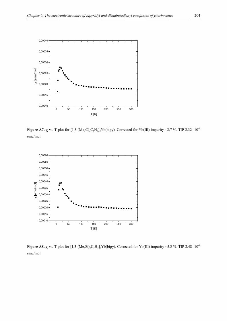
Chapter 6: The electronic structure of bipyridyl and diazabutadienyl complexes of ytterbocenes
204
Figure A7. χ vs. T plot for [1,3-(Me3C)2C5H3]2Yb(bipy). Corrected for Yb(III) impurity ~2.7 %. TIP 2.32 . 10-4
emu/mol.
Figure A8. χ vs. T plot for [1,3-(Me3Si)2C5H3]2Yb(bipy). Corrected for Yb(III) impurity ~5.8 %. TIP 2.48 . 10-4
emu/mol.
0 50 100 150 200 250 3000,00010
0,00015
0,00020
0,00025
0,00030
0,00035
0,00040
χ
[em
u/m
ol]
T [K]
0 50 100 150 200 250 3000,00010
0,00015
0,00020
0,00025
0,00030
0,00035
0,00040
0,00045
0,00050
0,00055
0,00060
χ [e
mu/
mol
]
T [K]
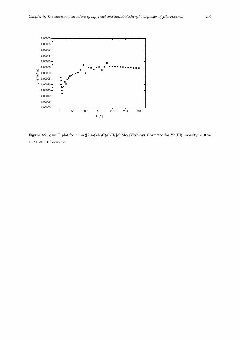
Chapter 6: The electronic structure of bipyridyl and diazabutadienyl complexes of ytterbocenes
205
Figure A9. χ vs. T plot for ansa-{[2,4-(Me3C)2C5H2]2SiMe2}Yb(bipy). Corrected for Yb(III) impurity ~1.0 %.
TIP 1.98 . 10-4 emu/mol.
0 50 100 150 200 250 3000,00000
0,00005
0,00010
0,00015
0,00020
0,00025
0,00030
0,00035
0,00040
0,00045
0,00050
0,00055
0,00060
χ
[em
u/m
ol]
T [K]
Chapter 7: Titanium Complexes with 1,3-Di(tert.-butyl)cyclopentadienyl Ligands 206
Chapter 7: Titanium Complexes with 1,3-Di(tert.-butyl)cyclopentadienyl Ligands
Introduction
Of the first row transition metal metallocenes titanocene represented a significant challenge with respect to the
isolation of a pure and characterizable product. Historically, the chemistry of titanocenes has been inseparable
from intramolecular oxidative addition reactions and, until recently, no isolable base-free titanocenes were
known. A further complication arises due to the fact that most titanocene derived species are in equilibrium with
each other.1, 2
In analogy to ferrocene Fischer and Wilkinson prepared in 1956 a green compound, whose empirical formula
was in agreement with (C5H5)2Ti, from TiCl2 and Na(C5H5). “Titanocene” was prepared later by numerous other
synthetic strategies, but questions about its molecular structure and characterization ensued. The mystery
concerning the nature of this compound was solved by the extensive work of Brintzinger and Bercaw, who
proposed that (C5H5)2Ti was formed initially, but rapidly underwent intramolecular oxidative addition of a C-H
bond on a cyclopentadienyl ring to form (η1-C5H4)(η5-C5H5)TiH.3 The two C5H4 groups couple to form a
bridging fulvalene ligand. This proposed structure was confirmed by mass spectra, 13C NMR spectroscopy3 and
in 1992 by X-ray crystallography.4 This kind of coupling is inhibited by the introduction of the
pentamethylcyclopentadienyl ligand into the titanium chemistry. Brintzinger and Bercaw were also pioneers in
the study of the (C5Me5)2Ti system, the most fundamental studies in this field were done in their laboratories.1, 2,
5-9 However two recent crystal structures of the titanocenes, (C5Me4SiMe2CMe3)2Ti10 and (C5Me4SiMe3)2Ti 11,
have finally closed the chapter on the structural questions surrounding Ti(II) metallocenes that began nearly 50
years ago.4, 12, 13
In an attempt to prepare a base-free titanocene Sofield employed the 1,3-(Me3C)2C5H3 ligand,14 abbreviated Cp’,
which has not been used as extensively as the C5H5 (Cp) or the Me5C5 (Cp*)- ligands in the preparation of
metallocene derivatives of the early d-transition metals.14 However, it has been employed to prepare f-block
metallocenes since it is potentially diastereotopic, a property that can be useful in dynamic NMR studies.15 A
possible reason for the lack of interest in the 1,3-(Me3C)2C5H3-ligand is that its chemical and physical properties
are comparable to those of their C5Me5–analogues. Structurally this is true as demonstrated by bond distances
and angles. However, the main difference is the smaller Cp(centroid)-Ti-Cp(centroid) angle compared to C5Me5,
caused by the net steric repulsion in the bent metallocene. Thermochemical studies on group 4 metallocene
derivatives revealed that the bond dissociation enthalpies (BDE’s) is about 20 kcal/mol weaker for the
(Me3C)2C5H3 derivatives relative to their C5Me5 analogues.16 Due to reduced steric strain and weaker Ti-X bonds
Cp’2TiCl was found to be a suitable synthon for the synthesis of base-free tetra(tert.-butyl) titanocene(II), Cp’2Ti,
via reduction with K/Hg in n-hexane under an argon atmosphere. However, this metallocene is highly
susceptible towards even trace amounts of N2 to form (Cp’2Ti)2(µ-N2). This is in contrast to (C5Me5SiMe3)2Ti11
and (C5Me4SiMe2CMe3)2Ti10, which were reported to be resistant to N2 coordination. Sofield also explored the
reactivity of Cp’2Ti towards a variety of substrates, e.g. H2, C2H4, PhCCPh, N2 and CO, some of them have been
characterized by X-ray diffraction (Scheme 1).14
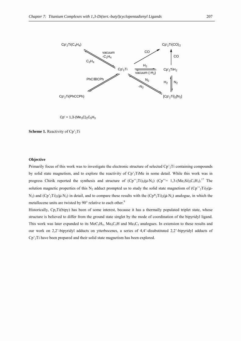
Chapter 7: Titanium Complexes with 1,3-Di(tert.-butyl)cyclopentadienyl Ligands 207
Cp'2Ti
PhC CPh
H2
Cp'2Ti(C4H4)
Cp'2Ti(PhCCPh)
Cp'2TiH2
[Cp'2Ti]2[N2]
vacuum (-H2)
C2H4
vacuum -C2H4
Cp'2Ti(CO)2
COCO
Cp' = 1,3-(Me3C)2C5H3
N2H2-N2
N2
Scheme 1. Reactivity of Cp’2Ti
Objective
Primarily focus of this work was to investigate the electronic structure of selected Cp’2Ti containing compounds
by solid state magnetism, and to explore the reactivity of Cp’2TiMe in some detail. While this work was in
progress Chirik reported the synthesis and structure of (Cp’’2Ti)2(µ-N2) (Cp’’= 1,3-(Me3Si)2C5H3).17 The
solution magnetic properties of this N2 adduct prompted us to study the solid state magnetism of (Cp’’2Ti)2(µ-
N2) and (Cp’2Ti)2(µ-N2) in detail, and to compare these results with the (Cp*2Ti)2(µ-N2) analogue, in which the
metallocene units are twisted by 90° relative to each other.8
Historically, Cp2Ti(bipy) has been of some interest, because it has a thermally populated triplet state, whose
structure is believed to differ from the ground state singlet by the mode of coordination of the bipyridyl ligand.
This work was later expanded to its MeC5H5, Me4C5H and Me5C5 analogues. In extension to these results and
our work on 2,2’-bipyridyl adducts on ytterbocenes, a series of 4,4’-disubstituted 2,2’-bipyridyl adducts of
Cp’2Ti have been prepared and their solid state magnetism has been explored.

Chapter 7: Titanium Complexes with 1,3-Di(tert.-butyl)cyclopentadienyl Ligands 208
Results and Discussion
The starting materials for the synthesis of Cp’2Ti containing molecules is Cp’2TiCl, whose synthesis has been
described earlier.16, 18 However, our preparation follows our well established magnesocene route (Scheme 2),
which has been used in the case of uranium,15 cerium19, 20 and manganese (see Chapter 2).
Cp' = 1,3-(Me3C)2C5H3
Cp'2Mg + TiCl3(thf)3
Cp'2TiCl
THF - MgCl2
LiMe - LiCl
Cp'2TiMe
Cp'2TiH
H2OCp'2Ti(OH)
-CH4
H2 -CH4
D2
-CH3Dd36 - Cp'2TiD
K/Hg, N2 [Cp'2Ti]2(N2)toluene-NaCl
K/Hg, Ar
n-hexaneCp'2Ti
-NaCl
Scheme 2.
Cp’2TiCl is obtained as dark-blue crystals from hexane, m.p. 145-146 °C. The physical properties are in
agreement with the ones reported previously.16 The starting material, Cp’2TiCl, shows the same broad feature at
3.82 ppm (ν1/2= 430 Hz) in the 1H NMR spectrum in C6D6. Oxidation with CCl4 or HgCl2 gives the known
Cp’2TiCl2 (see Experimental Section for details) (Table 1). The χm-1 vs. T plot for Cp’2TiCl is linear from 5-300
K with µeff= 1.74 B.M. (Fig. 1) This value is in agreement with the value calculated from the EPR data (gav=
1.9677) as well as the expected S= ½ ground state.
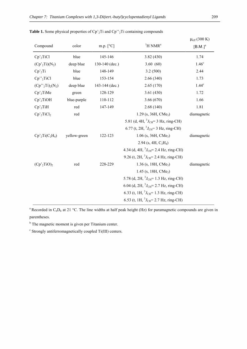
Chapter 7: Titanium Complexes with 1,3-Di(tert.-butyl)cyclopentadienyl Ligands 209
Table 1. Some physical properties of Cp’2Ti and Cp’’2Ti containing compounds
Compound
color
m.p. [°C]
1H NMRa
µeff (300 K)
[Β.Μ.]a
Cp’2TiCl blue 145-146 3.82 (430) 1.74
(Cp’2Ti)(N2) deep blue 130-140 (dec.) 3.60 (60) 1.46c
Cp’2Ti blue 148-149 3.2 (500) 2.44
Cp’’2TiCl blue 153-154 2.66 (340) 1.73
(Cp’’2Ti)2(N2) deep blue 143-144 (dec.) 2.65 (170) 1.44c
Cp’2TiMe green 128-129 3.61 (430) 1.72
Cp’2TiOH blue-purple 110-112 3.66 (670) 1.66
Cp’2TiH red 147-149 2.68 (140) 1.81
Cp’2TiCl2 red 1.29 (s, 36H, CMe3)
5.81 (d, 4H, 3JCH= 3 Hz, ring-CH)
6.77 (t, 2H, 3JCH= 3 Hz, ring-CH)
diamagnetic
Cp’2Ti(C2H4) yellow-green 122-123 1.06 (s, 36H, CMe3)
2.94 (s, 4H, C2H4)
4.34 (d, 4H, 3JCH= 2.4 Hz, ring-CH)
9.26 (t, 2H, 3JCH= 2.4 Hz, ring-CH)
diamagnetic
(Cp’2TiO)2 red 228-229 1.36 (s, 18H, CMe3)
1.45 (s, 18H, CMe3)
5.78 (d, 2H, 3JCH= 1.3 Hz, ring-CH)
6.04 (d, 2H, 3JCH= 2.7 Hz, ring-CH)
6.33 (t, 1H, 3JCH= 1.3 Hz, ring-CH)
6.53 (t, 1H, 3JCH= 2.7 Hz, ring-CH)
diamagnetic
a Recorded in C6D6 at 21 °C. The line widths at half peak height (Hz) for paramagnetic compounds are given in
parentheses. b The magnetic moment is given per Titanium center. c Strongly antiferromagnetically coupled Ti(III) centers.
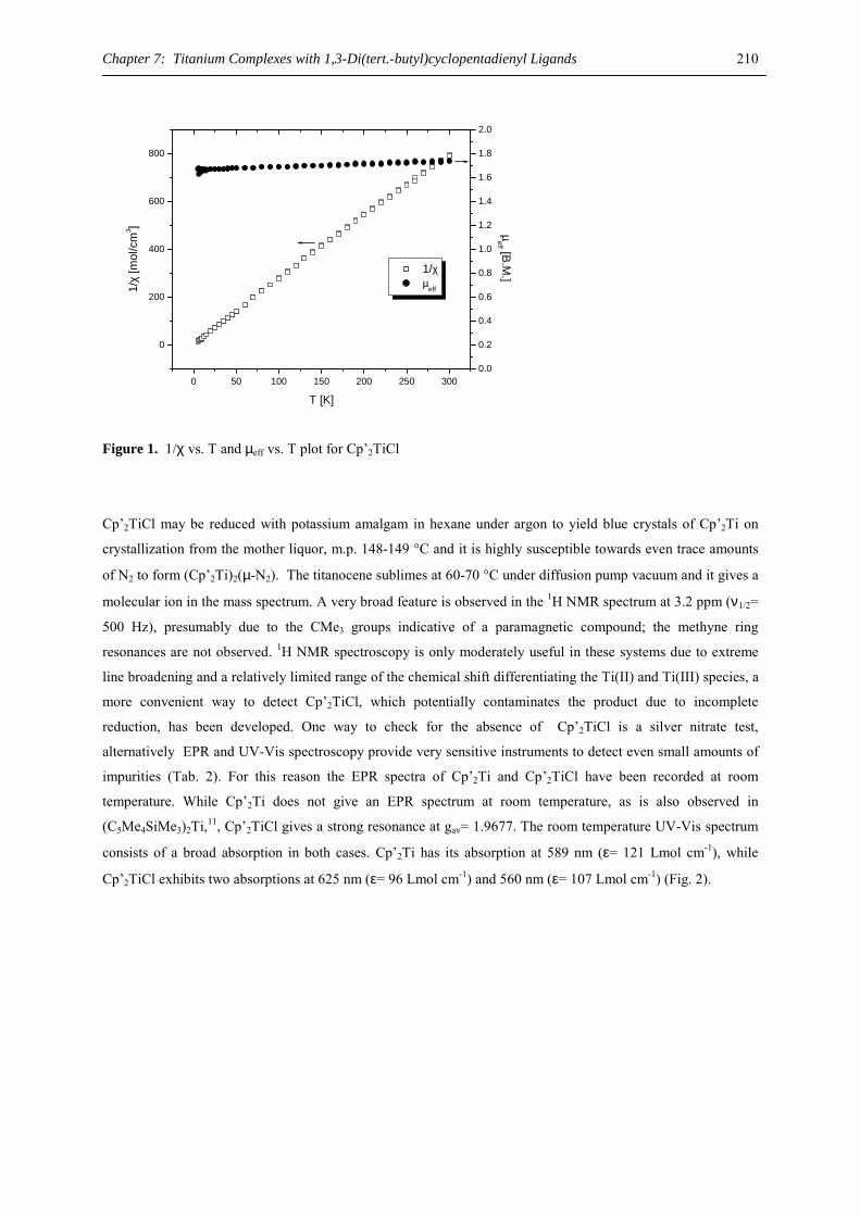
Chapter 7: Titanium Complexes with 1,3-Di(tert.-butyl)cyclopentadienyl Ligands 210
0 50 100 150 200 250 300
0
200
400
600
800
1/χ µ
eff
T [K]
1/χ
[mol
/cm
3 ]
0.0
0.2
0.4
0.6
0.8
1.0
1.2
1.4
1.6
1.8
2.0
µeff [B.M
.]
Figure 1. 1/χ vs. T and µeff vs. T plot for Cp’2TiCl
Cp’2TiCl may be reduced with potassium amalgam in hexane under argon to yield blue crystals of Cp’2Ti on
crystallization from the mother liquor, m.p. 148-149 °C and it is highly susceptible towards even trace amounts
of N2 to form (Cp’2Ti)2(µ-N2). The titanocene sublimes at 60-70 °C under diffusion pump vacuum and it gives a
molecular ion in the mass spectrum. A very broad feature is observed in the 1H NMR spectrum at 3.2 ppm (ν1/2=
500 Hz), presumably due to the CMe3 groups indicative of a paramagnetic compound; the methyne ring
resonances are not observed. 1H NMR spectroscopy is only moderately useful in these systems due to extreme
line broadening and a relatively limited range of the chemical shift differentiating the Ti(II) and Ti(III) species, a
more convenient way to detect Cp’2TiCl, which potentially contaminates the product due to incomplete
reduction, has been developed. One way to check for the absence of Cp’2TiCl is a silver nitrate test,
alternatively EPR and UV-Vis spectroscopy provide very sensitive instruments to detect even small amounts of
impurities (Tab. 2). For this reason the EPR spectra of Cp’2Ti and Cp’2TiCl have been recorded at room
temperature. While Cp’2Ti does not give an EPR spectrum at room temperature, as is also observed in
(C5Me4SiMe3)2Ti,11, Cp’2TiCl gives a strong resonance at gav= 1.9677. The room temperature UV-Vis spectrum
consists of a broad absorption in both cases. Cp’2Ti has its absorption at 589 nm (ε= 121 Lmol cm-1), while
Cp’2TiCl exhibits two absorptions at 625 nm (ε= 96 Lmol cm-1) and 560 nm (ε= 107 Lmol cm-1) (Fig. 2).
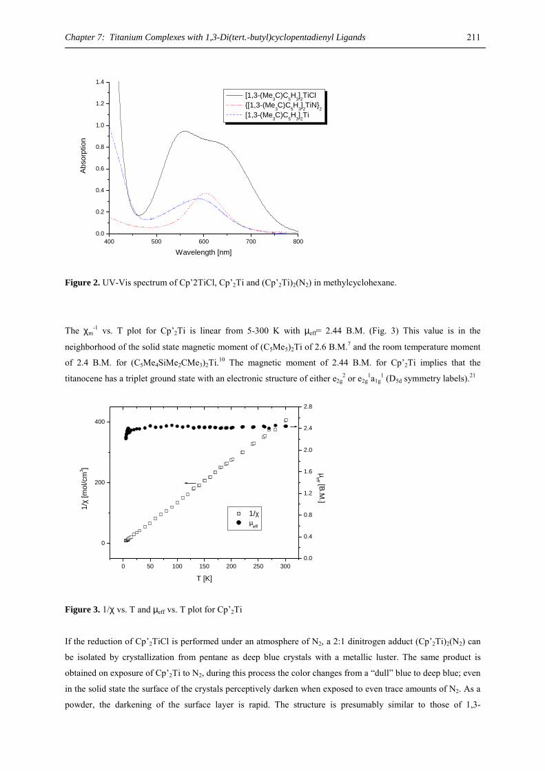
Chapter 7: Titanium Complexes with 1,3-Di(tert.-butyl)cyclopentadienyl Ligands 211
400 500 600 700 8000.0
0.2
0.4
0.6
0.8
1.0
1.2
1.4A
bsor
ptio
n
Wavelength [nm]
[1,3-(Me3C)C5H3]2TiCl {[1,3-(Me
3C)C
5H
3]2TiN}
2 [1,3-(Me3C)C5H3]2Ti
Figure 2. UV-Vis spectrum of Cp’2TiCl, Cp’2Ti and (Cp’2Ti)2(N2) in methylcyclohexane.
The χm-1 vs. T plot for Cp’2Ti is linear from 5-300 K with µeff= 2.44 B.M. (Fig. 3) This value is in the
neighborhood of the solid state magnetic moment of (C5Me5)2Ti of 2.6 B.M.7 and the room temperature moment
of 2.4 B.M. for (C5Me4SiMe2CMe3)2Ti.10 The magnetic moment of 2.44 B.M. for Cp’2Ti implies that the
titanocene has a triplet ground state with an electronic structure of either e2g2 or e2g
1a1g1 (D5d symmetry labels).21
0 50 100 150 200 250 300
0
200
400
1/χ µ
eff
T [K]
1/χ
[mol
/cm
3 ]
0.0
0.4
0.8
1.2
1.6
2.0
2.4
2.8
µeff [B.M
.]
Figure 3. 1/χ vs. T and µeff vs. T plot for Cp’2Ti
If the reduction of Cp’2TiCl is performed under an atmosphere of N2, a 2:1 dinitrogen adduct (Cp’2Ti)2(N2) can
be isolated by crystallization from pentane as deep blue crystals with a metallic luster. The same product is
obtained on exposure of Cp’2Ti to N2, during this process the color changes from a “dull” blue to deep blue; even
in the solid state the surface of the crystals perceptively darken when exposed to even trace amounts of N2. As a
powder, the darkening of the surface layer is rapid. The structure is presumably similar to those of 1,3-
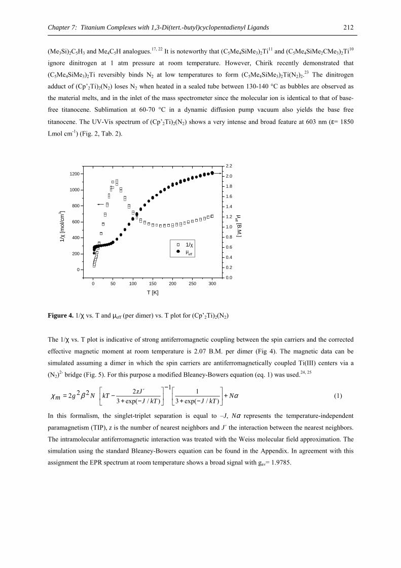
Chapter 7: Titanium Complexes with 1,3-Di(tert.-butyl)cyclopentadienyl Ligands 212
(Me3Si)2C5H3 and Me4C5H analogues.17, 22 It is noteworthy that (C5Me4SiMe3)2Ti11 and (C5Me4SiMe2CMe3)2Ti10
ignore dinitrogen at 1 atm pressure at room temperature. However, Chirik recently demonstrated that
(C5Me4SiMe3)2Ti reversibly binds N2 at low temperatures to form (C5Me4SiMe3)2Ti(N2)2.23 The dinitrogen
adduct of (Cp’2Ti)2(N2) loses N2 when heated in a sealed tube between 130-140 °C as bubbles are observed as
the material melts, and in the inlet of the mass spectrometer since the molecular ion is identical to that of base-
free titanocene. Sublimation at 60-70 °C in a dynamic diffusion pump vacuum also yields the base free
titanocene. The UV-Vis spectrum of (Cp’2Ti)2(N2) shows a very intense and broad feature at 603 nm (ε= 1850
Lmol cm-1) (Fig. 2, Tab. 2).
0 50 100 150 200 250 300
0
200
400
600
800
1000
1200
1/χ µ
eff
T [K]
1/χ
[mol
/cm
3 ]
0.0
0.2
0.4
0.6
0.8
1.0
1.2
1.4
1.6
1.8
2.0
2.2µ
eff [B.M.]
Figure 4. 1/χ vs. T and µeff (per dimer) vs. T plot for (Cp’2Ti)2(N2)
The 1/χ vs. T plot is indicative of strong antiferromagnetic coupling between the spin carriers and the corrected
effective magnetic moment at room temperature is 2.07 B.M. per dimer (Fig 4). The magnetic data can be
simulated assuming a dimer in which the spin carriers are antiferromagnetically coupled Ti(III) centers via a
(N2)2- bridge (Fig. 5). For this purpose a modified Bleaney-Bowers equation (eq. 1) was used.24, 25
αβχ NkTJkTJ
zJkTNgm +
−+
−
−+−=
)/exp(311
)/exp(3´2222 (1)
In this formalism, the singlet-triplet separation is equal to –J, Nα represents the temperature-independent
paramagnetism (TIP), z is the number of nearest neighbors and J´ the interaction between the nearest neighbors.
The intramolecular antiferromagnetic interaction was treated with the Weiss molecular field approximation. The
simulation using the standard Bleaney-Bowers equation can be found in the Appendix. In agreement with this
assignment the EPR spectrum at room temperature shows a broad signal with gav= 1.9785.
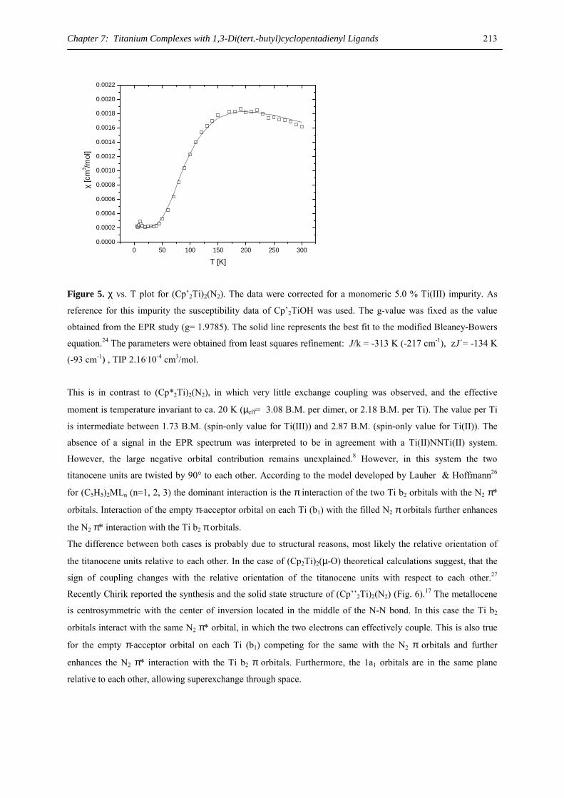
Chapter 7: Titanium Complexes with 1,3-Di(tert.-butyl)cyclopentadienyl Ligands 213
0 50 100 150 200 250 3000.0000
0.0002
0.0004
0.0006
0.0008
0.0010
0.0012
0.0014
0.0016
0.0018
0.0020
0.0022
χ
[cm
3 /mol
]
T [K]
Figure 5. χ vs. T plot for (Cp’2Ti)2(N2). The data were corrected for a monomeric 5.0 % Ti(III) impurity. As
reference for this impurity the susceptibility data of Cp’2TiOH was used. The g-value was fixed as the value
obtained from the EPR study (g= 1.9785). The solid line represents the best fit to the modified Bleaney-Bowers
equation.24 The parameters were obtained from least squares refinement: J/k = -313 K (-217 cm-1), zJ´= -134 K
(-93 cm-1) , TIP 2.16.10-4 cm3/mol.
This is in contrast to (Cp*2Ti)2(N2), in which very little exchange coupling was observed, and the effective
moment is temperature invariant to ca. 20 K (µeff= 3.08 B.M. per dimer, or 2.18 B.M. per Ti). The value per Ti
is intermediate between 1.73 B.M. (spin-only value for Ti(III)) and 2.87 B.M. (spin-only value for Ti(II)). The
absence of a signal in the EPR spectrum was interpreted to be in agreement with a Ti(II)NNTi(II) system.
However, the large negative orbital contribution remains unexplained.8 However, in this system the two
titanocene units are twisted by 90° to each other. According to the model developed by Lauher & Hoffmann26
for (C5H5)2MLn (n=1, 2, 3) the dominant interaction is the π interaction of the two Ti b2 orbitals with the N2 π*
orbitals. Interaction of the empty π-acceptor orbital on each Ti (b1) with the filled N2 π orbitals further enhances
the N2 π* interaction with the Ti b2 π orbitals.
The difference between both cases is probably due to structural reasons, most likely the relative orientation of
the titanocene units relative to each other. In the case of (Cp2Ti)2(µ-O) theoretical calculations suggest, that the
sign of coupling changes with the relative orientation of the titanocene units with respect to each other.27
Recently Chirik reported the synthesis and the solid state structure of (Cp’’2Ti)2(N2) (Fig. 6).17 The metallocene
is centrosymmetric with the center of inversion located in the middle of the N-N bond. In this case the Ti b2
orbitals interact with the same N2 π* orbital, in which the two electrons can effectively couple. This is also true
for the empty π-acceptor orbital on each Ti (b1) competing for the same with the N2 π orbitals and further
enhances the N2 π* interaction with the Ti b2 π orbitals. Furthermore, the 1a1 orbitals are in the same plane
relative to each other, allowing superexchange through space.
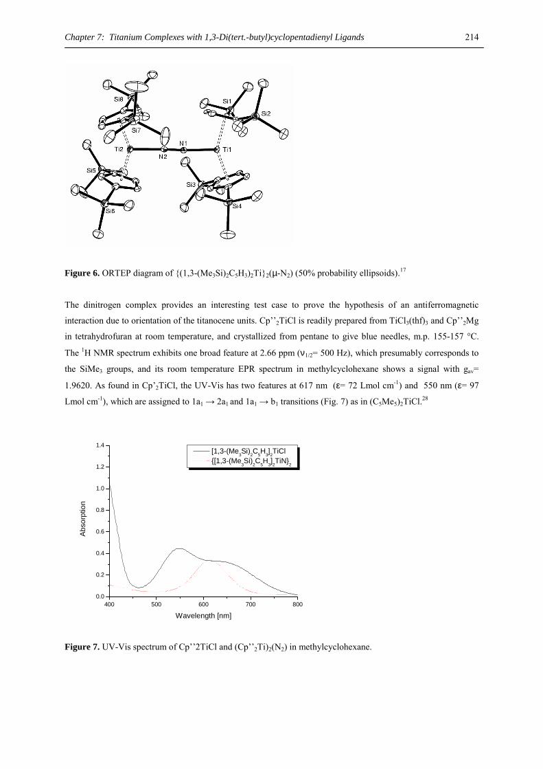
Chapter 7: Titanium Complexes with 1,3-Di(tert.-butyl)cyclopentadienyl Ligands 214
Figure 6. ORTEP diagram of {(1,3-(Me3Si)2C5H3)2Ti}2(µ-N2) (50% probability ellipsoids).17
The dinitrogen complex provides an interesting test case to prove the hypothesis of an antiferromagnetic
interaction due to orientation of the titanocene units. Cp’’2TiCl is readily prepared from TiCl3(thf)3 and Cp’’2Mg
in tetrahydrofuran at room temperature, and crystallized from pentane to give blue needles, m.p. 155-157 °C.
The 1H NMR spectrum exhibits one broad feature at 2.66 ppm (ν1/2= 500 Hz), which presumably corresponds to
the SiMe3 groups, and its room temperature EPR spectrum in methylcyclohexane shows a signal with gav=
1.9620. As found in Cp’2TiCl, the UV-Vis has two features at 617 nm (ε= 72 Lmol cm-1) and 550 nm (ε= 97
Lmol cm-1), which are assigned to 1a1 → 2a1 and 1a1 → b1 transitions (Fig. 7) as in (C5Me5)2TiCl.28
400 500 600 700 8000.0
0.2
0.4
0.6
0.8
1.0
1.2
1.4
Abs
orpt
ion
Wavelength [nm]
[1,3-(Me3Si)2C5H3]2TiCl {[1,3-(Me
3Si)
2C
5H
3]2TiN}
2
Figure 7. UV-Vis spectrum of Cp’’2TiCl and (Cp’’2Ti)2(N2) in methylcyclohexane.
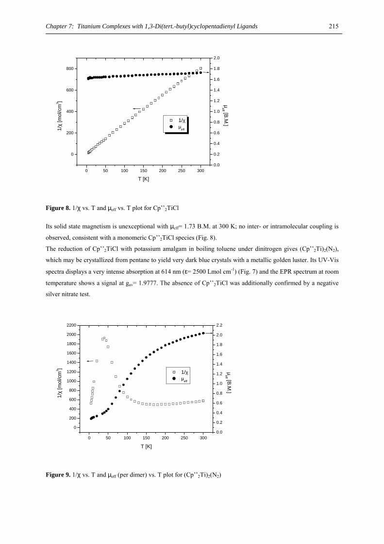
Chapter 7: Titanium Complexes with 1,3-Di(tert.-butyl)cyclopentadienyl Ligands 215
0 50 100 150 200 250 300
0
200
400
600
800
1/χ µ
eff
T [K]
1/χ
[mol
/cm
3 ]
0.0
0.2
0.4
0.6
0.8
1.0
1.2
1.4
1.6
1.8
2.0
µeff [B.M
.]
Figure 8. 1/χ vs. T and µeff vs. T plot for Cp’’2TiCl
Its solid state magnetism is unexceptional with µeff= 1.73 B.M. at 300 K; no inter- or intramolecular coupling is
observed, consistent with a monomeric Cp’’2TiCl species (Fig. 8).
The reduction of Cp’’2TiCl with potassium amalgam in boiling toluene under dinitrogen gives (Cp’’2Ti)2(N2),
which may be crystallized from pentane to yield very dark blue crystals with a metallic golden luster. Its UV-Vis
spectra displays a very intense absorption at 614 nm (ε= 2500 Lmol cm-1) (Fig. 7) and the EPR spectrum at room
temperature shows a signal at gav= 1.9777. The absence of Cp’’2TiCl was additionally confirmed by a negative
silver nitrate test.
0 50 100 150 200 250 300
0
200
400
600
800
1000
1200
1400
1600
1800
2000
2200
1/χ µeff
T [K]
1/χ
[mol
/cm
3 ]
0.0
0.2
0.4
0.6
0.8
1.0
1.2
1.4
1.6
1.8
2.0
2.2
µeff [B.M
.]
Figure 9. 1/χ vs. T and µeff (per dimer) vs. T plot for (Cp’’2Ti)2(N2)
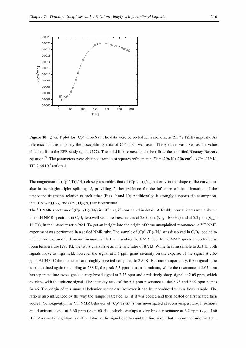
Chapter 7: Titanium Complexes with 1,3-Di(tert.-butyl)cyclopentadienyl Ligands 216
0 50 100 150 200 250 3000.0000
0.0002
0.0004
0.0006
0.0008
0.0010
0.0012
0.0014
0.0016
0.0018
0.0020
0.0022
χ
[cm
3 /mol
]
T [K]
Figure 10. χ vs. T plot for (Cp’’2Ti)2(N2). The data were corrected for a monomeric 2.5 % Ti(III) impurity. As
reference for this impurity the susceptibility data of Cp’’2TiCl was used. The g-value was fixed as the value
obtained from the EPR study (g= 1.9777). The solid line represents the best fit to the modified Bleaney-Bowers
equation.24 The parameters were obtained from least squares refinement: J/k = -296 K (-206 cm-1), zJ´= -119 K,
TIP 2.66.10-4 cm3/mol. The magnetism of (Cp’’2Ti)2(N2) closely resembles that of (Cp’2Ti)2(N2) not only in the shape of the curve, but
also in its singlet-triplet splitting -J, providing further evidence for the influence of the orientation of the
titanocene fragments relative to each other (Figs. 9 and 10) Additionally, it strongly supports the assumption,
that (Cp’’2Ti)2(N2) and (Cp’2Ti)2(N2) are isostructural.
The 1H NMR spectrum of (Cp’’2Ti)2(N2) is difficult, if considered in detail: A freshly crystallized sample shows
in its 1H NMR spectrum in C6D6 two well separated resonances at 2.65 ppm (ν1/2= 160 Hz) and at 5.3 ppm (ν1/2=
44 Hz), in the intensity ratio 96:4. To get an insight into the origin of these unexplained resonances, a VT-NMR
experiment was performed in a sealed NMR tube. The sample of (Cp’’2Ti)2(N2) was dissolved in C7D8, cooled to
–30 °C and exposed to dynamic vacuum, while flame sealing the NMR tube. In the NMR spectrum collected at
room temperature (290 K), the two signals have an intensity ratio of 87:13. While heating sample to 353 K, both
signals move to high field, however the signal at 5.3 ppm gains intensity on the expense of the signal at 2.65
ppm. At 348 °C the intensities are roughly inverted compared to 290 K. But more importantly, the original ratio
is not attained again on cooling at 288 K, the peak 5.3 ppm remains dominant, while the resonance at 2.65 ppm
has separated into two signals, a very broad signal at 2.73 ppm and a relatively sharp signal at 2.09 ppm, which
overlaps with the toluene signal. The intensity ratio of the 5.3 ppm resonance to the 2.73 and 2.09 ppm pair is
54:46. The origin of this unusual behavior is unclear; however it can be reproduced with a fresh sample. The
ratio is also influenced by the way the sample is treated, i.e. if it was cooled and then heated or first heated then
cooled. Consequently, the VT-NMR behavior of (Cp’2Ti)2(N2) was investigated at room temperature. It exhibits
one dominant signal at 3.60 ppm (ν1/2= 60 Hz), which overlaps a very broad resonance at 3.2 ppm (ν1/2~ 160
Hz). An exact integration is difficult due to the signal overlap and the line width, but it is on the order of 10:1.
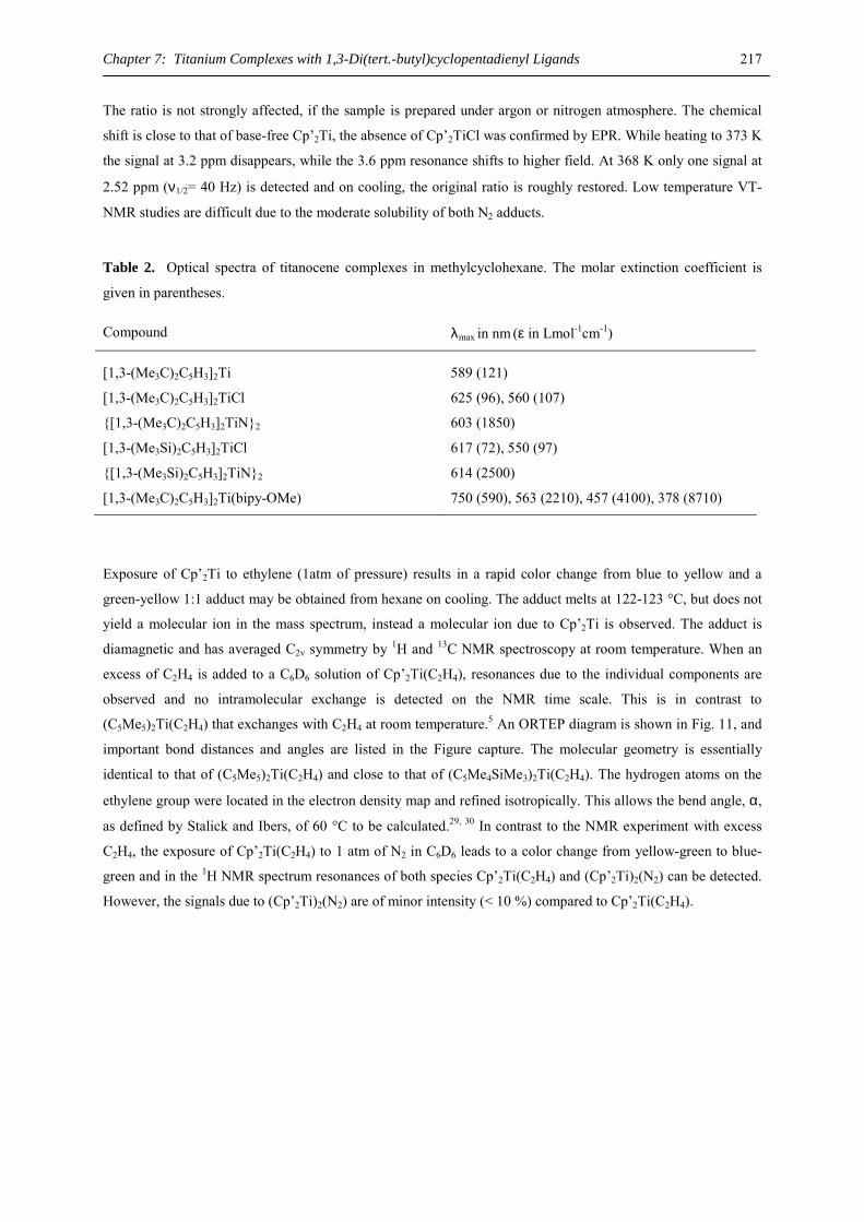
Chapter 7: Titanium Complexes with 1,3-Di(tert.-butyl)cyclopentadienyl Ligands 217
The ratio is not strongly affected, if the sample is prepared under argon or nitrogen atmosphere. The chemical
shift is close to that of base-free Cp’2Ti, the absence of Cp’2TiCl was confirmed by EPR. While heating to 373 K
the signal at 3.2 ppm disappears, while the 3.6 ppm resonance shifts to higher field. At 368 K only one signal at
2.52 ppm (ν1/2= 40 Hz) is detected and on cooling, the original ratio is roughly restored. Low temperature VT-
NMR studies are difficult due to the moderate solubility of both N2 adducts.
Table 2. Optical spectra of titanocene complexes in methylcyclohexane. The molar extinction coefficient is
given in parentheses.
Compound λmax in nm (ε in Lmol-1cm-1)
[1,3-(Me3C)2C5H3]2Ti 589 (121)
[1,3-(Me3C)2C5H3]2TiCl 625 (96), 560 (107)
{[1,3-(Me3C)2C5H3]2TiN}2 603 (1850)
[1,3-(Me3Si)2C5H3]2TiCl 617 (72), 550 (97)
{[1,3-(Me3Si)2C5H3]2TiN}2 614 (2500)
[1,3-(Me3C)2C5H3]2Ti(bipy-OMe) 750 (590), 563 (2210), 457 (4100), 378 (8710)
Exposure of Cp’2Ti to ethylene (1atm of pressure) results in a rapid color change from blue to yellow and a
green-yellow 1:1 adduct may be obtained from hexane on cooling. The adduct melts at 122-123 °C, but does not
yield a molecular ion in the mass spectrum, instead a molecular ion due to Cp’2Ti is observed. The adduct is
diamagnetic and has averaged C2v symmetry by 1H and 13C NMR spectroscopy at room temperature. When an
excess of C2H4 is added to a C6D6 solution of Cp’2Ti(C2H4), resonances due to the individual components are
observed and no intramolecular exchange is detected on the NMR time scale. This is in contrast to
(C5Me5)2Ti(C2H4) that exchanges with C2H4 at room temperature.5 An ORTEP diagram is shown in Fig. 11, and
important bond distances and angles are listed in the Figure capture. The molecular geometry is essentially
identical to that of (C5Me5)2Ti(C2H4) and close to that of (C5Me4SiMe3)2Ti(C2H4). The hydrogen atoms on the
ethylene group were located in the electron density map and refined isotropically. This allows the bend angle, α,
as defined by Stalick and Ibers, of 60 °C to be calculated.29, 30 In contrast to the NMR experiment with excess
C2H4, the exposure of Cp’2Ti(C2H4) to 1 atm of N2 in C6D6 leads to a color change from yellow-green to blue-
green and in the 1H NMR spectrum resonances of both species Cp’2Ti(C2H4) and (Cp’2Ti)2(N2) can be detected.
However, the signals due to (Cp’2Ti)2(N2) are of minor intensity (< 10 %) compared to Cp’2Ti(C2H4).
Chapter 7: Titanium Complexes with 1,3-Di(tert.-butyl)cyclopentadienyl Ligands 218
Figure 11. ORTEP diagram of [1,3-(Me3C)2C5H3]2Ti(H2CCH2) (50% probability ellipsoids). All hydrogen
atoms were located, the hydrogen atoms on the Me3C groups included in calculated positions, but not refined. In
the ORTEP diagram hydrogen atoms have been omitted for clarity, except the hydrogen atoms on the ethylene
ligand which have been refined isotropically.
Metal oxide and hydroxide complexes have long been of special interest to the Andersen group,15, 31, 32
so the behavior of Cp’2Ti and Cp’2Ti(C2H4) towards nitrous oxide, N2O, has been investigated. Nitrous oxide is a
convenient source of oxygen atoms for the synthesis of oxometallocenes (and the products derived therefrom) as
well as for the insertion into metal-hydrogen and –carbon bonds.33-36 On exposure of Cp’2Ti to N2O (1atm of
pressure) Sofield isolated, albeit in small yield, the oxo-bridged dimer (Cp’2Ti)(µ-O),37 whose molecular
structure was determined by X-ray diffraction.38 An ORTEP diagram is shown in Figure 12. Under the same
conditions (C5Me5)2Ti yields a mixture of compounds indicated by its 1H NMR spectrum; resonances due to
(C5Me5)2Ti(CH2C5Me4)(µ-O)33,34 and for (C5Me5)2Ti4(µ-O)639,40 can be identified. Monomeric
(C5Me5)2Ti(O)(py) may be isolated, if the reaction of (C5Me5)2Ti or (C5Me5)2Ti(C2H4) with N2O is conducted in
the presence of pyridine.41
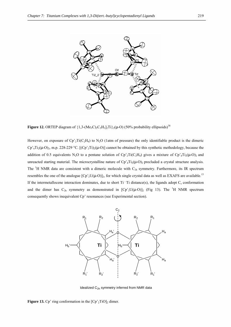
Chapter 7: Titanium Complexes with 1,3-Di(tert.-butyl)cyclopentadienyl Ligands 219
Figure 12. ORTEP diagram of {1,3-(Me3C)2C5H3]2Ti}2(µ-O) (50% probability ellipsoids)38
However, on exposure of Cp’2Ti(C2H4) to N2O (1atm of pressure) the only identifiable product is the dimeric
Cp’4Ti2(µ-O)2, m.p. 228-229 °C. [(Cp’2Ti)2(µ-O)] cannot be obtained by this synthetic methodology, because the
addition of 0.5 equivalents N2O to a pentane solution of Cp’2Ti(C2H4) gives a mixture of Cp’4Ti2(µ-O)2 and
unreacted starting material. The microcrystalline nature of Cp’4Ti2(µ-O)2 precluded a crystal structure analysis.
The 1H NMR data are consistent with a dimeric molecule with C2h symmetry. Furthermore, its IR spectrum
resembles the one of the analogue [Cp’2U(µ-O)]2, for which single crystal data as well as EXAFS are available.15
If the intermetallocene interaction dominates, due to short Ti…Ti distance(s), the ligands adopt Cs conformation
and the dimer has C2h symmetry as demonstrated in [Cp’2U(µ-O)]2 (Fig 13). The 1H NMR spectrum
consequently shows inequivalent Cp’ resonances (see Experimental section).
Idealized C2h symmetry inferred from NMR data
C2
R1
R1'
R2
R2'
R2 R1
R2' R1'
O
O
Hb
Ha
Ha
Ha'
Ha'
Hb' Ti Ti
Figure 13. Cp’ ring conformation in the [Cp’2TiO]2 dimer.
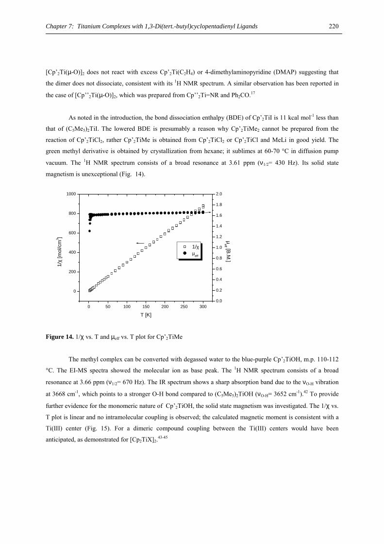
Chapter 7: Titanium Complexes with 1,3-Di(tert.-butyl)cyclopentadienyl Ligands 220
[Cp’2Ti(µ-O)]2 does not react with excess Cp’2Ti(C2H4) or 4-dimethylaminopyridine (DMAP) suggesting that
the dimer does not dissociate, consistent with its 1H NMR spectrum. A similar observation has been reported in
the case of [Cp’’2Ti(µ-O)]2, which was prepared from Cp’’2Ti=NR and Ph2CO.17
As noted in the introduction, the bond dissociation enthalpy (BDE) of Cp’2TiI is 11 kcal mol-1 less than
that of (C5Me5)2TiI. The lowered BDE is presumably a reason why Cp’2TiMe2 cannot be prepared from the
reaction of Cp’2TiCl2, rather Cp’2TiMe is obtained from Cp’2TiCl2 or Cp’2TiCl and MeLi in good yield. The
green methyl derivative is obtained by crystallization from hexane; it sublimes at 60-70 °C in diffusion pump
vacuum. The 1H NMR spectrum consists of a broad resonance at 3.61 ppm (ν1/2= 430 Hz). Its solid state
magnetism is unexceptional (Fig. 14).
0 50 100 150 200 250 300
0
200
400
600
800
1000
1/χ µeff
T [K]
1/χ
[mol
/cm
3 ]
0.0
0.2
0.4
0.6
0.8
1.0
1.2
1.4
1.6
1.8
2.0
µeff [B.M
.]
Figure 14. 1/χ vs. T and µeff vs. T plot for Cp’2TiMe
The methyl complex can be converted with degassed water to the blue-purple Cp’2TiOH, m.p. 110-112
°C. The EI-MS spectra showed the molecular ion as base peak. The 1H NMR spectrum consists of a broad
resonance at 3.66 ppm (ν1/2= 670 Hz). The IR spectrum shows a sharp absorption band due to the νO-H vibration
at 3668 cm-1, which points to a stronger O-H bond compared to (C5Me5)2TiOH (νO-H= 3652 cm-1).42 To provide
further evidence for the monomeric nature of Cp’2TiOH, the solid state magnetism was investigated. The 1/χ vs.
T plot is linear and no intramolecular coupling is observed; the calculated magnetic moment is consistent with a
Ti(III) center (Fig. 15). For a dimeric compound coupling between the Ti(III) centers would have been
anticipated, as demonstrated for [Cp2TiX]2.43-45
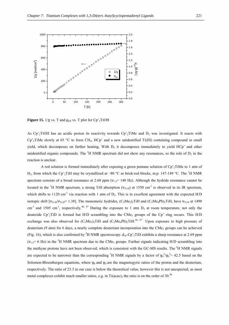
Chapter 7: Titanium Complexes with 1,3-Di(tert.-butyl)cyclopentadienyl Ligands 221
0 50 100 150 200 250 300
0
200
400
600
800
1000
1/χ µ
eff
T [K]
1/χ
[mol
/cm
3 ]
0.0
0.2
0.4
0.6
0.8
1.0
1.2
1.4
1.6
1.8
2.0
µeff [B.M
.]
Figure 15. 1/χ vs. T and µeff vs. T plot for Cp’2TiOH
As Cp’2TiOH has an acidic proton its reactivity towards Cp’2TiMe and D2 was investigated. It reacts with
Cp’2TiMe slowly at 65 °C to form CH4, HCp’ and a new unidentified Ti(III) containing compound in small
yield, which decomposes on further heating, With D2 it decomposes immediately to yield HCp’ and other
unidentified organic compounds. The 2H NMR spectrum did not show any resonances, so the role of D2 in the
reaction is unclear.
A red solution is formed immediately after exposing a green pentane solution of Cp’2TiMe to 1 atm of
H2, from which the Cp’2TiH may be crystallized at –80 °C as brick-red blocks, m.p. 147-149 °C. The 1H NMR
spectrum consists of a broad resonance at 2.68 ppm (ν1/2= 140 Hz). Although the hydride resonance cannot be
located in the 1H NMR spectrum, a strong TiH absorption (νTi-H) at 1550 cm-1 is observed in its IR spectrum,
which shifts to 1120 cm-1 via reaction with 1 atm of D2. This is in excellent agreement with the expected H/D
isotopic shift [νTi-H/νTi-D= 1.38]. The monomeric hydrides, (C5Me5)2TiH and (C5Me4Ph)2TiH, have νTi-H at 1490
cm-1 and 1505 cm-1, respectively.46, 47 During the exposure to 1 atm D2 at room temperature, not only the
deuteride Cp’2TiD is formed but H/D scrambling into the CMe3 groups of the Cp’ ring occurs. This H/D
exchange was also observed for (C5Me5)2TiH and (C5Me4Ph)2TiH.46, 47 Upon exposure to high pressure of
deuterium (9 atm) for 6 days, a nearly complete deuterium incorporation into the CMe3 groups can be achieved
(Fig. 16), which is also confirmed by 2H NMR spectroscopy. d36-Cp’2TiD exhibits a sharp resonance at 2.69 ppm
(ν1/2= 6 Hz) in the 2H NMR spectrum due to the CMe3 groups. Further signals indicating H/D scrambling into
the methyne protons have not been observed, which is consistent with the GC-MS results. The 2H NMR signals
are expected to be narrower than the corresponding 1H NMR signals by a factor of γH2/γD
2= 42.5 based on the
Solomon-Bloembergen equations, where γH and γD
are the magnetogyric ratios of the proton and the deuterium,
respectively. The ratio of 23.3 in our case is below the theoretical value, however this is not unexpected, as most
metal complexes exhibit much smaller ratios, e.g. in Ti(acac)3 the ratio is on the order of 30.48
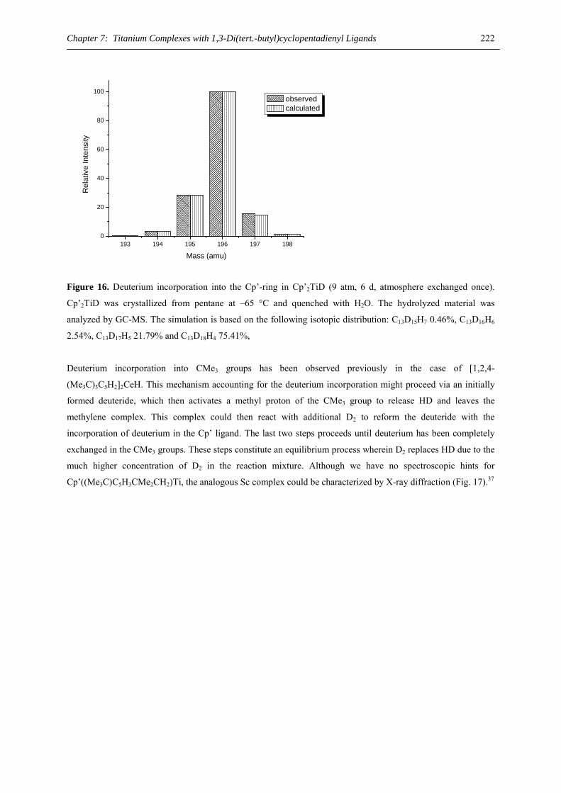
Chapter 7: Titanium Complexes with 1,3-Di(tert.-butyl)cyclopentadienyl Ligands 222
193 194 195 196 197 1980
20
40
60
80
100R
elat
ive
Inte
nsity
Mass (amu)
observed calculated
Figure 16. Deuterium incorporation into the Cp’-ring in Cp’2TiD (9 atm, 6 d, atmosphere exchanged once).
Cp’2TiD was crystallized from pentane at –65 °C and quenched with H2O. The hydrolyzed material was
analyzed by GC-MS. The simulation is based on the following isotopic distribution: C13D15H7 0.46%, C13D16H6
2.54%, C13D17H5 21.79% and C13D18H4 75.41%,
Deuterium incorporation into CMe3 groups has been observed previously in the case of [1,2,4-
(Me3C)3C5H2]2CeH. This mechanism accounting for the deuterium incorporation might proceed via an initially
formed deuteride, which then activates a methyl proton of the CMe3 group to release HD and leaves the
methylene complex. This complex could then react with additional D2 to reform the deuteride with the
incorporation of deuterium in the Cp’ ligand. The last two steps proceeds until deuterium has been completely
exchanged in the CMe3 groups. These steps constitute an equilibrium process wherein D2 replaces HD due to the
much higher concentration of D2 in the reaction mixture. Although we have no spectroscopic hints for
Cp’((Me3C)C5H3CMe2CH2)Ti, the analogous Sc complex could be characterized by X-ray diffraction (Fig. 17).37
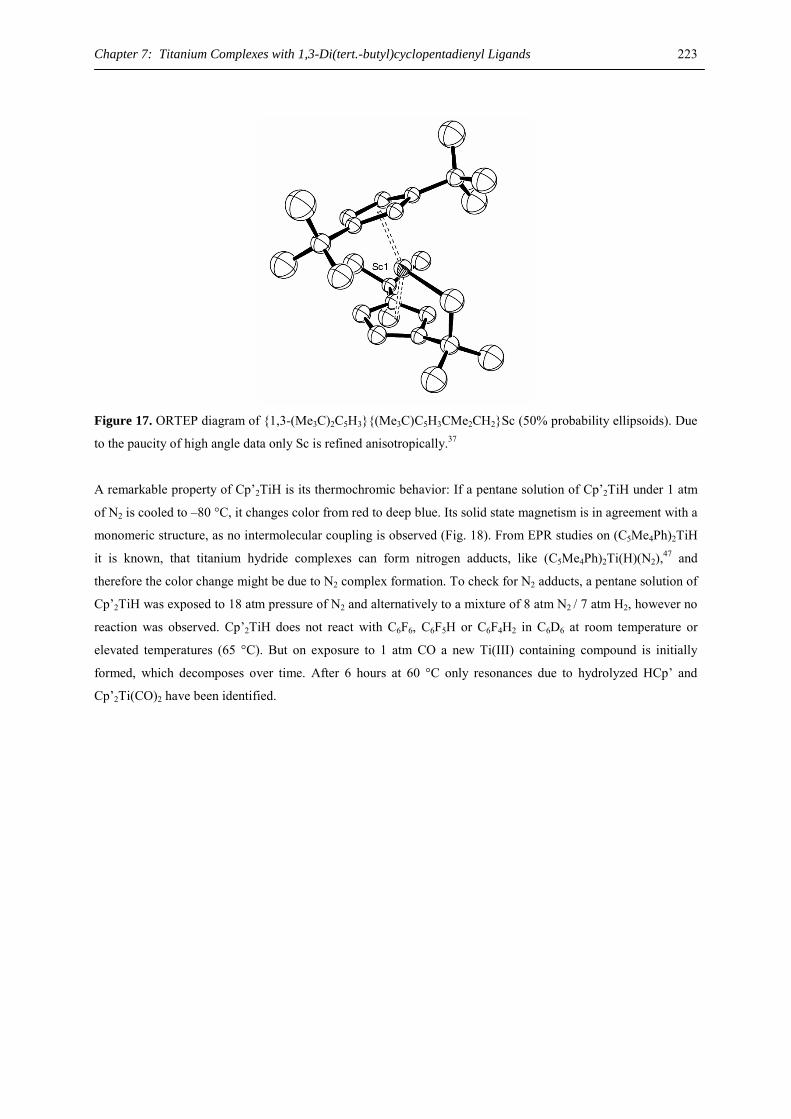
Chapter 7: Titanium Complexes with 1,3-Di(tert.-butyl)cyclopentadienyl Ligands 223
Figure 17. ORTEP diagram of {1,3-(Me3C)2C5H3}{(Me3C)C5H3CMe2CH2}Sc (50% probability ellipsoids). Due
to the paucity of high angle data only Sc is refined anisotropically.37
A remarkable property of Cp’2TiH is its thermochromic behavior: If a pentane solution of Cp’2TiH under 1 atm
of N2 is cooled to –80 °C, it changes color from red to deep blue. Its solid state magnetism is in agreement with a
monomeric structure, as no intermolecular coupling is observed (Fig. 18). From EPR studies on (C5Me4Ph)2TiH
it is known, that titanium hydride complexes can form nitrogen adducts, like (C5Me4Ph)2Ti(H)(N2),47 and
therefore the color change might be due to N2 complex formation. To check for N2 adducts, a pentane solution of
Cp’2TiH was exposed to 18 atm pressure of N2 and alternatively to a mixture of 8 atm N2 / 7 atm H2, however no
reaction was observed. Cp’2TiH does not react with C6F6, C6F5H or C6F4H2 in C6D6 at room temperature or
elevated temperatures (65 °C). But on exposure to 1 atm CO a new Ti(III) containing compound is initially
formed, which decomposes over time. After 6 hours at 60 °C only resonances due to hydrolyzed HCp’ and
Cp’2Ti(CO)2 have been identified.
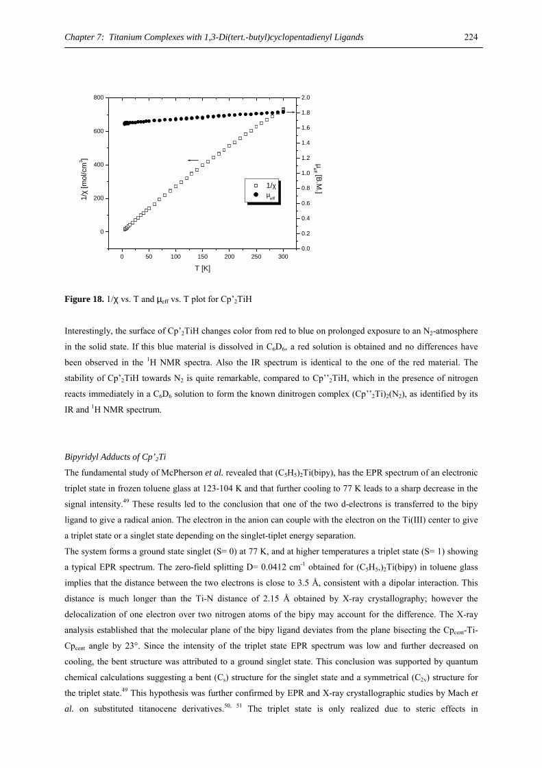
Chapter 7: Titanium Complexes with 1,3-Di(tert.-butyl)cyclopentadienyl Ligands 224
0 50 100 150 200 250 300
0
200
400
600
800
1/χ µ
eff
T [K]
1/χ
[mol
/cm
3 ]
0.0
0.2
0.4
0.6
0.8
1.0
1.2
1.4
1.6
1.8
2.0
µeff [B.M
.]
Figure 18. 1/χ vs. T and µeff vs. T plot for Cp’2TiH
Interestingly, the surface of Cp’2TiH changes color from red to blue on prolonged exposure to an N2-atmosphere
in the solid state. If this blue material is dissolved in C6D6, a red solution is obtained and no differences have
been observed in the 1H NMR spectra. Also the IR spectrum is identical to the one of the red material. The
stability of Cp’2TiH towards N2 is quite remarkable, compared to Cp’’2TiH, which in the presence of nitrogen
reacts immediately in a C6D6 solution to form the known dinitrogen complex (Cp’’2Ti)2(N2), as identified by its
IR and 1H NMR spectrum.
Bipyridyl Adducts of Cp’2Ti
The fundamental study of McPherson et al. revealed that (C5H5)2Ti(bipy), has the EPR spectrum of an electronic
triplet state in frozen toluene glass at 123-104 K and that further cooling to 77 K leads to a sharp decrease in the
signal intensity.49 These results led to the conclusion that one of the two d-electrons is transferred to the bipy
ligand to give a radical anion. The electron in the anion can couple with the electron on the Ti(III) center to give
a triplet state or a singlet state depending on the singlet-tiplet energy separation.
The system forms a ground state singlet (S= 0) at 77 K, and at higher temperatures a triplet state (S= 1) showing
a typical EPR spectrum. The zero-field splitting D= 0.0412 cm-1 obtained for (C5H5,)2Ti(bipy) in toluene glass
implies that the distance between the two electrons is close to 3.5 Å, consistent with a dipolar interaction. This
distance is much longer than the Ti-N distance of 2.15 Å obtained by X-ray crystallography; however the
delocalization of one electron over two nitrogen atoms of the bipy may account for the difference. The X-ray
analysis established that the molecular plane of the bipy ligand deviates from the plane bisecting the Cpcent-Ti-
Cpcent angle by 23°. Since the intensity of the triplet state EPR spectrum was low and further decreased on
cooling, the bent structure was attributed to a ground singlet state. This conclusion was supported by quantum
chemical calculations suggesting a bent (Cs) structure for the singlet state and a symmetrical (C2v) structure for
the triplet state.49 This hypothesis was further confirmed by EPR and X-ray crystallographic studies by Mach et
al. on substituted titanocene derivatives.50, 51 The triplet state is only realized due to steric effects in

Chapter 7: Titanium Complexes with 1,3-Di(tert.-butyl)cyclopentadienyl Ligands 225
(C5Me5)2Ti(bipy). 51 In all other cases (C5H5)2Ti(bipy),51 (MeC5H4,)2Ti(bipy)49 and (Me4C5H)2Ti(bipy)51 the
singlet state is lower in energy than the triplet state despite of the steric congestions of the bipy and the
cyclopentadienyl ligands. This is understandable as the unpaired electron is delocalized over the bipy radical
anion in a MO oriented perpendicular to the plane of the bipy ligand.
As the bending angle in Cp’2TiX compounds is bigger than in the corresponding (C5Me5)2TiX compounds a
singlet ground state can also be expected for Cp’2Ti(bipy). This also provides an interesting test case to
investigate the electronic influence on the coupling between the spin carriers.
Cp’2Ti(C2H4) reacts with one equivalent of bipy’ (bipy’= bipy, bipy-Me, bipy-OMe) in a pentane solution at
room temperature to give Cp’2Ti(bipy’) in good yields (Scheme 2). Alternatively (Cp’2Ti)(N2) or Cp’2Ti(CO)2
can be used as starting material. However, Cp’2Ti(PhCCPh) does not react under the same conditions.
Cp'2Ti(C4H4) (Cp'2Ti)2(N2)C2H4
Cp'2Ti(CO)2N2
N2
CO
bipy'= bipy, bipy-Me, bipy-OMe
Cp'2Ti(bipy')
Cp' = 1,3-(Me3C)2C5H3
Scheme 2. Synthesis of Cp’2Ti(bipy’) compounds
The Cp’2Ti(bipy’) complexes were identified by elemental analysis and their EI-MS spectra, showing molecular
ions. The fragmentation pathway of the M+ ions involves the loss of bipy’. The 1H NMR spectra of the bipyridyl
adducts show relative sharp resonances due to the CMe3 groups at about 1.4 ppm, but some of the resonances
due to the bipyridyl CH’s are not observed, Table 3.
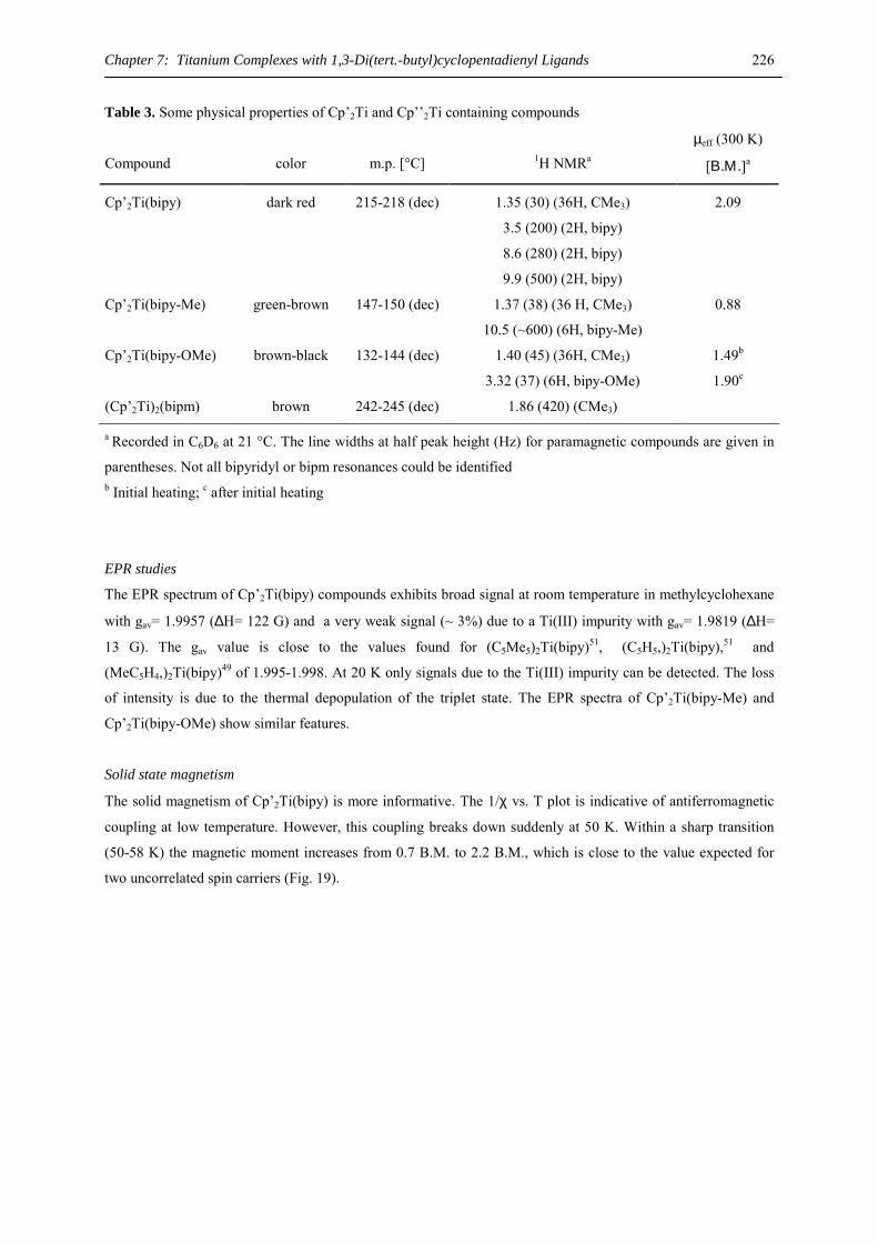
Chapter 7: Titanium Complexes with 1,3-Di(tert.-butyl)cyclopentadienyl Ligands 226
Table 3. Some physical properties of Cp’2Ti and Cp’’2Ti containing compounds
Compound
color
m.p. [°C]
1H NMRa
µeff (300 K)
[Β.Μ.]a
Cp’2Ti(bipy) dark red
215-218 (dec) 1.35 (30) (36H, CMe3)
3.5 (200) (2H, bipy)
8.6 (280) (2H, bipy)
9.9 (500) (2H, bipy)
2.09
Cp’2Ti(bipy-Me) green-brown 147-150 (dec) 1.37 (38) (36 H, CMe3)
10.5 (~600) (6H, bipy-Me)
0.88
Cp’2Ti(bipy-OMe) brown-black 132-144 (dec) 1.40 (45) (36H, CMe3)
3.32 (37) (6H, bipy-OMe)
1.49b
1.90c
(Cp’2Ti)2(bipm) brown 242-245 (dec) 1.86 (420) (CMe3)
a Recorded in C6D6 at 21 °C. The line widths at half peak height (Hz) for paramagnetic compounds are given in
parentheses. Not all bipyridyl or bipm resonances could be identified b Initial heating; c after initial heating
EPR studies
The EPR spectrum of Cp’2Ti(bipy) compounds exhibits broad signal at room temperature in methylcyclohexane
with gav= 1.9957 (∆H= 122 G) and a very weak signal (~ 3%) due to a Ti(III) impurity with gav= 1.9819 (∆H=
13 G). The gav value is close to the values found for (C5Me5)2Ti(bipy)51, (C5H5,)2Ti(bipy),51 and
(MeC5H4,)2Ti(bipy)49 of 1.995-1.998. At 20 K only signals due to the Ti(III) impurity can be detected. The loss
of intensity is due to the thermal depopulation of the triplet state. The EPR spectra of Cp’2Ti(bipy-Me) and
Cp’2Ti(bipy-OMe) show similar features.
Solid state magnetism
The solid magnetism of Cp’2Ti(bipy) is more informative. The 1/χ vs. T plot is indicative of antiferromagnetic
coupling at low temperature. However, this coupling breaks down suddenly at 50 K. Within a sharp transition
(50-58 K) the magnetic moment increases from 0.7 B.M. to 2.2 B.M., which is close to the value expected for
two uncorrelated spin carriers (Fig. 19).
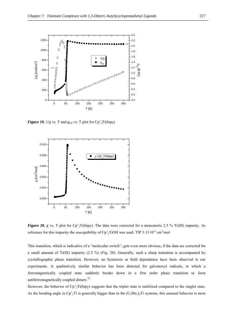
Chapter 7: Titanium Complexes with 1,3-Di(tert.-butyl)cyclopentadienyl Ligands 227
0 50 100 150 200 250 3000
200
400
600
800
1000
1200
1/χ µ
eff
T [K]
1/χ
[mol
/cm
3 ]
0.0
0.2
0.4
0.6
0.8
1.0
1.2
1.4
1.6
1.8
2.0
2.2
2.4
µeff [B.M
.]
Figure 19. 1/χ vs. T and µeff vs. T plot for Cp’2Ti(bipy)
0 50 100 150 200 250 300
0.000
0.002
0.004
0.006
0.008
0.010
χ [c
m3 /m
ol]
T [K]
χ Cp'2Ti(bipy)
Figure 20. χ vs. T plot for Cp’2Ti(bipy). The data were corrected for a monomeric 2.5 % Ti(III) impurity. As
reference for this impurity the susceptibility of Cp’2TiOH was used. TIP 3.13.10-4 cm3/mol
This transition, which is indicative of a “molecular switch”, gets even more obvious, if the data are corrected for
a small amount of Ti(III) impurity (2.5 %) (Fig. 20). Generally, such a sharp transition is accompanied by
crystallographic phase transition. However, no hysteresis or field dependence have been observed in our
experiments. A qualitatively similar behavior has been detected for galvanoxyl radicals, in which a
ferromagnetically coupled state suddenly breaks down in a first order phase transition to form
antiferromagnetically coupled dimers.52
However, the behavior of Cp’2Ti(bipy) suggests that the triplet state is stabilized compared to the singlet state.
As the bending angle in Cp’2Ti is generally bigger than in the (C5Me5)2Ti systems, this unusual behavior is most
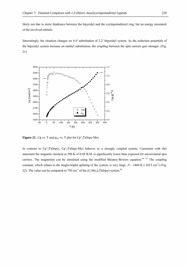
Chapter 7: Titanium Complexes with 1,3-Di(tert.-butyl)cyclopentadienyl Ligands 228
likely not due to steric hindrance between the bipyridyl and the cyclopentadienyl ring, but an energy mismatch
of the involved orbitals.
Interestingly, the situation changes on 4,4’-substitution of 2,2’-bipyridyl system. As the reduction potentials of
the bipyridyl system increase on methyl substitution, the coupling between the spin carriers gets stronger. (Fig.
21)
-50 0 50 100 150 200 250 300 350 4002250
2500
2750
3000
3250
3500
3750
4000
4250
4500
1/χ µeff
T [K]
1/χ
[mol
/cm
3 ]
0.0
0.2
0.4
0.6
0.8
1.0
1.2
µeff [B.M
.]
Figure 21. 1/χ vs. T and µeff vs. T plot for Cp’2Ti(bipy-Me)
In contrast to Cp’2Ti(bipy), Cp’2Ti(bipy-Me) behaves as a strongly coupled system. Consistent with this
statement the magnetic moment at 300 K of 0.88 B.M. is significantly lower than expected for uncorrelated spin
carriers. The magnetism can be simulated using the modified Bleaney-Bowers equation.24, 25 The coupling
constant, which relates to the singlet-triplet splitting of the system, is very large, J= –1460 K (-1015 cm-1) (Fig.
22). The value can be compared to 750 cm-1 of the (C5Me5)2Ti(bipy) system.49
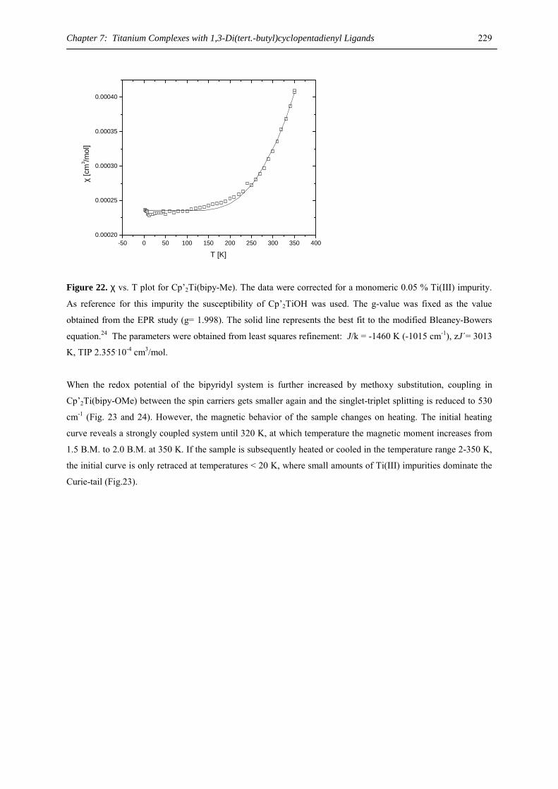
Chapter 7: Titanium Complexes with 1,3-Di(tert.-butyl)cyclopentadienyl Ligands 229
-50 0 50 100 150 200 250 300 350 4000.00020
0.00025
0.00030
0.00035
0.00040
χ [c
m3 /m
ol]
T [K]
Figure 22. χ vs. T plot for Cp’2Ti(bipy-Me). The data were corrected for a monomeric 0.05 % Ti(III) impurity.
As reference for this impurity the susceptibility of Cp’2TiOH was used. The g-value was fixed as the value
obtained from the EPR study (g= 1.998). The solid line represents the best fit to the modified Bleaney-Bowers
equation.24 The parameters were obtained from least squares refinement: J/k = -1460 K (-1015 cm-1), zJ´= 3013
K, TIP 2.355.10-4 cm3/mol.
When the redox potential of the bipyridyl system is further increased by methoxy substitution, coupling in
Cp’2Ti(bipy-OMe) between the spin carriers gets smaller again and the singlet-triplet splitting is reduced to 530
cm-1 (Fig. 23 and 24). However, the magnetic behavior of the sample changes on heating. The initial heating
curve reveals a strongly coupled system until 320 K, at which temperature the magnetic moment increases from
1.5 B.M. to 2.0 B.M. at 350 K. If the sample is subsequently heated or cooled in the temperature range 2-350 K,
the initial curve is only retraced at temperatures < 20 K, where small amounts of Ti(III) impurities dominate the
Curie-tail (Fig.23).
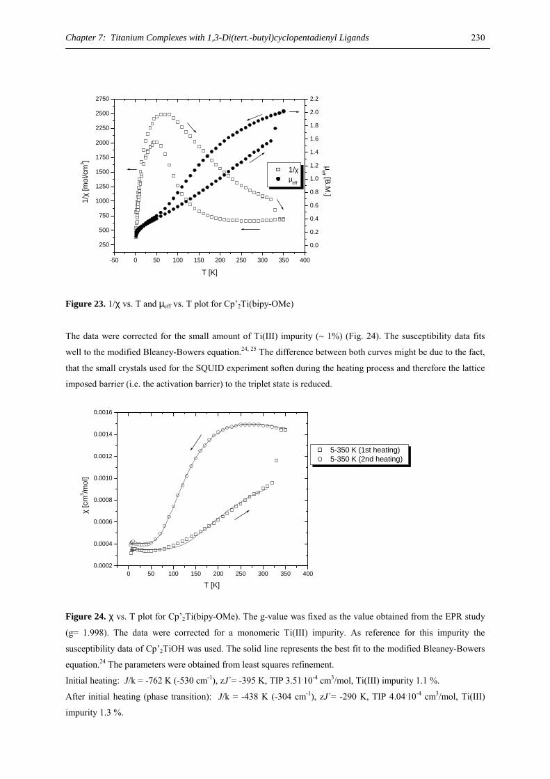
Chapter 7: Titanium Complexes with 1,3-Di(tert.-butyl)cyclopentadienyl Ligands 230
-50 0 50 100 150 200 250 300 350 400
250
500
750
1000
1250
1500
1750
2000
2250
2500
2750
1/χ µeff
T [K]
1/χ
[mol
/cm
3 ]
0.0
0.2
0.4
0.6
0.8
1.0
1.2
1.4
1.6
1.8
2.0
2.2
µeff [B.M
.]
Figure 23. 1/χ vs. T and µeff vs. T plot for Cp’2Ti(bipy-OMe)
The data were corrected for the small amount of Ti(III) impurity (~ 1%) (Fig. 24). The susceptibility data fits
well to the modified Bleaney-Bowers equation.24, 25 The difference between both curves might be due to the fact,
that the small crystals used for the SQUID experiment soften during the heating process and therefore the lattice
imposed barrier (i.e. the activation barrier) to the triplet state is reduced.
0 50 100 150 200 250 300 350 4000.0002
0.0004
0.0006
0.0008
0.0010
0.0012
0.0014
0.0016
χ [c
m3 /m
ol]
T [K]
5-350 K (1st heating) 5-350 K (2nd heating)
Figure 24. χ vs. T plot for Cp’2Ti(bipy-OMe). The g-value was fixed as the value obtained from the EPR study
(g= 1.998). The data were corrected for a monomeric Ti(III) impurity. As reference for this impurity the
susceptibility data of Cp’2TiOH was used. The solid line represents the best fit to the modified Bleaney-Bowers
equation.24 The parameters were obtained from least squares refinement.
Initial heating: J/k = -762 K (-530 cm-1), zJ´= -395 K, TIP 3.51.10-4 cm3/mol, Ti(III) impurity 1.1 %. After initial heating (phase transition): J/k = -438 K (-304 cm-1), zJ´= -290 K, TIP 4.04.10-4 cm3/mol, Ti(III)
impurity 1.3 %.
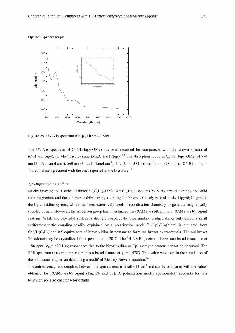
Chapter 7: Titanium Complexes with 1,3-Di(tert.-butyl)cyclopentadienyl Ligands 231
Optical Spectroscopy
300 400 500 600 700 800 900 1000 1100
0.0
0.5
1.0
1.5
2.0
2.5
3.0
200 300 400 500 600 700 800 900 1000 1100
0.1
1
log 10
(Abs
orpt
ion)
Wavelength [nm]
Abs
orpt
ion
Wavelength [nm]
Figure 25. UV-Vis spectrum of Cp’2Ti(bipy-OMe)
The UV-Vis spectrum of Cp’2Ti(bipy-OMe) has been recorded for comparison with the known spectra of
(C5H5)2Ti(bipy), (C5Me5)2Ti(bipy) and (Me4C5H)2Ti(bipy).50 The absorption found in Cp’2Ti(bipy-OMe) of 750
nm (ε= 590 Lmol cm- ), 560 nm (ε= 2210 Lmol cm-1), 457 (ε= 4100 Lmol cm-1) and 378 nm (ε= 8710 Lmol cm-
1) are in close agreement with the ones reported in the literature.50
2,2’-Bipyrimidine Adduct
Stucky investigated a series of dimeric [(C5H5)2TiX]2, X= Cl, Br, I, systems by X-ray crystallography and solid
state magnetism and these dimers exhibit strong coupling J~400 cm-1. Closely related to the bipyridyl ligand is
the bipyrimidine system, which has been extensively used in coordination chemistry to generate magnetically
coupled dimers. However, the Andersen group has investigated the ((C5Me5)2Yb(bipy) and ((C5Me5)2Yb)2(bipm)
systems. While the bipyridyl system is strongly coupled, the bipyrimidine bridged dimer only exhibits small
antiferromagnetic coupling readily explained by a polarization model.53 (Cp’2Ti)2(bipm) is prepared from
Cp’2Ti(C2H4) and 0.5 equivalents of bipyrimidine in pentane to form red-brown microcrystals. The red-brown
2:1 adduct may be crystallized from pentane at – 20°C. The 1H NMR spectrum shows one broad resonance at
1.86 ppm (ν1/2= 420 Hz); resonances due to the bipyrimidine or Cp’-methyne protons cannot be observed. The
EPR spectrum at room temperature has a broad feature at gav= 1.9783. This value was used in the simulation of
the solid state magnetism data using a modified Bleaney-Bowers equation.24
The antiferromagnetic coupling between the spin carriers is small ~13 cm-1 and can be compared with the values
obtained for ((C5Me5)2Yb)2(bipm) (Fig. 26 and 27). A polarization model appropriately accounts for this
behavior, see also chapter 4 for details.
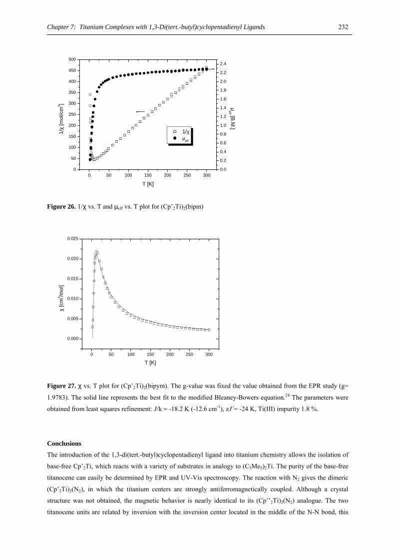
Chapter 7: Titanium Complexes with 1,3-Di(tert.-butyl)cyclopentadienyl Ligands 232
0 50 100 150 200 250 3000
50
100
150
200
250
300
350
400
450
500
1/χ µeff
T [K]
1/χ
[mol
/cm
3 ]
0.0
0.2
0.4
0.6
0.8
1.0
1.2
1.4
1.6
1.8
2.0
2.2
2.4
µeff [B.M
.]
Figure 26. 1/χ vs. T and µeff vs. T plot for (Cp’2Ti)2(bipm)
0 50 100 150 200 250 300
0.000
0.005
0.010
0.015
0.020
0.025
χ [c
m3 /m
ol]
T [K]
Figure 27. χ vs. T plot for (Cp’2Ti)2(bipym). The g-value was fixed the value obtained from the EPR study (g=
1.9783). The solid line represents the best fit to the modified Bleaney-Bowers equation.24 The parameters were
obtained from least squares refinement: J/k = -18.2 K (-12.6 cm-1), zJ´= -24 K, Ti(III) impurity 1.8 %.
Conclusions
The introduction of the 1,3-di(tert.-butyl)cyclopentadienyl ligand into titanium chemistry allows the isolation of
base-free Cp’2Ti, which reacts with a variety of substrates in analogy to (C5Me5)2Ti. The purity of the base-free
titanocene can easily be determined by EPR and UV-Vis spectroscopy. The reaction with N2 gives the dimeric
(Cp’2Ti)2(N2), in which the titanium centers are strongly antiferromagnetically coupled. Although a crystal
structure was not obtained, the magnetic behavior is nearly identical to its (Cp’’2Ti)2(N2) analogue. The two
titanocene units are related by inversion with the inversion center located in the middle of the N-N bond, this
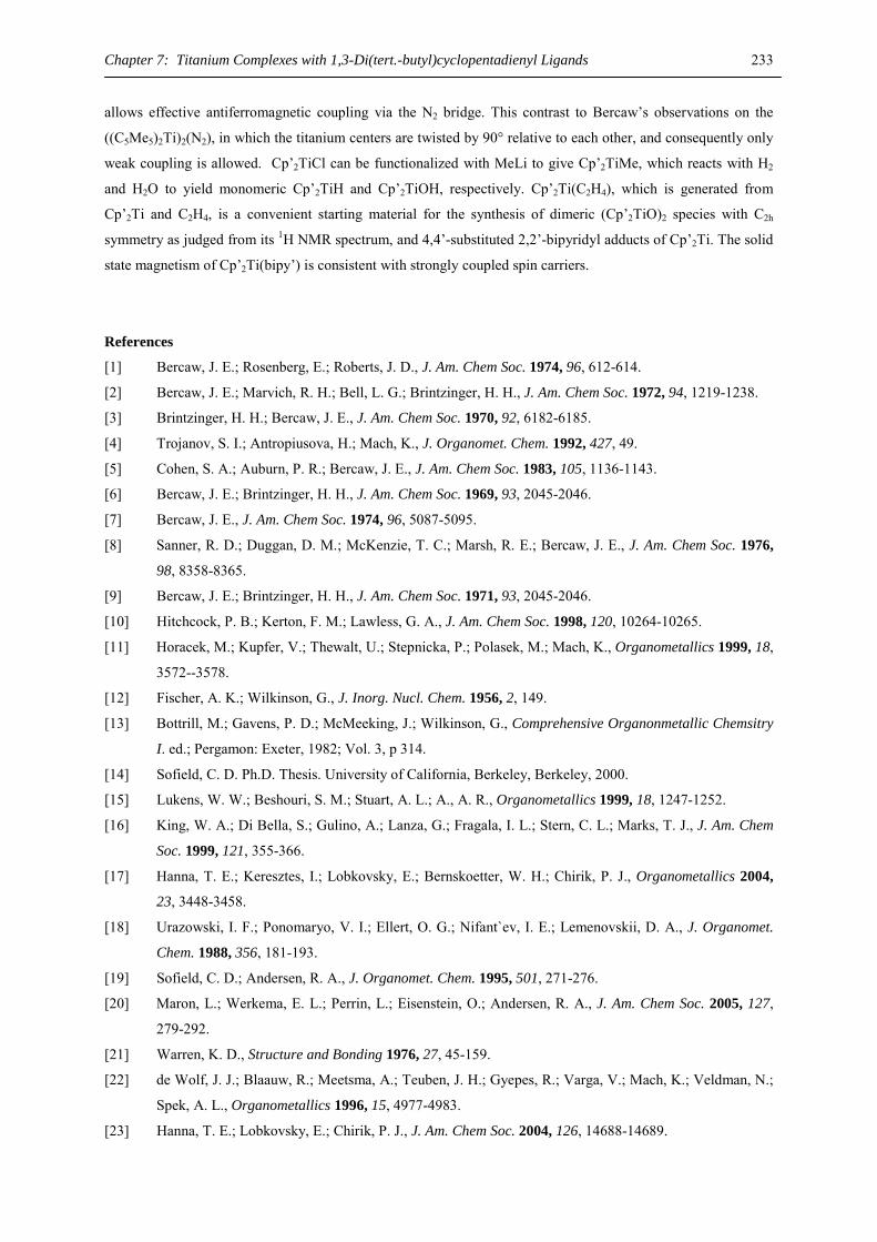
Chapter 7: Titanium Complexes with 1,3-Di(tert.-butyl)cyclopentadienyl Ligands 233
allows effective antiferromagnetic coupling via the N2 bridge. This contrast to Bercaw’s observations on the
((C5Me5)2Ti)2(N2), in which the titanium centers are twisted by 90° relative to each other, and consequently only
weak coupling is allowed. Cp’2TiCl can be functionalized with MeLi to give Cp’2TiMe, which reacts with H2
and H2O to yield monomeric Cp’2TiH and Cp’2TiOH, respectively. Cp’2Ti(C2H4), which is generated from
Cp’2Ti and C2H4, is a convenient starting material for the synthesis of dimeric (Cp’2TiO)2 species with C2h
symmetry as judged from its 1H NMR spectrum, and 4,4’-substituted 2,2’-bipyridyl adducts of Cp’2Ti. The solid
state magnetism of Cp’2Ti(bipy’) is consistent with strongly coupled spin carriers.
References
[1] Bercaw, J. E.; Rosenberg, E.; Roberts, J. D., J. Am. Chem Soc. 1974, 96, 612-614.
[2] Bercaw, J. E.; Marvich, R. H.; Bell, L. G.; Brintzinger, H. H., J. Am. Chem Soc. 1972, 94, 1219-1238.
[3] Brintzinger, H. H.; Bercaw, J. E., J. Am. Chem Soc. 1970, 92, 6182-6185.
[4] Trojanov, S. I.; Antropiusova, H.; Mach, K., J. Organomet. Chem. 1992, 427, 49.
[5] Cohen, S. A.; Auburn, P. R.; Bercaw, J. E., J. Am. Chem Soc. 1983, 105, 1136-1143.
[6] Bercaw, J. E.; Brintzinger, H. H., J. Am. Chem Soc. 1969, 93, 2045-2046.
[7] Bercaw, J. E., J. Am. Chem Soc. 1974, 96, 5087-5095.
[8] Sanner, R. D.; Duggan, D. M.; McKenzie, T. C.; Marsh, R. E.; Bercaw, J. E., J. Am. Chem Soc. 1976,
98, 8358-8365.
[9] Bercaw, J. E.; Brintzinger, H. H., J. Am. Chem Soc. 1971, 93, 2045-2046.
[10] Hitchcock, P. B.; Kerton, F. M.; Lawless, G. A., J. Am. Chem Soc. 1998, 120, 10264-10265.
[11] Horacek, M.; Kupfer, V.; Thewalt, U.; Stepnicka, P.; Polasek, M.; Mach, K., Organometallics 1999, 18,
3572--3578.
[12] Fischer, A. K.; Wilkinson, G., J. Inorg. Nucl. Chem. 1956, 2, 149.
[13] Bottrill, M.; Gavens, P. D.; McMeeking, J.; Wilkinson, G., Comprehensive Organonmetallic Chemsitry
I. ed.; Pergamon: Exeter, 1982; Vol. 3, p 314.
[14] Sofield, C. D. Ph.D. Thesis. University of California, Berkeley, Berkeley, 2000.
[15] Lukens, W. W.; Beshouri, S. M.; Stuart, A. L.; A., A. R., Organometallics 1999, 18, 1247-1252.
[16] King, W. A.; Di Bella, S.; Gulino, A.; Lanza, G.; Fragala, I. L.; Stern, C. L.; Marks, T. J., J. Am. Chem
Soc. 1999, 121, 355-366.
[17] Hanna, T. E.; Keresztes, I.; Lobkovsky, E.; Bernskoetter, W. H.; Chirik, P. J., Organometallics 2004,
23, 3448-3458.
[18] Urazowski, I. F.; Ponomaryo, V. I.; Ellert, O. G.; Nifant`ev, I. E.; Lemenovskii, D. A., J. Organomet.
Chem. 1988, 356, 181-193.
[19] Sofield, C. D.; Andersen, R. A., J. Organomet. Chem. 1995, 501, 271-276.
[20] Maron, L.; Werkema, E. L.; Perrin, L.; Eisenstein, O.; Andersen, R. A., J. Am. Chem Soc. 2005, 127,
279-292.
[21] Warren, K. D., Structure and Bonding 1976, 27, 45-159.
[22] de Wolf, J. J.; Blaauw, R.; Meetsma, A.; Teuben, J. H.; Gyepes, R.; Varga, V.; Mach, K.; Veldman, N.;
Spek, A. L., Organometallics 1996, 15, 4977-4983.
[23] Hanna, T. E.; Lobkovsky, E.; Chirik, P. J., J. Am. Chem Soc. 2004, 126, 14688-14689.
Chapter 7: Titanium Complexes with 1,3-Di(tert.-butyl)cyclopentadienyl Ligands 234
[24] Kahn, O., Molecular Magnetism. ed.; VCH: New York, 1993; pp. 131-132.
[25] O'Connor, C. J., Magnetochemistry-Advances in Theory and Experimentation. In Progress in Inorganic
Chemistry, ed.; Lippard, S. J., Ed. J. Wiley & Sons: New York, 1982; 'Vol.' 29, p^pp 203-285.
[26] Lauher, J. W.; Hoffmann, R., J. Am. Chem Soc. 1976, 98, 1729-1742.
[27] Ren, Q.; Chen, Z.; Ren, J.; Wei, H.; Feng, W.; Zhang, L., J. Phys. Chem. A 2002, 106, 6161-6166.
[28] Lukens, W. W.; Smith, M. R.; Andersen, R. A., J. Am. Chem Soc. 1996, 118, 1719-1728.
[29] Stalick, J. K.; Ibers, J. A., J. Am. Chem Soc. 1970, 92, 5333-5338.
[30] Ittel, S. D.; Ibers, J. A., Adv. Organomet. Chem. 1976, 14, 33-61.
[31] Polse, J. L.; Andersen, R. A.; Bergman, R. G., J. Am. Chem Soc. 1995, 117, 5393-5394.
[32] Schwartz, D. J.; Smith, M. R.; Andersen, R. A., Organometallics 1996, 15, 1446-1450.
[33] Bottomley, F.; Egharevba, G. O.; Lin, I. J. B.; White, P. S., Organometallics 1985, 4, 550-553.
[34] Bottomley, F.; Lin, I. J. B.; White, P. S., J. Am. Chem Soc. 1981, 103, 703-704.
[35] Bottomley, F.; Lin, I. J. B.; Mukaida, M., J. Am. Chem Soc. 1980, 102, 5238-5242.
[36] Matsunaga, P. T.; Morropoulos, J. C.; G.L., H., Polyhedron 1995, 14, 175-185, and refences therein.
[37] Sofield, C. D.; Andersen, R. A., unpublished results.
[38] Sofield, C. D.; Walter, M. D.; R., A., Acta Cryst. E 2004, 60, m1417-m1419.
[39] Babcock, L. M.; Day, V. W.; Klemperer, W. G., J. Chem. Soc., Chem. Commun. 1987, 858.
[40] Babcock, L. M.; Klemperer, W. G., Inorg. Chem. 1989, 28, 2003.
[41] Smith, M. R.; Matsunaga, P. T.; Andersen, R. A., J. Am. Chem Soc. 1993, 115, 7049-7050.
[42] Horacek, M.; Gyepes, R.; Kubista, J.; Mach, K., Inorg. Chem. Commun. 2004, 7, 155-159.
[43] Fieselmann, B. F.; Stucky, G. D., Inorg. Chem. 1978, 17, 2074-2077.
[44] Jungst, R.; Sekutowski, D.; Davis, J.; Luly, M.; Stucky, G. D., Inorg. Chem. 1977, 16, 1645-1655.
[45] Francesconi, L. C.; Corbin, D. R.; Hendrickson, D. N.; Stucky, G. D., Inorg. Chem. 1979, 18, 3074-
3080.
[46] Lukens, W. W.; Matsunaga, P. T.; Andersen, R. A., Organometallics 1998, (17), 5240.
[47] de Wolf, J. M.; Meetsma, A.; Teuben, J. H., Organometallics 1995, (14), 5466-5468.
[48] La Mar, G. N.; Horrocks, W. D.; Holm, R. H., NMR of Paramagnetic Molecules. ed.; Academic Press:
New York, 1973; pp. 287-289; 638-640.
[49] McPherson, A. M.; Fieselmann, B. F.; Lichtenberger, D. L.; McPherson, G. L.; Stucky, G. D., J. Am.
Chem Soc. 1979, 101, 3425-3430.
[50] Witte, P. T.; Klein, R.; Kooijman, H.; Spek, A. L.; Polasek, M.; Varga, V.; Mach, K., J. Organomet.
Chem. 1996, 519, 195-204.
[51] Gyepes, R.; Witte, P. T.; Horacek, M.; Mach, K., J. Organomet. Chem. 1998, 551, 207-213.
[52] Kahn, O., Molecular Magnetism. ed.; VCH: New York, 1993; 'Vol.' p 300-303.
[53] Berg, D. J.; Boncella, J. M.; Andersen, R. A., Organometallics 2002, 21, 4622-4631.
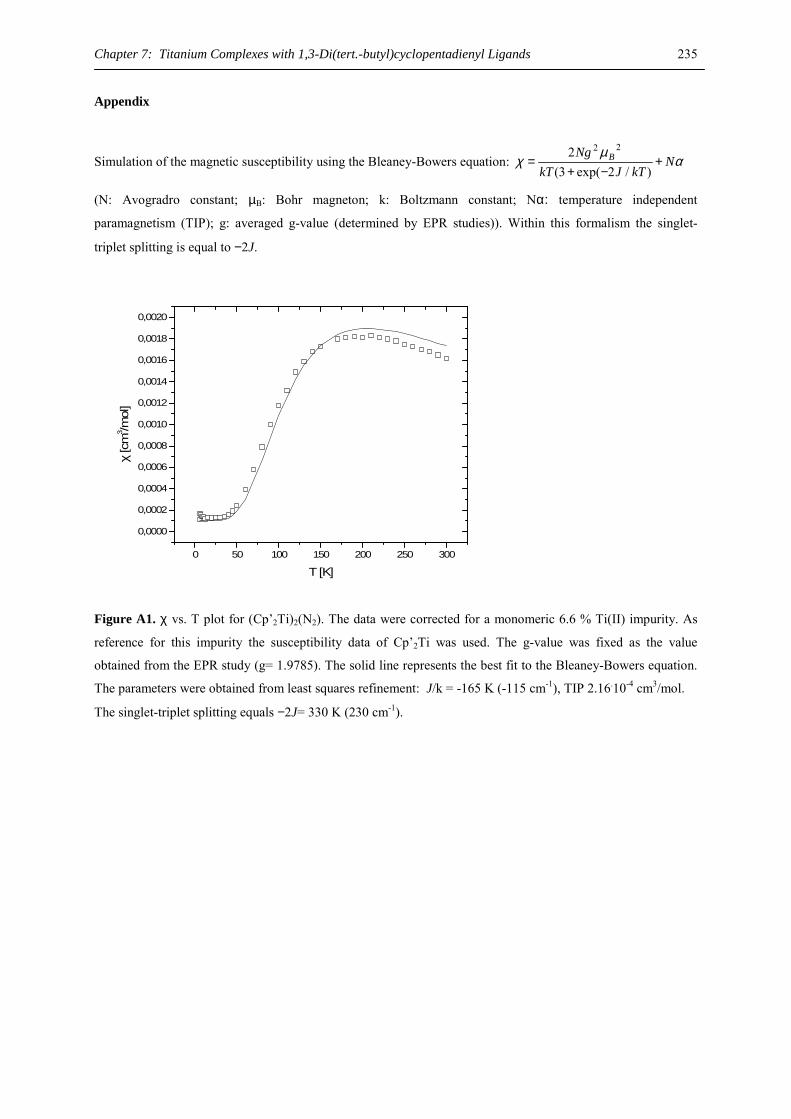
Chapter 7: Titanium Complexes with 1,3-Di(tert.-butyl)cyclopentadienyl Ligands 235
Appendix
Simulation of the magnetic susceptibility using the Bleaney-Bowers equation: αµ
χ NkTJkT
Ng B +−+
=)/2exp(3(
2 22
(N: Avogradro constant; µB: Bohr magneton; k: Boltzmann constant; Nα: temperature independent
paramagnetism (TIP); g: averaged g-value (determined by EPR studies)). Within this formalism the singlet-
triplet splitting is equal to −2J.
0 50 100 150 200 250 300
0,0000
0,0002
0,0004
0,0006
0,0008
0,0010
0,0012
0,0014
0,0016
0,0018
0,0020
χ [c
m3 /m
ol]
T [K]
Figure A1. χ vs. T plot for (Cp’2Ti)2(N2). The data were corrected for a monomeric 6.6 % Ti(II) impurity. As
reference for this impurity the susceptibility data of Cp’2Ti was used. The g-value was fixed as the value
obtained from the EPR study (g= 1.9785). The solid line represents the best fit to the Bleaney-Bowers equation.
The parameters were obtained from least squares refinement: J/k = -165 K (-115 cm-1), TIP 2.16.10-4 cm3/mol. The singlet-triplet splitting equals −2J= 330 K (230 cm-1).
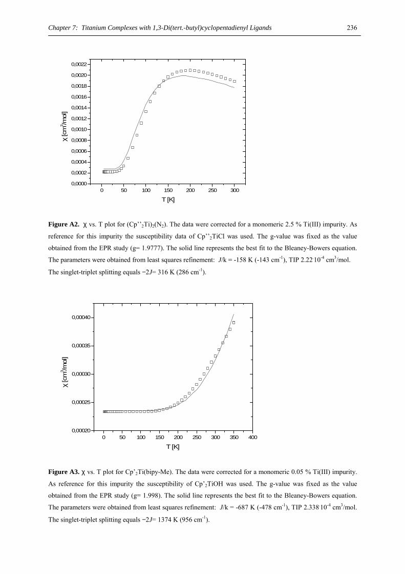
Chapter 7: Titanium Complexes with 1,3-Di(tert.-butyl)cyclopentadienyl Ligands 236
0 50 100 150 200 250 3000,0000
0,0002
0,0004
0,0006
0,0008
0,0010
0,0012
0,0014
0,0016
0,0018
0,0020
0,0022
χ [c
m3 /m
ol]
T [K]
Figure A2. χ vs. T plot for (Cp’’2Ti)2(N2). The data were corrected for a monomeric 2.5 % Ti(III) impurity. As
reference for this impurity the susceptibility data of Cp’’2TiCl was used. The g-value was fixed as the value
obtained from the EPR study (g= 1.9777). The solid line represents the best fit to the Bleaney-Bowers equation.
The parameters were obtained from least squares refinement: J/k = -158 K (-143 cm-1), TIP 2.22.10-4 cm3/mol. The singlet-triplet splitting equals −2J= 316 K (286 cm-1).
0 50 100 150 200 250 300 350 4000,00020
0,00025
0,00030
0,00035
0,00040
χ [c
m3 /m
ol]
T [K]
Figure A3. χ vs. T plot for Cp’2Ti(bipy-Me). The data were corrected for a monomeric 0.05 % Ti(III) impurity.
As reference for this impurity the susceptibility of Cp’2TiOH was used. The g-value was fixed as the value
obtained from the EPR study (g= 1.998). The solid line represents the best fit to the Bleaney-Bowers equation.
The parameters were obtained from least squares refinement: J/k = -687 K (-478 cm-1), TIP 2.338.10-4 cm3/mol.
The singlet-triplet splitting equals −2J= 1374 K (956 cm-1).
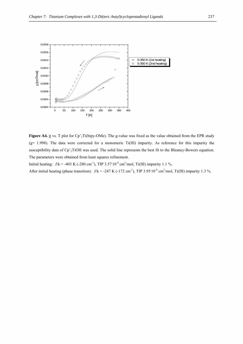
Chapter 7: Titanium Complexes with 1,3-Di(tert.-butyl)cyclopentadienyl Ligands 237
0 50 100 150 200 250 300 350 4000,0002
0,0004
0,0006
0,0008
0,0010
0,0012
0,0014
0,0016
0,0018
χ [c
m3 /m
ol]
T [K]
5-350 K (1st heating) 5-350 K (2nd heating)
Figure A4. χ vs. T plot for Cp’2Ti(bipy-OMe). The g-value was fixed as the value obtained from the EPR study
(g= 1.998). The data were corrected for a monomeric Ti(III) impurity. As reference for this impurity the
susceptibility data of Cp’2TiOH was used. The solid line represents the best fit to the Bleaney-Bowers equation.
The parameters were obtained from least squares refinement.
Initial heating: J/k = -403 K (-280 cm-1), TIP 3.57.10-4 cm3/mol, Ti(III) impurity 1.1 %. After initial heating (phase transition): J/k = -247 K (-172 cm-1), TIP 3.95.10-4 cm3/mol, Ti(III) impurity 1.3 %.
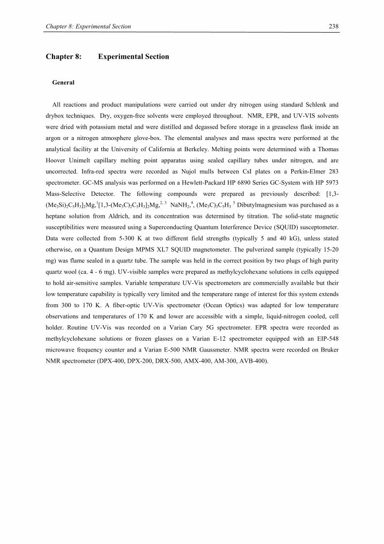
Chapter 8: Experimental Section
238
Chapter 8: Experimental Section
General
All reactions and product manipulations were carried out under dry nitrogen using standard Schlenk and
drybox techniques. Dry, oxygen-free solvents were employed throughout. NMR, EPR, and UV-VIS solvents
were dried with potassium metal and were distilled and degassed before storage in a greaseless flask inside an
argon or a nitrogen atmosphere glove-box. The elemental analyses and mass spectra were performed at the
analytical facility at the University of California at Berkeley. Melting points were determined with a Thomas
Hoover Unimelt capillary melting point apparatus using sealed capillary tubes under nitrogen, and are
uncorrected. Infra-red spectra were recorded as Nujol mulls between CsI plates on a Perkin-Elmer 283
spectrometer. GC-MS analysis was performed on a Hewlett-Packard HP 6890 Series GC-System with HP 5973
Mass-Selective Detector. The following compounds were prepared as previously described: [1,3-
(Me3Si)2C5H3]2Mg,1[1,3-(Me3C)2C5H3]2Mg,2, 3 NaNH2,4, (Me3C)3C5H3 5 Dibutylmagnesium was purchased as a
heptane solution from Aldrich, and its concentration was determined by titration. The solid-state magnetic
susceptibilities were measured using a Superconducting Quantum Interference Device (SQUID) susceptometer.
Data were collected from 5-300 K at two different field strengths (typically 5 and 40 kG), unless stated
otherwise, on a Quantum Design MPMS XL7 SQUID magnetometer. The pulverized sample (typically 15-20
mg) was flame sealed in a quartz tube. The sample was held in the correct position by two plugs of high purity
quartz wool (ca. 4 - 6 mg). UV-visible samples were prepared as methylcyclohexane solutions in cells equipped
to hold air-sensitive samples. Variable temperature UV-Vis spectrometers are commercially available but their
low temperature capability is typically very limited and the temperature range of interest for this system extends
from 300 to 170 K. A fiber-optic UV-Vis spectrometer (Ocean Optics) was adapted for low temperature
observations and temperatures of 170 K and lower are accessible with a simple, liquid-nitrogen cooled, cell
holder. Routine UV-Vis was recorded on a Varian Cary 5G spectrometer. EPR spectra were recorded as
methylcyclohexane solutions or frozen glasses on a Varian E-12 spectrometer equipped with an EIP-548
microwave frequency counter and a Varian E-500 NMR Gaussmeter. NMR spectra were recorded on Bruker
NMR spectrometer (DPX-400, DPX-200, DRX-500, AMX-400, AM-300, AVB-400).
Chapter 8: Experimental Section
239
Chapter 1
2-Ca: A suspension of calcium chloride (222 mg, 2.0 mmol) and sodium tetraisopropylcyclopentadienide (512
mg, 2.0 mmol) in tetrahydrofuran (10 mL) was stirred at room temperature. The suspension turned brown-green
after 5 min and stirring was continued for 2 h until the calcium chloride was completely dissolved. Disodium
cyclooctatetraenide (152 mg, 1.0 mmol) was added and the suspension was stirred for 4 d at ambient
temperature. Removal of the solvent in vacuo and extraction of the solid residue with toluene (20 mL), followed
by filtration and evaporation of the orange solution gave a solid, which was washed with three 5 mL portions of
n-hexane to yield 370 mg (0.57 mmol, 57%) of a colorless powder, which could be obtained as single crystals
suitable for X-ray diffraction from a saturated hexadeuterobenzene solution. M. p. 188-189 °C, sublimation
temperature 130 °C (oil pump vacuum), Anal. calcd for C42H66Ca2: C,77.47, H 10.22, Ca 12.31. Found: C,
68.80; H, 9.92; Ca 12.04; 1H-NMR (400 MHz, 298 K, C6D6): δ = 6.04 (s, 8H, COT), 5.21 (s, 2H, ring H (4Cp)),
2.87 (m, 4H, CHMe2), 2.72 (m, 4H, CHMe2), 1.24 (d, 3J(HH) = 7.1 Hz, 12 H, CH3), 1.20 (d, 3J(HH) = 6.8 Hz, 12
H, CH3), 1.19 (d, 3J(HH) = 7.2 Hz, 12 H, CH3), 1.09 (d, 3J(HH) = 6.7 Hz, 12 H, CH3); 13C{1H}-NMR (100 MHz,
C6D6): δ = 127.6 (s, ring-C), 123.3 (s, ring-C), 98.1 (dt, ring CH (4Cp), 1J(CH) = 156 Hz, 4J(CH) = 5 Hz), 89.6
(d, 1J(CH) = 157 Hz, COT), 26.8 (CHMe2), 26.7 (CHMe2), 26.5 (CH3), 25.2 (CH3), 24.6 (CH3), 23.9 (CH3); EI-
MS (70 eV): m/z (%): 650.3 (45) [M+], 417.1 (100) [M+ - C5H(CHMe2)4], 273.1 (45) [Ca{C5H(CHMe2)4}+].
2-Sr: A suspension of strontium iodide (341 mg, 1.0 mmol) and sodium tetraisopropylcyclopentadienide (256
mg, 1.0 mmol) in tetrahydrofuran (20 mL) was stirred at room temperature. The suspended material was
dissolved to a yellow solution within 5 min. Disodium cyclooctatetraenide (75 mg, 0.5 mmol) was added and
stirring was continued for 24 h at ambient temperature. 2 mL of n-hexane were added with formation of a white
precipitate and stirring was continued for another 24 h. Removal of the precipitate by centrifugation and
evaporation of the solvent in vacuo gave a light yellow residue, which was extracted with toluene (3 × 10 mL).
Centrifugation and evaporation of the light yellow solution gave a light yellow powder, which was washed with
three 5 mL portions of n-hexane and sublimed to yield 260 mg (0.35 mmol, 70%) of a white solid. 2-Sr was
heated to 250 °C without melting or decomposition, in oil pump vacuum sublimation was observed at 175 °C.
Anal. calcd for C42H66Sr2: C, 67.60, H 8.91. Found: C, 57.58; H, 8.83; 1H-NMR (400 MHz, 298 K, C6D6): δ =
6.00 (s, 8H, COT), 5.22 (s, 2H, ring H (4Cp)), 2.93 (m, 4H, CHMe2), 2.78 (m, 4H, CHMe2), 1.22 (d, 3J(HH) =
6.9 Hz, 12 H, CH3), 1.18 (d, 3J(HH) = 6.8 Hz, 12 H, CH3), 1.12 (d, 3J(HH) = 7.3 Hz, 12 H, CH3), 1.08 (d, 3J(HH)
= 6.8 Hz, 12 H, CH3); 13C{1H}-NMR (100 MHz, C6D6): δ = 127.6 (s, ring-C), 124.3 (s, ring-C), 99.1 (dt, ring
CH (4Cp), 1J(CH) = 155 Hz, 4J(CH) = 5 Hz), 91.6 (d, 1J(CH) = 157 Hz, COT), 26.7 (CHMe2), 26.5 (CHMe2),
25.9 (CH3), 24.9 (CH3), 24.3 (CH3), 23.9 (CH3); EI-MS (70 eV): m/z (%): 746 (<1) [M+], 513 (2) [M+ -
C5H(CHMe2)4], 321.1 (9) [Sr{C5H(CHMe2)4}+].
2-Ba: To a solution of 1-Ba (642 mg, 0.5 mmol) in a mixture of tetrahydrofuran (20 mL) and hexanes (5 mL)
solid Na2C8H8 (75 mg, 0.5 mmol) was added and the suspension was stirred for 2 d at ambient temperature.
Removal of the solvent in vacuo and extraction of the solid residue with toluene (20 mL), followed by filtration
and evaporation gave 190 mg (0.22 mmol, 45%) of an ivory powder, which could be obtained as colorless single
crystals suitable for X-ray diffraction from hexadeuterobenzene solution. M. p. 224-227 °C, sublimation
Chapter 8: Experimental Section
240
temperature 215 °C in oil-pump vacuum. C,H-analysis gave completely unsatisfactory results due to the reasons
outlined above. 1H-NMR (400 MHz, 298 K, C6D6): δ = 6.02 (s, 8H, COT), 5.11 (s, 2H, ring H (4Cp)), 2.94 (m,
4H, CHMe2), 2.84 (m, 4H, CHMe2), 1.24 (d, 3J(HH) = 7.2 Hz, 12 H, CH3), 1.21 (d, 3J(HH) = 7.2 Hz, 12 H,
CH3), 1.16 (d, 3J(HH) = 6.8 Hz, 12 H, CH3), 1.02 (d, 3J(HH) = 6.8 Hz, 12 H, CH3). — 13C{1H}-NMR (100 MHz,
C6D6): δ = 127.5 (s, ring-C), 123.4 (s, ring-C), 101.7 (dt, ring CH (4Cp), 1J(CH) = 155 Hz, 4J(CH) = 5 Hz), 95.3
(d, 1J(CH) = 158 Hz, COT), 26.9 (CHMe2), 26.6 (CHMe2), 25.7 (CH3), 25.3 (CH3), 24.1 (CH3), 23.7 (CH3). —
EI-MS (70 eV): m/z (%): 846.2 (6) [M+], 613.0 (74) [M+ - C5H(CHMe2)4], 371.1 (100) [Ba{C5H(CHMe2)4}+].
2-Yb: A suspension of YbI2(thf)4 (717 mg, 1.0 mmol) and sodium tetraisopropylcyclopentadienide (256 mg, 1.0
mmol) in tetrahydrofuran (40 mL) was stirred at room temperature. Disodium cyclooctatetraenide (75 mg, 0.5
mmol) was added and stirring was continued for 2 d at ambient temperature. During this time the mixture turned
green-brown. The solvent was removed in vacuo and the residue extracted with hexane (3 × 10 mL).
Centrifugation and evaporation of the dark red solution yielded a red powder. 1H NMR spectra of the crude
product revealed a mixture of 2-Yb and [C5H(CHMe2)4]2Yb. The [C5H(CHMe2)4]2Yb contamination was
removed by sublimation in oil pump vacuum (110-120 °C). 2-Yb was re-crystallized from a saturated n-hexane
solution at –20 °C to yield 254 mg (0.28 mmol, 56%) as blue-red microcrystalline powder. Anal. calcd for
C42H66Yb2: C, 55.0, H 7.04. Found: C, 53.22; H, 6.90; 1H-NMR (400 MHz, 298 K, C6D6): δ = 5.88 (s, 8H,
COT), 5.20 (s, 2H, ring H (4Cp)), 2.87 (m, 4H, CHMe2), 2.73 (m, 4H, CHMe2), 1.25 (d, 3J(HH) = 7.06 Hz, 12 H,
CH3), 1.24 (d, 3J(HH) = 6.98 Hz, 12 H, CH3), 1.21 (d, 3J(HH) = 7.35 Hz, 12 H, CH3), 1.11 (d, 3J(HH) = 6.66 Hz,
12 H, CH3); 13C{1H}-NMR (100 MHz, C6D6): δ = 126.9 (s, ring-C), 122.9 (s, ring-C), 98.4 (dt, ring CH (4Cp), 1J(CH) = 156 Hz, 4J(CH) = 5 Hz), 89.8 (d, 1J(CH) = 163 Hz, COT), 26.8 (CHMe2), 26.4 (CHMe2), 26.7 (CH3),
25.1 (CH3), 24.6 (2 x CH3).
3-Yb: Ytterbium chips (173 mg, 1.00 mmol), HgCl2 (7 mg) and a few glass splitters were suspended in
tetrahydrofuran (ca. 10 ml). The clear THF solution turned cloudy within 10 min. After 20 min
pentaisopropycyclopentadienyl radical (275 mg, 1.00 mmol) dissolved in tetrahydrofuran (5 ml) was added and
resulted in color change of tetrahydrofuran solution to dark brown within a few minutes. At this point
cyclooctatetraene (52 mg, 0.5 mmol) was added to the reaction mixture. After 15 h the tetrahydrofuran solution
changed color to deep blue, and is heated at 70-80 °C for 4 d. During this time the solution changed again color
to red-violet. Insoluble materials were separated by centrifugation and the solvent was removed in dynamic
vacuum leaving behind a deep blue-purple powder (287 mg, 0.287 mmol, 57.3 %). It was very soluble in
tetrahydrofuran and moderately soluble in aromatic hydrocarbons. Crystallization from a concentrated toluene
and n-hexane solution yielded an analytically pure microcyrstalline blue-purple powder. M.p. > 250 °C. Anal.
calcd for C48H78Yb2: C, 57.58, H 7.85. Found: C, 57.91; H, 8.68; 1H-NMR (400 MHz, 298 K, C6D6): δ = 6.06 (s,
8H, COT), 3.01 (m, 10H, CHMe2), 1.34 (d, 3J(HH) = 7.4 Hz, 30 H, CH3), 1.22 (d, 3J(HH) = 6.83 Hz, 30 H, CH3); 13C{1H}-NMR (100 MHz, C6D6): δ = 123.3 (s, ring-C), 90.1 (d, 1J(CH) = 163 Hz, COT), 26.8 (CHMe2), 26.6 (d, 1J(CH) = 123 Hz, CHMe2), 25.1 (q, 1J(CH) = 125 Hz, CH3), 25.1 (q, 1J(CH) = 127 Hz CH3).
4-Ba: Solid KC9H9 (332 mg, 2 mmol) was added to a suspension of BaI2 (341 mg, 1 mmol) in 20 ml
tetrahydrofuran. The brown mixture was stirred for 10 min at ambient temperature. Removal of the solvent in
Chapter 8: Experimental Section
241
vacuo, washing of the remaining off-white solid with ca. 10 ml cold pentane, and extraction into 3 x 15 ml
toluene, followed by filtration and evaporation gave an ivory powder, which was further purified by sublimation
(210-215 °C in oil pump vacuum) to yield a colorless powder (156 mg, 0.42 mmol, 42 %). ─ C18H18Ba
(371.67): calcd. C, 58.17; H, 4.88: Ba, 36.95; found C, 57.05; H, 5.04; Ba, 36.3. 1H-NMR (400 MHz, 298 K, d8-
THF): 6.96 ppm (s, cnt); 13C-NMR (100 MHz, 298 K, d8-THF): 110.1 ppm (d, 1JCH= 151 Hz, cnt); EI-MS (70
eV): m/z (%): 372 (6) [M]+, 346 (<1) [M-C2H2]+, 320 (<1) [M-2C2H2]+, 234 [C18H18]+, 117 (100) [C9H9]+, 91
(55) [C7H7]+, 55 (33) [C5H5]+. M.p. > 280 °C
5-Ba: Crystalline [4CpBaI(thf)2]2 ( mg, 0.5 mmol) was dissolved in 15 ml tetrahydrofuran, and solid KC9H9 (156
mg, 1 mmol) was added. The brown suspension was stirred at ambient temperature for 10 min. Solvent was
removed in vacuo, and the dark residue extracted with 3 x 15 ml hexane-toluene (10:1), filtration and
evaporation gave a yellow solid. The yellow solid was sublimed at 170 °C in oil pump vacuum to yield a
colorless solid. − NMR as well as EI-MS analysis confirmed a 1:1 mixture of 4Cp2Ba and (4Cp)(C9H9)Ba –
Ba(cnt)2 was not present in the mixture (as shown by MS-EI and NMR). 1H-NMR (400 MHz, 298 K, C6D6):
Signal set of 4Cp2Ba: 5.68 (ring-CH), 3.19 (m, 4H, CHMe2), 2.96 (m, 4H, CHMe2), 1.31 (d, 3J(HH) = 6.9 Hz, 12
H, CH3), 1.27 (d, 3J(HH) = 7.2 Hz, 12 H, CH3), 1.23 (d, 3J(HH) = 6.7 Hz, 24 H, CH3).
Signal set of 4CpBa(cnt): 7.28 (C9H9), 5.03 (ring-CH), 2.77 (m, 4H, CHMe2), 1.13 (d, 3J(HH) = 7.2 Hz, 6 H,
CH3), 1.12 (d, 3J(HH) = 6.6 Hz, 6 H, CH3), 1.09 (d, 3J(HH) = 7.0 Hz, 12 H, CH3). 13C-NMR (100 MHz, 298 K, C6D6):
Signal set of 4Cp2Ba: one ring-C(iPr) overlayed by the solvent signal, 123.6 (ring-C(iPr)), 101.3 (ring-CH), 27.2
(CHMe2), 26.8 (CHMe2), 27.1 (CH3), 25.0 (CH3), 24.0 (CH3), 23.7 (CH3).
Signal set of 4CpBa(cnt): 129.0 (ring-C(iPr)), 123.8 (ring-C(iPr)), 111.6 (CNT, 1JCH= 151 Hz), 101.9 (ring-CH),
26.7 (CHMe2), 26.4 (CHMe2), 25.6 (CH3), 25.2 (CH3), 24.0 (CH3), 23.9 (CH3).
Calculational Details
The calculations were carried out at the density functional (DFT) level, using the B3PW91 functional, which
incorporates Becke´s three-parameter exchange functional6 with the 1991 gradient-corrected correlation
functional of Perdew and Wang7; this hybrid functional has previously been shown to provide realistic
geometries for organometallic species.8 For comparison calculations were also undertaken by employing the
B3LYP functional. On carbon and hydrogen atoms a 6-311G basis set was used, and for barium and strontium
the effective core potentials (ECP`s) provided by the Stuttgart-Dresden group (SDD) was employed. A full
geometry optimization was carried out without any symmetry restrictions. The calculations were performed
using the ultrafine (99, 590) grid having 99 radial shells and 590 angular points per shell. The nature of the
optimized extrema was verified by analytical frequency calculations. All calculations have been carried out with
the Gaussian 98 suite of programs.
Chapter 8: Experimental Section
242
Chapter 2
[(Me3C)2C5H4]. Di(1,3-tert-butyl)cyclopentadiene, [(Me3C)2C5H4], was prepared in a modified version of the
synthesis reported by Venier and Casserly 9: Potassium hydroxide (1 kg, 25 mol) was dissolved in 750 mL of
water in a 3-L, 3-necked flask equipped with an overhead stirrer. The potassium hydroxide solution was allowed
to cool to room temperature and Adogen 464 (5g), freshly cracked cyclopentadiene (100 mL, 1.21 mol) and t-
butyl bromide, Me3CBr, (300 mL, 2.65 mol), were added. In contrast to the reported procedure, better yields
have been obtained by sequential addition of t-butyl bromide. The reaction mixture was stirred vigorously for 12
h. An additional 50 mL (0.44 mol) of Me3CBr was added and the mixture was stirred for another 24 h, followed
by addition of another 50 mL of Me3CBr and further stirring. Progress of the reaction was monitored by gas
chromatography (GC) and after 4-5 days, only 5% [(Me3C)C5H5], and no C5H6 remained in the mixture. The
organic layer was separated and washed with 1 M HCl (100 mL). The product was dried over anhydrous
Mg(SO4) and distilled at 40 °C (10-2 torr). The product was analyzed by GC and was typically > 98 % pure
(Me3C)2 C5H4 (150 g, 0.841 mol, 69.5 % yield).
[(Me3C)C5H5]. Mono(tert-butyl)cyclopentadiene, [(Me3C)C5H5], was prepared by phase transfer catalysis.
Potassium hydroxide (1 kg, 25 mol) was dissolved in water (750 mL) and the solution was allowed to cool to
room temperature. Freshly cracked cyclopentadiene (100 mL, 1.21 mol), Adogen 464 (5 g) and Me3CBr (200 ml,
1.76 mol) were added and the mixture was stirred vigorously for 12 h. The organic layer was separated, washed
with 1M HCl (100 mL), and dried over anhydrous Mg(SO4) was distilled at 25-26 °C (8 torr) and was analyzed
by GC (100 g, 0.818 mol, 67.6 %). This compound has been prepared using different synthetic routes.10-12
[(Me3C)C5H4]2Mg. 1,1‘-Di(tert-butyl)magnesocene, [(Me3C)C5H4]2Mg, was synthesized by treating mono(tert-
butyl)cyclopentadiene (100g, 0.82 mol) with a heptane solution of dibutylmagnesium (470 mL, 0.87 M, 0.41
mol). The solution became warm and gas evolution was vigorous. The mixture was stirred at room temperature
for 12 h. The solution was filtered and concentrated to a volume of 250 mL and slowly cooled to −80 °C.
Colorless crystals were isolated (69 g, 63 %). A second crop of crystals (15 g) was obtained by concentrating the
mother liquor and cooling to –20 °C. Overall yield: 84 g (0.315 mol, 76.8 %). M.p. 93-95 °C. [(Me3C)C5H4]2Mg
sublimes at 75-80 °C in oil pump vacuum. The NMR spectroscopic data agree with the one previously reported
for this compound.13
[(Me3Si)C5H4]2Mg. 1,1‘-Bis(trimethylsilyl)magnesocene, [(Me3Si)C5H4]2Mg, was synthesized by treating
mono(trimethylsilyl)cyclopentadiene (19.6 g, 0.142 mol) with a heptane solution of dibutylmagnesium (11.4 mL,
0.625 M, 0.071 mol). The mixture was heated at gentle reflux for 3 hours. The solution was filtered and the
solvent removed under reduced pressure leaving behind a light yellow oil. In contrast to a previous report 1, pure
[(Me3Si)C5H4]2Mg was obtained as colorless, viscous liquid by distillation in diffusion pump vacuum with a bath
temperature of 80-100 °C (13.04 g, 0.04 mol, 57 %). 1H-NMR (C6D6): δ 6.23 ppm (s, 4H, ring-CH), 0.26 ppm (s,
9 H, Si(CH3)3).
[2,5,5-(Me3Si)3C5H4]. Tetrakis(trimethylsilyl)magnesocene, [1,3-(Me3Si)2C5H3]2Mg, (15 g, 33.8 mmol) were
dissolved in tetrahydrofuran (100 mL) and freshly distilled trimethylsilyl chloride, (8.6 ml, 7.34 g, 67.6 mmol)
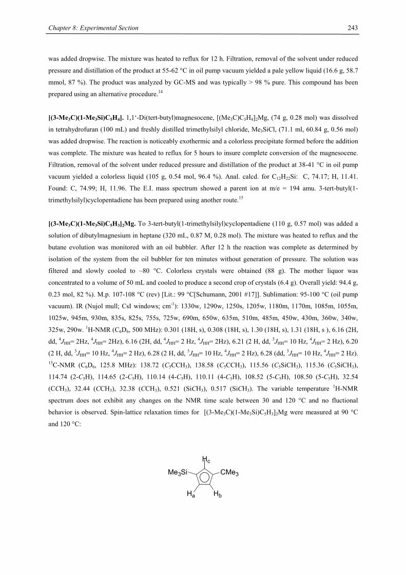
Chapter 8: Experimental Section
243
was added dropwise. The mixture was heated to reflux for 12 h. Filtration, removal of the solvent under reduced
pressure and distillation of the product at 55-62 °C in oil pump vacuum yielded a pale yellow liquid (16.6 g, 58.7
mmol, 87 %). The product was analyzed by GC-MS and was typically > 98 % pure. This compound has been
prepared using an alternative procedure.14
[(3-Me3C)(1-Me3Si)C5H4]. 1,1‘-Di(tert-butyl)magnesocene, [(Me3C)C5H4]2Mg, (74 g, 0.28 mol) was dissolved
in tetrahydrofuran (100 mL) and freshly distilled trimethylsilyl chloride, Me3SiCl, (71.1 ml, 60.84 g, 0.56 mol)
was added dropwise. The reaction is noticeably exothermic and a colorless precipitate formed before the addition
was complete. The mixture was heated to reflux for 5 hours to insure complete conversion of the magnesocene.
Filtration, removal of the solvent under reduced pressure and distillation of the product at 38-41 °C in oil pump
vacuum yielded a colorless liquid (105 g, 0.54 mol, 96.4 %). Anal. calcd. for C12H22Si: C, 74.17; H, 11.41.
Found: C, 74.99; H, 11.96. The E.I. mass spectrum showed a parent ion at m/e = 194 amu. 3-tert-butyl(1-
trimethylsilyl)cyclopentadiene has been prepared using another route.15
[(3-Me3C)(1-Me3Si)C5H3]2Mg. To 3-tert-butyl(1-trimethylsilyl)cyclopentadiene (110 g, 0.57 mol) was added a
solution of dibutylmagnesium in heptane (320 mL, 0.87 M, 0.28 mol). The mixture was heated to reflux and the
butane evolution was monitored with an oil bubbler. After 12 h the reaction was complete as determined by
isolation of the system from the oil bubbler for ten minutes without generation of pressure. The solution was
filtered and slowly cooled to –80 °C. Colorless crystals were obtained (88 g). The mother liquor was
concentrated to a volume of 50 mL and cooled to produce a second crop of crystals (6.4 g). Overall yield: 94.4 g,
0.23 mol, 82 %). M.p. 107-108 °C (rev) [Lit.: 99 °C[Schumann, 2001 #17]]. Sublimation: 95-100 °C (oil pump
vacuum). IR (Nujol mull; CsI windows; cm-1): 1330w, 1290w, 1250s, 1205w, 1180m, 1170m, 1085m, 1055m,
1025w, 945m, 930m, 835s, 825s, 755s, 725w, 690m, 650w, 635m, 510m, 485m, 450w, 430m, 360w, 340w,
325w, 290w. 1H-NMR (C6D6, 500 MHz): 0.301 (18H, s), 0.308 (18H, s), 1.30 (18H, s), 1.31 (18H, s ), 6.16 (2H,
dd, 4JHH= 2Hz, 4JHH= 2Hz), 6.16 (2H, dd, 4JHH= 2 Hz, 4JHH= 2Hz), 6.21 (2 H, dd, 3JHH= 10 Hz, 4JHH= 2 Hz), 6.20
(2 H, dd, 3JHH= 10 Hz, 4JHH= 2 Hz), 6.28 (2 H, dd, 3JHH= 10 Hz, 4JHH= 2 Hz), 6.28 (dd, 3JHH= 10 Hz, 4JHH= 2 Hz). 13C-NMR (C6D6, 125.8 MHz): 138.72 (C5CCH3), 138.58 (C5CCH3), 115.56 (C5SiCH3), 115.36 (C5SiCH3),
114.74 (2-C5H), 114.65 (2-C5H), 110.14 (4-C5H), 110.11 (4-C5H), 108.52 (5-C5H), 108.50 (5-C5H), 32.54
(CCH3), 32.44 (CCH3), 32.38 (CCH3), 0.521 (SiCH3), 0.517 (SiCH3). The variable temperature 1H-NMR
spectrum does not exhibit any changes on the NMR time scale between 30 and 120 °C and no fluctional
behavior is observed. Spin-lattice relaxation times for [(3-Me3C)(1-Me3Si)C5H3]2Mg were measured at 90 °C
and 120 °C:
Hc
Ha Hb
CMe3Me3Si
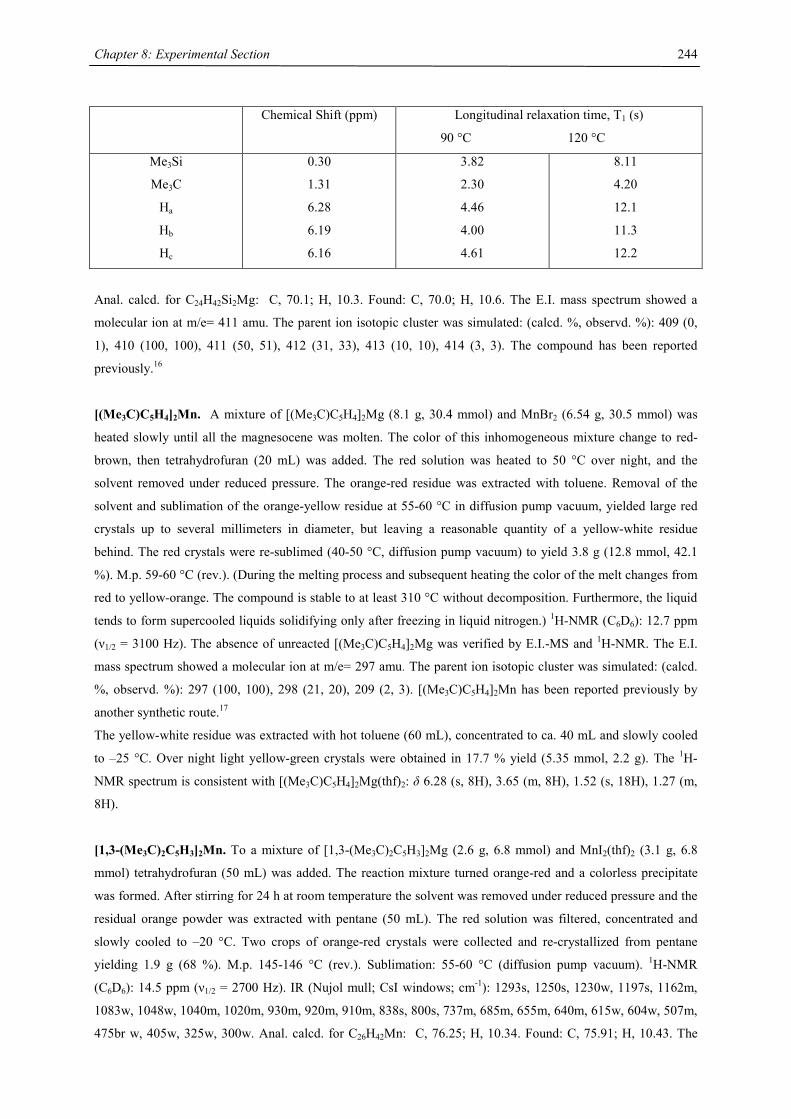
Chapter 8: Experimental Section
244
Chemical Shift (ppm) Longitudinal relaxation time, T1 (s)
90 °C 120 °C
Me3Si
Me3C
Ha
Hb
Hc
0.30
1.31
6.28
6.19
6.16
3.82
2.30
4.46
4.00
4.61
8.11
4.20
12.1
11.3
12.2
Anal. calcd. for C24H42Si2Mg: C, 70.1; H, 10.3. Found: C, 70.0; H, 10.6. The E.I. mass spectrum showed a
molecular ion at m/e= 411 amu. The parent ion isotopic cluster was simulated: (calcd. %, observd. %): 409 (0,
1), 410 (100, 100), 411 (50, 51), 412 (31, 33), 413 (10, 10), 414 (3, 3). The compound has been reported
previously.16
[(Me3C)C5H4]2Mn. A mixture of [(Me3C)C5H4]2Mg (8.1 g, 30.4 mmol) and MnBr2 (6.54 g, 30.5 mmol) was
heated slowly until all the magnesocene was molten. The color of this inhomogeneous mixture change to red-
brown, then tetrahydrofuran (20 mL) was added. The red solution was heated to 50 °C over night, and the
solvent removed under reduced pressure. The orange-red residue was extracted with toluene. Removal of the
solvent and sublimation of the orange-yellow residue at 55-60 °C in diffusion pump vacuum, yielded large red
crystals up to several millimeters in diameter, but leaving a reasonable quantity of a yellow-white residue
behind. The red crystals were re-sublimed (40-50 °C, diffusion pump vacuum) to yield 3.8 g (12.8 mmol, 42.1
%). M.p. 59-60 °C (rev.). (During the melting process and subsequent heating the color of the melt changes from
red to yellow-orange. The compound is stable to at least 310 °C without decomposition. Furthermore, the liquid
tends to form supercooled liquids solidifying only after freezing in liquid nitrogen.) 1H-NMR (C6D6): 12.7 ppm
(ν1/2 = 3100 Hz). The absence of unreacted [(Me3C)C5H4]2Mg was verified by E.I.-MS and 1H-NMR. The E.I.
mass spectrum showed a molecular ion at m/e= 297 amu. The parent ion isotopic cluster was simulated: (calcd.
%, observd. %): 297 (100, 100), 298 (21, 20), 209 (2, 3). [(Me3C)C5H4]2Mn has been reported previously by
another synthetic route.17
The yellow-white residue was extracted with hot toluene (60 mL), concentrated to ca. 40 mL and slowly cooled
to –25 °C. Over night light yellow-green crystals were obtained in 17.7 % yield (5.35 mmol, 2.2 g). The 1H-
NMR spectrum is consistent with [(Me3C)C5H4]2Mg(thf)2: δ 6.28 (s, 8H), 3.65 (m, 8H), 1.52 (s, 18H), 1.27 (m,
8H).
[1,3-(Me3C)2C5H3]2Mn. To a mixture of [1,3-(Me3C)2C5H3]2Mg (2.6 g, 6.8 mmol) and MnI2(thf)2 (3.1 g, 6.8
mmol) tetrahydrofuran (50 mL) was added. The reaction mixture turned orange-red and a colorless precipitate
was formed. After stirring for 24 h at room temperature the solvent was removed under reduced pressure and the
residual orange powder was extracted with pentane (50 mL). The red solution was filtered, concentrated and
slowly cooled to –20 °C. Two crops of orange-red crystals were collected and re-crystallized from pentane
yielding 1.9 g (68 %). M.p. 145-146 °C (rev.). Sublimation: 55-60 °C (diffusion pump vacuum). 1H-NMR
(C6D6): 14.5 ppm (ν1/2 = 2700 Hz). IR (Nujol mull; CsI windows; cm-1): 1293s, 1250s, 1230w, 1197s, 1162m,
1083w, 1048w, 1040m, 1020m, 930m, 920m, 910m, 838s, 800s, 737m, 685m, 655m, 640m, 615w, 604w, 507m,
475br w, 405w, 325w, 300w. Anal. calcd. for C26H42Mn: C, 76.25; H, 10.34. Found: C, 75.91; H, 10.43. The
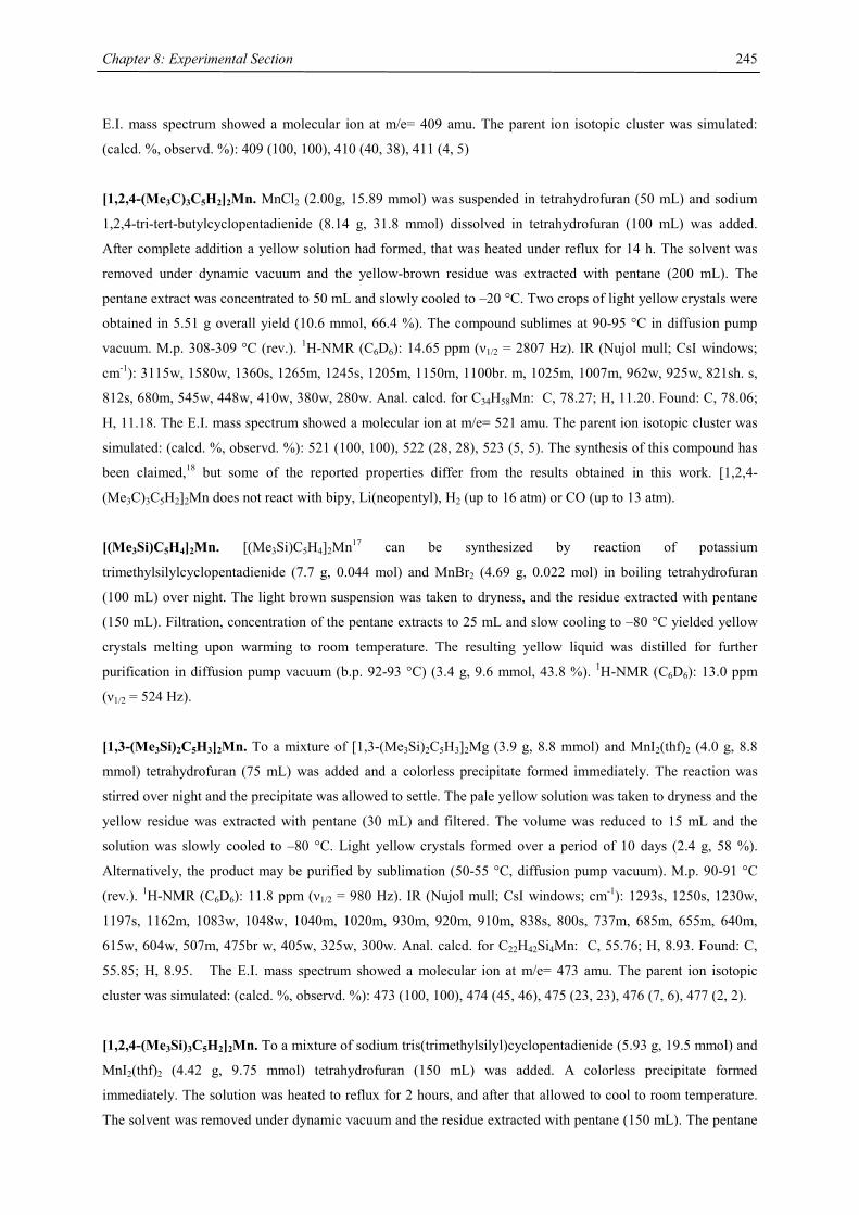
Chapter 8: Experimental Section
245
E.I. mass spectrum showed a molecular ion at m/e= 409 amu. The parent ion isotopic cluster was simulated:
(calcd. %, observd. %): 409 (100, 100), 410 (40, 38), 411 (4, 5)
[1,2,4-(Me3C)3C5H2]2Mn. MnCl2 (2.00g, 15.89 mmol) was suspended in tetrahydrofuran (50 mL) and sodium
1,2,4-tri-tert-butylcyclopentadienide (8.14 g, 31.8 mmol) dissolved in tetrahydrofuran (100 mL) was added.
After complete addition a yellow solution had formed, that was heated under reflux for 14 h. The solvent was
removed under dynamic vacuum and the yellow-brown residue was extracted with pentane (200 mL). The
pentane extract was concentrated to 50 mL and slowly cooled to –20 °C. Two crops of light yellow crystals were
obtained in 5.51 g overall yield (10.6 mmol, 66.4 %). The compound sublimes at 90-95 °C in diffusion pump
vacuum. M.p. 308-309 °C (rev.). 1H-NMR (C6D6): 14.65 ppm (ν1/2 = 2807 Hz). IR (Nujol mull; CsI windows;
cm-1): 3115w, 1580w, 1360s, 1265m, 1245s, 1205m, 1150m, 1100br. m, 1025m, 1007m, 962w, 925w, 821sh. s,
812s, 680m, 545w, 448w, 410w, 380w, 280w. Anal. calcd. for C34H58Mn: C, 78.27; H, 11.20. Found: C, 78.06;
H, 11.18. The E.I. mass spectrum showed a molecular ion at m/e= 521 amu. The parent ion isotopic cluster was
simulated: (calcd. %, observd. %): 521 (100, 100), 522 (28, 28), 523 (5, 5). The synthesis of this compound has
been claimed,18 but some of the reported properties differ from the results obtained in this work. [1,2,4-
(Me3C)3C5H2]2Mn does not react with bipy, Li(neopentyl), H2 (up to 16 atm) or CO (up to 13 atm).
[(Me3Si)C5H4]2Mn. [(Me3Si)C5H4]2Mn17 can be synthesized by reaction of potassium
trimethylsilylcyclopentadienide (7.7 g, 0.044 mol) and MnBr2 (4.69 g, 0.022 mol) in boiling tetrahydrofuran
(100 mL) over night. The light brown suspension was taken to dryness, and the residue extracted with pentane
(150 mL). Filtration, concentration of the pentane extracts to 25 mL and slow cooling to –80 °C yielded yellow
crystals melting upon warming to room temperature. The resulting yellow liquid was distilled for further
purification in diffusion pump vacuum (b.p. 92-93 °C) (3.4 g, 9.6 mmol, 43.8 %). 1H-NMR (C6D6): 13.0 ppm
(ν1/2 = 524 Hz).
[1,3-(Me3Si)2C5H3]2Mn. To a mixture of [1,3-(Me3Si)2C5H3]2Mg (3.9 g, 8.8 mmol) and MnI2(thf)2 (4.0 g, 8.8
mmol) tetrahydrofuran (75 mL) was added and a colorless precipitate formed immediately. The reaction was
stirred over night and the precipitate was allowed to settle. The pale yellow solution was taken to dryness and the
yellow residue was extracted with pentane (30 mL) and filtered. The volume was reduced to 15 mL and the
solution was slowly cooled to –80 °C. Light yellow crystals formed over a period of 10 days (2.4 g, 58 %).
Alternatively, the product may be purified by sublimation (50-55 °C, diffusion pump vacuum). M.p. 90-91 °C
(rev.). 1H-NMR (C6D6): 11.8 ppm (ν1/2 = 980 Hz). IR (Nujol mull; CsI windows; cm-1): 1293s, 1250s, 1230w,
1197s, 1162m, 1083w, 1048w, 1040m, 1020m, 930m, 920m, 910m, 838s, 800s, 737m, 685m, 655m, 640m,
615w, 604w, 507m, 475br w, 405w, 325w, 300w. Anal. calcd. for C22H42Si4Mn: C, 55.76; H, 8.93. Found: C,
55.85; H, 8.95. The E.I. mass spectrum showed a molecular ion at m/e= 473 amu. The parent ion isotopic
cluster was simulated: (calcd. %, observd. %): 473 (100, 100), 474 (45, 46), 475 (23, 23), 476 (7, 6), 477 (2, 2).
[1,2,4-(Me3Si)3C5H2]2Mn. To a mixture of sodium tris(trimethylsilyl)cyclopentadienide (5.93 g, 19.5 mmol) and
MnI2(thf)2 (4.42 g, 9.75 mmol) tetrahydrofuran (150 mL) was added. A colorless precipitate formed
immediately. The solution was heated to reflux for 2 hours, and after that allowed to cool to room temperature.
The solvent was removed under dynamic vacuum and the residue extracted with pentane (150 mL). The pentane
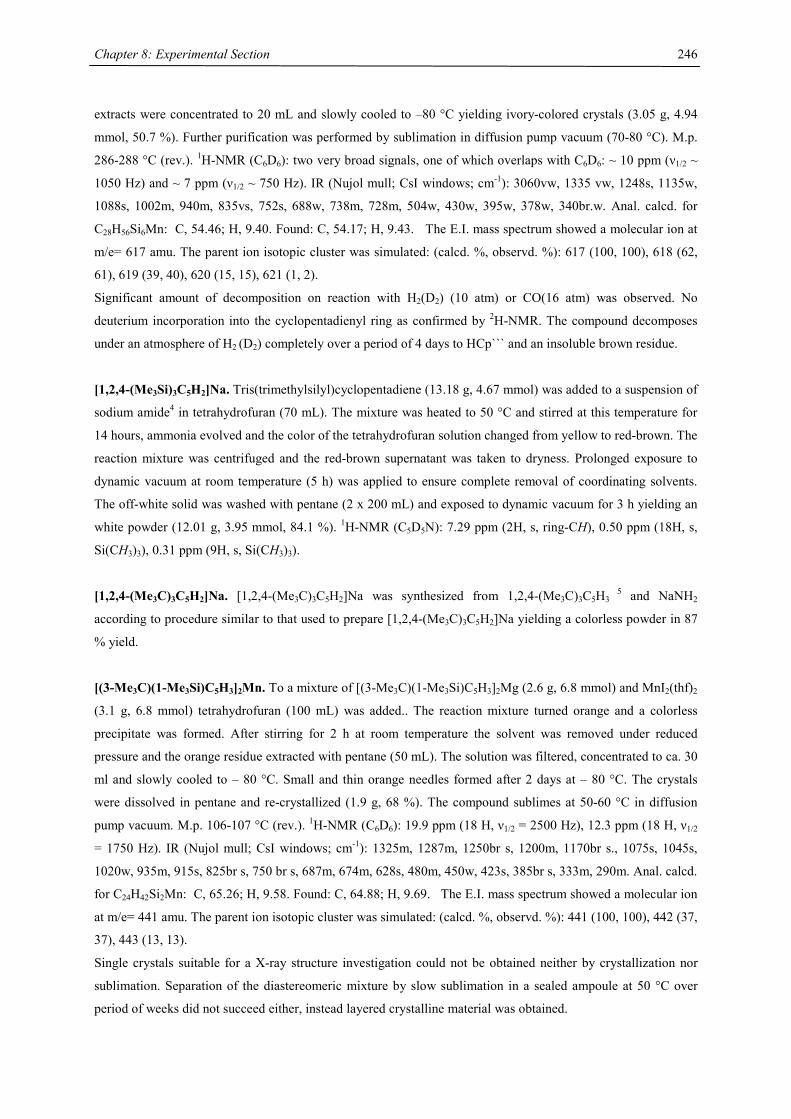
Chapter 8: Experimental Section
246
extracts were concentrated to 20 mL and slowly cooled to –80 °C yielding ivory-colored crystals (3.05 g, 4.94
mmol, 50.7 %). Further purification was performed by sublimation in diffusion pump vacuum (70-80 °C). M.p.
286-288 °C (rev.). 1H-NMR (C6D6): two very broad signals, one of which overlaps with C6D6: ~ 10 ppm (ν1/2 ~
1050 Hz) and ~ 7 ppm (ν1/2 ~ 750 Hz). IR (Nujol mull; CsI windows; cm-1): 3060vw, 1335 vw, 1248s, 1135w,
1088s, 1002m, 940m, 835vs, 752s, 688w, 738m, 728m, 504w, 430w, 395w, 378w, 340br.w. Anal. calcd. for
C28H56Si6Mn: C, 54.46; H, 9.40. Found: C, 54.17; H, 9.43. The E.I. mass spectrum showed a molecular ion at
m/e= 617 amu. The parent ion isotopic cluster was simulated: (calcd. %, observd. %): 617 (100, 100), 618 (62,
61), 619 (39, 40), 620 (15, 15), 621 (1, 2).
Significant amount of decomposition on reaction with H2(D2) (10 atm) or CO(16 atm) was observed. No
deuterium incorporation into the cyclopentadienyl ring as confirmed by 2H-NMR. The compound decomposes
under an atmosphere of H2 (D2) completely over a period of 4 days to HCp``` and an insoluble brown residue.
[1,2,4-(Me3Si)3C5H2]Na. Tris(trimethylsilyl)cyclopentadiene (13.18 g, 4.67 mmol) was added to a suspension of
sodium amide4 in tetrahydrofuran (70 mL). The mixture was heated to 50 °C and stirred at this temperature for
14 hours, ammonia evolved and the color of the tetrahydrofuran solution changed from yellow to red-brown. The
reaction mixture was centrifuged and the red-brown supernatant was taken to dryness. Prolonged exposure to
dynamic vacuum at room temperature (5 h) was applied to ensure complete removal of coordinating solvents.
The off-white solid was washed with pentane (2 x 200 mL) and exposed to dynamic vacuum for 3 h yielding an
white powder (12.01 g, 3.95 mmol, 84.1 %). 1H-NMR (C5D5N): 7.29 ppm (2H, s, ring-CH), 0.50 ppm (18H, s,
Si(CH3)3), 0.31 ppm (9H, s, Si(CH3)3).
[1,2,4-(Me3C)3C5H2]Na. [1,2,4-(Me3C)3C5H2]Na was synthesized from 1,2,4-(Me3C)3C5H3 5 and NaNH2
according to procedure similar to that used to prepare [1,2,4-(Me3C)3C5H2]Na yielding a colorless powder in 87
% yield.
[(3-Me3C)(1-Me3Si)C5H3]2Mn. To a mixture of [(3-Me3C)(1-Me3Si)C5H3]2Mg (2.6 g, 6.8 mmol) and MnI2(thf)2
(3.1 g, 6.8 mmol) tetrahydrofuran (100 mL) was added.. The reaction mixture turned orange and a colorless
precipitate was formed. After stirring for 2 h at room temperature the solvent was removed under reduced
pressure and the orange residue extracted with pentane (50 mL). The solution was filtered, concentrated to ca. 30
ml and slowly cooled to – 80 °C. Small and thin orange needles formed after 2 days at – 80 °C. The crystals
were dissolved in pentane and re-crystallized (1.9 g, 68 %). The compound sublimes at 50-60 °C in diffusion
pump vacuum. M.p. 106-107 °C (rev.). 1H-NMR (C6D6): 19.9 ppm (18 H, ν1/2 = 2500 Hz), 12.3 ppm (18 H, ν1/2
= 1750 Hz). IR (Nujol mull; CsI windows; cm-1): 1325m, 1287m, 1250br s, 1200m, 1170br s., 1075s, 1045s,
1020w, 935m, 915s, 825br s, 750 br s, 687m, 674m, 628s, 480m, 450w, 423s, 385br s, 333m, 290m. Anal. calcd.
for C24H42Si2Mn: C, 65.26; H, 9.58. Found: C, 64.88; H, 9.69. The E.I. mass spectrum showed a molecular ion
at m/e= 441 amu. The parent ion isotopic cluster was simulated: (calcd. %, observd. %): 441 (100, 100), 442 (37,
37), 443 (13, 13).
Single crystals suitable for a X-ray structure investigation could not be obtained neither by crystallization nor
sublimation. Separation of the diastereomeric mixture by slow sublimation in a sealed ampoule at 50 °C over
period of weeks did not succeed either, instead layered crystalline material was obtained.

Chapter 8: Experimental Section
247
Manganocenes, [(C5H5)2Mn]19 and [(C5H4Me)2Mn]20, were synthesized according to published procedures, and
purified by sublimation and/or crystallization. (C5H4Me)2Mn was crystallized from diethyl ether at –20 °C,
(C5H5)2Mn was crystallized from toluene at –20 °C.
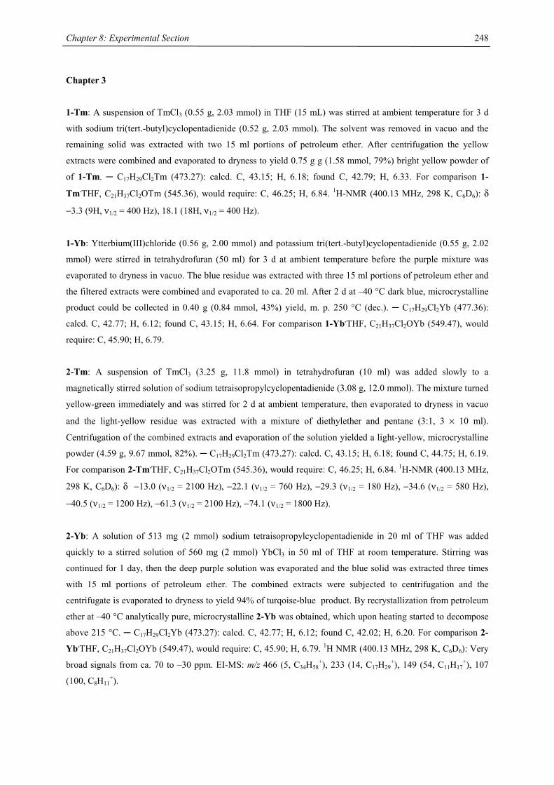
Chapter 8: Experimental Section
248
Chapter 3
1-Tm: A suspension of TmCl3 (0.55 g, 2.03 mmol) in THF (15 mL) was stirred at ambient temperature for 3 d
with sodium tri(tert.-butyl)cyclopentadienide (0.52 g, 2.03 mmol). The solvent was removed in vacuo and the
remaining solid was extracted with two 15 ml portions of petroleum ether. After centrifugation the yellow
extracts were combined and evaporated to dryness to yield 0.75 g g (1.58 mmol, 79%) bright yellow powder of
of 1-Tm. ─ C17H29Cl2Tm (473.27): calcd. C, 43.15; H, 6.18; found C, 42.79; H, 6.33. For comparison 1-
Tm.THF, C21H37Cl2OTm (545.36), would require: C, 46.25; H, 6.84. 1H-NMR (400.13 MHz, 298 K, C6D6): δ
−3.3 (9H, ν1/2 = 400 Hz), 18.1 (18H, ν1/2 = 400 Hz).
1-Yb: Ytterbium(III)chloride (0.56 g, 2.00 mmol) and potassium tri(tert.-butyl)cyclopentadienide (0.55 g, 2.02
mmol) were stirred in tetrahydrofuran (50 ml) for 3 d at ambient temperature before the purple mixture was
evaporated to dryness in vacuo. The blue residue was extracted with three 15 ml portions of petroleum ether and
the filtered extracts were combined and evaporated to ca. 20 ml. After 2 d at –40 °C dark blue, microcrystalline
product could be collected in 0.40 g (0.84 mmol, 43%) yield, m. p. 250 °C (dec.). ─ C17H29Cl2Yb (477.36):
calcd. C, 42.77; H, 6.12; found C, 43.15; H, 6.64. For comparison 1-Yb.THF, C21H37Cl2OYb (549.47), would
require: C, 45.90; H, 6.79.
2-Tm: A suspension of TmCl3 (3.25 g, 11.8 mmol) in tetrahydrofuran (10 ml) was added slowly to a
magnetically stirred solution of sodium tetraisopropylcyclopentadienide (3.08 g, 12.0 mmol). The mixture turned
yellow-green immediately and was stirred for 2 d at ambient temperature, then evaporated to dryness in vacuo
and the light-yellow residue was extracted with a mixture of diethylether and pentane (3:1, 3 × 10 ml).
Centrifugation of the combined extracts and evaporation of the solution yielded a light-yellow, microcrystalline
powder (4.59 g, 9.67 mmol, 82%). ─ C17H29Cl2Tm (473.27): calcd. C, 43.15; H, 6.18; found C, 44.75; H, 6.19.
For comparison 2-Tm.THF, C21H37Cl2OTm (545.36), would require: C, 46.25; H, 6.84. 1H-NMR (400.13 MHz,
298 K, C6D6): δ −13.0 (ν1/2 = 2100 Hz), −22.1 (ν1/2 = 760 Hz), −29.3 (ν1/2 = 180 Hz), −34.6 (ν1/2 = 580 Hz),
−40.5 (ν1/2 = 1200 Hz), −61.3 (ν1/2 = 2100 Hz), −74.1 (ν1/2 = 1800 Hz).
2-Yb: A solution of 513 mg (2 mmol) sodium tetraisopropylcyclopentadienide in 20 ml of THF was added
quickly to a stirred solution of 560 mg (2 mmol) YbCl3 in 50 ml of THF at room temperature. Stirring was
continued for 1 day, then the deep purple solution was evaporated and the blue solid was extracted three times
with 15 ml portions of petroleum ether. The combined extracts were subjected to centrifugation and the
centrifugate is evaporated to dryness to yield 94% of turqoise-blue product. By recrystallization from petroleum
ether at –40 °C analytically pure, microcrystalline 2-Yb was obtained, which upon heating started to decompose
above 215 °C. ─ C17H29Cl2Yb (473.27): calcd. C, 42.77; H, 6.12; found C, 42.02; H, 6.20. For comparison 2-
Yb.THF, C21H37Cl2OYb (549.47), would require: C, 45.90; H, 6.79. 1H NMR (400.13 MHz, 298 K, C6D6): Very
broad signals from ca. 70 to –30 ppm. EI-MS: m/z 466 (5, C34H58+), 233 (14, C17H29
+), 149 (54, C11H17+), 107
(100, C8H11+).
Chapter 8: Experimental Section
249
2-Lu: To a suspension of lutetium trichloride (0.99 g, 3.52 mmol) in tetrahydrofuran (20 ml) a solution of
sodium tetraisopropylcyclopentadienide (0.90 g, 3.51 mmol) in tetrahydrofuran (20 ml) was slowly added. The
yellow suspension was stirred at ambient temperature for 24 hours. The solvent was evaporated and the yellow-
white residue washed with 5 ml cold pentane. The residue was then extracted with toluene (3 x 10 ml) and the
combined extracts were concentrated. After slow cooling to − 35 °C very pale yellow microcrystalline powder
was collected (1.3 g, 2.71 mmol, 77.2 %). − C17H29Cl2Yb (479.29): calcd. C, 42.6; H, 6.1; found C, 43.74; H,
6.47. For comparison 2-Lu.THF, C21H37Cl2OLu (551.40), would require: C, 45.76; H, 6.76.
4-Lu: To a magnetically stirred suspension of LuCl3 (1.085 g, 3.8 mmol) in tetrahydrofuran (50 ml) a solution of
sodium tetraisopropylcyclopentadienide (1.053 g, 4.1 mmol) in tetrahydrofuran (30 ml) was added slowly.
During this time the suspended solid was dissolved and the clear solution was heated to reflux for 3 h. A white
precipitate formed and the suspension was stirred at room temperature for another 12 h. The solvent was
evaporated and the solid residue was stirred with diethyl ether (40 ml) for 12 h. Centrifugation of the extract,
extraction of the solid residue with diethyl ether (2 × 10 ml), followed by centrifugation and concentration of the
combined extracts in vacuo gave a concentrated diethyl ether solution, which was allowed to slowly evaporate in
a glove box equipped with a charcoal filter to afford colorless crystals of the Lu6 complex containing five
equivalents of coordinated diethyl ether (0.360 g, 0.12 mmol, 19%). ─ C105H195Cl13Lu6O5 (3024.37): calcd. C,
41.79; H, 6.50; found: C, 40.88; H, 6.44. In [D8]-THF this complex dissolved to form a solution of
[(C5HR4)LuCl2(OC4D8)] (5-Lu) ; 1H-NMR (400.13 MHz, 298 K, d8-THF): δ 5.81 (s, 1H, ring-H), 3.28 (m,
CHMe2, J = 6.9 Hz), 3.08 (m, CHMe2, J = 7.1 Hz), 1.54 (d, 6H, CH3, J = 7.2 Hz), 1.35 (d, 6H, CH3, J = 6.9 Hz),
1.18 (d, 6H, CH3, J = 6.9 Hz), 1.05 (d, 6H, CH3, J not determined because of superimposition with the triplet
signal of diethyl ether present in the solution). 13C{1H}-NMR (100.62 MHz, 298 K, d8-THF): δ 134.3 (2C,
CCHMe2), 129.1 (2C, CCHMe2), 104.8 (1C, ring-CH), 28.4 (2C, CHMe2), 28.3 (2C, CH3), 27.9 (2C, CHMe2),
26.0 (2C, CH3), 23.7 (2C, CH3), 23.4 (2C, CH3); diethylether signals: 66.3 (OCH2), 15.8 (OCH2CH3).
3-Tm: single crystals of the dimethoxyethane adduct were grown from a concentrated DME solution of 2-Tm at
room temperature. ─ C21H39Cl2O2Tm (563.39): calcd. C, 44.77; H, 6.98; found C, 45.75; H, 6.91.
3-Yb: single crystals of the dimethoxyethane adduct were grown from a concentrated DME solution of 3-Yb at
room temperature. Alternatively 3-Yb was easily accessible by oxidation of octaisopropylytterbocen (4Cp2Yb)
with 0.5 equivalents on hexachloroethane (C2Cl6) in dimethoxyethane as the only identifiable Yb containing
compound.
3-Lu: Slow cooling of a saturated dimethoxyethane solution of 2-Lu to − 35 °C yielded colorless crystalline
plates losing coordinated solvent rather quickly. Alternatively crystallization at − 35 °C from a saturated toluene-
dimethoxyethane (10:1) solution yielded colorless blocks subjected to elemental and 1H-NMR analysis.
C21H39O2Cl2Yb (569.41): calcd. C, 44.3; H, 6.9; found C, 45.0; H, 7.05. 1H-NMR (400.13 MHz, 298 K, C6D6): δ
6.44 (s, 1H, ring-H), 3.41 (m, 2H, CHMe2), 3.32 (8H, CHMe2 and (CH2OCH3)2 superimposed), 3.14 (s, 6H,
(CH2OCH3)2), 1.70 (d, 6H, CH3, J = 7.25 Hz), 1.59 (d, 6H, CH3, J = 6.53 Hz), 1.38 (d, 6H, CH3, J = 7.05 Hz),
1.19 (d, 6H, CH3, J = 6.62 Hz ).
Chapter 8: Experimental Section
250
6-Yb: A purple-blue solution of 2-Yb (0.72 g, 1.51 mmol) in dimethoxyethane (15 ml) was stirred with sodium
azide (0.20 g, 3.07 mmol) for 3 d at ambient temperature. The solvent was evaporated and the red-purple residue
was extracted with diethyl ether (4 × 10 ml). Centrifugation and evaporation of the combined extracts gave a
dark red-purple solid, which was washed with pentane (2 × 3-4 ml) and dried in vacuo (0.53 g. 1.08 mmol,
58.2%). Shock-sensitivity was not been observed, however the compound should be handled with great care. X-
ray suitable crystals were grown from a concentrated dme solution at −15°C. These crystals lose coordinated
dimethoxyethane at 90-150 °C and change color to light blue, but do not melt at 250 °C. ─
Na2C126H234O12N42Yb6 (3613.74): calcd. C, 41.88; H, 6.53; N, 16.28; found C, 41.63; H, 5.96; N 17.13. IR
(Nujol) 2189 cm-1 (s), 2170 cm-1 (s), 2127 cm-1 (m), 2083 cm-1 (s).
7-Tm: A solution of sodium bis(trimethylsilyl)amide (0.28 g, 1.52 mmol) in toluene (5 ml) was added to a
solution of 1-Tm (0.36 g, 0.76 mmol) in toluene (10 ml) and stirred at ambient temperature for 2 h. The solvent
was removed in vacuo and the yellow residue was extracted with pentane (2 × 10 ml). After centrifugation the
volume of the combined extracts was reduced in vacuo to ca. 5 ml and cooled to 3 °C. Seven days later light
yellow-green crystals could be harvested (0.48 g, 0.73 mmol, 87%). ─ C29H65N2Si4Tm (723.13): calcd. C, 48.17;
H, 9.06; N, 3.87; found C, 48.00; H, 8.86; N, 3.85; 1H-NMR (400.13 MHz, 298 K, C6D6): δ 202.6 (18H, ν1/2 =
1550 Hz), 18.8 (9H, ν1/2 = 1140 Hz), −0.5 (36H, ν1/2 = 425 Hz), −218 (2H, ν1/2 = 4500 Hz); MS (70 eV, EI): m/z
(%) = 722.1 (<1) M+, 707.1 (<1) M+-CH3, 635.1 (<1) M+-SiMe4, 562.2 (<1) M+-N(SiMe3)2, 489.2 (28) M+-Cp´´´,
234.1 (12) HCp´´´+, 161.2 (3) HN(SiMe3)2+, 146.2 (75) Me3Si-SiMe3
+, 57.1 (62) CMe3+.
8-Yb: To a solution of 2-Yb (0.72 g, 1.51 mmol) in diethyl ether (20 ml) lithium (tert.-butyl)cyclopentadienide
(0.19 g, 1.51 mmol) was added. The color changed to red-brown immediately and the reaction mixture was
allowed to stir for 2 days at room temperature. The solvent was evaporated and the residue extracted with
pentane (2 x 15 ml). Centrifugation, concentration of the combined pentane extracts and crystallization at – 15
°C resulted in big, red-brown hexagonal crystals (0.2 g, 0.22 mmol). The compound was identified by X-ray
diffraction as 1,1´-Bis((tert-butyl)cyclopentadienyl)ytterbium chloride. 1H NMR (400.13 MHz, 298 K, C6D6): δ
55.6 (18H, ν1/2 = 280 Hz, CMe3), -60.2 (4 H, ν1/2 = 560 Hz, ring-CH).
9-Yb: To a suspension of ytterbium trichloride (0.21 g, 0.75 mmol) in dimethoxyethane (6 ml) a solution of
sodium tri(tert.-butyl)cyclopentadienide (0.39 g, 1.5 mmol) in dimethoxyethane (5 ml) was added slowly at room
temperature. Within a few minutes the suspended solid was dissolved and a purple-red color was observed.
Stirring was continued for 2 d, then pentane (5 ml) was added and the precipitate formed was removed by
centrifugation. Evaporation of the solution afforded a purple-red oil, which was dissolved in hexane (5 ml) and
stirred at room temperature for 20 minutes. The green precipitate formed during this period was removed by
centrifugation and the solution was evaporated in vacuo. The red-brown, oily solid was recrystallized by slow
cooling of a saturated hexane solution to –10 °C to yield the dark red, crystalline product (0.11 g, 0.106 mmol,
28%). ─ C20H36O2ClYb (517.00): calcd. C, 46.46; H, 7.02; found C, 45.45; H, 6.94.
Chapter 8: Experimental Section
251
10-Tm: A solution of sodium tetraisopropylcyclopentadienide (1.024 g, 4 mmol) was added to a suspension of
TmI3 (1.099 g, 2 mmol) in tetrahydrofuran (10 ml) and the mixture was stirred for 2 days at room temperature.
Solvent removal in vacuo, extraction with hexane (5 × 10 ml), centrifugation and evaporation of the hexane
solvent in dynamic vacuum afforded a yellow-brown solid. Compound 15-Tm was obtained as yellow crystals
by sublimation in oil pump vacuum at 80 – 90 °C (0.96 g, 1.26 mmol, 63 %). Single crystals for X-ray
diffraction were obtained by sublimation in an under vacuum sealed ampoule at 70 °C. ─ C34H58TmI (762.63):
calcd. C, 53.55; H, 7.67; found C, 54.27; H, 7.02; MS (70 eV, EI): m/z (%) = 762 (7) [M+], 635 (2) [M+-I], 529
(100) [M+-C5H(CHMe2)4], 496 (1) [M+-I-C5H(CHMe2)4], 149 (5) [C11H17+], 107 (14) [C8H11
+].
11-Nd: A mixture of solid neodymium trichloride (0.75 g, 3.00 mmol) and sodium tri(tert.-
butyl)cyclopentadienide (1.54 g, 6.00 mmol) was suspended in a 1:1 mixture of toluene and tetrahydrofuran (50
ml) and developed a blue-green color within a few minutes of stirring at room temperature. Stirring was
continued for 4 d, then the solvents were removed in vacuo and the dark green residue was extracted with
toluene (5 × 10 ml). Centrifugation and evaporation of the intense blue-green extracts to a residual volume of ca.
30 ml and crystallization at –20 °C yielded blue-green product (1.62 g, 2.50 mmol, 83%), which melted without
decomposition at 180-181 °C and could be sublimed at 130 °C in a glass tube sealed at 10-3 mbar. ─ C34H58ClNd
(646.53): calcd. C, 63.16; H, 9.04; found C, 62.74; H, 8.95; 1H-NMR (400.13 MHz, 298 K, C6D6): δ −5.4 (36H,
CMe3, ν1/2 = 135 Hz), −9.0 (4H, ring-H, ν1/2 = 150 Hz), −18.3 (18H,CMe3, ν1/2 = 100 Hz). MS (70 eV, EI): m/z
(%) = 644.9 (12) M+, 610 (3) M+-Cl, 411.9 (100) M+-C5H2(CMe3)3, 234.1 (26), HC5H2(CMe3)3, 178.1 (54)
NdCl+, 57 (99) CMe3+.
11-Pr: Praseodymium trichloride (0.56 g, 2.25 mmol) and sodium tri(tert.-butyl)cyclopentadienide (1.16 g, 4.50
mmol) were suspended in a 2:5 mixture of hexane and tetrahydrofuran (50 ml) and stirred at room temperature
for 4 d. The yellow solution was separated from a white precipitate by centrifugation, the solvent was removed
in vacuo, and the remaining solid was extracted with toluene (5 × 10 ml). Centrifugation of the intense yellow
extracts and removal of the solvent in vacuo gave a yellow powder, which was recrystallized from hexane at –20
°C to yield yellow crystals (1.02 g, 1.59 mmol, 70.5%), m. p. 189-192 °C without decomposition. ─ C34H58ClPr
(643.20): calcd. C, 63.49; H, 9.09; found C, 63.70; H, 8.95; 1H-NMR (400.13 MHz, 298 K, C6D6): δ −7.9 (40H,
CMe3 and ring-H, ν1/2 = 600 Hz), −35.1 (18H, CMe3, ν1/2 = 110 Hz). MS (70 eV, EI): m/z (%) = 641.9 (7) M+,
607 (2) M+-Cl, 408.9 (100) M+-C5H2(CMe3)3, 234.1 (4), HC5H2(CMe3)3, 57 (63) CMe3+.
12-Nd: Upon slow addition of a solution of trimethylaluminum (0.20 g, 2.77 mmol) in hexane (2 ml) to a
solution of 11-Nd (0.32 g, 0.50 mmol) in hexane (10 ml) at room temperature a color change from blue-green to
light green was observed and stirring was continued for 1 d. A small amount of insoluble material was removed
by centrifugation and the solvent was evaporated to a residual volume of 2 ml. Crystallization occurred already
at room temperature and yielded grass-green single crystals suitable for X-ray diffraction (0.20 g, 0.28 mmol,
56%), which upon heating to 110 °C decomposed to a green, oily solid. ─ C37H67AlClNd (718.62): calcd. C,
61.84; H, 9.40; found C, 57.27; H, 8.95; 1H-NMR (400.13 MHz, 298 K, C6D6): δ −4.4 (9H, AlMe3 ν1/2 = 350
Hz), −6.1 (36H, CMe3, ν1/2 = 790 Hz), -9.8 (4H, ring-H, ν1/2 = 200 Hz). –17.5 (18H, CMe3, ν1/2 = 180 Hz); MS
Chapter 8: Experimental Section
252
(70 eV, EI): m/z (%) = 641.9 (5) M+-AlMe3, 607 (3) M+-AlMe3-Cl, 408.9 (100) M+-AlMe3-C5H2(CMe3)3, 234.1
(3), HC5H2(CMe3)3, 72.0 (7) AlMe3+, 57 (71) CMe3
+.
13-Yb: To a suspension of ytterbium diiodide (2.00 g, 4.69 mmol) in tetrahydrofuran (40 ml) a solution of
sodium 1,3-di(tert.-butyl)cyclopentadienide (1.88 g, 9.37 mmol) in tetrahydrofuran (20 ml) was added. The dark
green solution was stirred for 3 days at ambient temperature. The solvent was evaporated and the residue was
extracted with pentane (4 x 20 ml). Centrifugation, concentration of the combined extracts and slow cooling to
−35 °C resulted in dark green crystals (1.72 g, 2.87 mmol, 61.3 %) − C30H50OYb (599.77): calcd. C, 60.08; H,
8.40; found C, 59.24; H, 8.22; MS (70 eV, EI): m/z (%) = 528 [M+-thf]. 1H-NMR (400.13 MHz, 298 K, C6D6):
δ 6.17 (4H, d, J = 2.52 Hz, ring-H), 5.69 (2 H, t, J = 2.52 Hz, ring-H), 3.49 (m, 4 H, O(CH2CH2)2), 1.33 (s, 36 H,
C(CH3)3), 1.24 (m, 4 H, O(CH2CH2)2). 13C{1H}-NMR (298 K, C6D6): 134.8 (4C, C(CMe3)), 103.2 (4C, ring-
CH), 101.3 (2C, ring-CH), 32.9 (12 C, C(CH3)3), 32.3 (4C, C(CH3)3); thf signals: 70.6 (2C, O(CH2CH2)2), 25.4
(2 C, O(CH2CH2)2).
14-Yb:To a solution of 13-Yb [(C5H3tBu2)2Yb(thf)] (1.00g, 1.67 mmol) in tetrahydrofuran (25 ml) a mixture of
tetrahydrofuran (50 ml) and degassed water (30 mg, 30 µl, 1.67 mmol) was added. The green tetrahydrofuran
solution turned orange-red. Evaporation of the solvent, extraction of residue into toluene and slow cooling to –10
°C yielded long orange-red needles (0.29 g, 0.275 mmol, 33 %) suitable for X-ray diffraction. − C52H86O2Yb2
(1090.54): calcd. C, 57.22; H, 7.95; found C, 57.11; H, 7.83. The compound was only sparingly soluble in C6D6
and no further efforts have been made to obtain an 1H NMR spectrum. IR (Nujol) 3620 cm-1 (s).
Chapter 8: Experimental Section
253
Chapter 4
[Ce(C8H8)2][Li(thf)4]. Li2(C8H8)٠(thf)1.7 (2.95 g, 12.3 mmol) and anhydrous CeCl3 (1.51 g, 6.15 mmol) were
weighed into a Schlenk flask under nitrogen, suspended in tetrahydrofuran (60 mL) and stirred at reflux
overnight. During this time the color changed to lime green, the suspension was allowed to cool to room
temperature and to settle. The deep green tetrahydrofuran solution was filtered through a glass fiber filter,
concentrated to ca. 40 mL and cooled to –20 °C. Two crops of light green needles were isolated (2.62 g, 4.07
mmol, 66.1 %). The crystals obtained were exceedingly sensitive to moisture and air. Even trace amounts of
oxygen result in a significant color change to deep red, before turning yellow. The dark red compound was
identified to be Ce(C8H8)2 (by NMR spectroscopy). The crystals lost coordinated tetrahydrofuran at elevated
temperature on exposure to dynamic vacuum. The tetrahydrofuran content was established either by NMR
spectroscopy in pyridine-d5 or after hydrolysis of a sample with D2O in C6D6 M.p. 330 °C (dec.). 1H NMR
(C5D5N, 20 °C): δ 3.66 (16H, t, 3JCH = 6.0 Hz, α-CH2), 2.44 (16H, ν1/2 = 130 Hz, C8H8), 1.61 (16H, 3JCH = 6.0
Hz, β-CH2). 1H NMR (C4D8O, 20 °C): δ 3.60 (16H, br. s, α-CH2), 1.66 (16H, br. s, β-CH2), 1.50 (16H, ν1/2 =
100 Hz, C8H8). IR (Nujol mull; CsI windows; cm-1): 1815 (br. w), 1700 (w), 1580 (br. w), 1555 (br. vw), 1341
(m) 1305 (vw), 1260 (vw), 1180 (vbr. w), 1200 (sh), 1045 (vs), 920 (m), 895 (vs), 800 (vw), 762 (m), 740 (m),
722 (sh), 701 (m), 681 (vs), 610 (vw), 470 (sh), 425 (br. m), 370 (sh), 340 (br. s), 232 (vs).
[Ce(C8H8)2]. [Ce(C8H8)2][Li(thf)4] (2.57 g, 4.0 mmol) was exposed at 100 °C to dynamic vacuum for 2 hours to
ensure the complete removal of the coordinated tetrahydrofuran. The green crystals collapsed to a fine green
powder during this procedure. Under nitrogen freshly sublimed p-benzoquinone (0.43 g, 4.0 mmol) was added,
even in solid state an immediate color change to brown-purple was observed. Toluene (40 mL) was added and
the suspension stirred at 80 °C for 1 h, and then the solvent was slowly removed under dynamic vacuum. The
residue was charged onto an extraction frit and was continuously extracted with boiling toluene for 12 hours,
until the toluene extracts remained colorless. The deep red toluene extracts were allowed to cool slowly to room
temperature, during this time deep red crystals formed. The mother liquor was separated from the crystals by
filtration and cooled to –25 °C to yield a second crop of red blocks (0.80 g, 2.30 mmol, 57.5 %). M.p. 290 °C
(dec.). 1H NMR (C6D6, 20°C): δ 5.79 (16H, ν1/2 = 2.4 Hz, C8H8). 13C NMR (C6D6, 20°C): δ 114.9 (C8H8). The
E.I. mass spectrum showed a molecular ion at m/e= 348 amu. The parent ion isotopic cluster was simulated:
(calcd. %, observd. %): 348 (100,100), 349 (18,17), 350 (14,14), 351 (2,2). IR (Nujol mull; CsI windows; cm-1):
1880 (vw), 1780 (vw), 1318 (m), 1260 (vw), 1162 (vw), 1098 (vw), 898 (s), 785 (m), 742 (s), 730 (sh. m), 688
(vs), 249 (s).
[Ce2(C8H8)3]. Ce(C8H8)2 (0.25 g, 0.72 mmol) was weighed into a Schlenk tube, exposed to dynamic vacuum
and heated to 300 °C over a period of 15 min. During this time the color changed from dark red to light green
and the crystals collapsed to a light green powder (0.2 g, 0.34 mmol, 94.4 %). The light green compound was
insoluble in aromatic hydrocarbons. 1H NMR spectra revealed the absence of C8H8, as well as the starting
material. The identity of the compound was verified by its IR spectrum and reactivity. M.p. > 340 °C. IR (Nujol
mull; CsI windows; cm-1): 1880 (vw), 1838 (vw), 1772 (vw), 1728 (vw), 1580 (vw), 1427 (m), 1312 (m), 1262
(m), 898 (s), 850 (sh), 845 (w), 800 (m), 742 (s), 728 (s), 695 (vs), 682 (vs), 240 (sh), 232 (vs), 210 (s).
Chapter 8: Experimental Section
254
Chapter 5
[Ce(tmtaa)(tmtaaH)]. A hot, light yellow solution of [Ce(NTMS2)3] (0.18 g, 0.29 mmol), dissolved in ca. 10
mL toluene was added slowly to a hot, yellow solution of tmtaaH2 (0.314 g, 0.91 mmol) dissolved in ca. 10 mL
toluene. During the addition the color changed to red, and the solution was allowed to reach room temperature
without stirring to yield, after 24 h, red crystals (0.148 g, 0.18 mmol, 59 %). The product was sparingly soluble
in tetrahydrofuran and aromatic hydrocarbons, and insoluble in aliphatic solvents. M.p. > 330 °C. Anal. Calcd
for C44H45N8Ce: C, 63.98; H, 5.49; N, 13.59. Found: C, 63.83; H, 5.47; N, 13.49.
The E.I. mass spectrum showed a molecular ion at m/e= 825 amu. The parent ion isotopic cluster was simulated:
(calcd. %, observd. %): 825 (100,100), 826 (51,51), 827 (25,26), 828 (8,8), 829 (2,2). IR (Nujol mull; CsI
windows; cm-1): 3050 (w), 1675 (s), 1650 (w), 1560 (s), 1530 (m), 1420 (vs), 1395 (vs), 1380 (vs), 1270 (s),
1265 (s), 1221 (w), 1180 (vs), 1155 (m), 1110 (m), 1100 (m), 1100 (m), 1042 (m), 1020 (s), 932 (w), 935 (w),
795 (s), 788 (vs), 740 (vs), 700 (w), 658 (w), 620 (w), 550 (w), 480 (w), 382 (m).
[Ce2(tmtaa)3]. A solution of tmtaaH2 (0.15 g, 0.43 mmol) dissolved in ca. 15 mL tetrahydrofuran was added to a
solution [Ce(NTMS2)3] (0.18 g, 0.29 mmol) dissolved in 5 mL tetrahydrofuran and the mixture was left at room
temperature, without stirring, for 48 h. During this time the color of the mixture slowly changed from orange to
red and dark red crystals deposited on the walls of the Schlenk tube. The crystals, which co-crystallized with one
molecule of tetrahydrofuran per cerium center, were isolated by filtration and washed with pentane (5 mL) to
yield 0.12 g (0.083 mmol, 57 %). M.p. > 330 °C. Anal. Calcd for C74H82N12O2Ce2: C, 61.22; H, 5.69; N, 11.57.
Found: C, 60.75; H, 5.81; N, 11.31. 1H NMR (C6D6, 20°C): δ 16.75 (2H, ν1/2 = 50 Hz, CH), 15.10 (2H, ν1/2 = 50
Hz, CH), 7.68 (12H, ν1/2 = 21 Hz, CH3), 6.21 (12H, ν1/2 = 36 Hz, CH3), −2.53 (4H, ν1/2 = 15 Hz, C6H4), −2.64
(4H, ν1/2 = 13 Hz, C6H4), −5.08 (4H, ν1/2 = 13 Hz, C6H4), −5.51 (4H, ν1/2 = 35 Hz, C6H4), −8.59 (4H, ν1/2 = 52
Hz, C6H4), −13.20 (2H, ν1/2 = 60 Hz, CH), −15.26 (16H, ν1/2 = 45 Hz, overlapping CH3 + C6H4). IR (Nujol mull;
CsI windows; cm-1): 3050 (m), 1675 (vw), 1620 (w), 1560 (sh), 1550 (vs), 1535 (vs), 1415 (vs), 1380 (vs), 1270
(s), 1221 (m), 1185 (s), 1115 (m), 1070 (m), 1022 (s), 922 (m), 848 (w), 795 (vs), 770 (sh), 738 (vs), 700 (m),
625 (w), 615 (w), 390 (m), 362 (w), 329 (m).
[Ce(tmtaa)2]. Ce2(tmtaa)3 or Ce(tmtaa)(tmtaaH) were converted to Ce(tmtaa)2, thermally or chemically by
addition of 1,4-benzoquinone, [Cp2Fe][PF6] or traces of O2. The most convenient way was the reaction with
[Cp2Fe][PF6] or 1,4-benzoquinone (1eq per cerium center) in tetrahydrofuran or toluene, respectively. In each
case an insoluble residue was formed immediately, besides Ce(tmtaa)2 and tmtaaH2 (in case of [Cp2Fe][PF6] as
reactant also Cp2Fe). The solvent was removed under dynamic vacuum, and the residue was washed with hot
heptane to remove tmtaaH2 (and Cp2Fe), the remaining residue was dissolved in toluene and concentrated. X-ray
suitable crystals were grown by vapor diffusion of pentane into a concentrated toluene solution overnight. The
product was obtained as deep green, shiny cubes (58 % yield), which co-crystallized with half a molecule of
pentane per cerium center. The stoichiometry was established by X-ray crystallography, elemental analysis and
NMR spectroscopy. M.p.: > 310 °C. Anal. Calcd for C46.5H50N8Ce: C, 64.86; H, 5.85; N, 13.01. Found: C,
64.81; H, 5.89; N, 12.69. 1H NMR (C6D6, 20°C): δ 7.29 (8H, m, C6H4), 7.21 (8H, m, C6H4), 7.21 3.85 (4H, s,
CH), 1.77 (24H, s, CH3). 13C{1H} NMR (C6D6, 20°C): δ 156.5 (s, CMe), 136.6 (s, ipso-Ar), 126.0 (s, C6H4),
Chapter 8: Experimental Section
255
125.2 (s, C6H4), 106.1 (s, CH), 24.3 (s, CH3) (.The E.I. mass spectrum showed a molecular ion at m/e= 824 amu.
The parent ion isotopic cluster was simulated: (calcd. %, observd. %): 824 (100,100), 825 (51,51), 826 (25,26),
827 (8,8), 828 (2,2). IR (Nujol mull; CsI windows; cm-1): 3095 (vw), 3060 (m), 1565 (sh. vs), 1558 (vs), 1532
(s), 1438 (s), 1410 (br. vs), 1198 (m-s), 1280 (vs), 1228 (m), 1195 (vs), 1122 (m), 1050 (m), 1028 (s), 930 (m),
862 (vw), 853 (w), 800 (vs), 772(m), 742 (vs), 702 (m), 620 (w), 570 (vw), 510 (vw), 398 (m), 380 (m), 228
(vs).
[Ce(NTMS2)3].21 LiNTMS2٠(OEt2) (4.45 g, 18.45 mmol) (prepared from HNTMS2 and n-BuLi in Et2O) and
anhydrous CeCl3 (1.51 g, 6.15 mmol) were weighed into a Schlenk flask under nitrogen, suspended in
tetrahydrofuran (60 mL) and stirred at reflux overnight. After cooling to room temperature the solvent was
evaporated under dynamic vacuum and the yellow residue extracted into pentane (ca. 50 mL) and filtered.
Concentration of the yellow filtrate to ca. 20 mL and cooling to –25 °C yielded fine yellow needles, which were
further purified by sublimation in diffusion pump vacuum at 100-110 °C. Recrystallization from pentane yielded
fine, yellow needles which were very soluble in ethers and aliphatic and aromatic hydrocarbons (2.80 g, 4.5
mmol, 73.2 %).
{[Ce(NTMS2)3](µµµµ-O2C6H4)}. To a mixture of [Ce(NTMS2)3] (0.2 g, 0.32 mmol) and 1,4-benzoquinone (0.017 g,
0.16 mmol) toluene (ca. 20 mL) was added. After stirring, at room temperature for 45 min., the solvent was
partially removed under vacuum and the saturated solution was cooled to –20 °C overnight to yield dark purple,
layered crystals (0.135 g, 0.10 mmol, 62 %). Anal. Calcd for C42H112N6O2Si12Ce2: C, 37.35; H, 8.36; N, 6.22.
Found: C, 37.30; H, 8.50; N, 6.37. 1H NMR (C6D6, 20°C): δ 7.28 (4H, s, C6H4), 0.42 (108H, s, Si(CH3)3). IR
(Nujol mull; CsI windows; cm-1): 1490 (s), 1250 (s), 1228 (s), 1212 (s), 990 (vbr. m), 910 (vbr. m)., 915 (vs),
855 (vs), 838 (vs), 770 (s), 720 (w), 675 (sh.), 655 (s), 602 (s), 480 (vbr. w), 390 (s), 295 (m-s).
Chapter 8: Experimental Section
256
Chapter 6
(C5Me5)2Yb(4,4'-Dimethyl-2,2'-bipyridyl). 4,4'-Dimethyl-2,2'-bipyridyl (0.28 g, 1.6 mmol) was dissolved in
toluene (ca. 10 mL) and added to the etherate (C5Me5)2Yb(OEt2) (0.80 g, 1.6 mmol), dissolved in 20 mL toluene.
During the addition the color changed to dark brown. The solution was stirred for 1 h at room temperature,
filtered and the filtrate was concentrated to 15 mL. Cooling at −25°C overnight produced large red crystals in 69
% yield (0.67 g, 1.1 mmol). M.p. > 300°C. Anal. Calcd for C32H42N2Yb: C, 61.2; H, 6.74; N, 4.46. Found: C,
60.8; H, 6.72; N, 4.42. 1H NMR (C6D8, 20°C): δ 144.1 (2H, ν1/2 = 60 Hz, bipy-H), 8.2 (2H, ν1/2 = 57 Hz, bipy-
H), 3.80 (30H, ν1/2 = 8 Hz, C5Me5), −9.3 (6H, ν1/2 = 11 Hz, bipy-Me), −9.5 (2H, ν1/2 = 22 Hz, bipy-H). 1H NMR
(C7D8, 20°C): δ 145.4 (2H, ν1/2 = 60 Hz, bipy-H), 7.40 (2H, ν1/2 = 56 Hz, bipy-H), 3.90 (30H, ν1/2 = 10 Hz,
C5Me5), −9.0 (6H, ν1/2 = 27 Hz, bipy-Me), −9.7 (2H, ν1/2 = 14 Hz, bipy-H). IR (Nujol mull; CsI windows; cm-1):
2720 (vw), 1595 (s), 1519 (br. m), 1399 (w), 1365 (sh. w), 1294 (s), 1240 (s), 1177 (s), 1018 (w), 944 (br. vs),
900 (m), 872 (w), 825 (m), 799 (w), 737 (vw), 727 (vw), 591 (vw), 536 (m), 509 (w), 308 (s), 271 (br. s)
(C5Me5)2Yb(4,4'-Dicarboxylate-2,2'-bipyridyl diethyl diester). 4,4'-Dicarboxylate-2,2'-bipyridyl diethyl
diester was prepared by esterification of the diacid according to literature.22 The crude diester was recrystallized
twice from boiling ethanol and sublimed in oil pump vacuum at 200 °C.
(C5Me5)2Yb(OEt2) (0.63 g, 1.2 mmol) and the 4,4'-dicarboxylate-2,2'-bipyridyl diethyl diester (0.36 g, 1.2
mmol) were weighed into a Schlenk flask and dissolved in 60 mL of pentane with stirring. The orange-red
solution was stirred 1 h at room temperature, filtered and the filtrate was concentrated to 5 mL. Cooling at –80
°C for several days produced deep red crystals (0.79 g, 0.95 mmol, 79 %) which collapsed to a brick red powder
on exposure to vacuum. M.p. > 330 °C (dec.). Anal. Calcd for C36H46N2O4Yb: C, 58.13; H, 6.23; N, 3.77.
Found: C, 57.77; H, 6.19; N, 3.75. 1H NMR (C6D6, 20°C): δ 209.9 (2H, ν1/2 = 110 Hz, bipy-H), 4.66 (4H, ν1/2 =
41 Hz, CH2), 4.43 (30H, ν1/2 = 18 Hz, C5Me5), 0.82 (6H, t, 3JCH= 5.2 Hz, CH3), −9.1 (2H, ν1/2 = 87 Hz, bipy-H),
−19.5 (2H, ν1/2 = 22 Hz, bipy-H). IR (Nujol mull; CsI windows; cm-1): 2730 (w), 2626 (w), 2540 (w), 1732 (m),
1708 (vs), 1611 (m), 1534 (vs), 1502 (vs), 1372 (s), 1365 (m), 1284 (s), 1267 (vs), 1202 (vs), 1161 (w), 1134
(sh. m), 1116 (s), 1037 (s), 963 (vs), 920 (vw), 862 (br. m-s), 800 (br. vs), 748 (m-s), 724 (w), 686 (br. m), 663
(w), 536 (w), 443 (s), 390 (br. s), 311 (s), 294 (w).
(C5Me5)2Yb(4,4'-Dicarboxylate-2,2'-bipyridyl dimethyl diester). (C5Me5)2Yb(OEt2) (0.42 g, 0.81 mmol) and
the 4,4'-dicarboxylate-2,2'-bipyridyl dimethyl diester23 (0.22 g, 0.81 mmol) were weighed into a Schlenk flask
and dissolved in 40 mL of toluene with stirring. The dark red solution was stirred 1 h at room temperature, and
the solvent removed under dynamic vacuum leaving a red powder. The red residue was dissolved in 100 mL
pentane, filtered and the filtrate was concentrated to ca. 20 mL. Cooling at –20 °C over night produced deep red
block-like crystals (0.50 g, 0.70 mmol, 86.4 %). M.p. 180-181 °C (dec.). Anal. Calcd for C36H46N2O4Yb: C,
57.05; H, 5.91; N, 3.91. Found: C, 57.35; H, 5.93; N, 3.75. 1H NMR (C6D6, 20°C): δ 211.0 (2H, ν1/2 = 90 Hz,
bipy-H), 4.45 (30H, ν1/2 = 13 Hz, C5Me5), 4.04 (6H, ν1/2 = 5 Hz, bipy-CO2Me), −10.5 (2H, ν1/2 = 80 Hz, bipy-
H), −19.6 (2H, ν1/2 = 11 Hz, bipy-H). IR (Nujol mull; CsI windows; cm-1): 3100 (vw), 2720 (vw), 2630 (vw),
2540 (vw), 2400 (vw), 2380 (vw), 1720 (s), 1708 (vs), 1695 (vs), 1540 (vs), 1505 (vs), 1450 (s), 1432 (vs), 1338
(m), 1295 (w), 1288 (m), 1258 (m-s), 1240 (m), 1220 (br. vs.), 1190 (m), 1115 (s), 1095 (m), 1042 (s), 980 (m),
Chapter 8: Experimental Section
257
960 (vs), 905 (w), 880 (vw), 830 (w), 805 (m), 775 (vw), 750 (vs), 735 (vw), 690 (m), 410 (vw), 360 (vw), 350
(vw), 315 (vs).
(C5Me5)2Yb(4,4'-Dimethoxy-2,2'-bipyridyl). 4,4'-Dimethoxy-2,2'-bipyridyl was purified by sublimation in oil
pump vacuum at 130-140 °C.
(C5Me5)2Yb(OEt2) (0.57 g, 1.1 mmol) and the 4,4'-dimethoxy-2,2'-bipyridyl (0.22 g, 1.0 mmol) were weighed
into a Schlenk flask and dissolved in 30 mL of toluene with stirring. The dark red solution was stirred 1 h at
room temperature, filtered and the filtrate was concentrated to ca. 20 mL. Cooling at –20 °C for several days
produced large deep red crystals (0.44 g, 0.67 mmol, 67 %). M.p. 206-208 °C (rev.). Anal. Calcd for
C32H42N2O2Yb: C, 58.26; H, 6.42; N, 4.25. Found: C, 58.24; H, 6.38; N, 4.20. 1H NMR (C6D6, 20°C): δ 169.4
(2H, ν1/2 = 108 Hz, bipy-H), 4.1 (30H, ν1/2 = 9.8 Hz, C5Me5), 1.21 (6H, ν1/2 = 3.6 Hz, bipy-OMe), −18.6 (2H, ν1/2
= 98 Hz, bipy-H), −20.0 (2H, ν1/2 = 40 Hz, bipy-H). IR (Nujol mull; CsI windows; cm-1): 2720 (w), 1602 (vs),
1560 (m), 1492 (s), 1422 (s), 1368 (m), 1330 (w), 1310 (m), 1282 (w), 1265 (s), 1248 (m), 1210 (s), 1190 (w),
1102 (w), 1060 (m), 1038 (s), 872 (w), 862 (w), 858 (m), 842 (w), 832 (w), 800 (w), 745 (m), 722 (m), 590 (m),
260 (br. s).
(C5Me5)2Yb(4,4'-Diphenyl-2,2'-bipyridyl). (C5Me5)2Yb(OEt2) (0.19 g, 0.36 mmol) and the 4,4'-diphenyl-2,2'-
bipyridyl (0.11 g, 0.36 mmol) were weighed into a Schlenk flask and dissolved in 20 mL of toluene with stirring.
The red-purple solution was stirred 1 h at room temperature, and the solvent removed under dynamic vacuum
leaving a red-purple powder. The red-purple residue was dissolved in 50 mL pentane, filtered and the filtrate was
concentrated to ca. 20 mL. Cooling at –20 °C over night produced deep red crystals (0.1 g, 0.13 mmol, 36.9 %).
M.p. 245-248 °C (dec.). Anal. Calcd for C42H46N2Yb: C, 67.09; H, 6.17; N, 3.73. Found: C, 66.73; H, 6.36; N,
3.60. 1H NMR (C6D6, 20°C): δ 178.3 (2H, ν1/2 = 74 Hz, bipy-H), 10.7 (2H, ν1/2 = 15 Hz, bipy-H), 6.53 (8H, ν1/2
~ 7 Hz, br. signal with some coupling structure (overlapping triplet/doublet), m,o-C6H5), 4.21 (32H, ν1/2 = 10
Hz, C5Me5 and bipy-H), −16.8 (2H, ν1/2 = 6 Hz, p-C6H5). 1H NMR (C7D8, 21°C): 178.0 (2H, ν1/2 = 97 Hz,
bipy-H), 10.9 (2H, “t”, 3JCH= 6.4 Hz, bipy-H5/6), 6.67 (4H, d, 3JCH= 7.2 Hz, o-C6H5), 6.47 (4H, d, 3JCH= 6.6 Hz,
m-C6H5), 5.17 (2H, ν1/2 = 50 Hz, bipy-H), 4.22 (30H, ν1/2 = 10 Hz, C5Me5), −16.8 (2H, ν1/2 = 10 Hz, p-C6H5).
IR (Nujol mull; CsI windows; cm-1): 1598 (m), 1580 (m), 1560 (br. s), 1495 (w), 1488 (w), 1300 (vw), 1290 (m),
1262 (m), 1242 (vw), 1100 (br. m), 1022 (m), 1000 (vw), 960 (br. m), 880 (w), 800 (br. m), 750 (s), 720 (m),
690 (br. m), 630 (m), 530 (w), 310 (br. s).
(C5Me5)2Yb(4,4'-Di(tert.-butyl)-2,2'-bipyridyl). (C5Me5)2Yb(OEt2) (0.80 g, 1.6 mmol) and the 4,4'-di(tert.-
butyl)-2,2'-bipyridyl (0.43 g, 1.6 mmol) were weighed into a Schlenk flask and dissolved in 20 mL of toluene
with stirring. The color changed immediately to dark red-brown. The solvent was removed under dynamic
vacuum. The residue was extracted into pentane (30 mL), filtered, concentrated and cooled to –25 °C overnight
yielding red-brown crystals (0.76 g, 1.07 mmol, 66.7 %). M.p. 247-250 °C (dec.). Anal. Calcd for C38H54N2Yb:
C, 64.11; H, 7.65; N, 3.94. Found: C, 64.48; H, 7.96; N, 3.75. 1H NMR (C6D6, 20°C): δ 141.1 (2H, ν1/2 = 55
Hz, bipy-H), 11.4 (2H, ν1/2 = 45 Hz, bipy-H), 3.95 (30H, ν1/2 = 7 Hz, C5Me5), 1.07 (18H, ν1/2 = 3 Hz, bipy-
CMe3), −8.9 (2H, ν1/2 = 10 Hz, bipy-H). IR (Nujol mull; CsI windows; cm-1): 1610 (m), 1592 (sh. m), 1588 (m),
Chapter 8: Experimental Section
258
1550 (m), 1405 (m), 1380 (s), 1310 (br. w), 1260 (s), 1200 (w), 1198 (vbr. vs), 1020 (vbr. vs.), 902 (vw), 845
(m), 800 (br. vs), 730 (br. w), 670 (w), 602 (m), 400 (vbr. s), 310 (vbr. s), 280 (vbr. s).
(C5Me5)2Yb(4-picoline)2 •C7H8.
4-Methylypyridine (1 ml, 1.02 mmol) was added with stirring to a green toluene solution of (C5Me5)2Yb(OEt2)
(0.50 g, 0.97 mmol). The solution immediately became deep blue-green. The solvent was removed under
dynamic vacuum, the residue washed with pentane and dissolved in hot toluene (20 mL). After filtration, the
solution was slowly cooled to –25 °C to give dark green crystals (0.58 g, mmol, 82.8 %), which are found by
microanalysis and hydrolysis to contain one toluene of crystallization. The compound was insoluble in pentane
and only sparely soluble in toluene. M.p. 228-238 °C (dec.) Anal. Calcd for C39H52N2Yb: C, 64.89; H, 7.26; N,
3.88. Found: C, 65.25; H, 6.82; N, 4.03. The low solubility of the product prevented the acquisition of an NMR
spectrum. IR (Nujol mull; CsI windows; cm-1): 2720 (w), 1612 (s), 1505 (m), 1495 (m), 1265 (m), 1230 (w),
1215 (m), 1080 (vbr. m), 1020 (vbr. m), 1010 (m), 812 (vs), 737 (vs), 730 (sh), 700 (s), 670 (w), 540 (w), 505
(s), 475 (m), 260 (br. s).
[(C5Me5)2Yb(bipy-CO2Et)]+[Cp*2YbI2]−−−−. Bis(pentamethylcyclopentadienyl)ytterbium 4,4'-dicarboxylate-2,2'-
bipyridyl diethyl diester (0.73 g, 0.98 mmol) in toluene (15 mL) was contacted with AgI (0.23 g, 0.98 mmol) for
24 h. The solvent was removed under dynamic vacuum and the residue was washed with pentane (2 x 10 mL).
The precipitate was then dissolved in CH2Cl2 (10 mL), and the red CH2Cl2 solution was filtered from the silver
metal formed in the reaction. Pentane (30 mL) was carefully layered onto the CH2Cl2 solution, and diffusive
mixing at room temperature overnight resulted in dark red blocks (0.4 g, 0.32 mmol, 65 % ). M.p. > 320 °C
(dec.). Anal. Calcd for C56H76N2O4I2Yb2: C, 46.67; H, 5.32; N, 1.94. Found: C, 45.54; H, 5.18; N, 2.02. 1H
NMR (CD2Cl2, 20°C): δ 58.0 (2H, ν1/2 = 90 Hz, bipy-H), 6.50 (4H, ν1/2 = 60 Hz, CH2), 5.51 (30H, ν1/2 = 60 Hz,
C5Me5), 4.05 (30H, ν1/2 = 35 Hz, C5Me5), 3.68 (6H, ν1/2 = 80 Hz, CH3), −13.1 (2H, ν1/2 = 80 Hz, bipy-H). The
remaining proton of the bipyridyl was not located. IR (Nujol mull; CsI windows; cm-1): 3080 (vw), 2720 (vw),
1738 (vs), 1730 (sh), 1615 (m), 1550 (m), 1400 (vs), 1300 (s), 1270 (s), 1250 (s), 1235 (m), 1170 (vbr. w), 1140
(s), 1120 (vbr. w), 1065 (vw), 1020 (vs), 912 (w), 880 (w), 860 (w), 800 (vbr. w), 778 (s), 722 (s), 690 (m-s),
672 (vw), 390 (vbr. m), 330 (vs), 300 (br. s).
[Cp*2Yb(bipy-OMe)]+[Cp*2YbI2]−−−−.
Method 1. Bis(pentamethylcyclopentadienyl)ytterbium 4,4'-dimethoxy-2,2'-bipyridyl (0.32 g, 0.48 mmol) in
toluene (15 mL) was contacted with AgI (0.11 g, 0.48 mmol) for 24 h. The solvent was removed under dynamic
vacuum and the residue was washed with pentane (2 x 10 mL). The precipitate was then dissolved in CH2Cl2 (10
mL), and the red CH2Cl2 solution was filtered from the silver metal formed in the reaction. Pentane (30 mL) was
carefully layered onto the CH2Cl2 solution, and diffusive mixing at room temperature overnight resulted in dark
red blocks (0.23 g, 0.17 mmol, 70.6 %). M.p. 234-235 °C (dec.). Anal. Calcd for C52H72N2O2I2Yb2: C, 46.02; H,
5.35; N, 2.06. Found: C, 47.20; H, 5.10; N, 2.10. 1H NMR (CD2Cl2, 20°C): δ 334.6 (2H, ν1/2 ~ 400 Hz, bipy-H),
58.4 (2H, ν1/2 = 34 Hz, bipy-H), 10.04 (6H, ν1/2 = 16 Hz, bipy-OMe), 5.30 (30H, ν1/2 = 50 Hz, C5Me5), 4.26
(30H, ν1/2 = 75 Hz, C5Me5), −15.3 (2H, ν1/2 = 24 Hz, bipy-H). IR (Nujol mull; CsI windows; cm-1): 3060 (w),
2720 (w), 1610 (br. s), 1562 (m), 1555 (m), 1502 (m), 1495 (m), 1420 (m), 1350 (s), 1328 (m), 1295 (w), 1287
Chapter 8: Experimental Section
259
(w), 1272 (m), 1262 (s), 1232 (br. m), 1190 (vw), 1042 (s), 1030 (s), 1008 (w), 920 (vw), 895 (vw), 892 (vw),
875 (vw), 860 (vw), 850 (vw), 840 (vw), 730 (br. w), 620 (vw), 600 (w), 400 (br. m), 325 (vs), 300 (vs).
Method 2: (C5Me5)2YbI•thf (0.12 g, 0.17 mmol) and 4,4'-Dimethoxy-2,2'-bipyridyl (0.036 g, 0.17 mmol) were
weighed into a Schlenk tube and toluene (20 mL) was addded. The slurry was stirred at room temperature
overnight, the supernatant was discarded, the residue was washed with pentane (2 x 10 mL), and extracted into
dichloromethane (5 mL). Pentane (30 mL) was carefully layered onto the CH2Cl2 solution, and diffusive mixing
at room temperature overnight resulted in dark red blocks (0.078 g, 0.057 mmol, 67.6 % ). The physical
properties were identical to the one obtained by method 1.
[(C5Me5)2Yb(2,2'-bipyridyl)]+[I]−−−−• 0.5CH2Cl2. Bis(pentamethylcyclopenatdienyl)ytterbium bipyridyl (0.76 g,
1.3 mmol) in toluene (15 mL), was contacted with AgI (0.30 g, 1.3 mmol) for 24 h. The resulting brown
crystalline precipitate was isolated by filtration and washed with pentane (2 x 10 mL). The precipitate was then
dissolved in CH2Cl2 (10 mL), and the CH2Cl2 solution was filtered from silver metal formed in the reaction.
Pentane (40 mL) was carefully layered onto the CH2Cl2 solution, and diffusive mixing of the solvents at room
temperature resulted in quantitative yield of red-brown plates which co-crystallized with a half molecule of
dichloromethane per ytterbium center. M.p. 125-130 °C (dec.). Anal. Calcd. for C30.5H39IClN2Yb: C, 47.6; H,
5.11; I, 16.5; N, 3.64. Found: C, 47.9; H, 5.23; I, 16.7; N, 3.63. 1H NMR (CH2Cl2, 20°C): δ 57.0 (2H, ν1/2 = 34
Hz, bipy-H), 9.22 (2H, ν1/2 = 30 Hz, bipy-H), 3.32 (30H, ν1/2 = 50 Hz, C5Me5), −15.3 (2H, ν1/2 = 24 Hz, bipy-H).
The remaining proton of the bipyridyl was not located. IR (Nujol mull; CsI windows; cm-1): 2720 (w), 1598 (m),
1570 (vw), 1418 (m), 1262 (m), 1158 (m), 1100 (br. w), 1023 (s), 800 (m), 780 (s), 730 (br. s), 652 (vw), 632
(vw), 320 (br. s).
(C5Me5)2Yb(py-OMe)2
4-Methoxypyridine (1 ml, 0.88 mmol) was added with stirring to a green toluene solution of (C5Me5)2Yb(OEt2)
(0.40 g, 0.77 mmol). The solution immediately became deep green. The solvent was removed under dynamic
vacuum, the residue washed with pentane and dissolved in toluene (20 mL). After filtration, the solution was
concentrated and cooled to –25 °C to give dark blue crystals (0.33 g, 0.50 mmol, 64.9 %). M.p. 209-211 °C
(dec.). Anal. Calcd for C32H44N2O2Yb: C, 58.08; H, 6.70; N, 4.23. Found: C, 57.92; H, 6.79; N, 4.15. 1H NMR
(C6D6, 20°C): δ 8.32 (4H, br. s, py-H), 6.36 (4H, br. s, py-H), 3.01 (6H, s, p-OMe), 2.12 (30H, s, C5Me5). A
coupling for the pyridine-CH signals was not observed, but the resonances are significantly broadend suggesting
an exchange on the NMR time scale. IR (Nujol mull; CsI windows; cm-1): 3050 (m), 3030 (m), 2720 (w), 1608
(s), 1568 (m), 1550 (s), 1340 (w), 1302 (vs), 1210 (s), 1040 (vs), 1002 (s), 860 (w), 820 (s), 696 (w), 660 (vw),
572 (w), 550 (w), 470 (w), 375 (br. w), 253 (br. s).
[(C5Me5)2Yb(bipy-Me)]+[Cp*2YbI2]−−−−.
Method 1. Bis(pentamethylcyclopentadienyl)ytterbium 4,4'-dimethyl-2,2'-bipyridyl (0.3 g, 0.48 mmol) in
toluene (15 mL), was contacted with AgI (0.11 g, 0.48 mmol) for 24 h. The solvent was removed under dynamic
vacuum and the residue was washed with pentane (2 x 10 mL). The precipitate was then dissolved in CH2Cl2 (10
mL), and the red CH2Cl2 solution was filtered from the silver metal formed in the reaction. Pentane (30 mL) was
carefully layered onto the CH2Cl2 solution, and diffusive mixing at room temperature overnight resulted in dark
Chapter 8: Experimental Section
260
red crystals (0.2 g, 0.15 mmol, 62.8 %). M.p. 230°C (dec.). Anal. Calcd for C52H72N2I2Yb2: C, 47.14; H, 5.48; N,
2.11. Found: C, 47.62; H, 5.16; N, 2.24. 1H NMR (CD2Cl2, 20°C): δ 56.6 (2H, ν1/2 = 34 Hz, bipy-H), 8.29 (6H,
ν1/2 = 10 Hz, bipy-Me), 3.61 (30H, ν1/2 = 72 Hz, C5Me5), 2.76 (30H, ν1/2 = 32 Hz, C5Me5), −14.7 (2H, ν1/2 = 23
Hz, bipy-H). The remaining proton of the bipyridyl was not located. IR (Nujol mull; CsI windows; cm-1): 2720
(w), 1615 (s), 1310 (m), 1268 (w), 1245 (w), 1228 (vw), 1022 (s), 920 (br. vw), 895 (vw), 850 (m), 830 (m), 805
(br. vw), 732 (br. s), 698 (w), 560 (m), 530 (m), 480 (w), 438 (w), 390 (vbr. w), 322 (vs), 300 (br. s).
Method 2: (C5Me5)2YbI•thf (0.12 g, 0.17 mmol) and 4,4'-dimethyl-2,2'-bipyridyl (0.03 g, 0.17 mmol) were
weighed into a Schlenk tube and toluene (20 mL) was addded. The slurry was stirred at room temperature
overnight, the supernatant was discarded, the residue was washed with pentane (2 x 10 mL), and extracted into
dichloromethane (5 mL). Pentane (30 mL) was carefully layered onto the CH2Cl2 solution, and diffusive mixing
at room temperature overnight resulted in dark red crystals (0.08 g, 0.06 mmol, 71 % ). The physical properties
were identical to the one obtained by method 1.
ansa-{[1,3-(Me3C)2C5H2]2SiMe2}Yb(bipy). 2,2'-Bipyridyl (0.070 g, 4.0 mmol) was added to the etherate ansa-
{[1,3-(Me3C)2C5H2]2SiMe2}Yb(OEt2) (0.30 g, 4.0 mmol) and the mixture was dissolved in toluene (30 mL).
The dark green-brown solution was stirred for 15 minutes and the solvent volume was reduced slightly. The
flask was cooled to -80°C, resulting in the formation of dark blue crystals in 70% yield. mp 337-338°C. Anal.
Calcd for C38H54N2SiYb: C, 61.7; H, 7.36; N, 3.79. Found: C, 60.5; H, 6.91; N, 3.16. 1H NMR (C6D6, 20°C): δ
28.9 (2H, ν1/2 = 16 Hz, bipy or C5H2), 14.4 (2H, br. t, ν1/2 = 20 Hz, bipy), 10.1 (2H, ν1/2 = 11 Hz, bipy or C5H2),
9.2 (2H, ν1/2 = 17 Hz, bipy or C5H2), 5.08 (18H, ν1/2 = 8 Hz, Me3C), 3.72 (2H, d, 3JHH = 6.4 Hz, bipy), 0.69
(18H, ν1/2 = 7 Hz, Me3C), -0.05 (6H, ν1/2 = 7 Hz, Me2Si), -2.15 (2H, ν1/2 = 10 Hz, bipy or C5H2) ppm.. IR (Nujol
mull; KBr windows; cm-1): 1594 (w), 1461 (s), 1377 (s), 1154 (w), 1095 (w), 803 (w), 765 (w), 670 (w).
(C5H5)2Yb(bipy). K(C5H5) (1.4 g, 13.5 mmol) and YbI2 (3.3 g, 7.7 mmol) were weighed into a Schlenk flask
equipped with a magnetic stirrer under a flow of nitrogen. Tetrahydrofuran (60 mL) was added and the bright
purple slurry was stirred overnight at room temperature. 2, 2’-Bipyridyl (1.2 g, 7.6 mmol) was weighed into a
clean Schlenk flask and the purple THF solution of (C5H5)2Yb(THF)2 was filtered into that flask. The solution
became dark green-blue upon contact with the bipyridyl. The volume of THF was reduced to 40 mL under
dynamic vacuum and the solution was cooled to –20 °C which resulted in the isolation of a small amount of
green solid. The mother liquor was transferred into a Schlenk flask, the volume was reduced again and deep
green crystals formed within 2 days at –20 °C which exhibited very low solubility in toluene, pentane and
diethyl ether. Yield: 2.38 g, 77 %. M.p. 218-220 °C. Anal. Calcd. for C20H18N2Yb: C, 52.3; H, 3.92; N, 6.10.
Found: C, 52.3; H, 3,54; N, 6.08. 1H-NMR (C6D6, 23 °C): δ 53.1 (2H, ν1/2 = 50 Hz, bipy), 18.7 (2H, ν1/2 = 30 Hz,
bipy), 10.8 (2H, ν1/2 = 30 Hz, bipy), 0.60 (2H, ν1/2 = 25 Hz, bipy), -1.69 (10H, ν1/2 = 9 Hz, C5H5). IR (Nujol
mull; KBr windows; cm-1): 1587 (m), 1566 (w), 1463 (s), 1431 (s), 1378 (m), 1271 (s), 1152 (m), 1055 (w),
1007 (s), 897 (w), 867 (w), 750 (vs), 729 (vs), 637 (w).
[1,2,4-(Me3Si)3C5H2]2Yb. To a mixture of sodium tris(trimethylsilyl)cyclopentadienide (2.33 g, 7.66 mmol) and
YbI2 (1.64 g, 3.83 mmol) tetrahydrofuran (150 mL) was added. The solution turned orange-red and was stirred at
room temperature overnight. The solvent was removed under dynamic vacuum leaving a dark oily residue. This
Chapter 8: Experimental Section
261
residue was exposed to dynamic vacuum for 5 hours at 40 °C. During this time the residue solidified, and turned
dark blue-green colored. The residue was then extracted into pentane (100 mL), filtered and the solvent was
removed under dynamic vacuum. The compound was purified by sublimation in diffusion pump vacuum (80-90
°C) and crystallization from pentane at –80 °C. Yield: 1.3 g (1.77 mmol, 46.1 %). M.p. 228-230 °C (rev.). Anal.
Calcd for C28H58Si6Yb: C, 45.67; H, 7.94. Found: C, 45.59; H, 8.09. 1H-NMR (C6D6, 23 °C): δ 6.93 (4H, s, ring-
CH), 0.42 (18 H, s, Si(CH3)3), 0.37 (36 H, s, Si(CH3)3) ppm. 13C-NMR (C6D6, 23 °C): δ 132.5(ring-CH), 130.8
(ring-C(tms)), 125.9 (ring-C(tms)), 1.78 (Si(CH3)3), 1.17 (Si(CH3)3) ppm. The E.I. mass spectrum showed a
molecular ion at m/e= 736 amu. The parent ion isotopic cluster was simulated: (calcd. %, observd. %): 732 (5,5),
733 (30,31), 734 (61,60), 735 (67,66), 736 (100,100). 737 (56,57), 738 (54,54), 739 (25,26), 740 (12,13), 741
(4,4). IR (Nujol mull; CsI windows; cm-1): 3042 (w), 1638 (vw), 1342 (w), 1250 (vs), 1140 (m), 1192 (vs), 1106
(s), 942 (s), 838 (br. vs), 752 (s), 689 (m), 638 (m), 630 (sh.), 510 (m), 439 (m), 398 (w), 377 (w), 368 (w), 306
(m), 235 (w).
Note: Reaction in diethyl ether did not work well: The product was obtained in poor yield (< 15 %). Probably
due to the formation of [Cp’’’YbI(Et2O)x] (orange-red residue) as an intermediate which was not very soluble in
pentane or toluene. This residue disproportionated slowly on stirring in toluene at room temperature supporting
this assumption.
[1,2,4-(Me3Si)3C5H2]2Yb(bipy). 2,2'-Bipyridyl (0.172 g, 1.09 mmol) was added to the base-free [1,2,4-
(Me3Si)3C5H2]2Yb (0.80 g, 1.09 mmol) and the mixture was suspended in pentane (30 mL). The mixture turned
dark green and was stirred at room temperature until the 2,2'-bipyridyl dissolved completely. The solution was
filtered, the solvent volume reduced and the flask was cooled to -80°C, resulting in the formation of dark blue-
black crystals in 70% yield (0.68 g, 0.76 mmol). M.p. 233-238 °C (dec.). Anal. Calcd for C38H64N2Si6Yb: C,
51.25; H, 7.24; N, 3.15. Found: C, 51.26; H, 7.55; N, 2.96. 1H-NMR (C6D6, 23 °C): δ 9.16 (2 H, d, 3JHH= 4.8
Hz, bipy), 7.47 (2H, “t”, 3JHH= 7.8 Hz, bipy), 6.99 (2H, d, 3JHH= 7.8 Hz, bipy), 6.81 (2H, “t”, 3JHH= 6.3 Hz,
bipy), 6.73 (4H, s, ring-CH), 0.62 (18H, s, Si(CH3)3), 0.08 (36H, s, Si(CH3)3) ppm. IR (Nujol mull; CsI
windows; cm-1): 1595 (m), 1347 (w), 1320 (vw), 1305 (sh. vw), 1262 (sh), 1250 (sh), 1242 (s), 1160 (m), 1092
(s), 1020 (sh), 1010 (m), 942 (s), 835 (br. vs), 805 (sh), 762 (sh. m), 752 (s), 725 (sh. w.), 682 (m), 640 (m), 625
(m), 510 (m), 435 (m), 378 (m), 358 (m), 292 (m).
[1,2,4-(Me3C)3C5H2]2Yb. To a mixture of sodium 1,2,4-tri-tert-butylcyclopentadienide (2.05 g, 8.00 mmol) and
YbI2 (1.7 g, 4.00 mmol) tetrahydrofuran (60 mL) was added. The color changed immediately to green-brown
and a small amount of a colorless precipitate formed. The mixture was stirred at room temperature for 20 h, and
the solvent was then removed under dynamic vacuum. The residue was extracted with pentane (40 mL) and
filtered. The pentane solution was concentrated to ca. 20 mL and cooled to –80 °C to yield green-brown crystals.
Yield: 1.36 g (2.13 mmol, 53.1 %). The compound was sublimed in diffusion pump vacuum (90-100 °C). M.p.
164-166 °C (dec). Anal. Calcd for C34H58Yb: C, 63.82; H, 9.14. Found: C, 63.56; H, 9.14. 1H-NMR (C6D6, 23
°C): δ 6.00 (4H, s, ring-CH), 1.46 (36H, s, C(CH3)3), 1.44 (18H, s, C(CH3)3) ppm. 13C-NMR (C6D6, 23 °C): δ
133.7 (s, ring-CtBu), 130.1 (s, ring-CtBu), 107.7(dd, ring-CH, 1JCH= 157.5 Hz, 3JCH= 7.5 Hz), 34.3 (q, CH3, 1JCH= 124.7 Hz), 33.8 (s, CMe3) 33.2 (q, CH3, 1JCH= 124.7 Hz), 32.2 (s, CMe3). The E.I. mass spectrum showed
a molecular ion at m/e= 640 amu. The parent ion isotopic cluster was simulated: (calcd. %, observd. %): 636
Chapter 8: Experimental Section
262
(11,8), 637 (42,40), 638 (72,70), 639 (70,67), 640 (100,100), 641 (34,34), 642 (42,42), 643 (15,14), 644 (3,3).
IR (Nujol mull; CsI windows; cm-1): 1378 (s), 1362 (s), 1262 (s), 1243 (s), 1203 (m), 1160 (m), 1090 (br. s),
1025 (br. s), 800 (vs), 725 (w), 695 (w), 680 (m), 552 (w), 442 (w), 400 (br. m), 352 (w).
[1,2,4-(Me3C)3C5H2]2Yb(bipy). 2,2'-Bipyridyl (0.244 g, 1.56 mmol) was added to the base-free [1,2,4-
(Me3C)3C5H2]2Yb (1.0 g, 1.56 mmol) and the mixture was dissolved in toluene (30 mL). The green-brown
mixture turned dark brown and was stirred at room temperature for 30 min. The solution was filtered, the solvent
volume reduced and the flask was cooled to -25°C, resulting in the formation of large, dark red blocks in 63.6 %
yield (0.79 g, 0.99 mmol). The compound was sparingly soluble in aliphatic solvents and moderately soluble in
benzene and toluene. M.p. 172-178 °C (dec.). Anal. Calcd for C44H64N2Yb: C, 66.39, H, 8.10, N, 3.52. Found:
C, 66.40; H, 8.10; N, 3.49. 1H-NMR (C6D6, 23 °C): 8.64 (br. s, 4 H, ν1/2 = 55 Hz, bipy), 7.41 (br. s, 2H, ν1/2 = 22
Hz, bipy), 6.69 (2H, dd, 3JHH= 5.5 Hz, 3JHH= 6.5 Hz, bipy), 5.97 (4H, s, ring-CH), 1.51 (18H, s, C(CH3)3), 1.42
(36H, s, C(CH3)3) ppm. . IR (Nujol mull; CsI windows; cm-1): 3088 (w), 1598 (m), 1578 (w), 1438 (m), 1360
(m), 1318 (w), 1302 (w), 1278 (w), 1240 (s), 1200 (w), 1165 (sh. m), 1158 (m), 1105 (w), 1065 (w), 1048 (vw),
1023 (w), 1008 (w), 962 (w), 820 (w), 792 (s), 758 (vs), 740 (w), 732 (vw), 675 (m), 540 (m), 470 (w), 440 (vw),
428 (w).
(C5Me5)2Yb(N,N'-bis(p-tolyl)-1,4-diazadienyl). N,N'-Bis(p-tolyl)-1,4-diazadiene was prepared by the Schiff
base condensation of glyoxal and p-toluidine as reported. The crude product was recrystallized twice from hot
isopropanol.24
N,N'-Bis(p-tolyl)-1,4-diazadiene (0.27 g, 1.1 mmol) and the etherate (C5Me5)2Yb(OEt2) (0.59 g, 1.1 mmol) were
weighed into a Schlenk flask under nitrogen, dissolved in toluene (80 mL) and the dark green-black solution was
stirred at room temperature for 3 h. The solvent was removed under dynamic vacuum and the residue was
sublimed in diffusion pump vacuum at 160-180 °C. The sublimed material was dissolved in ca. 100 mL pentane,
filtered and the filtrate was cooled to –25 °C overnight to form dark green crystals (0.58 g, 0.83 mmol, 75.5 %).
M.p. 220 °C (dec.). Anal. Calcd for C36H46N2Yb: C, 64.6; H, 7.02; N, 4.08. Found: C, 64.2; H, 7.02; N, 4.08. 1H
NMR (C6D6, 20°C): δ 58.3 (6 H, ν1/2 = 90 Hz, p-Me), 50.2 (4 H, ν1/2 = 700 Hz, C6-ring β-CH), 1.74 (30 H, ν1/2
= 55 Hz, C5Me5), -123.7 (2H, ν1/2 = 330 Hz, backbone dad-CH). C6-ring α-CH was not observed at 20 °C in
C6D6. The E.I. mass spectrum showed a molecular ion at m/e= 680 amu. The parent ion isotopic cluster was
simulated: (calcd. %, observd. %): 676 (11,11), 677 (41,40), 678 (72,72), 679 (71,70), 680 (100,100), 681
(36,37), 682 (42,42), 683 (15,15), 684 (3,3). IR (Nujol mull; CsI windows; cm-1): 2720 (w), 1604 (vs), 1553 (m),
1497 (vs), 1328 (s), 1313 (sh. w), 1274 (vs), 1181 (m), 1143 (s), 1113 (vw), 1026 (m), 1000 (m), 897 (br. s), 830
(vw), 796 (s), 722 (w), 700 (vw), 647 (vw), 616 (vw), 591 (vw), 513 (m), 415 (br. w), 384 (br. w), 313 (vs), 303
(vs).
(C5Me5)2Yb(N,N'-bis(iso-propyl)-1,4-diazadienyl). N,N'-Bis(iso-propyl)-1,4-diazadiene25 (0.21 g, 1.5 mmol)
and the etherate (C5Me5)2Yb(OEt2) (0.78 g, 1.5 mmol) were weighed into a Schlenk flask under nitrogen,
dissolved in toluene (80 mL) and the bright red solution was stirred at room temperature for 3 h. The solvent was
removed under dynamic vacuum and the residue was sublimed in diffusion pump vacuum at 100-120 °C. The
sublimed material was dissolved in a minimum amount of pentane (ca. 5 mL) and cooled to –80 °C for several
Chapter 8: Experimental Section
263
days. The compound crystallized in big, deep red blocks (0.48 g, 0.82 mmol, 54.8 %). It is very soluble in
aromatic and aliphatic hydrocarbons. M.p. 208-211 °C (dec.). Anal. Calcd for C28H46N2Yb: C, 57.50; H, 7.93; N,
4.79. Found: C, 57.55; H, 8.04; N, 4.74. 1H NMR (C6D6, 20°C): δ 108.9 (2 H, ν1/2 = 260 Hz, CHMe2), 32.3 (12
H, ν1/2 = 12 Hz, CH(CH3)2), 2.83 (30 H, ν1/2 = 9 Hz, C5Me5), −28.3 (2H, ν1/2 = 170 Hz, backbone dad-CH). The
E.I. mass spectrum showed a molecular ion at m/e= 584 amu. The parent ion isotopic cluster was simulated:
(calcd. %, observd. %): 580 (11,10), 581 (43,42), 582 (72,75), 583 (68,66), 584 (100,100). 585 (29,30), 586
(41,36), 587 (13,12). IR (Nujol mull; CsI windows; cm-1): 2720 (w), 1575 (w), 1548 (w), 1338 (w), 1310 (br. w),
1262 (m), 1232 (m), 1160 (m), 1105 (m), 1021 (br. m), 800 (br. m), 784 (m), 720 (br. m), 610 (vw), 475 (vw),
450 (vw), 395 (br. vw), 295 (br. s).
(C5Me5)2Yb(N,N'-bis(t-butyl)-1,4-diazadienyl). N,N'-Bis(t-butyl)-1,4-diazadiene26 (0.23 g, 1.37 mmol) and the
etherate (C5Me5)2Yb(OEt2) (0.71 g, 1.37 mmol) were weighed into a Schlenk flask under nitrogen, dissolved in
toluene (80 mL) and the dark red solution was stirred at room temperature for 3 h. The solvent was removed
under dynamic vacuum and the residue was sublimed in diffusion pump vacuum at 180-190 °C. The sublimed
material was dissolved in a minimum amount of pentane and cooled to –80 °C for several days. The compound
crystallized in deep red blocks (0.45 g, 0.74 mmol, 54 %). M.p. 220-222 °C (rev.). Anal. Calcd for C30H50N2Yb:
C, 58.90; H, 8.24; N, 4.58. Found: C, 58.70; H, 8.14; N, 4.63. 1H NMR (C6D6, 20°C): δ 0.15 (30 H, ν1/2 = 8 Hz,
C5Me5), −26.6 (2H, ν1/2 = 200 Hz, backbone dad-CH). The resonance due to the tBu-protons is not observed at
room temperature. The E.I. mass spectrum showed a molecular ion at m/e= 612 amu. The parent ion isotopic
cluster was simulated: (calcd. %, observd. %): 608 (11,10), 609 (42,42), 610 (72,75), 611 (68,66), 612 (100,100).
613 (31,30), 614 (41,37), 615 (13,12), 616 (2,2). IR (Nujol mull; CsI windows; cm-1): 2720 (w), 1542 (w), 1492
(m), 1389 (sh), 1368 (s), 1358 (m), 1263 (s), 1220 (w), 1188 (br. vs), 1118 (vw), 1100 (w), 1020 (br. m), 990
(m), 895 (m), 800 (m), 782 (m), 760 (w), 721 (m), 612 (br. w), 532 (vw), 478 (br. m), 383 (br. m), 280 (br. vs).
(C5Me5)2Yb(N,N'-bis(p-anisyl)-1,4-diazadienyl). N,N'-Bis(p-anisyl)-1,4-diazadiene24 (0.33 g, 0.99 mmol) and
the etherate (C5Me5)2Yb(OEt2) (0.51 g, 0.99 mmol) were weighed into a Schlenk flask under nitrogen, dissolved
in toluene (80 mL) and the dark green-black solution was stirred at room temperature for 1 h. The solvent was
removed under dynamic vacuum and the residue was sublimed in diffusion pump vacuum at 200-220 °C. The
sublimed material was dissolved in ca. 100 mL pentane, filtered and the filtrate was concentrated and cooled to
–25 °C overnight to form dark green shiny plates (0.45 g, 0.63 mmol, 63.9 %). M.p. 260-262 °C (dec.). Anal.
Calcd for C36H46N2O2Yb: C, 60.75; H, 6.51; N, 3.94. Found: C, 60.60; H, 6.56; N, 4.21. 1H NMR (C6D6, 20°C):
δ 46.9 (4 H, ν1/2 = 350 Hz, C6-ring β-CH), 18.9 (6 H, ν1/2 = 4 Hz, p-OMe), 1.76 (30 H, ν1/2 = 13 Hz, C5Me5), -
109.3 (2H, ν1/2 = 270 Hz, backbone dad-CH). C6-ring α-CH was not observed at 20 °C in C6D6. The E.I. mass
spectrum showed a molecular ion at m/e= 712 amu. The parent ion isotopic cluster was simulated: (calcd. %,
observd. %): 708 (10,10), 709 (41,40), 710 (72,72), 711 (70,70), 712 (100,100). 713 (37,37), 714 (42,42), 715
(16,16), 716 (3,3). IR (Nujol mull; CsI windows; cm-1): 2720 (vw), 1602 (m), 1570 (br. w), 1503 (vs), 1300 (m),
1278 (s), 1250 (vs), 1188 (w), 1172 (m), 1112 (sh), 1098 (br. m), 1052 (br. m), 822 (m), 800 (m), 780 (m), 722
(br. w), 673 (br. w), 638 (vw), 562 (vw), 532 (w), 510 (vw), 480 (vw), 388 (w), 308 (br. s).
Chapter 8: Experimental Section
264
[(C5Me5)2Yb(N,N'-bis(p-anisyl)-2,3-dimethyl-1,4-diazadienyl)]. N,N'-Bis(p-anisyl)-2,3-dimethyl-1,4-
diazadiene (0.29 g, 0.98 mmol) and the etherate (C5Me5)2Yb(OEt2) (0.52 g, 1.00 mmol) were weighed into a
Schlenk flask under nitrogen, dissolved in toluene (80 mL) and the dark green-brown solution was stirred at
room temperature for 1 h. The solvent was removed under dynamic vacuum and the residue was sublimed in
diffusion pump vacuum at 170-185 °C. The sublimed material was dissolved in ca. 100 mL pentane, filtered and
the filtrate was concentrated and cooled to –25 °C overnight to form dark green-brown crystals (0.4 g, 0.54
mmol, 55 %). M.p. 219-221 °C (rev.). Anal. Calcd for C38H50N2O2Yb: C, 61.69; H, 6.81; N, 3.79. Found: C,
61.48; H, 6.75; N, 3.66. 1H NMR (C6D6, 20°C): δ 126.0 (6H, ν1/2 = 360 Hz, backbone dad-C(CH3)), 103.3 (4 H,
ν1/2 = 800 Hz, C6-ring α-CH), 50.1 (4 H, ν1/2 = 34 Hz, C6-ring β-CH), 18.5 (6 H, ν1/2= 8 Hz, p-OMe), 1.51 (30
H, ν1/2 = 23 Hz, C5Me5). The E.I. mass spectrum showed a molecular ion at m/e= 740 amu. The parent ion
isotopic cluster was simulated: (calcd. %, observd. %): 736 (11,11), 737 (47,46), 738 (72,72), 739 (71,70), 740
(100,100). 741 (38,40), 742 (42,42), 743 (16,16), 744 (3,3). IR (Nujol mull; CsI windows; cm-1): 2720 (vw),
1603 (w), 1560 (vw), 1555 (w), 1540 (w), 1500 (vs), 1350 (m), 1300 (m), 1290 (m), 1240 (br. vs), 1210 (sh),
1180 (w), 1170 (w), 1105 (br. w), 1038 (sh), 1028 (s), 980 (w), 870 (br. vw), 838 (m), 820 (w), 800 (br. m), 778
(vw), 762 (vw), 710 (br. w), 652 (w), 628 (w), 530 (w), 480 (vbr. w), 390 (vbr. w), 295 (br. s), 275 (sh), 245 (m).
(C5Me5)2Yb(N,N'-bis(adamantyl)-1,4-diazadienyl). N,N'-Bis(adamantyl)-1,4-diazadiene (0.33 g, 1.01 mmol)
and the etherate (C5Me5)2Yb(OEt2) (0.53 g, 1.02 mmol) were weighed into a Schlenk flask under nitrogen,
dissolved in toluene (120 mL) and the dark red solution was stirred at room temperature for 2 h. The solution
was filtered, concentrated to ca. 50 ml and cooled to –25 °C overnight to form brown-red crystals (0.62 g, 0.80
mmol, 79 %). M.p. 238–239 °C (rev.). Anal. Calcd for C42H62N2Yb: C, 65.68; H, 8.14; N, 3.65. Found: C,
65.51; H, 8.31; N, 3.69. 1H NMR (C6D6, 20°C): δ 18.9 (2H, ν1/2 ~ 650 Hz, adamantly-CH2), 16.7 (6 H, ν1/2 ~ 400
Hz, adamantly-CH2), 12.5 (7H, ν1/2 = 67 Hz, adamantly-CH), 0.14 (30 H, ν1/2 = 8 Hz, C5Me5), -25.2 (2H, ν1/2 =
200 Hz, backbone dad-CH). The resonaces due to the adamantly-cage are significantly broadened due to
coalescence. IR (Nujol mull; CsI windows; cm-1): 2720 (vw), 2670 (vw), 2650 (vw), 1622 (br. w), 1482 (m),
1352 (m), 1341 (m), 1312 (m), 1303 (m), 1272 (m), 1234 (vs), 1185 (m), 1178 (m), 1112 (m), 1185 (w), 1178
(m), 1112 (m), 1088 (vs), 1069 (m), 1012 (br. m), 995 (w), 980 (vw), 970 (vw), 954 (w), 938 (w), 912 (m), 812
(br. m), 775 (s), 721 (m), 698 (vw), 620 (br. w), 470 (br. m), 418 (br. m), 380 (br. w), 355 (br. w), 280 (br. vs),
245 (w), 221(m).
(C5Me5)2Yb(N,N'-bis(mesityl)-1,4-diazadienyl). N,N'-Bis(mesityl)-1,4-diazadiene (0.44 g, 1.5 mmol) and the
etherate (C5Me5)2Yb(OEt2) (0.78 g, 1.5 mmol) were weighed into a Schlenk flask under nitrogen, dissolved in
toluene (80 mL) and the green-blue solution was stirred at room temperature for 3 h. The solution was filtered,
concentrated and cooled to –25 °C overnight. The compound crystallized in dark blue-green crystals (0.52 g,
0.71 mmol, 47.1 %). It was nearly insoluble in aliphatic hydrocarbons and moderately soluble in aromatic
hydrocarbons. M.p. 230-232 °C (dec.). Anal. Calcd for C40H54N2Yb: C, 65.28; H, 7.40; N, 3.81. Found: C,
62.55; H, 7.37; N, 3.98. 1H NMR (C6D6, 20°C): δ 224.9 (6 H, ν1/2 = 120 Hz, mesityl-CH3), 62.5 (2 H, ν1/2 = 32
Hz, mesityl-CH), 58.2 (6 H, ν1/2 = 24 Hz, mesityl- CH3), 50.4 (2 H, ν1/2 = 24 Hz, mesityl-CH), 16.6 (6 H, ν1/2 =
35 Hz, mesityl- CH3), 0.29 (30 H, ν1/2=90 Hz, C5Me5), −146.2 (2H, ν1/2 = 240 Hz, backbone dad-CH).
Chapter 8: Experimental Section
265
(C5HMe4)2Yb(N,N'-bis(t-butyl)-1,4-diazadienyl). N,N'-Bis(t-butyl)-1,4-diazadiene26 (0.085 g, 0.51 mmol) and
the etherate (C5HMe4)2Yb(OEt2) (0.25 g, 0.51 mmol) were weighed into a Schlenk flask under nitrogen,
dissolved in toluene (80 mL) and the bright red solution was stirred at room temperature for 2 h. The solvent was
removed under dynamic vacuum and the residue was sublimed in diffusion pump vacuum at 180-190 °C to give
analytically and spectroscopically pure product as red crystalline material (0.18 g, 0.31 mmol, 60.4 %). M.p.
242-245 °C (dec.). Anal. Calcd for C28H46N2Yb: C, 57.61; H, 7.94; N, 4.80. Found: C, 57.31; H, 8.00; N, 4.72. 1H NMR (C7D8, 21°C): δ 43.1 (18 H, ν1/2 = 8 Hz, dad-C(CH3)3), 2.52 (24 H, ν1/2 = 34 Hz, C5HMe4), −26.6 (2H,
ν1/2 = 180 Hz, backbone dad-CH), −26.6 (2H, ν1/2 = 60 Hz, C5HMe5). The E.I. mass spectrum showed a
molecular ion at m/e= 584 amu. The parent ion isotopic cluster was simulated: (calcd. %, observd. %): 580
(12,11), 581 (43,42), 582 (73,74), 584 (100,100), 585 (29,29), 586 (41,41), 587 (13,14), 588 (2,2).
Chapter 8: Experimental Section
266
Chapter 7
Cp’2TiCl. Tetrahydrofuran (150 ml) was added to a mixture of TiCl3·3THF (13.0 g, 34.9 mmol) and Cp’2Mg
(13.2 g, 34.9 mmol). The solution turned dark blue immediately and the mixture was stirred for 12 h. The solvent
was removed under reduced pressure and the resulting dark-blue solid was extracted with 2 ×150 mL of hexane.
The combined extracts were concentrated to a volume of 200 mL and cooled to -80 ºC. Blue needles were
isolated (11.6 g, 83 % yield). M.p. 145-146 ºC. 1H-NMR (C6D6): 3.8 ppm (ν1/2 = 430 Hz). IR (Nujol mull; CsI
windows; cm-1): 3110(w), 3090(w), 1393(m), 1372(s), 1364(m), 1357(s), 1294(m), 1253(s), 1197(m), 1166(m),
1059(s), 1035(w), 1028(w), 935(w), 928(w), 919(w), 885(w), 878(w), 846(s), 821(w), 808(m), 720(w), 680(w),
655(m), 498(m), 453(m), 342(w), 325(w), 274(m). Anal. calcd. for C26H42ClTi: C, 71.31; H, 9.67. Found: C,
71.04; H, 9.57. The E.I. mass spectrum showed a parent ion at m/e = 437 amu. The parent ion isotopic cluster
was simulated: (calcd. %, obsvd. %): 435 (10, 13), 436 (12, 15), 437 (100, 100), 438 (44, 47), 439 (53, 44), 440
(14, 17), 441 (5, 5), 442 (1, 1).
Cp’2TiCl2. Carbon tetrachloride (5 ml, 50 mmol) was dissolved in tetrahydrofuran (100 mL) and the solution
was added to Cp’2TiCl (4.48 g, 10.2 mmol) and stirred for 8 h. The dark blue solution gradually changed red and
red needles deposited, which were collected (4.32 g, 89 % yield).
A second method: Tetrahydrofuran (250 mL) was added to mixture of Cp’2TiCl (7.33 g, 16.7 mmol) and HgCl2
(5.00 g, 18.4 mmol). A dark red solution with a colorless precipitate immediately formed. The mixture was
stirred for 3 h and the solvent was removed under reuced pressure. The dark red product was sublimed (150 ºC,
10-3 torr) and crystallized from tetrahydrofuran. The compound has been prepared by other methods.27, 28 1H-NMR (C6D6): 1.29 ((s), 36 H, C(CH3)3), 5.81 (d, 4H, 3JCH = 3 Hz, ring-CH), 6.77 ppm (t, 2H, 3JCH = 3 Hz,
ring-CH).
Cp’2Ti. Under argon, potassium amalgam was prepared from 20 mL of mercury and potassium (4.6 g, 120
mmol). Cp’2TiCl (10.3 g, 23.5 mmol) was dissolved in hexane (200 mL) and the solution was added to
potassium amalgam. The mixture was heated to reflux and stirred for 12 h. The stirring was halted and the
mixture was allowed to cool to room temperature. The dark blue solution was filtered and concentrated to a
volume of 100 mL. Layered crystals formed (3.85 g, 41 % yield). A second crop of crystals was obtained by
concentrating and cooling the solution to -80 ºC (2.5 g, 67 % overall yield). Using the silver nitrate test, no
chloride was detected either in the crystals or mother liquor. M.p. 148-149 ºC. 1H-NMR (C6D6): 3.2 ppm (ν1/2 =
500 Hz). IR (Nujol mull; CsI windows, cm-1): 3105(w), 3095(m), 1489(m), 1413(m), 1382(m), 1369(s), 1362(s),
1354(w), 1318(w), 1315(w), 1271(w), 1253(s), 1202(m), 1187(s), 1098(s), 1070(m), 1061(m), 1029(m), 948(s),
923(s), 904(w), 899(w), 867(s), 835(s), 760(s), 728(w), 700(m), 682(w), 673(w), 640(m), 591(w), 495(w),
470(w), 440(s), 420(m), 410(s), 377(w), 365(w), 338(w), 268(s). UV-Vis (methylcyclohexane, 25 ºC): 589 nm
(ε= 121 Lmol·cm-1). Anal. calcd. for C26H42Ti: C, 77.59; H, 10.52. Found: C, 77.29; H, 10.29. The E.I. mass
spectrum showed a parent ion at m/e = 402 amu. The parent ion isotopic cluster was simulated: (calcd. %, obsvd.
%): 405 (3, 3), 404 (13, 17), 403 (36, 45), 402 (100, 100), 401 (13, 17), 400 (11, 14), 399 (0, 2). An alternative
method: High purity Cp’2Ti may be easily obtained by allowing crystalline Cp’2TiH2 to decompose into Cp’2Ti
and H2 by gently warming a solution in hexane.
Chapter 8: Experimental Section
267
[Cp’2TiN]2. Cp’2Ti (1.0 g, 2.5 mmol) was dissolved in hexane (25 mL) and the solution was exposed to 1 atm of
nitrogen. The solution was cooled to -78 ºC and very dark crystals with metallic golden luster formed (0.78 g, 75
% yield). The compound appeared homogeneous and blue when crushed in a mortar. M.p. (under N2 loss) 130-
140 ºC. 1H-NMR (C6D6): 3.6 ppm (ν1/2 = 60 Hz). IR (Nujol mull; CsI windows, cm-1): 1300(m), 1255(s),
1235(w), 1210(m), 1200(m), 1170(s), 1055(w), 1050(m), 1020(m), 950(m), 940(w), 855(s), 830(s), 815(m),
785(s), 740(w), 725(w), 670(m), 660(m), 590(w), 500(w), 460(w), 405(w). Anal. calcd. for C26H42NTi: C, 74.98;
H, 10.16; N, 3.36. Found: C, 74.88; H, 10.19; N, 3.43. The E.I. mass spectrum is consistent with Cp’2Ti.
Cp’’2TiCl. Tetrahydrofuran (150 ml) was added to a mixture of TiCl3·3THF (13.0 g, 34.9 mmol) and Cp’’2Mg
(15.5 g, 34.9 mmol). The solution turned dark blue immediately and the mixture was stirred for 12 h. The solvent
was removed under reduced pressure and the resulting dark-blue solid was extracted with 2 ×150 mL of hexane.
The combined extracts were concentrated to a volume of 200 mL and cooled to -80 ºC. Blue needles were
isolated (14.9 g, 85 % yield). M.p. 153-154 ºC. 1H-NMR (C6D6): 2.66 ppm (ν1/2 = 340 Hz). IR (Nujol mull; CsI
windows; cm-1): 3100(sh), 3090(s), 1940(br.vw), 1880(br.vw), 1830(w), 1805(w), 1760(w), 1460(vs), 1450v(s),
1425(m), 1408(s), 1392(m), 1382(m), 1330(m), 1250(vbr.s.), 1210(vs), 1187(w), 1095v(s), 1070(m), 1060(s),
932(sh), 922(s), 870(s), 850(vbr.s), 760(vs), 698(vs), 638(vs), 490(s), 438(s), 412(s), 383(s), 370(s), 350(vw),
300(s), 280(s), 258(m), 250(s). Anal. calcd. for C22H42Si4ClTi: C, 52.61; H, 8.43. Found: C, 52.33; H, 8.52.
[Cp’’2TiN]2. Under nitrogen, potassium amalgam was prepared from 5 mL of mercury and potassium (1.2 g, 31
mmol). Cp’’2TiCl (2.95 g, 5.88 mmol) was dissolved in toluene (60 mL) and the solution was added to
potassium amalgam. The mixture was heated to reflux and stirred for 12 h. The stirring was halted and the
mixture was allowed to cool to room temperature. The solvent was removed in vacuum and the blue residue was
extracted into 80 ml pentane. The royal blue solution was cooled to –20 °C, afforded very dark blue crystals with
a metallic golden luster (1.9 g, 1.97 mmol, 67.2 % yield). The compound appeared homogeneous and blue when
crushed in a mortar. M.p. (under N2 loss) 143-144 ºC. Negative AgNO3 test. 1H-NMR (C6D6): 2.65 ppm (ν1/2 =
170 Hz). IR (Nujol mull; CsI windows, cm-1): 3080(vw), 1460(s), 1380(s), 1320(w), 1248(s), 1200(m), 1090(s),
923(s), 840(br.vs), 810(m), 752(m), 725(w), 690(m), 638(m), 620(sh), 498(w), 485(w), 435(w), 390(m), 378(m),
360(w), 330(w), 268(m). Anal. calcd. for C44H84N2Si8Ti2: C, 54.96; H, 8.81; N, 2.91. Found: C, 54.67; H, 8.61;
N, 2.62. The compound has been prepared by another method.29
Cp’2Ti(C2H4). Cp’2Ti (5.8 g, 13 mmol) was dissolved in hexane (50 mL) and exposed to 1 atm of ethylene. The
solution changed color from blue to yellow in < 5 min. The solution was filtered and concentrated to a volume of
25 mL and cooled to -20 ºC. Yellow-green needles formed (2.2 g, 36 % yield). M.p. 122-123 ºC (dec.). 1H-NMR
(C6D6): 1.06 ((s), 36 H, C(CH3)3), 2.94 ((s), 4H, C2H4), 4.34 (d, 4H, 3JCH = 2.4 Hz, ring-CH), 9.26 ppm (t, 2H, 3JCH = 2.4 Hz, ring-CH). 13C-NMR (C6D6): 142.9 ppm (ring-CCMe3), 119.1 ppm (ring-CH), 108.6 ppm (ring-
CH), 97.8 ppm (CH2CH2), 34.3 ppm (CMe3), 32.0 ppm (CMe3). IR (Nujol mull; CsI windows, cm-1): 1289(m),
1249(s), 1231(m), 1198(m), 1180(w), 1164(m), 1098(s), 1020(m), 944(w), 933(m), 929(m), 918(w), 863(w),
850(s), 817(w), 800(s), 788(s), 725(w), 678(w), 659(s), 610(w), 488(s), 435(m), 384(m), 308(w). Anal. calcd. for
C28H46Ti: C, 78.11; H, 10.77. Found: C, 77.84; H, 10.87. The E.I. mass spectrum showed [M-(C2H4)]+ as parent
ion.
Chapter 8: Experimental Section
268
Cp’2TiMe. Diethylether (50 mL) was added to Cp’2TiCl (3.9 g, 8.8 mmol). Methyllithium in diethylether (11
mL, 0.81 (M), 8.8 mmol) was added to the blue solution using a syringe. The solution immediately turned green
and a colorless solid precipitated. The solvent was removed under reduced pressure and the green solid was
extracted with hexane (50 mL). The solution was filtered and concentrated to a volume of 25 mL and cooled to -
20 ºC. The green crystals (1.24 g) were isolated by filtration, the mother liquor concentrated and cooled to –80
°C. Total yield: 2.14 g, 58 %. Negative Li flame test, negative AgNO3 test. Purification could also be
accomplished by sublimation in diffusion pump vacuum at 60-70 °C. M.p. 128-129 ºC (rev.). 1H-NMR (C6D6):
3.61 ppm (ν1/2 = 430 Hz). IR (Nujol mull; CsI windows, cm-1): 1297(w), 1252(s), 1238(w), 1200(m), 1168(m),
1108(w), 1062(m), 1023(m), 939(m), 931(m), 844(s), 820(w), 812(w), 802(m), 791(s), 721(w), 680(w), 658(s),
605(w), 495(w), 442(w), 387(w), 310(w). Anal. calcd. for C27H45Ti: C, 77.7; H, 10.86. Found: C, 77.58; H,
11.13.
An alternative method: Methyllithium (11 mL, 0.80 M in Et2O, 8.9 mmol) was added dropwise to an ether
suspension of Cp’2TiCl2 (2.0 g, 4.2 mmol). The dark-red suspension changed to a light green with a colorless
precipitate. The mixture was stirred for 30 min and the solvent was removed under reduced pressure. The solid
was extracted with hexane (100 mL) and the green solution was filtered and concentrated to a volume of 40 mL
and cooled to -80 ºC. Green needles of Cp’2TiMe were isolated (identified by its IR-spectrum and m.p.).
Cp’2TiOH.
Toluene (50 mL) was added to Cp’2TiMe (1.3 g, 3.1 mmol). A solution of degassed water (56 µL, 3.1 mmol) in
30 mL tetrahydrofuran was added to the green Cp’2TiMe solution via cannula. The mixture was heated gently to
35-40 °C for 15 min. During this time the color changed to light blue-purple and gas evolved. The solvent was
removed under reduced pressure and the residue was extracted into pentane (30 mL). The pentane solution was
concentrated and cooled to –25 °C to give blue-purple blocks (0.73 g, 1.74 mmol, 56%). M.p. 110-112 °C (rev.). 1H-NMR (C6D6): 3.66 ppm (ν1/2 = 670 Hz). IR (Nujol mull; CsI windows, cm-1): 3668(s), 3080(w), 1595(w),
1392sh.(m), 1375(s), 1360(s), 1300(w), 1258(s), 1205(m), 1170(m), 1090(vbr.m), 1060vbr.(m), 1030(vbr.m),
940(vbr.m), 890br.(w), 855(m), 842v(s), 815(m), 796(s), 730(vbr.w), 690(w), 680(w), 660(m), 630(br.w),
590´(br.vs), 595(sh), 500(w), 445vbr.(m), 390(m), 340(w). Anal. calcd. for C26H43TiO: C, 74.44; H, 10.33.
Found: C, 77.63; H, 10.59. The E.I. mass spectrum showed a parent ion at m/e = 419 amu. The parent ion
isotopic cluster was simulated: (calcd. %, obsvd. %): 417 (10, 9), 418 (13, 12), 419 (100, 100), 420 (37, 37), 421
(14, 14), 422 (3, 3).
Cp’2TiH. Hexane (50 mL) was added to Cp’2TiMe (1.3 g, 3.1 mmol) and the green solution was exposed to 1
atm of hydrogen. The solution turned dark-red in color within 5 min. The volume of the solution was reduced to
10 mL. Slow Cooling to -80 ºC under a nitrogen atmosphere yielded brick-red crystals (1.04 g, 2.57 mmol, 83 %
yield). Solutions of Cp’2TiH showed thermochromic behavior: They were red at room temperature, and deep
blue at –80 °C. M.p. 147-149 ºC (rev.). 1H-NMR (C6D6): 2.68 (ν1/2 = 140 Hz). IR (Nujol mull; CsI windows, cm-
1): 1550(s) (Ti-H), 1375(s), 1355(s), 1297(m), 1233(w), 1200(m), 1165(m), 1050(m), 1025(w), 945w., 935(w),
927(m), 852(w), 837(s), 810(m), 781(w), 772(m), 723(m), 690(m), 659(m), 610(w), 585(w), 500(w), 470(m),
397(m), 320(w), 250(w). Anal. calcd. for C26H43Ti: C, 77.39; H, 10.74. Found: C, 77.22; H, 10.92.
Chapter 8: Experimental Section
269
(Cp’-d18)2TiD. Pentane (50 mL) was added to Cp’2TiMe (0.3 g, 0.72 mmol) and the green solution was exposed
to 9 atm of D2 for 6 days and the atmosphere was exchanged once. The solution turned dark-red in color over
within 5 min. The volume of the solution was reduced to 10 mL. Slow cooling to -80 ºC under an nitrogen
atmosphere yielded brick-red crystals (0.2 g). 2H-NMR (C6D6/C6H6) : 2.69 (ν1/2 = 5 Hz). IR (Nujol mull; CsI
windows, cm-1): 3080(w), 2210(vs) (C-D), 2110(br.m) (C-D), 2060(m) (C-D), 2040(m) (C-D), 1530vbr.(m),
1460(s), 1378(s), 1290(w), 1260(w), 1215(s), 1215(s), 1170(m), 1120(s) (Ti-D), 1100(w), 1052(w), 1025(m),
912(w), 902(m), 845(vs), 838(sh), 818(w), 780(vs), 750(m), 710(w), 707(w), 672(m), 648(m), 600(vw), 482(m),
467(m).
(Cp’2TiO)2. 0.45 g Cp’2Ti(C2H4) was dissolved in ca. 100 ml pentane and the solution was exposed to 1 atm of
N2O. The color of the Cp’2Ti(C2H4) solution changed immediately from yellow-green to bright red and a red-
brown microcrystalline precipitate formed. The solvent was removed in vacuum, the red-brown powder was
extracted into 60 ml of hot toluene and cooling to room temperature and then to –20 °C yielded the product as
red microcrystals (0.40 g, ). (Cp’2TiO)2 was nearly insoluble in pentane and only moderately soluble in toluene
and does not react with DMAP or Cp’2Ti(C2H4). M.p. 228-229 °C (dec.). 1H-NMR (C6D6): 1.36 ((s), 18 H,
C(CH3)3), 1.45 ((s), 18 H, C(CH3)3), 5.78 (d, 2H, 3JCH = 1.3 Hz, ring-CH), 6.04 (d, 2H, 3JCH = 2.7 Hz, ring-CH),
6.33 (t, 1H, 3JCH = 1.3 Hz, ring-CH), 6.53 (t, 1H, 3JCH = 2.7 Hz, ring-CH). IR (Nujol mull; CsI windows, cm-1):
3080(w), 12654(sh), 1255(m), 1205(w), 1182(w), 1148(w), 1040(w), 1023(w), 930(m), 858(s), 828(s), 810(m),
800(s), 729(br.s), 680(m), 620(br.s), 468(m), 428(sh), 402(m), 359(m), 295(w), 259(m). Anal. calcd. for
C52H84O2Ti: C, 74.61; H, 10.12. Found: C, 74.61; H, 10.01. The E.I. mass spectrum showed a parent ion at m/e =
837amu. The parent ion isotopic cluster was simulated: (calcd. %, obsvd. %): 834 (2,2), 835 (10,9), 836(29,28),
837 (100,100), 838 (69,70), 839 (37,37), 840 (14,15), 841 (4,3). However, the EI-MS also shows a higher mass
fragment due to [Cp4Ti4O4]+.
(Cp’2Ti)2bipm. Cp’2Ti(C2H4) (0.53 g, 1.24 mmol) was dissolved in 150 mL pentane and the yellow-green
solution was transferred via cannula onto bipyrimidine (0.096g, 0.61 mmol). This mixture was stirred at room
temperature and the bipyrimidine slowly dissolved, while the color of the solution changed to red-brown. After 4
hours the solution was filtered, concentrated to ca. 20 mL and cooled to –20 °C to yield brown microcrystals.
Yield: 0.49 g (0.51 mmol, 82%). M.p. 242-245 °C (dec.). 1H-NMR (C6D6): 1.86 ppm (ν1/2 = 420 Hz). IR (Nujol
mull; CsI windows, cm-1): 3060(vw), 1586(vs), 140(vs), 1395(m), 1380(s), 1368(s), 1360(s), 1288(w), 1250(m),
1205(w), 1192wm 1170(m), 1100(m), 1090(m), 1060vbr.(s), 1040(m), 930(m), 842(m), 830(s), 822(s), 810(m),
792(m), 780(s), 720(br.w), 678(s), 662(m), 658(m), 622(m), 605(w), 430(br.m), 370(br.m). Anal. calcd. for
C60H90N2Ti: C, 74.82; H, 9.42; N, 5.82. Found: C, 73.97; H, 9.47; N, 5.52.
Cp’2Ti(bipy). Cp’2Ti(C2H4) (0.69 g, 1.6 mmol) was dissolved in 40 mL pentane and the yellow-green solution
was transferred via cannula onto 2,2’-bipyridyl (0.25, 1.6 mmol). This mixture was stirred at room temperature
and the 2,2’-bipyridyl slowly dissolved, while the color of the solution changed to red-brown. After 30 min the
solution was filtered, concentrated and cooled to –20 °C to yield black crystals. Yield: 0.58 g (1.04 mmol, 65%)
M.p. 215-218 °C (rev.). 1H-NMR (C6D6): 1.35 ppm (36H, CMe3, ν1/2 = 30 Hz), 3.5 ppm (2H, bipy-H, ν1/2 ~ 200
Hz), 8.6 ppm (2H, bipy-H, ν1/2 ~ 280 Hz), 9.9 ppm (2H, bipy-H, ν1/2 ~ 500 Hz), no other resonances were
observed. IR (Nujol mull; CsI windows, cm-1): 1550(m), 1505(vs), 1475(vbr.vs), 1421(m), 1382(vs), 1372(sh),
Chapter 8: Experimental Section
270
1360(sh), 1300(s), 1290(sh), 1280(m), 1254(m), 1215(m), 1172(m), 1150(m), 1125(w), 1092(w), 1060(w),
1020(s), 965(vs), 958(sh), 945(s), 862(vs), 777(s), 728(vs), 685(m), 760(sh), 755(m), 620(m), 480(m), 435(m),
420(w), 375(s) Anal. calcd. for C36H50N2Ti : C, 77.40; H, 9.02; N, 5.01. Found: C, 77.43; H, 9.15; N, 5.00. The
E.I. mass spectrum showed a parent ion at m/e = 558 amu. The parent ion isotopic cluster was simulated: (calcd.
%, obsvd. %): 556 (10,13), 557 (14,15), 558 (100,100), 559 (48,46), 560 (18,20), 561 (5,5).
Cp’2Ti(dmb). Cp’2Ti(C2H4) (0.5 g, 1.16 mmol) was dissolved in 40 mL pentane and the yellow-green solution
was transferred via cannula onto 4,4’-dimethyl-2,2’-bipyridyl (0.214, 1.16 mmol). This mixture was stirred at
room temperature and the 4,4’-dimethyl-2,2’-bipyridyl slowly dissolved, while the color of the solution changed
to green-brown. After 30 min the solution was filtered, concentrated and cooled to –20 °C to yield black crystals.
Yield: 0.42 g (0.72 mmol, 62%). M.p. 147-150 °C (dec.). 1H-NMR (C6D6): 1.37 ppm (36H, CMe3, ν1/2 = 38 Hz),
10.52 ppm (6H, bipy-Me, ν1/2 ~ 600 Hz), no other resonances were observed. IR (Nujol mull; CsI windows, cm-
1): 1595(m), 1582(s), 1525(m), 1495(s), 1450(vbr.vs), 1380v(s), 1355(s), 1330(vw), 1295(vw), 1285(w),
1272(s), 1250(m), 1212(vw), 1195(vw), 1169(w), 1100(w), 1055(w), 970(s), 940(w), 930(w), 915(vw), 878(vw),
860(s), 845(m), 820(m), 785(m), 762(vs), 720(m), 680(vw), 660(vw), 650(w), 555(w), 545(w), 505(w), 435(w),
380(w). Anal. calcd. for C38H54N2Ti: C, 77.79; H, 9.28; N, 4.77. Found: C, 77.70; H, 9.05; N, 4.82. The E.I.
mass spectrum showed a parent ion at m/e = 586 amu. The parent ion isotopic cluster was simulated: (calcd. %,
obsvd. %): 584 (10, 9), 585 (14, 13), 586 (100, 100), 587 (50, 50), 588 (19, 19), 589 (19,18)
Cp’2Ti(bipy-OMe)*(C5H12). Cp’2Ti(C2H4) (0.48 g, 1.12 mmol) was dissolved in 40 mL pentane and the yellow-
green solution was transferred via cannula onto 4,4’-dimethoxy-2,2’-bipyridyl (0.24, 1.12 mmol). This mixture
was stirred at room temperature and the 4,4’-dimethyl-2,2’-bipyridyl slowly dissolved, while the color of the
solution changed to brown-black. After 30 min the solution was filtered, concentrated and cooled to –20 °C to
yield black crystals. Yield: 0.39 g (0.57 mmol, 51%). M.p. 132-144 °C (dec.). 1H-NMR (C6D6): 1.40 ppm (36H,
CMe3, ν1/2 = 45 Hz), 3.32 ppm (6H, bipy-OMe, ν1/2 = 37 Hz), no other resonances were observed. IR (Nujol
mull; CsI windows, cm-1): 1615(br.s), 1560(m), 1542(w), 1527(vw), 1380(s), 1294(m), 1262(s), 1242(sh),
1238(w), 1210(w), 1190(m), 1090(vbr.vs), 1035(vbr.vs), 930(vw), 850(m), 800(vbr.vs), 660(w), 480(vbr.m),
380(vbr.m). Anal. calcd. for C43H66N2O2Ti: C, 74.70; H, 9.55; N, 4.05. Found: C, 74.89; H, 9.80; N, 3.99. The
E.I. mass spectrum showed a parent ion at m/e = 618 amu. The parent ion isotopic cluster was simulated: (calcd.
%, obsvd. %): 616 (10, 10), 617 (14, 12), 618 (100, 100), 619 (50, 50), 620 (19, 20), 621 (5,5)
Chapter 8: Experimental Section
271
References
[1] Duff, A. W.; Hitchcock, P. B.; Lappert, M. F.; Taylor, R. G.; Segal, J. A., J. Organomet. Chem. 1985,
293, 271-283.
[2] Sofield, C. D.; Andersen, R. A., J. Organomet. Chem. 1995, 501, 271-276.
[3] Lukens, W. W.; Beshouri, S. M.; Blosch, L. L.; Stuart, A. C.; Andersen, R. A., Organometallics 1999,
18, 1235-1246.
[4] Greenlee, K. W.; Henne, A. L., Inorg. Synth. 1946, 2, 128-135.
[5] Dehmlow, E. V.; Bollmann, C., Z. Naturforsch. 1993, 48b, 457-460.
[6] Becke, A. D., J. Chem. Phys. 1993, 98, 5648-5652.
[7] Perdew, J. P.; Wang, Y., Phys. Rev. B 1992, 45, 13244-13249.
[8] Smith, J. D.; Hanusa, T. P., Organometallics 2001, 20, 3056-3062.
[9] Venier, C. G.; Casserly, E. W., J. Am. Chem. Soc. 1990, 112, 2808-2809.
[10] Riemscheider, R.; Reisch, A.; Horak, H., Monatsh. Chem. 1960, 91, 805-811.
[11] Knox, G. R.; Pauson, P. L., J. Chem. Soc. 1961, 4610-4615.
[12] Leigh, T., J. Chem. Soc. 1964, 3294-3302.
[13] Gardiner, M. G.; Raston, C. L.; Kennard, C. H. L., Organometallics 1991, 10, 3680-3686.
[14] Jutzi, P.; Sauer, R., J. Organomet. Chem. 1973, 50, C29-C30.
[15] Okuda, J., Chem. Ber. 1989, 122, 1075-1078.
[16] Schumann, H.; Gottfriedsen, J.; Glanz, M.; Dechert, S.; Demtschuk, J., J. Organomet. Chem. 2001, 617-
618, 588-600.
[17] Hebendanz, N.; Köhler, F. H.; Müller, G.; Riede, J., J. Am. Chem. Soc. 1986, 108, 3281-3289.
[18] Sitzmann, H.; Schär, M.; Dormann, E.; Kelemen, M., Z. Allgem. Anorg. Chem. 1997, 623, 1609-1613.
[19] Wilkinson, G.; Cotton, F. A.; Birmingham, J. M., J. Inorg. Nucl. Chem. 1956, 2, 95-113.
[20] Reynolds, L. T.; Wilkinson, G., J. Inorg. Nucl. Chem. 1958, 9, 86-92.
[21] Bradley, D. C.; Gohtra, J. S.; Hart, F. A., J. Chem. Soc., Dalton Trans. 1973, 1021-1023.
[22] Elliot, C. M.; Hershenhart, E. J., J. Am. Chem. Soc. 1982, 104, 7519-7526.
[23] Garelli, N.; Vierling, P., J. Org. Chem. 1992, 57, 3046-3051.
[24] Kliegman, J. M.; Barnes, R. K., J. Org. Chem. 1970, 35, 3140-3143.
[25] tom Dieck, H.; Franz, K.-D.; Majunke, W., Z. Naturforsch. 1975, 30b, 922-925.
[26] Kliegman, J. M.; Barnes, R. K., Tetrahedron 1970, 26, 2555-2560.
[27] Okuda, J., J. Organomet. Chem. 1990, 385, C39-C42.
[28] Urazowski, I. F.; Ponomaryo, V. I.; Ellert, O. G.; Nifant`ev, I. E.; Lemenovskii, D. A., J. Organomet.
Chem. 1988, 356, 181-193.
[29] Hanna, T. E.; Keresztes, I.; Lobkovsky, E.; Bernskoetter, W. H.; Chirik, P. J., Organometallics 2004,
23, 3448-3458.
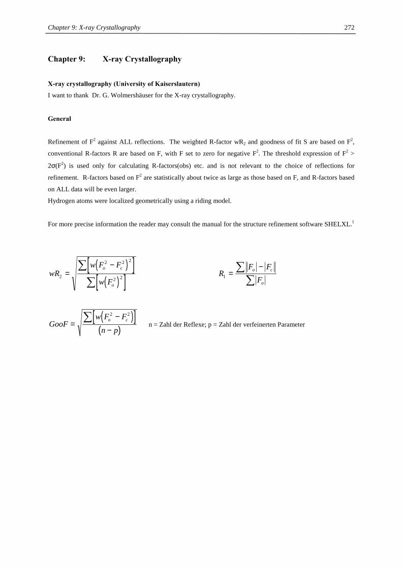
Chapter 9: X-ray Crystallography
272
Chapter 9: X-ray Crystallography
X-ray crystallography (University of Kaiserslautern)
I want to thank Dr. G. Wolmershäuser for the X-ray crystallography.
General
Refinement of F2 against ALL reflections. The weighted R-factor wR2 and goodness of fit S are based on F2,
conventional R-factors R are based on F, with F set to zero for negative F2. The threshold expression of F2 >
2σ(F2) is used only for calculating R-factors(obs) etc. and is not relevant to the choice of reflections for
refinement. R-factors based on F2 are statistically about twice as large as those based on F, and R-factors based
on ALL data will be even larger.
Hydrogen atoms were localized geometrically using a riding model.
For more precise information the reader may consult the manual for the structure refinement software SHELXL.1
( )[ ]( )[ ]wR
w F F
w F
o c
o
2
2 2 2
2 2=−∑
∑ R
F FF
o c
o1 =
−∑
∑
( )[ ]( )GooF
w F F
n po c
=−
−∑
2 2
n = Zahl der Reflexe; p = Zahl der verfeinerten Parameter
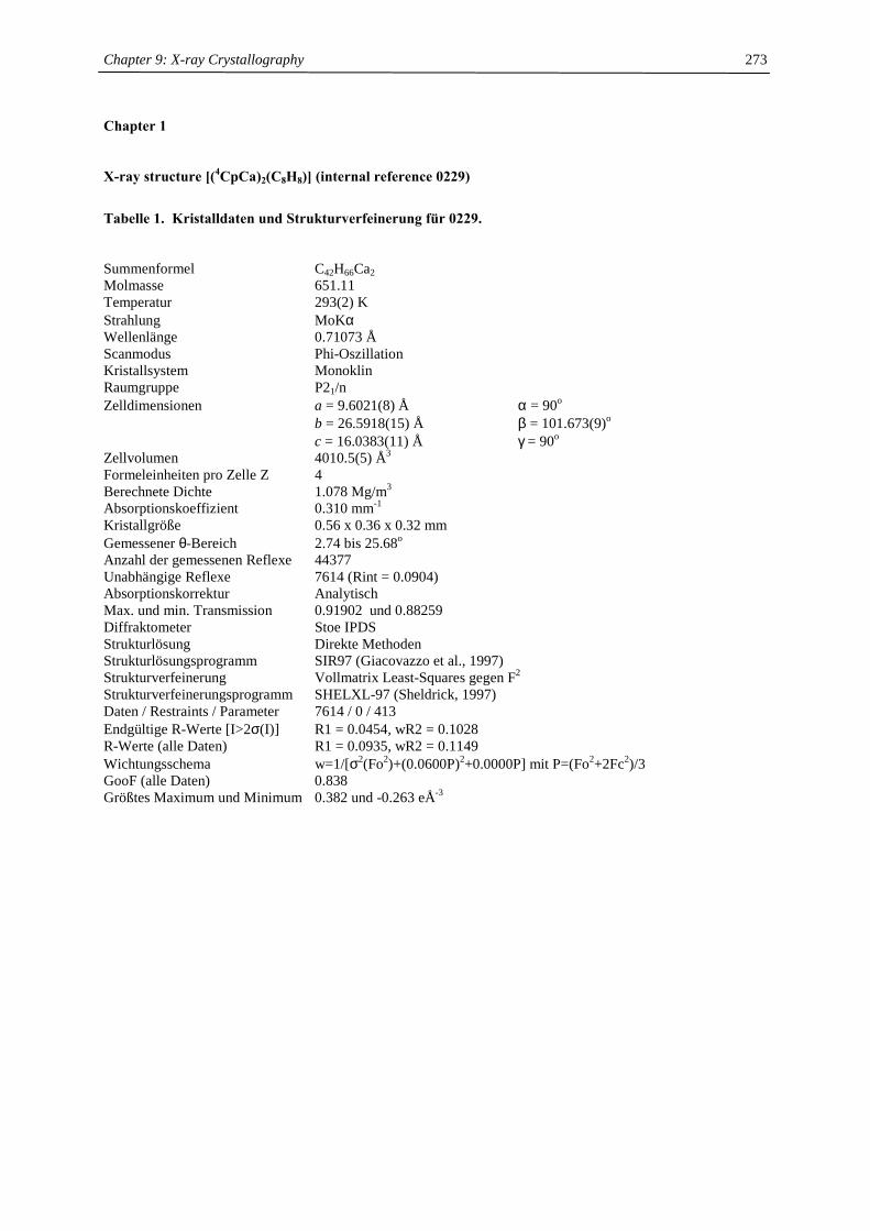
Chapter 9: X-ray Crystallography
273
Chapter 1
X-ray structure [(4CpCa)2(C8H8)] (internal reference 0229)
Tabelle 1. Kristalldaten und Strukturverfeinerung für 0229. Summenformel C42H66Ca2 Molmasse 651.11 Temperatur 293(2) K Strahlung MoKα Wellenlänge 0.71073 Å Scanmodus Phi-Oszillation Kristallsystem Monoklin Raumgruppe P21/n Zelldimensionen a = 9.6021(8) Å α = 90o b = 26.5918(15) Å β = 101.673(9)o c = 16.0383(11) Å γ = 90o Zellvolumen 4010.5(5) Å3 Formeleinheiten pro Zelle Z 4 Berechnete Dichte 1.078 Mg/m3 Absorptionskoeffizient 0.310 mm-1 Kristallgröße 0.56 x 0.36 x 0.32 mm Gemessener θ-Bereich 2.74 bis 25.68o Anzahl der gemessenen Reflexe 44377 Unabhängige Reflexe 7614 (Rint = 0.0904) Absorptionskorrektur Analytisch Max. und min. Transmission 0.91902 und 0.88259 Diffraktometer Stoe IPDS Strukturlösung Direkte Methoden Strukturlösungsprogramm SIR97 (Giacovazzo et al., 1997) Strukturverfeinerung Vollmatrix Least-Squares gegen F2 Strukturverfeinerungsprogramm SHELXL-97 (Sheldrick, 1997) Daten / Restraints / Parameter 7614 / 0 / 413 Endgültige R-Werte [I>2σ(I)] R1 = 0.0454, wR2 = 0.1028 R-Werte (alle Daten) R1 = 0.0935, wR2 = 0.1149 Wichtungsschema w=1/[σ2(Fo2)+(0.0600P)2+0.0000P] mit P=(Fo2+2Fc2)/3 GooF (alle Daten) 0.838 Größtes Maximum und Minimum 0.382 und -0.263 eÅ-3
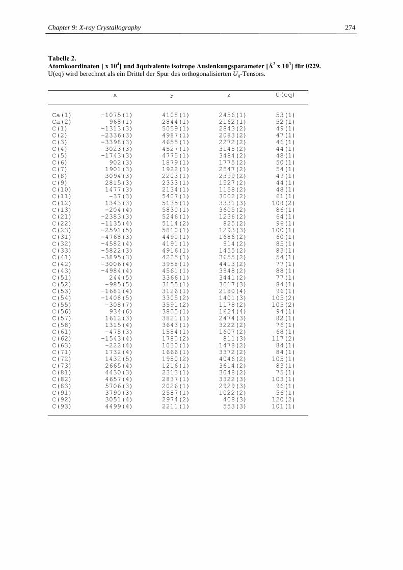
Chapter 9: X-ray Crystallography
274
Tabelle 2. Atomkoordinaten [ x 104] und äquivalente isotrope Auslenkungsparameter [Å2 x 10³] für 0229. U(eq) wird berechnet als ein Drittel der Spur des orthogonalisierten Uij-Tensors.
________________________________________________________________x y z U(eq)
________________________________________________________________
Ca(1) -1075(1) 4108(1) 2456(1) 53(1)Ca(2) 968(1) 2844(1) 2162(1) 52(1)C(1) -1313(3) 5059(1) 2843(2) 49(1)C(2) -2336(3) 4987(1) 2083(2) 47(1)C(3) -3398(3) 4655(1) 2272(2) 46(1)C(4) -3023(3) 4527(1) 3145(2) 44(1)C(5) -1743(3) 4775(1) 3484(2) 48(1)C(6) 902(3) 1879(1) 1775(2) 50(1)C(7) 1901(3) 1922(1) 2547(2) 54(1)C(8) 3094(3) 2203(1) 2399(2) 49(1)C(9) 2815(3) 2333(1) 1527(2) 44(1)C(10) 1477(3) 2134(1) 1158(2) 48(1)C(11) -37(3) 5407(1) 3002(2) 61(1)C(12) 1343(3) 5135(1) 3331(3) 108(2)C(13) -204(4) 5830(1) 3605(2) 86(1)C(21) -2383(3) 5246(1) 1236(2) 64(1)C(22) -1135(4) 5114(2) 825(2) 96(1)C(23) -2591(5) 5810(1) 1293(3) 100(1)C(31) -4768(3) 4490(1) 1686(2) 60(1)C(32) -4582(4) 4191(1) 914(2) 85(1)C(33) -5822(3) 4916(1) 1455(2) 83(1)C(41) -3895(3) 4225(1) 3655(2) 54(1)C(42) -3006(4) 3958(1) 4413(2) 77(1)C(43) -4984(4) 4561(1) 3948(2) 88(1)C(51) 244(5) 3366(1) 3441(2) 77(1)C(52) -985(5) 3155(1) 3017(3) 84(1)C(53) -1681(4) 3126(1) 2180(4) 96(1)C(54) -1408(5) 3305(2) 1401(3) 105(2)C(55) -308(7) 3591(2) 1178(2) 105(2)C(56) 934(6) 3805(1) 1624(4) 94(1)C(57) 1612(3) 3821(1) 2474(3) 82(1)C(58) 1315(4) 3643(1) 3222(2) 76(1)C(61) -478(3) 1584(1) 1607(2) 68(1)C(62) -1543(4) 1780(2) 811(3) 117(2)C(63) -222(4) 1030(1) 1478(2) 84(1)C(71) 1732(4) 1666(1) 3372(2) 84(1)C(72) 1432(5) 1980(2) 4046(2) 105(1)C(73) 2665(4) 1216(1) 3614(2) 83(1)C(81) 4430(3) 2313(1) 3048(2) 75(1)C(82) 4657(4) 2837(1) 3322(3) 103(1)C(83) 5706(3) 2026(1) 2929(3) 96(1)C(91) 3790(3) 2587(1) 1022(2) 56(1)C(92) 3051(4) 2974(2) 408(3) 120(2)C(93) 4499(4) 2211(1) 553(3) 101(1)
________________________________________________________________
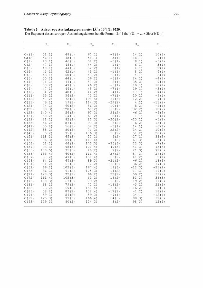
Chapter 9: X-ray Crystallography
275
Tabelle 3. Anisotrope Auslenkungsparameter [Å2 x 10³] für 0229. Der Exponent des anisotropen Auslenkungsfaktors hat die Form: -2π2 [ (ha*)2U11 + ... + 2hka*b*U12 ]
_______________________________________________________________________U11 U22 U33 U23 U13 U12
_______________________________________________________________________
Ca(1) 51(1) 48(1) 60(1) -3(1) 14(1) 10(1)Ca(2) 56(1) 45(1) 58(1) -5(1) 18(1) 7(1)C(1) 43(1) 46(1) 58(2) -5(1) 8(1) -3(1)C(2) 47(1) 48(1) 44(2) 1(1) 6(1) 3(1)C(3) 40(1) 44(1) 52(2) -3(1) 4(1) 2(1)C(4) 43(1) 42(1) 45(2) -1(1) 8(1) 4(1)C(5) 48(1) 50(1) 43(2) -5(1) 4(1) 2(1)C(6) 55(2) 44(1) 56(2) -6(1) 24(1) -4(1)C(7) 71(2) 44(1) 57(2) 6(1) 35(2) 9(1)C(8) 53(2) 47(1) 46(2) -6(1) 10(1) 10(1)C(9) 47(1) 44(1) 45(2) -7(1) 19(1) -3(1)C(10) 54(2) 48(1) 44(2) -4(1) 17(1) -4(1)C(11) 55(2) 54(2) 73(2) -7(1) 10(2) -9(1)C(12) 47(2) 72(2) 198(5) -31(3) 12(2) -7(2)C(13) 79(2) 59(2) 114(3) -29(2) 4(2) -11(2)C(21) 74(2) 60(2) 56(2) 10(1) 8(2) -4(1)C(22) 98(3) 128(3) 69(2) 19(2) 32(2) -10(2)C(23) 140(4) 56(2) 92(3) 24(2) -6(2) -9(2)C(31) 50(2) 64(2) 60(2) 2(1) -1(1) -2(1)C(32) 81(2) 82(2) 81(3) -20(2) -13(2) -5(2)C(33) 56(2) 87(2) 97(3) 6(2) -6(2) 13(2)C(41) 55(2) 56(2) 54(2) -3(1) 14(1) -6(1)C(42) 88(2) 80(2) 71(2) 22(2) 34(2) 10(2)C(43) 75(2) 95(2) 106(3) 15(2) 51(2) 20(2)C(51) 118(3) 65(2) 52(2) 6(2) 27(2) 33(2)C(52) 96(3) 59(2) 117(4) 6(2) 67(3) 5(2)C(53) 51(2) 64(2) 172(5) -36(3) 22(3) -7(2)C(54) 93(3) 95(3) 101(4) -49(3) -41(3) 43(3)C(55) 170(5) 95(3) 49(2) 7(2) 21(3) 72(3)C(56) 133(4) 60(2) 116(4) 27(2) 87(3) 27(2)C(57) 57(2) 47(2) 151(4) -13(2) 41(2) -2(1)C(58) 64(2) 65(2) 89(3) -21(2) -6(2) 18(2)C(61) 71(2) 61(2) 82(2) -12(2) 36(2) -17(2)C(62) 66(2) 102(3) 167(4) 18(3) -12(3) -21(2)C(63) 86(2) 61(2) 105(3) -14(2) 17(2) -14(2)C(71) 128(3) 72(2) 66(2) 22(2) 50(2) 31(2)C(72) 161(4) 105(3) 61(2) 16(2) 53(3) 38(3)C(73) 108(3) 63(2) 79(2) 18(2) 19(2) 11(2)C(81) 68(2) 79(2) 70(2) -18(2) -3(2) 22(2)C(82) 73(2) 69(2) 151(4) -36(2) -16(2) 1(2)C(83) 58(2) 83(2) 138(4) -17(2) -1(2) 18(2)C(91) 59(2) 54(2) 59(2) -9(1) 24(1) -12(1)C(92) 125(3) 99(3) 166(4) 64(3) 98(3) 32(3)C(93) 129(3) 80(2) 124(3) 8(2) 98(3) 12(2)_______________________________________________________________________
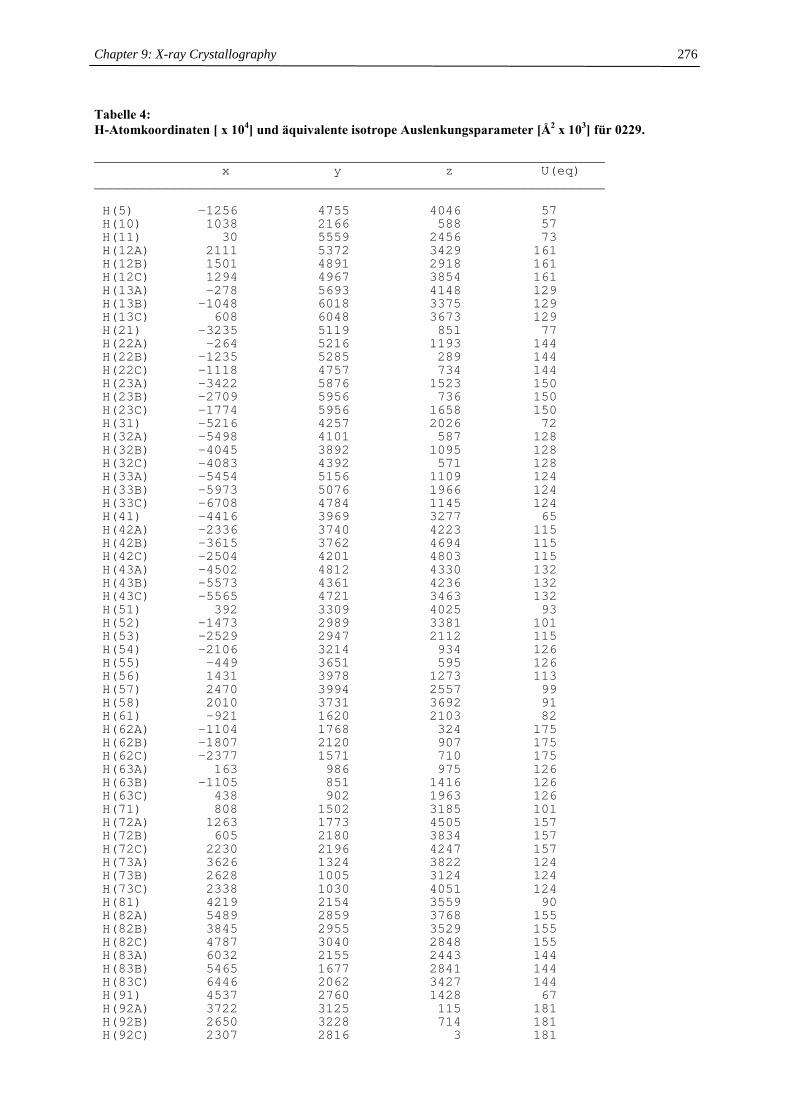
Chapter 9: X-ray Crystallography
276
Tabelle 4: H-Atomkoordinaten [ x 104] und äquivalente isotrope Auslenkungsparameter [Å2 x 10³] für 0229.
________________________________________________________________x y z U(eq)
________________________________________________________________
H(5) -1256 4755 4046 57H(10) 1038 2166 588 57H(11) 30 5559 2456 73H(12A) 2111 5372 3429 161H(12B) 1501 4891 2918 161H(12C) 1294 4967 3854 161H(13A) -278 5693 4148 129H(13B) -1048 6018 3375 129H(13C) 608 6048 3673 129H(21) -3235 5119 851 77H(22A) -264 5216 1193 144H(22B) -1235 5285 289 144H(22C) -1118 4757 734 144H(23A) -3422 5876 1523 150H(23B) -2709 5956 736 150H(23C) -1774 5956 1658 150H(31) -5216 4257 2026 72H(32A) -5498 4101 587 128H(32B) -4045 3892 1095 128H(32C) -4083 4392 571 128H(33A) -5454 5156 1109 124H(33B) -5973 5076 1966 124H(33C) -6708 4784 1145 124H(41) -4416 3969 3277 65H(42A) -2336 3740 4223 115H(42B) -3615 3762 4694 115H(42C) -2504 4201 4803 115H(43A) -4502 4812 4330 132H(43B) -5573 4361 4236 132H(43C) -5565 4721 3463 132H(51) 392 3309 4025 93H(52) -1473 2989 3381 101H(53) -2529 2947 2112 115H(54) -2106 3214 934 126H(55) -449 3651 595 126H(56) 1431 3978 1273 113H(57) 2470 3994 2557 99H(58) 2010 3731 3692 91H(61) -921 1620 2103 82H(62A) -1104 1768 324 175H(62B) -1807 2120 907 175H(62C) -2377 1571 710 175H(63A) 163 986 975 126H(63B) -1105 851 1416 126H(63C) 438 902 1963 126H(71) 808 1502 3185 101H(72A) 1263 1773 4505 157H(72B) 605 2180 3834 157H(72C) 2230 2196 4247 157H(73A) 3626 1324 3822 124H(73B) 2628 1005 3124 124H(73C) 2338 1030 4051 124H(81) 4219 2154 3559 90H(82A) 5489 2859 3768 155H(82B) 3845 2955 3529 155H(82C) 4787 3040 2848 155H(83A) 6032 2155 2443 144H(83B) 5465 1677 2841 144H(83C) 6446 2062 3427 144H(91) 4537 2760 1428 67H(92A) 3722 3125 115 181H(92B) 2650 3228 714 181H(92C) 2307 2816 3 181
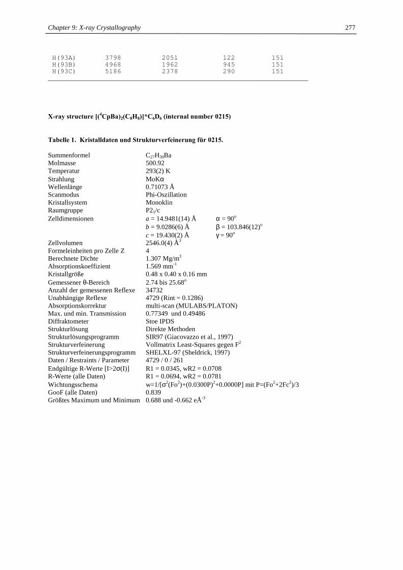
Chapter 9: X-ray Crystallography
277
H(93A) 3798 2051 122 151H(93B) 4968 1962 945 151H(93C) 5186 2378 290 151
________________________________________________________________
X-ray structure [(4CpBa)2(C8H8)]*C6D6 (internal number 0215)
Tabelle 1. Kristalldaten und Strukturverfeinerung für 0215. Summenformel C27H39Ba Molmasse 500.92 Temperatur 293(2) K Strahlung MoKα Wellenlänge 0.71073 Å Scanmodus Phi-Oszillation Kristallsystem Monoklin Raumgruppe P21/c Zelldimensionen a = 14.9481(14) Å α = 90o b = 9.0286(6) Å β = 103.846(12)o c = 19.430(2) Å γ = 90o Zellvolumen 2546.0(4) Å3 Formeleinheiten pro Zelle Z 4 Berechnete Dichte 1.307 Mg/m3 Absorptionskoeffizient 1.569 mm-1 Kristallgröße 0.48 x 0.40 x 0.16 mm Gemessener θ-Bereich 2.74 bis 25.68o Anzahl der gemessenen Reflexe 34732 Unabhängige Reflexe 4729 (Rint = 0.1286) Absorptionskorrektur multi-scan (MULABS/PLATON) Max. und min. Transmission 0.77349 und 0.49486 Diffraktometer Stoe IPDS Strukturlösung Direkte Methoden Strukturlösungsprogramm SIR97 (Giacovazzo et al., 1997) Strukturverfeinerung Vollmatrix Least-Squares gegen F2 Strukturverfeinerungsprogramm SHELXL-97 (Sheldrick, 1997) Daten / Restraints / Parameter 4729 / 0 / 261 Endgültige R-Werte [I>2σ(I)] R1 = 0.0345, wR2 = 0.0708 R-Werte (alle Daten) R1 = 0.0694, wR2 = 0.0781 Wichtungsschema w=1/[σ2(Fo2)+(0.0300P)2+0.0000P] mit P=(Fo2+2Fc2)/3 GooF (alle Daten) 0.839 Größtes Maximum und Minimum 0.688 und -0.662 eÅ-3
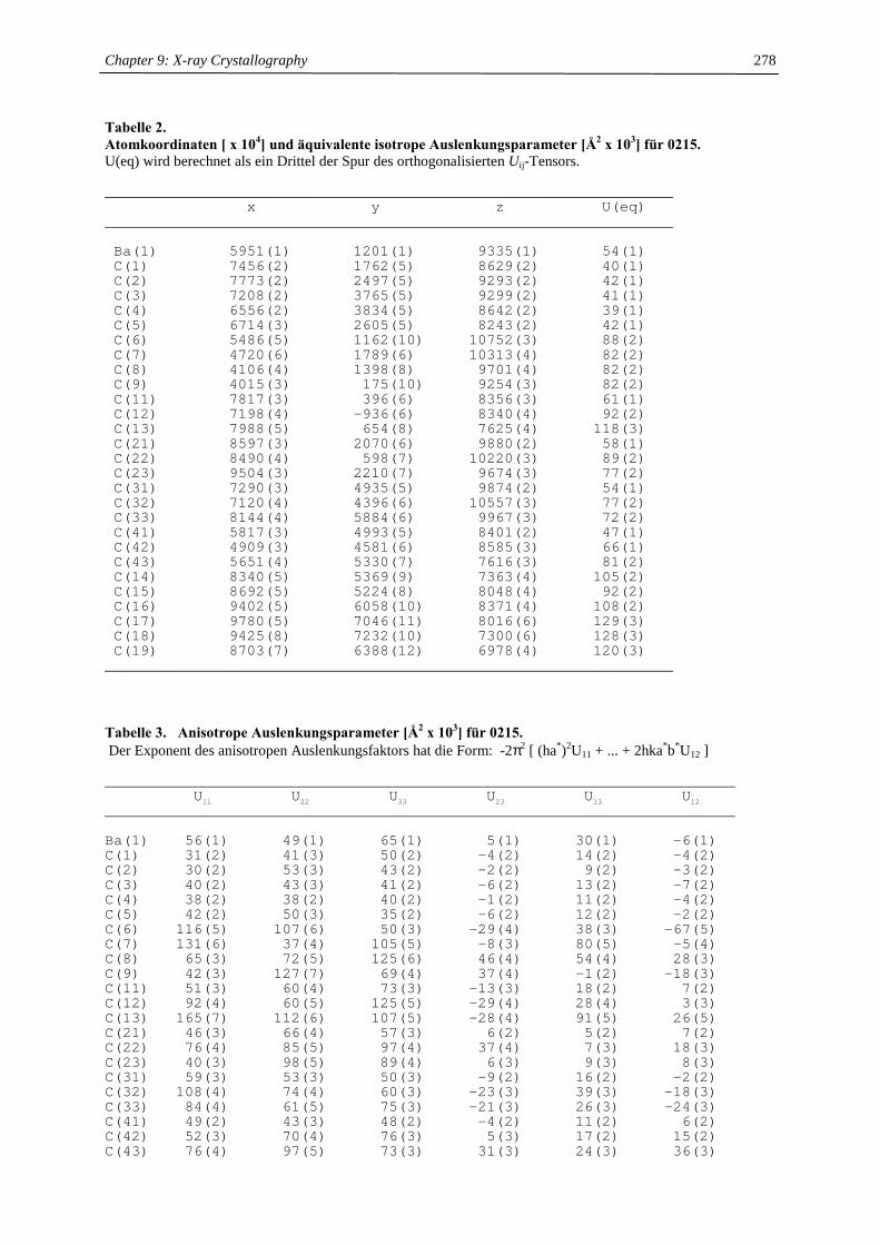
Chapter 9: X-ray Crystallography
278
Tabelle 2. Atomkoordinaten [ x 104] und äquivalente isotrope Auslenkungsparameter [Å2 x 10³] für 0215. U(eq) wird berechnet als ein Drittel der Spur des orthogonalisierten Uij-Tensors.
________________________________________________________________x y z U(eq)
________________________________________________________________
Ba(1) 5951(1) 1201(1) 9335(1) 54(1)C(1) 7456(2) 1762(5) 8629(2) 40(1)C(2) 7773(2) 2497(5) 9293(2) 42(1)C(3) 7208(2) 3765(5) 9299(2) 41(1)C(4) 6556(2) 3834(5) 8642(2) 39(1)C(5) 6714(3) 2605(5) 8243(2) 42(1)C(6) 5486(5) 1162(10) 10752(3) 88(2)C(7) 4720(6) 1789(6) 10313(4) 82(2)C(8) 4106(4) 1398(8) 9701(4) 82(2)C(9) 4015(3) 175(10) 9254(3) 82(2)C(11) 7817(3) 396(6) 8356(3) 61(1)C(12) 7198(4) -936(6) 8340(4) 92(2)C(13) 7988(5) 654(8) 7625(4) 118(3)C(21) 8597(3) 2070(6) 9880(2) 58(1)C(22) 8490(4) 598(7) 10220(3) 89(2)C(23) 9504(3) 2210(7) 9674(3) 77(2)C(31) 7290(3) 4935(5) 9874(2) 54(1)C(32) 7120(4) 4396(6) 10557(3) 77(2)C(33) 8144(4) 5884(6) 9967(3) 72(2)C(41) 5817(3) 4993(5) 8401(2) 47(1)C(42) 4909(3) 4581(6) 8585(3) 66(1)C(43) 5651(4) 5330(7) 7616(3) 81(2)C(14) 8340(5) 5369(9) 7363(4) 105(2)C(15) 8692(5) 5224(8) 8048(4) 92(2)C(16) 9402(5) 6058(10) 8371(4) 108(2)C(17) 9780(5) 7046(11) 8016(6) 129(3)C(18) 9425(8) 7232(10) 7300(6) 128(3)C(19) 8703(7) 6388(12) 6978(4) 120(3)
________________________________________________________________ Tabelle 3. Anisotrope Auslenkungsparameter [Å2 x 10³] für 0215. Der Exponent des anisotropen Auslenkungsfaktors hat die Form: -2π2 [ (ha*)2U11 + ... + 2hka*b*U12 ]
_______________________________________________________________________U11 U22 U33 U23 U13 U12
_______________________________________________________________________
Ba(1) 56(1) 49(1) 65(1) 5(1) 30(1) -6(1)C(1) 31(2) 41(3) 50(2) -4(2) 14(2) -4(2)C(2) 30(2) 53(3) 43(2) -2(2) 9(2) -3(2)C(3) 40(2) 43(3) 41(2) -6(2) 13(2) -7(2)C(4) 38(2) 38(2) 40(2) -1(2) 11(2) -4(2)C(5) 42(2) 50(3) 35(2) -6(2) 12(2) -2(2)C(6) 116(5) 107(6) 50(3) -29(4) 38(3) -67(5)C(7) 131(6) 37(4) 105(5) -8(3) 80(5) -5(4)C(8) 65(3) 72(5) 125(6) 46(4) 54(4) 28(3)C(9) 42(3) 127(7) 69(4) 37(4) -1(2) -18(3)C(11) 51(3) 60(4) 73(3) -13(3) 18(2) 7(2)C(12) 92(4) 60(5) 125(5) -29(4) 28(4) 3(3)C(13) 165(7) 112(6) 107(5) -28(4) 91(5) 26(5)C(21) 46(3) 66(4) 57(3) 6(2) 5(2) 7(2)C(22) 76(4) 85(5) 97(4) 37(4) 7(3) 18(3)C(23) 40(3) 98(5) 89(4) 6(3) 9(3) 8(3)C(31) 59(3) 53(3) 50(3) -9(2) 16(2) -2(2)C(32) 108(4) 74(4) 60(3) -23(3) 39(3) -18(3)C(33) 84(4) 61(5) 75(3) -21(3) 26(3) -24(3)C(41) 49(2) 43(3) 48(2) -4(2) 11(2) 6(2)C(42) 52(3) 70(4) 76(3) 5(3) 17(2) 15(2)C(43) 76(4) 97(5) 73(3) 31(3) 24(3) 36(3)
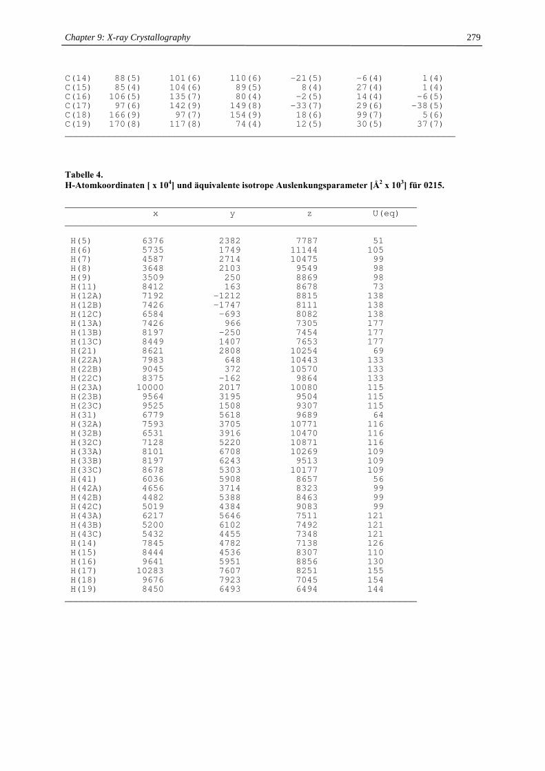
Chapter 9: X-ray Crystallography
279
C(14) 88(5) 101(6) 110(6) -21(5) -6(4) 1(4)C(15) 85(4) 104(6) 89(5) 8(4) 27(4) 1(4)C(16) 106(5) 135(7) 80(4) -2(5) 14(4) -6(5)C(17) 97(6) 142(9) 149(8) -33(7) 29(6) -38(5)C(18) 166(9) 97(7) 154(9) 18(6) 99(7) 5(6)C(19) 170(8) 117(8) 74(4) 12(5) 30(5) 37(7)_______________________________________________________________________ Tabelle 4. H-Atomkoordinaten [ x 104] und äquivalente isotrope Auslenkungsparameter [Å2 x 10³] für 0215.
________________________________________________________________x y z U(eq)
________________________________________________________________
H(5) 6376 2382 7787 51H(6) 5735 1749 11144 105H(7) 4587 2714 10475 99H(8) 3648 2103 9549 98H(9) 3509 250 8869 98H(11) 8412 163 8678 73H(12A) 7192 -1212 8815 138H(12B) 7426 -1747 8111 138H(12C) 6584 -693 8082 138H(13A) 7426 966 7305 177H(13B) 8197 -250 7454 177H(13C) 8449 1407 7653 177H(21) 8621 2808 10254 69H(22A) 7983 648 10443 133H(22B) 9045 372 10570 133H(22C) 8375 -162 9864 133H(23A) 10000 2017 10080 115H(23B) 9564 3195 9504 115H(23C) 9525 1508 9307 115H(31) 6779 5618 9689 64H(32A) 7593 3705 10771 116H(32B) 6531 3916 10470 116H(32C) 7128 5220 10871 116H(33A) 8101 6708 10269 109H(33B) 8197 6243 9513 109H(33C) 8678 5303 10177 109H(41) 6036 5908 8657 56H(42A) 4656 3714 8323 99H(42B) 4482 5388 8463 99H(42C) 5019 4384 9083 99H(43A) 6217 5646 7511 121H(43B) 5200 6102 7492 121H(43C) 5432 4455 7348 121H(14) 7845 4782 7138 126H(15) 8444 4536 8307 110H(16) 9641 5951 8856 130H(17) 10283 7607 8251 155H(18) 9676 7923 7045 154H(19) 8450 6493 6494 144
________________________________________________________________
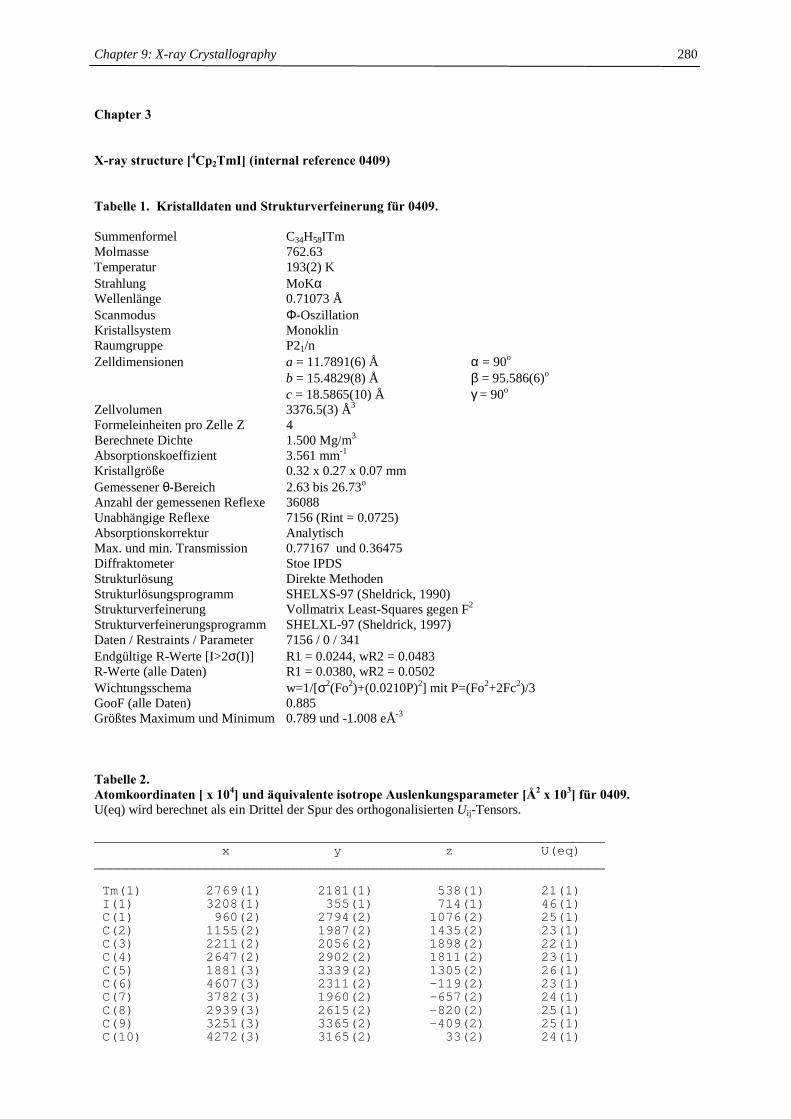
Chapter 9: X-ray Crystallography
280
Chapter 3
X-ray structure [4Cp2TmI] (internal reference 0409)
Tabelle 1. Kristalldaten und Strukturverfeinerung für 0409. Summenformel C34H58ITm Molmasse 762.63 Temperatur 193(2) K Strahlung MoKα Wellenlänge 0.71073 Å Scanmodus Φ-Oszillation Kristallsystem Monoklin Raumgruppe P21/n Zelldimensionen a = 11.7891(6) Å α = 90o b = 15.4829(8) Å β = 95.586(6)o c = 18.5865(10) Å γ = 90o Zellvolumen 3376.5(3) Å3 Formeleinheiten pro Zelle Z 4 Berechnete Dichte 1.500 Mg/m3 Absorptionskoeffizient 3.561 mm-1 Kristallgröße 0.32 x 0.27 x 0.07 mm Gemessener θ-Bereich 2.63 bis 26.73o Anzahl der gemessenen Reflexe 36088 Unabhängige Reflexe 7156 (Rint = 0.0725) Absorptionskorrektur Analytisch Max. und min. Transmission 0.77167 und 0.36475 Diffraktometer Stoe IPDS Strukturlösung Direkte Methoden Strukturlösungsprogramm SHELXS-97 (Sheldrick, 1990) Strukturverfeinerung Vollmatrix Least-Squares gegen F2 Strukturverfeinerungsprogramm SHELXL-97 (Sheldrick, 1997) Daten / Restraints / Parameter 7156 / 0 / 341 Endgültige R-Werte [I>2σ(I)] R1 = 0.0244, wR2 = 0.0483 R-Werte (alle Daten) R1 = 0.0380, wR2 = 0.0502 Wichtungsschema w=1/[σ2(Fo2)+(0.0210P)2] mit P=(Fo2+2Fc2)/3 GooF (alle Daten) 0.885 Größtes Maximum und Minimum 0.789 und -1.008 eÅ-3
Tabelle 2. Atomkoordinaten [ x 104] und äquivalente isotrope Auslenkungsparameter [Å2 x 10³] für 0409. U(eq) wird berechnet als ein Drittel der Spur des orthogonalisierten Uij-Tensors.
________________________________________________________________x y z U(eq)
________________________________________________________________
Tm(1) 2769(1) 2181(1) 538(1) 21(1)I(1) 3208(1) 355(1) 714(1) 46(1)C(1) 960(2) 2794(2) 1076(2) 25(1)C(2) 1155(2) 1987(2) 1435(2) 23(1)C(3) 2211(2) 2056(2) 1898(2) 22(1)C(4) 2647(2) 2902(2) 1811(2) 23(1)C(5) 1881(3) 3339(2) 1305(2) 26(1)C(6) 4607(3) 2311(2) -119(2) 23(1)C(7) 3782(3) 1960(2) -657(2) 24(1)C(8) 2939(3) 2615(2) -820(2) 25(1)C(9) 3251(3) 3365(2) -409(2) 25(1)C(10) 4272(3) 3165(2) 33(2) 24(1)
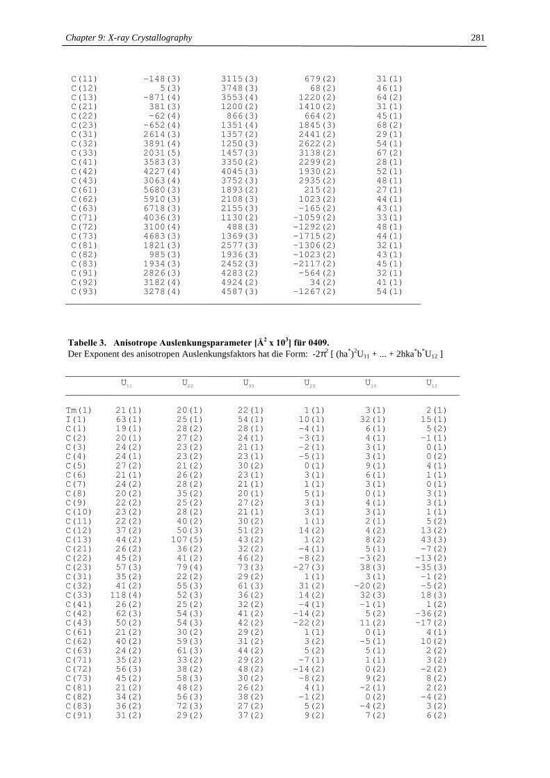
Chapter 9: X-ray Crystallography
281
C(11) -148(3) 3115(3) 679(2) 31(1)C(12) 5(3) 3748(3) 68(2) 46(1)C(13) -871(4) 3553(4) 1220(2) 64(2)C(21) 381(3) 1200(2) 1410(2) 31(1)C(22) -62(4) 866(3) 664(2) 45(1)C(23) -652(4) 1351(4) 1845(3) 68(2)C(31) 2614(3) 1357(2) 2441(2) 29(1)C(32) 3891(4) 1250(3) 2622(2) 54(1)C(33) 2031(5) 1457(3) 3138(2) 67(2)C(41) 3583(3) 3350(2) 2299(2) 28(1)C(42) 4227(4) 4045(3) 1930(2) 52(1)C(43) 3063(4) 3752(3) 2935(2) 48(1)C(61) 5680(3) 1893(2) 215(2) 27(1)C(62) 5910(3) 2108(3) 1023(2) 44(1)C(63) 6718(3) 2155(3) -165(2) 43(1)C(71) 4036(3) 1130(2) -1059(2) 33(1)C(72) 3100(4) 488(3) -1292(2) 48(1)C(73) 4683(3) 1369(3) -1715(2) 44(1)C(81) 1821(3) 2577(3) -1306(2) 32(1)C(82) 985(3) 1936(3) -1023(2) 43(1)C(83) 1934(3) 2452(3) -2117(2) 45(1)C(91) 2826(3) 4283(2) -564(2) 32(1)C(92) 3182(4) 4924(2) 34(2) 41(1)C(93) 3278(4) 4587(3) -1267(2) 54(1)
________________________________________________________________ Tabelle 3. Anisotrope Auslenkungsparameter [Å2 x 10³] für 0409. Der Exponent des anisotropen Auslenkungsfaktors hat die Form: -2π2 [ (ha*)2U11 + ... + 2hka*b*U12 ]
_______________________________________________________________________U11 U22 U33 U23 U13 U12
_______________________________________________________________________
Tm(1) 21(1) 20(1) 22(1) 1(1) 3(1) 2(1)I(1) 63(1) 25(1) 54(1) 10(1) 32(1) 15(1)C(1) 19(1) 28(2) 28(1) -4(1) 6(1) 5(2)C(2) 20(1) 27(2) 24(1) -3(1) 4(1) -1(1)C(3) 24(2) 23(2) 21(1) -2(1) 3(1) 0(1)C(4) 24(1) 23(2) 23(1) -5(1) 3(1) 0(2)C(5) 27(2) 21(2) 30(2) 0(1) 9(1) 4(1)C(6) 21(1) 26(2) 23(1) 3(1) 6(1) 1(1)C(7) 24(2) 28(2) 21(1) 1(1) 3(1) 0(1)C(8) 20(2) 35(2) 20(1) 5(1) 0(1) 3(1)C(9) 22(2) 25(2) 27(2) 3(1) 4(1) 3(1)C(10) 23(2) 28(2) 21(1) 3(1) 3(1) 1(1)C(11) 22(2) 40(2) 30(2) 1(1) 2(1) 5(2)C(12) 37(2) 50(3) 51(2) 14(2) 4(2) 13(2)C(13) 44(2) 107(5) 43(2) 1(2) 8(2) 43(3)C(21) 26(2) 36(2) 32(2) -4(1) 5(1) -7(2)C(22) 45(2) 41(2) 46(2) -8(2) -3(2) -13(2)C(23) 57(3) 79(4) 73(3) -27(3) 38(3) -35(3)C(31) 35(2) 22(2) 29(2) 1(1) 3(1) -1(2)C(32) 41(2) 55(3) 61(3) 31(2) -20(2) -5(2)C(33) 118(4) 52(3) 36(2) 14(2) 32(3) 18(3)C(41) 26(2) 25(2) 32(2) -4(1) -1(1) 1(2)C(42) 62(3) 54(3) 41(2) -14(2) 5(2) -36(2)C(43) 50(2) 54(3) 42(2) -22(2) 11(2) -17(2)C(61) 21(2) 30(2) 29(2) 1(1) 0(1) 4(1)C(62) 40(2) 59(3) 31(2) 3(2) -5(1) 10(2)C(63) 24(2) 61(3) 44(2) 5(2) 5(1) 2(2)C(71) 35(2) 33(2) 29(2) -7(1) 1(1) 3(2)C(72) 56(3) 38(2) 48(2) -14(2) 0(2) -2(2)C(73) 45(2) 58(3) 30(2) -8(2) 9(2) 8(2)C(81) 21(2) 48(2) 26(2) 4(1) -2(1) 2(2)C(82) 34(2) 56(3) 38(2) -1(2) 0(2) -4(2)C(83) 36(2) 72(3) 27(2) 5(2) -4(2) 3(2)C(91) 31(2) 29(2) 37(2) 9(2) 7(2) 6(2)
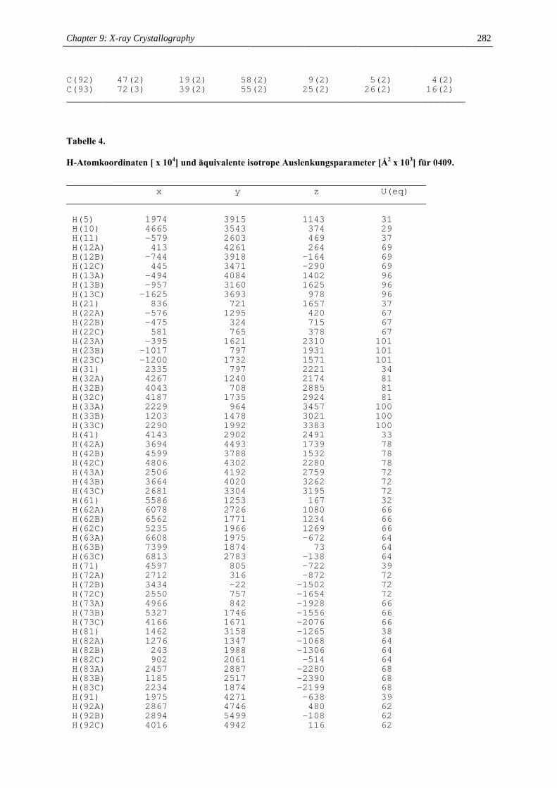
Chapter 9: X-ray Crystallography
282
C(92) 47(2) 19(2) 58(2) 9(2) 5(2) 4(2)C(93) 72(3) 39(2) 55(2) 25(2) 26(2) 16(2)_______________________________________________________________________ Tabelle 4. H-Atomkoordinaten [ x 104] und äquivalente isotrope Auslenkungsparameter [Å2 x 10³] für 0409.
________________________________________________________________x y z U(eq)
________________________________________________________________
H(5) 1974 3915 1143 31H(10) 4665 3543 374 29H(11) -579 2603 469 37H(12A) 413 4261 264 69H(12B) -744 3918 -164 69H(12C) 445 3471 -290 69H(13A) -494 4084 1402 96H(13B) -957 3160 1625 96H(13C) -1625 3693 978 96H(21) 836 721 1657 37H(22A) -576 1295 420 67H(22B) -475 324 715 67H(22C) 581 765 378 67H(23A) -395 1621 2310 101H(23B) -1017 797 1931 101H(23C) -1200 1732 1571 101H(31) 2335 797 2221 34H(32A) 4267 1240 2174 81H(32B) 4043 708 2885 81H(32C) 4187 1735 2924 81H(33A) 2229 964 3457 100H(33B) 1203 1478 3021 100H(33C) 2290 1992 3383 100H(41) 4143 2902 2491 33H(42A) 3694 4493 1739 78H(42B) 4599 3788 1532 78H(42C) 4806 4302 2280 78H(43A) 2506 4192 2759 72H(43B) 3664 4020 3262 72H(43C) 2681 3304 3195 72H(61) 5586 1253 167 32H(62A) 6078 2726 1080 66H(62B) 6562 1771 1234 66H(62C) 5235 1966 1269 66H(63A) 6608 1975 -672 64H(63B) 7399 1874 73 64H(63C) 6813 2783 -138 64H(71) 4597 805 -722 39H(72A) 2712 316 -872 72H(72B) 3434 -22 -1502 72H(72C) 2550 757 -1654 72H(73A) 4966 842 -1928 66H(73B) 5327 1746 -1556 66H(73C) 4166 1671 -2076 66H(81) 1462 3158 -1265 38H(82A) 1276 1347 -1068 64H(82B) 243 1988 -1306 64H(82C) 902 2061 -514 64H(83A) 2457 2887 -2280 68H(83B) 1185 2517 -2390 68H(83C) 2234 1874 -2199 68H(91) 1975 4271 -638 39H(92A) 2867 4746 480 62H(92B) 2894 5499 -108 62H(92C) 4016 4942 116 62
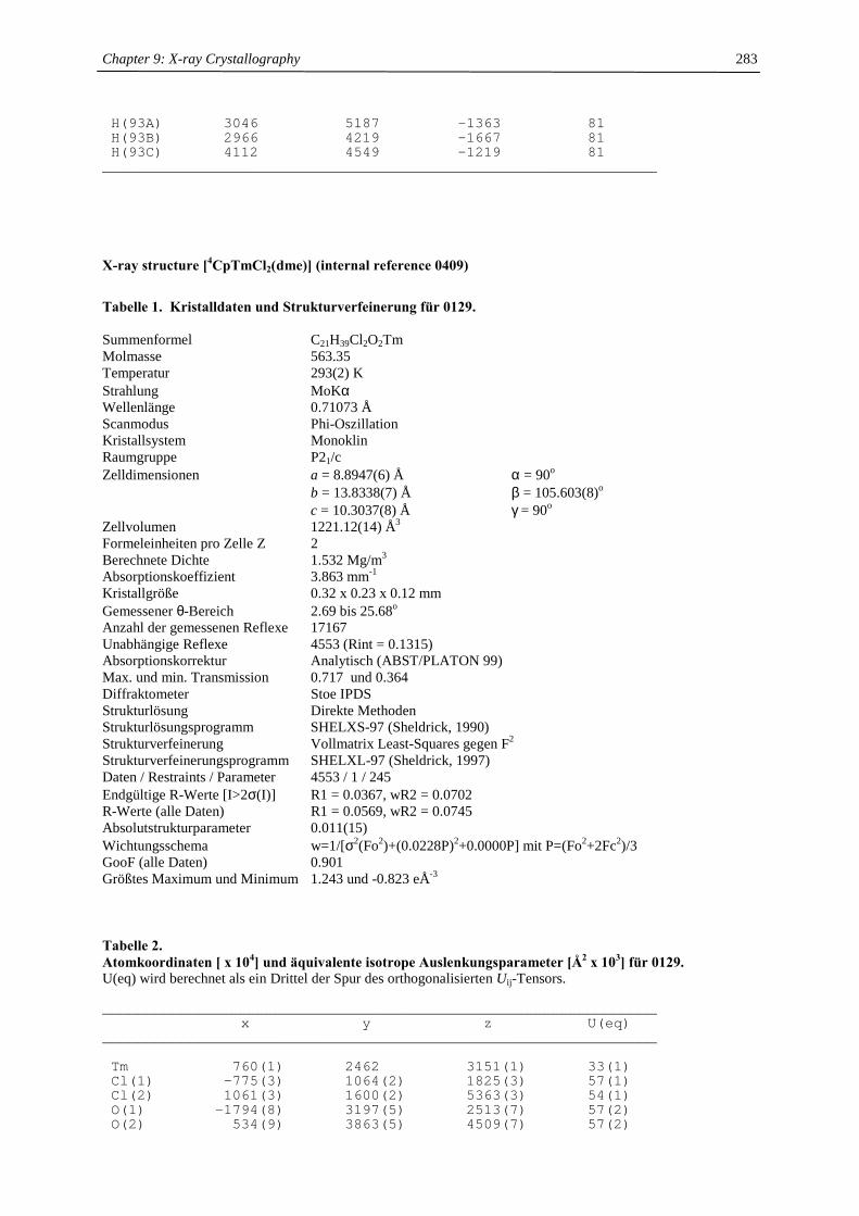
Chapter 9: X-ray Crystallography
283
H(93A) 3046 5187 -1363 81H(93B) 2966 4219 -1667 81H(93C) 4112 4549 -1219 81
________________________________________________________________ X-ray structure [4CpTmCl2(dme)] (internal reference 0409)
Tabelle 1. Kristalldaten und Strukturverfeinerung für 0129. Summenformel C21H39Cl2O2Tm Molmasse 563.35 Temperatur 293(2) K Strahlung MoKα Wellenlänge 0.71073 Å Scanmodus Phi-Oszillation Kristallsystem Monoklin Raumgruppe P21/c Zelldimensionen a = 8.8947(6) Å α = 90o b = 13.8338(7) Å β = 105.603(8)o c = 10.3037(8) Å γ = 90o Zellvolumen 1221.12(14) Å3 Formeleinheiten pro Zelle Z 2 Berechnete Dichte 1.532 Mg/m3 Absorptionskoeffizient 3.863 mm-1 Kristallgröße 0.32 x 0.23 x 0.12 mm Gemessener θ-Bereich 2.69 bis 25.68o Anzahl der gemessenen Reflexe 17167 Unabhängige Reflexe 4553 (Rint = 0.1315) Absorptionskorrektur Analytisch (ABST/PLATON 99) Max. und min. Transmission 0.717 und 0.364 Diffraktometer Stoe IPDS Strukturlösung Direkte Methoden Strukturlösungsprogramm SHELXS-97 (Sheldrick, 1990) Strukturverfeinerung Vollmatrix Least-Squares gegen F2 Strukturverfeinerungsprogramm SHELXL-97 (Sheldrick, 1997) Daten / Restraints / Parameter 4553 / 1 / 245 Endgültige R-Werte [I>2σ(I)] R1 = 0.0367, wR2 = 0.0702 R-Werte (alle Daten) R1 = 0.0569, wR2 = 0.0745 Absolutstrukturparameter 0.011(15) Wichtungsschema w=1/[σ2(Fo2)+(0.0228P)2+0.0000P] mit P=(Fo2+2Fc2)/3 GooF (alle Daten) 0.901 Größtes Maximum und Minimum 1.243 und -0.823 eÅ-3 Tabelle 2. Atomkoordinaten [ x 104] und äquivalente isotrope Auslenkungsparameter [Å2 x 10³] für 0129. U(eq) wird berechnet als ein Drittel der Spur des orthogonalisierten Uij-Tensors.
________________________________________________________________x y z U(eq)
________________________________________________________________
Tm 760(1) 2462 3151(1) 33(1)Cl(1) -775(3) 1064(2) 1825(3) 57(1)Cl(2) 1061(3) 1600(2) 5363(3) 54(1)O(1) -1794(8) 3197(5) 2513(7) 57(2)O(2) 534(9) 3863(5) 4509(7) 57(2)
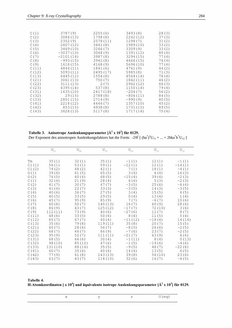
Chapter 9: X-ray Crystallography
284
C(1) 3787(9) 2255(6) 3493(8) 28(3)C(2) 3086(13) 1798(8) 2242(12) 37(3)C(3) 2352(9) 2578(11) 1298(7) 31(2)C(4) 2607(12) 3441(8) 1989(10) 33(2)C(5) 3469(10) 3266(7) 3309(9) 33(2)C(6) -3037(13) 3048(9) 1391(12) 80(4)C(7) -2101(14) 3987(8) 3294(15) 77(4)C(8) -993(15) 3942(8) 4666(15) 76(4)C(9) 1618(15) 4168(9) 5696(10) 77(4)C(11) 4844(11) 1841(6) 4761(9) 44(2)C(12) 5093(11) 2495(17) 5985(8) 71(3)C(13) 6445(12) 1554(8) 4564(14) 78(4)C(21) 3061(13) 750(7) 1842(11) 46(2)C(22) 3111(13) 2(7) 2962(12) 66(3)C(23) 4395(14) 537(8) 1150(14) 79(4)C(31) 1635(10) 2417(18) -204(7) 56(2)C(32) 29(15) 2788(8) -806(11) 84(5)C(33) 2801(15) 2714(9) -990(9) 80(5)C(41) 2218(12) 4446(7) 1357(10) 45(2)C(42) 853(15) 4938(8) 1731(15) 89(5)C(43) 3628(13) 5117(8) 1717(14) 75(4)
________________________________________________________________ Tabelle 3. Anisotrope Auslenkungsparameter [Å2 x 10³] für 0129. Der Exponent des anisotropen Auslenkungsfaktors hat die Form: -2π2 [ (ha*)2U11 + ... + 2hka*b*U12 ]
_______________________________________________________________________U11 U22 U33 U23 U13 U12
_______________________________________________________________________
Tm 35(1) 32(1) 35(1) -1(1) 12(1) -1(1)Cl(1) 56(1) 53(1) 59(1) -12(1) 12(1) -14(1)Cl(2) 74(2) 48(2) 42(1) 7(1) 18(1) -4(1)O(1) 39(4) 61(5) 65(5) 3(4) 6(4) 16(3)O(2) 76(5) 40(4) 68(5) -15(4) 39(4) -2(3)C(1) 32(4) 21(9) 28(4) 6(4) 5(3) -2(3)C(2) 41(7) 30(7) 47(7) -3(5) 25(6) -6(4)C(3) 41(4) 22(7) 33(3) -3(5) 14(3) -3(5)C(4) 40(6) 36(7) 27(5) 3(4) 15(5) 6(5)C(5) 40(5) 33(5) 29(5) 0(4) 13(4) 1(4)C(6) 45(7) 95(9) 85(9) 7(7) -6(7) 10(6)C(7) 60(8) 50(7) 140(13) 16(7) 60(9) 28(6)C(8) 86(9) 43(7) 125(12) -17(7) 72(10) 3(6)C(9) 112(11) 73(9) 40(6) -27(6) 11(7) 8(7)C(11) 48(6) 33(5) 50(6) 8(4) 11(5) 5(4)C(12) 85(7) 67(7) 40(4) -11(12) -18(4) 14(14)C(13) 35(6) 79(8) 119(11) 35(8) 20(7) 15(5)C(21) 60(7) 28(6) 56(7) -8(5) 26(6) -2(5)C(22) 68(7) 46(7) 86(9) -7(6) 23(7) -2(5)C(23) 95(9) 52(7) 111(11) -21(7) 63(9) 6(6)C(31) 68(5) 66(6) 30(4) -1(11) 8(4) 0(13)C(32) 98(10) 85(12) 47(6) -1(5) -19(6) -4(6)C(33) 131(10) 88(14) 35(5) -9(5) 48(7) -22(8)C(41) 60(7) 35(6) 40(6) 14(4) 13(5) 6(5)C(42) 77(9) 61(8) 143(13) 39(8) 56(10) 23(6)C(43) 63(7) 43(7) 114(10) 32(6) 16(7) -4(5)_______________________________________________________________________ Tabelle 4. H-Atomkoordinaten [ x 104] und äquivalente isotrope Auslenkungsparameter [Å2 x 10³] für 0129.
________________________________________________________________x y z U(eq)
________________________________________________________________
Chapter 9: X-ray Crystallography
285
H(5) 3790 3736 3972 40H(6A) -3999 3109 1638 120H(6B) -2967 2411 1040 120H(6C) -3007 3520 715 120H(7A) -1972 4594 2864 92H(7B) -3167 3952 3361 92H(8A) -1083 4521 5170 92H(8B) -1228 3387 5153 92H(9A) 1445 4838 5853 115H(9B) 2657 4085 5605 115H(9C) 1494 3790 6441 115H(11) 4351 1247 4966 53H(12A) 5177 3154 5718 106H(12B) 6035 2311 6643 106H(12C) 4224 2434 6366 106H(13A) 7135 2100 4751 117H(13B) 6315 1347 3651 117H(13C) 6881 1035 5167 117H(21) 2073 647 1153 55H(22A) 4134 -2 3581 100H(22B) 2878 -628 2568 100H(22C) 2354 171 3434 100H(23A) 4189 878 309 118H(23B) 4438 -144 989 118H(23C) 5376 748 1729 118H(31) 1544 1713 -308 67H(32A) -581 2711 -174 126H(32B) -445 2432 -1613 126H(32C) 77 3460 -1022 126H(33A) 2959 3400 -922 119H(33B) 2396 2537 -1920 119H(33C) 3777 2389 -621 119H(41) 1920 4363 376 54H(42A) 1100 5016 2691 133H(42B) -67 4547 1431 133H(42C) 670 5561 1306 133H(43A) 4013 5161 2680 112H(43B) 3327 5747 1350 112H(43C) 4431 4864 1350 112
________________________________________________________________
X-ray structure [Cp╪2Yb(µµµµ-OH)]2 (internal reference 0156)
Tabelle 1. Kristalldaten und Strukturverfeinerung für 0156. Summenformel C26H43OYb Molmasse 544.64 Temperatur 293(2) K Strahlung MoKα Wellenlänge 0.71073 Å Scanmodus Phi-Oszillation Kristallsystem Monoklin Raumgruppe I2 Zelldimensionen a = 12.2121(9) Å α = 90o b = 12.8452(12) Å β = 108.068(8)o c = 17.5139(12) Å γ = 90o Zellvolumen 2611.9(4) Å3 Formeleinheiten pro Zelle Z 4 Berechnete Dichte 1.385 Mg/m3 Absorptionskoeffizient 3.593 mm-1
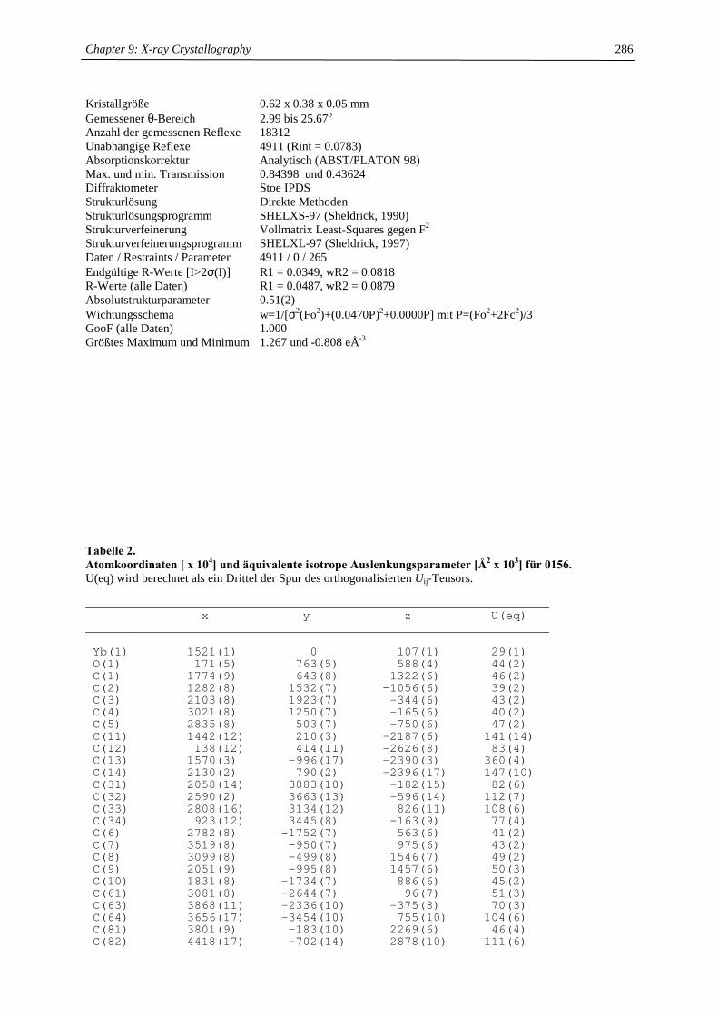
Chapter 9: X-ray Crystallography
286
Kristallgröße 0.62 x 0.38 x 0.05 mm Gemessener θ-Bereich 2.99 bis 25.67o Anzahl der gemessenen Reflexe 18312 Unabhängige Reflexe 4911 (Rint = 0.0783) Absorptionskorrektur Analytisch (ABST/PLATON 98) Max. und min. Transmission 0.84398 und 0.43624 Diffraktometer Stoe IPDS Strukturlösung Direkte Methoden Strukturlösungsprogramm SHELXS-97 (Sheldrick, 1990) Strukturverfeinerung Vollmatrix Least-Squares gegen F2 Strukturverfeinerungsprogramm SHELXL-97 (Sheldrick, 1997) Daten / Restraints / Parameter 4911 / 0 / 265 Endgültige R-Werte [I>2σ(I)] R1 = 0.0349, wR2 = 0.0818 R-Werte (alle Daten) R1 = 0.0487, wR2 = 0.0879 Absolutstrukturparameter 0.51(2) Wichtungsschema w=1/[σ2(Fo2)+(0.0470P)2+0.0000P] mit P=(Fo2+2Fc2)/3 GooF (alle Daten) 1.000 Größtes Maximum und Minimum 1.267 und -0.808 eÅ-3 Tabelle 2. Atomkoordinaten [ x 104] und äquivalente isotrope Auslenkungsparameter [Å2 x 10³] für 0156. U(eq) wird berechnet als ein Drittel der Spur des orthogonalisierten Uij-Tensors.
________________________________________________________________x y z U(eq)
________________________________________________________________
Yb(1) 1521(1) 0 107(1) 29(1)O(1) 171(5) 763(5) 588(4) 44(2)C(1) 1774(9) 643(8) -1322(6) 46(2)C(2) 1282(8) 1532(7) -1056(6) 39(2)C(3) 2103(8) 1923(7) -344(6) 43(2)C(4) 3021(8) 1250(7) -165(6) 40(2)C(5) 2835(8) 503(7) -750(6) 47(2)C(11) 1442(12) 210(3) -2187(6) 141(14)C(12) 138(12) 414(11) -2626(8) 83(4)C(13) 1570(3) -996(17) -2390(3) 360(4)C(14) 2130(2) 790(2) -2396(17) 147(10)C(31) 2058(14) 3083(10) -182(15) 82(6)C(32) 2590(2) 3663(13) -596(14) 112(7)C(33) 2808(16) 3134(12) 826(11) 108(6)C(34) 923(12) 3445(8) -163(9) 77(4)C(6) 2782(8) -1752(7) 563(6) 41(2)C(7) 3519(8) -950(7) 975(6) 43(2)C(8) 3099(8) -499(8) 1546(7) 49(2)C(9) 2051(9) -995(8) 1457(6) 50(3)C(10) 1831(8) -1734(7) 886(6) 45(2)C(61) 3081(8) -2644(7) 96(7) 51(3)C(63) 3868(11) -2336(10) -375(8) 70(3)C(64) 3656(17) -3454(10) 755(10) 104(6)C(81) 3801(9) -183(10) 2269(6) 46(4)C(82) 4418(17) -702(14) 2878(10) 111(6)
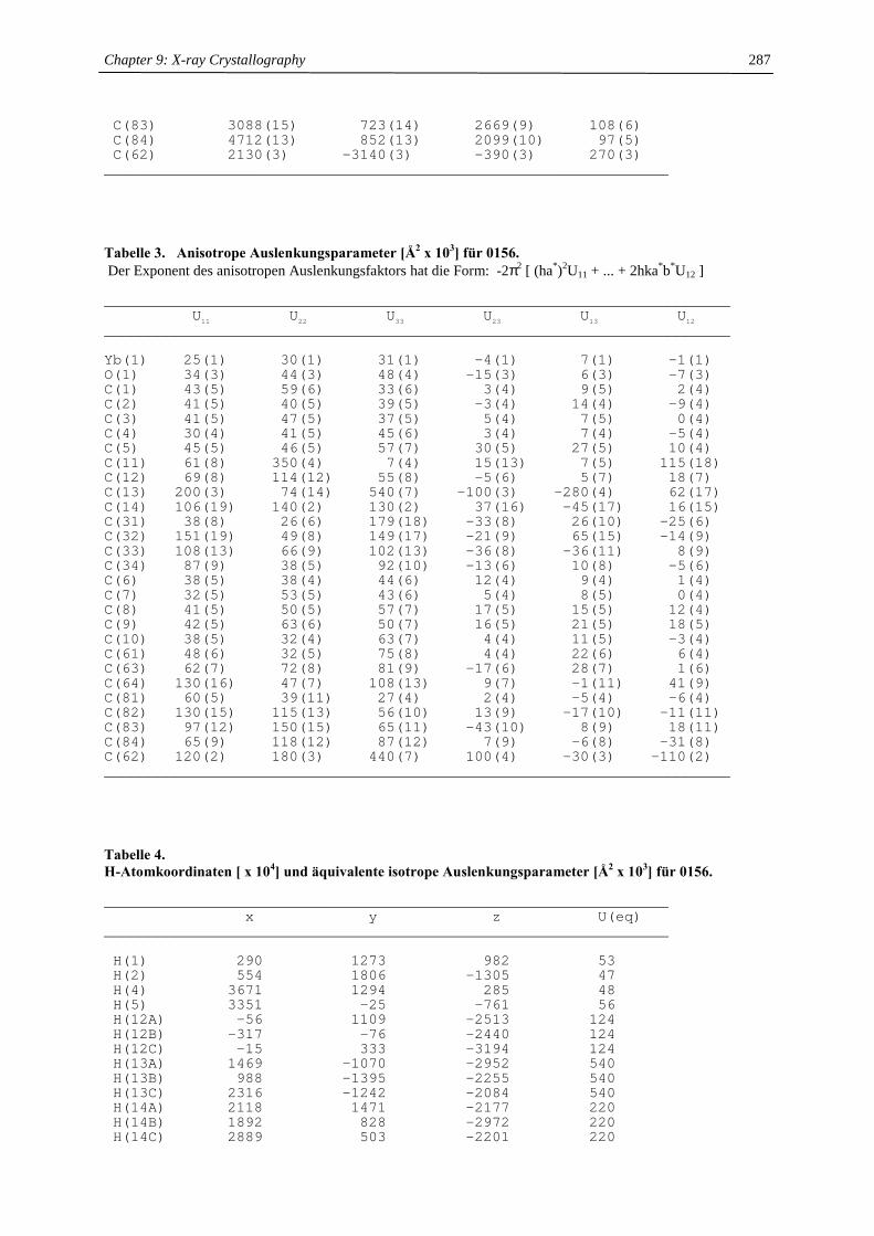
Chapter 9: X-ray Crystallography
287
C(83) 3088(15) 723(14) 2669(9) 108(6)C(84) 4712(13) 852(13) 2099(10) 97(5)C(62) 2130(3) -3140(3) -390(3) 270(3)
________________________________________________________________ Tabelle 3. Anisotrope Auslenkungsparameter [Å2 x 10³] für 0156. Der Exponent des anisotropen Auslenkungsfaktors hat die Form: -2π2 [ (ha*)2U11 + ... + 2hka*b*U12 ]
_______________________________________________________________________U11 U22 U33 U23 U13 U12
_______________________________________________________________________
Yb(1) 25(1) 30(1) 31(1) -4(1) 7(1) -1(1)O(1) 34(3) 44(3) 48(4) -15(3) 6(3) -7(3)C(1) 43(5) 59(6) 33(6) 3(4) 9(5) 2(4)C(2) 41(5) 40(5) 39(5) -3(4) 14(4) -9(4)C(3) 41(5) 47(5) 37(5) 5(4) 7(5) 0(4)C(4) 30(4) 41(5) 45(6) 3(4) 7(4) -5(4)C(5) 45(5) 46(5) 57(7) 30(5) 27(5) 10(4)C(11) 61(8) 350(4) 7(4) 15(13) 7(5) 115(18)C(12) 69(8) 114(12) 55(8) -5(6) 5(7) 18(7)C(13) 200(3) 74(14) 540(7) -100(3) -280(4) 62(17)C(14) 106(19) 140(2) 130(2) 37(16) -45(17) 16(15)C(31) 38(8) 26(6) 179(18) -33(8) 26(10) -25(6)C(32) 151(19) 49(8) 149(17) -21(9) 65(15) -14(9)C(33) 108(13) 66(9) 102(13) -36(8) -36(11) 8(9)C(34) 87(9) 38(5) 92(10) -13(6) 10(8) -5(6)C(6) 38(5) 38(4) 44(6) 12(4) 9(4) 1(4)C(7) 32(5) 53(5) 43(6) 5(4) 8(5) 0(4)C(8) 41(5) 50(5) 57(7) 17(5) 15(5) 12(4)C(9) 42(5) 63(6) 50(7) 16(5) 21(5) 18(5)C(10) 38(5) 32(4) 63(7) 4(4) 11(5) -3(4)C(61) 48(6) 32(5) 75(8) 4(4) 22(6) 6(4)C(63) 62(7) 72(8) 81(9) -17(6) 28(7) 1(6)C(64) 130(16) 47(7) 108(13) 9(7) -1(11) 41(9)C(81) 60(5) 39(11) 27(4) 2(4) -5(4) -6(4)C(82) 130(15) 115(13) 56(10) 13(9) -17(10) -11(11)C(83) 97(12) 150(15) 65(11) -43(10) 8(9) 18(11)C(84) 65(9) 118(12) 87(12) 7(9) -6(8) -31(8)C(62) 120(2) 180(3) 440(7) 100(4) -30(3) -110(2)_______________________________________________________________________ Tabelle 4. H-Atomkoordinaten [ x 104] und äquivalente isotrope Auslenkungsparameter [Å2 x 10³] für 0156.
________________________________________________________________x y z U(eq)
________________________________________________________________
H(1) 290 1273 982 53H(2) 554 1806 -1305 47H(4) 3671 1294 285 48H(5) 3351 -25 -761 56H(12A) -56 1109 -2513 124H(12B) -317 -76 -2440 124H(12C) -15 333 -3194 124H(13A) 1469 -1070 -2952 540H(13B) 988 -1395 -2255 540H(13C) 2316 -1242 -2084 540H(14A) 2118 1471 -2177 220H(14B) 1892 828 -2972 220H(14C) 2889 503 -2201 220
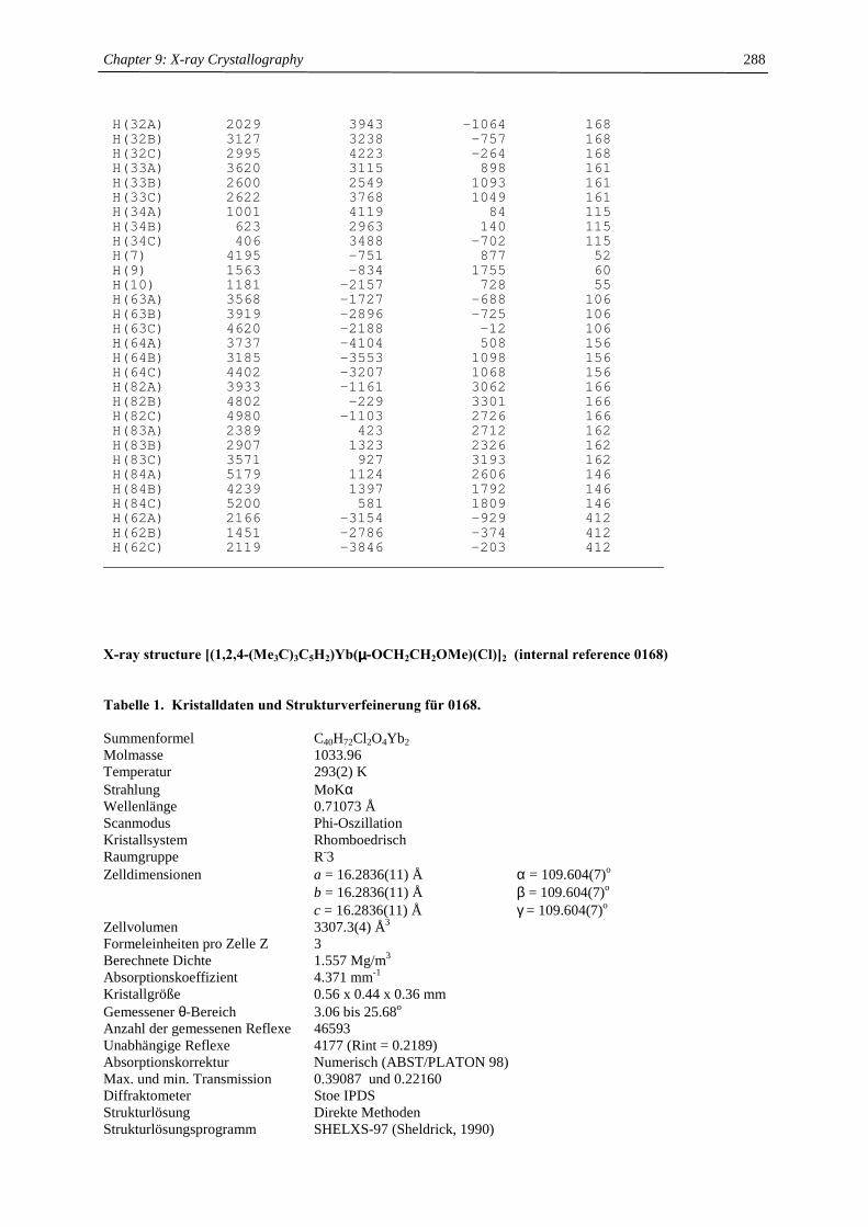
Chapter 9: X-ray Crystallography
288
H(32A) 2029 3943 -1064 168H(32B) 3127 3238 -757 168H(32C) 2995 4223 -264 168H(33A) 3620 3115 898 161H(33B) 2600 2549 1093 161H(33C) 2622 3768 1049 161H(34A) 1001 4119 84 115H(34B) 623 2963 140 115H(34C) 406 3488 -702 115H(7) 4195 -751 877 52H(9) 1563 -834 1755 60H(10) 1181 -2157 728 55H(63A) 3568 -1727 -688 106H(63B) 3919 -2896 -725 106H(63C) 4620 -2188 -12 106H(64A) 3737 -4104 508 156H(64B) 3185 -3553 1098 156H(64C) 4402 -3207 1068 156H(82A) 3933 -1161 3062 166H(82B) 4802 -229 3301 166H(82C) 4980 -1103 2726 166H(83A) 2389 423 2712 162H(83B) 2907 1323 2326 162H(83C) 3571 927 3193 162H(84A) 5179 1124 2606 146H(84B) 4239 1397 1792 146H(84C) 5200 581 1809 146H(62A) 2166 -3154 -929 412H(62B) 1451 -2786 -374 412H(62C) 2119 -3846 -203 412
________________________________________________________________
X-ray structure [(1,2,4-(Me3C)3C5H2)Yb(µµµµ-OCH2CH2OMe)(Cl)]2 (internal reference 0168)
Tabelle 1. Kristalldaten und Strukturverfeinerung für 0168. Summenformel C40H72Cl2O4Yb2 Molmasse 1033.96 Temperatur 293(2) K Strahlung MoKα Wellenlänge 0.71073 Å Scanmodus Phi-Oszillation Kristallsystem Rhomboedrisch Raumgruppe R-3 Zelldimensionen a = 16.2836(11) Å α = 109.604(7)o b = 16.2836(11) Å β = 109.604(7)o c = 16.2836(11) Å γ = 109.604(7)o Zellvolumen 3307.3(4) Å3 Formeleinheiten pro Zelle Z 3 Berechnete Dichte 1.557 Mg/m3 Absorptionskoeffizient 4.371 mm-1 Kristallgröße 0.56 x 0.44 x 0.36 mm Gemessener θ-Bereich 3.06 bis 25.68o Anzahl der gemessenen Reflexe 46593 Unabhängige Reflexe 4177 (Rint = 0.2189) Absorptionskorrektur Numerisch (ABST/PLATON 98) Max. und min. Transmission 0.39087 und 0.22160 Diffraktometer Stoe IPDS Strukturlösung Direkte Methoden Strukturlösungsprogramm SHELXS-97 (Sheldrick, 1990)

Chapter 9: X-ray Crystallography
289
Strukturverfeinerung Vollmatrix Least-Squares gegen F2 Strukturverfeinerungsprogramm SHELXL-97 (Sheldrick, 1997) Daten / Restraints / Parameter 4177 / 0 / 227 Endgültige R-Werte [I>2σ(I)] R1 = 0.0422, wR2 = 0.0842 R-Werte (alle Daten) R1 = 0.0483, wR2 = 0.0871 Wichtungsschema w=1/[σ2(Fo2)+(0.0000P)2+1.3688P] mit P=(Fo2+2Fc2)/3 GooF (alle Daten) 1.114 Größtes Maximum und Minimum 1.137 und -1.023 eÅ-3
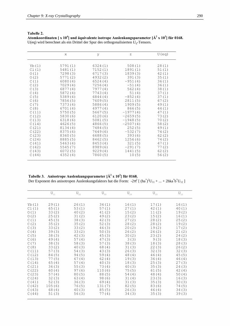
Chapter 9: X-ray Crystallography
290
Tabelle 2. Atomkoordinaten [ x 104] und äquivalente isotrope Auslenkungsparameter [Å2 x 10³] für 0168. U(eq) wird berechnet als ein Drittel der Spur des orthogonalisierten Uij-Tensors.
________________________________________________________________x y z U(eq)
________________________________________________________________
Yb(1) 5791(1) 6324(1) 508(1) 28(1)Cl(1) 5481(1) 7152(1) 1891(1) 51(1)O(1) 7298(3) 6717(3) 1839(3) 42(1)O(2) 5771(2) 4932(2) 391(3) 35(1)C(1) 6080(4) 6524(4) -951(4) 36(1)C(2) 7029(4) 7256(4) -51(4) 36(1)C(3) 6877(4) 7977(4) 562(4) 38(1)C(4) 5872(4) 7743(4) 51(4) 37(1)C(5) 5389(4) 6844(4) -852(4) 37(1)C(6) 7856(5) 7609(5) 2811(5) 67(2)C(7) 7373(4) 5886(4) 1909(5) 49(1)C(8) 6701(4) 4977(4) 866(5) 44(1)C(11) 5750(5) 5667(5) -1977(4) 47(1)C(12) 5830(6) 6120(6) -2659(5) 73(2)C(13) 6318(6) 5081(5) -1948(5) 70(2)C(14) 4626(5) 4866(5) -2507(4) 58(2)C(21) 8134(4) 7484(5) 252(5) 49(1)C(22) 8375(6) 7649(6) -532(7) 74(2)C(23) 8360(5) 6688(5) 393(6) 62(2)C(24) 8885(5) 8462(5) 1256(6) 74(2)C(41) 5463(4) 8453(4) 321(5) 47(1)C(42) 5545(7) 8989(6) -291(7) 77(2)C(43) 6072(5) 9229(4) 1441(5) 62(2)C(44) 4352(4) 7860(5) 10(5) 56(2)
________________________________________________________________ Tabelle 3. Anisotrope Auslenkungsparameter [Å2 x 10³] für 0168. Der Exponent des anisotropen Auslenkungsfaktors hat die Form: -2π2 [ (ha*)2U11 + ... + 2hka*b*U12 ]
_______________________________________________________________________U11 U22 U33 U23 U13 U12
_______________________________________________________________________
Yb(1) 29(1) 26(1) 34(1) 16(1) 17(1) 16(1)Cl(1) 65(1) 53(1) 57(1) 27(1) 42(1) 40(1)O(1) 33(2) 40(2) 41(2) 15(2) 11(2) 19(2)O(2) 25(2) 31(2) 49(2) 23(2) 15(2) 16(1)C(1) 45(3) 38(3) 42(3) 27(2) 28(2) 25(2)C(2) 35(2) 35(2) 52(3) 28(2) 28(2) 19(2)C(3) 33(2) 33(2) 46(3) 20(2) 19(2) 17(2)C(4) 39(3) 33(2) 50(3) 26(2) 26(2) 21(2)C(5) 38(3) 42(3) 45(3) 30(2) 23(2) 24(2)C(6) 49(4) 57(4) 47(3) 3(3) 9(3) 18(3)C(7) 38(3) 58(3) 57(3) 38(3) 18(3) 28(3)C(8) 33(2) 40(3) 68(4) 31(3) 22(3) 26(2)C(11) 57(3) 54(3) 43(3) 26(3) 32(3) 32(3)C(12) 84(5) 94(5) 59(4) 48(4) 46(4) 45(5)C(13) 77(5) 67(4) 62(4) 19(3) 36(4) 46(4)C(14) 65(4) 57(4) 40(3) 18(3) 25(3) 27(3)C(21) 36(3) 55(3) 73(4) 40(3) 35(3) 24(3)C(22) 60(4) 97(6) 110(6) 75(5) 61(5) 42(4)C(23) 57(4) 80(5) 88(5) 54(4) 48(4) 50(4)C(24) 32(3) 63(4) 98(6) 31(4) 23(3) 16(3)C(41) 52(3) 36(3) 69(4) 31(3) 35(3) 30(3)C(42) 105(6) 76(5) 131(7) 82(5) 83(6) 74(5)C(43) 68(4) 40(3) 85(5) 26(3) 46(4) 34(3)C(44) 51(3) 56(3) 77(4) 34(3) 35(3) 39(3)_______________________________________________________________________
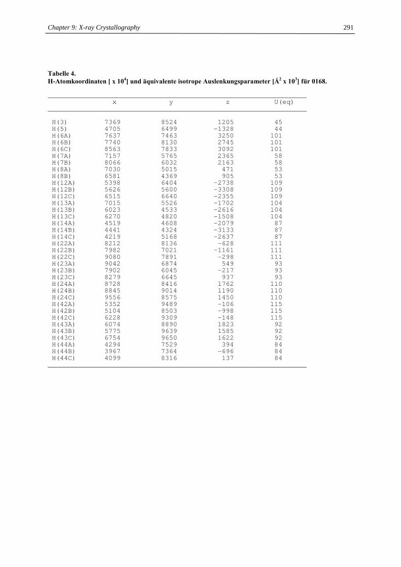
Chapter 9: X-ray Crystallography
291
Tabelle 4. H-Atomkoordinaten [ x 104] und äquivalente isotrope Auslenkungsparameter [Å2 x 10³] für 0168.
________________________________________________________________x y z U(eq)
________________________________________________________________
H(3) 7369 8524 1205 45H(5) 4705 6499 -1328 44H(6A) 7637 7463 3250 101H(6B) 7740 8130 2745 101H(6C) 8563 7833 3092 101H(7A) 7157 5765 2365 58H(7B) 8066 6032 2163 58H(8A) 7030 5015 471 53H(8B) 6581 4369 905 53H(12A) 5398 6404 -2738 109H(12B) 5626 5600 -3308 109H(12C) 6515 6640 -2355 109H(13A) 7015 5526 -1702 104H(13B) 6023 4533 -2616 104H(13C) 6270 4820 -1508 104H(14A) 4519 4608 -2079 87H(14B) 4441 4324 -3133 87H(14C) 4219 5168 -2637 87H(22A) 8212 8136 -628 111H(22B) 7982 7021 -1161 111H(22C) 9080 7891 -298 111H(23A) 9042 6874 549 93H(23B) 7902 6045 -217 93H(23C) 8279 6645 937 93H(24A) 8728 8416 1762 110H(24B) 8845 9014 1190 110H(24C) 9556 8575 1450 110H(42A) 5352 9489 -106 115H(42B) 5104 8503 -998 115H(42C) 6228 9309 -148 115H(43A) 6074 8890 1823 92H(43B) 5775 9639 1585 92H(43C) 6754 9650 1622 92H(44A) 4294 7529 394 84H(44B) 3967 7364 -696 84H(44C) 4099 8316 137 84
________________________________________________________________
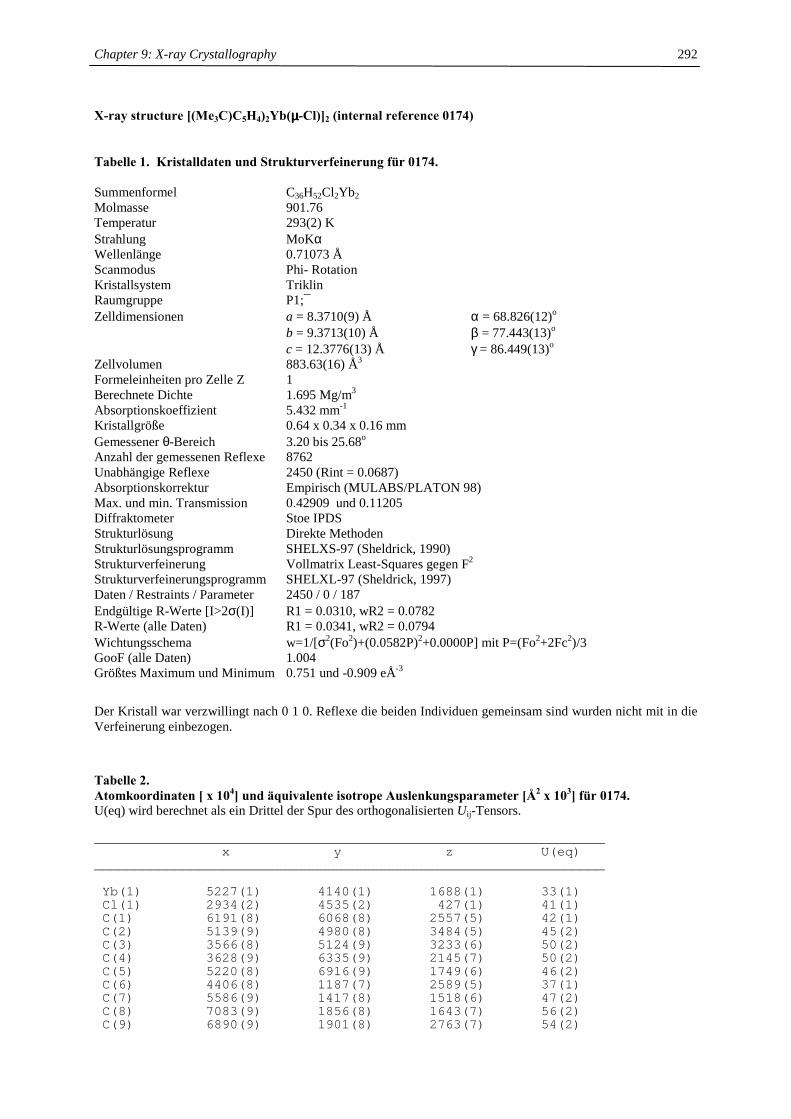
Chapter 9: X-ray Crystallography
292
X-ray structure [(Me3C)C5H4)2Yb(µµµµ-Cl)]2 (internal reference 0174)
Tabelle 1. Kristalldaten und Strukturverfeinerung für 0174. Summenformel C36H52Cl2Yb2 Molmasse 901.76 Temperatur 293(2) K Strahlung MoKα Wellenlänge 0.71073 Å Scanmodus Phi- Rotation Kristallsystem Triklin Raumgruppe P1;¯ Zelldimensionen a = 8.3710(9) Å α = 68.826(12)o b = 9.3713(10) Å β = 77.443(13)o c = 12.3776(13) Å γ = 86.449(13)o Zellvolumen 883.63(16) Å3 Formeleinheiten pro Zelle Z 1 Berechnete Dichte 1.695 Mg/m3 Absorptionskoeffizient 5.432 mm-1 Kristallgröße 0.64 x 0.34 x 0.16 mm Gemessener θ-Bereich 3.20 bis 25.68o Anzahl der gemessenen Reflexe 8762 Unabhängige Reflexe 2450 (Rint = 0.0687) Absorptionskorrektur Empirisch (MULABS/PLATON 98) Max. und min. Transmission 0.42909 und 0.11205 Diffraktometer Stoe IPDS Strukturlösung Direkte Methoden Strukturlösungsprogramm SHELXS-97 (Sheldrick, 1990) Strukturverfeinerung Vollmatrix Least-Squares gegen F2 Strukturverfeinerungsprogramm SHELXL-97 (Sheldrick, 1997) Daten / Restraints / Parameter 2450 / 0 / 187 Endgültige R-Werte [I>2σ(I)] R1 = 0.0310, wR2 = 0.0782 R-Werte (alle Daten) R1 = 0.0341, wR2 = 0.0794 Wichtungsschema w=1/[σ2(Fo2)+(0.0582P)2+0.0000P] mit P=(Fo2+2Fc2)/3 GooF (alle Daten) 1.004 Größtes Maximum und Minimum 0.751 und -0.909 eÅ-3
Der Kristall war verzwillingt nach 0 1 0. Reflexe die beiden Individuen gemeinsam sind wurden nicht mit in die Verfeinerung einbezogen.
Tabelle 2. Atomkoordinaten [ x 104] und äquivalente isotrope Auslenkungsparameter [Å2 x 10³] für 0174. U(eq) wird berechnet als ein Drittel der Spur des orthogonalisierten Uij-Tensors.
________________________________________________________________x y z U(eq)
________________________________________________________________
Yb(1) 5227(1) 4140(1) 1688(1) 33(1)Cl(1) 2934(2) 4535(2) 427(1) 41(1)C(1) 6191(8) 6068(8) 2557(5) 42(1)C(2) 5139(9) 4980(8) 3484(5) 45(2)C(3) 3566(8) 5124(9) 3233(6) 50(2)C(4) 3628(9) 6335(9) 2145(7) 50(2)C(5) 5220(8) 6916(9) 1749(6) 46(2)C(6) 4406(8) 1187(7) 2589(5) 37(1)C(7) 5586(9) 1417(8) 1518(6) 47(2)C(8) 7083(9) 1856(8) 1643(7) 56(2)C(9) 6890(9) 1901(8) 2763(7) 54(2)
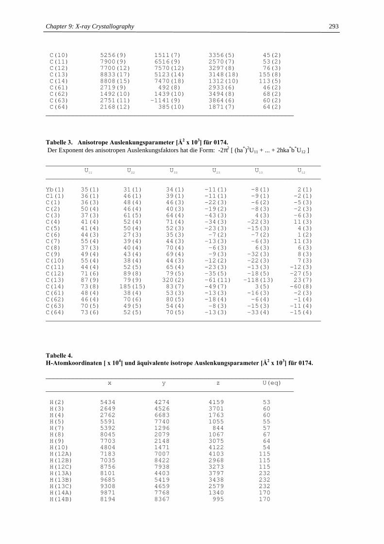
Chapter 9: X-ray Crystallography
293
C(10) 5256(9) 1511(7) 3356(5) 45(2)C(11) 7900(9) 6516(9) 2570(7) 53(2)C(12) 7700(12) 7570(12) 3297(8) 76(3)C(13) 8833(17) 5123(14) 3148(18) 155(8)C(14) 8808(15) 7470(18) 1312(10) 113(5)C(61) 2719(9) 492(8) 2933(6) 46(2)C(62) 1492(10) 1439(10) 3494(8) 68(2)C(63) 2751(11) -1141(9) 3864(6) 60(2)C(64) 2168(12) 385(10) 1871(7) 64(2)
________________________________________________________________
Tabelle 3. Anisotrope Auslenkungsparameter [Å2 x 10³] für 0174. Der Exponent des anisotropen Auslenkungsfaktors hat die Form: -2π2 [ (ha*)2U11 + ... + 2hka*b*U12 ]
_______________________________________________________________________U11 U22 U33 U23 U13 U12
_______________________________________________________________________
Yb(1) 35(1) 31(1) 34(1) -11(1) -8(1) 2(1)Cl(1) 36(1) 46(1) 39(1) -11(1) -9(1) -2(1)C(1) 36(3) 48(4) 46(3) -22(3) -6(2) -5(3)C(2) 50(4) 46(4) 40(3) -19(2) -8(3) -2(3)C(3) 37(3) 61(5) 64(4) -43(3) 4(3) -6(3)C(4) 41(4) 52(4) 71(4) -34(3) -22(3) 11(3)C(5) 41(4) 50(4) 52(3) -23(3) -15(3) 4(3)C(6) 44(3) 27(3) 35(3) -7(2) -7(2) 1(2)C(7) 55(4) 39(4) 44(3) -13(3) -6(3) 11(3)C(8) 37(3) 40(4) 70(4) -6(3) 6(3) 6(3)C(9) 49(4) 43(4) 69(4) -9(3) -32(3) 8(3)C(10) 55(4) 38(4) 44(3) -12(2) -22(3) 7(3)C(11) 44(4) 52(5) 65(4) -23(3) -13(3) -12(3)C(12) 71(6) 89(8) 79(5) -35(5) -18(5) -27(5)C(13) 87(9) 79(9) 320(2) -61(11) -118(13) 23(7)C(14) 73(8) 185(15) 83(7) -49(7) 3(5) -60(8)C(61) 48(4) 38(4) 53(3) -13(3) -16(3) -2(3)C(62) 46(4) 70(6) 80(5) -18(4) -6(4) -1(4)C(63) 70(5) 49(5) 54(4) -8(3) -15(3) -11(4)C(64) 73(6) 52(5) 70(5) -13(3) -33(4) -15(4)_______________________________________________________________________ Tabelle 4. H-Atomkoordinaten [ x 104] und äquivalente isotrope Auslenkungsparameter [Å2 x 10³] für 0174.
________________________________________________________________x y z U(eq)
________________________________________________________________
H(2) 5434 4274 4159 53H(3) 2649 4526 3701 60H(4) 2762 6683 1763 60H(5) 5591 7740 1055 55H(7) 5392 1296 844 57H(8) 8045 2079 1067 67H(9) 7703 2148 3075 64H(10) 4804 1471 4122 54H(12A) 7183 7007 4103 115H(12B) 7035 8422 2968 115H(12C) 8756 7938 3273 115H(13A) 8101 4403 3797 232H(13B) 9685 5419 3438 232H(13C) 9308 4659 2579 232H(14A) 9871 7768 1340 170H(14B) 8194 8367 995 170
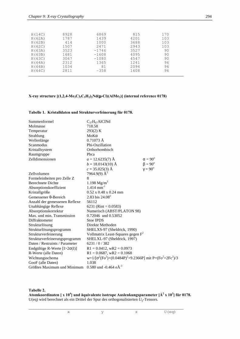
Chapter 9: X-ray Crystallography
294
H(14C) 8928 6869 815 170H(62A) 1787 1439 4201 103H(62B) 414 1000 3688 103H(62C) 1507 2471 2943 103H(63A) 3523 -1746 3527 90H(63B) 1681 -1608 4095 90H(63C) 3067 -1080 4547 90H(64A) 2312 1365 1241 96H(64B) 1034 81 2094 96H(64C) 2811 -358 1608 96
________________________________________________________________
X-ray structure [(1,2,4-Me3C)3C5H2)2Nd(µµµµ-Cl)(AlMe3)] (internal reference 0178)
Tabelle 1. Kristalldaten und Strukturverfeinerung für 0178. Summenformel C37H67AlClNd Molmasse 718.58 Temperatur 293(2) K Strahlung MoKα Wellenlänge 0.71073 Å Scanmodus Phi-Oszillation Kristallsystem Orthorhombisch Raumgruppe Pbca Zelldimensionen a = 12.6235(7) Å α = 90o b = 18.0143(10) Å β = 90o c = 35.025(3) Å γ = 90o Zellvolumen 7964.9(9) Å3 Formeleinheiten pro Zelle Z 8 Berechnete Dichte 1.198 Mg/m3 Absorptionskoeffizient 1.414 mm-1 Kristallgröße 0.52 x 0.48 x 0.24 mm Gemessener θ-Bereich 2.83 bis 24.08o Anzahl der gemessenen Reflexe 56112 Unabhängige Reflexe 6231 (Rint = 0.0583) Absorptionskorrektur Numerisch (ABST/PLATON 98) Max. und min. Transmission 0.72046 und 0.53052 Diffraktometer Stoe IPDS Strukturlösung Direkte Methoden Strukturlösungsprogramm SHELXS-97 (Sheldrick, 1990) Strukturverfeinerung Vollmatrix Least-Squares gegen F2 Strukturverfeinerungsprogramm SHELXL-97 (Sheldrick, 1997) Daten / Restraints / Parameter 6231 / 0 / 382 Endgültige R-Werte [I>2σ(I)] R1 = 0.0412, wR2 = 0.0973 R-Werte (alle Daten) R1 = 0.0687, wR2 = 0.1068 Wichtungsschema w=1/[σ2(Fo2)+(0.0484P)2+9.2366P] mit P=(Fo2+2Fc2)/3 GooF (alle Daten) 1.038 Größtes Maximum und Minimum 0.580 und -0.464 eÅ-3
Tabelle 2. Atomkoordinaten [ x 104] und äquivalente isotrope Auslenkungsparameter [Å2 x 10³] für 0178. U(eq) wird berechnet als ein Drittel der Spur des orthogonalisierten Uij-Tensors.
________________________________________________________________x y z U(eq)
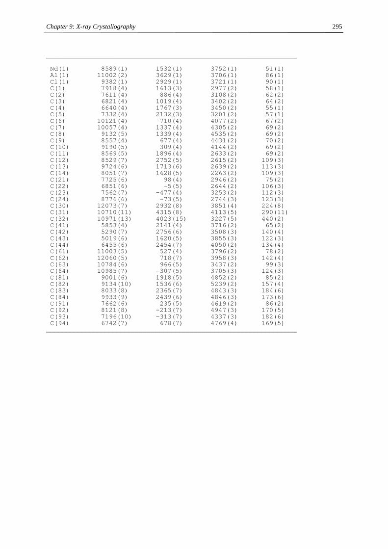
Chapter 9: X-ray Crystallography
295
________________________________________________________________
Nd(1) 8589(1) 1532(1) 3752(1) 51(1)Al(1) 11002(2) 3629(1) 3706(1) 86(1)Cl(1) 9382(1) 2929(1) 3721(1) 90(1)C(1) 7918(4) 1613(3) 2977(2) 58(1)C(2) 7611(4) 886(4) 3108(2) 62(2)C(3) 6821(4) 1019(4) 3402(2) 64(2)C(4) 6640(4) 1767(3) 3450(2) 55(1)C(5) 7332(4) 2132(3) 3201(2) 57(1)C(6) 10121(4) 710(4) 4077(2) 67(2)C(7) 10057(4) 1337(4) 4305(2) 69(2)C(8) 9132(5) 1339(4) 4535(2) 69(2)C(9) 8557(4) 677(4) 4431(2) 70(2)C(10) 9190(5) 309(4) 4144(2) 69(2)C(11) 8569(5) 1896(4) 2633(2) 69(2)C(12) 8529(7) 2752(5) 2615(2) 109(3)C(13) 9724(6) 1713(6) 2639(2) 113(3)C(14) 8051(7) 1628(5) 2263(2) 109(3)C(21) 7725(6) 98(4) 2946(2) 75(2)C(22) 6851(6) -5(5) 2644(2) 106(3)C(23) 7562(7) -477(4) 3253(2) 112(3)C(24) 8776(6) -73(5) 2744(3) 123(3)C(30) 12073(7) 2932(8) 3851(4) 224(8)C(31) 10710(11) 4315(8) 4113(5) 290(11)C(32) 10971(13) 4023(15) 3227(5) 440(2)C(41) 5853(4) 2141(4) 3716(2) 65(2)C(42) 5290(7) 2756(6) 3508(3) 140(4)C(43) 5019(6) 1620(5) 3855(3) 122(3)C(44) 6455(6) 2454(7) 4050(2) 134(4)C(61) 11003(5) 527(4) 3796(2) 78(2)C(62) 12060(5) 718(7) 3958(3) 142(4)C(63) 10784(6) 966(5) 3437(2) 99(3)C(64) 10985(7) -307(5) 3705(3) 124(3)C(81) 9001(6) 1918(5) 4852(2) 85(2)C(82) 9134(10) 1536(6) 5239(2) 157(4)C(83) 8033(8) 2365(7) 4843(3) 184(6)C(84) 9933(9) 2439(6) 4846(3) 173(6)C(91) 7662(6) 235(5) 4619(2) 86(2)C(92) 8121(8) -213(7) 4947(3) 170(5)C(93) 7196(10) -313(7) 4337(3) 182(6)C(94) 6742(7) 678(7) 4769(4) 169(5)
________________________________________________________________
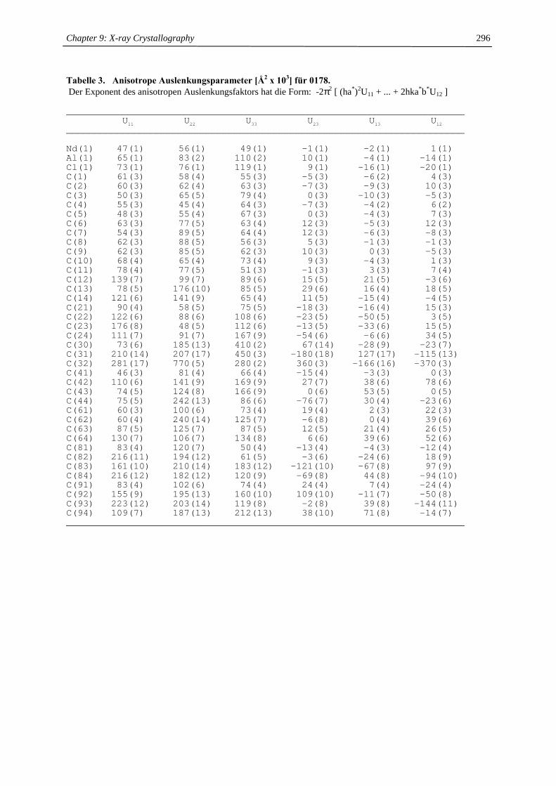
Chapter 9: X-ray Crystallography
296
Tabelle 3. Anisotrope Auslenkungsparameter [Å2 x 10³] für 0178. Der Exponent des anisotropen Auslenkungsfaktors hat die Form: -2π2 [ (ha*)2U11 + ... + 2hka*b*U12 ]
_______________________________________________________________________U11 U22 U33 U23 U13 U12
_______________________________________________________________________
Nd(1) 47(1) 56(1) 49(1) -1(1) -2(1) 1(1)Al(1) 65(1) 83(2) 110(2) 10(1) -4(1) -14(1)Cl(1) 73(1) 76(1) 119(1) 9(1) -16(1) -20(1)C(1) 61(3) 58(4) 55(3) -5(3) -6(2) 4(3)C(2) 60(3) 62(4) 63(3) -7(3) -9(3) 10(3)C(3) 50(3) 65(5) 79(4) 0(3) -10(3) -5(3)C(4) 55(3) 45(4) 64(3) -7(3) -4(2) 6(2)C(5) 48(3) 55(4) 67(3) 0(3) -4(3) 7(3)C(6) 63(3) 77(5) 63(4) 12(3) -5(3) 12(3)C(7) 54(3) 89(5) 64(4) 12(3) -6(3) -8(3)C(8) 62(3) 88(5) 56(3) 5(3) -1(3) -1(3)C(9) 62(3) 85(5) 62(3) 10(3) 0(3) -5(3)C(10) 68(4) 65(4) 73(4) 9(3) -4(3) 1(3)C(11) 78(4) 77(5) 51(3) -1(3) 3(3) 7(4)C(12) 139(7) 99(7) 89(6) 15(5) 21(5) -3(6)C(13) 78(5) 176(10) 85(5) 29(6) 16(4) 18(5)C(14) 121(6) 141(9) 65(4) 11(5) -15(4) -4(5)C(21) 90(4) 58(5) 75(5) -18(3) -16(4) 15(3)C(22) 122(6) 88(6) 108(6) -23(5) -50(5) 3(5)C(23) 176(8) 48(5) 112(6) -13(5) -33(6) 15(5)C(24) 111(7) 91(7) 167(9) -54(6) -6(6) 34(5)C(30) 73(6) 185(13) 410(2) 67(14) -28(9) -23(7)C(31) 210(14) 207(17) 450(3) -180(18) 127(17) -115(13)C(32) 281(17) 770(5) 280(2) 360(3) -166(16) -370(3)C(41) 46(3) 81(4) 66(4) -15(4) -3(3) 0(3)C(42) 110(6) 141(9) 169(9) 27(7) 38(6) 78(6)C(43) 74(5) 124(8) 166(9) 0(6) 53(5) 0(5)C(44) 75(5) 242(13) 86(6) -76(7) 30(4) -23(6)C(61) 60(3) 100(6) 73(4) 19(4) 2(3) 22(3)C(62) 60(4) 240(14) 125(7) -6(8) 0(4) 39(6)C(63) 87(5) 125(7) 87(5) 12(5) 21(4) 26(5)C(64) 130(7) 106(7) 134(8) 6(6) 39(6) 52(6)C(81) 83(4) 120(7) 50(4) -13(4) -4(3) -12(4)C(82) 216(11) 194(12) 61(5) -3(6) -24(6) 18(9)C(83) 161(10) 210(14) 183(12) -121(10) -67(8) 97(9)C(84) 216(12) 182(12) 120(9) -69(8) 44(8) -94(10)C(91) 83(4) 102(6) 74(4) 24(4) 7(4) -24(4)C(92) 155(9) 195(13) 160(10) 109(10) -11(7) -50(8)C(93) 223(12) 203(14) 119(8) -2(8) 39(8) -144(11)C(94) 109(7) 187(13) 212(13) 38(10) 71(8) -14(7)_______________________________________________________________________
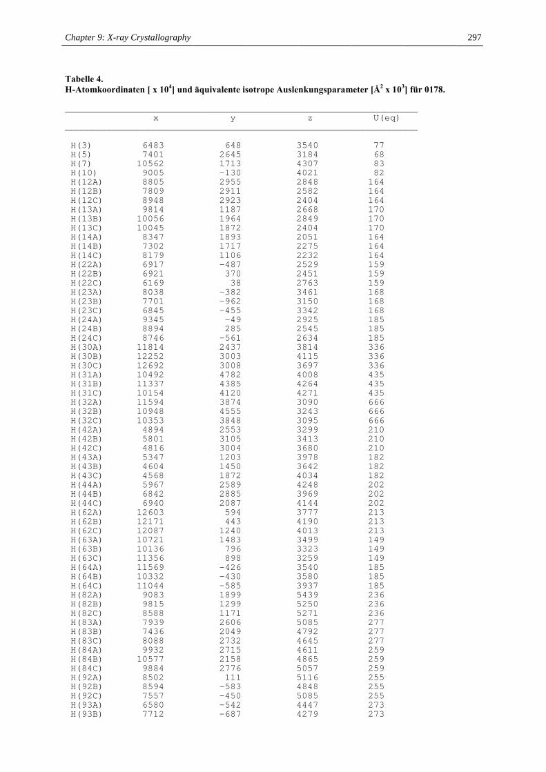
Chapter 9: X-ray Crystallography
297
Tabelle 4. H-Atomkoordinaten [ x 104] und äquivalente isotrope Auslenkungsparameter [Å2 x 10³] für 0178.
________________________________________________________________x y z U(eq)
________________________________________________________________
H(3) 6483 648 3540 77H(5) 7401 2645 3184 68H(7) 10562 1713 4307 83H(10) 9005 -130 4021 82H(12A) 8805 2955 2848 164H(12B) 7809 2911 2582 164H(12C) 8948 2923 2404 164H(13A) 9814 1187 2668 170H(13B) 10056 1964 2849 170H(13C) 10045 1872 2404 170H(14A) 8347 1893 2051 164H(14B) 7302 1717 2275 164H(14C) 8179 1106 2232 164H(22A) 6917 -487 2529 159H(22B) 6921 370 2451 159H(22C) 6169 38 2763 159H(23A) 8038 -382 3461 168H(23B) 7701 -962 3150 168H(23C) 6845 -455 3342 168H(24A) 9345 -49 2925 185H(24B) 8894 285 2545 185H(24C) 8746 -561 2634 185H(30A) 11814 2437 3814 336H(30B) 12252 3003 4115 336H(30C) 12692 3008 3697 336H(31A) 10492 4782 4008 435H(31B) 11337 4385 4264 435H(31C) 10154 4120 4271 435H(32A) 11594 3874 3090 666H(32B) 10948 4555 3243 666H(32C) 10353 3848 3095 666H(42A) 4894 2553 3299 210H(42B) 5801 3105 3413 210H(42C) 4816 3004 3680 210H(43A) 5347 1203 3978 182H(43B) 4604 1450 3642 182H(43C) 4568 1872 4034 182H(44A) 5967 2589 4248 202H(44B) 6842 2885 3969 202H(44C) 6940 2087 4144 202H(62A) 12603 594 3777 213H(62B) 12171 443 4190 213H(62C) 12087 1240 4013 213H(63A) 10721 1483 3499 149H(63B) 10136 796 3323 149H(63C) 11356 898 3259 149H(64A) 11569 -426 3540 185H(64B) 10332 -430 3580 185H(64C) 11044 -585 3937 185H(82A) 9083 1899 5439 236H(82B) 9815 1299 5250 236H(82C) 8588 1171 5271 236H(83A) 7939 2606 5085 277H(83B) 7436 2049 4792 277H(83C) 8088 2732 4645 277H(84A) 9932 2715 4611 259H(84B) 10577 2158 4865 259H(84C) 9884 2776 5057 259H(92A) 8502 111 5116 255H(92B) 8594 -583 4848 255H(92C) 7557 -450 5085 255H(93A) 6580 -542 4447 273H(93B) 7712 -687 4279 273
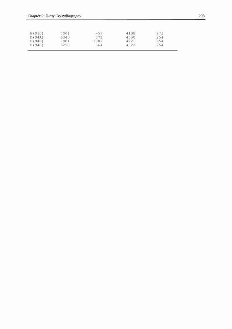
Chapter 9: X-ray Crystallography
298
H(93C) 7001 -57 4108 273H(94A) 6340 871 4558 254H(94B) 7001 1082 4921 254H(94C) 6298 364 4922 254
________________________________________________________________
Chapter 9: X-ray Crystallography
299
X-ray crystallography (UC Berkeley)
I want to thank Dr. Frederick J. Hollander and Dr. Allen G. Olivier (at CHEXRAY, the University of California
at Berkeley X-ray diffraction facility) for their assistance with the crystallography.
General
A crystal of appropriate dimensions was mounted on a glass fiber using Paratone N hydrocarbon oil. All
measurements were made on a Bruker SMART 1K CCD diffractometer.2 Cell constants and an orientation
matrix were obtained of the measured positions of reflections with I > 10 σ to give the unit cell. The systematic
absences uniquely determined the space group in each case. An arbitrary hemisphere of data was collected at low
temperature (see Experimental Details) using the ω scan technique with 0.3° scans counted for 10-30 s per
frame. Data were integrated using SAINT3 and corrected for Lorentz and polarization effects. The data were
analyzed for agreement and absorption using XPREP,4 and an empirical absorption correction was applied
based on comparison of redundant and equivalent reflections using SADABS.5 The structures were solved by
direct methods and expanded using Fourier techniques. Non-hydrogen atoms were refined anisotropically
(unless stated otherwise), and the hydrogen atoms were included in calculated positions using a riding model, but
not refined. The structures were solved and refined using the software package SHELXS-97 (structure solution)6
and SHELXL-97 (refinement).1 The structure solution refinements were unexceptional unless stated otherwise.
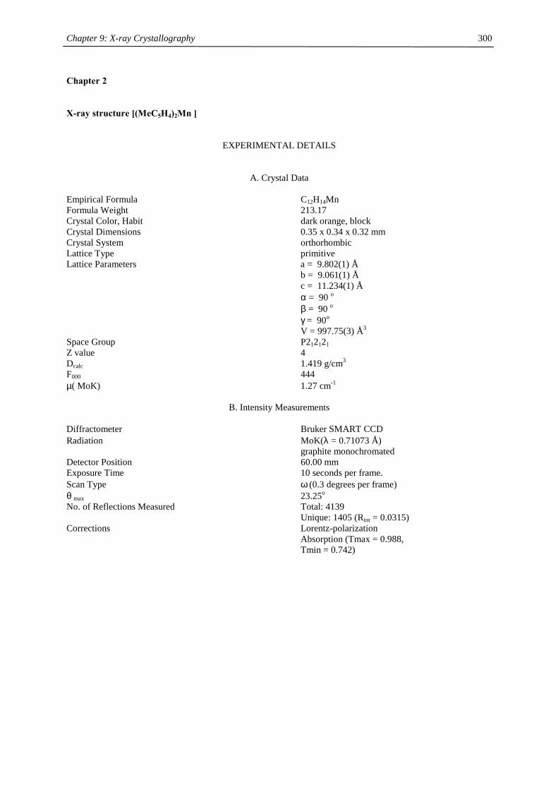
Chapter 9: X-ray Crystallography
300
Chapter 2
X-ray structure [(MeC5H4)2Mn ]
EXPERIMENTAL DETAILS
A. Crystal Data Empirical Formula C12H14Mn Formula Weight 213.17 Crystal Color, Habit dark orange, block Crystal Dimensions 0.35 x 0.34 x 0.32 mm Crystal System orthorhombic Lattice Type primitive Lattice Parameters a = 9.802(1) Å b = 9.061(1) Å c = 11.234(1) Å α = 90 o β = 90 o γ = 90o V = 997.75(3) Å3 Space Group P212121 Z value 4 Dcalc 1.419 g/cm3 F000 444 µ( MoK) 1.27 cm-1
B. Intensity Measurements Diffractometer Bruker SMART CCD Radiation MoK(λ = 0.71073 Å) graphite monochromated Detector Position 60.00 mm Exposure Time 10 seconds per frame. Scan Type ω (0.3 degrees per frame) θ max 23.25o No. of Reflections Measured Total: 4139 Unique: 1405 (Rint = 0.0315) Corrections Lorentz-polarization Absorption (Tmax = 0.988, Tmin = 0.742)
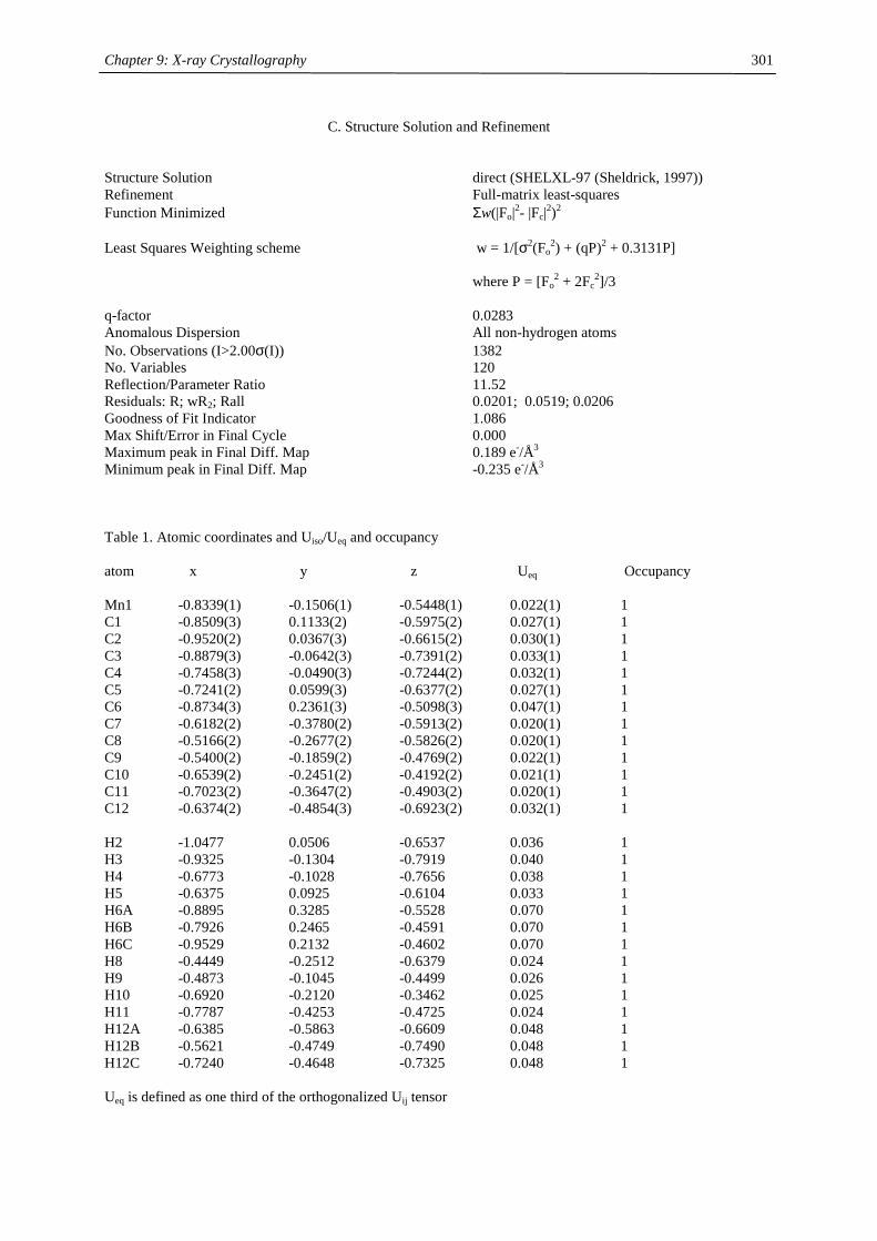
Chapter 9: X-ray Crystallography
301
C. Structure Solution and Refinement Structure Solution direct (SHELXL-97 (Sheldrick, 1997)) Refinement Full-matrix least-squares Function Minimized Σw(|Fo|2- |Fc|2)2 Least Squares Weighting scheme w = 1/[σ2(Fo
2) + (qP)2 + 0.3131P] where P = [Fo
2 + 2Fc2]/3
q-factor 0.0283 Anomalous Dispersion All non-hydrogen atoms No. Observations (I>2.00σ(I)) 1382 No. Variables 120 Reflection/Parameter Ratio 11.52 Residuals: R; wR2; Rall 0.0201; 0.0519; 0.0206 Goodness of Fit Indicator 1.086 Max Shift/Error in Final Cycle 0.000 Maximum peak in Final Diff. Map 0.189 e-/Å3 Minimum peak in Final Diff. Map -0.235 e-/Å3 Table 1. Atomic coordinates and Uiso/Ueq and occupancy atom x y z Ueq Occupancy Mn1 -0.8339(1) -0.1506(1) -0.5448(1) 0.022(1) 1 C1 -0.8509(3) 0.1133(2) -0.5975(2) 0.027(1) 1 C2 -0.9520(2) 0.0367(3) -0.6615(2) 0.030(1) 1 C3 -0.8879(3) -0.0642(3) -0.7391(2) 0.033(1) 1 C4 -0.7458(3) -0.0490(3) -0.7244(2) 0.032(1) 1 C5 -0.7241(2) 0.0599(3) -0.6377(2) 0.027(1) 1 C6 -0.8734(3) 0.2361(3) -0.5098(3) 0.047(1) 1 C7 -0.6182(2) -0.3780(2) -0.5913(2) 0.020(1) 1 C8 -0.5166(2) -0.2677(2) -0.5826(2) 0.020(1) 1 C9 -0.5400(2) -0.1859(2) -0.4769(2) 0.022(1) 1 C10 -0.6539(2) -0.2451(2) -0.4192(2) 0.021(1) 1 C11 -0.7023(2) -0.3647(2) -0.4903(2) 0.020(1) 1 C12 -0.6374(2) -0.4854(3) -0.6923(2) 0.032(1) 1 H2 -1.0477 0.0506 -0.6537 0.036 1 H3 -0.9325 -0.1304 -0.7919 0.040 1 H4 -0.6773 -0.1028 -0.7656 0.038 1 H5 -0.6375 0.0925 -0.6104 0.033 1 H6A -0.8895 0.3285 -0.5528 0.070 1 H6B -0.7926 0.2465 -0.4591 0.070 1 H6C -0.9529 0.2132 -0.4602 0.070 1 H8 -0.4449 -0.2512 -0.6379 0.024 1 H9 -0.4873 -0.1045 -0.4499 0.026 1 H10 -0.6920 -0.2120 -0.3462 0.025 1 H11 -0.7787 -0.4253 -0.4725 0.024 1 H12A -0.6385 -0.5863 -0.6609 0.048 1 H12B -0.5621 -0.4749 -0.7490 0.048 1 H12C -0.7240 -0.4648 -0.7325 0.048 1 Ueq is defined as one third of the orthogonalized Uij tensor
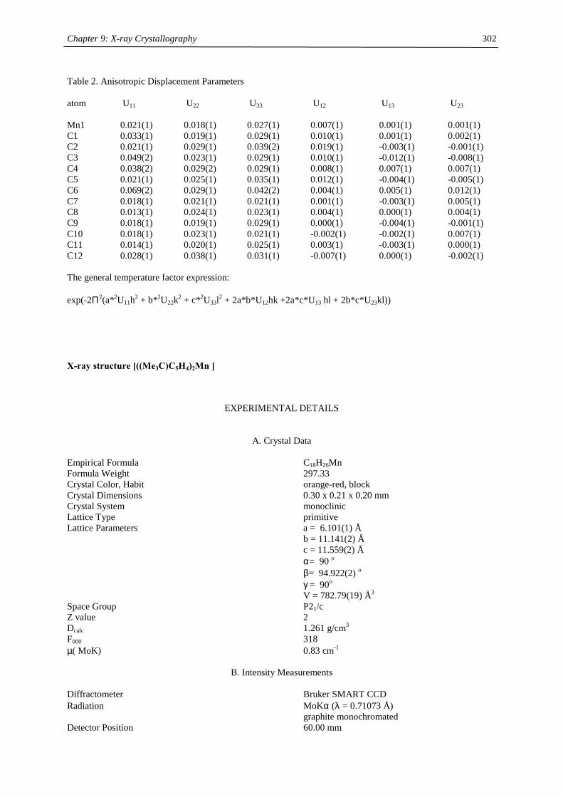
Chapter 9: X-ray Crystallography
302
Table 2. Anisotropic Displacement Parameters atom U11 U22 U33 U12 U13 U23 Mn1 0.021(1) 0.018(1) 0.027(1) 0.007(1) 0.001(1) 0.001(1) C1 0.033(1) 0.019(1) 0.029(1) 0.010(1) 0.001(1) 0.002(1) C2 0.021(1) 0.029(1) 0.039(2) 0.019(1) -0.003(1) -0.001(1) C3 0.049(2) 0.023(1) 0.029(1) 0.010(1) -0.012(1) -0.008(1) C4 0.038(2) 0.029(2) 0.029(1) 0.008(1) 0.007(1) 0.007(1) C5 0.021(1) 0.025(1) 0.035(1) 0.012(1) -0.004(1) -0.005(1) C6 0.069(2) 0.029(1) 0.042(2) 0.004(1) 0.005(1) 0.012(1) C7 0.018(1) 0.021(1) 0.021(1) 0.001(1) -0.003(1) 0.005(1) C8 0.013(1) 0.024(1) 0.023(1) 0.004(1) 0.000(1) 0.004(1) C9 0.018(1) 0.019(1) 0.029(1) 0.000(1) -0.004(1) -0.001(1) C10 0.018(1) 0.023(1) 0.021(1) -0.002(1) -0.002(1) 0.007(1) C11 0.014(1) 0.020(1) 0.025(1) 0.003(1) -0.003(1) 0.000(1) C12 0.028(1) 0.038(1) 0.031(1) -0.007(1) 0.000(1) -0.002(1) The general temperature factor expression: exp(-2Π2(a*2U11h2 + b*2U22k2 + c*2U33l2 + 2a*b*U12hk +2a*c*U13 hl + 2b*c*U23kl))
X-ray structure [((Me3C)C5H4)2Mn ]
EXPERIMENTAL DETAILS
A. Crystal Data Empirical Formula C18H26Mn Formula Weight 297.33 Crystal Color, Habit orange-red, block Crystal Dimensions 0.30 x 0.21 x 0.20 mm Crystal System monoclinic Lattice Type primitive Lattice Parameters a = 6.101(1) Å b = 11.141(2) Å c = 11.559(2) Å α= 90 o β= 94.922(2) o γ = 90o V = 782.79(19) Å3 Space Group P21/c Z value 2 Dcalc 1.261 g/cm3 F000 318 µ( MoK) 0.83 cm-1
B. Intensity Measurements Diffractometer Bruker SMART CCD Radiation MoKα (λ = 0.71073 Å) graphite monochromated Detector Position 60.00 mm
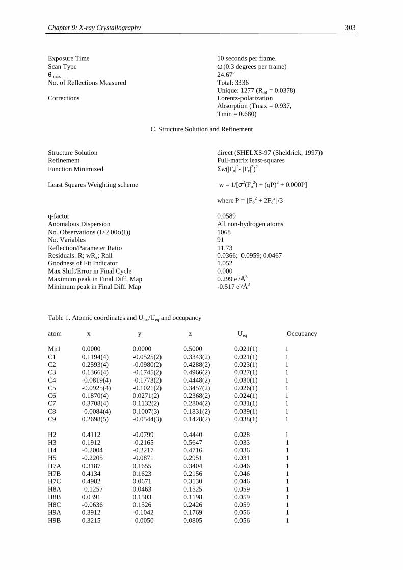
Chapter 9: X-ray Crystallography
303
Exposure Time 10 seconds per frame. Scan Type ω (0.3 degrees per frame) θ max 24.67o No. of Reflections Measured Total: 3336 Unique: 1277 (Rint = 0.0378) Corrections Lorentz-polarization Absorption (Tmax = 0.937, Tmin = 0.680)
C. Structure Solution and Refinement Structure Solution direct (SHELXS-97 (Sheldrick, 1997)) Refinement Full-matrix least-squares Function Minimized Σw(|Fo|2- |Fc|2)2 Least Squares Weighting scheme w = 1/[σ2(Fo
2) + (qP)2 + 0.000P] where P = [Fo
2 + 2Fc2]/3
q-factor 0.0589 Anomalous Dispersion All non-hydrogen atoms No. Observations (I>2.00σ(I)) 1068 No. Variables 91 Reflection/Parameter Ratio 11.73 Residuals: R; wR2; Rall 0.0366; 0.0959; 0.0467 Goodness of Fit Indicator 1.052 Max Shift/Error in Final Cycle 0.000 Maximum peak in Final Diff. Map 0.299 e-/Å3 Minimum peak in Final Diff. Map -0.517 e-/Å3 Table 1. Atomic coordinates and Uiso/Ueq and occupancy atom x y z Ueq Occupancy Mn1 0.0000 0.0000 0.5000 0.021(1) 1 C1 0.1194(4) -0.0525(2) 0.3343(2) 0.021(1) 1 C2 0.2593(4) -0.0980(2) 0.4288(2) 0.023(1) 1 C3 0.1366(4) -0.1745(2) 0.4966(2) 0.027(1) 1 C4 -0.0819(4) -0.1773(2) 0.4448(2) 0.030(1) 1 C5 -0.0925(4) -0.1021(2) 0.3457(2) 0.026(1) 1 C6 0.1870(4) 0.0271(2) 0.2368(2) 0.024(1) 1 C7 0.3708(4) 0.1132(2) 0.2804(2) 0.031(1) 1 C8 -0.0084(4) 0.1007(3) 0.1831(2) 0.039(1) 1 C9 0.2698(5) -0.0544(3) 0.1428(2) 0.038(1) 1 H2 0.4112 -0.0799 0.4440 0.028 1 H3 0.1912 -0.2165 0.5647 0.033 1 H4 -0.2004 -0.2217 0.4716 0.036 1 H5 -0.2205 -0.0871 0.2951 0.031 1 H7A 0.3187 0.1655 0.3404 0.046 1 H7B 0.4134 0.1623 0.2156 0.046 1 H7C 0.4982 0.0671 0.3130 0.046 1 H8A -0.1257 0.0463 0.1525 0.059 1 H8B 0.0391 0.1503 0.1198 0.059 1 H8C -0.0636 0.1526 0.2426 0.059 1 H9A 0.3912 -0.1042 0.1769 0.056 1 H9B 0.3215 -0.0050 0.0805 0.056 1
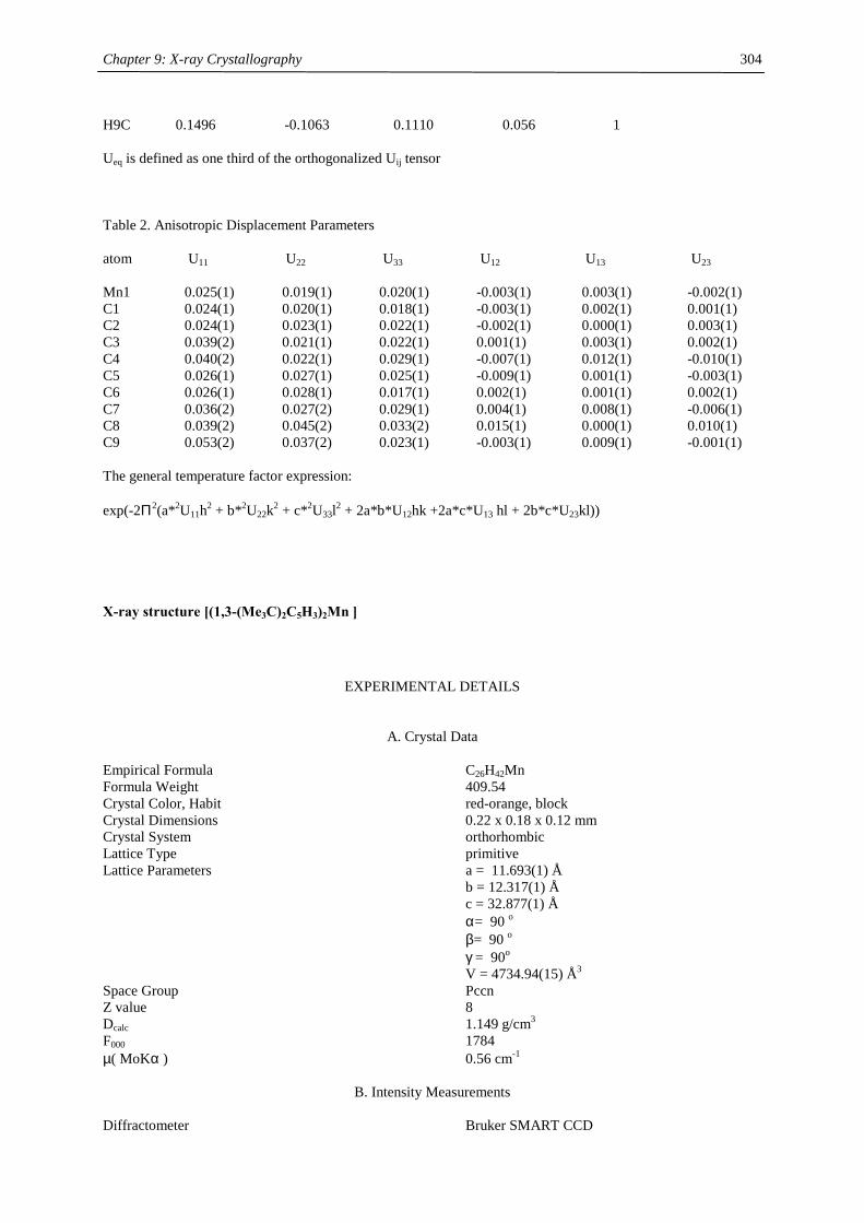
Chapter 9: X-ray Crystallography
304
H9C 0.1496 -0.1063 0.1110 0.056 1 Ueq is defined as one third of the orthogonalized Uij tensor Table 2. Anisotropic Displacement Parameters atom U11 U22 U33 U12 U13 U23 Mn1 0.025(1) 0.019(1) 0.020(1) -0.003(1) 0.003(1) -0.002(1) C1 0.024(1) 0.020(1) 0.018(1) -0.003(1) 0.002(1) 0.001(1) C2 0.024(1) 0.023(1) 0.022(1) -0.002(1) 0.000(1) 0.003(1) C3 0.039(2) 0.021(1) 0.022(1) 0.001(1) 0.003(1) 0.002(1) C4 0.040(2) 0.022(1) 0.029(1) -0.007(1) 0.012(1) -0.010(1) C5 0.026(1) 0.027(1) 0.025(1) -0.009(1) 0.001(1) -0.003(1) C6 0.026(1) 0.028(1) 0.017(1) 0.002(1) 0.001(1) 0.002(1) C7 0.036(2) 0.027(2) 0.029(1) 0.004(1) 0.008(1) -0.006(1) C8 0.039(2) 0.045(2) 0.033(2) 0.015(1) 0.000(1) 0.010(1) C9 0.053(2) 0.037(2) 0.023(1) -0.003(1) 0.009(1) -0.001(1) The general temperature factor expression: exp(-2Π2(a*2U11h2 + b*2U22k2 + c*2U33l2 + 2a*b*U12hk +2a*c*U13 hl + 2b*c*U23kl))
X-ray structure [(1,3-(Me3C)2C5H3)2Mn ]
EXPERIMENTAL DETAILS
A. Crystal Data Empirical Formula C26H42Mn Formula Weight 409.54 Crystal Color, Habit red-orange, block Crystal Dimensions 0.22 x 0.18 x 0.12 mm Crystal System orthorhombic Lattice Type primitive Lattice Parameters a = 11.693(1) Å b = 12.317(1) Å c = 32.877(1) Å α= 90 o β= 90 o γ = 90o V = 4734.94(15) Å3 Space Group Pccn Z value 8 Dcalc 1.149 g/cm3 F000 1784 µ( MoKα ) 0.56 cm-1
B. Intensity Measurements Diffractometer Bruker SMART CCD
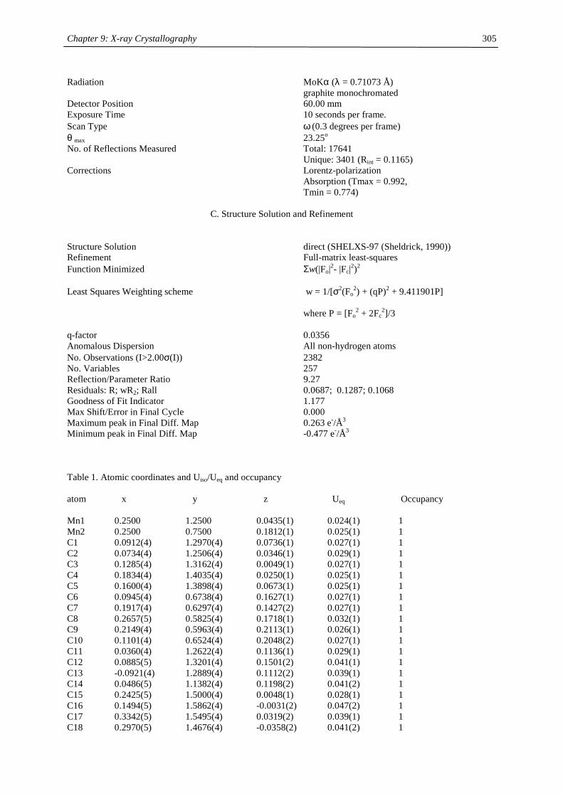
Chapter 9: X-ray Crystallography
305
Radiation MoKα (λ = 0.71073 Å) graphite monochromated Detector Position 60.00 mm Exposure Time 10 seconds per frame. Scan Type ω (0.3 degrees per frame) θ max 23.25o No. of Reflections Measured Total: 17641 Unique: 3401 (Rint = 0.1165) Corrections Lorentz-polarization Absorption (Tmax = 0.992, Tmin = 0.774)
C. Structure Solution and Refinement Structure Solution direct (SHELXS-97 (Sheldrick, 1990)) Refinement Full-matrix least-squares Function Minimized Σw(|Fo|2- |Fc|2)2 Least Squares Weighting scheme w = 1/[σ2(Fo
2) + (qP)2 + 9.411901P] where P = [Fo
2 + 2Fc2]/3
q-factor 0.0356 Anomalous Dispersion All non-hydrogen atoms No. Observations (I>2.00σ(I)) 2382 No. Variables 257 Reflection/Parameter Ratio 9.27 Residuals: R; wR2; Rall 0.0687; 0.1287; 0.1068 Goodness of Fit Indicator 1.177 Max Shift/Error in Final Cycle 0.000 Maximum peak in Final Diff. Map 0.263 e-/Å3 Minimum peak in Final Diff. Map -0.477 e-/Å3 Table 1. Atomic coordinates and Uiso/Ueq and occupancy atom x y z Ueq Occupancy Mn1 0.2500 1.2500 0.0435(1) 0.024(1) 1 Mn2 0.2500 0.7500 0.1812(1) 0.025(1) 1 C1 0.0912(4) 1.2970(4) 0.0736(1) 0.027(1) 1 C2 0.0734(4) 1.2506(4) 0.0346(1) 0.029(1) 1 C3 0.1285(4) 1.3162(4) 0.0049(1) 0.027(1) 1 C4 0.1834(4) 1.4035(4) 0.0250(1) 0.025(1) 1 C5 0.1600(4) 1.3898(4) 0.0673(1) 0.025(1) 1 C6 0.0945(4) 0.6738(4) 0.1627(1) 0.027(1) 1 C7 0.1917(4) 0.6297(4) 0.1427(2) 0.027(1) 1 C8 0.2657(5) 0.5825(4) 0.1718(1) 0.032(1) 1 C9 0.2149(4) 0.5963(4) 0.2113(1) 0.026(1) 1 C10 0.1101(4) 0.6524(4) 0.2048(2) 0.027(1) 1 C11 0.0360(4) 1.2622(4) 0.1136(1) 0.029(1) 1 C12 0.0885(5) 1.3201(4) 0.1501(2) 0.041(1) 1 C13 -0.0921(4) 1.2889(4) 0.1112(2) 0.039(1) 1 C14 0.0486(5) 1.1382(4) 0.1198(2) 0.041(2) 1 C15 0.2425(5) 1.5000(4) 0.0048(1) 0.028(1) 1 C16 0.1494(5) 1.5862(4) -0.0031(2) 0.047(2) 1 C17 0.3342(5) 1.5495(4) 0.0319(2) 0.039(1) 1 C18 0.2970(5) 1.4676(4) -0.0358(2) 0.041(2) 1
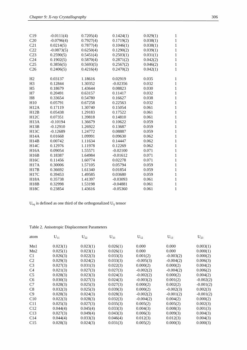
Chapter 9: X-ray Crystallography
306
C19 -0.0111(4) 0.7205(4) 0.1424(1) 0.029(1) 1 C20 -0.0796(4) 0.7927(4) 0.1719(2) 0.038(1) 1 C21 0.0214(5) 0.7877(4) 0.1046(1) 0.038(1) 1 C22 -0.0873(5) 0.6250(4) 0.1290(2) 0.039(1) 1 C23 0.2590(5) 0.5451(4) 0.2503(1) 0.031(1) 1 C24 0.1902(5) 0.5870(4) 0.2871(2) 0.042(2) 1 C25 0.3856(5) 0.5693(5) 0.2567(2) 0.046(2) 1 C26 0.2406(5) 0.4216(4) 0.2470(2) 0.042(1) 1 H2 0.03137 1.18616 0.02919 0.035 1 H3 0.12844 1.30352 -0.02356 0.032 1 H5 0.18679 1.43644 0.08823 0.030 1 H7 0.20491 0.63157 0.11417 0.032 1 H8 0.33654 0.54780 0.16627 0.038 1 H10 0.05791 0.67258 0.22563 0.032 1 H12A 0.17119 1.30740 0.15054 0.061 1 H12B 0.05438 1.29183 0.17522 0.061 1 H12C 0.07351 1.39818 0.14810 0.061 1 H13A -0.10194 1.36679 0.10622 0.059 1 H13B -0.12910 1.26922 0.13687 0.059 1 H13C -0.12689 1.24772 0.08887 0.059 1 H14A 0.01668 1.09991 0.09630 0.062 1 H14B 0.00742 1.11634 0.14447 0.062 1 H14C 0.12976 1.11978 0.12269 0.062 1 H16A 0.09054 1.55571 -0.02100 0.071 1 H16B 0.18387 1.64984 -0.01612 0.071 1 H16C 0.11456 1.60774 0.02278 0.071 1 H17A 0.30006 1.57105 0.05794 0.059 1 H17B 0.36692 1.61340 0.01854 0.059 1 H17C 0.39453 1.49585 0.03680 0.059 1 H18A 0.35738 1.41397 -0.03093 0.061 1 H18B 0.32998 1.53198 -0.04881 0.061 1 H18C 0.23854 1.43616 -0.05360 0.061 1 Ueq is defined as one third of the orthogonalized Uij tensor Table 2. Anisotropic Displacement Parameters atom U11 U22 U33 U12 U13 U23 Mn1 0.023(1) 0.023(1) 0.026(1) 0.000 0.000 0.003(1) Mn2 0.025(1) 0.023(1) 0.026(1) 0.000 0.000 0.000(1) C1 0.026(3) 0.022(3) 0.033(3) 0.001(2) -0.003(2) 0.000(2) C2 0.029(3) 0.024(2) 0.033(3) -0.005(3) -0.004(2) 0.006(3) C3 0.027(3) 0.031(3) 0.022(3) 0.000(2) 0.000(2) 0.004(2) C4 0.021(3) 0.027(3) 0.027(3) -0.002(2) -0.004(2) 0.006(2) C5 0.028(3) 0.023(3) 0.024(3) -0.002(2) 0.000(2) 0.004(2) C6 0.030(3) 0.027(3) 0.024(3) -0.003(2) 0.001(2) -0.002(2) C7 0.028(3) 0.025(3) 0.027(3) 0.000(2) 0.002(2) -0.001(2) C8 0.032(3) 0.025(3) 0.039(3) 0.000(2) -0.002(3) 0.002(3) C9 0.028(3) 0.024(3) 0.028(3) -0.002(2) -0.001(2) -0.001(2) C10 0.022(3) 0.028(3) 0.032(3) -0.004(2) 0.004(2) 0.000(2) C11 0.025(3) 0.027(3) 0.035(3) 0.005(2) 0.005(2) 0.002(3) C12 0.044(4) 0.045(4) 0.033(3) 0.004(3) 0.008(3) 0.001(3) C13 0.027(3) 0.049(4) 0.043(3) 0.006(3) 0.009(3) 0.004(3) C14 0.044(4) 0.033(3) 0.046(4) 0.012(3) 0.012(3) 0.004(3) C15 0.028(3) 0.024(3) 0.031(3) 0.005(2) 0.000(3) 0.000(3)
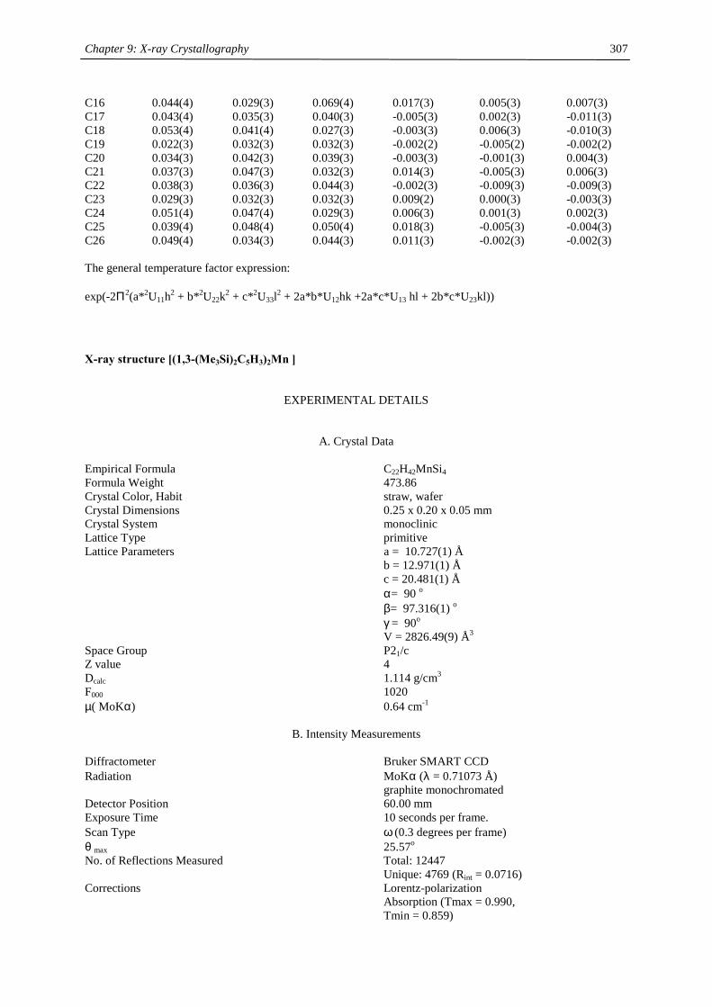
Chapter 9: X-ray Crystallography
307
C16 0.044(4) 0.029(3) 0.069(4) 0.017(3) 0.005(3) 0.007(3) C17 0.043(4) 0.035(3) 0.040(3) -0.005(3) 0.002(3) -0.011(3) C18 0.053(4) 0.041(4) 0.027(3) -0.003(3) 0.006(3) -0.010(3) C19 0.022(3) 0.032(3) 0.032(3) -0.002(2) -0.005(2) -0.002(2) C20 0.034(3) 0.042(3) 0.039(3) -0.003(3) -0.001(3) 0.004(3) C21 0.037(3) 0.047(3) 0.032(3) 0.014(3) -0.005(3) 0.006(3) C22 0.038(3) 0.036(3) 0.044(3) -0.002(3) -0.009(3) -0.009(3) C23 0.029(3) 0.032(3) 0.032(3) 0.009(2) 0.000(3) -0.003(3) C24 0.051(4) 0.047(4) 0.029(3) 0.006(3) 0.001(3) 0.002(3) C25 0.039(4) 0.048(4) 0.050(4) 0.018(3) -0.005(3) -0.004(3) C26 0.049(4) 0.034(3) 0.044(3) 0.011(3) -0.002(3) -0.002(3) The general temperature factor expression: exp(-2Π2(a*2U11h2 + b*2U22k2 + c*2U33l2 + 2a*b*U12hk +2a*c*U13 hl + 2b*c*U23kl))
X-ray structure [(1,3-(Me3Si)2C5H3)2Mn ]
EXPERIMENTAL DETAILS
A. Crystal Data Empirical Formula C22H42MnSi4 Formula Weight 473.86 Crystal Color, Habit straw, wafer Crystal Dimensions 0.25 x 0.20 x 0.05 mm Crystal System monoclinic Lattice Type primitive Lattice Parameters a = 10.727(1) Å b = 12.971(1) Å c = 20.481(1) Å α= 90 o β= 97.316(1) o γ = 90o V = 2826.49(9) Å3 Space Group P21/c Z value 4 Dcalc 1.114 g/cm3 F000 1020 µ( MoKα) 0.64 cm-1
B. Intensity Measurements Diffractometer Bruker SMART CCD Radiation MoKα (λ = 0.71073 Å) graphite monochromated Detector Position 60.00 mm Exposure Time 10 seconds per frame. Scan Type ω (0.3 degrees per frame) θ max 25.57o No. of Reflections Measured Total: 12447 Unique: 4769 (Rint = 0.0716) Corrections Lorentz-polarization Absorption (Tmax = 0.990, Tmin = 0.859)
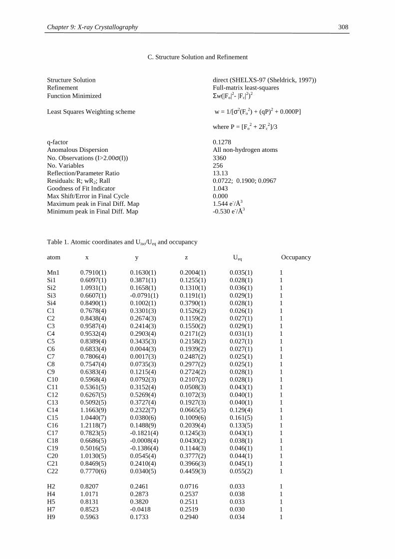
Chapter 9: X-ray Crystallography
308
C. Structure Solution and Refinement Structure Solution direct (SHELXS-97 (Sheldrick, 1997)) Refinement Full-matrix least-squares Function Minimized Σw(|Fo|2- |Fc|2)2 Least Squares Weighting scheme w = 1/[σ2(Fo
2) + (qP)2 + 0.000P] where P = [Fo
2 + 2Fc2]/3
q-factor 0.1278 Anomalous Dispersion All non-hydrogen atoms No. Observations (I>2.00σ(I)) 3360 No. Variables 256 Reflection/Parameter Ratio 13.13 Residuals: R; wR2; Rall 0.0722; 0.1900; 0.0967 Goodness of Fit Indicator 1.043 Max Shift/Error in Final Cycle 0.000 Maximum peak in Final Diff. Map 1.544 e-/Å3 Minimum peak in Final Diff. Map -0.530 e-/Å3 Table 1. Atomic coordinates and Uiso/Ueq and occupancy atom x y z Ueq Occupancy Mn1 0.7910(1) 0.1630(1) 0.2004(1) 0.035(1) 1 Si1 0.6097(1) 0.3871(1) 0.1255(1) 0.028(1) 1 Si2 1.0931(1) 0.1658(1) 0.1310(1) 0.036(1) 1 Si3 0.6607(1) -0.0791(1) 0.1191(1) 0.029(1) 1 Si4 0.8490(1) 0.1002(1) 0.3790(1) 0.028(1) 1 C1 0.7678(4) 0.3301(3) 0.1526(2) 0.026(1) 1 C2 0.8438(4) 0.2674(3) 0.1159(2) 0.027(1) 1 C3 0.9587(4) 0.2414(3) 0.1550(2) 0.029(1) 1 C4 0.9532(4) 0.2903(4) 0.2171(2) 0.031(1) 1 C5 0.8389(4) 0.3435(3) 0.2158(2) 0.027(1) 1 C6 0.6833(4) 0.0044(3) 0.1939(2) 0.027(1) 1 C7 0.7806(4) 0.0017(3) 0.2487(2) 0.025(1) 1 C8 0.7547(4) 0.0735(3) 0.2977(2) 0.025(1) 1 C9 0.6383(4) 0.1215(4) 0.2724(2) 0.028(1) 1 C10 0.5968(4) 0.0792(3) 0.2107(2) 0.028(1) 1 C11 0.5361(5) 0.3152(4) 0.0508(3) 0.043(1) 1 C12 0.6267(5) 0.5269(4) 0.1072(3) 0.040(1) 1 C13 0.5092(5) 0.3727(4) 0.1927(3) 0.040(1) 1 C14 1.1663(9) 0.2322(7) 0.0665(5) 0.129(4) 1 C15 1.0440(7) 0.0380(6) 0.1009(6) 0.161(5) 1 C16 1.2118(7) 0.1488(9) 0.2039(4) 0.133(5) 1 C17 0.7823(5) -0.1821(4) 0.1245(3) 0.043(1) 1 C18 0.6686(5) -0.0008(4) 0.0430(2) 0.038(1) 1 C19 0.5016(5) -0.1386(4) 0.1144(3) 0.046(1) 1 C20 1.0130(5) 0.0545(4) 0.3777(2) 0.044(1) 1 C21 0.8469(5) 0.2410(4) 0.3966(3) 0.045(1) 1 C22 0.7770(6) 0.0340(5) 0.4459(3) 0.055(2) 1 H2 0.8207 0.2461 0.0716 0.033 1 H4 1.0171 0.2873 0.2537 0.038 1 H5 0.8131 0.3820 0.2511 0.033 1 H7 0.8523 -0.0418 0.2519 0.030 1 H9 0.5963 0.1733 0.2940 0.034 1
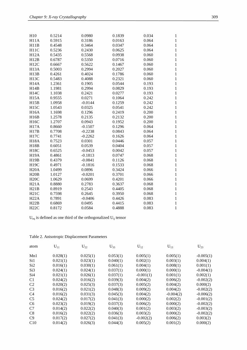
Chapter 9: X-ray Crystallography
309
H10 0.5214 0.0980 0.1839 0.034 1 H11A 0.5915 0.3186 0.0163 0.064 1 H11B 0.4548 0.3464 0.0347 0.064 1 H11C 0.5236 0.2430 0.0625 0.064 1 H12A 0.5435 0.5568 0.0938 0.060 1 H12B 0.6787 0.5350 0.0716 0.060 1 H12C 0.6667 0.5622 0.1467 0.060 1 H13A 0.5003 0.2994 0.2027 0.060 1 H13B 0.4261 0.4024 0.1786 0.060 1 H13C 0.5483 0.4088 0.2321 0.060 1 H14A 1.2361 0.1905 0.0544 0.193 1 H14B 1.1981 0.2994 0.0829 0.193 1 H14C 1.1038 0.2421 0.0277 0.193 1 H15A 0.9555 0.0271 0.1064 0.242 1 H15B 1.0958 -0.0144 0.1259 0.242 1 H15C 1.0543 0.0325 0.0541 0.242 1 H16A 1.1698 0.1296 0.2419 0.200 1 H16B 1.2578 0.2135 0.2132 0.200 1 H16C 1.2707 0.0943 0.1952 0.200 1 H17A 0.8660 -0.1507 0.1296 0.064 1 H17B 0.7708 -0.2238 0.0843 0.064 1 H17C 0.7741 -0.2262 0.1626 0.064 1 H18A 0.7523 0.0301 0.0446 0.057 1 H18B 0.6051 0.0539 0.0404 0.057 1 H18C 0.6525 -0.0453 0.0042 0.057 1 H19A 0.4861 -0.1813 0.0747 0.068 1 H19B 0.4379 -0.0841 0.1126 0.068 1 H19C 0.4971 -0.1816 0.1533 0.068 1 H20A 1.0499 0.0896 0.3424 0.066 1 H20B 1.0127 -0.0201 0.3701 0.066 1 H20C 1.0629 0.0699 0.4201 0.066 1 H21A 0.8880 0.2783 0.3637 0.068 1 H21B 0.8919 0.2543 0.4405 0.068 1 H21C 0.7598 0.2645 0.3950 0.068 1 H22A 0.7891 -0.0406 0.4426 0.083 1 H22B 0.6869 0.0495 0.4415 0.083 1 H22C 0.8172 0.0584 0.4888 0.083 1 Ueq is defined as one third of the orthogonalized Uij tensor Table 2. Anisotropic Displacement Parameters atom U11 U22 U33 U12 U13 U23 Mn1 0.028(1) 0.025(1) 0.053(1) 0.005(1) 0.005(1) -0.005(1) Si1 0.021(1) 0.023(1) 0.040(1) 0.002(1) 0.003(1) 0.004(1) Si2 0.016(1) 0.030(1) 0.061(1) 0.004(1) 0.008(1) 0.001(1) Si3 0.024(1) 0.024(1) 0.037(1) 0.000(1) 0.000(1) -0.004(1) Si4 0.021(1) 0.026(1) 0.037(1) -0.001(1) 0.001(1) 0.002(1) C1 0.024(2) 0.016(2) 0.039(3) 0.004(2) 0.006(2) -0.002(2) C2 0.020(2) 0.025(3) 0.037(3) 0.005(2) 0.004(2) 0.000(2) C3 0.016(2) 0.021(2) 0.048(3) 0.009(2) 0.004(2) -0.002(2) C4 0.016(2) 0.031(3) 0.045(3) 0.004(2) -0.004(2) -0.006(2) C5 0.024(2) 0.017(2) 0.041(3) 0.000(2) 0.002(2) -0.001(2) C6 0.023(2) 0.019(2) 0.037(3) 0.006(2) 0.000(2) -0.002(2) C7 0.014(2) 0.022(2) 0.040(3) 0.001(2) 0.003(2) -0.003(2) C8 0.016(2) 0.022(2) 0.036(3) 0.003(2) 0.000(2) -0.002(2) C9 0.017(2) 0.027(2) 0.041(3) -0.002(2) 0.006(2) 0.003(2) C10 0.014(2) 0.026(3) 0.044(3) 0.005(2) 0.001(2) 0.000(2)
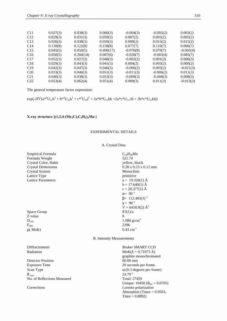
Chapter 9: X-ray Crystallography
310
C11 0.027(3) 0.038(3) 0.060(3) -0.004(3) -0.005(2) 0.003(2) C12 0.029(3) 0.031(3) 0.059(3) 0.007(2) 0.003(2) 0.005(2) C13 0.026(3) 0.038(3) 0.059(3) 0.009(2) 0.015(2) 0.015(2) C14 0.130(8) 0.122(8) 0.158(8) 0.077(7) 0.110(7) 0.066(7) C15 0.045(5) 0.050(5) 0.400(17) -0.076(8) 0.070(7) -0.002(4) C16 0.050(5) 0.260(14) 0.087(6) -0.020(7) -0.003(4) 0.085(7) C17 0.052(3) 0.027(3) 0.048(3) -0.002(2) 0.001(3) 0.006(3) C18 0.029(3) 0.043(3) 0.041(3) 0.004(2) 0.003(2) 0.000(2) C19 0.042(3) 0.047(3) 0.046(3) -0.006(2) 0.003(2) -0.021(3) C20 0.033(3) 0.046(3) 0.051(3) -0.011(3) -0.006(2) 0.011(3) C21 0.040(3) 0.038(3) 0.053(3) -0.009(3) -0.008(3) 0.008(3) C22 0.053(4) 0.062(4) 0.051(4) 0.009(3) 0.011(3) -0.013(3) The general temperature factor expression: exp(-2Π2(a*2U11h2 + b*2U22k2 + c*2U33l2 + 2a*b*U12hk +2a*c*U13 hl + 2b*c*U23kl))
X-ray structure [(1,2,4-(Me3C)3C5H2)2Mn ]
EXPERIMENTAL DETAILS
A. Crystal Data Empirical Formula C34H58Mn Formula Weight 521.74 Crystal Color, Habit yellow, block Crystal Dimensions 0.28 x 0.23 x 0.12 mm Crystal System Monoclinic Lattice Type primitive Lattice Parameters a = 19.326(1) Å b = 17.640(1) Å c = 20.377(1) Å α= 90 o β= 112.483(3) o γ = 90 o V = 6418.9(2) Å3 Space Group P2(1)/a Z value 8 Dcalc 1.080 g/cm3 F000 2296 µ( MoK) 0.43 cm-1
B. Intensity Measurements Diffractometer Bruker SMART CCD Radiation MoK(λ = 0.71073 Å) graphite monochromated Detector Position 60.00 mm Exposure Time 20 seconds per frame. Scan Type ω (0.3 degrees per frame) θ max 24.79 o No. of Reflections Measured Total: 27459 Unique: 10450 (Rint = 0.0705) Corrections Lorentz-polarization Absorption (Tmax = 0.9503, Tmin = 0.8892)
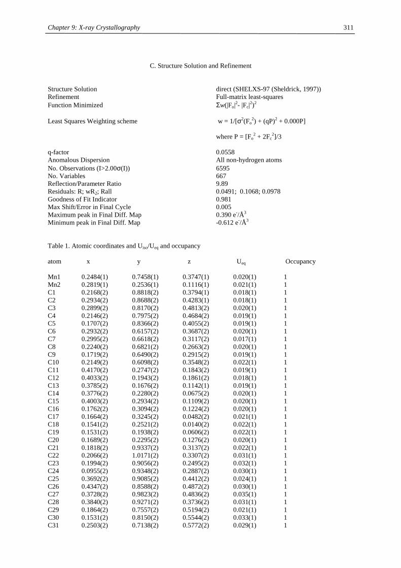
Chapter 9: X-ray Crystallography
311
C. Structure Solution and Refinement
Structure Solution direct (SHELXS-97 (Sheldrick, 1997)) Refinement Full-matrix least-squares Function Minimized Σw(|Fo|2- |Fc|2)2 Least Squares Weighting scheme w = 1/[σ2(Fo
2) + (qP)2 + 0.000P] where P = [Fo
2 + 2Fc2]/3
q-factor 0.0558 Anomalous Dispersion All non-hydrogen atoms No. Observations (I>2.00σ(I)) 6595 No. Variables 667 Reflection/Parameter Ratio 9.89 Residuals: R; wR2; Rall 0.0491; 0.1068; 0.0978 Goodness of Fit Indicator 0.981 Max Shift/Error in Final Cycle 0.005 Maximum peak in Final Diff. Map 0.390 e-/Å3 Minimum peak in Final Diff. Map -0.612 e-/Å3 Table 1. Atomic coordinates and Uiso/Ueq and occupancy atom x y z Ueq Occupancy Mn1 0.2484(1) 0.7458(1) 0.3747(1) 0.020(1) 1 Mn2 0.2819(1) 0.2536(1) 0.1116(1) 0.021(1) 1 C1 0.2168(2) 0.8818(2) 0.3794(1) 0.018(1) 1 C2 0.2934(2) 0.8688(2) 0.4283(1) 0.018(1) 1 C3 0.2899(2) 0.8170(2) 0.4813(2) 0.020(1) 1 C4 0.2146(2) 0.7975(2) 0.4684(2) 0.019(1) 1 C5 0.1707(2) 0.8366(2) 0.4055(2) 0.019(1) 1 C6 0.2932(2) 0.6157(2) 0.3687(2) 0.020(1) 1 C7 0.2995(2) 0.6618(2) 0.3117(2) 0.017(1) 1 C8 0.2240(2) 0.6821(2) 0.2663(2) 0.020(1) 1 C9 0.1719(2) 0.6490(2) 0.2915(2) 0.019(1) 1 C10 0.2149(2) 0.6098(2) 0.3548(2) 0.022(1) 1 C11 0.4170(2) 0.2747(2) 0.1843(2) 0.019(1) 1 C12 0.4033(2) 0.1943(2) 0.1861(2) 0.018(1) 1 C13 0.3785(2) 0.1676(2) 0.1142(1) 0.019(1) 1 C14 0.3776(2) 0.2280(2) 0.0675(2) 0.020(1) 1 C15 0.4003(2) 0.2934(2) 0.1109(2) 0.020(1) 1 C16 0.1762(2) 0.3094(2) 0.1224(2) 0.020(1) 1 C17 0.1664(2) 0.3245(2) 0.0482(2) 0.021(1) 1 C18 0.1541(2) 0.2521(2) 0.0140(2) 0.022(1) 1 C19 0.1531(2) 0.1938(2) 0.0606(2) 0.022(1) 1 C20 0.1689(2) 0.2295(2) 0.1276(2) 0.020(1) 1 C21 0.1818(2) 0.9337(2) 0.3137(2) 0.022(1) 1 C22 0.2066(2) 1.0171(2) 0.3307(2) 0.031(1) 1 C23 0.1994(2) 0.9056(2) 0.2495(2) 0.032(1) 1 C24 0.0955(2) 0.9348(2) 0.2887(2) 0.030(1) 1 C25 0.3692(2) 0.9085(2) 0.4412(2) 0.024(1) 1 C26 0.4347(2) 0.8588(2) 0.4872(2) 0.030(1) 1 C27 0.3728(2) 0.9823(2) 0.4836(2) 0.035(1) 1 C28 0.3840(2) 0.9271(2) 0.3736(2) 0.031(1) 1 C29 0.1864(2) 0.7557(2) 0.5194(2) 0.021(1) 1 C30 0.1531(2) 0.8150(2) 0.5544(2) 0.033(1) 1 C31 0.2503(2) 0.7138(2) 0.5772(2) 0.029(1) 1
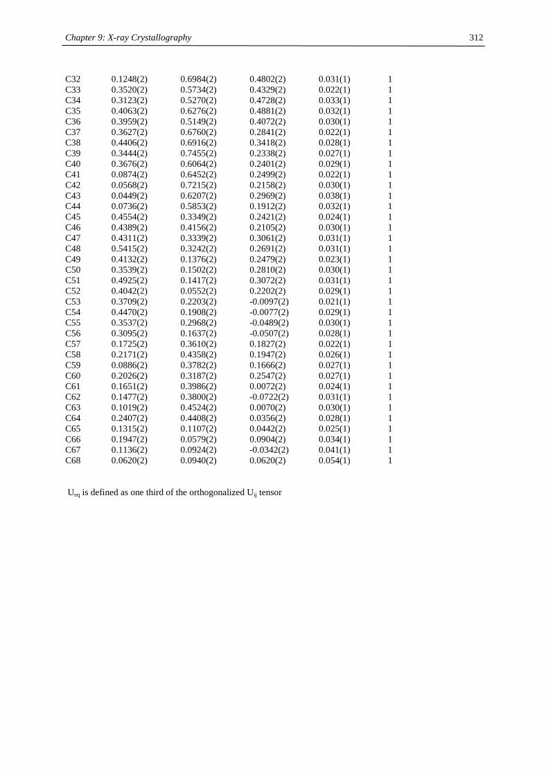
Chapter 9: X-ray Crystallography
312
C32 0.1248(2) 0.6984(2) 0.4802(2) 0.031(1) 1 C33 0.3520(2) 0.5734(2) 0.4329(2) 0.022(1) 1 C34 0.3123(2) 0.5270(2) 0.4728(2) 0.033(1) 1 C35 0.4063(2) 0.6276(2) 0.4881(2) 0.032(1) 1 C36 0.3959(2) 0.5149(2) 0.4072(2) 0.030(1) 1 C37 0.3627(2) 0.6760(2) 0.2841(2) 0.022(1) 1 C38 0.4406(2) 0.6916(2) 0.3418(2) 0.028(1) 1 C39 0.3444(2) 0.7455(2) 0.2338(2) 0.027(1) 1 C40 0.3676(2) 0.6064(2) 0.2401(2) 0.029(1) 1 C41 0.0874(2) 0.6452(2) 0.2499(2) 0.022(1) 1 C42 0.0568(2) 0.7215(2) 0.2158(2) 0.030(1) 1 C43 0.0449(2) 0.6207(2) 0.2969(2) 0.038(1) 1 C44 0.0736(2) 0.5853(2) 0.1912(2) 0.032(1) 1 C45 0.4554(2) 0.3349(2) 0.2421(2) 0.024(1) 1 C46 0.4389(2) 0.4156(2) 0.2105(2) 0.030(1) 1 C47 0.4311(2) 0.3339(2) 0.3061(2) 0.031(1) 1 C48 0.5415(2) 0.3242(2) 0.2691(2) 0.031(1) 1 C49 0.4132(2) 0.1376(2) 0.2479(2) 0.023(1) 1 C50 0.3539(2) 0.1502(2) 0.2810(2) 0.030(1) 1 C51 0.4925(2) 0.1417(2) 0.3072(2) 0.031(1) 1 C52 0.4042(2) 0.0552(2) 0.2202(2) 0.029(1) 1 C53 0.3709(2) 0.2203(2) -0.0097(2) 0.021(1) 1 C54 0.4470(2) 0.1908(2) -0.0077(2) 0.029(1) 1 C55 0.3537(2) 0.2968(2) -0.0489(2) 0.030(1) 1 C56 0.3095(2) 0.1637(2) -0.0507(2) 0.028(1) 1 C57 0.1725(2) 0.3610(2) 0.1827(2) 0.022(1) 1 C58 0.2171(2) 0.4358(2) 0.1947(2) 0.026(1) 1 C59 0.0886(2) 0.3782(2) 0.1666(2) 0.027(1) 1 C60 0.2026(2) 0.3187(2) 0.2547(2) 0.027(1) 1 C61 0.1651(2) 0.3986(2) 0.0072(2) 0.024(1) 1 C62 0.1477(2) 0.3800(2) -0.0722(2) 0.031(1) 1 C63 0.1019(2) 0.4524(2) 0.0070(2) 0.030(1) 1 C64 0.2407(2) 0.4408(2) 0.0356(2) 0.028(1) 1 C65 0.1315(2) 0.1107(2) 0.0442(2) 0.025(1) 1 C66 0.1947(2) 0.0579(2) 0.0904(2) 0.034(1) 1 C67 0.1136(2) 0.0924(2) -0.0342(2) 0.041(1) 1 C68 0.0620(2) 0.0940(2) 0.0620(2) 0.054(1) 1 Ueq is defined as one third of the orthogonalized Uij tensor
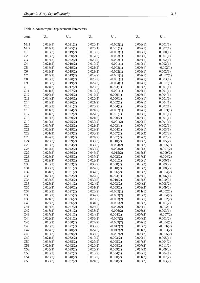
Chapter 9: X-ray Crystallography
313
Table 2. Anisotropic Displacement Parameters atom U11 U22 U33 U12 U13 U23 Mn1 0.019(1) 0.021(1) 0.020(1) -0.002(1) 0.008(1) 0.001(1) Mn2 0.014(1) 0.025(1) 0.025(1) 0.001(1) 0.009(1) 0.002(1) C1 0.016(2) 0.019(2) 0.016(2) -0.003(1) 0.005(1) 0.000(1) C2 0.018(2) 0.020(2) 0.017(2) -0.003(1) 0.008(1) 0.001(1) C3 0.016(2) 0.022(2) 0.020(2) -0.002(1) 0.005(1) 0.002(1) C4 0.021(2) 0.019(2) 0.019(2) -0.001(1) 0.010(1) 0.002(1) C5 0.016(2) 0.019(2) 0.021(2) -0.003(1) 0.006(1) -0.002(1) C6 0.019(2) 0.019(2) 0.023(2) -0.002(1) 0.009(1) 0.002(1) C7 0.014(2) 0.019(2) 0.019(2) -0.005(1) 0.007(1) -0.002(1) C8 0.019(2) 0.020(2) 0.020(2) -0.001(1) 0.007(1) 0.003(1) C9 0.015(2) 0.019(2) 0.022(2) -0.004(1) 0.007(1) -0.001(1) C10 0.024(2) 0.017(2) 0.029(2) 0.003(1) 0.015(2) 0.001(1) C11 0.011(2) 0.027(2) 0.019(2) -0.001(1) 0.005(1) 0.001(1) C12 0.009(2) 0.026(2) 0.017(2) 0.000(1) 0.003(1) 0.004(1) C13 0.014(2) 0.020(2) 0.020(2) 0.000(1) 0.004(1) 0.001(1) C14 0.013(2) 0.026(2) 0.021(2) 0.002(1) 0.007(1) 0.004(1) C15 0.013(2) 0.021(2) 0.026(2) 0.004(1) 0.009(1) 0.002(1) C16 0.011(2) 0.024(2) 0.024(2) -0.002(1) 0.007(1) -0.003(1) C17 0.014(2) 0.022(2) 0.027(2) 0.001(1) 0.008(1) 0.001(1) C18 0.015(2) 0.030(2) 0.021(2) 0.000(2) 0.008(1) 0.001(1) C19 0.010(2) 0.025(2) 0.030(2) -0.001(2) 0.009(1) 0.001(1) C20 0.017(2) 0.022(2) 0.021(2) 0.003(1) 0.007(1) 0.002(1) C21 0.023(2) 0.019(2) 0.023(2) 0.004(1) 0.008(1) 0.003(1) C22 0.031(2) 0.023(2) 0.038(2) 0.007(2) 0.013(2) 0.002(2) C23 0.042(2) 0.030(2) 0.024(2) 0.007(2) 0.012(2) 0.007(2) C24 0.026(2) 0.029(2) 0.033(2) 0.010(2) 0.008(2) 0.005(2) C25 0.018(2) 0.024(2) 0.032(2) -0.004(2) 0.012(2) -0.005(1) C26 0.017(2) 0.042(2) 0.030(2) -0.003(2) 0.010(2) -0.007(2) C27 0.025(2) 0.036(2) 0.046(2) -0.015(2) 0.015(2) -0.009(2) C28 0.026(2) 0.035(2) 0.037(2) 0.002(2) 0.017(2) -0.004(2) C29 0.019(2) 0.023(2) 0.022(2) 0.001(2) 0.010(1) 0.000(1) C30 0.040(2) 0.033(2) 0.035(2) 0.008(2) 0.025(2) 0.009(2) C31 0.033(2) 0.031(2) 0.027(2) 0.010(2) 0.015(2) 0.008(2) C32 0.031(2) 0.031(2) 0.037(2) 0.006(2) 0.019(2) -0.004(2) C33 0.020(2) 0.022(2) 0.022(2) 0.003(1) 0.006(1) 0.006(1) C34 0.033(2) 0.033(2) 0.032(2) 0.010(2) 0.013(2) 0.010(2) C35 0.026(2) 0.041(2) 0.024(2) 0.003(2) 0.004(2) 0.008(2) C36 0.028(2) 0.030(2) 0.031(2) 0.005(2) 0.009(2) 0.009(2) C37 0.016(2) 0.027(2) 0.025(2) -0.003(1) 0.011(1) -0.002(1) C38 0.018(2) 0.035(2) 0.032(2) -0.003(2) 0.010(2) -0.004(2) C39 0.021(2) 0.036(2) 0.025(2) -0.003(2) 0.010(1) -0.002(2) C40 0.025(2) 0.036(2) 0.031(2) -0.005(2) 0.018(2) 0.001(2) C41 0.013(2) 0.027(2) 0.025(2) -0.003(2) 0.007(1) -0.002(1) C42 0.018(2) 0.031(2) 0.038(2) -0.006(2) 0.006(2) 0.003(1) C43 0.017(2) 0.061(3) 0.034(2) 0.004(2) 0.007(2) -0.007(2) C44 0.022(2) 0.031(2) 0.036(2) -0.007(2) 0.004(2) 0.001(2) C45 0.016(2) 0.030(2) 0.024(2) -0.009(2) 0.007(1) -0.004(1) C46 0.025(2) 0.027(2) 0.038(2) -0.012(2) 0.013(2) -0.006(2) C47 0.027(2) 0.040(2) 0.027(2) -0.012(2) 0.011(2) -0.003(2) C48 0.018(2) 0.039(2) 0.035(2) -0.007(2) 0.008(2) -0.005(2) C49 0.021(2) 0.032(2) 0.019(2) 0.003(2) 0.009(1) 0.005(1) C50 0.033(2) 0.035(2) 0.027(2) 0.005(2) 0.017(2) 0.004(2) C51 0.028(2) 0.042(2) 0.020(2) 0.008(2) 0.007(2) 0.011(2) C52 0.035(2) 0.029(2) 0.025(2) 0.009(2) 0.014(2) 0.009(2) C53 0.019(2) 0.027(2) 0.018(2) 0.004(1) 0.009(1) 0.004(1) C54 0.023(2) 0.048(2) 0.019(2) 0.000(2) 0.011(2) 0.007(2) C55 0.030(2) 0.037(2) 0.024(2) 0.008(2) 0.013(2) 0.003(2)
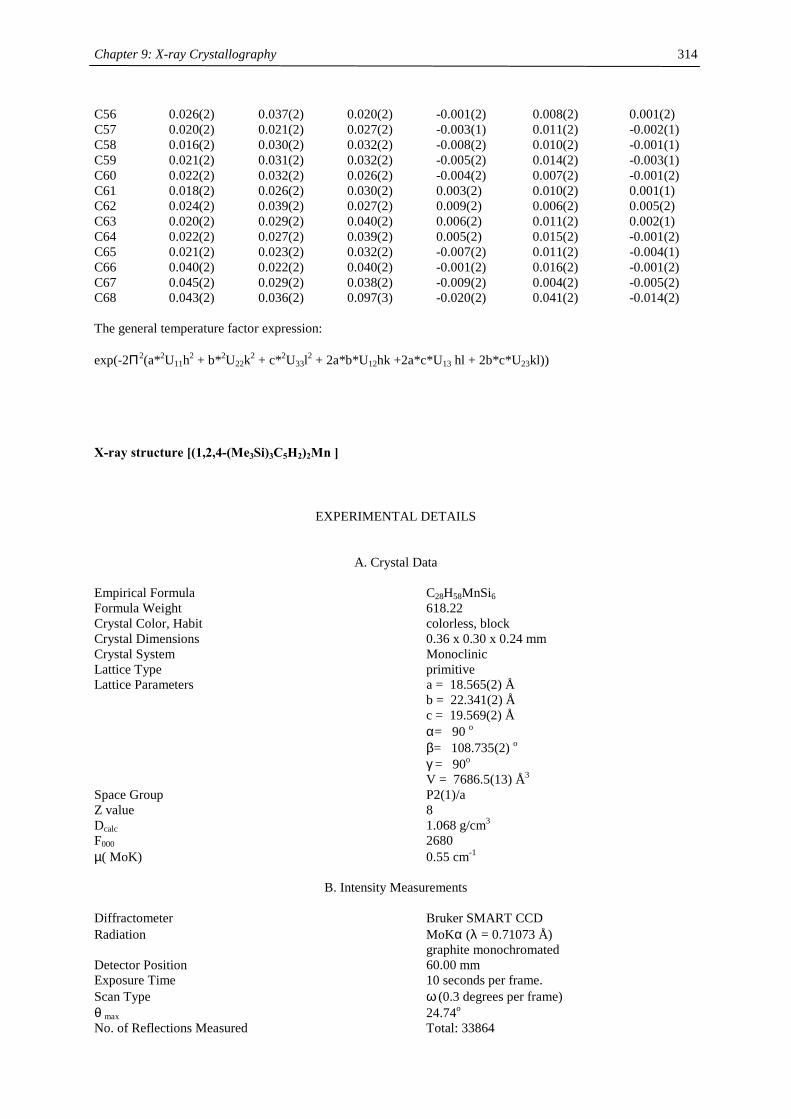
Chapter 9: X-ray Crystallography
314
C56 0.026(2) 0.037(2) 0.020(2) -0.001(2) 0.008(2) 0.001(2) C57 0.020(2) 0.021(2) 0.027(2) -0.003(1) 0.011(2) -0.002(1) C58 0.016(2) 0.030(2) 0.032(2) -0.008(2) 0.010(2) -0.001(1) C59 0.021(2) 0.031(2) 0.032(2) -0.005(2) 0.014(2) -0.003(1) C60 0.022(2) 0.032(2) 0.026(2) -0.004(2) 0.007(2) -0.001(2) C61 0.018(2) 0.026(2) 0.030(2) 0.003(2) 0.010(2) 0.001(1) C62 0.024(2) 0.039(2) 0.027(2) 0.009(2) 0.006(2) 0.005(2) C63 0.020(2) 0.029(2) 0.040(2) 0.006(2) 0.011(2) 0.002(1) C64 0.022(2) 0.027(2) 0.039(2) 0.005(2) 0.015(2) -0.001(2) C65 0.021(2) 0.023(2) 0.032(2) -0.007(2) 0.011(2) -0.004(1) C66 0.040(2) 0.022(2) 0.040(2) -0.001(2) 0.016(2) -0.001(2) C67 0.045(2) 0.029(2) 0.038(2) -0.009(2) 0.004(2) -0.005(2) C68 0.043(2) 0.036(2) 0.097(3) -0.020(2) 0.041(2) -0.014(2) The general temperature factor expression: exp(-2Π2(a*2U11h2 + b*2U22k2 + c*2U33l2 + 2a*b*U12hk +2a*c*U13 hl + 2b*c*U23kl))
X-ray structure [(1,2,4-(Me3Si)3C5H2)2Mn ]
EXPERIMENTAL DETAILS
A. Crystal Data Empirical Formula C28H58MnSi6 Formula Weight 618.22 Crystal Color, Habit colorless, block Crystal Dimensions 0.36 x 0.30 x 0.24 mm Crystal System Monoclinic Lattice Type primitive Lattice Parameters a = 18.565(2) Å b = 22.341(2) Å c = 19.569(2) Å α= 90 o β= 108.735(2) o γ = 90o V = 7686.5(13) Å3 Space Group P2(1)/a Z value 8 Dcalc 1.068 g/cm3 F000 2680 µ( MoK) 0.55 cm-1
B. Intensity Measurements Diffractometer Bruker SMART CCD Radiation MoKα (λ = 0.71073 Å) graphite monochromated Detector Position 60.00 mm Exposure Time 10 seconds per frame. Scan Type ω (0.3 degrees per frame) θ max 24.74o No. of Reflections Measured Total: 33864
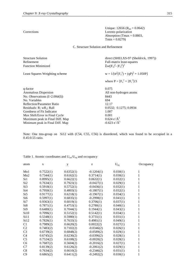
Chapter 9: X-ray Crystallography
315
Unique: 12656 (Rint = 0.0642) Corrections Lorentz-polarization Absorption (Tmax = 0.8803, Tmin = 0.8279)
C. Structure Solution and Refinement Structure Solution direct (SHELXS-97 (Sheldrick, 1997)) Refinement Full-matrix least-squares Function Minimized Σw(|Fo|2- |Fc|2)2 Least Squares Weighting scheme w = 1/[σ2(Fo
2) + (qP)2 + 1.050P] where P = [Fo
2 + 2Fc2]/3
q-factor 0.075 Anomalous Dispersion All non-hydrogen atoms No. Observations (I>2.00σ(I)) 8443 No. Variables 694 Reflection/Parameter Ratio 12.17 Residuals: R; wR2; Rall 0.0532; 0.1275; 0.0934 Goodness of Fit Indicator 1.007 Max Shift/Error in Final Cycle 0.001 Maximum peak in Final Diff. Map 0.624 e-/Å3 Minimum peak in Final Diff. Map -0.623 e-/Å3
Note: One tms-group on Si12 with (C54, C55, C56) is disordered, which was found to be occupied in a 0.45:0.55 ratio. Table 1. Atomic coordinates and Uiso/Ueq and occupancy atom x y z Ueq Occupancy Mn1 0.7522(1) 0.6352(1) -0.1204(1) 0.030(1) 1 Mn2 0.7344(1) 0.6162(1) 0.3714(1) 0.030(1) 1 Si1 0.8995(1) 0.6622(1) 0.0632(1) 0.032(1) 1 Si2 0.7634(1) 0.7923(1) -0.0427(1) 0.029(1) 1 Si3 0.5918(1) 0.5752(1) -0.0436(1) 0.032(1) 1 Si4 0.7950(1) 0.4803(1) -0.1807(1) 0.032(1) 1 Si5 0.9177(1) 0.6218(1) -0.1967(1) 0.034(1) 1 Si6 0.5997(1) 0.6835(1) -0.2998(1) 0.041(1) 1 Si7 0.9363(1) 0.6019(1) 0.3706(1) 0.037(1) 1 Si8 0.7871(1) 0.4753(1) 0.2780(1) 0.040(1) 1 Si9 0.6480(1) 0.7044(1) 0.1944(1) 0.043(1) 1 Si10 0.7096(1) 0.5152(1) 0.5142(1) 0.034(1) 1 Si11 0.5348(1) 0.5980(1) 0.3731(1) 0.031(1) 1 Si12 0.7826(1) 0.7633(1) 0.4981(1) 0.049(1) 1 C1 0.7989(2) 0.6620(2) 0.0022(2) 0.027(1) 1 C2 0.7483(2) 0.7102(2) -0.0346(2) 0.026(1) 1 C3 0.6739(2) 0.6848(2) -0.0589(2) 0.029(1) 1 C4 0.6745(2) 0.6236(2) -0.0396(2) 0.028(1) 1 C5 0.7524(2) 0.6108(2) -0.0020(2) 0.028(1) 1 C6 0.7687(2) 0.5604(2) -0.2016(2) 0.027(1) 1 C7 0.8139(2) 0.6126(2) -0.2061(2) 0.029(1) 1 C8 0.7634(2) 0.6610(2) -0.2346(2) 0.031(1) 1 C9 0.6865(2) 0.6411(2) -0.2492(2) 0.030(1) 1
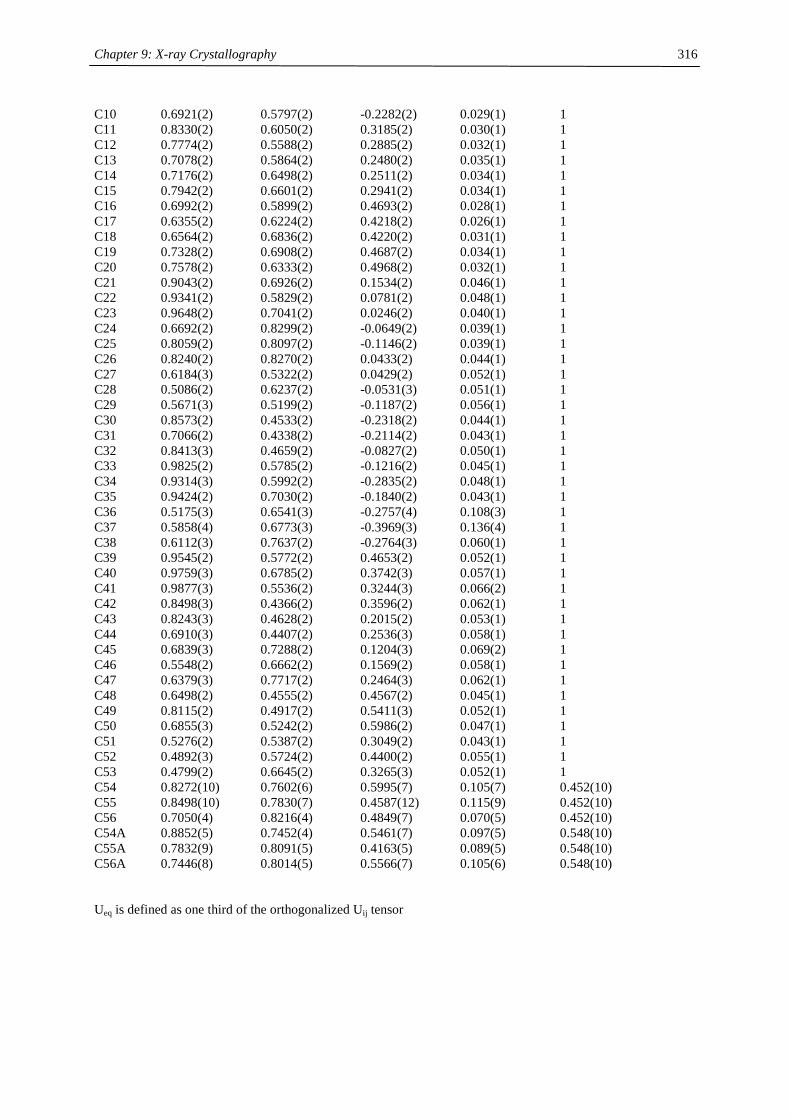
Chapter 9: X-ray Crystallography
316
C10 0.6921(2) 0.5797(2) -0.2282(2) 0.029(1) 1 C11 0.8330(2) 0.6050(2) 0.3185(2) 0.030(1) 1 C12 0.7774(2) 0.5588(2) 0.2885(2) 0.032(1) 1 C13 0.7078(2) 0.5864(2) 0.2480(2) 0.035(1) 1 C14 0.7176(2) 0.6498(2) 0.2511(2) 0.034(1) 1 C15 0.7942(2) 0.6601(2) 0.2941(2) 0.034(1) 1 C16 0.6992(2) 0.5899(2) 0.4693(2) 0.028(1) 1 C17 0.6355(2) 0.6224(2) 0.4218(2) 0.026(1) 1 C18 0.6564(2) 0.6836(2) 0.4220(2) 0.031(1) 1 C19 0.7328(2) 0.6908(2) 0.4687(2) 0.034(1) 1 C20 0.7578(2) 0.6333(2) 0.4968(2) 0.032(1) 1 C21 0.9043(2) 0.6926(2) 0.1534(2) 0.046(1) 1 C22 0.9341(2) 0.5829(2) 0.0781(2) 0.048(1) 1 C23 0.9648(2) 0.7041(2) 0.0246(2) 0.040(1) 1 C24 0.6692(2) 0.8299(2) -0.0649(2) 0.039(1) 1 C25 0.8059(2) 0.8097(2) -0.1146(2) 0.039(1) 1 C26 0.8240(2) 0.8270(2) 0.0433(2) 0.044(1) 1 C27 0.6184(3) 0.5322(2) 0.0429(2) 0.052(1) 1 C28 0.5086(2) 0.6237(2) -0.0531(3) 0.051(1) 1 C29 0.5671(3) 0.5199(2) -0.1187(2) 0.056(1) 1 C30 0.8573(2) 0.4533(2) -0.2318(2) 0.044(1) 1 C31 0.7066(2) 0.4338(2) -0.2114(2) 0.043(1) 1 C32 0.8413(3) 0.4659(2) -0.0827(2) 0.050(1) 1 C33 0.9825(2) 0.5785(2) -0.1216(2) 0.045(1) 1 C34 0.9314(3) 0.5992(2) -0.2835(2) 0.048(1) 1 C35 0.9424(2) 0.7030(2) -0.1840(2) 0.043(1) 1 C36 0.5175(3) 0.6541(3) -0.2757(4) 0.108(3) 1 C37 0.5858(4) 0.6773(3) -0.3969(3) 0.136(4) 1 C38 0.6112(3) 0.7637(2) -0.2764(3) 0.060(1) 1 C39 0.9545(2) 0.5772(2) 0.4653(2) 0.052(1) 1 C40 0.9759(3) 0.6785(2) 0.3742(3) 0.057(1) 1 C41 0.9877(3) 0.5536(2) 0.3244(3) 0.066(2) 1 C42 0.8498(3) 0.4366(2) 0.3596(2) 0.062(1) 1 C43 0.8243(3) 0.4628(2) 0.2015(2) 0.053(1) 1 C44 0.6910(3) 0.4407(2) 0.2536(3) 0.058(1) 1 C45 0.6839(3) 0.7288(2) 0.1204(3) 0.069(2) 1 C46 0.5548(2) 0.6662(2) 0.1569(2) 0.058(1) 1 C47 0.6379(3) 0.7717(2) 0.2464(3) 0.062(1) 1 C48 0.6498(2) 0.4555(2) 0.4567(2) 0.045(1) 1 C49 0.8115(2) 0.4917(2) 0.5411(3) 0.052(1) 1 C50 0.6855(3) 0.5242(2) 0.5986(2) 0.047(1) 1 C51 0.5276(2) 0.5387(2) 0.3049(2) 0.043(1) 1 C52 0.4892(3) 0.5724(2) 0.4400(2) 0.055(1) 1 C53 0.4799(2) 0.6645(2) 0.3265(3) 0.052(1) 1 C54 0.8272(10) 0.7602(6) 0.5995(7) 0.105(7) 0.452(10) C55 0.8498(10) 0.7830(7) 0.4587(12) 0.115(9) 0.452(10) C56 0.7050(4) 0.8216(4) 0.4849(7) 0.070(5) 0.452(10) C54A 0.8852(5) 0.7452(4) 0.5461(7) 0.097(5) 0.548(10) C55A 0.7832(9) 0.8091(5) 0.4163(5) 0.089(5) 0.548(10) C56A 0.7446(8) 0.8014(5) 0.5566(7) 0.105(6) 0.548(10) Ueq is defined as one third of the orthogonalized Uij tensor
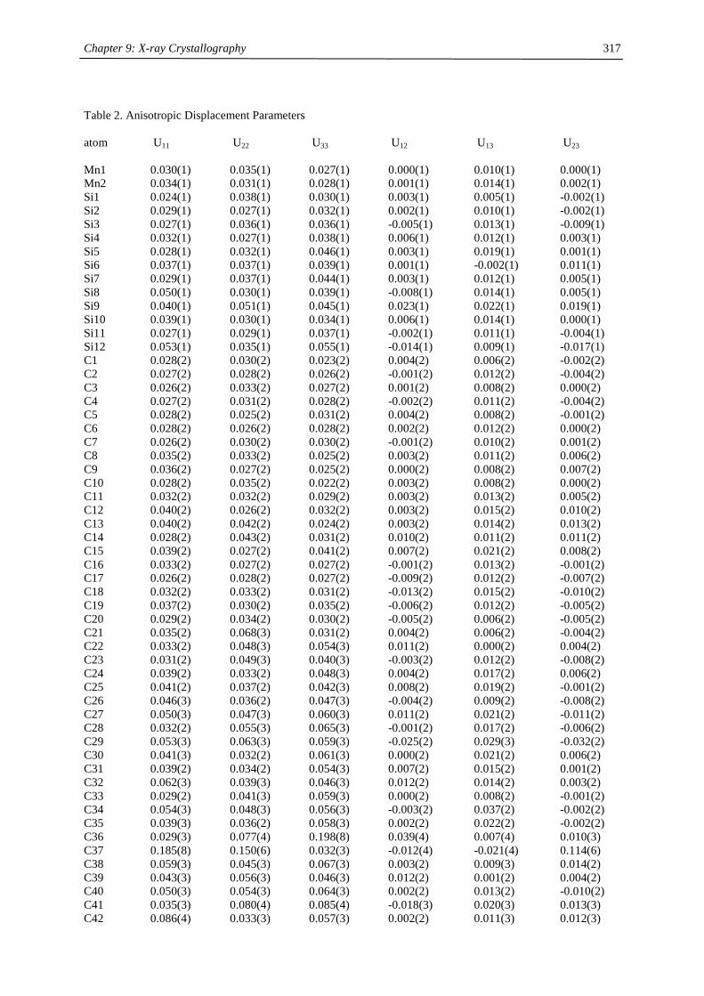
Chapter 9: X-ray Crystallography
317
Table 2. Anisotropic Displacement Parameters atom U11 U22 U33 U12 U13 U23 Mn1 0.030(1) 0.035(1) 0.027(1) 0.000(1) 0.010(1) 0.000(1) Mn2 0.034(1) 0.031(1) 0.028(1) 0.001(1) 0.014(1) 0.002(1) Si1 0.024(1) 0.038(1) 0.030(1) 0.003(1) 0.005(1) -0.002(1) Si2 0.029(1) 0.027(1) 0.032(1) 0.002(1) 0.010(1) -0.002(1) Si3 0.027(1) 0.036(1) 0.036(1) -0.005(1) 0.013(1) -0.009(1) Si4 0.032(1) 0.027(1) 0.038(1) 0.006(1) 0.012(1) 0.003(1) Si5 0.028(1) 0.032(1) 0.046(1) 0.003(1) 0.019(1) 0.001(1) Si6 0.037(1) 0.037(1) 0.039(1) 0.001(1) -0.002(1) 0.011(1) Si7 0.029(1) 0.037(1) 0.044(1) 0.003(1) 0.012(1) 0.005(1) Si8 0.050(1) 0.030(1) 0.039(1) -0.008(1) 0.014(1) 0.005(1) Si9 0.040(1) 0.051(1) 0.045(1) 0.023(1) 0.022(1) 0.019(1) Si10 0.039(1) 0.030(1) 0.034(1) 0.006(1) 0.014(1) 0.000(1) Si11 0.027(1) 0.029(1) 0.037(1) -0.002(1) 0.011(1) -0.004(1) Si12 0.053(1) 0.035(1) 0.055(1) -0.014(1) 0.009(1) -0.017(1) C1 0.028(2) 0.030(2) 0.023(2) 0.004(2) 0.006(2) -0.002(2) C2 0.027(2) 0.028(2) 0.026(2) -0.001(2) 0.012(2) -0.004(2) C3 0.026(2) 0.033(2) 0.027(2) 0.001(2) 0.008(2) 0.000(2) C4 0.027(2) 0.031(2) 0.028(2) -0.002(2) 0.011(2) -0.004(2) C5 0.028(2) 0.025(2) 0.031(2) 0.004(2) 0.008(2) -0.001(2) C6 0.028(2) 0.026(2) 0.028(2) 0.002(2) 0.012(2) 0.000(2) C7 0.026(2) 0.030(2) 0.030(2) -0.001(2) 0.010(2) 0.001(2) C8 0.035(2) 0.033(2) 0.025(2) 0.003(2) 0.011(2) 0.006(2) C9 0.036(2) 0.027(2) 0.025(2) 0.000(2) 0.008(2) 0.007(2) C10 0.028(2) 0.035(2) 0.022(2) 0.003(2) 0.008(2) 0.000(2) C11 0.032(2) 0.032(2) 0.029(2) 0.003(2) 0.013(2) 0.005(2) C12 0.040(2) 0.026(2) 0.032(2) 0.003(2) 0.015(2) 0.010(2) C13 0.040(2) 0.042(2) 0.024(2) 0.003(2) 0.014(2) 0.013(2) C14 0.028(2) 0.043(2) 0.031(2) 0.010(2) 0.011(2) 0.011(2) C15 0.039(2) 0.027(2) 0.041(2) 0.007(2) 0.021(2) 0.008(2) C16 0.033(2) 0.027(2) 0.027(2) -0.001(2) 0.013(2) -0.001(2) C17 0.026(2) 0.028(2) 0.027(2) -0.009(2) 0.012(2) -0.007(2) C18 0.032(2) 0.033(2) 0.031(2) -0.013(2) 0.015(2) -0.010(2) C19 0.037(2) 0.030(2) 0.035(2) -0.006(2) 0.012(2) -0.005(2) C20 0.029(2) 0.034(2) 0.030(2) -0.005(2) 0.006(2) -0.005(2) C21 0.035(2) 0.068(3) 0.031(2) 0.004(2) 0.006(2) -0.004(2) C22 0.033(2) 0.048(3) 0.054(3) 0.011(2) 0.000(2) 0.004(2) C23 0.031(2) 0.049(3) 0.040(3) -0.003(2) 0.012(2) -0.008(2) C24 0.039(2) 0.033(2) 0.048(3) 0.004(2) 0.017(2) 0.006(2) C25 0.041(2) 0.037(2) 0.042(3) 0.008(2) 0.019(2) -0.001(2) C26 0.046(3) 0.036(2) 0.047(3) -0.004(2) 0.009(2) -0.008(2) C27 0.050(3) 0.047(3) 0.060(3) 0.011(2) 0.021(2) -0.011(2) C28 0.032(2) 0.055(3) 0.065(3) -0.001(2) 0.017(2) -0.006(2) C29 0.053(3) 0.063(3) 0.059(3) -0.025(2) 0.029(3) -0.032(2) C30 0.041(3) 0.032(2) 0.061(3) 0.000(2) 0.021(2) 0.006(2) C31 0.039(2) 0.034(2) 0.054(3) 0.007(2) 0.015(2) 0.001(2) C32 0.062(3) 0.039(3) 0.046(3) 0.012(2) 0.014(2) 0.003(2) C33 0.029(2) 0.041(3) 0.059(3) 0.000(2) 0.008(2) -0.001(2) C34 0.054(3) 0.048(3) 0.056(3) -0.003(2) 0.037(2) -0.002(2) C35 0.039(3) 0.036(2) 0.058(3) 0.002(2) 0.022(2) -0.002(2) C36 0.029(3) 0.077(4) 0.198(8) 0.039(4) 0.007(4) 0.010(3) C37 0.185(8) 0.150(6) 0.032(3) -0.012(4) -0.021(4) 0.114(6) C38 0.059(3) 0.045(3) 0.067(3) 0.003(2) 0.009(3) 0.014(2) C39 0.043(3) 0.056(3) 0.046(3) 0.012(2) 0.001(2) 0.004(2) C40 0.050(3) 0.054(3) 0.064(3) 0.002(2) 0.013(2) -0.010(2) C41 0.035(3) 0.080(4) 0.085(4) -0.018(3) 0.020(3) 0.013(3) C42 0.086(4) 0.033(3) 0.057(3) 0.002(2) 0.011(3) 0.012(3)
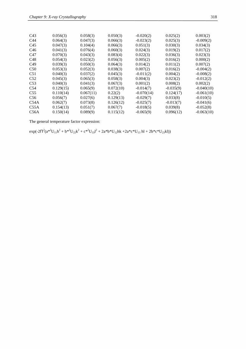
Chapter 9: X-ray Crystallography
318
C43 0.056(3) 0.058(3) 0.050(3) -0.020(2) 0.025(2) 0.003(2) C44 0.064(3) 0.047(3) 0.066(3) -0.023(2) 0.025(3) -0.009(2) C45 0.047(3) 0.104(4) 0.066(3) 0.051(3) 0.030(3) 0.034(3) C46 0.041(3) 0.076(4) 0.060(3) 0.024(3) 0.019(2) 0.017(2) C47 0.070(3) 0.043(3) 0.083(4) 0.022(3) 0.036(3) 0.023(3) C48 0.054(3) 0.023(2) 0.056(3) 0.005(2) 0.016(2) 0.000(2) C49 0.039(3) 0.050(3) 0.064(3) 0.014(2) 0.011(2) 0.007(2) C50 0.053(3) 0.052(3) 0.038(3) 0.007(2) 0.016(2) -0.004(2) C51 0.040(3) 0.037(2) 0.045(3) -0.011(2) 0.004(2) -0.008(2) C52 0.045(3) 0.065(3) 0.058(3) 0.004(3) 0.023(2) -0.012(2) C53 0.040(3) 0.041(3) 0.067(3) 0.001(2) 0.008(2) 0.002(2) C54 0.129(15) 0.065(9) 0.072(10) -0.014(7) -0.035(9) -0.040(10) C55 0.110(14) 0.067(11) 0.22(2) -0.070(14) 0.124(17) -0.061(10) C56 0.056(7) 0.027(6) 0.129(13) -0.029(7) 0.033(8) -0.010(5) C54A 0.062(7) 0.073(8) 0.126(12) -0.025(7) -0.013(7) -0.041(6) C55A 0.154(13) 0.051(7) 0.067(7) -0.018(5) 0.039(8) -0.052(8) C56A 0.150(14) 0.089(9) 0.115(12) -0.065(9) 0.096(12) -0.063(10) The general temperature factor expression: exp(-2Π2(a*2U11h2 + b*2U22k2 + c*2U33l2 + 2a*b*U12hk +2a*c*U13 hl + 2b*c*U23kl))
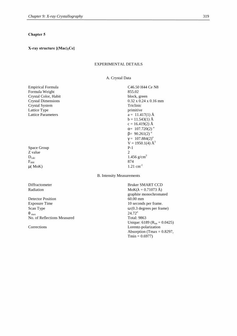
Chapter 9: X-ray Crystallography
319
Chapter 5
X-ray structure [(Mac)2Ce]
EXPERIMENTAL DETAILS
A. Crystal Data Empirical Formula C46.50 H44 Ce N8 Formula Weight 855.02 Crystal Color, Habit block, green Crystal Dimensions 0.32 x 0.24 x 0.16 mm Crystal System Triclinic Lattice Type primitive Lattice Parameters a = 11.417(1) Å b = 11.543(1) Å c = 16.419(2) Å α= 107.720(2) o β= 90.261(2) o γ = 107.884(2)o V = 1950.1(4) Å3 Space Group P-1 Z value 2 Dcalc 1.456 g/cm3 F000 874 µ( MoK) 1.21 cm-1
B. Intensity Measurements Diffractometer Bruker SMART CCD Radiation MoK(λ = 0.71073 Å) graphite monochromated Detector Position 60.00 mm Exposure Time 10 seconds per frame. Scan Type ω (0.3 degrees per frame) θ max 24.72o No. of Reflections Measured Total: 9863 Unique: 6189 (Rint = 0.0425) Corrections Lorentz-polarization Absorption (Tmax = 0.8297, Tmin = 0.6977)
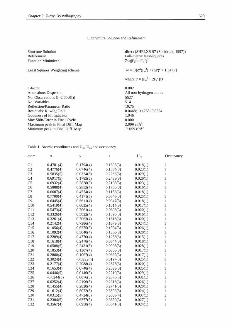
Chapter 9: X-ray Crystallography
320
C. Structure Solution and Refinement
Structure Solution direct (SHELXS-97 (Sheldrick, 1997)) Refinement Full-matrix least-squares Function Minimized Σw(|Fo|2- |Fc|2)2 Least Squares Weighting scheme w = 1/[σ2(Fo
2) + (qP)2 + 1.347P] where P = [Fo
2 + 2Fc2]/3
q-factor 0.082 Anomalous Dispersion All non-hydrogen atoms No. Observations (I>2.00σ(I)) 5527 No. Variables 514 Reflection/Parameter Ratio 10.75 Residuals: R; wR2; Rall 0.0468; 0.1238; 0.0524 Goodness of Fit Indicator 1.046 Max Shift/Error in Final Cycle 0.000 Maximum peak in Final Diff. Map 2.009 e-/Å3 Minimum peak in Final Diff. Map -2.059 e-/Å3 Table 1. Atomic coordinates and Uiso/Ueq and occupancy atom x y z Ueq Occupancy C1 0.4781(4) 0.1794(4) 0.1603(3) 0.018(1) 1 C2 0.4776(4) 0.0746(4) 0.1864(3) 0.023(1) 1 C3 0.5835(5) 0.0724(5) 0.2263(3) 0.029(1) 1 C4 0.6917(5) 0.1783(5) 0.2439(3) 0.029(1) 1 C5 0.6932(4) 0.2828(5) 0.2198(3) 0.023(1) 1 C6 0.5888(4) 0.2852(4) 0.1766(3) 0.016(1) 1 C7 0.6607(4) 0.4574(4) 0.1158(3) 0.018(1) 1 C8 0.7709(4) 0.4171(5) 0.0843(3) 0.025(1) 1 C9 0.6445(4) 0.5611(4) 0.0947(3) 0.018(1) 1 C10 0.5419(4) 0.6025(4) 0.1014(3) 0.017(1) 1 C11 0.5473(4) 0.7061(4) 0.0608(3) 0.020(1) 1 C12 0.3326(4) 0.5822(4) 0.1393(3) 0.016(1) 1 C13 0.3261(4) 0.7063(4) 0.1616(3) 0.020(1) 1 C14 0.2142(4) 0.7286(4) 0.1679(3) 0.024(1) 1 C15 0.1056(4) 0.6275(5) 0.1554(3) 0.026(1) 1 C16 0.1092(4) 0.5040(4) 0.1360(3) 0.020(1) 1 C17 0.2209(4) 0.4770(4) 0.1253(3) 0.015(1) 1 C18 0.1618(4) 0.2478(4) 0.0544(3) 0.018(1) 1 C19 0.0508(5) 0.2431(5) 0.0008(3) 0.028(1) 1 C20 0.1853(4) 0.1307(4) 0.0365(3) 0.017(1) 1 C21 0.2880(4) 0.1067(4) 0.0665(3) 0.017(1) 1 C22 0.3024(4) -0.0222(4) 0.0197(3) 0.025(1) 1 C23 0.2177(4) 0.2088(4) 0.2875(3) 0.020(1) 1 C24 0.1653(4) 0.0748(4) 0.2593(3) 0.025(1) 1 C25 0.0446(5) 0.0146(5) 0.2210(3) 0.029(1) 1 C26 -0.0244(5) 0.0876(5) 0.2070(3) 0.031(1) 1 C27 0.0252(4) 0.2196(5) 0.2313(3) 0.026(1) 1 C28 0.1455(4) 0.2828(4) 0.2741(3) 0.020(1) 1 C29 0.1612(4) 0.5072(5) 0.3392(3) 0.024(1) 1 C30 0.0315(5) 0.4724(6) 0.3669(4) 0.037(1) 1 C31 0.2304(5) 0.6377(5) 0.3659(3) 0.027(1) 1 C32 0.3567(4) 0.6958(4) 0.3641(3) 0.024(1) 1
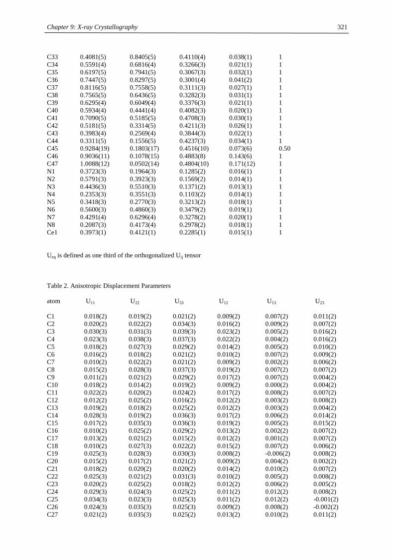
Chapter 9: X-ray Crystallography
321
C33 0.4081(5) 0.8405(5) 0.4110(4) 0.038(1) 1 C34 0.5591(4) 0.6816(4) 0.3266(3) 0.021(1) 1 C35 0.6197(5) 0.7941(5) 0.3067(3) 0.032(1) 1 C36 0.7447(5) 0.8297(5) 0.3001(4) 0.041(2) 1 C37 0.8116(5) 0.7558(5) 0.3111(3) 0.027(1) 1 C38 0.7565(5) 0.6436(5) 0.3282(3) 0.031(1) 1 C39 0.6295(4) 0.6049(4) 0.3376(3) 0.021(1) 1 C40 0.5934(4) 0.4441(4) 0.4082(3) 0.020(1) 1 C41 0.7090(5) 0.5185(5) 0.4708(3) 0.030(1) 1 C42 0.5181(5) 0.3314(5) 0.4211(3) 0.026(1) 1 C43 0.3983(4) 0.2569(4) 0.3844(3) 0.022(1) 1 C44 0.3311(5) 0.1556(5) 0.4237(3) 0.034(1) 1 C45 0.9284(19) 0.1803(17) 0.4516(10) 0.073(6) 0.50 C46 0.9036(11) 0.1078(15) 0.4883(8) 0.143(6) 1 C47 1.0088(12) 0.0502(14) 0.4804(10) 0.171(12) 1 N1 0.3723(3) 0.1964(3) 0.1285(2) 0.016(1) 1 N2 0.5791(3) 0.3923(3) 0.1569(2) 0.014(1) 1 N3 0.4436(3) 0.5510(3) 0.1371(2) 0.013(1) 1 N4 0.2353(3) 0.3551(3) 0.1103(2) 0.014(1) 1 N5 0.3418(3) 0.2770(3) 0.3213(2) 0.018(1) 1 N6 0.5600(3) 0.4860(3) 0.3479(2) 0.019(1) 1 N7 0.4291(4) 0.6296(4) 0.3278(2) 0.020(1) 1 N8 0.2087(3) 0.4173(4) 0.2978(2) 0.018(1) 1 Ce1 0.3973(1) 0.4121(1) 0.2285(1) 0.015(1) 1 Ueq is defined as one third of the orthogonalized Uij tensor Table 2. Anisotropic Displacement Parameters atom U11 U22 U33 U12 U13 U23 C1 0.018(2) 0.019(2) 0.021(2) 0.009(2) 0.007(2) 0.011(2) C2 0.020(2) 0.022(2) 0.034(3) 0.016(2) 0.009(2) 0.007(2) C3 0.030(3) 0.031(3) 0.039(3) 0.023(2) 0.005(2) 0.016(2) C4 0.023(3) 0.038(3) 0.037(3) 0.022(2) 0.004(2) 0.016(2) C5 0.018(2) 0.027(3) 0.029(2) 0.014(2) 0.005(2) 0.010(2) C6 0.016(2) 0.018(2) 0.021(2) 0.010(2) 0.007(2) 0.009(2) C7 0.010(2) 0.022(2) 0.021(2) 0.009(2) 0.002(2) 0.006(2) C8 0.015(2) 0.028(3) 0.037(3) 0.019(2) 0.007(2) 0.007(2) C9 0.011(2) 0.021(2) 0.029(2) 0.017(2) 0.007(2) 0.004(2) C10 0.018(2) 0.014(2) 0.019(2) 0.009(2) 0.000(2) 0.004(2) C11 0.022(2) 0.020(2) 0.024(2) 0.017(2) 0.008(2) 0.007(2) C12 0.012(2) 0.025(2) 0.016(2) 0.012(2) 0.003(2) 0.008(2) C13 0.019(2) 0.018(2) 0.025(2) 0.012(2) 0.003(2) 0.004(2) C14 0.028(3) 0.019(2) 0.036(3) 0.017(2) 0.006(2) 0.014(2) C15 0.017(2) 0.035(3) 0.036(3) 0.019(2) 0.005(2) 0.015(2) C16 0.010(2) 0.025(2) 0.029(2) 0.013(2) 0.002(2) 0.007(2) C17 0.013(2) 0.021(2) 0.015(2) 0.012(2) 0.001(2) 0.007(2) C18 0.010(2) 0.027(3) 0.022(2) 0.015(2) 0.007(2) 0.006(2) C19 0.025(3) 0.028(3) 0.030(3) 0.008(2) -0.006(2) 0.008(2) C20 0.015(2) 0.017(2) 0.021(2) 0.009(2) 0.004(2) 0.002(2) C21 0.018(2) 0.020(2) 0.020(2) 0.014(2) 0.010(2) 0.007(2) C22 0.025(3) 0.021(2) 0.031(3) 0.010(2) 0.005(2) 0.008(2) C23 0.020(2) 0.025(2) 0.018(2) 0.012(2) 0.006(2) 0.005(2) C24 0.029(3) 0.024(3) 0.025(2) 0.011(2) 0.012(2) 0.008(2) C25 0.034(3) 0.023(3) 0.025(3) 0.011(2) 0.012(2) -0.001(2) C26 0.024(3) 0.035(3) 0.025(3) 0.009(2) 0.008(2) -0.002(2) C27 0.021(2) 0.035(3) 0.025(2) 0.013(2) 0.010(2) 0.011(2)
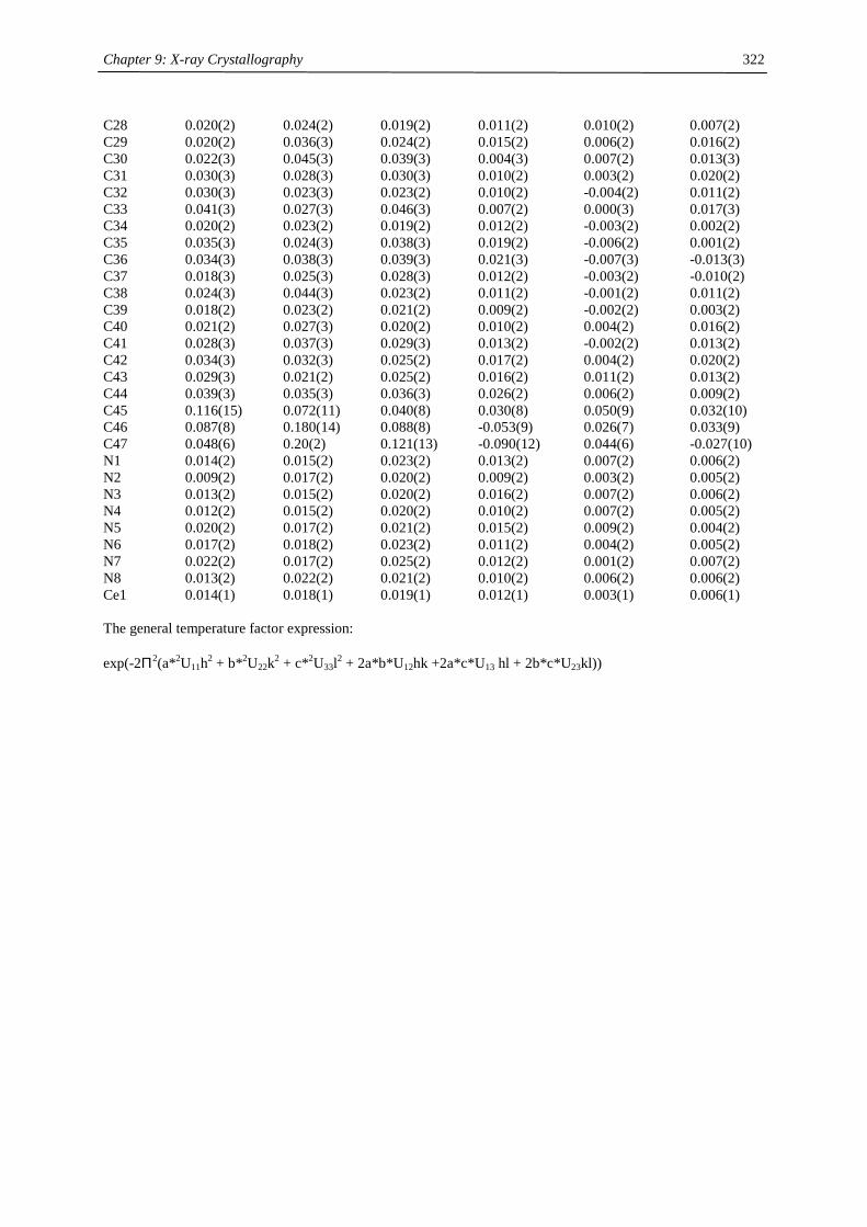
Chapter 9: X-ray Crystallography
322
C28 0.020(2) 0.024(2) 0.019(2) 0.011(2) 0.010(2) 0.007(2) C29 0.020(2) 0.036(3) 0.024(2) 0.015(2) 0.006(2) 0.016(2) C30 0.022(3) 0.045(3) 0.039(3) 0.004(3) 0.007(2) 0.013(3) C31 0.030(3) 0.028(3) 0.030(3) 0.010(2) 0.003(2) 0.020(2) C32 0.030(3) 0.023(3) 0.023(2) 0.010(2) -0.004(2) 0.011(2) C33 0.041(3) 0.027(3) 0.046(3) 0.007(2) 0.000(3) 0.017(3) C34 0.020(2) 0.023(2) 0.019(2) 0.012(2) -0.003(2) 0.002(2) C35 0.035(3) 0.024(3) 0.038(3) 0.019(2) -0.006(2) 0.001(2) C36 0.034(3) 0.038(3) 0.039(3) 0.021(3) -0.007(3) -0.013(3) C37 0.018(3) 0.025(3) 0.028(3) 0.012(2) -0.003(2) -0.010(2) C38 0.024(3) 0.044(3) 0.023(2) 0.011(2) -0.001(2) 0.011(2) C39 0.018(2) 0.023(2) 0.021(2) 0.009(2) -0.002(2) 0.003(2) C40 0.021(2) 0.027(3) 0.020(2) 0.010(2) 0.004(2) 0.016(2) C41 0.028(3) 0.037(3) 0.029(3) 0.013(2) -0.002(2) 0.013(2) C42 0.034(3) 0.032(3) 0.025(2) 0.017(2) 0.004(2) 0.020(2) C43 0.029(3) 0.021(2) 0.025(2) 0.016(2) 0.011(2) 0.013(2) C44 0.039(3) 0.035(3) 0.036(3) 0.026(2) 0.006(2) 0.009(2) C45 0.116(15) 0.072(11) 0.040(8) 0.030(8) 0.050(9) 0.032(10) C46 0.087(8) 0.180(14) 0.088(8) -0.053(9) 0.026(7) 0.033(9) C47 0.048(6) 0.20(2) 0.121(13) -0.090(12) 0.044(6) -0.027(10) N1 0.014(2) 0.015(2) 0.023(2) 0.013(2) 0.007(2) 0.006(2) N2 0.009(2) 0.017(2) 0.020(2) 0.009(2) 0.003(2) 0.005(2) N3 0.013(2) 0.015(2) 0.020(2) 0.016(2) 0.007(2) 0.006(2) N4 0.012(2) 0.015(2) 0.020(2) 0.010(2) 0.007(2) 0.005(2) N5 0.020(2) 0.017(2) 0.021(2) 0.015(2) 0.009(2) 0.004(2) N6 0.017(2) 0.018(2) 0.023(2) 0.011(2) 0.004(2) 0.005(2) N7 0.022(2) 0.017(2) 0.025(2) 0.012(2) 0.001(2) 0.007(2) N8 0.013(2) 0.022(2) 0.021(2) 0.010(2) 0.006(2) 0.006(2) Ce1 0.014(1) 0.018(1) 0.019(1) 0.012(1) 0.003(1) 0.006(1) The general temperature factor expression: exp(-2Π2(a*2U11h2 + b*2U22k2 + c*2U33l2 + 2a*b*U12hk +2a*c*U13 hl + 2b*c*U23kl))
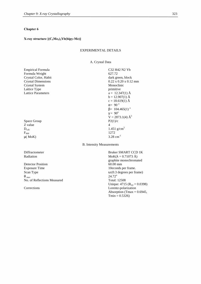
Chapter 9: X-ray Crystallography
323
Chapter 6
X-ray structure [(C5Me5)2Yb(bipy-Me)]
EXPERIMENTAL DETAILS
A. Crystal Data Empirical Formula C32 H42 N2 Yb Formula Weight 627.72 Crystal Color, Habit dark green, block Crystal Dimensions 0.22 x 0.20 x 0.12 mm Crystal System Monoclinic Lattice Type primitive Lattice Parameters a = 12.347(1) Å b = 12.907(1) Å c = 18.619(1) Å α= 90 o β= 104.465(1) o γ = 90o V = 2873.1(4) Å3 Space Group P2(1)/c Z value 4 Dcalc 1.451 g/cm3 F000 1272 µ( MoK) 3.28 cm-1
B. Intensity Measurements Diffractometer Bruker SMART CCD 1K Radiation MoK(λ = 0.71073 Å) graphite monochromated Detector Position 60.00 mm Exposure Time 10econds per frame. Scan Type ω (0.3 degrees per frame) θ max 24.72o No. of Reflections Measured Total: 12508 Unique: 4715 (Rint = 0.0398) Corrections Lorentz-polarization Absorption (Tmax = 0.6945, Tmin = 0.5326)
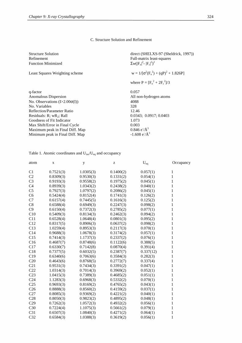
Chapter 9: X-ray Crystallography
324
C. Structure Solution and Refinement
Structure Solution direct (SHELXS-97 (Sheldrick, 1997)) Refinement Full-matrix least-squares Function Minimized Σw(|Fo|2- |Fc|2)2 Least Squares Weighting scheme w = 1/[σ2(Fo
2) + (qP)2 + 1.826P] where P = [Fo
2 + 2Fc2]/3
q-factor 0.057 Anomalous Dispersion All non-hydrogen atoms No. Observations (I>2.00σ(I)) 4088 No. Variables 328 Reflection/Parameter Ratio 12.46 Residuals: R; wR2; Rall 0.0343; 0.0917; 0.0403 Goodness of Fit Indicator 1.073 Max Shift/Error in Final Cycle 0.003 Maximum peak in Final Diff. Map 0.846 e-/Å3 Minimum peak in Final Diff. Map -1.608 e-/Å3 Table 1. Atomic coordinates and Uiso/Ueq and occupancy atom x y z Ueq Occupancy C1 0.7521(3) 1.0305(3) 0.1400(2) 0.057(1) 1 C2 0.8309(3) 0.9530(3) 0.1331(2) 0.054(1) 1 C3 0.9193(3) 0.9558(2) 0.1975(2) 0.045(1) 1 C4 0.8939(3) 1.0343(2) 0.2438(2) 0.040(1) 1 C5 0.7927(3) 1.0797(2) 0.2086(2) 0.045(1) 1 C6 0.5424(4) 0.8152(4) 0.1741(3) 0.126(2) 1 C7 0.6157(4) 0.7445(5) 0.1616(3) 0.125(2) 1 C8 0.6588(4) 0.6949(3) 0.2247(3) 0.098(2) 1 C9 0.6150(4) 0.7372(3) 0.2785(2) 0.077(1) 1 C10 0.5409(3) 0.8134(3) 0.2462(3) 0.094(2) 1 C11 0.6528(4) 1.0648(4) 0.0801(3) 0.095(2) 1 C12 0.8317(5) 0.8906(3) 0.0637(2) 0.098(2) 1 C13 1.0259(4) 0.8953(3) 0.2117(3) 0.070(1) 1 C14 0.9688(3) 1.0678(3) 0.3174(2) 0.057(1) 1 C15 0.7414(3) 1.1737(3) 0.2337(2) 0.076(1) 1 C16 0.4687(7) 0.8748(6) 0.1122(6) 0.388(5) 1 C17 0.6330(7) 0.7142(8) 0.0870(4) 0.391(4) 1 C18 0.7377(5) 0.6032(5) 0.2387(7) 0.337(12) 1 C19 0.6340(6) 0.7063(6) 0.3584(3) 0.282(3) 1 C20 0.4643(6) 0.8768(5) 0.2772(7) 0.337(4) 1 C21 0.9531(3) 0.7434(3) 0.3391(2) 0.047(1) 1 C22 1.0314(3) 0.7014(3) 0.3969(2) 0.052(1) 1 C23 1.0415(3) 0.7389(3) 0.4685(2) 0.051(1) 1 C24 1.1283(3) 0.6968(3) 0.5332(2) 0.070(1) 1 C25 0.9693(3) 0.8169(2) 0.4765(2) 0.043(1) 1 C26 0.8888(3) 0.8560(2) 0.4159(2) 0.037(1) 1 C27 0.8085(3) 0.9369(2) 0.4221(2) 0.040(1) 1 C28 0.8050(3) 0.9823(2) 0.4895(2) 0.048(1) 1 C29 0.7262(3) 1.0572(3) 0.4932(2) 0.056(1) 1 C30 0.7224(4) 1.1075(3) 0.5661(2) 0.079(1) 1 C31 0.6507(3) 1.0840(3) 0.4271(2) 0.064(1) 1 C32 0.6584(3) 1.0388(3) 0.3619(2) 0.056(1) 1
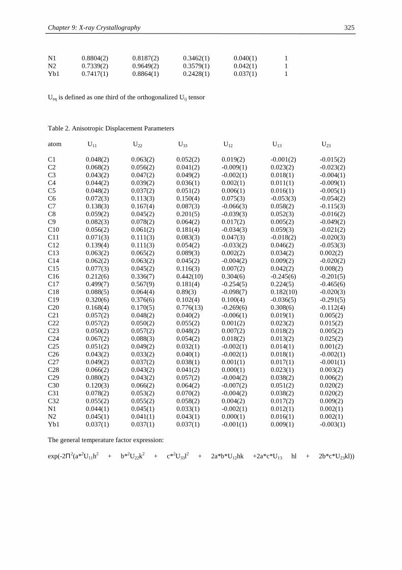
Chapter 9: X-ray Crystallography
325
N1 0.8804(2) 0.8187(2) 0.3462(1) 0.040(1) 1 N2 0.7339(2) 0.9649(2) 0.3579(1) 0.042(1) 1 Yb1 0.7417(1) 0.8864(1) 0.2428(1) 0.037(1) 1 Ueq is defined as one third of the orthogonalized Uij tensor Table 2. Anisotropic Displacement Parameters atom U11 U22 U33 U12 U13 U23 C1 0.048(2) 0.063(2) 0.052(2) 0.019(2) -0.001(2) -0.015(2) C2 0.068(2) 0.056(2) 0.041(2) -0.009(1) 0.023(2) -0.023(2) C3 0.043(2) 0.047(2) 0.049(2) -0.002(1) 0.018(1) -0.004(1) C4 0.044(2) 0.039(2) 0.036(1) 0.002(1) 0.011(1) -0.009(1) C5 0.048(2) 0.037(2) 0.051(2) 0.006(1) 0.016(1) -0.005(1) C6 0.072(3) 0.113(3) 0.150(4) 0.075(3) -0.053(3) -0.054(2) C7 0.138(3) 0.167(4) 0.087(3) -0.066(3) 0.058(2) -0.115(3) C8 0.059(2) 0.045(2) 0.201(5) -0.039(3) 0.052(3) -0.016(2) C9 0.082(3) 0.078(2) 0.064(2) 0.017(2) 0.005(2) -0.049(2) C10 0.056(2) 0.061(2) 0.181(4) -0.034(3) 0.059(3) -0.021(2) C11 0.071(3) 0.111(3) 0.083(3) 0.047(3) -0.018(2) -0.020(3) C12 0.139(4) 0.111(3) 0.054(2) -0.033(2) 0.046(2) -0.053(3) C13 0.063(2) 0.065(2) 0.089(3) 0.002(2) 0.034(2) 0.002(2) C14 0.062(2) 0.063(2) 0.045(2) -0.004(2) 0.009(2) -0.020(2) C15 0.077(3) 0.045(2) 0.116(3) 0.007(2) 0.042(2) 0.008(2) C16 0.212(6) 0.336(7) 0.442(10) 0.304(6) -0.245(6) -0.201(5) C17 0.499(7) 0.567(9) 0.181(4) -0.254(5) 0.224(5) -0.465(6) C18 0.088(5) 0.064(4) 0.89(3) -0.098(7) 0.182(10) -0.020(3) C19 0.320(6) 0.376(6) 0.102(4) 0.100(4) -0.036(5) -0.291(5) C20 0.168(4) 0.170(5) 0.776(13) -0.269(6) 0.308(6) -0.112(4) C21 0.057(2) 0.048(2) 0.040(2) -0.006(1) 0.019(1) 0.005(2) C22 0.057(2) 0.050(2) 0.055(2) 0.001(2) 0.023(2) 0.015(2) C23 0.050(2) 0.057(2) 0.048(2) 0.007(2) 0.018(2) 0.005(2) C24 0.067(2) 0.088(3) 0.054(2) 0.018(2) 0.013(2) 0.025(2) C25 0.051(2) 0.049(2) 0.032(1) -0.002(1) 0.014(1) 0.001(2) C26 0.043(2) 0.033(2) 0.040(1) -0.002(1) 0.018(1) -0.002(1) C27 0.049(2) 0.037(2) 0.038(1) 0.001(1) 0.017(1) -0.001(1) C28 0.066(2) 0.043(2) 0.041(2) 0.000(1) 0.023(1) 0.003(2) C29 0.080(2) 0.043(2) 0.057(2) -0.004(2) 0.038(2) 0.006(2) C30 0.120(3) 0.066(2) 0.064(2) -0.007(2) 0.051(2) 0.020(2) C31 0.078(2) 0.053(2) 0.070(2) -0.004(2) 0.038(2) 0.020(2) C32 0.055(2) 0.055(2) 0.058(2) 0.004(2) 0.017(2) 0.009(2) N1 0.044(1) 0.045(1) 0.033(1) -0.002(1) 0.012(1) 0.002(1) N2 0.045(1) 0.041(1) 0.043(1) 0.000(1) 0.016(1) 0.002(1) Yb1 0.037(1) 0.037(1) 0.037(1) -0.001(1) 0.009(1) -0.003(1) The general temperature factor expression: exp(-2Π2(a*2U11h2 + b*2U22k2 + c*2U33l2 + 2a*b*U12hk +2a*c*U13 hl + 2b*c*U23kl))
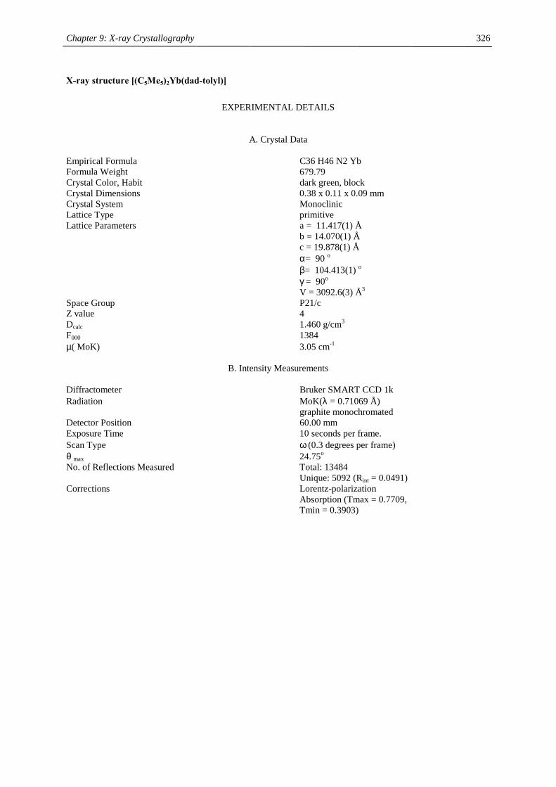
Chapter 9: X-ray Crystallography
326
X-ray structure [(C5Me5)2Yb(dad-tolyl)]
EXPERIMENTAL DETAILS
A. Crystal Data Empirical Formula C36 H46 N2 Yb Formula Weight 679.79 Crystal Color, Habit dark green, block Crystal Dimensions 0.38 x 0.11 x 0.09 mm Crystal System Monoclinic Lattice Type primitive Lattice Parameters a = 11.417(1) Å b = 14.070(1) Å c = 19.878(1) Å α= 90 o β= 104.413(1) o γ = 90o V = 3092.6(3) Å3 Space Group P21/c Z value 4 Dcalc 1.460 g/cm3 F000 1384 µ( MoK) 3.05 cm-1
B. Intensity Measurements Diffractometer Bruker SMART CCD 1k Radiation MoK(λ = 0.71069 Å) graphite monochromated Detector Position 60.00 mm Exposure Time 10 seconds per frame. Scan Type ω (0.3 degrees per frame) θ max 24.75o No. of Reflections Measured Total: 13484 Unique: 5092 (Rint = 0.0491) Corrections Lorentz-polarization Absorption (Tmax = 0.7709, Tmin = 0.3903)

Chapter 9: X-ray Crystallography
327
C. Structure Solution and Refinement
Structure Solution direct (SHELXS-97 (Sheldrick, 1997)) Refinement Full-matrix least-squares Function Minimized Σw(|Fo|2- |Fc|2)2 Least Squares Weighting scheme w = 1/[σ2(Fo
2) + (qP)2 + 0.000P] where P = [Fo
2 + 2Fc2]/3
q-factor 0.0829 Anomalous Dispersion All non-hydrogen atoms No. Observations (I>2.00σ(I)) 4045 No. Variables 364 Reflection/Parameter Ratio 11.11 Residuals: R; wR2; Rall 0.0467; 0.1121; 0.0607 Goodness of Fit Indicator 1.000 Max Shift/Error in Final Cycle 0.000 Maximum peak in Final Diff. Map 2.013 e-/Å3 Minimum peak in Final Diff. Map -4.890 e-/Å3 Table 1. Atomic coordinates and Uiso/Ueq and occupancy atom x y z Ueq Occupancy Yb1 0.1965(1) 0.3307(1) 0.1993(1) 0.015(1) 1 N1 0.2470(5) 0.2129(4) 0.2868(2) 0.019(1) 1 N2 0.0682(5) 0.2014(4) 0.1609(3) 0.022(1) 1 C22 0.5036(6) 0.2234(6) 0.4822(3) 0.035(2) 1 C3 0.1396(6) 0.5123(4) 0.2160(3) 0.020(1) 1 C27 0.3344(5) 0.2120(4) 0.3505(3) 0.019(1) 1 C7 0.3103(5) 0.3959(4) 0.1090(3) 0.019(1) 1 C9 0.3306(5) 0.2369(4) 0.1318(3) 0.019(1) 1 C4 0.0306(6) 0.4689(4) 0.1786(3) 0.020(1) 1 C10 0.4130(5) 0.2856(5) 0.1867(3) 0.020(1) 1 C30 -0.0134(6) 0.1847(4) 0.0962(3) 0.021(1) 1 C14 -0.0432(6) 0.5012(5) 0.1078(3) 0.027(2) 1 C6 0.4004(5) 0.3843(4) 0.1727(3) 0.017(1) 1 C2 0.1690(6) 0.4735(4) 0.2836(3) 0.022(2) 1 C11 0.0715(6) 0.3485(5) 0.3510(3) 0.026(2) 1 C28 0.1708(6) 0.1389(5) 0.2712(3) 0.025(2) 1 C15 -0.1218(6) 0.3470(5) 0.2086(4) 0.028(2) 1 C1 0.0830(5) 0.4035(4) 0.2880(3) 0.019(1) 1 C31 -0.0444(6) 0.0938(5) 0.0688(3) 0.030(2) 1 C8 0.2696(6) 0.3045(5) 0.0846(3) 0.022(1) 1 C5 -0.0044(6) 0.4002(4) 0.2229(3) 0.020(1) 1 C13 0.1937(6) 0.6015(5) 0.1957(3) 0.032(2) 1 C17 0.2802(6) 0.4864(4) 0.0669(3) 0.029(2) 1 C19 0.3221(7) 0.1292(4) 0.1239(4) 0.032(2) 1 C16 0.4810(6) 0.4600(5) 0.2143(4) 0.030(2) 1 C33 -0.0656(6) 0.2609(5) 0.0547(3) 0.025(2) 1 C35 -0.1714(6) 0.1580(5) -0.0374(3) 0.028(2) 1 C29 0.0825(6) 0.1329(5) 0.2097(3) 0.025(2) 1 C36 -0.2529(7) 0.1443(6) -0.1095(4) 0.039(2) 1 C20 0.5099(6) 0.2392(5) 0.2415(3) 0.032(2) 1 C18 0.1889(6) 0.2844(5) 0.0133(3) 0.033(2) 1 C32 -0.1214(6) 0.0817(5) 0.0040(3) 0.032(2) 1 C26 0.3891(6) 0.2976(5) 0.3773(4) 0.030(2) 1
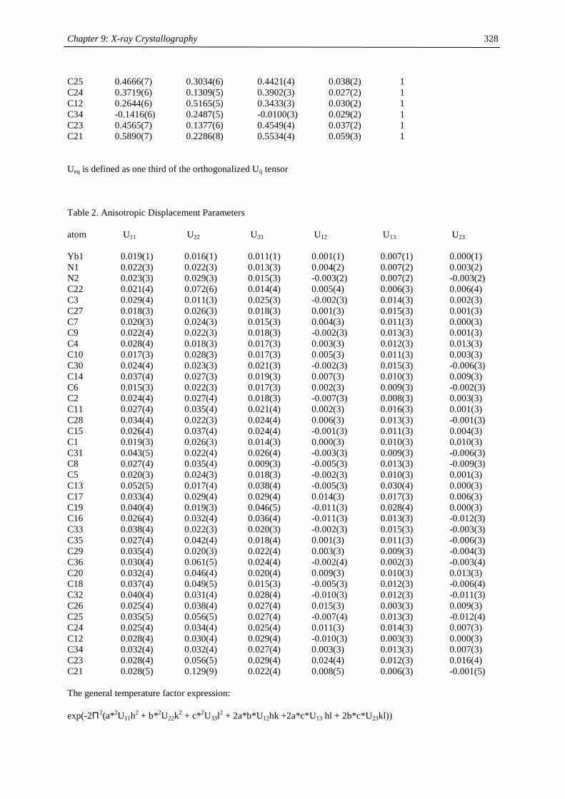
Chapter 9: X-ray Crystallography
328
C25 0.4666(7) 0.3034(6) 0.4421(4) 0.038(2) 1 C24 0.3719(6) 0.1309(5) 0.3902(3) 0.027(2) 1 C12 0.2644(6) 0.5165(5) 0.3433(3) 0.030(2) 1 C34 -0.1416(6) 0.2487(5) -0.0100(3) 0.029(2) 1 C23 0.4565(7) 0.1377(6) 0.4549(4) 0.037(2) 1 C21 0.5890(7) 0.2286(8) 0.5534(4) 0.059(3) 1 Ueq is defined as one third of the orthogonalized Uij tensor Table 2. Anisotropic Displacement Parameters atom U11 U22 U33 U12 U13 U23 Yb1 0.019(1) 0.016(1) 0.011(1) 0.001(1) 0.007(1) 0.000(1) N1 0.022(3) 0.022(3) 0.013(3) 0.004(2) 0.007(2) 0.003(2) N2 0.023(3) 0.029(3) 0.015(3) -0.003(2) 0.007(2) -0.003(2) C22 0.021(4) 0.072(6) 0.014(4) 0.005(4) 0.006(3) 0.006(4) C3 0.029(4) 0.011(3) 0.025(3) -0.002(3) 0.014(3) 0.002(3) C27 0.018(3) 0.026(3) 0.018(3) 0.001(3) 0.015(3) 0.001(3) C7 0.020(3) 0.024(3) 0.015(3) 0.004(3) 0.011(3) 0.000(3) C9 0.022(4) 0.022(3) 0.018(3) -0.002(3) 0.013(3) 0.001(3) C4 0.028(4) 0.018(3) 0.017(3) 0.003(3) 0.012(3) 0.013(3) C10 0.017(3) 0.028(3) 0.017(3) 0.005(3) 0.011(3) 0.003(3) C30 0.024(4) 0.023(3) 0.021(3) -0.002(3) 0.015(3) -0.006(3) C14 0.037(4) 0.027(3) 0.019(3) 0.007(3) 0.010(3) 0.009(3) C6 0.015(3) 0.022(3) 0.017(3) 0.002(3) 0.009(3) -0.002(3) C2 0.024(4) 0.027(4) 0.018(3) -0.007(3) 0.008(3) 0.003(3) C11 0.027(4) 0.035(4) 0.021(4) 0.002(3) 0.016(3) 0.001(3) C28 0.034(4) 0.022(3) 0.024(4) 0.006(3) 0.013(3) -0.001(3) C15 0.026(4) 0.037(4) 0.024(4) -0.001(3) 0.011(3) 0.004(3) C1 0.019(3) 0.026(3) 0.014(3) 0.000(3) 0.010(3) 0.010(3) C31 0.043(5) 0.022(4) 0.026(4) -0.003(3) 0.009(3) -0.006(3) C8 0.027(4) 0.035(4) 0.009(3) -0.005(3) 0.013(3) -0.009(3) C5 0.020(3) 0.024(3) 0.018(3) -0.002(3) 0.010(3) 0.001(3) C13 0.052(5) 0.017(4) 0.038(4) -0.005(3) 0.030(4) 0.000(3) C17 0.033(4) 0.029(4) 0.029(4) 0.014(3) 0.017(3) 0.006(3) C19 0.040(4) 0.019(3) 0.046(5) -0.011(3) 0.028(4) 0.000(3) C16 0.026(4) 0.032(4) 0.036(4) -0.011(3) 0.013(3) -0.012(3) C33 0.038(4) 0.022(3) 0.020(3) -0.002(3) 0.015(3) -0.003(3) C35 0.027(4) 0.042(4) 0.018(4) 0.001(3) 0.011(3) -0.006(3) C29 0.035(4) 0.020(3) 0.022(4) 0.003(3) 0.009(3) -0.004(3) C36 0.030(4) 0.061(5) 0.024(4) -0.002(4) 0.002(3) -0.003(4) C20 0.032(4) 0.046(4) 0.020(4) 0.009(3) 0.010(3) 0.013(3) C18 0.037(4) 0.049(5) 0.015(3) -0.005(3) 0.012(3) -0.006(4) C32 0.040(4) 0.031(4) 0.028(4) -0.010(3) 0.012(3) -0.011(3) C26 0.025(4) 0.038(4) 0.027(4) 0.015(3) 0.003(3) 0.009(3) C25 0.035(5) 0.056(5) 0.027(4) -0.007(4) 0.013(3) -0.012(4) C24 0.025(4) 0.034(4) 0.025(4) 0.011(3) 0.014(3) 0.007(3) C12 0.028(4) 0.030(4) 0.029(4) -0.010(3) 0.003(3) 0.000(3) C34 0.032(4) 0.032(4) 0.027(4) 0.003(3) 0.013(3) 0.007(3) C23 0.028(4) 0.056(5) 0.029(4) 0.024(4) 0.012(3) 0.016(4) C21 0.028(5) 0.129(9) 0.022(4) 0.008(5) 0.006(3) -0.001(5) The general temperature factor expression: exp(-2Π2(a*2U11h2 + b*2U22k2 + c*2U33l2 + 2a*b*U12hk +2a*c*U13 hl + 2b*c*U23kl))

Chapter 9: X-ray Crystallography
329
X-ray structure [(C5Me5)2Yb(dad-OMe)]
EXPERIMENTAL DETAILS
A. Crystal Data Empirical Formula C36 H46 N2 O2 Yb Formula Weight 711.79 Crystal Color, Habit dark green, block Crystal Dimensions 0.19 x 0.13 x 0.04 mm Crystal System Monoclinic Lattice Type primitive Lattice Parameters a = 9.990(1) Å b = 30.731(3) Å c = 10.457(1) Å α= 90 o β= 97.978(1) o γ = 90o V = 3179.2(5) Å3 Space Group P21/a Z value 4 Dcalc 1.487 g/cm3 F000 1448 µ( MoK) 2.97 cm-1
B. Intensity Measurements Diffractometer Bruker SMART CCD 1k Radiation MoK(λ = 0.71069 Å) graphite monochromated Detector Position 60.00 mm Exposure Time 10 seconds per frame. Scan Type ω (0.3 degrees per frame) θ max 24.73o No. of Reflections Measured Total: 14042 Unique: 5234 (Rint = 0.0886) Corrections Lorentz-polarization Absorption (Tmax = 0.8903, Tmin = 0.6018)
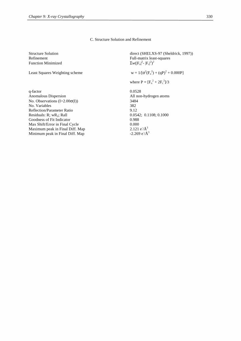
Chapter 9: X-ray Crystallography
330
C. Structure Solution and Refinement
Structure Solution direct (SHELXS-97 (Sheldrick, 1997)) Refinement Full-matrix least-squares Function Minimized Σw(|Fo|2- |Fc|2)2 Least Squares Weighting scheme w = 1/[σ2(Fo
2) + (qP)2 + 0.000P] where P = [Fo
2 + 2Fc2]/3
q-factor 0.0528 Anomalous Dispersion All non-hydrogen atoms No. Observations (I>2.00σ(I)) 3484 No. Variables 382 Reflection/Parameter Ratio 9.12 Residuals: R; wR2; Rall 0.0542; 0.1108; 0.1000 Goodness of Fit Indicator 0.988 Max Shift/Error in Final Cycle 0.000 Maximum peak in Final Diff. Map 2.121 e-/Å3 Minimum peak in Final Diff. Map -2.269 e-/Å3
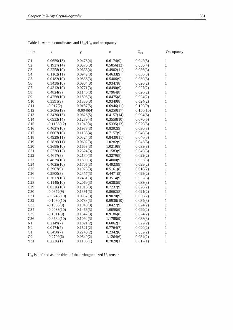
Chapter 9: X-ray Crystallography
331
Table 1. Atomic coordinates and Uiso/Ueq and occupancy atom x y z Ueq Occupancy C1 0.0659(13) 0.0478(4) 0.6174(9) 0.042(3) 1 C2 0.1927(14) 0.0376(3) 0.5856(12) 0.056(4) 1 C3 0.2258(10) 0.0666(4) 0.4902(11) 0.036(3) 1 C4 0.1162(11) 0.0942(3) 0.4633(9) 0.030(3) 1 C5 0.0182(10) 0.0836(3) 0.5406(9) 0.030(3) 1 C6 0.3438(10) 0.0904(3) 0.9347(8) 0.026(2) 1 C7 0.4313(10) 0.0771(3) 0.8490(9) 0.027(2) 1 C8 0.4824(9) 0.1146(3) 0.7964(8) 0.026(2) 1 C9 0.4256(10) 0.1508(3) 0.8475(8) 0.024(2) 1 C10 0.3391(9) 0.1356(3) 0.9349(8) 0.024(2) 1 C11 -0.017(2) 0.0187(5) 0.6946(11) 0.129(9) 1 C12 0.2696(19) -0.0046(4) 0.6250(17) 0.156(10) 1 C13 0.3430(13) 0.0626(5) 0.4157(14) 0.094(6) 1 C14 0.0933(14) 0.1279(4) 0.3558(10) 0.070(5) 1 C15 -0.1185(12) 0.1049(4) 0.5335(13) 0.079(5) 1 C16 0.4627(10) 0.1978(3) 0.8292(9) 0.030(3) 1 C17 0.6007(10) 0.1135(4) 0.7157(9) 0.040(3) 1 C18 0.4929(11) 0.0324(3) 0.8430(11) 0.046(3) 1 C19 0.2836(11) 0.0602(3) 1.0282(9) 0.043(3) 1 C20 0.2698(10) 0.1653(3) 1.0219(8) 0.033(3) 1 C21 0.5236(12) 0.2624(3) 0.1583(9) 0.045(3) 1 C22 0.4617(9) 0.2180(3) 0.3279(8) 0.022(2) 1 C23 0.4829(10) 0.1800(3) 0.4000(9) 0.033(3) 1 C24 0.4025(10) 0.1705(3) 0.4923(9) 0.029(2) 1 C25 0.2967(9) 0.1973(3) 0.5161(8) 0.018(2) 1 C26 0.2800(9) 0.2357(3) 0.4471(9) 0.029(2) 1 C27 0.3612(10) 0.2461(3) 0.3514(9) 0.032(3) 1 C28 0.1149(10) 0.2069(3) 0.6383(9) 0.033(3) 1 C29 0.0316(10) 0.1918(3) 0.7237(9) 0.028(2) 1 C30 -0.0372(9) 0.1391(3) 0.8662(8) 0.021(2) 1 C31 -0.0245(10) 0.0957(3) 0.9070(9) 0.030(2) 1 C32 -0.1030(10) 0.0788(3) 0.9936(10) 0.034(3) 1 C33 -0.1963(9) 0.1040(3) 1.0427(9) 0.024(2) 1 C34 -0.2088(10) 0.1466(3) 1.0058(9) 0.029(2) 1 C35 -0.1311(9) 0.1647(3) 0.9186(8) 0.024(2) 1 C36 -0.3684(10) 0.1094(3) 1.1788(9) 0.038(3) 1 N1 0.2149(7) 0.1821(2) 0.6062(7) 0.022(2) 1 N2 0.0474(7) 0.1521(2) 0.7764(7) 0.020(2) 1 O1 0.5450(7) 0.2240(2) 0.2342(6) 0.032(2) 1 O2 -0.2709(6) 0.0840(2) 1.1264(6) 0.034(2) 1 Yb1 0.2226(1) 0.1133(1) 0.7020(1) 0.017(1) 1 Ueq is defined as one third of the orthogonalized Uij tensor
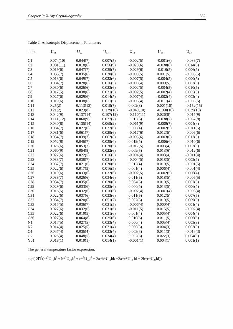
Chapter 9: X-ray Crystallography
332
Table 2. Anisotropic Displacement Parameters atom U11 U22 U33 U12 U13 U23 C1 0.074(10) 0.044(7) 0.007(5) -0.002(5) -0.001(6) -0.036(7) C2 0.081(11) 0.018(6) 0.056(9) -0.028(6) -0.038(8) 0.014(6) C3 0.019(6) 0.047(7) 0.039(7) -0.029(6) -0.010(5) 0.006(5) C4 0.033(7) 0.035(6) 0.020(6) -0.003(5) 0.001(5) -0.008(5) C5 0.018(6) 0.049(7) 0.022(6) -0.007(5) -0.004(5) 0.000(5) C6 0.034(7) 0.028(6) 0.016(5) -0.003(4) 0.000(5) 0.003(5) C7 0.030(6) 0.026(6) 0.023(6) -0.002(5) -0.004(5) 0.010(5) C8 0.017(5) 0.038(6) 0.021(5) -0.002(5) -0.002(4) 0.005(5) C9 0.027(6) 0.029(6) 0.014(5) -0.007(4) -0.002(4) 0.002(4) C10 0.019(6) 0.038(6) 0.011(5) -0.006(4) -0.011(4) -0.008(5) C11 0.25(2) 0.113(13) 0.019(7) 0.002(8) 0.001(10) -0.152(15) C12 0.21(2) 0.023(8) 0.179(18) -0.049(10) -0.160(16) 0.039(10) C13 0.042(9) 0.137(14) 0.107(12) -0.110(11) 0.026(8) -0.015(9) C14 0.111(12) 0.060(9) 0.027(7) 0.013(6) -0.038(7) -0.037(8) C15 0.030(8) 0.135(14) 0.069(9) -0.061(9) -0.009(7) 0.004(8) C16 0.034(7) 0.027(6) 0.027(6) 0.000(4) -0.002(5) -0.011(5) C17 0.031(6) 0.061(7) 0.029(6) -0.017(6) 0.012(5) -0.006(6) C18 0.034(7) 0.039(7) 0.062(8) -0.005(6) -0.003(6) 0.012(5) C19 0.052(8) 0.046(7) 0.029(6) 0.019(5) -0.006(6) -0.010(6) C20 0.025(6) 0.053(7) 0.020(5) -0.017(5) 0.003(4) 0.003(5) C21 0.060(9) 0.054(8) 0.022(6) 0.009(5) 0.013(6) -0.012(6) C22 0.027(6) 0.022(5) 0.016(5) -0.004(4) 0.003(4) -0.011(4) C23 0.033(7) 0.038(7) 0.031(6) -0.004(5) 0.018(5) 0.002(5) C24 0.037(7) 0.021(6) 0.030(6) 0.012(4) 0.010(5) -0.001(5) C25 0.022(6) 0.017(5) 0.016(5) 0.001(4) 0.006(4) -0.001(4) C26 0.019(6) 0.033(6) 0.032(6) -0.002(5) -0.002(5) 0.006(4) C27 0.038(7) 0.026(6) 0.034(6) 0.011(5) 0.018(5) -0.005(5) C28 0.034(7) 0.035(6) 0.030(6) 0.004(5) 0.010(5) 0.007(5) C29 0.029(6) 0.033(6) 0.025(6) 0.000(5) 0.013(5) 0.006(5) C30 0.015(5) 0.032(6) 0.016(5) -0.002(4) -0.001(4) -0.003(4) C31 0.022(6) 0.037(6) 0.033(6) 0.011(5) 0.012(5) 0.007(5) C32 0.034(7) 0.020(6) 0.051(7) 0.007(5) 0.019(5) 0.009(5) C33 0.015(5) 0.036(7) 0.021(5) -0.006(4) 0.000(4) 0.001(4) C34 0.027(6) 0.032(6) 0.031(6) -0.011(5) 0.015(5) -0.002(4) C35 0.022(6) 0.019(5) 0.031(6) 0.001(4) 0.005(4) 0.004(4) C36 0.027(6) 0.064(8) 0.025(6) 0.010(6) 0.011(5) 0.006(6) N1 0.017(5) 0.027(5) 0.023(4) 0.000(4) 0.005(4) 0.003(3) N2 0.014(4) 0.025(5) 0.021(4) 0.000(3) 0.004(3) 0.003(3) O1 0.037(4) 0.036(4) 0.023(4) 0.003(3) 0.011(3) -0.013(3) O2 0.025(4) 0.048(5) 0.034(4) 0.007(3) 0.022(3) 0.004(3) Yb1 0.018(1) 0.019(1) 0.014(1) -0.001(1) 0.004(1) 0.001(1) The general temperature factor expression: exp(-2Π2(a*2U11h2 + b*2U22k2 + c*2U33l2 + 2a*b*U12hk +2a*c*U13 hl + 2b*c*U23kl))
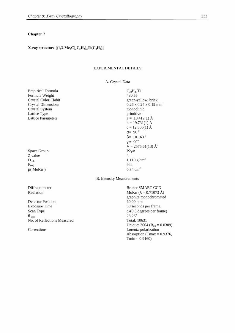
Chapter 9: X-ray Crystallography
333
Chapter 7
X-ray structure [(1,3-Me3C)2C5H3)2Ti(C2H4)]
EXPERIMENTAL DETAILS
A. Crystal Data Empirical Formula C28H46Ti Formula Weight 430.55 Crystal Color, Habit green-yellow, brick Crystal Dimensions 0.26 x 0.24 x 0.19 mm Crystal System monoclinic Lattice Type primitive Lattice Parameters a = 10.412(1) Å b = 19.731(1) Å c = 12.800(1) Å α= 90 o β= 101.63 o γ = 90o V = 2575.61(13) Å3 Space Group P21/n Z value 4 Dcalc 1.110 g/cm3 F000 944 µ( MoKα ) 0.34 cm-1
B. Intensity Measurements Diffractometer Bruker SMART CCD Radiation MoKα (λ = 0.71073 Å) graphite monochromated Detector Position 60.00 mm Exposure Time 30 seconds per frame. Scan Type ω (0.3 degrees per frame) θ max 23.26o No. of Reflections Measured Total: 10631 Unique: 3664 (Rint = 0.0309) Corrections Lorentz-polarization Absorption (Tmax = 0.9376, Tmin = 0.9160)
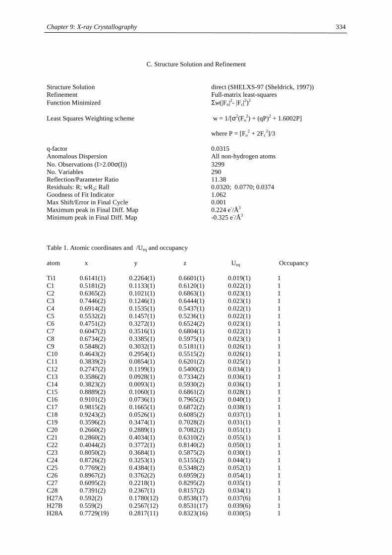
Chapter 9: X-ray Crystallography
334
C. Structure Solution and Refinement
Structure Solution direct (SHELXS-97 (Sheldrick, 1997)) Refinement Full-matrix least-squares Function Minimized Σw(|Fo|2- |Fc|2)2 Least Squares Weighting scheme w = 1/[σ2(Fo
2) + (qP)2 + 1.6002P] where P = [Fo
2 + 2Fc2]/3
q-factor 0.0315 Anomalous Dispersion All non-hydrogen atoms No. Observations (I>2.00σ(I)) 3299 No. Variables 290 Reflection/Parameter Ratio 11.38 Residuals: R; wR2; Rall 0.0320; 0.0770; 0.0374 Goodness of Fit Indicator 1.062 Max Shift/Error in Final Cycle 0.001 Maximum peak in Final Diff. Map 0.224 e-/Å3 Minimum peak in Final Diff. Map -0.325 e-/Å3 Table 1. Atomic coordinates and /Ueq and occupancy atom x y z Ueq Occupancy Ti1 0.6141(1) 0.2264(1) 0.6601(1) 0.019(1) 1 C1 0.5181(2) 0.1133(1) 0.6120(1) 0.022(1) 1 C2 0.6365(2) 0.1021(1) 0.6863(1) 0.023(1) 1 C3 0.7446(2) 0.1246(1) 0.6444(1) 0.023(1) 1 C4 0.6914(2) 0.1535(1) 0.5437(1) 0.022(1) 1 C5 0.5532(2) 0.1457(1) 0.5236(1) 0.022(1) 1 C6 0.4751(2) 0.3272(1) 0.6524(2) 0.023(1) 1 C7 0.6047(2) 0.3516(1) 0.6804(1) 0.022(1) 1 C8 0.6734(2) 0.3385(1) 0.5975(1) 0.023(1) 1 C9 0.5848(2) 0.3032(1) 0.5181(1) 0.026(1) 1 C10 0.4643(2) 0.2954(1) 0.5515(2) 0.026(1) 1 C11 0.3839(2) 0.0854(1) 0.6201(2) 0.025(1) 1 C12 0.2747(2) 0.1199(1) 0.5400(2) 0.034(1) 1 C13 0.3586(2) 0.0928(1) 0.7334(2) 0.036(1) 1 C14 0.3823(2) 0.0093(1) 0.5930(2) 0.036(1) 1 C15 0.8889(2) 0.1060(1) 0.6861(2) 0.028(1) 1 C16 0.9101(2) 0.0736(1) 0.7965(2) 0.040(1) 1 C17 0.9815(2) 0.1665(1) 0.6872(2) 0.038(1) 1 C18 0.9243(2) 0.0526(1) 0.6085(2) 0.037(1) 1 C19 0.3596(2) 0.3474(1) 0.7028(2) 0.031(1) 1 C20 0.2660(2) 0.2889(1) 0.7082(2) 0.051(1) 1 C21 0.2860(2) 0.4034(1) 0.6310(2) 0.055(1) 1 C22 0.4044(2) 0.3772(1) 0.8140(2) 0.050(1) 1 C23 0.8050(2) 0.3684(1) 0.5875(2) 0.030(1) 1 C24 0.8726(2) 0.3253(1) 0.5155(2) 0.044(1) 1 C25 0.7769(2) 0.4384(1) 0.5348(2) 0.052(1) 1 C26 0.8967(2) 0.3762(2) 0.6959(2) 0.054(1) 1 C27 0.6095(2) 0.2218(1) 0.8295(2) 0.035(1) 1 C28 0.7391(2) 0.2367(1) 0.8157(2) 0.034(1) 1 H27A 0.592(2) 0.1780(12) 0.8538(17) 0.037(6) 1 H27B 0.559(2) 0.2567(12) 0.8531(17) 0.039(6) 1 H28A 0.7729(19) 0.2817(11) 0.8323(16) 0.030(5) 1
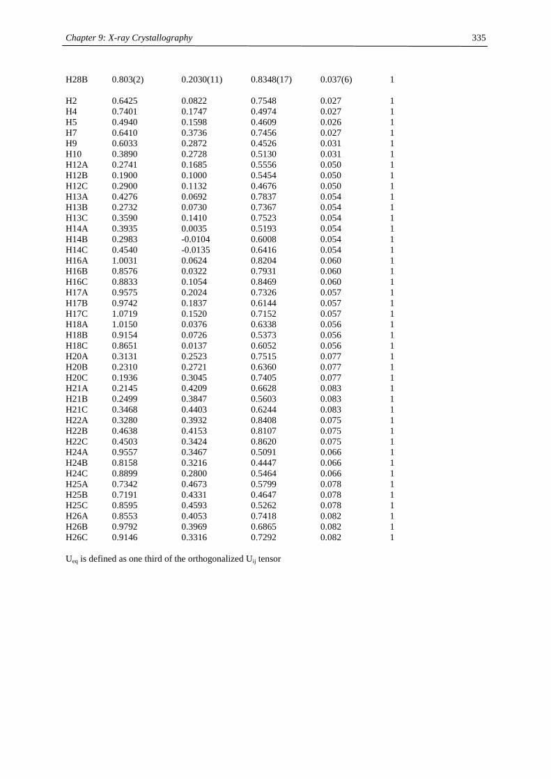
Chapter 9: X-ray Crystallography
335
H28B 0.803(2) 0.2030(11) 0.8348(17) 0.037(6) 1 H2 0.6425 0.0822 0.7548 0.027 1 H4 0.7401 0.1747 0.4974 0.027 1 H5 0.4940 0.1598 0.4609 0.026 1 H7 0.6410 0.3736 0.7456 0.027 1 H9 0.6033 0.2872 0.4526 0.031 1 H10 0.3890 0.2728 0.5130 0.031 1 H12A 0.2741 0.1685 0.5556 0.050 1 H12B 0.1900 0.1000 0.5454 0.050 1 H12C 0.2900 0.1132 0.4676 0.050 1 H13A 0.4276 0.0692 0.7837 0.054 1 H13B 0.2732 0.0730 0.7367 0.054 1 H13C 0.3590 0.1410 0.7523 0.054 1 H14A 0.3935 0.0035 0.5193 0.054 1 H14B 0.2983 -0.0104 0.6008 0.054 1 H14C 0.4540 -0.0135 0.6416 0.054 1 H16A 1.0031 0.0624 0.8204 0.060 1 H16B 0.8576 0.0322 0.7931 0.060 1 H16C 0.8833 0.1054 0.8469 0.060 1 H17A 0.9575 0.2024 0.7326 0.057 1 H17B 0.9742 0.1837 0.6144 0.057 1 H17C 1.0719 0.1520 0.7152 0.057 1 H18A 1.0150 0.0376 0.6338 0.056 1 H18B 0.9154 0.0726 0.5373 0.056 1 H18C 0.8651 0.0137 0.6052 0.056 1 H20A 0.3131 0.2523 0.7515 0.077 1 H20B 0.2310 0.2721 0.6360 0.077 1 H20C 0.1936 0.3045 0.7405 0.077 1 H21A 0.2145 0.4209 0.6628 0.083 1 H21B 0.2499 0.3847 0.5603 0.083 1 H21C 0.3468 0.4403 0.6244 0.083 1 H22A 0.3280 0.3932 0.8408 0.075 1 H22B 0.4638 0.4153 0.8107 0.075 1 H22C 0.4503 0.3424 0.8620 0.075 1 H24A 0.9557 0.3467 0.5091 0.066 1 H24B 0.8158 0.3216 0.4447 0.066 1 H24C 0.8899 0.2800 0.5464 0.066 1 H25A 0.7342 0.4673 0.5799 0.078 1 H25B 0.7191 0.4331 0.4647 0.078 1 H25C 0.8595 0.4593 0.5262 0.078 1 H26A 0.8553 0.4053 0.7418 0.082 1 H26B 0.9792 0.3969 0.6865 0.082 1 H26C 0.9146 0.3316 0.7292 0.082 1 Ueq is defined as one third of the orthogonalized Uij tensor
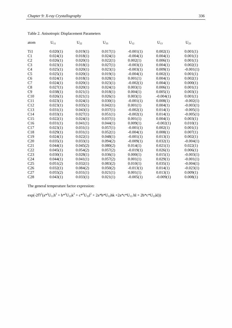
Chapter 9: X-ray Crystallography
336
Table 2. Anisotropic Displacement Parameters atom U11 U22 U33 U12 U13 U23 Ti1 0.020(1) 0.019(1) 0.017(1) -0.001(1) 0.002(1) 0.001(1) C1 0.024(1) 0.018(1) 0.024(1) -0.004(1) 0.004(1) 0.001(1) C2 0.026(1) 0.020(1) 0.022(1) 0.002(1) 0.006(1) 0.001(1) C3 0.023(1) 0.018(1) 0.027(1) -0.003(1) 0.004(1) 0.002(1) C4 0.025(1) 0.020(1) 0.023(1) -0.003(1) 0.009(1) -0.001(1) C5 0.025(1) 0.020(1) 0.019(1) -0.004(1) 0.002(1) 0.001(1) C6 0.024(1) 0.018(1) 0.028(1) 0.001(1) 0.004(1) 0.002(1) C7 0.024(1) 0.020(1) 0.023(1) -0.002(1) 0.004(1) 0.000(1) C8 0.027(1) 0.020(1) 0.024(1) 0.003(1) 0.006(1) 0.001(1) C9 0.038(1) 0.021(1) 0.018(1) 0.004(1) 0.005(1) 0.003(1) C10 0.026(1) 0.021(1) 0.026(1) 0.003(1) -0.004(1) 0.001(1) C11 0.023(1) 0.024(1) 0.030(1) -0.001(1) 0.008(1) -0.002(1) C12 0.023(1) 0.035(1) 0.042(1) 0.001(1) 0.004(1) -0.003(1) C13 0.031(1) 0.043(1) 0.037(1) -0.002(1) 0.014(1) -0.005(1) C14 0.033(1) 0.027(1) 0.051(1) -0.002(1) 0.014(1) -0.005(1) C15 0.022(1) 0.024(1) 0.037(1) 0.001(1) 0.004(1) 0.003(1) C16 0.031(1) 0.041(1) 0.044(1) 0.009(1) -0.002(1) 0.010(1) C17 0.023(1) 0.031(1) 0.057(1) -0.001(1) 0.002(1) 0.001(1) C18 0.029(1) 0.031(1) 0.052(1) -0.004(1) 0.008(1) 0.007(1) C19 0.024(1) 0.022(1) 0.048(1) -0.001(1) 0.013(1) 0.002(1) C20 0.035(1) 0.033(1) 0.094(2) -0.009(1) 0.032(1) -0.004(1) C21 0.044(1) 0.045(2) 0.080(2) 0.014(1) 0.021(1) 0.022(1) C22 0.045(1) 0.054(2) 0.057(2) -0.019(1) 0.026(1) 0.006(1) C23 0.030(1) 0.028(1) 0.036(1) 0.000(1) 0.015(1) -0.003(1) C24 0.044(1) 0.041(1) 0.057(2) 0.001(1) 0.029(1) -0.001(1) C25 0.051(2) 0.032(1) 0.083(2) 0.010(1) 0.035(1) -0.004(1) C26 0.032(1) 0.084(2) 0.050(2) -0.013(1) 0.014(1) -0.023(1) C27 0.055(2) 0.031(1) 0.021(1) 0.001(1) 0.013(1) 0.009(1) C28 0.043(1) 0.033(1) 0.021(1) -0.005(1) -0.009(1) 0.008(1) The general temperature factor expression: exp(-2Π2(a*2U11h2 + b*2U22k2 + c*2U33l2 + 2a*b*U12hk +2a*c*U13 hl + 2b*c*U23kl))
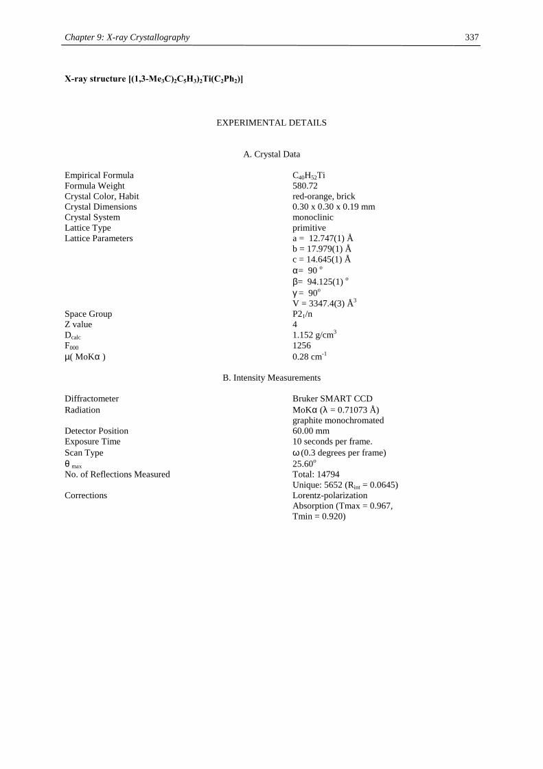
Chapter 9: X-ray Crystallography
337
X-ray structure [(1,3-Me3C)2C5H3)2Ti(C2Ph2)]
EXPERIMENTAL DETAILS
A. Crystal Data Empirical Formula C40H52Ti Formula Weight 580.72 Crystal Color, Habit red-orange, brick Crystal Dimensions 0.30 x 0.30 x 0.19 mm Crystal System monoclinic Lattice Type primitive Lattice Parameters a = 12.747(1) Å b = 17.979(1) Å c = 14.645(1) Å α= 90 o β= 94.125(1) o γ = 90o V = 3347.4(3) Å3 Space Group P21/n Z value 4 Dcalc 1.152 g/cm3 F000 1256 µ( MoKα ) 0.28 cm-1
B. Intensity Measurements Diffractometer Bruker SMART CCD Radiation MoKα (λ = 0.71073 Å) graphite monochromated Detector Position 60.00 mm Exposure Time 10 seconds per frame. Scan Type ω (0.3 degrees per frame) θ max 25.60o No. of Reflections Measured Total: 14794 Unique: 5652 (Rint = 0.0645) Corrections Lorentz-polarization Absorption (Tmax = 0.967, Tmin = 0.920)

Chapter 9: X-ray Crystallography
338
C. Structure Solution and Refinement
Structure Solution direct (SHELXS-97 (Sheldrick, 1997)) Refinement Full-matrix least-squares Function Minimized Σw(|Fo|2- |Fc|2)2 Least Squares Weighting scheme w = 1/[σ2(Fo
2) + (qP)2 + 0.000P] where P = [Fo
2 + 2Fc2]/3
q-factor 0.1474 Anomalous Dispersion All non-hydrogen atoms No. Observations (I>2.00σ(I)) 3316 No. Variables 382 Reflection/Parameter Ratio 8.68 Residuals: R; wR2; Rall 0.0815; 0.2191; 0.1254 Goodness of Fit Indicator 0.987 Max Shift/Error in Final Cycle 0.000 Maximum peak in Final Diff. Map 2.470 e-/Å3 Minimum peak in Final Diff. Map -0.790 e-/Å3 Table 1. Atomic coordinates and Uiso/Ueq and occupancy atom x y z Ueq Occupancy Ti1 0.3229(1) 0.2378(1) 0.0329(1) 0.027(1) 1 C1 0.2232(3) 0.1379(2) -0.0398(3) 0.026(1) 1 C2 0.1977(3) 0.1403(2) 0.0530(3) 0.023(1) 1 C3 0.1394(4) 0.2051(2) 0.0695(3) 0.026(1) 1 C4 0.1350(4) 0.2457(2) -0.0130(3) 0.027(1) 1 C5 0.1830(3) 0.2046(2) -0.0807(3) 0.028(1) 1 C6 0.2660(4) 0.0712(3) -0.0900(3) 0.035(1) 1 C7 0.1802(4) 0.0123(3) -0.1020(4) 0.050(2) 1 C8 0.3616(4) 0.0378(3) -0.0336(3) 0.041(1) 1 C9 0.2981(4) 0.0921(3) -0.1855(3) 0.045(1) 1 C10 0.0715(4) 0.2184(3) 0.1486(3) 0.038(1) 1 C11 0.1238(4) 0.1926(3) 0.2412(3) 0.052(2) 1 C12 -0.0278(4) 0.1718(3) 0.1279(4) 0.057(2) 1 C13 0.0406(5) 0.3000(3) 0.1553(4) 0.057(2) 1 C14 0.5096(4) 0.2691(2) 0.0112(3) 0.029(1) 1 C15 0.4508(4) 0.3359(2) 0.0152(3) 0.029(1) 1 C16 0.3774(4) 0.3402(3) -0.0631(3) 0.031(1) 1 C17 0.3885(4) 0.2740(3) -0.1132(3) 0.031(1) 1 C18 0.4667(4) 0.2300(2) -0.0666(3) 0.027(1) 1 C19 0.6159(4) 0.2537(3) 0.0622(3) 0.033(1) 1 C20 0.6190(4) 0.2741(3) 0.1618(3) 0.047(1) 1 C21 0.6462(5) 0.1727(3) 0.0550(4) 0.056(2) 1 C22 0.6961(4) 0.3009(3) 0.0166(4) 0.056(2) 1 C23 0.3173(4) 0.4095(3) -0.0958(3) 0.039(1) 1 C24 0.2345(4) 0.3900(3) -0.1742(4) 0.045(1) 1 C25 0.2597(5) 0.4459(3) -0.0196(4) 0.052(2) 1 C26 0.3967(5) 0.4638(3) -0.1316(4) 0.053(2) 1 C27 0.4043(3) 0.1432(2) 0.2304(3) 0.024(1) 1 C28 0.3812(4) 0.1484(3) 0.3221(3) 0.037(1) 1 C29 0.4030(4) 0.0907(3) 0.3827(3) 0.045(1) 1 C30 0.4463(4) 0.0254(3) 0.3550(4) 0.046(2) 1
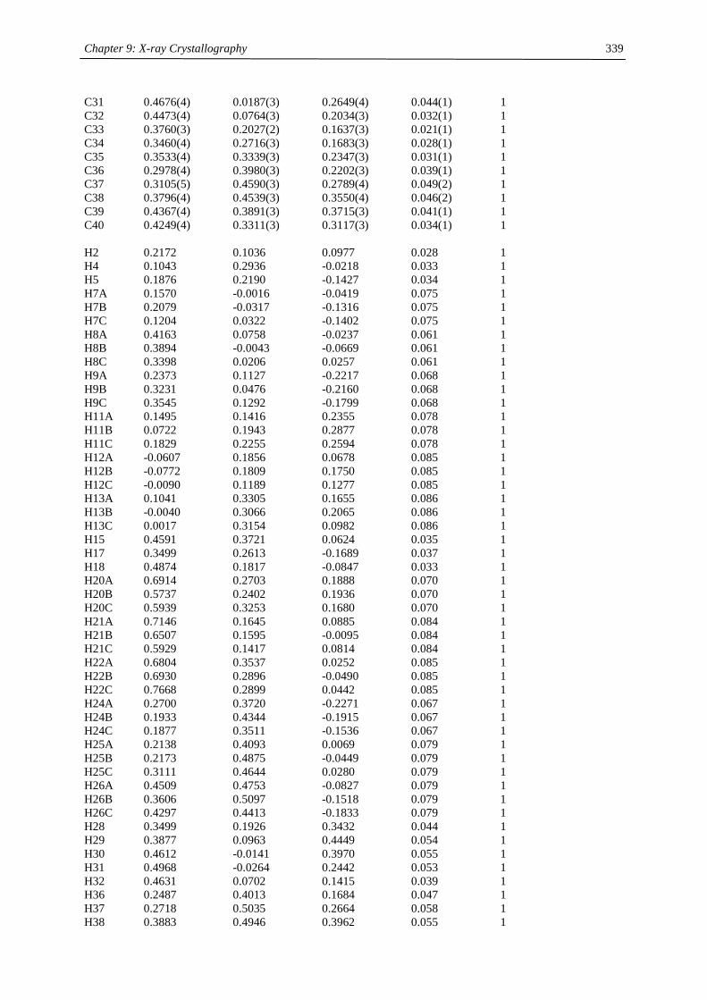
Chapter 9: X-ray Crystallography
339
C31 0.4676(4) 0.0187(3) 0.2649(4) 0.044(1) 1 C32 0.4473(4) 0.0764(3) 0.2034(3) 0.032(1) 1 C33 0.3760(3) 0.2027(2) 0.1637(3) 0.021(1) 1 C34 0.3460(4) 0.2716(3) 0.1683(3) 0.028(1) 1 C35 0.3533(4) 0.3339(3) 0.2347(3) 0.031(1) 1 C36 0.2978(4) 0.3980(3) 0.2202(3) 0.039(1) 1 C37 0.3105(5) 0.4590(3) 0.2789(4) 0.049(2) 1 C38 0.3796(4) 0.4539(3) 0.3550(4) 0.046(2) 1 C39 0.4367(4) 0.3891(3) 0.3715(3) 0.041(1) 1 C40 0.4249(4) 0.3311(3) 0.3117(3) 0.034(1) 1 H2 0.2172 0.1036 0.0977 0.028 1 H4 0.1043 0.2936 -0.0218 0.033 1 H5 0.1876 0.2190 -0.1427 0.034 1 H7A 0.1570 -0.0016 -0.0419 0.075 1 H7B 0.2079 -0.0317 -0.1316 0.075 1 H7C 0.1204 0.0322 -0.1402 0.075 1 H8A 0.4163 0.0758 -0.0237 0.061 1 H8B 0.3894 -0.0043 -0.0669 0.061 1 H8C 0.3398 0.0206 0.0257 0.061 1 H9A 0.2373 0.1127 -0.2217 0.068 1 H9B 0.3231 0.0476 -0.2160 0.068 1 H9C 0.3545 0.1292 -0.1799 0.068 1 H11A 0.1495 0.1416 0.2355 0.078 1 H11B 0.0722 0.1943 0.2877 0.078 1 H11C 0.1829 0.2255 0.2594 0.078 1 H12A -0.0607 0.1856 0.0678 0.085 1 H12B -0.0772 0.1809 0.1750 0.085 1 H12C -0.0090 0.1189 0.1277 0.085 1 H13A 0.1041 0.3305 0.1655 0.086 1 H13B -0.0040 0.3066 0.2065 0.086 1 H13C 0.0017 0.3154 0.0982 0.086 1 H15 0.4591 0.3721 0.0624 0.035 1 H17 0.3499 0.2613 -0.1689 0.037 1 H18 0.4874 0.1817 -0.0847 0.033 1 H20A 0.6914 0.2703 0.1888 0.070 1 H20B 0.5737 0.2402 0.1936 0.070 1 H20C 0.5939 0.3253 0.1680 0.070 1 H21A 0.7146 0.1645 0.0885 0.084 1 H21B 0.6507 0.1595 -0.0095 0.084 1 H21C 0.5929 0.1417 0.0814 0.084 1 H22A 0.6804 0.3537 0.0252 0.085 1 H22B 0.6930 0.2896 -0.0490 0.085 1 H22C 0.7668 0.2899 0.0442 0.085 1 H24A 0.2700 0.3720 -0.2271 0.067 1 H24B 0.1933 0.4344 -0.1915 0.067 1 H24C 0.1877 0.3511 -0.1536 0.067 1 H25A 0.2138 0.4093 0.0069 0.079 1 H25B 0.2173 0.4875 -0.0449 0.079 1 H25C 0.3111 0.4644 0.0280 0.079 1 H26A 0.4509 0.4753 -0.0827 0.079 1 H26B 0.3606 0.5097 -0.1518 0.079 1 H26C 0.4297 0.4413 -0.1833 0.079 1 H28 0.3499 0.1926 0.3432 0.044 1 H29 0.3877 0.0963 0.4449 0.054 1 H30 0.4612 -0.0141 0.3970 0.055 1 H31 0.4968 -0.0264 0.2442 0.053 1 H32 0.4631 0.0702 0.1415 0.039 1 H36 0.2487 0.4013 0.1684 0.047 1 H37 0.2718 0.5035 0.2664 0.058 1 H38 0.3883 0.4946 0.3962 0.055 1
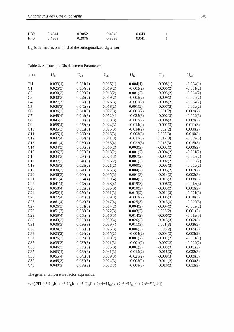
Chapter 9: X-ray Crystallography
340
H39 0.4841 0.3852 0.4245 0.049 1 H40 0.4663 0.2876 0.3226 0.041 1 Ueq is defined as one third of the orthogonalized Uij tensor Table 2. Anisotropic Displacement Parameters atom U11 U22 U33 U12 U13 U23 Ti1 0.033(1) 0.031(1) 0.016(1) 0.004(1) -0.008(1) -0.004(1) C1 0.025(3) 0.034(3) 0.019(2) -0.002(2) -0.005(2) -0.001(2) C2 0.030(3) 0.026(2) 0.013(2) 0.001(2) -0.005(2) -0.004(2) C3 0.030(3) 0.029(2) 0.019(2) -0.003(2) -0.009(2) -0.005(2) C4 0.027(3) 0.028(3) 0.026(3) -0.001(2) -0.008(2) -0.004(2) C5 0.025(3) 0.042(3) 0.016(2) 0.001(2) -0.007(2) -0.002(2) C6 0.036(3) 0.041(3) 0.027(3) -0.005(2) 0.001(2) 0.009(2) C7 0.048(4) 0.049(3) 0.052(4) -0.025(3) -0.002(3) -0.002(3) C8 0.045(3) 0.038(3) 0.038(3) -0.002(2) -0.006(3) 0.009(2) C9 0.058(4) 0.053(3) 0.024(3) -0.014(2) -0.001(3) 0.011(3) C10 0.035(3) 0.052(3) 0.025(3) -0.014(2) 0.002(2) 0.000(2) C11 0.055(4) 0.085(4) 0.016(3) -0.003(3) 0.005(3) 0.010(3) C12 0.047(4) 0.084(4) 0.041(3) -0.017(3) 0.017(3) -0.009(3) C13 0.061(4) 0.059(4) 0.055(4) -0.022(3) 0.015(3) 0.015(3) C14 0.034(3) 0.038(3) 0.015(2) 0.003(2) -0.002(2) 0.000(2) C15 0.036(3) 0.033(3) 0.018(2) 0.001(2) -0.004(2) -0.001(2) C16 0.034(3) 0.036(3) 0.023(3) 0.007(2) -0.005(2) -0.003(2) C17 0.037(3) 0.040(3) 0.016(2) 0.001(2) -0.002(2) -0.006(2) C18 0.035(3) 0.025(2) 0.021(2) 0.000(2) -0.003(2) -0.002(2) C19 0.034(3) 0.040(3) 0.025(3) 0.004(2) -0.003(2) 0.002(2) C20 0.036(3) 0.066(4) 0.035(3) 0.001(3) -0.014(2) 0.002(3) C21 0.051(4) 0.054(4) 0.059(4) 0.004(3) -0.015(3) 0.008(3) C22 0.041(4) 0.078(4) 0.048(4) 0.019(3) -0.008(3) -0.013(3) C23 0.058(4) 0.032(3) 0.025(3) 0.010(2) -0.003(2) 0.003(2) C24 0.052(4) 0.050(3) 0.031(3) 0.013(2) -0.011(3) -0.001(3) C25 0.072(4) 0.045(3) 0.039(3) -0.002(2) -0.005(3) 0.018(3) C26 0.061(4) 0.049(3) 0.047(4) 0.025(3) -0.013(3) -0.009(3) C27 0.026(3) 0.031(3) 0.014(2) 0.004(2) -0.004(2) -0.002(2) C28 0.051(3) 0.038(3) 0.022(3) 0.003(2) 0.003(2) 0.001(2) C29 0.059(4) 0.058(4) 0.016(3) 0.014(2) -0.006(2) -0.012(3) C30 0.043(3) 0.052(4) 0.039(4) 0.026(3) -0.013(3) 0.002(3) C31 0.036(3) 0.036(3) 0.059(4) 0.011(3) 0.001(3) 0.009(2) C32 0.034(3) 0.038(3) 0.025(3) 0.006(2) 0.006(2) 0.005(2) C33 0.023(2) 0.024(2) 0.015(2) -0.004(2) -0.004(2) 0.003(2) C34 0.026(3) 0.039(3) 0.020(2) 0.001(2) -0.001(2) -0.001(2) C35 0.035(3) 0.037(3) 0.021(3) -0.001(2) -0.007(2) -0.002(2) C36 0.046(3) 0.035(3) 0.035(3) 0.001(2) -0.009(3) 0.001(2) C37 0.063(4) 0.038(3) 0.041(3) -0.015(2) -0.018(3) 0.022(3) C38 0.055(4) 0.043(3) 0.039(3) -0.021(2) -0.009(3) 0.009(3) C39 0.045(3) 0.052(3) 0.024(3) -0.005(2) -0.011(2) 0.000(3) C40 0.040(3) 0.038(3) 0.022(3) -0.008(2) -0.010(2) 0.012(2) The general temperature factor expression: exp(-2Π2(a*2U11h2 + b*2U22k2 + c*2U33l2 + 2a*b*U12hk +2a*c*U13 hl + 2b*c*U23kl))
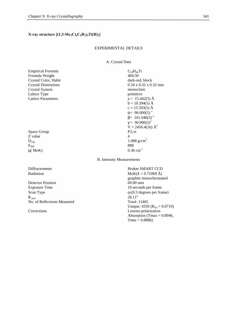
Chapter 9: X-ray Crystallography
341
X-ray structure [(1,3-Me3C)2C5H3)2Ti(H)2]
EXPERIMENTAL DETAILS
A. Crystal Data Empirical Formula C26H44Ti Formula Weight 404.50 Crystal Color, Habit dark-red, block Crystal Dimensions 0.34 x 0.32 x 0.32 mm Crystal System monoclinic Lattice Type primitive Lattice Parameters a = 15.442(5) Å b = 10.394(5) Å c = 15.593(5) Å α= 90.000(5) o β= 101.040(5) o γ = 90.000(5)o V = 2456.4(16) Å3 Space Group P21/n Z value 4 Dcalc 1.088 g/cm3 F000 888 µ( MoK) 0.36 cm-1
B. Intensity Measurements Diffractometer Bruker SMART CCD Radiation MoK(λ = 0.71069 Å) graphite monochromated Detector Position 60.00 mm Exposure Time 10 seconds per frame. Scan Type ω (0.3 degrees per frame) θ max 26.11o No. of Reflections Measured Total: 11465 Unique: 4350 (Rint = 0.0719) Corrections Lorentz-polarization Absorption (Tmax = 0.8946, Tmin = 0.8886)

Chapter 9: X-ray Crystallography
342
C. Structure Solution and Refinement
Structure Solution direct (SHELXS-97 (Sheldrick, 1997)) Refinement Full-matrix least-squares Function Minimized Σw(|Fo|2- |Fc|2)2 Least Squares Weighting scheme w = 1/[σ2(Fo
2) + (qP)2 + 0.000P] where P = [Fo
2 + 2Fc2]/3
q-factor 0.1021 Anomalous Dispersion All non-hydrogen atoms No. Observations (I>2.00σ(I)) 2843 No. Variables 264 Reflection/Parameter Ratio 10.77 Residuals: R; wR2; Rall 0.0568; 0.1413; 0.0960 Goodness of Fit Indicator 0.958 Max Shift/Error in Final Cycle 0.001 Maximum peak in Final Diff. Map 0.643 e-/Å3 Minimum peak in Final Diff. Map -0.636 e-/Å3 Table 1. Atomic coordinates and /Ueq and occupancy atom x y z Ueq Occupancy Ti1 0.4715(1) -0.1606(1) 0.7019(1) 0.025(1) 1 C1 0.4212(2) -0.3340(3) 0.7789(2) 0.027(1) 1 C2 0.3558(2) -0.3076(3) 0.7046(2) 0.026(1) 1 C3 0.3883(2) -0.3341(3) 0.6278(2) 0.027(1) 1 C4 0.4766(2) -0.3760(3) 0.6556(2) 0.031(1) 1 C5 0.4960(2) -0.3766(3) 0.7480(2) 0.030(1) 1 C6 0.5206(2) 0.0464(3) 0.7536(2) 0.026(1) 1 C7 0.5600(2) 0.0169(3) 0.6808(2) 0.026(1) 1 C8 0.6186(2) -0.0892(3) 0.7014(2) 0.026(1) 1 C9 0.6137(2) -0.1263(3) 0.7873(2) 0.029(1) 1 C10 0.5538(2) -0.0445(3) 0.8193(2) 0.028(1) 1 C11 0.4078(2) -0.3349(3) 0.8734(2) 0.029(1) 1 C12 0.3568(2) -0.2154(3) 0.8939(2) 0.039(1) 1 C13 0.3523(2) -0.4554(3) 0.8851(2) 0.039(1) 1 C14 0.4967(2) -0.3422(3) 0.9378(2) 0.040(1) 1 C15 0.3344(2) -0.3398(3) 0.5354(2) 0.033(1) 1 C16 0.3921(3) -0.3279(4) 0.4663(2) 0.049(1) 1 C17 0.2869(3) -0.4715(4) 0.5254(2) 0.056(1) 1 C18 0.2641(2) -0.2338(3) 0.5204(2) 0.042(1) 1 C19 0.6823(2) -0.1401(3) 0.6458(2) 0.030(1) 1 C20 0.7192(2) -0.2729(3) 0.6780(2) 0.038(1) 1 C21 0.6381(2) -0.1497(3) 0.5497(2) 0.038(1) 1 C22 0.7588(2) -0.0435(3) 0.6551(2) 0.037(1) 1 C23 0.4676(2) 0.1674(3) 0.7649(2) 0.029(1) 1 C24 0.4068(2) 0.1471(3) 0.8301(2) 0.040(1) 1 C25 0.5350(2) 0.2723(3) 0.8007(2) 0.041(1) 1 C26 0.4129(2) 0.2130(3) 0.6781(2) 0.040(1) 1 H27 0.384(2) -0.073(3) 0.701(2) 0.054(10) 1 H28 0.439(2) -0.115(3) 0.604(2) 0.041(9) 1 H2 0.2982 -0.2764 0.7060 0.032 1 H4 0.5160 -0.3997 0.6184 0.038 1
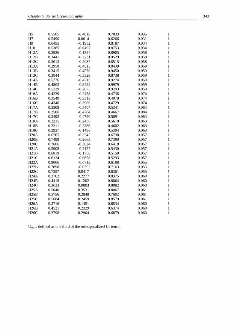
Chapter 9: X-ray Crystallography
343
H5 0.5505 -0.4016 0.7833 0.035 1 H7 0.5490 0.0614 0.6266 0.031 1 H9 0.6455 -0.1952 0.8187 0.034 1 H10 0.5385 -0.0497 0.8753 0.034 1 H12A 0.3926 -0.1384 0.8905 0.058 1 H12B 0.3441 -0.2231 0.9529 0.058 1 H12C 0.3013 -0.2087 0.8515 0.058 1 H13A 0.2958 -0.4515 0.8439 0.059 1 H13B 0.3415 -0.4579 0.9450 0.059 1 H13C 0.3844 -0.5329 0.8738 0.059 1 H14A 0.5276 -0.4213 0.9274 0.059 1 H14B 0.4862 -0.3422 0.9979 0.059 1 H14C 0.5329 -0.2675 0.9293 0.059 1 H16A 0.4239 -0.2458 0.4738 0.074 1 H16B 0.3549 -0.3313 0.4079 0.074 1 H16C 0.4346 -0.3989 0.4729 0.074 1 H17A 0.3308 -0.5407 0.5341 0.084 1 H17B 0.2506 -0.4784 0.4667 0.084 1 H17C 0.2492 -0.4790 0.5691 0.084 1 H18A 0.2235 -0.2456 0.5610 0.063 1 H18B 0.2311 -0.2386 0.4602 0.063 1 H18C 0.2927 -0.1496 0.5304 0.063 1 H20A 0.6705 -0.3345 0.6738 0.057 1 H20B 0.7499 -0.2663 0.7390 0.057 1 H20C 0.7606 -0.3024 0.6418 0.057 1 H21A 0.5908 -0.2137 0.5430 0.057 1 H21B 0.6819 -0.1756 0.5150 0.057 1 H21C 0.6134 -0.0658 0.5293 0.057 1 H22A 0.8006 -0.0713 0.6188 0.055 1 H22B 0.7890 -0.0395 0.7163 0.055 1 H22C 0.7357 0.0417 0.6361 0.055 1 H24A 0.3762 0.2277 0.8375 0.060 1 H24B 0.4418 0.1202 0.8864 0.060 1 H24C 0.3633 0.0803 0.8082 0.060 1 H25A 0.5040 0.3531 0.8067 0.061 1 H25B 0.5756 0.2848 0.7602 0.061 1 H25C 0.5684 0.2459 0.8579 0.061 1 H26A 0.3716 0.1451 0.6534 0.060 1 H26B 0.4521 0.2329 0.6374 0.060 1 H26C 0.3798 0.2904 0.6879 0.060 1 Ueq is defined as one third of the orthogonalized Uij tensor
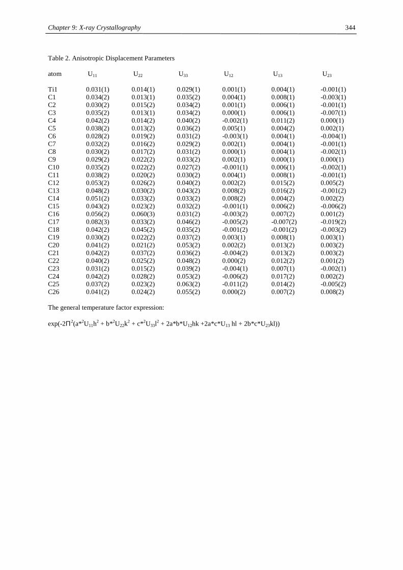
Chapter 9: X-ray Crystallography
344
Table 2. Anisotropic Displacement Parameters atom U11 U22 U33 U12 U13 U23 Ti1 0.031(1) 0.014(1) 0.029(1) 0.001(1) 0.004(1) -0.001(1) C1 0.034(2) 0.013(1) 0.035(2) 0.004(1) 0.008(1) -0.003(1) C2 0.030(2) 0.015(2) 0.034(2) 0.001(1) 0.006(1) -0.001(1) C3 0.035(2) 0.013(1) 0.034(2) 0.000(1) 0.006(1) -0.007(1) C4 0.042(2) 0.014(2) 0.040(2) -0.002(1) 0.011(2) 0.000(1) C5 0.038(2) 0.013(2) 0.036(2) 0.005(1) 0.004(2) 0.002(1) C6 0.028(2) 0.019(2) 0.031(2) -0.003(1) 0.004(1) -0.004(1) C7 0.032(2) 0.016(2) 0.029(2) 0.002(1) 0.004(1) -0.001(1) C8 0.030(2) 0.017(2) 0.031(2) 0.000(1) 0.004(1) -0.002(1) C9 0.029(2) 0.022(2) 0.033(2) 0.002(1) 0.000(1) 0.000(1) C10 0.035(2) 0.022(2) 0.027(2) -0.001(1) 0.006(1) -0.002(1) C11 0.038(2) 0.020(2) 0.030(2) 0.004(1) 0.008(1) -0.001(1) C12 0.053(2) 0.026(2) 0.040(2) 0.002(2) 0.015(2) 0.005(2) C13 0.048(2) 0.030(2) 0.043(2) 0.008(2) 0.016(2) -0.001(2) C14 0.051(2) 0.033(2) 0.033(2) 0.008(2) 0.004(2) 0.002(2) C15 0.043(2) 0.023(2) 0.032(2) -0.001(1) 0.006(2) -0.006(2) C16 0.056(2) 0.060(3) 0.031(2) -0.003(2) 0.007(2) 0.001(2) C17 0.082(3) 0.033(2) 0.046(2) -0.005(2) -0.007(2) -0.019(2) C18 0.042(2) 0.045(2) 0.035(2) -0.001(2) -0.001(2) -0.003(2) C19 0.030(2) 0.022(2) 0.037(2) 0.003(1) 0.008(1) 0.003(1) C20 0.041(2) 0.021(2) 0.053(2) 0.002(2) 0.013(2) 0.003(2) C21 0.042(2) 0.037(2) 0.036(2) -0.004(2) 0.013(2) 0.003(2) C22 0.040(2) 0.025(2) 0.048(2) 0.000(2) 0.012(2) 0.001(2) C23 0.031(2) 0.015(2) 0.039(2) -0.004(1) 0.007(1) -0.002(1) C24 0.042(2) 0.028(2) 0.053(2) -0.006(2) 0.017(2) 0.002(2) C25 0.037(2) 0.023(2) 0.063(2) -0.011(2) 0.014(2) -0.005(2) C26 0.041(2) 0.024(2) 0.055(2) 0.000(2) 0.007(2) 0.008(2) The general temperature factor expression: exp(-2Π2(a*2U11h2 + b*2U22k2 + c*2U33l2 + 2a*b*U12hk +2a*c*U13 hl + 2b*c*U23kl))

Chapter 9: X-ray Crystallography
345
References
[1] Sheldrick, G. SHELXL-97, Program for refinement of crystal structures, University of Göttingen:
Göttingen, Germany, 1997.
[2] SMART Area-Detection Software Package, Siemens Industrial Automation, Inc: Madison, WI, 1995.
[3] SAINT, 4.024; Siemens Industrial Automation, Inc: Madison, WI, 1995.
[4] Sheldrick, G. M. XPREP, 5.03; Siemens Industrial Automation, Inc: Madison, WI, 1995.
[5] Sheldrick, G. M. SADABS, Siemens Industrial Automation, Inc: Madison, WI, 1996.
[6] Sheldrick, G. SHELXS-97, Program for solution of crystal structures, University of Göttingen:
Göttingen, Germany, 1997.