Embed Size (px)
Citation preview
Diplomarbeit
THE ROLE OF
OPTICAL COHERENCE TOMOGRAPHY
IN THE DIAGNOSIS OF ERYTHEMATOSQUAMOUS
SKIN LESIONS
eingereicht von
Clemens Cijan
zur Erlangung des akademischen Grades
Doktor der gesamten Heilkunde
(Dr. med. univ.)
an der
Medizinischen Universität Graz
ausgeführt an der
Universitätsklinik für Dermatologie und Venerologie
unter Anleitung von
Priv.-Doz. Dr. med. univ. Iris Zalaudek
und
Dr. med. univ. Edith Johanna Arzberger
Graz, am 15.2.2016

ii
Eidesstattliche Erklärung
Ich erkläre ehrenwörtlich, dass ich die vorliegende Arbeit selbstständig und ohne fremde Hilfe verfasst habe, andere als die angegebenen Quellen nicht verwendet habe und die den benutzten Quellen wörtlich oder inhaltlich entnommenen Stellen als solche kenntlich gemacht habe.
Graz, am 15.2.2016 Clemens Cijan eh.

iii
Danksagung
Mein besonderer Dank gilt Frau Priv.-Doz. Dr. Iris Zalaudek für die geduldige,
fachkompetente Betreuung und moralische Unterstützung, die Anregungen und
Verbesserungsvorschläge bei der Erstellung dieser Diplomarbeit.
Herrn Univ.-Prof. Dr. Rainer Hofmann-Wellenhof möchte ich für die Heranführung
an die Thematik und Frau Dr. Edith Arzberger für die große Hilfe im
organisatorischen Ablauf danken.
Zuletzt möchte ich meiner Familie herzlich danken, vor allem meinen Eltern, die
mir dieses Studium ermöglicht haben und mich im gesamten Verlauf immer
unterstützt und gefördert haben.
iv
Zusammenfassung
Hintergrund
Aktinische Keratosen, Morbus Bowen, superfizielles Basaliom, Psoriasis vulgaris
und nummuläres Ekzem können sich als erythematosquamöse
Hautveränderungen präsentieren. Da die Behandlung dieser Krankheitsbilder
deutlich variiert, muss zwischen ihnen verlässlich unterschieden werden können.
Optische Kohärenztomographie (OCT) ist ein innovatives bildgebendes Verfahren,
das zunehmend Verwendung in der Dermatologie findet. Der Stellenwert der OCT
bei der Differentialdiagnose der zuvor erwähnten Hautveränderungen wurde noch
nicht untersucht.
Ziel der Arbeit
Bezugnehmend auf den letzten Stand der Literatur wurden morphologische
Kriterien für die optische Kohärenztomographie festgelegt, die den jeweiligen
Krankheitsbildern zugehörig sind. In weiterer Folge wurde versucht, den Nutzen
der optischen Kohärenztomographie bei der Differentialdiagnose von
erythematosquamösen Hautveränderungen zu beurteilen.
Methode
Erythematosquamöse Hautveränderungen wurden mit HD-OCT abgebildet und
auf das Vorhandensein von 30 definierten OCT Merkmalen untersucht. Mit
statistischen Methoden wurden OCT Merkmale herausgefiltert, die bei der
Differentialdiagnose erythematosquamöser Hautveränderungen hilfreich sein
könnten.
Ergebnisse
Es wurden 51 Teilnehmer und Teilnehmerinnen mit 74 Läsionen eingeschlossen
und von diesen insgesamt 220 High Definition (HD) OCT 3D-Bilder aufgenommen.
Das durchschnittliche Alter der Studienkohorte (51% männlich, 49% weiblich)
betrug 73 Jahre (±15,7 Jahre). Von den 74 inkludierten Läsionen war die
Diagnose in 54 Fällen histologisch und in 20 Fällen klinisch gesichert.

v
Etablierte und häufige OCT-Merkmale wurden in Tabellen dargestellt und
statistisch untersucht. Bei der Basaliom-Gruppe wurde ein Algorithmus von
Boone et al. angewendet und evaluiert.
Außerdem wurde anhand der Ergebnisse der Studie ein Algorithmus zur
Differentialdiagnose erythematosquamöser Plaques entwickelt.
Schlussfolgerungen
Die OCT ist ein geeignetes Verfahren zur Differenzierung der häufigsten
Hautveränderungen, die sich als erythematosquamöse Plaques darstellen. Ein
Zusammenhang zwischen bestimmten OCT-Merkmalen und Krankheitsbildern
konnte ermittelt werden. Die Eignung des vorgeschlagenen Algorithmus bedarf
weiterer Überprüfung.
vi
Abstract
Background
Actinic keratosis, Bowen’s disease, superficial basal cell carcinoma, psoriasis
vulgaris and nummular eczema may present as erythematosquamous lesions. As
their treatment varies significantly, an accurate differentiation between these
entities is required. Optical coherence tomography (OCT) is an innovative optical
imaging technology increasingly finding use in dermatology. The role of OCT for
the differential diagnosis of the aforementioned diseases has not yet been
explored.
Objective
With reference to current relevant literature (2015) we aimed to define
morphological criteria identifiable in Optical Coherence Tomography of the
respective diseases and tried to assess the value of Optical Coherence
Tomography in the diagnosis of erythematosquamous skin lesions.
Method
Erythematosquamous lesions were imaged by HD-OCT and examined for the
presence of 30 defined OCT features. Statistical analysis was performed to
identify morphological findings possibly useful for the differential diagnosis of
erythematosquamous skin lesions.
Results
Fifty-one participants presenting with 74 lesions depicted in 220 HD-OCT 3D
images were included. The mean age of the study cohort (51% male, 49% female)
was 73 years (±15,7 years). Out of the 74 included lesions, a histologically verified
diagnosis was found in 54 cases and a distinct clinical diagnosis existed in 20
cases. The most common or established OCT features were presented and
statistical analysis was performed. For the basal cell carcinoma group, an
algorithm by Boone et al. was applied and evaluated. Finally, an algorithm for the
differential diagnosis of erythematosquamous plaques was developed, based
upon the results of the study.

vii
Conclusion
OCT is a useful tool for differentiating common skin conditions presenting as
erythematosquamous plaques. A correlation between certain OCT features and
skin conditions could be confirmed. The eligibility of the proposed algorithm
requires additional validation.
viii
Index INTRODUCTION .................................................................................................... 1
Optical Coherence Tomography in Dermatology ................................................ 1
Historical aspects ............................................................................................ 1
Technical aspects ........................................................................................... 2
Skin morphology in Optical Coherence Tomography ...................................... 3
Skin conditions presenting as erythematosquamous plaque ............................ 10
Definition of erythematosquamous skin lesions ............................................ 10
Actinic keratosis ............................................................................................ 10
Bowen’s disease ........................................................................................... 12
Basal cell carcinoma ..................................................................................... 14
Psoriasis ........................................................................................................ 17
Nummular eczema ........................................................................................ 21
Study aims ........................................................................................................ 23
MATERIAL AND METHODS ................................................................................ 24
Process of image acquisition ............................................................................ 24
Process of image interpretation ........................................................................ 25
Definition of the OCT features .......................................................................... 25
Statistical analysis ............................................................................................ 26
RESULTS ............................................................................................................. 27
Frequency of the individual diseases ................................................................ 28
Overall frequency of the evaluated OCT features ............................................. 28
Diagnostic value of the OCT features for the diagnosis of erythematosquamous
plaques ............................................................................................................. 30
Results for actinic keratosis and Bowen’s disease ........................................ 30
Results for basal cell carcinoma .................................................................... 30
Results for psoriasis ...................................................................................... 31
Results for eczema ....................................................................................... 32
Algorithm for the differential diagnosis of erythematosquamous plaques ......... 33
DISCUSSION ....................................................................................................... 40
ix
List of Abbreviations
ACE Angiotensin converting enzyme
AK Actinic keratosis
BCC Basal cell carcinoma
C5a Complement component 5a
DEJ Dermoepidermal junction
EGF Epidermal Growth Factor
HD High Definition
HIV Human immunodeficiency virus
HLA Human Leukocyte Antigen
HPV Human papillomavirus
IFN γ Interferon gamma
IL-2 Interleukin 2
IL-8 Interleukin 8
IL-17 Interleukin 17
LTB4 Leukotriene B4
NSAIDs Nonsteroidal anti-inflammatory drugs
OCT Optical coherence tomography
PTCH/SMOH Patched protein/ Smoothened receptor
Th1 cells Type 1 helper T cells
TNF alpha Tumor Necrosis Factor
TP53 Tumor protein p53
x
List of Figures
Fig. 1 Simplified illustration of the principle of the OCT. Fujimoto JG, Pitris C,
Boppart SA, Brezinski ME. Optical coherence tomography: an emerging
technology for biomedical imaging and optical biopsy. Neoplasia. 2000;2(1-
2):9-25 ............................................................................................................. 3
Fig. 2 Healthy skin. Cross-sectional (a) and en-face (b) view of healthy skin of the
volar forearm. Department of Dermatology, Medical University of Graz ......... 6
Fig. 3 Healthy skin. Cross-sectional (a) and en-face (b) view of healthy skin of the
forehead. Department of Dermatology, Medical University of Graz ................ 7
Fig. 4 Healthy skin. Cross-sectional (a) and en-face (b) view of healthy skin of the
hand palm. Department of Dermatology, Medical University of Graz .............. 8
Fig. 5 HD-OCT algorithm for discrimination of basal cell carcinomas from imitators
and differentiation between subtypes. Boone MA, Suppa M, Pellacani G,
Marneffe A, Miyamoto M, Alarcon I, et al. High-definition optical coherence
tomography algorithm for discrimination of basal cell carcinoma from clinical
BCC imitators and differentiation between common subtypes. J Eur Acad
Dermatol Venereol. 2015 .............................................................................. 26
Fig. 6 Algorithm for differential diagnosis of erythematoaquamous plaques.
Department of Dermatology, Medical University of Graz............................... 34
Fig. 7 Tumorous structure. In cross-sectional view (a) a lobular structure
connected to the epidermis is visible. Department of Dermatology, Medical
University of Graz .......................................................................................... 35
Fig. 8 Tumorous structure. In cross-sectional (a) and en-face (b) view lobular
structures connected to the epidermis can be identified. Department of
Dermatology, Medical University of Graz ...................................................... 36
Fig. 9 Tumorous structure. In cross-sectional (a) and en-face (b) view an
extensive lobular structure connected to the epidermis can be seen.
Department of Dermatology, Medical University of Graz............................... 37
Fig. 10 Psoriasis. Cross-sectional (a) and en-face (b) view of a psoriatic patch 1.
Department of Dermatology, Medical University of Graz............................... 38
Fig. 11 Psoriasis. Cross-sectional (a) and en-face (b) view of a psoriatic patch 2.
Department of Dermatology, Medical University of Graz............................... 39

xi
List of Tables
Table 1 Frequency of the individual skin conditions. ............................................ 28
Table 2 Frequency of HD-OCT features of 74 erythematosquamous plaques. ... 29
Table 3 OCT features of the actinic keratosis or Bowen’s disease group. ........... 30
Table 4 Statistical performance of the algorithm by Boone et al. ......................... 31
Table 5 OCT features of the basal cell carcinoma group. .................................... 31
Table 6 OCT features of the psoriasis group. ...................................................... 32
Table 7 OCT features of the eczema group. ........................................................ 32
Table 8 Statistical analysis of the algorithm for differential diagnosis of
erythematosquamous plaques. ..................................................................... 34
1
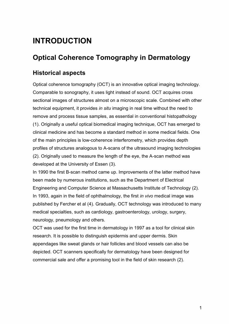
INTRODUCTION
Optical Coherence Tomography in Dermatology
Historical aspects Optical coherence tomography (OCT) is an innovative optical imaging technology.
Comparable to sonography, it uses light instead of sound. OCT acquires cross
sectional images of structures almost on a microscopic scale. Combined with other
technical equipment, it provides in situ imaging in real time without the need to
remove and process tissue samples, as essential in conventional histopathology
(1). Originally a useful optical biomedical imaging technique, OCT has emerged to
clinical medicine and has become a standard method in some medical fields. One
of the main principles is low-coherence interferometry, which provides depth
profiles of structures analogous to A-scans of the ultrasound imaging technologies
(2). Originally used to measure the length of the eye, the A-scan method was
developed at the University of Essen (3).
In 1990 the first B-scan method came up. Improvements of the latter method have
been made by numerous institutions, such as the Department of Electrical
Engineering and Computer Science at Massachusetts Institute of Technology (2).
In 1993, again in the field of ophthalmology, the first in vivo medical image was
published by Fercher et al (4). Gradually, OCT technology was introduced to many
medical specialties, such as cardiology, gastroenterology, urology, surgery,
neurology, pneumology and others.
OCT was used for the first time in dermatology in 1997 as a tool for clinical skin
research. It is possible to distinguish epidermis and upper dermis. Skin
appendages like sweat glands or hair follicles and blood vessels can also be
depicted. OCT scanners specifically for dermatology have been designed for
commercial sale and offer a promising tool in the field of skin research (2).
2
Technical aspects While it is possible to directly measure reflected sound waves in sonography, this
does not apply to light. With a velocity of around 3x10⁸ m/s and only short
travelling distances in skin examinations, other solutions had to be found to
measure the diminutive echo time delay of about 30 femtoseconds. Correlation
and interferometry of low coherent light offer a key to solve this problem.
The Michelson type interferometer analyses backscattered light from the tissue
sample by comparing it to the backscattered light of a known reference path
length.
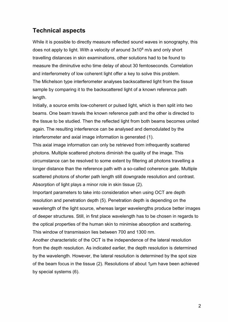
Initially, a source emits low-coherent or pulsed light, which is then split into two
beams. One beam travels the known reference path and the other is directed to
the tissue to be studied. Then the reflected light from both beams becomes united
again. The resulting interference can be analysed and demodulated by the
interferometer and axial image information is generated (1).
This axial image information can only be retrieved from infrequently scattered
photons. Multiple scattered photons diminish the quality of the image. This
circumstance can be resolved to some extent by filtering all photons travelling a
longer distance than the reference path with a so-called coherence gate. Multiple
scattered photons of shorter path length still downgrade resolution and contrast.
Absorption of light plays a minor role in skin tissue (2).
Important parameters to take into consideration when using OCT are depth
resolution and penetration depth (5). Penetration depth is depending on the
wavelength of the light source, whereas larger wavelengths produce better images
of deeper structures. Still, in first place wavelength has to be chosen in regards to
the optical properties of the human skin to minimise absorption and scattering.
This window of transmission lies between 700 and 1300 nm.
Another characteristic of the OCT is the independence of the lateral resolution
from the depth resolution. As indicated earlier, the depth resolution is determined
by the wavelength. However, the lateral resolution is determined by the spot size
of the beam focus in the tissue (2). Resolutions of about 1µm have been achieved
by special systems (6).
3
Another important parameter is contrast, which is created by the different
refractive indices of the sample. Since scattering plays a crucial role in imaging
quality, not the content of any pigment itself, but rather the density, distribution and
orientation of the refracting media influence the contrast (2). The best contrast can
be achieved with central wavelengths around 800nm (7).
Fig. 1 Simplified illustration of the principle of the OCT.
Skin morphology in Optical Coherence Tomography
Morphology of normal skin
Epidermis
The first noticeable observation when examining the skin in slice mode (B-scan) is
a highly reflective phenomenon on the skin surface, the entrance signal (8). The
stratum corneum is best seen when examining palmoplantar skin, because only
here it is thick enough for conventional OCT to be visualized. It presents as a well-
demarcated, homogenous low-scattering zone and appears about 300 μm thick in
OCT in vivo (2).
The next layers represent the rest of the epidermis. While scattering more
intensely than the stratum corneum, it scatters lower than the dermis. It is possible
to distinguish the dermoepidermal junction from the dermis as long as the margin
is even. Also the basal layer and region around the basal membrane can be
depicted and present as a weak signal adjacent to the papillary dermis.
4
Finally, the dermis appears brighter than the epidermis, with exception of possible
blood and lymph vessels.
In addition to slice mode also the en-face mode can be used in OCT. When
viewing OCT images in en-face mode, the stratum corneum presents again as the
first layer. Due to its high refractive index, the keratin acts as a natural contrast
agent and thus the margins of single corneocytes can be seen. The acaryote
polygonal shaped corneocytes are arranged in groups separated by almost black
skin folds.
The next layer is the stratum granulosum, which consists of two to four cell layers.
The cells in this layer can be recognized by their dark centers, resembling the
nucleus, which are surrounded by a bright and grainy cytoplasm.
Subsequently the stratum spinosum presents as a layer of smaller cells
assembled in a tight honeycomb pattern.
Eventually the basal layer can be seen at the dermoepidermal junction, appearing
as bright cells wrapping the dark dermal papillae.
These observations can only be made with High Definition (HD)-OCT (8).
Dermis
When viewing the dermis in en face mode, it presents as a bright zone due to its
high collagen concentration, which leads to prominent reflection. Blood and lymph
vessels can be identified.
Also deeper layers of the dermis can be visualized with OCT. It is possible to
observe the superficial reticular dermis, which shows a web of reticulated fibres
and small blood vessels. Pilo-sebaceus units are displayed as hollow-centred
structures surrounded by elliptical cells with the hair shaft as a core (8).
Role of skin phototype
Since the melanin-rich keratinozytes of darkly pigmented skin tend to be more
reflective, contrast is increased and differentiation of the specific skin layers is
easier to achieve. Whereas for instance it may be difficult to delineate the basal
layer in phototype II, it is more obvious in phototype V. Furthermore, the papillary
dermis appears darker compared to the epidermis in higher phototypes (8).

5
Role of the anatomical site
Depending on the anatomical region, variants of skin architecture can be found.
The most significant differences can be found between glabrous and non-glabrous
skin. In palmoplantar skin a particularly bright band, corresponding to the entrance
signal, can be distinguished.
Also, the stratum corneum is best seen on this site (8). The dermatoglyphics
appear as a wavy pattern (2). Non-glabrous skin shows consistency to some
degree. Reflectivity is influenced by the presence of skin appendages like hair
follicles or sebaceous glands (8).
6
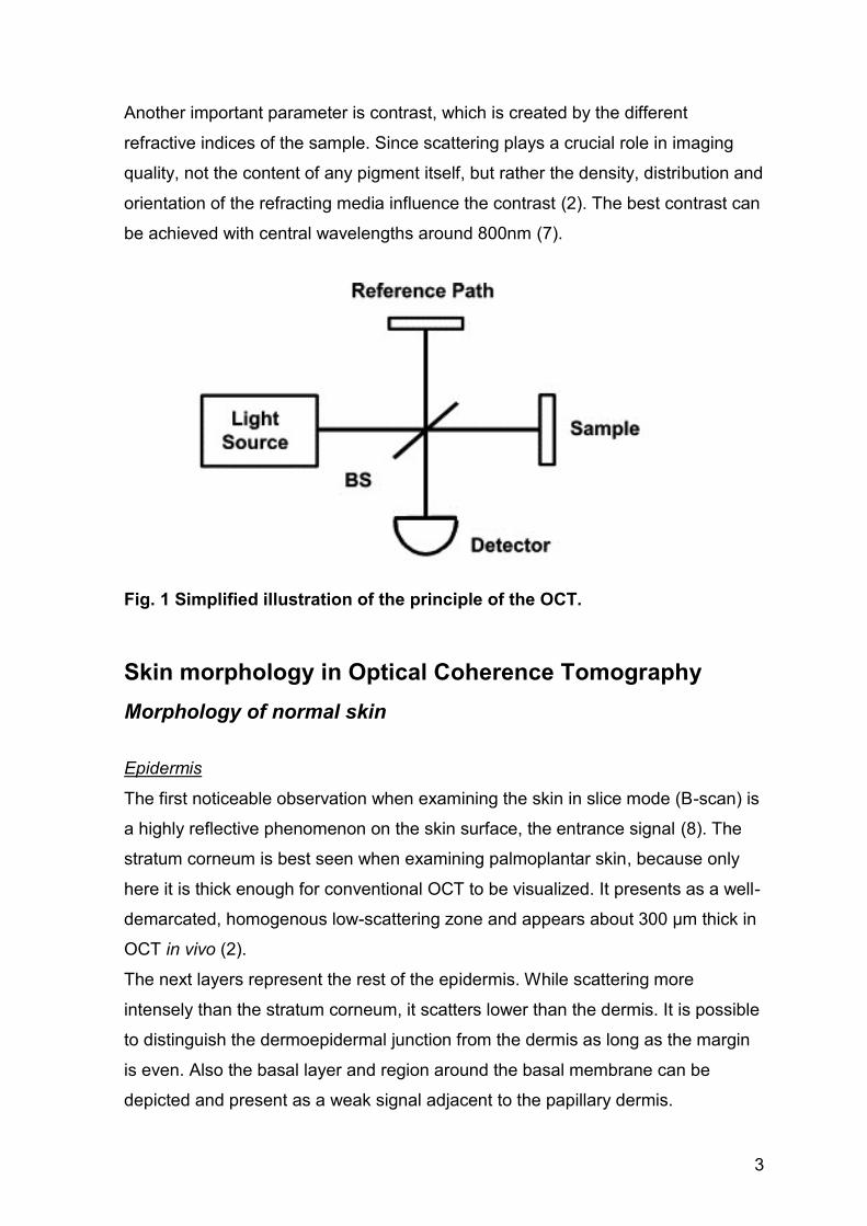
Fig. 2 Healthy skin. Cross-sectional (a) and en-face (b) view of healthy skin of the volar forearm. The stratum corneum appears as two bright lines separated by a thin dark line (green arrow) on the surface of the skin. The dermoepidermal junction (red arrows) is clearly distinguishable, with the darker lower layers of the epidermis bordering to bright fibrous structures and blood vessels of the upper dermis. In en-face mode, dermal papillae present as evenly distributed round and homogenously dark areas (yellow arrows).
7
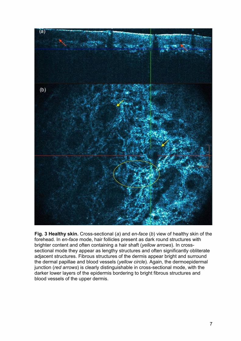
Fig. 3 Healthy skin. Cross-sectional (a) and en-face (b) view of healthy skin of the forehead. In en-face mode, hair follicles present as dark round structures with brighter content and often containing a hair shaft (yellow arrows). In cross-sectional mode they appear as lengthy structures and often significantly obliterate adjacent structures. Fibrous structures of the dermis appear bright and surround the dermal papillae and blood vessels (yellow circle). Again, the dermoepidermal junction (red arrows) is clearly distinguishable in cross-sectional mode, with the darker lower layers of the epidermis bordering to bright fibrous structures and blood vessels of the upper dermis.
8
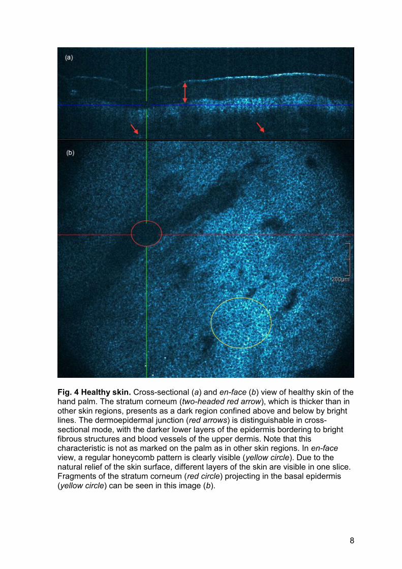
Fig. 4 Healthy skin. Cross-sectional (a) and en-face (b) view of healthy skin of the hand palm. The stratum corneum (two-headed red arrow), which is thicker than in other skin regions, presents as a dark region confined above and below by bright lines. The dermoepidermal junction (red arrows) is distinguishable in cross-sectional mode, with the darker lower layers of the epidermis bordering to bright fibrous structures and blood vessels of the upper dermis. Note that this characteristic is not as marked on the palm as in other skin regions. In en-face view, a regular honeycomb pattern is clearly visible (yellow circle). Due to the natural relief of the skin surface, different layers of the skin are visible in one slice. Fragments of the stratum corneum (red circle) projecting in the basal epidermis (yellow circle) can be seen in this image (b).
9
Morphology of pathological skin
Here a brief summary of phenomena, which can be observed in pathological skin
conditions, will be given. Features associated with specific diseases will be
discussed in detail in the respective chapters.
Alterations of epidermal and dermal architecture
Acanthosis can be identified as a thickening of the stratum spinosum or of the
epidermis in general. Right beneath the bright entrance signal, a broadened dark
zone indicates thickening of the stratum corneum and thus hyperkeratosis. The
latter is often accompanied by parakeratosis, which presents as bright dots in the
stratum corneum. Papillomatosis can be seen in en face mode at the
dermoepidermal junction as numerous thickened rings (9). Irregularities of the
size, shape and reflectivity of the keratinocytes can be summarized as atypical
honeycomb pattern. Atrophy can be defined as an epidermis thinner than 40 µm
and hypertrophy as an epidermis thicker than 60 µm (10).
Tumorous and cystic structures
Tumorous structures present as homogenous grey structures, often surrounded by
a peripheral rim. Various features might be noticed alongside the presence of
tumorous structures. Fibrous structures appear to be stretched and dark streaks
emerge into the epidermis. Also, increased vascularization can be seen (11).
Signs of inflammation
Inflammatory reactions in the skin sometimes cause spongiosis or acantholysis.
Spongiosis is observed as broadened intercellular space between the
keratinozytes, which appears as a dark halo. Acantholysis presents as a dark rim
in the epidermis, occasionally accompanied by necrotic and inflammatory cells.
Eventually, blisters may form. These can be identified as dark round areas.
Further, very bright dots corresponding to lymphocytes can be seen, often
surrounding the significantly dilated blood vessels of the papillary dermis (9).
10
Skin conditions presenting as
erythematosquamous plaque
Definition of erythematosquamous skin lesions
The term erythematous plaque refers to a broad spectrum of inflammatory,
infectious and/or tumoral skin manifestations, which are all characterized by a
slightly elevated red and scaly clinical appearance.
Depending on the underlying pathogenesis, lesions may present as single or
multiple, well or poorly defined, large or small lesions with or without further
associated morphological characteristics.
Common lesions presenting as erythematosquamous plaques are for example
actinic keratosis, Bowen’s disease and basal cell carcinoma, but also psoriasis
and eczema, which will be further discussed in detail.
An erythema (from the Greek erythros, meaning red) is „ redness oft the skin or
mucous membranes, caused by hyperemia of superficial capillaries“ (12). A
plaque is a flat topped, elevated primary lesion and usually defined as 1,0 cm or
larger in diameter. It can originate from the epidermis as well as the dermis.
A scale, or squama, is a secondary lesion formed by a conglomerate of cornified
epithelial cells (13).
Actinic keratosis
Epidemiology and pathogenesis
Actinic keratosis (AK) is one of the most common skin diseases of humankind in
the world, preferably developing in fair skinned persons. Whereas it used to be
more prevalent in men due to occupational UV-exposure, today’s recreational
habits lead to an equal incidence in women. In Europe and the USA almost 100%
of the population of 70-year-olds are affected (13).
11
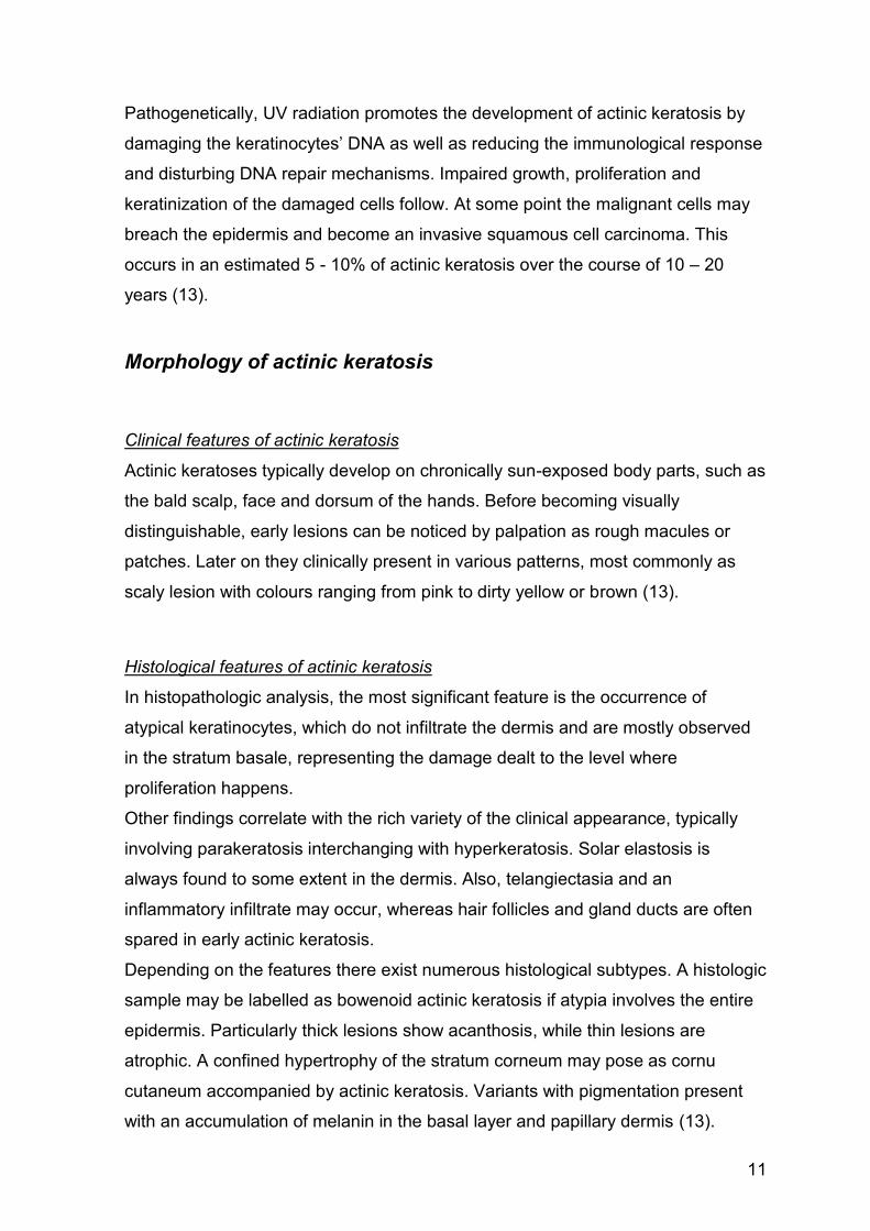
Pathogenetically, UV radiation promotes the development of actinic keratosis by
damaging the keratinocytes’ DNA as well as reducing the immunological response
and disturbing DNA repair mechanisms. Impaired growth, proliferation and
keratinization of the damaged cells follow. At some point the malignant cells may
breach the epidermis and become an invasive squamous cell carcinoma. This
occurs in an estimated 5 - 10% of actinic keratosis over the course of 10 – 20
years (13).
Morphology of actinic keratosis
Clinical features of actinic keratosis
Actinic keratoses typically develop on chronically sun-exposed body parts, such as
the bald scalp, face and dorsum of the hands. Before becoming visually
distinguishable, early lesions can be noticed by palpation as rough macules or
patches. Later on they clinically present in various patterns, most commonly as
scaly lesion with colours ranging from pink to dirty yellow or brown (13).
Histological features of actinic keratosis
In histopathologic analysis, the most significant feature is the occurrence of
atypical keratinocytes, which do not infiltrate the dermis and are mostly observed
in the stratum basale, representing the damage dealt to the level where
proliferation happens.
Other findings correlate with the rich variety of the clinical appearance, typically
involving parakeratosis interchanging with hyperkeratosis. Solar elastosis is
always found to some extent in the dermis. Also, telangiectasia and an
inflammatory infiltrate may occur, whereas hair follicles and gland ducts are often
spared in early actinic keratosis.
Depending on the features there exist numerous histological subtypes. A histologic
sample may be labelled as bowenoid actinic keratosis if atypia involves the entire
epidermis. Particularly thick lesions show acanthosis, while thin lesions are
atrophic. A confined hypertrophy of the stratum corneum may pose as cornu
cutaneum accompanied by actinic keratosis. Variants with pigmentation present
with an accumulation of melanin in the basal layer and papillary dermis (13).
12
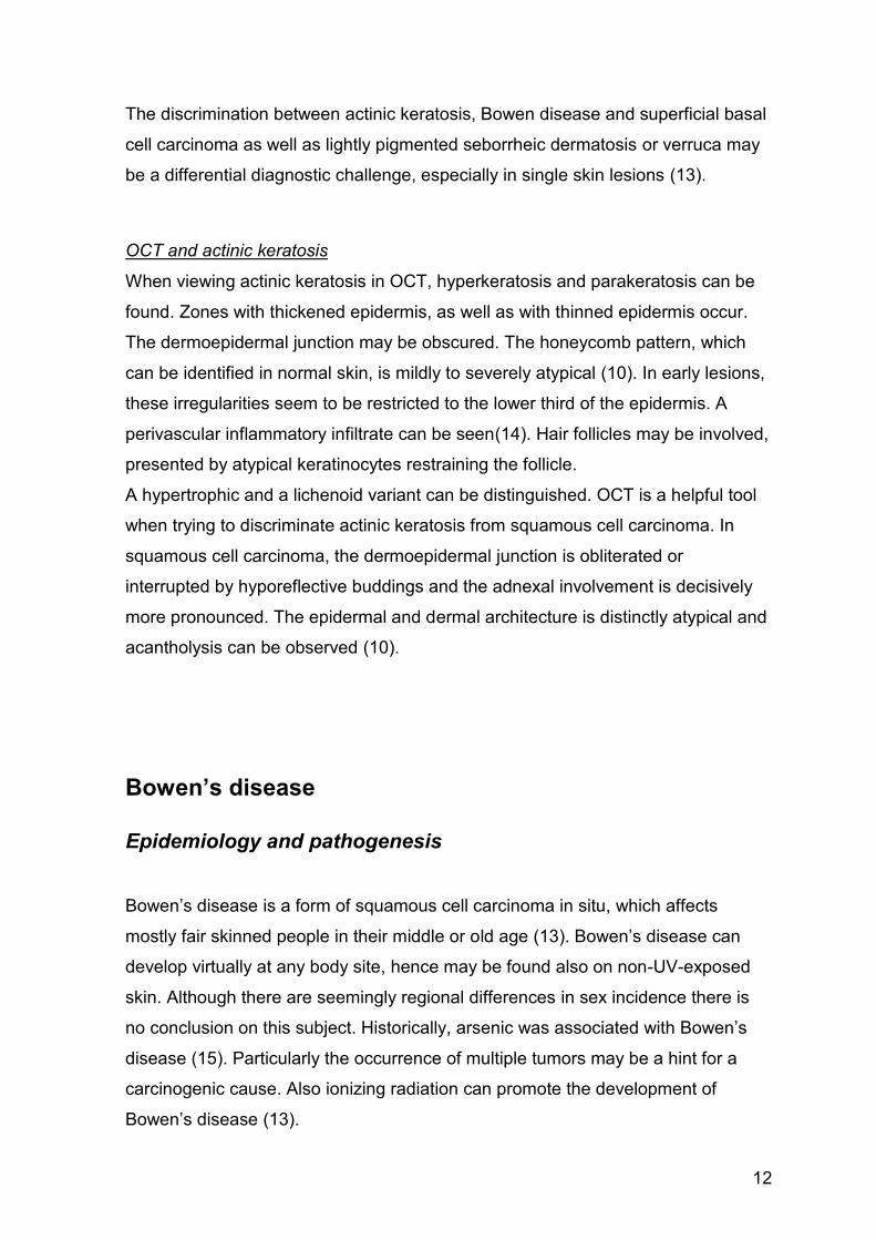
The discrimination between actinic keratosis, Bowen disease and superficial basal
cell carcinoma as well as lightly pigmented seborrheic dermatosis or verruca may
be a differential diagnostic challenge, especially in single skin lesions (13).
OCT and actinic keratosis
When viewing actinic keratosis in OCT, hyperkeratosis and parakeratosis can be
found. Zones with thickened epidermis, as well as with thinned epidermis occur.
The dermoepidermal junction may be obscured. The honeycomb pattern, which
can be identified in normal skin, is mildly to severely atypical (10). In early lesions,
these irregularities seem to be restricted to the lower third of the epidermis. A
perivascular inflammatory infiltrate can be seen(14). Hair follicles may be involved,
presented by atypical keratinocytes restraining the follicle.
A hypertrophic and a lichenoid variant can be distinguished. OCT is a helpful tool
when trying to discriminate actinic keratosis from squamous cell carcinoma. In
squamous cell carcinoma, the dermoepidermal junction is obliterated or
interrupted by hyporeflective buddings and the adnexal involvement is decisively
more pronounced. The epidermal and dermal architecture is distinctly atypical and
acantholysis can be observed (10).
Bowen’s disease
Epidemiology and pathogenesis
Bowen’s disease is a form of squamous cell carcinoma in situ, which affects
mostly fair skinned people in their middle or old age (13). Bowen’s disease can
develop virtually at any body site, hence may be found also on non-UV-exposed
skin. Although there are seemingly regional differences in sex incidence there is
no conclusion on this subject. Historically, arsenic was associated with Bowen’s
disease (15). Particularly the occurrence of multiple tumors may be a hint for a
carcinogenic cause. Also ionizing radiation can promote the development of
Bowen’s disease (13).
13
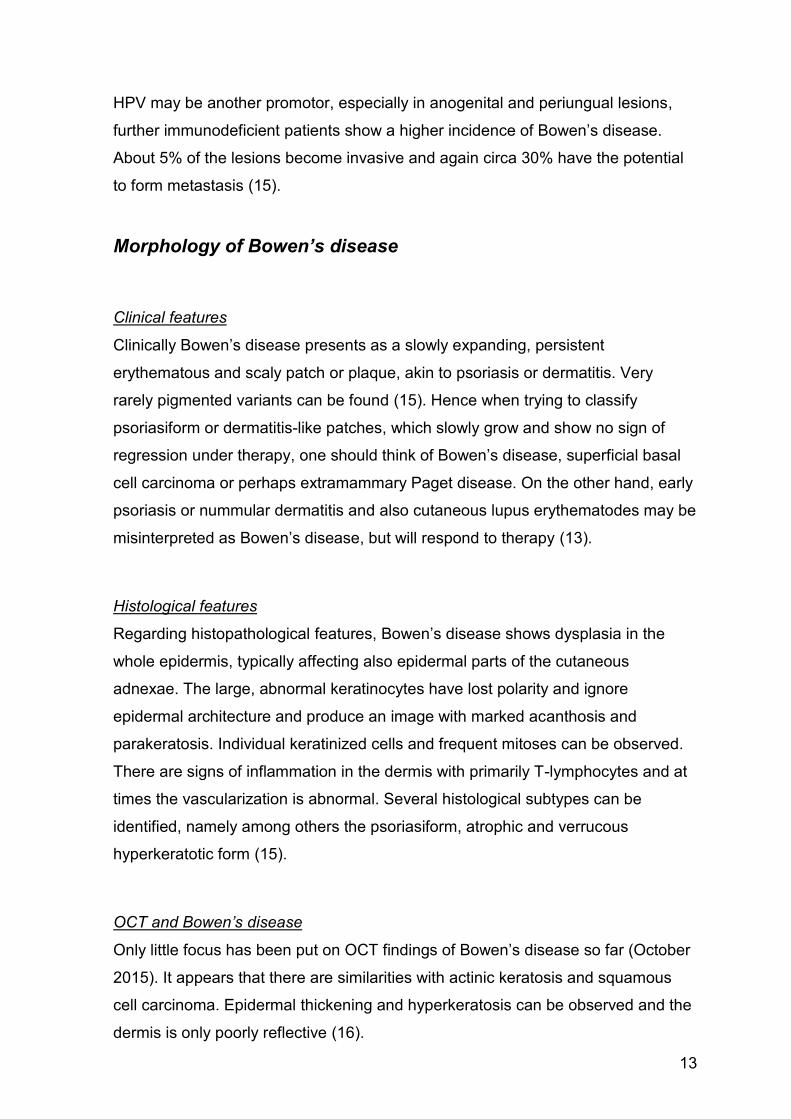
HPV may be another promotor, especially in anogenital and periungual lesions,
further immunodeficient patients show a higher incidence of Bowen’s disease.
About 5% of the lesions become invasive and again circa 30% have the potential
to form metastasis (15).
Morphology of Bowen’s disease
Clinical features
Clinically Bowen’s disease presents as a slowly expanding, persistent
erythematous and scaly patch or plaque, akin to psoriasis or dermatitis. Very
rarely pigmented variants can be found (15). Hence when trying to classify
psoriasiform or dermatitis-like patches, which slowly grow and show no sign of
regression under therapy, one should think of Bowen’s disease, superficial basal
cell carcinoma or perhaps extramammary Paget disease. On the other hand, early
psoriasis or nummular dermatitis and also cutaneous lupus erythematodes may be
misinterpreted as Bowen’s disease, but will respond to therapy (13).
Histological features
Regarding histopathological features, Bowen’s disease shows dysplasia in the
whole epidermis, typically affecting also epidermal parts of the cutaneous
adnexae. The large, abnormal keratinocytes have lost polarity and ignore
epidermal architecture and produce an image with marked acanthosis and
parakeratosis. Individual keratinized cells and frequent mitoses can be observed.
There are signs of inflammation in the dermis with primarily T-lymphocytes and at
times the vascularization is abnormal. Several histological subtypes can be
identified, namely among others the psoriasiform, atrophic and verrucous
hyperkeratotic form (15).
OCT and Bowen’s disease
Only little focus has been put on OCT findings of Bowen’s disease so far (October
2015). It appears that there are similarities with actinic keratosis and squamous
cell carcinoma. Epidermal thickening and hyperkeratosis can be observed and the
dermis is only poorly reflective (16).
14
Basal cell carcinoma
Epidemiology and pathogenesis
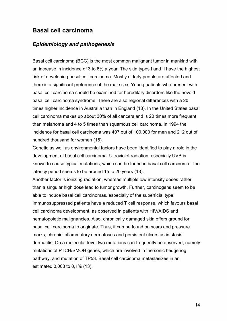
Basal cell carcinoma (BCC) is the most common malignant tumor in mankind with
an increase in incidence of 3 to 8% a year. The skin types I and II have the highest
risk of developing basal cell carcinoma. Mostly elderly people are affected and
there is a significant preference of the male sex. Young patients who present with
basal cell carcinoma should be examined for hereditary disorders like the nevoid
basal cell carcinoma syndrome. There are also regional differences with a 20
times higher incidence in Australia than in England (13). In the United States basal
cell carcinoma makes up about 30% of all cancers and is 20 times more frequent
than melanoma and 4 to 5 times than squamous cell carcinoma. In 1994 the
incidence for basal cell carcinoma was 407 out of 100,000 for men and 212 out of
hundred thousand for women (15).
Genetic as well as environmental factors have been identified to play a role in the
development of basal cell carcinoma. Ultraviolet radiation, especially UVB is
known to cause typical mutations, which can be found in basal cell carcinoma. The
latency period seems to be around 15 to 20 years (13).
Another factor is ionizing radiation, whereas multiple low intensity doses rather
than a singular high dose lead to tumor growth. Further, carcinogens seem to be
able to induce basal cell carcinomas, especially of the superficial type.
Immunosuppressed patients have a reduced T cell response, which favours basal
cell carcinoma development, as observed in patients with HIV/AIDS and
hematopoietic malignancies. Also, chronically damaged skin offers ground for
basal cell carcinoma to originate. Thus, it can be found on scars and pressure
marks, chronic inflammatory dermatoses and persistent ulcers as in stasis
dermatitis. On a molecular level two mutations can frequently be observed, namely
mutations of PTCH/SMOH genes, which are involved in the sonic hedgehog
pathway, and mutation of TP53. Basal cell carcinoma metastasizes in an
estimated 0,003 to 0,1% (13).
15
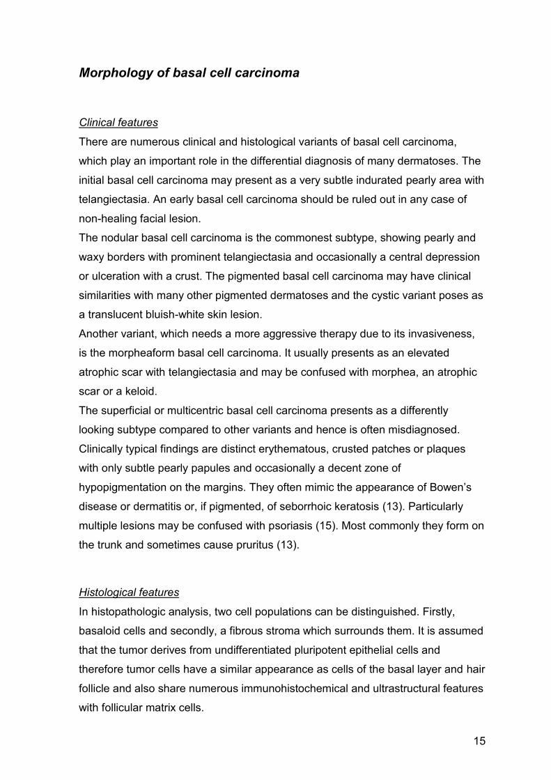
Morphology of basal cell carcinoma
Clinical features
There are numerous clinical and histological variants of basal cell carcinoma,
which play an important role in the differential diagnosis of many dermatoses. The
initial basal cell carcinoma may present as a very subtle indurated pearly area with
telangiectasia. An early basal cell carcinoma should be ruled out in any case of
non-healing facial lesion.
The nodular basal cell carcinoma is the commonest subtype, showing pearly and
waxy borders with prominent telangiectasia and occasionally a central depression
or ulceration with a crust. The pigmented basal cell carcinoma may have clinical
similarities with many other pigmented dermatoses and the cystic variant poses as
a translucent bluish-white skin lesion.
Another variant, which needs a more aggressive therapy due to its invasiveness,
is the morpheaform basal cell carcinoma. It usually presents as an elevated
atrophic scar with telangiectasia and may be confused with morphea, an atrophic
scar or a keloid.
The superficial or multicentric basal cell carcinoma presents as a differently
looking subtype compared to other variants and hence is often misdiagnosed.
Clinically typical findings are distinct erythematous, crusted patches or plaques
with only subtle pearly papules and occasionally a decent zone of
hypopigmentation on the margins. They often mimic the appearance of Bowen’s
disease or dermatitis or, if pigmented, of seborrhoic keratosis (13). Particularly
multiple lesions may be confused with psoriasis (15). Most commonly they form on
the trunk and sometimes cause pruritus (13).
Histological features
In histopathologic analysis, two cell populations can be distinguished. Firstly,
basaloid cells and secondly, a fibrous stroma which surrounds them. It is assumed
that the tumor derives from undifferentiated pluripotent epithelial cells and
therefore tumor cells have a similar appearance as cells of the basal layer and hair
follicle and also share numerous immunohistochemical and ultrastructural features
with follicular matrix cells.
16
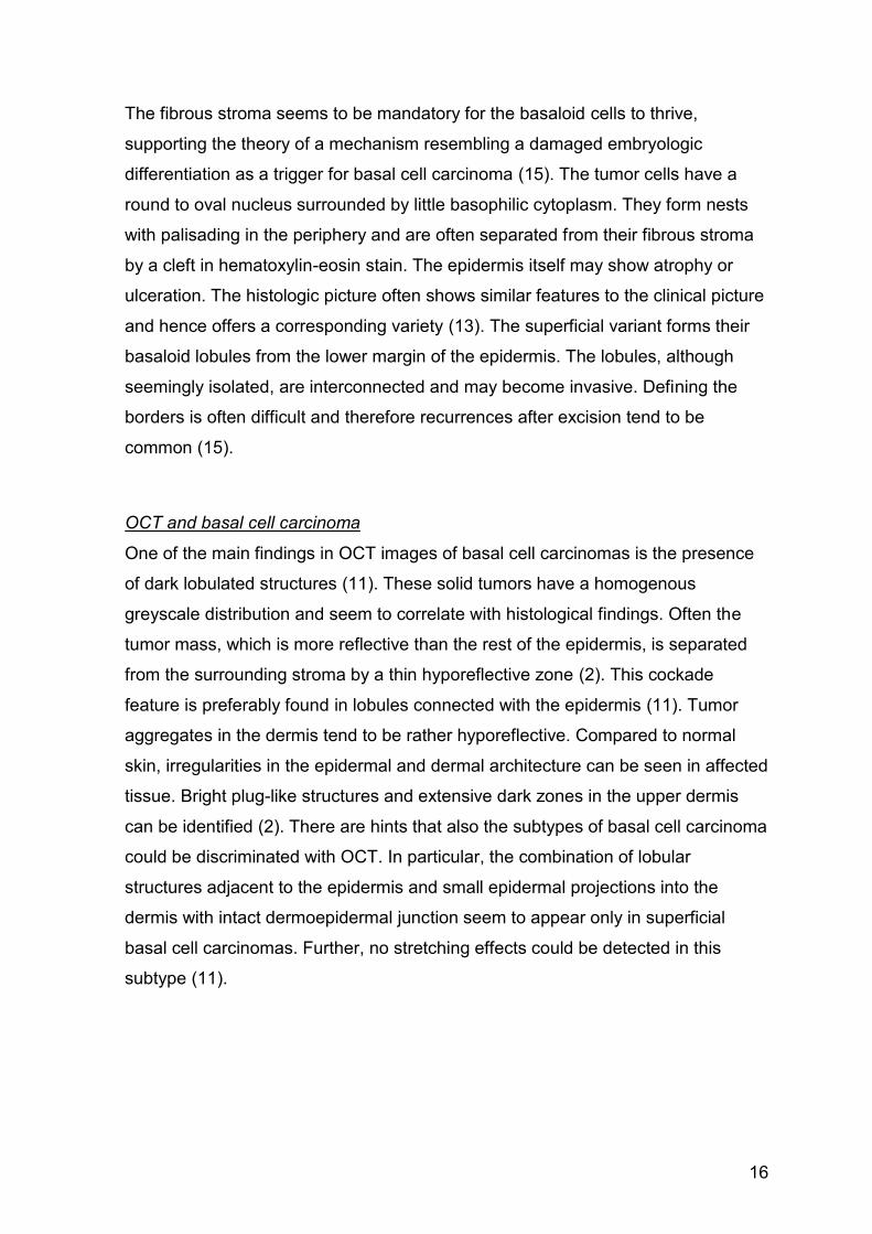
The fibrous stroma seems to be mandatory for the basaloid cells to thrive,
supporting the theory of a mechanism resembling a damaged embryologic
differentiation as a trigger for basal cell carcinoma (15). The tumor cells have a
round to oval nucleus surrounded by little basophilic cytoplasm. They form nests
with palisading in the periphery and are often separated from their fibrous stroma
by a cleft in hematoxylin-eosin stain. The epidermis itself may show atrophy or
ulceration. The histologic picture often shows similar features to the clinical picture
and hence offers a corresponding variety (13). The superficial variant forms their
basaloid lobules from the lower margin of the epidermis. The lobules, although
seemingly isolated, are interconnected and may become invasive. Defining the
borders is often difficult and therefore recurrences after excision tend to be
common (15).
OCT and basal cell carcinoma
One of the main findings in OCT images of basal cell carcinomas is the presence
of dark lobulated structures (11). These solid tumors have a homogenous
greyscale distribution and seem to correlate with histological findings. Often the
tumor mass, which is more reflective than the rest of the epidermis, is separated
from the surrounding stroma by a thin hyporeflective zone (2). This cockade
feature is preferably found in lobules connected with the epidermis (11). Tumor
aggregates in the dermis tend to be rather hyporeflective. Compared to normal
skin, irregularities in the epidermal and dermal architecture can be seen in affected
tissue. Bright plug-like structures and extensive dark zones in the upper dermis
can be identified (2). There are hints that also the subtypes of basal cell carcinoma
could be discriminated with OCT. In particular, the combination of lobular
structures adjacent to the epidermis and small epidermal projections into the
dermis with intact dermoepidermal junction seem to appear only in superficial
basal cell carcinomas. Further, no stretching effects could be detected in this
subtype (11).
17
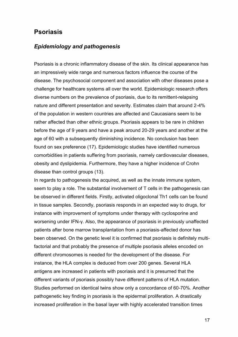
Psoriasis
Epidemiology and pathogenesis
Psoriasis is a chronic inflammatory disease of the skin. Its clinical appearance has
an impressively wide range and numerous factors influence the course of the
disease. The psychosocial component and association with other diseases pose a
challenge for healthcare systems all over the world. Epidemiologic research offers
diverse numbers on the prevalence of psoriasis, due to its remittent-relapsing
nature and different presentation and severity. Estimates claim that around 2-4%
of the population in western countries are affected and Caucasians seem to be
rather affected than other ethnic groups. Psoriasis appears to be rare in children
before the age of 9 years and have a peak around 20-29 years and another at the
age of 60 with a subsequently diminishing incidence. No conclusion has been
found on sex preference (17). Epidemiologic studies have identified numerous
comorbidities in patients suffering from psoriasis, namely cardiovascular diseases,
obesity and dyslipidemia. Furthermore, they have a higher incidence of Crohn
disease than control groups (13).
In regards to pathogenesis the acquired, as well as the innate immune system,
seem to play a role. The substantial involvement of T cells in the pathogenesis can
be observed in different fields. Firstly, activated oligoclonal Th1 cells can be found
in tissue samples. Secondly, psoriasis responds in an expected way to drugs, for
instance with improvement of symptoms under therapy with cyclosporine and
worsening under IFN-γ. Also, the appearance of psoriasis in previously unaffected
patients after bone marrow transplantation from a psoriasis-affected donor has
been observed. On the genetic level it is confirmed that psoriasis is definitely multi-
factorial and that probably the presence of multiple psoriasis alleles encoded on
different chromosomes is needed for the development of the disease. For
instance, the HLA complex is deduced from over 200 genes. Several HLA
antigens are increased in patients with psoriasis and it is presumed that the
different variants of psoriasis possibly have different patterns of HLA mutation.
Studies performed on identical twins show only a concordance of 60-70%. Another
pathogenetic key finding in psoriasis is the epidermal proliferation. A drastically
increased proliferation in the basal layer with highly accelerated transition times
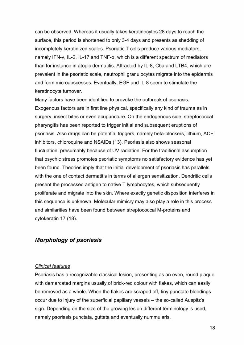
18
can be observed. Whereas it usually takes keratinocytes 28 days to reach the
surface, this period is shortened to only 3-4 days and presents as shedding of
incompletely keratinized scales. Psoriatic T cells produce various mediators,
namely IFN-γ, IL-2, IL-17 and TNF-α, which is a different spectrum of mediators
than for instance in atopic dermatitis. Attracted by IL-8, C5a and LTB4, which are
prevalent in the psoriatic scale, neutrophil granulocytes migrate into the epidermis
and form microabscesses. Eventually, EGF and IL-8 seem to stimulate the
keratinocyte turnover.
Many factors have been identified to provoke the outbreak of psoriasis.
Exogenous factors are in first line physical, specifically any kind of trauma as in
surgery, insect bites or even acupuncture. On the endogenous side, streptococcal
pharyngitis has been reported to trigger initial and subsequent eruptions of
psoriasis. Also drugs can be potential triggers, namely beta-blockers, lithium, ACE
inhibitors, chloroquine and NSAIDs (13). Psoriasis also shows seasonal
fluctuation, presumably because of UV radiation. For the traditional assumption
that psychic stress promotes psoriatic symptoms no satisfactory evidence has yet
been found. Theories imply that the initial development of psoriasis has parallels
with the one of contact dermatitis in terms of allergen sensitization. Dendritic cells
present the processed antigen to native T lymphocytes, which subsequently
proliferate and migrate into the skin. Where exactly genetic disposition interferes in
this sequence is unknown. Molecular mimicry may also play a role in this process
and similarities have been found between streptococcal M-proteins and
cytokeratin 17 (18).
Morphology of psoriasis
Clinical features
Psoriasis has a recognizable classical lesion, presenting as an even, round plaque
with demarcated margins usually of brick-red colour with flakes, which can easily
be removed as a whole. When the flakes are scraped off, tiny punctate bleedings
occur due to injury of the superficial papillary vessels – the so-called Auspitz’s
sign. Depending on the size of the growing lesion different terminology is used,
namely psoriasis punctata, guttata and eventually nummularis.
19
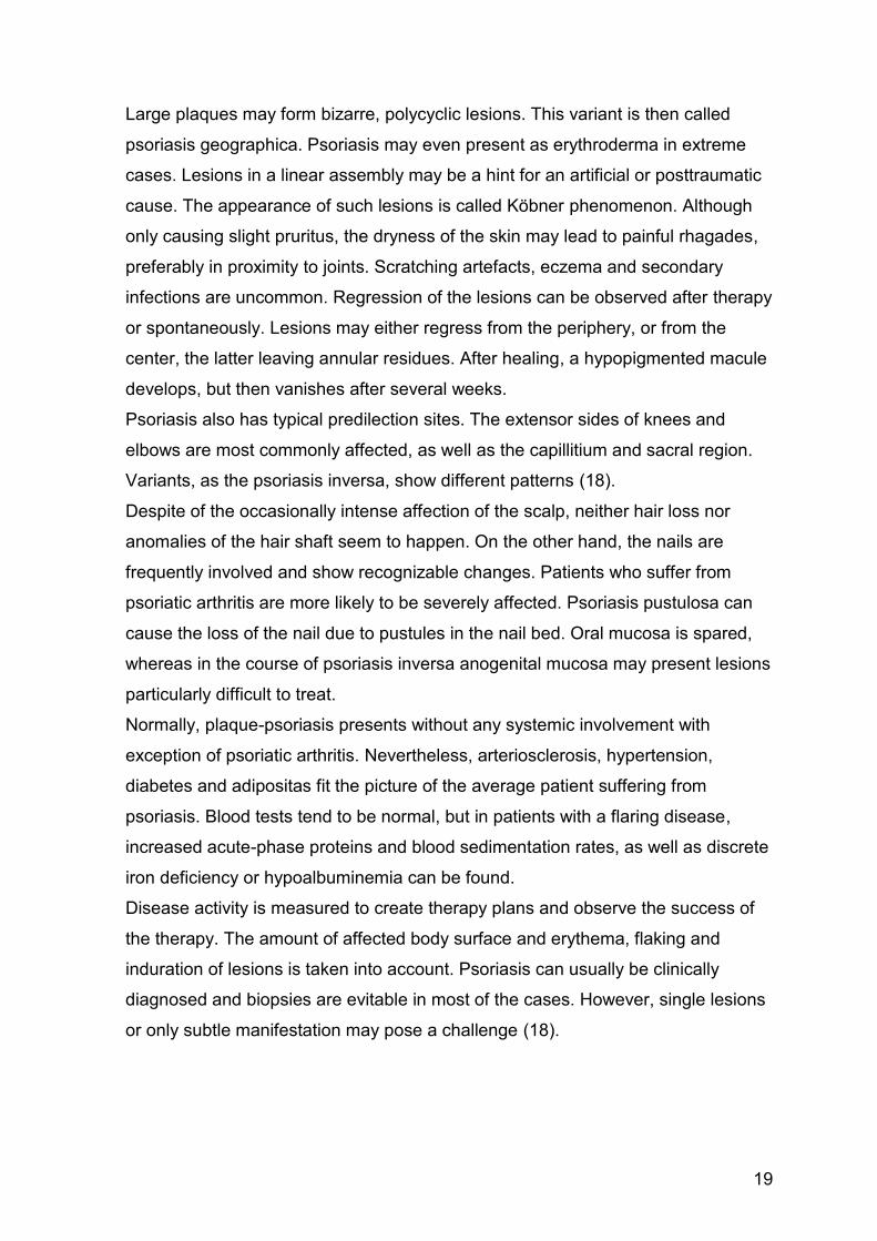
Large plaques may form bizarre, polycyclic lesions. This variant is then called
psoriasis geographica. Psoriasis may even present as erythroderma in extreme
cases. Lesions in a linear assembly may be a hint for an artificial or posttraumatic
cause. The appearance of such lesions is called Köbner phenomenon. Although
only causing slight pruritus, the dryness of the skin may lead to painful rhagades,
preferably in proximity to joints. Scratching artefacts, eczema and secondary
infections are uncommon. Regression of the lesions can be observed after therapy
or spontaneously. Lesions may either regress from the periphery, or from the
center, the latter leaving annular residues. After healing, a hypopigmented macule
develops, but then vanishes after several weeks.
Psoriasis also has typical predilection sites. The extensor sides of knees and
elbows are most commonly affected, as well as the capillitium and sacral region.
Variants, as the psoriasis inversa, show different patterns (18).
Despite of the occasionally intense affection of the scalp, neither hair loss nor
anomalies of the hair shaft seem to happen. On the other hand, the nails are
frequently involved and show recognizable changes. Patients who suffer from
psoriatic arthritis are more likely to be severely affected. Psoriasis pustulosa can
cause the loss of the nail due to pustules in the nail bed. Oral mucosa is spared,
whereas in the course of psoriasis inversa anogenital mucosa may present lesions
particularly difficult to treat.
Normally, plaque-psoriasis presents without any systemic involvement with
exception of psoriatic arthritis. Nevertheless, arteriosclerosis, hypertension,
diabetes and adipositas fit the picture of the average patient suffering from
psoriasis. Blood tests tend to be normal, but in patients with a flaring disease,
increased acute-phase proteins and blood sedimentation rates, as well as discrete
iron deficiency or hypoalbuminemia can be found.
Disease activity is measured to create therapy plans and observe the success of
the therapy. The amount of affected body surface and erythema, flaking and
induration of lesions is taken into account. Psoriasis can usually be clinically
diagnosed and biopsies are evitable in most of the cases. However, single lesions
or only subtle manifestation may pose a challenge (18).
20
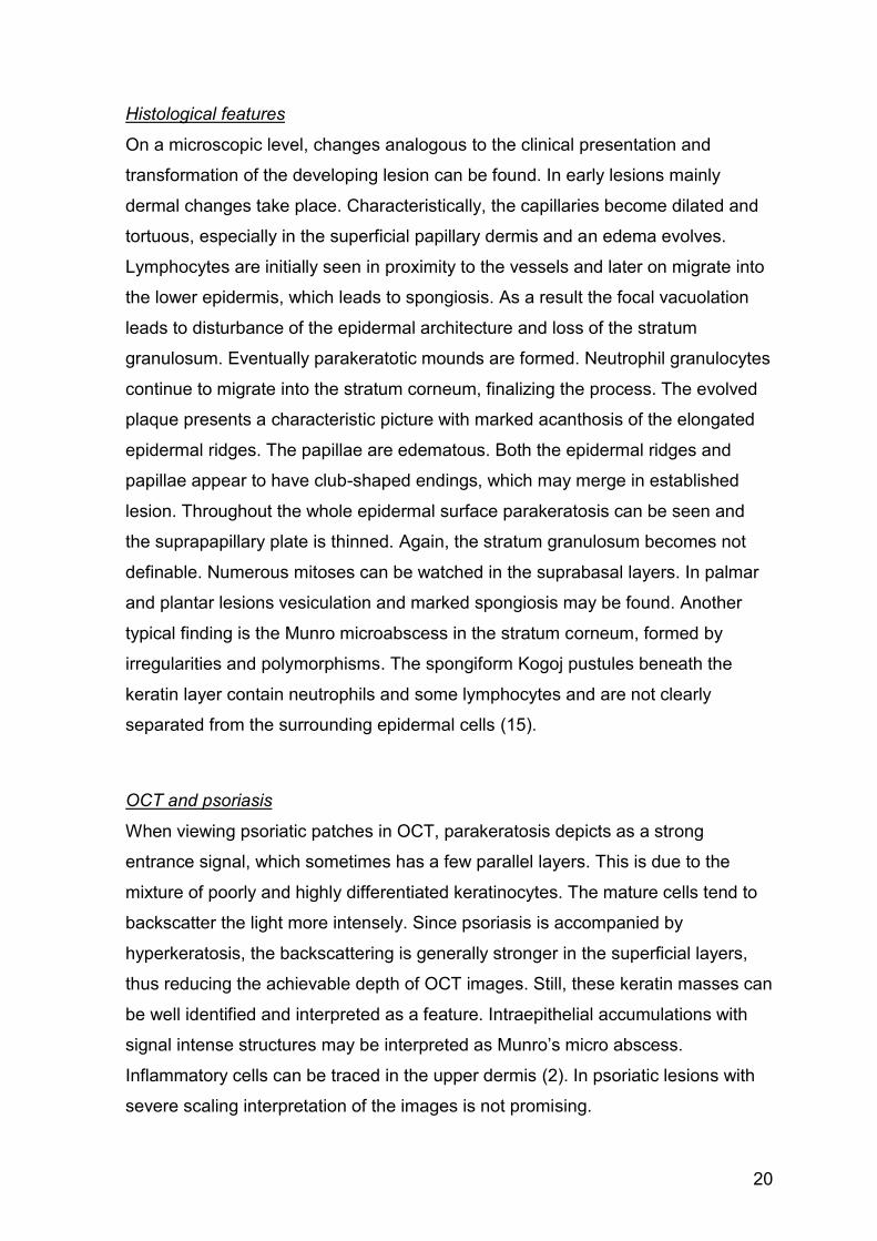
Histological features
On a microscopic level, changes analogous to the clinical presentation and
transformation of the developing lesion can be found. In early lesions mainly
dermal changes take place. Characteristically, the capillaries become dilated and
tortuous, especially in the superficial papillary dermis and an edema evolves.
Lymphocytes are initially seen in proximity to the vessels and later on migrate into
the lower epidermis, which leads to spongiosis. As a result the focal vacuolation
leads to disturbance of the epidermal architecture and loss of the stratum
granulosum. Eventually parakeratotic mounds are formed. Neutrophil granulocytes
continue to migrate into the stratum corneum, finalizing the process. The evolved
plaque presents a characteristic picture with marked acanthosis of the elongated
epidermal ridges. The papillae are edematous. Both the epidermal ridges and
papillae appear to have club-shaped endings, which may merge in established
lesion. Throughout the whole epidermal surface parakeratosis can be seen and
the suprapapillary plate is thinned. Again, the stratum granulosum becomes not
definable. Numerous mitoses can be watched in the suprabasal layers. In palmar
and plantar lesions vesiculation and marked spongiosis may be found. Another
typical finding is the Munro microabscess in the stratum corneum, formed by
irregularities and polymorphisms. The spongiform Kogoj pustules beneath the
keratin layer contain neutrophils and some lymphocytes and are not clearly
separated from the surrounding epidermal cells (15).
OCT and psoriasis
When viewing psoriatic patches in OCT, parakeratosis depicts as a strong
entrance signal, which sometimes has a few parallel layers. This is due to the
mixture of poorly and highly differentiated keratinocytes. The mature cells tend to
backscatter the light more intensely. Since psoriasis is accompanied by
hyperkeratosis, the backscattering is generally stronger in the superficial layers,
thus reducing the achievable depth of OCT images. Still, these keratin masses can
be well identified and interpreted as a feature. Intraepithelial accumulations with
signal intense structures may be interpreted as Munro’s micro abscess.
Inflammatory cells can be traced in the upper dermis (2). In psoriatic lesions with
severe scaling interpretation of the images is not promising.
21
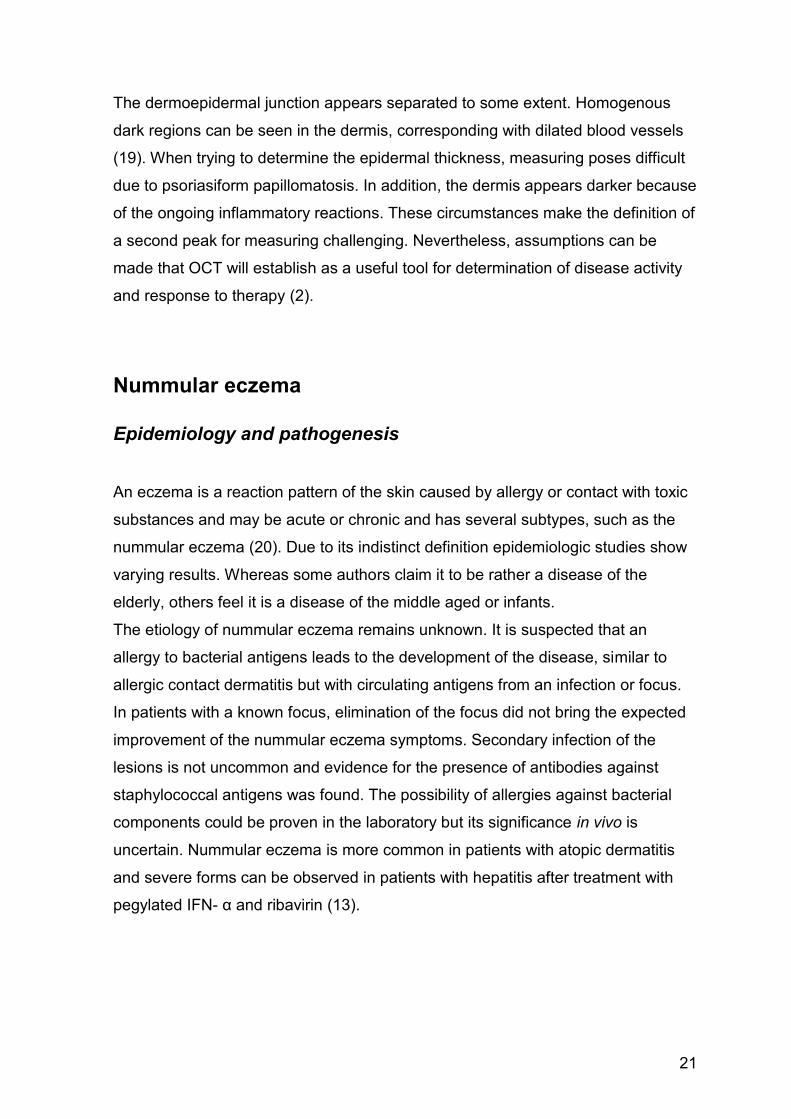
The dermoepidermal junction appears separated to some extent. Homogenous
dark regions can be seen in the dermis, corresponding with dilated blood vessels
(19). When trying to determine the epidermal thickness, measuring poses difficult
due to psoriasiform papillomatosis. In addition, the dermis appears darker because
of the ongoing inflammatory reactions. These circumstances make the definition of
a second peak for measuring challenging. Nevertheless, assumptions can be
made that OCT will establish as a useful tool for determination of disease activity
and response to therapy (2).
Nummular eczema
Epidemiology and pathogenesis
An eczema is a reaction pattern of the skin caused by allergy or contact with toxic
substances and may be acute or chronic and has several subtypes, such as the
nummular eczema (20). Due to its indistinct definition epidemiologic studies show
varying results. Whereas some authors claim it to be rather a disease of the
elderly, others feel it is a disease of the middle aged or infants.
The etiology of nummular eczema remains unknown. It is suspected that an
allergy to bacterial antigens leads to the development of the disease, similar to
allergic contact dermatitis but with circulating antigens from an infection or focus.
In patients with a known focus, elimination of the focus did not bring the expected
improvement of the nummular eczema symptoms. Secondary infection of the
lesions is not uncommon and evidence for the presence of antibodies against
staphylococcal antigens was found. The possibility of allergies against bacterial
components could be proven in the laboratory but its significance in vivo is
uncertain. Nummular eczema is more common in patients with atopic dermatitis
and severe forms can be observed in patients with hepatitis after treatment with
pegylated IFN- α and ribavirin (13).
22
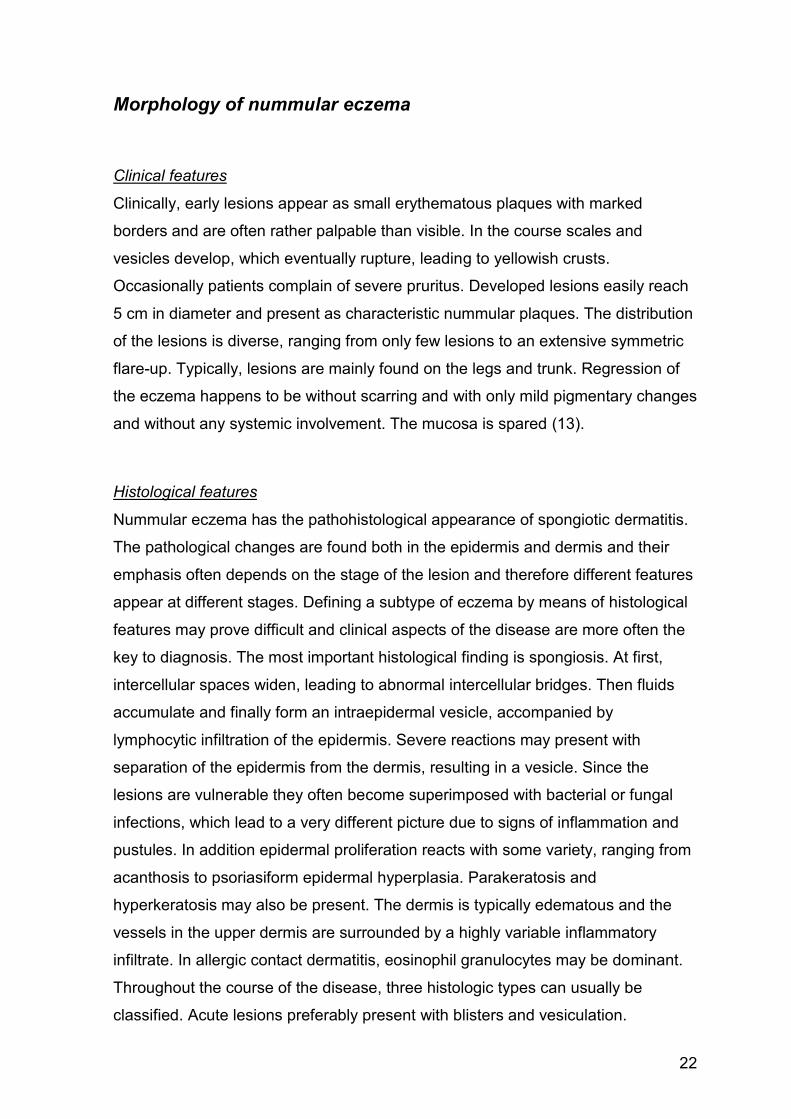
Morphology of nummular eczema
Clinical features
Clinically, early lesions appear as small erythematous plaques with marked
borders and are often rather palpable than visible. In the course scales and
vesicles develop, which eventually rupture, leading to yellowish crusts.
Occasionally patients complain of severe pruritus. Developed lesions easily reach
5 cm in diameter and present as characteristic nummular plaques. The distribution
of the lesions is diverse, ranging from only few lesions to an extensive symmetric
flare-up. Typically, lesions are mainly found on the legs and trunk. Regression of
the eczema happens to be without scarring and with only mild pigmentary changes
and without any systemic involvement. The mucosa is spared (13).
Histological features
Nummular eczema has the pathohistological appearance of spongiotic dermatitis.
The pathological changes are found both in the epidermis and dermis and their
emphasis often depends on the stage of the lesion and therefore different features
appear at different stages. Defining a subtype of eczema by means of histological
features may prove difficult and clinical aspects of the disease are more often the
key to diagnosis. The most important histological finding is spongiosis. At first,
intercellular spaces widen, leading to abnormal intercellular bridges. Then fluids
accumulate and finally form an intraepidermal vesicle, accompanied by
lymphocytic infiltration of the epidermis. Severe reactions may present with
separation of the epidermis from the dermis, resulting in a vesicle. Since the
lesions are vulnerable they often become superimposed with bacterial or fungal
infections, which lead to a very different picture due to signs of inflammation and
pustules. In addition epidermal proliferation reacts with some variety, ranging from
acanthosis to psoriasiform epidermal hyperplasia. Parakeratosis and
hyperkeratosis may also be present. The dermis is typically edematous and the
vessels in the upper dermis are surrounded by a highly variable inflammatory
infiltrate. In allergic contact dermatitis, eosinophil granulocytes may be dominant.
Throughout the course of the disease, three histologic types can usually be
classified. Acute lesions preferably present with blisters and vesiculation.
23
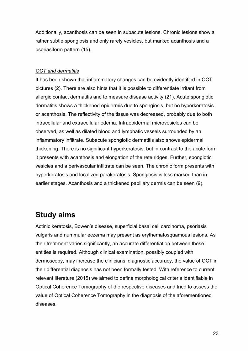
Additionally, acanthosis can be seen in subacute lesions. Chronic lesions show a
rather subtle spongiosis and only rarely vesicles, but marked acanthosis and a
psoriasiform pattern (15).
OCT and dermatitis
It has been shown that inflammatory changes can be evidently identified in OCT
pictures (2). There are also hints that it is possible to differentiate irritant from
allergic contact dermatitis and to measure disease activity (21). Acute spongiotic
dermatitis shows a thickened epidermis due to spongiosis, but no hyperkeratosis
or acanthosis. The reflectivity of the tissue was decreased, probably due to both
intracellular and extracellular edema. Intraepidermal microvesicles can be
observed, as well as dilated blood and lymphatic vessels surrounded by an
inflammatory infiltrate. Subacute spongiotic dermatitis also shows epidermal
thickening. There is no significant hyperkeratosis, but in contrast to the acute form
it presents with acanthosis and elongation of the rete ridges. Further, spongiotic
vesicles and a perivascular infiltrate can be seen. The chronic form presents with
hyperkeratosis and localized parakeratosis. Spongiosis is less marked than in
earlier stages. Acanthosis and a thickened papillary dermis can be seen (9).
Study aims
Actinic keratosis, Bowen’s disease, superficial basal cell carcinoma, psoriasis
vulgaris and nummular eczema may present as erythematosquamous lesions. As
their treatment varies significantly, an accurate differentiation between these
entities is required. Although clinical examination, possibly coupled with
dermoscopy, may increase the clinicians’ diagnostic accuracy, the value of OCT in
their differential diagnosis has not been formally tested. With reference to current
relevant literature (2015) we aimed to define morphological criteria identifiable in
Optical Coherence Tomography of the respective diseases and tried to assess the
value of Optical Coherence Tomography in the diagnosis of the aforementioned
diseases.
24
MATERIAL AND METHODS
Consecutive indoor or outdoor patients over 18 years attending the Department of
Dermatology, Medical University of Graz, with a clinical or histopathologically
proven diagnosis of actinic keratosis, Bowen’s disease, superficial basal cell
carcinoma, psoriasis vulgaris and nummular eczema were included. All
participants were informed about the study aim, the procedure of image acquisition
and had to give written consent before any study related investigations were
performed. Anonymity of patients was guaranteed by assigning each patient to a
specific number. Participation in the study did not affect in any way the routine
treatment of the patient. Inflamed, ulcerated or painful skin was a reason for
exclusion. A medical student under the supervision of a board-certified
dermatologist carried out all examinations. The study was conducted during a 15-
month period from July 2014 to October 2015.
Process of image acquisition
Each patient received a full body skin examination in order to select the most
representative lesion for the subsequent OCT examination. All lesions were
macroscopically and dermatoscopically photographed using a digital camera
(Canon®) and a mobile dermatoscope (handyscope by Fotofinder ®) with 20-fold
magnification, attached to iPhone ®. All pictures were stored in the clinical
photography archive of the Department of Dermatology, Medical University of
Graz. In case of a wider distribution or multiple lesions a suitable spot was
selected and tagged with a reinforcement ring. For the OCT image acquisition, a
contact gel (AGFA Skintell Optical Gel ®) was sparingly applied with a pipette
(Handystep ® by BRAND). Then image acquisition with the High Resolution
Optical Coherence Tomograph (AGFA Skintell ®) was performed according to the
instructions in the manual. AGFA Skintell ® acquires images with a lateral and
axial resolution of 3 µm. It has a field of view of 1,8 mm x 1,5 mm and a
penetration depth of up to 1 mm. Images of poor quality caused by artefacts from
air bubbles or very scaly skin were excluded.
25
Process of image interpretation
All images were subsequently evaluated by a medical student trained in OCT and
under the supervision of a board certified dermatologist on a large computer
screen. The student was blinded for the clinical and histological diagnosis. The
OCT 3D-voxel images were systemically investigated for the presence or absence
of the defined OCT features and the results were documented in Excel ®. The
histological diagnoses were then retrieved from available medical records in the
MEDOCS ® healthcare software.
Definition of the OCT features
Actinic keratosis/Bowen’s disease The applied and analysed OCT features for actinic keratosis were used from
available online sources (10), (14), (22). We decided to include the category
“Bowen’s disease” into the group of actinic keratosis because it presents still an in
situ stage in the progression of keratinocyte skin cancer. Moreover, only few
reports on the OCT features of Bowen’s disease have been published.
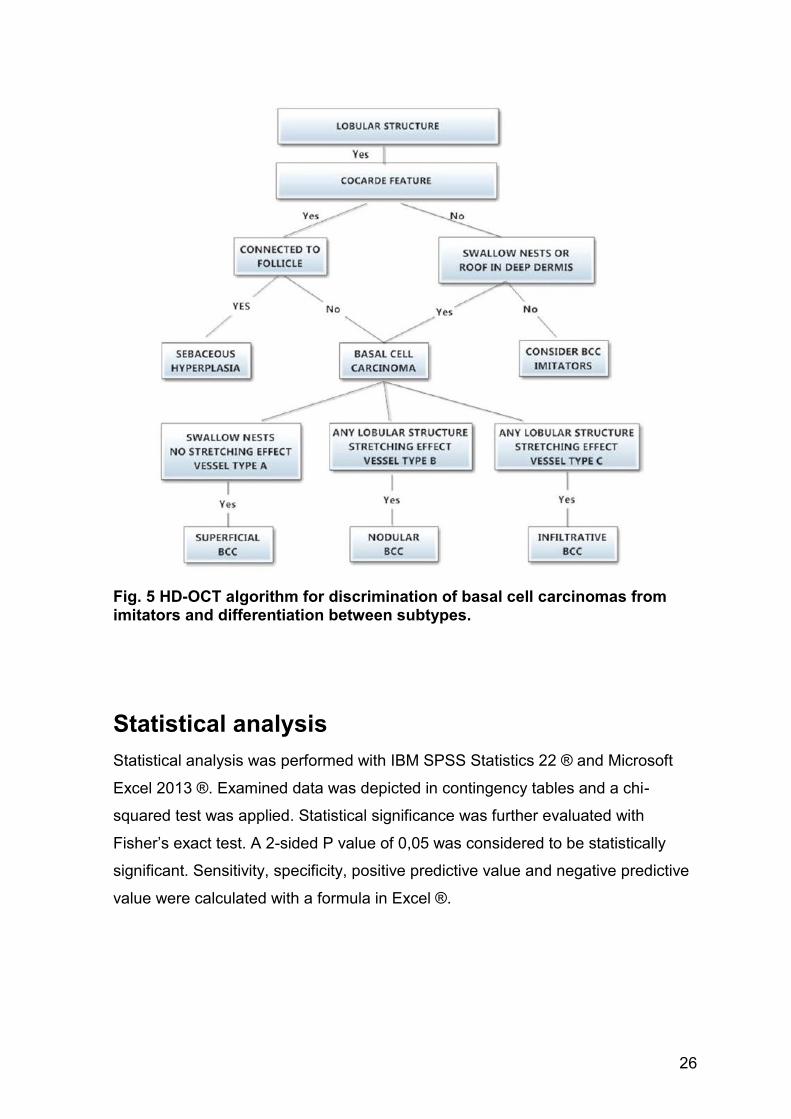
Basal cell carcinoma OCT features and algorithm for basal cell carcinoma were taken from available
online sources (11), (23), (24). The algorithm is shown in figure 5.
Psoriasis OCT features of psoriasis were taken from available online sources (10).
Additional features were defined in accordance to histopathological findings of
psoriasis.
Nummular eczema We could not find any research on OCT features of nummular eczema. Because
of pathohistological similarities we decided to apply OCT features of spongiotic
dermatitis (9).
26
Fig. 5 HD-OCT algorithm for discrimination of basal cell carcinomas from imitators and differentiation between subtypes.
Statistical analysis
Statistical analysis was performed with IBM SPSS Statistics 22 ® and Microsoft
Excel 2013 ®. Examined data was depicted in contingency tables and a chi-
squared test was applied. Statistical significance was further evaluated with
Fisher’s exact test. A 2-sided P value of 0,05 was considered to be statistically
significant. Sensitivity, specificity, positive predictive value and negative predictive
value were calculated with a formula in Excel ®.
27
RESULTS
In total, 56 participants were recruited and 96 erythematosquamous skin lesions
were imaged by OCT. Of those, 22 lesions were excluded due to an equivocal
clinical diagnosis (16 cases) or poor image quality (6 cases). After all, 51
participants presenting with 74 lesions depicted in 220 HD-OCT 3D images were
included. The mean age of the study cohort (51% male, 49% female) was 73
years (±15,7 years). Out of the 74 included lesions, a histologically verified
diagnosis was found in 54 cases and a distinct clinical diagnosis existed in 20
cases.
The images were examined for the presence of 30 defined OCT features. The
most common and established features were statistically analysed to identify
morphological findings possibly useful for the differential diagnosis of
erythematosquamous skin lesions with OCT. For the basal cell carcinoma group,
an algorithm by Boone et al. was applied (11). We also propose an algorithm for
OCT for the differential diagnosis of erythematosquamous plaques.
28
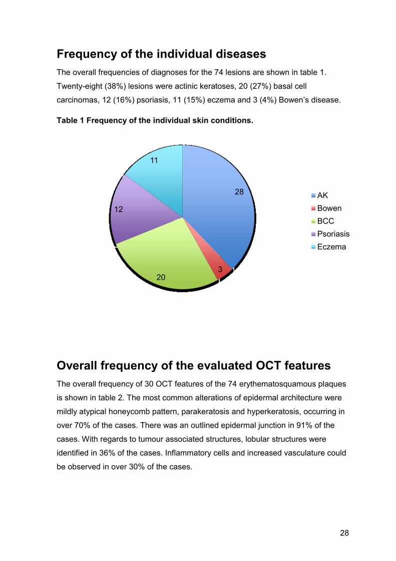
Frequency of the individual diseases
The overall frequencies of diagnoses for the 74 lesions are shown in table 1.
Twenty-eight (38%) lesions were actinic keratoses, 20 (27%) basal cell
carcinomas, 12 (16%) psoriasis, 11 (15%) eczema and 3 (4%) Bowen’s disease.
Table 1 Frequency of the individual skin conditions.
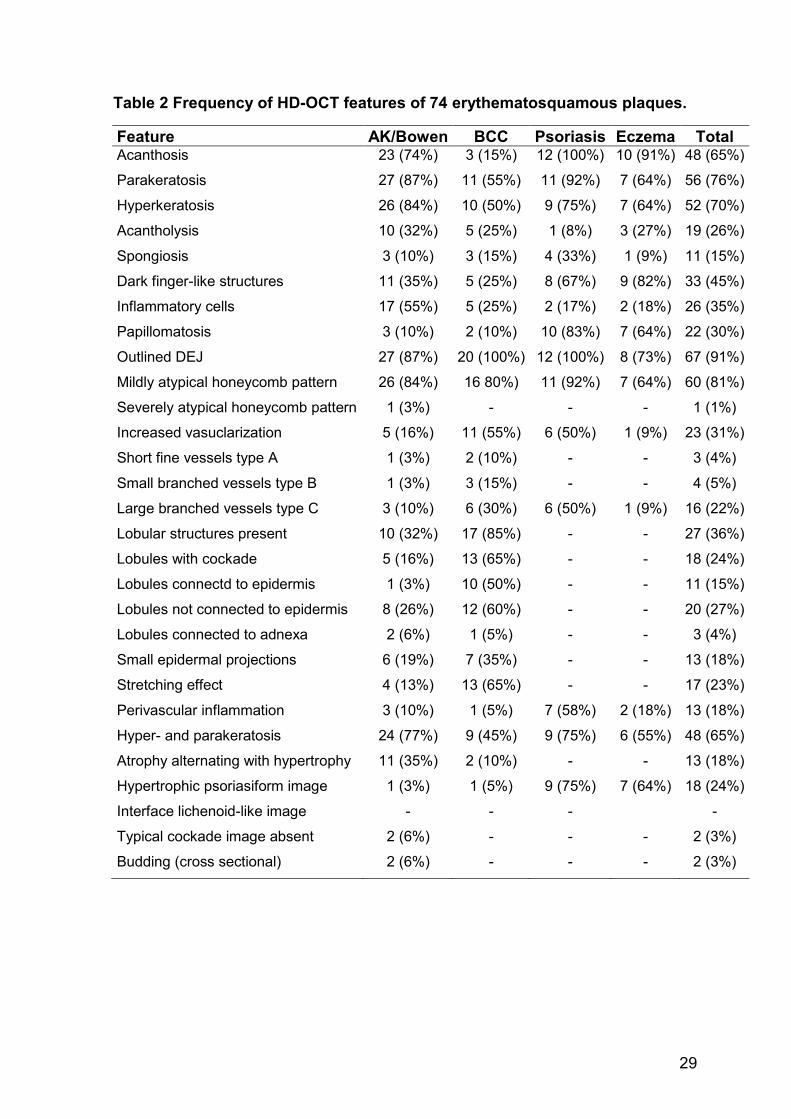
Overall frequency of the evaluated OCT features
The overall frequency of 30 OCT features of the 74 erythematosquamous plaques
is shown in table 2. The most common alterations of epidermal architecture were
mildly atypical honeycomb pattern, parakeratosis and hyperkeratosis, occurring in
over 70% of the cases. There was an outlined epidermal junction in 91% of the
cases. With regards to tumour associated structures, lobular structures were
identified in 36% of the cases. Inflammatory cells and increased vasculature could
be observed in over 30% of the cases.
28
320
12
11
AK
Bowen
BCC
Psoriasis
Eczema
29
Table 2 Frequency of HD-OCT features of 74 erythematosquamous plaques.
Feature AK/Bowen BCC Psoriasis Eczema Total Acanthosis 23 (74%) 3 (15%) 12 (100%) 10 (91%) 48 (65%)
Parakeratosis 27 (87%) 11 (55%) 11 (92%) 7 (64%) 56 (76%)
Hyperkeratosis 26 (84%) 10 (50%) 9 (75%) 7 (64%) 52 (70%)
Acantholysis 10 (32%) 5 (25%) 1 (8%) 3 (27%) 19 (26%)
Spongiosis 3 (10%) 3 (15%) 4 (33%) 1 (9%) 11 (15%)
Dark finger-like structures 11 (35%) 5 (25%) 8 (67%) 9 (82%) 33 (45%)
Inflammatory cells 17 (55%) 5 (25%) 2 (17%) 2 (18%) 26 (35%)
Papillomatosis 3 (10%) 2 (10%) 10 (83%) 7 (64%) 22 (30%)
Outlined DEJ 27 (87%) 20 (100%) 12 (100%) 8 (73%) 67 (91%)
Mildly atypical honeycomb pattern 26 (84%) 16 80%) 11 (92%) 7 (64%) 60 (81%)
Severely atypical honeycomb pattern 1 (3%) - - - 1 (1%)
Increased vasuclarization 5 (16%) 11 (55%) 6 (50%) 1 (9%) 23 (31%)
Short fine vessels type A 1 (3%) 2 (10%) - - 3 (4%)
Small branched vessels type B 1 (3%) 3 (15%) - - 4 (5%)
Large branched vessels type C 3 (10%) 6 (30%) 6 (50%) 1 (9%) 16 (22%)
Lobular structures present 10 (32%) 17 (85%) - - 27 (36%)
Lobules with cockade 5 (16%) 13 (65%) - - 18 (24%)
Lobules connectd to epidermis 1 (3%) 10 (50%) - - 11 (15%)
Lobules not connected to epidermis 8 (26%) 12 (60%) - - 20 (27%)
Lobules connected to adnexa 2 (6%) 1 (5%) - - 3 (4%)
Small epidermal projections 6 (19%) 7 (35%) - - 13 (18%)
Stretching effect 4 (13%) 13 (65%) - - 17 (23%)
Perivascular inflammation 3 (10%) 1 (5%) 7 (58%) 2 (18%) 13 (18%)
Hyper- and parakeratosis 24 (77%) 9 (45%) 9 (75%) 6 (55%) 48 (65%)
Atrophy alternating with hypertrophy 11 (35%) 2 (10%) - - 13 (18%)
Hypertrophic psoriasiform image 1 (3%) 1 (5%) 9 (75%) 7 (64%) 18 (24%)
Interface lichenoid-like image - - -
-
Typical cockade image absent 2 (6%) - - - 2 (3%)
Budding (cross sectional) 2 (6%) - - - 2 (3%)
30
Diagnostic value of the OCT features for the
diagnosis of erythematosquamous plaques
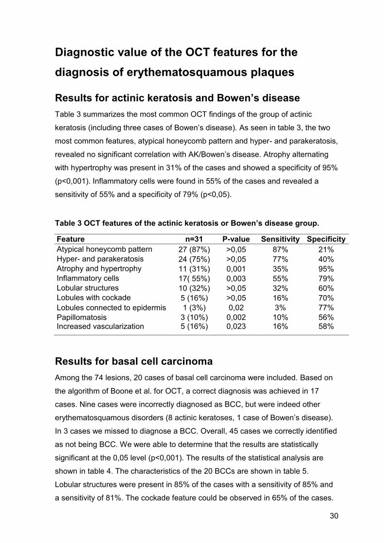
Results for actinic keratosis and Bowen’s disease
Table 3 summarizes the most common OCT findings of the group of actinic
keratosis (including three cases of Bowen’s disease). As seen in table 3, the two
most common features, atypical honeycomb pattern and hyper- and parakeratosis,
revealed no significant correlation with AK/Bowen’s disease. Atrophy alternating
with hypertrophy was present in 31% of the cases and showed a specificity of 95%
(p<0,001). Inflammatory cells were found in 55% of the cases and revealed a
sensitivity of 55% and a specificity of 79% (p<0,05).
Table 3 OCT features of the actinic keratosis or Bowen’s disease group.
Feature n=31 P-value Sensitivity Specificity
Atypical honeycomb pattern 27 (87%) >0,05 87% 21%
Hyper- and parakeratosis 24 (75%) >0,05 77% 40%
Atrophy and hypertrophy 11 (31%) 0,001 35% 95%
Inflammatory cells 17( 55%) 0,003 55% 79%
Lobular structures 10 (32%) >0,05 32% 60%
Lobules with cockade 5 (16%) >0,05 16% 70%
Lobules connected to epidermis 1 (3%) 0,02 3% 77%
Papillomatosis 3 (10%) 0,002 10% 56% Increased vascularization 5 (16%) 0,023 16% 58%
Results for basal cell carcinoma
Among the 74 lesions, 20 cases of basal cell carcinoma were included. Based on
the algorithm of Boone et al. for OCT, a correct diagnosis was achieved in 17
cases. Nine cases were incorrectly diagnosed as BCC, but were indeed other
erythematosquamous disorders (8 actinic keratoses, 1 case of Bowen’s disease).
In 3 cases we missed to diagnose a BCC. Overall, 45 cases we correctly identified
as not being BCC. We were able to determine that the results are statistically
significant at the 0,05 level (p<0,001). The results of the statistical analysis are
shown in table 4. The characteristics of the 20 BCCs are shown in table 5.
Lobular structures were present in 85% of the cases with a sensitivity of 85% and
a sensitivity of 81%. The cockade feature could be observed in 65% of the cases.

31
Stretching effect and small epidermal projections revealed a specificity of 93% and
89% (p<0,05). Increased vasculature was present in 55% of the cases with a
sensitivity of 55% and a specificity of 78% (<0,05). Alterations of the epidermal
and dermal architecture, such as atypical honeycomb pattern, showed no
correlation with BCC.
Table 4
Statistical analysis of the algorithm for BCC
Sensitivity
85%
Specificity
83%
Positive predicitve value
65%
Negative predictive value 94%
Table 5 OCT features of the basal cell carcinoma group.
Feature n=20 P-value Sensitivity Specificity
Lobular structures present 17 (85%) <0,001 85% 81%
Lobules with cockade 13 (65%) <0,001 65% 91%
Lobules connected to epidermis 10 (50%) <0,001 50% 98%
Lobules in dermis 12 (60%) <0,001 60% 85%
Lobules connected to adnexae 1 (5%) >0,05 5% 96%
Stretching effect 13 (65%) <0,001 65% 93%
Small dark projections 7 (35%) 0,034 35% 89%
Atypical honeycomb pattern 16 (80%) >0,05 80% 19%
Hyper- and parakeratosis 9 (45%) >0,05 45% 28%
Atrophy and hypertrophy 2 (10%) >0,05 10% 80%
Increased vasculature 11 (55%) 0,011 55% 78%
Papillomatosis 2 (10%) 0,025 10% 63% Inflammatory cells 5 (25%) >0,05 25% 61%
Results for psoriasis
The 12 cases of psoriasis presented with the findings shown in table 6. The most
common findings with a significant correlation were papillomatosis and acanthosis.
Papillomatosis revealed a significance of 83% and a specificity of 81%, acanthosis
a significance of 100% and a specificity of 42%. Perivascular inflammation was
found in 58% of the cases with a sensitivity of 58% and a specificity of 90%.
32
Table 6 OCT features of the psoriasis group.
Feature n=12 P-value Sensitivity Specificity
Papillomatosis 10 (83%) <0,001 83% 81%
Acanthosis 12 (100%) 0,006 100% 42%
Hyper- and parakeratosis 9 (75%) >0,05 75% 37%
Atypical honeycomb pattern 11 (92%) >0,05 92% 21%
Increased vascularization 6 (50%) >0,05 50% 73%
Perivascular inflammation 7 (58%) <0,001 58% 90% Psoriasiform image 9 (75%) <0,001 75% 85%
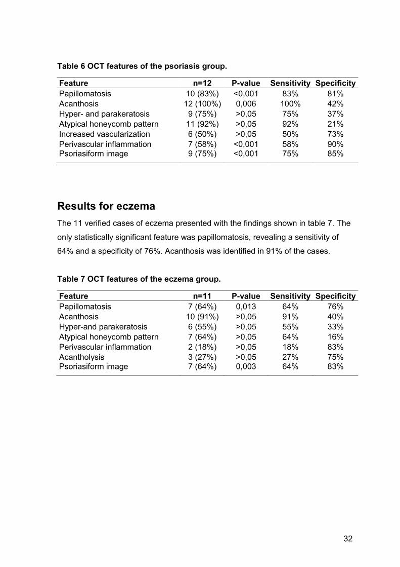
Results for eczema
The 11 verified cases of eczema presented with the findings shown in table 7. The
only statistically significant feature was papillomatosis, revealing a sensitivity of
64% and a specificity of 76%. Acanthosis was identified in 91% of the cases.
Table 7 OCT features of the eczema group.
Feature n=11 P-value Sensitivity Specificity
Papillomatosis 7 (64%) 0,013 64% 76%
Acanthosis 10 (91%) >0,05 91% 40%
Hyper-and parakeratosis 6 (55%) >0,05 55% 33%
Atypical honeycomb pattern 7 (64%) >0,05 64% 16%
Perivascular inflammation 2 (18%) >0,05 18% 83%
Acantholysis 3 (27%) >0,05 27% 75% Psoriasiform image 7 (64%) 0,003 64% 83%
33
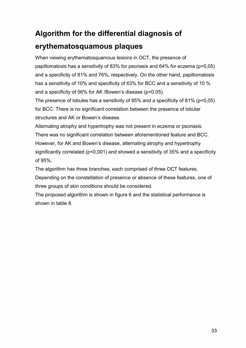
Algorithm for the differential diagnosis of
erythematosquamous plaques
When viewing erythematosquamous lesions in OCT, the presence of
papillomatosis has a sensitivity of 83% for psoriasis and 64% for eczema (p<0,05)
and a specificity of 81% and 76%, respectively. On the other hand, papillomatosis
has a sensitivity of 10% and specificity of 63% for BCC and a sensitivity of 10 %
and a specificity of 56% for AK /Bowen’s disease (p<0,05).
The presence of lobules has a sensitivity of 85% and a specificity of 81% (p<0,05)
for BCC. There is no significant correlation between the presence of lobular
structures and AK or Bowen’s disease.
Alternating atrophy and hypertrophy was not present in eczema or psoriasis.
There was no significant correlation between aforementioned feature and BCC.
However, for AK and Bowen’s disease, alternating atrophy and hypertrophy
significantly correlated (p<0,001) and showed a sensitivity of 35% and a specificity
of 95%.
The algorithm has three branches, each comprised of three OCT features.
Depending on the constellation of presence or absence of these features, one of
three groups of skin conditions should be considered.
The proposed algorithm is shown in figure 6 and the statistical performance is
shown in table 8.
34
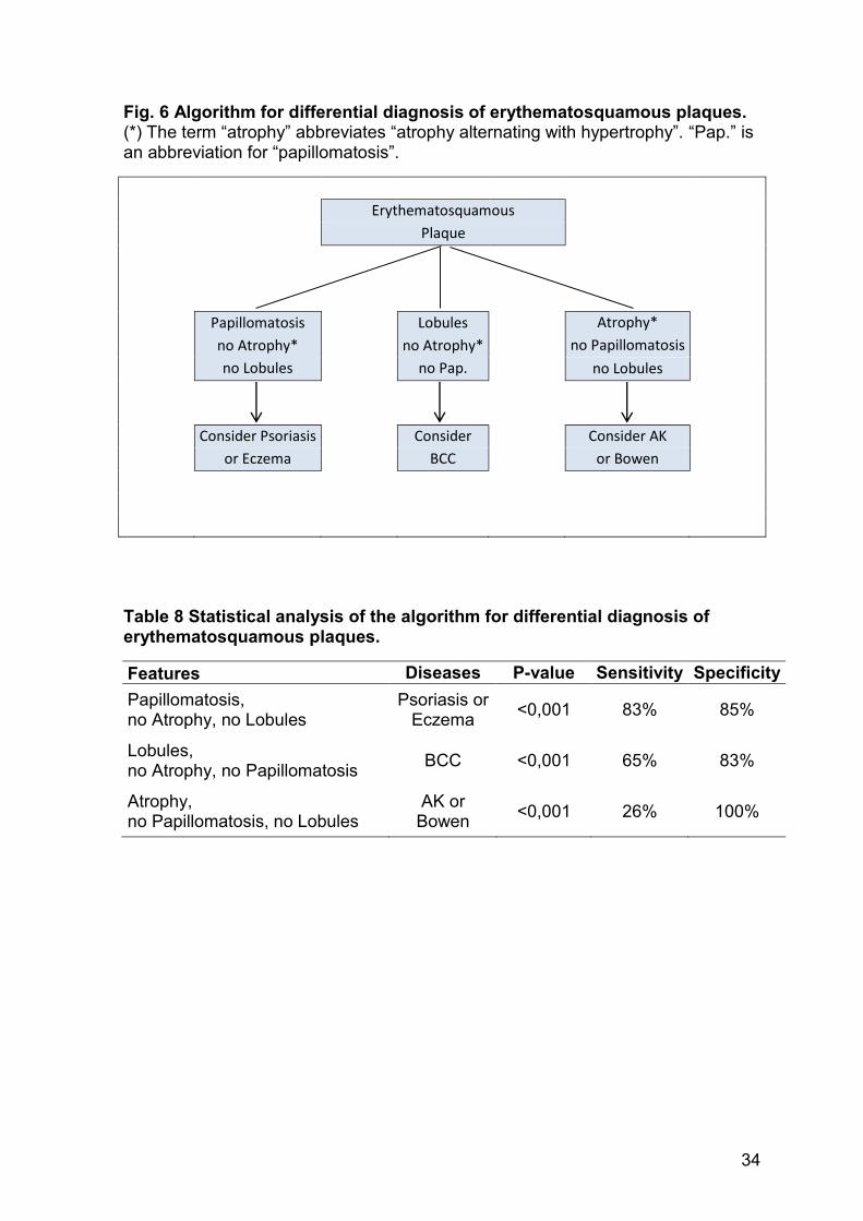
Fig. 6 Algorithm for differential diagnosis of erythematosquamous plaques. (*) The term “atrophy” abbreviates “atrophy alternating with hypertrophy”. “Pap.” is an abbreviation for “papillomatosis”.
Erythematosquamous
Plaque
Papillomatosis
no Atrophy*
no Lobules
Lobules
no Atrophy*
no Pap.
Atrophy*
no Papillomatosis
no Lobules
Consider Psoriasis Consider Consider AK
or Eczema BCC or Bowen
Table 8 Statistical analysis of the algorithm for differential diagnosis of erythematosquamous plaques.
Features Diseases P-value Sensitivity Specificity
Papillomatosis, no Atrophy, no Lobules
Psoriasis or Eczema
<0,001 83% 85%
Lobules, no Atrophy, no Papillomatosis
BCC <0,001 65% 83%
Atrophy, no Papillomatosis, no Lobules
AK or Bowen
<0,001 26% 100%
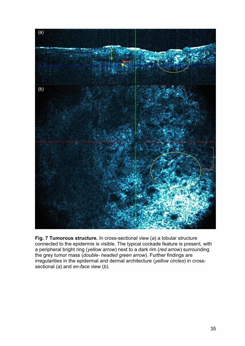
35
Fig. 7 Tumorous structure. In cross-sectional view (a) a lobular structure connected to the epidermis is visible. The typical cockade feature is present, with a peripheral bright ring (yellow arrow) next to a dark rim (red arrow) surrounding the grey tumor mass (double- headed green arrow). Further findings are irregularities in the epidermal and dermal architecture (yellow circles) in cross-sectional (a) and en-face view (b).
36
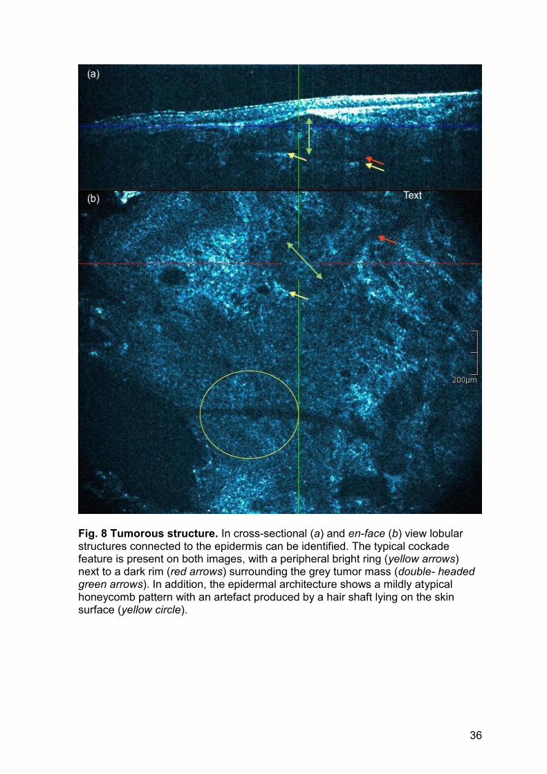
Fig. 8 Tumorous structure. In cross-sectional (a) and en-face (b) view lobular structures connected to the epidermis can be identified. The typical cockade feature is present on both images, with a peripheral bright ring (yellow arrows) next to a dark rim (red arrows) surrounding the grey tumor mass (double- headed green arrows). In addition, the epidermal architecture shows a mildly atypical honeycomb pattern with an artefact produced by a hair shaft lying on the skin surface (yellow circle).
37
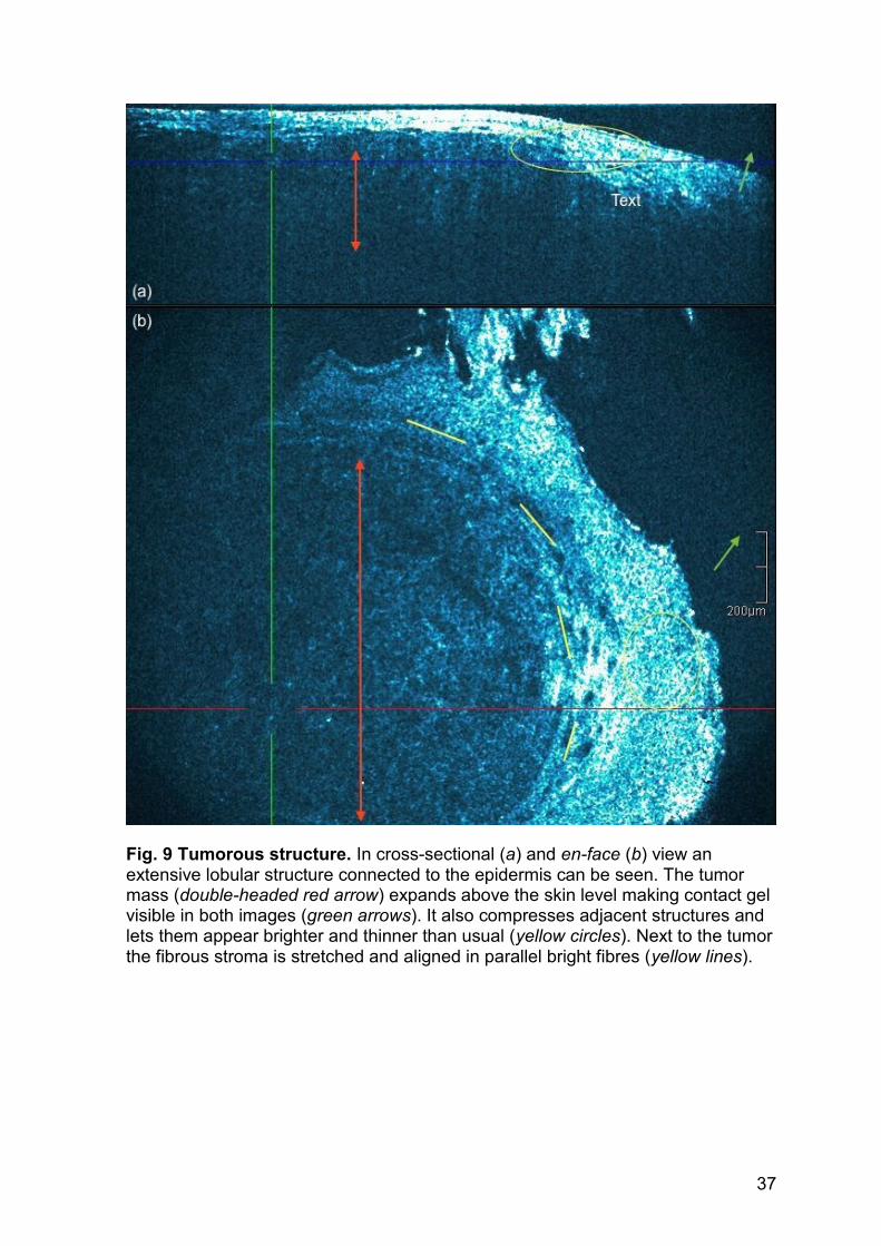
Fig. 9 Tumorous structure. In cross-sectional (a) and en-face (b) view an extensive lobular structure connected to the epidermis can be seen. The tumor mass (double-headed red arrow) expands above the skin level making contact gel visible in both images (green arrows). It also compresses adjacent structures and lets them appear brighter and thinner than usual (yellow circles). Next to the tumor the fibrous stroma is stretched and aligned in parallel bright fibres (yellow lines).
38
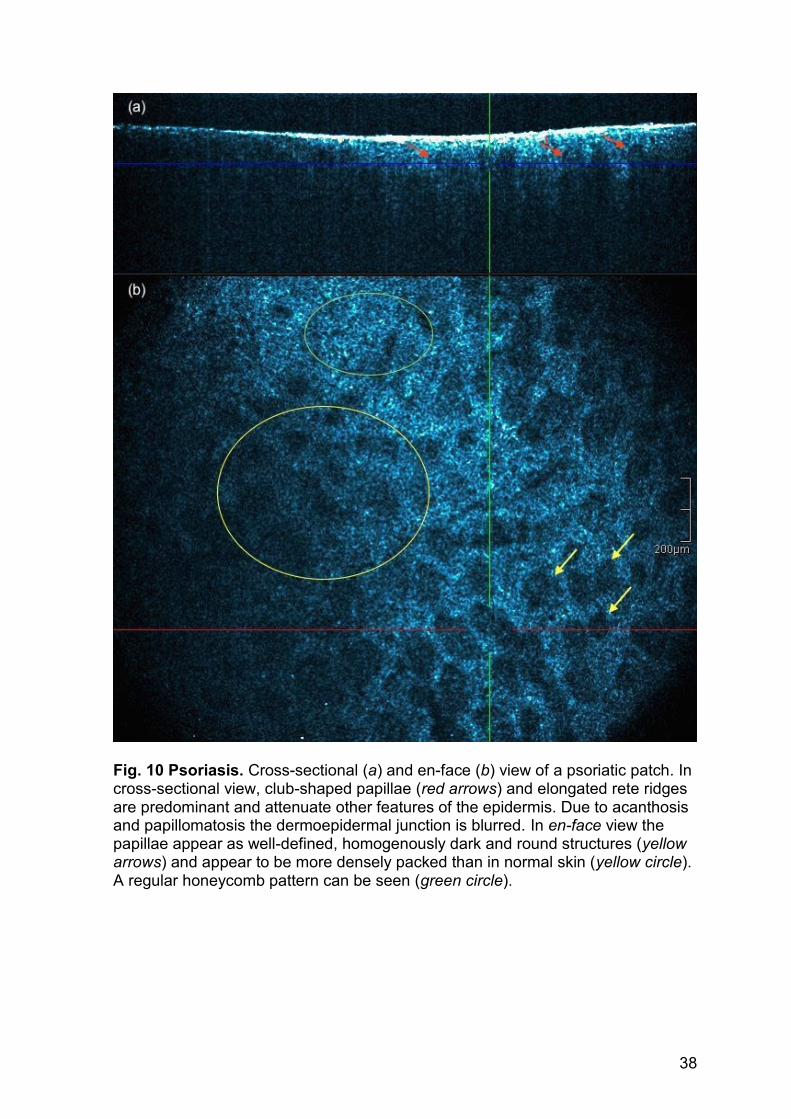
Fig. 10 Psoriasis. Cross-sectional (a) and en-face (b) view of a psoriatic patch. In cross-sectional view, club-shaped papillae (red arrows) and elongated rete ridges are predominant and attenuate other features of the epidermis. Due to acanthosis and papillomatosis the dermoepidermal junction is blurred. In en-face view the papillae appear as well-defined, homogenously dark and round structures (yellow arrows) and appear to be more densely packed than in normal skin (yellow circle). A regular honeycomb pattern can be seen (green circle).
39
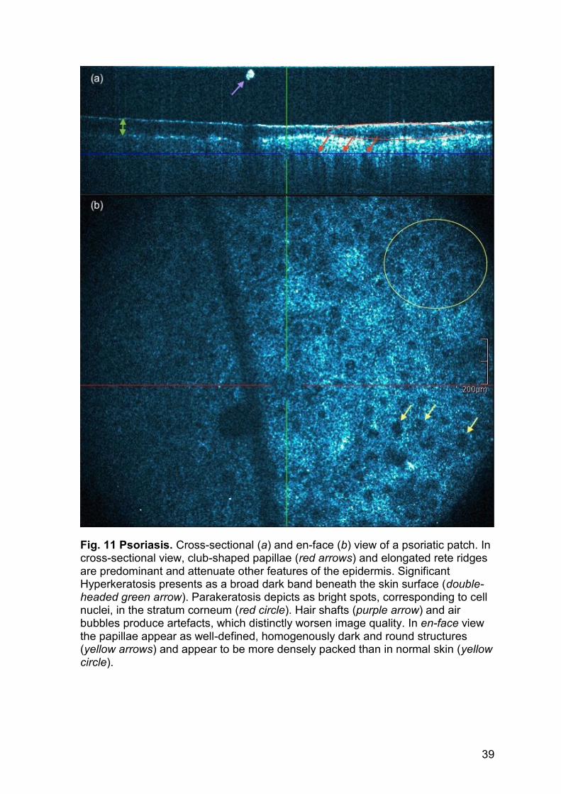
Fig. 11 Psoriasis. Cross-sectional (a) and en-face (b) view of a psoriatic patch. In cross-sectional view, club-shaped papillae (red arrows) and elongated rete ridges are predominant and attenuate other features of the epidermis. Significant Hyperkeratosis presents as a broad dark band beneath the skin surface (double-headed green arrow). Parakeratosis depicts as bright spots, corresponding to cell nuclei, in the stratum corneum (red circle). Hair shafts (purple arrow) and air bubbles produce artefacts, which distinctly worsen image quality. In en-face view the papillae appear as well-defined, homogenously dark and round structures (yellow arrows) and appear to be more densely packed than in normal skin (yellow circle).
40
DISCUSSION
Erythematosquamous plaques are a very common reason for patients to seek
dermatologic consultation. For the dermatologist however, the correct diagnosis
and management of these lesions pose a challenge in everyday clinic.
Studies suggest that dermoscopy may aid the diagnosis (25), but the role of OCT
has not yet been formally tested in this regard. Eventually, a combination of
multiple diagnostic modalities will reduce the need of biopsies, avoid incorrect
therapy and accelerate the process of finding a diagnosis.
Up to date, the majority of OCT studies concentrated on the definition of criteria in
the realm of non-melanoma skin cancers, which also commonly present as red
plaques or patches on the skin (10), (11), (19), (26), (27).
The data obtained by our study extend this current knowledge also to inflammatory
lesions such as psoriasis and eczema.
Boone et al. investigated the diagnostic potential of OCT for AK and squamous
cell carcinoma in 37 cases and found the presence of hyperkeratosis alternating
with parakeratosis in 100% of cases, while atypical honeycomb pattern were
present in about 90% of their cases. Other criteria such as atrophy and
hypertrophy were present in half of their cases (10). In our study including 31 AKs,
we found hyperkeratosis and parakeratosis in 24 (75%) cases, atrophy and
hypertrophy was found in 11 (31%) cases, atypical honeycomb pattern was
present in 27 (87%). The aforementioned features are suitable to discriminate
healthy skin from AK or squamous cell carcinoma (10). Notably, our study
highlights that these features are generally very common in erythematosquamous
plaques and therefore showed no significant correlation with the diagnosis of AK.
For instance, hyperkeratosis and parakeratosis were found in 48 (65%) of our
sample of 74 erythematosquamous plaques and in 9/12 (75%) cases of psoriasis.
Another study introduced an algorithm to aid discriminating specific BCC subtypes
from clinical imitators. In this sample of 50 BCCs, lobular structures occurred in 48
(96%) cases and lobular structures with the cockade feature in 37 (74%) cases. A
stretching effect was observed in 31 (61%) cases and lobular structures connected
to epidermis in 16 (32%) and lobular structures not connected to the epidermis in
32 (64%) cases (11).
41
Similarly, we found lobular structures to be present in 17/20 (85%) cases, lobular
structures with cockade in 13 (65%) cases, stretching effect in 13 (65%) cases,
lobular structures connected to epidermis in 10 (50%) and lobular structures not
connected to the epidermis in 12 (60%) cases. The defined features for BCC and
the algorithm by Boone et al. achieved in our sample a sensitivity of 85% and
specificity of 83% and therefore represent useful criteria for the discrimination of
BCC from other conditions presenting as erythematosquamous plaques.
Ulrich et al. investigated the sensitivity and specificity of OCT for 235 lesions
suspicious of BCC and found a sensitivity of 95,7% and a specificity of 75,3%.
Thereby OCT improved the diagnostic accuracy from 65,8% (clinical examination
alone) to 87,4% (28). A study by Markowitz et al. with 115 lesions suspicious of
BCC, detected a sensitivity of 92,9% and a specificity of 80% for OCT for the
diagnosis of BCC (29).
A review reported that the diagnostic accuracy declines when it comes to
differentiate BCC from AK (30). This supports the need for an algorithm offering
high sensitivity and specificity for the BCC imitators as well.
With regards to psoriasis, OCT poses as a useful tool for monitoring disease
activity and treatment response (19) ,(31), (32). However, the characteristic
pathohistological appearance of psoriasis might have its analogies in OCT and
further research on the morphology of psoriasis in OCT is promising. In our study
acanthosis and papillomatosis were very frequent findings, supporting the
assumption that these features could represent an important clue for the
diagnosis.
In our opinion, OCT will be secondary in the diagnosis of eczema and dermatitis in
general, although it may play a role in monitoring treatment response and disease
activity (9), (33). Our study included 11 cases of eczema and only papillomatosis
correlated significantly with the diagnosis of eczema (sensitivity 64%, specificity
76%). However, the number is too small to draw any definitive conclusions about
the diagnostic value of OCT in the diagnosis of eczema.
In summary, a noteworthy amount of established features of certain skin
conditions could be clearly observed and correlated with statistical significance.
With regards to our proposed algorithm for the differential diagnosis of
erythematosquamous skin lesions, further research should be encouraged and the
definition of the OCT features needs to be further validated.
42
Nevertheless, the partition into three groups of skin conditions appears to be
practicable. Also, the search for main criteria, as for instance lobules for basal cell
carcinoma, may aid the diagnosis.
Overall, OCT appears to be a useful tool for differentiating common skin conditions
presenting as erythematosquamous plaques.
However, concerning the implementation of OCT in everyday clinical practice a
few issues should be considered.
The recorded area is rather small and practically never captures the whole lesion.
Also, the processing of the acquired information interrupts the working flow. Real-
time recording with a wider field of vision could improve this matter.
Achieving images of lesions in certain body regions can be challenging due to the
construction of the camera. In this case, smaller construction or even
combinations with other modalities, as, for instance, dermatoscopy offer a solution.
This would also make aiming at an exact spot easier. Different skin regions and
skin types might be better visualized with different settings of the OCT,
comparable to the diverse ultrasound probes and settings available.
Concerning further research on the use of OCT in the differential diagnosis of
erythematosquamous plaques or other skin lesions the following seems
suggestive. The appearance and variance of healthy skin in OCT should be
thoroughly examined. Particularly features of the elderly skin, which presents with
findings such as solar elastosis or atrophy, should be taken into account when
defining algorithms. Moreover, the combination with other diagnostic tools, such as
dermoscopy or confocal laser scanning microscopy, and creation of algorithms
merging the results of the latter could significantly improve the quality of diagnosis.
Limitations
Our study is limited by the relatively small number of cases in each category and
by the fact that our algorithm has not been tested in an independent test set of
lesions. However, it was the aim of our study to establish specific criteria that aid
the differential diagnosis of common entities presenting at times as clinically
equivocal skin lesions.
43
References
1. Fujimoto JG, Pitris C, Boppart SA, Brezinski ME. Optical coherence tomography: an emerging technology for biomedical imaging and optical biopsy. Neoplasia. 2000;2(1-2):9-25. 2. Gambichler T, Jaedicke V, Terras S. Optical coherence tomography in dermatology: technical and clinical aspects. Arch Dermatol Res. 2011;303(7):457-73. 3. Fercher AF, Mengedoht K, Werner W. Eye-length measurement by interferometry with partially coherent light. Opt Lett. 1988;13(3):186-8. 4. Fercher AF, Hitzenberger CK, Drexler W, Kamp G, Sattmann H. In vivo optical coherence tomography. Am J Ophthalmol. 1993;116(1):113-4. 5. Podoleanu AG. Optical coherence tomography. J Microsc. 2012;247(3):209-19. 6. Wang Y, Zhao Y, Nelson JS, Chen Z, Windeler RS. Ultrahigh-resolution optical coherence tomography by broadband continuum generation from a photonic crystal fiber. Opt Lett. 2003;28(3):182-4. 7. Alex A, Povazay B, Hofer B, Popov S, Glittenberg C, Binder S, et al. Multispectral in vivo three-dimensional optical coherence tomography of human skin. J Biomed Opt. 2010;15(2):026025. 8. Boone M, Jemec GB, Del Marmol V. High-definition optical coherence tomography enables visualization of individual cells in healthy skin: comparison to reflectance confocal microscopy. Exp Dermatol. 2012;21(10):740-4. 9. Boone M, Norrenberg S, Jemec G, Del Marmol V. High-definition optical coherence tomography: adapted algorithmic method for pattern analysis of inflammatory skin diseases: a pilot study. Arch Dermatol Res. 2013;305(4):283-97. 10. Boone MA, Marneffe A, Suppa M, Miyamoto M, Alarcon I, Hofmann-Wellenhof R, et al. High-definition optical coherence tomography algorithm for the discrimination of actinic keratosis from normal skin and from squamous cell carcinoma. J Eur Acad Dermatol Venereol. 2015;29(8):1606-15. 11. Boone MA, Suppa M, Pellacani G, Marneffe A, Miyamoto M, Alarcon I, et al. High-definition optical coherence tomography algorithm for discrimination of basal cell carcinoma from clinical BCC imitators and differentiation between common subtypes. J Eur Acad Dermatol Venereol. 2015. 12. Mosby. Mosby’s Medical Dictionary. 9th ed: Elsevier; 2012. 1984 p. 13. Braun-Falco O. Braun-Falco's Dermatology. 3rd ed. Heidelberg: Springer Medizin Verlag Heidelberg; 2009. 1712 p. 14. Boone MA, Norrenberg S, Jemec GB, Del Marmol V. Imaging actinic keratosis by high-definition optical coherence tomography. Histomorphologic correlation: a pilot study. Exp Dermatol. 2013;22(2):93-7. 15. Calonje EB, T; Lazar, A; McKee, PH;. McKee's Pathology of the skin. 4th ed: Saunders W.B.; 2011. 1906 p. 16. Forsea AM, Carstea EM, Ghervase L, Giurcaneanu C, Pavelescu G. Clinical application of optical coherence tomography for the imaging of non-melanocytic cutaneous tumors: a pilot multi-modal study. J Med Life. 2010;3(4):381-9. 17. Parisi R, Symmons DP, Griffiths CE, Ashcroft DM, Identification, Management of P, et al. Global epidemiology of psoriasis: a systematic review of incidence and prevalence. J Invest Dermatol. 2013;133(2):377-85. 18. Fritsch P. Dermatologie und Venerologie für das Studium. 1st ed. Heidelberg: Springer Medizin Verlag; 2009.
44
19. Morsy H, Kamp S, Thrane L, Behrendt N, Saunder B, Zayan H, et al. Optical coherence tomography imaging of psoriasis vulgaris: correlation with histology and disease severity. Arch Dermatol Res. 2010;302(2):105-11. 20. Kempf WH, M; Kutzner, H; Burgdorf, W H. Dermatopathologie. 2nd ed. Berlin Heidelberg: Springer Verlag Berlin Heidelberg; 2011. 304 p. 21. Boone MA, Jemec GB, Del Marmol V. Differentiating allergic and irritant contact dermatitis by high-definition optical coherence tomography: a pilot study. Arch Dermatol Res. 2015;307(1):11-22. 22. Maier T, Braun-Falco M, Laubender RP, Ruzicka T, Berking C. Actinic keratosis in the en-face and slice imaging mode of high-definition optical coherence tomography and comparison with histology. Br J Dermatol. 2013;168(1):120-8. 23. Maier T, Braun-Falco M, Hinz T, Schmid-Wendtner MH, Ruzicka T, Berking C. Morphology of basal cell carcinoma in high definition optical coherence tomography: en-face and slice imaging mode, and comparison with histology. J Eur Acad Dermatol Venereol. 2013;27(1):e97-104. 24. Boone M, Suppa M, Miyamoto M, Marneffe A, Jemec GB, Pellacani G, et al. Three-dimensional high-definition optical coherence tomography image acquisition procedure for basal cell carcinoma. Br J Dermatol. 2015;172(4):1153-4. 25. Pan Y, Chamberlain AJ, Bailey M, Chong AH, Haskett M, Kelly JW. Dermatoscopy aids in the diagnosis of the solitary red scaly patch or plaque-features distinguishing superficial basal cell carcinoma, intraepidermal carcinoma, and psoriasis. J Am Acad Dermatol. 2008;59(2):268-74. 26. Gambichler T, Plura I, Schmid-Wendtner M, Valavanis K, Kulichova D, Stucker M, et al. High-definition optical coherence tomography of melanocytic skin lesions. J Biophotonics. 2015;8(8):681-6. 27. Boone MA, Norrenberg S, Jemec GB, Del Marmol V. High-definition optical coherence tomography imaging of melanocytic lesions: a pilot study. Arch Dermatol Res. 2014;306(1):11-26. 28. Ulrich M, von Braunmuehl T, Kurzen H, Dirschka T, Kellner C, Sattler E, et al. The sensitivity and specificity of optical coherence tomography for the assisted diagnosis of nonpigmented basal cell carcinoma: an observational study. Br J Dermatol. 2015. 29. Markowitz O, Schwartz M, Feldman E, Bienenfeld A, Bieber AK, Ellis J, et al. Evaluation of Optical Coherence Tomography as a Means of Identifying Earlier Stage Basal Cell Carcinomas while Reducing the Use of Diagnostic Biopsy. J Clin Aesthet Dermatol. 2015;8(10):14-20. 30. Cheng HM, Guitera P. Systematic review of optical coherence tomography usage in the diagnosis and management of basal cell carcinoma. Br J Dermatol. 2015. 31. Welzel J, Bruhns M, Wolff HH. Optical coherence tomography in contact dermatitis and psoriasis. Arch Dermatol Res. 2003;295(2):50-5. 32. Buder K, Knuschke P, Wozel G. Evaluation of methylprednisolone aceponate, tacrolimus and combination thereof in the psoriasis plaque test using sum score, 20-MHz-ultrasonography and optical coherence tomography. Int J Clin Pharmacol Ther. 2010;48(12):814-20. 33. Zabihian B, Weingast J, Liu M, Zhang E, Beard P, Pehamberger H, et al. In vivo dual-modality photoacoustic and optical coherence tomography imaging of human dermatological pathologies. Biomed Opt Express. 2015;6(9):3163-78.

45
APPENDIX
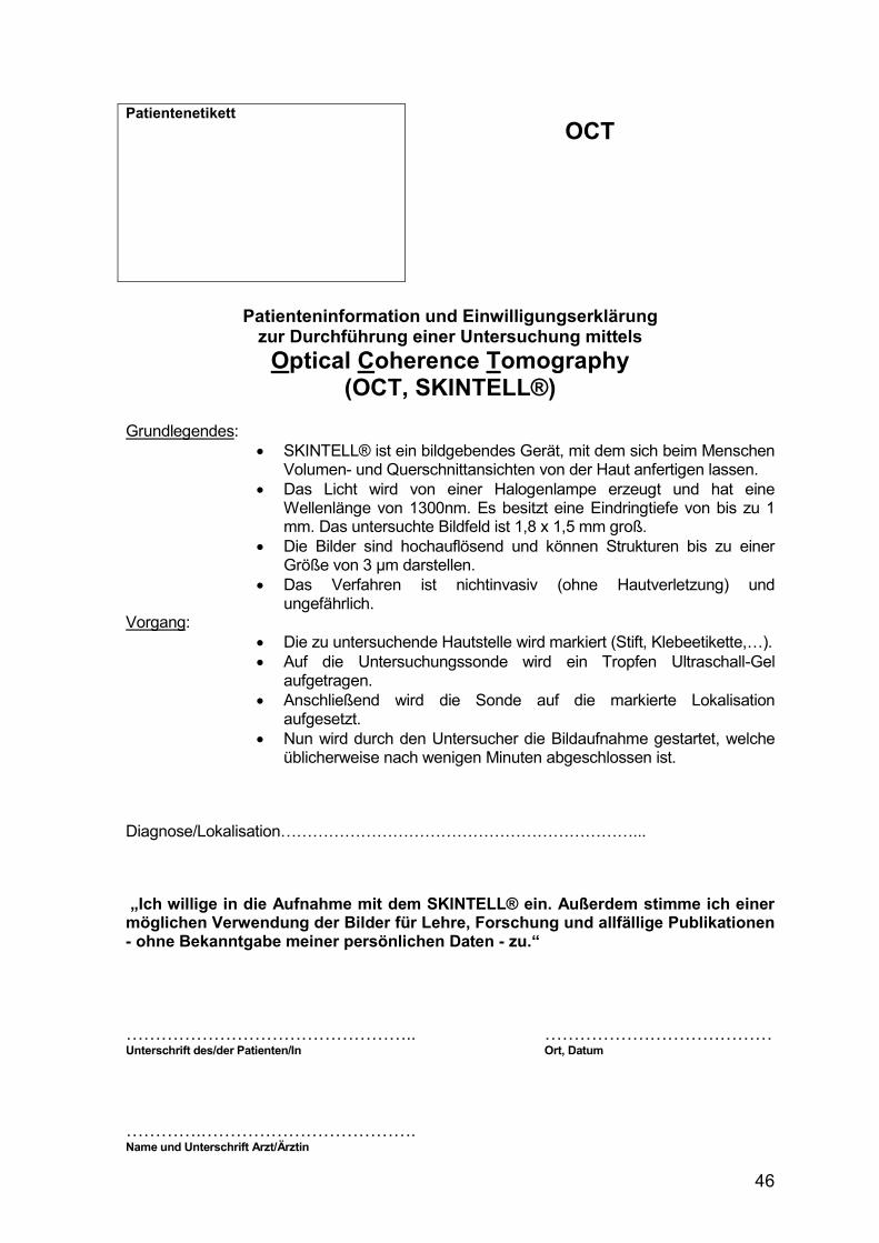
46
Patientenetikett
Patienteninformation und Einwilligungserklärung
zur Durchführung einer Untersuchung mittels
Optical Coherence Tomography (OCT, SKINTELL®)
Grundlegendes:
SKINTELL® ist ein bildgebendes Gerät, mit dem sich beim Menschen Volumen- und Querschnittansichten von der Haut anfertigen lassen.
Das Licht wird von einer Halogenlampe erzeugt und hat eine Wellenlänge von 1300nm. Es besitzt eine Eindringtiefe von bis zu 1 mm. Das untersuchte Bildfeld ist 1,8 x 1,5 mm groß.
Die Bilder sind hochauflösend und können Strukturen bis zu einer Größe von 3 µm darstellen.
Das Verfahren ist nichtinvasiv (ohne Hautverletzung) und ungefährlich.
Vorgang:
Die zu untersuchende Hautstelle wird markiert (Stift, Klebeetikette,…).
Auf die Untersuchungssonde wird ein Tropfen Ultraschall-Gel aufgetragen.
Anschließend wird die Sonde auf die markierte Lokalisation aufgesetzt.
Nun wird durch den Untersucher die Bildaufnahme gestartet, welche üblicherweise nach wenigen Minuten abgeschlossen ist.
Diagnose/Lokalisation…………………………………………………………...
„Ich willige in die Aufnahme mit dem SKINTELL® ein. Außerdem stimme ich einer möglichen Verwendung der Bilder für Lehre, Forschung und allfällige Publikationen - ohne Bekanntgabe meiner persönlichen Daten - zu.“
………………………………………….. ………………………………… Unterschrift des/der Patienten/In Ort, Datum
………….………………………………. Name und Unterschrift Arzt/Ärztin
OCT

47
Sehr geehrte Teilnehmerin, sehr geehrter Teilnehmer!
Wir laden Sie ein an der klinischen Studie Verwendung der „Optischen Kohärenztomographie zur …“ teilzunehmen. Die Aufklärung darüber erfolgt in einem ausführlichen ärztlichen Gespräch.
Ihre Teilnahme an dieser klinischen Studie erfolgt freiwillig. Sie können jederzeit ohne Angabe von Gründen aus der Studie ausscheiden. Die Ablehnung der Teilnahme hat keine nachteiligen Folgen für Ihre medizinische Betreuung.
Einwilligungserklärung
Name des Patienten ...........................................................................
Geb.Datum: ............................ Code: ...........................................................................
Ich erkläre mich bereit, an der klinischen Studie „ ...... „ teilzunehmen.
Ich bin ausführlich und verständlich über mögliche Belastungen und Risiken, sowie über
Bedeutung der klinischen Studie aufgeklärt worden. Ich habe darüber hinaus den Text dieser
Patientenaufklärung und Einwilligungserklärung gelesen. Aufgetretene Fragen wurden mir vom
Studienarzt verständlich und genügend beantwortet. Ich hatte ausreichend Zeit, mich zu
entscheiden. Ich habe zurzeit keine weiteren Fragen mehr.
Ich bin zugleich damit einverstanden, dass meine im Rahmen dieser klinischen Studie ermittelten
Daten aufgezeichnet werden. Um die Richtigkeit der Datenaufzeichnung zu überprüfen, dürfen
Beauftragte des Auftraggebers und der zuständigen Behörden beim Studienarzt Einblick in meine
personenbezogenen Krankheitsdaten nehmen.
......................................................................................................
(Datum und Unterschrift des Patienten)
......................................................................................................
(Datum, Name und Unterschrift des verantwortlichen Arztes)

48