Embed Size (px)
Citation preview

International Journal of
Molecular Sciences
Review
The Roles of Cyclin-Dependent Kinases in Cell-CycleProgression and Therapeutic Strategies in HumanBreast Cancer
Lei Ding 1,2,†, Jiaqi Cao 1,2,†, Wen Lin 1,2,†, Hongjian Chen 1,2, Xianhui Xiong 1,2,Hongshun Ao 1,2, Min Yu 1,2, Jie Lin 1,2 and Qinghua Cui 1,2,*
1 Lab of Biochemistry & Molecular Biology, School of Life Sciences, Yunnan University,Kunming 650091, China; [email protected] (L.D.); [email protected] (J.C.);[email protected] (W.L.); [email protected] (H.C.); [email protected] (X.X.);[email protected] (H.A.); [email protected] (M.Y.); [email protected] (J.L.)
2 Key Lab of Molecular Cancer Biology, Yunnan Education Department, Kunming 650091, China* Correspondence: [email protected]† The authors contributed equally to this work.
Received: 31 December 2019; Accepted: 24 February 2020; Published: 13 March 2020�����������������
Abstract: Cyclin-dependent kinases (CDKs) are serine/threonine kinases whose catalytic activities areregulated by interactions with cyclins and CDK inhibitors (CKIs). CDKs are key regulatory enzymesinvolved in cell proliferation through regulating cell-cycle checkpoints and transcriptional eventsin response to extracellular and intracellular signals. Not surprisingly, the dysregulation of CDKsis a hallmark of cancers, and inhibition of specific members is considered an attractive target incancer therapy. In breast cancer (BC), dual CDK4/6 inhibitors, palbociclib, ribociclib, and abemaciclib,combined with other agents, were approved by the Food and Drug Administration (FDA) recentlyfor the treatment of hormone receptor positive (HR+) advanced or metastatic breast cancer (A/MBC),as well as other sub-types of breast cancer. Furthermore, ongoing studies identified more selectiveCDK inhibitors as promising clinical targets. In this review, we focus on the roles of CDKs indriving cell-cycle progression, cell-cycle checkpoints, and transcriptional regulation, a highlight ofdysregulated CDK activation in BC. We also discuss the most relevant CDK inhibitors currently inclinical BC trials, with special emphasis on CDK4/6 inhibitors used for the treatment of estrogenreceptor-positive (ER+)/human epidermal growth factor 2-negative (HER2−) M/ABC patients, as wellas more emerging precise therapeutic strategies, such as combination therapies and microRNA(miRNA) therapy.
Keywords: breast cancer (BC); cyclin-dependent kinase (CDK); CDK inhibitor; cell cycle; clinic therapy
1. Introduction
Breast cancer (BC) is one of the leading causes of mortality in cancer patients, classified as fivemajor sub-types based on the comprehensive intrinsic gene expression profiling: luminal A (estrogenreceptor (ER)- and/or progesterone receptor (PR)-positive, and human epidermal growth factorreceptor 2 (HER2)-negative), luminal B (ER- or PR-positive and HER2-positive), basal-like (ER-, PR-,and HER2-negative, cytokeratin 5/6-positive, and/or epidermal growth factor receptor (EGFR)-positive,75% of triple-negative breast cancer (TNBC) (ER-, PR-, and HER2-negative) share the basal markerexpression), HER2-type (ER-negative, PR-negative, and HER2-positive), and normal-like [1]. Thesesub-types of BC were characterized based on specific morphological patterns, biological properties,different clinical stages, and prognosis. About 77% of BC patients are receptor-positive and the targetedtreatment has proven efficacy [2,3]. Unfortunately, approximately 15%–25% of TNBC patients present
Int. J. Mol. Sci. 2020, 21, 1960; doi:10.3390/ijms21061960 www.mdpi.com/journal/ijms

Int. J. Mol. Sci. 2020, 21, 1960 2 of 28
poor outcome due to unavailable targeted treatment. Surgery combined with chemotherapy andradiotherapy is commonly recommended for TNBC and most BC patients [4]. Early diagnosis, precisetreatment, and prognosis are urgently needed to improve prognostication, prevent cancer progression,and develop efficient therapies. Currently, the role of cell-cycle regulation is a fascinating area ofsuch discovery.
The mammalian cell division and death are the two major predominant physiologic processes inthe tissue homeostasis. The cell-cycle process is highly conserved and precisely controlled to governthe genome duplication and cell cycle, consisting of four distinct ordered phases, termed G0/G1 (gap 1),S (DNA synthesis), G2 (gap 2), and M (mitosis), and multiple checkpoints to ensure faithful replicationin the S phase and the exact aggregation of the chromosomes into daughter cells [5]. The G1 and G2phases are critical regulatory checkpoints, whereby the restriction point between the G1 and S phasedetermines whether the cells enter the S phase or exit the cell cycle to halt at the G0 phase. The cell cycleis regulated by many cyclins and cyclin-dependent kinases (CDKs) that are a group of serine/threoninekinases. They form complexes with cyclins to stabilize, activate, and phosphorylate CDKs in the specificphases [6,7]. The formation of cyclin/CDKs controls the cell-cycle progression via phosphorylation ofthe target genes, such as tumor suppressor protein retinoblastoma (Rb). The activation of cyclins/CDKsis induced by mitogenic signals and inhibited by the activation of cell-cycle checkpoints in responseto DNA damage [8]. The cyclin/CDKs themselves are negatively regulated by cyclin-dependentkinase inhibitors (CKIs), such as the inhibitor of CDK4 (INK4) proteins (p16INK4a, p15INK4b, p18INK4c,and p19INK4d), and CDK-interacting protein/kinase inhibitory proteins (CIP/KIPs) (p21CIP1, p27KIP1,and p57Kip2) [9]. In addition, the E3 ubiquitin ligases are also involved in regulating expression ofmany mitotic proteins to affect the transitions of the cell cycle, such as Skp1–Cul1–F-box-protein (SCF)complex and anaphase-promoting complex/cyclosome (APC/C) [10–12]. Dysregulation of the cellcycle and genetic alterations in cell-cycle regulatory proteins lead to uncontrolled cell proliferation inmany solid cancer types, including BC [13,14], which is a frontier in biomedical research for designingsynthetic inhibitors of CDKs as anticancer drugs [15]. In this review, we focus on the roles of CDKs incell-cycle regulation and gene transcription, and we provide CDK inhibitors as potential targets in BCclinical application.
2. CDKs in the Cell Cycle and Transcription
CDKs respond to the extracellular and intracellular signals to regulate cell division, acting asthe catalytic subunits by forming a heterodimer complex with the cyclins, which function as theregulatory subunits [16]. In human cells, there are 20 CDKs and 29 cyclins [17]. CDK1, CDK2, CDK3,CDK4, CDK6, and CDK7 directly regulate cell-cycle transitions and cell division, whereas CDK7–11mediate gene transcription. The expression of CDKs fluctuates in a cyclical fashion throughout the cellcycle [18].
2.1. The Roles of CDKs in the Cell Cycle
In most adult tissues, the majority of cells with diploid DNA content are arrested in a quiescentG0 state that can be either transient (quiescence) or permanent (terminal differentiation or senescence).As shown in Figure 1, upon mitosis, quiescent cells are involved in re-entering the cell cycle throughstimulation with mitogenic factors (hormone or growth factor). These factors converge on the cellcycle to activate cascades of intracellular signaling networks that impinge on CDK4 and CDK6 to drivecell-cycle progression from G0 or G1 phase into S phase. The activity of CDK4/6 is positively controlledby the association with D-type cyclins (cyclin D1, cyclin D2, and cyclin D3) and negatively controlledby binding to CDK inhibitors of the INK4 family (p16INK4A, p15INKB, p18INK4C, and p19INK4D) [19].Then, the active cyclin D/CDK4/6 complexes initiate phosphorylation of tumor suppressor proteinRB (encoded by RB1), as well as the closely related proteins p107 (also known as RBL1) and p130(also known as RBL2). The RB protein originally recruits co-repressors and represses the transcriptionof target genes regulated by E2F transcription factors (E2Fs) to inhibit G1/S transition. In this way,

Int. J. Mol. Sci. 2020, 21, 1960 3 of 28
the sequential phosphorylation inactivating the activity of RB leads to cell-cycle progression from G1into S phase. Meanwhile, the phosphorylated RB de-represses E2F transcription factors and inducesthe transcription of G1/S target genes such as cyclin E (CCNE), CCNA, and CCNB, dihydrofolatereductase (DHFR), ribonucleotide reductase M1 (RRM1) and RRM2 and polo-like kinase 1 (PLK1),spindle checkpoint protein MAD2 (MAD2), and BUB1 mitotic checkpoint serine/threonine kinase(BUB1), involved in G1–S entry and cell-cycle progression [9]. During late G1 phase, the target genes(cyclins E1 and E2) of E2Fs are activated, thereby binding and activating CDK2, which is originallysequestered by two CDK inhibitors p21CIP1 and p27KIP1, as well as ubiquitin-mediated proteolysisof p27KIP1 and p21CIP1. In addition, cell division cycle 25A (CDC25A) activates CDK2 by removingphosphorylation from CDK2 [20]. Furthermore, the active CDK2 is capable of phosphorylating aconsiderably broader range of substrate profile proteins required for cell-cycle progression (such asp27KIP1, RB, and E2F1), DNA replication (such as replication factors A and C), centrosome duplication(such as nucleophosmin (NPM)) and histone synthesis (such as nuclear protein, coactivator of histonetranscription (NPAT)) [21–23]. Specifically, the cyclin E/CDK2 active complex modulates RB to overridethe restriction point of the G1/S phase at the boundary, resulting in S phase initiation, which formsa positive feedback loop. The activities of CDK4/6 and CDK2 coordinate the cell-cycle progressioninto S phase, termed the “restriction point”, such that the mitogens are no longer required to completethe current cell cycle. Near the end of S phase, cyclin A removes cyclin E and forms a new complex,cyclin A/CDK2, where the cyclin E is rapidly degraded by the F-box/WD repeat-containing protein7 (FBXW7)-mediated ubiquitylation [21,22]. The cyclin A/CDK2 complex terminates the S phaseby phosphorylating CDC6 and E2F1, and drives the cell-cycle transition from S phase to G2 phase,and subsequently activates CDK1 by cyclin A leading the cells to enter the transition to M phase.Upon mitosis, CDK1 activity is maintained by the complex cyclin B/CDK1. The phosphorylationof activated CDK1 leads to the breakdown of nuclear envelope, the condensation of chromosome,and the assembly of mitotic spindle. The mitotic metaphase to anaphase is controlled by the spindleassembly checkpoints (SAC), and the anaphase is dependent on the decreased activity of CDK1 via thedegradation of cyclin B by APC/C [24]. The deregulated expression of CDK1 enables chromosomeseparation and completion of mitosis and cytokinesis. CDK1 is the only CDK that is essential forcell-cycle progression, as it initiates the onset of mitosis and ensures the critical events occur in the exactsequence in cellular replication with high fidelity [25]. In addition to regulation by its cyclin partners,the activity of CDK1 is controlled by the balance between the WEE1 G2 checkpoint kinase (WEE1),the membrane-associated tyrosine- and threonine-specific cdc2-inhibitory kinase myelin transcriptionfactor 1 (MYT1, also known as PKMYT1), and the phosphorylation of CDC25C phosphatases. WEE1phosphorylates CDK1 at Tyr 15, while MYT1 is phosphorylated at Thr 14 and Tyr 15 to inhibit theactivity of CDK1, and this phosphorylation is relieved by CDC25C phosphatases [26]. On the otherhand, cyclin C/CDK3 phosphorylates RB to push the cells into S phase from the G0 phase. The cells exitthe cell cycle and enter the reversible or permanent G0 phase, also regulated by cyclin C/CDK3 [27].

Int. J. Mol. Sci. 2020, 21, 1960 4 of 28Int. J. Mol. Sci. 2019, 20, x FOR PEER REVIEW 4 of 26
Figure 1. Progression of the cell cycle and its regulation by the CDKs and checkpoints. The cell cycle is regulated by many CDKs which form complexes with their associated cyclin partners. The cell cycle consists of four distinct ordered phases of the cell cycle, termed G0/G1, S, G2, and M phases, and it contains multiple checkpoints (red) throughout to prevent genomic instability, as well as ensure faithful replication. The cells exit the cell cycle and enter the reversible or permanent quiescent state (G0 phase) regulated by cyclin C/CDK3. Various extracellular signals, such as the mitogenic signal, lead to the synthesis of cyclin D and stimulate CDK4/6, resulting in promoting entry into the cell cycle. Active CDK4/6 complexes initiate the phosphorylation (P) of RB protein, thereby unleashing E2F transcription factors, resulting in the expression of cyclin E, cyclin A, cyclin B, and many genes required for S phase progression. Cyclin E subsequently activates CDK2 and contributes to the further phosphorylating RB, progresses into S phase, and initiates DNA synthesis. Near the end of S phase, cyclin A removes cyclin E and forms a new complex, cyclin A/CDK2. Cyclin A/CDK2 terminates the S phase by phosphorylating CDC6 and E2F1; it drives the cell-cycle transition from S phase to G2 phase, and subsequently activates CDK1 by cyclin A, leading to cells entering the M phase. Upon mitosis, the CDK1 activity is maintained by the complex cyclin B/CDK1. The deregulation of CDK1 enables chromosome separation and the completion of mitosis and cytokinesis. The INK4, CIP/KIP, and CDK4/6 inhibitors (palbociclib, ribociclib, and abemaciclib) inhibit the activity of CDK/cyclin. The ubiquitination (Ub) of cyclins is involved in regulating the expression of many proteins to control the cyclical activities of the CDKs, such as SCF and APC/C. The PLK1 and aurora A proteins are involved in the progression through S phase and from G2 phase into M phase. In addition, DNA damage checkpoints safeguard the genomic integrity and trigger cell-cycle arrest via checkpoint kinase 2 (CHK2) and p53 in G1 phase or via CHK1 in S or G2 phase. P in a dashed circle shows dephosphorylation. Green ovals indicate positive regulators and blue ovals indicate negative regulators of cell-cycle progression. (Adapted from reference “[8], doi:10.1038/nrc.2016.138” with permission of the journal Nature Reviews Cancer 2017).
The cell cycle is arrested or blocked by DNA damage-mediated cell-cycle checkpoints, thereby allowing DNA repair before cell-cycle progression into mitosis. As shown in Figure 1, two major cell-cycle checkpoints respond to DNA damage; they occur pre- and post-DNA synthesis in G1 and G2 phases and impinge on the activity of specific CDK complexes. The checkpoint kinases phosphatidylinositol 3-kinase (PI3K)-like protein kinases (PI3KKs) ataxia telangiectasia and Rad3-related (ATR) or ataxia telangiectasia mutated (ATM) protein, and the transducer checkpoint kinases CHK1 (encoded by the CHEK1 gene) and CHK2 (encoded by the CHEK2 gene) are key regulators of DNA damage signaling [28]. The DNA damage signaling is detected by ATM/ATR,
Figure 1. Progression of the cell cycle and its regulation by the CDKs and checkpoints. The cell cycleis regulated by many CDKs which form complexes with their associated cyclin partners. The cellcycle consists of four distinct ordered phases of the cell cycle, termed G0/G1, S, G2, and M phases,and it contains multiple checkpoints (red) throughout to prevent genomic instability, as well as ensurefaithful replication. The cells exit the cell cycle and enter the reversible or permanent quiescent state(G0 phase) regulated by cyclin C/CDK3. Various extracellular signals, such as the mitogenic signal,lead to the synthesis of cyclin D and stimulate CDK4/6, resulting in promoting entry into the cellcycle. Active CDK4/6 complexes initiate the phosphorylation (P) of RB protein, thereby unleashingE2F transcription factors, resulting in the expression of cyclin E, cyclin A, cyclin B, and many genesrequired for S phase progression. Cyclin E subsequently activates CDK2 and contributes to the furtherphosphorylating RB, progresses into S phase, and initiates DNA synthesis. Near the end of S phase,cyclin A removes cyclin E and forms a new complex, cyclin A/CDK2. Cyclin A/CDK2 terminates the Sphase by phosphorylating CDC6 and E2F1; it drives the cell-cycle transition from S phase to G2 phase,and subsequently activates CDK1 by cyclin A, leading to cells entering the M phase. Upon mitosis,the CDK1 activity is maintained by the complex cyclin B/CDK1. The deregulation of CDK1 enableschromosome separation and the completion of mitosis and cytokinesis. The INK4, CIP/KIP, and CDK4/6inhibitors (palbociclib, ribociclib, and abemaciclib) inhibit the activity of CDK/cyclin. The ubiquitination(Ub) of cyclins is involved in regulating the expression of many proteins to control the cyclical activitiesof the CDKs, such as SCF and APC/C. The PLK1 and aurora A proteins are involved in the progressionthrough S phase and from G2 phase into M phase. In addition, DNA damage checkpoints safeguardthe genomic integrity and trigger cell-cycle arrest via checkpoint kinase 2 (CHK2) and p53 in G1 phaseor via CHK1 in S or G2 phase. P in a dashed circle shows dephosphorylation. Green ovals indicatepositive regulators and blue ovals indicate negative regulators of cell-cycle progression. (Adapted fromreference “[8], doi:10.1038/nrc.2016.138” with permission of the journal Nature Reviews Cancer 2017).
The cell cycle is arrested or blocked by DNA damage-mediated cell-cycle checkpoints, therebyallowing DNA repair before cell-cycle progression into mitosis. As shown in Figure 1, twomajor cell-cycle checkpoints respond to DNA damage; they occur pre- and post-DNA synthesisin G1 and G2 phases and impinge on the activity of specific CDK complexes. The checkpointkinases phosphatidylinositol 3-kinase (PI3K)-like protein kinases (PI3KKs) ataxia telangiectasia andRad3-related (ATR) or ataxia telangiectasia mutated (ATM) protein, and the transducer checkpointkinases CHK1 (encoded by the CHEK1 gene) and CHK2 (encoded by the CHEK2 gene) are keyregulators of DNA damage signaling [28]. The DNA damage signaling is detected by ATM/ATR, which
Int. J. Mol. Sci. 2020, 21, 1960 5 of 28
then phosphorylate and activate CHK2/CHK1, respectively [29]. The activated CHK2 is involved inthe activation of p53, leading to p53-dependent early phase G1 arrest to allow time for DNA repair [30].The activation of p53 induces the expression of the CKI p21CIP1 gene, leading to inhibition of cyclinE/CDK2 complexes and downstream upregulation of DNA repair machinery. If the DNA repair cannotbe completed successfully or the cells cannot program to respond to the stresses of viable cell-cyclearrest, the cells face the fate of apoptosis induced by p53 [31]. The activated CHK1 mediates temporaryS phase arrest through phosphorylation to inactivate CDC25A, causing ubiquitination and proteolysis.Moreover, the activated CHK1 phosphorylates and inactivates CDC25C, leading to cell-cycle arrestin the G2 phase. The active CHK1 also directly stimulates the phosphorylation of WEE1, resultingin enhancing the inhibitory Tyr15 phosphorylation of CDK2 and CDK1 and subsequent cell-cycleblocking in G2 phase [8]. The activity of WEE1 can also be stimulated by the low levels of CDK activityin G2 cell-cycle phase [32]. The SAC, also known as the mitotic checkpoint, functions as the monitor ofthe correct attachment of the chromosomes to the mitotic spindle in metaphase, which is regulatedby the TTK protein kinase (TTK, also known as monopolar spindle 1 (MPS1)). The activation of SACtransiently induces cell-cycle arrest via inhibiting the activation of APC/C. In order to establish andmaintain the mitotic checkpoint, the TTK recruits many checkpoint proteins to kinetochores duringmitosis via phosphorylating its substrates to ensure adequate chromosome segregation and genomicintegrity [33,34]. In this way, the genomic instability from chromosome segregation defects is protectedby SAC. Once the SAC is passed, the APC/C E3 ligase complex stimulates and tags cyclin B and securinfor ubiquitin-mediated degradation, leading to the initiation of mitosis [5]. In a word, the checkpointsoffer a failsafe mechanism to ensure the genomic integrity from the parental cell to daughter cell.The signal transduction cascade of checkpoint activation eventually converges to CDK inhibition,which indicates the CDK function as a key driver of cell-cycle progression.
2.2. The Roles of CDKs in Transcription
In mammals, production of messenger RNAs (mRNAs) is strictly regulated, and it is divided intodiscrete phases of initiation, pausing, elongation, and termination, catalyzed by RNA polymeraseII (RNAPII), which is composed of a largest subunit (Rpb1) with a C-terminal domain (CTD) repeatof an evolutionarily conserved heptapeptide (Tyr–Ser–Pro–Thr–Ser–Pro–Ser) [35]. The CTD playsa vital role in RNA processing and chromatin organization in the coordination of transcriptionaland co-transcriptional events through changing its phosphorylation level [36,37]. Tyr1, Ser2, Thr4,Ser5, and Ser7 are phosphorylated in the heptapeptide by multiple CDK/cyclin subunits, such asCDK1 or CDK2 and most transcriptional CDKs, such as CDK7, CDK8, and CDK9 subfamilies [38].The phosphorylation at Ser5 and Ser7 of the CTD-RNAPII is required for the transcriptional initiation ofthe promoters. When the initiating transcription occurs, the Ser5 phosphorylation level decreases, whileSer2 and Tyr1 phosphorylation increases to promote transcriptional elongation. During transcriptiontermination, Tyr1 is dephosphorylated firstly, closely followed by Ser5, Ser7, and Ser2, which permitsrestarting the transcription cycle [39].
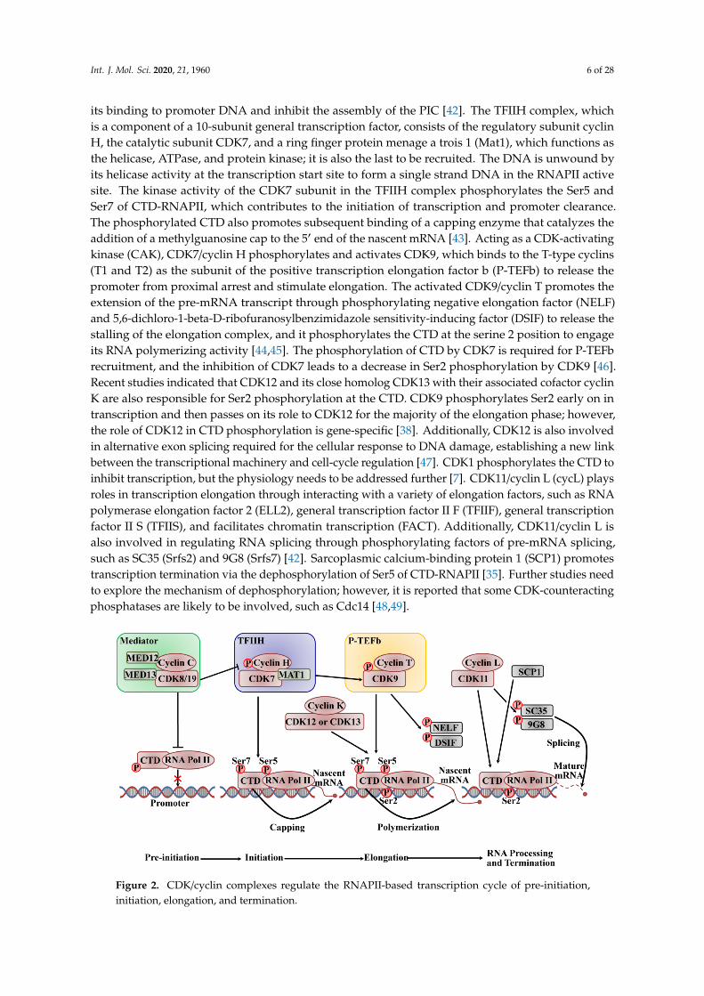
The active transcription is initiated by promoter recognition and DNA unwinding, therebyforming the pre-initiation complex. As shown in Figure 2, a very complicated process requiresRNAPII to interact with the large multi-subunit mediator complex and several general transcriptionfactors, and it is initiated by the binding of TATA binding protein of transcription factor II D(TFIID) to the core promoter to form the pre-initiation complex (PIC). CDK8 or CDK19 associatewith C-type cyclins, which are part of the mediator complex kinase module (MED) that acts as amolecular bridge linking the gene-specific signals from DNA-bound transcription factors to the generalRNAPII pre-initiation complex transcription machinery at the promoter [40,41]. The four-subunitkinase module of MED consists of CDK8 (or CDK19), cyclin C, the mediator complex subunit 12(Med12), and Med13, and this module is commonly associated with repression of transcription. MEDphosphorylates cyclin H to inhibit the assembly of pre-initiation complexes to negatively regulate theactivity of transcription factor II H (TFIIH) on CTD, and it phosphorylates CTD-RNAPII to impede
Int. J. Mol. Sci. 2020, 21, 1960 6 of 28
its binding to promoter DNA and inhibit the assembly of the PIC [42]. The TFIIH complex, whichis a component of a 10-subunit general transcription factor, consists of the regulatory subunit cyclinH, the catalytic subunit CDK7, and a ring finger protein menage a trois 1 (Mat1), which functions asthe helicase, ATPase, and protein kinase; it is also the last to be recruited. The DNA is unwound byits helicase activity at the transcription start site to form a single strand DNA in the RNAPII activesite. The kinase activity of the CDK7 subunit in the TFIIH complex phosphorylates the Ser5 andSer7 of CTD-RNAPII, which contributes to the initiation of transcription and promoter clearance.The phosphorylated CTD also promotes subsequent binding of a capping enzyme that catalyzes theaddition of a methylguanosine cap to the 5′ end of the nascent mRNA [43]. Acting as a CDK-activatingkinase (CAK), CDK7/cyclin H phosphorylates and activates CDK9, which binds to the T-type cyclins(T1 and T2) as the subunit of the positive transcription elongation factor b (P-TEFb) to release thepromoter from proximal arrest and stimulate elongation. The activated CDK9/cyclin T promotes theextension of the pre-mRNA transcript through phosphorylating negative elongation factor (NELF)and 5,6-dichloro-1-beta-D-ribofuranosylbenzimidazole sensitivity-inducing factor (DSIF) to release thestalling of the elongation complex, and it phosphorylates the CTD at the serine 2 position to engageits RNA polymerizing activity [44,45]. The phosphorylation of CTD by CDK7 is required for P-TEFbrecruitment, and the inhibition of CDK7 leads to a decrease in Ser2 phosphorylation by CDK9 [46].Recent studies indicated that CDK12 and its close homolog CDK13 with their associated cofactor cyclinK are also responsible for Ser2 phosphorylation at the CTD. CDK9 phosphorylates Ser2 early on intranscription and then passes on its role to CDK12 for the majority of the elongation phase; however,the role of CDK12 in CTD phosphorylation is gene-specific [38]. Additionally, CDK12 is also involvedin alternative exon splicing required for the cellular response to DNA damage, establishing a new linkbetween the transcriptional machinery and cell-cycle regulation [47]. CDK1 phosphorylates the CTD toinhibit transcription, but the physiology needs to be addressed further [7]. CDK11/cyclin L (cycL) playsroles in transcription elongation through interacting with a variety of elongation factors, such as RNApolymerase elongation factor 2 (ELL2), general transcription factor II F (TFIIF), general transcriptionfactor II S (TFIIS), and facilitates chromatin transcription (FACT). Additionally, CDK11/cyclin L isalso involved in regulating RNA splicing through phosphorylating factors of pre-mRNA splicing,such as SC35 (Srfs2) and 9G8 (Srfs7) [42]. Sarcoplasmic calcium-binding protein 1 (SCP1) promotestranscription termination via the dephosphorylation of Ser5 of CTD-RNAPII [35]. Further studies needto explore the mechanism of dephosphorylation; however, it is reported that some CDK-counteractingphosphatases are likely to be involved, such as Cdc14 [48,49].
Int. J. Mol. Sci. 2019, 20, x FOR PEER REVIEW 6 of 26
complex, which is a component of a 10-subunit general transcription factor, consists of the regulatory subunit cyclin H, the catalytic subunit CDK7, and a ring finger protein menage a trois 1 (Mat1), which functions as the helicase, ATPase, and protein kinase; it is also the last to be recruited. The DNA is unwound by its helicase activity at the transcription start site to form a single strand DNA in the RNAPII active site. The kinase activity of the CDK7 subunit in the TFIIH complex phosphorylates the Ser5 and Ser7 of CTD-RNAPII, which contributes to the initiation of transcription and promoter clearance. The phosphorylated CTD also promotes subsequent binding of a capping enzyme that catalyzes the addition of a methylguanosine cap to the 5′ end of the nascent mRNA [43]. Acting as a CDK-activating kinase (CAK), CDK7/cyclin H phosphorylates and activates CDK9, which binds to the T-type cyclins (T1 and T2) as the subunit of the positive transcription elongation factor b (P-TEFb) to release the promoter from proximal arrest and stimulate elongation. The activated CDK9/cyclin T promotes the extension of the pre-mRNA transcript through phosphorylating negative elongation factor (NELF) and 5,6-dichloro-1-beta-D-ribofuranosylbenzimidazole sensitivity-inducing factor (DSIF) to release the stalling of the elongation complex, and it phosphorylates the CTD at the serine 2 position to engage its RNA polymerizing activity [44,45]. The phosphorylation of CTD by CDK7 is required for P-TEFb recruitment, and the inhibition of CDK7 leads to a decrease in Ser2 phosphorylation by CDK9 [46]. Recent studies indicated that CDK12 and its close homolog CDK13 with their associated cofactor cyclin K are also responsible for Ser2 phosphorylation at the CTD. CDK9 phosphorylates Ser2 early on in transcription and then passes on its role to CDK12 for the majority of the elongation phase; however, the role of CDK12 in CTD phosphorylation is gene-specific [38]. Additionally, CDK12 is also involved in alternative exon splicing required for the cellular response to DNA damage, establishing a new link between the transcriptional machinery and cell-cycle regulation [47]. CDK1 phosphorylates the CTD to inhibit transcription, but the physiology needs to be addressed further [7]. CDK11/cyclin L (cycL) plays roles in transcription elongation through interacting with a variety of elongation factors, such as RNA polymerase elongation factor 2 (ELL2), general transcription factor II F (TFIIF), general transcription factor II S (TFIIS), and facilitates chromatin transcription (FACT). Additionally, CDK11/cyclin L is also involved in regulating RNA splicing through phosphorylating factors of pre-mRNA splicing, such as SC35 (Srfs2) and 9G8 (Srfs7) [42]. Sarcoplasmic calcium-binding protein 1 (SCP1) promotes transcription termination via the dephosphorylation of Ser5 of CTD-RNAPII [35]. Further studies need to explore the mechanism of dephosphorylation; however, it is reported that some CDK-counteracting phosphatases are likely to be involved, such as Cdc14 [48,49].
Figure 2. CDK/cyclin complexes regulate the RNAPII-based transcription cycle of pre-initiation,initiation, elongation, and termination.
Int. J. Mol. Sci. 2020, 21, 1960 7 of 28
CDK8/19 activates the transcription machinery at the promoter level. CDK8/19 also phosphorylates cyclinH to inhibit the assembly of the PIC to negatively regulate the activity of TFIIH, and it phosphorylates theCTD of RNAPII to impede its binding to promoter DNA and to inhibit the assembly of the PIC. CDK7and CDK9 drive mRNA elongation via sequential phosphorylation of the CTD-RNAPII. CDK12 andCDK13 with their cofactor cyclin K are also responsible for Ser2 phosphorylation at the CTD, allowingmRNA elongation. CDK11 is involved in the coordination between transcription and RNA splicing.DSIF and NELF inhibit elongation, while SCP1 promotes the termination of transcription. (Adaptedfrom reference “[42], doi:10.1242/dev.091744” with permission of the journal Development 2013).
3. Dysregulation of CDKs in BC
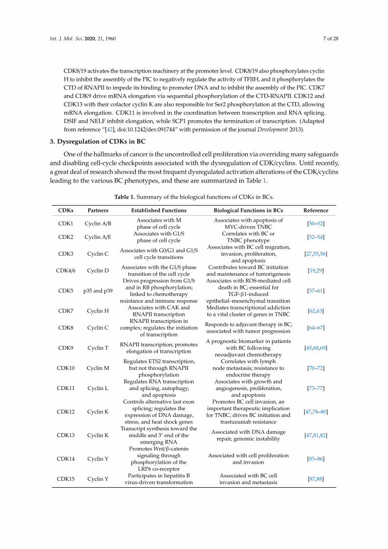
One of the hallmarks of cancer is the uncontrolled cell proliferation via overriding many safeguardsand disabling cell-cycle checkpoints associated with the dysregulation of CDK/cyclins. Until recently,a great deal of research showed the most frequent dysregulated activation alterations of the CDK/cyclinsleading to the various BC phenotypes, and these are summarized in Table 1.
Table 1. Summary of the biological functions of CDKs in BCs.
CDKs Partners Established Functions Biological Functions in BCs Reference
CDK1 Cyclin A/B Associates with Mphase of cell cycle
Associates with apoptosis ofMYC-driven TNBC [50–52]
CDK2 Cyclin A/E Associates with G1/Sphase of cell cycle
Correlates with BC orTNBC phenotype [52–54]
CDK3 Cyclin C Associates with G0/G1 and G1/Scell cycle transitions
Associates with BC cell migration,invasion, proliferation,
and apoptosis[27,55,56]
CDK4/6 Cyclin D Associates with the G1/S phasetransition of the cell cycle
Contributes toward BC initiationand maintenance of tumorigenesis [19,29]
CDK5 p35 and p39
Drives progression from G1/Sand in RB phosphorylation;
linked to chemotherapyresistance and immune response
Associates with ROS-mediated celldeath in BC; essential for
TGF-β1-inducedepithelial–mesenchymal transition
[57–61]
CDK7 Cyclin H Associates with CAK andRNAPII transcription
Mediates transcriptional addictionto a vital cluster of genes in TNBC [62,63]
CDK8 Cyclin CRNAPII transcription in
complex; regulates the initiationof transcription
Responds to adjuvant therapy in BC;associated with tumor progression [64–67]
CDK9 Cyclin T RNAPII transcription; promoteselongation of transcription
A prognostic biomarker in patientswith BC following
neoadjuvant chemotherapy[45,68,69]
CDK10 Cyclin MRegulates ETS2 transcription,
but not through RNAPIIphosphorylation
Correlates with lymphnode metastasis; resistance to
endocrine therapy[70–72]
CDK11 Cyclin LRegulates RNA transcription
and splicing, autophagy,and apoptosis
Associates with growth andangiogenesis, proliferation,
and apoptosis[73–77]
CDK12 Cyclin K
Controls alternative last exonsplicing; regulates the
expression of DNA damage,stress, and heat shock genes
Promotes BC cell invasion, animportant therapeutic implicationfor TNBC; drives BC initiation and
trastuzumab resistance
[47,78–80]
CDK13 Cyclin KTranscript synthesis toward the
middle and 3′ end of theemerging RNA
Associated with DNA damagerepair, genomic instability [47,81,82]
CDK14 Cyclin Y
Promotes Wnt/β-cateninsignaling through
phosphorylation of theLRP6 co-receptor
Associated with cell proliferationand invasion [83–86]
CDK15 Cyclin Y Participates in hepatitis Bvirus-driven transformation
Associated with BC cellinvasion and metastasis [87,88]

Int. J. Mol. Sci. 2020, 21, 1960 8 of 28
Table 1. Cont.
CDKs Partners Established Functions Biological Functions in BCs Reference
CDK16 Cyclin YRegulates mitosis, apoptosis,
and growth; synaptictrafficking and remodeling
Associated with TRAIL [89–92]
CDK17 Cyclin YPromotes amyloid precursor
protein-dependent Alzheimer;inhibits autophagy
Genetic expression profiles andchromosomal alterations [93–95]
CDK18 Cyclin Y
Promotes amyloid precursorprotein-dependent Alzheimer;inhibits autophagy; promotes
DNA replication stressand stability
Increases sensitivity to replicationstress-inducing chemotherapeutic
agents; induces DNAreplication stress
[93,94,96–98]
CDK19 Cyclin CCDK8 paralog, with a similar
role to CDK8, but seems toperform some distinct roles
The chemoresistance of BC;provides potential targets for the
improving chemotherapy[99,100]
CDK20 Cyclin HActivates ICK or β-catenin–TCF
signaling to stimulatecell-cycle progression
The role of CDK20 needs to befurther addressed in BC [7,35]
Abbreviations: MYC—MYC proto-oncogene, bHLH transcription factor; ROS—reactive oxygen species;TGF-β1—transforming growth factor beta 1; ETS2—ETS proto-oncogene 2, transcription factor;Wnt—wingless/integrated; LRP6—LDL receptor related protein 6; TRAIL—tumor necrosis factor (TNF)- relatedapoptosis-inducing ligand; ICK—intestinal cell kinase; TCF—T cell factor.
The CDK4/6–RB axis involved in the G1/S phase transition of the cell cycle plays an importantrole in BCs. Generally, cyclin D1/CDK4/6 is the key controller of RB phosphorylation to promotecell proliferation. It is expected that deregulation of the CDK4/6–cyclinD/INK4/pRB/E2F pathwayor its regulators contributes toward tumorigenesis and BC maintenance [29]. Practically, the loss ofINK4 and CIP/KIP family proteins, as well as the amplification of CDK4/6, was observed clinically inBC [9,14]. A recent study showed that the different BC sub-types have different cell-cycle checkpointmolecular alterations [14]. The dataset-based cancer genome study from 482 invasive BC patientsshowed that 27.4% of CDK4/6–RB axis genetic deregulation involves either the expression of singlegene alteration or multiple gene alterations in combination [101]. In particular, estrogen can increasethe rate of cell-cycle progression from the G1 to the S phase in ER-positive (ER+) BC, where the cyclinD1–CDK4/6–RB complex acts as the estrogen effector. Briefly, the estrogen binding to ER-alpha drivesthe transcription of cyclin D1, while the stimulation of CDK4/6 and phosphorylation of RB drivecell-cycle progression through the checkpoint, leading to initiation of the cell-cycle signal to stimulatethe expression of multiple receptor-driven genes involved in cell proliferation and survival. CyclinD1 amplification is detected in about 15% of BCs, particularly ER+ BCs [102]. Additionally, ER+ BCsoften exhibit upregulated expression of estrogen receptor 1 (ESR1) protein and high expression ofphosphatidylinositol-4,5-bisphosphate 3-kinase catalytic subunit alpha (PIK3CA), which contributes tocell-cycle progression through the mitogenic protein kinase B (AKT)/ mammalian target of rapamycin(mTOR) signaling pathway [14]. ER+ BC is relatively genomically stable with a primary dependencyon estrogen signaling, and it typically has normal function of RB and p53 tumor suppressor pathwayscompared to other BC subtypes, such as HER2+ and TNBC. Similarly, HER2-induced cell growth isalso mediated by the CDK4/6–RB axis [103]. The mouse models of human BC also indicate that thestimulation of the cyclin D1–CDK4/6 axis leads to a tumorigenic phenotype and contributes towardthe initiation and maintenance of tumorigenesis in HER2+ BC [101]. With the highly frequent HER2+
BC, CDK4 is amplified, as well as erb-b2 receptor tyrosine kinase 2 (ERBB2, the gene encoding theHER2 receptor), mutations of tumor protein p53 (TP53), PIK3CA, and phosphatase and tensin homolog(PTEN), and cyclin D1. On the contrary, genomic, clinical, and proteomic RB pathway data in TNBCexhibit RB1 mutation or deletion in 20% of cases and cyclin E1 amplification in 9% of the cases,high-level expression of cyclin dependent kinase inhibitor 2A (CDKN2A), low expression of RB1,and high proliferation rate, as well as frequent alteration in the DNA damage response genes, such as

Int. J. Mol. Sci. 2020, 21, 1960 9 of 28
tumor suppressor breast cancer 1 (BRCA1) [104,105]. The overexpression of cyclin E indicates a poorprognostic marker in TNBC and correlates to negative ER and PR status [106]. TNBC also activatesmutations or amplification of PIK3CA, B-Raf proto-oncogene, serine/threonine kinase (BRAF), KRASproto-oncogene, GTPase (KRAS) and/or EGFR and/or PTEN loss, resulting in the activation of anabnormal Raf/MAPK/ERK or PI3K/Akt/mTOR single pathway [107]. However, the rate of PIK3CAmutation in TNBC is only 8.3% [108]. Due to frequent loss or mutation of RB1 in TNBC, the integrity ofthe cell cycle is compromised, which is controlled by the Rb/E2F/CDK4/6 pathway. TNBC patients arecommonly considered to be poor candidates for CDK inhibition. However, TNBC is highly sensitive toa CDK2/9 inhibitor based on a preclinical trial, indicating that there may be unknown factors involvingthe CDK complex in TNBC proliferation [109]. On the other hand, some studies recently indicated thatthe expression of multiple genes of SAC is altered in TNBC, such as TTK, BUB1, MAD2, aurora kinaseB (AURKB), and DNA repair proteins, presumably due to the highly genomic instability in TNBC [5].
Additionally, other CDKs, such as CDK2, are upregulated; consequently, this often results in theamplification and/or overexpression of its partners cyclin A and cyclin E in BC [29]. CDK1 and itsassociated cyclins, cyclin A2 and cyclin B1, are often involved in mitotic progression, and increasedexpression of cyclin B1 is observed in BC [110]. However, there is no direct evidence to prove therelationship between genetic alteration dysregulating CDK1 activity and the initiation of BC. Onestudy indicated that the loss of CDK12 protein significantly improves the phenotype of TNBC due tothe loss of CDK12 resulting in defects in DNA repair [79]. The transcriptional cyclin-dependent kinase,CDK7, mediates transcriptional addition to a vital cluster of genes in TNBC, and CDK7 inhibition is auseful therapy for TNBC patients [62]. Thus, different mechanisms exist across various BC subtypes.
4. Targeting CDKs in BC Therapy
Given their roles in sustaining cancer cell growth, cyclins and CDKs are attractive targets forBC therapeutics. During the last decade, tremendous progress was made in developing new andeffective therapies, particularly through diverting BC cells from the proliferation phenotype to thenon-division state. CDK4/6 inhibitors, which mainly block the phosphorylation of RB to inhibitthe cell cycle, are studied widely, and they gained the most attractive findings. CDK inhibitors areclassified either as relatively non-selective pan-inhibitors or selective for one single CDK based on theirspecificity against CDKs. So far, CDK inhibitor drugs entered in numerous clinical trials in BC, insteadof irreversible ATP-competitive (covalent) inhibition, reversible and irreversible allosteric inhibition,and antibody–drug conjugation (ADCs). These inhibitors target cell-cycle regulators in the malignantcells, providing a therapeutic window where the vulnerabilities of cancer cells may be exploited withtolerable side effects from the normal tissue toxicity.
4.1. The Early Pan-CDK Inhibitors in BC
Most of the early CDK inhibitors exhibit relatively nonspecific inhibition, and they are referredto as pan-CDK inhibitors. Many pan-CDK inhibitors failed before phase II trials due to their limitedclinical activity as monotherapy, dose-limiting toxicities caused by undesirable target inhibitionfrom the off-target cells, such as nausea, vomiting, fatigue, hepatic dysfunction, neuropathy,myelosuppression, and gastrointestinal effects, and the lack of predictive biomarkers of these agents forpatient [111]. The early pan-CDK inhibitors include flavopiridol (also known as alvocidib, developedby Sanofi-Aventis), dinaciclib (also known as SCH 727965, developed by Merck), and seliciclib (alsoknown as roscovitine, developed by Cyclacel), as well as mitotic kinase inhibitors such as AURKB andPLK1. It was initially indicated that CDK1, CDK2, and CDK5 were the targets of seliciclib; however,subsequent data showed that it also inhibits transcription through CDK7 and CDK9 [112].
Of these first-generation inhibitors, flavopiridol is a semi-synthetic flavonoid derived fromrohitukine, a chromone alkaloid, with more than 60 clinical trials carried out between 1998 and 2014.It exerts its anticancer effect via the inhibition of CDK1, CDK2, CDK4, CDK6, CDK7, and CDK9 [113,114].Flavopiridol causes cell-cycle arrest in G1 and G2 phase; however, later work indicated that, in certain
Int. J. Mol. Sci. 2020, 21, 1960 10 of 28
contexts, it also induces a cytotoxic response by blocking the transcriptional activity of CDK7 andCDK9, as well as c-MYC [16]. In several phase I trials in solid malignancies, such as renal, prostate,and advanced sarcoma, flavopiridol was active as a single agent, as its combination with otherchemotherapy resulted in insufficient efficacy in BC [115], whereas low levels of clinical activity werereported in phase II studies for solid tumors. However, evidence showed that flavopiridol may haveclinical activity in hematological malignancies, such as chronic lymphocytic leukemia (CLL) and mantlecell lymphoma [116,117]. Seliciclib, an inhibitor of CDK1, CDK2, CDK5, and CDK7, did not showsignificant antitumor activities in preclinical and clinical studies as a monotherapy. Under clinicalinvestigation in combination with chemotherapy for solid tumors, side-effects occurred commonly,such as nausea, hyperglycemia, and hypokalemia [118]. In addition, one of the most significantpan-CDK inhibitors, dinaciclib, inhibits CDK1, CDK2, CDK5, and CDK9 with superior inhibitorypotency for RB phosphorylation, and it demonstrates an improved therapeutic index compared withflavopiridol [9]. In xenograft models of some solid tumors, such as ovarian and pancreatic cancers,pediatric acute lymphoblastic leukemia (ALL), and neuroblastoma-RAS viral oncogene homolog(NRAS)-mutant melanoma, it showed high activity in blocking the proliferation of tumor cells [8].Unfortunately, the preliminary results in BC were disappointing. A phase II randomized clinical trialof dinaciclib versus capecitabine in advanced breast cancer (ABC) was stopped due to its inferiorefficacy compared to capecitabine [119]. Moreover, preclinical studies suggested that the combinationof dinaciclib with an anthracycline has a synergistic effect in BC cell lines, and a phase I study withdinaciclib in combination with epirubicin in metastatic BC (MBC) patients showed no responses andhigh toxicity [29]. Finally, dinaciclib treatment may be efficacious in MYC-overexpressing TNBC due toit inhibiting the growth of tumor and enhancing survival in preclinical mouse models [8,50]. In parallelwith flavopiridol, roscovitine, a purine-based CDK inhibitor, was evaluated in the clinic. Unfortunately,only one single trial is ongoing for roscovitine in Cushing disease. Generally, first-generation pan-CDKinhibitors showed a low therapeutic index and high toxicities. Ongoing studies were developed toexploit new CDK inhibitors to circumvent these limitations.
4.2. The Clinical Success of CDK4/6-Selective Inhibitors in BC
The resistance induced by endocrine therapy in ER+ BC patients raised opportunities forthe development of CDK inhibitors. The CDK4/6 inhibitors were effectively approved, especiallywhen combined with anti-estrogen therapies [120], both in preclinical and clinical trials for ER+
BC. The selective CDK4/6 inhibitors, palbociclib (PD0332991), ribociclib (LEE011), and abemaciclib(LY2835219), were approved by the Food and Drug Administration (FDA) and the European MedicinesAgency (EMA) for the therapy of ER+/HER2− advanced and metastatic BC (A/MBC), showingdose-dependent growth inhibition in ER+ BC. All three drugs are small-molecule, ATP-competitivedrugs that can bind to the ATP-binding pocket of CDK4 and CDK6, with specific interactions withresidues in the ATP-binding cleft [9]. However, abemaciclib appears to readily bind to the ATP cleft byburying two fluorine atoms against the back wall of the ATP-binding pocket, and it binds with lessselectivity than ribociclib and palbociclib [121]. Palbociclib shows no activities against 36 additionalkinases, whereas ribociclib demonstrates no effects against CDK1/2 for the therapy of ER+/HER2−ABC in combination with anti-estrogen therapy, such as letrozole. Palbociclib and abemaciclib havedecreased potency against CDK1, CDK2, CDK7, and CDK9, and they are registered for second-linetherapy with fulvestrant [122]. Reported clinical trials assessing CDK4/6 inhibitors in BC are shown inTable 2.

Int. J. Mol. Sci. 2020, 21, 1960 11 of 28
Table 2. Reported clinical trials assessing CDK4/6 inhibitors in BC.
Trial Name Phase Treatment Arms Population No. PFS Most Common AEs
PALOMA-1/TRIO-18
(NCT00721409)II 1. Palbociclib + letrozole
2. Letrozole
Postmenopausal women,first-line treatment for
ER+/HER2−ABC177 1. 20.2 months
2. 10.2 months
Neutropenia, leukopenia,fatigue
anemia, nausea
PALOMA-2(NCT01740427)
III 1. Palbociclib + letrozole2. Placebo + letrozole ER+/HER2− ABC 666 1. 19.3 months
2. 12.9 months
Neutropenia, leukopenia,nausea, fatigue, arthralgia,
alopecia
PALOMA-3(NCT01942135)
III 1. Palbociclib + fulvestrant2. Placebo + fulvestrant HR+/HER2− ABC 521 1. 9.2 months
2. 3.8 months
Neutropenia, leukopenia,thrombocytopenia, fatigue,nausea, headache, alopecia
PALLAS(NCT02513394)
III
1. Palbociclib for 2 years +standard adjuvant
ET for 5 years2. Standard adjuvant ET for
5 years
ER+/HER2− early BC 5795 No detailed data No detailed data
PENELOPE-B(NCT01864746)
III
1. Palbociclib, 125 mg oncedaily, 28-day cycle
for 13 cycles2. Placebo 28-day cycle for
13 cycles
HR +/HER2− normalprimary BC with high
relapse risk afterneoadjuvant chemotherapy
1250 No detailed data No detailed data
MONALEESA-1(NCT01919229) II 1. Letrozole + ribociclib.
2. LetrozolePostmenopausal women
with HR+/HER2− early BC 14
Mean decrease inKi67-expressingcells, 1. 92%, 2.
69%
Nausea, decreased appetite,diarrhea, abdominal pain,
fatigue, asthenia
MONALEESA-2(NCT01958021) III 1. Ribociclib + letrozole
2. Placebo + letrozole
Postmenopausal womenwith HR+/HER2−MBC
received no prior therapyfor advanced disease
668 1. 19.3 months2. 14.7 months
Neutropenia, leukopenia,nausea, fatigue,
diarrhea, alopecia
MONALEESA-3(NCT02422615) III 1. Ribociclib + fulvestrant
2. Placebo + fulvestrant
Postmenopausal womenwith HR+/HER2− ABC
received no or only one lineprior endocrine treatment
726 1. 20.5 months2. 12.8 months
Neutropenia, leukopenia,nausea, fatigue, diarrhea,
alopecia, vomiting,constipation, arthralgia,
cough, headache,rash, anemia
MONALEESA-7(NCT02278120)
III1. Ribociclib +
NSAI/tamoxifen + goserelin2. placebo +
NSAI/tamoxifen + goserelin
Premenopausal orperimenopausal womenwith ER+/HER2−ABC
672 1. 23.8 months2. 13.0 months
Neutropenia, leukopenia,increased ALT, increased
AST, anemia, hypertension
MONARCH-1(NCT02102490) II Abemaciclib
heavily treatedHR+/HER2−M/ABC
patients (brain metastaseswere excluded)
132
6.0 months (95%confidence
interval (CI) 4.2 to7.5)
Leucopenia, neutropenia,diarrhea, fatigue, nausea,hypokalemia, increasedALT, decreased appetite,
hyponatremia, abdominalpain, thrombocytopenia
MONARCH-2(NCT02107703)
III 1. Abemaciclib +fulvestrant
2. Placebo + fulvestrant
HR+/HER2− locallyadvanced or metastatic BC. 669 1. 16.4 months
2. 9.3 months
Neutropenia, diarrhea,nausea, fatigue,abdominal pain
MONARCH-3(NCT02246621)
III1. Abemaciclib +
anastrozole/letrozole2. Placebo + anastrozole/
letrozole
Postmenopausal womenHR+/HER2− locoregionally,
recurrent, or MBC493 1. 28.2 months
2. 14.8 monthsNeutropenia, diarrhea,
nausea, fatigue, infections
Abbreviations: ER—estrogen receptor; HER2—human epidermal growth factor 2; ABC—advanced breastcancer; MBC—metastatic breast cancer; No.—number; ET—endocrine therapy; PFS—progression-freesurvival; AEs—adverse events; NSAI—nonsteroidal aromatase inhibitor; ALT—alanine aminotransferase;AST—aspartate aminotransferase.
Palbociclib (IBRANCE; PD0332991; Pfizer; C24H29N7O2) was the first CDK4/6 inhibitor approvedfor BC treatment. It is a reversible, orally administered, potent, small-molecule selective inhibitorof CDK4/6 [123], with a low enzymatic half-maximal inhibitory concentration (IC50) of 11 nM forCDK4 and 15 nM for CDK6 [124]. In fact, the human BC cell line shows different sensitivity topalbociclib based on its phenotype. The ER+ BC cell lines with luminal features are more sensitive topalbociclib than ER− BC cells with basal-like and TNBC histology [125,126]. Palbociclib is predicted tobe dependent on the presence of a functional RB protein, which causes cell-cycle arrest in G1 phaseby preventing RB phosphorylation at Ser 780 via cyclin D–CDK4/6. Consistent with the notion of RBrepresenting the major rate-limiting target of CDK4/6 in the cell cycle and the kinase selectivity profileof palbociclib, it shows no anti-proliferative activity in RB-deficient cells, where the requirement forCDK4/6 is bypassed [127]. Early trials showed palbociclib efficacy in ER+/HER2− ABC. The phase Ipalbociclib trial in MBC patients indicated that the preliminary, safety response and tolerable toxicity arefavorable for proceeding to the next phase of studies, and myelosuppression was the main dose-limiting
Int. J. Mol. Sci. 2020, 21, 1960 12 of 28
toxicity (DLT) [128,129]. The open-label randomized phase II clinical trial PALOMA-1 (also known asTRIO-18) studied the safety and efficacy of palbociclib (125 mg, 3/1 schedule) plus letrozole (an enzymeinvolved in the key step of estrogen biosynthesis) or letrozole alone for postmenopausal women withpreviously untreated ER+/HER2− ABC. The result showed that the median progression-free survival(PFS) for the combination of letrozole and palbociclib group was remarkably increased (from 10.2months to 20.2 months) compared to letrozole alone, and the median overall survival (OS) was alsoimproved with the combination treatment. Based on the results of the PALOMA-1 trial, the FDAprovided accelerated (provisional) approval of palbociclib (Ibrance) in combination with letrozolefor the treatment of postmenopausal women with ER+/HER2− ABC on February 3, 2015 [130]. Afterthe promising results, the randomized, double-blind, phase III PALOMA-2 (NCT01942135) clinicaltrial was conducted to confirm the significant clinical benefit and safety of palbociclib plus letrozole,with a median PFS of 24.8 months in the combination therapy versus 14.5 months in the letrozolegroup alone. Overall survival data showed a non-significant trend toward the palbociclib group.Similarly, the mainly adverse event (AE) was neutropenia. Based on data from PALOMA-2, the FDAgranted regular approval of palbociclib in combination with an aromatase inhibitor for the treatmentof ER+/HER2− ABC/MBC in 2017 [131]. Finally, the randomized, double-blind, placebo-controlledphase III trial PALOMA-3 trial tested the efficacy of palbociclib with fulvestrant (ER downregulatorand antagonist) by comparing treatment with palbociclib and fulvestrant to placebo and fulvestrantfor patients with ER+/HER2−MBC who relapsed or progressed during prior endocrine therapy inpre- and postmenopausal stages. Palbociclib combined with fulvestrant improved the median PFScompared to fulvestrant alone, from 4.6 months to 9.5 months. Based on these results, the FDAapproved palbociclib (Ibrance) for use in combination with fulvestrant for the treatment of womenwith ER+/HER2− ABC/MBC on February 19, 2016 [132–135]. Additionally, many ongoing studies areexploring palbociclib in ER+/HER2− early BC patients, including adjuvant and neoadjuvant studies,such as the randomized phase III study of PALLAS (NCT02513394) and PENELOPE-B (NCT01864746);primary results are expected in 2020 [6]. Furthermore, the combination of palbociclib with othertargeted agents, such as tamoxifen and trastuzumab, demonstrated a synergistic interaction andefficiently suppressed the proliferation of ER+ BC cell lines [126].
Ribociclib (KISQALI; LEE011; Novartis; C23H30N8O) is another oral drug with high-potencybioavailable selective inhibition of CDK4/6, which lacks significant activity against CDK1 and CDK2,with a low enzymatic IC50 of 10 nM for CDK4 and 40 nM for CDK6 [120]. Similarly to palbociclib,it blocks RB-positive (RB+) BC cell lines through inhibiting RB phosphorylation, and it causescell-cycle arrest of tumor cells [136]. Pharmacokinetic data were reported in a phase I ribociclibmonotherapy dose escalation study (NCT01237236), showing that doses of ribociclib in monotherapywere administered from 50 to 1200 mg (3/1 schedule), as well as 600 mg (once daily, continuously)for three consecutive weeks and one week off, and the maximum tolerated dose was 900 mg fromRB+ advanced solid tumors (including 18 breast cancer patients) or lymphomas, while the mostcommon DLTs were neutropenia and thrombocytopenia [137]. In the phase II trial MONALEESA-1(NCT01919229), postmenopausal women with ER+/HER2− early BC received letrozole with or withoutribociclib (400 or 600 mg) for two weeks prior to surgery. The study showed that the ribociclib plusletrozole combination was tolerated well, and no grade 3/4 AEs were observed after treatment [138].A randomized double-blind, placebo-controlled, phase III MONALEESA-2 (NCT01958021) trialenrolled 668 postmenopausal HR+/HER2− ABC patients who received no prior therapy for advanceddisease; they received either ribociclib (600 mg/day, three weeks on/one week off) plus letrozole (2.5mg/day) or placebo plus letrozole until the endpoints. The trial showed that the combination treatmentof ribociclib and letrozole significantly prolonged PFS (from 16 months to 25.3 months) compared toletrozole alone, which led to the FDA approval of ribociclib (Kisquali) in combination with letrozoleinhibitors as a first-line treatment for HR+/HER2−M/ABC on March 13, 2017 [139]. A randomizeddouble-blind, MONALEESA-3 (NCT02422615) phase III trial was intended for postmenopausal womenwith HR+/HER2− ABC patients who received no or only one line of prior endocrine treatment to
Int. J. Mol. Sci. 2020, 21, 1960 13 of 28
evaluate the efficacy of ribociclib or placebo with fulvestrant. The trial showed that the median PFSwas significantly improved with the addition of ribociclib to fulvestrant compared to the placebowith fulvestrant (20.5 months versus 12.8 months). Based on this result, the FDA approved thistreatment combination in HR+/HER2− M/ABC patients as first-line or second-line therapy in July2018 [140]. Most recently, another randomized double-blind, MONALEESA-7 (NCT02278120) phase IIItrial was dedicated to premenopausal patients with HR+/HER2− ABC who received ovarian functionsuppression together with oral endocrine therapy (tamoxifen and goserelin or a non-steroidal aromataseinhibitor (NSAI) and goserelin) with or without ribociclib. The study showed that the combination ofribociclib with endocrine therapy significantly improved the median PFS with 23.8 months versus13.0 months in the placebo with NSAI/tamoxifen plus goserelin group [141]. Additionally, there are agreat deal of other ongoing trials exploring ribociclib in combination with other targeted agents for BC,such as the PACE trial (NCT03147287) and MAINTAIN trial (NCT02632045), which are being carriedout to evaluate the efficacy of Ribociclib in HR+ MBC patients previously treated with a CDK4/6inhibitor. Moreover, the TRINITI-1 trial (NCT01857193) is evaluating the effects of a triple combinationof everolimus with ribociclib and exemestane-progressed patients who previously used a CDK4/6inhibitor. The study of NCT03238196 is evaluating the effects of a triple combination of erdafitinibwith ribociclib and fulvestrant in HR+ MBC [142].
Abemaciclib (VERZENIO; LY2834219; Lilly; C27H32F2N8) is an orally available bioavailableinhibitor of CDK4/6 with low enzymatic IC50, i.e., 2 nM for CDK4 and 10 nM for CDK6, correlatingwith a dose-dependent inhibition of RB phosphorylation and cell-cycle arrest at G1 phase, leadingto inhibition of proliferation and decreased cell number. Abemaciclib inhibits not only CDK4/6 butalso some other more complex pharmacological functions, including an effective CDK9 and Pim-1proto-oncogene, serine/threonine kinase (PIM1), albeit with lower potency [143]. A preclinical studyshowed that abemaciclib can be used alone or in combination with other drugs. Abemaciclib can crossthe blood–brain barrier at lower doses and may remain on target for a longer time than palbociclib.Therefore, these properties are being investigated in anti-tumor therapy for patients with brainmetastases [144,145]. A phase I trial of abemaciclib monotherapy (200 mg as monotherapy or 150 mgwhen given with endocrine therapy) showed that the overall response rate was 31% and 61% of heavilypretreated HR+ BC patients achieving either response or stable disease lasting beyond six months,suggesting that abemaciclib may have benefit as a monotherapy [146]. The single-arm phase II studyMONARCH-1 (NCT02102490) was carried to evaluate the safety and efficacy of abemaciclib alone forheavily treated HR+/HER2−M/ABC patients (brain metastases were excluded), with abemaciclib 200mg administered on a continuous schedule every 12 h until disease progression. The study exhibited aconfirmed objective response rate (ORR) of 19.7%, while the clinical benefit rate was 42.4% and medianPFS was 6.0 months [147]. Based on the results, the FDA granted approval for the poor-prognosis BCpatient group in September 2017. Furthermore, the randomized, double-blind, placebo-controlledMONARCH 2 (NCT02107703) phase III clinical trial was carried out for HR+/HER2− ABC patientswho received prior neoadjuvant or adjuvant endocrine therapy and relapsed after endocrine therapy.Patients were randomized 2:1 and received abemaciclib or placebo with fulvestrant. The study indicatedthat a combination of abemaciclib with fulvestrant significant improved median PFS compared tofulvestrant alone (16.4 versus 9.3 months) [148], which led to the FDA approval of abemaciclib(Verzenio) in combination with fulvestrant in patients with HR+/HER2−M/ABC as second-line therapyin September 2017. Another phase III MONARCH 3 (NCT02246621) study treatment was proposed asa first-line treatment in HR+/HER2−MBC patients who did not receive prior treatment. The patientswere randomized 2:1 to receive abemaciclib with anastrozole or letrozole compared to placebo plusanastrozole or letrozole. The results showed that the abemaciclib arm had better median PFS thanthe placebo arm (28.18 versus 14.76 months) [149], which also led to the FDA approving abemaciclib(Verzenio) in combination with aromatase therapy in HR+/HER2−M/ABC patients as first-line therapyin February 2018. In addition, other ongoing studies with abemaciclib in BC were carried out, such asthe neoMONARCH (NCT02441946) phase II clinical trial to compare the biological effects of abemaciclib

Int. J. Mol. Sci. 2020, 21, 1960 14 of 28
combination with anastrozole to those of abemaciclib monotherapy and anastrozole monotherapyfor two weeks, as well as to evaluate the activity and safety of abemaciclib with anastrozole in asubsequent 14 weeks with HR+/HER2− ABC [150]. The monarchE study (NCT03155997) assessedthe safety and efficacy of abemaciclib combined with standard endocrine therapy versus endocrinetherapy alone in high-risk, node-positive, early-stage BC [131].
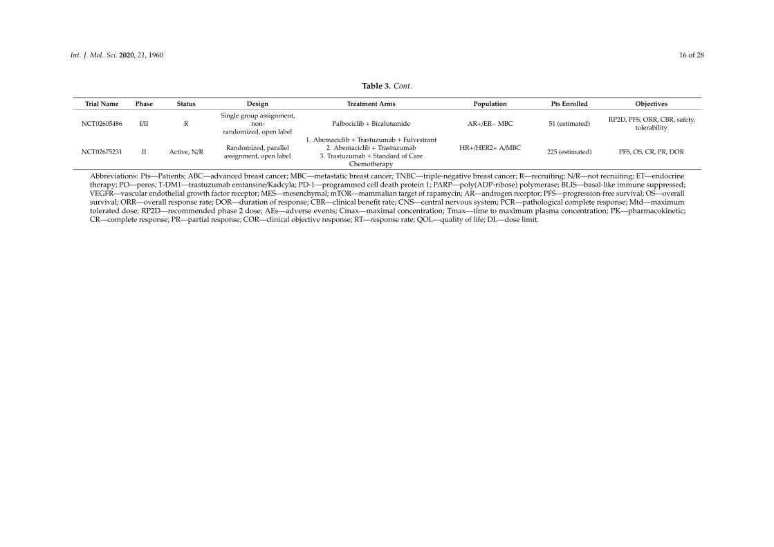
In addition to the FDA-approved three compounds, several other CDK4/6 inhibitors are beingexplored, with ongoing clinical studies evaluating their effect on their own or in combination withother targeted therapies as standard chemotherapies in BC. For instance, the international, open-label,randomized clinical trial, phase III study PEARL (NCT02028507) is being carried out to test palbociclibwith endocrine therapy versus chemotherapy (capecitabine) in postmenopausal HR+/HER2−MBC,with resistance to aromatase inhibitors. The international, multicenter, randomized clinical trial, phaseIb/II study PASTOR (NCT02599714) is assessing vistusertib plus palbociclib plus fulvestrant versusplacebo plus palbociclib plus fulvestrant in ER+ local A/MBC postmenopausal patients previouslytreated with hormonal therapy [120]. In addition to HR+/HER2− A/MBC, ongoing and recruitingclinical trials are evaluating the activity of CDK4/6 inhibitors in other BC subtypes, such as HER2+
BC and TNBC. Palbociclib/ribociclib with bicalutamide therapy was carried out for the treatmentof AR+ TNBC (NCT02605486/NCT03090165). The monarcHER (NCT02675231) study also featuresabemaciclib under active clinical investigation for patients with HR+/HER2+ M/ABC after at leasttwo HER2-directed therapies [131]. Some ongoing clinical trials assessing CDK4/6 inhibitors forHR+/HER2+ M/ABC or TNBC patients are listed in Table 3.

Int. J. Mol. Sci. 2020, 21, 1960 15 of 28
Table 3. Ongoing clinical trials assessing CDK4/6 inhibitors for HR+/HER2+ M/ABC or TNBC patients.
Trial Name Phase Status Design Treatment Arms Population Pts Enrolled Objectives
NCT02947685 III R Randomized, parallelassignment, open label
1. Palbociclib + anti-HER2 therapy(trastuzumab/pertuzumab) + ET (letrozole,
anastrozole, exemestane, fulvestrant)2. Anti-HER2 therapy
(trastuzumab/pertuzumab) + ET (letrozole,anastrozole, exemestane, fulvestrant)
HER2+/ER+ BC 496 (estimated)PFS, OS, ORR, DOR, CBR,
safety, 3 and 5 year survivalprobabilities
NCT02774681 II Terminated Single group assignment,open label
1. palbociclib PO2. palbociclib PO + trastuzumab IV
HER2+/PR−MBC with brainmetastasis 12 (estimated) AEs, CNS, PFS, OS, CNS, ORR,
safety, tolerability
NCT02530424 II Active, N/R Single group assignment,open label
(Trastuzumab + Pertuzumab + Palbociclib ±Fulvestrant) + Surgery
ER+/HER2+ BC suitable forneoadjuvant therapy 102 (actual) PCR, COR, safety, tolerability
NCT02657343 Ib/II Active, N/RNon-
randomized, parallelassignment, open label
1. Ribociclib + T-DM12. Ribociclib + Trastuzumab
3. Ribociclib + Trastuzumab + FulvestrantHER+ A/MBC 26 (actual) Mtd, RP2D,CBR, ORR, PFS, OS.
NCT03913234 I/II Not yet R Single group assignment,open label Ribociclib + Trastuzumab + Letrozole Postmenopausal HER2+ MBC 95 (estimated) PFS,OS, RT, QOL
NCT03054363 Ib/II RNon-
randomized, single groupassignment, open label
Tucatinib + Palbociclib + Letrozole HR+/HER2+ A/MBC 25 (estimated) AEs, PFS
NCT03993964 II Not yet R Single group assignment,open label Pyrotinib + SHR6390 HER2+ ABC 20 (estimated) ORR, PFS, OS
NCT03090165 I/II Active,N/R Single group assignment,open label
1. bicalutamide + ribociclib 400mg PO dailyon days 1-21 of a 28-day cycle.
2. bicalutamide + ribociclib 400mg PO dailyon days 1-28 of a 28-day cycle.
3. bicalutamide + ribociclib 600mg PO dailyon days 1-21 of a 28-day cycle.
AR+ TNBC 11 (actual) ORR, DOR, safety, tolerability,PFS, OS, CBR,
NCT03805399 Ib/II R
Non-randomized, open label,umbrella study, parallel
assignment
1. Pyrotinib + Capecitabine2. AR inhibitor + CDK4/6 inhibitor
3. anti PD-1 + nab-paclitaxel4. PARP inhibitor
5. BLIS + anti-VEGFR6. MES + anti-VEGFR
7. mTOR inhibitor + nab-paclitaxel
TNBC 140 (estimated) ORR, DOR, PFS, OS
NCT03519178 II RNon-
randomized, single groupassignment, open label
1. PF-068736002. PF-06873600 + Endocrine Therapy 13. PF-06873600 + Endocrine Therapy 2
HR+/HER2−MBC, TNBC 220 (estimated) DL, safety, tolerability, ORR,Cmax, Tmax, PK
NCT02907918 II R Single group assignment,open label
(Palbociclib + letrozole + trastuzumab +/-goserelin) + surgery ER+/HER2+ Stage II-III BC 48 (estimated) PCR, safety, tolerability
Int. J. Mol. Sci. 2020, 21, 1960 16 of 28
Table 3. Cont.
Trial Name Phase Status Design Treatment Arms Population Pts Enrolled Objectives
NCT02605486 I/II RSingle group assignment,
non-randomized, open label
Palbociclib + Bicalutamide AR+/ER−MBC 51 (estimated) RP2D, PFS, ORR, CBR, safety,tolerability
NCT02675231 II Active, N/R Randomized, parallelassignment, open label
1. Abemaciclib + Trastuzumab + Fulvestrant2. Abemaciclib + Trastuzumab
3. Trastuzumab + Standard of CareChemotherapy
HR+/HER2+ A/MBC 225 (estimated) PFS, OS, CR, PR, DOR
Abbreviations: Pts—Patients; ABC—advanced breast cancer; MBC—metastatic breast cancer; TNBC—triple-negative breast cancer; R—recruiting; N/R—not recruiting; ET—endocrinetherapy; PO—peros; T-DM1—trastuzumab emtansine/Kadcyla; PD-1—programmed cell death protein 1; PARP—poly(ADP-ribose) polymerase; BLIS—basal-like immune suppressed;VEGFR—vascular endothelial growth factor receptor; MES—mesenchymal; mTOR—mammalian target of rapamycin; AR—androgen receptor; PFS—progression-free survival; OS—overallsurvival; ORR—overall response rate; DOR—duration of response; CBR—clinical benefit rate; CNS—central nervous system; PCR—pathological complete response; Mtd—maximumtolerated dose; RP2D—recommended phase 2 dose; AEs—adverse events; Cmax—maximal concentration; Tmax—time to maximum plasma concentration; PK—pharmacokinetic;CR—complete response; PR—partial response; COR—clinical objective response; RT—response rate; QOL—quality of life; DL—dose limit.

Int. J. Mol. Sci. 2020, 21, 1960 17 of 28
4.3. The Novel CDK Inhibitors in BC
With a better understanding of CDKs in different BC subtypes, as well as the achievements usingCDK4/6 inhibitors in HR+/HER2− A/MBC, along with the side effects and the emergence of resistanceto the present widely used inhibitors, the current exploration of new CDK inhibitors against cell-cycletargets elicits more interests.
At present, many novel CDK inhibitors were discovered. Jeong et al. found that piperlongumine(PL) inhibits ER+ BC cell proliferation and migration. PL as a natural product extracted from pepper,which inhibits the expression levels of CDK1 and CDK4/6 and induces G2/M phase cell-cycle arrestto inhibit tumorigenesis [151]. Quereda et al. found that SR-4835 is a highly selective dual inhibitorof CDK12 and CDK13, which can inhibit TNBC cell proliferation [80]. Panduratin A (PA) playsmultiple roles with anti-inflammatory, antibacterial, antioxidant, and anti-cancer activity. PA alsoblocks the cell cycle in G0/G1 phase through dose-dependently decreasing the expression of CDK4and cyclin D1 [152]. Vanicoside B, a phenylpropanoyl sucrose derivative of flavonoid glycoside, actedboth as a PKC inhibitor and as a chemo-preventive agent in 12-O-tetradecanoylphorbol-13-acetate(TPA)-induced skin carcinogenesis mouse model. Kim et al. found that Vanicoside B inhibitedthe expression of CDK8-mediated signaling pathways and epithelial transforming proteins, and itinduced cell-cycle arrest in MDA-MB-231 and HCC38 cells [153]. The protein phosphatase,Mg2+/Mn2+-dependent 1A (PPM1A), a member of Ser/Thr protein phosphatase 2C family, is involved inregulating proliferation, cell invasion, and migration through reducing CDK and RB phosphorylationin TNBC [154]. Yu et al. found that the inhibition of the subunit of the COP9 signalosomecomplex subunit 4 (CSN4) increases the sub-G1 cell population and induces apoptosis via regulatingCDK6 in the BC cell line MDA-MB-231 [155]. β-Thujaplicin is a natural monoterpenoid that caninduce G0/G1 phase cell-cycle arrest, as well as regulate cell-cycle mediators, cyclin D1, cyclin E,and CDK4, thereby inhibiting the proliferation of ER- basal-like MCF10DCIS.com human BC cells [156].The Fomes fomentarius ethanol extract (FFE) arrests the S and G2/M cell populations by inhibiting theexpression of cell-cycle regulatory proteins, such as CDK2, cyclin A/E, and S-phase kinase-associatedprotein 2 (Skp2), leading to apoptosis via targeting AKT and a reduction in the migration ofMDA-MB-231 cells [157]. Recently, researchers found that a new water soluble bis(hydroxymethyl)alkanoate curcuminoid derivative, MTH-3, participates in G2/M phase arrest in MDA-MB-231cells by downregulating the expression of CDK1 [158]. Similarly, (5, 7, 8-trihydroxyflavone(NOR-wogonin) is a polyhydroxyflavone with antitumor activity. It can significantly inhibit theproliferation of TNBC cell lines (MDA-MB-231, BT-549, HCC70, and HCC1806) compared withnon-tumorigenic BC lines (MCF-10A and AG11132) through downregulating the expression ofCDK1 [159]. Moreover, Galangin, another plant anticancer compound, inhibits the survival ofMCF-7 cells and induces apoptosis by downregulating CDK1, CDK2, and CDK4, leading to cell-cyclearrest [160]. The 5,7-dihydroxy-2-[4′-hydroxy-3′-(methoxymethyl)phenyl]-6-C-β-glucopyranosylflavone (from Urginea indica bulb) induces G0/G1 arrest and apoptosis, as well as inhibits angiogenesisin BC cells through targeting CDK1 and CDK6 [161]. Resveratrol improves the sensitivity of BCchemotherapy and prevents the development of cancer by targeting miR-122-5, and then influences theexpression of CDK2, CDK4, and CDK6, resulting in cell-cycle arrest [162]. Icariin, the main componentwith antitumor activity extracted from Epimedium brevicornum Maxim, decreases the expression ofCDK2 and CDK4 to cause cell-cycle arrest in tamoxifen-resistant BC cell line MCF-7/TAM [163].Tyrosine kinase WEE1 inhibitor AZD 1775 shows strong anti-proliferative effects. Jin et al. found thatthe combined use of AZD 6738 and AZD 1775 activates CDK1, leading to DNA damage, mitotic defects,and cell death [164]. A microtubule-targeting agent methyl2-(-5-fluoro-2-hydroxyphenyl)-1H-benzo[d] imidazole-5-carboxylate (MBIC) regulates the expression of p53, and then downregulates theexpression of CDK1, leading to cell death [165].
In addition, microRNAs (miRNAs) are endogenous single-stranded non-coding RNAs with asize of 20–24 nt which specifically bind to the 3’-untranslated region (3′-UTR) of the target mRNAto induce degradation of the target mRNA or inhibit its protein translation process [166]. Studies
Int. J. Mol. Sci. 2020, 21, 1960 18 of 28
found that multiple abnormally expressed miRNAs in BC directly target CDK and participate in theregulation of tumor progression. The expression of miR-424 is reduced in most human BC specimensand cell lines, and increased expression of miR-424 reduces the expression of CDK1, thereby causingG2/M cell-cycle arrest and inhibiting cell proliferation [167]. MiR-128-3p inhibits the proliferationand motility of BC cells by affecting the expression of CDK4/6/cyclin D1 and CDK2/cyclin E1, leadingto G0/G1 phase arrest [168]. MiR-122-5 also directly targets CDK2, CDK4, and CDK6, resultingin cell-cycle arrest [162]. MiR-141-3p is downregulated in the trastuzumab-resistant cell line andderegulated expression of miR-141-3p/CDK8 reduces drug resistance and inhibits cell migration andinvasion [169]. Liu et al. found that the upregulation of hsa_circ_0136666 promotes the progression ofBC by spongifying miR-1299 and targeting CDK6 to inhibit the proliferation, migration, and invasionof BC [170]. Moreover, Zheng et al. found that long non-coding RNA CASC2/miR-18a-5p/CDK19 isinvolved in BC chemical resistance [100] and the non-coding RNA 00511 (LINC00511)/miR-29c/CDK6is involved in paclitaxel cytotoxicity in BC cells [171]. Taken together, these miRNAs can be usedas candidate targets for novel CDK inhibitors in BC therapeutic purposes with miRNA antagonists(also called antagomirs or antimiRs, gene-silencing therapy) or miRNA mimics (also known as miRNAreplacement therapy, replacement therapy).
4.4. The Combined Treatment with CDK Inhibitors and Other Agents
BC is a challenging solid cancer type, and the monotherapy CDK inhibitors therapy may leadto many defects; thus, CDK inhibitors combined with other clinical agents may gain a synergistictreatment and good outcome. Recently, the sequential treatment of palbociclib/paclitaxel inhibitedcell proliferation and increased cell death more efficaciously than single treatments. Paclitaxelinhibits palbociclib-mediated AKT induction and downregulates the RB/E2F/c-myc signaling pathway.The sequential combination of palbociclib/paclitaxel can enhance the inhibitory effects on glucosemetabolism, and pretreatment with palbociclib can significantly improve the therapeutic effect ofchemotherapy [172]. However, the simultaneous use of palbociclib and paclitaxel produces anantagonistic effect. Kettner et al. found that targeting interleukin 6 (IL6)/signal transducer and activatorof transcription 3 (STAT3) and DNA repair deficiency using a combination STAT3 and poly(ADP-ribose)polymerase (PARP) inhibitor could effectively treat palbociclib-resistant ER+ BC [173]. Messer et al.reported a case of the combined use of palbociclib and radiotherapy to enhance the effect of radiotherapy,with improved survival and reduced cell proliferation by G1 cell-cycle arrest [174]. In MDA-MB-231cells, PTC-209 and palbociclib exhibited more profound dose-dependent cytotoxic effects, leading toinhibition of insulin signaling, focal adhesion, DNA damage response, and Wnt/pluripotency signaling,thereby reducing colony and sphere formation, cell migration, and cell viability [175]. Furthermore,the PI3K/AKT/mTOR signal pathway is an important pathway in ER+ BC. Thus, combining a CDK4/6inhibitor with an aromatase or ribociclib and PI3K inhibitor alpelisib (BYL719) brought about enhancedtumor regression and improved the PFS versus single-agent treatment [176]. However, recent datashowed that the application of PI3K inhibitors seems unsatisfactory due to its modest effects and greattoxicities, while everolimus, an mTOR inhibitor, evidently improves PFS when added to endocrinetherapy (ET) with less toxicity [177]. On the other hand, recent data indicated that the CDK-RB-E2Fpathway was reactivated in CDK4/6 inhibitor-resistant BC cell lines, but it was sensitive to mammaliantarget of rapamycin complex1/2 (mTORC1/2) inhibitors. Hence, the combined use of mTORC1/2inhibitors and a CDK4/6 inhibitor will be more effective in terms of E2F-dependent transcription andcell proliferation inhibition to overcome the resistance to CDK4/6 inhibitors [178].
5. Conclusions
It is abundantly clear that CDK complexes have central roles in cell proliferation, gene transcription,and cell-cycle progression control, forming a system to regulate the cell-cycle-promoting activity inresponse to various intracellular scenarios and extracellular signals. Continued research into therole of cell-cycle dysregulation in BC led to the identification of their potential as attractive targets
Int. J. Mol. Sci. 2020, 21, 1960 19 of 28
for cancer therapy. Many novel CDK inhibitors enabled cell-cycle studies to be brought from benchto bedside. From initial unsatisfactory results in clinical trials with non-selective CDK inhibitors tosuccessful selectively specific inhibitors, the treatment landscape of ER+/HER2−M/ABC was developedfundamentally over the last few decades. Novel treatment modalities that target multiple componentsin the same signal pathway, such as the miRNA antagonists or miRNA mimics, may help us to achievemore sustained therapeutic benefit. Indeed, the miR-34 mimic, MRX34, which targets the transcriptsof multiple cell-cycle genes, entered clinical phase I evaluation recently [8]. However, resistanceand the increased cost require the development of more therapeutic strategies and rational designs.The combination of CDK4/6 inhibitors with other compounds in adjuvant therapies for differentsub-types of BC deserves more attention.
Authors Contributions
Conceptualization and design (L.D., J.C., W.L., Q.C.); collection and/or assembly of data (L.D., J.C.,W.L., H.C., X.X., H.A., Q.C.); manuscript writing (L.D., J.C., W.L., Q.C.); final approval of manuscript(all authors).
Funding: This work was financially supported by the National Natural Science Foundation of China (No. 81760507,81360310, 31760331, 81860531, 81660583) and the Yunnan Province Science and Technology Innovation Team (No.2011CI123).
Conflicts of Interest: The authors declare no conflict of interest.
References
1. Ding, L.; Gu, H.; Xiong, X.; Ao, H.; Cao, J.; Lin, W.; Yu, M.; Lin, J.; Cui, Q. MicroRNAs Involved inCarcinogenesis, Prognosis, Therapeutic Resistance and Applications in Human Triple-Negative BreastCancer. Cells 2019, 8, 1492. [CrossRef] [PubMed]
2. Maxmen, A. The hard facts. Nature 2012, 485, S50–S51. [CrossRef] [PubMed]3. Mathe, A.; Scott, R.J.; Avery-Kiejda, K.A. MiRNAs and Other Epigenetic Changes as Biomarkers in Triple
Negative Breast Cancer. Int. J. Mol. Sci. 2015, 16, 28347–28376. [CrossRef] [PubMed]4. Bianchini, G.; Balko, J.M.; Mayer, I.A.; Sanders, M.E.; Gianni, L. Triple-negative breast cancer: Challenges
and opportunities of a heterogeneous disease. Nat. Rev. Clin. Oncol. 2016, 13, 674–690. [CrossRef]5. Thu, K.L.; Soria-Bretones, I.; Mak, T.W.; Cescon, D.W. Targeting the cell cycle in breast cancer: Towards the
next phase. Cell Cycle 2018, 17, 1871–1885. [CrossRef]6. Niu, Y.; Xu, J.; Sun, T. Cyclin-Dependent Kinases 4/6 Inhibitors in Breast Cancer: Current Status, Resistance,
and Combination Strategies. J. Cancer 2019, 10, 5504–5517. [CrossRef]7. Malumbres, M. Cyclin-dependent kinases. Genome Biol. 2014, 15, 122. [CrossRef]8. Otto, T.; Sicinski, P. Cell cycle proteins as promising targets in cancer therapy. Nat. Rev. Cancer 2017, 17,
93–115. [CrossRef]9. Asghar, U.; Witkiewicz, A.K.; Turner, N.C.; Knudsen, E.S. The history and future of targeting cyclin-dependent
kinases in cancer therapy. Nat. Rev. Drug Discov. 2015, 14, 130–146. [CrossRef]10. Sivakumar, S.; Gorbsky, G.J. Spatiotemporal regulation of the anaphase-promoting complex in mitosis. Nat.
Rev. Mol. Cell Biol. 2015, 16, 82–94. [CrossRef]11. Zhou, Z.; He, M.; Shah, A.A.; Wan, Y. Insights into APC/C: From cellular function to diseases and therapeutics.
Cell Div. 2016, 11, 9. [CrossRef] [PubMed]12. Senft, D.; Qi, J.; Ronai, Z.A. Ubiquitin ligases in oncogenic transformation and cancer therapy. Nat. Rev.
Cancer 2018, 18, 69–88. [CrossRef] [PubMed]13. Hanahan, D.; Weinberg, R.A. Hallmarks of cancer: The next generation. Cell 2011, 144, 646–674. [CrossRef]
[PubMed]14. Cancer Genome Atlas, N. Comprehensive molecular portraits of human breast tumours. Nature 2012, 490,
61–70. [CrossRef]15. Sanchez-Martinez, C.; Lallena, M.J.; Sanfeliciano, S.G.; de Dios, A. Cyclin dependent kinase (CDK) inhibitors
as anticancer drugs: Recent advances (2015-2019). Bioorg. Med. Chem. Lett. 2019, 29, 126637. [CrossRef]

Int. J. Mol. Sci. 2020, 21, 1960 20 of 28
16. Canavese, M.; Santo, L.; Raje, N. Cyclin dependent kinases in cancer: Potential for therapeutic intervention.Cancer Biol. Ther. 2012, 13, 451–457. [CrossRef]
17. Cao, L.; Chen, F.; Yang, X.; Xu, W.; Xie, J.; Yu, L. Phylogenetic analysis of CDK and cyclin proteins inpremetazoan lineages. BMC Evol. Biol. 2014, 14, 10. [CrossRef]
18. Cicenas, J.; Valius, M. The CDK inhibitors in cancer research and therapy. J. Cancer Res. Clin. Oncol. 2011,137, 1409–1418. [CrossRef]
19. Malumbres, M.; Barbacid, M. To cycle or not to cycle: A critical decision in cancer. Nat. Rev. Cancer 2001, 1,222–231. [CrossRef]
20. Watanabe, N.; Broome, M.; Hunter, T. Regulation of the human WEE1Hu CDK tyrosine 15-kinase during thecell cycle. EMBO J. 1995, 14, 1878–1891. [CrossRef]
21. Ma, T.; Van Tine, B.A.; Wei, Y.; Garrett, M.D.; Nelson, D.; Adams, P.D.; Wang, J.; Qin, J.; Chow, L.T.; Harper, J.W.Cell cycle-regulated phosphorylation of p220(NPAT) by cyclin E/Cdk2 in Cajal bodies promotes histone genetranscription. Genes Dev. 2000, 14, 2298–2313. [CrossRef] [PubMed]
22. Okuda, M.; Horn, H.F.; Tarapore, P.; Tokuyama, Y.; Smulian, A.G.; Chan, P.K.; Knudsen, E.S.; Hofmann, I.A.;Snyder, J.D.; Bove, K.E.; et al. Nucleophosmin/B23 is a target of CDK2/cyclin E in centrosome duplication.Cell 2000, 103, 127–140. [CrossRef]
23. Sever-Chroneos, Z.; Angus, S.P.; Fribourg, A.F.; Wan, H.; Todorov, I.; Knudsen, K.E.; Knudsen, E.S.Retinoblastoma tumor suppressor protein signals through inhibition of cyclin-dependent kinase 2 activity todisrupt PCNA function in S phase. Mol. Cell Biol. 2001, 21, 4032–4045. [CrossRef] [PubMed]
24. Gavet, O.; Pines, J. Progressive activation of CyclinB1-Cdk1 coordinates entry to mitosis. Dev. Cell 2010, 18,533–543. [CrossRef] [PubMed]
25. Santamaria, D.; Barriere, C.; Cerqueira, A.; Hunt, S.; Tardy, C.; Newton, K.; Caceres, J.F.; Dubus, P.;Malumbres, M.; Barbacid, M. Cdk1 is sufficient to drive the mammalian cell cycle. Nature 2007, 448, 811–815.[CrossRef] [PubMed]
26. Giannone, G.; Tuninetti, V.; Ghisoni, E.; Genta, S.; Scotto, G.; Mittica, G.; Valabrega, G. Role ofCyclin-Dependent Kinase Inhibitors in Endometrial Cancer. Int. J. Mol. Sci. 2019, 20, 2353. [CrossRef][PubMed]
27. Ren, S.; Rollins, B.J. Cyclin C/cdk3 promotes Rb-dependent G0 exit. Cell 2004, 117, 239–251. [CrossRef]28. Zhao, H.; Piwnica-Worms, H. ATR-mediated checkpoint pathways regulate phosphorylation and activation
of human Chk1. Mol. Cell Biol. 2001, 21, 4129–4139. [CrossRef]29. Santo, L.; Siu, K.T.; Raje, N. Targeting Cyclin-Dependent Kinases and Cell Cycle Progression in Human
Cancers. Semin. Oncol. 2015, 42, 788–800. [CrossRef]30. Matsuoka, S.; Rotman, G.; Ogawa, A.; Shiloh, Y.; Tamai, K.; Elledge, S.J. Ataxia telangiectasia-mutated
phosphorylates Chk2 in vivo and in vitro. Proc. Natl. Acad. Sci. USA 2000, 97, 10389–10394. [CrossRef]31. Koniaras, K.; Cuddihy, A.R.; Christopoulos, H.; Hogg, A.; O’Connell, M.J. Inhibition of Chk1-dependent G2
DNA damage checkpoint radiosensitizes p53 mutant human cells. Oncogene 2001, 20, 7453–7463. [CrossRef][PubMed]
32. Harvey, S.L.; Charlet, A.; Haas, W.; Gygi, S.P.; Kellogg, D.R. Cdk1-dependent regulation of the mitoticinhibitor Wee1. Cell 2005, 122, 407–420. [CrossRef] [PubMed]
33. Musacchio, A. The Molecular Biology of Spindle Assembly Checkpoint Signaling Dynamics. Curr. Biol. 2015,25, R1002–R1018. [CrossRef] [PubMed]
34. Lara-Gonzalez, P.; Westhorpe, F.G.; Taylor, S.S. The spindle assembly checkpoint. Curr. Biol. 2012, 22,R966–R980. [CrossRef]
35. Whittaker, S.R.; Mallinger, A.; Workman, P.; Clarke, P.A. Inhibitors of cyclin-dependent kinases as cancertherapeutics. Pharmacol. Ther. 2017, 173, 83–105. [CrossRef]
36. Suh, H.; Hazelbaker, D.Z.; Soares, L.M.; Buratowski, S. The C-terminal domain of Rpb1 functions on otherRNA polymerase II subunits. Mol. Cell 2013, 51, 850–858. [CrossRef]
37. Jeronimo, C.; Bataille, A.R.; Robert, F. The writers, readers, and functions of the RNA polymerase II C-terminaldomain code. Chem. Rev. 2013, 113, 8491–8522. [CrossRef]
38. Jeronimo, C.; Collin, P.; Robert, F. The RNA Polymerase II CTD: The Increasing Complexity of aLow-Complexity Protein Domain. J. Mol. Biol. 2016, 428, 2607–2622. [CrossRef]
39. Galbraith, M.D.; Bender, H.; Espinosa, J.M. Therapeutic targeting of transcriptional cyclin-dependent kinases.Transcription 2019, 10, 118–136. [CrossRef]
Int. J. Mol. Sci. 2020, 21, 1960 21 of 28
40. Allen, B.L.; Taatjes, D.J. The Mediator complex: A central integrator of transcription. Nat. Rev. Mol. Cell Biol.2015, 16, 155–166. [CrossRef]
41. Yin, J.W.; Wang, G. The Mediator complex: A master coordinator of transcription and cell lineage development.Development 2014, 141, 977–987. [CrossRef] [PubMed]
42. Lim, S.; Kaldis, P. Cdks, cyclins and CKIs: Roles beyond cell cycle regulation. Development 2013, 140,3079–3093. [CrossRef] [PubMed]
43. Fisher, R.P. The CDK Network: Linking Cycles of Cell Division and Gene Expression. Genes Cancer 2012, 3,731–738. [CrossRef] [PubMed]
44. Larochelle, S.; Amat, R.; Glover-Cutter, K.; Sanso, M.; Zhang, C.; Allen, J.J.; Shokat, K.M.; Bentley, D.L.;Fisher, R.P. Cyclin-dependent kinase control of the initiation-to-elongation switch of RNA polymerase II. Nat.Struct. Mol. Biol. 2012, 19, 1108–1115. [CrossRef] [PubMed]
45. Peterlin, B.M.; Price, D.H. Controlling the elongation phase of transcription with P-TEFb. Mol. Cell 2006, 23,297–305. [CrossRef] [PubMed]
46. Viladevall, L.; St Amour, C.V.; Rosebrock, A.; Schneider, S.; Zhang, C.; Allen, J.J.; Shokat, K.M.; Schwer, B.;Leatherwood, J.K.; Fisher, R.P. TFIIH and P-TEFb coordinate transcription with capping enzyme recruitmentat specific genes in fission yeast. Mol. Cell 2009, 33, 738–751. [CrossRef] [PubMed]
47. Blazek, D.; Kohoutek, J.; Bartholomeeusen, K.; Johansen, E.; Hulinkova, P.; Luo, Z.; Cimermancic, P.; Ule, J.;Peterlin, B.M. The Cyclin K/Cdk12 complex maintains genomic stability via regulation of expression of DNAdamage response genes. Genes Dev. 2011, 25, 2158–2172. [CrossRef]
48. Clemente-Blanco, A.; Sen, N.; Mayan-Santos, M.; Sacristan, M.P.; Graham, B.; Jarmuz, A.; Giess, A.; Webb, E.;Game, L.; Eick, D.; et al. Cdc14 phosphatase promotes segregation of telomeres through repression of RNApolymerase II transcription. Nat. Cell. Biol. 2011, 13, 1450–1456. [CrossRef]
49. Guillamot, M.; Manchado, E.; Chiesa, M.; Gomez-Lopez, G.; Pisano, D.G.; Sacristan, M.P.; Malumbres, M.Cdc14b regulates mammalian RNA polymerase II and represses cell cycle transcription. Sci. Rep. 2011, 1,189. [CrossRef]
50. Horiuchi, D.; Kusdra, L.; Huskey, N.E.; Chandriani, S.; Lenburg, M.E.; Gonzalez-Angulo, A.M.; Creasman, K.J.;Bazarov, A.V.; Smyth, J.W.; Davis, S.E.; et al. MYC pathway activation in triple-negative breast cancer issynthetic lethal with CDK inhibition. J. Exp. Med. 2012, 209, 679–696. [CrossRef]
51. Chen, Y.J.; Dominguez-Brauer, C.; Wang, Z.; Asara, J.M.; Costa, R.H.; Tyner, A.L.; Lau, L.F.; Raychaudhuri, P.A conserved phosphorylation site within the forkhead domain of FoxM1B is required for its activation bycyclin-CDK1. J. Biol. Chem. 2009, 284, 30695–30707. [CrossRef] [PubMed]
52. Marais, A.; Ji, Z.; Child, E.S.; Krause, E.; Mann, D.J.; Sharrocks, A.D. Cell cycle-dependent regulation ofthe forkhead transcription factor FOXK2 by CDK.cyclin complexes. J. Biol. Chem. 2010, 285, 35728–35739.[CrossRef] [PubMed]
53. Nie, L.; Wei, Y.; Zhang, F.; Hsu, Y.H.; Chan, L.C.; Xia, W.; Ke, B.; Zhu, C.; Deng, R.; Tang, J.; et al.CDK2-mediated site-specific phosphorylation of EZH2 drives and maintains triple-negative breast cancer.Nat. Commun. 2019, 10, 5114. [CrossRef] [PubMed]
54. Zhang, X.; Zhao, Y.; Wang, C.; Ju, H.; Liu, W.; Zhang, X.; Miao, S.; Wang, L.; Sun, Q.; Song, W. Rhomboiddomain-containing protein 1 promotes breast cancer progression by regulating the p-Akt and CDK2 levels.Cell Commun. Signal. 2018, 16, 65. [CrossRef]
55. Zhang, Z.; Huang, A.; Zhang, A.; Zhou, C. HuR promotes breast cancer cell proliferation and survival viabinding to CDK3 mRNA. Biomed. Pharmacother 2017, 91, 788–795. [CrossRef]
56. Cao, T.; Xiao, T.; Huang, G.; Xu, Y.; Zhu, J.J.; Wang, K.; Ye, W.; Guan, H.; He, J.; Zheng, D. CDK3, target ofmiR-4469, suppresses breast cancer metastasis via inhibiting Wnt/beta-catenin pathway. Oncotarget 2017, 8,84917–84927.
57. Pozo, K.; Castro-Rivera, E.; Tan, C.; Plattner, F.; Schwach, G.; Siegl, V.; Meyer, D.; Guo, A.; Gundara, J.;Mettlach, G.; et al. The role of Cdk5 in neuroendocrine thyroid cancer. Cancer Cell 2013, 24, 499–511.[CrossRef]
58. Dorand, R.D.; Nthale, J.; Myers, J.T.; Barkauskas, D.S.; Avril, S.; Chirieleison, S.M.; Pareek, T.K.; Abbott, D.W.;Stearns, D.S.; Letterio, J.J.; et al. Cdk5 disruption attenuates tumor PD-L1 expression and promotes antitumorimmunity. Science 2016, 353, 399–403. [CrossRef]

Int. J. Mol. Sci. 2020, 21, 1960 22 of 28
59. NavaneethaKrishnan, S.; Rosales, J.L.; Lee, K.Y. Loss of Cdk5 in breast cancer cells promotes ROS-mediatedcell death through dysregulation of the mitochondrial permeability transition pore. Oncogene 2018, 37,1788–1804. [CrossRef]
60. NavaneethaKrishnan, S.; Rosales, J.L.; Lee, K.Y. Targeting Cdk5 for killing of breast cancer cells viaperturbation of redox homeostasis. Oncoscience 2018, 5, 152–154.
61. Liang, Q.; Li, L.; Zhang, J.; Lei, Y.; Wang, L.; Liu, D.X.; Feng, J.; Hou, P.; Yao, R.; Zhang, Y.; et al. CDK5 isessential for TGF-beta1-induced epithelial-mesenchymal transition and breast cancer progression. Sci. Rep.2013, 3, 2932. [CrossRef] [PubMed]
62. Wang, Y.; Zhang, T.; Kwiatkowski, N.; Abraham, B.J.; Lee, T.I.; Xie, S.; Yuzugullu, H.; Von, T.; Li, H.; Lin, Z.;et al. CDK7-dependent transcriptional addiction in triple-negative breast cancer. Cell 2015, 163, 174–186.[CrossRef] [PubMed]
63. Li, B.; Ni Chonghaile, T.; Fan, Y.; Madden, S.F.; Klinger, R.; O’Connor, A.E.; Walsh, L.; O’Hurley, G.; MallyaUdupi, G.; Joseph, J.; et al. Therapeutic Rationale to Target Highly Expressed CDK7 Conferring PoorOutcomes in Triple-Negative Breast Cancer. Cancer Res. 2017, 77, 3834–3845. [CrossRef] [PubMed]
64. Nemet, J.; Jelicic, B.; Rubelj, I.; Sopta, M. The two faces of Cdk8, a positive/negative regulator of transcription.Biochimie 2014, 97, 22–27. [CrossRef] [PubMed]
65. Firestein, R.; Bass, A.J.; Kim, S.Y.; Dunn, I.F.; Silver, S.J.; Guney, I.; Freed, E.; Ligon, A.H.; Vena, N.; Ogino, S.;et al. CDK8 is a colorectal cancer oncogene that regulates beta-catenin activity. Nature 2008, 455, 547–551.[CrossRef] [PubMed]
66. Crown, J. CDK8: A new breast cancer target. Oncotarget 2017, 8, 14269–14270. [CrossRef]67. Broude, E.V.; Gyorffy, B.; Chumanevich, A.A.; Chen, M.; McDermott, M.S.; Shtutman, M.; Catroppo, J.F.;
Roninson, I.B. Expression of CDK8 and CDK8-interacting Genes as Potential Biomarkers in Breast Cancer.Curr. Cancer Drug Targets 2015, 15, 739–749. [CrossRef]
68. Schlafstein, A.J.; Withers, A.E.; Rudra, S.; Danelia, D.; Switchenko, J.M.; Mister, D.; Harari, S.; Zhang, H.;Daddacha, W.; Ehdaivand, S.; et al. CDK9 Expression Shows Role as a Potential Prognostic Biomarker inBreast Cancer Patients Who Fail to Achieve Pathologic Complete Response after Neoadjuvant Chemotherapy.Int. J. Breast Cancer 2018. [CrossRef]
69. Del Re, M.; Bertolini, I.; Crucitta, S.; Fontanelli, L.; Rofi, E.; De Angelis, C.; Diodati, L.; Cavallero, D.;Gianfilippo, G.; Salvadori, B.; et al. Overexpression of TK1 and CDK9 in plasma-derived exosomes isassociated with clinical resistance to CDK4/6 inhibitors in metastatic breast cancer patients. Breast Cancer Res.Treat. 2019, 178, 57–62. [CrossRef]
70. Guen, V.J.; Gamble, C.; Lees, J.A.; Colas, P. The awakening of the CDK10/Cyclin M protein kinase. Oncotarget2017, 8, 50174–50186. [CrossRef]
71. You, Y.; Li, H.; Qin, X.; Zhang, Y.; Song, W.; Ran, Y.; Gao, F. Decreased CDK10 expression correlates withlymph node metastasis and predicts poor outcome in breast cancer patients - a short report. Cell Oncol.(Dordr) 2015, 38, 485–491. [CrossRef] [PubMed]
72. Iorns, E.; Turner, N.C.; Elliott, R.; Syed, N.; Garrone, O.; Gasco, M.; Tutt, A.N.; Crook, T.; Lord, C.J.;Ashworth, A. Identification of CDK10 as an important determinant of resistance to endocrine therapy forbreast cancer. Cancer Cell 2008, 13, 91–104. [CrossRef] [PubMed]
73. Dos Santos Paparidis, N.F.; Canduri, F. The Emerging Picture of CDK11: Genetic, Functional and MedicinalAspects. Curr. Med. Chem. 2018, 25, 880–888. [CrossRef] [PubMed]
74. Loyer, P.; Trembley, J.H.; Grenet, J.A.; Busson, A.; Corlu, A.; Zhao, W.; Kocak, M.; Kidd, V.J.; Lahti, J.M.Characterization of cyclin L1 and L2 interactions with CDK11 and splicing factors: Influence of cyclin Lisoforms on splice site selection. J. Biol. Chem. 2008, 283, 7721–7732. [CrossRef] [PubMed]
75. Chi, Y.; Huang, S.; Peng, H.; Liu, M.; Zhao, J.; Shao, Z.; Wu, J. Critical role of CDK11(p58) in human breastcancer growth and angiogenesis. BMC Cancer 2015, 15, 701. [CrossRef] [PubMed]
76. Zhou, Y.; Han, C.; Li, D.; Yu, Z.; Li, F.; Li, F.; An, Q.; Bai, H.; Zhang, X.; Duan, Z.; et al. Cyclin-dependentkinase 11(p110) (CDK11(p110)) is crucial for human breast cancer cell proliferation and growth. Sci. Rep.2015, 5, 10433. [CrossRef]
77. Zhou, Y.; Shen, J.K.; Hornicek, F.J.; Kan, Q.; Duan, Z. The emerging roles and therapeutic potential ofcyclin-dependent kinase 11 (CDK11) in human cancer. Oncotarget 2016, 7, 40846–40859. [CrossRef]

Int. J. Mol. Sci. 2020, 21, 1960 23 of 28
78. Tien, J.F.; Mazloomian, A.; Cheng, S.G.; Hughes, C.S.; Chow, C.C.T.; Canapi, L.T.; Oloumi, A.;Trigo-Gonzalez, G.; Bashashati, A.; Xu, J.; et al. CDK12 regulates alternative last exon mRNA splicing andpromotes breast cancer cell invasion. Nucleic Acids Res. 2017, 45, 6698–6716. [CrossRef]
79. Naidoo, K.; Wai, P.T.; Maguire, S.L.; Daley, F.; Haider, S.; Kriplani, D.; Campbell, J.; Mirza, H.; Grigoriadis, A.;Tutt, A.; et al. Evaluation of CDK12 Protein Expression as a Potential Novel Biomarker for DNA DamageResponse-Targeted Therapies in Breast Cancer. Mol. Cancer Ther. 2018, 17, 306–315. [CrossRef]
80. Quereda, V.; Bayle, S.; Vena, F.; Frydman, S.M.; Monastyrskyi, A.; Roush, W.R.; Duckett, D.R. TherapeuticTargeting of CDK12/CDK13 in Triple-Negative Breast Cancer. Cancer Cell 2019, 36, 545–558. [CrossRef]
81. Liang, K.; Gao, X.; Gilmore, J.M.; Florens, L.; Washburn, M.P.; Smith, E.; Shilatifard, A. Characterization ofhuman cyclin-dependent kinase 12 (CDK12) and CDK13 complexes in C-terminal domain phosphorylation,gene transcription, and RNA processing. Mol. Cell Biol. 2015, 35, 928–938. [CrossRef] [PubMed]
82. Kohoutek, J.; Blazek, D. Cyclin K goes with Cdk12 and Cdk13. Cell Div. 2012, 7, 12. [CrossRef] [PubMed]83. Davidson, G.; Niehrs, C. Emerging links between CDK cell cycle regulators and Wnt signaling. Trends Cell
Biol. 2010, 20, 453–460. [CrossRef] [PubMed]84. Ou-Yang, J.; Huang, L.H.; Sun, X.X. Cyclin-Dependent Kinase 14 Promotes Cell Proliferation, Migration and
Invasion in Ovarian Cancer by Inhibiting Wnt Signaling Pathway. Gynecol Obstet Invest. 2017, 82, 230–239.[CrossRef]
85. Wang, B.; Zou, A.; Ma, L.; Chen, X.; Wang, L.; Zeng, X.; Tan, T. miR-455 inhibits breast cancer cell proliferationthrough targeting CDK14. Eur. J. Pharmacol. 2017, 807, 138–143. [CrossRef]
86. Imawari, Y.; Mimoto, R.; Hirooka, S.; Morikawa, T.; Takeyama, H.; Yoshida, K. Downregulation ofdual-specificity tyrosine-regulated kinase 2 promotes tumor cell proliferation and invasion by enhancingcyclin-dependent kinase 14 expression in breast cancer. Cancer Sci. 2018, 109, 363–372. [CrossRef]
87. Shiraishi, Y.; Fujimoto, A.; Furuta, M.; Tanaka, H.; Chiba, K.; Boroevich, K.A.; Abe, T.; Kawakami, Y.;Ueno, M.; Gotoh, K.; et al. Integrated analysis of whole genome and transcriptome sequencing revealsdiverse transcriptomic aberrations driven by somatic genomic changes in liver cancers. PLoS ONE 2014, 9,e114263. [CrossRef]
88. Li, S.; Dai, X.; Gong, K.; Song, K.; Tai, F.; Shi, J. PA28alpha/beta Promote Breast Cancer Cell Invasion andMetastasis via Down-Regulation of CDK15. Front. Oncol. 2019, 9, 1283. [CrossRef]
89. Yanagi, T.; Matsuzawa, S. PCTAIRE1/PCTK1/CDK16: A new oncotarget? Cell Cycle 2015, 14, 463–464.[CrossRef]
90. Ou, C.Y.; Poon, V.Y.; Maeder, C.I.; Watanabe, S.; Lehrman, E.K.; Fu, A.K.; Park, M.; Fu, W.Y.; Jorgensen, E.M.;Ip, N.Y.; et al. Two cyclin-dependent kinase pathways are essential for polarized trafficking of presynapticcomponents. Cell 2010, 141, 846–858. [CrossRef]
91. Park, M.; Watanabe, S.; Poon, V.Y.; Ou, C.Y.; Jorgensen, E.M.; Shen, K. CYY-1/cyclin Y and CDK-5 differentiallyregulate synapse elimination and formation for rewiring neural circuits. Neuron. 2011, 70, 742–757. [CrossRef][PubMed]
92. Yanagi, T.; Shi, R.; Aza-Blanc, P.; Reed, J.C.; Matsuzawa, S. PCTAIRE1-knockdown sensitizes cancer cells toTNF family cytokines. PLoS ONE 2015, 10, e0119404. [CrossRef] [PubMed]
93. Chaput, D.; Kirouac, L.; Stevens, S.M., Jr.; Padmanabhan, J. Potential role of PCTAIRE-2, PCTAIRE-3 andP-Histone H4 in amyloid precursor protein-dependent Alzheimer pathology. Oncotarget 2016, 7, 8481–8497.[CrossRef] [PubMed]
94. Leonardi, M.; Perna, E.; Tronnolone, S.; Colecchia, D.; Chiariello, M. Activated kinase screening identifies theIKBKE oncogene as a positive regulator of autophagy. Autophagy 2019, 15, 312–326. [CrossRef]
95. Valladares, A.; Hernandez, N.G.; Gomez, F.S.; Curiel-Quezada, E.; Madrigal-Bujaidar, E.; Vergara, M.D.;Martinez, M.S.; Arenas Aranda, D.J. Genetic expression profiles and chromosomal alterations in sporadicbreast cancer in Mexican women. Cancer Genet. Cytogenet 2006, 170, 147–151. [CrossRef]
96. Barone, G.; Staples, C.J.; Ganesh, A.; Patterson, K.W.; Bryne, D.P.; Myers, K.N.; Patil, A.A.; Eyers, C.E.;Maslen, S.; Skehel, J.M.; et al. Human CDK18 promotes replication stress signaling and genome stability.Nucleic Acids Res. 2016, 44, 8772–8785. [CrossRef]
97. Braams, E.; D’Angiolella, V. Keeping CDK18 in balance to prevent DNA replication stress in breast cancer.Oncotarget 2018, 9, 37610–37611. [CrossRef]
Int. J. Mol. Sci. 2020, 21, 1960 24 of 28
98. Barone, G.; Arora, A.; Ganesh, A.; Abdel-Fatah, T.; Moseley, P.; Ali, R.; Chan, S.Y.; Savva, C.; Schiavone, K.;Carmell, N.; et al. The relationship of CDK18 expression in breast cancer to clinicopathological parametersand therapeutic response. Oncotarget 2018, 9, 29508–29524. [CrossRef]
99. Galbraith, M.D.; Allen, M.A.; Bensard, C.L.; Wang, X.; Schwinn, M.K.; Qin, B.; Long, H.W.; Daniels, D.L.;Hahn, W.C.; Dowell, R.D.; et al. HIF1A employs CDK8-mediator to stimulate RNAPII elongation in responseto hypoxia. Cell 2013, 153, 1327–1339. [CrossRef]
100. Zheng, P.; Dong, L.; Zhang, B.; Dai, J.; Zhang, Y.; Wang, Y.; Qin, S. Long noncoding RNA CASC2 promotespaclitaxel resistance in breast cancer through regulation of miR-18a-5p/CDK19. Histochem Cell Biol. 2019, 152,281–291. [CrossRef]
101. Dukelow, T.; Kishan, D.; Khasraw, M.; Murphy, C.G. CDK4/6 inhibitors in breast cancer. Anticancer Drugs2015, 26, 797–806. [CrossRef] [PubMed]
102. Arnold, A.; Papanikolaou, A. Cyclin D1 in breast cancer pathogenesis. J. Clin. Oncol. 2005, 23, 4215–4224.[CrossRef] [PubMed]
103. Spring, L.M.; Zangardi, M.L.; Moy, B.; Bardia, A. Clinical Management of Potential Toxicities and DrugInteractions Related to Cyclin-Dependent Kinase 4/6 Inhibitors in Breast Cancer: Practical Considerationsand Recommendations. Oncologist 2017, 22, 1039–1048. [CrossRef] [PubMed]
104. Robinson, T.J.; Liu, J.C.; Vizeacoumar, F.; Sun, T.; Maclean, N.; Egan, S.E.; Schimmer, A.D.; Datti, A.;Zacksenhaus, E. RB1 status in triple negative breast cancer cells dictates response to radiation treatment andselective therapeutic drugs. PLoS ONE 2013, 8, e78641. [CrossRef] [PubMed]
105. Fedele, P.; Sanna, V.; Fancellu, A.; Cinieri, S. A clinical evaluation of treatments that target cell cycle machineryin breast cancer. Expert Opin. Pharmacother 2019, 20, 2305–2315. [CrossRef]
106. Jabbour-Leung, N.A.; Chen, X.; Bui, T.; Jiang, Y.; Yang, D.; Vijayaraghavan, S.; McArthur, M.J.; Hunt, K.K.;Keyomarsi, K. Sequential Combination Therapy of CDK Inhibition and Doxorubicin Is Synthetically Lethalin p53-Mutant Triple-Negative Breast Cancer. Mol. Cancer Ther. 2016, 15, 593–607. [CrossRef]
107. Van der Noord, V.E.; McLaughlin, R.P.; Smid, M.; Foekens, J.A.; Martens, J.W.M.; Zhang, Y.; van de Water, B.An increased cell cycle gene network determines MEK and Akt inhibitor double resistance in triple-negativebreast cancer. Sci. Rep. 2019, 9, 13308. [CrossRef]
108. Stemke-Hale, K.; Gonzalez-Angulo, A.M.; Lluch, A.; Neve, R.M.; Kuo, W.L.; Davies, M.; Carey, M.; Hu, Z.;Guan, Y.; Sahin, A.; et al. An integrative genomic and proteomic analysis of PIK3CA, PTEN, and AKTmutations in breast cancer. Cancer Res. 2008, 68, 6084–6091. [CrossRef]
109. Matutino, A.; Amaro, C.; Verma, S. CDK4/6 inhibitors in breast cancer: Beyond hormone receptor-positiveHER2-negative disease. Ther. Adv. Med. Oncol. 2018. [CrossRef]
110. Aaltonen, K.; Amini, R.M.; Heikkila, P.; Aittomaki, K.; Tamminen, A.; Nevanlinna, H.; Blomqvist, C. Highcyclin B1 expression is associated with poor survival in breast cancer. Br. J. Cancer 2009, 100, 1055–1060.[CrossRef]
111. Finn, R.S.; Aleshin, A.; Slamon, D.J. Targeting the cyclin-dependent kinases (CDK) 4/6 in estrogenreceptor-positive breast cancers. Breast Cancer Res. 2016, 18, 17. [CrossRef] [PubMed]
112. Whittaker, S.R.; Walton, M.I.; Garrett, M.D.; Workman, P. The Cyclin-dependent kinase inhibitor CYC202(R-roscovitine) inhibits retinoblastoma protein phosphorylation, causes loss of Cyclin D1, and activates themitogen-activated protein kinase pathway. Cancer Res. 2004, 64, 262–272. [CrossRef] [PubMed]
113. Shapiro, G.I. Cyclin-dependent kinase pathways as targets for cancer treatment. J. Clin. Oncol. 2006, 24,1770–1783. [CrossRef] [PubMed]
114. Sedlacek, H.; Czech, J.; Naik, R.; Kaur, G.; Worland, P.; Losiewicz, M.; Parker, B.; Carlson, B.; Smith, A.;Senderowicz, A.; et al. Flavopiridol (L86 8275; NSC 649890), a new kinase inhibitor for tumor therapy. Int. J.Oncol. 1996, 9, 1143–1168. [CrossRef]
115. Rizzolio, F.; Tuccinardi, T.; Caligiuri, I.; Lucchetti, C.; Giordano, A. CDK inhibitors: From the bench to clinicaltrials. Curr. Drug Targets 2010, 11, 279–290. [CrossRef]
116. Lin, T.S.; Ruppert, A.S.; Johnson, A.J.; Fischer, B.; Heerema, N.A.; Andritsos, L.A.; Blum, K.A.; Flynn, J.M.;Jones, J.A.; Hu, W.; et al. Phase II study of flavopiridol in relapsed chronic lymphocytic leukemiademonstrating high response rates in genetically high-risk disease. J. Clin. Oncol. 2009, 27, 6012–6018.[CrossRef]
Int. J. Mol. Sci. 2020, 21, 1960 25 of 28
117. Lanasa, M.C.; Andritsos, L.; Brown, J.R.; Gabrilove, J.; Caligaris-Cappio, F.; Ghia, P.; Larson, R.A.; Kipps, T.J.;Leblond, V.; Milligan, D.W.; et al. Final results of EFC6663: A multicenter, international, phase 2 studyof alvocidib for patients with fludarabine-refractory chronic lymphocytic leukemia. Leuk. Res. 2015, 39,495–500. [CrossRef]
118. Benson, C.; White, J.; De Bono, J.; O’Donnell, A.; Raynaud, F.; Cruickshank, C.; McGrath, H.; Walton, M.;Workman, P.; Kaye, S.; et al. A phase I trial of the selective oral cyclin-dependent kinase inhibitor seliciclib(CYC202; R-Roscovitine), administered twice daily for 7 days every 21 days. Br. J. Cancer 2007, 96, 29–37.[CrossRef]
119. Mita, M.M.; Joy, A.A.; Mita, A.; Sankhala, K.; Jou, Y.M.; Zhang, D.; Statkevich, P.; Zhu, Y.; Yao, S.L.; Small, K.;et al. Randomized phase II trial of the cyclin-dependent kinase inhibitor dinaciclib (MK-7965) versuscapecitabine in patients with advanced breast cancer. Clin. Breast Cancer 2014, 14, 169–176. [CrossRef]
120. Sobhani, N.; D’Angelo, A.; Pittacolo, M.; Roviello, G.; Miccoli, A.; Corona, S.P.; Bernocchi, O.; Generali, D.;Otto, T. Updates on the CDK4/6 Inhibitory Strategy and Combinations in Breast Cancer. Cells 2019, 8, 321.[CrossRef]
121. Chen, P.; Lee, N.V.; Hu, W.; Xu, M.; Ferre, R.A.; Lam, H.; Bergqvist, S.; Solowiej, J.; Diehl, W.; He, Y.A.; et al.Spectrum and Degree of CDK Drug Interactions Predicts Clinical Performance. Mol. Cancer Ther. 2016, 15,2273–2281. [CrossRef] [PubMed]
122. Chen, X.; Xu, D.; Li, X.; Zhang, J.; Xu, W.; Hou, J.; Zhang, W.; Tang, J. Latest Overview of the Cyclin-DependentKinases 4/6 Inhibitors in Breast Cancer: The Past, the Present and the Future. J. Cancer 2019, 10, 6608–6617.[CrossRef] [PubMed]
123. Im, S.A.; Lu, Y.S.; Bardia, A.; Harbeck, N.; Colleoni, M.; Franke, F.; Chow, L.; Sohn, J.; Lee, K.S.;Campos-Gomez, S.; et al. Overall Survival with Ribociclib plus Endocrine Therapy in Breast Cancer.N. Engl. J. Med. 2019, 381, 307–316. [CrossRef] [PubMed]
124. Fry, D.W.; Harvey, P.J.; Keller, P.R.; Elliott, W.L.; Meade, M.; Trachet, E.; Albassam, M.; Zheng, X.; Leopold, W.R.;Pryer, N.K.; et al. Specific inhibition of cyclin-dependent kinase 4/6 by PD 0332991 and associated antitumoractivity in human tumor xenografts. Mol. Cancer Ther. 2004, 3, 1427–1438. [PubMed]
125. Asghar, U.S.; Barr, A.R.; Cutts, R.; Beaney, M.; Babina, I.; Sampath, D.; Giltnane, J.; Lacap, J.A.; Crocker, L.;Young, A.; et al. Single-Cell Dynamics Determines Response to CDK4/6 Inhibition in Triple-Negative BreastCancer. Clin. Cancer Res. 2017, 23, 5561–5572. [CrossRef] [PubMed]
126. Finn, R.S.; Dering, J.; Conklin, D.; Kalous, O.; Cohen, D.J.; Desai, A.J.; Ginther, C.; Atefi, M.; Chen, I.; Fowst, C.;et al. PD 0332991, a selective cyclin D kinase 4/6 inhibitor, preferentially inhibits proliferation of luminalestrogen receptor-positive human breast cancer cell lines in vitro. Breast Cancer Res. 2009, 11, R77. [CrossRef]
127. Dean, J.L.; Thangavel, C.; McClendon, A.K.; Reed, C.A.; Knudsen, E.S. Therapeutic CDK4/6 inhibition inbreast cancer: Key mechanisms of response and failure. Oncogene 2010, 29, 4018–4032. [CrossRef]
128. Flaherty, K.T.; Lorusso, P.M.; Demichele, A.; Abramson, V.G.; Courtney, R.; Randolph, S.S.; Shaik, M.N.;Wilner, K.D.; O’Dwyer, P.J.; Schwartz, G.K. Phase I, dose-escalation trial of the oral cyclin-dependent kinase4/6 inhibitor PD 0332991, administered using a 21-day schedule in patients with advanced cancer. Clin.Cancer Res. 2012, 18, 568–576. [CrossRef]
129. Schwartz, G.K.; LoRusso, P.M.; Dickson, M.A.; Randolph, S.S.; Shaik, M.N.; Wilner, K.D.; Courtney, R.;O’Dwyer, P.J. Phase I study of PD 0332991, a cyclin-dependent kinase inhibitor, administered in 3-weekcycles (Schedule 2/1). Br. J. Cancer 2011, 104, 1862–1868. [CrossRef]
130. Beaver, J.A.; Amiri-Kordestani, L.; Charlab, R.; Chen, W.; Palmby, T.; Tilley, A.; Zirkelbach, J.F.; Yu, J.; Liu, Q.;Zhao, L.; et al. FDA Approval: Palbociclib for the Treatment of Postmenopausal Patients with EstrogenReceptor-Positive, HER2-Negative Metastatic Breast Cancer. Clin. Cancer Res. 2015, 21, 4760–4766. [CrossRef]
131. Kwapisz, D. Cyclin-dependent kinase 4/6 inhibitors in breast cancer: Palbociclib, ribociclib, and abemaciclib.Breast Cancer Res. Treat. 2017, 166, 41–54. [CrossRef] [PubMed]
132. Turner, N.C.; Slamon, D.J.; Ro, J.; Bondarenko, I.; Im, S.A.; Masuda, N.; Colleoni, M.; DeMichele, A.; Loi, S.;Verma, S.; et al. Overall Survival with Palbociclib and Fulvestrant in Advanced Breast Cancer. N. Engl. J.Med. 2018, 379, 1926–1936. [CrossRef] [PubMed]
133. Slamon, D.J.; Neven, P.; Chia, S.; Fasching, P.A.; De Laurentiis, M.; Im, S.A.; Petrakova, K.; Val Bianchi, G.;Esteva, F.J.; Martin, M.; et al. Overall Survival with Ribociclib plus Fulvestrant in Advanced Breast Cancer.N. Engl. J. Med. 2019, 382, 514–524. [CrossRef] [PubMed]

Int. J. Mol. Sci. 2020, 21, 1960 26 of 28
134. Cristofanilli, M.; Turner, N.C.; Bondarenko, I.; Ro, J.; Im, S.A.; Masuda, N.; Colleoni, M.; DeMichele, A.;Loi, S.; Verma, S.; et al. Fulvestrant plus palbociclib versus fulvestrant plus placebo for treatment ofhormone-receptor-positive, HER2-negative metastatic breast cancer that progressed on previous endocrinetherapy (PALOMA-3): Final analysis of the multicentre, double-blind, phase 3 randomised controlled trial.Lancet. Oncol. 2016, 17, 425–439. [PubMed]
135. Turner, N.C.; Ro, J.; Andre, F.; Loi, S.; Verma, S.; Iwata, H.; Harbeck, N.; Loibl, S.; Huang Bartlett, C.; Zhang, K.;et al. Palbociclib in Hormone-Receptor-Positive Advanced Breast Cancer. N. Engl. J. Med. 2015, 373, 209–219.[CrossRef]
136. Barroso-Sousa, R.; Shapiro, G.I.; Tolaney, S.M. Clinical Development of the CDK4/6 Inhibitors Ribociclib andAbemaciclib in Breast Cancer. Breast Care (Basel) 2016, 11, 167–173. [CrossRef]
137. Infante, J.R.; Cassier, P.A.; Gerecitano, J.F.; Witteveen, P.O.; Chugh, R.; Ribrag, V.; Chakraborty, A.; Matano, A.;Dobson, J.R.; Crystal, A.S.; et al. A Phase I Study of the Cyclin-Dependent Kinase 4/6 Inhibitor Ribociclib(LEE011) in Patients with Advanced Solid Tumors and Lymphomas. Clin. Cancer Res. 2016, 22, 5696–5705.[CrossRef]
138. Curigliano, G.; Gomez Pardo, P.; Meric-Bernstam, F.; Conte, P.; Lolkema, M.P.; Beck, J.T.; Bardia, A.; MartinezGarcia, M.; Penault-Llorca, F.; Dhuria, S.; et al. Ribociclib plus letrozole in early breast cancer: A presurgical,window-of-opportunity study. Breast 2016, 28, 191–198. [CrossRef]
139. Hortobagyi, G.N.; Stemmer, S.M.; Burris, H.A.; Yap, Y.S.; Sonke, G.S.; Paluch-Shimon, S.; Campone, M.;Blackwell, K.L.; Andre, F.; Winer, E.P.; et al. Ribociclib as First-Line Therapy for HR-Positive, AdvancedBreast Cancer. N. Engl. J. Med. 2016, 375, 1738–1748. [CrossRef]
140. Slamon, D.J.; Neven, P.; Chia, S.; Fasching, P.A.; De Laurentiis, M.; Im, S.A.; Petrakova, K.; Bianchi, G.V.;Esteva, F.J.; Martin, M.; et al. Phase III Randomized Study of Ribociclib and Fulvestrant in HormoneReceptor-Positive, Human Epidermal Growth Factor Receptor 2-Negative Advanced Breast Cancer:MONALEESA-3. J. Clin. Oncol. 2018, 36, 2465–2472. [CrossRef]
141. Tripathy, D.; Im, S.A.; Colleoni, M.; Franke, F.; Bardia, A.; Harbeck, N.; Hurvitz, S.A.; Chow, L.; Sohn, J.;Lee, K.S.; et al. Ribociclib plus endocrine therapy for premenopausal women with hormone-receptor-positive,advanced breast cancer (MONALEESA-7): A randomised phase 3 trial. Lancet. Oncol. 2018, 19, 904–915.[CrossRef]
142. Spring, L.; Bardia, A. Cycling Toward Progress: Ribociclib, a CDK 4/6 Inhibitor for Breast Cancer. Clin.Cancer Res. 2018, 24, 2981–2983. [CrossRef] [PubMed]
143. Gelbert, L.M.; Cai, S.; Lin, X.; Sanchez-Martinez, C.; Del Prado, M.; Lallena, M.J.; Torres, R.; Ajamie, R.T.;Wishart, G.N.; Flack, R.S.; et al. Preclinical characterization of the CDK4/6 inhibitor LY2835219: In-vivo cellcycle-dependent/independent anti-tumor activities alone/in combination with gemcitabine. Invest. NewDrugs 2014, 32, 825–837. [CrossRef]
144. Tolaney, S.M.; Lin, N.U.; Thornton, D.; Klise, S.; Costigan, T.M.; Turner, P.K.; Anders, C.K. Abemaciclib forthe treatment of brain metastases (BM) secondary to hormone receptor positive (HR+), HER2 negative breastcancer. J. Clin. Oncol. 2017, 35, 1019. [CrossRef]
145. Raub, T.J.; Wishart, G.N.; Kulanthaivel, P.; Staton, B.A.; Ajamie, R.T.; Sawada, G.A.; Gelbert, L.M.;Shannon, H.E.; Sanchez-Martinez, C.; De Dios, A. Brain Exposure of Two Selective Dual CDK4 and CDK6Inhibitors and the Antitumor Activity of CDK4 and CDK6 Inhibition in Combination with Temozolomide inan Intracranial Glioblastoma Xenograft. Drug Metab. Dispos. 2015, 43, 1360–1371. [CrossRef]
146. Spring, L.; Bardia, A.; Modi, S. Targeting the cyclin D-cyclin-dependent kinase (CDK) 4/6-retinoblastomapathway with selective CDK 4/6 inhibitors in hormone receptor-positive breast cancer: Rationale, currentstatus, and future directions. Discov. Med. 2016, 21, 65–74. [PubMed]
147. Dickler, M.N.; Tolaney, S.M.; Rugo, H.S.; Cortes, J.; Dieras, V.; Patt, D.; Wildiers, H.; Hudis, C.A.;O’Shaughnessy, J.; Zamora, E.; et al. MONARCH 1, A Phase II Study of Abemaciclib, a CDK4 andCDK6 Inhibitor, as a Single Agent, in Patients with Refractory HR(+)/HER2(-) Metastatic Breast Cancer. Clin.Cancer Res. 2017, 23, 5218–5224. [CrossRef] [PubMed]
148. Sledge, G.W., Jr.; Toi, M.; Neven, P.; Sohn, J.; Inoue, K.; Pivot, X.; Burdaeva, O.; Okera, M.; Masuda, N.;Kaufman, P.A.; et al. MONARCH 2: Abemaciclib in Combination With Fulvestrant in Women WithHR+/HER2- Advanced Breast Cancer Who Had Progressed While Receiving Endocrine Therapy. J. Clin.Oncol. 2017, 35, 2875–2884. [CrossRef] [PubMed]

Int. J. Mol. Sci. 2020, 21, 1960 27 of 28
149. Johnston, S.; Martin, M.; Di Leo, A.; Im, S.A.; Awada, A.; Forrester, T.; Frenzel, M.; Hardebeck, M.C.; Cox, J.;Barriga, S.; et al. MONARCH 3 final PFS: A randomized study of abemaciclib as initial therapy for advancedbreast cancer. NPJ. Breast Cancer 2019, 5, 5. [CrossRef]
150. Hurvitz, S.A.; Martin, M.; Press, M.F.; Chan, D.; Fernandez-Abad, M.; Petru, E.; Rostorfer, R.; Guarneri, V.;Huang, C.S.; Barriga, S.; et al. Potent Cell-Cycle Inhibition and Upregulation of Immune Response withAbemaciclib and Anastrozole in neoMONARCH, Phase II Neoadjuvant Study in HR(+)/HER2(-) BreastCancer. Clin. Cancer Res. 2019, 26, 566–580. [CrossRef]
151. Jeong, C.H.; Ryu, H.; Kim, D.H.; Cheng, W.N.; Yoon, J.E.; Kang, S.; Han, S.G. Piperlongumine Induces CellCycle Arrest via Reactive Oxygen Species Accumulation and IKKbeta Suppression in Human Breast CancerCells. Antioxidants (Basel) 2019, 8.
152. Liu, Q.; Cao, Y.; Zhou, P.; Gui, S.; Wu, X.; Xia, Y.; Tu, J. Panduratin A Inhibits Cell Proliferation by InducingG0/G1 Phase Cell Cycle Arrest and Induces Apoptosis in Breast Cancer Cells. Biomol. Ther. (Seoul) 2018, 26,328–334. [CrossRef] [PubMed]
153. Kim, D.; Wang, C.Y.; Hu, R.; Lee, J.Y.; Luu, T.T.; Park, H.J.; Lee, S.K. Antitumor Activity of Vanicoside BIsolated from Persicaria dissitiflora by Targeting CDK8 in Triple-Negative Breast Cancer Cells. J. Nat. Prod.2019, 82, 3140–3149. [CrossRef] [PubMed]
154. Mazumdar, A.; Tahaney, W.M.; Reddy Bollu, L.; Poage, G.; Hill, J.; Zhang, Y.; Mills, G.B.; Brown, P.H.The phosphatase PPM1A inhibits triple negative breast cancer growth by blocking cell cycle progression.NPJ. Breast Cancer 2019, 5, 22. [CrossRef]
155. Yu, T.L.; Cai, D.L.; Zhu, G.F.; Ye, X.J.; Min, T.S.; Chen, H.Y.; Lu, D.R.; Chen, H.M. [Effects of CSN4 knockdownon proliferation and apoptosis of breast cancer MDA-MB-231 cells]. Yi Chuan 2019, 41, 318–326.
156. Chen, J.; Ko, J.; Kim, J.T.; Cho, J.S.; Qiu, S.; Kim, G.D.; Auh, J.H.; Lee, H.J. beta-Thujaplicin inhibits basal-likemammary tumor growth by regulating glycogen synthase kinase-3beta/beta-catenin signaling. Food Funct.2019, 10, 2691–2700. [CrossRef]
157. Lee, S.O.; Lee, M.H.; Lee, K.R.; Lee, E.O.; Lee, H.J. Fomes fomentarius Ethanol Extract Exerts Inhibition ofCell Growth and Motility Induction of Apoptosis via Targeting AKT in Human Breast Cancer MDA-MB-231Cells. Int. J. Mol. Sci. 2019, 20, 1147. [CrossRef]
158. Chang, L.C.; Hsieh, M.T.; Yang, J.S.; Lu, C.C.; Tsai, F.J.; Tsao, J.W.; Chiu, Y.J.; Kuo, S.C.; Lee, K.H. Effect ofbis(hydroxymethyl) alkanoate curcuminoid derivative MTH-3 on cell cycle arrest, apoptotic and autophagicpathway in triple-negative breast adenocarcinoma MDA-MB-231 cells: An in vitro study. Int. J. Oncol. 2018,52, 67–76. [CrossRef]
159. Abd El-Hafeez, A.A.; Khalifa, H.O.; Mahdy, E.A.M.; Sharma, V.; Hosoi, T.; Ghosh, P.; Ozawa, K.;Montano, M.M.; Fujimura, T.; Ibrahim, A.R.N.; et al. Anticancer effect of nor-wogonin (5, 7,8-trihydroxyflavone) on human triple-negative breast cancer cells via downregulation of TAK1, NF-kappaB,and STAT3. Pharmacol. Rep. 2019, 71, 289–298. [CrossRef]
160. Liu, D.; You, P.; Luo, Y.; Yang, M.; Liu, Y. Galangin Induces Apoptosis in MCF-7 Human Breast Cancer CellsThrough Mitochondrial Pathway and Phosphatidylinositol 3-Kinase/Akt Inhibition. Pharmacology 2018, 102,58–66. [CrossRef]
161. Bevara, G.B.; Naveen Kumar, A.D.; Koteshwaramma, K.L.; Badana, A.; Kumari, S.; Malla, R.R. C-glycosylflavone from Urginea indica inhibits proliferation & angiogenesis & induces apoptosis via cyclin-dependentkinase 6 in human breast, hepatic & colon cancer cell lines. Indian J. Med. Res. 2018, 147, 158–168. [PubMed]
162. Zhang, W.; Jiang, H.; Chen, Y.; Ren, F. Resveratrol chemosensitizes adriamycin-resistant breast cancer cellsby modulating miR-122-5p. J. Cell Biochem. 2019, 120, 16283–16292. [CrossRef] [PubMed]
163. Cheng, X.; Tan, S.; Duan, F.; Yuan, Q.; Li, Q.; Deng, G. Icariin induces apoptosis by suppressing autophagy intamoxifen-resistant breast cancer cell line MCF-7/TAM. Breast Cancer 2019, 26, 766–775. [CrossRef] [PubMed]
164. Jin, J.; Fang, H.; Yang, F.; Ji, W.; Guan, N.; Sun, Z.; Shi, Y.; Zhou, G.; Guan, X. Combined Inhibition of ATRand WEE1 as a Novel Therapeutic Strategy in Triple-Negative Breast Cancer. Neoplasia 2018, 20, 478–488.[CrossRef]
165. Hasanpourghadi, M.; Pandurangan, A.K.; Karthikeyan, C.; Trivedi, P.; Mustafa, M.R. Mechanisms of theanti-tumor activity of Methyl 2-(-5-fluoro-2-hydroxyphenyl)-1 H-benzo[d]imidazole-5-carboxylate againstbreast cancer in vitro and in vivo. Oncotarget 2017, 8, 28840–28853. [CrossRef]
166. Ding, L.; Lan, Z.; Xiong, X.; Ao, H.; Feng, Y.; Gu, H.; Yu, M.; Cui, Q. The Dual Role of MicroRNAs inColorectal Cancer Progression. Int. J. Mol. Sci. 2018, 19, 2791. [CrossRef]

Int. J. Mol. Sci. 2020, 21, 1960 28 of 28
167. Xie, D.; Song, H.; Wu, T.; Li, D.; Hua, K.; Xu, H.; Zhao, B.; Wu, C.; Hu, J.; Ji, C.; et al. MicroRNA424 servesan antioncogenic role by targeting cyclindependent kinase 1 in breast cancer cells. Oncol. Rep. 2018, 40,3416–3426.
168. Zhao, J.; Li, D.; Fang, L. MiR-128-3p suppresses breast cancer cellular progression via targeting LIMK1.Biomed. Pharmacother 2019, 115, 108947. [CrossRef]
169. Song, W.; Wu, S.; Wu, Q.; Zhou, L.; Yu, L.; Zhu, B.; Gong, X. The microRNA-141-3p/ CDK8 pathway regulatesthe chemosensitivity of breast cancer cells to trastuzumab. J. Cell Biochem. 2019, 120, 14095–14106. [CrossRef]
170. Liu, L.H.; Tian, Q.Q.; Liu, J.; Zhou, Y.; Yong, H. Upregulation of hsa_circ_0136666 contributes to breast cancerprogression by sponging miR-1299 and targeting CDK6. J. Cell Biochem. 2019, 120, 12684–12693. [CrossRef]
171. Zhang, H.; Zhao, B.; Wang, X.; Zhang, F.; Yu, W. LINC00511 knockdown enhances paclitaxel cytotoxicity inbreast cancer via regulating miR-29c/CDK6 axis. Life Sci. 2019, 228, 135–144. [CrossRef] [PubMed]
172. Cretella, D.; Fumarola, C.; Bonelli, M.; Alfieri, R.; La Monica, S.; Digiacomo, G.; Cavazzoni, A.; Galetti, M.;Generali, D.; Petronini, P.G. Pre-treatment with the CDK4/6 inhibitor palbociclib improves the efficacy ofpaclitaxel in TNBC cells. Sci. Rep. 2019, 9, 13014. [CrossRef] [PubMed]
173. Kettner, N.M.; Vijayaraghavan, S.; Durak, M.G.; Bui, T.; Kohansal, M.; Ha, M.J.; Liu, B.; Rao, X.; Wang, J.;Yi, M.; et al. Combined Inhibition of STAT3 and DNA Repair in Palbociclib-Resistant ER-Positive BreastCancer. Clin. Cancer Res. 2019, 25, 3996–4013. [CrossRef] [PubMed]
174. Messer, J.A.; Ekinci, E.; Patel, T.A.; Teh, B.S. Enhanced dermatologic toxicity following concurrent treatmentwith palbociclib and radiation therapy: A case report. Rep. Pract. Oncol. Radiother 2019, 24, 276–280.[CrossRef] [PubMed]
175. Elango, R.; Vishnubalaji, R.; Manikandan, M.; Binhamdan, S.I.; Siyal, A.A.; Alshawakir, Y.A.; Alfayez, M.;Aldahmash, A.; Alajez, N.M. Concurrent targeting of BMI1 and CDK4/6 abrogates tumor growth in vitroand in vivo. Sci. Rep. 2019, 9, 13696. [CrossRef] [PubMed]
176. DiPippo, A.J.; Patel, N.K.; Barnett, C.M. Cyclin-Dependent Kinase Inhibitors for the Treatment of BreastCancer: Past, Present, and Future. Pharmacotherapy 2016, 36, 652–667. [CrossRef] [PubMed]
177. Ballinger, T.J.; Meier, J.B.; Jansen, V.M. Current Landscape of Targeted Therapies for Hormone-ReceptorPositive, HER2 Negative Metastatic Breast Cancer. Front. Oncol. 2018, 8, 308. [CrossRef]
178. Michaloglou, C.; Crafter, C.; Siersbaek, R.; Delpuech, O.; Curwen, J.O.; Carnevalli, L.S.; Staniszewska, A.D.;Polanska, U.M.; Cheraghchi-Bashi, A.; Lawson, M.; et al. Combined Inhibition of mTOR and CDK4/6Is Required for Optimal Blockade of E2F Function and Long-term Growth Inhibition in EstrogenReceptor-positive Breast Cancer. Mol. Cancer Ther. 2018, 17, 908–920. [CrossRef]
© 2020 by the authors. Licensee MDPI, Basel, Switzerland. This article is an open accessarticle distributed under the terms and conditions of the Creative Commons Attribution(CC BY) license (http://creativecommons.org/licenses/by/4.0/).