Embed Size (px)
Citation preview
Transcriptional gene silencing mutants in Arabidopsis thaliana and their impact
on nuclear architecture and heterochromatin organization
Friedrich Miescher Institut
Inauguraldissertation
zur
Erlangung der Würde eines Doktors der Philosophie
vorgelegt der
Philosophisch-Naturwissenschaftlichen Fakultät der Universität
Basel
von
Aline Valeska Probst aus Deutschland
Basel, 2004
TABLE OF CONTENTS 1. SUMMARY 1 2. ABBREVIATIONS 2 3. INTRODUCTION 5
I. The Functional Significance of Nuclear Architecture 5
II. Chromosome Structure and Nuclear Architecture in Arabidopsis thaliana 9
III. Epigenetic Modifications of DNA and Histones 11
III.1. DNA Methylation 11
III.2. Histone Tail Modifications 14
IV. Initiation of Transcriptional Repression 20
V. Objective of This Study 21
4. MATERIALS AND METHODS 23
I. Materials 23
I.1. Plant Material 23
I.2. Plant Tissue Culture Medium 23
I.3. Bacterial Strains 23
I.4. Bacterial Growth Media 24
I.5. Plasmid Vectors 24
I.6. Enzymes and Reagents 24
I.7. Oligonucleotides 24
II. Methods 24
II.1. Plant Growth 24
II.2. Plasmid Construction 25
II.2.1. pBluescript 180bp
II.2.2. MOM-GFP Fusion Construct
II.3. Generation of Competent Bacteria 25
II.4. Bacterial Transformation 26
II.5. Agrobacterium tumefaciens Mediated Plant Transformation 26
II.6. Genotyping of Arabidopsis Mutants 27
II.7. DNA Isolation and Southern Blot Analysis 27
II.7.1. DNA Isolation
II.7.2. DNA Electrophoresis and Southern Blot
II.8. RNA Isolation and Northern Blot Analysis 28
II.8.1. RNA Isolation
II.8.2. RNA Electrophoresis and Northern Blot
II.9. Hybridization with Radioactive Probe 28
II.10. RT-PCR 29
II.11. Transfection of Nicotiana plumbaginifolia Protoplasts 29
II.12. Histone Isolation and Western Blot Analysis 30
II.13. Fluorescence in situ Hybridization (FISH) 30
II.14. Immunolocalization of Modified Histones and MOM1-GFP Fusion 31
Protein
II.15. Immuno-FISH 32
II.16. Chromatin Immunoprecipitation (ChIP) 33
5. RESULTS 35
I. Components Involved in Epigenetic Control of Chromatin Organization and
Transcription in A. thaliana 35
I.1. DNA Methyltransferases 35
I.1.1. MET1 35
I.1.2. CMT3 and DRM2 38
I.2. SWI2/SNF2 Chromatin Remodeling Factor DDM1 40
I.3. MOM1 – Morpheus Molecule 46
I.3.1. Subnuclear Localization of MOM1 Protein 48
I.3.2. Chromatin Structure and Distribution of Histone
Modifications are not affected in mom1-1 51
I.4. Histone De-Acetylatase HDA6 55
I.4.1. Effect of HDA6 mutants on Maintenance of Transcriptional
Gene Silencing 57
I.4.2. Effect of HDA6 mutants on Chromatin Structure at rDNA
Repeats 60
I.5. Chromatin Factors Involved in DNA Replication 69
II. Epistasis Analysis of MOM1 with DDM1 and HDA6 72
II.1. Analysis of the mom1-1/ddm1-5 Double Mutant 72
II.2. Analysis of the mom1-1/axe1-1 Double Mutant 78
6. DISCUSSION 81
I. Relationship Between DNA Methylation and H3K9 Methylation 81
II. DNA Methylation Defects and Heterochromatin Stability 83
II.1. drm2 and cmt3 83
II.2. met1 loss-of-function Mutant 84
II.3. ddm1 85
III. DNA Replication – Assuring Maintenance of Epigenetic Modifications 87
IV. Histone Deacetylation involved in TGS and Control of
Heterochromatin Structure at rDNA Repeats 89
V. Transcriptional Control Within an Heterochromatic Environment 95
VI. MOM1 Defines a Novel Pathway of Epigenetic Regulation 99
VII. Conclusions 102
7. ACKNOWLEDGEMENTS 104 8. REFERENCES 105 9. ANNEX
I. Mittelsten Scheid, O., Probst, A.V., Afsar, K., and Paszkowski, J. (2002). Two regulatory levels of transcriptional gene silencing in Arabidopsis. Proc Natl Acad Sci U S A 99, 13659-13662.
II. Probst, A.V., Fransz, P.F., Paszkowski, J., and Mittelsten Scheid, O. (2003). Two means of transcriptional reactivation within heterochromatin. Plant J 33, 743-749.
III. Tariq, M., Saze, H., Probst, A.V., Lichota, J., Habu, Y., and Paszkowski, J. (2003). Erasure of CpG methylation in Arabidopsis alters patterns of histone H3 methylation in heterochromatin. Proc Natl Acad Sci U S A.
IV. Takeda, S., Tadele, Z., Hofmann, I., Probst, A.V., Angelis, K.J., Kaya, H., Araki, T., Mengiste, T., Mittelsten Scheid, O., Shibahara, K., Scheel, D., and Paszkowski, J. (2004). BRU1, a novel link between responses to DNA damage and epigenetic gene silencing in Arabidopsis. Genes Dev 18, 782-793.
V. Probst, A.V., Fagard, M., Proux, F., Mourrain, P., Boutet, S., Earley, K., Lawrence, R.J., Pikaard, C.S., Murfett, J., Furner, I., Vaucheret, H., and Mittelsten Scheid, O. (2004). Arabidopsis histone deacetylase HDA6 is required for maintenance of transcriptional gene silencing and determines nuclear organization of rDNA repeats. Plant Cell 16, 1021-1034.
1. SUMMARY
The epigenetic regulation of gene expression is defined by covalent modifications of DNA
and histone tails as well as by chromatin structure and nuclear architecture. Maintenance
of epigenetic states therefore requires proteins engaged in chromatin remodeling, histone
modifications, DNA replication and methylation. Using Fluorescence In Situ Hybridization,
immunolocalization, genetic as well as molecular biology techniques I investigated
chromatin structure and properties in Arabidopsis mutants impaired in the maintenance of
transcriptional gene silencing (TGS). I showed that silencing of a multicopy transgenic
locus results in neo-heterochromatin formation, accompanied by hypermethylation of DNA
and of histone H3K9, two modifications enriched also in constitutive heterochromatin. Loss
of the SWI2/SNF2 chromatin-remodeling factor DDM1 involved in maintenance of DNA
methylation patterns, but also lack of functional protein MOM1 release silencing from
specific repetitive targets. While the reactivation in ddm1 mutants is accompanied by a
significant decondensation of heterochromatin and changes in histone modification
patterns, mom1 allows transcription within a heterochromatic environment without
disturbing DNA and histone methylation. An analysis of epistasis revealed that the nuclear
MOM1 protein is part of a novel, methylation-independent pathway. Therefore
decondensation of heterochromatin may accompany transcriptional activation, but it is not
an obligate prerequisite. However, both pathways may act synergistically, as shown by the
additive effects of reactivation, chromatin and phenotype aberrations in mom1/ddm1
double mutants. Within the methylation-based pathway, the different chromatin
modifications are interrelated since the correct setting of H3K9 methylation marks depends
on CpG methylation. In the complete absence of CpG methylation, histone H3 methylated
at K9 becomes redistributed away from the chromocenters. Heterochromatin structure at
the chromocenters, however, can be maintained even in the absence of both modifications
previously assumed to be essential for heterochromatin formation and maintenance. In
addition to the factors influencing chromatin properties globally, other components seem to
have more specific targets, as loss of functional histone de-acetylase 6 results in
hyperacetylation and DNA methylation changes preferentially at rDNA loci.
In general, the work presented here revealed several connections between chromatin
shape and modifications at transgenic and endogenous parts of the genome and added to
our insight into the complexity of epigenetic transcriptional regulation in Arabidopsis.
1
2. ABRREVIATIONS
aa: amino acids ASF1: Anti-Silencing Factor 1
ATP: Adenosine Triphosphate
AXE: Auxin-inducible Expression
BAC: Bacterial Artificial Chromosome
BRU1: Brushy 1
BSA: Bovine Serum Albumin
CAF-1: Chromatin Assembly Factor 1
CaMV: Cauliflower Mosaic Virus
ChIP: Chromatin Immunoprecipitation
CHS: Chalcone Synthase
CMT3 : Chromomethylase 1
Col: Columbia
CTP: Cytidine triphosphate
DAPI: 4'-6-diamidino-2-phenylindole
DDM1: Decrease in DNA Methylation1
DEPC: Diethyl pyrocarbonate
DIG: Digoxigenin
DMSO: Dimethyl Sulfoxide
DRM: Domain Rearranged Methyltransferase
DSB: Double Stranded Break
FISH: Fluorescent In Situ Hybridization
FITC: Fluorescein isothiocyanate
FLC: Flowering Locus C
GFP: Green Fluorescent Protein
GTP: Guanine Triphosphate
GUS: Glucuronidase
HA: Influenza A virus hemagglutinin
HAT: Histone Acetyltransferase
HDAC: Histone De-Acetylase
HP1: Heterochromatin Protein 1
HPT: Hygromycin Phospho Transferase
HR: Homologous Recombination
2
HRP: Horse Reddish Peroxidase
KYP: KRYPTONITE
LBR: Lamin B Receptor
Ler: Landsberg erecta
LSH: Lymphoid specific helicase
MAR: Matrix Attachment Region
MBD: Methyl-DNA Binding Domain
MES: (N-morpholino)ethanesulfonic acid)
MFP1: MAR binding filament-like protein 1 (MFP1)
MMS: Methyl Methane Sulfonate
MOM1: Morpheus Molecule1
MS: Murashige and Skoog
NHEJ: Non-Homologous End Joining
NLS: Nucleus Localization Sequence
NOR: Nucleolus Organizer Region
NPT: Neomycine Phoshotransferase
NTP: Nucleotide Triphosphate
OD: Optical Density
PBS: Phosphate-buffered saline
PCNA: Proliferating Cell Nuclear Antigen
PCR: Polymerase Chain Reaction
PEG: PolyEthylene Glycol
PEV: Position Effect Variegation
PFA: Paraformaldehyde
PIPES: Piperazine-N,N'-bis[2-ethanesulfonic acid]
PMSF: Phenylmethyl Sulphonyl Fluoride
PTGS: Post-Transcriptional Gene Silencing
RCAF: replication-coupling assembly factor
RIP: Repeat-Induced Point Mutations
RISC: RNA Induced Silencing Complex
RITS: RNA-induced Initiation of Transcriptional Gene Silencing
RPD3: Reduced Potassium Dependency 3
RT: Room Temperature
RTS1: RNA-mediated Transcriptional Silencing
SDS: Sodium Dodecyl Sulfate
3
SET: Su(var)3-9, E(Z) and Trithorax
SIL1: modifier of silencing 1
SIR: Silent Information Regulator
SSC: Sodium chloride/sodium citrate
SUP: SUPERMAN
T-DNA: Transfer-DNA
TE: Tris-EDTA
TEMED: N,N,N',N'-tetramethylethylenediamine
TGS: Transcriptional Gene Silencing
TPCK: L-1-Chloro-3-[4-tosylamido]-4-phenyl-2-butanone
TSI: Transcriptionally Silent Information
TTP: Thymine Triphosphate
UV: Utra Violet
WS: Wassilewskija
ZH: Zürich
4
3. INTRODUCTION
In eucaryotic cells, most of the genetic material is organized in a complex structure termed
chromatin, derived from the Greek khroma, denoting color, and packaged into a
membrane-surrounded organelle, the nucleus. The nucleus contains the machinery
responsible for essential processes like DNA replication, condensation and separation of
the chromosomes into daughter cells during mitosis, as well as for the regulation of gene
expression. Since every cell of an organism carries the same complement of genes,
patterns of gene expression must be defined to allow differentiation and to assure proper
reaction to changing environmental conditions. The expression of genes is not only
determined by cis-acting DNA regulatory elements (e.g. enhancers and promoters)
specified by nucleotide sequence, but also depends on additional mechanisms involving
DNA and histone modifications and high order chromatin structure. These processes that
contribute to heritable changes in gene expression and cannot be accounted for by
changes in the DNA sequence were termed epigenetic mechanisms, ‘epi; meaning ‘in
addition’.
I. The functional significance of nuclear architecture
Since the early days of cytological investigations evidence existed that the interphase
nucleus is characterized by a well-defined architecture (Comings, 1968; Comings, 1980).
Nuclear staining techniques e.g. with DAPI (4',6'-diamidino-2-phenylindole) that forms
fluorescent complexes with natural double-stranded DNA, reveals three distinct chromatin
domains in the interphase nucleus: the nucleolus, heterochromatin and euchromatin (see
also Figure 2A). Within the nucleolus, devoid of staining, ribosomal RNA genes and their
products are separated from the rest of the genome. The nucleoli develop during
telophase at the site of the chromosomal nucleolus organizer regions (NORs), which
contain the tandemly arrayed rRNA genes. Heterochromatin, cytologically defined as
chromatin that remains condensed throughout the cell cycle except during its replication
(Heitz, 1928), is intensely stained, because of its high degree of condensation.
Euchromatin is faintly labeled and partially decondensed. The distinction of eu- and
heterochromatin was initially inferred from staining properties. Later these two
compartments were characterized by their difference in gene density, content of repetitive
5
DNA, meiotic recombination frequency, replication timing, chromatin composition,
nucleosome spacing and accessibility to nucleases (Henikoff et al., 2000).
The recent development of fluorescence in situ hybridization (FISH) and
immunolocalization techniques as well as the use of fluorescent proteins has defined
many more nuclear subcompartments, e.g. PMG, Cajal and PML bodies (Spector, 2001),
and has allowed characterizing the position of single loci relative to those (Brown et al.,
1997). Many non-random chromatin arrangements have been described ranging from
association between NORs (Schwarzacher and Wachtler, 1983), centromeric and
telomeric associations (Haaf and Schmid, 1989; Nagele et al., 2001), ectopic pairing of
constitutively heterochromatic regions (Schmid et al., 1975) to somatic pairing of
homologous chromosomes (Comings, 1980). FISH techniques with probes covering whole
chromosomes (chromosome painting) showed that individual chromosomes occupy
compact non-overlapping domains within the interphase nucleus, termed chromosome
territories (Schermelleh et al., 2001). In certain plant species with larger genomes (e.g.
barley and wheat) as well as in Drosophila polytene salivary gland nuclei (Hochstrasser et
al., 1986) chromosomes are arranged in the so-called Rabl conformation (Rabl, 1885) with
centromeres clustered at one and telomeres at the opposite pole (Abranches et al., 1998)
(Figure 1A). The Rabl-orientation is possibly a relict of anaphase movement, when
centromeres disjoin and move to opposite poles with chromatids and telomeres dragging
behind. It suggests an interaction of both centromeres and telomeres with peripheral
nuclear structures. In contrast, vertebrate chromosomes are arranged in a more complex
fashion (Figure 1B).
Is this chromosome arrangement of functional significance and can nuclear architecture
influence gene expression? Active and inactive regions of the genome as well as protein
factors involved in the activation or repression of gene expression could be
compartmentalized within the nucleus. Indeed, within a chromosome territory distinct
active and inactive domains exist, with potentially active genes preferentially located at the
periphery of the territory or extending in form of chromatin loops outwards into the intra-
chromosomal domain (Volpi et al., 2000). An interesting observation is the fact that
chromosomes 18 and 19 of human fibroblasts occupy relatively peripheral versus central
locations (Schermelleh et al., 2001). These two chromosomes are similar in size, but differ
significantly in their estimated gene content, establishing a correlation between gene
density and nuclear localization: The gene-poor chromosome 18 is preferentially located at
the nuclear periphery, while chromosome 19 is found in the interior of the nucleus (Figure
1C). A comparison of the radial distribution of human chromosome 18- and 19-
6
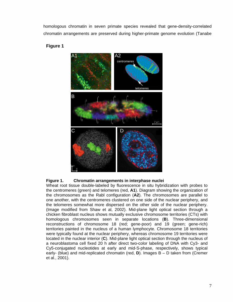
homologous chromatin in seven primate species revealed that gene-density-correlated
chromatin arrangements are preserved during higher-primate genome evolution (Tanabe
m
mmmmmmmmmmmmmmmmmmmmmmmmmmmmmmmmmmmmmmmmmmmmmmmm
mm
Figure 1
telomeres
A1 A2 centromeres
B
DC
Figure 1. Chromatin arrangements in interphase nuclei Wheat root tissue double-labeled by fluorescence in situ hybridization with probes to the centromeres (green) and telomeres (red, A1). Diagram showing the organization of the chromosomes as the Rabl configuration (A2). The chromosomes are parallel to one another, with the centromeres clustered on one side of the nuclear periphery, and the telomeres somewhat more dispersed on the other side of the nuclear periphery. (Image modified from Shaw et al, 2002). Mid-plane light optical section through a chicken fibroblast nucleus shows mutually exclusive chromosome territories (CTs) with homologous chromosomes seen in separate locations (B). Three-dimensional reconstructions of chromosome 18 (red; gene-poor) and 19 (green; gene-rich) territories painted in the nucleus of a human lymphocyte. Chromosome 18 territories were typically found at the nuclear periphery, whereas chromosome 19 territories were located in the nuclear interior (C). Mid-plane light optical section through the nucleus of a neuroblastoma cell fixed 20 h after direct two-color labeling of DNA with Cy3- and Cy5-conjugated nucleotides at early and mid-S-phase, respectively, shows typical early- (blue) and mid-replicated chromatin (red, D). Images B – D taken from (Cremer et al., 2001).
7
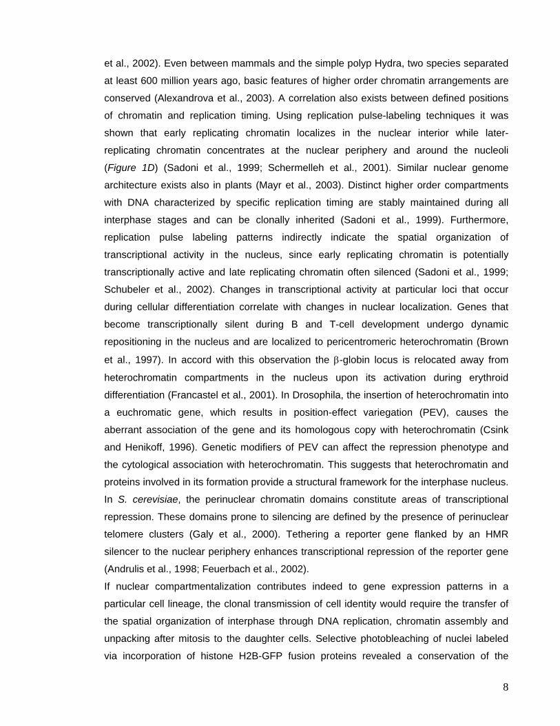
et al., 2002). Even between mammals and the simple polyp Hydra, two species separated
at least 600 million years ago, basic features of higher order chromatin arrangements are
conserved (Alexandrova et al., 2003). A correlation also exists between defined positions
of chromatin and replication timing. Using replication pulse-labeling techniques it was
shown that early replicating chromatin localizes in the nuclear interior while later-
replicating chromatin concentrates at the nuclear periphery and around the nucleoli
(Figure 1D) (Sadoni et al., 1999; Schermelleh et al., 2001). Similar nuclear genome
architecture exists also in plants (Mayr et al., 2003). Distinct higher order compartments
with DNA characterized by specific replication timing are stably maintained during all
interphase stages and can be clonally inherited (Sadoni et al., 1999). Furthermore,
replication pulse labeling patterns indirectly indicate the spatial organization of
transcriptional activity in the nucleus, since early replicating chromatin is potentially
transcriptionally active and late replicating chromatin often silenced (Sadoni et al., 1999;
Schubeler et al., 2002). Changes in transcriptional activity at particular loci that occur
during cellular differentiation correlate with changes in nuclear localization. Genes that
become transcriptionally silent during B and T-cell development undergo dynamic
repositioning in the nucleus and are localized to pericentromeric heterochromatin (Brown
et al., 1997). In accord with this observation the β-globin locus is relocated away from
heterochromatin compartments in the nucleus upon its activation during erythroid
differentiation (Francastel et al., 2001). In Drosophila, the insertion of heterochromatin into
a euchromatic gene, which results in position-effect variegation (PEV), causes the
aberrant association of the gene and its homologous copy with heterochromatin (Csink
and Henikoff, 1996). Genetic modifiers of PEV can affect the repression phenotype and
the cytological association with heterochromatin. This suggests that heterochromatin and
proteins involved in its formation provide a structural framework for the interphase nucleus.
In S. cerevisiae, the perinuclear chromatin domains constitute areas of transcriptional
repression. These domains prone to silencing are defined by the presence of perinuclear
telomere clusters (Galy et al., 2000). Tethering a reporter gene flanked by an HMR
silencer to the nuclear periphery enhances transcriptional repression of the reporter gene
(Andrulis et al., 1998; Feuerbach et al., 2002).
If nuclear compartmentalization contributes indeed to gene expression patterns in a
particular cell lineage, the clonal transmission of cell identity would require the transfer of
the spatial organization of interphase through DNA replication, chromatin assembly and
unpacking after mitosis to the daughter cells. Selective photobleaching of nuclei labeled
via incorporation of histone H2B-GFP fusion proteins revealed a conservation of the
8
bleaching pattern in daughter nuclei (Gerlich et al., 2003), indicating that global
chromosome positions can be transmitted through mitosis.
II. Chromosome structure and nuclear architecture in Arabidopsis thaliana Arabidopsis has 5 chromosomes per haploid genome ranging in their physical length from
17 to 29 Mbps, with a size in mitotic metaphase from 1.5 –1.8µm (Koornneef et al., 2003).
Compared to the average human chromosome territory with 130Mbp and ~1700 genes, an
Arabidopsis chromosome contains three times more genes confined in only ~25Mpbs. In
contrast to wheat and barley where the majority of the DNA consists of tandem repeats
and transposable elements located in heterochromatic segments, the relative
heterochromatin fraction in Arabidopsis nuclei is only ~10-15%, matching the percentage
of repetitive sequences in the Arabidopsis genome (ArabidopsisGenomeInitiative, 2000).
The heterochromatin in Arabidopsis is predominantly confined to the pericentromeric
regions of all chromosomes and the NORs on distal ends of chromosome 2 and 4, which
comprise the ribosomal 18S, 5.8S, and 25S units, together known as 45S rDNA.
Pericentromeric regions and NORs contain all major tandem repeats of the Arabidopsis
genome (Heslop-Harrison et al., 2003). Each NOR spans 3.5-4.0 Mbps in the ecotype
Columbia (Col) (Copenhaver and Pikaard, 1996). In addition 1000 copies of 5S rDNA
genes are located within the centromeric regions of chromosome 3, 4 and 5 (Cloix et al.,
2000). Some ecotypes, e.g. Wassilevskija (WS) and Col, also contain a heterochromatic
knob on the short arm of chromosome 4 that originated from the pericentromere after an
inversion event (Fransz et al., 2000). The Arabidopsis centromeric region consists of a
core accommodating the functional centromere comprising large tandem arrays of 180bp
repeats. These repeats are embedded in a recombination-deficient heterochromatic region
formed largely by retrotransposons and other moderately repetitive sequences
(ArabidopsisGenomeInitiative, 2000; Haupt et al., 2001). This tripartite organization, a
central domain of satellites that mediates spindle attachment flanked by pericentromeric
heterochromatin, conforms to a general model of the structure of centromeric regions
(Choo, 2001). In comparison, the centromere region of Drosophila also displays three
domains, where the central and flanking domains are referred to as α- and β-
heterochromatin (Miklos and Cotsell, 1990). The estimated size of the 180bp domains
varies from 1.1-2.9Mb. Sequencing of part of the central domain of chromosome 5
revealed nearly equal amounts of 180bp repeats and interspersed Athila derivatives, plus
4% other sequences (Kumekawa et al., 2000). The 180bp repeats at one end of the core
9
were oriented oppositely to those at the other end, similar to the organization in S. pombe
centromeres (Chikashige et al., 1989).
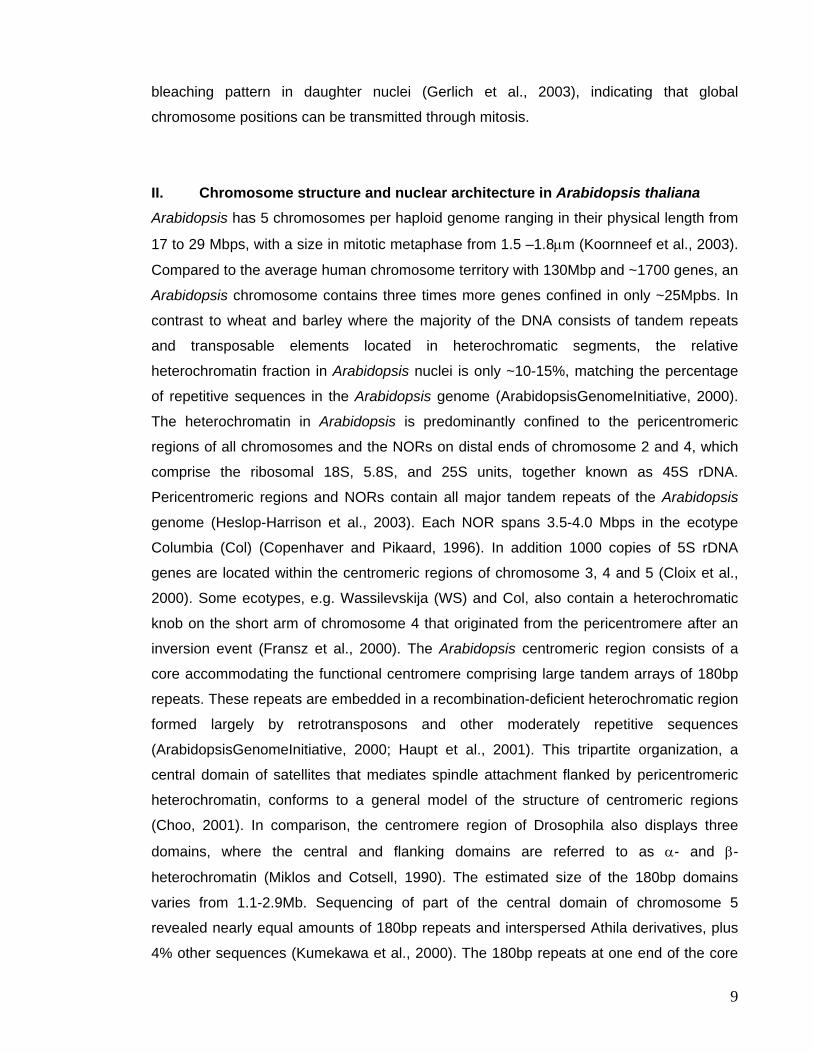
Figure 2. Distribution of the major repeats in Arabidopsis as revealed byFluorescence in situ hybridization (FISH) FISH for ribosomal DNA (red, A), the 180bp repeat probe (red, B), which labels the core centromeric regions of all chromosomes (Murata et al., 1994; Haupt et al., 2001), TSI (for Transcriptionally Silent Information) specific to pericentromeric repeats (red, B, Steimer et al., 2000), and a telomeric probe (C, Weiss and Scherthan, 2002). DNA is visualized by DAPI staining. Arrowheads in A depict the heterochromatic chromocenters.
Figure 2
rDNADAPI
centromereTSI
DAPI
telomeresDAPI
A
C
B
nucleolus
euchromatin
10
Combining DNA staining and fluorescent in situ hybridization (FISH) the arrangement of
the major repeats in the Arabidopsis genome can be visualized. In DAPI stained nuclear
spreads a single nucleolus can be identified, free of DNA except single threads protruding
into the nucleolar compartment. The chromocenters are DAPI bright (Figure 2A,
arrowheads), because of the specificity of DAPI for AT clusters, enriched in
pericentromeric repeats in Arabidopsis and mammals. The NORs (Figure 2A, left panel in
red) are part of the chromocenters and those actively involved in rRNA transcription
associate with the nucleolar compartment. In contrast, the 180bp repeats are the
predominant repeats clustered in each chromocenter (Figure 2B, left panel), but also
pericentromeric repeats are confined in the chromocenter (Figure 2B, exemplified by TSI
repeats (Steimer et al., 2000). The chromocenters align mainly at the nuclear periphery
and have the tendency to fuse, so in a diploid Arabidopsis plant the majority of nuclei show
8 or 9 chromocenters (Fransz et al., 2002). Telomeres are clustered around the nucleolus
(Figure 2C). With this arrangement the Arabidopsis nucleus differs substantially from
plants with large genomes having their chromosomes arranged in the Rabl configuration
or from yeast, where the telomeres localize to the nuclear periphery and centromeres
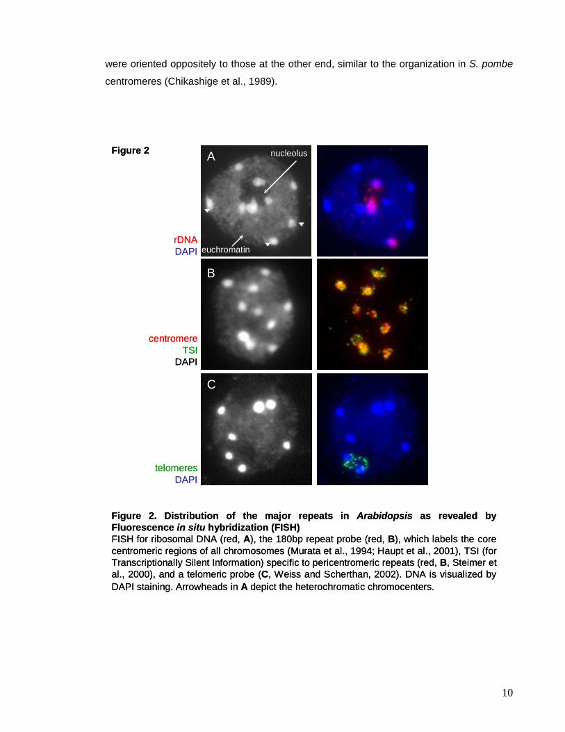
show a high incidence of clustering (Jin et al., 1998). Arabidopsis chromosomes also
occupy nuclear territories (Lysak et al., 2001) (Figure 3A), with their chromocenters as
organizing point and emanating euchromatic DNA loops (Figure 3B) (Fransz et al., 2002).
The euchromatic loops range in size from 200kb up to an entire chromosome arm, since
the telomeres-near NORs on chromosome 2 and 4 colocalize with the chromocenter
formed by the centromeric and pericentromeric repeats of their respective chromosome
(Figure 3C) (Fransz et al., 2002). The formation of the described higher order chromatin structure, however, requires
epigenetic modifications directly placed onto DNA and the histone proteins involved in
packaging of nuclear DNA.
III. Epigenetic Modifications of DNA and Histones III.1. DNA Methylation Nuclear DNA can be modified by post-replicative methylation of its cytosine residues. This
is the case in plants (Bender, 2004), mammals (Attwood et al., 2002), and Neurospora
crassa (Selker et al., 2003), while in yeast, Drosophila and C. elegans very little or no DNA
11
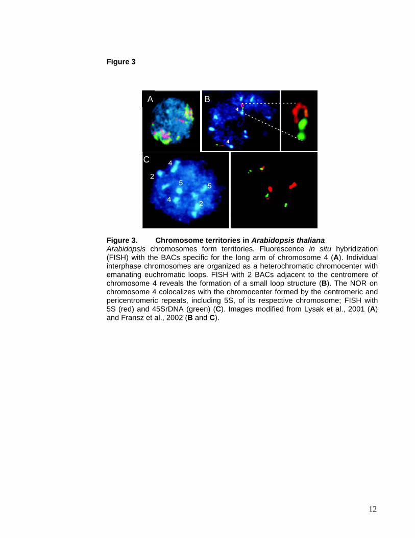
Figure 3
C
A B
Figure 3. Chromosome territories in Arabidopsis thaliana Arabidopsis chromosomes form territories. Fluorescence in situ hybridization (FISH) with the BACs specific for the long arm of chromosome 4 (A). Individual interphase chromosomes are organized as a heterochromatic chromocenter with emanating euchromatic loops. FISH with 2 BACs adjacent to the centromere of chromosome 4 reveals the formation of a small loop structure (B). The NOR on chromosome 4 colocalizes with the chromocenter formed by the centromeric and pericentromeric repeats, including 5S, of its respective chromosome; FISH with 5S (red) and 45SrDNA (green) (C). Images modified from Lysak et al., 2001 (A) and Fransz et al., 2002 (B and C).
12
methylation is found. Recent evidence indicates that DNA methylation is an important
epigenetic mark essential for normal development in mammals and plants, implicated in
gene silencing, imprinting, as well as transposon and X-chromosome inactivation (Heard
et al., 1997; Feil and Khosla, 1999; Martienssen and Colot, 2001). In mammals and other
vertebrates, methylation occurs predominantly at symmetrical CpG sites, which can be
faithfully maintained through replication. Besides CpG methylation, plants also show
extensive CpNpG as well as asymmetric methylation. Methylation is preferentially targeted
to repeated sequences including centromere-associated repeats, rDNA and transposable
elements. For this reason the 5metC content correlates with the repeat content. While in
maize a quarter of all cytosines are methylated, in Arabidopsis only 6% carry this
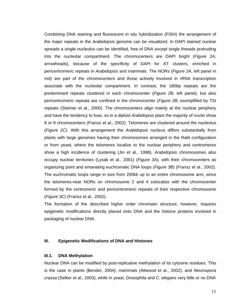
modification (Kakutani et al., 1999). Immunostaining experiments using Arabidopsis nuclei
with an antibody directed against methylated cytosines reveal that most of it colocalizes
with repetitive DNA clustered in the chromocenters (Figure 5A).
In mammals DNA methylation patterns are reprogrammed during germ cell development
and in preimplantation embryos (Reik et al., 2001). In plants, however, DNA demethylation
seems to occur mainly via passive de-methylation in the context of DNA replication (Saze
et al., 2003), even though local, active de-methylation activity has recently been reported
to play a role in imprinting (Choi et al., 2002). DNA methylation can suppress gene
transcription either directly, by blocking the binding of transcription factors to the promoter
or indirectly, through proteins that bind methylated DNA resulting in deacetylation of
nearby histones and decreased transcription (Jones et al., 1998). The mammalian
genome encodes three active methyltransferases, Dnmt1, Dnmt3a and Dnmt3b. Dnmt1,
the maintenance methyltransferase, acts preferentially on hemimethylated substrates and
localizes to DNA replication foci (Leonhardt et al., 1992). Mice that are homozygous for a
deletion in Dnmt1 die early in development (Lei et al., 1996), and also the two de novo
DNA methylases Dnmt3a/b are essential enzymes (Okano et al., 1999). In A. thaliana,
three different classes of DNA methyltransferases exist, MET1, the Dnmt1 ortholog, the
plant specific CMT3 (CHROMOMETHYLASE 3) and two de novo methyltransferases DRM
(DOMAINS REARRANGED METHYLTRANSFERASE). In addition, DNA methylation
patterns in Arabidopsis were found to depend on the presence of DDM1, a SWI2/SNF2
chromatin remodeling factor (Jeddeloh et al., 1999). SNF2 family members have seven
conserved helicase motifs involved in ATP binding and hydrolysis. They allow for the
physical movement of nucleosomes along the DNA driven by ATP hydrolysis to alter
accessibility of chromatin to regulators of replication, transcription and repair (Yan et al.,
2003).
13
III.2. Histone Tail Modifications DNA methylation, although essential for those organisms that have evolved this epigenetic
mark, is not found in every species known to be subject to epigenetic regulation.
Chromatin proteins are good candidates to function as additional carriers of epigenetic
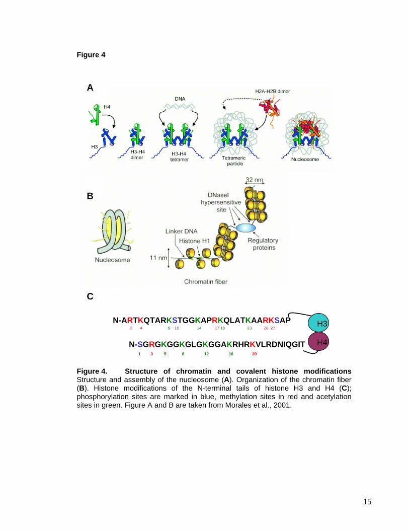
information: In chromatin, 147bp of DNA are wrapped in 1 3/4 superhelical turns around a
histone octamer forming a structure termed nucleosome. The histone octamer consists of
two molecules each of histone proteins H2A, H2B, H3 and H4 with their N-terminal tails
protruding from the globular domains (Figure 4A) (Luger et al., 1997). Linker DNA
connects the neighboring histone octamers, serving as binding site for histone H1 in some
organisms (Zhou et al., 1998). For many years it was believed that the role of histones is
constraint to their packaging function (Figure 4B), that non-histone proteins carry the
instructions for the activity of chromatin and that changes in gene expression are regulated
by the synthesis, modification and compartmentalization of these proteins. However, in
recent years it became clear that the nucleosome core particle contributes to the dynamic
remodeling of chromatin during gene activation and repression and carries important
epigenetic information. This information resides primarily in the histone tails, which are
subject to various covalent modifications, including acetylation, methylation,
phosphorylation, ubiquitination, sumoylation and ADP-ribosylation (Figure 4C) (Jenuwein
and Allis, 2001; Berger, 2002). It has been suggested that the specific tail modifications
and their combinations constitute a histone code that defines actual or potential
transcriptional states (Berger, 2002). The histones are evolutionary highly conserved
proteins not only in their globular domain but also in their protruding ends and their
covalent modifications. This suggests that the role of histones as mediator of epigenetic
information as well as the mechanism of subsequent ‘translation’ of this message into
actual transcriptional activation or repression might be evolutionary conserved. In
principle, despite the presence of certain species-specific details, insights gained from the
study of one model organism can be conferred to other systems.
Acetylation of lysine residues was one of the first histone modifications described to
correlate with transcriptional activity (Allfrey et al., 1964). Acetylation influences
transcription by neutralizing the positive charge of the histone tails and decreasing their
affinity for DNA, however, there is growing evidence that acetylation also helps shape the
binding surface for activators and repressors (Kurdistani and Grunstein, 2003). The
hypoacetylated histone H3 and H4 tails, for example, serve as a binding site for Sir3 and
14
mmmmmmmmmmmmmmmmmmmmmmmmmmmmmmmmmmmmmmmmmmmmmmmm
m Figure 4
B
A
C
N-ARTKQTARKSTGGKAPRKQLATKAARKSAP 2 4 9 10 14 17 18 23 26 27
N-SGRGKGGKGLGKGGAKRHRKVLRDNIQGIT H4
H3
1 3 5 8 12 16 20
Figure 4. Structure of chromatin and covalent histone modifications Structure and assembly of the nucleosome (A). Organization of the chromatin fiber (B). Histone modifications of the N-terminal tails of histone H3 and H4 (C); phosphorylation sites are marked in blue, methylation sites in red and acetylation sites in green. Figure A and B are taken from Morales et al., 2001.
15
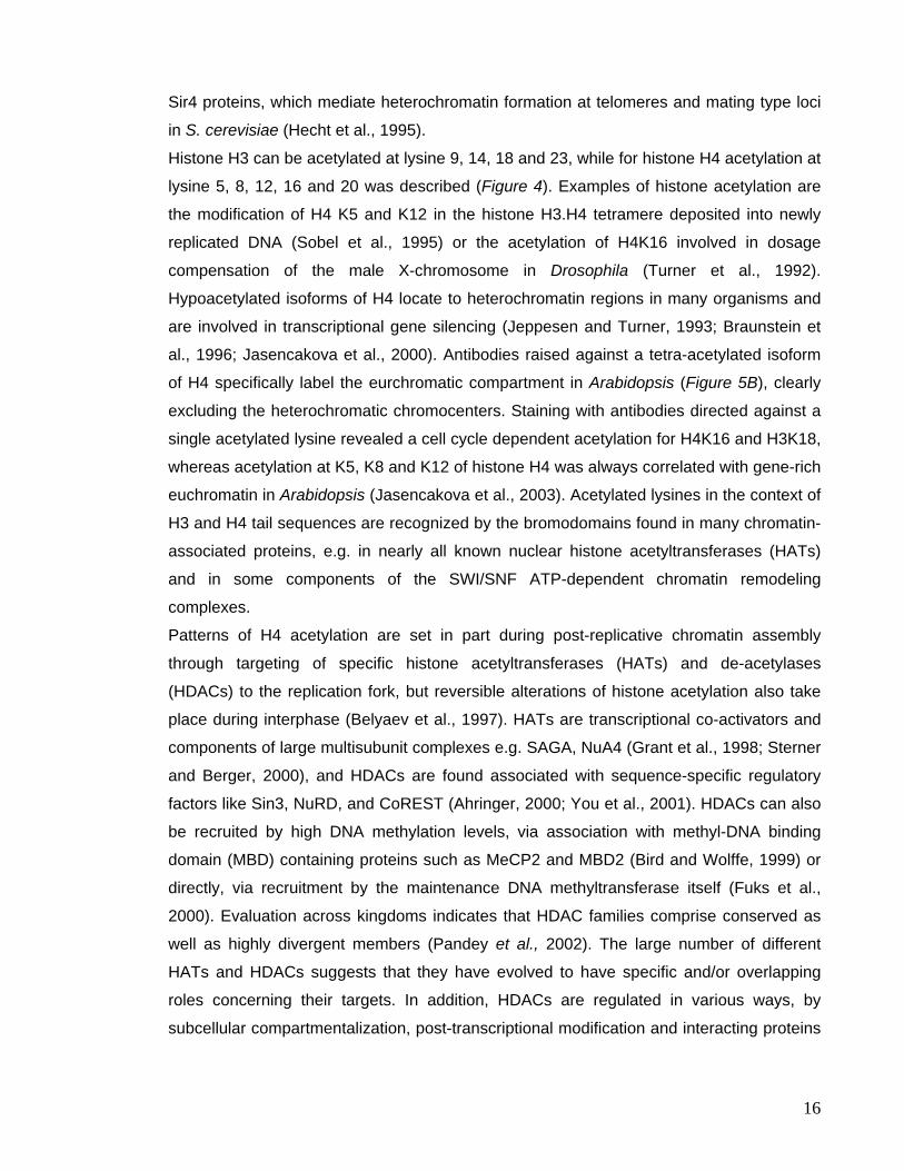
Sir4 proteins, which mediate heterochromatin formation at telomeres and mating type loci
in S. cerevisiae (Hecht et al., 1995).
Histone H3 can be acetylated at lysine 9, 14, 18 and 23, while for histone H4 acetylation at
lysine 5, 8, 12, 16 and 20 was described (Figure 4). Examples of histone acetylation are
the modification of H4 K5 and K12 in the histone H3.H4 tetramere deposited into newly
replicated DNA (Sobel et al., 1995) or the acetylation of H4K16 involved in dosage
compensation of the male X-chromosome in Drosophila (Turner et al., 1992).
Hypoacetylated isoforms of H4 locate to heterochromatin regions in many organisms and
are involved in transcriptional gene silencing (Jeppesen and Turner, 1993; Braunstein et
al., 1996; Jasencakova et al., 2000). Antibodies raised against a tetra-acetylated isoform
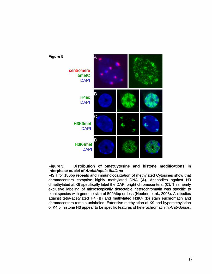
of H4 specifically label the eurchromatic compartment in Arabidopsis (Figure 5B), clearly
excluding the heterochromatic chromocenters. Staining with antibodies directed against a
single acetylated lysine revealed a cell cycle dependent acetylation for H4K16 and H3K18,
whereas acetylation at K5, K8 and K12 of histone H4 was always correlated with gene-rich
euchromatin in Arabidopsis (Jasencakova et al., 2003). Acetylated lysines in the context of
H3 and H4 tail sequences are recognized by the bromodomains found in many chromatin-
associated proteins, e.g. in nearly all known nuclear histone acetyltransferases (HATs)
and in some components of the SWI/SNF ATP-dependent chromatin remodeling
complexes.
Patterns of H4 acetylation are set in part during post-replicative chromatin assembly
through targeting of specific histone acetyltransferases (HATs) and de-acetylases
(HDACs) to the replication fork, but reversible alterations of histone acetylation also take
place during interphase (Belyaev et al., 1997). HATs are transcriptional co-activators and
components of large multisubunit complexes e.g. SAGA, NuA4 (Grant et al., 1998; Sterner
and Berger, 2000), and HDACs are found associated with sequence-specific regulatory
factors like Sin3, NuRD, and CoREST (Ahringer, 2000; You et al., 2001). HDACs can also
be recruited by high DNA methylation levels, via association with methyl-DNA binding
domain (MBD) containing proteins such as MeCP2 and MBD2 (Bird and Wolffe, 1999) or
directly, via recruitment by the maintenance DNA methyltransferase itself (Fuks et al.,
2000). Evaluation across kingdoms indicates that HDAC families comprise conserved as
well as highly divergent members (Pandey et al., 2002). The large number of different
HATs and HDACs suggests that they have evolved to have specific and/or overlapping
roles concerning their targets. In addition, HDACs are regulated in various ways, by
subcellular compartmentalization, post-transcriptional modification and interacting proteins
16
H3K9metDAPI
H3K4metDAPI
H4acDAPI
centromere5metC
DAPI
Figure 5. Distribution of 5metCytosine and histone modifications in interphase nuclei of Arabidopsis thalianaFISH for 180bp repeats and immunolocalization of methylated Cytosines show that chromocenters comprise highly methylated DNA (A). Antibodies against H3dimethylated at K9 specifically label the DAPI bright chromocenters, (C). This nearly exclusive labeling of microscopically detectable heterochromatin was specific to plant species with genome size of 500Mbp or less (Houben et al., 2003). Antibodies against tetra-acetylated H4 (B) and methylated H3K4 (D) stain euchromatin and chromocenters remain unlabeled. Extensive methylation of K9 and hypomethylationof K4 of histone H3 appear to be specific features of heterochromatin in Arabidopsis.
Figure 5 A
B
C
D
17
(Yang and Seto, 2003). The Arabidopsis genome encodes a total of 12 and 16 potentially
functional histone acetyltransferases and deacetylases, respectively (Pandey et al., 2002).
In contrast to acetylation, the methylation of a lysine residue, which can accommodate up
to three methyl groups, is usually a stable modification (Zhang and Reinberg, 2001) and
no demethylation activity has been described so far. Histone methylation affects not only
lysine residues (K4, 9, 27, 79 of H3 and K20 of H4), but also arginine residues (R2, 17, 26
of H3 and R3 of H4, Figure 4C). Methylation of H3K9 and H3K27 have been linked to
epigenetic silencing. Histone H3 methylated at lysine9 is enriched in heterochromatin and
has the potential to initiate chromatin condensation and silencing (Peters et al., 2001) in
part through its ability to bind proteins like HP1 via their chromodomain (Bannister et al.,
2001; Lachner et al., 2001). Chromodomains characterize proteins like HP1, involved in
heterochromatin formation, Polycomb proteins or chromatin remodeling factors (Mi-2) and
the CMT3 DNA methyltransferase. At lower levels H3K9 methylation is found in
mammalian euchromatin where it is involved in transcriptional repression (Tachibana et
al., 2002). H3K27 methylation in contrast is specifically recognized by Polycomb proteins
to mediate gene silencing of developmentally regulated genes (Fischle et al., 2003). In
Arabidopsis, mono- and di-methylated H3K9 are enriched in centromeric and
pericentromeric repeats (Gendrel et al., 2002; Jackson et al., 2004) (Figure 5C). While in
mammals trimethylated H3K9 is specifically localized to pericentromeric repeats (Lehnertz
et al., 2003), Arabidopsis chromatin seems to be devoid of this modification (Jackson et
al., 2004). Both H3K9 and K27 methylation are involved in silencing of the FLC locus
during the vernalization response in Arabidopsis (Bastow et al., 2004; Yan et al., 2004).
Histone methylation is catalyzed by SET domain containing proteins; Su(var)3-9 in
Drosophila, SUV39H in humans and Clr4 in S. pombe. In Arabidopsis several proteins with
homology to SUV39H have been identified, with KRYPTONITE (KYP) being the
predominant histone methyltransferase (Jackson et al., 2002; Jackson et al., 2004). The
kyp mutant was identified in a mutant screen for suppressors of gene silencing at the
Arabidopsis thaliana SUPERMAN (SUP) locus. It shows loss of cytosine methylation at
CpNpG sites and reactivation of endogenous retrotransposon sequences.
In contrast to methylation of H3K9 and K27, H3K4 methylation is linked to transcriptional
activity. In yeast, H3K4met was predominantly found in coding regions suggesting that
H3K4met plays an important role in the elongation stage of transcription, rather than its
initiation. In this case H3K4 trimethylation serves as a “short term memory” for actively
transcribed genes (Santos-Rosa et al., 2002; Ng et al., 2003), probably maintaining their
transcriptional potential and protecting them from long term silencing by inhibition of
18
binding of the NuRD complex (Zegerman et al., 2002) and methylation of H3K9.
Interestingly, the set of the H3K4 mark during transcription elongation requires the
ubiquitination of K123 of H2B mediated by Rad6. Staining of Arabidopsis nuclei with
antibodies raised against di-methylated K4 of H3 showed that H3K4 methylation is
exclusively localized to euchromatin (Figure 5D), with the heterochromatic chromocenters
being devoid of staining. Mutations in ddm1 resulting in loss of H3K9 at target genes in the
heterochromatic knob also induced increased association with H3 methylated at K4
(Gendrel et al., 2002), suggesting a reciprocal effect on epigenetic gene regulation of
these two modifications.
Like H3K4 methylation arginine methylation of H3 and H4 correlates with transcriptional
activation, e.g. the methylation of H4R3 facilitates subsequent acetylation of H4 (Zhang
and Reinberg, 2001). The phosphorylation of H3 serine 10 is also linked to transcriptional
activation (Lo et al., 2000) that precedes K14 methylation but negatively affects H3K9
methylation (Rea et al., 2000). Furthermore, together with phosphorylation at serine 28 of
H3 this modification is involved in mitosis and chromosome condensation (Wei et al.,
1999).
Many of the different histone modifications are located close enough together on the
histone tail to influence the ability of enzymes to further modify histones. Hence, the
implication of a specific histone modification depends on the modifications that surround it
and the time point of its setting. For example the Drosophila Ash1 protein specifically
methylates H3K4 and K9 as well as H4K20 thereby creating a distinct signal for binding of
the Brahma complex (Beisel et al., 2002).
In addition to the canonical histones, the genome of many organisms also encodes
specific histone variants. The H3.3 variant of histone H3 for example is enriched in
transcriptional active domains and replaces H3 (methylated at K9) in a replication-
independent manner (Ahmad and Henikoff, 2002; Janicki et al., 2004) upon transcriptional
activation. H3.3 differs from H3 only in few residues. Another histone variant specific for
centromeres shows significant differences in the N-terminal tail. Named SpCENP-A in S.
pombe (Takahashi et al., 2000)), Cid in Drosophila (Henikoff et al., 2000), CenP-A in
humans (Sullivan et al., 1994) and HTR12 in Arabidopsis (Talbert et al., 2002), it
contributes to centromere organization and function. The histone H2A.Z variant is involved
in transcriptional regulation (Santisteban et al., 2000), while the macroH2A, that has a
25kDa non-histone fold domain added to its C-terminus, is found to be enriched in the
inactive mammalian X chromosome.
19
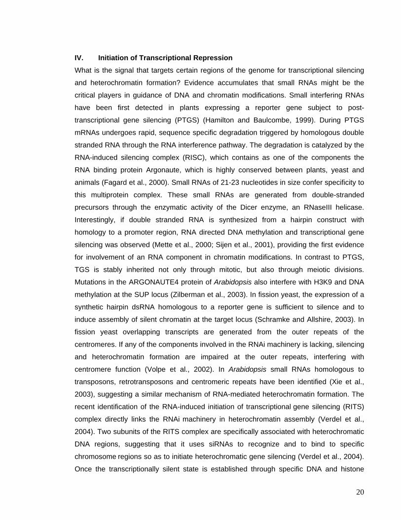
IV. Initiation of Transcriptional Repression What is the signal that targets certain regions of the genome for transcriptional silencing
and heterochromatin formation? Evidence accumulates that small RNAs might be the
critical players in guidance of DNA and chromatin modifications. Small interfering RNAs
have been first detected in plants expressing a reporter gene subject to post-
transcriptional gene silencing (PTGS) (Hamilton and Baulcombe, 1999). During PTGS
mRNAs undergoes rapid, sequence specific degradation triggered by homologous double
stranded RNA through the RNA interference pathway. The degradation is catalyzed by the
RNA-induced silencing complex (RISC), which contains as one of the components the
RNA binding protein Argonaute, which is highly conserved between plants, yeast and
animals (Fagard et al., 2000). Small RNAs of 21-23 nucleotides in size confer specificity to
this multiprotein complex. These small RNAs are generated from double-stranded
precursors through the enzymatic activity of the Dicer enzyme, an RNaseIII helicase.
Interestingly, if double stranded RNA is synthesized from a hairpin construct with
homology to a promoter region, RNA directed DNA methylation and transcriptional gene
silencing was observed (Mette et al., 2000; Sijen et al., 2001), providing the first evidence
for involvement of an RNA component in chromatin modifications. In contrast to PTGS,
TGS is stably inherited not only through mitotic, but also through meiotic divisions.
Mutations in the ARGONAUTE4 protein of Arabidopsis also interfere with H3K9 and DNA
methylation at the SUP locus (Zilberman et al., 2003). In fission yeast, the expression of a
synthetic hairpin dsRNA homologous to a reporter gene is sufficient to silence and to
induce assembly of silent chromatin at the target locus (Schramke and Allshire, 2003). In
fission yeast overlapping transcripts are generated from the outer repeats of the
centromeres. If any of the components involved in the RNAi machinery is lacking, silencing
and heterochromatin formation are impaired at the outer repeats, interfering with
centromere function (Volpe et al., 2002). In Arabidopsis small RNAs homologous to
transposons, retrotransposons and centromeric repeats have been identified (Xie et al.,
2003), suggesting a similar mechanism of RNA-mediated heterochromatin formation. The
recent identification of the RNA-induced initiation of transcriptional gene silencing (RITS)
complex directly links the RNAi machinery in heterochromatin assembly (Verdel et al.,
2004). Two subunits of the RITS complex are specifically associated with heterochromatic
DNA regions, suggesting that it uses siRNAs to recognize and to bind to specific
chromosome regions so as to initiate heterochromatic gene silencing (Verdel et al., 2004).
Once the transcriptionally silent state is established through specific DNA and histone
20
modifications, it needs to be clonally transmitted. The inheritance of epigenetic
modifications at the level of DNA and histones seems to be tightly linked to DNA
replication. The chromatin assembly complex CAF-1 is targeted to replication forks via its
interaction with the proliferating cell nuclear antigen (PCNA) (Shibahara and Stillman,
1999). This three-component complex is a histone chaperone involved in DNA synthesis-
dependent histone deposition. CAF-1 mutants in S. cerevisiae are deficient in stable
inheritance of gene silencing at mating-type loci and telomeres (Gerbi and Bielinsky, 2002)
and Arabidopsis CAF-1 mutants are impaired in the maintenance of transcriptional gene
silencing (Kaya et al., 2001). In mammals Dnmt1 associates with sites of DNA synthesis
recruited by PCNA (Vertino et al., 2002). Dnmt1 interacts with HDACs (Fuks et al., 2000;
Rountree et al., 2000), HP1 and histone methyltransferases (Burgers et al., 2002; Fuks et
al., 2003), thereby targeting a whole set of epigenetic regulators to the replication forks
and assuring inheritance of silenced states in the daughter cells.
V. Objective of This Study Genetic screens for mutants involved in the maintenance of transcriptional gene silencing
at transgenic loci or endogenous repeats have identified several components of the
regulatory network involved in epigenetic regulation in the model plant Arabidopsis. The
aim of this work was to analyze the nuclear structure as well as DNA and histone
modifications in Arabidopsis and to investigate the impact of these TGS mutants on
nuclear architecture. All of the studied mutants are either directly involved in the
establishment of DNA methylation patterns (met1, cmt3, drm2 and ddm1) or affect DNA
replication (fas1, fas2 and bru1), histone deacetylation (axe1) or release silencing by an
unknown mechanism (mom1). More specifically I investigated the relationship between
DNA methylation at CpG sites and histone H3K9 methylation and asked whether DNA
methylation might be dispensable for heterochromatin formation. We also asked if MOM1
controls the arrangement and the degree of heterochromatinization of the targets
reactivated in the mutant and compared the results to plants lacking the chromatin-
remodeling factor DDM1. Further I studied the release of transcriptional gene silencing in a
histone deacetylase mutant and investigated its role in maintenance of nuclear
organization at rDNA repeats, as well as the effect of mutations impaired in DNA
21

replication on heterochromatin structure. Using an epistasis analysis I investigated
whether MOM1 acts downstream of a silencing pathway delineated by DNA methylation
and histone deacetylation or whether MOM1 is part of an independent pathway of
epigenetic control.
22
4. MATERIALS AND METHODS I. Material I.1. Plant Material In this study Arabidopsis thaliana wild type plants of the ecotypes Zürich (ZH), Col
(Columbia), Wassilewskija (WS), Nossen (No), Enkheim (En) and Landsberg erecta (Ler)
were used. Line A, carrying a silent multicopy HPT transgene (Mittelsten Scheid et al.,
1991), is derived from ecotype ZH. The mutants mom1-2, met1-3 (MET1, isolated by
Hidetoshi Saze), axe1-1, axe 1-4 and axe1-5 (HDA6 mutant alleles, provided by Jane
Murfett), as well as bru1-2 and bru1-3 (Takeda et al., 2004) are in the Col background.
The mutants mom1-1, ddm1-5 (som8) were isolated in the lineA background (Mittelsten
Scheid et al., 1998; Amedeo et al., 2000); sil1 (provided by Ian Furner, (Furner et al.,
1998)) as well as the fas2-1 allele (Leyser and Furner, 1992) are in the Ler background,
fas1-1 (Reinberg et al., 1966) and fas2-2 (Kaya et al., 2001) are derived from ecotypes
Enkheim and Nossen, respectively. The bru1-1 mutant (originally isolated by Zerihun
Tadele) is in the Wassilewskija background.
I.2. Plant Tissue Culture Media Solid germination medium (Masson and Paskowski, 1992) contains MS macroelements
(KNO3 (0.95 g/l), NH4NO3 (0.825 g/l), CaCl2xH2O (0.22 g/l), MgSO4x7H2O (0.185 g/l) and
KH2PO4 (85 mg/l) final concentration), B5 microelements (MnSO4xH2O (10 mg/l), H3BO4 (3
mg/l), ZnSO4x7H2O (2 mg/l), KJ (0.75 mg/l), Na2MoO4x2H2O (0.25 mg/l), CuSO4x5H2O (25
µg/l) and CoCl2x6H2O (25 µg/l)), 0.005% ammonium iron citrate, 1% sucrose, and 0.8%
agar-agar (Merck). The pH was adjusted with KOH to pH5.6 and buffered with 0.07%
MES. The antibiotics Kanamycin (50 µg/ml) or Hygromycin (10 µg/ml) were added for
selection of plants containing T-DNA inserts from Agrobacterium mediated transformation;
Cefotaxime (250 µg/ml) and Vancomycine (250 µg/ml) to eliminate Agrobacterium growth
in plant tissue culture.
I.2. Bacterial Strains
Escherichia coli DH5α was used for all cloning procedures. For plant transformation the
Agrobacterium tumefaciens strain C58CIRifR containing the non-oncogenic Ti plasmid
pGV3101 was used.
23
I.3. Bacterial Growth Media E. coli and A. tumefaciens were grown in liquid LB medium (Luria-Bertani medium: 1%
Bacto-tryptone, 0.5% (w/v) Bacto yeast extract, 0.5% (w/v) NaCl) or on LB plates
(supplemented with 1.5% Bacto-agar). The antibiotics Ampicillin (100 µg/ml), Kanamycin
(50 µg/ml), Rifampicin (10 µg/ml) or Gentamycin (25 µg/ml) were added for selection of
plasmids.
I.4. Plasmid Vectors For cloning and plant transformation purposes the plasmid vectors pCambia1300,
pBluescript SK- (Stratagene) and pGEM-7Zf(+) (Promega) were employed.
I.5. Enzymes and Reagents The enzymes used in this study were purchased form Roche Diagnostics (Rotkreuz,
Switzerland), New England Biolabs (Beverly, MA, USA), Amersham Pharmacia Biotech
(Buckinghamshire, UK), Gibco BRL (Grand Island, NY, USA), Promega (Madison, WI,
USA) and Stratagene (La Jolla, CA, USA). Chemicals were obtained from Fluka (Buchs,
Switzerland), Merck (Darmstadt, Germany), Sigma (St. Louis, MO, USA), and Bio-Rad
(Hercules, CA, USA) and were of analytical grade. Radioactively labeled 32P was obtained
from Amersham Pharmacia Biotech.
Antibodies for FISH, Immunostaining and ChIP experiments were obtained from Sigma,
Molecular Probes (Eugene, OR, USA), Vector Laboratories (Burlinghame, CA, USA),
Eurogentec (Seraing, Belgium), and Upstate Biotechnology (Lake Placid, NY, USA); the
HRP-conjugated secondary antibody was purchased from DAKO A/S, Denmark.
I.6. Oligonucleotides Oligonucleotides were designed with help of Vector NTI PCR amplification and were
synthesized by Microsynth (Balgach, Switzerland).
II. Methods II.1. Plant Growth Seeds for in vitro culture were sterilized for 10 min in 5% sodium-hydrochlorite containing
0.1% Tween80, followed by 3 washes with sterile ddwater. The seeds were dried and
24
plated on germination medium to allow growth under axenic culture conditions with 16 h
light of 100 µEm-2s-1 (Osram Natura de Luxe) at 22.5°C and 8 h darkness at 16°C.
Seeds were otherwise directly sown on soil, and plants grown in a phytotron under short
day (12 h light/21°C and 12 h darkness/16°C) or under long day conditions (16 h
light/21°C and 8 h darkness/16°C) with 80% humidity and a light intensity of 3000-4000
lux. All seeds were stratified after sowing for 2-4 days at 4°C.
II.2. Plasmid Construction II.2.1. pBluescript 180bp
The original clone containing the conserved 180bp repeat as a HindIII fragment was
obtained from Eric J. Richards (Richards, et al., 1991). It was subcloned into pBluescript
and a clone containing a tandem repeat of the 180bp repeat was selected.
II.2.2. MOM-GFP fusion construct
For transient expression of a MOM1-GFP fusion protein in N. plumbaginifolia protoplasts
the pUC based plasmid pCK GFP S65C was used, in which the GFP coding region had
been replaced by the one of eGFP (enhanced GFP) and the NdeI site in the lacZ gene
was removed (Habu personal communication). The vector was linearized with NcoI and a
linker was introduced (5’-CAT GCA TAT GAT GTT CCT GAT TAT GC-3’ and 5’-CAT GGC
ATA ATC AGG AAC ATC ATA TG-3’) to generate a new NdeI site. MOM1 cDNA was cut
as NdeI fragment from the p2HAPA vector (provided by Yoshiki Habu) and then cloned in
frame to eGFP.
II.3. Generation of Competent Bacteria To generate heat-shock competent bacteria the “Ultra-competect E.coli method” was
applied (Inoue et al., 1990). Therefore, 250 ml SOB (2% (w/v) bacto-tryptone, 0.5% (w/v)
yeast extract, 10 mM NaCl, 2.5 mM KCl, 10 mM MgCl2 and 10 mM MgSO4 pH6.7-7.0) was
inoculated from an overnight culture to OD600=0.05-0.08. The culture was grown to an
OD600 of 0.6 at 18°C, incubated on ice for 10 min and resuspended gently in 80 ml ice-cold
transformation buffer (10 mM PIPES, 55 mM MnCl2, 15 mM CaCl2, 250 mM KCl, pH6.7).
The cells were collected by centrifugation (2500g, 10 min, 4°C), resuspended in 20 ml
transformation buffer and incubated on ice for 10 min. After the addition of DMSO to a final
concentration of 7%, the cells were again incubated on ice for 10 min, aliquoted and
frozen in liquid nitrogen.
25
Electrocompetent Agrobacterium tumefaciens were prepared by inoculating 500 ml LB
with 5 ml of a fresh saturated culture and incubation at 28°C with agitation (210 rpm).
When the culture reached an OD600 of 0.5-0.8, the cells were chilled in ice-water and
pelleted by centrifugation (4000g, 10 min, 4°C). The bacteria were resuspended in 500 ml
of ice-cold ddwater. Centrifugation and resuspension were repeated twice and the cells
resuspended in a final volume of 250 ml and 50 ml, respectively. After an additional
centrifugation the bacteria were resuspended in 5 ml of 10% (v/v) ice-cold sterile glycerol,
aliquoted and frozen in liquid nitrogen.
II.4. Bacterial transformation Competent E.coli cells were transformed with plasmid DNA or ligated plasmid products
using the heat shock transformation protocol: After thawing the competent cells on ice and
incubation with an appropriate amount of plasmid DNA for 15 min on ice, the cells were
heat shocked at 42°C for 90 sec and transferred to ice for 5 min. 800 µl of LB-medium was
added and the cells incubated at 37°C for 1 hour before plating on appropriate selection
medium. Alternatively, electrocompetent E. coli (Invitrogen) or A. tumefaciens were
transformed using the BioRad E. coli pulser at a voltage of 1.8kV according the
manufacturer’s instructions.
II.5. Agrobacterium tumefaciens mediated plant transformation Arabidopsis mom1-1 plants were transformed according to the germ-line transformation
protocol using the floral dip method (adapted from Clough and Bent, 1998): Agrobacterium
cultures were initiated by inoculation of 15 ml LB medium supplemented with Rifampicin
(10 µg/ml), Gentamycin (25 µg/ml) and Kanamycin (50 µg/ml) to select for cells containing
the plasmids. After 48 h at 28°C, the cultures were transferred to 1 l flasks containing 500
ml LB with Kanamycin (50 µg/ml) and grown to an OD600 of 1.8-2.0. The bacteria were
harvested by centrifugation at 400 rpm for 20 min at RT, the pellet resuspended in 500 ml
infiltration medium (5% sucrose), and Silwet L-77 was added to 0.05%. Plants with
inflorescences at the early flowering stage were dipped into the bacterial solution for 30
sec. After infiltration, plants were covered with a polyethylene foil, kept horizontally for 24 h
in a growth chamber and then grown to maturation. Seeds were sterilized with ethanol in
addition to sodium-hydrochlorite and transformants were selected on plates containing 250
µg/ml cefotaxime, 250 µg/ml vancomycine and 50 µg/ml kanamycin.
26
II.6. Genotyping of Arabidopsis mutants One Arabidopsis leaf frozen in liquid nitrogen was ground to fine powder in the presence
of 1.7 – 2.0 mm glass beads (Roth) with help of a Silamat S5 (Vivadent), vortexed in 400
µl of buffer (0.2 M Tris-HCl pH7.5, 0.25 M NaCl, 25 mM EDTA, 0.5%SDS) and centrifuged
for 5 min (13000 rpm) at RT. The DNA in the supernatant was precipitated with an equal
volume of isopropanol and centrifuged for 5 min (13000 rpm) at RT. The pellet was
washed in 70% ethanol, dried and resuspended in 50 µl ddwater. 1 µl was added to 20 µl
of a standard PCR mix (1x Polymerase buffer supplemented with MgCl2 to 1.5 mM and
KCl to 5 mM, 0.25 mM dNTPs, 0.5 µM of each primer and 0.75 units Taq polymerase,
Roche).
To genotype the mom1-1 mutant the T-DNA insertion can be amplified using 5’-GTG GTT
ACT GAT CAA GTC TCG-3’ and 5’-GTG AAG GGC AAT CAG CTG TTG-3’, giving rise to
a 600 bp fragment. The wildtype allele can be amplified with a combination of 5’-CAC TTT
CCG ATT TCG ATT CTC G-3’ and 5’-CAT GAC TCC CCC AGC CAG TAG-3’ resulting in
a 260 bp fragment.
To identify the ddm1-5 mutant allele we made use of the 82 bp insertion in the 5′ region of
the DDM1 gene. Using the primer pair DDM+ (5′-CGC TCT CGA AAT CGC TCG CTG
TTC-3’) and DDM- (5′-AAA GGA CCC ATT TAC AGA ACA C-3’) amplification of the wild
type locus results in a band of 332 bp, while the mutant locus gives rise to band of 414 bp.
For genotyping of the axe1-1 locus we exploited the fact that the single nucleotide
exchange in this mutant (C to A, 337 bp downstream of the ATG) abolishes an MspI site.
A 259 bp fragment was amplified (5’- CGG AAT CTA TGG GCG ATC C-3’ and 5’- CCA
GAA TCC CTA GCA CGA TGT C-3’), PCR purified and subjected to an MspI digest.
II.7. DNA isolation and Southern Blot analysis
II.7.1 Genomic DNA Isolation from Arabidopsis DNA from Arabidopsis thaliana leaf tissue was isolated with help of the nucleon extraction
and purification kit (Amersham) according to the manufacturer’s instructions and
resuspended in a final volume of 30 µl ddwater.
II.7.2 Gel Electrophoresis and Southern Blot Analysis DNA was digested overnight with the required restriction enzyme and electrophoretically
separated on a 1% agarose gel. The gel was than treated with 250 mM HCl for 10 min,
the DNA denatured in denaturation solution (500 mM NaOH, 1.5 M NaCl) for 30 min and
27
treated with neutralization solution (500 mM Tris-HCl pH7.2, 1.5 M NaCl and 1 mM EDTA)
twice for 15 min. The DNA was transferred overnight by capillary transfer onto a Hybond N
membrane (Amersham) using 20x SSC (1.5 M NaCl, 0.3 M C6H5Na3O7-2H2O) as transfer
buffer. After transfer, the DNA was cross-linked to the membrane by UV (1.2 kJ/m2,
Stratalinker UV Crosslinker, Stratagene).
II.8. Total RNA Isolation and Northern Blot Analysis II.8.1 RNA Isolation
RNA was isolated using the Trizol Reagent (Gibco BRL) according to the manufacturer’s
instructions. In brief, 2-3 Arabidopsis leaves, frozen in liquid nitrogen, were ground to fine
powder in the presence of 1.7 – 2.0 mm glass beads (Roth) with help of a Silamat S5
(Vivadent). After adding 1 ml of Trizol Reagent the suspension was vortexed and
incubated at RT for 5 min. Subsequently 0.2 ml of chloroform was added and each sample
was vortexed for 15 sec. Following centrifugation (12000 g, 5 min, 4°C) the aqueous
phase was transferred to a fresh tube, the RNA precipitated with 0.5 ml isopropanol for 10
min at RT and collected by centrifugation (12000 g, 10 min, 4°C). The RNA pellet was
washed once in 75% Ethanol, air-dried and resuspended in 50 µl of DEPC treated ddwater
(10 min, 55°C).
II.8.1. RNA Electrophoresis and Northern Blot Analysis
5-20 µg of RNA was dried in a speed vac and resuspended in 50% DMSO, 10 mM
sodiumphosphate, pH7.0 and 6% glyoxal pH5.5-6.0 by incubation for 1 h at 50°C. 4 µl of
sample buffer (50% glycerol, 0.4% bromophenolblue, 10 mM Na2HPO4/NaH2PO4, pH7.0)
was added and the RNA separated on a 1.5% agarose gel in 10 mM Na2HPO4/NaH2PO4,
pH7.0. The gel was stained in Radiant Red RNA Gel Stain (BioRad, diluted 1:1000 in 10
mM Na2HPO4/NaH2PO4, pH7.0), and the RNA blotted by capillary transfer overnight onto a
Hybond N membrane with 20x SSC (3 M NaCl, 0.3 M C6H5Na3O7-2H2O) as transfer buffer.
The membrane is washed briefly in 2x SSC, dried and cross-linked by UV (see above).
II.9. Hybridization with radioactive probe DNA fragments were labeled with 32P by random oligonucleotide-primer synthesis
(Feinberg and Vogelstein, 1983). The DNA fragment (50 ng in 7 µl of water) was
denatured by boiling for 3 min, followed by cooling down on ice for 5 min. Then 11.5 µl of
labeling solution (LS, Feinberg and Vogelstein, 1983), 1 µl of BSA, 0.5 µl (2.5U) Klenow
fragment of polymerase I, and 5 µl of [α-32P] dATP (2 MBq) were added, mixed and
28
incubated at RT for 2-4 h. The labeled DNA was separated from unincorporated
radioactive precursors by chromatography on a NICKTM Column (Pharmacia Biotech)
following the manufacturer’s instructions. Membranes were pre-hybridized in hybridization
buffer (0.5 M NaHPO4 pH7.2, 7%SDS, 1 mM EDTA) for 1-3 h at 65°C (RNA membranes
were washed before in 0.1% boiling SDS). The radioactive probe was denatured (95°C, 5
min), cooled down on ice and added to 15 ml of fresh hybridization solution. The
hybridization was carried out overnight at 65°C in a hybridization oven. The membranes
were washed 10 min at 65°C, twice for 20 min at 60°C in washing solution (0.5 M NaHPO4
pH7.2, 1%SDS), dried, rapped in polyethylene foil and either exposed to a phosphoimager
screen (BioRad) at RT or autoradiographed at -80°C. To allow hybridization of the same
membrane with a different radioactive probe, the membranes were stripped using 0.1%
boiling SDS.
II.10. Reverse-Transcription (RT-PCR)
5 µg of RNA was treated with 4 µl of DNase RQ1 (Promega) for 75 min at 37°C in the
presence of 1 µl of RNase inhibitor (Invitrogen), 0.1 µl of 1 M DTT, and 1x RQ buffer in a
total volume of 50 µl. The treated RNA was phenol-chloroform extracted, precipitated with
1/10 volume of NaAc and 2.5 volumes of Ethanol and washed in 70% Ethanol. The RNA
pellet was resuspended in 10 µl of DEPC treated water by incubation for 10 min at 65°C in
a heating block with agitation. The 10 µl were divided into 2 PCR tubes and incubated
either with or without 1 µl of AMV reverse transcriptase (Roche) in 1x AMV buffer with 0.67
mM dNTPs, and 0.167 µM RT-primer in a total volume of 15 µl. cDNA synthesis was
allowed to proceed for 1 h at 42°C, followed by a denaturation step (95°C, 5 min). 2 µl of
the cDNA was used in a standard PCR reaction.
II.11. Transfection of Nicotiana plumbaginifolia protoplasts Protoplasts at a concentration of 6x10⁵/ml competent for transfection were obtained from
Matthias Müller (FMI). In a 15 ml falcon tube 10 µg of plasmid DNA, 0.3 ml of protoplast
suspension and 0.3 ml of 40% PEG 6000 were mixed. After 5 min of incubation at RT, 4
ml of K3 medium (Nagy et al., 1976, provided) was added. The protoplasts were incubated
at 25°C in the dark for 4 h to overnight and GFP fluorescence was analyzed using the
Leitz DMR Fluorescence Microscope.
29
II.12. Isolation of histones and Western blot analysis Fresh leaf tissue (5 g) was ground in liquid nitrogen, transferred to extraction buffer (0.25
N HCl, 10 mM Tris-HCl pH7.5, 2 mM EDTA, 20 mM β-mercaptoethanol, 0.2 mM PMSF)
and treated with ultra-sound. Following centrifugation (10 min, 12000 g) soluble proteins
were precipitated with trichloracetic acid (TCA, 25% final concentration) and collected by
centrifugation at 17000 g for 20 min. The pellet was washed two times in acetone, air dried
and resuspended in 1x SDS loading buffer (75 mM Tris-HCl pH6.8, 0.6% SDS, 15%
glycerol, 0.0009% bromophenol blue and 1.075 M β-mercaptoethanol). The proteins were
separated electrophoretically by means of a 14% SDS page (stacking gel: 3.9%
acrylamide, 0.1%bisacrylamide, 125 mM Tris-HCl pH6.8, 0.1% SDS, 0.05% ammonium
persulfate, 6.6 mM TEMED; separating gel: 14% acrylamide, 0.37% bisacrylamide, 375
mM Tris-HCl pH8.8, 0.1% SDS, 0,033% ammonoium persulfate, 4.4 mM TEMED) in
Lämmli Buffer (3% Tris base, 14.4% glycine, 1% SDS). Subsequently the proteins were
blotted onto a Hybond ECL membrane (Amersham) in blotting buffer (20% methanol, 25
mM Tris, 200 mM glycine) using the Mini Trans-Blot electrophoretic transfer cell (Bio-Rad).
The membrane was blocked with 3% dry milk in western basic buffer (20 mM Tris-HCl
pH7.5, 150 mM NaCl, 0.05% Tween 20) and incubated overnight at 4°C with α-tetra-
acetylated H4 or α-H3K4met (from Upstate, 1:2000 in western basic buffer supplemented
with 1% BSA). After washing, the primary antibody was detected with secondary anti-
rabbit HRP coupled antibody (1:7500, Amersham, diluted in western basic buffer) at RT for
45 min. Visualization was achieved using the ECL Western detection kit (Amersham)
according to the manufacturer’s instructions. For loading control a gel was stained in
Coomassie solution (50% (v/v) methanol, 0.1% (w/v) Coomassie brilliant blue R-250, and
10% (v/v) acetic acid) and destained in 15% methanol/10% acetic acid (v/v).
II.13. Fluorescence in situ hybridization (FISH) The protocol for fluorescence in situ hybridization (FISH) was adapted from (Fransz et al.,
1998). Young rosette leaves (1-1.5 cm) were fixed in 3:1 ethanol-acetic acid and stored at
-20°C until use. After two washes in ddwater and 1x citrate buffer (10 mM C6H8O7 and 10
mM Na3C6H5O7 pH4.8) the leaf tissue was digested with a combination of cellulase,
pectolyase and cytohelicase (Sigma, 0.3% w/v) in citrate buffer. A piece of the digested
leaf was transferred to a clean microscope slide and the tissue tapped to form a fine
suspension. After adding 20 µl of 60% acetic acid the suspension was stirred for 1 min on
a heating block set to 45°C, and the spread nuclei fixed in ethanol-acetic acid 3:1.
30
Following a post-fixation in 2% paraformaldehyde in PBS, the slides were air-dried.
Subsequently the slides were baked at 60°C, treated with RNase (100 µg/ml in 2x SSC) at
37°C for 1 hour, followed by a pepsin treatment (10 µg/ml in water with pH2) at 37°C for 20
min. After 3 washes in 2x SSC, the slides were post-fixed in 2% PFA in PBS and washed
again in 2x SSC, followed by a dehydration via an ethanol series (70%, 90%,100%, each
for 2 min). The slides were air-dried for at least 1 hour.
Biotin labeled probes complementary to the 180bp repeat region were generated by PCR
from a cloned tandem repeat in pBluescript using T3 and T7 standard primers and 0.1 mM
dATP, dCTP, dGTP, 0.065mM dTTP and 0.035 mM Biotin-dUTP (Roche). TSI-probes
were produced by PCR from a vector containing a 1.5 kb fragment of TSI A2 (Hide,
Andrea) with primers 5’-GTT AAT CCA AGT AGC TGA CTC TCC–’3 and 5’-TTT AAC
AAC TAA GGT TCC TG-3’ using the Dig DNA labeling kit. The amplified sequence
corresponds to region 68442 - 68869 of BAC F7N22 (GenBank AF058825). To ensure
incorporation the extension time was set to 1 min.
Probes for the HPT locus were obtained by labeling the pGL2 plasmid with the
digoxygenin-dUTP nick translation kit (Roche), rDNA probes were obtained with the biotin
nick translation kit (Roche) using 18S- and 25S-rDNA-containing plasmids (courtesy of
Hanna Weiss-Schneeweiss) following the manufacturers instructions. Telomere-specific
labeling with digoxygenin was achieved in a primer extension PCR (5’ 95°C, followed by 5
cycles (1’ 95°C, 40’’55°C, 2’ 72°C) and 25 cycles (1’ 95°C, 40’’ 60°C, 2’ 72°C)) with 5’-TTT
AGG G-3’ and 5’-CCC TAA A-3’ oligonucleotides using the digoxygenin-dUTP DNA
labeling mix (Roche).
For hybridization 1 µl of the PCR reaction or 3 µl of the nick translation mix were added to
20 µl hybridization mix (50% deionized formamide, 2x SSC, 50 mM sodium phosphate
pH7.0, 10% dextran sulfate). The probe and the nuclear DNA were denatured for 2 min at
80°C. After hybridization for about 15 h in a wet chamber, slides were washed for 5 min in
2x SSC, 5 min in 0.1x SSC, 3 min in 2x SSC at 42°C and 5 min in 2x SSC/0.1 % Tween20
at RT; for telomere detection the slides were washed twice for 5 min in 0.3x SSC, 3 min in
2x SSC at 42°C and 5 min in 2x SSC/0.1 % Tween 20 at RT.
The Biotin labeled probe was detected with Texas Red conjugated avidin (5 µg/ml, Vector
Laboratories), followed by a biotinylated goat-anti-avidin antibody (5 µg/ml, Vector
Laboratories) and once more Texas Red avidin. The Digoxygenin probe was detected with
mouse anti-digoxigenin antibody (0.2 µg/ml, Roche), followed by a rabbit anti-mouse
antibody coupled to FITC (28 µg/ml, Sigma) and subsequently Alexa 488-conjugated goat
31
anti-rabbit antibody (10 µg/ml, Molecular Probes). The antibody incubations were
performed for 30 min at 37°C in a wet chamber and followed by 3 x 5 min washes in 4T
(4x SSC, 0.05% Tween20) or TNT (100 mM Tris-HCl pH7.5, 150 mM NaCl, 0.05% Tween-
20) at RT. DNA was counterstained with DAPI (2 µg/ml) in Vectashield Mounting Medium
(Vector Laboratories). Images were analyzed with a Leitz DMR Fluorescence Microscope
and documented with a SPOT RT camera (Diagnostic Instruments) or a Zeiss Axioplan
Microscope equipped with a CoolSNAP–HQ camera (Visitron). Images were merged and
processed using Adobe Photoshop 7.0 or analyzed with the MetaMorph Imaging Software.
II.14. Immunolocalization The immunolocalization procedure was modified from
http://www.arabidopsis.org/info/protocols.jsp). Protoplasts were isolated from young leaves
by digestion with 1% cellulase and 0.25% macerozyme in MES buffer (10 mM MES,
pH5.7, 0.4 M mannitol, 30 mM CaCl2, 5 mM β-mercaptoethanol and 0.1% BSA). The
protoplasts were washed 3 times in wash solution (4 mM MES, pH5.7, 2 mM KCl, 0.5 M
mannitol), attached to poly-lysine coated slides, fixed in 2% paraformaldehyd (PFA) in
PHEM buffer (6 mM Pipes, 25 mM Hepes, 10 mM EGTA, 2 mM MgCl2, pH6.9) for 10 min,
permeabilized in 0.5% NP40 in PHEM buffer, and post-fixed in methanol-acetone 1:1 at –
20°C. Following rehydration in PBS, slides were blocked in 2% BSA in PBS (30 min, 37°C)
and incubated with antibodies against tetra-acetylated H4 (dilution 1:100), K9 dimethylated
H3 (1:100) or lysine4-di-methylated H3 (1:500) (all from Upstate) in blocking solution or
1% BSA in PBS (1 h, 37°C or overnight at 4°C). To detect the MOM1-GFP fusion protein
an anti-GFP antibody (courtesy of Uli Mueller) was diluted (1:1000 to 1:10 000) in 1% BSA
in PBS. The detection was carried out with an anti-rabbit~FITC-coupled antibody
(Molecular Probes, 1:100, 37°C, 45 min) in 0.5% BSA in PBS. Between each antibody
incubation the slides were washed in PBS, PBS supplemented with 0.1% NP-40 and PBS,
5 min each. The DNA was counterstained with DAPI (2 µg/ml) in Vectashield Mounting
Medium (Vector Laboratories). Images were analyzed with the Deltavision Deconvolution
Microscope (Applied Precision, LCC). The WoRx software supplied with the Deltavision
system was applied for deconvolution of the image stacks and single layers were chosen
for illustration.
32
II.15. Immuno-FISH To combine immunostaining and FISH for specific DNA sequences the slides were first
processed as for immunostaining experiments. After incubation with the secondary
antibody and subsequent washes, the slides were dehydrated in an ethanol series (2 min
in 70%, 2 min in 90% and 2 min in 100%), air-dried and baked at 60°C for 30 min.
Following an RNase treatment (100 µg/ml in 2x SSC) for 1 h at 37°C, the slides were
washed in PBS, post-fixed in 4% PFA in PBS for 20min at 4°C, washed again in PBS,
dehydrated in an ethanol series and air-dried. Hybridization, washing and detection of the
labeled probe were carried out exactly as for FISH on spread nuclei. Images were
analyzed with the Deltavision Deconvolution Microscope.
II.16. Chromatin Immunoprecipitation (ChIP) Chromatin Immunoprecipitation (ChIP) and PCR analysis were performed as described
(Gendrel et al., 2002; Johnson et al., 2002) with minor modifications. In brief, 1.5 g of leaf
tissue derived from 3-week old plants grown in soil was vacuum-infiltrated with 1%
formaldehyde in buffer 1 (0.4 M sucrose, 10 mM Tris-HCl pH8.0) for 10 min. The reaction
was stopped by the addition of glycine to a final concentration of 125 mM and vacuum
application for another 5 min. The leaves were then rinsed with ddwater, dried briefly,
ground to fine powder in liquid nitrogen, and resuspended in ice-cold buffer 1
supplemented with 5 mM β-mercaptoethanol, 1 mM PMSF and protein inhibitors
(Aprotinin, Pepstatin A, Leupeptin, Antipain, TPCK and Benzamidine, all at 1 µg/ml),
filtered and centrifuged (4000 rpm, 20 min, 4°C). The pellet was dissolved in buffer 2 (0.25
M sucrose, 10 mM Tris-HCl pH8.0, 10 mM MgCl2, 1% Triton X-100, 5 mM β-
mercaptoethanol and protein inhibitors) and transferred to a 1.5 ml eppendorf tube. After
centrifugation (12000 g, 10 min, 4°C), the crude nuclear pellet was resuspended in buffer
3 (1.7 M sucrose, 10 mM Tris-HCl pH8.0, 2 mM MgCl2, 0.15% Triton X-100, 5 mM β-
mercaptoethanol, 1 mM PMSF and protein inhibitors) and layered on top of an equal
amount of buffer 3. Centrifugation (16000 rpm, 1 h, 4°C) resulted in a nuclear pellet that
was finally resuspended in nuclei lysis buffer (50 mM Tris-HCl pH8.0, 10 mM EDTA, 1%
SDS), incubated for 15 min on ice, diluted with ChIP dilution buffer (1.1% Triton X-100, 1.2
mM EDTA, 16.7 mM Tris-HCl pH8.0, 167 mM NaCl) and sonicated 4 times for 20 sec, at
70% amplitude using a Branson Digital Sonifier. The sheared chromatin was pre-cleared
with salmon sperm DNA/protein-A agarose (Upstate) for 1 h and the histone-DNA
complexes were immunoprecipitated with α-H4ac and α-H3K4met antibodies (Upstate)
33
overnight with gentle agitation. The immuno-complexes were then collected by incubation
with protein-A agarose for 3 hours at 4°C. The beads were then washed 5 times: once in
wash buffer 1 (20 mM Tris-HCl pH8.0, 150 mM NaCl, 1% TritonX-100, 2 mM EDTA), once
in wash buffer 2 (20 mM Tris-HCl pH8.0, 500 mM NaCl, 1% TritonX-100, 2 mM EDTA),
once in wash buffer 3 (10 mM Tris-HCl pH8.0, 250 mM LiCl, 1% NP40, 1% sodium
deoxycholate, 1 mM EDTA) and twice in TE buffer and collected by centrifugation (12000
g, 2 min, 4°C) between the washes. Finally, the chromatin was eluted with 0.1 M NaHCO3
and 1% SDS. Cross-linking was reversed overnight at 65ºC in the presence of 0.2 M NaCl,
and samples were treated with proteinase K (42 µg/ml) for 3 hours. After phenol-
chloroform extraction the DNA was resuspended in TE supplemented with RNase A to 10
µg/ml. The immunoprecipitated DNA was analyzed by PCR as described (Johnson et al.,
2002). Amplification of the Actin2/7 gene (Tariq et al., 2003) was performed for 40 cycles,
the rDNA repeats for 25 cycles using the primers 5’-GAT TCC CTT AGT AAC GGC G-3’
and 5’- CGG TAC TTG TTC GCT ATC GG -3’ (95°C 5 min, [95°C 30 sec, 60°C 30 sec,
72°C 30 sec], 72°C 4 min).
34
5. RESULTS
I. Components Involved in Epigenetic Control of Chromatin Organization and Transcription in A. thaliana
I.1. DNA Methyltransferases I.1.1. MET1 The symmetric CpG methylation in Arabidopsis is maintained by the mammalian Dnmt1
homologue: MET1. Four MET1 genes are encoded in the Arabidopsis genome (MET1, 2,
3 and MetIIb, see also Chromatin data base), however so far in any screen affecting DNA
methylation (Vongs et al., 1993) or gene silencing (Saze et al., 2003) solely mutations in
one of the four genes, MET1, were discovered. The function of MET1 and the role of DNA
methylation in regulation of gene expression have been studied expressing an
antisenseMET1 construct, resulting in up to 90% reduction in DNA methylation levels in
certain lines (Finnegan et al., 1996). The plants with reduced methylation exhibit a number
of phenotypic and developmental abnormalities including decreased fertility, altered plant
size, leaf shapes and flowering time. Partial loss of function mutations like met1-1 and
met1-2 (Vongs et al., 1993; Kankel et al., 2003) with missense mutations affecting the
catalytic domain were also described. Recently a complete loss-of-function mutant allele
was identified in a screen for insertion mutants impaired in transcriptional gene silencing
(Saze et al., 2003). The 7.1kb T-DNA insert in the met1-3 locus disrupts the conserved
DNA methyltransferase motif. The DNA methylation status at the centromeric 180bp
repeats in the mutant plant was determined by Southern Blot and bisulfite sequencing:
CpG methylation was reduced from 90% in wildtype to 1.3%, while CpNpG methylation
was reduced from 45.6% to 26.3% and asymmetric methylation from 14.1% to 10.3%,
revealing the major impact of this DNA methyltransferase on the maintenance of
symmetric CpG methylation (Saze et al., 2003). Mutations in MET1 however do not only
affect repeat sequences, but also single copy genes (Rap2.1 and FWA, (Saze et al.,
2003)). Segregation analysis revealed that the transmission frequency of the mutant met1-
3 allele to the progeny was lower than expected. Siliques of heterozygous plants showed
seed abortion and homozygous met1-3 plants were recovered only with very low
35
frequency (Saze et al., 2003). However, when a homozygous mutant plant was obtained,
the phenotype was rather subtle, mainly showing a late flowering phenotype and reduced
seed set (Saze et al., 2003).
We were interested in studying the effect of a near complete removal of DNA methylation
on heterochromatin, chromatin structure and epigenetic modifications of histones.
Conflicting data existed to what extend DNA methylation is determined by histone
modifications (Johnson et al., 2002) or whether genomic DNA methylation is in fact the
stable epigenetic mark determining N-terminal histone modifications (Soppe et al., 2002).
Therefore, we performed Chromatin Immunoprecipitation (ChIP) on 3-week old
homozygous met1-3 plants using targets like the Ta2 retrotransposon, hypothetical genes
and transposons residing within the heterochromatic knob on chromosome 4, and the
180bp repeat (Tariq et al., 2003). ChIP revealed that H3K9 methylation is consistently lost
or reduced at all targets examined, while all targets except the 180bp repeat sequences
gain methylation at H3K4. In addition, enrichment in acetylated histone H4 was observed,
which correlated with transcriptional reactivation of the target sequences in the mutant.
Western analysis revealed total H3K9 methylation as well as histone H4 acetylation levels
to be unaffected in met1-3 plants. Total H3K4 methylation levels, however, are increased
as a result of CpG methylation loss.
Using the same antibodies employed in the ChIP experiments, I performed
immunostaining on nuclei of met1-3 mutant plants (Tariq et al., 2003). Surprisingly, DAPI
staining revealed the chromocenters to be well discernible and most of the mutant nuclei
were indistinguishable from those in wildtype plants. In wildtype nuclei the antibody
directed against di-methylated H3K9 specifically labels chromocenters (Figure 6A, left).
Chromocenters mainly consist of the 180bp repeat sequence and pericentromeric repeats
and are hypermethylated, as revealed by immunostaining with an anti-5metCytosine
antibody on chromatin spreads (Figure 6D, left). In met1-3 nuclei however, the
chromocenters detectable by DAPI staining remained unlabeled by the anti-H3K9met
antibody, only dispersed signals were observed (Figure 6A, right). In wildtype nuclei, the
antibodies specific for H3K4 methylation or tetra-acetylated H4 stain euchromatin and
exclude the chromocenters (Figure 6B and C, left part). The distribution of these histone
modifications is unaltered in met1-3 mutant nuclei (Figure 6B and C, right part). This
corroborates the ChIP results that did not reveal an increase in H3K4 methylation at the
180bp repeats. The staining with the anti-5metCytosine antibody on chromosome spreads
of the mutant showed as expected a strong reduction in DNA methylation. Interestingly
however, most of the nuclei retained the condensed heterochromatic structure of the
36
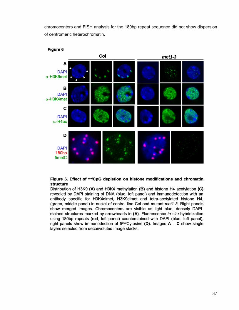
chromocenters and FISH analysis for the 180bp repeat sequence did not show dispersion
of centromeric heterochromatin.
Figure 6. Effect of metCpG depletion on histone modifications and chromatin structureDistribution of H3K9 (A) and H3K4 methylation (B) and histone H4 acetylation (C)revealed by DAPI staining of DNA (blue, left panel) and immunodetection with an antibody specific for H3K4dimet, H3K9dimet and tetra-acetylated histone H4, (green, middle panel) in nuclei of control line Col and mutant met1-3. Right panels show merged images. Chromocenters are visible as light blue, densely DAPI-stained structures marked by arrowheads in (A). Fluorescence in situ hybridization using 180bp repeats (red, left panel) counterstained with DAPI (blue, left panel), right panels show immunodection of 5metCytosine (D). Images A – C show single layers selected from deconvoluted image stacks.
Col
DAPI180bp5metC
α-H4ac
α-H3K4met
α-H3K9met
DAPI
met1-3A
B
D
C
DAPI
DAPI
Figure 6
Figure 6. Effect of metCpG depletion on histone modifications and chromatin structureDistribution of H3K9 (A) and H3K4 methylation (B) and histone H4 acetylation (C)revealed by DAPI staining of DNA (blue, left panel) and immunodetection with an antibody specific for H3K4dimet, H3K9dimet and tetra-acetylated histone H4, (green, middle panel) in nuclei of control line Col and mutant met1-3. Right panels show merged images. Chromocenters are visible as light blue, densely DAPI-stained structures marked by arrowheads in (A). Fluorescence in situ hybridization using 180bp repeats (red, left panel) counterstained with DAPI (blue, left panel), right panels show immunodection of 5metCytosine (D). Images A – C show single layers selected from deconvoluted image stacks.
Col
DAPI
DAPI
met1-3A
B
D
C
DAPI
DAPI
Figure 6
180bp5metC
α-H4ac
α-H3K4met
α-H3K9met
37
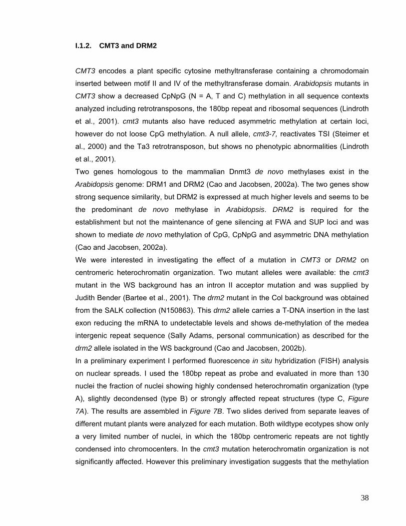
I.1.2. CMT3 and DRM2 CMT3 encodes a plant specific cytosine methyltransferase containing a chromodomain
inserted between motif II and IV of the methyltransferase domain. Arabidopsis mutants in
CMT3 show a decreased CpNpG (N = A, T and C) methylation in all sequence contexts
analyzed including retrotransposons, the 180bp repeat and ribosomal sequences (Lindroth
et al., 2001). cmt3 mutants also have reduced asymmetric methylation at certain loci,
however do not loose CpG methylation. A null allele, cmt3-7, reactivates TSI (Steimer et
al., 2000) and the Ta3 retrotransposon, but shows no phenotypic abnormalities (Lindroth
et al., 2001).
Two genes homologous to the mammalian Dnmt3 de novo methylases exist in the
Arabidopsis genome: DRM1 and DRM2 (Cao and Jacobsen, 2002a). The two genes show
strong sequence similarity, but DRM2 is expressed at much higher levels and seems to be
the predominant de novo methylase in Arabidopsis. DRM2 is required for the
establishment but not the maintenance of gene silencing at FWA and SUP loci and was
shown to mediate de novo methylation of CpG, CpNpG and asymmetric DNA methylation
(Cao and Jacobsen, 2002a).
We were interested in investigating the effect of a mutation in CMT3 or DRM2 on
centromeric heterochromatin organization. Two mutant alleles were available: the cmt3
mutant in the WS background has an intron II acceptor mutation and was supplied by
Judith Bender (Bartee et al., 2001). The drm2 mutant in the Col background was obtained
from the SALK collection (N150863). This drm2 allele carries a T-DNA insertion in the last
exon reducing the mRNA to undetectable levels and shows de-methylation of the medea
intergenic repeat sequence (Sally Adams, personal communication) as described for the
drm2 allele isolated in the WS background (Cao and Jacobsen, 2002b).
In a preliminary experiment I performed fluorescence in situ hybridization (FISH) analysis
on nuclear spreads. I used the 180bp repeat as probe and evaluated in more than 130
nuclei the fraction of nuclei showing highly condensed heterochromatin organization (type
A), slightly decondensed (type B) or strongly affected repeat structures (type C, Figure
7A). The results are assembled in Figure 7B. Two slides derived from separate leaves of
different mutant plants were analyzed for each mutation. Both wildtype ecotypes show only
a very limited number of nuclei, in which the 180bp centromeric repeats are not tightly
condensed into chromocenters. In the cmt3 mutation heterochromatin organization is not
significantly affected. However this preliminary investigation suggests that the methylation
38
changes caused by a mutation in the de novo methylase DRM2 affect heterochromatin
organization.
Figure 7
type A
A
type B type C
0%
20%
40%
60%
80%
100%
WS cmt3 1 cmt3 2 Col drm2 1 drm2 2
B
Figure 7. Centromeric heterochromatin organization in cmt3 (WS) and drm2(Col) mutants Organization of 180bp centromeric repeats was analyzed using FISH on nuclear spreads of WS, Col, cmt3 and drm2. Nuclei were scored according to their phenotype into tightly organized chromocenters (dark gray), dispersion of single repeats (light gray) and severely affected organization (white) (A). For each mutant two independent slides were analyzed, the number of scored nuclei per slide is >131(B).
39
I.2. SWI2/SNF2 Chromatin Remodeling Factor DDM1
Mutations in the DDM1 gene encoding a SWI2/SNF2-like chromatin remodeling factor of
the DEXD/H-ATPase superfamily (Vongs et al., 1993; Jeddeloh et al., 1999) have been
identified in several independent mutant screens: The first screen was based on Southern
blot analysis aiming to discover mutants impaired in the maintenance of DNA methylation
at the 180bp centromere repeats (Vongs et al., 1993) and the identified mutants were
called ddm1 for “decrease in DNA methylation”. Additional alleles were found to release
transcriptional gene silencing in the transgenic line A (Mittelsten Scheid et al., 1998). Line
A contains a complex locus with multiple copies of the hygromycine phosphotransferase
(HPT) gene, stably silenced over several generations (Mittelsten Scheid et al., 1991). Here
I used the ddm1-5 allele (former som8) in a background isogenic to line A that reactivates
silencing of the HPT locus. This allele carries an 82bp insertion that forms two stop codons
in the DDM1 reading frame terminating translation after 30 or 53 aa, respectively. A
possible reinitiation of translation at the first downstream start codon would result in a
frameshift. However, although detectable in wild-type plants, no DDM1 mRNA was found in
ddm1–5, suggesting its decreased stability (Mittelsten Scheid et al., 2002).
The ddm1 mutation is recessive when maintained as heterozygote, but interestingly the
demethylation phenotype arising in homozygotes persists when outcrossed to wildtype
plants. The early generations of homozygotes display demethylation primarily of
pericentromeric sequences, but the severity of the phenotypic abnormalities and the
demethylation increase with successively selfed generations of homozygous ddm1 plants,
affecting – in later generations - single copy sequences as well. DDM1 deficiency induces
loss of DNA methylation at transposons and causes new transposition events (Kato et al.,
2003; Hiroshika, et al., 2000; Steimer et al., 2000). In addition to reduced DNA methylation
levels, the ddm1 mutant also shows altered patterns of H3K4 and H3K9 methylation as
revealed by Chromatin Immunoprecipitation (ChIP) (Gendrel et al., 2002).
The human or mouse orthologue of DDM1 is the Lsh (Lymphoid specific helicase) protein
(Jarvis et al., 1996). Homologues in S. cerevisiae and A. fumigatus were identified,
however sequencing of the Drosophila and the C. elegans genome did not reveal DDM1-
like proteins. Mice deficient in Lsh die in the perinatal period with defects in peripheral T
cells and kidney abnormalities (Dennis et al., 2001). A recent biochemical analysis of
recombinant Arabidopsis DDM1 protein confirmed its ATPase dependent nucleosome
remodeling activity (Brzeski et al., 2003). Since mutations in DDM1 affect different
epigenetic marks suggested being compulsory for transcriptional gene silencing and
40
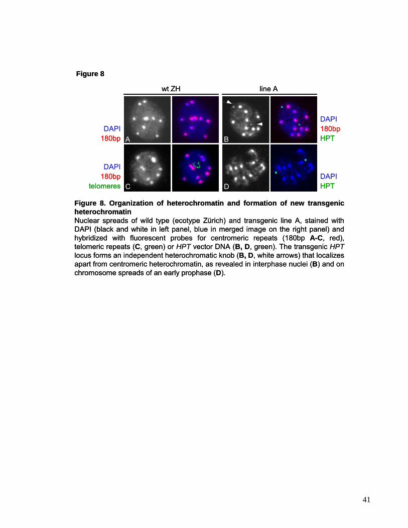
mmmmmmmmmmmmmmmmmmmmmmmmmmmmmmmmmmmmmmmmmmmmmmmm
mmm
Figure 8
DAPI
wt ZH
DAPI180bp
180bp
180bpDAPI
telomeres
HPT
DAPIHPT
line A
A
C D
B
Figure 8. Organization of heterochromatin and formation of new transgenic heterochromatinNuclear spreads of wild type (ecotype Zürich) and transgenic line A, stained with DAPI (black and white in left panel, blue in merged image on the right panel) and hybridized with fluorescent probes for centromeric repeats (180bp A-C, red),telomeric repeats (C, green) or HPT vector DNA (B, D, green). The transgenic HPTlocus forms an independent heterochromatic knob (B, D, white arrows) that localizes apart from centromeric heterochromatin, as revealed in interphase nuclei (B) and on chromosome spreads of an early prophase (D).
Figure 8
DAPI
DAPI
wt ZH
DAPI180bp
180bp
180bptelomeres
HPT
DAPIHPT
line A
A
C D
B
Figure 8. Organization of heterochromatin and formation of new transgenic heterochromatinNuclear spreads of wild type (ecotype Zürich) and transgenic line A, stained with DAPI (black and white in left panel, blue in merged image on the right panel) and hybridized with fluorescent probes for centromeric repeats (180bp A-C, red),telomeric repeats (C, green) or HPT vector DNA (B, D, green). The transgenic HPTlocus forms an independent heterochromatic knob (B, D, white arrows) that localizes apart from centromeric heterochromatin, as revealed in interphase nuclei (B) and on chromosome spreads of an early prophase (D).
41
heterochromatin formation we were interested to investigate the effect of a DDM1 null
mutation on chromatin structure and heterochromatin organization. Before assessing
possible global changes in chromatin organization associated with TGS release by the
ddm1-5 mutation, it was necessary to make sure that nuclei of the transgenic Arabidopsis
line A, the genetic background of the ddm1-5 mutant allele, are identical to wildtype nuclei.
I combined DAPI staining of DNA with detection of 180bp repeats, TSI, ribosomal DNA
and telomeres by fluorescent in situ hybridization (FISH) (Figure 8 and 9). In line A the
bright DAPI-stained heterochromatic chromocenters mainly comprised of 180bp repeats
(Figure 8A-C) predominantly align at the nuclear periphery, in the same way as observed
in wildtype Zürich nuclei. The pericentromeric TSI repeats, located in pericentromeric
regions of all chromosomes, as visualized on pachytene chromosomes and confirmed by
localization on the Arabidopsis physical map (Steimer et al., 2000), are tightly associated
with the chromocenters both in line A and wildtype ZH (Figure 9C and data not shown).
Those chromocenters, that include one of the four nucleolus organizer regions (NORs),
were found close to the nucleolus (Figure 8A-C) and telomeres were clustered around this
subnuclear compartment ((Armstrong et al., 2001) and Figure 8C; 9E). Although the
images of line A and the corresponding wild type Zürich (ZH) are very similar, I detected
one or two additional DAPI-bright spots in nuclei of line A plants hemizygous or
homozygous for the transgenic locus respectively (Figure 8B, 9A). To investigate whether
they reflect a chromatin condensation acquired de novo at the silent transgenic locus, we
combined DAPI staining with two-color FISH using HPT and 180bp repeats as probes. The
HPT signals indeed co-localized with the two neo-chromocenters (Figure 8B). This implies
that the insertion of a complex transgene and its consecutive silencing triggered the
formation of an additional, neo-heterochromatic knob. It has been observed that formation
of repressive chromatin at silenced transgenes is enhanced by neighboring
heterochromatic regions (Dorer and Henikoff, 1994). However, the HPT locus seems to be
located distant to preexisting centromeric heterochromatin in interphase nuclei (Figure 8B,
9A) and on chromosome spreads (Figure 8D).
Since the overall structure of the nucleus of the transgenic line A was identical to the
wildtype except for the additional heterochromatic knob, I carried on to examine the
consequences of the ddm1 mutation on the nuclear organization and structure of the
reactivated transgenic locus. DAPI staining of the ddm1-5 nuclei revealed a striking
decondensation of chromocenters (Figure 9B, D). The number of clearly stained
chromocenters was reduced and those visible were considerably smaller than in line A.
FISH analysis of centromeric repeats revealed their expansion in the form of loops into
42
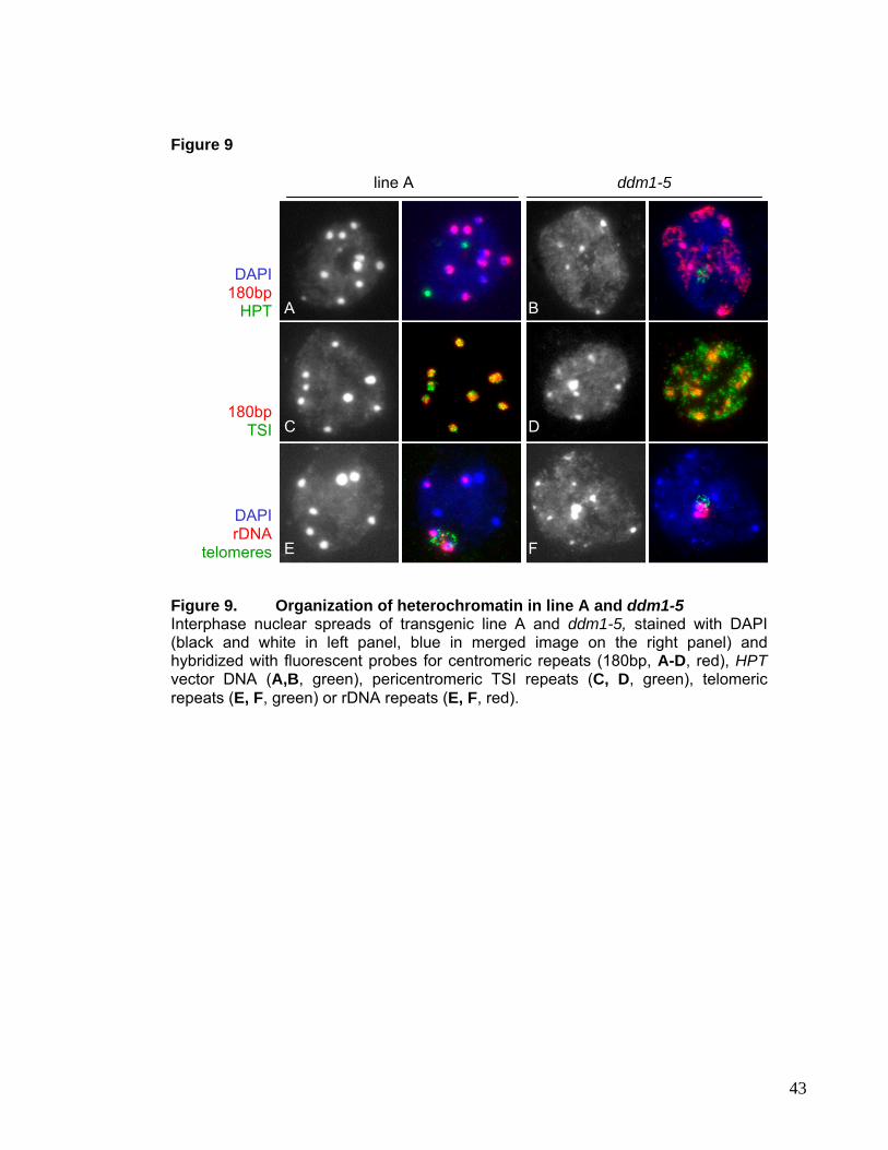
mmmmmmmmmmmmmmmmmmmmmmmmmmmmmmmmmmmmmmmmmmmmmmmm
mmm Figure 9
E F
B
DC
A
180bp TSI
DAPI rDNA
telomeres
DAPI
180bp HPT
ddm1-5 line A
Figure 9. Organization of heterochromatin in line A and ddm1-5 Interphase nuclear spreads of transgenic line A and ddm1-5, stained with DAPI (black and white in left panel, blue in merged image on the right panel) and hybridized with fluorescent probes for centromeric repeats (180bp, A-D, red), HPTvector DNA (A,B, green), pericentromeric TSI repeats (C, D, green), telomeric repeats (E, F, green) or rDNA repeats (E, F, red).
43
territory usually occupied by euchromatin (Figure 9B). The neo-heterochromatin
associated with the transgenic HPT locus was likewise decondensed (Figure 9B). The
ddm1-5 mutant had been backcrossed to wild type to promote recovery from the
progressive developmental effects, and the illustrated donor plant for the nuclei, though
homozygous for the mutation, is hemizygous for the transgenic locus. Moreover, the TSI
templates lost the tight association with the centromeric DNA. This was clearly visible also
in nuclei that still had remnants of chromocenters (Figure 9D), suggesting that
decondensation and looping out of the pericentromeric repeats precedes a similar process
occurring at centromeres and results in disappearance of chromocenters. Interestingly,
telomeres were largely unaffected and remained clustered around the nucleolus, and the
rDNA was associated with remnants of chromocenters where these were still present
(Figure 9F).
In plants and mammals, heterochromatic DNA contains a significant amount of 5-
methylCytosine (Johnson et al., 2002). ddm1 mutants are characterized by a substantial
hypomethylation of the repetitive pericentromeric sequences (Vongs et al., 1993) and of
the reactivated transgenic loci (Mittelsten Scheid et al., 1998). By immunolocalization of
modified cytosines, the DNA in chromocenters in line A was found to be hypermethylated,
while the remaining DNA showed low methylation (Figure 10A). The additional small
heterochromatic knobs associated with the transgenic loci were also hypermethylated
(Figure 10A, arrows). In ddm1-5 nuclei, methylation signals were reduced and rather
uniformly distributed (Figure 10B). The methylation changes visualized by immunostaining
were clearly correlated with the decondensation of the centromeric heterochromatin and its
relocation away from the chromocenter remnants (Figure 10B). It was described that ddm1
affects histone modifications at silenced loci (Grendel et al., 2002; Johnson et al., 2002).
Therefore, I investigated the effect of DDM1 depletion on the global distribution of
particular histone modifications, namely H4 acetylation, H3K4 and H3K9 methylation.
These three modifications were well characterized in other organisms and their
involvement in either activation or silencing established. H4 acetylation patterns in line A
and ddm1-5 were visualized by immunostaining with an antibody directed against a tetra-
acetylated isoform of H4. Euchromatin of line A was intensely stained (Figure 10C), but the
chromocenters lacked any signal. In contrast, nuclei of ddm1-5 plants were almost evenly
labeled (Figure 10D). Since ddm1-5 nuclei have dispersed heterochromatic regions, the
loss of DDM1 appears to have at least an indirect influence on the distribution of
acetylated histones, which now become associated also with centromeric and
pericentromeric DNA. Whereas histone acetylation marks transcriptionally active
44
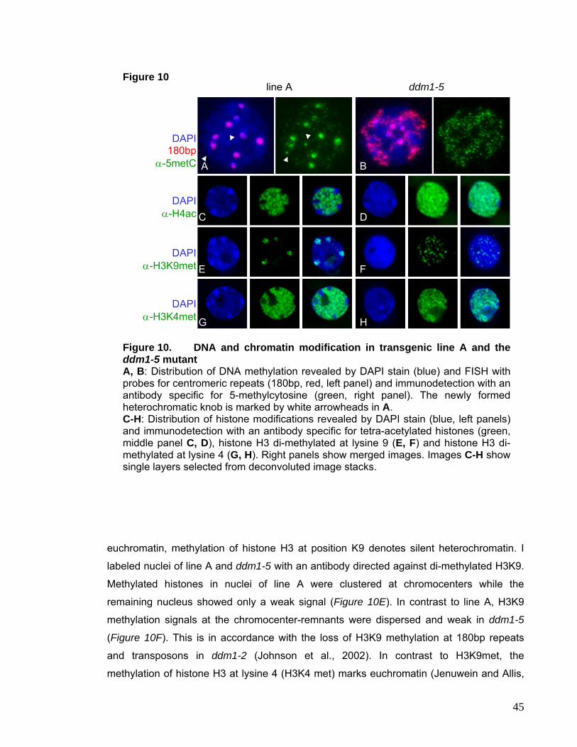
mmmmmmmmmmmmmmmmmmmmmmmmmmmmmmmmmmmmmmmmmmmmmmmm
mmm Figure 10 line A ddm1-5
G
DAPI
α-H3K4met H
E
DAPI
α-H3K9met
F
A
C
DAPI
α-H4ac
DAPI
180bp α-5metC
B
D
Figure 10. DNA and chromatin modification in transgenic line A and the ddm1-5 mutant A, B: Distribution of DNA methylation revealed by DAPI stain (blue) and FISH with probes for centromeric repeats (180bp, red, left panel) and immunodetection with an antibody specific for 5-methylcytosine (green, right panel). The newly formed heterochromatic knob is marked by white arrowheads in A. C-H: Distribution of histone modifications revealed by DAPI stain (blue, left panels) and immunodetection with an antibody specific for tetra-acetylated histones (green, middle panel C, D), histone H3 di-methylated at lysine 9 (E, F) and histone H3 di-methylated at lysine 4 (G, H). Right panels show merged images. Images C-H show single layers selected from deconvoluted image stacks.
euchromatin, methylation of histone H3 at position K9 denotes silent heterochromatin. I
labeled nuclei of line A and ddm1-5 with an antibody directed against di-methylated H3K9.
Methylated histones in nuclei of line A were clustered at chromocenters while the
remaining nucleus showed only a weak signal (Figure 10E). In contrast to line A, H3K9
methylation signals at the chromocenter-remnants were dispersed and weak in ddm1-5
(Figure 10F). This is in accordance with the loss of H3K9 methylation at 180bp repeats
and transposons in ddm1-2 (Johnson et al., 2002). In contrast to H3K9met, the
methylation of histone H3 at lysine 4 (H3K4 met) marks euchromatin (Jenuwein and Allis,
45
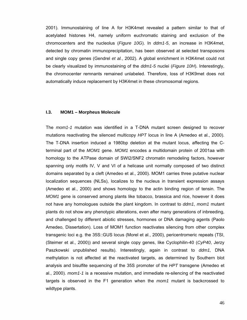
2001). Immunostaining of line A for H3K4met revealed a pattern similar to that of
acetylated histones H4, namely uniform euchromatic staining and exclusion of the
chromocenters and the nucleolus (Figure 10G). In ddm1-5, an increase in H3K4met,
detected by chromatin immunoprecipitation, has been observed at selected transposons
and single copy genes (Gendrel et al., 2002). A global enrichment in H3K4met could not
be clearly visualized by immunostaining of the ddm1-5 nuclei (Figure 10H). Interestingly,
the chromocenter remnants remained unlabeled. Therefore, loss of H3K9met does not
automatically induce replacement by H3K4met in these chromosomal regions.
I.3. MOM1 – Morpheus Molecule The mom1-1 mutation was identified in a T-DNA mutant screen designed to recover
mutations reactivating the silenced multicopy HPT locus in line A (Amedeo et al., 2000).
The T-DNA insertion induced a 1980bp deletion at the mutant locus, affecting the C-
terminal part of the MOM1 gene. MOM1 encodes a multidomain protein of 2001aa with
homology to the ATPase domain of SWI2/SNF2 chromatin remodeling factors, however
spanning only motifs IV, V and VI of a helicase unit normally composed of two distinct
domains separated by a cleft (Amedeo et al., 2000). MOM1 carries three putative nuclear
localization sequences (NLSs), localizes to the nucleus in transient expression assays
(Amedeo et al., 2000) and shows homology to the actin binding region of tensin. The
MOM1 gene is conserved among plants like tobacco, brassica and rice, however it does
not have any homologues outside the plant kingdom. In contrast to ddm1, mom1 mutant
plants do not show any phenotypic alterations, even after many generations of inbreeding,
and challenged by different abiotic stresses, hormones or DNA damaging agents (Paolo
Amedeo, Dissertation). Loss of MOM1 function reactivates silencing from other complex
transgenic loci e.g. the 35S::GUS locus (Morel et al., 2000), pericentromeric repeats (TSI,
(Steimer et al., 2000)) and several single copy genes, like Cyclophilin-40 (CyP40, Jerzy
Paszkowski unpublished results). Interestingly, again in contrast to ddm1, DNA
methylation is not affected at the reactivated targets, as determined by Southern blot
analysis and bisulfite sequencing of the 35S promoter of the HPT transgene (Amedeo et
al., 2000). mom1-1 is a recessive mutation, and immediate re-silencing of the reactivated
targets is observed in the F1 generation when the mom1 mutant is backcrossed to
wildtype plants.
46

Figure 11
1 2 3 3 4 5 5 5
5 5 6 6 7 7 3 2 1
A
B
Figure 11. MOM1-HA fusions expressed from a genomic construct under its endogenous promoter complement the TSI expression phenotype, while expression of MOM1-HA cDNA from a constitutive 35S promoter fails to silence TSI Different MOM1-HA fusion constructs introduced into the mom1-1 mutant were tested for functional complementation by Northern Blot analysis using a probe for TSI repeats, reactivated in mom1-1 mutant plants; genomic construct (A) and 35S::cDNA MOM1 (B) line A (1), mom1-1 (2), mom1-1 with empty vector (3), mom1-1 with a genomic MOM construct (4), mom1-1 expressing a MOM1-HA protein fusion under the endogenous promoter (5), mom1-1 expressing HA-MOM1 cDNA under control of the 35S promoter (6), mom1-1 expressing MOM1-HA cDNA under control of the 35S promoter (7).
I.3.1. Subcellular Localization of MOM1 Protein In order to study the subcellular localization of MOM1 protein, a tagged version of the
protein was expressed in A. thaliana. Therefore, mom1-1 mutant plants were transformed
with genomic constructs expressing MOM1-HA or MOM1-GFP fusion proteins from its
endogenous promoter, as well as a construct expressing a MOM1-HA fusion protein from
47
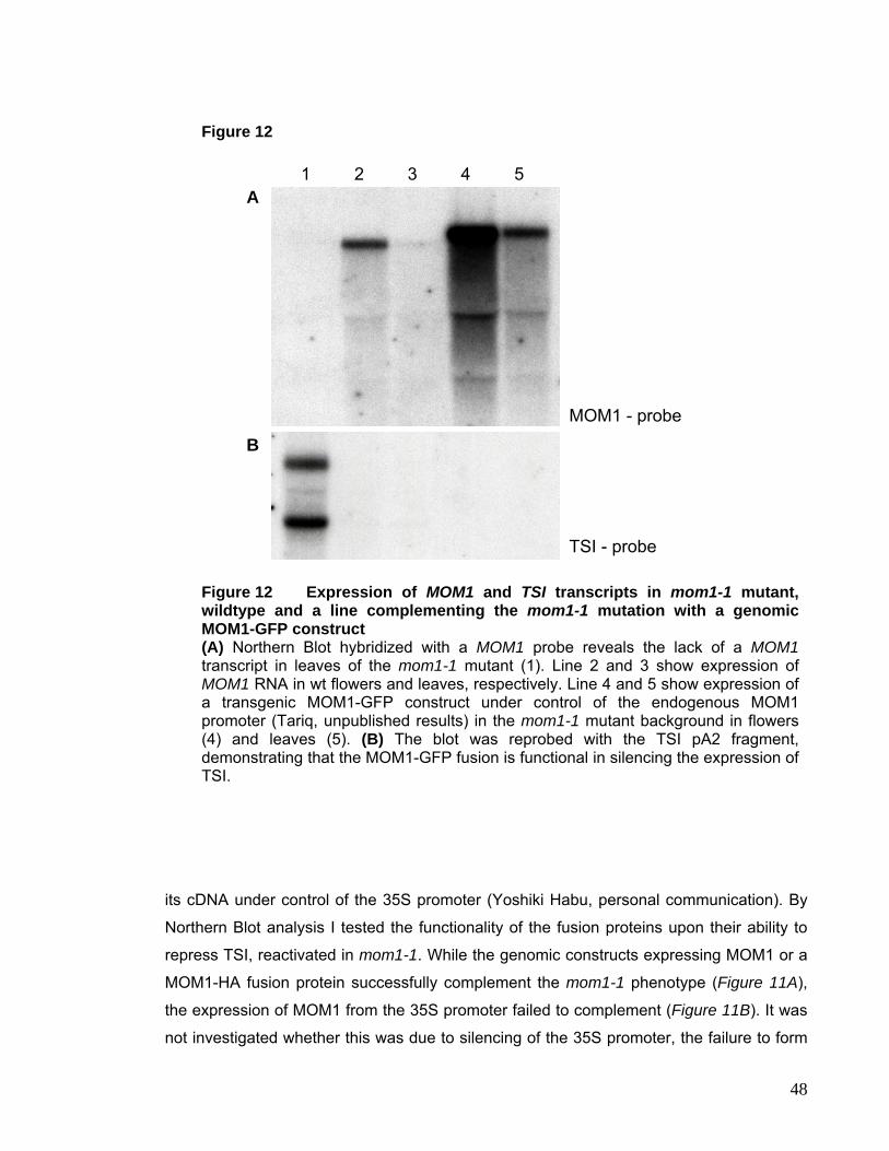
mmmmmmmmmmmmmmmmmmmmmmmmmmmmmmmmmmmmmmmmmmmmmmmm
mm Figure 12
1 2 3 4 5 A
MOM1 - probe
B
TSI - probe
Figure 12 Expression of MOM1 and TSI transcripts in mom1-1 mutant, wildtype and a line complementing the mom1-1 mutation with a genomic MOM1-GFP construct (A) Northern Blot hybridized with a MOM1 probe reveals the lack of a MOM1transcript in leaves of the mom1-1 mutant (1). Line 2 and 3 show expression of MOM1 RNA in wt flowers and leaves, respectively. Line 4 and 5 show expression of a transgenic MOM1-GFP construct under control of the endogenous MOM1 promoter (Tariq, unpublished results) in the mom1-1 mutant background in flowers (4) and leaves (5). (B) The blot was reprobed with the TSI pA2 fragment, demonstrating that the MOM1-GFP fusion is functional in silencing the expression of TSI.
its cDNA under control of the 35S promoter (Yoshiki Habu, personal communication). By
Northern Blot analysis I tested the functionality of the fusion proteins upon their ability to
repress TSI, reactivated in mom1-1. While the genomic constructs expressing MOM1 or a
MOM1-HA fusion protein successfully complement the mom1-1 phenotype (Figure 11A),
the expression of MOM1 from the 35S promoter failed to complement (Figure 11B). It was
not investigated whether this was due to silencing of the 35S promoter, the failure to form
48
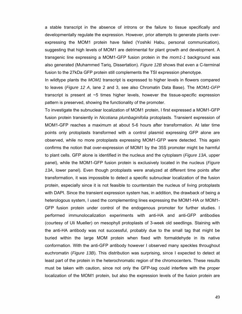
a stable transcript in the absence of introns or the failure to tissue specifically and
developmentally regulate the expression. However, prior attempts to generate plants over-
expressing the MOM1 protein have failed (Yoshiki Habu, personal communication),
suggesting that high levels of MOM1 are detrimental for plant growth and development. A
transgenic line expressing a MOM1-GFP fusion protein in the mom1-1 background was
also generated (Muhammed Tariq, Dissertation). Figure 12B shows that even a C-terminal
fusion to the 27kDa GFP protein still complements the TSI expression phenotype.
In wildtype plants the MOM1 transcript is expressed to higher levels in flowers compared
to leaves (Figure 12 A, lane 2 and 3, see also Chromatin Data Base). The MOM1-GFP
transcript is present at ~5 times higher levels, however the tissue-specific expression
pattern is preserved, showing the functionality of the promoter.
To investigate the subnuclear localization of MOM1 protein, I first expressed a MOM1-GFP
fusion protein transiently in Nicotiana plumbaginifolia protoplasts. Transient expression of
MOM1-GFP reaches a maximum at about 5-6 hours after transformation. At later time
points only protoplasts transformed with a control plasmid expressing GFP alone are
observed, while no more protoplasts expressing MOM1-GFP were detected. This again
confirms the notion that over-expression of MOM1 by the 35S promoter might be harmful
to plant cells. GFP alone is identified in the nucleus and the cytoplasm (Figure 13A, upper
panel), while the MOM1-GFP fusion protein is exclusively located in the nucleus (Figure
13A, lower panel). Even though protoplasts were analyzed at different time points after
transformation, it was impossible to detect a specific subnuclear localization of the fusion
protein, especially since it is not feasible to counterstain the nucleus of living protoplasts
with DAPI. Since the transient expression system has, in addition, the drawback of being a
heterologous system, I used the complementing lines expressing the MOM1-HA or MOM1-
GFP fusion protein under control of the endogenous promoter for further studies. I
performed immunolocalization experiments with anti-HA and anti-GFP antibodies
(courtesy of Uli Mueller) on mesophyll protoplasts of 3-week old seedlings. Staining with
the anti-HA antibody was not successful, probably due to the small tag that might be
buried within the large MOM protein when fixed with formaldehyde in its native
conformation. With the anti-GFP antibody however I observed many speckles throughout
euchromatin (Figure 13B). This distribution was surprising, since I expected to detect at
least part of the protein in the heterochromatic region of the chromocenters. These results
must be taken with caution, since not only the GFP-tag could interfere with the proper
localization of the MOM1 protein, but also the expression levels of the fusion protein are
49
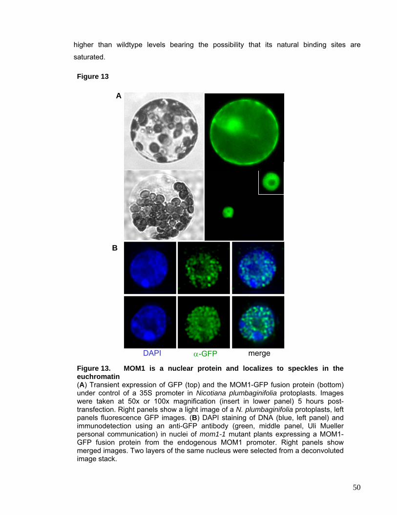
higher than wildtype levels bearing the possibility that its natural binding sites are
saturated.
Figure 13
merge α-GFPDAPI
A
B
Figure 13. MOM1 is a nuclear protein and localizes to speckles in the euchromatin (A) Transient expression of GFP (top) and the MOM1-GFP fusion protein (bottom) under control of a 35S promoter in Nicotiana plumbaginifolia protoplasts. Images were taken at 50x or 100x magnification (insert in lower panel) 5 hours post-transfection. Right panels show a light image of a N. plumbaginifolia protoplasts, left panels fluorescence GFP images. (B) DAPI staining of DNA (blue, left panel) and immunodetection using an anti-GFP antibody (green, middle panel, Uli Mueller personal communication) in nuclei of mom1-1 mutant plants expressing a MOM1-GFP fusion protein from the endogenous MOM1 promoter. Right panels show merged images. Two layers of the same nucleus were selected from a deconvoluted image stack.
50
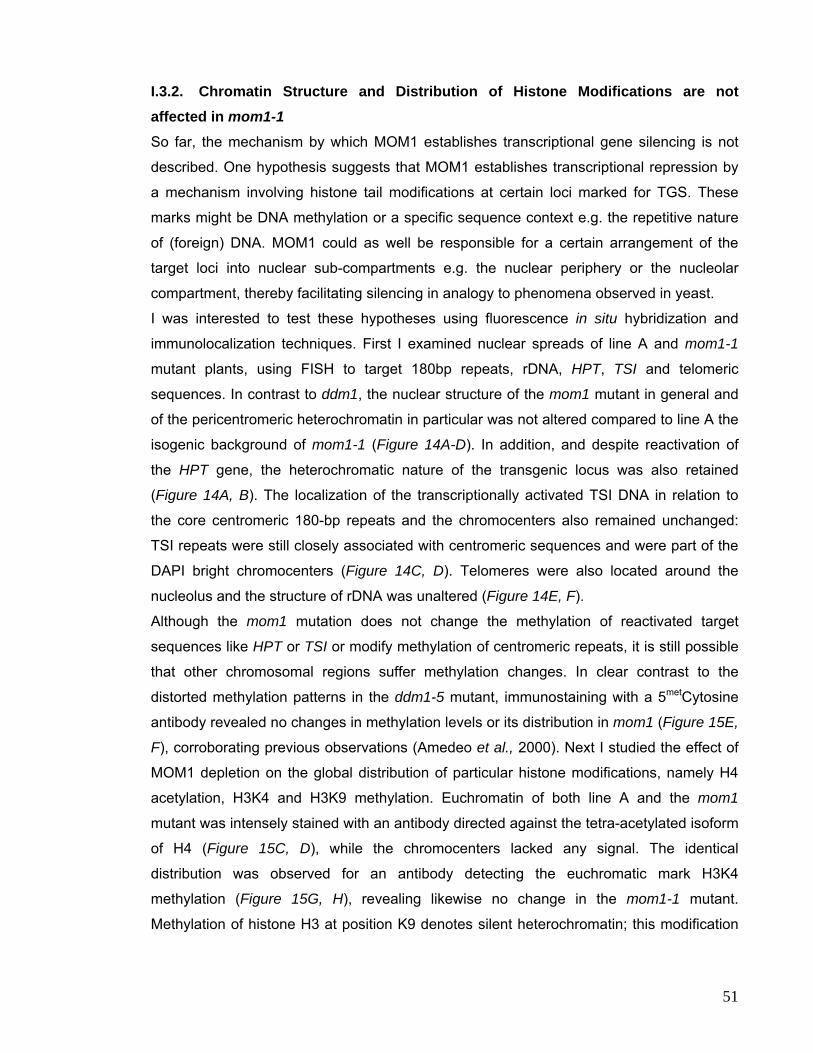
I.3.2. Chromatin Structure and Distribution of Histone Modifications are not affected in mom1-1 So far, the mechanism by which MOM1 establishes transcriptional gene silencing is not
described. One hypothesis suggests that MOM1 establishes transcriptional repression by
a mechanism involving histone tail modifications at certain loci marked for TGS. These
marks might be DNA methylation or a specific sequence context e.g. the repetitive nature
of (foreign) DNA. MOM1 could as well be responsible for a certain arrangement of the
target loci into nuclear sub-compartments e.g. the nuclear periphery or the nucleolar
compartment, thereby facilitating silencing in analogy to phenomena observed in yeast.
I was interested to test these hypotheses using fluorescence in situ hybridization and
immunolocalization techniques. First I examined nuclear spreads of line A and mom1-1
mutant plants, using FISH to target 180bp repeats, rDNA, HPT, TSI and telomeric
sequences. In contrast to ddm1, the nuclear structure of the mom1 mutant in general and
of the pericentromeric heterochromatin in particular was not altered compared to line A the
isogenic background of mom1-1 (Figure 14A-D). In addition, and despite reactivation of
the HPT gene, the heterochromatic nature of the transgenic locus was also retained
(Figure 14A, B). The localization of the transcriptionally activated TSI DNA in relation to
the core centromeric 180-bp repeats and the chromocenters also remained unchanged:
TSI repeats were still closely associated with centromeric sequences and were part of the
DAPI bright chromocenters (Figure 14C, D). Telomeres were also located around the
nucleolus and the structure of rDNA was unaltered (Figure 14E, F).
Although the mom1 mutation does not change the methylation of reactivated target
sequences like HPT or TSI or modify methylation of centromeric repeats, it is still possible
that other chromosomal regions suffer methylation changes. In clear contrast to the
distorted methylation patterns in the ddm1-5 mutant, immunostaining with a 5metCytosine
antibody revealed no changes in methylation levels or its distribution in mom1 (Figure 15E,
F), corroborating previous observations (Amedeo et al., 2000). Next I studied the effect of
MOM1 depletion on the global distribution of particular histone modifications, namely H4
acetylation, H3K4 and H3K9 methylation. Euchromatin of both line A and the mom1
mutant was intensely stained with an antibody directed against the tetra-acetylated isoform
of H4 (Figure 15C, D), while the chromocenters lacked any signal. The identical
distribution was observed for an antibody detecting the euchromatic mark H3K4
methylation (Figure 15G, H), revealing likewise no change in the mom1-1 mutant.
Methylation of histone H3 at position K9 denotes silent heterochromatin; this modification
51
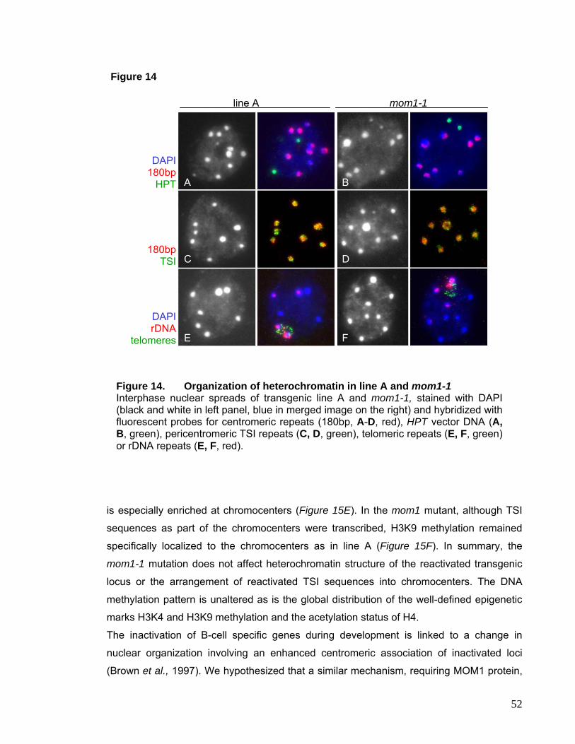
mmmmmmmmmmmmmmmmmmmmmmmmmmmmmmmmmmmmmmmmmmmmmmmm
mm Figure 14
mom1-1 line A
E F
B
C D
A
180bp TSI
DAPI rDNA
telomeres
DAPI
180bp HPT
Figure 14. Organization of heterochromatin in line A and mom1-1 Interphase nuclear spreads of transgenic line A and mom1-1, stained with DAPI (black and white in left panel, blue in merged image on the right) and hybridized with fluorescent probes for centromeric repeats (180bp, A-D, red), HPT vector DNA (A, B, green), pericentromeric TSI repeats (C, D, green), telomeric repeats (E, F, green) or rDNA repeats (E, F, red).
is especially enriched at chromocenters (Figure 15E). In the mom1 mutant, although TSI
sequences as part of the chromocenters were transcribed, H3K9 methylation remained
specifically localized to the chromocenters as in line A (Figure 15F). In summary, the
mom1-1 mutation does not affect heterochromatin structure of the reactivated transgenic
locus or the arrangement of reactivated TSI sequences into chromocenters. The DNA
methylation pattern is unaltered as is the global distribution of the well-defined epigenetic
marks H3K4 and H3K9 methylation and the acetylation status of H4.
The inactivation of B-cell specific genes during development is linked to a change in
nuclear organization involving an enhanced centromeric association of inactivated loci
(Brown et al., 1997). We hypothesized that a similar mechanism, requiring MOM1 protein,
52
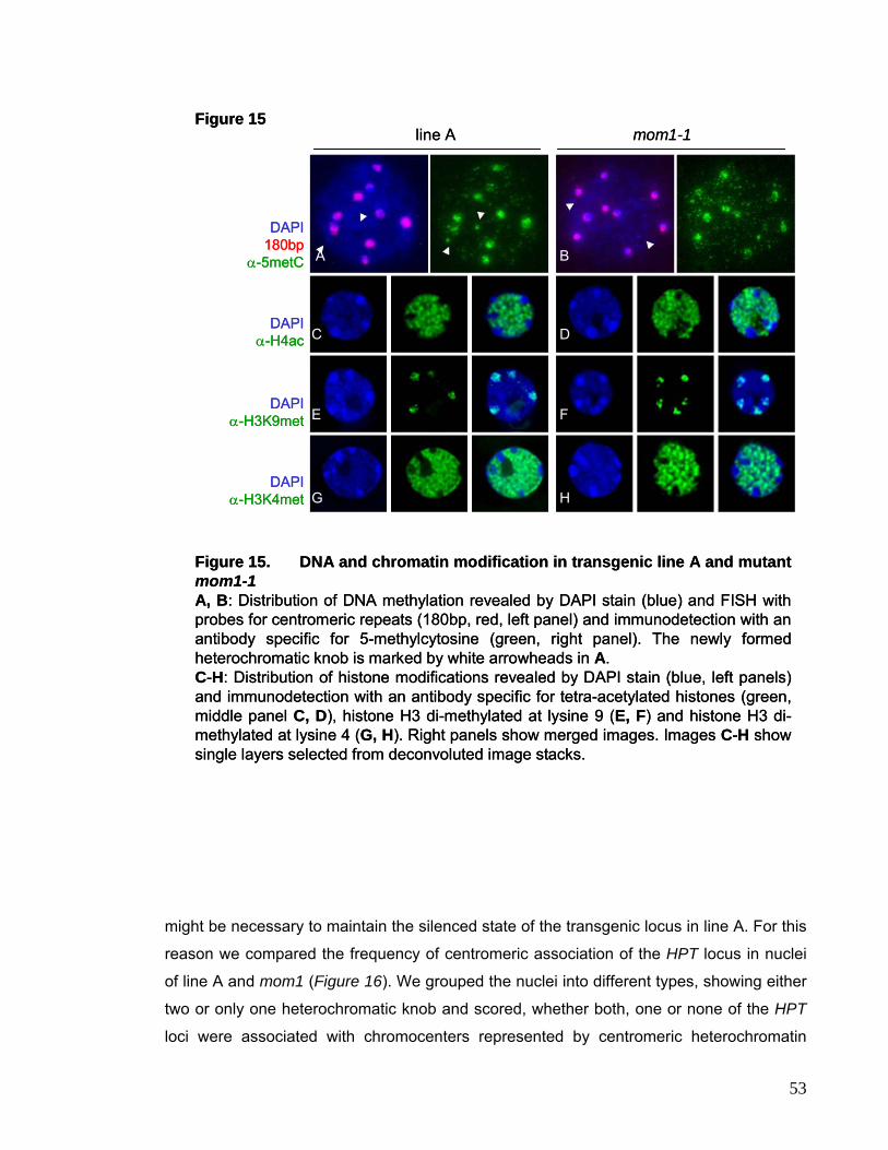
line A mom1-1
A
C D
B
E F
G H
DAPIα-H3K9met
α-H3K4met
α-H4ac
α-5metC
DAPI
DAPI
DAPI180bp
Figure 15. DNA and chromatin modification in transgenic line A and mutant mom1-1A, B: Distribution of DNA methylation revealed by DAPI stain (blue) and FISH with probes for centromeric repeats (180bp, red, left panel) and immunodetection with an antibody specific for 5-methylcytosine (green, right panel). The newly formed heterochromatic knob is marked by white arrowheads in A.C-H: Distribution of histone modifications revealed by DAPI stain (blue, left panels) and immunodetection with an antibody specific for tetra-acetylated histones (green, middle panel C, D), histone H3 di-methylated at lysine 9 (E, F) and histone H3 di-methylated at lysine 4 (G, H). Right panels show merged images. Images C-H show single layers selected from deconvoluted image stacks.
Figure 15line A mom1-1
A
C D
B
E F
G H
DAPI
DAPI
DAPI
DAPI
α-H3K9met
α-H3K4met
α-H4ac
α-5metC180bp
Figure 15. DNA and chromatin modification in transgenic line A and mutant mom1-1A, B: Distribution of DNA methylation revealed by DAPI stain (blue) and FISH with probes for centromeric repeats (180bp, red, left panel) and immunodetection with an antibody specific for 5-methylcytosine (green, right panel). The newly formed heterochromatic knob is marked by white arrowheads in A.C-H: Distribution of histone modifications revealed by DAPI stain (blue, left panels) and immunodetection with an antibody specific for tetra-acetylated histones (green, middle panel C, D), histone H3 di-methylated at lysine 9 (E, F) and histone H3 di-methylated at lysine 4 (G, H). Right panels show merged images. Images C-H show single layers selected from deconvoluted image stacks.
Figure 15
mmmmmmmmmmmmmmmmmmmmmmmmmmmmmmmmmmmmmmmmmmmmmmmm
mm
might be necessary to maintain the silenced state of the transgenic locus in line A. For this
reason we compared the frequency of centromeric association of the HPT locus in nuclei
of line A and mom1 (Figure 16). We grouped the nuclei into different types, showing either
two or only one heterochromatic knob and scored, whether both, one or none of the HPT
loci were associated with chromocenters represented by centromeric heterochromatin
53
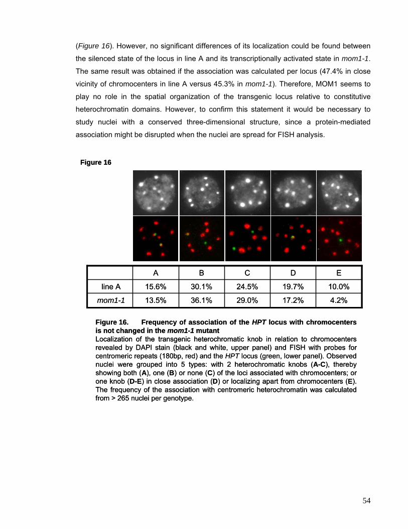
(Figure 16). However, no significant differences of its localization could be found between
the silenced state of the locus in line A and its transcriptionally activated state in mom1-1.
The same result was obtained if the association was calculated per locus (47.4% in close
vicinity of chromocenters in line A versus 45.3% in mom1-1). Therefore, MOM1 seems to
play no role in the spatial organization of the transgenic locus relative to constitutive
heterochromatin domains. However, to confirm this statement it would be necessary to
study nuclei with a conserved three-dimensional structure, since a protein-mediated
association might be disrupted when the nuclei are spread for FISH analysis.
4.2%17.2%29.0%36.1%13.5%mom1-1
10.0%19.7%24.5%30.1%15.6%line A
EDCBA
Figure 16
Figure 16. Frequency of association of the HPT locus with chromocenters is not changed in the mom1-1 mutantLocalization of the transgenic heterochromatic knob in relation to chromocenters revealed by DAPI stain (black and white, upper panel) and FISH with probes for centromeric repeats (180bp, red) and the HPT locus (green, lower panel). Observed nuclei were grouped into 5 types: with 2 heterochromatic knobs (A-C), thereby showing both (A), one (B) or none (C) of the loci associated with chromocenters; or one knob (D-E) in close association (D) or localizing apart from chromocenters (E). The frequency of the association with centromeric heterochromatin was calculated from > 265 nuclei per genotype.
54
I.4. Histone De-Acetylase HDA6 In the Arabidopsis genome a total of 16 potentially functional histone deacetylases have
been identified and these can be classified into three families (Pandey et al., 2002); see
also http://chromdb.biosci.arizona.edu): 10 belong to the RPD3/HDA1 superfamily, 2 are
members of the SIR2-like family and 4 belong to the plant-specific HD2–like histone
deacetylases originally identified as acidic nucleolar phosphoproteins from corn (Lusser et
al., 1997). Interference of HDAC functions in plants has been studied using inhibitors such
as trichostatin A, SAHA or butyrate and using transgenic plants containing antisense or
over-expressing constructs. These approaches have provided evidence that HDACs are
involved in regulation of histone acetylation and thereby gene expression, with
consequences for plant morphology and development (Chen and Pikaard, 1997; Wu et al.,
2000; Tian and Chen, 2001). Dissecting the function of individual HDAC members is
problematic in these studies. Therefore the analysis of loss-of-function mutations of
individual HDAC genes should add valuable information on specific roles. Mutants in an
Arabidopsis RPD3-like HDAC gene, AtHDA6 were found in three independent mutant
screens based upon their effects on specific transgene expression (Murfett et al., 2001;
Aufsatz et al., 2002). The HDA6 mutant alleles axe1 lead to higher expression from a
marker gene with an auxin-responsive promoter element (Murfett et al., 2001), while the
rts1 alleles of the locus interfere with double-stranded RNA-directed transcriptional
silencing (Aufsatz et al., 2002). The mutant sil1 (modifiers of silencing 1, Furner et al.,
1998) was identified in a screen for mutations releasing silencing of the complex,
rearranged transgenic locus C, containing the chalcone synthase gene (CHS) and the
resistance marker genes neomycin phosphotransferase (NPT) and hygromycin
phosphotransferase (HPT). The sil1 mutation reactivates mainly the resistance marker
genes, while the homology-dependent silencing of the endogenous and transgenic CHS
copies are only weakly affected (Furner et al., 1998).
I studied the effect of the HDA6 mutant on the release of transcriptional silencing and the
impact on chromatin structure of heterochromatic regions, especially rDNA repeats.
Thereby, I concentrated on two alleles, axe1-5 and sil1 (Figure 17A). The mutant allele
axe1-5 does not produce an HDA6 transcript of the expected size, but shorter and longer
mRNAs due to a splice-site mutation (Murfett et al., 2001). This allele resides in the Col
background and in all experiments the transgenic line DR5, carrying the pDR5 plasmid
insertion, was used as wt control. The second allele sil1 carries a point mutation in the
very N-terminal part of the protein, which leads to a single amino acid
55
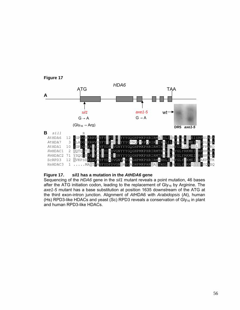
mmmmmmmmmmmmmmmmmmmmmmmmmmmmmmmmmmmmmmmmmmmmmmmm
mmmmmm
Figure 17
ATG A
TAA HDA6
sil1 G → A
(Gly16 → Arg)
axe1-5 G → A
sil1 R AtHDA6 12 SGPDGRKRRVSYFYEPTIGDYYYGQGHPMKPHRIRMAHSLIIHYHLHRRLEISRPSLADAAtHDA7 3 SLADGGKRRVSYFYEPMIGDYYYGVNQPTKPQRIRVTHNLILSYNLHRHMEINHPDLADAAtHDA1 10 SGPDGVKRKVCYFYDPEVGNYYYGQGHPMKPHRIRMTHALLAHYGLLQHMQVLKPFPARDHsHDAC1 2 AQTQGTRRKVCYYYDGDVGNYYYGQGHPMKPHRIRMTHNLLLNYGLYRKMEIYRPHKANAHsHDAC2 71 YSQGGGKKKVCYYYDGDIGNYYYGQGHPMKPHRIRMTHNLLLNYGLYRKMEIYRPHKATAScRPD3 12 TVKPSDKRRVAYFYDADVGNYAYGAGHPMKPHRIRMAHSLIMNYGLYKKMEIYRAKPATKHsHDAC3 1 .....MAKTVAYFYDPDVGNFHYGAGHPMKPHRLALTHSLVLHYGLYKKMIVFKPYQASQ
Figure 17. sil1 has a mutation in the AtHDA6 gene Sequencing of the HDA6 gene in the sil1 mutant reveals a point mutation, 46 bases after the ATG initiation codon, leading to the replacement of Gly16 by Arginine. The axe1-5 mutant has a base substitution at position 1635 downstream of the ATG at the third exon-intron junction. Alignment of AtHDA6 with Arabidopsis (At), human (Hs) RPD3-like HDACs and yeast (Sc) RPD3 reveals a conservation of Gly16 in plant and human RPD3-like HDACs.
B
wt
DR5 axe1-5
56
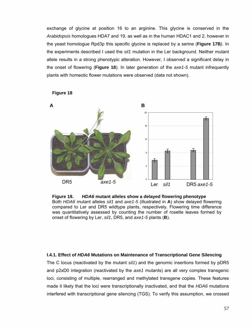
exchange of glycine at position 16 to an arginine. This glycine is conserved in the
Arabidopsis homologues HDA7 and 19, as well as in the human HDAC1 and 2, however in
the yeast homologue Rpd3p this specific glycine is replaced by a serine (Figure 17B). In
the experiments described I used the sil1 mutation in the Ler background. Neither mutant
allele results in a strong phenotypic alteration. However, I observed a significant delay in
the onset of flowering (Figure 18). In later generation of the axe1-5 mutant infrequently
plants with homeotic flower mutations were observed (data not shown).
0
4
8
12
16
20
Ler sil1 DR5 axe1-5DR5 axe1-5sil1LerDR5 axe1-5
Figure 18
A B
Figure 18. HDA6 mutant alleles show a delayed flowering phenotype Both HDA6 mutant alleles sil1 and axe1-5 (illustrated in A) show delayed flowering compared to Ler and DR5 wildtype plants, respectively. Flowering time difference was quantitatively assessed by counting the number of rosette leaves formed by onset of flowering by Ler, sil1, DR5, and axe1-5 plants (B).
I.4.1. Effect of HDA6 Mutations on Maintenance of Transcriptional Gene Silencing The C locus (reactivated by the mutant sil1) and the genomic insertions formed by pDR5
and p2xD0 integration (reactivated by the axe1 mutants) are all very complex transgenic
loci, consisting of multiple, rearranged and methylated transgene copies. These features
made it likely that the loci were transcriptionally inactivated, and that the HDA6 mutations
interfered with transcriptional gene silencing (TGS). To verify this assumption, we crossed
57
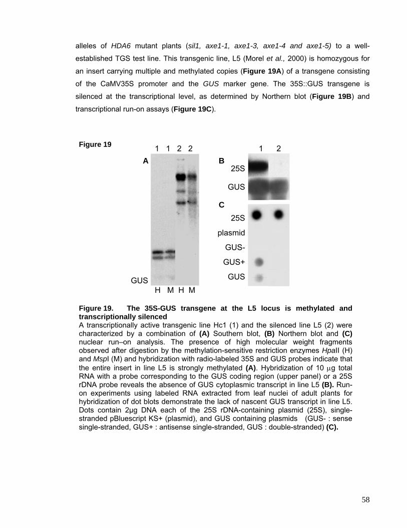
alleles of HDA6 mutant plants (sil1, axe1-1, axe1-3, axe1-4 and axe1-5) to a well-
established TGS test line. This transgenic line, L5 (Morel et al., 2000) is homozygous for
an insert carrying multiple and methylated copies (Figure 19A) of a transgene consisting
of the CaMV35S promoter and the GUS marker gene. The 35S::GUS transgene is
silenced at the transcriptional level, as determined by Northern blot (Figure 19B) and
transcriptional run-on assays (Figure 19C).
Figure 19 1 1 2 2 1 2
25S
plasmid
GUS-
GUS+
GUS
A B
C
H M H M
25S
GUS
GUS
Figure 19. The 35S-GUS transgene at the L5 locus is methylated and transcriptionally silenced A transcriptionally active transgenic line Hc1 (1) and the silenced line L5 (2) were characterized by a combination of (A) Southern blot, (B) Northern blot and (C)nuclear run–on analysis. The presence of high molecular weight fragments observed after digestion by the methylation-sensitive restriction enzymes HpaII (H) and MspI (M) and hybridization with radio-labeled 35S and GUS probes indicate that the entire insert in line L5 is strongly methylated (A). Hybridization of 10 µg total RNA with a probe corresponding to the GUS coding region (upper panel) or a 25S rDNA probe reveals the absence of GUS cytoplasmic transcript in line L5 (B). Run-on experiments using labeled RNA extracted from leaf nuclei of adult plants for hybridization of dot blots demonstrate the lack of nascent GUS transcript in line L5. Dots contain 2µg DNA each of the 25S rDNA-containing plasmid (25S), single-stranded pBluescript KS+ (plasmid), and GUS containing plasmids (GUS- : sense single-stranded, GUS+ : antisense single-stranded, GUS : double-stranded) (C).
58
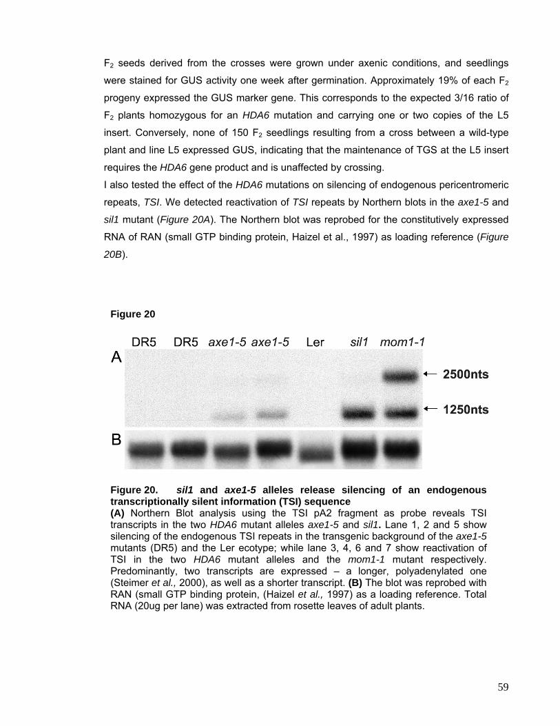
F2 seeds derived from the crosses were grown under axenic conditions, and seedlings
were stained for GUS activity one week after germination. Approximately 19% of each F2
progeny expressed the GUS marker gene. This corresponds to the expected 3/16 ratio of
F2 plants homozygous for an HDA6 mutation and carrying one or two copies of the L5
insert. Conversely, none of 150 F2 seedlings resulting from a cross between a wild-type
plant and line L5 expressed GUS, indicating that the maintenance of TGS at the L5 insert
requires the HDA6 gene product and is unaffected by crossing.
I also tested the effect of the HDA6 mutations on silencing of endogenous pericentromeric
repeats, TSI. We detected reactivation of TSI repeats by Northern blots in the axe1-5 and
sil1 mutant (Figure 20A). The Northern blot was reprobed for the constitutively expressed
RNA of RAN (small GTP binding protein, Haizel et al., 1997) as loading reference (Figure
20B).
Figure 20
Figure 20. sil1 and axe1-5 alleles release silencing of an endogenous transcriptionally silent information (TSI) sequence (A) Northern Blot analysis using the TSI pA2 fragment as probe reveals TSI transcripts in the two HDA6 mutant alleles axe1-5 and sil1. Lane 1, 2 and 5 show silencing of the endogenous TSI repeats in the transgenic background of the axe1-5mutants (DR5) and the Ler ecotype; while lane 3, 4, 6 and 7 show reactivation of TSI in the two HDA6 mutant alleles and the mom1-1 mutant respectively. Predominantly, two transcripts are expressed – a longer, polyadenylated one (Steimer et al., 2000), as well as a shorter transcript. (B) The blot was reprobed with RAN (small GTP binding protein, (Haizel et al., 1997) as a loading reference. Total RNA (20ug per lane) was extracted from rosette leaves of adult plants.
59
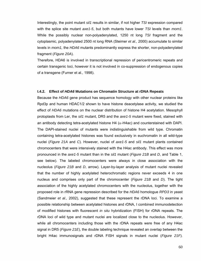
Interestingly, the point mutant sil1 results in similar, if not higher TSI expression compared
with the splice site mutant axe1-5, but both mutants have lower TSI levels than mom1.
While the possibly nuclear non-polyadenylated, 1250 nt long TSI fragment and the
cytoplasmic, polyadenylated 2500 nt long RNA (Steimer et al., 2000) accumulate to similar
levels in mom1, the HDA6 mutants predominantly express the shorter, non-polyadenylated
fragment (Figure 20A).
Therefore, HDA6 is involved in transcriptional repression of pericentromeric repeats and
certain transgenic loci, however it is not involved in co-suppression of endogenous copies
of a transgene (Furner et al., 1998).
I.4.2. Effect of HDA6 Mutations on Chromatin Structure at rDNA Repeats Because the HDA6 gene product has sequence homology with other nuclear proteins like
Rpd3p and human HDAC1/2 shown to have histone deacetylase activity, we studied the
effect of HDA6 mutations on the nuclear distribution of histone H4 acetylation. Mesophyll
protoplasts from Ler, the sil1 mutant, DR5 and the axe1-5 mutant were fixed, stained with
an antibody detecting tetra-acetylated histone H4 (α-H4ac) and counterstained with DAPI.
The DAPI-stained nuclei of mutants were indistinguishable from wild type. Chromatin
containing tetra-acetylated histones was found exclusively in euchromatin in all wild-type
nuclei (Figure 21A and C). However, nuclei of axe1-5 and sil1 mutant plants contained
chromocenters that were intensively stained with the H4ac antibody. This effect was more
pronounced in the axe1-5 mutant than in the sil1 mutant (Figure 21B and D, and Table 1,
see below). The labeled chromocenters were always in close association with the
nucleolus (Figure 21B and D, arrow). Layer-by-layer analysis of mutant nuclei revealed
that the number of highly acetylated heterochromatic regions never exceeds 4 in one
nucleus and comprises only part of the chromocenter (Figure 21B and D). The tight
association of the highly acetylated chromocenters with the nucleolus, together with the
proposed role in rRNA gene repression described for the HDA6 homologue RPD3 in yeast
(Sandmeier et al., 2002), suggested that these represent the rDNA loci. To examine a
possible relationship between acetylated histones and rDNA, I combined immunodetection
of modified histones with fluorescent in situ hybridization (FISH) for rDNA repeats. The
rDNA loci of wild type and mutant nuclei are localized close to the nucleolus. However,
while all chromocenters including those with the rDNA repeats were free of any H4ac
signal in DR5 (Figure 21E), the double labeling technique revealed an overlap between the
bright H4ac immunosignals and rDNA FISH signals in mutant nuclei (Figure 21F).
60
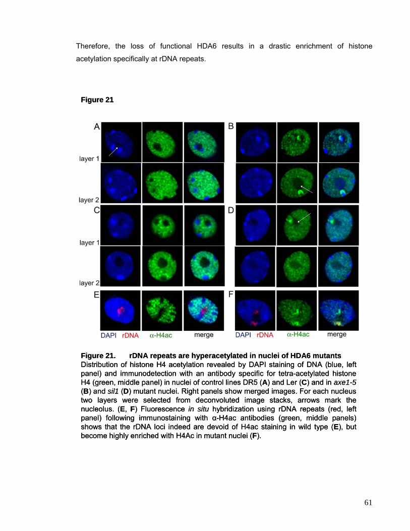
Therefore, the loss of functional HDA6 results in a drastic enrichment of histone
acetylation specifically at rDNA repeats.
Figure 21
Figure 21. rDNA repeats are hyperacetylated in nuclei of HDA6 mutantsDistribution of histone H4 acetylation revealed by DAPI staining of DNA (blue, left panel) and immunodetection with an antibody specific for tetra-acetylated histone H4 (green, middle panel) in nuclei of control lines DR5 (A) and Ler (C) and in axe1-5(B) and sil1 (D) mutant nuclei. Right panels show merged images. For each nucleus two layers were selected from deconvoluted image stacks, arrows mark the nucleolus. (E, F) Fluorescence in situ hybridization using rDNA repeats (red, left panel) following immunostaining with α-H4ac antibodies (green, middle panels) shows that the rDNA loci indeed are devoid of H4ac staining in wild type (E), but become highly enriched with H4Ac in mutant nuclei (F).
61
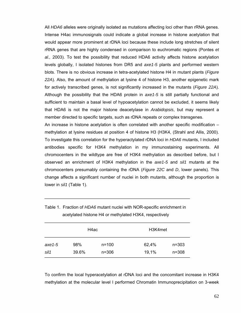
All HDA6 alleles were originally isolated as mutations affecting loci other than rRNA genes.
Intense H4ac immunosignals could indicate a global increase in histone acetylation that
would appear more prominent at rDNA loci because these include long stretches of silent
rRNA genes that are highly condensed in comparison to euchromatic regions (Pontes et
al., 2003). To test the possibility that reduced HDA6 activity affects histone acetylation
levels globally, I isolated histones from DR5 and axe1-5 plants and performed western
blots. There is no obvious increase in tetra-acetylated histone H4 in mutant plants (Figure
22A). Also, the amount of methylation at lysine 4 of histone H3, another epigenetic mark
for actively transcribed genes, is not significantly increased in the mutants (Figure 22A).
Although the possibility that the HDA6 protein in axe1-5 is still partially functional and
sufficient to maintain a basal level of hypoacetylation cannot be excluded, it seems likely
that HDA6 is not the major histone deacetylase in Arabidopsis, but may represent a
member directed to specific targets, such as rDNA repeats or complex transgenes.
An increase in histone acetylation is often correlated with another specific modification –
methylation at lysine residues at position 4 of histone H3 (H3K4, (Strahl and Allis, 2000).
To investigate this correlation for the hyperactylated rDNA loci in HDA6 mutants, I included
antibodies specific for H3K4 methylation in my immunostaining experiments. All
chromocenters in the wildtype are free of H3K4 methylation as described before, but I
observed an enrichment of H3K4 methylation in the axe1-5 and sil1 mutants at the
chromocenters presumably containing the rDNA (Figure 22C and D, lower panels). This
change affects a significant number of nuclei in both mutants, although the proportion is
lower in sil1 (Table 1).
Table 1. Fraction of HDA6 mutant nuclei with NOR-specific enrichment in
acetylated histone H4 or methylated H3K4, respectively
H4ac H3K4met
axe1-5 98% n=100 62,4% n=303
sil1 39.6% n=306 19,1% n=308
To confirm the local hyperacetylation at rDNA loci and the concomitant increase in H3K4
methylation at the molecular level I performed Chromatin Immunoprecipitation on 3-week
62
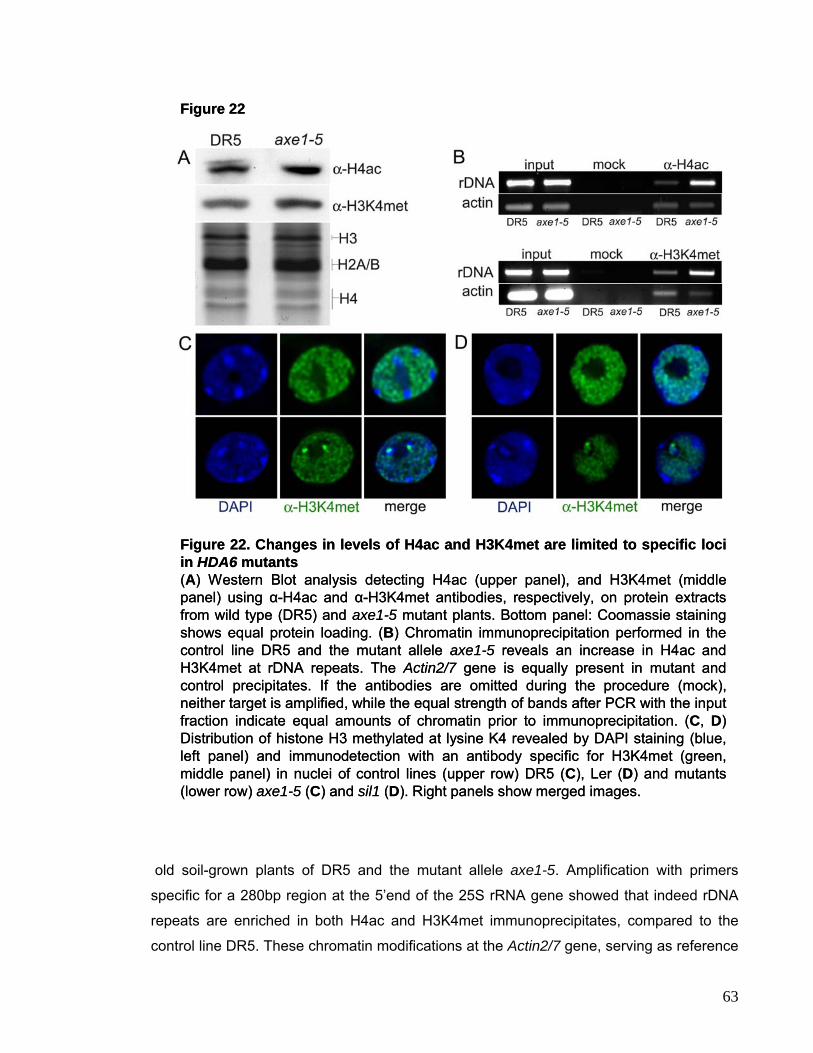
Figure 22
Figure 22. Changes in levels of H4ac and H3K4met are limited to specific loci in HDA6 mutants(A) Western Blot analysis detecting H4ac (upper panel), and H3K4met (middle panel) using α-H4ac and α-H3K4met antibodies, respectively, on protein extracts from wild type (DR5) and axe1-5 mutant plants. Bottom panel: Coomassie staining shows equal protein loading. (B) Chromatin immunoprecipitation performed in the control line DR5 and the mutant allele axe1-5 reveals an increase in H4ac and H3K4met at rDNA repeats. The Actin2/7 gene is equally present in mutant and control precipitates. If the antibodies are omitted during the procedure (mock), neither target is amplified, while the equal strength of bands after PCR with the input fraction indicate equal amounts of chromatin prior to immunoprecipitation. (C, D) Distribution of histone H3 methylated at lysine K4 revealed by DAPI staining (blue, left panel) and immunodetection with an antibody specific for H3K4met (green, middle panel) in nuclei of control lines (upper row) DR5 (C), Ler (D) and mutants (lower row) axe1-5 (C) and sil1 (D). Right panels show merged images.
mmmmmmmmmmmmmmmmmmmmmmmmmmmmmmmmmmmmmmmmmmmmmmmm
m
old soil-grown plants of DR5 and the mutant allele axe1-5. Amplification with primers
specific for a 280bp region at the 5’end of the 25S rRNA gene showed that indeed rDNA
repeats are enriched in both H4ac and H3K4met immunoprecipitates, compared to the
control line DR5. These chromatin modifications at the Actin2/7 gene, serving as reference
63
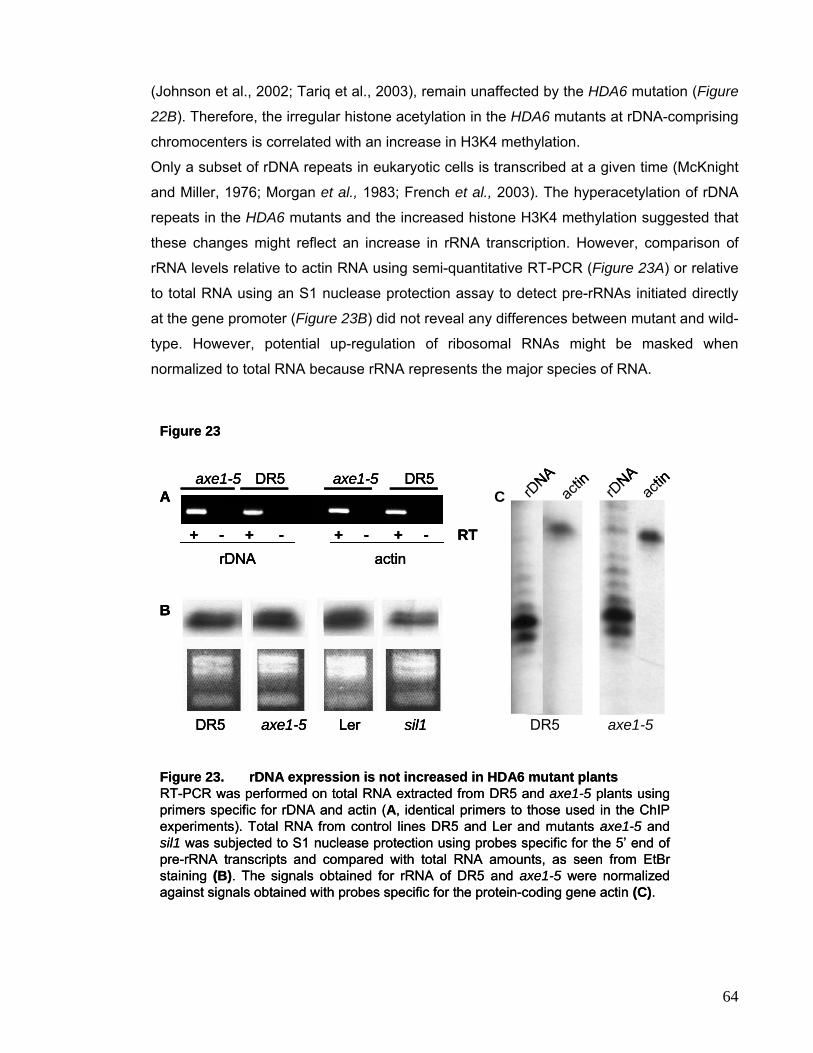
(Johnson et al., 2002; Tariq et al., 2003), remain unaffected by the HDA6 mutation (Figure
22B). Therefore, the irregular histone acetylation in the HDA6 mutants at rDNA-comprising
chromocenters is correlated with an increase in H3K4 methylation.
Only a subset of rDNA repeats in eukaryotic cells is transcribed at a given time (McKnight
and Miller, 1976; Morgan et al., 1983; French et al., 2003). The hyperacetylation of rDNA
repeats in the HDA6 mutants and the increased histone H3K4 methylation suggested that
these changes might reflect an increase in rRNA transcription. However, comparison of
rRNA levels relative to actin RNA using semi-quantitative RT-PCR (Figure 23A) or relative
to total RNA using an S1 nuclease protection assay to detect pre-rRNAs initiated directly
at the gene promoter (Figure 23B) did not reveal any differences between mutant and wild-
type. However, potential up-regulation of ribosomal RNAs might be masked when
normalized to total RNA because rRNA represents the major species of RNA.
Figure 23
Figure 23. rDNA expression is not increased in HDA6 mutant plantsRT-PCR was performed on total RNA extracted from DR5 and axe1-5 plants using primers specific for rDNA and actin (A, identical primers to those used in the ChIP experiments). Total RNA from control lines DR5 and Ler and mutants axe1-5 and sil1 was subjected to S1 nuclease protection using probes specific for the 5’ end of pre-rRNA transcripts and compared with total RNA amounts, as seen from EtBrstaining (B). The signals obtained for rRNA of DR5 and axe1-5 were normalized against signals obtained with probes specific for the protein-coding gene actin (C).
axe1-5DR5
rDNA
actin rD
NA
+ - + - + - + - RTrDNA actin
axe1-5 axe1-5DR5 DR5
sil1Leraxe1-5DR5
A C
B
actin
axe1-5DR5
rDNA
actin rD
NA
+ - + - + - + - RTrDNA actin
axe1-5 axe1-5DR5 DR5
sil1Leraxe1-5DR5
A C
B
actin
64
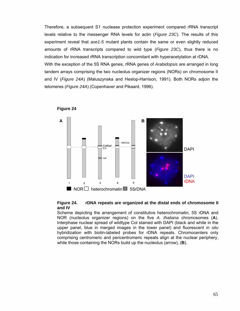
Therefore, a subsequent S1 nuclease protection experiment compared rRNA transcript
levels relative to the messenger RNA levels for actin (Figure 23C). The results of this
experiment reveal that axe1-5 mutant plants contain the same or even slightly reduced
amounts of rRNA transcripts compared to wild type (Figure 23C), thus there is no
indication for increased rRNA transcription concomitant with hyperacetylation at rDNA.
With the exception of the 5S RNA genes, rRNA genes of Arabidopsis are arranged in long
tandem arrays comprising the two nucleolus organizer regions (NORs) on chromosome II
and IV (Figure 24A) (Maluszynska and Heslop-Harrison, 1991). Both NORs adjoin the
telomeres (Figure 24A) (Copenhaver and Pikaard, 1996).
Figure 24
A
NOR 5SrDNA heterochromatin
B
DAPI
DAPI rDNA
Figure 24. rDNA repeats are organized at the distal ends of chromosome II and IV Scheme depicting the arrangement of constitutive heterochromatin, 5S rDNA and NOR (nucleolus organizer regions) on the five A. thaliana chromosomes (A). Interphase nuclear spread of wildtype Col stained with DAPI (black and white in the upper panel, blue in merged images in the lower panel) and fluorescent in situhybridization with biotin-labeled probes for rDNA repeats. Chromocenters only comprising centromeric and pericentromeric repeats align at the nuclear periphery, while those containing the NORs build up the nucleolus (arrow), (B).
65
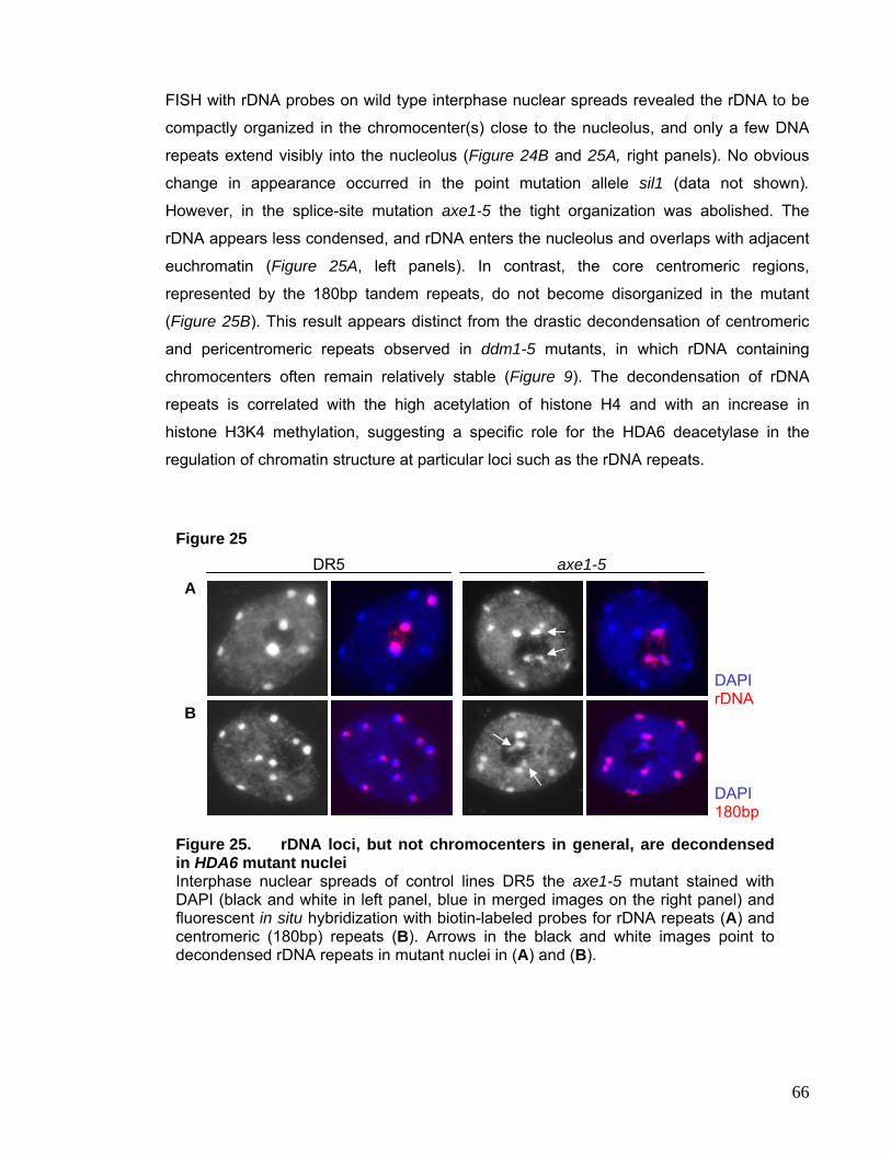
FISH with rDNA probes on wild type interphase nuclear spreads revealed the rDNA to be
compactly organized in the chromocenter(s) close to the nucleolus, and only a few DNA
repeats extend visibly into the nucleolus (Figure 24B and 25A, right panels). No obvious
change in appearance occurred in the point mutation allele sil1 (data not shown).
However, in the splice-site mutation axe1-5 the tight organization was abolished. The
rDNA appears less condensed, and rDNA enters the nucleolus and overlaps with adjacent
euchromatin (Figure 25A, left panels). In contrast, the core centromeric regions,
represented by the 180bp tandem repeats, do not become disorganized in the mutant
(Figure 25B). This result appears distinct from the drastic decondensation of centromeric
and pericentromeric repeats observed in ddm1-5 mutants, in which rDNA containing
chromocenters often remain relatively stable (Figure 9). The decondensation of rDNA
repeats is correlated with the high acetylation of histone H4 and with an increase in
histone H3K4 methylation, suggesting a specific role for the HDA6 deacetylase in the
regulation of chromatin structure at particular loci such as the rDNA repeats.
Figure 25
DR5 axe1-5 A
DAPI rDNA
B
DAPI 180bp
Figure 25. rDNA loci, but not chromocenters in general, are decondensed in HDA6 mutant nuclei Interphase nuclear spreads of control lines DR5 the axe1-5 mutant stained with DAPI (black and white in left panel, blue in merged images on the right panel) and fluorescent in situ hybridization with biotin-labeled probes for rDNA repeats (A) and centromeric (180bp) repeats (B). Arrows in the black and white images point to decondensed rDNA repeats in mutant nuclei in (A) and (B).
66
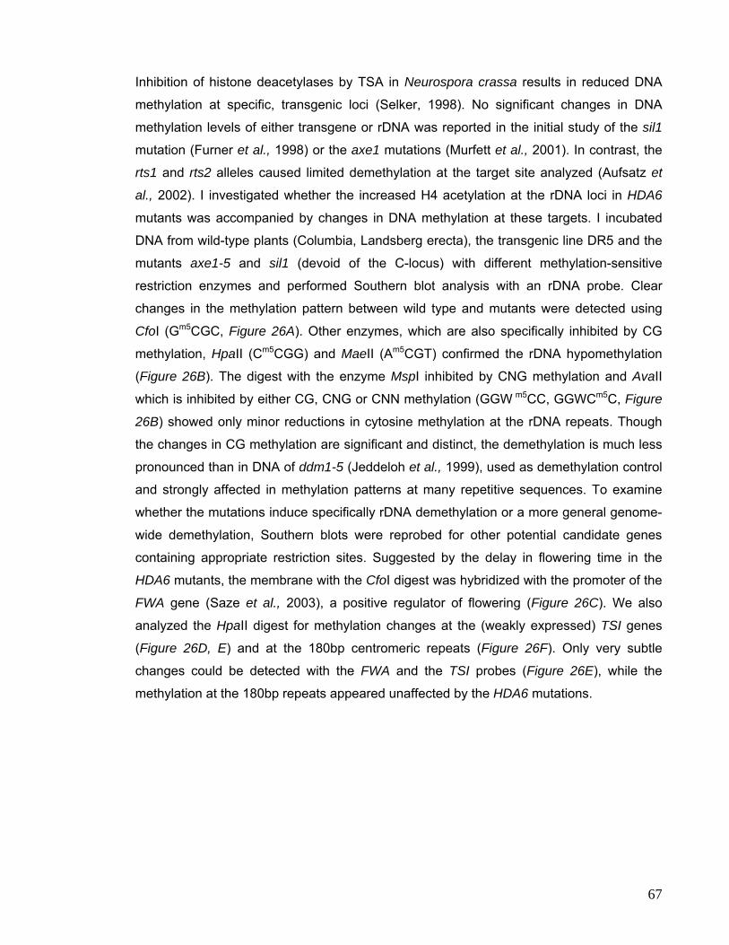
Inhibition of histone deacetylases by TSA in Neurospora crassa results in reduced DNA
methylation at specific, transgenic loci (Selker, 1998). No significant changes in DNA
methylation levels of either transgene or rDNA was reported in the initial study of the sil1
mutation (Furner et al., 1998) or the axe1 mutations (Murfett et al., 2001). In contrast, the
rts1 and rts2 alleles caused limited demethylation at the target site analyzed (Aufsatz et
al., 2002). I investigated whether the increased H4 acetylation at the rDNA loci in HDA6
mutants was accompanied by changes in DNA methylation at these targets. I incubated
DNA from wild-type plants (Columbia, Landsberg erecta), the transgenic line DR5 and the
mutants axe1-5 and sil1 (devoid of the C-locus) with different methylation-sensitive
restriction enzymes and performed Southern blot analysis with an rDNA probe. Clear
changes in the methylation pattern between wild type and mutants were detected using
CfoI (Gm5CGC, Figure 26A). Other enzymes, which are also specifically inhibited by CG
methylation, HpaII (Cm5CGG) and MaeII (Am5CGT) confirmed the rDNA hypomethylation
(Figure 26B). The digest with the enzyme MspI inhibited by CNG methylation and AvaII
which is inhibited by either CG, CNG or CNN methylation (GGW m5CC, GGWCm5C, Figure
26B) showed only minor reductions in cytosine methylation at the rDNA repeats. Though
the changes in CG methylation are significant and distinct, the demethylation is much less
pronounced than in DNA of ddm1-5 (Jeddeloh et al., 1999), used as demethylation control
and strongly affected in methylation patterns at many repetitive sequences. To examine
whether the mutations induce specifically rDNA demethylation or a more general genome-
wide demethylation, Southern blots were reprobed for other potential candidate genes
containing appropriate restriction sites. Suggested by the delay in flowering time in the
HDA6 mutants, the membrane with the CfoI digest was hybridized with the promoter of the
FWA gene (Saze et al., 2003), a positive regulator of flowering (Figure 26C). We also
analyzed the HpaII digest for methylation changes at the (weakly expressed) TSI genes
(Figure 26D, E) and at the 180bp centromeric repeats (Figure 26F). Only very subtle
changes could be detected with the FWA and the TSI probes (Figure 26E), while the
methylation at the 180bp repeats appeared unaffected by the HDA6 mutations.
67
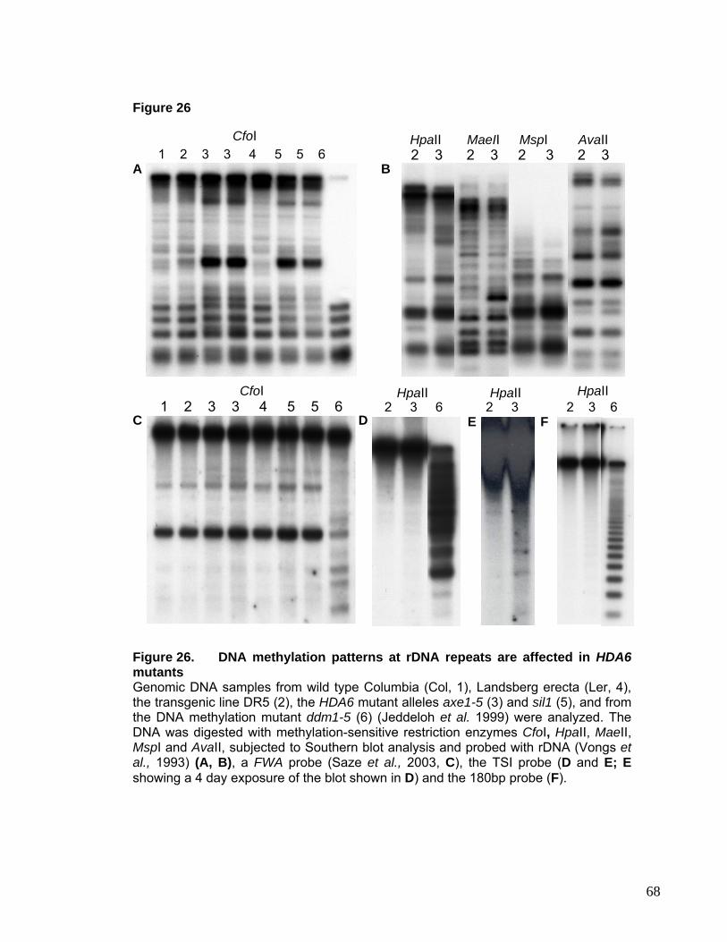
Figure 26
HpaII CfoI 1 2 3 3 4 5 5 6
A B
CfoI 1 2 3 3 4 5 5 6 2 3 6 2 3 6
HpaII HpaII
C D F E 2 3
HpaII
MaeII MspI AvaII 2 3 2 3 2 3 2 3
Figure 26. DNA methylation patterns at rDNA repeats are affected in HDA6mutants Genomic DNA samples from wild type Columbia (Col, 1), Landsberg erecta (Ler, 4), the transgenic line DR5 (2), the HDA6 mutant alleles axe1-5 (3) and sil1 (5), and from the DNA methylation mutant ddm1-5 (6) (Jeddeloh et al. 1999) were analyzed. The DNA was digested with methylation-sensitive restriction enzymes CfoI, HpaII, MaeII, MspI and AvaII, subjected to Southern blot analysis and probed with rDNA (Vongs et al., 1993) (A, B), a FWA probe (Saze et al., 2003, C), the TSI probe (D and E; E showing a 4 day exposure of the blot shown in D) and the 180bp probe (F).
68

I.5 Chromatin Factors Involved in DNA Replication Histone deposition after DNA replication is mediated by specialized histone chaperones.
For example, the chromatin assembly factor-1 (CAF-1) preferentially associates with
histones H3.H4, localizes to replication foci and is thought to assemble nucleosomes onto
replicating DNA (Stillman, 1986). The association of CAF-I with the replication fork is
mediated by its interaction with the proliferating cell nuclear antigen (PCNA) (Shibahara
and Stillman, 1999). CAF-1 is conserved in eukaryotic organisms ranging from yeast to
man. In Arabidopsis thaliana CAF-1 consists of three subunits, named p150, p60 and p48.
Null mutations in both the p150 (fas1-1) and the p60 subunit (fas2-2) have been described
(Kaya et al., 2001). fas mutants show distorted phyllotaxy and fasciation (thick and
flattened stems and fused organs) and are sensitive to DNA damaging agents, revealing
the implication in DNA repair mechanisms (Takeda et al., 2004). Recently, in a screen for
mutants hypersensitive to Methyl Methane Sulfonate (MMS), a reagent mimicking DNA
double strand breaks (DSBs), a novel mutant bru1 (brushy) was identified (Takeda et al.,
2004), which also shows a fasciation phenotype. Three alleles were described; bru1-1
carrying a deletion, leading to miss splicing and truncation of the C-terminus of the protein;
bru1-2, in which a single amino acid in the N-terminal region is exchanged and bru1-3 with
a T-DNA insertion in the middle of the gene. The BRU1 gene encodes a novel protein of
1311aa characterized by domains involved in protein-protein interaction and possibly DNA
binding.
Even though bru1 mutants are extremely sensitive to DNA damaging treatments, they are
proficient not only in intra-chromosomal homologous recombination (HR), but also in non-
homologous end joining (NHEJ). The sensitivity to MMS reflects probably a constitutively
increased level of DSBs, possibly resulting from replication defects (Takeda et al., 2004).
Both fas and bru1 mutants release transcriptional gene silencing in a stochastic fashion as
was shown for two independent silenced transgenes (Takeda et al., 2004), but do not
affect global DNA methylation. It was proposed that the CAF-1 complex ensures stable
propagation of epigenetic states and maintenance of genome integrity by facilitating rapid
reformation of chromatin structure after passage of the replication fork (Kaya et al., 2001).
Even though BRU1 does not directly interact with the CAF-1 subunits (Takeda et al., 2004)
it is likely to be involved in chromatin assembly and to contribute – together with the CAF-1
complex – to post-replicative stability of epigenetic states. In order to maintain the
epigenetic information throughout cell division the histone code has to be transmitted to
the daughter cells. Therefore, I investigated the impact of fas and bru1 mutations on
69
mmmmmmmmmmmmmmmmmmmmmmmmmmmmmmmmmmmmmmmmmmmmmmmm
m Figure 27 fas2-2 bru1-2
H3K9met
DAPI
H3K4met
DAPI
H4ac DAPI
A
B
C
Figure 27. Histone modifications are not affected in fas and bru1 mutants Distribution of histone H4 acetylation (A), H3K9 (B) and H3K4 methylation (C) revealed by DAPI staining of DNA (blue, left panel) and immunodetection with an antibody specific for tetra-acetylated histone H4, H3K9dimet, and H3K4dimet (green, middle panel) in nuclei of the fas2-2 and bru1-2 mutants. Right panels show merged images.
patterns of histone modifications. I performed immunostaining with antibodies against
tetra-acetylated H4, as well as against H3 methylated at K4 or K9. In brief, both mutants
did not show any alteration in the pattern of these modifications (Figure 27A-C). Tetra-
acetylated histone H4 and methylated histone H3 at K4 remained enriched in euchromatin.
The chromocenters, clearly visible in the DAPI staining, were free of these two marks
however enriched in methylated H3K9. One has to bear in mind, however, that the bru1-2
allele used in this experiment still gives rise to a partially functional protein. In a
subsequent approach I studied heterochromatin stability by FISH analysis using the 180bp
repeats as probe. Several plants were selected for analysis showing a wide phenotype
variation ranging from almost none to strong alterations. We performed a double-blind
study, since the effect on heterochromatin stability was expected to be only marginal.
Although the organization of centromeric heterochromatin in the majority of bru1 nuclei
was identical to wild type (Figure 28A, C and E), some nuclei exhibited decondensation of
centromeric heterochromatin (Figure 28B, D). Altered heterochromatin organization was
observed especially in plants showing strong fasciation and distorted leaf shapes. In fas2-
70
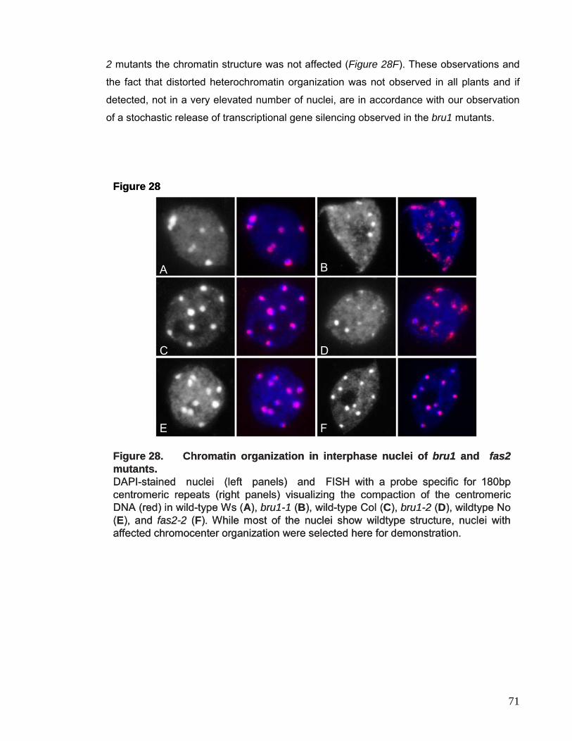
2 mutants the chromatin structure was not affected (Figure 28F). These observations and
the fact that distorted heterochromatin organization was not observed in all plants and if
detected, not in a very elevated number of nuclei, are in accordance with our observation
of a stochastic release of transcriptional gene silencing observed in the bru1 mutants.
A B
E
C
F
D
Figure 28
Figure 28. Chromatin organization in interphase nuclei of bru1 and fas2 mutants.DAPI-stained nuclei (left panels) and FISH with a probe specific for 180bp centromeric repeats (right panels) visualizing the compaction of the centromeric DNA (red) in wild-type Ws (A), bru1-1 (B), wild-type Col (C), bru1-2 (D), wildtype No (E), and fas2-2 (F). While most of the nuclei show wildtype structure, nuclei with affected chromocenter organization were selected here for demonstration.
A B
E
C
F
D
A B
E
C
F
D
Figure 28
Figure 28. Chromatin organization in interphase nuclei of bru1 and fas2 mutants.DAPI-stained nuclei (left panels) and FISH with a probe specific for 180bp centromeric repeats (right panels) visualizing the compaction of the centromeric DNA (red) in wild-type Ws (A), bru1-1 (B), wild-type Col (C), bru1-2 (D), wildtype No (E), and fas2-2 (F). While most of the nuclei show wildtype structure, nuclei with affected chromocenter organization were selected here for demonstration.
71
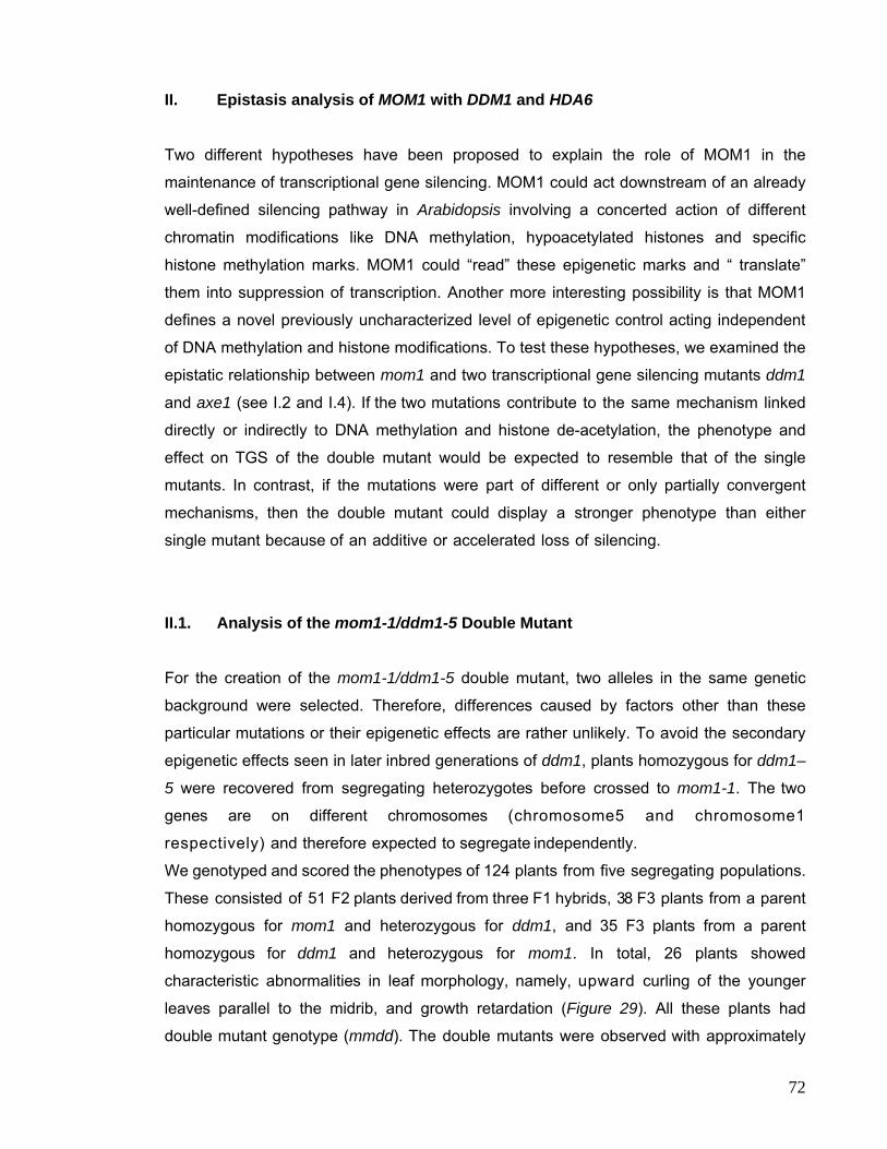
II. Epistasis analysis of MOM1 with DDM1 and HDA6
Two different hypotheses have been proposed to explain the role of MOM1 in the
maintenance of transcriptional gene silencing. MOM1 could act downstream of an already
well-defined silencing pathway in Arabidopsis involving a concerted action of different
chromatin modifications like DNA methylation, hypoacetylated histones and specific
histone methylation marks. MOM1 could “read” these epigenetic marks and “ translate”
them into suppression of transcription. Another more interesting possibility is that MOM1
defines a novel previously uncharacterized level of epigenetic control acting independent
of DNA methylation and histone modifications. To test these hypotheses, we examined the
epistatic relationship between mom1 and two transcriptional gene silencing mutants ddm1
and axe1 (see I.2 and I.4). If the two mutations contribute to the same mechanism linked
directly or indirectly to DNA methylation and histone de-acetylation, the phenotype and
effect on TGS of the double mutant would be expected to resemble that of the single
mutants. In contrast, if the mutations were part of different or only partially convergent
mechanisms, then the double mutant could display a stronger phenotype than either
single mutant because of an additive or accelerated loss of silencing.
II.1. Analysis of the mom1-1/ddm1-5 Double Mutant For the creation of the mom1-1/ddm1-5 double mutant, two alleles in the same genetic
background were selected. Therefore, differences caused by factors other than these
particular mutations or their epigenetic effects are rather unlikely. To avoid the secondary
epigenetic effects seen in later inbred generations of ddm1, plants homozygous for ddm1–
5 were recovered from segregating heterozygotes before crossed to mom1-1. The two
genes are on different chromosomes (chromosome5 and chromosome1
respectively) and therefore expected to segregate independently.
We genotyped and scored the phenotypes of 124 plants from five segregating populations.
These consisted of 51 F2 plants derived from three F1 hybrids, 38 F3 plants from a parent
homozygous for mom1 and heterozygous for ddm1, and 35 F3 plants from a parent
homozygous for ddm1 and heterozygous for mom1. In total, 26 plants showed
characteristic abnormalities in leaf morphology, namely, upward curling of the younger
leaves parallel to the midrib, and growth retardation (Figure 29). All these plants had
double mutant genotype (mmdd). The double mutants were observed with approximately
72
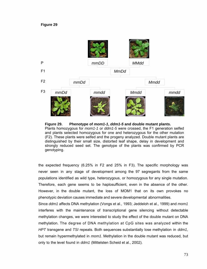
mmmmmmmmmmmmmmmmmmmmmmmmmmmmmmmmmmmmmmmmmmm
mmm Figure 29
mmDd mmdd mmddMmdd
Mmdd mmDd
MmDd
MMddmmDD
P
F1
F2
F3
Figure 29. Phenotype of mom1-1, ddm1-5 and double mutant plants. Plants homozygous for mom1-1 or ddm1-5 were crossed, the F1 generation selfed and plants selected homozygous for one and heterozygous for the other mutation (F2). These plants were selfed and the progeny analyzed. Double mutant plants are distinguished by their small size, distorted leaf shape, delay in development and strongly reduced seed set. The genotype of the plants was confirmed by PCR genotyping.
the expected frequency (6.25% in F2 and 25% in F3). The specific morphology was
never seen in any stage of development among the 97 segregants from the same
populations identified as wild type, heterozygous, or homozygous for any single mutation.
Therefore, each gene seems to be haplosufficient, even in the absence of the other.
However, in the double mutant, the loss of MOM1 that on its own provokes no
phenotypic deviation causes immediate and severe developmental abnormalities.
Since ddm1 affects DNA methylation (Vongs et al., 1993; Jeddeloh et al., 1999) and mom1
interferes with the maintenance of transcriptional gene silencing without detectable
methylation changes, we were interested to study the effect of the double mutant on DNA
methylation. The degree of DNA methylation at CpG sites was analyzed within the
HPT transgene and TSI repeats. Both sequences substantially lose methylation in ddm1,
but remain hypermethylated in mom1. Methylation in the double mutant was reduced, but
only to the level found in ddm1 (Mittelsten Scheid et al., 2002).
73
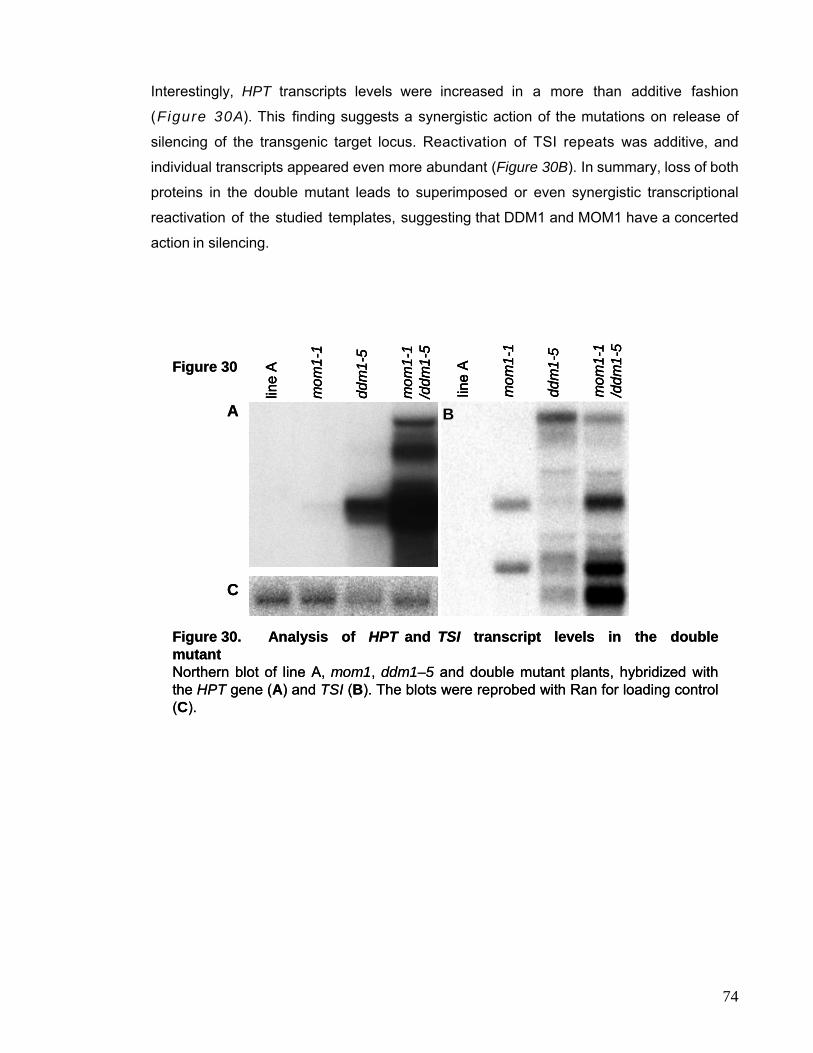
Interestingly, HPT transcripts levels were increased in a more than additive fashion
(Figure 30A). This finding suggests a synergistic action of the mutations on release of
silencing of the transgenic target locus. Reactivation of TSI repeats was additive, and
individual transcripts appeared even more abundant (Figure 30B). In summary, loss of both
proteins in the double mutant leads to superimposed or even synergistic transcriptional
reactivation of the studied templates, suggesting that DDM1 and MOM1 have a concerted
action in silencing.
Figure 30
- 1000nt
Figure 30. Analysis of HPT and TSI transcript levels in the double mutantNorthern blot of line A, mom1, ddm1–5 and double mutant plants, hybridized with the HPT gene (A) and TSI (B). The blots were reprobed with Ran for loading control (C).
A
C
B
line
A
mom
1-1
mom
1-1
/ddm
1-5
ddm
1-5
line
A
mom
1-1
mom
1-1
/ddm
1-5
ddm
1-5
line
A
mom
1-1
mom
1-1
/ddm
1-5
ddm
1-5
line
A
mom
1-1
mom
1-1
/ddm
1-5
ddm
1-5
74
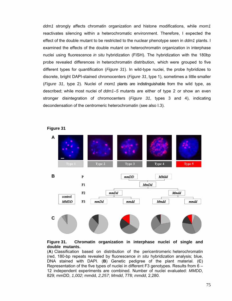
75
dm1 strongly affects chromatin organization and histone modifications, while mom1
Figure 31
Figure 31. Chromatin organization in interphase nuclei of single and
d
reactivates silencing within a heterochromatic environment. Therefore, I expected the
effect of the double mutant to be restricted to the nuclear phenotype seen in ddm1 plants. I
examined the effects of the double mutant on heterochromatin organization in interphase
nuclei using fluorescence in situ hybridization (FISH). The hybridization with the 180bp
probe revealed differences in heterochromatin distribution, which were grouped to five
different types for quantification (Figure 31). In wild-type nuclei, the probe hybridizes to
discrete, bright DAPI-stained chromocenters (Figure 31, type 1), sometimes a little smaller
(Figure 31, type 2). Nuclei of mom1 plants are indistinguishable from the wild type, as
described; while most nuclei of ddm1–5 mutants are either of type 2 or show an even
stronger disintegration of chromocenters (Figure 31, types 3 and 4), indicating
decondensation of the centromeric heterochromatin (see also I.3).
A
B
C
double mutants. (A) Classification based on distribution of the pericentromeric heterochromatin (red, 180-bp repeats revealed by fluorescence in situ hybridization analysis; blue, DNA stained with DAPI. (B) Genetic pedigree of the plant material. (C) Representation of the five types of nuclei in different F3 genotypes. Results from 6 –12 independent experiments are combined. Number of nuclei evaluated: MMDD, 829; mmDD, 1,002; mmdd, 2,257; Mmdd, 778; mmdd, 2,280.
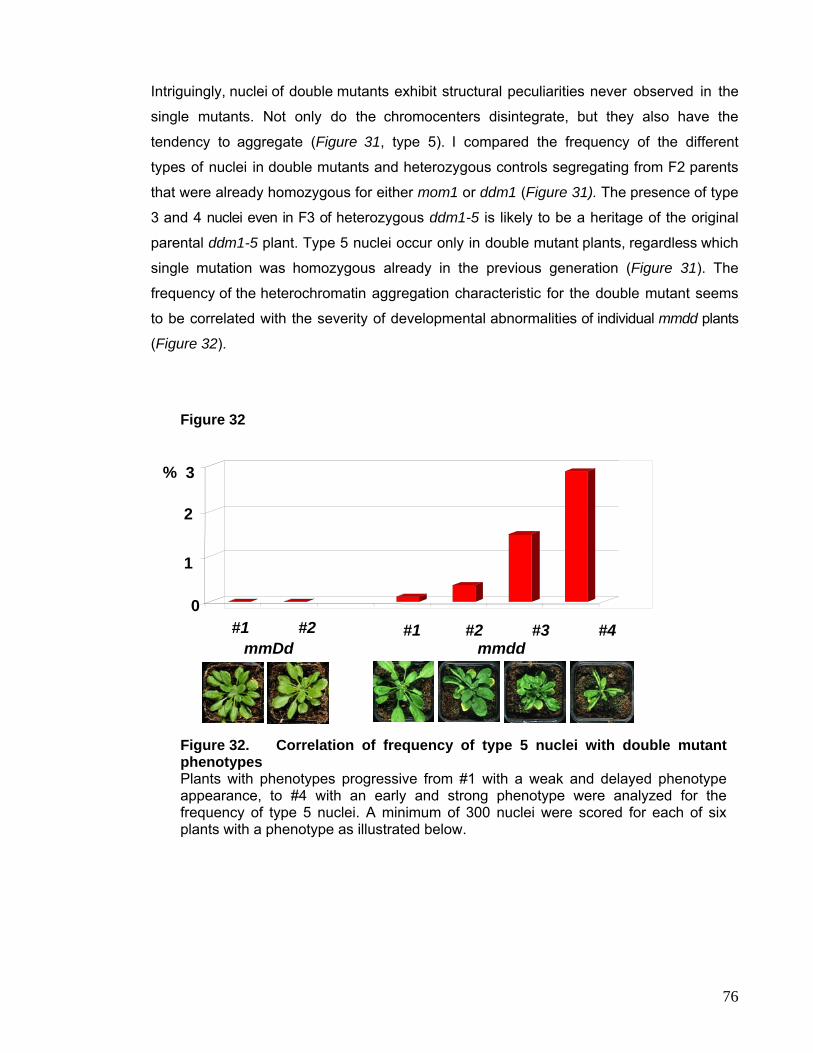
Intriguingly, nuclei of double mutants exhibit structural peculiarities never observed in the
single mutants. Not only do the chromocenters disintegrate, but they also have the
tendency to aggregate (Figure 31, type 5). I compared the frequency of the different
types of nuclei in double mutants and heterozygous controls segregating from F2 parents
that were already homozygous for either mom1 or ddm1 (Figure 31). The presence of type
3 and 4 nuclei even in F3 of heterozygous ddm1-5 is likely to be a heritage of the original
parental ddm1-5 plant. Type 5 nuclei occur only in double mutant plants, regardless which
single mutation was homozygous already in the previous generation (Figure 31). The
frequency of the heterochromatin aggregation characteristic for the double mutant seems
to be correlated with the severity of developmental abnormalities of individual mmdd plants
(Figure 32).
Figure 32
0 mmDd #2 #1
mmdd#1 #2 #3 #4
1
2
% 3
Figure 32. Correlation of frequency of type 5 nuclei with double mutant phenotypes Plants with phenotypes progressive from #1 with a weak and delayed phenotype appearance, to #4 with an early and strong phenotype were analyzed for the frequency of type 5 nuclei. A minimum of 300 nuclei were scored for each of six plants with a phenotype as illustrated below.
76
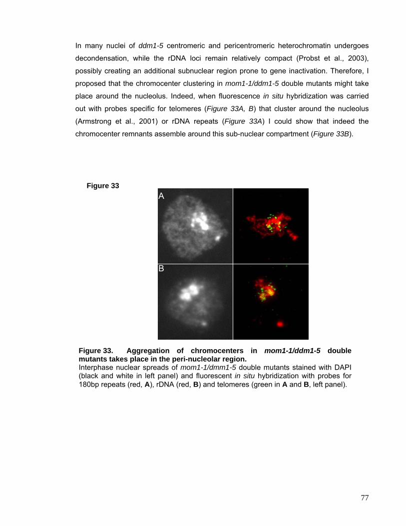
In many nuclei of ddm1-5 centromeric and pericentromeric heterochromatin undergoes
decondensation, while the rDNA loci remain relatively compact (Probst et al., 2003),
possibly creating an additional subnuclear region prone to gene inactivation. Therefore, I
proposed that the chromocenter clustering in mom1-1/ddm1-5 double mutants might take
place around the nucleolus. Indeed, when fluorescence in situ hybridization was carried
out with probes specific for telomeres (Figure 33A, B) that cluster around the nucleolus
(Armstrong et al., 2001) or rDNA repeats (Figure 33A) I could show that indeed the
chromocenter remnants assemble around this sub-nuclear compartment (Figure 33B).
Figure 33 A
B
Figure 33. Aggregation of chromocenters in mom1-1/ddm1-5 double mutants takes place in the peri-nucleolar region. Interphase nuclear spreads of mom1-1/dmm1-5 double mutants stained with DAPI (black and white in left panel) and fluorescent in situ hybridization with probes for 180bp repeats (red, A), rDNA (red, B) and telomeres (green in A and B, left panel).
77
II.2. Analysis of the mom1-1/axe1-1 Double Mutant The phenotype of the mom1-1/ddm1-5 double mutant suggests that MOM1 defines an
independent level of epigenetic control not linked to DNA methylation. Mutations in
the HDA6 gene reactivate several complex transgenic loci e.g. the C-locus (Furner et al.,
1998) and the GUS locus in line L5 (Morel et al., 2000) and interfere with silencing of TSI
repeats. This effect is probably due to increased histone H4 acetylation at the target loci
and subsequently induced changes in H3 histone modifications, DNA methylation and
chromatin structure (Aufsatz et al., 2002; Probst et al., 2004). Therefore, the analysis of
the epistatic relationship between mom1 and axe1 mutations should confirm the existence
of an independent pathway of epigenetic control defined by MOM1.
We chose to cross mom1-1 to axe1-1 (Murfett et al., 2001), since the point-mutation in this
HDA6 mutant allele destroys a MspI site therefore allowing genotyping. The analysis of
progeny of two selfed Aamm and one selfed aaMm plant suggested an under-
representation of double mutant plants (3.7% observed instead of 25% expected).
However the limited number of plants genotyped (27 plants) does not allow a final
statement. Two double mutant lines (line 33 and line 36) were recovered from the F2
population (provided by Jane Murfett and Ortrun Mittelsten Scheid). I compared plants
from the two double mutant lines to segregating siblings of the same population
homozygous for one of the two mutations or wildtype for both genes (aaMM, AAmm and
AAMM). While the single mutants did not show any phenotypic alterations, a variation of
phenotypes was observed in the double mutant lines (Figure 34). The abnormalities range
from variation in flowering time to reduced plant size and rolling of leaves (Figure 34,
arrow). This observation suggested that MOM1 might operate at a level of epigenetic
regulation not only independent of DDM1, but also of HDA6 function. To confirm this I
extracted RNA from different plants of lines 33 and 36, the segregating single mutants and
wildtype controls and performed Northern blot analysis with a TSI probe. In this assay no
TSI expression was detected in the weak axe1-1 mutant allele, in contrast to the axe1-5
splice site mutation (I.4). Interestingly, TSI was reactivated in a more than additive fashion
in each of the independent double mutant plants analyzed (Figure 35), however levels are
still lower than observed in ddm1-5 plants. Interestingly, both mutations have either no
(Amedeo et al., 2000) or very subtle effects on the DNA methylation patterns at TSI loci as
shown for the strong HDA6 mutant allele, axe1-5 (Figure 26F).
78
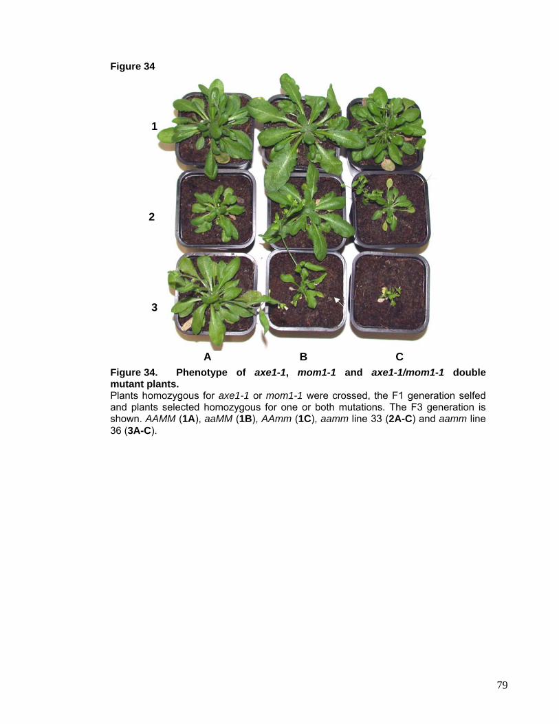
Figure 34
3
2
1
A B C
Figure 34. Phenotype of axe1-1, mom1-1 and axe1-1/mom1-1 double mutant plants. Plants homozygous for axe1-1 or mom1-1 were crossed, the F1 generation selfed and plants selected homozygous for one or both mutations. The F3 generation is shown. AAMM (1A), aaMM (1B), AAmm (1C), aamm line 33 (2A-C) and aamm line 36 (3A-C).
79
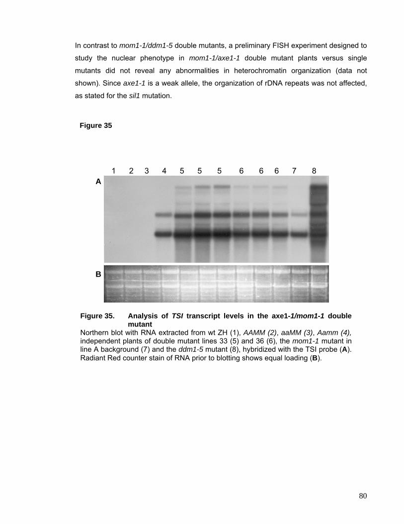
In contrast to mom1-1/ddm1-5 double mutants, a preliminary FISH experiment designed to
study the nuclear phenotype in mom1-1/axe1-1 double mutant plants versus single
mutants did not reveal any abnormalities in heterochromatin organization (data not
shown). Since axe1-1 is a weak allele, the organization of rDNA repeats was not affected,
as stated for the sil1 mutation.
Figure 35
1 2 3 4 5 5 5 6 6 6 7 8 A
B
Figure 35. Analysis of TSI transcript levels in the axe1-1/mom1-1 double
mutant Northern blot with RNA extracted from wt ZH (1), AAMM (2), aaMM (3), Aamm (4),independent plants of double mutant lines 33 (5) and 36 (6), the mom1-1 mutant in line A background (7) and the ddm1-5 mutant (8), hybridized with the TSI probe (A). Radiant Red counter stain of RNA prior to blotting shows equal loading (B).
80
6. DISCUSSION
I. Relationship between DNA Methylation and H3K9 Methylation
DNA methylation in Arabidopsis thaliana is essential for transcriptional gene silencing,
transposon silencing and imprinting. Here I have studied the effects of several mutations in
DNA methyltransferases and DDM1, a chromatin remodeling factor impaired in the
maintenance of DNA methylation, on heterochromatin structure and patterns of histone
modifications.
We tried to understand whether histone methylation or DNA methylation is the primary
epigenetic modification, using a met1 null mutant, well characterized for its erasure of DNA
methylation (Saze et al., 2003). Contradicting hypotheses were coined whether DNA
methylation would determine histone H3K9 methylation or whether the histone methylation
would target DNA methylation.
The following evidence suggested that DNA methylation would be downstream of H3K9
methylation: Neurospora crassa mutations in the histone methyltransferase DIM5 abolish
DNA methylation (Tamaru and Selker, 2001), while elimination of the DNA methylation in
the dim2 DNA methyltransferase mutant does not affect H3K9 methylation (Kouzminova
and Selker, 2001). In kyp mutant plants, carrying a mutation in the major histone
methyltransferase KRYTONITE (KYP) (Jackson et al., 2002; Jackson et al., 2004), H3K9
methylation is lost, leading to reduced CpNpG methylation and release of transcriptional
gene silencing (Jackson et al., 2002). Johnson et al (2002) studied H3K9 methylation at
180pb repeats and two retrotransposons in weak alleles of met1 and ddm1. While
mutations in ddm1 were as effective in reducing H3K9 methylation as kyp at the target
loci, H3K9 methylation was retained in met1 and cmt3 mutant plants. Instead a reverse
relationship between the levels of transcriptional reactivation and levels of H3K9
methylation was found, explained by replication-independent exchange of methylated H3
at K9 for the H3.3 variant (Ahmad and Henikoff, 2002; Choi et al., 2002). In accord with
the observation in Arabidopsis, cells derived from Dnmt1-/- or Dnmt3a/b -/- mice embryos
show wildtype distribution of tri-methylated H3K9 at pericentromeric heterochromatin
(Lehnertz et al., 2003). The authors stated that DNA methylation functions downstream of
histone methylation. Yet, it was not taken into account that only weak methyltransferase
mutant alleles in Arabidopsis were used (Choi et al., 2002) and that both Dnmt1 -/- and
Dnmt3a/b -/- cell lines still retain certain levels of DNA methylation at major satellites
81
(Lehnertz et al., 2003). Further, Neurospora differs from higher organisms, since most
DNA methylation is associated with repeat-induced point mutations (RIP) (Selker et al.,
2002), is not essential and not preferentially localized at CpG nucleotides (Selker et al.,
2003).
In contrast Soppe and coworkers suggested that DNA methylation directs histone
methylation. This hypothesis was based on their observation that H3K9 methylation levels
were reduced in both ddm1 and met1 mutants in immunostaining experiments (Soppe et
al., 2002).
To solve this contradiction, the met1-3 null mutant represented the ideal system, since no
residual CpG methylation remains in the mutant, moreover CpNpG and asymmetric
methylation levels are reduced (Saze et al., 2003). Additionally, de novo methylation in
Arabidopsis was suggested to have only a marginal influence on total methylation levels
(discussed below), which is in contrast to the situation in mammals. Using Chromatin IP
and immunostaining experiments with the met1-3 allele we demonstrated that methylation
at CpG sites is required to epigenetically mark genomic regions for H3K9 methylation and
not vice versa (Tariq et al., 2003). In contrast to the results obtained by Johnson et al.
(2002) the complete erasure of CpG methylation in the met1-3 allele abolished H3K9
methylation at 180bp repeats and the Ta2 retrotransposon. The recently discovered
interaction between proteins recognizing CpG methylation (MeCP2, MBD1) and histone
methyltransferases (Fujita et al., 2003; Fuks et al., 2003b) provides a mechanistic link
between these two modifications. This pathway seems to be independent of transcriptional
activity: We were unable to detect transcription from the 180bp repeats or a transposon
located in the heterochromatic knob on chromosome 4 (At4g03870) by RT-PCR, but
nevertheless observed loss of methylated H3K9 at these targets.
DNA methylation and histone deacetylation operate along a common mechanistic pathway
to repress transcription. HDACs can be recruited via methyl-binding domain proteins
(MBDs) (Bird and Wolffe, 1999) or via interaction with the DNA methyltransferases itself
(Fuks et al., 2000). Enrichment in acetylated histones H4 in the met1-3 mutant was
observed only at those loci that became transcriptionally activated, but not at 180bp
repeats. This suggests that HDACs can be recruited to the 180pb repeats by components
other than those involved in DNA methylation to maintain the hypoacetylated state at
hypomethylated centromeric DNA in met1-3.
The results from immunostaining experiments in met1-3 nuclei and Western Blot analysis
on the total histone fraction implied not a loss but a redistribution of H3K9 methylation
marks on chromatin. Interestingly, total H3K9 methylation levels stay constant also in the
82
ddm1 mutant even though a significant reduction of this modification at many target
sequences was observed (Gendrel et al., 2002). When the DNA methylation mark
normally directing KYP to its target sequences is lost, it is possible that KYP methylates
H3K9 at random throughout the genome or is directed towards novel sites determined by
other features like DNA structure, acetylation status or the presence of homologous small
RNAs.
II. DNA Methylation Defects and Heterochromatin Stability
In mice, deletions in either Dnmt1, the de novo methylases Dnmt3a/b or the DDM1
homologue Lsh are lethal. In contrast, Arabidopsis can tolerate major disruptions in DNA
methylation: ddm1-5 mutant plants, even though the accumulation of developmental
phenotypes was observed, can be inbred for several generations; met1 homozygous
plants, even though recovered with low frequency, are viable and develop to maturity; and
cmt3 and drm2 mutants do not show any phenotypes in our hands.
II.1 drm2 and cmt3 Mutants In a preliminary experiment designed to investigate heterochromatin integrity, no
significant cytological difference in heterochromatin structure between wildtype and the
cmt3 mutants was observed. Interestingly, however, these preliminary data suggested that
loss of DRM2 function in Arabidopsis led to disturbed chromocenter structure in a
significant number of nuclei. DRM2 is required for de novo methylation and initiation of
silencing at the FWA locus (Cao et al., 2003). If an epigenetic fwa mutant with
hypomethylated FWA promoter is transformed with an extra copy of the gene, the
transgene becomes methylated and silenced in wildtype plants. However, the
transformation into a drm1 drm2 double mutant prevents de novo methylation (Cao et al.,
2003). The initiation of silencing at the FWA locus requires the functional RNAi machinery
that probably guides de novo methyltransferase activity (Chan et al., 2004). If this
transformed double mutant is further backcrossed to a wildtype plant, the hypomethylated
state of the FWA transgene is maintained even in the presence of functional DRM1 and
DRM2, suggesting that during transformation or in the first generation the transgene must
have been recognized as target, while it later escaped the surveillance system (Cao et al.,
2003). The observation that in a met1 or ddm1 background re-methylation of all
demethylated sequences occurs slowly and the fact that drm1 drm2 double mutant plants
83
do not have phenotypic alterations imply that de novo methylation does not play a major
role in control of total DNA methylation levels in Arabidopsis.
Our observation, however, suggests that constant de novo methylation activity might be
required for stable heterochromatin formation. The presence of small RNAs detected with
homology to pericentromeric (Xie et al., 2004) and 180bp repeats (Todd Blevins, personal
communication) proposes that a similar mechanism as suggested for silencing of the FWA
transgene might be operating in Arabidopsis to control heterochromatin structure and
silencing, directing the de novo methyltransferase to specific targets. Humans carrying a
mutation in the DNMT3b gene suffer from the rare ICF (for immunodeficiency, centromere
instability and facial abnormalities) syndrome, which is in part characterized by extensive
cytosine demethylation at satellite DNA, while the total content of 5methylCytosine was not
measurably reduced (Xu et al., 1999). In mice it was shown that a Dnmt3b is involved in
DNA methylation of major and minor satellites (Lehnertz et al., 2003). In Arabidopsis
DRM2 and CMT3 seem to act redundantly in the maintenance of proper patterns of
asymmetric and CpNpG methylation, even though DRM2 appears to participate in both
maintenance and initiation of methylation states, dependent on the locus investigated (Cao
and Jacobsen, 2002b). The different influence of cmt3 or drm2 on heterochromatic
structure could also be explained by their specificity for methylation sites. While cmt3 is
predominantly impaired in DNA methylation at CpNpG sites and was proposed to have
evolved as an additional epigenetic modification involved specifically in transposon control
(Kato et al., 2003), drm2 mutations affect DNA methylation in all sequence contexts
including CpG sites (Cao and Jacobsen, 2002a).
II.2 met1 loss-of-function Mutant The partial-loss-of function mutant met1-2 was shown to have chromocenter fractions
reduced by ~25-30% compared to wildtype nuclei (Soppe et al., 2002). In our MET1
complete loss of function mutant, in which not only CpG and H3K9 methylation at
chromocenters are lost, but also CpNpG and asymmetric sites are affected, we expected a
very drastic consequence for heterochromatin structure. Surprisingly, most of the nuclei
showed wildtype heterochromatin structure with a tight arrangement of centromeric
heterochromatin into chromocenters.
It must be mentioned, however, that a more statistical analysis as done for cmt3 and drm2
plants or a quantitative analysis of chromocenter size as performed in Soppe et al., 2002
was not done. This might have revealed more subtle differences in heterochromatin
84
organization between wildtype and met1-3 mutants and might have resolved the
discrepancy between the effect of drm2 and met1 mutants on heterochromatin structure
described here. Nevertheless, we can conclude that at least in the majority of nuclei DNA
and histone H3K9 methylation are dispensable for heterochromatin formation. In
conformance with this, chromocenter structure remains unaffected in the kyp mutant
(Jasencakova et al., 2003), and in mouse fibroblasts recovered from Dnmt1 -/- embryos no
disturbance of pericentromeric heterochromatin was observed (Lehnertz et al., 2003).
Interestingly, even in dnmt1 dnmt3a dnmt3b triple mutants pericentromeric
heterochromatin remained intact (Wendy Bickmore, Alan Wolffe Chromatin Workshop,
Heidelberg). Homozygous met1-3 plants can be recovered only with very low frequency
suggesting that under strong selective pressure only those plants able to compensate for
the loss of DNA methylation both during embryo development and adult plant life can
survive. In this respect it would be interesting to study the expression profile of met1-3
plants to investigate if other pathways of epigenetic control are differentially regulated to
compensate for the abolished methylation based gene silencing pathway. The 180bp
repeats in wildtype chromocenters are not only enriched in methylated H3K9, but also the
centromere specific histone H3 variant HTR12 can be found in plant centromeric
chromatin (Talbert et al., 2002). It would be interesting to investigate whether the binding
of this histone H3 variant depends on DNA methylation or whether it can localize to 180bp
repeats despite reduced CpG levels and therefore assure centromere function in met1-3
mutants. In this respect it would be also of interest to compare the localization of HTR12 to
centromeric chromatin in met1 and ddm1 mutants.
II.3 ddm1 Mutants In contrast to met1, mutants in ddm1 reveal drastic changes in nuclear organization, as
well as in patterns of DNA methylation and histone modifications. The strongest effects
were observed in centromeric and pericentromeric regions as well as in previously
hypermethylated transgenic neo-heterochromatin. This is in accordance with the
observation reported previously that the ddm1 mutation predominantly influences DNA
and H3K9 methylation of centromeric regions (Choi et al., 2002). Comparable FISH and
immunolocalization studies carried out with the ddm1-2 allele (Soppe et al., 2003)
revealed a significant reduction in chromocenter size and relocation of low-copy
pericentromeric sequences away from the remaining chromocenters. The observed
differences between this study and my observation are probably due to the nature of the
85
ddm1 allele. While the ddm1-5 allele is likely to be a null allele, the ddm1-2 allele has a G
to A transition in the splice donor site of intron 11 leading to a premature translation
termination (Jeddeloh et al, 1998). This allele may encode a partially functional protein,
since the RNA is stable (Jeddeloh et al, 1998). Also the number of inbred generations and
the ecotype background (line A versus Col/Ler) may influence the consequences of DDM1
deficiency. It has been well documented that the integrity of genomic methylation patterns
during development requires chromatin remodeling factors (Jeddeloh et al., 1999; Dennis
et al., 2001). The ddm1 mutation affects both CpG and the plant specific CpNpG
methylation. It was speculated that DDM1 acts by facilitating the access of DNA
methyltransferase MET1 to its substrate, thereby determining CpG methylation levels.
Interestingly, despite the predominant effect of ddm1 on DNA methylation at
pericentromeric and centromeric heterochromatin, its in vitro ATPase activity was shown
to be independent of the methylation status of the DNA substrate (Brzeski et al, 2003).
CpNpG methylation levels might be controlled indirectly by DDM1 through changes in
histone methylation (Gendrel et al., 2002), since the status of H3K9 methylation was
reported to affect CpNpG methylation in Neurospora (Tamaru and Selker, 2001) and
Arabidopsis (Jackson et al., 2002).
It can be hypothesized that the observed changes in chromatin structure may be indirectly
caused by the loss of DNA methylation in the ddm1 mutant. The fact that Soppe et al.
reported a nuclear phenotype identical to ddm1-2 in the hypomethylation mutant met1
supports this hypothesis. However, our study of the complete loss-of-function allele met1-3
(Saze et al., 2003) showed that DNA methylation and histone H3K9 methylation are
dispensable for stable heterochromatin and chromocenter formation. Additionally, the
mutation in CMT3 did not significantly affect nuclear structure and resulted in
phenotypically normal plants. However, we cannot exclude that the disturbed chromatin
structure might in part be a result of the re-distribution of DNA methylation and the
subsequent miss-regulation of gene expression reported for ddm1 mutants (Miura et al.,
2001).
While ddm1 decreases H3K9 methylation at chromocenters, H3K4 methylation is still
retained in euchromatin and does not become enriched at the chromocenter remnants
(Results I.2.). Interestingly, mouse Lsh -/- cells retain the condensed pericentromeric
heterochromatin structure (Yan et al., 2003a), even though Lsh deletion induced
methylation defects at repetitive sequences similar to ddm1 (Dennis et al., 2001). In these
Lsh -/- cells H3K9 trimethylation is still preserved at pericentromeric heterochromatin, but
the DAPI bright foci acquire both di- and trimethylation at H3K4 (Yan et al., 2003b). The
86
enrichment of H3K4 methylation seems to be a consequence of reduced CpG methylation
at the pericentromeric repeats, since treatment with 5azadeoxyCytidine induced the same
effect and the localization of Lsh to pericentromeric foci is independent of their DNA
methylation status (Yan et al., 2003b). Interestingly, the disruption of heterochromatin by
prolonged treatment with TSA, a histone de-acetylase inhibitor, prevented binding of Lsh
to chromatin (Taddei et al., 2001; Yan et al., 2003a). What determines the
heterochromatin state in Arabidopsis remains unclear. Since met1 plants are viable and
able to form chromocenters with wildtype structure one could argue that not the absolute
amounts of H3K9 and H3K4 methylation, but the ratio between the two modifications and
probably the degree of hypoacetylation determines heterochromatin structure. Either the
loss of H3K9 in met1 in the presence of low amounts of H3K4 methylation, or the increase
in H3K4 methylation in the presence of wildtype levels of H3K9 observed in Lsh -/- and
5azadeoxyCytidine treated mammalian cells might be insufficient to disturb chromatin
structure. Instead, the ddm1 mutation in Arabidopsis might induce further changes in
chromatin modifications for example histone acetylation changes specifically at H4K16 in
heterochromatin (Soppe et al., 2002) that result in decondensation of heterochromatin
while in met1 hypoacetylated chromocenters are retained. An example, in which
hyperacetylation can result in loosening of heterochromatin structure is the organization of
rDNA repeats in the Arabidopsis HDA6 mutants (Results I.4).
III. DNA Replication – Assuring Maintenance of Epigenetic Modifications
The process of DNA assembly into nucleosomes behind the replication fork is likely the
crucial time point either for maintenance and/or de novo establishment of epigenetic
information. The targeting of Dnmt1/MET1 to replication foci and its preference for
hemimethylated DNA explains how DNA methylation marks are set after separation of the
parental and synthesis of the new strands. How histone modifications are placed onto
newly assembled nucleosomes is not well characterized. It was previously assumed that
the histone octamer on the parental strand disassembles into one H3.H4 tetramer and two
H2A.H2B dimers upon passage of the replication fork, resulting in random distribution of
the parental H3.H4 tetramer on the two daughter strands. Now, recent evidence suggests
that during replication octamers are broken into H3.H4 dimers that are evenly distributed
onto the daughter strands, allowing the parental half to serve as template to rebuilt the
nucleosome with its covalent modifications identical to the parental strand (Korber and
87
Horz, 2004; Tagami et al., 2004). H3.H4 dimers are detected in a complex with the
chromatin assembly factor CAF-1 and together with ASF1 (anti-silencing factor 1) in the
replication-coupling assembly factor (RCAF). Both complexes are tightly coupled to DNA
replication and repair (Tyler et al., 1999). Given its crucial role in histone assembly
subsequent to passage of the replication fork, CAF-1 mutants in yeast have silencing
defects at telomeres and mating type loci (Kaufman et al., 1997). In mouse, the interaction
of CAF-1 with HP1α (Murzina et al., 1999) and MBD1 (Reese et al., 2003) suggests a role
of CAF-1 in heterochromatin formation. We observed that fas1 and fas2 mutants,
encoding the p150 and the p60 subunits of the CAF-1 complex, respectively, release
transcriptional gene silencing of a transgene stochastically and affect silencing of
pericentromeric repeats (Takeda et al., 2004). However, while in yeast replication errors
resulting from mutations in CAF-1 or RCAF lead to spontaneous gross chromosomal
rearrangements (Myung et al., 2003), we did not observe failure to maintain
heterochromatin structure in our Arabidopsis fas1 and fas2 mutants. In contrast, some
plants carrying a mutation in the novel BRU1 gene exhibit disturbed heterochromatin
organization of 180bp repeats in a significant number of nuclei. A disturbed chromocenter
structure strongly correlated with those plants showing a strong fasciation phenotype.
Because of its phenotypic similarities to fas (Kaya et al., 2001), mre-11 (Bundock and
Hooykaas, 2002) and condensin mutants (Siddiqui et al., 2003), we proposed that BRU1 is
involved in DNA replication and chromatin assembly. We also suggested that BRU1
deficiency leads to an increase in double stranded breaks, impaired chromatin structure in
some clonal lineages and release of silencing. The differences in the effect on
heterochromatin structure between fas and bru1 mutants are also reflected in the
differential sensitivity of the two mutations to double strand breaks inducing mutagens
(Takeda et al., 2004) and may be explained by functional redundancy between CAF-1 and
the putative ASF1 chromatin assembly factor genes found in Arabidopsis. The release of
transcriptional gene silencing in both fas and bru1 mutants underlines the importance of
DNA replication and subsequent chromatin assembly for inheritance of epigenetic states.
In mice, CAF-1 mutations induce S-phase arrest in early and mid S-phase suggesting that
CAF-1 and in Arabidopsis probably also BRU1 function may be specifically required for
replication of heterochromatic repeat regions that normally replicate late in S-phase (Hoek
and Stillman, 2003).
In mouse, the DDM1 homologue, Lsh, was reported to localize to replication foci (Yan et
al., 2003a) – but it is unclear what role DDM1 may play during or after passage of the
replication fork. While Dnmt1 is associated with replication foci throughout the whole S-
88
phase, Lsh associates with replication foci only during late S-phase when replication of
pericentromeric heterochromatin takes place. Lsh is not required for targeting of Dnmt1 to
replication foci, since in Lsh -/- cells Dnmt1 can still be recruited to heterochromatic foci
(Yan et al., 2003a). Instead, Lsh/DDM1 may function in facilitating accessibility and
increasing enzymatic activity of chromatin modifying enzymes. The difference between an
early and late replicating replication fork could be envisaged as follows: Dnmt1 might be
targeted to every replication fork via its interaction with PCNA and subsequently recruiting
HDACs, HP1 and HMTases (Fuks et al., 2000; Fuks et al., 2003a). While Dnmt1 activity
might be required to set methylation marks also in euchromatin, the activity of HDACs and
HMTases and the recruitment of HP1 in euchromatin might not be desired. Specific
targeting of Lsh/DDM1 to pericentromeric heterochromatin could lead to enhanced
enzymatic activities of the histone modifiers at these target sequences, suggesting that
Lsh/DDM1 plays a decisive role in differentiating between euchromatic and
heterochromatic chromatin. Interestingly, in those multicellular organisms that do not
employ DNA methylation (Drosophila, C. elegans), no DDM1 homologues were identified.
Alternatively, replication through heterochromatin and post-replicative chromatin
modifications as well as the assembly into a condensed structure might require chromatin
remodeling activity rather than replication of euchromatic chromatin. Whether DDM1
behaves like its mammalian homologue Lsh in localizing predominantly to pericentromeric
heterochromatin remains to be determined.
IV. Histone Deacetylation involved in TGS and Control of Heterochromatin Structure at rDNA Repeats
The Arabidopsis gene HDA6 is a putative histone deacetylase with significant homology to
yeast RPD3 and mouse HDAC1. Seven different mutated alleles have been previously
identified. The first five were isolated as recessive mutations increasing expression of
transgenes with auxin-responsive promoters. Two additional alleles were recovered in a
screen for interference with transcriptional silencing acting in trans via RNA-directed DNA
methylation (Aufsatz et al., 2002). The sil1 mutant was isolated as a modifier of transgene
silencing (Furner et al., 1998) and is now shown to be another allele of HDA6. By crossing
five of these alleles to a line having a well–characterized cis-transcriptionally silenced
locus, release of silencing was observed, indicating that HDA6 is involved in epigenetic
regulation. The HDA6 mutants also express non-coding RNA from endogenous repetitive
89
(TSI) templates (Steimer et al., 2000). This indicates that functional HDA6 is required to
maintain transcriptional gene silencing at certain, probably repetitive target sequences. All
of the HDA6 mutants analyzed accumulate less TSI transcripts than TGS mutants that
affect global DNA methylation such as met1 (Saze et al., 2003) or ddm1 (Steimer et al.,
2000). This could either be due to the fact that none of the sil or axe alleles are true null
mutations or that there might be redundancy with other members of the HDAC family
(Pandey et al., 2002). An interesting peculiarity of the HDA6 mutations is the predominant
accumulation of the smaller, non-polyadenylated TSI transcript (compared to mom1, ddm1
and met1 mutants). The origin and/or the processing of the TSI transcript family are not
yet well understood. Even though TSI repeats are present in the pericentromeric region of
all five chromosomes, they are predominantly expressed from two genomic templates
(Steimer et al., 2000). However, ectopic expression of TSI leads to the same pattern of
TSI transcripts observed in mom11 (Andrea Steimer, Dissertation) suggesting that the
different transcripts are processed from a single precursor. Nevertheless, the observed
pattern of TSI transcripts in hda6 mutants might reflect release of expression from
particular transcriptional initiation sites, transcription from templates lacking appropriate
polyadenylation signals or ecotype differences of TSI expression patterns between Ler
and Col. The changes of chromatin modifications at rDNA loci that also produce non-
polyadenylated transcripts, suggest further studies to investigate whether HDA6 has a
specific role in regulation of transcripts lacking this 3’ end modification.
The known HDA6 mutations occur throughout the coding region of the gene. With the
exception of the glycine mutated in sil1 that is not conserved in the yeast counterpart
RPD3 all amino acid exchange mutations affect residues that are highly conserved
between plant, animal and yeast HDACs (Murfett et al., 2001). Although the mutations
cause different degrees of transcriptional reactivation, histone H4 acetylation and histone
H3K4 methylation at rDNA loci, these differences are not directly correlated with either
single amino acid exchanges or splice-site-mutations. HDA6 activity might be very
sensitive to any structural changes of the protein.
Even though it cannot be excluded that HDA6 has an effect on acetylation of other, yet
unidentified target proteins, the significant increase of histone acetylation at the rDNA loci
in HDA6 mutants strongly suggests that HDA6 is indeed a functional histone deacetylase.
Since the Western blot analysis indicated no significant increase in the total level of tetra-
acetylated histone H4, HDA6 might remove acetyl residues only from specific targets,
while other related family members are responsible for a more general control of histone
deacetylation. HDAC genes form a large family, and many members were already shown
90
to be responsible for the reversible and dynamic acetylation changes of histone tails
(Kurdistani and Grunstein, 2003). Both yeast RPD3 (Rundlett et al., 1998) and mouse
HDAC1 (Doetzlhofer et al., 1999) are required for transcriptional repression of reporter
genes, and RPD3 is involved in the deacetylation of large chromosomal domains
throughout the yeast genome (Vogelauer et al., 2000; Kurdistani et al., 2002). The ten
members of the RPD3/HDA1 gene family with complete HDAC domains in Arabidopsis
(Pandey et al., 2002) show varying and tissue-specific expression levels. Evidence for
their role in histone modification and gene regulation so far was limited to HDA19
(synonyms AtRPD3A, AtHDA1), the closest homologue of HDA6 with detectable
expression levels (Kunz et al., 2001). Other members are weakly expressed or have yet to
be analyzed for their transcript levels (http://www.chromdb.org/). Transcripts from HDA19
are highly abundant in leaves, stem and flowers, and expression as a GAL4 fusion protein
was shown to down-regulate a reporter gene (Deuring et al., 2000). Antisense-based
down regulation of HDA19 resulted in a ten-fold increase in tetra-acetylated histone 4
(Kunz et al., 2001). Therefore, HDA6 shares sequence homology and very likely
enzymatic activity with its homologue HDA19, but seems to be responsible more for
specific rather than for general deacetylation.
A knock-out of mouse HDAC1 leads to embryonic lethality due to severe proliferation
defects (Lagger et al., 2002). Clr3 (for Cryptic loci regulator; RPD3-like) and Clr6 (HDAC1-
like) HDACs in fission yeast are involved in maintenance of silent mating type and
centromeric heterochromatin (Grewal et al., 1998), and mutants for clr3 and clr6 show
defective mitotic segregation (Grewal et al., 1998). No Arabidopsis mutant affected in any
other HDA gene has been described, but down-regulation of HDA19 by antisense RNA
expression resulted in strong pleiotropic effects in transgenic plants, including some that
are due to secondary deregulation of genes controlling development (Kunz et al., 2001). In
contrast, HDA6 mutants do not have any drastic phenotype even after several generations
of inbreeding (Furner et al., 1998; Murfett et al., 2001; Aufsatz et al., 2002), except a
significant delay in flowering time. The fact that morphology is largely unaffected in all
plants with a mutated HDA6 is further evidence that the regulatory role of this protein is
restricted to very specific target genes.
The developmental transition in Arabidopsis from the vegetative to the reproductive phase
is tightly regulated by several pathways, integrating environmental factors and internal
signals, reviewed in (Putterill et al., 2004). Some of the key components involved in
flowering control are regulated by epigenetic mechanisms. For example, the late flowering
mutant fwa-1 is due to ectopic FWA expression resulting from hypomethylation of the
91
direct repeats in the 5’ region of the gene (Soppe et al., 2000). The autonomous and the
vernalization pathway control flowering by repression of the MADS box transcription factor
flowering locus C (FLC). During the vernalization response chromatin around the
transcription start site and in the first intron of FLC becomes enriched in histone H3
hypoacetylated at K9 and K14 and hypermethylated at position K9 and K27 (Bastow et al.,
2004; Sung and Amasino, 2004). These histone modifications require the function of VIN3,
VRN1 and VRN2 and lead to downregulation of FLC expression. Another recent
publication also describes the requirement of a novel protein FLD, a component of the
autonomous pathway, for FLC repression (He et al., 2003). The human FLD homologue is
associated with HDAC1/2 containing corepressor complexes and indeed an fld mutation
leads to local H4 hyperacetylation at the FLC gene and to an increase in FLC mRNA
levels (He et al., 2003).
The late flowering phenotype in hda6 mutants could therefore be a result of misregulation
of FLC. A preliminary experiment using semiquantitative RT-PCR revealed an increase of
FLC transcripts relative to actin mRNA levels in the axe1-5 mutant compared to DR5
plants (data not shown). Interestingly, overexpression of HDA6 as a GFP fusion protein
under control of the 35S promoter leads to early flowering (Richard Lawrence, personal
communication). Therefore, HDA6 is likely to control flowering via FLC expression,
however if HDA6 acts redundantly with its close homologues HDA7, 9 or 19 or whether the
effect on FLC expression is specific for HDA6 needs to be determined. In this respect it
would be interesting to investigate the interaction between the autonomous and the
vernalization pathway and to explore whether the hyperacetylation of H4 in FLC chromatin
in fld mutants influences hypoacetylation and hypermethylation of H3 induced by the
vernalization response.
Our FISH analysis on interphase chromosome spreads indicated that the organization of
rRNA genes into chromocenters was affected in the HDA6 mutants. Interestingly, in ddm1-
5 mutant nuclei mainly centromeric and pericentromeric repeats undergo decondensation,
while in axe1-5 nuclei specifically rDNA repeats were affected. Therefore histone
deacetylation by HDA6 may be required to establish a heterochromatin-like structure at
the rDNA repeats, while down-regulation of transcription may well be achieved also by
other mechanisms like modulation of the initiation frequency at active, decondensed rRNA
genes (Sandmeier et al., 2002; Grummt and Pikaard, 2003). The number of active rDNA
repeats can be variable, as evident by ultrastructural analysis in Drosophila, Xenopus and
yeast (McKnight and Miller, 1976; Morgan et al., 1983; French et al., 2003). A very drastic
specific regulation of rRNA gene activity in numerous eukaryotes is known as nucleolar
92
dominance (Pikaard 2000a, b), a phenomenon observed upon the formation of genetic
hybrids between related but different species, when one set of parental rDNA is
suppressed while the other is active. Nucleolar dominance in interspecific hybrids of
Brassica and Arabidopsis can be overcome by treatment with trichostatin A (TSA), a
general inhibitor of histone deacetylation, or by the DNA methyltransferase inhibitor 5-
azadeoxyCytidine (Chen and Pikaard, 1997). Suppressed rDNA in allotetraploid hybrids
between A. thaliana and A. arenosa is also characterized by DNA and histone
modifications characteristic for heterochromatin, and nucleolar dominance is released by
the same inhibitors as in Brassica (Lawrence et al., 2004). rRNA silencing was further
shown to depend on histone deacetylase HDT1, a member of the plant-specific class II
HDAC family with nucleolar localization. Interference with HDT1 expression by RNAi
technology caused expression of the otherwise suppressed A. thaliana rRNA and an
increase in histone H3K4 methylation and loss of cytosine methylation at rDNA (Lawrence
et al., 2004). In contrast, the changes of chromatin features at rDNA in the HDA6 mutants
seem to occur without major changes in transcription rates. This suggests several layers
of regulation: a general control of potential transcription via accessibility of the templates
and a secondary control of actual transcription by polymerase loading or activity of the
polymerase complex. The existence of additional rDNA loci in the allopolyploid hybrids
might feed back on both regulatory systems while transcriptional activity in an inbred
diploid background is unaffected even if functional HDA6 is missing. This assumption is
further supported by the observation that RNAi-downregulation of HDA6 in allopolyploid
hybrids does indeed interfere with the selective uniparental transcription of rDNA repeats
(Lawrence and Pikaard, personal communication). One could ask why the extra level of
rDNA control involving histone deacetylation exists if it plays no role in transcriptional
regulation. Limiting the number of potential accessible genes could have the advantage of
reducing DNA damage, restricting ingression of PolII transcription, enhancing ribosome
assembly by concentrating it around fewer active genes and repressing recombination
(Fritze et al., 1997; Cioci et al., 2003; Moss, 2004).
Similar effects of chemical or genetic interference with DNA methyltransferase or histone
deacetylase in nucleolar dominance suggest that DNA methylation and hypoacetylation
collaborate in gene silencing mechanisms at rDNA loci. TSA treatment resulted in
derepression of two silenced loci in Neurospora crassa (Selker, 1998), and induced a
specific reduction of DNA methylation at these two silenced loci, without affecting overall
methylation levels. This suggests a re-enforcing relationship between acetylation and DNA
methylation, although an actual histone hyperacetylation at the affected loci was not
93
demonstrated. It has been well established that DNA methylation can lead to the
recruitment of HDACs (Feng and Zhang, 2001), but our data suggest that histone H4
hyperacetylation can also affect DNA methylation levels. We observed clear differences in
CpG methylation of rDNA genes between wild type and sil1/axe1-5 mutants, upon
digestion with a number of methylation-sensitive enzymes. These differences were not
observed previously in the sil1 or rts1 mutants (Furner et al., 1998; Aufsatz et al., 2002),
possibly because they are most obvious with restriction enzymes CfoI and MaeII that were
not used in earlier studies. The reductions in rDNA methylation levels in sil1/axe1-5
mutants, compared with wild-type plants, were much less than those observed in other
DNA methylation mutants, such as ddm1 and hog1 (Furner et al., 1998; Jeddeloh et al.,
1999). Further, we observed a more significant effect on cytosines followed by G residues
than on cytosines in other contexts. This is in accordance with the results of Aufsatz and
co-workers (2002), which used bisulfite sequencing to measure cytosine methylation
levels in the promoter region silenced by RNA-directed transcriptional gene silencing. The
highest reductions in methylation levels between mutant rts1 and wild-type plants were
observed in CpG sites, and a lesser effect was observed in CpNpG sites. Non-symmetrical
CpNpN sites showed no significant decrease in cytosine methylation in the mutants.
These results led Aufsatz and co-workers to propose a model for HDA6 function, in which
HDA6 plays a role in reinforcing CpG methylation after primary and intermediate de novo
CpNpG methylation by other components, thus establishing the silent state of the target
gene. My results are consistent with this model, which might explain why the HDA6
mutations discovered to date show only moderate reactivation of silenced target genes.
Since methylation of rDNA and centromeric repeats was not affected in rts1 mutants, and
the HDA6 gene had not been identified in other screens for DNA hypomethylation or TGS
mutants, it was suggested that HDA6 might be specifically involved in RNA-directed
pathways of gene silencing (Aufsatz et al., 2002). However, here I showed that the
sil1/axe1 mutants alleviate silencing of a well-characterized TGS locus and endogenous,
transcriptionally silenced repeats, but also affect acetylation of histones and maintenance
of chromatin structure at rDNA loci. These observations indicate that HDA6 is not
restricted to its role in an RNA-dependent epigenetic regulation, but acts with a certain
level of specificity on other selective targets. Hypoacetylation of rDNA repeats and DNA
methylation can be also reinforced by methylation of histone H3K9. Interestingly, recent
studies in fission yeast have revealed that a mutation in the clr3 histone deacetylase
impairs methylation of histone H3K9 (Nakayama et al., 2001). It will be interesting to
investigate if HDA6 mutants in Arabidopsis affect H3K9met on particular heterochromatic
94
targets or have additional effects on other histone modifications. In our immunostaining
experiments we could not observe a clear reduction of H3K9 methylation at
chromocenters close to the nucleolus (data not shown).
The recent identification of a nucleolar remodeling complex in mouse (Santoro et al.,
2002) might allow connecting DNA methylation and histone acetylation activities at the
rDNA locus on a biochemical basis. NoRC, consisting of the large nucleolar protein Tip5
and SNF2, can induce nucleosomal movement on chromatin templates in vitro that
depends not only on ATP, but also specifically on the presence of the histone H4 tail
(Strohner et al., 2001). Tip5 was shown to interact in vitro with the DNA
methyltransferases DNMT1 and DNMT3b, as well as with the deacetylase HDAC1. Being
recruited to acetylated histone H4 tails via the bromodomain of Tip5, the complex might
establish a repressive state by means of histone and DNA modifications. Interestingly, the
failure to deacetylate histones also abolished DNA methylation of transfected rRNA gene
templates; therefore supporting our observation that histone deacetylation can be required
to maintain wild-type DNA methylation levels. It remains to be seen whether a NoRC-like
complex with HDA6 (and/or HDT1) as a component exists in plants, or whether some
other mechanism controls the equilibrium between decondensed, active rDNA, and
condensed, inactive rDNA repeats.
V. Transcriptional Control within a Heterochromatic Environment
Many Arabidopsis mutations interfering with the maintenance of transcriptional gene
silencing (e.g. met1, cmt3, hog1, ddm1, kyp1 and even sil1) affect DNA methylation levels.
In contrast, the mom1 mutation was shown by Southern Blot analysis and bisulfite
sequencing to reactivate silencing from highly methylated transgenes and to preserve the
DNA methylation pattern (Amedeo et al., 2000) at the reactivated target sequences as well
as at 180bp and rDNA repeats. TSI repeats are found in the pericentromeric region of all
five chromosomes (Steimer et al., 2000) and are tightly condensed into the chromocenters
together with the extensive heterochromatic 180bp repeats regions in wildtype (Figure
14C, D). The transgenic HPT locus consists of at least 15 copies of the transformed
plasmid carrying the hygromycine resistance gene and salmon sperm DNA used as carrier
DNA during transformation (Mittelsten Scheid et al., 1991). FISH analysis showed that the
locus has formed a small highly methylated, neo-heterochromatic knob, clearly visible as
condensed region in DAPI stained nuclei (Figure 14A, B). Here I showed that the release
95
of transcriptional silencing of TSI and HPT loci in mom1-1 mutant nuclei is not connected
to detectable structural changes at the associated heterochromatic structure. Likewise the
pattern of DNA methylation and the distribution of the three analyzed histone modifications
are not altered in the mutant compared to wildtype. This is in striking contrast to the
release of silencing observed in plants carrying the ddm1 mutation, which reactivates
silencing of a partly overlapping set of target genes (Jerzy Paszkowski, personal
communication). The effect of the mom1-1 mutation on histone modifications was also
investigated at the 35S promoter and the HPT coding region using Chromatin
Immunoprecipitation (Muhammed Tariq, Dissertation). The molecular analysis confirmed
the results obtained by my cytological study, namely H3K9 and H3K4 methylation and H4
acetylation levels remained unaffected in the mutant.
Tethering a target locus into close vicinity of constitutive heterochromatin was described to
be associated with transcriptional inactivation in mammalian cells (Belyaeva et al., 1997).
In wheat, multiple transgenes integrated at far spaced sites have the tendency to
colocalize during interphase. An increase in their transcriptional activity by 5-
azadeoxyCytidine or TSA treatment leads to dispersion of the transgene sites (Santos et
al., 2002). In Drosophila position-effect variegation (PEV) resulting from an insertion of
heterochromatin into a euchromatic gene can cause aberrant association of the gene and
its homologous copy with heterochromatin. This association with heterochromatic regions
is affected by the chromosomal distance from heterochromatin (Csink and Henikoff, 1996).
The chromosomal position of the HPT locus has not been mapped, however, an early
prophase spread of line A revealed that it did not integrate close to the centromeres
(Figure 8).
Given the evidence for spatial rearrangements of silenced loci in other organisms, we
examined, whether the reactivation of silencing of the HPT locus in the mom1-1 mutant
might be associated with a change in nuclear localization in respect to chromocenters. Our
results, using FISH on spread nuclei did not indicate a significant difference between line
A and mom1-1 regarding the two loci relative to each other or their association with
constitutive heterochromatin. Nevertheless, it remains possible that a difference would be
observed not on spread nuclei but on nuclei with a preserved three-dimensional structure.
In mammals the nuclear membrane or the region surrounding the nucleolus is populated
by pericentromeric heterochromatin (Haaf and Schmid, 1991) and in yeast the nuclear
periphery was shown to play a role in telomere silencing (Andrulis et al., 1998; Feuerbach
et al., 2002). Since MOM1 encodes a putative transmembrane domain its role could be
envisaged in binding heterochromatin to the nuclear periphery. Concerning the HPT locus
96
a FISH analysis on formaldehyde nuclei was attempted, but it turned out to be technically
demanding and was not pursued due to time limitations. However, DAPI staining of
formaldehyde fixed nuclei did not reveal any difference in the localization of
chromocenters when comparing line A and mom1-1. Chromocenters remained at the
nuclear periphery, despite transcriptional reactivation of pericentromeric TSI repeats
residing within the chromocenters. Additionally, immunostaining experiments using a GFP
antibody to detect the MOM1-GFP fusion protein in our transgenic Arabidopsis lines did
not reveal localization to the nuclear periphery, suggesting that at least a fraction of MOM1
protein is not anchored in the membrane.
The cytological investigation of the nuclear structure in mom1-1 implies that release of
TGS can be achieved without gross changes in chromatin structure, DNA methylation or
histone modifications. Therefore, the epigenetic control mediated by MOM1 may present a
novel yet undefined mechanism. Evidence for active genes residing within
heterochromatin and resisting inactivation exists, e.g. some genes are expressed from the
inactive X-chromosome (Carrel et al., 1999). Regulatory mechanisms for such genes are
not well understood. Transcriptional activation in the mom1-1 mutant, occurring without
obvious signs of conversion into euchromatin, resembles such “heterochromatic
transcription”. Interestingly, some of the Drosophila genes expressed from
heterochromatin (Hilliker et al., 1980) have special characteristics since they need the
heterochromatin environment for expression. HP1 is required for expression of two
essential heterochromatic genes in Drosophila (Lu et al., 2000), suggesting that HP1 plays
a role in the formation of a particular chromatin structure that interferes on one hand with
the activation of euchromatic genes, but on the other hand might be necessary for
expression of heterochromatic genes. Also DNA hypomethylation at a locus is not always
implicated with an increase in transcription. For example the activation of the H-2K gene in
teratocarcinoma cells requires an increase in DNA methylation (Tanaka et al., 1983;
Tanaka et al., 1986). One hypothesis is that MOM1 contributes to the regulation of
transcription in usually silent, heterochromatic regions of chromosomes and therefore
might be involved in the control of genes that are resistant to silencing by the
“conventional” epigenetic pathway including histone and DNA methylation and whose
expression is even favored by heterochromatic chromatin. In this respect an interesting
observation is the increase in DNA methylation, predominantly at CpNpG and asymmetric
but as well at CpG sites, in the 35S promoter region of the HPT gene in mom1-1
compared to line A as determined by bisulfite sequencing (Paolo Amedeo, Dissertation).
97
Northern Blot analysis revealed a predominant expression of MOM1 in flowers and young
seedlings, and only low transcript levels in adult leaves, suggesting a role of MOM1
especially in proliferating tissue. Similar to DDM1 it could therefore be involved in the
faithful reproduction of epigenetic states during mitotic or meiotic cell divisions. Depletion
of MOM1 in a chemically inducible system showed, however, that MOM1 protein is
required for maintenance of silencing of a multicopy GUS reporter gene also in non-
replicating cells (Tariq et al., 2002). Such properties of MOM1 resemble yeast SIR2 and
SIR3 proteins, likewise dynamic components of silent chromatin (Cheng and Gartenberg,
2000; Tariq et al., 2002). SIR proteins are specifically tethered to certain regions in the
genome, namely telomeres and mating type loci. The MOM1-GFP fusion protein, however,
is localized in speckles all over the euchromatic compartment, revealing no evidence for
specific targeting to e.g. the pericentromeric heterochromatin or the nuclear membrane.
Yet these observations are based on the detection of a GFP fusion protein, which, even
though shown to repress TSI expression, may be not completely functional or mislocalized
due to ectopic expression and the presence of the tag. Experiments to investigate the
localization of the endogenous protein using a polyclonal antibody generated against the
C-terminal part of the protein are ongoing. With this antibody the localization of MOM1
protein in different TGS mutants especially those impaired in DNA methylation can also be
studied.
To date important characteristics of the MOM1 protein remain still vague. For example it is
unknown whether MOM1 has the ability to interact with DNA or with nucleosomes,
probably via its half helicase domain. Nevertheless, I assume that MOM1 interacts directly
with chromatin and does not function e.g. by binding of transcriptional activators to render
them unavailable for transcription initiation. Slight evidence for the binding to chromatin
arose from immunostaining experiments of the MOM1-GFP line in comparison to a line
expressing GFP alone (Todd Blevins, personal communication). GFP is a small protein
that can pass through nuclear pores and accumulate in the cytoplasm as well as in the
nucleus. While GFP can be seen clearly in the nucleus when intact cells are observed
under blue fluorescent light, I detected no signal in the immunostaining experiments,
suggesting that those proteins diffusing in the nucleoplasm might be removed during
permeabilization of the fixed protoplasts. MOM1-GFP fusion proteins remain detectable
when the cells are fixed and permeabilized under identical conditions. The direct binding of
MOM1 protein to chromatin is the prerequisite to identify new targets of MOM1 dependent
gene regulation. Experiments are ongoing to genome wide map the binding sites of MOM1
protein using a ChIP on CHIP approach: Chromatin crosslinked to the MOM1 protein will
98
be pulled using the GFP epitope. The recovered DNA will then be amplified and hybridized
to an Arabidopsis microarray. The identification of at least one target sequence, which
directly interacts with the MOM1 protein and which can be further used as positive control,
is a requirement for this experiment. Therefore, I will initially test the hypothesis that in
wildtype plants, MOM1 protein is indeed physically associated with either TSI or HPT loci
when contributing to their transcriptional inactivation.
Several domains have been predicted to be part of the large MOM1 protein (Amedeo et
al., 2000). The most significant similarity is found to the helicase domain of the
SWI2/SNF2 chromatin remodeling factor, however this domain is truncated in MOM1.
Therefore, we expect MOM1 to function in a complex possibly including a protein that
would provide the second half of functional helicase domain. However, so far, in silico
searches did not reveal any protein with these properties.
VI. MOM1 Defines a Novel Pathway of Epigenetic Regulation
To further characterize the role of MOM1 in relation to epigenetic mechanisms involving
DNA and histone tail modifications we established double mutant lines of mom1-1 with
ddm1 and axe1 mutants. All three mutants reactivated transcriptional silencing of TSI
repeats, although to different extends. The mutant ddm1 interferes mainly with DNA
methylation, while the axe1 mutants affect histone acetylation patterns. Both the mom1-
1/ddm1-5 and the mom1-1/axe1-1 double mutant plants reactivated silencing at the TSI
repeats synergistically. Additionally, the combination of two mutations led to a variation of
strong phenotypes suggesting a mismanagement of a large number of genes normally
kept under epigenetic control. These observations argue against the hypothesis that
MOM1 acts downstream of epigenetic prints like DNA and histone modifications, but
suggest that MOM1 defines a novel, previously uncharacterized level of epigenetic control.
Although an indirect, epistatic interaction cannot be completely excluded, considering
secondary epigenetic effects of the three mutations, a hypomorphic interaction at least in
the mom1-1/ddm1-5 double mutant is rather unlikely since ddm1–5 is expected to be a
null allele. In addition, the synergistic effect on plant morphology and silencing release was
not restricted to the mom1-1/ddm1-5 double mutant, but was also found in the mom1-
1/axe1-1 double mutant. This also confirms that the observed effect is not specific for the
loss of DDM1 function as chromatin remodeling factor, but rather due to general
independence of MOM1 function of the histone and DNA code. In addition, the
99
requirement for MOM1 to silence HPT and TSI loci showed that the DNA methylation
based silencing machinery is not always sufficient to assure a complete shutdown of
transcription, but requires another, probably minor TGS pathway.
The analysis of double mutant plants provided the first demonstration of a phenotype for
mom1-1 mutant plants. So far no challenge of the mutant plant with different stimuli or
stresses has revealed an aberration from the wildtype phenotype (Paolo Amedeo,
Dissertation). Until now there was no evidence that the epigenetic pathway delineated by
MOM1 would indeed be of functional relevance for the plant. However, by combination of
the mom1-1 mutation with mutants affecting DNA methylation and histone acetylation we
showed that MOM1 defines a level of epigenetic control that prevents major impacts on
the plant when alternative silencing pathway(s) are compromised.
Interestingly, however, in the mom1-1/ddm1-5 double mutant plants, the phenotype was
not restricted to aberrant plant morphology but also the nuclear architecture was affected.
Only in mom1-1/ddm1-5 double mutant plants and not in segregating siblings, which are
heterozygous for one of the two mutations, was a clustering of chromocenters observed.
This phenotype was also not found in the mom1-1/axe1-1 double mutant. Nuclei with
clustered chromocenters were also more abundant in progeny of a mother homozygous of
the mom1-1 and heterozygous for ddm1 mutation, than in the progeny of a mother already
homozygous for ddm1. The analysis of slides derived from different plants showed that
either a plant contained a large number of extremely dispersed 180bp repeats or a high
percentage of aggregating chromocenters, as if the two nuclear phenotypes would reflect
two ways to respond to a loss of DNA methylation and of the DNA methylation-
independent pathway represented by mom1-1.
Centromeres, telomeres, constitutively heterochromatic regions and NORs are proposed
to provide a structural framework for the nuclear architecture. Especially the centromeres
may behave as structural centers for chromatin organization in interphase favoring the
creation of functional compartments for essential nuclear processes such as gene
expression (Manuelidis, 1990). The centromere position in interphase nuclei of mammals
seems to be cell type specific and to change during cell cycle and differentiation
(Manuelidis, 1990; Ferguson and Ward, 1992). Centromeres of human and mouse
lymphocyte nuclei localize to the nuclear periphery (Weierich et al., 2003), but also around
the nucleolus (Haaf and Schmid, 1991). Both subcompartments are also characterized by
late replicating chromatin. In human lymphocytes, centromeres close to the nucleolus
predominately belong to chromosomes containing NORs (Alcobia et al., 2000). In addition,
centromeres in mammalian nuclei tend to cluster in chromocenters (Haaf and Schmid,
100
1991). The associations between heterochromatic chromosome regions could be
mediated by similar chemical properties of the chromatin in these regions, e.g. highly
concentration of simple sequence DNA or proteins specifically associated with constitutive
heterochromatin (Marcand et al., 1996). Specific combinations of centromeres found in
chromocenters seem to depend again on the cell type (Alcobia et al., 2000). In
Arabidopsis, centromere clustering also takes place revealing a variation of chromocenter
numbers ranging form four to ten (Fransz et al., 2002). A reduction in the chromocenter
number to one or two containing the NORs was observed in mesophyll cells when
transformed into protoplasts, probably accompanied by major changes in gene expression
predicted to occur during protoplast isolation (Paul Fransz, personal communication).
However, the centromere clustering observed in the mom1-1/ddm1-5 double mutant plants
appears to be distinct, since it affects all chromocenters (Figure 31 and 33A) or all except
one chromocenter, which comprises a NOR (Figure 33B). In addition the clustering of the
chromocenters seems to take place around the nucleolus. Why would the centromere
clustering happen around this nuclear subcompartment? Those ddm1-5 nuclei that show
strongly affected heterochromatin organization can retain relatively compact NORs (Figure
9). One could speculate that chromatin structure at NORs is regulated by a mechanism
not or less dependent on DDM1 function. It is possible that chromocenter clustering
around the NORs, which at least partly retained their heterochromatic structure, would
help to keep a repressive chromatin environment required for the regulation of gene
expression. Conversely, the clustering of centromeres may be a result of the intrinsic
property of repetitive DNA. While in wildtype the chromocenters are organized at the
nuclear periphery by a mechanism dependent on DNA methylation and possibly MOM1
function, clustering could be induced in the double mutant if the mountings that under
normal conditions tie the chromocenters to the periphery are absent. No information is yet
available that would elucidate the nature of these attachment sites at the nuclear
periphery.
In mammals, interaction of chromatin with the nuclear lamina, a scaffold-like network of
protein filaments surrounding the nucleus, could provide such an anchoring point. For
example histones were reported to interact with the nuclear lamins via their C-terminal tails
(Taniura et al., 1995) and the lamin B receptor (LBR) was found to interact with the
chromodomain protein HP1 (Ye et al., 1997), which in turn is specifically enriched in
pericentromeric heterochromatin domains. The Arabidopsis genome however, does not
encode homologues to nuclear lamins, suggesting that plants have evolved a nuclear
membrane with a unique protein composition. MAR binding filament-like protein 1 (MFP1),
101
a protein with similarities to myosin, tropomyosin and intermediated filament proteins,
locates to the nuclear envelope and is a possible component of a nuclear substructure that
might connect the nuclear envelope and the internal nuclear matrix in Arabidopsis (Meier
et al., 1996; Gindullis and Meier, 1999).
Centromere clustering involving all centromeres has been described for interphase cells of
the yeast S. cerevisia, the centromeres arrange in a circle at one pole of the nucleus
however opposite of the nucleolus (Jin et al., 2000). The configuration in yeast, similar to
the Rabl configuration, could be a relict of anaphase, however it was found to be
reconstituted even in the absence of anaphase, when disrupted experimentally, and to
require stabilization by a microtubule-dependent process (Jin et al., 2000). If the
chromocenter clustering in the mom1-1/ddm1-5 double mutant is a relict of the nuclear
division, reflecting a failure to redistribute the chromocenters to the nuclear periphery after
the division was completed, or if this structure is actively established to serve a function,
remains to be determined.
Interestingly, this aggregation was not only observed in mom1-1/ddm1-5 double mutants,
but also in one line homozygous for met1-3. Preliminary experiments suggested that there
is no further increase in this nuclear phenotype in a mom1-1/met1-3 double mutant (data
not shown). This would suggest that either the nuclear phenotype is not a direct
consequence of the mom1/ddm1 or met1 mutation but that they indirectly determine
expression of a downstream target responsible for the changes in nuclear architecture.
Alternatively MOM1 function could only be required if residual levels of DNA methylation
remain.
VII. Conclusions
In this work I have analyzed several Arabidopsis mutants impaired in the maintenance of
transcriptional gene silencing for their impact on DNA and histone modifications as well as
nuclear organization. One aspect revealed by this study is that several epigenetic prints
namely DNA methylation, histone tail acetylation and methylation collaborate to establish
an active or inactive state of the underlying genes and display distinct effects on nuclear
structure. While inactivation of a transgene array can result in the formation of
heterochromatin as shown for the HPT locus, the reactivation of transcription can be
achieved by different mechanisms. A cytological visible disturbance of higher order
chromatin structure as shown for the ddm1 mutant is not required for transcriptional
102
reactivation in the mom1 mutant. But the amount of HPT transcript produced in mom1 and
ddm1 differs substantially, suggesting that the loosening of chromatin structure can
contribute to achieve high transcription levels, probably by establishing further points of
contact for the transcription machinery. In this line cytological studies of a large transgenic
locus in human cells have shown that the onset of transcription precedes noticeable
changes in higher order chromatin structure (Janicki et al., 2004). The study of the HDA6
mutant alleles, however, has revealed that loosening of chromatin structure not
necessarily entails an increase in transcription, implying the presence of additional
mechanisms to assure gene activation and repression.
Regarding all possible epigenetic imprints, special importance is accorded to DNA
methylation. CpG methylation determines histone H3K9 methylation and thereby probably
defines a more condensed chromatin structure in Arabidopsis. A fundamental distinction
between plants and animals is the lack of genome-wide resetting of methylation for
example during germ cell development in plants. While in mammals the creation of germ
cells occurs early in development, the reproductive tissue of plants arises from the shoot
apical meristem during transition to flowering after a more or less extensive vegetative
phase. If epigenetic modifications like DNA methylation change the phenotype so, that it
performs better under certain environmental conditions, a change can become meiotically
stable. This bears the possibility to transmit acquired adaptation to the progeny. A crucial
parameter for successful reproduction of a plant is the onset of flowering, and flowering
time is indeed at least partially under epigenetic control.
So far gene regulation by histone tail modifications and RNA-directed gene silencing are
mechanisms common to all organisms investigated to date, while DNA methylation is not
conserved in all organisms. However, in those eukaryotes applying this modification it is
an important part of epigenetic regulation and genome evolution. However, in addition to
those elements occurring transkingdom-wide, MOM1 of Arabidopsis seems to delineate a
previously uncharacterized, methylation-independent epigenetic pathway controlling
heritable patterns of gene expression.
103
7. ACKNOWLEDGEMENTS
First of all I would like to express my gratitude to both my PhD supervisor Dr. Jerzy
Paszkowski and to Dr. Ortrun Mittelsten Scheid for their enthusiasm, encouragement and
patience they have granted me during my PhD thesis. I am indebted to Sally Adams, Karin
Afsar, Marian Caikowski, Sonia Guimil, Jacek Lichota, Susanne Lienhard, Yoshiki Habu,
Moez Hanin, Uta Paskowski, Jon Reinders, Chotika Smathajitt, Shin Takeda, Muhammed
Tariq and Roman Ulm whom I had the pleasure to work with. They created a great
atmosphere in the laboratory and I profited from many fruitful discussions and
collaborations. All my colleagues taught me a lot and stimulated my development both as
scientist and human being. Special thanks also go to the members of my thesis
committee, Dr. Barbara Hohn and Dr. Lukas Landmann, for their advice and support. Dr.
Paul Fransz helped me tremendously by sharing his technical expertise, in the same way I
owe thanks to Dr. Hanna Weiss for her support. Further I thank Drs. Herve Vaucheret,
Jane Murfett, Ian Furner, Craig Pikaard and their research groups for the excellent
collaboration on the HDA6 mutant project. Last but not least I would like to thank my
parents and grand parents, my sister and Ilja Boschke for their constant support,
encouragement and love.
104
8. REFERENCES Abranches, R., Beven, A.F., Aragon-Alcaide, L., and Shaw, P.J. (1998). Transcription
sites are not correlated with chromosome territories in wheat nuclei. J Cell Biol 143, 5-12.
Ahmad, K., and Henikoff, S. (2002). The histone variant H3.3 marks active chromatin by replication-independent nucleosome assembly. Mol Cell 9, 1191-1200.
Ahringer, J. (2000). NuRD and SIN3 histone deacetylase complexes in Development. Trends Genet 16, 351-356.
Alexandrova, O., Solovei, I., Cremer, T., and David, C.N. (2003). Replication labeling patterns and chromosome territories typical of mammalian nuclei are conserved in the early metazoan Hydra. Chromosoma 112, 190-200.
Allfrey, V.G., Faulkner, R.M., and Mirsky, A.E. (1964). Acetylation and methylation of histones and their possible role in the regulation of RNA synthesis. Proc Natl Acad Sci USA 51, 786-794.
Amedeo, P., Habu, Y., Afsar, K., Scheid, O.M., and Paszkowski, J. (2000). Disruption of the plant gene MOM releases transcriptional silencing of methylated genes. Nature 405, 203-206.
Andrulis, E.D., Neiman, A.M., Zappulla, D.C., and Sternglanz, R. (1998). Perinuclear localization of chromatin facilitates transcriptional silencing. Nature 394, 592-595.
ArabidopsisGenomeInitiative. (2000). Analysis of the genome sequence of the flowering plant Arabidopsis thaliana. Nature 408, 796-815.
Armstrong, S.J., Franklin, F.C., and Jones, G.H. (2001). Nucleolus-associated telomere clustering and pairing precede meiotic chromosome synapsis in Arabidopsis thaliana. J Cell Sci 114, 4207-4217.
Attwood, J.T., Yung, R.L., and Richardson, B.C. (2002). DNA methylation and the regulation of gene transcription. Cell Mol Life Sci 59, 241-257.
Aufsatz, W., Mette, M.F., Van Der Winden, J., Matzke, M., and Matzke, A.J. (2002). HDA6, a putative histone deacetylase needed to enhance DNA methylation induced by double-stranded RNA. Embo J 21, 6832-6841.
Bannister, A.J., Zegerman, P., Partridge, J.F., Miska, E.A., Thomas, J.O., Allshire, R.C., and Kouzarides, T. (2001). Selective recognition of methylated lysine 9 on histone H3 by the HP1 chromo domain. Nature 410, 120-124.
Bartee, L., Malagnac, F., and Bender, J. (2001). Arabidopsis cmt3 chromomethylase mutations block non-CG methylation and silencing of an endogenous gene. Genes Dev 15, 1753-1758.
Bastow, R., Mylne, J.S., Lister, C., Lippman, Z., Martienssen, R.A., and Dean, C. (2004). Vernalization requires epigenetic silencing of FLC by histone methylation. Nature 427, 164-167.
Beisel, C., Imhof, A., Greene, J., Kremmer, E., and Sauer, F. (2002). Histone methylation by the Drosophila epigenetic transcriptional regulator Ash1. Nature 419, 857-862.
Belyaev, N.D., Houben, A., Baranczewski, P., and Schubert, I. (1997). Histone H4 acetylation in plant heterochromatin is altered during the cell cycle. Chromosoma 106, 193-197.
Bender, J. (2004). DNA Methylation and Epigenetics. Annu Rev Plant Physiol Plant Mol Biol 55, 41-68.
Berger, S.L. (2002). Histone modifications in transcriptional regulation. Curr Opin Genet Dev 12, 142-148.
Bird, A.P., and Wolffe, A.P. (1999). Methylation-induced repression--belts, braces, and chromatin. Cell 99, 451-454.
105
Braunstein, M., Sobel, R.E., Allis, C.D., Turner, B.M., and Broach, J.R. (1996). Efficient transcriptional silencing in Saccharomyces cerevisiae requires a heterochromatin histone acetylation pattern. Mol Cell Biol 16, 4349-4356.
Brown, K.E., Guest, S.S., Smale, S.T., Hahm, K., Merkenschlager, M., and Fisher, A.G. (1997). Association of transcriptionally silent genes with Ikaros complexes at centromeric heterochromatin. Cell 91, 845-854.
Burgers, W.A., Fuks, F., and Kouzarides, T. (2002). DNA methyltransferases get connected to chromatin. Trends Genet 18, 275-277.
Cao, X., and Jacobsen, S.E. (2002a). Role of the Arabidopsis DRM Methyltransferases in De Novo DNA Methylation and Gene Silencing. Curr Biol 12, 1138-1144.
Cao, X., and Jacobsen, S.E. (2002b). Locus-specific control of asymmetric and CpNpG methylation by the DRM and CMT3 methyltransferase genes. Proc Natl Acad Sci USA 99 Suppl 4, 16491-16498.
Chen, Z.J., and Pikaard, C.S. (1997). Epigenetic silencing of RNA polymerase I transcription: a role for DNA methylation and histone modification in nucleolar dominance. Genes Dev 11, 2124-2136.
Chikashige, Y., Kinoshita, N., Nakaseko, Y., Matsumoto, T., Murakami, S., Niwa, O., and Yanagida, M. (1989). Composite motifs and repeat symmetry in S. pombe centromeres: direct analysis by integration of NotI restriction sites. Cell 57, 739-751.
Choi, Y., Gehring, M., Johnson, L., Hannon, M., Harada, J.J., Goldberg, R.B., Jacobsen, S.E., and Fischer, R.L. (2002). DEMETER, a DNA glycosylase domain protein, is required for endosperm gene imprinting and seed viability in arabidopsis. Cell 110, 33-42.
Choo, K.H. (2001). Domain organization at the centromere and neocentromere. Dev Cell 1, 165-177.
Cloix, C., Tutois, S., Mathieu, O., Cuvillier, C., Espagnol, M.C., Picard, G., and Tourmente, S. (2000). Analysis of 5S rDNA arrays in Arabidopsis thaliana: physical mapping and chromosome-specific polymorphisms. Genome Res 10, 679-690.
Comings, D.E. (1968). The rationale for an ordered arrangement of chromatin in the interphase nucleus. Am J Hum Genet 20, 440-460.
Comings, D.E. (1980). Arrangement of chromatin in the nucleus. Hum Genet 53, 131-143.
Copenhaver, G.P., and Pikaard, C.S. (1996). RFLP and physical mapping with an rDNA-specific endonuclease reveals that nucleolus organizer regions of Arabidopsis thaliana adjoin the telomeres on chromosomes 2 and 4. Plant J 9, 259-272.
Csink, A.K., and Henikoff, S. (1996). Genetic modification of heterochromatic association and nuclear organization in Drosophila. Nature 381, 529-531.
Dorer, D.R., and Henikoff, S. (1994). Expansions of transgene repeats cause heterochromatin formation and gene silencing in Drosophila. Cell 77, 993-1002.
Fagard, M., Boutet, S., Morel, J.B., Bellini, C., and Vaucheret, H. (2000). AGO1, QDE-2, and RDE-1 are related proteins required for post-transcriptional gene silencing in plants, quelling in fungi, and RNA interference in animals. Proc Natl Acad Sci USA 97, 11650-11654.
Feil, R., and Khosla, S. (1999). Genomic imprinting in mammals: an interplay between chromatin and DNA methylation? Trends Genet 15, 431-435.
Feuerbach, F., Galy, V., Trelles-Sticken, E., Fromont-Racine, M., Jacquier, A., Gilson, E., Olivo-Marin, J.C., Scherthan, H., and Nehrbass, U. (2002). Nuclear architecture and spatial positioning help establish transcriptional states of telomeres in yeast. Nat Cell Biol 4, 214-221.
106
Finnegan, E.J., Peacock, W.J., and Dennis, E.S. (1996). Reduced DNA methylation in Arabidopsis thaliana results in abnormal plant Development . Proc Natl Acad Sci USA 93, 8449-8454.
Fischle, W., Wang, Y., Jacobs, S.A., Kim, Y., Allis, C.D., and Khorasanizadeh, S. (2003). Molecular basis for the discrimination of repressive methyl-lysine marks in histone H3 by Polycomb and HP1 chromodomains. Genes Dev 17, 1870-1881.
Francastel, C., Magis, W., and Groudine, M. (2001). Nuclear relocation of a transactivator subunit precedes target gene activation. Proc Natl Acad Sci USA 98, 12120-12125.
Fransz, P., Armstrong, S., Alonso-Blanco, C., Fischer, T.C., Torres-Ruiz, R.A., and Jones, G. (1998). Cytogenetics for the model system Arabidopsis thaliana. Plant J 13, 867-876.
Fransz, P., De Jong, J.H., Lysak, M., Castiglione, M.R., and Schubert, I. (2002). Interphase chromosomes in Arabidopsis are organized as well defined chromocenters from which euchromatin loops emanate. Proc Natl Acad Sci USA 99, 14584-14589.
Fransz, P.F., Armstrong, S., de Jong, J.H., Parnell, L.D., van Drunen, C., Dean, C., Zabel, P., Bisseling, T., and Jones, G.H. (2000). Integrated cytogenetic map of chromosome arm 4S of A. thaliana: structural organization of heterochromatic knob and centromere region. Cell 100, 367-376.
French, S.L., Osheim, Y.N., Cioci, F., Nomura, M., and Beyer, A.L. (2003). In exponentially growing Saccharomyces cerevisiae cells, rRNA synthesis is determined by the summed RNA polymerase I loading rate rather than by the number of active genes. Mol Cell Biol 23, 1558-1568.
Fuks, F., Burgers, W.A., Brehm, A., Hughes-Davies, L., and Kouzarides, T. (2000). DNA methyltransferase Dnmt1 associates with histone deacetylase activity. Nat Genet 24, 88-91.
Fuks, F., Hurd, P.J., Deplus, R., and Kouzarides, T. (2003). The DNA methyltransferases associate with HP1 and the SUV39H1 histone methyltransferase. Nucleic Acids Res 31, 2305-2312.
Fuks, F., Hurd, P.J., Wolf, D., Nan, X., Bird, A.P. and Kouzarides, T. (2003) The Methyl-CpG-binding protein MeCP2 links DNA methylation to histone methylation. J Biol Chem 278, 4035-4040.
Furner, I.J., Sheikh, M.A., and Collett, C.E. (1998). Gene silencing and homology- dependent gene silencing in Arabidopsis: genetic modifiers and DNA methylation. Genetics 149, 651-662.
Galy, V., Olivo-Marin, J.C., Scherthan, H., Doye, V., Rascalou, N., and Nehrbass, U. (2000). Nuclear pore complexes in the organization of silent telomeric chromatin. Nature 403, 108-112.
Gendrel, A.V., Lippman, Z., Yordan, C., Colot, V., and Martienssen, R.A. (2002). Dependence of heterochromatic histone H3 methylation patterns on the Arabidopsis gene DDM1. Science 297, 1871-1873.
Gerbi, S.A., and Bielinsky, A.K. (2002). DNA replication and chromatin. Curr Opin Genet Dev 12, 243-248.
Gerlich, D., Beaudouin, J., Kalbfuss, B., Daigle, N., Eils, R., and Ellenberg, J. (2003). Global chromosome positions are transmitted through mitosis in mammalian cells. Cell 112, 751-764.
Grant, P.A., Sterner, D.E., Duggan, L.J., Workman, J.L., and Berger, S.L. (1998). The SAGA unfolds: convergence of transcription regulators in chromatin-modifying complexes. Trends Cell Biol 8, 193-197.
Haaf, T., and Schmid, M. (1989). Centromeric association and non-random distribution of centromeres in human tumour cells. Hum Genet 81, 137-143.
107
Hamilton, A.J., and Baulcombe, D.C. (1999). A species of small antisense RNA in posttranscriptional gene silencing in plants. Science 286, 950-952.
Haupt, W., Fischer, T.C., Winderl, S., Fransz, P., and Torres-Ruiz, R.A. (2001). The centromere1 (CEN1) region of Arabidopsis thaliana: architecture and functional impact of chromatin. Plant J 27, 285-296.
Heard, E., Clerc, P., and Avner, P. (1997). X-chromosome inactivation in mammals. Annu Rev Genet 31, 571-610.
Hecht, A., Laroche, T., Strahl-Bolsinger, S., Gasser, S.M., and Grunstein, M. (1995). Histone H3 and H4 N-termini interact with SIR3 and SIR4 proteins: a molecular model for the formation of heterochromatin in yeast. Cell 80, 583-592.
Heitz, E. (1928). Das Heterochromatin der Moose. Jahrb. Wiss. Bot 69, 762-818. Henikoff, S., Ahmad, K., Platero, J.S., and van Steensel, B. (2000). Heterochromatic
deposition of centromeric histone H3-like proteins. Proc Natl Acad Sci USA 97, 716-721.
Heslop-Harrison, J.S., Brandes, A., and Schwarzacher, T. (2003). Tandemly repeated DNA sequences and centromeric Chromosomal regions of Arabidopsis species. Chromosome Res 11, 241-253.
Hochstrasser, M., Mathog, D., Gruenbaum, Y., Saumweber, H., and Sedat, J.W. (1986). Spatial organization of chromosomes in the salivary gland nuclei of Drosophila melanogaster. J Cell Biol 102, 112-123.
Jackson, J.P., Johnson, L., Jasencakova, Z., Zhang, X., PerezBurgos, L., Singh, P.B., Cheng, X., Schubert, I., Jenuwein, T., and Jacobsen, S.E. (2004). Dimethylation of histone H3 lysine 9 is a critical mark for DNA methylation and gene silencing in Arabidopsis thaliana. Chromosoma 112, 308-315.
Jackson, J.P., Lindroth, A.M., Cao, X., and Jacobsen, S.E. (2002). Control of CpNpG DNA methylation by the KRYPTONITE histone H3 methyltransferase. Nature 416, 556-560.
Janicki, S.M., Tsukamoto, T., Salghetti, S.E., Tansey, W.P., Sachidanandam, R., Prasanth, K.V., Ried, T., Shav-Tal, Y., Bertrand, E., Singer, R.H., and Spector, D.L. (2004). From silencing to gene expression: real-time analysis in single cells. Cell 116, 683-698.
Jasencakova, Z., Meister, A., Walter, J., Turner, B.M., and Schubert, I. (2000). Histone H4 acetylation of euchromatin and heterochromatin is cell cycle dependent and correlated with replication rather than with transcription. Plant Cell 12, 2087-2100.
Jasencakova, Z., Soppe, W.J., Meister, A., Gernand, D., Turner, B.M., and Schubert, I. (2003). Histone modifications in Arabidopsis- high methylation of H3 lysine 9 is dispensable for constitutive heterochromatin. Plant J 33, 471-480.
Jeddeloh, J.A., Stokes, T.L., and Richards, E.J. (1999). Maintenance of genomic methylation requires a SWI2/SNF2-like protein. Nat Genet 22, 94-97.
Jenuwein, T., and Allis, C.D. (2001). Translating the histone code. Science 293, 1074-1080.
Jeppesen, P., and Turner, B.M. (1993). The inactive X chromosome in female mammals is distinguished by a lack of histone H4 acetylation, a cytogenetic marker for gene expression. Cell 74, 281-289.
Jin, Q., Trelles-Sticken, E., Scherthan, H., and Loidl, J. (1998). Yeast nuclei display prominent centromere clustering that is reduced in nondividing cells and in meiotic prophase. J Cell Biol 141, 21-29.
Johnson, L., Cao, X., and Jacobsen, S. (2002). Interplay between two epigenetic marks. DNA methylation and histone H3 lysine 9 methylation. Curr Biol 12, 1360-1367.
108
Jones, P.L., Veenstra, G.J., Wade, P.A., Vermaak, D., Kass, S.U., Landsberger, N., Strouboulis, J., and Wolffe, A.P. (1998). Methylated DNA and MeCP2 recruit histone deacetylase to repress transcription. Nat Genet 19, 187-191.
Kakutani, T., Munakata, K., Richards, E.J., and Hirochika, H. (1999). Meiotically and mitotically stable inheritance of DNA hypomethylation induced by ddm1 mutation of Arabidopsis thaliana. Genetics 151, 831-838.
Kankel, M.W., Ramsey, D.E., Stokes, T.L., Flowers, S.K., Haag, J.R., Jeddeloh, J.A., Riddle, N.C., Verbsky, M.L., and Richards, E.J. (2003). Arabidopsis MET1 cytosine methyltransferase mutants. Genetics 163, 1109-1122.
Kaya, H., Shibahara, K.I., Taoka, K.I., Iwabuchi, M., Stillman, B., and Araki, T. (2001). FASCIATA genes for chromatin assembly factor-1 in Arabidopsis maintain the cellular organization of apical meristems. Cell 104, 131-142.
Koornneef, M., Fransz, P., and de Jong, H. (2003). Cytogenetic tools for Arabidopsis thaliana. Chromosome Res 11, 183-194.
Kumekawa, N., Hosouchi, T., Tsuruoka, H., and Kotani, H. (2000). The size and sequence organization of the centromeric region of Arabidopsis thaliana chromosome 5. DNA Res 7, 315-321.
Kurdistani, S.K., and Grunstein, M. (2003). Histone acetylation and deacetylation in yeast. Nat Rev Mol Cell Biol 4, 276-284.
Lachner, M., O'Carroll, D., Rea, S., Mechtler, K., and Jenuwein, T. (2001). Methylation of histone H3 lysine 9 creates a binding site for HP1 proteins. Nature 410, 116-120.
Lehnertz, B., Ueda, Y., Derijck, A.A., Braunschweig, U., Perez-Burgos, L., Kubicek, S., Chen, T., Li, E., Jenuwein, T., and Peters, A.H. (2003). Suv39h-mediated histone H3 lysine 9 methylation directs DNA methylation to major satellite repeats at pericentric heterochromatin. Curr Biol 13, 1192-1200.
Lei, H., Oh, S.P., Okano, M., Juttermann, R., Goss, K.A., Jaenisch, R., and Li, E. (1996). De novo DNA cytosine methyltransferase activities in mouse embryonic stem cells. Development 122, 3195-3205.
Leonhardt, H., Page, A.W., Weier, H.U., and Bestor, T.H. (1992). A targeting sequence directs DNA methyltransferase to sites of DNA replication in mammalian nuclei. Cell 71, 865-873.
Leyser, H.M.O., and Furner, I.J. (1992). Charcterisation of three shoot apical meristem mutants of Arabidopsis thaliana. Development 116, 397-403.
Lindroth, A.M., Cao, X., Jackson, J.P., Zilberman, D., McCallum, C.M., Henikoff, S., and Jacobsen, S.E. (2001). Requirement of CHROMOMETHYLASE3 for maintenance of CpXpG methylation. Science 292, 2077-2080.
Lo, W.S., Trievel, R.C., Rojas, J.R., Duggan, L., Hsu, J.Y., Allis, C.D., Marmorstein, R., and Berger, S.L. (2000). Phosphorylation of serine 10 in histone H3 is functionally linked in vitro and in vivo to Gcn5-mediated acetylation at lysine 14. Mol Cell 5, 917-926.
Luger, K., Mader, A.W., Richmond, R.K., Sargent, D.F., and Richmond, T.J. (1997). Crystal structure of the nucleosome core particle at 2.8 A resolution. Nature 389, 251-260.
Lusser, A., Brosch, G., Loidl, A., Haas, H., and Loidl, P. (1997). Identification of maize histone deacetylase HD2 as an acidic nucleolar phosphoprotein. Science 277, 88-91.
Lysak, M.A., Fransz, P.F., Ali, H.B., and Schubert, I. (2001). Chromosome painting in Arabidopsis thaliana. Plant J 28, 689-697.
Maluszynska, J., and Heslop-Harrison, J.S. (1991). Localization of tandemly repeated DNA sequences in Arabidopsis thaliana. Plant J 1, 159-166.
Martienssen, R.A., and Colot, V. (2001). DNA methylation and epigenetic inheritance
109
in plants and filamentous fungi. Science 293, 1070-1074. Masson, J., and Paskowski, D.J. (1992). The culture response of Arabidopsis
thaliana protoplasts is determined by the growth conditions of the donor plants. Plant J 2, 829-833.
Mayr, C., Jasencakova, Z., Meister, A., Schubert, I., and Zink, D. (2003). Comparative analysis of the functional genome architecture of animal and Plant Cell nuclei. Chromosome Res 11, 471-484.
McKnight, S.L., and Miller, O.L., Jr. (1976). Ultrastructural patterns of RNA synthesis during early embryogenesis of Drosophila melanogaster. Cell 8, 305-319.
Mette, M.F., Aufsatz, W., van der Winden, J., Matzke, M.A., and Matzke, A.J. (2000). Transcriptional silencing and promoter methylation triggered by double-stranded RNA. Embo J 19, 5194-5201.
Miklos, G.L., and Cotsell, J.N. (1990). Chromosome structure at interfaces between major chromatin types: alpha- and beta-heterochromatin. Bioessays 12, 1-6.
Mittelsten Scheid, O., Afsar, K., and Paszkowski, J. (1998). Release of epigenetic gene silencing by trans-acting mutations in Arabidopsis. Proc Natl Acad Sci USA 95, 632-637.
Mittelsten Scheid, O., Paszkowski, J., and Potrykus, I. (1991). Reversible inactivation of a transgene in Arabidopsis thaliana. Mol Gen Genet 228, 104-112.
Mittelsten Scheid, O., Probst, A.V., Afsar, K., and Paszkowski, J. (2002). Two regulatory levels of transcriptional gene silencing in Arabidopsis. Proc Natl Acad Sci USA 99, 13659-13662.
Morel, J.B., Mourrain, P., Beclin, C., and Vaucheret, H. (2000). DNA methylation and chromatin structure affect transcriptional and post-transcriptional transgene silencing in Arabidopsis. Curr Biol 10, 1591-1594.
Morgan, G.T., Reeder, R.H., and Bakken, A.H. (1983). Transcription in cloned spacers of Xenopus laevis ribosomal DNA. Proc Natl Acad Sci USA 80, 6490-6494.
Murfett, J., Wang, X.J., Hagen, G., and Guilfoyle, T.J. (2001). Identification of Arabidopsis histone deacetylase HDA6 mutants that affect transgene expression. Plant Cell 13, 1047-1061.
Nagele, R.G., Velasco, A.Q., Anderson, W.J., McMahon, D.J., Thomson, Z., Fazekas, J., Wind, K., and Lee, H. (2001). Telomere associations in interphase nuclei: possible role in maintenance of interphase chromosome topology. J Cell Sci 114, 377-388.
Ng, H.H., Robert, F., Young, R.A., and Struhl, K. (2003). Targeted recruitment of Set1 histone methylase by elongating Pol II provides a localized mark and memory of recent transcriptional activity. Mol Cell 11, 709-719.
Okano, M., Bell, D.W., Haber, D.A., and Li, E. (1999). DNA methyltransferases Dnmt3a and Dnmt3b are essential for de novo methylation and mammalian Development . Cell 99, 247-257.
Pandey, R., Muller, A., Napoli, C.A., Selinger, D.A., Pikaard, C.S., Richards, E.J., Bender, J., Mount, D.W., and Jorgensen, R.A. (2002). Analysis of histone acetyltransferase and histone deacetylase families of Arabidopsis thaliana suggests functional diversification of chromatin modification among multicellular eukaryotes. Nucleic Acids Res 30, 5036-5055.
Peters, A.H., O'Carroll, D., Scherthan, H., Mechtler, K., Sauer, S., Schofer, C., Weipoltshammer, K., Pagani, M., Lachner, M., Kohlmaier, A., Opravil, S., Doyle, M., Sibilia, M., and Jenuwein, T. (2001). Loss of the Suv39h histone methyltransferases impairs mammalian heterochromatin and genome stability. Cell 107, 323-337.
Probst, A.V., Fagard, M., Proux, F., Mourrain, P., Boutet, S., Earley, K., Lawrence,
110
R.J., Pikaard, C.S., Murfett, J., Furner, I., Vaucheret, H., and Mittelsten Scheid, O. (2004). Arabidopsis histone deacetylase HDA6 is required for maintenance of transcriptional gene silencing and determines nuclear organization of rDNA repeats. Plant Cell 16, 1021-1034.
Probst, A.V., Fransz, P.F., Paszkowski, J., and Mittelsten Scheid, O. (2003). Two means of transcriptional reactivation within heterochromatin. Plant J 33, 743-749.
Rabl, C. (1885). Uber Zellteilung. Morphol. Jahrbuch. 10, 214-330. Rea, S., Eisenhaber, F., O'Carroll, D., Strahl, B.D., Sun, Z.W., Schmid, M., Opravil,
S., Mechtler, K., Ponting, C.P., Allis, C.D., and Jenuwein, T. (2000). Regulation of chromatin structure by site-specific histone H3 methyltransferases. Nature 406, 593-599.
Reik, W., Dean, W., and Walter, J. (2001). Epigenetic reprogramming in mammalian Development . Science 293, 1089-1093.
Rountree, M.R., Bachman, K.E., and Baylin, S.B. (2000). DNMT1 binds HDAC2 and a new co-repressor, DMAP1, to form a complex at replication foci. Nat Genet 25, 269-277.
Sadoni, N., Langer, S., Fauth, C., Bernardi, G., Cremer, T., Turner, B.M., and Zink, D. (1999). Nuclear organization of mammalian genomes. Polar chromosome territories build up functionally distinct higher order compartments. J Cell Biol 146, 1211-1226.
Sandmeier, J.J., French, S., Osheim, Y., Cheung, W.L., Gallo, C.M., Beyer, A.L., and Smith, J.S. (2002). RPD3 is required for the inactivation of yeast ribosomal DNA genes in stationary phase. Embo J 21, 4959-4968.
Santisteban, M.S., Kalashnikova, T., and Smith, M.M. (2000). Histone H2A.Z regulats transcription and is partially redundant with nucleosome remodeling complexes. Cell 103, 411-422.
Santos-Rosa, H., Schneider, R., Bannister, A.J., Sherriff, J., Bernstein, B.E., Emre, N.C., Schreiber, S.L., Mellor, J., and Kouzarides, T. (2002). Active genes are tri-methylated at K4 of histone H3. Nature 419, 407-411.
Saze, H., Mittelsten Scheid, O., and Paszkowski, J. (2003). Maintenance of CpG methylation is essential for epigenetic inheritance during plant gametogenesis. Nat Genet 34, 65-69.
Schermelleh, L., Solovei, I., Zink, D., and Cremer, T. (2001). Two-color fluorescence labeling of early and mid-to-late replicating chromatin in living cells. Chromosome Res 9, 77-80.
Schramke, V., and Allshire, R. (2003). Hairpin RNAs and retrotransposon LTRs effect RNAi and chromatin-based gene silencing. Science 301, 1069-1074.
Schubeler, D., Scalzo, D., Kooperberg, C., van Steensel, B., Delrow, J., and Groudine, M. (2002). Genome-wide DNA replication profile for Drosophila melanogaster: a link between transcription and replication timing. Nat Genet 32, 438-442.
Schwarzacher, H.G., and Wachtler, F. (1983). Nucleolus organizer regions and nucleoli. Hum Genet 63, 89-99.
Selker, E.U. (1998). Trichostatin A causes selective loss of DNA methylation in Neurospora. Proc Natl Acad Sci USA 95, 9430-9435.
Selker, E.U., Tountas, N.A., Cross, S.H., Margolin, B.S., Murphy, J.G., Bird, A.P., and Freitag, M. (2003). The methylated component of the Neurospora crassa genome. Nature 422, 893-897.
Shibahara, K., and Stillman, B. (1999). Replication-dependent marking of DNA by PCNA facilitates CAF-1-coupled inheritance of chromatin. Cell 96, 575-585.
Sijen, T., Vijn, I., Rebocho, A., van Blokland, R., Roelofs, D., Mol, J.N., and Kooter,
111
J.M. (2001). Transcriptional and posttranscriptional gene silencing are mechanistically related. Curr Biol 11, 436-440.
Sobel, R.E., Cook, R.G., Perry, C.A., Annunziato, A.T., and Allis, C.D. (1995). Conservation of deposition-related acetylation sites in newly synthesized histones H3 and H4. Proc Natl Acad Sci USA 92, 1237-1241.
Soppe, W.J., Jasencakova, Z., Houben, A., Kakutani, T., Meister, A., Huang, M.S., Jacobsen, S.E., Schubert, I., and Fransz, P.F. (2002). DNA methylation controls histone H3 lysine 9 methylation and heterochromatin assembly in Arabidopsis. Embo J 21, 6549-6559.
Spector, D.L. (2001). Nuclear domains. J Cell Sci 114, 2891-2893. Steimer, A., Amedeo, P., Afsar, K., Fransz, P., Mittelsten Scheid, O., and Paszkowski,
J. (2000). Endogenous targets of transcriptional gene silencing in Arabidopsis. Plant Cell 12, 1165-1178.
Sterner, D.E., and Berger, S.L. (2000). Acetylation of histones and transcription- related factors. Microbiol Mol Biol Rev 64, 435-459.
Stillman, B. (1986). Chromatin assembly during SV40 DNA replication in vitro. Cell 45, 555-565.
Strahl, B.D., and Allis, C.D. (2000). The language of covalent histone modifications. Nature 403, 41-45.
Sullivan, K.F., Hechenberger, M., and Masri, K. (1994). Human CENP-A contains a histone H3 related histone fold domain that is required for targeting to the centromere. J Cell Biol 127, 581-592.
Tachibana, M., Sugimoto, K., Nozaki, M., Ueda, J., Ohta, T., Ohki, M., Fukuda, M., Takeda, N., Niida, H., Kato, H., and Shinkai, Y. (2002). G9a histone methyltransferase plays a dominant role in euchromatic histone H3 lysine 9 methylation and is essential for early embryogenesis. Genes Dev 16, 1779-1791.
Takahashi, K., Chen, E.S., and Yanagida, M. (2000). Requirement of Mis6 centromere connector for localizing a CENP-A-like protein in fission yeast. Science 288, 2215-2219.
Takeda, S., Tadele, Z., Hofmann, I., Probst, A.V., Angelis, K.J., Kaya, H., Araki, T., Mengiste, T., Scheid, O.M., Shibahara, K., Scheel, D., and Paszkowski, J. (2004). BRU1, a novel link between responses to DNA damage and epigenetic gene silencing in Arabidopsis. Genes Dev 18, 782-793.
Talbert, P.B., Masuelli, R., Tyagi, A.P., Comai, L., and Henikoff, S. (2002). Centromeric localization and adaptive evolution of an Arabidopsis histone H3 variant. Plant Cell 14, 1053-1066.
Tanabe, H., Muller, S., Neusser, M., von Hase, J., Calcagno, E., Cremer, M., Solovei, I., Cremer, C., and Cremer, T. (2002). Evolutionary conservation of chromosome territory arrangements in cell nuclei from higher primates. Proc Natl Acad Sci USA 99, 4424-4429.
Tariq, M., Saze, H., Probst, A.V., Lichota, J., Habu, Y., and Paszkowski, J. (2003). Erasure of CpG methylation in Arabidopsis alters patterns of histone H3 methylation in heterochromatin. Proc Natl Acad Sci USA 100, 8823-8827.
Tian, L., and Chen, Z.J. (2001). Blocking histone deacetylation in Arabidopsis induces pleiotropic effects on plant gene regulation and Development . Proc Natl Acad Sci USA 98, 200-205.
Turner, B.M., Birley, A.J., and Lavender, J. (1992). Histone H4 isoforms acetylated at specific lysine residues define individual chromosomes and chromatin domains in Drosophila polytene nuclei. Cell 69, 375-384.
Verdel, A., Jia, S., Gerber, S., Sugiyama, T., Gygi, S., Grewal, S.I., and Moazed, D.
112
(2004). RNAi-mediated targeting of heterochromatin by the RITS complex. Science 303, 672-676.
Vertino, P.M., Sekowski, J.A., Coll, J.M., Applegren, N., Han, S., Hickey, R.J., and Malkas, L.H. (2002). DNMT1 is a component of a multiprotein DNA replication complex. Cell Cycle 1, 416-423.
Volpe, T.A., Kidner, C., Hall, I.M., Teng, G., Grewal, S.I., and Martienssen, R.A. (2002). Regulation of heterochromatic silencing and histone H3 lysine-9 methylation by RNAi. Science 297, 1833-1837.
Volpi, E.V., Chevret, E., Jones, T., Vatcheva, R., Williamson, J., Beck, S., Campbell, R.D., Goldsworthy, M., Powis, S.H., Ragoussis, J., Trowsdale, J., and Sheer, D. (2000). Large-scale chromatin organization of the major histocompatibility complex and other regions of human chromosome 6 and its response to interferon in interphase nuclei. J Cell Sci 113 ( Pt 9), 1565-1576.
Vongs, A., Kakutani, T., Martienssen, R.A., and Richards, E.J. (1993). Arabidopsis thaliana DNA methylation mutants. Science 260, 1926-1928.
Wei, Y., Yu, L., Bowen, J., Gorovsky, M.A., and Allis, C.D. (1999). Phosphorylation of histone H3 is required for proper chromosome condensation and segregation. Cell 97, 99-109.
Wu, K., Malik, K., Tian, L., Brown, D., and Miki, B. (2000). Functional analysis of a RPD3 histone deacetylase homologue in Arabidopsis thaliana. Plant Mol Biol 44, 167-176.
Xie, Z., Kasschau, K.D., and Carrington, J.C. (2003). Negative feedback regulation of Dicer-Like1 in Arabidopsis by microRNA-guided mRNA degradation. Curr Biol 13, 784-789.
Yan, L., Loukoianov, A., Blechl, A., Tranquilli, G., Ramakrishna, W., SanMiguel, P., Bennetzen, J.L., Echenique, V., and Dubcovsky, J. (2004). The wheat VRN2 gene is a flowering repressor down-regulated by vernalization. Science 303, 1640-1644.
Yan, Q., Huang, J., Fan, T., Zhu, H., and Muegge, K. (2003). Lsh, a modulator of CpG methylation, is crucial for normal histone methylation. Embo J 22, 5154-5162.
Yang, X.J., and Seto, E. (2003). Collaborative spirit of histone deacetylases in regulating chromatin structure and gene expression. Curr Opin Genet Dev 13, 143-153.
You, A., Tong, J.K., Grozinger, C.M., and Schreiber, S.L. (2001). CoREST is an integral component of the CoREST- human histone deacetylase complex. Proc Natl Acad Sci USA 98, 1454-1458.
Zegerman, P., Canas, B., Pappin, D., and Kouzarides, T. (2002). Histone H3 lysine 4 methylation disrupts binding of nucleosome remodeling and deacetylase (NuRD) repressor complex. J Biol Chem 277, 11621-11624.
Zhang, Y., and Reinberg, D. (2001). Transcription regulation by histone methylation: interplay between different covalent modifications of the core histone tails. Genes Dev 15, 2343-2360.
Zhou, Y.B., Gerchman, S.E., Ramakrishnan, V., Travers, A., and Muyldermans, S. (1998). Position and orientation of the globular domain of linker histone H5 on the nucleosome. Nature 395, 402-405.
Zilberman, D., Cao, X., and Jacobsen, S.E. (2003). ARGONAUTE4 control of locus- specific siRNA accumulation and DNA and histone methylation. Science 299, 716-719.
113