Embed Size (px)
Citation preview
Aus dem Zentrum für Klinische Tiermedizin
der Tierärztlichen Fakultät
der Ludwig-Maximilians-Universität München
Angefertigt unter der Leitung von Univ.-Prof. Dr. Katrin Hartmann
Untersuchungen zur Diagnose und Therapie der
felinen infektiösen Peritonitis
Inaugural-Dissertation
zur Erlangung der tiermedizinischen Doktorwürde
der Tierärztlichen Fakultät der Ludwig-Maximilians-Universität
München
von
Yvonne Fischer
aus Augsburg
München 2012

Gedruckt mit Genehmigung der Tierärztlichen Fakultät
der Ludwig-Maximilians-Universität München
Dekan: Univ.-Prof. Dr. Joachim Braun
Referent: Univ.-Prof. Dr. Katrin Hartmann
Korreferent: Univ.-Prof. Dr. Gerd Sutter
Tag der Promotion: 11. Februar 2012

Meinen Eltern und Markus
In tiefer Liebe und Dankbarkeit
Inhaltsverzeichnis IV
INHALTSVERZEICHNIS
I. EINLEITUNG ............................................................................................ 1
II. LITERATURÜBERSICHT
PATHOGENESE DER FELINEN INFEKTIÖSEN PERITONITIS .......... 2
1. Übertragung und Ausscheidung der felinen Coronaviren .....................2
2. Vermehrung der felinen Coronaviren ......................................................4
3. Mutation der felinen Coronaviren ............................................................5
4. Antibody dependent enhancement ...........................................................6
5. Entstehung immun-mediierter Veränderungen ......................................8
5.1. Granulomartige Veränderungen ...................................................................9
5.2. Vaskulitis und Körperhöhlenergüsse .........................................................11
5.2.1. Entstehung der Ergüsse ..............................................................................11
5.2.2. Ergussuntersuchung ...................................................................................12
5.2.2.1. Makroskopische Untersuchung ..................................................................13
5.2.2.2. Zellzahl und Ergusschemie ........................................................................13
5.2.2.3. Akute-Phase-Proteine im Erguss ................................................................14
5.2.2.4. Zytologische Untersuchung........................................................................14
5.2.2.5. Nachweis von felinem Coronavirus-Antigen in Ergussmakrophagen .......15
5.2.2.6. Antikörper gegen feline Coronaviren im Erguss........................................16
5.2.2.7. Polymerase-Kettenreaktion im Erguss .......................................................17
5.2.2.8. Rivalta-Probe ..............................................................................................17
5.2.2.8.1. Historie .......................................................................................................18
5.2.2.8.2. Durchführung .............................................................................................20
5.2.2.8.3. Einsatz in der Humanmedizin ....................................................................21
5.2.2.8.4. Einsatz in der Tiermedizin .........................................................................22
5.2.3. Therapie zur Verminderung von Vaskulitis und Ergussproduktion...........22
5.2.3.1. Immunsuppressiva ......................................................................................23
5.2.3.2. Ozagrelhydrochlorid...................................................................................23
5.2.3.3. Acetylsalicylsäure ......................................................................................24
5.2.3.4. Thioprolin ...................................................................................................24
5.2.3.5. Methylxanthinderivate Pentoxifyllin und Propentofyllin ..........................25
Inhaltsverzeichnis V
III. STUDIE I
THE RIVALTA´S TEST AS A TEST TO DIAGNOSE FELINE
INFECTIOUS PERITONITIS ................................................................... 27
IV. STUDIE II
THE RIVALTA´S TEST AS A DIAGNOSTIC VARIABLE IN FELINE
EFFUSIONS - EVALUATION OF OPTIMUM REACTION AND
STORAGE CONDITION .......................................................................... 54
V. STUDIE III
RANDOMIZED, PLACEBO CONTROLLED STUDY OF THE EFFECT
OF PROPENTOFYLLINE ON SURVIVAL TIME AND QUALITY OF
LIFE OF CATS WITH FELINE INFECTIOUS PERITONITIS .............. 73
VI. STUDIE IV
PERIKARDERGUSS BEI EINER KATZE MIT FELINER
INFEKTIÖSER PERITONITIS ................................................................. 81
VII. DISKUSSION ........................................................................................... 87
VIII. ZUSAMMENFASSUNG ......................................................................... 97
IX. SUMMARY............................................................................................... 99
X. LITERATURVERZEICHNIS .............................................................. 101
XI. DANKSAGUNG ..................................................................................... 125
Abkürzungsverzeichnis VI
ABKÜRZUNGSVERZEICHNIS
ADE antibody dependent enhancement
Aqua dest. Aqua destillata (destilliertes Wasser)
ASS Acetylsalicylsäure
EDTA ethylenediaminetetraacetic acid
(Ethylendiamintetraessigsäure)
et al. „et alii“ (und andere)
FCoV felines Coronavirus
FECV felines enterales Coronavirus
FIP feline infektiöse Peritonitis
FIPV FIP-Virus
ggf. gegebenenfalls
HCM hypertrophe Kardiomyopathie
HIV humanes Immunschwächevirus
i. d. R. in der Regel
LDH Laktatdehydrogenase
NPW negativer prädiktiver Wert
PCR polymerase chain reaction (Polymerase-
Kettenreaktion)
PPF Propentofyllin
PPW positiver prädiktiver Wert
PTX Pentoxifyllin
SARS severe acute respiratory syndrome
(schweres akutes Atemwegssyndrom)
sog. sogenannt

Abkürzungsverzeichnis VII
v. a. vor allem
VEGF vascular endothelial growth factor
z. B. zum Beispiel
I. Einleitung 1
I. EINLEITUNG
Die feline infektiöse Peritonitis (FIP) (WOLFE & GRIESEMER, 1966) ist eine
tödliche Krankheit bei Katzen, die erstmals 1963 durch HOLZWORTH
beschrieben wurde (HOLZWORTH, 1963). Auch heute noch sind viele Fragen
zur Pathogenese, Diagnostik und zu den therapeutischen Möglichkeiten ungeklärt.
Es gibt weder eine sichere Prophylaxe (PEDERSEN & BLACK, 1983;
HARTMANN, 2008), noch eine bewiesenermaßen erfolgreiche Therapie (RITZ et
al., 2007; KENT, 2009; PEDERSEN, 2009). Katzen, bei denen FIP diagnostiziert
wird, sterben innerhalb weniger Tage bis Wochen oder müssen euthanasiert
werden (PEDERSEN, 2009). Deshalb wäre eine wirkungsvolle Therapie gegen
FIP besonders wichtig. Typische Symptome zeigen die erkrankten Katzen meist
erst relativ spät nach Ausbruch der Krankheit, wenn das Virus schon weit im
Körper verbreitet und die Vaskulitis weit fortgeschritten ist (PEDERSEN, 1995;
RITZ et al., 2007). Erst bei Auftreten massiver pathologischer Veränderungen ist
jedoch eine definitive Diagnose von FIP anhand des Nachweises von felinem
Coronavirus-(FCoV-)Antigen in den Makrophagen in Gewebe oder Erguss
möglich (TAMMER et al., 1995; HARTMANN et al., 2003). Die Gewinnung von
Gewebsbiopsien ist sehr invasiv. Weniger invasive und sichere diagnostische
Verfahren wären daher sehr wichtig.
Das erste Ziel dieser Arbeit (Studie 1 und 2) war eine genaue Betrachtung der
Rivalta-Probe in Bezug auf ihren diagnostischen Wert und praktischen Nutzen bei
Katzen mit FIP. Dem Prinzip der Rivalta-Probe liegen die Entstehung und das
Bestehenbleiben eines Ergusstropfens in einer schwach sauren Lösung zu Grunde.
Zweites Ziel der Arbeit (Studie 3) war es festzustellen, ob eine Behandlung mit
Propentofyllin eine signifikante Verlängerung der Überlebenszeit, eine
Verbesserung des Allgemeinbefindens und eine Reduktion der Vaskulitis bei
Katzen mit FIP bewirken kann. Anhand eines Fallberichts (Studie 4) wurden
schließlich das sehr akute Auftreten und die schnelle Progression der Symptome
bei FIP sowie die Manifestation der Vaskulitis am Perikard beschrieben.
II. Literaturübersicht 2
II. LITERATURÜBERSICHT
PATHOGENESE DER FELINEN INFEKTIÖSEN PERITONITIS
Die Pathogenese der FIP ist komplex und noch immer nicht im Detail geklärt
(STODDART et al., 1988; FOLEY et al., 1997a; FOLEY et al., 2003; KIPAR et
al., 2005; SHARIF et al., 2010). FIP wird durch eine virulente Variante der an
sich harmlosen FCoV verursacht, die von einem Großteil aller Katzen
ausgeschieden werden (PEDERSEN et al., 1981b; HERREWEGH et al., 1995b).
1. Übertragung und Ausscheidung der felinen Coronaviren
Die Infektion mit den FCoV erfolgt hauptsächlich auf dem fäkal-oralem Weg
(MCREYNOLDS, 1997; KIPAR et al., 2005; KENT, 2009). Dabei spielt die
Übertragung durch FCoV-ausscheidende Mütter auf Welpen eine sehr große
Rolle. Die meisten Welpen infizieren sich bereits zwischen der 5. bis
8. Lebenswoche, wenn die maternalen Antikörper abgefallen sind und noch kein
adäquater Schutz durch das eigene Immunsystem aufgebaut ist (PEDERSEN et
al., 1981a; HARTMANN et al., 2003). Unabhängig vom Alter sind
Katzentoiletten die größte Ansteckungsquelle, vor allem (v. a.) in
Mehrkatzenhaushalten, Zuchten und Tierheimen (MCREYNOLDS, 1997). Einer
Infektion über den Speichel wird nur eine untergeordnete Rolle zugeschrieben
(ADDIE & JARRETT, 2001); sie ist aber, wie auch die Ausscheidung über den
Urin, möglich (HOSKINS, 1993). Eine transplazentare Übertragung wurde bei
einer Katze beschrieben, die während der Trächtigkeit eine FIP entwickelte
(PASTORET & HENROTEAUX, 1978). Diese Form der Übertragung kommt
jedoch unter natürlichen Umständen sehr selten vor (ADDIE & JARRETT, 1990).
Die indirekte Übertragung der FCoV über Kleidung oder Spielzeug ist theoretisch
möglich, aber nicht sehr wahrscheinlich (HARTMANN, 2005).
FCoV werden hauptsächlich über den Kot ausgeschieden. Etwa jede dritte FCoV-
infizierte Katze scheidet die Viren über den Darm aus. Dies kann sowohl
kontinuierlich, als auch intermittierend erfolgen (ADDIE & JARRETT, 1992;
HARTMANN, 2005). FCoV konnte hauptsächlich in den Epithelien von Ileum,
Kolon und Rektum nachgewiesen werden (HERREWEGH et al., 1997; KIPAR et
al., 2010). Die Virusmenge im Kot korreliert mit der Höhe der Antikörper im
II. Literaturübersicht 3
Blut. Je höher die Virusvermehrung im Darm ist, desto höher sind die Antikörper-
Titer. Katzen ohne FCoV-Antikörper scheiden in der Regel (i. d. R.) kein Virus
aus (ADDIE & JARRETT, 1992; FOLEY et al., 1997b). Ausscheider von FCoV
können sowohl klinisch gesunde, als auch an FIP erkrankte Katzen sein (wobei
diese meist weniger Virus ausscheiden als gesunde Tiere) (ADDIE et al., 1996;
MELI et al., 2004; KIPAR et al., 2006). Zu Beginn einer FCoV-Infektion kann
das Virus auch im Speichel oder im Urin, allerdings nur in geringen Mengen,
nachgewiesen werden (STODDART et al., 1988; HERREWEGH et al., 1995a;
PEDERSEN & HAWKINS, 1995).
Es gibt zwei Theorien über die Entwicklung einer FIP nach Infektion mit FCoV.
Die erste, und nach jetzigem Wissensstand wahrscheinlichste Theorie ist die
„in-vivo-Mutationstheorie“. Bei dieser wird angenommen, dass das avirulente
FCoV (auch „felines enterales Coronavirus (FECV)“ genannt) in vivo spontan und
individuell in der Katze zum pathogenen Virus (auch „FIP-Virus (FIPV)“
genannt) mutiert, und dieses dann eine FIP auslöst (PEDERSEN et al., 1981b;
POLAND et al., 1996; VENNEMA et al., 1998; HARTMANN, 2005; SHARIF et
al., 2010; VOGEL et al., 2010). Laut dieser Theorie wird nur das nicht-mutierte
FCoV übertragen (FOLEY et al., 1997a). Das krankheitsauslösende, mutierte
FCoV konnte weder in Se- noch Exkreten nachgewiesen werden und eine
Übertragung des mutierten Virus wird deshalb unter natürlichen Umständen als
sehr unwahrscheinlich erachtet (HARTMANN, 2005). Allerdings kann das
pathogene FCoV iatrogen gezielt, oder unter experimentellen Bedingungen (zum
Beispiel (z. B.) durch Injektion von Erguss einer an FIP erkrankten Katze in eine
nicht-infizierte Katze) übertragen werden (SHERDING, 1994; HARTMANN,
2005).
Diese Mutationstheorie wird allerdings seit Kurzem in Frage gestellt; eine zweite
Theorie („Zwei-Stämme-Theorie“) wird von manchen Autoren proklamiert (DYE
& SIDDELL, 2007; BROWN et al., 2009; SHARIF et al., 2010). Hierbei wird
von einer Koexistenz genetisch unterschiedlicher avirulenter und virulenter
Formen der FCoV ausgegangen (BROWN et al., 2009). Eine Übertragung der
Viren soll auf den gleichen Wegen wie bei der „Mutationstheorie“ erfolgen. Die
Katze wird nach dieser Theorie aber entweder mit einem avirulenten oder einem
virulenten Stamm infiziert. FIP entwickelt sich demnach nur in den Katzen, die
mit dem virulenten FCoV infiziert sind, ähnlich wie es bei dem Dengue-Virus
II. Literaturübersicht 4
oder dem equinen Venezuela-Enzephalitis-Virus beschrieben ist
(MONGKOLSAPAYA et al., 2003; ANISHCHENKO et al., 2006; BROWN et
al., 2009). Diese Theorie wirft jedoch viele ungeklärte Fragen auf, die sie als
alleinigen Mechanismus in Frage stellen (PEDERSEN, 2009; CHANG et al.,
2011). Es ist auch denkbar, dass beide Theorien zutreffend sind, dass also z. B.
bestimmte („virulente“) zirkulierende Stämme prädisponiert für eine in-vivo-
Mutation sind (BROWN, 2011). Es wird ebenfalls eine genetische Komponente
der Katzen diskutiert (BROWN, 2011), da FIP in bestimmten Katzenfamilien
häufiger aufzutreten scheint (O'BRIEN et al., 1985; NORRIS et al., 2005;
PESTEANU-SOMOGYI et al., 2006).
2. Vermehrung der felinen Coronaviren
Nach fäkal-oraler Übertragung der FCoV werden hauptsächlich die Enterozyten
infiziert (KIPAR et al., 2005; KIPAR et al., 2010). Spezifische Rezeptoren am
Darmepithel für FCoV sind das Enzym Aminopeptidase-N (TRESNAN et al.,
1996; BENBACER et al., 1997; VAN HAMME et al., 2011) sowie das „feline
dentritische zellspezifische Adhäsionsmolekül grabbing non-Integrin“ (VAN
HAMME et al., 2011). Nach Infektion kommt es zunächst in den Darmzellen zur
Virusvermehrung. Das nicht-mutierte FCoV vermehrt sich ausschließlich in den
Enterozyten des Dünndarms (PEDERSEN et al., 1981a; HARTMANN, 2005;
ROTTIER et al., 2005). Es infiziert kaum Makrophagen und seine
Vermehrungsrate ist viel geringer als bei dem virulenten mutierten FCoV
(STODDART & SCOTT, 1989). KIPAR und Mitarbeiter (2010) konnten jedoch
zeigen, dass auch das nicht-mutierte FCoV in der Darmmukosa von Makrophagen
aufgenommen wird und zu den regionären Lymphknoten transportiert wird. Über
Lymph- und Blutgefäße kann das Virus weiter im Körper verteilt werden. So kann
auch das nicht-mutierte Virus eine Virämie auslösen.
Es wurde vermutet, dass die mit FIPV infizierten Monozyten in jegliches Gewebe
einwandern und sich dann zu persistierenden, infizierten Makrophagen
differenzieren können (KIPAR et al., 2010). Das mutierte FCoV erhält durch die
Mutation nicht nur die Fähigkeit, auch andere Zellen als die Enterozyten,
insbesondere die Makrophagen (und Monozyten) zu befallen (HERREWEGH et
al., 1995a; HERREWEGH et al., 1997; GUNN-MOORE et al., 1998; KIPAR et
al., 1999; HARTMANN, 2005), sondern sich auch massiv in diesen zu replizieren
II. Literaturübersicht 5
(WARD, 1970; PEDERSEN, 1976; STODDART & SCOTT, 1989; ROTTIER et
al., 2005). Diese Vermehrung stellt ein Schlüsselereignis in der Pathogenese der
FIP dar (WEISS & SCOTT, 1981b; STODDART & SCOTT, 1988; ROTTIER,
1999; DEWERCHIN et al., 2005).
3. Mutation der felinen Coronaviren
Die Mutation des avirulenten, enteralen FCoV findet in der individuellen Katze
spontan durch Deletionen im 3C- und/oder 7B-Gen statt (HERREWEGH et al.,
1995b; VENNEMA et al., 1998; PEDERSEN, 2009; PEDERSEN et al., 2009;
CHANG et al., 2010). Die meisten Katzen mit FIP zeigen eine Mutation im
3C-Gen des FCoV. Da aber nicht alle an FIP erkrankten Katzen diese Mutation
haben, kann sie nicht die einzige Ursache für die Entstehung der FIP darstellen
(VENNEMA et al., 1998; CHANG et al., 2010). Durch die Mutation erhält das
Virus die Fähigkeit zur massiven Replikation in den Makrophagen (STODDART
& SCOTT, 1989; KIPAR et al., 2001; KIPAR et al., 2006; KENT, 2009). In
Zellkulturen konnte gezeigt werden, dass avirulente FCoV Makrophagen fast
nicht infizieren (STODDART & SCOTT, 1989) und auch die Vermehrung in den
Makrophagen geringer ist als bei virulenten Stämmen (MCKEIRNAN et al.,
1987; STODDART & SCOTT, 1989).
Die Wahrscheinlichkeit einer Mutation wird von mindestens drei Faktoren
beeinflusst. Diese sind erstens die Replikationsrate der Zellen (POLAND et al.,
1996). Je schneller sich das Virus innerhalb einer Zelle repliziert, umso höher ist
die Wahrscheinlichkeit einer Mutation (POLAND et al., 1996; VENNEMA et al.,
1998). Zweitens spielen erworbene oder vererbte Resistenzen bestimmter Rassen,
Blutlinien oder der individuellen Katzen gegenüber dem mutierten Virus eine
Rolle (POLAND et al., 1996). Es konnte in vitro gezeigt werden, dass die
Infektion durch FCoV von den zellulären Eigenschaften der Katzen abhängt.
Monozyten von manchen Katzen können nicht infiziert werden (DEWERCHIN et
al., 2005). Außerdem ist die Wahrscheinlichkeit der Mutation noch abhängig von
Virusmenge, Begleiterkrankungen, Stress und dem individuellen Immunstatus der
Katze (HERREWEGH et al., 1995b). Drittens ist auch der FCoV-Stamm an sich
(sowie dessen Fähigkeit zur Mutation) entscheidend (VENNEMA et al., 1998).
Bei jungen, immunsupprimierten und erstinfizierten Tieren treten Mutationen
häufiger auf (FOLEY et al., 1997b; PEDERSEN et al., 2008). Bei diesen Tieren
II. Literaturübersicht 6
ist die Virusvermehrung im Darm besonders hoch (POLAND et al., 1996) und mit
wachsender Replikationsrate steigt auch die Wahrscheinlichkeit einer Mutation
(VENNEMA et al., 1998; ROTTIER et al., 2005). In einer Studie fand eine
Mutation bei 20 % der FCoV-infizierten Katzen statt (POLAND et al., 1996).
Über eine Infektion der Monozyten kann sich das Virus über die Blutzirkulation
verteilen. Durch den Befall der Monozyten und Makrophagen nach der Mutation
des Virus kommt es zu einer zellassoziierten Virämie. Nun werden auch die
Makrophagen der regionären Mesenteriallymphknoten infiziert (STODDART et
al., 1988; MCREYNOLDS, 1997) und die anderen Organe befallen (WEISS &
SCOTT, 1981c; JACOBSE-GEELS et al., 1982; JACOBSE-GEELS &
HORZINEK, 1983).
Normalerweise präsentieren infizierte Zellen Virus-Antigene auf ihren
Zellmembranen. Entsprechende Antikörper können daran binden und das
Immunsystem kann die Zellen erkennen und zerstören („Antikörper-abhängige
Zelllyse“). In einer Studie wurde gezeigt, dass viele mit mutierten FCoV-
infizierten Zellen keine Antikörper auf der Zellmembran präsentieren und dadurch
für die humorale Immunantwort unsichtbar bleiben (CORNELISSEN et al., 2007,
2009). Das mutierte FCoV schützt sich noch auf andere Weise. Wenn die FCoV-
infizierten Zellen Virus-Proteine auf der Membran präsentieren und Antikörper
daran binden um sie für die Lyse durch das Immunsystem zu markieren, wird die
Einschleusung dieser Proteine in die infizierte Zelle induziert und die
Plasmamembran somit „gereinigt“. Dadurch kann das Immunsystem auch diese
Zellen nicht als infiziert erkennen (DEWERCHIN et al., 2006; CORNELISSEN et
al., 2009).
4. Antibody dependent enhancement
Antibody dependent enhancement (ADE) bedeutet, dass Katzen, die bereits
Antikörper im Blut haben, schneller und stärker an FIP erkranken als Katzen ohne
FCoV-Antikörper (PETERSEN & BOYLE, 1980). In einer experimentellen
Studie, in der Katzen subkutan geimpft wurden und Antikörper entwickelten,
traten die Symptome schneller und in einer verstärkten Form auf (VENNEMA et
al., 1990; OLSEN, 1993; HOHDATSU et al., 1998). Auch verlief die Krankheit
progressiver und es wurde eine Verkürzung der Überlebenszeit beobachtet („early
death syndrome“), wenn Katzen zum Zeitpunkt der Infektion bereits Antikörper
II. Literaturübersicht 7
hatten (PETERSEN & BOYLE, 1980). Dieses Phänomen wurde als ADE, also als
antikörperabhängige Verstärkung beschrieben (PEDERSEN & BOYLE, 1980;
WEISS & SCOTT, 1981c).
Der dahinter vermutete Mechanismus ist eine erleichterte Phagozytose von
antikörperbeladenen FCoV durch Makrophagen, wenn bereits vor Infektion
Antikörper im Blut vorhanden sind (HOHDATSU et al., 1991; CORAPI et al.,
1992; OLSEN et al., 1992; OLSEN et al., 1993; CORAPI et al., 1995). Die
zerfallenden Makrophagen setzen Komplexe, Entzündungsmediatoren und
Virus-Antigene frei. Diese Virus-Antigene können nun wiederum an neue
Antikörper binden. Immunkomplexgebundene Viruspartikel werden in größeren
Mengen als freies Virus von den Makrophagen aufgenommen und binden mit
Hilfe derer Rezeptoren an die Zielzellen (hauptsächlich neue Makrophagen)
(HALSTEAD & O'ROURKE, 1977; PEIRIS & PORTERFIELD, 1979, 1981;
CHANAS et al., 1982; GOLLINS & PORTERFIELD, 1984, 1985). Auch wenn
durch bereits vorhandene Antikörper eine FIP forciert wird, ist das Vorhandensein
von Antikörpern vor der Infektion mit FCoV jedoch keine Voraussetzung für die
Entwicklung der Krankheit. Auch bei Erstkontakt von Katzen mit FCoV kann sich
eine FIP entwickeln (ADDIE et al., 1995; FOLEY et al., 1997b; ADDIE et al.,
2003).
Das Phänomen des ADE ist auch bekannt bei Infektionen mit dem humanen
Immundefizienz-Virus (HIV), dem Dengue-Virus und anderen Viren
(PORTERFIELD, 1986). Auch bei dem das schwere akute respiratorische
Syndrom (SARS) auslösenden Virus (ebenfalls ein Coronavirus) wird von ADE
berichtet. Bei Frettchen wurde eine verstärkte Hepatitis nach Immunisierung mit
dem SARS-Virus beobachtet (WEINGARTL et al., 2004).
ADE war bei vielen Impfexperimenten gegen FIP ein Problem (VENNEMA et
al., 1990; SCOTT et al., 1995). Es gibt jedoch keine Hinweise darauf, dass ADE
unter natürlichen Umständen eine Rolle spielt (ADDIE & JARRET, 2006).
Manche Autoren sind sogar der Meinung, dass bei natürlich infizierten,
Antikörper-positiven Katzen eine bessere Abwehr gegenüber Superinfektionen
herrschen könnte und diese Katzen damit resistenter gegen FIP seien
(HERREWEGH et al., 1997; ADDIE et al., 2000).
II. Literaturübersicht 8
5. Entstehung immun-mediierter Veränderungen
FIP ist charakterisiert durch eine fibrinöse bis granulomatöse Serositis.
Proteinreiche Ergüsse in Körperhöhlen und granulomatös-entzündliche Läsionen
in verschiedenen Organen sind typische Kennzeichen der Erkrankung
(MONTALI & STRANDBERG, 1972; WEISS & SCOTT, 1981c; KIPAR et al.,
2005; KIPAR et al., 2006).
Früher wurden zwei (oder sogar drei) Formen der FIP unterschieden (MONTALI
& STRANDBERG, 1972; WEISS & SCOTT, 1981c; HOSKINS, 1993; KISS et
al., 2004; KIPAR et al., 2005; LEGENDRE & BARTGES, 2009; SHARIF et al.,
2010; TSAI et al., 2011). MONTALI & STRANDBERG (1972) berichteten als
erstes von einer „trockenen/granulomatösen/parenchymatösen“ und einer
„feuchten/effusiven/nicht-parenchymatösen“ Form. Daneben wurde eine
„gemischte Form“ als dritte Variante beschrieben (MONTALI &
STRANDBERG, 1972). Es stellte sich jedoch heraus, dass eine Unterscheidung in
diese Formen nicht sinnvoll ist, da FIP immer als „gemischte“ Form auftritt
(HORZINEK & OSTERHAUS, 1979; WALTER & RUDOLPH, 1989;
HARTMANN, 2005). In jeder Katze sind zu irgendeinem Zeitpunkt sowohl
Erguss, als auch granulomatöse Veränderungen zu finden, wobei eine der beiden
Veränderungen dominieren kann, und die Krankheit somit als „mehr exsudativ“
oder „mehr granulomatös“ bezeichnet werden könnte (HORZINEK &
OSTERHAUS, 1979; WALTER & RUDOLPH, 1989). In vielen Publikationen
wird dennoch weiterhin in „feuchte“ und „trockene Form“ unterteilt.
Beobachtungen bei experimentell und natürlich infizierten Katzen haben gezeigt,
dass der sogenannten (sog.) „trockenen“ Form eine kurze Episode der „feuchten“
FIP voran gehen kann und die granulomatösen Läsionen Rückstände der
ursprünglichen Entzündung der „feuchten“ Form sein können. Andererseits aber
geht im Endstadium einer FIP oft, wenn das Immunsystem zusammenbricht, eine
„trockene“ letztendlich in eine „feuchte“ FIP über (HARTMANN, 2005;
PEDERSEN, 2009). Anders beschrieben sind beide Formen also nur klinische
Extreme eines kontinuierlichen Übergangs (ADDIE et al., 2009).
Welche Form überwiegt, hängt vorwiegend von der Reaktion des Immunsystems
ab (PEDERSEN, 1987). Das Überwiegen einer zellulären Immunität soll zu einer
„eher granulomatösen“ Form der FIP führen (PEDERSEN, 1987; PALTRINIERI
et al., 1998b). Durch eine starke zelluläre Immunantwort kommt es zu einem
II. Literaturübersicht 9
Wettbewerb zwischen Virusreplikation und Viruszerstörung (PEDERSEN, 2009).
Liegt hauptsächlich eine humorale, aber keine oder nur sehr geringe zelluläre
Immunität vor, entsteht die „eher effusive“ Form der FIP (PEDERSEN, 1987;
PEDERSEN, 1995; PEDERSEN, 2009).
5.1. Granulomartige Veränderungen
Hauptkennzeichen der „trockenen“ Komponente der FIP sind pyogranulomatöse
Infiltrate in vielen Organen (wie Lunge, Leber, Gehirn, Lymphgewebe, Omentum,
Nieren, Milz, Augen, Epiglottis, Ösophagus, Pankreas, Magen, Mesenterium,
Pleura und Myokard) (WEISS & SCOTT, 1981c; HARTMANN, 2005; KIPAR et
al., 2005; LEGENDRE & BARTGES, 2009; PEDERSEN, 2009). Es wird jedoch
diskutiert, ob diese fokalen Läsionen tatsächlich „Granulome“ genannt werden
dürfen, da diese Herde zwar Makrophagen, Plasmazellen und Lymphozyten
beinhalten, die zentrale Veränderung aber eher eine Nekrose mit Fibrin und
neutrophilen Granulozyten darstellt (BREUER et al., 1998).
Es werden mehrere Theorien zur Entstehung der granulomatösen Läsionen bei
FIP diskutiert. Man geht von Hypersensitivitätsreaktionen aus. Davon sind vier
Typen bekannt (Typ I bis IV). Während Typ I bis III antikörpervermittelte
Reaktionen darstellen, wird die Typ-IV-Reaktion durch T-Zellen ausgelöst
(TIZARD, 2004). Die erste Theorie bei FIP geht von einer Typ-III-
Hypersensitivitätsreaktion aus (JACOBSE-GEELS et al., 1980; PETERSEN &
BOYLE, 1980; HAYASHI et al., 1982; JACOBSE-GEELS et al., 1982;
PALTRINIERI et al., 1998a). Dabei handelt es sich um eine
Überempfindlichkeitsreaktion, bei der Antikörper gegen lösliche Antigene
gebildet werden und an diese binden. Sie wird deshalb auch
antikörperabhängiger-Immunkomplex-Typ, oder Arthus-Typ genannt (TIZARD,
2004). Die gebildeten Immunkomplexe können sich an den Kapillaren anlagern.
Lysosomale Enzyme, freie Radikale, Zytokine und Komplement werden
ausgeschüttet und führen zu Entzündungsreaktionen und Gewebsschäden der
Gefäßwände (TIZARD, 2004; TAKANO et al., 2009). In den pyogranulomatösen
Infiltraten an FIP erkrankter Katzen konnten Virusantigen, Immunglobuline und
die Komplementkomponente C3 nachgewiesen werden (JACOBSE-GEELS et al.,
1980; PETERSEN & BOYLE, 1980; WEISS & SCOTT, 1981c; HAYASHI et al.,
1982). Dies sind Kennzeichen einer Typ-III-Reaktion (WAGNER, 2006). Es
kommt dadurch zu einer sich sehr schnell verbreitenden Entzündung, zusammen
II. Literaturübersicht 10
mit einer weitreichenden Infektion von Makrophagen. In diesen Makrophagen
findet eine Virusreplikation statt und aus den infizierten, zugrunde gehenden
Zellen tritt neues Virus aus. Die infizierten Makrophagen verlassen den
Blutkreislauf und ermöglichen dem Virus, auch Gewebe zu befallen. Antikörper,
Komplement, Makrophagen und neutrophile Granulozyten sowie Antigen-
Antikörper-Komplexe lagern sich an den Gefäßen an und verursachen die
typischen, perivaskulären Pyogranulome. Die gebildeten Antigen-Antikörper-
Komplexe werden jedoch nicht von Killerzellen, wie vorgesehen, zerstört. All
diese Vorgänge werden der Typ-III-Hypersensitivitätsreaktion zugesprochen
(JACOBSE-GEELS et al., 1980; PETERSEN & BOYLE, 1980; WEISS &
SCOTT, 1981c, 1981a; JACOBSE-GEELS et al., 1982; JACOBSE-GEELS &
HORZINEK, 1983; PALTRINIERI et al., 1998a; HARTMANN, 2005;
PEDERSEN, 2009).
Die zweite Theorie ist das Auftreten einer Typ-IV-Hypersensitivitätsreaktion
(PEDERSEN & BLACK, 1983; PALTRINIERI et al., 1998b; KIPAR et al.,
2001). Diese Typ-IV-Reaktion wird auch verzögerte oder zell-mediierte Reaktion
genannt und ist Folge von Interaktionen zwischen Antigenen, antigen-
präsentierenden Zellen und T-Zellen (TIZARD, 2004). Diese Reaktion wird durch
die Aktivierung allergen-spezifischer T-Zellen ausgelöst. Durch drei
Mechanismen wird eine Entzündungsreaktion hervorgerufen. Dies geschieht zum
einen durch eine Aktivierung der Makrophagen durch Th1-Zellen, des weiteren
durch die Aktivierung von eosinophilen Granulozyten durch Th2-Zellen und
drittens durch eine entzündliche Reaktion mittels zytotoxischer T-Zellen
(WAGNER, 2006). In den fokalen Granulomen bei Katzen mit FIP wurden die für
die Typ-IV-Reaktion typischen Zellen (hauptsächlich CD4+
-Lymphozyten,
Plasmazellen und Makrophagen) nachgewiesen (PEDERSEN & BLACK, 1983;
PALTRINIERI et al., 1998b). Die Makrophagen in den perivenösen
Agglomeraten und die intravaskulären Monozyten setzen Matrix-
Metallproteinase-9 frei, die Kollagen IV (einen Hauptbestandteil der
Gefäßmembran) spalten kann (KIPAR et al., 2001). Dadurch wird die Membran
geschädigt und Flüssigkeit tritt aus.
Die Entstehung der granulomartigen Veränderungen kann nicht eindeutig einer
der beiden Typen zugeordnet werden. Daher wird auch eine gleichzeitige
Beteiligung beider Hypersensitivitätsreaktionen (dritte Theorie) in Erwägung
II. Literaturübersicht 11
gezogen (PALTRINIERI et al., 1998b; KIPAR et al., 2001).
5.2. Vaskulitis und Körperhöhlenergüsse
Kennzeichen der „feuchten“ Komponente der FIP sind unterschiedlich große
Mengen an Flüssigkeiten in den Körperhöhlen (HOSKINS, 1993; MOCHIZUKI
et al., 1997; ROTTIER, 1999; LEGENDRE & BARTGES, 2009). Die Angaben
über die Häufigkeit von Ergüssen bei Katzen mit FIP variieren zwischen 45 und
84 % (WALTER & RUDOLPH, 1989; HOSKINS, 1993; EVERMANN et al.,
1995; HARTMANN et al., 2003; NORRIS et al., 2005; TSAI et al., 2011).
Solche Körperhöhlenergüsse sind die Folge einer systemischen Vaskulitis. Von
dieser betroffen sind v. a. Peritoneum und Pleura, daneben die Leptomeningen
und die Augen. Im Grunde kann jedes Organ befallen werden (KIPAR et al.,
2005; PEDERSEN, 2009). Als typisch für FIP werden eine granulomatöse bis
nekrotisierende Phlebitis und Periphlebitis bezeichnet, die jedoch nicht immer
offensichtlich sind (MONTALI & STRANDBERG, 1972; HAYASHI et al., 1977;
WEISS et al., 1980; PEDERSEN, 1995; KIPAR et al., 1998; KIPAR et al., 2005;
GOODSON et al., 2009).
5.2.1. Entstehung der Ergüsse
Für die typischen Körperhöhlenergüsse sind perivaskuläre Entzündungen auf den
serösen Oberflächen, v. a. in Thorax und Abdomen verantwortlich, ebenso wie die
Entzündungsreaktionen an den Gefäßen und die daraus entstehende Vaskulitis
(KENT, 2009; LEGENDRE & BARTGES, 2009). Zur Pathogenese der Vaskulitis
bei FIP gibt es unterschiedliche Ansichten.
Die wichtigste Rolle bei der Entstehung der Vaskulitis spielen die Monozyten im
Blut und die Makrophagen im Gewebe sowie Immunkomplexablagerungen an
den Gefäßwänden (WEISS & SCOTT, 1981c; JACOBSE-GEELS et al., 1982;
PEDERSEN & HAWKINS, 1995; KIPAR et al., 2004; DEWERCHIN et al.,
2005). Freie Viren innerhalb der Blutgefäße bilden Immunkomplexe mit
zirkulierenden Anti-Coronavirus-Antikörpern. Die Komplexe können ihrerseits
Komplement und Entzündungsmediatoren aktivieren. Dieser Vorgang führt zur
Freisetzung vasoaktiver Amine, zu endothelialen Zellreaktionen und zu erhöhter
vaskulärer Permeabilität. Es konnte gezeigt werden, dass bei Katzen mit FIP der
sog. „vascular endothelial growth factor (VEGF)“ erhöht war. Je höher die
Konzentration dieses Faktors, desto mehr Erguss war vorhanden. Daher wird
II. Literaturübersicht 12
angenommen, dass Faktoren, wie der VEGF, durch infizierte Makrophagen und
Monozyten produziert werden und die Gefäßpermeabilität erhöhen. Flüssigkeit
und Plasmaproteine können aus den Gefäßen austreten und führen zu dem typisch
proteinreichen Erguss (MOCHIZUKI et al., 1997; ADDIE et al., 1998;
HARTMANN, 2005; KENT, 2009; TAKANO et al., 2011). Da die infizierten
Blutzellen (hauptsächlich Monozyten und Makrophagen) sich im Lumen, in der
Intima und in den Wänden der Venen sowie im perivaskulären Raum befinden,
kommt es bei einer Lyse der befallenen Zellen zusätzlich zur Zerstörung der
betroffenen Kapillaren (WEISS & SCOTT, 1981a; WEISS & TOIVIO-
KINNUCAN, 1988).
KIPAR und Mitarbeiter (2005) bezweifeln, dass Immunkomplexe Auslöser für die
Vaskulitis bei der FIP darstellen. Sie schreiben den Immunkomplexen zwar eine
gewisse Beteiligung an der Reaktion zu, postulieren aber, dass die Vaskulitis
direkt durch aktivierte, zirkulierende Monozyten verursacht wird. Weiter
beschreiben sie, dass diese Monozyten (und eben nicht die Immunkomplexe) sich
an die granulomatösen Läsionen an den Gefäßwänden (und auch perivaskulär)
ansetzen und die Gefäße teilweise komplett ersetzen; dadurch wird wiederum die
Permeabilität gesteigert und Flüssigkeit kann austreten (KIPAR et al., 2005).
Die Vaskulitis bei FIP unterscheidet sich sowohl morphologisch, in der zellulären
Zusammensetzung, als auch im Verteilungsmuster deutlich von allen anderen
immun-mediierten Vaskulitiden (PORTER et al., 1973; LIGGITT &
DEMARTINI, 1980; JENNETTE & FALK, 1995; CLAUDY, 1998; THIBAULT
et al., 1998; JENNETTE et al., 2001; KIPAR et al., 2005). Es sind nämlich weder
die postkapillären Venolen, noch die arteriellen Gefäße bei FIP betroffen
(JENNETTE et al., 2001; KIPAR et al., 2005). Bei FIP sind hauptsächlich die
kleinen und mittelgroßen Gefäße beteiligt. Auch unterscheidet sich die durch FIP
verursachte Vaskulitis von der zell-mediierten Vaskulitis, da das Virus die
Gefäßwände nicht direkt anzugreifen scheint, sondern die Gefäße bei der
Zerstörung der Zellen durch Lyse zugrunde gehen (JENNETTE & FALK, 1995;
KIPAR et al., 2005).
5.2.2. Ergussuntersuchung
Besteht bei einer Katze Verdacht auf FIP und ist Erguss vorhanden, stellt die
Untersuchung der punktierten Flüssigkeit den wichtigsten diagnostischen Schritt
II. Literaturübersicht 13
zur weiteren Diagnosefindung dar. Sobald das Allgemeinbefinden des Patienten
und die Umstände es erlauben, sollten deshalb immer eine Punktion und eine
Untersuchung des Ergusses erfolgen (KENT, 2009; BEATTY & BARRS, 2010).
Die Ergussdiagnostik weist viel höhere Sensitivität und Spezifität zur Diagnose
der FIP auf als sämtliche Bluttests (PALTRINIERI et al., 1999; HARTMANN et
al., 2003; ADDIE et al., 2004; HARTMANN, 2005; ADDIE et al., 2009).
Typische Anzeichen für Aszites sind eine Vermehrung des Bauchumfangs sowie
fühlbare Fluktuation (ROBISON et al., 1971; HARTMANN, 2005; PEDERSEN,
2009). Dyspnoe, Tachypnoe, zyanotische Schleimhäute oder gedämpfte
Herztönen können Hinweise auf einen Thorax- oder Perikarderguss sein
(ROBISON et al., 1971; HARTMANN, 2005; PEDERSEN, 2009; BEATTY &
BARRS, 2010).
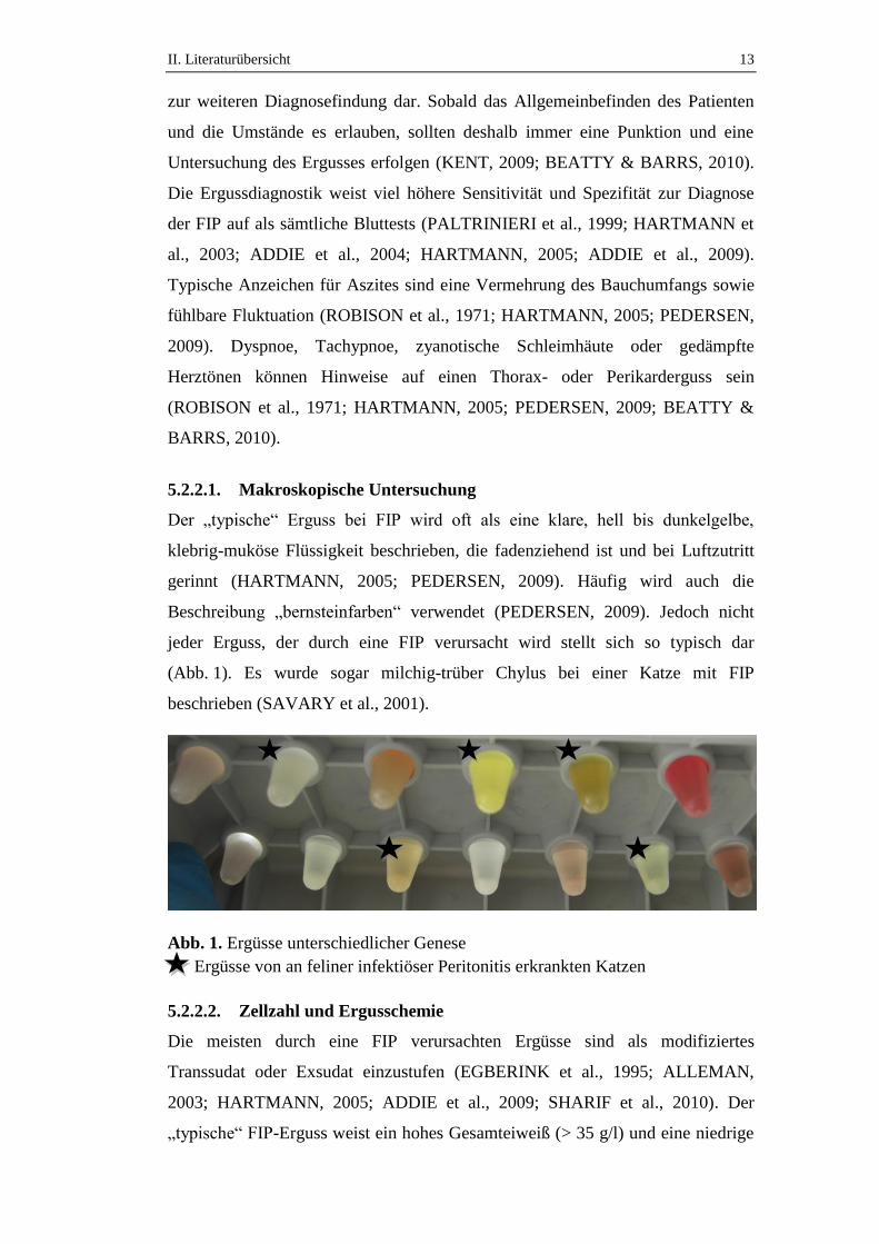
5.2.2.1. Makroskopische Untersuchung
Der „typische“ Erguss bei FIP wird oft als eine klare, hell bis dunkelgelbe,
klebrig-muköse Flüssigkeit beschrieben, die fadenziehend ist und bei Luftzutritt
gerinnt (HARTMANN, 2005; PEDERSEN, 2009). Häufig wird auch die
Beschreibung „bernsteinfarben“ verwendet (PEDERSEN, 2009). Jedoch nicht
jeder Erguss, der durch eine FIP verursacht wird stellt sich so typisch dar
(Abb. 1). Es wurde sogar milchig-trüber Chylus bei einer Katze mit FIP
beschrieben (SAVARY et al., 2001).
Abb. 1. Ergüsse unterschiedlicher Genese
Ergüsse von an feliner infektiöser Peritonitis erkrankten Katzen
5.2.2.2. Zellzahl und Ergusschemie
Die meisten durch eine FIP verursachten Ergüsse sind als modifiziertes
Transsudat oder Exsudat einzustufen (EGBERINK et al., 1995; ALLEMAN,
2003; HARTMANN, 2005; ADDIE et al., 2009; SHARIF et al., 2010). Der
„typische“ FIP-Erguss weist ein hohes Gesamteiweiß (> 35 g/l) und eine niedrige
II. Literaturübersicht 14
Zellzahl (< 1000/µl) auf (HARTMANN, 2005; ADDIE et al., 2009). Diese Werte
können jedoch weit variieren, und es kommen Zellzahlen bis zu 25000/µl vor.
Ähnliche Ergüsse können aber auch durch anderen Krankheiten, wie Lymphome,
Herzversagen, Cholangiohepatitis und bakteriellen Serositiden verursacht werden
(HARTMANN, 2005).
Neben dem hohen Eiweißgehalt ist bei Ergüssen von Katzen mit FIP oft die
Aktivität des Enzyms Laktatdehydrogenase (LDH) erhöht. Bei FIP werden LDH-
Aktivitäten von > 300 IU/l als typisch angegeben. Diese Werte werden jedoch
auch bei Exsudaten anderer Genese (bakterieller Pleuritits und Malignomen)
beobachtet und werden durch eine Freisetzung von Entzündungszellen verursacht
(LIGHT et al., 1972; HIRSCHBERGER, 1992; HARTMANN, 2005; ZOIA et al.,
2009). In der Humanmedizin wird die Messung der LDH-Aktivität genutzt, um
zwischen Transsudaten und Exsudaten zu unterscheiden, und speziell zwischen
malignen und benignen Ergussursachen (LIGHT et al., 1972). Häufig werden
auch erhöhte α-Amylase-Aktivitäten in den Ergüssen von Katzen mit FIP
gemessen. Hohe Aktivitäten der α-Amylase werden als Konsequenz einer
Pankreasbeteiligung am Krankheitsgeschehen angesehen (LIGHT et al., 1972;
HIRSCHBERGER, 1992; HARTMANN, 2005; ZOIA et al., 2009).
5.2.2.3. Akute-Phase-Proteine im Erguss
Es gibt nur sehr wenige Untersuchungen über Akute-Phase-Proteine im Erguss.
Es konnte gezeigt werden, dass das Akute-Phase-Protein Alpha-1-Acid-
Glykoprotein bei Katzen mit FIP im Erguss (wie auch im Serum) stark erhöhte
Konzentrationen aufweist (DUTHIE et al., 1997; GIORDANO et al., 2004;
GIORI et al., 2011). Man erhoffte sich einen Nutzen bei der Diagnose von FIP
(BENCE et al., 2005; GIORI et al., 2011), da die Werte bei an FIP erkrankten
Katzen höher waren als bei anderen entzündlich bedingten Ergussursachen (z. B.
bei Neoplasien und bakteriellen Serositiden) (DUTHIE et al., 1997; SELTING et
al., 2000). Eine frühere Studie ergab nur geringe Aussagekraft der Akute-Phase-
Proteine zur Diagnose bei FIP (STODDART et al., 1988). Auch die Ergebnisse
einer aktuellen Studie lassen darauf schließen, dass diese bei FIP nicht konstant
erhöht sind (TAYLOR et al., 2010).
5.2.2.4. Zytologische Untersuchung
Die zytologische Untersuchung sollte, wenn möglich, immer als diagnostische
II. Literaturübersicht 15
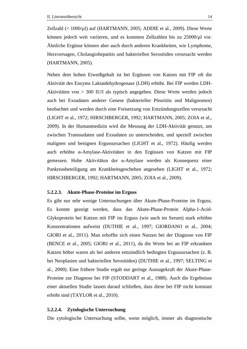
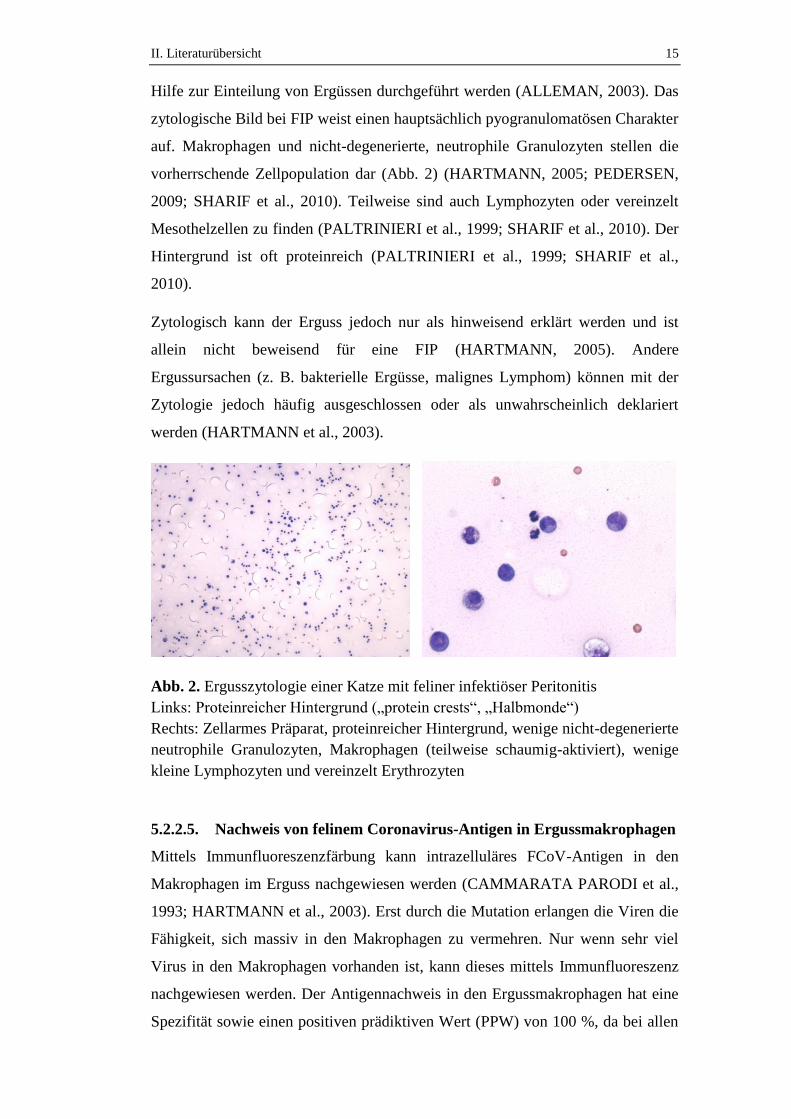
Hilfe zur Einteilung von Ergüssen durchgeführt werden (ALLEMAN, 2003). Das
zytologische Bild bei FIP weist einen hauptsächlich pyogranulomatösen Charakter
auf. Makrophagen und nicht-degenerierte, neutrophile Granulozyten stellen die
vorherrschende Zellpopulation dar (Abb. 2) (HARTMANN, 2005; PEDERSEN,
2009; SHARIF et al., 2010). Teilweise sind auch Lymphozyten oder vereinzelt
Mesothelzellen zu finden (PALTRINIERI et al., 1999; SHARIF et al., 2010). Der
Hintergrund ist oft proteinreich (PALTRINIERI et al., 1999; SHARIF et al.,
2010).
Zytologisch kann der Erguss jedoch nur als hinweisend erklärt werden und ist
allein nicht beweisend für eine FIP (HARTMANN, 2005). Andere
Ergussursachen (z. B. bakterielle Ergüsse, malignes Lymphom) können mit der
Zytologie jedoch häufig ausgeschlossen oder als unwahrscheinlich deklariert
werden (HARTMANN et al., 2003).
Abb. 2. Ergusszytologie einer Katze mit feliner infektiöser Peritonitis
Links: Proteinreicher Hintergrund („protein crests“, „Halbmonde“)
Rechts: Zellarmes Präparat, proteinreicher Hintergrund, wenige nicht-degenerierte
neutrophile Granulozyten, Makrophagen (teilweise schaumig-aktiviert), wenige
kleine Lymphozyten und vereinzelt Erythrozyten
5.2.2.5. Nachweis von felinem Coronavirus-Antigen in Ergussmakrophagen
Mittels Immunfluoreszenzfärbung kann intrazelluläres FCoV-Antigen in den
Makrophagen im Erguss nachgewiesen werden (CAMMARATA PARODI et al.,
1993; HARTMANN et al., 2003). Erst durch die Mutation erlangen die Viren die
Fähigkeit, sich massiv in den Makrophagen zu vermehren. Nur wenn sehr viel
Virus in den Makrophagen vorhanden ist, kann dieses mittels Immunfluoreszenz
nachgewiesen werden. Der Antigennachweis in den Ergussmakrophagen hat eine
Spezifität sowie einen positiven prädiktiven Wert (PPW) von 100 %, da bei allen
II. Literaturübersicht 16
Katzen mit einem positiven Ergebnis in diesem Test FIP bestätigt wurde
(HIRSCHBERGER et al., 1995; HARTMANN et al., 2003). Es gibt jedoch häufig
falsch-negative Ergebnisse (HIRSCHBERGER et al., 1995; HARTMANN et al.,
2003). Diese können z. B. durch eine geringe Zellzahl auf dem Ausstrich aus
Erguss und somit zu wenigen Makrophagen zum Nachweis des FCoV-Antigens,
verursacht sein. Eine andere Erklärung ist, dass das Antigen durch kompetitive
Bindung von FCoV-Antikörpern im Erguss maskiert und eine Bindung von
fluoreszierenden Antikörpern verhindert wird. Somit ist eine definitive Diagnose
von FIP bei einem positiven Nachweis von intrazellulärem FCoV-Antigen in den
Makrophagen im Erguss möglich, aber FIP kann bei einem negativen Ergebnis
nicht ausgeschlossen werden (TAMMER et al., 1995; PALTRINIERI et al.,
1999).
5.2.2.6. Antikörper gegen feline Coronaviren im Erguss
Die Messung von anti-FCoV-Antikörpern im Erguss ist ein weit verbreiteter Test.
Ein Vorhandensein von Antikörpern im Blut einer Katze zeigt nur einen Kontakt
mit FCoV an, kann aber nicht zwischen einer harmlosen FCoV-Infektion und
einer FIP unterscheiden (VENNEMA et al., 1998). Antikörper können aus dem
Blut in einen Erguss anderer Ursache übertreten. Damit ist der
Antikörpernachweis im Erguss oft auch positiv bei Katzen mit anderen
Ergussursachen. Ebenso wenig kann ein Fehlen von Antikörpern im Erguss eine
FIP ausschließen, da diese durch Antigen-Antikörper-Komplexe gebunden sein
können und damit nicht detektierbar sind (BARLOUGH & STODDART, 1988;
PEDERSEN, 1995). Die Sensitivität des Nachweises von Antikörpern im Erguss
zur Diagnose einer FIP betrug in einer Studie 86 %, die Spezifität 85 %
(HARTMANN et al., 2003). Da sich jedoch die Titerhöhe zwischen Katzen mit
FIP und Katzen mit anderen Krankheiten nicht signifikant unterscheidet, erscheint
auch eine quantitative Messung der Antikörper zur Diagnose von FIP im Erguss
nicht hilfreich (KENNEDY et al., 1998; HARTMANN et al., 2003). Die Präsenz
von Antikörpern im Erguss korreliert mit der von Antikörpern im Blut (SOMA &
ISHII, 2004). Es werden dabei meist, unabhängig von der Ergussursache, höhere
Titer im Erguss als im Serum gemessen (PALTRINIERI et al., 1998a;
PEDERSEN, 2009).
II. Literaturübersicht 17
5.2.2.7. Polymerase-Kettenreaktion im Erguss
Auch mittels der Polymerase-Kettenreaktion (Polymerase Chain Reaction (PCR))
in Blut oder in Erguss, kann keine gesicherte Diagnose einer FIP gestellt werden
(EGBERINK et al., 1995; HERREWEGH et al., 1995a; GAMBLE et al., 1997;
ADDIE et al., 2004). Es existieren nicht viele Studien über Untersuchungen zur
Aussagekraft der PCR aus Erguss und der diagnostische Nutzen ist daher schwer
einzuschätzen. In einer Studie von HARTMANN und Mitarbeitern (2003) hatten
fünf von fünf Katzen mit FIP eine positive PCR im Erguss und die einzige Katze
die nicht an FIP erkrankt war (hier wurde ein Lymphom diagnostiziert) hatte ein
negatives PCR-Ergebnis (HARTMANN et al., 2003). EGBERINK und
Mitarbeiter (1995) fanden in ihrer Studie mit einer größeren Anzahl von Katzen
(n = 183) heraus, dass die PCR im Erguss bei 83 % aller an FIP erkrankten Katzen
richtig-positiv ausfiel. Bei 17 % der Katzen fiel die PCR falsch-negativ aus. Fünf
von 40 Katzen (13 %) mit positiver PCR litten an anderen Erkrankungen. Ein
negatives Ergebnis schließt also eine FIP nicht aus und auch bei einem positiven
Ergebnis muss eine Katze nicht an FIP erkrankt sein (EGBERINK et al., 1995;
GAMBLE et al., 1997; HARTMANN et al., 2003; SHARIF et al., 2010).
5.2.2.8. Rivalta-Probe
Ein einfacher, schneller und kostengünstiger Test ist die Rivalta-Probe
(RIVALTA, 1895), die in Deutschland bei Katzen mit Körperhöhlenergüssen
routinemäßig durchgeführt wird. Ein positiver Ausfall kann einen Hinweis auf
FIP geben (HIRSCHBERGER, 1995; HARTMANN et al., 2003) und zur
Unterscheidung von anderen Krankheiten beitragen (HARTMANN et al., 2003).
Die Rivalta-Probe galt bisher als relativ sensitiv und spezifisch für FIP
(HIRSCHBERGER et al., 1995; HARTMANN et al., 2003). In früheren Studien
wies die Rivalta-Probe sehr gute diagnostische Werte auf. Es gibt jedoch auch
Ergüsse mit falsch-positiven Ergebnissen (z. B. bei Vorliegen eines Lymphoms
oder bakteriellen Serositiden) (HIRSCHBERGER et al., 1995; HARTMANN et
al., 2003; HARTMANN, 2005). Außerhalb Deutschlands ist die Rivalta-Probe
noch nicht so populär, gewinnt jedoch weiter an Bedeutung. Es ist bisher noch
relativ wenig über Herkunft, Nutzen und Funktionsweise dieses Testes bekannt.
Aufgrund der Einfachheit und der geringen Kosten sollte die Rivalta-Probe bei
jeder Katze mit Erguss durchgeführt werden (HARTMANN et al., 2003).
II. Literaturübersicht 18
5.2.2.8.1. Historie
Der Name der „Rivalta-Probe“ geht auf den italienischen Mediziner Fabio Rivalta
zurück, der als Erfinder dieses Testes gilt (RIVALTA, 1895). Diese Tatsache ist
jedoch nicht unumstritten, da auch ein deutscher Mediziner, Friedrich Moritz,
Ende des 19. Jahrhunderts Anspruch auf die Urheberschaft stellt (BERTI-BOCK
et al., 1979).
Der italienische Mediziner Fabio Rivalta publizierte als Erster im April 1895 ein
Vorgehen, mit dessen Hilfe man entzündliche Exsudate von einfachen
Transsudaten in menschlichen Körperhöhlenergüssen unterscheiden konnte
(RIVALTA, 1895; BERTI-BOCK et al., 1979). Er beschrieb die Reaktion als eine
Präzipitation in stark verdünnten sauren Lösungen (bestehend aus ca. 200 ml
Wasser, welches durch zwei Tropfen Essigsäure pro 100 ml Wasser angesäuert
wird). In diese Lösung wurden langsam einige Tropfen der zu untersuchenden
Körperhöhlenflüssigkeit fallen gelassen. Handelte es sich bei der Lösung um ein
Exsudat, entstand ein weißliches oder milchiges Zeichen („Rauchfahne“,
„Zigarre“). Bei einem Transsudat blieb die Lösung klar (RIVALTA, 1895;
EISENACK, 1951; BERTI-BOCK et al., 1979).
Diese Untersuchungsmethode setzte sich wegen ihrer einfachen Durchführung
und Zuverlässigkeit in ganz Italien sowie in Deutschland, Polen, Russland und
Frankreich unter der Bezeichnung „Rivalta-Probe“ durch (JANOWSKI, 1907;
BERTI-BOCK et al., 1979) und behielt diesen Namen bis heute. Damals nahm
Rivalta an, dass es sich bei dem ausfällbaren Körper um eine Mischung aus
„Para“- und „Pseudoglobulin“ handelte (BERTI-BOCK et al., 1979) und dass sich
dieses Phänomen auf die Anwesenheit von großen Mengen Globulin in Exsudaten
bezog, welches in Transsudaten fehlte (FRATELLI, 1939).
Etwa zur gleichen Zeit wie Fabio Rivalta beschäftigte sich auch in Deutschland
ein Mediziner, namens Friedrich Moritz, mit derselben Untersuchungsmethode.
Der gebürtige Mainzer schrieb 1886 seine Dissertation an der Ludwig-
Maximilians-Universität in München mit dem Thema „Beiträge zur Lehre von
den Exsudaten und Transsudaten“ und beschrieb darin einen durch Essigsäure
ausfällbaren Eiweißkörper, der entzündliche Exsudate von nicht entzündlichen
Transsudaten unterscheiden konnte (MORITZ, 1886).
Nachdem Moritz von der „Rivalta-Probe“ erfahren hatte, entzündet sich 1902 eine
II. Literaturübersicht 19
Diskussion um die Urheberschaft deren Erfindung. Es fand reger Schriftwechsel
zwischen Moritz und Rivalta aufgrund der „Rivalta-Frage“ statt (BERTI-BOCK
et al., 1979). In seiner Publikation verwies MORITZ (1902) darauf, dass seine
Forschungen zu diesem Test mindestens bis in das Jahr 1886 zurück gehen
(MORITZ, 1902). Schließlich kam man zur Einigung, dass Rivalta die Arbeit von
Moritz nicht bekannt war und auch die Methoden sich in Bedingungen,
technischen Modalitäten und in den Ergebnissen voneinander unterschieden
(BERTI-BOCK et al., 1979).
Auch andere Forscher beschäftigte das Thema „Unterscheidung zwischen Trans-
und Exsudaten“. Anfang des 20. Jahrhunderts machten die meisten Mediziner
gleiche Beobachtungen; nämlich, dass ein durch Essigsäure ausfällbarer Körper in
Exsudaten entstand, der nicht in Transsudaten vorkam. Jedoch gingen die
Meinungen bezüglich der Beschaffenheit dieses Eiweißkörpers auseinander
(MORITZ, 1902; STAEHELIN, 1902; UMBER, 1902; JOACHIM, 1903;
GARGANO & BRUSCA, 1960).
Während PAIJKULL diesen Körper als Nukleoalbumin bezeichnete, war
UMBER (1902) der Meinung, dass es sich um ein Seromucin, eine Zwischenstufe
von Eiweißkörper und Mucinen handelte (MORITZ, 1902; UMBER, 1902).
STAEHLIN (1902) vertrat die These, dass der Körper den Globulinen ähnelte,
bezeichnete ihn aber zunächst weiterhin nur als „den durch Essigsäure fällbaren
Eiweisskörper der Exsudate“ (MORITZ, 1902; STAEHELIN, 1902). JOACHIM
(1903) vertrat die Ansicht, dass die verschiedenen Autoren unterschiedliche
Eiweißkörper beschrieben und dass nur ein ganz bestimmter Globulinanteil,
nämlich das „Para-Euglobulin“ ausfallen würde (JOACHIM, 1903). UMBER
(1902) schrieb diesem, von ihm so bezeichneten Seromucin sogar eine
pathogonomische Bedeutung zur Beurteilung, ob ein Erguss exsudativer oder
transsudativer Natur sei, zu (UMBER, 1902). Etwa 60 Jahre später wurde die
Thematik erneut aufgegriffen und GARGANO & BRUSCA (1960) fanden heraus,
dass es sich bei den Substanzen, die eine positive Rivalta-Probe verursachten, um
saure Proteinkolloide von polysaccharidärer, nucleotidischer oder
phospholipidärer Natur handelten (GARGANO & BRUSCA, 1960).
Auch wenn die Rivalta-Probe heute keine Standarduntersuchung in der
Humanmedizin mehr darstellt, wurden von SAKAI und Mitarbeitern (2004)
genaue Untersuchungen des Tropfens, der bei der positiven Rivalta-Probe
II. Literaturübersicht 20
entsteht, durchgeführt. Dabei konnten acht verschiedene Proteintypen
(C-reaktives Protein, alpha1-Antitrypsin, alpha1-Acid Glykoprotein, Haptoglobin,
Transferrin, Zeruloplasmin, Fibrinogen und Hämopexin) in humanen Ergüssen
mit positiver Rivalta-Probe identifiziert werden. Welche Substanz es nun
tatsächlich ist, die eine Rivalta-Probe positiv ausfallen lässt, ist jedoch bis heute
nicht geklärt (SAKAI et al., 2004).
5.2.2.8.2. Durchführung
Die Rivalta-Probe ist bisher nicht genau standardisiert. Deswegen existieren
verschiedene Varianten der Durchführung (HIRSCHBERGER et al., 1995;
HARTMANN et al., 2003; SAKAI et al., 2004; HARTMANN, 2005; ADDIE et
al., 2009).
Benötigt wird ein Reagenzröhrchen, destilliertes Wasser (Aqua dest.), Eisessig
(98%ig) und der zu untersuchende Körperhöhlenerguss (abgefüllt in ein
Probenröhrchen mit Ethylendiamintetraessigsäure (EDTA) als Antikoagulanz)
(LEGA, 1939; HARTMANN et al., 2003; HARTMANN, 2005; ADDIE et al.,
2009). Die Rivalta-Probe wurde auch schon mit Leitungswasser beschrieben
(HIRSCHBERGER et al., 1995). Durchgesetzt hat sich jedoch die Durchführung
mit Aqua dest. (LEGA, 1939; HARTMANN et al., 2003; HARTMANN, 2005;
ADDIE et al., 2009).
Das Reagenzröhrchen wird zu etwa drei Vierteln (HARTMANN, 2005) mit Aqua
dest. gefüllt. Andere Volumenangaben variieren zwischen 5 ml (HARTMANN et
al., 2003) bis zu 7 – 8 ml (ADDIE et al., 2009). Ein Tropfen Eisessig (98%ig)
wird hinzugefügt und die Lösung sorgfältig gemischt. Auf die Oberfläche der
Lösung wird ein Tropfen der zu untersuchenden Flüssigkeit gegeben
(HARTMANN et al., 2003; HARTMANN, 2005).
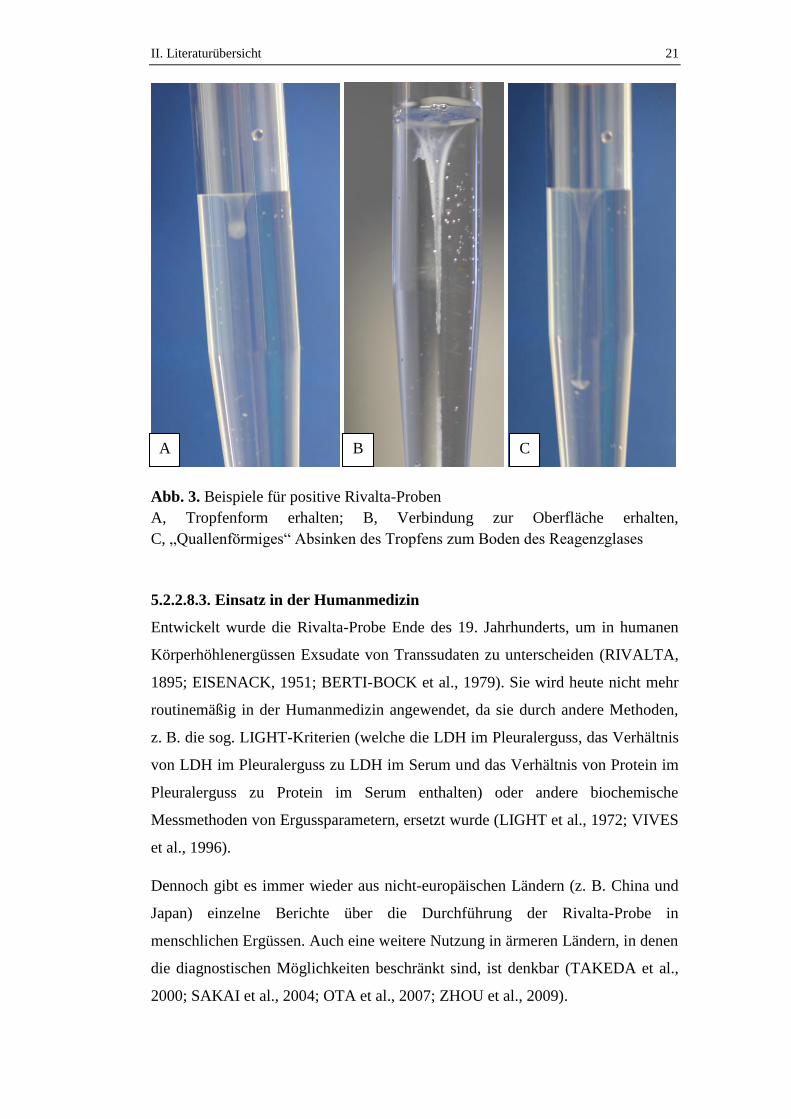
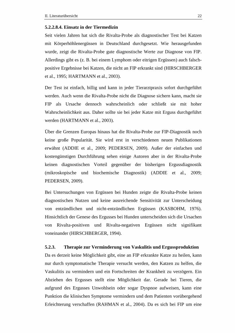
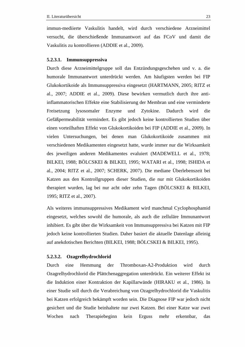
Die Rivalta-Probe gilt als negativ, falls der zugegebene Tropfen des
Körperhöhlenergusses sich auflöst und die Lösung klar bleibt. Ein positives
Ergebnis liegt vor, wenn der Tropfen seine Form beibehält, mit der Oberfläche
verbunden bleibt oder langsam (tropfen- oder quallenförmig) auf den Boden des
Reagenzröhrchens schwebt (HARTMANN et al., 2003; HARTMANN, 2005;
ADDIE et al., 2009) (Abb. 3). Auch das Entstehen einer rauchigen Trübung wird
manchmal bereits als positiv bewertet (HIRSCHBERGER et al., 1995).
II. Literaturübersicht 21
Abb. 3. Beispiele für positive Rivalta-Proben
A, Tropfenform erhalten; B, Verbindung zur Oberfläche erhalten,
C, „Quallenförmiges“ Absinken des Tropfens zum Boden des Reagenzglases
5.2.2.8.3. Einsatz in der Humanmedizin
Entwickelt wurde die Rivalta-Probe Ende des 19. Jahrhunderts, um in humanen
Körperhöhlenergüssen Exsudate von Transsudaten zu unterscheiden (RIVALTA,
1895; EISENACK, 1951; BERTI-BOCK et al., 1979). Sie wird heute nicht mehr
routinemäßig in der Humanmedizin angewendet, da sie durch andere Methoden,
z. B. die sog. LIGHT-Kriterien (welche die LDH im Pleuralerguss, das Verhältnis
von LDH im Pleuralerguss zu LDH im Serum und das Verhältnis von Protein im
Pleuralerguss zu Protein im Serum enthalten) oder andere biochemische
Messmethoden von Ergussparametern, ersetzt wurde (LIGHT et al., 1972; VIVES
et al., 1996).
Dennoch gibt es immer wieder aus nicht-europäischen Ländern (z. B. China und
Japan) einzelne Berichte über die Durchführung der Rivalta-Probe in
menschlichen Ergüssen. Auch eine weitere Nutzung in ärmeren Ländern, in denen
die diagnostischen Möglichkeiten beschränkt sind, ist denkbar (TAKEDA et al.,
2000; SAKAI et al., 2004; OTA et al., 2007; ZHOU et al., 2009).
A C B
II. Literaturübersicht 22
5.2.2.8.4. Einsatz in der Tiermedizin
Seit vielen Jahren hat sich die Rivalta-Probe als diagnostischer Test bei Katzen
mit Körperhöhlenergüssen in Deutschland durchgesetzt. Wie herausgefunden
wurde, zeigt die Rivalta-Probe gute diagnostische Werte zur Diagnose von FIP.
Allerdings gibt es (z. B. bei einem Lymphom oder eitrigen Ergüssen) auch falsch-
positive Ergebnisse bei Katzen, die nicht an FIP erkrankt sind (HIRSCHBERGER
et al., 1995; HARTMANN et al., 2003).
Der Test ist einfach, billig und kann in jeder Tierarztpraxis sofort durchgeführt
werden. Auch wenn die Rivalta-Probe nicht die Diagnose sichern kann, macht sie
FIP als Ursache dennoch wahrscheinlich oder schließt sie mit hoher
Wahrscheinlichkeit aus. Daher sollte sie bei jeder Katze mit Erguss durchgeführt
werden (HARTMANN et al., 2003).
Über die Grenzen Europas hinaus hat die Rivalta-Probe zur FIP-Diagnostik noch
keine große Popularität. Sie wird erst in verschiedenen neuen Publikationen
erwähnt (ADDIE et al., 2009; PEDERSEN, 2009). Außer der einfachen und
kostengünstigen Durchführung sehen einige Autoren aber in der Rivalta-Probe
keinen diagnostischen Vorteil gegenüber der bisherigen Ergussdiagnostik
(mikroskopische und biochemische Diagnostik) (ADDIE et al., 2009;
PEDERSEN, 2009).
Bei Untersuchungen von Ergüssen bei Hunden zeigte die Rivalta-Probe keinen
diagnostischen Nutzen und keine ausreichende Sensitivität zur Unterscheidung
von entzündlichen und nicht-entzündlichen Ergüssen (KASBOHM, 1976).
Hinsichtlich der Genese des Ergusses bei Hunden unterscheiden sich die Ursachen
von Rivalta-positiven und Rivalta-negativen Ergüssen nicht signifikant
voneinander (HIRSCHBERGER, 1994).
5.2.3. Therapie zur Verminderung von Vaskulitis und Ergussproduktion
Da es derzeit keine Möglichkeit gibt, eine an FIP erkrankte Katze zu heilen, kann
nur durch symptomatische Therapie versucht werden, den Katzen zu helfen, die
Vaskulitis zu vermindern und ein Fortschreiten der Krankheit zu verzögern. Ein
Abziehen des Ergusses stellt eine Möglichkeit dar. Gerade bei Tieren, die
aufgrund des Ergusses Unwohlsein oder sogar Dyspnoe aufweisen, kann eine
Punktion die klinischen Symptome vermindern und dem Patienten vorübergehend
Erleichterung verschaffen (RAHMAN et al., 2004). Da es sich bei FIP um eine
II. Literaturübersicht 23
immun-mediierte Vaskulitis handelt, wird durch verschiedene Arzneimittel
versucht, die überschießende Immunantwort auf das FCoV und damit die
Vaskulitis zu kontrollieren (ADDIE et al., 2009).
5.2.3.1. Immunsuppressiva
Durch diese Arzneimittelgruppe soll das Entzündungsgeschehen und v. a. die
humorale Immunantwort unterdrückt werden. Am häufigsten werden bei FIP
Glukokortikoide als Immunsuppressiva eingesetzt (HARTMANN, 2005; RITZ et
al., 2007; ADDIE et al., 2009). Diese bewirken vermutlich durch ihre anti-
inflammatorischen Effekte eine Stabilisierung der Membran und eine verminderte
Freisetzung lysosomaler Enzyme und Zytokine. Dadurch wird die
Gefäßpermeabilität vermindert. Es gibt jedoch keine kontrollierten Studien über
einen vorteilhaften Effekt von Glukokortikoiden bei FIP (ADDIE et al., 2009). In
vielen Untersuchungen, bei denen man Glukokortikoide zusammen mit
verschiedenen Medikamenten eingesetzt hatte, wurde immer nur die Wirksamkeit
des jeweiligen anderen Medikamentes evaluiert (MADEWELL et al., 1978;
BILKEI, 1988; BÖLCSKEI & BILKEI, 1995; WATARI et al., 1998; ISHIDA et
al., 2004; RITZ et al., 2007; SCHERK, 2007). Die mediane Überlebenszeit bei
Katzen aus den Kontrollgruppen dieser Studien, die nur mit Glukokortikoiden
therapiert wurden, lag bei nur acht oder zehn Tagen (BÖLCSKEI & BILKEI,
1995; RITZ et al., 2007).
Als weiteres immunsuppressives Medikament wird manchmal Cyclophosphamid
eingesetzt, welches sowohl die humorale, als auch die zelluläre Immunantwort
inhibiert. Es gibt über die Wirksamkeit von Immunsuppressiva bei Katzen mit FIP
jedoch keine kontrollierten Studien. Daher basiert die aktuelle Datenlage alleinig
auf anekdotischen Berichten (BILKEI, 1988; BÖLCSKEI & BILKEI, 1995).
5.2.3.2. Ozagrelhydrochlorid
Durch eine Hemmung der Thromboxan-A2-Produktion wird durch
Ozagrelhydrochlorid die Plättchenaggregation unterdrückt. Ein weiterer Effekt ist
die Induktion einer Kontraktion der Kapillarwände (HIRAKU et al., 1986). In
einer Studie soll durch die Verabreichung von Ozagrelhydrochlorid die Vaskulitis
bei Katzen erfolgreich bekämpft worden sein. Die Diagnose FIP war jedoch nicht
gesichert und die Studie beinhaltete nur zwei Katzen. Bei einer Katze war zwei
Wochen nach Therapiebeginn kein Erguss mehr erkennbar, das
II. Literaturübersicht 24
Allgemeinbefinden und auch die Blutwerte verbesserten sich. Nach 18 Monaten
war die Katze immer noch bei gutem Allgemeinbefinden und symptomfrei. Auch
bei der zweiten Katze verschwand der Erguss nach zwölf Tagen unter Therapie
und die Katze war für acht Monate bei gutem Allgemeinbefinden. Die Therapie
musste dann jedoch aufgrund von Nebenwirkungen (Nasenbluten) abgebrochen
werden. Daraufhin bildete sich wieder Erguss und die Katze verstarb zwei Monate
später. Ob Ozagrelhydrochlorid nun tatsächlich zu der langen Überlebenszeit
beitrug, ist nicht bekannt (WATARI et al., 1998).
5.2.3.3. Acetylsalicylsäure
Bei Acetylsalicylsäure (ASS) handelt es sich um ein nicht-steroidales
Antiphlogistikum. Die Wirkungen sind sowohl anti-pyretisch, anti-
inflammatorisch, als auch gerinnungshemmend. ASS hemmt auch die Synthese
von Thromboxan A2 und damit die Thrombozytenaggregation. Die
Wirkungsweise bei FIP soll auf einer Unterdrückung des Entzündungsgeschehens
sowie der Vaskulitis beruhen; ähnlich der Wirkung, die bei dem humanen
Kawasaki-Syndrom beschrieben ist. Bei dieser Erkrankung handelt es sich
ebenfalls um eine, durch eine generalisierte Vaskulitis geprägte, Erkrankung beim
Menschen (ONOUCHI & KAWASAKI, 1999). Ein zusätzlich positiver Effekt bei
FIP könnte die anti-pyretische Wirkung von ASS darstellen. Über die tatsächliche
Wirksamkeit gibt es jedoch keine Studien und eine Kombination mit
Glukokortikoiden ist aufgrund der Nebenwirkungen (z. B. Gefahr von Magen-
Darm-Ulzera) nicht sinnvoll (ADDIE et al., 2009).
5.2.3.4. Thioprolin
Bei dem Wirkstoff Thioprolin handelt es sich um ein Antioxidans und einen freien
Radikalfänger. Dadurch könnten die zellulären Membranen vor freien Radikalen
geschützt werden (WEBER et al., 1982). Thioprolin senkt die Produktion des
Zytokins TNF-α (welches bei der Entstehung einer Vaskulitis beteiligt ist
(SULLIVAN et al., 1988)) und kann durch eine Immunmodulation gegebenenfalls
(ggf.) ein Fortschreiten der Krankheit einschränken (FORD, 1986; DE LA
FUENTE et al., 2002; GUAYERBAS et al., 2004). In einer Studie mit 52 Katzen
mit angeblicher FIP gingen die typischen Symptome Fieber, Erguss und Anorexie
nach Therapie mit Thioprolin in Remission. Jedoch war die Diagnose FIP nicht
gesichert, es existierte keine Kontrollgruppe und es gab keine Angaben über die
II. Literaturübersicht 25
Dauer der Remission (FORD, 1986).
5.2.3.5. Methylxanthinderivate Pentoxifyllin und Propentofyllin
Bisher wurden noch keine klinischen Studien mit Propentofyllin (PPF) oder
Pentoxifyllin (PTX) durchgeführt. Es gibt jedoch verschiedene Berichte, die PTX
in Kombination mit Glukokortikoiden eine Wirksamkeit bei Katzen mit FIP
zuschreiben (LITTLE, 2005; NORRIS, 2007; SCHERK, 2007; PEDERSEN,
2009). Obwohl die genaue Wirkungsweise und der Mechanismus noch nicht
völlig geklärt sind, erscheint eine Wirksamkeit dieser beider
Methylxanthinderivate aufgrund der pharmakodynamischen Eigenschaften
wahrscheinlich (NORRIS, 2007; SCHERK, 2007). PTX und PPF wirken als
Inhibitoren der Phosphodiesterase, von Interleukinen und TNF-α (ZABEL et al.,
1993; MEINERS et al., 2004). Diese pro-inflammatorischen Zytokine spielen eine
wichtige Rolle bei der Entstehung der Vaskulitis bei Katzen mit FIP und ihre
Konzentrationen sind bei diesen Katzen stark erhöht (KISS et al., 2004; KIPAR et
al., 2005; REGAN et al., 2009; TAKANO et al., 2009). TNF-α induziert die
Synthese von Fibrinogen und ist verantwortlich für eine Hyperadhäsion der
neutrophilen Granulozyten an den Endothelzellen und für eine gesteigerte
Produktion von freien Radikalen, die Gewebsschäden verursachen. Durch die
mikrovaskulären Verletzungen wird die Gefäßpermeabilität erhöht und eine
Vaskulitis entsteht (MOVAT, 1987; MOVAT et al., 1987; SULLIVAN et al.,
1988). PTX verhindert die Synthese von diesem TNF-α durch aktivierte
Monozyten und kann so vermutlich sowohl die Gewebsschäden und die Vaskulitis
vermindern und auch die hohen Fibrinogenkonzentrationen reduzieren
(HAMMERSCHMIDT et al., 1988; BACHET et al., 1989; SORIA et al., 1990;
GROSS, 1994; NEUNER et al., 1994).
Es gibt einige Studien über die Hemmung dieser inflammatorischen Zytokine
durch PTX bei Menschen und Tieren und deren Erfolge bei der Therapie von
Vaskulitiden unterschiedlicher Genese (ZABEL et al., 1989; ARIAS-DIAZ et al.,
1994; NEUNER et al., 1994; MARTON et al., 1998; NICHOLS et al., 2001). Bei
Menschen, die nach einer Injektion von Endotoxinen stark erhöhte TNF-α-
Konzentrationen aufwiesen, waren diese drei Wochen nach Therapie mit PTX
wieder auf das ursprüngliche Level gesunken (ZABEL et al., 1989). Auch
normale TNF-α-Konzentrationen konnten durch orale Gabe von PTX gesenkt
werden. Bei Ratten mit iatrogen-induzierter, akuter Pankreatitis wurden durch

II. Literaturübersicht 26
Therapie mit PTX sowohl die TNF-α-Konzentrationen, als auch die
Mortalitätsrate signifikant gesenkt (MARTON et al., 1998). PEDERSEN (2009)
beschreibt einen Therapieversuch mit PTX und Interferon-γ bei einer Katze mit
FIP, der jedoch ohne Erfolg blieb (PEDERSEN, 2009).
III. Studie I 27
III. STUDIE I
The Rivalta´s Test as a Test to Diagnose Feline Infectious
Peritonitis
Yvonne Fischer 1
Carola Sauter-Louis, Dr. med. vet., PhD (epidemiology), Dipl. ECVPH 2
Katrin Hartmann, Prof. Dr. med. vet., Dr. med. vet. habil., Dipl. ECVIM-CA 1
1 Clinic of Small Animal Medicine, LMU University of Munich, Germany
2 Clinic for Ruminants, LMU University of Munich, Germany
Veterinary Clinical Pathology, akzeptiert
III. Studie I 28
The Rivalta´s test as a test to diagnose feline infectious peritonitis
Yvonne Fischer, Carola Sauter-Louis, and Katrin Hartmann
Abstract:
Background: Since years, the Rivalta´s test is routinely used in Europe to
diagnose feline infectious peritonitis (FIP) in cats with effusion. It is inexpensive
and easy to perform in private practice. There is, however, only little information
about its mode of action and diagnostic value.
Objectives: The objectives of this study were to evaluate sensitivity, specificity,
positive (PPV) and negative (NPV) predictive values of the Rivalta´s test for
diagnosing FIP and to investigate if there are correlations with any effusion or
blood variables.
Methods: Medical records of 782 cats between 1999 and 2010 were reviewed.
Effusion and blood variables were compared between Rivalta-positive and
-negative effusions using the Mann Whitney U test and a multivariate analysis.
Results: Prevalence of FIP in cats with effusion was 34.6%. The Rivalta´s test
showed a sensitivity of 91.3%, a specificity of 65.5%, a PPV of 58.4%, and a
NPV of 93.4% for the diagnosis of FIP. Most important significantly different
variables in Rivalta-positive effusions were higher cholesterol and specific
gravity, and lower albumin to globulin ratio, as the higher bilirubin in the blood.
Conclusions: The Rivalta´s test in general is an easy and cheap test, but its
sensitivity and specificity to diagnose FIP are not as high as previously assumed.
If it is, however, performed in young cats or if certain diseases have been ruled-
out, it is a valuable practice test. It remains unknown which components in
effusions lead to a positive Rivalta´s test, but the positivity is not due to high
protein concentrations.
III. Studie I 29
Introduction
Feline infectious peritonitis (FIP) is a very common disease, especially in young
cats. The clinical signs are variable and depend on the involved organs1-2
. A
definitive diagnosis is often difficult to obtain. Many diagnostic tests exist in
blood and effusions of body cavities, but most of these tests do not provide a
definitive diagnosis and make FIP just more or less suggestive3-4
.
In Germany, a simple test performed on effusions of cats suspected to have FIP
has been used, named “Rivalta´s test”. This test is cheap and can be quickly
performed in private practice. The principle of the Rivalta´s test is the formation
of a drop if effusion is added to a slightly acetic solution. This test was examined
in two previous studies and showed a very high sensitivity3 as well as a high
specificity for FIP3, 5
. In these studies, however, that have used study populations
of past decades, the test was only evaluated in a relatively low number of cats5-6
.
Although used for a long time, there is only little information about the mode of
action of the Rivalta´s test. The test was developed by and named after the Italian
physician Fabio Rivalta to differentiate exudates from transudates in human body
cavity effusions7-9
. Rivalta et al. published this method in 18957. Since then, the
“Rivalta´s test” has been mainly used in Germany, Poland, Russia, and France10
.
Today, the Rivalta´s test is not longer used in human medicine, because other
analytical methods, including the Light´s criteria (pleural LDH, pleural/serum
LDH ratio, and pleural/serum protein ratio) have replaced it1, 11-12
. In dogs, the
Rivalta`s test has not demonstrated good diagnostic value and is lacking
sensitivity to diagnose exudates13
.
The objectives of this study were to determine the sensitivity, specificity, positive
predictive value (PPV), and negative predictive value (NPV) of the Rivalta´s test
to diagnose FIP, using a large cohort of cats with a definitive diagnosis, and to
investigate its correlation with effusion (including specific gravity (SG), white
blood cells (WBC), red blood cells (RBC), total protein (TP), triglycerides,
glucose, creatinine, lactate dehydrogenase (LDH), α-amlyase, albumin,
cholesterol) and blood (including RBC, hemoglobin, hematocrit, WBC,
monocytes, lymphocytes, banded neutrophils, mature neutrophils, alanine
aminotransferase (ALT), alkaline phosphatase (AP), bilirubin, TP, albumin, urea,
creatinine, glucose, albumin to globulin ratio, phosphorus, sodium, potassium,
chloride, total and ionized calcium) variables.
III. Studie I 30
Materials and Methods
Study design
The study was conducted according to the Standards for Reporting of Diagnostic
Accuracy (STARD)14-15
, which standardize the conduct and reporting of results of
studies of diagnostic accuracy. It was performed as a retrospective analysis,
including data of 782 cats with effusion that had been presented to the Clinic of
Small Animal Medicine of the Ludwig Maximilian University of Munich,
Germany, between January 1999 and May 2010. Information collected on these
cats included age of the cats, effusion variables, blood variables (CBC, serum
chemistry, and electrolytes), and (if available) definitive diagnosis. Inclusion
criteria to enter the study were the presence of effusion (detected by ultrasound)
and a Rivalta´s test performed. Exclusion criterion was a questionable Rivalta´s
test result. A Rivalta´s test was defined as questionable (n = 69) if slightly cloudy
shadows were seen in the solution, but one was not able to distinguish if these
dissolved or not. For the assessment of the prevalence of FIP and the diagnostic
accuracy of the Rivalta´s test, only cats with a definitive diagnosis were included
(n = 497) as shown in figures 1 to 3. For the correlation of the Rivalta´s test with
blood or effusion variables, all cats with a clearly positive or negative Rivalta´s
test result were included (figure 4).
The definitive diagnosis of FIP was either confirmed by immunofluorescent
staining of feline coronavirus (FCoV) antigen in macrophages in effusion5, by
histopathologic changes (if diagnostic for FIP)16-17
, or immunohistochemical
staining of FCoV antigen in tissue macrophages at postmortem examination18
.
The definitive diagnoses of other diseases causing effusion were obtained as
follows. Effusion due to heart disease was diagnosed using echocardiography19-20
.
Neoplasia was either diagnosed by cytology or, if not diagnostic, by
histopathological examinations or necropsy21
. Bacterial infections were confirmed
by bacterial culture or detection of bacteria in cytology22-23
. Cholangiohepatitis
and intussusception were diagnosed by abdominal ultrasound24-25
, laparotomy, or
necropsy25-27
. Pancreatitis was diagnosed by an elevation of the feline pancreatic
lipase immunoreactivity (fPLi)28-29
, abdominal ultrasound28-29
, laparotomy30
,
necropsy30
, or a combination of those features31
. Feline lower urinary tract disease
with leakage of urine in the abdominal cavity was diagnosed using a combination
of history of anuria, physical and ultrasound examination. Bleeding was
III. Studie I 31
diagnosed by comparing effusion and blood packed cell volume. If the hematocrit
was equal or higher in the effusion than in the peripheral blood, the effusion was
assumed to be caused by bleeding32
. Systemic hypertension was determined by
Doppler-guided measuring of the systemic blood pressure33
. When FIP was
diagnosed, it was considered to be the responsible disease for the effusion and
therefore, those cats were classified to the group “FIP”. If cats with FIP had
concurrent diseases (e. g., diabetes mellitus, feline immunodeficiency virus (FIV)
infection), these diseases were not evaluated because they were not considered to
be a cause for an effusion.
Test methods
To perform tests on effusion fluid, thoraco- or abdominocentesis was performed
ultrasound-guided, which in most cases is possible without sedation. A 19- or
21-gauge butterfly needle connected to a closed system, using a 3-way-stopcock
and a 10 ml syringe was used to carefully and sterilely draw the effusion out of
the body cavity. Depending on how much effusion was present, the amount of
effusion removed varied between 0.5 and 800 ml. The effusion was filled in
reagent tubes, containing EDTA as anticoagulant, and in plain serum tubes
without anticoagulant. For every individual cat, effusion variables were
determined and the Rivalta´s test was performed for diagnostic purposes on the
day of aspiration.
For the Rivalta´s test, a plastic reagent tube (volume 10 ml) was filled with 7 to
8 ml of distilled water. One drop of acetic acid (98 to 100%) was added from a
disposable pipette. The suspension was thoroughly mixed. Using a second
disposable pipette, one drop of effusion fluid was carefully laid on top of the
suspension. If a drop developed and stayed attached to the surface, retained its
shape or slowly floated down to the bottom of the reaction tube, the Rivalta´s test
was defined as positive (figure 5). If the drop disappeared and the solution
remained clear, the Rivalta´s test was defined as negative. Slightly cloudy
shadows were considered a “questionable” Rivalta´s test. Those cats were
excluded from analysis.Variables evaluated in effusions included SG, WBC,
RBC, TP, triglycerides, glucose, creatinine, LDH, α-amlyase, albumin, and
cholesterol. SG was measured using a refractometer (Atago Company Ltd, Japan).
Cell counts of effusions were determined by an automatic analyzer (Cell-Dyn
3500; Abott Laboratories, Illinois, USA). The other effusion variables were
III. Studie I 32
analyzed by automatic analyzers (1999 - 2000: Hitachi 717; 2000 - 2010: Hitachi
911; Roche Deutschland Holding GmbH, Grenzach-Wyhlen, Germany). Effusions
were classified, according to concentration of TP, SG, and WBC, as transudate
(SG < 1.018, TP < 25 g/l, and WBC < 1500/µl), modified transudate (SG 1.018 –
1.025, TP 25 – 30 g/l, and WBC 1500 – 7000/µl; an effusion that had
characteristics of both, a transudate and an exudate (e. g., because of a high TP,
but a low WBC) was also classified as a modified transudate.), or exudate
(SG > 1.025, TP > 30 g/l and WBC > 7000/µl). Classification of effusion was
possible in 643 cats. The other 139 effusions could not be classified because
values of SG, TP, and/or WBC were not available (figure 4).
Blood samples were taken out of the Vena (V.) cephalica, the V. saphena
medialis, or the V. jugularis, using a 20-gauge needle. Blood was collected in
plastic tubes containing EDTA, in plastic serum tubes without anticoagulant, and
in blood gas tubes containing lithium-heparin. In blood, the following variables
were analyzed: RBC, hemoglobin, hematocrit, WBC, monocytes, lymphocytes,
banded neutrophils, mature neutrophils, ALT, AP, bilirubin, TP, albumin, urea,
creatinine, glucose, albumin to globulin ratio, phosphorus, sodium, potassium,
chloride, total and ionized calcium. The CBC was determined using an automatic
analyzer (Cell-Dyn 3500; Abott Laboratories, Illinois, USA). The differential
WBC count was determined manually counting 100 white blood cells. The serum
chemistry was evaluated using an automatic analyzer (1999 - 2000: Hitachi 717;
2000 - 2010: Hitachi 911; Roche Deutschland Holding GmbH, Grenzach-Wyhlen,
Germany). The ionized calcium was measured with an automatic electrolyte
analyzer (GEM Premier 3000; Instrumentation Laboratory GmBH, Kirchheim,
Germany).
Statistical methods
Values used to evaluate the diagnostic utility of the Rivalta´s test included the
diagnostic sensitivity (proportion of cats that show Rivalta-positive test results
among all cats with FIP), the diagnostic specificity (proportion of cats that show
Rivalta-negative test results among all cats with other diseases than FIP), the PPV
(probability for a cat with Rivalta-positive test results to have FIP), and the NPV
(probability for a cat with Rivalta-negative test results to have no FIP). These
values were calculated for all cats with a definitive diagnosis (n = 497) and in
addition for different subpopulations of cats (figures 1 to 3). Subpopulations were
III. Studie I 33
all cats a) excluding those with bacterial infection or lymphoma (because these
diagnoses usually can easily be differentiated from FIP by cytology and/or
bacterial culture5) (n = 383) and cats not older than b) 2 years (n = 118) (because
most cats with FIP are young1, 3, 34-36
).
To investigate the correlation between different blood and effusion variables
(figure 4) with the results of the Rivalta´s test and the classifications of the
effusions in transudate, modified transudate, or exudate, statistical analyses were
performed using software SPSS version 17.0 (SPSS Inc., Chicago, USA). Because
almost all variables were not normally distributed (determined visually using
boxplots), the Mann Whitney U test was used to investigate a difference between
the effusion and blood variables of cats with Rivalta-positive or –negative
effusions. All variables, that showed a statistically significant difference in the
univariate analysis, were analyzed in a multivariate logistic regression model,
using backwards selection (likehood ratio). Not all variables were available in all
cats. For the logistic regression model, only those samples with complete data
could be analyzed. Variables were retained in the model if the p-value was less
than 0.05. The significance of a variable was higher the more the Odds-ratio (OR)
was distanced to 1.000. The OR is a descriptive statistic, describing the strength of
association between two variables.
III. Studie I 34
Results
Diagnoses in cats with effusion
Of all 782 cats with effusion included, 497 cats had a definitive diagnosis
(table 1). Of those 172 had FIP, and 325 had other diseases (156, neoplasia; 93,
heart diseases; 45, bacterial infection; 11, pancreatitis; 3, cholangiohepatitis;
2, intussusception; 1, systemic hypertension; 4, bleeding; 10, feline lower urinary
tract disease with effusion secondary to obstruction or lesions of the urinary tract).
There were 643 effusions, which could be classified as transudate, modified
transudate, or exudate (figure 4). Of the Rivalta-positive effusions, 49.7% were
classified as exudates, 44.6% as modified transudates, and 5.7% as pure
transudates. However, also 11.5% of the Rivalta-negative effusions were exudates
(table 2).
Value of the Rivalta´s test to diagnose FIP
Only the results of cats with a definitive diagnosis (n = 497) were used for the
determination of the prevalence of FIP, the sensitivity, the specificity, the PPV,
and the NPV of the Rivalta´s test to diagnose FIP (figure 1 - 3, table 3). The
prevalence of FIP was 34.6%. There was only a low number of cats ≤ 2 years
(n = 22; 96/118 had FIP) with diseases other than FIP, and the prevalence, as the
diagnostic sensitivity and the PPV in this subgroup was higher.
Correlation of effusion and blood variables with Rivalta´s test results
For the evaluation of the correlation between effusion and blood variables and the
Rivalta´s test, 782 effusions were included (figure 4). A statistically significant
difference in most of the blood or effusion variables was found between cats with
Rivalta-positive and –negative effusions (table 4 and 5) using the Mann Whitney
U test. However, using a multivariate logistic regression model with 329 effusions
included, only 4 variables in the effusion and 2 variables in the blood remained as
significant factors in the model (table 4 and 5). The significance of the resulting
variables (cholesterol, specific gravity, glucose, and triglycerides in the effusions,
as albumin to globulin ratio and bilirubin in the blood) are higher the more their
OR was distanced to 1.000.
III. Studie I 35
Discussion
The prevalence of FIP (34.6%) in cats with effusion in this study was lower than
in previous studies of the past decades, in which it reached 41%3 to 51%
5. The
question arises whether there is a true decrease in the overall prevalence of FIP
within the past 15 years. It may be possible that many cats with high suspicion of
FIP are no longer referred to the University Veterinary Teaching Hospital by other
veterinarians, but are more often euthanized in private practice because of the very
poor prognosis of FIP. The prevalence in this study could also be low, because
cats without a definitive diagnosis were excluded from statistical analysis. Of
these cats, many were highly suspicious for FIP, but because of the bad prognosis
for most causes of effusion, many owners elected not to pursue diagnostic tests for
a definitive diagnosis. In recent years, more and more owners decline to allow
necropsy. For that reason, more cats might have been taken home after death to be
buried or cremated and less have been left at the University to perform a necropsy,
which likely would have revealed a definitive diagnosis. These points may bias
the population in this study14-15
. The fact, that about one third of all cats with
effusions had to be excluded from further evaluation because of a missing
definitive diagnosis, might bias the sensitivity and specificity of the test as well as
predictive values of this study14-15
. Many of those cats were suspicious for FIP. If
these cats would have gone to necropsy, the prevalence of FIP and in consequence
also the PPV would have been higher. On the other hand, there are many other
diseases (e. g., other neoplasias than lymphoma), which are not easy to detect and
can also cause effusion. If many of those cats would have had a diagnosis, this
would also influence the predictive values. Another reason for the lower
prevalence of FIP in the present study could be that other diseases, such as
neoplasia (one of the most common differential diagnoses), pancreatitis, or heart
diseases are now more commonly diagnosed. Due to better possibilities today to
find such diagnoses, e. g. heart diseases and tumors (better equipment and
techniques in ultrasonography, cytology, diagnostic imaging), more other diseases
may have been diagnosed in the present study (which might have not been
diagnosed in earlier studies because of missing diagnostic possibilities), which
could have decreased the percentage of FIP amongst cats with a definitive
diagnosis.
The prevalence of FIP in the population of cats younger than 2 years of age is
III. Studie I 36
much higher with 81.4%, respectively, than in the overall population of cats with
effusion. This higher percentage has to be expected, because most cats with FIP
are younger than 2 years or even younger than 1 year1, 3, 34-36
and the differential
diagnoses causing effusions are rare at this young age37
.
If the Rivalta´s test was positive, the probability of FIP as the causative disease
was 58.4% (PPV), which is much lower than in earlier studies (86% or 84%)3, 5
.
Other differential diagnoses resulting in a positive Rivalta´s test in former studies
were rare. In the study of Hirschberger et al.3, 83.9% of the cats with Rivalta-
positive effusions had FIP. In contrast, a much higher proportion of Rivalta-
positive effusions were caused by other diseases in the present study that,
however, included a much higher number of cats. An explanation for the higher
number of other diseases causing Rivalta-positive effusions (besides the lower
prevalence of FIP in the present study) could be that some effusions remained in
the body cavity for a longer period. There could be an inflammation and serositis
secondary to the effusion, which could result in production of inflammatory
mediators and cytokines leading to a positive Rivalta´s test at a later stage of
disease independent of the original disease process. Alternatively, cats with a
positive Rivalta´s test and another diagnosis than FIP, such as heart disease,
could, in theory, have had two diseases at the same time. If a diagnosis as a reason
for the effusion was found in a cat, no further diagnostics might have been
performed. So, FIP might have been undetected in these cats, and the Rivalta´s
test could have been truly positive because of FIP, and not “false-positive”
because of the other disease. Although this is a potential explanation, it is quite
unlikely, because many of the cats with other diseases lived longer than 14 days.
Although some cats with FIP can live longer, a median survival time of only
8 days is described38
.
One important reason for the lower PPV in contrast to older studies is that
predictive values strongly depend on the prevalence of the disease, and the lower
prevalence of FIP can explain the lower PPV of the Rivalta´s test in this study. In
a population of young cats (≤ 2 years), the probability for a cat with a positive
Rivalta´s test to have FIP reaches about 90%, due to the higher prevalence of FIP
in this population1, 3, 34-36
. As there are not many differential diagnoses for a young
cat with effusion, the probability of FIP in a cat with a positive Rivalta´s test
remains very high. Predictive values are strongly influenced by the prevalence of
III. Studie I 37
the disease, which may vary considerably from one clinical situation to another14-
15. Therefore, the predictive values reported in this study have to be interpreted in
view of the investigated population and may not be assigned to other clinical
situations.
Although the sensitivity of the Rivalta´s test to diagnose FIP was also lower than
in former studies (98% and 100%)5, 39
, it was still very good in the present one
(91.3%). Correspondingly, the NPV was very high (93.4%). Therefore, if a cat
with effusion has a negative Rivalta´s test, the likelihood of FIP is very low, and
other diseases should be considered (sensitivity 91.3%).
Different effusion variables were analyzed and correlated with the Rivalta´s test
outcome to see, if there is a correlation between a positive Rivalta´s reaction and
any components in the effusion. In most of the variables measured in effusion or
in blood, a statistically significant difference between Rivalta-positive and
Rivalta-negative effusions (table 4 and 5) was obvious when analyzed with the
Mann Whitney U test. The high number of significant differences is a result of the
large number of patients included, but not all variables and differences seem to be
clinically relevant. In the multivariate logistic regression model, only cholesterol
concentration, the specific gravity, the glucose concentration, and the triglycerides
of the effusion, as well as the albumin to globulin ratio and the bilirubin in the
blood were significantly different.
For a long time it was assumed, that a positive Rivalta´s test is mainly dependent
on the TP concentration in the effusion8, 40-42
. This assumption obviously seems
not to be true, because in the present study the TP concentrations of Rivalta-
positive and Rivalta-negative effusions were not significantly different in the
multivariate analysis. There were Rivalta-negative effusions with a very high TP
and Rivalta-positive effusions with quite low TP levels (table 4). Thus, the
findings of this study suggest that the TP is not the major variable correlated with
the outcome of the Rivalta´s test, and therefore, other components of the effusion
seem to be involved in the formation of the drop seen in a positive Rivalta´s test.
The cholesterol concentration in effusion was significantly higher in Rivalta-
positive effusions and the OR was highest in this variable. Cholesterol is a useful
variable to differentiate between transudates and exudates in human effusions43-45
.
The reason for the higher concentration in exudates is not fully understood.
III. Studie I 38
Possibilities are that cholesterol develops because of a degeneration of effusion
components (e. g., WBC and RBC), or that cholesterol can pass the vessels
because of increased capillary permeability in vasculitis44, 46
. As there were much
more Rivalta-positive effusions in this study classified as exudates, and exudates
show higher cholesterol concentrations, it seems logical that the cholesterol
concentrations were higher in these Rivalta-positive effusions. Whether, however,
the cholesterol in the effusion actually could cause the positive Rivalta´s reaction,
still remains to be clarified. Interestingly, the triglyceride concentration, in
contrary, was significantly lower in Rivalta-positive than in Rivalta-negative
effusions (table 4). It would have been expected, that triglycerides and cholesterol
show the same pattern. Triglycerides may, however, behave different as it was
shown in one study in mice, that cholesterol feeding and high cholesterol levels
actually decreased the production of triglycerides47
. Another explanation could be
that some effusions may be present in the body cavities for an extended period of
time before being diagnosed. In this case, a “pseudochyle” can develop, which
typically contains high cholesterol values, but no triglycerides48
.
The SG showed significantly higher values in Rivalta-positive than in Rivalta-
negative effusions (table 4). The SG should mainly depend on the TP
concentrations or cell counts49
of an effusion, but both did not remain
significantly different between Rivalta-positive and Rivalta-negative effusions in
the multivariate analysis. Potentially other components, like the high cholesterol
or other not investigated variables, also influence the SG.
In blood, only albumin to globulin ratio and the bilirubin were significantly
different in the multivariate logistic regression model. Cats with Rivalta-positive
effusions had significantly lower albumin to globulin ratios; cats with a positive
Rivalta´s reaction in effusion had a median blood albumin to globulin ratio of
0.56, while cats with a negative test result had a median ratio of 0.88. A low
albumin to globulin ratio not only depends on a high globulin but also on a low
albumin. The low albumin may be caused by reduced production in the liver, but
most likely may be caused by vasculitis, leading to a leakage of protein and other
blood components in the effusion, thus leading to a positive Rivalta´s test1.
The highest OR in blood (1.019) was reached with the bilirubin concentration; the
median bilirubin concentration was markedly higher in cats with Rivalta-positive
effusions (table 5). The high percentage of cats with FIP could be responsible for
III. Studie I 39
that difference2. Cats with FIP often show a hyperbilirubinemia without an
increase of liver enzymes. An interaction of the bilirubin metabolism and the
excretion into the biliary system is suggested1. Cytokines can affect the
degradation of bilirubin. Either they reduce transporter gene expression or they
inhibit the transport protein function directly in liver cells. In consequence, serum
bilirubin increases50
. Hyperbilirubinemia in FIP can also be caused by hemolysis
(as a result of disseminated intravasal coagulation or autoimmunhemolytic
anemia), an involvement of the liver, or cholestasis1, 51
.
There were several limitations in this study. This was a retrospective analysis, and
not all variables were available in every patient14-15
. Other limitations of the study
are a certain subjectivity of the Rivalta´s test with no available reference standard
method for the performance of the test, and the fact that the laboratory personal
performing the test changed since 1999, which could also bias the results of this
study14-15
. Because not every cat went to necropsy after death, FIP could not be
completely ruled-out as a second disease in the group with other diseases, and
therefore as a cause responsible for a positive Rivalta`s reaction. In contrary, cats
with FIP may have had a second disease that might have also caused effusion.
Although unlikely, this is a problem that cannot be totally excluded.
In conclusion, this study showed that the Rivalta´s test can be used as a good
diagnostic variable in effusion diagnostics. Although its PPV to diagnose FIP is
not as high as in former studies, especially in young cats, the diagnostic value is
very good. The big asset of the Rivalta´s test is to significantly decrease the
probability of FIP if the test is negative. TP was not significantly higher in
Rivalta-positive than in Rivalta-negative effusions when analyzed with a
multivariate regression model. Therefore, the Rivalta´s reaction seems not to
mainly depend on a high protein concentration, as it was assumed earlier8, 40-42
.
Cholesterol may play a role in a positive reaction, but it still remains unclear
which components in the effusions of cats actually lead to a positive Rivalta´s test.
Thus, it would be very important to explore the mode of action of this test and
further identify responsible variables for its positive outcome.
III. Studie I 40
References
1. Hartmann K. Feline infectious peritonitis. Vet Clin North Am Small Anim
Pract. 2005;35:39-79.
2. Norris JM, Bosward KL, White JD, Baral RM, Catt MJ, Malik R.
Clinicopathological findings associated with feline infectious peritonitis in
Sydney, Australia: 42 cases (1990-2002). Aust Vet J. 2005;83:666-673.
3. Hirschberger J, Hartmann K, Wilhelm N, Frost J, Lutz H, Kraft W.
Clinical symptoms and diagnosis of feline infectious peritonitis. Tierarztl
Prax. 1995;23:92-99.
4. Pedersen NC. Serologic studies of naturally occurring feline infectious
peritonitis. Am J Vet Res. 1976;37:1449-1453.
5. Hartmann K, Binder C, Hirschberger J, et al. Comparison of different tests
to diagnose feline infectious peritonitis. J Vet Intern Med. 2003;17:781-
790.
6. Hirschberger J, Hartmann K, Wilhelm N, Frost J, Lutz H, Kraft W.
[Clinical symptoms and diagnosis of feline infectious peritonitis]. Tierarztl
Prax. 1995;23:92-99.
7. Rivalta F. Su di una nuova reazione per la diagnosi chimica differenziale
fra gli essudati sierosi e i semplici trasudati. La Riforma Medica. 1895242.
8. Berti-Bock G, Vial F, Premuda L, Rulliere R. Exudates, transudates and
the Rivalta reaction (1895). Current status and historical premises.
Minerva Med. 1979;70:3573-3580.
9. Eisenack W. Untersuchungen über Exsudat und Transsudat (Dissertation).
Veterinär-Medizinische Fakultät der Justus Liebig-Hochschule zu
Gießen;. 1951
10. Virchow R. Jahresbericht über die Leistungen und Fortschritte in der
gesammten Medizin.1895.
11. Vives M, Porcel JM, Vicente de Vera M, Ribelles E, Rubio M. A study of
Light's criteria and possible modifications for distinguishing exudative
from transudative pleural effusions. Chest. 1996;109:1503-1507.
12. Light RW, Macgregor MI, Luchsinger PC, Ball WCJ. Pleural effusions:
the diagnostic separation of transudates and exudates. Ann Intern Med.
1972;77:507-513.
13. Kasbohm C. Effusions of body cavities in the dog (2). Clincal and
diagnostic study with special reference to punctate cytology. Tierarztl
Prax. 1976;4:85-94.
14. Bossuyt PM, Reitsma JB, Bruns DE, et al. The STARD statement for
reporting studies of diagnostic accuracy: explanation and elaboration. Clin
Chem. 2003;49:7-18.
15. Bossuyt PM, Reitsma JB, Bruns DE, et al. Towards complete and accurate
reporting of studies of diagnostic accuracy: the STARD initiative.
Standards for Reporting of Diagnostic Accuracy. Clin Chem. 2003;49:1-6.
16. Addie DD, Paltrinieri S, Pedersen NC. Recommendations from workshops
of the second international feline coronavirus/feline infectious peritonitis
symposium. J Feline Med Surg. 2004;6:125-130.
17. Sparkes AH, Gruffydd-Jones TJ, Harbour DA. Feline infectious
peritonitis: a review of clinicopathological changes in 65 cases, and a
critical assessment of their diagnostic value. Vet Rec. 1991;129:209-212.
18. Tammer R, Evensen O, Lutz H, Reinacher M. Immunohistological
demonstration of feline infectious peritonitis virus antigen in paraffin-
embedded tissues using feline ascites or murine monoclonal antibodies.
III. Studie I 41
Vet Immunol Immunopathol. 1995;49:177-182.
19. Ferasin L. Feline myocardial disease 2: diagnosis, prognosis and clinical
management. J Feline Med Surg. 2009;11:183-194.
20. Bonagura JD. Feline echocardiography. J Feline Med Surg. 2000;2:147-
151.
21. Hirschberger J, DeNicola DB, Hermanns W, Kraft W. Sensitivity and
specificity of cytologic evaluation in the diagnosis of neoplasia in body
fluids from dogs and cats. Vet Clin Pathol. 1999;28:142-146.
22. Ottenjann M, Lubke-Becker A, Linzmann H, Brunnberg L, Kohn B.
[Pyothorax in 26 cats: clinical signs, laboratory results and therapy (2000-
2007)]. Berl Munch Tierarztl Wochenschr. 2008;121:365-373.
23. Barrs VR, Beatty JA. Feline pyothorax - new insights into an old problem:
part 1. Aetiopathogenesis and diagnostic investigation. Vet J.
2009;179:163-170.
24. Newell SM, Selcer BA, Girard E, Roberts GD, Thompson JP, Harrison
JM. Correlations between ultrasonographic findings and specific hepatic
diseases in cats: 72 cases (1985-1997). J Am Vet Med Assoc. 1998;213:94-
98.
25. Patsikas MN, Papazoglou LG, Papaioannou NG, Savvas I, Kazakos GM,
Dessiris AK. Ultrasonographic findings of intestinal intussusception in
seven cats. J Feline Med Surg. 2003;5:335-343.
26. Day DG. Feline cholangiohepatitis complex. Vet Clin North Am Small
Anim Pract. 1995;25:375-385.
27. Day MJ. Immunohistochemical characterization of the lesions of feline
progressive lymphocytic cholangitis/cholangiohepatitis. J Comp Pathol.
1998;119:135-147.
28. Forman MA, Marks SL, De Cock HE, et al. Evaluation of serum feline
pancreatic lipase immunoreactivity and helical computed tomography
versus conventional testing for the diagnosis of feline pancreatitis. J Vet
Intern Med. 2004;18:807-815.
29. Gerhardt A, Steiner JM, Williams DA, et al. Comparison of the sensitivity
of different diagnostic tests for pancreatitis in cats. J Vet Intern Med.
2001;15:329-333.
30. Webb CB, Trott C. Laparoscopic diagnosis of pancreatic disease in dogs
and cats. J Vet Intern Med. 2008;22:1263-1266.
31. Zoran DL. Pancreatitis in cats: diagnosis and management of a challenging
disease. J AM Anim Hosp Assoc. 2006;42:1-9.
32. Noone KE. Pleural effusions and diseases of the pleura. Vet Clin North Am
Small Anim Pract. 1985;15:1069-1084.
33. Stepien RL. Feline systemic hypertension: Diagnosis and management. J
Feline Med Surg. 2011;13:35-43.
34. Marioni-Henry K, Vite CH, Newton AL, Van Winkle TJ. Prevalence of
diseases of the spinal cord of cats. J Vet Intern Med. 2004;18:851-858.
35. Rohrbach BW, Legendre AM, Baldwin CA, Lein DH, Reed WM, Wilson
RB. Epidemiology of feline infectious peritonitis among cats examined at
veterinary medical teaching hospitals. J Am Vet Med Assoc.
2001;218:1111-1115.
36. Foley JE, Poland A, Carlson J, Pedersen NC. Risk factors for feline
infectious peritonitis among cats in multiple-cat environments with
endemic feline enteric coronavirus. J Am Vet Med Assoc. 1997;210:1313-
1318.
37. Beatty J, Barrs V. Pleural effusion in the cat: a practical approach to
III. Studie I 42
determining aetiology. J Feline Med Surg. 2010;12:693-707.
38. Ritz S, Egberink H, Hartmann K. Effect of feline interferon-omega on the
survival time and quality of life of cats with feline infectious peritonitis. J
Vet Intern Med. 2007;21:1193-1197.
39. Hirschberger J. Cytology of body cavity effusions. Tierarztl Prax.
1995;23:192-199.
40. Matracia S, Campo G, Titone C. Interaction between globin and Rivalta-
positive colloids. Boll Soc Ital Biol Sper. 1963;39:148-149.
41. Moritz F. Beiträge zur Lehre von den Exsudaten und Transsudaten
(Dissertation). Hohe Medicinische Facultät der königl. Ludwig-
Maximilians-Universität zu München. 1886
42. Staehelin R. Ueber den durch Essigsäure fällbaren Eiweisskörper der
Exsudate und des Urins. Munch Med Wochenschr. 1902;34:1413-1415.
43. Jenss U, Towe D, Diwok K. Determination of individual lipid parameters
in non-chylous pleural effusions. Z Erkr Atmungsorgane. 1989;173:161-
165.
44. Valdes L, San-Jose E, Estevez JC, et al. Cholesterol in pleural exudates
depends mainly on increased capillary permeability. Transl Res.
2010;155:178-184.
45. Guleria R, Agarwal SR, Sinha S, Pande JN, Misra A. Role of pleural fluid
cholesterol in differentiating transudative from exudative pleural effusion.
Natl Med J India. 2003;16:64-69.
46. Valdes L, Pose A, Suarez J, et al. Cholesterol: a useful parameter for
distinguishing between pleural exudates and transudates. Chest.
1991;99:1097-1102.
47. Van der Veen JN, Havinga R, Bloks VW, Groen AK, Kuipers F.
Cholesterol feeding strongly reduces hepatic VLDL-triglyceride
production in mice lacking the liver X receptor alpha. J Lipid Res.
2007;48:337-347.
48. Hillerdal G. Chylothorax and pseudochylothorax. Eur Respir J.
1997;10:1157-1162.
49. Meyers DG, Meyers RE, Prendergast TW. The usefulness of diagnostic
tests on pericardial fluid. Chest. 1997;111:1213-1221.
50. Zollner G, Trauner M. Molecular mechanisms of cholestasis. Wien Med
Wochenschr. 2006;156:380-385.
51. Pedersen NC. A review of feline infectious peritonitis virus infection:
1963-2008. J Feline Med Surg. 2009;11:225-258.
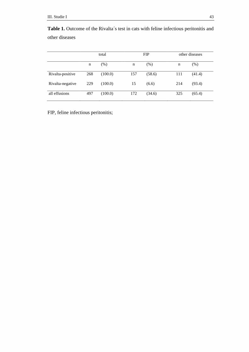
III. Studie I 43
Table 1. Outcome of the Rivalta´s test in cats with feline infectious peritonitis and
other diseases
total FIP other diseases
n (%) n (%) n (%)
Rivalta-positive 268 (100.0) 157 (58.6) 111 (41.4)
Rivalta-negative 229 (100.0) 15 (6.6) 214 (93.4)
all effusions 497 (100.0) 172 (34.6) 325 (65.4)
FIP, feline infectious peritonitis;
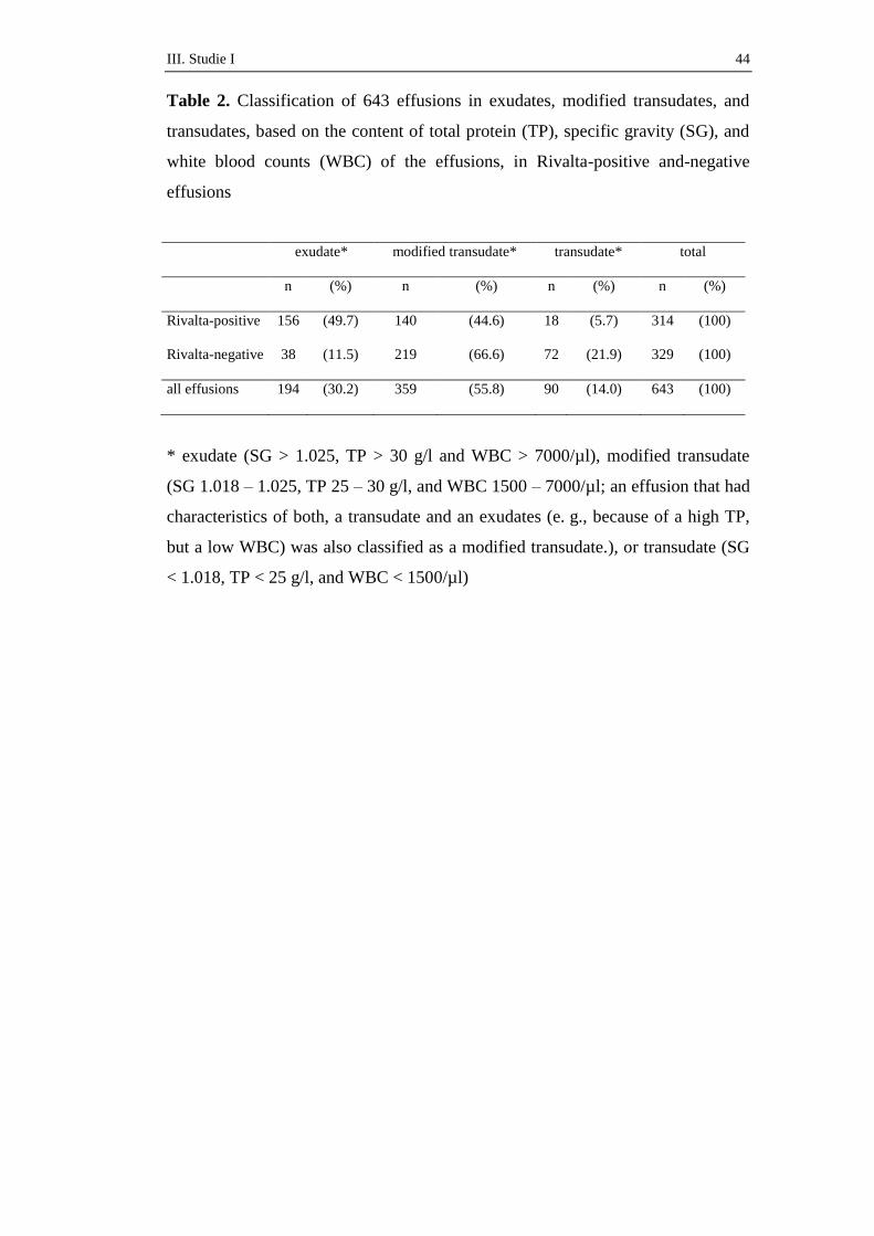
III. Studie I 44
Table 2. Classification of 643 effusions in exudates, modified transudates, and
transudates, based on the content of total protein (TP), specific gravity (SG), and
white blood counts (WBC) of the effusions, in Rivalta-positive and-negative
effusions
exudate* modified transudate* transudate* total
n (%) n (%) n (%) n (%)
Rivalta-positive 156 (49.7) 140 (44.6) 18 (5.7) 314 (100)
Rivalta-negative 38 (11.5) 219 (66.6) 72 (21.9) 329 (100)
all effusions 194 (30.2) 359 (55.8) 90 (14.0) 643 (100)
* exudate (SG > 1.025, TP > 30 g/l and WBC > 7000/µl), modified transudate
(SG 1.018 – 1.025, TP 25 – 30 g/l, and WBC 1500 – 7000/µl; an effusion that had
characteristics of both, a transudate and an exudates (e. g., because of a high TP,
but a low WBC) was also classified as a modified transudate.), or transudate (SG
< 1.018, TP < 25 g/l, and WBC < 1500/µl)
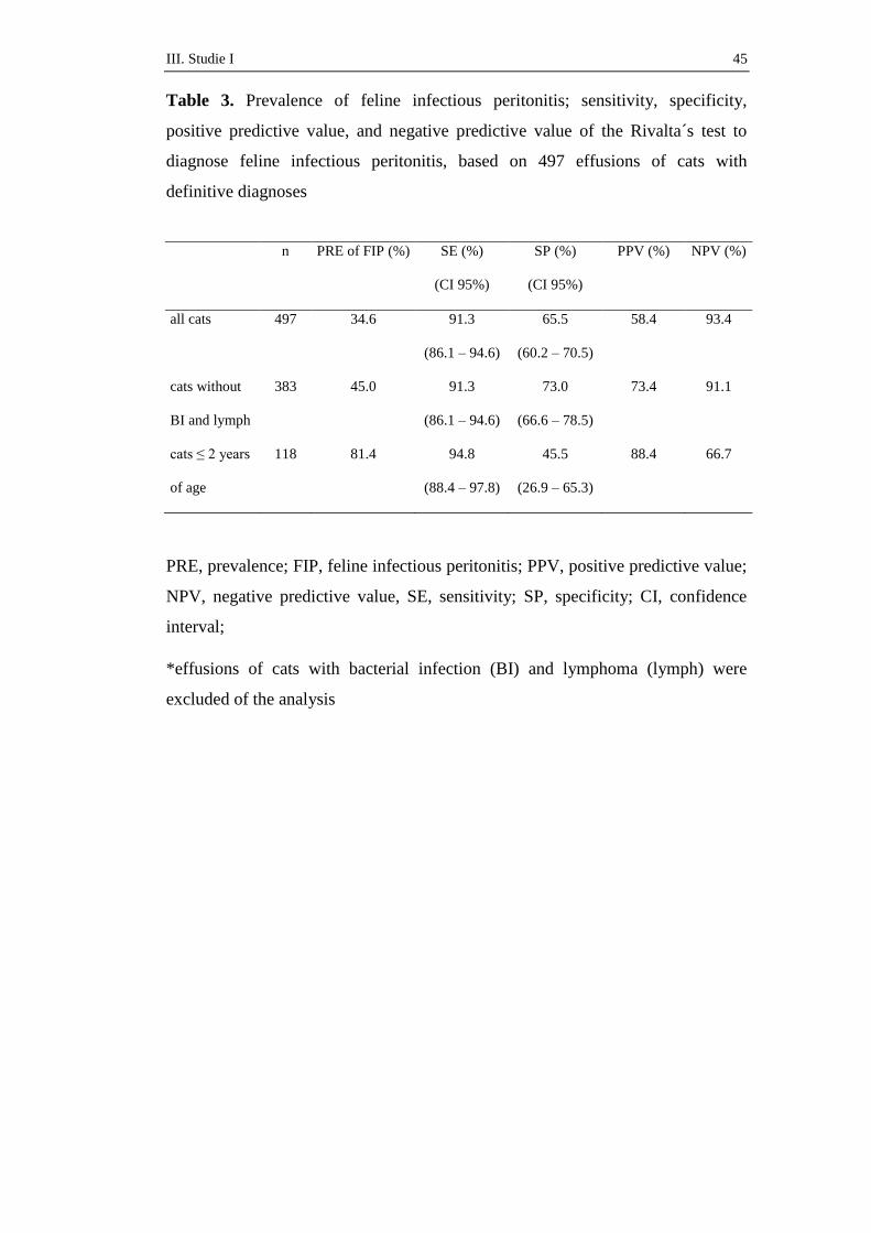
III. Studie I 45
Table 3. Prevalence of feline infectious peritonitis; sensitivity, specificity,
positive predictive value, and negative predictive value of the Rivalta´s test to
diagnose feline infectious peritonitis, based on 497 effusions of cats with
definitive diagnoses
n
PRE of FIP (%)
SE (%)
(CI 95%)
SP (%)
(CI 95%)
PPV (%)
NPV (%)
all cats
497
34.6
91.3
(86.1 – 94.6)
65.5
(60.2 – 70.5)
58.4
93.4
cats without
BI and lymph
383
45.0
91.3
(86.1 – 94.6)
73.0
(66.6 – 78.5)
73.4
91.1
cats ≤ 2 years
of age
118
81.4
94.8
(88.4 – 97.8)
45.5
(26.9 – 65.3)
88.4
66.7
PRE, prevalence; FIP, feline infectious peritonitis; PPV, positive predictive value;
NPV, negative predictive value, SE, sensitivity; SP, specificity; CI, confidence
interval;
*effusions of cats with bacterial infection (BI) and lymphoma (lymph) were
excluded of the analysis
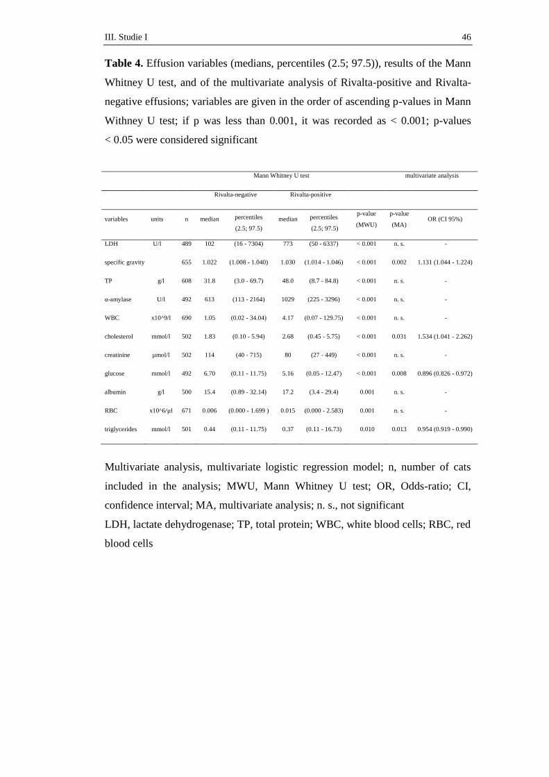
III. Studie I 46
Table 4. Effusion variables (medians, percentiles (2.5; 97.5)), results of the Mann
Whitney U test, and of the multivariate analysis of Rivalta-positive and Rivalta-
negative effusions; variables are given in the order of ascending p-values in Mann
Withney U test; if p was less than 0.001, it was recorded as < 0.001; p-values
< 0.05 were considered significant
Mann Whitney U test multivariate analysis
Rivalta-negative Rivalta-positive
variables units n median percentiles
(2.5; 97.5)
median percentiles
(2.5; 97.5)
p-value
(MWU)
p-value
(MA) OR (CI 95%)
LDH U/l 489 102 (16 - 7304) 773 (50 - 6337) < 0.001 n. s. -
specific gravity 655 1.022 (1.008 - 1.040) 1.030 (1.014 - 1.046) < 0.001 0.002 1.131 (1.044 - 1.224)
TP g/l 608 31.8 (3.0 - 69.7) 48.0 (8.7 - 84.8) < 0.001 n. s. -
α-amylase U/l 492 613 (113 - 2164) 1029 (225 - 3296) < 0.001 n. s. -
WBC x10^9/l 690 1.05 (0.02 - 34.04) 4.17 (0.07 - 129.75) < 0.001 n. s. -
cholesterol mmol/l 502 1.83 (0.10 - 5.94) 2.68 (0.45 - 5.75) < 0.001 0.031 1.534 (1.041 - 2.262)
creatinine µmol/l 502 114 (40 - 715) 80 (27 - 449) < 0.001 n. s. -
glucose mmol/l 492 6.70 (0.11 - 11.75) 5.16 (0.05 - 12.47) < 0.001 0.008 0.896 (0.826 - 0.972)
albumin g/l 500 15.4 (0.89 - 32.14) 17.2 (3.4 - 29.4) 0.001 n. s. -
RBC x10^6/µl 671 0.006 (0.000 - 1.699 ) 0.015 (0.000 - 2.583) 0.001 n. s. -
triglycerides mmol/l 501 0.44 (0.11 - 11.75) 0.37 (0.11 - 16.73) 0.010 0.013 0.954 (0.919 - 0.990)
Multivariate analysis, multivariate logistic regression model; n, number of cats
included in the analysis; MWU, Mann Whitney U test; OR, Odds-ratio; CI,
confidence interval; MA, multivariate analysis; n. s., not significant
LDH, lactate dehydrogenase; TP, total protein; WBC, white blood cells; RBC, red
blood cells
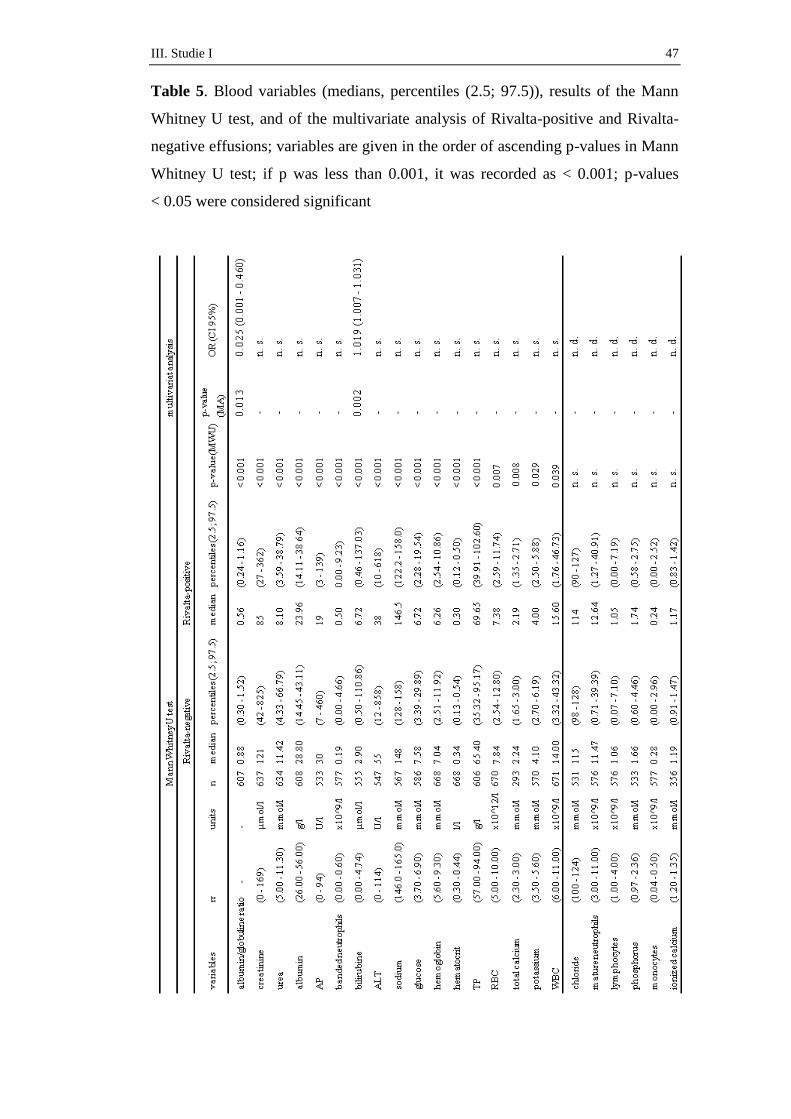
III. Studie I 47
Table 5. Blood variables (medians, percentiles (2.5; 97.5)), results of the Mann
Whitney U test, and of the multivariate analysis of Rivalta-positive and Rivalta-
negative effusions; variables are given in the order of ascending p-values in Mann
Whitney U test; if p was less than 0.001, it was recorded as < 0.001; p-values
< 0.05 were considered significant

III. Studie I 48
Multivariate analysis, multivariate logistic regression model; rr, reference range;
n, number of cats included in the analysis; MWU, Mann Whitney U test; OR,
Odds-ratio; CI, confidence interval; MA, multivariate analysis; n. s., not
significant; n. d., not determined
AP, alkaline phosphatase; ALT, alanine aminotransferase; TP, total protein; RBC,
red blood counts; WBC, white blood cell
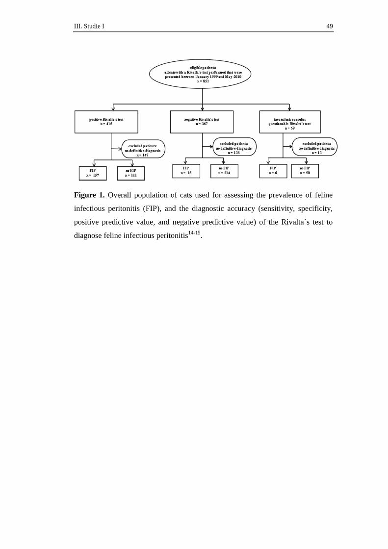
III. Studie I 49
Figure 1. Overall population of cats used for assessing the prevalence of feline
infectious peritonitis (FIP), and the diagnostic accuracy (sensitivity, specificity,
positive predictive value, and negative predictive value) of the Rivalta´s test to
diagnose feline infectious peritonitis14-15
.

III. Studie I 50
Figure 2. Population of cats excluding those with the diagnosis bacterial infection
or lymphoma, used for assessing the prevalence of feline infectious peritonitis
(FIP), and the diagnostic accuracy (sensitivity, specificity, positive predictive
value, and negative predictive value) of the Rivalta´s test to diagnose feline
infectious peritonitis14-15
.
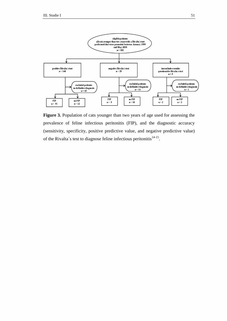
III. Studie I 51
Figure 3. Population of cats younger than two years of age used for assessing the
prevalence of feline infectious peritonitis (FIP), and the diagnostic accuracy
(sensitivity, specificity, positive predictive value, and negative predictive value)
of the Rivalta´s test to diagnose feline infectious peritonitis14-15
.
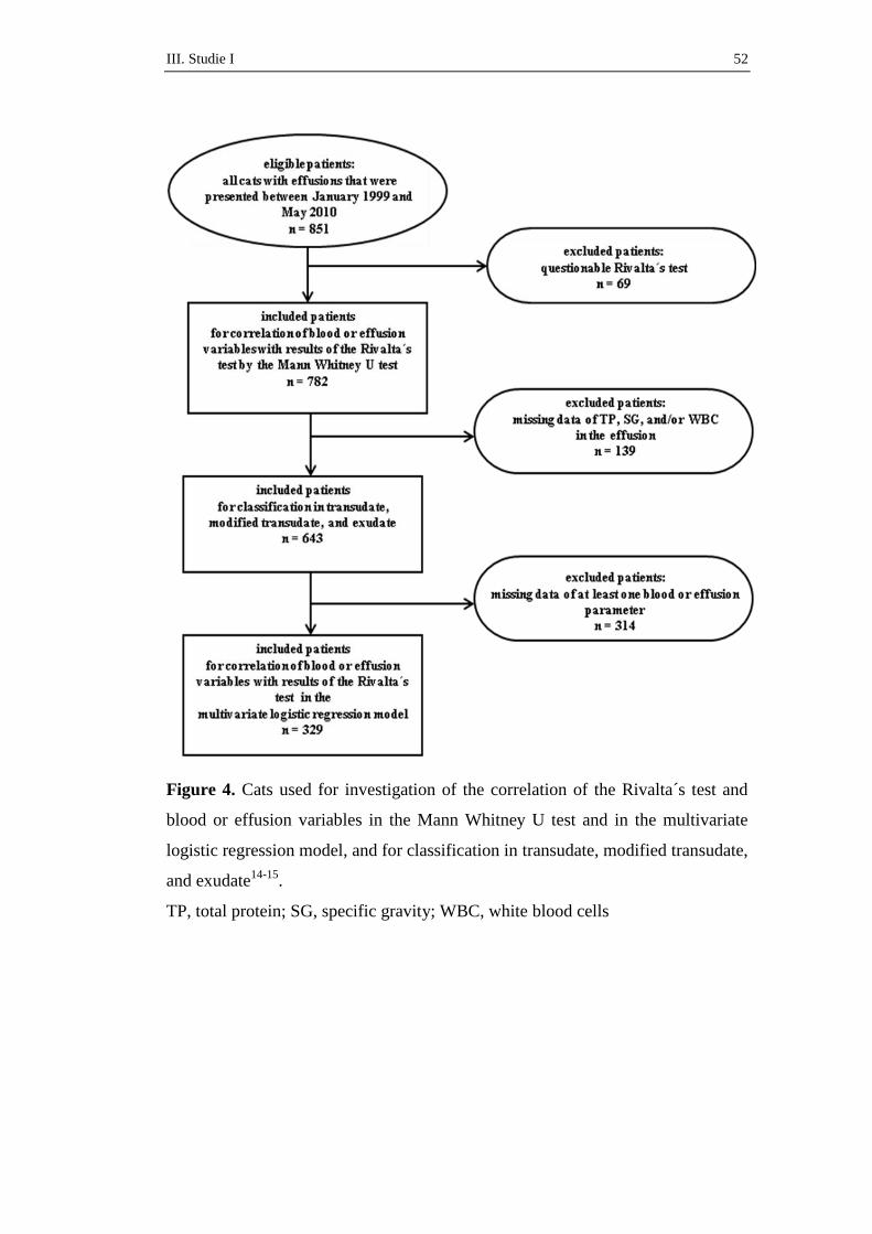
III. Studie I 52
Figure 4. Cats used for investigation of the correlation of the Rivalta´s test and
blood or effusion variables in the Mann Whitney U test and in the multivariate
logistic regression model, and for classification in transudate, modified transudate,
and exudate14-15
.
TP, total protein; SG, specific gravity; WBC, white blood cells
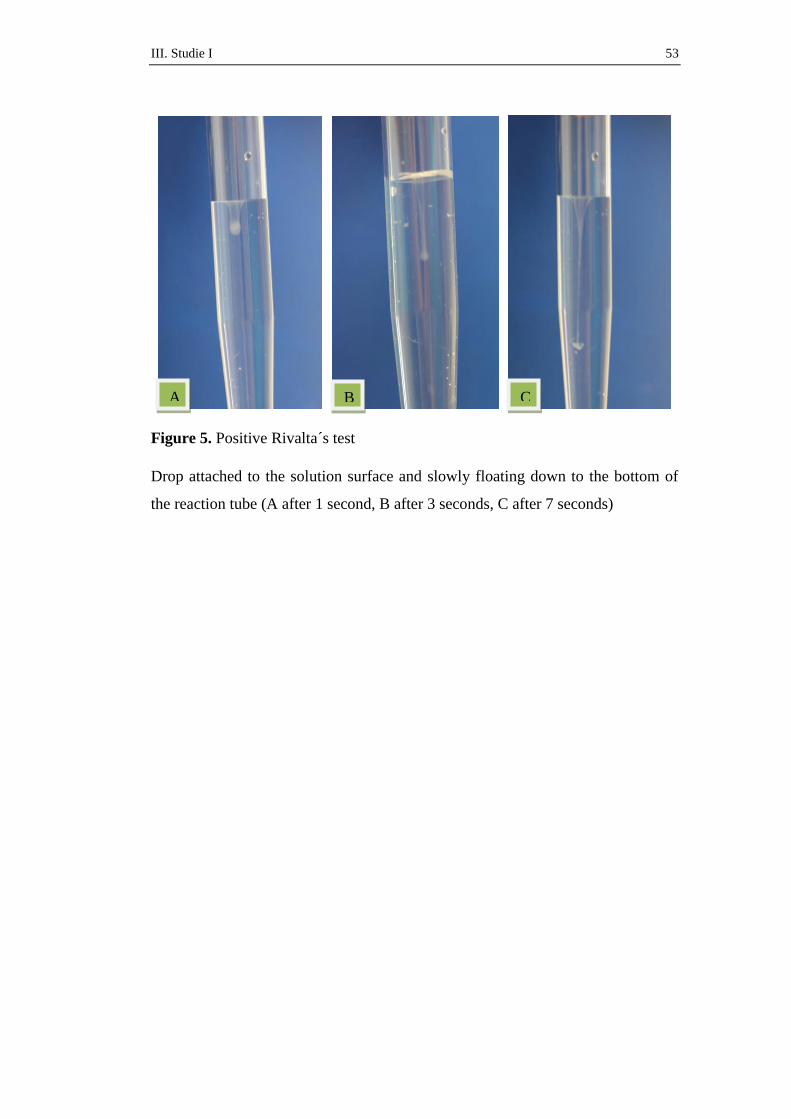
III. Studie I 53
Figure 5. Positive Rivalta´s test
Drop attached to the solution surface and slowly floating down to the bottom of
the reaction tube (A after 1 second, B after 3 seconds, C after 7 seconds)
A B C
IV. Studie II 54
IV. STUDIE II
The Rivalta´s Test as a Diagnostic Variable in Feline Effusions –
Evaluation of Optimum Reaction and Storage Condition
Yvonne Fischer1
Karin Weber, Dr. med. vet. 1
Carola Sauter-Louis, Dr. med. vet., PhD (epidemiology), Dipl. ECVPH 2
Katrin Hartmann, Prof. Dr. med. vet., Dr. med. vet. habil., Dipl. ECVIM-CA 1
1 Clinic of Small Animal Medicine, LMU University of Munich, Germany
2 Clinic for Ruminants, LMU University of Munich, Germany
Journal of Feline Medicine and Surgery, zur Veröffentlichung eingereicht
IV. Studie II 55
The Rivalta´s Test as a Diagnostic Variable in Feline Effusions – Evaluation
of Optimum Reaction and Storage Condition
Yvonne Fischer, Karin Weber, Carola Sauter-Louis, and Katrin Hartmann
Abstract:
Background: The Rivalta´s test is used to diagnose feline infectious peritonitis
(FIP) in cats with effusion. However, not much information on storage and
optimum reaction conditions is available in the literature, and diagnostic
sensitivity and specificity to diagnose FIP vary considerably between the few
available studies.
Objectives: The objectives of this study were to (1) examine the influence of
different storage conditions on the outcome of the test, (2) evaluate interobserver
variation, and (3) investigate optimum reaction conditions.
Methods: To determine influence of storage, the Rivalta´s test was repeated up to
21 days after storage at room temperature, in the refrigerator, or in the freezer. To
determine interobserver variation, the test was performed by two independent
blinded investigators. To determine optimum reaction conditions, the test was
performed with different volumes of acetic acid, with different acids as well as
with water.
Results: Even after storage of 21 days the test results were comparable; best
reproducibility was seen when effusion was stored in the refrigerator. While
interobserver variation revealed a relative high degree of disagreement, different
acids, different volumes of acetic acid, different kinds of water, and different
tubes for effusion collection showed no major influence on the outcome.
Conclusions: The Rivalta´s test seems to be a very robust test. Storage up to
21 days has no remarkable influence of the outcome of the test. Modifications in
reaction condition also do not substantially influence outcome of the test.
However, the test is subjective and depends on the experience of the investigator.
IV. Studie II 56
Introduction:
The Rivalta`s test is an old test that is still used commonly in many European
countries to diagnose feline infectious peritonitis (FIP) in cats with effusions. An
Italian physician, named Fabio Rivalta, developed this test to differentiate
exudates from transudates in human body cavity effusions1-2
. The principle of the
Rivalta´s test is the creation of a drop if effusion is added to a slightly acetic
solution. The Rivalta´s test had a sensitivity of 98% and a specificity of 80% to
diagnose FIP in cats in a large study in Germany3. Although commonly used in
Germany in veterinary practice, there is only little information about its mode of
action and the reasons, why the test is specifically useful to diagnose FIP. Most
authors support the theory, that a positive Rivalta´s reaction defines an effusion as
an exudate and that the positive reaction is mostly dependant on a high total
protein concentration in the effusion1, 4-6
. However, others argue that not only a
high total protein is responsible for a positive Rivalta´s reaction, but also
inflammatory mediators present in a “FIP-typical” exudate may play a role3, 7
.
Although commonly performed, there is also only little information available
about optimization of reagents used for the Rivalta´s test as well as optimum
conditions for storage of the effusion to obtain reliable results. So far, no
standardized protocol exists for this test6. The diagnostic values of the Rivalta´s
test to diagnose FIP vary considerably between different studies3, 8-9
, and
therefore, it also has been discussed, if the test outcome may depend on the
performing persons and the test conditions. Therefore, the aims of this study were
to (1) examine the influence of different storage conditions on the outcome of the
test, (2) evaluate interobserver variation, and (3) investigate optimum reaction
conditions.
IV. Studie II 57
Materials and methods
(1) Collection of effusions for the evaluation of storage and reaction conditions
To evaluate optimum storage and reaction conditions, 55 effusions of cats that
were presented to the Clinic of Small Animal Medicine, LMU University of
Munich, Germany, between January and July 2010 were collected. Each effusion
was obtained by ultrasound-guided thoracocentesis or aspiration of ascites or
pericardial fluid, using a 19- or 21-gauge butterfly needle connected to a closed
system, fixed to a 10 ml syringe. Two to 4 ml of the effusions were filled into
tubes containing EDTA as anticoagulant, and 0.5 to 2 ml were filled into a plain
tube without anticoagulant for further evaluation.
Of the 55 cats, 17 cats had the final diagnosis of FIP, 38 had other underlying
diseases. The diagnosis FIP was either confirmed by immunofluorescent staining
of feline coronavirus (FCoV) antigen in macrophages in effusion3, by
histopathological changes (if diagnostic for FIP)10-11
, or immunohistochemical
staining at postmortem examination12
. Effusion due to heart disease (n = 14) was
diagnosed using echocardiography13-14
. Neoplasia (n = 12) was either diagnosed
by cytology or, if not diagnostic, by histopathological examinations or
necropsy15
. Bacterial infections (n = 5) were confirmed by bacterial culture or
detection of bacteria in cytology16-17
. In seven cats, the reason for the effusion
was unclear, but all seven cats recovered within a short period of time and were
still healthy after four weeks. Therefore, FIP was considered extremely unlikely
in these cats.
(2) Performance of the Rivalta´s test
The Rivalta´s test (standard method) was performed as followed: a plastic test
tube (volume 10 ml) was filled with 7 ml of distilled water. Twenty µl of acetic
acid (100%) were added. The solution was thoroughly mixed. Twenty µl of the
effusion was layered on the surface of this solution, and the behavior of this drop
was observed. The test was evaluated as positive (+) if the drop stayed attached to
the surface, or if the drop retained its shape and kept a connection to the surface or
slowly floated down to the bottom. The test was also defined as (+), if the drop
did not stay in shape, changed to an upside-down jelly fish, or dispensed in little
cloudy pieces (that could be observed falling down to the bottom of the tube)
(figure 1). If the drop disappeared and the solution remained clear, or if clear or
IV. Studie II 58
cloudy cords were falling down (which dissolved before reaching the bottom of
the tube) but no drop or jelly fish was visible, the Rivalta´s test was defined as
negative (-).
The Rivalta´s test was performed with 55 effusion within one hour after aspiration
of the fluid. Of these 27 were defined as (+), 28 were defined as (-).
(3) Influence of different storage conditions on the outcome of the Rivalta´s test
To evaluate optimum storage conditions, 2 to 4 ml of each of the 55 effusions
filled in tubes containing EDTA as anticoagulant were used. Of each of the 55
collected effusions, 200 µl were stored in the refrigerator at a temperature of 4 °C
(39.2 °F) for a maximum of 21 days. Another 200 µl were stored at room
temperature at 20 °C (68 °F) for a maximum of 21 days. Nine eppendorf tubes
containing 30 µl of effusion each were frozen at a temperature of –20 °C (-4°F)
for a maximum of 21 days. To examine the influence of different storage
conditions on the outcome of the test, the Rivalta´s test was performed on day 0,
day 1 to 7, day 14, and day 21 with the effusions stored at the different
temperatures.
(4) Interobserver variations
To evaluate interobserver variations, the 55 effusions collected as described above
were used. The test was performed independently by two persons (one
experienced and one inexperienced) with all 55 effusions. Both investigators
performed the test within one hour after collection. Both were blinded to the
results of the other investigator.
(5) Optimization of the reaction conditions
To evaluate optimum reaction conditions, 2 to 4 ml of the 55 effusions collected
as described above were filled in tubes containing EDTA as anticoagulant. In
addition, 0.5 to 2.0 ml were filled in a plain tube without anticoagulant. The
Rivalta´s test was performed within one hour after collection using different
conditions (see table 1). A “standard tube” (tube (S)), in which the standard
protocol (as described) above was used, served as control.
First, the influence of different kinds of acid was investigated. In tube (S), 20 µl
of acetic acid (100%), in tube (A2) 20 µl of distilled white vinegar (25%), and in
IV. Studie II 59
tube (A3) 20 µl of wine vinegar (5%) were added to the 7 ml of distilled water.
Second, to examine the influence of the amount of acids and therefore, the
influence of the acidity on the outcome of the Rivalta´s test, the test was
performed with different volumes of acetic acid (100%); in tube (S) 20 µl
(pH 3.00), and in tube (V2) 200 µl (pH 2.50) of acetic acid (100%) were added to
the 7 ml of distilled water.
Third, the influence of the collection tubes and preparation of the effusion was
investigated. In tube (S) the Rivalta´s test was performed with 20 µl native
effusion collected in a tube with EDTA, in tube (C2) the test was performed with
20 µl supernatant after centrifugation at 1500 revolutions per minute for
5 minutes and removal of the sediment of the effusion collected in a tube with
EDTA, and in tube (C3) the test was performed with 20 µl native effusion
collected in a plain tube without anticoagulant (serum tube).
Fourth, the influence of the kind of fluid was evaluated. Therefore, in tube (S) the
Rivalta´s test was performed using distilled water (tube (S)), and in tube (W)
using 7 ml of normal tap water.
(6) Statistical analyses
Statistical analyses were performed using statistical software SPSS version 17.0
(http://www.spss.com). To determine influence of different storage conditions on
the outcome of the Rivalta´s test, the results of the tests on day 0 were compared
with the results on day 1 to day 7, day 14, and day 21 performed with effusions
stored at different temperatures using a 2-way-mixed effect intraclass correlation
coefficient (ICC) together with the confidence interval. The ICC is a descriptive
statistic that can be used to assess the consistency or reproducibility of
quantitative measurements of the same test performed at different points in time.
High levels of agreement (ICC = 1) were obtained when the amount of variation
caused by the method was smaller than the variability resulting from all other
sources of variation. According to Landis and Koch (1977), an ICC value of less
than 0.40 was considered as representing poor to slight agreement, 0.40 – 0.80 as
fair to good agreement, and more than 0.80 as excellent agreement18
.
To determine interobserver variation, the agreement of the results of the two
persons performing the Rivalta´s test was also assessed using the ICC18
.
IV. Studie II 60
To assess the agreement of the different reaction conditions with the standard
method, also the ICC was used as well18
. In addition, the diagnostic sensitivity of
the tests performed with different reaction conditions compared to the standard
method (proportion of the different tests, which were true-positive (TP) or false-
negative (FN) compared to tube (S)), and the specificity (proportion of the
different tests, which were false-positive (FP) or true-negative (TN) compared to
the results of the standard method (tube (S))) were evaluated when performing the
Rivalta`s test under different conditions18
. To examine, if different reaction
conditions other than the standard method showed better diagnostic values to
diagnose FIP, the number of TP, FP, TN, FN results, as well as the diagnostic
sensitivity to diagnose FIP (here proportion of cats that show Rivalta-positive test
results among all cats with FIP), and the diagnostic specificity to diagnose FIP
(proportion of cats that show Rivalta-negative test results among all cats with
other diseases than FIP) were investigated.
IV. Studie II 61
Results
(1) Influence of different storage conditions on the outcome of the Rivalta´s test
Comparison of ICC values of the Rivalta´s test performed directly after obtaining
the effusion with the results of the Rivalta´s tests after storage at room
temperature (20 °C/68 °F), in the refrigerator (4 °C/39.2 °F), or in the freezer
(-20 °C/-4 °F) for up to 21 days are shown in table 2. All values showed almost
perfect (or at least substantial) agreement with the result on day 0, meaning, that
storage of effusions did not change the outcome of the Rivalta´s test. There were
only minimal differences in ICC values between the different storage options. The
best values with always almost perfect agreement on all individually days were
obtained when effusions were stored in the refrigerator (table 2).
(2) Interobserver variations
Table 3 shows the interobserver variations as well as sensitivity and specificity to
diagnose FIP of the Rivalta´s test performed by the inexperienced and the
experienced person. In 47/55 (85.5%) of the results, an agreement between the
two persons was obtained leading to an ICC of 0.710 (confidence interval 95%:
0.550 - 0.820).
(3) Optimization of the reaction conditions
Table 4 shows the results of the Rivalta´s test performed with different reaction
conditions compared to the standard method (tube (S)) as well as the diagnostic
value of the different conditions to diagnosis FIP. The best agreement with the
standard method was seen with the tube using 200 µl acetic acid (100%)
(ICC = 0.927). The tube with 40 µl of distilled white vinegar showed the best
sensitivity and specificity to diagnose FIP.
IV. Studie II 62
Discussion
An important practical aspect of the present study that has not been addressed
before was the question, if storage of the effusion has an influence on the
positivity or negativity of the Rivalta´s test. This study proved, that the outcome
of the Rivalta´s test does not change, when effusion is stored for up to three weeks
at room temperature, in the refrigerator, or in the freezer. As storage in the
refrigerator in general showed the best ICC values (≥ 0.82), refrigerator storage
should be recommended. But also storage in the freezer or at room temperature
showed on most days almost perfect (ICC > 0.79) (or at least substantial (ICC
0.60 – 0.79)) agreement with the results obtained on the day of tapping the
effusion (table 2). Therefore, storage of effusion up to 21 days is possible without
problems before performing the Rivalta´s test, and effusion fluids can easily be
send by regular mail to a commercial laboratory without loss of information.
However, sensitivity and specificity of the Rivalta´s test to diagnose FIP vary
considerably between the few available publications. Some years ago, two studies
in Germany were performed, in which the Rivalta´s test showed a sensitivity of
98 - 100% and a specificity of 80 - 82%3, 9
. Recently, two new studies in Germany
were performed. In the first, the sensitivity of the Rivalta´s test only reached 39%
(with 8/13 false-negative results), but the specificity reached 100%8. In the other
study, the sensitivity was 91% and the specificity was 66%19
. Therefore it has
been discussed, if these differences may depend on the performing persons and
their experience. To evaluate an influence of the investigator, the Rivalta´s test in
the present study was performed by the two different persons under identical
conditions that were blinded to the results obtained by the other person. In 8/55
(14.5%) effusions, different results between the experienced and the
inexperienced persons (ICC 0.71) were obtained, which is not a very good
agreement. If the test was performed by an inexperienced person, the value of the
Rivalta´s test to diagnose FIP was worse (sensitivity 64.7%, specificity 63.2%)
than the values of the experienced person (sensitivity 88.2%, specificity 68.4%).
Therefore, the interpretation of the Rivalta´s test seems to be very subjective. This
may be due to the absence of clear definitions on how to interpret Rivalta´s test
results. As the appearance of a drop can be influenced when the test is performed
manually, e. g. by the angle or fall velocity of the effusion drop, not every drop
will have the same form, and the outcome can be interpreted falsely. Therefore,
IV. Studie II 63
more clear descriptions on how a positive Rivalta´s reaction is defined might be
helpful.
The recipes of the Rivalta´s test and explanation, how it should be performed
differ in the literature, e. g. concerning details about the volumes of distilled water
(e. g., 5 ml, 7-8 ml, ¾ of the reagent tube)3, 7, 20
. As “one drop” (in most
descriptions “one drop of acetic “ is added to the distilled water, and “one drop of
effusion” is then added to the reaction tube) is also a very imprecise instruction.
Therefore, because there is no standardized method in literature6, a standardized
method was first defined in the paper. Instead of “¾ of the reagent tube”7, 7 ml
were used. Instead of “one drop”, 20 µl were used. However, as shown in table 4,
different reaction conditions did hardly change the outcome and still had an
almost perfect agreement with the standard method (tube (S)).
Acetic acid obviously can be used as well as commercial acid (in this study
distilled white vinegar or vine vinegar was chosen). In this study, the amount of
the lower concentrated vinegars was increased, so that approximately the same pH
value was achieved as if using a smaller amount of acetic acid (100%). Using
40 µl distilled white vinegar even leads to better sensitivity and specificity to
diagnose FIP. In an older study with human body fluids, diagnostic value of the
Rivalta´s test the use of other acids (hydrochloric acid (35-37%) and nitric acid
(60-62%)) was also investigated. In that study, however, results obtained with
different kinds of acid were actually quite different; of 39 samples, 32 were
judged as positive when using acetic acid. With hydrochloric acid, only 29 and
with nitric acid, 34 of the 39 samples were positive. It was not described in detail
which amounts of each acid were used, but the tests were performed within the
same pH range (3.0 – 5.0)6. Maybe, it is not important which kind of acetic acid is
used, but it should be some type of vinegar, and no any other source of acid. In the
present study, no other acid was examined, but it did not seem to be very
important which type of vinegar is used.
It was also investigated whether the amount of acid and/or the pH value play a
role. There was no major difference in outcome using 20 µl (pH 3.00) or with
200 µl (pH 3.06) of acetic acid (100%) in this study. Interestingly, this is contrary
to an investigation of Sakai et al. (2004), who found, that the percentage of
positive results in human effusions rapidly changes when pH value is < 3.86. As
nearly all Rivalta´s tests in the present study were performed in solutions with pH
IV. Studie II 64
values much less than 3.8 (see table 1; only the performance with tap water had a
pH value of 3.84), it can be speculated, that in feline effusions other substances
(e. g., different proteins) in the effusion may be responsible for a positive
outcome and the reaction may not be influenced by the lower pH6.
The influence of collection tubes and preparation of the effusion was also
assessed. The type of tube in which the effusion was collected after aspiration did
not seem to be important (table 4). It can be either a tube with EDTA, or a serum
tube. Even the supernatant of an effusion can be used, if the effusion, for example,
was already centrifuged for cytology. This actually shows that the inflammatory
cells themselves cannot be responsible for a positive Rivalta´s reaction, as those
had been removed when supernatant was used.
The Influence of the type of water for the Rivalta´s test was also investigated.
Interestingly, outcome of the Rivalta´s test did not change when tap water was
used instead of distilled water. The solution with tap water had a much higher pH
value (pH 3.84) than the solutions with distilled water (pH 3.00), at least using
Munich tap water, as pH values differ from region to region. But even when
performing the test using solutions of different pH, the outcome did not change.
There are some limitations in this study. One limitation is the subjectivity of this
test. As shown with the high interobserver variation, it cannot be excluded that
even when the experienced person performed the test, other factors (e. g., distance
and fall velocity of the effusion-drop) influenced outcome. As the test is always
performed manually and judged visually, the results remain somehow subjective
to individual interpretation.
In conclusion, this study shows, that the Rivalta´s test is a very robust test. It can
be performed without any problems after a storage period of up to 21 days. The
best method of storage is in the refrigerator. It more or less makes no difference,
which type of acetic acid and which water is used or in which tubes the effusion is
collected. The results hardly change with some modifications of the standard
protocol. The interobserver variation, however, showed, that the Rivalta´s test
remains a very subjective test, and that a standardization of the interpretation
criteria would be useful to increase the sensitivity and specificity of the Rivalta´s
test to diagnose FIP.
IV. Studie II 65
References
1. Berti-Bock G, Vial F, Premuda L, Rulliere R. [Exudates, transudates and
the Rivalta reaction (1895). Current status and historical premises].
Minerva Med. 1979;70:3573-3580.
2. Rivalta F. Su di una nuova reazione per la diagnosi chimica differenziale
fra gli essudati sierosi e i semplici trasudati. La Riforma Medica.
1895;242.
3. Hartmann K, Binder C, Hirschberger J, et al. Comparison of different tests
to diagnose feline infectious peritonitis. J Vet Intern Med. 2003;17:781-
790.
4. Staehelin R. Ueber den durch Essigsäure fällbaren Eiweisskörper der
Exsudate und des Urins. Munch Med Wochenschr. 1902;34:1413-1415.
5. Matracia S, Campo G, Titone C. [Interaction between globin and Rivalta-
positive colloids.]. Boll Soc Ital Biol Sper. 1963;39:148-149.
6. Sakai N, Iijima S, Shiba K. Reinvestigation of clinical value of Rivalta
reaction of puncture fluid. Rinsho Byori. 2004;52:877-882.
7. Hartmann K. Feline infectious peritonitis. Vet Clin North Am Small Anim
Pract. 2005;35:39-79.
8. Held S, König, M., Hamann, H.-P., Neiger, R. Evaluierung diagnostischer
Tests für feline infektiöse Peritonitis (FIP) bei Katzen mit Aszites
(abstract). Leipzig: InnLab DVG;2011.
9. Hirschberger J, Hartmann K, Wilhelm N, Frost J, Lutz H, Kraft W.
[Clinical symptoms and diagnosis of feline infectious peritonitis]. Tierarztl
Prax. 1995;23:92-99.
10. Addie DD, Paltrinieri S, Pedersen NC. Recommendations from workshops
of the second international feline coronavirus/feline infectious peritonitis
symposium. J Feline Med Surg. 2004;6:125-130.
11. Sparkes AH, Gruffydd-Jones TJ, Harbour DA. Feline infectious
peritonitis: a review of clinicopathological changes in 65 cases, and a
critical assessment of their diagnostic value. Vet Rec. 1991;129:209-212.
12. Tammer R, Evensen O, Lutz H, Reinacher M. Immunohistological
demonstration of feline infectious peritonitis virus antigen in paraffin-
embedded tissues using feline ascites or murine monoclonal antibodies.
Vet Immunol Immunopathol. 1995;49:177-182.
13. Bonagura JD. Feline echocardiography. J Feline Med Surg. 2000;2:147-
151.
14. Ferasin L. Feline myocardial disease 2: diagnosis, prognosis and clinical
management. J Feline Med Surg. 2009;11:183-194.
15. Hirschberger J, DeNicola DB, Hermanns W, Kraft W. Sensitivity and
specificity of cytologic evaluation in the diagnosis of neoplasia in body
fluids from dogs and cats. Vet Clin Pathol. 1999;28:142-146.
16. Barrs VR, Beatty JA. Feline pyothorax - new insights into an old problem:
part 1. Aetiopathogenesis and diagnostic investigation. Vet J.
2009;179:163-170.
17. Ottenjann M, Lubke-Becker A, Linzmann H, Brunnberg L, Kohn B.
[Pyothorax in 26 cats: clinical signs, laboratory results and therapy (2000-
2007)]. Berl Munch Tierarztl Wochenschr. 2008;121:365-373.
18. Landis JR, Koch GG. The measurement of observer agreement for
categorical data. Biometrics. 1977;33:159-174.
19. Fischer Y, Sauter-Louis, C., Hartmann, K. Die Rivalta-Probe zur

IV. Studie II 66
Diagnose der felinen infektiösen Peritonitis - retrospektive Auswertung
von 782 Ergüssen (abstract). Leipzig: InnLab DVG;2011.
20. Addie D, Belak S, Boucraut-Baralon C, et al. Feline infectious peritonitis.
ABCD guidelines on prevention and management. J Feline Med Surg.
2009;11:594-604.
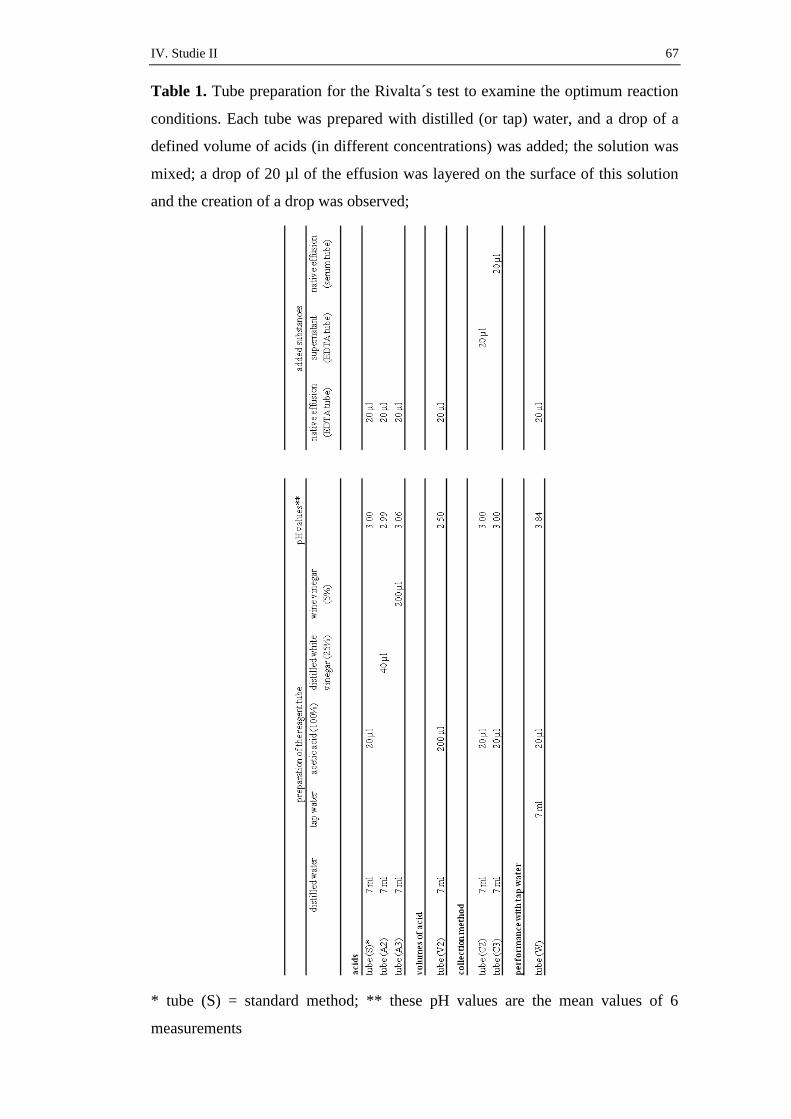
IV. Studie II 67
Table 1. Tube preparation for the Rivalta´s test to examine the optimum reaction
conditions. Each tube was prepared with distilled (or tap) water, and a drop of a
defined volume of acids (in different concentrations) was added; the solution was
mixed; a drop of 20 µl of the effusion was layered on the surface of this solution
and the creation of a drop was observed;
* tube (S) = standard method; ** these pH values are the mean values of 6
measurements
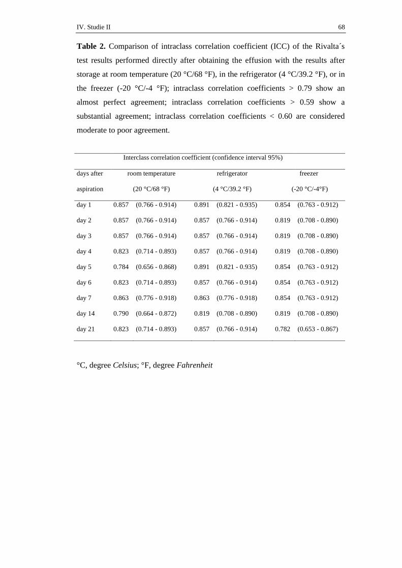
IV. Studie II 68
Table 2. Comparison of intraclass correlation coefficient (ICC) of the Rivalta´s
test results performed directly after obtaining the effusion with the results after
storage at room temperature (20 °C/68 °F), in the refrigerator (4 °C/39.2 °F), or in
the freezer (-20 °C/-4 °F); intraclass correlation coefficients > 0.79 show an
almost perfect agreement; intraclass correlation coefficients > 0.59 show a
substantial agreement; intraclass correlation coefficients < 0.60 are considered
moderate to poor agreement.
Interclass correlation coefficient (confidence interval 95%)
days after room temperature refrigerator freezer
aspiration (20 °C/68 °F) (4 °C/39.2 °F) (-20 °C/-4°F)
day 1 0.857 (0.766 - 0.914) 0.891 (0.821 - 0.935) 0.854 (0.763 - 0.912)
day 2 0.857 (0.766 - 0.914) 0.857 (0.766 - 0.914) 0.819 (0.708 - 0.890)
day 3 0.857 (0.766 - 0.914) 0.857 (0.766 - 0.914) 0.819 (0.708 - 0.890)
day 4 0.823 (0.714 - 0.893) 0.857 (0.766 - 0.914) 0.819 (0.708 - 0.890)
day 5 0.784 (0.656 - 0.868) 0.891 (0.821 - 0.935) 0.854 (0.763 - 0.912)
day 6 0.823 (0.714 - 0.893) 0.857 (0.766 - 0.914) 0.854 (0.763 - 0.912)
day 7 0.863 (0.776 - 0.918) 0.863 (0.776 - 0.918) 0.854 (0.763 - 0.912)
day 14 0.790 (0.664 - 0.872) 0.819 (0.708 - 0.890) 0.819 (0.708 - 0.890)
day 21 0.823 (0.714 - 0.893) 0.857 (0.766 - 0.914) 0.782 (0.653 - 0.867)
°C, degree Celsius; °F, degree Fahrenheit
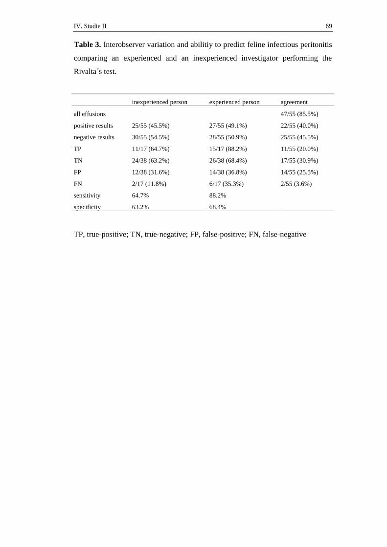
IV. Studie II 69
Table 3. Interobserver variation and abilitiy to predict feline infectious peritonitis
comparing an experienced and an inexperienced investigator performing the
Rivalta´s test.
inexperienced person experienced person agreement
all effusions
47/55 (85.5%)
positive results 25/55 (45.5%) 27/55 (49.1%) 22/55 (40.0%)
negative results 30/55 (54.5%) 28/55 (50.9%) 25/55 (45.5%)
TP 11/17 (64.7%) 15/17 (88.2%) 11/55 (20.0%)
TN 24/38 (63.2%) 26/38 (68.4%) 17/55 (30.9%)
FP 12/38 (31.6%) 14/38 (36.8%) 14/55 (25.5%)
FN 2/17 (11.8%) 6/17 (35.3%) 2/55 (3.6%)
sensitivity 64.7% 88.2%
specificity 63.2% 68.4%
TP, true-positive; TN, true-negative; FP, false-positive; FN, false-negative
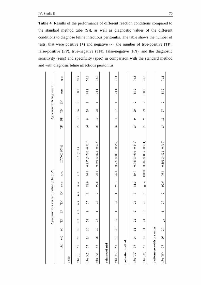
IV. Studie II 70
Table 4. Results of the performance of different reaction conditions compared to
the standard method tube (S)), as well as diagnostic values of the different
conditions to diagnose feline infectious peritonitis. The table shows the number of
tests, that were positive (+) and negative (-), the number of true-positive (TP),
false-positive (FP), true-negative (TN), false-negative (FN), and the diagnostic
sensitivity (sens) and specificity (spec) in comparison with the standard method
and with diagnosis feline infectious peritonitis.

IV. Studie II 71
(+), Rivalta´s test positive; (-), Rivalta´s test negative; TP, true-positive; FP, false-
positive; TN, true-negative; FN, false-negative; sens, sensitivity; spec, specificity;
ICC, intraclass correlation coefficient; CI, confidence interval; n. a., not
applicable;
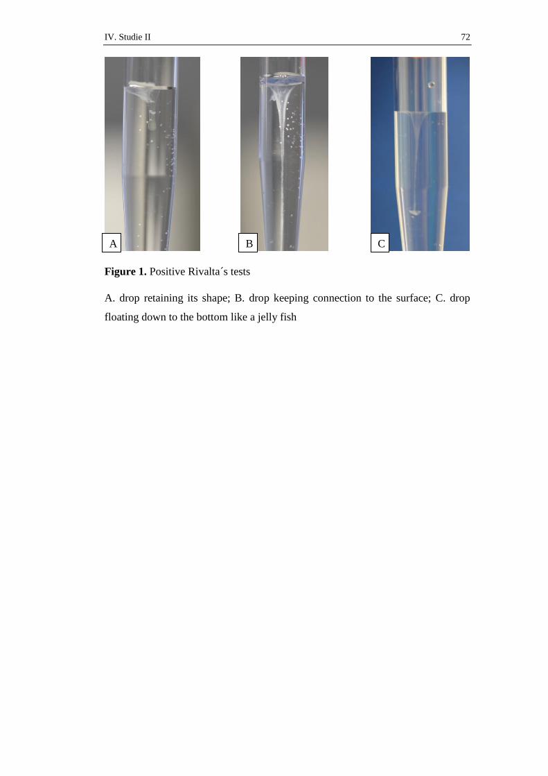
IV. Studie II 72
Figure 1. Positive Rivalta´s tests
A. drop retaining its shape; B. drop keeping connection to the surface; C. drop
floating down to the bottom like a jelly fish
C B A
V. Studie III 73
V. STUDIE III
Randomized, Placebo Controlled Study of the Effect of
Propentofylline on Survival Time and Quality of Life of Cats with
Feline Infectious Peritonitis
Yvonne Fischer 1
Susanne Ritz, Dr. med. vet. 1
Karin Weber, Dr. med. vet. 1
Carola Sauter-Louis, Dr. med. vet., PhD (epidemiology), Dipl. ECVPH 2
Katrin Hartmann, Prof. Dr. med. vet., Dr. med. vet. habil., Dipl. ECVIM-CA 1
1 Clinic of Small Animal Medicine, LMU University of Munich, Germany
2 Clinic for Ruminants, LMU University of Munich, Germany
Journal of Veterinary Internal Medicine, veröffentlicht
J Vet Intern Med 2011;25:1270–1276
V. Studie III 74
V. Studie III 75
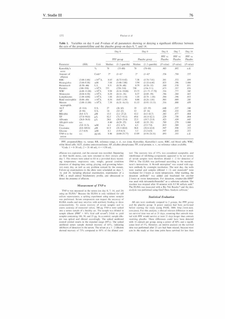
V. Studie III 76
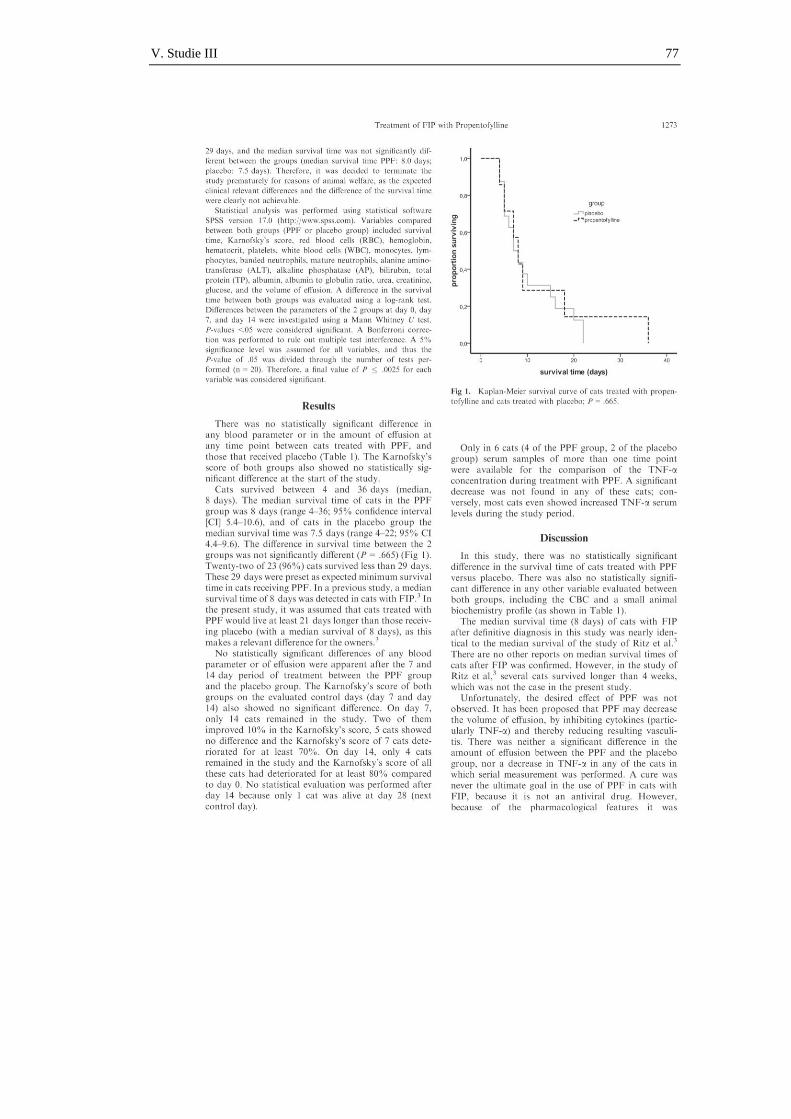
V. Studie III 77
V. Studie III 78
V. Studie III 79

V. Studie III 80
VI. Studie IV 81
VI. STUDIE IV
Perikarderguss bei einer Katze mit Feliner Infektiöser Peritonitis
Yvonne Fischer 1
Gerhard Wess, Priv.-Doz., Dr. med. vet., Dr. med. vet. habil., Dipl. ECVIM-CA
(Internal Medicine),l Dipl. ECVIM-CA + ACVIM (Cardiology) 1
Katrin Hartmann, Prof. Dr. med. vet., Dr. med. vet. habil., Dipl. ECVIM-CA
(Internal Medicine) 1
1 Clinic of Small Animal Medicine, LMU University of Munich, Germany
Schweizer Archiv für Tierheilkunde, veröffentlicht
Schweiz. Arch. Tierheilk. Band 154, Heft 1, Januar 2012, 27 – 31
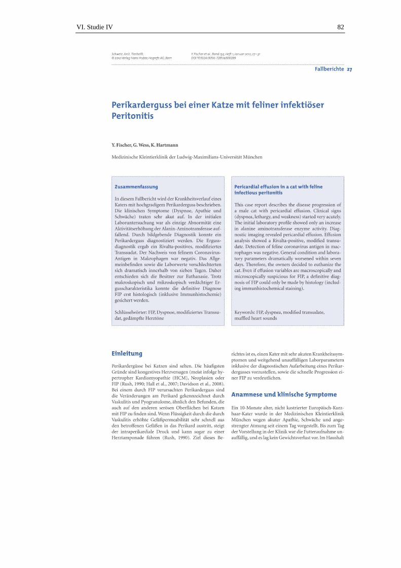
VI. Studie IV 82
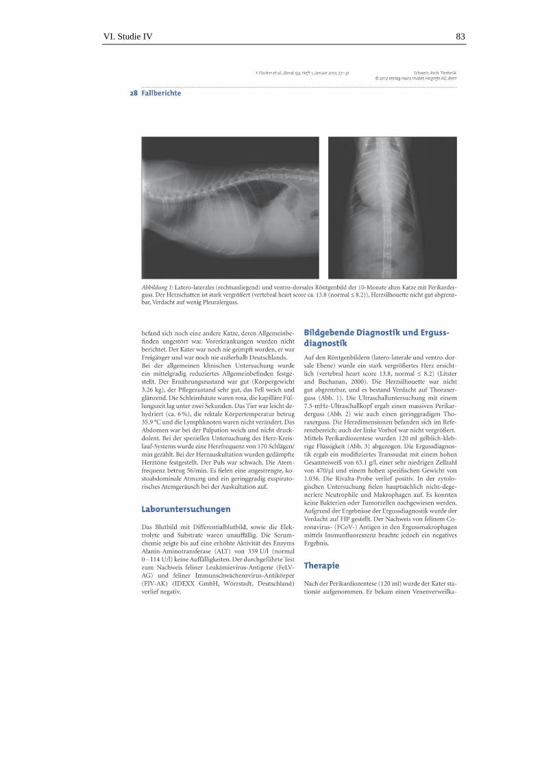
VI. Studie IV 83
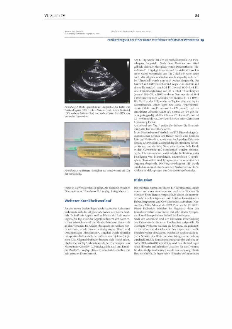
VI. Studie IV 84
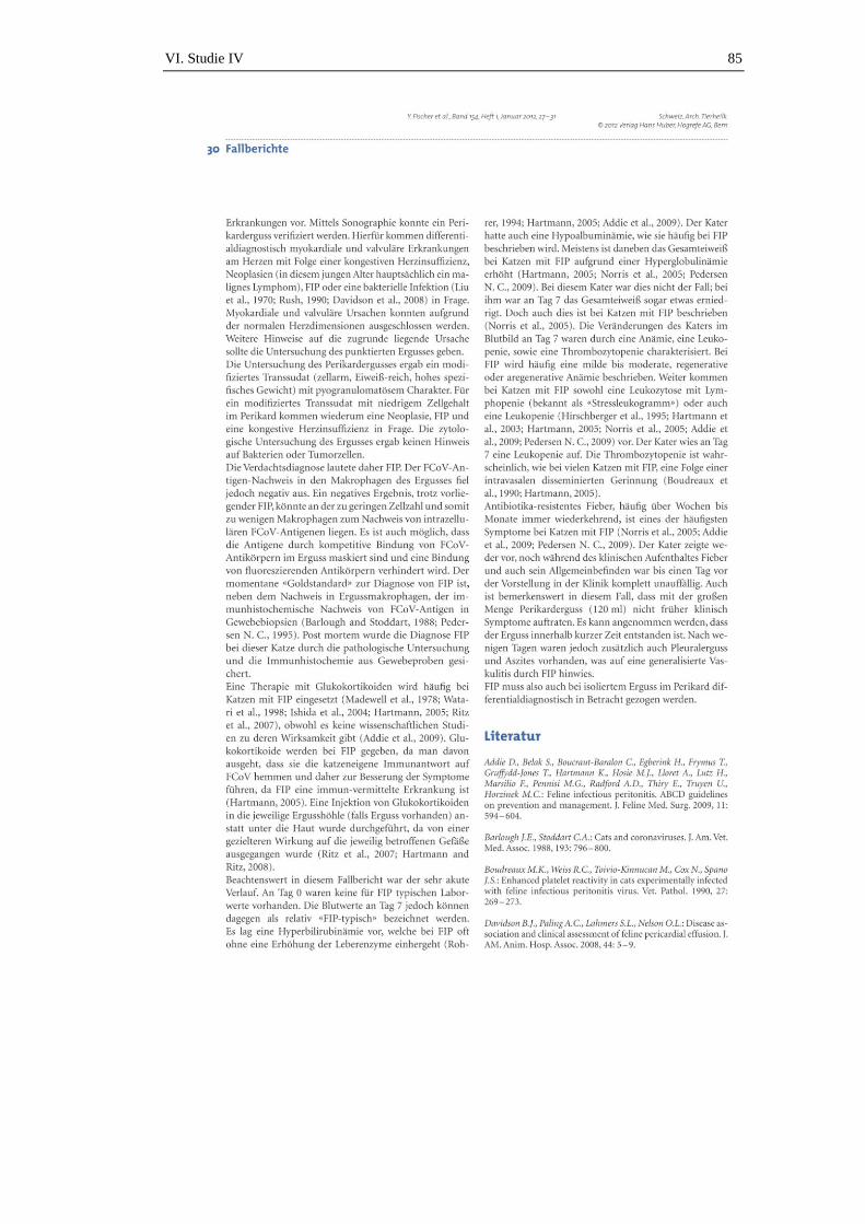
VI. Studie IV 85
VI. Studie IV 86
VII. Diskussion 87
VII. DISKUSSION
Noch immer sind die Möglichkeiten zur Diagnose einer FIP am lebenden Tier
sehr beschränkt. Bei Katzen mit Erguss gibt es die Möglichkeit, FCoV-Antigen in
den Makrophagen im Erguss nachzuweisen und damit die Diagnose zu sichern
(HIRSCHBERGER et al., 1995; HARTMANN et al., 2003). Diese Methode ist im
positiven Fall beweisend für eine FIP. Leider jedoch dauert es meist mehrere Tage
bis ein Ergebnis vorliegt. Vielen Katzen geht es sehr schlecht und ein Warten auf
das Ergebnis verzögert eine möglicherweise einzuleitende immunsuppressive
Therapie. Eine andere Untersuchungsmethode von Erguss, die Rivalta-Probe,
zeigte in früheren Studien sehr gute diagnostische Werte (HIRSCHBERGER et
al., 1995; HARTMANN et al., 2003). Die Rivalta-Probe kann schnell und einfach
durchgeführt werden. Der Fokus des ersten Teils dieser Arbeit lag auf einer
genauen Betrachtung der Rivalta-Probe durch eine retrospektive Auswertung mit
einer großen Patientenzahl. Eine weitere Studie über die Rivalta-Probe
beschäftigte sich mit den optimalen Reaktions- und Lagerungsbedingungen, die
das Ergebnis der Rivalta-Probe beeinflussen könnten.
In der ersten Studie wurden Körperhöhlenergüsse bei Katzen ausgewertet. Die
Prävalenz von FIP (35 %) unter allen Katzen mit Erguss war in dieser
Untersuchung niedriger als in früheren Studien (41 % (HIRSCHBERGER et al.,
1995) und 51 % (HARTMANN et al., 2003)). Eine mögliche Begründung dafür
ist, dass die Prävalenz von FIP in den letzen 15 Jahren tatsächlich gesunken ist.
Katzen, bei denen der Verdacht auf FIP sehr hoch war, könnten auch weniger
häufig zur weiteren Abklärung an die Ludwig-Maximilians-Universität München
überwiesen worden sein und statt dessen, aufgrund der infausten Prognose, bereits
in der Privatpraxis euthanasiert worden sein. Die Prävalenz könnte in dieser
Studie auch niedriger ausfallen, da in der Studie nur Katzen mit einer definitiven
Diagnose in die statistische Auswertung der Prävalenz eingeschlossen wurden.
Bei vielen der ausgeschlossenen Katzen lag ein hochgradiger Verdacht von FIP
vor, aber die Diagnose konnte nicht gesichert werden. Aufgrund der schlechten
Prognose beinahe aller Ursachen von Ergüssen bei Katzen, hatten sich viele
Besitzer gegen weitere diagnostische Tests entschieden. Die Besitzer lehnten in
den letzten Jahren, aufgrund einer immer engeren Beziehung zu ihren Haustieren,
eine Sektion ab. Viele möchten ihre tote Katze zu Hause begraben oder einäschern
VII. Diskussion 88
lassen. Auch erklärt sich die niedrigere Prävalenz von FIP unter den Katzen mit
Ergüssen in dieser Studie dadurch, dass andere Krankheiten, wie Neoplasien
(welche eine der häufigsten Differentialdiagnosen zu FIP darstellen),
Pankreatitiden oder Herzerkrankungen heutzutage häufiger diagnostiziert werden.
Durch einen Anstieg der Zucht- und Rassekatzen, könnten alternativ genetisch
bedingte Krankheiten die zu Erguss führen, wie z. B. die hypertrophe
Kardiomyopathien (HCM), durch enge Zuchtlinien häufiger vorkommen als
früher. Bessere Möglichkeiten, verschiedene Krankheiten (wie Herzerkrankungen
und Tumoren) durch bessere Geräte und Techniken mittels Zytologie, Ultraschall
und anderer bildgebender Verfahren zu diagnostizieren, kommen als weitere
mögliche Erklärung in Frage. Es können heutzutage also andere Krankheiten
besser diagnostiziert werden als früher. Deshalb könnte die Prävalenz von FIP
unter den Katzen mit definitiven Diagnosen heute niedriger sein. Die Prävalenz
von FIP bei Subpopulationen von Katzen, die jünger als ein oder zwei Jahre
waren, lag viel höher (87 % bzw. 81 %). Dieses Ergebnis war zu erwarten, da die
meisten Katzen, welche an FIP erkranken, jünger als zwei Jahre sind
(HIRSCHBERGER et al., 1995; FOLEY et al., 1997a; ROHRBACH et al., 2001;
MARIONI-HENRY et al., 2004; HARTMANN, 2005). In diesem Alter sind auch
andere Ursachen für Ergüsse bei Katzen selten. Der Kater aus dem Fallbericht
(Studie 4) war ebenfalls erst zehn Monate alt.
Die Rivalta-Probe wird bei der Ergussdiagnostik bei Katzen (zumindest in
Deutschland) häufig angewendet. In der retrospektiven Studie dieser Arbeit war
die Wahrscheinlichkeit von FIP als Ursache des Ergusses bei einer positiven
Rivalta-Probe 58 % (PPW). Dieser Wert ist viel niedriger als in früheren Studien,
in denen der PPW 86 % (HARTMANN et al., 2003) und 84 %
(HIRSCHBERGER et al., 1995) erreichte. In diesen früheren Studien waren, wie
oben beschrieben, aber auch andere Krankheiten als Ursache für einen Rivalta-
positiven Erguss selten. Eine noch nicht beendete, aktuelle Studie zeigte jedoch
auch nur einen PPW der Rivalta-Probe von 17 %. Diese Studie beinhaltete aber
nur 29 Katzen und sollte weiter fortgeführt werden. Die Diagnose FIP war bei den
Katzen letztendlich nicht (immunhistochemisch) gesichert (HELD et al., 2011). In
der Studie von HIRSCHBERGER und Mitarbeitern (1995) hatten 84 % der
Katzen FIP, wenn die Rivalta-Probe positiv ausfiel. Im Gegensatz dazu wurde in
der aktuellen Studie ein wesentlich höherer Anteil der Rivalta-positiven Ergüsse
VII. Diskussion 89
durch andere Krankheiten als FIP verursacht. Eine mögliche Erklärung hierfür
(neben der niedrigeren Prävalenz von FIP in der aktuellen Studie) könnte sein,
dass einige Ergüsse bereits länger in den Körperhöhlen vorhanden waren, bevor
sie aspiriert wurden, und sich dadurch sekundär eine Entzündung in der jeweiligen
Körperhöhle entwickelte. Dadurch könnten wiederum Entzündungsmediatoren
und Zytokine produziert werden, die dann eine positive Rivalta-Probe
hervorriefen. Eine weitere Erklärung wäre, dass Katzen mit einem positiven
Rivalta-Test und einer anderen Diagnose als FIP (wie z. B. einer Herzerkrankung)
theoretisch an zwei verschiedenen Krankheiten litten. Es ist bekannt, dass z. B.
eine Pankreatitis durch FIP verursacht werden kann. Wurde jedoch eine Diagnose
als Grund für den Erguss gefunden (wie Herzerkrankung oder Pankreatitis),
wurde häufig nicht weiter nach anderen Krankheiten gesucht. Somit wäre eine FIP
bei solchen Katzen unentdeckt geblieben. Der Erguss dieser Katzen könnte also
theoretisch richtig-positiv aufgrund der (nicht diagnostizierten) FIP und nicht
falsch-positiv aufgrund der anderen Krankheit gewesen sein. Aber auch wenn das
eine theoretisch mögliche Erklärung darstellt, ist diese relativ unwahrscheinlich,
da viele dieser Katzen länger als 14 Tage gelebt haben. In einer früheren Studie
wurde eine mediane Überlebenszeit von nur acht Tagen bei Katzen mit FIP
beschrieben (RITZ et al., 2007). Ein weiterer wichtiger Grund für den niedrigeren
PPW im Vergleich zu früheren Studien ist, dass die prädiktiven Werte stark von
der Prävalenz einer Krankheit abhängen. Damit kann auch die niedrige Prävalenz
von FIP in dieser Studie (35 %) den niedrigen PPW der Rivalta-Probe erklären. In
der jungen Katzenpopulation (jünger als ein oder zwei Jahre) lag die
Wahrscheinlichkeit einer Katze mit einer positiver Rivalta-Probe FIP zu haben
damit viel höher, bei 91 % und 88 % (bedingt durch die höhere Prävalenz von FIP
in dieser Population) (HIRSCHBERGER et al., 1995; FOLEY et al., 1997a;
ROHRBACH et al., 2001; MARIONI-HENRY et al., 2004; HARTMANN, 2005).
Da nicht viele Krankheiten differentialdiagnostisch bei einer jungen Katze mit
Erguss in Frage kommen, bleibt also die Wahrscheinlichkeit von FIP bei einer
positiven Rivalta-Probe bei einer jungen Katze sehr hoch.
Obwohl auch die Sensitivität der Rivalta-Probe zur Diagnose von FIP niedriger
als in früheren Studien war (98 % (HARTMANN et al., 2003) und 100 %
(HIRSCHBERGER et al., 1995)), kann sie mit 91 % in dieser Studie noch immer
als sehr gut angesehen werden. Auch der negative prädiktive Wert (NPW) war
VII. Diskussion 90
sehr hoch (93 %). Deshalb ist die Wahrscheinlichkeit, dass eine Katze mit
Rivalta-negativem Erguss FIP hat sehr gering und andere Krankheiten sollten in
Betracht gezogen werden. Eine aktuelle, bisher nur als Abstract veröffentlichte
Studie aus Deutschland wies eine Sensitivität der Rivalta-Probe von nur 39 % auf
(HELD et al., 2011).
Bezieht man die Ergebnisse der oben genannten Studie (HELD et al., 2011) mit
ein, variieren die Angaben von Sensitivität und Spezifität in den wenigen,
publizierten Studien zur Rivalta-Probe stark. In der vorliegenden Studie war die
Spezifität mit 66 % ebenfalls niedriger als in den beiden früheren Studien
(80 – 82 %) (HIRSCHBERGER et al., 1995; HARTMANN et al., 2003).
Aufgrund der sehr unterschiedlichen Ergebnisse der einzelnen Studien, wurde die
Objektivität der Rivalta-Probe bei der Interpretation in Frage gestellt. Daher
wurde in der prospektiven Studie dieser Arbeit untersucht, ob die diagnostischen
Werte sich aufgrund der durchführenden und beurteilenden Person so stark
unterscheiden. Es wurden insgesamt 55 Ergüsse unterschiedlicher Genese
gesammelt und untersucht. FIP war bei 17 der Katzen die Ursache des Ergusses;
38 Katzen litten an anderen Krankheiten (wie z. B. Herzversagen, Neoplasien
oder Pyothorax). Die Rivalta-Probe wurde aus allen 55 Ergüssen, sowohl von
einer mit dem Test vertrauten Person, als auch von einer weniger geübten Person
durchgeführt. Bei acht von 55 Ergüssen (15 %) unterschieden sich die Ergebnisse
zwischen den beiden, durchführenden Personen. Dies stellt keine gute
Übereinstimmung dar und bestätigt die vermutete Subjektivität der Rivalta-Probe.
Diese schlechte Übereinstimmung könnte sowohl Folge des Fehlens von klaren
Definitionen in der vorhandenen Literatur bei der Beschreibung der Durchführung
sein, als auch in einer falschen Interpretation liegen (HARTMANN et al., 2003;
HARTMANN, 2005; ADDIE et al., 2009).
So sehr der Test auch von der durchführenden Person abhängt, so wenig scheinen
Variationen bei den Reaktionsbedingungen (wie z. B. verschiedene Essigarten,
pH-Werte, Probengefäße oder die Wasserart) einen Einfluss auf das Testergebnis
zu haben. Auch eine Lagerung von bis zu drei Wochen ist sowohl im
Kühlschrank, als auch bei Zimmertemperatur oder im Gefrierschrank möglich,
ohne dass sich das Testergebnis signifikant verändert. Eine Lagerung im
Kühlschrank wies dabei die minimal beste Übereinstimmung mit dem
Testergebnis des Ergusses direkt nach Punktion auf. Die Modifikationen bei der
VII. Diskussion 91
Durchführung der Rivalta-Probe hatten keinen großen Einfluss auf das Ergebnis.
In der retrospektiven Studie wurden Erguss- und Blutparameter von 782 Katzen
mit dem Ergebnis der Rivalta-Probe verglichen, um herauszufinden, ob eine
Korrelation zwischen einer positiven Rivalta-Probe und bestimmten Parametern
besteht. Bei einer multivariaten Analyse waren vier Ergussparameter (Cholesterol,
spezifisches Gewicht, Glukose und der Triglyzeridgehalt) sowie zwei Blutwerte
(Albumin-Globulin-Quotient und Bilirubin) signifikant unterschiedlich zwischen
Rivalta-positiven und Rivalta-negativen Ergüssen mit den höchsten Odds-Werten.
Die Vermutung, dass ein positiver Ausfall der Rivalta-Probe hauptsächlich vom
Eiweißgehalt des Ergusses abhängt (MORITZ, 1902; STAEHELIN, 1902;
MATRACIA et al., 1963; BERTI-BOCK et al., 1979) konnte damit in dieser
Studie nicht bestätigt werden. Der Eiweißgehalt zwischen Rivalta-positiven und
Rivalta-negativen Ergüssen war nicht signifikant unterschiedlich. Es gab Rivalta-
negative Ergüsse mit sehr hohen und Rivalta-positive Ergüsse mit sehr niedrigen
Eiweißgehalten. Die Ergebnisse dieser Studie zeigen also, dass die Konzentration
des Gesamteiweißes nicht sehr gut mit einem positiven Ausfall der Rivalta-Probe
korreliert. Deshalb ist anzunehmen, dass eben nicht nur der Eiweißgehalt für ein
positives Ergebnis verantwortlich ist, sondern noch andere Komponenten in den
Ergüssen beteiligt sind. Der Cholesterolgehalt zeigte zwischen Rivalta-positiven
und Rivalta-negativen Ergüssen einen statistisch signifikanten Unterschied.
Cholesterol hat sich als nützlicher Parameter zur Unterscheidung von
Transsudaten und Exsudaten in menschlichen Körperhöhlenergüssen erwiesen.
Die Gründe für die höheren Konzentrationen in Exsudaten sind nicht genau
bekannt (JENSS et al., 1989; GULERIA et al., 2003; VALDES et al., 2010). Eine
mögliche Erklärung ist eine Bildung des Cholesterols im Erguss durch eine
Degeneration von Ergusskomponenten (z. B. Leukozyten und Erythrozyten). Das
Cholesterol könnte auch aufgrund einer erhöhten Gefäßpermeabilität bei einer
Vaskulitis aus den Gefäßen austreten (VALDES et al., 1991; VALDES et al.,
2010). Da in der retrospektiven Studie viel mehr Rivalta-positive Ergüsse als
Exsudate klassifiziert wurden (und Exsudate höhere Cholesterolwerte aufweisen),
erscheint es logisch, dass die Cholesterolwerte in diesen Rivalta-positiven
Ergüssen höher waren. Ob jedoch das Cholesterol in Ergüssen tatsächlich für eine
positive Rivalta-Probe mit verantwortlich ist, muss noch weiter untersucht
werden. Interessanter Weise waren die Triglycerid-Konzentrationen in Rivalta-
VII. Diskussion 92
positiven Ergüssen niedriger als in Rivalta-negativen Ergüssen. Eine ähnliche
Verteilung der Konzentrationen von Triglycerid und Cholesterol wäre eigentlich
zu erwarten gewesen. Es wurde jedoch in einer Studie mit Mäusen gezeigt, dass
durch die Fütterung von Cholesterol die Produktion von Triglyceriden gesenkt
werden kann (VAN DER VEEN et al., 2007). Somit könnten die niedrigen
Triglycerid-Konzentrationen eine Folge der erhöhten Cholesterol-Werte
darstellen. Ein anderer Grund hierfür könnte sein, dass sich bei einigen Ergüssen,
die sich für eine längere Zeit in der Körperhöhle befanden, bevor sie punktiert
werden, ein „Pseudochylus“ entwickelt. Für diesen ist eine hohe Cholesterol-
Konzentration ohne ein Vorhandensein von Triglyceriden im Erguss typisch
(HILLERDAL, 1997). Die Cholesterol-Konzentration erhöht sich in diesem Fall,
weil Cholesterol entweder schlecht rückresorbiert werden kann (und sich somit
langsam ansammelt), oder weil es von Serumlipoproteinen abgegeben wird
(HAMM et al., 1991; HILLERDAL, 1997).
Die einzigen, statistisch und klinisch relevanten Unterschiede zwischen Rivalta-
positiven und Rivalta-negativen Ergüssen im Blut zeigten der Albumin-Globulin-
Quotient und die Bilirubin-Konzentration. Katzen mit einer positiven Rivalta-
Probe hatten einen medianen Albumin-Globulin-Quotient von 0,56 im Blut,
während Katzen mit Rivalta-negativem Erguss einen medianen Quotienten von
0,88 aufwiesen. Der niedrige Albumin-Globulin-Quotient hängt nicht nur von
hohen Globulinwerten, sondern auch von einem niedrigen Albumingehalt ab. Das
niedrige Albumin könnte durch eine eingeschränkte Produktion der Leber oder
durch eine Vaskulitis verursacht worden sein, wodurch Protein und andere
Blutkomponenten in den Erguss ausgetreten sein können und dadurch zu einer
positiven Rivalta-Probe geführt haben. Der andere signifikante Blutparameter,
welcher den besten Odds-Wert (1,019) in der multivariaten Analyse aufwies, war
das Bilirubin. Dafür verantwortlich könnte der hohe Anteil an Katzen mit FIP in
der Studie sein (NORRIS et al., 2005), da diese häufig eine Hyperbilirubinämie
(meist ohne Erhöhung der Leberenzyme) aufweisen. Vermutet wird hierbei ein
veränderter Bilirubin-Metabolismus und eine reduzierte Ausscheidung in das
Gallengangsystem (HARTMANN, 2005). Zytokine können den Abbau von
Bilirubin beeinflussen. Entweder verringern diese die Transporter-Gen-
Expression, oder sie unterdrücken die Transport-Protein-Funktion in den
Leberzellen. Als Folge erhöht sich das Bilirubin im Serum (ZOLLNER &
VII. Diskussion 93
TRAUNER, 2006). Eine Hyperbilirubinämie bei FIP könnte auch durch
Hämolyse (z. B. aufgrund von disseminierter intravasaler Gerinnung oder
sekundärer autoimmunhämolytischer Anämie), Granulomen in der Leber oder
durch eine Cholestase verursacht werden.
In einem Fallbericht wurde der sehr akute Verlauf eines Katers mit FIP
vorgestellt. Einzige Ergusshöhle am Tag der Vorstellung war das Perikard. Auch
im Blut wies der Kater zunächst keine Hinweise für FIP auf. Der Erguss war
Rivalta-positiv. Dadurch lag der Verdacht auf FIP nahe. Der Antigen-Nachweis in
den Ergussmakrophagen fiel jedoch negativ aus. Ein falsch-negatives Ergebnis
kann durch eine zu geringe Zellzahl (und dadurch zu wenigen Makrophagen zum
Nachweis von intrazellulären FCoV-Antigenen) auftreten. Durch kompetitive
Bindung können die Antigene im Erguss, an FCoV-Antikörper binden. Damit
werden die Antigene maskiert und die fluoreszierenden Antikörper können nicht
mehr daran binden (BARLOUGH & STODDART, 1988; PEDERSEN, 1995).
Trotzdem wurde die Diagnose FIP bei dem Kater nach dem Tod durch die
pathologische Untersuchung gestellt.
Es gibt bis heute keine bestätigten Fälle von Katzen, bei denen die Diagnose FIP
gesichert wurde, die durch Therapie geheilt werden konnten oder bei denen durch
eine Therapie eine dauerhafte Verbesserung des Allgemeinbefindens oder einer
Verlängerung der Überlebenszeit hätten nachgewiesen werden können
(HARTMANN, 2005; LITTLE, 2005; NORRIS; RITZ et al., 2007; SCHERK).
Aufgrund einzelner Fallberichte, in denen von einen positiven Effekt des
Methylxanthinderivates PTX berichtet wird, wurde in der vorliegenden Studie ein
Therapieversuch mit PPF (einem sehr ähnlichen Methylxanthinderivat)
durchgeführt (http://www.VIN.com) (LITTLE, 2005; SCHERK; PEDERSEN,
2009). Den Berichten zur Folge kann PTX eine Katze mit FIP zwar nicht heilen,
jedoch soll es die Überlebenszeit verlängern. Mit den Ergebnissen der aktuellen
Studie kann dieser Effekt jedoch nicht bestätigt werden. Es gab keinen statistisch
signifikanten Unterschied zwischen den Katzen aus der Placebo- und der
PPF-Gruppe, weder bei der Überlebenszeit, noch bei irgendeinem Blutparameter
(des Blutbildes und des durchgeführten Serumprofiles). Die Katzen überlebten
durchschnittlich acht Tage. Dies stimmt mit der medianen Überlebenszeit einer
früheren Studie überein (RITZ et al., 2007). Der erhoffte Effekt einer Reduktion
der Ergussmenge durch Hemmung von Zytokinen (v. a. von TNF-α) und dadurch
VII. Diskussion 94
der Vaskulitis (STRIETER et al., 1988; MARTON et al., 1998; MEINERS et al.,
2004) konnte ebenfalls nicht beobachtet werden. Weder die Ergussmenge, noch
die TNF-α-Konzentrationen konnten durch PPF bei den individuellen Katzen
signifikant gesenkt werden. Die TNF-α-Konzentrationen stiegen teilweise sogar
an. Sehr wahrscheinlich war PPF in der kurzen Zeit bis zum Tode der Katzen
nicht in der Lage, seine vollständige Wirkung zu erreichen, da diese nach
Herstellerangaben bei den Methylxanthinderivaten erst nach zwei bis vier Wochen
Behandlungsdauer zu erwarten ist. Der Anstieg von TNF-α könnte das starke
Fortschreiten der Erkrankung anzeigen. Auch wenn keine Heilung erwartet wurde,
da PPF kein antivirales Arzneimittel ist, hätte man sich einen positiven Effekt auf
das Allgemeinbefinden sowie auf die Überlebenszeit erhofft. Mögliche Gründe
für das Versagen dieser Therapie sind vielfältig. Am wahrscheinlichsten ist, dass
die Therapie zu spät begonnen wurde. Denn wenn die Katzen klinische Anzeichen
einer Vaskulitis zeigen, ist die Krankheit vermutlich bereits zu weit
fortgeschritten. Die Zytokinproduktion ist wahrscheinlich zu stark angeregt, um
durch PPF noch inhibiert werden zu können. In einer experimentellen Studie
konnte gezeigt werden, dass die meisten Katzen ein bis zwei Wochen nach
Inokulation mit dem mutierten FCoV klinische Symptome zeigen (JACOBSE-
GEELS et al., 1982). Die Herstellerangaben von PTX beschreiben einen
Wirkeintritt etwa zwei bis vier Wochen nach Therapiebegin. Da für PPF eine
ähnliche Anflutungsdauer angenommen werden kann, waren beinahe alle Katzen
dieser Studie bereits tot, bevor PPF seine volle Wirkung entwickeln konnte. Eine
andere Möglichkeit für die mangelnde Wirksamkeit könnte ein zu langes
Zeitintervall zwischen den Behandlungen sein. Wie in den Fallberichten über
PTX beschrieben, wurde PPF in dieser Studie alle zwölf Stunden oral verabreicht
(NICHOLS et al., 2001; LITTLE, 2005; SCHERK; STURSBERG et al., 2010).
Die Herstellerangaben empfehlen jedoch aufgrund einer relativ kurzen
Halbwertszeit eine Eingabe dreimal täglich (also alle 8 Stunden) (WARD &
CLISSOLD, 1987; MARSELLA et al., 2000).
In dieser Studie überlebte nur eine Katze länger als vier Wochen. Diese Katze war
eine von zwei Katzen, die am Tag der Vorstellung in der Klinik noch keinen
Erguss aufwiesen. Sie wurde mit PPF therapiert. Es ist möglich, dass bei dieser
Katze die Vaskulitis noch nicht weit genug fortgeschritten war, um einen Erguss
zu verursachen. So könnte PPF genug Zeit gehabt haben, um seine Wirkung zu
VII. Diskussion 95
entfalten und zunächst durch Hemmung von TNF-α ein Fortschreiten der
Vaskulitis zu unterdrücken. Diese Katze könnte auch in einem früheren Stadium
der Erkrankung in der Klinik vorgestellt (und FIP damit in einem früheren
Stadium diagnostiziert) worden sein als die anderen Katzen. Die relativ längere
Überlebenszeit könnte also auch nur von einer früheren Aufnahme in die Studie
herrühren, und unabhängig von der Therapie mit PPF sein. Möglicherweise wirkt
PPF auch nur bei Katzen, die noch keinen Erguss haben.
Die Behandlung mit PPF ergab keinen statistisch signifikanten Unterschied
zwischen den beiden Gruppen, weder in der Überlebenszeit, im
Allgemeinbefinden, noch bei den anderen untersuchten Parametern. Wenige
Katzen zeigten kurz nach Aufnahme in die Studie eine Verbesserung des
Allgemeinbefindens. Dieses ist aber wahrscheinlich auf die begleitende
Anwendung von Glukokortikoiden zurückzuführen und stellt keinen Effekt von
PPF dar, da diese Verbesserung in beiden Gruppen beobachtet werden konnte.
Jedoch hielt dieses bessere Allgemeinbefinden nicht lange an, und der
Allgemeinzustand der Katzen verschlechterte sich schnell zwischen Tag vier und
21 nach Therapiebeginn.
Zu diskutieren ist auch die Behandlung mit Glukokortikoiden von Katzen mit FIP.
Glukokortikoide werden häufig eingesetzt, da es nicht das Virus selbst zu sein
scheint, sondern das eigene Immunsystem der an FIP erkrankten Katzen, welches
die fatalen Schäden im Tier verursacht (MADEWELL et al., 1978; BILKEI, 1988;
WATARI et al., 1998; ISHIDA et al., 2004; HARTMANN, 2005; RITZ et al.,
2007). Es existieren jedoch bis heute keine evidenz-basierten Studien, dass
Glukokortikoide tatsächlich einen positiven Effekt bei Katzen mit FIP haben
(ADDIE et al., 2009). Auch Vor- und Nachteile der Injektion in die jeweilige
Ergusshöhle sollten überdacht werden. Diese Applikationsart wird aufgrund
besserer und schnellerer lokaler Wirkung am Ort der vorherrschenden Vaskulitis
empfohlen (HARTMANN, 2005). Sie ist jedoch deutlich invasiver und
vermutlich auch mit mehr Stress für die Katzen verbunden als eine subkutane oder
orale Applikation (welche auch zu Hause durchgeführt werden könnte). Die eine
Katze dieser Studie, die länger als vier Wochen lebte, hatte am Tag der
Vorstellung noch keinen Erguss gebildet und erhielt daher die Glukokortikoide
subkutan. Ob diese Katze nun eine längere Überlebenszeit als die anderen Katzen
aufwies, weil sie das Studienmedikament PPF erhalten hatte, weil sie nicht täglich
VII. Diskussion 96
Kortison in eine Körperhöhle injiziert bekam oder aus ganz anderen Gründen
etwas länger überlebte, bleibt Spekulation. Der Kater aus dem Fallbericht erhielt
ebenfalls intrathorakale und sogar intraperikardiale Injektionen mit
Dexamethason. Auch dieser Kater verstarb relativ schnell (nach sieben Tagen)
und es kann somit nicht von einem positiven Effekt des Kortisons bei dieser an
FIP erkrankten Katze gesprochen werden. Letztendlich muss überlegt werden, ob
die Gabe von immunsuppressiven Dosen von Glukokortikoiden bei Katzen mit
FIP, entgegen der bisherigen Empfehlungen, eher einen potentiellen Negativfaktor
darstellt (ROHRER, 1993; FOLEY et al., 1997a). Eine Verabreichung von
niedrigeren Dosen (oder überhaupt keinen Glukokortikoiden) sollten als
„Langzeittherapie“ in Betracht gezogen werden.
Im Moment existiert also noch immer keine wirksame Therapieform zur Heilung
einer an FIP erkrankten Katze. Es gibt noch nicht einmal ein Medikament, das
einen nachgewiesenen positiven Effekt auf das Wohlbefinden von Katzen mit FIP
aufweist. Eine kürzlich publizierte Fallserie mit Polyprenyl-Immunostimulant
berichtet zumindest von einer längeren Überlebenszeit und Verbesserung der
klinischen Symptome bei der „eher granulomatösen“ Form von FIP. Jedoch
bestand diese Studie aus nur drei Katzen (von denen zwei Katzen eine definitive
Diagnose von FIP hatten). Es gab keine Kontrollgruppe und der Autor selbst
beschreibt, dass das Medikament bei der „eher feuchten Form“ bisher keinen
Effekt erzielen konnte (LEGENDRE & BARTGES, 2009).
VIII. Zusammenfassung 97
VIII. ZUSAMMENFASSUNG
Untersuchungen zur Diagnose und Therapie der
felinen infektiösen Peritonitis
Bei der vorliegenden Arbeit handelt es sich um eine kumulative Doktorarbeit,
welche vier Publikationen umfasst. Die ersten beiden Publikationen beschäftigen
sich mit der Rivalta-Probe. Diese wird als Labortest bei Verdacht auf feline
infektiöse Peritonitis genutzt. In der ersten Studie wurde eine retrospektive
Auswertung von 782 Ergüssen von Katzen durchgeführt, die in den Jahren 1999
bis 2010 in der Medizinischen Kleintierklinik der Ludwig-Maximilians-
Universität München vorgestellt wurden. Ziel der Arbeit war eine Auswertung der
Daten dieser Katzen in Bezug auf den diagnostischen Nutzen der Rivalta-Probe
sowie die Untersuchung einer Korrelation des Ergebnisses zu bestimmten Erguss-
oder Blutparametern. Gesammelte Daten waren Alter der Katzen, Erguss- und
Blutparameter und die Diagnose, die für den Erguss verantwortlich war. Die
Prävalenz der felinen infektiösen Peritonitis unter den Katzen mit Erguss in dieser
Studie lag bei 34,6 %. Die Rivalta-Probe zeigte eine Sensitivität von 91,3 %, eine
Spezifität von 65,5 %, einen positiven prädiktiven Wert von 58,4 % und einen
negativen prädiktiven Wert von 93,4 % für die Diagnose einer felinen infektiösen
Peritonitis. In der multivariaten Analyse waren die statistisch signifikanten
Unterschiede zwischen Rivalta-positiven und Rivalta-negativen Ergüssen in der
Cholesterol-Konzentration und dem spezifisches Gewicht im Erguss sowie beim
Albumin-Globulin-Quotient und dem Bilirubin im Blut ersichtlich. Das Ergebnis
dieser Studie zeigt, dass die Rivalta-Probe als nützlicher Test bei der
Diagnosefindung einer felinen infektiösen Peritonitis anzusehen ist, aber die
Sensitivität und die Spezifität nicht so hoch sind wie bisher angenommen. Der
stets postulierte Eiweißgehalt eines Ergusses konnte in dieser Studie nicht als
Ursache für eine Rivalta-positive Probe bestätigt werden. Es konnte nicht
abschließend geklärt werden, welche Parameter im Erguss von Katzen mit feliner
infektiöser Peritonitis zu einem positiven Ausfall der Rivalta-Probe führen.
Ziel der zweiten Studie war es, die optimalen Lager- und Reaktionsbedingungen
VIII. Zusammenfassung 98
der Rivalta-Probe sowie das Ausmaß der Subjektivität des Testes zu erforschen.
Um die optimalen Lagerbedingungen zu untersuchen, wurde die Rivalta-Probe bei
55 Ergüssen täglich bis zum Tag 21 nach Entnahme des Ergusses und nach
Lagerung bei Raumtemperatur, im Kühlschrank und in der Gefriertruhe
wiederholt. Für die Bestimmung der optimalen Reaktionsbedingungen wurde die
Rivalta-Probe mit verschiedenen Mengen an Eisessig sowie mit verschiedenen
Essig- und Wasserarten durchgeführt. Die Ergebnisse wurden mit denen einer
standardisierten Methode verglichen. Der Test wurde zudem von zwei
unterschiedlichen, gegenseitig geblindeten Personen durchgeführt. Die Lagerung
bis zu 21 Tagen hatte keinen Einfluss auf das Ergebnis der Rivalta-Probe. Die
Aufbewahrung im Kühlschrank zeigte die besten (jedoch nicht signifikanten)
Werte und kann deshalb empfohlen werden. Auch die Modifikationen bei der
Durchführung führten zu keinen signifikanten Unterschieden bei den Ergebnissen.
Jedoch gab es Unterschiede bei der Auswertung zwischen verschiedenen
Personen. Dies klassifiziert die Rivalta-Probe als einen robusten, aber sehr
subjektiven Test, dessen Beurteilung eine gewisse Erfahrung erfordert.
In der dritten Studie wurde der Einfluss von Propentofyllin auf die Überlebenszeit
und die Lebensqualität bei Katzen mit feliner infektiöser Peritonitis ermittelt. In
der randomisierten Doppelblindstudie wurden 23 Katzen mit einer bestätigten
felinen infektiösen Peritonitis zwischen April 2009 und Dezember 2010 mit
Propentofyllin oder Placebo behandelt. Alle Katzen erhielten zusätzlich
Glukokortikoide, Antibiotika und symptomatische Therapie. Es gab keinen
signifikanten Unterschied in der Überlebenszeit, der Lebensqualität, bei den
klinischen Symptomen und den Laborparametern (Blutbild, Serumprofil)
zwischen den Katzen, die mit Propentofyllin oder Placebo behandelt wurden.
Zudem konnte weder die TNF-α-Konzentration noch die Ergussmenge durch
Propentofyllin signifikant gesenkt werden. Die Überlebenszeit der Katzen lag
zwischen vier und 36 Tagen.
Die vierte Publikation stellt einen Fallbericht eines Katers mit Perikarderguss dar.
Bei diesem Kater traten die Symptome sehr akut auf und initial lagen im Blut
keine „FIP-typischen“ Veränderungen vor. Erguss war bei Vorstellung der Katze
beinahe ausschließlich im Perikard (nicht im Abdomen und nur geringstgradig im
Thorax) vorhanden. Dieser Fallbericht verdeutlicht, neben einer eher
außergewöhnlichen Manifestation, den oft progressiven Verlauf von FIP.
IX. Summary 99
IX. SUMMARY
Studies on diagnosis and treatment of
feline infectious peritonitis
This doctoral thesis is a cumulative publication, containing 4 studies. Subject of
the first and the second study is the Rivalta´s test that is used as a laboratory test
in cats with effusion in which feline infectious peritonitis is suspected. In the first
study, a retrospective analysis was performed including 782 effusions of cats that
were presented to the Clinic of Small Animal Medicine of the Ludwig Maximilian
University of Munich, Germany, between 1999 and 2010. The objectives of this
study were to determine the sensitivity, specificity, positive predictive value, and
negative predictive value of the Rivalta´s test to diagnose FIP, using a large cohort
of cats with a definitive diagnosis, and to investigate its correlation with effusion
and blood variables. Information collected on the cats with effusion included the
age of the cats, effusion and blood parameters, and a definitive diagnosis
responsible for the effusion. The prevalence of feline infectious peritonitis in cats
with effusion was 34.6%. The Rivalta´s test showed a sensitivity of 91.3%, a
specificity of 65.5%, a positive predictive value of 58.4%, and a negative
predictive value of 93.4% for the diagnosis of feline infectious peritonitis. In the
multivariate analysis the statistically significant differences between Rivalta-
positive and –negative effusions were apparent in the concentration of cholesterol
and the specific gravity in the effusion, as in the albumin to globulin ratio and the
bilirubin in the blood. The findings of this study suggest that the Rivalta´s test in
general is an easy and cheap test to diagnose feline infectious peritonitis, but its
sensitivity and specificity are not as high as previously assumed. The total protein
content of effusions as causative agent for a positive Rivalta´s test, as it was
always postulated, could not be confirmed in this study. It still could not be
clarified which components, however, in the effusion of cats with feline infectious
peritonitis lead to a positive test result.
Aim of the second study was to determine optimum storage and reaction
conditions of the Rivalta´s test as well as interobserver variation and subjectivity
IX. Summary 100
of the test. To examine the influence of storage, the Rivalta´s test was repeated
with 55 effusions daily, up to 21 days after paracenthesis, with storage of the
effusion at room temperature, in the refrigerator, or in the freezer. To determine
optimum reaction conditions, the test was performed with different volumes of
acetic acid, with different acids, as well as with different kinds of water. The
results were compared to those of a standardized method. To determine
interobserver variation, the test was performed by two independent blinded
investigators. Storage up to 21 days had no influence on the outcome of the
Rivalta´s test. Storage in the refrigerator showed the slightly (but not significantly
different) best results; therefore, storage in the refrigerator can be recommended.
Modifications of test conditions showed no significant differences between the
results. However, there were differences in the interpretation between the two
different persons. Therefore, the Rivalta´s test appears to be a very robust, but also
very subjective test. The assessment of the test requires some experience.
In the third study, the influence of propentofylline on the survival time and quality
of life of cats with feline infectious peritonitis was evaluated. In the randomized,
double-blinded study, 23 cats with confirmed feline infectious peritonitis were
treated with either propentofylline or placebo between April 2009 and December
2010. Additionally, glucocorticoids, antibiotics, and symptomatic treatment were
given to all cats. There was no significant difference in survival time, quality of
life, or between any other clinical or laboratory parameters (including complete
blood count and serum biochemistry profile) between cats treated with
propentofylline and cats treated with placebo. Neither the TNF-α concentration,
nor the amount of effusion could be reduced by treatment with propentofylline.
Cats survived between 4 and 36 days.
The fourth publication is a case report about a cat with pericardial effusion. In this
cat, the clinical symptoms were very acute and initially, no “FIP-typical” changes
were apparent in the blood. At the day the cat was presented to the clinic, effusion
was almost exclusively present in the pericard (not yet in the abdomen, and just
very little in the thorax). Besides the unusual presentation, this case report
illustrates the often progressive process of FIP once effusion is present.
X. Literaturverzeichnis 101
X. LITERATURVERZEICHNIS
Addie DD, Jarrett O. Control of feline coronavirus infection in kittens. Vet Rec
1990; 126: 164.
Addie DD, Jarrett JO. Feline coronavirus antibodies in cats. Vet Rec 1992; 131:
202-3.
Addie DD, Toth S, Murray GD, Jarrett O. Risk of feline infectious peritonitis in
cats naturally infected with feline coronavirus. Am J Vet Res 1995; 56: 429-34.
Addie DD, Toth S, Herrewegh AA, Jarrett O. Feline coronavirus in the intestinal
contents of cats with feline infectious peritonitis. Vet Rec 1996; 139: 522-3.
Addie DD, Toth S, Thompson H, Greenwood N, Jarrett JO. Detection of feline
parvovirus in dying pedigree kittens. Vet Rec 1998; 142: 353-6.
Addie DD, Dennis JM, Toth S, Callanan JJ, Reid S, Jarrett O. Long-term impact
on a closed household of pet cats of natural infection with feline coronavirus,
feline leukaemia virus and feline immunodeficiency virus. Vet Rec 2000; 146:
419-24.
Addie DD, Jarrett O. Use of a reverse-transcriptase polymerase chain reaction for
monitoring the shedding of feline coronavirus by healthy cats. Vet Rec 2001; 148:
649-53.
Addie DD, Schaap IA, Nicolson L, Jarrett O. Persistence and transmission of
natural type I feline coronavirus infection. J Gen Virol 2003; 84: 2735-44.
Addie DD, Paltrinieri S, Pedersen NC. Recommendations from workshops of the
second international feline coronavirus/feline infectious peritonitis symposium. J
Feline Med Surg 2004; 6: 125-30.
X. Literaturverzeichnis 102
Addie DD, Jarret O. Feline coronavirus infections. In: Infectious Diseases of the
Dog and Cat, 3rd edn. Green CE, ed. St. Louis: Saunders Elsevier 2006: 88-102.
Addie DD, Belak S, Boucraut-Baralon C, Egberink H, Frymus T, Gruffydd-Jones
T, Hartmann K, Hosie MJ, Lloret A, Lutz H, Marsilio F, Pennisi MG, Radford
AD, Thiry E, Truyen U, Horzinek MC. Feline infectious peritonitis. ABCD
guidelines on prevention and management. J Feline Med Surg 2009; 11: 594-604.
Alleman AR. Abdominal, thoracic, and pericardial effusions. Vet Clin North Am
Small Anim Pract 2003; 33: 89-118.
Anishchenko M, Bowen RA, Paessler S, Austgen L, Greene IP, Weaver SC.
Venezuelan encephalitis emergence mediated by a phylogenetically predicted
viral mutation. Proc Natl Acad Sci U S A 2006; 103: 4994-9.
Arias-Diaz J, Vara E, Garcia C, Torres-Melero J, Rodriguez JM, Balibrea JL.
Pentoxifylline partially reverts the effect of tumor necrosis factor on human islets.
Transplant Proc 1994; 26: 698-700.
Bachet P, Lancrenon S, Chassoux G. Fibrinogen and pentoxifylline. Thromb Res
1989; 55: 161-3.
Barlough JE, Stoddart CA. Cats and coronaviruses. J Am Vet Med Assoc 1988;
193: 796-800.
Beatty J, Barrs V. Pleural effusion in the cat: a practical approach to determining
aetiology. J Feline Med Surg 2010; 12: 693-707.
Benbacer L, Kut E, Besnardeau L, Laude H, Delmas B. Interspecies
aminopeptidase-N chimeras reveal species-specific receptor recognition by canine
coronavirus, feline infectious peritonitis virus, and transmissible gastroenteritis
virus. J Virol 1997; 71: 734-7.
X. Literaturverzeichnis 103
Bence LM, Addie DD, Eckersall PD. An immunoturbidimetric assay for rapid
quantitative measurement of feline alpha-1-acid glycoprotein in serum and
peritoneal fluid. Vet Clin Pathol 2005; 34: 335-41.
Berti-Bock G, Vial F, Premuda L, Rulliere R. Exudates, transudates and the
Rivalta reaction (1895). Current status and historical premises. Minerva Med
1979; 70: 3573-80.
Bilkei G. Beitrag zur Therapie der FIP. Tierärztl Umschau 1988; 43: 192-6.
Bölcskei A, Bilkei G. Langzeitstudie über behandelte FIP-verdächtige Katzen.
Tierärztl Umschau 1995; 50: 721-8.
Breuer W, Stahr K, Majzoub M, Hermanns W. Bone-marrow changes in
infectious diseases and lymphohaemopoietic neoplasias in dogs and cats - a
retrospective study. J Comp Pathol 1998; 119: 57-66.
Brown MA, Troyer JL, Pecon-Slattery J, Roelke ME, O'Brien SJ. Genetics and
pathogenesis of feline infectious peritonitis virus. Emerg Infect Dis 2009; 15:
1445-52.
Brown MA. Genetic determinants of pathogenesis by feline infectious peritonitis
virus. Vet Immunol Immunopathol 2011; 143: 265-8.
Cammarata Parodi M, G. C, Paltrinieri S, Lavazza A, Ape F. Using direct
immunofluorescence to detect coronaviruses in peritoneal and pleural effusions. J
Small Anim Pract 1993; 34: 609-13.
Chanas AC, Ellis DS, Stamford S, Gould EA. The interaction of monoclonal
antibodies directed against envelope glycoprotein E1 of Sindbis virus with virus-
infected cells. Antiviral Res 1982; 2: 191-201.
Chang HW, de Groot RJ, Egberink HF, Rottier PJ. Feline infectious peritonitis:
X. Literaturverzeichnis 104
insights into feline coronavirus pathobiogenesis and epidemiology based on
genetic analysis of the viral 3c gene. J Gen Virol 2010; 91: 415-20.
Chang HW, Egberink HF, Rottier PJ. Sequence analysis of feline coronaviruses
and the circulating virulent/avirulent theory. Emerg Infect Dis 2011; 17: 744-6.
Claudy A. Pathogenesis of leukocytoclastic vasculitis. Eur J Dermatol 1998; 8:
75-9.
Corapi WV, Olsen CW, Scott FW. Monoclonal antibody analysis of neutralization
and antibody-dependent enhancement of feline infectious peritonitis virus. J Virol
1992; 66: 6695-705.
Corapi WV, Darteil RJ, Audonnet JC, Chappuis GE. Localization of antigenic
sites of the S glycoprotein of feline infectious peritonitis virus involved in
neutralization and antibody-dependent enhancement. J Virol 1995; 69: 2858-62.
Cornelissen E, Dewerchin HL, Van Hamme E, Nauwynck HJ. Absence of surface
expression of feline infectious peritonitis virus (FIPV) antigens on infected cells
isolated from cats with FIP. Vet Microbiol 2007; 121: 131-7.
Cornelissen E, Dewerchin HL, Van Hamme E, Nauwynck HJ. Absence of
antibody-dependent, complement-mediated lysis of feline infectious peritonitis
virus-infected cells. Virus Res 2009; 144: 285-9.
De La Fuente M, Miquel J, Catalan MP, Victor VM, Guayerbas N. The amount of
thiolic antioxidant ingestion needed to improve several immune functions is
higher in aged than in adult mice. Free Radic Res 2002; 36: 119-26.
Dewerchin HL, Cornelissen E, Nauwynck HJ. Replication of feline coronaviruses
in peripheral blood monocytes. Arch Virol 2005; 150: 2483-500.
Dewerchin HL, Cornelissen E, Nauwynck HJ. Feline infectious peritonitis virus-
X. Literaturverzeichnis 105
infected monocytes internalize viral membrane-bound proteins upon antibody
addition. J Gen Virol 2006; 87: 1685-90.
Duthie S, Eckersall PD, Addie DD, Lawrence CE, Jarrett O. Value of alpha 1-acid
glycoprotein in the diagnosis of feline infectious peritonitis. Vet Rec 1997; 141:
299-303.
Dye C, Siddell SG. Genomic RNA sequence of feline coronavirus strain FCoV
C1Je. J Feline Med Surg 2007; 9: 202-13.
Egberink HF, Herrewegh AP, Schuurman NM, van der Linde-Sipman JS,
Horzinek MC, de Groot RJ. FIP, easy to diagnose? Vet Q 1995; 17 Suppl 1: S24-
5.
Eisenack W. Untersuchungen über Exsudat und Transsudat (Dissertation).
Veterinär-Medizinische Fakultät der Justus Liebig-Hochschule zu Gießen; 1951.
Evermann JF, Henry CJ, Marks SL. Feline infectious peritonitis. J Am Vet Med
Assoc 1995; 206: 1130-4.
Foley JE, Poland A, Carlson J, Pedersen NC. Risk factors for feline infectious
peritonitis among cats in multiple-cat environments with endemic feline enteric
coronavirus. J Am Vet Med Assoc 1997a; 210: 1313-8.
Foley JE, Poland A, Carlson J, Pedersen NC. Patterns of feline coronavirus
infection and fecal shedding from cats in multiple-cat environments. J Am Vet
Med Assoc 1997b; 210: 1307-12.
Foley JE, Rand C, Leutenegger C. Inflammation and changes in cytokine levels in
neurological feline infectious peritonitis. J Feline Med Surg 2003; 5: 313-22.
Ford RB. Biological response modifiers in the management of viral infection. Vet
Clin North Am Small Anim Pract 1986; 16: 1191-204.
X. Literaturverzeichnis 106
Fratelli L. In memoria del Prof. Fabio Rivalta: La "reazione di Rivalta." soritti
medici in suo onore. Societá-medico chirurgica di Romagna. 1939; 50: 398.
Gamble DA, Lobbiani A, Gramegna M, Moore LE, Colucci G. Development of a
nested PCR assay for detection of feline infectious peritonitis virus in clinical
specimens. J Clin Microbiol 1997; 35: 673-5.
Gargano F, Brusca EA. Reduction of the positivity of the Rivalta exudate reaction
induced by electropositive proteins. Boll Soc Ital Biol Sper 1960; 36: 445-6.
Giordano A, Spagnolo V, Colombo A, Paltrinieri S. Changes in some acute phase
protein and immunoglobulin concentrations in cats affected by feline infectious
peritonitis or exposed to feline coronavirus infection. Vet J 2004; 167: 38-44.
Giori L, Giordano A, Giudice C, Grieco V, Paltrinieri S. Performances of
different diagnostic tests for feline infectious peritonitis in challenging clinical
cases. J Small Anim Pract 2011; 52: 152-7.
Gollins SW, Porterfield JS. Flavivirus infection enhancement in macrophages:
radioactive and biological studies on the effect of antibody on viral fate. J Gen
Virol 1984; 65: 1261-72.
Gollins SW, Porterfield JS. Flavivirus infection enhancement in macrophages: an
electron microscopic study of viral cellular entry. J Gen Virol 1985; 66: 1969-82.
Goodson T, Randell S, Moore L. Feline infectious peritonitis. Compend Contin
Educ Vet 2009; 31: 1-9.
Gross WL. New developments in the treatment of systemic vasculitis. Curr Opin
Rheumatol 1994; 6: 11-9.
Guayerbas N, Puerto M, Alvarez P, de la Fuente M. Improvement of the
macrophage functions in prematurely ageing mice by a diet supplemented with
X. Literaturverzeichnis 107
thiolic antioxidants. Cell Mol Biol (Noisy-le-grand) 2004; 50: 677-81.
Guleria R, Agarwal SR, Sinha S, Pande JN, Misra A. Role of pleural fluid
cholesterol in differentiating transudative from exudative pleural effusion. Natl
Med J India 2003; 16: 64-9.
Gunn-Moore DA, Gruffydd-Jones TJ, Harbour DA. Detection of feline
coronaviruses by culture and reverse transcriptase-polymerase chain reaction of
blood samples from healthy cats and cats with clinical feline infectious peritonitis.
Vet Microbiol 1998; 62: 193-205.
Halstead SB, O'Rourke EJ. Dengue viruses and mononuclear phagocytes. I.
Infection enhancement by non-neutralizing antibody. J Exp Med 1977; 146: 201-
17.
Hamm H, Pfalzer B, Fabel H. Lipoprotein analysis in a chyliform pleural
effusion: implications for pathogenesis and diagnosis. Respiration 1991; 58: 294-
300.
Hammerschmidt DE, Kotasek D, McCarthy T, Huh PW, Freyburger G,
Vercellotti GM. Pentoxifylline inhibits granulocyte and platelet function,
including granulocyte priming by platelet activating factor. J Lab Clin Med 1988;
112: 254-63.
Hartmann K, Binder C, Hirschberger J, Cole D, Reinacher M, Schroo S, Frost J,
Egberink H, Lutz H, Hermanns W. Comparison of different tests to diagnose
feline infectious peritonitis. J Vet Intern Med 2003; 17: 781-90.
Hartmann K. Feline infectious peritonitis. Vet Clin North Am Small Anim Pract
2005; 35: 39-79.
Hartmann K. Feline infektiöse Peritonitis. In: Infektionskrankheiten der Katze.
Hartmann K, Hein J, eds. München: Schlütersche 2008: 42-61.
X. Literaturverzeichnis 108
Hayashi T, Goto N, Takahashi R, Fujiwara K. Systemic vascular lesions in feline
infectious peritonitis. Nippon Juigaku Zasshi 1977; 39: 365-77.
Hayashi T, Ishida T, Fujiwara K. Glomerulonephritis associated with feline
infectious peritonitis. Nippon Juigaku Zasshi 1982; 44: 909-16.
Held S, König M, Hamann H-P, Neiger R. Evaluierung diagnostischer Tests für
feline infektiöse Peritonitis (FIP) bei Katzen mit Aszites (Abstract). Tierarztl Prax
2011; 1: 11.
Herrewegh AA, de Groot RJ, Cepica A, Egberink HF, Horzinek MC, Rottier PJ.
Detection of feline coronavirus RNA in feces, tissues, and body fluids of naturally
infected cats by reverse transcriptase PCR. J Clin Microbiol 1995a; 33: 684-9.
Herrewegh AA, Vennema H, Horzinek MC, Rottier PJ, de Groot RJ. The
molecular genetics of feline coronaviruses: comparative sequence analysis of the
ORF7a/7b transcription unit of different biotypes. Virology 1995b; 212: 622-31.
Herrewegh AA, Mahler M, Hedrich HJ, Haagmans BL, Egberink HF, Horzinek
MC, Rottier PJ, de Groot RJ. Persistence and evolution of feline coronavirus in a
closed cat-breeding colony. Virology 1997; 234: 349-63.
Hillerdal G. Chylothorax and pseudochylothorax. Eur Respir J 1997; 10: 1157-62.
Hiraku S, Taniguchi K, Wakitani K, Omawari N, Kira H, Miyamoto T, Okegawa
T, Kawasaki A, Ujiie A. Pharmacological studies on the TXA2 synthetase
inhibitor (E)-3-[p-(1H-imidazol-1-ylmethyl)phenyl]-2-propenoic acid (OKY-046).
Jpn J Pharmacol 1986; 41: 393-401.
Hirschberger J. Körperhöhlenergüsse beim Kleintier, Ätiologie - Diagnostik -
Therapie. Tierärztliche Umschau 1992; 47: 571.
Hirschberger J. Klinisch-chemische und zytologische Differenzierung von
X. Literaturverzeichnis 109
Körperhöhlenergüssen bei Hund und Katze (Habilitation). Tierärztliche Fakultät
der Ludwig-Maximilians-Universität München; 1994.
Hirschberger J. Cytology of body cavity effusions. Tierarztl Prax 1995; 23: 192-9.
Hirschberger J, Hartmann K, Wilhelm N, Frost J, Lutz H, Kraft W. Clinical
symptoms and diagnosis of feline infectious peritonitis. Tierarztl Prax 1995; 23:
92-9.
Hohdatsu T, Nakamura M, Ishizuka Y, Yamada H, Koyama H. A study on the
mechanism of antibody-dependent enhancement of feline infectious peritonitis
virus infection in feline macrophages by monoclonal antibodies. Arch Virol 1991;
120: 207-17.
Hohdatsu T, Yamada M, Tominaga R, Makino K, Kida K, Koyama H. Antibody-
dependent enhancement of feline infectious peritonitis virus infection in feline
alveolar macrophages and human monocyte cell line U937 by serum of cats
experimentally or naturally infected with feline coronavirus. J Vet Med Sci 1998;
60: 49-55.
Holzworth J. Some important disorders of cats. Cornell Vet 1963; 53: 157-60.
Horzinek MC, Osterhaus AD. The virology and pathogenesis of feline infectious
peritonitis. Brief review. Arch Virol 1979; 59: 1-15.
Hoskins JD. Coronavirus infection in cats. Vet Clin North Am Small Anim Pract
1993; 23: 1-16.
Ishida T, Shibanai A, Tanaka S, Uchida K, Mochizuki M. Use of recombinant
feline interferon and glucocorticoid in the treatment of feline infectious
peritonitis. J Feline Med Surg 2004; 6: 107-9.
Jacobse-Geels HE, Daha MR, Horzinek MC. Isolation and characterization of

X. Literaturverzeichnis 110
feline C3 and evidence for the immune complex pathogenesis of feline infectious
peritonitis. J Immunol 1980; 125: 1606-10.
Jacobse-Geels HE, Daha MR, Horzinek MC. Antibody, immune complexes, and
complement activity fluctuations in kittens with experimentally induced feline
infectious peritonitis. Am J Vet Res 1982; 43: 666-70.
Jacobse-Geels HE, Horzinek MC. Expression of feline infectious peritonitis
coronavirus antigens on the surface of feline macrophage-like cells. J Gen Virol
1983; 64: 1859-66.
Janowski W. Über die Entscheidung der Transsudate von Exudate mittels einer
Probe mit stark verdünnten Esigsäure (Probe von Rivalta). Berl klin Wochenschr
1907; 44: 1412.
Jennette JC, Falk RJ. Update on the pathobiology of vasculitis. Monogr Pathol
1995; 37: 156-72.
Jennette JC, Thomas DB, Falk RJ. Microscopic polyangiitis (microscopic
polyarteritis). Semin Diagn Pathol 2001; 18: 3-13.
Jenss U, Towe D, Diwok K. Determination of individual lipid parameters in non-
chylous pleural effusions. Z Erkr Atmungsorgane 1989; 173: 161-5.
Joachim J. Ueber die Eiweissvertheilung in menschlichen und thierischen
Körperflüssigkeiten. Pflügers Arch Europ J Physiol 1903; 93: 558-604.
Kasbohm C. Effusions of body cavities in the dog (2). Clincal and diagnostic
study with special reference to punctate cytology. Tierarztl Prax 1976; 4: 85-94.
Kennedy MA, Brenneman K, Millsaps RK, Black J, Potgieter LN. Correlation of
genomic detection of feline coronavirus with various diagnostic assays for feline
infectious peritonitis. J Vet Diagn Invest 1998; 10: 93-7.
X. Literaturverzeichnis 111
Kent M. The cat with neurological manifestations of systemic disease. Key
conditions impacting on the CNS. J Feline Med Surg 2009; 11: 395-407.
Kipar A, Bellmann S, Kremendahl J, Kohler K, Reinacher M. Cellular
composition, coronavirus antigen expression and production of specific antibodies
in lesions in feline infectious peritonitis. Vet Immunol Immunopathol 1998; 65:
243-57.
Kipar A, Bellmann S, Gunn-Moore DA, Leukert W, Kohler K, Menger S,
Reinacher M. Histopathological alterations of lymphatic tissues in cats without
feline infectious peritonitis after long-term exposure to FIP virus. Vet Microbiol
1999; 69: 131-7.
Kipar A, Kohler K, Leukert W, Reinacher M. A comparison of lymphatic tissues
from cats with spontaneous feline infectious peritonitis (FIP), cats with FIP virus
infection but no FIP, and cats with no infection. J Comp Pathol 2001; 125: 182-
91.
Kipar A, Boretti FS, Meli MM, Failing K, Reinacher M, Lutz H. Reduced
constitutive cytokine transcription in isolated monocytes of clinically healthy cats,
infected with an FIV strain of low pathogenicity. Vet Immunol Immunopathol
2004; 98: 215-21.
Kipar A, May H, Menger S, Weber M, Leukert W, Reinacher M. Morphologic
features and development of granulomatous vasculitis in feline infectious
peritonitis. Vet Pathol 2005; 42: 321-30.
Kipar A, Baptiste K, Barth A, Reinacher M. Natural FCoV infection: cats with
FIP exhibit significantly higher viral loads than healthy infected cats. J Feline
Med Surg 2006; 8: 69-72.
Kipar A, Meli ML, Baptiste KE, Bowker LJ, Lutz H. Sites of feline coronavirus
persistence in healthy cats. J Gen Virol 2010; 91: 1698-707.
X. Literaturverzeichnis 112
Kiss I, Poland AM, Pedersen NC. Disease outcome and cytokine responses in cats
immunized with an avirulent feline infectious peritonitis virus (FIPV)-UCD1 and
challenge-exposed with virulent FIPV-UCD8. J Feline Med Surg 2004; 6: 89-97.
Lega F. In Memoria del Prof. Fabio Rivalta: La "reazione de Rivalta". J Am Med
Assoc 1939; 114: 915-6.
Legendre AM, Bartges JW. Effect of Polyprenyl Immunostimulant on the survival
times of three cats with the dry form of feline infectious peritonitis. J Feline Med
Surg 2009; 11: 624-6.
Liggitt HD, DeMartini JC. The pathomorphology of malignant catarrhal fever. I.
Generalized lymphoid vasculitis. Vet Pathol 1980; 17: 58-72.
Light RW, Macgregor MI, Luchsinger PC, Ball WCJ. Pleural effusions: the
diagnostic separation of transudates and exudates. Ann Intern Med 1972; 77: 507-
13.
Little S. Diagnosis and treatment of FIP in the real world. In: Western Veterinary
Conference 2005: S7D.
Madewell BR, Crow SE, Nickerson TR. Infectious peritonitis in a cat that
subsequently developed a myeloproliferative disorder. J Am Vet Med Assoc
1978; 172: 169-72.
Marioni-Henry K, Vite CH, Newton AL, Van Winkle TJ. Prevalence of diseases
of the spinal cord of cats. J Vet Intern Med 2004; 18: 851-8.
Marsella R, Nicklin CF, Munson JW, Roberts SM. Pharmacokinetics of
pentoxifylline in dogs after oral and intravenous administration. Am J Vet Res
2000; 61: 631-7.
Marton J, Farkas G, Takacs T, Nagy Z, Szasz Z, Varga J, Jarmay K, Balogh A,
X. Literaturverzeichnis 113
Lonovics J. Beneficial effects of pentoxifylline treatment of experimental acute
pancreatitis in rats. Res Exp Med (Berl) 1998; 197: 293-9.
Matracia S, Campo G, Titone C. Interaction between globin and Rivalta-positive
colloids. Boll Soc Ital Biol Sper 1963; 39: 148-9.
McKeirnan AJ, Evermann JF, Davis EV, Ott RL. Comparative properties of feline
coronaviruses in vitro. Can J Vet Res 1987; 51: 212-6.
McReynolds C. Feline Infectious Peritonitis. Part I. Etiology and Diagnosis.
Comp Cont Educ Pract Vet 1997; 19: 1007-16.
Meiners I, Hauschildt S, Nieber K, Munch G. Pentoxyphylline and
propentophylline are inhibitors of TNF-alpha release in monocytes activated by
advanced glycation endproducts. J Neural Transm 2004; 111: 441-7.
Meli M, Kipar A, Muller C, Jenal K, Gonczi E, Borel N, Gunn-Moore D,
Chalmers S, Lin F, Reinacher M, Lutz H. High viral loads despite absence of
clinical and pathological findings in cats experimentally infected with feline
coronavirus (FCoV) type I and in naturally FCoV-infected cats. J Feline Med
Surg 2004; 6: 69-81.
Meyers DG, Meyers RE, Prendergast TW. The usefulness of diagnostic tests on
pericardial fluid. Chest 1997; 111: 1213-21.
Mochizuki M, Mitsutake Y, Miyanohara Y, Higashihara T, Shimizu T, Hohdatsu
T. Antigenic and plaque variations of serotype II feline infectious peritonitis
coronaviruses. J Vet Med Sci 1997; 59: 253-8.
Mongkolsapaya J, Dejnirattisai W, Xu XN, Vasanawathana S,
Tangthawornchaikul N, Chairunsri A, Sawasdivorn S, Duangchinda T, Dong T,
Rowland-Jones S, Yenchitsomanus PT, McMichael A, Malasit P, Screaton G.
Original antigenic sin and apoptosis in the pathogenesis of dengue hemorrhagic
X. Literaturverzeichnis 114
fever. Nat Med 2003; 9: 921-7.
Montali RJ, Strandberg JD. Extraperitoneal lesions in feline infectious peritonitis.
Vet Pathol 1972; 9: 109-21.
Moritz F. Beiträge zur Lehre von den Exsudaten und Transsudaten (Dissertation).
Hohe Medicinische Facultät der königl. Ludwig-Maximilians-Universität zu
München 1886.
Moritz F. Ueber den durch Essigsäure fällbaren Eiweisskörper in Exsudaten.
Munch Med Wochenschr 1902; 42: 1748-9.
Movat HZ. Tumor necrosis factor and interleukin-1: role in acute inflammation
and microvascular injury. J Lab Clin Med 1987; 110: 668-81.
Movat HZ, Cybulsky MI, Colditz IG, Chan MK, Dinarello CA. Acute
inflammation in gram-negative infection: endotoxin, interleukin 1, tumor necrosis
factor, and neutrophils. Fed Proc 1987; 46: 97-104.
Neuner P, Klosner G, Schauer E, Pourmojib M, Macheiner W, Grunwald C,
Knobler R, Schwarz A, Luger TA, Schwarz T. Pentoxifylline in vivo down-
regulates the release of IL-1 beta, IL-6, IL-8 and tumour necrosis factor-alpha by
human peripheral blood mononuclear cells. Immunology 1994; 83: 262-7.
Nichols PR, Morris DO, Beale KM. A retrospective study of canine and feline
cutaneous vasculitis. Vet Dermatol 2001; 12: 255-64.
Norris J. Feline infectious peritonitis (FIP): update 2007 [Internet]. University of
Sydney 2007;
http://www.ancats.com.au/PDF%20Files/Feline%20Infectious%20Peritonitis%20
summary%20August%202007%20-%20for%20catteries.pdf
Norris JM, Bosward KL, White JD, Baral RM, Catt MJ, Malik R.
X. Literaturverzeichnis 115
Clinicopathological findings associated with feline infectious peritonitis in
Sydney, Australia: 42 cases (1990-2002). Aust Vet J 2005; 83: 666-73.
O'Brien SJ, Roelke ME, Marker L, Newman A, Winkler CA, Meltzer D, Colly L,
Evermann JF, Bush M, Wildt DE. Genetic basis for species vulnerability in the
cheetah. Science 1985; 227: 1428-34.
Olsen CW, Corapi WV, Ngichabe CK, Baines JD, Scott FW. Monoclonal
antibodies to the spike protein of feline infectious peritonitis virus mediate
antibody-dependent enhancement of infection of feline macrophages. J Virol
1992; 66: 956-65.
Olsen CW. A review of feline infectious peritonitis virus: molecular biology,
immunopathogenesis, clinical aspects, and vaccination. Vet Microbiol 1993; 36:
1-37.
Olsen CW, Corapi WV, Jacobson RH, Simkins RA, Saif LJ, Scott FW.
Identification of antigenic sites mediating antibody-dependent enhancement of
feline infectious peritonitis virus infectivity. J Gen Virol 1993; 74: 745-9.
Onouchi Z, Kawasaki T. Overview of pharmacological treatment of Kawasaki
disease. Drugs 1999; 58: 813-22.
Ota S, Kasahara A, Yamada T, Tanimoto M. Dermatomyositis with massive
ascites. Rheumatol Int 2007; 27: 877-9.
Paltrinieri S, Cammarata Parodi M, Cammarata G, Comazzi S. Some aspects of
humoral and cellular immunity in naturally occuring feline infectious peritonitis.
Vet Immunol Immunopathol 1998a; 65: 205-20.
Paltrinieri S, Cammarata Parodi M, Cammarata G, Mambretti M. Type IV
hypersensitivity in the pathogenesis of FIPV-induced lesions. Zentralbl
Veterinarmed B 1998b; 45: 151-9.
X. Literaturverzeichnis 116
Paltrinieri S, Cammarata Parodi M, Cammarata G. In vivo diagnosis of feline
infectious peritonitis by comparison of protein content, cytology, and direct
immunofluorescence test on peritoneal and pleural effusions. J Vet Diagn Invest
1999; 11: 358-61.
Pastoret PP, Henroteaux M. Epigenetic transmission of feline infectious
peritonitis. Comp Immunol Microbiol Infect Dis 1978; 1: 67-70.
Pedersen NC. Serologic studies of naturally occurring feline infectious peritonitis.
Am J Vet Res 1976; 37: 1449-53.
Pedersen NC, Boyle JF. Immunologic Phenomena in the Effusive Form of Feline
Infectious Peritonitis. Am J Vet Res 1980; 41: 868-76.
Pedersen NC, Boyle JF, Floyd K. Infection studies in kittens, using feline
infectious peritonitis virus propagated in cell culture. Am J Vet Res 1981a; 42:
363-7.
Pedersen NC, Boyle JF, Floyd K, Fudge A, Barker J. An enteric coronavirus
infection of cats and its relationship to feline infectious peritonitis. Am J Vet Res
1981b; 42: 368-77.
Pedersen NC, Black JW. Attempted immunization of cats against feline infectious
peritonitis, using avirulent live virus or sublethal amounts of virulent virus. Am J
Vet Res 1983; 44: 229-34.
Pedersen NC. Virologic and immunologic aspects of feline infectious peritonitis
virus infection. Adv Exp Med Biol 1987; 218: 529-50.
Pedersen NC. An overview of feline enteric coronavirus and infectious peritonitis
virus infections. Feline Pract. 1995; 23: 7-20.
Pedersen NC, Hawkins KF. Mechanisms for persistence of acute and chronic
X. Literaturverzeichnis 117
feline calicivirus infections in the face of vaccination. Vet Microbiol 1995; 47:
141-56.
Pedersen NC, Allen CE, Lyons LA. Pathogenesis of feline enteric coronavirus
infection. J Feline Med Surg 2008; 10: 529-41.
Pedersen NC. A review of feline infectious peritonitis virus infection: 1963-2008.
J Feline Med Surg 2009; 11: 225-58.
Pedersen NC, Liu H, Dodd KA, Pesavento PA. Significance of coronavirus
mutants in feces and diseased tissues of cats suffering from feline infectious
peritonitis. Viruses 2009; 1: 166-84.
Peiris JS, Porterfield JS. Antibody-mediated enhancement of Flavivirus
replication in macrophage-like cell lines. Nature 1979; 282: 509-11.
Peiris JS, Porterfield JS. Antibody-dependent enhancement of plaque formation
on cell lines of macrophage origin - a sensitive assay for antiviral antibody. J Gen
Virol 1981; 57: 119-25.
Pesteanu-Somogyi LD, Radzai C, Pressler BM. Prevalence of feline infectious
peritonitis in specific cat breeds. J Feline Med Surg 2006; 8: 1-5.
Petersen NC, Boyle JF. Immunologic phenomena in the effusive form of feline
infectious peritonitis. Am J Vet Res 1980; 41: 868-76.
Poland AM, Vennema H, Foley JE, Pedersen NC. Two related strains of feline
infectious peritonitis virus isolated from immunocompromised cats infected with
a feline enteric coronavirus. J Clin Microbiol 1996; 34: 3180-4.
Porter DD, Larsen AE, Porter HG. The pathogenesis of Aleutian disease of mink.
3. Immune complex arteritis. Am J Pathol 1973; 71: 331-44.
X. Literaturverzeichnis 118
Porterfield JS. Antibody-dependent enhancement of viral infectivity. Adv Virus
Res 1986; 31: 335-55.
Rahman NM, Chapman SJ, Davies RJ. Pleural effusion: a structured approach to
care. Br Med Bull 2004; 72: 31-47.
Regan AD, Cohen RD, Whittaker GR. Activation of p38 MAPK by feline
infectious peritonitis virus regulates pro-inflammatory cytokine production in
primary blood-derived feline mononuclear cells. Virology 2009; 384: 135-43.
Ritz S, Egberink H, Hartmann K. Effect of feline interferon-omega on the survival
time and quality of life of cats with feline infectious peritonitis. J Vet Intern Med
2007; 21: 1193-7.
Rivalta F. Su di una nuova reazione per la diagnosi chimica differenziale fra gli
essudati sierosi e i semplici trasudati. La Riforma Medica 1895: 242.
Robison RL, Holzworth J, Gilmore CE. Naturally occurring feline infectious
peritonitis: signs and clinical diagnosis. J Am Vet Med Assoc 1971; 158: Suppl 2:
981-6.
Rohrbach BW, Legendre AM, Baldwin CA, Lein DH, Reed WM, Wilson RB.
Epidemiology of feline infectious peritonitis among cats examined at veterinary
medical teaching hospitals. J Am Vet Med Assoc 2001; 218: 1111-5.
Rohrer C. Die Diagnositik der felinen infektiösen Peritonitis (FIP): Retrospektive
und prospektive Untersuchungen. Kleintierpraxis 1993; 38: 379-89.
Rottier PJ. The molecular dynamics of feline coronaviruses. Vet Microbiol 1999;
69: 117-25.
Rottier PJ, Nakamura K, Schellen P, Volders H, Haijema BJ. Acquisition of
macrophage tropism during the pathogenesis of feline infectious peritonitis is
X. Literaturverzeichnis 119
determined by mutations in the feline coronavirus spike protein. J Virol 2005; 79:
14122-30.
Sakai N, Iijima S, Shiba K. Reinvestigation of clinical value of Rivalta reaction of
puncture fluid. Rinsho Byori 2004; 52: 877-82.
Savary KC, Sellon RK, Law JM. Chylous abdominal effusion in a cat with feline
infectious peritonitis. J AM Anim Hosp Assoc 2001; 37: 35-40.
Scherk M. Feline infectious peritonitis: what`s new? In: Atlantic Coast Veterinary
Conference 2007: 62-5.
Scott FW, Olsen CW, Corapi WV. Antibody-dependent enhancement of feline
infectious peritonitis virus infection. Feline Pract. 1995; 23: 77-80.
Selting KA, Ogilvie GK, Lana SE, Fettman MJ, Mitchener KL, Hansen RA,
Richardson KL, Walton JA, Scherk MA. Serum alhpa 1-acid glycoprotein
concentrations in healthy and tumor-bearing cats. J Vet Intern Med 2000; 14: 503-
6.
Sharif S, Arshad SS, Hair-Bejo M, Omar AR, Zeenathul NA, Alazawy A.
Diagnostic methods for feline coronavirus: a review. Vet Med Int 2010: 1-7.
Sherding RG. The cat diseases and clinical management. In: Feline Infectious
Peritonitis and other Coronaviruses, 2nd edn. Weiss RC, ed. New York: Churchill
Livingstone 1994: 449-77.
Soma T, Ishii H. Detection of feline coronavirus antibody, feline
immunodeficiency virus antibody, and feline leukemia virus antigen in ascites
from cats with effusive feline infectious peritonitis. J Vet Med Sci 2004; 66: 89-
90.
Soria J, Giovannangeli ML, Jolchine IE, Chassoux G. Pentoxifylline, fibrinogen
X. Literaturverzeichnis 120
and leukocytes. Blood Coagul Fibrinolysis 1990; 1: 485-7.
Staehelin R. Ueber den durch Essigsäure fällbaren Eiweisskörper der Exsudate
und des Urins. Munch Med Wochenschr 1902; 34: 1413-15.
Stoddart CA, Scott FW. Isolation and identification of feline peritoneal
macrophages for in vitro studies of coronavirus-macrophage interactions. J
Leukoc Biol 1988; 44: 319-28.
Stoddart CA, Scott FW. Intrinsic resistance of feline peritoneal macrophages to
coronavirus infection correlates with in vivo virulence. J Virol 1989; 63: 436-40.
Stoddart ME, Whicher JT, Harbour DA. Cats inoculated with feline infectious
peritonitis virus exhibit a biphasic acute phase plasma protein response. Vet Rec
1988; 123: 622-4.
Strieter RM, Remick DG, Ward PA, Spengler RN, Lynch JP, 3rd, Larrick J,
Kunkel SL. Cellular and molecular regulation of tumor necrosis factor-alpha
production by pentoxifylline. Biochem Biophys Res Commun 1988; 155: 1230-6.
Stursberg U, Zenker I, Hecht S, Hartmann K, Schulz BS. Use of propentofylline
in feline bronchial disease: prospective, randomized, positive-controlled study. J
AM Anim Hosp Assoc 2010; 46: 318-26.
Sullivan GW, Carper HT, Novick WJ, Jr., Mandell GL. Inhibition of the
inflammatory action of interleukin-1 and tumor necrosis factor (alpha) on
neutrophil function by pentoxifylline. Infect Immun 1988; 56: 1722-9.
Takano T, Azuma N, Satoh M, Toda A, Hashida Y, Satoh R, Hohdatsu T.
Neutrophil survival factors (TNF-alpha, GM-CSF, and G-CSF) produced by
macrophages in cats infected with feline infectious peritonitis virus contribute to
the pathogenesis of granulomatous lesions. Arch Virol 2009; 154: 775-81.
X. Literaturverzeichnis 121
Takano T, Ohyama T, Kokumoto A, Satoh R, Hohdatsu T. Vascular endothelial
growth factor (VEGF), produced by feline infectious peritonitis (FIP) virus-
infected monocytes and macrophages, induces vascular permeability and effusion
in cats with FIP. Virus Res 2011; 158: 161-8.
Takeda A, Ikegame K, Kimura Y, Ogawa H, Kanazawa S, Nakamura H. Pleural
effusion during interferon treatment for chronic hepatitis C.
Hepatogastroenterology 2000; 47: 1431-5.
Tammer R, Evensen O, Lutz H, Reinacher M. Immunohistological demonstration
of feline infectious peritonitis virus antigen in paraffin-embedded tissues using
feline ascites or murine monoclonal antibodies. Vet Immunol Immunopathol
1995; 49: 177-82.
Taylor SS, Tappin SW, Dodkin SJ, Papasouliotis K, Casamian-Sorrosal D, Tasker
S. Serum protein electrophoresis in 155 cats. J Feline Med Surg 2010; 12: 643-53.
Thibault S, Drolet R, Germain MC, D'Allaire S, Larochelle R, Magar R.
Cutaneous and systemic necrotizing vasculitis in swine. Vet Pathol 1998; 35: 108-
16.
Tizard JR. In: Veterinary Immunology: An Introduction, 7th edn. Tizard JR, ed.
St. Louis: Saunders Elsevier 2004: 332-42.
Tresnan DB, Levis R, Holmes KV. Feline aminopeptidase N serves as a receptor
for feline, canine, porcine, and human coronaviruses in serogroup I. J Virol 1996;
70: 8669-74.
Tsai HY, Chueh LL, Lin CN, Su BL. Clinicopathological findings and disease
staging of feline infectious peritonitis: 51 cases from 2003 to 2009 in Taiwan. J
Feline Med Surg 2011; 13: 74-80.
Umber F. Ueber autolytische Vorgänge in Exsudaten. Munch Med Wochenschr
X. Literaturverzeichnis 122
1902; 28: 1169-71.
Valdes L, Pose A, Suarez J, Gonzalez-Juanatey JR, Sarandeses A, San Jose E,
Alvarez Dobana JM, Salgueiro M, Rodriguez Suarez JR. Cholesterol: a useful
parameter for distinguishing between pleural exudates and transudates. Chest
1991; 99: 1097-102.
Valdes L, San-Jose E, Estevez JC, Gonzalez-Barcala FJ, Alvarez-Dobano JM,
Golpe A, Valle JM, Penela P, Vizcaino L, Pose A. Cholesterol in pleural exudates
depends mainly on increased capillary permeability. Transl Res 2010; 155: 178-
84.
Van der Veen JN, Havinga R, Bloks VW, Groen AK, Kuipers F. Cholesterol
feeding strongly reduces hepatic VLDL-triglyceride production in mice lacking
the liver X receptor alpha. J Lipid Res 2007; 48: 337-47.
Van Hamme E, Desmarets L, Dewerchin HL, Nauwynck HJ. Intriguing interplay
between feline infectious peritonitis virus and its receptors during entry in primary
feline monocytes. Virus Res 2011;
Vennema H, de Groot RJ, Harbour DA, Dalderup M, Gruffydd-Jones T, Horzinek
MC, Spaan WJ. Immunogenicity of recombinant feline infectious peritonitis virus
spike protein in mice and kittens. Adv Exp Med Biol 1990; 276: 217-22.
Vennema H, Poland A, Foley J, Pedersen NC. Feline infectious peritonitis viruses
arise by mutation from endemic feline enteric coronaviruses. Virology 1998; 243:
150-7.
Vives M, Porcel JM, Vicente de Vera M, Ribelles E, Rubio M. A study of Light's
criteria and possible modifications for distinguishing exudative from transudative
pleural effusions. Chest 1996; 109: 1503-7.
Vogel L, van der Lubben M, te Lintelo EG, Bekker CP, Geerts T, Schuijff LS,
X. Literaturverzeichnis 123
Grinwis GC, Egberink HF, Rottier PJ. Pathogenic characteristics of persistent
feline enteric coronavirus infection in cats. Vet Res 2010; 41: 71.
Wagner N. Vaskulitis - Klassifikation und Immunpathogenese. Monatsschr
Kinderheilkd 2006; 154: 859-64.
Walter JH, Rudolph R. The frequency and pathogenesis of feline infectious
peritonitis (FIP). Dtsch Tierarztl Wochenschr 1989; 96: 194-6, 201.
Ward A, Clissold SP. Pentoxifylline. A review of its pharmacodynamic and
pharmacokinetic properties, and its therapeutic efficacy. Drugs 1987; 34: 50-97.
Ward JM. Morphogenesis of a virus in cats with experimental feline infectious
peritonitis. Virology 1970; 41: 191-4.
Watari T, Kaneshima T, Tsujimoto H, Ono K, Hasegawa A. Effect of
thromboxane synthetase inhibitor on feline infectious peritonitis in cats. J Vet
Med Sci 1998; 60: 657-9.
Weber HU, Fleming JF, Miquel J. Thiazolidine-4-carboxylic acid, a physiologic
sulfhydryl antioxidant with potential value in geriatric medicine. Arch Gerontol
Geriatr 1982; 1: 299-310.
Weingartl H, Czub M, Czub S, Neufeld J, Marszal P, Gren J, Smith G, Jones S,
Proulx R, Deschambault Y, Grudeski E, Andonov A, He R, Li Y, Copps J, Grolla
A, Dick D, Berry J, Ganske S, Manning L, Cao J. Immunization with modified
vaccinia virus Ankara-based recombinant vaccine against severe acute respiratory
syndrome is associated with enhanced hepatitis in ferrets. J Virol 2004; 78:
12672-6.
Weiss RC, Dodds WJ, Scott FW. Disseminated intravascular coagulation in
experimentally induced feline infectious peritonitis. Am J Vet Res 1980; 41: 663-
71.
X. Literaturverzeichnis 124
Weiss RC, Scott FW. Pathogenesis of feline infetious peritonitis: pathologic
changes and immunofluorescence. Am J Vet Res 1981a; 42: 2036-48.
Weiss RC, Scott FW. Antibody-mediated enhancement of disease in feline
infectious peritonitis: comparisons with dengue hemorrhagic fever. Comp
Immunol Microbiol Infect Dis 1981b; 4: 175-89.
Weiss RC, Scott FW. Pathogenesis of feline infectious peritonitis: nature and
development of viremia. Am J Vet Res 1981c; 42: 382-90.
Weiss RC, Toivio-Kinnucan M. Inhibition of feline infectious peritonitis virus
replication by recombinant human leukocyte (alpha) interferon and feline
fibroblastic (beta) interferon. Am J Vet Res 1988; 49: 1329-35.
Wolfe LG, Griesemer RA. Feline infectious peritonitis. Pathol Vet 1966; 3: 255-
70.
Zabel P, Wolter DT, Schonharting MM, Schade UF. Oxpentifylline in
endotoxaemia. Lancet 1989; 2: 1474-7.
Zabel P, Schade FU, Schlaak M. Inhibition of endogenous TNF formation by
pentoxifylline. Immunobiology 1993; 187: 447-63.
Zhou QT, Zhu H, He B. Clinical analysis of lymphoma with chest involvement:
report of 25 cases. Zhonghua Nei Ke Za Zhi 2009; 48: 846-9.
Zoia A, Slater LA, Heller J, Connolly DJ, Church DB. A new approach to pleural
effusion in cats: markers for distinguishing transudates from exudates. J Feline
Med Surg 2009; 11: 847-55.
Zollner G, Trauner M. Molecular mechanisms of cholestasis. Wien Med
Wochenschr 2006; 156: 380-5.
XI. Danksagung 125
XI. DANKSAGUNG
Mein erster Dank geht an Frau Univ.-Prof. Dr. Katrin Hartmann für all Ihre
Hilfe bei der Entstehung dieser Arbeit. Ich bedanke mich für die Unterstützung in
allen anfallenden Fragen und Problemen. Ihrem Engagement und Ihrer Flexibilität
auch außerhalb jeglicher normaler Arbeitszeiten sowie Ihrer außergewöhnlichen
Motivationsfähigkeiten verdanke ich das schnelle Gelingen meiner Dissertation.
Den Kollegen und Freunden aus der Medizinischen Kleintierklink, welche
mir alle sehr ans Herz gewachsen sind, möchte ich herzlich für die schöne Zeit
und die hervorragende Zusammenarbeit danken. Wir hatten wahnsinnig viel Spaß
und haben viel gelernt. Unser Team war einzig- sowie großartig und ich werde an
diese Zeit immer sehr gerne zurückdenken.
Großer Dank geht auch an Frau Dr. Susanne Ritz, die mir die Daten aus Ihrer
Studie zur Verfügung gestellt hat.
An dieser Stelle auch vielen Dank an Frau Dr. Carola Sauter-Louis für die große
Hilfestellung bei der statistischen Auswertung und an Frau Dr. Karin Weber für
die Geduld und Hilfestellung bei den Laborarbeiten.
Danke an alle Mitarbeiter, die trotz Klinikstress immer an mich gedacht und mir
somit zu Patienten und Proben für meine Studien verholfen haben.
Von ganzem Herzen möchte ich mich bei meinen Eltern bedanken. Nur Ihre
bedingungslose Liebe und Unterstützung haben mir mein Studium und die
Doktorarbeit ermöglicht. Sie haben niemals an mir gezweifelt und mich immer
bestärkt, meinen eigenen Weg zu gehen. Ich bedanke mich bei meinem Bruder
Andi, bei dem ich immer weiß, dass er mir zur Seite steht. Es ist schön, einen
großen Bruder zu haben. Großer Dank geht auch an Walter, Patricia und meine
Oma, die mich ebenso während der ganzen Zeit wahnsinnig unterstützt haben.
XI. Danksagung 126
Nicht genug danken kann ich meinem Freund Markus, der immer für mich da ist.
Auch seinem unglaubliches Vertrauen in mich und seiner Liebe verdanke ich das
Gelingen dieser Arbeit. Sein Beistand hat mich immer wieder motiviert und in
meiner Überzeugung bestärkt, den richtigen Weg zu gehen. Er hat mir oft die
Leviten gelesen, damit ich trotzdem nicht den Blick für das Wesentliche im Leben
verliere.
Bei meiner Freundin und Kollegin Sabine möchte ich mich ganz besonders
bedanken. Schon während des Studiums haben wir viele Wege gemeinsam
beschritten und vor zwei Jahren auch die Doktorarbeit zusammen begonnen. Sie
war immer mein Ansprechpartner und manchmal die Einzige, die meine Sorgen
und Probleme so wirklich verstanden hat.
Vielen lieben Dank auch an meine Freunde Claudia, Miri, Yvonne und Katha,
die mir seit vielen Jahren gute Freunde sind und mich in so manchen stressigen
Zeiten abgelenkt, und mein Selbstbewusstsein wieder aufgebaut haben.


![WELL TESTING - Leoben · PDF file4 Multiple Well Testing ... Log-log plot vs. (after BOURDET et al.[2]) ... (this is a drillsteam test) as well as in a completed well](https://img.pdfslide.org/doc/110x75/5a7129ee7f8b9ac0538ca372/well-testing-leoben-nbsppdf-file4-multiple-well-testing-log-log.jpg)