Embed Size (px)
Citation preview
Zurich Open Repository andArchiveUniversity of ZurichMain LibraryStrickhofstrasse 39CH-8057 Zurichwww.zora.uzh.ch
Year: 2017
CAD/CAM milled removable complete dentures: an in vitro evaluation oftrueness
Srinivasan, Murali ; Cantin, Yoann ; Mehl, Albert ; Gjengedal, Harald ; Müller, Frauke ; Schimmel,Martin
Abstract: OBJECTIVES: This study aimed to compare the trueness of one type of CAD/CAM milledcomplete removable dental prostheses (CRDPs) with injection-molding and conventionally manufacturedCRDPs. MATERIALS AND METHODS: Thirty-three CRDPs were fabricated by three different man-ufacturing techniques (group CAD/CAM (AvaDent™): n = 11; group injection molding (Ivocap™): n= 11; group flask-pack-press: n = 11) using a single master reference model and incubated in artifi-cial saliva for 21 days. The trueness of the entire intaglio surface along with five specific regions ofinterest (vestibular-flange, palate, tuberosities, alveolar crest, and post-dam areas) was compared. Non-parametric tests were used with a level of significance set at p < 0.05. RESULTS: At baseline, there wasno difference in the trueness of the total intaglio surfaces between the groups. After incubation, onlythe conventional CRDPs showed a significant improvement in trueness of the entire intaglio surface (p= 0.0044), but improved trueness was confirmed for all three techniques in most individual regions ofinterest. The 80-20 % /2 median quantile of the CAD/CAM group demonstrated the highest variabilityof individual readings, probably due to the size of the milling instrument. However, for all three tech-niques, 80 % of all deviations of the complete intaglio surface after incubation in saliva were below 0.1mm. CONCLUSIONS: In this in vitro study, the trueness of the intaglio surface of all three investi-gated techniques seems to remain within a clinically acceptable range. Additional research is warrantedon material-related aspects, cost-effectiveness, clinical performance, patient-centered outcomes, as wellas other CAD/CAM techniques for CRDP fabrication. CLINICAL RELEVANCE: The intaglio surfacetrueness is an essential aspect in the clinical performance of CRDPs.
DOI: https://doi.org/10.1007/s00784-016-1989-7
Posted at the Zurich Open Repository and Archive, University of ZurichZORA URL: https://doi.org/10.5167/uzh-131426Journal ArticleAccepted Version
Originally published at:Srinivasan, Murali; Cantin, Yoann; Mehl, Albert; Gjengedal, Harald; Müller, Frauke; Schimmel, Martin(2017). CAD/CAM milled removable complete dentures: an in vitro evaluation of trueness. Clinical OralInvestigations, 21(6):2007-2019.DOI: https://doi.org/10.1007/s00784-016-1989-7
1
Title: CAD/CAM milled removable complete dentures: an in vitro evaluation
of trueness
Authors: *Murali Srinivasan Dr. med. dent, BDS, MDS, MBA1
*Yoann Cantin DMD1
Albert Mehl Prof Dr. med. dent, Dr. rer biol. hum2
Harald Gjengedal PhD3
Frauke Müller Prof. Dr. med. dent. habil.1, 4
Martin Schimmel Prof. Dr. med. dent., MAS1, 5
1- Division of Gerodontology and Removable Prosthodontics,
University Clinics of Dental Medicine, University of Geneva,
Geneva, Switzerland.
2- Division of Computerized Restorative Dentistry, Clinic of Preventive
Dentistry, Periodontology and Cariology, Centre for Dental
Medicine, University of Zurich, Zurich, Switzerland.
3- Department of Clinical Dentistry, University of Bergen, Bergen,
Norway.
4- Service of Geriatrics, Department of Internal Medicine,
Rehabilitation and Geriatrics, University Hospitals of Geneva,
Thônex, Switzerland.
5- Division of Gerodontology, School of Dental Medicine, University of
Bern, Bern, Switzerland.
Corresponding Author:
Prof. Frauke Müller, Prof. Dr. med. dent.,
Division of Gerodontology and Removable Prosthodontics,
University Clinics of Dental Medicine, University of Geneva,
Rue Barthélemy-Menn, CH-1205 Geneva, Switzerland.
Tel. No: +41 22 3794060, Fax: +41 22 3794052
Email: [email protected]
* - Equal contributions as first author
2
1. Introduction
The introduction and evolution of computer aided designing and manufacturing
(CAD/CAM) technology in dentistry has greatly revolutionized treatment concepts
and prostheses fabrication. Although, this technology has been well established in
fixed prosthodontics, it is still an emerging science in the field of removable
prosthodontics. Conventional complete removable dental prosthesis (CRDP)
fabrication has been effective and reliable for over 70 years since their inception [1,
2]. However, the clinical protocols involved for the construction of a conventional
CRDP may be cumbersome, time-consuming, and difficult to undergo, especially for
elderly edentates who are multi-morbid and/or live in institutions. The advent of
modified clinical protocols, for digitally manufactured CRDPs, has greatly shortened
the treatment time, patient visits, and a considerable reduction in laboratory cost.
Added advantages of the digitally manufactured CRDPs include easy reproducibility
and the existence of a permanent digital record for future use. This may be
particularly helpful, when a CRDP is lost in a nursing home. Certain CAD/CAM
protocols for CRDP manufacturing allow transferring chosen features of the existing
prosthesis into the novel CRDP which may present a considerable advantage for
denture adaptation in geriatric patients with reduced neuroplasticity.
Fabrication of CRDPs using the CAD/CAM technology had been first reported
in the early 90’s, yet only a few scientific publications describe the fabrication
process using this technology [3-7]. Over the years, there have been considerable
developments progressively improving the methods of data acquisition and
prostheses fabrication [8-10]. CAD/CAM manufacturing of CRDPs can either be
achieved by an additive (rapid prototyping), or by a subtractive (computerized
numerical control milling) process. The latter seems to be the most frequently
3
employed method and a recently published report highlights the effectiveness of
CRDPs fabricated with this method [11]. However, scientific evidence related to the
emerging technique of CRDP fabrication in terms of effectiveness, accuracy of
fabrication, patient perception, clinical feasibility, and biological compatibility, is
scarce [12].
Whether the accuracy of CAD/CAM milled CRDPs are comparable to
conventionally manufactured ones, has been dealt with in very few studies [13].
Therefore, the aim of this in vitro study was to evaluate the trueness of CAD/CAM
milled CRDPs and compare it with CRDPs fabricated with conventional, well
established laboratory procedures like “flask, pack and press” and “injection-
molding”. Therefore, the null-hypothesis set for this in vitro study was that there is no
difference in the trueness of the intaglio surfaces of the CAD/CAM milled and
conventional manufacturing methods such as flask-pack and press as well as
injection-molding.
2. Materials and Methods
This in vitro study was conducted in the University clinics of dental medicine,
University of Geneva, Geneva, Switzerland. No patient related records or elements
were used in this study and hence no ethical committee approval was required. The
mapping and difference analysis, of the scans, was performed at the Centre for
dental medicine, University of Zurich, Zurich, Switzerland.
2.1. Master reference model
A completely edentulous maxillary plaster study model was duplicated and cast into
a cobalt-chrome alloy after three reference pyramids had been added on three
regions of the alveolar crest. This master reference model served for the fabrication
of the entire complete denture specimens evaluated in the study.
4
2.2. Samples and study groups
Thirty-three CRDP samples were fabricated using the above mentioned reference
model, applying three fabrication techniques with 11 specimens per group.
2.2.1. Group 1: CAD/CAM milled CRDPs
Eleven CAD/CAM milled dentures were manufactured for this group (AvaDentTM,
Global Dental Science Europe BV, Tilburg, Netherlands). The reference master
model was scanned using a 3D laboratory scanner (IScan D103i Bundle Scanner,
Cendres+Métaux, C+M, Biel, Switzerland) and the resultant data was saved in a
*.stl-format. The latter were exported to Global Dental Science through the
AvaDentTM Connect software. Upon receiving the scan data, the manufacturers
imported the files into the AvaDentTM design software, where the anatomical
landmarks are automatically detected and indicated. The denture was designed
using the software by means of its digital algorithm without reference to an
antagonistic arch. After approval of the design preview by the investigators, 11
CRDPs were milled from a specially crafted acrylic block produced under high
pressure. The selected denture teeth were nano-filled composite resin teeth
Candulor PhysioStar NFC+ (Candulor AG, Wangen, Switzerland) which were later
resin bonded into the milled denture body.
2.2.2. Group 2: Injection molded CRDPs
The CAD/CAM milled denture was used as reference for the manufacturing of
injection molded CRDPs. Hence, on the master model 11 complete dentures
conforming to the exact arch, teeth and occlusal plane were fabricated by one
master dental technician in a commercial dental laboratory. The set-up of these
5
dentures was performed by means of a vestibular silicone key, hence the shape and
the thickness of the palatal plates were not necessarily identical. The injection
molding technique (IvocapTM technique, Ivoclar Vivadent AG, Schaan,
Liechtenstein) employed a modified polymethylmethacrylate (PMMA) resin (Ivobase
High Impact, Ivoclar Vivadent AG, Schaan, Lichtenstein).
2.2.3. Group 3: Conventional CRDPs
Eleven CRDPs, similar to group 1 and 2 in all aspects except for the manufacturing
technique, were manufactured directly on the reference model using conventional
PMMA resin (Ivoclar ProBase, Ivoclar Vivadent AG, Lichtenstein). Again, a vestibular
silicone key was employed for the setup of the teeth. The technique employed was
the conventional split-mold flask, pack and press technique. One very experienced
dental technician manufactured these dentures in a university based dental
laboratory.
2.2.4. Artificial saliva
For immersion of the CRDP specimens, a custom-composed artificial saliva similar
to the commercial product (Glandosane®, Helvepharm AG, Frauenfeld, Switzerland)
was created [14, 15]. The composition of the artificial saliva used in this experiment
is given below:
• 10.15 g/1 Carboxymethylcellulose Sodium (Fluka, Sigma-Aldrich Chemie,
GmbH, Buchs, Switzerland)
• 30.45 g/l Sorbitol (Calbiochem, Merck Millipore, Merck, KgaA, Darmstadt,
Germany)
• 1.22 g/l Potassium chloride (Merck, KgaA, Darmstadt, Germany)
• 0.856 g/l Sodium chloride (Merck, KgaA, Darmstadt, Germany)
6
• 0.456 g/l Di-Kaliumhydrogenphosphate 3-hydrate (Merck, KgaA, Darmstadt,
Germany)
• 0.148 g/l Calcium chloride dihydrate (Merck, KgaA, Darmstadt, Germany)
• 0.052 g/l Magnesium chloride hexahydrate (Merck, KgaA, Darmstadt,
Germany).
2.3 Protocol
After 33 samples were fabricated, the intaglio/fit surfaces of the 33 specimens were
scanned (Imetric, …?) and the scan-data were saved in the prescribed digital format
(*.stl format). Scanning was performed after a minimum time lapse of 7 days after
processing (Baseline Scans). The clamp provided by the manufacturer of the Imetric
Scanner was used to hold the dentures in place during the scanning process. After
scanning, all CRDPs were immersed in the above mentioned artificial saliva solution
at room temperature for a period of 21 days, when the intaglio surfaces of the
dentures were scanned again (After Incubation). The scanning process resulted in
one data set for the reference model and 66 data sets for the denture groups (2 sets
of 11 datasets for each group: pre– and post– saliva immersion). The corresponding
surfaces of the reference model and the 3D images of the dentures were super-
imposed using a 3D-software (Oracheck version 2.10, Cyfex, Switzerland) as shown
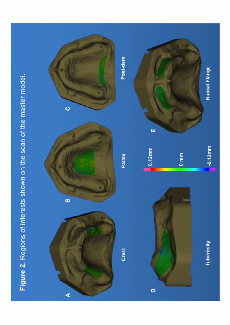
in figure 1 with the pyramids excluded. After superimposition five specific regions of
interest (vestibular, palatal, tuberosities, alveolar crest and post dam) were defined
(Figure 2). The software measured the distances for each surface point between the
intaglio surfaces of the superimposed denture against the scanned master model.
[32]
7
2.4 Statistical analysis:
Wilcoxon’s signed rank test was used to evaluate the effect of artificial saliva
incubation on the trueness of the intaglio surfaces of the specimens, between the
study groups(?? See table 1: no results for that?) and for the regions of interest
within the study groups. The confidence interval (CI) was set at 95% and level of
statistical significance set to p<0.05. Mann-Whitney test was used for an inter-group
comparison of the trueness split by the regions of interests studied. Mann-Whitney
tests were further used for evaluation the potential denture “sore spots” (20%
quantile) and the “variability” of the individual trueness measurements (80%-20%
quantile/2). The level of statistical significance was set to p<0.05. Power analysis for
sample size calculation was not done in this study as similar studies employed a
minimum of 5 and a maximum of 10 samples per group. The current study employed
a sample size of 11 specimens per group. Statistical analysis was performed by
StatView version 5.0 statistical software package (SAS Institute Inc., Cary, North
Carolina, USA).
2.5 Post-hoc experiments
2.5.1. Scanning
After a preliminary analysis of the scans from the original protocol, a region of misfit
beyond 0.1 mm (red color) was observed in the postdam region adjacent to the
screw of the scanner’s scanning table in some of the CAD/CAM and the injection-
molded specimens, but not in the conventional CRDP group. Hence a rescan of all
the CRDPs from all groups was done without the use of the screw of scanning table.
By that time, the specimens had been stored dry for a period of 90 days (After 3
Months). This time the specimens were held in place by a scanning ring with 3 pins
8
and the dentures were fixated by means of sticky wax to ensure that no pressure
was applied on the specimen.
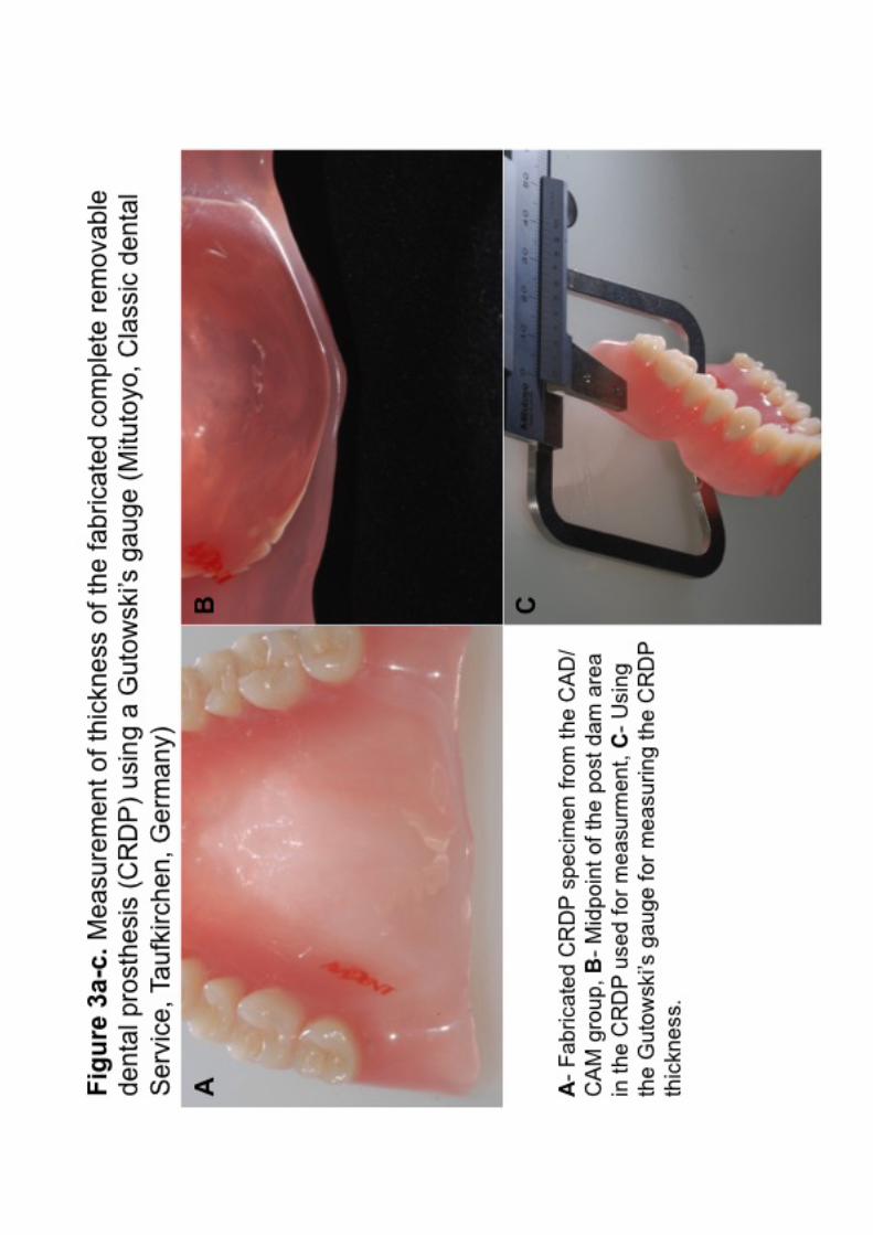
2.5.2. Thickness of the postdam
The thickness of the palatal plate in the post-dam area was measured for all the
specimens. A characteristic landmark (mid-point of the post-dam area) was chosen
to assure comparability between groups. Measurements were performed by a
Gutowski-gauge (Mitutoyo, Classic Dental Service, Taufkirchen, Germany) (Figures
3a-c).
2.6. Post-hoc statistical analysis
Scans after 90 days were analyzed using the Oracheck software [32]. Only the total
surface scans (excluding the pyramids) were analyzed. Non-parametric Wilcoxon’s
signed-rank tests and Mann-Whitney tests were used to compare the trueness of this
scan superimposed over the original reference model scan. Arithmetic means were
calculated for the palatal thickness measurements. Data was checked for normal
distribution using the Kolmogorov-Smirnov Test. Standard paired t-tests were
applied for analysis.
3. Results
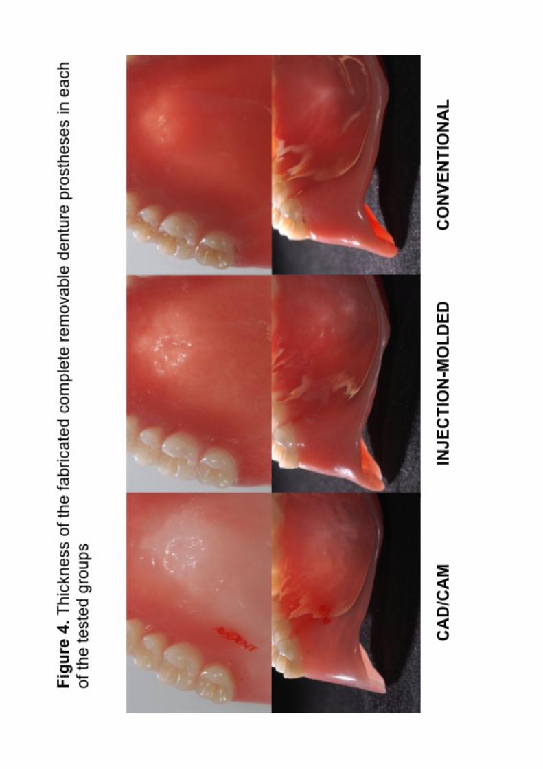
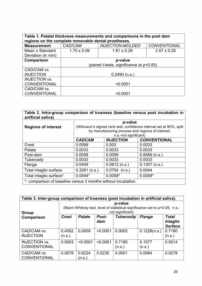
3.1 Palatal thickness
When measuring the palatal thickness of the conventional CRDPs showed the
thickest palatal plate when compared to the CAD/CAM (p<0.0001, paired t-tests)
injection-molded (p<0.0001, paired t-tests) groups. There was no difference between
the CAD/CAM and the injection-molded groups (Figure 4, Table 1).
9
3.2 Trueness of intaglio surface
At baseline, there was no significant difference (n.s.) in the trueness of the total
intaglio surfaces of the CRDPs between the three groups. However, the variability of
the median trueness of the individual measurement points was the lowest in the
conventional group (CAD/CAM versus Conventional p=0.0001, Injection versus
Conventional p=0.0007, CAD/CAM versus Injection n.s.; Mann-Whitney test).
(comment: no data provided?)
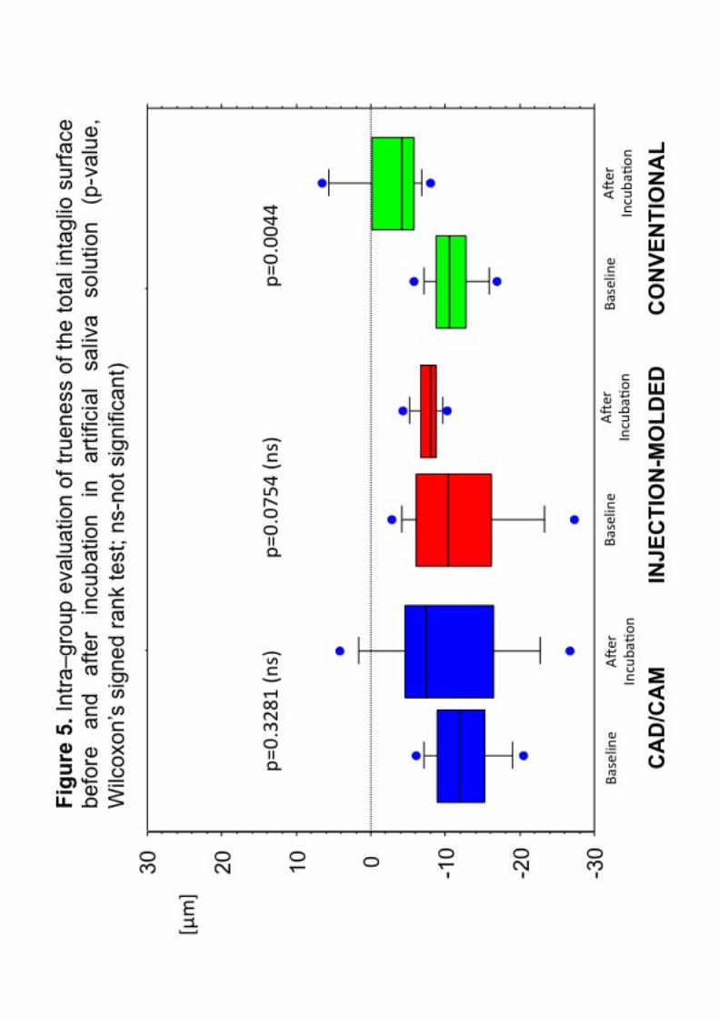
After incubation in saliva the conventional CRDPs showed a significant improvement
in trueness of the entire intaglio surface (p=0.0044) which was not present in the
other two groups, despite a clear trend (Figure 5, Table 3). However, the trueness of
the CAD/CAM and Injection CRDPs indicated equally an improvement, especially in
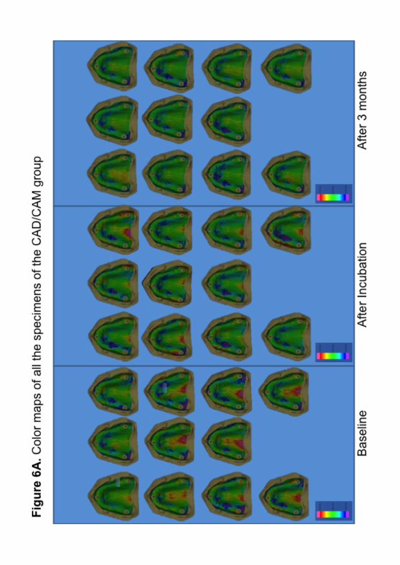
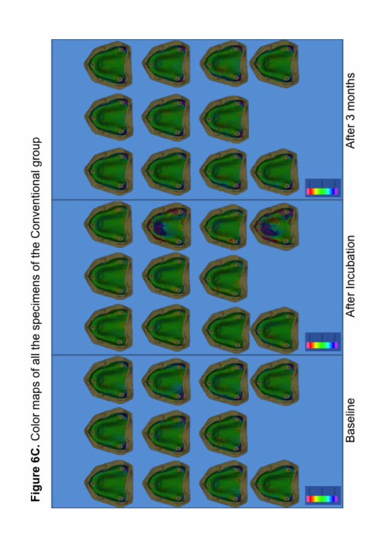
the palatal area, where a clear misfit had been noted in the area, of the clamp which
held the denture in place during scanning (Figure 6a-c, baseline and after
incubation).
For all three techniques, 60% (80%) of all deviations of the complete intaglio surface
after incubation in saliva were below 0.07 (0.1) mm.
The improved trueness after incubation in saliva was confirmed for all three
techniques, when considering only the regions of interest. Incubation in saliva
introduced a significantly better trueness in all regions of interest, except for
conventional technique crest, post-dam and flange areas as well as injection
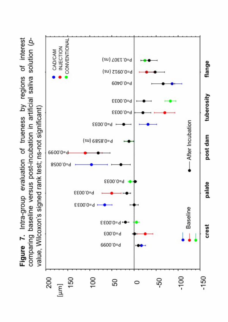
technique flange areas (Figure. 7, Table 2).
Re-scanning of the intaglio surfaces after 3 months without clamping, but rather
holding the CRDPs in the scanner by means of a sticky wax, reduced the misfit in
10
the area around the palatal clamp, that had been noted during the baseline scanning
and after the incubation in saliva (Figure 6a and 6b).
After 21 days of wet and 3 month of dry storage, a general “shrinkage” of the
specimen was noted, demonstrating a significantly “tighter fit” for all three techniques
(Table 2). Hence the further analyses are only referring to the post-incubation
measurements.
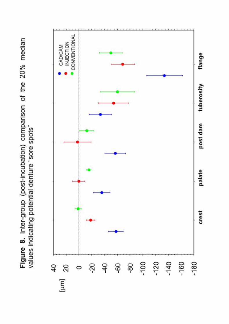
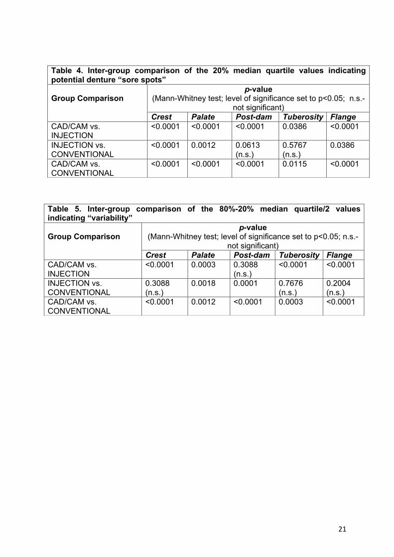
3.3 Compression zones
The 20% median quartile indicates the closest fit and may therefore be considered a
“compression zone”. With the exception of the tuberosities, CAD/CAM techniques
showed the strongest compression from all three techniques, especially in the
anterior flange area (Table 4, Figure 8).
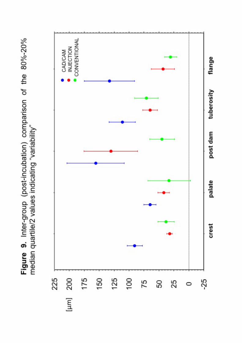
3.4 Variability of trueness
The 80%-20%/2 median quartile indicates the variability of the trueness readings
from the individual measuring points of the intaglio surface. Here, the CAD/CAM
group demonstrated the highest variability amongst the three groups, except for
post-dam which was equally variable in the CAD/CAM and injection techniques
(Table 5, Figure 9).
4. Discussion
Why is this study relevant?
CRDP fabrication by CAD/CAM is a novel technology in digital prosthodontics and
no clinical trials concerning the denture fit are published till date [12]. Scientific
evidence related to the trueness of the intaglio surface and the material properties of
11
the CAD/CAM milled CRDPs are scarce. Only one study has evaluated the accuracy
of the denture bases manufactured by different techniques as opposed to CAD/CAM
milling [13]. Hence the current study was undertaken as an attempt to evaluate the
trueness of the CAD/CAM milled CRDPs by comparing it with CRDPs manufactured
by traditional “flask, pack and press” and “injection-molding” methods. We wanted to
confirm the validity of the novel CAD/CAM technique in bench experiments under
standardized experimental conditions, before conducting a clinical trial.
Sample size
The sample size adopted for the current bench experiments was in accordance with
similar published studies involving digital impression techniques, which recommend
10 scans from a single study cast per experimental group [16]. Goodacre [13] used
four experimental groups with a sample size of four dentures per group in a similar
purpose bench experiment. In the current study, a sample size of 11 was chosen in
order to respect the empirical rule of Harrell [17], and to avoid type-II statistical errors
[18].
Error of method – saliva, thermocycling
The saliva substitute used and the incubation conditions deviated from a purely
clinical context. Human saliva is a sophisticated exocrine secretion which follows a
circadian rhythm; and the composition of saliva is dependent on this salivary flow
rate [19]. The reproduction of the saliva’s inorganic components is manageable;
difficulty arises when trying to replicate the viscosity. The importance of using an
appropriate liquid medium in bench experiments has been well-documented [20-22].
The artificial saliva substitute used for the current bench experiments was a
previously described custom prepared solution similar to the commercially available
Glandosane® [14, 15]. Furthermore, no thermo-cycling effect was mimicked in our
12
experiments. Thermo-cycling is known to decrease the micro-hardness of the
denture bases [23, 24].
Error of method – scanning
Scanning errors should also be considered. The high quality laboratory scanner used
is equipped with a fully automated calibration method which, to a large extent, is
directly related to the temperature changes in the scanning compartment. Since the
facility in Geneva is not climate controlled, the influence of the thermal changes may
account for a source of error. A repeated recalibration ensured that the temperature
cline would not be a factor affecting the accuracy of the scans. A further possible
source of error which could have affected the scan accuracy may be because of the
powder coating before scanning the model and the CRDPs. Schaefer et al. (2014)
have reported that the powder coating may have a detrimental effect on the marginal
fit and internal adaptation in partial coverage restorations; however in the same
report they stated that the deviations still remained within clinically acceptable
thresholds [25]. Enders and Mehl (2013) reported that digital impressions for
complete-arch seem less accurate and demonstrate a different pattern of deviation
than conventional impressions [26]. Although these issues may be of considerable
relevance in fixed prosthodontics, inaccuracies in the range of micrometers is
deemed clinically acceptable in complete denture prosthodontics, but this still needs
to be scientifically proven.
Error of method – clamping and palatal thickness
In order to maintain standardization in the scanning process, a single investigator
(YC) performed the digital scans of the master model, and the CRDPs. As
mentioned above, during the scanning process, the clamps used to fix the dentures
to the scan table might have been fastened rather tight causing an area of misfit
13
around the palatal clamp which was visible in the scans of the CAD/CAM and
injection molding CRDPs (Figure 6a, b). The absence of this misfit in the
conventional CRDP group, where the specimens presented with a thicker palatal
plate, as well as the absence of this misfit in the 3-months post-hoc scanning of the
specimens strengthens the hypothesis of a mechanical distortion during clamping.
The different thicknesses of the palatal plate in the 3 groups was unintentional and
only discovered after the experiments were completed. Giving precise instructions
concerning the palatal thickness to the manufacturers of the CRDPs might be an
important feature for similar future studies. However, the thinner CAD/CAM milled
palatal plates might be an important factor in patient satisfaction, providing a more
natural sensation and a more physiological tongue posture. It may also enhance
thermal sensation during hot and cold food intake. The mechanical distortion noticed
in our current experiments do not justify prescribing a thicker palatal palate. Firstly,
forces due to clamping do not occur during normal denture wearing. Secondly, the
misfit in the post-dam area due to this mishap was not larger than 0.08 mm, a range
that would at any rate be compensated by cutting a groove of 0.4 to 0.7 mm depth in
the plaster cast in the post dam area on the master model. However, further
research needs to verify, if the claimed enhanced mechanical properties of the pre-
polymerized PMMA resins allows such thin palatal plates without an increased
incidence of denture fracture.
Error of method: why do we expect a difference
In a conventional denture manufacturing technique, the procedures of mixing the
resin, packing, flasking, as well as heat-polymerization are sources for inconsistency,
which result in a final distortion of the prosthesis. It is well established that PMMA
resin incorporates water when immersed in a wet environment like the oral cavity.
14
Also, the well-documented effect of linear shrinkage during processing usually
results in a small spacing between the palatal mucosa and the denture’s palatal plate
[27-30]. The release of initial tensions from the polymerization process might further
account for the reported changes in shape [31]. The initial misfit after processing, as
well as the settling of the denture into the denture bearing tissues justify remounting
the dentures after a period of 10-14 days after insertion. It is tempting to suggest,
that the enhanced density of pre-fabricated pucks in the CAD/CAM and the injection
resin reduce water intake and thus reduce the volume changes introduced by the
flask, pack and press techniques. Milling the denture from a pre-polymerized block
would create mechanical “milling tensions”, but no polymerization tensions.
Interpretation of results: trueness and saliva immersion
A difference in the fit of the intaglio surface was noted between the three
manufacturing methods only after incubation in saliva. The interpretation of the
current results mainly focusses on the post-immersion trueness of the intaglio
surface, as we consider this the clinically most relevant finding. The better trueness
of the overall intaglio surface of the conventional dentures may be explained by the
many years of experience which the dental technician who manufactured the
conventional dentures has. Given that he did not work in a private, and hence a
competitive environment, he took all the time he needed to pack, process and
subsequently cool the flask. This might have minimized the post-polymerization
tensions and distortion of the prostheses and explained the excellent adaptation of
the palatal plate. However, when analyzing the individual regions of interest,
CAD/CAM and injection techniques do equally show an overall improved trueness
after immersion in saliva. When interpreting trueness, it has to be borne in mind, that
a misfit with space from the master model resulted in a positive value (red color) and
15
a compression of the tissues is indicated by negative values (blue color) (see
Figures 6A-C). Hence calculating the mean value might have “neutralized” the
spacing and compression zones. All fit surfaces corresponded with an accuracy of
±0.1 mm to the originally scanned master model. Consequently, all the three CRDP
groups provide adequate and clinically acceptable physical denture retention via
cohesive and adhesive forces.
Interpretation of results: variability of trueness
Interestingly, the CAD/CAM milled CRDPs presented the highest variability of
trueness of the intaglio surface (Figure 9). In fact, the 80-20% quantile was more
than twice as large for the CAD/CAM dentures as for the standard techniques. This
can be explained by the size of the milling instrument which is inevitably larger than
the particle size of stone plaster. The intaglio surface of a CAD/CAM milled denture
is therefore not smooth, but rather “terraced”. Inevitably, the size of the milling
instrument determines the smoothness of the fit surface, but also the time which is
needed to cut the denture base. A micro-terraced intaglio surface is not necessarily a
clinical disadvantage, as it does not seem to compromise the overall fit of the
denture. Micro-spaces for saliva might even contribute to the adhesive forces. On
the other hand, the micro-roughness might increase the adhesion of biofilm and
render denture cleaning difficult. Clinical studies will have to investigate the ideal
balance between fit surface detail and manufacturing time and cost.
Interpretation of results: trueness and flange
When investigating the different regions of interest, it can be noted that all three
techniques seem to create some sort of compression in the anterior flange area. This
means, that the scanned denture surface penetrates the scanned cobalt-chrome
master model indicating a compression of the tissues when the denture is seated in
16
the mouth. This may be due to its vertical position which makes it more vulnerable to
distortion. For the CAD/CAM techniques an increased imprecision might be added
when vertical surfaces are scanned, as more surface of the alveolar ridge is
represented in each single pixel. To minimize this source of imprecision, we scanned
our reference model as well as the denture specimen from various angles. When
interpreting these results, it further has to be considered that in the present
experiments, an edentulous ridge was chosen without pronounced undercuts and
with a shallow palate. Had the roof of the mouth or the tuberosities been steeper, the
shrinkage during heat polymerization would have probably increased the misfit of the
intaglio surface [31]. For these shapes of the alveolar ridge, a milled CRDP from a
pre-polymerized block may be considered more favorable and result in a better
trueness in terms of adaptation of the palatal plate and tuberosities; but this
hypothesis remains to be proven. Compression in the area of the vestibular flange
was most prominent in the CAD/CAM group. In a clinical context, such compression
might foster the denture adhesion and provide an inner seal. The anterior inner seal
is very vulnerable to the patient’s movement during impression taking, and a lack of
retention at insertion can often be related to such an “open inner seal”. During
conventional impression taking a second layer of impression material is often used to
achieve a slight compression and hence a tight inner seal. The CAD/CAM technique
provided such a compression “automatically”, hence this extra treatment step might
not be necessary; however, this hypothesis remains to be confirmed by a clinical
study. Our first clinical experiences with the CAD/CAM milled CRDPs confirm a very
good retention, which might in fact be related to the compression of the inner seal.
The reported median compression of 0.08 mm might present an ideal balance
17
between a tight fit and painful injury, which can only be expected with compressions
beyond 0.5 mm.
5. Conclusions
Based on the findings of this study, the null hypothesis can only be rejected for the
post-saliva measurements, where CAD/CAM and Injection molded CRDPs present a
significantly lower trueness of the total intaglio surface than conventional CRDPs.
However, measures in the present experiments are relative and a consistently
superior technique cannot be determined, when individually analyzing certain
regions of interest. Since all three complete denture manufacturing methods provide
excellent clinical fit, patient-centered outcome measures, but also chair-side time
and clinical complexity of the procedures, esthetics, cost, cleansability and
biomechanical properties of the material might be important factors to consider in the
clinical decision making.
Acknowledgements
The scanner used in this study was funded by a grant from the Swiss Dental
Association (SSO – Schweizerische Zahnärzte-Gesellschaft). MDT Roger Renevey
is acknowledged for the manufacturing of the injection-molded dentures and dental
technician Fabien Chevrolet for fabrication of the conventional CRDPs. Thanks are
due to Global Dental Science for manufacturing the CAD/CAM dentures. The
manufacturing of all specimen was paid for in full by the University of Geneva
Research funds. The authors received no compensation and report no conflict of
interest.
18
References
[1] Jacob RF. The traditional therapeutic paradigm: complete denture therapy. J Prosthet
Dent. 1998;79:6-‐13.
[2] Murray MD, Darvell BW. The evolution of the complete denture base. Theories of
complete denture retention-‐-‐a review. Part 1. Aust Dent J. 1993;38:216-‐9.
[3] Busch M, Kordass B. Concept and development of a computerized positioning of
prosthetic teeth for complete dentures. Int J Comput Dent. 2006;9:113-‐20.
[4] Goodacre CJ, Garbacea A, Naylor WP, Daher T, Marchack CB, Lowry J. CAD/CAM
fabricated complete dentures: concepts and clinical methods of obtaining required
morphological data. J Prosthet Dent. 2012;107:34-‐46.
[5] Kanazawa M, Inokoshi M, Minakuchi S, Ohbayashi N. Trial of a CAD/CAM system for
fabricating complete dentures. Dent Mater J. 2011;30:93-‐6.
[6] Kawahata N, Ono H, Nishi Y, Hamano T, Nagaoka E. Trial of duplication procedure for
complete dentures by CAD/CAM. J Oral Rehabil. 1997;24:540-‐8.
[7] Maeda Y, Minoura M, Tsutsumi S, Okada M, Nokubi T. A CAD/CAM system for removable
denture. Part I: Fabrication of complete dentures. Int J Prosthodont. 1994;7:17-‐21.
[8] Inokoshi M, Kanazawa M, Minakuchi S. Evaluation of a complete denture trial method
applying rapid prototyping. Dent Mater J. 2012;31:40-‐6.
[9] Sun Y, Lu P, Wang Y. Study on CAD&RP for removable complete denture. Comput
Methods Programs Biomed. 2009;93:266-‐72.
[10] Zhang YD, Jiang JG, Liang T, Hu WP. Kinematics modeling and experimentation of the
multi-‐manipulator tooth-‐arrangement robot for full denture manufacturing. J Med Syst.
2011;35:1421-‐9.
[11] Kattadiyil MT, Jekki R, Goodacre CJ, Baba NZ. Comparison of treatment outcomes in
digital and conventional complete removable dental prosthesis fabrications in a predoctoral
setting. J Prosthet Dent. 2015;114:818-‐25.
[12] Bidra AS, Taylor TD, Agar JR. Computer-‐aided technology for fabricating complete
dentures: systematic review of historical background, current status, and future
perspectives. J Prosthet Dent. 2013;109:361-‐6.
[13] Goodacre B. Comparing the surface matching Accuracy of Conventional and CAD/CAM
Methods of FAbricating Complete Denture Bases: A Pilot Study. . Academy of
Prosthodontics. Maui, Hawaii 2014.
[14] Srinivasan M, Schimmel M, Badoud I, Ammann P, Herrmann FR, Muller F. Influence of
implant angulation and cyclic dislodging on the retentive force of two different overdenture
attachments -‐ an in vitro study. Clin Oral Implants Res. 2015.
[15] Srinivasan M, Schimmel M, Kobayashi M, Badoud I, Ammann P, Herrmann FR, et al.
Influence of different lubricants on the retentive force of LOCATOR attachments -‐ an in vitro
pilot study. Clin Oral Implants Res. 2015.
[16] Flugge TV, Schlager S, Nelson K, Nahles S, Metzger MC. Precision of intraoral digital
dental impressions with iTero and extraoral digitization with the iTero and a model scanner.
Am J Orthod Dentofacial Orthop. 2013;144:471-‐8.
[17] Harrell FE, Jr., Lee KL, Califf RM, Pryor DB, Rosati RA. Regression modelling strategies for
improved prognostic prediction. Statistics in medicine. 1984;3:143-‐52.
[18] Faul F, Erdfelder E, Buchner A, Lang AG. Statistical power analyses using G*Power 3.1:
tests for correlation and regression analyses. Behavior research methods. 2009;41:1149-‐60.
19
[19] Gal JY, Fovet Y, Adib-‐Yadzi M. About a synthetic saliva for in vitro studies. Talanta.
2001;53:1103-‐15.
[20] Bayer S, Keilig L, Kraus D, Gruner M, Stark H, Mues S, et al. Influence of the lubricant
and the alloy on the wear behaviour of attachments. Gerodontology. 2011;28:221-‐6.
[21] Besimo CE, Guarneri A. In vitro retention force changes of prefabricated attachments
for overdentures. Journal of oral rehabilitation. 2003;30:671-‐8.
[22] Botega DM, Mesquita MF, Henriques GE, Vaz LG. Retention force and fatigue strength
of overdenture attachment systems. J Oral Rehabil. 2004;31:884-‐9.
[23] Goiato MC, Dos Santos DM, Andreotti AM, Nobrega AS, Moreno A, Haddad MF, et al.
Effect of beverages and mouthwashes on the hardness of polymers used in intraoral
prostheses. Journal of prosthodontics : official journal of the American College of
Prosthodontists. 2014;23:559-‐64.
[24] Goiato MC, Dos Santos DM, Baptista GT, Moreno A, Andreotti AM, Dekon SF. Effect of
thermal cycling and disinfection on microhardness of acrylic resin denture base. Journal of
medical engineering & technology. 2013;37:203-‐7.
[25] Schaefer O, Decker M, Wittstock F, Kuepper H, Guentsch A. Impact of digital impression
techniques on the adaption of ceramic partial crowns in vitro. J Dent. 2014;42:677-‐83.
[26] Ender A, Mehl A. Accuracy of complete-‐arch dental impressions: a new method of
measuring trueness and precision. J Prosthet Dent. 2013;109:121-‐8.
[27] Firtell DN, Green AJ, Elahi JM. Posterior peripheral seal distortion related to processing
temperature. J Prosthet Dent. 1981;45:598-‐601.
[28] Sanders JL, Levin B, Reitz PV. Comparison of the adaptation of acrylic resin cured by
microwave energy and conventional water bath. Quintessence Int. 1991;22:181-‐6.
[29] Sykora O, Sutow EJ. Posterior palatal seal adaptation: influence of processing
technique, palate shape and immersion. J Oral Rehabil. 1993;20:19-‐31.
[30] Woelfel JB, Paffenbarger GC, Sweeney WT. Dimensional changes occurring in dentures
during processing. J Am Dent Assoc. 1960;61:413-‐30.
[31] Wong DM, Cheng LY, Chow TW, Clark RK. Effect of processing method on the
dimensional accuracy and water sorption of acrylic resin dentures. J Prosthet Dent.
1999;81:300-‐4.
[32] Mehl A, Koch R, Zaruba M, Ender A. 3D monitoring and quality control using intraoral
optical camera systems. Int J Comput Dent. 2013;16:23-‐36.
20
Table 1. Palatal thickness measurements and comparisons in the post dam regions on the complete removable dental prostheses.
Measurement CAD/CAM INJECTION-MOLDED CONVENTIONAL
Mean ± Standard Deviation (in mm)
1.70 ± 0.06 1.81 ± 0.28 2.57 ± 0.20
Comparison p-value (paired t-tests, significance at p<0.05)
CAD/CAM vs. INJECTION
0.2490 (n.s.)
INJECTION vs. CONVENTIONAL
<0.0001
CAD/CAM vs. CONVENTIONAL
<0.0001
Table 2. Intra-group comparison of trueness (baseline versus post incubation in artificial saliva)
Regions of interest
p-value (Wilcoxon’s signed rank test, confidence interval set at 95%; split
by manufacturing process and regions of interest;
n.s.-not significant)
CADCAM INJECTION CONVENTIONAL
Crest 0.0099 0.003 0.0033
Palate 0.0033 0.0033 0.0033
Post-dam 0.0058 0.0099 0.8589 (n.s.)
Tuberosity 0.0033 0.0033 0.0033
Flange 0.0409 0.0912 (n.s.) 0.1307 (n.s.)
Total intaglio surface 0.3281 (n.s.) 0.0754 (n.s.) 0.0044
Total intaglio surface* 0.0044* 0.0058* 0.0058*
*- comparison of baseline versus 3 months without incubation.
Table 3. Inter-group comparison of trueness (post incubation in artificial saliva).
Group Comparison
p-value (Mann-Whitney test, level of statistical significance set to p<0.05; n.s.-
not significant)
Crest Palate Post-dam
Tuberosity Flange Total Intaglio Surface
CAD/CAM vs. INJECTION
0.4502 (n.s.)
0.0009 <0.0001 0.0002 0.1228(n.s.) 0.7180 (n.s.)
INJECTION vs. CONVENTIONAL
0.0003 <0.0001 <0.0001 0.7180 (n.s.)
0.1077 (n.s.)
0.0014
CAD/CAM vs. CONVENTIONAL
0.0078 0.6224 (n.s.)
0.0235 0.0001 0.0064 0.0278
21
Table 4. Inter-group comparison of the 20% median quartile values indicating potential denture “sore spots”
Group Comparison
p-value (Mann-Whitney test; level of significance set to p<0.05; n.s.-
not significant)
Crest Palate Post-dam Tuberosity Flange
CAD/CAM vs. INJECTION
<0.0001 <0.0001 <0.0001 0.0386 <0.0001
INJECTION vs. CONVENTIONAL
<0.0001 0.0012 0.0613 (n.s.)
0.5767 (n.s.)
0.0386
CAD/CAM vs. CONVENTIONAL
<0.0001 <0.0001 <0.0001 0.0115 <0.0001
Table 5. Inter-group comparison of the 80%-20% median quartile/2 values indicating “variability”
Group Comparison
p-value (Mann-Whitney test; level of significance set to p<0.05; n.s.-
not significant)
Crest Palate Post-dam Tuberosity Flange
CAD/CAM vs. INJECTION
<0.0001 0.0003 0.3088 (n.s.)
<0.0001 <0.0001
INJECTION vs. CONVENTIONAL
0.3088 (n.s.)
0.0018 0.0001 0.7676 (n.s.)
0.2004 (n.s.)
CAD/CAM vs. CONVENTIONAL
<0.0001 0.0012 <0.0001 0.0003 <0.0001
22
FIGURE LEGENDS
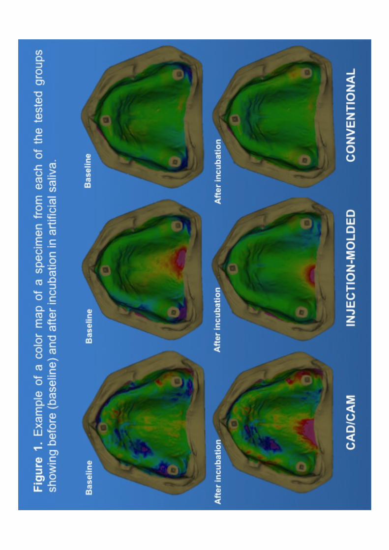
Figure 1 Example of a color map of a specimen from each of the tested groups
showing before (baseline) and after incubation in artificial saliva.
Figure 2 Regions of interests analyzed shown on the scan of the master model.
Figure 3 Measurement of thickness of the fabricated complete removable dental
prosthesis (CRDP) using a Gutowski’s gauge (Mitutoyo, Classic dental
Service, Taufkirchen, Germany). A- Fabricated CRDP specimen from
the CAD/CAM group, B- Midpoint of the post dam area in the CRDP
used for measurement, C- Using the Gutowski’s gauge for measuring
the CRDP thickness.
Figure 4 Thickness of the fabricated complete removable denture prostheses in
each of the tested groups.
Figure 5 Comparison of median (mean) values of the total intaglio surface within
the groups before and after incubation in artificial saliva solution (p-
value, Wilcoxon’s signed rank test).
Figure 6 A. Color maps of all the specimens of the CAD/CAM group, B. Color
maps of all the specimens of the Injection-Molding group, C. Color
maps of all the specimens of the Conventional group.
Figure 7 Intra-group comparison of median (mean) values in the regions of
interest at baseline and after incubation in artificial saliva solution (p-
value, Wilcoxon’s signed rank test).
Figure 8 Inter-group (post-incubation) comparison of the 20% median quartiles
indicating potential denture “sore spots”.

23
Figure 9 Inter-group (post-incubation) comparison of the 80%-20% median quartile/2
values indicating “variability”