Molecular functions of the ubiquitin domain protein Herp in
Synoviolin mediated endoplasmic reticulum associated protein
degradation (ERAD)
Dissertation
zur Erlangung des akademischen Grades
doctor rerum naturalium (Dr. rer. nat.)
im Fach Biologie
eingereicht an der
Mathematisch-Naturwissenschaftlichen Fakultät I
der Humboldt-Universität zu Berlin
von
Diplom-Ernährungswissenschaftlerin Melanie Kny
Präsident der Humboldt-Universität zu Berlin:
Prof. Dr. Dr. h.c. Christoph Markschies
Dekan der Mathematisch-Naturwissenschaftlichen Fakultät I:
Prof. Dr. Lutz-Helmut Schön
Gutachter:
1. Professor Dr. Peter Michael Kloetzel
2. Professor Dr. Wolfgang Lockau
3. Professor Dr. Wolfgang Dubiel
Tag der mündlichen Prüfung: 23.06.2010
Table of contents
2
Table of contents
ABSTRACT ........................................................................................................................................4
ZUSAMMENFASSUNG ......................................................................................................................5
1 INTRODUCTION.........................................................................................................................6
1.1 The ubiquitin proteasome system (UPS)..........................................................................6 1.1.1 Composition of the 26S proteasome...............................................................................7 1.1.2 The role of ubiquitin........................................................................................................7 1.1.3 The process of ubiquitination..........................................................................................8 1.1.4 E3 ubiquitin protein ligases...........................................................................................10 1.1.5 Deubiquitinating enzymes (DUBs) ................................................................................11
1.2 Endoplasmic reticulum (ER) protein quality control......................................................13 1.2.1 Protein synthesis at the ER ..........................................................................................13 1.2.2 ER quality control, ER stress and the unfolded protein response (UPR)........................13 1.2.3 ER stress signalling......................................................................................................14 1.2.4 ER associated protein degradation (ERAD)..................................................................16 1.2.5 Synoviolin based ERAD complexes..............................................................................19 1.2.6 Homocysteine inducible endoplasmic reticulum - resident protein (Herp) ......................22
1.3 Aim of this study..............................................................................................................25
2 MATERIAL AND METHODS .....................................................................................................26
2.1 Instruments, consumables and chemicals.....................................................................26 2.2 Molecular biology ............................................................................................................28
2.2.1 Cultivation and storage of Escherichia coli (E.coli) ........................................................28 2.2.2 Isolation of plasmid DNA from E.coli.............................................................................28 2.2.3 Separation of DNA fragments by agarose gel electrophoresis.......................................29 2.2.4 Determination of DNA and RNA concentration in solution.............................................29 2.2.5 Preparation of competent E.coli cells............................................................................30 2.2.6 Transformation of E.coli with plasmid DNA ...................................................................30 2.2.7 Isolation of total RNA from HeLa cells ..........................................................................30 2.2.8 Amplification of DNA by polymerase chain reaction (PCR)............................................31 2.2.9 Semiquantitative analysis of mRNA levels by reverse transcriptase (RT) PCR..............31 2.2.10 In vitro recombination of DNA...................................................................................32 2.2.11 Site-directed mutagenesis........................................................................................34
2.3 Tissue culture ..................................................................................................................35 2.3.1 Cell lines and media used in this study .........................................................................35 2.3.2 Culture of cells .............................................................................................................36 2.3.3 Cryoconservation and thawing of cells..........................................................................36 2.3.4 Transfection of mammalian cells ..................................................................................37 2.3.5 Generation of a stable inducible cell line expressing Herp specific shRNA....................39 2.3.6 Cycloheximide chase analysis......................................................................................40 2.3.7 Metabolic labelling using [35S]-methionine/-cysteine and pulse chase analysis..............40
2.4 Protein biochemistry .......................................................................................................41 2.4.1 Lysis of mammalian cells..............................................................................................41 2.4.2 Determination of protein concentration in solution.........................................................41 2.4.3 Precipitation of proteins using trichloro acetic acid (TCA)..............................................41 2.4.4 Sodium dodecyl sulfate polyacrylamid gel electrophoresis (SDS-PAGE).......................42 2.4.5 Western blot analysis ...................................................................................................43 2.4.6 Protein visualisation .....................................................................................................43 2.4.7 Affinity precipitation of proteins.....................................................................................45 2.4.8 Separation of proteins by glycerol gradient centrifugation .............................................47
Table of contents
3
2.4.9 In vitro binding studies..................................................................................................47 2.5 Bioinformatics and databases ........................................................................................49
3 RESULTS .................................................................................................................................50
3.1 The importance of the dynamics of Herp for ERAD.......................................................50 3.1.1 Herp is exchanged at Synoviolin based complexes.......................................................50 3.1.2 Synoviolin complex components are not essential for the degradation of Herp..............52 3.1.3 Herp-K61R is stabilised and impairs the degradation of NHK........................................54
3.2 The role of Herp in maintaining the integrity of Synoviolin based complexes .............57 3.2.1 Herp does not alter the formation of Synoviolin oligomers.............................................57 3.2.2 Usp7 is a target of the Herp UBL domain......................................................................58
3.2.2.1 The AXXS motif contributes to an efficient binding of Usp7 to Herp ......................59 3.2.2.2 Herp recruits Usp7 to Synoviolin ..........................................................................61 3.2.2.3 Usp7 does not affect the stability of Herp or NHK.................................................64 3.2.2.4 Herp is not involved in the regulation of p53.........................................................66
3.2.3 Ancient ubiquitous protein 1 (AUP1) is associated with Synoviolin................................69 3.2.3.1 Herp regulates the association of AUP1 with Synoviolin .......................................69 3.2.3.2 AUP1 binds to Synoviolin.....................................................................................70 3.2.3.3 AUP1 is required for the degradation of NHK .......................................................71 3.2.3.4 The AUP1-CUE domain is required for the efficient degradation of NHK...............72
3.3 Characterisation of Herp2 ...............................................................................................74 3.3.1 Herp2 reveals dynamics different from Herp.................................................................74 3.3.2 Herp and Herp2 form homo- and heterooligomers ........................................................76 3.3.3 Herp2 is associated with Synoviolin based complexes..................................................78
4 DISCUSSION............................................................................................................................80
4.1 The role of the dynamics of Herp in ERAD.....................................................................80 4.1.1 The turnover of Herp at Synoviolin based ERAD complexes.........................................80 4.1.2 Correlation of the turnover of Herp and ERAD substrates .............................................83
4.2 The importance of Herp for the integrity of Synoviolin based complexes....................85 4.2.1 The impact of Herp on Synoviolin oligomerisation.........................................................85 4.2.2 Herp dependent recruitment of Usp7 to Synoviolin .......................................................87 4.2.3 Herp dependent association of Synoviolin and AUP1 ...................................................92
4.3 Comparison of Herp and Herp2 ......................................................................................95 4.4 Conclusion.......................................................................................................................97
LITERATURE ...................................................................................................................................98
APPENDIX .....................................................................................................................................105
ABBREVIATIONS...........................................................................................................................105
DANKSAGUNG ..............................................................................................................................109
Abstract
4
Abstract The accumulation of aberrant proteins in the endoplasmic reticulum (ER) induces the
unfolded protein response (UPR) pathway for surmounting this cellular stress situation. One
of the strongly UPR-induced genes in mammalia encodes the ubiquitin domain protein Herp.
Herp interacts with the E3 ligase Synoviolin, a central component of ER associated protein
degradation (ERAD) mediating multiprotein complexes. Dependent on its ubiquitin-like (UBL)
domain, Herp is required for the efficient degradation of Synoviolin substrates. The molecular
mechanism underlying this function of Herp is poorly understood.
In the present study, it was shown that Herp is continuously exchanged at Synoviolin based
complexes. However, Herp did not serve as a Synoviolin substrate. Since both stabilisation
and depletion of Herp resulted in the impaired degradation of Synoviolin substrates, the
continuous turnover of Herp seems to be decisive for ERAD.
Herp was also shown to regulate the composition of Synoviolin based complexes. The
deubiquitinating enzyme Usp7 was linked to Synoviolin via its interaction with Herp.
However, Usp7 did not influence the stability of Herp or ERAD substrates. In addition, Herp
improved the association of the CUE domain protein AUP1 with Synoviolin. AUP1 triggered
the ERAD process in a CUE domain dependent manner.
Also Herp2, a homologue of Herp, was found to associate with Synoviolin based complexes.
However, in contrast to Herp, Herp2 was not induced by the UPR, was stable, and did not
bind Usp7 supporting the idea of Herp having a unique function in ERAD.
In conclusion, Herp is a dynamic ERAD component recruiting accessory proteins to
Synoviolin thus enabling Synoviolin dependent ubiquitination of substrates. These findings
point out the crucial role of Herp for the elimination of misfolded proteins, which is important
for cell survival.
keywords: Herp, ERAD, UPR, Synoviolin, Usp7, AUP1, Herp2
Zusammenfassung
5
Zusammenfassung Die Akkumulation fehlerhafter Proteine im Endoplasmatischen Retikulum (ER) induziert den „unfolded protein response“ (UPR) - Signalweg zur Überwindung dieser zellulären Stress-Situation. Ein in Säugern stark UPR-induziertes Gen kodiert für das Ubiquitin-Domäne-Protein Herp. Herp interagiert mit der E3-Ligase Synoviolin, einer zentralen Komponente von Multiproteinkomplexen, welche die ER assoziierte Protein-Degradation (ERAD) vermitteln. Abhängig von seiner Ubiquitin-ähnlichen (‘UBL’) Domäne wird Herp für den effizienten Abbau von Synoviolin-Substraten benötigt. Der zugrundeliegende molekulare Mechanismus dieser Funktion von Herp ist kaum bekannt. In der vorliegenden Studie wurde gezeigt, dass Herp kontinuierlich an Synoviolin-basierten Komplexen umgesetzt wird, aber kein Substrat ist. Da sowohl Depletion als auch Stabilisierung von Herp zum verminderten Abbau von Synoviolin-Substraten führt, lässt sich schlussfolgern, dass der kontinuierliche Umsatz von Herp entscheidend ist für ERAD. Weiterhin regulierte Herp die Zusammensetzung Synoviolin-basierter Komplexe. Das deubiquitinierende Enzym Usp7 ist über seine Bindung an Herp mit Synoviolin assoziiert. Usp7 beeinflusste aber nicht die Stabilität von Herp oder ERAD-Substraten. Zusätzlich verstärkte Herp die Interaktion zwischen dem CUE-Domäne-Protein AUP1 und Synoviolin. In Abhängigkeit von der CUE-Domäne steigerte AUP1 den ERAD-Prozess. Auch das Herp-Homolog Herp2 war mit Synoviolin-basierten Komplexen assoziiert. Im Gegensatz zu Herp wurde Herp2 nicht durch den UPR-Signalweg induziert, war stabil und interagierte nicht Usp7. Diese Daten unterstreichen die einzigartige Funktion von Herp im ERAD-Prozess. Schlussfolgernd ist Herp eine dynamische ERAD-Komponente, welche die Rekrutierung akzessorischer Proteine an Synoviolin vermittelt und damit die Ubiquitinierung von Synoviolin-Substraten ermöglicht. Diese Daten zeigen die kritische Rolle von Herp für die Beseitigung fehlerhafter Proteine und das Überleben der Zelle.
Schlagwörter: Herp, ERAD, UPR, Synoviolin, Usp7, AUP1, Herp2
1 Introduction
6
1 Introduction
1.1 The ubiquitin proteasome system (UPS) Cellular protein homeostasis is essential for cell survival. To sustain a certain level of
intracellular proteins, continuous synthesis and degradation takes place. The major pathway
for the regulated degradation of intracellular proteins is the ubiquitin proteasome system
(UPS). The small protein modifier ubiquitin and the 26S proteasome, a multi protease
complex, have key positions within this system. These two components of the UPS are
abundantly present in the cytoplasm and the nucleus of all eukaryotic cells. Regulatory
proteins such as cyclins or p53, which are involved in essential cellular processes such as
cell-cycle control or apoptosis, respectively, are processed by the UPS. Therefore, this
pathway is essential for cell survival (Hershko and Ciechanover, 1998). In addition, the UPS
is involved in the generation of antigenic epitopes which are presented by major
histocompatibility complex (MHC) class I molecules, a central process of the cellular immune
response (Kloetzel, 2004).
20S CP
19S RP
19S RP
ββ
α
α
S
base
lid
20S CP
19S RP
19S RP
ββ
α
α
S
base
lid
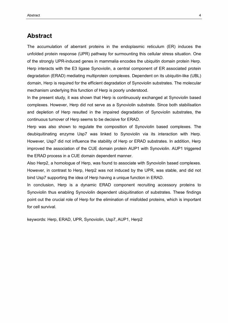
Figure 1: Schematic overview of the ubiquitin proteasome system (UPS) and its connections to various cellular processes. Degradation of regulatory proteins by the UPS is essential for a variety of highly interconnected cellular processes such as DNA-repair and therefore maintains cell survival. Disturbances within the UPS may lead to severe diseases such as inflammation. Protein degradation is mediated by the 26S proteasome (highlighted right hand) which consists of the 20S core particle (CP) and one or two 19S regulatory particles (RP). The CP is composed of four heptameric rings formed by the inner β- and the outer α-subunits. The RP is composed of base and lid substructures. S indicates a ubiquitinated substrate, E2 and E3 indicate the ubiquitination machinery ( adapted from (Wolf and Hilt, 2004).
As part of the protein quality control, the UPS is the primary intracellular mechanism which is
responsible for the degradation of defective proteins.
1 Introduction
7
This includes incomplete, misfolded, denatured, oxidised or else damaged proteins which
otherwise accumulate and have a tendency to form cytotoxic aggregates. Degradation of
UPS substrates includes two major sequential steps: first, ubiquitination and second,
degradation by the proteasome. These processes are subject to stringent regulation at the
steps of substrate selection, substrate processing and product generation. Dysregulation of
the UPS can lead to a variety of diseases such as cancer, inflammation or
neurodegenerative disorders (Figure 1, Hershko and Ciechanover, 1998).
1.1.1 Composition of the 26S proteasome
Protein degradation within the UPS is mediated by the 26S proteasome, a large
cytoplasmatic protein complex, consisting of the proteolytically active 20S core particle and
one or two terminal 19S regulatory particles as reviewed in (Tanaka, 2009). The 20S core
particle, which has a molecular mass of about 750 kDa, consists of four stacked heptameric
rings each containing evolutionary related proteins. These rings shape a cylindrical structure
(Groll et al., 1997). The constituents of the core particle are subdivided into α- and β-
subunits according to their homologies. In eukaryotes the outer rings are composed of seven
different α-subunits and the inner rings of seven different β-subunits. Three of the β-subunits
display proteolytic activity. The 19S regulatory particle comprises approximately 20 different
subunits forming two subcomplexes which are termed base and lid with the base associating
with the α-rings of the 20S core particle (Glickman et al., 1998). The 19S regulatory particle
is important for the recruitment, deubiquitination and entry of substrates into the 20S
chamber.
1.1.2 The role of ubiquitin
Ubiquitin is a heat-stable polypeptide of 7.6 kDa, highly conserved and ubiquitously
expressed in eukaryotes (Ciehanover et al., 1978) (Wilkinson et al., 1980). During a process
termed ‘ubiquitination’ ubiquitin is covalently attached to target proteins in an ATP dependent
manner resulting in the formation of a mono- or polyubiquitinated protein (Ciechanover et al.,
1980). Within this polyubiquitin chain the single molecules are connected through isopeptide
bonds, formed between the C-terminal glycine 76 of one molecule and a lysine residue of the
other ubiquitin molecule. Ubiquitin possesses seven lysine residues (K6, K11, K27, K29,
K33, K48 and K63) of which K11, K29, K48 and K63 can form ubiquitin-ubiquitin linkages in
vivo (Dubiel and Gordon, 1999). Polyubiquitin, consisting of at least four molecules
connected to a protein, serves as a signal for the recruitment of the ubiquitinated protein to
the 26S proteasome. Predominantly, G76-K48 linked ubiquitin chains signal proteasomal
degradation (Chau et al., 1989; Thrower et al., 2000).
1 Introduction
8
Apart from ubiquitin, a number of ubiquitin-like proteins such as SUMO, Nedd8 or ISG15
function as protein modifiers in a similar way as ubiquitin (Kerscher et al., 2006). Moreover,
ubiquitin-like (UBL) domains have been described to be integral parts of ubiquitin domain
proteins (UDPs). These UBL domains are, in contrast to ubiquitin-like modifiers, neither
processed nor conjugated with other proteins. A variety of proteins have been reported to
harbour ubiquitin interacting motifs. Such motifs are for example ubiquitin associated (UBA)
domains binding to polyubiquitin (Wilkinson et al., 2001) and coupling of ubiquitin conjugation
to ER degradation (CUE) domains described to bind poly- and monoubiquitin (Hurley et al.,
2006).
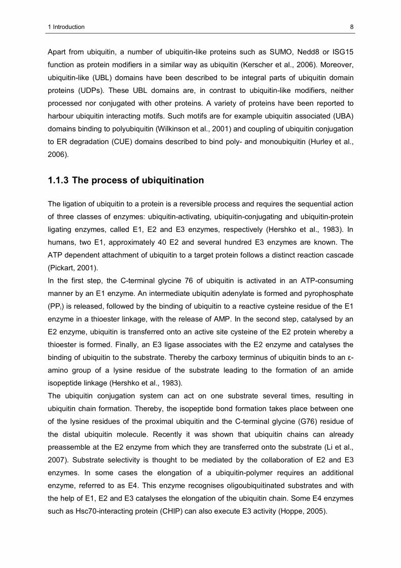
1.1.3 The process of ubiquitination
The ligation of ubiquitin to a protein is a reversible process and requires the sequential action
of three classes of enzymes: ubiquitin-activating, ubiquitin-conjugating and ubiquitin-protein
ligating enzymes, called E1, E2 and E3 enzymes, respectively (Hershko et al., 1983). In
humans, two E1, approximately 40 E2 and several hundred E3 enzymes are known. The
ATP dependent attachment of ubiquitin to a target protein follows a distinct reaction cascade
(Pickart, 2001).
In the first step, the C-terminal glycine 76 of ubiquitin is activated in an ATP-consuming
manner by an E1 enzyme. An intermediate ubiquitin adenylate is formed and pyrophosphate
(PPi) is released, followed by the binding of ubiquitin to a reactive cysteine residue of the E1
enzyme in a thioester linkage, with the release of AMP. In the second step, catalysed by an
E2 enzyme, ubiquitin is transferred onto an active site cysteine of the E2 protein whereby a
thioester is formed. Finally, an E3 ligase associates with the E2 enzyme and catalyses the
binding of ubiquitin to the substrate. Thereby the carboxy terminus of ubiquitin binds to an ε-
amino group of a lysine residue of the substrate leading to the formation of an amide
isopeptide linkage (Hershko et al., 1983).
The ubiquitin conjugation system can act on one substrate several times, resulting in
ubiquitin chain formation. Thereby, the isopeptide bond formation takes place between one
of the lysine residues of the proximal ubiquitin and the C-terminal glycine (G76) residue of
the distal ubiquitin molecule. Recently it was shown that ubiquitin chains can already
preassemble at the E2 enzyme from which they are transferred onto the substrate (Li et al.,
2007). Substrate selectivity is thought to be mediated by the collaboration of E2 and E3
enzymes. In some cases the elongation of a ubiquitin-polymer requires an additional
enzyme, referred to as E4. This enzyme recognises oligoubiquitinated substrates and with
the help of E1, E2 and E3 catalyses the elongation of the ubiquitin chain. Some E4 enzymes
such as Hsc70-interacting protein (CHIP) can also execute E3 activity (Hoppe, 2005).
1 Introduction
9
Ub UbUb
E1
E1
E2
E2
substrate
substrate
Ub
Ub
Ub
UbE3
ATP +
AMP + PP
SH
SH
UbUb Ub
A
B
K Ksubstrate
+ Ub
UbUb
UbUb
E1, E2, E3
26S
ATP
ATP
AMP+PP
ADP+P
Ub UbUb
E1
E1
E2
E2
substrate
substrate
Ub
Ub
Ub
UbE3
ATP +
AMP + PP
SH
SH
UbUb Ub
Ub UbUb
Ub UbUb
E1
E1
E2
E2
substrate
substrate
Ub
Ub
Ub
UbE3
ATP +
AMP + PP
SH
SH
UbUb Ub
UbUb Ub
A
B
K Ksubstrate
+ Ub
UbUb
UbUb
E1, E2, E3
26S
ATP
ATP
AMP+PP
ADP+P
K Ksubstrate
+ Ub
UbUb
UbUb
E1, E2, E3
26S
ATP
ATP
AMP+PP
ADP+P
Figure 2: Components and mechanisms in the ubiquitin proteasome system. (A) Overview of the pathway showing the consumption of ATP in the conjugative (top) and degradative (bottom) phases. E1, E2 and E3 represent the ubiquitin-activating, -conjugating and -ligating enzymes, respectively. K indicates a lysine residue; the black circle with ‘Ub’ indicates a ubiquitin molecule. (B) Cascade of ubiquitination. Ubiquitin is activated by E1, transferred to E2, where a preassembly of ubiquitin chains may occur, and finally attached to a substrate, a process mediated by E3. A row of light grey circles with ‘Ub’ indicates a preformed polyubiquitin chain (Pickart, 2004).
Originally, ubiquitination was thought to mainly target proteins for proteasomal degradation.
However, it was gradually assessed that this posttranslational protein modification serves
diverse functions within the cell aside from proteolysis. The fate of a protein depends on the
amount of ubiquitin, which may be attached as a single molecule or as a chain. In addition,
the kind of linkage within polyubiquitin chains is decisive as monoubiquitination provides a
signal for receptor internalisation or transcription regulation (Hicke, 2001; Strous et al., 1996),
whereas K48-linked polyubiquitin predominantly leads to proteasomal degradation.
Moreover, K63-linked polyubiquitin plays a key role in the activation of kinases within NFkB
signalling. The IL-1 receptor associated kinase (IRAK1), e.g., is activated through
modification with K63-linked polyubiquitin (Keating and Bowie, 2009; Windheim et al., 2008).
1 Introduction
10
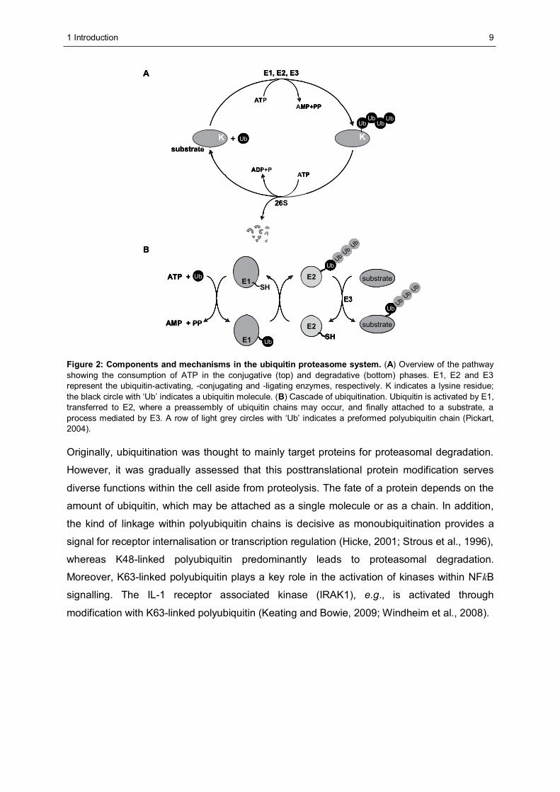
1.1.4 E3 ubiquitin protein ligases
The human genome encodes hundreds of E3 ubiquitin ligases (E3s) which are responsible
for the final step of ubiquitination. E3 enzymes are classified as ‘really interesting new gene’
(RING) E3s and ‘homologous to E6 associated protein carboxy terminus’ (HECT) E3s. Acting
as single proteins or within multiprotein complexes, E3s promote substrate recognition, E2-
ubiquitin recruitment and transfer of ubiquitin onto the target protein (Pickart, 2001). E6-
associated protein (E6-AP) was the first described HECT E3 enzyme, identified in cells which
had been transfected with the human papilloma virus oncoprotein E6. In the presence of the
E6 protein E6-AP ubiquitinates p53, which leads to the rapid proteasomal degradation of p53
(Scheffner et al., 1993). The C-terminal HECT domain of E6-AP is evolutionarily conserved
and present in all HECT E3s. A cysteine residue within this domain serves as an acceptor of
the activated ubiquitin from the E2 enzyme (Huibregtse et al., 1995; Scheffner et al., 1995).
Thus, a thioester intermediate between ubiquitin and the HECT E3 is formed. Finally,
ubiquitin is transferred to the substrate (Figure 3, A).
The large family of the RING E3 enzymes contains a characteristic conserved motif, which is
rich in cysteine and histidine residues and termed RING domain (Freemont et al., 1991). The
RING domain coordinates zinc ions and determines the activity of the protein (Lorick et al.,
1999). In contrast to HECT-E3s, RING E3s do not form a thioester with ubiquitin but bring the
ubiquitin-loaded E2s and the substrate in close proximity and promote ubiquitin transfer onto
the substrates. Therefore, this class of E3s acts as scaffold (Deshaies and Joazeiro, 2009).
RING E3 ligases are either cytoplasmatic such as CHIP or are membrane associated such
as the mammalian Hrd1p orthologue Synoviolin (Hrd1) integrated in the ER membrane
(Ballinger et al., 1999; Kaneko et al., 2002). In addition, RING E3s either exist as a single
molecule or as a component of multi protein complexes. Single subunit E3s such as parkin
harbour a substrate recognition element and the RING domain on the same molecule (Figure
3, B-1; (Imai et al., 2001). Multi subunit RING E3s such as the SCF complex contain a
member of the cullin protein family as backbone, a RING domain protein harbouring enzyme
activity and other proteins which are adaptors involved in substrate recognition and E2
binding (Deshaies and Joazeiro, 2009).

1 Introduction
11
Ub
E2E2
K
SH
K
UbE2 binding(RING)
substrate-binding
Cullin-1
SKP1F-box K
Ub
RBX1E2
SH
substrate-binding
E2 binding(RING)
Ub
E2 E2 E2
SH SH
SH Ub SH
K K K
Ub
E2 binding(HECT)
substrate-binding
B RING domain E3s
A HECT domain E3s
1 2Ub
E2E2
K
SH
K
UbE2 binding(RING)
substrate-binding
Ub
E2E2
KK
SH
KK
UbE2 binding(RING)
substrate-binding
Cullin-1
SKP1F-box K
Ub
RBX1E2
SH
substrate-binding
E2 binding(RING)
Cullin-1
SKP1F-box K
Ub
KK
Ub
RBX1RBX1E2
SHE2
SH
substrate-binding
E2 binding(RING)
Ub
E2 E2 E2
SH SH
SH Ub SH
K K K
Ub
E2 binding(HECT)
substrate-binding
Ub
E2E2 E2E2 E2E2
SH SH
SH Ub SH
KK KK KK
Ub
E2 binding(HECT)
substrate-binding
B RING domain E3s
A HECT domain E3s
1 2
Figure 3: Schematic overview of the major E3 classes. (A) HECT domain E3 mediated ubiquitination. The E2 is bound by the HECT domain and ubiquitin is transiently transferred to a conserved cysteine within the same region. The substrate binds to another domain within the same protein. (B) RING domain E3 mediated ubiquitination. (1) Single subunit RING E3. The E2 is bound by the RING domain and the substrate by a different domain within the E3. Ubiquitin is transferred from the E2 to the substrate. (2) Multi subunit RING E3 (SCF complex). E2 and substrate binding are mediated by domains of different subunits (adapted from Pickart, 2004).
1.1.5 Deubiquitinating enzymes (DUBs)
Ubiquitination is a reversible protein modification. Regulated ubiquitin removal from a
substrate is performed by deubiquitinating enzymes (DUBs). These proteases cleave
ubiquitin or ubiquitin-like proteins from precursor proteins as well as from conjugates of target
proteins (Reyes-Turcu et al., 2009). Within the UPS, DUBs have diverse functions including
the processing of ubiquitin precursors for their activation, recycling of ubiquitin, generation of
monoubiquitin from polyubiquitin and reversal of ubiquitination. Antagonising the action of E3
ligases, DUBs are potent regulators of ubiquitin mediated cellular processes.
Approximately one hundred human DUBs have been identified which are organised in five
different gene families (Nijman et al., 2005). They are classified either as JAB1/MPN/Mov34
metalloproteases (JAMM) or as cysteine proteases. The cysteine proteases are further
subdivided into ubiquitin-specific hydrolases (USPs, the largest family with more than 50
members in humans), ubiquitin C-terminal hydrolases (UCHs), otubain proteases (OTUs)
and Machado-Joseph disease proteases (MJDs). Deubiquitination, as well as ubiquitination,
is a highly regulated process which is involved in various functions of the cell such as gene
expression, DNA repair, kinase activation and lysosomal as well as proteasomal protein
degradation.
1 Introduction
12
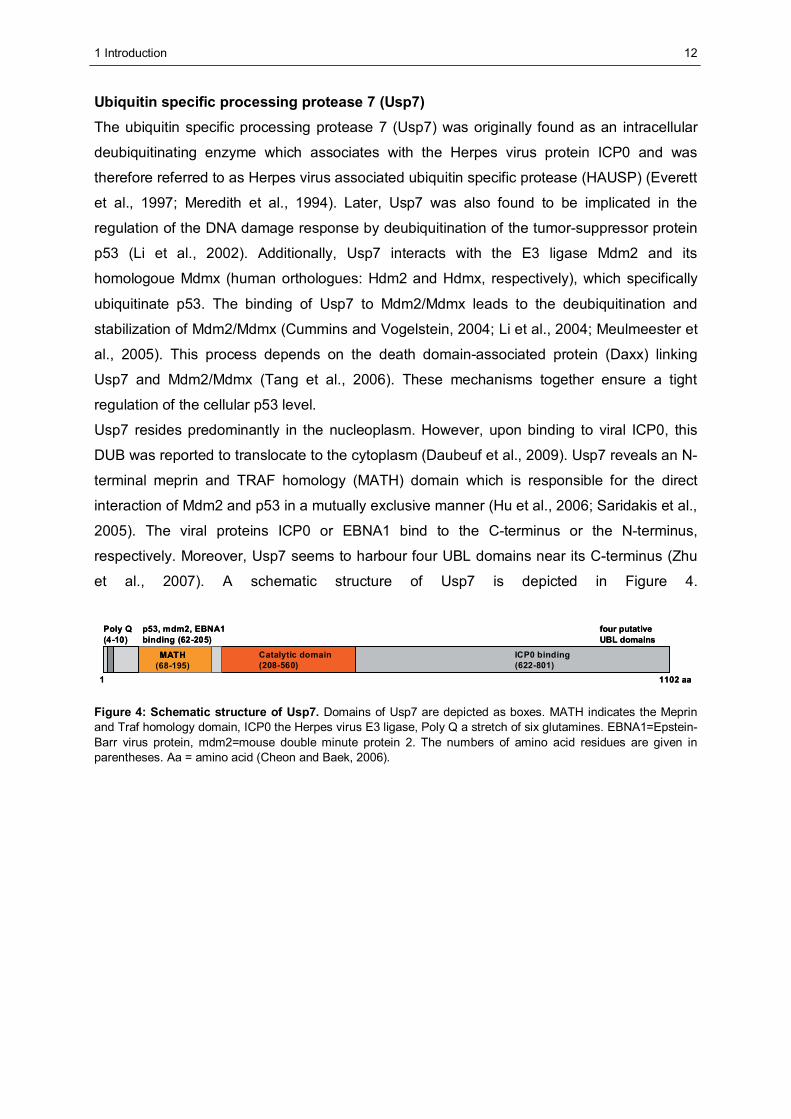
Ubiquitin specific processing protease 7 (Usp7) The ubiquitin specific processing protease 7 (Usp7) was originally found as an intracellular
deubiquitinating enzyme which associates with the Herpes virus protein ICP0 and was
therefore referred to as Herpes virus associated ubiquitin specific protease (HAUSP) (Everett
et al., 1997; Meredith et al., 1994). Later, Usp7 was also found to be implicated in the
regulation of the DNA damage response by deubiquitination of the tumor-suppressor protein
p53 (Li et al., 2002). Additionally, Usp7 interacts with the E3 ligase Mdm2 and its
homologoue Mdmx (human orthologues: Hdm2 and Hdmx, respectively), which specifically
ubiquitinate p53. The binding of Usp7 to Mdm2/Mdmx leads to the deubiquitination and
stabilization of Mdm2/Mdmx (Cummins and Vogelstein, 2004; Li et al., 2004; Meulmeester et
al., 2005). This process depends on the death domain-associated protein (Daxx) linking
Usp7 and Mdm2/Mdmx (Tang et al., 2006). These mechanisms together ensure a tight
regulation of the cellular p53 level.
Usp7 resides predominantly in the nucleoplasm. However, upon binding to viral ICP0, this
DUB was reported to translocate to the cytoplasm (Daubeuf et al., 2009). Usp7 reveals an N-
terminal meprin and TRAF homology (MATH) domain which is responsible for the direct
interaction of Mdm2 and p53 in a mutually exclusive manner (Hu et al., 2006; Saridakis et al.,
2005). The viral proteins ICP0 or EBNA1 bind to the C-terminus or the N-terminus,
respectively. Moreover, Usp7 seems to harbour four UBL domains near its C-terminus (Zhu
et al., 2007). A schematic structure of Usp7 is depicted in Figure 4.
MATH(68-195)
p53, mdm2, EBNA1binding (62-205)
ICP0 binding(622-801)
Catalytic domain(208-560)
Poly Q(4 10)
1 1102 aa
MATH ICP0 binding(622-801)
-four putativeUBL domains
MATH(68-195)
p53, mdm2, EBNA1binding (62-205)
ICP0 binding(622-801)
Catalytic domain(208-560)
Poly Q(4 10)
1 1102 aa
MATH ICP0 binding(622-801)
-four putativeUBL domains
Figure 4: Schematic structure of Usp7. Domains of Usp7 are depicted as boxes. MATH indicates the Meprin and Traf homology domain, ICP0 the Herpes virus E3 ligase, Poly Q a stretch of six glutamines. EBNA1=Epstein-Barr virus protein, mdm2=mouse double minute protein 2. The numbers of amino acid residues are given in parentheses. Aa = amino acid (Cheon and Baek, 2006).
1 Introduction
13
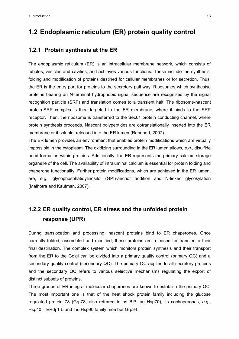
1.2 Endoplasmic reticulum (ER) protein quality control
1.2.1 Protein synthesis at the ER
The endoplasmic reticulum (ER) is an intracellular membrane network, which consists of
tubules, vesicles and cavities, and achieves various functions. These include the synthesis,
folding and modification of proteins destined for cellular membranes or for secretion. Thus,
the ER is the entry port for proteins to the secretory pathway. Ribosomes which synthesise
proteins bearing an N-terminal hydrophobic signal sequence are recognised by the signal
recognition particle (SRP) and translation comes to a transient halt. The ribosome-nascent
protein-SRP complex is then targeted to the ER membrane, where it binds to the SRP
receptor. Then, the ribosome is transferred to the Sec61 protein conducting channel, where
protein synthesis proceeds. Nascent polypeptides are cotranslationally inserted into the ER
membrane or if soluble, released into the ER lumen (Rapoport, 2007).
The ER lumen provides an environment that enables protein modifications which are virtually
impossible in the cytoplasm. The oxidizing surrounding in the ER lumen allows, e.g., disulfide
bond formation within proteins. Additionally, the ER represents the primary calcium-storage
organelle of the cell. The availability of intraluminal calcium is essential for protein folding and
chaperone functionality. Further protein modifications, which are achieved in the ER lumen,
are, e.g., glycophosphatidylinositol (GPI)-anchor addition and N-linked glycosylation
(Malhotra and Kaufman, 2007).
1.2.2 ER quality control, ER stress and the unfolded protein
response (UPR)
During translocation and processing, nascent proteins bind to ER chaperones. Once
correctly folded, assembled and modified, these proteins are released for transfer to their
final destination. The complex system which monitors protein synthesis and their transport
from the ER to the Golgi can be divided into a primary quality control (primary QC) and a
secondary quality control (secondary QC). The primary QC applies to all secretory proteins
and the secondary QC refers to various selective mechanisms regulating the export of
distinct subsets of proteins.
Three groups of ER integral molecular chaperones are known to establish the primary QC.
The most important one is that of the heat shock protein family including the glucose
regulated protein 78 (Grp78, also referred to as BiP, an Hsp70), its cochaperones, e.g.,
Hsp40 = ERdj 1-5 and the Hsp90 family member Grp94.
1 Introduction
14
These chaperones bind to nascent proteins, assist their folding and are involved in stress
signalling and degradation processes. Also lectins such as calnexin and calreticulin as well
as thiol-disulfide oxidoreductases such as protein disulfide isomerase (PDI) belong to the
primary QC system. Calnexin together with Calreticulin interacts with monoglycosylated N-
linked glycans and determines the revision or degradation of the glycoprotein. PDIs catalyse
the oxidation, reduction and isomerisation of disulfide bonds (Ellgaard and Helenius, 2003;
Nishikawa et al., 2005).
Despite the existence of this complex ER quality control system, the process of protein
synthesis, modification and targeting is error prone due to mutations, transcription-,
translation- or folding deficiencies. Estimated 30% of newly synthesised proteins are
defective (Schubert et al., 2000). Furthermore, mature proteins might also be damaged, e.g.,
by environmental noxes such as high energy radiation or chemical insults. In addition,
disorders of the ER such as perturbation of calcium homeostasis, disturbance of the ER
luminal redox state, increased protein cargo or altered protein glycosylation occur. All of
these situations may lead to the accumulation of misfolded proteins within the ER. Since the
accumulation of irreparable proteins is a threat for cell viability, this ER stress induces
multiple signalling pathways to avoid cell toxic protein aggregation. These pathways are
designated as the unfolded protein response (UPR). The UPR is an adaptive mechanism
leading to the induction of genes which enhance the ER folding capacity and the degradation
of unfolded proteins while general protein synthesis is stopped. Beyond that, prolonged ER
stress leads to the activation of programmed cell death (Schroder and Kaufman, 2005).
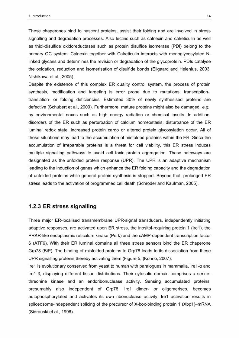
1.2.3 ER stress signalling
Three major ER-localised transmembrane UPR-signal transducers, independently initiating
adaptive responses, are activated upon ER stress, the inositol-requiring protein 1 (Ire1), the
PRKR-like endoplasmic reticulum kinase (Perk) and the cAMP-dependent transcription factor
6 (ATF6). With their ER luminal domains all three stress sensors bind the ER chaperone
Grp78 (BiP). The binding of misfolded proteins to Grp78 leads to its dissociation from these
UPR signalling proteins thereby activating them (Figure 5; (Kohno, 2007).
Ire1 is evolutionary conserved from yeast to human with paralogues in mammalia, Ire1-α and
Ire1-β, displaying different tissue distributions. Their cytosolic domain comprises a serine-
threonine kinase and an endoribonuclease activity. Sensing accumulated proteins,
presumably also independent of Grp78, Ire1 dimer- or oligomerises, becomes
autophosphorylated and activates its own ribonuclease activity. Ire1 activation results in
spliceosome-independent splicing of the precursor of X-box-binding protein 1 (Xbp1)–mRNA
(Sidrauski et al., 1996).
1 Introduction
15
Golgi
ER
eIF2α
Inhibition of Protein synthesis
P
ATF4/PERK ATF6
IRE1α,β
Xbp1mRNA
Transcription of chaperones
Misfolded proteins
Grp78 (BiP)
Golgi
ER
eIF2α
Inhibition of Protein synthesis
P
ATF4/PERK ATF6
IRE1α,β
Xbp1mRNA
Transcription of chaperones
Misfolded proteins
Grp78 (BiP)
Figure 5: Schematic overview of the unfolded protein response (UPR) signalling pathways. Misfolded proteins within the ER lumen bind to the ER chaperone Grp78 (BiP) which leads to the induction of three independent signalling pathways. Activated Perk phosphorylates eIF2α, which results in cell cycle arrest and stop of translation. ATF4 mRNA is translated in this situation and induces UPR genes. Activated ATF6 translocates to the Golgi, where it is cleaved and then acts as a transcription factor, which results in the induction of UPR target genes. Activated Ire1 splices the Xbp1-mRNA which encodes a transcription factor that induces UPR target genes (Hayden et al., 2005).
Xbp1 pre-mRNA has a DNA-binding domain (DBD) and a transcription activation domain
(AD) in different open reading frames and thus is inactive. The unconventional splicing
removes the intron and xbp1 is converted to mature mRNA, from which the active
transcription factor is translated. Targets of Xbp1 include genes encoding ER chaperones
and proteins involved in ER degradation, transcription factors (Xbp1 itself) and components
of the secretory pathway such as Sec61 (Yoshida, 2007).
Perk, a type 1 transmembrane protein, binds Grp78 in its inactive state and has a cytosolic
domain with kinase activity. Upon ER stress, Perk autophosphorylation and oligomerisation
occurs analogous to Ire1. Active Perk phosphorylates elF2-α, resulting in the attenuation of
protein synthesis. However, the translation of some mRNAs is induced in this situation such
as that of the transcription factor ATF4, which in turn induces UPR target genes (Harding et
al., 1999).
ATF6 is a basic leucine zipper transcription factor that binds to ER stress response elements
(ERSE), thereby inducing ER chaperone genes such as human Grp78 and Grp94. During
ATF6 activation, two luminal Golgi localisation sites are responsible for the transfer of ATF6
to the Golgi. There, a proteolytic intramembrane cleavage of ATF6 leads to the release of its
cytosolic domain which represents the active transcription factor, harbouring DNA binding
and transcription activation sites (Yoshida et al., 1998).
1 Introduction
16
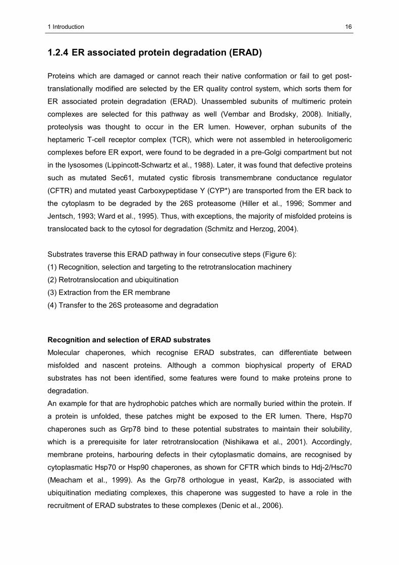
1.2.4 ER associated protein degradation (ERAD)
Proteins which are damaged or cannot reach their native conformation or fail to get post-
translationally modified are selected by the ER quality control system, which sorts them for
ER associated protein degradation (ERAD). Unassembled subunits of multimeric protein
complexes are selected for this pathway as well (Vembar and Brodsky, 2008). Initially,
proteolysis was thought to occur in the ER lumen. However, orphan subunits of the
heptameric T-cell receptor complex (TCR), which were not assembled in heterooligomeric
complexes before ER export, were found to be degraded in a pre-Golgi compartment but not
in the lysosomes (Lippincott-Schwartz et al., 1988). Later, it was found that defective proteins
such as mutated Sec61, mutated cystic fibrosis transmembrane conductance regulator
(CFTR) and mutated yeast Carboxypeptidase Y (CYP*) are transported from the ER back to
the cytoplasm to be degraded by the 26S proteasome (Hiller et al., 1996; Sommer and
Jentsch, 1993; Ward et al., 1995). Thus, with exceptions, the majority of misfolded proteins is
translocated back to the cytosol for degradation (Schmitz and Herzog, 2004).
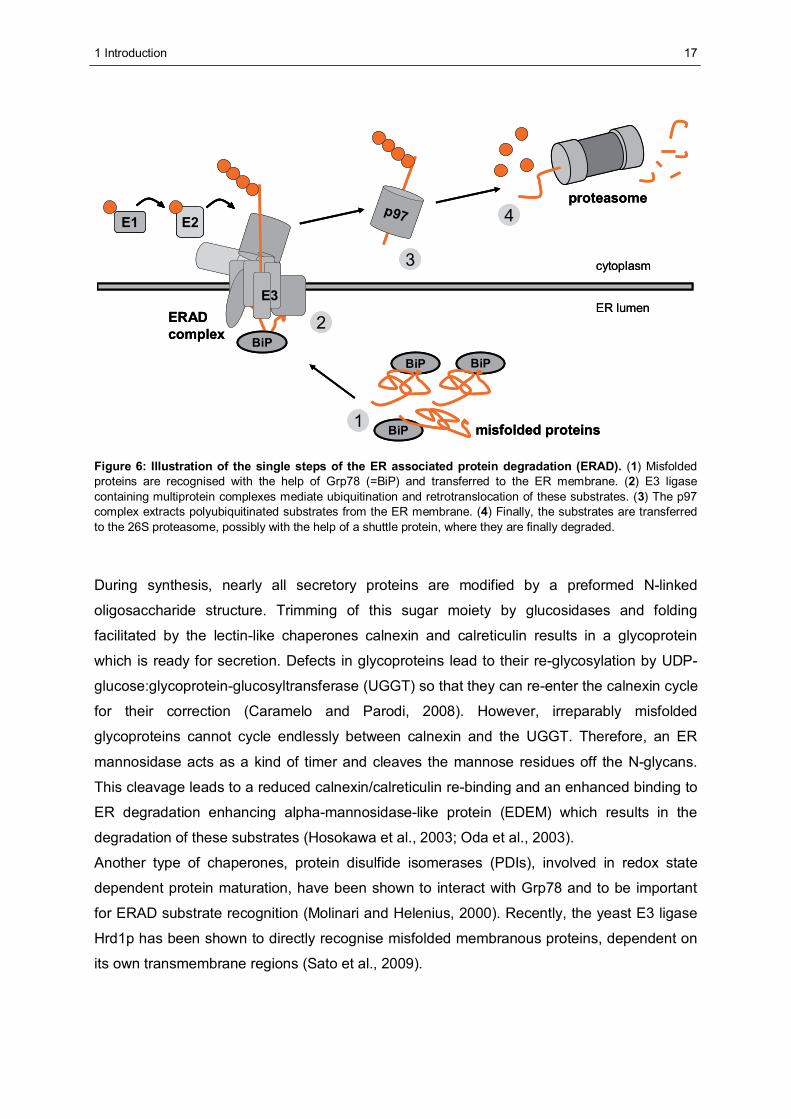
Substrates traverse this ERAD pathway in four consecutive steps (Figure 6):
(1) Recognition, selection and targeting to the retrotranslocation machinery
(2) Retrotranslocation and ubiquitination
(3) Extraction from the ER membrane
(4) Transfer to the 26S proteasome and degradation
Recognition and selection of ERAD substrates Molecular chaperones, which recognise ERAD substrates, can differentiate between
misfolded and nascent proteins. Although a common biophysical property of ERAD
substrates has not been identified, some features were found to make proteins prone to
degradation.
An example for that are hydrophobic patches which are normally buried within the protein. If
a protein is unfolded, these patches might be exposed to the ER lumen. There, Hsp70
chaperones such as Grp78 bind to these potential substrates to maintain their solubility,
which is a prerequisite for later retrotranslocation (Nishikawa et al., 2001). Accordingly,
membrane proteins, harbouring defects in their cytoplasmatic domains, are recognised by
cytoplasmatic Hsp70 or Hsp90 chaperones, as shown for CFTR which binds to Hdj-2/Hsc70
(Meacham et al., 1999). As the Grp78 orthologue in yeast, Kar2p, is associated with
ubiquitination mediating complexes, this chaperone was suggested to have a role in the
recruitment of ERAD substrates to these complexes (Denic et al., 2006).
1 Introduction
17
ER lumen
cytoplasm
misfolded proteins
BiP
BiP
BiP
E3
p97E1 E2
BiP
proteasome
1
2
3
4
ERAD complex
ER lumen
cytoplasm
misfolded proteins
BiP
BiP
BiP
E3
p97E1 E2
BiP
proteasome
1
2
3
4
ERAD complex
Figure 6: Illustration of the single steps of the ER associated protein degradation (ERAD). (1) Misfolded proteins are recognised with the help of Grp78 (=BiP) and transferred to the ER membrane. (2) E3 ligase containing multiprotein complexes mediate ubiquitination and retrotranslocation of these substrates. (3) The p97 complex extracts polyubiquitinated substrates from the ER membrane. (4) Finally, the substrates are transferred to the 26S proteasome, possibly with the help of a shuttle protein, where they are finally degraded.
During synthesis, nearly all secretory proteins are modified by a preformed N-linked
oligosaccharide structure. Trimming of this sugar moiety by glucosidases and folding
facilitated by the lectin-like chaperones calnexin and calreticulin results in a glycoprotein
which is ready for secretion. Defects in glycoproteins lead to their re-glycosylation by UDP-
glucose:glycoprotein-glucosyltransferase (UGGT) so that they can re-enter the calnexin cycle
for their correction (Caramelo and Parodi, 2008). However, irreparably misfolded
glycoproteins cannot cycle endlessly between calnexin and the UGGT. Therefore, an ER
mannosidase acts as a kind of timer and cleaves the mannose residues off the N-glycans.
This cleavage leads to a reduced calnexin/calreticulin re-binding and an enhanced binding to
ER degradation enhancing alpha-mannosidase-like protein (EDEM) which results in the
degradation of these substrates (Hosokawa et al., 2003; Oda et al., 2003).
Another type of chaperones, protein disulfide isomerases (PDIs), involved in redox state
dependent protein maturation, have been shown to interact with Grp78 and to be important
for ERAD substrate recognition (Molinari and Helenius, 2000). Recently, the yeast E3 ligase
Hrd1p has been shown to directly recognise misfolded membranous proteins, dependent on
its own transmembrane regions (Sato et al., 2009).
1 Introduction
18
Retrotranslocation and ubiquitination Since degradation of ERAD substrates takes place in the cytoplasm, ER derived proteins
have to pass the ER membrane in the opposite direction as protein synthesis occurs. This
process is termed retrotranslocation. In addition, the process of ubiquitination is also located
at the cytoplasmatic site of the ER. This means that for their degradation, ERAD substrates
at least have to gain access to this compartment. Starting retrotranslocation is a prerequisite
for ubiquitination. All ER lumen and most ER membrane derived proteins are thought to be
retrotranslocated presumably by a protein-conducting channel.
A few proteins have been suggested to form this retrotranslocation pore. The mammalian ER
transmembrane protein Derlin-1 was found in a complex with cytoplasmatic and membrane-
resident ERAD mediating proteins as well as with ERAD substrates (Katiyar et al., 2005;
Lilley and Ploegh, 2004; Schulze et al., 2005). A depletion of Derlin1 results in the induction
of the UPR and in retarded degradation of selected substrates (Ye et al., 2005). Also E3
ligases are assumed to mediate retrotranslocation and ubiquitination. In yeast, the RING E3
ligases Hrd1p and Doa10 are integral components of distinct multiprotein complexes
consisting of ER luminal, ER membrane and cytoplasmatic proteins. While Hrd1p mediates
the degradation of substrates that harbour misfolded domains within the ER lumen (ERAD-
L), Doa10 is required for proteins with a folding error in their cytosolic domain (ERAD-C)
(Carvalho et al., 2006; Vashist and Ng, 2004).
In mammalia, the membrane-multispanning E3 ligase Gp78, an orthologue of yeast Hrd1p,
was suggested to mediate retrotranslocation (Zhong et al., 2004). A different hypothesis
implies that retrotranslocation of protein complexes or of large proteins requires the formation
of so called lipid droplets from the ER membrane (Ploegh, 2007).
ERAD substrates have to be ubiquitinated prior to extraction and proteasomal targeting and
ubiquitination requires the sequential action of E1, E2 and E3 enzymes (see 1.1.3). The
mammalian ER-resident E3 ligases Gp78 and Hrd1 (also called Synoviolin), for instance,
together with the E2 enzyme Ube2g2 mediate the degradation of CD3-δ and TCR-α,
respectively (Fang et al., 2001; Kikkert et al., 2004).
Cytosolic ubiquitin ligases may also be implicated in the ERAD process, as this was reported
for the RING E3 parkin which ubiquitinates the parkin-associated endothelin receptor-like
receptor (Pael-R) (Imai et al., 2001). Since Synoviolin was described to also ubiquitinate
Pael-R, the efficient turnover of substrates may depend on more than one E3 ligase (Omura
et al., 2006). This was also shown for the ERAD substrate CFTR which is processed by the
cooperation of two E3 ligases, Gp78 and RMA1 (Morito et al., 2008).
1 Introduction
19
Membrane extraction of ERAD substrates Polyubiquitination of ERAD substrates is not only a signal for proteasomal degradation but
also for the extraction of substrates from the ER membrane (Kikkert et al., 2001). To start
this process, a minimal length of the polyubiquitin chain bound to the substrate is decisive
(Jarosch et al., 2002). The polyubiquitin moiety is recognised by a cytosolic molecular
chaperone complex, consisting of the AAA-ATPase p97 (also called VCP for Valosin
containing protein or Cdc48 in yeast), which forms a homohexamer, and the associated
proteins Ufd1 and Npl4. In yeast, the Cdc48-Ufd1-Npl4 complex was shown to bind ubiquitin
conjugates and to be required for the ATP dependent extraction of various substrates such
as MHC class I molecules (Dai and Li, 2001; Rape et al., 2001; Ye et al., 2001).
P97 was suggested to act as a motor, actively pulling the substrate out of the ER membrane
in an ATP-consuming process and dependent on the binding of p97 to the ER membrane
(Ye et al., 2003). The recruitment of the p97 complex to the ER membrane was proposed to
be mediated by ER-resident proteins. Among these proteins are the VCP interacting
membrane protein (VIMP) and ubiquitin regulator-X (UBX) domain proteins such as Ubxd2
(Liang et al., 2006; Ye et al., 2004). Also Ubxd8 was suggested as a p97 recruitment factor in
mammalia (Mueller et al., 2008).
Transfer of ERAD substrates to the 26S proteasome Since it has been found to interact with 26S subunits, p97 was initially assumed to escort the
extracted ERAD substrate to the 26S proteasome for degradation (Dai et al., 1998). Later, a
variety of proteins exhibiting a UBA as well as a UBL domain were described to execute this
shuttle function. Harbouring both domains allows these proteins to interact with ubiquitinated
substrates and the 26 proteasome concomitantly. The most prominent UBL and UBA domain
containing proteins are Rad23 and Dsk2 required for ERAD in yeast (Medicherla et al.,
2004). In humans, two homologues of Rad23, hHR23A and hHR23B interact with the
proteasomal 19S subunit Rpn10 (S5a) via their UBL domain (Hiyama et al., 1999).
1.2.5 Synoviolin based ERAD complexes
Synoviolin based ERAD complexes consist of a variety of proteins which are introduced
below and schematically depicted in Figure 7, B. Mammalian Synoviolin, also referred to as
Hrd1, is an ER-resident E3 ligase of the RING type (see 1.1.4) and involved in the
ubiquitination and retrotranslocation of ERAD substrates. This enzyme of 617 amino acids is
one of two mammalian orthologues of yeast Hrd1p which was first described in connection
with the turnover of 3-hydroxy-3-methylglutaryl coenzyme A reductase (HMGR) and
therefore named Hrd1 for HMGR degradation (Hampton et al., 1996).
1 Introduction
20
The other Hrd1p orthologue is Gp78, also known as autocrine motility factor receptor (AMFR)
(Fang et al., 2001; Nadav et al., 2003). Gp78 harbours a CUE domain and a specific Ube2g2
binding site, which are both essential for its function in protein degradation (Chen et al.,
2006). Furthermore, Gp78 interacts with various other proteins involved in ERAD such as the
AAA ATPase p97. This interaction is essential for the degradation of the ERAD substrates
CD3-δ and the Z variant of α1-antitrypsin (Ballar et al., 2006; Zhong et al., 2004). Synoviolin
is ER stress induced, dependent on Ire1 and ATF6 activity. It reveals six transmembrane
domains and a ubiquitin ligase activity harbouring RING domain near the C-terminus
(Kaneko and Nomura, 2003; Nadav et al., 2003). Importantly, Synoviolin is highly expressed
in synovial cells of patients with rheumatoid arthritis and was shown there to protect cells
from ER stress induced apoptosis (Amano et al., 2003). Apoptosis inhibition leads to synovial
hyperplasia which is causative for the progression of the disease. In mammalia, Synoviolin
mediates the basal degradation of HMGR, whereas the turnover of the sterol-regulated
isoform of HMGR is mediated by Gp78 (Kikkert et al., 2004; Song et al., 2005). In addition,
degradation of a variety of substrates such as CD3-δ, TCR-α, misfolded insulin and Ig-μ
chains is mediated by Synoviolin (Allen et al., 2004; Cattaneo et al., 2008; Kikkert et al.,
2004). The direct interaction of Synoviolin with the ERAD components Homocysteine
inducible endoplasmic reticulum-resident protein (Herp), Derlin-1 and p97 was described.
Furthermore, in vitro interaction studies gave rise to the assumption that Synoviolin is able to
homooligomerise (Schulze et al., 2005). Additionally, Sel1L and Derlin-2 as well as the
lectins OS-9 and XTP3-B were found to be associated with this E3 ligase (Christianson et al.,
2008; Lilley and Ploegh, 2005). The association of these proteins results in the formation of
multiprotein complexes located at the ER membrane. Synoviolin is thought to be the major
component of these multimeric structures. However, the mode of Synoviolin complex
assembly or the exact composition of the functional ERAD complexes is not known. Since
the study presented here deals with this Synoviolin based ERAD complexes, major
components of these complexes are introduced below.
In humans, three members of the Derlin family, ER-resident proteins of about 240 amino
acids in size, UPR inducible and harbouring four transmembrane domains, have been shown
to be associated with Synoviolin. Derlin-1 was found to be essential for the human
cytomegaloviral US11 protein triggered MHC class I dislocation and to be a central
component of complexes containing VIMP, p97 and Synoviolin (Lilley and Ploegh, 2004;
Schulze et al., 2005; Ye et al., 2004). Furthermore, Derlin-1 forms homooligomeric structures
(Ye et al., 2005). In addition to the degradation of ERAD substrates, Derlin-1 has also been
reported to be implicated in the processing of pathogenic proteins such as cholera toxin
(Bernardi et al., 2008). Derlin-2 and Derlin-3 were also reported to be components of ERAD
complexes. Derlin-2 and Derlin-3 heterooligomerise with the other Derlins.
1 Introduction
21
Moreover, Derlin-2 was found to homooligomerise (Lilley and Ploegh, 2005). Derlin-2 and
Derlin-3 provide a link between EDEM and p97 and are involved in the degradation of
misfolded glycoproteins (Oda et al., 2006).
The Suppressor of lin-12-like protein1, Sel-1L, mammalian homologue of Hrd3p, is another
ERAD complex component. Sel1L is an ER luminal glycoprotein of 794 amino acids with a
potential transmembrane domain at the C-terminus. Sel1L is induced by ER stress and was
reported to be associated with Synoviolin, Derlin-1, Derlin-2 and the AAA-ATPase p97 as
well as with Ubxd8, Ube2j1 and OS9 (Kaneko and Nomura, 2003; Lilley and Ploegh, 2005;
Mueller et al., 2008). Since Sel1L is involved in glycoprotein degradation, it was suggested to
play a role in the selection of misfolded proteins or to stabilise Synoviolin, comparable to its
yeast orthologue Hrd3p (Mueller et al., 2006). Recently, two additional soluble Sel1L
isoforms were identified which are secreted and involved in sorting and export of Ig-μ chains
(Cattaneo et al., 2009).
Ancient ubiquitous protein 1, AUP1, is an ER-resident protein of 476 amino acids.
Harbouring an N-terminal membrane anchor, the majority of the protein is cytoplasmatic.
Near its C-terminus, AUP1 possesses a CUE domain, which is thought to be involved in
ubiquitin binding or the recruitment of E2 enzymes and shows similarities to the UBA domain
(Hurley et al., 2006). Initially, AUP1 was found to be associated with platelet integrins and
thought to be involved in integrin signalling. However, this CUE domain protein is associated
with Sel1L and Sel1L interacting ERAD proteins, such as Ube2j1 and OS9. The elevated
expression of a GFP-tagged AUP1 inhibited viral US11 protein mediated dislocation of ER
resident MHC class I molecules, which implicates a function of AUP1 in ERAD (Mueller et al.,
2008).
Various other proteins such as lectins, selenoproteins, UBX domain proteins, E2 enzymes
and ubiquilins are associated with Synoviolin. The ER-luminal lectins OS9 and XTP3-B
represent the human orthologues of yeast Yos9p which recognises N-glycans in yeast and
promotes their degradation. Two isoforms of OS9, OS9.1 and OS9.2 and XTP3-B interact
with Sel1L and bind the ERAD substrate NHK (Christianson et al., 2008). The selenoprotein
VIMP binds p97 and Derlin-1 concomitantly (Ye et al., 2004). UBX domain proteins such as
Ubxd8 also interact with p97 and Sel1L (Mueller et al., 2008). VIMP and Ubxd8 seem to
recruit p97 to the site of dislocation. In addition, p97 (see 1.2.4), also directly interacts with
Synoviolin (Schulze et al., 2005). The E2 enzyme Ube2j1 (Ubc6e) was shown to be
associated with Sel1L and thus suggested to work in concert with Synoviolin (Mueller et al.,
2008). Ubiquilin 1 and 2, human orthologues of yeast Dsk2, were recently found to interact
with Herp, a component of Synoviolin based complexes (Kim et al., 2008). Ubiquilins (also
referred to as hPlic) belong to the family of ubiquitin domain proteins (UDPs) and bind the
26S proteasome and polyubiquitin moieties concomitantly (Kleijnen et al., 2000).
1 Introduction
22
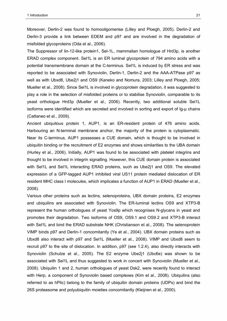
1.2.6 Homocysteine inducible endoplasmic reticulum - resident
protein (Herp)
The Homocysteine inducible endoplasmic reticulum resident protein Herp (Swiss-Prot entry:
Q15011) is strongly induced by UPR inducing agents such as homocysteine and β-
mercaptoethanol as well as the N-glycosylation inhibitor tunicamycin or the calcium-ATPase
inhibitor thapsigargin (Hori et al., 2004; Kokame et al., 2000; Kokame et al., 1996). Apart
from ER stress, the Herp encoding gene, herpud1, is also induced in response to osmotic
stress and by DNA damaging agents such as methyl methanesulfonate (MMS). Therefore,
Herp was also termed MMS inducible factor1 (Mif1). The herpud1 promoter contains the ‘ER
stress response elements’ (ERSE I and II) which are binding sites for the UPR dependent
transcription factors ATF6 and Xbp1 (Kokame et al., 2000; van Laar et al., 2000). Structural
analysis revealed that Herp harbours a UBL domain at the very N-terminus thus belonging to
the family of the UDPs. Herp is an integral ER-membrane protein of 391 amino acids in size
with both termini facing the cytoplasm (Kokame et al., 2000). Biochemical studies showed
that Herp is associated with Synoviolin based ERAD complexes through direct binding of the
E3 ligase Synoviolin (Schulze et al., 2005).
ER lumen
cytoplasm
UBL
C-term
N-term
Herp
p97
Sel1LDerlin-1
Synoviolin
ER lumen
cytoplasm
BA
AUP1
Ube2j1
ER lumen
cytoplasm
UBL
C-term
N-term
Herp
p97
Sel1LDerlin-1
Synoviolin
ER lumen
cytoplasm
BA
AUP1
Ube2j1
Figure 7: Illustration of the homocysteine inducible ER stress protein (Herp). (A) Membrane topology of Herp. The UDP faces the cytoplasm with both termini. (B) Components of Synoviolin based ERAD complexes. Herp directly interacts with the potentially oligomeric E3 ligase Synoviolin and thus is part of Synoviolin based complexes. Thereby, Herp is also associated with other Synoviolin interacting proteins such as p97 or Derlin-1 (Schulze et al., 2005). Recently, the E2 enzyme Ube2j1 and the AUP1-protein have been found in association with Sel1-L which is a direct interaction partner of Synoviolin (Mueller et al., 2008).
1 Introduction
23
Herp is essential for the effective degradation of ERAD substrates such as Connexin 43,
CD3-δ and a nonsecreted Igκ light chain (Hori et al., 2004; Okuda-Shimizu and Hendershot,
2007; Schulze et al., 2005). These findings support the functional role of Herp in ERAD.
Interestingly, the UBL domain of Herp seems to mediate the effect of Herp in the ERAD
process. Herp-dependent substrates were stabilised, if Herp lacking the UBL domain was
overexpressed (Schulze, 2006). UBL domains within UDPs display diverse binding
specificities which are important for their molecular function (Madsen et al., 2007). Since
proteasome binding had been demonstrated for the UBL domains of a subset of UDPs such
as BAG-1 (Luders et al., 2000), Herp was proposed to recruit the 26S proteasome to the ER
upon ER stress (van Laar et al., 2001). However, the UBL domain of Herp does not interact
with the 26S proteasome and is also dispensable for the interaction of Herp with Synoviolin
(Schulze et al., 2005). Herp and the 26S proteasome might nevertheless be found
associated, since the UDP is a substrate of the UPS (Sai et al., 2003). A yeast two hybrid
screen for target proteins of the Herp N-terminus (including the UBL domain) identified Usp7
as binding partner (Schulze, 2006). The functional importance of this deubiquitinating
enzyme for ERAD has not been tested. Moreover, the UBL domain of Herp seems to
determine its half-life of about three hours (Kokame et al., 2000; Sai et al., 2003). Beyond,
the lysine 61 residue within the Herp UBL domain seems to be the crucial ubiquitination site,
as shown by in vitro ubiquitination experiments (Li et al., 2007).
Although Herp was shown to be ubiquitinated and degraded by the proteasome, E2 or E3
enzymes involved in this process have not been identified (Sai et al., 2003). Only the soluble
E3 ligase ‘plenty of SH3 domains’ (POSH) was reported to ubiquitinate Herp, but with K63
linked polyubiquitin which does not lead to degradation but to the redistribution of Herp from
the Trans Golgi network to the ER. Upon calcium perturbation, Herp, dependent on its UBL
domain, induces the oligomerisation and activation of POSH (Tuvia et al., 2007).
A few data on the cellular function of Herp have been reported. The induction of Herp by ER
stress is connected with the improvement of the folding capacity of the ER. With this, Herp is
involved in the protection of cells against ER stress (Hori et al., 2004). In neurons, the
elevated expression of Herp promotes cell survival by stabilisation of calcium homeostasis
and maintaining mitochondrial function. However, prolonged ER stress leads to the cleavage
of Herp by caspases and to apoptosis (Chan et al., 2004). Concerning ERAD, Herp was
shown to associate with Ubiquilin1 and 2 and suggested to thereby enhance the degradation
of CD3-δ (Kim et al., 2008). However, a general molecular function of Herp has not been
shown.
1 Introduction
24
In yeast, the UDP U1 SNP1-associating protein 1 (Usa1p) associates with Hrd1p, Hrd3p and
Der1p and is required for the efficient ERAD of the model substrate CPY*. Usa1p and Herp
reveal similar domain architectures. Thus, both UDPs seem to be structurally related. Usa1p
links Der1p to the Hrd1p/Hrd3p complex and was suggested to be the functional equivalent
of Herp, since the mammalian UDP was able to partly rescue the Usa1p deletion in yeast
(Carvalho et al., 2006). Most recently, it was shown that Usa1p, dependent on its N-terminus
which harbours a UBL domain, functions as a scaffold protein enabling the oligomerisation of
Hrd1p. This process was postulated to be a prerequisite for the degradation of membrane
derived ERAD substrates (Horn et al., 2009). However, a general role of the Usa1p UBL
domain in yeast ERAD is not given, since this domain was shown to be essential only for the
degradation of 6myc-Hmg2 but not of Hmg2-GFP or CPY* (Carroll and Hampton, 2010; Horn
et al., 2009; Kim et al., 2009). Comparable to Herp, the UBL domain of Usa1p is not
associated with the 26S proteasome (Kim et al., 2009).
A database search using the SMART program was performed to find human proteins
harbouring a UBL and transmembrane domains comparable to Herp. With this approach four
candidates were found and designated as transmembrane-associated protein containing a
ubiquitin-like domain (mubl). One member of this family is Herp2 (mubl2, Swiss-Prot entry:
Q9BSE4) which is most similar to Herp (Schulze, 2006). The amino acid sequence of Herp2
(406 amino acids) is 40% identical with the Herp sequence. The sequences of the UBL
domains of both UDPs are even 50% identical. Both proteins display similar domain
architectures with a UBL domain at the N-terminus and a transmembrane domain close to
the C-terminus. Herp2 additionally contains a serine-rich region downstream its UBL domain.
A Herp2 splice variant lacking amino acids 49-70 was also identified. So far, there are no
further data reported on Herp2 in the literature.
1 Introduction
25
1.3 Aim of this study The UPR-induced ER-resident protein Herp, which is associated with Synoviolin based
complexes, plays an important role in the degradation of the Synoviolin substrate CD3-δ.
Depletion of Herp or disturbance of the Herp UBL domain leads to the stabilisation of this
substrate. However, the mechanisms underlying the function of Herp are poorly understood.
Thus, the aim of this study was to characterise the molecular function of Herp within
Synoviolin mediated ERAD.
Compared to Synoviolin, ER stress caused induction and degradation of Herp occurs more
rapidly. Therefore, it was investigated whether these dynamics of Herp are important for its
function in ERAD. In this context, the turnover of Herp at Synoviolin based complexes was
analysed and with regard to this aspect it was tested whether Herp is a substrate of
Synoviolin or its associated proteins. Moreover, it was analysed whether the degradation of
Herp is required for the process of ERAD.
The UBL domain of Herp, crucial for the function of this UDP, specifically binds the
deubiquitinating enzyme Usp7. Therefore, it was analysed whether Usp7 is a component of
Synoviolin based complexes playing a role in determining the stability of Herp or interfering
with the ERAD process.
In yeast, the ubiquitin domain protein Usa1p, required for the efficient ERAD process, was
shown to maintain the integrity of ERAD complexes. Therefore, it was tested whether Herp
had an analogous function in mammalia; Herp was tested for influence on the
oligomerisation of Synoviolin or the interaction of accessory proteins with Synoviolin based
complexes.
Herp2, a homologue of Herp, was hypothesised to be capable of taking over the cellular
functions of Herp. Therefore, it was tested whether Herp2 revealed the same properties as
Herp such as UPR inducibility, binding of Usp7, instability and association with Synoviolin.
2 Material and Methods
26
2 Material and Methods
2.1 Instruments, consumables and chemicals
Table 1: Instruments used in this study
Instrument Supplier
Phosphoimager FLA3000 Fujifilm Raytest
Freezing container Cryo 1°C Nalgene
G-Box gel documentation Syngene
mini-Protean 3 electrophoresis system Biorad
Scintillation counter Wallace 1410 Pharmacia
semidry blotting apparatus Peqlab
SW 40 swing out rotor Beckman Coulter
Table top centrifuge Eppendorf
Thermocycler Uno Thermoblock Biometra
Ultracentrifuge OptimaTM-L Beckman Coulter
UV VIS photometer Ultrospec 2100 pro Amersham Biosciences
G:BOX gel documentation system Syngene
Table 2: Consumables used in this study
Consumable Supplier
BioMAX film Kodak
Cell culture flasks, dishes and multiwell plates Greiner
Cryoconservation vials Corning
Nitrocellulose membranes Whatman
Whatman paper Schleicher und Schuell
XOmat-UV film Kodak
General chemicals were purchased from VWR International, Roth, Sigma or Applichem, if
not otherwise noted. They all were of at least 99% purity. All solutions in this study are
aqueous, unless otherwise stated.
2 Material and Methods
27
Table 3: Chemicals used in this study
Chemical Supplier
1 Kb Marker New England Biolabs
1 KB Plus DNA Ladder Invitrogen 35S-S-L-methionine / 35S-L-cysteine (Tran35S-LabelTM,
metabolic labelling reagent) MP Biomedicals
Ampicillin Roth
Blasticidin S Sigma
CompleteTM protease inhibitor Roche
Cycloheximide Sigma
Dharmafect Dharmacon
DMSO Sigma
dNTP mix New England Biolabs
Doxicycline Clontech
Fetal calf serum (and dialysed fetal calf serum) Biochrom
Glutathione sepharose Amersham / GE
Iscove’s or DMEM medium Biochrom
L-glutamin PAA
Lipofectamin 2000 Invitrogen
MG132 Calbiochem
OptiMEM Biochrom
Penicillin PAA
Polynucleotide kinase Roche
Protein A – HRP Biorad
Protein A- and Protein G-sepharose Amersham / GE
Puromycin Sigma
RPMI 1640 medium (also methionine free) Biochrom
shrimp alkaline phosphatase Roche
Streptavidin agarose Novagen
Streptomycin PAA
T4 DNA ligase and accordant buffer NEB
Thapsigargin Sigma
Trypsin/EDTA (1x) PAA
Tunicamycin Sigma
ZeocinTM Invitrogen
2 Material and Methods
28
2.2 Molecular biology
2.2.1 Cultivation and storage of Escherichia coli (E.coli)
• Luria-Bertani (LB) medium: 1% (w/v) bacto tryptone, 0.5% (w/v) yeast extract, 1% (w/v) NaCl
E.coli cells were cultured in LB medium. Liquid cultures were incubated on a rotating shaker
at 37°C, for protein expression likewise at room temperature (RT). For growing bacteria on
plates 1.5% agar (w/v) was added to LB medium and the incubation was performed at 37°C
over night. To select transformed cells, 100 μg/mL ampicillin or 50 μg/mL kanamycin was
added to liquid medium or plates, referred to as LB-amp or LB-kan, respectively. The optical
density of liquid cultures was determined by measuring the absorbance at a wavelength of
600 nm (OD600). For long-term storages, cultures in stationary phase were frozen at -80°C in
LB medium containing 15% (v/v) glycerol.
2.2.2 Isolation of plasmid DNA from E.coli
Preparation of DNA using alkaline lysis • P1: 50 mM Tris-HCl, pH adjusted to 8.0 with HCl ,10 mM EDTA, 10 μg/mL RNaseA
• P2: 200 mM NaOH, 1% (w/v) SDS
• P3: 3 M potassium acetate, pH 5.5
To test potential positive clones from a transformation (2.2.6), a single bacteria colony from
the agar plate was taken to inoculate five mL of LB medium containing the accordant
antibiotic, which were then shaken over night at 37°C. From the resulting culture two mL
were centrifuged in a reaction tube at 10,000 g for three min to sediment the cells. The
sediment was resuspended in 300 μL of buffer P1. Then 300 μL of buffer P2 were added and
the suspension was carefully inverted several times. Then 300 μL of buffer P3 were added
and the suspension was vigorously mixed and then centrifuged for 10 min at 13,000 g. The
supernatant was transferred into a new tube and the DNA was precipitated by addition of 500
μL isopropanol. To pellet the DNA, a centrifugation step for 20 min at 13,000 g was
performed and sedimented DNA was subsequently washed with 70% ethanol, dried at RT
and resuspended in 50 μL of sterile water.
2 Material and Methods
29
Preparation of DNA using ion exchange chromatography ("Mini-, Maxi-Prep”) Plasmid DNA for transformations or sequencing reactions was isolated from three mL of a
bacteria culture with the Plasmid Mini-Prep Kit (Qiagen). For the preparation of plasmid DNA
from a 200 mL bacteria culture, needed for the transfection of mammalian cells, the Plasmid
Maxi-Prep Kit (Qiagen) was used. The isolation was done according to the supplier’s
instructions.
2.2.3 Separation of DNA fragments by agarose gel electrophoresis
• TAE buffer: 40 mM Tris-HCl, 20 mM acetic acid, 1 mM EDTA
• DNA loading buffer: 40% (w/v) sucrose, 0.5% (w/v) SDS and 0.25% (w/v) bromophenol blue
(both: sodium salt)
DNA fragments, generated by restriction endonucleases or cDNA fragments were
electrophoretically separated on 1 - 2% agarose gels containing 0.1 μg/mL ethidium bromide.
For production of gels, the agarose was dissolved in TAE buffer by heating and ethidium
bromide was added after cooling to 60°C. TAE buffer was also used as running buffer. One
μL DNA loading buffer was added to nine μL of each DNA solution and the samples were
electrophoretically separated on the gels at ten V/cm. DNA fragments were visualised via the
intercalated ethidium bromide using a UV transluminator. The size of the fragments was
estimated by standard size markers such as the 1 KB Plus DNA ladder (Invitrogen), run on
the identical gels.
2.2.4 Determination of DNA and RNA concentration in solution
DNA and RNA concentrations were determined by measuring the absorbance of a nucleic
acid solution at a wavelength of 260 nm (A260). An A260 of one equals a concentration of 50
μg/mL double stranded DNA, 40 μg/mL single stranded DNA and RNA or 33 μg/mL
desoxyribonucleotides.
2 Material and Methods
30
2.2.5 Preparation of competent E.coli cells
• ϕa-medium: 0.5% (w/v) yeast extract, 2% (w/v) bacto tryptone, 40 mM MgSO4, pH adjusted to
7.6 with 1M KOH, sterile filtered
• TFB-I buffer: 30 mM potassium acetate, 50 mM MnCl2, 100 mM RbCl, 10 mM CaCl2, 15%
(v/v) glycerol, pH adjusted to 5.8 with acetic acid, sterile filtered
• TFB-II solution: 10 mM MOPS, 75 mM CaCl2, 10 mM RbCl, 15% (v/v) glycerol, pH adjusted to
7.0 with 1 M KOH, sterile filtered
A single E.coli colony from a LB agar plate was used to inoculate five mL of ϕa-medium
which then were shaken over night at 37°C. Two mL of this culture were diluted in 100 mL
fresh medium and grown to an OD600 of 0.5 at 37°C. After chilling the culture on ice for five
min the cells were sedimented by centrifugation (10 min, 5,000 g, 4°C). All following steps
were performed on ice with pre-chilled solutions. The sediment was resuspended in 40 mL
TFB-I buffer and incubated on ice for five minutes. Then cells were sedimented again by
centrifugation (10 min, 5,000 g, 4°C), resuspended in four mL TFB-II solution and further
incubated on ice for 30 minutes. Then the cells were transferred into reaction tubes in 100 μL
aliquots and quickly frozen in liquid nitrogen and stored at -80°C.
2.2.6 Transformation of E.coli with plasmid DNA
• SOC medium: 2% (w/v) bacto tryptone, 0.5% (w/v) yeast extract, 10 mM NaCl, 2.5 mM KCl,
10 mM MgCl2, 10 mM MgSO4, 2.0 mM glucose
For transformation of E.coli with plasmid DNA 100 μL of competent cells (XL1 blue or One
Shot®TOP10 cells (Invitrogen)) were thawed on ice. 50 ng of plasmid DNA were added to the
cells and incubation was continued for 30 min. A brief heat shock (42°C, 45 s) was followed
by incubation on ice for ten min. 500 μL of SOC medium were added and cells were kept at
37°C for one h while shaking. This suspension was streaked out onto agar plates containing
the accordant antibiotics and incubated over night at 37°C.
2.2.7 Isolation of total RNA from HeLa cells
For the analysis of the transcription of RNA, coding for proteins of interest, total RNA from
HeLa cells was isolated and used for reverse transcriptase PCR analysis. For that purpose
HeLa cells were left untreated or treated with 10 μg/mL tunicamycin or 2 μM thapsigargin for
up to ten h. Then 106 cells were subjected to RNA isolation using the High Pure RNA
isolation kit (Roche) according to the manufacturer’s instructions.
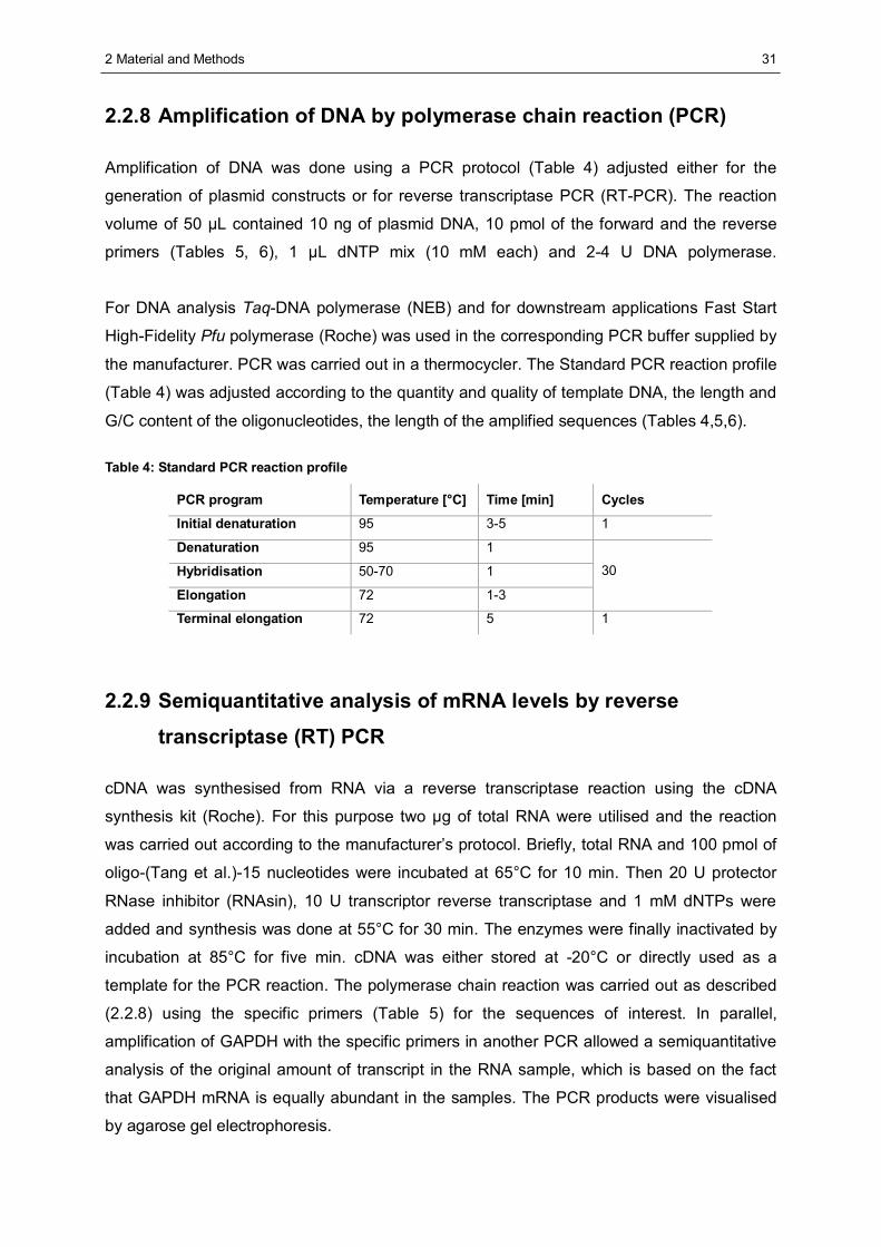
2 Material and Methods
31
2.2.8 Amplification of DNA by polymerase chain reaction (PCR)
Amplification of DNA was done using a PCR protocol (Table 4) adjusted either for the
generation of plasmid constructs or for reverse transcriptase PCR (RT-PCR). The reaction
volume of 50 μL contained 10 ng of plasmid DNA, 10 pmol of the forward and the reverse
primers (Tables 5, 6), 1 μL dNTP mix (10 mM each) and 2-4 U DNA polymerase.
For DNA analysis Taq-DNA polymerase (NEB) and for downstream applications Fast Start
High-Fidelity Pfu polymerase (Roche) was used in the corresponding PCR buffer supplied by
the manufacturer. PCR was carried out in a thermocycler. The Standard PCR reaction profile
(Table 4) was adjusted according to the quantity and quality of template DNA, the length and
G/C content of the oligonucleotides, the length of the amplified sequences (Tables 4,5,6).
Table 4: Standard PCR reaction profile
PCR program Temperature [°C] Time [min] Cycles
Initial denaturation 95 3-5 1
Denaturation 95 1
Hybridisation 50-70 1
Elongation 72 1-3
30
Terminal elongation 72 5 1
2.2.9 Semiquantitative analysis of mRNA levels by reverse
transcriptase (RT) PCR
cDNA was synthesised from RNA via a reverse transcriptase reaction using the cDNA
synthesis kit (Roche). For this purpose two μg of total RNA were utilised and the reaction
was carried out according to the manufacturer’s protocol. Briefly, total RNA and 100 pmol of
oligo-(Tang et al.)-15 nucleotides were incubated at 65°C for 10 min. Then 20 U protector
RNase inhibitor (RNAsin), 10 U transcriptor reverse transcriptase and 1 mM dNTPs were
added and synthesis was done at 55°C for 30 min. The enzymes were finally inactivated by
incubation at 85°C for five min. cDNA was either stored at -20°C or directly used as a
template for the PCR reaction. The polymerase chain reaction was carried out as described
(2.2.8) using the specific primers (Table 5) for the sequences of interest. In parallel,
amplification of GAPDH with the specific primers in another PCR allowed a semiquantitative
analysis of the original amount of transcript in the RNA sample, which is based on the fact
that GAPDH mRNA is equally abundant in the samples. The PCR products were visualised
by agarose gel electrophoresis.
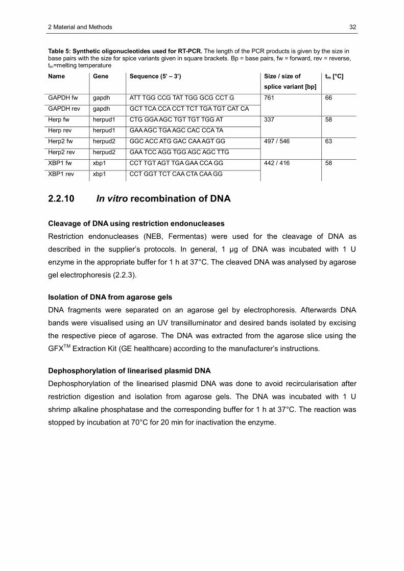
2 Material and Methods
32
Table 5: Synthetic oligonucleotides used for RT-PCR. The length of the PCR products is given by the size in base pairs with the size for spice variants given in square brackets. Bp = base pairs, fw = forward, rev = reverse, tm=melting temperature
Name Gene Sequence (5’ – 3’) Size / size of splice variant [bp]
tm [°C]
GAPDH fw gapdh ATT TGG CCG TAT TGG GCG CCT G
GAPDH rev gapdh GCT TCA CCA CCT TCT TGA TGT CAT CA
761 66
Herp fw herpud1 CTG GGA AGC TGT TGT TGG AT
Herp rev herpud1 GAA AGC TGA AGC CAC CCA TA
337 58
Herp2 fw herpud2 GGC ACC ATG GAC CAA AGT GG
Herp2 rev herpud2 GAA TCC AGG TGG AGC AGC TTG
497 / 546 63
XBP1 fw xbp1 CCT TGT AGT TGA GAA CCA GG
XBP1 rev xbp1 CCT GGT TCT CAA CTA CAA GG
442 / 416 58
2.2.10 In vitro recombination of DNA
Cleavage of DNA using restriction endonucleases Restriction endonucleases (NEB, Fermentas) were used for the cleavage of DNA as
described in the supplier’s protocols. In general, 1 μg of DNA was incubated with 1 U
enzyme in the appropriate buffer for 1 h at 37°C. The cleaved DNA was analysed by agarose
gel electrophoresis (2.2.3).
Isolation of DNA from agarose gels DNA fragments were separated on an agarose gel by electrophoresis. Afterwards DNA
bands were visualised using an UV transilluminator and desired bands isolated by excising
the respective piece of agarose. The DNA was extracted from the agarose slice using the
GFXTM Extraction Kit (GE healthcare) according to the manufacturer’s instructions.
Dephosphorylation of linearised plasmid DNA Dephosphorylation of the linearised plasmid DNA was done to avoid recircularisation after
restriction digestion and isolation from agarose gels. The DNA was incubated with 1 U
shrimp alkaline phosphatase and the corresponding buffer for 1 h at 37°C. The reaction was
stopped by incubation at 70°C for 20 min for inactivation the enzyme.
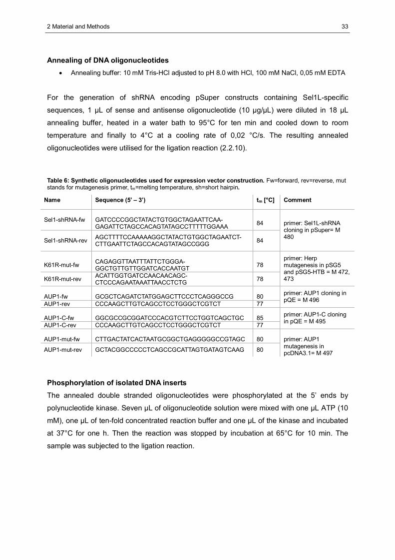
2 Material and Methods
33
Annealing of DNA oligonucleotides • Annealing buffer: 10 mM Tris-HCl adjusted to pH 8.0 with HCl, 100 mM NaCl, 0,05 mM EDTA
For the generation of shRNA encoding pSuper constructs containing Sel1L-specific
sequences, 1 μL of sense and antisense oligonucleotide (10 μg/μL) were diluted in 18 μL
annealing buffer, heated in a water bath to 95°C for ten min and cooled down to room
temperature and finally to 4°C at a cooling rate of 0,02 °C/s. The resulting annealed
oligonucleotides were utilised for the ligation reaction (2.2.10).
Table 6: Synthetic oligonucleotides used for expression vector construction. Fw=forward, rev=reverse, mut stands for mutagenesis primer, tm=melting temperature, sh=short hairpin.
Name Sequence (5’ – 3’) tm [°C] Comment
Sel1-shRNA-fw GATCCCCGGCTATACTGTGGCTAGAATTCAA-GAGATTCTAGCCACAGTATAGCCTTTTTGGAAA
84
Sel1-shRNA-rev AGCTTTTCCAAAAAGGCTATACTGTGGCTAGAATCT-CTTGAATTCTAGCCACAGTATAGCCGGG 84
primer: Sel1L-shRNA cloning in pSuper= M 480
K61R-mut-fw
CAGAGGTTAATTTATTCTGGGA-GGCTGTTGTTGGATCACCAATGT
78
K61R-mut-rev ACATTGGTGATCCAACAACAGC-CTCCCAGAATAAATTAACCTCTG 78
primer: Herp mutagenesis in pSG5 and pSG5-HTB = M 472, 473
AUP1-fw
GCGCTCAGATCTATGGAGCTTCCCTCAGGGCCG
80
AUP1-rev CCCAAGCTTGTCAGCCTCCTGGGCTCGTCT 77
primer: AUP1 cloning in pQE = M 496
AUP1-C-fw
GGCGCCGCGGATCCCACGTCTTCCTGGTCAGCTGC
85
AUP1-C-rev CCCAAGCTTGTCAGCCTCCTGGGCTCGTCT 77
primer: AUP1-C cloning in pQE = M 495
AUP1-mut-fw
CTTGACTATCACTAATGCGGCTGAGGGGGCCGTAGC
80
AUP1-mut-rev GCTACGGCCCCCTCAGCCGCATTAGTGATAGTCAAG 80
primer: AUP1 mutagenesis in pcDNA3.1= M 497
Phosphorylation of isolated DNA inserts The annealed double stranded oligonucleotides were phosphorylated at the 5’ ends by
polynucleotide kinase. Seven μL of oligonucleotide solution were mixed with one μL ATP (10
mM), one μL of ten-fold concentrated reaction buffer and one μL of the kinase and incubated
at 37°C for one h. Then the reaction was stopped by incubation at 65°C for 10 min. The
sample was subjected to the ligation reaction.
2 Material and Methods
34
Ligation of linearised vector and insert DNA fragments To test the DNA quality and estimate the quantity before a ligation reaction, isolated DNA
fragments (inserts) and linearised vectors were subjected to agarose gel electrophoresis. For
the ligation reaction 100 ng of dephosphorylated vector, 500 – 1000 ng of phophorylated
insert and 10 U of T4 DNA ligase were mixed with the accordant buffer to a volume of 20 μL.
The samples were incubated at 16°C for 14 h. Resulting plasmids were subjected to
transformation reactions.
2.2.11 Site-directed mutagenesis
For the insertion of site specific mutations into double-stranded DNA the QuikChange XL
Site-Directed Mutagenesis Kit (Stratagene) was used. Two complementary oligonucleotide
primers were used, carrying the codon to be mutated in the middle flanked by 15 additional
base pairs on each side that correspond to the target sequence. The experiment was
performed according to the supplier’s manual. Briefly, 50 ng of template DNA were mixed
with 125 ng of each primer, 1 μL of desoxyribonucleotide mix (10 μM), 5 μL of 10 times
concentrated reaction buffer and 1 μL DNA polymerase in a volume of 50 μL. Then a PCR
reaction was carried out (Table 4) and the resulting products were digested with DpnI to
eliminate parental DNA. E.coli XL1 blue cells or One Shot®TOP10 cells (Invitrogen) were
transformed with the resulting DNA (see 2.2.6).
Common remarks on molecular biology: • All desoxyribonucleotide sequences were confirmed by
Agowa sequencing services / Berlin.
• All DNA oligonucleotides were synthesised by Biotez Berlin.
• siRNA was purchased from Dharmacon RNAi Technologies (Thermo Scientific).
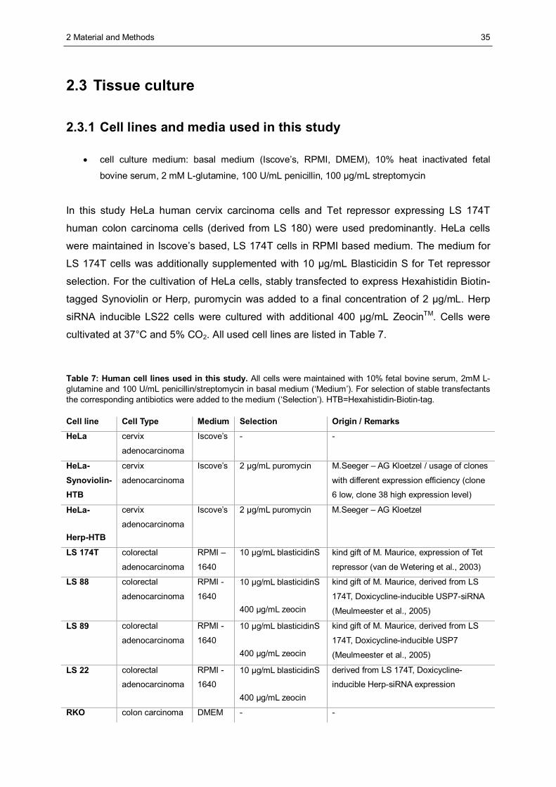
2 Material and Methods
35
2.3 Tissue culture
2.3.1 Cell lines and media used in this study
• cell culture medium: basal medium (Iscove’s, RPMI, DMEM), 10% heat inactivated fetal
bovine serum, 2 mM L-glutamine, 100 U/mL penicillin, 100 μg/mL streptomycin
In this study HeLa human cervix carcinoma cells and Tet repressor expressing LS 174T
human colon carcinoma cells (derived from LS 180) were used predominantly. HeLa cells
were maintained in Iscove’s based, LS 174T cells in RPMI based medium. The medium for
LS 174T cells was additionally supplemented with 10 μg/mL Blasticidin S for Tet repressor
selection. For the cultivation of HeLa cells, stably transfected to express Hexahistidin Biotin-
tagged Synoviolin or Herp, puromycin was added to a final concentration of 2 μg/mL. Herp
siRNA inducible LS22 cells were cultured with additional 400 μg/mL ZeocinTM. Cells were
cultivated at 37°C and 5% CO2. All used cell lines are listed in Table 7.
Table 7: Human cell lines used in this study. All cells were maintained with 10% fetal bovine serum, 2mM L-glutamine and 100 U/mL penicillin/streptomycin in basal medium (‘Medium’). For selection of stable transfectants the corresponding antibiotics were added to the medium (‘Selection’). HTB=Hexahistidin-Biotin-tag.
Cell line Cell Type Medium Selection Origin / Remarks
HeLa cervix
adenocarcinoma
Iscove’s - -
HeLa-Synoviolin-HTB
cervix
adenocarcinoma
Iscove’s 2 μg/mL puromycin M.Seeger – AG Kloetzel / usage of clones
with different expression efficiency (clone
6 low, clone 38 high expression level)
HeLa-
Herp-HTB
cervix
adenocarcinoma
Iscove’s 2 μg/mL puromycin M.Seeger – AG Kloetzel
LS 174T colorectal
adenocarcinoma
RPMI –
1640
10 μg/mL blasticidinS kind gift of M. Maurice, expression of Tet
repressor (van de Wetering et al., 2003)
LS 88 colorectal
adenocarcinoma
RPMI -
1640
10 μg/mL blasticidinS
400 μg/mL zeocin
kind gift of M. Maurice, derived from LS
174T, Doxicycline-inducible USP7-siRNA
(Meulmeester et al., 2005)
LS 89
colorectal
adenocarcinoma
RPMI -
1640
10 μg/mL blasticidinS
400 μg/mL zeocin
kind gift of M. Maurice, derived from LS
174T, Doxicycline-inducible USP7
(Meulmeester et al., 2005)
LS 22
colorectal
adenocarcinoma
RPMI -
1640 10 μg/mL blasticidinS
400 μg/mL zeocin
derived from LS 174T, Doxicycline-
inducible Herp-siRNA expression
RKO colon carcinoma DMEM - -
2 Material and Methods
36
2.3.2 Culture of cells
• Phosphate buffered saline (PBS): 137 mM NaCl, 2.7 mM KCl, 8.1 mM Na2HPO4, 1.5 mM
KH2PO4
Cells were generally grown in sterile culture dishes or flasks and passaged when they
reached 80% confluence, depending on the generation time. For the passage of the cells,
the culture medium was removed and the cells were washed with PBS and incubated with a
trypsin/EDTA solution for 2-5 min. After the detachment, serum containing culture medium
was added to the cells and they were transferred to a new tube and sedimented by short
centrifugation at 250 g. Cells were resuspended in fresh medium containing all necessary
additives and plated in the appropriate density.
Counting of cells • trypan blue solution: 0,1% (w/v) trypan blue, 0,3% (w/v) NaCl in PBS
10 μL of a single cell suspension were inspected using a Neubauer counting chamber under
a microscope. Cells in four selected large squares were counted. The volume of one large
square is 0.1 μL. The mean value of four large squares was multiplied with 104, resulting in
the number of cells per 1 mL cell suspension.
To evaluate only living cells, the cell suspension was mixed 1:2 with trypan blue solution.
Living cells do not incorporate the dye, whereas dead cells or cell debris get stained.
2.3.3 Cryoconservation and thawing of cells
• freezing medium: 20% FCS, 10% (v/v) DMSO in culture medium (RPMI or Iscove’s)
For long term storage of human cell lines 106 cells were resuspended in 1 mL of freezing
medium and immediately transferred into a cryoconservation vial and placed in a freezing
container, filled with pre-cooled isopropanol. The container was kept at -80°C over night to
ensure a 1°C/min cooling rate. The next day the tubes were transferred into a liquid nitrogen
(-196°C) tank for the final storage.
Thawing of the cells was performed in a water bath heated to 37°C. The suspension of
thawed cells was carefully mixed with 10 mL of culture medium and sedimented at 1,000 g
for 5 min. The culture medium was removed and the cells were resuspended in fresh
medium and afterwards plated in 10 mL culture medium on a 10 cm culture dish.
2 Material and Methods
37
2.3.4 Transfection of mammalian cells
Transfection of cells with DNA by calcium phosphate precipitation • HEPES buffered saline (HBS): 280 mM NaCl, 10 mM KCl, 1.5 mM Na2HPO4, 12 mM dextrose,
50 mM HEPES adjusted to pH 7.05 with NaOH
HeLa cells were plated at 5 x 10⁵ cells per 10 cm dish in Iscove’s medium and incubated
over night at 37°C. On the next day 500 μl of HBS was pipetted into a 5 mL polypropylene
vial. Then, 62 μL 2 M CaCl2 and 10 μg DNA were diluted in 440 μL of sterile water. This
solution was slowly dripped into the vial containing the HBS buffer while slowly mixing. The
resulting mixture was incubated at room temperature for 30 min to gain precipitation and
finally added dropwise to the cells. The plate was gently shaken to distribute the DNA and
incubated at 37°C. After 16 hours the medium was exchanged and the cells were harvested
after a total incubation time of 24 to 72 hours.
Transfection of cells with DNA by lipofection HeLa cells were plated at 105 cells per well in a 24-well-plate in 500 μL medium without
antibiotics and incubated over night. For each well 0.8 μg DNA and 0.8 μL Lipofectamin 2000
were diluted in 50 μL OptiMEM, mixed, incubated at room temperature for 20 min and then
transferred to the well containing the cells. The plate was gently shaken to distribute the DNA
and then further incubated at 37°C. After a total incubation of 24 to 48 h after transfection the
cells were harvested.
Transfection of cells with siRNA by lipofection
HeLa cells were plated at 5 x 10⁵ cells per 10 cm dish in 5 mL medium without antibiotics and
incubated over night at 37°C. 25 μL of siRNA (20 μM) were mixed with 25 μL OptiMEM
transfection medium. In parallel, 10 μL of Dharmafect lipofection reagent were diluted in 490
μL Optimem. Both solutions were combined, mixed and incubated at room temperature for
20 min. The whole mixture was then added dropwise to the cells. For cycloheximide chase
experiments (see 2.2.7) one day after transfection the cells were detached and seeded onto
a 24 well plate in an appropriate dilution.
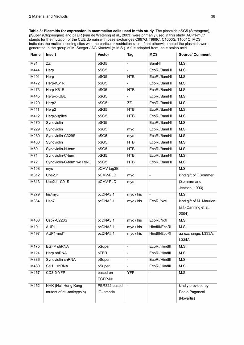
2 Material and Methods
38
Table 8: Plasmids for expression in mammalian cells used in this study. The plasmids pSG5 (Stratagene), pSuper (Oligoengine) and pTER (van de Wetering et al., 2003) were primarily used in this study. AUP1-mut* stands for the mutation of the CUE domain with base exchanges C997G, T998C, C1000G, T1001C. MCS indicates the multiple cloning sites with the particular restriction sites. If not otherwise noted the plasmids were generated in the group of M. Seeger / AG Kloetzel (= M.S.). A.f. = adapted from, aa = amino acid.
Name Insert Vector Tag MCS Source/ Comment
M31 ZZ pSG5 - BamHI M.S.
M444 Herp pSG5 - EcoRI/BamHI M.S.
M401 Herp pSG5 HTB EcoRI/BamHI M.S.
M472 Herp-K61R pSG5 - EcoRI/BamHI M.S.
M473 Herp-K61R pSG5 HTB EcoRI/BamHI M.S.
M445 Herp-d-UBL pSG5 - EcoRI/BamHI M.S.
M129 Herp2 pSG5 ZZ EcoRI/BamHI M.S.
M411 Herp2 pSG5 HTB EcoRI/BamHI M.S.
M412 Herp2-splice pSG5 HTB EcoRI/BamHI M.S.
M470 Synoviolin pSG5 - EcoRI/BamHI M.S.
M229 Synoviolin pSG5 myc EcoRI/BamHI M.S.
M230 Synoviolin-C329S pSG5 myc EcoRI/BamHI M.S.
M400 Synoviolin pSG5 HTB EcoRI/BamHI M.S.
M69 Synoviolin-N-term pSG5 HTB EcoRI/BamHI M.S.
M71 Synoviolin-C-term pSG5 HTB EcoRI/BamHI M.S.
M72 Synoviolin-C-term wo RING pSG5 HTB EcoRI/BamHI M.S.
M158 myc pCMV-tag3B - - M.S.
M312 Ube2J1 pCMV-PLD myc -
M313 Ube2J1-C91S pCMV-PLD myc -
kind gift of T.Sommer
(Sommer and
Jentsch, 1993)
M279 his/myc pcDNA3.1 myc / his - M.S.
M384 Usp7 pcDNA3.1 myc / his EcoRI/NotI kind gift of M. Maurice
(a.f.(Canning et al.,
2004)
M468 Usp7-C223S pcDNA3.1 myc / his EcoRI/NotI M.S.
M19 AUP1 pcDNA3.1 myc / his HindIII/EcoRI M.S.
M497 AUP1-mut* pcDNA3.1 myc / his HindIII/EcoRI aa exchange: L333A,
L334A
M175 EGFP shRNA pSuper - EcoRI/HindIII M.S.
M124 Herp shRNA pTER - EcoRI/HindIII M.S.
M336 Synoviolin shRNA pSuper - EcoRI/HindIII M.S.
M480 Sel1L shRNA pSuper - EcoRI/HindIII M.S.
M457 CD3-δ-YFP based on
EGFP-N1
YFP - M.S.
M452 NHK (Null Hong Kong
mutant of α1-antitrypsin)
PBR322 based
lG-lambda
- - kindly provided by
Paolo Paganetti
(Novartis)
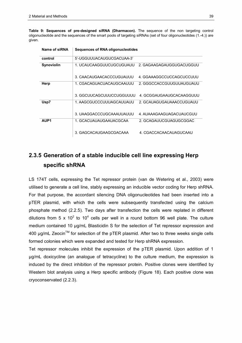
2 Material and Methods
39
Table 9: Sequences of pre-designed siRNA (Dharmacon). The sequence of the non targeting control oligonucleotide and the sequences of the smart pools of targeting siRNAs (set of four oligonucleotides (1.-4.)) are given.
Name of siRNA Sequences of RNA oligonucleotides
control 5’-UGGUUUACAUGUCGACUAA-3’
Synoviolin 1. UCAUCAAGGUUCUGCUGUAUU 2. GAGAAGAGAUGGUGACUGGUU
3. CAACAUGAACACCCUGUAUUU 4. GGAAAGGCCUCCAGCUCCUUU
Herp 1. CGACAGUACUACAUGCAAUUU 2. GGGCCACCGUUGUUAUGUAUU
3. GGCUUCAGCUUUCCUGGUUUU 4. GCGGAUGAAUGCACAAGGUUU
Usp7 1. AAGCGUCCCUUUAGCAUUAUU 2. GCAUAGUGAUAAACCUGUAUU
3. UAAGGACCCUGCAAAUUAUUU 4. AUAAAGAAGUAGACUAUCGUU
AUP1 1. GCACUAUAUGAAUACGCAA 2. GCAGAUUCGUAGUGCGGAC
3. GAGCACAUGAAGCGACAAA 4. CGACCACAACAUAGUCAAU
2.3.5 Generation of a stable inducible cell line expressing Herp specific shRNA
LS 174T cells, expressing the Tet repressor protein (van de Wetering et al., 2003) were
utilised to generate a cell line, stably expressing an inducible vector coding for Herp shRNA.
For that purpose, the accordant silencing DNA oligonucleotides had been inserted into a
pTER plasmid, with which the cells were subsequently transfected using the calcium
phosphate method (2.2.5). Two days after transfection the cells were replated in different
dilutions from 5 x 10² to 104 cells per well in a round bottom 96 well plate. The culture
medium contained 10 μg/mL Blasticidin S for the selection of Tet repressor expression and
400 μg/mL ZeocinTM for selection of the pTER plasmid. After two to three weeks single cells
formed colonies which were expanded and tested for Herp shRNA expression.
Tet repressor molecules inhibit the expression of the pTER plasmid. Upon addition of 1
μg/mL doxicycline (an analogue of tetracycline) to the culture medium, the expression is
induced by the direct inhibition of the repressor protein. Positive clones were identified by
Western blot analysis using a Herp specific antibody (Figure 18). Each positive clone was
cryoconservated (2.2.3).
2 Material and Methods
40
2.3.6 Cycloheximide chase analysis
For the estimation of the half-life of proteins, 105 cells expressing the respective protein were
seeded per well on a 24 well plate and incubated at 37°C over night. Then cycloheximide
(chx) was added to a final concentration of 50 μg/mL. In order to analyse proteasome
dependent degradation, 10 μM MG132, an inhibitor of the proteasome, dissolved in DMSO,
was added to one chx containing well. In this case the equivalent volume of the solvent
DMSO was added to the other samples. Cells were harvested at various time points and
frozen in liquid nitrogen. For further analysis, cells of each sample were lysed in 100 μL of
RIPA buffer (2.4.1). 10 μL of each lysate were subjected SDS-PAGE (2.4.4) and Western
blot analysis (2.4.5).
2.3.7 Metabolic labelling using [35S]-methionine/-cysteine and pulse chase analysis
• starving medium: methionine free RPMI, 1%(v/v) dialysed fetal calf serum
For the determination of the half-life of a protein or the analysis of intrinsic protein
interactions, 4 x 10⁵ cells per well on a 6 well plate or 106 cells on a 10 cm dish, expressing
the protein of interest, were seeded and incubated at 37°C over night. Then cells were left
untreated or treated with 2 μM thapsigargin for up to 8 h to induce ER stress. After washing
the cells twice with 3 mL (5 mL for the dish) PBS, 1 mL (2.5 mL for the dish) of starving
medium was added and cells were incubated for 1 h at 37°C.
Then, [35S]-S-L-methionine / [35S]-L-cysteine (Tran35S-LabelTM) was directly added to a final
concentration of 0.1 mCi/mL and cells were labelled for 1 h. Then the label medium was
removed and two washing steps followed with 2 mL (5 mL for the dish) of PBS. Then the
cells were harvested directly or medium, containing all additives, was added and they were
further incubated for various periods of time. The collected cells were either lysed in 500 μL
of RIPA buffer (2.4.1) and subjected to immunoprecipitation for pulse chase analysis or in
500 μL of DBC containing buffer (2.4.1) to precipitate ERAD complexes for the analysis of
physiological protein interactions (2.4.7). An aliquot of each lysate was subjected to
scintillation counting and samples were normalised for 35S-methionin incorporation.
2 Material and Methods
41
2.4 Protein biochemistry
2.4.1 Lysis of mammalian cells
• RIPA lysis buffer: 50 mM Tris-HCl, 150 mM NaCl, 1 mM EDTA (sodium salt), 1% (v/v) Nonidet
P40, 0.5% (w/v) Sodium-deoxycholate, 0.1% (w/v) SDS and CompleteTM, adjusted to pH 8.0
with NaOH
• DBC lysis buffer: 1% (w/v) DBC (Applichem), 33 mM HEPES, 150 mM Kalium-acetate, 4 mM
Magnesium-acetate, 10% (w/v) glycerol, CompleteTM, adjusted to pH 7.3 with KOH
Mammalian cells were lysed either in RIPA lysis buffer for analysis of whole cell extracts or in
deoxy-big-chap (DBC) lysis buffer for analysis of ERAD complexes. For RIPA lysis cells were
resuspended in ice cold RIPA lysis buffer and incubated on ice for 20 min and then
centrifuged for 15 min at 4°C and 20,000 g. The supernatant was subjected to
immunoprecipitation or SDS-PAGE. For DBC lysis cells were resuspended in DBC lysis
buffer and gently agitated for 30 min at 4°C. After that, two centrifugation steps of 5 and 10
min at 10,000 g and 4°C were performed. The final supernatant was either subjected to
immunoprecipitation, precipitation experiments or SDS-PAGE.
2.4.2 Determination of protein concentration in solution
Protein concentrations in solution were determined by measuring the absorbance at 280 nm
in an UV-VIS photometer. An absorbance of one at the wavelength of 280 nm equals a
concentration of 1 mg protein/mL solution.
2.4.3 Precipitation of proteins using trichloro acetic acid (TCA)
For the precipitation of protein complexes, separated on a glycerol gradient, TCA was added
to the sample to a final concentration of 10% (w/v). This mixture was kept on ice for 20 min,
followed by centrifugation at 20,000 g and 4°C for 15 min. The sediment was washed 3 times
with 500 μL of ice cold acetone (-20°C). After another centrifugation step (5 min, 4°C, 14,000
g) the sediment was air dried and resuspended in 60 μL SDS sample buffer (see 2.4.4).
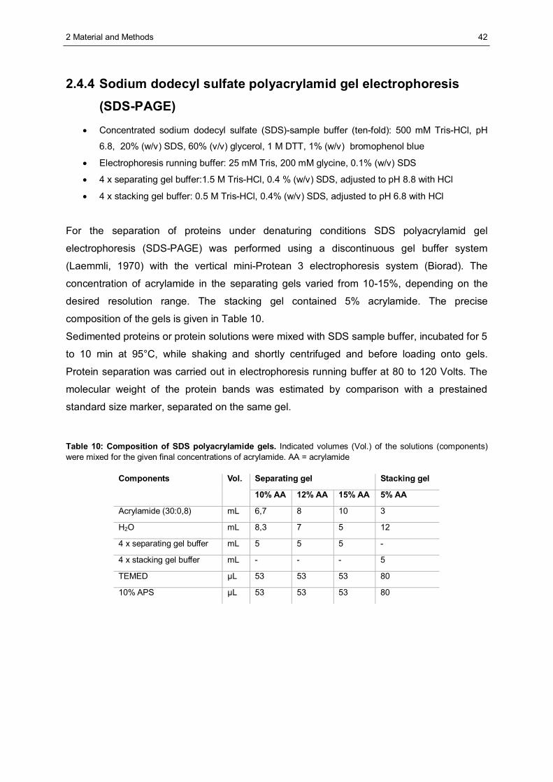
2 Material and Methods
42
2.4.4 Sodium dodecyl sulfate polyacrylamid gel electrophoresis
(SDS-PAGE)
• Concentrated sodium dodecyl sulfate (SDS)-sample buffer (ten-fold): 500 mM Tris-HCl, pH
6.8, 20% (w/v) SDS, 60% (v/v) glycerol, 1 M DTT, 1% (w/v) bromophenol blue
• Electrophoresis running buffer: 25 mM Tris, 200 mM glycine, 0.1% (w/v) SDS
• 4 x separating gel buffer:1.5 M Tris-HCl, 0.4 % (w/v) SDS, adjusted to pH 8.8 with HCl
• 4 x stacking gel buffer: 0.5 M Tris-HCl, 0.4% (w/v) SDS, adjusted to pH 6.8 with HCl
For the separation of proteins under denaturing conditions SDS polyacrylamid gel
electrophoresis (SDS-PAGE) was performed using a discontinuous gel buffer system
(Laemmli, 1970) with the vertical mini-Protean 3 electrophoresis system (Biorad). The
concentration of acrylamide in the separating gels varied from 10-15%, depending on the
desired resolution range. The stacking gel contained 5% acrylamide. The precise
composition of the gels is given in Table 10.
Sedimented proteins or protein solutions were mixed with SDS sample buffer, incubated for 5
to 10 min at 95°C, while shaking and shortly centrifuged and before loading onto gels.
Protein separation was carried out in electrophoresis running buffer at 80 to 120 Volts. The
molecular weight of the protein bands was estimated by comparison with a prestained
standard size marker, separated on the same gel.
Table 10: Composition of SDS polyacrylamide gels. Indicated volumes (Vol.) of the solutions (components) were mixed for the given final concentrations of acrylamide. AA = acrylamide
Separating gel Stacking gel Components Vol.
10% AA 12% AA 15% AA 5% AA
Acrylamide (30:0,8) mL 6,7 8 10 3
H2O mL 8,3 7 5 12
4 x separating gel buffer mL 5 5 5 -
4 x stacking gel buffer mL - - - 5
TEMED μL 53 53 53 80
10% APS μL 53 53 53 80
2 Material and Methods
43
2.4.5 Western blot analysis
• cathode buffer: 40 mM ε-amino caproic acid, 0,01% (w/v) SDS, 20% (v/v) methanol
• anode buffer 2: 25 mM Tris, 20% (v/v) methanol
• anode buffer 1: 300 mM Tris, 20% (v/v) methanol
After separation of proteins by SDS-PAGE they were transferred from the gels to
nitrocellulose membranes in a semi dry blotting procedure using a three buffer system. Four
Whatman 3 MM papers were soaked in the cathode buffer and put on the bottom (cathode)
of a semidry blotting apparatus. First the gel and then the membrane were piled upon it. then
a layer of two Whatman 3 MM paper sheets, soaked in anode buffer 2 and finally two
Whatman 3 MM paper sheets, soaked in anode buffer 1 were put on top of it. The transfer
was carried out at 320 mA for 1 h at room temperature. Immunodetection was performed as
described in 2.4.6.
2.4.6 Protein visualisation
Ponceau staining of proteins on nitrocellulose membranes • Ponceau staining solution: 1% (w/v) Ponceau S in 5% (w/v) TCA-solution
After the protein transfer onto nitrocellulose membranes these were incubated in Ponceau
staining solution for 1 min and thereafter destained in deionised water until the desired
contrast of protein bands was achieved.
Coomassie Blue staining of proteins in gels • Coomassie Blue staining solution: 0.1% (w/v) Coomassie Brilliant Blue R250, 40% (v/v)
methanol, 10% (v/v) acetic acid, filtered before use
• destaining solution: 40% (v/v) methanol, 10% (v/v) acetic acid
Mini gels were incubated in Coomassie Blue staining solution for 30 min at room temperature
while being agitated. For destaining the gels were incubated in destaining solution for 30 min,
exchanging the solution several times.
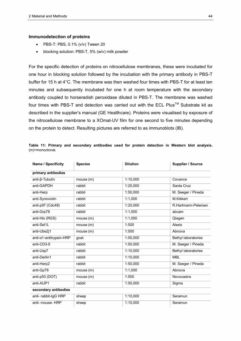
2 Material and Methods
44
Immunodetection of proteins • PBS-T: PBS, 0.1% (v/v) Tween 20
• blocking solution: PBS-T, 5% (w/v) milk powder
For the specific detection of proteins on nitrocellulose membranes, these were incubated for
one hour in blocking solution followed by the incubation with the primary antibody in PBS-T
buffer for 15 h at 4°C. The membrane was then washed four times with PBS-T for at least ten
minutes and subsequently incubated for one h at room temperature with the secondary
antibody coupled to horseradish peroxidase diluted in PBS-T. The membrane was washed
four times with PBS-T and detection was carried out with the ECL PlusTM Substrate kit as
described in the supplier’s manual (GE Healthcare). Proteins were visualised by exposure of
the nitrocellulose membrane to a XOmat-UV film for one second to five minutes depending
on the protein to detect. Resulting pictures are referred to as immunoblots (IB).
Table 11: Primary and secondary antibodies used for protein detection in Western blot analysis. (m)=monoclonal.
Name / Specificity Species Dilution Supplier / Source
primary antibodies
anti-β-Tubulin mouse (m) 1:10,000 Covance
anti-GAPDH rabbit 1:20,000 Santa Cruz
anti-Herp rabbit 1:50,000 M. Seeger / Pineda
anti-Synoviolin rabbit 1:1,000 M.Kikkert
anti-p97 (Cdc48) rabbit 1:20,000 R.Hartmann-Petersen
anti-Grp78 rabbit 1:1,000 abcam
anti-His (RGS) mouse (m) 1:1,000 Qiagen
anti-Sel1L mouse (m) 1:500 Alexis
anti-Ube2j1 mouse (m) 1:500 Abnova
anti-α1-antitrypsin-HRP goat 1:50,000 Bethyl laboratories
anti-CD3-δ rabbit 1:50,000 M. Seeger / Pineda
anti-Usp7 rabbit 1:10,000 Bethyl laboratories
anti-Derlin1 rabbit 1:10,000 MBL
anti-Herp2 rabbit 1:50,000 M. Seeger / Pineda
anti-Gp78 mouse (m) 1:1,000 Abnova
anti-p53 (DO7) mouse (m) 1:500 Novocastra
anti-AUP1 rabbit 1:50,000 Sigma
secondary antibodies
anti- rabbit-IgG HRP sheep 1:10,000 Seramun
anti- mouse- HRP sheep 1:10,000 Seramun
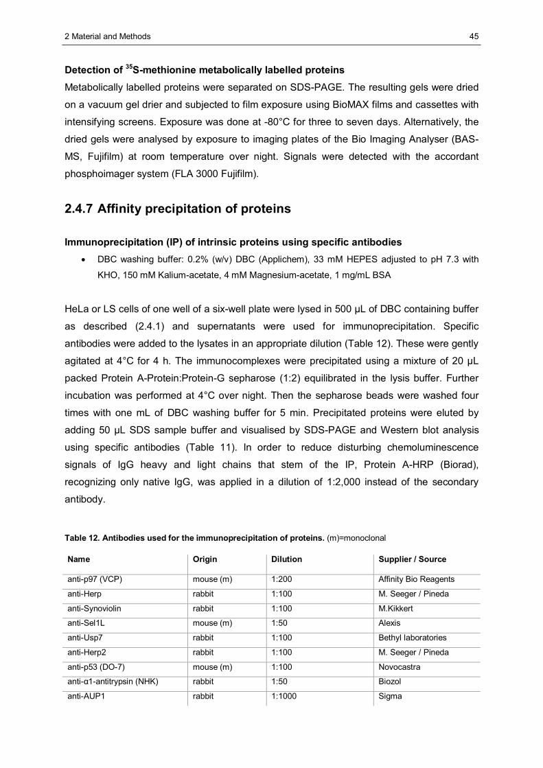
2 Material and Methods
45
Detection of 35S-methionine metabolically labelled proteins Metabolically labelled proteins were separated on SDS-PAGE. The resulting gels were dried
on a vacuum gel drier and subjected to film exposure using BioMAX films and cassettes with
intensifying screens. Exposure was done at -80°C for three to seven days. Alternatively, the
dried gels were analysed by exposure to imaging plates of the Bio Imaging Analyser (BAS-
MS, Fujifilm) at room temperature over night. Signals were detected with the accordant
phosphoimager system (FLA 3000 Fujifilm).
2.4.7 Affinity precipitation of proteins
Immunoprecipitation (IP) of intrinsic proteins using specific antibodies • DBC washing buffer: 0.2% (w/v) DBC (Applichem), 33 mM HEPES adjusted to pH 7.3 with
KHO, 150 mM Kalium-acetate, 4 mM Magnesium-acetate, 1 mg/mL BSA
HeLa or LS cells of one well of a six-well plate were lysed in 500 μL of DBC containing buffer
as described (2.4.1) and supernatants were used for immunoprecipitation. Specific
antibodies were added to the lysates in an appropriate dilution (Table 12). These were gently
agitated at 4°C for 4 h. The immunocomplexes were precipitated using a mixture of 20 μL
packed Protein A-Protein:Protein-G sepharose (1:2) equilibrated in the lysis buffer. Further
incubation was performed at 4°C over night. Then the sepharose beads were washed four
times with one mL of DBC washing buffer for 5 min. Precipitated proteins were eluted by
adding 50 μL SDS sample buffer and visualised by SDS-PAGE and Western blot analysis
using specific antibodies (Table 11). In order to reduce disturbing chemoluminescence
signals of IgG heavy and light chains that stem of the IP, Protein A-HRP (Biorad),
recognizing only native IgG, was applied in a dilution of 1:2,000 instead of the secondary
antibody.
Table 12. Antibodies used for the immunoprecipitation of proteins. (m)=monoclonal
Name Origin Dilution Supplier / Source
anti-p97 (VCP) mouse (m) 1:200 Affinity Bio Reagents
anti-Herp rabbit 1:100 M. Seeger / Pineda
anti-Synoviolin rabbit 1:100 M.Kikkert
anti-Sel1L mouse (m) 1:50 Alexis
anti-Usp7 rabbit 1:100 Bethyl laboratories
anti-Herp2 rabbit 1:100 M. Seeger / Pineda
anti-p53 (DO-7) mouse (m) 1:100 Novocastra
anti-α1-antitrypsin (NHK) rabbit 1:50 Biozol
anti-AUP1 rabbit 1:1000 Sigma
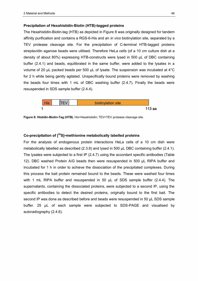
2 Material and Methods
46
Precipitation of Hexahistidin-Biotin (HTB)-tagged proteins The Hexahistidin-Biotin-tag (HTB) as depicted in Figure 8 was originally designed for tandem
affinity purification and contains a RGS-6-His and an in vivo biotinylation site, separated by a
TEV protease cleavage site. For the precipitation of C-terminal HTB-tagged proteins
streptavidin agarose beads were utilised. Therefore HeLa cells (of a 10 cm culture dish at a
density of about 80%) expressing HTB-constructs were lysed in 500 μL of DBC containing
buffer (2.4.1) and beads, equilibrated in the same buffer, were added to the lysates in a
volume of 20 μL packed beads per 500 μL of lysate. The suspension was incubated at 4°C
for 2 h while being gently agitated. Unspecifically bound proteins were removed by washing
the beads four times with 1 mL of DBC washing buffer (2.4.7). Finally the beads were
resuspended in SDS sample buffer (2.4.4).
His TEV biotinylation site1 113 aa
His TEV biotinylation site1 113 aa
Figure 8: Histidin-Biotin-Tag (HTB). His=Hexahistidin; TEV=TEV protease cleavage site.
Co-precipitation of [35S]-methionine metabolically labelled proteins
For the analysis of endogenous protein interactions HeLa cells of a 10 cm dish were
metabolically labelled as described (2.3.8) and lysed in 500 μL DBC containing buffer (2.4.1).
The lysates were subjected to a first IP (2.4.7) using the accordant specific antibodies (Table
12). DBC washed Protein A/G beads then were resuspended in 500 μL RIPA buffer and
incubated for 1 h in order to achieve the dissociation of the precipitated complexes. During
this process the bait protein remained bound to the beads. These were washed four times
with 1 mL RIPA buffer and resuspended in 50 μL of SDS sample buffer (2.4.4). The
supernatants, containing the dissociated proteins, were subjected to a second IP, using the
specific antibodies to detect the desired proteins, originally bound to the first bait. The
second IP was done as described before and beads were resuspended in 50 μL SDS sample
buffer. 25 μL of each sample were subjected to SDS-PAGE and visualised by
autoradiography (2.4.6).
2 Material and Methods
47
2.4.8 Separation of proteins by glycerol gradient centrifugation
107 HeLa cells were lysed in 500 μL of DBC containing lysis buffer (2.4.1). The lysates were
put on top of a glycerol gradient ranging from 10-50% glycerol based on the lysis buffer.
Then centrifugation was performed using an ultracentrifuge for 16 h at 260,000 g and 4°C
with an SW40 rotor. Beginning from the top, gradient fractions of 1 mL were collected and
subjected to TCA precipitation (2.4.3). Resulting samples were analysed via SDS-PAGE and
immunodetection.
2.4.9 In vitro binding studies
Expression of proteins in E.coli
Glutathione-S-transferase (GST) - tagged recombinant fusion proteins (Table 13) were
expressed in the XL1 blue E.coli strain. Therefore the bacteria were transformed with the
respective expression vectors and streaked out on LB-amp agar plates and incubated over
night at 37°C. Then a single colony was picked and added to 10 mL liquid LB-amp medium
and shaken over night at 37°C. At the next day 200 mL LB-amp were inoculated with the 10
mL culture and grown at 37°C until an OD600 of 0.5 was reached. Then 0.4 mM isopropyl-
beta-D-thiogalactopyranoside (IPTG) was added to the culture to induce protein expression
and incubation was continued at RT for four h followed by cell sedimentation. Aliquots were
taken before and after IPTG induction to analyse the protein expression via SDS-PAGE and
Coomassie staining.
Table 13. Plasmids used for expression in E.coli. For the in vitro precipitation assays the plasmids pGEX-KG (Guan and Dixon, 1991), pQE30 (Qiagen), pRSET (Invitrogen) were used. The inserts code for proteins of which the names and the range of amino acids is given. MCS = multiple cloning site, GST = glutathione S transferase, His = Hexahistidin. All constructs were generated in the laboratory of M. Seeger.
Name Insert Vector Tag MCS
M5 - pGEX-KG GST
M99 Herp1 1-240 pGEX-KG GST BamHI/EcoRI
M123 Herp1 1-240 A39T pGEX-KG GST BamHI/EcoRI
M100 Herp2 1-240 pGEX-KG GST BamHI/EcoRI
M122 Herp2 1-240 T39A pGEX-KG GST BamHI/EcoRI
M111 Herp 304-391 pGEX-KG GST BamHI/EcoRI
M110 Herp 1-260 pGEX-KG GST BamHI/EcoRI
M360 Synoviolin 236-616 pGEX-KG GST BamHI/EcoRI
M496 AUP1 1-410 pQE30 His BamHI/HindIII
M495 AUP1 24-410 pQE30 His BamHI/HindIII
M400 Usp7 1-208 pRSET His BamHI/EcoRI
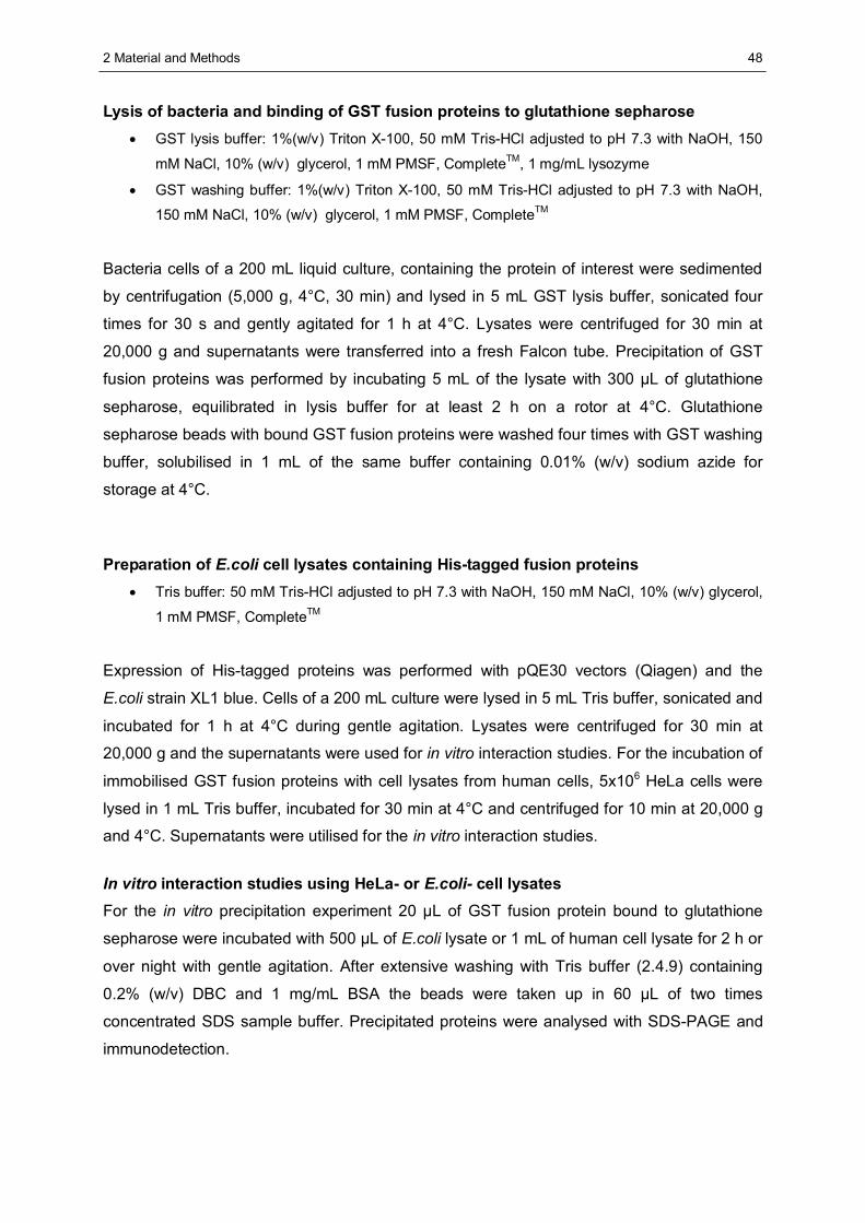
2 Material and Methods
48
Lysis of bacteria and binding of GST fusion proteins to glutathione sepharose • GST lysis buffer: 1%(w/v) Triton X-100, 50 mM Tris-HCl adjusted to pH 7.3 with NaOH, 150
mM NaCl, 10% (w/v) glycerol, 1 mM PMSF, CompleteTM, 1 mg/mL lysozyme
• GST washing buffer: 1%(w/v) Triton X-100, 50 mM Tris-HCl adjusted to pH 7.3 with NaOH,
150 mM NaCl, 10% (w/v) glycerol, 1 mM PMSF, CompleteTM
Bacteria cells of a 200 mL liquid culture, containing the protein of interest were sedimented
by centrifugation (5,000 g, 4°C, 30 min) and lysed in 5 mL GST lysis buffer, sonicated four
times for 30 s and gently agitated for 1 h at 4°C. Lysates were centrifuged for 30 min at
20,000 g and supernatants were transferred into a fresh Falcon tube. Precipitation of GST
fusion proteins was performed by incubating 5 mL of the lysate with 300 μL of glutathione
sepharose, equilibrated in lysis buffer for at least 2 h on a rotor at 4°C. Glutathione
sepharose beads with bound GST fusion proteins were washed four times with GST washing
buffer, solubilised in 1 mL of the same buffer containing 0.01% (w/v) sodium azide for
storage at 4°C.
Preparation of E.coli cell lysates containing His-tagged fusion proteins • Tris buffer: 50 mM Tris-HCl adjusted to pH 7.3 with NaOH, 150 mM NaCl, 10% (w/v) glycerol,
1 mM PMSF, CompleteTM
Expression of His-tagged proteins was performed with pQE30 vectors (Qiagen) and the
E.coli strain XL1 blue. Cells of a 200 mL culture were lysed in 5 mL Tris buffer, sonicated and
incubated for 1 h at 4°C during gentle agitation. Lysates were centrifuged for 30 min at
20,000 g and the supernatants were used for in vitro interaction studies. For the incubation of
immobilised GST fusion proteins with cell lysates from human cells, 5x10⁶ HeLa cells were
lysed in 1 mL Tris buffer, incubated for 30 min at 4°C and centrifuged for 10 min at 20,000 g
and 4°C. Supernatants were utilised for the in vitro interaction studies.
In vitro interaction studies using HeLa- or E.coli- cell lysates For the in vitro precipitation experiment 20 μL of GST fusion protein bound to glutathione
sepharose were incubated with 500 μL of E.coli lysate or 1 mL of human cell lysate for 2 h or
over night with gentle agitation. After extensive washing with Tris buffer (2.4.9) containing
0.2% (w/v) DBC and 1 mg/mL BSA the beads were taken up in 60 μL of two times
concentrated SDS sample buffer. Precipitated proteins were analysed with SDS-PAGE and
immunodetection.
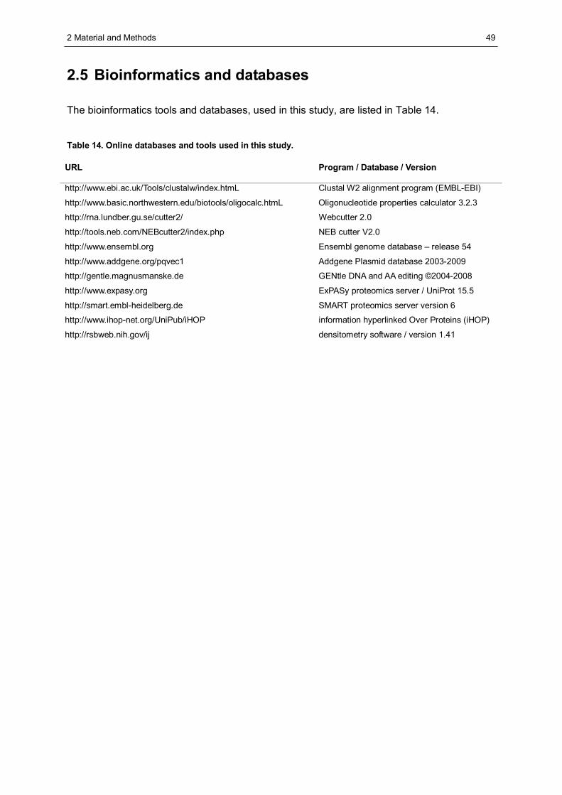
2 Material and Methods
49
2.5 Bioinformatics and databases
The bioinformatics tools and databases, used in this study, are listed in Table 14.
Table 14. Online databases and tools used in this study.
URL Program / Database / Version
http://www.ebi.ac.uk/Tools/clustalw/index.htmL Clustal W2 alignment program (EMBL-EBI)
http://www.basic.northwestern.edu/biotools/oligocalc.htmL Oligonucleotide properties calculator 3.2.3
http://rna.lundber.gu.se/cutter2/ Webcutter 2.0
http://tools.neb.com/NEBcutter2/index.php NEB cutter V2.0
http://www.ensembl.org Ensembl genome database – release 54
http://www.addgene.org/pqvec1 Addgene Plasmid database 2003-2009
http://gentle.magnusmanske.de GENtle DNA and AA editing ©2004-2008
http://www.expasy.org ExPASy proteomics server / UniProt 15.5
http://smart.embl-heidelberg.de SMART proteomics server version 6
http://www.ihop-net.org/UniPub/iHOP information hyperlinked Over Proteins (iHOP)
http://rsbweb.nih.gov/ij densitometry software / version 1.41
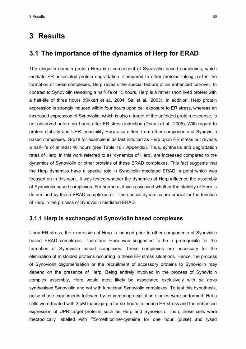
3 Results
50
3 Results
3.1 The importance of the dynamics of Herp for ERAD
The ubiquitin domain protein Herp is a component of Synoviolin based complexes, which
mediate ER associated protein degradation. Compared to other proteins taking part in the
formation of these complexes, Herp reveals the special feature of an enhanced turnover. In
contrast to Synoviolin revealing a half-life of 15 hours, Herp is a rather short lived protein with
a half-life of three hours (Kikkert et al., 2004; Sai et al., 2003). In addition, Herp protein
expression is strongly induced within four hours upon cell exposure to ER stress, whereas an
increased expression of Synoviolin, which is also a target of the unfolded protein response, is
not observed before six hours after ER stress induction (Donati et al., 2006). With regard to
protein stability and UPR inducibility Herp also differs from other components of Synoviolin
based complexes. Grp78 for example is as fast induced as Herp upon ER stress but reveals
a half-life of at least 48 hours (see Table 16 / Appendix). Thus, synthesis and degradation
rates of Herp, in this work referred to as ‘dynamics of Herp’, are increased compared to the
dynamics of Synoviolin or other proteins of these ERAD complexes. This fact suggests that
the Herp dynamics have a special role in Synoviolin mediated ERAD, a point which was
focused on in this work. It was tested whether the dynamics of Herp influence the assembly
of Synoviolin based complexes. Furthermore, it was assessed whether the stability of Herp is
determined by these ERAD complexes or if the special dynamics are crucial for the function
of Herp in the process of Synoviolin mediated ERAD.
3.1.1 Herp is exchanged at Synoviolin based complexes
Upon ER stress, the expression of Herp is induced prior to other components of Synoviolin
based ERAD complexes. Therefore, Herp was suggested to be a prerequisite for the
formation of Synoviolin based complexes. These complexes are necessary for the
elimination of misfolded proteins occurring in these ER stress situations. Hence, the process
of Synoviolin oligomerisation or the recruitment of accessory proteins to Synoviolin may
depend on the presence of Herp. Being entirely involved in the process of Synoviolin
complex assembly, Herp would most likely be associated exclusively with de novo
synthesised Synoviolin and not with functional Synoviolin complexes. To test this hypothesis,
pulse chase experiments followed by co-immunoprecipitation studies were performed. HeLa
cells were treated with 2 μM thapsigargin for six hours to induce ER stress and the enhanced
expression of UPR target proteins such as Herp and Synoviolin. Then, these cells were
metabolically labelled with 35S-methionine/-cysteine for one hour (pulse) and lysed
3 Results
51
immediately or after a further incubation for three and six hours with medium lacking
radioactively labelled amino acids (chase). Before the chase cell cultures were split into
halfes of which one half was additionally incubated with cycloheximide to stop translation.
Herp and p97 were immunoprecipitated from the lysates using antibodies specific for these
proteins (first IP). Due to mild lysis conditions whole ERAD complexes were co-precipitated
with p97 or Herp. These complexes were dissociated by incubation in SDS containing buffer
and the supernatants including the single ERAD components were subjected to a second
immunoprecipitation (second IP) using an antibody specific for Synoviolin.
Immunoprecipitates were subjected to SDS-PAGE and analysed by autoradiography. This
experiment enables to monitor the degradation of Herp and p97, which is displayed by the
decrease of the labelled protein population over time. In addition, the association of
Synoviolin with these proteins can be evaluated. As shown in Figure 9, the expression of
Herp protein was increased upon ER stress treatment by thapsigargin whereas the
expression of p97 did not change (Figure 9, b and c, upper panel). Radioactively labelled
Herp was completely degraded within six hours of chase, irrespective of cycloheximide
addition. In contrast, p97 remained stable for the whole chase period.
PI Herp p97First IP:
SecondIP: Synv
*p97
Herp
Synv
h chase-- - - -
+ + + ++++ + +
0 0 3 6 0 3 6
chxtg-
- - - -+ + + +++
+ + +
0 0 3 6 0 3 6-- - - -
+ + + ++++ + +
0 0 3 6 0 3 6
a b c
Figure 9: Dynamics of Herp at Synoviolin based complexes. HeLa cells were left untreated or treated with 2 μM thapsigargin for six h. Then, cells were metabolically labelled with 35S-methionine and, to stop translation, 50 μg/mL cycloheximide were added where indicated. Where indicated, thapsigargin was present during incubation. Cells were lysed at the indicated time points (chase) in DBC containing buffer. Lysates were equalised for overall radioactivity after scintillation counting. For the first immunoprecipitation (IP) either pre-immune serum (PI) or the specific antibodies as indicated were utilised (a-c). Dissociation of co-precipitated proteins was achieved by the incubation in RIPA buffer. The supernatant was subjected to a second IP with an antibody specific for Synoviolin. Precipitated proteins were separated on SDS-PAGE and visualised by autoradiography. An unspecific cross reaction, occurring in thapsigargin treated samples, is marked with an asterisk. Synv=Synoviolin; h=hour; tg=thapsigargin; chx=cycloheximide.
Meeting the expectations, the expression of the E3 ligase Synoviolin was induced by ER
stress treatment, since a Synoviolin specific signal was only observed in the autoradiography
upon thapsigargin treatment (Figure 9, b and c, lower panel). Independent of cycloheximide
addition, the amount of Synoviolin, which was co-precipitated with stable p97, remained
constant during the chase.
3 Results
52
On the other hand, the amount of Synoviolin, which was co-precipitated with Herp,
decreased in correlation with the degradation of Herp when treated with cycloheximide. In
the absence of cycloheximide the amount of Synoviolin, co-precipitated with Herp, remained
constant, although the population of labelled Herp was degraded (Figure 9, b). This
observation can be explained by the fact that newly synthesised and therefore not labelled
Herp substitutes the older population of labelled Herp occurring at already existing Synoviolin
based complexes. Therefore, it appears that Herp is not exclusively bound to de novo
synthesised Synoviolin but is exchanged at already existing Synoviolin based complexes.
This exchange has not been shown for any of the other proteins associated with Synoviolin.
3.1.2 Synoviolin complex components are not essential for the
degradation of Herp
Herp is a substrate of the ubiquitin proteasome system and is associated with Synoviolin (Sai
et al., 2003; Schulze et al., 2005). As shown in 3.1.1, Herp is subject to constant degradation
and reassociation with established Synoviolin based complexes suggesting that Herp is a
substrate of Synoviolin. Initial experiments which focused on Synoviolin potentially mediating
Herp degradation showed that the elevated expression of Synoviolin did not enhance its
degradation but led to the stabilisation of Herp, irrespective of the E3 ligase activity (Schulze,
2006). Thus, Synoviolin seems not to be required for the degradation of Herp. To confirm this
result and to test whether further components of Synoviolin based complexes are involved in
the regulation of Herp stability, degradation of Herp was monitored after depletion of
Synoviolin and additionally after depletion of Sel1L, a Synoviolin associated protein that was
described to be required for ERAD (Mueller et al., 2008). Depletion of Synoviolin and Sel1L
protein in HeLa cells was performed by the expression of shRNA specific for these proteins.
ShRNA expressing cells were subjected to a pulse chase experiment for six hours and
proteins of interest were immunoprecipitated and analysed by autoradiography.
Compared to the expression of shRNA targeting GFP as control, the expression of shRNA
specific either for Synoviolin or for Sel1L led to the reduced expression of these proteins by
about 50% (Figure 10, A). Depletion of either of these proteins did not result in an altered
degradation of Herp (Figure 10, B), although the reduction of the cellular level of Sel1L by
50% was reported to impair degradation of NHK (Christianson et al., 2008). Therefore, the
results presented here indicate that Synoviolin and Sel1L are not involved in the degradation
of Herp.
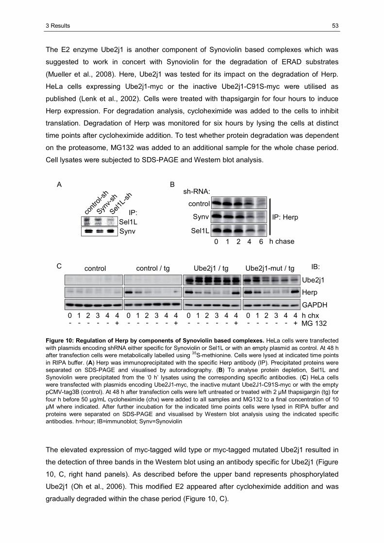
3 Results
53
The E2 enzyme Ube2j1 is another component of Synoviolin based complexes which was
suggested to work in concert with Synoviolin for the degradation of ERAD substrates
(Mueller et al., 2008). Here, Ube2j1 was tested for its impact on the degradation of Herp.
HeLa cells expressing Ube2j1-myc or the inactive Ube2j1-C91S-myc were utilised as
published (Lenk et al., 2002). Cells were treated with thapsigargin for four hours to induce
Herp expression. For degradation analysis, cycloheximide was added to the cells to inhibit
translation. Degradation of Herp was monitored for six hours by lysing the cells at distinct
time points after cycloheximide addition. To test whether protein degradation was dependent
on the proteasome, MG132 was added to an additional sample for the whole chase period.
Cell lysates were subjected to SDS-PAGE and Western blot analysis.
contr
ol-sh
Synv-s
h
Sel1L-s
h
IP:Sel1LSynv
B
control
Synv
Sel1L
sh-RNA:
h chase
IP: Herp
A
control control / tg Ube2j1 / tg Ube2j1-mut / tgUbe2j1
Herp
GAPDH
IB:
h chx
C
MG 1320 321 4 4- - - - - +
0 321 4 4- - - - - +
0 321 4 4- - - - - +
0 321 4 4- - - - - +
0 421 6
Figure 10: Regulation of Herp by components of Synoviolin based complexes. HeLa cells were transfected with plasmids encoding shRNA either specific for Synoviolin or Sel1L or with an empty plasmid as control. At 48 h after transfection cells were metabolically labelled using 35S-methionine. Cells were lysed at indicated time points in RIPA buffer. (A) Herp was immunoprecipitated with the specific Herp antibody (IP). Precipitated proteins were separated on SDS-PAGE and visualised by autoradiography. (B) To analyse protein depletion, Sel1L and Synoviolin were precipitated from the ‘0 h’ lysates using the corresponding specific antibodies. (C) HeLa cells were transfected with plasmids encoding Ube2J1-myc, the inactive mutant Ube2J1-C91S-myc or with the empty pCMV-tag3B (control). At 48 h after transfection cells were left untreated or treated with 2 μM thapsigargin (tg) for four h before 50 μg/mL cycloheximide (chx) were added to all samples and MG132 to a final concentration of 10 μM where indicated. After further incubation for the indicated time points cells were lysed in RIPA buffer and proteins were separated on SDS-PAGE and visualised by Western blot analysis using the indicated specific antibodies. h=hour; IB=immunoblot; Synv=Synoviolin
The elevated expression of myc-tagged wild type or myc-tagged mutated Ube2j1 resulted in
the detection of three bands in the Western blot using an antibody specific for Ube2j1 (Figure
10, C, right hand panels). As described before the upper band represents phosphorylated
Ube2j1 (Oh et al., 2006). This modified E2 appeared after cycloheximide addition and was
gradually degraded within the chase period (Figure 10, C).
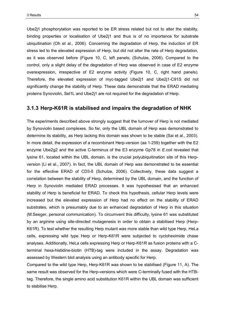
3 Results
54
Ube2j1 phosphorylation was reported to be ER stress related but not to alter the stability,
binding properties or localisation of Ube2j1 and thus is of no importance for substrate
ubiquitination (Oh et al., 2006). Concerning the degradation of Herp, the induction of ER
stress led to the elevated expression of Herp, but did not alter the rate of Herp degradation,
as it was observed before (Figure 10, C, left panels; (Schulze, 2006). Compared to the
control, only a slight delay of the degradation of Herp was observed in case of E2 enzyme
overexpression, irrespective of E2 enzyme activity (Figure 10, C, right hand panels).
Therefore, the elevated expression of myc-tagged Ube2j1 and Ube2j1-C91S did not
significantly change the stability of Herp. These data demonstrate that the ERAD mediating
proteins Synoviolin, Sel1L and Ube2j1 are not required for the degradation of Herp.
3.1.3 Herp-K61R is stabilised and impairs the degradation of NHK
The experiments described above strongly suggest that the turnover of Herp is not mediated
by Synoviolin based complexes. So far, only the UBL domain of Herp was demonstrated to
determine its stability, as Herp lacking this domain was shown to be stable (Sai et al., 2003).
In more detail, the expression of a recombinant Herp-version (aa 1-259) together with the E2
enzyme Ube2g2 and the active C-terminus of the E3 enzyme Gp78 in E.coli revealed that
lysine 61, located within the UBL domain, is the crucial polyubiquitination site of this Herp-
version (Li et al., 2007). In fact, the UBL domain of Herp was demonstrated to be essential
for the effective ERAD of CD3-δ (Schulze, 2006). Collectively, these data suggest a
correlation between the stability of Herp, determined by the UBL domain, and the function of
Herp in Synoviolin mediated ERAD processes. It was hypothesised that an enhanced
stability of Herp is beneficial for ERAD. To check this hypothesis, cellular Herp levels were
increased but the elevated expression of Herp had no effect on the stability of ERAD
substrates, which is presumably due to an enhanced degradation of Herp in this situation
(M.Seeger, personal communication). To circumvent this difficulty, lysine 61 was substituted
by an arginine using site-directed mutagenesis in order to obtain a stabilised Herp (Herp-
K61R). To test whether the resulting Herp mutant was more stable than wild type Herp, HeLa
cells, expressing wild type Herp or Herp-K61R were subjected to cycloheximide chase
analyses. Additionally, HeLa cells expressing Herp or Herp-K61R as fusion proteins with a C-
terminal hexa-histidine-biotin (HTB)-tag were included in the assay. Degradation was
assessed by Western blot analysis using an antibody specific for Herp.
Compared to the wild type Herp, Herp-K61R was shown to be stabilised (Figure 11, A). The
same result was observed for the Herp-versions which were C-terminally fused with the HTB-
tag. Therefore, the single amino acid substitution K61R within the UBL domain was sufficient
to stabilise Herp.
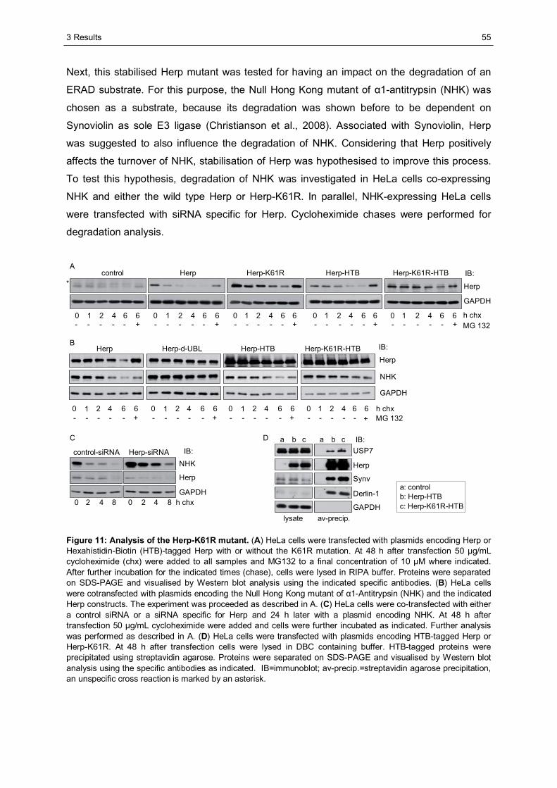
3 Results
55
Next, this stabilised Herp mutant was tested for having an impact on the degradation of an
ERAD substrate. For this purpose, the Null Hong Kong mutant of α1-antitrypsin (NHK) was
chosen as a substrate, because its degradation was shown before to be dependent on
Synoviolin as sole E3 ligase (Christianson et al., 2008). Associated with Synoviolin, Herp
was suggested to also influence the degradation of NHK. Considering that Herp positively
affects the turnover of NHK, stabilisation of Herp was hypothesised to improve this process.
To test this hypothesis, degradation of NHK was investigated in HeLa cells co-expressing
NHK and either the wild type Herp or Herp-K61R. In parallel, NHK-expressing HeLa cells
were transfected with siRNA specific for Herp. Cycloheximide chases were performed for
degradation analysis.
Herp Herp-d-UBL Herp-HTBHerp
NHK
GAPDH
Herp-K61R-HTB IB:B
MG 132h chx0 66421
- +----0 66421- +----
0 66421- +----
0 66421- +----
a: controlb: Herp-HTBc: Herp-K61R-HTB
lysate av-precip.
IB:USP7
Synv
Derlin-1
Herp
GAPDH
D
control Herp Herp-K61R Herp-HTB Herp-K61R-HTB IB:
MG 132
*
A
Herp
GAPDH
h chx0 66421- +----
h chx0 842
control-siRNA Herp-siRNA
0 842
Herp
GAPDH
IB:
NHK
C
0 664210 664210 664210 66421- +---- - +---- - +---- - +----
a b c a b c
Figure 11: Analysis of the Herp-K61R mutant. (A) HeLa cells were transfected with plasmids encoding Herp or Hexahistidin-Biotin (HTB)-tagged Herp with or without the K61R mutation. At 48 h after transfection 50 μg/mL cycloheximide (chx) were added to all samples and MG132 to a final concentration of 10 μM where indicated. After further incubation for the indicated times (chase), cells were lysed in RIPA buffer. Proteins were separated on SDS-PAGE and visualised by Western blot analysis using the indicated specific antibodies. (B) HeLa cells were cotransfected with plasmids encoding the Null Hong Kong mutant of α1-Antitrypsin (NHK) and the indicated Herp constructs. The experiment was proceeded as described in A. (C) HeLa cells were co-transfected with either a control siRNA or a siRNA specific for Herp and 24 h later with a plasmid encoding NHK. At 48 h after transfection 50 μg/mL cycloheximide were added and cells were further incubated as indicated. Further analysis was performed as described in A. (D) HeLa cells were transfected with plasmids encoding HTB-tagged Herp or Herp-K61R. At 48 h after transfection cells were lysed in DBC containing buffer. HTB-tagged proteins were precipitated using streptavidin agarose. Proteins were separated on SDS-PAGE and visualised by Western blot analysis using the specific antibodies as indicated. IB=immunoblot; av-precip.=streptavidin agarose precipitation, an unspecific cross reaction is marked by an asterisk.
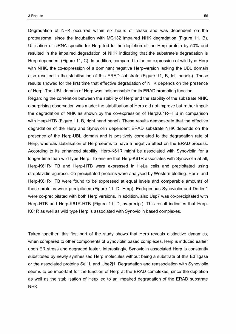
3 Results
56
Degradation of NHK occurred within six hours of chase and was dependent on the
proteasome, since the incubation with MG132 impaired NHK degradation (Figure 11, B).
Utilisation of siRNA specific for Herp led to the depletion of the Herp protein by 50% and
resulted in the impaired degradation of NHK indicating that the substrate’s degradation is
Herp dependent (Figure 11, C). In addition, compared to the co-expression of wild type Herp
with NHK, the co-expression of a dominant negative Herp-version lacking the UBL domain
also resulted in the stabilisation of this ERAD substrate (Figure 11, B, left panels). These
results showed for the first time that effective degradation of NHK depends on the presence
of Herp. The UBL-domain of Herp was indispensable for its ERAD promoting function.
Regarding the correlation between the stability of Herp and the stability of the substrate NHK,
a surprising observation was made: the stabilisation of Herp did not improve but rather impair
the degradation of NHK as shown by the co-expression of HerpK61R-HTB in comparison
with Herp-HTB (Figure 11, B, right hand panel). These results demonstrate that the effective
degradation of the Herp and Synoviolin dependent ERAD substrate NHK depends on the
presence of the Herp-UBL domain and is positively correlated to the degradation rate of
Herp, whereas stabilisation of Herp seems to have a negative effect on the ERAD process.
According to its enhanced stability, Herp-K61R might be associated with Synoviolin for a
longer time than wild type Herp. To ensure that Herp-K61R associates with Synoviolin at all,
Herp-K61R-HTB and Herp-HTB were expressed in HeLa cells and precipitated using
streptavidin agarose. Co-precipitated proteins were analysed by Western blotting. Herp- and
Herp-K61R-HTB were found to be expressed at equal levels and comparable amounts of
these proteins were precipitated (Figure 11, D, Herp). Endogenous Synoviolin and Derlin-1
were co-precipitated with both Herp versions. In addition, also Usp7 was co-precipitated with
Herp-HTB and Herp-K61R-HTB (Figure 11, D, av-precip.). This result indicates that Herp-
K61R as well as wild type Herp is associated with Synoviolin based complexes.
Taken together, this first part of the study shows that Herp reveals distinctive dynamics,
when compared to other components of Synoviolin based complexes. Herp is induced earlier
upon ER stress and degraded faster. Interestingly, Synoviolin associated Herp is constantly
substituted by newly synthesised Herp molecules without being a substrate of this E3 ligase
or the associated proteins Sel1L and Ube2j1. Degradation and reassociation with Synoviolin
seems to be important for the function of Herp at the ERAD complexes, since the depletion
as well as the stabilisation of Herp led to an impaired degradation of the ERAD substrate
NHK.
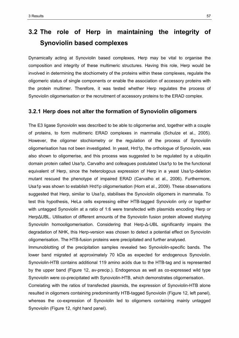
3 Results
57
3.2 The role of Herp in maintaining the integrity of Synoviolin based complexes
Dynamically acting at Synoviolin based complexes, Herp may be vital to organise the
composition and integrity of these multimeric structures. Having this role, Herp would be
involved in determining the stochiometry of the proteins within these complexes, regulate the
oligomeric status of single components or enable the association of accessory proteins with
the protein multimer. Therefore, it was tested whether Herp regulates the process of
Synoviolin oligomerisation or the recruitment of accessory proteins to the ERAD complex.
3.2.1 Herp does not alter the formation of Synoviolin oligomers
The E3 ligase Synoviolin was described to be able to oligomerise and, together with a couple
of proteins, to form multimeric ERAD complexes in mammalia (Schulze et al., 2005).
However, the oligomer stochiometry or the regulation of the process of Synoviolin
oligomerisation has not been investigated. In yeast, Hrd1p, the orthologue of Synoviolin, was
also shown to oligomerise, and this process was suggested to be regulated by a ubiquitin
domain protein called Usa1p. Carvalho and colleagues postulated Usa1p to be the functional
equivalent of Herp, since the heterologous expression of Herp in a yeast Usa1p-deletion
mutant rescued the phenotype of impaired ERAD (Carvalho et al., 2006). Furthermore,
Usa1p was shown to establish Hrd1p oligomerisation (Horn et al., 2009). These observations
suggested that Herp, similar to Usa1p, stabilises the Synoviolin oligomers in mammalia. To
test this hypothesis, HeLa cells expressing either HTB-tagged Synoviolin only or together
with untagged Synoviolin at a ratio of 1:6 were transfected with plasmids encoding Herp or
HerpΔUBL. Utilisation of different amounts of the Synoviolin fusion protein allowed studying
Synoviolin homooligomerisation. Considering that Herp-Δ-UBL significantly impairs the
degradation of NHK, this Herp-version was chosen to detect a potential effect on Synoviolin
oligomerisation. The HTB-fusion proteins were precipitated and further analysed.
Immunoblotting of the precipitation samples revealed two Synoviolin-specific bands. The
lower band migrated at approximately 70 kDa as expected for endogenous Synoviolin.
Synoviolin-HTB contains additional 119 amino acids due to the HTB-tag and is represented
by the upper band (Figure 12, av-precip.). Endogenous as well as co-expressed wild type
Synoviolin were co-precipitated with Synoviolin-HTB, which demonstrates oligomerisation.
Correlating with the ratios of transfected plasmids, the expression of Synoviolin-HTB alone
resulted in oligomers containing predominantly HTB-tagged Synoviolin (Figure 12, left panel),
whereas the co-expression of Synoviolin led to oligomers containing mainly untagged
Synoviolin (Figure 12, right hand panel).
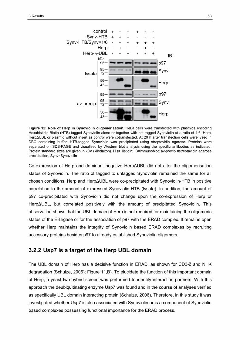
3 Results
58
p97
Synv
Herp
av-precip.
Herp
Synv
p97
IB:
lysate
controlSynv-HTB
Synv-HTB/Synv=1/6Herp
Herp-Δ-UBL - - + - - +- + - - + -- - - + + ++ + + - - -+ - - + - -
95
95
95
95
72
72
56
kDa
43
5643
Figure 12: Role of Herp in Synoviolin oligomerisation. HeLa cells were transfected with plasmids encoding Hexahistidin-Biotin (HTB)-tagged Synoviolin alone or together with not tagged Synoviolin at a ratio of 1:6. Herp, HerpΔUBL or plasmid without insert as control were cotransfected. At 20 h after transfection cells were lysed in DBC containing buffer. HTB-tagged Synoviolin was precipitated using streptavidin agarose. Proteins were separated on SDS-PAGE and visualised by Western blot analysis using the specific antibodies as indicated. Protein standard sizes are given in kDa (kilodalton). His=Histidin; IB=immunoblot; av-precip.=streptavidin agarose precipitation, Synv=Synoviolin
Co-expression of Herp and dominant negative HerpΔUBL did not alter the oligomerisation
status of Synoviolin. The ratio of tagged to untagged Synoviolin remained the same for all
chosen conditions. Herp and HerpΔUBL were co-precipitated with Synoviolin-HTB in positive
correlation to the amount of expressed Synoviolin-HTB (lysate). In addition, the amount of
p97 co-precipitated with Synoviolin did not change upon the co-expression of Herp or
HerpΔUBL, but correlated positively with the amount of precipitated Synoviolin. This
observation shows that the UBL domain of Herp is not required for maintaining the oligomeric
status of the E3 ligase or for the association of p97 with the ERAD complex. It remains open
whether Herp maintains the integrity of Synoviolin based ERAD complexes by recruiting
accessory proteins besides p97 to already established Synoviolin oligomers.
3.2.2 Usp7 is a target of the Herp UBL domain
The UBL domain of Herp has a decisive function in ERAD, as shown for CD3-δ and NHK
degradation (Schulze, 2006); Figure 11,B). To elucidate the function of this important domain
of Herp, a yeast two hybrid screen was performed to identify interaction partners. With this
approach the deubiquitinating enzyme Usp7 was found and in the course of analyses verified
as specifically UBL domain interacting protein (Schulze, 2006). Therefore, in this study it was
investigated whether Usp7 is also associated with Synoviolin or is a component of Synoviolin
based complexes possessing functional importance for the ERAD process.
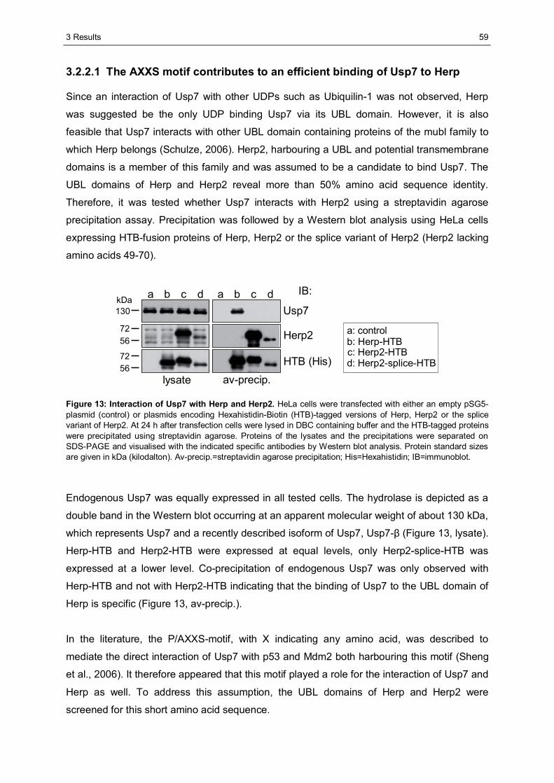
3 Results
59
3.2.2.1 The AXXS motif contributes to an efficient binding of Usp7 to Herp
Since an interaction of Usp7 with other UDPs such as Ubiquilin-1 was not observed, Herp
was suggested be the only UDP binding Usp7 via its UBL domain. However, it is also
feasible that Usp7 interacts with other UBL domain containing proteins of the mubl family to
which Herp belongs (Schulze, 2006). Herp2, harbouring a UBL and potential transmembrane
domains is a member of this family and was assumed to be a candidate to bind Usp7. The
UBL domains of Herp and Herp2 reveal more than 50% amino acid sequence identity.
Therefore, it was tested whether Usp7 interacts with Herp2 using a streptavidin agarose
precipitation assay. Precipitation was followed by a Western blot analysis using HeLa cells
expressing HTB-fusion proteins of Herp, Herp2 or the splice variant of Herp2 (Herp2 lacking
amino acids 49-70).
IB:
Usp7
Herp2
HTB (His)
lysate av-precip.
7256
7256
130kDa
a: controlb: Herp-HTBc: Herp2-HTBd: Herp2-splice-HTB
a b c d a b c d
Figure 13: Interaction of Usp7 with Herp and Herp2. HeLa cells were transfected with either an empty pSG5-plasmid (control) or plasmids encoding Hexahistidin-Biotin (HTB)-tagged versions of Herp, Herp2 or the splice variant of Herp2. At 24 h after transfection cells were lysed in DBC containing buffer and the HTB-tagged proteins were precipitated using streptavidin agarose. Proteins of the lysates and the precipitations were separated on SDS-PAGE and visualised with the indicated specific antibodies by Western blot analysis. Protein standard sizes are given in kDa (kilodalton). Av-precip.=streptavidin agarose precipitation; His=Hexahistidin; IB=immunoblot.
Endogenous Usp7 was equally expressed in all tested cells. The hydrolase is depicted as a
double band in the Western blot occurring at an apparent molecular weight of about 130 kDa,
which represents Usp7 and a recently described isoform of Usp7, Usp7-β (Figure 13, lysate).
Herp-HTB and Herp2-HTB were expressed at equal levels, only Herp2-splice-HTB was
expressed at a lower level. Co-precipitation of endogenous Usp7 was only observed with
Herp-HTB and not with Herp2-HTB indicating that the binding of Usp7 to the UBL domain of
Herp is specific (Figure 13, av-precip.).
In the literature, the P/AXXS-motif, with X indicating any amino acid, was described to
mediate the direct interaction of Usp7 with p53 and Mdm2 both harbouring this motif (Sheng
et al., 2006). It therefore appeared that this motif played a role for the interaction of Usp7 and
Herp as well. To address this assumption, the UBL domains of Herp and Herp2 were
screened for this short amino acid sequence.
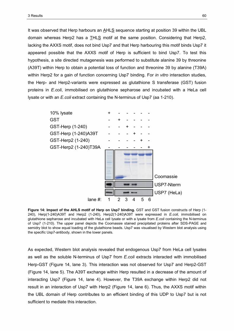
3 Results
60
It was observed that Herp harbours an AHLS sequence starting at position 39 within the UBL
domain whereas Herp2 has a THLS motif at the same position. Considering that Herp2,
lacking the AXXS motif, does not bind Usp7 and that Herp harbouring this motif binds Usp7 it
appeared possible that the AXXS motif of Herp is sufficient to bind Usp7. To test this
hypothesis, a site directed mutagenesis was performed to substitute alanine 39 by threonine
(A39T) within Herp to obtain a potential loss of function and threonine 39 by alanine (T39A)
within Herp2 for a gain of function concerning Usp7 binding. For in vitro interaction studies,
the Herp- and Herp2-variants were expressed as glutathione S transferase (GST) fusion
proteins in E.coli, immobilised on glutathione sepharose and incubated with a HeLa cell
lysate or with an E.coli extract containing the N-terminus of Usp7 (aa 1-210).
10% lysateGSTGST-Herp (1-240)GST-Herp (1-240)A39TGST-Herp2 (1-240)GST-Herp2 (1-240)T39A
+ - - - - -
- - - - - +
- - - - + -
- - - + - -
- - + - - -
- + - - - -
Coomassie
USP7-Nterm
USP7 (HeLa)1 2 3 4 5 6lane #:
Figure 14: Impact of the AHLS motif of Herp on Usp7 binding. GST and GST fusion constructs of Herp (1-240), Herp(1-240)A39T and Herp2 (1-240), Herp2(1-240)A39T were expressed in E.coli, immobilised on glutathione sepharose and incubated with HeLa cell lysate or with a lysate from E.coli containing the N-terminus of Usp7 (1-210). The upper panel depicts the Coomassie stained precipitated proteins after SDS-PAGE and semidry blot to show equal loading of the glutathione beads. Usp7 was visualised by Western blot analysis using the specific Usp7-antibody, shown in the lower panels.
As expected, Western blot analysis revealed that endogenous Usp7 from HeLa cell lysates
as well as the soluble N-terminus of Usp7 from E.coli extracts interacted with immobilised
Herp-GST (Figure 14, lane 3). This interaction was not observed for Usp7 and Herp2-GST
(Figure 14, lane 5). The A39T exchange within Herp resulted in a decrease of the amount of
interacting Usp7 (Figure 14, lane 4). However, the T39A exchange within Herp2 did not
result in an interaction of Usp7 with Herp2 (Figure 14, lane 6). Thus, the AXXS motif within
the UBL domain of Herp contributes to an efficient binding of this UDP to Usp7 but is not
sufficient to mediate this interaction.
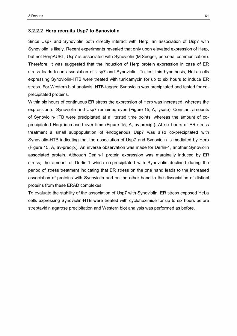
3 Results
61
3.2.2.2 Herp recruits Usp7 to Synoviolin
Since Usp7 and Synoviolin both directly interact with Herp, an association of Usp7 with
Synoviolin is likely. Recent experiments revealed that only upon elevated expression of Herp,
but not HerpΔUBL, Usp7 is associated with Synoviolin (M.Seeger, personal communication).
Therefore, it was suggested that the induction of Herp protein expression in case of ER
stress leads to an association of Usp7 and Synoviolin. To test this hypothesis, HeLa cells
expressing Synoviolin-HTB were treated with tunicamycin for up to six hours to induce ER
stress. For Western blot analysis, HTB-tagged Synoviolin was precipitated and tested for co-
precipitated proteins.
Within six hours of continuous ER stress the expression of Herp was increased, whereas the
expression of Synoviolin and Usp7 remained even (Figure 15, A, lysate). Constant amounts
of Synoviolin-HTB were precipitated at all tested time points, whereas the amount of co-
precipitated Herp increased over time (Figure 15, A, av.precip.). At six hours of ER stress
treatment a small subpopulation of endogenous Usp7 was also co-precipitated with
Synoviolin-HTB indicating that the association of Usp7 and Synoviolin is mediated by Herp
(Figure 15, A, av-precip.). An inverse observation was made for Derlin-1, another Synoviolin
associated protein. Although Derlin-1 protein expression was marginally induced by ER
stress, the amount of Derlin-1 which co-precipitated with Synoviolin declined during the
period of stress treatment indicating that ER stress on the one hand leads to the increased
association of proteins with Synoviolin and on the other hand to the dissociation of distinct
proteins from these ERAD complexes.
To evaluate the stability of the association of Usp7 with Synoviolin, ER stress exposed HeLa
cells expressing Synoviolin-HTB were treated with cycloheximide for up to six hours before
streptavidin agarose precipitation and Western blot analysis was performed as before.
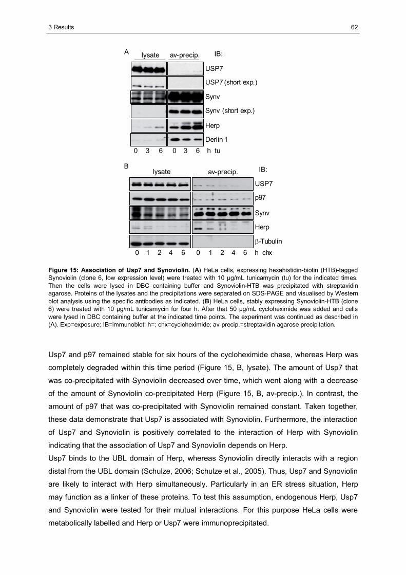
3 Results
62
A
B IB:
USP7
p97
Synv
Herp
β-Tubulin
lysate av-precip.
64210 h chx64210
lysate av-precip. IB:
USP7
USP7 (short exp.)
Synv
Synv (short exp.)
Herp
Derlin 1h tu630630
Figure 15: Association of Usp7 and Synoviolin. (A) HeLa cells, expressing hexahistidin-biotin (HTB)-tagged Synoviolin (clone 6, low expression level) were treated with 10 μg/mL tunicamycin (tu) for the indicated times. Then the cells were lysed in DBC containing buffer and Synoviolin-HTB was precipitated with streptavidin agarose. Proteins of the lysates and the precipitations were separated on SDS-PAGE and visualised by Western blot analysis using the specific antibodies as indicated. (B) HeLa cells, stably expressing Synoviolin-HTB (clone 6) were treated with 10 μg/mL tunicamycin for four h. After that 50 μg/mL cycloheximide was added and cells were lysed in DBC containing buffer at the indicated time points. The experiment was continued as described in (A). Exp=exposure; IB=immunoblot; h=; chx=cycloheximide; av-precip.=streptavidin agarose precipitation.
Usp7 and p97 remained stable for six hours of the cycloheximide chase, whereas Herp was
completely degraded within this time period (Figure 15, B, lysate). The amount of Usp7 that
was co-precipitated with Synoviolin decreased over time, which went along with a decrease
of the amount of Synoviolin co-precipitated Herp (Figure 15, B, av-precip.). In contrast, the
amount of p97 that was co-precipitated with Synoviolin remained constant. Taken together,
these data demonstrate that Usp7 is associated with Synoviolin. Furthermore, the interaction
of Usp7 and Synoviolin is positively correlated to the interaction of Herp with Synoviolin
indicating that the association of Usp7 and Synoviolin depends on Herp.
Usp7 binds to the UBL domain of Herp, whereas Synoviolin directly interacts with a region
distal from the UBL domain (Schulze, 2006; Schulze et al., 2005). Thus, Usp7 and Synoviolin
are likely to interact with Herp simultaneously. Particularly in an ER stress situation, Herp
may function as a linker of these proteins. To test this assumption, endogenous Herp, Usp7
and Synoviolin were tested for their mutual interactions. For this purpose HeLa cells were
metabolically labelled and Herp or Usp7 were immunoprecipitated.
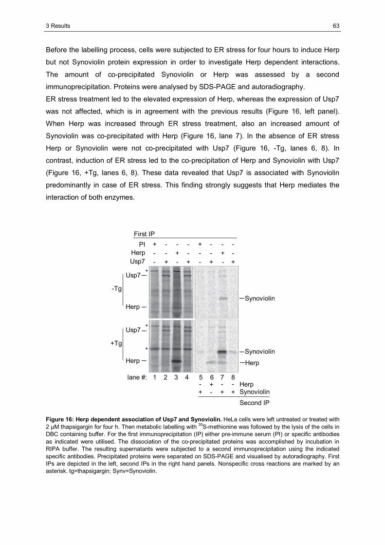
3 Results
63
Before the labelling process, cells were subjected to ER stress for four hours to induce Herp
but not Synoviolin protein expression in order to investigate Herp dependent interactions.
The amount of co-precipitated Synoviolin or Herp was assessed by a second
immunoprecipitation. Proteins were analysed by SDS-PAGE and autoradiography.
ER stress treatment led to the elevated expression of Herp, whereas the expression of Usp7
was not affected, which is in agreement with the previous results (Figure 16, left panel).
When Herp was increased through ER stress treatment, also an increased amount of
Synoviolin was co-precipitated with Herp (Figure 16, lane 7). In the absence of ER stress
Herp or Synoviolin were not co-precipitated with Usp7 (Figure 16, -Tg, lanes 6, 8). In
contrast, induction of ER stress led to the co-precipitation of Herp and Synoviolin with Usp7
(Figure 16, +Tg, lanes 6, 8). These data revealed that Usp7 is associated with Synoviolin
predominantly in case of ER stress. This finding strongly suggests that Herp mediates the
interaction of both enzymes.
+-+-+-+--+---+-----+---+
++-+--+-
Usp7
Herp
-Tg
Usp7
Herp
+Tg
1 2 3 4 5 6 7 8
Synoviolin
Synoviolin
SynoviolinHerp
Second IP
Usp7Herp
PIFirst IP
*
*
*
lane #:
Herp
Figure 16: Herp dependent association of Usp7 and Synoviolin. HeLa cells were left untreated or treated with 2 μM thapsigargin for four h. Then metabolic labelling with 35S-methionine was followed by the lysis of the cells in DBC containing buffer. For the first immunoprecipitation (IP) either pre-immune serum (PI) or specific antibodies as indicated were utilised. The dissociation of the co-precipitated proteins was accomplished by incubation in RIPA buffer. The resulting supernatants were subjected to a second immunoprecipitation using the indicated specific antibodies. Precipitated proteins were separated on SDS-PAGE and visualised by autoradiography. First IPs are depicted in the left, second IPs in the right hand panels. Nonspecific cross reactions are marked by an asterisk. tg=thapsigargin; Synv=Synoviolin.
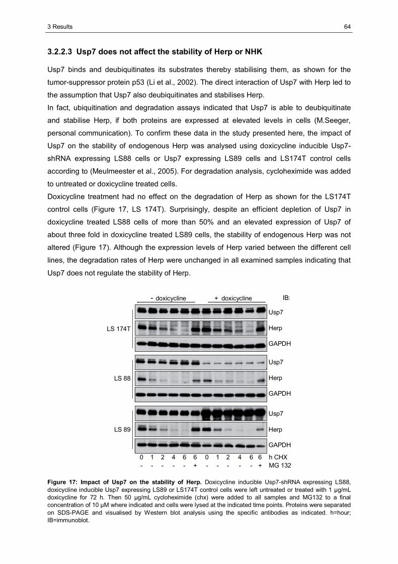
3 Results
64
3.2.2.3 Usp7 does not affect the stability of Herp or NHK
Usp7 binds and deubiquitinates its substrates thereby stabilising them, as shown for the
tumor-suppressor protein p53 (Li et al., 2002). The direct interaction of Usp7 with Herp led to
the assumption that Usp7 also deubiquitinates and stabilises Herp.
In fact, ubiquitination and degradation assays indicated that Usp7 is able to deubiquitinate
and stabilise Herp, if both proteins are expressed at elevated levels in cells (M.Seeger,
personal communication). To confirm these data in the study presented here, the impact of
Usp7 on the stability of endogenous Herp was analysed using doxicycline inducible Usp7-
shRNA expressing LS88 cells or Usp7 expressing LS89 cells and LS174T control cells
according to (Meulmeester et al., 2005). For degradation analysis, cycloheximide was added
to untreated or doxicycline treated cells.
Doxicycline treatment had no effect on the degradation of Herp as shown for the LS174T
control cells (Figure 17, LS 174T). Surprisingly, despite an efficient depletion of Usp7 in
doxicycline treated LS88 cells of more than 50% and an elevated expression of Usp7 of
about three fold in doxicycline treated LS89 cells, the stability of endogenous Herp was not
altered (Figure 17). Although the expression levels of Herp varied between the different cell
lines, the degradation rates of Herp were unchanged in all examined samples indicating that
Usp7 does not regulate the stability of Herp.
MG 132+-----h CHX664210664210
LS 174T
Usp7
GAPDH
- +doxicycline
LS 88
LS 89
Herp
IB:doxicycline
Usp7
GAPDH
Herp
Usp7
GAPDH
Herp
-
+-----
Figure 17: Impact of Usp7 on the stability of Herp. Doxicycline inducible Usp7-shRNA expressing LS88, doxicycline inducible Usp7 expressing LS89 or LS174T control cells were left untreated or treated with 1 μg/mL doxicycline for 72 h. Then 50 μg/mL cycloheximide (chx) were added to all samples and MG132 to a final concentration of 10 μM where indicated and cells were lysed at the indicated time points. Proteins were separated on SDS-PAGE and visualised by Western blot analysis using the specific antibodies as indicated. h=hour; IB=immunoblot.
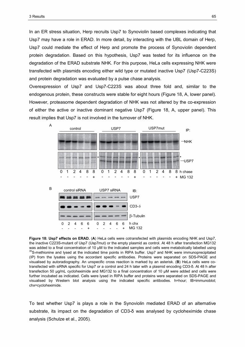
3 Results
65
In an ER stress situation, Herp recruits Usp7 to Synoviolin based complexes indicating that
Usp7 may have a role in ERAD. In more detail, by interacting with the UBL domain of Herp,
Usp7 could mediate the effect of Herp and promote the process of Synoviolin dependent
protein degradation. Based on this hypothesis, Usp7 was tested for its influence on the
degradation of the ERAD substrate NHK. For this purpose, HeLa cells expressing NHK were
transfected with plasmids encoding either wild type or mutated inactive Usp7 (Usp7-C223S)
and protein degradation was evaluated by a pulse chase analysis.
Overexpression of Usp7 and Usp7-C223S was about three fold and, similar to the
endogenous protein, these constructs were stable for eight hours (Figure 18, A, lower panel).
However, proteasome dependent degradation of NHK was not altered by the co-expression
of either the active or inactive dominant negative Usp7 (Figure 18, A, upper panel). This
result implies that Usp7 is not involved in the turnover of NHK.
control USP7 USP7mut
NHK
USP7
IP:
*
A
* *
MG 132h chase0 421 8 8
- - - - - +0 421 8 8- - - - - +
0 421 8 8- - - - - +
0 2 4 6 6- - - - +
0 2 4 6 6- - - - +
control siRNA USP7 siRNA IB:
CD3-δ
β-Tubulin
MG 132h chx
USP7
B
Figure 18: Usp7 effects on ERAD. (A) HeLa cells were cotransfected with plasmids encoding NHK and Usp7, the inactive C223S-mutant of Usp7 (Usp7mut) or the empty plasmid as control. At 48 h after transfection MG132 was added to a final concentration of 10 μM to the indicated samples and cells were metabolically labelled using 35S-methionine and lysed at the indicated time points in RIPA buffer. Usp7 and NHK were immunoprecipitated (IP) from the lysates using the accordant specific antibodies. Proteins were separated on SDS-PAGE and visualised by autoradiography. An unspecific cross reaction is marked by an asterisk. (B) HeLa cells were co-transfected with siRNA specific for Usp7 or a control and 24 h later with a plasmid encoding CD3-δ. At 48 h after transfection 50 μg/mL cycloheximide and MG132 to a final concentration of 10 μM were added and cells were further incubated as indicated. Cells were lysed in RIPA buffer and proteins were separated on SDS-PAGE and visualised by Western blot analysis using the indicated specific antibodies. h=hour; IB=immunoblot; chx=cycloheximide.
To test whether Usp7 is plays a role in the Synoviolin mediated ERAD of an alternative
substrate, its impact on the degradation of CD3-δ was analysed by cycloheximide chase
analysis (Schulze et al., 2005).
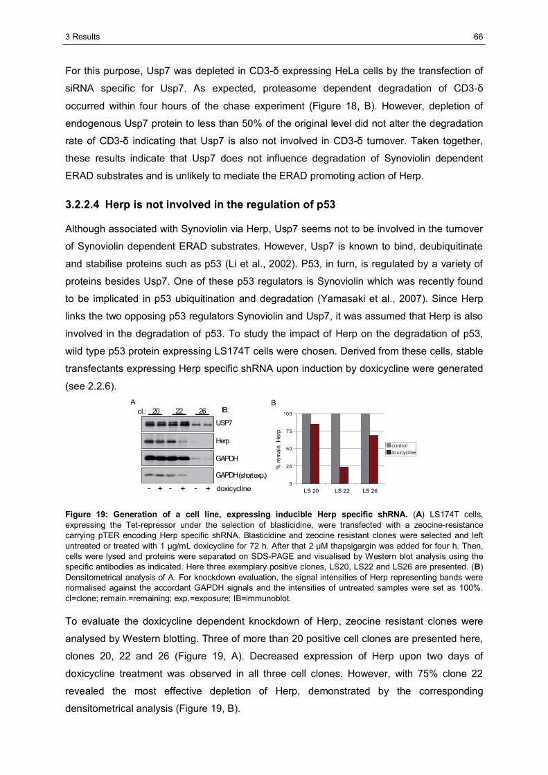
3 Results
66
For this purpose, Usp7 was depleted in CD3-δ expressing HeLa cells by the transfection of
siRNA specific for Usp7. As expected, proteasome dependent degradation of CD3-δ
occurred within four hours of the chase experiment (Figure 18, B). However, depletion of
endogenous Usp7 protein to less than 50% of the original level did not alter the degradation
rate of CD3-δ indicating that Usp7 is also not involved in CD3-δ turnover. Taken together,
these results indicate that Usp7 does not influence degradation of Synoviolin dependent
ERAD substrates and is unlikely to mediate the ERAD promoting action of Herp.
3.2.2.4 Herp is not involved in the regulation of p53
Although associated with Synoviolin via Herp, Usp7 seems not to be involved in the turnover
of Synoviolin dependent ERAD substrates. However, Usp7 is known to bind, deubiquitinate
and stabilise proteins such as p53 (Li et al., 2002). P53, in turn, is regulated by a variety of
proteins besides Usp7. One of these p53 regulators is Synoviolin which was recently found
to be implicated in p53 ubiquitination and degradation (Yamasaki et al., 2007). Since Herp
links the two opposing p53 regulators Synoviolin and Usp7, it was assumed that Herp is also
involved in the degradation of p53. To study the impact of Herp on the degradation of p53,
wild type p53 protein expressing LS174T cells were chosen. Derived from these cells, stable
transfectants expressing Herp specific shRNA upon induction by doxicycline were generated
(see 2.2.6).
doxicycline
Herp
GAPDH
IB:20 22 26cl.:USP7
(short exp.)GAPDH
A
0
25
50
75
100
LS 20 LS 22 LS 26
%re
mai
n.H
erp
controldoxicycline
B
- +-+-+
Figure 19: Generation of a cell line, expressing inducible Herp specific shRNA. (A) LS174T cells, expressing the Tet-repressor under the selection of blasticidine, were transfected with a zeocine-resistance carrying pTER encoding Herp specific shRNA. Blasticidine and zeocine resistant clones were selected and left untreated or treated with 1 μg/mL doxicycline for 72 h. After that 2 μM thapsigargin was added for four h. Then, cells were lysed and proteins were separated on SDS-PAGE and visualised by Western blot analysis using the specific antibodies as indicated. Here three exemplary positive clones, LS20, LS22 and LS26 are presented. (B) Densitometrical analysis of A. For knockdown evaluation, the signal intensities of Herp representing bands were normalised against the accordant GAPDH signals and the intensities of untreated samples were set as 100%. cl=clone; remain.=remaining; exp.=exposure; IB=immunoblot.
To evaluate the doxicycline dependent knockdown of Herp, zeocine resistant clones were
analysed by Western blotting. Three of more than 20 positive cell clones are presented here,
clones 20, 22 and 26 (Figure 19, A). Decreased expression of Herp upon two days of
doxicycline treatment was observed in all three cell clones. However, with 75% clone 22
revealed the most effective depletion of Herp, demonstrated by the corresponding
densitometrical analysis (Figure 19, B).
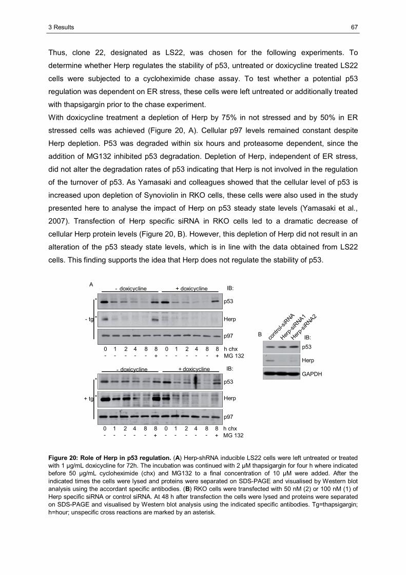
3 Results
67
Thus, clone 22, designated as LS22, was chosen for the following experiments. To
determine whether Herp regulates the stability of p53, untreated or doxicycline treated LS22
cells were subjected to a cycloheximide chase assay. To test whether a potential p53
regulation was dependent on ER stress, these cells were left untreated or additionally treated
with thapsigargin prior to the chase experiment.
With doxicycline treatment a depletion of Herp by 75% in not stressed and by 50% in ER
stressed cells was achieved (Figure 20, A). Cellular p97 levels remained constant despite
Herp depletion. P53 was degraded within six hours and proteasome dependent, since the
addition of MG132 inhibited p53 degradation. Depletion of Herp, independent of ER stress,
did not alter the degradation rates of p53 indicating that Herp is not involved in the regulation
of the turnover of p53. As Yamasaki and colleagues showed that the cellular level of p53 is
increased upon depletion of Synoviolin in RKO cells, these cells were also used in the study
presented here to analyse the impact of Herp on p53 steady state levels (Yamasaki et al.,
2007). Transfection of Herp specific siRNA in RKO cells led to a dramatic decrease of
cellular Herp protein levels (Figure 20, B). However, this depletion of Herp did not result in an
alteration of the p53 steady state levels, which is in line with the data obtained from LS22
cells. This finding supports the idea that Herp does not regulate the stability of p53.
Herp
GAPDH
p53IB:co
ntrol-
siRNA
Herp-si
RNA1
Herp-si
RNA2
p53
p53
Herp
p97
Herp
p97
h chx
doxicycline
IB:
- + IB:
doxicycline- +
- tg
+ tg
doxicycline
doxicycline
A
B
*
*
*
*
MG 1320 421 8 8- - - - - +
0 421 8 8- - - - - +
h chxMG 132
0 421 8 8- - - - - +
0 421 8 8- - - - - +
Figure 20: Role of Herp in p53 regulation. (A) Herp-shRNA inducible LS22 cells were left untreated or treated with 1 μg/mL doxicycline for 72h. The incubation was continued with 2 μM thapsigargin for four h where indicated before 50 μg/mL cycloheximide (chx) and MG132 to a final concentration of 10 μM were added. After the indicated times the cells were lysed and proteins were separated on SDS-PAGE and visualised by Western blot analysis using the accordant specific antibodies. (B) RKO cells were transfected with 50 nM (2) or 100 nM (1) of Herp specific siRNA or control siRNA. At 48 h after transfection the cells were lysed and proteins were separated on SDS-PAGE and visualised by Western blot analysis using the indicated specific antibodies. Tg=thapsigargin; h=hour; unspecific cross reactions are marked by an asterisk.
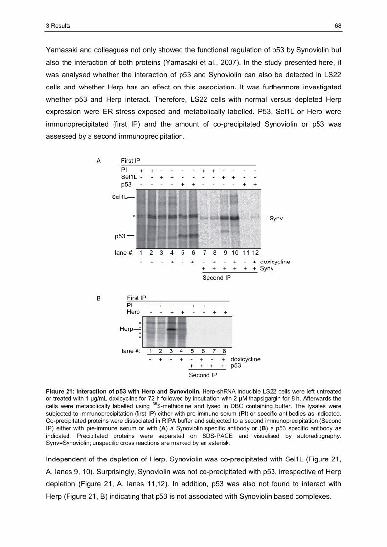
3 Results
68
Yamasaki and colleagues not only showed the functional regulation of p53 by Synoviolin but
also the interaction of both proteins (Yamasaki et al., 2007). In the study presented here, it
was analysed whether the interaction of p53 and Synoviolin can also be detected in LS22
cells and whether Herp has an effect on this association. It was furthermore investigated
whether p53 and Herp interact. Therefore, LS22 cells with normal versus depleted Herp
expression were ER stress exposed and metabolically labelled. P53, Sel1L or Herp were
immunoprecipitated (first IP) and the amount of co-precipitated Synoviolin or p53 was
assessed by a second immunoprecipitation.
First IP
+ + + +Second IP
*
Sel1L
Synv
p53
First IP
doxicycline
Second IP
*
***
A
B
+ + - ---- - - -++- - + +--
+ + - ---- - - -++- - + +--
PISel1Lp53
- -+++ +--
- -+++ +--
PIHerp
++- + - + +-- + - + +- doxicycline
Synv
- +- + - + +-+ + ++ p53
7 8 9 10 12111 2 3 4 65
7 81 2 3 4 65
Herp
lane #:
lane #:
Figure 21: Interaction of p53 with Herp and Synoviolin. Herp-shRNA inducible LS22 cells were left untreated or treated with 1 μg/mL doxicycline for 72 h followed by incubation with 2 μM thapsigargin for 8 h. Afterwards the cells were metabolically labelled using 35S-methionine and lysed in DBC containing buffer. The lysates were subjected to immunoprecipitation (first IP) either with pre-immune serum (PI) or specific antibodies as indicated. Co-precipitated proteins were dissociated in RIPA buffer and subjected to a second immunoprecipitation (Second IP) either with pre-immune serum or with (A) a Synoviolin specific antibody or (B) a p53 specific antibody as indicated. Precipitated proteins were separated on SDS-PAGE and visualised by autoradiography. Synv=Synoviolin; unspecific cross reactions are marked by an asterisk.
Independent of the depletion of Herp, Synoviolin was co-precipitated with Sel1L (Figure 21,
A, lanes 9, 10). Surprisingly, Synoviolin was not co-precipitated with p53, irrespective of Herp
depletion (Figure 21, A, lanes 11,12). In addition, p53 was also not found to interact with
Herp (Figure 21, B) indicating that p53 is not associated with Synoviolin based complexes.
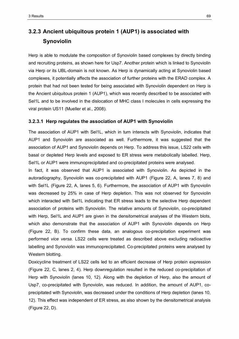
3 Results
69
3.2.3 Ancient ubiquitous protein 1 (AUP1) is associated with
Synoviolin
Herp is able to modulate the composition of Synoviolin based complexes by directly binding
and recruiting proteins, as shown here for Usp7. Another protein which is linked to Synoviolin
via Herp or its UBL-domain is not known. As Herp is dynamically acting at Synoviolin based
complexes, it potentially affects the association of further proteins with the ERAD complex. A
protein that had not been tested for being associated with Synoviolin dependent on Herp is
the Ancient ubiquitous protein 1 (AUP1), which was recently described to be associated with
Sel1L and to be involved in the dislocation of MHC class I molecules in cells expressing the
viral protein US11 (Mueller et al., 2008).
3.2.3.1 Herp regulates the association of AUP1 with Synoviolin
The association of AUP1 with Sel1L, which in turn interacts with Synoviolin, indicates that
AUP1 and Synoviolin are associated as well. Furthermore, it was suggested that the
association of AUP1 and Synoviolin depends on Herp. To address this issue, LS22 cells with
basal or depleted Herp levels and exposed to ER stress were metabolically labelled. Herp,
Sel1L or AUP1 were immunoprecipitated and co-precipitated proteins were analysed.
In fact, it was observed that AUP1 is associated with Synoviolin. As depicted in the
autoradiography, Synoviolin was co-precipitated with AUP1 (Figure 22, A, lanes 7, 8) and
with Sel1L (Figure 22, A, lanes 5, 6). Furthermore, the association of AUP1 with Synoviolin
was decreased by 25% in case of Herp depletion. This was not observed for Synoviolin
which interacted with Sel1L indicating that ER stress leads to the selective Herp dependent
association of proteins with Synoviolin. The relative amounts of Synoviolin, co-precipitated
with Herp, Sel1L and AUP1 are given in the densitometrical analyses of the Western blots,
which also demonstrate that the association of AUP1 with Synoviolin depends on Herp
(Figure 22, B). To confirm these data, an analogous co-precipitation experiment was
performed vice versa. LS22 cells were treated as described above excluding radioactive
labelling and Synoviolin was immunoprecipitated. Co-precipitated proteins were analysed by
Western blotting.
Doxicycline treatment of LS22 cells led to an efficient decrease of Herp protein expression
(Figure 22, C, lanes 2, 4). Herp downregulation resulted in the reduced co-precipitation of
Herp with Synoviolin (lanes 10, 12). Along with the depletion of Herp, also the amount of
Usp7, co-precipitated with Synoviolin, was reduced. In addition, the amount of AUP1, co-
precipitated with Synoviolin, was decreased under the conditions of Herp depletion (lanes 10,
12). This effect was independent of ER stress, as also shown by the densitometrical analysis
(Figure 22, D).
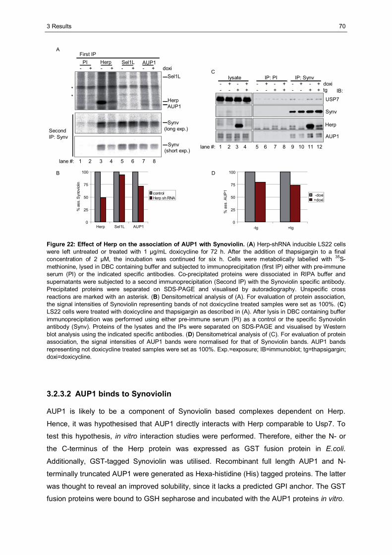
3 Results
70
lysate IP: PI IP: Synv
USP7
Synv
Herp
AUP1
IB:-----+- +++++ doxi+-+-+-- +-+-+ tg
Cdoxi
PI Herp Sel1L AUP1First IP
SecondIP: Synv
HerpAUP1
Sel1L
(long exp.)
(short exp.)
A
Synv
Synv
**
B
0
25
50
75
100
Herp Sel1L AUP1
%as
s. S
ynov
iolin
controlHerp sh RNA
D
- - + - + - ++
0
25
50
75
100
-tg +tg%
ass.
AU
P1
-doxi+doxi
1 2 3 4 5 6 7 8
1 2 3 4 5 6 7 8 9 10 11 12lane #:
lane #:
Figure 22: Effect of Herp on the association of AUP1 with Synoviolin. (A) Herp-shRNA inducible LS22 cells were left untreated or treated with 1 μg/mL doxicycline for 72 h. After the addition of thapsigargin to a final concentration of 2 μM, the incubation was continued for six h. Cells were metabolically labelled with 35S-methionine, lysed in DBC containing buffer and subjected to immunoprecipitation (first IP) either with pre-immune serum (PI) or the indicated specific antibodies. Co-precipitated proteins were dissociated in RIPA buffer and supernatants were subjected to a second immunoprecipitation (Second IP) with the Synoviolin specific antibody. Precipitated proteins were separated on SDS-PAGE and visualised by autoradiography. Unspecific cross reactions are marked with an asterisk. (B) Densitometrical analysis of (A). For evaluation of protein association, the signal intensities of Synoviolin representing bands of not doxicycline treated samples were set as 100%. (C) LS22 cells were treated with doxicycline and thapsigargin as described in (A). After lysis in DBC containing buffer immunoprecipitation was performed using either pre-immune serum (PI) as a control or the specific Synoviolin antibody (Synv). Proteins of the lysates and the IPs were separated on SDS-PAGE and visualised by Western blot analysis using the indicated specific antibodies. (D) Densitometrical analysis of (C). For evaluation of protein association, the signal intensities of AUP1 bands were normalised for that of Synoviolin bands. AUP1 bands representing not doxicycline treated samples were set as 100%. Exp.=exposure; IB=immunoblot; tg=thapsigargin; doxi=doxicycline.
3.2.3.2 AUP1 binds to Synoviolin
AUP1 is likely to be a component of Synoviolin based complexes dependent on Herp.
Hence, it was hypothesised that AUP1 directly interacts with Herp comparable to Usp7. To
test this hypothesis, in vitro interaction studies were performed. Therefore, either the N- or
the C-terminus of the Herp protein was expressed as GST fusion protein in E.coli.
Additionally, GST-tagged Synoviolin was utilised. Recombinant full length AUP1 and N-
terminally truncated AUP1 were generated as Hexa-histidine (His) tagged proteins. The latter
was thought to reveal an improved solubility, since it lacks a predicted GPI anchor. The GST
fusion proteins were bound to GSH sepharose and incubated with the AUP1 proteins in vitro.
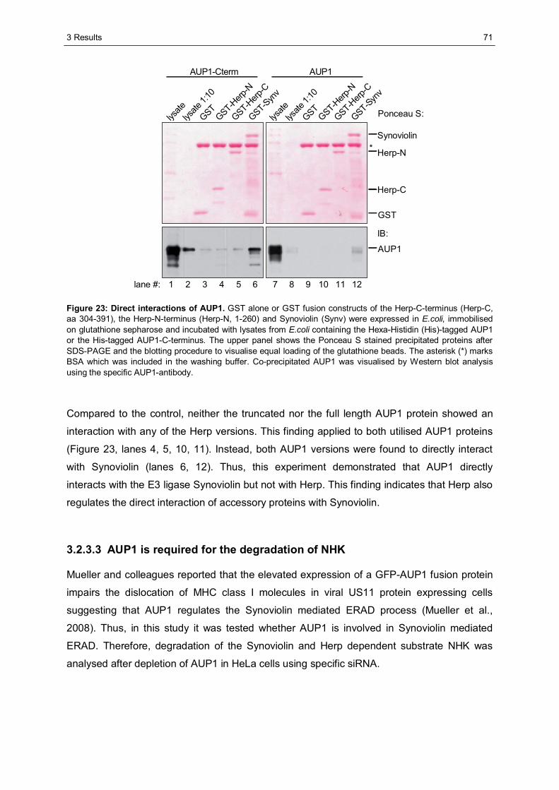
3 Results
71
Ponceau S:
IB:
AUP1
GST
Herp-C
Herp-N
Synoviolin
AUP1-Cterm AUP1
lysate
lysate
1:10
GSTGST-H
erp-N
GST-Herp
-CGST-S
ynv
lysate
lysate
1:10
GSTGST-H
erp-N
GST-Herp
-CGST-Syn
v
*
1 2 3 4 5 6 7 8lane #: 9 10 11 12
Figure 23: Direct interactions of AUP1. GST alone or GST fusion constructs of the Herp-C-terminus (Herp-C, aa 304-391), the Herp-N-terminus (Herp-N, 1-260) and Synoviolin (Synv) were expressed in E.coli, immobilised on glutathione sepharose and incubated with lysates from E.coli containing the Hexa-Histidin (His)-tagged AUP1 or the His-tagged AUP1-C-terminus. The upper panel shows the Ponceau S stained precipitated proteins after SDS-PAGE and the blotting procedure to visualise equal loading of the glutathione beads. The asterisk (*) marks BSA which was included in the washing buffer. Co-precipitated AUP1 was visualised by Western blot analysis using the specific AUP1-antibody.
Compared to the control, neither the truncated nor the full length AUP1 protein showed an
interaction with any of the Herp versions. This finding applied to both utilised AUP1 proteins
(Figure 23, lanes 4, 5, 10, 11). Instead, both AUP1 versions were found to directly interact
with Synoviolin (lanes 6, 12). Thus, this experiment demonstrated that AUP1 directly
interacts with the E3 ligase Synoviolin but not with Herp. This finding indicates that Herp also
regulates the direct interaction of accessory proteins with Synoviolin.
3.2.3.3 AUP1 is required for the degradation of NHK
Mueller and colleagues reported that the elevated expression of a GFP-AUP1 fusion protein
impairs the dislocation of MHC class I molecules in viral US11 protein expressing cells
suggesting that AUP1 regulates the Synoviolin mediated ERAD process (Mueller et al.,
2008). Thus, in this study it was tested whether AUP1 is involved in Synoviolin mediated
ERAD. Therefore, degradation of the Synoviolin and Herp dependent substrate NHK was
analysed after depletion of AUP1 in HeLa cells using specific siRNA.
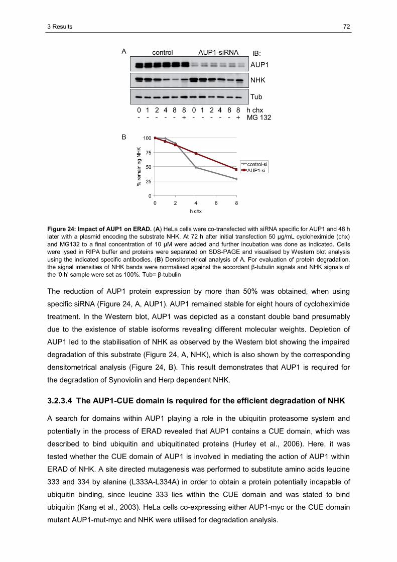
3 Results
72
NHK
AUP1
Tub
IB:control AUP1-siRNAA
B
MG 132h chx0 421 8 8
- - - - - +0 421 8 8- - - - - +
0
25
50
75
100
0 2 4 6 8h chx
%re
mai
ning
NH
K
control-siAUP1-si
Figure 24: Impact of AUP1 on ERAD. (A) HeLa cells were co-transfected with siRNA specific for AUP1 and 48 h later with a plasmid encoding the substrate NHK. At 72 h after initial transfection 50 μg/mL cycloheximide (chx) and MG132 to a final concentration of 10 μM were added and further incubation was done as indicated. Cells were lysed in RIPA buffer and proteins were separated on SDS-PAGE and visualised by Western blot analysis using the indicated specific antibodies. (B) Densitometrical analysis of A. For evaluation of protein degradation, the signal intensities of NHK bands were normalised against the accordant β-tubulin signals and NHK signals of the ‘0 h’ sample were set as 100%. Tub= β-tubulin
The reduction of AUP1 protein expression by more than 50% was obtained, when using
specific siRNA (Figure 24, A, AUP1). AUP1 remained stable for eight hours of cycloheximide
treatment. In the Western blot, AUP1 was depicted as a constant double band presumably
due to the existence of stable isoforms revealing different molecular weights. Depletion of
AUP1 led to the stabilisation of NHK as observed by the Western blot showing the impaired
degradation of this substrate (Figure 24, A, NHK), which is also shown by the corresponding
densitometrical analysis (Figure 24, B). This result demonstrates that AUP1 is required for
the degradation of Synoviolin and Herp dependent NHK.
3.2.3.4 The AUP1-CUE domain is required for the efficient degradation of NHK
A search for domains within AUP1 playing a role in the ubiquitin proteasome system and
potentially in the process of ERAD revealed that AUP1 contains a CUE domain, which was
described to bind ubiquitin and ubiquitinated proteins (Hurley et al., 2006). Here, it was
tested whether the CUE domain of AUP1 is involved in mediating the action of AUP1 within
ERAD of NHK. A site directed mutagenesis was performed to substitute amino acids leucine
333 and 334 by alanine (L333A-L334A) in order to obtain a protein potentially incapable of
ubiquitin binding, since leucine 333 lies within the CUE domain and was stated to bind
ubiquitin (Kang et al., 2003). HeLa cells co-expressing either AUP1-myc or the CUE domain
mutant AUP1-mut-myc and NHK were utilised for degradation analysis.
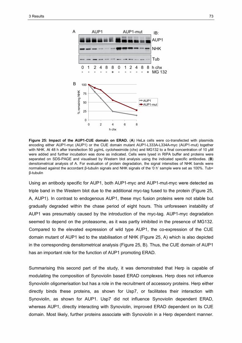
3 Results
73
NHK
AUP1
Tub
IB:AUP1 AUP1-mutA
B
MG 132h chx0 421 8 8
- - - - - +0 421 8 8- - - - - +
0
25
50
75
100
0 2 4 6 8h chx
%re
mai
ning
NH
K
AUP1AUP1-mut
Figure 25: Impact of the AUP1-CUE domain on ERAD. (A) HeLa cells were co-transfected with plasmids encoding either AUP1-myc (AUP1) or the CUE domain mutant AUP1-L333A-L334A-myc (AUP1-mut) together with NHK. At 48 h after transfection 50 μg/mL cycloheximide (chx) and MG132 to a final concentration of 10 μM were added and further incubation was done as indicated. Cells were lysed in RIPA buffer and proteins were separated on SDS-PAGE and visualised by Western blot analysis using the indicated specific antibodies. (B) densitometrical analysis of A. For evaluation of protein degradation, the signal intensities of NHK bands were normalised against the accordant β-tubulin signals and NHK signals of the ‘0 h’ sample were set as 100%. Tub= β-tubulin
Using an antibody specific for AUP1, both AUP1-myc and AUP1-mut-myc were detected as
triple band in the Western blot due to the additional myc-tag fused to the protein (Figure 25,
A, AUP1). In contrast to endogenous AUP1, these myc fusion proteins were not stable but
gradually degraded within the chase period of eight hours. This unforeseen instability of
AUP1 was presumably caused by the introduction of the myc-tag. AUP1-myc degradation
seemed to depend on the proteasome, as it was partly inhibited in the presence of MG132.
Compared to the elevated expression of wild type AUP1, the co-expression of the CUE
domain mutant of AUP1 led to the stabilisation of NHK (Figure 25, A) which is also depicted
in the corresponding densitometrical analysis (Figure 25, B). Thus, the CUE domain of AUP1
has an important role for the function of AUP1 promoting ERAD.
Summarising this second part of the study, it was demonstrated that Herp is capable of
modulating the composition of Synoviolin based ERAD complexes. Herp does not influence
Synoviolin oligomerisation but has a role in the recruitment of accessory proteins. Herp either
directly binds these proteins, as shown for Usp7, or facilitates their interaction with
Synoviolin, as shown for AUP1. Usp7 did not influence Synoviolin dependent ERAD,
whereas AUP1, directly interacting with Synoviolin, improved ERAD dependent on its CUE
domain. Most likely, further proteins associate with Synoviolin in a Herp dependent manner.
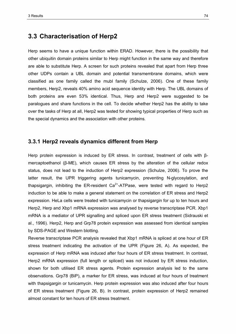
3 Results
74
3.3 Characterisation of Herp2
Herp seems to have a unique function within ERAD. However, there is the possibility that
other ubiquitin domain proteins similar to Herp might function in the same way and therefore
are able to substitute Herp. A screen for such proteins revealed that apart from Herp three
other UDPs contain a UBL domain and potential transmembrane domains, which were
classified as one family called the mubl family (Schulze, 2006). One of these family
members, Herp2, reveals 40% amino acid sequence identity with Herp. The UBL domains of
both proteins are even 53% identical. Thus, Herp and Herp2 were suggested to be
paralogues and share functions in the cell. To decide whether Herp2 has the ability to take
over the tasks of Herp at all, Herp2 was tested for showing typical properties of Herp such as
the special dynamics and the association with other proteins.
3.3.1 Herp2 reveals dynamics different from Herp
Herp protein expression is induced by ER stress. In contrast, treatment of cells with β-
mercaptoethanol (β-ME), which causes ER stress by the alteration of the cellular redox
status, does not lead to the induction of Herp2 expression (Schulze, 2006). To prove the
latter result, the UPR triggering agents tunicamycin, preventing N-glycosylation, and
thapsigargin, inhibiting the ER-resident Ca2+-ATPase, were tested with regard to Herp2
induction to be able to make a general statement on the correlation of ER stress and Herp2
expression. HeLa cells were treated with tunicamycin or thapsigargin for up to ten hours and
Herp2, Herp and Xbp1 mRNA expression was analysed by reverse transcriptase PCR. Xbp1
mRNA is a mediator of UPR signalling and spliced upon ER stress treatment (Sidrauski et
al., 1996). Herp2, Herp and Grp78 protein expression was assessed from identical samples
by SDS-PAGE and Western blotting.
Reverse transcriptase PCR analysis revealed that Xbp1 mRNA is spliced at one hour of ER
stress treatment indicating the activation of the UPR (Figure 26, A). As expected, the
expression of Herp mRNA was induced after four hours of ER stress treatment. In contrast,
Herp2 mRNA expression (full length or spliced) was not induced by ER stress induction,
shown for both utilised ER stress agents. Protein expression analysis led to the same
observations. Grp78 (BiP), a marker for ER stress, was induced at four hours of treatment
with thapsigargin or tunicamycin. Herp protein expression was also induced after four hours
of ER stress treatment (Figure 26, B). In contrast, protein expression of Herp2 remained
almost constant for ten hours of ER stress treatment.
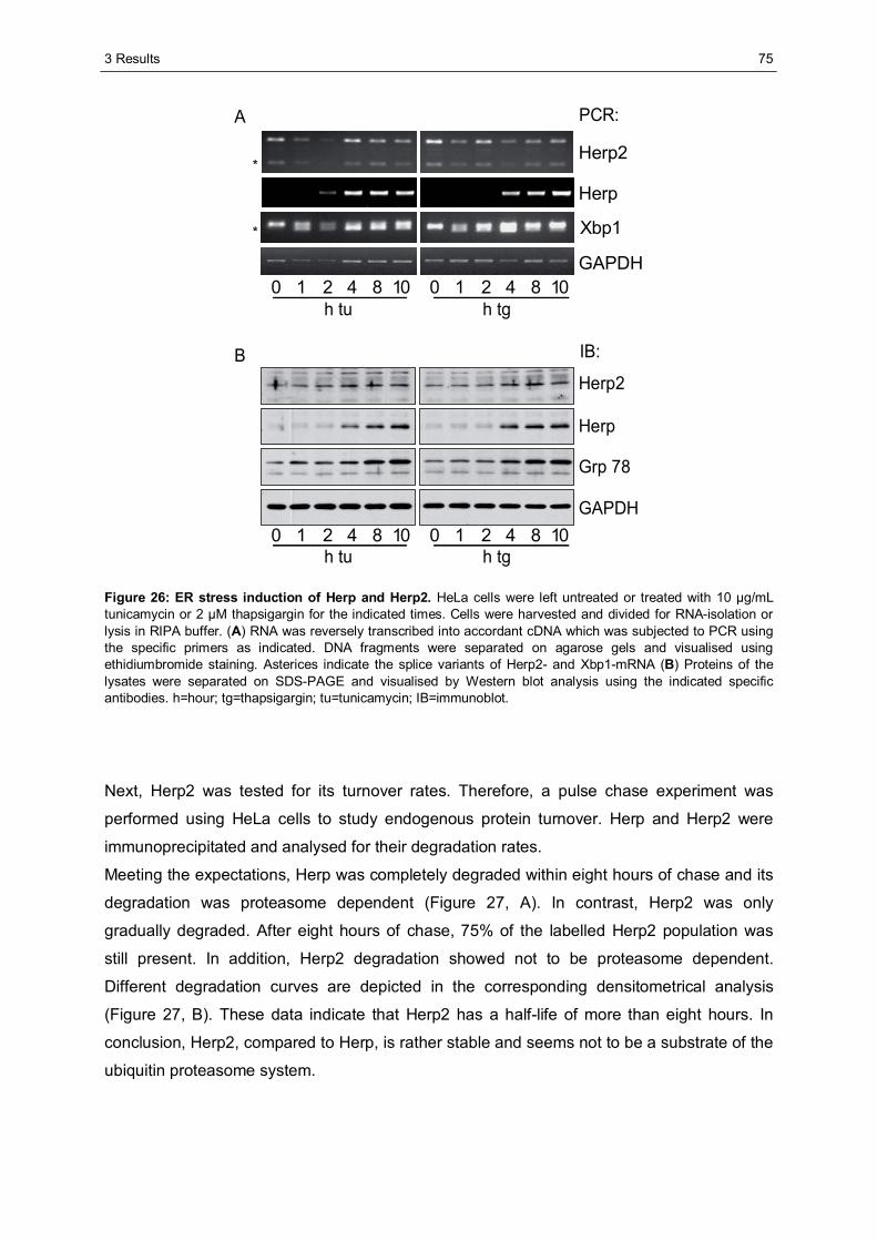
3 Results
75
GAPDH
Herp
h tu h tg
PCR:A
h tu h tg
Herp
Grp 78
GAPDH
B IB:
Herp2
Xbp1
Herp2
0 421 8 10 0 421 8 10
0 421 8 10 0 421 8 10
*
*
Figure 26: ER stress induction of Herp and Herp2. HeLa cells were left untreated or treated with 10 μg/mL tunicamycin or 2 μM thapsigargin for the indicated times. Cells were harvested and divided for RNA-isolation or lysis in RIPA buffer. (A) RNA was reversely transcribed into accordant cDNA which was subjected to PCR using the specific primers as indicated. DNA fragments were separated on agarose gels and visualised using ethidiumbromide staining. Asterices indicate the splice variants of Herp2- and Xbp1-mRNA (B) Proteins of the lysates were separated on SDS-PAGE and visualised by Western blot analysis using the indicated specific antibodies. h=hour; tg=thapsigargin; tu=tunicamycin; IB=immunoblot.
Next, Herp2 was tested for its turnover rates. Therefore, a pulse chase experiment was
performed using HeLa cells to study endogenous protein turnover. Herp and Herp2 were
immunoprecipitated and analysed for their degradation rates.
Meeting the expectations, Herp was completely degraded within eight hours of chase and its
degradation was proteasome dependent (Figure 27, A). In contrast, Herp2 was only
gradually degraded. After eight hours of chase, 75% of the labelled Herp2 population was
still present. In addition, Herp2 degradation showed not to be proteasome dependent.
Different degradation curves are depicted in the corresponding densitometrical analysis
(Figure 27, B). These data indicate that Herp2 has a half-life of more than eight hours. In
conclusion, Herp2, compared to Herp, is rather stable and seems not to be a substrate of the
ubiquitin proteasome system.
3 Results
76
IP:
Herp
Herp2
A
BMG 132h0 421 8 8
- - - - - +
0
25
50
75
100
0 2 4 8
HerpHerp2
h chase
% re
mai
n. p
rote
in
Figure 27: Degradation of Herp and Herp2. (A) HeLa cells were metabolically labelled using 35S-methionine and lysed at the indicated time points in RIPA buffer. Herp or Herp2 were immunoprecipitated (IP) using specific antibodies. MG132 was added to a final concentration of 10 μM where indicated. Precipitated proteins were separated on SDS-PAGE and visualised by autoradiography. (B) Densitometrical analyis of (A). For evaluation of protein degradation, the signal intensities of Herp or Herp2 ‘0 h’ samples were set as 100%.
Taking the data on cellular Herp2 turnover together, Herp2 is not induced by ER stress but
continuously expressed. Furthermore, Herp2 is stable and not degraded by the UPS. These
findings indicate that Herp2 does not reveal the same dynamics as Herp, which distinguish
Herp from other proteins implicated in Synoviolin mediated ERAD.
3.3.2 Herp and Herp2 form homo- and heterooligomers
Recent experiments revealed that the precipitation of HTB-tagged Herp from cells led to the
co-precipitation of endogenous Herp indicating that Herp is able to homooligomerise.
Furthermore, these experiments also indicated that Herp2 binds to Herp (M. Seeger,
personal communication). These findings suggested that Herp and Herp2 might be
associated with Synoviolin concomitantly. Therefore, in the study presented here it was
tested whether Herp and Herp2 are associated with each other and whether each of these
UDPs is able to homooligomerise. Lysates from cells expressing either HTB-tagged Herp or
Herp2 only or together with untagged Herp or Herp2 were subjected to streptavidin agarose
precipitation. Due to the size differences of tagged and untagged proteins, oligomer
formation of the UDPs could be visualised analogous to Synoviolin oligomerisation (3.2.1).
Lysates and precipitates were analysed by Western blotting.
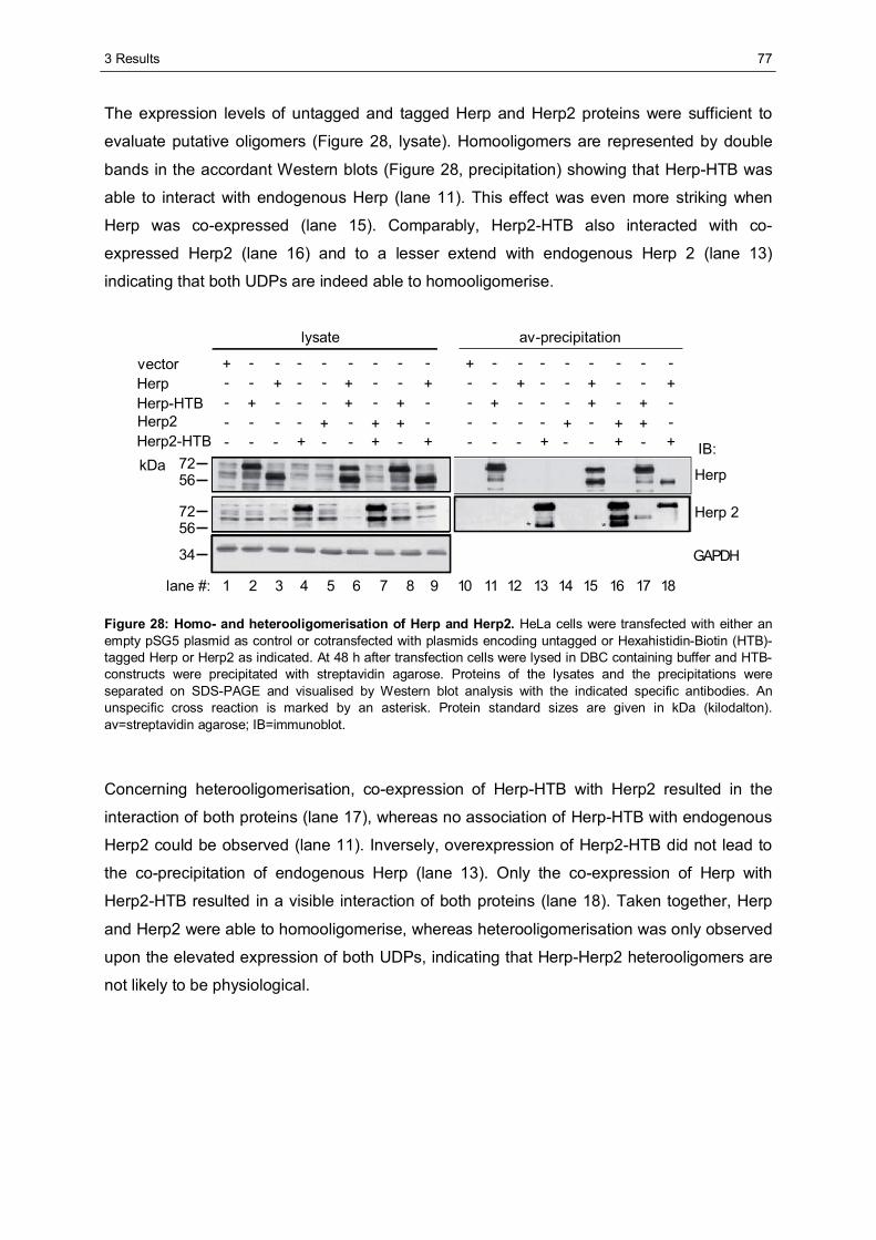
3 Results
77
The expression levels of untagged and tagged Herp and Herp2 proteins were sufficient to
evaluate putative oligomers (Figure 28, lysate). Homooligomers are represented by double
bands in the accordant Western blots (Figure 28, precipitation) showing that Herp-HTB was
able to interact with endogenous Herp (lane 11). This effect was even more striking when
Herp was co-expressed (lane 15). Comparably, Herp2-HTB also interacted with co-
expressed Herp2 (lane 16) and to a lesser extend with endogenous Herp 2 (lane 13)
indicating that both UDPs are indeed able to homooligomerise.
IB:
Herp
Herp 2
GAPDH
lysate av-precipitation
vectorHerpHerp-HTBHerp2Herp2-HTB
321 4 5 6 987 10 11 13 14 15 16 17 1812
--+ - - - ---+-- - - + +---+- - - + -+---- - + - -++--- + - - +-+
--+ - - - ---+-- - - + +---+- - - + -+---- - + - -++--- + - - +-+
7256
7256
34
kDa
lane #:
Figure 28: Homo- and heterooligomerisation of Herp and Herp2. HeLa cells were transfected with either an empty pSG5 plasmid as control or cotransfected with plasmids encoding untagged or Hexahistidin-Biotin (HTB)-tagged Herp or Herp2 as indicated. At 48 h after transfection cells were lysed in DBC containing buffer and HTB-constructs were precipitated with streptavidin agarose. Proteins of the lysates and the precipitations were separated on SDS-PAGE and visualised by Western blot analysis with the indicated specific antibodies. An unspecific cross reaction is marked by an asterisk. Protein standard sizes are given in kDa (kilodalton). av=streptavidin agarose; IB=immunoblot.
Concerning heterooligomerisation, co-expression of Herp-HTB with Herp2 resulted in the
interaction of both proteins (lane 17), whereas no association of Herp-HTB with endogenous
Herp2 could be observed (lane 11). Inversely, overexpression of Herp2-HTB did not lead to
the co-precipitation of endogenous Herp (lane 13). Only the co-expression of Herp with
Herp2-HTB resulted in a visible interaction of both proteins (lane 18). Taken together, Herp
and Herp2 were able to homooligomerise, whereas heterooligomerisation was only observed
upon the elevated expression of both UDPs, indicating that Herp-Herp2 heterooligomers are
not likely to be physiological.
3 Results
78
3.3.3 Herp2 is associated with Synoviolin based complexes
The association with Synoviolin based ERAD complexes is assumed to be a precondition for
Herp acting as an ERAD promoting protein. Therefore, Herp2 was suggested to also be a
part of these ERAD complexes in order to be able to function in the same way as Herp.
Thus, Herp2 may be associated with Synoviolin. To uncover whether this notion holds true,
Herp2 was tested for its co-migration with components of Synoviolin based ERAD complexes
in a density gradient. Therefore, lysates of HeLa cells were loaded on top of a 15-50%
glycerol gradient and subjected to ultracentrifugation. Samples from the resulting fractions
were analysed by Western blotting. This experiment demonstrated that Herp2 co-migrates
with the other tested components of Synoviolin based complexes such as Synoviolin itself,
Herp, p97 and Derlin-1 within the identical fractions of glycerol gradients (Figure 29, A). In
fact, this indicates that Herp2 is a novel component of Synoviolin based complexes.
To test whether Herp2 is associated with Synoviolin, Herp-HTB or Synoviolin-HTB,
expressed in HeLa cells, was subjected to streptavidin agarose precipitation and Western
blot analysis.
With this experiment it was demonstrated that endogenous Synoviolin is associated with
Herp2 and Herp to a comparable extend. In contrast, the Synoviolin homologue Gp78 was
not co-precipitated with Herp or Herp2 indicating that Herp2 specifically interacts with
Synoviolin based ERAD complexes (Figure 29, B). The other way round, when expressing
Synoviolin as HTB-fusion protein, endogenous Herp as well as Herp2 were co-precipitated.
Thus, Herp2 is associated with Synoviolin.
To summarise the third part of this study, Herp2 was shown to be a novel component of
Synoviolin based ERAD complexes. Like Herp, Herp2 is able to homooligomerise and
interacts with Synoviolin. In contrast to Herp, Herp2 expression is not induced by ER stress,
Herp2 is stable and not a substrate of the UPS indicating that Herp2 reveals dynamics
different from Herp. In addition and in contrast to Herp, Herp2 does not bind Usp7. As Herp
and Herp2 did not show an interaction, it is likely that both UDPs exclude each other from
Synoviolin based complexes. Taken together, Herp2 most likely has a function different from
Herp.
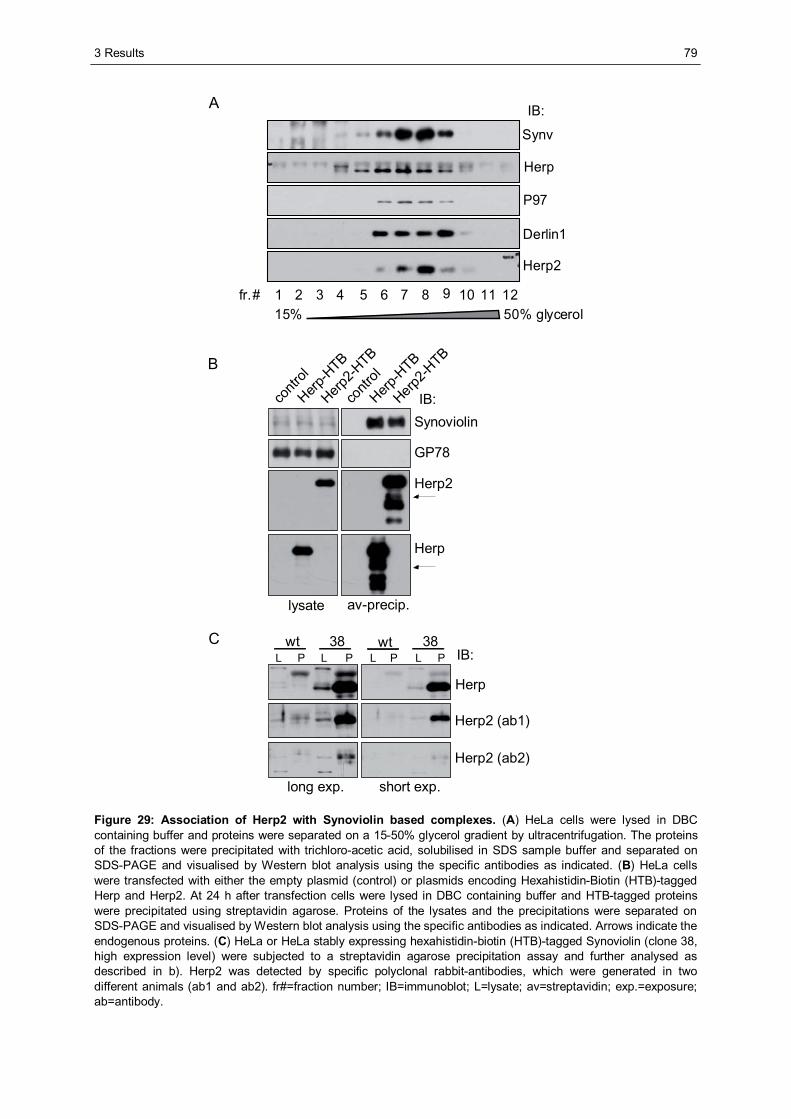
3 Results
79
15% 50% glycerolfr.#
Herp
Synv
12108642
P97
Derlin1
1197531
Herp2
IB:A
lysate av-precip.
contr
ol
Herp-H
TB
Herp2-H
TB
contr
ol
Herp-H
TB
Herp2-H
TB
IB:Synoviolin
GP78
Herp2
Herp
B
C L P L P
wt wt38 38
long exp. short exp.
IB:
Herp
Herp2 (ab1)
Herp2 (ab2)
L P L P
Figure 29: Association of Herp2 with Synoviolin based complexes. (A) HeLa cells were lysed in DBC containing buffer and proteins were separated on a 15-50% glycerol gradient by ultracentrifugation. The proteins of the fractions were precipitated with trichloro-acetic acid, solubilised in SDS sample buffer and separated on SDS-PAGE and visualised by Western blot analysis using the specific antibodies as indicated. (B) HeLa cells were transfected with either the empty plasmid (control) or plasmids encoding Hexahistidin-Biotin (HTB)-tagged Herp and Herp2. At 24 h after transfection cells were lysed in DBC containing buffer and HTB-tagged proteins were precipitated using streptavidin agarose. Proteins of the lysates and the precipitations were separated on SDS-PAGE and visualised by Western blot analysis using the specific antibodies as indicated. Arrows indicate the endogenous proteins. (C) HeLa or HeLa stably expressing hexahistidin-biotin (HTB)-tagged Synoviolin (clone 38, high expression level) were subjected to a streptavidin agarose precipitation assay and further analysed as described in b). Herp2 was detected by specific polyclonal rabbit-antibodies, which were generated in two different animals (ab1 and ab2). fr#=fraction number; IB=immunoblot; L=lysate; av=streptavidin; exp.=exposure; ab=antibody.
4 Discussion
80
4 Discussion
4.1 The role of the dynamics of Herp in ERAD
The Herp protein, one of the ER stress induced components of Synoviolin based ERAD
complexes, plays a crucial role for the effective degradation of misfolded proteins. Herp
protein expression is induced early, whereas other Synoviolin associated ERAD proteins
such as Synoviolin itself or Derlin-1 are induced at later time points upon ER stress (Donati
et al., 2006; Kokame et al., 2000; Kokame et al., 1996; Ma and Hendershot, 2004; Oda et al.,
2006). Some of the Synoviolin associated proteins such as Usp7 and p97 are even not
induced by ER stress, as it has been shown here. Data on ER stress induction of Synoviolin
associated proteins are compiled in Table 16 (Appendix). The only protein that is as fast
induced by ER stress as Herp is the Hsp70-like ER chaperone Grp78 (BiP), which is also
part of Synoviolin complexes (Chigurupati et al., 2009; Donati et al., 2006; Hosokawa et al.,
2008; Kokame et al., 1996). Since Grp78 mediates ERAD substrate recognition and UPR
pathway activation, it acts as a central player at the luminal side of the ERAD machinery (see
1.2.2). The fact that the majority of Herp is located in the cytoplasm indicates that Herp,
according to Grp78, plays an important role at the cytoplasmatic side of the ERAD system
(Kokame et al., 2000).
The half-life of Herp is relatively short compared to half-lives of other Synoviolin associated
proteins such as Synoviolin or Usp7 (Boutell et al., 2005; Kikkert et al., 2004; Sai et al.,
2003). In contrast to Herp, Grp78 is also a stable protein revealing a half-life of about 50
hours (Knittler, 1992). Comparing literature data on ER stress inducibility and stability of
Synoviolin complex components indicates that Herp reveals the highest turnover rates of
Synoviolin complex components (Table 16 / Appendix). The fact that Herp, compared to
other components of Synoviolin based ERAD complexes, shows a different cellular turnover
rate points towards a unique function of Herp in the process of ERAD.
4.1.1 The turnover of Herp at Synoviolin based ERAD complexes
As Herp protein expression is induced earlier upon ER stress than the expression of other
Synoviolin associated proteins, Herp was reasoned to be a prerequisite for the assembly of
Synoviolin based complexes. If Herp would act as an assembly factor, it should be found
exclusively associated with assembling precursor complexes. The function as such an
assembly factor has been reported for another protein, the proteasome maturation protein
(POMP). POMP mediates the assembly of the 20S proteasome and is associated only with
precursor complexes. Once matured, 20S proteasomes degrade POMP (Griffin et al., 2000).
4 Discussion
81
However, the work presented here showed that Herp is not exclusively associated with de
novo synthesised Synoviolin but can also be found with mature populations of this E3 ligase.
Therefore, Herp acts not only as an assembly factor like POMP, but very likely also has a
role for the readily assembled Synoviolin based complexes.
To evaluate whether Herp is associated with functioning ERAD complexes, its interaction
with the AAA-ATPase p97 was tested in the study here. Ye and colleagues found that p97 is
able to extract a ubiquitinated substrate from the ER membrane, only if this substrate enters
the cytoplasmatic side through the process of retrotranslocation (Ye et al., 2003).
Furthermore, Cdc48p, the yeast homologue of p97, was also demonstrated to associate
exclusively with functional Hrd1p based ERAD complexes (Gauss et al., 2006). These
findings lead to the conclusion that p97 is an indicator for readily assembled and functional
ERAD complexes.
The study presented here in fact indicated that Herp and p97 bind to Synoviolin
simultaneously. It was also shown that Herp populations are subject to exchange at
Synoviolin complexes. Furthermore, co-precipitation experiments using Herp-ZZ revealed
that p97 is associated with Herp (Schulze et al., 2005). The demonstrated interaction of Herp
with p97 excludes Herp from being restricted to precursor complexes as in case of POMP,
and suggests that Herp acts during the process of ERAD. Nevertheless, as the UDP was
found to be associated with de novo synthesised Synoviolin, Herp might also assist
Synoviolin complex assembly. In addition, the fact that Herp molecules are continuously
replaced at Synoviolin indicates that the presence of Herp at the ERAD machinery is
required for protein processing. Apart from Herp, no other protein being exchanged at
Synoviolin complexes is known. Only ERAD substrates such as NHK that transiently interact
with these complexes have been described (Hosokawa et al., 2008).
The continuous turnover of a protein at an enzymatic complex is characteristic for substrates,
which associate transiently with the enzyme and after being processed are substituted by the
next substrate molecules. Such a transient association of an ERAD substrate with the
Synoviolin complex has been shown for NHK, which associates with hXTP3B, a component
of Synoviolin based ERAD complexes (Hosokawa et al., 2008). Similar to NHK, Herp was
assumed to be a substrate of Synoviolin complexes, since Herp also transiently interacts with
Synoviolin. Based on this hypothesis, degradation of Herp should be impaired upon
disruption of major components of the Synoviolin complex.
However, the study presented here showed that neither the reduced expression of the
central ERAD complex components Sel1L and Synoviolin nor the elevated expression of the
dominant negative inactive E2 enzyme Ube2j1 affected the degradation of Herp. Other
studies revealed that the depletion of the central ERAD complex components Synoviolin and
4 Discussion
82
Sel1L led to the impaired degradation of the ERAD substrate NHK (Christianson et al., 2008;
Mueller et al., 2008). In addition, the elevated expression of the inactivated Ube2j1 inhibited
the degradation of another ERAD substrate, TCR-α (Lenk et al., 2002). The present study
shows that the elevated expression of both myc-tagged wild type and inactive Ube2j1 led to
a modest stabilisation of Herp. However, this effect is presumably due to the overexpression
of the myc-tagged proteins, as this was also demonstrated for the degradation of MHC class
I molecules (Mueller et al., 2008).
Taken together, the fact that depletion or disturbance of the major ERAD components
Synoviolin and Sel1L or Ube2j1, respectively, affect the stability of known ERAD substrates
but not the stability of Herp strongly suggests that Herp is not a substrate of Synoviolin based
ERAD complexes. Furthermore, Herp is essential for the effective degradation of Synoviolin
dependent ERAD substrates such as NHK, as it has been shown here. If Herp were also a
substrate of Synoviolin, it would act as a potent competitor of NHK and rather impede than
promote its degradation. As this is not the case, this fact also points towards Herp not being
a Synoviolin substrate.
Although ER stress induction significantly enhances the expression of Herp, the degradation
rates of Herp remain unaffected, as it has been shown here and by others (Schulze, 2006).
To ensure this efficient turnover of Herp in an ER stress situation, more than one type of
ERAD complexes might be involved in Herp degradation. In this case, Synoviolin based
complexes can also play a role for the stability of Herp. To finally exclude that Herp
degradation is mediated by Synoviolin or Sel1L in case of ER stress, degradation analyses
after depletion of Synoviolin or Sel1L should be performed under ER stress conditions.
The present study showed that in an ER stress situation the elevated expression of Ube2j1
did not increase the degradation rate of Herp. However, data from the literature showed that
overexpression of Ube2j1 improves the degradation of SGK1, a Synoviolin dependent
substrate (Arteaga et al., 2006). Taken together, the findings from this study and those of the
literature strongly indicate that Herp is not a substrate of Synoviolin based complexes.
Therefore, it is likely that another E3 ligase besides Synoviolin is involved in the degradation
of Herp. In the literature, the two E3 ligases Gp78 and POSH were reported to ubiquitinate
Herp. Li and colleagues showed that recombinant Herp (aa 1-259) was ubiquitinated by the
Synoviolin homologue Gp78 along with the E2 enzyme Ube2g2 in vitro, whereas a K61R
mutant of Herp failed to undergo polyubiquitination (Li et al., 2007). This result indicates that
Gp78 mediates Herp degradation. Interestingly, Gp78 was recently shown to be a substrate
of Synoviolin, demonstrating that Synoviolin regulates the stability of its homologue (Shmueli
et al., 2009). In addition, it was demonstrated that the elevated expression of Synoviolin
leads to the stabilisation of Herp, whereas depletion of Synoviolin reduced cellular Herp
steady state levels (Schulze, 2006).
4 Discussion
83
Taken together, the combination of these findings indicates that Gp78 very likely mediates
the ubiquitination and degradation of Herp.
However, in the study presented here, an association of Herp and Gp78 was not verified. In
line with this observation, other studies also showed that Gp78 is excluded from complexes
containing Sel1L and Synoviolin (Hosokawa et al., 2008). The finding that Gp78 is not
associated with Herp rather argues against Herp being a substrate of Gp78 and Gp78 being
a substrate of Synoviolin. However, these discrepancies of the results can be explained by
the fact that the transient interaction of a substrate with an enzyme might not be detected by
conventional interaction studies. The Plenty of SH3s (POSH) protein is the other E3 ligase
that was reported to ubiquitinate Herp. Once bound to POSH, Herp acts concomitantly as
activator and substrate of POSH. Upon calcium perturbation POSH polyubiquitinates Herp
with lysine-63-type ubiquitin chains. This type of ubiquitin linkage results in the translocation
of Herp from the trans-Golgi network to the ER. In turn, Herp promotes POSH
oligomerisation and activation in a UBL domain dependent manner (Tuvia et al., 2007).
Importantly, the fact that K63-type polyubiquitination does not affect the stability of Herp
excludes POSH as a mediator of Herp degradation. It was demonstrated that, under ER
stress conditions, Herp is predominantly located at the ER membrane (Kokame et al., 2000;
Sai et al., 2003). Since the present study deals with the importance of Herp for ERAD, the
localisation of this protein in proximity to the other ERAD components is directly connected to
its function concerning ERAD. However, a fast availability of Herp through its POSH
mediated redistribution to the ER could enhance its function at ERAD complexes. In
summary, Herp degradation is not mediated by Synoviolin based complexes or POSH. Other
E3 ligases such as Gp78 are likely to execute this function.
4.1.2 Correlation of the turnover of Herp and ERAD substrates
Herp is an instable protein being continuously exchanged at Synoviolin based complexes.
Since elevated expression of the E3 ligases Synoviolin or Gp78 leads to enhanced
degradation of the ERAD substrates Ire1 or CD3-δ (Gao et al., 2008; Zhong et al., 2004), it
was assumed that also an increased availability of Herp within the cell is beneficial for its
function in ERAD.
The work presented here showed that an elevated expression of Herp has no effect on the
degradation of the substrate NHK, whereas depletion of Herp impairs ERAD of NHK. The
observation that elevated Herp levels do not improve ERAD could argue for several
possibilities. The endogenous Herp level could already be sufficient to promote the
degradation process. In this case, Herp would not be the rate-limiting factor for ERAD.
Another possibility could be that the majority of overexpressed Herp protein is not integrated
into Synoviolin complexes and therefore does not provide any function in ERAD.
4 Discussion
84
A third option is that, when overexpressed, the excess of Herp protein is degraded more
efficiently. To address the question of a potential enhanced degradation of overexpressed
Herp, a stabilised Herp was generated and utilised for degradation studies.
As shown in the study presented here, the K61R mutation was sufficient to stabilise Herp. In
addition, Herp-K61R was associated with Synoviolin, Derlin1 and Usp7. Surprisingly, the
elevated expression of Herp-K61R did not improve but slowed down degradation rates of the
ERAD substrate NHK. Impairment of ERAD was also observed upon overexpression of a
dominant negative Herp variant lacking the UBL domain. Sai and colleagues found that Herp
lacking the UBL domain is stable (Sai et al., 2003). In addition, Li and colleagues showed
that lysine 61 is the crucial ubiquitination site of Herp (Li et al., 2007); see 4.1.2). Therefore,
the results from my study confirm that the UBL domain determines the stability of Herp and,
in particular, that lysine 61 is crucial for that determination. Furthermore, the data reveal that
the stability of Herp is positively correlated to the stability of the ERAD substrate. The
stabilisation of Herp impairs the process of ERAD. Taken together, from these data it can be
concluded that the UBL domain of Herp has a crucial role in the degradation of Herp, and
that its degradation rate correlates with its function in ERAD.
The Herp-UBL domain was demonstrated to be fundamental for Herp mediated cellular
processes. Dependent on this domain, Herp promotes ERAD and maintains calcium
homeostasis in stressed neuronal cells. Both functions are important anti-apoptotic
mechanisms (Chigurupati et al., 2009; Schulze et al., 2005). In contrast, within the
pathogenesis of Alzheimer’s disease, Herp promotes amyloid β-protein (Aβ) generation.
Deletion of the Herp UBL domain even enhances Aβ-production, presumably due to the
stabilisation of Herp (Sai et al., 2003). These data strongly suggest that different cellular
functions of Herp are based on its stability, which is determined by its UBL domain. In
general, this argues for the Herp-UBL domain to have a cytoprotective function. Accordingly,
Herp-K61R, which is associated with Synoviolin, is exchanged at Synoviolin based
complexes at lower rates, which could then lead to the impairment of ERAD. The fast
exchange of Herp at Synoviolin presumably determines its proper function in ERAD.
The data presented here on the dynamics of Herp indicate that its function in the process of
Synoviolin mediated ERAD is strongly dependent on its degradation rate. Herp only acts
efficiently in this process, when continuously dissociating from Synoviolin, followed by its
degradation and replacement by newly synthesised Herp molecules. Stabilised Herp very
likely leads to blockage of processes downstream from ERAD substrate retrotranslocation
and ubiquitination. Future studies should address the association dynamics of stabilised
Herp with Synoviolin based complexes in greater detail. In addition, it would be of great
interest to establish an instable Herp and analyse its impact on the ERAD process.
4 Discussion
85
4.2 The importance of Herp for the integrity of Synoviolin based complexes
4.2.1 The impact of Herp on Synoviolin oligomerisation
In vitro interaction studies revealed that Synoviolin molecules are able to interact with each
other suggesting that they are able to form homooligomers (Schulze et al., 2005). In the
study presented here, Synoviolin oligomerisation was demonstrated to occur in Synoviolin-
HTB expressing mammalian cells in vivo. Dependent on the expression level of Synoviolin-
HTB, the ratio of tagged to untagged Synoviolin within an oligomer changed in a reciprocal
correlation. In addition, titration of the plasmid encoding Synoviolin-HTB before transfection
revealed that at least five molecules of wild type Synoviolin were co-precipitated with one
molecule of Synoviolin-HTB. Therefore, at least Synoviolin homohexamers were assumed to
exist physiologically (M. Seeger, personal communication). Also the Synoviolin homologue
Gp78 was demonstrated to form homooligomers. These Gp78 oligomers bind several
molecules of the E2 enzyme Ube2g2 enabling the process of ERAD substrate
polyubiquitination (Li et al., 2009). In addition, the yeast orthologue of Synoviolin, Hrd1p, was
also demonstrated to oligomerise (Horn et al., 2009). However, the oligomer stochiometry of
Gp78 in mammalia or Hrd1p in yeast is not known so far. Nevertheless, the findings on
ERAD E3 ligases’ oligomerisation indicate that the process of homooligomerisation is a
general feature of these enzymes and presumably required for the process of substrate
ubiquitination and retrotranslocation. ER membrane resident oligomers consisting of more
than two molecules of the E3 ligase potentially form a kind of pores that are needed for the
retrotranslocation of ERAD substrates.
Although Synoviolin oligomerisation is evident, it is not known how this process is regulated.
In yeast, the ubiquitin domain protein Usa1p was demonstrated to mediate Hrd1p
oligomerisation (Horn et al., 2009). In addition, Usa1p was shown to link Der1p to Hrd1p,
which is necessary for the formation of a functional Hrd1p based ERAD complex (Carvalho
et al., 2006). Since heterologously expressed Herp partly rescued the Usa1p deletion
phenotype in yeast, Herp was proposed to be the functional equivalent of Usa1p (Carvalho et
al., 2006). These data point towards Herp regulating Synoviolin oligomerisation in mammalia.
However, the study presented here showed that upon elevated expression of dominant
negative HerpΔUBL neither the cellular amount nor the ratio of Synoviolin molecules within
the oligomers was affected. Additionally, depletion of Herp had also no effect on Synoviolin
oligomerisation (data not shown) suggesting that this process is Herp-independent. Horn and
colleagues reported that Usa1p mediated Hrd1p oligomerisation in yeast depends on the
Usa1p N-terminus, which contains a UBL domain (Horn et al., 2009).
4 Discussion
86
However, a general importance of the Usa1p UBL domain for ERAD has not been
demonstrated. A recent study revealed that Usa1p is involved in the ubiquitination of the
soluble model substrate CPY*, independent of the Usa1p UBL domain (Kim et al., 2009).
However, contrary data were reported concerning the significance of the Usa1p UBL domain
for the degradation of membrane derived substrates, as 6myc-Hmg2 but not of Hmg2-GFP
degradation required the Usa1p UBL domain (Carroll and Hampton, 2010; Horn et al., 2009).
In contrast to Usa1p, the Herp UBL domain is dispensable for the oligomerisation of
Synoviolin but required for the degradation of the different Synoviolin dependent ERAD
substrates NHK and CD3- δ. Taken together, Herp and Usa1p differ in essential functions
and are therefore not necessarily functional equivalents. However, their similar domain
architecture could explain why Herp is able to rescue a Usa1p deletion in yeast, at least
partly. Since Herp cannot function analogously to Usa1p, another UDP such as Herp2 (see
4.3) is imaginable to fulfil this function in mammalia.
Herp was indeed shown to be involved in the process of oligomerisation of the soluble E3
ligase POSH. Herp mediated oligomerisation and activation of POSH depends on the UBL
domain of Herp. Following activation, POSH induces Herp translocation to the ER (Tuvia et
al., 2007). Besides this function, POSH also mediates pro-apoptotic signalling by forming a
scaffold for kinases such as JNK (Xu et al., 2003). In contrast, Herp and Synoviolin both
promote ERAD and therefore have anti-apoptotic functions in the cell (Amano et al., 2003;
Chan et al., 2004). The fact that Herp activates a pro-apoptotic protein could be connected to
the hampering of cellular survival after a longer time of persisting ER stress. Subsequently,
apoptotic signalling is activated, whereas cell survival mechanisms are inactivated. This
cellular situation might lead to Herp mediated activation of POSH. However, the action of
Herp on POSH most likely is limited, since apoptosis also leads to caspase mediated Herp
cleavage (Amano et al., 2003; Chan et al., 2004).
To investigate the functional relevance of Herp in Synoviolin mediated ERAD, recent
degradation analyses have been performed using the ERAD model substrate CD3-δ. Herp
protein depletion significantly inhibited the turnover of CD3-δ. Therefore, this substrate is a
suitable model to investigate ERAD (Schulze et al., 2005). However, besides Synoviolin also
other E3 ligases such as Rfp2 and Gp78 mediate CD3-δ degradation (Lerner et al., 2007;
Zhong et al., 2004). Since Synoviolin and Gp78 are homologues, this is an explanation for
the specificity of both E3 ligases for the same substrate. However, as known so far, Herp is
excluded from Gp78 mediated ERAD processes. Thus, an ERAD substrate exclusively
dependent on Herp and Synoviolin was suggested to be inevitable to investigate this
particular ERAD pathway. So far, degradation of NHK, another model ERAD substrate, was
shown to be dependent only on Synoviolin (Christianson et al., 2008).
4 Discussion
87
In contrast to membrane associated CD3-δ, NHK, which is bound to calreticulin, remains
solubilised in the ER lumen before it is degraded (Oda et al., 2003).
The study presented here demonstrated for the first time that Herp is required for the
effective degradation of NHK, which was dependent on the UBL domain of Herp. Therefore,
NHK represents an appropriate substrate to investigate the Synoviolin and Herp dependent
ERAD process in particular. This result seems to contradict data from the literature showing
that a reduced expression of Herp does not interfere with the degradation of NHK (Okuda-
Shimizu and Hendershot, 2007). The oligomerisation of Synoviolin could be an explanation
for this divergent situation. It is conceivable that binding of just one molecule of Herp to a
Synoviolin oligomer is sufficient to maintain the function of the Synoviolin complex. If this is
the case, a reduction of the cellular Herp protein level by just 50% as shown in the
publication of Okuda-Shimizu and colleagues would not be sufficient to observe any effects
on the rate of substrate degradation. However, in contrast to the work of Okuda-Shimizu and
colleagues, my study shows that a depletion of Herp by 50% leads to a delayed degradation
of NHK, a fact that could be due to the different used cell lines. Beyond, as shown here, the
lack or disturbance of the Herp UBL domain results in an almost complete stabilisation of the
substrate verifying that Herp is indeed required for the degradation of ER-luminal NHK.
4.2.2 Herp dependent recruitment of Usp7 to Synoviolin
The UBL domain of Herp determines the stability of this protein (Sai et al., 2003).
Furthermore, the Herp-UBL domain has a crucial role in the process of Synoviolin mediated
ERAD (Schulze, 2006); Kny et al., manuscript). UBL domains of different ubiquitin domain
proteins vary regarding their protein binding specificity. The yeast protein Rad23, e.g., binds
the 26S proteasome and the E4 enzyme Ufd2 via its UBL domain in a mutual exclusive
manner. Hhr23, the human orthologue of Rad23, also binds different proteins such as the
deubiquitinating enzyme Ataxin-3 or the 26S proteasome via its UBL domain (Madsen et al.,
2007). In contrast to Hhr23, the UBL domain of Herp does not interact with the 26S
proteasome (Schulze et al., 2005). Starting with the analysis of the molecular function of
Herp, its UBL domain was assumed to bind proteins that mediate the function of this UDP.
As demonstrated by in vitro and in vivo interaction studies, the deubiquitinating enzyme Usp7
was identified as a target of the Herp UBL domain (Schulze, 2006).
In the work presented here, the specific interaction of endogenous Usp7 with Herp-HTB was
shown. In addition, also the higher molecular weight isoform of Usp7, Usp7β, was found to
bind to Herp-HTB. Initially, Usp7 was stated to only reside in the nucleus and thus, Usp7 was
questionable to be able to interact with the cytoplasmatic UBL domain of Herp under in vivo
conditions.
4 Discussion
88
However, immunofluorescence experiments revealed that a certain amount of Usp7 and the
isoform Usp7β is also present in the cytoplasm (Antrobus and Boutell, 2008; Meredith et al.,
1994). In addition, under certain conditions such as the infection of cells with Herpes Simplex
virus Usp7 can translocate from the nucleus to the cytoplasm (Daubeuf et al., 2009). Thus,
an interaction of cytoplasmatic Usp7 and Herp is generally possible. Since Usp7β is stable
and binds viral proteins such as ICP0, it was suggested to have the same functions as Usp7
(Antrobus and Boutell, 2008). The finding that Usp7β also binds Herp strengthens the results
of Antrobus and Boutell who stated Usp7β to reveal the same binding properties as Usp7.
Taken together, by demonstrating the interaction of endogenous Usp7 with Herp, the study
here confirmed Usp7 to be a target of Herp.
The literature reveals that the P/AXXS motif, a short amino acid sequence that is contained,
e.g., in Mdm2 and p53, mediates Usp7 binding. In close proximity of the P/AXXS motif, with
X indicating any amino acid, several lysines are subject to polyubiquitination, possibly
inhibited by Usp7 (Sheng et al., 2006). Since Herp also directly interacts with Usp7, Herp
was screened for this possible Usp7 interaction site.
Indeed, as demonstrated in the study here, Herp harbours an AHLS motif, which is located
within its UBL domain. This AHLS motif of Herp was found to contribute to the efficient
binding of Herp with Usp7. However, this motif alone was not sufficient to mediate the
interaction of Usp7 and Herp. Mdm2 and p53 both exhibit two closely spaced P/AXXS motifs,
which are involved in Usp7 binding. Beyond that, additional regions might be required to
enable the interaction of Usp7 with Mdm2 or p53 (Sheng et al., 2006). Altogether, these
findings show that a single P/AXXS motif is not sufficient to mediate Usp7 binding. In
conclusion, apart from the AHLS motif, other regions within the Herp UBL domain must
contribute to the binding of Usp7 to Herp.
Usp7 interacts with the UBL domain of Herp, while Synoviolin interacts with a region distal
from the UBL domain (Schulze, 2006; Schulze et al., 2005). Therefore, Usp7 was assumed
to be associated with Synoviolin through its interaction with Herp.
Indeed, the study here demonstrated that the deubiquitinating Usp7 is associated with the
ubiquitin ligase Synoviolin. Furthermore, this interaction showed to be dependent on Herp,
since a marked increase of Usp7 interacting with Synoviolin was observed only after an
increased expression of Herp.
Simultaneous binding of two enzymes with opposing activities to one protein can enable a
fine-tuning of these activities towards the common substrate. This has been shown for, e.g.,
Hdmx, a protein involved in p53 regulation. The binding of both Hdm2, a specific ubiquitin E3
ligase, and Usp7, the ubiquitin hydrolase, to Hdmx enables a tight regulation of the
ubiquitination status of the Hdmx protein. Upon DNA damage, Usp7 dissociates from Hdmx,
which leads to the degradation of both Hdm2 and Hdmx.
4 Discussion
89
Then, Usp7 binds and stabilises p53 (Meulmeester et al., 2005). Coupling ubiquitination and
deubiquitination can positively influence the degradation of a substrate.
Since a ubiquitinating ERAD system is prone to autoubiquitination, single components of this
system might be degraded by the proteasome resulting in ERAD inhibition. For example, the
yeast orthologue of Synoviolin, Hrd1p, is subject to autoubiquitination (Bays et al., 2001).
Then, the ERAD system benefits from a hydrolase, stabilising its components by
deubiquitination. Besides Usp7, other deubiquitinating enzymes have been reported to
stabilise E3 ligases. One example of such stabilising enzymes is Usp15, which antagonises
polyubiquitination of the Cullin E3 ligase component Rbx1 (Hetfeld et al., 2005). Bringing
together Synoviolin and Usp7 at one protein complex points to a tight regulation of the
ubiquitination status of target proteins. Since Usp7 and Synoviolin are part of multiprotein
complexes, their action on separate proteins is also conceivable.
The direct binding of Usp7 to, e.g., p53, Mdm2 and viral ICP0 leads to their deubiquitination
and stabilisation (Canning et al., 2004; Li et al., 2004; Li et al., 2002). Since it binds to Herp,
Usp7 was suggested to also stabilise Herp. In vitro ubiquitination studies and cycloheximide
degradation assays indeed revealed that Usp7 can deubiquitinate and stabilise Herp, if both
proteins are expressed at elevated levels in cells (M. Seeger, personal communication).
Surprisingly, the study presented here demonstrated that neither decrease nor increase of
cellular Usp7 protein affects degradation of endogenous Herp. This discrepancy of the Usp7-
action on endogenous versus exogenous Herp can be explained by the fact that the majority
of overexpressed Herp is not integrated into ERAD complexes but rather accumulates
outside these complexes and becomes ubiquitinated. In this case, the excess of Usp7 could
act on the increased amount of Herp. It is also thinkable that the elevated expression of Usp7
of three fold or the depletion of Usp7 by 50%, as observed in this study, was not sufficient to
affect the turnover of endogenous Herp. However, another study showed that the depletion
of Usp7 by 50% resulted in an enhanced degradation of Alix/HP95, a Usp7 interacting
protein (Kessler et al., 2007). This finding indicates that, if Herp were also a substrate of
Usp7, depletion of Usp7 should result in the stabilisation of Herp, which is not the case.
Taken together, although directly binding to Herp, Usp7 does not stabilise endogenous Herp.
Regarding the functional role of a ubiquitin hydrolase at Synoviolin based complexes, Usp7
was suggested to regulate the degradation of ERAD substrates. Considering the
deubiquitinating activity of Usp7, its binding to an ERAD complex was expected to inhibit or
at least delay substrate degradation.
However, this work here demonstrated that Usp7 does not affect degradation of the ERAD
substrates CD3-δ and NHK. Ubiquitin hydrolases can deubiquitinate ERAD substrates,
4 Discussion
90
a process that needs not necessarily lead to their stabilisation but can also result in their
improved degradation. This phenomenon has been shown for Ataxin-3, a deubiquitinating
enzyme, which shortens the ubiquitin chains of the ERAD substrate TCR-α to an appropriate
size enabling the recognition by downstream components such as p97. Hence, a
deubiquitination step is important for TCR-α extraction from the ER membrane (Wang et al.,
2006). Usp13 is another deubiquitinating enzyme, which was recently found to be associated
with p97 and acts on TCR- α. In contrast to Ataxin-3, Usp13 was demonstrated to inhibit
degradation of TCR-α (Sowa et al., 2009). Thus, Usp13 and Ataxin-3 act on the same
substrate but with the opposite outcome. These data demonstrate that two different
deubiquitinating enzymes acting in one ERAD system determine the fate of the substrate and
introduce a further step of ERAD regulation. Other than Ataxin-3 or Usp13, Usp7 did not
show to influence the degradation of the substrates NHK or CD3-δ indicating that Usp7 does
not regulate the ERAD process. However, the example of the opposing enzymes Ataxin-3
and Usp13 both acting on TCR-α shows that these DUBs exert a very specific and not a
general function towards an ERAD substrate. This finding allows concluding that also Usp7
specifically acts on the degradation of ERAD substrates besides CD3-δ and NHK.
Since Usp7 is associated with Synoviolin based ERAD complexes, it was assumed to be ER
stress induced. However, the study presented here revealed that Usp7 is a stable protein
and that its expression is not induced by ER stress.
Boutell and colleagues also demonstrated that Usp7 is stable, except in case of, e.g., Herpes
Simplex virus infection, when the viral E3 ligase ICP0 targets Usp7 for fast proteasomal
degradation (Boutell et al., 2005). Furthermore, Usp7 was found to be associated with the
26S proteasome (Besche et al., 2009; Bousquet-Dubouch et al., 2009). These data
demonstrate that Usp7 becomes a substrate of the UPS under certain cellular conditions
such as viral infections. The observation that, upon ER stress, a small subpopulation of Usp7
is recruited to Synoviolin based ERAD complexes but not degraded shows that Usp7 does
not serve as a substrate of Synoviolin. The Herp dependent association of Usp7 with
Synoviolin rather points towards a specific role of Usp7 at the ERAD complexes in case of
ER stress. Therefore, Usp7 still might be required for the turnover of Synoviolin dependent
ERAD substrates, which have not been investigated so far. Usp7 is a specific target of the
Herp-UBL domain but does not promote the degradation of the ERAD substrates CD3-δ or
NHK. Therefore, it is conceivable that other target proteins of Herp fulfil this task. Herp
mediated recruitment of further accessory proteins beside Usp7 to Synoviolin could
decisively affect the qualitative and quantitative composition of the ERAD complexes. This
function of Herp is therefore likely to determine the ERAD complexes’ specificity and
function.
4 Discussion
91
Herp is dispensable for the regulation of p53 stability
In 2007, Yamasaki and colleagues reported that ubiquitination and degradation of the tumor-
suppressor protein p53 depends on Synoviolin (Yamasaki et al., 2007). Since Herp binds
Usp7, a well-known regulator of p53, and Synoviolin concomitantly, Herp was hypothesised
to be involved in Synoviolin mediated p53 degradation.
However, Herp was not required for the Synoviolin mediated turnover of p53, as it was
shown in the study here. This finding allows concluding that Herp is required only for the
degradation of a subset of Synoviolin dependent ERAD substrates and not for all substrates
in general. Data from yeast experiments suggested that different kinds of ERAD substrates
are processed by distinct ubiquitination systems, dependent on the location of the misfolded
domain within the substrate. Therefore, ERAD was sub-classified as ‘ERAD-C’ for substrates
harbouring a defect in their cytosolic domain and ‘ERAD-M’ or ‘ERAD-L’ for substrates with a
defect in their transmembrane or luminal domain, respectively. ‘ERAD-C’ substrates are
processed by the E3 ligase Doa10p and associated proteins, whereas ‘ERAD-M’ or ‘ERAD-L’
substrates are ubiquitinated by the Synoviolin orthologue Hrd1p and its interacting proteins
(Carvalho et al., 2006). This classification was suggested to apply also to the mammalian
ERAD systems. However, a simple sub-classification of the mammalian ERAD pathways is
not feasible, since an individual substrate can be processed by different ERAD systems as
shown for CD3-δ, which is ubiquitinated by Synoviolin, Gp78 and Rfp2 (Fang et al., 2001;
Kikkert et al., 2004; Lerner et al., 2007). Moreover, the ubiquitination of different ERAD
substrates such as CD3-δ, p53 and NHK can be performed by the same E3 ligase,
Synoviolin (Christianson et al., 2008; Kikkert et al., 2004; Yamasaki et al., 2007). Thus, it is
conceivable that in mammalian cells rather a complex network of interacting ERAD proteins
decides about processing of an ERAD subtrate. Herp, e.g., could be such a protein required
only for the turnover of Synoviolin substrates that have to be retrotranslocated such as
membrane-associated CD3-δ and ER-luminal NHK. In contrast, Herp seems to be
dispensable for the degradation of substrates that are already cytoplasmatic such as p53.
The degradation of such cytoplasm derived substrates does not require ERAD complex
proteins, which mediate the steps retrotranslocation or extraction. Since these central ERAD
steps are dispensable for p53 degradation, this protein is not a classical ERAD substrate.
Also the existence of distinct cellular pools of p53 that are diversely regulated could be an
explanation for the fact that Herp is not involved in the degradation of p53. Nuclear p53 is
regulated by a couple of proteins such as the E3 ligase Hdm2, Hdmx and Usp7 (see 1.1.5)
and the E3 ligases COP1 and Pirh2 (Brooks and Gu, 2004). Genotoxic stress, e.g., triggers
Hdm2 mediated monoubiquitination of a subpopulation of p53, which translocates to the
mitochondria, where a mitochondrial Usp7 stabilises p53 and promotes the pro-apoptotic
action of p53 (Marchenko et al., 2007).
4 Discussion
92
Furthermore, besides Synoviolin, another E3 ligase, the Parkin like protein Parc, traps p53
and sequesters it in the cytoplasm (Nikolaev et al., 2003). Parc and Synoviolin might serve to
store p53, until cellular conditions such as DNA damage lead to the redistribution of p53 to
the nucleus or to the mitochondria. As p53 is regulated by many other proteins besides
Synoviolin and is located primarily in the nucleus, Synoviolin based ERAD complexes most
likely play a minor role for the regulation of p53 stability.
As a substrate of Synoviolin p53 has been also demonstrated to be associated with this E3
ligase (Yamasaki et al., 2007). Therefore, Herp was assumed to be associated with p53, too.
However, in the study presented here, p53 was not found to interact with Synoviolin or Herp.
This discrepancy between the results presented here and the published data might be due to
the different cell lines used in these different studies. Another explanation for the fact that
p53 was not found to be associated with Synoviolin or Herp could be that the interaction
experiments here were performed after eight hours of ER stress. As prolonged ER stress
induces apoptosis, p53 most likely dissociates from Synoviolin and translocates to its sites of
action in the nucleus and the mitochondria (Li et al., 2006).
4.2.3 Herp dependent association of Synoviolin and AUP1
The ancient ubiquitous protein 1 (AUP1) was identified as a novel protein interacting with
Sel1L (Mueller et al., 2008). Since Sel1L is bound to Synoviolin, AUP1 was assumed to be a
component of Synoviolin based ERAD complexes.
In fact, in vitro interaction analyses of the study presented here demonstrated that AUP1
directly binds to Synoviolin. This finding strongly points towards AUP1 being a new-found
component of Synoviolin based ERAD complexes. Interestingly, the interaction of AUP1 and
Synoviolin was demonstrated to depend on the presence of Herp.
These data indicate that Herp mediates the recruitment of AUP1 to Synoviolin, thereby
regulating the integrity of Synoviolin based ERAD complexes. However, the drastic decrease
of the Herp protein resulted only in a slight decrease of AUP1 associating with Synoviolin.
This effect can be explained by the presence of Synoviolin oligomers. The binding of just one
Herp molecule per Synoviolin oligomer could promote the binding of several AUP1 molecules
per oligomer. Therefore, even reduced Herp levels could be sufficient to enable this
interaction of AUP1 and Synoviolin. Presumably, only the complete depletion of cellular Herp
protein would lead to the loss of AUP1 association with the E3 ligase Synoviolin.
The finding that both, AUP1 and Usp7 are associated with Synoviolin based complexes, in a
Herp dependent manner, demonstrates that Herp is involved in establishing the integrity of
Synoviolin based complexes. Depletion of Herp leads to the loss of Usp7 and AUP1 proteins
from these complexes.
4 Discussion
93
Herp dependent recruitment of accessory proteins to Synoviolin based complexes obviously
occurs in different ways. On the one hand, Herp enables direct binding of proteins with a
component of the ERAD complex, as it was shown for AUP1. On the other hand, Herp
directly binds to proteins and links them to the ERAD complexes, as it was demonstrated for
Usp7. Besides Usp7, Herp also directly binds to Ubiquilin 2 and links this protein to
Synoviolin (Kim et al., 2008). However, this interaction depends on a region between the
amino acids 115 and 200 located distal from the Herp UBL domain and not on the UBL
domain itself. Also ubiquilins harbour a UBL domain, capable of interacting with the
proteasome, and additionally a UBA domain binding to ubiquitin or ubiquitinated substrates
(Kleijnen et al., 2000). Therefore, ubiquilins are able to shuttle ubiquitinated substrates to the
proteasome. In the study of Kim and colleagues, depletion of Ubiquilin 1 and Ubiquilin 2 as
well as overexpression of a truncated dominant negative Herp version lacking the critical
binding region for Ubiquilin 2 led to the impaired degradation of the ERAD substrate CD3-δ
(Kim et al., 2008). These data on the interaction between Herp and Ubiquilin 2 support the
idea that Herp represents an important Synoviolin complex component, which is inevitable
for the recruitment of essential ERAD proteins. Thus, Herp regulates the integrity of ERAD
complexes. To strengthen these findings, it should be further addressed whether the
association of other proteins with Synoviolin can also be subject to Herp regulation. In
addition, it would be important to analyse how an alteration of the stability of Herp influences
its recruitment function. Recruited proteins should be tested for their impact on ERAD.
AUP1 is required for Sel1L mediated dislocation of MHC class I molecules (Mueller et al.,
2008). Since Sel1L is a component of Synoviolin based ERAD complexes and AUP1
interacts with Synoviolin, AUP1 was suggested to generally improve Synoviolin mediated
ERAD.
Indeed, the study presented here demonstrated that AUP1 promotes the degradation of the
Synoviolin and Herp dependent ERAD substrate NHK. This was shown by depletion of the
cellular AUP1 protein using siRNA. In the study of Müller and colleagues a GFP-tag was
fused to AUP1 to disturb protein function. Overexpression of AUP1-GFP resulted in the
decreased dislocation of MHC class I molecules from the ER to the cytoplasm (Mueller et al.,
2008). Thus, both studies demonstrated that AUP1 is an important Synoviolin complex
component, which is required for the effective degradation of substrates.
AUP1 possesses a C-terminal ′coupling of ubiquitin conjugation to ER degradation′ (CUE)
domain, which was discussed to be responsible for the promoting effect of AUP1 on
Synoviolin mediated ERAD. Indeed, the study here showed that disruption of the CUE
domain impaired the function of AUP1 in ERAD.
4 Discussion
94
CUE domains reveal structural homology to ubiquitin associated (UBA) domains and both
bind similarly to a hydrophobic patch within ubiquitin. A number of proteins, which belong to
the network of the ubiquitin proteasome system, contain CUE domains. These proteins
promote their own ubiquitination, as it was shown for the E3 ligase Gp78 (Hurley et al., 2006;
Kang et al., 2003). The CUE domain of Gp78 is involved in the recruitment of the E2 enzyme
Ube2g2 and is therefore essential for the functionality of Gp78. The first identified CUE
domain containing protein in yeast, Cue1p, also recruits the E2 enzyme Ubc7p. Degradation
of the ERAD substrate CD3-δ is impaired upon the elevated expression of a dominant
negative Gp78 lacking the CUE domain (Chen et al., 2006). According to the data of Kang
and colleagues, leucine 333 within the CUE domain was assumed to be essential for
ubiquitin binding (Kang et al., 2003). In the study here, it was shown that the substitution of
this critical lysine 333 by an alanine impaired degradation of NHK. The importance of the
CUE domain might base on its ability to bind ubiquitin, since an in vitro binding study
demonstrated a diminished binding of ubiquitin upon mutation of the CUE domain (M.
Seeger, personal communication). Taken together, these data indicate that AUP1 is involved
in the binding or transfer of ubiquitinated substrates. A study of Morito and colleagues
revealed that the CUE domain of Gp78 is sufficient to bind the substrate CFTRΔF508.
Deletion of the Gp78-CUE domain inhibited its binding of CFTRΔF508, whereas the
introduction of this domain into Synoviolin had the reversed effect and enabled the binding of
CFTRΔF508 with Synoviolin (Morito et al., 2008). Therefore, the CUE domain of Gp78 is not
only able to bind ubiquitin but also ubiquitinated ERAD substrates. In addition, the same
study showed that the E3 ligase Gp78 exerts E4 activity, which is also mediated by its CUE
domain. Based on its CUE domain, the functional role of AUP1 in ERAD can be diverse, as
this is supported by the fact that the CUE domain of Gp78 has various functions. AUP1 might
provide ubiquitin, recruit the E2 enzyme or the substrate, or could be involved in ubiquitin
chain elongation, all processes that enhance ERAD.
Taken together, it was demonstrated here that the UDP Herp is essential for maintaining the
integrity of Synoviolin based complexes. Although Herp is dispensable for the process of
Synoviolin oligomerisation, it was shown to have a decisive role for determining the
composition of Synoviolin based complexes by recruiting accessory proteins. Herp either
binds directly to these proteins, as in case of Usp7, and thereby provides a link to Synoviolin
or enables their binding to one of the complex components, as this has been shown for
AUP1. Herp mediated regulation of the composition of Synoviolin based complexes very
likely is crucial for the function of these complexes in ERAD.
4 Discussion
95
4.3 Comparison of Herp and Herp2
The ubiquitin domain proteins Herp and Herp2 belong to the mubl protein family (1.2.6) and
reveal 40% (their UBL domains even 50%) amino acid sequence identity. Based on these
similarities, Herp2 was suggested to be a paralouge of Herp. Thus, a central question of the
study presented here was whether Herp2 is able to take over the functional role of Herp
within cells. Therefore, Herp2 was hypothesised to also associate with Synoviolin.
Indeed, the data presented here demonstrated that Herp2 interacts with Synoviolin. In
addition, Herp2 was demonstrated to be a component of Synoviolin based ERAD complexes.
Together, these findings point towards Herp2 being a component of Synoviolin based ERAD
complexes. However, these data did not reveal whether Herp and Herp2 are integrated in the
same Synoviolin based ERAD complex simultaneously.
The data presented here showed that Herp and Herp2 are not associated with each other
and therefore are not integrated in the same complex. Only in case of the elevated
expression of Herp and Herp2 both proteins interact with each other, a fact that is unlikely to
represent the physiological situation. However, these interaction studies also revealed that
both Herp and Herp2 have the ability to homooligomerise.
The finding that Herp does not bind to Herp2 indicates that these UDPs exclude each other
from the same Synoviolin complex. This fact points towards the existence of at least two
types of Synoviolin based complexes, harbouring either Herp or Herp2. These different kinds
of ERAD complexes potentially exert different functions in the cell, e.g., processing of
selected ERAD substrates. These data also suggest that Herp and Herp2 compete for direct
binding to Synoviolin. In this case, the composition of the ERAD complexes would depend on
the cellular concentration and relation of these two UDPs. The analysis of protein expression
of both UDPs revealed that Herp2 is predominantly expressed in brain, spleen and thymus,
whereas Herp is ubiquitously expressed (Kokame et al., 2000; Schulze, 2006). Therefore, it
is conceivable that in Herp2 expressing tissues the majority of Synoviolin based ERAD
complexes contains Herp2 instead of Herp. It remains open whether Herp2 also directly
binds to Synoviolin or to another component of these multiprotein complexes.
In contrast to heterooligomerisation, homooligomerisation of Herp or Herp2 is likely to occur
at physiological conditions, since the endogenous protein was experimentally co-precipitated
with a HTB-tagged protein. This finding indicates that homooligomeric substructures
consisting of Herp or Herp2 can interact with Synoviolin based complexes. Very likely, the
amount of Herp or Herp2 molecules bound to one Synoviolin oligomer determines the
function of the whole ERAD complex. In general, oligomerisation seems to be a widespread
feature of components of Synoviolin based complexes. Besides Herp, Herp2 and Synoviolin
also the Derlin-proteins and p97 were shown to oligomerise.
4 Discussion
96
The AAA-ATPase p97 forms homohexamers (Ogura and Wilkinson, 2001). Derlin-1 was
shown to homooligomerise (Ye et al., 2005). In addition, Derlin-1, -2 and -3 were shown to
form mixed Derlin-oligomers (Lilley and Ploegh, 2005). The authors therefore suggested that
the Derlin proteins, by oligomerisation, fulfil the requirements to form a pore that enables
retrotranslocation. Generally, association of potentially ring shaped oligomeric structures
could enable the formation of a retrotranslocation pore that is suitable for substrates of even
larger size. Using structural analysis by, e.g., electron microscopy, the composition,
arrangements, and stochiometry of these high molecular weight ERAD complexes could be
analysed in detail.
Besides the strong similarities between Herp and Herp2 also considerable differences of
both UDPs were found. In contrast to Herp, Herp2 expression was increased by the ER
stress inducing agent β-mercaptoethanol (Schulze, 2006).
As also presented in this work here, the ER stress induction by disturbance of glycosylation
or calcium homeostasis did not increase Herp2 expression. In addition, it was demonstrated
that Herp2 is a stable protein, which is not subject to proteasomal degradation. Moreover,
Herp2 did not bind Usp7.
As shown here, ER stress treatment of the cells led to the detection of a minor increase of
the Herp2 protein level. However, as the accordant Herp2 mRNA levels were not enhanced
upon ER stress treatment, the slightly increased Herp2 protein level cannot be attributed to
an induction of protein expression but can rather be explained by a diminished degradation
and accumulation of the protein. Herp2, in contrast to Herp, is degraded slowly and
proteasome-independent indicating that the stability of these UDPs is regulated by different
degradation systems. In contrast to Herp, Herp2 protein expression is not induced by ER
stress and Herp2 turnover is markedly delayed. These findings point towards the different
dynamics of these UDPs that distinguish Herp and Herp2 decisively. Thus, it is likely that
Herp2 is not capable of taking over the function of Herp in ERAD. Although the UBL domains
of Herp and Herp2 show a significant similarity, Herp2 does not bind Usp7 supporting the
notion that different UBL domains have binding specificities (4.2.2).
Herp2 might be a candidate which represents the functional analogue of the yeast ubiquitin
domain protein Usa1p. Usa1p was shown to link Der1p to Hrd1p and enables Hrd1p
oligomerisation thereby ascertaining the ERAD complex integrity (Carvalho et al., 2006; Horn
et al., 2009). Herp2 and Usa1p reveal similar domain architectures and it was recently
demonstrated that Usa1p as Herp2 is stable supporting the idea that these proteins are
indeed functional analogues (Kim et al., 2009). Thus, it would be interesting to address
whether Herp2 has an impact on the ERAD process in mammalia.
4 Discussion
97
In summary, the findings on Herp2 reveal remarkable differences of Herp and Herp2
suggesting that these UDPs are not able to substitute each other in the cell. Although both
ubiquitin domain proteins are components of Synoviolin based ERAD complexes, it is evident
that the role of Herp2 within these complexes differs from that of Herp, a fact that underlines
the peculiarity of Herp.
4.4 Conclusion
This study reveals insight into the molecular function and characteristics of Herp within
Synoviolin mediated ERAD. It was shown that Herp is dynamically exchanged at Synoviolin
based complexes due to its high degradation and synthesis rates in an ER stress situation.
The turnover of Herp seems to be essential for its function in ERAD since both stabilisation
and knockdown of Herp led to an impaired degradation of Synoviolin substrates. Although
behaving like a substrate itself, Herp was not processed by Synoviolin complex components.
Additionally, Herp mediated the recruitment of accessory proteins to Synoviolin. Whereas
Usp7 - directly bound to the Herp UBL domain - had no influence on ERAD of CD3-δ or
NHK, AUP1 - directly interacting with Synoviolin - positively affected the degradation of NHK,
dependent on its CUE domain. Since Herp directly influenced the ERAD complex
composition, this UDP most likely functions at the level of ubiquitination and
retrotranslocation within ERAD. Herp2 is also associated with Synoviolin based complexes,
but reveals different characteristics than Herp and is therefore proposed to exert different
functions in ERAD, presumably in Synoviolin complexes lacking Herp. As Herp2 is unable to
substitute Herp, this supports the unique role of Herp in Synoviolin mediated ERAD. Taken
together, the findings of the study presented here underline the extraordinary role of Herp in
the process of Synoviolin mediated ERAD which is essential for cell survival.
Literature
98
Literature
Allen, J. R., Nguyen, L. X., Sargent, K. E., Lipson, K. L., Hackett, A., and Urano, F. (2004). High ER stress in beta-cells stimulates intracellular degradation of misfolded insulin. Biochem Biophys Res Commun 324, 166-170.
Amano, T., Yamasaki, S., Yagishita, N., Tsuchimochi, K., Shin, H., Kawahara, K., Aratani, S., Fujita, H., Zhang, L., Ikeda, R., et al. (2003). Synoviolin/Hrd1, an E3 ubiquitin ligase, as a novel pathogenic factor for arthropathy. Genes Dev 17, 2436-2449.
Antrobus, R., and Boutell, C. (2008). Identification of a novel higher molecular weight isoform of USP7/HAUSP that interacts with the Herpes simplex virus type-1 immediate early protein ICP0. Virus Res 137, 64-71.
Arteaga, M. F., Wang, L., Ravid, T., Hochstrasser, M., and Canessa, C. M. (2006). An amphipathic helix targets serum and glucocorticoid-induced kinase 1 to the endoplasmic reticulum-associated ubiquitin-conjugation machinery. Proc Natl Acad Sci U S A 103, 11178-11183.
Ballar, P., Shen, Y., Yang, H., and Fang, S. (2006). The role of a novel p97/valosin-containing protein-interacting motif of gp78 in endoplasmic reticulum-associated degradation. J Biol Chem 281, 35359-35368.
Ballinger, C. A., Connell, P., Wu, Y., Hu, Z., Thompson, L. J., Yin, L. Y., and Patterson, C. (1999). Identification of CHIP, a novel tetratricopeptide repeat-containing protein that interacts with heat shock proteins and negatively regulates chaperone functions. Mol Cell Biol 19, 4535-4545.
Bays, N. W., Gardner, R. G., Seelig, L. P., Joazeiro, C. A., and Hampton, R. Y. (2001). Hrd1p/Der3p is a membrane-anchored ubiquitin ligase required for ER-associated degradation. Nat Cell Biol 3, 24-29.
Bernardi, K. M., Forster, M. L., Lencer, W. I., and Tsai, B. (2008). Derlin-1 facilitates the retro-translocation of cholera toxin. Mol Biol Cell 19, 877-884.
Besche, H. C., Haas, W., Gygi, S. P., and Goldberg, A. L. (2009). Isolation of mammalian 26S proteasomes and p97/VCP complexes using the ubiquitin-like domain from HHR23B reveals novel proteasome-associated proteins. Biochemistry 48, 2538-2549.
Bousquet-Dubouch, M. P., Baudelet, E., Guerin, F., Matondo, M., Uttenweiler-Joseph, S., Burlet-Schiltz, O., and Monsarrat, B. (2009). Affinity purification strategy to capture human endogenous proteasome complexes diversity and to identify proteasome-interacting proteins. Mol Cell Proteomics 8, 1150-1164.
Boutell, C., Canning, M., Orr, A., and Everett, R. D. (2005). Reciprocal activities between herpes simplex virus type 1 regulatory protein ICP0, a ubiquitin E3 ligase, and ubiquitin-specific protease USP7. J Virol 79, 12342-12354.
Brooks, C. L., and Gu, W. (2004). Dynamics in the p53-Mdm2 ubiquitination pathway. Cell Cycle 3, 895-899.
Canning, M., Boutell, C., Parkinson, J., and Everett, R. D. (2004). A RING finger ubiquitin ligase is protected from autocatalyzed ubiquitination and degradation by binding to ubiquitin-specific protease USP7. J Biol Chem 279, 38160-38168.
Caramelo, J. J., and Parodi, A. J. (2008). Getting in and out from calnexin/calreticulin cycles. J Biol Chem 283, 10221-10225.
Carroll, S. M., and Hampton, R. Y. (2010). Usa1p is required for optimal function and regulation of the Hrd1p endoplasmic reticulum-associated degradation ubiquitin ligase. J Biol Chem 285, 5146-5156.
Carvalho, P., Goder, V., and Rapoport, T. A. (2006). Distinct ubiquitin-ligase complexes define convergent pathways for the degradation of ER proteins. Cell 126, 361-373.
Cattaneo, M., Lotti, L. V., Martino, S., Cardano, M., Orlandi, R., Mariani-Costantini, R., and Biunno, I. (2009). Functional characterization of two secreted SEL1L isoforms capable of exporting unassembled substrate. J Biol Chem 284, 11405-11415.
Cattaneo, M., Otsu, M., Fagioli, C., Martino, S., Lotti, L. V., Sitia, R., and Biunno, I. (2008). SEL1L and HRD1 are involved in the degradation of unassembled secretory Ig-mu chains. J Cell Physiol 215, 794-802.
Chan, S. L., Fu, W., Zhang, P., Cheng, A., Lee, J., Kokame, K., and Mattson, M. P. (2004). Herp stabilizes neuronal Ca2+ homeostasis and mitochondrial function during endoplasmic reticulum stress. J Biol Chem 279, 28733-28743.
Chau, V., Tobias, J. W., Bachmair, A., Marriott, D., Ecker, D. J., Gonda, D. K., and Varshavsky, A. (1989). A multiubiquitin chain is confined to specific lysine in a targeted short-lived protein. Science 243, 1576-1583.
Chen, B., Mariano, J., Tsai, Y. C., Chan, A. H., Cohen, M., and Weissman, A. M. (2006). The activity of a human endoplasmic reticulum-associated degradation E3, gp78, requires its Cue domain, RING finger, and an E2-binding site. Proc Natl Acad Sci U S A 103, 341-346.
Cheon, K. W., and Baek, K. H. (2006). HAUSP as a therapeutic target for hematopoietic tumors (review). Int J Oncol 28, 1209-1215.
Literature
99
Chigurupati, S., Wei, Z., Belal, C., Vandermey, M., Kyriazis, G. A., Arumugam, T. V., and Chan, S. L. (2009). The homocysteine-inducible endoplasmic reticulum stress protein counteracts calcium store depletion and induction of CCAAT enhancer-binding protein homologous protein in a neurotoxin model of Parkinson disease. J Biol Chem 284, 18323-18333.
Christianson, J. C., Shaler, T. A., Tyler, R. E., and Kopito, R. R. (2008). OS-9 and GRP94 deliver mutant alpha1-antitrypsin to the Hrd1-SEL1L ubiquitin ligase complex for ERAD. Nat Cell Biol 10, 272-282.
Ciechanover, A., Heller, H., Elias, S., Haas, A. L., and Hershko, A. (1980). ATP-dependent conjugation of reticulocyte proteins with the polypeptide required for protein degradation. Proc Natl Acad Sci U S A 77, 1365-1368.
Ciehanover, A., Hod, Y., and Hershko, A. (1978). A heat-stable polypeptide component of an ATP-dependent proteolytic system from reticulocytes. Biochem Biophys Res Commun 81, 1100-1105.
Cummins, J. M., and Vogelstein, B. (2004). HAUSP is required for p53 destabilization. Cell Cycle 3, 689-692.
Dai, R. M., Chen, E., Longo, D. L., Gorbea, C. M., and Li, C. C. (1998). Involvement of valosin-containing protein, an ATPase Co-purified with IkappaBalpha and 26 S proteasome, in ubiquitin-proteasome-mediated degradation of IkappaBalpha. J Biol Chem 273, 3562-3573.
Dai, R. M., and Li, C. C. (2001). Valosin-containing protein is a multi-ubiquitin chain-targeting factor required in ubiquitin-proteasome degradation. Nat Cell Biol 3, 740-744.
Daubeuf, S., Singh, D., Tan, Y., Liu, H., Federoff, H. J., Bowers, W. J., and Tolba, K. (2009). HSV ICP0 recruits USP7 to modulate TLR-mediated innate response. Blood 113, 3264-3275.
Denic, V., Quan, E. M., and Weissman, J. S. (2006). A luminal surveillance complex that selects misfolded glycoproteins for ER-associated degradation. Cell 126, 349-359.
Deshaies, R. J., and Joazeiro, C. A. (2009). RING domain E3 ubiquitin ligases. Annu Rev Biochem 78, 399-434.
Donati, G., Imbriano, C., and Mantovani, R. (2006). Dynamic recruitment of transcription factors and epigenetic changes on the ER stress response gene promoters. Nucleic Acids Res 34, 3116-3127.
Dubiel, W., and Gordon, C. (1999). Ubiquitin pathway: another link in the polyubiquitin chain? Curr Biol 9, R554-557.
Ellgaard, L., and Helenius, A. (2003). Quality control in the endoplasmic reticulum. Nat Rev Mol Cell Biol 4, 181-191.
Everett, R. D., Meredith, M., Orr, A., Cross, A., Kathoria, M., and Parkinson, J. (1997). A novel ubiquitin-specific protease is dynamically associated with the PML nuclear domain and binds to a herpesvirus regulatory protein. Embo J 16, 566-577.
Fang, S., Ferrone, M., Yang, C., Jensen, J. P., Tiwari, S., and Weissman, A. M. (2001). The tumor autocrine motility factor receptor, gp78, is a ubiquitin protein ligase implicated in degradation from the endoplasmic reticulum. Proc Natl Acad Sci U S A 98, 14422-14427.
Freemont, P. S., Hanson, I. M., and Trowsdale, J. (1991). A novel cysteine-rich sequence motif. Cell 64, 483-484.
Gao, B., Lee, S. M., Chen, A., Zhang, J., Zhang, D. D., Kannan, K., Ortmann, R. A., and Fang, D. (2008). Synoviolin promotes IRE1 ubiquitination and degradation in synovial fibroblasts from mice with collagen-induced arthritis. EMBO Rep 9, 480-485.
Gauss, R., Sommer, T., and Jarosch, E. (2006). The Hrd1p ligase complex forms a linchpin between ER-lumenal substrate selection and Cdc48p recruitment. Embo J 25, 1827-1835.
Glickman, M. H., Rubin, D. M., Fried, V. A., and Finley, D. (1998). The regulatory particle of the Saccharomyces cerevisiae proteasome. Mol Cell Biol 18, 3149-3162.
Griffin, T. A., Slack, J. P., McCluskey, T. S., Monaco, J. J., and Colbert, R. A. (2000). Identification of proteassemblin, a mammalian homologue of the yeast protein, Ump1p, that is required for normal proteasome assembly. Mol Cell Biol Res Commun 3, 212-217.
Groll, M., Ditzel, L., Lowe, J., Stock, D., Bochtler, M., Bartunik, H. D., and Huber, R. (1997). Structure of 20S proteasome from yeast at 2.4 A resolution. Nature 386, 463-471.
Guan, K. L., and Dixon, J. E. (1991). Eukaryotic proteins expressed in Escherichia coli: an improved thrombin cleavage and purification procedure of fusion proteins with glutathione S-transferase. Anal Biochem 192, 262-267.
Hampton, R. Y., Gardner, R. G., and Rine, J. (1996). Role of 26S proteasome and HRD genes in the degradation of 3-hydroxy-3-methylglutaryl-CoA reductase, an integral endoplasmic reticulum membrane protein. Mol Biol Cell 7, 2029-2044.
Harding, H. P., Zhang, Y., and Ron, D. (1999). Protein translation and folding are coupled by an endoplasmic-reticulum-resident kinase. Nature 397, 271-274.
Literature
100
Hayden, M. R., Tyagi, S. C., Kerklo, M. M., and Nicolls, M. R. (2005). Type 2 diabetes mellitus as a conformational disease. Jop 6, 287-302.
Hershko, A., and Ciechanover, A. (1998). The ubiquitin system. Annu Rev Biochem 67, 425-479.
Hershko, A., Heller, H., Elias, S., and Ciechanover, A. (1983). Components of ubiquitin-protein ligase system. Resolution, affinity purification, and role in protein breakdown. J Biol Chem 258, 8206-8214.
Hetfeld, B. K., Bech-Otschir, D., and Dubiel, W. (2005). Purification method of the COP9 signalosome from human erythrocytes. Methods Enzymol 398, 481-491.
Hicke, L. (2001). Protein regulation by monoubiquitin. Nat Rev Mol Cell Biol 2, 195-201.
Hiller, M. M., Finger, A., Schweiger, M., and Wolf, D. H. (1996). ER degradation of a misfolded luminal protein by the cytosolic ubiquitin-proteasome pathway. Science 273, 1725-1728.
Hiyama, H., Yokoi, M., Masutani, C., Sugasawa, K., Maekawa, T., Tanaka, K., Hoeijmakers, J. H., and Hanaoka, F. (1999). Interaction of hHR23 with S5a. The ubiquitin-like domain of hHR23 mediates interaction with S5a subunit of 26 S proteasome. J Biol Chem 274, 28019-28025.
Hoppe, T. (2005). Multiubiquitylation by E4 enzymes: 'one size' doesn't fit all. Trends Biochem Sci 30, 183-187.
Hori, O., Ichinoda, F., Yamaguchi, A., Tamatani, T., Taniguchi, M., Koyama, Y., Katayama, T., Tohyama, M., Stern, D. M., Ozawa, K., et al. (2004). Role of Herp in the endoplasmic reticulum stress response. Genes Cells 9, 457-469.
Horn, S. C., Hanna, J., Hirsch, C., Volkwein, C., Schutz, A., Heinemann, U., Sommer, T., and Jarosch, E. (2009). Usa1 functions as a scaffold of the HRD-ubiquitin ligase. Mol Cell 36, 782-793.
Hosokawa, N., Tremblay, L. O., You, Z., Herscovics, A., Wada, I., and Nagata, K. (2003). Enhancement of endoplasmic reticulum (ER) degradation of misfolded Null Hong Kong alpha1-antitrypsin by human ER mannosidase I. J Biol Chem 278, 26287-26294.
Hosokawa, N., Wada, I., Nagasawa, K., Moriyama, T., Okawa, K., and Nagata, K. (2008). Human XTP3-B forms an endoplasmic reticulum quality control scaffold with the HRD1-SEL1L ubiquitin ligase complex and BiP. J Biol Chem 283, 20914-20924.
Hu, M., Gu, L., Li, M., Jeffrey, P. D., Gu, W., and Shi, Y. (2006). Structural basis of competitive recognition of p53 and MDM2 by HAUSP/USP7: implications for the regulation of the p53-MDM2 pathway. PLoS Biol 4, e27.
Huibregtse, J. M., Scheffner, M., Beaudenon, S., and Howley, P. M. (1995). A family of proteins structurally and functionally related to the E6-AP ubiquitin-protein ligase. Proc Natl Acad Sci U S A 92, 5249.
Hurley, J. H., Lee, S., and Prag, G. (2006). Ubiquitin-binding domains. Biochem J 399, 361-372.
Imai, Y., Soda, M., Inoue, H., Hattori, N., Mizuno, Y., and Takahashi, R. (2001). An unfolded putative transmembrane polypeptide, which can lead to endoplasmic reticulum stress, is a substrate of Parkin. Cell 105, 891-902.
Jarosch, E., Taxis, C., Volkwein, C., Bordallo, J., Finley, D., Wolf, D. H., and Sommer, T. (2002). Protein dislocation from the ER requires polyubiquitination and the AAA-ATPase Cdc48. Nat Cell Biol 4, 134-139.
Kaneko, M., Ishiguro, M., Niinuma, Y., Uesugi, M., and Nomura, Y. (2002). Human HRD1 protects against ER stress-induced apoptosis through ER-associated degradation. FEBS Lett 532, 147-152.
Kaneko, M., and Nomura, Y. (2003). ER signaling in unfolded protein response. Life Sci 74, 199-205.
Kaneko, M., Yasui, S., Niinuma, Y., Arai, K., Omura, T., Okuma, Y., and Nomura, Y. (2007). A different pathway in the endoplasmic reticulum stress-induced expression of human HRD1 and SEL1 genes. FEBS Lett 581, 5355-5360.
Kang, R. S., Daniels, C. M., Francis, S. A., Shih, S. C., Salerno, W. J., Hicke, L., and Radhakrishnan, I. (2003). Solution structure of a CUE-ubiquitin complex reveals a conserved mode of ubiquitin binding. Cell 113, 621-630.
Katiyar, S., Joshi, S., and Lennarz, W. J. (2005). The retrotranslocation protein Derlin-1 binds peptide:N-glycanase to the endoplasmic reticulum. Mol Biol Cell 16, 4584-4594.
Keating, S. E., and Bowie, A. G. (2009). Role of non-degradative ubiquitination in interleukin-1 and toll-like receptor signaling. J Biol Chem 284, 8211-8215.
Kerscher, O., Felberbaum, R., and Hochstrasser, M. (2006). Modification of proteins by ubiquitin and ubiquitin-like proteins. Annu Rev Cell Dev Biol 22, 159-180.
Kessler, B. M., Fortunati, E., Melis, M., Pals, C. E., Clevers, H., and Maurice, M. M. (2007). Proteome changes induced by knock-down of the deubiquitylating enzyme HAUSP/USP7. J Proteome Res 6, 4163-4172.
Kikkert, M., Doolman, R., Dai, M., Avner, R., Hassink, G., van Voorden, S., Thanedar, S., Roitelman, J., Chau, V., and Wiertz, E. (2004). Human HRD1 is an E3 ubiquitin ligase involved in degradation of proteins from the endoplasmic reticulum. J Biol Chem 279, 3525-3534.
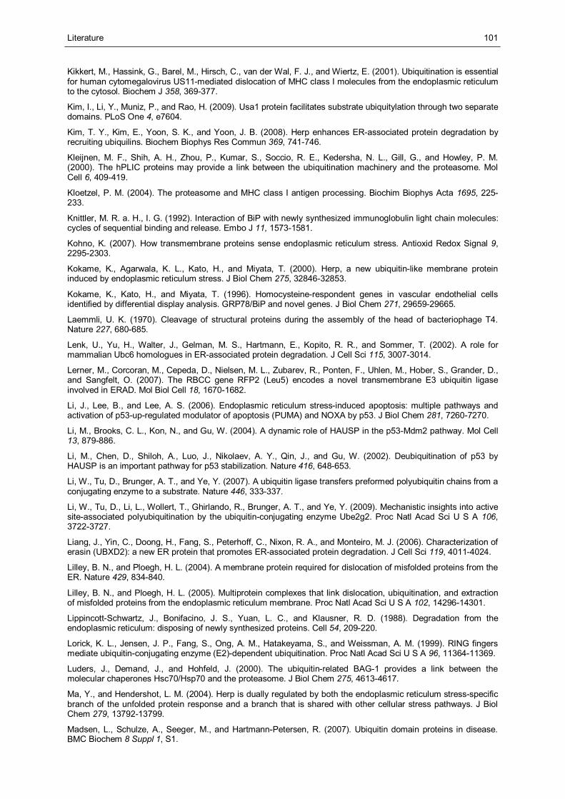
Literature
101
Kikkert, M., Hassink, G., Barel, M., Hirsch, C., van der Wal, F. J., and Wiertz, E. (2001). Ubiquitination is essential for human cytomegalovirus US11-mediated dislocation of MHC class I molecules from the endoplasmic reticulum to the cytosol. Biochem J 358, 369-377.
Kim, I., Li, Y., Muniz, P., and Rao, H. (2009). Usa1 protein facilitates substrate ubiquitylation through two separate domains. PLoS One 4, e7604.
Kim, T. Y., Kim, E., Yoon, S. K., and Yoon, J. B. (2008). Herp enhances ER-associated protein degradation by recruiting ubiquilins. Biochem Biophys Res Commun 369, 741-746.
Kleijnen, M. F., Shih, A. H., Zhou, P., Kumar, S., Soccio, R. E., Kedersha, N. L., Gill, G., and Howley, P. M. (2000). The hPLIC proteins may provide a link between the ubiquitination machinery and the proteasome. Mol Cell 6, 409-419.
Kloetzel, P. M. (2004). The proteasome and MHC class I antigen processing. Biochim Biophys Acta 1695, 225-233.
Knittler, M. R. a. H., I. G. (1992). Interaction of BiP with newly synthesized immunoglobulin light chain molecules: cycles of sequential binding and release. Embo J 11, 1573-1581.
Kohno, K. (2007). How transmembrane proteins sense endoplasmic reticulum stress. Antioxid Redox Signal 9, 2295-2303.
Kokame, K., Agarwala, K. L., Kato, H., and Miyata, T. (2000). Herp, a new ubiquitin-like membrane protein induced by endoplasmic reticulum stress. J Biol Chem 275, 32846-32853.
Kokame, K., Kato, H., and Miyata, T. (1996). Homocysteine-respondent genes in vascular endothelial cells identified by differential display analysis. GRP78/BiP and novel genes. J Biol Chem 271, 29659-29665.
Laemmli, U. K. (1970). Cleavage of structural proteins during the assembly of the head of bacteriophage T4. Nature 227, 680-685.
Lenk, U., Yu, H., Walter, J., Gelman, M. S., Hartmann, E., Kopito, R. R., and Sommer, T. (2002). A role for mammalian Ubc6 homologues in ER-associated protein degradation. J Cell Sci 115, 3007-3014.
Lerner, M., Corcoran, M., Cepeda, D., Nielsen, M. L., Zubarev, R., Ponten, F., Uhlen, M., Hober, S., Grander, D., and Sangfelt, O. (2007). The RBCC gene RFP2 (Leu5) encodes a novel transmembrane E3 ubiquitin ligase involved in ERAD. Mol Biol Cell 18, 1670-1682.
Li, J., Lee, B., and Lee, A. S. (2006). Endoplasmic reticulum stress-induced apoptosis: multiple pathways and activation of p53-up-regulated modulator of apoptosis (PUMA) and NOXA by p53. J Biol Chem 281, 7260-7270.
Li, M., Brooks, C. L., Kon, N., and Gu, W. (2004). A dynamic role of HAUSP in the p53-Mdm2 pathway. Mol Cell 13, 879-886.
Li, M., Chen, D., Shiloh, A., Luo, J., Nikolaev, A. Y., Qin, J., and Gu, W. (2002). Deubiquitination of p53 by HAUSP is an important pathway for p53 stabilization. Nature 416, 648-653.
Li, W., Tu, D., Brunger, A. T., and Ye, Y. (2007). A ubiquitin ligase transfers preformed polyubiquitin chains from a conjugating enzyme to a substrate. Nature 446, 333-337.
Li, W., Tu, D., Li, L., Wollert, T., Ghirlando, R., Brunger, A. T., and Ye, Y. (2009). Mechanistic insights into active site-associated polyubiquitination by the ubiquitin-conjugating enzyme Ube2g2. Proc Natl Acad Sci U S A 106, 3722-3727.
Liang, J., Yin, C., Doong, H., Fang, S., Peterhoff, C., Nixon, R. A., and Monteiro, M. J. (2006). Characterization of erasin (UBXD2): a new ER protein that promotes ER-associated protein degradation. J Cell Sci 119, 4011-4024.
Lilley, B. N., and Ploegh, H. L. (2004). A membrane protein required for dislocation of misfolded proteins from the ER. Nature 429, 834-840.
Lilley, B. N., and Ploegh, H. L. (2005). Multiprotein complexes that link dislocation, ubiquitination, and extraction of misfolded proteins from the endoplasmic reticulum membrane. Proc Natl Acad Sci U S A 102, 14296-14301.
Lippincott-Schwartz, J., Bonifacino, J. S., Yuan, L. C., and Klausner, R. D. (1988). Degradation from the endoplasmic reticulum: disposing of newly synthesized proteins. Cell 54, 209-220.
Lorick, K. L., Jensen, J. P., Fang, S., Ong, A. M., Hatakeyama, S., and Weissman, A. M. (1999). RING fingers mediate ubiquitin-conjugating enzyme (E2)-dependent ubiquitination. Proc Natl Acad Sci U S A 96, 11364-11369.
Luders, J., Demand, J., and Hohfeld, J. (2000). The ubiquitin-related BAG-1 provides a link between the molecular chaperones Hsc70/Hsp70 and the proteasome. J Biol Chem 275, 4613-4617.
Ma, Y., and Hendershot, L. M. (2004). Herp is dually regulated by both the endoplasmic reticulum stress-specific branch of the unfolded protein response and a branch that is shared with other cellular stress pathways. J Biol Chem 279, 13792-13799.
Madsen, L., Schulze, A., Seeger, M., and Hartmann-Petersen, R. (2007). Ubiquitin domain proteins in disease. BMC Biochem 8 Suppl 1, S1.
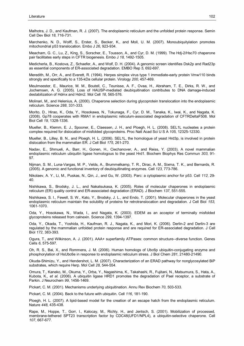
Literature
102
Malhotra, J. D., and Kaufman, R. J. (2007). The endoplasmic reticulum and the unfolded protein response. Semin Cell Dev Biol 18, 716-731.
Marchenko, N. D., Wolff, S., Erster, S., Becker, K., and Moll, U. M. (2007). Monoubiquitylation promotes mitochondrial p53 translocation. Embo J 26, 923-934.
Meacham, G. C., Lu, Z., King, S., Sorscher, E., Tousson, A., and Cyr, D. M. (1999). The Hdj-2/Hsc70 chaperone pair facilitates early steps in CFTR biogenesis. Embo J 18, 1492-1505.
Medicherla, B., Kostova, Z., Schaefer, A., and Wolf, D. H. (2004). A genomic screen identifies Dsk2p and Rad23p as essential components of ER-associated degradation. EMBO Rep 5, 692-697.
Meredith, M., Orr, A., and Everett, R. (1994). Herpes simplex virus type 1 immediate-early protein Vmw110 binds strongly and specifically to a 135-kDa cellular protein. Virology 200, 457-469.
Meulmeester, E., Maurice, M. M., Boutell, C., Teunisse, A. F., Ovaa, H., Abraham, T. E., Dirks, R. W., and Jochemsen, A. G. (2005). Loss of HAUSP-mediated deubiquitination contributes to DNA damage-induced destabilization of Hdmx and Hdm2. Mol Cell 18, 565-576.
Molinari, M., and Helenius, A. (2000). Chaperone selection during glycoprotein translocation into the endoplasmic reticulum. Science 288, 331-333.
Morito, D., Hirao, K., Oda, Y., Hosokawa, N., Tokunaga, F., Cyr, D. M., Tanaka, K., Iwai, K., and Nagata, K. (2008). Gp78 cooperates with RMA1 in endoplasmic reticulum-associated degradation of CFTRDeltaF508. Mol Biol Cell 19, 1328-1336.
Mueller, B., Klemm, E. J., Spooner, E., Claessen, J. H., and Ploegh, H. L. (2008). SEL1L nucleates a protein complex required for dislocation of misfolded glycoproteins. Proc Natl Acad Sci U S A 105, 12325-12330.
Mueller, B., Lilley, B. N., and Ploegh, H. L. (2006). SEL1L, the homologue of yeast Hrd3p, is involved in protein dislocation from the mammalian ER. J Cell Biol 175, 261-270.
Nadav, E., Shmueli, A., Barr, H., Gonen, H., Ciechanover, A., and Reiss, Y. (2003). A novel mammalian endoplasmic reticulum ubiquitin ligase homologous to the yeast Hrd1. Biochem Biophys Res Commun 303, 91-97.
Nijman, S. M., Luna-Vargas, M. P., Velds, A., Brummelkamp, T. R., Dirac, A. M., Sixma, T. K., and Bernards, R. (2005). A genomic and functional inventory of deubiquitinating enzymes. Cell 123, 773-786.
Nikolaev, A. Y., Li, M., Puskas, N., Qin, J., and Gu, W. (2003). Parc: a cytoplasmic anchor for p53. Cell 112, 29-40.
Nishikawa, S., Brodsky, J. L., and Nakatsukasa, K. (2005). Roles of molecular chaperones in endoplasmic reticulum (ER) quality control and ER-associated degradation (ERAD). J Biochem 137, 551-555.
Nishikawa, S. I., Fewell, S. W., Kato, Y., Brodsky, J. L., and Endo, T. (2001). Molecular chaperones in the yeast endoplasmic reticulum maintain the solubility of proteins for retrotranslocation and degradation. J Cell Biol 153, 1061-1070.
Oda, Y., Hosokawa, N., Wada, I., and Nagata, K. (2003). EDEM as an acceptor of terminally misfolded glycoproteins released from calnexin. Science 299, 1394-1397.
Oda, Y., Okada, T., Yoshida, H., Kaufman, R. J., Nagata, K., and Mori, K. (2006). Derlin-2 and Derlin-3 are regulated by the mammalian unfolded protein response and are required for ER-associated degradation. J Cell Biol 172, 383-393.
Ogura, T., and Wilkinson, A. J. (2001). AAA+ superfamily ATPases: common structure--diverse function. Genes Cells 6, 575-597.
Oh, R. S., Bai, X., and Rommens, J. M. (2006). Human homologs of Ubc6p ubiquitin-conjugating enzyme and phosphorylation of HsUbc6e in response to endoplasmic reticulum stress. J Biol Chem 281, 21480-21490.
Okuda-Shimizu, Y., and Hendershot, L. M. (2007). Characterization of an ERAD pathway for nonglycosylated BiP substrates, which require Herp. Mol Cell 28, 544-554.
Omura, T., Kaneko, M., Okuma, Y., Orba, Y., Nagashima, K., Takahashi, R., Fujitani, N., Matsumura, S., Hata, A., Kubota, K., et al. (2006). A ubiquitin ligase HRD1 promotes the degradation of Pael receptor, a substrate of Parkin. J Neurochem 99, 1456-1469.
Pickart, C. M. (2001). Mechanisms underlying ubiquitination. Annu Rev Biochem 70, 503-533.
Pickart, C. M. (2004). Back to the future with ubiquitin. Cell 116, 181-190.
Ploegh, H. L. (2007). A lipid-based model for the creation of an escape hatch from the endoplasmic reticulum. Nature 448, 435-438.
Rape, M., Hoppe, T., Gorr, I., Kalocay, M., Richly, H., and Jentsch, S. (2001). Mobilization of processed, membrane-tethered SPT23 transcription factor by CDC48(UFD1/NPL4), a ubiquitin-selective chaperone. Cell 107, 667-677.
Literature
103
Rapoport, T. A. (2007). Protein translocation across the eukaryotic endoplasmic reticulum and bacterial plasma membranes. Nature 450, 663-669.
Reyes-Turcu, F. E., Ventii, K. H., and Wilkinson, K. D. (2009). Regulation and cellular roles of ubiquitin-specific deubiquitinating enzymes. Annu Rev Biochem 78, 363-397.
Sai, X., Kokame, K., Shiraishi, H., Kawamura, Y., Miyata, T., Yanagisawa, K., and Komano, H. (2003). The ubiquitin-like domain of Herp is involved in Herp degradation, but not necessary for its enhancement of amyloid beta-protein generation. FEBS Lett 553, 151-156.
Saridakis, V., Sheng, Y., Sarkari, F., Holowaty, M. N., Shire, K., Nguyen, T., Zhang, R. G., Liao, J., Lee, W., Edwards, A. M., et al. (2005). Structure of the p53 binding domain of HAUSP/USP7 bound to Epstein-Barr nuclear antigen 1 implications for EBV-mediated immortalization. Mol Cell 18, 25-36.
Sato, B. K., Schulz, D., Do, P. H., and Hampton, R. Y. (2009). Misfolded membrane proteins are specifically recognized by the transmembrane domain of the Hrd1p ubiquitin ligase. Mol Cell 34, 212-222.
Scheffner, M., Huibregtse, J. M., Vierstra, R. D., and Howley, P. M. (1993). The HPV-16 E6 and E6-AP complex functions as a ubiquitin-protein ligase in the ubiquitination of p53. Cell 75, 495-505.
Scheffner, M., Nuber, U., and Huibregtse, J. M. (1995). Protein ubiquitination involving an E1-E2-E3 enzyme ubiquitin thioester cascade. Nature 373, 81-83.
Schmitz, A., and Herzog, V. (2004). Endoplasmic reticulum-associated degradation: exceptions to the rule. Eur J Cell Biol 83, 501-509.
Schroder, M., and Kaufman, R. J. (2005). ER stress and the unfolded protein response. Mutat Res 569, 29-63.
Schubert, U., Anton, L. C., Gibbs, J., Norbury, C. C., Yewdell, J. W., and Bennink, J. R. (2000). Rapid degradation of a large fraction of newly synthesized proteins by proteasomes. Nature 404, 770-774.
Schulze, A. (2006). A Role for the Ubiquitin Domain Protein HERP in ER-associated Protein Degradation.
Schulze, A., Standera, S., Buerger, E., Kikkert, M., van Voorden, S., Wiertz, E., Koning, F., Kloetzel, P. M., and Seeger, M. (2005). The ubiquitin-domain protein HERP forms a complex with components of the endoplasmic reticulum associated degradation pathway. J Mol Biol 354, 1021-1027.
Sheng, Y., Saridakis, V., Sarkari, F., Duan, S., Wu, T., Arrowsmith, C. H., and Frappier, L. (2006). Molecular recognition of p53 and MDM2 by USP7/HAUSP. Nat Struct Mol Biol 13, 285-291.
Shmueli, A., Tsai, Y. C., Yang, M., Braun, M. A., and Weissman, A. M. (2009). Targeting of gp78 for ubiquitin-mediated proteasomal degradation by Hrd1: cross-talk between E3s in the endoplasmic reticulum. Biochem Biophys Res Commun 390, 758-762.
Sidrauski, C., Cox, J. S., and Walter, P. (1996). tRNA ligase is required for regulated mRNA splicing in the unfolded protein response. Cell 87, 405-413.
Sommer, T., and Jentsch, S. (1993). A protein translocation defect linked to ubiquitin conjugation at the endoplasmic reticulum. Nature 365, 176-179.
Song, B. L., Sever, N., and DeBose-Boyd, R. A. (2005). Gp78, a membrane-anchored ubiquitin ligase, associates with Insig-1 and couples sterol-regulated ubiquitination to degradation of HMG CoA reductase. Mol Cell 19, 829-840.
Sowa, M. E., Bennett, E. J., Gygi, S. P., and Harper, J. W. (2009). Defining the human deubiquitinating enzyme interaction landscape. Cell 138, 389-403.
Strous, G. J., van Kerkhof, P., Govers, R., Ciechanover, A., and Schwartz, A. L. (1996). The ubiquitin conjugation system is required for ligand-induced endocytosis and degradation of the growth hormone receptor. Embo J 15, 3806-3812.
Tanaka, K. (2009). The proteasome: overview of structure and functions. Proc Jpn Acad Ser B Phys Biol Sci 85, 12-36.
Tang, J., Qu, L. K., Zhang, J., Wang, W., Michaelson, J. S., Degenhardt, Y. Y., El-Deiry, W. S., and Yang, X. (2006). Critical role for Daxx in regulating Mdm2. Nat Cell Biol 8, 855-862.
Thrower, J. S., Hoffman, L., Rechsteiner, M., and Pickart, C. M. (2000). Recognition of the polyubiquitin proteolytic signal. Embo J 19, 94-102.
Tuvia, S., Taglicht, D., Erez, O., Alroy, I., Alchanati, I., Bicoviski, V., Dori-Bachash, M., Ben-Avraham, D., and Reiss, Y. (2007). The ubiquitin E3 ligase POSH regulates calcium homeostasis through spatial control of Herp. J Cell Biol 177, 51-61.
van de Wetering, M., Oving, I., Muncan, V., Pon Fong, M. T., Brantjes, H., van Leenen, D., Holstege, F. C., Brummelkamp, T. R., Agami, R., and Clevers, H. (2003). Specific inhibition of gene expression using a stably integrated, inducible small-interfering-RNA vector. EMBO Rep 4, 609-615.
Literature
104
van Laar, T., Schouten, T., Hoogervorst, E., van Eck, M., van der Eb, A. J., and Terleth, C. (2000). The novel MMS-inducible gene Mif1/KIAA0025 is a target of the unfolded protein response pathway. FEBS Lett 469, 123-131.
van Laar, T., van der Eb, A. J., and Terleth, C. (2001). Mif1: a missing link between the unfolded protein response pathway and ER-associated protein degradation? Curr Protein Pept Sci 2, 169-190.
Vashist, S., and Ng, D. T. (2004). Misfolded proteins are sorted by a sequential checkpoint mechanism of ER quality control. J Cell Biol 165, 41-52.
Vembar, S. S., and Brodsky, J. L. (2008). One step at a time: endoplasmic reticulum-associated degradation. Nat Rev Mol Cell Biol 9, 944-957.
Wang, Q., Li, L., and Ye, Y. (2006). Regulation of retrotranslocation by p97-associated deubiquitinating enzyme ataxin-3. J Cell Biol 174, 963-971.
Ward, C. L., Omura, S., and Kopito, R. R. (1995). Degradation of CFTR by the ubiquitin-proteasome pathway. Cell 83, 121-127.
Wilkinson, C. R., Seeger, M., Hartmann-Petersen, R., Stone, M., Wallace, M., Semple, C., and Gordon, C. (2001). Proteins containing the UBA domain are able to bind to multi-ubiquitin chains. Nat Cell Biol 3, 939-943.
Wilkinson, K. D., Urban, M. K., and Haas, A. L. (1980). Ubiquitin is the ATP-dependent proteolysis factor I of rabbit reticulocytes. J Biol Chem 255, 7529-7532.
Windheim, M., Stafford, M., Peggie, M., and Cohen, P. (2008). Interleukin-1 (IL-1) induces the Lys63-linked polyubiquitination of IL-1 receptor-associated kinase 1 to facilitate NEMO binding and the activation of IkappaBalpha kinase. Mol Cell Biol 28, 1783-1791.
Wolf, D. H., and Hilt, W. (2004). The proteasome: a proteolytic nanomachine of cell regulation and waste disposal. Biochim Biophys Acta 1695, 19-31.
Xu, Z., Kukekov, N. V., and Greene, L. A. (2003). POSH acts as a scaffold for a multiprotein complex that mediates JNK activation in apoptosis. Embo J 22, 252-261.
Yamasaki, S., Yagishita, N., Sasaki, T., Nakazawa, M., Kato, Y., Yamadera, T., Bae, E., Toriyama, S., Ikeda, R., Zhang, L., et al. (2007). Cytoplasmic destruction of p53 by the endoplasmic reticulum-resident ubiquitin ligase 'Synoviolin'. Embo J 26, 113-122.
Ye, Y., Meyer, H. H., and Rapoport, T. A. (2001). The AAA ATPase Cdc48/p97 and its partners transport proteins from the ER into the cytosol. Nature 414, 652-656.
Ye, Y., Meyer, H. H., and Rapoport, T. A. (2003). Function of the p97-Ufd1-Npl4 complex in retrotranslocation from the ER to the cytosol: dual recognition of nonubiquitinated polypeptide segments and polyubiquitin chains. J Cell Biol 162, 71-84.
Ye, Y., Shibata, Y., Kikkert, M., van Voorden, S., Wiertz, E., and Rapoport, T. A. (2005). Inaugural Article: Recruitment of the p97 ATPase and ubiquitin ligases to the site of retrotranslocation at the endoplasmic reticulum membrane. Proc Natl Acad Sci U S A 102, 14132-14138.
Ye, Y., Shibata, Y., Yun, C., Ron, D., and Rapoport, T. A. (2004). A membrane protein complex mediates retro-translocation from the ER lumen into the cytosol. Nature 429, 841-847.
Yoshida, H. (2007). Unconventional splicing of XBP-1 mRNA in the unfolded protein response. Antioxid Redox Signal 9, 2323-2333.
Yoshida, H., Haze, K., Yanagi, H., Yura, T., and Mori, K. (1998). Identification of the cis-acting endoplasmic reticulum stress response element responsible for transcriptional induction of mammalian glucose-regulated proteins. Involvement of basic leucine zipper transcription factors. J Biol Chem 273, 33741-33749.
Zhong, X., Shen, Y., Ballar, P., Apostolou, A., Agami, R., and Fang, S. (2004). AAA ATPase p97/valosin-containing protein interacts with gp78, a ubiquitin ligase for endoplasmic reticulum-associated degradation. J Biol Chem 279, 45676-45684.
Zhu, X., Menard, R., and Sulea, T. (2007). High incidence of ubiquitin-like domains in human ubiquitin-specific proteases. Proteins 69, 1-7.
Appendix
105
Appendix
Abbreviations
Table 15. Abbreviations
Aa amino acid
Amp Ampicillin
AUP1 Ancient ubiquitous protein 1
BSA bovine serum albumin
CD-δ cluster of differentiation
CFTR cystic fibrosis transmembrane conductance regulator
Chx Cycloheximide
CUE coupling of ubiquitin conjugation to ER degradation
DBC DeoxyBigChap
DMSO dimethyl sulfoxide
dNTP deoxyribonucleotide triphosphate
DTT Dithiothreitol
DUB deubiquitinating enzyme
E.coli Escherichia coli
E1 ubiquitin activating enzyme
E2 ubiquitin conjugating enzyme
E3 ubiquitin ligase
ECL enhanced chemiluminescence
e.g. exempli gratia (lat. for example)
EDTA ethylenediamine tetraacetic acid
ER endoplasmic reticulum
ERAD ER associated protein degradation
FCS fetal calf serum
Fw Forward
G acceleration of gravity
GAPDH glycerin aldehyd 3 phosphate dehydrogenase
Grp78 glucose regulated protein 78
GST glutathione S transferase
H Hour
HEPES 4-(2-Hydroxyethyl)piperazine-1-ethanesulfonic acid
Herp Homocysteine inducible endoplasmic reticulum-resident protein
HMGR 3-hydroxy-3-methylglutaryl-coenzyme A reductase
Hrd1 HMG-CoA reductase degradation
HTB hexahistidine-TEV protease-biotinylation-tag
IB immunoblot (identical to Western blot)
Ig Immunoglobulin
IP Immunoprecipitation
IPTG isopropyl-beta-D-thiogalactopyranoside
Kan Kanamycin
Kb Kilobase
Appendix
106
kDa Kilodalton
LB Lysogeny broth
M mol/L
Mdm2 /Hdm2 mouse / human double minute protein 2
MHC major histocompatibility complex
NHK Null Hong Kong mutant of α1-antitrypsin
OD optical density
PAGE polyacrylamide gel electrophoresis
PBS phosphate buffered saline
PCR polymerase chain reaction
QC quality control
Rev Reverse
RING really interesting new gene
RIPA radioimmunoprecipitation assay
Rpm revolutions per minute
RT Room temperature
RT-PCR reverse transcriptase polymerase chain reaction
S Svedberg sedimentation coefficient
SDS sodium dodecylsulfate
Sel1L Suppressor of lin-12-like protein1
siRNA small interfering RNA
TAE Tris-acetate-EDTA
TCA trichloro-acetic acid
TCR-α T cell receptor α
TEMED tetramethyl ethylene diamine
TEV Tobacco Etch Virus
Tg Thapsigargin
Tris Tris (hydroxylmethyl) aminomethane
Tu Tunicamycin
U Unit
UBA ubiquitin-associated
UBL ubiquitin-like
UDP ubiquitin domain protein
UPR unfolded protein response
UPS ubiquitin proteasome system
Usa1p U1 SNP1-associating protein 1
USP ubiquitin specific protease
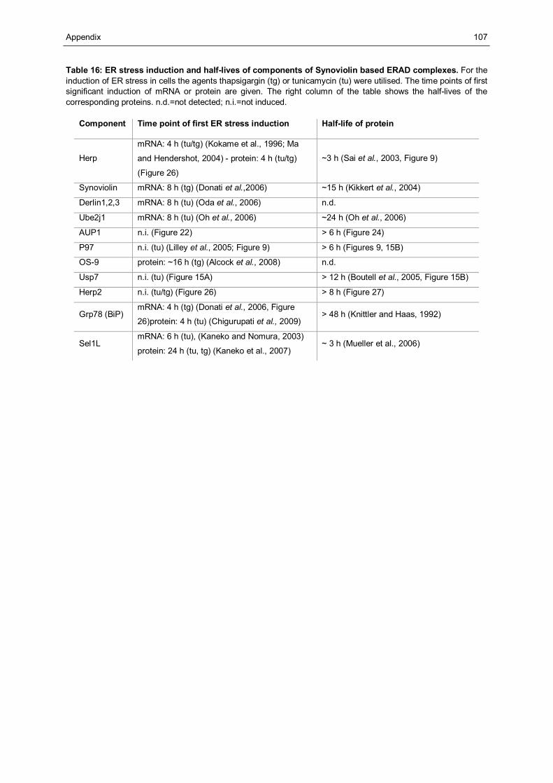
Appendix
107
Table 16: ER stress induction and half-lives of components of Synoviolin based ERAD complexes. For the induction of ER stress in cells the agents thapsigargin (tg) or tunicamycin (tu) were utilised. The time points of first significant induction of mRNA or protein are given. The right column of the table shows the half-lives of the corresponding proteins. n.d.=not detected; n.i.=not induced.
Component Time point of first ER stress induction Half-life of protein
Herp
mRNA: 4 h (tu/tg) (Kokame et al., 1996; Ma
and Hendershot, 2004) - protein: 4 h (tu/tg)
(Figure 26)
~3 h (Sai et al., 2003, Figure 9)
Synoviolin mRNA: 8 h (tg) (Donati et al.,2006) ~15 h (Kikkert et al., 2004)
Derlin1,2,3 mRNA: 8 h (tu) (Oda et al., 2006) n.d.
Ube2j1 mRNA: 8 h (tu) (Oh et al., 2006) ~24 h (Oh et al., 2006)
AUP1 n.i. (Figure 22) > 6 h (Figure 24)
P97 n.i. (tu) (Lilley et al., 2005; Figure 9) > 6 h (Figures 9, 15B)
OS-9 protein: ~16 h (tg) (Alcock et al., 2008) n.d.
Usp7 n.i. (tu) (Figure 15A) > 12 h (Boutell et al., 2005, Figure 15B)
Herp2 n.i. (tu/tg) (Figure 26) > 8 h (Figure 27)
Grp78 (BiP) mRNA: 4 h (tg) (Donati et al., 2006, Figure
26)protein: 4 h (tu) (Chigurupati et al., 2009) > 48 h (Knittler and Haas, 1992)
Sel1L mRNA: 6 h (tu), (Kaneko and Nomura, 2003)
protein: 24 h (tu, tg) (Kaneko et al., 2007) ~ 3 h (Mueller et al., 2006)
Appendix
108
Publikationen und Vorträge
Brigelius-Flohé R, Banning A, Kny M, Böl GF: Redox events in interleukin-1 signaling.
Arch Biochem Biophys. 2004 Mar 1;423(1):66-73. Review
Kny M, Standera S, Schulze A, Maurice M, Hartmann-Petersen R, Kloetzel PM, Seeger M:
Herp recruits USP7 to Synoviolin-/Hrd1-based ERAD complexes - Talk: 19th European
Students’ Conference 2008 – Charité Berlin
Melanie Kny, Sybille Standera, Andrea Gurok, Rasmus Hartmann-Petersen, Peter-Michael
Kloetzel and Michael Seeger: The ubiquitin-like domain of Herp is required for Hrd1-
mediated ubiquitylation of ERAD substrates - 2009 (manuscript submitted)
Keller M, Ebstein F, Paschen A, Kny M, Seeger M, Bürger E, Schadendorf D, Kloetzel PM,
Seifert U: Deregulation of the endoplasmic reticulum associated degradation (ERAD)
pathway mediates immune escape of malignant melanoma - 2009 (manuscript submitted)
Kny M, Standera S, Schulze A, Maurice M, Hartmann-Petersen R, Kloetzel PM Seeger M:
The UBL domain of Herp mediates the recruitment of Usp7 to Synoviolin/Hrd1 ERAD
complexes (manuscript in preparation)
Appendix
109
Danksagung
Ich möchte mich hiermit ganz herzlich bei allen Personen bedanken, die zum Gelingen
dieser Arbeit beigetragen haben.
Allen voran danke ich Prof. Dr. Peter-Michael Kloetzel, der es mir ermöglichte, in seiner
Arbeitsgruppe des Instituts für Biochemie diese Arbeit anzufertigen.
Ein ebenso großer Dank gilt Dr. Michael Seeger für die Überlassung des überaus
interessanten Themas, die gute wissenschaftliche Betreuung, die vielen Anregungen und
unzähligen konstruktiven Diskussionen.
Ein herzliches Dankeschön geht an Sybille Standera, die mir nicht nur mit technischer
Expertise stets mit Rat und Tat zur Seite stand. Danke auch an Natalie Steffen für die nette
Zusammenarbeit und die Durchführung von UDP-Interaktionsstudien.
Franziska Kriegenburg, Sandra Jäkel, Tobias Schwarz, Nicole Lange, Janos Steffen, Kathrin
Klass und Sandra Schulz sei unter anderem für das kritische Diskutieren dieser Arbeit und
das Korrekturlesen gedankt.
Weiterhin ein großes Dankeschön an alle ehemaligen und derzeitigen Mitarbeiter der AG
Kloetzel, die mich auf vielfältige Weise unterstützt und für die nette Arbeitsatmosphäre
gesorgt haben. Auch meiner neuen Arbeitsgruppe „Experimentelle Geburtsmedizin“ möchte
ich danken für die mentale Unterstützung während der letzten Phase des Schreibens.
Nicht zuletzt gilt mein allerherzlichstes Dankeschön meinen Eltern, die mich stets unterstützt
haben.
André danke ich für seine unermüdliche Geduld und Diskussionsbereitschaft, außerdem für
seine stete Motivation und die zahlreichen wertvollen Tips.
Recommended


![Detektion von Protein/Protein-Interaktionen mit Hilfe des€¦ · Untersuchung von Protein-Protein-Interaktionen in Hefen [Fields & Song (1989)]. Eine wesentliche Voraussetzung für](https://img.pdfslide.org/doc/110x75/6062f4209e52cc3fcc6ea8d4/detektion-von-proteinprotein-interaktionen-mit-hilfe-des-untersuchung-von-protein-protein-interaktionen.jpg)