Embed Size (px)
Citation preview
This work has been digitalized and published in 2013 by Verlag Zeitschrift für Naturforschung in cooperation with the Max Planck Society for the Advancement of Science under a Creative Commons Attribution4.0 International License.
Dieses Werk wurde im Jahr 2013 vom Verlag Zeitschrift für Naturforschungin Zusammenarbeit mit der Max-Planck-Gesellschaft zur Förderung derWissenschaften e.V. digitalisiert und unter folgender Lizenz veröffentlicht:Creative Commons Namensnennung 4.0 Lizenz.
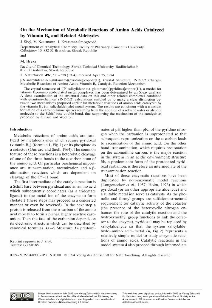
On the Mechanism of Metabolic Reactions of Amino Acids Catalyzed by Vitamin B6 and Related AldehydesJ. Sivý, V. K ettm ann, J. K rätsm ár-ŠmogrovičDepartment of Analytical Chemistry, Faculty of Pharmacy, Comenius University,Odbojärov 10, 832 32 Bratislava, Slovak Republic
M. BrezaFaculty of Chemical Technology, Slovak Technical University, Radlinskeho 9,812 37 Bratislava, Slovak RepublicZ. Naturforsch. 49c, 571-578 (1994); received April 25, 1994[(N-salicylidene-D,L-glutamato)(pyridine)]copper(II), Crystal Structure, INDO/2 Charges, Metabolic Reactions of Amino Acids, Vitamin B6 Catalysis, Reaction Mechanism
The crystal structure of [(N-salicylidene-D,L-glutamato)(pyridine)]copper(II), a model for vitamin B6-amino acid-related metal complexes, has been determined by an X-ray analysis. A close examination of the structural data on this and other related complexes combined with quantum-chemical (INDO/2) calculations enabled us to make a clear distinction between two mechanisms proposed earlier for metabolic reactions of amino acids catalyzed by the vitamin BA (or salicylaldehyde)-metal system. The results are consistent with a transient formation of a carbinolamine species resulting from the addition of a solvent water or alcohol molecule to the Schiff base double bond, thus supporting the mechanism of the catalysis as proposed by Gillard and Wootton.
In troduction
M etabolic reactions of amino acids are catalyzed by m etaloenzym es which require pyridoxal (vitam in B6) (form ula 1, Fig. 1) or its phosphate as a cofactor (G uirard and Snell, 1964). The common feature of these reactions is a heterolytic cleavage of one of the th ree bonds to the a-carbon atom of the am ino acid. O f particular biochemical im portance are transam ination, racem ization and a,ß- elim ination reactions which are dependent on cleavage of the Ca - H bond.
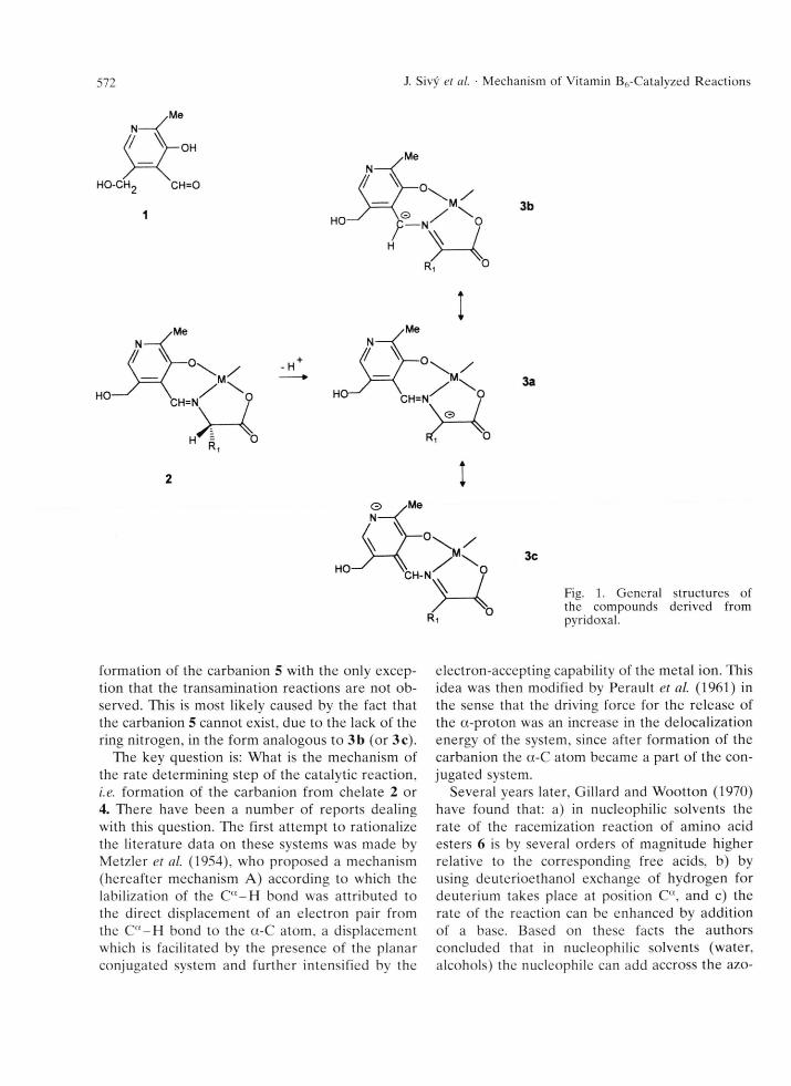
The first in term ediate of the catalytic reaction is a Schiff base betw een pyridoxal and an amino acid which subsequently coordinates (as a tridentate ligand) to the m etal ion of the enzyme to form chelate 2 (these steps may proceed in a concerted m anner or even be reversed). In the next step a pro ton is released from the a-carbon of the amino acid m oiety to form a planar, highly reactive carb- anion. Then the fate of the carbanion depends on its electronic structure which can be described by canonical form ulas 3 a - c . Structure 3a predomi-
Reprint requests to J. Sivy. Telefax: (7) 60388.
nates at pH higher than pK a of the pyridine n itrogen when the carbanion is unpro tonated so that subsequent reprotonization on the a-carbon leads to racem ization of the am ino acid. O n the other hand, transam ination, which requires protonation on the azom ethine carbon, is the m ajor reaction in the system in an acidic environm ent; structure3 b, a predom inant form of the p ro tonated pyridoxal carbanion, is therefore an interm ediate of the transam ination reaction.
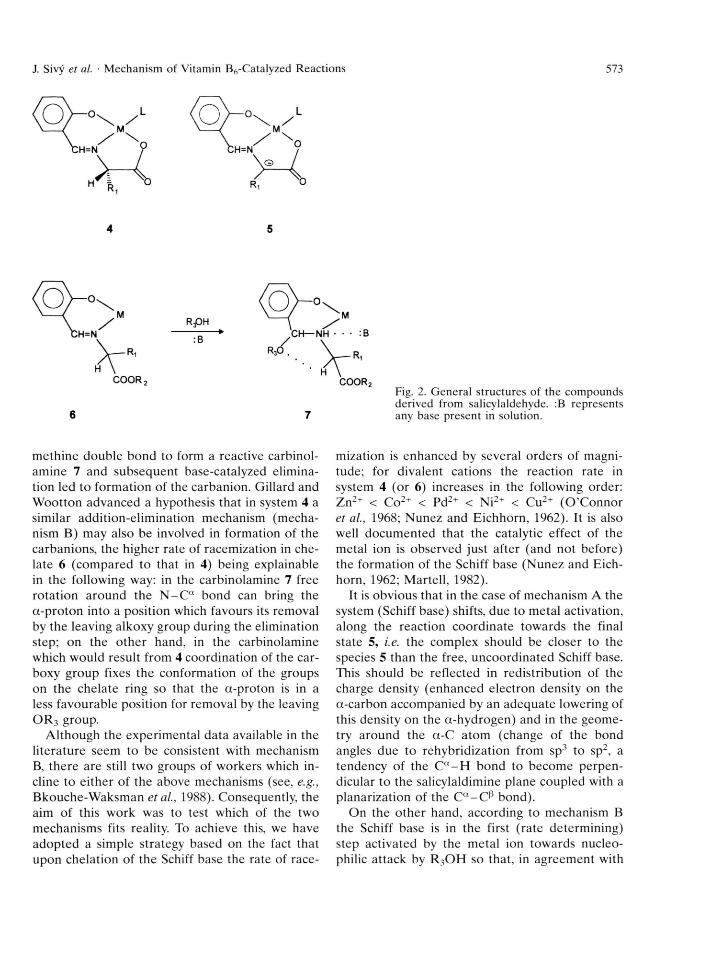
M ost of these enzym atic reactions have been duplicated by non-enzym atic m odel reactions (Longenecker et al., 1957; Holm, 1973) in which pyridoxal (or an o ther appropriate aldehyde) and a suitable m etal ion serve as catalysts. As the phenolic and formyl groups are sufficient structural requirem ent for catalytic activity of the cofactor (the presence of the heterocyclic nitrogen enhances the rate of the catalytic reaction and the hydroxym ethyl group functions to link the cofactor to the enzym e), pyridoxal may be replaced by salicylaldehyde so tha t the system salicylalde- h y d e-am in o a c id -m e ta l (4, Fig. 2) represents a relatively simple m odel to study enzym atic reactions of amino acids. Catalytic reactions in the model system 4 also proceed through interm ediate
0939-5075/94/0900-0571 $ 06.00 © 1994 Verlag der Zeitschrift für Naturforschung. All rights reserved.
572 J. Sivy et al. • Mechanism of Vitamin Bft-Catalyzed Reactions
3a
3c
Fig. 1. General structures of the compounds derived from pyridoxal.
form ation of the carbanion 5 with the only exception that the transam ination reactions are not ob served. This is most likely caused by the fact that the carbanion 5 cannot exist, due to the lack of the ring nitrogen, in the form analogous to 3 b (or 3c).
The key question is: W hat is the mechanism of the rate determ ining step of the catalytic reaction, i.e. form ation of the carbanion from chelate 2 or4. There have been a num ber of reports dealing with this question. The first attem pt to rationalize the literature data on these systems was made by M etzler et al. (1954), who proposed a mechanism (hereafter mechanism A ) according to which the labilization of the Cct- H bond was attributed to the direct displacem ent of an electron pair from the Ca - H bond to the a-C atom , a displacem ent which is facilitated by the presence of the planar conjugated system and further intensified by the
electron-accepting capability of the m etal ion. This idea was then modified by Perault et al. (1961) in the sense that the driving force for the release of the a-pro ton was an increase in the delocalization energy of the system, since after form ation of the carbanion the a-C atom became a part of the conjugated system.
Several years later, G illard and W ootton (1970) have found that: a) in nucleophilic solvents the rate of the racemization reaction of am ino acid esters 6 is by several orders of m agnitude higher relative to the corresponding free acids, b) by using deuterioethanol exchange of hydrogen for deuterium takes place at position Ca, and c) the rate of the reaction can be enhanced by addition of a base. Based on these facts the authors concluded that in nucleophilic solvents (water, alcohols) the nucleophile can add accross the azo-
J. Sivy et al. ■ Mechanism of Vitamin B6-Catalyzed Reactions 573
RPH
Fig. 2. General structures of the compounds derived from salicylaldehyde. :B represents any base present in solution.
m ethine double bond to form a reactive carbinol- am ine 7 and subsequent base-catalyzed elim ination led to form ation of the carbanion. Gillard and W ootton advanced a hypothesis that in system 4 a similar addition-elim ination mechanism (m echanism B) may also be involved in form ation of the carbanions, the higher rate of racem ization in chelate 6 (com pared to that in 4) being explainable in the following way: in the carbinolam ine 7 free ro tation around the N - C “ bond can bring the a-p ro ton into a position which favours its removal by the leaving alkoxy group during the elimination step; on the o ther hand, in the carbinolamine which would result from 4 coordination of the car- boxy group fixes the conform ation of the groups on the chelate ring so that the a-proton is in a less favourable position for rem oval by the leaving O R 3 group.
A lthough the experim ental data available in the literature seem to be consistent with mechanismB, there are still two groups of workers which incline to either of the above mechanisms (see, e.g., Bkouche-W aksm an et al., 1988). Consequently, the aim of this work was to test which of the two m echanism s fits reality. To achieve this, we have adopted a simple strategy based on the fact that upon chelation of the Schiff base the rate of race
mization is enhanced by several orders of magnitude; for divalent cations the reaction rate in system 4 (or 6) increases in the following order: Z n2+ < Co2+ < Pd2+ < Ni2+ < Cu2+ (O ’C onnor et al., 1968; Nunez and Eichhorn, 1962). It is also well docum ented that the catalytic effect of the metal ion is observed just after (and not before) the form ation of the Schiff base (N unez and Eichhorn, 1962; M artell, 1982).
It is obvious that in the case of m echanism A the system (Schiff base) shifts, due to m etal activation, along the reaction coordinate tow ards the final state 5, i.e. the complex should be closer to the species 5 than the free, uncoordinated Schiff base. This should be reflected in redistribution of the charge density (enhanced electron density on the a-carbon accom panied by an adequate lowering of this density on the a-hydrogen) and in the geom etry around the a-C atom (change of the bond angles due to rehybridization from sp3 to sp2, a tendency of the Ca - H bond to becom e perpendicular to the salicylaldimine plane coupled with a planarization of the Ca -C ß bond).
On the o ther hand, according to mechanism B the Schiff base is in the first (rate determ ining) step activated by the m etal ion tow ards nucleo- philic attack by R 3O H so that, in agreem ent with
574 J. Sivy et al. ■ Mechanism of Vitamin B6-Catalyzed Reactions
the theory of charge controlled A dN2 reactions (Fleming, 1976), one would expect an enhancem ent of the positive charge on the azom ethine carbon upon introduction of the metal ion.
From the above it is clear that in order to study the mechanism of the catalytic (e.g. racem ization) reaction it is necessary to m onitor changes in geom etry and distribution of the electron density associated with the transfer of the Schiff base into the chelate. In this work, single-crystal X-ray diffraction and quantum -chem ical calculations were chosen for this purpose. The crystal structure of one derivative belonging to class 4 was investigated in this paper and the rem aining were re trieved from the Cam bridge Structural D atabase (A llen et al., 1979). The salicylaldehyde chelates were chosen over pyridoxal chelates since the form er m ore readily provide good crystals, and C u” was chosen over o ther metals because of the high reaction rate for copper-catalyzed reactions (Nunez and Eichhorn, 1962; M artell, 1982).
Experimental
Selection o f the compounds
All com pounds selected for this study are listed in Table II. They differ in the nature of the am ino acid and of the additional ligand L. The glycinato chelates were not included in the analysis because of uncertainty which of the two C“ - H bonds is actually cleaved in the catalytic reaction. Since, to our knowledge, no Schiff base of salicylaldehyde and an amino acid has been studied crystallo- graphically, we have chosen for this purpose the known structure of chloro-triphenyl-(0-ethyl-N - salicylideneglycinato) Sn,v (Lee et al., 1990) in which the O-ethyl-N-salicylideneglycinato ligand is m onodentately coordinated via the phenolate oxygen to the tin atom , i.e. it contains an uncoordinated azom ethine nitrogen and hence may be re garded, to a good approxim ation, as the m etal-free Schiff base.
X-ray structure o f 4 e
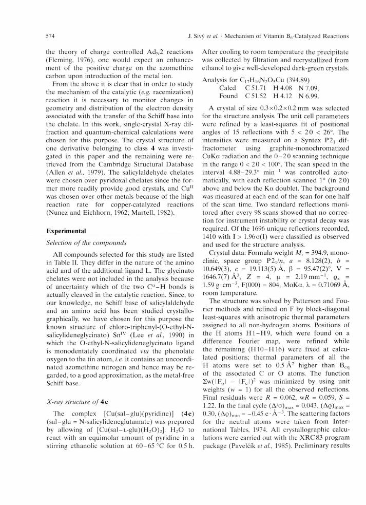
The complex [C u(sal-g lu)(pyrid ine)] (4e) (sa l-g lu = N -salicylideneglutam ate) was prepared by allowing of [C u(sal-L -g lu)(H 20 ) 2]. H 20 to react with an equim olar am ount of pyridine in a stirring ethanolic solution at 6 0 -6 5 °C for 0.5 h.
A fter cooling to room tem perature the precipitate was collected by filtration and recrystallized from ethanol to give well-developed dark-green crystals.
Analysis for C i7H 16N20 5Cu (394.89)Calcd C 51.71 H 4.08 N 7.09,Found C 51.52 H 4.12 N 6.99.
A crystal of size 0.3 x 0.2 x 0.2 mm was selected for the structure analysis. The unit cell param eters were refined by a least-squares fit of positional angles of 15 reflections with 5 < 20 < 26°. The intensities were m easured on a Syntex P2] diffractom eter using graphite-m onochrom atized C uK a radiation and the 0 - 2 0 scanning technique in the range 0 < 20 < 100°. The scan speed in the interval 4.88-29.3° m in -1 was controlled au to matically, with each reflection scanned 1° (in 2 0 ) above and below the K a doublet. The background was m easured at each end of the scan for one half of the scan time. Two standard reflections m onitored after every 98 scans showed that no correction for instrum ent instability or crystal decay was required. O f the 1696 unique reflections recorded, 1410 with I > 1.96o(I) were classified as observed and used for the structure analysis.
Crystal data: Formula weight M x = 394.9, m onoclinic, space group P 2 x!n, a = 8.128(2), b - 10.649(3), c = 19.113(5) A, ß = 95.47(2)°, V = 1646.7(7) Ä 3, Z = 4, [i = 2.19 m m “ 1, qx = 1.59 g- cm “3, F(000) = 804, M oK a, X = 0.71069 Ä, room tem perature.
The structure was solved by Patterson and Fourier m ethods and refined on F by block-diagonal least-squares with anisotropic therm al param eters assigned to all non-hydrogen atoms. Positions of the H atom s H 1 -H 9 , which were found on a difference Fourier map, were refined while the rem aining (H 1 0 -H 1 6 ) were fixed at calculated positions; therm al param eters of all the H atom s were set to 0.5 A 2 higher than Beq of the associated C or O atoms. The function Zw (IF0 l - I Fc I )2 was minimized by using unit weights (w = 1) for all the observed reflections. Final residuals were R = 0.062, wR = 0.059, S =1.22. In the final cycle (A/o)max = 0.043, (AQ)max = 0.30, (A0 )min = -0.45 e • A -3. The scattering factors for the neutral atoms were taken from In te rnational Tables, 1974. All crystallographic calculations were carried out with the X R C 83 program package (Pavelcik et al., 1985). Prelim inary results
J. Sivy et al. ■ Mechanism of Vitamin B6-Catalyzed Reactions 575
of the crystal structure of 4 e have been reported previously (Krätsm är-Sm ogrovic et al., 1985).
M O calculations
E lectronic structures in term s of total atomic charges, W iberg indices and orbital energies were obtained from a local version of the INDO/2 program (Boca, 1989) by using a fixed geometry - constructed on the basis of known X-ray structures - for all six chelates studied. Initial geom etries of the m etal-free Schiff bases were derived from the X-ray structure of the corresponding chelate parents by rem oval of the copper atom and com pleting the freed valencies on the phenolate and carboxylate oxygens by H atoms. Subsequent optim izations using A M I (D ew ar et a l, 1985) have revealed only slight torsional rearrangem ent indicating that the planar conform ation of the Schiff base ligand observed in the chelate is also a low-energy conform ation for the ligand itself.
Results and Discussion
Final atom ic coordinates for 4e are given in Table I. R elevant structural param eters and the total atom ic charges in the catalytically interesting part of the chelates studied are com pared in Tables II and III, respectively. As the atomic charges calculated for the six metal-free Schiff bases were alm ost identical, only the mean values are given in Table III.
Table I. Final atomic coordinates (xlO4) and equivalent isotropic thermal parameters Beq (A2) with E.S.D.'s in parentheses*. Beq = 4/3 YZ. BVj c?j, ctj.
' j
Atom X y z Beq [A2]
CO(l) 1450(2) 1301(1) 4547(1) 6.52(4)0(1) 2117(7) 61(5) 5238(3) 5.57(18)0(2) 986(7) 2585(5) 3813(3) 5.85(17)0(3) 1122(7) 2983(6) 2683(3) 6.51(19)0(4) -3793(7) -412(6) 2671(3) 7.18(21)0(5) -1977(8) -1393(8) 2100(4) 9.82(28)N(l) 512(8) 2430(6) 5251(3) 5.07(20)N(2) 2223(8) 358(6) 3781(3) 5.41(21)C(l) -617(11) 3315(8) 5036(5) 5.92(31)C(2) -1231(11) 4117(8) 5523(6) 6.77(35)C(3) -745(13) 4017(9) 6219(5) 7.38(33)C(4) 399(13) 3106(9) 6435(5) 7.13(35)C(5) 1001(11) 2333(8) 5941(5) 5.97(29)0(6) 1299(10) 2303(8) 3201(4) 5.63(27)C(7) 1930(11) 949(8) 3082(4) 5.82(28)0(8) 668(11) 233(8) 2591(4) 5.66(27)0(9) -1079(11) 346(9) 2843(5) 6.31(30)0(10) -2300(12) -581(9) 2477(5) 6.50(30)0(11) 3102(10) -876(8) 5146(4) 5.23(25)0(12) 3604(10) -1263(8) 4496(4) 5.15(25)0(13) 4672(11) -2305(8) 4456(5) 6.06(26)0(14) 5227(11) -2968(9) 5049(6) 6.70(32)0(15) 4732(11) -2611(9) 5692(5) 6.55(31)0(16) 3694(11) -1600(8) 5756(4) 5.81(28)0(17) 3108(11) -630(8) 3847(4) 5.58(26)
* Anisotropic thermal parameters, H-atom coordinates, F0/Fc tables, and complete lists of geometrical parameters are available at the authors.
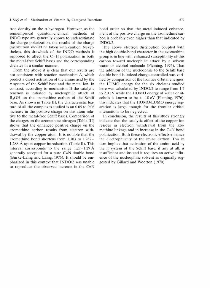
The overall geom etry of 4 e (Fig. 3) shows the usual features of this type of complex, perhaps with the exception that the coordination around the Cu atom is exactly square-planar. This is sub-
Table II. List of compounds3 studied here and their relevant geometric parametersb.
Compound N -C a-C N -C a-CP C -C a-CP C=N C=N -Ca-CP C=N -Ca-H Ref.
4a 107.4 109.5 109.1 1.270 83.3 -39.8C 184b 108.5 112.3 110.2 1.275 70.6 -46.6C 194c 107.3 112.7 108.3 1.286 73.7 -48.8 204d 107.5 111.7 109.1 1.285 72.8 -47.7C 214e 109.6 112.6 110.0 1.267 75.9 -50.3 this work4f 105.3 108.3 114.8 1.288 74.0 -41.8 22usbd 111.8 1.303 13
a The compounds are designated as follows (see also Fig. 2): 4a: Ri = CH3, L = pyrazole; 4b: R, = CH3, L = NCO; 4c: R, = CH2Ph, L = NCO; 4d: R, - (CH2)2COOH, L = H20 ; 4e: Rj = (CFb)2COOH, L = pyridine; 4f: R, = CH(CH3)2, L = h 2o.
b E.S.D.’s for the bond angles range from 0.2 to 0.6°, for the C=N bond length from 0.003 to 0.011 Ä, and for the torsion angles from 0.3 to 0.7° (C=N-C“-C P) and from 0.6 to 1.0° (C=N -C“-H ).
c Position of the hydrogen calculated, i.e. not actually found in the difference Fourier synthesis. d Uncoordinated Schiff base (see text).
576 J. Sivy et al. ■ Mechanism of Vitamin B6-Catalyzed Reactions
Table III. INDO/2 net atomic charges.
Compound3 Qc Qn Ra-C qa-H
4a 0.26 -0.02 0.05 -0.014b 0.25 -0.01 0.06 -0.024c 0.25 -0.03 0.07 -0.024d 0.26 -0.05 0.05 -0.014e 0.26 -0.05 0.03 -0.024f 0.26 -0.02 0.06 -0.01usbb 0.20 -0.22 0.11 -0.02
a For designation of the compounds see the footnote of Table II.
b Atomic charges for the uncoordinated Schiff base (usb) are means of six values (see text).
stantiated by the fact that no atom occurs at a distance shorter than 3.3 A in the axial direction and by the coplanarity of Cu with the four donor atoms. The pro tonated side chain carboxyl group is not involved in the coordination to the copper atom , but it forms a strong hydrogen bond with the carbonyl oxygen 0 3 [0 — 0 = 2.593(5) A] of another molecule at ( -0 .5 - jc, -0 .5 + y, 0.5 - z), thus forming chains going along the diagonal of the unit cell.
As the space group (P 2 \/n) is centrosym - metrical, the crystal structure of 4 e is a racemic m ixture of [Cu(sal-D,L-glu)(pyridine)] even though an optically active parent complex [C u(sal-L -glu)(H 20 ) 2] was used in the reaction with pyridine (see above). This again dem onstrates that the racem ization of the Schiff base ligand readily occurs under mild conditions even in neutral or weak acidic aqueous or alcoholic solutions (Krätsmär-Smogrovic et al., 1991).
As can be seen in Table II, bond angles at the a-C atom for the six chelates studied range from105.3 to 114.8° with the mean value of 109.7° corresponding exactly to the tetrahedral (sp3) value. M oreover, the N - C a - C angle in all six chelates is even smaller than that found in the uncoordinated Schiff base (111.8°). Similarly, as shown by the to rsion angles presented in Table II, it is the Ca - C |3 bond rather than the Ca - H bond which tends to be oriented perpendicularly to the jt system of the molecule. Inspection of Table III fu rther shows that although chelation of the Schiff base causes a lowering of the positive charge on the a-carbon, the lowering is not at the expense of reduced elec-
Fig. 3. A perspective drawing of the 5-enantiomer of 4e derived from the X-ray coordinates.
J. Sivy et al. ■ Mechanism of Vitamin B6-Catalyzed Reactions 577
tron density on the a-hydrogen. However, as the sem iem pirical quantum -chem ical methods of IN D O type are generally known to underestim ate the charge polarization, the results of the charge d istribution should be taken with caution. N evertheless, this draw back of the IN D O methods is supposed to affect the C -H polarization in both the m etal-free Schiff bases and the corresponding chelates in a similar manner.
From the above it is clear that our results are not consistent with reaction mechanism A, which predict a direct activation of the am ino acid by the ji system of the Schiff base and the metal ion. In contrast, according to mechanism B the catalytic reaction is initiated by nucleophilic attack of R 3O H on the azom ethine carbon of the Schiff base. As shown in Table III, the characteristic feature of all the complexes studied is an 0.05 to 0.06 increase in the positive charge on this atom relative to the m etal-free Schiff bases. Comparison of the charges on the azom ethine nitrogen (Table III) shows that the enhanced positive charge on the azom ethine carbon results from electron withdrawal by the copper atom. It is notable that the azom ethine bond shortens from 1.303 to 1.267-1.288 A upon copper introduction (Table II). This interval corresponds to the range 1.27-1.29 A generally accepted for a pure C=N double bond (Burke-Laing and Laing, 1976). It should be em phasized in this context that IN D O /2 was unable to reproduce the observed increase in the C=N
bond order so that the m etal-induced enhancem ent of the positive charge on the azom ethine carbon is probably even higher than that indicated by INDO/2.
The above electron distribution coupled with the high double-bond character in the azom ethine group is in line with enhanced susceptibility of this carbon tow ard nucleophilic attack by a solvent w ater or alcohol molecule (Fleming, 1976). That the addition of the nucleophile to the Schiff base double bond is indeed charge controlled was verified by com parison of the frontier orbital energies: the LU M O energy for the six chelates studied here was calculated by IN D O /2 to range from 1.7 to 2.0 eV while the H O M O energy of w ater or alcohols is known to be < - 1 0 e V (Fleming, 1976); this indicates that the H O M O /L U M O energy separation is large enough for the frontier orbital interactions to be neglected.
In conclusion, the results of this study strongly indicate that the catalytic effect of the copper ion resides in electron withdrawal from the azom ethine linkage and in increase in the C=N bond polarization. Both these electronic effects enhance the electrophilicity of the imine carbon. This in turn implies that activation of the am ino acid by the it system of the Schiff base, if any at all, is insufficient and instead it requires an active influence of the nucleophilic solvent as originally suggested by Gillard and W ootton (1970).
578 J. Sivy et al. ■ Mechanism of Vitamin B6-Catalyzed Reactions
Allen F. H., Bellard S., Brice M. D., Cartwright B. A., Doubleday A., Higgs A.. Hummelink T.. Hummelink- Peters B. G.. Kennard O., Motherwell W. D. S., Rogers J. R. and Watson D. G. (1979), The Cambridge Crys- tallographic Data Centre: computer-based search, retrieval, analysis and display of information. Acta Crystallogr. B35, 2331-2339.
Bkouche-Waksman I., Barbe J. M. and Kvick A. (1988), A model for vitamin-amino acid-related metal complexes. Neutron diffraction study of aqua(N-salicylid- eneglycinato)copper(II) hemihydrate at 130 K. Acta Crystallogr. B44, 595-601.
Burke-Laing M. and Laing M. (1976), Structures of nitrogen-containing aromatic compounds. III. Benz- alazine, redetermination and refinement. Acta Crystallogr. B32, 3216-3224.
Boca R. (1989), Program MO 7300, Slovak Technical University, Bratislava, Slovakia.
Cennard O. (1975), Cambridge Structural Database System, University Chemical Laboratory, Lensfield Road, Cambridge CB2 1 EW. England.
Dewar M. J. S.. Zoebisch E. G.. Healy E. F. and Stewart J. J. P. (1985), Development and use of quantum- mechanical molecular models. 76.AM 1: a new general purpose quantum-mechanical molecular model. J. Am. Chem. Soc. 107, 3902-3908.
Fleming I. (1976), Frontier Orbitals and Organic Chemical Reactions. John Wiley & Sons, Ltd., Chichester, Sussex, England.
Gillard R. D. and Wootton R. (1970), Reactions of some copper(II) complexes of the Schiff bases of amino acid esters. J. Chem. Soc. (B), 364-371.
Guirard B. M. and Snell E. E. (1964), Comprehensive Biochemistry, Vol. 15. Elsevier, Amsterdam, p. 138.
Holm R. H. (1973), Inorganic Biochemistry, Vol. 2. Elsevier, Amsterdam, p. 1137.
International Tables for X-Ray Crystallography (1974), Vol. IV. Kynoch Press, Birmingham, England.
Kettmann V., Fresovä E., Blahovä M. and Krätsmär- Smogrovic J. (1993), Structure of [(N-salicylidene-L- alaninato)(pyrazole)copper(II)pyrazole. Acta Crystallogr. C49, 1932-1934.
Kettmann V.. Krätsmär-Smogrovic J. and Svajlenovä O.(1990), Structure of potassium isocyanato[N-salicyl- idene-D,L-alaninato]cuprate(II). Acta Crystallogr. C46, 1119-1121.
Korhonen K. and Hämäläinen R. (1979). The crystal and molecular structure of |i-(N-salicylidene-L-valinato- O)-N-salicylidene-L-valinatodiaquadicopper(II). Acta Chem. Scand. A33, 569-575.
Korhonen K.. Hämäläinen R. and Turpeinen U. (1984), Structure of catena-tetraaqua-di-[x3-(N-salicylidene- D.L-glutamato)-tricopper(II) heptahydrate, [Cu3(C1-.HioNOs)2(H^O)4] -7H?0. Acta Crystallogr. C40, 1175-1177.
Krätsmär-Smogrovic J., Blahovä M. and Kettmann V. (1981), Racemization of aqua(N-salicylidene-S-alani- nato)copper(II) by reaction with potassium cyanate. Chirality 3, 503-507.
Krätsmär-Smogrovic J., Soldänovä J., Pavelcfk F. and Sokolfk J. (1985), Structure and properties of the N-salicylideneglutamatopyridinecopper(II) complex. Proc. 10th Conf. Coord. Chem., Smolenice, Slovakia, pp. 209-214.
Lee F. L. F.. Gabe E. G., Khoo L. E., Eng G. and SmithF. E. (1990), Synthesis of organotin complexes with ligands of biological significance. Polyhedron 9(5), 653-657.
Longenecker J. B.. Ikawa M. and Snell E. E. (1957), Cleavage of a-methylserine and a-methylolserine by pyridoxal and metal ions. J. Biol. Chem. 226, 663-666.
Martell A. E. (1982). Reaction pathways and mechanism of pyridoxal catalysis. Adv. Enzymol. 53, 163-199.
Metzler D. E., Ikawa M. and Snell E. E. (1954), A general mechanism for vitamin B6-catalyzed reactions. J. Am. Chem. Soc. 76, 648-652.
Nunez L. J. and Eichhorn G. L. (1962). The mechanism of formation of the metal complexes of Schiff bases. J. Am. Chem. Soc. 84, 901-906.
O'Connor M. J.. Ernst R. E., Schoenborn J. E. and Holm R. H. (1968), Diastereoisomeric four-coordinate complexes. IV. Zinc(II) complexes with three asymmetric centers and ligand racemization in bis[N-(alkoxy- carbonylalkyl)salicylaldimino]-metal(II) complexes. J. Am. Chem. Soc. 90(7), 1744-1752.
Pavelcfk F., Kettmann V. and Majer J. (1985), XRC83 program package for structure determination or organic molecules and drugs by single-crystal X-ray diffraction. Chem. Papers 39, 467-471.
Perault A. M., Pullman B. and Valdemoro C. (1961), Electronic aspects of the reactions of pyridoxal phosphate enzymes. Biochim. Biophys. Acta 46, 555-575.
Sivy J., Pavelcfk F., Krätsmär-Smogrovic J., Zemlicka M. and Seressovä V. (1990), The crystal, molecular structure and EPR spectrum of dipotassium bis[([i-iso- thiocyanato-N,S)-(N-salicylidene-(R)-phenyl- alaninato)(N-salicylidene-(S)-phylalaninato)]- dicuprate(II). Coll. Czech. Chem. Commun. 55, 2924-2932.