Embed Size (px)
Citation preview

A cGMP-signaling pathway in a subset of olfactorysensory neuronsMike R. Meyer*†, Albert Angele*†, Elisabeth Kremmer‡, U. Benjamin Kaupp†, and Frank Muller†§
†Institut fur Biologische Informationsverarbeitung, Forschungszentrum Julich, D-52425 Julich, Germany; and ‡Institut fur Molekulare Immunologie,GSF-Forschungszentrum fur Umwelt und Gesundheit, Marchioninistrasse 25, D-81377 Munchen, Germany
Edited by David L. Garbers, University of Texas Southwestern Medical Center, Dallas, TX, and approved July 10, 2000 (received for review March 27, 2000)
It is well established that signal transduction in sensory neurons ofthe rat olfactory epithelium involves a cAMP-signaling pathway.However, a small number of olfactory neurons specifically expresscGMP-signaling components, namely a guanylyl cyclase (GC-D) anda cGMP-stimulated phosphodiesterase (PDE2). Here, we show thatthis subset of olfactory neurons expressing GC-D and PDE2 doesalso express the subunit of a cGMP-selective cyclic nucleotide-gated (CNG) channel that has been previously identified in conephotoreceptors. Further, components of the prototypical cAMP-signaling pathway could not be detected in this subpopulation ofcells. These results imply that these neurons use an alternativesignaling pathway, with cGMP as the intracellular messenger, andthat, in these cells, the receptor current is initiated by the openingof cGMP-gated channels.
The binding of odorants to receptors in the ciliary membraneof olfactory sensory neurons (OSNs) initiates the odorant
signal. A cAMP-signaling pathway is thought to be involved inmammalian chemosensory transduction in most, if not all, OSNs(Fig. 1; for review see ref. 1). This pathway consists of serpentineodorant receptors (ref. 2; for review see ref. 3), coupled to typeIII adenylyl cyclase (ACIII) (4) through a G protein (Golf) (4, 5).The ensuing rise in cAMP concentration (6) opens cyclicnucleotide-gated (CNG) channels and initiates a depolarizingresponse of the OSN (7). The CNG channel is exquisitelysensitive to cAMP; however, it does not discriminate wellbetween cAMP and cGMP [K1/2 (constant of half-maximalactivation) values of activation of 3.0 and 1.6 mM, respectively](7, 8). The ligand sensitivity and selectivity is accomplished bythe assembly of three different channel subunits, a3, a4, and b1b(9, 10). In contrast, the CNG channels from rod and conephotoreceptors both have evolved very different cAMP vs.cGMP sensitivities, resulting in channels that are highly cGMPselective (11, 12).
A small population of olfactory neurons that project to a groupof atypical glomeruli in the olfactory bulb, named necklaceglomeruli, expresses an olfactory-specific guanylyl cyclase(GC-D) and a cGMP-stimulated subtype of phosphodiesterase(PDE2) (13, 14). GC-D is a member of the family of receptor-type guanylyl cyclases that become activated by binding ofpeptide hormones to the extracellular domain (15). Although noligand has yet been identified for GC-D, this group of OSNs maynot respond to normal odorants but to ligands that control someaspects of reproductive behavior (14, 16, 17).
For a cGMP-signaling pathway in this subset of OSNs, threedifferent scenarios can be envisioned. These neurons could alsoengage a cAMP-signaling pathway involving the same or similarsignaling components as other olfactory neurons. Within thisframework, the chief function of cGMP could be in negativefeedback, by stimulating the breakdown of cAMP via the cGMP-regulated PDE2 (Fig. 1). A conceptual problem this modelsuffers from is that the CNG channels’ ligand selectivity is poorand a rise in cGMP itself might activate the CNG channel beforecGMP becomes degraded by PDE2. Alternatively, cAMP-signaling components could be lacking altogether, with cGMP
serving as the principal intracellular messenger that mediates theresponse of this population of OSNs (Fig. 1). Finally, theseneurons could host both G protein-coupled chemoreceptors andreceptor guanylyl cyclases that feed into independent cAMP-and cGMP-signaling pathways.
We set out to test the hypothesis that a cGMP-selective CNGchannel represents the final target of a cGMP-signaling pathwayin these OSNs. We have identified a cGMP-selective isoform ofa CNG channel a-subunit that had previously been characterizedin cone photoreceptors (a2) and that is strictly colocalized withGC-D and PDE2 in the olfactory epithelium. Most significantly,all components of the canonical cAMP-signaling pathway areabsent from this subset of OSNs. Our results demonstrate thatcGMP is the messenger in a subset of OSNs and that it isanticipated that a receptor current is produced by a cGMP-selective CNG channel after activation of GC-D by unknownligand(s).
MethodsCloning of a2 cDNA. Cloning of cDNA encoding a CNG channela2 subunit from rat olfactory epithelium was accomplished inseveral steps. First, we performed nested PCR on olfactory firststrand cDNA by using specific primers derived from the rat‘‘CNGgust’’ channel sequence (18). Overlapping fragments wereamplified, spanning the region from I79 to D670 of the deducedamino acid sequence. Identical fragments were amplified byusing retinal cDNA as template. Initially we failed to clone themissing 59 end of the channel cDNA from olfactory epitheliumby the 59RACE (rapid amplification of cDNA ends)-PCR tech-nique, but were successful using retinal cDNA. We then verifiedby PCR by using specific primers derived from the 59 end of theretinal cDNA that this sequence also exists in cDNA from theolfactory epithelium. For the amplification of 39 regions, oneround of PCR (40 cycles) was sufficient. For the amplification ofthe 59 region, nested PCR (2 3 30 or 2 3 40 cycles) was used.PCR experiments indicate that the a2 message was more abun-dant in the retina than in the olfactory epithelium. We obtainedtwo 59-fragments from rat olfactory epithelium. The sequence ofthe shorter fragment was identical with the 59-end of the retinala2a sequence. The longer fragment contained an insertion of114 bp (deduced amino acid sequence: S36–R73). The final
This paper was submitted directly (Track II) to the PNAS office.
Abbreviations: CNG channels, cyclic nucleotide-gated channels; PDE, phosphodiesterase;GC, guanylyl cyclase; AC, adenylyl cyclase; OSN, olfactory sensory neuron; CaM, calmodulin;K1/2, constant of half-maximal activation; MBP, maltose-binding protein.
Data deposition: The sequences reported in this paper have been deposited in the GenBankdatabase (accession nos. AJ272428 and AJ272429).
*M.R.M. and A.A. contributed equally to this work.
§To whom reprint requests should be addressed at: Forschungszentrum Julich, Institut furBiologische Informationsverarbeitung-1, 52425 Julich, Germany. E-mail: [email protected].
The publication costs of this article were defrayed in part by page charge payment. Thisarticle must therefore be hereby marked “advertisement” in accordance with 18 U.S.C.§1734 solely to indicate this fact.
PNAS u September 12, 2000 u vol. 97 u no. 19 u 10595–10600
NEU
ROBI
OLO
GY
clones, a2a and a2b, were constructed from overlapping PCRfragments.
Antibodies Against a2 and a3 Channel Subunits. Rabbit polyclonalantibody FPc66K was obtained after immunization with amaltose-binding protein (MBP)-fusion protein of the C-terminalregion of the a2 subunit (amino acids K581–D670). The antibodywas purified by sequential affinity chromatography using col-umns with either immobilized MBP or MBP-fusion proteins. Thesame fusion protein was used to produce monoclonal antibodyCNC9C1. Monoclonal antibody CRO3B10 was raised against anMBP-fusion protein of the C-terminal region of rat a3 (aminoacids Y482–E664). Immunization of C57BLy6 mice and fusionof immune spleen cells were performed according to standardprocedures.
Electrophysiological Recordings. Rat a2a and a2b were subclonedinto pcDNAI Amp vector (Invitrogen). A Kozak sequence wasintroduced before the initiating ATG codon. HEK293 cells weretransiently transfected (19). Ligand sensitivity was determined inexcised inside-out membrane patches. The bath and pipet solu-tions contained 100 mM KCly10 mM EGTAy10 mM Hepes (pH7.4)yKOH. For CaM experiments, patches were first superfused(1 min) with EGTA-containing solution to remove endogenousCaM that might have adhered to the channel protein. Subse-quently, patches were exposed to a solution containing 50 mMfree Ca21 (0.8 mM CaCl2 and 2 mM nitrilotriacetic acid) andcGMP and CaM as indicated.
Immunohistochemistry. Retinae and olfactory epithelia of 3-wk-old Sprague–Dawley rats were prepared by immersion fixation in4% paraformaldehyde in phosphate buffer for 40 min, followedby cryoprotection. After freezing, coronal sections were cut in acryostat, stained according to standard immunocytochemistryprotocols (10) and were examined by using a confocal laser
scanning microscope (Leica TCS). In double-labeling experi-ments, intensities of excitation light and filter settings werecarefully adjusted to completely suppress crosstalk between thefluorescence detection channels.
Primary antibodies were as follows: anti-PDE2 (chicken),1:100; FPc66K against rat a2 (rabbit), 1:4,000; CNC9C1 againsta2 (mouse), 1:50; I-055 against GC-D (rabbit), 1:3,000; anti-ACIII (rabbit), 1:1,500; FPc21K against rat b1 (rabbit), 1:1,600;anti-GolfyGs (rabbit), 1:1,600; and CRO3B10 against rat a3(mouse), 1:300. Secondary antibodies (raised in donkey) were asfollows: anti-chicken DTAF, 1:50; anti-rabbit Cy3, 1:1,000; andanti-mouse Cy5, 1:100. In some experiments, FPc66K was visu-alized with a biotinylated secondary antibody (1:1,000) andextravidin-horseradish peroxidase (HRP) (1:300), using diami-nobenzidine as chromogen (10). The anti-PDE2 antibody was agift of J. Beavo, GC-D antiserum I-055 was a gift of D. Garbers,FPc21K and anti-GolfyGs were gifts of H. G. Korschen and I.Boekhoff, respectively. Anti-ACIII was from Santa Cruz Bio-technology, f luorescence-labeled secondary antibodies fromDianova (Hamburg, Germany), and biotinylated antibodies andextravidin-HRP from Sigma.
ResultsCloning of a2 Subunit from OSNs. The CNG channel in chemosen-sory cilia is composed of three different subunits, designated a3,a4, and b1b (9, 10). We analyzed by PCR the expression in ratolfactory epithelium of additional CNG channel subunits. Inparticular, we searched for the a2 subunit, which forms highlycGMP-selective CNG channels. This subunit was first identifiedin cone photoreceptors (20–23), but alternative splicing of the a2gene gives rise to channels that differ in their N-terminal regionsin several tissues, including testis, kidney, heart, pineal, and tastebuds (18, 20, 21, 23–25). The nucleotide sequence of the rat a2subunit expressed in taste buds [referred to as CNGgust (18)]was used to design primers for PCR. As template, first strandcDNA synthesized from rat olfactory poly(A1) RNA was used.Two a2 cDNAs that originate from alternative splicing wereconstructed from PCR products (see below and Methods).Assigning the initiating methionine to the first ATG codon inframe with a stop codon, the shorter form (a2a, 1,975 nucleo-tides) is predicted to encode a 632-aa protein; the longer form(a2b, 2,089 nucleotides) a 670-aa protein (Fig. 2).
The nucleotide sequences of CNGgust and a2ayb are identicaldownstream of nucleotide 219 (aa V74 to D670 of a2b), yet bearno sequence similarity (amino acids M1 to R73) upstream of theV74 codon. Moreover, the N-terminal region of CNGgust issignificantly shorter than that of other a2 orthologs, whereas thelonger N-terminal regions of a2ayb and all known a2 orthologsare highly conserved. Because R73yV74 demarcate an exon-exon boundary, we suggest that CNGgust represents a partialclone of rat a2 whose 59 region could carry intronic sequences.
To compare which of the a2a and a2b splice forms areexpressed in rat retina and olfactory epithelium, cDNA encodingthe N-terminal region up to transmembrane segment S2 wasamplified by a set of specific primers. Amplification of olfactorycDNA produced fragments of 483 bp and 597 bp, whereas onlythe 483-bp fragment was amplified from retinal cDNA. Thelarger 597-bp fragment contains a 114-bp insertion that corre-sponds to exon 3 of a2 orthologs (Fig. 2; amino acid sequencehighlighted by gray background). Whereas the functional signif-icance of the a2 splice variants is not known, alternative usageof exon 3 would change a CaM-binding motif (Fig. 2B) (25, 26)that is encoded by the 59 end of exon 4 and the 39 region of thepreceding exon (see below).
The a2 Subunit Is Expressed in a Subset of OSNs. The expressionpattern of the a2 subunit in the olfactory epithelium wasexamined with the affinity-purified antiserum FPc66K (anti-a2).
Fig. 1. Two alternative hypotheses of cGMP signaling in a subset of OSNs.(Upper) An odorant receptor (OR) activates a cAMP-signaling pathway involv-ing a G protein (Golf), an adenylyl cyclase (ACIII), a cyclic nucleotide-gated(CNG) channel (a3a4b1b), and a chloride channel (ClC). The cAMP is degradedby a CaM-dependent phosphodiesterase (PDE1C2). (Lower) Components of acGMP-signaling pathway and putative targets of cGMP: receptor guanylylcyclase GC-D; cGMP-regulated PDE2; an unknown cGMP-regulated ion chan-nel; and the known CNG channel of the cAMP-signaling pathway.
10596 u www.pnas.org Meyer et al.
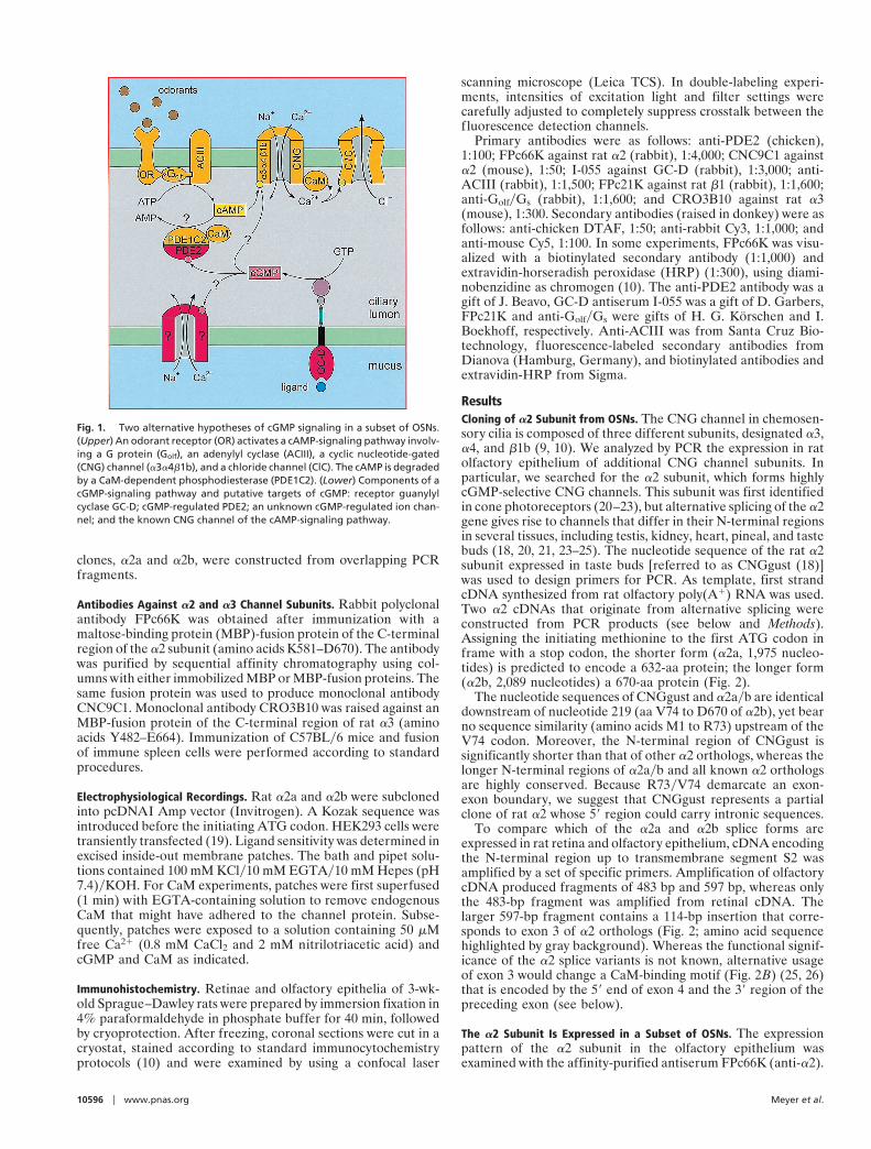
The antiserum is directed against the C terminus of the rat a2and will not discriminate between N-terminal splice variants. Wetested the anti-a2 serum by immunocytochemistry on verticalsections of the rat retina (Fig. 3a). On the left, the retinal layersare shown by differential interference contrast (DIC) optics. Onthe right, strong anti-a2 immunofluorescence is observed incone outer segments, which are readily identified by their typicalmorphology and their location at the inner segment (IS)yrodouter segment (ROS) border. The anti-a2 serum was used underidentical conditions to localize a2 in rat olfactory epithelium. Wedetected immunolabeling in a sparse population of OSNs (Fig.3b). Typically, the cilia of the neurons were strongly labeled,whereas dendrite and soma showed much weaker labeling, oftenin a punctate fashion (Fig. 3b Left). In some experiments, onlythe cilia were stained. Labeled cells were distributed throughoutthe olfactory epithelium either as single isolated neurons or insmall clusters. Often, only a few cells were labeled in a coronalsection of the nasal cavity. Larger groups of a2-immunoreactivecells were identified in the recesses of the olfactory turbinatesnear the cribriform plate (Fig. 3b Right).
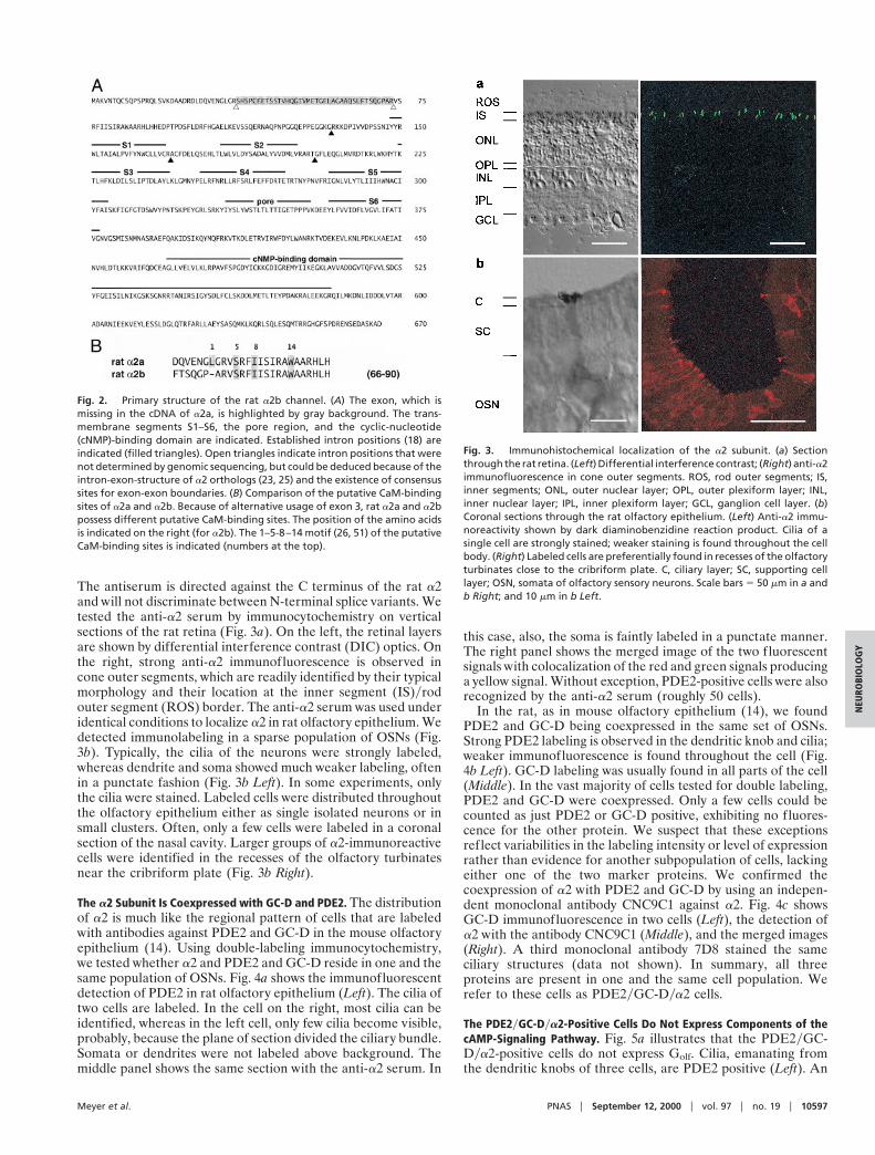
The a2 Subunit Is Coexpressed with GC-D and PDE2. The distributionof a2 is much like the regional pattern of cells that are labeledwith antibodies against PDE2 and GC-D in the mouse olfactoryepithelium (14). Using double-labeling immunocytochemistry,we tested whether a2 and PDE2 and GC-D reside in one and thesame population of OSNs. Fig. 4a shows the immunofluorescentdetection of PDE2 in rat olfactory epithelium (Left). The cilia oftwo cells are labeled. In the cell on the right, most cilia can beidentified, whereas in the left cell, only few cilia become visible,probably, because the plane of section divided the ciliary bundle.Somata or dendrites were not labeled above background. Themiddle panel shows the same section with the anti-a2 serum. In
this case, also, the soma is faintly labeled in a punctate manner.The right panel shows the merged image of the two fluorescentsignals with colocalization of the red and green signals producinga yellow signal. Without exception, PDE2-positive cells were alsorecognized by the anti-a2 serum (roughly 50 cells).
In the rat, as in mouse olfactory epithelium (14), we foundPDE2 and GC-D being coexpressed in the same set of OSNs.Strong PDE2 labeling is observed in the dendritic knob and cilia;weaker immunofluorescence is found throughout the cell (Fig.4b Left). GC-D labeling was usually found in all parts of the cell(Middle). In the vast majority of cells tested for double labeling,PDE2 and GC-D were coexpressed. Only a few cells could becounted as just PDE2 or GC-D positive, exhibiting no fluores-cence for the other protein. We suspect that these exceptionsreflect variabilities in the labeling intensity or level of expressionrather than evidence for another subpopulation of cells, lackingeither one of the two marker proteins. We confirmed thecoexpression of a2 with PDE2 and GC-D by using an indepen-dent monoclonal antibody CNC9C1 against a2. Fig. 4c showsGC-D immunofluorescence in two cells (Left), the detection ofa2 with the antibody CNC9C1 (Middle), and the merged images(Right). A third monoclonal antibody 7D8 stained the sameciliary structures (data not shown). In summary, all threeproteins are present in one and the same cell population. Werefer to these cells as PDE2yGC-Dya2 cells.
The PDE2yGC-Dya2-Positive Cells Do Not Express Components of thecAMP-Signaling Pathway. Fig. 5a illustrates that the PDE2yGC-Dya2-positive cells do not express Golf. Cilia, emanating fromthe dendritic knobs of three cells, are PDE2 positive (Left). An
Fig. 2. Primary structure of the rat a2b channel. (A) The exon, which ismissing in the cDNA of a2a, is highlighted by gray background. The trans-membrane segments S1–S6, the pore region, and the cyclic-nucleotide(cNMP)-binding domain are indicated. Established intron positions (18) areindicated (filled triangles). Open triangles indicate intron positions that werenot determined by genomic sequencing, but could be deduced because of theintron-exon-structure of a2 orthologs (23, 25) and the existence of consensussites for exon-exon boundaries. (B) Comparison of the putative CaM-bindingsites of a2a and a2b. Because of alternative usage of exon 3, rat a2a and a2bpossess different putative CaM-binding sites. The position of the amino acidsis indicated on the right (for a2b). The 1–5-8–14 motif (26, 51) of the putativeCaM-binding sites is indicated (numbers at the top).
Fig. 3. Immunohistochemical localization of the a2 subunit. (a) Sectionthrough the rat retina. (Left) Differential interference contrast; (Right) anti-a2immunofluorescence in cone outer segments. ROS, rod outer segments; IS,inner segments; ONL, outer nuclear layer; OPL, outer plexiform layer; INL,inner nuclear layer; IPL, inner plexiform layer; GCL, ganglion cell layer. (b)Coronal sections through the rat olfactory epithelium. (Left) Anti-a2 immu-noreactivity shown by dark diaminobenzidine reaction product. Cilia of asingle cell are strongly stained; weaker staining is found throughout the cellbody. (Right) Labeled cells are preferentially found in recesses of the olfactoryturbinates close to the cribriform plate. C, ciliary layer; SC, supporting celllayer; OSN, somata of olfactory sensory neurons. Scale bars 5 50 mm in a andb Right; and 10 mm in b Left.
Meyer et al. PNAS u September 12, 2000 u vol. 97 u no. 19 u 10597
NEU
ROBI
OLO
GY
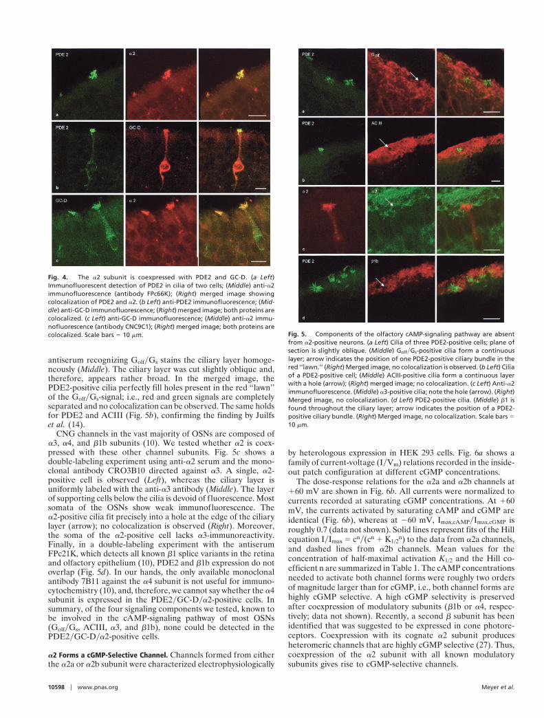
antiserum recognizing GolfyGs stains the ciliary layer homoge-neously (Middle). The ciliary layer was cut slightly oblique and,therefore, appears rather broad. In the merged image, thePDE2-positive cilia perfectly fill holes present in the red ‘‘lawn’’of the GolfyGs-signal; i.e., red and green signals are completelyseparated and no colocalization can be observed. The same holdsfor PDE2 and ACIII (Fig. 5b), confirming the finding by Juilfset al. (14).
CNG channels in the vast majority of OSNs are composed ofa3, a4, and b1b subunits (10). We tested whether a2 is coex-pressed with these other channel subunits. Fig. 5c shows adouble-labeling experiment using anti-a2 serum and the mono-clonal antibody CRO3B10 directed against a3. A single, a2-positive cell is observed (Left), whereas the ciliary layer isuniformly labeled with the anti-a3 antibody (Middle). The layerof supporting cells below the cilia is devoid of fluorescence. Mostsomata of the OSNs show weak immunofluorescence. Thea2-positive cilia fit precisely into a hole at the edge of the ciliarylayer (arrow); no colocalization is observed (Right). Moreover,the soma of the a2-positive cell lacks a3-immunoreactivity.Finally, in a double-labeling experiment with the antiserumFPc21K, which detects all known b1 splice variants in the retinaand olfactory epithelium (10), PDE2 and b1b expression do notoverlap (Fig. 5d). In our hands, the only available monoclonalantibody 7B11 against the a4 subunit is not useful for immuno-cytochemistry (10), and, therefore, we cannot say whether the a4subunit is expressed in the PDE2yGC-Dya2-positive cells. Insummary, of the four signaling components we tested, known tobe involved in the cAMP-signaling pathway of most OSNs(GolfyGs, ACIII, a3, and b1b), none could be detected in thePDE2yGC-Dya2-positive cells.
a2 Forms a cGMP-Selective Channel. Channels formed from eitherthe a2a or a2b subunit were characterized electrophysiologically
by heterologous expression in HEK 293 cells. Fig. 6a shows afamily of current-voltage (IyVm) relations recorded in the inside-out patch configuration at different cGMP concentrations.
The dose-response relations for the a2a and a2b channels at160 mV are shown in Fig. 6b. All currents were normalized tocurrents recorded at saturating cGMP concentrations. At 160mV, the currents activated by saturating cAMP and cGMP areidentical (Fig. 6b), whereas at 260 mV, Imax,cAMPyImax,cGMP isroughly 0.7 (data not shown). Solid lines represent fits of the Hillequation IyImax 5 cny(cn 1 K1/2
n) to the data from a2a channels,and dashed lines from a2b channels. Mean values for theconcentration of half-maximal activation K1/2 and the Hill co-efficient n are summarized in Table 1. The cAMP concentrationsneeded to activate both channel forms were roughly two ordersof magnitude larger than for cGMP, i.e., both channel forms arehighly cGMP selective. A high cGMP selectivity is preservedafter coexpression of modulatory subunits (b1b or a4, respec-tively; data not shown). Recently, a second b subunit has beenidentified that was suggested to be expressed in cone photore-ceptors. Coexpression with its cognate a2 subunit producesheteromeric channels that are highly cGMP selective (27). Thus,coexpression of the a2 subunit with all known modulatorysubunits gives rise to cGMP-selective channels.
Fig. 4. The a2 subunit is coexpressed with PDE2 and GC-D. (a Left)Immunofluorescent detection of PDE2 in cilia of two cells; (Middle) anti-a2immunofluorescence (antibody FPc66K); (Right) merged image showingcolocalization of PDE2 and a2. (b Left) anti-PDE2 immunofluorescence; (Mid-dle) anti-GC-D immunofluorescence; (Right) merged image; both proteins arecolocalized. (c Left) anti-GC-D immunofluorescence; (Middle) anti-a2 immu-nofluorescence (antibody CNC9C1); (Right) merged image; both proteins arecolocalized. Scale bars 5 10 mm. Fig. 5. Components of the olfactory cAMP-signaling pathway are absent
from a2-positive neurons. (a Left) Cilia of three PDE2-positive cells; plane ofsection is slightly oblique. (Middle) GolfyGs-positive cilia form a continuouslayer; arrow indicates the position of one PDE2-positive ciliary bundle in thered ‘‘lawn.’’ (Right) Merged image, no colocalization is observed. (b Left) Ciliaof a PDE2-positive cell; (Middle) ACIII-positive cilia form a continuous layerwith a hole (arrow); (Right) merged image; no colocalization. (c Left) Anti-a2immunofluorescence. (Middle) a3-positive cilia; note the hole (arrow). (Right)Merged image, no colocalization. (d Left) PDE2-positive cilia. (Middle) b1 isfound throughout the ciliary layer; arrow indicates the position of a PDE2-positive ciliary bundle. (Right) Merged image, no colocalization. Scale bars 510 mm.
10598 u www.pnas.org Meyer et al.
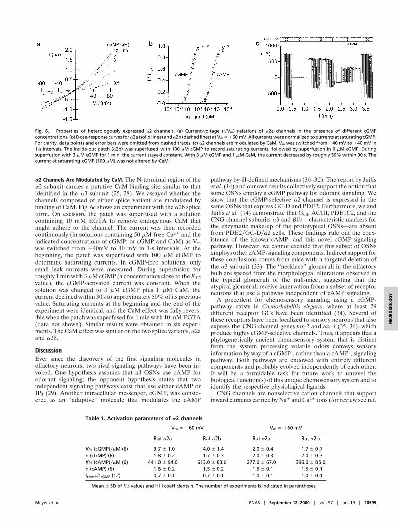
a2 Channels Are Modulated by CaM. The N-terminal region of thea2 subunit carries a putative CaM-binding site similar to thatidentified in the a3 subunit (25, 28). We assayed whether thechannels composed of either splice variant are modulated bybinding of CaM. Fig. 6c shows an experiment with the a2b spliceform. On excision, the patch was superfused with a solutioncontaining 10 mM EGTA to remove endogenous CaM thatmight adhere to the channel. The current was then recordedcontinuously (in solutions containing 50 mM free Ca21 and theindicated concentrations of cGMP; or cGMP and CaM) as Vmwas switched from 240mV to 40 mV in 1-s intervals. At thebeginning, the patch was superfused with 100 mM cGMP todetermine saturating currents. In cGMP-free solutions, onlysmall leak currents were measured. During superfusion forroughly 1 min with 3 mM cGMP (a concentration close to the K1/2value), the cGMP-activated current was constant. When thesolution was changed to 3 mM cGMP plus 1 mM CaM, thecurrent declined within 30 s to approximately 50% of its previousvalue. Saturating currents at the beginning and the end of theexperiment were identical, and the CaM effect was fully revers-ible when the patch was superfused for 1 min with 10 mM EGTA(data not shown). Similar results were obtained in six experi-ments. The CaM effect was similar on the two splice variants, a2aand a2b.
DiscussionEver since the discovery of the first signaling molecules inolfactory neurons, two rival signaling pathways have been in-voked. One hypothesis assumes that all OSNs use cAMP forodorant signaling; the opponent hypothesis states that twoindependent signaling pathways exist that use either cAMP orIP3 (29). Another intracellular messenger, cGMP, was consid-ered as an ‘‘adaptive’’ molecule that modulates the cAMP
pathway by ill-defined mechanisms (30–32). The report by Juilfset al. (14) and our own results collectively support the notion thatsome OSNs employ a cGMP pathway for odorant signaling. Weshow that the cGMP-selective a2 channel is expressed in thesame OSNs that express GC-D and PDE2. Furthermore, we andJuilfs et al. (14) demonstrate that Golf, ACIII, PDE1C2, and theCNG channel subunits a3 and b1b—characteristic markers forthe enzymatic make-up of the prototypical OSNs—are absentfrom PDE2yGC-Dya2 cells. These findings rule out the coex-istence of the known cAMP- and this novel cGMP-signalingpathway. However, we cannot exclude that this subset of OSNsemploys other cAMP-signaling components. Indirect support forthese conclusions comes from mice with a targeted deletion ofthe a3 subunit (33). The ‘‘necklace’’ glomeruli in the olfactorybulb are spared from the morphological alterations observed inthe typical glomeruli of the null-mice, suggesting that theatypical glomeruli receive innervation from a subset of receptorneurons that use a pathway independent of cAMP signaling.
A precedent for chemosensory signaling using a cGMP-pathway exists in Caenorhabditis elegans, where at least 29different receptor GCs have been identified (34). Several ofthese receptors have been localized to sensory neurons that alsoexpress the CNG channel genes tax-2 and tax-4 (35, 36), whichproduce highly cGMP-selective channels. Thus, it appears that aphylogenetically ancient chemosensory system that is distinctfrom the system processing volatile odors conveys sensoryinformation by way of a cGMP-, rather than a cAMP-, signalingpathway. Both pathways are endowed with entirely differentcomponents and probably evolved independently of each other.It will be a formidable task for future work to unravel thebiological function(s) of this unique chemosensory system and toidentify the respective physiological ligands.
CNG channels are nonselective cation channels that supportinward currents carried by Na1 and Ca21 ions (for review see ref.
Fig. 6. Properties of heterologously expressed a2 channels. (a) Current-voltage (IyVm) relations of a2a channels in the presence of different cGMPconcentrations. (b) Dose-response curves for a2a (solid lines) and a2b (dashed lines) at Vm 5 160 mV. All currents were normalized to currents at saturating cGMP.For clarity, data points and error bars were omitted from dashed traces. (c) a2 channels are modulated by CaM. Vm was switched from 240 mV to 140 mV in1-s intervals. The inside-out patch (a2b) was superfused with 100 mM cGMP to record saturating currents, followed by superfusion in 0 mM cGMP. Duringsuperfusion with 3 mM cGMP for 1 min, the current stayed constant. With 3 mM cGMP and 1 mM CaM, the current decreased by roughly 50% within 30 s. Thecurrent at saturating cGMP (100 mM) was not altered by CaM.
Table 1. Activation parameters of a2 channels
Vm 5 260 mV Vm 5 160 mV
Rat a2a Rat a2b Rat a2a Rat a2b
K1⁄2 (cGMP)ymM (6) 3.7 6 1.0 4.0 6 1.4 2.0 6 0.4 1.7 6 0.7n (cGMP) (6) 1.8 6 0.2 1.7 6 0.3 2.0 6 0.3 2.0 6 0.3K1⁄2 (cAMP)ymM (6) 441.0 6 94.0 613.0 6 83.0 277.0 6 67.0 396.0 6 85.0n (cAMP) (6) 1.6 6 0.2 1.5 6 0.2 1.5 6 0.1 1.5 6 0.1IcAMPyIcGMP (12) 0.7 6 0.1 0.7 6 0.1 1.0 6 0.1 1.0 6 0.1
Mean 6 SD of K1⁄2 values and Hill coefficients n. The number of experiments is indicated in parentheses.
Meyer et al. PNAS u September 12, 2000 u vol. 97 u no. 19 u 10599
NEU
ROBI
OLO
GY
11). In OSNs that use a cAMP-signaling pathway, the odorant-stimulated rise in [Ca21]i (37) plays an important role in bothexcitation and adaptation. A rise in [Ca21]i activates Cl2 chan-nels that carry a large fraction of the odorant-induced receptorcurrent (38–40). Ca21 also stimulates cAMP hydrolysis by theciliary CaM-dependent PDE1C2 (14, 41), and it attenuates thecAMP sensitivity of the CNG channel by a mechanism involvingCaM (28, 42). Both mechanisms terminate the odorant responseand lower the sensitivity of the OSN to subsequent stimulation(43). CNG channels containing a2 subunits are roughly as Ca21
permeable as the a3a4b1b channels (44–47), and stimulation ofOSNs equipped with a cGMP-signaling pathway is expected toraise [Ca21]i.
At present, we can only speculate about a Ca21-dependentfeedback in PDE2yGC-Dya2 cells. The CaM-dependentPDE1C2 is absent from these cells (14), and the CaM action ona2 channels is much weaker than on CNG channels in typicalOSNs (42). It is conceivable that adaptation to ligands thatcontrol important aspects of behavior differs from the fast andcomplete adaptation to normal odors in typical OSNs. Alterna-tively, an unknown Ca21-binding protein with much higher
efficacy than CaM might be the authentic modulator of CNGchannels in PDE2yGC-Dya2 cells.
By analogy to what is known in photoreceptors, the shift of thecGMP sensitivity may not be the only action of Ca21 insidePDE2yGC-Dya2 cells. Guanylyl cyclases GC-E and GC-F ex-pressed in retinal photoreceptors are regulated by [Ca21]i (48) andnot by extracellular ligands. Their Ca21 sensitivity is mediated by aset of guanylyl cyclase-activating proteins (GCAPs), which aremembers of the large family of Ca21-binding proteins (for reviewsee ref. 49). GC-D is phylogenetically more akin to the Ca21-regulated GC-E and GC-F than to receptor GCs-AyByC, which areactivated by peptide ligands. In particular, GC-D and GC-EyFshare characteristic sequence similarity in a regulatory domain thatis involved in binding of GCAPs (50). This similarity raises theintriguing possibility that GC-D activity is under the dual control ofan unknown extracellular ligand and [Ca21]i.
We thank D. Garbers (Dallas), J. Beavo (Seattle), I. Boekhoff (Stutt-gart), and H. G. Korschen (Julich) for gifts of antibodies, A. Kongeterand W. Bonigk for characterization of antibody CR03B10, and J. Bradley(Paris) for many helpful comments on the manuscript. This work wassupported by the European Commission (BIO4-98-0034).
1. Gold, G. H. (1999) Annu. Rev. Physiol. 61, 857–871.2. Buck, L. & Axel, R. (1991) Cell 65, 175–187.3. Mombaerts, P. (1999) Nat. Neurosci. 2, 686–687.4. Bakalyar, H. A. & Reed, R. R. (1990) Science 250, 1403–1406.5. Jones, D. T. & Reed, R. R. (1989) Science 244, 790–795.6. Breer, H., Boekhoff, I. & Tareilus, E. (1990) Nature (London) 345, 65–68.7. Nakamura, T. & Gold, G. H. (1987) Nature (London) 325, 442–444.8. Frings, S. & Lindemann, B. (1991) J. Gen. Physiol. 97, 1–16.9. Sautter, A., Zong, X., Hofmann, F. & Biel, M. (1998) Proc. Natl. Acad. Sci. USA
95, 4696–4701.10. Bonigk, W., Bradley, J., Muller, F., Sesti, F., Boekhoff, I., Ronnett, G. V.,
Kaupp, U. B. & Frings, S. (1999) J. Neurosci. 19, 5332–5347.11. Kaupp, U. B. (1995) Curr. Opinion Neurobiol. 5, 434–442.12. Finn, J. T., Grunwald, M. E. & Yau, K.-W. (1996) Annu. Rev. Physiol. 58,
395–426.13. Fulle, H.-J., Vassar, R., Foster, D. C., Yang, R.-B., Axel, R. & Garbers, D. L.
(1995) Proc. Natl. Acad. Sci. USA 92, 3571–3575.14. Juilfs, D. M., Fulle, H.-J., Zhao, A. Z., Houslay, M. D., Garbers, D. L. & Beavo,
J. A. (1997) Proc. Natl. Acad. Sci. USA 94, 3388–3395.15. Foster, D. C., Wedel, B. J., Robinson, S. W. & Garbers, D. L. (1999) Rev.
Physiol. Biochem. Pharmacol. 135, 1–39.16. Teicher, M. H., Stewart, W. B., Kauer, J. S. & Shepherd, G. M. (1980) Brain
Res. 194, 530–535.17. Greer, C. A., Stewart, W. B., Teicher, M. H., Kauer, J. S. & Shepherd, G. M.
(1982) J. Neurosci. 12, 1744–1759.18. Misaka, T., Kusakabe, Y., Emori, Y., Gonoi, T., Arai, S. & Abe, K. (1997)
J. Biol. Chem. 272, 22623–22629.19. Baumann, A., Frings, S., Godde, M., Seifert, R. & Kaupp, U. B. (1994) EMBO
J. 13, 5040–5050.20. Bonigk, W., Altenhofen, W., Muller, F., Dose, A., Illing, M., Molday, R. S. &
Kaupp, U. B. (1993) Neuron 10, 865–877.21. Weyand, I., Godde, M., Frings, S., Weiner, J., Muller, F., Altenhofen, W., Hatt,
H. & Kaupp, U. B. (1994) Nature (London) 368, 859–863.22. Yu, W.-P., Grunwald, M. E. & Yau, K.-W. (1996) FEBS Lett. 393, 211–215.23. Wissinger, B., Muller, F., Weyand, I., Schuffenhauer, S., Thanos, S., Kaupp,
U. B. & Zrenner, E. (1997) Eur. J. Neurosci. 9, 2512–2521.24. Biel, M., Zong, X., Distler, M., Bosse, E., Klugbauer, N., Murakami, M.,
Flockerzi, V. & Hofmann, F. (1994) Proc. Natl. Acad. Sci. USA 91, 3505–3509.
25. Bonigk, W., Muller, F., Middendorff, R., Weyand, I. & Kaupp, U. B. (1996)J. Neurosci. 16, 7458–7468.
26. Grunwald, M. E., Zhong, H., Lai, J. & Yau, K.-W. (1999) Proc. Natl. Acad. Sci.USA 96, 13444–13449.
27. Gerstner, A., Zong, X., Hofmann, F. & Biel, M. (2000) J. Neurosci. 20,1324–1332.
28. Liu, M., Chen, T.-Y., Ahamed, B., Li, J. & Yau, K.-W. (1994) Science 266,1348–1354.
29. Schild, D. & Restrepo, D. (1998) Physiol. Rev. 78, 429–466.30. Breer, H. & Shepherd, G. M. (1993) Trends Neurosci. 16, 5–9.31. Kroner, C., Boekhoff, I., Lohmann, S. M., Genieser, H.-G. & Breer, H. (1996)
Eur. J. Biochem. 236, 632–637.32. Zufall, F. & Leinders-Zufall, T. (1998) Ann. N. Y. Acad. Sci. 855, 199–204.33. Baker, H., Cummings, D. M., Munger, S. D., Margolis, J. W., Franzen, L., Reed,
R. R. & Margolis, F. L. (1999) J. Neurosci. 19, 9313–9321.34. Yu, S., Avery, L., Baude, E. & Garbers, D. L. (1997) Proc. Natl. Acad. Sci. USA
94, 3384–3387.35. Coburn, C. M. & Bargmann, C. I. (1996) Neuron 17, 695–706.36. Komatsu, H., Mori, I., Rhee, J.-S., Akaike, N. & Ohshima, Y. (1996) Neuron
17, 707–718.37. Leinders-Zufall, T., Greer, C. A., Shepherd, G. M. & Zufall, F. (1998)
J. Neurosci. 18, 5630–5639.38. Kleene, S. J. (1993) Neuron 11, 123–132.39. Kurahashi, T. & Yau, K.-W. (1993) Nature (London) 363, 71–74.40. Lowe, G. & Gold, G. H. (1993) Nature (London) 366, 283–286.41. Yan, C., Zhao, A. Z., Bentley, J. K. & Beavo, J. A. (1996) J. Biol. Chem. 271,
25699–25706.42. Chen, T.-Y. & Yau, K.-W. (1994) Nature (London) 368, 545–548.43. Kurahashi, T. & Menini, A. (1997) Nature (London) 385, 725–729.44. Perry, R. J. & McNaughton, P. A. (1991) J. Physiol. 434, 70P.45. Frings, S., Seifert, R., Godde, M. & Kaupp, U. B. (1995) Neuron 15, 169–179.46. Picones, A. & Korenbrot, J. I. (1995) Biophys. J. 69, 120–127.47. Dzeja, C., Hagen, V., Kaupp, U. B. & Frings, S. (1999) EMBO J. 18,48. Koch, K.-W. & Stryer, L. (1988) Nature (London) 334, 64–66.49. Polans, A., Baehr, W. & Palczewski, K. (1996) Trends Neurosci. 19, 547–554.50. Lange, C., Duda, T., Beyermann, M., Sharma, R. K. & Koch, K.-W. (1999)
FEBS Lett. 460, 27–31.51. Rhoads, A. R. & Friedberg, F. (1997) FASEB J. 11, 331–340.
10600 u www.pnas.org Meyer et al.