Embed Size (px)
Citation preview
200 J Orofac Orthop 2009 · No. 3 © Urban & Vogel
Journal of Orofacial OrthopedicsFortschritte der Kieferorthopädie Original Article
Alignment of Impacted Maxillary Canines: Critical Analysis of Eruption Path and Treatment TimeKritische Analyse von Weg und Zeit bei der Eckzahn-einordnung in den ZahnbogenMichael Schubert1, Uwe Baumert2
1 Private Orthodontic Practice, Regensburg, Germany,2 Department of Orthodontics, Munich University Hospital, Ludwig
Maximilian University, Munich, Germany.
Received: January 7, 2009; accepted: March 9, 2009
Abstract Objective: The aim of this study was to evaluate a possible cor-relation between treatment time and the distance covered by the canine tip during forced eruption of impacted maxillary canines. We also addressed the question whether it is possible to estimate the treatment time based on this data.Materials and Methods: Orthopantomograms of 57 juvenile pa-tients below 18 years of age with at least one palatally impacted canine were examined. We determined the position of the canine in relation to the neighboring teeth, its axial inclination and distance to the occlusal plane. We also introduced a new linear measurement representing the distance covered by the canine’s crown tip during the alignment of the impacted tooth.Results: The length of overall treatment time and canine align-ment, its axial inclination and the distance covered by the canine correlated significantly with the canine’s position relative to its neighboring teeth. The newly-introduced linear measurement was the only measure that proved gender specific. With regres-sion analysis we demonstrated that this measure is sufficient to predict the active treatment time for canine alignment (r2 = 0.391). This indicates that 39.1% of the differences ob-served in active treatment time can be explained by variation in this new parameter.Conclusions: The new measure introduced in this study provides a practice-oriented parameter with which to predict the treat-ment time for aligning impacted canines. Current 2D imaging diagnostics most likely restrict the ability to predict the length of therapy at 40%. Individual bone density and metabolism seem to have a strong influence on treatment time and must be taken into account for a more exact prognosis.
Key Words: Maxillary canine impaction · Prognosis of treatment time · EWC® System
Zusammenfassung Ziel: Von Interesse war es, in einer Studie über palatinal verla-gerte Eckzähne zu überprüfen, ob es einen Zusammenhang zwi-schen der Behandlungszeit und dem zurückgelegten Weg der Eckzahnkronenspitze bei der Einordnung gibt. Kann daraus eine Behandlungszeitprognose abgeleitet werden?Material und Methodik: Die Orthopantomogramme von 57 ju-gendlichen Patienten unter 18 Jahren mit mindestens einem pa-latinal verlagerten Eckzahn wurden ausgewertet. Neben seiner Position zu den Nachbarzähnen wurden die Achsenneigung des Eckzahnes und der Abstand zur Okklusionsebene ermittelt. Zu-sätzlich wurde eine Messstrecke eingeführt, die den zurückge-legten Weg der Eckzahnkronenspitze während der Einordnung darstellte. Ergebnisse: Die Dauer der Gesamtbehandlung und die der Eck-zahneinordnung, seine Achsenneigung und der zurückgelegte Weg waren signifikant von der Position des Eckzahnes zu seinen Nachbarzähnen abhängig. Die neu eingeführte Messstrecke er-wies sich als geschlechtsspezifisch. Mit ihr und einer Geradenfor-mel (r2 = 0,391) konnte die Dauer der Eckzahneinstellung berech-net werden. Durch die neu eingeführte Messstrecke können 39,1% der beobachteten Unterschiede in der aktiven Behandlungszeit erklärt werden.Schlussfolgerungen: Mit dem neuen Messparameter wurde ein praxisgerechter Weg für eine Prognose der Dauer der Eckzahnein-ordnung gefunden. In einem Bereich von etwa 40% dürfte die Grenze für eine Prognose mit Hilfe des zweidimensionalen bildge-benden Verfahrens liegen. Individuelle Knochendichte und Stoff-wechselvorgänge scheinen die Behandlungszeit stark zu beein-flussen und müssten für eine genauere Prognose mit einbezogen werden.
Schlüsselwörter: Eckzahnretention · Behandlungszeit-prognose · EWC®-System
J Orofac Orthop 2009;70:200–12
DOI 10.1007/s00056-009-0901-3
201J Orofac Orthop 2009 · No. 3 © Urban & Vogel
Schubert M, Baumert U. Einordnung verlagerter oberer Eckzähne
201J Orofac Orthop 2009 · No. 3 © Urban & Vogel
IntroductionThe second most frequently impacted teeth after the wis-dom teeth are the upper canines. The frequency reported in the literature varies from 1.5% to 2% in the maxilla and 0.3% in the mandible in randomly elected probands [5, 8, 10, 11, 18, 23]. By contrast, the frequency is as high as 23.5% in orthodontic practices (selected groups) [3, 15, 31]. The rate among female patients is twice as high as among males: 1.17% vs. 0.51% [2]. Palatal impaction prevails over the buccal position by a ratio of 2:1 to 9:1, ca. 20% palatal, ca. 15% buccal [12].
Various therapeutic options are available to patients af-fected by this anomaly:1. extraction of the impacted tooth and morphological im-
provement in the primary canine crown when it persists,2. extraction of the impacted tooth and primary canine fol-
lowed by orthodontic space closure,3. autotransplantation after orthodontic pretreatment for
space opening,4. implant-supported single tooth prosthesis as the primary
care after primary canine and impacted tooth extraction, or when autotransplantation fails, and
5. orthodontic alignment of the displaced tooth after its surgical exposure.
In deciding among these options, the treatment’s duration is an important criterion for the patient. Until now, how-ever, the estimation of treatment time has also been based on the orthodontist’s subjective clinical experience [30]; the data from scientific studies of this criterion vary to a con-siderable degree [17, 19, 21, 32]. Many authors agree that at present the orthopantomogram still offers a useful diagnos-tic method, and it occupies a key position, although it is known that X-ray images of the canine area include a cer-tain amount of distortion [26].
As early as 1970, Dausch-Neumann [9] used orthopan-tomograms to describe the eruption path of permanent ca-nines using her own measurement technique. Ericson & Ku-rol [13, 14] developed a method involving sectors to deter-mine the time factor. Both studies provide guidelines for scientific claims nowadays [17, 24, 32].
After the tooth being aligned was moved during the ac-tive treatment time from its initial activation into its final position with the EWC® System [29] used here, it seemed reasonable to relate the distance the canine must cover to the treatment time required. This necessitated a new measure-ment distance in the orthopantomogram. The goal of this retrospective study also involves examining to what extent additional parameters such as vertical and anteroposterior position and the angulation of palatally-displaced canines influence treatment time. We also included a patient group with bilateral palatally-displaced canines in the study to ex-amine whether their treatment time would differ from that of patients with only one displaced tooth.
EinleitungNach den Weisheitszähnen sind die oberen Eckzähne die am häufigsten impaktierten Zähne. Über das Vorkommen variieren die Literaturangaben von 1,5–2% im Oberkiefer und 0,3% im Unterkiefer bei unselektierten Probanden [5, 8, 10, 11, 18, 23]. In kieferorthopädischen Praxen (selek-tierte Gruppen) liegt dagegen die Häufigkeit bei bis zu 23,5% [3, 15, 31]. Das Vorkommen bei weiblichen Pa-tienten ist doppelt so hoch wie bei männlichen: 1,17% vs. 0,51% [2]. Die palatinale Verlagerung dominiert gegenüber der bukkalen Lage in einem Verhältnis von 2:1 bis 9:1, ca. 20% palatinal, ca. 15% bukkal [12].
Ist ein Patient von dieser Unregelmäßigkeit betroffen, gibt es für ihn verschiedene Möglichkeiten der Therapie:1. Entfernung des verlagerten Zahnes und morphologische
Verbesserung der Milcheckzahnkrone bei deren Persistenz2. Entfernung des verlagerten Zahnes und des Milcheck-
zahnes mit anschließendem kieferorthopädischem Lü-ckenschluss
3. Autotransplantation nach kieferorthopädischer Vorbe-handlung zur Lückenöffnung
4. Implantatgetragener Einzelzahnersatz als primäre Ver-sorgung nach Extraktion des Milcheckzahnes und des verlagerten Zahnes oder im Falle des misslungenen Autotransplantates
5. Kieferorthopädische Einordnung des verlagerten Zahnes nach chirurgischer Freilegung
Um zwischen diesen Möglichkeiten entscheiden zu können, ist die Frage nach der Dauer der Behandlung für den Pati-enten ein wichtiges Kriterium. Die Schätzung der Behand-lungsdauer beruht bisher allerdings nur auf der subjektiven klinischen Erfahrung des Kieferorthopäden [30], die auf wis-senschaftlichen Untersuchungen basierenden Informationen darüber variieren in beträchtlichem Ausmaß [17, 19, 21, 32]. Unabhängig davon sind sich viele Autoren einig, dass zurzeit das Orthopantomogramm noch die gebräuchliche Diagnos-tikmethode darstellt und eine gewisse Schlüsselstellung ein-nimmt, im Wissen, dass es bei der speziellen Aufnahmetech-nik im Eckzahnbereich zu Verzeichnungen kommt [26].
Bereits 1970 beschrieb Dausch-Neumann [9] an Ortho-pantomogrammen den Durchbruchsweg bleibender Eck-zähne mit einer eigenen Messtechnik. Ericson & Kurol [13, 14] entwickelten eine Methode mit Sektoren, um den Zeit-faktor zu ermitteln. Beide Studien sind heute noch Richtli-nien für wissenschaftliche Aussagen [17, 24, 32].
Nachdem bei der Eckzahneinstellung mit dem hier ver-wendeten EWC®-System [29] der einzuordnende Zahn von der ersten Aktivierung an direkt auf seine ihm zugeordnete Stelle hin bewegt wird, schien es sinnvoll, den zurückgelegten Weg mit der dazu benötigten Zeitdauer zu vergleichen. Dazu war eine neue Messstrecke im Orthopantomogramm erfor-derlich. Das Ziel dieser retrospektiven Studie bezieht sich weiter auf die Überprüfung, inwieweit zusätzliche Parameter
Schubert M, Baumert U. Alignment of Impacted Maxillary Canines
202 J Orofac Orthop 2009 · No. 3 © Urban & Vogel
Materials and MethodsIn this retrospective study we analyzed the data of 47 ado-lescent patients (n = 47; 33 female, 14 male) treated at an orthodontic practice in Regensburg, Germany between 1997 and 2007. The patients were selected for participation in this study according to the following criteria:1. Adolescent patients no older than 18 years of age pre-
senting with at least one palatally-displaced canine.2. Surgical exposure had to involve a closed-eruption ap-
proach using an intra-operatively adhesively-fixed at-tachment. The EWC® System was selected as the trac-tion force system [29].
3. After differential diagnosis had determined no tooth ex-tractions, no premolars were removed.
4. Treatment was performed with a vestibular multiband appliance (.018 slot).
5. Treatment dates were kept regularly and the alignment of the displaced canine had to have been successfully completed.
6. Complete diagnostic and treatment records consisting of panoramic radiographs and lateral cephalograms, photo-graphs and study casts, the dates of multibracket/multi-band application, surgical exposure and appliance re-moval had to be available.
None of the orthopantomograms available for analysis were any older than 1 year at the time of the canine’s surgi-cal exposure. Some of the patients supplied their own or-thopantomograms taken by the dentist who had referred them. Those images were generally X-ray negatives. Other orthopantomograms were taken using a digital X-ray pro-cedure in the orthodontic practice. All orthopantomograms at the beginning of treatment were analyzed under stan-dardized conditions, the X-ray negative films were traced onto acetate films, glossy copies were made from the digital orthopantomograms and analyzed. The tracings were done using a Staedtler Marsmicro 0.3 mm (Staedler Mars GmbH & Co. KG, Nürnberg, Germany), all measurements were taken by one examiner only.
The occlusal plane was used as the horizontal reference line [6, 9], defined by the mesiobuccal cusp tip of the upper first molar and the incisal edge of the upper central incisor on the same side (Figure 1). The midline between the longitudi-nal axis of both central incisors was defined as the vertical reference line. We also drew in the longitudinal axes of the displaced canine and adjacent lateral incisor.
The inclination of the displaced tooth was measured with a goniometer to 1°, the interval distances were measured with a digital sliding caliper (Electronic Digital Caliper, Masel, Germany) to 0.01 mm and rounded up or down to 0.1 mm.
The following data were gathered for this critical analysis:1. date of birth and gender, 2. patient age at the start of treatment and surgical expo-
sure,
wie die vertikale Position, die antero-posteriore Position und die Angulation von palatinal verlagerten Eckzähnen einen Einfluss auf die Behandlungsdauer haben. Zusätzlich wurde eine Patientengruppe mit beidseitig palatinal verlagerten Eckzähnen in die Studie aufgenommen, um zu prüfen, ob sich die Behandlungszeit gegenüber nur einseitig verlagerten unterscheidet.
Material und MethodikIn dieser retrospektiven Studie wurden die Daten von 47 jugendlichen Patienten (n = 47; 33 weiblich, 14 männlich) ausgewertet, die in einer kieferorthopädischen Fachpraxis in Regensburg, Deutschland, in der Zeit von 1997–2007 be-handelt wurden. Die Auswahl der Patienten zur Teilnahme an dieser Studie beruhte auf den folgenden Kriterien:1. Die jugendlichen Patienten von nicht älter als 18 Jahren
hatten mindestens einen palatinal verlagerten Eckzahn.2. Die chirurgische Freilegungstechnik erfolgte immer über
einen geschlossenen Zugang mit intraoperativem Kle-ben eines Attachments. Das EWC®-System wurde als Zughilfe gewählt [29].
3. Nach der Differenzialdiagnose lag ein Non-Ex-Kasus vor, so dass keine Extraktion der Prämolaren vorgenom-men wurde.
4. Zur Behandlung diente eine vestibuläre Multibandappa-ratur (0.018-Slot).
5. Die Behandlungstermine wurden regelmäßig eingehal-ten und die Einstellung des verlagerten Eckzahnes wurde erfolgreich beendet.
6. Vollständige Diagnostik und Behandlungsunterlagen, bestehend aus Panorama- und Fernröntgenaufnahme, Fotografien und Modellen, Zeitpunkt der Bebänderung, der chirurgischen Freilegung und der Entbänderung mussten vorliegen.
Alle Orthopantomogramme, die zur Bewertung anstanden, waren zum Zeitpunkt der chirurgischen Freilegung nicht älter als 1 Jahr. Ein Teil der Patienten brachte eigene Ortho-pantomogramme mit, die vom überweisenden Zahnarzt an-gefertigt worden waren. Hierbei handelte es sich in der Regel um Röntgennegativfilme. Andere Orthopantomo-gramme wurden über digitales Röntgenverfahren in der kie-ferorthopädischen Fachpraxis hergestellt. Alle Orthopanto-mogramme vom Behandlungsbeginn wurden unter standar-disierten Bedingungen ausgewertet, die Röntgennegativfilme auf Acetatfolien durchgezeichnet, von den digitalen Ortho-pantomogrammen Hochglanzabzüge erstellt und diese be-wertet. Die Zeichnungen erfolgten mit einem Staedtler Marsmicro 0,3 mm (Staedtler Mars GmbH & Co. KG, Nürn-berg, Deutschland), sämtliche Messungen führte ausschließ-lich ein Untersucher durch.
Als horizontale Referenzlinie [6, 9] diente die Okklu-sionsebene, definiert durch die mesiobukkale Höckerskizze des oberen ersten Molaren und die Inzisalkante des mittle-
203J Orofac Orthop 2009 · No. 3 © Urban & Vogel
Schubert M, Baumert U. Einordnung verlagerter oberer Eckzähne
203J Orofac Orthop 2009 · No. 3 © Urban & Vogel
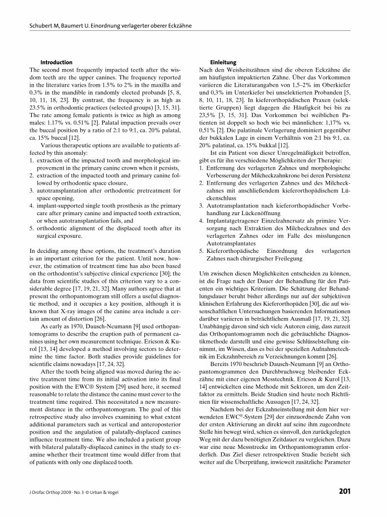
3. canine inclination α as the angle between its longitudi-nal axis and the vertical reference line (midline) [10] (Figure 1),
4. canine inclination � as the angle between its longitudi-nal axis and the longitudinal axis of the lateral incisor [10] (Figure 1),
5. the distance d1 from the occlusal plane projected as a perpendicular from the canine tip to the occlusal plane (Figure 1), and
6. the distance d2 from the canine tip to its target point P on the occlusal plane (Figure 1). This target point P is defined as the point of intersection between the occlusal plane and the perpendicular formed from the middle of the shortest connecting lines between the distal rim of the lateral incisor and mesial rim of the first premolar.
7. The mesiodistal position of the canine tip in relation to the neighboring teeth referred to as zone [14] (Figure 1),
8. overall treatment time in months,9. duration of canine alignment defined as the time from
the canine’s surgical exposure until fixed appliance re-moval, and
ren oberen Schneidezahnes derselben Seite (Abbildung 1). Als vertikale Bezugslinie wurde die Mittellinie zwischen der Längsachse der beiden mittleren Schneidezähne definiert. Zusätzlich erfolgten die Einzeichnung der Längsachse des verlagerten Eckzahnes und die des lateralen Schneidezahnes der entsprechenden Seite.
Die Neigung des verlagerten Zahnes wurde mit einem Winkelmesser auf 1° genau vermessen, die Abstandsstre-cken wurden mit einer digitalen Schiebelehre (Electronic Digital Caliper, Masel, Deutschland) auf 0,01 mm genau er-fasst und auf 0,1 mm auf- bzw. abgerundet.
Folgende Daten wurden für die vorliegende kritische Analyse erhoben:1. Geburtsdatum und Geschlecht 2. Alter der Patienten zum Zeitpunkt des Behandlungsbe-
ginns und der chirurgischen Freilegung 3. Eckzahnneigung α als Winkel zwischen seiner Längs-
achse und der vertikalen Bezugslinie (Mittellinie) [10] (Abbildung 1)
4. Eckzahnneigung � als Winkel zwischen seiner Längs-achse und der Längsachse des seitlichen Schneidezahnes [10] (Abbildung 1)
5. Der Abstand d1 von der Okklusionsebene als Lot von der Eckzahnspitze auf die Okklusionsebene gefällt (Ab-bildung 1)
6. Der Abstand d2 von der Eckzahnspitze zu ihrem Ziel-punkt P auf der Okklusionsebene (Abbildung 1). Dieser Zielpunkt P wird definiert als Schnittpunkt zwischen der Okklusionsebene und dem Lot, das von der Mitte der kürzesten Verbindungslinien zwischen dem distalen Rand des seitlichen Schneidezahnes und des mesialen Randes des ersten Prämolaren verläuft
7. Die mesiodistale Position der Eckzahnspitze in Relation zu den benachbarten Zähnen bezeichnet als Zone [14] (Abbildung 1)
8. Dauer der Gesamtbehandlungszeit in Monaten9. Dauer der Eckzahneinstellung, definiert als die Zeit von
der Freilegung bis zur Entbänderung10. Zahl der Sitzungen von der Freilegung bis zur Entbän-
derung, einschließlich Reparatursitzungen
Bei acht Patienten, die eine doppelseitige Verlagerung auf-wiesen, wurde derjenige Eckzahn für die Messung herange-zogen, der ungünstiger lag (weiter entfernt von der Okklu-sionsebene, stärkere Neigung, mehr mesial liegend).
Zur statistischen Analyse wurden zunächst Normalver-teilung und Homogenität der Varianzen der erhobenen Messwerte mittels Kolmogorov-Smirnov- bzw. Levene-Test überprüft (SPSS® für Windows® Version 16.0; SPSS Inc., Chicago, IL, USA). Alle Vergleiche wurden mittels Mann-Whitney-U-Test bzw. Kruskal-Wallis-H-Test durchgeführt. Ergebnisse mit p ≤ 0,05 wurden als signifikant betrachtet. Die Regressionsanalysen und deren grafische Darstellungen wurden mit dem Programm SigmaStat® 3.5 bzw. SigmaPlot®
α
β
OcclusalPlane Line
VerticalReferenceLine
d2
d1
1 2 3 4 5
Figure 1. Schematic illustration of the investigated parameters in the panoramic X-ray: the projection of the canine and zones (1–5) of the most medial position of the crown; the inclination of the maxillary canine to the midline (α) and long axis of the lateral incisor (β); the degree of vertical eruption of the maxillary canine determined as distance d1; intro-duced distance d2 (modified and completed after Ericson & Kurol [14]).
Abbildung 1. Schematische Darstellung der untersuchten Messparameter im Orthopantomogramm: die Projektion des Eckzahns und die Zonen (1–5) der am weitesten medial liegenden Position der Krone; die Inklina-tion des oberen Eckzahns zur Mittellinie (α) und zur Längsachse des seitlichen Eckzahnes (β); der Grad des vertikalen Durchbruchs des oberen Eckzahnes bestimmt durch den Abstand d1; der eingeführte Abstand d2; modifiziert und ergänzt nach Ericson & Kurol [14].
Schubert M, Baumert U. Alignment of Impacted Maxillary Canines
204 J Orofac Orthop 2009 · No. 3 © Urban & Vogel
10. the number of check-ups from the tooth’s surgical expo-sure to fixed appliance removal, including repair visits.
In the eight patients who presented with bilateral displace-ments, the canine selected for measurement was the one in the worse position (further away from the occlusal plane, more horizontally inclined, and/or in a more mesial position).
For statistical analysis the initial step was to test standard distribution and homogeneity of the variances of the collected measurements by means of the Kolmogorov-Smirnov or Lev-ene Test (SPSS® for Windows® Version 16.0; SPSS Inc., Chi-cago, IL, USA). All comparisons were made using the Mann-Whitney U test or Kruskal-Wallis H test. Results of p ≤ 0.05 were considered significant. The regression analyses and their charts were generated using the SigmaStat® 3.5 and Sigma-Plot® 10.0 program (Systat Software GmbH, Erkrath, Ger-many), respectively. Where not otherwise stated, all analyses were carried out using the unilateral data records.
ResultsOf the 57 patients, 41 (29 female, 12 male) were affected by unilateral displacement, 16 patients (12 female, 4 male) by a bilateral displacement. The patients’ mean age at the start of treatment was 13.3 years (± 1.6) for those presenting with unilateral displacement and 12.4 years (± 0.8) for those with bilateral displacement (Table 1). Of the 57 palatally-dis-placed canines, 34 teeth were on the left and 23 teeth on the right side. Only one canine was in zone 1, 15 were in zone 2, 17 in zone 3, 20 in zone 4, and four in zone 5 (Figure 1).
We first addressed the question whether digital X-ray technology (n = 25) supplied measurement values different from those of analog X-ray technology with negative films (n = 16). We observed no relationship between the measure-ment parameters and the X-ray procedure (digital/analog).
All 57 surgically-exposed canines were successfully aligned into the dental arch using the EWC® spring system [29] with a closed eruption technique, in no case was a sec-ond exposure required.
Canine alignment for the cohort with unilateral dis-placement lasted 18.0 (± 5.3) months, and overall treatment time was 25.4 (± 6.0) months (Table 1). In the comparison group presenting with bilateral displacement, mean treat-ment time for canine alignment was 23.5 (± 5.5) months, and overall treatment time 30.4 (± 6.3) months (Table 1).
The Mann-Whitney U test was performed to examine the data for any gender-related differences. None of the measurement parameters used so far revealed gender-spe-cific details. However, the newly-introduced measurement parameter “d2” proved to be sensitive to gender-specific dif-ferences (p = 0.009; Mann-Whitney U test).
Dependency on Zone DivisionThe Kruskal-Wallis test was used to examine the relation-ship between the various measurement parameters and the
10.0 (Systat Software GmbH, Erkrath, Deutschland) erstellt. Wenn nicht anders angegeben wurden sämtliche Auswer-tungen mit den unilateralen Datensätzen durchgeführt.
ErgebnisseVon den 57 Patienten waren 41 Patienten (29 weiblich, 12 männlich) von einer einseitigen Verlagerung betroffen, 16 Patienten (12 weiblich, 4 männlich) von einer beidseitigen Verlagerung. Das Alter der Patienten zu Beginn der Be-handlung lag im Durchschnitt bei 13,3 (± 1,6) Jahren für Patienten mit einseitiger Verlagerung und 12,4 (± 0,8) Jah-ren für Patienten mit beidseitiger Verlagerung (Tabelle 1). Von den 57 palatinal verlagerten Eckzähnen lagen 34 Zähne auf der linken Seite und 23 Zähne rechts. Nur ein Eckzahn war in Zone 1, 15 waren in Zone 2, 17 in Zone 3, 20 in Zone 4 und vier in Zone 5 gelegen (Abbildung 1).
Zunächst wurde die Frage geklärt, ob digitale Röntgen-technik (n = 25) andere Messwerte lieferte als analoge Rönt-gentechnik mit Negativfilmen (n = 16). Eine Abhängigkeit der Messparameter vom Röntgenverfahren (digital/analog) konnte nicht festgestellt werden.
Variable Mean SD Median Range
Unilaterally-impacted canines (n = 41)
Alpha angle (°) 35.5 14.3 35.0 9.5–77.0
Beta angle (°) 40.6 16.6 41.0 13.0–83.0
d1 distance (mm) 14.5 3.8 13.7 8.8–25.0
d2 distance (mm) 17.1 4.1 16.3 9.4–28.8
Duration of canine 18.0 5.3 17.0 8.6–34.0 alignment (mths)
Total treatment time 25.4 6.0 25.0 14.4–39.1(mths)
Age at bonding (yrs) 13.3 1.6 13.1 10.9–17.8
Age at activation (yrs) 13.9 1.6 13.7 11.7–18.1
Number of visits 15.5 4.5 16.0 6.0–26.0
Bilaterally-impacted canines (n = 16)
Alpha angle (°) 33.6 8.9 32.2 21.0–51.0
Beta angle (°) 40.2 8.8 40.0 31.0–54.0
d1 distance (mm) 14.5 2.0 14.6 11.7–17.9
d2 distance (mm) 17.2 2.2 17.7 14.0–20.2
Duration of canine 23.5 5.5 24.5 14.0–31.8alignment (mths)
Total treatment time 30.4 6.3 30.7 17.9–40.7(mths)
Age at bonding (yrs) 12.4 0.8 12.7 10.8–13.3
Age at activation (yrs) 13.0 0.9 13.3 11.6–14.1
Number of visits 18.2 4.8 18.0 11.0–26.0
Table 1. Descriptive statistics of the patient cohorts with unilaterally- and bilaterally-impacted canines.
Tabelle 1. Beschreibende Statistik der Patientenkohorten mit unilateral und bilateral verlagerten Eckzähnen.
205J Orofac Orthop 2009 · No. 3 © Urban & Vogel
Schubert M, Baumert U. Einordnung verlagerter oberer Eckzähne
205J Orofac Orthop 2009 · No. 3 © Urban & Vogel
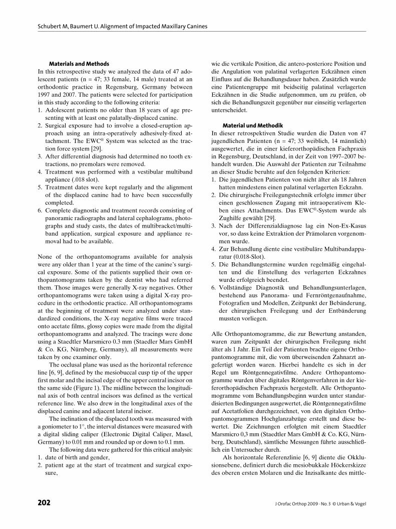
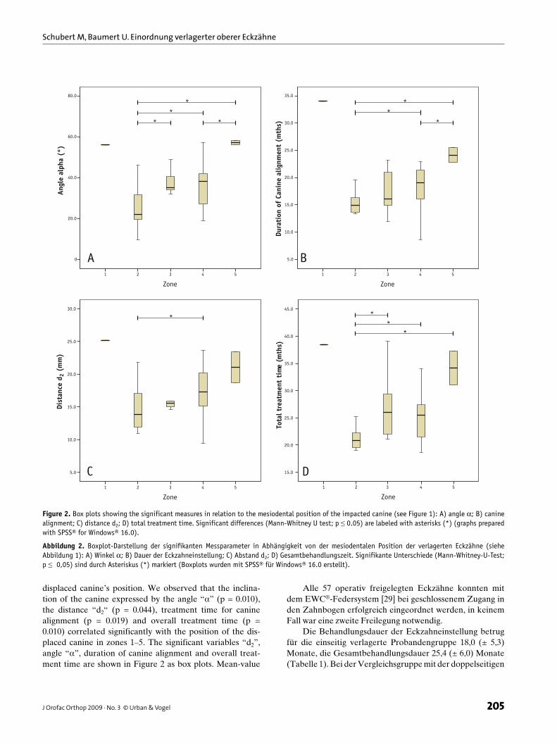
displaced canine’s position. We observed that the inclina-tion of the canine expressed by the angle “α” (p = 0.010), the distance “d2“ (p = 0.044), treatment time for canine alignment (p = 0.019) and overall treatment time (p = 0.010) correlated significantly with the position of the dis-placed canine in zones 1–5. The significant variables “d2”, angle “α”, duration of canine alignment and overall treat-ment time are shown in Figure 2 as box plots. Mean-value
Alle 57 operativ freigelegten Eckzähne konnten mit dem EWC®-Federsystem [29] bei geschlossenem Zugang in den Zahnbogen erfolgreich eingeordnet werden, in keinem Fall war eine zweite Freilegung notwendig.
Die Behandlungsdauer der Eckzahneinstellung betrug für die einseitig verlagerte Probandengruppe 18,0 (± 5,3) Monate, die Gesamtbehandlungsdauer 25,4 (± 6,0) Monate (Tabelle 1). Bei der Vergleichsgruppe mit der doppelseitigen
Zone54321
Angl
e al
pha
(°)
Dist
ance
d2
(mm
)
80.0
60.0
40.0
20.0
0
Zone54321
30.0
25.0
20.0
15.0
10.0
5.0
Zone54321
Dura
tion
of
Cani
ne a
lignm
ent
(mth
s)
35.0
30.0
25.0
20.0
15.0
10.0
5.0
**
*
***
**
*
*
*
Zone54321
Tota
l tre
atm
ent
tim
e(m
ths)
45.0
40.0
35.0
30.0
25.0
20.0
15.0
A B
C D
Figure 2. Box plots showing the significant measures in relation to the mesiodental position of the impacted canine (see Figure 1): A) angle α; B) canine alignment; C) distance d2; D) total treatment time. Significant differences (Mann-Whitney U test; p ≤ 0.05) are labeled with asterisks (*) (graphs prepared with SPSS® for Windows® 16.0).
Abbildung 2. Boxplot-Darstellung der signifikanten Messparameter in Abhängigkeit von der mesiodentalen Position der verlagerten Eckzähne (siehe Abbildung 1): A) Winkel α; B) Dauer der Eckzahneinstellung; C) Abstand d2; D) Gesamtbehandlungszeit. Signifikante Unterschiede (Mann-Whitney-U-Test; p ≤ 0,05) sind durch Asteriskus (*) markiert (Boxplots wurden mit SPSS® für Windows® 16.0 erstellt).
Schubert M, Baumert U. Alignment of Impacted Maxillary Canines
206 J Orofac Orthop 2009 · No. 3 © Urban & Vogel
comparisons were made using the Mann-Whitney U test to determine which of the individual zones differed signifi-cantly from each other. These results are also illustrated in Figure 2. It was mainly the displaced teeth in zone 2 that differed significantly from those in zones 3 to 5 in terms of overall treatment time and duration of canine alignment, respectively.
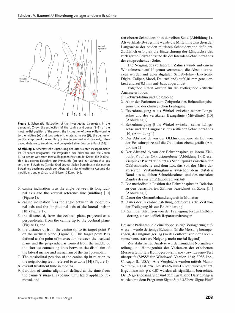
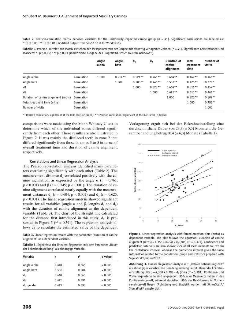
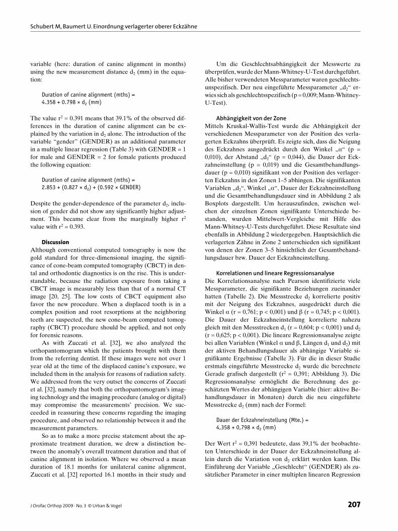
Correlations and Linear Regression AnalysisThe Pearson correlation analysis identified many parame-ters correlating significantly with each other (Table 2). The measurement distance d2 correlated positively with the ca-nine inclination, as expressed by the angle α (r = 0.761; p < 0.001) and � (r = 0.745; p < 0.001). The duration of ca-nine alignment correlated nearly equally with the measure-ment distances d1 (r = 0.604; p < 0.001) and d2 (r = 0.625; p < 0.001). The linear regression analysis showed significant results for all variables (angle α and �, lengths d1 and d2) with the duration of canine alignment as the dependent variable (Table 3). The chart of the straight line calculated for the distance first introduced in this study, d2, is pre-sented in Figure 3 (r2 = 0.391). The regression analysis al-lows us to calculate the estimated value of the dependent
Verlagerung ergab sich bei der Eckzahneinstellung eine durchschnittliche Dauer von 23,5 (± 5,5) Monaten, die Ge-samtbehandlung betrug 30,4 (± 6,3) Monate (Tabelle 1).
Angle Angle d1 d2 Duration of Total Number of alpha beta canine treatment visits alignment time
Angle alpha Correlation 1.000 0.914** 0.521** 0.761** 0.604** 0.469** 0.466**
Angle beta Correlation 1.000 0.503** 0.745** 0.533** 0.425** 0.378*
d1 Correlation 1.000 0.823** 0.604** 0.516** 0.457**
d2 Correlation 1.000 0.625** 0.511** 0.461**
Duration of canine alignment (mths) Correlation 1.000 0.825** 0.802**
Total treatment time (mths) Correlation 1.000 0.751**
Number of visits Correlation 1.000
*: Pearson correlation, significant at the 0.05 level (2-tailed); **: Pearson correlation, significant at the 0.01 level (2-tailed)
Table 2. Pearson-correlation matrix between variables for the unilaterally-impacted canine group (n = 41). Significant correlations are labeled as: *: p ≤ 0.05; **: p ≤ 0.01 (modified output from SPSS® 16.0 for Windows®).
Tabelle 2. Pearson-Korrelations-Matrix zwischen den Messparametern der Gruppe mit einseitig verlagerten Zähnen (n = 41). Signifikante Korrelationen sind markiert: *: p ≤ 0,05; **: p ≤ 0,01 (modifizierte Ausgabe des Programms SPSS® 16.0 für Windows®).
Variable r r2 p value
Angle alpha 0.604 0.365 < 0.001
Angle beta 0.533 0.284 < 0.001
d1 0.604 0.365 < 0.001
d2 0.625 0.391 < 0.001
d2, gender 0.627 0.393 < 0.001
Table 3. Linear regression results with the parameter “duration of canine alignment” as a dependent variable.
Tabelle 3. Ergebnisse der linearen Regression mit dem Parameter „Dauer der Eckzahneinstellung“ als abhängige Variable.
d2 (mm)
5 10 15 20 25 30
Cani
ne a
lignm
ent
(mth
s)
0
5
10
15
20
25
30
35
40
Linear regressio nConfidence intervalPrediction interval
Figure 3. Linear regression analysis with forced eruption time (mths) as dependent variable. The plot follows the equation: Duration of canine alignment (mths) = 4.358 + 0.798 × d2 (mm) (r2 = 0.391). Confidence and prediction intervals are also shown: 95% of all measurements fall within the confidence interval, whereas the prediction interval gives the same information related to the population (graph and statistics prepared with SigmaStat®/SigmaPlot®).
Abbildung 3. Lineare Regressionsanalyse mit „aktiver Behandlungszeit“ als abhängiger Variable. Die Geradengleichung lautet: Dauer der Eckzahn-einstellung (Mte.) = 4,358 + 0,798 × d2 (mm) (r2 = 0,391). Konfidenz- und Vorhersageintervalle sind angegeben: 95% aller Messwerte fallen in das Konfidenzintervall, während statistisch 95% der Bevölkerung im Vorher-sageintervall liegen (Abbildung und Statistik wurden mit SigmaStat®/SigmaPlot® angefertigt).
207J Orofac Orthop 2009 · No. 3 © Urban & Vogel
Schubert M, Baumert U. Einordnung verlagerter oberer Eckzähne
207J Orofac Orthop 2009 · No. 3 © Urban & Vogel
variable (here: duration of canine alignment in months) using the new measurement distance d2 (mm) in the equa-tion:
Duration of canine alignment (mths) = 4.358 + 0.798 × d2 (mm)
The value r2 = 0.391 means that 39.1% of the observed dif-ferences in the duration of canine alignment can be ex-plained by the variation in d2 alone. The introduction of the variable “gender” (GENDER) as an additional parameter in a multiple linear regression (Table 3) with GENDER = 1 for male and GENDER = 2 for female patients produced the following equation:
Duration of canine alignment (mths) = 2.853 + (0.827 × d2) + (0.592 × GENDER)
Despite the gender-dependence of the parameter d2, inclu-sion of gender did not show any significantly higher adjust-ment. This became clear from the marginally higher r2 value with r2 = 0.393.
DiscussionAlthough conventional computed tomography is now the gold standard for three-dimensional imaging, the signifi-cance of cone-beam computed tomography (CBCT) in den-tal and orthodontic diagnostics is on the rise. This is under-standable, because the radiation exposure from taking a CBCT image is measurably less than that of a normal CT image [20, 25]. The low costs of CBCT equipment also favor the new procedure. When a displaced tooth is in a complex position and root resorptions at the neighboring teeth are suspected, the new cone-beam computed tomog-raphy (CBCT) procedure should be applied, and not only for forensic reasons.
As with Zuccati et al. [32], we also analyzed the orthopantomogram which the patients brought with them from the referring dentist. If these images were not over 1 year old at the time of the displaced canine’s exposure, we included them in the analysis for reasons of radiation safety. We addressed from the very outset the concerns of Zuccati et al. [32], namely that both the orthopantomogram’s imag-ing technology and the imaging procedure (analog or digital) may compromise the measurements’ precision. We suc-ceeded in reassuring these concerns regarding the imaging procedure, and observed no relationship between it and the measurement parameters.
So as to make a more precise statement about the ap-proximate treatment duration, we drew a distinction be-tween the anomaly’s overall treatment duration and that of canine alignment in isolation. Where we observed a mean duration of 18.1 months for unilateral canine alignment, Zuccati et al. [32] reported 16.1 months in their study and
Um die Geschlechtsabhängigkeit der Messwerte zu überprüfen, wurde der Mann-Whitney-U-Test durchgeführt. Alle bisher verwendeten Messparameter waren geschlechts-unspezifisch. Der neu eingeführte Messparameter „d2“ er-wies sich als geschlechtsspezifisch (p = 0,009; Mann- Whitney-U-Test).
Abhängigkeit von der ZoneMittels Kruskal-Wallis-Test wurde die Abhängigkeit der verschiedenen Messparameter von der Position des verla-gerten Eckzahns überprüft. Es zeigte sich, dass die Neigung des Eckzahnes ausgedrückt durch den Winkel „α“ (p = 0,010), der Abstand „d2“ (p = 0,044), die Dauer der Eck-zahneinstellung (p = 0,019) und die Gesamtbehandlungs-dauer (p = 0,010) signifikant von der Position des verlager-ten Eckzahns in den Zonen 1–5 abhingen. Die signifikanten Variablen „d2“, Winkel „α“, Dauer der Eckzahneinstellung und die Gesamtbehandlungsdauer sind in Abbildung 2 als Boxplots dargestellt. Um herauszufinden, zwischen wel-chen der einzelnen Zonen signifikante Unterschiede be-standen, wurden Mittelwert-Vergleiche mit Hilfe des Mann-Whitney-U-Tests durchgeführt. Diese Resultate sind ebenfalls in Abbildung 2 wiedergegeben. Hauptsächlich die verlagerten Zähne in Zone 2 unterschieden sich signifikant von denen der Zonen 3–5 hinsichtlich der Gesamtbehand-lungsdauer bzw. Dauer der Eckzahneinstellung.
Korrelationen und lineare RegressionsanalyseDie Korrelationsanalyse nach Pearson identifizierte viele Messparameter, die signifikante Beziehungen zueinander hatten (Tabelle 2). Die Messstrecke d2 korrelierte positiv mit der Neigung des Eckzahnes, ausgedrückt durch die Winkel α (r = 0,761; p < 0,001) und � (r = 0,745; p < 0,001). Die Dauer der Eckzahneinstellung korrelierte nahezu gleich mit den Messstrecken d1 (r = 0,604; p < 0,001) und d2 (r = 0,625; p < 0,001). Die lineare Regressionsanalyse zeigte bei allen Variablen (Winkel α und �, Längen d1 und d2) mit der aktiven Behandlungsdauer als abhängige Variable si-gnifikante Ergebnisse (Tabelle 3). Für die in dieser Studie erstmals eingeführte Messstrecke d2 wurde die berechnete Gerade grafisch dargestellt (r2 = 0,391; Abbildung 3). Die Regressionsanalyse ermöglicht die Berechnung des ge-schätzten Wertes der abhängigen Variable (hier: aktive Be-handlungsdauer in Monaten) durch die neu eingeführte Messstrecke d2 (mm) nach der Formel:
Dauer der Eckzahneinstellung (Mte.) = 4,358 + 0,798 × d2 (mm)
Der Wert r2 = 0,391 bedeutete, dass 39,1% der beobachte-ten Unterschiede in der Dauer der Eckzahneinstellung al-lein durch die Variation von d2 erklärt werden kann. Die Einführung der Variable „Geschlecht“ (GENDER) als zu-sätzlicher Parameter in einer multiplen linearen Regression

Schubert M, Baumert U. Alignment of Impacted Maxillary Canines
208 J Orofac Orthop 2009 · No. 3 © Urban & Vogel
Grande et al. [17] reported 22.8 months. Concerning the study by Grande et al.: seven of the 59 canines they examined were in vestibular position, thus their cohort was not homog-enous. They [17] found no correlation between the degree of canine displacement in the orthopantomogram and treat-ment time.
Stewart et al. [30] reported a positive correlation (r = 0.424; p = 0.003) between overall treatment time and the canine’s distance from the occlusal plane in a comparable study, which we confirmed in this study (r = 0.502; p = 0.001). They reported a mean distance of 13.9 mm, and Zuccati et al. [32] observed an average value of 14.9 mm, which was somewhat higher than Stewart et al.’s and nearly matching the 14.5 mm value we identified in this study. By contrast, the pertinent overall treat-ment times, compared in Table 4, differed substantially.
While the longer treatment time of 6.6 months for the group with bilateral displacement was explained by a larger d1 value for Stewart et al. [30], the reason for the longer treat-ment time is unclear in our study, and it cannot be explained by the available data. Greater bone density in this region may be behind slower tooth movement – it may even be the reason for the canine’s displacement.
To predict treatment time involving unilateral displace-ment, Stewart et al. [30] assume a threshold value of 14 mm distance from the occlusal plane: shorter distances require a mean overall treatment time of 23.8 months, while distances over that required an average treatment time of 31.1 months. Zuccati et al. [32] attempted to improve these relatively im-precise prognoses by distinguishing between the duration of canine alignment and overall treatment time. In addition, as in a paper published by Becker & Chaushu [1], they stated the number of check-up appointments as a measure of the total treatment time. Zuccati et al. [32] calculated the number of appointments in advance by introducing a multiple linear re-gression comprising the patient’s age at the start of treatment, the canine’s distance from the occlusal plane, the zone and the canine’s inclination as angle �, reporting a regression co-efficient of r2 = 0.42 for the relevant line of best fit.
Schopf [28] and Crismani et al. [7] are among those who have described how the canine’s inclination is an important factor in the prognosis and duration of treatment.
(Tabelle 3) mit GENDER = 1 für männliche und GENDER = 2 für weibliche Patienten ergab die folgende Gleichung:
Dauer der Eckzahneinstellung (Mte.) = 2,853 + (0,827 × d2) + (0,592 × GENDER)
Trotz der Geschlechtsabhängigkeit des Messparameters d2 ergab der Einbezug des Geschlechts keine signifikant hö-here Anpassung. Dies wurde durch den geringfügig hö-heren r2-Wert mit r2 = 0,393 deutlich.
DiskussionAuch wenn heute die konventionelle Computertomografie den Goldstandard für ein dreidimensionales bildgebendes Verfahren darstellt, nimmt die Bedeutung der digitalen Vo-lumentomographie (DVT) in der zahnmedizinischen sowie kieferorthopädischen Diagnostik zu. Dies ist verständlich, weil die Strahlendosis für eine DVT-Aufnahme deutlich unter der einer normalen CT-Aufnahme liegt [20, 25]. Auch die geringeren Kosten des Gerätes sprechen für das neue Verfahren. Bei komplexer Position eines verlagerten Zahnes, mit Verdacht auf Wurzelresorptionen an den Nachbarzähnen sollte das neue DVT-Verfahren – nicht nur aus forensischen Gründen – angewendet werden.
Wie bei Zuccati et al. [32] wurden in der hier vorgestell-ten Studie die herkömmlichen Orthopantomogramme aus-gewertet, die die Patienten vom überweisenden Zahnarzt mitbrachten. Waren diese zum Zeitpunkt der Freilegung des verlagerten Eckzahnes nicht älter als 1 Jahr, wurden sie aus Gründen des Strahlenschutzes in die Auswertung mit einbe-zogen. Den Bedenken von Zuccati et al. [32], dass nicht nur die Bildtechnik des Orthopantomogramms, sondern auch das bildgebende Verfahren (analog bzw. digital) die Genau-igkeit der Messungen schmälern könnte, wurde von vorn-herein nachgegangen. Diese Bedenken konnten das bildge-bende Verfahren betreffend entkräftet werden, eine Abhän-gigkeit der Messparameter davon ließ sich ausschließen.
Um eine genauere Aussage über die ungefähre Dauer der Behandlung geben zu können, wurde zwischen der Gesamtbehandlungszeit der Anomalie und der alleinigen
Reference d1 Total treatment time
(mm) (mths) Range (mths)
Stewart et al. 2001 [30] unilateral 13.9 25.8 13.0–40.0 bilateral 15.6 32.3 23.0–50.0
Zuccati et al. 2006 [32] unilateral 14.9 19.6 7.0–50.0 bilateral n. d. n. d. n. d.
Schubert & Baumert 2009 unilateral 14.5 25.4 14.4–39.1 bilateral 14.5 30.4 17.9–40.7
Table 4. Parameter d1 and duration of canine alignment from selected references in comparison to this study (n.d. = no data available).
Tabelle 4. Vergleich der Parameter „d1“ und „Dauer der Eckzahneinstellung“ ausgewählter Publikationen mit dieser Studie (n. d. = keine Angaben).
209J Orofac Orthop 2009 · No. 3 © Urban & Vogel
Schubert M, Baumert U. Einordnung verlagerter oberer Eckzähne
209J Orofac Orthop 2009 · No. 3 © Urban & Vogel
It is interesting that the angle α – measured to the verti-cal – showed greater significance than angle �, measured to the axis of the lateral incisor. The frequent “evasion” of the lateral incisor from the palatally-positioned canine could ex-plain this.
In contrast to Zuccati et al. [32], who included adults in their cohort and also gave a high rating to patient age as a prognostic factor, we observed no significant correlation be-tween age and length of treatment in our group of adolescent probands (to 18 years of age). When analyzing the distance d1 and angle � (taking the length of treatment into account), our data confirmed the results of Zuccati et al. [32] in finding a significant correlation.
We identified a regression coefficient of r2 = 0.391 with the introduction of the new measurement distance d2 for the prognosis of the treatment time for canine alignment. What was new was that the distance d2 was the only parameter of other already-familiar parameters that proved to be gender-specific. The group of male subjects’ value of 19.8 (± 4.6) mm (n = 12; median: 18.6 mm) was distinctly greater than that of the females (15.9 [± 3.3] mm [n = 29; median: 15.3 mm]). Gender-specific differences in jaw size may be a reason be-hind this finding [4]. Thus it was even more surprising that gender contributed to only marginal improvement in the ac-commodation (Table 3).
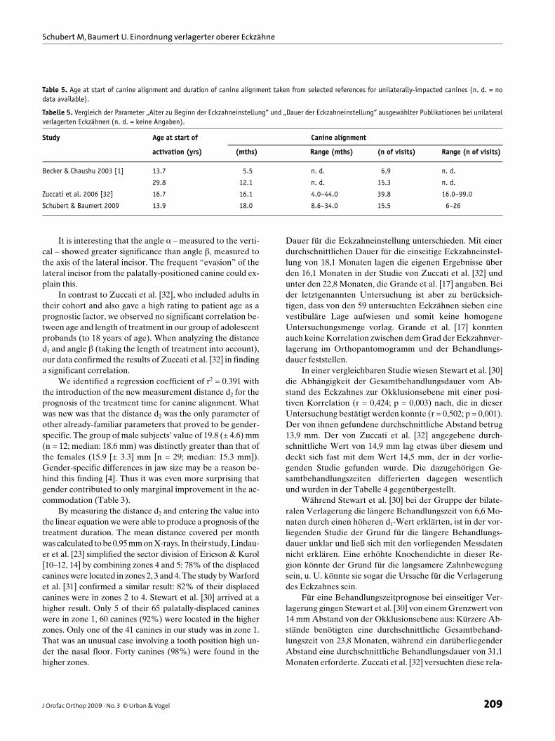
By measuring the distance d2 and entering the value into the linear equation we were able to produce a prognosis of the treatment duration. The mean distance covered per month was calculated to be 0.95 mm on X-rays. In their study, Lindau-er et al. [23] simplified the sector division of Ericson & Kurol [10–12, 14] by combining zones 4 and 5: 78% of the displaced canines were located in zones 2, 3 and 4. The study by Warford et al. [31] confirmed a similar result: 82% of their displaced canines were in zones 2 to 4. Stewart et al. [30] arrived at a higher result. Only 5 of their 65 palatally-displaced canines were in zone 1, 60 canines (92%) were located in the higher zones. Only one of the 41 canines in our study was in zone 1. That was an unusual case involving a tooth position high un-der the nasal floor. Forty canines (98%) were found in the higher zones.
Dauer für die Eckzahneinstellung unterschieden. Mit einer durchschnittlichen Dauer für die einseitige Eckzahneinstel-lung von 18,1 Monaten lagen die eigenen Ergebnisse über den 16,1 Monaten in der Studie von Zuccati et al. [32] und unter den 22,8 Monaten, die Grande et al. [17] angaben. Bei der letztgenannten Untersuchung ist aber zu berücksich-tigen, dass von den 59 untersuchten Eckzähnen sieben eine vestibuläre Lage aufwiesen und somit keine homogene Untersuchungsmenge vorlag. Grande et al. [17] konnten auch keine Korrelation zwischen dem Grad der Eckzahnver-lagerung im Orthopantomogramm und der Behandlungs-dauer feststellen.
In einer vergleichbaren Studie wiesen Stewart et al. [30] die Abhängigkeit der Gesamtbehandlungsdauer vom Ab-stand des Eckzahnes zur Okklusionsebene mit einer posi-tiven Korrelation (r = 0,424; p = 0,003) nach, die in dieser Untersuchung bestätigt werden konnte (r = 0,502; p = 0,001). Der von ihnen gefundene durchschnittliche Abstand betrug 13,9 mm. Der von Zuccati et al. [32] angegebene durch-schnittliche Wert von 14,9 mm lag etwas über diesem und deckt sich fast mit dem Wert 14,5 mm, der in der vorlie-genden Studie gefunden wurde. Die dazugehörigen Ge-samtbehandlungszeiten differierten dagegen wesentlich und wurden in der Tabelle 4 gegenübergestellt.
Während Stewart et al. [30] bei der Gruppe der bilate-ralen Verlagerung die längere Behandlungszeit von 6,6 Mo-naten durch einen höheren d1-Wert erklärten, ist in der vor-liegenden Studie der Grund für die längere Behandlungs-dauer unklar und ließ sich mit den vorliegenden Messdaten nicht erklären. Eine erhöhte Knochendichte in dieser Re-gion könnte der Grund für die langsamere Zahnbewegung sein, u. U. könnte sie sogar die Ursache für die Verlagerung des Eckzahnes sein.
Für eine Behandlungszeitprognose bei einseitiger Ver-lagerung gingen Stewart et al. [30] von einem Grenzwert von 14 mm Abstand von der Okklusionsebene aus: Kürzere Ab-stände benötigten eine durchschnittliche Gesamtbehand-lungszeit von 23,8 Monaten, während ein darüberliegender Abstand eine durchschnittliche Behandlungsdauer von 31,1 Monaten erforderte. Zuccati et al. [32] versuchten diese rela-
Study Age at start of Canine alignment
activation (yrs) (mths) Range (mths) (n of visits) Range (n of visits)
Becker & Chaushu 2003 [1] 13.7 5.5 n. d. 6.9 n. d.
29.8 12.1 n. d. 15.3 n. d.
Zuccati et al. 2006 [32] 16.7 16.1 4.0–44.0 39.8 16.0–99.0
Schubert & Baumert 2009 13.9 18.0 8.6–34.0 15.5 6–26
Table 5. Age at start of canine alignment and duration of canine alignment taken from selected references for unilaterally-impacted canines (n. d. = no data available).
Tabelle 5. Vergleich der Parameter „Alter zu Beginn der Eckzahneinstellung“ und „Dauer der Eckzahneinstellung“ ausgewählter Publikationen bei unilateral verlagerten Eckzähnen (n. d. = keine Angaben).
Schubert M, Baumert U. Alignment of Impacted Maxillary Canines
210 J Orofac Orthop 2009 · No. 3 © Urban & Vogel
The angle α and the distances d1 and d2 increased in as-cending order from zones 1 to 5. We observed significant correlations between the zones and overall treatment times and durations of canine alignment, confirming the results of Stewart et al. [30] and Zuccati et al. [32]. The reason for the significant increase in the duration of canine alignment and overall treatment time in zone 2 compared to other zones is probably due to the particular anisotropic quality of the bone in that region [27].
The number of appointments may be important for cal-culating the costs of therapy but they do not provide the pa-tient with a reliable answer regarding how long treatment will take. The numbers of appointments vary widely (Table 5) and ultimately depend on the therapeutic approach. When the numbers of appointments are compared with treatment duration, one observes that necessary re-activations and/or repairs were relatively frequent. Regardless of whether ca-nine displacements were unilateral or bilateral, our patient cohorts (all of whom were treated with the EWC® System [29]) had to undergo fewer activation steps on average per treatment time.
Our new “d2” variable alone describes ca. 40% of the variation observed. The prognostic precision of ca. 40% seems to be the limit for prognoses using common examina-tion methods. One major reason for this result may be due to the orthopantomogram’s imaging technology, where only a narrow section of the tomogram is in sharp focus. All the objects displayed outside that area are generally more or less distorted, especially in the canine region [10–12, 14, 16]. Fur-thermore, teeth react to applied forces highly individually, as shown by Iwasaki et al. [22] and Sander et al. [27].
In a clinical study of extraction cases, Iwasaki et al. [22] described a mean speed of canine movement of 0.87 mm/month and 1.27 mm/month under a traction force of 18 g and 60 g, respectively. However, they also found great individual differences in the speed, which varied at a ratio of 3:1 with the same amount of force. Cell metabolic processes are pre-sumed to be a reason for this variability in tooth movement speed. Applying digital macrophotogrammetry, Sander et al. [27] studied canine retraction with the hybrid retractor as a source of force. They found a mean value of 1.2 mm/month to be a realistic time estimate for patients. The bone’s aniso-tropic quality was considered the cause for intra- and inter-individual differences in the speed of movement.
In the procedure we introduce in this study, a follow-up X-ray 6 months after surgical exposure or the first activation proved to be beneficial for specifying the duration of treat-ment. Measurement of the distance covered by the canine and subtracting that from the total distance then permitted us to determine more precisely whether that patient was a “slow mover” or a “fast mover”.
tiv ungenaue Prognose zu verbessern, indem sie die Dauer der Eckzahneinstellung von der Gesamtbehandlungsdauer abtrennten. Zusätzlich gaben sie entsprechend einer Veröf-fentlichung von Becker & Chaushu [1] die Anzahl der Be-handlungssitzungen als Maß für die Behandlungsdauer an. Durch Einführung einer multiplen linearen Regression, in der das Alter zu Behandlungsbeginn, der Abstand des Eck-zahnes von der Okklusionsebene, die Zone und die Eck-zahnneigung als Winkel � einflossen, berechneten Zuccati et al. [32] die Anzahl der Sitzungen im Voraus und gaben für die zugehörige Ausgleichsgerade einen Regressionskoeffizi-enten von r2 = 0,42 an.
Dass die Neigung des Eckzahnes für die Prognose und Dauer einen wichtigen Faktor darstellt, wurde unter ande-rem bereits von Schopf [28] und Crismani et al. [7] beschrie-ben. Von Interesse ist, dass der Winkel α – gemessen zur Vertikalen – in dieser Studie eine höhere Signifikanz zeigte als der Winkel � – gemessen zur Achse des seitlichen Schnei-dezahnes. Das häufig beobachtete „Ausweichen“ des seit-lichen Schneidezahnes vom palatinal stehenden Eckzahn könnte der Grund dafür sein.
Im Gegensatz zu Zuccati et al. [32], die Erwachsene in ihre Untersuchungsgruppe mit einbezogen und auch das Pa-tientenalter als Prognosefaktor hoch werteten, wurde bei der vorliegenden jugendlichen Probandengruppe (bis 18 Jahre) keine signifikante Korrelation zwischen Alter und Behandlungszeit festgestellt. Bei der Bewertung des Ab-standes d1 und des Winkels � zur Dauer der Eckzahneinstel-lung konnten mit einer signifikanten Korrelation die Ergeb-nisse von Zuccati et al. [32] bestätigt werden.
Mit Einführung der neuen Messstrecke d2 zur Prognose für die Dauer der Eckzahneinstellung wurde ein Regressions-koeffizient von r2 = 0,391 erreicht. Neu war, dass die Strecke d2 als einzige von den anderen und bisher benutzten Messpara-metern als geschlechtsspezifisch erkannt wurde. Sie lag bei der Gruppe der männlichen mit 19,8 (± 4,6) mm (n = 12; Median: 18,6 mm) deutlich über dem Wert von 15,9 (± 3,3) mm der weiblichen (n = 29; Median: 15,3 mm). Geschlechtsspezifische Unterschiede in der Kiefergröße dürften dafür der Grund sein [4]. Umso überraschender war, dass das Geschlecht in der mul-tiplen Regressionsanalyse nur zu einer geringfügigen Verbes-serung in der Anpassung beitrug (Tabelle 3).
Durch das Abmessen der Strecke d2 und das Einsetzen des Wertes in die Geradengleichung konnte eine Behand-lungsdauerprognose gestellt werden. Die auf dem Röntgen-bild durchschnittlich zurückgelegte Strecke betrug 0,95 mm pro Monat bei einseitiger Verlagerung. Lindauer et al. [23] vereinfachten in ihrer Untersuchung die Sektoreneinteilung von Ericson & Kurol [10–12, 14] durch Zusammenfassung der Zonen 4 und 5: 78% der verlagerten Eckzähne befanden sich in den Zonen 2, 3 und 4. Ein ähnliches Ergebnis bestätigte die Studie von Warford et al. [31]: 82% der verlagerten Eckzähne wurden in den Zonen 2–4 gefunden. Zu einem höheren Er-gebnis kamen Stewart et al. [30]. Nur fünf von den 65 palatinal
211J Orofac Orthop 2009 · No. 3 © Urban & Vogel
Schubert M, Baumert U. Einordnung verlagerter oberer Eckzähne
211J Orofac Orthop 2009 · No. 3 © Urban & Vogel
Conclusions1. We observed that treatment duration depended on the
position and angulation of palatally-displaced canines in adolescents up to the age of 18. Our introduction of a new linear measurement has enabled us to identify a method for practitioners to estimate treatment times.
2. This enhanced the probability of an accurate prognosis to ca. 40%. This seems to be the limit of a prognosis using a two-dimensional imaging procedure.
3. Canine alignment in patients with bilateral displace-ments required 5.0 months longer than in those with uni-lateral displacements. As the mean distances from the occlusal level and distance to be covered were on aver-age nearly identical for both groups, we believe that metabolic processes and the bone’s anisotropic quality seem to retard tooth movement in these patients espe-cially.
4. To make the prognosis of treatment duration more pre-cise, those variables must probably be taken into ac-count. Further studies will be necessary to determine whether bone-density measurements using computed to-mography will eventually lead us to more exact progno-ses.
Acknowledgements The authors would like to express their gratitude to Prof. Dr. In-grid Rudzki, Munich, for her unflagging support and advice over the course of this study.
References1. Becker A, Chaushu S. Success rate and duration of orthodontic treat-
ment for adult patients with palatally impacted maxillary canines. Am J Orthod Dentofacial Orthop 2003;124:509–14.
2. Bishara SE. Impacted maxillary canines: a review. Am J Orthod Den-tofacial Orthop 1992;101:159–71.
3. Bishara SE. Clinical management of impacted maxillary canines. Semin Orthod 1998;4:87–98.
4. Burris BG, Harris EF. Maxillary arch size and shape in American blacks and whites. Angle Orthod 2000;70:297–302.
5. Chambas C. Canine maxillaire incluse et thérapeutique orthodon-tique. Rev Orthop Dento Faciale 1993;27:9–28.
6. Chaushu S, Chaushu G, Becker A. Reliability of a method for the lo-calization of displaced maxillary canines using a single panoramic radiograph. Clin Orthod Res 1999;2:194–9.
7. Crismani AG, Freudenthaler JW, Weber R, et al. Impaktierte obere Eckzähne – konventionelle Röntgendiagnostik und Therapie. Schweiz Monatsschr Zahnmed 2000;110:1256–67.
8. Dachi SF, Howell FV. A survey of 3, 874 routine full-month radio-graphs. II. A study of impacted teeth. Oral Surg Oral Med Oral Pathol 1961;14:1165–9.
9. Dausch-Neumann D. Der Durchbruchsweg bleibender Eckzähne. Fortschr Kieferorthop 1970;31:9–16.
10. Ericson S, Kurol J. Longitudinal study and analysis of clinical super-vision of maxillary canine eruption. Community Dent Oral Epidemiol 1986;14:172–6.
verlagerten Eckzähnen waren in Zone 1, 60 Eckzähne (92%) waren in den höheren Zonen gelegen. In der vorliegenden Studie befand sich nur ein einziger von den 41 untersuchten Eckzähnen in Zone 1. Dabei handelt es sich um einen Sonder-fall, der Zahn lag hoch unter dem Nasenboden. Vierzig Eck-zähne (98%) wurden in den höheren Zonen gefunden.
Der Winkel α und die Strecken d1 und d2 nahmen auf-steigend mit den Zonen 1 bis 5 zu. Eine signifikante Abhän-gigkeit der Gesamtbehandlungszeit und Dauer der Eckzahn-einstellung von den Zonen wurde nachgewiesen und bestä-tigte die Ergebnisse von Stewart et al. [30] und Zuccati et al. [32]. Der Grund dafür, dass sich in der Zone 2 die Dauer der Eckzahneinstellung und der Gesamtbehandlungsdauer ge-genüber den anderen Zonen signifikant erhöhte, wird wahr-scheinlich an der Anisotropie des Knochens speziell in die-ser Region liegen [27].
Die Anzahl der Sitzungen mag zwar für die Kalkulation der Behandlungskosten wichtig sein, gibt dem Patienten aber keine ausreichende Antwort auf die Behandlungsdau-er. Sie unterliegt einer starken Streubreite (Tabelle 5) und ist letztlich auch von der Behandlungstechnik abhängig. Korreliert man die Zahl der Behandlungssitzungen mit der Behandlungsdauer, waren relativ häufige Nachaktivie-rungen und/oder Reparaturen notwendig. Nur bei der hier vorgestellten Patientengruppe, die alle mit dem EWC®-Sys-tem [29] behandelt wurden, mussten sowohl bei der Gruppe der einseitigen als auch bei der mit doppelseitigen Verlage-rungen durchschnittlich weniger Aktivierungsschritte pro Behandlungsdauer durchgeführt werden.
Die neu eingeführte Variable „d2“ allein beschreibt ca. 40% der beobachteten Variation. Mit der Vorhersagegenau-igkeit von ca. 40% scheint mit den bisher üblichen Untersu-chungsmethoden die Grenze für eine Prognose erreicht zu sein. Ein Hauptgrund für dieses Ergebnis dürfte in der Bild-technik des Orthopantomogramms liegen, wo nur ein schmaler Bereich der Schichtaufnahme scharf abgebildet wird. Alle dargestellten Objekte außerhalb dieses Bereiches unterliegen einer mehr oder weniger starken Verzeichnung, speziell in der Eckzahnregion ist sie am größten [10–12, 14, 16]. Zusätzlich reagieren Zähne auf einwirkende Kräfte sehr individuell, wie auch Iwasaki et al. [22] und Sander et al. [27] feststellten.
Iwasaki et al. [22] beschrieben in einer klinischen Studie von Extraktionsfällen eine durchschnittliche Eckzahnge-schwindigkeit von 0,87 mm/Monat und 1,27 mm/Monat bei einer Zugkraft von 18 g bzw. 60 g. Allerdings stellten sie hier-bei einen hohen individuellen Unterschied bei der Ge-schwindigkeit fest, der im Verhältnis bis 3:1 bei gleicher Kraft variierte. Zelluläre Stoffwechselvorgänge sollen die Ursache für die Variabilität der Zahnbewegungsgeschwin-digkeit sein. Sander et al. [27] untersuchten mit Hilfe der di-gitalen Makrophotogrammetrie die Eckzahnretraktion mit dem Hybrid-Retraktor als Kraftquelle. Ein Mittelwert von 1,2 mm pro Monat schien ihnen als gewisse Zeitangabe für
Schubert M, Baumert U. Alignment of Impacted Maxillary Canines
212 J Orofac Orthop 2009 · No. 3 © Urban & Vogel
11. Ericson S, Kurol J. Radiographic assessment of maxillary canine eruption in children with clinical signs of eruption disturbance. Eur J Orthod 1986;8:133–40.
12. Ericson S, Kurol J. Radiographic examination of ectopically erupting maxillary canines. Am J Orthod Dentofacial Orthop 1987;91: 483–92.
13. Ericson S, Kurol J. Early treatment of palatally erupting maxillary canines by extraction of the primary canines. Eur J Orthod 1988;10:283–95.
14. Ericson S, Kurol J. Resorption of maxillary lateral incisors caused by ectopic eruption of the canines. A clinical and radiographic analysis of predisposing factors. Am J Orthod Dentofacial Orthop 1988;94:503–13.
15. Ferguson JW. Management of the unerupted maxillary canine. Br Dent J 1990;169:11–7.
16. Fox NA, Fletcher GA, Horner K. Localising maxillary canines using dental panoramic tomography. Br Dent J 1995;179:416–20.
17. Grande T, Stolze A, Goldbecher H, et al. The displaced maxillary canine – a retrospective study. J Orofac Orthop 2006;67:441–9.
18. Grover PS, Lorton L. The incidence of unerupted permanent teeth and related clinical cases. Oral Surg Oral Med Oral Pathol 1985;59:420–5.
19. Harzer W, Seifert D, Mahdi Y. Die kieferorthopädische Einordnung retinierter Eckzähne unter besonderer Berücksichtigung des Behan-dlungsalters, der Angulation und der dynamischen Okklusion. Fortschr Kieferorthop 1994;55:47–53.
20. Hashimoto K, Arai Y, Iwai K, et al. A comparison of a new limited cone beam computed tomography machine for dental use with a multidetector row helical CT machine. Oral Surg Oral Med Oral Pathol Oral Radiol Endod 2003;95:371–7.
21. Iramaneerat S, Cunningham SJ, Horrocks EN. The effect of two alter-native methods of canine exposure upon subsequent duration of orthodontic treatment. Int J Paediatr Dent 1998;8:123–9.
22. Iwasaki LR, Haack JE, Nickel JC, et al. Human tooth movement in response to continuous stress of low magnitude. Am J Orthod Den-tofacial Orthop 2000;117:175–83.
23. Lindauer SJ, Rubenstein LK, Hang WM, et al. Canine impaction iden-tified early with panoramic radiographs. J Am Dent Assoc 1992;123:91–7.
24. Lüdicke G, Harzer W, Tausche E. Incisor inclination – risk factor for palatally-impacted canines. J Orofac Orthop 2008;69:357–64.
25. Mah JK, Danforth RA, Bumann A, et al. Radiation absorbed in maxil-lofacial imaging with a new dental computed tomography device. Oral Surg Oral Med Oral Pathol Oral Radiol Endod 2003;96:508–13.
26. McKee IW, Williamson PC, Lam EW, et al. The accuracy of 4 pan-oramic units in the projection of mesiodistal tooth angulations. Am J Orthod Dentofacial Orthop 2002;121:166–75.
27. Sander C, Geiger M, Sander FG. Contactless measurement of canine retraction by digital macrophotogrammetry during hybrid retractor application. J Orofac Orthop 2002;63:472–82.
28. Schopf P. Curriculum Kieferorthopädie. Berlin: Quintessenz, 1994.29. Schubert M. The alignment of impacted and ectopic teeth using the
Easy-Way-Coil (EWC®) System. J Orofac Orthop 2008;69:213–26.30. Stewart JA, Heo G, Glover KE, et al. Factors that relate to treatment
duration for patients with palatally impacted maxillary canines. Am J Orthod Dentofacial Orthop 2001;119:216–25.
31. Warford JH, Jr., Grandhi RK, Tira DE. Prediction of maxillary canine impaction using sectors and angular measurement. Am J Orthod Dentofacial Orthop 2003;124:651–5.
32. Zuccati G, Ghobadlu J, Nieri M, et al. Factors associated with the duration of forced eruption of impacted maxillary canines: a retro-spective study. Am J Orthod Dentofacial Orthop 2006;130:349–56.
den Patienten als realistisch. Die Anisotropie des Knochens wurde als Ursache für den intra- und interindividuellen Un-terschied in der Bewegungsgeschwindigkeit gesehen.
Bei dem in dieser Studie vorgestellten Verfahren hat sich zur Präzisierung der Behandlungsdauer eine Kontroll-aufnahme 6 Monate nach Freilegung bzw. erster Aktivie-rung als günstig erwiesen. Das Abmessen der zurückgelegten Wegstrecke und deren Subtraktion von der Gesamtstrecke erlaubte dann eine genauere Prognose, ob der Patient zur Gruppe der „slow mover“ oder „fast mover“ zählte.
Schlussfolgerungen1. Es wurde eine Abhängigkeit der Behandlungsdauer von
der Position und der Angulation palatinal verlagerter Eckzähne bei Jugendlichen bis 18 Jahren nachgewiesen. Durch Einführung einer neuen Messstrecke konnte ein praxisgerechter Weg für die Prognose der Behandlungs-dauer gefunden werden.
2. Die damit erreichte Wahrscheinlichkeitsprognose lag bei ca. 40%. Auf etwa dieser Höhe scheint die Grenze für eine Prognose mit Hilfe des zweidimensionalen bildge-benden Verfahrens zu liegen.
3. Bei Patienten mit beidseitiger Verlagerung benötigte die Einstellung 5,0 Monate mehr Zeit als bei einseitiger Verlagerung. Nachdem der durchschnittliche Abstand zur Okklusionsebene und die zurückgelegte Strecke bei beiden Gruppen durchschnittlich nahezu gleich waren, scheinen Stoffwechselvorgänge und die Anisotropie des Knochens bei dieser Gruppe die Zahnbewegung beson-ders zu verlangsamen.
4. Um genauere Behandlungszeitprognosen stellen zu kön-nen, müssen wahrscheinlich diese Variablen mit einbe-zogen werden. Ob die Messung der Knochendichte mit Hilfe der Volumentomografie ein Weg für eine exaktere Prognose wäre, werden weitere Studien belegen müssen.
Danksagung Die Autoren bedanken sich bei Frau Prof. Dr. Ingrid Rudzki, München, für ihre kontinuierliche Unterstützung und Beratung bei der Durchführung dieser Studie.
Correspondence AddressDr. med. dent. Michael SchubertAlbertstr. 593047 Regensburg GermanyPhone: (+49/941) 560240, Fax: 51151e-mail: [email protected]