Embed Size (px)
Citation preview
Accepted Manuscript
Post-surgical effects on the maxillary segments of children with oral clefts: New three-dimensional anthropometric analysis
Eloá Cristina Passucci Ambrosio, Chiarella Sforza, Márcio De Menezes, CleideFelício Carvalho Carrara, Maria Aparecida Andrade Moreira Machado, Thais MarchiniOliveira
PII: S1010-5182(18)30477-3
DOI: 10.1016/j.jcms.2018.06.017
Reference: YJCMS 3044
To appear in: Journal of Cranio-Maxillo-Facial Surgery
Received Date: 20 November 2017
Revised Date: 2 June 2018
Accepted Date: 25 June 2018
Please cite this article as: Ambrosio ECP, Sforza C, De Menezes M, Carrara CFC, Machado MAAM,Oliveira TM, Post-surgical effects on the maxillary segments of children with oral clefts: New three-dimensional anthropometric analysis, Journal of Cranio-Maxillofacial Surgery (2018), doi: 10.1016/j.jcms.2018.06.017.
This is a PDF file of an unedited manuscript that has been accepted for publication. As a service toour customers we are providing this early version of the manuscript. The manuscript will undergocopyediting, typesetting, and review of the resulting proof before it is published in its final form. Pleasenote that during the production process errors may be discovered which could affect the content, and alllegal disclaimers that apply to the journal pertain.
MANUSCRIP
T
ACCEPTED
ACCEPTED MANUSCRIPTPost-surgical effects on the maxillary segments of children with oral clefts: New
three-dimensional anthropometric analysis
Eloá Cristina Passucci Ambrosioa, Chiarella Sforzab, Márcio De Menezesc, Cleide
Felício Carvalho Carrarad, Maria Aparecida Andrade Moreira Machadod, Thais
Marchini Oliveirad
a PhD Student, Department of Pediatric Dentistry, Orthodontics and Public Health,
Bauru School of Dentistry, University of São Paulo, Bauru, São Paulo, Brazil.
b PhD, Human Anatomy, Department of Biomedical Sciences for Health, Functional
Anatomy Research Center (FARC), Faculty of Medicine and Surgery, Università degli
Studi di Milano, Milan, Italy.
c PhD, School of Health Science, State University of Amazonas, Manaus, Brazil.
d PhD, Department of Pediatric Dentistry, Orthodontics and Public Health, Bauru
School of Dentistry, and Hospital for Rehabilitation of Craniofacial Anomalies,
University of São Paulo, Bauru, SP, Brazil.
MANUSCRIP
T
ACCEPTED
ACCEPTED MANUSCRIPT
Corresponding address:
Thais Marchini Oliveira
Bauru School of Dentistry, University of São Paulo
Alameda Dr. Octávio Pinheiro Brisolla, 9-75
Bauru, São Paulo, 17012-901- Brazil
Telephone: 55 14 32358224
E-mail: [email protected]
MANUSCRIP
T
ACCEPTED
ACCEPTED MANUSCRIPTSummary
This study aimed to use new three-dimensional (3D) anthropometric analyses to
verify the post-surgical effects on the maxillary segments of children with unilateral
cleft lip and palate. The sample was composed by digitized dental models of 60 children
with unilateral complete cleft lip and alveolus (UCLA) and complete unilateral cleft lip
and palate (UCLP). The impressions were taken before cheiloplasty (T1), after
cheiloplasty (T2), and after palatoplasty (T3). The 3D anthropometric analyses of
digitized dental casts were obtained through a specific software. Intragroup changes
were applied paired t test and Wilcoxon test (UCLA group) and for the UCLP group,
repeated-measures analyses of variance followed by the Tukey test. For intergroup
analyses, an independent t test and Mann-Whitney test were used. The palatal
dimensional changes of UCLA group showed that the distances I–C, I−T’, and I–T
significantly increased after cheiloplasty (p=0.0002, p=0.0007 and p<0.0001,
respectively). In the UCLP group, the I–C’ distance statistically decreased in the post-
surgical periods (p<0.0001), while the I–T distance increased (p<0.0001). The I–C
distance increased after cheiloplasty (p<0.0001). The I−T’ distance increased between
T2 and T3 with statistically significant differences (p=0.0037). The intergroup analysis
of palatal development (T2-T1) showed that the distances I–C’ and I–T’ demonstrated a
reduction of the dental arches growth of UCLP group compared with the UCLA group,
with statistically significant differences. The new 3D anthropometric analysis showed
that the development of the maxillary segments changed after surgical repair. The
UCLP group demonstrated a reduction of the dental arches growth compared with the
UCLA group.

MANUSCRIP
T
ACCEPTED
ACCEPTED MANUSCRIPT
Keywords: cleft lip, cleft palate, imaging, three-dimensional, anthropometry, surgery,
plastic.
MANUSCRIP
T
ACCEPTED
ACCEPTED MANUSCRIPTINTRODUCTION
In dentistry, researchers use digital anthropometry to analyze the dental arch
development of children with cleft lip and palate (CLP) undergoing reparative plastic
surgeries such as cheiloplasty and palatoplasty (Sakoda et al., 2017; Falzoni et al., 2016;
Jorge et al., 2016). These surgical procedures are indispensable methods for the
anatomic and functional rehabilitation performed usually at 3 months (cheiloplasty) and
12 months (palatoplasty) of life (Freitas et al., 2012).
Surgery improves the physiological and psychological aspects of these children;
however, the maxilla development is influenced not only by the characteristics of the
congenital defects (Chiu et al., 2011; Zhang et al., 2015), but also by the surgical
procedures carried out in early childhood (Falzoni et al., 2016; Shi, Losee, 2015; Zhang
et al., 2015). The evidence of changed maxillary growth could be analyzed through
dental casts with the benefit of performing a longitudinal following-up of the
rehabilitative protocol (Fernandes et al., 2015) without exposure to ionizing radiation.
The early analysis of palatal growth enables verifying how each cleft type
behave after the surgical procedures (Sakoda et al., 2017) and can suggest the surgical
technique and time most indicated for the correction of each cleft type, thus modifying
rehabilitative protocols (Fernandes et al., 2015). This would ensure more appropriate
growth, and consequently, harmony between esthetic and functional factors. Therefore,
this study aimed to use new 3D anthropometric analyses to verify the post-surgical
effects on the maxillary segments in children with unilateral cleft lip and palate.
MATERIAL AND METHODS
MANUSCRIP
T
ACCEPTED
ACCEPTED MANUSCRIPTThis study was submitted to and approved by the Institutional Review Board
regarding the ethical aspects. A total of 150 dental casts were obtained through the files
of the Hospital for the Rehabilitation of Craniofacial Anomalies, University of São
Paulo, Brazil (HRAC/USP). The rehabilitation protocol regarding lip repair was
performed with Millard’s technique around 3 months of age. Complete palate repair was
performed with Von Langenback’s technique around 12 months. Inclusion criteria were
children of either sex with unilateral complete cleft lip and alveolus (UCLA) and
unilateral complete cleft lip and palate (UCLP). Exclusion criteria were children with
syndromes or those without complete dental documentation.
Sample size calculation considered the study of Harila et al. (2013) with a
standard deviation of 1.83 mm. Considering the level of significance of 5%, test power
of 80%, and the minimum difference to be clinically detected of 1.4 mm, the minimum
sample size was 28 children. Thus, the study sample comprised 30 children with UCLA
(12 boys and 18 girls) and 30 children with UCLP (17 boys and 13 girls). The dental
casts of each child were obtained at the following periods: T1, before cheiloplasty
(UCLA and UCLP groups); T2, after cheiloplasty (UCLA and UCLP groups); and T3,
after palatoplasty (UCLP group).
The dental casts were digitized (Scanner 3Shape R700 Scanner, Copenhagen,
Denmark) (Sakoda et al., 2017; Falzoni et al., 2016; Jorge et al., 2016) and the
anthropometric analyses were performed by the software of a stereophotogrammetry
system (Mirror Imaging Software, Canfield Scientific Inc., Fairfield, NJ, USA) in the
Laboratory of Functional Anatomy of the Stomatognathic System, University of Milan,
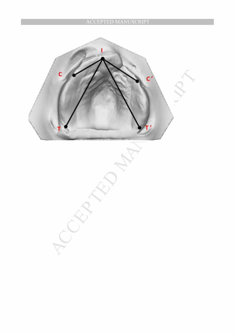
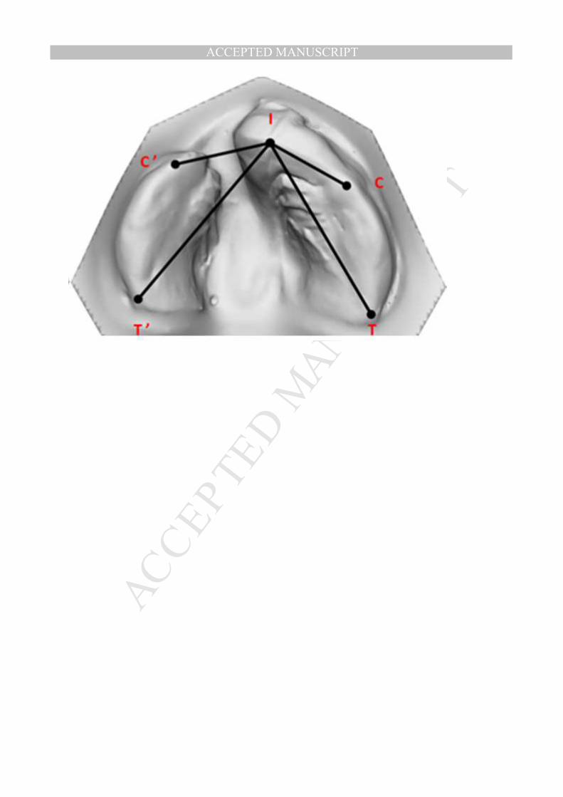
Italy (Céron-Zapata et al., 2016; De Menezes et al., 2016). Anatomic landmarks and
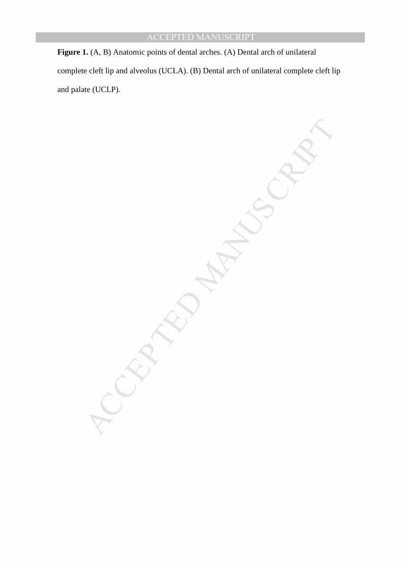
anthropometric measurements were: I−C (anterior inter-segment distance: interincisor
point to the point of eruption of the primary canine of the greater segment); I−T
MANUSCRIP
T
ACCEPTED
ACCEPTED MANUSCRIPT(anterior-posterior inter-segment distance: interincisor point to tuber of the greater
segment); I−C’ (anterior intra-segment distance: interincisor point to the point of
eruption of the primary canine of the lesser segment); and I−T’ (anterior-posterior intra-
segment distance: interincisor point to tuber of the lesser segment) (Figure 1 A – B). All
measurements were performed by a trained and calibrated examiner as in previous
studies (Falzoni et al., 2016; Jorge et al., 2016; Sakoda et al., 2017; Fuchigami et al.,
2017; Shetty et al., 2017).
All statistical analyses were performed with GraphPad Prism software (Prism 5
for Windows, version 5.0; GraphPad Software, Inc.) with a level of significance of 5%.
The intra-examiner error was analyzed through repeated-measures analysis 15 days after
the first measurements in one-third of the sample, randomly selected. To analyze the
systematic and casual error, a paired t test and Dahlberg’s formula were respectively
used. Data distributions were verified for all variables; for normally distributed values,
means and standard deviations were calculated, and inferential parametric tests were
used. Otherwise, medians, interquartile amplitudes and non-parametric tests were used.
To verify the intragroup changes in the UCLA group, a paired t test and Wilcoxon test
were applied. In the UCLP group, repeated-measures analysis of variance followed by
the Tukey test were applied. The intergroup comparisons used an independent t test and
the Mann-Whitney test.
RESULTS
The median ages (in years) of the children were verified at all study periods. In
UCLA group, the median ages were 0.29 and 1.74 respectively at T1 and T2. The
median ages of UCLP group were 0.29 (T1), 1.08 (T2), and 2.25 (T3). To assess
MANUSCRIP
T
ACCEPTED
ACCEPTED MANUSCRIPTreproducibility, the intra-examiner error was analyzed, and showed no statistically
significant differences in the repeated-measures analysis (p>0.05).
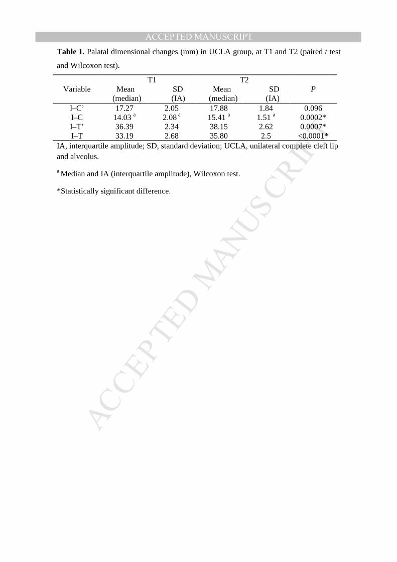
The palatal dimensional changes of the UCLA group showed that the distances I–
C, I−T’, and I–T significantly increased after cheiloplasty (Table 1). In the UCLP
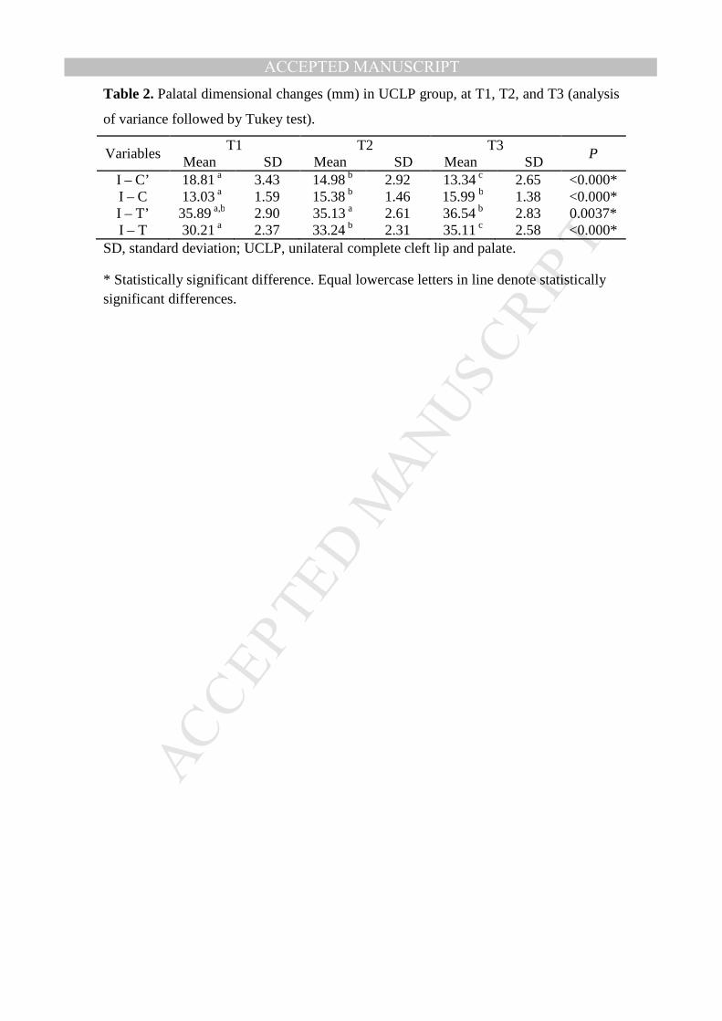
group, the I−C’ distance statistically decreased in the post-surgical period, while the I–T
distance increased. The I–C distance increased after cheiloplasty, with statistically
significant differences. The I−T’ distance increased between T2 and T3 with
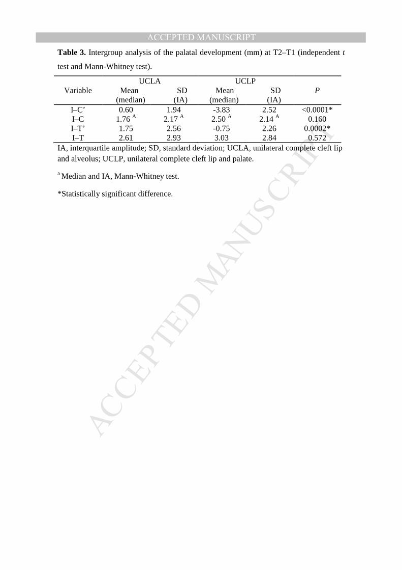
statistically significant differences (Table 2). Table 3 displays the intergroup analysis of
palatal development (T2-T1) and shows that the distances I−C’ and I−T’ demonstrated a
reduction of the dental arch growth in th e UCLP group compared with the UCLA
group, with statistically significant differences (Table 3).
DISCUSSION
Currently, digital anthropometric analysis is a viable alternative to conduct
studies to verify the development of the dental arches in children with cleft lip and
palate undergoing reparative surgical procedures (Carrara et al., 2016; Falzoni et al.,
2016; Sakoda et al., 2017). The 3D measurements can verify the differences between
children with and without congenital orofacial anomalies (Fernandes et al., 2015),
analyze how different cleft types develop after the same surgical procedures (Sakoda et
al., 2017), compare rehabilitative protocols (Jorge et al., 2016), and assess the growth of
the dental arches in children with pre-surgical orthopedics (Céron-Zapata et al., 2016;
Fuchigami et al., 2017; Shetty et al., 2017).
The present study analyzed the post-surgical effects on the development of the
maxillary segments of children with non-syndromic CLP who did not undergo pre-
surgical orthopedics. All children had cheiloplasty performed at 3 months of age
MANUSCRIP
T
ACCEPTED
ACCEPTED MANUSCRIPT(Millard technique) and one-stage palatoplasty at 12 months (von Langenbeck
technique) in HRAC/USP.
In the UCLA group, the I–C’ distance was greater before surgery than the I−C
distance, but at T2, the I–C’ distance showed inhibition, which strengths the hypothesis
that dental arches in children with oral clefts grow under the influence of the reparative
surgeries (Shi, Losee, 2015; Zhang et al., 2015; Falzoni et al., 2016) and of the cleft size
(Chiu et al., 2011; Zhang et al., 2015). Thus, a directly proportional relationship occurs
between the anatomic cleft size and the post-surgical dysmorphic growth.
In the UCLP group, the anthropometry of the anterior palatal area (I–C and I–C’)
demonstrated that on average the anterior inter-segment distance (I–C’) reduced after all
post-surgical periods, while the I–C distance significantly increased after cheiloplasty
(T2) and remained stable after palatoplasty (T3). Concerning the anterior-posterior
distances, the I−T’ distance increased only between T2 and T3, while the I–T distance
increased after all study periods. Several studies found comparable results in the
assessment of the anterior palatal area through the intercanine distance. After cheiloplasty,
the intercanine distance significantly reduced in children with UCLP (Falzoni et al., 2016;
Jorge et al., 2016; Sakoda et al., 2017). Heliövaara et al. (2017) reported that regardless of
the therapeutic approach, medical professionals always seek good dentofacial
development of individuals with cleft lip and palate. However, a consistent finding in
the scientific literature is the collapse of the maxillary dental arches propitiating a
retrusion of the maxilla, which can cause a modifications dental arch relationships. At
T3, I–T and I–T` distances did not decrease, although the I–C’ distance decreased and
the I–C distance seemed to be inhibited. Some authors believe that the palatoplasty can
inhibit the sagittal development of the maxilla (Tome et al., 2016). Indeed, it is difficult
to determine whether palatoplasty can inhibit anterior palatal growth or whether the
MANUSCRIP
T
ACCEPTED
ACCEPTED MANUSCRIPTinhibition continues due to the iatrogenic effect of early performed cheiloplasty.
According to Huang et al. (2002), cheiloplasty can exert an uninterrupted pressure
through the scar tissue on the anterior palatal area, and further studies are necessary to
quantify the post-surgical lip pressure on the dental arch.
The changes in the maxillary development of children with CLP can also be three-
dimensionally analyzed through facial morphology (Dádákova et al., 2016; Wu et al.,
2016). The facial analysis conducted by Dádákova et al. (2016) identified few differences
in facial symmetry in children with and without CLP. Moreover, the analysis
demonstrated that facial growth in the children with CLP was changed in comparison
with that in children without clefts, probably because of the cleft itself and the need of
palatoplasty (Dádákova et al., 2016).
The literature lacks consensus on which would be the reparative surgery with
more iatrogenic effects on the development of dental arches. This lack of consensus may
rely on other factors that may influence growth, such as the following: the size of the
anatomic defect (Chiu et al., 2011); the genetic pattern of the craniofacial growth of each
individual (Honda et al., 1995); the presence of syndromes or associated anomalies, and
the different surgical techniques and periods (Shi, Losee; 2015); the use of pre-surgical
orthopedics (Céron-Zapata et al., 2016; Fuchigami et al., 2017; Shetty et al., 2017); and
the surgeon’s ability (Stancheva et al., 2015).
Thus, further quantitative analyses of the development of the dental arches
(angulations, volume of the palatal bone segments, perimeter, and superposition of the
dental arches) are necessary to quantify the differences existing among the different
cleft types, from birth to skeletal maturity. This will enable the tailoring of an
individualized rehabilitative protocol for each type of orofacial anomaly that favors a
correct and harmonic maxillary growth.
MANUSCRIP
T
ACCEPTED
ACCEPTED MANUSCRIPTCONCLUSION
The new 3D anthropometric analysis showed that the development of the
maxillary segments changed after the repair surgeries. The UCLP group demonstrated a
reduction of the dental arches growth compared with the UCLA group.
MANUSCRIP
T
ACCEPTED
ACCEPTED MANUSCRIPTConflict of interest
The authors report no conflict of interest.
Funding
Funding was provided by the São Paulo Research Foundation (FAPESP; scholarship to
ECPA process # 2015/15586-6 and # 2016/07631-4).
Acknowledgements
The authors would like to acknowledge the support of all the participants in this study,
and also the financial support of the São Paulo Research Foundation (FAPESP;
scholarship to ECPA process # 2015/15586-6 and # 2016/07631-4).
MANUSCRIP
T
ACCEPTED
ACCEPTED MANUSCRIPTREFERENCES
Carrara CFC, Ambrosio ECP, Mello BZF, Jorge PK, Machado MAAM, Oliveira TM:
Three-dimensional evaluation of surgical techniques in neonates with orofacial cleft.
Ann Maxillofac Surg 6: 246-250, 2016.
Cerón-Zapata AM, López-Palacio AM, Rodriguez-Ardila MJ, Berrio-Gutiérrez LM, De
Menezes M, Sforza C: 3D evaluation of maxillary arches in unilateral cleft lip and
palate patients treated with nasoalveolar moulding vs. Hotz's plate. J Oral Rehabil
43:111-118, 2016.
Chiu YT, Liao YF, Chen PK: Initial cleft severity and maxillary growth in patients
with complete unilateral cleft lip and palate. Am J Orthod Dentofacial Orthop 140:189-
195, 2011.
Dadáková M, Cagáňová V, Dupej J, Hoffmannová E, Borský J, Velemínská J: Three-
dimensional evaluation of facial morphology in pre-school cleft patients following
neonatal cheiloplasty. J Craniomaxillofac Surg 44:1109-1116, 2016.
De Menezes M, Ceron-Zapata AM, Lopez-Palacio AM, Mapelli A, Pisoni L, Sforza C:
Evaluation of a 3D stereophotogrammetric method to identify and measure the palatal
surface area in children with unilateral cleft lip and palate. Cleft Palate Craniofac J
53:16-21, 2016.
MANUSCRIP
T
ACCEPTED
ACCEPTED MANUSCRIPTFalzoni MMM, Jorge PK, Laskos KV, Carrara CFC, Machado MAAM, Valarelli FP,
Oliveira TM: Three-dimensional dental arch evaluation of children with unilateral
complete cleft lip and palate. Dent Oral Craniofac Res 2:238-241, 2016.
Fernandes VM, Jorge PK, Carrara CFC, Gomide MR, Machado MAAM, Oliveira TM:
Three-dimensional digital evaluation of dental arches in infants with cleft lip and/or
palate. Brazil Dent J 26:297-302, 2015.
Freitas JA, das Neves LT, de Almeida AL, Garib DG, Trindade-Suedam IK, Yaedú RY,
de Lauris RC, Soares S, Oliveira TM, Pinto JH: Rehabilitative treatment of cleft lip and
palate: experience of the Hospital for Rehabilitation of Craniofacial Anomalies/USP
(HRAC/USP)─part 1: overall aspects. J Appl Oral Sci 20:9-15, 2012.
Fuchigami T, Kimura N, Kibe T, Tezuka M, Amir MS, Suga H, Takemoto
Y, Hashiguchi M, Maeda-Iino A, Nakamura N: Effects of pre-
surgical nasoalveolar moulding on maxillary arch and nasal form in unilateral cleftlip an
d palate before lip surgery. Orthod Craniofac Res 20:209-215, 2017.
Harila V, Ylikontiola LP, Palola R, Sandor GK: Maxillary arch dimensions in cleft
infants in Northern Finland. Acta Odontol Scand 71:930-936, 2013.
Heliövaara A, Küseler A, Skaare P, Shaw W, Mølsted K, Karsten A, Brinck E, Rizell
S, Marcusson A, Sæle P, Hurmerinta K, Rønning E, Najar Chalien M, Bellardie
H, Mooney J, Eyres P, Semb G:
MANUSCRIP
T
ACCEPTED
ACCEPTED MANUSCRIPTScandcleft randomised trials of primary surgery for unilateral cleft lip and palate: dental
arch relationships in 5 year-olds. J Plast Surg Hand Surg 51:52-57, 2017.
Honda Y, Suzuki A, Ohishi M, Tashiro H: Longitudinal study on the changes of
maxillary arch dimensions in Japanese children with cleft lip and/or palate: infancy to 4
years of age. Cleft Palate Craniofac J 32:149-155, 1995.
Huang CS, Wang WI, Liou EJ, Chen YR, Chen PK, Noordhoff MS: Effects of
cheiloplasty on maxillary dental arch development in infants with unilateral complete
cleft lip and palate. Cleft Palate Craniofac J 39:513-516, 2002.
Jorge PK, Gnoinski W, Laskos KV, Carrara CFC, Garib DG, Ozawa TO, Machado
MAAM, Valarelli FP, Oliveira TM: Comparison of two treatment protocols in children
with unilateral complete cleft lip and palate: tridimensional evaluation of the maxillary
dental arch. J Craniomaxillofac Surg 44:1117-1122, 2016.
Sakoda KL, Jorge PK, Carrara CFC, Machado MAAM, Valarelli FP, Pinzan A, Oliveira
TM: 3D analysis of effects of primary surgeries in cleft lip/palate children during the
first two years of life. Braz Oral Res 31:1-6, 2017.
Shetty V, Agrawal RK, Sailer HF: Long-term effect of presurgical nasoalveolar
molding on growth of maxillary arch in unilateral cleft lip and palate: randomized
controlled trial. Int J Oral Maxillofac Surg 46:977-987, 2017.
MANUSCRIP
T
ACCEPTED
ACCEPTED MANUSCRIPTShi B, Losee JE: The impact of cleft lip and palate repair on maxillofacial growth. Int J
Oral Sci 7:14-17, 2015.
Stancheva N, Dannhauer KH, Hemprich A, Krey KF: Three-dimensional analysis of
maxillary development in patients with unilateral cleft lip and palate during the first six
years of life. J Orofac Orthop 76: 391-404, 2015.
Tome W, Yashiro K, Otsuki K, Kogo M, Yamashiro T: Influence of different
palatoplasties on the facial morphology of early mixed dentition stage children with
unilateral cleft lip and palate. Cleft Palate Craniofac J 53:28-33, 2016.
Wu J, Heike C, Birgfeld C, Evans K, Maga M, Morrison C, Saltzman B, Shapiro L, Tse
R: Measuring symmetry in children with unrepaired cleft lip: defining a standard for the
three-dimensional midfacial reference plane. Cleft Palate Craniofac J 53:695-704, 2016.
Zhang D, Zheng L, Wang Q, Lu L, Ma J: Displacements prediction from 3D finite
element model of maxillary protraction with and without rapid maxillary expansion in a
patient with unilateral cleft palate and alveolus. Biomed Eng Online 14:1-15, 2015.
MANUSCRIP
T
ACCEPTED
ACCEPTED MANUSCRIPTFigure 1. (A, B) Anatomic points of dental arches. (A) Dental arch of unilateral
complete cleft lip and alveolus (UCLA). (B) Dental arch of unilateral complete cleft lip
and palate (UCLP).
MANUSCRIP
T
ACCEPTED
ACCEPTED MANUSCRIPTTable 1. Palatal dimensional changes (mm) in UCLA group, at T1 and T2 (paired t test
and Wilcoxon test).
Variable T1
Mean SD (median) (IA)
T2 Mean SD (median) (IA)
P
I–C’ 17.27 2.05 17.88 1.84 0.096 I–C 14.03 a 2.08 a 15.41 a 1.51 a 0.0002* I–T’ 36.39 2.34 38.15 2.62 0.0007* I–T 33.19 2.68 35.80 2.5 <0.0001*
IA, interquartile amplitude; SD, standard deviation; UCLA, unilateral complete cleft lip and alveolus.
a Median and IA (interquartile amplitude), Wilcoxon test.
*Statistically significant difference.
MANUSCRIP
T
ACCEPTED
ACCEPTED MANUSCRIPTTable 2. Palatal dimensional changes (mm) in UCLP group, at T1, T2, and T3 (analysis
of variance followed by Tukey test).
Variables T1
Mean SD T2
Mean SD T3
Mean SD P
I – C’ 18.81 a 3.43 14.98 b 2.92 13.34 c 2.65 <0.000* I – C 13.03 a 1.59 15.38 b 1.46 15.99 b 1.38 <0.000* I – T’ 35.89 a,b 2.90 35.13 a 2.61 36.54 b 2.83 0.0037* I – T 30.21 a 2.37 33.24 b 2.31 35.11 c 2.58 <0.000*
SD, standard deviation; UCLP, unilateral complete cleft lip and palate.
* Statistically significant difference. Equal lowercase letters in line denote statistically significant differences.
MANUSCRIP
T
ACCEPTED
ACCEPTED MANUSCRIPTTable 3. Intergroup analysis of the palatal development (mm) at T2–T1 (independent t
test and Mann-Whitney test).
Variable UCLA
Mean SD (median) (IA)
UCLP Mean SD (median) (IA)
P
I–C’ 0.60 1.94 -3.83 2.52 <0.0001* I–C 1.76 A 2.17 A 2.50 A 2.14 A 0.160 I–T’ 1.75 2.56 -0.75 2.26 0.0002* I–T 2.61 2.93 3.03 2.84 0.572
IA, interquartile amplitude; SD, standard deviation; UCLA, unilateral complete cleft lip and alveolus; UCLP, unilateral complete cleft lip and palate.
a Median and IA, Mann-Whitney test.
*Statistically significant difference.
MANUSCRIP
T
ACCEPTED
ACCEPTED MANUSCRIPT
MANUSCRIP
T
ACCEPTED
ACCEPTED MANUSCRIPT