Embed Size (px)
Citation preview
Bacterial Cytolysin during Meningitis Disrupts theRegulation of Glutamate in the Brain, Leading toSynaptic DamageCarolin Wippel1, Jana Maurer1, Christina Fortsch1, Sabrina Hupp1, Alexandra Bohl1, Jiangtao Ma2,
Timothy J. Mitchell2,3, Stephanie Bunkowski4, Wolfgang Bruck4, Roland Nau4,5, Asparouh I. Iliev1*
1 DFG Membrane/Cytoskeleton Interaction Group, Institute of Pharmacology and Toxicology & Rudolf Virchow Center for Experimental Medicine, University of Wurzburg,
Wurzburg, Germany, 2 Division of Infection and Immunity, Glasgow Biomedical Research Centre, University of Glasgow, Glasgow, United Kingdom, 3 Chair of Microbial
Infection and Immunity, School of Immunity and Infection, College of Medical and Dental Sciences, University of Birmingham, Birmingham, United Kingdom,
4 Department of Neuropathology, Georg-August-University of Gottingen, Gottingen, Germany, 5 Department of Geriatrics, Evangelisches Krankenhaus Gottingen-
Weende, Gottingen, Germany
Abstract
Streptococcus pneumoniae (pneumococcal) meningitis is a common bacterial infection of the brain. The cholesterol-dependent cytolysin pneumolysin represents a key factor, determining the neuropathogenic potential of the pneumococci.Here, we demonstrate selective synaptic loss within the superficial layers of the frontal neocortex of post-mortem brainsamples from individuals with pneumococcal meningitis. A similar effect was observed in mice with pneumococcalmeningitis only when the bacteria expressed the pore-forming cholesterol-dependent cytolysin pneumolysin. Exposure ofacute mouse brain slices to only pore-competent pneumolysin at disease-relevant, non-lytic concentrations causedpermanent dendritic swelling, dendritic spine elimination and synaptic loss. The NMDA glutamate receptor antagonistsMK801 and D-AP5 reduced this pathology. Pneumolysin increased glutamate levels within the mouse brain slices. In mouseastrocytes, pneumolysin initiated the release of glutamate in a calcium-dependent manner. We propose that pneumolysinplays a significant synapto- and dendritotoxic role in pneumococcal meningitis by initiating glutamate release fromastrocytes, leading to subsequent glutamate-dependent synaptic damage. We outline for the first time the occurrence ofsynaptic pathology in pneumococcal meningitis and demonstrate that a bacterial cytolysin can dysregulate the control ofglutamate in the brain, inducing excitotoxic damage.
Citation: Wippel C, Maurer J, Fortsch C, Hupp S, Bohl A, et al. (2013) Bacterial Cytolysin during Meningitis Disrupts the Regulation of Glutamate in the Brain,Leading to Synaptic Damage. PLoS Pathog 9(6): e1003380. doi:10.1371/journal.ppat.1003380
Editor: Paul M. Sullam, University of California, San Francisco, United States of America
Received November 17, 2012; Accepted April 8, 2013; Published June 13, 2013
Copyright: � 2013 Wippel et al. This is an open-access article distributed under the terms of the Creative Commons Attribution License, which permitsunrestricted use, distribution, and reproduction in any medium, provided the original author and source are credited.
Funding: The work in Wurzburg was funded by the Emmy Noether Programme of the German Science Foundation (DFG) [IL-151.1 to AII], the Rudolf VirchowCenter for Experimental Medicine, Wurzburg, and the University of Wurzburg. The work in Glasgow was supported by the Wellcome Trust, the BBSRC and theEuropean Science Foundation. The work in Gottingen was funded by Sparkasse Gottingen. The funders had no role in study design, data collection and analysis,decision to publish, or preparation of the manuscript.
Competing Interests: The authors have declared that no competing interests exist.
* E-mail: [email protected]
Introduction
Streptococcus pneumoniae (pneumococcal) meningitis is the most
common form of bacterial meningitis [1]. The patient survival
rate is 75%; however, half of the patients suffer from long-term
disabilities [2]. This disease is associated with significant but
not massive neuronal death, predominantly in the hippocampus
[3].
Pneumolysin (PLY) is a critical pneumococcal pathogenic factor
that belongs to the cholesterol-dependent cytolysin (CDC) group.
This 54-kD protein contains four domains and targets plasma-
lemmal cholesterol through its fourth domain [4]. Upon binding,
PLY oligomerizes into pre-pore rings of 30–50 monomers.
Subsequently, domain 3 of each monomer refolds and penetrates
the membrane, transforming the pre-pore to a pore [5]. Multiple
cellular toxic effects (e.g., small GTPase activation and microtu-
bule stabilization), although pore-dependent, occur at sub-lytic
and non-lytic concentrations, which is indicative of a more
complex interaction between the toxin and cells [6–8]. To obtain
tools to study the cellular effects of pore formation by PLY, the
delta6 mutant form of PLY has been created [9], which lacks the
amino acids alanine 146 and arginine 147. This mutation makes
the refolding of domain 3, and thus pore formation, impossible [9].
PLY is critical for the clinical course of experimental pneumo-
coccal meningitis [10,11], and PLY-deficient S. pneumoniae bacteria
initiate a substantially milder disease course. PLY is persistent in
the cerebrospinal fluid (CSF), which correlates with a poorer
prognosis in human patients [12]. However, the mechanism of this
PLY dependence remains largely unclear. There is some
experimental evidence from a rabbit model, however, that argues
against the role of PLY in meningitis [13], raising the question of
the specificity of different animal models.
In bacterial meningitis, pathogenic bacteria multiply in the CSF
and are abundant adjacent to the white matter (along the ventricles)
and the neocortex. The neocortex is a highly specialized and
complex brain structure comprised of up to six layers. Its major
component is pyramidal neurons (,80% of all cortical neurons are
pyramidal), whose somata are positioned predominantly in layers
PLOS Pathogens | www.plospathogens.org 1 June 2013 | Volume 9 | Issue 6 | e1003380
III, V and VI, send projections throughout the cortex and receive
projections from other neurons from other brain areas. Particularly
complex and dense synaptic connections are established in the
superficial layers I–II. Positioned within the neocortex among the
pyramidal neurons, multiple interneurons (typically spiny stellate
and basket cells) establish shorter connections with neighboring
neurons. Generally, the pyramidal neurons are described as
glutamatergic, with some exceptions (for review see [14]).
Synapses are complex structures, consisting of a pre- and post-
synapse and the surrounding cells (such as astrocytes) [15].
Astrocytes wrap around the synaptic cleft and parts of the pre-
and/or post-synapse, thereby allowing them to rapidly remove
released neurotransmitters [16]. The morphological structures that
host the active post-synapses along the dendrites are the dendritic
spines [17]. The dendritic spines are dynamic; not all of them host
synapses, but all synapses of pyramidal neurons are positioned on
spines [18]. Long-term potentiation or depression are associated
with spine growth or shrinkage [19]. The permanent loss and
alteration of dendritic spines correlate with decreased synaptic
numbers and cognitive impairment in multiple neurological
disorders [20,21].
Our aim was to study changes in neocortical synapses as a result
of Streptococcus pneumoniae meningitis and the role of the major
pneumococcal neurotoxin PLY in producing these effects.
Results
To address the question of synaptic damage resulting from
meningitis, we used four experimental approaches: i) human
frontal neocortical brain samples from autopsy cases; ii) a
pneumococcal meningitis mouse model (3–5 month-old C57Bl/6
mice); iii) an acute mouse brain slice model (postnatal day 12–14
C57Bl/6 mice); and iv) a primary mouse astrocyte culture system.
Reduction of synapses in human forebrain postmortemsamples from meningitis patients
Human frontal neocortical brain samples obtained from
autopsy cases following death from S. pneumoniae meningitis and
from control cases following a rapid death from non-neurological
causes (the exact descriptions of the patient groups are presented
in Table 1 and Table 2) were stained with anti-synapsin I (a pre-
synaptic marker protein [22]) and anti-PSD95 (post-synaptic
density 95; a post-synaptic protein, specific for glutamatergic
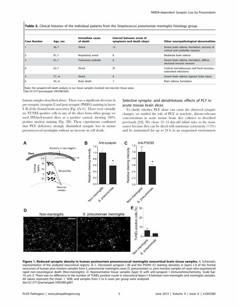
synapses [23]) antibodies. In the superficial neocortical layers I–II,
pre- and post-synaptic immunostaining levels were significantly
reduced in the meningitis-infected samples (Fig. 1A–D). The total
cell density and the number of dying cells (measured by TUNEL
staining for apoptotic cells) were identical between the groups
within the area of analysis (Fig. 1E). The presence of TUNEL-
positive cells in both groups may be due to the post-mortem delay
until sample collection. In all slices, the analyzed cortical areas
excluded necrotic regions, which were present in some patient
samples. Thus, S. pneumoniae meningitis induced a reduction in
synaptic density that was independent of cell death.
PLY-dependent reduction of synapses in the neocortexof pneumococcal meningitis mice
Next, we focused on the role of individual key pathogenic
components of S. pneumoniae as synaptotoxic factors. We infected the
right frontal brain lobes of C57BL/6 animals with the PLY-
producing S. pneumoniae strain D39 and with PLY-deficient mutants
of the D39 strain. Thirty-six hours after infection, we sacrificed the
animals and stained paraffin brain slices in a manner similar to the
Author Summary
Bacterial meningitis is one of the most devastating braindiseases. Among the bacteria that cause meningitis,Streptococcus pneumoniae is the most common. Meningitispredominantly affects children, especially in the ThirdWorld, and most of them do not survive. Those that dosurvive often suffer permanent brain damage and hearingproblems. The exact morphological substrates of braindamage in Streptococcus pneumoniae meningitis remainlargely unknown. In our experiments, we found that thebrain cortex of patients with meningitis demonstrated aloss of synapses (the contact points among neurons,responsible for the processes of learning and memory),and we identified the major pneumococcal neurotoxinpneumolysin as a sufficient cause of this loss. The effectwas not direct but was mediated by the brain neurotrans-mitter glutamate, which was released upon toxin bindingby one of the non-neuronal cell types of the brain – theastrocytes. Pneumolysin initiated calcium influx in astro-cytes and subsequent glutamate release. Glutamatedamaged the synapses via NMDA-receptors – a mecha-nism similar to the damage occurring in brain ischemia.Thus, we show that synaptic loss is present in pneumo-coccal meningitis, and we identify the toxic bacterialprotein pneumolysin as the major factor in this process.These findings alter our understanding of bacterialmeningitis and establish new therapeutic strategies forthis fatal disease.
Table 1. Clinical histories of the individual patients from the non-meningitis histology group.
Case Number Age, sex Underlying disease(s) Immediate cause of death
1 59, m COPD Pulmonary failure/shock
2 60, m None Chest and abdominal trauma
3 82, f Diarrhea, hypertension, aortic valve replacement,cardiac failure
Aspiration
4 42, m Myocardial infarction, CAD Ventricular fibrillation
5 58, f Coronary heart disease, myocardial infarction Ventricular fibrillation
6 66, f Renal insufficiency, suspected pulmonary embolism Acute cardiac failure
No focal necroses were present in the frontal cortical and hippocampal sections of control cases. In the hippocampal formation of case 58, f hypoxic injury was noted onHE stains.ARDS: adult respiratory distress syndrome, CAD: coronary artery disease, COPD: chronic obstructive pulmonary disease.doi:10.1371/journal.ppat.1003380.t001
NMDA-dependent Synaptic Loss by Pneumolysin
PLOS Pathogens | www.plospathogens.org 2 June 2013 | Volume 9 | Issue 6 | e1003380
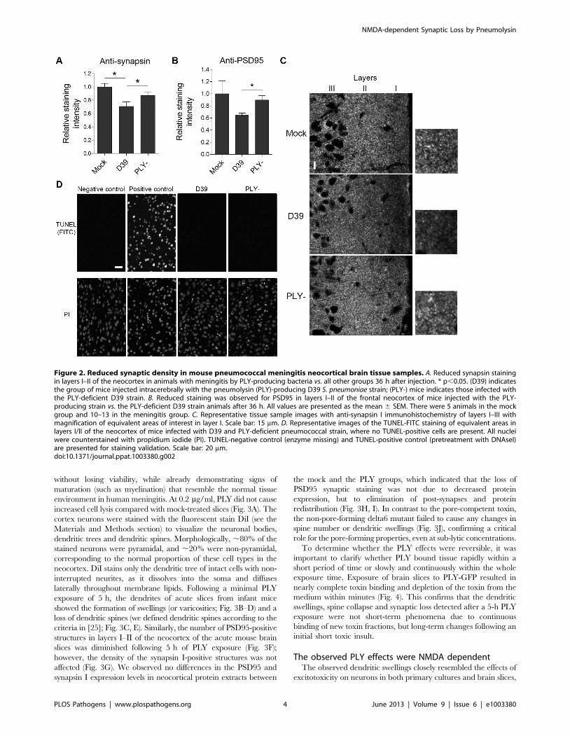
human samples described above. There was a significant decrease in
pre-synaptic (synapsin I) and post-synaptic (PSD95) staining in layers
I–II of the frontal brain neocortex (Fig. 2A–C). There were virtually
no TUNEL-positive cells in any of the slices from either group; we
used DNAseI-treated slices as a positive control, showing 100%
positive nuclear staining (Fig. 2D). These experiments confirmed
that PLY deficiency strongly diminished synaptic loss in mouse
pneumococcal meningitis without an increase in cell death.
Selective synapto- and dendritotoxic effects of PLY inacute mouse brain slices
To clarify whether PLY alone can cause the observed synaptic
changes, we studied the role of PLY at non-lytic, disease-relevant
concentrations in acute mouse brain slice cultures as described
previously [24]. We chose 12–14 day-old infant mice as the tissue
source because they can be sliced with minimum cytotoxicity (,5%)
and be maintained for up to 24 h in an oxygenized environment
Figure 1. Reduced synaptic density in human postmortem pneumococcal meningitis neocortical brain tissue samples. A. Schematicrepresentation of the analyzed neocortical regions. B, C. Decreased synapsin I (B) and the PSD95 (C) staining densities in layers I–II of the frontalneocortex of human post-mortem samples from S. pneumoniae meningitis cases (S. pneumoniae) vs. post mortem samples of cases who experiencedrapid non-neurological death (Non-meningitis). D. Representative tissue samples (layer II) with anti-synapsin I immunohistochemistry. Scale bar:10 mm. E. There was no difference in the number of TUNEL-positive nuclei in neocortical layers I–II between non-meningitis and meningitis samples.All values represent the mean 6 SEM, and samples from 5 to 6 cases per group were analyzed.doi:10.1371/journal.ppat.1003380.g001
Table 2. Clinical histories of the individual patients from the Streptococcus pneumoniae meningitis histology group.
Case Number Age, sexImmediate causeof death
Interval between onset ofsymptoms and death (days) Other neuropathological abnormalities
1 56, f Shock 12 Severe brain edema, herniation, necrosis ofcortical and cerebellar neurons
2 81, f Respiratory arrest 8 Moderate brain edema
3 61, f Pulmonary embolia 6 Severe brain edema, herniation, diffuseneuronal necrosis neurons
4 63, f Shock 91 Cortical microabscesses and focal necroses,watershed infarctions
5 57, m Shock 6 Severe brain edema, hypoxic brain injury
6 54, m Brain death 1 Brain edema, herniation
Note: the synaptic/cell death analysis in our tissue samples involved non-necrotic tissue areas.doi:10.1371/journal.ppat.1003380.t002
NMDA-dependent Synaptic Loss by Pneumolysin
PLOS Pathogens | www.plospathogens.org 3 June 2013 | Volume 9 | Issue 6 | e1003380
without losing viability, while already demonstrating signs of
maturation (such as myelination) that resemble the normal tissue
environment in human meningitis. At 0.2 mg/ml, PLY did not cause
increased cell lysis compared with mock-treated slices (Fig. 3A). The
cortex neurons were stained with the fluorescent stain DiI (see the
Materials and Methods section) to visualize the neuronal bodies,
dendritic trees and dendritic spines. Morphologically, ,80% of the
stained neurons were pyramidal, and ,20% were non-pyramidal,
corresponding to the normal proportion of these cell types in the
neocortex. DiI stains only the dendritic tree of intact cells with non-
interrupted neurites, as it dissolves into the soma and diffuses
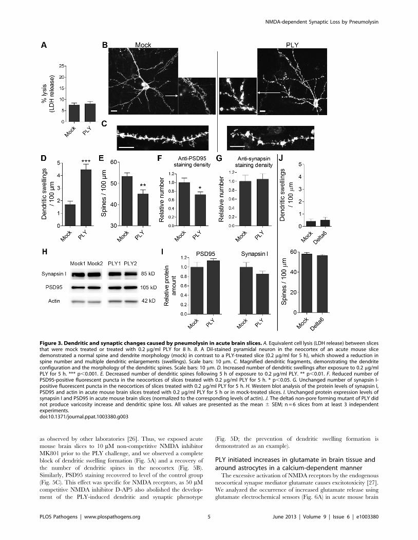
laterally throughout membrane lipids. Following a minimal PLY
exposure of 5 h, the dendrites of acute slices from infant mice
showed the formation of swellings (or varicosities; Fig. 3B–D) and a
loss of dendritic spines (we defined dendritic spines according to the
criteria in [25]; Fig. 3C, E). Similarly, the number of PSD95-positive
structures in layers I–II of the neocortex of the acute mouse brain
slices was diminished following 5 h of PLY exposure (Fig. 3F);
however, the density of the synapsin I-positive structures was not
affected (Fig. 3G). We observed no differences in the PSD95 and
synapsin I expression levels in neocortical protein extracts between
the mock and the PLY groups, which indicated that the loss of
PSD95 synaptic staining was not due to decreased protein
expression, but to elimination of post-synapses and protein
redistribution (Fig. 3H, I). In contrast to the pore-competent toxin,
the non-pore-forming delta6 mutant failed to cause any changes in
spine number or dendritic swellings (Fig. 3J), confirming a critical
role for the pore-forming properties, even at sub-lytic concentrations.
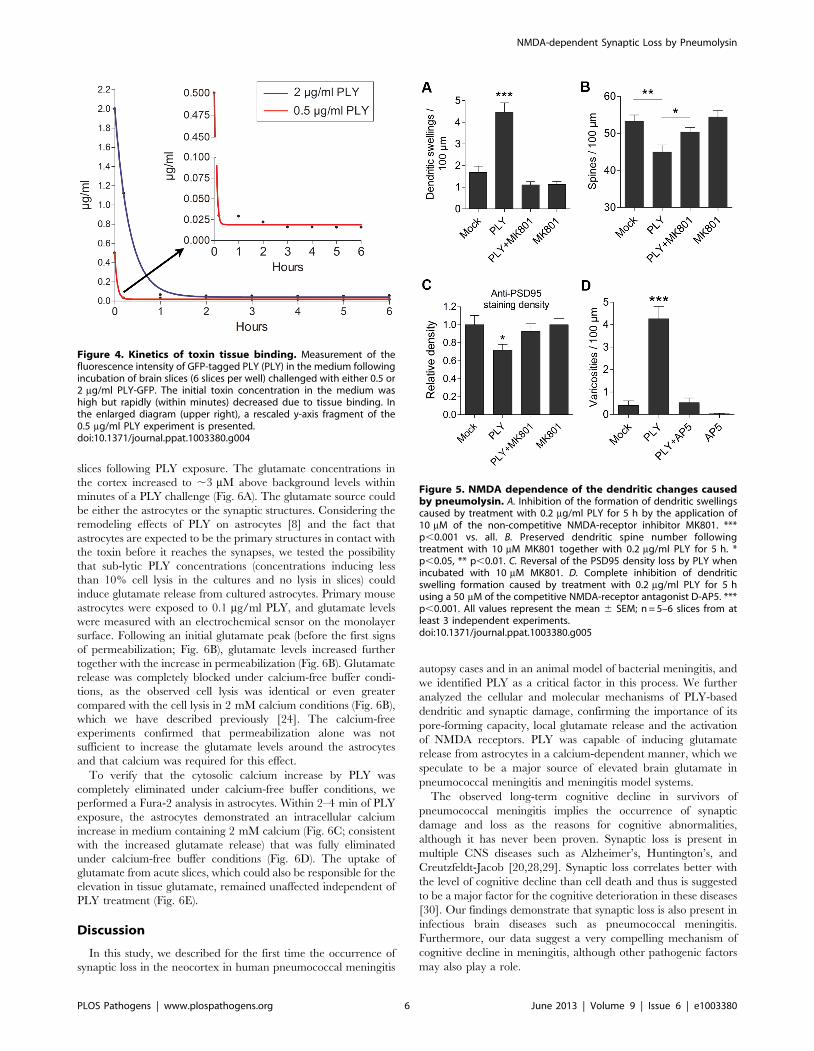
To determine whether the PLY effects were reversible, it was
important to clarify whether PLY bound tissue rapidly within a
short period of time or slowly and continuously within the whole
exposure time. Exposure of brain slices to PLY-GFP resulted in
nearly complete toxin binding and depletion of the toxin from the
medium within minutes (Fig. 4). This confirms that the dendritic
swellings, spine collapse and synaptic loss detected after a 5-h PLY
exposure were not short-term phenomena due to continuous
binding of new toxin fractions, but long-term changes following an
initial short toxic insult.
The observed PLY effects were NMDA dependentThe observed dendritic swellings closely resembled the effects of
excitotoxicity on neurons in both primary cultures and brain slices,
Figure 2. Reduced synaptic density in mouse pneumococcal meningitis neocortical brain tissue samples. A. Reduced synapsin stainingin layers I–II of the neocortex in animals with meningitis by PLY-producing bacteria vs. all other groups 36 h after injection. * p,0.05. (D39) indicatesthe group of mice injected intracerebrally with the pneumolysin (PLY)-producing D39 S. pneumoniae strain; (PLY-) mice indicates those infected withthe PLY-deficient D39 strain. B. Reduced staining was observed for PSD95 in layers I–II of the frontal neocortex of mice injected with the PLY-producing strain vs. the PLY-deficient D39 strain animals after 36 h. All values are presented as the mean 6 SEM. There were 5 animals in the mockgroup and 10–13 in the meningitis group. C. Representative tissue sample images with anti-synapsin I immunohistochemistry of layers I–III withmagnification of equivalent areas of interest in layer I. Scale bar: 15 mm. D. Representative images of the TUNEL-FITC staining of equivalent areas inlayers I/II of the neocortex of mice infected with D39 and PLY-deficient pneumococcal strain, where no TUNEL-positive cells are present. All nucleiwere counterstained with propidium iodide (PI). TUNEL-negative control (enzyme missing) and TUNEL-positive control (pretreatment with DNAseI)are presented for staining validation. Scale bar: 20 mm.doi:10.1371/journal.ppat.1003380.g002
NMDA-dependent Synaptic Loss by Pneumolysin
PLOS Pathogens | www.plospathogens.org 4 June 2013 | Volume 9 | Issue 6 | e1003380
as observed by other laboratories [26]. Thus, we exposed acute
mouse brain slices to 10 mM non-competitive NMDA inhibitor
MK801 prior to the PLY challenge, and we observed a complete
block of dendritic swelling formation (Fig. 5A) and a recovery of
the number of dendritic spines in the neocortex (Fig. 5B).
Similarly, PSD95 staining recovered to level of the control group
(Fig. 5C). This effect was specific for NMDA receptors, as 50 mM
competitive NMDA inhibitor D-AP5 also abolished the develop-
ment of the PLY-induced dendritic and synaptic phenotype
(Fig. 5D; the prevention of dendritic swelling formation is
demonstrated as an example).
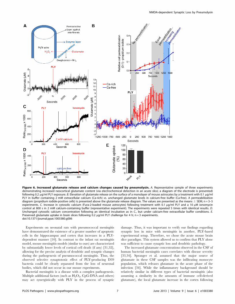
PLY initiated increases in glutamate in brain tissue andaround astrocytes in a calcium-dependent manner
The excessive activation of NMDA receptors by the endogenous
neocortical synapse mediator glutamate causes excitotoxicity [27].
We analyzed the occurrence of increased glutamate release using
glutamate electrochemical sensors (Fig. 6A) in acute mouse brain
Figure 3. Dendritic and synaptic changes caused by pneumolysin in acute brain slices. A. Equivalent cell lysis (LDH release) between slicesthat were mock treated or treated with 0.2 mg/ml PLY for 8 h. B. A DiI-stained pyramidal neuron in the neocortex of an acute mouse slicedemonstrated a normal spine and dendrite morphology (mock) in contrast to a PLY-treated slice (0.2 mg/ml for 5 h), which showed a reduction inspine number and multiple dendritic enlargements (swellings). Scale bars: 10 mm. C. Magnified dendritic fragments, demonstrating the dendriteconfiguration and the morphology of the dendritic spines. Scale bars: 10 mm. D. Increased number of dendritic swellings after exposure to 0.2 mg/mlPLY for 5 h. *** p,0.001. E. Decreased number of dendritic spines following 5 h of exposure to 0.2 mg/ml PLY. ** p,0.01. F. Reduced number ofPSD95-positive fluorescent puncta in the neocortices of slices treated with 0.2 mg/ml PLY for 5 h. * p,0.05. G. Unchanged number of synapsin I-positive fluorescent puncta in the neocortices of slices treated with 0.2 mg/ml PLY for 5 h. H. Western blot analysis of the protein levels of synapsin I,PSD95 and actin in acute mouse brain slices treated with 0.2 mg/ml PLY for 5 h or in mock-treated slices. I. Unchanged protein expression levels ofsynapsin I and PSD95 in acute mouse brain slices (normalized to the corresponding levels of actin). J. The delta6 non-pore forming mutant of PLY didnot produce varicosity increase and dendritic spine loss. All values are presented as the mean 6 SEM; n = 6 slices from at least 3 independentexperiments.doi:10.1371/journal.ppat.1003380.g003
NMDA-dependent Synaptic Loss by Pneumolysin
PLOS Pathogens | www.plospathogens.org 5 June 2013 | Volume 9 | Issue 6 | e1003380
slices following PLY exposure. The glutamate concentrations in
the cortex increased to ,3 mM above background levels within
minutes of a PLY challenge (Fig. 6A). The glutamate source could
be either the astrocytes or the synaptic structures. Considering the
remodeling effects of PLY on astrocytes [8] and the fact that
astrocytes are expected to be the primary structures in contact with
the toxin before it reaches the synapses, we tested the possibility
that sub-lytic PLY concentrations (concentrations inducing less
than 10% cell lysis in the cultures and no lysis in slices) could
induce glutamate release from cultured astrocytes. Primary mouse
astrocytes were exposed to 0.1 mg/ml PLY, and glutamate levels
were measured with an electrochemical sensor on the monolayer
surface. Following an initial glutamate peak (before the first signs
of permeabilization; Fig. 6B), glutamate levels increased further
together with the increase in permeabilization (Fig. 6B). Glutamate
release was completely blocked under calcium-free buffer condi-
tions, as the observed cell lysis was identical or even greater
compared with the cell lysis in 2 mM calcium conditions (Fig. 6B),
which we have described previously [24]. The calcium-free
experiments confirmed that permeabilization alone was not
sufficient to increase the glutamate levels around the astrocytes
and that calcium was required for this effect.
To verify that the cytosolic calcium increase by PLY was
completely eliminated under calcium-free buffer conditions, we
performed a Fura-2 analysis in astrocytes. Within 2–4 min of PLY
exposure, the astrocytes demonstrated an intracellular calcium
increase in medium containing 2 mM calcium (Fig. 6C; consistent
with the increased glutamate release) that was fully eliminated
under calcium-free buffer conditions (Fig. 6D). The uptake of
glutamate from acute slices, which could also be responsible for the
elevation in tissue glutamate, remained unaffected independent of
PLY treatment (Fig. 6E).
Discussion
In this study, we described for the first time the occurrence of
synaptic loss in the neocortex in human pneumococcal meningitis
autopsy cases and in an animal model of bacterial meningitis, and
we identified PLY as a critical factor in this process. We further
analyzed the cellular and molecular mechanisms of PLY-based
dendritic and synaptic damage, confirming the importance of its
pore-forming capacity, local glutamate release and the activation
of NMDA receptors. PLY was capable of inducing glutamate
release from astrocytes in a calcium-dependent manner, which we
speculate to be a major source of elevated brain glutamate in
pneumococcal meningitis and meningitis model systems.
The observed long-term cognitive decline in survivors of
pneumococcal meningitis implies the occurrence of synaptic
damage and loss as the reasons for cognitive abnormalities,
although it has never been proven. Synaptic loss is present in
multiple CNS diseases such as Alzheimer’s, Huntington’s, and
Creutzfeldt-Jacob [20,28,29]. Synaptic loss correlates better with
the level of cognitive decline than cell death and thus is suggested
to be a major factor for the cognitive deterioration in these diseases
[30]. Our findings demonstrate that synaptic loss is also present in
infectious brain diseases such as pneumococcal meningitis.
Furthermore, our data suggest a very compelling mechanism of
cognitive decline in meningitis, although other pathogenic factors
may also play a role.
Figure 4. Kinetics of toxin tissue binding. Measurement of thefluorescence intensity of GFP-tagged PLY (PLY) in the medium followingincubation of brain slices (6 slices per well) challenged with either 0.5 or2 mg/ml PLY-GFP. The initial toxin concentration in the medium washigh but rapidly (within minutes) decreased due to tissue binding. Inthe enlarged diagram (upper right), a rescaled y-axis fragment of the0.5 mg/ml PLY experiment is presented.doi:10.1371/journal.ppat.1003380.g004
Figure 5. NMDA dependence of the dendritic changes causedby pneumolysin. A. Inhibition of the formation of dendritic swellingscaused by treatment with 0.2 mg/ml PLY for 5 h by the application of10 mM of the non-competitive NMDA-receptor inhibitor MK801. ***p,0.001 vs. all. B. Preserved dendritic spine number followingtreatment with 10 mM MK801 together with 0.2 mg/ml PLY for 5 h. *p,0.05, ** p,0.01. C. Reversal of the PSD95 density loss by PLY whenincubated with 10 mM MK801. D. Complete inhibition of dendriticswelling formation caused by treatment with 0.2 mg/ml PLY for 5 husing a 50 mM of the competitive NMDA-receptor antagonist D-AP5. ***p,0.001. All values represent the mean 6 SEM; n = 5–6 slices from atleast 3 independent experiments.doi:10.1371/journal.ppat.1003380.g005
NMDA-dependent Synaptic Loss by Pneumolysin
PLOS Pathogens | www.plospathogens.org 6 June 2013 | Volume 9 | Issue 6 | e1003380
Experiments on neonatal rats with pneumococcal meningitis
have demonstrated the existence of a greater number of apoptotic
cells in the hippocampus and cortex that increases in a PLY-
dependent manner [10]. In contrast to the infant rat meningitis
model, mouse meningitis models (similar to ours) are characterized
by substantially lower levels of cortical cell death (if any) [31,32],
allowing for the precise analysis of dendritic and synaptic changes
during the pathogenesis of pneumococcal meningitis. Thus, the
observed selective synaptotoxic effect of PLY-producing D39
bacteria could be clearly separated from the loss of neuronal
bodies, which did not occur in our mouse experiments.
Bacterial meningitis is a disease with a complex pathogenesis.
Multiple additional factors (such as H2O2, CpG-DNA and others)
may act synergistically with PLY in the process of synaptic
damage. Thus, it was important to verify our findings regarding
synaptic loss in mice with meningitis in another, PLY-based
experimental setup. Therefore, we chose the acute mouse brain
slice paradigm. This system allowed us to confirm that PLY alone
was sufficient to cause synaptic loss and dendritic pathology.
The increased glutamate concentrations observed in the CSF of
human bacterial meningitis cases correlates with disease severity
[33,34]. Spranger et al. assumed that the major source of
glutamate in these CSF samples was the infiltrating monocyte
population, which releases glutamate in the acute phase of the
infection [34]. While the inflammatory background should be
relatively similar in different types of bacterial meningitis (also
assuming a similarity in the amounts of immune cell-derived
glutamate), the local glutamate increase in the cortex following
Figure 6. Increased glutamate release and calcium changes caused by pneumolysin. A. Representative sample of three experimentsdemonstrating increased neocortical glutamate content (via electrochemical detection in an acute slice; a diagram of the electrode is presented)following 0.2 mg/ml PLY exposure. B. Elevation of glutamate release on the surface of a monolayer of mouse astrocytes by a treatment with 0.1 mg/mlPLY in buffer containing 2 mM extracellular calcium (Ca-rich) vs. unchanged glutamate levels in calcium-free buffer (Ca-free). A permeabilizationdiagram (propidium iodide-positive cells) is presented above the glutamate release diagram. The values are presented as the means 6 SEM; n = 3–5experiments. C. Increase in cytosolic calcium (Fura-2-loaded mouse astrocytes) following treatment with 0.1 mg/ml PLY and a 10 mM ionomycincontrol at 800 s in 2 mM calcium-containing buffer (representative experiment). The experiments were repeated 5 times with identical results. D.Unchanged cytosolic calcium concentration following an identical incubation as in C., but under calcium-free extracellular buffer conditions. E.Preserved glutamate uptake in brain slices following 0.2 mg/ml PLY challenge for 4 h; n = 3 experiments.doi:10.1371/journal.ppat.1003380.g006
NMDA-dependent Synaptic Loss by Pneumolysin
PLOS Pathogens | www.plospathogens.org 7 June 2013 | Volume 9 | Issue 6 | e1003380
PLY exposure, which we observed, could act synergistically with
the immune cell-derived glutamate. Brain tissue has very high
glutamate uptake capacity [35]. The increase in the extracellular
glutamate concentration within the cortices of our isolated slice
system could be due either to a decreased uptake or an increased
release. The experiments using glutamate-sensitive microsensors
demonstrated that glutamate uptake by astrocytes remained
unaffected following PLY exposure, while the release was
increased. A similar pathogenic mechanism with increased
glutamate release is involved in ischemic brain damage [36] and
other neurological conditions [37]. The neurotoxic effect of
glutamate, known as excitotoxicity, is widely studied in ischemic
brain damage, although the patterns of excitotoxicity may differ
depending on multiple additional factors such as the duration,
persistence and glutamate amounts. The exact pathogenic
mechanisms connecting the increased glutamate levels and the
over-activation of NMDA receptors, calcium influx and subse-
quent dendritic alterations have been discussed extensively in the
literature (for review see [27]) and fall beyond the scope of our
work. In some cases where mitochondrial function is preserved
and glutamate exposure is short, these swellings and dendritic
spine losses are reversible within tens of minutes and contribute to
better tissue adaptation and remodeling [38]. With an increased
duration and strength of glutamate exposure, these changes
become permanent and irreversible. The loss of glutamatergic
synapse-specific PSD95 by PLY in our acute mouse brain slice
system confirmed the permanent synaptic specificity of the PLY
toxic insult. The rapid sequestration of PLY from the medium
within minutes after slice exposure further confirms that the initial
insult is short, and the effects 5 h later are rather long-term effects.
Earlier studies that combined PSD95 staining and electrophysiol-
ogy have confirmed that the loss of PSD95 completely correlates
with the electrophysiological evidence for synapse elimination
[39]. The reduction of synapsin I density in the brain meningitis
samples (both mouse and human) but not in the PLY-treated acute
mouse brain slices was most likely due to the difference in the
analysis time points: 5 h for the acute slices and 36 h and more
after the disease onset for the meningitis brain samples.
Experimental evidence from other labs confirms that the reduction
of PSD95 staining precedes the elimination of pre-synaptic
markers and structures in excitotoxicity [39].
Both astrocytes and the glutamatergic presynaptic endings
function as glutamate stores. The capacity of astrocytes to release
glutamate from vesicular structures has previously been studied in
detail [40]. This release occurs predominantly in a calcium-
dependent manner, suggesting a possible mechanism utilized by
PLY to increase glutamate release in our system [41,42]. PLY is
known to produce an increase in intracellular calcium concentra-
tions [43]. In our experiments, we used lower, sub-lytic
concentrations of PLY compared with those applied previously
(we define ‘‘sub-lytic’’ as PLY concentrations producing less than
10% lysis in cell cultures and no lysis in the slice culture system)
and again observed a calcium increase in all tested cells. Under
calcium-free buffer conditions, the intracellular calcium levels
remained unchanged following PLY challenge. This result
confirmed that extracellular calcium is the principal source of
the cytosolic calcium increase following PLY challenge under
normal conditions [43]. The possibility of a secondary role for
intracellular calcium stores could not be excluded, but it was not
the initiating factor. In agreement with other studies of calcium
dependence of glutamate release, the prevention of the intracel-
lular calcium increase during the incubation of the astrocytes with
PLY in a calcium-free buffer fully inhibited the elevation of
extracellular glutamate. Thus, calcium influx by PLY was indeed
the key event initiating glutamate release. Our experiments with
astrocytes indicated their role as a primary glutamate source
following PLY challenge. Nevertheless, synapses may be another
source of PLY-induced glutamate release. However, considering
the structural organization of the synaptic units, their ensheathing
by astrocytes and the large surface area of astrocytes in the cortex,
it appears reasonable to expect that astrocytes are the primary
cellular component that encounter and are affected by PLY in the
neocortex. In the astrocyte/glutamate experiments, we demon-
strated the feasibility of a calcium-dependent glutamate release
mechanism upon PLY challenge. We did not attempt to dissect the
individual components of this mechanism, which can be even
more complicated. It should be considered that autocrine
mechanisms such as the triggering of ATP release and subsequent
P2-receptor activation may also play a role in enhancing
glutamate release [13].
An analysis of the calcium dependence of our findings in the
slice cultures under calcium-free conditions was not an option due
to the high calcium sensitivity of brain neurons in acute slices,
which leads to the paradoxical formation of dendritic swellings
under calcium-free conditions (not shown) and has also been
observed by other researchers [44].
PLY has the capacity to modulate astrocyte shape by inducing
actin remodeling [8,45]. In these earlier works, PLY produced
astrocyte shrinking and reorganization, increasing the penetration
of toxic factors into the tissue, but not reducing the number of vital
glial cells. There, despite the use of sub-lytic concentrations of
PLY, the protein still requires an intact pore-forming capacity to
remodel actin [45]. Similarly, we observed a pore-forming
capacity dependence of PLY effects in both the varicosity number
increase and spine loss phenotype experiments here, as the delta6
non-pore forming PLY protein was not able to alter the dendrites.
While the actin remodeling effects by PLY on astrocytes do not
require calcium [45], the release of glutamate, which is responsible
for the synaptic and dendritic changes in our system, does. It is
possible that calcium influx through the pores is responsible for the
increase. In a broader perspective, both pathogenic mechanisms
initiated by PLY, calcium-dependent and calcium-independent,
may contribute to the complex brain tissue response to PLY.
An important question in our experimental acute brain slice
system is the penetration capacity of PLY into the tissue. Earlier
works of Braun et al. [46] confirm that PLY can penetrate the
hippocampus. Our earlier experiments have also confirmed the
ability of PLY to produce astrocyte shape changes deep into the
cortical tissue, consistent with the areas of analysis here, as well as
the ability of whole bacteria (substantially larger than the PLY
molecule) and fluorescent dextran to penetrate deep into the
cortex, a process that is further enhanced by PLY [47].
Histopathological brain samples from patients with pneumococcal
meningitis also demonstrate the presence of intracortical micro-
abscesses, which further increase the depth of PLY delivery into
the cortical layers [48]. The breakdown of the blood-brain barrier,
occurring early in bacterial meningitis, surely further promotes the
delivery of toxic molecules into the brain parenchyma [49].
PLY is known to activate TLR4 and induce neuroimmunolog-
ical responses [50,51]. Detailed analyses by our group have
demonstrated that at the applied non-lytic concentrations, PLY
does not induce increased neuroinflammatory response within the
initial 6–8 h of exposure, when most of the neuropathological
effects of PLY occur [8]. Nevertheless, we cannot exclude that the
neuroinflammatory alterations are operational at later time points.
Our studies demonstrate a pathogenic cascade linking the
presence of PLY with glutamate release and the subsequent,
NMDA-dependent dendritic and synaptic pathology associated
NMDA-dependent Synaptic Loss by Pneumolysin
PLOS Pathogens | www.plospathogens.org 8 June 2013 | Volume 9 | Issue 6 | e1003380
with pneumococcal meningitis. During the course of bacterial
meningitis, however, there is a plethora of complementary factors
at work including oxidative stress, brain ischemia, edema, immune
system-derived glutamate release, bacteria and bacterial products
and other toxic factors. Thus, the role of PLY as a critical pro-
inflammatory factor clearly contributes in a synergistic manner to
all of the other factors (in parallel to the glutamate cascade we
present here) to produce brain damage; however, we do not
exclude the role of other factors, especially in human patients. In a
broader context, the novel insight obtained herein regarding
bacterial meningitis as a synaptic disease should be considered in
future studies addressing the pathophysiology of meningitis.
Materials and Methods
Ethical statementThe animals used for tissue and cell culture preparation were
handled in accordance with the regulations of the German Law for
the Protection of the Animals, and the procedures were approved
by the Ethics Committee of the Government of Lower Saxony.
The human autopsy samples were obtained and used in
accordance with the current German legal regulations including
consent from the families of the deceased. Their use was approved
by the Ethics Committee of the University Hospital Gottingen,
Germany.
PLY preparationWild-type PLY was expressed in Escherichia coli BL-21 cells
(Stratagene, Cambridge, UK) and purified via metal affinity
chromatography as described previously [52]. The purified PLY
was tested for the presence of contaminating Gram-negative LPS
using the colorimetric LAL assay (KQCL-BioWhittaker, Lonza,
Basel, Switzerland). All purified proteins showed ,0.6 endotoxin
units/mg of protein. The initial stock of purified wild-type toxin
exhibited an activity of approximately 56104 hemolytic units/mg.
One hemolytic unit was defined as the minimum amount of toxin
needed to lyse 90% of 1.56108 human erythrocytes per ml within
1 hr at 37uC. A second toxin stock with a slightly lower lytic
activity was applied at corrected concentrations that were
equivalent to the activity of the first stock and are expressed as
the microgram equivalent throughout the text. The non-toxic
delta6 version of the plasmid was constructed using site-directed
mutagenesis (Quikchange SDMKit, Stratagene). The non-pore
forming delta6 mutant (deletion of the amino acids alanine 146
and arginine 147; diagram shown in Fig. 1B) was produced with
the following deletion primers: 59-GGTCAATAATGTCCCAAT
GCAGTATGAAAAAATAACGGCTC-39 and 59-GAGCCGT
TATTTTTTCATACTGCATTGGGACATTATTGACC-3.
The properties of this mutant were examined in detail
previously [9,45]; it binds comparably with the wild-type PLY
but cannot perform the transition to a pore (Fig. 1B).
Cell and slice cultures and culture treatmentsPrimary mouse astrocytes were prepared from the cortices of
newborn C57BL/6 mice (postnatal day 3–5) as mixed cultures
with microglia in Dulbecco’s modified Eagle’s medium (high
glutamate; Gibco, Invitrogen GmbH, Karlsruhe, Germany). The
growth medium was supplemented with 10% fetal calf serum
(FCS; PAN Biotech GmbH, Aidenbach, Germany) and 1%
penicillin/streptomycin (Gibco). Fourteen days after seeding in 75-
cm2 cell culture flasks (Sarstedt AG & Co KG, Nuembrecht,
Germany), the cells were reseeded.
The cells were treated with pneumolysin (PLY) in serum-free
medium depending on the experiment. In the glutamate
measurement and Fura-2 experiments, the cells were exposed to
the toxin in a buffer containing 135 mM NaCl, 2.5 mM MgCl2,
4 mM KCl, and 5 mM Hepes (all from Carl Roth GmbH+Co.
KG, Karlsruhe, Germany), pH 7.3, with or without 2 mM CaCl2.
Acute brain slices were prepared from infant (postnatal day 12–
14) C57Bl/6 mice via decapitation and vibratome sectioning
(Vibroslice NVSL, World Precision Instruments, Berlin, Germany)
in artificial CSF continuously oxygenized with carbogen gas (95%
O2, 5% CO2) at 4uC. The slices were allowed to adapt in
carbogenated BME (GibcoBRL) with 1% penicillin/streptavidin
and 1% glucose at 37uC for 1 h before being challenged with PLY
in the 5% CO2-buffered medium (pH = 7.3). In these acute slices,
cell lysis never exceeded 5% within 12 h.
Mouse meningitis modelThe model was performed exactly as described previously [53].
Briefly, 3 to 5 month-old anesthetized (ketamine [100 mg/kg of
body weight] and xylazine [10 mg/kg]) C57BL/6 mice were
infected in the right frontal lobe with 25 ml of 0.9% NaCl
containing 104 CFU of the different bacterial strains. The mice
were followed up 36 h after infection, at which time, the mice were
sacrificed by decapitation, and blood was collected. The cerebel-
lum and the ventral half of the spleen were homogenized in 0.9%
saline (1/10 [wt/wt]) to determine bacterial titers. The bacterial
titer in cerebellar homogenates, although slightly higher in the
D39 strain meningitis samples, did not differ significantly between
the groups (not shown). The remaining brain tissue was fixed in
4% paraformaldehyde. For a more detailed assessment of
neuronal damage, tissue samples from mice infected with D39
wild-type and pneumolysin-deficient S. pneumoniae (n = 10 each)
were perfused with phosphate-buffered saline and 4% formalin
and then embedded in paraffin. One-micrometer-thick coronary
sections were further stained immunohistochemically and with
TUNEL.
Human pathological brain materialNeocortical brain samples from human autopsy cases were
embedded in paraffin and analyzed. The individuals included in
the control group (6 cases) all died from acute, non-neurological
causes. The average age of the control group was 61 (range, 42–
82), and this group included 3 males and 3 females. The meningitis
victims (6 cases) suffered from microbiologically confirmed S.
pneumoniae, which was the immediate cause of death. The average
age of the meningitis group was 62 (range, 53–81), and this group
included 4 females and 2 males. Detailed patient histories are
presented in Table 1 and Table 2.
Tissue stainingAcute mouse brain slices were fixed in 2% paraformaldehyde
(Carl Roth) in PBS (pH 7.3) for 30 min, permeabilized with 0.1%
Triton X-100 (Carl Roth), and processed either for immunohis-
tochemistry or for neuron-specific DiI staining [54]. Briefly,
crystals of the lipophilic DiI stain (Invitrogen) were positioned on
layers IV–VI of the cortex and allowed to diffuse along the
membranes of neurons with an intact neuritic tree, staining the
apical dendrites and branches of pyramidal neurons. As only
projecting branches with clear morphological characteristics were
followed, shorter branched cells positioned close to the crystal
(presumably not neurons) were avoided.
For the immunohistochemical experiments, microtome slices
from mouse and human brain samples embedded in paraffin were
deparaffinized and rehydrated, and the antigen was revealed using
target retrieval solution (Dako Deutschland GmbH, Hamburg,
Germany) at 95uC. The primary antibodies used were rabbit anti-
NMDA-dependent Synaptic Loss by Pneumolysin
PLOS Pathogens | www.plospathogens.org 9 June 2013 | Volume 9 | Issue 6 | e1003380
MAP2 (1:200; Santa Cruz Biotechnology, Inc., Heidelberg,
Germany), mouse anti-synapsin I (1:500; Synaptic Systems
GmbH, Gottingen, Germany) and rabbit anti-PSD95 (Abcam,
Cambridge, UK), and the secondary antibodies were goat anti-
rabbit and goat anti-mouse antibodies tagged with FITC, Cy3 or
Cy5 (1:200; Dianova GmbH, Hamburg, Deutschland). The
sample nuclei were stained with DAPI (4,6-diamidino-2-pheny-
lindole dihydrochloride; Invitrogen; 1:1000 in PBS). All samples
were preserved with the ProLong antifade reagent (Invitrogen).
DNA fragmentation staining in the nuclei was detected using a
DeadEnd Fluorometric TUNEL kit according to the manufactur-
er’s instructions (Promega GmbH, Mannheim, Germany), and
nuclear counter-staining was performed with propidium iodide
(Invitrogen). Briefly, deparaffinized slices were rehydrated, treated
with proteinase K (Promega) for 15 min and incubated with the
reaction enzyme mix. Control slices were treated with DNase I
(Qiagen) prior to labeling with terminal deoxynucleotidyl trans-
ferase to yield 100%-positive staining (positive control), and slices
with no terminal deoxynucleotidyl transferase added served as a
negative control and showed no staining.
All samples were analyzed either on an Olympus Cell M
imaging fluorescent system (Olympus Europe, Hamburg, Ger-
many) using a 206dry objective or a 606oil immersion objective
or on a Leica SP5 laser-scanning microscope (Leica Microsystems
Heidelberg GmbH, Mannheim, Germany) using a 636 oil
immersion objective and with a zoom of up to 8x. Image
processing and analyses were performed using ImageJ software
(version 1.43 for Windows, National Institutes of Health,
Bethesda, Maryland, U.S.A.).
Calcium imagingWe utilized staining with Fura-2 AM (Invitrogen) preloaded for
30 min in primary astrocytes at a concentration of 5 mM in
imaging buffer, followed by washing for 30 min before proceeding
with imaging on an IonOptix microscopy system with a 636 oil
immersion objective and analysis with IonWizzard software (all
from IonOptix Limited, Dublin, Ireland).
Glutamate measurementGlutamate measurements were performed in slices and in
cultures using glutamate oxidase-coated semipermeable micro-
electrodes (Sarissa Biomedical Limited, Coventry, United King-
dom), which detect glutamate based on an enzymatic transition
and changes in the electric properties of the electrodes. The values
obtained from a closely positioned identical reference electrode
(without enzyme) were subtracted from those of the glutamate
sensor to obtain the glutamate-specific signal; the electrodes were
standardized at the end of the experiment against a known
glutamate concentration. The slices, perfused with carbogen-
enriched BME medium, or astrocytic cultures, incubated in
imaging buffer, were analyzed in a tissue recording system
equipped with micromanipulators (Kerr Scientific Instruments,
Christchurch, New Zealand), and the signal was acquired with a
potentiostat (Pinnacle Technology, Lawrence, Kansas, U.S.A.)
and analyzed with Pal 8100 software (Pinnacle). The cultures were
maintained at a temperature of 37uC. The measurements in the
glutamate uptake experiments were performed in an identical
manner with the addition of glutamate to a final concentration of
200 mM and subsequent measurements of the glutamate levels,
which were normalized to background levels within 3 min.
Protein biochemistry analysisIn Western blot experiments, samples containing equal amounts
of proteins were electrophoresed on SDS-PAGE gradient gels (4–
12% acrylamide, Anamed, Germany). The separated proteins
were transferred via semi-dry blotting to PVDF Imobilion-P
transfer membranes (Millipore, Germany). The membranes were
blocked using 3–5% non-fat milk (Carl Roth) and were incubated
with an anti-PSD95 rabbit antibody (Abcam Inc., Cambridge, MA
USA; 1:400), anti-synapsin I mouse antibody (Synaptic System
GmbH, Gottingen, Germany; 1:500) and anti-actin rabbit
antibody (Cytoskeleton). Following incubation with a horseradish
peroxidase-conjugated rabbit anti-mouse antibody (Jackson Im-
muno Research, USA), the signals were detected using ECL Plus
Western Blotting Reagent (GE Healthcare, Munich, Germany)
and a Fluorchem Q machine (Alpha Innotech GmbH, Kasendorf,
Germany). Acute brain slices were mechanically homogenized by
repetitive freeze-thaw cycles in liquid nitrogen followed by
mechanical mixing, all in protease inhibitor mix (Roche Applied
Science, Mannheim, Germany).
Statistical analysesStatistical analyses were performed using GraphPad Prism 4.02
for Windows (GraphPad Software Inc., La Jolla, CA, U.S.A.). The
statistical tests included Mann-Whitney U-tests (comparing 2
groups differing in one parameter) and one-way ANOVA with a
Bonferroni post-test (comparing 3 or more groups differing in one
parameter).
Author Contributions
Conceived and designed the experiments: C. Wippel, T.J. Mitchell, W.
Brueck, R. Nau, A.I. Iliev. Performed the experiments: C. Wippel, J.
Maurer, C. Fortsch, S. Hupp, A. Bohl, J. Ma, S. Bunkowski, A.I. Iliev.
Analyzed the data: C. Wippel, J. Maurer, A. Bohl, A.I. Iliev. Contributed
reagents/materials/analysis tools: J. Ma, T.J. Mitchell, S. Bunkowski, W.
Brueck, R. Nau. Wrote the paper: C. Wippel, R. Nau, T.J. Mitchell, A.I.
Iliev.
References
1. Klugman KP, Madhi SA, Feldman C (2007) HIV and pneumococcal disease.
Curr Opin Infect Dis 20: 11–15.
2. Schmidt H, Heimann B, Djukic M, Mazurek C, Fels C, et al. (2006)
Neuropsychological sequelae of bacterial and viral meningitis. Brain 129: 333–
345.
3. Nau R, Soto A, Bruck W (1999) Apoptosis of neurons in the dentate gyrus in
humans suffering from bacterial meningitis. J Neuropathol Exp Neurol 58: 265–
274.
4. Johnson MK, Geoffroy C, Alouf JE (1980) Binding of cholesterol by sulfhydryl-
activated cytolysins. Infect Immun 27: 97–101.
5. Tilley SJ, Orlova EV, Gilbert RJ, Andrew PW, Saibil HR (2005) Structural basis
of pore formation by the bacterial toxin pneumolysin. Cell 121: 247–256.
6. Iliev AI, Djannatian JR, Opazo F, Gerber J, Nau R, et al. (2009) Rapid
microtubule bundling and stabilization by the Streptococcus pneumoniae
neurotoxin pneumolysin in a cholesterol-dependent, non-lytic and Src-kinase
dependent manner inhibits intracellular trafficking. Mol Microbiol 71: 461–477.
7. Iliev AI, Djannatian JR, Nau R, Mitchell TJ, Wouters FS (2007) Cholesterol-
dependent actin remodeling via RhoA and Rac1 activation by the Streptococcus
pneumoniae toxin pneumolysin. Proc Natl Acad Sci U S A 104: 2897–2902.
8. Hupp S, Heimeroth V, Wippel C, Fortsch C, Ma J, et al. (2012) Astrocytic tissue
remodeling by the meningitis neurotoxin pneumolysin facilitates pathogen tissue
penetration and produces interstitial brain edema. Glia 60: 137–146.
9. Kirkham LA, Kerr AR, Douce GR, Paterson GK, Dilts DA, et al. (2006)
Construction and immunological characterization of a novel nontoxic protective
pneumolysin mutant for use in future pneumococcal vaccines. Infect Immun 74:
586–593.
10. Reiß A, Braun JS, Jager K, Freyer D, Laube G, et al. (2011) Bacterial Pore-
Forming Cytolysins Induce Neuronal Damage in a Rat Model of Neonatal
Meningitis. Journal of Infectious Diseases 203: 393–400.
11. Hirst RA, Gosai B, Rutman A, Guerin CJ, Nicotera P, et al. (2008)
Streptococcus pneumoniae deficient in pneumolysin or autolysin has reduced
virulence in meningitis. The Journal of infectious diseases 197: 744–751.
NMDA-dependent Synaptic Loss by Pneumolysin
PLOS Pathogens | www.plospathogens.org 10 June 2013 | Volume 9 | Issue 6 | e1003380
12. Wall EC, Gordon SB, Hussain S, Goonetilleke UR, Gritzfeld J, et al. (2012)
Persistence of Pneumolysin in the Cerebrospinal Fluid of Patients WithPneumococcal Meningitis Is Associated With Mortality. Clinical infectious
diseases 54: 701–705.
13. Friedland IR, Paris MM, Hickey S, Shelton S, Olsen K, et al. (1995) The limitedrole of pneumolysin in the pathogenesis of pneumococcal meningitis. J Infect Dis
172: 805–809.14. Nieuwenhuys R (1994) The neocortex. An overview of its evolutionary
development, structural organization and synaptology. Anatomy and embryol-
ogy 190: 307–337.15. Di Castro MA, Chuquet J, Liaudet N, Bhaukaurally K, Santello M, et al. (2011)
Local Ca2+ detection and modulation of synaptic release by astrocytes. Natureneuroscience 14: 1276–1284.
16. Reichenbach A, Derouiche A, Kirchhoff F (2010) Morphology and dynamics ofperisynaptic glia. Brain research reviews 63: 11–25.
17. Lippman J, Dunaevsky A (2005) Dendritic spine morphogenesis and plasticity.
J Neurobiol 64: 47–57.18. Smart FM, Halpain S (2000) Regulation of dendritic spine stability.
Hippocampus 10: 542–554.19. Bosch M, Hayashi Y (2011) Structural plasticity of dendritic spines. Current
opinion in neurobiology 22(3): 383–8.
20. Knobloch M, Mansuy IM (2008) Dendritic spine loss and synaptic alterations inAlzheimer’s disease. Molecular neurobiology 37: 73–82.
21. Penzes P, Cahill ME, Jones KA, VanLeeuwen JE, Woolfrey KM (2011)Dendritic spine pathology in neuropsychiatric disorders. Nature neuroscience
14: 285–293.22. De Camilli P, Cameron R, Greengard P (1983) Synapsin I (protein I), a nerve
terminal-specific phosphoprotein. I. Its general distribution in synapses of the
central and peripheral nervous system demonstrated by immunofluorescence infrozen and plastic sections. The Journal of cell biology 96: 1337–1354.
23. Vessey JP, Karra D (2007) More than just synaptic building blocks: scaffoldingproteins of the post-synaptic density regulate dendritic patterning. J Neurochem
102: 324–332.
24. Wippel C, Fortsch C, Hupp S, Maier E, Benz R, et al. (2011) Extracellularcalcium reduction strongly increases the lytic capacity of pneumolysin from
streptococcus pneumoniae in brain tissue. The Journal of infectious diseases 204:930–936.
25. Holtmaat AJ, Trachtenberg JT, Wilbrecht L, Shepherd GM, Zhang X, et al.(2005) Transient and persistent dendritic spines in the neocortex in vivo. Neuron
45: 279–291.
26. Swann JW, Al-Noori S, Jiang M, Lee CL (2000) Spine loss and other dendriticabnormalities in epilepsy. Hippocampus 10: 617–625.
27. Greenwood SM, Connolly CN (2007) Dendritic and mitochondrial changesduring glutamate excitotoxicity. Neuropharmacology 53: 891–898.
28. Ferrer I (2002) Synaptic pathology and cell death in the cerebellum in
Creutzfeldt-Jakob disease. Cerebellum 1: 213–222.29. Nithianantharajah J, Hannan AJ (2012) Dysregulation of synaptic proteins,
dendritic spine abnormalities and pathological plasticity of synapses asexperience-dependent mediators of cognitive and psychiatric symptoms in
Huntington’s disease. Neuroscience. doi: 10.1016/j.neuroscience.2012.05.043.30. Giannakopoulos P, Kovari E, Gold G, von Gunten A, Hof PR, et al. (2009)
Pathological substrates of cognitive decline in Alzheimer’s disease. Frontiers of
neurology and neuroscience 24: 20–29.31. Grandgirard D, Steiner O, Tauber MG, Leib SL (2007) An infant mouse model
of brain damage in pneumococcal meningitis. Acta neuropathologica 114: 609–617.
32. Zweigner J, Jackowski S, Smith SH, Van Der Merwe M, Weber JR, et al. (2004)
Bacterial inhibition of phosphatidylcholine synthesis triggers apoptosis in thebrain. The Journal of experimental medicine 200: 99–106.
33. Spranger M, Schwab S, Krempien S, Winterholler M, Steiner T, et al. (1996)Excess glutamate levels in the cerebrospinal fluid predict clinical outcome of
bacterial meningitis. Archives of neurology 53: 992–996.
34. Spranger M, Krempien S, Schwab S, Maiwald M, Bruno K, et al. (1996) Excessglutamate in the cerebrospinal fluid in bacterial meningitis. Journal of the
neurological sciences 143: 126–131.
35. Schwerin P, Bessman SP, Waelsch H (1950) The uptake of glutamic acid and
glutamine by brain and other tissues of the rat and mouse. Journal of Biological
Chemistry 184: 37–44.
36. Wilke S, Thomas R, Allcock N, Fern R (2004) Mechanism of acute ischemic
injury of oligodendroglia in early myelinating white matter: the importance of
astrocyte injury and glutamate release. Journal of neuropathology and
experimental neurology 63: 872–881.
37. Tilleux S, Hermans E (2007) Neuroinflammation and regulation of glial
glutamate uptake in neurological disorders. Journal of neuroscience research 85:
2059–2070.
38. Faddis BT, Hasbani MJ, Goldberg MP (1997) Calpain activation contributes to
dendritic remodeling after brief excitotoxic injury in vitro. J Neurosci 17: 951–
959.
39. Waataja JJ, Kim HJ, Roloff AM, Thayer SA (2008) Excitotoxic loss of post-
synaptic sites is distinct temporally and mechanistically from neuronal death.
Journal of Neurochemistry 104: 364–375.
40. Bezzi P, Gundersen V, Galbete JL, Seifert G, Steinhauser C, et al. (2004)
Astrocytes contain a vesicular compartment that is competent for regulated
exocytosis of glutamate. Nat Neurosci 7: 613–620.
41. Liu T, Sun L, Xiong Y, Shang S, Guo N, et al. (2011) Calcium triggers
exocytosis from two types of organelles in a single astrocyte. The Journal of
neuroscience : the official journal of the Society for Neuroscience 31: 10593–
10601.
42. Stenovec M, Kreft M, Grilc S, Potokar M, Kreft ME, et al. (2007) Ca2+-
dependent mobility of vesicles capturing anti-VGLUT1 antibodies. Experimen-
tal Cell Research 313: 3809–3818.
43. Stringaris AK, Geisenhainer J, Bergmann F, Balshusemann C, Lee U, et al.
(2002) Neurotoxicity of pneumolysin, a major pneumococcal virulence factor,
involves calcium influx and depends on activation of p38 mitogen-activated
protein kinase. Neurobiol Dis 11: 355–368.
44. Ikegaya Y, Kim JA, Baba M, Iwatsubo T, Nishiyama N, et al. (2001) Rapid and
reversible changes in dendrite morphology and synaptic efficacy following
NMDA receptor activation: implication for a cellular defense against
excitotoxicity. Journal of cell science 114: 4083–4093.
45. Fortsch C, Hupp S, Ma J, Mitchell TJ, Maier E, et al. (2011) Changes in
Astrocyte Shape Induced by Sublytic Concentrations of the Cholesterol-
Dependent Cytolysin Pneumolysin Still Require Pore-Forming Capacity. Toxins
3: 43–62.
46. Braun JS, Sublett JE, Freyer D, Mitchell TJ, Cleveland JL, et al. (2002)
Pneumococcal pneumolysin and H(2)O(2) mediate brain cell apoptosis during
meningitis. J Clin Invest 109: 19–27.
47. Hupp S, Heimeroth V, Wippel C, Fortsch C, Ma J, et al. (2011) Astrocytic tissue
remodeling by the meningitis neurotoxin pneumolysin facilitates pathogen tissue
penetration and produces interstitial brain edema. Glia 60(1): 137–46.
48. Moodley M, Bullock MR (1985) Severe neurological sequelae of childhood
bacterial meningitis. S Afr Med J 68: 566–570.
49. Prockop LD, Fishman RA (1968) Experimental pneumococcal meningitis.
Permeability changes influencing the concentration of sugars and macromole-
cules in cerebrospinal fluid. Arch Neurol 19: 449–463.
50. Malley R, Henneke P, Morse SC, Cieslewicz MJ, Lipsitch M, et al. (2003)
Recognition of pneumolysin by Toll-like receptor 4 confers resistance to
pneumococcal infection. Proc Natl Acad Sci U S A 100: 1966–1971.
51. Ebert S, Gerber J, Bader S, Muhlhauser F, Brechtel K, et al. (2005) Dose-
dependent activation of microglial cells by Toll-like receptor agonists alone and
in combination. J Neuroimmunol 159: 87–96.
52. Douce G, Ross K, Cowan G, Ma J, Mitchell TJ (2010) Novel mucosal vaccines
generated by genetic conjugation of heterologous proteins to pneumolysin (PLY)
from Streptococcus pneumoniae. Vaccine 28: 3231–3237.
53. Wellmer A, Zysk G, Gerber J, Kunst T, Von Mering M, et al. (2002) Decreased
virulence of a pneumolysin-deficient strain of Streptococcus pneumoniae in
murine meningitis. Infect Immun 70: 6504–6508.
54. Klauer S (1991) The corticotectal projection of the rat established in organotypic
culture. Neuroreport 2: 569–572.
NMDA-dependent Synaptic Loss by Pneumolysin
PLOS Pathogens | www.plospathogens.org 11 June 2013 | Volume 9 | Issue 6 | e1003380