Embed Size (px)
Citation preview
Ferredoxin-dependent electron
transport during methanogenesis from
acetate
Dissertation
zur
Erlangung des Doktorgrades (Dr. rer. nat.)
der
Mathematisch-Naturwissenschaftlichen Fakultät
der
Rheinischen Friedrich-Wilhelms-Universität Bonn
vorgelegt von
Cornelia Welte
aus
Frechen
Bonn, 2011
Angefertigt mit Genehmigung der Mathematisch Naturwissenschaftlichen Fakultät
der
Rheinischen Friedrich-Wilhelms-Universität Bonn
1. Gutachter: Prof. Dr. Uwe Deppenmeier
2. Gutachterin: apl. Prof. Dr. Christiane Dahl
Tag der Promotion: 06.07.2011
Erscheinungsjahr: 2011
Teile dieser Arbeit wurden bereits veröffentlicht.
Welte C, Kallnik V, Grapp M, Bender G, Ragsdale S, Deppenmeier U (2010)
Function of Ech hydrogenase in ferredoxin-dependent, membrane-bound electron
transport in Methanosarcina mazei. J Bacteriol 192:674-678.
Welte C, Krätzer C, Deppenmeier U (2010) Involvement of Ech hydrogenase in
energy conservation of Methanosarcina mazei. FEBS J 277:3396-3403.
Welte C, Deppenmeier U (2011) Re-evaluation of the function of the F420
dehydrogenase in electron transport of Methanosarcina mazei. FEBS J 278:1277-
1287.
Welte C, Deppenmeier U (2011) Proton translocation in methanogens. Methods
Enzymol 494:257-280.
Welte C, Deppenmeier U (2011) Membrane-bound electron transport in
Methanosaeta thermophila. J Bacteriol 193:2868-2870.

Table of contents
Chapter 1 Introduction to methanogic archaea and methanogenesis 1
Chapter 2 Function of Ech hydrogenase in ferredoxin-dependent,
membrane-bound electron transport in Methanosarcina
mazei
9
Chapter 3 Involvement of Ech hydrogenase in energy conservation of
Methanosarcina mazei
17
Chapter 4 Re-evaluation of the function of the F420 dehydrogenase in
electron transport of Methanosarcina mazei
26
Chapter 5 Membrane-bound electron transport in Methanosaeta
thermophila
40
Chapter 6 Proton translocation in methanogens 45
Chapter 7 Concluding discussion 60
Chapter 8 Summary 74
Reference list 76
Curriculum vitae 89
Publications 90
Danksagung 91

1
Chapter 1
Introduction to methanogenic archaea and methanogen esis
Ecology, phylogeny and distribution of methanogens Methanogenic archaea are one of the key groups in the global carbon cycle
because they metabolize the final products of aerobic and anaerobic respirations
and fermentations. The need of every ecosystem to maintain an intact carbon cycle
makes methanogens widespread in nature with many habitats commonly known: the
methanogens are found in anaerobic sediments of paddy fields, swamps and other
wetlands. Methane is the final product of methanogenesis, and due to the
occurrence of methanogens in wetlands, methane has also been described as
“swamp gas”. Methanogens are also present in sediments and other oceanic
anaerobic environments. In the deep sea they are well known for being responsible
for the formation of methane hydrates. Furthermore, the digestive tract of many
animals is inhabited by methanogens, with ruminants as most prominent example,
and every year cattle livestock emit considerable amounts of methane into the
atmosphere. Although the atmospheric methane concentration is much lower than
that of CO2, methane contributes considerably to global warming because it is 21-
fold more active as a greenhouse gas than CO2. Extensive use of agriculture and
cattle livestock has increased the atmospheric methane concentration by over
150 % in the last 250 years (WMO Greenhouse Gas Bulletin, 2009), so the
understanding of methanogenesis is now more important than ever. Additionally,
methanogens are biotechnologically employed in the production of biogas –
methane – in wastewater treatment and biogas plants and are thus involved in the
generation of renewable energy sources. Six orders of methanogens are known: Methanopyrales, Methanococcales,
Methanobacteriales, Methanomicrobiales, Methanocellales and Methanosarcinales.
The first five orders have a limited substrate spectrum. All but one known species
can grow on H2+CO2 or formate, whereas members of the order Methanosarcinales
have a broad substrate spectrum including acetate, methylated amines or sulfides,
methanol and H2+CO2. The order Methanosarcinales is composed of two families
(Kendall and Boone, 2006), the Methanosaetaceae and the Methanosarcinaceae.
The family Methanosaetaceae comprises only one genus (Methanosaeta) that is
restricted to growth on acetate. The family Methanosarcinaceae is composed of six
genera, of which five are restricted to growth on methylated amines and methanol
and are incapable of growth either on acetate or H2+CO2. Members of the sixth
2
genus – Methanosarcina – are capable of growth on acetate, methylated amines,
methanol and sometimes H2+CO2 and can be regarded as the metabolically most
versatile genus of all methanogens. The methanogenic substrate acetate is metabolized in the so-called aceticlastic
pathway. Aceticlastic methanogenesis is thought to be the major contributor to
biogenic methane emission with up to 70 % of the global biogenic methane
originating from aceticlastic methanogens (Ferry and Lessner, 2008). There are only
two genera that are able to grow aceticlastically, both belong to the
Methanosarcinales: Methanosarcina sp. are facultatively aceticlastic and
Methanosaeta sp. are obligately aceticlastic. Regarding the occurrence of both
genera in natural habitats, they differ in their minimal threshold concentration of
acetate: Methanosarcina sp. grow in environments containing at least 0.2 – 1.2 mM
acetate whereas Methanosaeta sp. can already grow with 7 – 70 µM (Jetten et al.,
1992b). The differences in the acetate threshold concentrations is also reflected in
their occurrence in biogas facilities whose community composition has been
intensively studied (Franke-Whittle et al., 2009; Lee et al., 2009a; Lee et al., 2009b;
Shin et al., 2010; Supaphol et al., 2011). Furthermore, Methanosaeta sp. were found
to be responsible for reactor performance and stability of the biogas plants at low
acetate concentrations (Karakashev et al., 2005; Supaphol et al., 2011). The
physiological understanding of aceticlastic methanogenesis, and thus the physiology
of Methanosarcina and Methanosaeta sp., is of special importance and will be the
main subject of this work.
Methanogenesis – central to all methanogens The central pathway of methanogenesis is well conserved among all methanogenic
archaea, and has been thoroughly investigated in past decades. In principal, three
pathways of methanogenesis can be described: methylotrophic, aceticlastic and
hydrogenotrophic methanogenesis. Methylotrophic methanogens utilize methyl
groups deriving from methylated amines, sulfides or methanol. Aceticlastic
methanogens gain energy from the metabolization of acetate, and hydrogenotrophic
methanogens use H2+CO2 for energy conservation. In methylotrophic methanogenesis (Figure 1) methyl groups are transferred to
coenzyme M (CoM, 2-mercaptoethanesulfonate) by substrate-specific methyl
3
CO2
Fdox
Fdred
F420
F420H2
H4SPT-methyl
H4SPT-methenyl
CoM-CH3
MeOHTMA
MeSH
CH4
F420
F420H2
H4SPT-methylene
CoB-SH
CoM-S-S-CoB
formyl-MFR
MFR
H4SPT
H4SPT
CoM-SH
¼
¾
CO2
Fdox
Fdred
F420
F420H2
H4SPT-methyl
H4SPT-methenyl
CoM-CH3
MeOHTMA
MeSH
CH4
F420
F420H2
H4SPT-methylene
CoB-SH
CoM-S-S-CoB
formyl-MFR
MFR
H4SPT
H4SPT
CoM-SH
¼
¾
Figure 1: Methylotrophic methanogenic pathway. Methyl groups enter the central
methanogenic pathway at the stage of CoM and are then reduced to CH4 or oxidized to CO2.
In the course of these reactions, reducing equivalents (Fdred/F420H2) and the terminal electron
acceptor heterodisulfide (CoM-S-S-CoB) are built. Fdred/Fdox, reduced/oxidized ferredoxin;
H4SPT, tetrahydrosarcinapterin; MeOH, methanol; MFR, methanofuran; TMA,
trimethylamine; MeSH, methylsulfide.
transferases (Ferguson et al., 2000; Tallant et al., 2001; Pritchett and Metcalf, 2005;
Krätzer et al., 2009). One out of four methyl groups is oxidized to CO2 to yield
reducing equivalents, and three out of four methyl groups are reduced to methane.
The production of methane is accompanied by the oxidative coupling of CoM to
coenzyme B (CoB, N-7-mercaptoheptanoyl-L-threonine phosphate). The resulting
heterodisulfide (CoM-S-S-CoB) is the terminal electron acceptor of the
methanogenic respiratory chain. In the oxidative branch of methanogenesis the
4
O
CH3
O-
O
CH3
Pi
CoA PiATP ADP
O
CH3
CoA-S
O
CH3
O-
ATPCoA
AMPPPi
H4SPT-CH3
CoM-CH3
CH4
CoB-SH
CoM-S-S-CoB
H4SPT CoAFdox
Fdred
CO2
Methanosarcina mazei
Methanosaeta thermophila
O
CH3
O-
O
CH3
Pi
CoA PiATP ADP
O
CH3
CoA-S
O
CH3
O-
ATPCoA
AMPPPi
H4SPT-CH3
CoM-CH3
CH4
CoB-SH
CoM-S-S-CoB
H4SPT CoAFdox
Fdred
CO2
Methanosarcina mazei
Methanosaeta thermophila
Figure 2: Aceticlastic methanogenic pathway. Acetate activation in Ms. mazei and Mt.
thermophila is different (left) but the resulting acetyl-CoA is probably metabolized similarly
(right). Fdred/Fdox, reduced/oxidized ferredoxin; H4SPT, tetrahydrosarcinapterin.
methyl group of CoM is transferred to tetrahydrosarcinapterin (H4SPT) by the action
of a membrane-bound methyl transferase that translocates two Na+ per methyl
group into the cytoplasm (Becher et al., 1992; Lienard et al., 1996). The H4SPT-
bound methyl (-CH3) group is oxidized to the level of methylene (-CH2) and formyl
(-CHO), and the electrons are transferred to the 8-hydroxy-5-deazaflavin cofactor
F420 to give two molecules of the reduced cofactor, F420H2. The formyl-group is
transferred to methanofuran (MFR), and the resulting formyl-MFR is a substrate for
the formyl-MFR dehydrogenase. In this process, two electrons are transferred to
ferredoxin, and CO2 is released (Kaesler and Schönheit, 1989; de Poorter et al.,
2003). In aceticlastic methanogenesis, acetate first has to be activated. The mode of
activation differs among Methanosarcina and Methanosaeta sp. (Figure 2). In
Methanosarcina sp., acetate is phosphorylated to acetyl phosphate, and then the
phosphate group is substituted by coenzyme A (CoA) (Ferry, 1997).
Methanosaeta sp. seem to employ acetyl-CoA synthetases that split ATP to AMP +
PPi (Jetten et al., 1989; Allen and Zinder, 1996). In comparison, Methanosarcina sp.
hydrolyze one ATP equivalent for acetate activation whereas the expense for
acetate activation in Methanosaeta sp. is two ATP equivalents. Methanosaeta sp.
lack a membrane-bound, ion-translocating pyrophosphatase (Smith and Ingram-
Smith, 2007) and the analysis of the soluble pyrophosphatase indicates that the
formed pyrophosphate is hydrolyzed without contributing to substrate-level or
5
electron-transport phosphorylation (Jetten et al., 1992a, Stefanie Berger (University
of Bonn), personal communcation). The intermediate that is common for acetate activation of Methanosarcina and
Methanosaeta sp. is acetyl-CoA (Figure 2). Acetyl-CoA is a substrate for a CO
dehydrogenase/acetyl-CoA synthase that cleaves the acetyl-CoA molecule: the
carbonyl group is oxidized to CO2, and two electrons are transferred to ferredoxin.
The methyl group enters the reductive branch of methanogenesis and is transferred
to H4SPT. The membrane-bound methyl transferase acts in the reverse direction as
compared to methylotrophic methanogenesis and conserves energy in the form of
two Na+ per methyl group transferred to CoM. Finally CH4 is released by the action
of the methyl-CoM reductase, with concomitant heterodisulfide production. In total,
two electrons are transferred to ferredoxin and one molecule of the heterodisulfide is
formed by the catabolism of one acetate molecule. In this process, two Na+ per
acetate molecule contribute to the electro-chemical gradient, and one
(Methanosarcina sp.) or two (Methanosaeta sp.) ATP equivalents are hydrolyzed in
the activation reaction. It is important to note that in aceticlastic methanogenesis F420
is not reduced and all electrons are transferred to ferredoxin. Hydrogenotrophic methanogenesis is not a topic of this work and will be discussed
only very briefly. Hydrogenotrophic methanogens reduce CO2 to CH4 with electrons
deriving from H2. Therefore, they make use of different hydrogenases: the soluble
F420 hydrogenase generates reduced F420 whereas the reduction of ferredoxin is
performed by different enzymes in obligate and facultative hydrogenotrophic
methanogens. Obligate hydrogenotrophs contain a soluble hydrogenase (Mvh) that
forms a complex with a soluble heterodisulfide reductase (HdrABC). Only recently
has it been shown that ferredoxin reduction by this complex is connected to
heterodisulfide reduction by electron bifurcation of the electrons deriving from H2
(Kaster et al., 2011). The coupling of the exergonic reduction of the heterodisulfide
to the endergonic reduction of ferredoxin drives this reaction. In the facultative
hydrogenotrophic Methanosarcina sp. ferredoxin reduction is not performed by the
Mvh/Hdr complex. These organisms make use of the membrane-bound Ech
hydrogenase (Meuer et al., 2002). All methanogenic pathways lead to the formation of the terminal electron acceptor
heterodisulfide (CoM-S-S-CoB) and reducing equivalents. Endogenously produced
reducing equivalents are reduced ferredoxin, the reduced form of F420 (F420H2) or
molecular hydrogen, depending on the methanogenic pathway used. Molecular
hydrogen can be additionally supplied by exogenous sources. These reducing
6
equivalents together with the heterodisulfide can be used in the methanogenic
respiratory chain to generate an electro-chemical ion gradient.
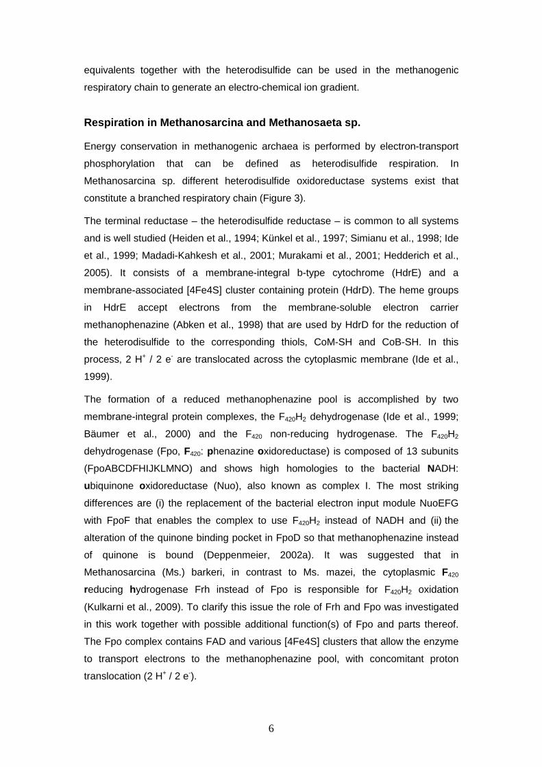
Respiration in Methanosarcina and Methanosaeta sp. Energy conservation in methanogenic archaea is performed by electron-transport
phosphorylation that can be defined as heterodisulfide respiration. In
Methanosarcina sp. different heterodisulfide oxidoreductase systems exist that
constitute a branched respiratory chain (Figure 3). The terminal reductase – the heterodisulfide reductase – is common to all systems
and is well studied (Heiden et al., 1994; Künkel et al., 1997; Simianu et al., 1998; Ide
et al., 1999; Madadi-Kahkesh et al., 2001; Murakami et al., 2001; Hedderich et al.,
2005). It consists of a membrane-integral b-type cytochrome (HdrE) and a
membrane-associated [4Fe4S] cluster containing protein (HdrD). The heme groups
in HdrE accept electrons from the membrane-soluble electron carrier
methanophenazine (Abken et al., 1998) that are used by HdrD for the reduction of
the heterodisulfide to the corresponding thiols, CoM-SH and CoB-SH. In this
process, 2 H+ / 2 e- are translocated across the cytoplasmic membrane (Ide et al.,
1999). The formation of a reduced methanophenazine pool is accomplished by two
membrane-integral protein complexes, the F420H2 dehydrogenase (Ide et al., 1999;
Bäumer et al., 2000) and the F420 non-reducing hydrogenase. The F420H2
dehydrogenase (Fpo, F420: phenazine oxidoreductase) is composed of 13 subunits
(FpoABCDFHIJKLMNO) and shows high homologies to the bacterial NADH:
ubiquinone oxidoreductase (Nuo), also known as complex I. The most striking
differences are (i) the replacement of the bacterial electron input module NuoEFG
with FpoF that enables the complex to use F420H2 instead of NADH and (ii) the
alteration of the quinone binding pocket in FpoD so that methanophenazine instead
of quinone is bound (Deppenmeier, 2002a). It was suggested that in
Methanosarcina (Ms.) barkeri, in contrast to Ms. mazei, the cytoplasmic F420
reducing hydrogenase Frh instead of Fpo is responsible for F420H2 oxidation
(Kulkarni et al., 2009). To clarify this issue the role of Frh and Fpo was investigated
in this work together with possible additional function(s) of Fpo and parts thereof.
The Fpo complex contains FAD and various [4Fe4S] clusters that allow the enzyme
to transport electrons to the methanophenazine pool, with concomitant proton
translocation (2 H+ / 2 e-).
7
2 H+
out
in
CoM-S-S-CoB+ 2 H+
HS-CoMHS-CoB
F420H2 F420
CM
2 H+ 2 H+ H2
Vho/Vht Ech
Fdox Fdred
Na+ / H+
2 H+H2
ATP ADP + Pi
3-4 H+/Na+
A1AO
MP MPHdrFpo
?2 H+
out
in
CoM-S-S-CoB+ 2 H+
HS-CoMHS-CoB
F420H2 F420
CM
2 H+ 2 H+ H2
Vho/Vht Ech
Fdox Fdred
Na+ / H+
2 H+H2
ATP ADP + Pi
3-4 H+/Na+
A1AO
MP MPHdrFpo
?
Figure 3: Model of the respiratory chain of Ms. mazei at the beginning of this work. Ion
translocation by Ech hydrogenase is putative. A1AO, ATP synthase; Ech, Ech hydrogenase;
Hdr, heterodisulfide reductase; Fdred/Fdox, reduced/oxidized ferredoxin; Fpo, F420H2
dehydrogenase; MP, methanophenazine; Vho/t, F420 non-reducing hydrogenase (isoforms
one and two).
The F420 non-reducing hydrogenase Vho/Vht (viologen hydrogenase one / two)
does not interact with F420 but is used when H2 serves as electron donor (Ide et al.,
1999). This enzyme consists of three subunits, two of which are located at the
extracellular side of the cytoplasmic membrane and represent the typical large and
small subunit of [NiFe] hydrogenases. They are anchored in the membrane by a
b-type cytochrome that transfers electrons to methanophenazine. Two protons are
released to the extracellular side when the H2 molecule is oxidized, and
methanophenazine takes up two protons upon reduction, so a vectorial proton
translocation with the stoichiometry of 2 H+ / 2 e- can be observed (Ide et al., 1999). The ferredoxin oxidizing enzyme system differs among the Methanosarcinales:
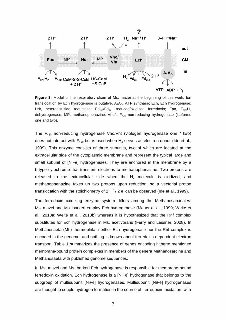
Ms. mazei and Ms. barkeri employ Ech hydrogenase (Meuer et al., 1999; Welte et
al., 2010a; Welte et al., 2010b) whereas it is hypothesized that the Rnf complex
substitutes for Ech hydrogenase in Ms. acetivorans (Ferry and Lessner, 2008). In
Methanosaeta (Mt.) thermophila, neither Ech hydrogenase nor the Rnf complex is
encoded in the genome, and nothing is known about ferredoxin-dependent electron
transport. Table 1 summarizes the presence of genes encoding hitherto mentioned
membrane-bound protein complexes in members of the genera Methanosarcina and
Methanosaeta with published genome sequences. In Ms. mazei and Ms. barkeri Ech hydrogenase is responsible for membrane-bound
ferredoxin oxidation. Ech hydrogenase is a [NiFe] hydrogenase that belongs to the
subgroup of multisubunit [NiFe] hydrogenases. Multisubunit [NiFe] hydrogenases
are thought to couple hydrogen formation in the course of ferredoxin oxidation with
8
Table 1: Occurrence of membrane-bound enzymes possibly involved in energy conservation
in sequenced members of the genera Methanosarcina (Ms.) and Methanosaeta (Mt.).
Strain Vho Rnf Ech Fpo HdrDE Source Ms. mazei + – + + + Deppenmeier et al., 2002 Ms. barkeri + – + + + Maeder et al., 2006 Ms. acetivorans –(1) + – + + Galagan et al., 2002
Mt. thermophila – – – +(2) + Smith and Ingram-Smith, 2007
(1) Genes coding for vho present, but not expressed (Guss et al., 2009; Rohlin and
Gunsalus, 2010). (2) Fpo complex is incomplete, F-subunit is missing (Smith and Ingram-Smith, 2007).
ion translocation. The initial designation “Ech” by Meuer et al 1999 was inspired by
its homology to the Escherichia coli hydrogenase 3. Later the abbreviation “Ech”
was sometimes described as energy-conserving hydrogenase (Hedderich and
Forzi, 2005), although an energy-conserving function had not yet been
experimentally demonstrated. The characterization of a Ms. mazei ∆ech mutant and
the biochemical investigation of the ion-translocating function of Ech hydrogenase
will be a central topic of this work. In contrast, energy conservation in Methanosaeta sp. has hardly been investigated
to date. The analysis of the genome sequence of Mt. thermophila (Smith and
Ingram-Smith, 2007) did not elucidate electron transport mechanisms because
known membrane-bound electron input proteins are missing: neither Ech
hydrogenase nor the Rnf complex is encoded in the genome. So if ferredoxin serves
as electron donor to the Mt. thermophila respiratory chain a novel type of
oxidoreductase has to be involved. The investigation of electron transport processes
in Mt. thermophila will also be examined in this work. Finally, the electro-chemical gradient built up by the systems described above can
be used for energy conservation. Energy is conserved by the action of an A1AO ATP
synthase. In Ms. mazei, the ATP synthase is driven either by the proton motive force
(Pisa et al., 2007) or by the sodium motive force (Katharina Schlegel (University of
Frankfurt), personal communication). Translocation of both H+ and Na+ occurs in
methanogenic bioenergetics, so the cells can take advantage of the bifunctional
ATP synthase without the necessity to utilize Na+/H+ antiporters. Aim of this work was to solve unanswered questions regarding electron transport in
methanogenic archaea of the genera Methanosarcina and Methanosaeta. The role
and identity of ferredoxin oxidizing enzymes will be of particular interest, which will
give a more complete understanding of the different ferredoxin: heterodisulfide
oxidoreductase systems in methanogenic archaea.
9
Chapter 2
Function of Ech hydrogenase in ferredoxin-dependent,
membrane-bound electron transport in
Methanosarcina mazei
Ms. mazei and Ms. barkeri contain the membrane-bound multisubunit [NiFe]
hydrogenase Ech that was described as ferredoxin oxidizing and H2 producing
(Meuer et al., 1999). It forms part of the ferredoxin: heterodisulfide oxidoreductase
that is responsible for energy conservation primarily in aceticlastic methanogenesis,
but in part also in methyltrophic methanogenesis. In this paper a detailed analysis of
a Ms. mazei ∆ech mutant is presented that complements the findings about a
Ms. barkeri ∆ech mutant (Meuer et al., 2002). Former works focused mainly on
subunit composition, purification and role of Ech hydrogenase in biosynthetic
pathways, which means the supply of reduced ferredoxin by H2 oxidation in anabolic
CO2 reduction. This paper, however, focuses on the function of Ech hydrogenase as
part of the respiratory chain, and reconstructs the electron flow from reduced
ferredoxin to the heterodisulfide using washed membrane preparations of the
wildtype and the ∆ech mutant strain. Furthermore, the phenotypic characterization
of the ∆ech mutant reveals that the consumption of methylated amines is much
faster with a lower growth yield as compared to the wildtype, and that growth on
acetate is no longer possible. Overall, the metabolism of the ∆ech mutant seems to
be ineffective and supports the hypothesis that Ech hydrogenase is an ion
translocating enzyme.
Welte et al. (2010) J Bacteriol 192:674-678
10
Reduced ferredoxin as electron donor for membrane-b ound electron
transport in Methanosarcina mazei
Cornelia Welte1, Verena Kallnik1, Marcel Grapp2, Gunes Bender3, Steve Ragsdale3,
Uwe Deppenmeier1
1 Institute of Microbiology and Biotechnology, University of Bonn, Meckenheimer Allee 168, 53115 Bonn, Germany
2 Zentrum für Kinderheilkunde und Jugendmedizin, University of Göttingen, Robert-Koch-Str. 40, 37075 Göttingen,
Germany
3 Department of Biological Chemistry, University of Michigan Medical School, 1150 W. Medical Center Dr. Ann
Arbor, MI 48109-0606, USA
Reduced ferredoxin is an intermediate in the methyl otrophic and aceticlastic pathway of
methanogenesis and donates electrons to membrane-in tegral proteins, which transfer electrons
to the heterodisulfide reductase. A ferredoxin inte raction has already been observed for the Ech
hydrogenase. Here we present the detailed analysis of a Ms. mazei ∆ech mutant which shows
decreased ferredoxin-dependent membrane-bound elect ron-transport activity, a slower growth
rate and faster substrate consumption. Evidence is p resented that a second protein whose
identity is unknown oxidizes reduced ferredoxin ind icating an involvement in methanogenesis.
INTRODUCTION
The aceticlastic pathway of methanogenesis creates approximately 70 % (11) of the biologically produced methane and is of great ecological importance, as methane is a potent greenhouse gas. Organisms using this pathway to convert acetate to methane exclusively belong to the genera Methanosarcina and Methanosaeta. The two carbon atoms of acetate have different fates in the pathway: The methyl moiety is converted to methane whereas the carbonyl moiety is further oxidized to CO2; the electrons derived from this oxidation step are used to reduce ferredoxin (Fd) (6). During methanogenesis from methylated C1-compounds (methanol and methylamines) one quarter of the methyl groups are oxidized to obtain electrons for the reduction of heterodisulfide (28). A key enzyme in the oxidative part of methylotrophic methanogenesis is the formylmethanofuran dehydrogenase, which oxidizes the intermediate formylmethanofuran to CO2 (7). The electrons are transferred to Fd. It was suggested that reduced ferredoxin (Fdred) donates electrons to the respiratory chain with the heterodisulfide (CoM-S-S-CoB) as terminal electron acceptor and the reaction being catalyzed by the Fdred:CoM-S-S-CoB oxidoreductase system (7; 25). The direct membrane-bound electron acceptor for Fdred is still a matter of debate: for
the Ech hydrogenase, a reduced ferredoxin accepting:H2 evolving activity has already been observed for Ms. barkeri (21), which implies the involvement of the H2:CoM-S-S-CoB oxidoreductase system in electron transport (14). The direct electron flow from the Ech hydrogenase to the heterodisulfide reductase has not been shown to date (21; 22). In contrast to Ms. barkeri, Ms. acetivorans is lacking the Ech hydrogenase (12). It can nevertheless grow on acetate, which is why another complex present in this organism, the Rnf complex, is discussed to be involved in the aceticlastic pathway of methanogenesis as acceptor for Fdred (8; 11; 19). The Ms. mazei genome, however, contains genes coding for the Ech hydrogenase, but is lacking the Rnf complex (5). To investigate whether the Ech hydrogenase is the only means by which Ms. mazei channels electrons from Fdred into the respiratory chain, a mutant lacking the Ech hydrogenase (Ms. mazei ∆ech) has been constructed. Electron transport experiments using Fdred as electron donor and CoM-S-S-CoB as electron acceptor were conducted with the wildtype and the mutant membranes to gain deeper insight into the actual membrane-bound protein complexes that accept electrons from Fdred. Furthermore, an in-depth characterization of growth and substrate consumption of the ∆ech mutant was performed resulting in insight into the in vivo role of Ech hydrogenase.
Welte et al. (2010) J Bacteriol 192:674-678
11
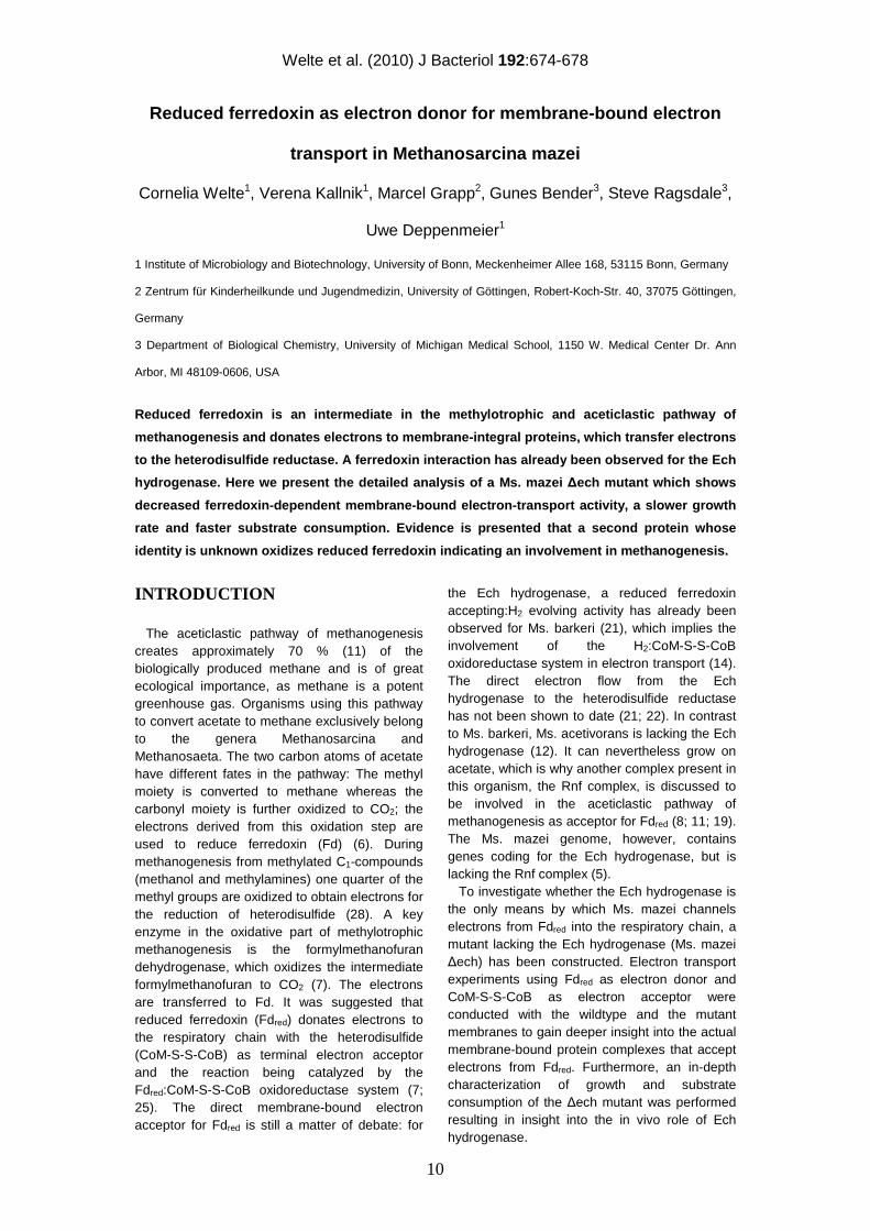
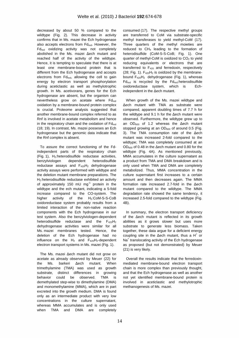
Figure 1: Proposed model of membrane-bound electron transfer in Ms. mazei. Proton translocation has been shown for the heterodisulfide reductase (15) and F420H2 dehydrogenase (2). Proton translocation for the Ech hydrogenase is assumed. H2ase, hydrogenase; DH, dehydrogenase; HDR, heterodisulfide reductase; MPhen, methanophenazine. Fd, ferredoxin.
MATERIALS AND METHODS
Purification of proteins. Ferredoxin was purified from Clostridium pasteurianum DSM 525T essentially as described by Mortenson (23). The last steps (dialyzation, crystallization) were replaced by ultrafiltration. Ferredoxin content was determined using the BCA method (27). CO dehydrogenase/acetyl-CoA synthase (CODH/ACS) was purified from Moorella thermoacetica ATCC 39073 as described in (26) except for the following modifications. The extract was not heat treated or fractionated with ammonium sulfate. The first DEAE-cellulose column was eluted with a step gradient of 0.1 to 0.5 M NaCl, and CODH/ACS eluted at 0.3 M NaCl. CODH/ACS was then purified further using Q-sepharose anion exchange and phenyl-sepharose hydrophobic interaction columns, from which CODH/ACS eluted at ~0.4 M NaCl and ~0.2 M (NH4)2SO4 respectively. The fractions from the phenyl-sepharose column that contained CODH/ACS were concentrated and buffer-exchanged into 50 mM Tris-HCl (pH 7.6) using Amicon ultra centrifuge concentrators in the anaerobic chamber (Vacum Atmospheres). Immediately after purification, the enzyme had a CO oxidation-methyl viologen reduction specific activity of 273 U/mg at 37 ºC. Membrane preparations. Methanosarcina mazei DSM 7222 and derivatives were grown anaerobically at 37°C in Methanosarcina medium (DSM medium 120) with trimethylamine as substrate (13). Subsequent steps were performed anaerobically in an anaerobic chamber (Coy laboratory products, US) under a 97 % N2 / 3 % H2 atmosphere. In the late exponential phase the cells were harvested and resuspended in phosphate buffer (40 mM KH2PO4/K2HPO4, pH 7.0, 5 mM DTE, 1 µg mL-1 resazurin) and incubated with DNAse I for 30 – 60 min at 4°C. As the buffer is hypoosmotic to
the cells, they lyse immediately. The cell lysate was ultracentrifuged (1 h, 120,000 x g), the supernatant (cytoplasmic fraction) discarded and the membrane pellet homogenized in 20 mM phosphate buffer (KH2PO4/K2HPO4 pH 7.0) containing 20 mM MgSO4, 500 mM sucrose, 5 mM DTE, 1 µg mL-1 resazurin. The ultracentrifugation step was repeated, the supernatant (which should be colourless) discarded and the membrane pellet again homogenized in the previously mentioned buffer. Protein content was determined using the Bradford method (3). Enzyme assays. The photometrical tests were performed anaerobically in rubber stoppered 1.5 mL glass cuvettes in a V-550 UV/Vis spectrophotometer (Jasco, Germany). Benzylviologen-dependent heterodisulfide reductase activity was determined as a decrease of absorption at 575 nm using 600 µL phosphate buffer (40 mM KH2PO4/K2HPO4, pH 7.0, reduced with Ti(III) citrate), 625 nmol benzylviologen (ε575nm = 8.9 mM-1 cm-1), 300 nmol Na2S2O4, 50 µg Ms. mazei membrane, 50 nmol CoM-S-S-CoB. F420H2 dehydrogenase activity was determined as an increase of absorption at 420 nm using 600 µL phosphate buffer (40 mM KH2PO4/K2HPO4, pH 7.0, 5 mM DTE), 15 nmol F420H2 (ε420nm=40 mM-1 cm-1), 50 µg Ms. mazei membrane, 300 nmol metronidazole, 180 nmol methylviologen. F420 was isolated and reduced as described by (1). 1 U of activity was defined as 1 µmol electrons transported per min. CoM-S-S-CoB (synthesized according to (10; 24)) was quantified in anaerobic rubber stoppered glass vials containing 250 µL phosphate buffer (40 mM, 1 µg mL-1 resazurin, reduced with Ti(III) citrate) gassed with N2/CO (5 % CO, purity 1.8, 95 % N2, purity 5.0) for 1 min. For analysis of CoM-S-S-CoB reduction, 100 nmol CoM-S-S-CoB, 8.9 µg Clostridium pasteurianum
out
in
MPhen
2 e-HDR
SH
CoM
SH
CoBS
CoB
S
CoM
2 H+
EchMPhen
2 Fdred 2 Fdox
2 e-
H2 2 H+
F420non-
reducingH2ase
H22 H+
F420H2DH
F420H2 F420
2 H+H+?
out
in
MPhen
2 e-HDR
SH
CoM
SH
CoBS
CoB
S
CoM
2 H+
EchMPhen
2 Fdred 2 Fdox
2 e-
H2 2 H+
F420non-
reducingH2ase
H22 H+
F420H2DH
F420H2 F420
2 H+H+?
Welte et al. (2010) J Bacteriol 192:674-678
12
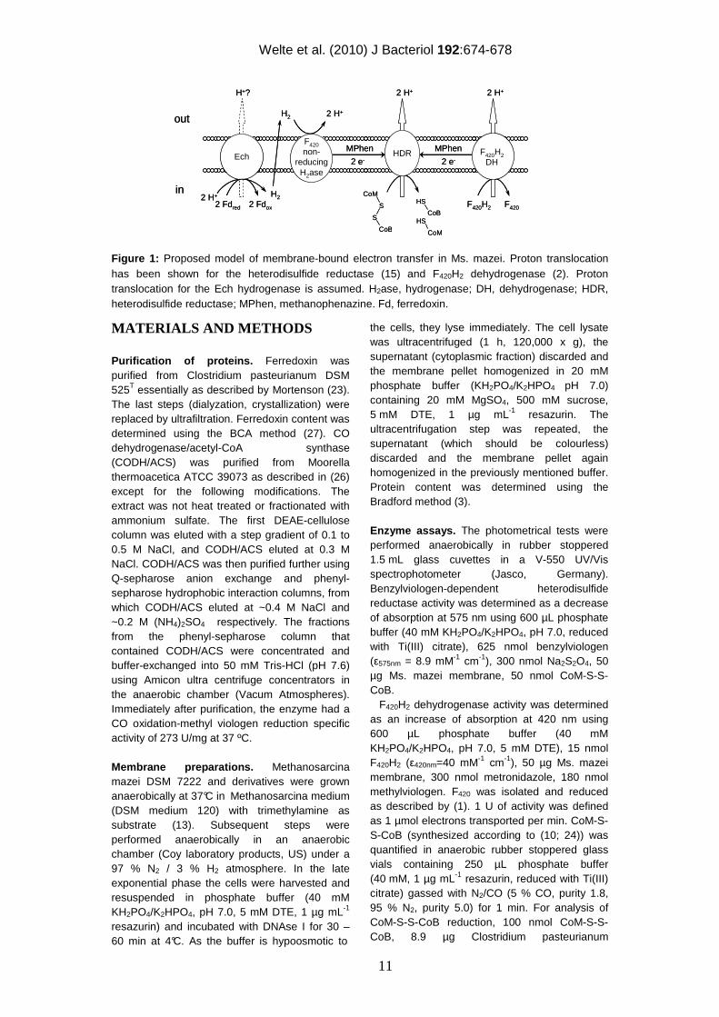
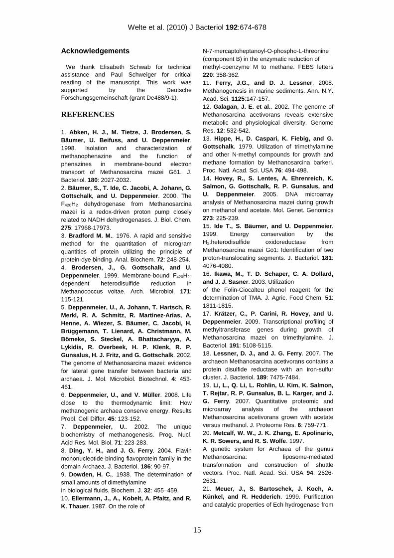
Figure 2: Fd-dependent heterodisulfide reduction with wildtype and ∆ech cytoplasmic membranes. The assays contained 100 nmol CoM-S-S-CoB, 8.86 µg Fd, 150 µg membrane and 75 µg CODH in a total volume of 250 µL. ( ■ ) wildtype membranes; (▲) membranes from ∆ech mutant; ( ● ) control without Fd; ( ○ ) control without CO.
ferredoxin, 150 µg Ms. mazei membrane, 75 µg Moorella thermoacetica CO dehydrogenase/acetyl-CoA synthase were added. For CoM-SH/CoB-SH quantification (30) 20 µL samples were taken every 10 min for 1 h and directly used in a modified Ellman’s assay: 950 µL Tris buffer (150 mM, pH 8.1) was mixed with the sample and 100 µL Ellman’s reagent (5 mM 5,5’-dithiobis(2-nitrobenzoic acid) (ε412 nm=13.6 mM-1 cm-1) in 50 mM sodium acetate buffer, pH 5.0), the absorption at 412 nm was immediately measured. Alternatively, H2:heterodisulfide oxidoreductase activity was determined by replacing CO, CO dehydrogenase/acetyl-CoA synthase and ferredoxin with H2. Here, only 50 µg membrane preparation were used. 1 U of activity was defined as 1 µmol CoM-S-S-CoB reduced per min. Generation of Ms. mazei ∆ech mutant. The Ms. mazei ∆ech mutant was generated by means of homologous recombination using the techniques described by Metcalf (20). Up- and downstream regions of the respective gene region were cloned into the two multiple cloning sites of pJK3 (20), linearized with ApaI and transformed into Ms. mazei. Instead of the respective gene, a puromycin resistance (pac) cassette was inserted. Puromycin resistant colonies were picked and screened for the knockout. Gene knockout was confirmed by sequencing and Southern blotting. For construction of Ms. mazei ∆ech, the primers 5’-
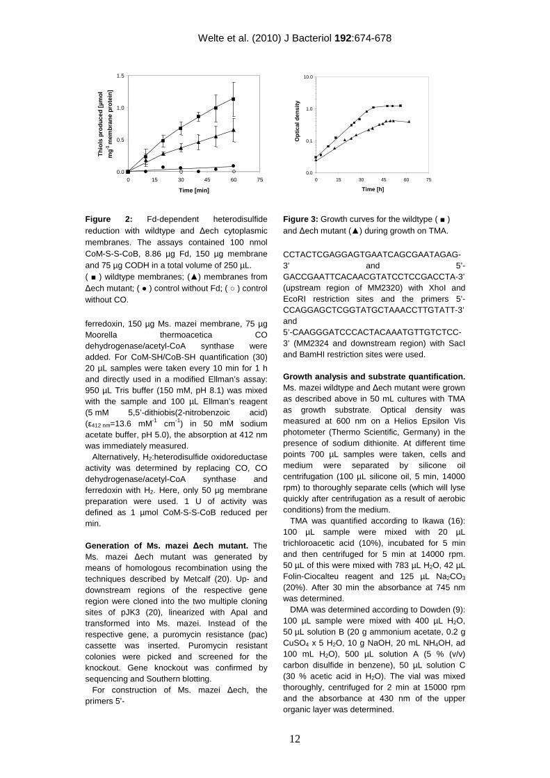
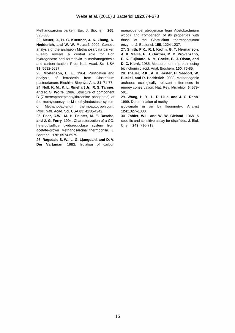
Figure 3: Growth curves for the wildtype ( ■ ) and ∆ech mutant (▲) during growth on TMA.
CCTACTCGAGGAGTGAATCAGCGAATAGAG-3’ and 5’-GACCGAATTCACAACGTATCCTCCGACCTA-3’ (upstream region of MM2320) with XhoI and EcoRI restriction sites and the primers 5’-CCAGGAGCTCGGTATGCTAAACCTTGTATT-3’ and 5’-CAAGGGATCCCACTACAAATGTTGTCTCC-3’ (MM2324 and downstream region) with SacI and BamHI restriction sites were used. Growth analysis and substrate quantification. Ms. mazei wildtype and ∆ech mutant were grown as described above in 50 mL cultures with TMA as growth substrate. Optical density was measured at 600 nm on a Helios Epsilon Vis photometer (Thermo Scientific, Germany) in the presence of sodium dithionite. At different time points 700 µL samples were taken, cells and medium were separated by silicone oil centrifugation (100 µL silicone oil, 5 min, 14000 rpm) to thoroughly separate cells (which will lyse quickly after centrifugation as a result of aerobic conditions) from the medium. TMA was quantified according to Ikawa (16): 100 µL sample were mixed with 20 µL trichloroacetic acid (10%), incubated for 5 min and then centrifuged for 5 min at 14000 rpm. 50 µL of this were mixed with 783 µL H2O, 42 µL Folin-Ciocalteu reagent and 125 µL Na2CO3 (20%). After 30 min the absorbance at 745 nm was determined. DMA was determined according to Dowden (9): 100 µL sample were mixed with 400 µL H2O, 50 µL solution B (20 g ammonium acetate, 0.2 g CuSO4 x 5 H2O, 10 g NaOH, 20 mL NH4OH, ad 100 mL H2O), 500 µL solution A (5 % (v/v) carbon disulfide in benzene), 50 µL solution C (30 % acetic acid in H2O). The vial was mixed thoroughly, centrifuged for 2 min at 15000 rpm and the absorbance at 430 nm of the upper organic layer was determined.
0.0
0.5
1.0
1.5
0 15 30 45 60 75
Time [min]
Thi
ols
prod
uced
[µm
ol
mg
-1 m
embr
ane
prot
ein]
0.0
0.5
1.0
1.5
0 15 30 45 60 75
Time [min]
Thi
ols
prod
uced
[µm
ol
mg
-1 m
embr
ane
prot
ein]
0.0
0.1
1.0
10.0
0 15 30 45 60 75
Time [h]
Opt
ical
dens
ity
0.0
0.1
1.0
10.0
0 15 30 45 60 75
Time [h]
Opt
ical
dens
ity
Welte et al. (2010) J Bacteriol 192:674-678
13
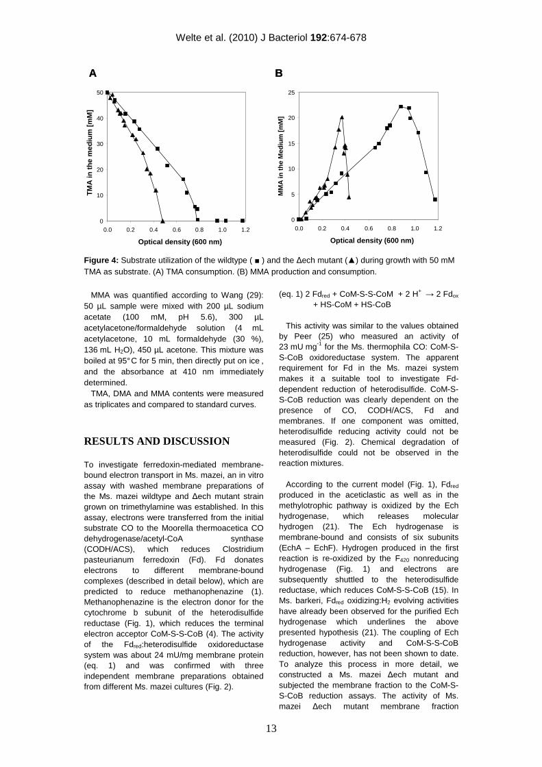
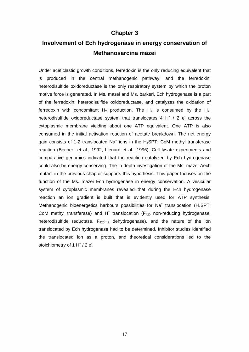
Figure 4: Substrate utilization of the wildtype ( ■ ) and the ∆ech mutant (▲) during growth with 50 mM TMA as substrate. (A) TMA consumption. (B) MMA production and consumption.
MMA was quantified according to Wang (29): 50 µL sample were mixed with 200 µL sodium acetate (100 mM, pH 5.6), 300 µL acetylacetone/formaldehyde solution (4 mL acetylacetone, 10 mL formaldehyde (30 %), 136 mL H2O), 450 µL acetone. This mixture was boiled at 95° C for 5 min, then directly put on ice , and the absorbance at 410 nm immediately determined. TMA, DMA and MMA contents were measured as triplicates and compared to standard curves.
RESULTS AND DISCUSSION
To investigate ferredoxin-mediated membrane-bound electron transport in Ms. mazei, an in vitro assay with washed membrane preparations of the Ms. mazei wildtype and ∆ech mutant strain grown on trimethylamine was established. In this assay, electrons were transferred from the initial substrate CO to the Moorella thermoacetica CO dehydrogenase/acetyl-CoA synthase (CODH/ACS), which reduces Clostridium pasteurianum ferredoxin (Fd). Fd donates electrons to different membrane-bound complexes (described in detail below), which are predicted to reduce methanophenazine (1). Methanophenazine is the electron donor for the cytochrome b subunit of the heterodisulfide reductase (Fig. 1), which reduces the terminal electron acceptor CoM-S-S-CoB (4). The activity of the Fdred:heterodisulfide oxidoreductase system was about 24 mU/mg membrane protein (eq. 1) and was confirmed with three independent membrane preparations obtained from different Ms. mazei cultures (Fig. 2).
(eq. 1) 2 Fdred + CoM-S-S-CoM + 2 H+ → 2 Fdox + HS-CoM + HS-CoB
This activity was similar to the values obtained by Peer (25) who measured an activity of 23 mU mg-1 for the Ms. thermophila CO: CoM-S-S-CoB oxidoreductase system. The apparent requirement for Fd in the Ms. mazei system makes it a suitable tool to investigate Fd-dependent reduction of heterodisulfide. CoM-S-S-CoB reduction was clearly dependent on the presence of CO, CODH/ACS, Fd and membranes. If one component was omitted, heterodisulfide reducing activity could not be measured (Fig. 2). Chemical degradation of heterodisulfide could not be observed in the reaction mixtures. According to the current model (Fig. 1), Fdred produced in the aceticlastic as well as in the methylotrophic pathway is oxidized by the Ech hydrogenase, which releases molecular hydrogen (21). The Ech hydrogenase is membrane-bound and consists of six subunits (EchA – EchF). Hydrogen produced in the first reaction is re-oxidized by the F420 nonreducing hydrogenase (Fig. 1) and electrons are subsequently shuttled to the heterodisulfide reductase, which reduces CoM-S-S-CoB (15). In Ms. barkeri, Fdred oxidizing:H2 evolving activities have already been observed for the purified Ech hydrogenase which underlines the above presented hypothesis (21). The coupling of Ech hydrogenase activity and CoM-S-S-CoB reduction, however, has not been shown to date. To analyze this process in more detail, we constructed a Ms. mazei ∆ech mutant and subjected the membrane fraction to the CoM-S-S-CoB reduction assays. The activity of Ms. mazei ∆ech mutant membrane fraction
0
5
10
15
20
25
0.0 0.2 0.4 0.6 0.8 1.0 1.2
Optical density (600 nm)
MM
A in
the
Med
ium
[mM
]
0
10
20
30
40
50
0.0 0.2 0.4 0.6 0.8 1.0 1.2
Optical density (600 nm)
TM
A in
the
med
ium
[mM
]
A B
0
5
10
15
20
25
0.0 0.2 0.4 0.6 0.8 1.0 1.2
Optical density (600 nm)
MM
A in
the
Med
ium
[mM
]
0
10
20
30
40
50
0.0 0.2 0.4 0.6 0.8 1.0 1.2
Optical density (600 nm)
TM
A in
the
med
ium
[mM
]
0
10
20
30
40
50
0.0 0.2 0.4 0.6 0.8 1.0 1.2
Optical density (600 nm)
TM
A in
the
med
ium
[mM
]
A B
Welte et al. (2010) J Bacteriol 192:674-678
14
decreased by about 50 % compared to the wildtype (Fig. 2). This decrease in activity confirms that in Ms. mazei the Ech hydrogenase also accepts electrons from Fdred. However, the Fdred oxidizing activity was not completely abolished in the Ms. mazei ∆ech mutant and reached half of the activity of the wildtype. Hence, it is tempting to speculate that there is at least one membrane-bound protein that is different from the Ech hydrogenase and accepts electrons from Fdred, allowing the cell to gain energy by electron transport phosphorylation during aceticlastic as well as methylotrophic growth. In Ms. acetivorans, genes for the Ech hydrogenase are absent, but the organism can nevertheless grow on acetate where Fdred oxidation by a membrane-bound protein complex is crucial. Proteome analysis suggested that another membrane-bound complex referred to as Rnf is involved in acetate metabolism and hence in the respiratory chain and the oxidation of Fdred (18; 19). In contrast, Ms. mazei posseses an Ech hydrogenase but the genomic data indicate that the Rnf complex is absent. To assure the correct functioning of the Fd-independent parts of the respiratory chain (Fig. 1), H2:heterodisulfide reductase activities, benzylviologen dependent heterodisulfide reductase assays and F420H2 dehydrogenase activity assays were performed with wildtype and the deletion mutant membrane preparations. The H2:heterodisulfide reductase exhibited an activity of approximately 150 mU mg-1 protein in the wildtype and the ech mutant, indicating a 5-fold increase compared to the CO-system. The higher activity of the H2:CoM-S-S-CoB oxidoreductase system probably results from a limited interaction of the non-native reaction components with the Ech hydrogenase in our test system. Also the benzylviologen-dependent heterodisulfide reductase and the F420H2 dehydrogenase activities were similar for all Ms. mazei membranes tested. Hence, the deletion of the Ech hydrogenase had no influence on the H2 and F420H2-dependent electron transport systems in Ms. mazei (Fig. 1). The Ms. mazei ∆ech mutant did not grow on acetate as already observed by Meuer (22) for the Ms. barkeri ∆ech mutant. When trimethylamine (TMA) was used as growth substrate, distinct differences in growing behavior could be observed. TMA is demethylated step-wise to dimethylamine (DMA) and monomethylamine (MMA), which are in part excreted into the growth medium. DMA is found only as an intermediate product with very low concentrations in the culture supernatant, whereas MMA accumulates and is only used when TMA and DMA are completely
consumed (17). The respective methyl groups are transferred to CoM via substrate-specific methyl transferases to yield methyl-CoM (17). Three quarters of the methyl moieties are reduced to CH4 leading to the formation of heterodisulfide (CoM-S-S-CoB; Fig. 1). One quarter of methyl-CoM is oxidized to CO2 to yield reducing equivalents or electrons that are transferred to F420 and ferredoxin, respectively (28; Fig. 1). F420H2 is oxidized by the membrane-bound F420H2 dehydrogenase (Fig. 1), whereas Fdred is recycled by the Fdred:heterodisulfide oxidoreductase system, which is Ech-independent in the ∆ech mutant. When growth of the Ms. mazei wildtype and ∆ech mutant with TMA as substrate were compared, apparent doubling times of 7.7 h for the wildtype and 9.1 h for the ∆ech mutant were observed. Furthermore, the wildtype grew up to an OD600 of 1.2 whereas the ∆ech mutant stopped growing at an OD600 of around 0.5 (Fig. 3). The TMA consumption rate of the ∆ech mutant was increased 2-fold compared to the wildtype; TMA was completely consumed at an OD600 of 0.48 in the ∆ech mutant and 0.80 for the wildtype (Fig. 4A). As mentioned previously, MMA accumulates in the culture supernatant as a product from TMA and DMA breakdown and is only used when TMA and DMA are completely metabolized. Thus, MMA concentration in the culture supernatant first increases to a certain amount and then decreases again. The MMA formation rate increased 2.7-fold in the ∆ech mutant compared to the wildtype. The MMA degradation rate showed the same tendency, it increased 2.5-fold compared to the wildtype (Fig. 4B). In summary, the electron transport deficiency of the ∆ech mutant is reflected in its growth abilities as it grows slower but uses more substrate to generate less biomass. Taken together, these data argue for a deficient energy coupling site in the ∆ech mutant, thus a H+ or Na+ translocating activity of the Ech hydrogenase as proposed (but not demonstrated) by Meuer (21) is very likely. Overall the results indicate that the ferredoxin-mediated membrane-bound electron transport chain is more complex than previously thought, and that the Ech hydrogenase as well as another not yet identified membrane-bound protein is involved in aceticlastic and methylotrophic methanogenesis of Ms. mazei.
Welte et al. (2010) J Bacteriol 192:674-678
15
Acknowledgements
We thank Elisabeth Schwab for technical assistance and Paul Schweiger for critical reading of the manuscript. This work was supported by the Deutsche Forschungsgemeinschaft (grant De488/9-1).
REFERENCES
1. Abken, H. J., M. Tietze, J. Brodersen, S. Bäumer, U. Beifuss, and U. Deppenmeier . 1998. Isolation and characterization of methanophenazine and the function of phenazines in membrane-bound electron transport of Methanosarcina mazei Gö1. J. Bacteriol. 180: 2027-2032. 2. Bäumer, S., T. Ide, C. Jacobi, A. Johann, G. Gottschalk, and U. Deppenmeier . 2000. The F420H2 dehydrogenase from Methanosarcina mazei is a redox-driven proton pump closely related to NADH dehydrogenases. J. Biol. Chem. 275: 17968-17973. 3. Bradford M. M. . 1976. A rapid and sensitive method for the quantitation of microgram quantities of protein utilizing the principle of protein-dye binding. Anal. Biochem. 72: 248-254. 4. Brodersen, J., G. Gottschalk, and U. Deppenmeier . 1999. Membrane-bound F420H2-dependent heterodisulfide reduction in Methanococcus voltae. Arch. Microbiol. 171: 115-121. 5. Deppenmeier, U., A. Johann, T. Hartsch, R. Merkl, R. A. Schmitz, R. Martinez-Arias, A. Henne, A. Wiezer, S. Bäumer, C. Jacobi, H. Brüggemann, T. Lienard, A. Christmann, M. Bömeke, S. Steckel, A. Bhattacharyya, A. Lykidis, R. Overbeek, H. P. Klenk, R. P. Gunsalus, H. J. Fritz, and G. Gottschalk . 2002. The genome of Methanosarcina mazei: evidence for lateral gene transfer between bacteria and archaea. J. Mol. Microbiol. Biotechnol. 4: 453-461. 6. Deppenmeier, U., and V. Müller . 2008. Life close to the thermodynamic limit: How methanogenic archaea conserve energy. Results Probl. Cell Differ. 45: 123-152. 7. Deppenmeier, U. . 2002. The unique biochemistry of methanogenesis. Prog. Nucl. Acid Res. Mol. Biol. 71: 223-283. 8. Ding, Y. H., and J. G. Ferry . 2004. Flavin mononucleotide-binding flavoprotein family in the domain Archaea. J. Bacteriol. 186: 90-97. 9. Dowden, H. C. . 1938. The determination of small amounts of dimethylamine in biological fluids. Biochem. J. 32: 455–459. 10. Ellermann, J., A., Kobelt, A. Pfaltz, and R. K. Thauer . 1987. On the role of
N-7-mercaptoheptanoyl-O-phospho-L-threonine (component B) in the enzymatic reduction of methyl-coenzyme M to methane. FEBS letters 220: 358-362. 11. Ferry, J.G., and D. J. Lessner . 2008. Methanogenesis in marine sediments. Ann. N.Y. Acad. Sci. 1125:147-157. 12. Galagan, J. E. et al. . 2002. The genome of Methanosarcina acetivorans reveals extensive metabolic and physiological diversity. Genome Res. 12: 532-542. 13. Hippe, H., D. Caspari, K. Fiebig, and G. Gottschalk . 1979. Utilization of trimethylamine and other N-methyl compounds for growth and methane formation by Methanosarcina barkeri. Proc. Natl. Acad. Sci. USA 76: 494-498. 14. Hovey, R., S. Lentes, A. Ehrenreich, K. Salmon, G. Gottschalk, R. P. Gunsalus, and U. Deppenmeier . 2005. DNA microarray analysis of Methanosarcina mazei during growth on methanol and acetate. Mol. Genet. Genomics 273: 225-239. 15. Ide T., S. Bäumer, and U. Deppenmeier . 1999. Energy conservation by the H2:heterodisulfide oxidoreductase from Methanosarcina mazei Gö1: Identification of two proton-translocating segments. J. Bacteriol. 181: 4076-4080. 16. Ikawa, M., T. D. Schaper, C. A. Dollard, and J. J. Sasner . 2003. Utilization of the Folin-Ciocalteu phenol reagent for the determination of TMA. J. Agric. Food Chem. 51: 1811-1815. 17. Krätzer, C., P. Carini, R. Hovey, and U. Deppenmeier . 2009. Transcriptional profiling of methyltransferase genes during growth of Methanosarcina mazei on trimethylamine. J. Bacteriol. 191: 5108-5115. 18. Lessner, D. J., and J. G. Ferry . 2007. The archaeon Methanosarcina acetivorans contains a protein disulfide reductase with an iron-sulfur cluster. J. Bacteriol. 189: 7475-7484. 19. Li, L., Q. Li, L. Rohlin, U. Kim, K. Salmon, T. Rejtar, R. P. Gunsalus, B. L. Karger, and J. G. Ferry . 2007. Quantitative proteomic and microarray analysis of the archaeon Methanosarcina acetivorans grown with acetate versus methanol. J. Proteome Res. 6: 759-771. 20. Metcalf, W. W., J. K. Zhang, E. Apolinario, K. R. Sowers, and R. S. Wolfe . 1997. A genetic system for Archaea of the genus Methanosarcina: liposome-mediated transformation and construction of shuttle vectors. Proc. Natl. Acad. Sci. USA 94: 2626-2631. 21. Meuer, J., S. Bartoschek, J. Koch, A. Künkel, and R. Hedderich . 1999. Purification and catalytic properties of Ech hydrogenase from
Welte et al. (2010) J Bacteriol 192:674-678
16
Methanosarcina barkeri. Eur. J. Biochem. 265: 325-335. 22. Meuer, J., H. C. Kuettner, J. K. Zhang, R. Hedderich, and W. W. Metcalf . 2002. Genetic analysis of the archaeon Methanosarcina barkeri Fusaro reveals a central role for Ech hydrogenase and ferredoxin in methanogenesis and carbon fixation. Proc. Natl. Acad. Sci. USA 99: 5632-5637. 23. Mortenson, L. E. . 1964. Purification and analysis of ferredoxin from Clostridium pasteurianum. Biochim. Biophys. Acta 81: 71-77. 24. Noll, K. M., K. L. Rinehart Jr., R. S. Tanner, and R. S. Wolfe . 1986. Structure of component B (7-mercaptoheptanoylthreonine phosphate) of the methylcoenzyme M methylreductase system of Methanobacterium thermoautotrophicum. Proc. Natl. Acad. Sci. USA 83: 4238-4242. 25. Peer, C.W., M. H. Painter, M. E. Rasche, and J. G. Ferry . 1994. Characterization of a CO: heterodisulfide oxidoreductase system from acetate-grown Methanosarcina thermophila. J. Bacteriol. 176: 6974-6979. 26. Ragsdale S. W., L. G. Ljungdahl, and D. V. Der Vartanian . 1983. Isolation of carbon
monoxide dehydrogenase from Acetobacterium woodii and comparison of its properties with those of the Clostridium thermoaceticum enzyme. J. Bacteriol. 155: 1224-1237. 27. Smith, P.K., R. I. Krohn, G. T. Hermanson, A. K. Mallia, F. H. Gartner, M. D. Provenzano, E. K. Fujimoto, N. M. Goeke, B. J. Olson, and D. C. Klenk . 1985. Measurement of protein using bicinchoninic acid. Anal. Biochem. 150: 76-85. 28. Thauer, R.K., A. K. Kaster, H. Seedorf, W. Buckel, and R. Hedderich . 2008. Methanogenic archaea: ecologically relevant differences in energy conservation. Nat. Rev. Microbiol. 6: 579-591. 29. Wang, H. Y., L. D. Liua, and J. C. Renb . 1999. Determination of methyl isocyanate in air by fluorimetry. Analyst 124:1327–1330. 30. Zahler, W.L. and W. W. Cleland . 1968. A specific and sensitive assay for disulfides. J. Biol. Chem. 243: 716-719.
17
Chapter 3
Involvement of Ech hydrogenase in energy conservation of
Methanosarcina mazei
Under aceticlastic growth conditions, ferredoxin is the only reducing equivalent that
is produced in the central methanogenic pathway, and the ferredoxin:
heterodisulfide oxidoreductase is the only respiratory system by which the proton
motive force is generated. In Ms. mazei and Ms. barkeri, Ech hydrogenase is a part
of the ferredoxin: heterodisulfide oxidoreductase, and catalyzes the oxidation of
ferredoxin with concomitant H2 production. The H2 is consumed by the H2:
heterodisulfide oxidoreductase system that translocates 4 H+ / 2 e- across the
cytoplasmic membrane yielding about one ATP equivalent. One ATP is also
consumed in the initial activation reaction of acetate breakdown. The net energy
gain consists of 1-2 translocated Na+ ions in the H4SPT: CoM methyl transferase
reaction (Becher et al., 1992, Lienard et al., 1996). Cell lysate experiments and
comparative genomics indicated that the reaction catalyzed by Ech hydrogenase
could also be energy conserving. The in-depth investigation of the Ms. mazei ∆ech
mutant in the previous chapter supports this hypothesis. This paper focuses on the
function of the Ms. mazei Ech hydrogenase in energy conservation. A vesicular
system of cytoplasmic membranes revealed that during the Ech hydrogenase
reaction an ion gradient is built that is evidently used for ATP synthesis.
Methanogenic bioenergetics harbours possibilities for Na+ translocation (H4SPT:
CoM methyl transferase) and H+ translocation (F420 non-reducing hydrogenase,
heterodisulfide reductase, F420H2 dehydrogenase), and the nature of the ion
translocated by Ech hydrogenase had to be determined. Inhibitor studies identified
the translocated ion as a proton, and theoretical considerations led to the
stoichiometry of 1 H+ / 2 e-.
Welte et al. (2010) FEBS J 277:3396-3403
18
Involvement of Ech hydrogenase in energy conservation of
Methanosarcina mazei
Cornelia Welte, Christian Krätzer, Uwe Deppenmeier
Institute of Microbiology and Biotechnology, University of Bonn, Meckenheimer Allee 168, 53115 Bonn, Germany
Methanosarcina mazei belongs to the group of aceticlastic methanogens and converts acetate
into the potent greenhouse gases CO2 and CH4. The aceticlastic respiratory chain involved in
methane formation comprises the three transmembrane proteins Ech hydrogenase, F420 non-
reducing hydrogenase and heterodisulfide reductase. It was shown that the latter two
contribute to the proton motive force. The data presented here clearly demonstrate that the Ech
hydrogenase is also involved in energy conservation. ATP synthesis was observed in a
cytoplasm-free vesicular system of Methanosarcina mazei that was dependent on the oxidation
of reduced ferredoxin and the formation of molecular hydrogen (as catalyzed by Ech
hydrogenase). Such an ATP formation was not observed in a ∆ech mutant strain. The
protonophore SF6847 led to complete inhibition of ATP formation in the Ms. mazei wildtype
without inhibiting hydrogen production by Ech hydrogenase, whereas the sodium ion
ionophore ETH157 did not affect ATP formation in this system. Thus, we conclude that Ech
hydrogenase acts as primary proton pump in a ferredoxin-dependent electron transport
system.
INTRODUCTION
Biological methanogenesis from acetate is one of the most important processes for the maintenance of the carbon cycle on earth. The products of methanogenesis from acetate, CH4 and CO2, are released from anaerobic habitats and large amounts of these greenhouse gases reach the atmosphere. Therefore, the process of biological methane formation is of great interest for global ecology [1, 2]. Moreover, the process of methanogenesis creates a combustible gas that can be used as an energy source. Only the genera Methanosarcina and Methanosaeta are able to use the aceticlastic pathway of methanogenesis, and Methanosarcina mazei strain Gö1 (from here referred to as Ms. mazei) is one of the important model organisms [3]. In Ms. mazei, acetate is activated by phosphorylation and exchange of inorganic phosphate with coenzyme A (CoA). The resulting acetyl-CoA is cleaved by the CO dehydrogenase/acetyl-CoA synthase (CODH/ACS). In the course of the reaction enzyme-bound CO is oxidized to CO2 and the electrons are used for ferredoxin (Fd) reduction. The methyl group of acetate is transferred to tetrahydrosarcinapterin (H4SPT). The resulting methyl-H4SPT is converted to methane by the catalytic activities of a Na+-translocating methyl-CoM methyltransferase (forming methyl-2-
mercaptoethanesulfonate (methyl-S-CoM)) and the methyl-S-CoM reductase, which uses N-7-mercaptoheptanoyl-L-threonine phosphate (HS-CoB) as the electron donor to reduce the methyl group to CH4. An additional product of this reaction is the heterodisulfide of HS-CoM and HS-CoB (CoM-S-S-CoB), which serves as terminal electron acceptor in the methanogenic respiratory chain (for review see [4]).
The intermediates of the aceticlastic pathway, CoM-S-S-CoB and reduced ferredoxin (Fdred), are recycled by a membrane-bound electron transport system which can be defined as ferredoxin:heterodisulfide oxidoreductase [5]. In most Methanosarcina species (e.g. Ms. mazei and Ms. barkeri) the oxidation of reduced ferredoxin is catalyzed by Ech hydrogenase resulting in the release of molecular hydrogen [6] that is then reoxidized by the F420 non-reducing hydrogenase and channelled via methanophenazine to the heterodisulfide reductase [7]. Some Methanosarcina species, e.g. Ms. acetivorans, lack Ech hydrogenase and must possess an alternative route for oxidation of Fdred. It was shown that the F420 non-reducing hydrogenase and the heterodisulfide reductase are key elements in membrane-bound electron transport and are essential to generate the proton motive force [7], whereas the methyl-CoM methyltransferase generates a Na+ ion
Welte et al. (2010) FEBS J 277:3396-3403
19
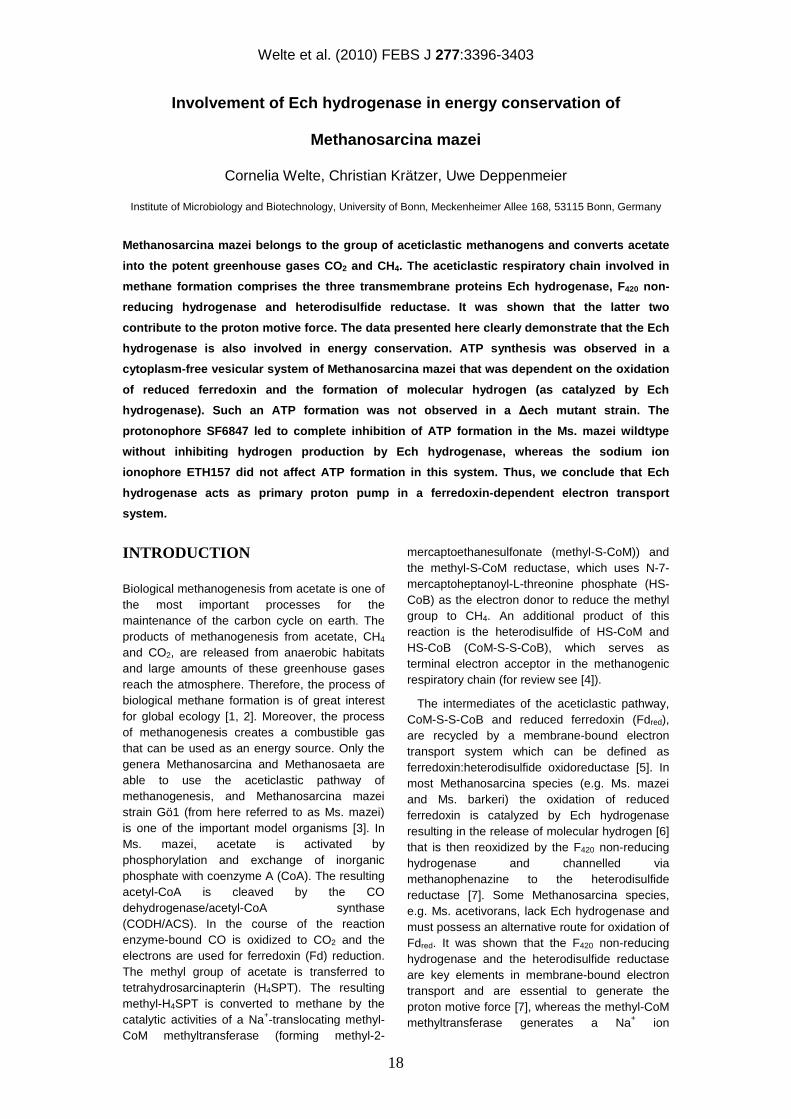
Table 1: Hydrogen formation by reduced ferredoxin-dependent proton reduction.Test vials contained 5 % CO / 95 % N2 in the headspace, 500 µg inverted membrane vesicles, 33.5 µg ferredoxin, 20 µg CODH/ACS, 150 nmol AMP, 300 nmol ADP. Addition or exclusion of single components is indicated.
Preparation Assay condition
H2 production rate (%)
wt1 vesicles complete 1002
wt vesicles + 10 µM ETH157 101
wt vesicles + 10 µM SF6847 130
wt vesicles + 400 µM DCCD 99
wt vesicles without Fd < 1
wt vesicles without CO < 1
∆ech vesicles complete < 1
1) wildtype 2) most active vesicle preparation showed a specific activity of 32.8 nmol min-1 x mg protein-1
gradient [8, 9]. Furthermore, it was suggested that the Ech hydrogenase also contributes to the electro chemical ion gradient [5] because of homologies to certain subunits of ion-translocating oxidoreductases [10] and indirect evidence from experiments with resting cells of Ms. barkeri [11, 12]. However, direct experimental evidence for this hypothesis is lacking. In this study, we present the first biochemical proof that Ech hydrogenase is indeed an ion-translocating enzyme, and thus represents an additional energy conserving coupling site in methanogenic metabolism. Inhibitor studies clearly indicate that H+ and not Na+ is the coupling ion, thus the proton gradient can directly be used for ATP synthesis via A1AO ATP synthase [13].
RESULTS
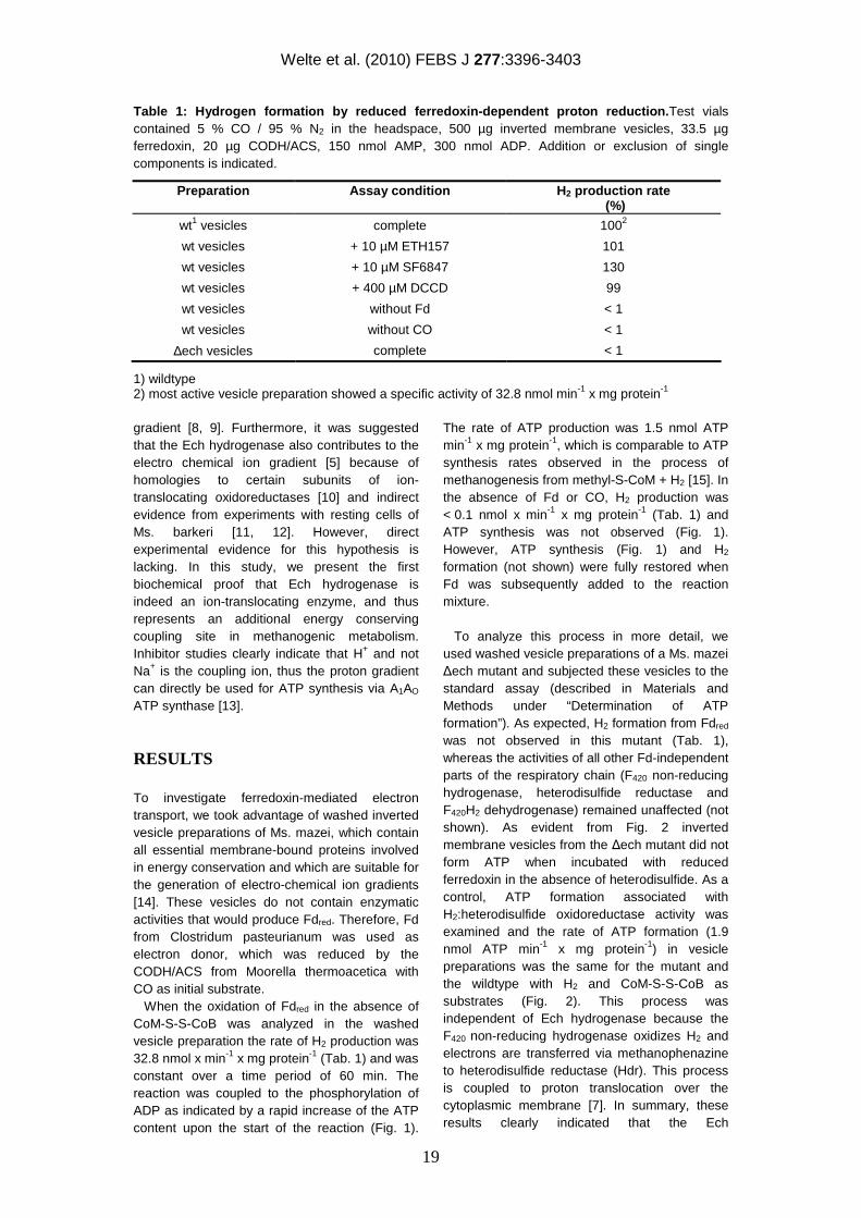
To investigate ferredoxin-mediated electron transport, we took advantage of washed inverted vesicle preparations of Ms. mazei, which contain all essential membrane-bound proteins involved in energy conservation and which are suitable for the generation of electro-chemical ion gradients [14]. These vesicles do not contain enzymatic activities that would produce Fdred. Therefore, Fd from Clostridum pasteurianum was used as electron donor, which was reduced by the CODH/ACS from Moorella thermoacetica with CO as initial substrate. When the oxidation of Fdred in the absence of CoM-S-S-CoB was analyzed in the washed vesicle preparation the rate of H2 production was 32.8 nmol x min-1 x mg protein-1 (Tab. 1) and was constant over a time period of 60 min. The reaction was coupled to the phosphorylation of ADP as indicated by a rapid increase of the ATP content upon the start of the reaction (Fig. 1).
The rate of ATP production was 1.5 nmol ATP min-1 x mg protein-1, which is comparable to ATP synthesis rates observed in the process of methanogenesis from methyl-S-CoM + H2 [15]. In the absence of Fd or CO, H2 production was < 0.1 nmol x min-1 x mg protein-1 (Tab. 1) and ATP synthesis was not observed (Fig. 1). However, ATP synthesis (Fig. 1) and H2 formation (not shown) were fully restored when Fd was subsequently added to the reaction mixture.
To analyze this process in more detail, we used washed vesicle preparations of a Ms. mazei ∆ech mutant and subjected these vesicles to the standard assay (described in Materials and Methods under “Determination of ATP formation”). As expected, H2 formation from Fdred was not observed in this mutant (Tab. 1), whereas the activities of all other Fd-independent parts of the respiratory chain (F420 non-reducing hydrogenase, heterodisulfide reductase and F420H2 dehydrogenase) remained unaffected (not shown). As evident from Fig. 2 inverted membrane vesicles from the ∆ech mutant did not form ATP when incubated with reduced ferredoxin in the absence of heterodisulfide. As a control, ATP formation associated with H2:heterodisulfide oxidoreductase activity was examined and the rate of ATP formation (1.9 nmol ATP min-1 x mg protein-1) in vesicle preparations was the same for the mutant and the wildtype with H2 and CoM-S-S-CoB as substrates (Fig. 2). This process was independent of Ech hydrogenase because the F420 non-reducing hydrogenase oxidizes H2 and electrons are transferred via methanophenazine to heterodisulfide reductase (Hdr). This process is coupled to proton translocation over the cytoplasmic membrane [7]. In summary, these results clearly indicated that the Ech
Welte et al. (2010) FEBS J 277:3396-3403
20
Figure 1: Ferredoxin-dependent ATP synthesis. Test vials contained 5 % CO / 95 % N2 in the headspace, 500-700 µg inverted membrane vesicles, 33.5 µg ferredoxin, 20 µg CODH/ACS, 150 nmol AMP, 300 nmol ADP. (□) positive control; (∆) control without ferredoxin, arrow indicates addition of 33.5 µg Fd; (●) control without CO.
Figure 2: ATP synthesis by wildtype and ∆ech mutant. Test vials contained 500-700 µg inverted membrane vesicles, 150 nmol AMP, 300 nmol ADP. (■) 5 % CO / 95 % N2 in the headspace, 33.5 µg ferredoxin, 20 µg CODH/ACS, ∆ech mutant vesicle preparation; (□) 100 % H2 in the headspace, 150 nmol CoM-S-S-CoB, ∆ech mutant vesicle preparation; (▲) 100 % H2 in the headspace, 150 nmol CoM-S-S-CoB, wildtype vesicle preparation .
hydrogenase is necessary to generate an electrochemical ion gradient when ferredoxin is the only reducing equivalent and heterodisulfide is absent.
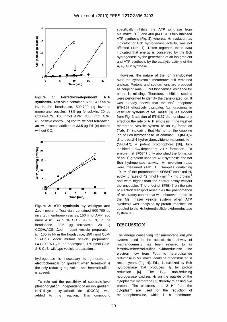
To rule out the possibility of substrate-level phosphorylation, independent of an ion gradient, N,N'-dicyclo-hexylcarbodiimide (DCCD) was added to the reaction. This compound
specifically inhibits the ATP synthase from Ms. mazei [13], and 400 µM DCCD fully inhibited ATP synthesis (Fig. 3), whereas H2 evolution, as indicator for Ech hydrogenase activity, was not affected (Tab. 1). Taken together, these data indicated that energy is conserved by the Ech hydrogenase by the generation of an ion gradient and ATP synthesis by the catalytic activity of the A1AO ATP synthase. However, the nature of the ion translocated over the cytoplasmic membrane still remained unclear. Protons and sodium ions are proposed as coupling ions [5], but biochemical evidence for either is missing. Therefore, inhibitor studies were performed to identify the translocated ion. It was already shown that the Na+ ionophore ETH157 effectively dissipates Na+ gradients in vesicular systems of Ms. mazei [8]. As evident from Fig. 3 addition of ETH157 did not show any effect on the rate of ATP synthesis in the washed membrane vesicle system or on H2 formation (Tab. 1), indicating that Na+ is not the coupling ion of Ech hydrogenase. In contrast, 10 µM 3,5-di-tert-butyl-4-hydroxybenzylidene-malononitrile (SF6847), a potent protonophore [16], fully inhibited Fdred-dependent ATP formation. To ensure that SF6847 only abolished the formation of an H+ gradient used for ATP synthesis and not Ech hydrogenase activity, H2 evolution rates were measured (Tab. 1). Samples containing 10 µM of the protonophore SF6847 exhibited H2 evolving rates of 42 nmol H2 min-1 x mg protein-1 and were higher than the control assay without the uncoupler. The effect of SF6847 on the rate of electron transport resembles the phenomenon of respiratory control that was observed before in the Ms. mazei vesicle system when ATP synthesis was analyzed by proton translocation coupled to the H2:heterodisulfide oxidroreductase system [16].
DISCUSSION
The energy conserving transmembrane enzyme system used in the aceticlastic pathway of methanogenesis has been referred to as ferredoxin:heterodisulfide oxidoreductase. The electron flow from Fdred to heterodisulfide reductase in Ms. mazei could be reconstructed in recent years (Fig. 4). Fdred is oxidized by Ech hydrogenase that produces H2 by proton reduction [6]. The F420 non-reducing hydrogenase oxidizes H2 on the outside of the cytoplasmic membrane [7], thereby releasing two protons. The electrons and 2 H+ from the cytoplasm are used for the reduction of methanophenazine, which is a membrane-
0
5
10
15
20
25
0 2 4 6 8 10 12
Time [min]
AT
P [
nm
ol/m
gp
rote
in]
0
5
10
15
20
25
0 2 4 6 8 10 12
Time [min]
AT
P [
nm
ol/m
gp
rote
in]
0
5
10
15
20
0 5 10 15 20
Time [min]
AT
P [
nm
ol/m
gp
pro
tein
]
0
5
10
15
20
0 5 10 15 20
Time [min]
AT
P [
nm
ol/m
gp
pro
tein
]
Welte et al. (2010) FEBS J 277:3396-3403
21
Figure 3: Influence of inhibitors on ATP synthesis. Assay conditions as in Fig. 1. (□) positive control without ionophore; (■) 10 µM ETH157; (○) 10 µM SF6847; (▲) 400 µM DCCD.
integral electron carrier in Methanosarcina species [17]. Reduced methanophenazine transfers electrons to heterodisulfide reductase (Fig. 4). The respective protons are released into the extracellular space [7] thereby generating an electro-chemical proton gradient, which is used for ATP synthesis by the A1AO ATP synthase. Energy conservation for Ech hydrogenase based on growth data and experiments on resting cells and cell suspensions has been proposed in several studies [6, 12, 18-20], but ATP production or generation of an H+ or Na+ gradient directly by Ech hydrogenase has not been reported. The data presented here clearly demonstrate a direct involvement of Ech hydrogenase in energy conservation: i) ATP synthesis was observed in the Ms. mazei vesicular system that was dependent on the oxidation of Fdred (catalyzed by Ech hydrogenase). ii) The Ms. mazei ∆ech mutant showed no formation of ATP in the presence of Fdred. In contrast, ATP synthesis from H2 + CoM-S-S-CoB was identical to wildtype levels, indicating that the ∆ech vesicle preparation was able to establish an ion gradient and that the ATP synthase was active. iii) Addition of protonophore SF6847 led to complete cessation of ATP formation without inhibiting the Ech hydrogenase, whereas the sodium ion ionophore ETH157 did not affect ATP formation in this system. Therefore, protons are clearly used as coupling ions. Proton translocation by Ech hydrogenase is similar to studies performed on the related Mbh hydrogenase from Pyrococcus furiosus [21], which also translocates protons in the process of Fdred oxidation. Both proteins belong to a small subset of multi-subunit [NiFe] hydrogenases
within the large group of [NiFe] hydrogenases, that use reduced ferredoxin or polyferredoxin as an electron donor [10]. Members of this group are thought to couple hydrogen formation to energy conservation, primarily based on their homology to the proton pumping NADH:ubiquinone oxidoreductase (complex I). Biochemical evidence of proton translocation has so far only been presented for the Mbh [NiFe] hydrogenase from P. furiosus [21]. Other members of this group are the Coo [NiFe] hydrogenases from Rhodospirillum rubrum [22] and Carboxydothermus hydrogenoformans [23], and the Hyc and Hyf [NiFe] hydrogenases from Escherichia coli [24-26]. Ech hydrogenase is now another member of the group of energy-conserving multi-subunit [NiFe] hydrogenases to that an energy conserving function can be assigned due to biochemical data and not solely based on sequence similarity to complex I or Mbh hydrogenase of P. furiosus.
It is evident that the proton gradients generated by the Ech hydrogenase from Ms. mazei and the Mbh hydrogenase from Pyrococcus furiosus is used for ATP synthesis catalysed by A1Ao-type ATP synthases. It was shown [27] that the enzyme from Ms. mazei has high sequence similarities to the Na+ translocating A1Ao ATPase from P. furiosus, but experimental data clearly show that the enzyme is H+-dependent. In contrast, the ATP synthase from Pyrococcus furiosus uses the sodium ion gradient for ATP synthesis [28]. Directly adjacent to the Mbh hydrogenase a gene encoding a Na+/H+ antiporter was found. Hence, the electrochemical proton gradient across the cytoplasmic membrane could be converted to a sodium ion potential by action of the Na+/H+ antiporter.
Under standard conditions the CO dependent H2 evolution is coupled to a change of free energy of –19.3 kJ/mol (∆E0’= 0.1 V). According to the equation n = 2∆Eh/∆p (with n = number of translocated protons, ∆Eh = redox potential difference, ∆p = electrochemical potential which is about 0.15 V in methanogens [29]) the Ech hydrogenase is able to translocate about one proton per hydrogen molecule formed. In many living cells three protons are needed for the phosphorylation of ADP as catalyzed by ATP synthases [30]. Assuming that the Ech hydrogenase translocates one proton per hydrogen molecule, the ratio of ATP synthesis and H2 production should be in the range of 0.33. The results presented showed rates of 1.5 nmol ATP x min-1 x mg-1 and 32.8 nmol H2 x min-1 x mg-1 resulting in a ATP/H2 stoichiometry of 0.05
0
5
10
15
20
25
0 5 10 15
Time (min)
AT
P [
nm
ol/m
g p
rote
in]
0
5
10
15
20
25
0 5 10 15
Time (min)
AT
P [
nm
ol/m
g p
rote
in]
Welte et al. (2010) FEBS J 277:3396-3403
22
2 Fdred
2 Fdox
H2
2H+H+
CoM-S-S-CoB+ 2H+
HS-CoMHS-CoB
H2
Ech
H2ase
MP
HDR
2H+
2H+
2H+
outin
2 Fdred
2 Fdox
H2
2H+H+
CoM-S-S-CoB+ 2H+
HS-CoMHS-CoB
H2
Ech
H2ase
MP
HDR
2H+
2H+
2H+
outin
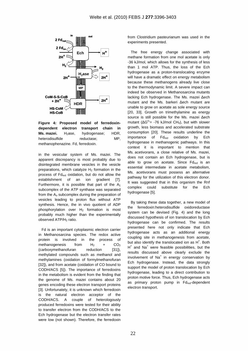
Figure 4: Proposed model of ferredoxin-dependent electron transport chain in Ms. mazei. H2ase, hydrogenase; HDR, heterodisulfide reductase; MP, methanophenazine. Fd, ferredoxin.
in the vesicular system of Ms. mazei. The apparent discrepancy is most probably due to disintegrated membrane vesicles in the vesicle preparations, which catalyze H2 formation in the process of Fdred oxidation, but do not allow the establishment of an ion gradient [7]. Furthermore, it is possible that part of the A1 subcomplex of the ATP synthase was separated from the Ao subcomplex during the preparation of vesicles leading to proton flux without ATP synthesis. Hence, the in vivo quotient of ADP phosphorylation over H2 formation is most probably much higher than the experimentally observed ATP/H2 ratio.
Fd is an important cytoplasmic electron carrier in Methanosarcina species. The redox active protein is involved in the process of methanogenesis from H2 + CO2 (carboxymethanofuran reduction [31]), methylated compounds such as methanol and methylamines (oxidation of formylmethanofuran [32]), and from acetate (oxidation of CO bound to CODH/ACS [5]). The importance of ferredoxins in the metabolism is evident from the finding that the genome of Ms. mazei contains about 20 genes encoding these electron transport proteins [3]. Unfortunately, it is unknown which ferredoxin is the natural electron acceptor of the CODH/ACS. A couple of heterologously produced ferredoxins were tested for their ability to transfer electron from the CODH/ACS to the Ech hydrogenase but the electron transfer rates were low (not shown). Therefore, the ferredoxin
from Clostridium pasteurianum was used in the experiments presented.
The free energy change associated with methane formation from one mol acetate is only -36 kJ/mol, which allows for the synthesis of less than 1 mol ATP. Thus, the loss of the Ech hydrogenase as a proton-translocating enzyme will have a dramatic effect on energy metabolism because these methanogens already live close to the thermodynamic limit. A severe impact can indeed be observed in Methanosarcina mutants lacking Ech hydrogenase. The Ms. mazei ∆ech mutant and the Ms. barkeri ∆ech mutant are unable to grow on acetate as sole energy source [20, 33]. Growth on trimethylamine as energy source is still possible for the Ms. mazei ∆ech mutant (∆Go’= -76 kJ/mol CH4), but with slower growth, less biomass and accelerated substrate consumption [20]. These results underline the importance of Fdred oxidation by Ech hydrogenase in methanogenic pathways. In this context it is important to mention that Ms. acetivorans, a close relative of Ms. mazei, does not contain an Ech hydrogenase, but is able to grow on acetate. Since Fdred is an essential intermediate in acetate metabolism, Ms. acetivorans must possess an alternative pathway for the utilization of this electron donor. It was suggested that in this organism the Rnf complex could substitute for the Ech hydrogenase [5].
By taking these data together, a new model of the ferredoxin:heterodisulfide oxidoreductase system can be devised (Fig. 4) and the long discussed hypothesis of ion translocation by Ech hydrogenase can be confirmed. The results presented here not only indicate that Ech hydrogenase acts as an additional energy coupling site in methanogenesis from acetate, but also identify the translocated ion as H+. Both H+ and Na+ were feasible possibilities, but the results discussed above clearly exclude the involvement of Na+ in energy conservation by Ech hydrogenase. Instead, the data strongly support the model of proton translocation by Ech hydrogenase, leading to a direct contribution to proton motive force. Thus, Ech hydrogenase acts as primary proton pump in Fdred-dependent electron transport.
Welte et al. (2010) FEBS J 277:3396-3403
23
EXPERIMENTAL PROCEDURES
Preparation of inverted membrane vesicles, proteins and reagents All experiments presented here were performed with Ms. mazei strain Gö1 (DSM 7222). Washed inverted membrane vesicles from Ms. mazei and Ms. mazei ∆ech [20] were prepared as described previously [7]. The strains were grown in 1 L glass bottles with 50 mM trimethylamine as substrate. The preparations were tested for the absence of enzyme activity with the cytoplasmic marker CODH/ACS to ensure the complete removal of cytoplasm from the membrane vesicles. Activity was tested by measuring the change of absorbance at 604 nm with 8.3 mM methylviologen, 5 % CO / 95 % N2 in the gas phase and 300-500 µg vesicle preparation in 40 mM potassium phosphate buffer (including 5 mM dithioerythritol, 1 µg mL-1 resazurin, pH 7.0) in a total volume of 1 mL. Fd from Clostridium pasteurianum was isolated as described [34] with replacement of the last two steps (dialyzation, crystallization) by ultrafiltration. Moorella thermoacetica CODH/ACS was isolated as described [35] with the modifications specified in [20]. Synthesis of CoM-S-S-CoB was done according to [36]. Determination of ATP formation ATP, ADP and AMP were supplied by Serva (Heidelberg, Germany). The inhibitors ETH157, DCCD and SF6847 and firefly lantern extract were supplied by Sigma-Aldrich (Schnelldorf, Germany). ETH157, DCCD and SF6847 were dissolved in 100% ethanol and used at final concentrations of 10-30 µM for ETH157 and SF6847, and 400 µM for DCCD. To determine ATP formation, rubber stoppered glass vials were filled with 500 µL buffer A (20 mM potassium phosphate, 20 mM MgSO4, 500 mM sucrose, 10 mM dithioerythritol, 1 µg mL-1 resazurin, pH 7.0), 5 % CO / 95 % N2 in the 1.5 mL headspace, 500-700 µg washed inverted membrane vesicles, 33.5 µg ferredoxin, 150 nmol AMP, and 300 nmol ADP. Before starting the reaction by addition of 20 µg CODH/ACS, the reaction mixture was pre-incubated for 5 min at 37° C in a shaking water bath to inhibit the membrane-bound adenylate kinase. This enzyme catalyzes the formation of ATP and AMP from two ADP and can be fully inhibited by high concentrations of AMP [37] present in the reaction mixture. Upon start of the reaction, 10 µL samples were taken every 2.5 min. ATP detection was performed according to [38]. The samples were mixed with 700 µL 20 mM glycylglycine buffer, pH 8.0, containing 4 mM
MgSO4, and 100 µL firefly lantern extract. Emitted light was quantified after 10 s by a Luminescence Spectrometer LS50B (Perkin Elmer, USA) at 560 nm and the values compared to a standard curve. Determination of H2 For determining H2 production rates, rubber stoppered glass vials were filled with 500 µL buffer A, 5 % CO / 95 % N2 in the 1.5 mL headspace, 500-700 µg washed inverted membrane vesicles, 33.5 µg ferredoxin, 20 µg CODH/ACS, 150 nmol AMP, and 300 nmol ADP. At various reaction time points, 10 µL of the headspace were injected into a gas chromatograph (GC-14A, Shimadzu, Japan) with argon as carrier gas. Molecular hydrogen was analyzed by a thermal conductivity detector and quantified by comparison to a standard curve.
Acknowledgements
We thank Elisabeth Schwab for technical assistance and Paul Schweiger for critical reading of the manuscript. This work was supported by the Deutsche Forschungsgemeinschaft (grant De488/9-1).
REFERENCES
1. Khalil MAK & Rasmussen RA (1994) Global emissions of methane during the last several centuries. Chemosphere 29, 833-842. 2. Reay DS (2003) Sinking methane. Biologist (London) 50, 15-19. 3. Deppenmeier U, Johann A, Hartsch T, Merkl R, Schmitz RA, Martinez-Arias R, Henne A, Wiezer A, Bäumer S, Jacobi C, Brüggemann H, Lienard T, Christmann A, Bömeke M, Steckel S, Bhattacharyya A, Lykidis A, Overbeek R, Klenk HP, Gunsalus RP, Fritz HJ & Gottschalk G (2002) The genome of Methanosarcina mazei: evidence for lateral gene transfer between bacteria and archaea. J Mol Microbiol Biotechnol 4, 453-461. 4. Deppenmeier U (2002) Redox-driven proton translocation in methanogenic Archaea. Cell Mol Life Sci 59, 1513-1533. 5. Ferry JG & Lessner DJ (2008) Methanogenesis in marine sediments. Ann N Y Acad Sci 1125, 147-157. 6. Meuer J, Bartoschek S, Koch J, Künkel A & Hedderich R (1999) Purification and catalytic properties of Ech hydrogenase from Methanosarcina barkeri. Eur J Biochem 265, 325-335. 7. Ide T, Bäumer S & Deppenmeier U (1999) Energy conservation by the H2:heterodisulfide
Welte et al. (2010) FEBS J 277:3396-3403
24
oxidoreductase from Methanosarcina mazei Gö1: identification of two proton-translocating segments. J Bacteriol 181, 4076-4080. 8. Becher B, Müller V & Gottschalk G (1992) N5-methyl-tetrahydromethanopterin:coenzyme M methyltransferase of Methanosarcina strain Gö1 is an Na(+)-translocating membrane protein. J Bacteriol 174, 7656-7660. 9. Lienard T, Becher B, Marschall M, Bowien S & Gottschalk G (1996) Sodium ion translocation by N5-methyltetrahydromethanopterin: coenzyme M methyltransferase from Methanosarcina mazei Gö1 reconstituted in ether lipid liposomes. Eur J Biochem 239, 857-864. 10. Hedderich R & Forzi L (2005) Energy-converting [NiFe] hydrogenases: more than just H2 activation. J Mol Microbiol Biotechnol 10, 92-104. 11. Bott M, Eikmanns B & Thauer R (1986) Coupling of carbon monoxide oxidation to CO2 and H2 with the phosphorylation of ADP in acetate-grown Methanosarcina barkeri. Eur J Biochem 159, 393-398. 12. Bott M & Thauer RK (1989) Proton translocation coupled to the oxidation of carbon monoxide to CO2 and H2 in Methanosarcina barkeri. Eur J Biochem 179, 469-472. 13. Wilms R, Freiberg C, Wegerle E, Meier I, Mayer F & Müller V (1996) Subunit structure and organization of the genes of the A1A0 ATPase from the Archaeon Methanosarcina mazei Gö1. J Biol Chem 271, 18843-18852. 14. Deppenmeier U, Blaut M, Mahlmann A & Gottschalk G (1990) Reduced coenzyme F420: heterodisulfide oxidoreductase, a proton- translocating redox system in methanogenic bacteria. Proc Natl Acad Sci U S A 87, 9449-9453. 15. Peinemann S, Blaut M & Gottschalk G (1989) ATP synthesis coupled to methane formation from methyl-CoM and H2 catalyzed by vesicles of the methanogenic bacterial strain Gö1. Eur J Biochem 186, 175-180. 16. Deppenmeier U, Blaut M, Schmidt B & Gottschalk G (1992) Purification and properties of a F420-nonreactive, membrane-bound hydrogenase from Methanosarcina strain Gö1. Arch Microbiol 157, 505-511. 17. Abken HJ, Tietze M, Brodersen J, Bäumer S, Beifuss U & Deppenmeier U (1998) Isolation and characterization of methanophenazine and function of phenazines in membrane-bound electron transport of Methanosarcina mazei Gö1. J Bacteriol 180, 2027-2032. 18. Tersteegen A & Hedderich R (1999) Methanobacterium thermoautotrophicum encodes two multisubunit membrane-bound [NiFe] hydrogenases. Transcription of the
operons and sequence analysis of the deduced proteins. Eur J Biochem 264, 930-943. 19. Stojanowic A & Hedderich R (2004) CO2 reduction to the level of formylmethanofuran in Methanosarcina barkeri is non-energy driven when CO is the electron donor. FEMS Microbiol Lett 235, 163-167. 20. Welte C, Kallnik V, Grapp M, Bender G, Ragsdale S & Deppenmeier U (2010) Function of Ech hydrogenase in ferredoxin-dependent, membrane-bound electron transport in Methanosarcina mazei. J Bacteriol 192, 674-678. 21. Sapra R, Bagramyan K & Adams MW (2003) A simple energy-conserving system: proton reduction coupled to proton translocation. Proc Natl Acad Sci U S A 100, 7545-7550. 22. Fox JD, Kerby RL, Roberts GP & Ludden PW (1996) Characterization of the CO-induced, CO-tolerant hydrogenase from Rhodospirillum rubrum and the gene encoding the large subunit of the enzyme. J Bacteriol 178, 1515-1524. 23. Soboh B, Linder D & Hedderich R (2002) Purification and catalytic properties of a CO-oxidizing:H2-evolving enzyme complex from Carboxydothermus hydrogenoformans. Eur J Biochem 269, 5712-5721. 24. Böhm R, Sauter M & Böck A (1990) Nucleotide sequence and expression of an operon in Escherichia coli coding for formate hydrogenlyase components. Mol Microbiol 4, 231-243. 25. Sauter M, Böhm R & Böck A (1992) Mutational analysis of the operon (hyc) determining hydrogenase 3 formation in Escherichia coli. Mol Microbiol 6, 1523-1532. 26. Andrews SC, Berks BC, McClay J, Ambler A, Quail MA, Golby P & Guest JR (1997) A 12-cistron Escherichia coli operon (hyf) encoding a putative proton-translocating formate hydrogenlyase system. Microbiology 143 (Pt 11), 3633-3647. 27. Pisa KY, Weidner C, Maischak H, Kavermann H & Müller V (2007) The coupling ion of the methanoarchaeal ATP synthases: H+ vs. Na+ in the A1Ao synthase from the archaeon Methanosarcina mazei Gö1. FEMS Microbiol Lett 277, 56-63. 28. Pisa KY, Huber H, Thomm M & Müller V (2007) A sodium ion-dependent A1Ao ATP synthase from the hyperthermophilic archaeon Pyrococcus furiosus. FEBS J 274, 3928-3938. 29. Peinemann S (1989) Kopplung von ATP-Synthese und Methanogenese in Vesikelpräparationen des methanogenen Bakteriums Stamm Gö1. PhD thesis, University of Göttingen, Germany. 30. Cross RL & Müller V (2004) The evolution of A-, F-, and V-type ATP synthases and ATPases:
Welte et al. (2010) FEBS J 277:3396-3403
25
reversals in function and changes in the H+/ATP coupling ratio. FEBS Lett 576, 1-4. 31. Bartoschek S, Vorholt JA, Thauer RK, Geierstanger BH & Griesinger C (2000) N-carboxymethanofuran (carbamate) formation from methanofuran and CO2 in methanogenic archaea. Thermodynamics and kinetics of the spontaneous reaction. Eur J Biochem 267, 3130-3138. 32. Deppenmeier U (2002) The unique biochemistry of methanogenesis. Prog Nucleic Acid Res Mol Biol 71, 223-283. 33. Meuer J, Kuettner HC, Zhang JK, Hedderich R & Metcalf WW (2002) Genetic analysis of the archaeon Methanosarcina barkeri Fusaro reveals a central role for Ech hydrogenase and ferredoxin in methanogenesis and carbon fixation. Proc Natl Acad Sci U S A 99, 5632-5637. 34. Mortenson LE (1964) Purification and analysis of ferredoxin from Clostridium pasteurianum. Biochimica et Biophysica Acta (BBA) - Specialized Section on Enzymological Subjects 81, 71-77. 35. Ragsdale SW, Ljungdahl LG & Der Vartanian DV (1983) Isolation of carbon monoxide dehydrogenase from Acetobacterium woodii and comparison of its properties with those of the Clostridium thermoaceticum enzyme. J Bacteriol 155, 1224-1237. 36. Ellermann J, Hedderich R, Bocher R & Thauer RK (1988) The final step in methane formation. Investigations with highly purified methyl-CoM reductase (component C) from Methanobacterium thermoautotrophicum (strain Marburg). Eur J Biochem 172, 669-677. 37. Bomsel JL & Pradet A (1968) Study of adenosine 5'-mono-, di- and triphosphates in plant tissues. IV. Regulation of the level of nucleotides, in vivo, by adenylate kinase: Theoretical and experimental study. Biochimica et Biophysica Acta (BBA) - Bioenergetics 162, 230-242. 38. Kimmich GA, Randles J & Brand JS (1975) Assay of picomole amounts of ATP, ADP, and AMP using the luciferase enzyme system. Anal Biochem 69, 187-206.
26
Chapter 4
Re-evaluation of the function of the F420 dehydrogenase in
electron transport of Methanosarcina mazei
The ferredoxin: heterodisulfide oxidoreductase system of Ms. mazei and Ms. barkeri
makes use of a hydrogen cycling mechanism via Ech hydrogenase and F420 non-
reducing hydrogenase. The membrane-bound F420H2 dehydrogenase was found to
be responsible for the oxidation of F420H2 that is produced during methylotrophic
methanogenesis and is well studied in Ms. mazei (Bäumer et al., 2000, Brodersen et
al., 1999). Conversely, it was recently suggested that in Ms. barkeri the cytoplasmic
F420 hydrogenase, not the F420H2 dehydrogenase, is responsible for F420H2 oxidation
with concomitant H2 production. This system would also make use of a hydrogen
cycling mechanism as shown for the ferredoxin: heterodisulfide oxidoreductase
(Kulkarni et al., 2009). In this publication, the role of the second hydrogen cycling
mechanism in comparison to the F420H2 dehydrogenase (Fpo) reaction was
investigated. With the help of two Ms. mazei fpo knockout mutants (∆fpoA-O, ∆fpoF)
it was found that Ms. mazei and Ms. barkeri differ in their preference for hydrogen
cycling from F420H2, and that the majority of Methanosarcina sp. probably do not
depend on hydrogen as obligate intermediate in F420H2 oxidation. The second part of
the publication describes the activity of the soluble F-subunit of Fpo. It catalyzes the
redox reaction of ferredoxin with F420. A protein catalyzing this reaction had been
hypothesized as being responsible for the survival of the Ms. mazei and Ms. barkeri
∆ech mutants because the accumulation of reduced ferredoxin had to be prevented
in these strains. Furthermore, the interconversion of different reducing equivalents
could be beneficial and allow more flexibility for the switch of growth substrates.
Welte and Deppenmeier (2011) FEBS J 278:1277-1287
27
Re-evaluation of the function of the F 420 dehydrogenase in electron
transport of Methanosarcina mazei
Cornelia Welte and Uwe Deppenmeier
Institute of Microbiology and Biotechnology, University of Bonn, Meckenheimer Allee 168, 53115 Bonn, Germany
Methanosarcina mazei is a methanogenic archaeon that is able to thrive on various substrates
and thus contains a variety of redox active protein s involved in both cytoplasmic and
membrane-bound electron transport. The organism pos sesses a complex branched respiratory
chain that has the ability to utilize different ele ctron donors. In this study, two knockout
mutants of the membrane-bound F 420 dehydrogenase ( ∆fpoF and ∆fpoA-O) were constructed
and analyzed. They exhibited severe growth deficien cies with trimethylamine, but not with
acetate as substrate. In cell lysates of the fpo mutants, the F 420: heterodisulfide oxidoreductase
activity was strongly reduced although soluble F 420 hydrogenase was still present. This led to
the conclusion that the predominant part of cellula r F420H2 oxidation in Ms. mazei is performed
by F 420 dehydrogenase. Enzyme assays of cytoplasmic fractio ns revealed that ferredoxin: F 420
oxidoreductase activity was essentially absent in t he ∆fpoF mutant. Subsequently, FpoF was
produced in Escherichia coli and purified for further characterization. The pur ified FpoF protein
catalyzed the ferredoxin: F 420 oxidoreductase reaction with high specificity (K m for reduced
ferredoxin 0.5 µM) but low velocity (v max 225 mU mg -1) and was present in the Ms. mazei
cytoplasm in considerable amounts. Consequently, so luble FpoF might participate in electron
carrier equilibrium and facilitate survival of the Ms. mazei ∆ech mutant that lacks the
membrane-bound ferredoxin-oxidizing Ech hydrogenase.
INTRODUCTION
Methanogenic archaea are one of the key groups in the global carbon cycle because they metabolize the final products of the anaerobic food chain (H2, CO2 and acetate) using unusual enzymes and cofactors in the so-called methanogenic pathway. The final product of all methanogenic pathways is methane and every year millions of tons of this highly potent greenhouse gas reach the atmosphere and contribute to global warming. Apart from their vast distribution in nature, methanogens are responsible for the production of CH4 in anaerobic digesters of biogas plants. The process of biomethanation is a viable alternative to fossil fuels and has great potential as an important renewable energy source. In principle, three different growth strategies in methanogenesis have evolved: hydrogenotrophic, methylotrophic and aceticlastic methanogenesis, using H2 + CO2, methylated compounds and acetate as substrates, respectively. The core of all methanogenic pathways is very similar, but there
are differences in the source of reducing equivalents and the mode of how electrons are channelled into the methanogenic respiratory chain. Methanosarcina mazei Gö1 (Ms. mazei) is a model organism for methanogenisis because it can grow hydrogenotrophically, methylotrophically, and aceticlastically. Its respiratory chain comprises three energy-conserving oxidoreductase systems that lead to the formation of an electrochemical proton gradient and ATP synthesis by the A1Ao ATP synthase [1]. The energy transducing systems are referred to as F420H2: heterodisulfide oxidoreductase, H2: heterodisulfide oxidoreductase and ferredoxin: heterodisulfide oxidoreductase, mirroring the three possible electron input compounds (F420H2, H2, reduced ferredoxin (Fdred)). Ferredoxin (Fd) is reduced during methylotrophic and aceticlastic methanogenesis, and is oxidized by Ech hydrogenase as part of the ferredoxin: heterodisulfide oxidoreductase system. F420H2 (the reduced form of a 8-hydroxy-5-deazaflavin) is formed in the course of methyl group oxidation in the methylotrophic pathway, or in the cytoplasm of hydrogenotrophically growing methanogens by F420 hydrogenase (Frh). This
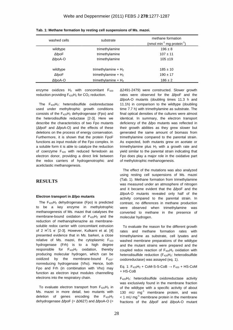
Welte and Deppenmeier (2011) FEBS J 278:1277-1287
28
Tab. 1: Methane formation by resting cell suspensio ns of Ms. mazei.
methane formation washed cells substrate (nmol min-1 mg protein-1)
wildtype trimethylamine 196 ± 8 ∆fpoF trimethylamine 107 ± 11 ∆fpoA-O trimethylamine 105 ±19
wildtype trimethylamine + H2 185 ± 10
∆fpoF trimethylamine + H2 190 ± 17
∆fpoA-O trimethylamine + H2 186 ± 2
enzyme oxidizes H2 with concomitant F420 reduction providing F420H2 for CO2 reduction.
The F420H2: heterodisulfide oxidoreductase used under methyltrophic growth conditions consists of the F420H2 dehydrogenase (Fpo) and the heterodisulfide reductase [2-3]. Here we describe the characteristics of two Fpo mutants (∆fpoF and ∆fpoA-O) and the effects of these deletions on the process of energy conservation. Furthermore, it is shown that the protein FpoF functions as input module of the Fpo complex. In a soluble form it is able to catalyze the reduction of coenzyme F420 with reduced ferredoxin as electron donor, providing a direct link between the redox carriers of hydrogenotrophic and aceticlastic methanogenesis.
RESULTS
Electron transport in ∆fpo mutants
The F420H2 dehydrogenase (Fpo) is predicted to be a key enzyme in methylotrophic methanogenesis of Ms. mazei that catalyses the membrane-bound oxidation of F420H2 and the reduction of methanophenazine as membrane-soluble redox carrier with concomitant extrusion of 2 H+/1 e- [2-3]. However, Kulkarni et al. [4] presented evidence that in Ms. barkeri, a close relative of Ms. mazei, the cytoplasmic F420 hydrogenase (Frh) is to a high degree responsible for F420H2 oxidation, thereby producing molecular hydrogen, which can be oxidized by the membrane-bound F420-nonreducing hydrogenase (Vho). Hence, both Fpo and Frh (in combination with Vho) may function as electron input modules channelling electrons into the respiratory chain. To evaluate electron transport from F420H2 in Ms. mazei in more detail, two mutants with deletion of genes encoding the F420H2 dehydrogenase ∆fpoF (= ∆0627) and ∆fpoA-O (=
∆2491-2479) were constructed. Slower growth rates were observed for the ∆fpoF and the ∆fpoA-O mutants (doubling times 11.3 h and 11.1h) in comparison to the wildtype (doubling time 7.7 h) with trimethylamine as substrate. The final optical densities of the cultures were almost identical. In summary, the electron transport deficiency of the ∆fpo mutants was reflected in their growth abilities as they grew slower but generated the same amount of biomass from trimethylamine compared to the parental strain. As expected, both mutants grew on acetate or trimethylamine plus H2 with a growth rate and yield similar to the parental strain indicating that Fpo does play a major role in the oxidative part of methylotrophic methanogenesis. The effect of the mutations was also analyzed using resting cell suspensions of Ms. mazei (Tab. 1). Methane formation from trimethylamine was measured under an atmosphere of nitrogen and it became evident that the ∆fpoF and the ∆fpoA-O mutants revealed only half of the activity compared to the parental strain. In contrast, no differences in methane production were observed when trimethylamine was converted to methane in the presence of molecular hydrogen. To evaluate the reason for the different growth rates and methane formation rates with trimethylamine as substrate, cell lysates and washed membrane preparations of the wildtype and the mutant strains were prepared and the coupled redox reaction of F420H2 oxidation with heterodisulfide reduction (F420H2: heterodisulfide oxidoreductase) was assayed (eq. 1).
Eq. 1: F420H2 + CoM-S-S-CoB → F420 + HS-CoM + HS-CoB
F420H2: heterodisulfide oxidoreductase activity was exclusively found in the membrane fraction of the wildtype with a specific activity of about 130 mU mg-1 membrane protein, and was < 1 mU mg-1 membrane protein in the membrane fractions of the ∆fpoF and ∆fpoA-O mutant
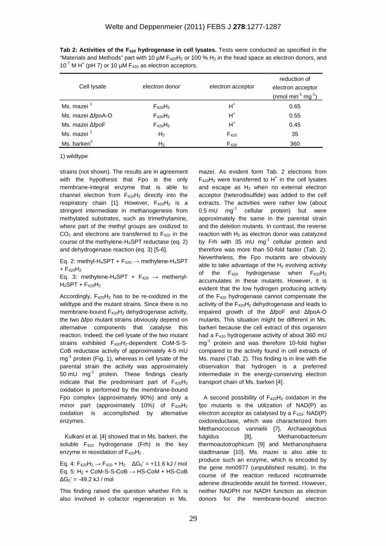
Welte and Deppenmeier (2011) FEBS J 278:1277-1287
29
Tab 2: Activities of the F 420 hydrogenase in cell lysates. Tests were conducted as specified in the “Materials and Methods” part with 10 µM F420H2 or 100 % H2 in the head space as electron donors, and 10-7 M H+ (pH 7) or 10 µM F420 as electron acceptors.
reduction of
electron acceptor Cell lysate electron donor electron acceptor
(nmol min-1 mg-1)
Ms. mazei 1 F420H2 H+ 0.65
Ms. mazei ∆fpoA-O F420H2 H+ 0.55
Ms. mazei ∆fpoF F420H2 H+ 0.45
Ms. mazei 1 H2 F420 35
Ms. barkeri1 H2 F420 360
1) wildtype
strains (not shown). The results are in agreement with the hypothesis that Fpo is the only membrane-integral enzyme that is able to channel electron from F420H2 directly into the respiratory chain [1]. However, F420H2 is a stringent intermediate in methanogenesis from methylated substrates, such as trimethylamine, where part of the methyl groups are oxidized to CO2 and electrons are transferred to F420 in the course of the methylene-H4SPT reductase (eq. 2) and dehydrogenase reaction (eq. 3) [5-6].
Eq. 2: methyl-H4SPT + F420 → methylene-H4SPT + F420H2 Eq. 3: methylene-H4SPT + F420 → methenyl-H4SPT + F420H2
Accordingly, F420H2 has to be re-oxidized in the wildtype and the mutant strains. Since there is no membrane-bound F420H2 dehydrogenase activity, the two ∆fpo mutant strains obviously depend on alternative components that catalyse this reaction. Indeed, the cell lysate of the two mutant strains exhibited F420H2-dependent CoM-S-S-CoB reductase activity of approximately 4-5 mU mg-1 protein (Fig. 1), whereas in cell lysate of the parental strain the activity was approximately 50 mU mg-1 protein. These findings clearly indicate that the predominant part of F420H2 oxidation is performed by the membrane-bound Fpo complex (approximately 90%) and only a minor part (approximately 10%) of F420H2 oxidation is accomplished by alternative enzymes. Kulkani et al. [4] showed that in Ms. barkeri, the soluble F420 hydrogenase (Frh) is the key enzyme in reoxidation of F420H2 .
Eq. 4: F420H2 → F420 + H2 ∆G0’ = +11.6 kJ / mol Eq. 5: H2 + CoM-S-S-CoB → HS-CoM + HS-CoB ∆G0’ = -49.2 kJ / mol
This finding raised the question whether Frh is also involved in cofactor regeneration in Ms.
mazei. As evident form Tab. 2 electrons from F420H2 were transferred to H+ in the cell lysates and escape as H2 when no external electron acceptor (heterodisulfide) was added to the cell extracts. The activities were rather low (about 0.5 mU mg-1 cellular protein) but were approximately the same in the parental strain and the deletion mutants. In contrast, the reverse reaction with H2 as electron donor was catalyzed by Frh with 35 mU mg-1 cellular protein and therefore was more than 50-fold faster (Tab. 2). Nevertheless, the Fpo mutants are obviously able to take advantage of the H2 evolving activity of the F420 hydrogenase when F420H2 accumulates in these mutants. However, it is evident that the low hydrogen producing activity of the F420 hydrogenase cannot compensate the activity of the F420H2 dehydrogenase and leads to impaired growth of the ∆fpoF and ∆fpoA-O mutants. This situation might be different in Ms. barkeri because the cell extract of this organism had a F420 hydrogenase activity of about 360 mU mg-1 protein and was therefore 10-fold higher compared to the activity found in cell extracts of Ms. mazei (Tab. 2). This finding is in line with the observation that hydrogen is a preferred intermediate in the energy-conserving electron transport chain of Ms. barkeri [4]. A second possibility of F420H2 oxidation in the fpo mutants is the utilization of NAD(P) as electron acceptor as catalysed by a F420: NAD(P) oxidoreductase, which was characterized from Methanococcus vannielii [7], Archaeoglobus fulgidus [8], Methanobacterium thermoautotrophicum [9] and Methanosphaera stadtmanae [10]. Ms. mazei is also able to produce such an enzyme, which is encoded by the gene mm0977 (unpublished results). In the course of the reaction reduced nicotinamide adenine dinucleotide would be formed. However, neither NADPH nor NADH function as electron donors for the membrane-bound electron
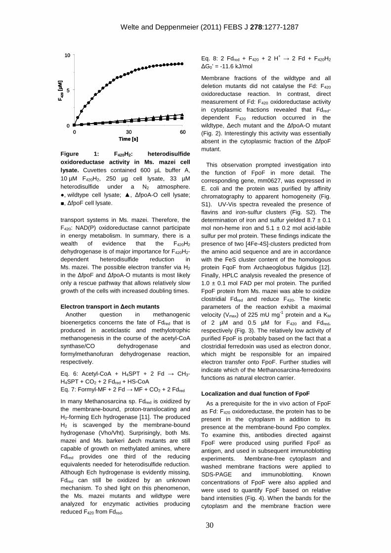
Welte and Deppenmeier (2011) FEBS J 278:1277-1287
30
Figure 1: F 420H2: heterodisulfide oxidoreductase activity in Ms. mazei cell lysate. Cuvettes contained 600 µL buffer A, 10 µM F420H2, 250 µg cell lysate, 33 µM heterodisulfide under a N2 atmosphere. ●, wildtype cell lysate; ▲, ∆fpoA-O cell lysate; ■, ∆fpoF cell lysate.
transport systems in Ms. mazei. Therefore, the F420: NAD(P) oxidoreductase cannot participate in energy metabolism. In summary, there is a wealth of evidence that the F420H2 dehydrogenase is of major importance for F420H2-dependent heterodisulfide reduction in Ms. mazei. The possible electron transfer via H2 in the ∆fpoF and ∆fpoA-O mutants is most likely only a rescue pathway that allows relatively slow growth of the cells with increased doubling times. Electron transport in ∆ech mutants Another question in methanogenic bioenergetics concerns the fate of Fdred that is produced in aceticlastic and methylotrophic methanogenesis in the course of the acetyl-CoA synthase/CO dehydrogenase and formylmethanofuran dehydrogenase reaction, respectively.
Eq. 6: Acetyl-CoA + H4SPT + 2 Fd → CH3-H4SPT + CO2 + 2 Fdred + HS-CoA Eq. 7: Formyl-MF + 2 Fd → MF + CO2 + 2 Fdred
In many Methanosarcina sp. Fdred is oxidized by the membrane-bound, proton-translocating and H2-forming Ech hydrogenase [11]. The produced H2 is scavenged by the membrane-bound hydrogenase (Vho/Vht). Surprisingly, both Ms. mazei and Ms. barkeri ∆ech mutants are still capable of growth on methylated amines, where Fdred provides one third of the reducing equivalents needed for heterodisulfide reduction. Although Ech hydrogenase is evidently missing, Fdred can still be oxidized by an unknown mechanism. To shed light on this phenomenon, the Ms. mazei mutants and wildtype were analyzed for enzymatic activities producing reduced F420 from Fdred.
Eq. 8: 2 Fdred + F420 + 2 H+ → 2 Fd + F420H2 ∆G0’ = -11.6 kJ/mol
Membrane fractions of the wildtype and all deletion mutants did not catalyse the Fd: F420 oxidoreductase reaction. In contrast, direct measurement of Fd: F420 oxidoreductase activity in cytoplasmic fractions revealed that Fdred-dependent F420 reduction occurred in the wildtype, ∆ech mutant and the ∆fpoA-O mutant (Fig. 2). Interestingly this activity was essentially absent in the cytoplasmic fraction of the ∆fpoF mutant. This observation prompted investigation into the function of FpoF in more detail. The corresponding gene, mm0627, was expressed in E. coli and the protein was purified by affinity chromatography to apparent homogeneity (Fig. S1). UV-Vis spectra revealed the presence of flavins and iron-sulfur clusters (Fig. S2). The determination of iron and sulfur yielded 8.7 ± 0.1 mol non-heme iron and 5.1 ± 0.2 mol acid-labile sulfur per mol protein. These findings indicate the presence of two [4Fe-4S]-clusters predicted from the amino acid sequence and are in accordance with the FeS cluster content of the homologous protein FqoF from Archaeoglobus fulgidus [12]. Finally, HPLC analysis revealed the presence of 1.0 ± 0.1 mol FAD per mol protein. The purified FpoF protein from Ms. mazei was able to oxidize clostridial Fdred and reduce F420. The kinetic parameters of the reaction exhibit a maximal velocity (Vmax) of 225 mU mg-1 protein and a KM of 2 µM and 0.5 µM for F420 and Fdred, respectively (Fig. 3). The relatively low activity of purified FpoF is probably based on the fact that a clostridial ferredoxin was used as electron donor, which might be responsible for an impaired electron transfer onto FpoF. Further studies will indicate which of the Methanosarcina-ferredoxins functions as natural electron carrier.
Localization and dual function of FpoF
As a prerequisite for the in vivo action of FpoF as Fd: F420 oxidoreductase, the protein has to be present in the cytoplasm in addition to its presence at the membrane-bound Fpo complex. To examine this, antibodies directed against FpoF were produced using purified FpoF as antigen, and used in subsequent immunoblotting experiments. Membrane-free cytoplasm and washed membrane fractions were applied to SDS-PAGE and immunoblotting. Known concentrations of FpoF were also applied and were used to quantify FpoF based on relative band intensities (Fig. 4). When the bands for the cytoplasm and the membrane fraction were
0
5
10
0 30 60Time [s]
F42
0[µ
M]
0
5
10
0 30 60Time [s]
F42
0[µ
M]
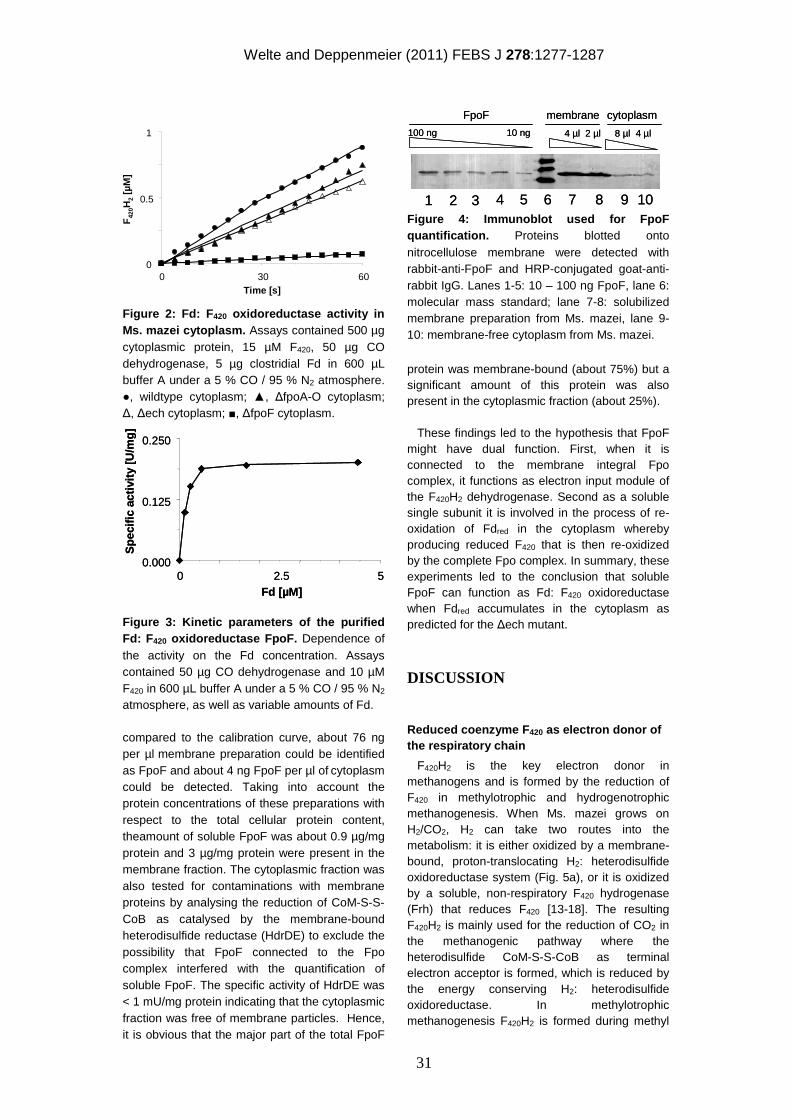
Welte and Deppenmeier (2011) FEBS J 278:1277-1287
31
Figure 2: Fd: F 420 oxidoreductase activity in Ms. mazei cytoplasm. Assays contained 500 µg cytoplasmic protein, 15 µM F420, 50 µg CO dehydrogenase, 5 µg clostridial Fd in 600 µL buffer A under a 5 % CO / 95 % N2 atmosphere. ●, wildtype cytoplasm; ▲, ∆fpoA-O cytoplasm; ∆, ∆ech cytoplasm; ■, ∆fpoF cytoplasm.
Figure 3: Kinetic parameters of the purified Fd: F 420 oxidoreductase FpoF. Dependence of the activity on the Fd concentration. Assays contained 50 µg CO dehydrogenase and 10 µM F420 in 600 µL buffer A under a 5 % CO / 95 % N2 atmosphere, as well as variable amounts of Fd.
compared to the calibration curve, about 76 ng per µl membrane preparation could be identified as FpoF and about 4 ng FpoF per µl of cytoplasm could be detected. Taking into account the protein concentrations of these preparations with respect to the total cellular protein content, theamount of soluble FpoF was about 0.9 µg/mg protein and 3 µg/mg protein were present in the membrane fraction. The cytoplasmic fraction was also tested for contaminations with membrane proteins by analysing the reduction of CoM-S-S-CoB as catalysed by the membrane-bound heterodisulfide reductase (HdrDE) to exclude the possibility that FpoF connected to the Fpo complex interfered with the quantification of soluble FpoF. The specific activity of HdrDE was < 1 mU/mg protein indicating that the cytoplasmic fraction was free of membrane particles. Hence, it is obvious that the major part of the total FpoF
Figure 4: Immunoblot used for FpoF quantification. Proteins blotted onto nitrocellulose membrane were detected with rabbit-anti-FpoF and HRP-conjugated goat-anti-rabbit IgG. Lanes 1-5: 10 – 100 ng FpoF, lane 6: molecular mass standard; lane 7-8: solubilized membrane preparation from Ms. mazei, lane 9-10: membrane-free cytoplasm from Ms. mazei.
protein was membrane-bound (about 75%) but a significant amount of this protein was also present in the cytoplasmic fraction (about 25%). These findings led to the hypothesis that FpoF might have dual function. First, when it is connected to the membrane integral Fpo complex, it functions as electron input module of the F420H2 dehydrogenase. Second as a soluble single subunit it is involved in the process of re-oxidation of Fdred in the cytoplasm whereby producing reduced F420 that is then re-oxidized by the complete Fpo complex. In summary, these experiments led to the conclusion that soluble FpoF can function as Fd: F420 oxidoreductase when Fdred accumulates in the cytoplasm as predicted for the ∆ech mutant.
DISCUSSION
Reduced coenzyme F 420 as electron donor of the respiratory chain
F420H2 is the key electron donor in methanogens and is formed by the reduction of F420 in methylotrophic and hydrogenotrophic methanogenesis. When Ms. mazei grows on H2/CO2, H2 can take two routes into the metabolism: it is either oxidized by a membrane-bound, proton-translocating H2: heterodisulfide oxidoreductase system (Fig. 5a), or it is oxidized by a soluble, non-respiratory F420 hydrogenase (Frh) that reduces F420 [13-18]. The resulting F420H2 is mainly used for the reduction of CO2 in the methanogenic pathway where the heterodisulfide CoM-S-S-CoB as terminal electron acceptor is formed, which is reduced by the energy conserving H2: heterodisulfide oxidoreductase. In methylotrophic methanogenesis F420H2 is formed during methyl
0
0.5
1
0 30 60Time [s]
F42
0H2
[µM
]
0
0.5
1
0 30 60Time [s]
F42
0H2
[µM
]
0.000
0.125
0.250
0 2.5 5Fd [µM]
Spe
cific
activ
ity[U
/mg]
0.000
0.125
0.250
0 2.5 5Fd [µM]
Spe
cific
activ
ity[U
/mg]
FpoF
100 ng 10 ng 4 µl 2 µl
membrane cytoplasm
8 µl 4 µl
1 2 3 4 5 6 7 8 9 10
FpoF
100 ng 10 ng 4 µl 2 µl
membrane cytoplasm
8 µl 4 µl
1 2 3 4 5 6 7 8 9 10
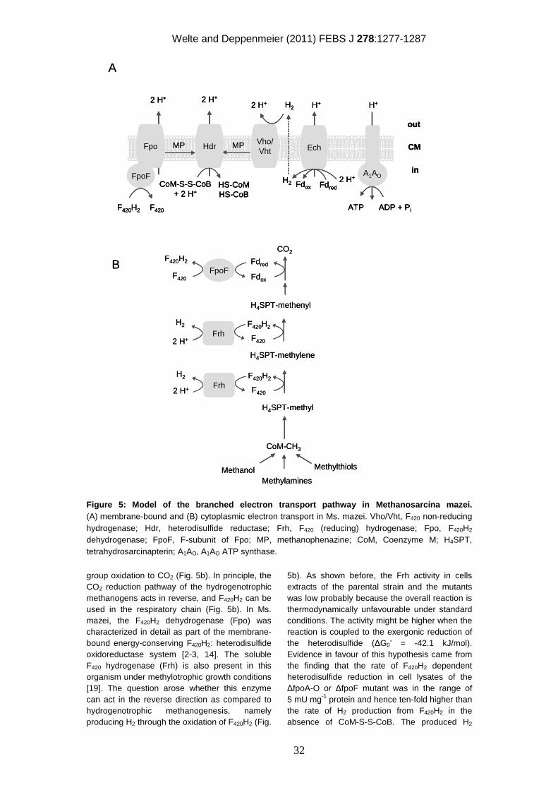
Welte and Deppenmeier (2011) FEBS J 278:1277-1287
32
Figure 5: Model of the branched electron transport pathway in Methanosarcina mazei. (A) membrane-bound and (B) cytoplasmic electron transport in Ms. mazei. Vho/Vht, F420 non-reducing hydrogenase; Hdr, heterodisulfide reductase; Frh, F420 (reducing) hydrogenase; Fpo, F420H2 dehydrogenase; FpoF, F-subunit of Fpo; MP, methanophenazine; CoM, Coenzyme M; H4SPT, tetrahydrosarcinapterin; A1AO, A1AO ATP synthase.
group oxidation to CO2 (Fig. 5b). In principle, the CO2 reduction pathway of the hydrogenotrophic methanogens acts in reverse, and F420H2 can be used in the respiratory chain (Fig. 5b). In Ms. mazei, the F420H2 dehydrogenase (Fpo) was characterized in detail as part of the membrane-bound energy-conserving F420H2: heterodisulfide oxidoreductase system [2-3, 14]. The soluble F420 hydrogenase (Frh) is also present in this organism under methylotrophic growth conditions [19]. The question arose whether this enzyme can act in the reverse direction as compared to hydrogenotrophic methanogenesis, namely producing H2 through the oxidation of F420H2 (Fig.
5b). As shown before, the Frh activity in cells extracts of the parental strain and the mutants was low probably because the overall reaction is thermodynamically unfavourable under standard conditions. The activity might be higher when the reaction is coupled to the exergonic reduction of the heterodisulfide (∆G0’ = -42.1 kJ/mol). Evidence in favour of this hypothesis came from the finding that the rate of F420H2 dependent heterodisulfide reduction in cell lysates of the ∆fpoA-O or ∆fpoF mutant was in the range of 5 mU mg-1 protein and hence ten-fold higher than the rate of H2 production from F420H2 in the absence of CoM-S-S-CoB. The produced H2
A
B
Frh
FpoF
F420H2
F420
CO2
Fdox
Fdred
F420
F420H2
H4SPT-methyl
H4SPT-methenyl
CoM-CH3
MethanolMethylamines
Methylthiols
H2
2 H+
Frh F420
F420H2H2
2 H+
H4SPT-methylene
2 H+
out
in
CoM-S-S-CoB+ 2 H+
HS-CoMHS-CoB
F420H2 F420
CM
FpoF
MP
2 H+
2 H+
MP
H2
Vho/Vht
HdrFpo Ech
Fdox Fdred
H+
2 H+H2
ATP ADP + Pi
H+
A1AO
2 H+
out
in
CoM-S-S-CoB+ 2 H+
HS-CoMHS-CoB
F420H2 F420
CM
FpoF
MP
2 H+
2 H+
MP
H2
Vho/Vht
HdrFpo Ech
Fdox Fdred
H+
2 H+H2
ATP ADP + Pi
H+
A1AO
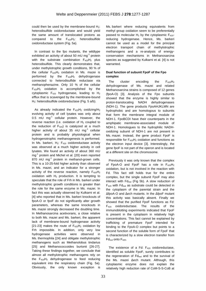
Welte and Deppenmeier (2011) FEBS J 278:1277-1287
33
could then be used by the membrane-bound H2: heterodisulfide oxidoreductase and would yield the same amount of translocated protons as compared to the F420H2: heterodisulfide oxidoreductase system (Fig. 5a). In contrast to the fpo mutants, the wildtype exhibited an activity of about 50 mU mg-1 protein with the substrate combination F420H2 plus heterodisulfide. This clearly demonstrates that, under methylotrophic growth conditions, 90 % of the cellular F420H2 oxidation in Ms. mazei is performed by the F420H2 dehydrogenase connected to heterodisulfide reductase via methanophenazine. Only 10 % of the cellular F420H2 oxidation is accomplished by the cytoplasmic F420 hydrogenase, leading to H2 efflux that is scavenged by the membrane-bound H2: heterodisulfide oxidoreductase (Fig. 5 a/b). As already indicated the F420H2 oxidizing/H2 evolving activity of cell lysates was only about 0.5 mU mg-1 cellular protein. However, the reverse reaction (i.e. oxidation of H2 coupled to the reduction of F420) is catalyzed at a much higher activity of about 35 mU mg-1 cellular protein and is probably physiological when hydrogenotrophic methanogenesis is performed. In Ms. barkeri, H2: F420 oxidoreductase activity was observed at a much higher activity in cell lysates. We found an activity of about 360 mU mg-1 protein and Michel et al. [20] even reported 870 mU mg-1 protein in methanol-grown cells. This is a 10-25-fold higher activity than observed in Ms. mazei, and an indicator for a stronger activity of the reverse reaction, namely F420H2 oxidation with H2 production. It is tempting to speculate that the role of Frh in Ms. barkeri under methylotrophic growth conditions is greater than the role for the same enzyme in Ms. mazei. In fact this was actually observed by Kulkarni et al. [4] who reported that in Ms. barkeri knockouts of fpoA-O or fpoF do not significantly alter growth parameters, whereas the same knockouts in Ms. mazei strongly decreased the doubling time. In Methanosarcina acetivorans, a close relative to both Ms. mazei and Ms. barkeri, the apparent lack of membrane-bound hydrogenase activity [21-23] makes the route of F420H2 oxidation by Frh impossible. In addition, only very low hydrogenase activities were observed in Ms. thermophila [24] and obligate methylotrophic methanogens such as Methanolobus tindarius [25] and Methanococcoides burtonii [26-27]. Taking these findings together, we conclude that almost all methylotrophic methanogens rely on the F420H2 dehydrogenase to feed reducing equivalent into the respiratory chain (Fig. 5a). Obviously, the only known exception is
Ms. barkeri where reducing equivalents from methyl group oxidation seem to be preferentially passed to molecular H2 by the cytoplasmic F420-reducing hydrogenase. Hence, Ms. barkeri cannot be used as a model for the principal electron transport chain of methylotrophic methanogens and a re-analysis of energy-conservation mechanisms in Methanosarcina species as suggested by Kulkarni et al. [4] is not warranted.
Dual function of subunit FpoF of the Fpo complex
The cluster encoding the F420H2 dehydrogenase of Ms. mazei and related Methanosarcina strains is composed of 12 genes (fpoA-O) [3]. Analysis of the Fpo subunits showed that the enzyme is highly similar to proton-translocating NADH dehydrogenases (NDH-1). The gene products FpoAHJKLMN are hydrophobic and are homologous to subunits that form the membrane integral module of NDH-1. FpoBCDI have their counterparts in the amphipatic membrane-associated module of NDH-1. Homologues to the hydrophilic NADH-oxidizing subunit of NDH-1 are not present in Ms. mazei. Instead, the gene product FpoF is responsible for F420H2 oxidation and functions as the electron input device [3]. Interestingly, the gene fpoF is not part of the operon and is located at a different site on the chromosome. Previously it was only known that the complex of FpoA-O and FpoF has a role in F420H2 oxidation, but is not involved in the reaction with Fd. This fact still holds true for the entire complex, but the single subunit FpoF may also interact with Fdred (Fig 5b). A slow reduction of F420 with Fdred as substrate could be detected in the cytoplasm of the parental strain and the ∆fpoA-O and ∆ech mutants. In the ∆fpoF mutant this activity was basically absent. Finally we showed that the purified FpoF functions as Fd: F420 oxidoreductase. The results of the immunoblotting experiments indicated that FpoF is present in the cytoplasm in relatively high concentrations. This fact cannot be explained by trafficking of premature FpoF intended for binding to the FpoA-O complex but points to a second function of the soluble form of FpoF that is characterized by a slow electron transfer from Fdred onto F420. The existence of a Fd: F420 oxidoreductase, identified as soluble FpoF, surely contributes to the regeneration of Fdred and to the survival of the Ms. mazei ∆ech mutant. Although, this cytoplasmic enzyme does not explain the relatively high reduction rate of CoM-S-S-CoB at
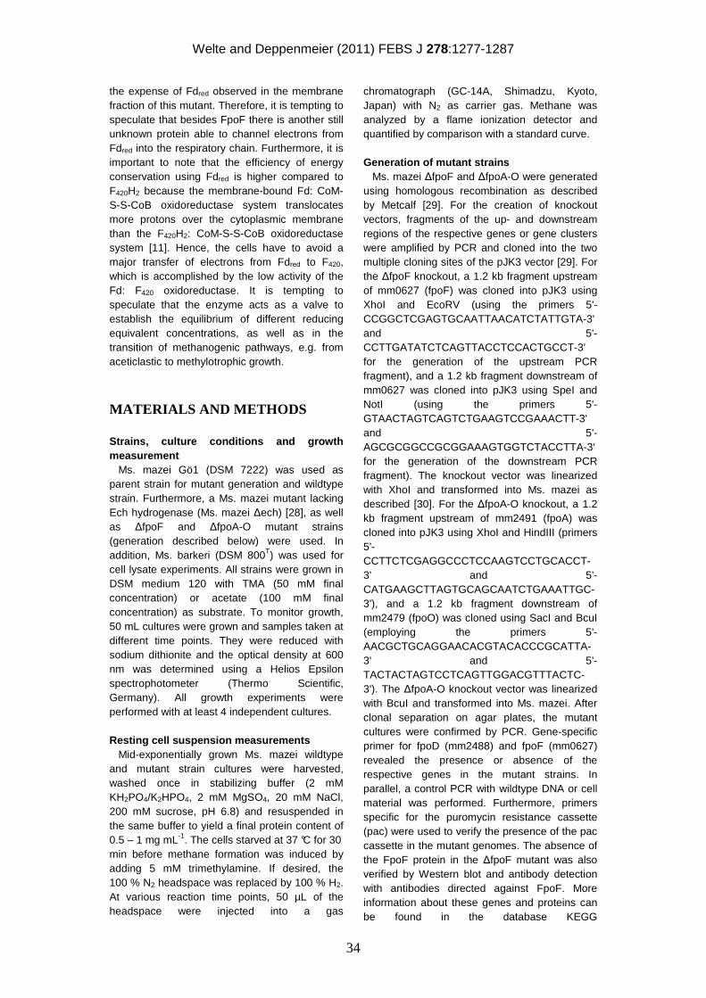
Welte and Deppenmeier (2011) FEBS J 278:1277-1287
34
the expense of Fdred observed in the membrane fraction of this mutant. Therefore, it is tempting to speculate that besides FpoF there is another still unknown protein able to channel electrons from Fdred into the respiratory chain. Furthermore, it is important to note that the efficiency of energy conservation using Fdred is higher compared to F420H2 because the membrane-bound Fd: CoM-S-S-CoB oxidoreductase system translocates more protons over the cytoplasmic membrane than the F420H2: CoM-S-S-CoB oxidoreductase system [11]. Hence, the cells have to avoid a major transfer of electrons from Fdred to F420, which is accomplished by the low activity of the Fd: F420 oxidoreductase. It is tempting to speculate that the enzyme acts as a valve to establish the equilibrium of different reducing equivalent concentrations, as well as in the transition of methanogenic pathways, e.g. from aceticlastic to methylotrophic growth.
MATERIALS AND METHODS
Strains, culture conditions and growth measurement Ms. mazei Gö1 (DSM 7222) was used as parent strain for mutant generation and wildtype strain. Furthermore, a Ms. mazei mutant lacking Ech hydrogenase (Ms. mazei ∆ech) [28], as well as ∆fpoF and ∆fpoA-O mutant strains (generation described below) were used. In addition, Ms. barkeri (DSM 800T) was used for cell lysate experiments. All strains were grown in DSM medium 120 with TMA (50 mM final concentration) or acetate (100 mM final concentration) as substrate. To monitor growth, 50 mL cultures were grown and samples taken at different time points. They were reduced with sodium dithionite and the optical density at 600 nm was determined using a Helios Epsilon spectrophotometer (Thermo Scientific, Germany). All growth experiments were performed with at least 4 independent cultures. Resting cell suspension measurements Mid-exponentially grown Ms. mazei wildtype and mutant strain cultures were harvested, washed once in stabilizing buffer (2 mM KH2PO4/K2HPO4, 2 mM MgSO4, 20 mM NaCl, 200 mM sucrose, pH 6.8) and resuspended in the same buffer to yield a final protein content of 0.5 – 1 mg mL-1. The cells starved at 37 °C for 30 min before methane formation was induced by adding 5 mM trimethylamine. If desired, the 100 % N2 headspace was replaced by 100 % H2. At various reaction time points, 50 µL of the headspace were injected into a gas
chromatograph (GC-14A, Shimadzu, Kyoto, Japan) with N2 as carrier gas. Methane was analyzed by a flame ionization detector and quantified by comparison with a standard curve. Generation of mutant strains Ms. mazei ∆fpoF and ∆fpoA-O were generated using homologous recombination as described by Metcalf [29]. For the creation of knockout vectors, fragments of the up- and downstream regions of the respective genes or gene clusters were amplified by PCR and cloned into the two multiple cloning sites of the pJK3 vector [29]. For the ∆fpoF knockout, a 1.2 kb fragment upstream of mm0627 (fpoF) was cloned into pJK3 using XhoI and EcoRV (using the primers 5'-CCGGCTCGAGTGCAATTAACATCTATTGTA-3' and 5'-CCTTGATATCTCAGTTACCTCCACTGCCT-3' for the generation of the upstream PCR fragment), and a 1.2 kb fragment downstream of mm0627 was cloned into pJK3 using SpeI and NotI (using the primers 5'-GTAACTAGTCAGTCTGAAGTCCGAAACTT-3' and 5'-AGCGCGGCCGCGGAAAGTGGTCTACCTTA-3' for the generation of the downstream PCR fragment). The knockout vector was linearized with XhoI and transformed into Ms. mazei as described [30]. For the ∆fpoA-O knockout, a 1.2 kb fragment upstream of mm2491 (fpoA) was cloned into pJK3 using XhoI and HindIII (primers 5'-CCTTCTCGAGGCCCTCCAAGTCCTGCACCT-3' and 5'-CATGAAGCTTAGTGCAGCAATCTGAAATTGC-3'), and a 1.2 kb fragment downstream of mm2479 (fpoO) was cloned using SacI and BcuI (employing the primers 5'-AACGCTGCAGGAACACGTACACCCGCATTA-3' and 5'-TACTACTAGTCCTCAGTTGGACGTTTACTC-3'). The ∆fpoA-O knockout vector was linearized with BcuI and transformed into Ms. mazei. After clonal separation on agar plates, the mutant cultures were confirmed by PCR. Gene-specific primer for fpoD (mm2488) and fpoF (mm0627) revealed the presence or absence of the respective genes in the mutant strains. In parallel, a control PCR with wildtype DNA or cell material was performed. Furthermore, primers specific for the puromycin resistance cassette (pac) were used to verify the presence of the pac cassette in the mutant genomes. The absence of the FpoF protein in the ∆fpoF mutant was also verified by Western blot and antibody detection with antibodies directed against FpoF. More information about these genes and proteins can be found in the database KEGG
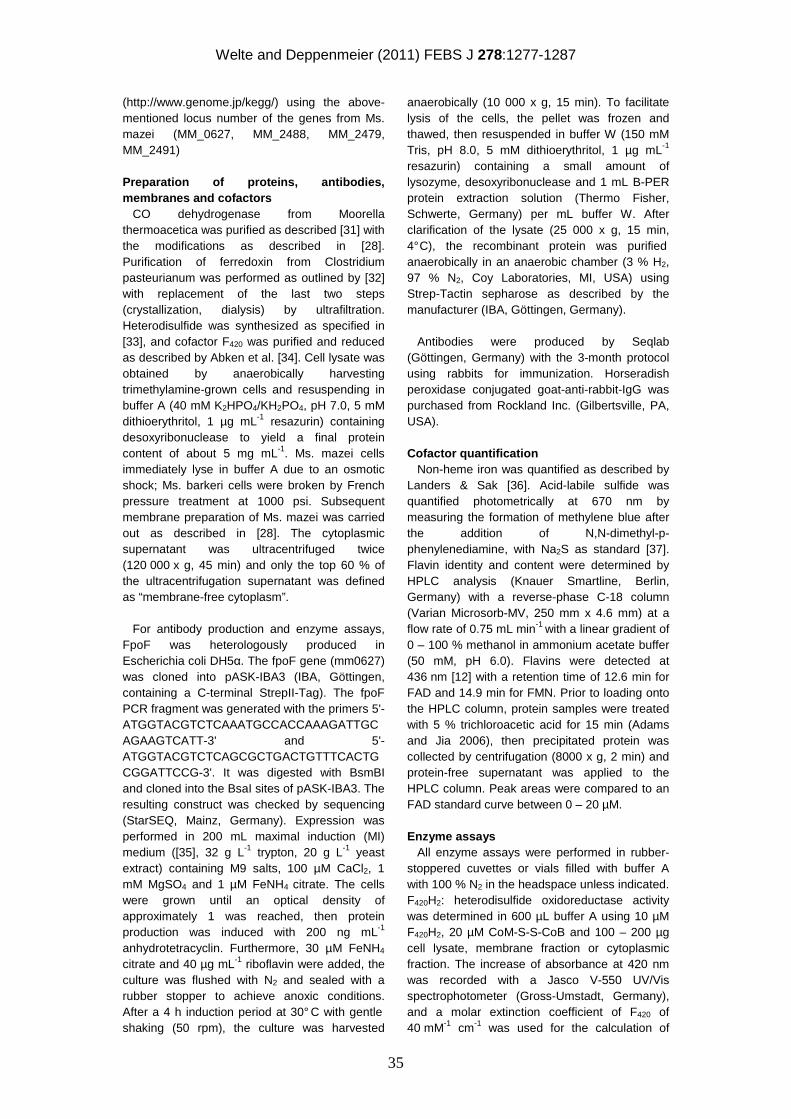
Welte and Deppenmeier (2011) FEBS J 278:1277-1287
35
(http://www.genome.jp/kegg/) using the above-mentioned locus number of the genes from Ms. mazei (MM_0627, MM_2488, MM_2479, MM_2491) Preparation of proteins, antibodies, membranes and cofactors CO dehydrogenase from Moorella thermoacetica was purified as described [31] with the modifications as described in [28]. Purification of ferredoxin from Clostridium pasteurianum was performed as outlined by [32] with replacement of the last two steps (crystallization, dialysis) by ultrafiltration. Heterodisulfide was synthesized as specified in [33], and cofactor F420 was purified and reduced as described by Abken et al. [34]. Cell lysate was obtained by anaerobically harvesting trimethylamine-grown cells and resuspending in buffer A (40 mM K2HPO4/KH2PO4, pH 7.0, 5 mM dithioerythritol, 1 µg mL-1 resazurin) containing desoxyribonuclease to yield a final protein content of about 5 mg mL-1. Ms. mazei cells immediately lyse in buffer A due to an osmotic shock; Ms. barkeri cells were broken by French pressure treatment at 1000 psi. Subsequent membrane preparation of Ms. mazei was carried out as described in [28]. The cytoplasmic supernatant was ultracentrifuged twice (120 000 x g, 45 min) and only the top 60 % of the ultracentrifugation supernatant was defined as “membrane-free cytoplasm”. For antibody production and enzyme assays, FpoF was heterologously produced in Escherichia coli DH5α. The fpoF gene (mm0627) was cloned into pASK-IBA3 (IBA, Göttingen, containing a C-terminal StrepII-Tag). The fpoF PCR fragment was generated with the primers 5'-ATGGTACGTCTCAAATGCCACCAAAGATTGCAGAAGTCATT-3' and 5'-ATGGTACGTCTCAGCGCTGACTGTTTCACTGCGGATTCCG-3'. It was digested with BsmBI and cloned into the BsaI sites of pASK-IBA3. The resulting construct was checked by sequencing (StarSEQ, Mainz, Germany). Expression was performed in 200 mL maximal induction (MI) medium ([35], 32 g L-1 trypton, 20 g L-1 yeast extract) containing M9 salts, 100 µM CaCl2, 1 mM MgSO4 and 1 µM FeNH4 citrate. The cells were grown until an optical density of approximately 1 was reached, then protein production was induced with 200 ng mL-1 anhydrotetracyclin. Furthermore, 30 µM FeNH4
citrate and 40 µg mL-1 riboflavin were added, the culture was flushed with N2 and sealed with a rubber stopper to achieve anoxic conditions. After a 4 h induction period at 30° C with gentle shaking (50 rpm), the culture was harvested
anaerobically (10 000 x g, 15 min). To facilitate lysis of the cells, the pellet was frozen and thawed, then resuspended in buffer W (150 mM Tris, pH 8.0, 5 mM dithioerythritol, 1 µg mL-1 resazurin) containing a small amount of lysozyme, desoxyribonuclease and 1 mL B-PER protein extraction solution (Thermo Fisher, Schwerte, Germany) per mL buffer W. After clarification of the lysate (25 000 x g, 15 min, 4° C), the recombinant protein was purified anaerobically in an anaerobic chamber (3 % H2, 97 % N2, Coy Laboratories, MI, USA) using Strep-Tactin sepharose as described by the manufacturer (IBA, Göttingen, Germany). Antibodies were produced by Seqlab (Göttingen, Germany) with the 3-month protocol using rabbits for immunization. Horseradish peroxidase conjugated goat-anti-rabbit-IgG was purchased from Rockland Inc. (Gilbertsville, PA, USA). Cofactor quantification Non-heme iron was quantified as described by Landers & Sak [36]. Acid-labile sulfide was quantified photometrically at 670 nm by measuring the formation of methylene blue after the addition of N,N-dimethyl-p-phenylenediamine, with Na2S as standard [37]. Flavin identity and content were determined by HPLC analysis (Knauer Smartline, Berlin, Germany) with a reverse-phase C-18 column (Varian Microsorb-MV, 250 mm x 4.6 mm) at a flow rate of 0.75 mL min-1 with a linear gradient of 0 – 100 % methanol in ammonium acetate buffer (50 mM, pH 6.0). Flavins were detected at 436 nm [12] with a retention time of 12.6 min for FAD and 14.9 min for FMN. Prior to loading onto the HPLC column, protein samples were treated with 5 % trichloroacetic acid for 15 min (Adams and Jia 2006), then precipitated protein was collected by centrifugation (8000 x g, 2 min) and protein-free supernatant was applied to the HPLC column. Peak areas were compared to an FAD standard curve between 0 – 20 µM. Enzyme assays All enzyme assays were performed in rubber-stoppered cuvettes or vials filled with buffer A with 100 % N2 in the headspace unless indicated. F420H2: heterodisulfide oxidoreductase activity was determined in 600 µL buffer A using 10 µM F420H2, 20 µM CoM-S-S-CoB and 100 – 200 µg cell lysate, membrane fraction or cytoplasmic fraction. The increase of absorbance at 420 nm was recorded with a Jasco V-550 UV/Vis spectrophotometer (Gross-Umstadt, Germany), and a molar extinction coefficient of F420 of 40 mM-1 cm-1 was used for the calculation of
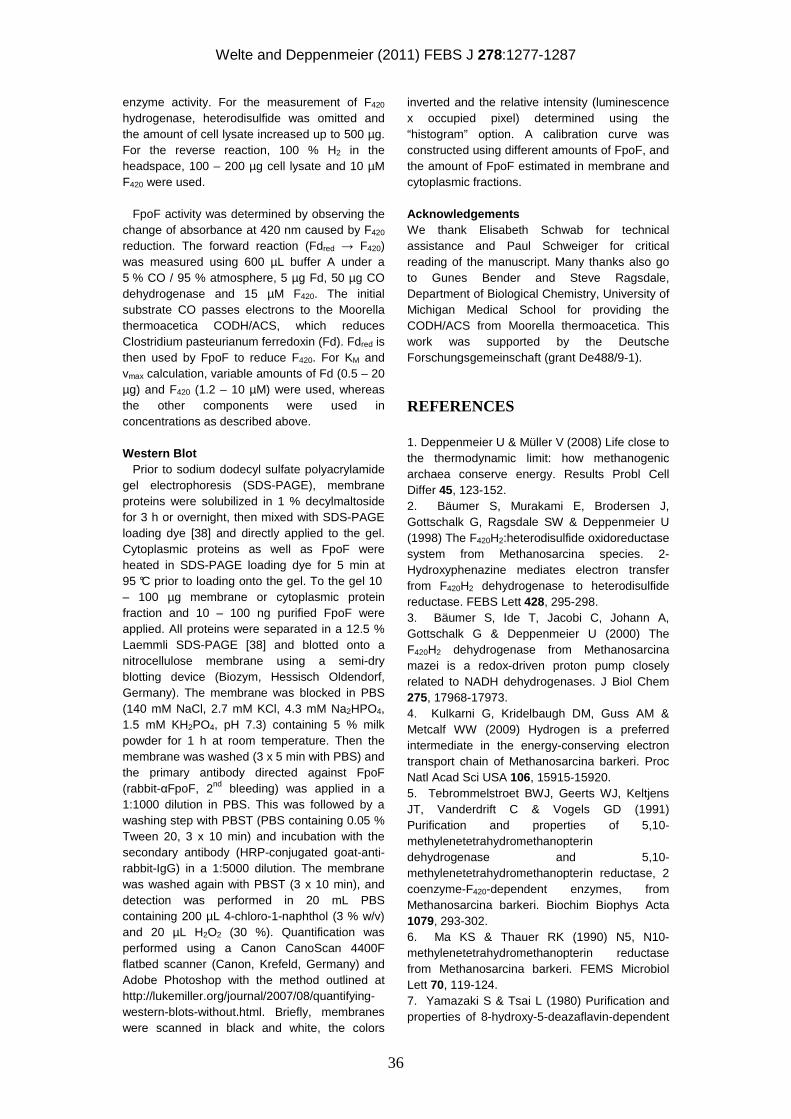
Welte and Deppenmeier (2011) FEBS J 278:1277-1287
36
enzyme activity. For the measurement of F420 hydrogenase, heterodisulfide was omitted and the amount of cell lysate increased up to 500 µg. For the reverse reaction, 100 % H2 in the headspace, 100 – 200 µg cell lysate and 10 µM F420 were used. FpoF activity was determined by observing the change of absorbance at 420 nm caused by F420 reduction. The forward reaction (Fdred → F420) was measured using 600 µL buffer A under a 5 % CO / 95 % atmosphere, 5 µg Fd, 50 µg CO dehydrogenase and 15 µM F420. The initial substrate CO passes electrons to the Moorella thermoacetica CODH/ACS, which reduces Clostridium pasteurianum ferredoxin (Fd). Fdred is then used by FpoF to reduce F420. For KM and vmax calculation, variable amounts of Fd (0.5 – 20 µg) and F420 (1.2 – 10 µM) were used, whereas the other components were used in concentrations as described above. Western Blot Prior to sodium dodecyl sulfate polyacrylamide gel electrophoresis (SDS-PAGE), membrane proteins were solubilized in 1 % decylmaltoside for 3 h or overnight, then mixed with SDS-PAGE loading dye [38] and directly applied to the gel. Cytoplasmic proteins as well as FpoF were heated in SDS-PAGE loading dye for 5 min at 95 °C prior to loading onto the gel. To the gel 10 – 100 µg membrane or cytoplasmic protein fraction and 10 – 100 ng purified FpoF were applied. All proteins were separated in a 12.5 % Laemmli SDS-PAGE [38] and blotted onto a nitrocellulose membrane using a semi-dry blotting device (Biozym, Hessisch Oldendorf, Germany). The membrane was blocked in PBS (140 mM NaCl, 2.7 mM KCl, 4.3 mM Na2HPO4, 1.5 mM KH2PO4, pH 7.3) containing 5 % milk powder for 1 h at room temperature. Then the membrane was washed (3 x 5 min with PBS) and the primary antibody directed against FpoF (rabbit-αFpoF, 2nd bleeding) was applied in a 1:1000 dilution in PBS. This was followed by a washing step with PBST (PBS containing 0.05 % Tween 20, 3 x 10 min) and incubation with the secondary antibody (HRP-conjugated goat-anti-rabbit-IgG) in a 1:5000 dilution. The membrane was washed again with PBST (3 x 10 min), and detection was performed in 20 mL PBS containing 200 µL 4-chloro-1-naphthol (3 % w/v) and 20 µL H2O2 (30 %). Quantification was performed using a Canon CanoScan 4400F flatbed scanner (Canon, Krefeld, Germany) and Adobe Photoshop with the method outlined at http://lukemiller.org/journal/2007/08/quantifying-western-blots-without.html. Briefly, membranes were scanned in black and white, the colors
inverted and the relative intensity (luminescence x occupied pixel) determined using the “histogram” option. A calibration curve was constructed using different amounts of FpoF, and the amount of FpoF estimated in membrane and cytoplasmic fractions.
Acknowledgements We thank Elisabeth Schwab for technical assistance and Paul Schweiger for critical reading of the manuscript. Many thanks also go to Gunes Bender and Steve Ragsdale, Department of Biological Chemistry, University of Michigan Medical School for providing the CODH/ACS from Moorella thermoacetica. This work was supported by the Deutsche Forschungsgemeinschaft (grant De488/9-1).
REFERENCES
1. Deppenmeier U & Müller V (2008) Life close to the thermodynamic limit: how methanogenic archaea conserve energy. Results Probl Cell Differ 45, 123-152. 2. Bäumer S, Murakami E, Brodersen J, Gottschalk G, Ragsdale SW & Deppenmeier U (1998) The F420H2:heterodisulfide oxidoreductase system from Methanosarcina species. 2-Hydroxyphenazine mediates electron transfer from F420H2 dehydrogenase to heterodisulfide reductase. FEBS Lett 428, 295-298. 3. Bäumer S, Ide T, Jacobi C, Johann A, Gottschalk G & Deppenmeier U (2000) The F420H2 dehydrogenase from Methanosarcina mazei is a redox-driven proton pump closely related to NADH dehydrogenases. J Biol Chem 275, 17968-17973. 4. Kulkarni G, Kridelbaugh DM, Guss AM & Metcalf WW (2009) Hydrogen is a preferred intermediate in the energy-conserving electron transport chain of Methanosarcina barkeri. Proc Natl Acad Sci USA 106, 15915-15920. 5. Tebrommelstroet BWJ, Geerts WJ, Keltjens JT, Vanderdrift C & Vogels GD (1991) Purification and properties of 5,10-methylenetetrahydromethanopterin dehydrogenase and 5,10-methylenetetrahydromethanopterin reductase, 2 coenzyme-F420-dependent enzymes, from Methanosarcina barkeri. Biochim Biophys Acta 1079, 293-302. 6. Ma KS & Thauer RK (1990) N5, N10-methylenetetrahydromethanopterin reductase from Methanosarcina barkeri. FEMS Microbiol Lett 70, 119-124. 7. Yamazaki S & Tsai L (1980) Purification and properties of 8-hydroxy-5-deazaflavin-dependent
Welte and Deppenmeier (2011) FEBS J 278:1277-1287
37
NADP+ reductase from Methanococcus vannielii. J Biol Chem 255, 6462-6465. 8. Kunow J, Schwörer B, Stetter KO & Thauer RK (1993) A F420-dependent NADP reductase in the extremely thermophilic sulfate-reducing Archaeoglobus fulgidus. Arch Microbiol 160, 199-205. 9. Berk H & Thauer RK (1998) F420H2 : NADP oxidoreductase from Methanobacterium thermoautotrophicum: identification of the encoding gene via functional overexpression in Escherichia coli. FEBS Lett 438, 124-126. 10. Elias DA, Juck DF, Berry KA & Sparling R (2000) Purification of the NADP+: F420 oxidoreductase of Methanosphaera stadtmanae. Can J Microbiol 46, 998-1003. 11. Welte C, Krätzer C & Deppenmeier U (2010) Involvement of Ech hydrogenase in energy conservation of Methanosarcina mazei. FEBS J 277, 3396-3403. 12. Brüggemann H, Falinski F & Deppenmeier U (2000) Structure of the F420H2:quinone oxidoreductase of Archaeoglobus fulgidus. Identification and overproduction of the F420H2-oxidizing subunit. Eur J Biochem 267, 5810-5814. 13. Muth E, Mörschel E & Klein A (1987) Purification and characterization of an 8-hydroxy-5-deazaflavin-reducing hydrogenase from the archaebacterium Methanococcus voltae. Eur J Biochem 169, 571-577. 14. Brodersen J, Bäumer S, Abken HJ, Gottschalk G & Deppenmeier U (1999) Inhibition of membrane-bound electron transport of the methanogenic archaeon Methanosarcina mazei Gö1 by diphenyleneiodonium. Eur J Biochem 259, 218-224. 15. Alex LA, Reeve JN, Orme-Johnson WH & Walsh CT (1990) Cloning, sequence determination, and expression of the genes encoding the subunits of the Nickel-containing 8-hydroxy-5-deazaflavin reducing hydrogenase from Methanobacterium thermoautotrophicum ∆H. Biochemistry 29, 7237-7244. 16. Fox JA, Livingston DJ, Orme-Johnson WH & Walsh CT (1987) 8-hydroxy-5-deazaflavin-reducing hydrogenase from Methanobacterium thermoautotrophicum .1. Purification and characterization. Biochemistry 26, 4219-4227. 17. Livingston DJ, Fox JA, Orme-Johnson WH & Walsh CT (1987) 8-hydroxy-5-deazaflavin-reducing hydrogenase from Methanobacterium thermoautotrophicum .2. Kinetic and hydrogen-transfer studies. Biochemistry 26, 4228-4237. 18. Jacobson FS, Daniels L, Fox JA, Walsh CT & Orme-Johnson WH (1982) Purification and properties of an 8-hydroxy-5-deazaflavin reducing hydrogenase from Methanobacterium
thermoautotrophicum. J Biol Chem 257, 3385-3388. 19. Hovey R, Lentes S, Ehrenreich A, Salmon K, Saba K, Gottschalk G, Gunsalus RP & Deppenmeier U (2005) DNA microarray analysis of Methanosarcina mazei Gö1 reveals adaptation to different methanogenic substrates. Mol Genet Genomics 273, 225-239. 20. Michel R, Massanz C, Kostka S, Richter M & Fiebig K (1995) Biochemical characterization of the 8-hydroxy-5-deazaflavin-reactive hydrogenase from Methanosarcina barkeri Fusaro. Eur J Biochem 233, 727-735. 21. Guss AM, Mukhopadhyay B, Zhang JK & Metcalf WW (2005) Genetic analysis of mch mutants in two Methanosarcina species demonstrates multiple roles for the methanopterin-dependent C1 oxidation/reduction pathway and differences in H2 metabolism between closely related species. Mol Microbiol 55, 1671-1680. 22. Guss AM, Kulkarni G & Metcalf WW (2009) Differences in hydrogenase gene expression between Methanosarcina acetivorans and Methanosarcina barkeri. J Bacteriol 191, 2826-2833. 23. Rohlin L & Gunsalus RP (2010) Carbon-dependent control of electron transfer and central carbon pathway genes for methane biosynthesis in the archaean Methanosarcina acetivorans strain C2A. BMC Microbiol 10, 62. 24. Zinder SH & Mah RA (1979) Isolation and characterization of a thermophilic strain of Methanosarcina unable to use H2-CO2 for methanogenesis. Appl Environ Microbiol 38, 996-1008. 25. Deppenmeier U (1991) Identifizierung und Charakterisierung membrangebundener protonentranslozierender Redox-Systeme in methanogenen Bakterien. PhD thesis, University of Göttingen. 26. Allen MA, Lauro FM, Williams TJ, Burg D, Siddiqui KS, De Francisci D, Chong KWY, Pilak O, Chew HH, De Maere MZ, Ting L, Katrib M, Ng C, Sowers KR, Galperin MY, Anderson IJ, Ivanova N, Dalin E, Martinez M, Lapidus A, Hauser L, Land M, Thomas T & Cavicchioli R (2009) The genome sequence of the psychrophilic archaeon Methanococcoides burtonii: the role of genome evolution in cold adaptation. ISME J 3, 1012-1035. 27. Franzmann PD, Springer N, Ludwig W, Demacario EC & Rohde M (1992) A methanogenic archaeon from Ace Lake, Antarctica - Methanococcoides burtonii sp. nov. Systematic and Applied Microbiology 15, 573-581. 28. Welte C, Kallnik V, Grapp M, Bender G, Ragsdale S & Deppenmeier U (2010) Function of
Welte and Deppenmeier (2011) FEBS J 278:1277-1287
38
Ech hydrogenase in ferredoxin-dependent, membrane-bound electron transport in Methanosarcina mazei. J Bacteriol 192, 674-678. 29. Metcalf WW, Zhang JK, Apolinario E, Sowers KR & Wolfe RS (1997) A genetic system for archaea of the genus Methanosarcina: Liposome-mediated transformation and construction of shuttle vectors. Proc Natl Acad Sci USA 94, 2626-2631. 30. Ehlers C, Weidenbach K, Veit K, Deppenmeier U, Metcalf WW & Schmitz RA (2005) Development of genetic methods and construction of a chromosomal glnK1 mutant in Methanosarcina mazei strain Gö1. Mol Genet Genomics 273, 290-298. 31. Ragsdale SW, Ljungdahl LG & Der Vartanian DV (1983) Isolation of carbon monoxide dehydrogenase from Acetobacterium woodii and comparison of its properties with those of the Clostridium thermoaceticum enzyme. J Bacteriol 155, 1224-1237. 32. Mortenson LE (1964) Purification and analysis of ferredoxin from Clostridium pasteurianum. Biochim Biophys Acta 81, 71-77. 33. Ellermann J, Hedderich R, Bocher R & Thauer RK (1988) The final step in methane formation. Investigations with highly purified methyl-CoM reductase (component C) from Methanobacterium thermoautotrophicum (strain Marburg). Eur J Biochem 172, 669-677. 34. Abken HJ, Tietze M, Brodersen J, Bäumer S, Beifuss U & Deppenmeier U (1998) Isolation and characterization of methanophenazine and function of phenazines in membrane-bound electron transport of Methanosarcina mazei Gö1. J Bacteriol 180, 2027-2032. 35. Mott JE, Grant RA, Ho YS & Platt T (1985) Maximizing gene expression from plasmid vectors containing the Lambda-Pl promoter - strategies for overproducing transcription termination factor Rho. Proc Natl Acad Sci USA 82, 88-92. 36. Landers JW & Zak B (1958) Determination of serum copper and iron in single small sample. Tech Bull Regist Med Technol 28, 98-100. 37. Beinert H (1983) Semi-micro methods for analysis of labile sulfide and of labile sulphide plus sulfane sulfur in unusually stable iron–sulfur proteins. Anal Biochem 131, 373-378. 38. Laemmli UK (1970) Cleavage of structural proteins during assembly of head of bacteriophage T4. Nature 227, 680-685.
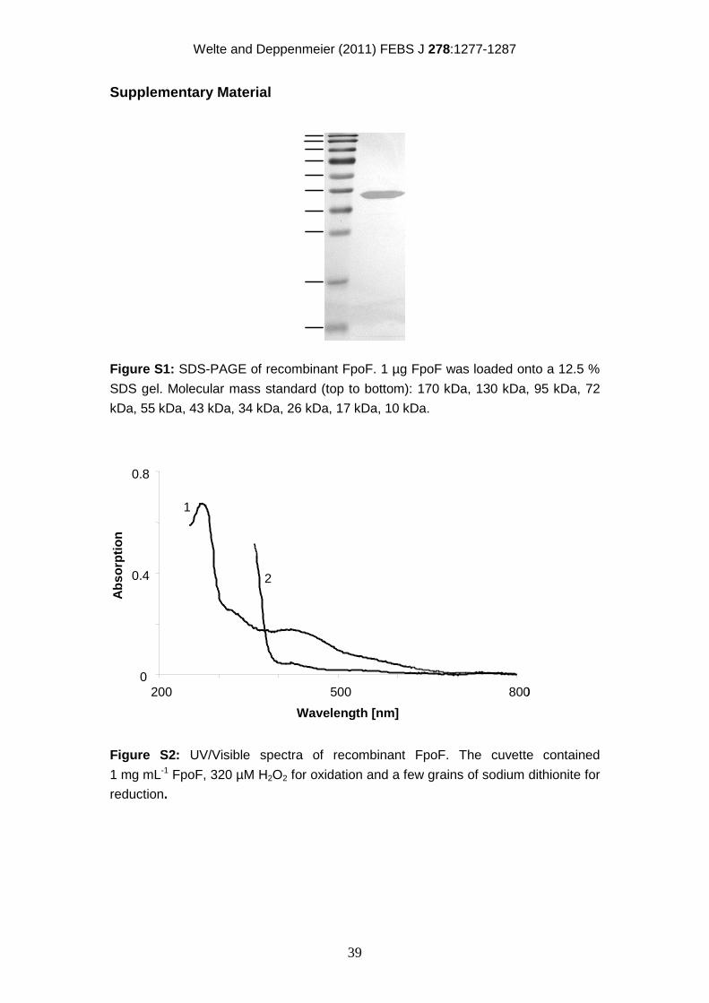
Welte and Deppenmeier (2011) FEBS J 278:1277-1287
39
Supplementary Material
Figure S1: SDS-PAGE of recombinant FpoF. 1 µg FpoF was loaded onto a 12.5 %
SDS gel. Molecular mass standard (top to bottom): 170 kDa, 130 kDa, 95 kDa, 72
kDa, 55 kDa, 43 kDa, 34 kDa, 26 kDa, 17 kDa, 10 kDa.
0
0.4
0.8
200 500 800
Wavelength [nm]
Ab
sorp
tio
n
1
2
0
0.4
0.8
200 500 800
Wavelength [nm]
Ab
sorp
tio
n
1
2
Figure S2: UV/Visible spectra of recombinant FpoF. The cuvette contained
1 mg mL-1 FpoF, 320 µM H2O2 for oxidation and a few grains of sodium dithionite for
reduction.
40
Chapter 5
Membrane-bound electron transport in
Methanosaeta thermophila
Energy conservation in Methanosarcina sp. is well understood, and useful tools
were found for the investigation of all membrane-bound heterodisulfide
oxidoreductase systems. Stoichiometries of translocated H+ or Na+ per 2 e- could be
established for all known oxidoreductase systems of Ms. mazei, and only few
unanswered questions remain. In contrast, there is essentially nothing known about
the energy conserving mechanisms in Methanosaeta sp. although this genus is
unique in its obligate aceticlastic growth. Together with Methanosarcina sp., these
organisms form the major group contributing to biogenic methane emission. At high
acetate concentrations the faster growing Methanosarcina sp. outcompete
Methanosaeta sp., but at low acetate concentrations (< 1 mM, Jetten et al., 1992b)
Methanosaeta sp. prevail. Faster growth, easier cultivation and the long-known
genome sequences of many Methanosarcina sp. has biased the research on energy
conservation towards members of the Methanosarcinaceae although members of
the Methanosaetaceae are of no less importance. The deciphering of the first
Methanosaeta genome in 2007 (Smith and Ingram-Smith, 2007) could have
unravelled the functioning of Methanosaeta energy conservation, but the genome
did not contain genes encoding known electron-input complexes used in the
respiratory systems of other aceticlastic methanogens: the genome neither
contained genes coding for Ech hydrogenase or the Rnf complex. Other ferredoxin-
interacting, membrane-bound enzymes from methanogens are not known. The
investigations presented in this paper focused on two issues: (i) Is a membrane-
bound heterodisulfide oxidoreductase system present in Methanosaeta
thermophila? (ii) What is the electron donor to the Mt. thermophila respiratory chain?
As a result, ferredoxin was the only electron donor that interacted with the
membrane. Reduced ferredoxin was the electron donor to a heterodisulfide
oxidoreductase system and high activities of the ferredoxin: heterodisulfide
oxidoreductase could be observed. The lack of Ech hydrogenase and the Rnf
complex indicates that a novel type of ferredoxin-interacting enzyme is involved.
Welte and Deppenmeier (2011) J Bacteriol 193:2868-2870
41
Membrane-bound electron transport in Methanosaeta thermophila
Cornelia Welte and Uwe Deppenmeier
Institute of Microbiology and Biotechnology, University of Bonn, Meckenheimer Allee 168, 53115 Bonn, Germany
The obligate aceticlastic methanogen Methanosaeta thermophila uses a membrane-bound
ferredoxin: heterodisulfide oxidoreductase system f or energy conservation. We propose that
the system is composed of a truncated form of the F 420H2 dehydrogenase, methanophenazine
and the heterodisulfide reductase. Hence, the elect ron transport chain is distinct from those of
well studied Methanosarcina species.
Biogenic methane production is dominated by Methanoarchaea of the genera Methanosarcina (Ms.) and Methanosaeta (Mt.) that grow on acetate (6). Interestingly, Methanosaeta species can use only acetate as substrate and are therefore obligate aceticlastic methanogens. Members of this genus are of special importance for the productivity of biogas plants, especially for reactor performance and stability at low acetate concentrations. To optimize biomethanation, it is necessary to acquire a comprehensive understanding of the biochemistry of acetate-dependent methanogenesis. Energy conservation in Methanosaeta sp. is not well understood, and even the sequencing of the Mt. thermophila genome (13) did not unravel its mechanism. Comparative genomics indicated that the core methanogenic pathway, the breakdown of acetyl-CoA to methane, is obviously well conserved in Mt. thermophila. It can be concluded that acetate is activated by acetyl-CoA synthetases and the resulting acetyl-CoA serves as substrate for a CO dehydrogenase/acetyl-CoA synthase (CODH/ACS) that oxidizes the carbonyl group to CO2 and reduces ferredoxin. The methyl group is first transferred to tetrahydromethanopterin, and then to coenzyme M (CoM, 2-mercaptoethanesulfonate) by the action of a membrane-bound Na+ translocating methyltransferase. Methyl-CoM is oxidatively coupled to coenzyme B (CoB, N-7-mercaptoheptanoyl-L-threonine phosphate) with the heterodisulfide CoM-S-S-CoB and methane as end products (5, 15). In contrast, the composition of the Mt. thermophila respiratory chain and the mode of energy conservation remained largely unknown. Evidence was only found for the presence of the reduced ferredoxin (Fdred) forming CODH/ACS and the heterodisulfide reductase (1, 13, 14). In Methanosarcina sp. a ferredoxin: heterodisulfide oxidoreductase is used for energy conservation in acetate metabolism. Ms. mazei and Ms. barkeri employ the Ech hydrogenase for H2
production from Fdred and the H2-uptake hydrogenase (Vho) that finally reduces methanophenazine, the electron donor for the heterodisulfide reductase (HdrDE). In Ms. acetivorans, Ech hydrogenase is absent, and instead the Rnf complex is proposed to be responsible for Fdred oxidation (6). Surprisingly, the Mt. thermophila genome does not contain genes coding for either hydrogenases or an Rnf complex (13). If Fdred serves as electron donor for the respiratory chain, the presence of a novel oxidoreductase must be postulated. To investigate the electron transport processes in Mt. thermophila, we isolated cytoplasmic membranes from Mt. thermophila DSM6194 as described (17) with cell disruption by French pressure treatment (1000 psi). Enzyme assays were carried out at 55° C (optimal growth temperature) (3, 17). Benzyl viologen-dependent heterodisulfide reductase activity was high with 878 ± 90 mU mg-1 membrane protein (Table 1) and was comparable to activities found in Methanosarcina sp. (3) (Figure 1). Hence, it is tempting to speculate that a membrane-bound heterodisulfide oxidoreductase system is used for energy conservation in Mt. thermophila, with a so far unidentified enzyme system that channels electrons into the respiratory chain. Genes encoding the F420H2 dehydrogenase (Fpo) were identified in the genome of Mt. thermophila and this protein is therefore a candidate for electron input into the respiratory chain. Fpo is usually involved in methylotrophic methanogenesis and oxidizes F420H2 that is formed in the methanogenic pathway of Methanosarcina sp. The Methanosarcina core enzyme FpoA-O is highly homologous to NADH dehydrogenase I from bacteria and eukarya. However, the reduced cofactor oxidizing subunits from F420H2 dehydrogenases and NADH dehydrogenases are not homologous. The corresponding module of the bacterial and eukaryotic enzymes is made from subunits NuoEFG. In contrast, the oxidation of reduced cofactor F420 is catalyzed by subunit
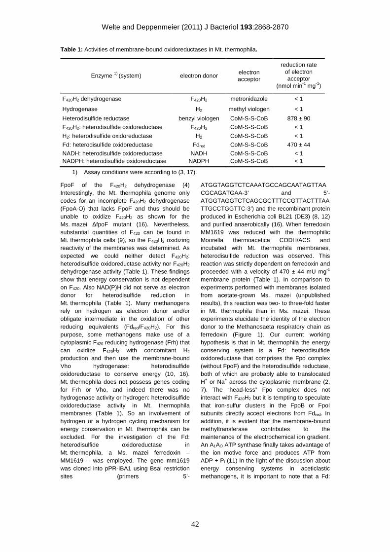
Welte and Deppenmeier (2011) J Bacteriol 193:2868-2870
42
Table 1: Activities of membrane-bound oxidoreductases in Mt. thermophila.
reduction rate of electron acceptor Enzyme 1) (system) electron donor electron
acceptor (nmol min-1 mg-1)
F420H2 dehydrogenase F420H2 metronidazole < 1
Hydrogenase H2 methyl viologen < 1
Heterodisulfide reductase benzyl viologen CoM-S-S-CoB 878 ± 90
F420H2: heterodisulfide oxidoreductase F420H2 CoM-S-S-CoB < 1
H2: heterodisulfide oxidoreductase H2 CoM-S-S-CoB < 1
Fd: heterodisulfide oxidoreductase Fdred CoM-S-S-CoB 470 ± 44
NADH: heterodisulfide oxidoreductase NADH CoM-S-S-CoB < 1 NADPH: heterodisulfide oxidoreductase NADPH CoM-S-S-CoB < 1
1) Assay conditions were according to (3, 17).
FpoF of the F420H2 dehydrogenase (4) Interestingly, the Mt. thermophila genome only codes for an incomplete F420H2 dehydrogenase (FpoA-O) that lacks FpoF and thus should be unable to oxidize F420H2 as shown for the Ms. mazei ∆fpoF mutant (16). Nevertheless, substantial quantities of F420 can be found in Mt. thermophila cells (9), so the F420H2 oxidizing reactivity of the membranes was determined. As expected we could neither detect F420H2: heterodisulfide oxidoreductase activity nor F420H2 dehydrogenase activity (Table 1). These findings show that energy conservation is not dependent on F420. Also NAD(P)H did not serve as electron donor for heterodisulfide reduction in Mt. thermophila (Table 1). Many methanogens rely on hydrogen as electron donor and/or obligate intermediate in the oxidation of other reducing equivalents (Fdred/F420H2). For this purpose, some methanogens make use of a cytoplasmic F420 reducing hydrogenase (Frh) that can oxidize F420H2 with concomitant H2 production and then use the membrane-bound Vho hydrogenase: heterodisulfide oxidoreductase to conserve energy (10, 16). Mt. thermophila does not possess genes coding for Frh or Vho, and indeed there was no hydrogenase activity or hydrogen: heterodisulfide oxidoreductase activity in Mt. thermophila membranes (Table 1). So an involvement of hydrogen or a hydrogen cycling mechanism for energy conservation in Mt. thermophila can be excluded. For the investigation of the Fd: heterodisulfide oxidoreductase in Mt. thermophila, a Ms. mazei ferredoxin – MM1619 – was employed. The gene mm1619 was cloned into pPR-IBA1 using BsaI restriction sites (primers 5’-
ATGGTAGGTCTCAAATGCCAGCAATAGTTAACGCAGATGAA-3’ and 5’-ATGGTAGGTCTCAGCGCTTTCCGTTACTTTAATTGCCTGGTTC-3’) and the recombinant protein produced in Escherichia coli BL21 (DE3) (8, 12) and purified anaerobically (16). When ferredoxin MM1619 was reduced with the thermophilic Moorella thermoacetica CODH/ACS and incubated with Mt. thermophila membranes, heterodisulfide reduction was observed. This reaction was strictly dependent on ferredoxin and proceeded with a velocity of 470 ± 44 mU mg-1 membrane protein (Table 1). In comparison to experiments performed with membranes isolated from acetate-grown Ms. mazei (unpublished results), this reaction was two- to three-fold faster in Mt. thermophila than in Ms. mazei. These experiments elucidate the identity of the electron donor to the Methanosaeta respiratory chain as ferredoxin (Figure 1). Our current working hypothesis is that in Mt. thermophila the energy conserving system is a Fd: heterodisulfide oxidoreductase that comprises the Fpo complex (without FpoF) and the heterodisulfide reductase, both of which are probably able to translocated H+ or Na+ across the cytoplasmic membrane (2, 7). The “head-less” Fpo complex does not interact with F420H2 but it is tempting to speculate that iron-sulfur clusters in the FpoB or FpoI subunits directly accept electrons from Fdred. In addition, it is evident that the membrane-bound methyltransferase contributes to the maintenance of the electrochemical ion gradient. An A1AO ATP synthase finally takes advantage of the ion motive force and produces ATP from ADP + Pi (11) In the light of the discussion about energy conserving systems in aceticlastic methanogens, it is important to note that a Fd:
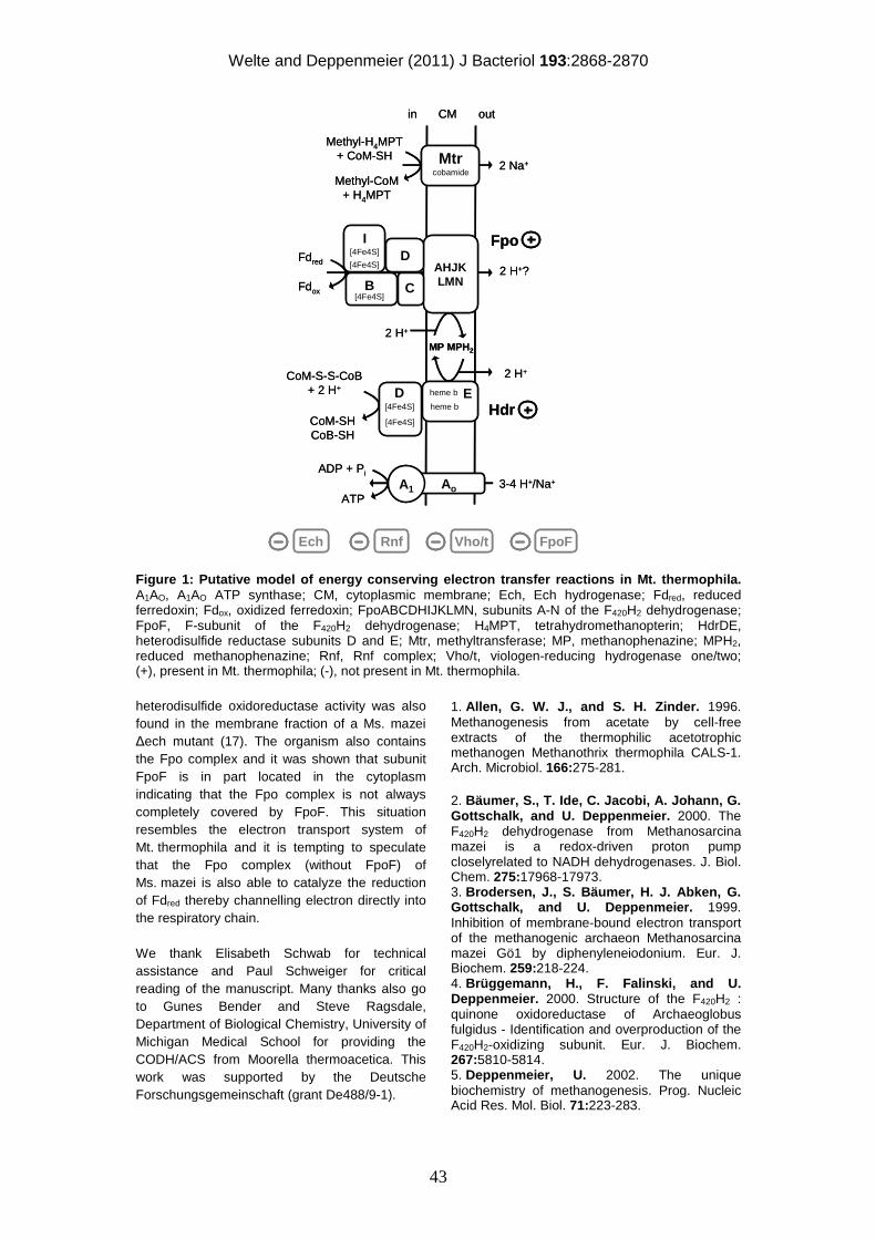
Welte and Deppenmeier (2011) J Bacteriol 193:2868-2870
43
Figure 1: Putative model of energy conserving electr on transfer reactions in Mt. thermophila. A1AO, A1AO ATP synthase; CM, cytoplasmic membrane; Ech, Ech hydrogenase; Fdred, reduced ferredoxin; Fdox, oxidized ferredoxin; FpoABCDHIJKLMN, subunits A-N of the F420H2 dehydrogenase; FpoF, F-subunit of the F420H2 dehydrogenase; H4MPT, tetrahydromethanopterin; HdrDE, heterodisulfide reductase subunits D and E; Mtr, methyltransferase; MP, methanophenazine; MPH2, reduced methanophenazine; Rnf, Rnf complex; Vho/t, viologen-reducing hydrogenase one/two; (+), present in Mt. thermophila; (-), not present in Mt. thermophila. heterodisulfide oxidoreductase activity was also found in the membrane fraction of a Ms. mazei ∆ech mutant (17). The organism also contains the Fpo complex and it was shown that subunit FpoF is in part located in the cytoplasm indicating that the Fpo complex is not always completely covered by FpoF. This situation resembles the electron transport system of Mt. thermophila and it is tempting to speculate that the Fpo complex (without FpoF) of Ms. mazei is also able to catalyze the reduction of Fdred thereby channelling electron directly into the respiratory chain. We thank Elisabeth Schwab for technical assistance and Paul Schweiger for critical reading of the manuscript. Many thanks also go to Gunes Bender and Steve Ragsdale, Department of Biological Chemistry, University of Michigan Medical School for providing the CODH/ACS from Moorella thermoacetica. This work was supported by the Deutsche Forschungsgemeinschaft (grant De488/9-1).
1. Allen, G. W. J., and S. H. Zinder. 1996. Methanogenesis from acetate by cell-free extracts of the thermophilic acetotrophic methanogen Methanothrix thermophila CALS-1. Arch. Microbiol. 166:275-281. 2. Bäumer, S., T. Ide, C. Jacobi, A. Johann, G. Gottschalk, and U. Deppenmeier. 2000. The F420H2 dehydrogenase from Methanosarcina mazei is a redox-driven proton pump closelyrelated to NADH dehydrogenases. J. Biol. Chem. 275:17968-17973. 3. Brodersen, J., S. Bäumer, H. J. Abken, G. Gottschalk, and U. Deppenmeier. 1999. Inhibition of membrane-bound electron transport of the methanogenic archaeon Methanosarcina mazei Gö1 by diphenyleneiodonium. Eur. J. Biochem. 259:218-224. 4. Brüggemann, H., F. Falinski, and U. Deppenmeier. 2000. Structure of the F420H2 : quinone oxidoreductase of Archaeoglobus fulgidus - Identification and overproduction of the F420H2-oxidizing subunit. Eur. J. Biochem. 267:5810-5814. 5. Deppenmeier, U. 2002. The unique biochemistry of methanogenesis. Prog. Nucleic Acid Res. Mol. Biol. 71:223-283.
Ech Rnf FpoFVho/t
2 H+?
MP MPH2
CoM-S-S-CoB+ 2 H+
CoM-SHCoB-SH
2 H+
Fdred
Fdox
AHJKLMN
in CM out
C
Fpo
Hdrheme b
D
B
I
[4Fe4S]
[4Fe4S]
[4Fe4S]
heme b[4Fe4S]
[4Fe4S]
ED
A1 3-4 H+/Na+
ADP + Pi
ATPAo
2 Na+Mtrcobamide
Methyl-H4MPT+ CoM-SH
Methyl-CoM+ H4MPT
2 H+
EchEch RnfRnf FpoFFpoFVho/tVho/t
2 H+?
MP MPH2
CoM-S-S-CoB+ 2 H+
CoM-SHCoB-SH
2 H+
Fdred
Fdox
AHJKLMN
in CM out
C
Fpo
Hdrheme b
D
B
I
[4Fe4S]
[4Fe4S]
[4Fe4S]
heme b[4Fe4S]
[4Fe4S]
ED
A1 3-4 H+/Na+
ADP + Pi
ATPAo
2 Na+Mtrcobamide
Methyl-H4MPT+ CoM-SH
Methyl-CoM+ H4MPT
2 H+
Welte and Deppenmeier (2011) J Bacteriol 193:2868-2870
44
6. Ferry, J. G., and D. J. Lessner. 2008. Methanogenesis in marine sediments. Ann. N. Y. Acad. Sci. 1125:147-157. 7. Ide, T., S. Bäumer, and U. Deppenmeier. 1999. Energy conservation by the H2:heterodisulfide oxidoreductase from Methanosarcina mazei Gö1: identification of two proton-translocating segments. J. Bacteriol. 181:4076-4080. 8. Jeoung, J. H., and H. Dobbek. 2007. Carbon dioxide activation at the Ni,Fe-cluster of anaerobic carbon monoxide dehydrogenase. Science 318:1461-1464. 9. Kamagata, Y., H. Kawasaki, H. Oyaizu, K. Nakamura, E. Mikami, G. Endo, Y. Koga, and K. Yamasato. 1992. Characterization of 3 thermophilic strains of Methanothrix (Methanosaeta) thermophila sp. nov. and rejection of Methanothrix (Methanosaeta) thermoacetophila. Int. J. Syst. Evol. Microbiol. 42:463-468. 10. Kulkarni, G., D. M. Kridelbaugh, A. M. Guss, and W. W. Metcalf. 2009. Hydrogen is a preferred intermediate in the energy-conserving electron transport chain of Methanosarcina barkeri. Proc. Natl. Acad. Sci. USA 106:15915-15920. 11. Lewalter, K., and V. Müller. 2006. Bioenergetics of archaea: Ancient energy conserving mechanisms developed in the early
history of life. Biochim. Biophys. Acta 1757:437-445. 12. Nakamura, M., K. Saeki, and Y. Takahashi. 1999. Hyperproduction of recombinant ferredoxins in Escherichia coli by coexpression of the ORF1-ORF2-iscS-iscU-iscA-hscB-hscA-fdx-ORF3 gene cluster. J. Biochem. 126:10-18. 13. Smith, K. S., and C. Ingram-Smith. 2007. Methanosaeta, the forgotten methanogen? Trends Microbiol. 15:150-155. 14. Terlesky, K. C., and J. G. Ferry. 1988. Ferredoxin requirement for electron transport from the carbon monoxide dehydrogenase complex to a membrane-bound hydrogenase in acetate-grown Methanosarcina thermophila. J. Biol. Chem. 263:4075-9. 15. Thauer, R. K., A. K. Kaster, H. Seedorf, W. Buckel, and R. Hedderich. 2008. Methanogenic archaea: ecologically relevant differences in energy conservation. Nat. Rev. Microbiol. 6:579-591. 16. Welte, C., and U. Deppenmeier. 2011. Re-evaluation of the function of the F420 dehydrogenase in electron transport in Methanosarcina mazei. FEBS J, in press. 17. Welte, C., V. Kallnik, M. Grapp, G. Bender, S. Ragsdale, and U. Deppenmeier. 2010. Function of Ech hydrogenase in ferredoxin-dependent, membrane-bound electron transport in Methanosarcina mazei. J. Bacteriol. 192:674-678.
45
Chapter 6
Proton translocation in methanogens
Methanogenic bioenergetics has been investigated for over 20 years now. However,
a comprehensive manual for the investigation of electron transport processes and
the measurement of proton translocation has not been published. This publication
compiles and updates protocols for the measurement of enzymatic activities
involved in methanogenic energy conservation and facilitates comparison and
accomplishments of the respective experiments. Furthermore, many cofactors
needed in this field of research are not commercially available and have to be
synthesized de novo or from commercial precursors. For some of the cofactors
fractional descriptions for the synthesis existed (e.g. CoM-S-S-CoB) whereas for
others (e.g. F420) a comprehensive explanation of the isolation or synthesis was
missing.
In the course of this work the protocols that are described below were refined and in
part newly established for the investigation of the ferredoxin: heterodisulfide
oxidoreductase of Ms. mazei. The previous protocols do not allow continuous
measurement of the electron flow from reduced ferredoxin to the heterodisulfide.
Furthermore, the analysis of proton translocation by pH measurement as performed
for other oxidoreductase systems in Ms. mazei was not applicable. The results
presented in the previous chapters are based on the methods and protocols
described below. These newly developed approachs combine different test systems
and allow dependable conclusions about ion translocation of the ferredoxin:
heterodisulfide oxidoreductase system of Ms. mazei and other methanogenic
archaea.
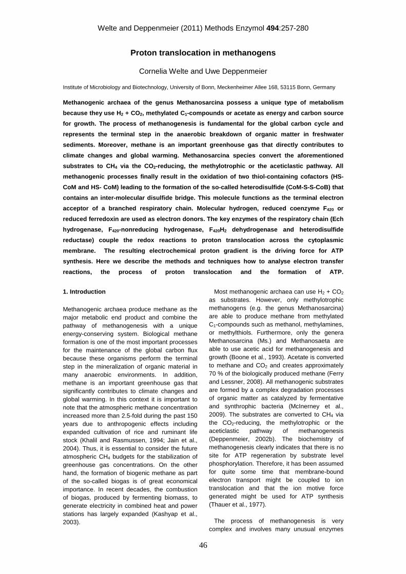
Welte and Deppenmeier (2011) Methods Enzymol 494:257-280
46
Proton translocation in methanogens
Cornelia Welte and Uwe Deppenmeier Institute of Microbiology and Biotechnology, University of Bonn, Meckenheimer Allee 168, 53115 Bonn, Germany
Methanogenic archaea of the genus Methanosarcina possess a unique type of metabolism
because they use H2 + CO2, methylated C1-compounds or acetate as energy and carbon source
for growth. The process of methanogenesis is fundamental for the global carbon cycle and
represents the terminal step in the anaerobic breakdown of organic matter in freshwater
sediments. Moreover, methane is an important greenhouse gas that directly contributes to
climate changes and global warming. Methanosarcina species convert the aforementioned
substrates to CH4 via the CO2-reducing, the methylotrophic or the aceticlastic pathway. All
methanogenic processes finally result in the oxidation of two thiol-containing cofactors (HS-
CoM and HS- CoM) leading to the formation of the so-called heterodisulfide (CoM-S-S-CoB) that
contains an inter-molecular disulfide bridge. This molecule functions as the terminal electron
acceptor of a branched respiratory chain. Molecular hydrogen, reduced coenzyme F420 or
reduced ferredoxin are used as electron donors. The key enzymes of the respiratory chain (Ech
hydrogenase, F420-nonreducing hydrogenase, F420H2 dehydrogenase and heterodisulfide
reductase) couple the redox reactions to proton translocation across the cytoplasmic
membrane. The resulting electrochemical proton gradient is the driving force for ATP
synthesis. Here we describe the methods and techniques how to analyse electron transfer
reactions, the process of proton translocation and the formation of ATP.
1. Introduction Methanogenic archaea produce methane as the major metabolic end product and combine the pathway of methanogenesis with a unique energy-conserving system. Biological methane formation is one of the most important processes for the maintenance of the global carbon flux because these organisms perform the terminal step in the mineralization of organic material in many anaerobic environments. In addition, methane is an important greenhouse gas that significantly contributes to climate changes and global warming. In this context it is important to note that the atmospheric methane concentration increased more than 2.5-fold during the past 150 years due to anthropogenic effects including expanded cultivation of rice and ruminant life stock (Khalil and Rasmussen, 1994; Jain et al., 2004). Thus, it is essential to consider the future atmospheric CH4 budgets for the stabilization of greenhouse gas concentrations. On the other hand, the formation of biogenic methane as part of the so-called biogas is of great economical importance. In recent decades, the combustion of biogas, produced by fermenting biomass, to generate electricity in combined heat and power stations has largely expanded (Kashyap et al., 2003).
Most methanogenic archaea can use H2 + CO2 as substrates. However, only methylotrophic methanogens (e.g. the genus Methanosarcina) are able to produce methane from methylated C1-compounds such as methanol, methylamines, or methylthiols. Furthermore, only the genera Methanosarcina (Ms.) and Methanosaeta are able to use acetic acid for methanogenesis and growth (Boone et al., 1993). Acetate is converted to methane and CO2 and creates approximately 70 % of the biologically produced methane (Ferry and Lessner, 2008). All methanogenic substrates are formed by a complex degradation processes of organic matter as catalyzed by fermentative and synthrophic bacteria (McInerney et al., 2009). The substrates are converted to CH4 via the CO2-reducing, the methylotrophic or the aceticlastic pathway of methanogenesis (Deppenmeier, 2002b). The biochemistry of methanogenesis clearly indicates that there is no site for ATP regeneration by substrate level phosphorylation. Therefore, it has been assumed for quite some time that membrane-bound electron transport might be coupled to ion translocation and that the ion motive force generated might be used for ATP synthesis (Thauer et al., 1977). The process of methanogenesis is very complex and involves many unusual enzymes
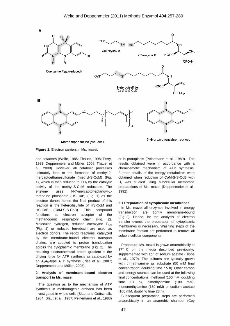
Welte and Deppenmeier (2011) Methods Enzymol 494:257-280
47
Figure 1: Electron carriers in Ms. mazei.
and cofactors (Wolfe, 1985; Thauer, 1998; Ferry, 1999; Deppenmeier and Müller, 2008; Thauer et al., 2008). However, all catabolic processes ultimately lead to the formation of methyl-2-mercaptoethanesulfonate (methyl-S-CoM) (Fig. 1), which is then reduced to CH4 by the catalytic activity of the methyl-S-CoM reductase. The enzyme uses N-7-mercaptoheptanoyl-L-threonine phosphate (HS-CoB) (Fig. 1) as the electron donor; hence the final product of this reaction is the heterodisulfide of HS-CoM and HS-CoB (CoM-S-S-CoB). This compound functions as electron acceptor of the methanogenic respiratory chain (Fig. 2). Molecular hydrogen, reduced coenzyme F420 (Fig. 1) or reduced ferredoxin are used as electron donors. The redox reactions, catalyzed by the membrane-bound electron transport chains, are coupled to proton translocation across the cytoplasmic membrane (Fig. 2). The resulting electrochemical proton gradient is the driving force for ATP synthesis as catalyzed by an A1AO-type ATP synthase (Pisa et al., 2007; Deppenmeier and Müller, 2008).
2. Analysis of membrane-bound electron transport in Ms. mazei
The question as to the mechanism of ATP synthesis in methanogenic archaea has been investigated in whole cells (Blaut and Gottschalk, 1984; Blaut et al., 1987; Peinemann et al., 1988)
or in protoplasts (Peinemann et al., 1989). The results obtained were in accordance with a chemiosmotic mechanism of ATP synthesis. Further details of the energy metabolism were obtained when reduction of CoM-S-S-CoB with H2 was studied using subcellular membrane preparations of Ms. mazei (Deppenmeier et al., 1992).
2.1 Preparation of cytoplasmic membranes In Ms. mazei all enzymes involved in energy transduction are tightly membrane-bound (Fig. 2). Hence, for the analysis of electron transfer events the preparation of cytoplasmic membranes is necessary. Washing steps of the membrane fraction are performed to remove all soluble cellular components. Procedure: Ms. mazei is grown anaerobically at 37° C on the media described previously, supplemented with 1g/l of sodium acetate (Hippe et al., 1979). The cultures are typically grown with trimethyamine as substrate (50 mM final concentration; doubling time 7.5 h). Other carbon and energy sources can be used at the following final concentrations: methanol (150 mM, doubling time 13 h), dimethylamine (100 mM), monomethylamine (150 mM) or sodium acetate (100 mM, doubling time 28 h). Subsequent preparation steps are performed anaerobically in an anaerobic chamber (Coy
HN
N N
O
O
R
OH
HH
H
Coenzyme F420 (reduced)
A
HN
N N
O
O
R
OH
HH
H
Coenzyme F420 (reduced)
A
BB
Welte and Deppenmeier (2011) Methods Enzymol 494:257-280
48
Figure 2: Membrane-bound electron transfer system of Ms. mazei. Ech, Echhydrogenase; Vho, F420-nonreducing hydrogenase; Fpo, F420H2 dehydrogenase; Hdr, heterodisulfide reductase; MP, methanophenazine.
laboratory products, US) under a 97 % N2 / 3 % H2 atmosphere. Exponentially grown cells are filled in gas-tight centrifugation bottles (500 ml) and harvested by centrifugation (3000 x g, 30 min). The cell pellet is resuspended in a small volume 40 mM potassium phosphate (K-phosphate) buffer, pH 7.0, 5 mM dithioerythritol and 1 µg mL-1 resazurin. In the course of resuspension the cells lyse because the protein cell wall is not stabile in this buffer. After incubated with DNase I for 30 min at 4° C, the cell lysate is ultracentrifuged (90 min, 150,000 x g) and the supernatant is carefully removed and stored at –20o C (e.g. for a subsequent isolation of coenzyme F420). The membrane pellet is homogenized in 20 mM K-phosphate buffer (pH 7.0) containing 20 mM MgSO4, 500 mM sucrose, 5 mM dithioerythritol and 1 µg mL-1 resazurin. The ultracentrifugation step is repeated, the supernatant discarded and the membrane pellet again homogenized in the previously mentioned buffer (300 µL per L culture). The membrane fraction is finally checked for remaining contamination of soluble compounds of the cells by measuring the activity of the acetyl-CoA synthetase, which is only present in the cytoplasm of Ms. mazei. To assay for acetyl-CoA synthetase activity an anaerobic rubber-stoppered glass cuvette containing 700 µL K-phosphate buffer (40 mM, pH 7.0, 5 mM dithioerythritol, 1 µg mL-1 resazurin) is flushed with 5 % CO / 95 % N2. Methyl viologen is added to a final concentration of 5 mM (ε604= 13.6 cm-1 x mM-1). Upon addition of the prepared washed membrane fraction, no activity should occur if the membrane fraction is indeed cytoplasm-free.
2.2 Analysis of the H2-dependent electron transport chain The source of reducing equivalents necessary for the reduction of CoM-S-S-CoB depends on the growth substrate. If molecular hydrogen is present, a membrane-bound F420-nonreducing hydrogenase channels electrons via b-type cytochromes to the heterodisulfide reductase, which reduces the terminal electron donor (Fig. 2) (Deppenmeier et al., 1992). This electron transport system was referred to as H2:heterodisulfide oxidoreductase. Later on it was found that the electron transport between the enzymes is mediated by the novel redox active cofactor methanophenazine (Fig. 1, 2) (Abken et al., 1998; Beifuss and Tietze, 2000; Beifuss and Tietze, 2005). With the identification of methanophenazine as an electron carrier in the membrane of Ms. mazei it became evident that the key enzymes of the membrane-bound electron transfer systems were able to interact with 2-hydroxy-phenazine which is a water-soluble homologue of methanophenazine (Bäumer et al., 2000). To analyze the key enzymes of this system the following procedure can be followed: 2.2.1. F420-nonreducing hydrogenase Several types of nickel-iron hydrogenases were described in methanogenic archaea (Vignais et al., 2001). Among them is the F420-nonreducing hydrogenase, which is composed of a small electron transfer subunit and a large catalytic subunit harboring a nickel-iron center. Both subunits are located at the outer leaflet of the cytoplasmic membrane. It has been shown that the metals in the bimetallic reaction center are directly involved in H2 cleavage to 2 e- and 2 H+. The small subunit contains three Fe/S clusters and is responsible for electron transport from the
2H+
out
in
2 Fdred
2 Fdox
H2
2 H+
H+
CoM-S-S-CoB+ 2H+
HS-CoMHS-CoB
MP
2H+
H2
2H+
Ech Vho Hdr Fpo
F420H2
F420
MP CM2e- 2e-
2H+
out
in
2 Fdred
2 Fdox
H2
2 H+
H+
CoM-S-S-CoB+ 2H+
HS-CoMHS-CoB
MP
2H+
H2
2H+
Ech Vho Hdr Fpo
F420H2
F420
MP CM2e- 2e-
Welte and Deppenmeier (2011) Methods Enzymol 494:257-280
49
catalytic center to the third subunit, which is a b-type cytochrome. This subunit forms the membrane anchor and transfers electrons to methanophenazine (Deppenmeier, 2002a). Procedure: The membrane-bound hydrogenase is assayed in 1.5-ml glass cuvettes gassed with H2 and closed with rubber stoppers. After gassing for 5 min the cuvette is filled with 1 ml anaerobic 40 mM K-phosphate buffer, pH 7.0, containing 5 mM dithiothreitol and 1 µg mL-1 resazurin, and 10-15 µg of the membrane fraction is added. The reaction is started by addition of 10 mM methyl viologen (ε604= 13.6 cm-1 x mM-1). The H2-dependent 2-hydroxyphenazine reduction is followed photometrically at 425 nm (ε = 4.5 mM-1 x cm-1) by adding 4 µl of 2-hydroxy-phenazine from a 20 mM stock solution in ethanol. The activity should be between 1.5-2 U / mg membrane protein. 2.2.2. Heterodisulfide reductase The heterodisulfide reductase catalyzes the reduction of CoM-S-S-CoB, which is the final step in the anaerobic respiratory chain of Ms. mazei and other Methanosarcina strains (Heiden et al., 1994; Künkel et al., 1997; Hedderich et al., 2005). In vivo electrons for CoM-S-S-CoB reduction derive from the reduced form of methanophenazine (Fig. 1, 2). In vitro the enzyme accepts electrons from the artificial electron donors benzyl viologen and 2-hydroxyphenazine (Fig. 2). Procedure: The benzyl viologen-dependent heterodisulfide reductase activity is determined at room temperature in 1 mL 40 mM K-phosphate buffer, pH 7.0, preflushed with N2. After addition of 2 µL 1 M benzyl viologen, 3 µL 50 mM Na-dithionite and 1-5 µL of the membrane preparation, the reaction is started by addition of CoB-S-S-CoM to a final concentration of 180 µM and followed at 575 nm (εbenzyl viologen = 8.9 mM-1 x cm-1). Typical activities range between 1-2 U / mg membrane protein.
2-hydroxyphenazine (0.25 mM final concentration) is reduced in 25 mM MOPS buffer (pH 7.0) containing 2 mM dithioerythritol, 1 µg mL-1 resazurin containing platinum(VI)-oxide (2 mg/ 40 mL) under a hydrogen atmosphere overnight. After reduction is completed the catalyst is removed by centrifugation in an anaerobic chamber and the resulting solution is kept under nitrogen. For the determination of membrane-bound heterodisulfide reductase activity, 25 mM MOPS buffer (pH 7.0) containing 2 mM dithioerythritol, 1 µg mL-1 resazurin and the reduced 2-hydroxy-phenazine solution are mixed
in a 1:1 ratio (final volume 1 ml). After washed membranes are added, the reaction is started by addition of CoB-S-S-CoM to a final concentration of 180 µM and followed at 425 nm (ε2-hydroxyphenazine = 4.5 mM-1 x cm-1). Typically, the activity is in the range of 2 U / mg membrane protein. 2.2.3. H2:heterodisulfide oxidoreductase (coupled reaction of F420-nonreducing hydrogenase and heterodisulfide reductase) The entire electron transport within the H2:heterodisulfide oxidoreductase system (Fig. 2) cannot be followed by photometric methods. However, the end product of the reaction, the thiols HS-CoM and HS-CoB (Fig. 2), can be determined using the so-called Ellman’s test in which 5,5’-dithiobis(2-nitrobenzoic acid) (DTNB, also known as Ellman’s reagent) reacts with mercaptans, like HS-CoM and HS-CoB, under alkaline conditions (Ellman, 1958; Zahler and Cleland, 1968). In the course of the reaction the yellow p-nitrobenzenethiol anion is released, which has a molar extinction coefficient of 13.6 mM-1 x cm-1 at 412 nm. Procedure: 1. Rubber-stoppered glass vials are flushed with N2 and 250 µL K-phosphate buffer (40 mM, pH 7.0, 1 µg mL-1 resazurin, flushed with N2) is added. The solution is reduced until it just becomes colorless (using e.g. titanium(III)citrate). Approximately 150 µg washed Ms. mazei membranes are added, and the whole vial is flushed with 100 % H2. 2. The reaction is started by addition of 400 µM heterodisulfide and the first sample is directly taken: a 20 µL sample of the reaction mixture is mixed with 950 µL Tris-HCl (150 mM, pH 8.1) and 100 µL DTNB (5 mM in 50 mM Na-acetate, pH 5.0) and the absorption at 412 nm is immediately measured. 3. Samples are continuously retrieved until the heterodisulfide is completely reduced (∆A ~ 0.2). Typically about 300 nmol thiols are produced per min per mg membrane protein. Note: A buffer without mercaptan-containing reducing agent is needed for the assay, thus the buffer must not be reduced with dithioerythritol or dithiothreitol. Instead, titanium(III)citrate (Zehnder and Wuhrmann, 1976) or other mercaptan-free reducing agent can be used. The titanium(III)citrate solution is prepared as follows: 15 mmol potassium citrate is solvated in 48.75 mL H2O and gassed with N2 for 10 min. Carefully, 9.38 mL of a 12 % Ti(III)chloride solution (in 12 % HCl) are added under nitrogen
Welte and Deppenmeier (2011) Methods Enzymol 494:257-280
50
gassing. The pH is adjusted to 7 with a saturated K2CO3 solution. 2.2.4. Preparation of the heterodisulfide (CoM-S-S-CoB) The heterodisulfide CoM-S-S-CoB is not commercially available and needs to be synthesized by condensation of HS-CoB and HS-CoM. HS-CoB (N-7-mercaptoheptanoyl-L-threonine phosphate) has to be synthesized de novo (Ellermann et al., 1988), whereas HS-CoM (2-mercaptoethanesulfonate) is available from various suppliers. A. Synthesis of N-hydroxysuccinimidester according to (Noll et al., 1987) 1. In a 500 mL Erlenmeyer flask 200 mL pure ethanol, 17.8 g 7-bromo-heptanoic acid (85.3 mmol) and 32.4 g thiourea (426.5 mmol) are added. The mixture is refluxed at 78° C for 16 h. 2. The solution is cooled to room temperature and alkalized with 50 mL NaOH (60 % w/v). The mixture is again refluxed at 78° C for 16 h. After cooling to room temperature, the solution is acidified with HCl to a pH < 1. 3. The solution is extracted three times with 50 mL chloroform (total 150 mL). The chloroform phases are combined and extracted with 1 M aqueous Na2CO3. The aqueous phase is acidified with 50 mL HCl (37 %) and extracted with chloroform (2 x 100 mL). An aqueous solution of iodine (10 % w/v)/potassium iodide (20 % w/v) is added until a light brown colour appears. 4. The aqueous phase is removed, and the chloroform phase is washed with water three times. Then the solution is dried with anhydrous MgSO4 and concentrated under reduced pressure. 5. The resulting reddish-brown substance is solvated in benzene/pentane (1:1, v/v) under gentle warming, and the substance is allowed to crystallize at 4° C. The resulting crystals are filtered and re-crystallized using the same solvents. Light yellow dithiodiheptanoic acid crystals form. The yield should be in the range of 4-5 g. 6. The disulfide product is activated by synthesis of its N-hydroxysuccinimide ester using dicyclohexylcarbodiimide. 2 g dithiodiheptanoic acid, 1.5 g N-hydroxysuccinimide and 2.6 g dicyclohexylcarbodiimide (DCCD) are solvated in 60 mL anhydrous 1,4-dioxane and incubated for 20 h at room temperature under stirring. The suspension is filtered, the solid matter (dicyclohexylurea) is discarded and the filtrate is dried under vacuum to give a white solid. The white solid is recrystallized twice using 2-
propanol to yield approximately 2 g N-hydroxysuccinimidester. B. Synthesis of N-7-mercaptoheptanoyl-L-threonine phosphate (HS-CoB) according to (Kobelt et al., 1987) 1. 546 mg L-threonine phosphate (2.74 mmol) are solvated in 5 mL H2O including 760 µL triethylamine. The mixture is stirred and 672 mg (1.30 mmol) N-hydroxysuccinimidester is solvated in 40 mL tetrahydrofuran and 14 mL acetonitrile are added. The mixture is stirred for 36 h under argon and then concentrated using a rotary evaporator. The volume is brought to approximately 40 mL with H2O. The pH is adjusted to 6.7 with 1 M NaOH or HCl and the solution gassed with N2. The disulfide compounds are reduced by the addition of NaBH4. The pH is titrated to pH 0 with 1 M HCl. 2. For a hydrophobic interaction chromatography, an XAD-2 column (2.5 cm x 8 cm) is prepared. The column is washed with water and equilibrated with 1 M HCl before the reaction mixture is applied. The column is then washed with 100 mL HCl (1 M) and 140 mL H2O. 3. N-7-mercaptoheptanoyl-L-threonine phosphate (HS-CoB) is eluted using a linear gradient of 0-50% methanol/H2O (100 ml). The thiol-containing elution fractions are pooled and concentrated under vacuum. To analyze the thiol content, the following test is performed: Mix 950 µL Tris-HCl (150 mM pH 8.1), 50 – 100 µL sample and a few grains NaBH4. Let the mixture stand open for 10 min, add 100 µL acetone and 100 µL DTNB solution (5 mM in 50 mM Na-acetate pH 5.0), measure at 412 nm against a control without sample (Ellmann, 1957; Zahler and Cleland 1967). C. Synthesis of the heterodisulfide of N-7-mercaptoheptanoyl-L-threonine phosphate (HS-CoB) and 2-mercaptoethanesulfonate (HS-CoM) according to (Ellermann et al., 1988) 1. The pH of the N-7-mercaptoheptanoyl-L-threonine phosphate solution (coenzyme B solution) is adjusted to 6.7 with 1 M NaOH/HCl and reduced with a few grains NaBH4 under a N2 atmosphere. The approximate content of HS-CoB is calculated using the thiol test as described above (MW (HS-CoB) = 395 g mol-1) and a 5-fold access of HS-CoM is added. The pH is adjusted to 9 (1 M NaOH) and the reaction mixture is gassed with air for 16 h at room temperature to allow the formation of the mixed disulfide. 2. After adjustment to pH 0 (1 M HCl) the preparation is applied to a XAD-2 column, equilibrated as described above. The column is washed with 150 mL 1M HCl, 250 mL H2O
Welte and Deppenmeier (2011) Methods Enzymol 494:257-280
51
(elution of HS-CoM and CoM-S-S-CoM). The heterodisulfide CoM-S-S-CoB is eluted with 250 mL 20 % methanol, and subsequently concentrated under vacuum, frozen and lyophilized. 3. The content of biologically active heterodisulfide is determined with a biological test: 700 µL K-phosphate buffer (pH 7.0, 5 mM dithioerythritol, 1 µg mL-1 resazurin), 5 mM methyl viologen (ε604 = 13.6 mM-1 x cm-1) and 50 – 100 µg Ms. mazei cytoplasmic membrane preparation (see above) are mixed in an anaerobic rubber-stoppered cuvette. The reaction mixture is titrated with Na-dithionite till an absorption of about 2 at 604 nm is reached. The test is started with a small amount of the synthesized heterodisulfide (solvated in H2O). From the change of absorbance at 604 nm the approximate content can be calculated. The typical yield is 15-50 µmol heterodisulfide. 2.2.5. Preparation of 2-hydroxyphenazine according to (Ott, 1959) It was found that the condensation of p-benzoquinone with o-phenylenediamine in ethanolic solution leads directly to 2-hydroxyphenazine. 1. 500 mg (4.6 mmol, 1.0 equiv.) of 1,4-benzoquinone is solvated in 5 ml of dry ethanol by slight warming. 2. Cool to room temperature, add 500 mg (4.6 mmol, 1.0 equiv.) of o-phenylenediamine and incubate for 1 h at ambient temperature. 3. Heat to 75° C and incubate for another 30 min. 4. Cool to room temperature and add 1 mL of H2O; incubate for 8 h at 4° C. 5. Filter the cooled solution and solvate the brownish-black crystals in 0.5 M NaOH. 6. Filter again and acidify the filtrate with concentrated acetic acid. A brownish compound will precipitate. 7. Repeat the filtration and dry the precipitation under vacuum. 8. Purify the compound by silica gel chromatography (silica gel 60, mesh 0.05-0.20 mm, Merck) with a linear gradient of the solvent system diethylether/ petroleum ether (3:1, v/v)/diethylether. 2-hydroxyphenazine is a slight yellow solid. The yield is about 10%. The natural electron carrier methanophenazine can also be chemically synthesized (Beifuss and Tietze 2005; Beifuss et al. 2000) It is a multi-step chemical synthesis and is not described here.
2.3. Analysis of the F420H2-dependent electron transport chain During methanogenesis from methylated C1-compounds in the absence of H2, one quarter of the methyl groups is oxidized to CO2 and reducing equivalents are in part transferred to the central electron carrier F420. In the reductive branch of the pathway, three out of four methyl groups are transferred to coenzyme M. Methyl-S-CoM is reductively cleaved by methyl-S-CoM reductase, forming methane and CoB-S-S-CoM (Deppenmeier, 2002a). Thus, F420H2 and CoB-S-S-CoM are generated as electron donor and acceptor for the electron transport chain of the membrane-bound F420H2:heterodisulfide oxidoreductase system. Reduced F420 is oxidized by F420H2 dehydrogenase and CoB-S-S-CoM is reduced by heterodisulfide reductase, respectively (Fig. 2). These two enzymes are interconnected by the membrane-soluble electron carrier methanophenazine (Deppenmeier, 2002a). 2.3.1. Photometric analysis
F420H2 dehydrogenase activity is determined at room temperature in glass cuvettes under N2. The 700 µL anaerobic assay mixture routinely contains: 40 mM K-phosphate buffer, pH 7.0, 20 mM MgSO4, 0.5 M sucrose, 1 µg mL-1 resazurin, 10 mM dithioerythritol and 16 µM F420H2. The F420H2 dehydrogenase reaction is started with 0.5 mM metronidazole and 0.3 mM methyl viologen. Metronidazole re-oxidizes reduced methyl viologen, which is necessary because the concentration of reduced methyl viologen has to be kept low to prevent the spectroscopic interference with F420. The oxidation of F420H2 is followed at 420 nm (ε = 40 mM-1 x cm-1). The activity is about 50 mU / mg membrane protein.
F420H2:heterodisulfide oxidoreductase activity is determined similarly to the F420H2 dehydrogenase activity except metronidazole and methyl viologen are replaced by the intrinsic electron acceptor heterodisulfide to a final concentration of 20 µM. Typical activities range between 50 and 200 mU / mg membrane protein. As the F420H2 dehydrogenase interacts with the membrane-soluble methanophenazine pool, the terminal electron acceptor heterodisulfide can be replaced by the intermediary electron acceptor 2-hydroxyphenazine, which is used at a final concentration of 50 µM. An activity of about 200 mU / mg membrane protein is to be expected.
Welte and Deppenmeier (2011) Methods Enzymol 494:257-280
52
2.3.2 Purification and reduction of coenzyme F420 The ultracentrifugation supernatants from membrane or vesicle preparations contain all cytoplasmic compounds and can be used for the preparation of F420. Alternatively, cells of Ms. barkeri or Ms. mazei can be used. The preparations are suspended under N2 in 40 mM NH4-acetate buffer, pH 4.8, containing 10 mM 2-mercaptoethanol. The preparations are boiled in a water bath for 30 min under an atmosphere of argon, sedimented by centrifugation at 25,000 x g for 30 min and resuspended in the same buffer. Boiling and centrifugation is repeated, and the supernatants of both extractions are combined and applied to a DEAE-Sephadex A25 anion exchange column, which has been equilibrated with a 40 mM NH4-acetate buffer, pH 4.8. A linear 1L gradient (0 to 1 M NH4Cl in NH4-acetate buffer) is applied and fractions containing F420 are detected by their absorption at 420 nm. Pooled fractions are diluted 10-fold with 50 mM Tris-HCl buffer, pH 7.5, and loaded onto a second DEAE-Sephadex column (8 cm x 4 cm). The cofactor is eluted with a 0 to 1 M NaCl gradient in the same buffer. The salt content of the green fluorescent solution of F420 is reduced by diluting with 50 mM Tris-HCl buffer, pH 7.5, and the preparation is placed on a QAE-Sephadex column (2.6 cm by 7 cm) that has been equilibrated with the same buffer. A 0.5 L gradient of 0 to 2 M NaCl is passed through the column. Finally, the F420-containing fractions are adjusted to pH 1 with 6 M HCl and applied to a XAD-4 column (2.6 cm x 5 cm). The preparation is desalted by applying 100 mL 1 M HCl and F420 is eluted with a linear methanol gradient (0 – 100 %). The F420 containing fractions are combined, neutralized with NaOH and concentrated by rotary evaporation. Coenzyme F420 is reduced under a flow of N2 by adding NaBH4, and incubating at room temperature until the solution turns colourless. Excess NaBH4 is removed by adding anaerobic HCl to a final pH of 0. Anaerobic NaOH is used to readjust to pH 7.0. As the volume of this reaction is rather low, care must be taken not to lose significant amounts of material due to monitoring pH. Ideally, only 1 µL of the solution is applied to an appropriate pH test strip. After readjustment of the pH, the vial is sealed with a rubber stopper, and is used the same day. The F420H2 solution is not suitable for storage and will slowly oxidize. 2.4. Analysis of the ferredoxin-dependent electron transport chain The major part of methane produced by the anaerobic breakdown of organic matter derives
from the methyl group of acetate (Ferry and Lessner, 2008). The process is referred to as the aceticlastic pathway of methanogenesis. Only species of the genera Methanosarcina and Methanosaeta are known to produce CH4 and CO2 from acetate. Methanosarcina species activate acetate by conversion to acetyl-coenzyme A (CoA). The key enzyme in the aceticlastic pathway is the CO-dehydrogenase/acetyl-CoA synthase which is responsible for the cleavage of the C-C and C-S bonds in acetyl-CoA (Gong et al., 2008; Ragsdale, 2008; Gencic et al., 2010). This enzyme also catalyzes the oxidation of CO to CO2 and the electrons are used for ferredoxin reduction (Clements et al., 1994). The methyl group of acetyl-CoA is transferred to H4SPT, forming methyl-H4SPT (Fischer and Thauer, 1989), which is converted to methane by the Na+-translocating methyl-H4MPT:HS-CoM methyltransferase (Gottschalk and Thauer, 2001) (see chapter xx of this volume) and the methyl-S-CoM reductase as described above (Ebner et al., 2010). Again, the heterodisulfide CoM-S-S-CoB is formed and is reduced by a membrane-bound electron transport system (Fig. 2). In Ms. mazei and Ms. barkeri the aceticlastic respiratory chain comprises the three transmembrane proteins Ech hydrogenase (Meuer et al., 1999; Meuer et al., 2002; Hedderich and Forzi, 2005), F420-nonreducing hydrogenase (Ide et al., 1999) and heterodisulfide reductase (Bäumer et al., 2000) (Fig. 2). The oxidation of reduced ferredoxin is catalyzed by Ech hydrogenase resulting in the release of molecular hydrogen (Meuer et al., 1999) that is then re-oxidized by the F420-nonreducing hydrogenase and the electrons are channelled via methanophenazine to the heterodisulfide reductase (Welte et al., 2010a; Welte et al., 2010b). The entire electron transport system is referred to as ferredoxin:heterodisulfide oxidoreductase. In addition to aceticlastic methanogenesis, reduced ferredoxin is produced in the oxidative branch of methylotrophic methanogenesis. As previously described in the section “Analysis of the F420H2-dependent electron transport chain”, ¾ of the methyl groups are used to generate the heterodisulfide with methane as a side product. A quarter of the methyl groups are oxidized to yield reducing equivalents in the form of two molecules of F420H2 and two reduced ferredoxins per methyl group oxidized. The enzyme responsible for the reduction of ferredoxin is the formyl-methanofuran dehydrogenase that oxidizes a formyl group to CO2 (Bartoschek et al., 2000).
Welte and Deppenmeier (2011) Methods Enzymol 494:257-280
53
Ferredoxin-mediated electron transport can be analyzed using washed cytoplasmic membranes that comprise all essential proteins involved in aceticlastic energy conservation (Welte et al., 2010a). However, these membranes do not contain a system to generate reduced ferredoxin. Therefore, a system of Moorella thermoacetica CO dehydrogenase/acetyl-CoA synthase (CODH/ACS) and Clostridium pasteurianum ferredoxin (FdCl) is used. In this small electron transport chain, CO is oxidized by CODH/ACS, which transfers electrons to ferredoxin. Clostridial ferredoxin can specifically donate electrons to Ech hydrogenase and initiate electron transport in the ferredoxin:heterodisulfide oxidoreductase system (Welte et al., 2010b). 2.4.1. Analysis of the Fd:heterodisulfide oxidoreductase (coupled reaction of Ech hydrogenase, F420-nonreducing hydrogenase and heterodisulfide reductase) The Fd:heterodisulfide oxidoreductase system can be analyzed using the Ellman’s test as described above under “H2:heterodisulfide oxidoreductase”. 1. Rubber-stoppered glass vials are flushed with N2. 250 µL K-phosphate buffer (40 mM, pH 7.0, 1 µg mL-1 resazurin, flushed with N2) is added and reduced until the solution just becomes colorless (using e.g. titanium(III)citrate). Approximately 150 µg washed Ms. mazei membranes are added and the whole vial is flushed with 5 % CO / 95 % N2. 30 µg clostridial ferredoxin and 70 µg CODH/ACS are added. 2. The reaction is started by addition of 400 µM heterodisulfide and the first sample is directly taken: a 20 µL sample of the reaction mixture is mixed with 950 µL Tris-HCl (150 mM, pH 8.1) and 100 µL DTNB (5 mM in 50 mM Na-acetate, pH 5.0) and the absorption at 412 nm is immediately measured. 3. Samples are taken until the heterodisulfide is completely reduced (∆A ~ 0.2). Normally 70 nmol thiols are produced per min per mg membrane protein. 2.4.2. Ech hydrogenase: Quantification of molecular hydrogen The activity of Ech hydrogenase can be determined independently of the complete Fd:heterodisulfide oxidoreductase. Ech hydrogenase catalyzes H2 formation in the process of ferredoxin oxidation (Meuer et al., 1999). So when washed cytoplasmic membranes of Ms. mazei are provided with reduced ferredoxin, they will produce molecular hydrogen. To measure H2 production rates, rubber stoppered glass vials are filled with 500 µL K-phosphate buffer (40 mM, pH 7.0, 1 µg mL-1
resazurin, 5 mM dithioerythritol), 5 % CO / 95 % N2 in the 1.5 mL headspace, 500-700 µg washed cytoplasmic membrane preparation, 33.5 µg FdCl and 20 µg CODH/ACS. The vials are shaken at 37° C, and 10 µL of the headspace are injected into a gas chromatograph connected to a thermal conductivity detector with argon as carrier gas at various time points. The resulting peak areas can be compared with standard curves with defined hydrogen content. Expected activities range between 30-60 nmol H2 / mg membrane protein. 2.4.3. Preparation of ferredoxin Clostridial ferredoxin (FdCl) can easily be prepared following a protocol developed by Mortenson (Mortenson, 1964). The steps can be performed aerobically. However, if FdCl is stored for a prolonged time under the influence of oxygen the iron-sulfur centers will decompose and the protein will lose its colour and function. For the preparation of FdCl, 2-4 L Clostridium pasteurianum DSM 525 culture are pelleted and resuspended in water. The cells are lysed by lyzozyme treatment and sonication (to improve lysis, the cell suspension can be frozen or lyophilized prior to sonication). The volume of the crude extract is brought to 150 – 200 mL with ice-cold water. An equal volume of ice-cold acetone is added and the solution is stirred for 15 min in an ice bath. The solution is centrifuged for 15 min at 7000 x g and 4 °C, and the supernatant is applied onto an anion exchange column (DEAE cellulose, 2.5 x 8 cm, equilibrated with 50 % (v/v) acetone). Ferredoxin directly becomes visible on top of the chromatography column as a dark brown ring. The column is washed with 200 mM Tris-HCl pH 8.0 until the bright yellow flavins no longer elute from the column (~ 100 mL). FdCl is eluted with 500 mM Tris-HCl pH 8.0. The brown fractions are concentrated via ultrafiltration, and stored aerobically at – 70° C in aliquots. 2.4.4. Preparation of CO dehydrogenase/acetyl-CoA synthase CO dehydrogenase/acetyl-CoA synthase (CODH/ACS) can be purified from Moorella thermoacetica ATCC 39073 as described by Ragsdale et al., (1983), with modifications as specified (Welte et al., 2010a). All steps should be carried out anaerobically. 1. Moorella thermoacetica cells are lysed (suspended in 50 mM Tris-HCl, pH 7.5, 2 mM Na-dithionite) by French press treatment. 2. The lysate is applied to a DEAE cellulose column and eluted with a step gradient of NaCl from 0.1 M to 0.5 M, CODH/ACS elutes at 0.3 M NaCl. To screen for the CODH/ACS containing
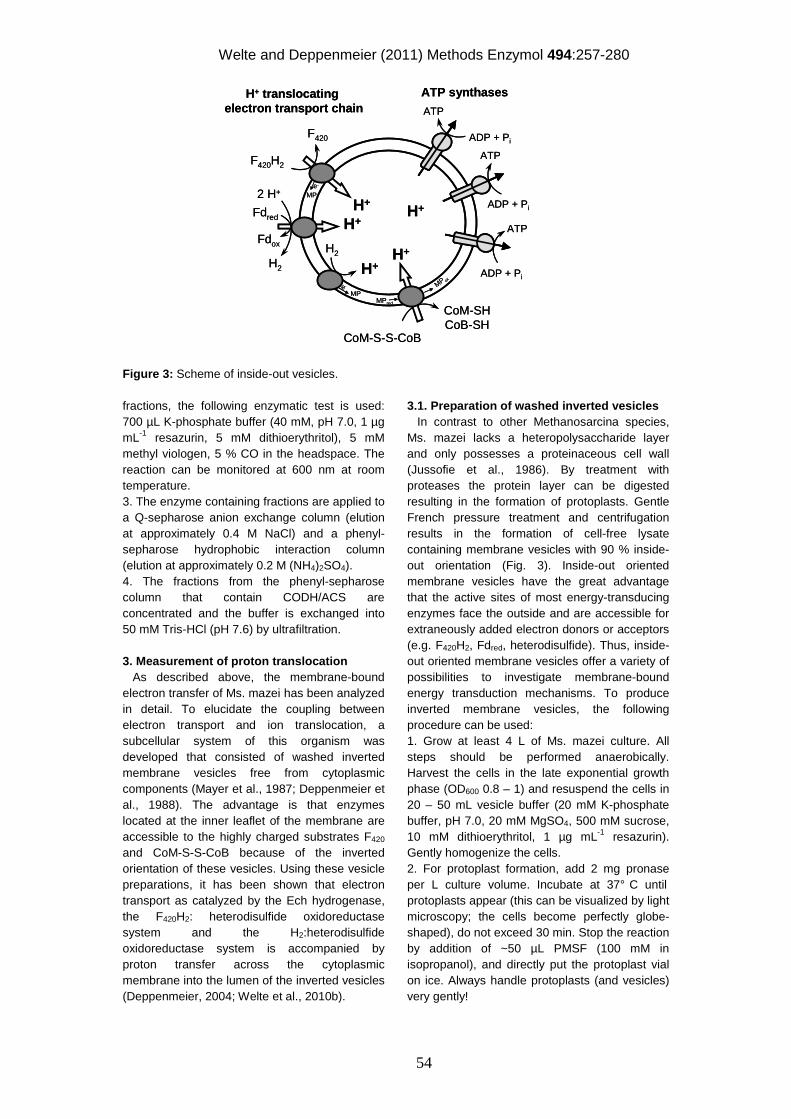
Welte and Deppenmeier (2011) Methods Enzymol 494:257-280
54
Figure 3: Scheme of inside-out vesicles.
fractions, the following enzymatic test is used: 700 µL K-phosphate buffer (40 mM, pH 7.0, 1 µg mL-1 resazurin, 5 mM dithioerythritol), 5 mM methyl viologen, 5 % CO in the headspace. The reaction can be monitored at 600 nm at room temperature. 3. The enzyme containing fractions are applied to a Q-sepharose anion exchange column (elution at approximately 0.4 M NaCl) and a phenyl-sepharose hydrophobic interaction column (elution at approximately 0.2 M (NH4)2SO4). 4. The fractions from the phenyl-sepharose column that contain CODH/ACS are concentrated and the buffer is exchanged into 50 mM Tris-HCl (pH 7.6) by ultrafiltration. 3. Measurement of proton translocation As described above, the membrane-bound electron transfer of Ms. mazei has been analyzed in detail. To elucidate the coupling between electron transport and ion translocation, a subcellular system of this organism was developed that consisted of washed inverted membrane vesicles free from cytoplasmic components (Mayer et al., 1987; Deppenmeier et al., 1988). The advantage is that enzymes located at the inner leaflet of the membrane are accessible to the highly charged substrates F420 and CoM-S-S-CoB because of the inverted orientation of these vesicles. Using these vesicle preparations, it has been shown that electron transport as catalyzed by the Ech hydrogenase, the F420H2: heterodisulfide oxidoreductase system and the H2:heterodisulfide oxidoreductase system is accompanied by proton transfer across the cytoplasmic membrane into the lumen of the inverted vesicles (Deppenmeier, 2004; Welte et al., 2010b).
3.1. Preparation of washed inverted vesicles In contrast to other Methanosarcina species, Ms. mazei lacks a heteropolysaccharide layer and only possesses a proteinaceous cell wall (Jussofie et al., 1986). By treatment with proteases the protein layer can be digested resulting in the formation of protoplasts. Gentle French pressure treatment and centrifugation results in the formation of cell-free lysate containing membrane vesicles with 90 % inside-out orientation (Fig. 3). Inside-out oriented membrane vesicles have the great advantage that the active sites of most energy-transducing enzymes face the outside and are accessible for extraneously added electron donors or acceptors (e.g. F420H2, Fdred, heterodisulfide). Thus, inside-out oriented membrane vesicles offer a variety of possibilities to investigate membrane-bound energy transduction mechanisms. To produce inverted membrane vesicles, the following procedure can be used: 1. Grow at least 4 L of Ms. mazei culture. All steps should be performed anaerobically. Harvest the cells in the late exponential growth phase (OD600 0.8 – 1) and resuspend the cells in 20 – 50 mL vesicle buffer (20 mM K-phosphate buffer, pH 7.0, 20 mM MgSO4, 500 mM sucrose, 10 mM dithioerythritol, 1 µg mL-1 resazurin). Gently homogenize the cells. 2. For protoplast formation, add 2 mg pronase per L culture volume. Incubate at 37° C until protoplasts appear (this can be visualized by light microscopy; the cells become perfectly globe-shaped), do not exceed 30 min. Stop the reaction by addition of ~50 µL PMSF (100 mM in isopropanol), and directly put the protoplast vial on ice. Always handle protoplasts (and vesicles) very gently!
H+ translocatingelectron transport chain
ATP synthases
F420H2
F420
H+
Fdox
Fdred H+
H2
H+
e-
MP
e-
MP
CoM-SHCoB-SH
H+
CoM-S-S-CoB
MPred
MP oxADP + Pi
ATP
ADP + Pi
ATP
ATP
ADP + Pi
H+
H2
2 H+
H+ translocatingelectron transport chain
ATP synthases
F420H2
F420
H+
Fdox
Fdred H+
H2
H+
e-
MP
e-
MP
CoM-SHCoB-SH
H+
CoM-S-S-CoB
MPred
MP oxADP + Pi
ATP
ADP + Pi
ATP
ATP
ADP + Pi
H+
H2
2 H+
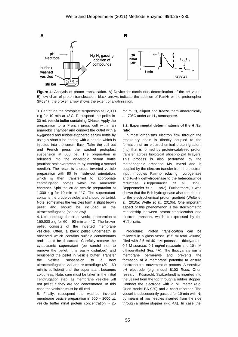
Welte and Deppenmeier (2011) Methods Enzymol 494:257-280
55
Figure 4: Analysis of proton translocation. A) Device for continuous determination of the pH value, B) flow chart of proton translocation, black arrows indicate the addition of F420H2 or the protonophor SF6847, the broken arrow shows the extent of alkalinization.
3. Centrifuge the protoplast suspension at 12,000 x g for 10 min at 4° C. Resuspend the pellet in 30 mL vesicle buffer containing DNase. Apply the preparation to a French press cell within an anaerobic chamber and connect the outlet with a N2-gassed and rubber-stoppered serum bottle by using a short tube ending with a needle which is injected into the serum flask. Take the cell out and French press the washed protoplast suspension at 600 psi. The preparation is released into the anaerobic serum bottle (caution: omit overpressure by inserting a second needle!). The result is a crude inverted vesicle preparation with 90 % inside-out orientation, which is then transferred to appropriate centrifugation bottles within the anaerobic chamber. Spin the crude vesicle preparation at 1,300 x g for 10 min at 4° C. The supernatant contains the crude vesicles and should be turbid. Note: sometimes the vesicles form a slight brown pellet and should be included in the ultracentrifugation (see below)! 4. Ultracentrifuge the crude vesicle preparation at 150,000 x g for 60 – 90 min at 4° C. The brown pellet consists of the inverted membrane vesicles. Often, a black pellet underneath is observed which contains sulfidic contaminants and should be discarded. Carefully remove the cytoplasmic supernatant (be careful not to remove the pellet: it is easily disturbed) and resuspend the pellet in vesicle buffer. Transfer the vesicle suspension to a new ultracentrifugation vial and re-centrifuge (30 – 60 min is sufficient) until the supernatant becomes colourless. Note: care must be taken in the initial centrifugation step, as membrane vesicles will not pellet if they are too concentrated. In this case the vesicles must be diluted. 5. Finally, resuspend the washed inverted membrane vesicle preparation in 500 – 2000 µL vesicle buffer (final protein concentration ~ 25
mg mL-1), aliquot and freeze them anaerobically at -70° C under an H 2 atmosphere. 3.2. Experimental determinations of the H+/2e- ratio In most organisms electron flow through the respiratory chain is directly coupled to the formation of an electrochemical proton gradient (p) that is formed by protein-catalysed proton transfer across biological phospholipid bilayers. This process is also performed by the methanogenic archaeon Ms. mazei and is coupled by the electron transfer from the electron input modules F420-nonreducing hydrogenase and F420H2 dehydrogenase to the heterodisulfide reductase (Deppenmeier et al., 1990; Deppenmeier et al., 1992). Furthermore, it was shown that the Ech hydrogenase also contributes to the electrochemical proton gradient (Welte et al., 2010a; Welte et al., 2010b). One important aspect of this phenomenon is the stoichiometric relationship between proton translocation and electron transport, which is expressed by the H+/2e- ratio. Procedure: Proton translocation can be followed in a glass vessel (5.5 ml total volume) filled with 2.5 ml 40 mM potassium thiocyanate, 0.5 M sucrose, 0.1 mg/ml resazurin and 10 mM dithioerythritol (Fig. 4A). The thiocyanate ion is membrane permeable and prevents the formation of a membrane potential to ensure electroneutral movement of protons. A sensitive pH electrode (e.g. model 8103 Ross, Orion research, Küsnacht, Switzerland) is inserted into the vessel from the top through a rubber stopper. Connect the electrode with a pH meter (e.g. Orion model EA 920) and a chart recorder. The vessel is subsequently gassed for 10 min with N2 by means of two needles inserted from the side through a rubber stopper (Fig. 4A). In case the
pHelectrode
N2/ H2 gassingaddition of compounds
A B
20 nmolOH-
SF6847
5 min
F420H2
F420H2
stir bar
buffer + washedvesicles
pHelectrode
N2/ H2 gassingaddition of compounds
A B
20 nmolOH-
SF6847
5 min
F420H2
F420H2
stir bar
buffer + washedvesicles
Welte and Deppenmeier (2011) Methods Enzymol 494:257-280
56
solution is not totally colourless, add up to 1 µL titanium(III) citrate for complete reduction. Then 100 µL washed vesicles (0.35-0.5 mg protein/assay) are added, resulting in a final K-phosphate concentration of 1.5 mM in the reaction mixture. The preparation is continuously stirred and the pH is adjusted to 6.8-6.9. Additions are made with a microliter syringe from the side arm. As mentioned above, proton translocation is due to electron transfer from an electron donor (H2, F420H2, reduced 2-hydroxyphenazine) to an acceptor (2-hydroxyphenazine or CoM-S-S-CoB). In principle one substrate is added in excess, whereas the second substrate is injected in low amounts (“pulse injection”; usually 2-10 nmol) that allows only a short period of electron transfer events. The pulse injection will lead to alkalinization of the medium as is apparent from the recorded time course of this process (Fig. 4B). A short period of alkalinization is then followed by a longer period of acidification until a stable baseline (pH) is reached again. The alkalinization is due to proton movement from the medium into the lumen of the inverted vesicles energized by the electron transfer reactions. The extent of alkalinization is dependent on the amount of the pulsed substrate. Changes of the pH value are converted into proton equivalents by double titration with standard solutions of HCl and KOH (Fig. 4B). The amount of H+ translocated is calculated from the difference between the maximum of alkalinization and the final baseline after reacidification (Fig. 4B). The ratio of translocated protons and transferred electron can be calculated since the amount of pulsed substrate is known. Reaction conditions: - H2: heterodisulfide oxidoreductase: Use an H2 atmosphere and pulse with CoM-S-S-CoB (2-10 nmol in oxygen free H2O) - F420-nonreducing hydrogenase: Use an H2 atmosphere and pulse with hydroxyphenazine (2-10 nmol from a 1 mM stock solution solvated in anoxic ethanol) - Heterodisulfide reductase: N2 atmosphere in the reaction vessel; add 250 nmol CoM-S-S-CoB and pulse with reduced hydroxyphenazine (2-10 nmol from a 1 mM stock solution solvated in anoxic ethanol) - F420H2 dehydrogenase: N2 atmosphere in the reaction vessel; add 250 nmol hydroxyphenazine and pulse with F420H2 (2-10 nmol) - F420H2: heterodisulfide oxidoreductase: N2 atmosphere in the reaction vessel; add 250 nmnol CoM-S-S-CoB and pulse with reduced F420 (2-10 nmol)
In our studies, the reversible alkalinization was used to calculate from more than 60 experiments an average of 1.9 protons translocated per F420H2 oxidized in the F420H2: heterodisulfide oxidoreductase assay. The observed alkalinization was specifically coupled with the F420H2-dependent CoM-S-S-CoB reduction. Alkalinization was not observed if F420H2 or CoM-S-S-CoB was omitted or if F420H2 was replaced by F420. Similar values were obtained when the H2:heterodisulfide oxidoreductase system was analysed. Keeping in mind that about 50 % of the membrane structures present in the vesicle preparations catalyze electron transport but are unable to establish a proton gradient (Deppenmeier et al., 1996), the H+/2e- values of the overall reaction increases to 3.8, which is in agreement with values found for whole cell preparations of Ms. barkeri (Blaut et al., 1987). The aforementioned washed inverted vesicles were found to couple electron transfer processes with the transfer of about four protons across the cytoplasmic membrane. Furthermore, it was shown that 2-hydroxyphenazine is reduced by molecular hydrogen, as catalyzed by the F420-nonreducing hydrogenase, and the membrane-bound heterodisulfide reductase was able to use reduced 2-hydroxyphenazine as the electron donor for the reduction of CoM-S-S-CoB. In addition, the F420H2-dehydrogenase could use hydroxyphenazine as electron acceptor (Ide et al., 1999; Bäumer et al., 2000). All reactions allowed the translocation of protons with a stoichiometry of about 2H+/2e-. Hence, there are two proton-translocating segments present in the F420H2:CoM-S-S-CoB and the H2:CoM-S-S-CoB oxidoreductase system, respectively. The first one involves the electron donating enzymes (F420-nonreducing hydrogenase or F420H2 dehydrogenase) and the second one the heterodisulfide reductase (Fig. 2). Control experiments showed that the formation of a proton gradient was not possible in the presence of a protonophor, such as SF6847 (10 nmol/ mg protein) (Fig. 4B). In contrast H+ translocation was not affected by an inhibitor of the ATP synthase (e.g. DCCD; 400 µM) or in the presence of a Na+ ionophor (e.g. ETH157; 30 µM). 4. Analysis of ATP formation in vesicle preparation When protons are translocated into the inverted membrane vesicles by enzymes comprising the methanogenic respiratory chain, and when ADP and Pi are present, the A1AO ATP synthase can phosphorylate ADP in the process of proton extrusion. ATP content can be
Welte and Deppenmeier (2011) Methods Enzymol 494:257-280
57
measured using the luciferin/luciferase assay (Kimmich et al., 1975). The reaction of luciferase with its substrate luciferin is strictly ATP-dependent and produces light, and the amount of ATP is directly proportional to the amount of emitted light. Procedure: The rubber-stoppered, N2-flushed glass reaction vial contains 500 µL vesicle buffer, 600 µM ADP, 300 µM AMP (to inhibit membrane-bound adenylate kinase), and 250-700 µg inverted membrane vesicles. Depending on the system to be observed different electron donors or acceptors need to be used. For the H2:heterodisulfide oxidoreductase, a H2 atmosphere and 200-500 µM heterodisulfide as electron acceptor is used. For the F420H2:heterodisulfide oxidoreductase, a N2 atmosphere, 10 µL F420H2 (final concentration 15 µM) as electron donor and 200-500 µM heterodisulfide as electron acceptor is used. For measuring the Fd:heterodisulfide oxidoreductase, a 5 % CO / 95 % N2 atmosphere is applied to the reaction vial. Furthermore, 70 µg CO-dehydrogenase and 30 µg Fd are added to generate Fdred, and 200-500 µM heterodisulfide are added as terminal electron acceptor. If Ech hydrogenase as part of the Fd:heterodisulfide oxidoreductase is to be measured independently, the heterodisulfide can be omitted. Electrons are transferred to protons as terminal electron acceptors and form H2. The reaction vials are shaken in a 37° C water bath and 10 µL samples are taken at various time points with a microliter syringe. The sample is mixed with 700 µL ATP determination buffer (4 mM MgSO4, 20 mM glycylglycine, pH 8.0) and 100 µL firefly lantern extract (Sigma-Aldrich, Germany). The emitted light is quantified with a luminometer at 560 nm after a lag phase of 10 s. The values are compared to a standard curve generated with 1 – 100 pmol ATP. For the H2:heterodisulfide oxidoreductase and Ech hydrogenase an activity of about 1.5-2 nmol ATP/ mg membrane protein, and for the complete Fd:heterodisulfide oxidoreductase an activity of 3-4 nmol ATP / mg membrane protein is expected. Acknowledgments We thank Elisabeth Schwab for technical assistance and Paul Schweiger for critical reading of the manuscript. Many thanks also go to Gunes Bender and Steve Ragsdale, Department of Biological Chemistry, University of Michigan Medical School for providing the CODH/ACS from Moorella thermoacetica. This
work was supported by the Deutsche Forschungsgemeinschaft (grant De488/9-1). References Abken, H. J., Tietze, M., Brodersen, J., Bäumer,
S., Beifuss, U. and Deppenmeier, U. (1998). Isolation and characterization of methanophenazine and function of phenazines in membrane-bound electron transport of Methanosarcina mazei Gö1. J. Bacteriol. 180(8), 2027-2032.
Bartoschek, S., Vorholt, J. A., Thauer, R. K., Geierstanger, B. H. and Griesinger, C. (2000). N-carboxymethanofuran (carbamate) formation from methanofuran and CO2 in methanogenic archaea. Thermodynamics and kinetics of the spontaneous reaction. Eur. J. Biochem. 267(11), 3130-3138.
Bäumer, S., Ide, T., Jacobi, C., Johann, A., Gottschalk, G. and Deppenmeier, U. (2000). The F420H2 dehydrogenase from Methanosarcina mazei is a redox-driven proton pump closely related to NADH dehydrogenases. J. Biol. Chem. 275(24), 17968-17973.
Beifuss, U. and Tietze, M. (2000). On the total synthesis of (S)-methanophenazine and the formal synthesis of (R)-methanophenazine from a common precursor. Tetrahedron Lett. 41(50), 9759-9763.
Beifuss, U., Tietze, M., Baumer, S. and Deppenmeier, U. (2000). Methanophenazine: Structure, total synthesis, and function of a new cofactor from methanogenic archaea. Angew. Chem. Int. Ed. 39 (14), 2470-
Beifuss, U. and Tietze, M. (2005). Methanophenazine and other natural biologically active phenazines. Top. Curr. Chem. 244, 77-113.
Blaut, M. and Gottschalk, G. (1984). Coupling of ATP synthesis and methane formation from methanol and molecular hydrogen in Methanosarcina barkeri. Eur. J. Biochem. 141(1), 217-222.
Blaut, M., Müller, V. and Gottschalk, G. (1987). Proton translocation coupled to methanogenesis from methanol + hydrogen in Methanosarcina barkeri. FEBS Lett. 215(1), 53-57.
Boone, D. R., Whitman, W. B. and Rouvière, P. E. (1993). Diversity and taxonomy of methanogens. Methanogenesis. J. G. Ferry. New York, Chapman & Hall: 35-80.
Clements, A. P., Kilpatrick, L., Lu, W. P., Ragsdale, S. W. and Ferry, J. G. (1994). Characterization of the iron-sulfur clusters in ferredoxin from acetate-grown Methanosarcina thermophila. J. Bacteriol. 176(9), 2689-2693.
Welte and Deppenmeier (2011) Methods Enzymol 494:257-280
58
Deppenmeier, U. (2002a). Redox-driven proton translocation in methanogenic archaea. Cell. Mol. Life Sci. 59(9), 1513-1533.
Deppenmeier, U. (2002b). The unique biochemistry of methanogenesis. Prog. Nucleic Acid Res. Mol. Biol. 71, 223-283.
Deppenmeier, U. (2004). The membrane-bound electron transport system of Methanosarcina species. J. Bioenerg. Biomembr. 36(1), 55-64.
Deppenmeier, U., Blaut, M., Jussofie, A. and Gottschalk, G. (1988). A methyl-CoM methylreductase system from methanogenic bacterium strain Gö 1 not requiring ATP for activity. FEBS Lett. 241(1-2), 60-64.
Deppenmeier, U., Blaut, M., Mahlmann, A. and Gottschalk, G. (1990). Reduced coenzyme F420: heterodisulfide oxidoreductase, a proton- translocating redox system in methanogenic bacteria. Proc. Natl. Acad. Sci. USA 87(23), 9449-9453.
Deppenmeier, U., Blaut, M., Schmidt, B. and Gottschalk, G. (1992). Purification and properties of a F420-nonreactive, membrane-bound hydrogenase from Methanosarcina strain Gö1. Arch. Microbiol. 157(6), 505-511.
Deppenmeier, U. and Müller, V. (2008). Life close to the thermodynamic limit: how methanogenic archaea conserve energy. Results Probl. Cell Differ. 45, 123-152.
Deppenmeier, U., Müller, V. and Gottschalk, G. (1996). Pathways of energy conservation in methanogenic archaea. Arch. Microbiol. 165(3), 149-163.
Ebner, S., Jaun, B., Goenrich, M., Thauer, R. K. and Harmer, J. (2010). Binding of coenzyme B induces a major conformational change in the active site of methyl-coenzyme M reductase. J. Am. Chem. Soc. 132(2), 567-575.
Ellermann, J., Hedderich, R., Bocher, R. and Thauer, R. K. (1988). The final step in methane formation. Investigations with highly purified methyl-CoM reductase (component C) from Methanobacterium thermoautotrophicum (strain Marburg). Eur. J. Biochem. 172(3), 669-677.
Ellman, G. L. (1958). A colorimetric method for determining low concentrations of mercaptans. Arch. Biochem. Biophys. 74(2), 443-450.
Ferry, J. G. (1999). Enzymology of one-carbon metabolism in methanogenic pathways. FEMS Microbiol. Rev. 23(1), 13-38.
Ferry, J. G. and Lessner, D. J. (2008). Methanogenesis in marine sediments. Ann. N. Y. Acad. Sci. 1125, 147-157.
Fischer, R. and Thauer, R. K. (1989). Methyltetrahydromethanopterin as an intermediate in methanogenesis from acetate in Methanosarcina barkeri. Arch. Microbiol. 151(5), 459-465.
Gencic, S., Duin, E. C. and Grahame, D. A. (2010). Tight coupling of partial reactions in the acetyl-CoA decarbonylase/synthase (ACDS) multienzyme complex from Methanosarcina thermophila: acetyl C-C bond fragmentation at the A cluster promoted by protein conformational changes. J. Biol. Chem. 285(20), 15450-15463.
Gong, W., Hao, B., Wei, Z., Ferguson, D. J., Tallant, T., Krzycki, J. A. and Chan, M. K. (2008). Structure of the alpha(2)epsilon(2) Ni-dependent CO dehydrogenase component of the Methanosarcina barkeri acetyl-CoA decarbonylase/synthase complex. Proc. Natl. Acad. Sci. USA 105(28), 9558-9563.
Gottschalk, G. and Thauer, R. K. (2001). The Na+-translocating methyltransferase complex from methanogenic archaea. Biochim. Biophys. Acta 1505(1), 28-36.
Hedderich, R. and Forzi, L. (2005). Energy-converting [NiFe] hydrogenases: more than just H2 activation. J. Mol. Microbiol. Biotechnol. 10(2-4), 92-104.
Hedderich, R., Hamann, N. and Bennati, M. (2005). Heterodisulfide reductase from methanogenic archaea: a new catalytic role for an iron-sulfur cluster. Biol. Chem. 386(10), 961-970.
Heiden, S., Hedderich, R., Setzke, E. and Thauer, R. K. (1994). Purification of a 2-subunit cytochrome-b containing heterodisulfide reductase from methanol-grown Methanosarcina barkeri. Eur. J. Biochem. 221(2), 855-861.
Hippe, H., Caspari, D., Fiebig, K. and Gottschalk, G. (1979). Utilization of trimethylamine and other N-methyl compounds for growth and methane formation by Methanosarcina barkeri. Proc. Natl. Acad. Sci. USA 76(1), 494-498.
Ide, T., Bäumer, S. and Deppenmeier, U. (1999). Energy conservation by the H2:heterodisulfide oxidoreductase from Methanosarcina mazei Gö1: identification of two proton-translocating segments. J. Bacteriol. 181(13), 4076-4080.
Jain, N., Pathak, H., Mitra, S. and Bhatia, A. (2004). Emission of methane from rice fields - A review. J. Sci. Ind. Res. 63(2), 101-115.
Jussofie, A., Mayer, F. and Gottschalk, G. (1986). Methane formation from methanol and molecular hydrogen by protoplasts of new methanogenic isolates and inhibition by dicyclohexylcarbodiimide. Arch. Microbiol. 146(3), 245-249.
Kashyap, D. R., Dadhich, K. S. and Sharma, S. K. (2003). Biomethanation under psychrophilic conditions: a review. Bioresour. Technol. 87(2), 147-153.
Khalil, M. A. K. and Rasmussen, R. A. (1994). Global emissions of methane during the last
Welte and Deppenmeier (2011) Methods Enzymol 494:257-280
59
several centuries. Chemosphere 29(5), 833-842.
Kimmich, G. A., Randles, J. and Brand, J. S. (1975). Assay of picomole amounts of ATP, ADP, and AMP using the luciferase enzyme system. Anal. Biochem. 69(1), 187-206.
Kobelt, A., Pfaltz, A., Ankelfuchs, D. and Thauer, R. K. (1987). The L-form of N-7-mercaptoheptanoyl-O-phosphothreonine is the enantiomer active as component B in methyl-CoM reduction to methane. FEBS Lett. 214(2), 265-268.
Künkel, A., Vaupel, M., Heim, S., Thauer, R. K. and Hedderich, R. (1997). Heterodisulfide reductase from methanol-grown cells of Methanosarcina barkeri is not a flavoenzyme. Eur. J. Biochem. 244(1), 226-234.
Mayer, F., Jussofie, A., Salzmann, M., Lubben, M., Rohde, M. and Gottschalk, G. (1987). Immunoelectron microscopic demonstration of ATPase on the cytoplasmic membrane of the methanogenic bacterium strain Gö1. J. Bacteriol. 169(5), 2307-2309.
McInerney, M. J., Sieber, J. R. and Gunsalus, R. P. (2009). Syntrophy in anaerobic global carbon cycles. Curr. Opin. Biotechnol. 20(6), 623-632.
Meuer, J., Bartoschek, S., Koch, J., Kunkel, A. and Hedderich, R. (1999). Purification and catalytic properties of Ech hydrogenase from Methanosarcina barkeri. Eur. J. Biochem. 265(1), 325-335.
Meuer, J., Kuettner, H. C., Zhang, J. K., Hedderich, R. and Metcalf, W. W. (2002). Genetic analysis of the archaeon Methanosarcina barkeri Fusaro reveals a central role for Ech hydrogenase and ferredoxin in methanogenesis and carbon fixation. Proc. Natl. Acad. Sci. USA 99(8), 5632-5637.
Mortenson, L. E. (1964). Purification and analysis of ferredoxin from Clostridium pasteurianum. Biochim. Biophys. Acta 81(1), 71-77.
Noll, K. M., Donnelly, M. I. and Wolfe, R. S. (1987). Synthesis of 7-Mercaptoheptanoylthreonine phosphate and its activity in the methylcoenzyme-M methylreductase system. J. Biol. Chem. 262(2), 513-515.
Ott, R. (1959). Untersuchungen über Chinone. 2. Mitt.: Azinbildung durch Umsetzung von p-Chinonen mit o-Diaminen. Mh. Chem. 90(6), 827-838.
Peinemann, S., Blaut, M. and Gottschalk, G. (1989). ATP synthesis coupled to methane formation from methyl-CoM and H2 catalyzed by vesicles of the methanogenic bacterial strain Gö1. Eur. J. Biochem. 186(1-2), 175-180.
Peinemann, S., Müller, V., Blaut, M. and Gottschalk, G. (1988). Bioenergetics of methanogenesis from acetate by Methanosarcina barkeri. J. Bacteriol. 170(3), 1369-1372.
Pisa, K. Y., Weidner, C., Maischak, H., Kavermann, H. and Müller, V. (2007). The coupling ion in the methanoarchaeal ATP synthases: H(+) vs. Na(+) in the A(1)A(o) ATP synthase from the archaeon Methanosarcina mazei Gö1. FEMS Microbiol. Lett. 277(1), 56-63.
Ragsdale, S. W. (2008). Catalysis of methyl group transfers involving tetrahydrofolate and B12. Vitam. Horm. 79, 293-324.
Ragsdale, S. W., Ljungdahl, L. G. and Der Vartanian, D. V. (1983). Isolation of carbon monoxide dehydrogenase from Acetobacterium woodii and comparison of its properties with those of the Clostridium thermoaceticum enzyme. J. Bacteriol. 155(3), 1224-1237.
Thauer, R. K. (1998). Biochemistry of methanogenesis: a tribute to Marjory Stephenson. Microbiology 144, 2377-2406.
Thauer, R. K., Jungermann, K. and Decker, K. (1977). Energy conservation in chemotrophic anaerobic bacteria. Bacteriol. Rev. 41(1), 100-180.
Thauer, R. K., Kaster, A. K., Seedorf, H., Buckel, W. and Hedderich, R. (2008). Methanogenic archaea: ecologically relevant differences in energy conservation. Nat. Rev. Microbiol. 6(8), 579-591.
Vignais, P. M., Billoud, B. and Meyer, J. (2001). Classification and phylogeny of hydrogenases. FEMS Microbiol. Rev. 25(4), 455-501.
Welte, C., Kallnik, V., Grapp, M., Bender, G., Ragsdale, S. and Deppenmeier, U. (2010a). Function of Ech hydrogenase in ferredoxin-dependent, membrane-bound electron transport in Methanosarcina mazei. J. Bacteriol. 192(3), 674-678.
Welte, C., Krätzer, C. and Deppenmeier, U. (2010b). Involvement of Ech hydrogenase in energy conservation of Methanosarcina mazei. FEBS J. 277, 3396-3403.
Wolfe, R. S. (1985). Unusual coenzymes of methanogenesis. Trends Biochem. Sci. 10(10), 396-399.
Zahler, W. L. and Cleland, W. W. (1968). A specific and sensitive assay for disulfides. J. Biol. Chem. 243(4), 716-719.
Zehnder, A. J. B. and Wuhrmann, K. (1976). Titan(III) citrate a nontoxic oxidation-reduction buffering system for the culture of obligate anaerobes. Science 194, 1165-1166.
60
Chapter 7
Concluding discussion
Methylotrophic energy conservation in Methanosarcina sp. with Methanosarcina
(Ms.) mazei as one of the important model organisms is well investigated. Two
proton-translocating membrane-bound oxidoreductase systems have been
described, the H2: heterodisulfide oxidoreductase (Ide et al., 1999) and the F420H2:
heterodisulfide oxidoreductase (Bäumer et al., 2000). A third heterodisulfide
oxidoreductase, the ferredoxin: heterodisulfide oxidoreductase, was proposed to be
responsible for ferredoxin turnover in aceticlastic methanogenesis (Fischer and
Thauer, 1990). In 1999 Ech hydrogenase was identified as an electron input protein
of the ferredoxin: heterodisulfide oxidoreductase in Ms. barkeri (Meuer et al., 1999)
and it was hypothesized that ferredoxin oxidation and H2 production are coupled to
energy conservation. However, no enzymatic system for continuous reduction of
ferredoxin was established and the idea of ion translocation by Ech hydrogenase
was not proven by biochemical experiments.
Enzymatic reduction of ferredoxin The first aim of this work was to establish an enzymatic system with which to
analyze ferredoxin-dependent electron transport using isolated cytoplasmic
membranes and vectorial ion translocation using membrane vesicles of Ms. mazei.
This enzymatic system should enable electron transport from an initial substrate to a
ferredoxin-reducing enzyme, then to a ferredoxin and finally to the membrane-bound
ferredoxin: heterodisulfide oxidoreductase. The Ms. mazei genome codes for
approximately 20 ferredoxins, and it is not known which of these ferredoxins
interacts with the membrane-bound ferredoxin: heterodisulfide oxidoreductase.
Initial attempts to produce some of these ferredoxins recombinantly in Escherichia
coli were not successful due to incomplete assembly of the [4Fe4S] clusters in
E. coli or in vitro reconstitution. As a substitute the [4Fe4S] ferredoxin from
Clostridium pasteurianum was used because direct purification from the parent
organism ascertains correct cofactor assembly. Furthermore, ferredoxin from
C. pasteurianum is used as model ferredoxin in many studies (Biegel and Müller,
2010; Kaster et al., 2011) and it has been observed that ferredoxins are often
functionally interchangeable albeit only distantly related (Tagawa and Arnon, 1968). In anaerobic metabolism, many enzymes interact with ferredoxins. Unfortunately,
many of these are highly oxygen unstable (e.g. CO dehydrogenases), contain
unusual cofactors (e.g. the tungsten cofactor of aldehyde: ferredoxin
61
oxidoreductases) or are built up by many subunits (multi-subunit pyruvate:
ferredoxin oxidoreductases) making recombinant production in E. coli difficult.
Laborious work to purify ferredoxin-reducing enzymes directly from anaerobic
organisms was inevitable, and thankfully a multi-subunit CO dehydrogenase/acetyl-
CoA synthase (CODH/ACS) from Moorella thermoacetica was provided by Güneş
Bender and Steve Ragsdale (University of Michigan). The CODH/ACS couples CO
oxidation to ferredoxin reduction, and indeed worked productively with the ferredoxin
from C. pasteurianum. This ferredoxin, in turn, served as electron donor to the
Ms. mazei membrane-bound ferredoxin: heterodisulfide oxidoreductase, and led to
heterodisulfide reduction. With this system, electron transport of the ferredoxin:
heterodisulfide oxidoreductase system could be followed, and also the activity of
Ech hydrogenase alone could be continuously measured.
Revealing energy-transducing properties of Ech hydr ogenase Ferredoxin-dependent electron transport measurements with Ms. mazei wildtype
and Ms. mazei ∆ech mutant membrane preparations revealed that the
heterodisulfide reduction rate was strongly decreased in the ∆ech mutant. This
finding confirmed the proposed involvement of Ech hydrogenase in the ferredoxin:
heterodisulfide oxidoreductase and validated the proposed hydrogen-cycling
mechanism in which Ech hydrogenase couples ferredoxin oxidation to hydrogen
production. Hydrogen is taken up by F420 non-reducing hydrogenase that donates
electrons to the methanophenazine pool. Reduced methanophenazine finally acts
as substrate to the heterodisulfide reductase. It is important to mention that
membrane-bound ferredoxin: heterodisulfide oxidoreductase activity was not
abolished in the Ms. mazei ∆ech mutant but was still present to an extent of about
50 %. This makes the existence of a second ferredoxin-interacting membrane
protein (complex) likely. The phenotypic characterization of the Ms. mazei ∆ech mutant showed that growth
on methylated amines was slower, substrate consumption was faster and the overall
growth yield was lower than observed for the wildtype. Furthermore, the ∆ech
mutant did not grow on acetate. Taking together it became evident that substrate
usage was less effective in the mutant than in the wildtype, indicating an important
function of Ech hydrogenase in energy conservation. However, reduced ferredoxin
that is produced in methylotrophic methanogenesis had to be re-oxidized in the
∆ech mutant in a process that was obviously only possible with energetic
disadvantages. There are two possibilities how the cell could accomplish the
reoxidation of reduced ferredoxin in the absence of the Ech hydrogenase: (i) there
62
could be a soluble ferredoxin: F420 oxidoreductase that transfers electrons from
reduced ferredoxin to coenzyme F420 and finally to the F420-dependent electron
transport pathway. (ii) There could be an Ech hydrogenase-independent membrane-
bound ferredoxin: heterodisulfide oxidoreductase system present that conserves
less energy than the Ech-dependent pathway. To further address the question of Ech hydrogenase as an energy-conserving
enzyme, a vesicular system of inside-out oriented membrane vesicles was
employed. Enzymatic activities that normally lead to the formation of an electro-
chemical ion gradient over the cytoplasmic membranes result in an accumulation of
the respective ions in the lumen of the inside-out oriented membrane vesicles. The
A1AO ATP synthase of Ms. mazei can take advantage of this potential and generate
ATP from ADP + Pi through reflux of H+ or Na+ ions. Experiments with membrane
vesicles of the Ms. mazei wildtype showed that the activity of Ech hydrogenase was
correlated with ATP production. This ATP synthesis was strictly dependent on an ion
gradient because the inhibitor DCCD abolishes ATP formation. These data showed
that Ech hydrogenase is an ion-translocating enzyme that conserves energy. In
methanogenic bioenergetics both H+ and Na+ serve as coupling ions so the nature
of the coupling ion of Ech hydrogenase had to be explored. The sodium ionophore
ETH157 did not alter ATP formation through Ech hydrogenase activity, but the
protonophore SF6847 effectively prevented ATP synthesis. At the same time the
activity of Ech hydrogenase was monitored following ferredoxin-dependent H2
production and it was found that Ech hydrogenase activity was not altered by the
ionophores. Theoretically a maximum of 1 H+ per 2 e- can be transported across the
membrane. Energy conservation by Ech hydrogenase is in accordance with
previous hypotheses (Bott and Thauer, 1989; Meuer et al., 1999; Tersteegen and
Hedderich, 1999; Stojanowic and Hedderich, 2004). Also the related Mbh
hydrogenase of Pyrococcus furiosus was shown to couple ferredoxin oxidation to
proton reduction and ion movement over the cytoplasmic membrane (Sapra et al.,
2003). In summary, it was clearly shown for the first time by experimental data that
the Ech hydrogenase from Methanosarcina sp. belongs to the group of energy-
conserving hydrogenases.
Pathway of respiratory F 420H2 turnover Hydrogen cycling by the Ms. mazei and Ms. barkeri ferredoxin: heterodisulfide
oxidoreductase initially seemed unlikely because a gaseous intermediate could
easily escape the respiratory system and would lead to energy loss. But apparently
these methanogens effectively capture the gaseous H2, so the overall partial H2
63
pressure of acetate-growing cultures is low (Lovley and Ferry, 1985). However,
hydrogen cycling was not thought to be involved in energy conservation for the
Ms. mazei F420: heterodisulfide oxidoreductase because the membrane-bound
F420H2 dehydrogenase directly channels electrons to methanophenazine.
Nevertheless, the possibility of an alternative soluble F420 hydrogenase that couples
F420H2 oxidation to hydrogen production exists and could be utilized in an analogous
hydrogen cycling mechanism as observed for ferredoxin oxidation. For the close
relative Ms. barkeri hydrogen cycling in the course of heterodisulfide-dependent
F420H2 oxidation was proposed to be the predominant mechanism (Kulkarni et al.,
2009). Taking these observations together, a re-investigation of the F420H2 oxidizing
mechanism in Ms. mazei and close relatives was done with the aim of resolving the
pathway by which electrons from F420H2 enter the respiratory chain. The phenotypic
characterizations of two mutants of the F420H2 dehydrogenase (Ms. mazei ∆fpoA-O
and ∆fpoF) revealed that the deletion of the F420H2 dehydrogenase leads to severely
slowed growth on methylated amines comparable to growth of the Ms. mazei ∆ech
mutant. However, the growth yield was not affected, indicating that the F420
hydrogenase system can energetically substitute for the loss of F420H2
dehydrogenase. Measurements of the F420H2: heterodisulfide oxidoreductase activity
in cell lysate demonstrated that this activity is greatly reduced in the two ∆fpo
mutants, and also the methane formation rates of resting cell suspensions was
strongly decreased. In summary, these findings showed that only a minor part of
F420H2 oxidation is performed by F420 hydrogenase using hydrogen cycling in
Ms. mazei. Other Methanosarcina sp. including Ms. acetivorans seem to be similar
in this preference, and Ms. barkeri can be regarded as an exception that prefers the
use of F420 hydrogenase.
FpoF, a novel ferredoxin: F 420 oxidoreductase Membrane fractions of the Ms. mazei ∆fpoF mutant, missing the electron input
subunit FpoF of the F420H2 dehydrogenase, were impaired in their ability to oxidize
F420H2.The same result was observed for the ∆fpoA-O mutant that lacked the
membrane-integral and membrane-associated part of F420H2 dehydrogenase. It was
surprising that the cytoplasmic activities of the two mutant strains were different. The
cytoplasm of the parental strain was found to catalyze the redox-reaction of
ferredoxin with F420 by use of a novel ferredoxin: F420 oxidoreductase. The cytoplasm
of the ∆fpoA-O mutant retained similar levels of activity whereas the cytoplasmic
fraction of the ∆fpoF mutant could no longer catalyze the ferredoxin-dependent
redox reaction. To investigate this enzymatic activity in more detail the FpoF protein
64
was recombinantly produced in E. coli. The purified protein catalyzed the ferredoxin:
F420 oxidoreductase reaction with high specificity but low rates, and was found to be
present in substantial quantities as soluble protein in the cytoplasm. It can be
concluded that FpoF indeed catalyzes the electron transfer between ferredoxin and
F420. However, this connection has to be strongly regulated because reduction of
F420 with reduced ferredoxin results in a decreased H+ / e- ratio. This is evident from
the fact that the ferredoxin: heterodisulfide oxidoreductase translocates 5 H+ / 2 e-
(with Ech hydrogenase as proton-translocatig enzyme) whereas the F420H2:
heterodisulfide oxidoreductase only translocates 4 H+ / 2 e-. In principle, the discovery of the ferredoxin: F420 oxidoreductase could explain the
survival of the Ms. mazei ∆ech mutant, because reduced ferredoxin that is produced
in the methylotrophic pathway could be re-oxidized by FpoF and the electrons could
be used by the F420H2: heterodisulfide oxidoreductase. However, the FpoF activity
(also in the cytoplasm of the ∆ech mutant) is too low to allow effective turnover of
reduced ferredoxin, and can only partially contribute to the survival of the ∆ech
mutant. Hence, the existence of another membrane-bound ferredoxin-oxidizing
enzyme is still a tempting alternative.
Membrane-bound electron transport in Methanosaeta thermophila Aceticlastic energy conservation in Ms. mazei was investigated in detail in the
course of this work. However, the energy conserving system of the close relative
Methanosaeta (Mt.) which is the only obligately aceticlastic methanogenic genus
has not been examined to date. Comparative studies of the Mt. thermophila genome
indicated that protein complexes involved in electron input to the aceticlastic
respiratory chain of Methanosarcina sp. are absent: neither genes coding for Ech
hydrogenase nor for the Rnf complex can be found in the genome. Furthermore, the
genome does not contain intact hydrogenase genes, and also only an incomplete
coding region for the F420H2 dehydrogenase that lacks the electron input protein
FpoF. Genes coding for a membrane-bound heterodisulfide reductase (HdrDE) were
found and activity measurements on isolated Mt. thermophila membranes showed
that the corresponding enzyme is active and exhibits comparable activities to the
Ms. mazei enzyme. This means that energy conservation via a membrane-bound
heterodisulfide oxidoreductase system is likely. F420H2, NAD(P)H and H2 did not
serve as electron donors to the membrane. The recombinant Ms. mazei ferredoxin
MM1619 as well as clostridial ferredoxin (not shown) served as electron donors for
heterodisulfide reduction, so a functional ferredoxin: heterodisulfide oxidoreductase
system is present in Mt. thermophila. The rate of membrane-bound heterodisulfide
65
reduction was two- to three-fold faster in Mt. thermophila than in acetate-grown
Ms. mazei which makes it likely that the Mt. thermophila ferredoxin: heterodisulfide
oxidoreductase is actually used for energy generation in the obligate aceticlastic
organism. In Mt. thermophila, the main part of the F420H2 dehydrogenase is encoded in the
genome (fpoABCDHIJKLMN), but the electron input subunit FpoF is missing. The
presence of genes encoding the F420H2 dehydrogenase in combination with the
apparent inability of the Mt. thermophila membranes to oxidize F420H2 resulted in the
idea that the incomplete F420H2 dehydrogenase could be involved in ferredoxin-
dependent electron transport. Two possibilities concerning how the F420H2
dehydrogenase could contribute to ferredoxin oxidation exist: (i) in Mt. thermophila,
there exists another unknown electron input subunit that productively works with
FpoA-N and is located elsewhere in the chromosome as is also the case for the
fpoF gene in Methanosarcina sp. FpoA-N is highly homologous to the core of
complex I (NADH dehydrogenase I of E. coli, Nuo), and the replacement of NuoEFG
by FpoF enables methanogens to use F420H2 instead of NADH. Another
replacement of the electron input module together with the core Fpo/Nuo could be a
feasible possibility to enable ferredoxin oxidation. (ii) The core enzyme FpoA-N is
catalytically active without FpoF or other subunits channelling electrons immediately
into the respiratory chain. Hence, the truncated Fpo complex would accept electrons
directly from reduced ferredoxin without the need of an alternative electron input
subunit. To address these questions, the diversity and distribution of complex I homologues
is discussed. Complex I from E. coli is termed NADH: ubiquinone oxidoreductase
(Nuo) that has a homologous subunit nomenclature to the Ms. mazei F420H2:
phenazine oxidoreductase (Fpo). Other terminologies include the NADH: quinone
oxidoreductase of Thermus thermophilus (Nqo) or NADH dehydrogenase 1 (NDH-1)
of cyanobacteria. For simplicity, the Nuo nomenclature will be used throughout this
chapter.
NADH dehydrogenases in bacteria NADH dehydrogenases are widely distributed among living organisms, and are well
investigated in mitochondria of eukaryotic cells and eubacteria. Only recently, the
crystal structure of the complete E. coli and T. thermophilus NADH dehydrogenase
(complex I) has been solved (Efremov et al., 2010). The bacterial NADH
dehydrogenase contains four proton coupling sites within its 14 subunits. These are
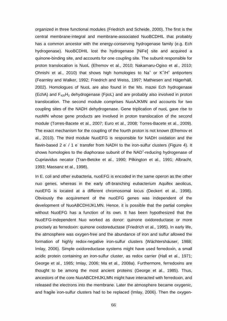
66
organized in three functional modules (Friedrich and Scheide, 2000). The first is the
central membrane-integral and membrane-associated NuoBCDHIL that probably
has a common ancestor with the energy-conserving hydrogenase family (e.g. Ech
hydrogenase). NuoBCDHIL lost the hydrogenase [NiFe] site and acquired a
quinone-binding site, and accounts for one coupling site. The subunit responsible for
proton translocation is NuoL (Efremov et al., 2010; Nakamaru-Ogiso et al., 2010;
Ohnishi et al., 2010) that shows high homologies to Na+ or K+/H+ antiporters
(Fearnley and Walker, 1992; Friedrich and Weiss, 1997; Mathiesen and Hägerhäll,
2002). Homologues of NuoL are also found in the Ms. mazei Ech hydrogenase
(EchA) and F420H2 dehydrogenase (FpoL) and are probably also involved in proton
translocation. The second module comprises NuoAJKMN and accounts for two
coupling sites of the NADH dehydrogenase. Gene triplication of nuoL gave rise to
nuoMN whose gene products are involved in proton translocation of the second
module (Torres-Bacete et al., 2007; Euro et al., 2008; Torres-Bacete et al., 2009).
The exact mechanism for the coupling of the fourth proton is not known (Efremov et
al., 2010). The third module NuoEFG is responsible for NADH oxidation and the
flavin-based 2 e- / 1 e- transfer from NADH to the iron-sulfur clusters (Figure 4). It
shows homologies to the diaphorase subunit of the NAD+-reducing hydrogenase of
Cupriavidus necator (Tran-Betcke et al., 1990; Pilkington et al., 1991; Albracht,
1993; Massanz et al., 1998). In E. coli and other eubacteria, nuoEFG is encoded in the same operon as the other
nuo genes, whereas in the early off-branching eubacterium Aquifex aeolicus,
nuoEFG is located at a different chromosomal locus (Deckert et al., 1998).
Obviously the acquirement of the nuoEFG genes was independent of the
development of NuoABCDHIJKLMN. Hence, it is possible that the partial complex
without NuoEFG has a function of its own. It has been hypothesized that the
NuoEFG-independent Nuo worked as donor: quinone oxidoreductase or more
precisely as ferredoxin: quinone oxidoreductase (Friedrich et al., 1995). In early life,
the atmosphere was oxygen-free and the abundance of iron and sulfur allowed the
formation of highly redox-negative iron-sulfur clusters (Wächtershäuser, 1988;
Imlay, 2006). Simple oxidoreductase systems might have used ferredoxin, a small
acidic protein containing an iron-sulfur cluster, as redox carrier (Hall et al., 1971;
George et al., 1985; Imlay, 2006; Ma et al., 2008a). Furthermore, ferredoxins are
thought to be among the most ancient proteins (George et al., 1985). Thus,
ancestors of the core NuoABCDHIJKLMN might have interacted with ferredoxin, and
released the electrons into the membrane. Later the atmosphere became oxygenic,
and fragile iron-sulfur clusters had to be replaced (Imlay, 2006). Then the oxygen-
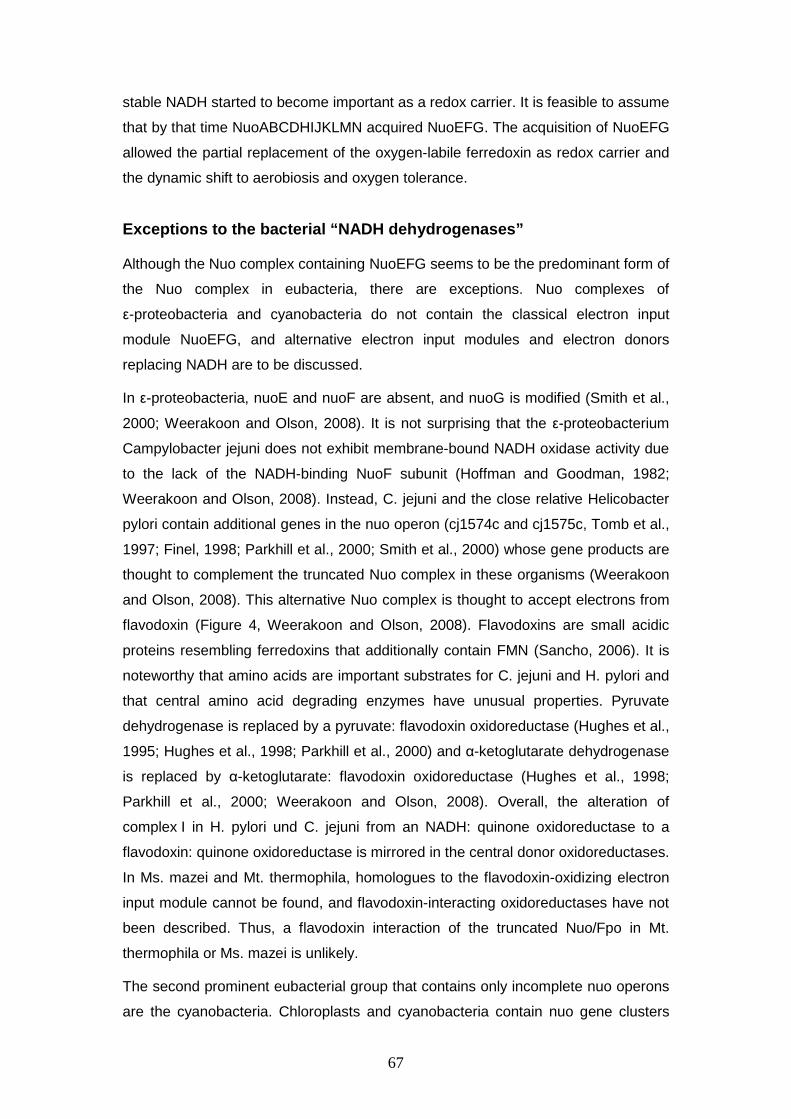
67
stable NADH started to become important as a redox carrier. It is feasible to assume
that by that time NuoABCDHIJKLMN acquired NuoEFG. The acquisition of NuoEFG
allowed the partial replacement of the oxygen-labile ferredoxin as redox carrier and
the dynamic shift to aerobiosis and oxygen tolerance.
Exceptions to the bacterial “NADH dehydrogenases” Although the Nuo complex containing NuoEFG seems to be the predominant form of
the Nuo complex in eubacteria, there are exceptions. Nuo complexes of
ε-proteobacteria and cyanobacteria do not contain the classical electron input
module NuoEFG, and alternative electron input modules and electron donors
replacing NADH are to be discussed. In ε-proteobacteria, nuoE and nuoF are absent, and nuoG is modified (Smith et al.,
2000; Weerakoon and Olson, 2008). It is not surprising that the ε-proteobacterium
Campylobacter jejuni does not exhibit membrane-bound NADH oxidase activity due
to the lack of the NADH-binding NuoF subunit (Hoffman and Goodman, 1982;
Weerakoon and Olson, 2008). Instead, C. jejuni and the close relative Helicobacter
pylori contain additional genes in the nuo operon (cj1574c and cj1575c, Tomb et al.,
1997; Finel, 1998; Parkhill et al., 2000; Smith et al., 2000) whose gene products are
thought to complement the truncated Nuo complex in these organisms (Weerakoon
and Olson, 2008). This alternative Nuo complex is thought to accept electrons from
flavodoxin (Figure 4, Weerakoon and Olson, 2008). Flavodoxins are small acidic
proteins resembling ferredoxins that additionally contain FMN (Sancho, 2006). It is
noteworthy that amino acids are important substrates for C. jejuni and H. pylori and
that central amino acid degrading enzymes have unusual properties. Pyruvate
dehydrogenase is replaced by a pyruvate: flavodoxin oxidoreductase (Hughes et al.,
1995; Hughes et al., 1998; Parkhill et al., 2000) and α-ketoglutarate dehydrogenase
is replaced by α-ketoglutarate: flavodoxin oxidoreductase (Hughes et al., 1998;
Parkhill et al., 2000; Weerakoon and Olson, 2008). Overall, the alteration of
complex I in H. pylori und C. jejuni from an NADH: quinone oxidoreductase to a
flavodoxin: quinone oxidoreductase is mirrored in the central donor oxidoreductases.
In Ms. mazei and Mt. thermophila, homologues to the flavodoxin-oxidizing electron
input module cannot be found, and flavodoxin-interacting oxidoreductases have not
been described. Thus, a flavodoxin interaction of the truncated Nuo/Fpo in Mt.
thermophila or Ms. mazei is unlikely. The second prominent eubacterial group that contains only incomplete nuo operons
are the cyanobacteria. Chloroplasts and cyanobacteria contain nuo gene clusters
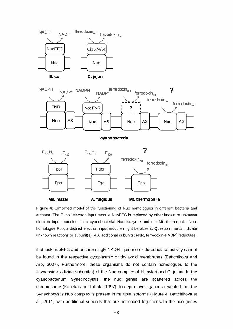
68
Nuo
NuoEFG
NADH NAD+
Nuo
Cj1574/5c
flavodoxinred flavodoxinox
Nuo
FNR
NADPHNADP+
Nuo
Not FNR
NADPHNADP+
Fpo
FpoF
F420H2 F420
AS AS
E. coli C. jejuni
cyanobacteria
Nuo
?
ferredoxinredferredoxinox
AS
Fqo
FqoF
F420H2 F420
Fpo
ferredoxinredferredoxinox
?
Ms. mazei A. fulgidus Mt. thermophila
Nuo
ferredoxinredferredoxinox
AS
?
Nuo
NuoEFG
NADH NAD+
Nuo
Cj1574/5c
flavodoxinred flavodoxinox
Nuo
FNR
NADPHNADP+
Nuo
Not FNR
NADPHNADP+
Fpo
FpoF
F420H2 F420
AS AS
E. coli C. jejuni
cyanobacteria
Nuo
?
ferredoxinredferredoxinox
AS
Fqo
FqoF
F420H2 F420
Fpo
ferredoxinredferredoxinox
?
Ms. mazei A. fulgidus Mt. thermophila
Nuo
ferredoxinredferredoxinox
AS
?
Figure 4: Simplified model of the functioning of Nuo homologues in different bacteria and
archaea. The E. coli electron input module NuoEFG is replaced by other known or unknown
electron input modules. In a cyanobacterial Nuo isozyme and the Mt. thermophila Nuo-
homologue Fpo, a distinct electron input module might be absent. Question marks indicate
unknown reactions or subunit(s). AS, additional subunits; FNR, ferredoxin-NADP+ reductase.
that lack nuoEFG and unsurprisingly NADH: quinone oxidoreductase activity cannot
be found in the respective cytoplasmic or thylakoid membranes (Battchikova and
Aro, 2007). Furthermore, these organisms do not contain homologues to the
flavodoxin-oxidizing subunit(s) of the Nuo complex of H. pylori and C. jejuni. In the
cyanobacterium Synechocystis, the nuo genes are scattered across the
chromosome (Kaneko and Tabata, 1997). In-depth investigations revealed that the
Synechocystis Nuo complex is present in multiple isoforms (Figure 4, Battchikova et
al., 2011) with additional subunits that are not coded together with the nuo genes
69
(Prommeenate et al., 2004; Zhang et al., 2004; Battchikova et al., 2005; Rumeau et
al., 2005; Nowaczyk et al., 2011). The cyanobacterial complex I isozymes seem to
fulfil various functions including roles in respiration, cyclic electron flow and CO2
uptake (Shibata et al., 2001; Battchikova and Aro, 2007). There is no consensus
about the electron donor or the electron input subunit(s) of the cyanobacterial Nuo
isozymes (Battchikova et al., 2011). However, for one complex I homologue it has been shown that NADPH serves as
electron donor (Figure 4, (Ma et al., 2006; Ma et al., 2008b) whereas for other
cyanobacterial Nuo complexes it was demonstrated that NADPH is not involved
(Prommeenate et al., 2004; Battchikova et al., 2011). Some studies identify the
electron input module of a NADPH-oxidizing Nuo complex as ferredoxin-NADP+
reductase (FNR, Guedeney et al., 1996; Funk et al., 1999). In contrast, other groups
find FNR separated from the NADPH-oxidizing Nuo complex (Ma et al., 2006). For some cyanobacterial or chloroplastid Nuo complexes, ferredoxin is hypothesized
to be the electron donor (Friedrich et al., 1995; Mi et al., 1995; Battchikova et al.,
2011). Either ferredoxin interacts with the cyanobacterial Nuo complex without an
electron input module as it is now hypothesized for the Mt. thermophila Fpo/Nuo
complex or additional proteins are involved in the binding of ferredoxin (Figure 4,
Prommeenate et al., 2004; Battchikova et al., 2011). Furthermore, there are
indications that photosystem I and the Nuo complex form a supercomplex in
chloroplasts with direct electron transfer (Peng et al., 2008; Sirpiö et al., 2009). Overall, the composition of the cyanobacterial Nuo complex isozymes is highly
diverse. For some isozymes the replacement of NuoEFG by another electron input
module has been demonstrated, whereas the electron input module and cofactor
reactivity of other isozymes remains enigmatic. A distinct ferredoxin-oxidizing Nuo
complex isozyme has been hypothesized but it has not been experimentally
validated, so an analogy to the possibly ferredoxin-interacting Nuo/Fpo complex of
Mt. thermophila is not possible.
“NADH dehydrogenases” in archaea In archaea, Nuo homologues have hardly been investigated. Archaeal genomes
always lack nuoEFG in their nuo gene clusters that are nevertheless widely
distributed. Sulofolobus solfataricus (She et al., 2001), Thermoplasma acidophilum
(Ruepp et al., 2000), Halobacterium salinarum (Ng et al., 2000), Aeropyrum pernix
(Kawarabayasi et al., 2001) as well as various members of the Methanosarcinales
(Ms. mazei, Ms. barkeri, Mt. thermophila) and the close relative Archaeoglobus
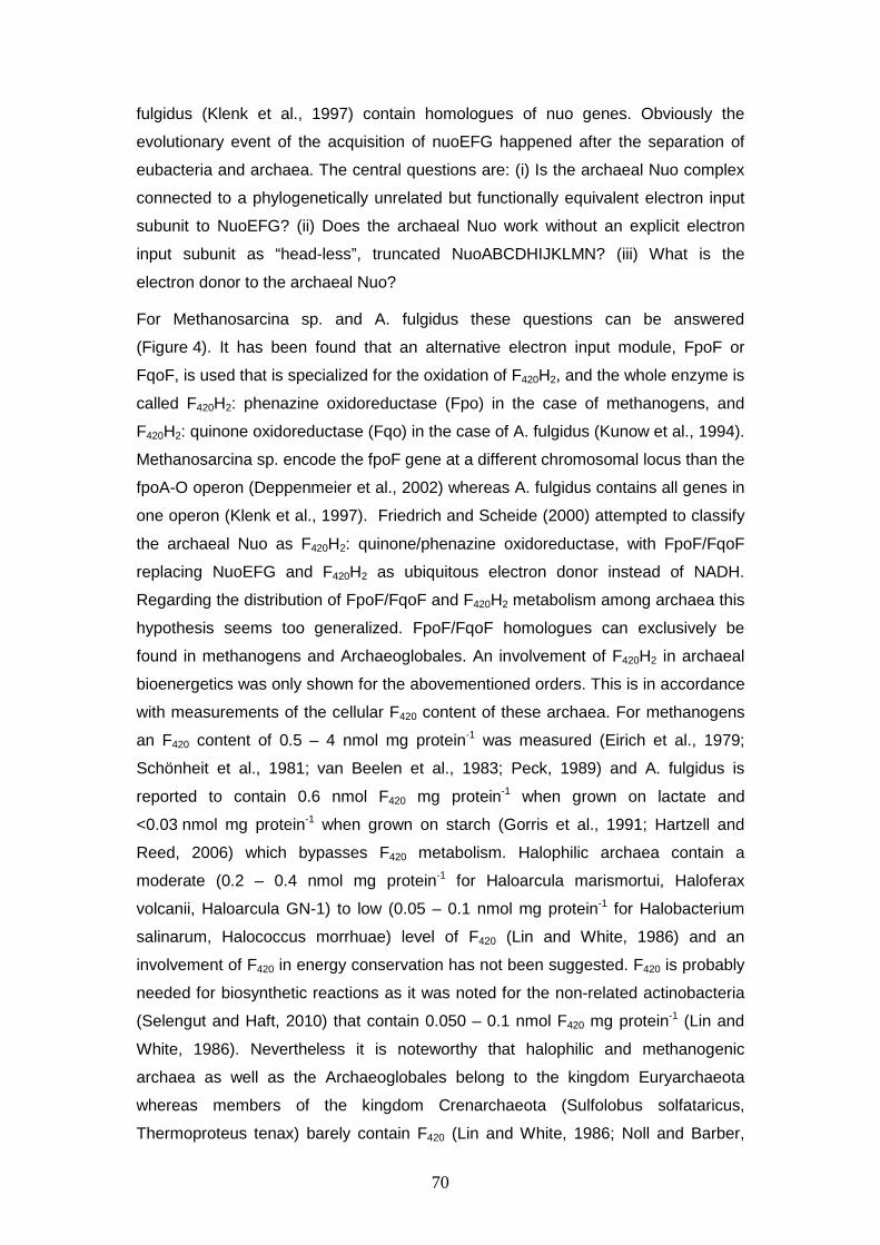
70
fulgidus (Klenk et al., 1997) contain homologues of nuo genes. Obviously the
evolutionary event of the acquisition of nuoEFG happened after the separation of
eubacteria and archaea. The central questions are: (i) Is the archaeal Nuo complex
connected to a phylogenetically unrelated but functionally equivalent electron input
subunit to NuoEFG? (ii) Does the archaeal Nuo work without an explicit electron
input subunit as “head-less”, truncated NuoABCDHIJKLMN? (iii) What is the
electron donor to the archaeal Nuo? For Methanosarcina sp. and A. fulgidus these questions can be answered
(Figure 4). It has been found that an alternative electron input module, FpoF or
FqoF, is used that is specialized for the oxidation of F420H2, and the whole enzyme is
called F420H2: phenazine oxidoreductase (Fpo) in the case of methanogens, and
F420H2: quinone oxidoreductase (Fqo) in the case of A. fulgidus (Kunow et al., 1994).
Methanosarcina sp. encode the fpoF gene at a different chromosomal locus than the
fpoA-O operon (Deppenmeier et al., 2002) whereas A. fulgidus contains all genes in
one operon (Klenk et al., 1997). Friedrich and Scheide (2000) attempted to classify
the archaeal Nuo as F420H2: quinone/phenazine oxidoreductase, with FpoF/FqoF
replacing NuoEFG and F420H2 as ubiquitous electron donor instead of NADH.
Regarding the distribution of FpoF/FqoF and F420H2 metabolism among archaea this
hypothesis seems too generalized. FpoF/FqoF homologues can exclusively be
found in methanogens and Archaeoglobales. An involvement of F420H2 in archaeal
bioenergetics was only shown for the abovementioned orders. This is in accordance
with measurements of the cellular F420 content of these archaea. For methanogens
an F420 content of 0.5 – 4 nmol mg protein-1 was measured (Eirich et al., 1979;
Schönheit et al., 1981; van Beelen et al., 1983; Peck, 1989) and A. fulgidus is
reported to contain 0.6 nmol F420 mg protein-1 when grown on lactate and
<0.03 nmol mg protein-1 when grown on starch (Gorris et al., 1991; Hartzell and
Reed, 2006) which bypasses F420 metabolism. Halophilic archaea contain a
moderate (0.2 – 0.4 nmol mg protein-1 for Haloarcula marismortui, Haloferax
volcanii, Haloarcula GN-1) to low (0.05 – 0.1 nmol mg protein-1 for Halobacterium
salinarum, Halococcus morrhuae) level of F420 (Lin and White, 1986) and an
involvement of F420 in energy conservation has not been suggested. F420 is probably
needed for biosynthetic reactions as it was noted for the non-related actinobacteria
(Selengut and Haft, 2010) that contain 0.050 – 0.1 nmol F420 mg protein-1 (Lin and
White, 1986). Nevertheless it is noteworthy that halophilic and methanogenic
archaea as well as the Archaeoglobales belong to the kingdom Euryarchaeota
whereas members of the kingdom Crenarchaeota (Sulfolobus solfataricus,
Thermoproteus tenax) barely contain F420 (Lin and White, 1986; Noll and Barber,
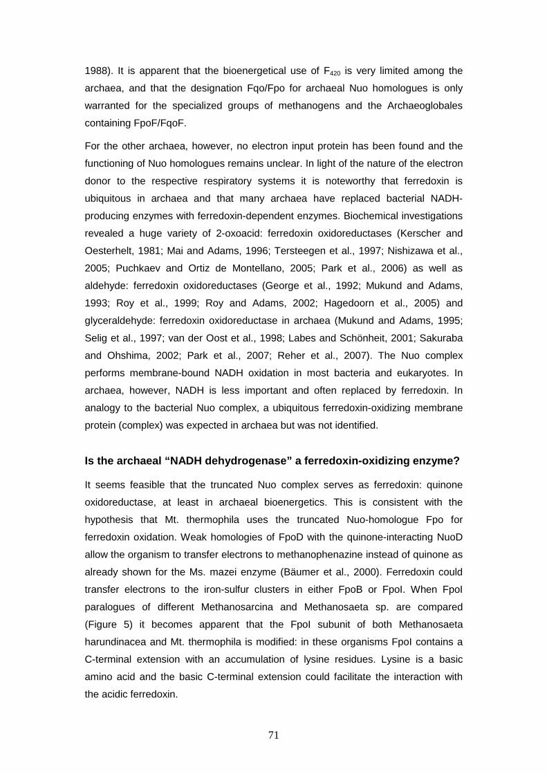
71
1988). It is apparent that the bioenergetical use of F420 is very limited among the
archaea, and that the designation Fqo/Fpo for archaeal Nuo homologues is only
warranted for the specialized groups of methanogens and the Archaeoglobales
containing FpoF/FqoF. For the other archaea, however, no electron input protein has been found and the
functioning of Nuo homologues remains unclear. In light of the nature of the electron
donor to the respective respiratory systems it is noteworthy that ferredoxin is
ubiquitous in archaea and that many archaea have replaced bacterial NADH-
producing enzymes with ferredoxin-dependent enzymes. Biochemical investigations
revealed a huge variety of 2-oxoacid: ferredoxin oxidoreductases (Kerscher and
Oesterhelt, 1981; Mai and Adams, 1996; Tersteegen et al., 1997; Nishizawa et al.,
2005; Puchkaev and Ortiz de Montellano, 2005; Park et al., 2006) as well as
aldehyde: ferredoxin oxidoreductases (George et al., 1992; Mukund and Adams,
1993; Roy et al., 1999; Roy and Adams, 2002; Hagedoorn et al., 2005) and
glyceraldehyde: ferredoxin oxidoreductase in archaea (Mukund and Adams, 1995;
Selig et al., 1997; van der Oost et al., 1998; Labes and Schönheit, 2001; Sakuraba
and Ohshima, 2002; Park et al., 2007; Reher et al., 2007). The Nuo complex
performs membrane-bound NADH oxidation in most bacteria and eukaryotes. In
archaea, however, NADH is less important and often replaced by ferredoxin. In
analogy to the bacterial Nuo complex, a ubiquitous ferredoxin-oxidizing membrane
protein (complex) was expected in archaea but was not identified.
Is the archaeal “NADH dehydrogenase” a ferredoxin-o xidizing enzyme? It seems feasible that the truncated Nuo complex serves as ferredoxin: quinone
oxidoreductase, at least in archaeal bioenergetics. This is consistent with the
hypothesis that Mt. thermophila uses the truncated Nuo-homologue Fpo for
ferredoxin oxidation. Weak homologies of FpoD with the quinone-interacting NuoD
allow the organism to transfer electrons to methanophenazine instead of quinone as
already shown for the Ms. mazei enzyme (Bäumer et al., 2000). Ferredoxin could
transfer electrons to the iron-sulfur clusters in either FpoB or FpoI. When FpoI
paralogues of different Methanosarcina and Methanosaeta sp. are compared
(Figure 5) it becomes apparent that the FpoI subunit of both Methanosaeta
harundinacea and Mt. thermophila is modified: in these organisms FpoI contains a
C-terminal extension with an accumulation of lysine residues. Lysine is a basic
amino acid and the basic C-terminal extension could facilitate the interaction with
the acidic ferredoxin.
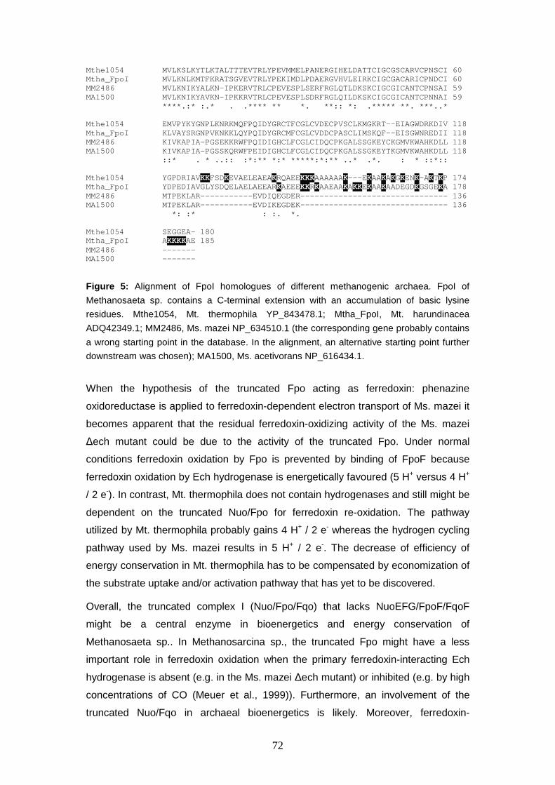
72
Mthe1054 MVLKSLKYTLKTALTTTEVTRLYPEVMMELPANERGIHELDATTCIGCGSCARVCPNSCI 60 Mtha_FpoI MVLKNLKMTFKRATSGVEVTRLYPEKIMDLPDAERGVHVLEIRKCIGCGACARICPNDCI 60 MM2486 MVLKNIKYALKN-IPKERVTRLCPEVESPLSERFRGLQTLDKSKCIGCGICANTCPNSAI 59 MA1500 MVLKNIKYAVKN-IPKKRVTRLCPEVESPLSDRFRGLQILDKSKCIGCGICANTCPNNAI 59 ****.:* :.* . .**** ** *. **:: *: .***** **. ***..* Mthe1054 EMVPYKYGNPLKNRKMQFPQIDYGRCTFCGLCVDECPVSCLKMGKRT--EIAGWDRKDIV 118 Mtha_FpoI KLVAYSRGNPVKNKKLQYPQIDYGRCMFCGLCVDDCPASCLIMSKQF--EISGWNREDII 118 MM2486 KIVKAPIA-PGSEKKRWFPQIDIGHCLFCGLCIDQCPKGALSSGKEYCKGMVKWAHKDLL 118 MA1500 KIVKAPIA-PGSSKQRWFPEIDIGHCLFCGLCIDQCPKGALSSGKEYTKGMVKWAHKDLL 118 ::* . * ..:: :*:** *:* *****:*:** ..* .*. : * ::*:: Mthe1054 YGPDRIAVKKFSDKEVAELEAEAKRQAEEKKKAAAAAAK---EKAAKAKGKENK-AKTKP 174 Mtha_FpoI YDPEDIAVGLYSDQELAELAEEARKAEEEKKRKAAEAAKAKKEKAAKAADEGDKGSGEKA 178 MM2486 MTPEKLAR-----------EVDIQEGDER------------------------------- 136 MA1500 MTPEKLAR-----------EVDIKEGDEK------------------------------- 136 *: :* : :. *. Mthe1054 SEGGEA- 180 Mtha_FpoI AKKKKAE 185 MM2486 ------- MA1500 -------
Figure 5: Alignment of FpoI homologues of different methanogenic archaea. FpoI of
Methanosaeta sp. contains a C-terminal extension with an accumulation of basic lysine
residues. Mthe1054, Mt. thermophila YP_843478.1; Mtha_FpoI, Mt. harundinacea
ADQ42349.1; MM2486, Ms. mazei NP_634510.1 (the corresponding gene probably contains
a wrong starting point in the database. In the alignment, an alternative starting point further
downstream was chosen); MA1500, Ms. acetivorans NP_616434.1.
When the hypothesis of the truncated Fpo acting as ferredoxin: phenazine
oxidoreductase is applied to ferredoxin-dependent electron transport of Ms. mazei it
becomes apparent that the residual ferredoxin-oxidizing activity of the Ms. mazei
∆ech mutant could be due to the activity of the truncated Fpo. Under normal
conditions ferredoxin oxidation by Fpo is prevented by binding of FpoF because
ferredoxin oxidation by Ech hydrogenase is energetically favoured (5 H+ versus 4 H+
/ 2 e-). In contrast, Mt. thermophila does not contain hydrogenases and still might be
dependent on the truncated Nuo/Fpo for ferredoxin re-oxidation. The pathway
utilized by Mt. thermophila probably gains 4 H+ / 2 e- whereas the hydrogen cycling
pathway used by Ms. mazei results in 5 H+ / 2 e-. The decrease of efficiency of
energy conservation in Mt. thermophila has to be compensated by economization of
the substrate uptake and/or activation pathway that has yet to be discovered. Overall, the truncated complex I (Nuo/Fpo/Fqo) that lacks NuoEFG/FpoF/FqoF
might be a central enzyme in bioenergetics and energy conservation of
Methanosaeta sp.. In Methanosarcina sp., the truncated Fpo might have a less
important role in ferredoxin oxidation when the primary ferredoxin-interacting Ech
hydrogenase is absent (e.g. in the Ms. mazei ∆ech mutant) or inhibited (e.g. by high
concentrations of CO (Meuer et al., 1999)). Furthermore, an involvement of the
truncated Nuo/Fqo in archaeal bioenergetics is likely. Moreover, ferredoxin-

73
dependent electron transport might be of greater importance than previously
thought.
74
Chapter 8
Summary
1) Aceticlastic and methylotrophic methanogens produce reduced ferredoxin in the
course of methane formation that is re-oxidized by membrane-bound enzymes. To
study ferredoxin-dependent electron transport processes, a defined system for
continuous generation of reduced ferredoxin was established. The CO-oxidizing CO
dehydrogenase/acetyl-CoA synthase from Moorella thermoacetica was used to
reduce Clostridium pasteurianum ferredoxin or the methanogenic ferredoxin
MM1619. This system was successfully used to study different ferredoxin-
dependent electron transport pathways. 2) Ferredoxin-dependent heterodisulfide reduction was measured in Ms. mazei
membrane preparations for the first time in the course of this work. Ms. mazei
mutants lacking Ech hydrogenase exhibited significantly reduced ferredoxin-
dependent electron transport rates by about 50 % in comparison to the wildtype.
The involvement of Ech hydrogenase in the ferredoxin: heterodisulfide
oxidoreductase system was clearly demonstrated. Furthermore, a second
membrane-bound protein responsible for the residual 50 % ferredoxin-dependent
heterodisulfide reduction was postulated. 3) The Ms. mazei ∆ech mutant exhibited a pronounced phenotype with respect to
growth parameters. It showed slower growth, less biomass yield and accelerated
substrate consumption when grown on trimethylamine. Furthermore, the mutant did
not grow on acetate. These findings underlined a possible energy conserving
function of Ech hydrogenase. In addition, it became evident that a second
ferredoxin-oxidizing pathway is necessary to explain the survival of the mutant when
grown on trimethylamine. 4) To address the question whether Ech hydrogenase belongs to the energy-
converting hydrogenase family, experiments with inverted membrane vesicles from
Ms. mazei were performed. These experiments demonstrated that Ech hydrogenase
is able to form molecular hydrogen by the oxidation of reduced ferredoxin. This
process was coupled to the translocation of ions over the cytoplasmic membrane
and ATP was synthesized by electron transport phosphorylation. The translocated
ion was identified as proton and Na+ ion transport was excluded. Theoretical
considerations led to a 1 H+ / 2 e- stoichiometry. 5) F420H2 is a reducing equivalent that is produced in hydrogenotrophic and
methylotrophic methanogenesis. The oxidation of F420H2 in Methanosarcina sp. can
75
proceed via the membrane-bound F420H2 dehydrogenase (Fpo) or the soluble F420
hydrogenase (Frh). Frh produces molecular hydrogen in the course of F420H2
oxidation that is used by the uptake hydrogenase Vho. To find out which pathway is
preferred by Ms. mazei two mutants of the F420H2 dehydrogenase (∆fpoF and
∆fpoA-O) were constructed. Both mutants grew slower than the wildtype on
trimethylamine, but exhibited similar growth rates compared to the parental strain
when grown on trimethylamine + H2 or acetate. Enzymatic activity measurements of
cell lysates, membranes, and cytoplasmic fractions revealed that the majority of
F420H2 oxidation in Ms. mazei is performed by F420H2 dehydrogenase and only a
minor part by F420 hydrogenase. 6) The characterization of the Ms. mazei fpo mutants revealed that the deletion of
fpoF leads not only to an alteration of membrane-bound electron transport but also
to a change of enzyme activities in the cytoplasm. The Ms. mazei ∆fpoF mutant no
longer catalyzed the ferredoxin: F420 oxidoreductase reaction. Subsequently, FpoF
was heterologously produced in E. coli and purified to homogeneity. The
recombinant FpoF was characterized as ferredoxin: F420 oxidoreductase and
contained one FAD and two [4Fe4S] clusters per monomer. Quantitative Western
blot experiments showed that FpoF is found in the membrane fraction where it is
bound to the FpoA-O complex but was also present in substantial quantities in the
cytoplasmic fraction. 7) Energy conservation in Methanosarcina sp. is by now well investigated, whereas
energy conservation pathways of members of the obligate aceticlastic genus
Methanosaeta remain largely unknown. During this work, membrane-bound electron
transport pathways of Mt. thermophila were investigated. Mt. thermophila was found
to possess a highly active membrane-bound ferredoxin: heterodisulfide
oxidoreductase. The Mt. thermophila genome does not contain genes coding for
known ferredoxin-interacting membrane proteins (Ech hydrogenase, Rnf complex).
However, it contains genes coding for the F420H2 dehydrogenase (without the
electron input subunit FpoF) although the membrane does not interact with F420H2.
As indicated in chapter 7, the F420H2 dehydrogenase is highly homologous to the
bacterial NADH dehydrogenase I (complex I). Replacements of the electron input
module of complex I enables the enzyme to oxidize different electron carriers
(NADPH, flavodoxin) in ε-proteobacteria and cyanobacteria. In archaea, NADH
dehydrogenase I homologues mostly appear without a distinct electron input
module. Hence, it is tempting to speculate that in Mt. thermophila the truncated
F420H2 dehydrogenase, as a homologue of complex I, is responsible for ferredoxin
oxidation, thereby channelling electrons into the respiratory chain.
76
Reference list Abken HJ, Tietze M, Brodersen J, Bäumer S, Beifuss U, Deppenmeier U (1998) Isolation
and characterization of methanophenazine and function of phenazines in membrane-bound electron transport of Methanosarcina mazei Gö1. J Bacteriol 180: 2027-2032.
Albracht SP (1993) Intimate relationships of the large and the small subunits of all nickel hydrogenases with two nuclear-encoded subunits of mitochondrial NADH: ubiquinone oxidoreductase. Biochim Biophys Acta 1144: 221-224.
Alex LA, Reeve JN, Orme-Johnson WH, Walsh CT (1990) Cloning, sequence determination, and expression of the genes encoding the subunits of the Nickel-containing 8-hydroxy-5-deazaflavin reducing hydrogenase from Methanobacterium thermoautotrophicum ∆H. Biochemistry 29: 7237-7244.
Allen GWJ, Zinder SH (1996) Methanogenesis from acetate by cell-free extracts of the thermophilic acetotrophic methanogen Methanothrix thermophila CALS-1. Arch Microbiol 166: 275-281.
Allen MA, Lauro FM, Williams TJ, Burg D, Siddiqui KS, De Francisci D, Chong KWY, Pilak O, Chew HH, De Maere MZ, et al. (2009) The genome sequence of the psychrophilic archaeon Methanococcoides burtonii: the role of genome evolution in cold adaptation. ISME J 3: 1012-1035.
Andrews SC, Berks BC, McClay J, Ambler A, Quail MA, Golby P, Guest JR (1997) A 12-cistron Escherichia coli operon (hyf) encoding a putative proton-translocating formate hydrogenlyase system. Microbiology 143: 3633-3647.
Bartoschek S, Vorholt JA, Thauer RK, Geierstanger BH, Griesinger C (2000) N-carboxymethanofuran (carbamate) formation from methanofuran and CO2 in methanogenic archaea. Thermodynamics and kinetics of the spontaneous reaction. Eur J Biochem 267: 3130-3138.
Battchikova N, Aro EM (2007) Cyanobacterial NDH-1 complexes: multiplicity in function and subunit composition. Physiol Plant 131: 22-32.
Battchikova N, Eisenhut M, Aro EM (2011) Cyanobacterial NDH-1 complexes: Novel insights and remaining puzzles. Biochim Biophys Acta 1807:935-944.
Battchikova N, Zhang P, Rudd S, Ogawa T, Aro EM (2005) Identification of NdhL and Ssl1690 (NdhO) in NDH-1L and NDH-1M complexes of Synechocystis sp. PCC 6803. J Biol Chem 280: 2587-2595.
Bäumer S, Ide T, Jacobi C, Johann A, Gottschalk G, Deppenmeier U (2000) The F420H2 dehydrogenase from Methanosarcina mazei is a redox-driven proton pump closely related to NADH dehydrogenases. J Biol Chem 275: 17968-17973.
Bäumer S, Murakami E, Brodersen J, Gottschalk G, Ragsdale SW, Deppenmeier U (1998) The F420H2:heterodisulfide oxidoreductase system from Methanosarcina species. 2-Hydroxyphenazine mediates electron transfer from F420H2 dehydrogenase to heterodisulfide reductase. FEBS Lett 428: 295-298.
Becher B, Müller V, Gottschalk G (1992) N5-methyl-tetrahydromethanopterin:coenzyme M methyltransferase of Methanosarcina strain Gö1 is an Na+-translocating membrane protein. J Bacteriol 174: 7656-7660.
Beifuss U, Tietze M (2000) On the total synthesis of (S)-methanophenazine and the formal synthesis of (R)-methanophenazine from a common precursor. Tetrahedron Lett 41: 9759-9763.
Beifuss U, Tietze M (2005) Methanophenazine and other natural biologically active phenazines. Top Curr Chem 244: 77-113.
Berk H, Thauer RK (1998) F420H2 : NADP oxidoreductase from Methanobacterium thermoautotrophicum: identification of the encoding gene via functional overexpression in Escherichia coli. FEBS Lett 438: 124-126.
77
Biegel E, Müller V (2010) Bacterial Na+-translocating ferredoxin:NAD+ oxidoreductase. Proc Natl Acad Sci U S A 107: 18138-18142.
Blaut M, Gottschalk G (1984) Coupling of ATP synthesis and methane formation from methanol and molecular hydrogen in Methanosarcina barkeri. Eur J Biochem 141: 217-222.
Blaut M, Müller V, Gottschalk G (1987) Proton translocation coupled to methanogenesis from methanol + hydrogen in Methanosarcina barkeri. FEBS Lett 215: 53-57.
Böhm R, Sauter M, Böck A (1990) Nucleotide sequence and expression of an operon in Escherichia coli coding for formate hydrogenlyase components. Mol Microbiol 4: 231-243.
Bomsel JL, Pradet A (1968) Study of adenosine 5'-mono-, di- and triphosphates in plant tissues. IV. Regulation of the level of nucleotides, in vivo, by adenylate kinase: Theoretical and experimental study. Biochim Biophys Acta 162: 230-242.
Boone DR, Whitman WB, Rouvière PE (1993). Diversity and taxonomy of methanogens. Methanogenesis. J. G. Ferry. New York, Chapman & Hall: 35-80.
Bott M, Thauer RK (1989) Proton translocation coupled to the oxidation of carbon monoxide to CO2 and H2 in Methanosarcina barkeri. Eur J Biochem 179: 469-472.
Bradford MM (1976) A rapid and sensitive method for the quantitation of microgram quantities of protein utilizing the principle of protein-dye binding. Anal Biochem 72: 248-254.
Brodersen J, Bäumer S, Abken HJ, Gottschalk G, Deppenmeier U (1999a) Inhibition of membrane-bound electron transport of the methanogenic archaeon Methanosarcina mazei Gö1 by diphenyleneiodonium. Eur J Biochem 259: 218-224.
Brodersen J, Gottschalk G, Deppenmeier U (1999b) Membrane-bound F420H2-dependent heterodisulfide reduction in Methanococcus voltae. Arch Microbiol 171: 115-121.
Brüggemann H, Falinski F, Deppenmeier U (2000) Structure of the F420H2 : quinone oxidoreductase of Archaeoglobus fulgidus - Identification and overproduction of the F420H2-oxidizing subunit. Eur J Biochem 267: 5810-5814.
Clements AP, Kilpatrick L, Lu WP, Ragsdale SW, Ferry JG (1994) Characterization of the iron-sulfur clusters in ferredoxin from acetate-grown Methanosarcina thermophila. J Bacteriol 176: 2689-2693.
de Poorter LM, Geerts WG, Theuvenet AP, Keltjens JT (2003) Bioenergetics of the formyl-methanofuran dehydrogenase and heterodisulfide reductase reactions in Methanothermobacter thermautotrophicus. Eur J Biochem 270: 66-75.
Deckert G, Warren PV, Gaasterland T, Young WG, Lenox AL, Graham DE, Overbeek R, Snead MA, Keller M, Aujay M, et al. (1998) The complete genome of the hyperthermophilic bacterium Aquifex aeolicus. Nature 392: 353-358.
Deppenmeier U (2002a) Redox-driven proton translocation in methanogenic archaea. Cell Mol Life Sci 59: 1513-1533.
Deppenmeier U (2002b) The unique biochemistry of methanogenesis. Prog Nucleic Acid Res Mol Biol 71: 223-283.
Deppenmeier U (2004) The membrane-bound electron transport system of Methanosarcina species. J Bioenerg Biomembr 36: 55-64.
Deppenmeier U, Blaut M, Jussofie A, Gottschalk G (1988) A methyl-CoM methylreductase system from methanogenic bacterium strain Gö 1 not requiring ATP for activity. FEBS Lett 241: 60-64.
Deppenmeier U, Blaut M, Mahlmann A, Gottschalk G (1990) Reduced coenzyme F420: heterodisulfide oxidoreductase, a proton- translocating redox system in methanogenic bacteria. Proc Natl Acad Sci U S A 87: 9449-9453.
78
Deppenmeier U, Blaut M, Schmidt B, Gottschalk G (1992) Purification and properties of a F420-nonreactive, membrane-bound hydrogenase from Methanosarcina strain Gö1. Arch Microbiol 157: 505-511.
Deppenmeier U, Johann A, Hartsch T, Merkl R, Schmitz RA, Martinez-Arias R, Henne A, Wiezer A, Bäumer S, Jacobi C, et al. (2002) The genome of Methanosarcina mazei: evidence for lateral gene transfer between bacteria and archaea. J Mol Microbiol Biotechnol 4: 453-461.
Deppenmeier U, Müller V (2008) Life close to the thermodynamic limit: how methanogenic archaea conserve energy. Results Probl Cell Differ 45: 123-152.
Deppenmeier U, Müller V, Gottschalk G (1996) Pathways of energy conservation in methanogenic archaea. Arch Microbiol 165: 149-163.
Ding YH, Ferry JG (2004) Flavin mononucleotide-binding flavoprotein family in the domain Archaea. J Bacteriol 186: 90-97.
Ebner S, Jaun B, Goenrich M, Thauer RK, Harmer J (2010) Binding of coenzyme B induces a major conformational change in the active site of methyl-coenzyme M reductase. J Am Chem Soc 132: 567-575.
Efremov RG, Baradaran R, Sazanov LA (2010) The architecture of respiratory complex I. Nature 465: 441-445.
Ehlers C, Weidenbach K, Veit K, Deppenmeier U, Metcalf WW, Schmitz RA (2005) Development of genetic methods and construction of a chromosomal glnK1 mutant in Methanosarcina mazei strain Gö1. Mol Genet Genomics 273: 290-298.
Eirich LD, Vogels GD, Wolfe RS (1979) Distribution of coenzyme F420 and properties of its hydrolytic fragments. J Bacteriol 140: 20-27.
Elias DA, Juck DF, Berry KA, Sparling R (2000) Purification of the NADP+: F420 oxidoreductase of Methanosphaera stadtmanae. Can J Microbiol 46: 998-1003.
Ellermann J, Hedderich R, Böcher R, Thauer RK (1988) The final step in methane formation. Investigations with highly purified methyl-CoM reductase (component C) from Methanobacterium thermoautotrophicum (strain Marburg). Eur J Biochem 172: 669-677.
Ellermann J, Kobelt A, Pfaltz A, Thauer RK (1987) On the role of N-7-mercaptoheptanoyl-O-phospho-L-threonine (component B) in the enzymatic reduction of methyl-coenzyme M to methane. FEBS Lett 220: 358-362.
Ellman GL (1958) A colorimetric method for determining low concentrations of mercaptans. Arch Biochem Biophys 74: 443-450.
Euro L, Belevich G, Verkhovsky MI, Wikström M, Verkhovskaya M (2008) Conserved lysine residues of the membrane subunit NuoM are involved in energy conversion by the proton-pumping NADH:ubiquinone oxidoreductase (Complex I). Biochim Biophys Acta 1777: 1166-1172.
Fearnley IM, Walker JE (1992) Conservation of sequences of subunits of mitochondrial complex I and their relationships with other proteins. Biochim Biophys Acta 1140: 105-134.
Ferguson DJ Jr., Gorlatova N, Grahame DA, Krzycki JA (2000) Reconstitution of dimethylamine:coenzyme M methyl transfer with a discrete corrinoid protein and two methyltransferases purified from Methanosarcina barkeri. J Biol Chem 275: 29053-29060.
Ferry JG (1997) Enzymology of the fermentation of acetate to methane by Methanosarcina thermophila. Biofactors 6: 25-35.
Ferry JG (1999) Enzymology of one-carbon metabolism in methanogenic pathways. FEMS Microbiol Rev 23: 13-38.
Ferry JG, Lessner DJ (2008) Methanogenesis in marine sediments. Ann N Y Acad Sci 1125: 147-157.
79
Finel M (1998) Does NADH play a central role in energy metabolism in Helicobacter pylori? Trends Biochem Sci 23: 412-413.
Fischer R, Thauer RK (1989) Methyltetrahydromethanopterin as an intermediate in methanogenesis from acetate in Methanosarcina barkeri. Arch Microbiol 151: 459-465.
Fischer R, Thauer RK (1990) Ferredoxin-dependent methane formation from acetate in cell extracts of Methanosarcina barkeri (strain MS). FEBS Lett 269: 368-372.
Fox JA, Livingston DJ, Orme-Johnson WH, Walsh CT (1987) 8-hydroxy-5-deazaflavin-reducing hydrogenase from Methanobacterium thermoautotrophicum. 1. Purification and characterization. Biochemistry 26: 4219-4227.
Fox JD, Kerby RL, Roberts GP, Ludden PW (1996) Characterization of the CO-induced, CO-tolerant hydrogenase from Rhodospirillum rubrum and the gene encoding the large subunit of the enzyme. J Bacteriol 178: 1515-1524.
Franke-Whittle IH, Goberna M, Pfister V, Insam H (2009) Design and development of the ANAEROCHIP microarray for investigation of methanogenic communities. J Microbiol Meth 79: 279-288.
Franzmann PD, Springer N, Ludwig W, Demacario EC, Rohde M (1992) A methanogenic archaeon from Ace Lake, Antarctica - Methanococcoides burtonii sp. nov. Syst Appl Microbiol 15: 573-581.
Friedrich T, Scheide D (2000) The respiratory complex I of bacteria, archaea and eukarya and its module common with membrane-bound multisubunit hydrogenases. FEBS Lett 479: 1-5.
Friedrich T, Steinmüller K, Weiss H (1995) The proton-pumping respiratory complex I of bacteria and mitochondria and its homolog in chloroplasts. FEBS Lett 367: 107-111.
Friedrich T, Weiss H (1997) Modular evolution of the respiratory NADH:ubiquinone oxidoreductase and the origin of its modules. J Theor Biol 187: 529-540.
Funk E, Schäfer E, Steinmüller K (1999) Characterization of the complex I-homologous NAD(P)H-plastoquinone-oxidoreductase (NDH-complex) of maize chloroplasts. J Plant Physiol 154: 16-23.
Galagan JE, Nusbaum C, Roy A, Endrizzi MG, Macdonald P, FitzHugh W, Calvo S, Engels R, Smirnov S, Atnoor D, et al. (2002) The genome of M. acetivorans reveals extensive metabolic and physiological diversity. Genome Res 12: 532-542.
Gencic S, Duin EC, Grahame DA (2010) Tight coupling of partial reactions in the acetyl-CoA decarbonylase/synthase (ACDS) multienzyme complex from Methanosarcina thermophila: acetyl C-C bond fragmentation at the A cluster promoted by protein conformational changes. J Biol Chem 285: 15450-15463.
George DG, Hunt LT, Yeh LS, Barker WC (1985) New perspectives on bacterial ferredoxin evolution. J Mol Evol 22: 20-31.
George GN, Prince RC, Mukund S, Adams MWW (1992) Aldehyde ferredoxin oxidoreductase from the hyperthermophilic archaebacterium Pyrococcus furiosus contains a tungsten oxo-thiolate center. J Am Chem Soc 114: 3521-3523.
Gong W, Hao B, Wei Z, Ferguson DJ, Tallant T, Krzycki JA, Chan MK (2008) Structure of the alpha(2)epsilon(2) Ni-dependent CO dehydrogenase component of the Methanosarcina barkeri acetyl-CoA decarbonylase/synthase complex. Proc Natl Acad Sci U S A 105: 9558-9563.
Gorris LGM, Voet ACWA, van der Drift C (1991) Structural characteristics of methanogenic cofactors in the non-methanogenic archaebacterium Archaeoglobus fulgidus. Biofactors 3: 29-35.
Gottschalk G, Thauer RK (2001) The Na+-translocating methyltransferase complex from methanogenic archaea. Biochim Biophys Acta 1505: 28-36.
80
Guedeney G, Corneille S, Cuiné S, Peltier G (1996) Evidence for an association of ndhB, ndhJ gene products and ferredoxin-NADP-reductase as components of a chloroplastic NAD(P)H dehydrogenase complex. FEBS Lett 378: 277-280.
Guss AM, Kulkarni G, Metcalf WW (2009) Differences in hydrogenase gene expression between Methanosarcina acetivorans and Methanosarcina barkeri. J Bacteriol 191: 2826-2833.
Guss AM, Mukhopadhyay B, Zhang JK, Metcalf WW (2005) Genetic analysis of mch mutants in two Methanosarcina species demonstrates multiple roles for the methanopterin-dependent C1 oxidation/reduction pathway and differences in H2 metabolism between closely related species. Mol Microbiol 55: 1671-1680.
Hagedoorn PL, Chen T, Schröder I, Piersma SR, de Vries S, Hagen WR (2005) Purification and characterization of the tungsten enzyme aldehyde:ferredoxin oxidoreductase from the hyperthermophilic denitrifier Pyrobaculum aerophilum. J Biol Inorg Chem 10: 259-269.
Hall DO, Cammack R, Rao KK (1971) Role for ferredoxins in the origin of life and biological evolution. Nature 233: 136-138.
Hartzell P, Reed DW (2006). The genus Archaeoglobus. The Procaryotes. M. Dworkin, S. Falkow, E. Rosenberg, K.-H. Schleifer and E. Stackebrandt. New York, Springer. 3: 82-100.
Hedderich R, Forzi L (2005) Energy-converting [NiFe] hydrogenases: more than just H2
activation. J Mol Microbiol Biotechnol 10: 92-104. Hedderich R, Hamann N, Bennati M (2005) Heterodisulfide reductase from methanogenic
archaea: a new catalytic role for an iron-sulfur cluster. Biol Chem 386: 961-970. Heiden S, Hedderich R, Setzke E, Thauer RK (1994) Purification of a 2-subunit cytochrome-
b containing heterodisulfide reductase from methanol-grown Methanosarcina barkeri. Eur J Biochem 221: 855-861.
Hippe H, Caspari D, Fiebig K, Gottschalk G (1979) Utilization of trimethylamine and other N-methyl compounds for growth and methane formation by Methanosarcina barkeri. Proc Natl Acad Sci U S A 76: 494-498.
Hoffman PS, Goodman TG (1982) Respiratory physiology and energy conservation efficiency of Campylobacter jejuni. J Bacteriol 150: 319-326.
Hovey R, Lentes S, Ehrenreich A, Salmon K, Saba K, Gottschalk G, Gunsalus RP, Deppenmeier U (2005) DNA microarray analysis of Methanosarcina mazei Gö1 reveals adaptation to different methanogenic substrates. Mol Genet Genomics 273: 225-239.
Hughes NJ, Chalk PA, Clayton CL, Kelly DJ (1995) Identification of carboxylation enzymes and characterization of a novel four-subunit pyruvate:flavodoxin oxidoreductase from Helicobacter pylori. J Bacteriol 177: 3953-3959.
Hughes NJ, Clayton CL, Chalk PA, Kelly DJ (1998) Helicobacter pylori porCDAB and oorDABC genes encode distinct pyruvate:flavodoxin and 2-oxoglutarate:acceptor oxidoreductases which mediate electron transport to NADP. J Bacteriol 180: 1119-1128.
Ide T, Bäumer S, Deppenmeier U (1999) Energy conservation by the H2:heterodisulfide oxidoreductase from Methanosarcina mazei Gö1: identification of two proton-translocating segments. J Bacteriol 181: 4076-4080.
Imlay JA (2006) Iron-sulphur clusters and the problem with oxygen. Mol Microbiol 59: 1073-1082.
Jacobson FS, Daniels L, Fox JA, Walsh CT, Orme-Johnson WH (1982) Purification and properties of an 8-hydroxy-5-deazaflavin reducing hydrogenase from Methanobacterium thermoautotrophicum. J Biol Chem 257: 3385-3388.
81
Jain N, Pathak H, Mitra S, Bhatia A (2004) Emission of methane from rice fields - A review. J Sci Ind Res 63: 101-115.
Jeoung JH, Dobbek H (2007) Carbon dioxide activation at the Ni,Fe-cluster of anaerobic carbon monoxide dehydrogenase. Science 318: 1461-1464.
Jetten MS, Fluit TJ, Stams AJ, Zehnder AJ (1992a) A fluoride-insensitive inorganic pyrophosphatase isolated from Methanothrix soehngenii. Arch Microbiol 157: 284-289.
Jetten MS, Stams AJ, Zehnder AJ (1989) Isolation and characterization of acetyl-coenzyme A synthetase from Methanothrix soehngenii. J Bacteriol 171: 5430-5435.
Jetten MSM, Stams AJM, Zehnder AJB (1992b) Methanogenesis from acetate - a comparison of the acetate metabolism in Methanothrix soehngenii and Methanosarcina spp. FEMS Microbiol Rev 88: 181-197.
Jussofie A, Mayer F, Gottschalk G (1986) Methane formation from methanol and molecular hydrogen by protoplasts of new methanogenic isolates and inhibition by dicyclohexylcarbodiimide. Arch Microbiol 146: 245-249.
Kaesler B, Schönheit P (1989) The sodium cycle in methanogenesis. CO2 reduction to the formaldehyde level in methanogenic bacteria is driven by a primary electrochemical potential of Na+ generated by formaldehyde reduction to CH4. Eur J Biochem 186: 309-316.
Kamagata Y, Kawasaki H, Oyaizu H, Nakamura K, Mikami E, Endo G, Koga Y, Yamasato K (1992) Characterization of 3 thermophilic strains of Methanothrix (Methanosaeta) thermophila sp. nov. and rejection of Methanothrix (Methanosaeta) thermoacetophila. Int J Syst Bacteriol 42: 463-468.
Kaneko T, Tabata S (1997) Complete genome structure of the unicellular cyanobacterium Synechocystis sp. PCC6803. Plant Cell Physiol 38: 1171-1176.
Karakashev D, Batstone DJ, Angelidaki I (2005) Influence of environmental conditions on methanogenic compositions in anaerobic biogas reactors. Appl Environ Microbiol 71: 331-338.
Kashyap DR, Dadhich KS, Sharma SK (2003) Biomethanation under psychrophilic conditions: a review. Bioresour Technol 87: 147-153.
Kaster AK, Moll J, Parey K, Thauer RK (2011) Coupling of ferredoxin and heterodisulfide reduction via electron bifurcation in hydrogenotrophic methanogenic archaea. Proc Natl Acad Sci U S A 108: 2981-2986.
Kawarabayasi Y, Hino Y, Horikawa H, Jin-no K, Takahashi M, Sekine M, Baba S, Ankai A, Kosugi H, Hosoyama A, et al. (2001) Complete genome sequence of an aerobic thermoacidophilic crenarchaeon, Sulfolobus tokodaii strain 7. DNA Res 8: 123-140.
Kendall M, Boone D (2006). The order Methanosarcinales. The Procaryotes. M. Dworkin, S. Falkow, E. Rosenberg, K.-H. Schleifer and E. Stackebrandt. New York, Springer. 3: 244-256.
Kerscher L, Oesterhelt D (1981) Purification and properties of two 2-oxoacid:ferredoxin oxidoreductases from Halobacterium halobium. Eur J Biochem 116: 587-594.
Khalil MAK, Rasmussen RA (1994) Global emissions of methane during the last several centuries. Chemosphere 29: 833-842.
Kimmich GA, Randles J, Brand JS (1975) Assay of picomole amounts of ATP, ADP, and AMP using the luciferase enzyme system. Anal Biochem 69: 187-206.
Klenk HP, Clayton RA, Tomb JF, White O, Nelson KE, Ketchum KA, Dodson RJ, Gwinn M, Hickey EK, Peterson JD, et al. (1997) The complete genome sequence of the hyperthermophilic, sulphate-reducing archaeon Archaeoglobus fulgidus. Nature 390: 364-370.
Kobelt A, Pfaltz A, Ankelfuchs D, Thauer RK (1987) The L-form of N-7-mercaptoheptanoyl-O-phosphothreonine is the enantiomer active as component B in methyl-CoM reduction to methane. FEBS Lett 214: 265-268.
82
Krätzer C, Carini P, Hovey R, Deppenmeier U (2009) Transcriptional profiling of methyltransferase genes during growth of Methanosarcina mazei on trimethylamine. J Bacteriol 191: 5108-5115.
Kulkarni G, Kridelbaugh DM, Guss AM, Metcalf WW (2009) Hydrogen is a preferred intermediate in the energy-conserving electron transport chain of Methanosarcina barkeri. Proc Natl Acad Sci U S A 106: 15915-15920.
Künkel A, Vaupel M, Heim S, Thauer RK, Hedderich R (1997) Heterodisulfide reductase from methanol-grown cells of Methanosarcina barkeri is not a flavoenzyme. Eur J Biochem 244: 226-234.
Kunow J, Linder D, Stetter KO, Thauer RK (1994) F420H2:quinone oxidoreductase from Archaeoglobus fulgidus - characterization of a membrane-bound multisubunit complex containing FAD and iron-sulfur clusters. Eur J Biochem 223: 503-511.
Kunow J, Schwörer B, Stetter KO, Thauer RK (1993) A F420-dependent NADP reductase in the extremely thermophilic sulfate-reducing Archaeoglobus fulgidus. Arch Microbiol 160: 199-205.
Labes A, Schönheit P (2001) Sugar utilization in the hyperthermophilic, sulfate-reducing archaeon Archaeoglobus fulgidus strain 7324: starch degradation to acetate and CO2 via a modified Embden-Meyerhof pathway and acetyl-CoA synthetase (ADP-forming). Arch Microbiol 176: 329-338.
Laemmli UK (1970) Cleavage of structural proteins during assembly of head of bacteriophage T4. Nature 227: 680-685.
Lee C, Kim J, Hwang K, O'Flaherty V, Hwang S (2009a) Quantitative analysis of methanogenic community dynamics in three anaerobic batch digesters treating different wastewaters. Water Res 43: 157-165.
Lee M, Hidaka T, Hagiwara W, Tsuno H (2009b) Comparative performance and microbial diversity of hyperthermophilic and thermophilic co-digestion of kitchen garbage and excess sludge. Bioresour Technol 100: 578-585.
Lessner DJ, Ferry JG (2007) The archaeon Methanosarcina acetivorans contains a protein disulfide reductase with an iron-sulfur cluster. J Bacteriol 189: 7475-7484.
Lewalter K, Müller V (2006) Bioenergetics of archaea: Ancient energy conserving mechanisms developed in the early history of life. Biochim Biophys Acta 1757: 437-445.
Li L, Li Q, Rohlin L, Kim U, Salmon K, Rejtar T, Gunsalus RP, Karger BL, Ferry JG (2007) Quantitative proteomic and microarray analysis of the archaeon Methanosarcina acetivorans grown with acetate versus methanol. J Proteome Res 6: 759-771.
Lienard T, Becher B, Marschall M, Bowien S, Gottschalk G (1996) Sodium ion translocation by N5-methyltetrahydromethanopterin: coenzyme M methyltransferase from Methanosarcina mazei Gö1 reconstituted in ether lipid liposomes. Eur J Biochem 239: 857-864.
Lin XL, White RH (1986) Occurrence of coenzyme F420 and its gamma-monoglutamyl derivative in nonmethanogenic archaebacteria. J Bacteriol 168: 444-448.
Livingston DJ, Fox JA, Orme-Johnson WH, Walsh CT (1987) 8-hydroxy-5-deazaflavin-reducing hydrogenase from Methanobacterium thermoautotrophicum. 2. Kinetic and hydrogen-transfer studies. Biochemistry 26: 4228-4237.
Lovley DR, Ferry JG (1985) Production and consumption of H2 during growth of Methanosarcina spp. on acetate. Appl Environ Microbiol 49: 247-249.
Ma BG, Chen L, Ji HF, Chen ZH, Yang FR, Wang L, Qu G, Jiang YY, Ji C, Zhang HY (2008a) Characters of very ancient proteins. Biochem Biophys Res Commun 366: 607-611.
Ma KS, Thauer RK (1990) N5,N10-methylenetetrahydromethanopterin reductase from Methanosarcina barkeri. FEMS Microbiol Lett 70: 119-124.
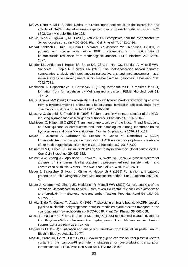
83
Ma W, Deng Y, Mi H (2008b) Redox of plastoquinone pool regulates the expression and activity of NADPH dehydrogenase supercomplex in Synechocystis sp. strain PCC 6803. Curr Microbiol 56: 189-193.
Ma W, Deng Y, Ogawa T, Mi H (2006) Active NDH-1 complexes from the cyanobacterium Synechocystis sp. strain PCC 6803. Plant Cell Physiol 47: 1432-1436.
Madadi-Kahkesh S, Duin EC, Heim S, Albracht SP, Johnson MK, Hedderich R (2001) A paramagnetic species with unique EPR characteristics in the active site of heterodisulfide reductase from methanogenic archaea. Eur J Biochem 268: 2566-2577.
Maeder DL, Anderson I, Brettin TS, Bruce DC, Gilna P, Han CS, Lapidus A, Metcalf WW, Saunders E, Tapia R, Sowers KR (2006) The Methanosarcina barkeri genome: comparative analysis with Methanosarcina acetivorans and Methanosarcina mazei reveals extensive rearrangement within methanosarcinal genomes. J Bacteriol 188: 7922-7931.
Mahlmann A, Deppenmeier U, Gottschalk G (1989) Methanofuran-B is required for CO2 formation from formaldehyde by Methanosarcina barkeri. FEMS Microbiol Lett 61: 115-120.
Mai X, Adams MW (1996) Characterization of a fourth type of 2-keto acid-oxidizing enzyme from a hyperthermophilic archaeon: 2-ketoglutarate ferredoxin oxidoreductase from Thermococcus litoralis. J Bacteriol 178: 5890-5896.
Massanz C, Schmidt S, Friedrich B (1998) Subforms and in vitro reconstitution of the NAD-reducing hydrogenase of Alcaligenes eutrophus. J Bacteriol 180: 1023-1029.
Mathiesen C, Hägerhäll C (2002) Transmembrane topology of the NuoL, M and N subunits of NADH:quinone oxidoreductase and their homologues among membrane-bound hydrogenases and bona fide antiporters. Biochim Biophys Acta 1556: 121-132.
Mayer F, Jussofie A, Salzmann M, Lübben M, Rohde M, Gottschalk G (1987) Immunoelectron microscopic demonstration of ATPase on the cytoplasmic membrane of the methanogenic bacterium strain Gö1. J Bacteriol 169: 2307-2309.
McInerney MJ, Sieber JR, Gunsalus RP (2009) Syntrophy in anaerobic global carbon cycles. Curr Opin Biotechnol 20: 623-632.
Metcalf WW, Zhang JK, Apolinario E, Sowers KR, Wolfe RS (1997) A genetic system for archaea of the genus Methanosarcina: Liposome-mediated transformation and construction of shuttle vectors. Proc Natl Acad Sci U S A 94: 2626-2631.
Meuer J, Bartoschek S, Koch J, Künkel A, Hedderich R (1999) Purification and catalytic properties of Ech hydrogenase from Methanosarcina barkeri. Eur J Biochem 265: 325-335.
Meuer J, Kuettner HC, Zhang JK, Hedderich R, Metcalf WW (2002) Genetic analysis of the archaeon Methanosarcina barkeri Fusaro reveals a central role for Ech hydrogenase and ferredoxin in methanogenesis and carbon fixation. Proc Natl Acad Sci USA 99: 5632-5637.
Mi HL, Endo T, Ogawa T, Asada K (1995) Thylakoid membrane-bound, NADPH-specific pyridine-nucleotide dehydrogenase complex mediates cyclic electron-transport in the cyanobacterium Synechocystis sp. PCC-68038. Plant Cell Physiol 36: 661-668.
Michel R, Massanz C, Kostka S, Richter M, Fiebig K (1995) Biochemical characterization of the 8-hydroxy-5-deazaflavin-reactive hydrogenase from Methanosarcina barkeri Fusaro. Eur J Biochem 233: 727-735.
Mortenson LE (1964) Purification and analysis of ferredoxin from Clostridium pasteurianum. Biochim Biophys Acta 81: 71-77.
Mott JE, Grant RA, Ho YS, Platt T (1985) Maximizing gene expression from plasmid vectors containing the Lambda-Pl promoter - strategies for overproducing transcription termination factor Rho. Proc Natl Acad Sci U S A 82: 88-92.
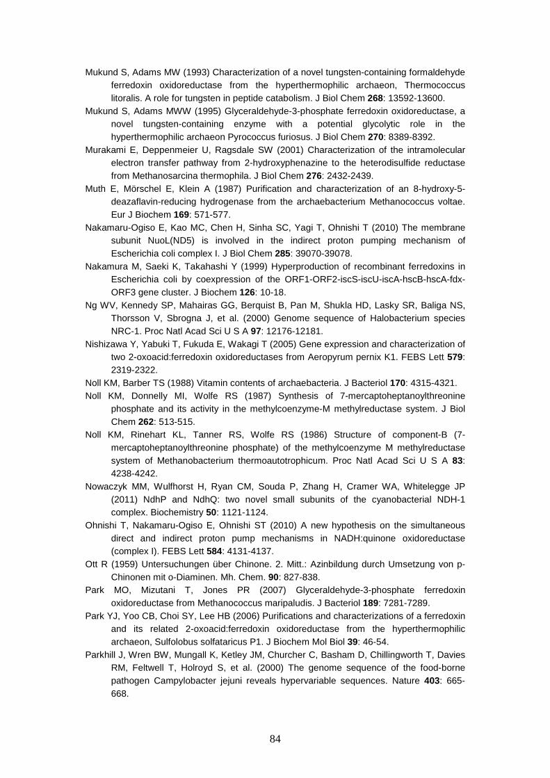
84
Mukund S, Adams MW (1993) Characterization of a novel tungsten-containing formaldehyde ferredoxin oxidoreductase from the hyperthermophilic archaeon, Thermococcus litoralis. A role for tungsten in peptide catabolism. J Biol Chem 268: 13592-13600.
Mukund S, Adams MWW (1995) Glyceraldehyde-3-phosphate ferredoxin oxidoreductase, a novel tungsten-containing enzyme with a potential glycolytic role in the hyperthermophilic archaeon Pyrococcus furiosus. J Biol Chem 270: 8389-8392.
Murakami E, Deppenmeier U, Ragsdale SW (2001) Characterization of the intramolecular electron transfer pathway from 2-hydroxyphenazine to the heterodisulfide reductase from Methanosarcina thermophila. J Biol Chem 276: 2432-2439.
Muth E, Mörschel E, Klein A (1987) Purification and characterization of an 8-hydroxy-5-deazaflavin-reducing hydrogenase from the archaebacterium Methanococcus voltae. Eur J Biochem 169: 571-577.
Nakamaru-Ogiso E, Kao MC, Chen H, Sinha SC, Yagi T, Ohnishi T (2010) The membrane subunit NuoL(ND5) is involved in the indirect proton pumping mechanism of Escherichia coli complex I. J Biol Chem 285: 39070-39078.
Nakamura M, Saeki K, Takahashi Y (1999) Hyperproduction of recombinant ferredoxins in Escherichia coli by coexpression of the ORF1-ORF2-iscS-iscU-iscA-hscB-hscA-fdx-ORF3 gene cluster. J Biochem 126: 10-18.
Ng WV, Kennedy SP, Mahairas GG, Berquist B, Pan M, Shukla HD, Lasky SR, Baliga NS, Thorsson V, Sbrogna J, et al. (2000) Genome sequence of Halobacterium species NRC-1. Proc Natl Acad Sci U S A 97: 12176-12181.
Nishizawa Y, Yabuki T, Fukuda E, Wakagi T (2005) Gene expression and characterization of two 2-oxoacid:ferredoxin oxidoreductases from Aeropyrum pernix K1. FEBS Lett 579: 2319-2322.
Noll KM, Barber TS (1988) Vitamin contents of archaebacteria. J Bacteriol 170: 4315-4321. Noll KM, Donnelly MI, Wolfe RS (1987) Synthesis of 7-mercaptoheptanoylthreonine
phosphate and its activity in the methylcoenzyme-M methylreductase system. J Biol Chem 262: 513-515.
Noll KM, Rinehart KL, Tanner RS, Wolfe RS (1986) Structure of component-B (7-mercaptoheptanoylthreonine phosphate) of the methylcoenzyme M methylreductase system of Methanobacterium thermoautotrophicum. Proc Natl Acad Sci U S A 83: 4238-4242.
Nowaczyk MM, Wulfhorst H, Ryan CM, Souda P, Zhang H, Cramer WA, Whitelegge JP (2011) NdhP and NdhQ: two novel small subunits of the cyanobacterial NDH-1 complex. Biochemistry 50: 1121-1124.
Ohnishi T, Nakamaru-Ogiso E, Ohnishi ST (2010) A new hypothesis on the simultaneous direct and indirect proton pump mechanisms in NADH:quinone oxidoreductase (complex I). FEBS Lett 584: 4131-4137.
Ott R (1959) Untersuchungen über Chinone. 2. Mitt.: Azinbildung durch Umsetzung von p-Chinonen mit o-Diaminen. Mh. Chem. 90: 827-838.
Park MO, Mizutani T, Jones PR (2007) Glyceraldehyde-3-phosphate ferredoxin oxidoreductase from Methanococcus maripaludis. J Bacteriol 189: 7281-7289.
Park YJ, Yoo CB, Choi SY, Lee HB (2006) Purifications and characterizations of a ferredoxin and its related 2-oxoacid:ferredoxin oxidoreductase from the hyperthermophilic archaeon, Sulfolobus solfataricus P1. J Biochem Mol Biol 39: 46-54.
Parkhill J, Wren BW, Mungall K, Ketley JM, Churcher C, Basham D, Chillingworth T, Davies RM, Feltwell T, Holroyd S, et al. (2000) The genome sequence of the food-borne pathogen Campylobacter jejuni reveals hypervariable sequences. Nature 403: 665-668.
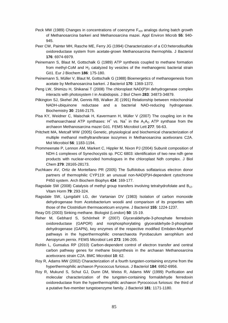
85
Peck MW (1989) Changes in concentrations of coenzyme F420 analogs during batch growth of Methanosarcina barkeri and Methanosarcina mazei. Appl Environ Microb 55: 940-945.
Peer CW, Painter MH, Rasche ME, Ferry JG (1994) Characterization of a CO:heterodisulfide oxidoreductase system from acetate-grown Methanosarcina thermophila. J Bacteriol 176: 6974-6979.
Peinemann S, Blaut M, Gottschalk G (1989) ATP synthesis coupled to methane formation from methyl-CoM and H2 catalyzed by vesicles of the methanogenic bacterial strain Gö1. Eur J Biochem 186: 175-180.
Peinemann S, Müller V, Blaut M, Gottschalk G (1988) Bioenergetics of methanogenesis from acetate by Methanosarcina barkeri. J Bacteriol 170: 1369-1372.
Peng LW, Shimizu H, Shikanai T (2008) The chloroplast NAD(P)H dehydrogenase complex interacts with photosystem I in Arabidopsis. J Biol Chem 283: 34873-34879.
Pilkington SJ, Skehel JM, Gennis RB, Walker JE (1991) Relationship between mitochondrial NADH-ubiquinone reductase and a bacterial NAD-reducing hydrogenase. Biochemistry 30: 2166-2175.
Pisa KY, Weidner C, Maischak H, Kavermann H, Müller V (2007) The coupling ion in the methanoarchaeal ATP synthases: H+ vs. Na+ in the A1AO ATP synthase from the archaeon Methanosarcina mazei Gö1. FEMS Microbiol Lett 277: 56-63.
Pritchett MA, Metcalf WW (2005) Genetic, physiological and biochemical characterization of multiple methanol methyltransferase isozymes in Methanosarcina acetivorans C2A. Mol Microbiol 56: 1183-1194.
Prommeenate P, Lennon AM, Markert C, Hippler M, Nixon PJ (2004) Subunit composition of NDH-1 complexes of Synechocystis sp. PCC 6803: identification of two new ndh gene products with nuclear-encoded homologues in the chloroplast Ndh complex. J Biol Chem 279: 28165-28173.
Puchkaev AV, Ortiz de Montellano PR (2005) The Sulfolobus solfataricus electron donor partners of thermophilic CYP119: an unusual non-NAD(P)H-dependent cytochrome P450 system. Arch Biochem Biophys 434: 169-177.
Ragsdale SW (2008) Catalysis of methyl group transfers involving tetrahydrofolate and B12. Vitam Horm 79: 293-324.
Ragsdale SW, Ljungdahl LG, der Vartanian DV (1983) Isolation of carbon monoxide dehydrogenase from Acetobacterium woodii and comparison of its properties with those of the Clostridium thermoaceticum enzyme. J Bacteriol 155: 1224-1237.
Reay DS (2003) Sinking methane. Biologist (London) 50: 15-19. Reher M, Gebhard S, Schönheit P (2007) Glyceraldehyde-3-phosphate ferredoxin
oxidoreductase (GAPOR) and nonphosphorylating glyceraldehyde-3-phosphate dehydrogenase (GAPN), key enzymes of the respective modified Embden-Meyerhof pathways in the hyperthermophilic crenarchaeota Pyrobaculum aerophilum and Aeropyrum pernix. FEMS Microbiol Lett 273: 196-205.
Rohlin L, Gunsalus RP (2010) Carbon-dependent control of electron transfer and central carbon pathway genes for methane biosynthesis in the archaean Methanosarcina acetivorans strain C2A. BMC Microbiol 10: 62.
Roy R, Adams MW (2002) Characterization of a fourth tungsten-containing enzyme from the hyperthermophilic archaeon Pyrococcus furiosus. J Bacteriol 184: 6952-6956.
Roy R, Mukund S, Schut GJ, Dunn DM, Weiss R, Adams MW (1999) Purification and molecular characterization of the tungsten-containing formaldehyde ferredoxin oxidoreductase from the hyperthermophilic archaeon Pyrococcus furiosus: the third of a putative five-member tungstoenzyme family. J Bacteriol 181: 1171-1180.
86
Ruepp A, Graml W, Santos-Martinez ML, Koretle KK, Volker C, Mewes HW, Frishman D, Stocker S, Lupas AN, Baumeister W (2000) The genome sequence of the thermoacidophilic scavenger Thermoplasma acidophilum. Nature 407: 508-513.
Rumeau D, Bécuwe-Linka N, Beyly A, Louwagie M, Garin J, Peltier G (2005) New subunits NDH-M, -N, and -O, encoded by nuclear genes, are essential for plastid Ndh complex functioning in higher plants. Plant Cell 17: 219-232.
Sakuraba H, Ohshima T (2002) Novel energy metabolism in anaerobic hyperthermophilic archaea: A modified Embden-Meyerhof pathway. J Biosci Bioeng 93: 441-448.
Sancho J (2006) Flavodoxins: sequence, folding, binding, function and beyond. Cell Mol Life Sci 63: 855-864.
Sapra R, Bagramyan K, Adams MW (2003) A simple energy-conserving system: proton reduction coupled to proton translocation. Proc Natl Acad Sci U S A 100: 7545-7550.
Sauter M, Böhm R, Böck A (1992) Mutational analysis of the operon (hyc) determining hydrogenase 3 formation in Escherichia coli. Mol Microbiol 6: 1523-1532.
Schönheit P, Keweloh H, Thauer RK (1981) Factor F420 degradation in Methanobacterium thermoautotrophicum during exposure to oxygen. FEMS Microbiol Lett 12: 347-349.
Selengut JD, Haft DH (2010) Unexpected abundance of coenzyme F420-dependent enzymes in Mycobacterium tuberculosis and other actinobacteria. J Bacteriol 192: 5788-5798.
Selig M, Xavier KB, Santos H, Schönheit P (1997) Comparative analysis of Embden-Meyerhof and Entner-Doudoroff glycolytic pathways in hyperthermophilic archaea and the bacterium Thermotoga. Arch Microbiol 167: 217-232.
She Q, Singh RK, Confalonieri F, Zivanovic Y, Allard G, Awayez MJ, Chan-Weiher CCY, Clausen IG, Curtis BA, de Moors A, et al. (2001) The complete genome of the crenarchaeon Sulfolobus solfataricus P2. Proc Natl Acad Sci U S A 98: 7835-7840.
Shibata M, Ohkawa H, Kaneko T, Fukuzawa H, Tabata S, Kaplan A, Ogawa T (2001) Distinct constitutive and low-CO2-induced CO2 uptake systems in cyanobacteria: genes involved and their phylogenetic relationship with homologous genes in other organisms. Proc Natl Acad Sci U S A 98: 11789-11794.
Shin SG, Lee S, Lee C, Hwang K, Hwang S (2010) Qualitative and quantitative assessment of microbial community in batch anaerobic digestion of secondary sludge. Bioresour Technol 101: 9461-9470.
Simianu M, Murakami E, Brewer JM, Ragsdale SW (1998) Purification and properties of the heme- and iron-sulfur-containing heterodisulfide reductase from Methanosarcina thermophila. Biochemistry 37: 10027-10039.
Sirpiö S, Allahverdiyeva Y, Holmström M, Khrouchtchova A, Haldrup A, Battchikova N, Aro EM (2009) Novel nuclear-encoded subunits of the chloroplast NAD(P)H dehydrogenase complex. J Biol Chem 284: 905-912.
Smith KS, Ingram-Smith C (2007) Methanosaeta, the forgotten methanogen? Trends Microbiol 15: 150-155.
Smith MA, Finel M, Korolik V, Mendz GL (2000) Characteristics of the aerobic respiratory chains of the microaerophiles Campylobacter jejuni and Helicobacter pylori. Arch Microbiol 174: 1-10.
Smith PK, Krohn RI, Hermanson GT, Mallia AK, Gartner FH, Provenzano MD, Fujimoto EK, Goeke NM, Olson BJ, Klenk DC (1985) Measurement of protein using bicinchoninic acid. Anal Biochem 150: 76-85.
Soboh B, Linder D, Hedderich R (2002) Purification and catalytic properties of a CO-oxidizing:H2-evolving enzyme complex from Carboxydothermus hydrogenoformans. Eur J Biochem 269: 5712-5721.
Stojanowic A, Hedderich R (2004) CO2 reduction to the level of formylmethanofuran in Methanosarcina barkeri is non-energy driven when CO is the electron donor. FEMS Microbiol Lett 235: 163-167.
87
Supaphol S, Jenkins SN, Intomo P, Waite IS, O'Donnell AG (2011) Microbial community dynamics in mesophilic anaerobic co-digestion of mixed waste. Bioresour Technol 102: 4021-4027.
Tagawa K, Arnon DI (1968) Oxidation-reduction potentials and stoichiometry of electron transfer in ferredoxins. Biochim Biophys Acta 153: 602-613.
Tallant TC, Paul L, Krzycki JA (2001) The MtsA subunit of the methylthiol:coenzyme M methyltransferase of Methanosarcina barkeri catalyses both half-reactions of corrinoid-dependent dimethylsulfide: coenzyme M methyl transfer. J Biol Chem 276: 4485-4493.
te Brömmelstroet BWJ, Geerts WJ, Keltjens JT, van der Drift C, Vogels GD (1991) Purification and properties of 5,10-methylenetetrahydromethanopterin dehydrogenase and 5,10-methylenetetrahydromethanopterin reductase, 2 coenzyme-F420-dependent enzymes, from Methanosarcina barkeri. Biochim Biophys Acta 1079: 293-302.
Terlesky KC, Ferry JG (1988) Ferredoxin requirement for electron transport from the carbon monoxide dehydrogenase complex to a membrane-bound hydrogenase in acetate-grown Methanosarcina thermophila. J Biol Chem 263: 4075-4079.
Tersteegen A, Hedderich R (1999) Methanobacterium thermoautotrophicum encodes two multisubunit membrane-bound [NiFe] hydrogenases. Transcription of the operons and sequence analysis of the deduced proteins. Eur J Biochem 264: 930-943.
Tersteegen A, Linder D, Thauer RK, Hedderich R (1997) Structures and functions of four anabolic 2-oxoacid oxidoreductases in Methanobacterium thermoautotrophicum. Eur J Biochem 244: 862-868.
Thauer RK (1998) Biochemistry of methanogenesis: a tribute to Marjory Stephenson. Microbiology 144: 2377-2406.
Thauer RK, Jungermann K, Decker K (1977) Energy conservation in chemotrophic anaerobic bacteria. Bacteriol Rev 41: 100-180.
Thauer RK, Kaster AK, Seedorf H, Buckel W, Hedderich R (2008) Methanogenic archaea: ecologically relevant differences in energy conservation. Nat Rev Microbiol 6: 579-591.
Tomb JF, White O, Kerlavage AR, Clayton RA, Sutton GG, Fleischmann RD, Ketchum KA, Klenk HP, Gill S, Dougherty BA, et al. (1997) The complete genome sequence of the gastric pathogen Helicobacter pylori. Nature 388: 539-547.
Torres-Bacete J, Nakamaru-Ogiso E, Matsuno-Yagi A, Yagi T (2007) Characterization of the NuoM (ND4) subunit in Escherichia coli NDH-1: conserved charged residues essential for energy-coupled activities. J Biol Chem 282: 36914-36922.
Torres-Bacete J, Sinha PK, Castro-Guerrero N, Matsuno-Yagi A, Yagi T (2009) Features of subunit NuoM (ND4) in Escherichia coli NDH-1: topology and implications of conserved Glu-144 for coupling site 1. J Biol Chem 284: 33062-33069.
Tran-Betcke A, Warnecke U, Bocker C, Zaborosch C, Friedrich B (1990) Cloning and nucleotide sequences of the genes for the subunits of NAD-reducing hydrogenase of Alcaligenes eutrophus H16. J Bacteriol 172: 2920-2929.
van Beelen P, Dijkstra AC, Vogels GD (1983) Quantitation of coenzyme F420 in methanogenic sludge by the use of reversed phase high performance liquid chromatography and a fluorescence detector. Eur J Appl Microbiol 18: 67-69.
van der Oost J, Schut G, Kengen SWM, Hagen WR, Thomm M, de Vos WM (1998) The ferredoxin-dependent conversion of glyceraldehyde-3-phosphate in the hyperthermophilic archaeon Pyrococcus furiosus represents a novel site of glycolytic regulation. J Biol Chem 273: 28149-28154.
Vignais PM, Billoud B, Meyer J (2001) Classification and phylogeny of hydrogenases. FEMS Microbiol Rev 25: 455-501.
Wächtershäuser G (1988) Before enzymes and templates: theory of surface metabolism. Microbiol Rev 52: 452-484.
88
Walsh C (1986) Naturally occurring 5-deazaflavin coenzymes - biological redox roles. Acc Chem Res 19: 216-221.
Weerakoon DR, Olson JW (2008) The Campylobacter jejuni NADH:ubiquinone oxidoreductase (complex I) utilizes flavodoxin rather than NADH. J Bacteriol 190: 915-925.
Welte C, Deppenmeier U (2011) Re-evaluation of the function of the F420 dehydrogenase in electron transport in Methanosarcina mazei. FEBS J 278: 1277-1287.
Welte C, Kallnik V, Grapp M, Bender G, Ragsdale S, Deppenmeier U (2010a) Function of Ech hydrogenase in ferredoxin-dependent, membrane-bound electron transport in Methanosarcina mazei. J Bacteriol 192: 674-678.
Welte C, Krätzer C, Deppenmeier U (2010b) Involvement of Ech hydrogenase in energy conservation of Methanosarcina mazei. FEBS J 277: 3396-3403.
Wilms R, Freiberg C, Wegerle E, Meier I, Mayer F, Müller V (1996) Subunit structure and organization of the genes of the A1AO ATPase from the achaeon Methanosarcina mazei Gö1. J Biol Chem 271: 18843-18852.
Wolfe RS (1985) Unusual coenzymes of methanogenesis. Trends Biochem Sci 10: 396-399. Yamazaki S, Tsai L (1980) Purification and properties of 8-hydroxy-5-deazaflavin-dependent
NADP+ reductase from Methanococcus vannielii. J Biol Chem 255: 6462-6465. Zahler WL, Cleland WW (1968) A specific and sensitive assay for disulfides. J Biol Chem
243: 716-719. Zhang P, Battchikova N, Jansen T, Appel J, Ogawa T, Aro EM (2004) Expression and
functional roles of the two distinct NDH-1 complexes and the carbon acquisition complex NdhD3/NdhF3/CupA/Sll1735 in Synechocystis sp PCC 6803. Plant Cell 16: 3326-3340.
Zinder SH, Mah RA (1979) Isolation and characterization of a thermophilic strain of Methanosarcina unable to use H2-CO2 for methanogenesis. Appl Environ Microb 38: 996-1008.

90
Publications
Welte C, Hafner S, Krätzer C, Quentmeier A, Friedri ch CG, Dahl C (2009)
Interaction between Sox proteins of two physiologically distinct bacteria and a new
protein involved in thiosulfate oxidation. FEBS Lett 583:1281-1286.
Welte C, Kallnik V, Grapp M, Bender G, Ragsdale S, Deppenmeier U (2010)
Function of Ech hydrogenase in ferredoxin-dependent, membrane-bound electron
transport in Methanosarcina mazei. J Bacteriol 192:674-678.
Welte C, Krätzer C, Deppenmeier U (2010) Involvement of Ech hydrogenase in
energy conservation of Methanosarcina mazei. FEBS J 277:3396-3403.
Krätzer C, Welte C, Dörner K, Friedrich T, Deppenme ier U (2011)
Methanoferrodoxin represents a new class of superoxide reductase containing an
iron-sulfur cluster. FEBS J 278:442-451.
Welte C, Deppenmeier U (2011) Re-evaluation of the function of the F420
dehydrogenase in electron transport of Methanosarcina mazei. FEBS J 278:1277-
1287.
Welte C, Deppenmeier U (2011) Proton translocation in methanogens. Methods
Enzymol 494:257-280.
Welte C, Deppenmeier U (2011) Membrane-bound electron transport in
Methanosaeta thermophila. J Bacteriol 193:2868-2870.