Embed Size (px)
Citation preview
2 www.avidscience.com
Top 5 Contributions in Materials Science: 3rd Edition
Chapter
Smart and Biofunctional Textiles: An Alternative for Vehiculation of Active Principles
Manuel J Lis1*, Luisa Coderch 2, Meritxell Martí2, Cristina Alonso2, Oscar García1, Carlos Garcia1 and Fabricio Maesta3
1INTEXTER-UPC, Terrassa, Spain2IQAC-CSIC; Barcelona, Spain3Federal University of Technological - Paraná, Brazil
*Corresponding Author: Manuel J Lis, INTEXTER-UPC, Terrassa, Spain
First Published February 28, 2019
Copyright: © 2019 Manuel J Lis, et al.
This article is distributed under the terms of the Creative Commons Attribution 4.0 International License (http://creativecommons.org/licenses/by/4.0/), which permits unrestricted use, distribution, and reproduction in any medium, provided you give appropriate credit to the original author(s) and the source.
This book chapter is an excerpt from an article published by Manuel J Lis, et al. at Materials in November 2018. (Lis Arias, M.J.; Coderch, L.; Martí, M.; Alonso, C.; García Carmona, O.; García Carmona, C.; Maesta, F. Vehiculation of Active Principles as a Way to Create Smart and Biofunc-tional Textiles. Materials 2018, 11, 2152.)
brought to you by COREView metadata, citation and similar papers at core.ac.uk
provided by Digital.CSIC
3
Top 5 Contributions in Materials Science: 3rd Edition
www.avidscience.com
Introduction In November 2017, the title of the International Symposium on
Materials from Renewables (ISMR) was “Advanced, Smart, and Sus-tainable Polymers, Fibers and Textiles”. Three specific sessions oc-curred under the denomination of “Smart Fibers and Textiles”. That simple fact gives an idea of the importance of this work. However, what really are smart textiles? In the foreword of the book edited by Tao, X. [1], Lewis states clearly that these type of textiles are not only special finished fabrics. The main defining idea of smart textiles is related to the “active character” of them. Smart textiles “react to envi-ronmental stimuli, from mechanical, thermal, chemical, magnetic or others”, including biotechnology, information technology, microelec-tronics, wearable computers, nanotechnology, and micromechanical machines.
Biofunctional textiles are fibrous substrates that have been modified to attain new properties and added value. The main idea is to modify their parameters, especially related to comfort, adapt-ing the tissues’ reaction to external or internal stimuli. Such textiles constitute appropriate substrates to be used for the delivery of active principles in cosmetic or pharmaceutical applications. Due to their specific response, biofunctional textiles are especially useful when the textile comes into close contact with the skin. As most of the human body is covered with some sort of textile, the potential of this type of textile is considerable. Textiles with functional properties used for delivery to skin have been studied and patented [2,3].
Three cases will be explored in this work as examples of biofunc-tional systems obtained using vehicles to transport different active principles to a textile substrate: Microcapsules, cyclodextrins, and liposomes.
Microcapsules Microcapsules may be obtained by a series of techniques that
involve liquids, gases, or solids in natural or synthetic polymeric
4 www.avidscience.com
Top 5 Contributions in Materials Science: 3rd Edition
membranes [4–7]. This process is known as microencapsulation, and requires a layer of an encapsulating agent, generally a polymeric material, that acts as a protective film insulating the active substance [8–10].
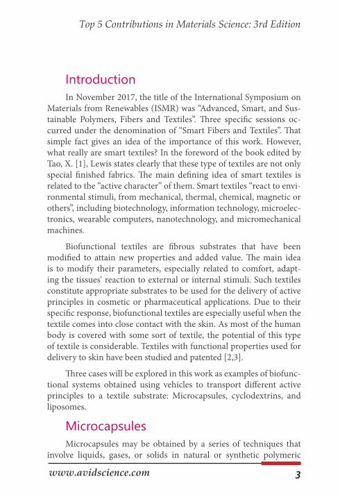
According to Souza and collaborators [11], this creates a physical barrier between the core (active principle) and the encapsulating ma-terial (shell). This membrane is removed by a specific stimulus, releas-ing the active substance in the ideal place or moment. In the Figure (Fig 1), it is possible to see this membrane that protects the nucleus
Figure 1: Optical microscopy image of microcapsules formed by complex coacerva-tion.
The encapsulation technique can be used to fulfill diverse objec-tives, and has the following advantages, as listed by different authors [12–14]. Protection of the encapsulated materials against oxidation or
5
Top 5 Contributions in Materials Science: 3rd Edition
www.avidscience.com
deactivation due to reaction with the environment (light, oxygen, hu-midity); masking odors, tastes, and other active principles; insulation of the active principles of undesirable materials; retardation of altera-tions that might occur in loss of aroma, color, and flavor; separation of reactive or incompatible components; and reduction of the migration rate of the core to the external environment. For this reason, the core and shell should have compatible physical-chemical properties [15].
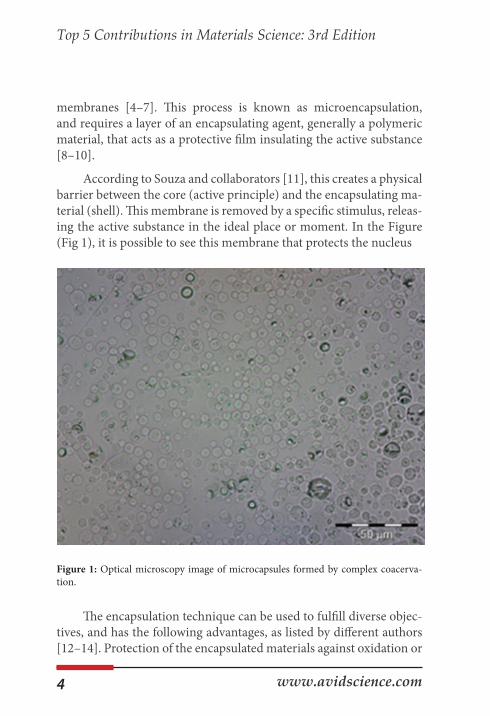
The materials commonly used as encapsulating agents (shells) are polymers [16,17] and biologic-based materials [18–20]. Bosnea and collaborators [21] highlighted that shell materials based on natu-ral polymers are promising for the formation of microcapsules due to their biodegradability, compatibility with other products, and low toxicity, as well as their wide availability from natural resources. Fur-thermore, Al Shannaq and collaborators [22] accentuate in their work that the choice of encapsulation material is important to obtain better degradation of the microcapsule. Table 1 shows the main polymers of natural sources used in microencapsulation.
Table 1: Polymers used in microencapsulation [23-27].
Source PolymerNatural Polysaccharides Starch, cellulose, chitosan, gum ara-
bic and alginate
Modified Polysaccharides Dextrins, carboxymethylcellulose, ethylcellulose, methylcellulose, ace-
tylcellulose and nitrocellulose
Proteins Gluten, casein, gelatin and albumin
Waxes and lipids Paraffin, tristearine, stearic acid,
monoacyl and diacyl
6 www.avidscience.com
Top 5 Contributions in Materials Science: 3rd Edition
Besides the inherent parameters of the shell material choice, San-tos, Ferreira, and Grosso [28] pointed out that the retention of the ac-tive principle is a fundamental factor for the realization of the process. Wang et al. [29] and Yang et al. [30] demonstrated in their works that the efficiency of the encapsulation should be a parameter that is taken into account in the choice of the core. Sharipova et al. [31] showed that the value of the effectiveness of the encapsulation depends on the encapsulating method, the shell material, and the relationship with the core.
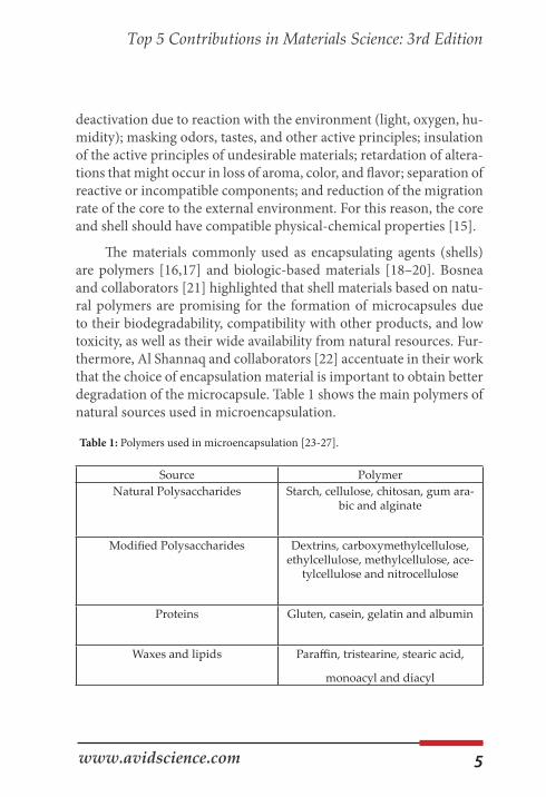
Thus, the casing material should relate to the chemical nature (molar mass, polarity, functionality, volatility, and so forth) of the ac-tive principle so that a high yield of the process is possible. The thermal analysis technique (DTGA / DSC) can be used to verify the uptake of microencapsulation, as well as its efficiency. The thermogravimetric curves shown in Figure 2 reveal that each component of the system has a different loss of weight when submitted to temperature changes in the oven of the instrument.
Figure 2: TGA/DTGA thermogram for different components of the system
Citronella oil shows a single stage loss of weight, while gelatin and arabic gum show two stages. Formation of the microcapsule gives the new organized system that presents three different stages of ther-mal rupture. From these thermograms it can be seen that the micro-capsule was formed.
7
Top 5 Contributions in Materials Science: 3rd Edition
www.avidscience.com
There are several techniques for obtaining microcapsules, which can be divided into physical methods (spray drying [32], solvent evaporation [33], pan coating [34]); chemical methods (interfacial polymerization [35], suspension polymerization [36], in situ polym-erization [37]); and physical-chemical methods (coacervation [38], ionic gelation [39], sol-gel [40]), among others.
The microencapsulation technique selection depends on the properties of the active principle, the morphology of the desired par-ticle, the nature and capacity of releasing the components, reproduc-ibility, ease of execution of the technique, and the cost/effectiveness ratio [41]. The chosen technique is a determining factor of the charac-teristics of the formed microcapsule, and will influence the release of the encapsulated agent via one of the following actions: Mechanical, temperature, pH, dissolution, or biodegradation [42].
Microcapsules in the textile field have been applied in various ways, giving very interesting results and showing very promising applications in several fields, including the use of flame retardant agents [43], protection of atmospheric agents [44–46], and functional finishing [47–50], along with the development of functional fabrics that might have a useful effect on the user and solve problems that conventional processes are not able to [51,52].
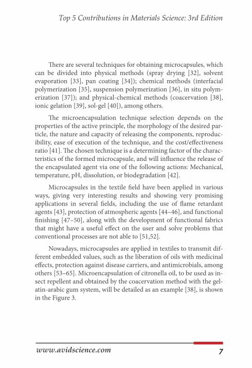
Nowadays, microcapsules are applied in textiles to transmit dif-ferent embedded values, such as the liberation of oils with medicinal effects, protection against disease carriers, and antimicrobials, among others [53–65]. Microencapsulation of citronella oil, to be used as in-sect repellent and obtained by the coacervation method with the gel-atin-arabic gum system, will be detailed as an example [38], is shown in the Figure 3.
8 www.avidscience.com
Top 5 Contributions in Materials Science: 3rd Edition
(a) (b)
Figure 3: Scanning electron of microcapsules: (a) cotton, and (b) polyester.
In these micrographs it is possible to verify the distribution of the dispersed microcapsules and their reduced size.
Another factor that can be pointed out is the small size of the microcapsules, which facilitates the absorption and penetration of the fabric surface due to the occupation of the interstices of the textile article. Li et al. [23] related the advantages of controlled dosing and increased durability of the textile finish to this small size.
It is also evident that the microcapsules ensure effective protec-tion of the encapsulated material, as already shown by the results of TGA. In short, it can be noted that the encapsulated material is not exposed to the elements, which has been the most serious problem of the application of oils in textiles, as pointed out by Chinta and Pooja [51] and Nelson [42].
9
Top 5 Contributions in Materials Science: 3rd Edition
www.avidscience.com
Cyclodextrines (CD) Cyclodextrines (CDs) are used in diverse industrial fields, such
as food, drugs, cosmetics, domestic products, agrochemicals, the textile industry, the paper industry, chemical technology, and oth-ers [66–68]. This wide range of applications can be attributed to the fact that cyclodextrin shave the capacity to form inclusion complexes with a broad range of substances, allowing the alteration of important properties in the complexed substances [69].
According to Matioli and collaborators [70], CDs are regularly produced from starch by the cyclation of linear chains of glucopyra-noses using the enzyme cyclomaltodextrin-glucanotransferase (CG-Tase). The three widest known natural cyclodextrins are alpha CD (α-CD), beta CD (β-CD), and gamma CD (γ-CD), composed of six, seven, and eight units of D-(+)-glucopyranose, respectively, and unit-ed by α-1,4 bonds. Table 2 presents the physical and chemical proper-ties of the most common CDs.Table 2: Physical-chemical properties of natural CDs [66,69].
These compounds have in their structure primary hydroxyl groups and secondary groups oriented to the exterior [66]. There-fore, present a cavity that allows the hydrophilic external part and a hydrophobic internal cavity. Such cavity allows the cyclodextrins to complex molecules that show compatible dimensions and alter its physical-chemical properties such as water solubility, stability, and bioavailability [70]. An example of complexation is the use of citron-ella oil (lipophilic) as the host agent, as shown in Figure.
Property α-CD β-CD γ-CDNº of glucose units 6 7 8Empirical formula C36H60O30 C42H70O35 C48H80O40
Solubility in water at 25 ºC (g L-1) 145 18.5 232Inner cavity volume (nm3) 1740 2620 4720
Nº of water molecules in the cavity 6 11 17Decomposition temperature (ºC) 278 298 267
10 www.avidscience.com
Top 5 Contributions in Materials Science: 3rd Edition
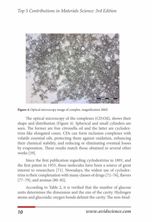
Figure 4: Optical microscopy image of complex. magnification 500X
The optical microscopy of the complexes (CD:Oil), shows their shape and distribution (Figure 4). Spherical and small cylinders are seen. The former are free citronella oil and the latter are cyclodex-trins like elongated cones. CDs can form inclusion complexes with volatile essential oils, protecting them against oxidation, enhancing their chemical stability, and reducing or eliminating eventual losses by evaporation. These results match those obtained in several other works [59].
Since the first publication regarding cyclodextrins in 1891, and the first patent in 1953, these molecules have been a source of great interest to researchers [71]. Nowadays, the widest use of cyclodex-trins is their complexation with many classes of drugs [72–76], flavors [77–79], and aromas [80–82].
According to Table 2, it is verified that the number of glucose units determines the dimension and the size of the cavity. Hydrogen atoms and glucosidic oxygen bonds delimit the cavity. The non-bind-
11
Top 5 Contributions in Materials Science: 3rd Edition
www.avidscience.com
ing electron pairs in the glucosidic oxygen bonds are directed to the interior of the cavity, being responsible for the hydrophobic internal effect and producing a high electron density that allows to the interior of the CD cavity a Lewis base character [66,70].
In textiles, the CDs may be applied in different ways and with many end-uses. Bhaskara-Amrit, Pramod, and Warmoeskerken [71], highlight that the CDs have an important role in the processing and textile innovation. Their use fosters immediate opportunities to the development of products that are less harmful to the environment, besides having a great potential in many applications.
The highest employability of the CDs in the textile field is related to the field of finishing, showing an excellent potential to be applied in dyeing and finishing [69,66, 71,81,82]
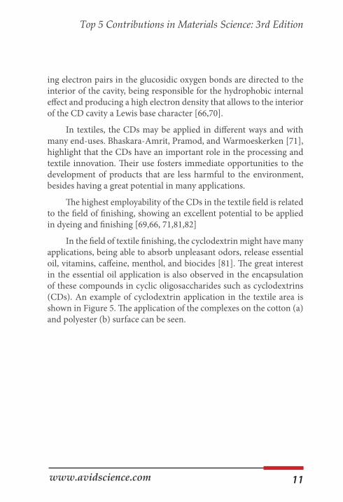
In the field of textile finishing, the cyclodextrin might have many applications, being able to absorb unpleasant odors, release essential oil, vitamins, caffeine, menthol, and biocides [81]. The great interest in the essential oil application is also observed in the encapsulation of these compounds in cyclic oligosaccharides such as cyclodextrins (CDs). An example of cyclodextrin application in the textile area is shown in Figure 5. The application of the complexes on the cotton (a) and polyester (b) surface can be seen.
12 www.avidscience.com
Top 5 Contributions in Materials Science: 3rd Edition
(a) (b)
Figure 5: Scanning electron of complex: (a) cotton, and (b) polyester.
CDs can form inclusion complexes with volatile essential oils protecting them against oxidation, enhancing the chemical stability and reducing or eliminating eventual losses by evaporation [82].
The CDs in textiles can create interactions according to two dif-ferent mechanisms: physical bonding without strong chemical inter-actions or covalent bonding [71,82]. In the first case, the cyclodextrin is physically bonded to the textile substrate via a resin; in the second, CDs are permanently fixed in the substrate via covalent bonds [81].
Liposomes Liposomes are vesicles prepared with lipids that can encapsulate
different ingredients; one of their applications could be onto textiles. Due to the liposome bilayer structure, liposomes have been applied as models for biological membranes in medical research. Another im-
13
Top 5 Contributions in Materials Science: 3rd Edition
www.avidscience.com
portant use of this type of vesicleis as microcapsules for drug delivery in the cosmetic field [83–86]. The textile industry has used liposomes in the wool dyeing process as an auxiliary [87,88]. Liposomes could have different properties depending on the lipidic base used during their formulation.
In this study, phosphatidylcholine (PC) and internal wool lipids (IWL) were used to form liposomes structures.
Internal wool lipids are a mix of cholesterol, free fatty acids, cho-lesterol sulphate, and ceramides, similar to those found in membranes of other keratinized tissues such as human hair or stratum corneum (SC) from skin. Wool is a fiber which is mainly keratinic but with a small amount of internal lipids [89,90]. Due to the IWL liposome bilayer structure’s similarity to stratum corneum lipids, their appli-cation onto human skin has been assessed in previous studies. The results obtained have demonstrated the beneficial effect of this type of liposome when used with ceramides, topically applied onto intact skin in aging populations or in individuals with dry skin [91–93]. Therefore, we could consider IWL liposomes an optimal encapsula-tion route for cosmetic or dermopharmacy applications [94].
Using a solar filter as a tracer, ethyl hexyl methoxycinnamate (EHMC), PC-based, and IWL-based liposomes were prepared. The influence of the type of lipid in the vesicle on skin penetration has been demonstrated in previous studies. In particular, the crystalline liquid state of PC liposomes seems to play an important role in this characteristic. On the other hand, when using IWL liposomes, pen-etration into the skin is delayed—a fact that suggests some reinforce-ment of the barrier function of the skin’s stratum corneum [95]. These two types of liposomes, with IWL and PC, were chosen to be applied to cotton and polyamide fabrics to design biofunctional textiles.
To evaluate the effectiveness of the textile in contact with the skin, a series of methodologies in vivo were used and an in vitro pro-cess were optimized to determine the penetration of the encapsulated active principle.
14 www.avidscience.com
Top 5 Contributions in Materials Science: 3rd Edition
For the evaluation of the biofunctional textiles’ beneficial capac-ity on skin, the transepidermal water loss change (TEWL) was used as an indicator of the barrier function state. TEWL measures water-holding capacity as changes in skin capacitance [95]. An in vitro methodology based on percutaneous absorption [96] was used to determine the amount of encapsulated principle that passed into the different skin layers (stratum corneum, epidermis, or dermis) from the textile.
An in vivo stripping was used as a minimally invasive methodol-ogy, where a series of strippings allowed quantification of the amount of active principal in the outermost layers of the SC [97].
These methodologies have shown that liposomes, especially IWL liposomes, are suitable for applying active principles onto biofunc-tional textiles. In this study, Liposomes, alone or as mixed micelles, form a very stable microstructure that allows the vehiculization of ac-tive principles for application into different textiles. The chemical and physico-chemical interactions between them and the textile substrate are translated to an adequate substantivity for most of the studied cases.
However, the high desorption of most synthetic acrylic and poly-ester fibers confirmed the preferential application of cotton and poly-amide as cosmetic biofunctional textiles. Moreover, this study showed that polyamide always presented high substantivity for the two phos-pholipid structures and also for the antioxidant [98].
The in vitro percutaneous absorption tests of different cosmeto-textiles (CO and PA with GA vehiculised with Liposomes and mixed micelles) have been performed to demonstrate GA penetration with-in the layers of the skin [99].
15
Top 5 Contributions in Materials Science: 3rd Edition
www.avidscience.com
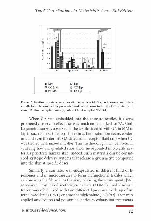
Figure 6: In vitro percutaneous absorption of gallic acid (GA) in liposome and mixed micelle formulations and the polyamide and cotton cosmeto-textiles (SC: stratum cor-neum, R. Fluid: receptor fluid) (significant level accepted *P<0.01)
When GA was embedded into the cosmeto-textiles, it always promoted a reservoir effect that was much more marked for PA. Simi-lar penetration was observed in the textiles treated with GA in MM or Lip in such compartments of the skin as the stratum corneum, epider-mis and even the dermis. GA detected in receptor fluid only when CO was treated with mixed micelles. This methodology may be useful in verifying how encapsulated substances incorporated into textile ma-terials penetrate human skin. Indeed, such materials can be consid-ered strategic delivery systems that release a given active compound into the skin at specific doses.
Similarly, a sun filter was encapsulated in different kind of li-posomes and in microcapsules to form biofunctional textiles which can break as the fabric rubs the skin, releasing the active agents [98]. Moreover, Ethyl hexyl methoxycinnamate (EHMC) used also as a tracer, was vehiculized with two different liposomes made up of in-ternal wool lipids (IWL) or phosphatidylcholine (PC) [99]. They were applied onto cotton and polyamide fabrics by exhaustion treatments.
16 www.avidscience.com
Top 5 Contributions in Materials Science: 3rd Edition
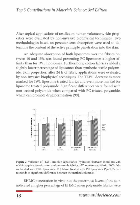
After topical applications of textiles on human volunteers, skin prop-erties were evaluated by non-invasive biophysical techniques. Two methodologies based on percutaneous absorption were used to de-termine the content of the active principle penetration into the skin.
An adequate absorption of both liposomes over the fabrics be-tween 10 and 15% was found presenting PC liposomes a higher af-finity than for IWL liposomes. Furthermore, cotton fabrics yielded a slightly lower percentage of liposomes than synthetic textile polyam-ide. Skin properties, after 24 h of fabric applications were evaluated by non-invasive biophysical techniques. The TEWL decrease is more marked for IWL liposome treated fabrics and even more marked for liposome treated polyamide. Significant differences were found with non-treated polyamide when compared with PC treated polyamide, which can promote drug permeation [99].
Figure 7: Variation of TEWL and skin capacitance (hydration) between initial and 24h of skin application of cotton and polyamide fabrics. NT: non-treated fabric. IWL: fab-ric treated with IWL liposomes. PC: fabric treated with PC liposomes (*p<0.05 cor-responds to significant difference between the marked columns).
EHMC penetration in vivo into the outermost layers of the skin indicated a higher percentage of EHMC when polyamide fabrics were
17
Top 5 Contributions in Materials Science: 3rd Edition
www.avidscience.com
applied. This is consistent with the higher effect found in the decrease in TEWL for IWL liposomes and in the increase of hydration for PC liposomes when absorbed into polyamide fabrics.
Therefore, polyamide was the fibre which presented the highest absorption and the greatest release to the skin when these two PC and IWL liposomes were applied with encapsulated EHMC. This was corroborated by the greater effect of skin barrier reinforcement due to IWL treated polyamide and the greater hydration increase due to PC treated polyamide. This resulted in a marked in vivo and in vitro EHMC release enhancement. The methodologies presented in this paper could serve to confirm penetration in human skin of encapsu-lated substances that can exert a marked influence on specific doses of active agents released to the skin [99].
Summarizing, antioxidants vehiculized through liposomes can be better applied to cotton and moreover polyamide due to their lower desorption in front of the other fibres assayed such as acryl-ic or polyester. The two in vitro and in vivo methodologies used to determine the content of active principle penetration into the skin when in contact with the smart textiles indicates the great influence of the physicochemical properties of the drugs. When GA is vehicu-lized in liposomes into the textile, there is a clear reservoir effect much marked with PA [98].
However when a clear lipophilic compound such as EHMC is also vehiculized in liposomes in the textile, a significant higher release of the active to the skin was found, being the polyamide the fibres with higher desorption [100]. This was corroborate by the in vivo results of percutaneous penetration and the greater skin barrier reinforcement and hydration of polyamide smart textiles [101].
Therefore, it can be concluded the different release behavior of hydrophilic drugs which may be much retained in the hydrophilic core of the liposome in front of lipophilic drugs which are embedded in the surface lipidic bilayer of the liposomes favouring their release [102].
18 www.avidscience.com
Top 5 Contributions in Materials Science: 3rd Edition
Vehiculation of Active Principles A variety of textile fibers was already studied as support for bi-
opolymers in the absorption of active molecules in the field of con-trolled releases such as cotton [37, 103-106] polyester [37, 107-109] wool [110-112] and nylon [107,113,114]. These many possibilities of fabric modification allow the textile combination with the controlled release system, enabling the capacity of absorbing therapeutic or cos-metic compounds and releasing them to the skin [48,103, 115-117].
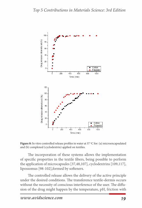
The combination of these effects results in the development of biofunctional textiles [48,107,113]. The use of textile articles as sup-ports for the controlled release presents as properties to emphasize the high contact area with the skin, drug-carrier capacity, ease of application, low cost, release via stimulus, biocompatibility, non-al-lergenicity, non-toxicity, among others [105,106,114]. The Figure 8 shows the controlled release profile of two microencapsulated sub-stances applied in different textile matrix.
19
Top 5 Contributions in Materials Science: 3rd Edition
www.avidscience.com
Figure 8: In vitro controlled release profiles in water at 37 oC for: (a) microencapsulated and (b) complexed (cyclodextrin) applied on textiles.
The incorporation of these systems allows the implementation of specific properties in the textile fibers, being possible to perform the application of microcapsules [37,48,107], cyclodextrins [109,117], lipossomus [98-102],formed by softeners.
The controlled release allows the delivery of the active principle under the desired conditions. The transference textile-dermis occurs without the necessity of conscious interference of the user. The diffu-sion of the drug might happen by the temperature, pH, friction with
20 www.avidscience.com
Top 5 Contributions in Materials Science: 3rd Edition
the skin, among others. These conditions enhance the effectiveness of the compound when compared to traditional release methods [118]. Costa & Lobo [119] point out that the discharge of these encapsulated substances follows, generally, three mechanisms: diffusion, liberation via activation, polymeric disaggregation/ erosion. The predominance of a release mechanism depends invariably on the properties of the polymer employed in the system, the matrix geometry, and the active principle [105].
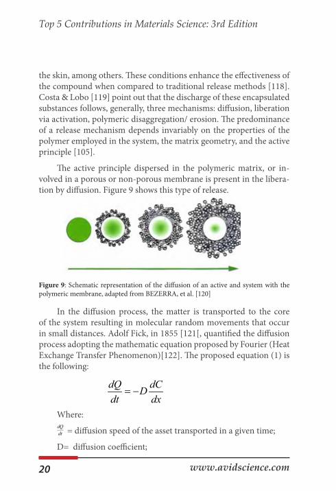
The active principle dispersed in the polymeric matrix, or in-volved in a porous or non-porous membrane is present in the libera-tion by diffusion. Figure 9 shows this type of release.
Figure 9: Schematic representation of the diffusion of an active and system with the polymeric membrane, adapted from BEZERRA, et al. [120]
In the diffusion process, the matter is transported to the core of the system resulting in molecular random movements that occur in small distances. Adolf Fick, in 1855 [121[, quantified the diffusion process adopting the mathematic equation proposed by Fourier (Heat Exchange Transfer Phenomenon)[122]. The proposed equation (1) is the following:
Where:
= diffusion speed of the asset transported in a given time;
D= diffusion coefficient;
dQ dCDdt dx
= −
dQdt
21
Top 5 Contributions in Materials Science: 3rd Edition
www.avidscience.com
= concentration of the substance diffusing in the spatial coor-dinate.
In the erosion mechanism, the polymer that protects the active principle will disintegrate and liberate it. According to Lopes, Lobo, and Costa [123] there are two important processes that involve this system: time dependent, first it occurs the diffusion from the middle to the interior of the polymer, making the dry core hydrated (dila-tion), and the second, when the exterior layer becomes jellified and suffers erosion.
Controlled Release Models
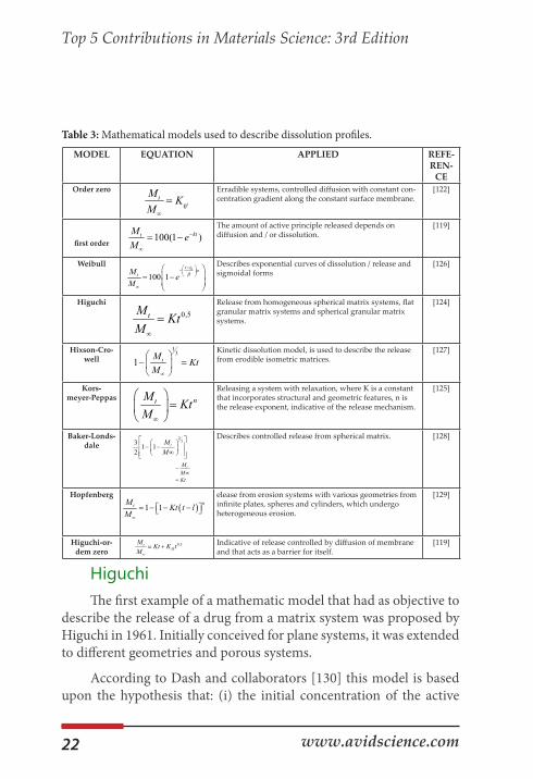
The analysis of the controlled in vitro release of active substances entrapped in polymers may offer important information, Manadas, Pina, and Veiga [122] point out that there is a necessity of a quantita-tive analysis of the obtained values in the tests of dissolution. Then, for this reason, generic equations are used, and these equations may be deduced by a theoretical analysis, as, for example, the zero order kinetics [119], or by empiric equations. Table 3 presents a summary of some models that were developed aiming to describe the discharge of an active principle, although the most commonly used are from Higuchi [124] and Korsmeyer et al. [125] since they describe the re-lease by diffusion.
dCdx
22 www.avidscience.com
Top 5 Contributions in Materials Science: 3rd Edition
Table 3: Mathematical models used to describe dissolution profiles.
Higuchi The first example of a mathematic model that had as objective to
describe the release of a drug from a matrix system was proposed by Higuchi in 1961. Initially conceived for plane systems, it was extended to different geometries and porous systems.
According to Dash and collaborators [130] this model is based upon the hypothesis that: (i) the initial concentration of the active
MODEL EQUATION APPLIED REFE-REN-
CEOrder zero Erradible systems, controlled diffusion with constant con-
centration gradient along the constant surface membrane.[122]
first order
The amount of active principle released depends on diffusion and / or dissolution.
[119]
Weibull Describes exponential curves of dissolution / release and sigmoidal forms
[126]
Higuchi Release from homogeneous spherical matrix systems, flat granular matrix systems and spherical granular matrix systems.
[124]
Hixson-Cro-well
Kinetic dissolution model, is used to describe the release from erodible isometric matrices.
[127]
Kors-meyer-Peppas
Releasing a system with relaxation, where K is a constant that incorporates structural and geometric features, n is the release exponent, indicative of the release mechanism.
[125]
Baker-Londs-dale
Describes controlled release from spherical matrix. [128]
Hopfenberg elease from erosion systems with various geometries from infinite plates, spheres and cylinders, which undergo heterogeneous erosion.
[129]
Higuchi-or-dem zero
Indicative of release controlled by diffusion of membrane and that acts as a barrier for itself.
[119]
0ttM K
M∞
=
100(1 )kttM eM
−
∞
= −
0
100 1t t
tM eM
αβ−
−
∞
= −
0,5tM KtM∞
=
13
1 tM KtM∞
− =
ntM KtM∞
=
2
33 1 12
tMM
− − ∞ tM
MKt
−∞
=
( )1 1ntM Kt t l
M∞
= − − −
0,5tH
M Kt K tM∞
= +
23
Top 5 Contributions in Materials Science: 3rd Edition
www.avidscience.com
principle in the matrix is much higher than its solubility; (ii) the dif-fusion of the active principle happens in only one dimension (border effect should be insignificant); (iii) the particles of the active principle are much smaller than the thickness of the system; (iv) the swelling and dissolution of the matrix are negligible; (v) the diffusivity of the active principle is constant; and (vi) the immersion conditions are al-ways reached in the middle. Generally, it is possible to summarize the Higuchi model as the expression:
Where the relation between the amount of active principle released at time t, KH the dissolution constant of Higuchi.
Therefore, every active principle release system said as Higuchi system is based upon Fick’s law, being, for this reason, named as fick-ian mechanism dependent on the square root of time [119].
Korsmeyer-Peppas Model Korsmeyer and collaborators [125] created a simple semi-em-
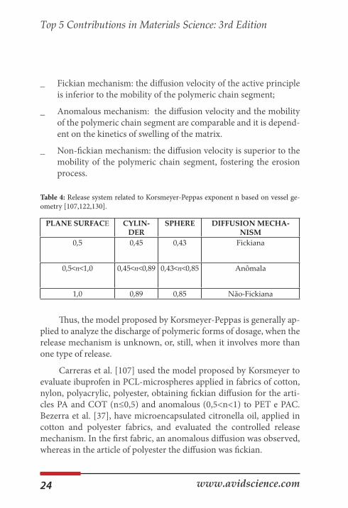
piric model, that relates exponentially to the liberation of the active principle and the elapsed time. This equation might be written as it follows:
Being the ratio between the amount of active principle released with the time t and the release in the equilibrium, KKP is the Korsemeyer-Peppas kinetic constant that incorporates structural and geometric characteristics, n is the exponent of release, indicative of the mechanism, that, according to Table 4 is related to the release ge-ometry [122]
Surathi and Karbhari [131] show what are the relations between the diffusion mechanism, Table 4, and the polymeric matrix:
12t
t
H
Mf KM∞
= =
tMM∞
nt
t
Kp
Mf KM∞
= =
tMM∞
24 www.avidscience.com
Top 5 Contributions in Materials Science: 3rd Edition
_ Fickian mechanism: the diffusion velocity of the active principle is inferior to the mobility of the polymeric chain segment;
_ Anomalous mechanism: the diffusion velocity and the mobility of the polymeric chain segment are comparable and it is depend-ent on the kinetics of swelling of the matrix.
_ Non-fickian mechanism: the diffusion velocity is superior to the mobility of the polymeric chain segment, fostering the erosion process.
Table 4: Release system related to Korsmeyer-Peppas exponent n based on vessel ge-ometry [107,122,130].
Thus, the model proposed by Korsmeyer-Peppas is generally ap-plied to analyze the discharge of polymeric forms of dosage, when the release mechanism is unknown, or, still, when it involves more than one type of release.
Carreras et al. [107] used the model proposed by Korsmeyer to evaluate ibuprofen in PCL-microspheres applied in fabrics of cotton, nylon, polyacrylic, polyester, obtaining fickian diffusion for the arti-cles PA and COT (n≤0,5) and anomalous (0,5<n<1) to PET e PAC. Bezerra et al. [37], have microencapsulated citronella oil, applied in cotton and polyester fabrics, and evaluated the controlled release mechanism. In the first fabric, an anomalous diffusion was observed, whereas in the article of polyester the diffusion was fickian.
PLANE SURFACE CYLIN-DER
SPHERE DIFFUSION MECHA-NISM
0,5 0,45 0,43 Fickiana
0,5<n<1,0 0,45<n<0,89 0,43<n<0,85 Anômala
1,0 0,89 0,85 Não-Fickiana

25
Top 5 Contributions in Materials Science: 3rd Edition
www.avidscience.com
Sun et al. [106] have evaluated a thermosensitive microgel-loaded cotton fabric for controlled drug release and obtained a val-ue of n≤0,5, thus, a fickian mechanism. When the microgel-loaded was evaluated, without the application in the fabric, it was obtained 0,5<n<1, or an anomalous diffusion mechanism.
Radu et al., [118] performed grafting in cotton with complexes of CD and HCr and studied the liberation in vitro, obtaining n=0.79, anomalous. Schaccheti et al [132] functionalized cotton fabrics with complexes formed with β-cyclodextrin and thyme oil and evaluated the release mechanism, obtaining anomalous mechanism of oil diffu-sion 0,5<n<1,0.
Considering this, the executed analysis has shown that the re-lease is influenced by the textile matrix and the type of polymer that constitutes it. It is possible to modify the diffusion of the active agent; furthermore, to indicate that textile supports show high potential to the use in systems of release [105,114]. Therefore, the use of biofunc-tional textiles allows treating many skin diseases by the contact skin-textile, displaying advantages in relation to the administration of the active principle when compared to via-oral. This principle does not pass through the digestive system when it occurs a loss of part of the drug via digestion or excretion [133].
Conclusion As can be seen from the experimental results obtained, there ex-
ist many possibilities to make “active” textiles substrates “against dif-ferent environments”, just using well designed vehiculizing systems to incorporate to the fabrics. These complex structures can be, among others, microcapsules, cyclodextrins or liposomes.
The response of the smart fabric “created” depends, strongly, on the existing interactions between the active principle, the molecular covered structure and on the interactions between these components with the textile substrate. The combination of these effects results in
26 www.avidscience.com
Top 5 Contributions in Materials Science: 3rd Edition
the development of biofunctional textiles capable of combining spe-cific characteristics of bioactive molecules that cannot be inserted di-rectly into the fabric, as is the case of the essential oils that lose their effect due to their volatility.
Therefore, the use of biofunctional textiles allows treating many skin diseases by the contact skin-textile, displaying advantages in rela-tion to the administration of the active principle.
References1. Lewis RW, Foreword. In: Xiaoming Tao editor. Smart fibres,
fabrics and clothing. Cambridge: Woodhead Publishing Limited. CRC Press. 2001.
2. R Wachter, M Weuthen, C Panzer, E Paff. Liposomes are used as textile finishes which not only improve elasticity and hand but can also be transferred to skin contact. Patent no. EP1510619-A2, DE10339358-A1, US2005058700-A1. 2005.
3. M Guarducci. Product having particular functional prop-erties for the skin and process for the preparation there of.Patent no. WO/2006/106546. 2006.
4. Lei M, Jiang F, Cai F, Hu S, Zhou R, et al. Facile microen-capsulation of olive oil in porous starch granules: Fabrica-tion, characterization, and oxidative stability. International Journal of Biological Macromolecules. 2018; 111: 755-761.
5. Paulo F, Santos L. Design of experiments for microencap-sulation applications: A review. Materials Science and Engi-neering C. 2017; 77: 1327-1340.
6. Ramakrishnan A, Pandit N, Badgujar M, Bhaskar C, Rao M. Encapsulation of endoglucanase using a biopolymer Gum Arabic for its controlled release. Bioresource Technology. 2007; 98: 368-372.
27
Top 5 Contributions in Materials Science: 3rd Edition
www.avidscience.com
7. Reshetnikov IS, Zubkova NS, Antonov VA, Potapova EV, Svistunov VS, et al. Microencapsulated fire retardants for polyolefins Materials Chemistry and Physics. 1998; 52: 78-82
8. Fadinia AL, Alvim ID, Ribeiro IP, Ruzene LG, Silva LB, et al. Innovative strategy based on combined microencapsulation technologies for food application and the influence of wall material composition. LWT. 2018; 91: 345-352.
9. Lamoudi L, Chaumeil JC. Daoud K. Effet des paramètres du procédé demicroencapsulation du piroxicam par coacerva-tion complexe. Annales Pharmaceutiques Françaises. 2014; 1: 1- 6.
10. Skeie S. Developments in microencapsulation science appli-cable to cheese research and development. A review. Inter-national Dairy Journal. 1994; 4: 573-595.
11. Souza FN, Gebara C, Ribeiro MCE, Chaves KS, Gigante ML, et al. Production and characterization of microparticles containing pectin and whey proteins. Food Research Inter-national. 2012; 49: 560–566
12. Ren PW, Ju XJ, Xie R, Chu LY. Monodisperse alginate mi-crocapsules with oil core generated from microfluidic de-vice. Journal of Colloid and Interface Science. 2010; 343: 392 – 395.
13. Ré MI. Microencapsulação – em busca de produtos ‘Inteli-gente’. Ciência Hoje. 2000; 27: 24 – 29.
14. Yoshizawa H. Trends in microencapsulation research. KONA Powder and Particle. 2004; 22: 23-31.
15. Donbrow M. Microcapsules and nanoparticles in medicine and pharmacy. Boca Raton: CRC Press. 1992; 360.
28 www.avidscience.com
Top 5 Contributions in Materials Science: 3rd Edition
16. Song X, Zhao Y, Hou S, Xu F. Zhao R, et al. Dual agents loaded PLGA nanoparticles: Systematic study of particle size and drug entrapment efficiency. European Journal of Pharmaceutics and Biopharmaceutics. 2008; 69: 445–453.
17. Freitas S, Merkle HP, Gander B. Microencapsulation by sol-vent extraction/evaporation: reviewing the state of the art of microsphere preparation process technology. Journal of Controlled Release. 2005; 102: 313–332.
18. Rocha-Selmi GA, Bozza FT, Thomazini M, Bolini HMA, Fávaro-Trindade CS. Microencapsulation of aspartame by double emulsion followed by complex coacervation to pro-vide protection and prolong sweetness. Food Chemistry. 2013; 139: 72–78.
19. Nakagawa K, Nagao H. Microencapsulation of oil droplets using freezing-induced gelatin–acacia complex coacerva-tion. Colloids and Surfaces A: Physicochem. Eng. As-pects. 2012; 411: 129–139.
20. Jun-Xia X, Hai-Yan Y, Jian Y. Microencapsulation of sweet orange oil by complex coacervation with soybean protein isolate/gum Arabic. Food Chemistry. 2011; 125: 1267–1272.
21. Bosnea LA, Moschakis T, Biliaderis CG. Complex coacerva-tion as a novel microencapsulation technique to improve vi-ability of probiotics under different stress. Food Bioprocess Technology. 2014; 1: 1-15.
22. Al-Shannaq R, Farid M, Al-Muhtaseb S, Kurdi J. Emulsion stability and cross-linking of PMMA microcapsules con-taining phase change materials. Solar Energy Materials & Solar Cells. 2015; 132: 311-318
23. Bosnea LA, Moschakis T, Biliaderis CG. Complex coacerva-tion as a novel microencapsulation technique to improve vi-
29
Top 5 Contributions in Materials Science: 3rd Edition
www.avidscience.com
ability of probiotics under different stress. Food Bioprocess Technology. 2014; 1: 1-15.
24. Li L, Song L, Hua T, Au WM, Wong KS. Characteristics of weaving parameters in microcapsule fabrics and their influ-ence on loading capability. Textile Research Journal. 2013; 83: 113-121.
25. Semyonov D, Ramon O, Kaplun Z, Levin-Brener L, Gurev-ich N, et al. Microencapsulation of Lactobacillus paracasei by spray freeze drying. Food Research International. 2010; 43: 193–202.
26. Shalaka DSR, Amruta NA, Parimal K. Vitamin and load-ed pectin alginate microspheres for cosmetic application. Journal of Pharmacy Research. 2009; 6: 1098-1102.
27. Suave J, Dall’agnol EC, Pezzini APT, Silva DAK, Meier MM, et al. Libecon. Microencapsulação: Inovação em diferentes áreas. Revista Saúde e Ambiente. 2006; 7: 12-20.
28. Santos AB, Ferreira VP, Grosso CRF. Microesferas – uma al-ternativa viável. Biotecnologia Ciência e Desenvolvimento. 2000; 16: 26-30.
29. Wang JM, Zheng W, Song QW, Zhu H, Zhou Y. Prepara-tion and characterization of natural fragrant microcapsules. Journal of Fiber Bioengineering and Informatics. 2009; 4: 293-300.
30. Yang Z, Peng Z, Li J, Li S, Kong L, et al. Development and evaluation of novel flavor microcapsules containing vanilla oil using complex coacervation approach. Food Chemistry. 2014; 145: 272-277.
31. Sharipova AA, Aidarova SB, Grigoriev D, Mutalieva B, Madibekova G, et al. Polymer-surfactant complexes for mi-croencapsulation of vitamin E and its release. Colloids and Surfaces B: Biointerfaces. 2016; 137: 152-157.
30 www.avidscience.com
Top 5 Contributions in Materials Science: 3rd Edition
32. França D, Medina AF, Messa LL, Souza CF, Faez R. Chitosan spray-dried microcapsule and microsphere as fertilizer host for swellable − controlled release materials. Carbohydrate Polymers. 2018; 196: 46-55.
33. Poncelet D. Microencapsulation: fundamentals, methods and applications.
34. In: Blitz J, Gun0ko V, editors. Surface chemistry in biomedi-cal and environmental science. Netherlands: Springer. 2006; 23–34.
35. Jamekhorshida A, Sadramelia SM, Farid M. A review of microencapsulation methods of phase change materials (PCMs) as a thermal energy storage (TES) medium. Renew-able and Sustainable Energy Reviews. 2014; 31: 531-542
36. Tsuda N, Ohtsubo T, Fuji M. Preparation of self-bursting microcapsules by interfacial polymerization. Advanced Powder Technology. 2012; 23: 724–730.
37. Alcázar A, Borreguero AM, Lucas A, Rodríguez JF, Car-mona M. Microencapsulation of TOMAC by suspension polymerisation: Process optimisation. Chemical engineer-ing research and design. 2017; 117: 1–10.
38. Zuo M, Liu T, Han J, Tang Y, Yao F, et al. Preparation and characterization of microcapsules containing ammonium persulfate as core by in situ polymerization. Chemical Engi-neering Journal. 2014; 249: 27–33.
39. Bezerra FM, Carmona OG, Carmona CG, Lis MJ, Moraes FF. Controlled release of microencapsulated citronella es-sential oil on cotton and polyester matrices. Cellulose. 2016; 23: 1459–1470
40. Li Y, Wu C, Wu T, Wang L, Chen S, et al. Preparation and characterization of citrus essential oils loaded in chitosan microcapsules by using different emulsifiers, Journal of Food Engineering. 2018; 217: 108-114.
31
Top 5 Contributions in Materials Science: 3rd Edition
www.avidscience.com
41. Bae J. Fabrication of carbon microcapsules containing sili-con nanoparticles–carbon nanotubes nanocomposite by sol–gel method for anode in lithium ion battery. Journal of Solid State Chemistry. 2011; 184: 1749–1755.
42. Barbosa-Cánovas GV, Ortega-Rivas E, Juliano P, Yan H. Food Powders - Physical Properties, Processing, and Func-tionality. US: Springer. 2005; 372.
43. Ulrich K, Eppinger S. Product design and development. New York: McGraw Hill. 2003; 432.
44. Nelson G. Application of microencapsulation in textiles. International Journal of Pharmaceutics. 2002; 242: 55 – 62.
45. Tekin R, Bac N, Erdogmus H. Microencapsulation of fra-grance and natural volatile oils application in cosmetics, and household cleaning products. Macromol. Symp. 2013; 333: 35-40.
46. Zimet P, Livney YD. Beta-lactoglobulin and its nanocom-plexes with pectin as vehicles for ω-3 polyunsaturated fatty acids. Food Hydrocolloids. 2009; 23: 1120–1126.
47. Annan NT, Borza AD, Hansen LT. Encapsulation in algi-nate-coated gelatin microspheres improves survival of the probiotic Bifidobacterium adolescentis 15703T during ex-posure to simulated gastro-intestinal conditions. Food Re-search International. 2008; 41: 184–193.
48. Prata AS, Grosso CRF. Production of microparticles with gelatina and chitosan. Carbohydrate Polymers. 2015; 116: 292 – 299.
49. Abdelkader MB, Azizi N, Baffoun A, Chevalier Y, Majdoub M. New microcapsules based on isosorbide for cosmetotex-tile: Preparation and characterization. 2018; 123: 591-599
50. Rubio L, Alonso C, Coderch L, Parra JL, Martí M, et al. Skin delivery of caffeine contained in biofunctional textiles. Tex-tile Research Journal. 2010; 80: 1214–1221.
32 www.avidscience.com
Top 5 Contributions in Materials Science: 3rd Edition
51. Cheng SY, Yuen MCW, Kan CW, Cheuk KKL, Chui CH, et al. Cosmetic textiles with biological benefits: Gelatin mi-crocapsules containing Vitamin C. International Journal of Molecular Medicine. 2009; 24: 411-419.
52. Meyer A. Perfume microencapsulation by complex coacer-vation. CHIMIA. 1992; 46: 101-102.
53. Chinta SK, Pooja PW. Use of microencapsulation in textiles. Indian Journal of Engineering. 2013; 3: 37-40.
54. Sanchez P, Sanchez-Fernandez MV, Romero A. Develop-ment of thermoregulating textiles using paraffin wax micro-capsules. Thermochim Acta. 2010; 498: 16–21.
55. Ma ZH, Yu DG, Branford-White CJ. Microencapsulation of tamoxifen: Application to cotton fabric. Colloids Surf B. 2009; 69: 85–90.
56. El Asbahani A, Miladi K, Badri W, Sala M, Aït Addi EH, et al. Essential oils: From extraction to encapsulation. Interna-tional Journal of Pharmaceutics. 2015; 483: 220-243.
57. Lv Y, Yang F, Li X, Zhang X. Abbas, S. Formation of heat-resistant nanocapsules of jasmine essential oil via gelatin/gum Arabic based complex coacervation. Food Hydrocol-loids. 2014; 35: 305 – 314.
58. Wang B, Adhikari B, Barrow CJ. Optimisation of the micro-encapsulation of tuna oil in gelatin-sodium hexametaphos-phate using complex coacervation. Food Chemistry. 2014; 158: 358 – 365.
59. Patrick KE, Abbas S, Lv Y, Ntsama ISB, Zhang X. Microen-capsulation by complex coacervation of fish oil using gela-tin/SDS/NaCMC. PAK. J. Food Science. 2013; 23: 17-25.
60. Piacentini E, Giorno L, Dragosavac MM, Vladisavljevic GT, Holdich RG. Microencapsulation of oil droplets using
33
Top 5 Contributions in Materials Science: 3rd Edition
www.avidscience.com
cold water fish gelatine/gum arabic complex coacervation by membrane emulsification. Food Research International. 2013; 53: 362 – 372.
61. Thilagavathi G, Kannaian T. Combined antimicrobial and aroma finishing treatment for cotton, using micro encap-sulated geranium (Pelargonium graveolens L’Herit. Ex Ait.) leaves extract. Indian Journal of Natural Products and Re-sources. 2010; 3: 348-352.
62. Wang CX, Chen ShL. Aromachology and its application in the textile field. Fibres and Textiles in Eastern Europe. 2005; 13: 41–44.
63. Solomon B, Sahle FF, Gebre-Mariam T, Asres K, Neubert RHHH. Microencapsulation of citronela oil for mosquito-repellent application: formulation and in vitro permeation studies. European Journal of Pharmaceutics and Biophar-maceutics. 2012; 80: 61-66.
64. Specos MMM, Garcia JJ, Tornesello J, Marino P, Della Vec-chia M, et al. Microencapsulated citronella oil for mosquito repelente finishing of cotton textiles. Transactions of Royal Society of Tropical Medicine and Hygiene. 2010; 104: 653-658.
65. Gonsalves JKMC, Costa AMB, Sousa DP, Cavalcanti SCH, Nunes RS. Microencapsulação do óleo essencial de Citrus sinensis (L) Osbeck pelo método da coacervação simples. Scientia Plena. 2009; 5: 1-8.
66. Tawatsin A, Wratten SD, Scott RR, Thavara U, Techadam-rongsin Y. Reppelency of volatile oils from plants against three mosquito vectors. Journal of Vector Ecology. 2001; 76-82.
67. Prata AS, Grosso CRF. Production of microparticles with gelatina and chitosan. Carbohydrate Polymers. 2015; 116: 292 – 299.
34 www.avidscience.com
Top 5 Contributions in Materials Science: 3rd Edition
68. Chatterjee S, Salaün F, Campagne C. Development of multi-layer microcapsules by a phase coacervation method based on ionic interations for textile applications. Pharmaceutics. 2014; 6: 281 – 297.
69. Szejtli J. Introduction and general overview of cyclodextrin chemistry. Chemical Reviews. 1998; 98: 1743-1753.
70. Duchêne D. Cyclodextrin as industrial uses. Paris: Edition Santé. 1987; 300.
71. Bender H. Production, characterization and application of CDs. Advances Biotechnological Processses. 1986; 6: 31-71.
72. Del Valle EMM. Cyclodextrins and their uses: a review. Pro-cess Biochemistry. 2004; 39: 1033 -1046
73. Matioli G, Moraes FF, Zanin GM. Ciclodextrinas e suas apli-cações em: alimentos, fármacos, cosméticos, agricultu-ra, biotecnologia, química analítica e produtos gerais, Eduem: Maringá. 2000; 124
74. Bhaskara-Amrit UR, Pramod BA, Warmoeskerken MCG. Applications of β –cyclodextrins in textiles. AUTEX Re-search Journal. 2011; 11: 94-101.
75. Shlar I, Droby S, Rodov V. Antimicrobial Coatings on Poly-ethylene Terephthalate Based on Curcumin/Cyclodextrin Complex Embedded in a Multilayer Polyelectrolyte Archi-tecture. 2018; 164: 379-384.
76. Lucio D, Irache JM, Font M, Martíinez-Oharriz MC. Su-pramolecular structure of glibenclamide and -cyclodextrins complexes. International Journal of Pharmaceutics. 2017; 530: 377-386.
77. Suvarna V, Gujar P, Murahari M. Complexation of phyto-chemicals with cyclodextrin derivatives – An insight. Bio-medicine & Pharmacotherapy. 2017; 88: 1122–1144.
35
Top 5 Contributions in Materials Science: 3rd Edition
www.avidscience.com
78. Zhang L, Man S, Qiu H, Liu Z, Zhang M, et al. Curcumin-cyclodextrin complexes enhanced the anti-cancer effects of curcumin. Environmental Toxicology and Pharmacology. 2016; 48: 31-38.
79. Irie T, Uekama K. Pharmaceutical applications of cyclodex-trins. III. Toxicological issues and safety evaluation. Phar-maceutical. Science. 1997; 86: 147 – 162.
80. Sali N, Csepregi R, Kőszegi T, Kunsági-Máté S, Szente L, et al. Complex formation of flavonoids fisetin and geraldol with β-cyclodextrins. Journal of Luminescence. 2018; 194: 82–90.
81. Lo Nostro P, Fratoni L, Ridi F, Baglioni P. Surface treatments on Tencel fabric: Grafting with β-cyclodextrin. Journal of Applied Polymer Science. 2003; 88: 706-715.
82. Partanen R, Ahro M, Hakala M, Kallio H, Forssell P. Mi-croencapsulation of caraway extract in β-cyclodextrin and modified starches. Europen Food Research Technology. 2002; 214: 242 – 247.
83. Yildiz ZA, Celebioglu A, Kilic ME, Durgun E, Uyar T. Men-thol/cyclodextrin inclusion complex nanofibers: Enhanced water-solubility and high-temperature stability of menthol. Journal of Food Engineering. 2018; 224: 27-36
84. Ciobanu A, Mallard I, Landy D, Brabie G, Nistor S, et al. Re-tention of aroma compunds from menthe piperita essential oil by cyclodextrins and crosslinked cyclodextrin polymers. Food Chemistry. 2013; 138: 291 – 297.
85. Peila R, Migliavacca G, Aimone F, Ferri A, Sicardi S. A com-parison of analytical methods for the quantification of a reactive β-cyclodextrin fixed onto cotton yarns. Cellulose. 2012; 19: 1097 – 1105.
86. Teschke O, de Souza EF. Liposome structure imaging by atomic force microscopy: verification of improved liposome
36 www.avidscience.com
Top 5 Contributions in Materials Science: 3rd Edition
stability during absorption of multiple aggregated vesicles. Langmuir. 2002; 18: 6513-6520.
87. Lian T, Ho RJY. Trends and developments in liposome drug delivery systems. J. Pharm. Sci. 2001; 90: 667-680.
88. Betz G, Aeppli A, Menshutina N, Leuenberge H. In vivo comparison of various liposome formulations for cosmetic application. Int. J. Pharm. 2005; 296: 44-54.
89. Aggarwal A, Dayal A, Kumar N. Microencapsulation pro-cesses and application in textile processing. Colourage. 1998; 45: 15-24.
90. Martí M, de la Maza A, Parra JL, Coderch L. Dyeing wool at low temperatures: New method using liposomes. Text. Res. J. 2001; 71: 678-682.
91. Montazer M, Validi M, Toliyat T. Influence of temperature on stability of multilamellar liposomes in wool dyeing. J Li-posome Res. 2008;, 16: 81-89.
92. Ramirez R, Martí M, Manich AM, Parra JL, Coderch L. Ceramides Extracted from Wool: Pilot Plant Solvent. Textile Res J. 2008; 78: 73-80.
93. Coderch L, Fonollosa J, Martí M, Garde F, de la Maza A, et al. Extraction and Análisis of Ceramides from Internal Wool Lipids. J Am Oil Chem Soc. 2002; 79: 1215-1220.
94. de Pera M, Coderch L, Fonollosa J, de la Maza A, Parra JL. Effect of Internal Wool Lipid liposomes on Skin Repair. Skin Pharmacol and Applied Physiology. 2000; 13: 188-195.
95. Coderch L, de Pera M, Fonollosa J, de la Maza A, Parra JL. Efficacy of stratum corneum lipid supplementation on hu-man skin. Contact Dermatitis. 2002; 47: 139-146.
37
Top 5 Contributions in Materials Science: 3rd Edition
www.avidscience.com
96. Ramírez R, Martí M, Barba C, Méndez S, Parra JL, et al. Skin Efficacy of liposomes composed of internal wool lipids rich in ceramides. J Cosmet Sci, 2010, 61: 235-245.
97. Coderch L, Fonollosa J, de Pera M, de la Maza A, Parra JL, et al. Compositions of internal lipid extract of wool and use thereof in the preparation of products for skin care and treatment. 2001: Patent nº WO/2001/004244.
98. Ramón E, Alonso C, Coderch L, De la Maza A, López O, et al. Liposomes as Alternative Vesicles for Sun Filter Formula-tions. Drug Delivery. 2005; 12: 83-88.
99. Williams AC. Transdermal and Topical Drug Delivery: from theory to clinical practice. London: Pharmaceutical Press. 2003.
100. M Martí, V Martínez, MJ Lis, J Valldeperas, A de la Maza, et al. Gallic Acid vehiculized through liposomes or mixed mi-celles in biofunctional textiles, Journal of Textile Institute. 2014; 105: 175-186
101. M Martí, C Alonso, V Martínez, MJ Lis, A de la Maza, et al. Cosmetotextiles with Gallic Acid: Skin reservoir effect. J. of drug release. 2013; 7.
102. M Martí, R Rodríguez, N Carreras, MJ Lis, J Valldeperas, et al. Monitoring of the microcapsule/liposome application on textile fabrics. Journal of Textile Institute. 2012; 103: 19-27.
103. M Martí, V Martínez, L Rubio, L Coderch, JL Parra. Bio-functional Textiles prepared with liposomes: in vivo and in vitro assessment. J. of Microencapsulation. 2011; 28: 799-806
104. Mihailiasa M, Caldera F, Li J, Peila R, Ferri A, et al. Prepara-tion of functionalized cotton fabrics by means of melatonin loaded -cyclodextrin nanosponges. Carbohydrate Polymers. 2016; 142: 24–30.
38 www.avidscience.com
Top 5 Contributions in Materials Science: 3rd Edition
105. Hebeish A, El-Shafei A, Sharaf S, Zagholou S. In situ forma-tion of silver nanoparticles for multifunctional cotton con-taining cyclodextrin. Carbohydrate Polymers. 2014; 103: 442-447.
106. Todorova SB, Silva CJS, Simeonov MP, Cavaco-Paulo A. Cotton fabric: A natural matrix suitable for controlled re-lease systems. Enzyme and Microbial Technology. 2007; 40: 1646–1650.
107. Sun X, Wang X, Wu J, Li S. Development of thermosensitive microgel-loaded cotton fabric for controlled drug release. Applied Surface Science. 2017; 403: 509–518.
108. Carreras N, Acuña V, Martí M, Lis MJ. Drug reléase sys-tem of ibuprofen in PCL-microespheres. Colloid Polym Sci. 2013; 291: 157-165
109. Voncina B, Vivod V. Chen, W. Surface Modification of PET Fibers with the Use of b-Cyclodextrin. Journal of Applied Polymer Science. 2009; 113: 3891–3895.
110. Martin A, Tabary N, Leclercq L, Junthip J, Degoutin S, et al. Multilayered textile coating based on a –cyclodextrin poly-electrolyte for the controlled release of drugs. Carbohydrate Polymers. 2013; 93: 718–730.
111. Haji A, Mehrizi MK, Akbarpour R. Optimization of b-cyclodextrin grafting on wool fibers improved by plasma treatment and assessment of antibacterial activity of berber-ine finished fabric. J Incl Phenom Macrocycl Chem. 2015; 81: 121–133.
112. Yu Y, Wang Q, Yuan J, Fan X, Wang P. A novel approach for grafting of -cyclodextrin onto wool via laccase/TEMPO oxi-dation. Carbohydrate Polymers. 2016; 153: 463–470.
39
Top 5 Contributions in Materials Science: 3rd Edition
www.avidscience.com
113. Bezerra FM, Lis M, Carmona OG, Carmona CG, Moises MP, et al. Assessment of the delivery of citronella oil from microcapsules supported on wool fabrics. Powder Tehcnol-ogy. 2019; 343: 775-782
114. Alonso C, Martí M, Barba C, Lis M, Rubio L, et al. Skin pen-etration and antioxidant effect of cosmeto textiles with gallic acid. Journal of Photochemistry & Photobiology, B: Biology. 2016; 156: 50-55.
115. Labay C, Canal C, García-Celma MJ. Influence of Corona Plasma Treatment on Polypropyleneand Polyamide 6.6 on the Release of a Model Drug. Plasma Chem Plasma Process. 2010; 30: 885–896.
116. Soares-Latour EM, Bernard J, Chambert S, Fleury E, Sintes-Zydowicz N. Environmentally benign 100% bio-based oli-goamide microcapsules. Colloids and Surfaces A. 2017; 524: 193–203.
117. Ghaheh FS, Khoddami A, Alihosseini F, Jing S, Ribeiro A, et al. Antioxidant cosmetotextiles: Cotton coating with nano-particles containing vitamin E. Process Biochemistry. 2017; 59: 46–51.
118. Ibrahim NA, El-Ghany NAA, Eid BM, Mabrouk EM. Green options for imparting antibacterial functionality to cotton fabrics. International Journal of Biological Macromolecules. 2018; 111: 526–533
119. Radu CD, Parteni O, Popa M, Muresan IE, Ochiuz L, et al. Comparative Study of a Drug Release from a Textile to Skin. J Pharm Drug Deliv Res. 2015; 4: 2-8.
120. Costa P, Lobo JMS. Modeling and comparison of dissolu-tion profiles. European Journal of Pharmaceutical Sciences. 2001; 13: 123-133.
40 www.avidscience.com
Top 5 Contributions in Materials Science: 3rd Edition
121. Bezerra FM, Croscato GS, Valldeperas J, Lis MJ, Carreras C, et al. Aplicação de microesferas para desenvolvimento de novos acabamentos têxteis. Química Têxtil. 2011; 105: 49-58.
122. Bird RB, Stewart WE, Lightfood EN. Fenômenos de trans-porte. Editora: LTC. 2004; 838
123. Manadas R, Pina ME, Veiga F. A dissolução in vitro na pre-visão da absorção oral de fármacos em formas farmacêuti-cas de liberação modificada. Brazilian Journal of Pharma-ceutical Sciences. 2002; 38: 375-399.
124. Lopes CM, Lobo JMS, Costa P. Formas farmacêuticas de lib-eração modificada: polímeros hidrifílicos. Revista Brasileira de Ciências Farmacêuticas. 2005; 41: 143-154.
125. Higuchi T. Mechanism of sustained-action medication. The-oretical analysis of rate of release of solid drugs dispersed in solid matrices. J Pharm Sci. 1963; 52: 1145–1149
126. Korsmeyer RW, Gurny R, Doelker E, Buri P, Peppas NA. Mechanisms of solute release from porous hydrophilic poly-mers. Int J Pharm. 1985; 15: 25–35
127. Weibull W, Sweden S. A statistical distribution function of wide applicability. Journal of Applied Mechanics. 1951; 83: 293-299.
128. Hixson AW, Crowell JH. Dependence of reaction velocity upon surface and agitation: I-theoretical consideration. Ind Eng Chem. 1931; 23: 923–31.
129. Baker RW, Lonsdale HS. Controlled release of biologically active agents. Plenum Press: Nova Iorque. 1974; 15-71.
130. Hopfenberg HB. In: Paul DR, Haris FW, editors. Controlled Release Polymeric Formulations. Washington: American Chemical Society. 1976; 317.
41
Top 5 Contributions in Materials Science: 3rd Edition
www.avidscience.com
131. Dash S, Murthy PN, Nath L, Chowdhury P. Kinetic mod-eling on drug release from controlled drug delivery systems. Acta Pol. Pharm. 2010; 67: 217-223
132. Surathi P, Karbhari VM. Hygrothermal effects on durability and moisture kinetics of fiber-reinforced polymer compos-ites. San-Diego: University of California. 2006.
133. Scacchetti FAP, Pinto E, Soares GMB. Functionalization and characterization of cotton with phase change materials and thyme oil encapsulated in beta-cyclodextrins, Progress in Organic Coatings. 2017; 107: 64–74.
134. Cezar-Doru R, Marcel P, Oana P, Mihaela S, Elena-Ctlina L, et al. Achievements and Limits on the Controlled Release of a Drug from a Textile Fabric to Dermis. The Open Confer-ence Proceedings Journal. 2014; 5: 1-8.