Embed Size (px)
Citation preview
Combined Spatial Limitation around Residues 16 and 108 ofPlasmodium falciparum Dihydrofolate Reductase Explains Resistanceto Cycloguanil
Jarunee Vanichtanankul,a Supannee Taweechai,a Chayasith Uttamapinant,a Penchit Chitnumsub,a Tirayut Vilaivan,b
Yongyuth Yuthavong,a and Sumalee Kamchonwongpaisana
National Centre for Genetic Engineering and Biotechnology, National Science and Technology Development Agency, Klong Luang, Pathumthani, Thailand,a andDepartment of Chemistry, Faculty of Science, Chulalongkorn University, Patumwan, Bangkok, Thailandb
Natural mutations of Plasmodium falciparum dihydrofolate reductase (PfDHFR) at A16V and S108T specifically confer resis-tance to cycloguanil (CYC) but not to pyrimethamine (PYR). In order to understand the nature of CYC resistance, the effects ofvarious mutations at A16 on substrate and inhibitor binding were examined. Three series of mutations at A16 with or withoutthe S108T/N mutation were generated. Only three mutants with small side chains at residue 16 (G, C, and S) were viable frombacterial complementation assay in the S108 series, whereas these three and an additional four mutants (T, V, M, and I) withslightly larger side chains were viable with simultaneous S108T mutation. Among these combinations, the A16V�S108T mutantwas the most CYC resistant, and all of the S108T series ranged from being highly to moderately sensitive to PYR. In the S108Nseries, a strict requirement for alanine was observed at position 16. Crystal structure analyses reveal that in PfDHFR-TS variantT9/94 (A16V�S108T) complexed with CYC, the ligand has substantial steric conflicts with the side chains of both A16V andS108T, whereas in the complex with PYR, the ligand only showed mild conflict with S108T. CYC analogs designed to avoid suchconflicts improved the binding affinity of the mutant enzymes. These results show that there is greater spatial limitation aroundthe S108T/N residue when combined with the limitation imposed by A16V. The limitation of mutation of this series providesopportunities for drug design and development against antifolate-resistant malaria.
Plasmodium falciparum dihydrofolate reductase (PfDHFR) isone of the most well-defined drug targets in malaria chemo-
therapy with such antifolates as pyrimethamine (PYR) and cy-cloguanil (CYC; administered as proguanil) among its inhibitors(26). It catalyzes the NADPH-dependent reduction of 7,8-dihy-drofolate (DHF) to 5,6,7,8-tetrahydrofolate (THF) in the biosyn-thesis of deoxythymidylate (dTMP). After decades of antifolatedeployment, the parasite has become increasingly resistant to PYRand CYC due to mutations that reduce the binding affinity ofinhibitors. While PfDHFR mutations at residues 108, 59, 51, and164 confer cross-resistance to both PYR and CYC, the double-mutation variant A16V�S108T specifically confers resistance toCYC. The CYC-resistant mutant was postulated to have arisen bythe S108T mutation followed by A16V, giving rise to high CYCresistance without a loss in catalytic efficiency (18). Molecularmodeling suggests that the A16V mutation causes steric clash be-tween the side chain and one of the 2,2-dimethyl groups of CYC(15). PYR, on the other hand, avoids such a steric clash, presum-ably due to lack of a group equivalent to the methyl of CYC (15).However, the lack of crystal structure data for the A16V�S108TPfDHFR and its complex with CYC makes such conclusion a ten-tative one.
Unlike cumulative mutations in the S108N series of PYR resis-tance-generating mutant PfDHFRs, no other resistant variants inthe S108T series apart from A16V�S108T have been identifiedfrom the field. Laboratory selection for PYR resistance in a wild-type parasite strain led to the identification of mutants with asingle mutation at either A16S or I164M (22). In the same study,selection for increased PYR resistance in the PfDHFR-TSA16V�S108T mutant strain T9/94 demonstrated DHFR-TS geneamplification but no further mutation of the protein (22). An-
other study of laboratory-induced PYR resistance reportedA16V�S108T variants with additional mutations at F223S andD54N (20). The A16V�S108T�D54E�F223S mutant enzymeshows enhanced resistance to CYC and is also resistant to PYR(19).
We investigated here the effect of mutation at residue 16 onPfDHFR catalytic activity and antifolate resistance. In the pres-ence or the absence of the S108T/N mutation, mutants of A16Xwere catalytically active only when X was an amino acid with asmall side chain, suggesting limitation in space around residue 16.Crystal structures of PfDHFR-TS T9/94 variant (A16V�S108T)in complex with CYC and PYR were determined to reveal thedifferent modes of inhibitor binding. The structural informationpresented here shows confinement of the DHFR binding pocket atsubsites containing residues 16 and 108, with the mutual effects ofsteric constraint in one subsite exerting further confinement onanother subsite. Such mutual effects are present in the binding ofCYC, which has a 2-methyl group in conflict with the side chain ofV16, and a p-chlorine atom in conflict with the side chain of T108.These constraints are absent in the binding of PYR, which lacks anequivalent group in the vicinity of V16. Therefore, the resistance
Received 9 February 2012 Returned for modification 26 March 2012Accepted 16 April 2012
Published ahead of print 23 April 2012
Address correspondence to Sumalee Kamchonwongpaisan,[email protected].
Copyright © 2012, American Society for Microbiology. All Rights Reserved.
doi:10.1128/AAC.00301-12
3928 aac.asm.org Antimicrobial Agents and Chemotherapy p. 3928–3935 July 2012 Volume 56 Number 7
Dow
nloa
ded
from
http
s://j
ourn
als.
asm
.org
/jour
nal/a
ac o
n 20
Jan
uary
202
2 by
81.
226.
97.5
9.
of the A16V�S108T PfDHFR-bearing parasite only to CYC, butnot to PYR, is explained. This information should help in thedesign of effective inhibitors against mutant PfDHFRs and againstantifolate-resistant malaria.
MATERIALS AND METHODSConstruction of mutant libraries. Mutant libraries of A16-saturated mu-tations were generated by QuikChange site-directed mutagenesis usingpET17b containing synthetic PfDHFR–A16V�S108T gene as a template,5=-C TTT GAC ATC TAC GCT ATC TGC [NNG/C] TGC TGT AAA GTTGAA AGC AAA-3= as the sense primer, and 5=-TTT GCT TTC AAC TTTACA GCA [G/CNN] GCA GAT AGC GTA GAT GTC AAA G-3= as theantisense primer, where the A16X codon is marked by brackets, and Nrepresents all four bases. The mutagenesis reaction contained 40 ng oftemplate, 125 ng of each primer, 1� Pfu buffer, and 10 U of Pfu DNApolymerase (Promega) and was carried out using the following tempera-ture cycling profile: 95°C for 5 min, followed by 20 cycles of 95°C for 30 s,45°C for 1 min, and 68°C for 8 min, with a final incubation at 68°C for 10min. The mutagenesis product was digested with 20 U of DpnI at 37°C for1 h and then transformed into E. coli DH5�. Recombinant clones grownon Luria-Bertani (LB) agar with 100 �g of ampicillin (Amp) ml�1 wererandomly selected for sequencing. Generation of A16X�S108 series wasperformed using the same protocol with an equimolar mixture of the 20identified A16X�S108T as templates and a pair of primers that allowedmutation of T108 back to S108 —5=-GTT GTT ATG GGC CGC ACCTCG TGG GAA TCG ATT CCG AAA AAA TTC-3= and 5=-GAA TTT TTTCGG AAT CGA TTC CCA CGA GGT GCG GCC CAT AAC AAC-3=—asthe sense and antisense primers, respectively. A16X mutants in the N108series were limited to G, C, S, T, and V. The mutagenesis reactions wereperformed separately using specific templates from the T108 series and apair of primers that allowed the mutation of T108N: 5=-GGG CCG CACCAA CTG GGA ATC GAT-3= and 5=-ATC GAT TCC CAG TTG GTGCGG CCC-3=. The clones obtained were verified by automated Sangersequencing to ensure that there were no adventitious mutations in thecourse of making the site-specific mutations.
Screening of active DHFR mutants by bacterial complementationassay. Plasmids from the pET-PfDHFR mutants were retransformed intoE. coli BL21(DE3)/pLysS. The transformants were grown on LB agar with100 �g of Amp ml�1 and 34 �g of chloramphenicol (Cam) ml�1. Themutant clones were screened for PfDHFR activity by streaking them onminimal medium containing 100 �g of Amp ml�1 and 34 �g of Camml�1 with or without 4 �M trimethoprim. After overnight incubation at37°C, transformed bacteria are only able to grow in the presence of tri-methoprim if the PfDHFR has adequate activity; mutants with low activitydo not support growth (4).
Expression and purification of mutant PfDHFRs. All of the mutantswere expressed in E. coli BL21(DE3)/pLysS by cultivation in LB supple-mented with 100 �g of Amp ml�1 and 34 �g of Cam ml�1 and 0.4 mMIPTG (isopropyl-�-D-thiogalactopyranoside) induction at 20°C for 24 h.The expression levels of all mutants were confirmed by SDS-PAGE. Therelative PfDHFR activity of each mutant compared to wild-type enzymewas determined from crude protein extracts. Those with relative crudeactivity higher than 3% were further purified by methotrexate-Sepharoseaffinity chromatography (17) and anion exchange as previously described(2, 9).
Enzyme kinetic analysis and inhibition by antifolates. DHFR activ-ities were determined spectrophotometrically using a Hewlett PackardUV-VIS spectrophotometer (HP8453) in a 1-ml reaction containing 100�M concentrations each of dihydrofolate and NADPH in 50 mM TES(pH 7.0), 75 mM �-mercaptoethanol, 1 mg of bovine serum albuminml�1, and the appropriate amount of purified enzyme (1 to 5 mU) toinitiate the reaction. PYR, CYC, WR99210, and a series of CYC analogues(15, 27) were 2-fold serial diluted in dimethyl sulfoxide in 96-well plates(200 �l, final well volume), and binding constants were determined usingthe protocol described previously (21). Kinetic parameters were calcu-
lated by a nonlinear least-square fit of the data to the Michaelis-Mentenequation using the software Kaleidagraph 3.51 as previously described(9, 21).
Crystallization, data collection, and structure determination. Thebifunctional DHFR-TS enzymes of both wild-type strain TM4 and theA16V�S108T mutant strain T9/94 of P. falciparum were cloned, ex-pressed, and purified as previously described (2, 28). Protein was cocrys-tallized with either 2 mM CYC or 2 mM PYR in the presence of NADPHand dUMP. X-ray diffraction data were collected at the CuK� wavelength(1.5418 Å) on a Bruker-Nonius FR591 X-ray generator equipped with a�CCD detector. The data were indexed, integrated and scaled using theDenzo/Scalepack program in the HKL-2000 package (14). The data weredirectly refined at 3.0-Å resolution using PfDHFR-TS TM4 (PDB 1J3I) asa template in the Crystallography and NMR System (1, 28). The modelswere iteratively rebuilt and adjusted in program O (8) and gradually re-fined to the highest resolution in CNS (1). Ligands and water moleculeswere added to a positive Fourier difference electron density map (Fo �Fc). The final models were verified by PROCHECK (11) with Ramachan-dran parameters listed in Table 1. Structural figures were prepared withPyMOL (16).
PDB accession codes. Coordinates and structure factors have beendeposited in the Protein Data Bank (PDB) under accession codes 3UM5,3UM6, and 3UM8.
RESULTSScreening of active A16X�S108T/N PfDHFR mutants. Con-structs of wild-type and 45 A16X mutants with S108 or S108N/Tmutations were successfully generated and verified by DNA se-quencing. These mutants included 19 from the A16X�S108series, 20 from the A16X�S108T series, and 6 from the S108Nseries. For the S108N series, only the A16 mutants that arecapable of complementing bacterial growth in the S108T serieswere created, i.e., G, A, C, S, T, and V.
The activities of the wild-type and mutant PfDHFRs in E. coliBL21(DE3)/pLysS were screened by complementation assay. Onlysix mutants from S108T series showed adequate activityfor growth, namely, S108T, A16C�S108T, A16G�S108T,A16S�S108T, A16T�S108T, and A16V�S108T. Hence, only theamino acids with small side chains at position 16 when combinedwith S108T were functionally active. Moreover, two mutants withslightly larger side chains at position 16, namely, A16I�S108T andA16M�S108T, could also complement host DHFR under thesame conditions, albeit poorly (Table 2). Further investigation ofthe S108 series showed that only the wild type and three mutants(A16G, A16S, and A16C) were found to be catalytically active bybacterial complementation assay, while the others were not. In theS108N series, only alanine was tolerated at position 16 among thesix mutants tested.
Crude extracts of wild-type PfDHFR and 45 mutants withA16X and S108 or S108N/T were prepared in 50 ml of LB brothwith IPTG induction in order to examine their expression levelsand DHFR activities. SDS-PAGE analysis of the crude extractsrevealed that all of the constructs expressed PfDHFR similarly tothe wild-type construct (data not shown). However, these crudeextracts exhibited a wide range of enzyme specific activities. Therelative DHFR activity of crude protein extracts of the active mu-tants, defined from the ability to complement bacterial DHFR inthe presence of trimethoprim in bacterial complementation assay,ranged from 1.4 to 62.4% of the wild-type enzyme (Table 3), whilethe activities of the inactive mutants were negligible. The resultsare in line with our previous reports, wherein only constructsexpressing relative DHFR activity higher than 1.3% of the wild-
Space Limits in PfDHFR Explain Cycloguanil Resistance
July 2012 Volume 56 Number 7 aac.asm.org 3929
Dow
nloa
ded
from
http
s://j
ourn
als.
asm
.org
/jour
nal/a
ac o
n 20
Jan
uary
202
2 by
81.
226.
97.5
9.
type enzymes could support bacterial growth on trimethoprimplates (4, 9).
Kinetic parameters of PfDHFR mutants. Wild-type PfDHFRand 10 active A16X PfDHFR mutants with or without S108T/Nmutations were purified to homogeneity and enzyme kinetic pa-rameters determined. As summarized in Table 3, the kcat values ofall of the active mutants varied from 1.6 to 86% of that of thewild-type enzyme. Among these mutants, A16C�S108T exhib-ited the highest kcat value, while A16T�S108T was the lowest. Thekcat values of the others were 14 to 51% of the wild-type enzyme.
Mutations of A16 and S108 affected DHF binding to variousdegrees. The A16S and S108N mutants showed 3- to 4-fold-higherKm values for DHF than those of the wild type, while the Km valuesfor the S108T and A16C�S108T mutants were about 2- to 4-foldless. Unlike DHF, the Km values for NADPH were not much af-fected by mutations since the values were within a 2-fold rangeof the wild-type enzyme. Among the 10 active mutants,A16C�S108T and S108T showed the highest catalytic efficiency(kcat/Km), about 1.4- to 1.6-fold that of the wild-type enzyme. Thecatalytic efficiency of the other mutants, including A16V�S108T,
was about 4 to 38% that of the wild-type enzyme and that ofA16T�S108T was �1%.
Antifolate sensitivity of PfDHFR mutants. The inhibitionconstants (Ki) of PYR, CYC, and WR99210, as well as of three CYCderivatives, namely, C17 (R1, R2, and R3 H, Me, and p-Cl phe-nyl, respectively), C88 (R1, R2, and R3 Me, Me, and m-Cl phe-nyl, respectively), and C248 (R1, R2, and R3 H, Me, and m-Clphenyl, respectively), against the 10 mutants were determined inorder to understand the drug resistance pattern of the active mu-tants. The data are summarized in Table 4.
Among the three active A16X single mutants, only A16S washighly resistant to CYC, with Ki values 83 times higher than thatof the wild type, while the others were about 1- to 3.5 times that ofthe wild type. In combination with S108T mutation, the Ki valuesof CYC against all of the active double mutants were markedlyincreased, i.e., by about 58- to 1,014-fold that of the wild-type and20- to 345-fold that of the S108T single mutant. In addition, the Ki
values of CYC against S108N were 8 times that of the wild-typeenzyme and 2.7 times that of S108T. These results suggested thatthe steric conflicts of the p-Cl group at R3 and a 2-methyl group of
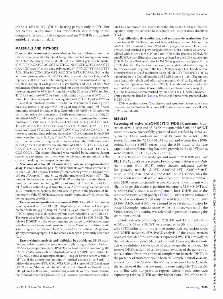
TABLE 1 Data collection and refinement statistics of crystal structures
Parametera
Enzyme and ligand
PfDHFR-TS T9/94
PfDHFR-TS TM4 (CYC)PYR CYC
Data collectionSpace group P212121 P212121 P212121
Unit cell parameters (Å)a 56.529 56.696 58.761b 155.119 155.549 157.538c 165.257 164.962 164.509
Resolution* (Å) 30–2.40 (2.49–2.40) 30–2.65 (2.74–2.65) 50–2.60 (2.69–2.60)Rmerge* (%) 6.5 (35.1) 7.9 (35.5) 4.1 (25.8)I/�I* 15.5 (3.0) 19.2 (4.2) 20.3 (4.3)Completeness* (%) 98.7 (95.0) 99.9 (99.7) 98.4 (93.2)Redundancy 4.6 8.1 4.4
RefinementResolution (Å) 30–2.40 30–2.65 50–2.60No. reflections (unique) 56,048 43,280 47,165Rwork/Rfree
b 20.2/25.4 20.3/25.7 21.2/25.8No. of atoms
Protein 9,058 9,048 9,046PYR or CYC 34 34 34NADPH 96 96 96dUMP 40 40Water 781 371 456Phosphate 10
Avg B factor (Å2) 46.9 48.5 52.5Ramachandran plot (%)
Favored region 85.6 84.6 87.3Additional allowed region 14.2 15.2 12.3Generously allowed region 0.2 0.2 0.2Disallowed region 0 0 0.2c
RMS deviationsBond length (Å) 0.006 0.007 0.008Bond angle (°) 1.4 1.4 1.5
a *, Values in parentheses are for the highest-resolution shell. RMS, root mean square.b Rfree is calculated from 5% of reflections chosen randomly in each of the 10 resolution bins.c Lys49 is located on a flexible loop of each molecule of PfDHFR-TS.
Vanichtanankul et al.
3930 aac.asm.org Antimicrobial Agents and Chemotherapy
Dow
nloa
ded
from
http
s://j
ourn
als.
asm
.org
/jour
nal/a
ac o
n 20
Jan
uary
202
2 by
81.
226.
97.5
9.
CYC with side chains of amino acid residues 108 and 16, respec-tively, are significant and that the clash with S108N exerted agreater effect on the binding affinity than that of S108T, which is inline with previous observations (18).
In order to reduce the steric clash of inhibitors with A16Xmutants, the inhibitor C17 was designed with only one methylgroup at the position 2 of the dihydrotriazine ring (either R1 orR2), while maintaining its p-Cl phenyl group at R3. The Ki valuesof C17 against the wild type and most of mutants were generallysimilar to those of CYC (0.2- to 2.2-fold) but reduced markedly by
10.7-fold against A16V�S108T compared to CYC. By moving thep-Cl group to the m-position to avoid steric clash with S108Twhile preserving the 2,2-dimethyl groups of CYC, the inhibitorC88 showed reduction of Ki values against all of the double mu-tants to an even greater extent than C17, by 4- to 41-fold com-pared to CYC. With the combination of both features so asto avoid steric conflicts on both sites, C248 (desmethyl and m-Cl phenyl) showed dramatically lower Ki values againstA16V�S108T by a factor of 51 compared to that of CYC (from1,518 nM for CYC to 30 nM for C248). The Ki values against other
TABLE 2 Summary of bacterial complementation assay of PfDHFR activities in E. coli BL21(DE3)/pLysS on a minimal medium plate in thepresence of trimethoprima
Amino acid atresidue 16 Type
Residue volb
(Å3)Surface areac
(Å2)
Bacterial complementation
S108 T108 N108
G Aliphatic 60.1 75 � � –A (wild type) Aliphatic 88.6 115 � � �P Aliphatic 112.7 145 – – NAd
V Aliphatic 140.0 155 – � –M Aliphatic 162.9 185 – �/– NAI Aliphatic 166.7 175 – �/– NAL Aliphatic 166.7 170 – – NAS Polar, uncharged 89.0 115 � � –C Polar, uncharged 108.5 135 � � –N Polar, uncharged 114.1 160 – – NAT Polar, uncharged 116.1 140 – � –Q Polar, uncharged 143.8 180 – – NAH Positively charged 153.2 195 – – NAK Positively charged 168.6 200 – – NAR Positively charged 173.4 225 – – NAD Negatively charged 111.1 150 – – NAE Negatively charged 138.4 190 – – NAF Aromatic 189.9 210 – – NAY Aromatic 193.6 230 – – NAW Aromatic 227.8 255 – – NAa The results are indicated as positive (�) and negative (–) for bacterial growth and no growth, respectively.b Zamyatnin (29).c Chothia (3).d NA, not analyzed.
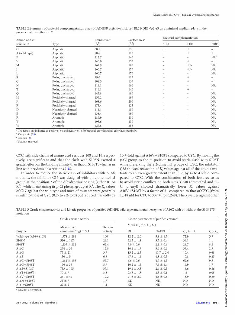
TABLE 3 Crude enzyme activity and kinetic properties of purified PfDHFR wild-type and mutant enzymes of A16X with or without the S108 T/Nmutation
Enzyme
Crude enzyme activity Kinetic parameters of purified enzymea
Mean sp act(nmol/min/mg) � SD
Relativeactivity
Mean Km � SD (�M)
kcat (s�1) kcat/KmDHF NADPH
Wild type (A16�S108) 1,978 � 284 100 12.2 � 2.0 5.8 � 1.7 72.9 5.9S108N 516 � 147 26.1 32.5 � 1.8 3.7 � 0.4 36.1 1.1S108T 1,235 � 232 62.4 3.0 � 0.6 2.1 � 0.6 24.7 8.2A16C 274 � 33 13.8 16.4 � 1.7 3.6 � 0.6 37.4 2.3A16G 77 � 21 3.9 15.2 � 2.3 11.7 � 2.0 10.4 0.68A16S 130 � 5 6.6 47.6 � 1.1 4.8 � 0.3 10.8 0.23A16C�S108T 1,181 � 198 59.7 6.6 � 0.6 4.7 � 1.3 62.6 9.5A16G�S108T 176 � 33 8.9 10.2 � 1.5 7.9 � 1.4 16.9 1.7A16S�S108T 733 � 195 37.1 19.4 � 3.3 2.4 � 0.3 16.6 0.86A16T�S108T 70 � 7 3.5 23.8 � 1.8 2.3 � 0.1 1.2 0.05A16V�S108T 241 � 49 12.2 21.3 � 2.9 4.5 � 0.5 18.9 0.89A16M�S108T 33 � 7 1.7 ND ND ND NDA16I�S108T 27 � 2 1.4 ND ND ND NDa ND, not determined.
Space Limits in PfDHFR Explain Cycloguanil Resistance
July 2012 Volume 56 Number 7 aac.asm.org 3931
Dow
nloa
ded
from
http
s://j
ourn
als.
asm
.org
/jour
nal/a
ac o
n 20
Jan
uary
202
2 by
81.
226.
97.5
9.
A16X�S108T mutants were also reduced by factors of 3 to 12compared to CYC. Unlike CYC and its derivatives, PYR with p-Clphenyl at R3 remained effective against most of the mutants cre-ated. The Ki values of PYR against the active mutants were mostly�1 nM. Only the A16S and S108N mutants showed slightlypoorer binding to PYR, with Ki values of 12 and 14 nM, respec-tively, mainly due to their high DHF Km value. Likewise,WR99210, a flexible antifolate, also remained effective against theentire collection of active PfDHFR mutants. The WR99210 Ki val-ues against these mutants ranged from 0.1 to 1.1 nM, with thehighest Ki value of 4.6 nM against the A16S mutant.
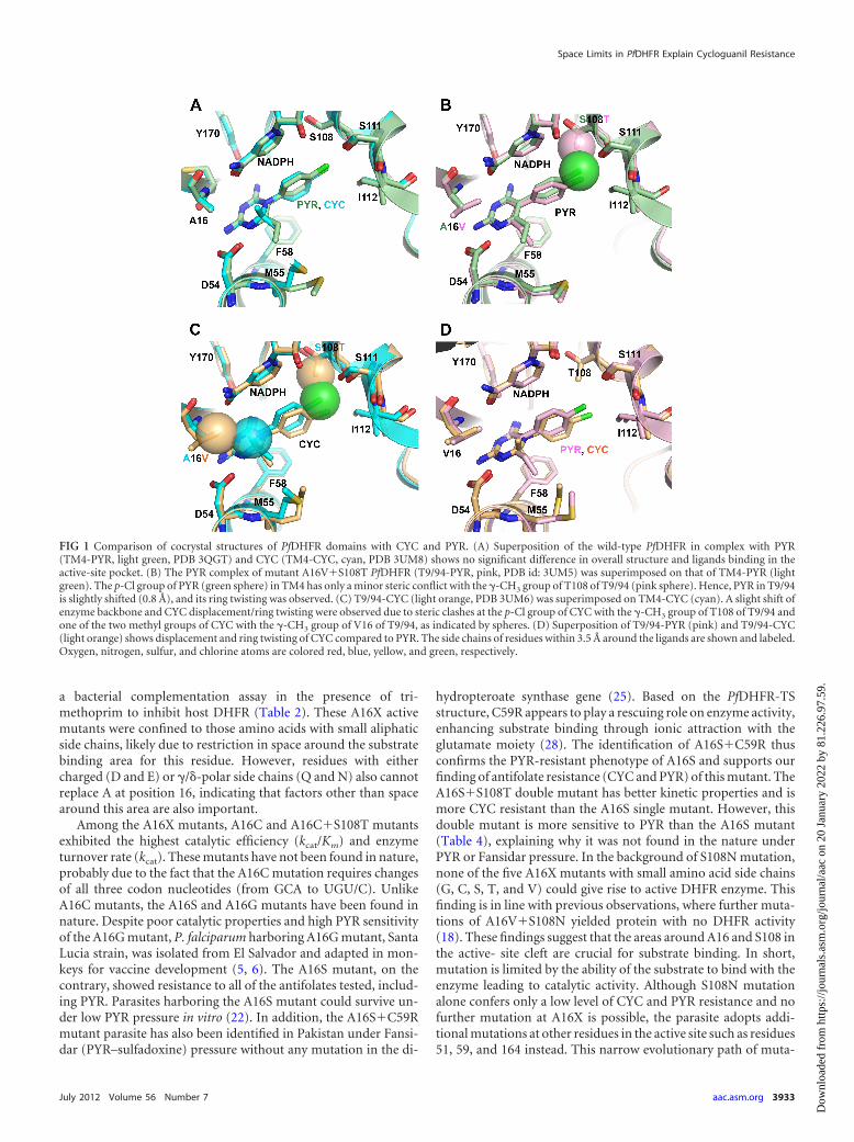
Structure analysis of PfDHFR-TS from T9/94 strain. Fourcrystal structures of the PfDHFR-TS wild type (TM4, PYR/CYCsensitive) and A16V�S108T (T9/94, PYR sensitive and CYC re-sistant) in complex with PYR (TM4-PYR [PDB 3QGT] and T9/94-PYR) and CYC (TM4-CYC and T9/94-CYC) were studied inorder to understand the basis of A16V�S108T resistance to CYC.Superimposition of TM4-CYC to TM4-PYR (Fig. 1A) showedsimilar overall structures, with only a slight shift of CYC, as indi-cated by movement of the p-Cl group of CYC 0.8 Å away from thatof PYR in TM4-PYR. The fact that TM4 is sensitive to CYC (Ki
value of 1.6 nM, Table 4) suggests that the slight shift does notaffect the binding affinity of CYC in the TM4 variant. Similarly,the superposition of T9/94-PYR to TM4-PYR (Fig. 1B) also re-vealed an unchanged overall structure of the DHFR enzyme activesite and only a minor displacement of PYR due to a slight clash ofthe -CH3 group of T108 of T9/94 with the p-Cl group of PYR.This is seen from a shift of the p-Cl group of PYR in T9/94-PYR by0.8 Å and a slight twist of the phenyl ring of PYR in T9/94-PYR by6.8° (the torsion angle of the two rings of PYR changed from111.1° to 104.3° in TM4-PYR and T9/94-PYR, respectively). Thedisplacement of PYR in the T9/94 binding pocket is an adjust-ment, which does not have any significant effect on the PYR bind-ing affinity, since the T9/94 remains sensitive to PYR (Ki value 0.6 nM for both, Table 4).
In contrast, in the structure of T9/94-CYC superposed onTM4-CYC (Fig. 1C), a slightly outward shift of the backbone of
the T9/94 around V16 and T108 to expand the active site chamberwas observed, presumably due to restricted space. Furthermore,the �-branched side chains of S108T and A16V in T9/94-CYCcause steric conflict to the p-Cl group and one of the 2,2-dimethylgroups of CYC, respectively, and hence the displacement of CYCmolecule in T9/94-CYC, as evidenced from a 1.3-Å shift of thep-Cl group and a 0.8-Å shift of 2,2-dimethyl groups compared toCYC in the TM4-CYC structure. The p-Cl phenyl ring of CYC istwisted by 10.6° (the torsion angles of the two rings of CYCchanged from 108.9° to 98.3° in TM4-CYC and T9/94-CYC, re-spectively). Superposition of T9/94-CYC to T9/94-PYR (Fig. 1D)clearly confirms that the steric conflict is much more pronouncedon CYC than on PYR, as shown by the movement of the side chainof V16 of the T9/94-CYC by 0.4 Å compared to that of T9/94-PYR.The conflict also resulted in a shift of the p-Cl group of CYC by 1.6Å compared to T9/94-PYR. This would explain the very high Ki
value of T9/94-CYC (Ki value 1,518 nM, Table 4). The higherdegree of displacement of CYC in T9/94-CYC, compared to PYRin T9/94-PYR, suggested a less favorable binding interaction withthe enzyme, thus causing CYC resistance in the T9/94 strain.
Taken altogether, these results indicated that the steric hin-drance caused by A16V and S108T in the active site of T9/94-CYCaggravate the binding of CYC, but not PYR. This is reflected by thepoor binding affinity of CYC to A16V�S108T, which results inresistance of T9/94 parasite to CYC, but not to PYR.
DISCUSSION
It has been well established that mutations of PfDHFR are respon-sible for antifolate resistance in P. falciparum. PfDHFR mutants inthe S108N series are resistant to both CYC and PYR, whileA16V�S108T, the only member of the S108T series, confers re-sistance to CYC only. In this report, we confirm that the mutationof residue A16 to V16 in the presence of S108T is indeed theoutcome of a compromise between its catalytic activity and CYCresistance.
In combination with or without S108T/N mutation, only 12mutants of A16X enzymes were catalytically active, as revealed by
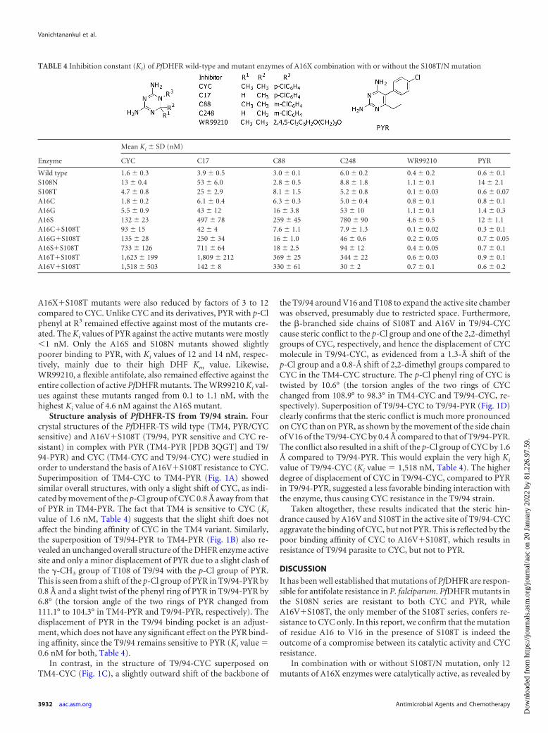
TABLE 4 Inhibition constant (Ki) of PfDHFR wild-type and mutant enzymes of A16X combination with or without the S108T/N mutation
Enzyme
Mean Ki � SD (nM)
CYC C17 C88 C248 WR99210 PYR
Wild type 1.6 � 0.3 3.9 � 0.5 3.0 � 0.1 6.0 � 0.2 0.4 � 0.2 0.6 � 0.1S108N 13 � 0.4 53 � 6.0 2.8 � 0.5 8.8 � 1.8 1.1 � 0.1 14 � 2.1S108T 4.7 � 0.8 25 � 2.9 8.1 � 1.5 5.2 � 0.8 0.1 � 0.03 0.6 � 0.07A16C 1.8 � 0.2 6.1 � 0.4 6.3 � 0.3 5.0 � 0.4 0.8 � 0.1 0.8 � 0.1A16G 5.5 � 0.9 43 � 12 16 � 3.8 53 � 10 1.1 � 0.1 1.4 � 0.3A16S 132 � 23 497 � 78 259 � 45 780 � 90 4.6 � 0.5 12 � 1.1A16C�S108T 93 � 15 42 � 4 7.6 � 1.1 7.9 � 1.3 0.1 � 0.02 0.3 � 0.1A16G�S108T 135 � 28 250 � 34 16 � 1.0 46 � 0.6 0.2 � 0.05 0.7 � 0.05A16S�S108T 733 � 126 711 � 64 18 � 2.5 94 � 12 0.4 � 0.05 0.7 � 0.1A16T�S108T 1,623 � 199 1,809 � 212 369 � 25 344 � 22 0.6 � 0.03 0.9 � 0.1A16V�S108T 1,518 � 503 142 � 8 330 � 61 30 � 2 0.7 � 0.1 0.6 � 0.2
Vanichtanankul et al.
3932 aac.asm.org Antimicrobial Agents and Chemotherapy
Dow
nloa
ded
from
http
s://j
ourn
als.
asm
.org
/jour
nal/a
ac o
n 20
Jan
uary
202
2 by
81.
226.
97.5
9.
a bacterial complementation assay in the presence of tri-methoprim to inhibit host DHFR (Table 2). These A16X activemutants were confined to those amino acids with small aliphaticside chains, likely due to restriction in space around the substratebinding area for this residue. However, residues with eithercharged (D and E) or /�-polar side chains (Q and N) also cannotreplace A at position 16, indicating that factors other than spacearound this area are also important.
Among the A16X mutants, A16C and A16C�S108T mutantsexhibited the highest catalytic efficiency (kcat/Km) and enzymeturnover rate (kcat). These mutants have not been found in nature,probably due to the fact that the A16C mutation requires changesof all three codon nucleotides (from GCA to UGU/C). UnlikeA16C mutants, the A16S and A16G mutants have been found innature. Despite poor catalytic properties and high PYR sensitivityof the A16G mutant, P. falciparum harboring A16G mutant, SantaLucia strain, was isolated from El Salvador and adapted in mon-keys for vaccine development (5, 6). The A16S mutant, on thecontrary, showed resistance to all of the antifolates tested, includ-ing PYR. Parasites harboring the A16S mutant could survive un-der low PYR pressure in vitro (22). In addition, the A16S�C59Rmutant parasite has also been identified in Pakistan under Fansi-dar (PYR–sulfadoxine) pressure without any mutation in the di-
hydropteroate synthase gene (25). Based on the PfDHFR-TSstructure, C59R appears to play a rescuing role on enzyme activity,enhancing substrate binding through ionic attraction with theglutamate moiety (28). The identification of A16S�C59R thusconfirms the PYR-resistant phenotype of A16S and supports ourfinding of antifolate resistance (CYC and PYR) of this mutant. TheA16S�S108T double mutant has better kinetic properties and ismore CYC resistant than the A16S single mutant. However, thisdouble mutant is more sensitive to PYR than the A16S mutant(Table 4), explaining why it was not found in the nature underPYR or Fansidar pressure. In the background of S108N mutation,none of the five A16X mutants with small amino acid side chains(G, C, S, T, and V) could give rise to active DHFR enzyme. Thisfinding is in line with previous observations, where further muta-tions of A16V�S108N yielded protein with no DHFR activity(18). These findings suggest that the areas around A16 and S108 inthe active- site cleft are crucial for substrate binding. In short,mutation is limited by the ability of the substrate to bind with theenzyme leading to catalytic activity. Although S108N mutationalone confers only a low level of CYC and PYR resistance and nofurther mutation at A16X is possible, the parasite adopts addi-tional mutations at other residues in the active site such as residues51, 59, and 164 instead. This narrow evolutionary path of muta-
FIG 1 Comparison of cocrystal structures of PfDHFR domains with CYC and PYR. (A) Superposition of the wild-type PfDHFR in complex with PYR(TM4-PYR, light green, PDB 3QGT) and CYC (TM4-CYC, cyan, PDB 3UM8) shows no significant difference in overall structure and ligands binding in theactive-site pocket. (B) The PYR complex of mutant A16V�S108T PfDHFR (T9/94-PYR, pink, PDB id: 3UM5) was superimposed on that of TM4-PYR (lightgreen). The p-Cl group of PYR (green sphere) in TM4 has only a minor steric conflict with the -CH3 group of T108 of T9/94 (pink sphere). Hence, PYR in T9/94is slightly shifted (0.8 Å), and its ring twisting was observed. (C) T9/94-CYC (light orange, PDB 3UM6) was superimposed on TM4-CYC (cyan). A slight shift ofenzyme backbone and CYC displacement/ring twisting were observed due to steric clashes at the p-Cl group of CYC with the -CH3 group of T108 of T9/94 andone of the two methyl groups of CYC with the -CH3 group of V16 of T9/94, as indicated by spheres. (D) Superposition of T9/94-PYR (pink) and T9/94-CYC(light orange) shows displacement and ring twisting of CYC compared to PYR. The side chains of residues within 3.5 Å around the ligands are shown and labeled.Oxygen, nitrogen, sulfur, and chlorine atoms are colored red, blue, yellow, and green, respectively.
Space Limits in PfDHFR Explain Cycloguanil Resistance
July 2012 Volume 56 Number 7 aac.asm.org 3933
Dow
nloa
ded
from
http
s://j
ourn
als.
asm
.org
/jour
nal/a
ac o
n 20
Jan
uary
202
2 by
81.
226.
97.5
9.
tion is determined by the fitness of antifolate-resistant variants (7,12, 18), which explains why these mutations are commonly foundin the field (26).
Compounds C17 and C88 (15, 27) were used to investigatethe implications of steric clash from both A16X and S108T/Nseries. C17, a CYC derivative with one methyl group removed,is expected to avoid the hindrance at position 16. However,except for the naturally occurring A16V�S108T mutant, the Ki
values of C17 for most of the selected active mutants are higherthan those of CYC, which is probably complicated by the na-ture of the amino acid at position 16. The polar amino acids atposition 16 affect not only the space available for DHF or in-hibitor binding but also likely interact with the nicotinamidemoiety of NADPH, as revealed by lower NADPH Km values(Table 3). Presumably, interaction of A16C/S/T with the aminogroup of the nicotinamide ring shifts the ring toward the DHFor inhibitor site, resulting in decreased affinity for C17 as thesite is in part restricted by NADPH. In comparison to C17, C88with m-Cl in place of p-Cl has little constraint at position 108,and the Ki values are reduced dramatically in the S108T/Nseries. C248, which is a desmethyl CYC derivative with an m-Clgroup, shows Ki values similar to those of C88 for the A16X andmost of A16X�S108T mutants, indicating that the inhibitor ismore susceptible to the clash at position 108 than at position16. However, the A16V�S108T mutant is more sensitive toC248 and C17, suggesting that relaxed binding occurs at posi-tion 16 irrespective of whether an m- or p-Cl substituent ispresent in the vicinity of position 108.
The crystal structure of PfDHFR-TS T9/94 (A16V�S108T) incomplex with CYC (T9/94-CYC) confirms that CYC resistance ofT9/94 is due to steric conflict of the �-methyl group of A16V andone of 2,2-dimethyl groups of CYC, together with the S108T andp-Cl group of CYC (Fig. 1B). For the S108N mutant, there is spacerestriction imposed on both PYR and CYC due to the aminogroup of S108N, which could clash with the p-Cl groups of bothcompounds. However, the displacement of PYR from theconflict with S108N is less because the side chain of S108Ntwists, with a push on NADPH, as observed in the structures ofPYR complex of PfDHFR-TS mutants (C59R�S108N andN51I�C59R�S108N�I164L) and S58R�S117N PvDHFR (10,23, 28). In comparison, the side chain of T108 retains its con-formation in complexing with PYR or CYC (Fig. 1A), suggest-ing space restriction. These mutants, e.g., S108N andA16V�S108T, bind reasonably well with DHF in optimal con-straint from both positions as revealed from Km and Ki (Tables3 and 4), whereas A16V�S108N, an inactive mutant, may posesteric clash dramatically with DHF. Hence, it can be concludedthat S108T has a more direct effect on the p-Cl group of CYCthan that of PYR, whereas S108N affects the binding of bothinhibitors. The double mutation A16V�S108T further reducesactive-site space available for CYC, with little effect on PYR,which has no steric conflict around the A16V site. On the otherhand, WR99210 binds comfortably with the A16 mutant seriesmainly due to its flexibility, as reflected by the Ki values (Table4). The steric clash of a 2-methyl group of CYC with A16V sidechain in T9/94-CYC supports our previous hypothesis basedon molecular modeling and inhibitors testing (15, 27) andgives a more exact explanation of the difference in sensitivity toCYC and PYR.
In a recent modeling study (13), it was predicted that the S108T
mutant has increased stability by van der Waals interactions withneighboring residues and greater affinity for NADPH than doesthe S108N mutant. This prediction is generally supported byour X-ray structural data. Our report further explains whyA16V�S108T is the sole member of the S108T series identifiedfrom CYC-resistant parasites since 1977 (6, 24). The blind alley forthis series is due to its susceptibility to PYR and limitation offurther mutation imposed by necessity for residual enzyme activ-ity. The limitation of DHFR mutations described here offers hopefor drug development against resistant parasites, highlighting thepotential of focusing on the parts of the target where resistantmutation is constrained by its biological activity, which is essentialfor the survival of the parasite.
ACKNOWLEDGMENTS
This research was supported by grants from the Wellcome Trust and theMMV Programme, WHO/TDR, Synchrotron Light Research Institute(Public Organization), Thailand, Thailand TDR Programme and NSTDACluster and Program Management Office to the team. S.K. is an interna-tional scholar of Howard Hughes Medical Institute.
We thank Philip Shaw for manuscript editing.
REFERENCES1. Brünger AT, et al. 1998. Crystallography and NMR system: a new soft-
ware suite for macromolecular structure determination. Acta Crystallogr.D Biol. Crystallogr. 54:905–921.
2. Chitnumsub P, et al. 2004. Characterization, crystallization and prelim-inary X-ray analysis of bifunctional dihydrofolate reductase-thymidylatesynthase from Plasmodium falciparum. Acta Crystallogr. D Biol. Crystal-logr. 60:780 –783.
3. Chothia C. 1976. The nature of the accessible and buried surfaces inproteins. J. Mol. Biol. 105:1–12.
4. Chusacultanachai S, Thiensathit P, Tarnchompoo B, Sirawaraporn W,Yuthavong Y. 2002. Novel antifolate resistant mutations of Plasmodiumfalciparum dihydrofolate reductase selected in Escherichia coli. Mol.Biochem. Parasitol. 120:61–72.
5. Collins WE, Galland GG, Sullivan JS, Morris CL, Richardson BB. 1996.The Santa Lucia strain of Plasmodium falciparum as a model for vaccinestudies. II. Development of Aotus vociferans as a model for testing trans-mission-blocking vaccines. Am. J. Trop. Med. Hyg. 54:380 –385.
6. Collins WE, Warren M, Skinner JC, Chin W, Richardson BB. 1977.Studies on the Santa Lucia (El Salvador) strain of Plasmodium falciparumin Aotus trivirgatus monkeys. J. Parasitol. 63:52–56.
7. Costanzo MS, Brown KM, Hartl DL. 2011. Fitness trade-offs in theevolution of dihydrofolate reductase and drug resistance in Plasmodiumfalciparum. PLoS One 6:e19636. doi:10.1371/journal.pone.0019636.
8. Jones TA, Zou JY, Cowan SW, Kjeldgaard M. 1991. Improved methodsfor building protein models in electron density maps and the location oferrors in these models. Acta Crystallogr. A 47(Pt 2):110 –119.
9. Kamchonwongpaisan S, Vanichtanankul J, Taweechai S, ChitnumsubP, Yuthavong Y. 2007. The role of tryptophan-48 in catalysis and bindingof inhibitors of Plasmodium falciparum dihydrofolate reductase. Int. J.Parasitol. 37:787–793.
10. Kongsaeree P, et al. 2005. Crystal structure of dihydrofolate reductasefrom Plasmodium vivax: pyrimethamine displacement linked with muta-tion-induced resistance. Proc. Natl. Acad. Sci. U. S. A. 102:13046 –13051.
11. Laskowski RA, MacArthur MW, Moss DS, Thornton JM. 1993. PRO-CHECK: a program to check the stereochemical quality of protein struc-tures. J. Appl. Crystallogr. 26:283–291.
12. Lozovsky ER, et al. 2009. Stepwise acquisition of pyrimethamine resis-tance in the malaria parasite. Proc. Natl. Acad. Sci. U. S. A. 106:12025–12030.
13. Mharakurwa S, et al. 2011. Malaria antifolate resistance with contrastingPlasmodium falciparum dihydrofolate reductase (DHFR) polymorphismsin humans and Anopheles mosquitoes. Proc. Natl. Acad. Sci. U. S. A. 108:18796 –18801.
14. Otwinowski Z, Minor W. 1997. Processing of X-ray diffraction datacollected in oscillation mode. Methods Enzymol. 276:307–326.
Vanichtanankul et al.
3934 aac.asm.org Antimicrobial Agents and Chemotherapy
Dow
nloa
ded
from
http
s://j
ourn
als.
asm
.org
/jour
nal/a
ac o
n 20
Jan
uary
202
2 by
81.
226.
97.5
9.
15. Rastelli G, et al. 2000. Interaction of pyrimethamine, cycloguanil,WR99210 and their analogues with Plasmodium falciparum dihydrofolatereductase: structural basis of antifolate resistance. Bioorg. Med. Chem.8:1117–1128.
16. Schrödinger LLC. 2010. The PyMOL molecular graphics system, version1.3r1. Schrödinger, Rockville, MD.
17. Sirawaraporn W, Prapunwattana P, Sirawaraporn R, Yuthavong Y,Santi DV. 1993. The dihydrofolate reductase domain of Plasmodium fal-ciparum thymidylate synthase-dihydrofolate reductase. Gene synthesis,expression, and anti-folate-resistant mutants. J. Biol. Chem. 268:21637–21644.
18. Sirawaraporn W, Sathitkul T, Sirawaraporn R, Yuthavong Y, Santi DV.1997. Antifolate-resistant mutants of Plasmodium falciparum dihydrofo-late reductase. Proc. Natl. Acad. Sci. U. S. A. 94:1124 –1129.
19. Sirawaraporn W, et al. 2002. Mutational analysis of Plasmodium falcip-arum dihydrofolate reductase: the role of aspartate 54 and phenylalanine223 on catalytic activity and antifolate binding. Mol. Biochem. Parasitol.121:185–193.
20. Tanaka M, Gu HM, Bzik DJ, Li WB, Inselburg J. 1990. Mutant dihy-drofolate reductase-thymidylate synthase genes in pyrimethamine-resistant Plasmodium falciparum with polymorphic chromosome dupli-cations. Mol. Biochem. Parasitol. 42:83–91.
21. Tarnchompoo B, et al. 2002. Development of 2,4-diaminopyrimidines asantimalarials based on inhibition of the S108N and C59R�S108N mu-
tants of dihydrofolate reductase from pyrimethamine-resistant Plasmo-dium falciparum. J. Med. Chem. 45:1244 –1252.
22. Thaithong S, et al. 2001. Plasmodium falciparum: gene mutations andamplification of dihydrofolate reductase genes in parasites grown in vitroin presence of pyrimethamine. Exp. Parasitol. 98:59 –70.
23. Vanichtanankul J, et al. 2011. Trypanosomal dihydrofolate reductasereveals natural antifolate resistance. ACS Chem. Biol. 6:905–911.
24. Volkman SK, et al. 2007. A genome-wide map of diversity in Plasmodiumfalciparum. Nat. Genet. 39:113–119.
25. Wang P, et al. 1997. Resistance to antifolates in Plasmodium falciparummonitored by sequence analysis of dihydropteroate synthetase and dihy-drofolate reductase alleles in a large number of field samples of diverseorigins. Mol. Biochem. Parasitol. 89:161–177.
26. Yuthavong Y. 2002. Basis for antifolate action and resistance in malaria.Microbes Infect. 4:175–182.
27. Yuthavong Y, et al. 2000. Development of a lead inhibitor for theA16V�S108T mutant of dihydrofolate reductase from the cycloguanil-resistant strain (T9/94) of Plasmodium falciparum. J. Med. Chem. 43:2738 –2744.
28. Yuvaniyama J, et al. 2003. Insights into antifolate resistance from malar-ial DHFR-TS structures. Nat. Struct. Biol. 10:357–365.
29. Zamyatnin AA. 1972. Protein volume in solution. Prog. Biophys. Mol.Biol. 24:107–123.
Space Limits in PfDHFR Explain Cycloguanil Resistance
July 2012 Volume 56 Number 7 aac.asm.org 3935
Dow
nloa
ded
from
http
s://j
ourn
als.
asm
.org
/jour
nal/a
ac o
n 20
Jan
uary
202
2 by
81.
226.
97.5
9.


![Die Zukunft der Saatgutbehandlung - bvo-saaten.de · Residues of Clothianidin with Deflector Distance to Application Equipment ] Ereignis am Rheingraben 2008: Konsequenzen • Ruhen](https://img.pdfslide.org/doc/110x75/5d5dedea88c993a5678bc972/die-zukunft-der-saatgutbehandlung-bvo-residues-of-clothianidin-with-deflector.jpg)