Embed Size (px)
Citation preview
1
Control of the release of digestive enzymes in the cricket Gryllus bimaculatus and the fall armyworm, Spodoptera frugiperda
Dissertation
zur Erlangung des Grades eines Doktors der Naturwissenschaften
Dr. rer. nat.
der Fakultät für Biologie, Chemie und Geowissenschaften
der Universität Bayreuth
vorgelegt von
LWALABA DIGALI Aus dem Tschad
Bayreuth, Germany
Januar 2010

2
Die vorliegende Arbeit wurde am Lehrstuhl für Tierökologie I der Universität Bayreuth
unter Leitung von Prof. Dr. Klaus Hubert Hoffmann durchgeführt und entstand im
Zeitraum von April 2007 bis Januar 2010.
3
Vollständiger Abdruck der von der Fakultät Biologie, Chemie und Geowissenschaften
der Universität Bayreuth zur Erlangung des akademischen Grades eines Doktors der
Naturwissenschaften genehmigten Dissertation.
Tag der Einreichung : 03. Januar 2010
Tag der mündlichen Prüfung : 02. Juni 2010
1. Gutachter : Prof. Dr. K. H. Hoffmann
2. Gutachter : Prof. Dr. St. Schuster
Prüfungsausschuss : Prof. Dr. K. Dettner
Prof. Dr. K. Seifert
Prof. Dr. E. Komor (Vorsitz)

4
To my wife Hingfene Gong-Non Moundou
and children Reggabe Digali and Lwatchebe Digali
5
Title 1 Contents 5
Introduction 6
Synopsis 19 References 30 Paper 1: 43 Control of the release of digestive enzymes in the caeca of the cricket, Gryllus bimaculatus Paper 2: 51 Control of the release of digestive enzymes in the larvae of the fall armyworm, Spodoptera frugiperda Paper 3: 67 Exogenous and endogenous proteases and amylase inhibitors in the larvae of the fall armyworm, Spodoptera frugiperda Summary 87
Zusammenfassung (German) 89
List of publications 92
Abbreviations 93
Danksagung (Statement, in German) 94
Erklärung (Statement, in German) 96
6
Introduction Insects represent the dominant group in the world fauna (80% of known
animal species in the world are insects) and most are beneficial to the ecology and
the economy. Most flowering plants depend on insects for their pollination (the values
of insect-pollinated crops was of the order of US $17, 000 million for a total of about
90 crops in the USA in the year 1986). Virtually all predators and parasites of pest
insects are other insects. However, insects are severe pests in agriculture, destroying
15% of total production, representing over US $ 100 billion, and are vectors of major
sickness such as malaria, dengue, sleeping disease, and yellow fever.
During the last century a huge amount of chlorinated hydrocarbon and
organophosphate pesticides were used to control the populations of insect pests.
These pesticides are potent nerve toxins, functioning by inhibiting the action of
acetylcholinesterase in nerve cells. Such effects are the results of changes in the
kinetics of the Na+ and K+ ion fluxes across the nerve cell membranes and
subsequent interference with nerve synaptic transmission. This toxic mechanism is
nonspecific, affecting both insects (target species) as well as all nontarget species.
These pesticides are contact poisons in all animals. They are fat soluble and are
absorbed through the body wall when the insect walks on the treated foliage, or when
it moves through the thatch and soil. Contact poisons can also enter through the
digestive tract when the insect feeds on treated plant tissues.
In the last 30 years synthetic or natural compounds, which can interrupt or
inhibit the life cycle of the target insects, were developed. These chemicals are
known as insect growth regulators (IGR), which depend on using the insects own
hormones or hormone analogs to control pest populations. Insect growth regulators
disrupt the normal development and reproduction of insects by mimicking juvenile
hormone (JH) and/ or moulting hormone (20-hydroxyecdysone). This means they
have very low toxicity to non-arthropod animals, however they are seldom used
today, because these products are quickly degraded in the environment and are
quickly metabolised and/or excreted in the insect.
Transgenic plants provide an attractive alternative to the use of chemical
pesticides and herbicides and could contribute to the production of crop varieties that
are inherently resistant to their major insect pests. Therefore, in recent years, intense
research has been focused on gene products (proteins) with insecticidal activities.
Bacillus thuringiensis (BT) produce crystal proteins (cry), which punctures the insects
7
midgut wall and cause uncontrolled leakage and death in all lepidopteran larvae
tested (Pigott et al., 2008). When the cry genes are incorporated into plant genome,
the cry endotoxins are very effective and very specific.
The first generation of pesticides (organophosphate, chlorinated
hydrocarbons) posed a great hazard because they inhibit synaptic transmission in
essentially all organisms and accumulate in adipose tissues. They are toxic to non
target animals and totally disrupted the environment. The second generation of
pesticides (insect growth regulators) are not toxic to vertebrates, but IGR
effectiveness was limited. The third generation of pesticides are essentially peptides
that for the most part disrupt some aspect of digestion. One example is the cry
proteins (above), a second example are enzyme inhibitors. Plants produce a broad
range of ihnhibitors of insect digestive enzymes such as soybean trypsin inhibitor,
bestatin and wheat α-amylase inhibitor. In general it is not practical to apply cry
proteins, neuropeptides or enzyme inhibitors to plants as a control method, because
of high costs, quick degradation, and very low uptake by the insect. Therefore, the
genes for these materials must be incorporated into the plant genome.
There are a number of plant protease inhibitors (see review, Fan and Wu,
2005) which bind to endo- and exopeptidases in insects and effectively inhibit growth,
and likewise a number of plant α-amylase inhibitors (see review, Franco et al., 2002).
In recent years it has been shown that members of several families of insect
neuropeptides, including allatostatins (AS) and allatotropins (AT), control not only the
juvenile hormone (JH) biosynthesis and release (regulating growth and reproduction)
but also the release of digestive enzymes and gut myoactivity in many insects (Fusé
et al., 1999; Hill and Orchard 2005). Insertion of genes into crop plants that encode
protease inhibitors or neuropeptides would be worth the effort, but only if more
detailed information on the action of these peptides indicates effective in vivo
regulation of digestive functions. Therefore, a major goal of my research is to
evaluate the effect of inhibitors and neuropeptides on the release of digestive
enzymes in model insects.
The first goal was to elucidate the effect of specific nutrients [glucose, maltose,
starch, bovine serum albumin (BSA), peptone and amino acids mixture] on the
release of digestive enzymes in the two model insects Spodoptera frugiperda
(Lepidoptera, Noctuidae) and Gryllus bimaculutus (Ensifera, Gryllidae). The second
goal was to examine the endogenous control of digestive enzyme release via
8
neuropeptides (allatostatins and allatropin). The third goal was to examine plant
based enzyme inhibitors and compare them to endogenous enzyme inhibitors. As
model insects I used the cricket G. bimaculatus (an omnivore with hemimetabolic
development) and the fall armyworm S. frugiperda (a herbivore with holometabolic
development) because the cricket represents the basic primitive form of the insect
digestive system, and the fall army worm represents a specialized herbivore of
considerable economic importance. It was also important that the experimental
animal was easy to rear on a defined diet, and that they are large enough to dissect.
Digestive systems in insects The digestive systems of vertebrates and insects are very similar both in
structure and function (Penzlin, 1999). Despite the diversity of feeding habits, three
basic divisions occur in all insects: the foregut which serves mostly for storage, the
midgut where enzymes are secreted and digestive products are absorbed, and the
hindgut where undigested food and metabolic wastes are compacted, dehydrated,
and egested (Nation, 2002; Dettner and Peters, 2003). The foregut and hindgut are
ectodermal and the epithelium secretes a chitinous cuticle continuously with the
exoskeleton. The midgut (ventriculus) secretes a delicate protective peritrophic
envelope around the food, which also regulates flow of digestive enzymes (Dettner
and Peters, 2003). The midgut wall is a single layered epithelium with an extensive
microvilli on the luminal side, which increases surface area for maximum absorption
of nutrients.
The foregut of G. bimaculatus consists of a pharynx, an oesophagus, a very
large crop, and thick-walled muscular proventriculus (Woodring and Lorenz, 2007).
The crop stores a large mass of freshly ingested food and enzymes originating in the
midgut initiate digestion. Most enzyme release and nutrient absorption occurs in the
large lateral caeca arising from the anterior end of the ventriculus. Typical of
terrestrial insects, the rectum very efficiently resorbs water to produce a very dry
fecal pellet. The flow of nutrients through the digestive tract of G.bimaculatus is
regulated by the proventriculus, which effectively triturates the partially digested food
coming from the crop and shoves the mushy nutrient mass into the ventriculus
(Woodring and Lorenz, 2007). Nutrients are pressed into the caeca, and the
remaining indigestible food is shoved into a tube formed by the peritrophic membrane
9
(PM). A countercurrent flow of enzymes has recently been described in crickets
(Biagio et al., 2009)
The major deviations from the primitive digestive system occur in fluid feeders,
that is, with phytophagous or with blood sucking insects. The larvae of the fall
armyworm, S. frugiperda, represent a typical phytophagous insect, which have a
simple straight-through type of gut without outpockets (caeca). Such a digestive tract
is well designed for transport of large masses of food with low nutrient quality. The
incompletely digested food mass of moist plant material passes rapidly through the
gut and water is not resorbed in the rectum (Nation, 2002). Ingested food is not
stored, so the crop is greatly reduced. The ventriculus comprises about 80% of the
total gut and the surface area is increased by a complex series of ring like folds
(Eaton, 1988). A well developed peritrophic membrane is formed in the anterior end
of the ventriculus (Harper and Granados, 1999), and it creates an endo- and
exoperitrophic space, in which a separation and recycling of digestive enzymes
occurs (Terra et al., 1979; Terra and Ferreira, 1994; Ferreira et al., 1994b; Terra et
al., 1996b; Bolognesi et al., 2001).
In S. frugiperda trypsin and α-amylase are secreted into the ectoperitrophic
space and diffuse into the endoperitrophic space, whereas aminopeptidase and
disaccharidases are secreted into the ectoperitrophic space and remain attached to
the membrane complex of the gut epithelium. Aminopeptidase is both bound to the
microvillar membranes and present as a soluble enzyme trapped in the cell
glycocalyx of the ventriculus (Klinkowstrom et al., 1994). Partially digested food
moves inside the PM (endoperitrophic space) posterior, while outside the PM
(exoperitrophic space) fluid containing partially or completely digested food plus
digestive enzymes move anterior. The enzymes involved in initial digestion are
secreted by the epithelium and pass through into the anterior endoperitrophic space
where they bind to nutrients. The enzyme-substrate complexes are too large to
diffuse back across PM, however, as the food is digested the molecular size of the
food particles decreases until these along with the enzymes can then pass back into
the exoperitrophic space. Along with resorbed water, the enzymes and oligomeric
molecules flow anterior where terminal digestion (to monomers) and absorption occur
(Terra et al., 1996 a, b). The enzymes diffuse back into the anterior endoperitrophic
space and a new cycle starts (Bolognesi et al., 2001).
10
Digestive enzymes in insects Enzymes are catalysts that regulate all reactions in all cells in the body
(Lehninger, 1956; Penzlin, 2003). All enzymes are proteins, whose sequence and the
spontaneous folding patterns and twists create specific 3-dimensional shapes. The
particular folding pattern of each enzyme gives each enzyme a distinct characteristic
and function. By disruption of the specific folding pattern, for example at
temperatures above 45°C, enzymes lose their ability to function, becoming
inactivated or are destroyed. The digestive enzymes are extracellular hydrolases,
which cut a nutrient molecule at a specific site by inserting a water molecule.
G. bimaculatus and S. frugiperda contain the typical array of insect digestive
enzymes in their digestive tracts. The chemical structure and catalytic action of these
enzymes are quite similar in all animals (Penzlin, 2003). However, the storage of
digestive enzymes, the activation of enzymes, the site where the enzymes are
released varies considerably between vertebrates and insects (Nation, 2002). The
hormonal and neural regulation of digestive enzyme release in vertebrates is well
understood, but is in general not as well understood in insects (Penzlin, 1991). In the
omnivorous cricket G. bimaculatus, the caecal region of the midgut is the primarily
site for the secretion of digestive enzymes. Proteases and amylase flow anteriorly
from the caeca into the mostly empty crop on day 1, and carbohydrate and protein
digestion starts as soon as food is present (Woodring et al., 2007).
In the midgut of the phytophagous larvae of S. frugiperda, aminopeptidase and
part of amylase, carboxypeptidase, dipeptidase, and trypsin are bound to the
microvillar membranes (Ferreira et al., 1994b). Major amounts of soluble dipeptidase,
and maltase are trapped in the cell glycocalyx; and only soluble carboxypeptidase,
amylase, and trypsin occur in intracellular vesicles. Aminopeptidase are mostly
bound to cells membranes in the ectoperitrophic space and a minor amount
incorporated in the peritrophic membrane. Amylase and trypsin are found primarily in
the endoperitrophic space after diffusing through the PM. The gradient is maintained
by binding of amylase and trypsin to substrates in the endoperitrophic space.
Proteases are enzymes acting on peptide bonds either in the middle of the
protein (endopeptidases) or at either end (exopeptidases). Endoproteases are
divided into three classes. 1) The serine proteases are characterized by the presence
of a serine and histidine at their active site. Examples are trypsin, chymotrypsin,
elastin, and subtilin. 2) The cysteine proteases possess a cysteine at the active site
11
and are inhibited by mercurial compounds. Examples are cathepsin B and cathepsin
L. 3) The aspartic proteases have a pH optimum below 5, due to the involvement of a
carboxyl residue in catalysis, for example pepsin. Exopeptidases hydrolyse single
amino acids from the N-terminus (aminopeptidase) or from the C-terminus
(carboxypeptidase) of the peptide chain and those enzymes that hydrolyse specific
dipeptides are called dipeptidases (see review Fan and Wu, 2005).
Typsin is a member of the serine proteinase superfamily, which have a
common catalytic triad of the specific residues: serine, histidine and aspartic acid
(Klein et al., 1996). Trypsin is highly specific for the positively charged side chains of
leucine and arginine (Kraut, 1977). Trypsins occur in almost all insect species. The
trypsins isolated from lepidopteran insects have a higher pH optimum, corresponding
to the higher pH values found in their midguts (Ferreira et al., 1994a). Cleavage
specificity of trypsins from several insects show that these enzymes are similar (but
not identical) to that of vertebrates trypsins. For example, insect trypsins are not
activated or stabilized by calcium ions (Lemos and Terra, 1992), in most cases are
unstable in acid pH (Sakal et al., 1989) and have different sensitivities to natural
trypsin inhibitors (Purcell et al., 1992). The molecular weight of insect trypsins lie
between 21 and 28 kDa and they do not require activation once secreted (Terra et
al., 1996a). Other serine proteases such a chymotrypsin, elastase, and subtilisins
were not considered in this dissertation because of time limitations and the known
similarity in the mode of action and release.
Aminopeptidases and exopeptidases are widely distributed as both extra- and
intracellular enzymes in all animals, including insects (Hooper and Lendeckel, 2004).
As integral membrane or cytoplasmic proteins they play important roles in various
biological processes such as protein processing and turnover, and various
pathological disorders regulation of peptide hormone action (Brownlees and Williams,
1993; Huang et al., 2009). The aminopeptidases are metalloenzymes, and they occur
in all animals and are especially active in the digestive tract of most insects (Terra et
al., 1996a; Woodring et al., 2007). Insect aminopeptidases have alkaline pH optima
(range 7.2 - 9.0), irrespective of the pH of the midgut lumen in which they occur
(Ferreira and Terra, 1985), and a molecular weight in the range of 90 - 190 kDa.
The cellular carboxypeptidases of vertebrates are very important in the
biosynthesis of insulin and function in blood clotting, growth and wound healing. In
insects they play a role in many physiological processes including the maturation of
12
neuroendocrine peptide precursors (Terra et al., 1996a). As digestive enzymes they
hydrolyze, along with the aminopeptidases, the peptides resulting from
endopeptidase action to liberate free amino acids to complete the digestion process
and produce molecules, which can be absorbed by the gut (Bown and Gatehouse,
2004). Carboxypeptidases were not included in my dissertation research because
they have a low activity in Spodoptera and Gryllus (unpublished results), and the
literature indicates little difference in mode of action and release to that of
aminopeptidases.
Insect carbohydrases are classified into two broad categories. The first group
includes enzymes which cleave internal bonds in polysaccharides, and that are
usually named from their substrates, for example by amylase, cellulase, pectinase
and chitinase. The second category includes glucosidases (maltase, trehalase,
fructosidase, xylosidase etc.) that catalyse the hydrolysis of terminal bonds in
oligosaccharides and disaccharides (Franco et al., 2002). The resulting
monosaccharides glucose, fructose, galactose etc. are directly absorbed in the
midgut.
Insect α-amylases (α-1,4-glucan-4-glucanohydrolases) are a group of
glycoside hydrolases that are widely distributed animal tissues, which catalyse the
hydrolysis of the α-(1,4) glycosidic linkage found in starch, glycogen and other
polysaccharides (Franco et al., 2002). These enzymes are well adapted to the slightly
acidic to neutral conditions found in the more anterior regions of the midgut in many
insects (Buonocore et al.,1976). The central calcium ion stabilises the 3-dimensional
structure of α-amylase and protects it against digestion by proteases (Stein and
Fischer, 1958).
Lipids consist of a large and heterogeneous group of substances that are
relatively insoluble in water but readily soluble in apolar solvents. Lipases hydrolyse
ester bonds in lipids containing fatty acids as fats, phospholipids, glycolipids and
waxes before they are absorbed (Terra et al., 1996a). Insect lipases, triacylglycerol
hydrolases, preferentially hydrolyse the outer ester links of triacylglycerols (TAG) and
release free fatty acids, diglyceride, monoglyceride, and glycerol (Secundo et al.,
2006). Insect cellular lipases in the fat body are needed for the hormone stimulated
adipokinetic hormone (AKH) release of diacylglycerides into the hemolymph, which is
the essential flight fuel for most insects (Wheeler and Goldsworthy, 1983). Insect
lipases preferentially cleave fatty acids from the α-positions (Hoffman and Downer,

13
1979), prefer unsaturated fatty acids (Weintraub and Tietz, 1973) and are activated
by calcium ions (Gilbert et al., 1965), thus resembling the action of mammalian
pancreatic lipase.
Insect midgut triacylglycerol lipases have been studied in relatively few insects
and only in crude preparations. Other than in vertebrates, fat micelles are not formed
and bile salt emulsification does not occur (Penzlin, 2003). The phytophagous larval
Manduca sexta appears to completely hydrolyze triglycerides in the midgut, and
absorbs the released fatty acids into the hemolymph (Tsuchida and Wells, 1988).
However, as in vertebrates the transport of lipids into the hemolymph in insects also
involves reconstitution to triglycerides in the ventricular cells (Turunen and
Chippendale, 1989).
Release of digestive enzymes There is a vast literature regarding the stage and age variation of digestive
enzymes in whole gut preparations of all groups of insects (see Terra, 1996 a, b,
Chapman, 1998). There is, however, relatively little known about the factors
controlling the release of digestive enzymes in insects. This knowledge is a
prerequisite for developing methods of control of pests based on inactivation of
digestive enzymes. The chief aim of my dissertation therefore was to determine what
extrinsic factors (nutrients) and intrinsic factors (neuropeptides) are most important
in the regulation of enzyme release. The second aim was to examine the action of
exogenous enzyme inhibitors, occurring in the food, and compare them with the
occurrence and action of endogenous inhibitors secreted by the gut.
Neuropeptides
A number of neuropeptides have been isolated in various insect orders, chiefly
from the brain and other parts of the nervous system and from the digestive tract.
These peptides are multifunctional and are best known for their effect on growth and
reproduction, controlling primarily the release of juvenile hormone (JH) from the
corpora allata (CA). They either stimulate (allatotropin, AT) or inhibit (allatostatin,
AS) the secretion and release of JH (Hoffmann et al., 1999; Gäde, 2002).
Neuroendocrine cells are distributed throughout the midgut, and in recent years it has
been shown that the same neurohormones that regulate JH release also affect
14
digestive processes such as release of digestive enzymes and gut myoactivity
(Nachman et al., 1997; Fusé et al., 1999; Penzlin, 1999).
Type-A allatostatins (Grybi-AS and Spofr-AS), also called cockroach
allatostatins, are characterized by the common C-terminal pentapeptide sequence
Y/FXFGL-amide (Stay et al., 1991). They were first isolated from the brains of the
cockroach Diploptera punctata (Woodhead et al., 1989) and have subsequently been
found in multiple forms in most insect orders (Nässel, 2002; Gäde, 2002). The cricket
G. bimaculatus hormone precursor contains at least 14 putative allatostatins
(Meyering-Vos et al., 2001). These AS have a strong allatostatic effect on the corpora
allata in vitro in crickets, cockroaches, and termites, but not in other insects
(Hoffmann et al., 1999). I used Grybi-AS 5 (DRLYSFGLamide), an allatostatin that
not only has a strong allatostatic effect, but it and related peptides are also myoactive
and stimulate release of amylase in cockroaches (Fusé et al., 1999).
Immunocytochemical studies showed that type-A AS in cockroaches are widely
distributed in the body, not only in the central nervous system but also in peripheral
nerves and in midgut endocrine cells (Stay, 2000). In cockroaches, a number of AS
inhibit spontaneous contraction of muscles of the hingut and in the antennal pulsatile
organ (Lange et al., 1993). In S. frugiperda, the gene encoding a family of nine to ten
peptides (type-A Spofr-AS) was isolated from brain cDNA (Abdel-latief et al., 2004).
There is evidence of allatostatic activity for these peptides in the adult moth. I used
two of the possible peptides deduced from the precursor molecule, Spofr-AS A5
(ARAYDFGLAamide) and Spofr-AS A6 (LPMYNFGLamide), to test their effect on
digestive enzyme release and on gut myoactivity.
To date, only one allatotropin family (Manse-AT) (TARGF/Yamide) is known,
and it functions in a number of Lepidoptera to stimulate the release of JH from
corpora allata. First isolated from the head of the moth Manduca sexta (Kataoka et
al., 1989), Manse-AT mRNAs were detected in the brain and suboesophageal
ganglion as well in cells of the abdominal ganglia (Veenstra et al., 1994). This
suggests multiple physiological functions beyond allatotropic effects, such as
functions in regulation of digestion. Manse-AT (GFKNVEMMTARGFamide) has also
been isolated from head extracts of S. frugiperda (Oeh et al., 2000). In larvae of S.
frugiperda, three mRNA isoforms of the Manse-AT gene are expressed in brain,
digestive tract, and reproductive organs (Abel-latief et al., 2004). To date there is no
15
information on the functions in the larvae or in regulation of digestive enzyme
release.
Inhibitors
Considerable efforts are being made to explore the use of plant genes
encoding insecticidal proteins in developing insect resistance in susceptible crop
plants (Lawrence and Koundal, 2002; Ferry et al., 2004). To date several different
classes of plant proteins have been shown to be insecticidal towards a range of
economically important insect pests when tested in artificial diets or in transgenic
plants (Gatehouse and Gatehouse, 1998). Plant protease and α-amylase inhibitors
(PI) form the main classes of insecticidal proteins, and they function by forming
complexes with digestive enzymes which are highly stable and dissociate very slowly
(Gatehouse et al., 1999; Morton et al., 2000; Fan and Wu, 2005). In plants which
produce them, they are thought to provide a form of natural defence against
herbivorous insects (Broadway and Duffey, 1986). The possible role of PIs in plant
protection was envisaged as early as 1947 when Mickel and Standish observed that
the larvae of certain insects were unable to develop normally on soybean products
(Haq et al., 2004). Soybean (Kunitz) trypsin inhibitor (SBTI) was the first PI isolated
and characterized (Kunitz, 1948; Fan and Wu, 2005).
Most plants PIs vary from 8 to 20 kDa (Hung et al., 2003), and they have a
high content of cysteine residues that form disulfide bridges and confer resistance to
heat, extremes in pH, and proteolysis (Richardson, 1991). The gene sequence for
SBTI is known and it has been inserted into a number of different plants including
potato, tobacco and rice (Fan and Wu, 2005). The Kunitz type trypsin inhibitor (SBTI)
is the trypsin inhibitor I used in my dissertation research. It has a low molecular
weight (25 kDa), and is quite resistant to high termperatures (Kunitz, 1948). It is
readily available in a purified crystalline form. The inhibitor binds to the active site on
the enzyme to form a complex with a very low dissociation constant (107 to 1014 M at
neutral pH values), thus effectively blocking the active site (Terra et al., 1996a). Thus
PIs directly mimic a normal substrate for the enzyme, but do not allow the normal
enzyme mechanism of peptide bond cleavage to proceed to completion i.e.,
dissociation of the product (Walker et al., 1998).
Relatively few exopeptide inhibitors in plants have been described (Bayes et
al., 2005). Bestatin is an inhibitor of exopeptidases extracted from Streptomyces
16
olivoreti and its structure was determined to be N-[(2S,3R)-3-amino-2-hydroxy-4-
phenylbutanoyl]-L-leucine (Umezawa et al., 1976). The molecular weight is only 345
Da. Bestatin inhibits metalloproteases and is selective for aminopeptidases including
leucine aminopeptidase and alanyl aminopeptidase use at 1-5 mM (Fan and Wu,
2005).
α-Amylase inhibitor, from Triticum aestivum (wheat) kernels which are
particularly rich in inhibitors of α-amylases, affect carbohydrate digestion in many
insects and mammals (Baker and Lum, 1989; Gomez et al., 1989). Some of these
are selective for insect enzymes, in that they inhibit α-amylases from insects strongly
but inhibit mammalian salivary or pancreatic α-amylases only weakly or not at all
(Feng et al., 1995). Plant based α-amylase inhibitors have been separated into three
major families of inhibitors (Petrucci et al., 1974). The family with the smallest
molecular weight (12 kDa) are monomeric proteins, the inhibitors of molecular weight
of 24 kDa are dimeric proteins. The third family of inhibitors, which have a molecular
weight of 60 kDa, are not well defined (Buonocore et al., 1980). The dimeric α-
amylase inhibitor I used in my dissertation is one of the most studied inhibitor from
the cereal- α-amylase inhibitors family (Franco et al., 2002).
Research gaps and the aims of this thesis There is a vast literature regarding the stage and age variation and the effect
of feeding on the digestive enzyme activity present within the gut lumen representing
all groups of insects (Applebaum, 1985; Chapman, 1998; Terra et al., 1996a, 1996b).
There is, however, relatively little known about the factors controlling release of
digestive enzymes in insects (Lehane et al., 1996; Blakemore et al., 1995; Lwalaba
et al., 2009). This knowledge is a prerequisite for developing methods of control of
pests based on inactivation of digestive enzymes.
The chief aim of my dissertation was to determine what extrinsic factors
(nutrients) and what intrinsic factors (neuropeptides) are most important in the
regulation of enzyme release. There are relatively few studies addressing the direct
effect of food (grains, plants, meat, etc.) in the gut on the release of digestive
enzymes (prandial regulation), and very few studies on the direct effect of specific
nutrients (protein, amino acids, starch, maltose, fat, etc.) on enzyme release. Aside
from the effect of proteins on protease release in cockroaches (Engelmann and
17
Geraerts, 1980) and Stomoxys calcitrans (stable fly) (Blakemore et al., 1995) there is
no knowledge of the effect of carbohydrates or lipids on the release of proteases,
amylases or lipases. Essential for such investigations is a reliable system for
incubation of midgut tissues.
The action of insect neuropepeptides on development and reproduction, water
balance and digestion was summerized in several reviews (Gäde et al., 1997;
Nässel, 2002; Gäde and Hoffmann, 2005). The effect of several neurohormones on
the release of amylase and maltase was described for a cockroach (Fusé et al.,
1999) and for a locust (Hill and Orchard, 2005). But the effect of neuropetides on the
release of other enzymes in crickets or in any phytophagous insect remains
unknown.
Meyering-Vos et al. (2001) identified a gene encoding 14 putative allatostatins
(AS) in G. bimaculatus and five of these were previously isolated by Lorenz et al.
(1995) by HPLC. The Grybi-AS precursor is expressed most strongly in the brain,
subesophageal ganglion and in the midgut caecum (Meyering-Vos and Hoffmann
2003). In mated females the AS type-A may regulate JH biosynthesis during
vitellogenesis, egg maturation and oviposition, respectively. However its effect on
digesting enzymes in G. bimaculatus was not investigated. Likewise, using molecular
biological techniques, Abdel-latief et al. (2004) found the gene sequences of two
allatostatins (Spofr-AS5, AS6) and an allatotropin (Manse-AT) in S. frugiperda.
Manse-AT strongly stimulates, and all tested Spofr-AS inhibit the biosynthesis of JH
in adult S. frugiperda (Oeh et al., 2000). Therefore, we know which neurohormones
occur in our model insects, G. bimaculatus and S. frugiperda, however, the effect of
these neuropeptides on gut myoactvity or on the release of the digestive enzymes
was not known. Therefore, this was an important aim of my research.
A further aim of the dissertation was to investigate the action of exogenous
enzyme inhibitors (those inhibitors that occur in the food), and compare them with the
occurrence and action of endogenous inhibitors that are secreted by the gut.
Because of the great importance of protease inhibitors (PI) for agriculture, there is a
vast literature on the effect of protease inhibitors from plants (see review Fan and
Wu, 2005) on protease activity in the gut. Likewise, a great amount of information on
the successful adaptation of insects to PIs has accumulated (Jongsma et al., 1995;
Broadway, 1995; Bown et al., 1997; Gatehouse et al., 1997; Brito et al., 2001;
Brioschi et al., 2007). However, there are almost no studies on the effect of PI or
18
other inhibitors or mixtures thereof on the release of digestive enzymes. Such studies
can actually be best carried out with short-term (3 days) feeding in the last larval
stage and only with tissue incubation, and this is what I did.
Except for the works of Engelmann and Geraerts (1980) and Vinokurov et al.
(2007) there is very little known about endogenous inhibitors in insects, and
essentially nothing is known about the factors regulating their release. The functions
of these endogenous inhibitors is completely unknown, but my investigation of the
regulation of the release has suggested possible new function in insects.
In order to answer the questions regarding the regulation of the release of
digestive enzymes in insects, as elucidated above, the following experiments were
performed:
1. Determination of the release in vitro of amylase, trypsin, aminopeptidase, and
lipase from flat-sheet caecal preparations in response to feeding of a control
diet, a non-nutrient diet and starvation in young adult females of G. bimaculatus
and in L6 larvae of S. frugiperda.
2. Determination of the effect of various nutrients in the incubation medium
(specifically proteins, peptides, amino acids, starch, maltose and glucose) on
the release of amylase, trypsin and aminopeptidase from isolated flat-sheet
preparations of the caecum (G. bimaculatus) and the ventriculus (S. frugiperda).
3. Determination of the effect of cricket allatostatin Grybi-AS5 on the release of
digestive enzymes, and determine the relative importance of prandial and
hormonal control in the cricket G. bimaculatus.
4. Determination of the effect of armyworm allatostatin (Spofr-AS5 and Spofr-AS6)
and tobacco hornworm allatotropin (Manse-AT) on the release of enzymes from
incubated midguts (flat-sheet preparations) of S. frugiperda.
5. Determination of the dose-dependent inhibition resulting from acute feeding
exogenous inhibitors (soybean trypsin inhibitor, aminopeptidase inhibitor
bestatin, and wheat amylase inhibitor) in last instar larvae (L6) of S. frugiperda.
Possible cross inhibition between inhibitors in S. frugiperda was also
investigated.
6. Determination of the presense of endogenous inhibitors in L6 S. frugiperda
larvae in comparison to the exogenous inhibitors. Suggest a role for the
occurrence of endogeneous enzyme inhibitors in insects.
19
Synopsis The basic aim of my dissertation research was first, to determine relative
importance of feeding various diet, specific nutrients in the diet (extrinsic), and
neuropeptides (intrinsic) on the release of enzymes from the midgut tissue of the
cricket Gryllus bimaculatus and of the fall armyworm, Spodoptera frugiperda, and
second, to compare the effects of exogenous protease and amylase inhibitors on the
production, release, and lumen contents of trypsin, aminopeptidase and amylase in
the larvae of the pest species (S. frugiperda).
In the first paper (Physiological Entomology 34, 144-151, 2009) I found that in
Gryllus bimaculatus the release of digestives enzymes into the caecum is regulated
by feeding, by specific nutrients and by the neuropeptide allatostatin 5 (Grybi-AS5).
The amount of all three enzymes released into the caecum of fed animals was
consistently 20-70% more than in unfed animals but a considerable amount of trypsin
and amylase was released even after 24 h without food. In unfed animals, feeding
always stimulated within an hour an increase in enzyme release over the basal
(unfed) levels, which continued over the next 4 h. The released amylase, trypsin and
aminopeptidase activity into the incubation medium after only 30 min was double the
enzyme activity within the caecal cells. The lipase release on the other hand was less
than a quarter of the activity measured in cell homogenates. This confirms our earlier
results (Woodring et al., 2007) that in crickets there is a basal level of enzyme activity
in the gut in unfed animals, and that feeding leads to a general increase in enzymes.
Not only was the enzyme release higher in fed animals compared with unfed
animals, but also the composition of the food had a profound effect on the ratio of the
digestive enzymes released. Feeding stimulated the release of digestive enzymes in
response to a specific ratio of nutrients, which led to a different ratio of digestive
enzyme release. A high nutrient component (for example protein) in the food did not
result necessarily in a correspondingly high digestive enzyme for that component
(trypsin). An increase over basal secretion rates of amylase, trypsin, and lipase
occurred in crickets fed cat food (high protein), but not when fed rabbit food (high
carbohydrate). In order to explain this anomaly, specific nutrients were added to the
incubation medium of isolated flat-sheet preparations of the digestive caeca.
A flat-sheet preparation is made by cutting a section of the gut lengthwise so
that both sides are exposed to the incubation medium. It could be demonstrated that
such preparations are stable, secrete enzymes over a period of several hours. The
20
caecum is composed of a single layer of several types of epithelial cells. Enzymes
are secreted from the luminar side of the zymogen cells and neurohormal receptors
are present on the hemolymph side. Nutrient receptors are located on the luminar
side of neuroendocrine cells and neurohormones are secreted from the hemolymph
side. The flat-sheet caecal preparation that I developed for this study allowed me to
demonstrate the paracrine mechanism of enzyme release in response to nutrients
and the endogenous control of enzyme release via neurohormones (allatostatins,
allatotropin).
There was no significant stimulation of amylase release in response to low
concentrations of starch to the incubation medium, but there was a strong dose-
dependent increase in amylase release, up to a 175% increase at maltose
concentrations of 8.0 mg maltose/ml incubation medium, in both fed and starved
crickets. Glucose also strongly stimulated amylase release. Trypsin or
aminopeptidase release was not affected by starch or sugars in the incubation
medium. There are no polysaccharide (starch) receptors known in any animal, but
the sugar receptors present in the epithelium of G. bimaculatus seem to be similar to
those in vertebates. In the case of the cricket, sugar binding leads to a strong
stimulation of amylase release. The slight increase in amylase activity at higher
starch concentration probably was not due to starch, but rather the result of traces of
glucose in the starch, which initiated digestion, and the resulting additional glucose or
maltose stimulated amylase release.
There was a slight increase in trypsin release in response to 0.4 to 0.6 mg/ml
peptone, which is a partial digest of casein containing a mixture of short length
polypeptides and amino acids, in the incubation medium, but at very high
concentrations of peptone (>6 mg/ml) there was an inhibition of trypsin release.
Similarly, with lower concentrations of bovine serum albumin (BSA) there was no
effect on trypsin release, but at concentrations above 2 mg/ml a strong inhibition of
trypsin release was observed. This unexpected inhibition of trypsin at high BSA
concentrations is probably due to the protein (BSA) blocking or altering access to the
enzyme active site, as observed with plant protein inhibitors (Fan and Wu, 2005).
Perhaps a kind of “excessive substrate inhibition” occurs in G. bimaculatus, where
excess BSA irreversibly binds to the trypsin active site. Excess substrate inhibition
occurs in approximately 20% of all enzymes. There was basically no stimulation of
exopeptidase (amino- and carboxypeptidase) release in response to BSA, peptone or
21
to a mixture of amino acids. The observed inhibition of trypsin release at higher
peptone concentrations is also probably due to excess substrate inhibition.
Similar to the study on the control of the release of digestive enzymes in the
cricket G. bimacualtus (first paper), the main purpose of the second paper (Archives
of Insect Biochemistry and Physiology, in press, doi:10.1002/arch.20332) was to
describe the regulatory role of various nutrients and neuropeptides on the release of
enzymes from the ventricular tissue (midgut) in the larvae of the fall armyworm,
Spodoptera frugiperda. A second aim was to determine the effect of these same
neuropeptides on the myoactivity of the digestive tract. The results demonstrate that larval age, food consumed, and the source of the enzymes (lumen or cells), all
influence the release of digestive enzymes into the lumen. It must be emphasized
that most published data on digestive enzymes deal with the standing enzyme
activity found in the lumen, and very little is known about the release.
The amount of enzyme released (activity) from the ventricular cells during the
3 days of the last larvae stadium (L6) increased from day 1 to the maximal activity on
day 3, and then dramatically decreased on day 4 (prepupae) as the digestive tract
emptied and started to disintegrate. The total activities of all four enzymes studied
were almost 15-times higher in the ventriculus than in the ileum, which suggests that
an efficient counter-current flow of enzymes occurs in the ventriculus of S. frugiperda,
as is described by Bolognesi et al. (2001), and this reduces the loss of important
digestive enzymes via egestion. A conclusion drawn from the observation that the
enzyme activity in the ventriculus of nutrient-fed larvae was much higher than those
fed the non-nutritive cellulose diet is that it is the nutrient composition and not merely
the bulk that stimulates enzyme release over that of the basal levels.
Aminopeptidase activity was much higher in the cell fraction and amylase and
trypsin much higher in the lumen. Typical of lepidopteran larvae (Ferreira et al.,
1994b; Lenz et al., 1991; Ortego et al., 1996), trypsin and amylase occur primarily in
the endoperitrophic space and the aminopeptidase is bound to the ventricular cell
membranes.
The total lipase activity of S. frugiperda is low, but the lumen fraction of the
total is relatively high (Lwalaba et al., 2009). This is the first report of high lipase
activitiy in the gut lumen of Lepidoptera larvae. Based on studies with radiolabelled
trioleine, dietary triacylglycerol in Manduca sexta larvae is completely hydrolyzed to
22
free fatty acids in the lumen before absorption into the cells (Tsuchida and Wells,
1988). We suggest, that because lipase and amylase have similar molecular weights
(50-60 kD) it would be expected that soluble lipase also passes into the
endoperitrophic space at the anterior end of the ventriculus. Amylase is secreted at
the anterior end of the ventriculus and passes through the pores of the peritrophic
membrane into the endoperitrophic space (Ferreira et al., 1994 a,b).
In continuously feeding insects, such as the caterpillar S. frugiperda, there are
few benefits to be gained from regulated secretion of digestive enzymes. A basal
release rate of enzymes into the ventriculus independent of the presence of food in
the gut is found in many insects (Chapman, 1985), however, feeding almost always
leads to an increase in the release and amount of enzymes in the gut (Applebaum,
1985; Woodring et al., 2007). In the cockroach Leucophaea maderae, feeding
stimulated the production of proteases in the midgut (Engelmann and Geraerts,
1980). The amylase release is three times higher in fed than in unfed larvae of S.
frugiperda, but trypsin release (quite unlike the cockroach) is the same for both fed
and unfed larvae. Both enzymes are immediately released after synthesis into the
lumen (Ferreira et al., 1994 a,b), which means that the synthesis rate must be related
to nutrients in the gut. But why does amylase release vary with the feeding and
trypsin release does not? A rationale based on food utilization can be hypothesized.
The primary nutrient in the diet of the caterpillar is carbohydrate, much of which after
digestion is absorbed, then converted to lipids and stored in the fat body. The adult
moth subsequently uses this lipid as a flight fuel (Wheeler, 1989). Reduced dietary
carbohydrate simply means less lipid reserve, and it would it be wasteful to produce
large amounts of amylase when food is in short supply. Therefore, the larvae can
afford to adjust the amylase release to the carbohydrate intake. Proteins, on the
other hand, are essential for larval growth, and in the adult moth egg production
depends entirely on proteins stored in larval fat body (Sorge et al., 2000). The
caterpillar, therefore, can not afford to allow any dietary protein to pass undigested
through the gut. Therefore, it is advantageous to maintain a constant level of trypsin
release in the event that even a small amount of protein is ingested. Regulated
secretion of digestive enzymes in insects, in which enzymes are synthesized and
stored and then released only in response to massive amounts of food, occurs in
some intermittent feeders (such as mosquitoes) (Lehane et al., 1996).
23
Whether the enzyme release rate is sufficient to maintain the standing amount
present in the lumen was determined in the following manner. The time of food
passage through the ventriculus, based on feeding L6 larvae carmine red colored
diet (n=4), averaged 3 h (180 min). The standing amylase activity in the ventricular
lumen was 340 µg maltose/min, and the secreted activity of the incubated flat-sheet
prepared ventriculus was 12 µg maltose/min. Therefore, in 3 h (= 180 min) 72 µg
maltose/min was secreted, which was about 20% of the standing amylase activity in
the lumen. The standing trypsin activity in the ventriculus was 3.8 units/min, and the
secreted activity was 0.11 units/min. In 3 h 0.66 units/min was secreted, which was
about 17% of the standing trypsin activity. The standing lipase activity was 3.7
nmol/min. In 3 h 7.8 nmol/min lipase activity was secreted, which was more than the
standing lipase activity. The standing aminopeptidase in the lumen was only 1.8
µmol/min. The very small aminopeptidase activity found in the incubate was probably
released from damaged cells and not from secretion, because almost all
aminopeptidase is membrane-bound. There was 10-times more amylase and trypsin
in the lumen of the ventriculus than in the ileum, thus, the relatively high secretion
rate of enzymes along with relatively little loss via egestion indicates that the
secretion of enzymes is sufficient to maintain the standing concentrations in the
lumen.
One can not determine the role of food or nutrients on enzyme release or
synthesis by simply measuring the enzyme activity in the lumen, but rather a direct
measure of enzyme secretion (release) by the ventricular tissues is required. In order
to precisely determine the role of feeding on the increased secretion of enzymes over
that of basal levels, specific nutrients were added to incubated flat-sheet preparations
of the ventriculus. A concentration of 0.5 mg/ml or higher of maltose or glucose in the
incubation medium resulted in an up to 350% elevation of amylase secretion. The
maximum effect of starch on amylase release was a modest 30-40% and only at
higher concentrations (>0.5 mg/ml). The larvae of the fall armyworm are plant-
feeders, and simple sugars and polysaccharide intake is therefore high. In the current
study, glucose and maltose provide a strong signal to release amylase, which is the
normal way to stimulate amylase release in animals. There is no evidence in
vertebrates or insects of starch or other polysaccharide receptors on gut
neuroendocrine cells that induce amylase release. The presence of starch in the
incubation medium, however, appears to only mildly stimulate amylase release in S.
24
frugiperda, and this is most likely due to a minimum digestion to maltose or a small
amount of sugar in the starch tested, and it is the maltose or glucose that stimulates
amylase release and not the starch. Similar to other insects, sugars bind to receptors
in the apical membrane of neuroendocrine cells in the epithelial layer of the
ventriculus, and induce the release of neuropeptides at the basal end into the
hemolymph. A binding of sugars to the apical (lumen) end of the gut neuroendocrine
cells is postulated in G. bimaculatus, which induces the release of neuropeptides
from the basal end into the hemolymph (Woodring et al., 2007). Specific
neuroendocrine cells in the gut epithelium of cockroaches and locusts release
several kinds of neuropeptides that stimulate the release of amylase (Fusé et al.,
1999; Sakai et al., 2004; Hill and Orchard, 2005).
The protein BSA had no effect on trypsin, and peptone (a mixture of small
polypeptides) caused only a moderate increase (p<0.05) in trypsin. The trypsin
release is not elevated by feeding in S. frugiperda. Therefore, it is not surprising that
bovine serum albumen (BSA) has no effect on release, and that peptone or a mixture
of all 20 amino acids has only a weak effect in the incubated flat-sheet preparations.
BSA in the bloodsucking fly (Stomoxys), on the other hand, has a strong stimulating
effect on the release of trypsin (Blakemore et al., 1995), which makes sense in an
animal with sudden and massive input of protein.
The effect of nutrients on the release of lipases was not studied because of
the very low activities. The aminopeptidase activity in the incubation did not result
from secretion, but rather from broken cells. Almost all aminopeptidase remains
bound to the external cell membrane (lumen side) and very little is released into the
lumen (Ferreira et al., 1994 a,b).
The gene sequence for Manse-AT and Spofr AS A5 were recently identified in
S. frugiperda (Abdel-latief et al., 2004), therefore the effect of these specific
neuropeptides on the release of amylase, trypsin and aminopeptidase was
investigated. The allatostatin Spofr-AS A5 inhibited the in vitro release of amylase
and trypsin by the ventriculus, and the allatotropin Manse-AT significantly stimulated
amylase and trypsin release at low concentrations. Neither the AS nor the AT had
any effect on aminopetidases. The neurohormones AT and AS are small peptides
that bind to receptors on the hemolypmh side of the zymogen cells (enzyme-
producing cells) and induce either an inhibition or stimulation of the release on the
25
lumen side. One of the first examples was the stimulation of amylase release in
response to AS, as was shown in the cockroach D. punctata (Fusé et al., 1999).
Interestingly, in almost all other Lepidoptera larvae an in vitro contraction of
the foregut is seen (Duve et al., 1999; Matthews et al., 2007). However, in S. littoralis
(Matthews et al., 2008) and in S. frugiperda, foregut motility was not observed.
Therefore, in the current study the ileum was employed to assay myoactivity.
Increased contraction of ileal muscles was observed only in a resting ileum with no
spontaneous contractions, that is zero beats/min. Concentrations of Manse-AT
greater than 10-6 M elevated the contractions from 0 beats/min to over 20 beats/min.
Interestingly, the recovery back to 0 beats/min was graduate and required several
transfers to fresh HGR (high glucose ringer) over 3-4 min, indicating that the peptide
must be rinsed from the receptor site.
Inhibition of the contraction rhythm was observed only in an ileum that had
started to beat spontaneously. The rate ranged from 15 to 30 beats/min, but once
started the rate was constant for any single ileum over a 5 to 20 min period.
Therefore, the amount of inhibition as a percent change from the control (initial rate)
in this time span was consistent and repeatable. The ileum was very sensitive to
inhibition by Spofr-AS A5 at all concentration over 10-8 M, showing an immediate and
complete inhibition (0 beats/min). Recovery in fresh HGR required only 20-30
seconds, indicating a weaker receptor binding than that with AT (above). A WX6W
allatostatin (AS type-B) inhibits gut myocontraction in another lepidopteran species,
M. sexta (Blackburn et al., 1995). In conclusion, Sporf-AT stimulates digestive
enzyme release and myoactivity in S. frugiperda, and Spofr-AS A5 inhibits both
enzyme release and myoactivity.
In the third paper (Journal of Insect Physiology, in review) the effects of
exogenous and endogenous protease and amylase inhibitors on the production and
secretion of digestive enzymes in the larvae of S. frugiperda was investigated. It has
been long known that digestive enzymes are inhibited by a wide variety of inhibitors
occurring in plants (Franco et al., 2002; Fan and Wu, 2005). A strong inhibition of
trypsin activity by the general serine protease inhibitor SBTI, aminopeptidase by
bestatin, and amylase by the wheat α-amylase inhibitor was observed in the lumen
contents of L6 larvae of S. frugiperda. An age-dependent effect was apparent (with
constant inhibitor concentration), whereby three days of feeding of inhibitor reduced
26
enzyme activity in the lumen (higher inhibition) more than one day of feeding. A
distinct dose-dependent inhibition was also apparent after three days of feeding,
which was also reported in other lepidopteran larvae (Paulillo et al., 2000; Brioschi et
al., 2007).
With regards to inhibition of trypsin in tissue homogenates, both soybean
trypsin inhibitor (SBTI) and N-α-tosyl-L-lysine chloromethyl ketone (TLCK) were
effective. Bestatin and wheat amylase inhibitor had no effect on the secretion or
tissue activity of aminopeptidase or amylase, respectively. SBTI strongly inhibited
trypsin release in the tissue extracts, especially at high concentration (1200
µg/larva/day). At concentrations of TLCK >400 µg/larva/day a feeding inhibition was
observed, and as a result (less TLCK consumed) no further trypsin inhibition was
seen.
Whereas acute feeding (3 days) of inhibitors had only moderate effects on
typsin activity in tissue extracts or no effect on aminopeptidase and amylase activity,
these same inhibitors when added directly to the cellular extracts (in vitro) strongly
inhibited all enzymes down to only 5-24% of control values (Table 3). Thus it was
clear that the inhibitors studied inhibited specifically the enzymes (endogenous)
release by the tissues and not just the exogenous enzymes used for the enzyme
assays.
With regards inhibition of the release of enzymes, only TLCK was effective.
This is the first direct evidence of inhibition of enzyme release in insects. Neither
aminopeptidase nor amylase release is affected by the corresponding inhibitors
employed. Inhibition of enzyme secretion by an ingested inhibitor implies either a
direct effect (entry into the cell) or indirect effect (docking on receptors) on the cell
production (synthesis) or release. Evidence for the effect of inhibitors on cellular
enzyme production and release is seen in the increased trypsin activity in the gut of
S. frugiperda and Heliothis virescens in response to chronic feeding of SBTI (Brito et
al., 2001; Brioschi et al., 2007).
A cross reaction of one inhibitor on a different enzyme has been reported. In
Teleogryllus commodus, where SBTI increased aminopeptidase activity (Burgess et
al., 1994), as did a barley trypsin inhibitor in S. exigua (Lara et al., 2000). In our study
SBTI fed to L6 larval S. frugiperda induced a moderate elevation (P<0.05) of
aminopeptidase in the lumen, and the more potent protease inhibitor TLCK strongly
elevated (P<0.01) aminopeptidase activity in both the lumen and in the tissue
27
extracts. Thus, aminopeptidase showed a cross reaction in vivo in response to both
protease inhibitors (SBTI and TLCK). Bestatin, an aminopeptidase inhibitor, however,
showed no cross reaction to in vivo trypsin inhibition. The cross reaction of SBTI and
TLCK on aminopeptidase activity indicated an altered synthesis of one enzyme in
response to a product reduction of a different enzyme. A molecular basis of
crossover effects can be seen in Helicoverpa armigera, where specific inhibitors
increased chymotrypsin mRNA but trypsin mRNA levels decreased (Gatehouse et
al., 1997), or in Trichoplusia ni, where a procarboxypeptidase is activated by trypsin
(Wang et al., 2004).
The difficulty in demonstrating the presence of endogenous inhibitors,
specifically those secreted by the gut itself, is the high concentrations of endogenous
enzymes present in the gut lumen. I solved this problem by irreversible inactivation
by means of heat treatment. It was assumed that the endogenous inhibitors are as
heat resistant as the demonstrated resistance of the exogenous inhibitors, which
were used for the enzyme assays. The first demonstration of endogenous gut
protease inhibitors was described in tissue extracts and lumen contents from the
midgut of Leucophaea maderae (Engelmann and Geraerts, 1980). They also used
boiled extracts to inactivate endogenous enzymes, and found up to 80% protease
inhibition in the posterior half of the midgut. Endogenous inhibitors in whole midgut
extracts (epithelium plus lumen contents) are also reported by Elpidina et al. (2001)
and Vinokurov et al. (2007) in the cockroach Nauphoeta cinerea.
Heated tissue extracts in diet fed L6 S. frugiperda larvae showed less trypsin
activity (78%), and much less aminopeptidase activity (47%) compared to controls
(100%), which demonstrates the presence of endogenous inhibitors in the
ventriculus. The unheated tissue extracts showed more trypsin (143%) and much
more aminopeptidase (332%), a result of the endogenous enzyme activities. Since
the tissues are thoroughly rinsed, exogenous inhibitors associated with the food are
removed, and only endogenous inhibitors are to be found in the heated tissue extract
and no active enzymes. In the unheated tissue extract the endogenous trypsin was
still active and when combined with the exogenous bovine trypsin an increase
instead of a decrease in total enzyme activity was seen. The same reasoning applies
to aminopeptidase and amylase inhibitors present in the tissues extracts.
Endogenous enzyme activity is much higher in the lumen than in the tissues.
Heated lumen contents showed much less trypsin (65%), and aminopeptidase (79%)
28
activity, clearly an indication of endogenous inhibitors. The unheated lumen contents
showed very high trypsin activity (426%) because both endogenous and exogenous
trypsin was present. There was basically no difference in the endogenous trypsin
inhibitor activity in diet fed, cellulose fed or unfed L6 larvae. It should be noted that
there could not be any exogenous inhibitors in the lumen of cellulose fed or unfed
larvae.
Lumen contents run on an analytical SDS-PAGE gel showed the presence of
an endogenous inhibitor, and the same lumen contents run on a gelatine containing
SDS-PAGE gel demonstrated the binding of an endogenous protease inhibitor to the
gelatine substrate.
The situation with endogenous aminopeptidase inhibition is different because
most aminopeptidase is not released into the lumen, as are trypsin and amylase, but
rather it remains bound to the cell membranes (Ferreira et al., 1994 a,b). The
membrane bound aminopeptidases of heated tissue extracts were inactivated, and
the endogenous aminopeptidase inhibitors were greatly reduced (P<0.01) in tissue
extracts. The PM is part of the lumen contents, and it contains some aminopeptidase
(Ferreira et al., 1994 a), and a moderate amount (P<0.05) of endogenous
aminopeptidase inhibition occurs in heated lumen contents. In diet fed larvae the
endogenous inhibitor strongly reduced (P<0.01) aminopeptidase in the tissue, less so
in cellulose fed larvae, and not at all in starved larvae. In cellulose-fed and unfed
larvae the aminopeptidase inhibition was undetectable in the lumen.
Endogenous amylase activity was studied only in fed larvae. Heated tissue
extracts and lumen contents of L6 larvae showed less (P<0.05) amylase activity
(85%) compared to controls, clearly indicating an endogenous amylase inhibition.
The unheated tissue extracts and lumen contents showed more amylase (142%),
indicating the high levels of endogenous amylase in the lumen.
It was demonstrated that trypsin digests both human salivary amylase as well
as the amylase present in the lumen of S. frugoperda larvae. About 10% of the
amylase is digested over a period of 30 min, which in fed larvae is quickly replaced.
However, starved larvae only secrete about 20% of the controls and trypsin digestion
of amylase could present problems.
Several functions of the endogenous protease inhibitors in the digestive tract
of cockroaches have been suggested. Elpidina et al. (2001) suggest the endogenous
protease inhibitors might protect amylases in the anterior midgut of N. cinerea,
29
however, this seems unnecessary because divalent ions confer a general resistance
of α-amylases to proteolytic degradation (Stein and Fischer, 1958). Other authors
suggest that endogenous protease inhibitors might be important as protection against
fungi and bacteria that employ subtilisins to attack insects (Vinokurov et al., 2007;
Taranushenko et al., 2009). Another possible function of endogenous protease
inhibitors in insects might involve the protection of the epithelium when dietary protein
is scarce. Trypsin is continuoulsy released in fed and unfed S. frugiperda (Lwalaba et
al., 2009). In vertebrates an inactive trypsinogen is released in response to
cholecystokinin (CCK), but a trypsinogen has never been found in insects. My
hypothesis is that endogenous inhibitors, especially trypsin inhibitors, are essential to
protect the delicate ventricular epithelium again the digestive action of trypsin (which
is always present) when food is scarce or lacking.
30
References
Abdel-latief, M., Meyering-Vos, M., Hoffmann, K.H. (2004) Type-A allatostatins from
the fall armyworm, Spodoptera frugiperda: Molecular cloning, expression and tissue-
specific localization. Archives of Insect Biochemistry and Physiology 56, 120-132.
Applebaum, S.W. (1985) Biochemistry of digestion. In: Kerkut, G.A., Gilbert, L.I, eds.
Comprehensive Insect Physiology, Biochemistry, and Pharmacology, Vol. 4. London,
Pergamon Press. pp. 9-311.
Bagio, F.P, Tamaki, F.K, Terra, W.R., Ribeiro, A.F. (2009) Digestive
morphophysiology of Gryllodes sigillatus. Journal of Insect Physiology 55, 1125-1133.
Baker, J.E., Lum, P.T.M. (1989) Multiplicity of albumins from wheat inhibitory to
amylase from the rice weevil (Coleoptera: Curculionidae). Journal of Economic
Entomology 82, 1548-1553.
Bayes, A., Comellas-Bigler, M., Rodriguez de la Vega, M., Maskos, K., Bode, W., Aviles, F.X., Jongsma, M.A., Beekwilder, J., Vendrell, J. (2005). Structural basis of
the resistance of an insect carboxypeptidase to plant protease inhibitors.
Proceedings of the National Academy of Sciences USA 102, 16602–16607.
Blakemore, D., Williams, S., Lehane, M.J. (1995) Protein stimulation of trypsin
secretion from the opaque zone midgut cells of Stomoxys calcitrans. Comparative
Biochemistry and Physiology 110B, 301-307.
Bolognesi, R., Ribeiro, A.F., Terra, W.R. and Ferreira, C. (2001) The peritrophic
membrane of Spodoptera frugiperda: Secretion of peritrophins and role in
immobilization and recycling digestive enzymes. Archives of Insect Biochemistry and
Physiology 47, 62-75.
Bown, D.P., Gatehouse, J.A. (2004) Characterization of a digestive
carboxypeptidase from the insect pest corn earworm (Helicoverpa armigera) with
31
novel specificity towards C-terminal glutamate residues. European Journal of
Biochemistry 271, 2000-2011.
Bown, D.P., Wilkinson, H.S., Gatehouse, J.A. (1997) Differentially regulated
inhibitor-sensitive and insensitive protease genes from the phytophagous insect pest,
Helicoverpa armigera, are members of complex multigene families. Insect
Biochemistry and Molecular Biology 27, 625-638.
Brioschi, D., Nadalini, L.D., Bengtson, M.H., Sogayar, M.C., Moura, D.S., Silva-Filho, M.C. (2007) General up regulation of Spodoptera frugiperda trypsins and
chymotrypsin allows its adaptation to soybean proteinase inhibitor. Insect
Biochemistry and Molecular Biology 37, 1283-1290.
Brito, L.O., Lopes, A.R., Parra, J.R.P., Terra, W.R., Silva-Filho, M.C. (2001) Adaptation of tobacco budworm Heliothis virescens to proteinase inhibitors may be
mediated by the synthesis of new proteinases. Comparative Biochemistry and
Physiology 128B, 365-375.
Broadway, R.M. (1995) Are insects resistant to plant proteinase inhibitors? Journal of
Insect Physiology 41, 107-116.
Broadway, R.M., Duffey, S.S. (1986) Plant proteinase inhibitors: mechanism of action
and effect on the growth and digestive physiology of larval Heliothis zea and
Spodoptera exigua. Journal of Insect Physiology 32, 827-833.
Brownlees, J. and Williams, C.H. (1993) Peptidases, peptides, and the mammalian
blood-brain barrier. Journal of Neurochemistry 60, 793-803.
Buonocore, V., Gramenzi, F., Pace, W., Petrucci, T., Poerio, E., Silano, V. (1980) Interaction of wheat monomeric and dimeric protein inhibitors of α-amylase from
yellow mealworm (Tenebrio molitor L.) larva. Biochemical Journal 187, 637-645.
32
Buonocore, V., Poerio, E., Pace, W., Petrucci, T., Silano, V., Tomasi, M. (1976) Interaction of Tenebrio molitor L. α-amylase with wheat flour protein inhibitor. FEBS
Letters 67, 202-206.
Burgess, E.P.J., Main, C.A., Stevens, J.T., Crhristeller, J.T., Gatehouse, A.M.R., Laing, W.A. (1994) Effects of protease inhibitor concentration and combination on the
survival, growth and gut enzyme activities of the black field cricket, Teleogryllus
commodus. Journal of Insect Physiology 40, 803-811.
Chapman, R.F. (1985) Coordination of Digestion. In: Kerkut, GA, Gilbert, LI editors.
Comparative Insect Physiology, Biochemistry and Pharmacology. Vol. 4. Oxford,
Pergamon Press, pp. 213-240.
Chapman, R.F. (1998) Alimentary canal, digestion and absorption. The Insects-
Structure and Function. 4th ed. Cambridge University Press, U.K., pp. 38-58.
Dettner, K., Peters, W. (2003) Lehrbuch der Entomologie, 2nd ed. Gustav Fischer
Verlag, Stuttgart, Germany, pp.91-126.
Duve, H., Eastr, P.D., Thorpe, A. (1999) Regulation of lepidopteran forefug
movements by allatostatins and allatrtropin from the frontal ganglion. Journal of
Comparative Neurology.415, 405-416.
Eaton, J.L. (1988) Lepidopteran anatomy. In: Schaefer, W.C. ed. Interscience Series
in Insect Morphology. Wiley-Interscience Publication, New York, pp. 189-202.
Elpidina, E.N., Vinokurov, K.S., Rudenskaya, Y.A., Dunaevsky, Y.E., Shuzhikov, D.P. (2001) Proteinase inhibitors in Nauphoeta cinerea midgut. Archivs of Insect
Biochemistry and Physiology 48, 217-222.
Engelmann, F., Geraerts, W.P.M. (1980) The proteases and the protease inhibitor in
the midgut of Leucophaea maderae. Journal of Insect Physiology 26, 703-710.
33
Fan, S.G., Wu, G.J. (2005) Characteristics of plant proteinase inhibitors and their
applications in combating phytophagous insects. Botanical Bulletin of Academia
Sinica 46, 273-292.
Feng, G.H., Richardson, M., Chen, M.S., Kramer, K.J., Morgan, T.D., Reeck, G.R. (1995) α-Amylase inhibitors from wheat: amino acid sequences and patterns of
inhibition of insect and human α-amylases. Insect Biochemistry and Molecular Biology
26, 419-426.
Ferreira, C., Terra, W.R. (1985) Minor aminopeptidases purified from the plasma
membrane of midgut caeca cells of an insect (Rhynchosciara americana) larva. Insect
Biochemistry 15, 619-625.
Ferreira, C., Capella, A.N., Sitnik, R., Terra, W.R. (1994b) Digestive enzymes in
midgut cells, endo- and ectoperitrophic contents, and peritrophic membranes of
Spodoptera frugiperda (Lepidoptera) larvae. Archives of Insect Biochemistry and
Physiology 26, 299-313.
Ferreira, C., Capella, A.N., Sitnik, R., Terra, W.R. (1994a) Properties of the
digestive enzymes and the permeability of the peritrophic membrane of Spodoptera
frugiperda larvae. Comparitive Biochemistry and Physiology 107A, 631-640.
Ferreira, C., Oliveira, M.C. and Terra, W.R. (1990) Compartmentalization of the
digestive process in Abracris flavolineata (Orthoptera: Acrididae). Insect Biochemistry
20, 267-274.
Ferry, N., Edwards, M.G., Gatehouse, J.A., Gatehouse, A.M.R. (2004) Plant-insect
interactions: molecular approaches to insect resistance. Current Opinion in
Biotechnology 15, 1-7.
Franco, O.L., Rigden, D.J., Melo, F.R., Grossi-de-sá, M.F. (2002) Plant α-amylase
inhibitors and their interaction with insect α-amylases structure, function and potential
for crop protection. European Journal of Biochemistry 269, 397-412.
34
Fusé, M., Zhang, J.R., Partridge, E., Nachman, R.J., Orchard, I., Bendena, W.G., Tobe, S.S. (1999) Effects of an allatostatin and a myosuppressin on midgut
carbohydrate enzyme activity in the cockroach Diploptera punctata. Peptides 20,
1285-1293.
Gäde, G. (2002) Allatoregulatory peptides: molecules with multiple functions.
Invertebrate Reproduction and Development 41, 127-135.
Gäde, G., Hoffmann, K.H. (2005) Neuropeptides regulating development and
reproduction in insects. Physiological Entomology 30, 103-121.
Gäde, G., Hoffmann, K.H., Spring, J.H. (1997) Hormonal regulation in insects: facts,
gaps and future directions. Physiological Reviews 77, 963-1032.
Gatehouse, A.M.R., Gatehouse, J.A. (1998) Identifying proteins with insecticidal
activity: use of encoding genes to produce insect-resistant transgenic crops. Pesticide
Science 52, 165-175.
Gatehouse, A.M.R., Norton, E., Davison, G.M., Babbe, S.M., Newell, J.A., Gatehouse, J.A. (1999) Digestive proteolytic activity in larvae of tomato moth,
Lacanobia oleracea; effects of plant protease inhibitors in vitro and in vivo. Journal of
Insect Physiology 45, 545-558.
Gatehouse, L.N., Shannon, A.L., Burgess, E.P.J. Christeller, J.T. (1997)
Characterization of major midgut proteinase cDNAs from Helicoverpa armigera
larvae and changes in gene expression in response to four proteinase inhibitors in
the diet. Insect Biochemistry and Molecular Biology 27, 929-944.
Gilbert, L.I., Chino, H., Domroese, K.A. (1965) Lipolytic activity of insect tissue and
its significance in lipid transport. Journal of Insect Physiology 11, 1057-1070.
Gomez, L., Sanchez-Monge, R., Garcia-Olmedo, F., Salcedo, G. (1989) Wheat
tetrameric inhibitors of insect α-amylases: alloploid heterosic at the molecular level.
Proceedings of the National Academy of Sciences USA 86, 3242-3246.
35
Haq, S.K., Atif, S.M., Khan, R.H. (2004) Protein proteinase inhibitor genes in combat
against insects, pests, and pathogens: natural and engineered phytoprotection.
Archives of Biochemistry and Biophysics 431, 145-159.
Harper, M.S., Granados, R.R. (1999). Peritrophic membrane structure and formation
of larval Trichoplusia ni with an investigation on the secretion patterns of a PM mucin.
Tissue and Cell 31, 202–211.
Hill, S.H., Orchard, I. (2005) In vitro analysis of the digestive enzymes amylase and
α-glucosidase in the midguts of Locusta migratoria L. in response to the
myosuppresin, SchistoFLRFamide. Journal of Insect Physiology 51, 1-9.
Hoffman, A.G.D., Downer, R.G.H. (1979) End product specificity of triacylglycerol
lipases from intestine, fat body, muscle and haemolymph of the America cockroach,
Periplaneta americana. Lipids 14, 893-899.
Hoffmann, K.H., Meyering-Vos, M., Lorenz, M.W. (1999) Allatostatins and
allatotropins: is regulation of corpora allata activity their primary function? European
Journal of Entomology 96, 255-266.
Hooper, N.M., Lendeckel, U. (2004) Proteases in biology and disease:
Aminopeptidases in biology and disease. Kluwer Academic/Plenum Publischers, New
York, p. 201.
Huang, H., Tanaka, H., Hammock, B.D., Morisseau, C. (2009) Novel and highly
sensitive fluorescent assay for leucine aminopeptidases. Analytical Biochemistry 391,
11-16.
Hung, C.H., Huang, C.C., Tsai, W.S., Wang, H.L., Chen, Y.L. (2003) Purification and
characterization of a trypsin inhibitor from Brassica campestris seeds. Journal Yuanpei
University Science and Technology 10, 13-22.
36
Jongsma, M.A., Bakker, P.L., Peters, J., Bosch, D., Stiekema, W.J. (1995) Adaptation of Spodoptera exigua larvae to plant proteinase inhibitors by induction of
gut proteinase activity insensitive to inhibition. Proceedings of the National Academy
of Sciences USA 92, 8041-8045.
Kataoka, H., Toschi, A., Li, J.P., Carney, R.L., Schooley, D.A., Kramer, S.J. (1989)
Identification of an allatotropin from adult Manduca sexta. Science 243, 1481-1483.
Klein, B., Le, M.G., Sellos, D., Van, W.A. (1996) Molecular cloning and sequencing
of trypsin cDNAs from Penaeus vannamei (Crustacea, Decapoda): use in assessing
gene expression during the moult cycle. International Journal of Biochemistry and Cell
Biology 28, 551-563.
Klinkowstrom, A.M., Terra, W.R., Ferreira, C. (1994) Aminopeptidase-A from
Rhynchosciara americana (Diptera) larval midgut –properties and midgut distribution.
Archives of Insect Biochemistry and Physiology 27, 301–315.
Kraut, J. (1977) Serine proteases: structure and mechanism of catalysis. Annual
Review of Biochemistry 46, 331-358.
Kunitz, M. (1948) The kinetics and thermodynamics of reversible denaturation of
crystalline soybean trypsin inhibitor. Journal of General Physiology 32, 241–263.
Lange, A. B., Chan, K. k., Stay, B. (1993) Effect of thirteen allatostatins and proctolin
on antennal pulsatile organ and hindgut muscle in the cockroach Diploptera punctata.
Archives of Insect Biochemistry and Physiology 24, 79-92.
Lara, P., Ortego, F., Gonzylez-Hidalgo, E., Castnera, P., Carbonero, P., Diaz, I. (2000) Adaptation of Spodoptera exigua to barley trypsin inhibitor BTI-Cme
expressed in transgenic tobacco. Transgenic Research 9, 169-178.
Lawrence, P.K., Koundal, K.R. (2002) Plant protease inhibitors in control of
phytophagous insects. Electronic Journal of Biotechnology 5, 93-109.
37
Lehane, M.J, Bühler, H.M., Crisanti, A. (1996) Mechanisms controlling the synthesis
and secretion of digestives enzymes in insects. In: Lehane, M.J, Billingsley, P.F, eds.
Biology of the Insect Midgut. London, Chapman & Hall, pp. 195-205.
Lehninger, A.L. (1956) Enzymes: units of biological structure and function (Gaebler,
O.H,, ed.). Academic Press, New York, p. 217.
Lemos, F.J.A., Terra, W.R. (1992) Soluble and membrane-bound forms of trypsin-like
enzymes in Musca domestica larval midguts. Insect Biochemistry and Molecular
Biology 22, 613-619.
Lenz, C.J., Kang, J., Rice, W.C., McIntosh, A.H. Chippendale, G.M. Schubert, R.R. (1991) Duigestive proteinases of larvae of the corn earworm, Heliothis zea:
characterization, distribution and dietary relationships. Archive of Insect Biochemistry
and Physiology. 16, 201-212.
Lorenz, M.W., Kellner, R., Hoffmann, K.H. (1995) A family of neuropeptides that
inhibit juvenile hormone biosynthesis in the cricket, Gryllus bimaculatus. Journal of
Biological Chemistry 270, 21103-21108.
Lwalaba, D., Hoffmann, K.H., and Woodring, J. (2009) Control of the release of
digestive enzymes in the larvae of the fall armyworm, Spodoptera frugiperda.
Archives of Insect. Biochemistry and Physiology, doi: 10.1002/arch.20332.
Matthews, H.J., Audsley, N., Weaver, R.J. (2007) Interactions between allatostatins
and allatotropin on spontaneous of the foregut of larval Lacanobia oleracea. Journal
of Insect Physiology 53, 75-83.
McManus, M.T., Burgess, E.P.J. (1995) Effects of the soybean (Kunitz) trypsin
inhibitor on growth and digestive proteases of larvae of Spodoptera litura. Journal of
Insect Physology 41, 731-738.
38
Meyering-Vos, M., Hoffmann, K.H. (2003) Expression of allatostatins in the
Mediterranean field cricket, Gryllus bimaculatus de Geer (Ensifera, Gryllidae).
Comparative Biochemistry and Physiology B 136, 207-215.
Meyering-Vos, M., Wu, X., Huang, J., Jindra, M., Hoffmann, K.H., Sehnal, F. (2001) The allatostatin gene of the cricket Gryllus bimaculatus (Ensifera: Gryllidae).
Molecular and Cellular Endocrinology 184, 103-114.
Morton, R.L., Schroeder, H.E., Batement, K.S., Chrispeels, M.J., Armstrong,E., Higgins, T.J.V. (2000) Bean α-amylase inhibitor 1 in transgenic Peas (Pisum sativum)
provides complete protection from pea weevil (Bruchus pisorum) under field
conditions. Proceedings of the National Academy of Sciences USA 97, 3820-3825.
Nachman, R.J., Giard, W., Favrel, P., Suresh, T., Sreekumar, S., Holman, G.M. (1997) Insect myossupressins and sulfakinins stimulate release of the digestive
enzymes α-amylase in two invertebrates: The scallop Pecten maximus and insect
Rhynchophorus ferrugineus. Annals of the New York Academy of Sciences 814, 335-
338.
Nässel, R. (2002) Neuropeptides in the nervous system of Drosophila and other
insects: multiple roles as neuromodulators and neurohormones. Progress in
Neurobiology 68, 1-84.
Nation, J.L. (2002) Insect physiology and biochemistry. CRC Press, Boca Raton,
USA, pp. 28-58.
Oeh, U., Lorenz, M.W., Dyker, H., Lösel, P., Hoffmann, K.H. (2000) Interaction
between Manduca sextra allatotropin and Manduca sextra allatostatin in the fall
armyworm Spodoptera frugiperda. Insect Biochemistry and Molecular Biology 30,
719-727.
Ortego, F., Nuvillo, C., Castafiera, P. (1996) Characterization and distribution of
digestive proteases of the stalk corn borer, Sesamia nonogroides. Archives of Insect
Biochemistry and Physiology 32, 163-180.
39
Paulillo, L.C.M.S., Lopes, A:R:, Cristofoletti, P.T., Parra, J.R.P., Terra, W.R., Silva-Filho, M.C. (2000) Changes in midgut endopeptidase activity of Spodoptera
frugiperda are responsible for adaptation to soybean proteinase inhibitors. Journal of
Economic Entomology 93, 892-896.
Penzlin, H.(2003) Lehrbuch der Tierphysiologie, 7th edn., Gustav Fischer Verlag,
Jena, Germany.
Penzlin, H. (1999). Neuropeptide und die Steuerung visceraler Funktionen bei
Insekten. Leopoldina 44, 411–426.
Petrucci, T., Tomassi, M., Cantagalli, P., Silano, V. (1974) Comparison of wheat
albumin inhibitors of α-amylase and trypsin. Phytochemistry 13, 2487-2495.
Pigott, C.R., King, M.S., Ellar, D.J. (2008) Investigating the properties of Bacillus
thuringiensis cry proteins with novel loop replacements created using combinatorial
molecular biology. Applied and Environmental Microbiology 74, 3497-3511.
Purcell, J.P., Greenplate, J.T., Sammons, R.D. (1992) Examination of midgut
luminal proteinase activities in six economically important insects. Insect Biochemistry
and Molecular Biology 22, 41-47.
Richardson, M. (1991) Seed storage proteins: The enzyme inhibitors. In: L.J. Rogers
ed., Methods in Plant Biochemistry, Vol. 5, Amino acids, Proteins and Nucleic Acids.
Academic Press, New York, pp. 259-305.
Sakal, E., Applebaum, S.W., Birk, Y. (1989) Purification and characterization of
trypsins from the digestive tract of Locusta migratoria. International Journal of Peptide
and Protein Research 34, 498-505.
40
Sakai, T., Satake, H., Minakata, H., Tkeda, M. (2004) Characterization of crustacean
cardioactive peptide as a novel insect midgut factor: Isolation, localization, and
stimulation of α-amylase activity and gut contratction. Endocrinology 145, 5671-5678.
Secundo, F., Carrea, G., Tarabiono, C., Gatti-Lafranconi, P., Brocca, S., Lotti, M. et al. (2006) The lids is a structural and functional determinant of lipase activity and
selectivity. Journal of Molecular Catalysis B: Enzymatic 39, 166-170.
Sorge, D., Nauen, R., Range, U., Hoffmann, K.H. (2000). Regulation of
vitellogenesis in the fall armyworm, Spodoptera frugiperda (Lepidoptera: Noctuidae).
Journal of Insect Physiology 46, 969-976.
Stay, B. (2000) A review of the role of neurosecretion in the control of juvenile
hormone synthesis: a tribute to Berta Scharrer. Insect Biochemistry and Molecular
Biology 30, 653-662.
Stay, B., Joshi, S., Woodhead, A.P. (1991) Sensitivity to allatostatins of corpora
allata from larval and adult female Diploptera punctata. Journal of Insect Physiology
37, 36-70.
Stein, E.A., Fischer, E. H. (1958) The resistance of α-amylases towards proteolytics
attack. Journal of Biological Chemistry 232, 867-879.
Terra, W.R. (2001) The origin and functions of the insect peritrophic membrane and
peritrophic gel. Archives of Insect Biochemistry and Physiolog 47, 47-61.
Terra, W.R., Ferreira, C. (1994) Insect digestive enzymes: properties,
compartmentalisation and function. Comparative Biochemistry and Physiology 109B,
1-62.
Terra, W.R., Ferreira, C., De Bianchi, A.G. (1979) Distribution of digestive enzymes
among the endo- and ectoperitrophic spaces and midgut cells of Rhynchosciara and
its physiological significance. Journal of Insect Physiology 25, 487-494.
41
Terra, W.R., Ferreira, C., Jordao, B.P. (1996a) Digestive enzymes. In: Lehane, M.J,
Billingsley, P.F, eds. Biology of the Insect Midgut. Chapman & Hall, London, pp. 153-
194.
Terra, W.R., Ferreira, C., Baker, J.E. (1996b) Compartmentalization of digestion. In:
Lehane, M.J, Billingsley, P.F, eds. Biology of the Insect Midgut. Chapman & Hall,
London, pp. 206-235.
Terra, W.R., Santos, C.D., Ribeiro, A.F. (1990) Ultrastructural and biochemical
basis of the digestion of nectar and other nutrients by the moth Erinnyis ello.
Entomologia Experimentalis et Applicata 56, 277-286.
Tsuchida, K., Wells, M.A. (1988) Digestion, absorption, transport and storage of fat
during the last larval stadium of Manduca sexta. Changes in the role of lipophorin in
the delivery of dietary lipid to the fat body. Insect Biochemistry 18, 263-268.
Turunen, S., Chippendale, G.M. (1989) Relationship between dietary lipids, midgut
lipids, and lipid absorption in eight species of Lepidoptera reared on artificial and
natural diets. Journal of Insect Physiology 35, 627-633.
Umezawa, H., Aoyagi, T., Suda, H., Hamada, M., Takeuchi, T. (1976) Bestatin, an
inhibitor of aminopeptidase B, produced by actinomycetes. Journal of Antibiotics 29,
97.
Veenstra, J.A., Lehman, H.K., Davis, N.T. (1994) Allatotropin is a
cardioacceleratory peptide in Manduca sexta. Journal of Experimental Biology 188,
347–354.
Vinokurov, K., Taranushenko, Y., Krishnan, N., Sehnal, F. (2007) Proteinase,
amylase, and proteinase-inhibitor activities in the gut of six cockroach species.
Journal of Insect Physiology 53, 794-802.
Walker, A.J., Ford, L., Majerus, M.E.N., Geoghegan, I.E., Birch, A.N.E., Gatehouse, J.A., Gatehouse, A.M.R. (1998) Characterisation of the midgut digestive
42
proteinase activity of the two-spot ladybird (Adalia bipunctata L.) and its sensitivity to
proteinase inhibitors. Insect Biochemistry and Molecular Biology 28, 173-180.
Wang, P., Guoxun, L., Kain, W. (2004) Characterization and cDNA cloning of midgut
carboxypeptidases from Trichoplusia ni. Insect Biochemistry and Molecular Biology
34, 831-843.
Weintraub, H., Tietz, A. (1973) Triglyceride digestion and absorption in the locust,
Locusta migratoria. Biochimica et Biophysica Acta 306, 31-41.
Wheeler, C.H. (1989) Mobilization and transport of fuels to the flight muscles. In.
Goldsworthy GJ, Wheeler CH, editors. Insect Flight. Boca Raton, Fl, CRC Press, pp.
273-304.
Wheeler, C. H., Goldsworthy, G. J. (1983) Protein-lipoprotein interactions in the
haemolymph of Locusta during the action of adipokinetic hormone: the role of CL
proteins. Journal of Insect Physiology 29, 349-354.
Woodhead, A.P., Stay, B., Seidel, S.L., Khan, M.A., Tobe, S.S. (1989) Primary
structure of four allatostatins: neuropeptide inhibitors of juvenile hormone synthesis.
Proceedings of the National Academy of Science USA 86, 5997-6001.
Woodring, J., Lorenz, M.W. (2007) Feeding, nutrient flow, and functional gut
morphology in the Cricket Gryllus bimaculatus. Journal of Morphology 268, 815-825.
Woodring, J., Hoffmann, K.H., Lorenz, M.W. (2007) Activity, release and flow of
digestive enzymes in the cricket, Gryllus bimaculatus. Physiological Entomology 32,
56-63.
Physiological Entomology (2009) 34, 144–151 DOI: 10.1111/j.1365-3032.2008.00665.x
© 2009 The Authors144 Journal compilation © 2009 The Royal Entomological Society
Introduction
In vertebrates, the release of digestive enzymes is regulated by feeding, acting through a feedback to the brain that con-trols digestion directly via the visceral nervous system and indirectly via paracrine hormone release ( Penzlin, 1996 ). Based on ultrastructural studies of midgut cells, a continuous release of digestive enzymes is indicated in most insects, and a regulated release of digestive enzymes (with stored enzymes in the cytoplasm) is assumed to occur essentially only in dis-continuous feeders, such as blood sucking insects ( Lehane et al. , 1996 ). Both the amount and the ratio of nutrients in the gut play an important role in the release of digestive enzymes, whereby more enzymes are generally released in
fed insects than in unfed insects ( Chapman, 1985; Woodring et al. , 2007 ). A direct neural regulation of digestive enzyme release in insects is considered to be unlikely ( Lehane et al. , 1996; Sehnal & Zitnan, 1996; Chapman, 1998 ) and, there-fore, the only possible mechanisms controlling the release are hormonal, paracrine or prandial ( Lehane et al. , 1995 ).
To date, there is no available evidence for the hormonal con-trol of digestive enzyme release in insects. An inhibition of trypsin biosynthesis (not release) via blocked transcription is known for the ‘trypsin modulating oostatic factor’ in mosqui-toes, biting flies and fleas ( Borovsky & Mahmood, 1995; Borovsky, 2003 ). A paracrine mechanism requires endocrine cells in the gut, which occur in all the insects investigated, and are usually concentrated in the midgut epithelium ( Montuenge et al. , 1989; Sehnal & Zitnan, 1996 ). Many neuropeptides (FMRFamides, allatostatins, sulfakinins and others) are identi-fied through antibody reactions in the enteric nervous system and in the endocrine cells of the gut ( Sehnal & Zitnan, 1996 ). These affect both gut musculature via the enteric nervous
Control of the release of digestive enzymes in the caeca of the cricket Gryllus bimaculatus
J O S E P H W O O D R I N G , S A N D R A D I E R S C H , D I G A L I L W A L A B A , K L A U S H . H O F F M A N N and M A R T I N A M E Y E R I N G - V O S Department of Animal Ecology I, University of Bayreuth, Bayreuth, Germany
Abstract . In Gryllus bimaculatus , more digestive enzymes (amylase, trypsin, aminopeptidase) are secreted in the caecum of fed crickets than in unfed crickets, but the enzymes are released continuously at a basal rate in unfed animals. The rate of synthesis of the enzymes appears to parallel their rate of release. Digestive enzymes are released in response to a specific ratio of nutrients, although a high nutrient component in the food does not necessarily induce a high digestive enzyme release for that component. Rinsed flat-sheet preparations of the caecum are incu-bated with specific nutrients (carbohydrates and proteins) and various concentrations of a neuropeptide (type-A allatostatin), which affects generally the basal rates of secretion. Both maltose and glucose increase the release of amylase in vitro , but starch produces an inhibition of amylase release at lower concentrations. Bovine serum albumin (BSA), peptone and a mixture of amino acids have almost no effect on the release of aminopeptidase or carboxypeptidase, and only low concentrations of peptone increase trypsin release. High concentrations of both BSA and peptone strongly inhibit trypsin activity, perhaps by excess substrate binding to the trypsin active site. The allatostatin Grybi-AST 5 elevates the release of amylase in vitro , but not of trypsin or aminopeptidase, in 2-day-old fed females. In the caeca from 1-day-old unfed crickets, both amylase and the trypsin release are stimulated in the presence of AST 5. The paracrine AST 5 is probably released from the gut endocrine cells and binds to the enzyme-producing caecal cells.
Key words . allatostatin , aminopeptidase , amylase , diets , lipase , nutrients , trypsin .
Correspondence: Dr J. Woodring, Department of Animal Ecology 1, University of Bayreuth, 95440 Bayreuth, Germany. Tel.: +49 921 55 26 56; fax: +49 921 55 27 84; e-mail: joseph.woodring@uni -bayreuth.de
Control of digestive enzymes in G. bimaculatus 145
© 2009 The AuthorsJournal compilation © 2009 The Royal Entomological Society, Physiological Entomology, 34, 144–151
system ( Lange et al. , 1995 ) and digestive enzyme release in a paracrine way ( Fusé et al. , 1999; Harshini et al. , 2002b; Hill & Orchard, 2005; Sakai et al. , 2006 ). A prandial mechanism (also termed a secretogogue mechanism) refers to the direct binding of a nutrient element to a digestive enzyme-producing cell caus-ing the release of an enzyme; however, there is no convincing evidence of such a mechanism in insects ( Lehane et al. , 1995 ).
There are many examples of ingested nutrients, particu-larly proteins in blood feeding insects, that stimulate the release of trypsin ( Lehane et al. , 1996 ), and these most likely involve a paracrine mechanism. Endocrine gut cells are dis-tinguished from other gut cells by the presence of peptidergic secretory granules of which there are two types in insects. In the open type, the apical end of the cell reaches the gut lumen and, in the closed type, a luminary contact is lacking ( Sehnal & Zitnan, 1996 ). Gut endocrine cells usually have basal cyto-plasmic processes from which the (neuro)peptides are released, and typically more than one peptide is produced and released. In Gryllus , large numbers of long open type secretory cells occur at the junction of the caeca and ventriculus ( Sehnal & Zitnan, 1996 ).
A gene encoding 14 putative allatostatins (AST) is expressed most strongly in the brain of G. bimaculatus ( Meyering-Vos et al. , 2001 ) and the isolation by HPLC of five of these is reported in this species ( Lorenz et al. , 1995 ). The AST precur-sor is expressed most strongly in the brain, subesophageal ganglion and in the caecum. In all other tissues (including the other parts of the gut), the precursor is expressed at much lower levels ( Meyering-Vos & Hoffmann, 2003 ).
In the present study, the release in vitro of amylase, trypsin and aminopeptidase from flat-sheet caecal preparations is investigated in response to feeding various diets and to the presence of various nutrients in the incubation medium, as well as the effect of allatostatin Grybi-AST 5. Possible mecha-nisms of digestive enzyme release (i.e. hormonal, prandial or paracrine) are discussed in light of the effect of nutrients and the neuropeptide AST 5 on enzyme release.
Materials and methods
Experimental animals
The Mediterranean field cricket G. bimaculatus de Geer (Ensifera, Gryllidae) was raised under an LD 16 : 8 h photope-riod at 27 °C as described previously ( Lorenz et al. , 1997 ). Newly-emerged females were isolated within 1 h of the imaginal moult (long before they started to feed) and were designated 0-day-old crickets. Control 2-day-old unfed females always had access to water but no food. Fed 2-day-old females received a mixed diet consisting of ground rabbit, rat and cat food (4 : 2 : 1, w/w; Altromin, Germany) with a nutrient value of 40% car bohydrate, 25% protein and 6% lipids. When food is available, most females started feeding within 5 – 6 h of the final moult, and the crop is already half full 1 h later ( Woodring & Lorenz, 2007 ). The sunflower seed diet was prepared by grinding seeds with a coffee bean grinder.
Tissue preparation and incubation
The cricket midgut consists of a pair of very large, inter-nally folded caeca at the anterior end and a relatively short and narrow ventriculus ( Woodring & Lorenz, 2007 ). Animals were pinned dorsal side up onto a piece of styrofoam and cut open from the periproct to the pronotum. The caecum was removed, cut open and rinsed three times with low glucose Ringer (10 mg glucose 100 mL −1 of Gryllus Ringer). The Ringer consisted of 8 g NaCl, 0.4g KCl and 0.4 g CaCl 2 per liter brought to a pH of 7.2 with 1 g Hepes. Contraction of the muscles of the opened caecum led to the formation of an open, cup shaped structure with the lumen side outermost and the hemolymph side inner most, which was designated a flat-sheet preparation ( Blakemore et al. , 1995 ). Both sides of the caecal epithelium were equally exposed to the medium during incubation.
The effect of nutrients on the in vitro release of enzymes from the caecum was tested by addition of appropriate dilutions of stock solutions of starch (5 mg mL −1 ), maltose (10 mg mL −1 ), glucose (10 mg mL −1 ), bovine serum albumin (BSA) 10 mg mL −1 , peptone (casein) (10 mg mL −1 ) or a mixture of 20 amino acids (10 mg mL −1 ) dissolved in 300 � L of the incu-bation medium (low glucose Ringer) for 30 min at 30 °C without shaking or aeration. The effect of neuropeptides on the release in vitro of enzymes from the caecum was tested by addition of appropriate dilutions of 10 −3 m stock solution of Grybi-AST 5 (DRLYSFGLa) in 300 � L of low glucose Ringer. The results using the ligated preparations of the caeca showed no greater response to the AST 5 than the flat-sheet preparations, and were therefore not used further.
Enzyme preparation and assays
The rate of enzyme release from the flat-sheet caecal prepa-rations was almost constant over the tissue incubation time of 10 – 45 min for amylase, trypsin ( Fig. 1a,b ) aminopeptidase and carboxpetidase A (not shown). The 30-min tissue incubation time used was in the linear region. After incubation, the caeca were removed with minimal fluid loss from the incubation medium, and discarded. Thus, only the enzymes released dur-ing incubation were present in the supernatant, which was cen-trifuged for 5 min at 5000 g at 4 °C. The enzyme activity curves were also linear from 5 – 30 min for amylase and from 10 – 40 min for trypsin and aminopeptidase ( Fig. 2 ). The incubation times used for the amylase, trypsin and aminopeptidase assays were again in these linear regions. The temperature and pH optima for the three enzymes studied along with their K m and V max values are given in Table 1 .
The � -amylase activity was measured by adding 10 � L of the enzyme stock to 400 � L of 100 m m phosphate buffer, pH 7.2 containing 0.2 � m of the trypsin inhibitor N - � -tosyl- l -lysine chloromethyl ketone (TLCK) and the exoprotease inhibitor phenanthroline monohydrate (PAM) and starch (0.23 % end concentration). Incubation was performed at 30 °C for 30 min. The concentration of maltose released was determined using the dinitrosalicylic acid (DNS) method of Bernfeld (1955) , whereby 400 � L of DNS reagent (1.6 g
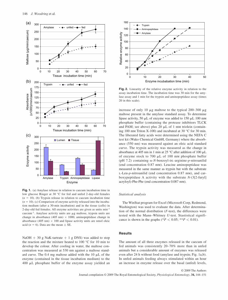
146 J. Woodring et al.
© 2009 The Authors Journal compilation © 2009 The Royal Entomological Society, Physiological Entomology, 34, 144–151
NaOH + 30 g NaK-tartrate + 1 g DNS) was added to stop the reaction and the mixture heated to 100 °C for 10 min to develop the colour. After cooling in water, the maltose con-centration was measured at 530 nm against a maltose stand-ard curve. The 0.4 mg maltose added with the 10 � L of the enzyme (contained in the tissue incubation medium) to the 400 � L phosphate buffer of the enzyme assay yielded an
increase of only 10 � g maltose to the typical 200 – 300 � g maltose present in the amylase standard assay. To determine lipase activity, 50 � L of enzyme was added to 150 � L 100 m m phosphate buffer (containing the protease inhibitors TLCK and PAM; see above) plus 20 � L of 1 m m triolein (contain-ing 100 m m Triton X-100) and incubated at 30 °C for 30 min. The liberated fatty acids were determined using the NEFA C test kit (Wako Chemical GmbH, Germany) where the absorb-ance (550 nm) was measured against an oleic acid standard curve. The trypsin activity was measured as the change in absorbance at 405 nm in 1 min at 25 °C after addition of 100 � L of enzyme stock to 700 � L of 100 m m phosphate buffer (pH 7.2) containing � - N -benzoyl-dl-arginine- p -nitroanilid (end concentration 0.87 m m ). Leucine aminopeptidase was measured in the same manner as trypsin but with the substrate l -Leu- p -nitroanilid (end concentration 0.87 mm), and car-boxypeptidase A activity with the substrate N -(3[2-furyl] acryloyl)-Phe-Phe (end concentration 0.087 m m ).
Statistical analysis
The WinStat program for Excel (Microsoft Corp, Redmond, Washington) was used to evaluate the data. After determina-tion of the normal distribution ( F -test), the differences were tested with the Mann – Whitney U -test. Stastistical signifi-cance is shown in the graphs (* P < 0.05; ** P < 0.01).
Results
The amount of all three enzymes released in the caecum of fed animals was consistently 20 – 70% more than in unfed animals but a considerable amount of enzymes was released even after 24 h without food (amylase and trypsin; Fig. 1a,b ). In unfed animals feeding always stimulated within an hour an increase in enzyme release over the basal (unfed) levels,
0
50
100
150
200
250
300
0 10 20 30 40 50 60 70Tissue incubation time (min)
Mal
tose
(µg/
min
/cae
cum
) unfed fedAmylase
(a)
0
25
50
75
100
125
150
175
200
0 10 20 30 40 50 60 70Tissue incubation time (min)
Abso
rban
ce(x
1000
)/min
/cae
cum
unfed fedTrypsin(b)
0
50
100
150
200
250
300
Amylase Trypsin Aminopeptidase LipaseEnzyme
Enzy
me
activ
ity
Lumen Tissue(c)
Fig. 1. (a) Amylase release in relation to caecum incubation time in low glucose Ringer at 30 °C for fed and unfed 2-day-old females ( n � 10). (b) Trypsin release in relation to caecum incubation time ( n � 10). (c) Comparison of enzyme activity released into the incuba-tion medium (after a 30-min incubation) and in the tissue (cells) in 2-day-old fed females. All enzyme activities are given as units min – 1 caecum – 1 . Amylase activity units are � g maltose, trypsin units are change in absorbance (405 nm) × 1000, aminopeptidase change in absorbance (405 nm) × 100 and lipase activity units are nmol oleic acid ( n � 6). Data are the mean ± SE.
0
20
40
60
80
100
120
140
160
0 10 20 30 40 50
Trypsin
Aminopeptidase
Amylase
Rel
ativ
e en
zym
e ac
tivity
Enzyme incubabation time (min)
Fig. 2. Linearity of the relative enzyme activity in relation to the assay incubation time. The incubation time was 30 min for the amy-lase assay and 1 min for the trypsin and aminopeptidase assay (times 20 in this scale).
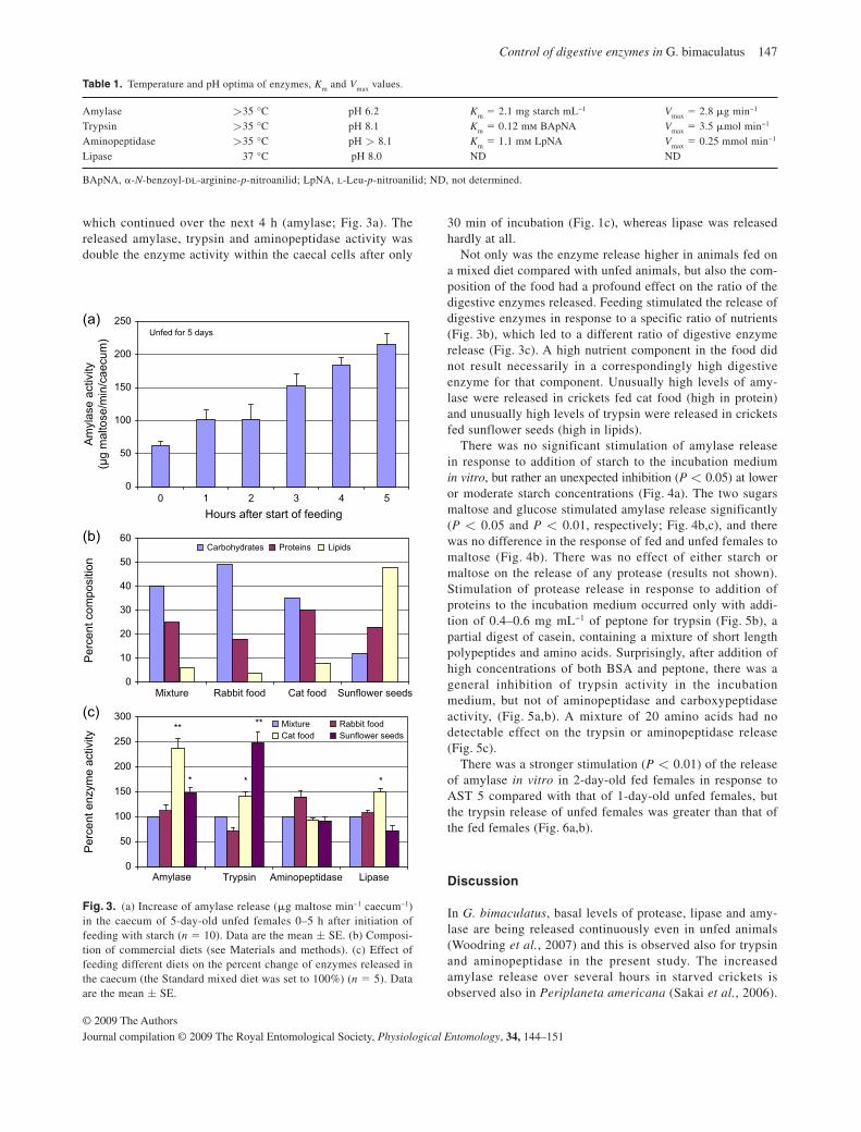
Control of digestive enzymes in G. bimaculatus 147
© 2009 The AuthorsJournal compilation © 2009 The Royal Entomological Society, Physiological Entomology, 34, 144–151
which continued over the next 4 h (amylase; Fig. 3a ). The released amylase, trypsin and aminopeptidase activity was double the enzyme activity within the caecal cells after only
30 min of incubation ( Fig. 1c ), whereas lipase was released hardly at all.
Not only was the enzyme release higher in animals fed on a mixed diet compared with unfed animals, but also the com-position of the food had a profound effect on the ratio of the digestive enzymes released. Feeding stimulated the release of digestive enzymes in response to a specific ratio of nutrients ( Fig. 3b ), which led to a different ratio of digestive enzyme release ( Fig. 3c ). A high nutrient component in the food did not result necessarily in a correspondingly high digestive enzyme for that component. Unusually high levels of amy-lase were released in crickets fed cat food (high in protein) and unusually high levels of trypsin were released in crickets fed sunflower seeds (high in lipids).
There was no significant stimulation of amylase release in response to addition of starch to the incubation medium in vitro , but rather an unexpected inhibition ( P < 0.05) at lower or moderate starch concentrations ( Fig. 4a ). The two sugars maltose and glucose stimulated amylase release significantly ( P < 0.05 and P < 0.01, respectively; Fig. 4b,c ), and there was no difference in the response of fed and unfed females to maltose ( Fig. 4b ). There was no effect of either starch or maltose on the release of any protease (results not shown). Stimulation of protease release in response to addition of proteins to the incubation medium occurred only with addi-tion of 0.4 – 0.6 mg mL −1 of peptone for trypsin ( Fig. 5b ), a partial digest of casein, containing a mixture of short length polypeptides and amino acids. Surprisingly, after addition of high concentrations of both BSA and peptone, there was a general inhibition of trypsin activity in the incubation medium, but not of aminopeptidase and carboxypeptidase activity, ( Fig. 5a,b ). A mixture of 20 amino acids had no detectable effect on the trypsin or aminopeptidase release ( Fig. 5c ).
There was a stronger stimulation ( P < 0.01) of the release of amylase in vitro in 2-day-old fed females in response to AST 5 compared with that of 1-day-old unfed females, but the trypsin release of unfed females was greater than that of the fed females ( Fig. 6a,b ).
Discussion
In G. bimaculatus , basal levels of protease, lipase and amy-lase are being released continuously even in unfed animals ( Woodring et al. , 2007 ) and this is observed also for trypsin and aminopeptidase in the present study. The increased amylase release over several hours in starved crickets is observed also in Periplaneta americana ( Sakai et al. , 2006 ).
Table 1. Temperature and pH optima of enzymes, K m and V max values.
Amylase >35 °C pH 6.2 K m � 2.1 mg starch mL −1 V max � 2.8 � g min −1 Trypsin >35 °C pH 8.1 K m � 0.12 m m BApNA V max � 3.5 � mol min −1 Aminopeptidase >35 °C pH > 8.1 K m � 1.1 m m LpNA V max � 0.25 mmol min −1 Lipase 37 °C pH 8.0 ND ND
BApNA, � - N -benzoyl- dl -arginine- p -nitroanilid; LpNA, l -Leu- p -nitroanilid; ND, not determined.
0
50
100
150
200
250
Hours after start of feeding
Amyl
ase
activ
ity(µ
g m
alto
se/m
in/c
aecu
m) Unfed for 5 days
(a)
(b)
(c)
0
10
20
30
40
50
60
Mixture Rabbit food Cat food Sunflower seeds
Perc
ent c
ompo
sitio
n
Carbohydrates Proteins Lipids
0
50
100
150
200
250
300
Amylase Trypsin Aminopeptidase Lipase
Perc
ent e
nzym
e ac
tivity
Mixture Rabbit foodCat food Sunflower seeds
*
**
*
**
*
0 1 2 3 4 5
Fig. 3. (a) Increase of amylase release ( � g maltose min – 1 caecum – 1 ) in the caecum of 5-day-old unfed females 0 – 5 h after initiation of feeding with starch ( n � 10). Data are the mean ± SE. (b) Composi-tion of commercial diets (see Materials and methods). (c) Effect of feeding different diets on the percent change of enzymes released in the caecum (the Standard mixed diet was set to 100%) ( n � 5). Data are the mean ± SE.
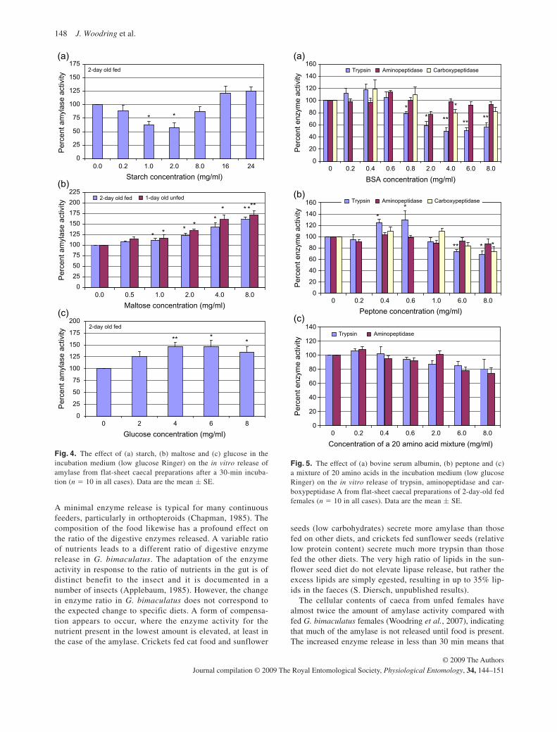
148 J. Woodring et al.
© 2009 The Authors Journal compilation © 2009 The Royal Entomological Society, Physiological Entomology, 34, 144–151
A minimal enzyme release is typical for many continuous feeders, particularly in orthopteroids ( Chapman, 1985 ). The composition of the food likewise has a profound effect on the ratio of the digestive enzymes released. A variable ratio of nutrients leads to a different ratio of digestive enzyme release in G. bimaculatus . The adaptation of the enzyme activity in response to the ratio of nutrients in the gut is of distinct benefit to the insect and it is documented in a number of insects ( Applebaum, 1985 ). However, the change in enzyme ratio in G. bimaculatus does not correspond to the expected change to specific diets. A form of compensa-tion appears to occur, where the enzyme activity for the nutrient present in the lowest amount is elevated, at least in the case of the amylase. Crickets fed cat food and sunflower
seeds (low carbohydrates) secrete more amylase than those fed on other diets, and crickets fed sunflower seeds (relative low protein content) secrete much more trypsin than those fed the other diets. The very high ratio of lipids in the sun-flower seed diet do not elevate lipase release, but rather the excess lipids are simply egested, resulting in up to 35% lip-ids in the faeces (S. Diersch, unpublished results).
The cellular contents of caeca from unfed females have almost twice the amount of amylase activity compared with fed G. bimaculatus females ( Woodring et al. , 2007 ), indicating that much of the amylase is not released until food is present. The increased enzyme release in less than 30 min means that
0
25
50
75
100
125
150
175
0.0 0.2 1.0 2.0 8.0 16 24Starch concentration (mg/ml)
Perc
ent a
myl
ase
activ
ity2-day old fed
* *
0
25
50
75
100
125
150
175
200
Glucose concentration (mg/ml)
Perc
ent a
myl
ase
activ
ity
2-day old fed
** **
0255075
100125150175200225
0.0 0.5 1.0 2.0 4.0 8.0Maltose concentration (mg/ml)
Perc
ent a
myl
ase
activ
ity
2-day old fed 1-day old unfed
* * * **
* ** **
0 2 4 6 8
(a)
(b)
(c)
Fig. 4. The effect of (a) starch, (b) maltose and (c) glucose in the incubation medium (low glucose Ringer) on the in vitro release of amylase from flat-sheet caecal preparations after a 30-min incuba-tion ( n � 10 in all cases). Data are the mean ± SE.
0
20
40
60
80
100
120
140
160(a)
(b)
(c)
0 0.2 0.4 0.6 0.8 2.0 4.0 6.0 8.0
BSA concentration (mg/ml)
Perc
ent e
nzym
e ac
tivity
Perc
ent e
nzym
e ac
tivity
Perc
ent e
nzym
e ac
tivity
Trypsin Aminopeptidase Carboxypeptidase
** ** ** **
*
0
20
40
60
80
100
120
140
0 0.2 0.4 0.6 2.0 6.0 8.0
Concentration of a 20 amino acid mixture (mg/ml)
0
20
40
60
80
100
120
140
160
0 0.2 0.4 0.6 1.0 6.0 8.0Peptone concentration (mg/ml)
**
*** *
Trypsin Aminopeptidase
Trypsin Aminopeptidase
Carboxypeptidase
Fig. 5. The effect of (a) bovine serum albumin, (b) peptone and (c) a mixture of 20 amino acids in the incubation medium (low glucose Ringer) on the in vitro release of trypsin, aminopeptidase and car-boxypeptidase A from flat-sheet caecal preparations of 2-day-old fed females ( n � 10 in all cases). Data are the mean ± SE.
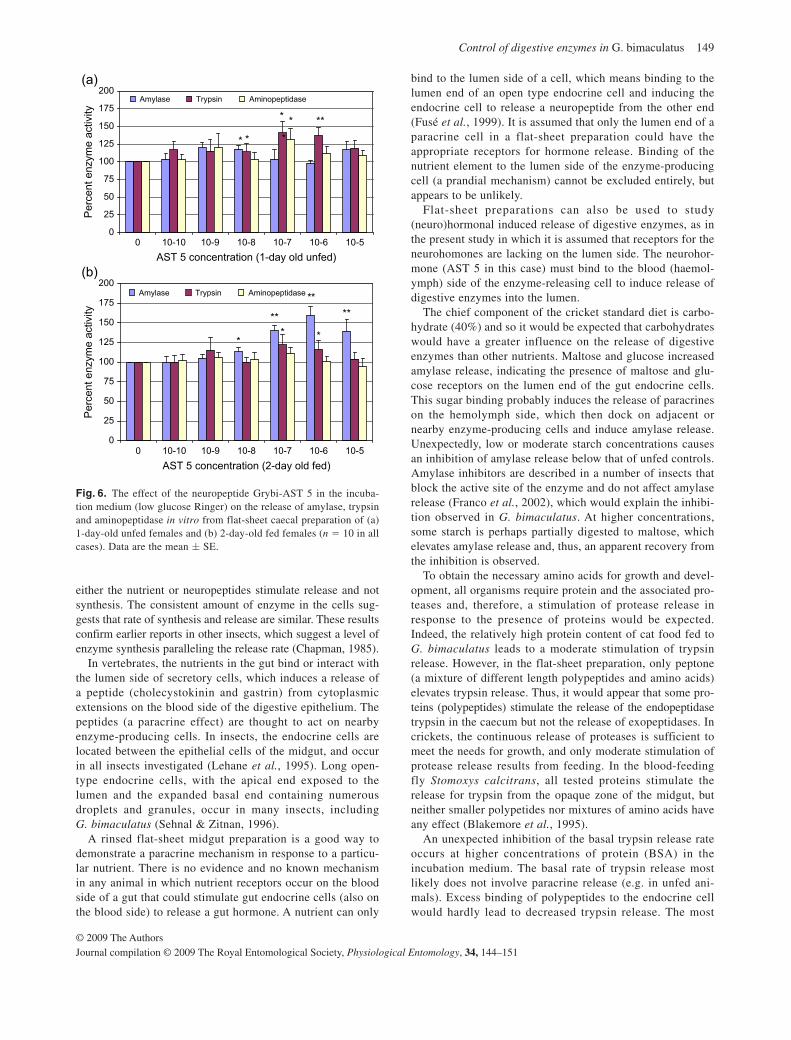
Control of digestive enzymes in G. bimaculatus 149
© 2009 The AuthorsJournal compilation © 2009 The Royal Entomological Society, Physiological Entomology, 34, 144–151
either the nutrient or neuropeptides stimulate release and not synthesis. The consistent amount of enzyme in the cells sug-gests that rate of synthesis and release are similar. These results confirm earlier reports in other insects, which suggest a level of enzyme synthesis paralleling the release rate ( Chapman, 1985 ).
In vertebrates, the nutrients in the gut bind or interact with the lumen side of secretory cells, which induces a release of a peptide (cholecystokinin and gastrin) from cytoplasmic extensions on the blood side of the digestive epithelium. The peptides (a paracrine effect) are thought to act on nearby enzyme-producing cells. In insects, the endocrine cells are located between the epithelial cells of the midgut, and occur in all insects investigated ( Lehane et al. , 1995 ). Long open-type endocrine cells, with the apical end exposed to the lumen and the expanded basal end containing numerous droplets and granules, occur in many insects, including G. bimaculatus ( Sehnal & Zitnan, 1996 ).
A rinsed flat-sheet midgut preparation is a good way to demonstrate a paracrine mechanism in response to a particu-lar nutrient. There is no evidence and no known mechanism in any animal in which nutrient receptors occur on the blood side of a gut that could stimulate gut endocrine cells (also on the blood side) to release a gut hormone. A nutrient can only
bind to the lumen side of a cell, which means binding to the lumen end of an open type endocrine cell and inducing the endocrine cell to release a neuropeptide from the other end ( Fusé et al. , 1999 ). It is assumed that only the lumen end of a paracrine cell in a flat-sheet preparation could have the appropriate receptors for hormone release. Binding of the nutrient element to the lumen side of the enzyme-producing cell (a prandial mechanism) cannot be excluded entirely, but appears to be unlikely.
Flat-sheet preparations can also be used to study (neuro)hormonal induced release of digestive enzymes, as in the present study in which it is assumed that receptors for the neurohomones are lacking on the lumen side. The neurohor-mone (AST 5 in this case) must bind to the blood (haemol-ymph) side of the enzyme-releasing cell to induce release of digestive enzymes into the lumen.
The chief component of the cricket standard diet is carbo-hydrate (40%) and so it would be expected that carbohydrates would have a greater influence on the release of digestive enzymes than other nutrients. Maltose and glucose increased amylase release, indicating the presence of maltose and glu-cose receptors on the lumen end of the gut endocrine cells. This sugar binding probably induces the release of paracrines on the hemolymph side, which then dock on adjacent or nearby enzyme-producing cells and induce amylase release. Unexpectedly, low or moderate starch concentrations causes an inhibition of amylase release below that of unfed controls. Amylase inhibitors are described in a number of insects that block the active site of the enzyme and do not affect amylase release ( Franco et al. , 2002 ), which would explain the inhibi-tion observed in G. bimaculatus . At higher concentrations, some starch is perhaps partially digested to maltose, which elevates amylase release and, thus, an apparent recovery from the inhibition is observed.
To obtain the necessary amino acids for growth and devel-opment, all organisms require protein and the associated pro-teases and, therefore, a stimulation of protease release in response to the presence of proteins would be expected. Indeed, the relatively high protein content of cat food fed to G. bimaculatus leads to a moderate stimulation of trypsin release. However, in the flat-sheet preparation, only peptone (a mixture of different length polypeptides and amino acids) elevates trypsin release. Thus, it would appear that some pro-teins (polypeptides) stimulate the release of the endopeptidase trypsin in the caecum but not the release of exopeptidases. In crickets, the continuous release of proteases is sufficient to meet the needs for growth, and only moderate stimulation of protease release results from feeding. In the blood-feeding fly Stomoxys calcitrans , all tested proteins stimulate the release for trypsin from the opaque zone of the midgut, but neither smaller polypetides nor mixtures of amino acids have any effect ( Blakemore et al. , 1995 ).
An unexpected inhibition of the basal trypsin release rate occurs at higher concentrations of protein (BSA) in the incubation medium. The basal rate of trypsin release most likely does not involve paracrine release (e.g. in unfed ani-mals). Excess binding of polypeptides to the endocrine cell would hardly lead to decreased trypsin release. The most
0
25
50
75
100
125
150
175
200
0 10-10 10-9 10-8 10-7 10-6 10-5AST 5 concentration (1-day old unfed)
Perc
ent e
nzym
e ac
tivity
Perc
ent e
nzym
e ac
tivity
Amylase Trypsin Aminopeptidase
Amylase Trypsin Aminopeptidase
*
***
* *
*
0
25
50
75
100
125
150
175
200
0 10-10 10-9 10-8 10-7 10-6 10-5AST 5 concentration (2-day old fed)
**
** **
***
(a)
(b)
Fig. 6. The effect of the neuropeptide Grybi-AST 5 in the incuba-tion medium (low glucose Ringer) on the release of amylase, trypsin and aminopeptidase in vitro from flat-sheet caecal preparation of (a) 1-day-old unfed females and (b) 2-day-old fed females ( n � 10 in all cases). Data are the mean ± SE.
150 J. Woodring et al.
© 2009 The Authors Journal compilation © 2009 The Royal Entomological Society, Physiological Entomology, 34, 144–151
likely explanation for the inhibition of trypsin is a direct inhibition of the trypsin active site. Plant protein inhibitors are capable of forming stable complexes with the target proteases in the insect gut, blocking, altering or preventing access to the enzyme active site ( Fan & Wu, 2005 ). Perhaps such ‘excess substrate inhibition’ occurs in G. bimaculatus where BSA irreversibly binds to the trypsin active site. Excess substrate inhibition occurs in approximately 20% of all enzymes ( Birch, 2008 ).
Inhibition of enzyme release (not the actual enzyme activity) via various leucokinins is reported in the lepidopteran Opisina arenosella ( Harshini et al. , 2002a ). In cock-roaches, allatostatins stimulate the release of carbohydrate digesting enzymes (Fusé et al. , 1999 ; Aguilar et al. , 2003 ). In G. bimaculatus , the allatostatin Grybi-AST 5 stimulates the release of amylase and trypsin. AST 5 is a paracrine secreted by the endocrine cells in the caecum, which is suggested by the expression of the AST 5 gene in the gut epithelium ( Meyering-Vos & Hoffmann, 2003 ). A massive input of carbohydrates in fed females elevates amylase release strongly and this process appears to be supported by AST 5. The basal level of release of trypsin in fed females, however, appears sufficient, and an additional AST 5-mediated increased trypsin release occurs only at a low level. Underfed females, on the other hand, require amino acids to grow, and an AST 5 sensitivity to stimulate trypsin and aminopeptidase release, as observed in the present study, would be advantageous.
Acknowledgements
This research was partially supported by a grant to M. Meyering-Vos (DFG ME 2034/1-3 and a scholarship to D. Lwalaba (DAAD A/06/33259). We especially thank Dr X. Bellés, Department of Molecular Biodiversity, Institute of Molecular Biology (CID, CSIC), Barcelona, Spain, for the generous gift of the neuropeptide BLAST 2 ( � Grybi-AST 5).
References
Aguilar , R. , Maestro , J.-L. , Vilaplana , L . et al . ( 2003 ) Allatostatin gene expression in brain and midgut, and activity of synthetic al-latostatins of feeding-related processes in the cockroach Blattella germanica . Regulatory Peptides , 115 , 171 – 177 .
Applebaum , S.W . ( 1985 ) Biochemistry of digestion . Comprehensive Insect Physiology, Biochemistry, and Pharmacology ( ed . by G. A . Kerkut and L. I . Gilbert ), Vol. 4 , pp . 279 – 311 . Pergamon Press , U.K .
Bernfeld , P . ( 1955 ) Amylases, � and � . Methods in Enzymology ( ed . by S. P. Colowick and N. Kaplan ), Vol. 1 , pp . 149 – 158 . Academic Press , New York, New York .
Birch , P . ( 2008 ) An Introduction to Enzyme Kinetics . University of Paisley , U.K .
Blakemore , D. , Williams , S. & Lehane , M.J . ( 1995 ) Protein stimula-tion of trypsin secretion from the opaque zone midgut cells of Stomoxyes calcitrans . Comparative Biochemistry and Physiology , 110B , 301 – 307 .
Borovsky , D . ( 2003 ) Trypsin-modulating oostatic factor: a potential new larvicide for mosquito control . Journal of Experimental Biology , 206 , 3869 – 3875 .
Borovsky , D. & Mahmood , F . ( 1995 ) Feeding the mosquito Aedes aegypti with TMOF and its analogs, effect on trypsin biosyn-thesis and egg development . Regulatory Peptides , 57 , 273 – 281 .
Chapman , R.F . ( 1985 ) Coordination of digestion . Comprehensive Insect Physiology, Biochemistry, and Pharmacology ( ed . by G. A. Kerkut and L. I. Gilbert ), Vol. 5 , pp . 213 – 240 . Pergamon Press , U.K .
Chapman , R.F . ( 1998 ) Alimentary canal, digestion and absorption . The Insects, Structure and Function , 4th edn , pp . 38 – 58 . Cambridge University Press , U.K .
Fan , S-G. & Wu , G.-J. ( 2005 ) Characteristics of plant proteinase in-hibitors and their applications in combating phytophagous insects . Botanical Bulletin Academia Sinica , 46 , 273 – 292 .
Franco , O.L. , Rigedn , D.J. , Melo , F.R. & Grossi-de-Sa , M.F . ( 2002 ) Plant � -amylase inhibitors and their interaction with insect � -amylases. Structure, function and potential for crop protection . European Journal of Biochemistry , 269 , 397 – 412 .
Fusé , M. , Zhang , J.R. , Partridge , E . et al . ( 1999 ) Effects of an alla-tostatin and a myosuppressin on midgut carbohydrate enzyme activity in the cockroach Diploptera punctata . Peptides , 20 , 1285 – 1293 .
Harshini , S. , Nachman , R.J. & Sreekumar , S . ( 2002a ) Inhibition of digestive enzyme release by neuropeptides in larvae of Opisina arenosella . Comparative Biochemistry and Physiology , 132B , 353 – 358 .
Harshini , S. , Nachman , R.J. & Sreekumar , S . ( 2002b ) In vitro release of digestive enzymes by FMRF amide related neuropeptides and analogues in the lepidopteran insect Opisina arenosella . Peptides , 23 , 1759 – 1763 .
Hill , S.H. & Orchard , I . ( 2005 ) In vitro analysis of the digestive en-zymes amylase and � -glucosidase in the midguts of Locusta migratoria in response to the myosuppressin, SchistoFLRFamide . Journal of Insect Physiology , 51 , 1 – 9 .
Lange , A.B. , Bendena , W.G. & Tobe , S.S . ( 1995 ) The effect of thir-teen unique dip-allatostatins on myogenic and induced contrac-tions on the cockroach hindgut Diploptera punctata . Archives of Insect Biochemistry and Physiology , 41 , 581 – 588 .
Lehane , M.J. , Blakemore , D. , Williams , S. & Moffatt , M.R . ( 1995 ) Regulation of digestive enzyme levels in insects . Comparative Biochemistry and Physiology , 110B , 285 – 289 .
Lehane , M.J. , Müller , H.M. & Crisanti , A . ( 1996 ) Mechanisms con-trolling the synthesis and secretion of digestive enzymes in in-sects . Biology of the Insect Midgut ( ed . by M. J. Lehane and P. F. Billingsley ), pp . 195 – 205 . Chapman & Hall , U.K .
Lorenz , M.W. , Kellner , R. & Hoffmann , K.H . ( 1995 ) A family of neuropeptides that inhibit juvenile hormone biosynthesis in the cricket Gryllus bimaculatus . Journal of Biological Chemistry , 270 , 21103 – 21108 .
Lorenz , J.I. , Lorenz , M.W. & Hoffmann , K.H . ( 1997 ) Factors that re-gulate juvenile hormone and ecdysteroid biosynthesis in the cricket, Gryllus bimaculatus . European Journal of Entomology , 94 , 369 – 379 .
Meyering-Vos , M. & Hoffmann , K.H . ( 2003 ) Expression of alla-tostatins in the Mediterranean field cricket, Gryllus bimaculatus . Comparative Biochemistry and Physiology , 136B , 207 – 215 .
Meyering-Vos , M. , Wu , X. , Huang , J . et al . ( 2001 ) The allatostatin gene of the cricket Gryllus bimaculatus . Molecular and Cellular Endocrinology , 184 , 103 – 114 .
Montuenge , L.M. , Barrenechea , M.A. , Sesma , P . et al . ( 1989 ) Ultra structure and immunocytochemistry of endocrine cells in the mid-gut of the desert locust, Schistocerca gregaria . Cell and Tissue Research , 258 , 577 – 583 .
Control of digestive enzymes in G. bimaculatus 151
© 2009 The AuthorsJournal compilation © 2009 The Royal Entomological Society, Physiological Entomology, 34, 144–151
Penzlin , H . ( 1996 ) Lehrbuch der Tierphysiologie , 6th ed . Gustav Fischer Verlag , Jena, Germany .
Sehnal , F. & Zitnan , D . ( 1996 ) Midgut endocrine cells . Biology of the Insect Midgut ( ed . by M. J. Lehane and P. F. Billingsley ), pp . 195 – 205 . Chapman & Hall , U.K .
Sakai , T. , Satake , H. & Takeda , M . ( 2006 ) Nutrient-induced � -amylase and protease activity is regulated by crustacean cardio-active peptide (CCAP) in the cockroach midgut . Peptides , 27 , 2157 – 2164 .
Woodring , J. & Lorenz , M.W . ( 2007 ) Feeding, nutrient flow, and functional gut morphology in the cricket Gryllus bimaculatus . Journal of Morphology , 268 , 815 – 825 .
Woodring , J. , Hoffmann , K.H. & Lorenz , M.W . ( 2007 ) Activity, re-lease and flow of digestive enzymes in the cricket, Gryllus bimac-ulatus . Physiological Entomology , 32 , 56 – 63 .
Accepted 3 October 2008 First published online 2 April 2009
A r t i c l e
CONTROL OF THE RELEASEOF DIGESTIVE ENZYMESIN THE LARVAE OF THEFALL ARMYWORM,Spodoptera frugiperda
Digali Lwalaba, Klaus H. Hoffmann, andJoseph WoodringDepartment of Animal Ecology I, University of Bayreuth, Bayreuth,Germany
There is a basal level of enzyme activity for trypsin, aminopeptidase,amylase, and lipase in the gut of unfed larval (L6) Spodopterafrugiperda. Trypsin activity does not decrease with non-feeding, possiblybecause of the low protein levels in plants along with high amino acidrequirements for growth and storage (for later reproduction in adults).Therefore, trypsin must always be present so that only a minimal proteinloss via egestion occurs. Larvae, however, adjust amylase activity tocarbohydrate ingestion, and indeed amylase activity is five-fold higher infed larvae compared to unfed larvae. Gut lipase activity is low, typical ofinsects with a high carbohydrate diet. A flat-sheet preparation of theventriculus was used to measure the release of enzymes in response tospecific nutrients and known brain/gut hormones in S. frugiperda.Sugars greatly increase (4300%) amylase release, but starch has noeffect. Proteins and amino acids have little or no effect on trypsin oraminopeptidase release. The control of enzyme release in response to foodis likely mediated through neurohormones. Indeed, an allatostatin(Spofr-AS A5) inhibits amylase and trypsin, and allatotropin (Manse-AT) stimulates amylase and trypsin release. Spofr-AS A5 also inhibitsileum myoactivity and Manse-AT stimulates myoactivity. The epithelialsecretion rate of amylase and trypsin was about 20% of the amount ofenzyme present in the ventricular lumen, which, considering the efficient
Correspondence to: Joseph Woodring, Department of Animal Ecology I, University of Bayreuth, 95440Bayreuth, Germany. E-mail: [email protected]
ARCHIVES OF INSECT BIOCHEMISTRY AND PHYSIOLOGY, Vol. 73, No. 1, 14–29 (2010)
Published online in Wiley InterScience (www.interscience.wiley.com).
& 2009 Wiley Periodicals, Inc. DOI: 10.1002/arch.20332
counter-current recycling of enzymes, suggests that the secretion rate isadequate to replace egested enzymes. �C 2009 Wiley Periodicals, Inc.
Keywords: digestive enzymes; larvae; Spodoptera frugiperda
INTRODUCTION
The fall armyworm, Spodoptera frugiperda (J.E. Smith) is an important defoliator ofcorn, sorghum, cotton, and alfalfa in the tropical regions of the western hemispherefrom the United States to Argentina. Various insecticides have been employed tocontrol the fall armyworm, however, with only limited success and at a cost ofconsiderable damage to the environment. The digestive tract of insects, especiallycaterpillars, is an excellent site for control mechanisms that in general are not toxic toother organisms. Endogenous enzyme inhibitors such as the trypsin-modulatingoostatic factor (TMOF) effectively inhibit translation of trypsin mRNA in the larva gutof Heliothis virescens (Nauen et al., 2001). The crystal proteins (cry) are endotoxinsproduced by Bacillus thuringiensis (Bt), which punctures the insects’ ventriculus andcauses uncontrolled leakage and death in all lepidopteran larvae tested (Pigott et al.,2008). Control is very effective and specific, and the Bt gene has been inserted intomany crop plants. There are a number of plant protease inhibitors (see review, Fanand Wu, 2005) that bind to endo- and ectopeptidases in insects and effectively inhibitgrowth, and likewise a number of plant a-amylase inhibitors (see review, Franco et al.,2002). Finally, in recent years it has been shown that members of several families ofinsect neuropeptides, including allatostatins (AS) and allatotropins (AT), control therelease of digestive enzymes and at the same time modulate gut myoactivity in manyinsects (Fuse et al., 1999; Harshini et al., 2002a; Sakai et al., 2004; Hill and Orchard,2005). Insertion of the genes of enzyme inhibitors or neurohormones into crop plantswould be worth the effort, but only if more detailed information on the action of thesepeptides indicates effective in vivo regulation of digestive functions. The currentinvestigation of the release of digestive enzymes in S. frugiperda is a step in thisdirection.
The proteases and amylases have been described in the gut of a number ofnoctuid larvae (Lenz et al., 1991; Johnson et al., 1995; Ortego et al., 1996; Bown et al.,1998), and the role of the peritrophic membrane and the counter-current flow ofthese enzymes in the ventriculus of S. frugiperda has been described in detail(Ferreira et al., 1994a,b; Bolognesi et al., 2001). In the current study, the distributionof lipase in addition to trypsin and amylase in the gut, the effect of larval age,and especially the role of feeding on enzyme release in S. frugiperda are examined.The main thrust of the current study however, is an examination of the effect ofspecific nutrients and neurohormones on the release of enzymes from incubatedmidguts (flat-sheet preparations), as previously described in Gryllus bimaculatus(Woodring et al., 2009). The neurohormones tested are based on the genesequences of two allatostatins (Spofr-AS A5, AS A6) and an allatotropin (Manse-AT)found in S. frugiperda (Abdel-latief et al., 2004). Many neurohormones thataffect enzyme release also affect gut myoactivity (Harshini et al., 2002b; Matthewset al., 2007), and, therefore, the myoactivity of these two AS and an AT is alsoexamined.
Release of Digestive Enzymes in S. frugiperda � 15
Archives of Insect Biochemistry and Physiology
MATERIALS AND METHODS
Rearing Methods and Diets
Pupae and eggs of S. frugiperda and an artificial diet (based on bean meal) wereprovided by Bayer CropScience AG (Monheim). Larvae were reared at 271C andapproximately 70% relative humidity under a L16:D8 photoperiod on the Bayer diet,as previously described (Oeh et al., 2000). To prevent cannibalism, L4 larvae weremaintained individually in separate compartments of assortment boxes with 40compartments (49� 32� 36 mm per compartment). The pupae were also separated,and after emergence a 2:1 ratio of males to females were placed in a 30� 30� 15-cmcontainer with a sucrose solution as a food source, water, and paper as a substrate foregg laying (Oeh et al., 2000). In order to test the effect of diet and feeding on enzymeactivity, some newly moulted L6 were provided a cellulose diet (30 g cellulose powder13 g agar in 250 ml water) and others received no food at all. Unless otherwise stated,the L6 day-3 larvae were used in all experiments.
Gut Preparation and Incubation
The midgut (ventriculus) averaged 21 mm in length, and the hindgut (Ileum1colon)averaged 5–6 mm. These were separated, and the enzyme activity was measured in thetissues and in the lumen. The ventricular lumen included the exo- and endoperi-trophic space plus the peritrophic membrane (PM). The total lumen contents werebrought to 300 ml with Spodoptera Ringer (per Liter 0.7 g NaCl, 2.0 g KCl, 0.25 gNaHCO3� 2H2O, 1.3 g CaCl2�2H2O, 4.4 g MgCl2� 7H2O, 1.0 g Hepes at pH 7.0).The tissue was sonicated lightly (BronsonSonifier 250, for 2–3 sec at the lowest settingat 41C) and centrifuged at 12,000g (41C) for 10 min. The secretion of enzymes wasdetermined using flat-sheet preparations. Freshly dissected ventriculus were slit openand rinsed twice with fresh low glucose Ringer (LGR) (10 mg glucose/100 ml Ringer), aconcentration adapted from Fuse et al. (1999) for incubation of gut tissues of thecockroach Diploptera punctata, placed in 300 ml LGR, and incubated for 30 min at 301Cwithout shaking. The tissue was discarded, and the remaining incubation medium wascentrifuged and used to measure enzyme release. Thus, there were three sources ofenzymes: lumen content, tissue (cells) homogenate, and incubation medium. Thereleased amylase, lipase, trypsin, and aminopeptidase each increased at a constant rateover an incubation time from 10 to 30 min (Fig. 1), indicating that the tissues wereviable over the test period.
Enzyme Assays
For the determination of enzyme activity, 10ml of the homogenate or incubate wasused for the amylase assay, 50ml for the lipase assay, and 100 ml for the trypsin andaminopeptidase assay. It was found that the amylase and lipase release (increase inactivity) was linear from 15 to 60 min, and the trypsin and aminopeptidase activitieswere linear from 0 to 5 min at 251C and at a pH of 8.0 (data not shown). For theamylase assay, 10 ml of the homogenate or incubate was added to 40 ml 2.5% starch1
360 ml Tris buffer (652 ml 0.1M Tris1348 ml 0.1M HCl) and incubated 30 min at 301Cwith light shaking. To this mixture, 400 ml dinitrosalicylic acid Reagent(1 g dinitrosalicylic acid in 70 ml 2N NaOH, 30 g KNa tartrate, with water made to
16 � Archives of Insect Biochemistry and Physiology, January 2010
Archives of Insect Biochemistry and Physiology
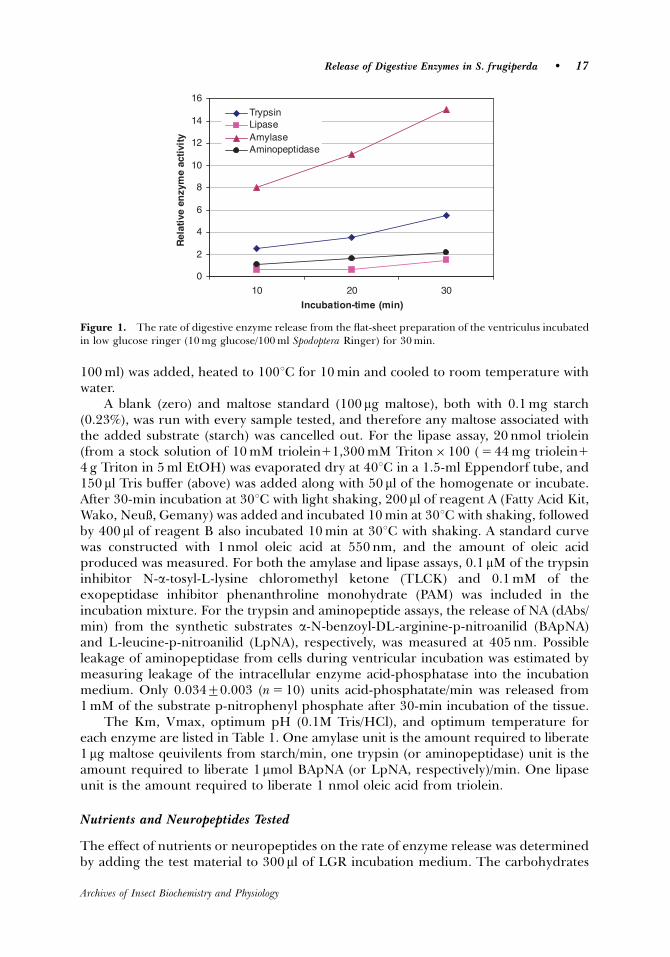
100 ml) was added, heated to 1001C for 10 min and cooled to room temperature withwater.
A blank (zero) and maltose standard (100 mg maltose), both with 0.1 mg starch(0.23%), was run with every sample tested, and therefore any maltose associated withthe added substrate (starch) was cancelled out. For the lipase assay, 20 nmol triolein(from a stock solution of 10 mM triolein11,300 mM Triton� 100 ( 5 44 mg triolein1
4 g Triton in 5 ml EtOH) was evaporated dry at 401C in a 1.5-ml Eppendorf tube, and150 ml Tris buffer (above) was added along with 50 ml of the homogenate or incubate.After 30-min incubation at 301C with light shaking, 200 ml of reagent A (Fatty Acid Kit,Wako, NeuX, Gemany) was added and incubated 10 min at 301C with shaking, followedby 400 ml of reagent B also incubated 10 min at 301C with shaking. A standard curvewas constructed with 1 nmol oleic acid at 550 nm, and the amount of oleic acidproduced was measured. For both the amylase and lipase assays, 0.1 mM of the trypsininhibitor N-a-tosyl-L-lysine chloromethyl ketone (TLCK) and 0.1 mM of theexopeptidase inhibitor phenanthroline monohydrate (PAM) was included in theincubation mixture. For the trypsin and aminopeptide assays, the release of NA (dAbs/min) from the synthetic substrates a-N-benzoyl-DL-arginine-p-nitroanilid (BApNA)and L-leucine-p-nitroanilid (LpNA), respectively, was measured at 405 nm. Possibleleakage of aminopeptidase from cells during ventricular incubation was estimated bymeasuring leakage of the intracellular enzyme acid-phosphatase into the incubationmedium. Only 0.03470.003 (n 5 10) units acid-phosphatate/min was released from1 mM of the substrate p-nitrophenyl phosphate after 30-min incubation of the tissue.
The Km, Vmax, optimum pH (0.1M Tris/HCl), and optimum temperature foreach enzyme are listed in Table 1. One amylase unit is the amount required to liberate1mg maltose qeuivilents from starch/min, one trypsin (or aminopeptidase) unit is theamount required to liberate 1mmol BApNA (or LpNA, respectively)/min. One lipaseunit is the amount required to liberate 1 nmol oleic acid from triolein.
Nutrients and Neuropeptides Tested
The effect of nutrients or neuropeptides on the rate of enzyme release was determinedby adding the test material to 300 ml of LGR incubation medium. The carbohydrates
0
2
4
6
8
10
12
14
16
10 20 30
Incubation-time (min)
Rel
ativ
e en
zym
e ac
tivi
ty
Trypsin LipaseAmylaseAminopeptidase
Figure 1. The rate of digestive enzyme release from the flat-sheet preparation of the ventriculus incubatedin low glucose ringer (10 mg glucose/100 ml Spodoptera Ringer) for 30 min.
Release of Digestive Enzymes in S. frugiperda � 17
Archives of Insect Biochemistry and Physiology
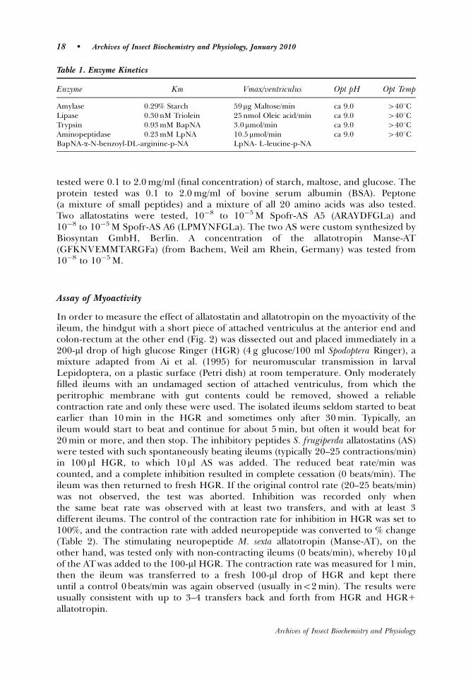
tested were 0.1 to 2.0 mg/ml (final concentration) of starch, maltose, and glucose. Theprotein tested was 0.1 to 2.0 mg/ml of bovine serum albumin (BSA). Peptone(a mixture of small peptides) and a mixture of all 20 amino acids was also tested.Two allatostatins were tested, 10�8 to 10�5 M Spofr-AS A5 (ARAYDFGLa) and10�8 to 10�5 M Spofr-AS A6 (LPMYNFGLa). The two AS were custom synthesized byBiosyntan GmbH, Berlin. A concentration of the allatotropin Manse-AT(GFKNVEMMTARGFa) (from Bachem, Weil am Rhein, Germany) was tested from10�8 to 10�5 M.
Assay of Myoactivity
In order to measure the effect of allatostatin and allatotropin on the myoactivity of theileum, the hindgut with a short piece of attached ventriculus at the anterior end andcolon-rectum at the other end (Fig. 2) was dissected out and placed immediately in a200-ml drop of high glucose Ringer (HGR) (4 g glucose/100 ml Spodoptera Ringer), amixture adapted from Ai et al. (1995) for neuromuscular transmission in larvalLepidoptera, on a plastic surface (Petri dish) at room temperature. Only moderatelyfilled ileums with an undamaged section of attached ventriculus, from which theperitrophic membrane with gut contents could be removed, showed a reliablecontraction rate and only these were used. The isolated ileums seldom started to beatearlier than 10 min in the HGR and sometimes only after 30 min. Typically, anileum would start to beat and continue for about 5 min, but often it would beat for20 min or more, and then stop. The inhibitory peptides S. frugiperda allatostatins (AS)were tested with such spontaneously beating ileums (typically 20–25 contractions/min)in 100 ml HGR, to which 10ml AS was added. The reduced beat rate/min wascounted, and a complete inhibition resulted in complete cessation (0 beats/min). Theileum was then returned to fresh HGR. If the original control rate (20–25 beats/min)was not observed, the test was aborted. Inhibition was recorded only whenthe same beat rate was observed with at least two transfers, and with at least 3different ileums. The control of the contraction rate for inhibition in HGR was set to100%, and the contraction rate with added neuropeptide was converted to % change(Table 2). The stimulating neuropeptide M. sexta allatotropin (Manse-AT), on theother hand, was tested only with non-contracting ileums (0 beats/min), whereby 10 mlof the ATwas added to the 100-ml HGR. The contraction rate was measured for 1 min,then the ileum was transferred to a fresh 100-ml drop of HGR and kept thereuntil a control 0 beats/min was again observed (usually ino2 min). The results wereusually consistent with up to 3–4 transfers back and forth from HGR and HGR1
allatotropin.
Table 1. Enzyme Kinetics
Enzyme Km Vmax/ventriculus Opt pH Opt Temp
Amylase 0.29% Starch 59mg Maltose/min ca 9.0 4401CLipase 0.30 nM Triolein 25 nmol Oleic acid/min ca 9.0 4401CTrypsin 0.93 mM BapNA 3.0mmol/min ca 9.0 4401CAminopeptidase 0.23 mM LpNA 10.5 mmol/min ca 9.0 4401CBapNA-a-N-benzoyl-DL-arginine-p-NA LpNA- L-leucine-p-NA
18 � Archives of Insect Biochemistry and Physiology, January 2010
Archives of Insect Biochemistry and Physiology
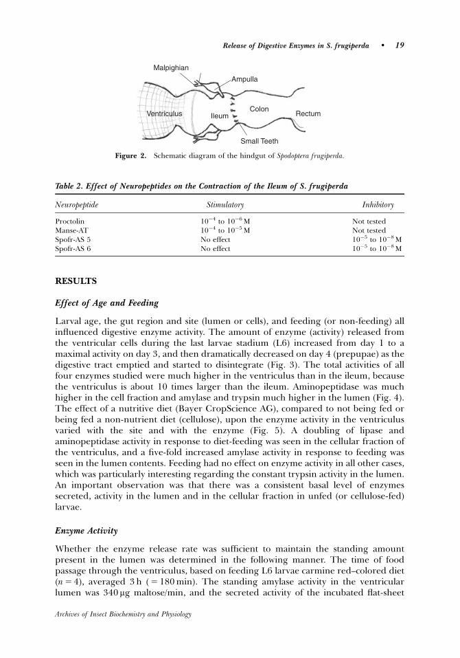
RESULTS
Effect of Age and Feeding
Larval age, the gut region and site (lumen or cells), and feeding (or non-feeding) allinfluenced digestive enzyme activity. The amount of enzyme (activity) released fromthe ventricular cells during the last larvae stadium (L6) increased from day 1 to amaximal activity on day 3, and then dramatically decreased on day 4 (prepupae) as thedigestive tract emptied and started to disintegrate (Fig. 3). The total activities of allfour enzymes studied were much higher in the ventriculus than in the ileum, becausethe ventriculus is about 10 times larger than the ileum. Aminopeptidase was muchhigher in the cell fraction and amylase and trypsin much higher in the lumen (Fig. 4).The effect of a nutritive diet (Bayer CropScience AG), compared to not being fed orbeing fed a non-nutrient diet (cellulose), upon the enzyme activity in the ventriculusvaried with the site and with the enzyme (Fig. 5). A doubling of lipase andaminopeptidase activity in response to diet-feeding was seen in the cellular fraction ofthe ventriculus, and a five-fold increased amylase activity in response to feeding wasseen in the lumen contents. Feeding had no effect on enzyme activity in all other cases,which was particularly interesting regarding the constant trypsin activity in the lumen.An important observation was that there was a consistent basal level of enzymessecreted, activity in the lumen and in the cellular fraction in unfed (or cellulose-fed)larvae.
Enzyme Activity
Whether the enzyme release rate was sufficient to maintain the standing amountpresent in the lumen was determined in the following manner. The time of foodpassage through the ventriculus, based on feeding L6 larvae carmine red–colored diet(n 5 4), averaged 3 h ( 5 180 min). The standing amylase activity in the ventricularlumen was 340 mg maltose/min, and the secreted activity of the incubated flat-sheet
Table 2. Effect of Neuropeptides on the Contraction of the Ileum of S. frugiperda
Neuropeptide Stimulatory Inhibitory
Proctolin 10�4 to 10�6 M Not testedManse-AT 10�4 to 10�5 M Not testedSpofr-AS 5 No effect 10�5 to 10�8 MSpofr-AS 6 No effect 10�5 to 10�8 M
Ventriculus Ileum Colon
Rectum
Malpighian
Ampulla
Small Teeth
Figure 2. Schematic diagram of the hindgut of Spodoptera frugiperda.
Release of Digestive Enzymes in S. frugiperda � 19
Archives of Insect Biochemistry and Physiology
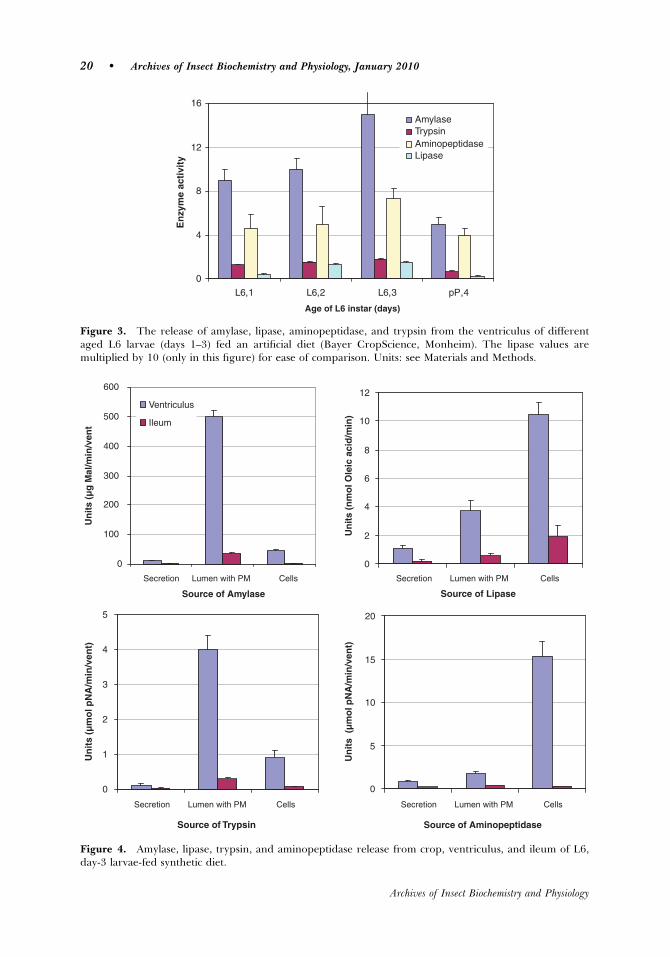
0
4
8
12
16
L6,1 L6,2 L6,3 pP,4
Age of L6 instar (days)
En
zym
e ac
tivi
ty
AmylaseTrypsinAminopeptidaseLipase
Figure 3. The release of amylase, lipase, aminopeptidase, and trypsin from the ventriculus of differentaged L6 larvae (days 1–3) fed an artificial diet (Bayer CropScience, Monheim). The lipase values aremultiplied by 10 (only in this figure) for ease of comparison. Units: see Materials and Methods.
0
100
200
300
400
500
600
Secretion
Source of Amylase
Un
its
(µg
Mal
/min
/ven
t
Ventriculus
Ileum
0
2
4
6
8
10
12
Source of Lipase
Un
its
(nm
ol O
leic
aci
d/m
in)
0
1
2
3
4
5
Source of Trypsin
Un
its
(µm
ol p
NA
/min
/ven
t)
0
5
10
15
20
Source of Aminopeptidase
Un
its
(µ
mo
l pN
A/m
in/v
ent)
Lumen with PM Cells Lumen with PM CellsSecretion
Secretion Lumen with PM Cells Lumen with PM CellsSecretion
Figure 4. Amylase, lipase, trypsin, and aminopeptidase release from crop, ventriculus, and ileum of L6,day-3 larvae-fed synthetic diet.
20 � Archives of Insect Biochemistry and Physiology, January 2010
Archives of Insect Biochemistry and Physiology
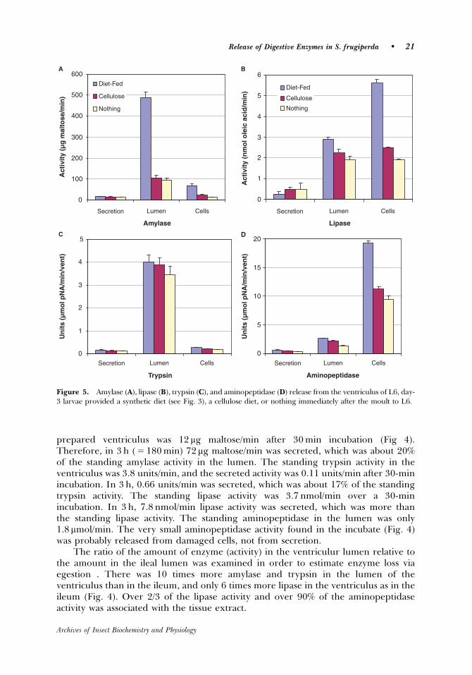
prepared ventriculus was 12 mg maltose/min after 30 min incubation (Fig 4).Therefore, in 3 h ( 5 180 min) 72mg maltose/min was secreted, which was about 20%of the standing amylase activity in the lumen. The standing trypsin activity in theventriculus was 3.8 units/min, and the secreted activity was 0.11 units/min after 30-minincubation. In 3 h, 0.66 units/min was secreted, which was about 17% of the standingtrypsin activity. The standing lipase activity was 3.7 nmol/min over a 30-minincubation. In 3 h, 7.8 nmol/min lipase activity was secreted, which was more thanthe standing lipase activity. The standing aminopeptidase in the lumen was only1.8 mmol/min. The very small aminopeptidase activity found in the incubate (Fig. 4)was probably released from damaged cells, not from secretion.
The ratio of the amount of enzyme (activity) in the ventriculur lumen relative tothe amount in the ileal lumen was examined in order to estimate enzyme loss viaegestion . There was 10 times more amylase and trypsin in the lumen of theventriculus than in the ileum, and only 6 times more lipase in the ventriculus as in theileum (Fig. 4). Over 2/3 of the lipase activity and over 90% of the aminopeptidaseactivity was associated with the tissue extract.
0
100
200
300
400
500
600
Secretion
Amylase
Act
ivit
y (µ
g m
alto
se/m
in)
Diet-Fed
Cellulose
Nothing
A
0
1
2
3
4
5
6
Lipase
Act
ivit
y (n
mo
l ole
ic a
cid
/min
) Diet-Fed
Cellulose
Nothing
B
0
1
2
3
4
5
Trypsin
Un
its
(µm
ol p
NA
/min
/ven
t)
C
0
5
10
15
20
Aminopeptidase
Un
its
(µm
ol p
NA
/min
/ven
t)D
Lumen Cells Secretion Lumen Cells
Secretion Lumen Cells Secretion Lumen Cells
Figure 5. Amylase (A), lipase (B), trypsin (C), and aminopeptidase (D) release from the ventriculus of L6, day-3 larvae provided a synthetic diet (see Fig. 3), a cellulose diet, or nothing immediately after the moult to L6.
Release of Digestive Enzymes in S. frugiperda � 21
Archives of Insect Biochemistry and Physiology
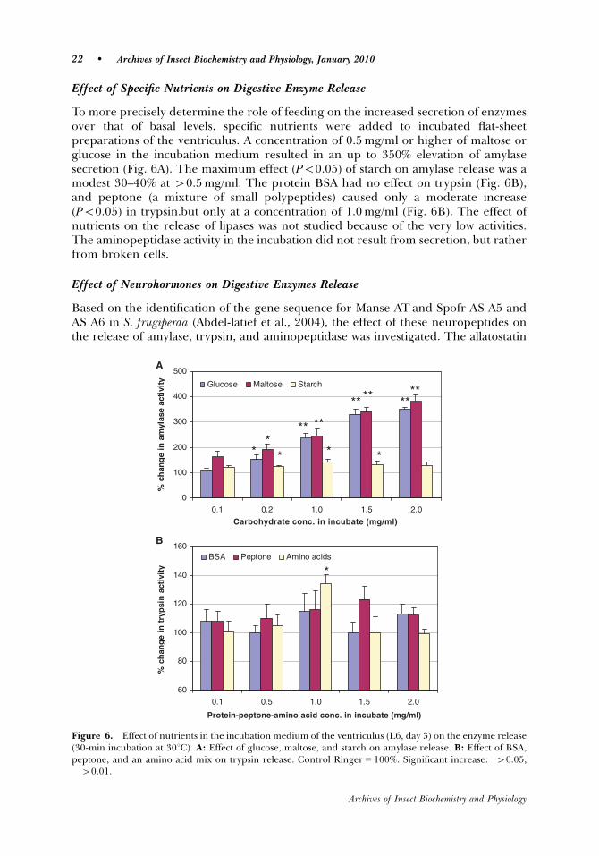
Effect of Specific Nutrients on Digestive Enzyme Release
To more precisely determine the role of feeding on the increased secretion of enzymesover that of basal levels, specific nutrients were added to incubated flat-sheetpreparations of the ventriculus. A concentration of 0.5 mg/ml or higher of maltose orglucose in the incubation medium resulted in an up to 350% elevation of amylasesecretion (Fig. 6A). The maximum effect (Po0.05) of starch on amylase release was amodest 30–40% at 40.5 mg/ml. The protein BSA had no effect on trypsin (Fig. 6B),and peptone (a mixture of small polypeptides) caused only a moderate increase(Po0.05) in trypsin.but only at a concentration of 1.0 mg/ml (Fig. 6B). The effect ofnutrients on the release of lipases was not studied because of the very low activities.The aminopeptidase activity in the incubation did not result from secretion, but ratherfrom broken cells.
Effect of Neurohormones on Digestive Enzymes Release
Based on the identification of the gene sequence for Manse-AT and Spofr AS A5 andAS A6 in S. frugiperda (Abdel-latief et al., 2004), the effect of these neuropeptides onthe release of amylase, trypsin, and aminopeptidase was investigated. The allatostatin
0
100
200
300
400
500
0.1 0.2 1.0 1.5 2.0
Carbohydrate conc. in incubate (mg/ml)
% c
han
ge
in a
myl
ase
acti
vity Glucose Maltose Starch
**
* *
**
*
**
** ** ****
A
60
80
100
120
140
160
0.1 0.5 1.0 1.5 2.0
Protein-peptone-amino acid conc. in incubate (mg/ml)
% c
han
ge
intr
ypsi
n a
ctiv
ity
BSA Peptone Amino acids
*
B
Figure 6. Effect of nutrients in the incubation medium of the ventriculus (L6, day 3) on the enzyme release(30-min incubation at 301C). A: Effect of glucose, maltose, and starch on amylase release. B: Effect of BSA,peptone, and an amino acid mix on trypsin release. Control Ringer 5 100%. Significant increase: �40.05,��40.01.
22 � Archives of Insect Biochemistry and Physiology, January 2010
Archives of Insect Biochemistry and Physiology
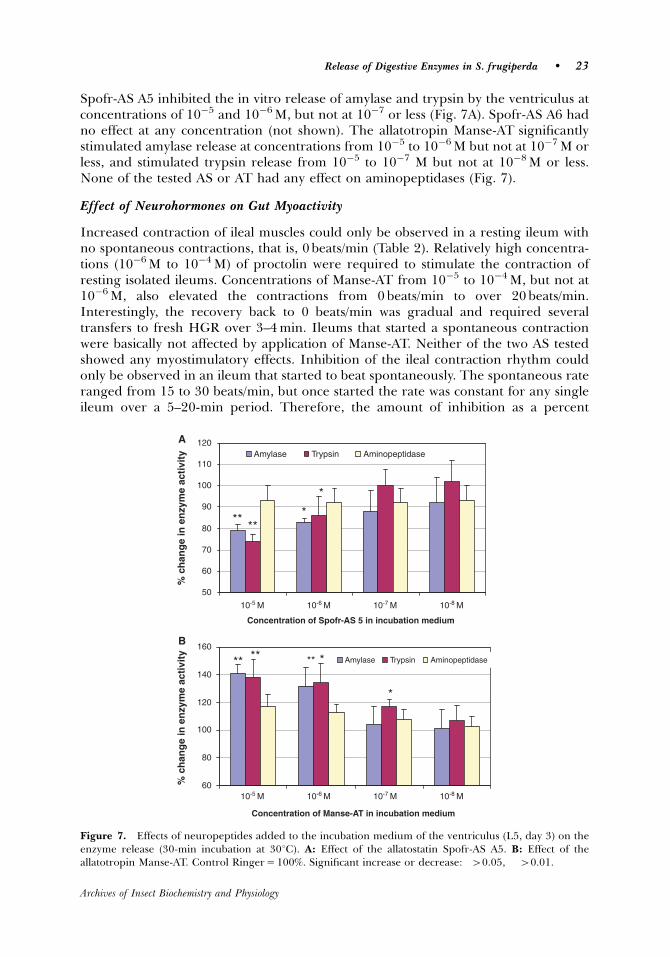
Spofr-AS A5 inhibited the in vitro release of amylase and trypsin by the ventriculus atconcentrations of 10�5 and 10�6 M, but not at 10�7 or less (Fig. 7A). Spofr-AS A6 hadno effect at any concentration (not shown). The allatotropin Manse-AT significantlystimulated amylase release at concentrations from 10�5 to 10�6 M but not at 10�7 M orless, and stimulated trypsin release from 10�5 to 10�7 M but not at 10�8 M or less.None of the tested AS or AT had any effect on aminopeptidases (Fig. 7).
Effect of Neurohormones on Gut Myoactivity
Increased contraction of ileal muscles could only be observed in a resting ileum withno spontaneous contractions, that is, 0 beats/min (Table 2). Relatively high concentra-tions (10�6 M to 10�4 M) of proctolin were required to stimulate the contraction ofresting isolated ileums. Concentrations of Manse-AT from 10�5 to 10�4 M, but not at10�6 M, also elevated the contractions from 0 beats/min to over 20 beats/min.Interestingly, the recovery back to 0 beats/min was gradual and required severaltransfers to fresh HGR over 3–4 min. Ileums that started a spontaneous contractionwere basically not affected by application of Manse-AT. Neither of the two AS testedshowed any myostimulatory effects. Inhibition of the ileal contraction rhythm couldonly be observed in an ileum that started to beat spontaneously. The spontaneous rateranged from 15 to 30 beats/min, but once started the rate was constant for any singleileum over a 5–20-min period. Therefore, the amount of inhibition as a percent
50
60
70
80
90
100
110
120
10-5 M
Concentration of Spofr-AS 5 in incubation medium
% c
han
ge
in e
nzy
me
acti
vity Amylase Trypsin Aminopeptidase
****
*
*
A
60
80
100
120
140
160
Concentration of Manse-AT in incubation medium
% c
han
ge
in e
nzy
me
acti
vity Amylase Trypsin Aminopeptidase** ** ** *
*
B
10-6 M 10-7 M 10-8 M
10-5 M 10-6 M 10-7 M 10-8 M
Figure 7. Effects of neuropeptides added to the incubation medium of the ventriculus (L5, day 3) on theenzyme release (30-min incubation at 301C). A: Effect of the allatostatin Spofr-AS A5. B: Effect of theallatotropin Manse-AT. Control Ringer 5 100%. Significant increase or decrease: �40.05, ��40.01.
Release of Digestive Enzymes in S. frugiperda � 23
Archives of Insect Biochemistry and Physiology
change from the control (initial rate) in this time span was consistent and repeatable.The ileum was very sensitive to inhibition by both AS (Spofr-AS A5 and AS A6) at allconcentrations over 10�8 M, showing an immediate complete inhibition (0 beats/min).At 10�9 M, the inhibition averaged 50% of the control, and there was no inhibition at10�10 M. Recovery in fresh HGR required 20–30 seconds.
DISCUSSION
The ventricular lumen volume is about 150 ml and the ileum only about 10 ml,therefore the ileum should contain 15% of the total amount (activity) of luminalamylase and trypsin. However, the ileum activity is about half (7%) of that of theventricular lumen. The lower concentration of amylase and trypsin in the ileumcompared to the ventriculas suggests that the efficient counter-current flow of enzymesin the ventriculus of S. frugiperda, as described by Bolognesi et al. (2001), reduces theloss of important digestive enzymes via egestion.
As in other lepidopteran larvae (Ortego et al., 1996; Lenz et al., 1991), trypsin andamylase in S. frugiperda occur primarily in the endoperitrophic space and theaminopeptidase is bound to the ventricular cell membranes. Amylase and trypsin arederived from membrane-bound forms, and are released in a soluble form by amicroapocrine mechanism into the ectoperitrophic space and diffused into theendoperitrophic space (Ferreira et al., 1994b). Part of the amylase (13%) and trypsin(18%) remains membrane-bound and some is incorporated into the peritrophicmembrane (PM) (Ferreira et al., 1994a). That almost all trypsin and amylase activity inthe current study is found in the ventricular lumen, and only about 20% in the cellularfraction, corroborates that earlier study. The virtual lack of aminopeptidase in theincubation medium and the low levels in the ventricular incubation medium found inthe current study likewise confirms the earlier report (Ferreira et al., 1994b), in whichsmall amounts of aminopeptidase bound to membranes in the ectoperitropic spaceand incorporated into the PM was found. That a significant amount of a soluble lipaseoccurs in the lumen of S. frugiperda has not been previously reported. The pattern ofthe distribution of trypsin, aminopeptidase, and amylase reported here is completelycompatible with the model of the counter-current flow of digestive enzymes previouslydescribed (Terra et al., 1979; Ferreira et al., 1994b; Bolognesi et al., 2001). Most of theinitial digestion (amylase and trypsin) occurs in the endoperitrophic space, andintermediate and final digestion (maltase, aminopeptidase, and carboxypeptidase)occur in the ectoperitrophic space or at the surface of the midgut cells.
The total lipase activity of S. frugiperda is low, but the lumen fraction of the total isrelatively high. It was not possible with the methods employed to determine if thelipids in the cellular fraction are part of the luminal membrane or intercellular, but inany case, a large portion of lipid digestion seems to occur in the lumen. Based onstudies with radiolabelled triolein, dietary triacylglycerol in Manduca sexta larvae iscompletely hydrolyzed to free fatty acids in the lumen before absorption into the cells(Tsuchida and Wells, 1988). Very little is known of the flow of lipase in the gut ofinsects. Amylase is secreted at the anterior end of the ventriculus and passes into theendoperitrophic space (Ferreira et al., 1994b). Lipase and amylase have similarmolecular weights (50–60 kD) and it would be expected that soluble lipase also passesinto the endoperitrophic space at the anterior end of the ventriculus.
24 � Archives of Insect Biochemistry and Physiology, January 2010
Archives of Insect Biochemistry and Physiology
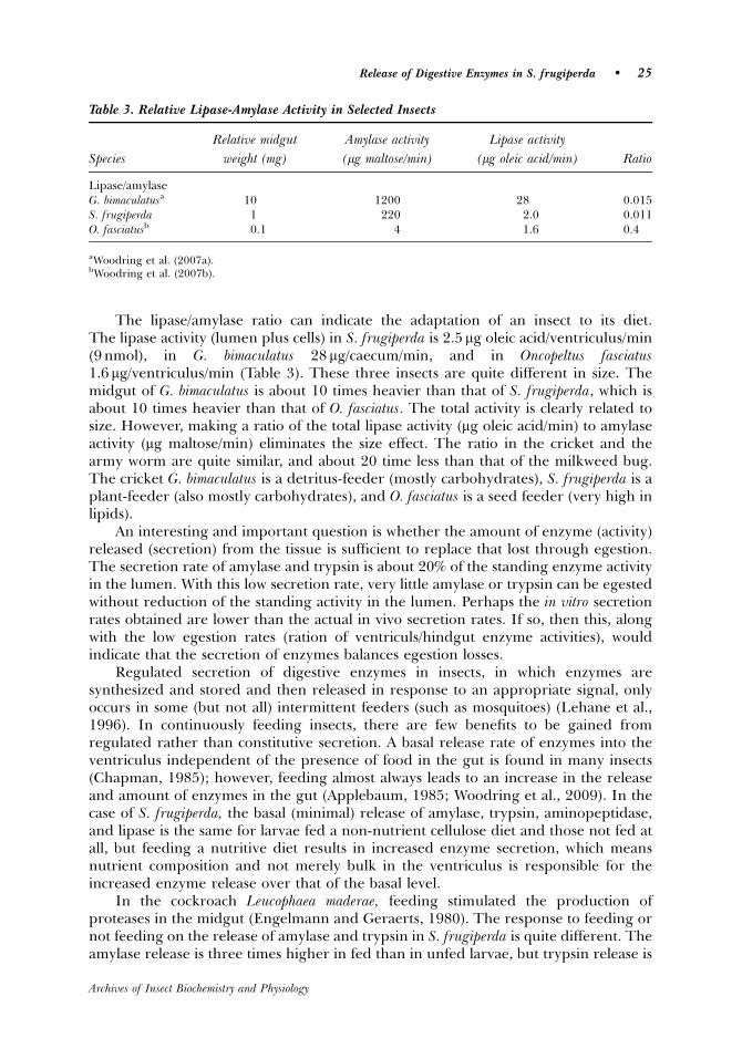
The lipase/amylase ratio can indicate the adaptation of an insect to its diet.The lipase activity (lumen plus cells) in S. frugiperda is 2.5 mg oleic acid/ventriculus/min(9 nmol), in G. bimaculatus 28 mg/caecum/min, and in Oncopeltus fasciatus1.6 mg/ventriculus/min (Table 3). These three insects are quite different in size. Themidgut of G. bimaculatus is about 10 times heavier than that of S. frugiperda, which isabout 10 times heavier than that of O. fasciatus. The total activity is clearly related tosize. However, making a ratio of the total lipase activity (mg oleic acid/min) to amylaseactivity (mg maltose/min) eliminates the size effect. The ratio in the cricket and thearmy worm are quite similar, and about 20 time less than that of the milkweed bug.The cricket G. bimaculatus is a detritus-feeder (mostly carbohydrates), S. frugiperda is aplant-feeder (also mostly carbohydrates), and O. fasciatus is a seed feeder (very high inlipids).
An interesting and important question is whether the amount of enzyme (activity)released (secretion) from the tissue is sufficient to replace that lost through egestion.The secretion rate of amylase and trypsin is about 20% of the standing enzyme activityin the lumen. With this low secretion rate, very little amylase or trypsin can be egestedwithout reduction of the standing activity in the lumen. Perhaps the in vitro secretionrates obtained are lower than the actual in vivo secretion rates. If so, then this, alongwith the low egestion rates (ration of ventriculs/hindgut enzyme activities), wouldindicate that the secretion of enzymes balances egestion losses.
Regulated secretion of digestive enzymes in insects, in which enzymes aresynthesized and stored and then released in response to an appropriate signal, onlyoccurs in some (but not all) intermittent feeders (such as mosquitoes) (Lehane et al.,1996). In continuously feeding insects, there are few benefits to be gained fromregulated rather than constitutive secretion. A basal release rate of enzymes into theventriculus independent of the presence of food in the gut is found in many insects(Chapman, 1985); however, feeding almost always leads to an increase in the releaseand amount of enzymes in the gut (Applebaum, 1985; Woodring et al., 2009). In thecase of S. frugiperda, the basal (minimal) release of amylase, trypsin, aminopeptidase,and lipase is the same for larvae fed a non-nutrient cellulose diet and those not fed atall, but feeding a nutritive diet results in increased enzyme secretion, which meansnutrient composition and not merely bulk in the ventriculus is responsible for theincreased enzyme release over that of the basal level.
In the cockroach Leucophaea maderae, feeding stimulated the production ofproteases in the midgut (Engelmann and Geraerts, 1980). The response to feeding ornot feeding on the release of amylase and trypsin in S. frugiperda is quite different. Theamylase release is three times higher in fed than in unfed larvae, but trypsin release is
Table 3. Relative Lipase-Amylase Activity in Selected Insects
Relative midgut Amylase activity Lipase activity
Species weight (mg) (mg maltose/min) (mg oleic acid/min) Ratio
Lipase/amylaseG. bimaculatusa 10 1200 28 0.015S. frugiperda 1 220 2.0 0.011O. fasciatusb 0.1 4 1.6 0.4
aWoodring et al. (2007a).bWoodring et al. (2007b).
Release of Digestive Enzymes in S. frugiperda � 25
Archives of Insect Biochemistry and Physiology
the same for both fed and unfed larvae. Both enzymes are immediately released aftersynthesis into the lumen (Ferreira et al., 1994a,b), which means that the synthesisrate must be related to nutrients in the gut. But why does amylase release vary with thefeeding and trypsin release does not? A rationale based on food utilization can behypothesized. The primary nutrient in the diet of the caterpillar is carbohydrate, muchof which is converted to lipids and stored in the fat body. The adult moth subsequentlyuses the lipid as a flight fuel (Wheeler, 1989). Reduced dietary carbohydrate simplymeans less lipid reserves, and it would be wasteful to produce large amounts ofamylase when food is in short supply. Therefore, the larvae can afford to adjust theamylase release to the carbohydrate intake. Proteins, on the other hand, are essentialfor larval growth, and egg production in the adult moth depends entirely on proteinsstored in larval fat body (Sorge et al., 2000). The caterpillar, therefore, can not affordto allow any dietary protein to pass undigested through the gut. Therefore, it isadvantageous to maintain a constant level of trypsin release in the event that even asmall amount of protein is ingested. The release of aminopeptidase is twice as high infed than in unfed larvae, which indicates some adjustment to gut peptide levels.
In order to more precisely determine the role of feeding on gut enzymes, theincreased enzyme secretion over basal levels in response to the addition of specificnutrients to incubated flat-sheet preparations (in vitro) of the ventriculus were carriedout. The larvae of the fall armyworm are plant-feeders, and simple sugars andpolysaccharide intake is therefore high. In the current study, glucose and maltoseprovide a strong signal to release amylase, which is the normal way to stimulateamylase release in all animals. There is no evidence in vertebrates or insects of starchor other polysaccharide receptors on gut neuroendocrine cells that induce amylaserelease. The presence of starch in the incubation medium, however, appears to onlymildly stimulate amylase release in S. frugiperda. This anomaly is most likely due to aminimum digestion to maltose or a small amount of sugar in the starch tested, and it isthe maltose or glucose that stimulates amylase release and not the starch.
Similar to other insects, sugars bind to receptors in the apical membrane ofneuroendocrine cells in the epithelial layer of the ventriculus, and induce the release ofneuropeptides at the basal end into the hemolymph. A binding of sugars to the apical(lumen) end of the gut neuroendocrine cells is postulated in G. bimaculatus, whichinduces the release of neuropeptides from the basel end into the hemolymph(Woodring et al., 2009). Specific neuroendocrine cells in the gut epithelium ofcockroaches and locusts release several kinds of neuropeptides that stimulate therelease of amylase (Fuse et al., 1999; Sakai et al., 2004; Hill and Orchard, 2005). The invitro inhibition or stimulation caused by allatostatin and allatotropin, respectively, inthe current study also indicates that the final step in the regulation of amylase releasein response to sugars is mediated by neuropeptides. The trypsin release is not elevatedby feeding in S. frugiperda. Therefore, it is not surprising that bovine serum albumin(BSA) has no effect on in vitro release, and that peptone (a mixture of different sizedpeptides) or a mixture of all 20 amino acids has only a weak effect on the in vitrorelease of trypsin in the incubated flat-sheet preparations. BSA in the bloodsucking fly(Stomoxys), on the other hand, has a strong stimulating effect on the release of trypsin(Blakemore et al., 1995), which makes sense in an animal with sudden and massiveinput of protein.
The allatostatins (AS) and allatropins (AT) are multifunctional neuropeptides andAS occur in virtually all insects investigated (Gade et al., 1997). Depending on thespecies, these neurohormones stimulate and/or inhibit simultaneously juvenile
26 � Archives of Insect Biochemistry and Physiology, January 2010
Archives of Insect Biochemistry and Physiology
hormone synthesis, gut myoactivity, and the release of digestive enzymes (Gade et al.,1997; Lorenz, 2001; Aguilar et al., 2003). In the larvae of the coconut pest Opisinaarenosella, various FMRF amides, leucomyosupressins, stimulate the in vitro release ofamylase and protease (Harshini et al., 2002b). Spofr-AS A5 and AS A6 belong to theallatostatin type-A family. AT and AS bind to receptors on the hemolymph side of theenzyme-producing cells (zymogen cells) and induce either an inhibition or stimulationof the release on the lumen end, for example the stimulation of amylase release inresponse to AS in the cockroach D. punctata (Fuse et al., 1999). Based on gene sequencestudies, Manse-AT and Spofr-AS A5 and AS A6 most likely occur in S. frugiperda(Abdel-latief et al., 2004). Spofr-AS A5 moderately inhibits amylase and trypsin releasein S. frugiperda, but AS A6 does not. Both peptides belong to the FGLa family and areabout the same size, but the amino acids sequences are quite different. The allatotropinManse-AT has quite the opposite effect, stimulating the release of amylase and trypsin.
Why do peptides and amino acids have no effect on the release of trypsin (currentstudy) though AS and AT clearly inhibit or stimulate trypsin release, respectively?A possible explanation is that peptide receptors on the lumen side of theneurohormonal cells are lacking in this species, though the AT and AS receptors onthe hemolymph side of the zymogen cells may be present. Blood-sucking insects, onthe other hand, have the peptide receptors on the lumen side of the neuroendocrinecells, and, therefore, dietary peptides induce trypsin release (Blakemore et al., 1995).
Several myosuppressins of the FLRFamide family also stimulate amylase release,for example in L. migratoria (Hill and Orchard, 2005), the beetle Rhynchophorusferrugineus (Nachman et al., 1997), and in the lepidopteran Opisina arenosella (Harshiniet al., 2002b). Leucokinines, on the other hand, inhibit amylase and general proteaserelease while stimulating midgut motility in Opisina arenosella (Harshini et al., 2002a).AT stimulates foregut muscles in several lepidopterans (Duve et al., 1999; Matthewset al., 2007), and an FGLamide (AS type-A) and a WX6W allatosttain (AS type-B)inhibit gut myocontraction in G. bimaculatus (Lorenz, 2001) and M. sexta (Blackburnet al., 1995). Interestingly, in almost all other Lepidoptera larvae an in vitrocontraction of the foregut is seen. However, in S. littoralis (Matthews et al., 2008) andS. frugiperda (current study), foregut motility is not observed. Therefore, in the currentstudy the ilium was employed to assay myoactivity. In view of the fact that somecaterpillars show both stimulatory and inhibitory responses (above), it is not surprisingthat in the current study an opposing response to the tested ASTs and AT occurs.Manse-AT is myostimulatory and Spofr-AS A5 myoinhibitory. A similar interaction alsois reported in another lepidopteran, Lacanobia oleracea (Matthews et al., 2007). Insummary, Spofr-ATstimulates digestive enzyme release and myoactivity in S. frugiperdaand Spofr-AS A5 inhibits both.
LITERATURE CITED
Abdel-latief M, Meyering-Vos M, Hoffmann KH. 2004. Expression and localization of theSpodoptera frugiperda allatrotropin (Spofr-AT) and allatostatin (Spofr-AS) genes. Arch InsectBiochem Physiol 55:188–199.
Aguilar R, Maestro JR, Vilaplana L, Pascual, N, Piulachs M-D, Belles X. 2003. Allatostatin geneexpression in brain and midgut, and activity of synthetic allatostatins on feeding-relatedprocesses in the cockroach Blattella germanica. Reg Pept 115:171–177.
Release of Digestive Enzymes in S. frugiperda � 27
Archives of Insect Biochemistry and Physiology
Ai H, Kuwasawa K, Yazawa T, Kurokawa M, Shimoda M, Kiguchi K.1995. A physiological salinefor lepidopterous insects: Effects of ionic composition on heart beat and neuromusculartransmission. J Insect Physiol 41:571–580.
Applebaum SW. 1985. Biochemistry of digestion. In: Kerkut GA, Gilbert LI, editors.Comprehensive insect physiology, biochemistry, and pharmacology, vol. 4. London:Pergamon Press. p 279–311.
Blackburn MB, Wagner RM, Kochansky JP, Harrison DJ, Thomas-Lemont P, Raina AK. 1995.The identification of two myoinhibitory peptides, with sequence similarities to the galanins,isolated from the ventral nerve cord of Manduca sexta. Reg Pept 7:213–219.
Blakemore D, Williams S, Lehane MJ. 1995. Protein stimulation of trypsin secretion fromthe opaque zone midgut cells of Stomoxys calcitrans. Comp Biochem Physiol 110B:301–307.
Bolognesi R, Ribeiro AF, Terra WR, Ferreira C. 2001. The peritrophic membrane of Spodopterafrugiperda: secretion of peritrophins and role in immobilization and recycling digestiveenzymes. Arch Insect Biochem Physiol 47:62–75.
Bown DP, Wilkinson HS, Gatehouse JA. 1998. Midgut carboyxypeptidase from Helicoverpaarmigera larvae: enzyme characterisation, cDNA cloning and expression. Insect BiochemMol Biol 28:739–749.
Chapman RF. 1985. Coordination of digestion. In: Kerkut GA, Gilbert LI, editors.Comprehensive insect physiology, biochemistry, and pharmacology, vol. 5. London:Pergamon Press. p 213–240.
Duve H, East PD, Thorpe A. 1999. Regulation of lepidopteran foregut movements byallatostatins and allatotropin from the frontal ganglion. J Comp Neurol 413:405–416.
Engelmann F, Geraerts WPM 1980. The proteases and the protease inhibitor in the midgut ofLeucophaea maderae. J Insect Physiol 26:703–710.
Fan S-G, Wu G-J. 2005. Characteristics of plant proteinase inhibitors and their applications incombating phytophagous insects. Review paper. Bot Bull Acad Sin 46:273–292.
Ferreira C, Capella AN, Sitnik R, Terra WR. 1994a. Properties of the digestive enzymes and thepermeability of the peritrophic membrane of Spodoptera frugiperda larvae. Comp BiochemPhysiol 107A:631–640.
Ferreira C, Capella AN, Sitnik R, Terra WR. 1994b. Digestive enzymes in midgut cell, endo- andectoperitrophic contents, and peritrophic membranes of Spodoptera frugiperda larvae. ArchInsect Biochem Physiol 26:299–313.
Franco OL, Rigden DJ, Melo FR, Grossi-de-Sa MF. 2002. Plant a-amylase inhibitors and theirinteraction with insect a-amylase: Structure, function and crop protection. Review Article.Eur J Biochem 269:397–412.
Fuse M, Zhang JR, Partridge E, Nachman RJ, Orchard I, Bendena WG, Tobe SS. 1999. Effects ofan allatostatin and a myosuppressin on midgut carbohydrate enzyme activity in thecockroach Diploptera punctata. Peptides 20:1285–1293.
Gade G, Hoffmann KH, Spring JH. 1997. Hormonal regulation in insects: Facts, gaps and futuredirections. Physiol Rev 77:963–1032.
Harshini S, Nachmann RJ, Sreekumar S. 2002a. Inhibition of digestive enzyme releaseby neuropeptides in the larvae of Opisina arenosella. Comp Biochem Physiol 132B:353–358.
Harshini S, Nachman RJ, Sreekumar S. 2002b. In vitro release of digestive enyzmes by FMRFamide related neuropeptides and analogs in the lepidopteran insect Opisina arenosella.Peptides 23:1759–1763.
Hill SH, Orchard I. 2005. In vitro analysis of the digestive enzymes amylase and a-glucosidase inthe midgut of Locusta migritoria in response to the myosuppressin, SchistoFLRFamide.J Insect Physiol 51:1–9.
28 � Archives of Insect Biochemistry and Physiology, January 2010
Archives of Insect Biochemistry and Physiology
Johnson KA, Lee MJ, Brough C, Hilder VA, Gatehouse AMR, Gatehouse JA. 1995. Proteaseactivities in the larval midgut of Heliothis virescens: Evidence for trypsin and chymotrypsin-like enzymes. Insect Biochem 25:375–383.
Lehane MJ, Buller HM, Crisanti A. 1996. Mechanisms controlling the synthesis and secretion ofdigestive enzymes in insects. In: Lehane MJ, Billingsley PF, editors. Biology of the insectmidgut. London: Chapman & Hall. p 195–205.
Lenz CJ, Kang J, Rice WC, McIntosh AH, Chippendale GM, Schubert RR. 1991. Digestiveproteinases of larvae of the corn earworm, Heliothis zea: characterization, distribution, anddietary relationships. Arch Insect Biochem Physiol 16:201–212.
Lorenz MW. 2001. Neuropeptides regulating developmental, reproductive, and metabolicevents in crickets: Structures and modes of action. (Review) J Insect Biochem Ser 70:69–93.
Matthews HJ, Audsley N, Weaver RJ. 2007. Interactions between allatostatins and allatotropin onspontaneous contractions of the foregut of larval Lacanobia oleracea. J Insect Physiol 53:75–83.
Matthews HJ, Audsley N, Weaver RJ. 2008. In vitro and in vivo effects of myo-active peptides onlarvae of the tomato moth Lacanobia oleracea and the cotton leaf worm Spodoptera littoralis.Arch Insect Biochem Physiol 69:60–69.
Nachman RJ, Giard W, Favrel P, Suresh T, Sreekumar S, Holman GM. 1997. Insectmyosuppressins and sulfakinins stimulate release of the digestive enzyme a-amylase intwo invertebrates: The scallop Pecten maximus and insect Rhynchophorus ferrugineus. Ann NYAcad Sci 814:335–338.
Nauen R, Sorge D, Sterner A, Borovsky D. 2001. TMOF-like factor controls the biosynthesis ofserine proteases in the larval gut of Heliothis virescens. Arch Insect Biochem Physiol 47:169–180.
Oeh U, Lorenz MW, Dyker H, Losel P, Hoffmann KH. 2000. Interaction between Manduca sextaallatotropin and Manduca sexta allatostatin in the fall armyworm. Insect Biochem. Mol Biol30:719–727.
Ortego F, Novillo C, Castafiera P. 1996. Characterization and distribution of digestive proteasesof the stalk corn borer, Sesamia nonagroides. Arch Insect Biochem Physiol 32:163–180.
Pigott CR, King MS, Ellar DJ. 2008. Investigating the properties of Bacillus thuringiensis cryproteins with novel loop replacements created using combinatorial molecular biology. AppEnviron Micro 74:3497–3511.
Sakai T, Satake H, Minakata H, Takeda M. 2004. Characterization of crustacean cardioactivepeptide as a novel insect midgut factor: Isolation, localization, and stimulation of a-amylaseactivity and gut contraction. Endocrinology 145:5671–5678.
Sorge D, Nauen R, Range S, Hoffmann KH. 2000. Regulation of vitellogenesis in the fallarmyworm, Spodoptera frugiperda. J Insect Physiol 46:969–976.
Terra WR, Ferreira C, DeBianchi AG. 1979. Distribution of digestive enzymes among the endo-and ectoperitrophic spaces and midgut cells of Rhynchosciara americana and its physiologicalsignificance. J Insect Physiol 25:487–494.
Tsuchida K, Wells MA. 1988. Digestion, absorption, transport and storage of fat during the lastlarval stadium of Manduca sexta: Changes in the role of lipophorin in the delivery of dietarylipid to the fat body. Insect Biochem 18:263–268.
Wheeler CH. 1989. Mobilization and transport of fuels to the flight muscles. In: Goldsworthy GJ,Wheeler CH, editors. Insect Flight. Boca Raton, FL: CRC Press. p 273–304.
Woodring J, Hoffmann KH, Lorenz MW. 2007a. Activity, release and flow of digestive enzymesin the cricket, Gryllus bimaculatus. Physiol Ent 32:56–63.
Woodring J, Hoffmann KH, Lorenz MW. 2007b. Feeding, nutrient flow, and digestive enzymerelease in the giant milkweed bug, Oncopeltus fasciatus. Physiol Ent 32:328–335.
Woodring J, Diersch S, Iwalaba D, Hoffmann KH, Meyering-Vos M. 2009 Control of the releaseof digestive enzymes in the caeca of the cricket Gryllus bimaculatus. Physiol Ent 34:144–151.
Release of Digestive Enzymes in S. frugiperda � 29
Archives of Insect Biochemistry and Physiology
A r t i c l e
EXOGENOUS ANDENDOGENOUS PROTEASEINHIBITORS IN THE GUT OF THEFALL ARMYWORM LARVAE,Spodoptera frugiperda
Digali Lwalaba, Sandy Weidlich, Klaus H. Hoffmann,and Joseph WoodringDepartment of Animal Ecology I, University of Bayreuth, Bayreuth,Germany
A dose-dependent inhibition of endogenous trypsin and aminopeptidaseoccurs in the lumen of Spodoptera frugiperda after feeding L6 larvaeexogenous inhibitors soybean trypsin inhibitor (SBTI), tosyl-L-lysinechloromethyl ketone-HCl (TLCK), or bestatin, respectively, for 3 days.TLCK inhibits trypsin in tissue extracts and in secretions more stronglythan SBTI. The aminopeptidase released into the lumen (containing theperitrophic membrane) is strongly inhibited by bestatin, but themembrane-bound enzyme is not. A bound enzyme may be more resistant toan inhibitor than unbound. A cross-class elevation of aminopeptidaseactivity occurs in response to ingested trypsin inhibitor, but there was nocross-class effect of aminopeptidase inhibitor (bestatin) on trypsin activity.An endogenous trypsin and aminopeptidase inhibitor is present in thelumen and ventricular cells. The strength of the endogenous trypsininhibition seems to be in the same range as that resulting from ingestionof the exogenous inhibitor SBTI. In some insect species, considerabletrypsin secretion occurs in unfed as well as in fed animals, andendogenous protease inhibitors might function to protect the ventricularepithelium by inactivation of trypsin when less food is available. �C 2010Wiley Periodicals, Inc.
Keywords: enzyme secretion; lumen contents; epithelium; dose response;trypsin inhibitors; aminopeptidase inhibitors
Correspondence to: Joseph Woodring, Department of Animal Ecology I, University of Bayreuth, 95440Bayreuth, Germany. E-mail: [email protected]
ARCHIVES OF INSECT BIOCHEMISTRY AND PHYSIOLOGY, Vol. 74, No. 2, 114–126 (2010)
Published online in Wiley InterScience (www.interscience.wiley.com).
& 2010 Wiley Periodicals, Inc. DOI: 10.1002/arch.20366
INTRODUCTION
The fall armyworm, Spodoptera frugiperda (J.E. Smith) is an important pest of corn,sorghum, cotton, and alfalfa in the tropical regions of the Western hemisphere. It hasproved to be an ideal model insect for the study of the basic functions of the digestivesystem in defoliating caterpillars. The distribution of digestive enzymes, the role of theperitrophic membrane (PM), and the counter-current flow of enzymes in S. frugiperda iswell documented (Ferreira et al., 1994a,b; Bolognesi et al., 2001). The release of digestiveenzymes in the larvae of the armyworm is regulated by the release of neuropeptides inresponse to the presence of specific nutrients in the lumen (Lwalaba et al., 2009).
Exogenous protease inhibitors in insects are produced by many plants as a defenceagainst feeding (Fan and Wu, 2005), and in general they have their effect in the crop ormidgut where they inhibit the proteases present in the lumen (Johnston et al., 1993;Telang et al., 2005; Duncan et al., 2006; Brioschi et al., 2007) as well as the secretion ofproteases by the epithelium (Lwalaba et al., 2009). These exogenous proteaseinhibitors are Kunitz or Kazal type serine protease inhibitors.
Endogenous serine protease inhibitors are widely found in all tissues of all animals.The serpins, form a very large class of proteases, are mostly intracellular, and undergo aunique change in shape when they inhibit target proteases (Huntington et al., 2000).They regulate such processes as coagulation, inflammation, and immunity. Thus far, allknown insect serpins are involved in insect immunity (Abraham et al., 2005). However,some insect inhibitors involved with immune responses (coagulation, prophenoloxidase activation, etc.) belong to the classical Kunitz and Kazal type inhibitors(Kanost, 1999), that is, they bind to and block access to the protease active site (Kunitz,1948). These endogenous inhibitors are produced by blood cells, silk glands, or ovaries(Nirmala et al., 2001; Simonet et al., 2002; Brillard-Bourdet et al., 2006). Proteaseinhibitors of the Kazal type are found in the salivary gland and saliva of Nauphoeta cinerea,which are particularly effective against the bacterial protease subtilisin. This suggests adefensive mechanism against the masses of bacteria typically found in the cockroach crop,some of which are probably pathogenic (Taranushenko et al., 1909). Inhibitory peptidesin the midgut lumen are less well studied, though trypsin and subtilisin inhibitors aredescribed from whole midgut preparations of the cockroach N. cinerea (Elpidina et al.,2001), and in five additional cockroach species (Vinokurov et al., 2007).
The first aim of the current study is to document the exogenous inhibition of proteaseactivity in the lumen and epithelial cells of the midgut and further document the inhibitionof protease secretion in L6 larvae of S. frugiperda. The cross-class inhibition of exogenousaminopeptidase by trypsin inhibitors (SBTI) and vice versa is likewise described. Themajor aim, however, is to determine if endogenous trypsin and aminopeptidase inhibitorsare produced by the midgut cells and released into the lumen. Finally, the relative strengthof inhibition of exogenous inhibitors is compared to the endogenous inhibitors.
MATERIALS AND METHODS
Rearing Methods, Diets, and Inhibitors
Pupae and eggs of S. frugiperda and an artificial diet (based on bean meal) wereprovided by Bayer Crop Science AG (Monheim, Germany). Larvae were reared at
Protease Inhibition in Spodoptera Gut � 115
Archives of Insect Biochemistry and Physiology
271C and approximately 70% relative humidity under a L16:D8 photoperiod aspreviously described (Oeh et al., 2000). To prevent cannibalism, L4 larvae weremaintained individually in separate compartments of assortment boxes with 40compartments (49� 32� 36 mm per compartment). In order to test the effect of dietand feeding on enzyme and inhibitor activity, newly moulted L6 were provided acellulose diet (30 g cellulose powder 13 g agar in 250 ml water) instead of the controldiet, and others received no food at all. The ventriculus of L6 day-3 larvae was used foranimals fed the control diet and the non-nutritive cellulose diet. Under theseconditions, the gut of fed larvae degenerated early on day 4 and the larvae entered theprepupal stage. The digestive tract of unfed larvae started to regress earlier into theprepupal condition on day 3, therefore the ventriculus of 2-day-old unfed larvae wasused. Soybean trypsin inhibitor (SBTI), the specific trypsin inhibitor (synthetic) tosyl-L-lysine chloromethyl ketone-HCl (TLCK), and the aminopeptidase inhibitor bestatin(from the bacterium Streptomyces olivoreticuli) were dissolved in water and added to afresh 300-mg cube of the Bayer artificial diet or cellulose diet on each of the 3 days ofthe L6 larvae (in vivo). The concentration of SBTI tested was 0.1 to 0.4%, of TLCK was0.025 to 0.15%, and of bestatin 0.03 to 0.10%. TLCK inhibited feeding atconcentrations 40.15%, and were therefore not tested. The diet was generallycompletely or almost totally consumed by L6 larvae in 24 h. In vitro inhibition wastested by adding the inhibitor to 200 ml of an extract of the ventriculus.
Ventriculus Dissection: Source of Endogenous Enzymes
Larvae of S. frugiperda were anesthetized on ice, slit open, and the entire ventriculusremoved. A crop and caeca are lacking, and functionally the entire midgut consists ofthe ventriculus. The standing enzyme activity in the tissue homogenate and in thelumen content, which included the exo- and endoperitrophic space plus theperitrophic membrane (PM), was measured. The total lumen content of oneventriculus was placed in 300 ml Spodoptera Ringer (per Liter 0.7 g NaCl, 2.0 g KCl,0.25 g NaHCO3.2 H2O, 1.3 g CaCl2.2 H2O, 4.4 g MgCl2.7 H2O, 1.0 g Hepes at pH 7.0).Two ventriculi were thoroughly rinsed in Ringer and placed in 300 ml Ringer, lightlysonicated (Bronson Sonifier 250, 2–3 sec at the lowest setting) and centrifuged. Twofreshly dissected ventriculi were slit open and rinsed with low-glucose Ringer (LGR)(10 mg glucose/100 ml Ringer), a concentration adapted from Fuse et al. (1999) forincubation of gut tissues of the cockroach Diploptera punctata, and placed in 300 ml freshLGR, and incubated for 30 min at 301C without shaking. The incubated tissue wasdiscarded, and the remaining incubation medium was used to measure enzyme release(secretion). Thus, there were three sources of enzymes: lumen content, tissuehomogenate, and the secretion in the incubation medium.
Endogenous Enzyme Assays
For the determination of protease activity 100 ml of the lumen contents, tissuehomogenate, or the incubation medium was used for the endogenous trypsin andaminopeptidase assay. The trypsin and aminopeptidase activities were linear from 0 to5 min at 251C at a pH of 8.0 (data not shown). For both assays, the release of NA(nitroaniline) (dAbs/min) from the synthetic substrates a-N-benzoyl-DL-arginine-p-NA(BApNA) and L-leucine-p-NA (LpNA), respectively, was measured at 405 nm. A 100-mlsample was added to 700 ml 1 mM BApNA (4.3 mg/10 ml Tris buffer) or to 700 ml 1 mMLpNA (2.87 mg/10 ml). One unit of activity was the amount of enzyme required to split
116 � Archives of Insect Biochemistry and Physiology, June 2010
Archives of Insect Biochemistry and Physiology
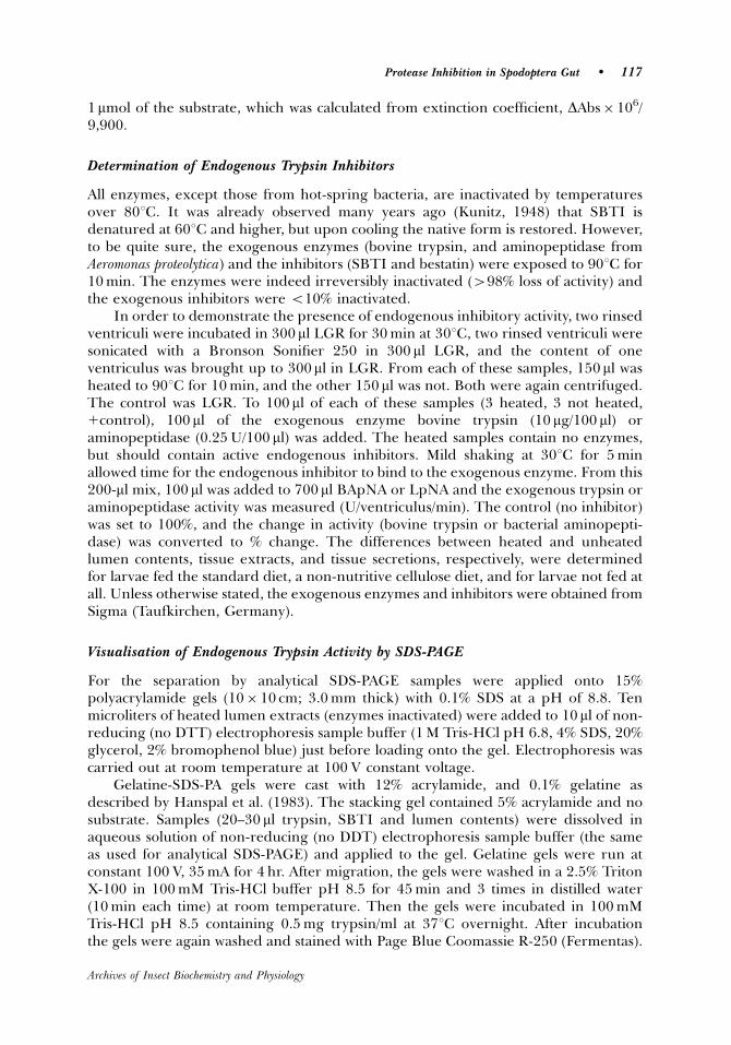
1mmol of the substrate, which was calculated from extinction coefficient, DAbs� 106/9,900.
Determination of Endogenous Trypsin Inhibitors
All enzymes, except those from hot-spring bacteria, are inactivated by temperaturesover 801C. It was already observed many years ago (Kunitz, 1948) that SBTI isdenatured at 601C and higher, but upon cooling the native form is restored. However,to be quite sure, the exogenous enzymes (bovine trypsin, and aminopeptidase fromAeromonas proteolytica) and the inhibitors (SBTI and bestatin) were exposed to 901C for10 min. The enzymes were indeed irreversibly inactivated (498% loss of activity) andthe exogenous inhibitors were o10% inactivated.
In order to demonstrate the presence of endogenous inhibitory activity, two rinsedventriculi were incubated in 300 ml LGR for 30 min at 301C, two rinsed ventriculi weresonicated with a Bronson Sonifier 250 in 300 ml LGR, and the content of oneventriculus was brought up to 300ml in LGR. From each of these samples, 150 ml washeated to 901C for 10 min, and the other 150 ml was not. Both were again centrifuged.The control was LGR. To 100 ml of each of these samples (3 heated, 3 not heated,1control), 100 ml of the exogenous enzyme bovine trypsin (10 mg/100 ml) oraminopeptidase (0.25 U/100 ml) was added. The heated samples contain no enzymes,but should contain active endogenous inhibitors. Mild shaking at 301C for 5 minallowed time for the endogenous inhibitor to bind to the exogenous enzyme. From this200-ml mix, 100 ml was added to 700 ml BApNA or LpNA and the exogenous trypsin oraminopeptidase activity was measured (U/ventriculus/min). The control (no inhibitor)was set to 100%, and the change in activity (bovine trypsin or bacterial aminopepti-dase) was converted to % change. The differences between heated and unheatedlumen contents, tissue extracts, and tissue secretions, respectively, were determinedfor larvae fed the standard diet, a non-nutritive cellulose diet, and for larvae not fed atall. Unless otherwise stated, the exogenous enzymes and inhibitors were obtained fromSigma (Taufkirchen, Germany).
Visualisation of Endogenous Trypsin Activity by SDS-PAGE
For the separation by analytical SDS-PAGE samples were applied onto 15%polyacrylamide gels (10� 10 cm; 3.0 mm thick) with 0.1% SDS at a pH of 8.8. Tenmicroliters of heated lumen extracts (enzymes inactivated) were added to 10ml of non-reducing (no DTT) electrophoresis sample buffer (1 M Tris-HCl pH 6.8, 4% SDS, 20%glycerol, 2% bromophenol blue) just before loading onto the gel. Electrophoresis wascarried out at room temperature at 100 V constant voltage.
Gelatine-SDS-PA gels were cast with 12% acrylamide, and 0.1% gelatine asdescribed by Hanspal et al. (1983). The stacking gel contained 5% acrylamide and nosubstrate. Samples (20–30 ml trypsin, SBTI and lumen contents) were dissolved inaqueous solution of non-reducing (no DDT) electrophoresis sample buffer (the sameas used for analytical SDS-PAGE) and applied to the gel. Gelatine gels were run atconstant 100 V, 35 mA for 4 hr. After migration, the gels were washed in a 2.5% TritonX-100 in 100 mM Tris-HCl buffer pH 8.5 for 45 min and 3 times in distilled water(10 min each time) at room temperature. Then the gels were incubated in 100 mMTris-HCl pH 8.5 containing 0.5 mg trypsin/ml at 371C overnight. After incubationthe gels were again washed and stained with Page Blue Coomassie R-250 (Fermentas).
Protease Inhibition in Spodoptera Gut � 117
Archives of Insect Biochemistry and Physiology
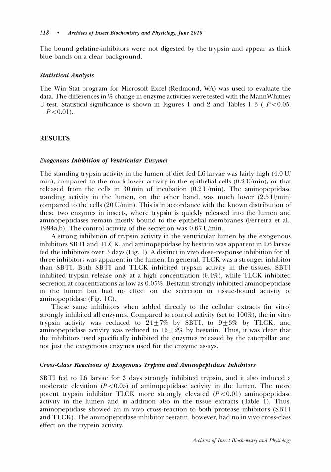
The bound gelatine-inhibitors were not digested by the trypsin and appear as thickblue bands on a clear background.
Statistical Analysis
The Win Stat program for Microsoft Excel (Redmond, WA) was used to evaluate thedata. The differences in % change in enzyme activities were tested with the MannWhitneyU-test. Statistical significance is shown in Figures 1 and 2 and Tables 1–3 (�Po0.05,��Po0.01).
RESULTS
Exogenous Inhibition of Ventricular Enzymes
The standing trypsin activity in the lumen of diet fed L6 larvae was fairly high (4.0 U/min), compared to the much lower activity in the epithelial cells (0.2 U/min), or thatreleased from the cells in 30 min of incubation (0.2 U/min). The aminopeptidasestanding activity in the lumen, on the other hand, was much lower (2.5 U/min)compared to the cells (20 U/min). This is in accordance with the known distribution ofthese two enzymes in insects, where trypsin is quickly released into the lumen andaminopeptidases remain mostly bound to the epithelial membranes (Ferreira et al.,1994a,b). The control activity of the secretion was 0.67 U/min.
A strong inhibition of trypsin activity in the ventricular lumen by the exogenousinhibitors SBTI and TLCK, and aminopeptidase by bestatin was apparent in L6 larvaefed the inhibitors over 3 days (Fig. 1). A distinct in vivo dose-response inhibition for allthree inhibitors was apparent in the lumen. In general, TLCK was a stronger inhibitorthan SBTI. Both SBTI and TLCK inhibited trypsin activity in the tissues. SBTIinhibited trypsin release only at a high concentration (0.4%), while TLCK inhibitedsecretion at concentrations as low as 0.05%. Bestatin strongly inhibited aminopeptidasein the lumen but had no effect on the secretion or tissue-bound activity ofaminopeptidase (Fig. 1C).
These same inhibitors when added directly to the cellular extracts (in vitro)strongly inhibited all enzymes. Compared to control activity (set to 100%), the in vitrotrypsin activity was reduced to 2477% by SBTI, to 973% by TLCK, andaminopeptidase activity was reduced to 1572% by bestatin. Thus, it was clear thatthe inhibitors used specifically inhibited the enzymes released by the caterpillar andnot just the exogenous enzymes used for the enzyme assays.
Cross-Class Reactions of Exogenous Trypsin and Aminopeptidase Inhibitors
SBTI fed to L6 larvae for 3 days strongly inhibited trypsin, and it also induced amoderate elevation (Po0.05) of aminopeptidase activity in the lumen. The morepotent trypsin inhibitor TLCK more strongly elevated (Po0.01) aminopeptidaseactivity in the lumen and in addition also in the tissue extracts (Table 1). Thus,aminopeptidase showed an in vivo cross-reaction to both protease inhibitors (SBTIand TLCK). The aminopeptidase inhibitor bestatin, however, had no in vivo cross-classeffect on the trypsin activity.
118 � Archives of Insect Biochemistry and Physiology, June 2010
Archives of Insect Biochemistry and Physiology
Endogenous Inhibition of Ventricular Enzymes
The endogenous inhibitors in the heated tissue extracts in diet-fed L6 larvae showedless trypsin activity (78%), and much less aminopeptidase activity (47%) compared to
0
20
40
60
80
100
120A
B
C
0.10 0.15 0.20 0.40
% SBTI in diet
% T
ryp
sin
act
ivit
y
Secretion Lumen Tissue
*** * ******
*
0
20
40
60
80
100
120
0.025 0.05 0.10 0.15
% TLCK in diet
% T
LC
K a
ctiv
ity
Secretion Lumen Tissue
* **
****
* *** * *
40
50
60
70
80
90
100
110
120
0.033 0.05 0.066 0.10% Bastatin (w/w) in diet
% A
min
op
epti
das
e ac
tivi
ty
Secretion Lumen Tissue
*
**
****
Figure 1. Effects of exogenous inhibitor concentration on the enzyme activity in the tissue secretions, tissuehomogenates, and lumen content of the ventricle of L6 (day 3) S. frugiperda larvae fed for 3 days on the standarddiet containing SBTI, TLCK, or bestatin. A: Soybean trypsin inhibitor (SBTI), a serine protease inhibitor.B: Tosyl-L-lysine chloromethyl ketone-HCl (TLCK), a trypsin-specific inhibitor. C: Bestatin, an aminopeptidaseinhibitor. The control trypsin activity in the lumen of diet-fed L6 larvae (no inhibitor) activity was 4.070.5 U/ventriculus/min, in the cells 0.2 U/vent/min, and in the secretion (30 min) 0.2 U/min. The aminopeptidaseactivity in the lumen was 2.5 U/ventriculus/min, in the cells 20.0 U/ventriculus/min, and in the secretions 0.67 U/ventriculus/min. The response to the inhibitors was converted to % change in activity compared to controlactivity (set to 100%). Standard error of mean is given as vertical bars. t-test: �Po0.05, ��Po0.01.
Protease Inhibition in Spodoptera Gut � 119
Archives of Insect Biochemistry and Physiology
controls (100%) (Tables 2 and 3), which demonstrated the presence of endogenousinhibitors in the ventricular cells. The unheated tissue extracts resulted in moretrypsin (143%), and very much more aminopeptidase activity (332%), a result of thecombination of active endogenous enzymes plus the added exogenous enzymes. Sincethe tissues were thoroughly rinsed, exogenous inhibitors associated with the food wereremoved, and only endogenous inhibitors were present in the heated tissue extract.
1 2 3 4 1 2 3 4
A B
Figure 2. A: Analytical SDS-PAGE. Samples were applied onto 15% polyacrylamide gels with 0.1% SDS at apH of 8.8. Extracts (10ml) were added to 10ml of non-reducing (no DTT) electrophoresis sample buffer(62.5 mM Tris-HCl, pH 6.8, 4% SDS, 20% glycerol, 2% bromophenol blue) without heating just beforeloading onto the gel. Lane 1: Lumen contents (heated) of diet-fed larvae; lane 2: unconcentrated lumencontents of cellulose-fed larvae, lane 3: 3� concentrated lumen contents (heated) of cellulose-fed larvae;lane 4: Molecular weight standards (11,17,18.36, 55, 72, 95, 130, 250 kDa). Inhibitor marked by black arrow.B: Gelatin-SDS-PAGE. Gels were cast with 12% acrylamide and 0.1% gelatine. The stacking gel contained 5%acrylamid and no substrate. Samples (trypsin, SBTI, and lumen contents; 20–30ml) were dissolved inelectrophoresis sample buffer (the same as used for analytical SDS-PAGE) and applied to the gel withoutheating. After migration, the gels were washed in a 2.5% Triton X-100 in 100 mM Tris-HCl buffer, pH 8.5, for45 min and 3 times in distilled water. Then the gels were incubated in 100 mM Tris-HCl, pH 8.5, containing0.5 mg trypsin/ml at 371C overnight. After incubation, the gels were again washed and stained with PageBlue Coomassie R-250. The bound gelatine-inhibitors were not digested by the trypsin and appear as thickblue bands on a clear background (white arrow). Lane 1: lumen contents of diet-fed larvae; lane 2: lumencontents of cellulose fed larvae; lane 3: 5 mg bovine trypsin; lane 4: 25mg SBTI. Arrow indicates endogenoustrypsin inhibitor.
Table 1. Cross-Class Reactions of Endoprotease Inhibitors and Exopreotease Inhibitors�
Dose of inhibitor Enzyme Lumen 7SEM Tissue 7SEM Secretion 7SEM
0.20% Bestatin Adase 57�� 5.5 97 6 96 60.10% SBTI Adase 142� 13 113 3 136 130.10% TLCK Adase 188� 17 120� 3 115 100.10% SBTI Trypsin 69�� 5.2 67�� 5 98 12
Trypsin 96 7.6 99 7 101 7
�SBTI and TLCK are endoprotease (trypsin) inhibitors and bestatin is an aminopeptidase (Adase) inhibitor. Thedose is givien as % inhibitor in the diet (w/w). L6 larvae were fed the inhibitor for 3 days, and enzyme activity wastested on day 3. The activity of the Adase and trypsin are given as % change in untreated controls set to 100%. Thecontrol activities (U/min) are as given in Figure 1. N 5 10, �Po0.05 and ��Po0.01 Student’s t-test.
120 � Archives of Insect Biochemistry and Physiology, June 2010
Archives of Insect Biochemistry and Physiology
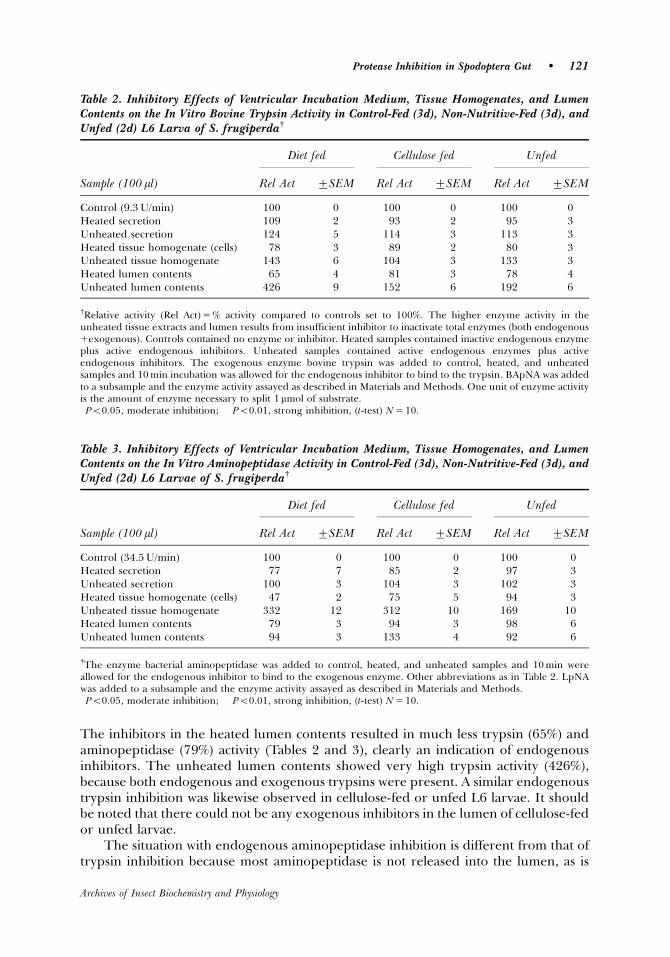
The inhibitors in the heated lumen contents resulted in much less trypsin (65%) andaminopeptidase (79%) activity (Tables 2 and 3), clearly an indication of endogenousinhibitors. The unheated lumen contents showed very high trypsin activity (426%),because both endogenous and exogenous trypsins were present. A similar endogenoustrypsin inhibition was likewise observed in cellulose-fed or unfed L6 larvae. It shouldbe noted that there could not be any exogenous inhibitors in the lumen of cellulose-fedor unfed larvae.
The situation with endogenous aminopeptidase inhibition is different from that oftrypsin inhibition because most aminopeptidase is not released into the lumen, as is
Table 3. Inhibitory Effects of Ventricular Incubation Medium, Tissue Homogenates, and LumenContents on the In Vitro Aminopeptidase Activity in Control-Fed (3d), Non-Nutritive-Fed (3d), andUnfed (2d) L6 Larvae of S. frugiperday
Diet fed Cellulose fed Unfed
Sample (100 ml) Rel Act 7SEM Rel Act 7SEM Rel Act 7SEM
Control (34.5 U/min) 100 0 100 0 100 0Heated secretion 77 7 85 2 97 3Unheated secretion 100 3 104 3 102 3Heated tissue homogenate (cells) 47�� 2 75� 5 94� 3Unheated tissue homogenate 332 12 312 10 169 10Heated lumen contents 79� 3 94 3 98 6Unheated lumen contents 94 3 133 4 92 6
yThe enzyme bacterial aminopeptidase was added to control, heated, and unheated samples and 10 min wereallowed for the endogenous inhibitor to bind to the exogenous enzyme. Other abbreviations as in Table 2. LpNAwas added to a subsample and the enzyme activity assayed as described in Materials and Methods.�Po0.05, moderate inhibition; ��Po0.01, strong inhibition, (t-test) N 5 10.
Table 2. Inhibitory Effects of Ventricular Incubation Medium, Tissue Homogenates, and LumenContents on the In Vitro Bovine Trypsin Activity in Control-Fed (3d), Non-Nutritive-Fed (3d), andUnfed (2d) L6 Larva of S. frugiperday
Diet fed Cellulose fed Unfed
Sample (100 ml) Rel Act 7SEM Rel Act 7SEM Rel Act 7SEM
Control (9.3 U/min) 100 0 100 0 100 0Heated secretion 109 2 93 2 95 3Unheated secretion 124 5 114 3 113 3Heated tissue homogenate (cells) 78�� 3 89� 2 80� 3Unheated tissue homogenate 143 6 104 3 133 3Heated lumen contents 65�� 4 81� 3 78� 4Unheated lumen contents 426 9 152 6 192 6
yRelative activity (Rel Act) 5 % activity compared to controls set to 100%. The higher enzyme activity in theunheated tissue extracts and lumen results from insufficient inhibitor to inactivate total enzymes (both endogenous1exogenous). Controls contained no enzyme or inhibitor. Heated samples contained inactive endogenous enzymeplus active endogenous inhibitors. Unheated samples contained active endogenous enzymes plus activeendogenous inhibitors. The exogenous enzyme bovine trypsin was added to control, heated, and unheatedsamples and 10 min incubation was allowed for the endogenous inhibitor to bind to the trypsin. BApNA was addedto a subsample and the enzyme activity assayed as described in Materials and Methods. One unit of enzyme activityis the amount of enzyme necessary to split 1 mmol of substrate.�Po0.05, moderate inhibition; ��Po0.01, strong inhibition, (t-test) N 5 10.
Protease Inhibition in Spodoptera Gut � 121
Archives of Insect Biochemistry and Physiology
trypsin, but rather remains bound to the cell membranes (Ferreira et al., 1994a). Themembrane-bound aminopeptidases of heated tissue extracts were inactivated, and theendogenous aminopeptidase inhibitors present led to greatly reduced (Po0.01)activity in the exogenous aminopeptidase (Table 3). The PM is part of the lumencontents, and it contains some aminopeptidase (Ferreira et al., 1994a), and a moderateamount (Po0.05) of endogenous aminopeptidase inhibition was found in heatedlumen contents. There was a moderate inhibition of aminopeptidase activity in thetissues of cellulose-fed larvae, but no inhibition in starved larvae.
The analytical SDS-PAGE indicated a peptide present in the heated lumencontents of diet-fed and cellulose-fed L6 larvae, which has a molecular weight in therange of 13–15 kDa (dark arrow, Fig. 2A). All enzymes were inactivated by heating10 min at 901C and do not appear in the gel. The zymogram (gelatine-SDS-PAGE) ofthe endogenous trypsin inhibitor in the heated lumen sample indicated that theinhibitor bound to the gelatine (white arrow, Fig. 2B) and prevented its digestion bythe bovine trypsin incubation (see Materials and Methods).
Comparison of Exogenous and Endogenous Inhibitors
It is difficult to compare the effectiveness of exo- and endogenous protease inhibitors.The inhibition of the exogenous inhibitors (ingested plant material) was measuredas the decrease in the endogenous trypsin and aminopeptidase activity in theventriculus. The inhibition of the endogenous inhibitors (released from ventricularcells) was measured as the decrease in digestion of exogenous trypsin (bovine) andaminopeptidase (bacterial) by lumen contents and tissue homogenates. However, anapproximate comparison can be made. Endogenous luminal trypsin was inhibited to65% (Fig. 1, with 0.2% SBTI, and the activity was reduced from 4.0 U/min to 2.6 U/min). Exogenous trypsin was inhibited in the lumen also to 65% (Table 2) and theactivity was reduced from 9.3 U/min to 6.0 U/min.
DISCUSSION
A wide variety of enzyme inhibitors occur in plants (see review by Fan and Wu, 2005),and an inhibition of digestive enzymes in insects can result from ingestion of plantscontaining one or more of these inhibitors. Protease inhibition by exogenous inhibitors(plant origin) occurs in S. frugiperda (Alfonso et al., 1997; Brioschi et al., 2007, currentstudy) as well in many other lepidopteran larvae (Christeller et al., 1992; Johnstonet al., 1993; Telang et al., 2005). In the current study, inhibition of digestive enzymesecretion by the gut epithelium in addition to inhibition of the enzymes alreadypresent in the lumen was demonstrated. This is the first report of the direct inhibitionof enzyme release.
With regards to exogenous inhibitors in the lumen, the more specific trypsininhibitor TLCK (a synthetic product) appears to be a stronger inhibitor of trypsin thanthe general serine protease inhibitor SBTI, because about three times more SBTI isrequired to cause a similar inhibiton of trypsin as TLCK. Both SBTI and TLCKinhibited activity of trypsin in cell homogenates and in the secretions. Inhibition of anenzyme secretion implies either a direct effect (entry into the cell) or indirect effect(docking on receptors), which then has an effect on the cell production (synthesis) and/or release of the enzyme. This is not surprising, because there is already considerableevidence for the effect of inhibitors on increased or altered trypsin variants in the gut
122 � Archives of Insect Biochemistry and Physiology, June 2010
Archives of Insect Biochemistry and Physiology
epithelial cells of S. frugiperda and Heliothis virescens in response to acute feeding ofSBTI (Lara et al., 2000; Brito et al., 2001; Brioschi et al., 2007).
Aminopeptidase is mostly bound to cell membranes, and only about 17% occurs inthe PM of S. frugiperda (Ferreira et al., 1994b), which is included in the lumen contentin the current study. The aminopeptidase activity of the epithelium (membranebound) was not inhibited by the exogenous inhibitor bestatin, but the small amount(17%) associated with the PM in the lumen was strongly inhibited. It is possible thatbeing bound to the cell membrane somehow protects the aminopeptidase.
Cross-class inhibition of plant protease inhibitors, as reported in the current study,occurs fairly frequently in insects. This indicates an altered synthesis of one enzyme inresponse to a reduction of a digestion product of a different enzyme. In Teleogrylluscommodus, SBTI also increases aminopeptidase activity (Burgess et al., 1994), as does abarley trypsin inhibitor in S. exigua (Lara et al., 2000). The barley inhibitor inS. frugiperda inhibits both trypsin, but has no effect on chymotrypsin, elastase, oraminopeptidase (Alfonso et al., 1997). A molecular basis of cross-class effects is seen inHelicoverpa armigera, where specific inhibitors increase chymotrypsin mRNA buttrypsin mRNA levels decrease (Gatehouse et al., 1997), or in Trichoplusia ni, where aprocarboxypeptidase is activated by trypsin (Wang et al., 2004).
The many endogenous proteases associated with various insect tissues (blood,ovaries, and fat body) are almost all involved with immunity or their function isunknown (Hamdaoui et al., 1998; Kanost, 1999; Simonet et al., 2002). The firstdemonstration of endogenous protease inhibitors specifically in the gut was describedin tissue extracts and lumen contents of the midgut of the cockroach Leucophaeamaderae (Engelmann and Geraerts, 1980). They used boiled extracts to inactivateendogenous enzymes, as in the current study, and they found up to 80% proteaseinhibition in the posterior half of the midgut. Recently, endogenous protease inhibitorshave been described in the digestive system of cockroaches (Taranushenko et al., 1909;Elpidina et al., 2001) using whole gut extracts (epithelium plus lumen contents).Inhibition of trypsin and aminopeptidase occurs in the tissue extracts and in the lumenof S. frugiperda (current study), which is the first report of endogenous digestiveenzyme inhibitors in a lepidopteran species.
Several functions of the endogenous protease inhibitors in the digestive tract ofcockroaches have been suggested. Elpidina et al. (2001) suggests that the endogenousprotease inhibitors could protect amylases in the anterior midgut of N. cinerea, whichmight be true in some insects. However, divalent ions, especially Ca, seem to confer ageneral resistance of a-amylase to proteolytic degradation in vertebrates (Stein andFischer, 1958), and this probably holds true in S. frugiperda. Other authors suggest thatendogenous protease inhibitors might be important as protection against fungi andbacteria that employ subtilisins to attack insect tissues (Taranushenko et al., 2009;Vinokurov et al., 2007). This seems to be especially appropriate in cockroaches, wherethe salivary inhibitors could protect the insect against the massive amounts of bacteriathat are stored over long periods in the crop (Taranushenko et al., 2009).
Another possible function of endogenous protease inhibitors in some insects,which has not been considered, might be protection of the epithelium when dietaryprotein is scarce or lacking. In vertebrates, trypsinogen is stored in zymogen cells ofthe pancreas and secreted in response to food and the release of cholycystokinin.When protein is lacking, trypsinogen is not secreted. In S. frugiperda and inG. bimaculatus, very little trypsin is stored in the epithelial cells, but instead it isimmediately and continuously released. In unfed individuals, the basal trypsin activity
Protease Inhibition in Spodoptera Gut � 123
Archives of Insect Biochemistry and Physiology
in the midgut is 1/3 to 1/2 of that present in fed animals (Woodring et al., 2009, currentstudy). The constant release of active trypsin may perhaps be normal in continuousfeeding insects (such as crickets and caterpillars), but this could pose a threat to theepithelium when food intake is restricted. An endogenous trypsin inhibitor wouldinactivate the trypsin when too little protein is present in the ventriculus.
In conclusion, the digestive enzymes of S. frugiperda are susceptible to bothexogenous and endogenous inhibitors. Acute feeding of exogenous inhibitors (SBTIand TLCK) to L6 larvae reduces trypsin activity in the lumen, in the cells, and in thecell secretions. The exogenous aminopeptidase inhibitor bestatin inhibits aminopepti-dase released into the lumen, but has no effect on aminopeptidase bound to theepithelial membranes. This would protect most of the aminopeptidase from inhibitors.A cross-class stimulation of aminopeptidase activity in response to exogenous trypsininhibitor is observed but not the reverse. Endogenous trypsin and aminopeptidase arepresent in the lumen and tissue extracts of the ventriculus. Since active trypsin issecreted also in unfed larvae, it is suggested that the endogenous protease inhibitorsmight function to protect the exposed epithelium when food is not present in thelumen.
ACKNOWLEDGMENTS
A scholarship to D. Lwalaba from the German Academic Exchange Program (DAAD) isgratefully acknowledged. We also thank Sandy Weidlich, Department of AnimalEcology I, University of Bayreuth, for running the SDS-PAGE gels.
LITERATURE CITED
Abraham EG, Pinto SB, Ghosh A, Vanlandingham DL, Budd A, Higgs S, Kafatos FC, Jacobs-Lorena M, Michel K. 2005. An immune-responsive serpin, SRPN&, mediates mosquitodefence against malaria parasites. Proc Nat Acad Sci USA 105:16327–16332.
Alfonso J, Ortego F, Sanchez-Monge R, Garcia-Casado G, Pujol M, Castanera P, Salcedo G. 1997.Wheat and barley inhibitors active towards a-amylase and trypsin-like activities fromSpodoptera frugiperda. J Chem Ecol 23:1729–1741.
Bolognesi R, Ribeiro AF, Terra WR, Ferreira C. 2001. The peritrophic membrane of Spodopterafrugiperda: secretion of peritrophin and role in immobilization and recycling digestiveenzymes. Arch Insect Biochem Physiol 47:62–75.
Brioschi D, Nadalini LD, Bengtson MH, Sogayar MC, Moura DS, Silva-Filho MC. 2007. Generalup regulation of Spodoptera frugiperda trypsins and chymotrypsin allows its adaptation tosoybean proteinase inhibitor. Insect Biochem Mol Biol 37:1283–1290.
Brillard-Bourdet M, Hamadoui A, Hajjar E, Boudier C, Reuter N, Ehret-Sabatier L, Bieth JG,Gauthier F. 2006. A novel locust (Schistocerca gregaria) serine protease inhibitor with a highaffinity for neutrophil elastase. Biochem J 400:467–476.
Brito LO, Lopes AR, Parra JRP, Terra WR, Silva-Filho MC. 2001. Adaptation of tobaccobudworm Heliothis virescens to proteinase inhibitors may be mediated by the synthesis of newproteinases. Comp Biochem Physiol 128B:365–375.
Burgess EPJ, Main CA, Stevens PS, Christeller JT, Gatehouse AMR, Laing WA. 1994. Effects ofprotease inhibitor concentration and combinations on the survival, growth and gut enzymeactivities of the black field cricket, Teleogryllus commodus. J Insect Physiol 40:803–811.
124 � Archives of Insect Biochemistry and Physiology, June 2010
Archives of Insect Biochemistry and Physiology
Christeller JT, Laing WA, Markwick NP, Burgess EPJ. 1992. Midgut protease activities in 12phytophagous lepidopteran larvae: dietary and protease inhibitor interactions. InsectBiochem Mol Biol 22:735–746.
Duncan A-M, Ren H, Bound F, Tully J, Chandler DS, Sandeman RM. 2006. Assessment of novelinhibitors of Helicoverpa aminopeptidases as anti-insect agents. Pest Manag Sci 62:1098–1108.
Elpidina EN, Vinokurov KS, Rudenskaya YA, Dunaevsky YE, Zhuzhikov DP. 2001. Proteinaseinhibitors in Nauphoeta cinerea midgut. Arch Insect Biochem Physiol 48:217–222.
Engelmann F, Geraerts WPM. 1980. The proteases and the protease inhibitors in the midgut ofLeucophaea maderae. J Insect Physiol 26:703–710.
Fan S-G, Wu G-J. 2005. Characteristics of plant proteinase inhibitors and their applications incombating phytophagous insects. Bot Bull Acad Sinica 46:273–292.
Ferreira C, Capella AN, Sitnick R, Terra WR. 1994a. Properties of the digestive enzymes and thepermeability of the peritrophic membrane of Spodoptera frugiperda larvae. Comp BiochemPhysiol 107A:631–640.
Ferreira C, Capella AN, Sitnick R, Terra WR. 1994b. Digestive enzymes in midgut cells, endo-and ectoperitroph contents and peritrophic membranes of Spodoptera frugiperda larvae. ArchInsect Biochem Physiol 26:299–313.
Fuse M, Zhang JR, Partridge E, Nachman RJ, Orchard I, Bendena WG, Tobe SS. 1999. Effects ofan allatostatin and a myosuppressin on midgut carbohydrate enzyme activity inthecockroach Diploptera punctata. Peptides 20:1285–1293.
Gatehouse LN, Shannon AL, Burgess EPJ, Christeller JT. 1997. Characterization of majormidgut proteinase cDNAs from Helicoverpa armigera larvae and changes in gene expressionin response to four proteinase inhibitors in the diet. Insect Biochem Mol Biol 27:929–944.
Hamadaoui A, Wataleb S, Devreese D, Chiou S-J, Broeck JV, Beeumen JV, De Loof A, Schoofs L.1998. Purification and characterization of a group of five novel peptide serine proteaseinhibitors from ovaries of the desert locust, Schistocerca gregaria. FEBS Letters 422:74–78.
Hanspal JS, Bushell GR, Ghosh P. 1983. Detection of protease inhibitors using substrate-containing sodium dodecyl sulphate-polyacrylamide gel electrophoresis. Anal Biochem132:288–293.
Huntington J, Read R, Carrell R. 2000. Structure of a serpin-protease complex shows inhibitionby deformation. Nature 407:923–926.
Johnston KA, Gatehouse JA, Anstee JH. 1993. Effects of soybean protease inhibitors on thegrowth and development of larval Helicoverpa armigera. J Insect Physiol 39:657–664.
Kanost MR. 1999. Serine proteinase inhibitors in arthropod immunity. Dev Comp Immunol23:291–301.
Kunitz M. 1948. The kinetics and thermodynamics of reversible denaturation of crystallinesoybean trypsin inhibitor. J Gen Physiol 29:241–250.
Lara P, Ortego F, Gonzylez-Hidalgo E, Castnera P, Carbonero P, Diaz I. 2000. Adaptation ofSpodoptera exigua to barley trypsin inhibitor BTI-CMe expressed in transgenic tobacco.Transgenic Res 9:169–178.
Lwalaba D, Hoffmann KH, Woodring J. 2009. Control of the release of digestive enzymes in thelarvae of the fall armyworm, Spodoptera frugiperda. Arch Insect Biochem Physiol 72:1–16.
Nirmala X, Kodrik D, Zurovec M, Sehnal F. 2001. Insect silk contains both a Kunitz-type and aunique Kazal-type proteinase inhibitor. Eur J Biochem 268:2064–2073.
Oeh U, Lorenz MW, Dyker H, Losel P, Hoffmann KH. 2000. Interactions between Manduca sextaallatropin and Manduca sexta allatostatin in the fall armyworm. Insect Biochem Mol Biol30:719–727.
Simonet G, Claeys I, Vanden Broeck J. 2002. Structural and functional properties of a novel serineprotease inhibiting peptide family in arthropods. Comp Biochem Physiol 132B:247–255.
Protease Inhibition in Spodoptera Gut � 125
Archives of Insect Biochemistry and Physiology
Stein EA, Fischer EH. 1958. The resistance of a-amylase towards proteolytic attack. J Biol Chem232:867–879.
Taranushenko Y, Vinokurov KS, Kludkiewicz B, Kodrik D, Sehnal F. 2009. Peptide inhibitorsfrom the salivary glands of the cockroach Nauphoeta cinerea. Insect Biochem Mol Biol39:920–930.
Telang MA, Giri AP, Sainani MN, Gupta VS. 2005. Characterization of two midgut proteinases ofHelicoverpa armigera and their interactions with proteinase inhibitors. J Insect Physiol51:513–522.
Vinokurov K, Taranushenko J, Krishnan N, Sehnal F. 2007. Proteinase, amylase and proteinase-inhibitor activities in the gut of six cockroach species. J Insect Physiol 53:794–802.
Wang P, Guoxun L, Kain W. 2004. Characterization and cDNA cloning of midgut carboxy-peptidases from Trichoplusia ni. Insect Biochem Mol Biol 34:831–843.
Woodring J, Diersch S, Lwalaba D, Hoffmann KH, Meyering-Vos M. 2009. Control of the releaseof digestive enzymes in the caeca of the cricket Gryllus bimaculatus. Physiol Ent 34:144–151.
126 � Archives of Insect Biochemistry and Physiology, June 2010
Archives of Insect Biochemistry and Physiology
87
Summary
This thesis investigates control of the release of digestive enzymes in the cricket
Gryllus bimaculatus and the fall armyworm, Spodoptera frugiperda.
1. Control of enzyme release in the cricket G. bimaculatus. Using flat-sheet
preparations of the caecum, digestive enzyme release was investigated. More
trypsin, aminopeptidase and amylase are secreted in the caecum of fed
crickets than in unfed crickets, but basal levels of certain enzymes are
released continuously even in unfed animals. A variable ratio of nutrients in
ingested food leads to a different ratio of digestive enzyme release, but a high
nutrient component in the food does not necessarily induce a high digestive
enzyme release for that component. Maltose and glucose elevate amylase
release from the tissues into the incubation medium, but starch does not.
Bovine serum albumin (BSA), peptone and a mixture of amino acids have
almost no effect on the release of aminopeptidase, and only low
concentrations of peptone increase trypsin release. In crickets, the continuous
release of proteases is sufficient to meet the needs for growth, and only
moderate stimulation of trypsin results from feeding. Carbohydrates are used
for energy, and the release of amylase is adjusted to the amount of food
ingested. The neuropeptide allatostatin Grybi-AS 5 elevates the release of
amylase in fed females, but not of trypsin or aminopeptidase, however, both
amylase and trypsin release are stimulated by AS 5 in unfed crickets. Fed
crickets have sufficient trypsin to obtain needed amino acids, but unfed do not,
therefore the AS stimulation of trypsin release in unfed crickets makes sense.
2. Control of enzyme release in the larvae of S. frugiperda. A flat-sheet
preparation of the ventriculus was used to test the release of amylase, trypsin
and aminopeptidase in response to specific nutrients in the food and to
specific neuropeptides. The epithelial secretion rate of amylase and trypsin
was about 20% of the amount of enzyme present in the ventricular lumen,
which, considering the efficient counter-current recycling of enzymes,
suggests that the secretion rate is adequate to replace egested enzymes.
Dietary carbohydrates are used for energy, and larvae adjust amylase activity
88
to carbohydrate ingestion. Amylase activity is 5-times higher in fed compared
to unfed larvae, and sugars in the incubation medium induces more than a 300
% increase in amylase release. Plants contain a low level of protein, but larvae
need proteins for growth, thus the larvae can not afford to lose proteins by
egestion. Therefore, trypsin activity remains high even in unfed larvae. As a
result, proteins and amino acids have little or no effect on trypsin or
aminopeptidase release in incubated tissues. The control of enzymes release
in response to food is most likely mediated through neurohormones. Indeed,
an allatostatin (Spofr-AS A5) inhibits amylase and trypsin release, and
allatotropin (Manse- AT) stimulates amylase and trypsin release. Spofr-AS A5
also inhibits ileum myoactivity and Manse- AT stimulates myoactivity.
3. Inhibition of enzyme release in the larvae of S. frugiperda. Exogenous
inhibitors are produced by plants, and are ingested by the insect. Endogenous
inhibitors are produced by the gut epithelial cells themselves. A dose-
dependent inhibition of endogenous enzymes occur in the lumen after feeding
L6 larvae with the exogenous serine protease inhibitor from soybean (SBTI),
the specific trypsin inhibitor TLCK, an aminopeptidase inhibitor (bestatin), and
an amylase inhibitor from wheat. Inhibition in tissue extracts is seen only with
higher doses of SBTI and TLCK. Inhibition of enzyme release into the
incubation medium is apparent only with very high doses of SBTI. Inhibition in
the tissues and inhibition of release indicate a direct cellular response to an
inhibitor present in the lumen. The elevation of aminopeptidase activity in
response to ingested trypsin inhibitors indicates a cellular synthesis in
response to the product of a digestive enzyme. The enzymes investigated are
irreversibly inactivated by 10 min at 90°C, but the corresponding inhibitors are
not, therefore endogenous inhibitors could be identified. Endogenous
inhibitors are present in the ventricular cells and in the lumen. We suggest that
the endogenous protease inhibitors may protect the epithelium by inactivation
of the trypsin in underfed larvae. This is the first explanation of how insects are
able to secrete an active trypsin.
89
Zusammenfassung Diese Dissertation untersucht die Steuerung der Freisetzung der Verdauungsenzyme
bei der Mittelmeerfeldgrille Gryllus bimaculatus und dem Maisschädling Spodoptera
frugiperda.
1. Steuerung der Enzymfreisetzung bei der Mittelmeerfeldgrille G. bimaculatus. Unter
Verwendung von flachgeschnitten Gewebepräparaten des Darmcaecums wurde die
Freisetzung der Verdauungsenzyme untersucht. Mehr Trypsin, Aminopeptidase und
Amylase werden im Caecum von gefütterten Grillen freigesetzt als bei ungefütterten
Grillen, aber eine gewisse Menge an Enzymen wird kontinuierlich auch bei
ungefütterten Tieren freigesetzt. Verschiedene Nährstoffe im aufgenommenen Futter
führen zu einem weiteren Anteil an Verdauungsenzymfreisetzung, obwohl ein hoher
Nährstoffbestand in der Nahrung nicht unbedingt eine hohe Freisetzung von
Verdauungsenzymen für diesen Bestandteil hervorruft. Maltose und Glucose führen
zu einer Amylasefreisetzung aus den Geweben, aber Stärke hat keine Wirkung.
Rinderserumalbumin (BSA), Pepton und eine Mischung von Aminosäuren haben fast
keine Wirkung auf Aminopeptidase- oder Carboxypeptidasefreisetzungen, aber nur
geringe Mengen von Pepton erhöhen die Freisetzung von Trypsin. Die andauernde
Freisetzung von Proteasen ist ausreichend, um den Proteinbedarf für das Wachstum
zu decken, und nur eine mäßige Erhöhung des Trypsins ergibt sich aus Fütterung.
Kohlenhydrate werden für die Energiegewinnung verwendet, und die Freisetzung der
Amylase hängt von der Menge der aufgenommenen Nahrung ab. Das Neuropeptid
Allatostatin AS 5 (Grybi-AS 5) erhöht die Freisetzung von Amylase in gefütterten
Weibchen, aber nicht von Trypsin oder Aminopeptidase. In ungefütterten Grillen
hingegen wurde die Freisetzung von Amylase und Trypsin durch AS 5 erhöht.
Gefütterte Grillen haben genügend Trypsin im Darm, um die benötigten Aminosäuren
zu erhalten, aber nicht die Ungefütterten; deshalb macht die Erhöhung der Trypsin-
freisetzung durch AS in ungefütterten Grillen auch Sinn.
2 . Steuerung der Enzymfreisetzung in den Larven von S. frugiperda. Flach-
geschnittene Caecumpräparate wurden verwendet, um die Freisetzung von Amylase,
Trypsin und Aminopeptidase als Antwort auf spezifische Nährstoffe im Futter und auf
spezifische Neuropeptide zu testen. Die Sekretionsrate des Ventriculumepithels für
90
Amylase und Trypsin war ungefähr 20 % der Menge der anwesenden Enzyme im
Lumen, was in Anbetracht der hoch wirksamen Gegenstromwiederverwertung der
Enzyme erkennen lässt, dass die Sekretionsrate hoch genug ist, um ausscheidende
Enzyme zu ersetzen. Kohlenhydrate aus der Nahrung werden für den
Energiehaushalt verwendet, und die Amylaseaktivität im Darm passt sich an die
Kohlenhydratzufuhr an. Die Amylaseaktivität ist 5-mal höher in gefütterten im
Vergleich zu ungefütterten Larven, und Zucker im Inkubationsmedium bewirkt mehr
als eine 300%ige Erhöhung der Amylasefreisetzung. Pflanzen enthalten einen
verhältnismäßig niedrigen Proteinanteil, aber besonders die Larven brauchen
Proteine für Wachstum. Die Larven können es sich nicht leisten, Proteine durch
Ausscheidung zu verlieren. Deshalb bleibt die Trypsinaktivität sogar in ungefütterten
Larven hoch. Infolgedessen haben Proteine und Aminosäuren wenig oder keine
Auswirkung auf die Freisetzung von Trypsin oder Aminopeptidase in inkubierten
Geweben. Die Kontrolle der Enzymfreisetzung als Antwort auf Füttern wird sehr
wahrscheinlich durch Neurohormone vermittelt. Tatsächlich hemmt das Allatostatin
Spfr-AS A5 die Freisetzung von Amylase und Trypsin. Allatostatin A5 hemmt auch
die Myoaktivität im Ileum und Allatotropin erhöht die Myoaktivität.
3. Hemmung der Enzymfreisetzung in den Larven von S. frugiperda. Exogene
Hemmstoffe werden von Pflanzen produziert und von den Insekten mit der Nahrung
aufgenommen. Endogene Hemmstoffe werden in Darmepithelzellen gebildet. Eine
dosisabhängige Hemmung der endogenen Enzyme kommt im Lumen nach der
Fütterung von L6 Larven mit dem exogenen Serinproteasehemmer aus Sojabohnen
(SBTI), dem spezifischen Trypsininhibitor (TLCK), einem Aminopeptidasehemmer
(Bestatin), und einem Amylaseinhibitor aus Weizen vor. Hemmung in
Gewebeextrakten wird nur bei höheren Dosen von SBTI und TLCK gefunden. Die
Hemmung der Enzymfreisetzung ins Inkubationsmedium ist nur mit sehr hohen
Dosen von SBTI sichtbar. Hemmung der Enzymaktivitäten in den Geweben und
Hemmung der Enzymfreisetzung lassen eine direkte zelluläre Antwort auf einem
anwesenden Hemmstoff im Lumen vermuten. Der Anstieg der
Aminopeptidaseaktivität als Antwort auf den aufgenommenen Trypsininhibitor (d.h.
eine Wechselwirkung) zeigt eine zelluläre Synthese als Antwort auf das Produkt
eines anderen Verdauungsenzyms an. Die untersuchten Enzyme werden nach 10
min bei 90°C irreversibel inaktiviert, aber die entsprechenden Inhibitoren nicht. Nur

91
so konnten endogene Hemmstoffe festgestellt werden. Trypsin wird auch bei
ungefütterten Larven freigesetzt und könnte dem Darmepithel schaden. Wir
vermuten, dass die endogenen Proteasehemmer das Epithel in hungernden Larven
schützen. Zum ersten Mal präsentieren wir eine Erklärung für die eigentlich
gefährliche kontinuierliche Freisetzung von aktivem Trypsin.
92
List of publications Publications in peer reviewed journals Woodring J., Diersch S., Lwalaba D., Hoffmann K.H. and Meyering-Vos M. (2009):
Control of the release of digestive enzymes in the caeca of the cricket Gryllus
bimacullatus. Physiological Entomology 34, 144-151.
Lwalaba D., Hoffmann K.H. and Woodring J. (2009): Control of the release of
digestive enzymes in the larvae of the fall armyworm, Spodoptera frugiperda.
Archives of Insect Biochemistry and Physiology. 73, 14-29.
Lwalaba D., Hoffmann K.H. and Woodring J. (2010): Exogenous and endogenous
inhibitors of digestive enzymes in the larvae of the fall armyworm, Spodoptera
frugiperda. Journal of Insect Physiology, in review.
Published conference abstracts Lwalaba D., Hoffmann K.H. and Woodring J. (2009): Control of the release of
digestive enzymes in the larvae of the fall armyworm, Spodoptera frugiperda
(Lepidoptera: Noctuidae). 39th Conference of the Ecological Society of Germany,
Austria and Switzerland. Bayreuth, Germany. Abstract book, p.13.
93
Abbreviations AKH adipokinetic hormone
AS allatostatin
AT allatotropin
BSA bovine serum albumin
BT Bacillus thuringiensis
CA corpora allata
CCK cholycytokininen
cDNA complimentary deoxyribonucleic acid
Cry crystal proteins
Grybi-AS Gryllus bimaculatus allatostatin
HGR high glucose Ringer
HPLC high performance liquid chromatography
IGR insect growth regulators
JH juvenile hormone
kDa kilo Dalton
Manse-AT Manduca sexta allatotropin
mM Millimolar
mRNA messenger ribonucleic acid
PIs protease inhibitors
PM peritrophic membrane
SBTI soybean trypsin inhibitor
SDS-PAGE sodium dodecyl sulfate polyacrylamide gel electrophoresis
Spofr-AS Spodoptera frugiperda allatostatin
TAG triacylglycerol
TLCK N-α-tosyl-L-lysine chloromethyl ketone
94
Danksagung
Ich danke allen, die direkt oder indirekt bei der Verwirklichung dieser Arbeit geholfen
haben.
Mein besonderer Dank gilt Herrn Prof. Dr. Klaus H. Hoffmann, Inhaber des
Lehrstuhls Tierökologie I, Universität Bayreuth, der mir die Möglichkeit gegeben hat,
meine Forschungsarbeiten in seinem Labor durchzuführen. Ich bin ihm sehr dankbar
für das Vertrauen, das er in mich gesetzt hat, aber auch für die Ermutigungen, die
stete Verfügbarkeit und die intensive Betreuung, die ich bei dieser Arbeit genossen
habe.
Ich bezeuge meine tiefe Dankbarkeit Herrn Prof. Dr. Joseph Woodring, der das
Thema der Dissertation vorgeschlagen und mich in die Forschungsmethoden
eingewiesen hat, auf deren Basis alle Experimente im Rahmen dieser Doktorarbeit
durchgeführt wurden. Ich sage ihm herzlichen Dank für die Unterstützung, die
wissenschaftliche Beratung, sowie die Vorbereitung und Korrektur der Manuskripte.
Bei PD Dr. Martina Meyering-Vos bedanke ich mich für die Hilfe bei meinem ersten
Praktikum in den Labors des Lehrstuhls Tierökologie I. Ich werde mich an die Geduld
und die Aufmerksamkeit erinnern, die sie bei meinen Anfängen in der Forschung
bewiesen hat.
Allen Mitarbeiterinnen und Mitarbeitern des Lehrstuhls Tierökologie I, vor allem
Marion Preiß, Carmela Herrmann, Ursula Wilczek, Dorothea Wiesner und Dr. Judith
Lorenz danke ich für die gute Zusammenarbeit und die freundschaftliche
Arbeitsatmosphäre.
Bei dem verstorbenen Prof. Dr. Gongnet Gbeukoh Pafou, Departement Biologie,
Université N´Djamena (Tschad), bedanke ich mich für die Ermutigungen und die
wertvollen Ratschläge, die er mir während meines Universitätsstudiums gegeben hat.
Mein größter Dank gilt meiner Familie, vor allem meiner Frau Hingfene Beatrice und
meinen Kindern Reggabe Digali und Lwatchebe Digali, die mich auch in schwierigen

95
Zeiten unterstützt haben. Vielen Dank auch meinen Eltern und Geschwister für die
Hilfe in allen Lagen.
Schließlich bin ich dem Deutschen Akademischen Austauschdienst (DAAD:
A/06/33259) sehr dankbar für die finanzielle Unterstützung meiner gesamten
Forschung und meines Aufenthalts in Deutschland.
96
Erklärung
Hiermit erkläre ich, dass ich die vorliegende Arbeit selbständig verfasst und dabei
keine andern als die angegebenen Hilfsmittel verwendet habe.
Ferner erkläre ich, dass ich nicht anderweitig versucht habe, mit oder ohne Erfolg,
diese oder eine gleichartige Dissertation einzureichen oder mich der Doktorprüfung
zu unterziehen.
Bayreuth, den 03-01-2010
Lwalaba Digali
97
Erklärung
Hiermit erkläre ich, dass ich selbständig und ohne fremde Hilfe alle Experimente aus
den drei Publikationen in meiner Doktorarbeit durchgeführt habe. Frau PD Dr.
Martina Meyering-Vos hat mich in die Enzymkinetik eingeweiht. In die
Arbeitsmethoden mit Verdauungsenzymen wurde ich zu Beginn meiner Doktorarbeit
von Herrn Prof. Joseph Woodring eingewiesen, der auch meine Experimente
während der Doktorarbeit überwachte.
In der Publikation 1 habe ich die Strategie für den Test der Wirkungen von Glucose,
Maltose und Stärke auf Amylasefreisetzung und von BSA, Pepton und einer
Aminosäuremischung auf Trypsin-, Aminopeptidase-, und Carboxypeptidase-
freisetzungen in den Caeca von Gryllus bimaculatus selbständig durchführt. Frau
Sandra Diersch hat im Rahmen ihrer Diplomarbeit die Wirkungen von G. bimaculatus
Allatostatin 5 auf die Enzymfreisetzungen getestet. Die anderen Experimente,
einschließlich der Gewebevorbereitungen, habe ich selbständig durchgeführt.
Schließlich habe ich meine Experimente selbständig ausgewertet, die Abbildungen
angefertigt und die statistischen Analysen durchgeführt. Den Text des Manuskripts
hat Herr Prof. Joseph Woodring vorbereitet, bevor er mit Hilfe von Frau Sandra
Diersch, Herrn Prof. Dr. Klaus H. Hoffmann, Frau PD Dr. Martina Meyering-Vos und
meiner Hilfe fertiggestellt wurde.
In der Publikation 2 habe ich die Strategie für die Bestimmung der Wirkungen von
Nährstoffen (Glucose, Maltose, Stärke, BSA, Pepton und eine Aminosäuremischung)
und Neurohormonen (Spodoptera frugiperda Allatostatin 5 und 6, und Manse-
Allatotropin) auf die Freisetzung der Verdauungsenzyme im Darm von S. frugiperda
Larven selbständig geplant und durchgeführt, ebenso wie die Wirkungen der
Neurohormone auf die Myoaktivität im Darm. Auch die übrigen Experimente,
einschließlich der Insektaufzucht und Gewebevorbereitungen, habe ich selbständig
durchgeführt. Schließlich habe ich alle Experimente selbständig ausgewertet, die
Abbildungen angefertigt und die statistischen Analysen durchgeführt. Den Text des
Manuskripts habe ich selbst vorbereitet, bevor er mit Hilfe von Herrn Prof. Dr. Joseph
Woodring und Herrn Prof. Dr. Klaus H. Hoffmann fertiggestellt wurde.
98
In der Publikation 3 habe ich die Untersuchungen zur Wirkung der exogenen
Enzymhemmer von Trypsin- (SBTI), Aminopeptidase- (Bestatin) und α-Amylase
(Weizen α-A1) auf die Larven von Spodoptera frugiperda selbst durchgeführt und
bewertet. Die Methoden für die Bestimmung der Wirkung von endogenen
Enzymhemmern habe ich selbst entworfen. Dieses Verfahren beruht auf der
Tatsache, dass die Enzymaktivität durch Erhitzung (10 min bei 90o C) unwiderruflich
ausgeschaltet wurde und gleichzeitig die Hemmwirkung erhalten blieb. Auch die
übrigen Experimente, einschließlich der Insektenaufzucht und Gewebe-
vorbereitungen, habe ich selbständig durchgeführt. Schließlich habe ich alle
Experimente selbständig ausgewertet, die Abbildungen angefertigt und die statischen
Analysen durchgeführt. Den Text des Manuskripts habe ich selbst vorbereitet, bevor
er mit Hilfe von Herrn Prof. Dr. Joseph Woodring und Herrn Prof. Dr. Klaus H.
Hoffmann fertiggestellt wurde.
Bayreuth, den 03-01-2010
Lwalaba Digali